JP2009539413A - Single-chain multivalent binding protein with effector function - Google Patents
Single-chain multivalent binding protein with effector function Download PDFInfo
- Publication number
- JP2009539413A JP2009539413A JP2009515618A JP2009515618A JP2009539413A JP 2009539413 A JP2009539413 A JP 2009539413A JP 2009515618 A JP2009515618 A JP 2009515618A JP 2009515618 A JP2009515618 A JP 2009515618A JP 2009539413 A JP2009539413 A JP 2009539413A
- Authority
- JP
- Japan
- Prior art keywords
- protein
- binding domain
- cell
- binding
- fgf
- Prior art date
- Legal status (The legal status is an assumption and is not a legal conclusion. Google has not performed a legal analysis and makes no representation as to the accuracy of the status listed.)
- Pending
Links
Images


Classifications
-
- C—CHEMISTRY; METALLURGY
- C07—ORGANIC CHEMISTRY
- C07K—PEPTIDES
- C07K16/00—Immunoglobulins [IGs], e.g. monoclonal or polyclonal antibodies
- C07K16/18—Immunoglobulins [IGs], e.g. monoclonal or polyclonal antibodies against material from animals or humans
- C07K16/28—Immunoglobulins [IGs], e.g. monoclonal or polyclonal antibodies against material from animals or humans against receptors, cell surface antigens or cell surface determinants
- C07K16/2887—Immunoglobulins [IGs], e.g. monoclonal or polyclonal antibodies against material from animals or humans against receptors, cell surface antigens or cell surface determinants against CD20
-
- A—HUMAN NECESSITIES
- A61—MEDICAL OR VETERINARY SCIENCE; HYGIENE
- A61P—SPECIFIC THERAPEUTIC ACTIVITY OF CHEMICAL COMPOUNDS OR MEDICINAL PREPARATIONS
- A61P1/00—Drugs for disorders of the alimentary tract or the digestive system
- A61P1/04—Drugs for disorders of the alimentary tract or the digestive system for ulcers, gastritis or reflux esophagitis, e.g. antacids, inhibitors of acid secretion, mucosal protectants
-
- A—HUMAN NECESSITIES
- A61—MEDICAL OR VETERINARY SCIENCE; HYGIENE
- A61P—SPECIFIC THERAPEUTIC ACTIVITY OF CHEMICAL COMPOUNDS OR MEDICINAL PREPARATIONS
- A61P11/00—Drugs for disorders of the respiratory system
- A61P11/06—Antiasthmatics
-
- A—HUMAN NECESSITIES
- A61—MEDICAL OR VETERINARY SCIENCE; HYGIENE
- A61P—SPECIFIC THERAPEUTIC ACTIVITY OF CHEMICAL COMPOUNDS OR MEDICINAL PREPARATIONS
- A61P17/00—Drugs for dermatological disorders
- A61P17/06—Antipsoriatics
-
- A—HUMAN NECESSITIES
- A61—MEDICAL OR VETERINARY SCIENCE; HYGIENE
- A61P—SPECIFIC THERAPEUTIC ACTIVITY OF CHEMICAL COMPOUNDS OR MEDICINAL PREPARATIONS
- A61P19/00—Drugs for skeletal disorders
- A61P19/02—Drugs for skeletal disorders for joint disorders, e.g. arthritis, arthrosis
-
- A—HUMAN NECESSITIES
- A61—MEDICAL OR VETERINARY SCIENCE; HYGIENE
- A61P—SPECIFIC THERAPEUTIC ACTIVITY OF CHEMICAL COMPOUNDS OR MEDICINAL PREPARATIONS
- A61P25/00—Drugs for disorders of the nervous system
-
- A—HUMAN NECESSITIES
- A61—MEDICAL OR VETERINARY SCIENCE; HYGIENE
- A61P—SPECIFIC THERAPEUTIC ACTIVITY OF CHEMICAL COMPOUNDS OR MEDICINAL PREPARATIONS
- A61P25/00—Drugs for disorders of the nervous system
- A61P25/04—Centrally acting analgesics, e.g. opioids
-
- A—HUMAN NECESSITIES
- A61—MEDICAL OR VETERINARY SCIENCE; HYGIENE
- A61P—SPECIFIC THERAPEUTIC ACTIVITY OF CHEMICAL COMPOUNDS OR MEDICINAL PREPARATIONS
- A61P29/00—Non-central analgesic, antipyretic or antiinflammatory agents, e.g. antirheumatic agents; Non-steroidal antiinflammatory drugs [NSAID]
-
- A—HUMAN NECESSITIES
- A61—MEDICAL OR VETERINARY SCIENCE; HYGIENE
- A61P—SPECIFIC THERAPEUTIC ACTIVITY OF CHEMICAL COMPOUNDS OR MEDICINAL PREPARATIONS
- A61P3/00—Drugs for disorders of the metabolism
- A61P3/08—Drugs for disorders of the metabolism for glucose homeostasis
- A61P3/10—Drugs for disorders of the metabolism for glucose homeostasis for hyperglycaemia, e.g. antidiabetics
-
- A—HUMAN NECESSITIES
- A61—MEDICAL OR VETERINARY SCIENCE; HYGIENE
- A61P—SPECIFIC THERAPEUTIC ACTIVITY OF CHEMICAL COMPOUNDS OR MEDICINAL PREPARATIONS
- A61P31/00—Antiinfectives, i.e. antibiotics, antiseptics, chemotherapeutics
-
- A—HUMAN NECESSITIES
- A61—MEDICAL OR VETERINARY SCIENCE; HYGIENE
- A61P—SPECIFIC THERAPEUTIC ACTIVITY OF CHEMICAL COMPOUNDS OR MEDICINAL PREPARATIONS
- A61P31/00—Antiinfectives, i.e. antibiotics, antiseptics, chemotherapeutics
- A61P31/12—Antivirals
-
- A—HUMAN NECESSITIES
- A61—MEDICAL OR VETERINARY SCIENCE; HYGIENE
- A61P—SPECIFIC THERAPEUTIC ACTIVITY OF CHEMICAL COMPOUNDS OR MEDICINAL PREPARATIONS
- A61P31/00—Antiinfectives, i.e. antibiotics, antiseptics, chemotherapeutics
- A61P31/12—Antivirals
- A61P31/20—Antivirals for DNA viruses
- A61P31/22—Antivirals for DNA viruses for herpes viruses
-
- A—HUMAN NECESSITIES
- A61—MEDICAL OR VETERINARY SCIENCE; HYGIENE
- A61P—SPECIFIC THERAPEUTIC ACTIVITY OF CHEMICAL COMPOUNDS OR MEDICINAL PREPARATIONS
- A61P35/00—Antineoplastic agents
-
- A—HUMAN NECESSITIES
- A61—MEDICAL OR VETERINARY SCIENCE; HYGIENE
- A61P—SPECIFIC THERAPEUTIC ACTIVITY OF CHEMICAL COMPOUNDS OR MEDICINAL PREPARATIONS
- A61P37/00—Drugs for immunological or allergic disorders
-
- A—HUMAN NECESSITIES
- A61—MEDICAL OR VETERINARY SCIENCE; HYGIENE
- A61P—SPECIFIC THERAPEUTIC ACTIVITY OF CHEMICAL COMPOUNDS OR MEDICINAL PREPARATIONS
- A61P37/00—Drugs for immunological or allergic disorders
- A61P37/02—Immunomodulators
-
- A—HUMAN NECESSITIES
- A61—MEDICAL OR VETERINARY SCIENCE; HYGIENE
- A61P—SPECIFIC THERAPEUTIC ACTIVITY OF CHEMICAL COMPOUNDS OR MEDICINAL PREPARATIONS
- A61P37/00—Drugs for immunological or allergic disorders
- A61P37/02—Immunomodulators
- A61P37/06—Immunosuppressants, e.g. drugs for graft rejection
-
- A—HUMAN NECESSITIES
- A61—MEDICAL OR VETERINARY SCIENCE; HYGIENE
- A61P—SPECIFIC THERAPEUTIC ACTIVITY OF CHEMICAL COMPOUNDS OR MEDICINAL PREPARATIONS
- A61P43/00—Drugs for specific purposes, not provided for in groups A61P1/00-A61P41/00
-
- C—CHEMISTRY; METALLURGY
- C07—ORGANIC CHEMISTRY
- C07K—PEPTIDES
- C07K16/00—Immunoglobulins [IGs], e.g. monoclonal or polyclonal antibodies
- C07K16/18—Immunoglobulins [IGs], e.g. monoclonal or polyclonal antibodies against material from animals or humans
-
- C—CHEMISTRY; METALLURGY
- C07—ORGANIC CHEMISTRY
- C07K—PEPTIDES
- C07K16/00—Immunoglobulins [IGs], e.g. monoclonal or polyclonal antibodies
- C07K16/18—Immunoglobulins [IGs], e.g. monoclonal or polyclonal antibodies against material from animals or humans
- C07K16/28—Immunoglobulins [IGs], e.g. monoclonal or polyclonal antibodies against material from animals or humans against receptors, cell surface antigens or cell surface determinants
- C07K16/2803—Immunoglobulins [IGs], e.g. monoclonal or polyclonal antibodies against material from animals or humans against receptors, cell surface antigens or cell surface determinants against the immunoglobulin superfamily
- C07K16/2809—Immunoglobulins [IGs], e.g. monoclonal or polyclonal antibodies against material from animals or humans against receptors, cell surface antigens or cell surface determinants against the immunoglobulin superfamily against the T-cell receptor (TcR)-CD3 complex
-
- C—CHEMISTRY; METALLURGY
- C07—ORGANIC CHEMISTRY
- C07K—PEPTIDES
- C07K16/00—Immunoglobulins [IGs], e.g. monoclonal or polyclonal antibodies
- C07K16/18—Immunoglobulins [IGs], e.g. monoclonal or polyclonal antibodies against material from animals or humans
- C07K16/28—Immunoglobulins [IGs], e.g. monoclonal or polyclonal antibodies against material from animals or humans against receptors, cell surface antigens or cell surface determinants
- C07K16/2803—Immunoglobulins [IGs], e.g. monoclonal or polyclonal antibodies against material from animals or humans against receptors, cell surface antigens or cell surface determinants against the immunoglobulin superfamily
- C07K16/2818—Immunoglobulins [IGs], e.g. monoclonal or polyclonal antibodies against material from animals or humans against receptors, cell surface antigens or cell surface determinants against the immunoglobulin superfamily against CD28 or CD152
-
- C—CHEMISTRY; METALLURGY
- C07—ORGANIC CHEMISTRY
- C07K—PEPTIDES
- C07K16/00—Immunoglobulins [IGs], e.g. monoclonal or polyclonal antibodies
- C07K16/18—Immunoglobulins [IGs], e.g. monoclonal or polyclonal antibodies against material from animals or humans
- C07K16/28—Immunoglobulins [IGs], e.g. monoclonal or polyclonal antibodies against material from animals or humans against receptors, cell surface antigens or cell surface determinants
- C07K16/2803—Immunoglobulins [IGs], e.g. monoclonal or polyclonal antibodies against material from animals or humans against receptors, cell surface antigens or cell surface determinants against the immunoglobulin superfamily
- C07K16/2827—Immunoglobulins [IGs], e.g. monoclonal or polyclonal antibodies against material from animals or humans against receptors, cell surface antigens or cell surface determinants against the immunoglobulin superfamily against B7 molecules, e.g. CD80, CD86
-
- C—CHEMISTRY; METALLURGY
- C07—ORGANIC CHEMISTRY
- C07K—PEPTIDES
- C07K16/00—Immunoglobulins [IGs], e.g. monoclonal or polyclonal antibodies
- C07K16/18—Immunoglobulins [IGs], e.g. monoclonal or polyclonal antibodies against material from animals or humans
- C07K16/28—Immunoglobulins [IGs], e.g. monoclonal or polyclonal antibodies against material from animals or humans against receptors, cell surface antigens or cell surface determinants
- C07K16/2803—Immunoglobulins [IGs], e.g. monoclonal or polyclonal antibodies against material from animals or humans against receptors, cell surface antigens or cell surface determinants against the immunoglobulin superfamily
- C07K16/2833—Immunoglobulins [IGs], e.g. monoclonal or polyclonal antibodies against material from animals or humans against receptors, cell surface antigens or cell surface determinants against the immunoglobulin superfamily against MHC-molecules, e.g. HLA-molecules
-
- C—CHEMISTRY; METALLURGY
- C07—ORGANIC CHEMISTRY
- C07K—PEPTIDES
- C07K16/00—Immunoglobulins [IGs], e.g. monoclonal or polyclonal antibodies
- C07K16/18—Immunoglobulins [IGs], e.g. monoclonal or polyclonal antibodies against material from animals or humans
- C07K16/28—Immunoglobulins [IGs], e.g. monoclonal or polyclonal antibodies against material from animals or humans against receptors, cell surface antigens or cell surface determinants
- C07K16/2851—Immunoglobulins [IGs], e.g. monoclonal or polyclonal antibodies against material from animals or humans against receptors, cell surface antigens or cell surface determinants against the lectin superfamily, e.g. CD23, CD72
-
- C—CHEMISTRY; METALLURGY
- C07—ORGANIC CHEMISTRY
- C07K—PEPTIDES
- C07K16/00—Immunoglobulins [IGs], e.g. monoclonal or polyclonal antibodies
- C07K16/18—Immunoglobulins [IGs], e.g. monoclonal or polyclonal antibodies against material from animals or humans
- C07K16/28—Immunoglobulins [IGs], e.g. monoclonal or polyclonal antibodies against material from animals or humans against receptors, cell surface antigens or cell surface determinants
- C07K16/2875—Immunoglobulins [IGs], e.g. monoclonal or polyclonal antibodies against material from animals or humans against receptors, cell surface antigens or cell surface determinants against the NGF/TNF superfamily, e.g. CD70, CD95L, CD153, CD154
-
- C—CHEMISTRY; METALLURGY
- C07—ORGANIC CHEMISTRY
- C07K—PEPTIDES
- C07K16/00—Immunoglobulins [IGs], e.g. monoclonal or polyclonal antibodies
- C07K16/18—Immunoglobulins [IGs], e.g. monoclonal or polyclonal antibodies against material from animals or humans
- C07K16/28—Immunoglobulins [IGs], e.g. monoclonal or polyclonal antibodies against material from animals or humans against receptors, cell surface antigens or cell surface determinants
- C07K16/2878—Immunoglobulins [IGs], e.g. monoclonal or polyclonal antibodies against material from animals or humans against receptors, cell surface antigens or cell surface determinants against the NGF-receptor/TNF-receptor superfamily, e.g. CD27, CD30, CD40, CD95
-
- C—CHEMISTRY; METALLURGY
- C07—ORGANIC CHEMISTRY
- C07K—PEPTIDES
- C07K16/00—Immunoglobulins [IGs], e.g. monoclonal or polyclonal antibodies
- C07K16/18—Immunoglobulins [IGs], e.g. monoclonal or polyclonal antibodies against material from animals or humans
- C07K16/28—Immunoglobulins [IGs], e.g. monoclonal or polyclonal antibodies against material from animals or humans against receptors, cell surface antigens or cell surface determinants
- C07K16/2896—Immunoglobulins [IGs], e.g. monoclonal or polyclonal antibodies against material from animals or humans against receptors, cell surface antigens or cell surface determinants against molecules with a "CD"-designation, not provided for elsewhere
-
- C—CHEMISTRY; METALLURGY
- C07—ORGANIC CHEMISTRY
- C07K—PEPTIDES
- C07K16/00—Immunoglobulins [IGs], e.g. monoclonal or polyclonal antibodies
- C07K16/18—Immunoglobulins [IGs], e.g. monoclonal or polyclonal antibodies against material from animals or humans
- C07K16/28—Immunoglobulins [IGs], e.g. monoclonal or polyclonal antibodies against material from animals or humans against receptors, cell surface antigens or cell surface determinants
- C07K16/30—Immunoglobulins [IGs], e.g. monoclonal or polyclonal antibodies against material from animals or humans against receptors, cell surface antigens or cell surface determinants from tumour cells
-
- C—CHEMISTRY; METALLURGY
- C07—ORGANIC CHEMISTRY
- C07K—PEPTIDES
- C07K16/00—Immunoglobulins [IGs], e.g. monoclonal or polyclonal antibodies
- C07K16/46—Hybrid immunoglobulins
-
- C—CHEMISTRY; METALLURGY
- C07—ORGANIC CHEMISTRY
- C07K—PEPTIDES
- C07K16/00—Immunoglobulins [IGs], e.g. monoclonal or polyclonal antibodies
- C07K16/46—Hybrid immunoglobulins
- C07K16/468—Immunoglobulins having two or more different antigen binding sites, e.g. multifunctional antibodies
-
- C—CHEMISTRY; METALLURGY
- C12—BIOCHEMISTRY; BEER; SPIRITS; WINE; VINEGAR; MICROBIOLOGY; ENZYMOLOGY; MUTATION OR GENETIC ENGINEERING
- C12N—MICROORGANISMS OR ENZYMES; COMPOSITIONS THEREOF; PROPAGATING, PRESERVING, OR MAINTAINING MICROORGANISMS; MUTATION OR GENETIC ENGINEERING; CULTURE MEDIA
- C12N15/00—Mutation or genetic engineering; DNA or RNA concerning genetic engineering, vectors, e.g. plasmids, or their isolation, preparation or purification; Use of hosts therefor
- C12N15/09—Recombinant DNA-technology
- C12N15/10—Processes for the isolation, preparation or purification of DNA or RNA
-
- A—HUMAN NECESSITIES
- A61—MEDICAL OR VETERINARY SCIENCE; HYGIENE
- A61K—PREPARATIONS FOR MEDICAL, DENTAL OR TOILETRY PURPOSES
- A61K39/00—Medicinal preparations containing antigens or antibodies
- A61K2039/505—Medicinal preparations containing antigens or antibodies comprising antibodies
-
- A—HUMAN NECESSITIES
- A61—MEDICAL OR VETERINARY SCIENCE; HYGIENE
- A61K—PREPARATIONS FOR MEDICAL, DENTAL OR TOILETRY PURPOSES
- A61K39/00—Medicinal preparations containing antigens or antibodies
- A61K2039/505—Medicinal preparations containing antigens or antibodies comprising antibodies
- A61K2039/507—Comprising a combination of two or more separate antibodies
-
- C—CHEMISTRY; METALLURGY
- C07—ORGANIC CHEMISTRY
- C07K—PEPTIDES
- C07K2317/00—Immunoglobulins specific features
- C07K2317/20—Immunoglobulins specific features characterized by taxonomic origin
- C07K2317/24—Immunoglobulins specific features characterized by taxonomic origin containing regions, domains or residues from different species, e.g. chimeric, humanized or veneered
-
- C—CHEMISTRY; METALLURGY
- C07—ORGANIC CHEMISTRY
- C07K—PEPTIDES
- C07K2317/00—Immunoglobulins specific features
- C07K2317/30—Immunoglobulins specific features characterized by aspects of specificity or valency
- C07K2317/31—Immunoglobulins specific features characterized by aspects of specificity or valency multispecific
-
- C—CHEMISTRY; METALLURGY
- C07—ORGANIC CHEMISTRY
- C07K—PEPTIDES
- C07K2317/00—Immunoglobulins specific features
- C07K2317/30—Immunoglobulins specific features characterized by aspects of specificity or valency
- C07K2317/34—Identification of a linear epitope shorter than 20 amino acid residues or of a conformational epitope defined by amino acid residues
-
- C—CHEMISTRY; METALLURGY
- C07—ORGANIC CHEMISTRY
- C07K—PEPTIDES
- C07K2317/00—Immunoglobulins specific features
- C07K2317/30—Immunoglobulins specific features characterized by aspects of specificity or valency
- C07K2317/35—Valency
-
- C—CHEMISTRY; METALLURGY
- C07—ORGANIC CHEMISTRY
- C07K—PEPTIDES
- C07K2317/00—Immunoglobulins specific features
- C07K2317/50—Immunoglobulins specific features characterized by immunoglobulin fragments
- C07K2317/52—Constant or Fc region; Isotype
-
- C—CHEMISTRY; METALLURGY
- C07—ORGANIC CHEMISTRY
- C07K—PEPTIDES
- C07K2317/00—Immunoglobulins specific features
- C07K2317/50—Immunoglobulins specific features characterized by immunoglobulin fragments
- C07K2317/52—Constant or Fc region; Isotype
- C07K2317/53—Hinge
-
- C—CHEMISTRY; METALLURGY
- C07—ORGANIC CHEMISTRY
- C07K—PEPTIDES
- C07K2317/00—Immunoglobulins specific features
- C07K2317/60—Immunoglobulins specific features characterized by non-natural combinations of immunoglobulin fragments
- C07K2317/62—Immunoglobulins specific features characterized by non-natural combinations of immunoglobulin fragments comprising only variable region components
- C07K2317/622—Single chain antibody (scFv)
-
- C—CHEMISTRY; METALLURGY
- C07—ORGANIC CHEMISTRY
- C07K—PEPTIDES
- C07K2317/00—Immunoglobulins specific features
- C07K2317/60—Immunoglobulins specific features characterized by non-natural combinations of immunoglobulin fragments
- C07K2317/64—Immunoglobulins specific features characterized by non-natural combinations of immunoglobulin fragments comprising a combination of variable region and constant region components
-
- C—CHEMISTRY; METALLURGY
- C07—ORGANIC CHEMISTRY
- C07K—PEPTIDES
- C07K2317/00—Immunoglobulins specific features
- C07K2317/70—Immunoglobulins specific features characterized by effect upon binding to a cell or to an antigen
- C07K2317/73—Inducing cell death, e.g. apoptosis, necrosis or inhibition of cell proliferation
- C07K2317/732—Antibody-dependent cellular cytotoxicity [ADCC]
-
- C—CHEMISTRY; METALLURGY
- C07—ORGANIC CHEMISTRY
- C07K—PEPTIDES
- C07K2317/00—Immunoglobulins specific features
- C07K2317/70—Immunoglobulins specific features characterized by effect upon binding to a cell or to an antigen
- C07K2317/73—Inducing cell death, e.g. apoptosis, necrosis or inhibition of cell proliferation
- C07K2317/734—Complement-dependent cytotoxicity [CDC]
-
- C—CHEMISTRY; METALLURGY
- C07—ORGANIC CHEMISTRY
- C07K—PEPTIDES
- C07K2319/00—Fusion polypeptide
- C07K2319/30—Non-immunoglobulin-derived peptide or protein having an immunoglobulin constant or Fc region, or a fragment thereof, attached thereto
Landscapes
- Health & Medical Sciences (AREA)
- Chemical & Material Sciences (AREA)
- Immunology (AREA)
- Organic Chemistry (AREA)
- Life Sciences & Earth Sciences (AREA)
- General Health & Medical Sciences (AREA)
- Medicinal Chemistry (AREA)
- Genetics & Genomics (AREA)
- Molecular Biology (AREA)
- Biophysics (AREA)
- Biochemistry (AREA)
- Proteomics, Peptides & Aminoacids (AREA)
- Nuclear Medicine, Radiotherapy & Molecular Imaging (AREA)
- Veterinary Medicine (AREA)
- Pharmacology & Pharmacy (AREA)
- Chemical Kinetics & Catalysis (AREA)
- Animal Behavior & Ethology (AREA)
- Engineering & Computer Science (AREA)
- Public Health (AREA)
- General Chemical & Material Sciences (AREA)
- Bioinformatics & Cheminformatics (AREA)
- Biomedical Technology (AREA)
- Virology (AREA)
- Biotechnology (AREA)
- Diabetes (AREA)
- Communicable Diseases (AREA)
- Oncology (AREA)
- Pain & Pain Management (AREA)
- Neurology (AREA)
- Neurosurgery (AREA)
- Rheumatology (AREA)
- Pulmonology (AREA)
- Zoology (AREA)
- Wood Science & Technology (AREA)
- General Engineering & Computer Science (AREA)
- Cell Biology (AREA)
- Physics & Mathematics (AREA)
- Microbiology (AREA)
- Endocrinology (AREA)
- Emergency Medicine (AREA)
Abstract
免疫グロブリンエフェクター機能を有する二重特異的結合ペプチドを含めた多価結合ペプチドを、コード核酸、ベクターおよび宿主細胞、ならびにそのようなペプチドを作製する方法、およびそのようなペプチドを使用して様々な疾患、障害または状態を治療もしくは予防し、そのような疾患、障害または状態に関連する少なくとも1つの症状を改善する方法と共に提供する。
【選択図】図1Multivalent binding peptides, including bispecific binding peptides with immunoglobulin effector function, can be encoded using a variety of encoding nucleic acids, vectors and host cells, and methods for making such peptides, and using such peptides Provided together with a method of treating or preventing a disease, disorder or condition and ameliorating at least one symptom associated with such disease, disorder or condition.
[Selection] Figure 1
Description
本発明は、一般に、多価結合分子およびその治療的応用の分野に関する。 The present invention relates generally to the field of multivalent binding molecules and their therapeutic applications.
健康な哺乳動物では、免疫系が身体を外来物質および病原体からの損傷から保護する。しかし、一部の事例では、免疫系が失敗し、外傷傷害および/または疾患が生じる。例えば、B細胞は、外来タンパク質ではなく自己タンパク質を認識する抗体を産生することができ、これにより、エリテマトーデス、関節リウマチなどの自己免疫疾患に特徴的な自己抗体が産生される。他の事例では、臓器移植後など、外来物質と闘う免疫系の典型的には有益な効果が逆効果となる。哺乳動物の免疫系、特にヒト免疫系の力が認識されており、異常環境(例えば臓器移植)における免疫系の正常な機能または他の点では見かけ上正常な環境における免疫系の異常機能(例えば自己免疫疾患の進行)のどちらかから生じる、健康に対する有害な結果を回避または緩和するために系を制御する努力がなされている。さらに、抗体が抗原標的を特異的に認識し、特異性を持って結合する能力に依存する、免疫系を利用したいくつかの標的特異的な診断方法および治療方法を提供する努力がなされている。 In healthy mammals, the immune system protects the body from damage from foreign substances and pathogens. However, in some cases, the immune system fails, resulting in trauma and / or disease. For example, B cells can produce antibodies that recognize self-proteins rather than foreign proteins, thereby producing autoantibodies characteristic of autoimmune diseases such as lupus erythematosus and rheumatoid arthritis. In other cases, such as after an organ transplant, the typically beneficial effect of the immune system fighting foreign substances is counterproductive. The power of the mammalian immune system, particularly the human immune system, is recognized and the normal functioning of the immune system in an abnormal environment (e.g. organ transplantation) or otherwise in the apparently normal environment (e.g. Efforts are being made to control the system to avoid or mitigate adverse health consequences resulting from either the progression of autoimmune disease. In addition, efforts are being made to provide several target-specific diagnostic and therapeutic methods utilizing the immune system that rely on the ability of antibodies to specifically recognize and bind antigenic targets with specificity. .
免疫系が身体を保護する1つの方法は、Bリンパ球またはB細胞と呼ばれる特殊化細胞の産生によるものである。B細胞は、外来物質または病原体と結合し、一部の事例ではその破壊を媒介する抗体を産生する。しかし、一部の事例では、ヒト免疫系、具体的にはヒト免疫系のBリンパ球が失敗し、疾患が生じる。B細胞の非制御増殖に関与する数々の癌が存在する。また、B細胞による、外来物質および病原体と結合する代わりに身体部分と結合する抗体の産生に関与する数々の自己免疫疾患も存在する。さらに、例えばT細胞に対する不適切なB細胞抗原提示によって、またはB細胞に関与する他の経路によって、B細胞の病理学に関与する数々の自己免疫疾患および炎症性疾患が存在する。例えば、B細胞を欠く自己免疫を起こしやすいマウスは、自己免疫性の腎臓病、血管炎または自己抗体を発生しない。(Shlomchik他、J Exp.Med.1994、180:1295-306)。興味深いことに、B細胞を保有するが免疫グロブリンの産生を欠くこれら同じ自己免疫を起こしやすいマウスは、実験的に誘発した際に自己免疫疾患を発生し(Chan他、J Exp.Med.1999、189:1639-48)、これは、B細胞が自己免疫疾患の発生において不可欠な役割を果たすことを示している。 One way the immune system protects the body is by the production of specialized cells called B lymphocytes or B cells. B cells bind to foreign substances or pathogens and in some cases produce antibodies that mediate their destruction. However, in some cases, the human immune system, specifically the B lymphocytes of the human immune system, fail and cause disease. There are a number of cancers that are involved in the uncontrolled proliferation of B cells. There are also a number of autoimmune diseases that involve B cells producing antibodies that bind to body parts instead of binding to foreign substances and pathogens. In addition, there are numerous autoimmune and inflammatory diseases that are involved in B cell pathology, for example by inappropriate B cell antigen presentation to T cells, or by other pathways involving B cells. For example, mice prone to autoimmunity that lack B cells do not develop autoimmune kidney disease, vasculitis, or autoantibodies. (Shlomchik et al., J Exp. Med. 1994, 180: 1295-306). Interestingly, these same autoimmune mice that possess B cells but lack immunoglobulin production develop autoimmune disease when induced experimentally (Chan et al., J Exp. Med. 1999, 189: 1639-48), indicating that B cells play an essential role in the development of autoimmune diseases.
B細胞は、その細胞表面上の分子によって同定することができる。モノクローナル抗体によって同定された、最初のヒトB細胞系列特異的表面分子はCD20であった。これは、そのアミノおよびカルボキシ末端がどちらも細胞内に位置する、グリコシル化されていない疎水性の35kDaのB細胞膜貫通リンタンパク質である。Einfeld他、EMBO J.1988、7:711-17。CD20はすべての正常な成熟B細胞によって発現されるが、前駆B細胞または形質細胞によっては発現されない。CD20に対する天然リガンドは同定されておらず、B細胞生物学におけるCD20の機能の理解は依然として不完全である。 B cells can be identified by molecules on their cell surface. The first human B cell lineage specific surface molecule identified by a monoclonal antibody was CD20. This is a non-glycosylated, hydrophobic 35 kDa B-cell transmembrane phosphoprotein with both its amino and carboxy termini located intracellularly. Einfeld et al., EMBO J. 1988, 7: 711-17. CD20 is expressed by all normal mature B cells but not by precursor B cells or plasma cells. The natural ligand for CD20 has not been identified, and an understanding of the function of CD20 in B cell biology is still incomplete.
別のB細胞系列特異的細胞表面分子はCD37である。CD37は、細胞表面抗原のテトラスパニン膜貫通ファミリーに属する、高度にグリコシル化された40〜52kDaのタンパク質である。これは細胞膜を4回横断して2つの細胞外ループを形成し、そのアミノおよびカルボキシ末端を細胞質に曝す。CD37は正常な抗体産生(sIg+)B細胞上で高度に発現されるが、前B細胞または形質細胞上では発現されない。静止および活性型T細胞、単球および顆粒球上でのCD37の発現は低く、NK細胞、血小板または赤血球上では検出可能なCD37の発現は存在しない。Belov他、Cancer Res.、61(11):4483-4489(2001);Schwartz-Albiez他、J.Immunol.、140(3):905-914(1988);およびLink他、J.Immunol.、137(9):3013-3018(1988)を参照されたい。正常なB細胞以外では、CLL、NHL、および有毛細胞白血病を含めたB細胞起源のほぼすべての悪性疾患がCD37の発現に対して陽性である(Moore他、1987;MersonおよびBrochier 1988;Faure他、1990)。CD37を欠くマウスは血清IgG1のレベルが低く、また、ウイルス抗原およびモデル抗原に対するその液性応答が損なわれていたことが見出されたので、CD37は、B細胞機能の調節に関与している。これは、非古典的な共刺激分子として、またはMHCクラスII分子との複合体形成を介して抗原提示に直接影響を与えることによって作用すると考えられる。Knobeloch他、Mol.Cell.Biol.、20(15):5363-5369(2000)を参照されたい。 Another B cell lineage specific cell surface molecule is CD37. CD37 is a highly glycosylated 40-52 kDa protein belonging to the tetraspanin transmembrane family of cell surface antigens. This crosses the cell membrane four times to form two extracellular loops, exposing its amino and carboxy termini to the cytoplasm. CD37 is highly expressed on normal antibody producing (sIg +) B cells but not on pre-B cells or plasma cells. CD37 expression is low on quiescent and activated T cells, monocytes and granulocytes, and there is no detectable CD37 expression on NK cells, platelets or erythrocytes. Belov et al., Cancer Res., 61 (11): 4483-4489 (2001); Schwartz-Albiez et al., J. Immunol., 140 (3): 905-914 (1988); and Link et al., J. Immunol., 137 (9): 3013-3018 (1988). Except for normal B cells, almost all malignancies of B cell origin including CLL, NHL, and hair cell leukemia are positive for CD37 expression (Moore et al., 1987; Merson and Brochier 1988; Faure Et al., 1990). CD37 is involved in the regulation of B cell function because mice lacking CD37 were found to have low serum IgG1 levels and impaired their humoral response to viral and model antigens . This is thought to act as a non-classical costimulatory molecule or by directly affecting antigen presentation through complex formation with MHC class II molecules. See Knobeloch et al., Mol. Cell. Biol., 20 (15): 5363-5369 (2000).
研究および薬剤開発は、CD37およびCD20などのB細胞系列特異的細胞表面分子自体が、その表面上にCD37およびCD20を有する癌および自己免疫疾患を引き起こすB細胞と結合し、その破壊を媒介する抗体の標的となることができるという概念に基づいて行われている。CD37またはCD20と結合する非ヒト動物内で作製された抗体(または作製された抗体に基づく抗体)を、癌または自己免疫疾患を引き起こすB細胞を枯渇させるために患者に与え、これを「免疫療法」と呼ぶ。 Research and drug development shows that B cell lineage-specific cell surface molecules such as CD37 and CD20 themselves bind to and mediate the destruction of B cells that cause CD37 and CD20 on their surface and cause cancer and autoimmune diseases It is based on the concept that it can be the target of. Antibodies produced in non-human animals that bind to CD37 or CD20 (or antibodies based on the produced antibodies) are given to patients to deplete B cells that cause cancer or autoimmune disease, and this is referred to as `` immunotherapy "
モノクローナル抗体技術および遺伝子操作方法により、ヒト疾患を診断および治療するための免疫グロブリン分子の開発が促進されている。免疫グロブリンのドメイン構造は、抗原結合ドメインおよびエフェクター機能を与えるドメインを免疫グロブリンクラスとサブクラスとの間で交換し得るという点で操作しやすい。免疫グロブリンの構造および機能は、例えば、Harlow他編、Antibodies:A Laboratory Manual、第14章、Cold Spring Harbor Laboratory、Cold Spring Harbor(1988)に総説されている。組換え抗体技術のすべての側面に関する大規模な手引きおよび詳細な情報は、教科書、「Recombinant Antibodies」、(John Wiley & Sons、ニューヨーク、1999)中に見つけることができる。詳細な抗体操作の実験室プロトコルの包括的なコレクションは、R.KontermannおよびS.Dubel(編)、「The Antibody Engineering Lab Manual」、(Springer Verlag、Heidelberg/ニューヨーク、2000)中に見つけることができる。
Monoclonal antibody technology and genetic engineering methods have facilitated the development of immunoglobulin molecules for diagnosing and treating human diseases. The domain structure of immunoglobulins is easy to manipulate in that antigen binding domains and domains that provide effector functions can be exchanged between immunoglobulin classes and subclasses. The structure and function of immunoglobulins is reviewed, for example, in Harlow et al., Antibodies: A Laboratory Manual,
免疫グロブリン分子(Igと略す)とは、典型的には、鎖間ジスルフィド結合、すなわち、隣接するシステイン残基のスルフヒドリル基間の共有結合によって巨大分子複合体へと結合されている、2つの同一の軽鎖ポリペプチドおよび2つの同一の重鎖ポリペプチド(H2L2)からなる多量体タンパク質である。5つのヒト免疫グロブリンクラスがその重鎖の組成に基づいて定義されており、IgG、IgM、IgA、IgE、およびIgDと呼ばれる。IgGクラスおよびIgAクラスの抗体は、サブクラス、すなわち、それぞれIgG1、IgG2、IgG3、およびIgG4、ならびにIgA1およびIgA2へとさらに分類される。鎖内ジスルフィド結合は、同じポリペプチド鎖の異なる領域を結合し、これにより、隣接アミノ酸と共に免疫グロブリンドメインを構成するループが形成される。アミノ末端部分では、それぞれの軽鎖およびそれぞれの重鎖は、抗体毎に相当なアミノ酸組成の変動を示す、単一の可変領域を有する。軽鎖可変領域、VLは、単一の抗原結合ドメインを有し、重鎖の可変領域、VH(やはり単一の抗原結合ドメインを含む)と会合して、免疫グロブリンの抗原結合部位、Fvを形成する。 An immunoglobulin molecule (abbreviated Ig) is typically two identical, linked to a macromolecular complex by interchain disulfide bonds, i.e., covalent bonds between sulfhydryl groups of adjacent cysteine residues. A multimeric protein consisting of two light chain polypeptides and two identical heavy chain polypeptides (H 2 L 2 ). Five human immunoglobulin classes are defined based on their heavy chain composition and are referred to as IgG, IgM, IgA, IgE, and IgD. The IgG and IgA class antibodies are further classified into subclasses, ie, IgG1, IgG2, IgG3, and IgG4, and IgA1 and IgA2, respectively. Intrachain disulfide bonds join different regions of the same polypeptide chain, thereby forming a loop that forms an immunoglobulin domain with adjacent amino acids. In the amino-terminal portion, each light chain and each heavy chain has a single variable region that shows considerable amino acid composition variation from antibody to antibody. The light chain variable region, V L, has a single antigen binding domain and is associated with the variable region of the heavy chain, V H (also containing a single antigen binding domain), the immunoglobulin antigen binding site, Form Fv.
可変領域に加えて、完全長抗体鎖のそれぞれは、1つまたは複数のドメインを含む定常領域を有する。軽鎖は、単一のドメインを含む定常領域を有する。したがって、軽鎖は1つの可変ドメインおよび1つの定常ドメインを有する。重鎖は、いくつかのドメインを含む定常領域を有する。IgG、IgA、およびIgD抗体中の重鎖は3つのドメインを有し、これらはCH1、CH2、およびCH3と呼ばれ;IgMおよびIgE抗体中の重鎖は4つのドメイン、CH1、CH2、CH3およびCH4を有する。したがって、重鎖は1つの可変ドメインおよび3つまたは4つの定常ドメインを有する。これらのドメインがすべての既知の種において不変の組成であり、1つまたは複数のドメインを含む定常領域が免疫グロブリン分子の軽鎖および重鎖の両方のC末端の場所またはその付近に位置し、可変ドメインが軽鎖および重鎖のN末端に向かって位置することは、注目すべきである。免疫グロブリンの構造および機能は、例えば、Harlow他編、Antibodies:A Laboratory Manual、第14章、Cold Spring Harbor Laboratory、Cold Spring Harbor(1988)に総説されている。
In addition to the variable region, each full-length antibody chain has a constant region that includes one or more domains. The light chain has a constant region containing a single domain. Thus, the light chain has one variable domain and one constant domain. The heavy chain has a constant region containing several domains. The heavy chain in IgG, IgA, and IgD antibodies has three domains, these are called C H1 , C H2 , and C H3 ; the heavy chain in IgM and IgE antibodies has four domains, C H1 , It has C H2 , C H3 and C H4 . Thus, the heavy chain has one variable domain and three or four constant domains. These domains are invariant composition in all known species, and the constant region containing one or more domains is located at or near the C-terminal location of both the light and heavy chains of the immunoglobulin molecule, It should be noted that the variable domain is located towards the N-terminus of the light and heavy chains. The structure and function of immunoglobulins is reviewed, for example, in Harlow et al., Antibodies: A Laboratory Manual,
また、免疫グロブリンの重鎖は、3つの機能的領域、すなわち、Fd領域(VHおよびCH1、すなわち重鎖の2つのN末端ドメインを含む断片)、ヒンジ領域、ならびにFc領域(「断片結晶化可能」領域)へと分類することができる。Fc領域は、細胞上の免疫グロブリン受容体および補体カスケードの初期要素と相互作用するドメインを含む。したがって、Fc領域または断片は一般に、免疫グロブリンのエフェクター機能、例えばADCC(抗体依存性細胞媒介性細胞傷害)、CDC(補体依存性細胞傷害)および補体結合、Fc受容体との結合、FC領域を欠くポリペプチドと比較してより長いin vivo半減期、プロテインA結合、およびおそらくは胎盤通過をも司っていると考えられている。Capon他、Nature、337:525-531、(1989)。さらに、Fc領域を含むポリペプチドは、ポリペプチドの二量体化/多量体化を可能にする。これらの用語は、他の免疫グロブリンの類似領域にも用いる。 The heavy chain of an immunoglobulin also has three functional regions: the Fd region (V H and C H1 , a fragment containing the two N-terminal domains of the heavy chain), the hinge region, and the Fc region (`` fragment crystal Can be categorized into “regions that can be converted into an area”. The Fc region contains domains that interact with immunoglobulin receptors on cells and the initial elements of the complement cascade. Thus, the Fc region or fragment generally comprises immunoglobulin effector functions such as ADCC (antibody dependent cell mediated cytotoxicity), CDC (complement dependent cytotoxicity) and complement binding, binding to Fc receptors, F It is also believed to be responsible for longer in vivo half-life, protein A binding, and possibly placental passage compared to polypeptides lacking the C region. Capon et al., Nature, 337: 525-531, (1989). Furthermore, a polypeptide comprising an Fc region allows for dimerization / multimerization of the polypeptide. These terms are also used for similar regions of other immunoglobulins.
すべてのヒト免疫グロブリンアイソタイプは認識可能な構造を共通して含むが、それぞれのアイソタイプは明確に異なるエフェクター機能のパターンを示す。IgGは、完全ではない例として、毒素およびウイルスを中和し、オプソニン化し、補体を固定し(CDC)、ADCCに関与する。対照的に、IgMは、血液由来の病原体を中和し、オプソニン化に関与する。IgAは、その分泌片を会合した際に分泌され、粘膜を介した微生物感染に対する一次防御をもたらす。また、これは毒素を中和し、オプソニン化を支援する。IgEは炎症反応を媒介し、完全応答を開始するために必要な他の細胞の収集に中心的に関与している。IgDは、B細胞の活性化を制御する免疫調節機能をもたらすことが知られている。アイソタイプのエフェクター機能のこれらの特徴づけは、ヒトのアイソタイプ間で見出される差異の包括的でない例示を提供する。 All human immunoglobulin isotypes contain a recognizable structure in common, but each isotype exhibits a distinct pattern of effector function. As an incomplete example, IgG neutralizes toxins and viruses, opsonizes, fixes complement (CDC) and participates in ADCC. In contrast, IgM neutralizes blood-borne pathogens and is involved in opsonization. IgA is secreted when its secreted pieces are associated, providing primary protection against microbial infection through the mucosa. It also neutralizes toxins and aids opsonization. IgE mediates inflammatory responses and is centrally involved in the collection of other cells necessary to initiate a complete response. IgD is known to provide an immunoregulatory function that controls B cell activation. These characterizations of isotype effector functions provide a non-exhaustive illustration of the differences found between human isotypes.
IgG、IgA、IgD、およびIgEクラスの抗体中で見つかるヒンジ領域は柔軟なスペーサーとして働き、これにより、Fab部分が空間内で自由に動くことが可能となる。定常領域とは対照的に、ヒンジドメインは構造的に多様であり、免疫グロブリンクラスおよびサブクラス間で配列および長さがどちらも変動する。例えば、ヒンジ領域の長さおよび柔軟性はIgGサブクラス間で変動する。IgG1のヒンジ領域はアミノ酸216〜231を包含し、これは自由に柔軟であるので、Fab断片は、その対称軸の周りを回転し、2つの重鎖間ジスルフィド橋のうちの最初の橋を中心とする球内を動くことができる。IgG2はIgG1よりも短いヒンジを有し、12個のアミノ酸残基および4つのジスルフィド橋を有する。IgG2のヒンジ領域は、グリシン残基を欠き、比較的短く、余分な重鎖間ジスルフィド橋によって安定化された強固なポリプロリン二重らせんを含む。このような特性はIgG2分子の柔軟性を制限する。IgG3は、その独特な伸長したヒンジ領域(IgG1ヒンジと同じ長さまで約4倍)によって他のサブクラスとは異なり、62個のアミノ酸(21個のプロリンおよび11個のシステインを含む)を含んで柔軟性のないポリプロリン二重らせんを形成する。IgG3中では、Fab断片はFc断片から比較的遠くに位置し、これにより、分子により大きな柔軟性が与えられる。また、IgG3中の伸長したヒンジは、他のサブクラスと比較してより高いその分子量の原因ともなっている。IgG4のヒンジ領域はIgG1よりも短く、その柔軟性はIgG1およびIgG2の中間である。ヒンジ領域の柔軟性はIgG3>IgG1>IgG4>IgG2の順番で低下すると報告されている。また、4つのIgGサブクラスは、そのエフェクター機能に関しても互いに異なる。この差異は、可変領域、Fab断片、および定常Fc断片間の相互作用に関する差異を含めた構造の差異に関連する。 The hinge region found in IgG, IgA, IgD, and IgE class antibodies acts as a flexible spacer, which allows the Fab portion to move freely in space. In contrast to the constant region, the hinge domain is structurally diverse and varies in both sequence and length between immunoglobulin classes and subclasses. For example, the length and flexibility of the hinge region varies between IgG subclasses. Since the hinge region of IgG1 encompasses amino acids 216 to 231 and is freely flexible, the Fab fragment rotates around its axis of symmetry and is centered on the first of the two interchain disulfide bridges Can move in the sphere. IgG2 has a shorter hinge than IgG1, has 12 amino acid residues and 4 disulfide bridges. The hinge region of IgG2 lacks a glycine residue, is relatively short, and contains a strong polyproline duplex that is stabilized by an extra interchain disulfide bridge. Such properties limit the flexibility of IgG2 molecules. IgG3 differs from the other subclasses by its unique extended hinge region (approximately 4 times to the same length as the IgG1 hinge) and is flexible with 62 amino acids (including 21 prolines and 11 cysteines) Forms a non-sexual polyproline double helix. In IgG3, the Fab fragment is located relatively far from the Fc fragment, which gives the molecule greater flexibility. Also, the extended hinge in IgG3 is responsible for its higher molecular weight compared to other subclasses. The hinge region of IgG4 is shorter than IgG1, and its flexibility is intermediate between IgG1 and IgG2. It has been reported that the flexibility of the hinge region decreases in the order of IgG3> IgG1> IgG4> IgG2. The four IgG subclasses also differ from each other with respect to their effector functions. This difference is related to structural differences, including differences in interaction between variable regions, Fab fragments, and constant Fc fragments.
結晶学的研究によれば、免疫グロブリンヒンジ領域は、3つの領域、すなわち、上部ヒンジ領域、コア領域、および下部ヒンジ領域へと、機能的にさらに細分類することができる。Shin他、1992 Immunological Reviews 130:87。上部ヒンジ領域には、CH1のカルボキシル末端から、動きを制限するヒンジ中の最初の残基、一般には2本の重鎖間で鎖間ジスルフィド結合を形成する最初のシステイン残基までのアミノ酸が含まれる。上部ヒンジ領域の長さは、抗体の分節性の柔軟性と相関する。コアヒンジ領域は重鎖間ジスルフィド橋を含み、下部ヒンジ領域はCH2ドメインのアミノ末端を結合し、これにはCH2中の残基が含まれる。同上。ヒトIgG1のコアヒンジ領域は配列Cys-Pro-Pro-Cysを含み、これは、ジスルフィド結合の形成によって二量体化された際に、ピボットとして働くと考えられている環状オクタペプチドを生じ、したがって柔軟性が与えられる。また、ヒンジ領域は、いくつかの構造的に明確に異なる種類の炭水化物結合部位が含まれる、1つまたは複数のグリコシル化部位を含んでもよい。例えば、IgA1はヒンジ領域の17個のアミノ酸のセグメント内に5個のグリコシル化部位を含み、これにより、ヒンジ領域ポリペプチドに、分泌性免疫グロブリンの有利な特性とみなされている腸管プロテアーゼに対する耐性が与えられる。 According to crystallographic studies, the immunoglobulin hinge region can be further subdivided functionally into three regions: an upper hinge region, a core region, and a lower hinge region. Shin et al., 1992 Immunological Reviews 130: 87. The upper hinge region includes amino acids from the carboxyl end of C H1, the first residue in the hinge that restricts motion, generally the amino acid up to the first cysteine residue that forms an interchain disulfide bond between the two heavy chains included. The length of the upper hinge region correlates with the segmental flexibility of the antibody. The core hinge region contains an inter-heavy chain disulfide bridge, and the lower hinge region joins the amino terminus of the C H2 domain, including residues in C H2 . Same as above. The core hinge region of human IgG1 contains the sequence Cys-Pro-Pro-Cys, which yields a cyclic octapeptide that is thought to act as a pivot when dimerized by disulfide bond formation, and is therefore flexible. Sex is given. The hinge region may also include one or more glycosylation sites, including several structurally distinct types of carbohydrate binding sites. For example, IgA1 contains 5 glycosylation sites within a 17 amino acid segment of the hinge region, which allows the hinge region polypeptide to be resistant to intestinal proteases that are regarded as an advantageous property of secretory immunoglobulins. Is given.
免疫グロブリンヒンジ領域のポリペプチド配列の構造および柔軟性によって許容されるコンホメーション変化も、抗体のFc部分のエフェクター機能に影響を与え得る。Fc領域と関連するエフェクター機能の3つの一般的な分類には、(1)古典的な補体カスケードの活性化、(2)エフェクター細胞との相互作用、および(3)免疫グロブリンの区画化が含まれる。異なるヒトIgGサブクラスでは、それらが補体を固定する、または補体カスケードのステップを活性化および増幅する相対効率が異なる。例えば、Kirschfink、2001 Immunol.Rev.180:177;Chakraborti他、2000 Cell Signal 12:607;Kohl他、1999 Mol.Immunol.36:893;Marsh他、1999 Curr.Opin.Nephrol.Hypertens.8:557;Speth他、1999 Wien Klin.Wochenschr.111:378を参照されたい。 Conformational changes allowed by the structure and flexibility of the polypeptide sequence of the immunoglobulin hinge region can also affect the effector function of the Fc portion of the antibody. Three general categories of effector functions associated with the Fc region include (1) activation of the classical complement cascade, (2) interaction with effector cells, and (3) immunoglobulin compartmentalization. included. Different human IgG subclasses differ in the relative efficiency with which they fix complement or activate and amplify the steps of the complement cascade. For example, Kirschfink, 2001 Immunol.Rev. 180: 177; Chakraborti et al., 2000 Cell Signal 12: 607; Kohl et al., 1999 Mol.Immunol. 36: 893; Marsh et al., 1999 Curr.Opin.Nephrol.Hypertens. 8: 557 See Speth et al., 1999 Wien Klin. Wochenschr. 111: 378.
従来の抗体のH2L2構造の例外は、ラクダ科動物(camelid)(ラクダ(camel)、ヒトコブラクダ(dromedary)およびラマ(llama);Hamers-Casterman他、1993 Nature 363:446;Nguyen他、1998 J.Mol.Biol 275:413)、テンジクザメ(nurse shark)(Roux他、1998 Proc.Nat.Acad.Sci.USA 95:11804)、ならびにスポッテッドラットフィッシュ(spotted ratfish)(Nguyen他、2002 Immunogenetics 54(1):39-47)中に見つかる免疫グロブリンの一部のアイソタイプで起こる。これらの抗体は、重鎖可変領域のみを使用して抗原結合領域を見かけ上形成することができる、すなわち、これらの機能的抗体は重鎖のみのホモ二量体である(「重鎖抗体」または「HCAb」と呼ばれる)。疾患の診断および治療における抗体技術の利点にもかかわらず、全抗体技術の診断的および/または治療的試薬としての開発には一部不利な形態が存在する。全抗体は、IgGアイソタイプのヘテロ四量体構造によって例示される大きなタンパク質構造であり、2本の軽鎖および2本の重鎖を含む。そのような大分子は特定の応用においては立体的に障害となる。例えば、固形腫瘍の治療では、全抗体は腫瘍の内部に容易に貫通しない。さらに、全抗体の比較的大きなサイズは、そのような分子のin vivo投与が免疫応答を確実に誘発しないようにすることに対して課題を提示する。さらに、活性抗体分子の作製は、典型的には、新生抗体分子の適切な翻訳後プロセシングを提供することができる組換え真核細胞の培養を含み、そのような細胞は、培養が困難であり、また、活性抗体を商業用に有用な収率でもたらす様式で誘導することが困難である可能性がある。 An exception to the H 2 L 2 structure of conventional antibodies is camelid (camel, dromedary and llama; Hamers-Casterman et al., 1993 Nature 363: 446; Nguyen et al., 1998. J. Mol. Biol 275: 413), nurse shark (Roux et al., 1998 Proc. Nat. Acad. Sci. USA 95: 11804), and spotted ratfish (Nguyen et al., 2002 Immunogenetics 54 ( 1): occurs in some isotypes of immunoglobulin found in 39-47). These antibodies can apparently form an antigen binding region using only the heavy chain variable region, i.e., these functional antibodies are heavy dimers only of heavy chains (`` heavy chain antibodies '' Or called "HCAb"). Despite the advantages of antibody technology in disease diagnosis and treatment, there are some disadvantageous forms of development of whole antibody technology as diagnostic and / or therapeutic reagents. Whole antibodies are large protein structures exemplified by the heterotetrameric structure of the IgG isotype, which includes two light chains and two heavy chains. Such large molecules are sterically hindered in certain applications. For example, in the treatment of solid tumors, whole antibodies do not penetrate easily into the tumor. Moreover, the relatively large size of whole antibodies presents challenges to ensuring that in vivo administration of such molecules does not elicit an immune response. In addition, production of active antibody molecules typically involves the culture of recombinant eukaryotic cells that can provide proper post-translational processing of nascent antibody molecules, such cells are difficult to culture. It can also be difficult to derive active antibodies in a manner that yields commercially useful yields.
最近では、全免疫グロブリン方法に関連する問題に打ち勝つために、より小さな免疫グロブリン分子が構築されている。単鎖可変抗体断片(scFv)は、短いペプチドを介して抗体軽鎖可変ドメインと結合した抗体重鎖可変ドメインを含む(Huston他、Proc.Natl.Acad.Sci.USA、1988、85:5879-83)。scFv分子の小さなサイズにより、これらは、全免疫グロブリンよりも有効な組織内への貫通を示す。抗腫瘍scFvは、対応するキメラ抗体と比較して、より迅速な腫瘍貫通および腫瘍塊にわたってより均一な分布を示した(Yokota他、Cancer Res.1992、52:3402-08)。 Recently, smaller immunoglobulin molecules have been constructed to overcome the problems associated with whole immunoglobulin methods. Single chain variable antibody fragments (scFv) comprise an antibody heavy chain variable domain joined to an antibody light chain variable domain via a short peptide (Huston et al., Proc. Natl. Acad. Sci. USA, 1988, 85: 5879- 83). Due to the small size of scFv molecules, they exhibit more efficient penetration into tissues than whole immunoglobulins. Anti-tumor scFv showed more rapid tumor penetration and more uniform distribution across the tumor mass compared to the corresponding chimeric antibody (Yokota et al., Cancer Res. 1992, 52: 3402-08).
scFv分子が血清療法にもたらす利点にもかかわらず、この治療手法にはいくつかの欠点が存在する。scFvは循環から迅速に排除され、これは正常細胞において毒性効果を軽減し得るが、このような迅速な排除は最小有効用量の標的組織への送達を妨害する。患者に投与するために十分な量のscFvの製造は、収率に悪影響を与えるscFvの発現および単離の困難が原因で、挑戦的となっている。発現中、scFv分子は安定性を欠き、異なる分子の可変領域の対合が原因でしばしば凝集する。さらに、哺乳動物発現系におけるscFv分子の産生レベルは低く、治療用のscFv分子の効率的な製造の潜在性が制限される(Davis他、J Biol.Chem.1990、265:10410-18);Traunecker他、EMBO J 1991、10:3655-59)。可変領域にグリコシル化部位を付加することを含めた、産生を改善する戦略が調査されている(Jost,C.R.、米国特許第5,888,773号、Jost他、J.Biol.Chem.1994、69:26267-73)。 Despite the benefits of scFv molecules for serum therapy, there are several drawbacks to this therapeutic approach. scFv is rapidly cleared from the circulation, which can reduce toxic effects in normal cells, but such rapid clearance prevents delivery of a minimal effective dose to the target tissue. The production of sufficient amounts of scFv to be administered to patients has been challenging due to difficulties in expression and isolation of scFv that adversely affect yield. During expression, scFv molecules lack stability and often aggregate due to pairing of variable regions of different molecules. Furthermore, the level of production of scFv molecules in mammalian expression systems is low, limiting the potential for efficient production of therapeutic scFv molecules (Davis et al., J Biol. Chem. 1990, 265: 10410-18); Traunecker et al., EMBO J 1991, 10: 3655-59). Strategies to improve production, including the addition of glycosylation sites in the variable region have been investigated (Jost, CR, U.S. Pat.No. 5,888,773, Jost et al., J. Biol. 73).
scFvを治療に使用する別の不利な点は、エフェクター機能の欠如である。免疫グロブリンの定常領域に関連する抗体依存性細胞媒介性細胞傷害(ADCC)および補体依存性細胞傷害(CDC)などの、細胞溶解機能を有さないscFvは、疾患の治療に無効であり得る。scFv技術の開発は12年以上前に開始されたが、現在、治療に認可されているscFv製品は存在しない。 Another disadvantage of using scFv for therapy is the lack of effector function. ScFv without cytolytic functions, such as antibody-dependent cell-mediated cytotoxicity (ADCC) and complement-dependent cytotoxicity (CDC) associated with immunoglobulin constant regions, may be ineffective in treating disease . Although development of scFv technology began more than 12 years ago, there are currently no scFv products that are approved for treatment.
あるいは、scFvと毒素などの別の分子との融合は、特定の抗原結合活性およびscFvの小さなサイズの利点をとり、毒素を標的組織に送達できることが提案されている。Chaudary他、Nature 1989、339:394;Batra他、Mol.Cell.Biol.1991、11:2200。したがって、毒素とscFvとのコンジュゲーションまたは融合が強力かつ抗原に特異的な分子を提供する代替戦略として提供されるが、そのようなコンジュゲートまたはキメラと共に投薬することは、そのような調製物の毒素部分が原因で過剰および/または非特異的な毒性によって制限される可能性がある。毒性効果には、肝臓酵素の超生理的上昇および血液漏出症候群、ならびに他の望ましくない効果が含まれ得る。さらに、免疫毒素自体が宿主に投与した際に免疫原性が高く、免疫毒素に対して産生される宿主抗体が個体の繰り返し治療処置の潜在的な有用性を制限する。 Alternatively, it has been proposed that fusion of scFv with another molecule such as a toxin can take advantage of the specific antigen binding activity and the small size of scFv to deliver the toxin to the target tissue. Chaudary et al., Nature 1989, 339: 394; Batra et al., Mol. Cell. Biol. 1991, 11: 2200. Thus, although conjugation or fusion of toxins with scFv is provided as an alternative strategy to provide powerful and antigen specific molecules, dosing with such conjugates or chimeras is The toxin moiety can be limited by excess and / or nonspecific toxicity. Toxic effects can include superphysiological elevation of liver enzymes and blood leakage syndrome, as well as other undesirable effects. Furthermore, the immunotoxin itself is highly immunogenic when administered to a host, and host antibodies produced against the immunotoxin limit the potential usefulness of an individual's repeated therapeutic treatment.
外部照射および化学療法などの非外科的癌治療は、これらの治療が癌細胞に対する特異性を欠くので、正常な組織および細胞に対する毒性効果が原因で制限された有効性を受ける可能性がある。この制限に打ち勝つために、それを必要としている細胞および組織に対する治療の特異性を増大するための標的化治療方法が開発されている。in vivo使用のためのそのような標的化方法の例は抗体コンジュゲートの投与であり、抗体は、治療を必要としている細胞または組織と関連しているマーカーを特異的に認識するように設計されており、抗体は、癌治療の場合は毒素などの治療剤とコンジュゲートしている。全身性薬剤としての抗体は、骨髄などの敏感かつ望ましくない身体区画まで循環する。急性照射傷害では、リンパ系および造血区画の破壊は、敗血症の発生および続く死における主要な要因である。さらに、抗体は、治療を必要としている組織の良好でない浸透を示す可能性がある大きな球状タンパク質である。 Non-surgical cancer therapies such as external radiation and chemotherapy may receive limited efficacy due to toxic effects on normal tissues and cells because these therapies lack specificity for cancer cells. In order to overcome this limitation, targeted treatment methods have been developed to increase the specificity of the treatment for the cells and tissues in need thereof. An example of such a targeting method for in vivo use is administration of an antibody conjugate, where the antibody is designed to specifically recognize a marker associated with the cell or tissue in need of treatment. In the case of cancer treatment, the antibody is conjugated with a therapeutic agent such as a toxin. Antibodies as systemic drugs circulate to sensitive and undesirable body compartments such as bone marrow. In acute radiation injury, destruction of the lymphatic system and hematopoietic compartment is a major factor in the development of sepsis and subsequent death. Furthermore, antibodies are large globular proteins that can show poor penetration of tissues in need of treatment.
様々な末期疾患プロセスを患っているヒト患者および非ヒト被験体は、しばしば臓器移植を必要とする。しかし、臓器移植は、レシピエントの厄介な免疫応答と争い、リンパ系および造血系の他の部分に影響を与える細胞傷害剤を用いて外来器官に対するレシピエントの細胞性免疫応答を低下させることによって、移植した器官の免疫拒絶に対して保護しなければならない。移植片の許容は、その多くが抗癌(抗増殖)剤に類似しているこれらの細胞毒性化学薬品に対するレシピエントの寛容によって制限される。同様に、細胞傷害性抗微生物剤、特に抗ウイルス薬を使用する場合、または自己免疫疾患の治療用の細胞傷害性薬物を使用する場合、例えば、全身性エリテマトーデスの治療においては、重大な制限は、身体の骨髄および造血細胞に対する治療剤の毒性効果である。 Human patients and non-human subjects suffering from various end-stage disease processes often require organ transplantation. However, organ transplantation competes with the recipient's troublesome immune response by reducing the recipient's cellular immune response to foreign organs with cytotoxic agents that affect the lymphatic and other parts of the hematopoietic system. Must protect against immune rejection of the transplanted organ. Graft acceptance is limited by recipient tolerance to these cytotoxic chemicals, many of which are similar to anticancer (antiproliferative) agents. Similarly, when using cytotoxic antimicrobial agents, particularly antiviral agents, or when using cytotoxic drugs for the treatment of autoimmune diseases, for example in the treatment of systemic lupus erythematosus, there are significant limitations. The toxic effect of therapeutic agents on the bone marrow and hematopoietic cells of the body.
標的抗体コンジュゲート治療などの標的化治療の使用は、可能な限り最大量の治療剤を所望の作用の部位に局在化させるように設計されており、そのような治療の成功は、治療剤の比較的高いシグナル対バックグラウンド比によって示される。標的抗体の例には、抗体または抗体断片、細胞または組織に特異的なペプチド、およびホルモンならびに他の受容体結合分子の、診断剤または治療剤のコンジュゲートが含まれる。例えば、病的細胞および正常細胞ならびに病原性微生物に関連する様々な決定要因に対する抗体が、広範な病的状態または病変の検出および治療に用いられている。これらの方法では、標的抗体は、例えば、Hansen他、米国特許第3,927,193号ならびにGoldenberg、米国特許第4,331,647号、同第4,348,376号、同第4,361,544号、同第4,468,457号、同第4,444,744号、同第4,460,459号、同第4,460,561号、同第4,624,846号および同第4,818,709号に記載されているように、適切な検出剤または治療剤と直接コンジュゲートしている。 The use of targeted therapies, such as targeted antibody conjugate therapies, is designed to localize the maximum amount of therapeutic agent possible to the desired site of action, and the success of such treatment is Of a relatively high signal to background ratio. Examples of target antibodies include diagnostic or therapeutic conjugates of antibodies or antibody fragments, cells or tissue specific peptides, and hormones and other receptor binding molecules. For example, antibodies against various determinants associated with pathological and normal cells and pathogenic microorganisms are used in the detection and treatment of a wide range of pathological conditions or lesions. In these methods, target antibodies are described, for example, by Hansen et al., U.S. Pat.Nos. 3,927,193 and Goldenberg, U.S. Pat. It is directly conjugated with a suitable detection or therapeutic agent as described in 4,460,459, 4,460,561, 4,624,846 and 4,818,709.
直接標的化方法、すなわち、診断剤または治療剤(「活性剤」)を標的部分に直接コンジュゲートさせる方法で遭遇する1つの問題は、コンジュゲートの比較的小さな画分が実際に標的部位と結合する一方で、コンジュゲートの大多数が循環中に留まり、何らかの方法で標的コンジュゲートの機能を損なうことである。活性剤の最大の局在化を確実に行うために、典型的には過剰の標的コンジュゲートを投与し、それによって、コンジュゲートの一部が未結合に保たれて活性剤のバックグラウンドレベルに寄与することが確実となる。診断的コンジュゲート、例えば、その標的と結合しない放射免疫シンチグラフィーまたは磁気共鳴画像法のコンジュゲートは循環中に留まることができ、したがって、バックグラウンドが増加し、診断技術の解像度が低下する。活性剤として、抗体などの長期循環標的部分と付着した毒素(例えば、放射性同位体、薬物または毒性化合物)を有する治療コンジュゲートの場合、循環コンジュゲートは髄毒性または全身性副作用などの許容されない毒性を宿主にもたらす場合がある。 One problem encountered with direct targeting methods, i.e., conjugating diagnostic or therapeutic agents (`` active agents '') directly to the target moiety is that a relatively small fraction of the conjugate actually binds to the target site. On the other hand, the majority of conjugates remain in the circulation and impair the function of the target conjugate in some way. To ensure maximum localization of the active agent, typically an excess of the target conjugate is administered, thereby keeping a portion of the conjugate unbound to the background level of the active agent. It is certain to contribute. Diagnostic conjugates, such as radioimmunoscintigraphy or magnetic resonance imaging conjugates that do not bind to their target, can remain in the circulation, thus increasing the background and reducing the resolution of the diagnostic technique. In the case of therapeutic conjugates that have toxins (e.g., radioisotopes, drugs or toxic compounds) attached to long-circulating target moieties such as antibodies as active agents, circulating conjugates are unacceptable toxicities such as myelotoxicity or systemic side effects. May be provided to the host.
米国特許第4,782,840号は、手術中に上昇したバックグラウンド照射レベルの効果を軽減させる方法を開示している。この方法は、患者に新生物組織特異的抗体を注射することを含み、抗体は、ヨウ素-125などの適切に長い半減期を有する放射性同位体で標識する。放射標識した抗体を注射した後、未結合の放射標識した抗体をすべて低いバックグラウンドレベルまで排除させるために、手術を少なくとも7〜10日間、好ましくは14〜21日間遅らせる。 U.S. Pat. No. 4,782,840 discloses a method for reducing the effects of elevated background irradiation levels during surgery. This method involves injecting the patient with a neoplastic tissue-specific antibody, which is labeled with a radioisotope having a suitably long half-life, such as iodine-125. After injection of the radiolabeled antibody, the surgery is delayed for at least 7-10 days, preferably 14-21 days, in order to eliminate any unbound radiolabeled antibody to a low background level.
米国特許第4,932,412号は、術中検出中の非特異的なバックグラウンド照射を低下または補正する方法を開示している。この方法には、放射標識した一次抗体を受けた患者に、造影剤、減剤(subtraction agent)または一次抗体と結合する二次抗体を投与することが含まれる。 U.S. Pat. No. 4,932,412 discloses a method for reducing or correcting for non-specific background irradiation during intraoperative detection. The method includes administering a secondary antibody that binds to a contrast agent, subtraction agent, or primary antibody to a patient that has received the radiolabeled primary antibody.
上述の抗体の産生は別として、免疫系には強力な生物学的効果を有する様々な細胞種が含まれる。造血中、骨髄由来の幹細胞は、免疫系の成熟細胞(「B」細胞)または、骨髄から遊走して出て行き、胸腺中で成熟する細胞の前駆体(「T」細胞)のどちらかに分化する。 Apart from the production of antibodies described above, the immune system includes various cell types that have potent biological effects. During hematopoiesis, bone marrow-derived stem cells are either mature cells of the immune system (“B” cells) or precursors of cells that migrate out of the bone marrow and mature in the thymus (“T” cells). Differentiate.
B細胞は免疫応答の液性構成要素に中心的である。B細胞は抗原の適切な提示によって活性化されて、抗体を分泌する形質細胞となる。また、抗原提示は活性B細胞のクローン増殖ももたらす。B細胞は、主に免疫応答の液性構成要素を司る。形質細胞は、典型的にはその表面上に約105個の抗体分子(IgDおよびIgM)を示す。 B cells are central to the humoral component of the immune response. B cells are activated by appropriate presentation of antigens to become plasma cells that secrete antibodies. Antigen presentation also results in clonal expansion of active B cells. B cells are primarily responsible for the humoral component of the immune response. Plasma cells typically display about 10 5 antibody molecules (IgD and IgM) on their surface.
Tリンパ球は2つの分類に分類することができる。細胞傷害性T細胞、Tcリンパ球、すなわちCTL(CD8+T細胞)は、クラスI MHCと会合した外来表面抗原を保有する細胞を死滅させ、また、感染した細胞がその表面上に微生物抗原を示している限りは、細胞内寄生生物(細菌またはウイルスのどちらか)を保有する細胞を死滅させることができる。Tc細胞は腫瘍細胞を死滅させ、移植細胞の拒絶の原因となっている。Tc細胞は、標的細胞上の抗原-クラスI MHC複合体を認識し、それらと接触し、顆粒の内容物を標的細胞膜内に直接放出し、これにより細胞が溶解される。 T lymphocytes can be divided into two categories. Cytotoxic T cells, Tc lymphocytes, or CTLs (CD8 + T cells), kill cells carrying foreign surface antigens associated with class I MHC, and infected cells carry microbial antigens on their surfaces. As long as indicated, cells carrying intracellular parasites (either bacteria or viruses) can be killed. Tc cells kill tumor cells and cause transplant cell rejection. Tc cells recognize and contact antigen-class I MHC complexes on target cells and release the contents of the granules directly into the target cell membrane, thereby lysing the cells.
T細胞の第2の分類は、ヘルパーT細胞、すなわちThリンパ球(CD4+T細胞)であり、これは、B細胞が抗体を分泌する形質細胞へと成熟することにおける「ヘルパー」因子であるリンホカインを産生する。また、Th細胞は、エフェクターTリンパ球の分化およびマクロファージの活性を刺激する特定のリンホカインも産生する。Th1細胞は、クラスII MHCと会合したマクロファージ上の抗原を認識し、(IL-1によって)活性化されて、マクロファージおよびNK細胞を活性化するIFN-γを含めたリンホカインを産生する。これらの細胞は、遅延型過敏症反応を含めた細胞媒介性免疫応答の様々な側面を媒介する。Th2細胞は、抗原提示細胞すなわちAPC(例えば、遊走性マクロファージおよび樹状細胞)上のクラスII MHCと会合した抗原を認識し、その後、特異的なB細胞およびT細胞の増殖および活性を刺激する、インターロイキンおよび他の物質を産生する。 The second class of T cells is helper T cells, or Th lymphocytes (CD4 + T cells), which are “helper” factors in the maturation of B cells into plasma cells that secrete antibodies. Produces lymphokines. Th cells also produce certain lymphokines that stimulate the differentiation of effector T lymphocytes and the activity of macrophages. Th1 cells recognize antigens on macrophages associated with class II MHC and are activated (by IL-1) to produce lymphokines including IFN-γ that activate macrophages and NK cells. These cells mediate various aspects of the cell-mediated immune response, including delayed type hypersensitivity reactions. Th2 cells recognize antigens associated with class II MHC on antigen-presenting cells or APCs (e.g., migratory macrophages and dendritic cells) and then stimulate specific B and T cell proliferation and activity Produce interleukins and other substances.
T細胞の相互作用、発達、および増殖を開始するAPCとして役割を果たす以外は、マクロファージは、細胞媒介性免疫応答で産生されるIFN-γによって活性化されるので、細胞媒介性免疫の発現に関与している。活性マクロファージは増大した食作用の潜在性を有し、炎症を引き起こして多くの細菌および他の細胞を破壊する可溶性物質を放出する。ナチュラルキラー細胞とは、新規抗原を保有する細胞を、そのMHC型にかかわらず溶解し、MHCタンパク質を保有しない一部の細胞さえも溶解させる、細胞傷害性細胞である。ナチュラルキラーT細胞、すなわちNK細胞は、外来抗原を示す細胞(例えば腫瘍細胞)を、MHC型にかかわらず、かつその抗原に対する以前の感作(曝露)にかかわらず死滅させるその能力によって定義される。NK細胞はIL-2およびIFN-γによって活性化されることができ、細胞傷害性Tリンパ球と同じ様式で細胞を溶解させる。一部のNK細胞はIgG抗体のFcドメインの受容体(例えば、CD16またはFCγRIII)を有し、したがって、標的細胞の表面上のIgGのFc部分に結合して、抗体依存性細胞媒介性細胞傷害によって標的細胞を死滅させる細胞溶解性成分を放出することができる。 Except for serving as an APC that initiates T cell interactions, development, and proliferation, macrophages are activated by IFN-γ produced in cell-mediated immune responses, thus contributing to the expression of cell-mediated immunity. Is involved. Active macrophages have an increased phagocytic potential and release soluble substances that cause inflammation and destroy many bacteria and other cells. Natural killer cells are cytotoxic cells that lyse cells that carry a new antigen, regardless of their MHC type, and even some cells that do not carry MHC proteins. Natural killer T cells, or NK cells, are defined by their ability to kill cells that display foreign antigens (e.g., tumor cells) regardless of MHC type and regardless of previous sensitization (exposure) to that antigen . NK cells can be activated by IL-2 and IFN-γ and lyse cells in the same manner as cytotoxic T lymphocytes. Receptors of the Fc domain of some NK cells IgG antibody (e.g., CD16 or F C γRIII) has, therefore, binds to the Fc portion of IgG on the surface of target cells, antibody dependent cell-mediated Cytolytic components that kill target cells upon cell injury can be released.
別の細胞群は、顆粒球すなわち多形核白血球(PMN)である。PMNの一種である好中球は、細菌侵入物を死滅させ、残留物を貪食する。好酸球は別の種類のPMNであり、外来細胞などの別の細胞に対して放出された際に細胞傷害性があることが示されている顆粒を含む。第3の種類のPMNである好塩基球は、ヒスタミン、セロトニン、プロスタグランジン、およびロイコトリエンなどの様々な生物活性化合物を放出することによってその効果を発揮する、強力な生理的応答(例えば炎症)の重要な媒介物質である。これらの細胞種すべてに共通するのは、生物内で、外来細胞などの有害組成物を、頻繁には死滅させることによって、場合によっては除去すること(scavenging)によって、生理的効果を発揮する能力である。 Another group of cells is granulocytes or polymorphonuclear leukocytes (PMN). Neutrophils, a type of PMN, kill bacterial invaders and engulf residues. Eosinophils are another type of PMN, including granules that have been shown to be cytotoxic when released to other cells, such as foreign cells. The third type of PMN, basophils, has a strong physiological response (e.g. inflammation) that exerts its effects by releasing various bioactive compounds such as histamine, serotonin, prostaglandins, and leukotrienes. Is an important mediator. Common to all of these cell types is the ability to exert physiological effects in organisms, often by killing and sometimes scavenging harmful compositions such as foreign cells. It is.
免疫系の細胞を含めた様々な哺乳動物細胞が生理的効果(例えば、Tc、NK、一部のPMN、マクロファージなどによって典型的に表される細胞死滅)を直接発揮することができるが、他の細胞も生理的効果に間接的に寄与する。例えば、免疫系の未処理のT細胞への抗原の最初の提示は、細胞-細胞接触を命令するMHC提示を必要とする。さらに、多くの場合、特定の免疫原性応答を得るためには、活性T細胞と抗原特異的B細胞との間の接触が必要である。免疫応答でしばしば見られる細胞-細胞接触の第3の形態は、活性B細胞と濾胞性樹状細胞との間の接触である。これらの細胞-細胞接触の要件のそれぞれが、生物活性剤を所定の標的へと標的化することを複雑にする。 Various mammalian cells, including cells of the immune system, can directly exert physiological effects (for example, cell death typically represented by Tc, NK, some PMNs, macrophages, etc.) Cells also contribute indirectly to physiological effects. For example, the initial presentation of antigen to untreated T cells of the immune system requires MHC presentation that directs cell-cell contact. Furthermore, in many cases, contact between active T cells and antigen-specific B cells is required to obtain a specific immunogenic response. A third form of cell-cell contact often seen in the immune response is contact between active B cells and follicular dendritic cells. Each of these cell-cell contact requirements complicates the targeting of bioactive agents to a given target.
補体依存性細胞傷害(CDC)は、腫瘍細胞などの特定の標的細胞の排除の顕著な機構であると考えられている。CDCとは、カスケード様式で互いによって活性化される酵素のコレクションからなる一連の事象である。補体は抗原の排除において重要な役割を持ち、これは、その4つの主要な機能、すなわち(1)局所的血管拡張、(2)免疫細胞、特に食細胞の誘引(化学走性)、(3)貪食(オプソニン化)のための外来生物のタグ付け、および(4)膜攻撃複合体による侵入生物の破壊(MAC攻撃)によって達成される。中心的な分子はC3タンパク質である。これは、古典的な経路または代替経路のどちらかの構成要素によって2つの断片へと分割される酵素である。古典的な経路は抗体、特にIgGおよびIgMによって誘発される一方で、代替経路はリポ多糖(LPS)などの細菌産物によって非特異的に刺激される。手短に述べると、C3の分割の産物には、食作用性免疫細胞に対して化学走性である小ペプチドC3aが含まれ、C5からのC5a断片の放出を引き起こすことによって局所血管拡張がもたらされる。C3の他の部分であるC3bは、外来生物の表面上の抗原をコーティングし、生物を破壊するためにオプソニン化するように作用する。また、C3bは補体系の他の構成要素とも反応して、C5b、C6、C7、C8およびC9からなるMACを形成する。 Complement dependent cytotoxicity (CDC) is believed to be a prominent mechanism for the elimination of specific target cells such as tumor cells. CDC is a series of events consisting of a collection of enzymes that are activated by each other in a cascade fashion. Complement has an important role in the elimination of antigens, its four main functions: (1) local vasodilation, (2) attraction of immune cells, especially phagocytes (chemotacticity), ( 3) achieved by tagging alien organisms for phagocytosis (opsonization) and (4) destruction of invasive organisms (MAC attack) by membrane attack complexes. The central molecule is the C3 protein. This is an enzyme that is split into two fragments by components of either the classical or alternative pathway. The classical pathway is triggered by antibodies, particularly IgG and IgM, while alternative pathways are stimulated nonspecifically by bacterial products such as lipopolysaccharide (LPS). Briefly, the products of C3 cleavage include a small peptide C3a that is chemotactic for phagocytic immune cells, resulting in the release of C5a fragments from C5 resulting in local vasodilation. . The other part of C3, C3b, coats the antigen on the surface of the foreign organism and acts to opsonize to destroy the organism. C3b also reacts with other components of the complement system to form a MAC consisting of C5b, C6, C7, C8 and C9.
どの抗原に対する免疫系の応答も、最も単純なものでも「ポリクローナル」であり、すなわち、系は、その結合領域およびそのエフェクター領域のどちらでも広い範囲の構造の抗体を産生するので、ヒト治療における抗体の使用に関連して問題が存在する。 The response of the immune system to any antigen, even the simplest, is “polyclonal”, that is, the system produces antibodies with a wide range of structures in both its binding and effector regions, so antibodies in human therapy There are problems associated with the use of.
免疫原性抗体の問題を軽減する試みとして2つの手法が用いられている。第1の手法は、マウスモノクローナル抗体の抗原結合部分(可変領域)をヒト抗体のエフェクター部分(定常領域)と融合させる、キメラ抗体の産生である。第2の手法では、抗体を相補性決定領域(CDR)移植または「ヒト化」として知られる技術によって変更する。このプロセスはさらに改善されており、「再成形」(Verhoeyen他、1988 Science 239:1534-1536;Riechmann他、1988 Nature 332:323-337;Tempest他、Bio/Technol 1991 9:266-271)、「ハイパーキメラ化」(Queen他、1989 Proc Natl Acad Sci USA 86:10029-10033;Co他、1991 Proc Natl Acad Sci USA 88:2869-2873;Co他、1992 J Immunol 148:1149-1154)、および「張り合わせ」(veneering)(Mark他、Metcalf BW、Dalton BJ編、Cellular adhesion:molecular definition to therapeutic potential.、ニューヨーク:Plenum Press、1994:291-312)と呼ばれる変化が含まれる。 Two approaches have been used to try to alleviate the problem of immunogenic antibodies. The first approach is the production of a chimeric antibody in which the antigen-binding portion (variable region) of a mouse monoclonal antibody is fused with the effector portion (constant region) of a human antibody. In the second approach, antibodies are modified by a technique known as complementarity determining region (CDR) grafting or “humanization”. This process has been further improved, as described in `` Reshaping '' (Verhoeyen et al., 1988 Science 239: 1534-1536; Riechmann et al., 1988 Nature 332: 323-337; Tempest et al., Bio / Technol 1991 9: 266-271), `` Hyperchimerization '' (Queen et al., 1989 Proc Natl Acad Sci USA 86: 10029-10033; Co et al., 1991 Proc Natl Acad Sci USA 88: 2869-2873; Co et al., 1992 J Immunol 148: 1149-1154), and It includes a change called “veneering” (Mark et al., Metcalf BW, edited by Dalton BJ, Cellular adhesion: molecular definition to therapeutic potential., New York: Plenum Press, 1994: 291-312).
モノクローナル抗体の発表の11年後の1986年から始まって、平均で1年に1つ未満の治療用抗体しか市場に導入されていない。1986年〜1995年の10年間の期間にわたって5つのマウスモノクローナル抗体がヒト医薬品に導入されており、これには、器官移植の急性拒絶用の「ムロモナブ-CD3」(OrthoClone OKT3(登録商標));結腸直腸癌用の「エドレコロマブ」(Panorex(登録商標));移植片拒絶用の「オデュリモマブ(odulimomab)」(Antilfa(登録商標));および非ホジキンリンパ腫用の「イブリツモマブ」(Zevalin(登録商標)ユーキセタン(yiuxetan))が含まれる。さらに、モノクローナルFab、「アブシキシマブ」(ReoPro(登録商標))が、冠状動脈再閉塞を予防するために市販されている。3つのキメラモノクローナル抗体、すなわちB細胞リンパ腫の治療用の「リツキシマブ」(Rituxan(登録商標));移植片拒絶用の「バシリキシマブ」(Simulect(登録商標));ならびに関節リウマチおよびクローン病の治療用の「インフリキシマブ」(Remicade(登録商標))も発売されている。さらに、キメラヒト-マウスモノクローナル抗体の47.6kDのFab断片である「アブシキシマブ」(ReoPro(登録商標))が、経皮的冠血管インターベンションを受けている患者において心虚血合併症を予防するために、経皮的冠血管インターベンションの補助剤として市販されている。最後に、7つの「ヒト化」モノクローナル抗体が発売されている。「ダクリツマブ」(Zenapax(登録商標))は移植した腎臓の急性拒絶を予防するために使用され;「パリビズマブ(palivizumab)」(Synagis(登録商標))はRSVに使用され;「トラスツズマブ」(Herceptin(登録商標))は乳癌細胞上に見つかる成長因子受容体であるHER-2と結合し;「ゲムツズマブ」(Mylotarg(登録商標))は急性骨髄性白血病(AML)に使用され;「アレムツズマブ」(MabCampath(登録商標))は慢性リンパ性白血病に使用され;「アダリムマブ」(Humira(登録商標)(D2E7))は関節リウマチの治療に使用され;「オマリズマブ」(Xolair(登録商標))は持続性喘息の治療に使用される。 Starting in 1986, eleven years after the announcement of monoclonal antibodies, on average, less than one therapeutic antibody is introduced to the market on average per year. Five mouse monoclonal antibodies have been introduced into human drugs over a 10-year period from 1986 to 1995, including `` Muromonab-CD3 '' (OrthoClone OKT3®) for acute rejection of organ transplants; `` Edrecolomab '' (Panorex®) for colorectal cancer; `` odulimomab '' (Antilfa®) for graft rejection; and `` ibritumomab '' (Zevalin®) for non-Hodgkin lymphoma Yyuxetan) is included. In addition, the monoclonal Fab, “Abciximab” (ReoPro®) is commercially available to prevent coronary reocclusion. Three chimeric monoclonal antibodies: `` Rituximab '' (Rituxan®) for the treatment of B-cell lymphoma; `` Basiliximab '' (Simulect®) for graft rejection; and for the treatment of rheumatoid arthritis and Crohn's disease "Infliximab" (Remicade (R)) is also available. In addition, `` Abciximab '' (ReoPro®), a 47.6 kD Fab fragment of a chimeric human-mouse monoclonal antibody, is used to prevent cardiac ischemic complications in patients undergoing percutaneous coronary intervention. It is marketed as an adjunct to percutaneous coronary intervention. Finally, seven “humanized” monoclonal antibodies have been launched. `` Daclizumab '' (Zenapax®) is used to prevent acute rejection of transplanted kidneys; `` palivizumab '' (Synagis®) is used for RSV; `` Trastuzumab '' (Herceptin ( )) Binds to HER-2, a growth factor receptor found on breast cancer cells; `` Gemtuzumab '' (Mylotarg®) is used for acute myeloid leukemia (AML); `` Alemtuzumab '' (MabCampath (R)) is used for chronic lymphocytic leukemia; `` adalimumab '' (Humira® (D2E7)) is used to treat rheumatoid arthritis; `` Omalizumab '' (Xolair®) is persistent asthma Used to treat.
したがって、より有効な治療剤および対症方法を開発および市販する努力の中、様々な抗体技術が注目を受けている。残念ながら、問題がこれらの治療のそれぞれの展望を損ない続けている。例えば、リツキシマブで治療した癌患者の大多数が、一般に約6〜12カ月以内に再発し、リツキシマブ注入の24時間以内に致命的な注入反応(infusion reaction)が報告されている。透析を要する急性腎不全の致命的な結果の事例もリツキシマブを用いた治療で報告されており、重篤かつしばしば致命的な粘膜皮膚反応も報告されている。さらに、分子が約150kDaと大きく、多くの腫瘍細胞が存在し得るリンパ組織内への拡散が制限されるので、高用量のリツキシマブが静脈内注射に必要である。 Accordingly, various antibody technologies have received attention in an effort to develop and market more effective therapeutic agents and symptomatic methods. Unfortunately, problems continue to undermine each vision of these treatments. For example, the majority of cancer patients treated with rituximab generally relapse within about 6-12 months, and fatal infusion reactions have been reported within 24 hours of rituximab infusion. Cases of fatal consequences of acute renal failure requiring dialysis have also been reported with treatment with rituximab, and severe and often fatal mucocutaneous reactions have also been reported. In addition, high doses of rituximab are required for intravenous injection because the molecule is large, about 150 kDa, limiting its diffusion into lymphoid tissues where many tumor cells may be present.
トラスツズマブ投与は、心室機能不全、鬱血性心不全、および重篤な過敏症反応(アナフィラキシーを含む)、注入反応、ならびに肺の事象の発生をもたらす場合がある。ダクリツマブ免疫抑制性治療は、リンパ球増殖性障害および日和見感染症を発生する危険性の増加をもたらす。重篤な肝毒性から生じる肝不全および静脈閉塞症(VOD)による死亡が、ゲムツズマブを投与された患者で報告されている。 Trastuzumab administration may lead to the occurrence of ventricular dysfunction, congestive heart failure, and severe hypersensitivity reactions (including anaphylaxis), infusion reactions, and pulmonary events. Daclizumab immunosuppressive treatment results in an increased risk of developing lymphoproliferative disorders and opportunistic infections. Liver failure resulting from severe hepatotoxicity and death from venous obstruction (VOD) have been reported in patients receiving gemtuzumab.
肝毒性がアレムツズマブを投与されている患者でも報告されている。重篤、かつ一部の事例では致命的な、汎血球減少症/骨髄形成不全、自己免疫特発性血小板減少症、および自己免疫性溶血性貧血が、アレムツズマブ治療を投与されている患者で起こった。アレムツズマブは、重篤な注入反応および日和見感染症をもたらす場合がある。アダリムマブを用いて治療した患者では、致死を含めた重篤な感染症および敗血症が報告されており、また、脱髄性疾患の臨床症状および/またはX線証拠の再燃も報告されており、臨床治験においてアダリムマブを用いて治療した患者は、一般集団で予測される率よりも高いリンパ腫発生率を有していた。オマリズマブは悪性疾患およびアナフィラキシーを誘発すると報告されている。 Hepatotoxicity has also been reported in patients receiving alemtuzumab. Serious and in some cases fatal pancytopenia / myelodysplasia, autoimmune idiopathic thrombocytopenia, and autoimmune hemolytic anemia occurred in patients receiving alemtuzumab treatment . Alemtuzumab can cause severe infusion reactions and opportunistic infections. Patients treated with adalimumab have reported severe infections and septicemia, including lethality, and clinical symptoms of demyelinating disease and / or relapse of X-ray evidence. Patients treated with adalimumab in the trial had a higher incidence of lymphoma than expected in the general population. Omalizumab has been reported to induce malignancy and anaphylaxis.
癌には幅広い範囲の疾患が含まれ、世界中で約4人に1人の個体が罹患している。悪性細胞の迅速かつ非調節の増殖が、血液悪性疾患を含めた多くの種類の癌の特徴である。血液悪性状態に罹患している患者は過去20年間の癌治療の進歩から利益を受けており(Multani他、1998 J.Clin.Oncology 16:3691-3710)、寛解期間は増加しているが、ほとんどの患者は依然として再発し、その病に屈する。細胞毒性薬物を用いた治癒の障害には、例えば、多くの患者で最適投薬を妨げる、腫瘍細胞の耐性および化学療法の高い毒性が含まれる。 Cancer includes a wide range of diseases, affecting approximately 1 in 4 individuals worldwide. Rapid and unregulated growth of malignant cells is a feature of many types of cancer, including hematological malignancies. Patients suffering from hematologic malignancy have benefited from advances in cancer treatment over the past 20 years (Multani et al., 1998 J. Clin. Oncology 16: 3691-3710) and the duration of remission has increased, Most patients still relapse and succumb to the disease. Disorders of healing with cytotoxic drugs include, for example, tumor cell resistance and high toxicity of chemotherapy that prevent optimal dosing in many patients.
キメラCD20モノクローナル抗体を用いた低悪性度または濾胞性のB細胞リンパ腫に罹患している患者の治療は、患者において部分的または完全応答を誘発することが報告されている。McLaughlin他、1996 Blood 88:90a(アブストラクト、補遺1);Maloney他、1997 Blood 90:2188-95。しかし、上述のように、腫瘍の再発が一般的に6カ月から1年間以内に起こる。例えば低悪性度B細胞リンパ腫においてより耐久性のある応答を誘発し、高悪性度のリンパ腫および他のB細胞疾患の治療における有効な治療を可能にするために、血清療法のさらなる改善が必要である。 Treatment of patients with low-grade or follicular B-cell lymphoma using chimeric CD20 monoclonal antibodies has been reported to elicit partial or complete responses in patients. McLaughlin et al., 1996 Blood 88: 90a (Abstract, Addendum 1); Maloney et al., 1997 Blood 90: 2188-95. However, as noted above, tumor recurrence generally occurs within 6 months to 1 year. Further improvements in serum therapy are needed, for example, to elicit a more durable response in low-grade B-cell lymphomas and enable effective treatment in the treatment of high-grade lymphomas and other B-cell diseases is there.
別の手法は、CD20に特異的なモノクローナル抗体を用いて、B細胞リンパ腫を放射性同位体の標的とすることであった。治療の有効性は増加したと報告されているが、放射性抗体の長いin vivo半減期からの関連する毒性が増加し、患者が幹細胞レスキューを受けることが時々必要となる。Press他、1993 N.Eng.J.Med.329:1219-1224;Kaminski他、1993 N.Eng.J.Med.329:459-65。また、放射性同位体を付着させる前に、CD20に対するモノクローナル抗体をプロテアーゼで切断してF(ab')2またはFab断片を得る。これにより、放射性同位体コンジュゲートの腫瘍内への貫通が改善され、in vivo半減期が短縮され、したがって正常組織に対する毒性が軽減されると報告されている。しかし、これらの分子は、補体結合および/またはADCCを含めたエフェクター機能を欠く。 Another approach was to target B cell lymphomas with radioisotopes using monoclonal antibodies specific for CD20. Although the effectiveness of treatment has been reported to have increased, the associated toxicity from the long in vivo half-life of radioactive antibodies has increased and it is sometimes necessary for patients to undergo stem cell rescue. Press et al., 1993 N. Eng. J. Med. 329: 1219-1224; Kaminski et al., 1993 N. Eng. J. Med. 329: 459-65. In addition, before attaching the radioisotope, a monoclonal antibody against CD20 is cleaved with a protease to obtain F (ab ′) 2 or a Fab fragment. This has been reported to improve penetration of radioisotope conjugates into tumors, reduce in vivo half-life, and thus reduce toxicity to normal tissues. However, these molecules lack effector functions including complement binding and / or ADCC.
自己免疫疾患には自己免疫甲状腺疾患が含まれ、これにはグレーブス病橋本甲状腺炎が含まれる。米国のみで、約2千万人が何らかの形の自己免疫甲状腺疾患に罹患している。自己免疫甲状腺疾患は、甲状腺を刺激して甲状腺機能亢進症(グレーブス病)を引き起こすか、または甲状腺を破壊して甲状腺機能低下症(橋本甲状腺炎)を引き起こす、自己抗体の産生から生じる。甲状腺の刺激は、甲状腺刺激ホルモン(TSH)受容体と結合してそれを活性化させる自己抗体によって引き起こされる。甲状腺の破壊は、他の甲状腺抗原と反応する自己抗体によって引き起こされる。グレーブス病の現在の治療には、手術、放射性ヨウ素、または抗甲状腺薬物療法が含まれる。抗甲状腺の医薬品は顕著な副作用を有し、疾患の再発率が高いので、放射性ヨウ素が幅広く使用されている。手術は、大きな甲状腺種を有する患者または甲状腺機能の非常に迅速な正常化の必要性がある場合以外には控えられる。TSH受容体の刺激を司る自己抗体の産生を標的とする治療は存在しない。橋本甲状腺炎の現在の治療はレボチロキシンナトリウムであり、寛解の可能性は低いので生涯治療が予測される。抑制治療は橋本甲状腺炎において甲状腺種を縮小させることが示されているが、疾患機構を標的とする、自己抗体の産生を減少させる治療は知られていない。 Autoimmune diseases include autoimmune thyroid diseases, including Graves' disease Hashimoto thyroiditis. In the United States alone, approximately 20 million people suffer from some form of autoimmune thyroid disease. Autoimmune thyroid disease results from the production of autoantibodies that stimulate the thyroid to cause hyperthyroidism (Graves' disease) or destroy the thyroid to cause hypothyroidism (Hashimoto's thyroiditis). Thyroid stimulation is caused by autoantibodies that bind to and activate the thyroid stimulating hormone (TSH) receptor. Thyroid destruction is caused by autoantibodies that react with other thyroid antigens. Current treatments for Graves' disease include surgery, radioactive iodine, or antithyroid drug therapy. Anti-thyroid drugs have significant side effects and have a high recurrence rate of the disease, so radioactive iodine is widely used. Surgery is refrained except in patients with large thyroid species or when there is a need for very rapid normalization of thyroid function. There are no therapies that target the production of autoantibodies responsible for stimulating the TSH receptor. The current treatment for Hashimoto's thyroiditis is levothyroxine sodium, which is expected to be a lifelong treatment because the likelihood of remission is low. Suppressive treatment has been shown to reduce thyroid species in Hashimoto's thyroiditis, but there are no known treatments that reduce autoantibody production that target disease mechanisms.
関節リウマチ(RA)とは、腫脹、疼痛、および機能喪失をもたらす関節の炎症を特徴とする慢性疾患である。RAは米国で推定250万人に影響を与えている。RAは、最初の感染または傷害、異常免疫応答、および遺伝因子を含めた事象の組合せによって引き起こされる。RAでは自己反応性のT細胞およびB細胞が存在するが、リウマチ因子と呼ばれる関節内に集まる抗体の高レベルの検出をRAの診断に用いる。RAの現在の治療には、疼痛を管理し、疾患の進行を遅延させる多くの医薬品が含まれる。疾患を治癒できる治療は発見されていない。医薬品には、非ステロイド性抗炎症薬(NSAIDS)、および疾患改変抗リウマチ薬(DMARDS)が含まれる。NSAIDSは良性疾患に有用であるが、重篤なRAでは関節の破壊および衰弱への進行を防ぐことができない。NSAIDSおよびDMARDSはどちらも顕著な副作用に関連している。10年以上の間に1つの新しいDMARDであるレフルノミドだけが認可されている。レフルノミドは、自己抗体の産生を遮断し、炎症を軽減し、RAの進行を遅延させる。しかし、この薬物は、嘔気、下痢、脱毛、発疹、および肝傷害を含めた重篤な副作用も引き起こす。 Rheumatoid arthritis (RA) is a chronic disease characterized by joint inflammation that results in swelling, pain, and loss of function. RA affects an estimated 2.5 million people in the United States. RA is caused by a combination of events including initial infection or injury, abnormal immune response, and genetic factors. In RA, autoreactive T cells and B cells exist, but detection of a high level of antibodies gathered in the joint called rheumatoid factor is used for diagnosis of RA. Current treatments for RA include many medications that manage pain and delay disease progression. No treatment has been found that can cure the disease. Medications include non-steroidal anti-inflammatory drugs (NSAIDS) and disease modifying anti-rheumatic drugs (DMARDS). NSAIDS is useful for benign diseases, but severe RA cannot prevent joint destruction and progression to weakness. Both NSAIDS and DMARDS are associated with significant side effects. Only one new DMARD, leflunomide, has been approved for over 10 years. Leflunomide blocks autoantibody production, reduces inflammation, and slows the progression of RA. However, this drug also causes severe side effects including nausea, diarrhea, hair loss, rash, and liver injury.
全身性エリテマトーデス(SLE)とは、腎臓、皮膚、および関節を含めた複数の器官内の血管への再発性傷害によって引き起こされる自己免疫疾患である。SLEは、米国で500,000人以上に影響を与えていると推測されている。SLEに罹患している患者では、T細胞とB細胞との間の不完全な相互作用が、細胞核を攻撃する自己抗体の産生をもたらす。これらには、抗二本鎖DNAおよび抗Sm抗体が含まれる。また、リン脂質と結合する自己抗体がSLE患者の約半数中で見つかり、これが血管損傷および低い血算の原因となっている。免疫複合体はSLE患者の腎臓、血管、および関節内に蓄積し、これはここで炎症および組織損傷を引き起こす。SLEの治療は、どれもこの疾患を治癒することが見出されていない。疾患の重篤度に応じてNSAIDSおよびDMARDSを治療に用いる。自己抗体を除去するための血漿交換を伴った血漿瀉血により、SLE患者において一時的な改善を生じることができる。自己抗体がSLEの原因であるという一般的な合意が存在するので、B細胞系列を枯渇させ、新しいB細胞が前駆体から産生される際に免疫系をリセットすることを可能にする新しい治療が、SLE患者において長期持続的利益の希望をもたらす。 Systemic lupus erythematosus (SLE) is an autoimmune disease caused by recurrent injury to blood vessels in multiple organs, including the kidneys, skin, and joints. SLE is estimated to affect over 500,000 people in the United States. In patients with SLE, incomplete interactions between T and B cells result in the production of autoantibodies that attack the cell nucleus. These include anti-double stranded DNA and anti-Sm antibodies. Also, autoantibodies that bind to phospholipids are found in about half of SLE patients, causing vascular damage and low blood counts. Immune complexes accumulate in the kidneys, blood vessels, and joints of SLE patients, where they cause inflammation and tissue damage. None of the treatments for SLE has been found to cure this disease. NSAIDS and DMARDS are used for treatment depending on the severity of the disease. Plasma phlebotomy with plasma exchange to remove autoantibodies can produce temporary improvements in SLE patients. Since there is a general consensus that autoantibodies are responsible for SLE, there is a new treatment that will deplete the B cell lineage and allow the immune system to reset when new B cells are produced from the precursors. Brings hope for long lasting benefits in patients with SLE.
シェーグレン症候群とは、身体の水分生成腺の破壊を特徴とする自己免疫疾患である。シェーグレン症候群は最も蔓延している自己免疫障害の1つであり、米国で推定4百万人までが襲われている。シェーグレン症候群に襲われている人々の約半数はRAなどの結合組織病にも罹患している一方で、残りの半数は原発性シェーグレン症候群に罹患しており、他の自己免疫疾患を併発していない。多くの場合、抗核抗体、リウマチ因子、抗フォドリン、および抗ムスカリン受容体を含めた自己抗体がシェーグレン症候群に罹患している患者内に存在する。従来の治療にはコルチコステロイドが含まれ、さらなるより有効な治療が有益であろう。 Sjogren's syndrome is an autoimmune disease characterized by the destruction of the body's water-producing glands. Sjogren's syndrome is one of the most prevalent autoimmune disorders, affecting up to 4 million people in the United States. About half of people with Sjögren's syndrome suffer from connective tissue diseases such as RA, while the other half suffer from primary Sjögren's syndrome and other autoimmune diseases. Absent. In many cases, autoantibodies, including antinuclear antibodies, rheumatoid factors, anti-fodrin, and anti-muscarinic receptors, are present in patients suffering from Sjogren's syndrome. Conventional treatments include corticosteroids, and even more effective treatments may be beneficial.
免疫血小板減少性紫斑病(ITP)は、血小板と結合してその破壊を引き起こす自己抗体によって引き起こされる。ITPの一部の症例は薬物によって引き起こされ、他の症例は感染症、妊娠、またはSLEなどの自己免疫疾患に関連している。全症例の約半数が特発性の起源であると分類されている。ITPの治療は症状の重篤度によって決定される。一部の症例では治療が必要ないが、ほとんどの場合、T細胞を枯渇させるためのコルチコステロイドまたは免疫グロブリンの静脈点滴を含めた免疫抑制薬を提供する。血小板数の増加が通常もたらされる別の治療は、抗体でコーティングされた血小板を破壊する器官である脾臓の除去である。シクロスポリン、シクロホスファミド、またはアザチオプリンを含めたより強力な免疫抑制薬を、重篤な症例を有する患者に用いる。患者の血漿をプロテインAカラムに通すことによる自己抗体の除去を、重篤な疾患に罹患している患者における第2系統治療として用いる。さらなるより有効な治療が必要である。 Immune thrombocytopenic purpura (ITP) is caused by autoantibodies that bind to platelets and cause their destruction. Some cases of ITP are caused by drugs, and others are associated with infections, pregnancy, or autoimmune diseases such as SLE. About half of all cases are classified as idiopathic. Treatment of ITP is determined by the severity of symptoms. In some cases no treatment is required, but in most cases, immunosuppressive drugs are provided, including intravenous infusions of corticosteroids or immunoglobulins to deplete T cells. Another treatment that usually results in an increase in platelet count is the removal of the spleen, an organ that destroys antibody-coated platelets. More potent immunosuppressive drugs, including cyclosporine, cyclophosphamide, or azathioprine, are used for patients with severe cases. Removal of autoantibodies by passing the patient's plasma through a protein A column is used as a second line therapy in patients suffering from serious disease. There is a need for further and more effective treatments.
多発性硬化症(MS)も自己免疫疾患である。これは、中枢神経系の炎症ならびに脳、脊髄、および身体内の神経細胞線維を遮蔽するミエリンの破壊を特徴とする。MSの原因は知られていないが、自己免疫T細胞がこの疾患の病因に対して主に貢献していると広く考えられている。しかし、高レベルの抗体が脳MSに罹患している患者の脊髄液中に存在し、一部の者は、抗体産生をもたらすB細胞応答が疾患の媒介に重要であると予測している。B細胞枯渇治療はMSに罹患している患者において研究されておらず、MSの治癒方法は存在しない。現在の治療はコルチコステロイドであり、これは攻撃の持続期間および重篤度を軽減することができるが、長期的なMSの経過に影響を与えない。MSの新しい生命工学的インターフェロン(IFN)治療が最近認可されたが、さらなるより有効な治療が必要である。 Multiple sclerosis (MS) is also an autoimmune disease. It is characterized by inflammation of the central nervous system and destruction of myelin that shields nerve cell fibers in the brain, spinal cord, and body. Although the cause of MS is unknown, it is widely believed that autoimmune T cells contribute primarily to the pathogenesis of the disease. However, high levels of antibodies are present in the spinal fluid of patients suffering from brain MS, and some predict that B cell responses leading to antibody production are important in mediating the disease. B cell depletion treatment has not been studied in patients with MS and there is no cure for MS. Current treatment is corticosteroids, which can reduce the duration and severity of the attack, but do not affect the long-term course of MS. New biotechnological interferon (IFN) treatments for MS have recently been approved, but more effective treatments are needed.
重症筋無力症(MG)とは、随意筋群の衰弱を特徴とする慢性自己免疫神経筋障害である。MGは米国で約40,000人に影響を与えている。MGは、神経筋の接合部で発現されるアセチルコリン受容体と結合する自己抗体によって引き起こされる。自己抗体は、アセチルコリン受容体を減少または遮断し、神経から筋肉へのシグナル伝達を妨げる。MGの治癒方法は存在しない。一般的な治療には、コルチコステロイド、シクロスポリン、シクロホスファミド、またはアザチオプリンを用いた免疫抑制が含まれる。自己免疫応答を鈍らせるために胸腺の外科的除去がしばしば用いられる。血液中の自己抗体レベルを低下させるために用いる血漿瀉血はMGで有効であるが、自己抗体の産生は続くので短命である。血漿瀉血は通常、手術前の重篤な筋肉衰弱以外には控えられる。新しくかつ有効な治療が有益であろう。 Myasthenia gravis (MG) is a chronic autoimmune neuromuscular disorder characterized by weakness of voluntary muscle groups. MG affects approximately 40,000 people in the United States. MG is caused by autoantibodies that bind to acetylcholine receptors expressed at the neuromuscular junction. Autoantibodies reduce or block acetylcholine receptors and prevent nerve to muscle signaling. There is no cure for MG. Common therapies include immunosuppression with corticosteroids, cyclosporine, cyclophosphamide, or azathioprine. Surgical removal of the thymus is often used to blunt the autoimmune response. Plasma phlebotomy used to reduce autoantibody levels in the blood is effective with MG but is short-lived because autoantibody production continues. Plasma hemoptysis is usually withheld except for severe muscle weakness before surgery. New and effective treatments will be beneficial.
乾癬は約5百万人に影響を与え、皮膚の自己免疫炎症を特徴とする。また、乾癬は30%で関節炎にも関連している(乾癬性関節炎)。ステロイド、uv光レチノイド、ビタミンD誘導体、シクロスポリン、およびメトトレキサートを含めた多くの治療が使用されてきたが、乾癬も新しくかつ有効な治療から利益を得ることが明らかである。強皮症とは、全身性硬化症としても知られる、結合組織の慢性自己免疫疾患である。強皮症は皮膚の肥厚をもたらすコラーゲンの過剰産生を特徴とし、米国で約300,000人が強皮症に罹患しており、これも、新しくかつ有効な治療が有益であろう。 Psoriasis affects about 5 million people and is characterized by cutaneous autoimmune inflammation. Psoriasis is also associated with arthritis in 30% (psoriatic arthritis). Although many treatments have been used, including steroids, uv photoretinoids, vitamin D derivatives, cyclosporine, and methotrexate, it is clear that psoriasis will also benefit from new and effective treatments. Scleroderma is a chronic autoimmune disease of connective tissue, also known as systemic sclerosis. Scleroderma is characterized by overproduction of collagen resulting in skin thickening, and about 300,000 people in the United States suffer from scleroderma, which will also benefit from new and effective treatments.
前述の記述から、癌および自己免疫疾患を含めた様々な疾患、障害および状態を治療、緩和または予防する、改善された組成物および方法の必要性が明らかである。 From the foregoing description, there is a need for improved compositions and methods for treating, alleviating or preventing various diseases, disorders and conditions, including cancer and autoimmune diseases.
本発明は、少なくとも2つの特異的結合ドメインを含み、これら2つのドメインが、そのC末端が本明細書中でスコーピオンリンカーと呼ぶリンカーによって連結している抗体分子に由来する定常部分領域によって連結されているタンパク質、そのようなタンパク質をコードしている核酸、ならびにそのようなタンパク質および核酸の産生、診断的使用および治療的使用を提供することによって、当該技術分野の前述の必要性のうちの少なくとも1つを満たす。定常部分領域は、免疫グロブリンCH2ドメインに由来するドメイン、好ましくは免疫グロブリンCH3ドメインに由来するドメインを含むが、免疫グロブリンCH1ドメインに由来するまたは対応するドメインまたは領域を含まない。これまで、抗体に由来する定常領域をタンパク質の内部に配置することは、抗体の定常領域を抗体鎖のカルボキシ末端に従来配置していたことから類推して、エフェクター機能などの抗体機能を妨害すると考えられていた。さらに、免疫グロブリンヒンジ様ペプチドであり得るスコーピオンリンカーを、定常部分領域のC末端側に配置することは、天然に存在する免疫グロブリンの構成とは異なる構成である。しかし、定常部分領域(スコーピオンリンカーが定常領域のC末端側に連結)を、本発明によるポリペプチドまたはタンパク質鎖の内部に配置することで、立体障害によって比較的邪魔されない、エフェクター機能および多価(単一または多特異性の)結合能力を示すタンパク質がもたらされた。本開示を検討することで当業者には明らかとなるように、そのようなタンパク質の設計はモジュール式であり、結合ドメイン1もしくは結合ドメイン2について(または本発明による特定のタンパク質中に見つかる任意の追加の結合ドメインについて)様々な結合ドメインのうちの任意のものを選択することによって、エフェクター機能を有する定常部分領域を選択することによって、およびヒンジ様または非ヒンジ様(例えば、II型C-レクチン受容体ストーク(stalk)領域ペプチド)のスコーピオンリンカーを選択することによって構築してよく、タンパク質は、N-結合ドメイン1-定常部分領域-スコーピオンリンカー-結合ドメイン2-Cの一般構成を示す。当業者は、そのような構造のタンパク質、およびこれらのタンパク質をコードしている核酸には、医学および獣医学の用途を含めた様々な応用が見つかることを、さらに理解されよう。
The present invention includes at least two specific binding domains, which are joined by a constant partial region derived from an antibody molecule whose C-terminus is linked by a linker referred to herein as a scorpion linker. At least one of the foregoing needs in the art by providing a protein, a nucleic acid encoding such a protein, and the production, diagnostic use and therapeutic use of such protein and nucleic acid Satisfy one. A constant subregion includes a domain derived from an immunoglobulin C H2 domain, preferably a domain derived from an immunoglobulin C H3 domain, but does not include a domain or region derived from or corresponding to an immunoglobulin C H1 domain. Until now, placing a constant region derived from an antibody inside a protein would interfere with antibody functions such as effector functions, analogy to the conventional arrangement of the antibody constant region at the carboxy terminus of the antibody chain. It was thought. Furthermore, the arrangement of a scorpion linker, which may be an immunoglobulin hinge-like peptide, on the C-terminal side of the constant partial region is a configuration different from that of a naturally occurring immunoglobulin. However, by placing a constant partial region (the scorpion linker is linked to the C-terminal side of the constant region) inside the polypeptide or protein chain according to the present invention, effector functions and multivalents that are relatively undisturbed by steric hindrance ( Proteins that exhibit binding ability (single or multispecific) were produced. As will be apparent to those of skill in the art upon reviewing the present disclosure, the design of such proteins is modular, and for binding
本発明の一態様は、エフェクター機能を有する多価単鎖結合タンパク質、すなわちスコーピオン(用語は互換性があるように使用する)を対象とし、これは、免疫グロブリン(例えば抗体)または免疫グロブリン様分子に由来する第1の結合ドメインと、第1の結合ドメインのC末端側に位置する、エフェクター機能を提供する定常部分領域と、定常部分領域のC末端側に位置するスコーピオンリンカーと、定常部分領域のC末端側に位置する、免疫グロブリン(抗体など)または免疫グロブリン様分子に由来する第2の結合ドメインとを含み、それにより定常部分領域が第1の結合ドメインと第2の結合ドメインとの間に局在する。単鎖結合タンパク質は、多特異性、例えば、2つ以上の明確に異なる標的と結合することができる二重特異性であるか、または、2つの結合部位が同じ標的に対するものである単一特異性であり得る。さらに、タンパク質のすべてのドメインは単鎖上に見つかるが、タンパク質は、例えば鎖間ジスルフィド結合の形成によってホモ多量体を形成し得る。一部の実施形態では、第1の結合ドメインおよび/または第2の結合ドメインは、同じまたは異なる免疫グロブリン(例えば抗体)からの免疫グロブリンの軽鎖および重鎖の可変領域に由来する。免疫グロブリンは、ヒトを含めた哺乳動物などの任意の脊椎動物由来のものであってよく、天然に存在する免疫グロブリンのキメラ、ヒト化、断片、変異体または誘導体であり得る。 One aspect of the invention is directed to multivalent single chain binding proteins with effector functions, i.e., scorpions (the terms are used interchangeably), which are immunoglobulins (e.g., antibodies) or immunoglobulin-like molecules. A first binding domain derived from the above, a constant partial region providing an effector function located on the C-terminal side of the first binding domain, a scorpion linker located on the C-terminal side of the constant partial region, and a constant partial region A second binding domain derived from an immunoglobulin (such as an antibody) or an immunoglobulin-like molecule located on the C-terminal side of the constant region of the first binding domain and the second binding domain. Localized between. Single-chain binding proteins are multispecific, for example, bispecific that can bind to two or more distinct targets, or monospecific where the two binding sites are for the same target Can be sex. Furthermore, although all domains of a protein are found on a single chain, proteins can form homomultimers, for example, by formation of interchain disulfide bonds. In some embodiments, the first binding domain and / or the second binding domain are derived from the variable regions of immunoglobulin light and heavy chains from the same or different immunoglobulins (eg, antibodies). The immunoglobulin can be from any vertebrate, such as a mammal, including a human, and can be a chimeric, humanized, fragmented, variant or derivative of a naturally occurring immunoglobulin.
本発明には、第1および第2の結合ドメインが同じまたは異なる免疫グロブリン(例えば抗体)に由来し、第1および第2の結合ドメインが同じまたは異なる分子標的(例えば膜結合タンパク質などの細胞表面マーカー)を認識するタンパク質が企図される。さらに、第1および第2の結合ドメインは、同じまたは異なるエピトープを認識し得る。第1および第2の分子標的は、第1および第2の標的細胞、ウイルス、担体および/または物体と会合していてよい。本発明のこの態様による好ましい実施形態では、第1の結合ドメイン、第2の結合ドメイン、および定常部分領域のそれぞれは、IgG抗体などのヒト免疫グロブリンに由来する。さらに他の実施形態では、エフェクター機能を有する多価結合タンパク質は、少なくとも1つの細胞を含まない分子標的、例えば沈着タンパク質または可溶性タンパク質などの細胞と会合していないタンパク質を認識する、第1の結合ドメインおよび第2の結合ドメインのうちの少なくとも1つを有する。細胞を含まない分子標的には、例えば、細胞と一度も会合していないタンパク質、例えばタンパク質などの投与した化合物、ならびに分泌された、切断された、エキソソーム中に存在する、または他の様式で細胞から放出もしくは分離されたタンパク質が含まれる。 In the present invention, the first and second binding domains are derived from the same or different immunoglobulins (e.g., antibodies), and the first and second binding domains are the same or different molecular targets (e.g., cell surfaces such as membrane-bound proteins). Proteins that recognize the marker) are contemplated. Furthermore, the first and second binding domains may recognize the same or different epitopes. The first and second molecular targets may be associated with the first and second target cells, viruses, carriers and / or objects. In a preferred embodiment according to this aspect of the invention, each of the first binding domain, the second binding domain, and the constant subregion is derived from a human immunoglobulin, such as an IgG antibody. In yet other embodiments, the multivalent binding protein with effector function is a first binding that recognizes at least one cell free molecular target, for example, a protein that is not associated with a cell, such as a deposited protein or a soluble protein. Having at least one of a domain and a second binding domain. Cell-free molecular targets include, for example, proteins that have never been associated with a cell, eg, administered compounds such as proteins, as well as cells that are secreted, cleaved, present in exosomes, or otherwise Proteins released or separated from are included.
第1および第2の結合ドメインによって認識される標的分子は、同じまたは異なる原核細胞、真核細胞、ウイルス(バクテリオファージを含む)、有機もしくは無機標的分子担体、および外来物体上に見つかるか、またはそれと会合していてよい。さらに、これらの標的分子は、物理的に明確に異なる同じ種類の細胞、ウイルス、担体もしくは物体(例えば、2つの明確に異なる真核細胞、原核細胞、ウイルスもしくは担体)上にあるか、または、これらの標的分子は、異なる種類の細胞、ウイルス、担体、もしくは物体(例えば、真核細胞およびウイルス)上にあり得る。標的細胞とは、結合ドメインによって認識される標的分子と会合している細胞であり、内在細胞または自己細胞および外因細胞または外来細胞が含まれる(例えば、感染性微生物細胞、輸血細胞を含めた移植哺乳動物細胞)。本発明は、ヒトなどの哺乳動物の疾患、障害または状態に関連する標的細胞の表面上に見つかる、第1および/または第2の結合ドメインの標的を包含する。例示的な標的細胞には、癌細胞、自己免疫疾患または障害に関連する細胞、および感染性細胞(例えば感染性細菌)が含まれる。哺乳動物寄生生物などの感染性生物の細胞も標的細胞として企図される。一部の実施形態では、本発明のタンパク質は、エフェクター機能を有する多価(例えば多特異性)結合タンパク質であり、第1の結合ドメインおよび第2の結合ドメインのうちの少なくとも1つが、腫瘍抗原、B細胞標的、TNF受容体スーパーファミリーメンバー、ヘッジホッグファミリーメンバー、受容体チロシンキナーゼ、プロテオグリカン関連分子、TGF-βスーパーファミリーメンバー、Wnt関連分子、受容体リガンド、T細胞標的、樹状細胞標的、NK細胞標的、単球/マクロファージ細胞標的および血管形成標的からなる群より選択される標的を認識する。 Target molecules recognized by the first and second binding domains are found on the same or different prokaryotic cells, eukaryotic cells, viruses (including bacteriophages), organic or inorganic target molecule carriers, and foreign objects, or You may be meeting with it. Furthermore, these target molecules are on the same type of cell, virus, carrier or object that are physically distinct (e.g. two distinctly different eukaryotic cells, prokaryotic cells, viruses or carriers), or These target molecules can be on different types of cells, viruses, carriers, or objects (eg, eukaryotic cells and viruses). A target cell is a cell that is associated with a target molecule recognized by the binding domain, and includes endogenous or autologous cells and exogenous or foreign cells (e.g., transplantation including infectious microbial cells, transfusion cells). Mammalian cells). The present invention encompasses targets for the first and / or second binding domains found on the surface of target cells associated with a disease, disorder or condition in a mammal such as a human. Exemplary target cells include cancer cells, cells associated with autoimmune diseases or disorders, and infectious cells (eg, infectious bacteria). Cells of infectious organisms such as mammalian parasites are also contemplated as target cells. In some embodiments, the protein of the invention is a multivalent (e.g., multispecific) binding protein with effector function, wherein at least one of the first binding domain and the second binding domain is a tumor antigen. , B cell target, TNF receptor superfamily member, hedgehog family member, receptor tyrosine kinase, proteoglycan related molecule, TGF-β superfamily member, Wnt related molecule, receptor ligand, T cell target, dendritic cell target, Recognize a target selected from the group consisting of NK cell target, monocyte / macrophage cell target and angiogenic target.
上述のタンパク質の一部の実施形態では、腫瘍抗原は、扁平細胞癌抗原1(SCCA-1)、(タンパク質T4-A)、扁平細胞癌抗原2(SCCA-2)、卵巣癌抗原CA125(1A1-3B)(KIAA0049)、ムチン1(腫瘍関連ムチン)、(癌腫関連ムチン)、(多型上皮ムチン)、(PEM)、(PEMT)、(EPISIALIN)、(腫瘍関連上皮膜抗原)、(EMA)、(H23AG)、(ピーナッツ反応性尿ムチン)、(PUM)、(乳癌関連抗原DF3)、CTCL腫瘍抗原se1-1、CTCL腫瘍抗原se14-3、CTCL腫瘍抗原se20-4、CTCL腫瘍抗原se20-9、CTCL腫瘍抗原se33-1、CTCL腫瘍抗原se37-2、CTCL腫瘍抗原se57-1、CTCL腫瘍抗原se89-1、前立腺特異的膜抗原、5T4癌胎児性栄養芽細胞糖タンパク質、Orf73カポジ肉腫関連ヘルペスウイルス、MAGE-C1(癌/精巣抗原CT7)、MAGE-B1抗原(MAGE-XP抗原)(DAM10)、MAGE-B2抗原(DAM6)、MAGE-2抗原、MAGE-4a抗原、MAGE-4b抗原、結腸癌抗原NY-CO-45、肺癌抗原NY-LU-12変異体A、癌関連表面抗原、腺癌抗原ART1、腫瘍随伴関連脳-精巣-癌抗原(腫瘍神経抗原MA2;腫瘍随伴神経抗原)、神経腫瘍腹側抗原2(NOVA2)、肝細胞癌抗原遺伝子520、腫瘍関連抗原CO-029、腫瘍関連抗原MAGE-X2、滑膜肉腫、X切断点2、T細胞によって認識される扁平細胞癌抗原、血清学的に定義された結腸癌抗原1、血清学的に定義された乳癌抗原NY-BR-15、血清学的に定義された乳癌抗原NY-BR-16、クロモグラニンA;副甲状腺分泌性タンパク質1、DUPAN-2、CA19-9、CA72-4、CA195およびL6からなる群より選択される。 In some embodiments of the aforementioned proteins, the tumor antigen is squamous cell cancer antigen 1 (SCCA-1), (protein T4-A), squamous cell cancer antigen 2 (SCCA-2), ovarian cancer antigen CA125 (1A1 -3B) (KIAA0049), mucin 1 (tumor associated mucin), (carcinoma associated mucin), (polymorphic epithelial mucin), (PEM), (PEMT), (EPISIALIN), (tumor associated epithelial membrane antigen), (EMA ), (H23AG), (peanut-reactive urine mucin), (PUM), (breast cancer-associated antigen DF3), CTCL tumor antigen se1-1, CTCL tumor antigen se14-3, CTCL tumor antigen se20-4, CTCL tumor antigen se20 -9, CTCL tumor antigen se33-1, CTCL tumor antigen se37-2, CTCL tumor antigen se57-1, CTCL tumor antigen se89-1, prostate specific membrane antigen, 5T4 carcinoembryonic trophoblast glycoprotein, Orf73 Kaposi sarcoma Related herpesvirus, MAGE-C1 (cancer / testis antigen CT7), MAGE-B1 antigen (MAGE-XP antigen) (DAM10), MAGE-B2 antigen (DAM6), MAGE-2 antigen, MAGE-4a antigen, MAGE-4b Antigen, colon cancer antigen NY-CO-45, lung cancer antigen NY-LU-12 variant A, cancer-related surface , Adenocarcinoma antigen ART1, tumor-associated brain-testis-cancer antigen (tumor nerve antigen MA2; tumor associated nerve antigen), nerve tumor ventral antigen 2 (NOVA2), hepatocellular carcinoma antigen gene 520, tumor-associated antigen CO-029 , Tumor-associated antigen MAGE-X2, synovial sarcoma, X breakpoint 2, squamous cell carcinoma antigen recognized by T cells, serologically defined colon cancer antigen 1, serologically defined breast cancer antigen NY -BR-15, serologically defined breast cancer antigen NY-BR-16, chromogranin A; selected from the group consisting of parathyroid secretory protein 1, DUPAN-2, CA19-9, CA72-4, CA195 and L6 Is done.
上述の方法の実施形態は、CD10、CD19、CD20、CD21、CD22、CD23、CD24、CD37、CD38、CD39、CD40、CD72、CD73、CD74、CDw75、CDw76、CD77、CD78、CD79a/b、CD80、CD81、CD82、CD83、CD84、CD85、CD86、CD89、CD98、CD126、CD127、CDw130、CD138およびCDw150からなる群より選択されるB細胞標的を含む。 Embodiments of the method described above include CD10, CD19, CD20, CD21, CD22, CD23, CD24, CD37, CD38, CD39, CD40, CD72, CD73, CD74, CDw75, CDw76, CD77, CD78, CD79a / b, CD80, A B cell target selected from the group consisting of CD81, CD82, CD83, CD84, CD85, CD86, CD89, CD98, CD126, CD127, CDw130, CD138 and CDw150 is included.
上述の方法の他の実施形態では、TNF受容体スーパーファミリーメンバーは、4-1BB/TNFRSF9、NGF R/TNFRSF16、BAFF R/TNFRSF13C、オステオプロテゲリン/TNFRSF11B、BCMA/TNFRSF17、OX40/TNFRSF4、CD27/TNFRSF7、RANK/TNFRSF11A、CD30/TNFRSF8、RELT/TNFRSF19L、CD40/TNFRSF5、TACI/TNFRSF13B、DcR3/TNFRSF6B、TNF RI/TNFRSF1A、DcTRAIL R1/TNFRSF23、TNF RII/TNFRSF1B、DcTRAIL R2/TNFRSF22、TRAIL R1/TNFRSF10A、DR3/TNFRSF25、TRAIL R2/TNFRSF10B、DR6/TNFRSF21、TRAIL R3/TNFRSF10C、EDAR、TRAIL R4/TNFRSF10D、Fas/TNFRSF6、TROY/TNFRSF19、GITR/TNFRSF18、TWEAK R/TNFRSF12、HVEM/TNFRSF14、XEDAR、リンホトキシンβR/TNFRSF3、4-1BBリガンド/TNFSF9、リンホトキシン、APRIL/TNFSF13、リンホトキシンβ/TNFSF3、BAFF/TNFSF13C、OX40リガンド/TNFSF4、CD27リガンド/TNFSF7、TL1A/TNFSF15、CD30リガンド/TNFSF8、TNF-α/TNFSF1A、CD40リガンド/TNFSF5、TNF-β/TNFSF1B、EDA-A2、TRAIL/TNFSF10、Fasリガンド/TNFSF6、TRANCE/TNFSF11、GITRリガンド/TNFSF18、TWEAK/TNFSF12およびLIGHT/TNFSF14からなる群より選択される。 In other embodiments of the above methods, the TNF receptor superfamily member is 4-1BB / TNFRSF9, NGF R / TNFRSF16, BAFF R / TNFRSF13C, osteoprotegerin / TNFRSF11B, BCMA / TNFRSF17, OX40 / TNFRSF4, CD27 / TNFRSF7, RANK / TNFRSF11A, CD30 / TNFRSF8, RELT / TNFRSF19L, CD40 / TNFRSF5, TACI / TNFRSF13B, DcR3 / TNFRSF6B, TNF RI / TNFRSF1A, DcTRAIL R1 / TNFRSF23, TNF RII / TNFRSF10IL, RcTN , DR3 / TNFRSF25, TRAIL R2 / TNFRSF10B, DR6 / TNFRSF21, TRAIL R3 / TNFRSF10C, EDAR, TRAIL R4 / TNFRSF10D, Fas / TNFRSF6, TROY / TNFRSF19, GITR / TNFRSF18, TWEAK R / TNFRSF12, HVEM / TNFRSF14, XED βR / TNFRSF3, 4-1BB ligand / TNFSF9, lymphotoxin, APRIL / TNFSF13, lymphotoxin β / TNFSF3, BAFF / TNFSF13C, OX40 ligand / TNFSF4, CD27 ligand / TNFSF7, TL1A / TNFSF15, CD30 ligand / TNFSF8, TNF-α / TNFSF1 , CD40 ligand / TNFSF5, TNF-β / TNFSF1B, EDA-A2, TRAIL / TNFSF10, Fas ligand / TNFSF6, TRANCE / TNFSF11 GITR Ligand / TNFSF18, is selected from the TWEAK / TNFSF12 and the group consisting of LIGHT / TNFSF14.
上述の方法には、ヘッジホッグファミリーメンバーがPatchedおよびSmoothenedからなる群より選択される実施形態も含まれる。さらに他の実施形態では、プロテオグリカン関連分子は、プロテオグリカンおよびその調節因子からなる群より選択される。 The above methods also include embodiments in which the hedgehog family member is selected from the group consisting of Patched and Smoothened. In still other embodiments, the proteoglycan-related molecule is selected from the group consisting of proteoglycan and its modulators.
本方法のさらなる実施形態は、受容体チロシンキナーゼがAxl、FGF R4、C1q R1/CD93、FGF R5、DDR1、Flt-3、DDR2、HGF R、Dtk、IGF-I R、EGF R、IGF-II R、Eph、INSRR、EphA1、インスリンR/CD220、EphA2、M-CSF R、EphA3、Mer、EphA4、MSP R/Ron、EphA5、MuSK、EphA6、PDGF Rα、EphA7、PDGF Rβ、EphA8、Ret、EphB1、ROR1、EphB2、ROR2、EphB3、SCF R/c-kit、EphB4、Tie-1、EphB6、Tie-2、ErbB2、TrkA、ErbB3、TrkB、ErbB4、TrkC、FGF R1、VEGF R1/Flt-1、FGF R2、VEGF R2/Flk-1、FGF R3およびVEGF R3/Flt-4からなる群より選択されるプロセスを対象とする。 A further embodiment of this method is that the receptor tyrosine kinase is Axl, FGF R4, C1q R1 / CD93, FGF R5, DDR1, Flt-3, DDR2, HGF R, Dtk, IGF-IR, EGF R, IGF-II R, Eph, INSRR, EphA1, Insulin R / CD220, EphA2, M-CSF R, EphA3, Mer, EphA4, MSP R / Ron, EphA5, MuSK, EphA6, PDGF Rα, EphA7, PDGF Rβ, EphA8, Ret, EphB1 , ROR1, EphB2, ROR2, EphB3, SCF R / c-kit, EphB4, Tie-1, EphB6, Tie-2, ErbB2, TrkA, ErbB3, TrkB, ErbB4, TrkC, FGF R1, VEGF R1 / Flt-1, A process selected from the group consisting of FGF R2, VEGF R2 / Flk-1, FGF R3 and VEGF R3 / Flt-4 is targeted.
本方法の他の実施形態では、トランスフォーミング成長因子(TGF)-βスーパーファミリーメンバーは、アクチビンRIA/ALK-2、GFRα-1、アクチビンRIB/ALK-4、GFRα-2、アクチビンRIIA、GFRα-3、アクチビンRIIB、GFRα-4、ALK-1、MIS RII、ALK-7、Ret、BMPR-IA/ALK-3、TGF-βRI/ALK-5、BMPR-IB/ALK-6、TGF-βRII、BMPR-II、TGF-βRIIb、エンドグリン/CD105およびTGF-βRIIIからなる群より選択される。 In other embodiments of the method, the transforming growth factor (TGF) -β superfamily member is activin RIA / ALK-2, GFRα-1, activin RIB / ALK-4, GFRα-2, activin RIIA, GFRα- 3, Activin RIIB, GFRα-4, ALK-1, MIS RII, ALK-7, Ret, BMPR-IA / ALK-3, TGF-βRI / ALK-5, BMPR-IB / ALK-6, TGF-βRII, Selected from the group consisting of BMPR-II, TGF-βRIIb, endoglin / CD105 and TGF-βRIII.
本方法のさらに他の実施形態は、Frizzled-1、Frizzled-8、Frizzled-2、Frizzled-9、Frizzled-3、sFRP-1、Frizzled-4、sFRP-2、Frizzled-5、sFRP-3、Frizzled-6、sFRP-4、Frizzled-7、MFRP、LRP5、LRP6、Wnt-1、Wnt-8a、Wnt-3a、Wnt-10b、Wnt-4、Wnt-11、Wnt-5a、Wnt-9aおよびWnt-7aからなる群より選択されるWnt関連分子を含む。 Still other embodiments of the method include Frizzled-1, Frizzled-8, Frizzled-2, Frizzled-9, Frizzled-3, sFRP-1, Frizzled-4, sFRP-2, Frizzled-5, sFRP-3, Frizzled-6, sFRP-4, Frizzled-7, MFRP, LRP5, LRP6, Wnt-1, Wnt-8a, Wnt-3a, Wnt-10b, Wnt-4, Wnt-11, Wnt-5a, Wnt-9a and A Wnt-related molecule selected from the group consisting of Wnt-7a.
本方法の他の実施形態では、受容体リガンドは、4-1BBリガンド/TNFSF9、リンホトキシン、APRIL/TNFSF13、リンホトキシンβ/TNFSF3、BAFF/TNFSF13C、OX40リガンド/TNFSF4、CD27リガンド/TNFSF7、TL1A/TNFSF15、CD30リガンド/TNFSF8、TNF-α/TNFSF1A、CD40リガンド/TNFSF5、TNF-β/TNFSF1B、EDA-A2、TRAIL/TNFSF10、Fasリガンド/TNFSF6、TRANCE/TNFSF11、GITRリガンド/TNFSF18、TWEAK/TNFSF12、LIGHT/TNFSF14、アンフィレギュリン、NRG1アイソフォームGGF2、ベータセルリン、NRG1アイソフォームSMDF、EGF、NRG1-α/HRG1-α、エピゲン、NRG1-β1/HRG1-β1、エピレギュリン、TGF-α、HB-EGF、TMEFF1/トモレギュリン-1、ニューレギュリン-3、TMEFF2、IGF-I、IGF-II、インスリン、アクチビンA、アクチビンB、アクチビンAB、アクチビンC、BMP-2、BMP-7、BMP-3、BMP-8、BMP-3b/GDF-10、BMP-9、BMP-4、BMP-15、BMP-5、デカペンタプレジック、BMP-6、GDF-1、GDF-8、GDF-3、GDF-9、GDF-5、GDF-11、GDF-6、GDF-15、GDF-7、アルテミン、ニューチュリン、GDNF、パーセフィン、TGF-β、TGF-β2、TGF-β1、TGF-β3、LAP(TGF-β1)、TGF-β5、潜伏性TGF-β1、潜伏性TGF-βbp1、TGF-β1.2、Lefty、Nodal、MIS/AMH、酸性FGF、FGF-12、塩基性FGF、FGF-13、FGF-3、FGF-16、FGF-4、FGF-17、FGF-5、FGF-19、FGF-6、FGF-20、FGF-8、FGF-21、FGF-9、FGF-23、FGF-10、KGF/FGF-7、FGF-11、ニューロピリン-1、PlGF、ニューロピリン-2、PlGF-2、PDGF、PDGF-A、VEGF、PDGF-B、VEGF-B、PDGF-C、VEGF-C、PDGF-D、VEGF-DおよびPDGF-ABからなる群より選択される。 In other embodiments of the method, the receptor ligand is 4-1BB ligand / TNFSF9, lymphotoxin, APRIL / TNFSF13, lymphotoxin β / TNFSF3, BAFF / TNFSF13C, OX40 ligand / TNFSF4, CD27 ligand / TNFSF7, TL1A / TNFSF15, CD30 ligand / TNFSF8, TNF-α / TNFSF1A, CD40 ligand / TNFSF5, TNF-β / TNFSF1B, EDA-A2, TRAIL / TNFSF10, Fas ligand / TNFSF6, TRANCE / TNFSF11, GITR ligand / TNFSF18, TWEAK / TNFSF12, LIGHT / TNFSF14, amphiregulin, NRG1 isoform GGF2, betacellulin, NRG1 isoform SMDF, EGF, NRG1-α / HRG1-α, epigen, NRG1-β1 / HRG1-β1, epiregulin, TGF-α, HB-EGF, TMEFF1 / Tomoregulin-1, Neuregulin-3, TMEFF2, IGF-I, IGF-II, Insulin, Activin A, Activin B, Activin AB, Activin C, BMP-2, BMP-7, BMP-3, BMP-8 , BMP-3b / GDF-10, BMP-9, BMP-4, BMP-15, BMP-5, Decapentapregic, BMP-6, GDF-1, GDF-8 GDF-3, GDF-9, GDF-5, GDF-11, GDF-6, GDF-15, GDF-7, Artemin, Neuturin, GDNF, Percefin, TGF-β, TGF-β2, TGF-β1, TGF -β3, LAP (TGF-β1), TGF-β5, latent TGF-β1, latent TGF-βbp1, TGF-β1.2, Lefty, Nodal, MIS / AMH, acidic FGF, FGF-12, basic FGF , FGF-13, FGF-3, FGF-16, FGF-4, FGF-17, FGF-5, FGF-19, FGF-6, FGF-20, FGF-8, FGF-21, FGF-9, FGF -23, FGF-10, KGF / FGF-7, FGF-11, neuropilin-1, PlGF, neuropilin-2, PlGF-2, PDGF, PDGF-A, VEGF, PDGF-B, VEGF-B, PDGF -Selected from the group consisting of C, VEGF-C, PDGF-D, VEGF-D and PDGF-AB.
さらに他の実施形態では、T細胞標的は、2B4/SLAMF4、IL-2 Rα、4-1BB/TNFRSF9、IL-2 Rβ、ALCAM、B7-1/CD80、IL-4 R、B7-H3、BLAME/SLAMF8、BTLA、IL-6 R、CCR3、IL-7 Rα、CCR4、CXCR1/IL-8 RA、CCR5、CCR6、IL-10 Rα、CCR7、IL-10 Rβ、CCR8、IL-12 Rβ1、CCR9、IL-12 Rβ2、CD2、IL-13 Rα1、IL-13、CD3、CD4、ILT2/CD85j、ILT3/CD85k、ILT4/CD85d、ILT5/CD85a、インテグリンα4/CD49d、CD5、インテグリンαE/CD103、CD6、インテグリンαM/CD11b、CD8、インテグリンαX/CD11c、インテグリンβ2/CD18、KIR/CD158、CD27/TNFRSF7、KIR2DL1、CD28、KIR2DL3、CD30/TNFRSF8、KIR2DL4/CD158d、CD31/PECAM-1、KIR2DS4、CD40リガンド/TNFSF5、LAG-3、CD43、LAIR1、CD45、LAIR2、CD83、ロイコトリエンB4 R1、CD84/SLAMF5、NCAM-L1、CD94、NKG2A、CD97、NKG2C、CD229/SLAMF3、NKG2D、CD2F-10/SLAMF9、NT-4、CD69、NTB-A/SLAMF6、共通γ鎖/IL-2 Rγ、オステオポンチン、CRACC/SLAMF7、PD-1、CRTAM、PSGL-1、CTLA-4、RANK/TNFRSF11A、CX3CR1、CX3CL1、L-セレクチン、CXCR3、SIRPβ1、CXCR4、SLAM、CXCR6、TCCR/WSX-1、DNAM-1、サイモポエチン、EMMPRIN/CD147、TIM-1、EphB6、TIM-2、Fas/TNFRSF6、TIM-3、Fasリガンド/TNFSF6、TIM-4、FcγRIII/CD16、TIM-6、GITR/TNFRSF18、TNF RI/TNFRSF1A、グラニュリシン、TNF RII/TNFRSF1B、HVEM/TNFRSF14、TRAIL R1/TNFRSF10A、ICAM-1/CD54、TRAIL R2/TNFRSF10B、ICAM-2/CD102、TRA
IL R3/TNFRSF10C、IFN-γR1、TRAIL R4/TNFRSF10D、IFN-γR2、TSLP、IL-1 RIおよびTSLP Rからなる群より選択される。
In yet other embodiments, the T cell target is 2B4 / SLAMF4, IL-2 Rα, 4-1BB / TNFRSF9, IL-2 Rβ, ALCAM, B7-1 / CD80, IL-4 R, B7-H3, BLAME / SLAMF8, BTLA, IL-6 R, CCR3, IL-7 Rα, CCR4, CXCR1 / IL-8 RA, CCR5, CCR6, IL-10 Rα, CCR7, IL-10 Rβ, CCR8, IL-12 Rβ1, CCR9 , IL-12 Rβ2, CD2, IL-13 Rα1, IL-13, CD3, CD4, ILT2 / CD85j, ILT3 / CD85k, ILT4 / CD85d, ILT5 / CD85a, integrin α4 / CD49d, CD5, integrin αE / CD103, CD6 , Integrin αM / CD11b, CD8, integrin αX / CD11c, integrin β2 / CD18, KIR / CD158, CD27 / TNFRSF7, KIR2DL1, CD28, KIR2DL3, CD30 / TNFRSF8, KIR2DL4 / CD158d, CD31 / PECAM-1, KIR2DS4, CD40 ligand / TNFSF5, LAG-3, CD43, LAIR1, CD45, LAIR2, CD83, leukotriene B4 R1, CD84 / SLAMF5, NCAM-L1, CD94, NKG2A, CD97, NKG2C, CD229 / SLAMF3, NKG2D, CD2F-10 / SLAMF9, NT -4, CD69, NTB-A / SLAMF6, common γ chain / IL-2 Rγ, osteopontin, CRACC / SLAMF7, PD-1, CRTAM, PSGL-1, CTLA-4, RANK / TNFRS F11A, CX3CR1, CX3CL1, L-Selectin, CXCR3, SIRPβ1, CXCR4, SLAM, CXCR6, TCCR / WSX-1, DNAM-1, Thymopoietin, EMMPRIN / CD147, TIM-1, EphB6, TIM-2, Fas / TNFRSF6, TIM-3, Fas ligand / TNFSF6, TIM-4, FcγRIII / CD16, TIM-6, GITR / TNFRSF18, TNF RI / TNFRSF1A, granulysin, TNF RII / TNFRSF1B, HVEM / TNFRSF14, TRAIL R1 / TNFRSF10A, ICAM-1 / CD54, TRAIL R2 / TNFRSF10B, ICAM-2 / CD102, TRA
It is selected from the group consisting of IL R3 / TNFRSF10C, IFN-γR1, TRAIL R4 / TNFRSF10D, IFN-γR2, TSLP, IL-1 RI and TSLP R.
他の実施形態では、NK細胞受容体は、2B4/SLAMF4、KIR2DS4、CD155/PVR、KIR3DL1、CD94、LMIR1/CD300A、CD69、LMIR2/CD300c、CRACC/SLAMF7、LMIR3/CD300LF、DNAM-1、LMIR5/CD300LB、FcεRII、LMIR6/CD300LE、FcγRI/CD64、MICA、FcγRIIB/CD32b、MICB、FcγRIIC/CD32c、MULT-1、FcγRIIA/CD32a、ネクチン-2/CD112、FcγRIII/CD16、NKG2A、FcRH1/IRTA5、NKG2C、FcRH2/IRTA4、NKG2D、FcRH4/IRTA1、NKp30、FcRH5/IRTA2、NKp44、Fc受容体様3/CD16-2、NKp46/NCR1、NKp80/KLRF1、NTB-A/SLAMF6、Rae-1、Rae-1α、Rae-1β、Rae-1δ、H60、Rae-1ε、ILT2/CD85j、Rae-1γ、ILT3/CD85k、TREM-1、ILT4/CD85d、TREM-2、ILT5/CD85a、TREM-3、KIR/CD158、TREML1/TLT-1、KIR2DL1、ULBP-1、KIR2DL3、ULBP-2、KIR2DL4/CD158dおよびULBP-3からなる群より選択される。 In other embodiments, the NK cell receptor is 2B4 / SLAMF4, KIR2DS4, CD155 / PVR, KIR3DL1, CD94, LMIR1 / CD300A, CD69, LMIR2 / CD300c, CRACC / SLAMF7, LMIR3 / CD300LF, DNAM-1, LMIR5 / CD300LB, FcεRII, LMIR6 / CD300LE, FcγRI / CD64, MICA, FcγRIIB / CD32b, MICB, FcγRIIC / CD32c, MULT-1, FcγRIIA / CD32a, Nectin-2 / CD112, FcγRIII / CD16, NKG2A, FcRH1 / IRTA5, NKG FcRH2 / IRTA4, NKG2D, FcRH4 / IRTA1, NKp30, FcRH5 / IRTA2, NKp44, Fc receptor-like 3 / CD16-2, NKp46 / NCR1, NKp80 / KLRF1, NTB-A / SLAMF6, Rae-1, Rae-1α, Rae-1β, Rae-1δ, H60, Rae-1ε, ILT2 / CD85j, Rae-1γ, ILT3 / CD85k, TREM-1, ILT4 / CD85d, TREM-2, ILT5 / CD85a, TREM-3, KIR / CD158, It is selected from the group consisting of TREML1 / TLT-1, KIR2DL1, ULBP-1, KIR2DL3, ULBP-2, KIR2DL4 / CD158d and ULBP-3.
他の実施形態では、単球/マクロファージ細胞標的は、B7-1/CD80、ILT4/CD85d、B7-H1、ILT5/CD85a、共通β鎖、インテグリンα4/CD49d、BLAME/SLAMF8、インテグリンαX/CD11c、CCL6/C10、インテグリンβ2/CD18、CD155/PVR、インテグリンβ3/CD61、CD31/PECAM-1、ラテキシン、CD36/SR-B3、ロイコトリエンB4 R1、CD40/TNFRSF5、LIMPII/SR-B2、CD43、LMIR1/CD300A、CD45、LMIR2/CD300c、CD68、LMIR3/CD300LF、CD84/SLAMF5、LMIR5/CD300LB、CD97、LMIR6/CD300LE、CD163、LRP-1、CD2F-10/SLAMF9、MARCO、CRACC/SLAMF7、MD-1、ECF-L、MD-2、EMMPRIN/CD147、MGL2、エンドグリン/CD105、オステオアクチビン/GPNMB、FcγRI/CD64、オステオポンチン、FcγRIIB/CD32b、PD-L2、FcγRIIC/CD32c、Siglec-3/CD33、FcγRIIA/CD32a、SIGNR1/CD209、FcγRIII/CD16、SLAM、GM-CSF Rα、TCCR/WSX-1、ICAM-2/CD102、TLR3、IFN-γR1、TLR4、IFN-γR2、TREM-1、IL-1 RII、TREM-2、ILT2/CD85j、TREM-3、ILT3/CD85k、TREML1/TLT-1、2B4/SLAMF4、IL-10 Rα、ALCAM、IL-10 Rβ、アミノペプチダーゼN/ANPEP、ILT2/CD85j、共通β鎖、ILT3/CD85k、C1q R1/CD93、ILT4/CD85d、CCR1、ILT5/CD85a、CCR2、インテグリンα4/CD49d、CCR5、インテグリンαM/CD11b、CCR8、インテグリンαX/CD11c、CD155/PVR、インテグリンβ2/CD18、CD14、インテグリンβ3/CD61、CD36/SR-B3、LAIR1、CD43、LAIR2、CD45、ロイコトリエンB4 R1、CD68、LIMPII/SR-B2、CD84/SLAMF5、LMIR1/CD300A、CD97、LMIR2/CD300c、CD163、LMIR3/CD300LF、凝固第III因子/組織因子、LMIR5/CD300LB、CX3CR1、CX3CL1、LMIR6/CD300LE、CXCR4、LRP-1、CXCR6、M-CSF R、DEP-1/CD148、MD-1、DNAM-1、MD-2、EMMPRIN/CD147、MMR、エンドグリン/CD105、NCAM-L1、FcγRI/CD64、PSGL-1、FcγRIII/CD16、RP105、G-CSF R、L-セレクチン、GM-CSF Rα、Siglec-3/CD33、HVEM/TNFRSF14、SLAM、ICAM-1/CD54、TCCR/WSX-1、ICAM-2/CD102、TREM-1、IL-6 R、TREM-2、CXCR1/IL-8 RA、TREM-3およびTREML1/TLT-1からなる群より選択される。 In other embodiments, the monocyte / macrophage cell target is B7-1 / CD80, ILT4 / CD85d, B7-H1, ILT5 / CD85a, common β chain, integrin α4 / CD49d, BLAME / SLAMF8, integrin αX / CD11c, CCL6 / C10, integrin β2 / CD18, CD155 / PVR, integrin β3 / CD61, CD31 / PECAM-1, latexin, CD36 / SR-B3, leukotriene B4 R1, CD40 / TNFRSF5, LIMPII / SR-B2, CD43, LMIR1 / CD300A, CD45, LMIR2 / CD300c, CD68, LMIR3 / CD300LF, CD84 / SLAMF5, LMIR5 / CD300LB, CD97, LMIR6 / CD300LE, CD163, LRP-1, CD2F-10 / SLAMF9, MARCO, CRACC / SLAMF7, MD-1, ECF-L, MD-2, EMMPRIN / CD147, MGL2, Endoglin / CD105, Osteoactivin / GPNMB, FcγRI / CD64, Osteopontin, FcγRIIB / CD32b, PD-L2, FcγRIIC / CD32c, Siglec-3 / CD33, FcγRIIA / CD32a, SIGNR1 / CD209, FcγRIII / CD16, SLAM, GM-CSF Rα, TCCR / WSX-1, ICAM-2 / CD102, TLR3, IFN-γR1, TLR4, IFN-γR2, TREM-1, IL-1 RII, TREM-2, ILT2 / CD85j, TREM-3, ILT3 / CD85k, TREML1 / TLT-1, 2B4 / SLAMF4 IL-10 Rα, ALCAM, IL-10 Rβ, aminopeptidase N / ANPEP, ILT2 / CD85j, common β chain, ILT3 / CD85k, C1q R1 / CD93, ILT4 / CD85d, CCR1, ILT5 / CD85a, CCR2, integrin α4 / CD49d, CCR5, integrin αM / CD11b, CCR8, integrin αX / CD11c, CD155 / PVR, integrin β2 / CD18, CD14, integrin β3 / CD61, CD36 / SR-B3, LAIR1, CD43, LAIR2, CD45, leukotriene B4 R1, CD68, LIMPII / SR-B2, CD84 / SLAMF5, LMIR1 / CD300A, CD97, LMIR2 / CD300c, CD163, LMIR3 / CD300LF, Coagulation factor III / tissue factor, LMIR5 / CD300LB, CX3CR1, CX3CL1, LMIR6 / CD300LE, CXCR4, LRP-1, CXCR6, M-CSF R, DEP-1 / CD148, MD-1, DNAM-1, MD-2, EMMPRIN / CD147, MMR, Endoglin / CD105, NCAM-L1, FcγRI / CD64, PSGL- 1, FcγRIII / CD16, RP105, G-CSF R, L-selectin, GM-CSF Rα, Siglec-3 / CD33, HVEM / TNFRSF14, SLAM, ICAM-1 / CD54, TCCR / WSX-1, ICAM-2 / Selected from the group consisting of CD102, TREM-1, IL-6 R, TREM-2, CXCR1 / IL-8 RA, TREM-3 and TREML1 / TLT-1 That.
本方法のさらに他の実施形態では、樹状細胞標的は、CD36/SR-B3、LOX-1/SR-E1、CD68、MARCO、CD163、SR-AI/MSR、CD5L、SREC-I、CL-P1/COLEC12、SREC-II、LIMPII/SR-B2、RP105、TLR4、TLR1、TLR5、TLR2、TLR6、TLR3、TLR9、4-1BBリガンド/TNFSF9、IL-12/IL-23p40、4-アミノ-1,8-ナフタルイミド、ILT2/CD85j、CCL21/6Ckine、ILT3/CD85k、8-オキソ-dG、ILT4/CD85d、8D6A、ILT5/CD85a、A2B5、インテグリンα4/CD49d、Aag、インテグリンβ2/CD18、AMICA、ランゲリン、B7-2/CD86、ロイコトリエンB4 R1、B7-H3、LMIR1/CD300A、BLAME/SLAMF8、LMIR2/CD300c、C1q R1/CD93、LMIR3/CD300LF、CCR6、LMIR5/CD300LB、CCR7、LMIR6/CD300LE、CD40/TNFRSF5、MAG/Siglec-4a、CD43、MCAM、CD45、MD-1、CD68、MD-2、CD83、MDL-1/CLEC5A、CD84/SLAMF5、MMR、CD97、NCAM-L1、CD2F-10/SLAMF9、オステオアクチビン/GPNMB、Chem23、PD-L2、CLEC-1、RP105、CLEC-2、Siglec-2/CD22、CRACC/SLAMF7、Siglec-3/CD33、DC-SIGN、Siglec-5、DC-SIGNR/CD299、Siglec-6、DCAR、Siglec-7、DCIR/CLEC4A、Siglec-9、DEC-205、Siglec-10、Dectin-1/CLEC7A、Siglec-F、Dectin-2/CLEC6A、SIGNR1/CD209、DEP-1/CD148、SIGNR4、DLEC、SLAM、EMMPRIN/CD147、TCCR/WSX-1、FcγRI/CD64、TLR3、FcγRIIB/CD32b、TREM-1、FcγRIIC/CD32c、TREM-2、FcγRIIA/CD32a、TREM-3、FcγRIII/CD16、TREML1/TLT-1、ICAM-2/CD102およびバニロ
イドR1からなる群より選択される。
In yet another embodiment of the method, the dendritic cell target is CD36 / SR-B3, LOX-1 / SR-E1, CD68, MARCO, CD163, SR-AI / MSR, CD5L, SREC-I, CL- P1 / COLEC12, SREC-II, LIMPII / SR-B2, RP105, TLR4, TLR1, TLR5, TLR2, TLR6, TLR3, TLR9, 4-1BB ligand / TNFSF9, IL-12 / IL-23p40, 4-amino-1 , 8-Naphthalimide, ILT2 / CD85j, CCL21 / 6Ckine, ILT3 / CD85k, 8-oxo-dG, ILT4 / CD85d, 8D6A, ILT5 / CD85a, A2B5, integrin α4 / CD49d, Aag, integrin β2 / CD18, AMICA, Langerin, B7-2 / CD86, leukotriene B4 R1, B7-H3, LMIR1 / CD300A, BLAME / SLAMF8, LMIR2 / CD300c, C1q R1 / CD93, LMIR3 / CD300LF, CCR6, LMIR5 / CD300LB, CCR7, LMIR6 / CD300LE, CD40 / TNFRSF5, MAG / Siglec-4a, CD43, MCAM, CD45, MD-1, CD68, MD-2, CD83, MDL-1 / CLEC5A, CD84 / SLAMF5, MMR, CD97, NCAM-L1, CD2F-10 / SLAMF9 , Osteoactivin / GPNMB, Chem23, PD-L2, CLEC-1, RP105, CLEC-2, Siglec-2 / CD22, CRACC / SLAMF7, Siglec-3 / CD33, DC-SIGN, Siglec-5, DC-SIGNR / CD29 9, Siglec-6, DCAR, Siglec-7, DCIR / CLEC4A, Siglec-9, DEC-205, Siglec-10, Dectin-1 / CLEC7A, Siglec-F, Dectin-2 / CLEC6A, SIGNR1 / CD209, DEP- 1 / CD148, SIGNR4, DLEC, SLAM, EMMPRIN / CD147, TCCR / WSX-1, FcγRI / CD64, TLR3, FcγRIIB / CD32b, TREM-1, FcγRIIC / CD32c, TREM-2, FcγRIIA / CD32a, TREM-3, Selected from the group consisting of FcγRIII / CD16, TREML1 / TLT-1, ICAM-2 / CD102, and vanilloid R1.
本方法のさらに他の実施形態では、血管形成標的は、アンジオポエチン-1、アンジオポエチン様2、アンジオポエチン-2、アンジオポエチン様3、アンジオポエチン-3、アンジオポエチン様7/CDT6、アンジオポエチン-4、Tie-1、アンジオポエチン様1、Tie-2、アンジオゲニン、iNOS、凝固第III因子/組織因子、nNOS、CTGF/CCN2、NOV/CCN3、DANCE、OSM、EDG-1、Plfr、EG-VEGF/PK1、プロリフェリン、エンドスタチン、ROBO4、エリスロポエチン、トロンボスポンジン-1、キニノスタチン(Kininostatin)、トロンボスポンジン-2、MFG-E8、トロンボスポンジン-4、一酸化窒素、VG5Q、eNOS、EphA1、EphA5、EphA2、EphA6、EphA3、EphA7、EphA4、EphA8、EphB1、EphB4、EphB2、EphB6、EphB3、エフリン-A1、エフリン-A4、エフリン-A2、エフリン-A5、エフリン-A3、エフリン-B1、エフリン-B3、エフリン-B2、酸性FGF、FGF-12、塩基性FGF、FGF-13、FGF-3、FGF-16、FGF-4、FGF-17、FGF-5、FGF-19、FGF-6、FGF-20、FGF-8、FGF-21、FGF-9、FGF-23、FGF-10、KGF/FGF-7、FGF-11、FGF R1、FGF R4、FGF R2、FGF R5、FGF R3、ニューロピリン-1、ニューロピリン-2、セマフォリン3A、セマフォリン6B、セマフォリン3C、セマフォリン6C、セマフォリン3E、セマフォリン6D、セマフォリン6A、セマフォリン7A、MMP、MMP-11、MMP-1、MMP-12、MMP-2、MMP-13、MMP-3、MMP-14、MMP-7、MMP-15、MMP-8、MMP-16/MT3-MMP、MMP-9、MMP-24/MT5-MMP、MMP-10、MMP-25/MT6-MMP、TIMP-1、TIMP-3、TIMP-2、TIMP-4、ACE、IL-13 Rα1、IL-13、C1q R1/CD93、インテグリンα4/CD49d、VE-カドヘリン、インテグリンβ2/CD18、CD31/PECAM-1、KLF4、CD36/SR-B3、LYVE-1、CD151、MCAM、CL-P1/COLEC12、ネクチン-2/CD112、凝固第III因子/組織因子、E-セレクチン、D6、P-セレクチン、DC-SIGNR/CD299、SLAM、EMMPRIN/CD147、Tie-2、エンドグリン/CD105、TNF RI/TNFRSF1A、EPCR、TNF RII/TNFRSF1B、エリスロポエチンR、TRAIL R1/TNFRSF10A、ESAM、TRAIL R2/TNFRSF10B、FABP5、VCAM-1、ICAM-1/CD54、VEGF R2/Flk-1、ICAM-2/CD102、VEGF R3/Flt-4、IL-1 RIおよびVG5Qからなる群より選択される。 In yet another embodiment of the method, the angiogenic target is Angiopoietin-1, Angiopoietin-like 2, Angiopoietin-2, Angiopoietin-like 3, Angiopoietin-3, Angiopoietin-like 7 / CDT6, Angiopoietin-4, Tie-1, Angiopoietin Like 1, Tie-2, angiogenin, iNOS, coagulation factor III / tissue factor, nNOS, CTGF / CCN2, NOV / CCN3, DANCE, OSM, EDG-1, Plfr, EG-VEGF / PK1, proliferin, endostatin, ROBO4, erythropoietin, thrombospondin-1, kininostatin, thrombospondin-2, MFG-E8, thrombospondin-4, nitric oxide, VG5Q, eNOS, EphA1, EphA5, EphA2, EphA6, EphA3, EphA7 , EphA4, EphA8, EphB1, EphB4, EphB2, EphB6, EphB3, Ephrin-A1, Ephrin-A4, Ephrin-A2, Ephrin-A5, Ephrin-A3, Ephrin-B1, Ephrin-B3, Ephrin-B2, Acidic FGF, FGF-12, Basic FGF, FGF-13, FGF-3, FGF-16, FGF-4, FGF-17, FGF-5, FGF-19, FGF-6, FGF-20, FGF-8, FGF-21, FGF- 9, FGF-23, FGF-10, KGF / FGF-7, FGF-11, FGF R1, FGF R4, FGF R2, FGF R5, FGF R3, neuropilin-1, neuropilin-2, semaphorin 3A, semaphore Phosphorus 6B, Semaphorin 3C, Semaphorin 6C, Semaphorin 3E, Semaphorin 6D, Semaphorin 6A, Semaphorin 7A, MMP, MMP-11, MMP-1, MMP-12, MMP-2, MMP-13, MMP -3, MMP-14, MMP-7, MMP-15, MMP-8, MMP-16 / MT3-MMP, MMP-9, MMP-24 / MT5-MMP, MMP-10, MMP-25 / MT6-MMP , TIMP-1, TIMP-3, TIMP-2, TIMP-4, ACE, IL-13 Rα1, IL-13, C1q R1 / CD93, integrin α4 / CD49d, VE-cadherin, integrin β2 / CD18, CD31 / PECAM -1, KLF4, CD36 / SR-B3, LYVE-1, CD151, MCAM, CL-P1 / COLEC12, Nectin-2 / CD112, Coagulation factor III / tissue factor, E-selectin, D6, P-selectin, DC -SIGNR / CD299, SLAM, EMMPRIN / CD147, Tie-2, Endoglin / CD105 , TNF RI / TNFRSF1A, EPCR, TNF RII / TNFRSF1B, erythropoietin R, TRAIL R1 / TNFRSF10A, ESAM, TRAIL R2 / TNFRSF10B, FABP5, VCAM-1, ICAM-1 / CD54, VEGF R2 / Flk-1, ICAM-2 Selected from the group consisting of / CD102, VEGF R3 / Flt-4, IL-1 RI and VG5Q.
本方法の他の実施形態は、結合ドメイン1および結合ドメイン2のうちの少なくとも1つが、前立腺特異的膜抗原(葉酸加水分解酵素1)、表皮成長因子受容体(EGFR)、糖化最終産物受容体(RAGE、グリコシル化最終産物受容体すなわちAGERとしても知られる)、IL-17A、IL-17F、P19(IL23AおよびIL12B)、Dickkopf-1(Dkk1)、NOTCH1、NG2(コンドロイチン硫酸プロテオグリカン4すなわちCSPG4)、IgE(IgHEまたはIgH2)、IL-22R(IL22RA1)、IL-21、アミロイドβオリゴマー(Abオリゴマー)、アミロイドβ前駆タンパク質(APP)、NOGO受容体(RTN4R)、低密度リポタンパク質受容体関連タンパク質5(LRP5)、IL-4、ミオスタチン(GDF8)、最晩期抗原4、α4,β1インテグリン(VLA4またはITGA4)、白血球上に見つかるα4,β7インテグリン、およびIGF-1Rからなる群より選択される標的と特異的に結合する、多価結合タンパク質を提供する。例えば、VLA4標的は、結合ドメイン1および結合ドメイン2のうちの少なくとも1つがナタリズマブ(Antegren)に由来する結合ドメインである多価結合タンパク質によって認識され得る。
In other embodiments of the method, at least one of
一部の実施形態では、癌細胞は、形質転換した、または癌性の造血細胞である。これらの実施形態の特定のものでは、第1の結合ドメインおよび第2の結合ドメインのうちの少なくとも1つが、それぞれ本明細書中に定義したB細胞標的、単球/マクロファージ標的、樹状細胞標的、NK細胞標的およびT細胞標的からなる群より選択される標的を認識する。さらに、第1の結合ドメインおよび第2の結合ドメインのうちの少なくとも1つは、それだけには限定されないが、CD5、CD10、CD11b、CD11c、CD13、CD14、CD15、CD18、CD19、CD20、CD21、CD22、CD23、CD25、CD27、CD29、CD30、CD31、CD33、CD34、CD35、CD38、CD43、CD45、CD64、CD66、CD68、CD70、CD80、CD86、CD87、CD88、CD89、CD98、CD100、CD103、CD111、CD112、CD114、CD115、CD116、CD117、CD118、CD119、CD120a、CD120b、CDw123、CDw131、CD141、CD162、CD163、CD177、CD312、IRTA1、IRTA2、IRTA3、IRTA4、IRTA5、B-B2、B-B8およびB細胞抗原受容体を含めた骨髄標的を認識することができる。 In some embodiments, the cancer cell is a transformed or cancerous hematopoietic cell. In certain of these embodiments, at least one of the first binding domain and the second binding domain is a B cell target, monocyte / macrophage target, dendritic cell target, respectively, as defined herein. Recognizing a target selected from the group consisting of a NK cell target and a T cell target. Further, at least one of the first binding domain and the second binding domain is, but is not limited to, CD5, CD10, CD11b, CD11c, CD13, CD14, CD15, CD18, CD19, CD20, CD21, CD22 , CD23, CD25, CD27, CD29, CD30, CD31, CD33, CD34, CD35, CD38, CD43, CD45, CD64, CD66, CD68, CD70, CD80, CD86, CD87, CD88, CD89, CD98, CD100, CD103, CD111 CD112, CD114, CD115, CD116, CD117, CD118, CD119, CD120a, CD120b, CDw123, CDw131, CD141, CD162, CD163, CD177, CD312, IRTA1, IRTA2, IRTA3, IRTA4, IRTA5, B-B2, B-B8 And can recognize bone marrow targets including B cell antigen receptors.
本発明の他の実施形態は、配列番号2、4、6、103、105、107、109、332、333、334、および345からなる群より選択される配列を含む、本明細書中に記載の多価結合タンパク質を対象とする。他の実施形態は、配列番号355、356、357、358、359、360、361、362、363、364および365からなる群より選択される配列を含む多価結合タンパク質を対象とする。 Other embodiments of the invention are described herein, comprising a sequence selected from the group consisting of SEQ ID NOs: 2, 4, 6, 103, 105, 107, 109, 332, 333, 334, and 345. Of multivalent binding proteins. Other embodiments are directed to multivalent binding proteins comprising a sequence selected from the group consisting of SEQ ID NOs: 355, 356, 357, 358, 359, 360, 361, 362, 363, 364 and 365.
他の実施形態では、エフェクター機能を有する多価かつ多特異的結合のタンパク質は、EPHB4-KDRおよびTIE-TEKからなる群より選択される標的ペアを認識する第1の結合ドメインおよび第2の結合ドメインを有する。そのような実施形態では、タンパク質は、EPHB4を認識する第1の結合ドメインおよびKDRを認識する第2の結合ドメイン、またはKDRを認識する第1の結合ドメインおよびEPHB4を認識する第2の結合ドメインを有する。同様に、タンパク質は、TIEを認識する第1の結合ドメインおよびTEKを認識する第2の結合ドメイン、またはTEKを認識する第1の結合ドメインおよびTIEを認識する第2の結合ドメインを有し得る。 In other embodiments, the multivalent and multispecific binding protein with effector function comprises a first binding domain and a second binding that recognize a target pair selected from the group consisting of EPHB4-KDR and TIE-TEK. Have a domain. In such embodiments, the protein comprises a first binding domain that recognizes EPHB4 and a second binding domain that recognizes KDR, or a first binding domain that recognizes KDR and a second binding domain that recognizes EPHB4. Have Similarly, a protein may have a first binding domain that recognizes TIE and a second binding domain that recognizes TEK, or a first binding domain that recognizes TEK and a second binding domain that recognizes TIE. .
関連する態様では、本発明はエフェクター機能を有する多価結合タンパク質を提供し、定常部分領域はエフェクター細胞のFC受容体(例えば、FCγRI、FCγRII、FCγRIII、FCαR、およびFCεRIを認識する。特定の実施形態では、定常部分領域は、CD2、CD3、CD16、CD28、CD32、CD40、CD56、CD64、CD89、FCεRI、KIR、トロンボスポンジンR、NKG2D、2B4/NAILおよび41BBからなる群より選択されるエフェクター細胞表面タンパク質を認識する。定常部分領域は、同じまたは異なる免疫グロブリン、抗体アイソタイプ、または対立遺伝子変異体に由来するCH2ドメインおよびCH3ドメインを含み得る。一部の実施形態では、CH3ドメインは切断されており、配列番号366、367、368、369、370および371からなる群より選択されるC末端配列を含む。好ましくは、リンカーが免疫グロブリンに由来するヒンジ様ペプチドである場合は、CH2ドメインおよびスコーピオンリンカーは免疫グロブリンの同じクラスまたは同じサブクラスに由来する。 In a related aspect, the invention provides a multivalent binding protein with effector function, wherein the constant subregion is an effector cell F C receptor (e.g., F C γRI, F C γRII, F C γRIII, F C αR, and recognizing the F C εRI. in certain embodiments, the constant sub-region, CD2, CD3, CD16, CD28 , CD32, CD40, CD56, CD64, CD89, F C εRI, KIR, thrombospondin R, NKG2D, Recognizes an effector cell surface protein selected from the group consisting of 2B4 / NAIL and 41BB, wherein the constant subregions represent C H2 and C H3 domains derived from the same or different immunoglobulins, antibody isotypes, or allelic variants. In some embodiments, the C H3 domain is truncated and comprises a C-terminal sequence selected from the group consisting of SEQ ID NOs: 366, 367, 368, 369, 370 and 371. Preferably, the linker is Immunoglo If a hinge-like peptide derived from phosphorus, C H2 domain and the scorpion linker are derived from the same class or the same subclass of immunoglobulin.
また、本発明による一部のタンパク質は、定常部分領域と連結し、かつ第2の結合ドメインと連結した少なくとも約5個のアミノ酸のスコーピオンリンカーをさらに含むことも企図され、それによりスコーピオンリンカーが定常部分領域と第2の結合ドメインとの間に局在する。典型的には、スコーピオンリンカーペプチドの長さは5〜45個のアミノ酸である。スコーピオンリンカーには、IgG1、IgG2、IgG3、IgG4、IgA、およびIgEのヒンジ領域などの免疫グロブリンのヒンジ領域に由来するヒンジ様ペプチドが含まれる。好ましくは、ヒンジ様スコーピオンリンカーは、生理的条件下で鎖間ジスルフィド結合を形成することができる少なくとも1つのシステインを保持する。IgG1に由来するスコーピオンリンカーは1個のシステインまたは2個のシステインを有していてよく、好ましくは、IgG1のN末端ヒンジシステインに対応するシステインを保持する。一部の実施形態では、スコーピオンリンカーは、同族の免疫グロブリンヒンジ領域と比較して伸長しており、例示的な実施形態では、配列番号351、352、353および354からなる群より選択される配列を含む。また、そのようなペプチドが、より中心に位置する定常部分領域ドメインに対してそれぞれのタンパク質末端(NおよびC)側に1つずつ位置する2つの結合ドメインを形成することができる単鎖タンパク質を提供するために、十分な間隔および柔軟性を提供する限りは、非ヒンジ様ペプチドもスコーピオンリンカーとして企図される。例示的な非ヒンジ様スコーピオンリンカーには、CD69、CD72、CD94、NKG2AおよびNKG2Dのストーク領域などのII型C-レクチンのストーク領域からのペプチドが含まれる。一部の実施形態では、スコーピオンリンカーは、配列番号373、374、375、376および377からなる群より選択される配列を含む。 It is also contemplated that some proteins according to the present invention further comprise a scorpion linker of at least about 5 amino acids linked to the constant subregion and linked to the second binding domain, whereby the scorpion linker is constant It is localized between the partial region and the second binding domain. Typically, the length of a scorpion linker peptide is 5-45 amino acids. Scorpion linkers include hinge-like peptides derived from immunoglobulin hinge regions, such as IgG1, IgG2, IgG3, IgG4, IgA, and IgE hinge regions. Preferably, the hinge-like scorpion linker retains at least one cysteine capable of forming an interchain disulfide bond under physiological conditions. The scorpion linker derived from IgG1 may have one cysteine or two cysteines and preferably retains a cysteine corresponding to the N-terminal hinge cysteine of IgG1. In some embodiments, the scorpion linker extends relative to the cognate immunoglobulin hinge region, and in an exemplary embodiment, a sequence selected from the group consisting of SEQ ID NOs: 351, 352, 353, and 354. including. In addition, a single chain protein that can form two binding domains, one such peptide is located on each protein terminal (N and C) side relative to a more centrally located constant subregion domain. Non-hinge-like peptides are also contemplated as scorpion linkers as long as they provide sufficient spacing and flexibility to provide. Exemplary non-hinge-like scorpion linkers include peptides from the stalk region of type II C-lectin, such as the stalk regions of CD69, CD72, CD94, NKG2A and NKG2D. In some embodiments, the scorpion linker comprises a sequence selected from the group consisting of SEQ ID NOs: 373, 374, 375, 376, and 377.
また、タンパク質は、定常部分領域と連結し、かつ第1の結合ドメインと連結した少なくとも約5個のアミノ酸のリンカーも含んでいてよく、それによりリンカーが定常部分領域と第1の結合ドメインとの間に局在する。一部の実施形態では、リンカーは定常部分領域と2つの結合ドメインのそれぞれとの間に見つかり、これらのリンカーは、同じまたは異なる配列、かつ同じまたは異なる長さであり得る。 The protein may also include a linker of at least about 5 amino acids linked to the constant subregion and linked to the first binding domain such that the linker is between the constant partial region and the first binding domain. Localized between. In some embodiments, linkers are found between the constant subregion and each of the two binding domains, and these linkers can be the same or different sequences and the same or different lengths.
本発明によるタンパク質の定常部分領域は、少なくとも1つのエフェクター機能を提供する。抗体依存性細胞媒介性細胞傷害(ADCC)、補体依存性細胞傷害(CDC)、比較的長いin vivo半減期(定常部分領域を欠く同じ分子と比較して)、FcR結合、プロテインA結合などからなる群より選択されるエフェクター機能など、免疫グロブリン(例えば抗体)と関連していることが当該技術分野で知られている任意のエフェクター機能が企図される。一部の実施形態では、本発明のタンパク質の延長された半減期は、ヒトにおいて少なくとも28時間である。もちろん、非ヒト被験体に投与することを意図したタンパク質は、必ずしもヒトにおいてではなく、それらの非ヒト被験体において比較的長い半減期を示す。 The constant partial region of the protein according to the invention provides at least one effector function. Antibody-dependent cell-mediated cytotoxicity (ADCC), complement-dependent cytotoxicity (CDC), relatively long in vivo half-life (compared to the same molecule lacking a constant subregion), FcR binding, protein A binding, etc. Any effector function known in the art to be associated with an immunoglobulin (eg, an antibody) is contemplated, such as an effector function selected from the group consisting of In some embodiments, the extended half-life of the protein of the invention is at least 28 hours in humans. Of course, proteins intended for administration to non-human subjects exhibit a relatively long half-life in those non-human subjects, not necessarily in humans.
一般に、本発明のタンパク質(ポリペプチドおよびペプチドを含む)は、第1の結合ドメインおよび第2の結合ドメインのうちの少なくとも1つに対して10-9M未満、または少なくとも10-6Mの結合親和性を示す。 In general, the proteins of the invention (including polypeptides and peptides) bind less than 10 −9 M or at least 10 −6 M to at least one of the first binding domain and the second binding domain. Shows affinity.
本発明の別の態様は、本明細書中に記載のタンパク質および製薬上許容されるアジュバント、担体または賦形剤を含む医薬組成物を対象とする。当該技術分野で知られている任意のアジュバント、担体、または賦形剤が本発明の医薬組成物に有用である。 Another aspect of the present invention is directed to a pharmaceutical composition comprising a protein described herein and a pharmaceutically acceptable adjuvant, carrier or excipient. Any adjuvant, carrier, or excipient known in the art is useful in the pharmaceutical compositions of the invention.
本発明のさらに別の態様は、上述のタンパク質を産生する方法であって、タンパク質をコードしている核酸を宿主細胞内に導入するステップと、タンパク質の発現に適した条件下で宿主細胞をインキュベートし、それによりタンパク質を、好ましくは少なくとも1mg/リットルのレベルで発現させるステップとを含む方法を提供する。一部の実施形態では、本方法は、細胞内発現の際にそれが会合している少なくとも1つのタンパク質から分離することによって、タンパク質を単離することをさらに含む。本発明のタンパク質を産生するために核酸を発現させる適切な宿主細胞には、それだけには限定されないが、VERO細胞、HeLa細胞、CHO細胞、COS細胞、W138細胞、BHK細胞、HepG2細胞、3T3細胞、RIN細胞、MDCK細胞、A549細胞、PC12細胞、K562細胞、HEK293細胞、N細胞、スポドプテラ・フルギペルダ(Spodoptera frugiperda)細胞、サッカロミセス・セレビジエ(Saccharomyces cerevisiae)細胞、ピキア・パストリス(Pichia pastoris)細胞、様々な真菌細胞の任意のものならびに様々な細菌細胞の任意のもの(それだけには限定されないが、大腸菌(Escherichia coli)、枯草菌(Bacillus subtilis)、ネズミチフス菌(Salmonella typhimurium)、およびストレプトマイセス菌(Streptomycete)を含む)からなる群より選択される宿主細胞が含まれる。 Yet another aspect of the present invention is a method for producing the above-described protein, the step of introducing a nucleic acid encoding the protein into the host cell, and incubating the host cell under conditions suitable for protein expression. And thereby expressing the protein, preferably at a level of at least 1 mg / liter. In some embodiments, the method further comprises isolating the protein by separating it from at least one protein with which it is associated during intracellular expression. Suitable host cells that express nucleic acids to produce the proteins of the present invention include, but are not limited to, VERO cells, HeLa cells, CHO cells, COS cells, W138 cells, BHK cells, HepG2 cells, 3T3 cells, RIN cells, MDCK cells, A549 cells, PC12 cells, K562 cells, HEK293 cells, N cells, Spodoptera frugiperda cells, Saccharomyces cerevisiae cells, Pichia pastoris cells, various Any of the fungal cells as well as any of the various bacterial cells, including but not limited to Escherichia coli, Bacillus subtilis, Salmonella typhimurium, and Streptomycete A host cell selected from the group consisting of:
本発明はまた、上述のタンパク質をコードしている核酸を産生する方法であって、免疫グロブリン可変領域に由来する第1の結合ドメインをコードしているポリヌクレオチドの3'末端と定常部分領域をコードしているポリヌクレオチドの5'末端とを共有結合させるステップと、スコーピオンリンカーをコードしているポリヌクレオチドの5'末端と定常部分領域をコードしているポリヌクレオチドの3'末端とを共有結合させるステップと、免疫グロブリン可変領域に由来する第2の結合ドメインをコードしているポリヌクレオチドの5'末端とスコーピオンリンカーをコードしているポリヌクレオチドの3'末端とを共有結合させるステップとを含み、それによりエフェクター機能を有する多価結合タンパク質をコードしている核酸を作製する方法も提供する。これらのコード領域のそれぞれは、本発明による単鎖構造の一部としてリンカーまたはヒンジ様ペプチドのコード領域によって分離され得る。一部の実施形態では、本方法では、天然抗体を含めたヘテロ多量体タンパク質で起こらなければならない、別々にコードされたポリペプチドからの構築を要するのではなく、配列番号2(抗CD20可変領域、VL-VHの方向)、配列番号4(抗CD28可変領域、VL-VHの方向)および配列番号6(抗CD28可変領域、VH-VLの方向)からなる群より選択される配列を単鎖形態で含む、第1の結合ドメインをコードしているポリヌクレオチドを産生する。第1の結合ドメインをコードしている例示的なポリヌクレオチド配列は、配列番号1、3または5のいずれかを含むポリヌクレオチドである。 The present invention also provides a method for producing a nucleic acid encoding the above-mentioned protein, comprising a 3 ′ end of a polynucleotide encoding a first binding domain derived from an immunoglobulin variable region and a constant partial region. Covalently linking the 5 'end of the encoding polynucleotide to the 5' end of the polynucleotide encoding the scorpion linker and the 3 'end of the polynucleotide encoding the constant subregion And covalently binding the 5 ′ end of the polynucleotide encoding the second binding domain derived from the immunoglobulin variable region and the 3 ′ end of the polynucleotide encoding the scorpion linker. , Thereby providing a method for producing a nucleic acid encoding a multivalent binding protein having an effector function That. Each of these coding regions may be separated by a linker or hinge-like peptide coding region as part of a single chain structure according to the invention. In some embodiments, the method does not require construction from separately encoded polypeptides that must occur with heteromultimeric proteins, including natural antibodies, but rather SEQ ID NO: 2 (anti-CD20 variable region). , V L -V H direction), SEQ ID NO: 4 (anti-CD28 variable region, V L -V H direction) and SEQ ID NO: 6 (anti-CD28 variable region, V H -V L direction) Producing a polynucleotide encoding the first binding domain, comprising the sequence of interest in single-stranded form. An exemplary polynucleotide sequence encoding the first binding domain is a polynucleotide comprising any of SEQ ID NOs: 1, 3 or 5.
本発明のこの態様はまた、第1の結合ドメインをコードしているポリヌクレオチドと定常部分領域をコードしているポリヌクレオチドとの間に挿入されたリンカーポリヌクレオチドをさらに含み、リンカーポリヌクレオチドが少なくとも5個のアミノ酸のペプチドリンカーをコードしているコード核酸を産生する方法も提供する。さらに、これらの方法では、定常部分領域をコードしているポリヌクレオチドと第2の結合ドメインをコードしているポリヌクレオチドとの間に挿入されたリンカーポリヌクレオチドをさらに含み、リンカーポリヌクレオチドが少なくとも5個のアミノ酸のペプチドリンカーをコードしている核酸を産生する。好ましくは、コードされたペプチドリンカーは5〜45個のアミノ酸である。 This aspect of the invention also further comprises a linker polynucleotide inserted between the polynucleotide encoding the first binding domain and the polynucleotide encoding the constant subregion, wherein the linker polynucleotide is at least Also provided are methods of producing a coding nucleic acid encoding a five amino acid peptide linker. Furthermore, these methods further comprise a linker polynucleotide inserted between the polynucleotide encoding the constant subregion and the polynucleotide encoding the second binding domain, wherein the linker polynucleotide is at least 5 A nucleic acid encoding a peptide linker of amino acids is produced. Preferably, the encoded peptide linker is 5-45 amino acids.
BD1とEFDとの間またはEFDとBD2との間のどちらかに存在するリンカー領域が何であるかは、相同的な-Igスーパーファミリーメンバーから同定した他の配列に由来し得る。そのような配列は、多くの場合、細胞から表面受容体のプロテアーゼ切断を行って可溶形を作製するための基質であるので、-Igスーパーファミリーの相同メンバー中に存在する既存の配列に由来する新規リンカーの開発にあたって、C様ドメインの末端と膜貫通ドメインとの間に位置するものに類似の配列ストレッチを回避することが好ましい場合がある。-Igスーパーファミリーおよびサブファミリーの様々なメンバー間の配列比較を、複数のV様ドメインを結合するリンカー配列中の分子間、またはVおよびC様ドメイン間の類似性について比較することができる。この分析から、保存的な天然に存在する配列パターンが現れ得る。これらの配列は、多価融合タンパク質のサブドメイン間のリンカーとして用いた場合、プロテアーゼ耐性がより高いはずであり、Igループ領域間の適切な折畳みを容易にする場合があり、また、内在細胞表面分子の細胞外ドメイン中に存在するので免疫原性とならない。 What linker region is present either between BD1 and EFD or between EFD and BD2 can be derived from other sequences identified from homologous -Ig superfamily members. Such sequences are often derived from pre-existing sequences present in homologous members of the -Ig superfamily because they are substrates for protease cleavage of surface receptors from cells to produce soluble forms. In developing new linkers, it may be preferable to avoid sequence stretches similar to those located between the end of the C-like domain and the transmembrane domain. Sequence comparisons between various members of the -Ig superfamily and subfamily can be compared for similarities between molecules in linker sequences that join multiple V-like domains, or between V and C-like domains. From this analysis, conserved naturally occurring sequence patterns can emerge. These sequences should be more resistant to proteases when used as a linker between subdomains of a multivalent fusion protein, may facilitate proper folding between Ig loop regions, It is not immunogenic because it is present in the extracellular domain of the molecule.
核酸自体は本発明の別の態様を構成する。本明細書中に記載の本発明のタンパク質の任意のものをコードしている核酸が企図される。したがって、本発明の核酸は、5'から3'の順序で、第1の結合ドメインのコード領域、定常部分領域の配列、および第2の結合ドメインのコード領域を含む。また、2つの結合ドメインおよび定常部分領域の配列のアミノ酸配列が、既知の免疫グロブリン可変領域の配列および既知の定常部分領域の配列の合わせた配列に、総合的に少なくとも80%、好ましくは少なくとも85%、90%、95%、または99%同一である、タンパク質変異体をコードしている核酸も企図される。あるいは、本発明のタンパク質変異体は、0.015Mの塩化ナトリウム、0.0015Mのクエン酸ナトリウムで65〜68℃、または0.015Mの塩化ナトリウム、0.0015Mのクエン酸ナトリウム、および50%のホルムアミドで42℃の、ストリンジェントなハイブリダイゼーション条件下で本発明の非変異タンパク質をコードしている核酸とハイブリダイズする核酸によってコードされている。本発明の変異核酸は、すぐ上に定義した条件下でハイブリダイズする能力を示すか、または本発明による非変異タンパク質をコードしている核酸に対して90%、95%、99%、もしくは99.9%の配列同一性を示す。 The nucleic acid itself constitutes another aspect of the invention. Nucleic acids encoding any of the proteins of the invention described herein are contemplated. Accordingly, the nucleic acids of the invention comprise, in 5 ′ to 3 ′ order, the coding region of the first binding domain, the sequence of the constant subregion, and the coding region of the second binding domain. In addition, the amino acid sequences of the two binding domains and the constant subregion sequences are, in total, at least 80%, preferably at least 85% of the combined sequence of the known immunoglobulin variable region and the known constant subregion. Nucleic acids encoding protein variants that are%, 90%, 95%, or 99% identical are also contemplated. Alternatively, a protein variant of the invention is 0.015 M sodium chloride, 0.0015 M sodium citrate 65-68 ° C., or 0.015 M sodium chloride, 0.0015 M sodium citrate, and 50% formamide 42 ° C. Of a nucleic acid that hybridizes under stringent hybridization conditions with a nucleic acid encoding a non-mutated protein of the invention. The mutant nucleic acids of the invention exhibit the ability to hybridize under the conditions defined immediately above, or 90%, 95%, 99%, or 99.9% relative to the nucleic acid encoding a non-mutated protein according to the invention. % Sequence identity.
関連する態様では、本発明は、上述の核酸を含むベクター、および本明細書中に記載のベクターまたは核酸を含む宿主細胞を提供する。当該技術分野で知られている任意のベクターを使用してよく(例えば、プラスミド、ファージミド、ファスミド、コスミド、ウイルス、人工染色体、シャトルベクターなど)、当業者には、所定の目的のためにどのベクターが特に適しているかが認識されるであろう。例えば、本発明によるタンパク質を産生する方法において、選択した宿主細胞中で作動可能な発現ベクターを選択する。同様に、本発明の核酸またはベクターを用いて遺伝子を形質転換させることができる任意の宿主細胞が企図される。好ましい宿主細胞は高等真核宿主細胞であるが、下等の真核(例えば酵母)および原核(細菌)宿主細胞も企図される。 In related aspects, the invention provides vectors comprising the above-described nucleic acids and host cells comprising the vectors or nucleic acids described herein. Any vector known in the art may be used (e.g., plasmid, phagemid, fasmid, cosmid, virus, artificial chromosome, shuttle vector, etc.), and those skilled in the art will know which vector for a given purpose. Will be recognized as particularly suitable. For example, in the method for producing a protein according to the present invention, an expression vector operable in the selected host cell is selected. Similarly, any host cell capable of transforming a gene with the nucleic acids or vectors of the invention is contemplated. Preferred host cells are higher eukaryotic host cells, although lower eukaryotic (eg yeast) and prokaryotic (bacterial) host cells are also contemplated.
本発明の別の態様は、標的細胞に損傷を誘発する方法であって、損傷標的細胞を治療上有効な量の本明細書中に記載のタンパク質と接触させるステップを含む方法を対象とする。一部の実施形態では、タンパク質またはコード核酸を、必要としている生物に投与することによって、標的細胞をin vivoで接触させる。本発明のこの態様内には、多価単鎖結合タンパク質が標的細胞に相加量の損傷を誘発する方法が企図され、これは、結合ドメインの一方または他方を含む抗体別々の抗体に起因する損傷の和から予測される損傷の量である。また、多価単鎖結合タンパク質が、第1の結合ドメインを含むが第2の結合ドメインを含まない第1の抗体および第2の結合ドメインを含むが第1の結合ドメインを含まない第2の抗体によって誘発される損傷の和と比較して、標的細胞に相乗量の損傷を誘発する方法も企図される。一部の実施形態では、多価単鎖結合タンパク質は多特異性であり、CD19/CD20、CD20/CD21、CD20/CD22、CD20/CD40、CD20/CD79a、CD20/CD79b、CD20/CD81、CD21/CD79b、CD37/CD79b、CD79b/CD81、CD19/CL II(すなわち、MHCクラスII)、CD20/CL II、CD30/CL II、CD37/CL II、CD72/CL II、およびCD79b/CL IIからなる群より選択される抗原ペアを特異的に認識する結合ドメインペアを含む。 Another aspect of the present invention is directed to a method of inducing damage to a target cell comprising contacting the damaged target cell with a therapeutically effective amount of a protein described herein. In some embodiments, the target cell is contacted in vivo by administering the protein or encoding nucleic acid to the organism in need. Within this aspect of the invention, a method is contemplated in which a multivalent single chain binding protein induces an additive amount of damage to a target cell, resulting from separate antibodies that contain one or the other of the binding domains. The amount of damage predicted from the sum of the damage. The multivalent single chain binding protein also includes a first antibody that includes the first binding domain but does not include the second binding domain and a second antibody that includes the second binding domain but does not include the first binding domain. Also contemplated are methods of inducing a synergistic amount of damage to the target cell as compared to the sum of the damage induced by the antibody. In some embodiments, the multivalent single chain binding protein is multispecific and is CD19 / CD20, CD20 / CD21, CD20 / CD22, CD20 / CD40, CD20 / CD79a, CD20 / CD79b, CD20 / CD81, CD21 / The group consisting of CD79b, CD37 / CD79b, CD79b / CD81, CD19 / CL II (i.e., MHC class II), CD20 / CL II, CD30 / CL II, CD37 / CL II, CD72 / CL II, and CD79b / CL II A binding domain pair that specifically recognizes a more selected antigen pair.
本発明のこの態様はまた、多特異性の多価単鎖結合タンパク質が、第1の結合ドメインを含むが第2の結合ドメインを含まない第1の抗体および第2の結合ドメインを含むが第1の結合ドメインを含まない第2の抗体によって誘発される損傷の和と比較して、標的細胞に抑制された量の損傷を誘発する方法も包含する。例示的な実施形態には、多特異性の多価単鎖結合タンパク質が、CD20/CL II、CD21/CD79b、CD22/CD79b、CD40/CD79b、CD70/CD79b、CD72/CD79b、CD79a/CD79b、CD79b/CD80、CD79b/CD86、CD21/CL II、CD22/CL II、CD23/CL II、CD40/CL II、CD70/CL II、CD80/CL II、CD86/CL II、CD19/CD22、CD20/CD22、CD21/CD22、CD22/CD23、CD22/CD30、CD22/CD37、CD22/CD40、CD22/CD70、CD22/CD72、CD22/79a、CD22/79b、CD22/CD80、CD22/CD86およびCD22/CL IIからなる群より選択される抗原ペアを特異的に認識する結合ドメインペアを含む方法が含まれる。 This aspect of the invention also provides that the multispecific multivalent single chain binding protein comprises a first antibody comprising a first binding domain but not a second binding domain and a second binding domain. Also encompassed are methods of inducing a suppressed amount of damage to a target cell as compared to the sum of damage induced by a second antibody that does not include one binding domain. In an exemplary embodiment, the multispecific multivalent single chain binding protein comprises CD20 / CL II, CD21 / CD79b, CD22 / CD79b, CD40 / CD79b, CD70 / CD79b, CD72 / CD79b, CD79a / CD79b, CD79b / CD80, CD79b / CD86, CD21 / CL II, CD22 / CL II, CD23 / CL II, CD40 / CL II, CD70 / CL II, CD80 / CL II, CD86 / CL II, CD19 / CD22, CD20 / CD22, CD21 / CD22, CD22 / CD23, CD22 / CD30, CD22 / CD37, CD22 / CD40, CD22 / CD70, CD22 / CD72, CD22 / 79a, CD22 / 79b, CD22 / CD80, CD22 / CD86 and CD22 / CL II A method comprising a binding domain pair that specifically recognizes an antigen pair selected from the group is included.
関連する態様では、本発明は、細胞増殖障害、例えば癌を治療する方法であって、治療上有効な量のタンパク質(本明細書中に記載の)またはコード核酸を、必要としている生物に投与するステップを含む方法を提供する。医学専門家および獣医学専門家を含めた当業者は、治療を必要としている生物の同定に熟練している。治療を受け入れられる、本発明によって企図される障害には、癌、自己免疫障害、ラウス肉腫ウイルス感染症および炎症からなる群より選択される障害が含まれる。一部の実施形態では、タンパク質は、本明細書中に記載のタンパク質をコードしている核酸のin vivo発現によって投与する。本発明はまた、静脈内注射、動脈内注射、筋肉内注射、皮下注射、腹腔内注射および直接組織注射からなる群より選択される経路によってタンパク質を投与することを包含する。 In a related aspect, the invention is a method of treating a cell proliferative disorder, such as cancer, wherein a therapeutically effective amount of a protein (described herein) or an encoding nucleic acid is administered to an organism in need thereof. A method comprising the steps of: Those skilled in the art, including medical professionals and veterinary professionals, are skilled in identifying organisms in need of treatment. Disorders contemplated by the present invention that are amenable to treatment include disorders selected from the group consisting of cancer, autoimmune disorders, Rous sarcoma virus infection, and inflammation. In some embodiments, the protein is administered by in vivo expression of a nucleic acid encoding a protein described herein. The invention also encompasses administering the protein by a route selected from the group consisting of intravenous injection, intraarterial injection, intramuscular injection, subcutaneous injection, intraperitoneal injection and direct tissue injection.
本発明の別の態様は、細胞増殖障害に関連する症状を改善する方法であって、治療上有効な量の本明細書中に記載のタンパク質を、必要としている生物に投与するステップを含む方法を対象とする。また、当業者は、改善を受け入れられる症状を示す障害または疾患または状態の同定にも熟練している。一部の実施形態では、症状は、疼痛、熱、腫脹および関節硬直からなる群より選択される。 Another aspect of the invention is a method of ameliorating a symptom associated with a cell proliferative disorder, comprising administering to a living organism in need a therapeutically effective amount of a protein described herein. Is targeted. Those skilled in the art are also skilled in identifying disorders or diseases or conditions that exhibit symptoms that are amenable to improvement. In some embodiments, the symptom is selected from the group consisting of pain, heat, swelling and joint stiffness.
本発明のさらに別の態様は、感染性因子に関連する感染症を治療する方法であって、治療上有効な量の本発明によるタンパク質を、必要としている患者に投与するステップを含み、タンパク質が、感染性因子の標的分子と特異的に結合する結合ドメインを含む方法を対象とする。本発明のこの態様による治療を受け入れられる感染性因子には、原核および真核細胞、ウイルス(バクテリオファージを含む)、外来物体、ならびに寄生生物(例えば哺乳動物寄生生物)などの感染性生物が含まれる。 Yet another aspect of the present invention is a method of treating an infection associated with an infectious agent comprising the step of administering a therapeutically effective amount of a protein according to the present invention to a patient in need thereof, The invention is directed to a method comprising a binding domain that specifically binds to a target molecule of an infectious agent. Infectious agents amenable to treatment according to this aspect of the invention include infectious organisms such as prokaryotic and eukaryotic cells, viruses (including bacteriophages), foreign objects, and parasites (e.g., mammalian parasites). It is.
本発明の関連する態様は、感染性因子に関連する感染症の症状を改善する方法であって、有効量の本発明によるタンパク質を、必要としている患者に投与するステップを含み、タンパク質が、感染性因子の標的分子と特異的に結合する結合ドメインを含む方法を対象とする。医学および獣医学の分野の専門家は、日常的な実験を用いて、有効量のタンパク質を個別的に決定できるであろう。 A related aspect of the present invention is a method for ameliorating the symptoms of an infection associated with an infectious agent, comprising the step of administering an effective amount of a protein according to the present invention to a patient in need, wherein the protein comprises an infection. It is directed to a method comprising a binding domain that specifically binds to a target molecule of a sex factor. Medical and veterinary specialists will be able to individually determine effective amounts of protein using routine experimentation.
本発明のさらに別の関連する態様は、感染性因子に起因する感染症の危険性を軽減させる方法であって、予防上有効な量の本発明によるタンパク質を、感染症を発生する危険性のある患者に投与するステップを含み、タンパク質が、感染性因子の標的分子と特異的に結合する結合ドメインを含む方法である。関連分野の専門家は、日常的な実験を用いて、予防上有効な量のタンパク質を個別的に決定できるであろう。 Yet another related aspect of the invention is a method for reducing the risk of infection caused by an infectious agent, wherein a prophylactically effective amount of the protein according to the invention is used to reduce the risk of developing an infection. A method comprising administering to a patient, wherein the protein comprises a binding domain that specifically binds to a target molecule of an infectious agent. A specialist in the relevant field will be able to individually determine a prophylactically effective amount of protein using routine experimentation.
本発明の別の態様は、第1の結合ドメインおよび第2の結合ドメインのうちの少なくとも1つが、CD19、CD20、CD21、CD22、CD23、CD30、CD37、CD40、CD70、CD72、CD79a、CD79b、CD80、CD81、CD86、および主要組織適合性複合体クラスIIペプチドからなる群より選択される抗原と特異的に結合する、上述の多価単鎖結合タンパク質を対象とする。 Another aspect of the invention provides that at least one of the first binding domain and the second binding domain is CD19, CD20, CD21, CD22, CD23, CD30, CD37, CD40, CD70, CD72, CD79a, CD79b, It is directed to the multivalent single chain binding proteins described above that specifically bind to an antigen selected from the group consisting of CD80, CD81, CD86, and major histocompatibility complex class II peptides.
特定の実施形態では、第1の結合ドメインおよび第2の結合ドメインの一方がCD20と特異的に結合し、また、これらの実施形態の一部では、他方の結合ドメインが、CD19、CD20、CD21、CD22、CD23、CD30、CD37、CD40、CD70、CD72、CD79a、CD79b、CD80、CD81、CD86、および主要組織適合性複合体クラスIIペプチドからなる群より選択される抗原と特異的に結合する。例えば、一実施形態では、第1の結合ドメインがCD20と特異的に結合することができる一方で、第2の結合ドメインが、例えばCD19と特異的に結合することができる。別の実施形態では、第1の結合ドメインがCD19と結合する一方で、第2の結合ドメインがCD20と結合する。どちらの結合ドメインもCD20と結合する実施形態も企図される。 In certain embodiments, one of the first binding domain and the second binding domain specifically binds to CD20, and in some of these embodiments, the other binding domain is CD19, CD20, CD21. Specifically binds to an antigen selected from the group consisting of CD22, CD23, CD30, CD37, CD40, CD70, CD72, CD79a, CD79b, CD80, CD81, CD86, and major histocompatibility complex class II peptides. For example, in one embodiment, the first binding domain can specifically bind to CD20, while the second binding domain can specifically bind to, for example, CD19. In another embodiment, the first binding domain binds CD19 while the second binding domain binds CD20. Embodiments in which both binding domains bind CD20 are also contemplated.
本発明のこの態様による特定の他の実施形態では、第1の結合ドメインおよび第2の結合ドメインの一方がCD79bと特異的に結合し、また、これらの実施形態の一部では、他方の結合ドメインが、CD19、CD20、CD21、CD22、CD23、CD30、CD37、CD40、CD70、CD72、CD79a、CD79b、CD80、CD81、CD86、および主要組織適合性複合体クラスIIペプチドからなる群より選択される抗原と特異的に結合する。例示的な実施形態には、第1の結合ドメイン:第2の結合ドメインがCD79b:CD19またはCD19:CD79bと特異的に結合する、明確に異なる多特異性の多価単鎖結合タンパク質が含まれる。CD79bを認識する第1および第2の結合ドメインを有する多価結合タンパク質も包含される。 In certain other embodiments according to this aspect of the invention, one of the first binding domain and the second binding domain specifically binds to CD79b, and in some of these embodiments, the binding of the other The domain is selected from the group consisting of CD19, CD20, CD21, CD22, CD23, CD30, CD37, CD40, CD70, CD72, CD79a, CD79b, CD80, CD81, CD86, and major histocompatibility complex class II peptides Binds specifically to an antigen. Exemplary embodiments include a distinctly different multispecific multivalent single chain binding protein wherein the first binding domain: second binding domain specifically binds to CD79b: CD19 or CD19: CD79b . Also included are multivalent binding proteins having first and second binding domains that recognize CD79b.
さらに他の特定の実施形態では、第1の結合ドメインおよび第2の結合ドメインの一方が主要組織適合性複合体クラスIIペプチドと特異的に結合し、また、これらの実施形態の一部では、他方の結合ドメインが、CD19、CD20、CD21、CD22、CD23、CD30、CD37、CD40、CD70、CD72、CD79a、CD79b、CD80、CD81、CD86、および主要組織適合性複合体クラスIIペプチドからなる群より選択される抗原と特異的に結合する。例えば、一実施形態では、第1の結合ドメインが主要組織適合性複合体クラスIIペプチドと特異的に結合することができる一方で、第2の結合ドメインが、例えばCD19と特異的に結合することができる。別の実施形態では、第1の結合ドメインがCD19と結合する一方で、第2の結合ドメインが主要組織適合性複合体クラスIIペプチドと結合する。どちらの結合ドメインも主要組織適合性複合体クラスIIペプチドと結合する実施形態も企図される。 In still other specific embodiments, one of the first binding domain and the second binding domain specifically binds to a major histocompatibility complex class II peptide, and in some of these embodiments, The other binding domain is from the group consisting of CD19, CD20, CD21, CD22, CD23, CD30, CD37, CD40, CD70, CD72, CD79a, CD79b, CD80, CD81, CD86, and major histocompatibility complex class II peptides It specifically binds to the selected antigen. For example, in one embodiment, the first binding domain can specifically bind to a major histocompatibility complex class II peptide, while the second binding domain specifically binds to, for example, CD19. Can do. In another embodiment, the first binding domain binds to CD19 while the second binding domain binds to the major histocompatibility complex class II peptide. Embodiments in which either binding domain binds to a major histocompatibility complex class II peptide are also contemplated.
本発明のこの態様によるさらに他の実施形態では、第1の結合ドメインおよび第2の結合ドメインの一方がCD22と特異的に結合し、また、これらの実施形態の一部では、他方の結合ドメインが、CD19、CD20、CD21、CD22、CD23、CD30、CD37、CD40、CD70、CD72、CD79a、CD79b、CD80、CD81、CD86、および主要組織適合性複合体クラスIIペプチドからなる群より選択される抗原と特異的に結合する。例示的な実施形態には、第1の結合ドメイン:第2の結合ドメインがCD22:CD19またはCD19:CD22と特異的に結合する、明確に異なる多特異性の多価単鎖結合タンパク質が含まれる。CD22を認識する第1および第2の結合ドメインを有する多価結合タンパク質も包含される。 In yet other embodiments according to this aspect of the invention, one of the first binding domain and the second binding domain specifically binds to CD22, and in some of these embodiments, the other binding domain Is selected from the group consisting of CD19, CD20, CD21, CD22, CD23, CD30, CD37, CD40, CD70, CD72, CD79a, CD79b, CD80, CD81, CD86, and major histocompatibility complex class II peptides Binds specifically. Exemplary embodiments include a distinctly different multispecific multivalent single chain binding protein wherein the first binding domain: second binding domain specifically binds to CD22: CD19 or CD19: CD22 . Also included are multivalent binding proteins having first and second binding domains that recognize CD22.
本発明の関連する態様は、タンパク質が、結合ドメインのそれぞれの効果の和と比較して標的細胞の挙動に対する相乗効果を有する、上述の多価単鎖結合タンパク質を対象とする。一部の実施形態では、タンパク質は、CD20-CD19、CD20-CD21、CD20-CD22、CD20-CD40、CD20-CD79a、CD20-CD79bおよびCD20-CD81からなる群より選択される抗原ペアを特異的に認識する結合ドメインペアを含む。 A related aspect of the invention is directed to a multivalent single chain binding protein as described above, wherein the protein has a synergistic effect on target cell behavior compared to the sum of the respective effects of the binding domain. In some embodiments, the protein specifically binds an antigen pair selected from the group consisting of CD20-CD19, CD20-CD21, CD20-CD22, CD20-CD40, CD20-CD79a, CD20-CD79b and CD20-CD81. Contains a binding domain pair that recognizes it.
本発明は、タンパク質が、結合ドメインのそれぞれの効果の和と比較して標的細胞の挙動に対する相加効果を有する、上述の多価単鎖結合タンパク質をさらに包含する。本発明のこの態様による実施形態には、CD20-CD23、CD20-CD30、CD20-CD37、CD20-CD70、CD20-CD80、CD20-CD86、CD79b-CD37、CD79b-CD81、主要組織適合性複合体クラスIIペプチド-CD30、および主要組織適合性複合体クラスIIペプチド-CD72からなる群より選択される抗原ペアを特異的に認識する結合ドメインペアを含む多特異的タンパク質が含まれる。 The invention further encompasses a multivalent single chain binding protein as described above, wherein the protein has an additive effect on the behavior of the target cell compared to the sum of the respective effects of the binding domain. Embodiments according to this aspect of the invention include CD20-CD23, CD20-CD30, CD20-CD37, CD20-CD70, CD20-CD80, CD20-CD86, CD79b-CD37, CD79b-CD81, major histocompatibility complex classes A multispecific protein comprising a binding domain pair that specifically recognizes an antigen pair selected from the group consisting of II peptide-CD30 and major histocompatibility complex class II peptide-CD72 is included.
本発明のさらに別の関連する態様は、タンパク質が、結合ドメインのそれぞれの効果の和と比較して標的細胞の挙動に対する阻害効果を有する、上述の多価単鎖結合タンパク質である。一部の実施形態では、タンパク質は多特異性であり、CD20-主要組織適合性複合体クラスIIペプチド、CD79b-CD19、CD79b-CD20、CD79b-CD21、CD79b-CD22、CD79b-CD23、CD79b-CD30、CD79b-CD40、CD79b-CD70、CD79b-CD72、CD79b-CD79a、CD79b-CD80、CD79b-CD86、CD79b-主要組織適合性複合体クラスIIペプチド、主要組織適合性複合体クラスIIペプチド-CD19、主要組織適合性複合体クラスIIペプチド-CD20、主要組織適合性複合体クラスIIペプチド-CD21、主要組織適合性複合体クラスIIペプチド-CD22、主要組織適合性複合体クラスIIペプチド-CD23、主要組織適合性複合体クラスIIペプチド-CD37、主要組織適合性複合体クラスIIペプチド-CD40、主要組織適合性複合体クラスIIペプチド-CD70、主要組織適合性複合体クラスIIペプチド-CD79a、主要組織適合性複合体クラスIIペプチド-CD79b、主要組織適合性複合体クラスIIペプチド-CD80、主要組織適合性複合体クラスIIペプチド-CD81、主要組織適合性複合体クラスIIペプチド-CD86、CD22-CD19、CD22-CD40、CD22-CD79b、CD22-CD86およびCD22-主要組織適合性複合体クラスIIペプチドからなる群より選択される抗原ペアを特異的に認識する結合ドメインペアを含む。 Yet another related aspect of the invention is the multivalent single chain binding protein described above, wherein the protein has an inhibitory effect on target cell behavior compared to the sum of the respective effects of the binding domain. In some embodiments, the protein is multispecific and is a CD20-major histocompatibility complex class II peptide, CD79b-CD19, CD79b-CD20, CD79b-CD21, CD79b-CD22, CD79b-CD23, CD79b-CD30 CD79b-CD40, CD79b-CD70, CD79b-CD72, CD79b-CD79a, CD79b-CD80, CD79b-CD86, CD79b-major histocompatibility complex class II peptide, major histocompatibility complex class II peptide-CD19, major Histocompatibility complex class II peptide-CD20, major histocompatibility complex class II peptide-CD21, major histocompatibility complex class II peptide-CD22, major histocompatibility complex class II peptide-CD23, major histocompatibility Complex complex class II peptide-CD37, major histocompatibility complex class II peptide-CD40, major histocompatibility complex class II peptide-CD70, major histocompatibility complex class II peptide-CD79a, major histocompatibility complex Body class II peptide-CD79b, major histocompatibility complex II peptide-CD80, major histocompatibility complex class II peptide-CD81, major histocompatibility complex class II peptide-CD86, CD22-CD19, CD22-CD40, CD22-CD79b, CD22-CD86 and CD22-major tissue A binding domain pair that specifically recognizes an antigen pair selected from the group consisting of compatible complex class II peptides.
本発明の別の態様は、上述の多特異的結合分子などの多価結合分子の結合ドメインのうちの少なくとも1つを同定する方法であって、(a)抗アイソタイプ抗体を、第1の抗原を特異的に認識する抗体および第2の抗原を特異的に認識する抗体と接触させるステップと、(b)前記抗原のうちの少なくとも1つを含む標的を、ステップ(a)の組成物とさらに接触させるステップと、(c)標的の活性を測定するステップであって、活性を用いて多価結合分子の結合ドメインのうちの少なくとも1つを同定するステップとを含む方法である。一部の実施形態では、標的は、癌細胞(例えば癌性B細胞)、または自己抗体産生B細胞などの患部細胞である。 Another aspect of the invention is a method for identifying at least one of the binding domains of a multivalent binding molecule, such as a multispecific binding molecule as described above, comprising: (a) anti-isotype antibody comprising a first antigen Contacting with an antibody that specifically recognizes and an antibody that specifically recognizes a second antigen, and (b) a target comprising at least one of said antigens, and a composition of step (a) Contacting, and (c) measuring the activity of the target, wherein the activity is used to identify at least one of the binding domains of the multivalent binding molecule. In some embodiments, the target is a cancer cell (eg, a cancerous B cell) or an affected cell such as an autoantibody producing B cell.
本発明の前述の方法のそれぞれにおいて、本方法が複数の多価単鎖結合タンパク質をさらに含み得ることが企図される。一部の実施形態では、第1の多価単鎖結合タンパク質の結合ドメインおよび第2の多価単鎖結合タンパク質の結合ドメインは、標的細胞に対して相乗的、相加的、または阻害的な量の損傷など、標的細胞に相乗的、相加的、または阻害的な効果を誘発する。複数の多価単鎖結合タンパク質の相乗的、相加的または阻害的な効果は、そのような複数のタンパク質の効果を、結合ドメインの一方を含む抗体および他方の結合ドメインを含む抗体の合わせた効果と比較することによって決定する。 In each of the foregoing methods of the invention, it is contemplated that the method may further comprise a plurality of multivalent single chain binding proteins. In some embodiments, the binding domain of the first multivalent single chain binding protein and the binding domain of the second multivalent single chain binding protein are synergistic, additive, or inhibitory to the target cell. Induces synergistic, additive, or inhibitory effects on target cells, such as amount of damage. The synergistic, additive or inhibitory effects of multiple multivalent single chain binding proteins combine the effects of such multiple proteins with an antibody comprising one of the binding domains and an antibody comprising the other binding domain. Determine by comparing with effect.
本発明の関連する態様は、上述の複数の多価単鎖結合タンパク質を含む組成物を対象とする。一部の実施形態では、組成物は、第1の多価単鎖結合タンパク質の結合ドメインおよび第2の多価単鎖結合タンパク質の結合ドメインが、標的細胞に対する相乗的、相加的または阻害的な量の損傷など、標的細胞に相乗的、相加的、または阻害的な効果を誘発することができる、複数の多価単鎖結合タンパク質を含む。 A related aspect of the invention is directed to a composition comprising a plurality of multivalent single chain binding proteins as described above. In some embodiments, the composition has a binding domain of the first multivalent single chain binding protein and the binding domain of the second multivalent single chain binding protein synergistic, additive or inhibitory to the target cell. It includes a plurality of multivalent single chain binding proteins that can induce a synergistic, additive, or inhibitory effect on a target cell, such as a significant amount of damage.
本発明はさらに、上述の組成物および製薬上許容される担体、希釈剤または賦形剤を含む医薬組成物に及ぶ。さらに、本発明は、標的細胞に損傷を与えるなど、標的細胞に対して効果を発揮させるための、本明細書中に記載の組成物および前記組成物を投与するための1組の指示書を含むキットを包含する。 The present invention further extends to pharmaceutical compositions comprising the above-described composition and a pharmaceutically acceptable carrier, diluent or excipient. Further, the present invention provides a composition described herein and a set of instructions for administering the composition to exert an effect on the target cell, such as damaging the target cell. Includes kit containing.
最後に、本発明はまた、細胞増殖障害を治療するため、または細胞増殖障害の症状を改善するための、本明細書中に記載のタンパク質および該タンパク質を投与するための1組の指示書を含むキットを包含する。 Finally, the present invention also provides a set of instructions and a set of instructions for administering the proteins described herein for treating or ameliorating the symptoms of cell proliferation disorders. Includes kit containing.
本発明の他の特長および利点は、実施例を含めた以下の詳細な説明を参照することによってより良好に理解できるであろう。 Other features and advantages of the present invention will be better understood by reference to the following detailed description, including the examples.
本発明は、エフェクタードメインに対して末端に配置された、抗体などの免疫グロブリンの可変結合ドメインに由来する1つまたは複数の結合特異性をもたらし得る、少なくとも2つの結合領域またはドメインを有し、免疫グロブリン定常領域(すなわち、本明細書中に定義した定常部分領域が由来し得る供給源)の少なくとも一部を含む、比較的小さなペプチドの組成物を提供し、また、そのようなペプチドの組換え産生に関与する核酸、ベクターおよび宿主細胞、ならびに障害の治療やそのような障害の少なくとも1つの症状の寛解を含めた、様々な診断的および治療的応用においてペプチド組成物を使用する方法を提供する。ペプチド組成物では、第2の結合ドメインがエフェクタードメインのC末端側に有利に配置され、この配置は、予想外に、中心に配置されたエフェクタードメインのエフェクター機能または複数の機能を保持する一方で、ペプチドの少なくとも2つの結合ドメインによる立体的に妨害されないまたは妨害がより少ない結合を提供する。 The present invention has at least two binding regions or domains that can result in one or more binding specificities derived from the variable binding domain of an immunoglobulin, such as an antibody, terminally located relative to the effector domain; A composition of relatively small peptides comprising at least a portion of an immunoglobulin constant region (i.e., a source from which a constant subregion as defined herein can be derived) is also provided, and a set of such peptides Nucleic acids involved in recombination production, vectors and host cells, and methods of using peptide compositions in various diagnostic and therapeutic applications, including treatment of disorders and amelioration of at least one symptom of such disorders To do. In the peptide composition, the second binding domain is advantageously located on the C-terminal side of the effector domain, which arrangement unexpectedly retains the effector function or functions of the centrally located effector domain. Provide binding that is sterically unobstructed or less disturbed by at least two binding domains of the peptide.
本発明による多価ペプチドの第1および第2の結合ドメインは、同じであるか(すなわち、同一または実質的に同一のアミノ酸配列を有し単一特異性である)、または異なる(多特異性である)ものであってよい。一次構造に関しては異なるが、第1および第2の結合ドメインは、同じ標的分子のエピトープを認識および結合し、したがって、単一特異性となり得る。しかし、多くの事例では、結合ドメインは構造的に異なり、異なる結合部位と結合し、多価の多特異的タンパク質がもたらされる。これらの異なる結合部位は、単一の標的分子上または異なる標的分子上に存在し得る。異なる標的分子を認識する2つの結合分子の場合、これらの標的分子は、例えば、同じ構造(例えば同じ細胞の表面)上もしくはその中に存在し得るか、またはこれらの標的分子は別々の構造もしくは場所上もしくはその中に存在し得る。例えば、本発明による多特異的結合タンパク質は、明確に異なる細胞種の表面上の標的分子と特異的に結合する結合ドメインを有し得る。あるいは、一方の結合ドメインが、細胞表面上の標的と特異的に結合してよく、他方の結合ドメインが、細胞外構造(マトリックス)タンパク質または遊離(例えば、可溶性もしくは間質)タンパク質などの、細胞と会合することが見出されていない標的と特異的に結合してよい。 The first and second binding domains of the multivalent peptide according to the invention may be the same (i.e. having the same or substantially the same amino acid sequence and monospecific) or different (multispecificity It may be). Although different with respect to primary structure, the first and second binding domains recognize and bind to the epitope of the same target molecule and thus can be monospecific. However, in many cases, the binding domains are structurally different and bind to different binding sites, resulting in a multivalent, multispecific protein. These different binding sites can be on a single target molecule or on different target molecules. In the case of two binding molecules that recognize different target molecules, these target molecules may be present on or in, for example, the same structure (e.g., the same cell surface), or these target molecules may be separate structures or May be on or in place. For example, a multispecific binding protein according to the present invention may have binding domains that specifically bind to target molecules on the surface of distinct cell types. Alternatively, one binding domain may specifically bind to a target on the cell surface and the other binding domain is a cell, such as an extracellular structure (matrix) protein or a free (e.g. soluble or stromal) protein. May specifically bind to targets not found to associate with.
第1および第2の結合ドメインは、抗体分子などの同じまたは異なる免疫グロブリンタンパク質構造の1つまたは複数の領域に由来する。第1および/または第2の結合ドメインは、免疫グロブリンの領域の配列と同一の配列を示し得るか、または、例えば変更された結合特性もしくは変更された安定性をもたらす、そのような配列の改変であり得る。そのような改変は当該技術分野で知られており、変更された結合などの変更された特性に直接寄与する、例えばペプチドの変更された二次または高次構造をもたらす、アミノ酸配列の変更が含まれる。また、非天然従来型アミノ酸、非従来型アミノ酸およびイミノ酸などの非天然アミノ酸の取り込みから生じる改変アミノ酸配列も企図される。一部の実施形態では、変更された配列は、変更された翻訳後プロセシングをもたらす、例えば変更されたグリコシル化パターンをもたらす。 The first and second binding domains are derived from one or more regions of the same or different immunoglobulin protein structures, such as antibody molecules. The first and / or second binding domain may exhibit the same sequence as that of the region of the immunoglobulin, or alters such a sequence, e.g. resulting in altered binding properties or altered stability It can be. Such modifications are known in the art and include amino acid sequence alterations that contribute directly to altered properties such as altered binding, e.g. resulting in altered secondary or higher order structure of the peptide. It is. Also contemplated are modified amino acid sequences resulting from the incorporation of unnatural amino acids, such as unnatural conventional amino acids, unconventional amino acids, and imino acids. In some embodiments, the altered sequence results in altered post-translational processing, eg, altered glycosylation pattern.
免疫グロブリンまたは免疫グロブリン様ポリペプチド(例えば受容体)に由来する様々な結合ドメインのうちの任意のものが、スコーピオンでの使用に企図される。抗体に由来する結合ドメインは、VLおよびVHドメインのCDR領域を含み、これは、例えば、ヒト化抗体からの結合ドメインを使用する状況で見られる。抗体に由来する完全VLおよびVHドメインを含む結合ドメインは、どちらの方向にも構成し得る。本発明によるスコーピオンは、本明細書中に記載した結合ドメインのうちの任意のものを有し得る。B細胞を認識する少なくとも1つの結合ドメインを有するスコーピオンでは、例示的なスコーピオンは、CD3、CD10、CD19、CD20、CD21、CD22、CD23、CD24、CD37、CD38、CD39、CD40、CD72、CD73、CD74、CDw75、CDw76、CD77、CD78、CD79a/b、CD80、CD81、CD82、CD83、CD84、CD85、CD86、CD89、CD98、CD126、CD127、CDw130、CD138またはCDw150に由来する少なくとも1つの結合ドメインを有する。一部の実施形態では、スコーピオンは、配列番号2、4、6、103、105、107および109からなる群より選択される配列を有する少なくとも1つの結合ドメインを含む多価結合タンパク質である。一部の実施形態では、スコーピオンは、配列番号332〜345のうちの任意のものからなる群より選択される配列を含む結合ドメインを含む。一部の実施形態では、スコーピオンは、免疫グロブリンVLおよびVHドメインに由来する配列を含む結合ドメインを含み、配列は、配列番号355〜365のうちの任意のものからなる群より選択される。本発明は、配列番号355〜365のうちの任意のものから推定可能な配列を有する、逆方向のVLおよびVHを有する結合で尾メインを含むスコーピオンも、さらに企図する。 Any of a variety of binding domains derived from immunoglobulins or immunoglobulin-like polypeptides (eg, receptors) are contemplated for use in scorpions. The binding domain derived from an antibody includes the CDR regions of the VL and VH domains, which is found, for example, in the context of using a binding domain from a humanized antibody. Binding domains, including complete VL and VH domains derived from antibodies, can be configured in either direction. A scorpion according to the present invention may have any of the binding domains described herein. For scorpions having at least one binding domain that recognizes B cells, exemplary scorpions are CD3, CD10, CD19, CD20, CD21, CD22, CD23, CD24, CD37, CD38, CD39, CD40, CD72, CD73, CD74. Has at least one binding domain derived from CDw75, CDw76, CD77, CD78, CD79a / b, CD80, CD81, CD82, CD83, CD84, CD85, CD86, CD89, CD98, CD126, CD127, CDw130, CD138 or CDw150 . In some embodiments, the scorpion is a multivalent binding protein comprising at least one binding domain having a sequence selected from the group consisting of SEQ ID NOs: 2, 4, 6, 103, 105, 107 and 109. In some embodiments, the scorpion comprises a binding domain comprising a sequence selected from the group consisting of any of SEQ ID NOs: 332-345. In some embodiments, the scorpion comprises a binding domain comprising sequences derived from immunoglobulin VL and VH domains, wherein the sequence is selected from the group consisting of any of SEQ ID NOs: 355-365. . The present invention has an estimable sequence from any of SEQ ID NO: 355-365, scorpions containing tail main a bond having a reverse V L and V H is also further contemplated.
結合ドメインのどちらかまたは両方が免疫グロブリンの複数の領域(例えば、IgのVL領域およびIgのVH領域)に由来する実施形態では、複数の領域はリンカーペプチドによって結合されていてもよい。さらに、リンカーを用いて第1の結合ドメインを定常部分領域と結合させてもよい。定常部分領域と第2の結合ドメインとの結合(すなわち、結合ドメイン2をスコーピオンのC末端側に向かって配置する)は、スコーピオンリンカーによって達成する。これらのスコーピオンリンカーは、好ましくは約2〜45個のアミノ酸、または2〜38個のアミノ酸、または5〜45個のアミノ酸である。例えば、H1リンカーの長さは2個のアミノ酸であり、STD2リンカーの長さは38個のアミノ酸である。全般的な長さの考慮以外では、本発明によるスコーピオンでの使用に適したスコーピオンリンカー領域には、IgG、IgA、IgDおよびIgEのヒンジならびにその変異体からなる群より選択される抗体ヒンジ領域が含まれる。例えば、スコーピオンリンカーは、ヒトIgG1、ヒトIgG2、ヒトIgG3、およびヒトIgG4、ならびにその変異体からなる群より選択される抗体ヒンジ領域であり得る。一部の実施形態では、スコーピオンリンカー領域は、1つの鎖間ジスルフィド結合を形成するための1つのシステイン残基を有する。他の実施形態では、スコーピオンリンカーは、複数の鎖間ジスルフィド結合を形成するための2つのシステイン残基を有する。一部の実施形態では、スコーピオンリンカー領域は、免疫グロブリンヒンジ領域またはC-レクチンストーク領域に由来し、配列番号111、113、115、117、119、121、123、125、127、129、131、133、135、149、151、153、155、157、159、161、163、165、167、169、231、233、235、237、239、241、243、245、247、249、251、253、255、257、259、261、263、265、267、269、271、273、275、277、279、281、287、289、297、305、307、309、310、311、313、314、315、316,317、318、319、320、321、322、323、324、325、326、327、328、329、330、331、346、351、352、353、354、373、374、375、376および377からなる群より選択される配列を含む。より一般的には、ヒンジ領域に由来する配列を提供すると配列表中で同定したアミノ酸の任意の配列が、本発明によるスコーピオン分子中のスコーピオンリンカーとしての使用に企図される。さらに、Igヒンジに由来するスコーピオンリンカーは、鎖間ジスルフィド結合に参加することができる少なくとも1つの遊離システインを有するヒンジ様ペプチドドメインである。好ましくは、Igヒンジペプチドに由来するスコーピオンリンカーは、そのヒンジのN末端側に向かって配置されたヒンジシステインに対応するシステインを保持している。好ましくは、IgG1ヒンジに由来するスコーピオンリンカーは、1個のシステインまたはヒンジシステインに対応する2個のシステインを有する。さらに、スコーピオンリンカーは、II型C-レクチン分子のストーク領域である。一部の実施形態では、スコーピオンは、配列番号373〜377からなる群より選択される配列を有するスコーピオンリンカーを含む。
In embodiments where either or both of the binding domains are derived from multiple regions of an immunoglobulin (eg, an Ig VL region and an Ig VH region), the multiple regions may be joined by a linker peptide. Further, the first binding domain may be bound to the constant partial region using a linker. The binding between the constant subregion and the second binding domain (ie, the
中心に配置された定常部分領域は、免疫グロブリンタンパク質の定常領域に由来する。定常部分領域は、一般に、理論上は免疫グロブリンのCH領域のCH2部分に由来するが、CH2〜CH3部分に由来してもよい。任意選択で、定常部分領域は、免疫グロブリンのヒンジ-CH2またはヒンジ-CH2-CH3部分に由来してもよく、Igヒンジ領域に対応するペプチドは定常部分領域に対してN末端側に位置し、定常部分領域と結合ドメイン1との間に配置する。また、定常部分領域の部分は異なる免疫グロブリンのCH領域に由来してもよい。さらに、IgのCH3に対応するペプチドを切断してもよく、これにより、配列番号366〜371からなる群より選択されるC末端アミノ酸配列が残る。しかし、好ましい、スコーピオンヒンジが免疫グロブリンヒンジに由来するヒンジ様ペプチドである実施形態では、スコーピオンリンカーおよび定常部分領域が同じ種類の免疫グロブリンに由来することが好ましい。定常部分領域は、当業者に知られているように、抗体依存性細胞媒介性細胞傷害(ADCC)、補体依存性細胞傷害(CDC)、プロテインA結合、少なくとも1つのFC受容体との結合、定常部分領域が存在しない場合を除いて本発明によるタンパク質と比較して再現可能に検出可能な安定性、および場合によっては本発明による分子の世代間移行が有利な場合は胎盤通過などの、免疫グロブリンのCH領域に関連する少なくとも1つの活性を提供する。上述の結合ドメインと同様に、定常部分領域は少なくとも1つの免疫グロブリン分子に由来し、少なくとも1つの免疫グロブリンの領域または複数の領域に対して同一または実質的に同一のアミノ酸配列を示す。一部の実施形態では、定常部分領域は少なくとも1つの免疫グロブリンの配列または複数の配列から改変されており(1つまたは複数の非天然従来または非従来型、例えば合成のアミノ酸またはイミノ酸の置換による)、それに関連する特性が変更された、変更された二次もしくは高次構造をもたらし得る、またはグリコシル化などの翻訳後プロセシングの変更をもたらし得る一次構造が得られる。
The centrally located constant subregion is derived from the constant region of an immunoglobulin protein. The constant partial region is generally derived from the C H2 portion of the C H region of an immunoglobulin in theory, but may be derived from the C H2 to C H3 portion. Optionally, the constant subregion may be derived from the hinge-C H2 or hinge-C H2 -C H3 portion of an immunoglobulin, and the peptide corresponding to the Ig hinge region is N-terminal to the constant subregion. Located and placed between the constant subregion and the
1つまたは複数の免疫グロブリンポリペプチドに対して同一または実質的に同一のアミノ酸配列を示す結合ドメインおよび定常部分領域では、本発明による分子の翻訳後修飾により、改変の基本として役割を果たす免疫グロブリン(または複数の免疫グロブリン)と比較して改変された分子がもたらされ得る。例えば、当該技術分野で知られている技術を用いて、宿主細胞、例えばCHO細胞を、未改変の(例えばCHO)宿主細胞中のそのポリペプチドと比較して変更されたポリペプチドグリコシル化パターンをもたらす様式で、改変し得る。 An immunoglobulin that serves as a basis for alteration in post-translational modifications of molecules according to the invention in binding domains and constant subregions that exhibit the same or substantially the same amino acid sequence as one or more immunoglobulin polypeptides Molecules that have been modified relative to (or multiple immunoglobulins) can be provided. For example, using techniques known in the art, a host cell, e.g., a CHO cell, may be subjected to an altered polypeptide glycosylation pattern compared to its polypeptide in an unmodified (e.g., CHO) host cell. It can be modified in a resulting manner.
そのような分子、およびそれらをin vivoで組換えによって産生させる方法を提供すると共に、標的化の診断学および治療学の新しい手段が開かれ、例えば、免疫系のエフェクター細胞(例えば、細胞傷害性Tリンパ球、ナチュラルキラー細胞など)を、癌細胞および感染性因子などの破壊または隔離する細胞、組織、薬剤および外来物体へと標的化収集することが可能となる。治療細胞を治療部位に局在化させることに加えて、ペプチドは、放射標識したタンパク質などの治療化合物の局在化に有用である。さらに、ペプチドは、例えば毒素などの有害組成物を、その毒素を破壊または排除することができる細胞(例えばマクロファージ)と会合させることによる、有害組成物の捕捉にも有用である。本発明の分子は、細胞表面受容体などの結合パートナー分子の活性の調節に有用である。これは、CD20および/またはCD37を介したアポトーシスシグナル伝達が本発明の分子によって顕著に増強している図17によって示される。このシグナル伝達の効果は標的細胞の死である。定義した細胞集団の排除が有益である疾患および状態には、感染性および寄生性の疾患、炎症性および自己免疫の状態、悪性疾患、などが含まれるであろう。当業者は、アポトーシスシグナル伝達を増強させる手法には制限がないことを理解されよう。有糸分裂性のシグナル伝達および定義した細胞集団の分化、活性化、または失活をもたらすシグナル伝達を、結合パートナー分子の適切な選択により、本発明の分子によって誘発することができる。本発明の開示さらなる検討は、本明細書中で使用する以下の明確な定義を検討することによって容易となるであろう。 While providing such molecules and methods for producing them recombinantly in vivo, new means of targeting diagnostics and therapeutics are opened, such as effector cells of the immune system (e.g., cytotoxicity). T lymphocytes, natural killer cells, etc.) can be targeted and collected into cells, tissues, drugs and foreign objects that destroy or sequester cancer cells and infectious agents. In addition to localizing therapeutic cells to the treatment site, the peptides are useful for localizing therapeutic compounds such as radiolabeled proteins. In addition, peptides are useful for capturing harmful compositions, for example by associating harmful compositions such as toxins with cells (eg macrophages) that can destroy or eliminate the toxins. The molecules of the invention are useful for modulating the activity of binding partner molecules such as cell surface receptors. This is shown by FIG. 17 where apoptosis signaling via CD20 and / or CD37 is markedly enhanced by the molecules of the invention. The effect of this signaling is the death of the target cell. Diseases and conditions for which elimination of defined cell populations are beneficial will include infectious and parasitic diseases, inflammatory and autoimmune conditions, malignant diseases, and the like. One skilled in the art will appreciate that there are no limitations on the techniques for enhancing apoptotic signaling. Mitotic signaling and signaling that leads to differentiation, activation, or inactivation of defined cell populations can be triggered by the molecules of the present invention by appropriate selection of binding partner molecules. Further discussion of the present invention will be facilitated by consideration of the following clear definitions used herein.
「単鎖結合タンパク質」とは、共有結合したアミノ酸の単一の連続的な配置であり、鎖は、単鎖結合タンパク質によって検出可能に結合されるために十分な結合部位の決定要因を共有する1つまたは複数の結合パートナーと、特異的に結合することができる。例示的な結合パートナーには、タンパク質、炭水化物、脂質および小分子が含まれる。 A “single chain binding protein” is a single sequential arrangement of covalently linked amino acids, where the chains share sufficient determinants of binding sites to be detectably bound by the single chain binding protein. It can specifically bind to one or more binding partners. Exemplary binding partners include proteins, carbohydrates, lipids and small molecules.
解説を容易にするために、本発明によるタンパク質、ポリペプチド、およびペプチドの「誘導体」および「変異体」は、本発明によるタンパク質および/またはポリペプチドおよび/またはペプチドからの差異に関して記載し、これは、本発明によるタンパク質/ポリペプチド/ペプチドである誘導体および変異体が、本発明の誘導体化していないまたは変異していないタンパク質、ポリペプチドまたはペプチドと、定義した様式で異なることを意味する。当業者は、誘導体および変異体自体が本発明によるタンパク質、ポリペプチドおよびペプチドであることを理解されよう。 For ease of explanation, “derivatives” and “variants” of proteins, polypeptides, and peptides according to the invention are described with respect to differences from proteins and / or polypeptides and / or peptides according to the invention. Means that the derivatives / variants that are proteins / polypeptides / peptides according to the invention differ in a defined manner from the non-derivatised or unmutated proteins, polypeptides or peptides of the invention. One skilled in the art will appreciate that the derivatives and variants themselves are proteins, polypeptides and peptides according to the invention.
「抗体」には、当該技術分野におけるその意味と矛盾しない最も幅広い定義を与え、タンパク質または非タンパク質抗原などの少なくとも1つの結合パートナーと結合することができるタンパク質、ポリペプチドおよびペプチドが含まれる。本明細書中で使用する「抗体」には、任意の種の、単鎖または多鎖組成物の、タンパク質の免疫グロブリンスーパーファミリーのメンバー、ならびにそのような分子の変異体、類似体、誘導体および断片が含まれる。具体的には、「抗体」には、それだけには限定されないが、モノクローナルおよびポリクローナル抗体、キメラ抗体、CDR移植抗体、ヒト化抗体、単鎖可変断片、二重特異性抗体、ジアボディー、抗体融合体などを含めた、当該技術分野で知られている任意の形態の抗体が含まれる。 “Antibody” includes proteins, polypeptides and peptides that are capable of binding to at least one binding partner, such as a protein or non-protein antigen, giving the broadest definition consistent with its meaning in the art. As used herein, “antibody” refers to members of the immunoglobulin superfamily of proteins of any species, single chain or multi-chain compositions, and variants, analogs, derivatives and such molecules. Fragments are included. Specifically, “antibodies” include, but are not limited to, monoclonal and polyclonal antibodies, chimeric antibodies, CDR-grafted antibodies, humanized antibodies, single chain variable fragments, bispecific antibodies, diabodies, antibody fusions Any form of antibody known in the art, including and the like is included.
「結合ドメイン」とは、1つまたは複数の特異的結合パートナーと特異的に結合する、免疫グロブリン(例えば抗体)に由来するポリペプチドの断片などのペプチド領域である。複数の結合パートナーが存在する場合は、これらのパートナーは、結合ドメインと検出可能に結合するために十分な結合決定基を共有する。好ましくは、結合ドメインは、連続的なアミノ酸の配列である。 A “binding domain” is a peptide region, such as a fragment of a polypeptide derived from an immunoglobulin (eg, an antibody) that specifically binds to one or more specific binding partners. When multiple binding partners are present, these partners share sufficient binding determinants to detectably bind to the binding domain. Preferably, the binding domain is a continuous amino acid sequence.
「エピトープ」には、抗体が、例えば結合によって特異的に相互作用する物質(例えばタンパク質)上の、単一の抗原部位、すなわち抗原決定基の、その通常の意味を本明細書において与える。「可変軽鎖領域」、可変重鎖領域」、「定常軽鎖領域」、定常重鎖領域」、「抗体ヒンジ領域」、「相補性決定領域」、「フレームワーク領域」、「抗体アイソタイプ」、「FC領域」、「単鎖可変断片」または「scFv」、「ダイアボディー」、「キメラ」、「CDR移植抗体」、「ヒト化抗体」、「成形抗体」、「抗体融合体」などの、免疫グロブリン(例えば抗体)の分野においてよく確定した意味が与えられている他の用語には、本明細書中で明確に異なると記載しない限りは、それぞれ当該技術分野で知られているよく確定した意味が与えられている。 “Epitope” is given herein the normal meaning of a single antigenic site, ie, an antigenic determinant, on a substance (eg, a protein) with which an antibody specifically interacts, eg, by binding. "Variable light chain region", variable heavy chain region "," constant light chain region ", constant heavy chain region", "antibody hinge region", "complementarity determining region", "framework region", "antibody isotype", "F C region", "single chain variable fragment" or "scFv", "diabodies", "chimeric", "CDR grafted antibody", "humanized antibody", "molding antibody", such as "antibody fusion" Other terms that are given a well-defined meaning in the field of immunoglobulins (e.g. antibodies) are each well-known well-known in the art unless stated otherwise clearly in the specification. The meaning was given.
抗体技術に関して当業者に理解されている用語には、それぞれ、本明細書中で明確に定義しない限りは、当該技術分野で与えられている意味を与える。そのような用語の例は、それぞれ抗体の軽鎖および重鎖に由来する可変結合領域をいう「VL」および「VH」;ならびに「免疫グロブリン定常領域」、すなわち、それぞれ抗体の軽鎖または重鎖に由来する定常領域をいうCLおよびCHであり、後者の領域は、その領域が由来する抗体アイソタイプ(IgA、IgD、IgE、IgG、IgM)に応じて、CH1、CH2、CH3およびCH4定常領域ドメインへとさらに分類されると理解される。CDRとは「相補性決定領域」を意味する。「ヒンジ領域」とは、抗体の単鎖のCH1とCH2領域との間に介在してそれらを連結するアミノ酸配列に由来し、これは、全抗体に「ヒンジ」の形態で柔軟性を与えることが、当該技術分野で知られている。 Each term understood by those of ordinary skill in the art for antibody technology is given the meaning given in the art, unless expressly defined herein. Examples of such terms are “V L ” and “V H ”, which refer to the variable binding regions derived from the light and heavy chains of an antibody, respectively; and “immunoglobulin constant regions”, ie, the light chain or The constant regions derived from the heavy chain are C L and C H , the latter regions depending on the antibody isotype from which the region is derived (IgA, IgD, IgE, IgG, IgM), C H1 , C H2 , It is understood that it is further classified into C H3 and C H4 constant region domains. CDR means “complementarity determining region”. The “hinge region” is derived from the amino acid sequence that intervenes and links the single chain C H1 and C H2 regions of the antibody, which gives the whole antibody flexibility in the form of a “hinge”. Giving is known in the art.
「定常部分領域」(constant sub-region)とは、抗体の1つまたは複数の定常領域ドメインに対応する、またはそれに由来する、ペプチド、ポリペプチド、またはタンパク質の配列をいう、本明細書中で定義する用語である。したがって、定常部分領域には、以下のドメイン、すなわち、CH1ドメイン、ヒンジ領域、CH2ドメイン、CH3ドメイン(IgA、IgD、IgG、IgE、およびIgM)、ならびにCH4ドメイン(IgE、IgM)のうちの任意のものまたはすべてが含まれ得る。したがって、本明細書中で定義する定常部分領域は、抗体の定常領域全体、またはその一部分に対応するポリペプチド領域をいうことができる。典型的には、本発明のポリペプチドの定常部分領域、またはコード核酸は、ヒンジ、CH2ドメイン、およびCH3ドメインを有する。 “Constant sub-region” refers herein to a sequence of a peptide, polypeptide or protein that corresponds to or is derived from one or more constant region domains of an antibody. A term to be defined. Thus, the constant subregion includes the following domains: C H1 domain, hinge region, C H2 domain, C H3 domain (IgA, IgD, IgG, IgE, and IgM), and C H4 domain (IgE, IgM). Any or all of these may be included. Thus, a constant subregion as defined herein can refer to a polypeptide region corresponding to the entire antibody constant region or a portion thereof. Typically, the constant subregion of the polypeptide of the invention, or encoding nucleic acid, has a hinge, a C H2 domain, and a C H3 domain.
「エフェクター機能」とは、抗体の定常領域に関連する、またはそれによってもたらされる機能である。例示的なエフェクター機能には、抗体依存性細胞媒介性細胞傷害(ADCC)、補体活性化および補体依存性細胞傷害(CDC)、FC受容体結合、ならびに血漿半減期の増加、胎盤通過が含まれる。本発明による組成物のエフェクター機能は検出可能であり、好ましくは、その機能に関する本発明による組成物の特異的活性は、そのエフェクター機能に関する野生型抗体の特異的活性とほぼ同じである、すなわち、多価結合分子の定常部分領域が、野生型抗体と比較してどのエフェクター機能も失っていないことが好ましい。 “Effector function” is a function associated with or caused by the constant region of an antibody. Exemplary effector functions, antibody-dependent cell-mediated cytotoxicity (ADCC), complement activation and complement-dependent cytotoxicity (CDC), F C receptor binding, and increased plasma half-life, placental Is included. The effector function of the composition according to the invention is detectable, preferably the specific activity of the composition according to the invention with respect to its function is approximately the same as the specific activity of the wild-type antibody with respect to its effector function, i.e. It is preferred that the constant region of the multivalent binding molecule does not lose any effector function compared to the wild type antibody.
「リンカー」とは、他のペプチドまたはポリヌクレオチドを結合または連結させるペプチドまたはポリヌクレオチドである。典型的には、ペプチドリンカーは、約2〜50個のアミノ酸のオリゴペプチドであり、典型的なポリヌクレオチドリンカーはそのようなペプチドリンカーをコードしているので、長さは約6〜150個のヌクレオチドである。リンカーは、第1の結合ドメインと定常部分領域ドメインとを結合する。例示的なペプチドリンカーは(Gly4Ser)3である。スコーピオンリンカーを用いて定常部分領域のC末端と第2の結合ドメインとを結合させる。スコーピオンリンカーは、以下にさらに詳述するように、免疫グロブリンヒンジ領域またはII型C-レクチンのストーク領域に由来し得る。 A “linker” is a peptide or polynucleotide that binds or links other peptides or polynucleotides. Typically, peptide linkers are oligopeptides of about 2-50 amino acids, and typical polynucleotide linkers encode such peptide linkers, so that they are about 6-150 in length. It is a nucleotide. The linker joins the first binding domain and the constant subregion domain. An exemplary peptide linker is (Gly 4 Ser) 3 . A scorpion linker is used to join the C-terminus of the constant subregion to the second binding domain. The scorpion linker can be derived from the immunoglobulin hinge region or the stalk region of type II C-lectin, as described in further detail below.
「標的」には複数の意味が与えられ、それぞれの場合において使用の状況によって明白な意味が定義される。その最も狭い意味では、「標的」とは、結合部位、すなわち、本発明によるペプチド組成物の結合パートナーの結合ドメインである。より幅広い意味では、「標的」または「分子標的」とは、必ず結合部位を提示している結合パートナー(例えばタンパク質)の全体をいう。「CD20」、「CD37」などの特異的標的には、それぞれこの用語が当該技術分野で与えられている通常の意味を与える。「標的細胞」とは、健康であろうと病的であろうと、本発明による標的分子と会合している任意の原核または真核細胞である。もちろん、任意の細胞と会合していない標的分子(すなわち細胞を含まない標的)またはウイルス(バクテリオファージを含む)、有機もしくは無機標的分子担体、および外来物体などの他の組成物と会合した標的分子も見つかる。 “Target” is given multiple meanings, and in each case, the obvious meaning is defined by the context of use. In its narrowest sense, the “target” is the binding site, ie the binding domain of the binding partner of the peptide composition according to the invention. In a broader sense, “target” or “molecular target” refers to the entire binding partner (eg, protein) that necessarily presents a binding site. Specific targets such as “CD20”, “CD37” are each given the ordinary meaning that this term is given in the art. A “target cell” is any prokaryotic or eukaryotic cell that is associated with a target molecule according to the present invention, whether healthy or pathological. Of course, target molecules that are not associated with any cell (i.e., cell-free targets) or viruses (including bacteriophages), organic or inorganic target molecule carriers, and other compositions such as foreign objects Can also be found.
標的分子が会合し得る物質の例には、自己細胞(例えば、癌細胞または他の患部細胞)、感染性因子(例えば、感染性細胞および感染性ウイルス)などが含まれる。標的分子は、意図する使用(例えば、医療用、悪意のないもしくは意図的でない供給の結果として、または生物テロリストの脅威を進めるため)にかかわらず、標的分子を送達、輸送または局在化させるために使用し得る、除核細胞、細胞膜、リポソーム、スポンジ、ゲル、カプセル、錠剤などと会合していてよい。「細胞を含まない」、「ウイルスを含まない」、「担体を含まない」、「物体を含まない」などとは、指定した組成物または物質を会合していない標的分子をいう。 Examples of substances with which the target molecule can associate include autologous cells (eg, cancer cells or other affected cells), infectious agents (eg, infectious cells and infectious viruses), and the like. The target molecule is intended to deliver, transport or localize the target molecule regardless of its intended use (e.g., as a result of medical use, as a result of a malicious or unintentional supply, or to advance the threat of biological terrorists) And may be associated with enucleated cells, cell membranes, liposomes, sponges, gels, capsules, tablets and the like. “Cell free”, “virus free”, “carrier free”, “object free” and the like refer to target molecules that are not associated with a specified composition or substance.
「結合親和性」とは、本発明のペプチド組成物とその結合パートナーとの非共有結合の強度をいう。好ましくは、結合親和性とは、結合ペアのメンバー間の誘引の定量的測度をいう。 “Binding affinity” refers to the strength of non-covalent binding between a peptide composition of the invention and its binding partner. Preferably, binding affinity refers to a quantitative measure of attraction between members of a binding pair.
「アジュバント」とは、活性剤またはアジュバントを含む医薬組成物の形態のものなど、それが会合している化合物の機能的効果を増加または補助する物質である。「賦形剤」とは、医薬組成物の配合において希釈剤として用いる不活性物質である。「担体」とは、典型的には、医薬組成物を送達するためのビヒクルを提供するために用いる不活性物質である。 An “adjuvant” is a substance that increases or assists the functional effect of a compound with which it is associated, such as in the form of a pharmaceutical composition comprising an active agent or adjuvant. An “excipient” is an inert substance used as a diluent in the formulation of a pharmaceutical composition. A “carrier” is typically an inert substance used to provide a vehicle for delivering a pharmaceutical composition.
「宿主細胞」とは、内部に本発明によるポリヌクレオチド、タンパク質またはペプチドが見つかる、任意の原核または真核細胞をいう。 “Host cell” refers to any prokaryotic or eukaryotic cell in which a polynucleotide, protein or peptide according to the invention is found.
核酸またはポリヌクレオチドを宿主細胞内に「導入すること」とは、核酸またはポリヌクレオチドのその細胞内への侵入を、それだけには限定されないが、裸の核酸/ポリヌクレオチドまたはベクター媒介核酸/ポリヌクレオチドの、in vitro塩媒介沈降および他の形態の形質転換、ウイルス媒介感染ならびに任意選択で「ヘルパー」分子を用いたまたは用いない形質導入、弾道放射送達、コンジュゲーションなどを含めた、当該技術分野で知られている任意の手段によって提供することを意味する。 “Introducing” a nucleic acid or polynucleotide into a host cell refers to, but is not limited to, the entry of the nucleic acid or polynucleotide into the cell, either naked nucleic acid / polynucleotide or vector-mediated nucleic acid / polynucleotide. Known in the art, including in vitro salt-mediated precipitation and other forms of transformation, virus-mediated infection and optionally transduction with or without “helper” molecules, ballistic radiation delivery, conjugation, etc. It is meant to be provided by any means specified.
宿主細胞を「インキュベートすること」とは、遺伝子発現などの所定の目的に適していると当該技術分野で知られている環境条件下でその細胞を維持することを意味する。温度、イオン強度、酸素圧、二酸化炭素濃度、栄養組成などを含めたそのような条件は、当該技術分野で周知である。 “Incubating” a host cell means maintaining the cell under environmental conditions known in the art to be suitable for a given purpose, such as gene expression. Such conditions including temperature, ionic strength, oxygen pressure, carbon dioxide concentration, nutrient composition, etc. are well known in the art.
本発明によるタンパク質またはペプチドなどの化合物を「単離すること」とは、その化合物を、単離する化合物を発現している宿主細胞中などの、自然でそれが会合していることが見出されている少なくとも1つの明確に異なる化合物から、例えば化合物を含む消費培地をその培地中で増殖させた宿主細胞から単離することによって、分離することを意味する。 “Isolating” a compound, such as a protein or peptide, according to the present invention means that the compound is naturally associated with it, such as in a host cell expressing the compound to be isolated. It is meant to separate from at least one distinct compound that has been made, for example by isolating a spent medium containing the compound from host cells grown in that medium.
「必要としている生物」とは、本開示全体を調査することで明らかとなるように、それだけには限定されないが、様々な形態の癌のうちの任意のもの、いくつかの自己免疫疾患のうちの任意のもの、放射標識したタンパク質、ペプチドおよび同様の化合物による放射能中毒、摂取または内部産生された毒素などを含めた、本発明による組成物を用いた治療または寛解を受け入れられる任意の疾患、障害または状態の危険性のある、またはそれに罹患している、任意の生物をいう。好ましくは、必要としている生物はヒト患者である。 “Necessary organism” means, but is not limited to, any of the various forms of cancer, of several autoimmune diseases, as will be apparent from an examination of the entire disclosure. Any disease, disorder that is amenable to treatment or remission with the composition according to the invention, including any, radiotoxicity with radiolabeled proteins, peptides and similar compounds, ingested or internally produced toxins, etc. Or any organism that is at risk for or suffering from a condition. Preferably, the organism in need is a human patient.
疾患の症状を「改善すること」とは、当該技術分野で知られているように、疾患のその症状の重篤度を検出可能に軽減することを意味する。例示的な症状には、疼痛、熱、腫脹および関節硬直が含まれる。 “Improving” a symptom of a disease means detectably reducing the severity of that symptom of the disease, as is known in the art. Exemplary symptoms include pain, fever, swelling and joint stiffness.
状況から明らかな場合以外は、用語「タンパク質」、「ペプチド」、および「ポリペプチド」は本明細書中で互換性があるように使用し、それぞれがアミノ酸の少なくとも1つの連続的な鎖をいう。同様に、状況から特定かつ互換性のない意味が意図されることが明らかな場合以外は、用語「ポリヌクレオチド」、「核酸」、および「核酸分子」は互換性があるように使用する。 Except where apparent from the context, the terms “protein”, “peptide” and “polypeptide” are used interchangeably herein, each referring to at least one continuous chain of amino acids. . Similarly, the terms “polynucleotide”, “nucleic acid”, and “nucleic acid molecule” are used interchangeably, unless the context clearly indicates a specific and incompatible meaning.
「製薬上許容される塩」とは、本発明の化合物と有機もしくは無機酸(酸付加塩)または有機もしくは無機塩基(塩基付加塩)との組合せに由来する、そのような化合物の塩をいう。 “Pharmaceutically acceptable salt” refers to a salt of such a compound derived from the combination of a compound of the present invention and an organic or inorganic acid (acid addition salt) or an organic or inorganic base (base addition salt). .
上記定義した用語を使用して、本発明の様々な態様の概要を以下に提供する。概要に続いて、本明細書中に開示した本発明の実現可能性および有用性の補助的証拠を提供するために、実施例を提示する。 Using the terms defined above, a summary of various aspects of the invention is provided below. Following the summary, examples are presented to provide supplemental evidence of the feasibility and usefulness of the invention disclosed herein.
タンパク質およびポリペプチド
本発明の特定の実施形態では、エフェクター機能を有する多価結合タンパク質またはペプチドが2つ以上の結合ドメインポリペプチド配列を含む結合ドメイン-免疫グロブリン融合タンパク質を含めた、エフェクター機能を有する本明細書中に記載した多価結合タンパク質のうちの任意のものを提供する。結合ドメインポリペプチド配列のそれぞれが、抗原(または複数の抗原)などの標的(または複数の標的)と結合または特異的に結合することができ、標的または抗原は同じまたは異なっていてもよい。結合ドメインポリペプチド配列は、抗原可変領域に由来するか、または、免疫グロブリン様分子、例えば免疫グロブリン分子を模倣する様式で折り畳まれる受容体に由来し得る。結合ドメインが由来する抗体は、単一特異性ポリクローナルを含めたポリクローナル、モノクローナル(mAbs)、組換え、キメラ、ヒト化(CDR移植など)、ヒト、単鎖、触媒性の抗体、および当該技術分野で知られている任意の他の形態の抗体、ならびにその断片、変異体または誘導体であり得る。一部の実施形態では、本発明によるタンパク質の結合ドメインのそれぞれは、免疫グロブリンの完全な可変領域に由来する。好ましい実施形態では、結合ドメインは、それぞれヒトIg可変領域に基づく。他の実施形態では、タンパク質はIg可変領域の断片に由来する。そのような実施形態では、それぞれの結合ドメインポリペプチド配列が、所定のIg可変領域の相補性決定領域のそれぞれの配列に対応していることが好ましい。また、本発明内には、そのような結合ドメインが少なくとも1つの標的と特異的に結合する能力を保持している限りは、所定のIg可変領域のCDR全体よりも少ないものに対応する結合ドメインも企図される。
Proteins and polypeptides In certain embodiments of the invention, multivalent binding proteins or peptides having effector functions have effector functions, including binding domain-immunoglobulin fusion proteins comprising two or more binding domain polypeptide sequences. Any of the multivalent binding proteins described herein are provided. Each of the binding domain polypeptide sequences can bind or specifically bind to a target (or multiple targets), such as an antigen (or multiple antigens), and the targets or antigens can be the same or different. The binding domain polypeptide sequence can be derived from an antigen variable region or from a receptor that is folded in a manner that mimics an immunoglobulin-like molecule, eg, an immunoglobulin molecule. Antibodies from which the binding domain is derived include polyclonal, monoclonal (mAbs), recombinant, chimeric, humanized (such as CDR grafted), human, single chain, catalytic antibodies, and the art, including monospecific polyclonals Can be any other form of antibody as well as fragments, variants or derivatives thereof. In some embodiments, each of the binding domains of a protein according to the invention is derived from a complete variable region of an immunoglobulin. In preferred embodiments, the binding domains are each based on a human Ig variable region. In other embodiments, the protein is derived from a fragment of an Ig variable region. In such embodiments, it is preferred that each binding domain polypeptide sequence corresponds to a respective sequence of a complementarity determining region of a given Ig variable region. Also within the present invention, as long as such binding domains retain the ability to specifically bind to at least one target, binding domains corresponding to fewer than the entire CDR of a given Ig variable region Is also contemplated.
また、エフェクター機能を有する多価結合タンパク質は、免疫グロブリン定常領域、好ましくは抗体の重鎖定常領域に由来する、エフェクター機能を有する多価結合タンパク質中の2つの結合ドメイン間に共有的に並列された定常部分領域の配列も有する。 In addition, the multivalent binding protein having effector function is covalently juxtaposed between two binding domains in the multivalent binding protein having effector function, derived from an immunoglobulin constant region, preferably the heavy chain constant region of an antibody. It also has a constant partial region sequence.
また、エフェクター機能を有する多価結合タンパク質は、定常部分領域のC末端を結合ドメイン2のN末端と結合するスコーピオンリンカーも有する。スコーピオンリンカーはヘリックスペプチドではなく、抗体ヒンジ領域、免疫グロブリンの結合ドメインを結合している領域、またはII型C-レクチンのストーク領域に由来し得る。スコーピオンリンカーは、IgG1、IgG2、IgG3、IgG4、IgA、IgDまたはIgEのヒンジ領域などの免疫グロブリンの野生型ヒンジ領域に由来し得る。他の実施形態では、本発明は、変更されたヒンジを有する多価結合タンパク質を提供する。多価結合タンパク質に含めるために適した変更されたヒンジ領域の1つの分類は、変更された数のシステイン残基、特に野生型ヒンジを有する免疫グロブリン対応分子において鎖間ジスルフィド結合の形成に関与していることが当該技術分野で知られているCys残基を有する、ヒンジの分類である。したがって、タンパク質は、鎖間ジスルフィド結合の形成に参加することができる3つのCys残基のうちの1つが欠損している、IgG1ヒンジを有し得る。変更されたヒンジのシステイン部分構造を示すために、Cys部分配列をN末端からC末端で提示する。この同定系を用いて、変更されたIgGヒンジを有する多価結合タンパク質には、cxc、xxc、ccx、xxc、xcx、cxx、およびxxxとして特徴づけられたヒンジ構造が含まれる。Cys残基は、欠失しているか、または保存的置換もしくは非保存的置換をもたらすアミノ酸によって置換されていてよい。一部の実施形態では、システインはセリンによって置き換えられている。IgG1ヒンジを含むスコーピオンリンカーを有するタンパク質では、ヒンジシステインに対応するシステインの数は1または2まで減少しており、好ましくは、ヒンジシステインに対応するこれらのシステインのうちの1つがヒンジのN末端に一番近くに配置されている。
The multivalent binding protein having an effector function also has a scorpion linker that connects the C-terminus of the constant partial region to the N-terminus of
IgG2ヒンジを含むスコーピオンリンカーを有するタンパク質では、0個、1個、2個、3個、または4個のCys残基が存在し得る。1個、2または3個のCys残基を含む変更されたIgG2ヒンジを含むスコーピオンリンカーでは、すべての可能なCys残基の部分組が企図される。したがって、1個のCysを有するそのようなリンカーでは、多価結合タンパク質は、以下のCysモチーフ、すなわち、cxxx、xcxx、xxcx、またはxxxcをヒンジ領域中に有し得る。2または3個のCys残基を有するIgG2ヒンジ変異体を含むスコーピオンリンカーでは、保持および置換された(または欠失した)Cys残基のすべての可能な組合せが企図される。変更されたIgG3または変更されたIgG4ヒンジ領域を含むスコーピオンリンカーを有する多価結合タンパク質では、損失が欠失または保存的もしくは非保存的アミノ酸(例えばセリン)による置換によるものかにかかわらず、ヒンジ領域中の、1個から完全な数のCys残基よりも1個少ない数までのCys残基の減少が企図される。同様に、野生型IgA、IgDまたはIgEのヒンジを含むスコーピオンリンカーを有する多価結合タンパク質が企図され、また、0個から対応する野生型ヒンジ中に見つかるCys残基の合計数よりも1個少ない数まで及ぶ、減少した数のCys残基を有する、対応する変更されたヒンジ領域も企図される。IgG1ヒンジを有する一部の実施形態では、ヒンジの最初の、またはN末端のCys残基が保持される。野生型または変更されたヒンジ領域のいずれかを有するタンパク質では、多価結合タンパク質が、例えばジスルフィド結合の形成によって、二量体などのホモ多量体を形成することができる単鎖分子であることが企図される。さらに、変更されたヒンジを有するタンパク質は、本明細書中に開示するように、ヒンジドメインなどの所定の領域またはドメインのN末端、C末端または両末端で、ヒンジ領域の末端で変更、例えば、1つまたは複数のアミノ酸残基の損失または置換を有し得る。 In a protein with a scorpion linker containing an IgG2 hinge, there may be 0, 1, 2, 3, or 4 Cys residues. In a scorpion linker comprising an altered IgG2 hinge containing 1, 2 or 3 Cys residues, a subset of all possible Cys residues is contemplated. Thus, with such a linker having one Cys, the multivalent binding protein may have the following Cys motifs: cxxx, xcxx, xxcx, or xxxc in the hinge region. For scorpion linkers that include IgG2 hinge variants with 2 or 3 Cys residues, all possible combinations of retained and substituted (or deleted) Cys residues are contemplated. In multivalent binding proteins with scorpion linkers containing altered IgG3 or altered IgG4 hinge regions, the hinge region, regardless of whether the loss is due to deletion or substitution by a conservative or non-conservative amino acid (e.g., serine) Reduction of Cys residues from one to one less than the full number of Cys residues is contemplated. Similarly, multivalent binding proteins with scorpion linkers containing wild type IgA, IgD or IgE hinges are contemplated, and from one to one less than the total number of Cys residues found in the corresponding wild type hinge Corresponding altered hinge regions with reduced numbers of Cys residues, ranging in number, are also contemplated. In some embodiments with an IgG1 hinge, the first or N-terminal Cys residue of the hinge is retained. For proteins with either wild-type or altered hinge regions, the multivalent binding protein may be a single chain molecule capable of forming homomultimers such as dimers, for example by formation of disulfide bonds. Intended. In addition, proteins with altered hinges, as disclosed herein, are altered at the end of the hinge region, such as at the N-terminus, C-terminus or both ends of a given region or domain, such as a hinge domain, e.g. It may have a loss or substitution of one or more amino acid residues.
別の例示的な実施形態では、定常部分領域は、天然のまたは操作したIgDヒンジ領域を含む定常領域に由来する。野生型ヒトIgDヒンジは、天然IgD構造中の軽鎖とジスルフィド結合を結合する1個のシステインを有する。一部の実施形態では、このIgDヒンジシステインは、例えば二重特異性分子の結合ドメイン間の結合領域として使用するための変更されたヒンジを作製するために、突然変異している(例えば欠失している)。望ましくないヒンジの硬直性をもたらさない、他のアミノ酸の変化もしくは欠失またはIgDヒンジ中の変更が、本発明の範囲内にある。他の生物種からの天然のまたは操作したIgDヒンジ領域も本発明の範囲内にあり、また、非ヒト生物種からのヒト化した天然のまたは操作したIgDヒンジ、および他のヒトまたは非ヒトの抗体アイソタイプ(ラマIgG2ヒンジなど)からの(他の非IgD)ヒンジ領域も同様である。 In another exemplary embodiment, the constant subregion is derived from a constant region comprising a natural or engineered IgD hinge region. The wild type human IgD hinge has one cysteine that joins the light chain and disulfide bond in the native IgD structure. In some embodiments, the IgD hinge cysteine is mutated (e.g., deleted) to create an altered hinge for use, for example, as a binding region between the binding domains of a bispecific molecule. is doing). Other amino acid changes or deletions or changes in the IgD hinge that do not result in undesirable hinge stiffness are within the scope of the invention. Natural or engineered IgD hinge regions from other species are also within the scope of the invention, and humanized natural or engineered IgD hinges from non-human species and other human or non-human species. The same applies to (other non-IgD) hinge regions from antibody isotypes (such as llama IgG2 hinge).
本発明は、上述のようにIgG1ヒンジまたはIgDヒンジなどの既知のヒンジ領域に対応するヒンジに由来し得るスコーピオンリンカーと連結した定常部分領域をさらに包含する。定常部分領域は、鎖間ジスルフィド結合に関与することが知られている少なくとも1つのシステイン残基が、保存的置換(例えばSerでCysを置換)または非保存的置換で別のアミノ酸に置き換えられている、改変または変更された(野生型と比較して)ヒンジ領域を含み得る。定常部分領域には、免疫グロブリンCH1ドメインに対応するペプチド領域またはドメインは含まれない。 The invention further includes a constant subregion linked to a scorpion linker that can be derived from a hinge corresponding to a known hinge region, such as an IgG1 hinge or an IgD hinge, as described above. A constant subregion has at least one cysteine residue known to be involved in interchain disulfide bonds replaced with another amino acid by a conservative substitution (e.g., by replacing Cys with Ser) or a non-conservative substitution. It may contain a modified or altered hinge region (compared to the wild type). The constant subregion does not include peptide regions or domains that correspond to immunoglobulin C H1 domains.
結合領域として使用することができる別のヒンジおよびリンカー配列は、免疫グロブリンV様または免疫グロブリンC様ドメインを結合する細胞表面受容体の一部分からのものである。細胞表面受容体が複数のIgのV様ドメインを直列で含むIgのV様ドメイン間の領域、および細胞表面受容体が複数の直列のIgのC様領域を含むIgのC様ドメイン間の領域も、結合領域として企図される。ヒンジおよびリンカー配列の長さは、典型的には5〜60個のアミノ酸であり、主に柔軟であり得るが、より強固な特徴も提供し得る。さらに、リンカーは多くの場合、結合ドメイン間の立体障害の最小化を促進する間隔を提供する。好ましくは、これらのヒンジおよびリンカーペプチドは、主にαヘリックスの構造であり、最小限のβシート構造を有する。好ましい配列は血漿および血清中で安定であり、タンパク質分解的切断に耐性を有する。好ましい配列は、二量体の形成を安定化させるジスルフィド結合を与える、天然に存在するまたはCPPCモチーフなどの付加したモチーフを含み得る。好ましい配列は、1つまたは複数のグリコシル化部位を含み得る。好ましいヒンジおよびリンカー配列の例には、それだけには限定されないが、CD2、CD4、CD22、CD33、CD48、CD58、CD66、CD80、CD86、CD150、CD166、およびCD244のIgのV様とIgのC様領域との間のドメイン間領域が含まれる。 Another hinge and linker sequence that can be used as a binding region is from a portion of the cell surface receptor that binds an immunoglobulin V-like or immunoglobulin C-like domain. The region between the Ig V-like domains where the cell surface receptor contains multiple Ig V-like domains in tandem and the region between the Ig C-like domains where the cell surface receptor contains multiple tandem Ig C-like regions Is also contemplated as a binding region. The length of the hinge and linker sequences is typically 5-60 amino acids and may be primarily flexible, but may also provide more robust characteristics. In addition, linkers often provide spacing that facilitates minimization of steric hindrance between binding domains. Preferably, these hinge and linker peptides are predominantly α-helical and have a minimal β-sheet structure. Preferred sequences are stable in plasma and serum and are resistant to proteolytic cleavage. Preferred sequences may include naturally occurring or added motifs such as CPPC motifs that provide disulfide bonds that stabilize dimer formation. Preferred sequences may contain one or more glycosylation sites. Examples of preferred hinge and linker sequences include, but are not limited to, CD2, CD4, CD22, CD33, CD48, CD58, CD66, CD80, CD86, CD150, CD166, and CD244 Ig V-like and Ig C-like Interdomain regions between regions are included.
定常部分領域は、ラマまたはラクダのIgG2またはIgG3などの、ラクダ科動物の定常領域に由来し得る。 The constant subregion may be derived from a camelid constant region, such as a llama or camel IgG2 or IgG3.
任意のIgクラス、またはIgG1(例えばヒトIgG1)などの任意のIgGサブクラスからのCH2-CH3領域を有する定常部分領域が特に企図される。好ましい実施形態では、免疫グロブリンヒンジに由来する定常部分領域およびスコーピオンリンカーは、どちらも同じIgクラスに由来する。他の好ましい実施形態では、免疫グロブリンヒンジに由来する定常部分領域およびスコーピオンリンカーは、どちらも同じIgサブクラスに由来する。また、定常部分領域は、少なくとも1つの免疫グロブリンのエフェクター機能と関連している限りは、任意のIgクラスまたはIgG1(例えばヒトIgG1)などのサブクラスからのCH3ドメインであってもよい。 Specifically contemplated are constant subregions having C H2 -C H3 regions from any Ig class, or any IgG subclass, such as IgG1 (eg, human IgG1). In a preferred embodiment, the constant subregion and scorpion linker derived from an immunoglobulin hinge are both derived from the same Ig class. In other preferred embodiments, the constant subregion and scorpion linker derived from an immunoglobulin hinge are both derived from the same Ig subclass. The constant subregion may also be a CH3 domain from any Ig class or subclass such as IgG1 (eg, human IgG1) as long as it is associated with at least one immunoglobulin effector function.
定常部分領域は、IgGクラスの完全な免疫グロブリン定常領域(すなわちCH1-ヒンジ-CH2-CH3)に対応しない。定常部分領域は、他のクラスの完全な免疫グロブリン定常領域に対応し得る。突然変異または欠損した尾部を有するIgA1ヒンジ、IgA2ヒンジ、IgAのCH2およびIgAのCH3ドメインなどのIgA定常ドメインも、定常部分領域として企図される。さらに、任意の軽鎖定常ドメイン、例えばCKまたは任意のCLが、定常部分領域として機能し得る。定常部分領域には、ヒンジを有するまたは有さないJHまたはJKも含まれ得る。また、定常部分領域は、例えば、当該技術分野で理解されるように、ループ移植片を、IgGフレームワークを用いて選択的アミノ酸置換を行って、天然FCR以外の受容体(CD16、CD32、CD64、FCεR1)の結合部位を作製することによって構築した、操作した抗体にも対応し得る。この種類の例示的な定常部分領域は、CD89結合部位を有するように改変されたIgGのCH2-CH3領域である。 The constant subregion does not correspond to a complete immunoglobulin constant region of the IgG class (ie C H1 -hinge-C H2 -C H3 ). The constant subregion may correspond to another class of complete immunoglobulin constant regions. Mutated or defective tail IgA1 hinge having, IgA2 hinge, also IgA constant domains, such as C H3 domain of C H2 and IgA in IgA, are contemplated as constant sub-region. Further, any light chain constant domain, for example, C K or any C L, may serve as a constant sub-region. The constant subregion may also include JH or JK with or without a hinge. Also, the constant sub-region, for example, as is understood in the art, a loop graft, by selective amino acid substitution using IgG framework, natural F C R other receptors (CD16, CD32 , CD64, F C εR1) can also correspond to engineered antibodies constructed by creating binding sites. An exemplary constant subregion of this type is the IgG C H2 -C H3 region modified to have a CD89 binding site.
本発明のこの態様は、以下のものを含む、本質的にそれからなる、またはそれからなる、エフェクター機能を有する多価結合タンパク質またはペプチドを提供する:(a)免疫グロブリンに由来する、N末端に配置した結合ドメインポリペプチド配列であり、これは(b)と融合または他の様式で結合している;(b)免疫グロブリン定常領域に由来し、好ましくはヒンジ領域の配列が含まれる定常部分領域ポリペプチド配列であり、ヒンジ領域ポリペプチドは、本明細書中に記載のものであってよく、例えば代替ヒンジ領域ポリペプチド配列を含む、本質的にそれからなる、またはそれからなっていてよく、これは立ち代って、(c)と融合または他の様式で結合している;(c)免疫グロブリンに由来する、C末端に配置された第2の天然のまたは操作した結合ドメインポリペプチド配列。 This aspect of the invention provides a multivalent binding protein or peptide with effector function comprising, consisting essentially of, or consisting of: (a) derived from an immunoglobulin, placed at the N-terminus A binding domain polypeptide sequence, which is fused or otherwise linked to (b); (b) a constant subregion polypeptide derived from an immunoglobulin constant region, preferably comprising the sequence of the hinge region. The hinge region polypeptide may be that described herein, e.g., may comprise, consist essentially of, or consist of an alternative hinge region polypeptide sequence. Instead, it is fused or otherwise bound to (c); (c) a second natural or engineered binding domain located at the C-terminus, derived from an immunoglobulin. Polypeptide sequence.
免疫グロブリン定常領域に由来する中心に配置された定常部分領域ポリペプチド配列は、抗体依存性細胞性細胞傷害、CDC、補体結合、およびFC受容体結合からなる群より選択される少なくとも1つの免疫活性が可能である、結合ドメインポリペプチドは、それぞれ、抗原などの標的と結合または特異的に結合することができ、標的は同じまたは異なっていてよく、事実上同じ生理的環境(例えば同じ細胞の表面)または異なる環境(例えば、異なる細胞表面、細胞表面と溶液中などの細胞を含まない位置)で見つかり得る。 Immunoglobulin constant region constant sub-region polypeptide sequence that is centrally located from the antibody-dependent cellular cytotoxicity, CDC, complement fixation, and F C receptor binding by at least one selected from the group consisting Each of the binding domain polypeptides capable of immunoreactivity can bind or specifically bind to a target such as an antigen, the targets can be the same or different and are effectively the same physiological environment (e.g., the same cell Surface) or different environments (eg, different cell surfaces, cell surfaces and cell-free locations such as in solution).
また、本発明のこの態様は、本明細書中に開示する特定の配列の、エフェクター機能を有する多価タンパク質に対して少なくとも80%、好ましくは85%、90%、95%または99%同一である、エフェクター機能を示す変異タンパク質またはポリペプチドも包含する。 Also, this aspect of the invention is at least 80%, preferably 85%, 90%, 95% or 99% identical to a multivalent protein having effector function of a particular sequence disclosed herein. Also encompassed are mutant proteins or polypeptides that exhibit effector function.
ポリヌクレオチド
本発明はまた、本発明によるタンパク質またはペプチドをコードしているポリヌクレオチド(単離もしくは精製したまたは純粋なポリヌクレオチド)、そのようなポリヌクレオチドを含むベクター(クローニングベクターおよび発現ベクターを含む)、ならびに本発明によるポリヌクレオチドまたはベクターで形質転換またはトランスフェクションした細胞(例えば宿主細胞)を提供する。本発明のタンパク質またはポリペプチドのコードに関して、ポリヌクレオチドは、すべて免疫グロブリン、好ましくはヒト免疫グロブリンに由来する、第1の結合ドメイン、第2の結合ドメインおよびFCドメインをコードしている。それぞれの結合ドメインは、それぞれのそのような結合ドメインが特異的に結合する能力を保持している限りは、完全長の可変領域の配列(重鎖および/もしくは軽鎖のどちらか)またはその部分配列に対応する配列を含み得る。FCドメインは、FCドメインが本明細書中で定義した少なくとも1つのエフェクター機能を示す限りは、完全長の免疫グロブリンFCドメイン配列またはその部分配列に対応する配列を有し得る。さらに、結合ドメインのそれぞれを、典型的には長さが少なくとも8個、好ましくは少なくとも13個のアミノ酸であるリンカーペプチドを介してFCドメインと結合し得る。好ましいリンカー配列は、(Gly4Ser)3など、Gly4Serモチーフに基づいた配列である。
Polynucleotides The present invention also includes polynucleotides encoding proteins or peptides according to the present invention (isolated or purified or pure polynucleotides), vectors comprising such polynucleotides (including cloning vectors and expression vectors). As well as cells (eg, host cells) transformed or transfected with a polynucleotide or vector according to the invention. With respect to a protein or polypeptide of the cord of the present invention, polynucleotides, all immunoglobulins, preferably derived from a human immunoglobulin, the first binding domain, encoding the second binding domain and F C domain. Each binding domain is a full-length variable region sequence (either heavy and / or light chain) or part thereof as long as each such binding domain retains the ability to specifically bind A sequence corresponding to the sequence may be included. F C domain, F C domain is long as they exhibit at least one effector function as defined herein, may have a sequence corresponding to full-length immunoglobulin F C domain sequence or a partial sequence thereof. Further, each of the binding domains, typically at least 8 length to preferably capable of binding to F C domain via a linker peptide of at least 13 amino acids. Preferred linker sequences are sequences based on the Gly 4 Ser motif, such as (Gly 4 Ser) 3 .
エフェクター機能を有する多価結合タンパク質の変異体も本発明によって包含される。変異ポリヌクレオチドは、本明細書中に記載の定義した配列のポリヌクレオチドのうちの1つに対して少なくとも90%、好ましくは95%、99%、もしくは99.9%同一であるか、または、定義した配列のポリヌクレオチドのうちの1つと、0.015Mの塩化ナトリウム、0.0015Mのクエン酸ナトリウムで65〜68℃、もしくは0.015Mの塩化ナトリウム、0.0015Mのクエン酸ナトリウム、および50%のホルムアミドで42℃のストリンジェントなハイブリダイゼーション条件下でハイブリダイズするものである。ポリヌクレオチド変異体は、エフェクター機能を有する多価結合タンパク質をコードする能力を保持している。 Variants of multivalent binding proteins having effector functions are also encompassed by the present invention. A variant polynucleotide is at least 90%, preferably 95%, 99%, or 99.9% identical or defined to one of the polynucleotides of the defined sequence described herein One of the polynucleotides of the sequence and 0.015 M sodium chloride, 0.0015 M sodium citrate 65-68 ° C., or 0.015 M sodium chloride, 0.0015 M sodium citrate, and 50% formamide 42 ° C. It hybridizes under stringent hybridization conditions. Polynucleotide variants retain the ability to encode multivalent binding proteins with effector functions.
用語「ストリンジェント」とは、当該技術分野でストリンジェントとして一般的に理解されている条件をいうために使用する。ハイブリダイゼーションのストリンジェンシーは、温度、イオン強度、およびホルムアミドなどの変性剤の濃度によって主に決定される。ハイブリダイゼーションおよび洗浄のストリンジェントな条件の例は、0.015Mの塩化ナトリウム、0.0015Mのクエン酸ナトリウムで65〜68℃または0.015Mの塩化ナトリウム、0.0015Mのクエン酸ナトリウム、および50%のホルムアミドで42℃である。Sambrook他、Molecular Cloning:A Laboratory Manual、第2版、Cold Spring Harbor Laboratory、(ニューヨーク州Cold Spring Harbor、1989)を参照されたい。 The term “stringent” is used to refer to conditions that are commonly understood in the art as stringent. Hybridization stringency is primarily determined by temperature, ionic strength, and the concentration of denaturing agents such as formamide. Examples of stringent conditions for hybridization and washing are 0.015 M sodium chloride, 0.0015 M sodium citrate at 65-68 ° C or 0.015 M sodium chloride, 0.0015 M sodium citrate, and 50% formamide. 42 ° C. See Sambrook et al., Molecular Cloning: A Laboratory Manual, Second Edition, Cold Spring Harbor Laboratory, (Cold Spring Harbor, NY, 1989).
よりストリンジェントな条件(より高い温度、低いイオン強度、高いホルムアミドまたは他の変性剤など)も使用し得るが、しかし、ハイブリダイゼーション率に影響が与えられる。デオキシオリゴヌクレオチドのハイブリダイゼーションが懸案事項である場合は、さらなる例示的なストリンジェントなハイブリダイゼーション条件には、6×SSC、0.05%のピロリン酸ナトリウム中、37℃(14塩基のオリゴヌクレオチドについて)、48℃(17塩基のオリゴヌクレオチドについて)、55℃(20塩基のオリゴヌクレオチドについて)、および60℃(23塩基のオリゴヌクレオチドについて)での洗浄が含まれる。 More stringent conditions (such as higher temperature, lower ionic strength, higher formamide or other denaturing agents) may be used, but will affect the rate of hybridization. If deoxyoligonucleotide hybridization is a concern, additional exemplary stringent hybridization conditions include 6 × SSC, 0.05% sodium pyrophosphate, 37 ° C. (for 14 base oligonucleotides), Washes at 48 ° C. (for 17 base oligonucleotides), 55 ° C. (for 20 base oligonucleotides), and 60 ° C. (for 23 base oligonucleotides) are included.
本発明の関連する態様では、(a)本明細書中に記載または提供した宿主細胞を、構築物の発現を可能にする条件下で培養するステップと;(b)発現産物を、例えば、エフェクター機能を有する多価結合タンパク質またはペプチドを宿主細胞または宿主細胞培養物から単離するステップとを含む、例えば、エフェクター機能を有する多価結合タンパク質またはペプチドを含めた、本発明のポリペプチドもしくはタンパク質または他の構築物を産生する方法を提供する。 In a related aspect of the invention, (a) culturing a host cell described or provided herein under conditions that allow expression of the construct; (b) the expression product, e.g., effector function Isolating a multivalent binding protein or peptide having a host cell or host cell culture, for example, including a multivalent binding protein or peptide having an effector function or the like A method of producing the construct of is provided.
構築物
本発明はまた、ベクター、およびそれぞれに本発明のポリヌクレオチドまたは核酸が含まれることが知られているベクターから調製した構築物、ならびに、特に、本明細書中に定義する、エフェクター機能を有する、多価の、例えば二重特異性を含めた多特異性の結合タンパク質およびポリペプチドをコードしている任意の核酸が含まれる遺伝子治療に有用な送達構築物を含めた、様々な既知の構築物のうちの任意のものを含めた、組換え発現構築物に関し;また、本発明のベクターおよび/または他の構築物を用いた遺伝子操作した宿主細胞、ならびに、エフェクター機能を有する、多価の、例えば二重特異性を含めた多特異性の結合タンパク質をコードしている核酸配列を含む発現もしくは他の構築物、またはその断片もしくはその変異体を、組換え技術によって投与する方法にも関する。
Constructs The invention also has constructs prepared from vectors and vectors known to contain the polynucleotide or nucleic acid of the invention, respectively, and, in particular, have effector functions as defined herein. Of various known constructs, including delivery constructs useful for gene therapy, including any nucleic acid encoding a multivalent binding protein and polypeptide, eg, multispecific, including bispecific Recombinant expression constructs, including any of the above; also genetically engineered host cells using the vectors and / or other constructs of the invention, and multivalent, eg bispecific, having effector functions An expression or other construct comprising a nucleic acid sequence encoding a multispecific binding protein including sex, or a fragment thereof or its The variant also relates to a method of administering by recombinant techniques.
エフェクター機能を有する、多価の、例えば多特異性の結合タンパク質を含めた本発明の様々な構築物は、遺伝子治療で使用する場合のin vivo宿主細胞を含めた事実上任意の宿主細胞中で、構築物の性質(例えば上述のようにプロモーターの種類)および所望の宿主細胞の性質(例えば、有糸分裂後の最終分化したまたは活性に分裂している;例えば、エピソームとしてまたは宿主細胞ゲノム中に組み込まれたものとしての発現可能な構築物の維持)に応じた適切なプロモーターの制御下で発現させることができる。 Various constructs of the invention, including multivalent, e.g. multispecific binding proteins with effector function, can be used in virtually any host cell, including in vivo host cells for use in gene therapy, The nature of the construct (e.g., the type of promoter as described above) and the desired host cell nature (e.g., post-mitotic terminally differentiated or active division; e.g., episomal or integrated into the host cell genome In the control of an appropriate promoter according to the maintenance of an expressible construct as a product).
原核および真核宿主で使用する適切なクローニングおよび発現ベクターは、例えば、Sambrook他、Molecular Cloning:A Laboratory Manual、第2版、Cold Spring Harbor、ニューヨーク、(1989)に記載されている。例示的なクローニング/発現ベクターには、それだけには限定されないが、プラスミド、ファージミド、ファスミド、コスミド、ウイルス、人工染色体、または当該技術分野で知られている、それに含まれるポリヌクレオチドの増幅、移送、および/もしくは発現に適した任意の核酸ビヒクルに基づき得る、クローニングベクター、シャトルベクター、および発現構築物が含まれる。本明細書中に記載するように、本発明の好ましい実施形態では、組換え発現は、本発明による核酸を用いてトランスフェクション、形質転換または形質導入した哺乳動物細胞中で実施する。例えば、Machida,CA.、「Viral Vectors for Gene Therapy:Methods and Protocols」;Wolff,JA、「Gene Therapeutics:Methods and Applications of Direct Gene Transfer」(Birkhauser 1994);Stein,UおよびWalther,W(編、「Gene Therapy of Cancer:Methods and Protocols」(Humana Press 2000);Robbins,PD編、「Gene Therapy Protocols」(Humana Press 1997);Morgan,JR編、「Gene Therapy Protocols」(Humana Press 2002);Meager,A編、「Gene Therapy Technologies,Applications and Regulations:From Laboratory to Clinic」(John Wiley & Sons Inc.1999);MacHida,CAおよびConstant,JG、「Viral Vectors for Gene Therapy:Methods and Protocols」(Humana Press 2002);「New Methods Of Gene Therapy For Genetic Metabolic Diseases NIH Guide」、第22巻、第35号、1993年10月1日も参照されたい。米国特許第6,384,210号;同第6,384,203号;同第6,384,202号;同第6,384,018号;同第6,383,814号;同第6,383,811号;同第6,383,795号;同第6,383,794号;同第6,383,785号;同第6,383,753号;同第6,383,746号;同第6,383,743号;同第6,383,738号;同第6,383,737号;同第6,383,733号;同第6,383,522号;同第6,383,512号;同第6,383,481号;同第6,383,478号;同第6,383,138号;同第6,380,382号;同第6,380,371号;同第6,380,369号;同第6,380,362号;同第6,380,170号;同第6,380,169号;同第6,379,967号;および同第6,379,966号も参照されたい。 Suitable cloning and expression vectors for use in prokaryotic and eukaryotic hosts are described, for example, in Sambrook et al., Molecular Cloning: A Laboratory Manual, Second Edition, Cold Spring Harbor, New York, (1989). Exemplary cloning / expression vectors include, but are not limited to, plasmids, phagemids, fasmids, cosmids, viruses, artificial chromosomes, or amplification, transfer, and transfer of polynucleotides contained therein, as known in the art. Included are cloning vectors, shuttle vectors, and expression constructs that may be based on any nucleic acid vehicle suitable for expression. As described herein, in a preferred embodiment of the invention, the recombinant expression is carried out in mammalian cells transfected, transformed or transduced with the nucleic acids according to the invention. For example, Machida, CA., `` Viral Vectors for Gene Therapy: Methods and Protocols ''; Wolff, JA, `` Gene Therapeutics: Methods and Applications of Direct Gene Transfer '' (Birkhauser 1994); Stein, U and Walther, W (eds. `` Gene Therapy of Cancer: Methods and Protocols '' (Humana Press 2000); Robbins, PD, `` Gene Therapy Protocols '' (Humana Press 1997); Morgan, JR, `` Gene Therapy Protocols '' (Humana Press 2002); Meager, Volume A, “Gene Therapy Technologies, Applications and Regulations: From Laboratory to Clinic” (John Wiley & Sons Inc. 1999); MacHida, CA and Constant, JG, “Viral Vectors for Gene Therapy: Methods and Protocols” (Humana Press 2002) See also “New Methods Of Gene Therapy For Genetic Metabolic Diseases NIH Guide”, Vol. 22, No. 35, October 1, 1993. US Pat. No. 6,384,210; No. 6,384,203; No. 6,384,202 No. 6,384,018; No. 6,383,814; No. 6,383,811; No. 6,383,795; No. 6,383,794; No. 6,383,785; No. 6,383,753; No. 6 No. 6,383,743; No. 6,383,737; No. 6,383,737; No. 6,383,733; No. 6,383,522; No. 6,383,512; No. 6,383,481; No. 6,383,478; No. 6,383,138; See also 6,380,382; 6,380,371; 6,380,369; 6,380,362; 6,380,170; 6,380,169; 6,379,967; and 6,379,966.
典型的には、発現構築物はプラスミドベクターに由来する。1つの好ましい構築物は、アンピシリン耐性遺伝子、ポリアデニル化シグナルおよびT7プロモーター部位をコードしている核酸配列を有する、改変pNASSベクター(Clontech、カリフォルニア州Palo Alto)である。他の適切な哺乳動物発現ベクターも周知である(例えば、Ausubel他、1995;Sambrook他、上記、例えば、Invitrogen、カリフォルニア州San Diego;Novagen、ウィスコンシン州Madison;Pharmacia、ニュージャージー州Piscatawayのカタログも参照されたい参照)。現在、適切な調節制御下で、適切な選択剤(例えばメトトレキサート)の施用後の遺伝子増幅から生じる、エフェクター機能を有する多価結合タンパク質の産生レベルの増強を促進するために、ジヒドロ葉酸還元酵素(DHFR)をコードしている配列を含めた、好ましい構築物を調製し得る。 Typically, the expression construct is derived from a plasmid vector. One preferred construct is a modified pNASS vector (Clontech, Palo Alto, Calif.) Having a nucleic acid sequence encoding an ampicillin resistance gene, a polyadenylation signal and a T7 promoter site. Other suitable mammalian expression vectors are well known (see, e.g., Ausubel et al., 1995; Sambrook et al., Supra, e.g., Invitrogen, San Diego, Calif .; Novagen, Madison, Wis .; Pharmacia, Piscataway, NJ). Want to see). Currently, under appropriate regulatory control, dihydrofolate reductase (in order to promote enhanced production levels of multivalent binding proteins with effector functions resulting from gene amplification after application of an appropriate selection agent (e.g. methotrexate). Preferred constructs can be prepared, including sequences encoding DHFR).
一般に、組換え発現ベクターには、上述のように、複製起点および宿主細胞の形質転換を可能にする選択マーカー、ならびに下流の構造的配列の転写を指示するための、高度に発現される遺伝子に由来するプロモーターが含まれる。本発明によるポリヌクレオチドと作動可能に連結したベクターにより、クローニングまたは発現構築物が得られる。例示的なクローニング/発現構築物は、本発明のポリヌクレオチドに作動可能に連結した、少なくとも1つの発現制御エレメント、例えばプロモーターを含む。エンハンサー、因子特異的結合部位、ターミネーター、およびリボソーム結合部位などのさらなる発現制御エレメントも、本発明によるベクターおよびクローニング/発現構築物中に企図される。本発明によるポリヌクレオチドの異種構造的配列は、翻訳開始および終止配列と適切な相で構築されている。したがって、例えば、本明細書中に提供する多価結合タンパク質をコードしている核酸を、そのようなタンパク質を宿主細胞中で発現させるための組換え発現構築物として、様々な発現ベクター構築物のうちの任意の1つに含め得る。特定の好ましい実施形態では、構築物は、in vivoで投与する製剤中に含まれる。そのようなベクターおよび構築物には、以下に記載する染色体、非染色体および合成DNA配列、例えばSV40の誘導体;細菌プラスミド;ファージDNA;酵母プラスミド;プラスミドおよびファージDNAの組合せに由来するベクター、ワクシニア、アデノウイルス、鶏痘ウイルス、および仮性狂犬病などのウイルスDNA、または複製欠損性レトロウイルスが含まれる。しかし、任意の他のベクターを組換え発現構築物の調製に使用してよく、好ましい実施形態では、そのようなベクターが宿主中で複製可能かつ生存可能である。 In general, recombinant expression vectors contain, as described above, a highly expressed gene to direct replication of the origin of replication and host cell transformation, as well as transcription of downstream structural sequences. The derived promoter is included. A vector operably linked to a polynucleotide according to the invention provides a cloning or expression construct. Exemplary cloning / expression constructs include at least one expression control element, such as a promoter, operably linked to a polynucleotide of the invention. Additional expression control elements such as enhancers, factor specific binding sites, terminators, and ribosome binding sites are also contemplated in the vectors and cloning / expression constructs according to the invention. The heterologous structural sequence of the polynucleotide according to the present invention is constructed in appropriate phase with translation initiation and termination sequences. Thus, for example, a nucleic acid encoding a multivalent binding protein provided herein can be expressed as a recombinant expression construct for expressing such a protein in a host cell, among various expression vector constructs. Can be included in any one. In certain preferred embodiments, the construct is included in a formulation to be administered in vivo. Such vectors and constructs include chromosomal, non-chromosomal and synthetic DNA sequences described below, such as derivatives of SV40; bacterial plasmids; phage DNA; yeast plasmids; vectors derived from combinations of plasmids and phage DNA, vaccinia, adeno Viral DNA, such as viruses, fowlpox virus, and pseudorabies, or replication defective retroviruses. However, any other vector may be used in the preparation of the recombinant expression construct, and in a preferred embodiment such a vector is replicable and viable in the host.
適切なDNA配列(または複数のDNA配列)を、例えば様々な手順によってベクター内に挿入し得る。一般に、DNA配列を、適切な制限エンドヌクレアーゼ切断部位(または複数の切断部位)内に、当該技術分野で知られている手順によって挿入する。クローニング、DNAの単離、増幅および精製、DNAリガーゼ、DNAポリメラーゼ、制限エンドヌクレアーゼなどに関与する酵素反応、ならびに様々な分離技術の標準の技術が企図される。いくつかの標準の技術が、例えば、Ausubel他(1993、Current Protocols in Molecular Biology、Greene Publ.Assoc.Inc.& John Wiley & Sons,Inc.、マサチューセッツ州Boston);Sambrook他、(1989、Molecular Cloning、第2版、Cold Spring Harbor Laboratory、ニューヨーク州Plainview);Maniatis他(1982、Molecular Cloning、Cold Spring Harbor Laboratory、ニューヨーク州Plainview);Glover編(1985、DNA Cloning、第I巻および第II巻、IRL Press、英国Oxford);HamesおよびHiggins編、(1985、Nucleic Acid Hybridization、IRL Press、英国Oxford);ならびに他の箇所に記載されている。 An appropriate DNA sequence (or DNA sequences) can be inserted into the vector, for example, by various procedures. In general, the DNA sequence is inserted into an appropriate restriction endonuclease cleavage site (or cleavage sites) by procedures known in the art. Standard techniques for cloning, DNA isolation, amplification and purification, enzymatic reactions involving DNA ligase, DNA polymerase, restriction endonucleases, and various separation techniques are contemplated. Several standard techniques are described, for example, by Ausubel et al. (1993, Current Protocols in Molecular Biology, Greene Publ. Assoc. Inc. & John Wiley & Sons, Inc., Boston, Mass.); Sambrook et al. (1989, Molecular Cloning 2nd Edition, Cold Spring Harbor Laboratory, Plainview, NY); Maniatis et al. (1982, Molecular Cloning, Cold Spring Harbor Laboratory, Plainview, NY); Glover (1985, DNA Cloning, Volumes I and II, IRL) Press, Oxford, UK); edited by Hames and Higgins, (1985, Nucleic Acid Hybridization, IRL Press, Oxford, UK); and elsewhere.
発現ベクター中のDNA配列は、mRNAの合成を指示するために少なくとも1つの適切な発現制御配列(例えば、構成的プロモーターまたは調節プロモーター)と作動可能に連結している。そのような発現制御配列の代表的な例には、上述の真核細胞またはそのウイルスのプロモーターが含まれる。プロモーター領域は、CAT(クロラムフェニコールトランスフェラーゼ)ベクターまたは選択マーカーを有する他のベクターを用いて、任意の所望の遺伝子から選択することができる。真核プロモーターには、CMV前初期、HSVチミジンキナーゼ、初期および後期SV40、レトロウイルスからのLTR、およびマウスメタロチオネイン-Iが含まれる。適切なベクターおよびプロモーターの選択は、当該技術分野の通常の技能レベルの範囲内にあり、本発明によるタンパク質またはポリペプチドをコードしている核酸と作動可能に連結した少なくとも1つのプロモーターまたは調節プロモーターを含む、特定の特に好ましい組換え発現構築物の調製を、本明細書中に記載する。 The DNA sequence in the expression vector is operably linked to at least one suitable expression control sequence (eg, a constitutive or regulatory promoter) to direct mRNA synthesis. Representative examples of such expression control sequences include the eukaryotic or viral promoters described above. The promoter region can be selected from any desired gene using a CAT (chloramphenicol transferase) vector or other vector with a selectable marker. Eukaryotic promoters include CMV immediate early, HSV thymidine kinase, early and late SV40, LTR from retrovirus, and mouse metallothionein-I. Selection of appropriate vectors and promoters is within the ordinary skill level of the art and comprises at least one promoter or regulatory promoter operably linked to a nucleic acid encoding a protein or polypeptide according to the present invention. The preparation of certain particularly preferred recombinant expression constructs, including, is described herein.
高等真核生物による本発明のタンパク質およびポリペプチドをコードしているDNAの転写は、エンハンサー配列をベクター内に挿入することによって増加し得る。例には、複製起点bp100から270の後期側のSV40エンハンサー、サイトメガロウイルス初期プロモーターエンハンサー、複製起点の後期側のポリオーマエンハンサー、およびアデノウイルスエンハンサーが含まれる。
Transcription of DNA encoding the proteins and polypeptides of the present invention by higher eukaryotes can be increased by inserting an enhancer sequence into the vector. Examples include the SV40 enhancer late of
欠陥遺伝子を置き換えるまたは新しい遺伝子を細胞および/もしくは組織を遺伝子に付加する戦略を含む本発明の核酸を用いた遺伝子治療も企図されており、癌の治療、代謝障害の補正および免疫療法の分野における応用に関して開発されている。本発明の遺伝子治療には、本明細書中に記載した疾患、障害、および/または状態を治療するための、別の担体もしくは送達ビヒクルまたは構築物を用いたまたは用いない、本発明の様々な構築物の使用が含まれる。また、そのような構築物は、本明細書中に記載した疾患、障害、および/または状態を治療または予防するためのワクチンとしても使用し得る。例えば、DNAワクチンでは、免疫原性タンパク質および核酸決定要因をコードしているポリヌクレオチドを使用して、病原体または腫瘍細胞に対する免疫系を刺激する。そのような戦略は、獲得もしくは生得免疫を刺激するか、またはサイトカイン発現を介して免疫機能の変調に関与することができる。in vivo遺伝子治療は、典型的には疾患または疾患に関連する症状を治療、予防または改善するために、遺伝物質を患者または動物内に直接注射することを含む。ワクチンおよび免疫変調は全身療法である。癌の治療を目的とするものなどの組織に特異的なin vivo治療では、局所的な遺伝子送達および/または発現/標的化系が好ましい。特定の組織を標的とする多様な遺伝子治療ベクターが当該技術分野で知られており、例えばカテーテルに基づいた技術を用いた特定の組織を物理的に標的とする手順が開発されており、それらのすべてが本明細書中で企図される。 Gene therapy using the nucleic acids of the invention, including strategies to replace defective genes or add new genes to cells and / or tissues, is also contemplated, in the fields of cancer treatment, metabolic disorder correction and immunotherapy. Developed for application. The gene therapy of the present invention includes various constructs of the present invention, with or without another carrier or delivery vehicle or construct for treating the diseases, disorders, and / or conditions described herein. Use of. Such constructs can also be used as vaccines to treat or prevent the diseases, disorders, and / or conditions described herein. For example, in DNA vaccines, polynucleotides encoding immunogenic proteins and nucleic acid determinants are used to stimulate the immune system against pathogens or tumor cells. Such strategies can stimulate acquired or innate immunity or involve modulation of immune function through cytokine expression. In vivo gene therapy typically involves direct injection of genetic material into a patient or animal in order to treat, prevent or ameliorate a disease or symptoms associated with the disease. Vaccines and immune modulation are systemic therapies. For tissue specific in vivo therapies such as those intended for the treatment of cancer, local gene delivery and / or expression / targeting systems are preferred. A variety of gene therapy vectors targeting specific tissues are known in the art, for example, procedures have been developed to physically target specific tissues using catheter-based techniques, All are contemplated herein.
遺伝子治療のex vivo手法も本明細書中で企図され、被験体、例えばヒト患者の自己細胞の取り出し、遺伝子改変、増殖および再投与を含む。例には、癌治療のための骨髄移植またはリンパ系前駆細胞の遺伝子改変が含まれる。ex vivo遺伝子治療は、好ましくは、容易に取得可能であり、遺伝子導入プロセス中の培養で生存できる細胞(血液細胞または皮膚細胞など)の治療に応用する。 Ex vivo approaches to gene therapy are also contemplated herein, including removal of autologous cells, genetic modification, expansion and re-administration of a subject, eg, a human patient. Examples include bone marrow transplantation for cancer treatment or genetic modification of lymphoid progenitor cells. Ex vivo gene therapy is preferably applied to the treatment of cells (such as blood cells or skin cells) that are readily obtainable and can survive in culture during the gene transfer process.
有用な遺伝子治療ベクターには、アデノウイルスベクター、レンチウイルスベクター、アデノ随伴ウイルス(AAV)ベクター、単純ヘルペスウイルス(HSV)ベクター、およびレトロウイルスベクターが含まれる。また、遺伝子治療は、「裸のDNA」、リポソーム系送達、脂質系送達(正に帯電した脂質に付着したDNAを含む)、電気穿孔、弾道放射を用いて実施してもよい。 Useful gene therapy vectors include adenoviral vectors, lentiviral vectors, adeno-associated virus (AAV) vectors, herpes simplex virus (HSV) vectors, and retroviral vectors. Gene therapy may also be performed using “naked DNA”, liposomal delivery, lipid delivery (including DNA attached to positively charged lipids), electroporation, ballistic radiation.
それだけには限定されないが遺伝子治療の実施形態を含めた特定の実施形態では、ベクターは、例えばレトロウイルスベクターなどのウイルスベクターであり得る。Miller他、1989 BioTechniques 7:980;CoffinおよびVarmus、1996 Retroviruses、Cold Spring Harbor Laboratory Press、NY。例えば、レトロウイルスプラスミドベクターが由来し得るレトロウイルスには、それだけには限定されないが、モロニーマウス白血病ウイルス、脾臓壊死ウイルス、レトロウイルス、例えばラウス肉腫ウイルス、ハーベイ肉腫ウイルス、トリ白血症ウイルス、ギボン猿(ape)白血病ウイルス、ヒト免疫不全ウイルス、アデノウイルス、骨髄増殖性肉腫ウイルス、および乳癌ウイルスが含まれる。 In certain embodiments, including but not limited to gene therapy embodiments, the vector can be a viral vector, such as a retroviral vector. Miller et al., 1989 BioTechniques 7: 980; Coffin and Varmus, 1996 Retroviruses, Cold Spring Harbor Laboratory Press, NY. For example, retroviruses from which retroviral plasmid vectors can be derived include, but are not limited to, Moloney murine leukemia virus, spleen necrosis virus, retroviruses such as Rous sarcoma virus, Harvey sarcoma virus, avian leukemia virus, gibbon monkey ape) leukemia virus, human immunodeficiency virus, adenovirus, myeloproliferative sarcoma virus, and breast cancer virus.
レトロウイルスとは、複製され、DNA中間体を介して宿主細胞のゲノム内に統合されることができるRNAウイルスである。このDNA中間体、またはプロウイルスは、宿主細胞のDNA内に安定して統合され得る。本発明の特定の実施形態によれば、発現構築物は、外来タンパク質をコードしている外来遺伝子が正常なレトロウイルスRNAの代わりに組み込まれているレトロウイルスを含み得る。レトロウイルスRNAが感染と同時に宿主細胞に侵入した際、外来遺伝子も細胞内に導入され、その後、レトロウイルスゲノムの一部と同じように宿主細胞のDNA内に統合され得る。宿主内でこの外来遺伝子が発現されることにより、外来タンパク質の発現がもたらされる。 Retroviruses are RNA viruses that can be replicated and integrated into the host cell's genome via DNA intermediates. This DNA intermediate, or provirus, can be stably integrated into the DNA of the host cell. According to certain embodiments of the invention, the expression construct may comprise a retrovirus in which a foreign gene encoding a foreign protein is incorporated in place of normal retroviral RNA. When retroviral RNA enters the host cell simultaneously with infection, foreign genes can also be introduced into the cell and then integrated into the host cell's DNA in the same manner as part of the retroviral genome. Expression of this foreign gene in the host results in the expression of the foreign protein.
遺伝子治療用に開発されたほとんどのレトロウイルスベクター系は、マウスレトロウイルスに基づいている。そのようなレトロウイルスは、2つの形態、すなわち、ビリオンと呼ばれる遊離ウイルス粒子、または宿主細胞DNA内に統合されたプロウイルスとして存在する。ウイルスのビリオン形態は、レトロウイルスの構造タンパク質および酵素タンパク質(酵素逆転写酵素を含む)、2つのウイルスゲノムのRNAコピー、ならびにウイルス外被糖タンパク質を含む由来源の細胞形質膜の一部を含む。レトロウイルスゲノムは4つの主な領域、すなわち、転写の開始および停止に必要なシス作用性エレメントを含み、コード遺伝子の5'および3'の両方に位置する末端反復配列(LTR)、ならびにgag、pol、およびenvをコードしている3つの遺伝子で構成されている。これら3つの遺伝子、すなわちgag、pol、ならびにenvは、それぞれ、ウイルスに感染力および宿主範囲の特異性を与える内部ウイルス構造、酵素タンパク質(インテグラーゼなど)、ならびに外被糖タンパク質(gp70およびp15eと呼ぶ)、また、未確定の機能の「R」ペプチドをコードしている。 Most retroviral vector systems developed for gene therapy are based on murine retroviruses. Such retroviruses exist in two forms: free viral particles called virions, or proviruses integrated into host cell DNA. The virion form of the virus contains a portion of the source plasma plasma membrane that contains retroviral structural and enzyme proteins (including the enzyme reverse transcriptase), RNA copies of the two viral genomes, and viral coat glycoproteins. . The retroviral genome contains four major regions, namely cis-acting elements necessary for transcription initiation and termination, and a terminal repeat sequence (LTR) located both 5 'and 3' of the coding gene, and gag, It consists of three genes encoding pol and env. These three genes, gag, pol, and env, respectively, are internal viral structures that confer infectivity and host range specificity to the virus, enzyme proteins (such as integrase), and coat glycoproteins (gp70 and p15e). It also encodes an “R” peptide of undefined function.
発現構築物における使用を含めたレトロウイルスの使用に関する安全性の懸念により、別々のパッケージング細胞株およびベクター産生細胞株が開発されている。手短に述べると、この方法では、2つの構成要素、すなわちレトロウイルスベクターおよびパッケージング細胞株(PCL)の使用を用いる。レトロウイルスベクターは、末端反復配列(LTR)、移行する外来DNAおよびパッケージング配列(y)を含む。このレトロウイルスベクターは、構造タンパク質および外被タンパク質をコードしている遺伝子がベクターゲノムに含まれないので、それ自体だけでは複製されない。PCLはgag、pol、およびenvタンパク質をコードしている遺伝子を含むが、パッケージングシグナル「y」を含まない。したがって、PCLはそれ自体だけでは空のビリオン粒子しか形成することができない。この一般的な方法では、レトロウイルスベクターをPCL内に導入し、それによってベクター産生細胞株(VCL)を作製する。このVCLはレトロウイルスベクターの外来ゲノムのみを含むビリオン粒子を産生するので、これまでに、治療的に使用する安全なレトロウイルスベクターであるとみなされてきた。 Due to safety concerns regarding the use of retroviruses, including use in expression constructs, separate packaging and vector producing cell lines have been developed. Briefly, this method uses the use of two components: a retroviral vector and a packaging cell line (PCL). Retroviral vectors contain a long terminal repeat (LTR), a translocating foreign DNA, and a packaging sequence (y). This retroviral vector does not replicate by itself because the gene encoding the structural protein and coat protein is not included in the vector genome. PCL contains genes encoding the gag, pol, and env proteins, but does not contain the packaging signal “y”. Therefore, PCL can only form empty virion particles by itself. In this general method, a retroviral vector is introduced into PCL, thereby creating a vector producing cell line (VCL). Since this VCL produces virion particles containing only the foreign genome of the retroviral vector, it has so far been regarded as a safe retroviral vector for therapeutic use.
「レトロウイルスベクター構築物」とは、本発明の好ましい実施形態内にあり、多価結合タンパク質をコードしている核酸配列などの目的の配列(もしくは複数の配列)または遺伝子(もしくは複数の遺伝子)の発現を指示することができるアセンブリをいう。手短に述べると、レトロウイルスベクター構築物には、5'LTR、tRNA結合部位、パッケージングシグナル、第2鎖DNA合成の起点および3'LTRが含まれていなければならない。例えば、タンパク質(例えば、細胞傷害性タンパク質、疾患関連抗原、免疫アクセサリー分子、もしくは別の遺伝子)をコードしている配列、または分子自体として有用な配列(例えば、リボザイムもしくはアンチセンス配列として)を含めた、様々な異種配列がベクター構築物内に含まれ得る。 A “retroviral vector construct” is within a preferred embodiment of the invention and is a sequence (or sequences) or gene (or genes) of interest such as a nucleic acid sequence encoding a multivalent binding protein. An assembly that can direct expression. Briefly, retroviral vector constructs must include a 5 ′ LTR, a tRNA binding site, a packaging signal, an origin of second-strand DNA synthesis, and a 3 ′ LTR. For example, including sequences encoding proteins (e.g., cytotoxic proteins, disease-associated antigens, immune accessory molecules, or another gene), or sequences useful as the molecule itself (e.g., as ribozyme or antisense sequences) In addition, various heterologous sequences can be included in the vector construct.
本発明のレトロウイルスベクター構築物は、例えば、B、C、およびD型レトロウイルスならびにスプマウイルスおよびレンチウイルスを含めた様々なレトロウイルスから容易に構築し得る(例えば、RNA Tumor Viruses、第2版、Cold Spring Harbor Laboratory、1985参照)。そのようなレトロウイルスは、アメリカンタイプカルチャーコレクション(American Type Culture Collection、「ATCC」;メリーランド州Rockville)などの寄託機関もしくはコレクションから容易に得るか、または一般的に利用可能な技術を用いて既知の由来源から単離し得る。本明細書中に提供する開示および標準の組換え技術(例えば、Sambrook他、Molecular Cloning:A Laboratory Manual、第2版、Cold Spring Harbor Laboratory Press、1989;Kunkle、1985 Proc.Natl.Acad.Sci.(USA)82:488)を考慮して、上記レトロウイルスのうちの任意のものを、本発明のレトロウイルスベクター構築物、パッケージング細胞、または産生細胞をアセンブルまたは構築するために容易に利用し得る。 The retroviral vector constructs of the present invention can be readily constructed from a variety of retroviruses, including, for example, B, C, and D retroviruses and spumaviruses and lentiviruses (eg, RNA Tumor Viruses, 2nd Edition, Cold (See Spring Harbor Laboratory, 1985). Such retroviruses are easily obtained from depositories or collections such as the American Type Culture Collection ("ATCC"; Rockville, Maryland) or are known using commonly available techniques Can be isolated from a source of The disclosure and standard recombination techniques provided herein (e.g., Sambrook et al., Molecular Cloning: A Laboratory Manual, 2nd edition, Cold Spring Harbor Laboratory Press, 1989; Kunkle, 1985 Proc. Natl. Acad. Sci. (USA) 82: 488), any of the above retroviruses can be readily utilized to assemble or construct a retroviral vector construct, packaging cell, or producer cell of the invention. .
ウイルスベクターで使用するための適切なプロモーターには、一般に、それだけには限定されないが、Miller他、1989 Biotechniques 7:980-990に記載のレトロウイルスLTR;SV40プロモーター;およびヒトサイトメガロウイルス(CMV)プロモーター、または任意の他のプロモーター(例えば、それだけには限定されないが、ヒストン、pol III、およびβ-アクチンプロモーターを含めた真核細胞プロモーターなどの細胞性プロモーター)が含まれ得る。用い得る他のウイルスプロモーターには、それだけには限定されないが、アデノウイルスプロモーター、チミジンキナーゼ(TK)プロモーター、およびB19パルボウイルスプロモーターが含まれる。適切なプロモーターの選択は、本明細書中に含まれる教示から当業者に明らかであり、調節プロモーターまたは上述のプロモーターからのものであり得る。 Suitable promoters for use in viral vectors are generally, but not limited to, the retroviral LTR described in Miller et al., 1989 Biotechniques 7: 980-990; the SV40 promoter; and the human cytomegalovirus (CMV) promoter Or any other promoter (eg, cellular promoters such as, but not limited to, eukaryotic promoters including histone, pol III, and β-actin promoters). Other viral promoters that can be used include, but are not limited to, adenovirus promoter, thymidine kinase (TK) promoter, and B19 parvovirus promoter. The selection of a suitable promoter will be apparent to those skilled in the art from the teachings contained herein and may be from a regulated promoter or the promoters described above.
レトロウイルスプラスミドベクターを用いて、パッケージング細胞株を形質導入して産生細胞株を形成する。トランスフェクションし得るパッケージング細胞の例には、それだけには限定されないが、Miller、Human Gene Therapy、1:5-14(1990)に記載のPE501、PA317、ψ-2、ψ-AM、PA12、T19-14X、VT-19-17-H2、ψCRE、ψCRIP、GP+E-86、GP+envAm12、およびDAN細胞株が含まれる。ベクターは、当該技術分野で知られている任意の手段によってパッケージング細胞を形質導入し得る。そのような手段には、それだけには限定されないが、電気穿孔、リポソームの使用、およびCaPO4沈降が含まれる。一代替方法では、レトロウイルスプラスミドベクターをリポソーム内にカプセル封入するか、または脂質とカップリングさせ、その後、宿主に投与する。 A retroviral plasmid vector is used to transduce the packaging cell line to form a production cell line. Examples of packaging cells that can be transfected include, but are not limited to, PE501, PA317, ψ-2, ψ-AM, PA12, T19 as described in Miller, Human Gene Therapy, 1: 5-14 (1990). -14X, VT-19-17-H2, ψCRE, ψCRIP, GP + E-86, GP + envAm12, and DAN cell lines. The vector can transduce the packaging cells by any means known in the art. Such means include, but are not limited to, electroporation, the use of liposomes, and CaPO 4 precipitation. In an alternative method, the retroviral plasmid vector is encapsulated in liposomes or coupled with lipids and then administered to the host.
産生細胞株は、エフェクター機能を有する多価結合タンパク質をコードしている核酸配列(または複数の核酸配列)が含まれる、感染性レトロウイルスベクター粒子を産生する。その後、そのようなレトロウイルスベクター粒子を用いて、真核細胞をin vitroまたはin vivoのどちらかで形質導入し得る。形質導入した真核細胞は、タンパク質またはポリペプチドをコードしている核酸配列(または複数の核酸配列)を発現する。形質導入し得る真核細胞には、それだけには限定されないが、胚性幹細胞、ならびに造血幹細胞、肝細胞、線維芽細胞、骨髄単球細胞、リンパ球、筋芽細胞、組織マクロファージ、樹状細胞、クッパー細胞、リンパ節および脾臓のリンパ系および細網内皮細胞を含めた循環末梢血単核細胞および多形核細胞、ケラチノサイト、内皮細胞、および気管支上皮細胞が含まれる。 The producer cell line produces infectious retroviral vector particles that include a nucleic acid sequence (or multiple nucleic acid sequences) encoding a multivalent binding protein with effector function. Such retroviral vector particles can then be used to transduce eukaryotic cells either in vitro or in vivo. Transduced eukaryotic cells express a nucleic acid sequence (or nucleic acid sequences) encoding a protein or polypeptide. Eukaryotic cells that can be transduced include, but are not limited to, embryonic stem cells, as well as hematopoietic stem cells, hepatocytes, fibroblasts, bone marrow monocytic cells, lymphocytes, myoblasts, tissue macrophages, dendritic cells, Circulating peripheral blood mononuclear and polymorphonuclear cells, including Kupffer cells, lymph nodes and splenic lymphoid and reticuloendothelial cells, keratinocytes, endothelial cells, and bronchial epithelial cells are included.
宿主細胞
本発明のさらなる態様は、本発明のポリヌクレオチドまたはクローニング/発現構築物のうちの任意のものを用いて形質転換もしくはトランスフェクションした、または他の様式でそれを含む宿主細胞を提供する。ポリヌクレオチドおよびクローニング/発現構築物は、形質転換、トランスフェクションおよび形質導入を含めた当該技術分野で知られている任意の方法を用いて、適切な細胞内に導入する。宿主細胞には、例えばex vivo遺伝子治療を含めた、ex vivo細胞治療を受けている対象の細胞が含まれる。本発明によるポリヌクレオチド、ベクター、またはタンパク質を保有している場合に本発明の一態様として企図される真核宿主細胞には、被験体の自己細胞(例えばヒト患者の自己細胞)に加えて、VERO細胞、HeLa細胞、チャイニーズハムスター(chinese hamster)卵巣(CHO)細胞株(発現された多価結合分子のグリコシル化パターンを改変させる能力を有する改変CHO細胞を含む、公開米国特許出願第2003/0115614A1号参照)、本明細書中に参照により組み込まれている、COS細胞(COS-7など)、W138、BHK、HepG2、3T3、RIN、MDCK、A549、PC12、K562、HEK293細胞、HepG2細胞、N細胞、3T3細胞、スポドプテラ・フルギペルダ細胞(例えばSf9細胞)、サッカロミセス・セレビジエ細胞、ならびに本発明によるタンパク質またはペプチドの発現および任意選択で単離に有用な、当該技術分野で知られている任意の他の真核細胞が含まれる。また、それだけには限定されないが、大腸菌、枯草菌、ネズミチフス菌、ストレプトマイセス菌、または、本発明によるタンパク質もしくはペプチドの発現および任意選択で単離に適していることが当該技術分野で知られている任意の原核細胞を含めた原核細胞が企図される。特に、原核細胞からタンパク質またはペプチドを単離するにあたって、タンパク質を封入体から抽出するための当該技術分野で知られている技術を使用し得ることが企図される。適切な宿主の選択は、本明細書中の教示から当業者の範囲内にある。
Host Cells A further aspect of the present invention provides a host cell transformed or transfected with or otherwise containing any of the polynucleotides or cloning / expression constructs of the present invention. Polynucleotides and cloning / expression constructs are introduced into suitable cells using any method known in the art, including transformation, transfection and transduction. Host cells include cells of a subject undergoing ex vivo cell therapy, including, for example, ex vivo gene therapy. In addition to subject autologous cells (e.g., human patient autologous cells), eukaryotic host cells contemplated as an embodiment of the present invention when carrying a polynucleotide, vector, or protein according to the present invention include: VERO cells, HeLa cells, Chinese hamster ovary (CHO) cell lines (including modified CHO cells with the ability to alter the glycosylation pattern of the expressed multivalent binding molecule, published US patent application 2003 / 0115614A1 No.), incorporated by reference herein, COS cells (e.g., COS-7), W138, BHK, HepG2, 3T3, RIN, MDCK, A549, PC12, K562, HEK293 cells, HepG2 cells, N Cells, 3T3 cells, Spodoptera frugiperda cells (e.g. Sf9 cells), Saccharomyces cerevisiae cells, and proteins or peptides according to the invention, useful for the expression and optionally isolation, known in the art Any other eukaryotic cell that is present is included. It is also known in the art to be suitable for the expression and optionally isolation of, but not limited to, E. coli, Bacillus subtilis, Salmonella typhimurium, Streptomyces, or proteins or peptides according to the present invention. Prokaryotic cells are contemplated, including any prokaryotic cell that is present. In particular, it is contemplated that techniques known in the art for extracting proteins from inclusion bodies may be used in isolating proteins or peptides from prokaryotic cells. The selection of an appropriate host is within the scope of those skilled in the art from the teachings herein.
操作した宿主細胞は、必要に応じてプロモーターの活性化、形質転換体の選択、または特定の遺伝子の増幅のために改変した従来の栄養素培地中で培養することができる。発現のために選択された特定の宿主細胞の培養条件は、当業者に容易に明らかであろう。様々な哺乳動物細胞培養系も、組換えタンパク質を発現させるために用いることができる。哺乳動物発現系の例には、Gluzman、1981、Cell、23:175に記載のサル腎臓線維芽細胞のCOS-7系、ならびに適合性のあるベクターを発現することができる他の細胞株、例えば、C127、3T3、CHO、HeLaおよびBHK細胞が含まれる。哺乳動発現ベクターは、複製起点、適切なプロモーター、および任意選択でエンハンサー、また、任意の必要なリボソーム結合部位、ポリアデニル化部位、スプライスドナーおよびアクセプター部位、転写終止配列、ならびに5'フランキング非転写配列、例えば多価結合タンパク質発現構築物の調製に関して本明細書中に記載のものを含む。SV40スプライスに由来するDNA配列およびポリアデニル化部位を用いて、必要な非転写遺伝要素を提供し得る。宿主細胞内への構築物の導入は、それだけには限定されないが、リン酸カルシウムトランスフェクション、DEAE-デキストラン媒介性トランスフェクション、または電気穿孔を含めた、当業者が精通した様々な方法によって達成することができる(Davis他、1986 Basic Methods in Molecular Biology)。 Engineered host cells can be cultured in conventional nutrient media modified for promoter activation, selection of transformants, or amplification of specific genes, as needed. The culture conditions of the particular host cell selected for expression will be readily apparent to those skilled in the art. A variety of mammalian cell culture systems can also be employed to express recombinant protein. Examples of mammalian expression systems include the monkey kidney fibroblast COS-7 system described in Gluzman, 1981, Cell, 23: 175, as well as other cell lines capable of expressing compatible vectors, such as , C127, 3T3, CHO, HeLa and BHK cells. Mammalian expression vectors include an origin of replication, an appropriate promoter, and optionally an enhancer, and any necessary ribosome binding sites, polyadenylation sites, splice donor and acceptor sites, transcription termination sequences, and 5 'flanking non-transcribed Includes sequences, such as those described herein for the preparation of multivalent binding protein expression constructs. DNA sequences derived from the SV40 splice and polyadenylation sites can be used to provide the necessary non-transcribed genetic elements. Introduction of the construct into the host cell can be accomplished by a variety of methods familiar to those of skill in the art including, but not limited to, calcium phosphate transfection, DEAE-dextran mediated transfection, or electroporation ( Davis et al., 1986 Basic Methods in Molecular Biology).
一実施形態では、宿主細胞は、本発明によるタンパク質またはポリペプチドの発現を指示する組換えウイルス構築物によって形質導入する。形質導入した宿主細胞は、ウイルスの出芽中にウイルス粒子によって取り込まれた宿主細胞膜の一部分に由来する、発現されたタンパク質またはポリペプチドを含むウイルス粒子を産生する。 In one embodiment, the host cell is transduced with a recombinant viral construct that directs expression of a protein or polypeptide according to the invention. Transduced host cells produce viral particles that contain expressed proteins or polypeptides derived from a portion of the host cell membrane that is taken up by the viral particles during viral budding.
医薬組成物
一部の実施形態では、多価結合タンパク質または本明細書中に記載のそのようなタンパク質をコードしているポリヌクレオチドを含む組成物などの本発明の組成物は、遺伝子治療などのために、宿主細胞中、in vivoまたはin vitroで、コードされたタンパク質の発現を可能する条件下かつ十分な時間投与することに適している。そのような組成物は、周知の方法に従って投与するための医薬組成物へと製剤化し得る。医薬組成物は、一般に、1つもしくは複数の組換え発現構築物、および/またはそのような構築物の発現産物を、製薬上許容される担体、賦形剤または希釈剤と組み合わせて含む。そのような担体は、用いる用量および濃度でレシピエントに対して無毒性である。核酸に基づいた製剤、または本発明による発現産物を含む製剤では、体重1kgあたり約0.01μg〜約100mgを、例えば、皮内、皮下、筋肉内もしくは静脈内経路、または所定の定められた状況下に適していると当該技術分野で知られている任意の経路によって投与する。好ましい用量は、例えば約1μg/kg〜約1mg/kgであり、約5μg/kg〜約200μg/kgが特に好ましい。
In some embodiments, a composition of the invention, such as a composition comprising a multivalent binding protein or a polynucleotide encoding such a protein described herein, comprises gene therapy or the like. Therefore, it is suitable for administration in a host cell in vivo or in vitro under conditions that allow expression of the encoded protein for a sufficient time. Such compositions can be formulated into pharmaceutical compositions for administration according to well-known methods. A pharmaceutical composition generally comprises one or more recombinant expression constructs and / or expression products of such constructs in combination with a pharmaceutically acceptable carrier, excipient or diluent. Such carriers are nontoxic to recipients at the dosages and concentrations employed. For formulations based on nucleic acids, or formulations comprising an expression product according to the present invention, about 0.01 μg to about 100 mg per kg body weight, eg, intradermal, subcutaneous, intramuscular or intravenous route, or under certain defined circumstances. For administration by any route known in the art. A preferred dose is, for example, from about 1 μg / kg to about 1 mg / kg, with about 5 μg / kg to about 200 μg / kg being particularly preferred.
投与の数および頻度が宿主の応答に依存することは、当業者には明らかであろう。治療的使用のための製薬上許容される担体は製薬分野で周知であり、例えば、Remingtons Pharmaceutical Sciences、Mack Publishing Co.(A.R.Gennaro編、1985)に記載のとおりである。例えば、生理的なpHの無菌の生理食塩水およびリン酸緩衝生理食塩水を用い得る。保存料、安定化剤、色素などを医薬組成物中に提供し得る。例えば、安息香酸ナトリウム、ソルビン酸およびp-ヒドロキシ安息香酸のエステルを保存料として加え得る。同上の1449。さらに、抗酸化剤および懸濁剤を使用し得る。同上。本発明の化合物は、遊離塩基または塩の形態のどちらかで使用してよく、どちらの形態の本発明の範囲内にあるとみなされる。 It will be apparent to those skilled in the art that the number and frequency of administration depends on the host response. Pharmaceutically acceptable carriers for therapeutic use are well known in the pharmaceutical art and are described, for example, in Remingtons Pharmaceutical Sciences, Mack Publishing Co. (A.R. Gennaro, 1985). For example, sterile physiological saline and phosphate buffered saline at physiological pH can be used. Preservatives, stabilizers, dyes and the like may be provided in the pharmaceutical composition. For example, sodium benzoate, sorbic acid and esters of p-hydroxybenzoic acid can be added as preservatives. 1449. In addition, antioxidants and suspending agents may be used. Same as above. The compounds of the invention may be used in either the free base or salt form and are considered to be within the scope of the invention in either form.
本発明の1つもしくは複数の核酸構築物、またはそのような核酸構築物によってコードされている産物に対応するタンパク質を含む医薬組成物は、組成物を患者に投与することを可能にする任意の形態であり得る。例えば、組成物は、固体、液体または気体(エアロゾル)の形態であり得る。典型的な投与経路には、それだけには限定されないが、経口、局所的、非経口(例えば、舌下もしくは頬側)、舌下、直腸、経膣、および鼻腔内が含まれる。本明細書中で使用する用語、非経口には、皮下注射、静脈内、筋肉内、胸骨内、空洞内、くも膜下腔内、道内、尿道内注射または注入技術が含まれる。医薬組成物は、それに含まれる活性成分が、組成物を患者に投与した際に生体利用可能となるように、製剤化する。患者に投与する組成物は、例えば、錠剤が単一の単位用量であり得る、および、エアロゾル形態の本発明の1つまたは複数の化合物の容器が複数の単位用量を保持し得る、1つまたは複数の単位用量の形態をとる。 A pharmaceutical composition comprising one or more nucleic acid constructs of the invention, or a protein corresponding to a product encoded by such a nucleic acid construct, in any form that allows the composition to be administered to a patient. possible. For example, the composition may be in the form of a solid, liquid or gas (aerosol). Typical routes of administration include, but are not limited to, oral, topical, parenteral (eg, sublingual or buccal), sublingual, rectal, vaginal, and intranasal. The term parenteral as used herein includes subcutaneous injections, intravenous, intramuscular, intrasternal, intracavity, intrathecal, intratracheal, intraurethral injection or infusion techniques. A pharmaceutical composition is formulated so that the active ingredients contained therein are bioavailable when the composition is administered to a patient. A composition administered to a patient can be, for example, a tablet that can be a single unit dose, and a container of one or more compounds of the invention in aerosol form can hold multiple unit doses. It takes the form of multiple unit doses.
経口投与には、賦形剤および/または結合剤が存在し得る。例は、スクロース、カオリン、グリセリン、デンプンデキストリン、アルギン酸ナトリウム、カルボキシメチルセルロースおよびエチルセルロースである。着色剤および/または香味料が存在し得る。コーティングシェルを用い得る。 For oral administration, excipients and / or binders may be present. Examples are sucrose, kaolin, glycerin, starch dextrin, sodium alginate, carboxymethylcellulose and ethylcellulose. Coloring and / or flavoring agents may be present. A coating shell may be used.
組成物は、液体、例えば、エリキシル、シロップ、液剤、乳剤または懸濁液の形態であり得る。液体は、2つの例として、経口投与用または注射による送達用であり得る。経口投与を意図する場合、好ましい組成物は、1つまたは複数の結合ドメイン-免疫グロブリン融合構築物または発現産物に加えて、甘味剤、保存料、色素/着色料およびフレーバー強化剤のうちの1つまたは複数を含む。注射による投与を意図する組成物の場合、界面活性剤、保存料、湿潤剤、分散剤、懸濁剤、緩衝液、安定化剤および等張化剤のうちの1つまたは複数が含まれ得る。 The composition can be in the form of a liquid, eg, an elixir, syrup, solution, emulsion or suspension. The liquid may be for oral administration or for delivery by injection, as two examples. When intended for oral administration, preferred compositions include one or more binding domain-immunoglobulin fusion constructs or expression products plus one of sweeteners, preservatives, dyes / colorants and flavor enhancers. Or include multiple. For compositions intended for administration by injection, one or more of a surfactant, preservative, wetting agent, dispersing agent, suspending agent, buffer, stabilizer and isotonic agent may be included. .
本明細書中で使用する液体医薬組成物は、溶液、懸濁液または他の同様の形態のどれであろうと、以下の補助剤、すなわち、注射用水、生理食塩水などの無菌希釈剤、好ましくは生理食塩水、リンゲル液、等張塩化ナトリウム、溶媒または懸濁媒として役割を果たし得る合成モノもしくはジグリセリドなどの不揮発性油、ポリエチレングリコール、グリセリン、プロピレングリコールまたは他の溶媒;ベンジルアルコールまたはメチルパラベンなどの抗菌剤;アスコルビン酸または亜硫酸水素ナトリウムなどの抗酸化剤;エチレンジアミン四酢酸などのキレート化剤;酢酸、クエン酸またはリン酸などの緩衝液、および塩化ナトリウムまたはデキストロースなどの等張性調節剤のうちの、1つまたは複数を含み得る。非経口調製物は、アンプル、すなわちガラスまたプラスチックで作製された使い捨てのシリンジまたは複数用量バイアルに封入することができる。生理食塩水が好ましい補助剤である。注射用医薬組成物は、好ましくは無菌的である。 The liquid pharmaceutical composition used herein, whether in solution, suspension or other similar form, is preferably the following adjuvants: sterile diluents such as water for injection, saline, preferably Is saline, Ringer's solution, isotonic sodium chloride, non-volatile oils such as synthetic mono- or diglycerides that can serve as a solvent or suspending medium, polyethylene glycol, glycerin, propylene glycol or other solvents; such as benzyl alcohol or methyl paraben Antibacterial agents; antioxidants such as ascorbic acid or sodium bisulfite; chelating agents such as ethylenediaminetetraacetic acid; buffers such as acetic acid, citric acid or phosphoric acid, and isotonicity regulators such as sodium chloride or dextrose May include one or more of the following. The parenteral preparation can be enclosed in ampoules, ie, disposable syringes or multiple dose vials made of glass or plastic. Saline is a preferred adjuvant. The injectable pharmaceutical composition is preferably sterile.
また、それだけには限定されないが、アルミニウム塩、油中水型乳剤、生分解性油ビヒクル、水中油型乳剤、生分解性マイクロカプセル、およびリポソームを含めた送達ビヒクルなどの他の構成要素を調製物中に含めることが望ましい場合もある。免疫賦活物質(アジュバント)の例には、N-アセチルムラミル-L-アラニン-D-イソグルタミン(MDP)、リポ多糖(LPS)、グルカン、IL-12、GM-CSF、γインターフェロンおよびIL-15が含まれる。 Also, other components such as but not limited to aluminum salts, water-in-oil emulsions, biodegradable oil vehicles, oil-in-water emulsions, biodegradable microcapsules, and liposomes are prepared. It may be desirable to include it. Examples of immunostimulants (adjuvants) include N-acetylmuramyl-L-alanine-D-isoglutamine (MDP), lipopolysaccharide (LPS), glucan, IL-12, GM-CSF, gamma interferon and IL- 15 is included.
当業者に知られている任意の適切な担体を本発明の医薬組成物で用い得るが、担体の種類は、投与様式、および持続放出が所望されるかどうかに応じて異なる。皮下注射などの非経口投与には、担体は、好ましくは、水、生理食塩水、アルコール、脂肪、ワックスまたは緩衝液を含む。経口投与には、マンニトール、ラクトース、デンプン、ステアリン酸マグネシウム、サッカリンナトリウム、タルカム、セルロース、グルコース、スクロース、および炭酸マグネシウムなどの、上記担体または固形担体のうちの任意のものを用い得る。また、生分解性ミクロスフェア(例えばポリ乳酸ガラクチド)も、本発明の医薬組成物の担体として用い得る。適切な生分解性ミクロスフェアは、例えば、米国特許第4,897,268号および同第5,075,109号に開示されている。この点においては、ミクロスフェアは約25ミクロンよりも大きいことが好ましい。 Any suitable carrier known to those skilled in the art may be used in the pharmaceutical compositions of the invention, although the type of carrier will depend on the mode of administration and whether sustained release is desired. For parenteral administration, such as subcutaneous injection, the carrier preferably comprises water, saline, alcohol, a fat, a wax or a buffer. For oral administration, any of the above or solid carriers such as mannitol, lactose, starch, magnesium stearate, sodium saccharin, talcum, cellulose, glucose, sucrose, and magnesium carbonate may be used. Biodegradable microspheres (eg, polylactic acid galactide) can also be used as a carrier for the pharmaceutical composition of the present invention. Suitable biodegradable microspheres are disclosed, for example, in US Pat. Nos. 4,897,268 and 5,075,109. In this regard, the microspheres are preferably larger than about 25 microns.
また、医薬組成物は、緩衝液などの希釈剤、アスコルビン酸などの抗酸化剤、低分子量(約10個未満の残基)のポリペプチド、タンパク質、アミノ酸、炭水化物(例えば、グルコース、スクロースまたはデキストリン)、キレート化剤(例えばEDTA)、グルタチオンならびに他の安定化剤および賦形剤も含み得る。中性の緩衝生理食塩水または非特異的血清アルブミンと混合した生理食塩水が例示的な適切な希釈剤である。好ましくは、生成物は、適切な賦形剤溶液(例えばスクロース)を、希釈剤を用いて凍結乾燥物として製剤化する。 Pharmaceutical compositions also include diluents such as buffers, antioxidants such as ascorbic acid, low molecular weight (less than about 10 residues) polypeptides, proteins, amino acids, carbohydrates (e.g., glucose, sucrose, or dextrins). ), Chelating agents (eg EDTA), glutathione and other stabilizers and excipients. Neutral buffered saline or saline mixed with non-specific serum albumin are exemplary suitable diluents. Preferably, the product is formulated as a lyophilizate using a suitable excipient solution (eg sucrose) using a diluent.
また、本発明による医薬組成物には、本明細書中に参照により組み込まれている公開米国特許出願第2006/0008415A1号に開示されている技術を含めた、当該技術分野で知られている技術に従った安定化タンパク質および安定液体医薬製剤も含まれる。そのような技術には、N-アセチル-L-システイン、N-エチル-マレイミド、またはシステインとカップリングしたチオール基を含むタンパク質の誘導体化が含まれる。 Also, pharmaceutical compositions according to the present invention include techniques known in the art, including those disclosed in published U.S. Patent Application No. 2006 / 0008415A1, which is incorporated herein by reference. Also included are stabilized protein and stable liquid pharmaceutical formulations according to Such techniques include derivatization of proteins containing thiol groups coupled to N-acetyl-L-cysteine, N-ethyl-maleimide, or cysteine.
上述のように、本発明の目的には、エフェクター機能を有する多価結合タンパク質をコードしている核酸分子を送達することができる組成物が含まれる。そのような組成物には、組換えウイルスベクター、例えば、レトロウイルス(国際公開公報WO90/07936号、国際公開公報WO91/02805号、国際公開公報WO93/25234号、国際公開公報WO93/25698号、および国際公開公報WO94/03622号参照)、アデノウイルス(Berkner、1988 Biotechniques 6:616-627;Li他、1993 Hum.Gene Ther.4:403-409;Vincent他、Nat.Genet.5:130-134;and Kolls他、1994 Proc.Natl.Acad.Sci.USA 91:215-219参照)、ポックスウイルス(米国特許第 4,769,330号;米国特許第5,017,487号;および国際公開公報WO89/01973号参照))、ポリカチオン分子と複合体形成した組換え発現構築物核酸分子(国際公開公報WO93/03709号参照)、ならびにリポソームと会合した核酸(Wang他、1987 Proc.Natl.Acad.Sci.USA 84:7851参照)が含まれる。特定の実施形態では、DNAは、死滅または失活したアデノウイルスと連結していてよい(Curiel他、1992 Hum.Gene Ther.3:147-154;Cotton他、1992 Proc.Natl.Acad.Sci.USA 89:6094参照)。他の適切な組成物には、DNA-リガンド(Wu他、1989 J.Biol.Chem.264:16985-16987参照)および脂質-DNAの組合せ(Felgner他、1989 Proc.Natl.Acad.Sci.USA 84:7413-7417参照)が含まれる。 As mentioned above, the object of the present invention includes a composition capable of delivering a nucleic acid molecule encoding a multivalent binding protein having an effector function. Such compositions include recombinant viral vectors such as retroviruses (International Publication WO90 / 07936, International Publication WO91 / 02805, International Publication WO93 / 25234, International Publication WO93 / 25698, And International Publication No. WO 94/03622), adenovirus (Berkner, 1988 Biotechniques 6: 616-627; Li et al., 1993 Hum.Gene Ther. 4: 403-409; Vincent et al., Nat. Genet. 5: 130- 134; and Kolls et al., 1994 Proc. Natl. Acad. Sci. USA 91: 215-219), poxvirus (U.S. Pat.No. 4,769,330; U.S. Pat.No. 5,017,487; and International Publication No. WO89 / 01973)) , Recombinant expression construct nucleic acid molecules complexed with polycationic molecules (see WO 93/03709), and nucleic acids associated with liposomes (see Wang et al., 1987 Proc. Natl. Acad. Sci. USA 84: 7851 ) Is included. In certain embodiments, the DNA may be linked to a dead or inactivated adenovirus (Curiel et al., 1992 Hum. Gene Ther. 3: 147-154; Cotton et al., 1992 Proc. Natl. Acad. Sci. USA 89: 6094). Other suitable compositions include DNA-ligand (see Wu et al., 1989 J. Biol. Chem. 264: 16985-16987) and lipid-DNA combinations (Felgner et al., 1989 Proc. Natl. Acad. Sci. USA 84: 7413-7417).
直接in vivo手順に加えて、細胞を宿主(例えばヒト患者などの被験体)から取り出し、改変し、同じまたは別の宿主動物内に入れるex vivo手順も持ち得る。本発明の構築物、すなわちタンパク質/ポリペプチドまたはそれらをコードしている核酸のどちらかを、ex vivoの状況で組織細胞内に導入するために、上述の組成物のうちの任意のものを利用できることが明らかであろう。取り込みのウイルス、物理的および化学的方法のプロトコルは、当該技術分野で周知である。 In addition to direct in vivo procedures, there may also be ex vivo procedures in which cells are removed from a host (eg, a subject such as a human patient), modified and placed in the same or another host animal. Any of the above-described compositions can be utilized to introduce the constructs of the invention, either proteins / polypeptides or nucleic acids encoding them, into tissue cells in an ex vivo context. Will be clear. Protocols for uptake viruses, physical and chemical methods are well known in the art.
抗体の産生
抗原ポリペプチドに対するポリクローナル抗体は、一般に、動物(例えば、ウサギ(rabbit)、ハムスター(hamster)、ヤギ(goat)、ヒツジ(sheep)、ウマ(horse)、ブタ(pig)、ラット(rat)、スナネズミ(gerbil)、モルモット(guinea pig)、マウス(mouse)、または任意の他の適切な哺乳動物、および他の非哺乳動物種)中で、抗原ポリペプチドまたはその断片およびアジュバントを複数回皮下または腹腔内注射することによって、産生する。アジュバントには、それだけには限定されないが、完全または不完全フロイントアジュバント、水酸化アルミニウムなどの鉱物ゲル、ならびにリゾレシチン、プルロニックポリオール、ポリアニオン、ペプチド、油乳剤、およびジニトロフェノールなどの界面活性物質が含まれる。BCG(カルメット-ゲラン桿菌)およびコリネバクテリウム・パルバム(Corynebacterium parvum)も、潜在的に有用なアジュバントである。抗原ポリペプチドを、免疫化する動物種において免疫原性である担体タンパク質とコンジュゲートさせることが有用であり得る。典型的な担体には、キーホールリンペットヘモシアニン、血清アルブミン、ウシサイログロブリン、またはダイズトリプシンインヒビターが含まれる。また、ミョウバンなどの凝血剤を用いて免疫応答を増強させる。免疫化後、動物から採血し、従来技術を用いて、血清を、抗抗原ポリペプチドの抗体価についてアッセイする。ポリクローナル抗体は、それらが検出された血清中で利用するか、または、例えば抗原アフィニティークロマトグラフィーを用いて血清から精製し得る。
Polyclonal antibodies against antibody-producing antigen polypeptides are generally used in animals (e.g., rabbits, hamsters, goats, sheep, horses, pigs, rats). ), Gerbil, guinea pig, mouse, or any other suitable mammal, and other non-mammalian species) multiple times the antigenic polypeptide or fragment thereof and adjuvant. Produced by subcutaneous or intraperitoneal injection. Adjuvants include, but are not limited to, complete or incomplete Freund's adjuvant, mineral gels such as aluminum hydroxide, and surfactants such as lysolecithin, pluronic polyols, polyanions, peptides, oil emulsions, and dinitrophenol. BCG (Bacille Calmette-Guerin) and Corynebacterium parvum are also potentially useful adjuvants. It may be useful to conjugate the antigenic polypeptide to a carrier protein that is immunogenic in the animal species to be immunized. Typical carriers include keyhole limpet hemocyanin, serum albumin, bovine thyroglobulin, or soybean trypsin inhibitor. In addition, a coagulant such as alum is used to enhance the immune response. Following immunization, blood is drawn from the animal and the serum is assayed for antibody titer of the anti-antigen polypeptide using conventional techniques. Polyclonal antibodies can be utilized in the serum in which they are detected or purified from the serum using, for example, antigen affinity chromatography.
抗原ポリペプチドに対するモノクローナル抗体は、培養中の連続継代細胞株によって抗体分子の産生を提供する、任意の方法を用いて産生する。例えば、モノクローナル抗体は、Kohler他、Nature 256:495[1975];the human B-cell hybridoma technique(Kosbor他、Immunol Today 4:72、1983;Cote他、Proc Natl Acad Sci 80:2026-2030、1983)and the EBV-hybridoma technique(Cole他、Monoclonal Antibodies and Cancer Therapy、Alan R Liss Inc、New York N.Y.、pp77-96、(1985)に記載のように、ハイブリドーマ方法によって作製し得る。 Monoclonal antibodies against the antigenic polypeptide are produced using any method that provides for the production of antibody molecules by continuous cell lines in culture. For example, monoclonal antibodies are described by Kohler et al., Nature 256: 495 [1975]; the human B-cell hybridoma technique (Kosbor et al., Immunol Today 4:72, 1983; Cote et al., Proc Natl Acad Sci 80: 2026-2030, 1983. ) and the EBV-hybridoma technique (Cole et al., Monoclonal Antibodies and Cancer Therapy, Alan R Liss Inc, New York NY, pp 77-96, (1985).
ハイブリドーマ技術を用いる場合は、骨髄腫細胞株を用い得る。ハイブリドーマ産生融合手順での使用に適した細胞株は、内在抗体を産生せず、高い融合効率を有し、所望の融合細胞(ハイブリドーマ)のみの増殖を支持する、特定の選択培地中で増殖できなくさせる酵素欠損を示すことが好ましい。例えば、免疫化した動物がマウスである場合、P3-X63/Ag8、P3-X63-Ag8.653、NS1/1.Ag41、Sp210-Ag14、FO、NSO/U、MPC-11、MPC11-X45-GTG1.7およびS194/5XX0 Bulを使用してよく;ラットには、R210.RCY3、Y3-Ag1.2.3、IR983Fおよび4B210してよく;U-266、GM1500-GRG2、LICR-LON-HMy2およびUC729-6はすべて、細胞融合に関連して有用である。 When using hybridoma technology, a myeloma cell line may be used. Cell lines suitable for use in hybridoma production fusion procedures can grow in specific selective media that do not produce endogenous antibodies, have high fusion efficiency, and support the growth of only the desired fused cells (hybridomas). It is preferable to show an enzyme deficiency that is eliminated. For example, if the immunized animal is a mouse, P3-X63 / Ag8, P3-X63-Ag8.653, NS1 / 1.Ag41, Sp210-Ag14, FO, NSO / U, MPC-11, MPC11-X45- GTG1.7 and S194 / 5XX0 Bul may be used; rats may be R210.RCY3, Y3-Ag1.2.3, IR983F and 4B210; U-266, GM1500-GRG2, LICR-LON-HMy2 and UC729 -6 are all useful in connection with cell fusion.
別の実施形態では、ヒト抗体は、ファージディスプレイライブラリーから産生することができる(Hoogenboom他、J.Mol.Biol.227:381[1991];Marks他、J.Mol.Biol.222:581、米国特許第5,885,793号も参照されたい)。)。これらのプロセスは、糸状バクテリオファージの表面上の抗体レパートリーのディスプレイ、および続いて選択した抗原とのその結合によるファージの選択によって、免疫選択を模倣する。1つのそのような技術は、そのような技術を用いたMPL-およびmsk-受容体の高親和性かつ機能的な作用性抗体の単離を説明している、Adams他出願のPCT出願PCT/US98/17364号に記載されている。この手法では、以前に記載されているように、天然に再配列されたヒトV遺伝子を末梢血リンパ球からクローニングすることによって、ヒト抗体遺伝子の完全なレパートリーを作製できる(Mullinax他、Proc.Natl.Acad.Sci.(USA)87:8095-8099[1990])。 In another embodiment, human antibodies can be produced from phage display libraries (Hoogenboom et al., J. Mol. Biol. 227: 381 [1991]; Marks et al., J. Mol. Biol. 222: 581, See also US Pat. No. 5,885,793). ). These processes mimic immune selection by display of an antibody repertoire on the surface of filamentous bacteriophage and subsequent selection of phage by its binding to a selected antigen. One such technique describes the isolation of high affinity and functional agonist antibodies of MPL- and msk-receptors using such techniques, PCT application PCT / It is described in US98 / 17364. In this approach, a complete repertoire of human antibody genes can be generated by cloning naturally rearranged human V genes from peripheral blood lymphocytes as previously described (Mullinax et al., Proc. Acad. Sci. (USA) 87: 8095-8099 [1990]).
あるいは、完全に合成のヒト重鎖レパートリーは、それぞれのヒトVHセグメントを、ランダムヌクレオチドのDセグメントと、ヒトJセグメントと共にアセンブリすることによって、再配列されていないV遺伝子セグメントから作製することができる(Hoogenboom他、J.Mol.Biol.227:381-388[1992])。同様に、それぞれのヒトVセグメントをJセグメントと合わせることによって、軽鎖レパートリーを構築することができる(Griffiths他、EMBO J.13:3245-3260[1994])。完全抗体(すなわち、重鎖および軽鎖の両方)をコードしているヌクレオチドは単鎖Fv断片として連結されており、このポリヌクレオチドは、線維状ファージマイナーコーティングタンパク質をコードしているヌクレオチドとライゲートしている。この融合タンパク質はファージの表面上に発現された際、特異的抗体をコードしているポリヌクレオチドは、固定抗原を用いた選択によって同定することができる。 Alternatively, a fully synthetic human heavy chain repertoire can be made from unrearranged V gene segments by assembling each human VH segment with a random nucleotide D segment and a human J segment ( Hoogenboom et al., J. Mol. Biol. 227: 381-388 [1992]). Similarly, a light chain repertoire can be constructed by combining each human V segment with a J segment (Griffiths et al., EMBO J. 13: 3245-3260 [1994]). The nucleotides encoding the complete antibody (i.e. both heavy and light chains) are linked as a single chain Fv fragment, and this polynucleotide is ligated with the nucleotide encoding the filamentous phage minor coating protein. ing. When this fusion protein is expressed on the surface of a phage, the polynucleotide encoding the specific antibody can be identified by selection with a fixed antigen.
ポリクローナルおよびモノクローナル抗体を産生する従来の方法以外に、任意の既知の抗体形態を産生するための任意の方法が企図される。ポリクローナルおよびモノクローナルに加えて、抗体の形態には、キメラ化抗体、ヒト化抗体、CDR移植抗体、ならびに抗体断片および変異体が含まれる。 In addition to conventional methods of producing polyclonal and monoclonal antibodies, any method for producing any known antibody form is contemplated. In addition to polyclonal and monoclonal, antibody forms include chimerized antibodies, humanized antibodies, CDR-grafted antibodies, and antibody fragments and variants.
特異的結合剤の変異体および誘導体
一例では、1つまたは複数のアミノ酸残基が特異的結合剤のアミノ酸配列に追加されている挿入変異体を提供する。挿入は、タンパク質の一方もしくは両方の末端に位置するか、または特異的結合剤のアミノ酸配列の内部領域内に配置されていてよい。また、本発明の変異体産物には、成熟特異的結合剤、すなわち、リーダーまたはシグナル配列が除去されており、その結果生じるタンパク質が追加のアミノ末端残基を有する特異的結合剤が含まれる。追加のアミノ末端残基は、別のタンパク質に由来するか、または特定のタンパク質に由来すると同定できない1つもしくは複数の残基を含み得る。位置-1で追加のメチオニン残基を有するポリペプチド(例えば、エフェクター機能を有するMet-1-多価結合ペプチド)が企図され、位置-2および-1で追加のメチオニンおよびリシン残基を有する本発明のポリペプチド(エフェクター機能を有するMet-2-Lys-1-多価結合タンパク質)も企図される。追加のMet、Met-Lys、もしくはLys残基(または一般に1つもしくは複数の塩基性残基)を有する本発明のポリペプチドの変異体が、細菌宿主細胞における増強された組換えタンパク質の産生に特に有用である。
One example of a specific binding agent variant and derivative provides an insertion variant in which one or more amino acid residues are added to the amino acid sequence of the specific binding agent. The insertion may be located at one or both ends of the protein or may be located within an internal region of the amino acid sequence of the specific binding agent. The mutant products of the present invention also include mature specific binding agents, ie, specific binding agents in which the leader or signal sequence has been removed and the resulting protein has an additional amino terminal residue. The additional amino terminal residue may comprise one or more residues that are derived from another protein or that cannot be identified as derived from a particular protein. A polypeptide having an additional methionine residue at position-1 (e.g., a Met-1-multivalent binding peptide with effector function) is contemplated, and a book having additional methionine and lysine residues at positions-2 and -1. Inventive polypeptides (Met-2-Lys-1-multivalent binding proteins with effector functions) are also contemplated. Variants of the polypeptides of the invention having additional Met, Met-Lys, or Lys residues (or generally one or more basic residues) can be used for enhanced recombinant protein production in bacterial host cells. It is particularly useful.
本発明はまた、特定の発現系を使用することで生じる追加のアミノ酸残基を有する本発明の特定のポリペプチドも包含する。例えば、所望のポリペプチドをグルタチオン-S-トランスフェラーゼ(GST)融合産物の一部として発現する市販のベクターを使用することで、所望のポリペプチドからGST成分を切断した後で位置-1に追加のグリシン残基を有する所望のポリペプチドが提供される。ヒスチジンタグがアミノ酸配列内の、一般に配列のカルボキシおよび/またはアミノ末端内に組み込まれているものを含めた、他のベクター系中での発現により生じる変異体も企図される。 The invention also encompasses certain polypeptides of the invention having additional amino acid residues that result from using a particular expression system. For example, by using a commercially available vector that expresses the desired polypeptide as part of a glutathione-S-transferase (GST) fusion product, an additional GST component is cleaved from the desired polypeptide and added to position-1. Desired polypeptides having glycine residues are provided. Variants resulting from expression in other vector systems are also contemplated, including those in which the histidine tag is incorporated within the amino acid sequence, generally within the carboxy and / or amino terminus of the sequence.
別の態様では、本発明は、本発明のポリペプチド中の1つまたは複数のアミノ酸残基が除去されている欠失変異体を提供する。欠失は、ポリペプチドの一方もしくは両方の末端で行うか、またはアミノ酸配列内の1つもしくは複数の残基の除去から達成することができる。欠失変異体には、本発明によるポリペプチドのすべての断片が必ず含まれていなければならない。 In another aspect, the invention provides deletion mutants in which one or more amino acid residues in the polypeptides of the invention have been removed. Deletions can be made at one or both ends of the polypeptide, or can be accomplished from removal of one or more residues within the amino acid sequence. Deletion variants must always contain all fragments of the polypeptide according to the invention.
抗体断片とは、免疫グロブリン可変領域の配列の少なくとも一部に対応する配列を有するポリペプチドをいう。断片は、例えば、完全長の抗体に対応するポリペプチドの酵素的または化学的切断によって作製し得る。他の結合断片には、合成技術または部分的抗体可変領域をコードしている核酸配列を含む組換えプラスミドの発現などの組換えDNA技術によって作製したものが含まれる。好ましいポリペプチド断片は、本明細書中に記載の標的に対して独特な、または特異的な免疫特性を示す。所望の免疫特性を有する本発明の断片は、当該技術分野において周知かつ日常的に実施されている方法のうちの任意のものによって調製することができる。 An antibody fragment refers to a polypeptide having a sequence corresponding to at least part of the sequence of an immunoglobulin variable region. Fragments can be generated, for example, by enzymatic or chemical cleavage of a polypeptide corresponding to a full length antibody. Other binding fragments include those made by synthetic techniques or recombinant DNA techniques such as expression of recombinant plasmids containing nucleic acid sequences encoding partial antibody variable regions. Preferred polypeptide fragments exhibit unique or specific immune properties against the targets described herein. Fragments of the present invention having the desired immune properties can be prepared by any of the methods well known and routinely practiced in the art.
さらに別の態様では、本発明は、エフェクター機能を有する多価結合ポリペプチドの置換変異体を提供する。置換変異体には、アミノ酸配列中の1つまたは複数のアミノ酸残基が除去されて、代替残基によって置き換えられているポリペプチドが含まれる。一部の実施形態では、置換は保存的性質であるが、本発明は、非保存的の置換も包含する。アミノ酸は、物理特性ならびに二次および三次タンパク質構造への寄与に従って分類することができる。保存的置換は、当該技術分野において、1つのアミノ酸を類似の特性を有する別のアミノ酸で置換することと認識されている。例示的な保存的置換をすぐ下の表Aに示す(国際公開公報WO97/09433、10ページ、1997年3月13日公開(PCT/GB96/02197号、9/6/96出願)参照。
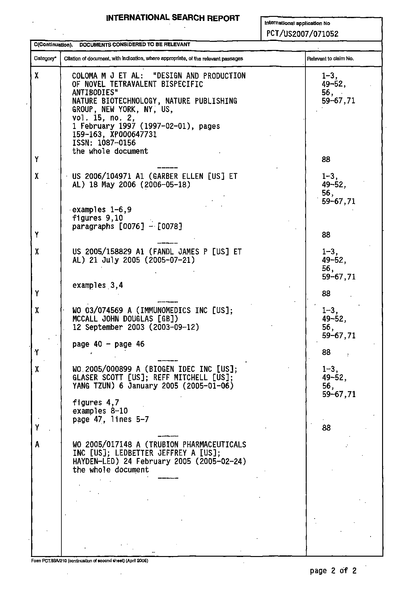
あるいは、保存的アミノ酸は、すぐ下の表Bに示すように、Lehninger、[Biochemistry、第2版;Worth Publishers,Inc.ニューヨーク州NY(1975)、ページ71-77]に記載のように分類することができる。
本発明はまた、特異的結合剤ポリペプチドの誘導体も提供する。誘導体には、アミノ酸残基の挿入、欠失、または置換以外の改変を保有する特異的結合剤ポリペプチドが含まれる。好ましくは、改変は共有的な性質のものであり、例えば、ポリマー、脂質、他の有機、および無機部分との化学結合が含まれる。本発明の誘導体は、特異的結合剤ポリペプチドの循環半減期を増加させるために調製するか、またはポリペプチドが所望の細胞、組織、または器官を標的化する能力を改善するために設計し得る。 The present invention also provides derivatives of specific binding agent polypeptides. Derivatives include specific binding agent polypeptides possessing modifications other than amino acid residue insertions, deletions, or substitutions. Preferably, the modification is of a covalent nature and includes, for example, chemical bonds with polymers, lipids, other organic, and inorganic moieties. Derivatives of the invention can be prepared to increase the circulating half-life of a specific binding agent polypeptide, or designed to improve the ability of the polypeptide to target a desired cell, tissue, or organ. .
本発明はさらに、米国特許第4,640,835号、同第4,496,689号、同第4,301,144号、同第4,670,417号、同第4,791,192号および同第4,179,337号に記載されているように、ポリエチレングリコール、ポリオキシエチレングリコール、またはポリプロピレングリコールなどの1つまたは複数の水溶性ポリマー連結物が含まれるように共有的に改変または誘導体化された、エフェクター機能を有する多価結合タンパク質を包含する。当該技術分野で知られているさらに他の有用なポリマーには、モノメトキシ-ポリエチレングリコール、デキストラン、セルロース、および他の炭水化物系ポリマー、ポリ-(N-ビニルピロリドン)-ポリエチレングリコール、プロピレングリコールホモポリマー、ポリプロピレンオキシド/酸化エチレンコポリマー、ポリオキシエチル化ポリオール(例えばグリセロール)、ポリビニルアルコール、ならびにこれらのポリマーの混合物が含まれる。ポリエチレングリコール(PEG)で誘導体化したタンパク質が特に好ましい。水溶性ポリマーは、特定の位置、例えば本発明によるタンパク質およびポリペプチドのアミノ末端で結合しているか、またはポリペプチドの1つもしくは複数の側鎖でランダムに連結していてよい。治療能力を改善させるためのPEGの使用は、Gonzales他の米国特許第6,133,426号に記載されている。 The present invention further relates to polyethylene glycol, polyoxyethylene glycol as described in U.S. Pat.Nos. 4,640,835, 4,496,689, 4,301,144, 4,670,417, 4,791,192 and 4,179,337. Or a multivalent binding protein with effector function that is covalently modified or derivatized to include one or more water-soluble polymer conjugates, such as polypropylene glycol. Still other useful polymers known in the art include monomethoxy-polyethylene glycol, dextran, cellulose, and other carbohydrate-based polymers, poly- (N-vinylpyrrolidone) -polyethylene glycol, propylene glycol homopolymer , Polypropylene oxide / ethylene oxide copolymers, polyoxyethylated polyols (eg glycerol), polyvinyl alcohol, and mixtures of these polymers. A protein derivatized with polyethylene glycol (PEG) is particularly preferred. The water soluble polymer may be attached at a specific position, for example at the amino terminus of the proteins and polypeptides according to the invention, or may be randomly linked at one or more side chains of the polypeptide. The use of PEG to improve therapeutic performance is described in US Pat. No. 6,133,426 to Gonzales et al.
免疫グロブリン突然変異誘発の標的部位
特定の戦略が、非免疫グロブリンに基づいた結合分子では利用可能でないが、抗原に特異的な免疫グロブリン(例えば抗体)の固有の特性の操作で利用可能である。これらの代替方法を超えて例えば抗体に基づいた分子を好む戦略のよい例は、親和性成熟による、その標的に対する抗体の親和性のin vivo調節であり、これは、免疫グロブリン遺伝子体性高頻度変異の利点をとって、免疫応答が進行するにつれて増加している親和性の抗体を与える。さらに、免疫グロブリンならびに免疫グロブリンの領域およびドメインの構造を変更させる組換え技術が開発されている。したがって、所定の抗原に対して変更された親和性を示す、抗体に由来するポリペプチドを産生してよく、また、これらのポリペプチドを同定かつ精製または単離するためのいくつかの精製プロトコルおよびモニタリングスクリーニングが当該技術分野で知られている。これらの既知の技術を用いて、抗原に対して低下または増加した親和性を示す、抗体由来の結合ドメインを含むポリペプチドを得ることができる。変更された親和性を示すポリペプチド変異体を作製する戦略には、タンパク質中に存在するアミノ酸を変化させるための、抗体をコードしているDNAの部位特異的またはランダム突然変異誘発の使用、続いて、所望の変化、例えば未改変の親または参照抗体と比較して増加または低下した親和性を示す抗体変異体を回収するために設計したスクリーニングステップが含まれる。
Target site- specific strategies for immunoglobulin mutagenesis are not available for non-immunoglobulin-based binding molecules, but can be used to manipulate the intrinsic properties of immunoglobulins specific to antigens (eg, antibodies). A good example of a strategy that favors, for example, antibody-based molecules over these alternative methods is the in vivo regulation of the affinity of an antibody for its target by affinity maturation, which is a high frequency of immunoglobulin gene The benefits of mutation are taken to give antibodies of increasing affinity as the immune response progresses. In addition, recombinant techniques have been developed that alter the structure of immunoglobulins and immunoglobulin regions and domains. Thus, polypeptides derived from antibodies that exhibit altered affinity for a given antigen may be produced, and several purification protocols for identifying and purifying or isolating these polypeptides and Monitoring screening is known in the art. These known techniques can be used to obtain polypeptides comprising antibody-derived binding domains that exhibit reduced or increased affinity for antigen. Strategies to generate polypeptide variants that exhibit altered affinity include the use of site-directed or random mutagenesis of the antibody-encoding DNA to alter the amino acids present in the protein, followed by Screening steps designed to recover antibody variants that exhibit the desired changes, eg, increased or decreased affinity compared to the unmodified parent or reference antibody.
親和性を変更するための突然変異原戦略において最も一般的に標的とされるアミノ酸残基は、抗体の軽鎖および重鎖可変領域の相補性決定領域(CDR)または高頻度可変領域中のものである。これらの領域は、抗原と物理化学的に相互作用する残基、およびこれらの残基の空間的配置に影響を与える他のアミノ酸を含む。しかし、CDR領域外の可変ドメインのフレームワーク領域中のアミノ酸も、抗体の抗原結合特性に相当寄与していることが示されており、そのような特性を操作するための標的とすることができる。Hudson,P.J.、Curr.Opin.Biotech.、9:395-402(1999)およびその中の参考文献を参照されたい。 The most commonly targeted amino acid residues in mutagenic strategies to alter affinity are those in the complementarity determining regions (CDRs) or hypervariable regions of the light and heavy chain variable regions of antibodies. It is. These regions include residues that interact physicochemically with antigens and other amino acids that affect the spatial arrangement of these residues. However, amino acids in the framework region of variable domains outside of the CDR region have also been shown to contribute significantly to the antigen-binding properties of antibodies and can be targeted to manipulate such properties. . See Hudson, P.J., Curr. Opin. Biotech., 9: 395-402 (1999) and references therein.
ランダムまたは部位特異的突然変異誘発を、体性親和性成熟プロセス中に「高頻度突然変異」を受けやすい領域に対応するCDR中の部位に限定することによって、より小さくより有効にスクリーニングした抗体変異体のライブラリを作製することができる。Chowdhury他、Nature Biotech.、17:568-572(1999)およびその中の参考文献を参照されたい。この様式で高頻度突然変異部位を定義することが知られているDNAエレメントの種類には、直列および逆方向反復、特定のコンセンサス配列、二次構造、および回文構造が含まれる。コンセンサスDNA配列には、四塩基配列プリン-G-ピリミジン-A/T(すなわち、AまたはG-G-CまたはT-AまたはT)およびセリンコドンAGY(YはCまたはTであることができる)が含まれる。 Smaller and more effective screened antibody mutations by limiting random or site-directed mutagenesis to sites in the CDR that correspond to regions that are susceptible to "hypermutation" during the somatic affinity maturation process A library of bodies can be created. See Chowdhury et al., Nature Biotech., 17: 568-572 (1999) and references therein. The types of DNA elements known to define hypermutation sites in this manner include tandem and inverted repeats, specific consensus sequences, secondary structures, and palindromic structures. The consensus DNA sequence includes the four base sequence purine-G-pyrimidine-A / T (ie, A or G-G-C or T-A or T) and the serine codon AGY (Y can be C or T).
したがって、本発明の別の態様は、抗体のその標的に対する親和性を改変するための一組の突然変異原戦略である。これらの戦略には、重鎖および/もしくは軽鎖の可変領域全体の突然変異誘発、CDR領域のみの突然変異誘発、CDR内のコンセンサス高頻度変異部位の突然変異誘発、フレームワーク領域の突然変異誘発、またはこれらの手法の任意の組合せが含まれる(本状況における「突然変異誘発」は、ランダムまたは部位特異的であることができる)。CDR領域の最終的な描写および抗体の結合部位を含む残基の同定は、懸案の抗体および抗体:リガンド複合体の構造を、X線結晶構造解析などの当業者に知られている技術によって解くことによって達成することができる。そのような抗体結晶構造の分析および特徴づけに基づいた様々な方法が当業者に知られており、CDR領域を近似するために用いることができる。そのような一般的に使用される方法の例には、カバット(Kabat)、コチア(Chothia)、AbMおよび接触定義が含まれる。 Thus, another aspect of the invention is a set of mutagen strategies for altering the affinity of an antibody for its target. These strategies include mutagenesis of the entire heavy and / or light chain variable region, mutagenesis of the CDR region only, mutagenesis of consensus hypermutation sites within the CDR, and mutagenesis of the framework region. Or any combination of these approaches ("mutagenesis" in this context can be random or site specific). Final delineation of the CDR regions and identification of the residues containing the antibody binding site solves the structure of the antibody and antibody: ligand complex of interest by techniques known to those skilled in the art, such as X-ray crystallography. Can be achieved. Various methods based on analysis and characterization of such antibody crystal structures are known to those skilled in the art and can be used to approximate the CDR regions. Examples of such commonly used methods include Kabat, Chothia, AbM and contact definitions.
カバット定義は、配列の可変性に基づいており、CDR領域の予測に最も一般的に使用される定義である。Johnson他、Nucleic Acids Research、28:214-8(2000)。コチア定義は、構造的ループ領域の位置に基づいている。(Chothia他、J.Mol.Biol.、196:901-17[1986];Chothia他、Nature、342:877-83[1989])。AbM定義は、カバットおよびコチア定義の折衷である。AbMは、Oxford Molecular Groupによって生成された抗体構造モデリングの総合プログラムスイートである(Martin他、Proc.Natl.Acad.Sci(USA)86:9268-9272[1989];Rees他、ABMTM、a computer program for modeling variable regions of antibodies、英国Oxford;Oxford Molecular,Ltd.)。AbMスイートモデルは、ナレッジデータベースおよび非経験的な方法の組合せを用いて、抗体の一次配列から三次構造をモデリングする。接触定義として知られるさらなる定義が最近導入された。MacCallum他、J.Mol.Biol.、5:732-45(1996)を参照されたい。この定義は、利用可能な複合体結晶構造の分析に基づいている。 The Kabat definition is based on sequence variability and is the most commonly used definition for CDR region prediction. Johnson et al., Nucleic Acids Research, 28: 214-8 (2000). The Cotia definition is based on the location of the structural loop region. (Chothia et al., J. Mol. Biol., 196: 901-17 [1986]; Chothia et al., Nature, 342: 877-83 [1989]). The AbM definition is a compromise of the Kabat and Cotia definitions. AbM is a comprehensive program suite of antibody structural modeling generated by the Oxford Molecular Group (Martin et al., Proc. Natl. Acad. Sci (USA) 86: 9268-9272 [1989]; Rees et al., ABMTM, a computer program for modeling variable regions of antibodies, UK Oxford; Oxford Molecular, Ltd.). The AbM suite model models tertiary structure from the primary sequence of an antibody using a combination of knowledge database and ab initio methods. A further definition known as the contact definition has recently been introduced. See MacCallum et al., J. Mol. Biol., 5: 732-45 (1996). This definition is based on an analysis of available complex crystal structures.
慣例により、重鎖中のCDRドメインを、典型的にはH1、H2およびH3と呼び、アミノ末端からカルボキシ末端に移動する順序の連番が付けられている。軽鎖中のCDR領域は、典型的にはL1、L2およびL3と呼ばれ、アミノ末端からカルボキシ末端に移動する順序の連番が付けられている。 By convention, the CDR domains in the heavy chain are typically referred to as H1, H2, and H3, and are numbered sequentially in order from the amino terminus to the carboxy terminus. The CDR regions in the light chain are typically referred to as L1, L2, and L3, and are numbered sequentially in order from the amino terminus to the carboxy terminus.
CDR-H1は、約10〜12個の残基の長さであり、コチアおよびAbM定義によれば典型的にはCysの4個の残基後、またはカバット定義によれば典型的には5個の残基後から開始される。H1には、典型的にはTrp、典型的にはTrp-Valが続くが、Trp-IleまたはTrp-Alaの場合もある。H1の長さは、AbM定義によれば約10〜12個の残基であるが、コチア定義では最後の4個の残基が除外される。 CDR-H1 is about 10-12 residues long, typically 4 residues after Cys according to Cotia and AbM definitions, or typically 5 according to Kabat definitions. Starting after residues. H1 is typically followed by Trp, typically Trp-Val, but may be Trp-Ile or Trp-Ala. The length of H1 is about 10-12 residues according to the AbM definition, but the last four residues are excluded according to the Cotia definition.
CDR-H2は、カバットおよびAbM定義によれば、典型的にはH1の末端15個の残基後から開始される。H2の前の残基は、典型的にはLeu-Glu-Trp-Ile-Glyであるが、いくつかの変形が存在する。H2には、典型的にはアミノ酸配列Lys/Arg-Leu/Ile/Val/Phe/Thr/Ala-Thr/Ser/Ile/Alaが続く。カバット定義によれば、H2の長さは約16〜19個の残基であり、AbM定義では、長さが典型的には9〜12個の残基であると予測する。 CDR-H2 typically starts after the last 15 residues of H1, according to Kabat and AbM definitions. The residue before H2 is typically Leu-Glu-Trp-Ile-Gly, but there are several variations. H2 is typically followed by the amino acid sequence Lys / Arg-Leu / Ile / Val / Phe / Thr / Ala-Thr / Ser / Ile / Ala. According to the Kabat definition, the length of H2 is about 16-19 residues, and the AbM definition predicts that the length is typically 9-12 residues.
CDR-H3は、典型的にはH2の末端33個の残基後から開始され、その前には典型的にはアミノ酸配列Cys-Ala-Argが存在する。H3には、典型的にはアミノ酸Glyが続く。H3の長さの範囲は3〜25個の残基である。 CDR-H3 typically starts after the last 33 residues of H2, typically preceded by the amino acid sequence Cys-Ala-Arg. H3 is typically followed by the amino acid Gly. The length range of H3 is 3-25 residues.
CDR-L1は、典型的には残基24辺りで開始され、典型的にはCysに続く。CDR-L1後の残基は必ずTrpであり、これが、典型的には以下の配列、すなわち、Trp-Tyr-Gln、Trp-Leu-Gln、Trp-Phe-Gln、またはTrp-Tyr-Leuのうちの1つを開始する。CDR-L1の長さは約10〜17個の残基である。 CDR-L1 typically starts around residue 24 and typically follows Cys. The residue after CDR-L1 is always Trp, which is typically the following sequence: Trp-Tyr-Gln, Trp-Leu-Gln, Trp-Phe-Gln, or Trp-Tyr-Leu Start one of them. The length of CDR-L1 is about 10-17 residues.
CDR-L2は、L1の末端の約16個の残基後から開始される。これは、一般に、残基Ile-Tyr、Val-Tyr、Ile-LysまたはIle-Pheに続く。CDR-L2の長さは約7個の残基である。 CDR-L2 begins about 16 residues after the end of L1. This generally follows the residues Ile-Tyr, Val-Tyr, Ile-Lys or Ile-Phe. The length of CDR-L2 is about 7 residues.
CDR-L3は、典型的にはL2の末端の33個の残基後から開始され、典型的にはCysに続く。L3には、典型的にはアミノ酸配列Phe-Gly-XXX-Glyが続く。L3の長さは約7〜11個の残基である。 CDR-L3 typically begins after the last 33 residues of L2 and typically follows Cys. L3 is typically followed by the amino acid sequence Phe-Gly-XXX-Gly. L3 is about 7-11 residues in length.
例えばヒト化免疫グロブリンの重鎖可変領域フレームワークの配列がドナー免疫グロブリンの重鎖可変領域フレームワークの配列と65%〜95%同一であるヒト化抗体を産生する方法を含めた、抗体を改変する様々な方法が当該技術分野で記載されている。それぞれのヒト化した免疫グロブリン鎖は、通常、CDRに加えて、例えば、ドナー免疫グロブリン中でCDRに直接隣接している1つもしくは複数のアミノ酸、または分子モデリングによって予測した場合に約3オングストローム以内のものなどの、結合親和性を達成するためにCDRと相互作用することができるドナー免疫グロブリンフレームワークからのアミノ酸を含む。重鎖および軽鎖は、それぞれ、様々な位置基準の任意の1つまたは複数を使用することによって設計し得る。無処置の抗体と合わせた場合、ヒト化した免疫グロブリンはヒトにおいて実質的に非免疫原性であり、エピトープを含むタンパク質または他の化合物などの抗原に対してドナー免疫グロブリンと実質的に同じ親和性を保持する。 Alter antibodies, including methods that produce humanized antibodies, for example, where the heavy chain variable region framework sequence of the humanized immunoglobulin is 65% to 95% identical to the sequence of the heavy chain variable region framework of the donor immunoglobulin Various methods have been described in the art. Each humanized immunoglobulin chain is usually in addition to the CDR, for example, one or more amino acids immediately adjacent to the CDR in the donor immunoglobulin, or within about 3 angstroms as predicted by molecular modeling It includes amino acids from a donor immunoglobulin framework that can interact with the CDR to achieve binding affinity. The heavy and light chains can each be designed by using any one or more of various positional criteria. When combined with an intact antibody, the humanized immunoglobulin is substantially non-immunogenic in humans and has substantially the same affinity as the donor immunoglobulin for antigens such as proteins or other compounds containing the epitope. Retain sex.
一例では、親抗体に類似の結合特異性を有するが増加したヒト特徴を有する抗体および抗体断片を産生する方法を記載する。ヒト化抗体は、例えば、ファージディスプレイ技術および目的の抗原に特異的な非ヒト抗体の重鎖または軽鎖可変領域を含むポリペプチドを用いた鎖シャフリングによって得て、その後、これをヒト相補的(軽鎖または重鎖)鎖可変領域のレパートリーと合わせる。目的の抗原に特異的なハイブリッド対合を同定し、選択された対合からのヒト鎖をヒト相補的可変ドメイン(重鎖または軽鎖)のレパートリーと合わせる。別の実施形態では、非ヒト抗体からのCDRの構成要素を、ヒト抗体からのCDRの構成要素部分のレパートリーと合わせる。生じる抗体ポリペプチド二量体のライブラリからハイブリッドを選択して、第2のヒト化シャフリングステップで使用してもよい。あるいは、ハイブリッドが治療的価値を有するために十分にヒトの特徴である場合は、この第2のステップを排除する。ヒト特徴を増加するための改変方法は、当該技術分野で知られている。 In one example, methods for producing antibodies and antibody fragments with similar binding specificity to the parent antibody but with increased human characteristics are described. Humanized antibodies can be obtained, for example, by phage shuffling and chain shuffling using a polypeptide comprising a heavy or light chain variable region of a non-human antibody specific for the antigen of interest, which is then human complementary. Combine with the repertoire of the light chain or heavy chain variable regions. A hybrid pairing specific to the antigen of interest is identified and the human chain from the selected pairing is combined with a repertoire of human complementary variable domains (heavy or light chain). In another embodiment, the CDR components from the non-human antibody are combined with the repertoire of the CDR component portions from the human antibody. Hybrids may be selected from the resulting library of antibody polypeptide dimers and used in the second humanized shuffling step. Alternatively, this second step is eliminated if the hybrid is sufficiently human characteristic to have therapeutic value. Modification methods for increasing human characteristics are known in the art.
別の例は、CDRアミノ酸配列を対応するヒトCDRアミノ酸配列で置換することおよび/またはFRアミノ酸配列を対応するヒトFRアミノ酸配列で置換することによってヒト化抗体を作製する方法である。 Another example is a method of making a humanized antibody by replacing a CDR amino acid sequence with a corresponding human CDR amino acid sequence and / or replacing a FR amino acid sequence with a corresponding human FR amino acid sequence.
さらに別の例は、抗原結合ドメインの天然の親和性を減少させない一方で異種の種に対するその免疫原性を軽減させるように改変し得る抗体可変ドメインのアミノ酸残基を同定する方法、および異種の種への投与が有用であるようにこれらの改変された抗体可変領域を調製する方法を提供する。 Yet another example is a method of identifying amino acid residues of an antibody variable domain that can be modified to reduce its immunogenicity against a heterologous species while not reducing the natural affinity of the antigen binding domain, and heterologous Methods are provided for preparing these modified antibody variable regions such that administration to a species is useful.
当該技術分野で知られている方法のうちの任意のものによる、抗体などの免疫グロブリンの改変は、抗原に対する結合親和性の増加もしくは低下を達成する、および/またはレシピエントにおける抗体の免疫原性を軽減させる、および/またはエフェクター活性レベルを調節するように設計されている。一手法では、ヒト化抗体は、抗体のその同族抗原に対する親和性を増加するために、グリコシル化部位を排除するように改変することができる(Co他、Mol.Immunol.30:1361-1367[1993])。「再成形」、「ハイパーキメラ化」、および「張り合わせ/再表面再建」(vennering/resurfacing)などの技術により、より大きな治療潜在性を有するヒト化抗体が産生されている。Vaswami他、Annals of Allergy、Asthma、& Immunol 81:105(1998);Roguska他、Prot.Engineer.9:895-904(1996)]。抗体を再成形する方法を記載している米国特許第6,072,035号も参照されたい。これらの技術は外来残基の数を減らすことによって抗体の免疫原性を減少させるが、これらは抗体の繰り返し投与後の抗イディオタイプおよび抗アロタイプ応答を予防しない。免疫原性を減少させるための、これらの方法の代替方法は、Gilliland他、J.Immunol.62(6):3663-71(1999)に記載されている。 Modification of an immunoglobulin, such as an antibody, by any of the methods known in the art achieves an increase or decrease in binding affinity for the antigen and / or the immunogenicity of the antibody in the recipient Designed to reduce and / or regulate effector activity levels. In one approach, a humanized antibody can be modified to eliminate glycosylation sites to increase the affinity of the antibody for its cognate antigen (Co et al., Mol. Immunol. 30: 1361-1367 [ 1993]). Techniques such as “reshape”, “hyperchimerization”, and “vennering / resurfacing” have produced humanized antibodies with greater therapeutic potential. Vaswami et al., Annals of Allergy, Asthma, & Immunol 81: 105 (1998); Roguska et al., Prot. Engineer. 9: 895-904 (1996)]. See also US Pat. No. 6,072,035 which describes a method for reshaping antibodies. Although these techniques reduce the immunogenicity of antibodies by reducing the number of foreign residues, they do not prevent anti-idiotype and anti-allotype responses after repeated administration of the antibody. An alternative to these methods for reducing immunogenicity is described in Gilliland et al., J. Immunol. 62 (6): 3663-71 (1999).
多くの場合、抗体のヒト化により、抗原結合能力の損失がもたらされる。したがって、抗体の結合親和性を修復する試みとして、最初の(ほとんどの場合はげっ歯類の)抗体中に見つかるアミノ酸残基の1つまたは複数が含まれるようにヒト化抗体を「復帰突然変異」させることが好ましい。例えば、Saldanha他、Mol.Immunol.36:709-19(1999)を参照されたい。 In many cases, humanization of antibodies results in a loss of antigen binding capacity. Thus, in an attempt to repair the binding affinity of the antibody, the humanized antibody is `` backmutated '' to include one or more of the amino acid residues found in the first (mostly rodent) antibody. Is preferred. See, for example, Saldanha et al., Mol. Immunol. 36: 709-19 (1999).
免疫グロブリンのグリコシル化は、エフェクター機能、構造的安定性、および抗体産生細胞からの分泌率に影響を与えることが示されている(本明細書中に参照により組み込まれているLeatherbarrow他、Mol.Immunol.22:407(1985)を参照)。これらの特性を司っている炭水化物基は、一般に、抗体の定常領域に連結している。例えば、CH2ドメイン中のAsn297におけるIgGのグリコシル化は、補体依存性細胞溶解を活性化させるIgGの完全な能力を促進する(Tao他、J.Immunol.143:2595(1989))。例えば、CH3ドメイン中のAsn402におけるIgMのグリコシル化は、抗体の適切なアセンブリおよび細胞溶解性活性を促進する(Muraoka他、J.Immunol.142:695(1989))。IgA抗体のCH1およびCH3ドメイン中の位置162および419におけるグリコシル化部位の除去により、細胞内分解および少なくとも90%の分泌の阻害がもたらされた(Taylor他、Wall、Mol.Cell.Biol.8:4197(1988))。したがって、本発明の分子には、エフェクター機能を変更するために、例えば定常部分領域中の特定の残基の突然変異によって変更されたグリコシル化パターンを示す、突然変異によって変更された免疫グロブリンが含まれる。それぞれが本明細書中に参照により組み込まれている、Co他、Mol.Immunol.30:1361-1367(1993)、Jacquemon他、J.Thromb.Haemost.4:1047-1055(2006)、Schuster他、Cancer Res.65:7934-7941(2005)、and Warnock他、Biotechnol Bioeng.92:831-842(2005)を参照されたい。 Immunoglobulin glycosylation has been shown to affect effector function, structural stability, and secretion rate from antibody-producing cells (Leatherbarrow et al., Mol. Immunol. 22: 407 (1985)). The carbohydrate group responsible for these properties is generally linked to the constant region of the antibody. For example, glycosylation of IgG at Asn297 in the C H2 domain promotes the full ability of IgG to activate complement-dependent cell lysis (Tao et al., J. Immunol. 143: 2595 (1989)). For example, glycosylation of IgM in Asn402 in C H3 domains facilitates proper assembly and cytolytic activity of the antibody (Muraoka other, J.Immunol.142: 695 (1989)) . Removal of the glycosylation site in C H1 and C H3 position 162 and 419 in the domain of IgA antibody, inhibition of intracellular degradation and at least 90% of the secretion was brought (Taylor other, Wall, Mol .8: 4197 (1988)). Thus, the molecules of the present invention include immunoglobulins altered by mutations that exhibit altered glycosylation patterns to alter effector function, eg, by mutation of certain residues in the constant subregion. It is. Co et al., Mol. Immunol. 30: 1361-1367 (1993), Jacquemon et al., J. Thromb.Haemost. 4: 1047-1055 (2006), Schuster et al. Cancer Res. 65: 7934-7941 (2005), and Warnock et al., Biotechnol Bioeng. 92: 831-842 (2005).
また、発明には、既知の免疫グロブリン可変領域の配列と少なくとも80%、好ましくは90%もしくは95%または99%同一の配列である少なくとも1つの結合ドメインを有し、そのような免疫グロブリン可変領域とは異なる少なくとも1つの残基を有し、変化した残基により、免疫グロブリン可変領域と比較して、グリコシル化部位が付加されるか、1つもしくは複数のグリコシル化部位の位置が変化されるか、または好ましくはグリコシル化部位が除去される、多価結合分子が含まれる。一部の実施形態では、変化により、免疫グロブリン可変領域フレームワーク中のN結合グリコシル化部位が除去されるか、または、Co他、J.Immunol.148:1149、(1992)の番号付けの慣例を用いてアミノ酸残基約65〜アミノ酸残基約85にわたる領域中の免疫グロブリンの重鎖可変領域フレームワーク内に存在するN結合グリコシル化部位が除去される。 The invention also has at least one binding domain that is at least 80%, preferably 90% or 95% or 99% identical to a sequence of a known immunoglobulin variable region, such an immunoglobulin variable region. With at least one residue different from, and the altered residue adds a glycosylation site or changes the position of one or more glycosylation sites compared to an immunoglobulin variable region Or preferably include multivalent binding molecules in which the glycosylation site is removed. In some embodiments, the change removes an N-linked glycosylation site in the immunoglobulin variable region framework or the numbering convention of Co et al., J. Immunol. 148: 1149, (1992). Is used to remove N-linked glycosylation sites present within the heavy chain variable region framework of immunoglobulins in a region spanning from about 65 amino acid residues to about 85 amino acid residues.
免疫グロブリン参照配列と比較して変更されたグリコシル化パターンを示す多価結合分子を産生するために、当該技術分野で知られている任意の方法が企図される。例えば、様々な遺伝子技術のうちの任意のものを用いて、1つまたは複数の特定の残基を変更し得る。あるいは、産生に用いた宿主細胞を操作して、変更されたグリコシル化パターンを生じさせ得る。例えば、当該技術分野で知られている一方法は、ADCCを増加させる、二分したフコシル化されていない変異体の形態の、変更されたグリコシル化を提供する。変異体は、オリゴ糖改変酵素を含む宿主細胞内での発現から生じる。あるいは、本発明によるグリコシル化された分子のフコース含有率を低下させるために、BioWa/Kyowa HakkoのPotelligent技術が企図される。1つの既知の方法では、免疫グロブリンFC領域のグリコシル化パターンをGDP-フコースの産生によって改変する、組換え免疫グロブリンを産生するためのCHO宿主細胞を提供する。この技術は、本発明による多価結合分子の定常部分領域のグリコシル化パターンを改変するために利用可能である。 Any method known in the art is contemplated for producing multivalent binding molecules that exhibit altered glycosylation patterns relative to an immunoglobulin reference sequence. For example, any of a variety of genetic techniques can be used to change one or more specific residues. Alternatively, the host cell used for production can be manipulated to produce an altered glycosylation pattern. For example, one method known in the art provides altered glycosylation in the form of a bisected, nonfucosylated variant that increases ADCC. Variants arise from expression in host cells that contain oligosaccharide modifying enzymes. Alternatively, BioWa / Kyowa Hakko Potelligent technology is contemplated to reduce the fucose content of glycosylated molecules according to the present invention. One known method provides a CHO host cell for producing recombinant immunoglobulins that alters the glycosylation pattern of the immunoglobulin F C region by production of GDP-fucose. This technique can be used to modify the glycosylation pattern of the constant subregion of a multivalent binding molecule according to the present invention.
免疫グロブリンの結合ドメインなどの結合ドメインの結合特性を改変することに加えて、また、ヒト化などの改変に加えて、本発明は、定常部分領域のエフェクター機能などの、エフェクター機能に寄与する残基を変化または突然変異させることによるエフェクター機能の変調を包含する。これらの改変は、本明細書中に参照により組み込まれているPresta他、Biochem.Soc.Trans.30:487-490(2001)に開示されている手法などの当該技術分野で知られている任意の技術を用いて達成することができる。例示的な手法には、FCγRI、FCγRII、FCγRIII、FCαR、およびFCεRに対応する1つまたは複数の定常部分領域における結合に影響を与えることが知られている特定の残基を改変するための、Presta他に開示されているプロトコルが含まれるであろう。 In addition to modifying the binding properties of a binding domain, such as the binding domain of an immunoglobulin, and in addition to modifications such as humanization, the present invention provides residues that contribute to effector functions, such as effector functions of constant subregions. Includes modulation of effector function by changing or mutating groups. These modifications may be any of those known in the art, such as those disclosed in Presta et al., Biochem.Soc.Trans. 30: 487-490 (2001), which is incorporated herein by reference. Can be achieved using this technique. Exemplary techniques include modifying specific residues that are known to affect binding in one or more constant subregions corresponding to F C γRI, FCγRII, FCγRIII, FCαR, and FCεR. Of the protocol disclosed in Presta et al.
別の手法では、Xencor XmAb技術が、細胞死滅エフェクター機能を増強するためにFCドメインに対応する定常部分領域を操作するために利用可能である。本明細書中に参照により組み込まれているLazar他、Proc.Natl.Acad.Sci.(USA)103(11):4005-4010(2006)を参照されたい。この手法を用いて、例えば、FCγR特異性および結合に最適化された定常部分領域を作製し、したがって細胞死滅エフェクター機能を増強させることができる。 In another approach, Xencor XmAb technology can be used to manipulate the constant subregion corresponding to the F C domain to enhance cell killing effector function. See Lazar et al., Proc. Natl. Acad. Sci. (USA) 103 (11): 4005-4010 (2006), incorporated herein by reference. This approach can be used, for example, to create constant subregions optimized for F C γR specificity and binding and thus enhance cell killing effector function.
エフェクター機能を有する多価結合タンパク質の産生
様々な発現ベクター/宿主系を、本発明の多価結合タンパク質(エフェクター機能を有する)を含むおよび発現するように利用し得る。これらの系には、それだけには限定されないが、微生物、例えば、組換えバクテリオファージ、プラスミド、コスミド、もしくは他の発現ベクターで形質転換させた細菌;酵母発現もしくはシャトルベクターで形質転換させた酵母;ウイルス発現ベクター(例えばバキュロウイルス)を感染させた昆虫細胞株;ウイルス発現ベクター(例えば、カリフラワーモザイクウイルス、CaMV;タバコモザイクウイルス、TMV)でトランスフェクションさせた、もしくは細菌発現ベクター(例えば、TiもしくはpBR322プラスミド)で形質転換させた植物細胞株;または動物細胞株が含まれる。組換え多価結合タンパク質の産生に有用な哺乳動物細胞には、それだけには限定されないが、VERO細胞、HeLa細胞、チャイニーズハムスター卵巣(CHO)細胞株、COS細胞(COS-7など)、W138、BHK、HepG2、3T3、RIN、MDCK、A549、PC12、K562およびHEK293細胞が含まれる。多価結合タンパク質の組換え発現の例示的なプロトコルを、本明細書中以下に記載する。
Production of Multivalent Binding Proteins with Effector Functions A variety of expression vector / host systems may be utilized to contain and express the multivalent binding proteins (with effector functions) of the present invention. These systems include, but are not limited to, microorganisms such as bacteria transformed with recombinant bacteriophage, plasmids, cosmids, or other expression vectors; yeast transformed with yeast expression or shuttle vectors; viruses Insect cell lines infected with expression vectors (e.g. baculovirus); transfected with viral expression vectors (e.g. cauliflower mosaic virus, CaMV; tobacco mosaic virus, TMV) or bacterial expression vectors (e.g. Ti or pBR322 plasmids) Plant cell lines transformed with); or animal cell lines. Mammalian cells useful for the production of recombinant multivalent binding proteins include, but are not limited to, VERO cells, HeLa cells, Chinese hamster ovary (CHO) cell lines, COS cells (such as COS-7), W138, BHK , HepG2, 3T3, RIN, MDCK, A549, PC12, K562 and HEK293 cells. Exemplary protocols for recombinant expression of multivalent binding proteins are described herein below.
発現ベクターは、(1)遺伝子発現において調節性の役割を有する遺伝的エレメントまたは複数のエレメント、例えば、プロモーター、エンハンサー、または因子特異的結合部位、(2)mRNAへと転写され、タンパク質へと翻訳される結合剤をコードしている構造体または配列、ならびに(3)適切な転写開始および終止配列のアセンブリを含む転写単位を含むことができる。酵母または真核発現系で用いることを意図する構造単位には、好ましくは、宿主細胞による翻訳されたタンパク質の細胞外分泌を可能にするリーダー配列が含まれる。あるいは、組換え多価結合タンパク質がリーダーまたは輸送配列なしで発現される場合は、アミノ末端にメチオニン残基を含み得る。この残基は、続いて、発現された組換えタンパク質から切断されるかされずに、最終多価結合タンパク質を提供し得る。 Expression vectors are (1) genetic elements or multiple elements that have a regulatory role in gene expression, e.g., promoters, enhancers, or factor-specific binding sites, (2) transcribed into mRNA and translated into proteins A structure or sequence encoding the binding agent to be produced, and (3) a transcription unit comprising an assembly of suitable transcription initiation and termination sequences. Structural units intended for use in yeast or eukaryotic expression systems preferably include a leader sequence that allows extracellular secretion of the translated protein by the host cell. Alternatively, if the recombinant multivalent binding protein is expressed without a leader or transport sequence, it may contain a methionine residue at the amino terminus. This residue can subsequently provide the final multivalent binding protein without being cleaved from the expressed recombinant protein.
例えば、多価結合タンパク質は、市販の発現系、例えばピキア(Pichia)発現系(Invitrogen、カリフォルニア州San Diego)を用いて、製造者の指示に従って、酵母中で組換えによって発現させ得る。また、この系は、分泌を指示するためにプリプロα配列に依存するが、インサートの転写は、メタノールによる誘発の際にアルコールオキシダーゼ(AOX1)プロモーターによって駆動される。分泌された多価結合ペプチドは、例えばペプチドを細菌および哺乳動物細胞の上清から精製するために用いる方法によって、酵母増殖培地から精製し得る。 For example, multivalent binding proteins can be expressed recombinantly in yeast using a commercially available expression system, such as the Pichia expression system (Invitrogen, San Diego, Calif.) According to the manufacturer's instructions. This system also relies on the prepro alpha sequence to direct secretion, but transcription of the insert is driven by the alcohol oxidase (AOX1) promoter upon induction with methanol. The secreted multivalent binding peptide can be purified from the yeast growth medium, for example by the method used to purify the peptide from bacterial and mammalian cell supernatants.
あるいは、多価結合ペプチドをコードしているcDNAをバキュロウイルス発現ベクターpVL1393(PharMingen、カリフォルニア州San Diego)内にクローニングし得る。このベクターを製造者の指示(PharMingen)に従って用いて、SF9タンパク質を含まない培地中でスポドプテラ・フルギペルダ細胞に感染させ、組換えタンパク質を産生させることができる。多価結合タンパク質は、ヘパリン-セファロースカラム(Pharmacia、ニュージャージー州Piscataway)を用いて培地から精製および濃縮することができる。SF9系などのタンパク質発現用の昆虫系が当業者に周知である。このような系の1つであるオートグラファ・カリフォルニカ(Autographa californica)核多角体病ウイルス(AcNPV)を、スポドプテラ・フルギペルダ細胞またはトリコプルジア(Trichoplusia)の幼虫中で外来遺伝子を発現させるベクターとして用いることができる。多価結合ペプチドコード配列を多角体遺伝子などのウイルスの非必須領域内にクローニングし、ポリヘドリンプロモーターの制御下に置くことができる。多価結合ペプチドの挿入の成功は多角体遺伝子を失活させ、コーティングタンパク質を欠く組換えウイルスが産生される。組換えウイルスを用いて、ペプチドを発現させるS.フルギペルダ細胞またはトリコプルジアの幼虫を感染させることができる(Smith他、J Virol 46:584、1983;Engelhard他、Proc Nat Acad Sci(USA)91:3224-7、1994)。 Alternatively, cDNA encoding a multivalent binding peptide can be cloned into the baculovirus expression vector pVL1393 (PharMingen, San Diego, CA). This vector can be used according to the manufacturer's instructions (PharMingen) to infect Spodoptera frugiperda cells in medium without SF9 protein to produce recombinant protein. Multivalent binding proteins can be purified and concentrated from the medium using a heparin-sepharose column (Pharmacia, Piscataway, NJ). Insect systems for protein expression such as the SF9 system are well known to those skilled in the art. One such system, Autographa californica nuclear polyhedrosis virus (AcNPV), should be used as a vector to express foreign genes in Spodoptera frugiperda cells or Trichoplusia larvae. Can do. The multivalent binding peptide coding sequence can be cloned into a nonessential region of the virus, such as a polyhedron gene, and placed under the control of the polyhedrin promoter. Successful insertion of the multivalent binding peptide inactivates the polyhedron gene and produces a recombinant virus lacking the coating protein. Recombinant viruses can be used to infect S. frugiperda cells expressing peptide or Trichopuldia larvae (Smith et al., J Virol 46: 584, 1983; Engelhard et al., Proc Nat Acad Sci (USA) 91: 3224 -7, 1994).
別の例では、多価結合ペプチドをコードしているDNA配列をPCRによって増幅し、適切なベクター、例えばpGEX-3X(Pharmacia、ニュージャージー州Piscataway)内にクローニングすることができる。pGEXベクターは、ベクターによってコードされているグルタチオン-S-トランスフェラーゼ(GST)と、ベクターのクローニング部位内に挿入されたDNA断片によってコードされている多価結合タンパク質とを含む融合タンパク質を産生するように設計されている。PCR用のプライマーは、例えば適切な切断部位が含まれるように作製することができる。多価結合タンパク質の融合部分を、発現を促進するためだけに使用する場合、または他の理由で目的のペプチドと連結することが望ましくない場合は、組換え多価結合タンパク質の融合体をこの時に融合タンパク質のGST部分から切断し得る。pGEX-3X/多価結合ペプチド構築物を大腸菌XL-1 Blue細胞(Stratagene、カリフォルニア州La Jolla)内に形質転換させ、個々の形質転換体を単離および増殖させる。個々の形質転換体からのプラスミドDNAを精製し、自動配列決定機を用いて部分的に配列決定して、適切な方向の所望の多価結合タンパク質をコードしている核酸インサートの存在を確認し得る。 In another example, a DNA sequence encoding a multivalent binding peptide can be amplified by PCR and cloned into an appropriate vector, such as pGEX-3X (Pharmacia, Piscataway, NJ). The pGEX vector produces a fusion protein containing glutathione-S-transferase (GST) encoded by the vector and a multivalent binding protein encoded by a DNA fragment inserted within the cloning site of the vector. Designed. PCR primers can be prepared, for example, so as to include an appropriate cleavage site. If the multivalent binding protein fusion moiety is used solely to facilitate expression, or if it is not desirable to link the peptide of interest for other reasons, the recombinant multivalent binding protein fusion is It can be cleaved from the GST portion of the fusion protein. The pGEX-3X / multivalent binding peptide construct is transformed into E. coli XL-1 Blue cells (Stratagene, La Jolla, Calif.) and individual transformants are isolated and grown. Plasmid DNA from individual transformants is purified and partially sequenced using an automated sequencer to confirm the presence of a nucleic acid insert encoding the desired multivalent binding protein in the appropriate orientation. obtain.
細菌中の不溶性の封入体として産生し得る融合した多価結合タンパク質は、以下のように精製することができる。宿主細胞を遠心分離によって収集し;0.15MのNaCl、10mMのトリス、pH8、1mMのEDTAで洗浄し;0.1mg/mlのリゾチーム(Sigma Chemical Co.)を用いて15分間、室温で処理することができる。溶解物を超音波処理によって清澄にすることができ、細胞細片を10分間、12,000×gで遠心分離することによってペレット化することができる。多価結合タンパク質融合体含有ペレットを50mMのトリス、pH8、および10mMのEDTAに再懸濁させ、層状の50%のグリセロールの上に層状に重ね、30分間、6000gで遠心分離することができる。ペレットを、Mg++およびCa++を含まない標準のリン酸緩衝生理食塩水(PBS)に再懸濁させることができる。多価結合タンパク質融合体は、再懸濁させたペレットを変性SDSポリアクリルアミドゲル中で分画することによって、さらに精製することができる(Sambrook他)。タンパク質を可視化するためにゲルを0.4MのKClに浸し、これを切除し、SDSを欠くゲルランニング緩衝液中で電気溶出させる。GST/多価結合ペプチド融合タンパク質が細菌中で可溶性タンパク質として産生される場合、これはGST精製モジュール(Pharmacia Biotech)を用いて精製することができる。
A fused multivalent binding protein that can be produced as an insoluble inclusion body in bacteria can be purified as follows. Host cells are collected by centrifugation; washed with 0.15 M NaCl, 10 mM Tris,
多価結合タンパク質融合体は、好ましくは、GSTを本発明の多価結合ペプチドから切断するために消化に供する。消化反応(20〜40μgの融合タンパク質、20〜30単位のヒトトロンビン(0.5mlのPBS中に4000U/mg(Sigma))を、16〜48時間、室温でインキュベーションし、変性SDS-PAGEゲルに添加して、反応生成物を分画することができる。ゲルを0.4MのKClに浸してタンパク質のバンドを可視化することができる。多価結合ペプチドの予測される分子量に対応するタンパク質のバンドが何であるかは、自動配列決定機(Applied Biosystems、モデル473A、カリフォルニア州Foster City)を用いたアミノ酸配列分析によって確認することができる。あるいは、ペプチドのHPLCおよび/または質量分析を行うことによって、何であるかを確認することができる。 The multivalent binding protein fusion is preferably subjected to digestion to cleave GST from the multivalent binding peptide of the invention. Digestion reaction (20-40 μg fusion protein, 20-30 units human thrombin (4000 U / mg (Sigma) in 0.5 ml PBS), incubated for 16-48 hours at room temperature and added to denaturing SDS-PAGE gel The reaction product can be fractionated, the gel can be soaked in 0.4 M KCl to visualize the protein band, and the protein band corresponding to the expected molecular weight of the multivalent binding peptide. It can be confirmed by amino acid sequence analysis using an automated sequencer (Applied Biosystems, model 473A, Foster City, Calif.), Or what is done by performing HPLC and / or mass spectrometry of the peptide Can be confirmed.
あるいは、多価結合ペプチドをコードしているDNA配列を、所望のプロモーター、および任意選択でリーダー配列を含むプラスミド内にクローニングすることができる(例えば、Better他、Science、240:1041-43、1988参照)。この構築物の配列は、自動配列決定によって確認することができる。その後、プラスミドを、MC1061株などの適切な大腸菌株内に、細菌のCaCl2インキュベーションおよび熱ショック処理を用いた標準の手順を使用して、形質転換させることができる(Sambrook他)。形質転換させた細菌は、カルベニシリンを添加したLB培地中または当該技術分野で知られている別の適切な選択した形態で増殖することができ、発現されたタンパク質の産生は、適切な培地中での増殖によって誘発することができる。存在する場合は、リーダー配列は、多価結合ペプチドの分泌をもたらし、分泌中に切断されることができる。分泌された組換えタンパク質は、本明細書中以下に記載する方法によって細菌培地から精製することができる。 Alternatively, a DNA sequence encoding a multivalent binding peptide can be cloned into a plasmid containing the desired promoter and optionally a leader sequence (e.g., Better et al., Science, 240: 1041-43, 1988). reference). The sequence of this construct can be confirmed by automated sequencing. The plasmid can then be transformed into a suitable E. coli strain such as the MC1061 strain using standard procedures with bacterial CaCl 2 incubation and heat shock treatment (Sambrook et al.). The transformed bacteria can be grown in LB medium supplemented with carbenicillin or in another suitable selected form known in the art, and production of the expressed protein is carried out in a suitable medium. Can be induced by proliferation of When present, the leader sequence results in secretion of the multivalent binding peptide and can be cleaved during secretion. Secreted recombinant protein can be purified from bacterial media by the methods described herein below.
組換えタンパク質を発現させるための哺乳動物宿主系は当業者に周知であり、それらが好ましい系である。宿主細胞株は、タンパク質の活性を提供するために有用な、発現されたタンパク質をプロセシングする、または特定の翻訳後修飾を生じる特定の能力について、選択することができる。ポリペプチドのそのような修飾には、それだけには限定されないが、アセチル化、カルボキシル化、グリコシル化、リン酸化、脂質化およびアシル化が含まれる。CHO、HeLa、MDCK、293、WI38などの様々な宿主細胞が、そのような翻訳後活性の特定の細胞機構および特徴機構を有しており、外来タンパク質の正しい修飾およびプロセシングが確実に行われるように選択することができる。 Mammalian host systems for expressing recombinant proteins are well known to those skilled in the art and are preferred systems. Host cell lines can be selected for their particular ability to process the expressed protein or produce specific post-translational modifications that are useful for providing protein activity. Such modifications of the polypeptide include, but are not limited to, acetylation, carboxylation, glycosylation, phosphorylation, lipidation and acylation. Various host cells such as CHO, HeLa, MDCK, 293, WI38 have specific cellular and characteristic mechanisms for such post-translational activity to ensure correct modification and processing of foreign proteins Can be selected.
形質転換細胞を長期的な高効率のタンパク質産生に用いることが好ましいので、安定した発現が望ましい。1つのそのような細胞は、好ましくは少なくとも1つの選択マーカーを所望の発現カセットと共に含むベクターで形質転換させ、細胞を強化培地中で1〜2日間増殖させた後、選択培地に切り替える。選択マーカーは選択に対する耐性を与えるように設計されており、その存在により、外来タンパク質の発現が成功する細胞の増殖および回収が可能となる。安定に形質転換された細胞の耐性塊を、細胞に適切な組織培養技術を用いて増殖することができる。 Since it is preferable to use transformed cells for long-term highly efficient protein production, stable expression is desirable. One such cell is preferably transformed with a vector containing at least one selectable marker along with the desired expression cassette, and the cells are grown in enhanced medium for 1-2 days before switching to the selective medium. Selectable markers are designed to confer resistance to selection, and their presence allows the growth and recovery of cells that successfully express foreign proteins. Resistant masses of stably transformed cells can be propagated using tissue culture techniques appropriate to the cells.
組換えタンパク質産生のために形質転換させた細胞を回収するために、いくつかの選択系を使用することができる。そのような選択系には、それだけには限定されないが、それぞれtk-、hgprt-またはaprt-細胞における、HSVチミジンキナーゼ、ヒポキサンチン-グアニンホスホリボシルトランスフェラーゼおよびアデニンホスホリボシルトランスフェラーゼ遺伝子が含まれる。また、代謝拮抗剤耐性を、メトトレキサートに対する耐性を与えるdhfr;ミコフェノール酸に対する耐性を与えるgpt;アミノグリコシドG418に耐性を与え、クロルスルフロンに対する耐性を与えるneo;およびハイグロマイシンに対する耐性を与えるhygroの選択の基礎として用いることができる。有用であり得るさらなる選択可能な遺伝子には、細胞がトリプトファンの代わりにインドールを利用することを可能にするtrpB、または、細胞がヒスチジンの代わりにヒスチノールを利用することを可能にするhisDが含まれる。形質転換体を同定するための視覚的指標を与えるマーカーには、アントシアニン、β-グルクロニダーゼおよびその基質、GUS、ならびにルシフェラーゼおよびその基質、ルシフェリンが含まれる。 Several selection systems can be used to recover cells transformed for recombinant protein production. Such selection systems include, but are not limited to, the HSV thymidine kinase, hypoxanthine-guanine phosphoribosyltransferase and adenine phosphoribosyltransferase genes in tk-, hgprt- or aprt-cells, respectively. Also select anti-metabolite resistance, dhfr that confer resistance to methotrexate; gpt that confer resistance to mycophenolic acid; neo that confer resistance to aminoglycoside G418, confer resistance to chlorsulfuron; and hygro that confer resistance to hygromycin Can be used as a basis for Additional selectable genes that may be useful include trpB, which allows cells to utilize indole instead of tryptophan, or hisD, which allows cells to utilize histinol instead of histidine. . Markers that provide visual indicators for identifying transformants include anthocyanins, β-glucuronidase and its substrate, GUS, and luciferase and its substrate, luciferin.
タンパク質の精製
タンパク質精製技術は当業者に周知である。これらの技術は、一定のレベルで、ポリペプチドおよび非ポリペプチド画分の粗分画を含む。多価結合ポリペプチドを少なくとも1つの他のタンパク質から分離した後、目的のポリペプチドを精製するが、部分的または完全な完全精製(もしくは均一になるまでの精製)を達成するためのクロマトグラフィーおよび電気泳動技術を用いたさらなる精製が、多くの場合所望される。純粋な多価結合ペプチドの調製に特に適した分析方法は、イオン交換クロマトグラフィー、排除クロマトグラフィー;ポリアクリルアミドゲル電気泳動;および等電点電気泳動である。ペプチドを精製する特に効率的な方法は高速タンパク質液体クロマトグラフィーおよびHPLCである。
Protein Purification Protein purification techniques are well known to those skilled in the art. These techniques include, at a certain level, a crude fraction of polypeptide and non-polypeptide fractions. After separating the multivalent binding polypeptide from at least one other protein, the target polypeptide is purified, but chromatographic to achieve partial or complete complete purification (or purification to homogeneity) and Further purification using electrophoretic techniques is often desirable. Analytical methods that are particularly suitable for the preparation of pure multivalent binding peptides are ion exchange chromatography, exclusion chromatography; polyacrylamide gel electrophoresis; and isoelectric focusing. Particularly efficient methods for purifying peptides are high performance protein liquid chromatography and HPLC.
本発明の特定の態様は精製に関し、特定の実施形態では、コードされた多価結合タンパク質またはペプチドの実質的な精製に関する。本明細書中で使用する用語「精製した多価結合タンパク質またはペプチド」とは、他の成分から単離可能な組成物をいうことを意図し、多価結合タンパク質またはペプチドは、その天然に入手可能な状態と比較して任意の度合まで精製されている。したがって、精製した多価結合タンパク質またはペプチドとは、それが天然に存在し得る環境から遊離した多価結合タンパク質またはペプチドもいう。 Certain aspects of the invention relate to purification and, in certain embodiments, to substantial purification of the encoded multivalent binding protein or peptide. As used herein, the term “purified multivalent binding protein or peptide” is intended to refer to a composition that can be isolated from other components, and the multivalent binding protein or peptide is obtained in nature. It has been purified to any degree compared to the possible state. Thus, a purified multivalent binding protein or peptide also refers to a multivalent binding protein or peptide that is liberated from the environment in which it can occur in nature.
一般に、「精製した」とは、様々な他の成分を除去するために分画に供した多価結合タンパク質組成物をいい、組成物は、その発現された生物活性を実質的に保持している。用語「実質的に精製した」を使用した場合、この指定は、多価結合タンパク質またはペプチドが組成物の主要な成分を形成する、例えば組成物中のタンパク質の約50重量%、約60重量%、約70重量%、約80重量%、約90重量%、約95重量%、約99重量%またはそれ以上を構成する、多価結合タンパク質組成物をいう。 In general, “purified” refers to a multivalent binding protein composition that has been subjected to fractionation to remove various other components, wherein the composition substantially retains its expressed biological activity. Yes. When the term “substantially purified” is used, this designation indicates that the multivalent binding protein or peptide forms a major component of the composition, eg, about 50%, about 60% by weight of the protein in the composition. , About 70 wt.%, About 80 wt.%, About 90 wt.%, About 95 wt.%, About 99 wt.% Or more.
多価結合タンパク質の精製の度合を定量する様々な方法は、本開示に鑑みて当業者に理解されるであろう。これらには、例えば、活性画分の特異的結合活性の決定、またはSDS/PAGE分析による画分内の多価結合ポリペプチドの量の評価が含まれる。多価結合タンパク質画分の純度を評価する好ましい方法は、画分の結合活性を計算すること、それを最初の抽出物の結合活性と比較すること、およびそれにより精製の度合を計算することであり、本明細書中で「〜倍精製数」によって評価する。結合活性の量を表すために使用する実際の単位は、もちろん、精製後に行う選択した特定のアッセイ技術および発現された多価結合タンパク質またはペプチドが検出可能な結合活性を示すかどうかに依存する。 Various methods for quantifying the degree of purification of the multivalent binding protein will be understood by those of skill in the art in light of the present disclosure. These include, for example, determining the specific binding activity of the active fraction, or assessing the amount of multivalent binding polypeptide in the fraction by SDS / PAGE analysis. A preferred method of assessing the purity of the multivalent binding protein fraction is to calculate the binding activity of the fraction, compare it to the binding activity of the original extract, and thereby calculate the degree of purification. Yes, and evaluated in this specification by "-fold purification number". The actual units used to represent the amount of binding activity will, of course, depend on the particular assay technique chosen after purification and whether the expressed multivalent binding protein or peptide exhibits detectable binding activity.
多価結合タンパク質の精製での使用に適した様々な技術が当業者に周知である。これらには、例えば、硫酸アンモニウム、PEG、抗体などを用いた、または熱変性による沈降、続いて遠心分離;イオン交換、ゲル濾過、逆相、ヒドロキシルアパタイトおよびアフィニティークロマトグラフィーなどのクロマトグラフィーステップ;等電点電気泳動;ゲル電気泳動;ならびにこれらおよび他の技術の組合せが含まれる。一般に当該技術分野で知られているように、様々な精製ステップを実施する順序を変更するか、または特定のステップを省略してもよく、それでも実質的に精製した多価結合タンパク質を調製する適切な方法がもたらされると考えられている。 Various techniques suitable for use in the purification of multivalent binding proteins are well known to those of skill in the art. These include, for example, precipitation using ammonium sulfate, PEG, antibodies, or by thermal denaturation followed by centrifugation; chromatography steps such as ion exchange, gel filtration, reverse phase, hydroxylapatite and affinity chromatography; isoelectric Point electrophoresis; gel electrophoresis; and combinations of these and other techniques are included. As is generally known in the art, the order in which the various purification steps are performed may be altered, or certain steps may be omitted, and still suitable for preparing a substantially purified multivalent binding protein. Is thought to bring about a new way.
多価結合タンパク質を常にその最も精製された状態で提供するという全般的な必要性は存在しない。実際、実質的により低い多価結合タンパク質が、特定の実施形態において有用性を有することが企図される。部分精製は、より少ない精製ステップを組み合わせて用いることによって、または異なる形態の同じ一般精製スキームを利用することによって達成し得る。例えば、HPLC装置を利用して行う陽イオン交換カラムクロマトグラフィーは、一般に、低圧クロマトグラフィー系を利用した同じ技術よりも高い精製をもたらすことを理解されよう。より低い度合の相対的精製を示す方法は、多価結合タンパク質産物の全回収、または発現された多価結合タンパク質の結合活性の維持において利点を有し得る。 There is no general need to always provide the multivalent binding protein in its most purified state. Indeed, it is contemplated that substantially lower multivalent binding proteins have utility in certain embodiments. Partial purification may be achieved by using fewer purification steps in combination or by utilizing different forms of the same general purification scheme. For example, it will be appreciated that cation exchange column chromatography performed using an HPLC instrument generally results in higher purification than the same technique utilizing a low pressure chromatography system. Methods that exhibit a lower degree of relative purification may have advantages in the overall recovery of the multivalent binding protein product or in maintaining the binding activity of the expressed multivalent binding protein.
ポリペプチドの遊走は、SDS/PAGEの様々な条件で、場合によっては顕著に、変動する場合があることが知られている(Capaldi他、Biochem.Biophys.Res.Comm.、76:425、1977)。したがって、異なる電気泳動条件下では、精製したまたは部分精製した多価結合タンパク質発現産物の見かけ上の分子量が変動し得ることを理解されたい。 Polypeptide migration is known to vary significantly in some cases under various SDS / PAGE conditions (Capaldi et al., Biochem. Biophys. Res. Comm., 76: 425, 1977). ). Thus, it should be understood that under different electrophoresis conditions, the apparent molecular weight of a purified or partially purified multivalent binding protein expression product can vary.
エフェクター細胞
標的細胞に対して、例えば、ADCC、ADCP(抗体依存性細胞貪食)などを誘発させるエフェクター細胞には、ヒト白血球、マクロファージ、単球、活性好中球、活性ナチュラルキラー(NK)細胞、および好酸球が含まれる。エフェクター細胞は、FCαR(CD89)、FcγRI、FcγRII、FcγRIII、および/またはFCεR1を発現し、これには、例えば単球および活性好中球が含まれる。例えば、FcγRIの発現は、インターフェロンγ(IFN-γ)によってアップレギュレーションされることが見出されている。この増強された発現は、標的細胞に対する単球および好中球の細胞傷害活性を増加させる。したがって、細胞の表面上のFcγRIの存在を増加させるために、本発明の多価タンパク質と接触させる前に、エフェクター細胞を(IFN-γ)または他のサイトカイン(例えば、TNF-αもしくはβ、コロニー刺激因子、IL-2)で活性化させ得る。
For effector cells that induce, for example, ADCC, ADCP (antibody-dependent cell phagocytosis) on effector cell target cells, human leukocytes, macrophages, monocytes, activated neutrophils, activated natural killer (NK) cells, And eosinophils. Effector cells express F C αR (CD89), FcγRI, FcγRII, FcγRIII, and / or F C εR1, including, for example, monocytes and activated neutrophils. For example, FcγRI expression has been found to be upregulated by interferon γ (IFN-γ). This enhanced expression increases monocyte and neutrophil cytotoxic activity against target cells. Thus, to increase the presence of FcγRI on the surface of the cell, the effector cells can be (IFN-γ) or other cytokines (eg, TNF-α or β, colonies) prior to contacting with the multivalent protein of the invention. It can be activated with a stimulatory factor, IL-2).
本発明の多価タンパク質は、標的細胞に対して用いるための、抗体依存性エフェクター細胞媒介性細胞傷害(ADCC)などの抗体エフェクター機能を提供する。エフェクター機能を有する多価タンパク質は、本明細書中に教示のように単独で、またはエフェクター細胞とカップリングさせて「活性エフェクター細胞」を形成した後に投与する。「活性エフェクター細胞」とは、エフェクター細胞に、投与の前に標的化機能が有効に提供されているように、本明細書中に定義したエフェクター機能を有する多価タンパク質と連結した、やはり本明細書中に定義したエフェクター細胞をいう。 The multivalent proteins of the present invention provide antibody effector functions, such as antibody dependent effector cell mediated cytotoxicity (ADCC), for use against target cells. The multivalent protein with effector function is administered alone or after coupling with effector cells to form “active effector cells” as taught herein. An “active effector cell” is also linked to a multivalent protein having an effector function as defined herein, such that the effector cell is effectively provided with a targeting function prior to administration. The effector cell defined in the book.
活性エフェクター細胞は、生理的に許容される溶液中の細胞の懸濁液としてin vivoで投与する。投与する細胞の数は、108〜109の桁数であるが、治療の目的に応じて変動する。一般に、この量が標的細胞でのエフェクター細胞の局在化を得るため、ならびにADCCおよび/または貪食による細胞死滅などの、その場所で所望のレベルのエフェクター細胞機能を提供するために十分である。本明細書中で使用する用語、生理的に許容される溶液には、例えば、生理食塩水および水性緩衝溶液、溶媒、抗菌剤および抗真菌剤、等張化剤など、in vivoで投与するために標的エフェクター細胞を安定化させる任意の担体溶液が含まれることを意図する。 Active effector cells are administered in vivo as a suspension of cells in a physiologically acceptable solution. The number of cells administered is on the order of 10 8 to 10 9 but will vary depending on the purpose of the treatment. In general, this amount is sufficient to obtain the localization of effector cells at the target cell and to provide the desired level of effector cell function at that location, such as cell death by ADCC and / or phagocytosis. As used herein, the term physiologically acceptable solution includes, for example, saline and aqueous buffer solutions, solvents, antibacterial and antifungal agents, isotonic agents, and the like for administration in vivo. Is intended to include any carrier solution that stabilizes the target effector cells.
したがって、本発明の別の態様は、被験体に、多価タンパク質(もしくはコード核酸)または生理的に許容される培地中の活性エフェクター細胞を投与することを含む、被験体中の細胞に対してADCCなどの特異的抗体エフェクター機能を誘発する方法を提供する。投与経路は変えることができ、適切な投与経路は、当該技術分野で知られているように、事例に特異的な変数およびルーチン手順の考慮に基づいて、当業者によって決定される。 Accordingly, another aspect of the present invention is directed against cells in a subject comprising administering to the subject a multivalent protein (or encoding nucleic acid) or active effector cells in a physiologically acceptable medium. Methods are provided for inducing specific antibody effector functions such as ADCC. The route of administration can vary and the appropriate route of administration is determined by one skilled in the art based on consideration of case-specific variables and routine procedures, as is known in the art.
細胞を含まない効果
細胞を含まない効果も、例えばCDC機能性を提供することによって、本発明の多価分子によって提供される。補体系は、病原体などの外来物質を生物から排除することを支援する免疫系の生化学的カスケードである。これは、標的細胞の形質膜を破壊することによって標的細胞の細胞溶解を誘発させるために一緒になって働く多数の小さな血漿タンパク質に由来する。補体系は、35個を超える可溶性および細胞結合タンパク質からなり、そのうちの12個は、補体経路に直接関与している。タンパク質は、補体系の活性化をもたらす3つの生化学的経路、すなわち、古典的な補体経路、代替補経路、およびマンノース結合レクチン経路中で活性である。抗体、特にIgG1クラスは、補体を「固定」することもできる。これらの経路の詳細な理解が当該技術分野で達成されており、ここでは繰り返さないが、補体依存性細胞傷害が結合分子と免疫系の細胞、例えばB細胞との相互作用に依存性でないことは、注記すべきである。また、補体系が補体調節タンパク質によって調節されることも、注記すべきである。これらのタンパク質は、血漿中に補体タンパク質よりも高い濃度で存在する。補体調節タンパク質は自己細胞の表面上に見つかり、自己細胞が補体タンパク質によって標的化されることを防ぐ機構を提供する。補体系は、バラケー-シモンズ症候群、アルツハイマー病、喘息、エリテマトーデス、様々な形態の関節炎、自己免疫心疾患、および多発性硬化症などの免疫構成要素を有するいくつかの疾患において役割を果たすことが予測される。最終経路中の欠陥は、個体を自己免疫疾患および感染症(特に髄膜炎)のどちらにも罹りやすくする。
Cell-free effects Cell-free effects are also provided by the multivalent molecules of the present invention, for example by providing CDC functionality. The complement system is a biochemical cascade of the immune system that helps eliminate foreign substances, such as pathogens, from an organism. This is derived from a number of small plasma proteins that work together to induce target cell lysis by disrupting the target cell plasma membrane. The complement system consists of over 35 soluble and cell-bound proteins, 12 of which are directly involved in the complement pathway. Proteins are active in three biochemical pathways that lead to activation of the complement system: the classical complement pathway, the alternative complement pathway, and the mannose-binding lectin pathway. Antibodies, particularly the IgG1 class, can also “fix” complement. A detailed understanding of these pathways has been achieved in the art, and not repeated here, but complement-dependent cytotoxicity is not dependent on the interaction of binding molecules with cells of the immune system, such as B cells Should be noted. It should also be noted that the complement system is regulated by complement regulatory proteins. These proteins are present in plasma at higher concentrations than complement proteins. Complement regulatory proteins are found on the surface of autologous cells and provide a mechanism to prevent autologous cells from being targeted by complement proteins. The complement system is predicted to play a role in several diseases with immune components such as Barackei-Simons syndrome, Alzheimer's disease, asthma, lupus erythematosus, various forms of arthritis, autoimmune heart disease, and multiple sclerosis Is done. Defects in the final pathway make an individual susceptible to both autoimmune diseases and infections (particularly meningitis).
疾患、障害および状態
本発明は、1つまたは複数の結合パートナーと結合する、エフェクター機能を有する多価結合タンパク質、ならびにその変異体および誘導体を提供し、これらの結合事象は、疾患、障害または病的状態、好ましくはヒトが罹患するものに関連する症状の治療、予防、または寛解に有用である。これらの方法の好ましい実施形態では、エフェクター機能を有する多価(および多特異性)結合タンパク質は、腫瘍に特異的な細胞表面マーカーなどの標的を保有する細胞を、細胞傷害活性を示す免疫系の細胞などのエフェクター細胞と会合させる。他の実施形態では、エフェクター機能を有する多特異性の多価結合タンパク質は、正しい標的が免疫系の細胞傷害性細胞などのエフェクター細胞と確実に会合するように、2つの異なる疾患、障害または状態に特異的な細胞表面マーカーと特異的に結合する。さらに、エフェクター機能を有する多価結合タンパク質を用いて、抗原活性を誘発もしくは増加させるか、または抗原活性を阻害することができる。また、エフェクター機能を有する多価結合タンパク質は、組合せ療法および対症レジメンにも適している。
Diseases, Disorders and Conditions The present invention provides multivalent binding proteins with effector functions, and variants and derivatives thereof, that bind to one or more binding partners, and these binding events are diseases, disorders or diseases. Useful in the treatment, prevention, or amelioration of symptoms associated with clinical conditions, preferably those affecting humans. In a preferred embodiment of these methods, the multivalent (and multispecific) binding protein with effector function allows cells carrying targets, such as tumor-specific cell surface markers, of the immune system to exhibit cytotoxic activity. Associating with effector cells such as cells. In other embodiments, the multispecific multivalent binding protein with effector function is in two different diseases, disorders or conditions to ensure that the correct target is associated with effector cells such as cytotoxic cells of the immune system. It specifically binds to cell surface markers specific for. In addition, multivalent binding proteins with effector functions can be used to induce or increase antigen activity or to inhibit antigen activity. Multivalent binding proteins with effector functions are also suitable for combination therapy and symptomatic regimens.
一態様では、本発明は、異常なレベルの細胞に関連する抗原活性を特徴とする疾患および状態の治療または予防に有用な、組成物および方法を提供する。これらの疾患には、癌および過形成などの他の過剰増殖性状態、乾癬、接触皮膚炎、免疫障害、ならびに不妊症が含まれる。固形腫瘍および白血病を含めた様々な癌が、本明細書中に開示した組成物および方法を受け入れられる。治療し得る癌の種類には、それだけには限定されないが、乳房、前立腺、および結腸の腺癌;肺の気管支原性癌のすべての形態;骨髄;黒色腫;肝細胞癌;神経芽細胞腫;乳頭腫;アプドーマ;分離腫;鰓腫;悪性カルチノイド症候群;カルチノイド心疾患;ならびに癌腫(例えば、ウォーカー、基底細胞、基底有棘細胞、ブラウン-ピアス、管、エールリッヒ腫瘍、クレブス2、メルケル細胞、粘液、非小細胞肺、燕麦細胞、乳頭性、硬性癌、細気管支、気管支原性、扁平細胞、および移行細胞)が含まれる。治療し得る癌のさらなる種類には、組織球障害;白血病;悪性組織球症;ホジキン病;免疫増殖性の小(immunoproliferative small);非ホジキンリンパ腫;形質細胞腫;細網内皮症;黒色腫;軟骨芽細胞腫;軟骨腫;軟骨肉腫;線維腫;線維肉腫;巨細胞腫;組織球腫;脂肪腫;脂肪肉腫;中皮腫;粘液腫;粘液肉腫;骨腫;骨肉腫;脊索腫;頭蓋咽頭腫;未分化胚細胞腫;過誤腫;間葉細胞腫;中腎腫;筋肉腫;エナメル上皮腫;セメント腫;歯牙腫;奇形腫;胸腺腫;栄養芽細胞腫が含まれる。さらに、以下の癌の種類も治療を受け入れられることが企図される:腺腫;胆管癌;胆脂腫;円柱腫;嚢胞腺癌;嚢胞線腫;顆粒細胞腫;男性胚細胞腫;肝細胞癌;汗腺腫;島細胞腫;レイディッヒ細胞腫;乳頭腫;セルトリ細胞腫;卵胞膜細胞腫;平滑筋腫;平滑筋肉腫;筋芽細胞腫;筋腫;筋肉腫;横紋筋腫;横紋筋肉腫;上衣腫;神経節神経腫;神経膠腫;髄芽腫;髄膜腫;神経鞘腫;神経芽細胞腫;神経上皮腫;神経線維腫;神経腫;傍神経節腫;非クロム親和性傍神経節腫。治療し得る癌の種類には、それだけには限定されないが、角化血管腫;好酸球増加随伴性血管リンパ様過形成;血管腫硬化性;血管腫症;グロムス血管腫;血管内皮腫;血管腫;血管外皮細胞腫;血管肉腫;リンパ管腫;リンパ管筋腫;リンパ管肉腫;松果体腫;癌肉腫;軟骨肉腫;葉状嚢肉腫;線維肉腫;血管肉腫;平滑筋肉腫;白血肉腫;脂肪肉腫;リンパ管肉腫;筋肉腫;粘液肉腫;卵巣癌;横紋筋肉腫;肉腫;新生物;神経線維腫症;および子宮頸部異形成も含まれる。本発明は、抗原の異常に高い発現により細胞が不死化または過剰増殖性となった他の状態の治療に有用な組成物および方法をさらに提供する。 In one aspect, the present invention provides compositions and methods useful for the treatment or prevention of diseases and conditions characterized by antigenic activity associated with abnormal levels of cells. These diseases include other hyperproliferative conditions such as cancer and hyperplasia, psoriasis, contact dermatitis, immune disorders, and infertility. A variety of cancers, including solid tumors and leukemias, are amenable to the compositions and methods disclosed herein. The types of cancer that can be treated include, but are not limited to, adenocarcinoma of the breast, prostate, and colon; all forms of lung bronchogenic carcinoma; bone marrow; melanoma; hepatocellular carcinoma; neuroblastoma; Papilloma; Apdoma; Sebliomas; Adenoma; Malignant carcinoid syndrome; Carcinoid heart disease; Non-small cell lung, oat cell, papillary, hard cancer, bronchiole, bronchogenic, squamous cell, and transitional cell). Additional types of cancer that can be treated include histiocytic disorders; leukemia; malignant histiocytosis; Hodgkin's disease; immunoproliferative small; non-Hodgkin's lymphoma; plasmacytoma; reticuloendotheliosis; Chondroblastoma; chondroma; chondrosarcoma; fibromas; fibrosarcoma; giant cell tumor; histiocytoma; lipoma; liposarcoma; mesothelioma; myxoma; Craniopharynoma; anaplastic germoma; hamartoma; mesenchymal cell tumor; medium nephroma; sarcoma; ameloblastoma; cementoma; odontoma; teratoma; thymoma; In addition, it is contemplated that the following types of cancer are also amenable to treatment: adenoma; cholangiocarcinoma; cholangioma; columnar tumor; cystadenocarcinoma; cystedenoma; granulocytoma; Sweat adenoma; islet cell tumor; Leydig cell tumor; papilloma; Sertoli cell tumor; follicular cell tumor; leiomyoma; leiomyosarcoma; myoblastoma; myoma; myoma; Ependymoma; ganglion neuroma; glioma; medulloblastoma; meningioma; schwannoma; neuroblastoma; neuroepithelioma; neurofibroma; neuroma; paraganglioma; Ganglionoma. The types of cancer that can be treated include, but are not limited to, keratinized hemangioma; eosinophilia-associated angiolymphoid hyperplasia; hemangiosclerosis; hemangiomatosis; glomus hemangioma; vascular endothelial tumor; Angiosarcoma; lymphangioma; lymphangiomyoma; lymphangiosarcoma; pineal tumor; carcinosarcoma; chondrosarcoma; phyllosarcoma; fibrosarcoma; angiosarcoma; leiomyosarcoma; Also included are liposarcoma; lymphangiosarcoma; myoma; myxosarcoma; ovarian cancer; rhabdomyosarcoma; sarcoma; neoplasm; neurofibromatosis; and cervical dysplasia. The present invention further provides compositions and methods useful for the treatment of other conditions in which cells have become immortalized or hyperproliferative due to abnormally high expression of the antigen.
本発明の組成物および方法を受け入れられる過剰増殖性障害の多様性を例示するのは、B細胞リンパ腫(ホジキン病の様々な形態、非ホジキンリンパ腫(NHL)または中枢神経系リンパ腫など)、白血病(急性リンパ芽球性白血病(ALL)、慢性リンパ性白血病(CLL)、有毛細胞白血病および慢性筋芽細胞白血病など)ならびに骨髄腫(多発性骨髄腫など)を含めた、B細胞癌である。さらなるB細胞癌には、小リンパ球リンパ腫、B細胞前リンパ性白血病、リンパ形質細胞性リンパ腫、脾臓周辺帯リンパ腫、形質細胞骨髄腫、骨の孤立性形質細胞腫、骨外性形質細胞腫、粘膜関連(麦芽)リンパ組織の結節外周辺帯B細胞リンパ腫、結節周辺帯B細胞リンパ腫、濾胞性リンパ腫、マントル細胞リンパ腫、びまん性大細胞型B細胞リンパ腫、縦隔(胸腺)大B細胞リンパ腫、血管内大B細胞リンパ腫、原発性滲出性リンパ腫、バーキットリンパ腫/白血病、不確定な悪性の潜在性のB細胞増殖、リンパ腫様肉芽腫症、および移植後リンパ球増殖性障害が含まれる。 Exemplifying the diversity of hyperproliferative disorders that are amenable to the compositions and methods of the present invention are B cell lymphomas (such as various forms of Hodgkin's disease, such as non-Hodgkin's lymphoma (NHL) or central nervous system lymphoma), leukemia ( B cell cancer, including acute lymphoblastic leukemia (ALL), chronic lymphocytic leukemia (CLL), hair cell leukemia and chronic myoblast leukemia) and myeloma (such as multiple myeloma). Additional B-cell cancers include small lymphocyte lymphoma, B-cell prolymphocytic leukemia, lymphoplasmacellular lymphoma, splenic marginal zone lymphoma, plasma cell myeloma, solitary plasmacytoma of bone, extraosseous plasmacytoma, Mucosal-related (malt) lymphoid tissue extranodal marginal zone B cell lymphoma, nodal marginal zone B cell lymphoma, follicular lymphoma, mantle cell lymphoma, diffuse large B cell lymphoma, mediastinal (thymic) large B cell lymphoma, Includes intravascular large B-cell lymphoma, primary exudative lymphoma, Burkitt lymphoma / leukemia, indefinite malignant latent B-cell proliferation, lymphoma-like granulomatosis, and post-transplant lymphoproliferative disorder.
自己抗体産生を特徴とする障害は、多くの場合、自己免疫疾患であるとみなされる。自己免疫疾患には、それだけには限定されないが、関節炎、関節リウマチ、若年性関節リウマチ、骨関節炎、多発性軟骨炎、乾癬性関節炎、乾癬、皮膚炎、多発性筋炎/皮膚筋炎、封入体筋炎、炎症性筋炎、中毒性表皮壊死症、全身性の強皮症および硬化症、CREST症候群、炎症性腸疾患に関連する応答、クローン病、潰瘍性大腸炎、呼吸窮迫症候群、成人呼吸窮迫症候群(ARDS)、髄膜炎、脳炎、ブドウ膜炎、大腸炎、糸球体腎炎、アレルギー状態、湿疹、喘息、T細胞の浸潤および慢性炎症反応に関与する状態、アテローム性動脈硬化症、自己免疫心筋炎、白血球接着不全症、全身性エリテマトーデス(SLE)、亜急性皮膚エリテマトーデス、円板状ループス、ループス脊髄炎、ループス脳炎、若年発症性糖尿病、多発性硬化症、アレルギー性脳脊髄炎、視神経脊髄炎、リウマチ熱、シデナム舞踏病、サイトカインおよびT-リンパ球によって媒介される急性および遅延性過敏症に関連する免疫応答、結核、サルコイドーシス、ウェゲナー肉芽腫症およびチャーグ-ストラウス病を含めた肉芽腫症、無顆粒球症、血管炎(過敏症血管炎/脈管炎、ANCAおよびリウマチ性血管炎を含む)、再生不良性貧血、ダイアモンドブラックファン貧血、自己免疫性溶血性貧血(AIHA)を含めた免疫性溶血性貧血、悪性貧血、赤芽球癆(PRCA)、VIII因子欠乏症、血友病A、自己免疫性好中球減少症、汎血球減少症、白血球減少症、白血球血管外漏出に関与する疾患、中枢神経系(CNS)炎症性障害、複数器官傷害症候群、重症筋無力症、抗原-抗体複合体に媒介される疾患、抗糸球体基底膜疾患、抗リン脂質抗体症候群、アレルギー性神経炎、ベーチェット病、キャッスルマン症候群、グッドパスチャー症候群、ランバート-イートン筋無力症症候群、レイノー症候群、シェーゲン(Sjorgen)症候群、スティーブンス-ジョンソン症候群、実質臓器移植片拒絶、移植片対宿主病(GVHD)、水疱性類天疱瘡、天疱瘡、自己免疫多腺性内分泌障害、血清陰性脊髄関節症、ライター病、全身硬直症候群、巨細胞動脈炎、免疫複合体腎炎、IgA腎症、IgM多発性神経障害またはIgM媒介性神経障害、特発性血小板減少性紫斑病(ITP)、血栓性血小板減少性紫斑病(TTP)、ヘノッホ-シェーンライン紫斑病、自己免疫性血小板減少症、自己免疫性睾丸炎および卵巣炎を含めた精巣および卵巣の自己免疫疾患、原発性甲状腺機能低下症;自己免疫性甲状腺炎、慢性甲状腺炎(橋本甲状腺炎)、亜急性甲状腺炎、特発性甲状腺機能低下症、アジソン病、グレーブス病、自己免疫性多発性腺症候群(または多発性内分泌腺症候群)、インスリン依存性真性糖尿病(IDDM)とも呼ばれるI型糖尿病およびシーハン症候群を含めた自己免疫性内分泌疾患;自己免疫性肝炎、リンパ系間質性肺炎(HIV)、閉塞性細気管支炎(移植なし)対NSIP、ギラン-バレー症候群、大血管血管炎(リウマチ性多発性筋炎および巨細胞(高安)動脈炎を含む)、中血管血管炎(川崎病および結節性多発性動脈炎を含む)、結節性多発性動脈炎(PAN)強直性脊椎炎、ベルガー病(IgA腎症)、急速進行性糸球体腎炎、原発性胆汁性肝硬変、セリアック病(グルテン性腸症)、クリオグロブリン血症、肝炎に関連するクリオグロブリン血症、筋萎縮性側索硬化症(ALS)、冠動脈疾患、家族性地中海熱、顕微鏡的多発性血管炎、コーガン症候群、ウィスコット-アルドリッチ症候群ならびに閉塞性血栓血管炎が含まれる。 Disorders characterized by autoantibody production are often considered to be autoimmune diseases. Autoimmune diseases include, but are not limited to, arthritis, rheumatoid arthritis, juvenile rheumatoid arthritis, osteoarthritis, polychondritis, psoriatic arthritis, psoriasis, dermatitis, polymyositis / dermatomyositis, inclusion body myositis, Inflammatory myositis, toxic epidermal necrosis, systemic scleroderma and sclerosis, CREST syndrome, responses associated with inflammatory bowel disease, Crohn's disease, ulcerative colitis, respiratory distress syndrome, adult respiratory distress syndrome (ARDS) ), Meningitis, encephalitis, uveitis, colitis, glomerulonephritis, allergic conditions, eczema, asthma, conditions involving T cell infiltration and chronic inflammatory reactions, atherosclerosis, autoimmune myocarditis, Leukocyte adhesion deficiency, systemic lupus erythematosus (SLE), subacute cutaneous lupus erythematosus, discoid lupus, lupus myelitis, lupus encephalitis, juvenile-onset diabetes, multiple sclerosis, allergic encephalomyelitis Granulation including optic neuromyelitis, rheumatic fever, Sydenham chorea, immune responses associated with acute and delayed hypersensitivity mediated by cytokines and T-lymphocytes, tuberculosis, sarcoidosis, Wegener's granulomatosis and Churg-Strauss disease Tumors, agranulocytosis, vasculitis (including hypersensitivity vasculitis / vasculitis, ANCA and rheumatic vasculitis), aplastic anemia, diamond blackfan anemia, autoimmune hemolytic anemia (AIHA) Including immune hemolytic anemia, pernicious anemia, erythroblastoma (PRCA), factor VIII deficiency, hemophilia A, autoimmune neutropenia, pancytopenia, leukopenia, leukocyte extravasation Diseases involving CNS, CNS inflammatory disorders, multiple organ injury syndrome, myasthenia gravis, antigen-antibody complex mediated diseases, anti-glomerular basement membrane disease, antiphospholipid antibody syndrome, allergy sex Menstrual inflammation, Behcet's disease, Castleman syndrome, Goodpasture syndrome, Lambert-Eaton myasthenia syndrome, Raynaud's syndrome, Sjorgen syndrome, Stevens-Johnson syndrome, parenchymal graft rejection, graft-versus-host disease (GVHD) ), Bullous pemphigoid, pemphigus, autoimmune polyadenoendocrine disorder, seronegative myeloarthropathy, Reiter's disease, systemic stiffness syndrome, giant cell arteritis, immune complex nephritis, IgA nephropathy, IgM polyneuropathy Disorder or IgM-mediated neuropathy, idiopathic thrombocytopenic purpura (ITP), thrombotic thrombocytopenic purpura (TTP), Henoch-Schönlein purpura, autoimmune thrombocytopenia, autoimmune testicularitis and Testicular and ovarian autoimmune diseases, including ovitis, primary hypothyroidism; autoimmune thyroiditis, chronic thyroiditis (Hashimoto thyroiditis), subacute thyroiditis, idiopathic thyroid Autoimmune endocrine diseases including type I diabetes and Shehan's syndrome, also called hypoglandular disease, Addison's disease, Graves' disease, autoimmune multiple gland syndrome (or multiple endocrine syndrome), insulin-dependent diabetes mellitus (IDDM) Autoimmune hepatitis, lymphoid interstitial pneumonia (HIV), obstructive bronchiolitis (no transplant) vs NSIP, Guillain-Barre syndrome, macrovascular vasculitis (rheumatic polymyositis and giant cell (Takay)) artery ), Medium vasculitis (including Kawasaki disease and nodular polyarteritis), nodular polyarteritis (PAN) ankylosing spondylitis, Berger's disease (IgA nephropathy), rapidly progressive glomeruli Nephritis, primary biliary cirrhosis, celiac disease (gluten enteropathy), cryoglobulinemia, cryoglobulinemia associated with hepatitis, amyotrophic lateral sclerosis (ALS), coronary artery disease, familial Mediterranean fever, Microscopic polyangiitis, Corgan Syndrome, Wiscott-Aldrich Syndrome and obstructive thromboangiitis.
関節リウマチ(RA)とは、関節の炎症を特徴とし、腫脹、疼痛、および機能喪失をもたらす慢性疾患である。長期間RAに罹患している患者は、通常、進行性の関節の破壊、変形、能力障害、およびさらには時期尚早の死を示す。RA以外では、炎症性の疾患、障害および状態が、炎症のプロセスに関連する症状(例えば、熱、疼痛、腫脹、赤み)の治療、予防または寛解を一般に受け入れられ、本発明の組成物および方法は、RAを含めた異常(aberrant)または異常(abnormal)な炎症プロセスの治療、予防または寛解に有益である。 Rheumatoid arthritis (RA) is a chronic disease characterized by joint inflammation and resulting in swelling, pain, and loss of function. Patients with long term RA usually show progressive joint destruction, deformity, disability, and even premature death. Other than RA, inflammatory diseases, disorders and conditions are generally accepted to treat, prevent or ameliorate symptoms associated with the inflammatory process (e.g., heat, pain, swelling, redness) and the compositions and methods of the invention Is useful in the treatment, prevention or remission of abnormal or abnormal inflammatory processes, including RA.
クローン病および関連する疾患の潰瘍性大腸炎は、炎症性腸疾患(IBD)と呼ばれる病気群に属する2つの主な疾患分類である。クローン病とは、消化管または胃腸(GI)管の炎症を引き起こす慢性障害である。これは、口から肛門までのGI管の任意の領域に関与することができるが、最も一般的には小腸および/または結腸に影響を与える。潰瘍性大腸炎では、GIの関与は結腸に限定される。クローン病は、好中球抗原に対する抗体、すなわち「核周囲抗好中球抗体」(pANCA)、およびサッカロマイセス・セレビジエ(Saccharomyces cervisiae)に対する抗体、すなわち「抗出芽酵母抗体」(ASCA)を特徴とし得る。潰瘍性大腸炎に罹患している多くの患者はその血液中にpANCA抗体を有しているがASCA抗体を有さない一方で、多くのクローン病患者はASCA抗体を示すがpANCA抗体を示さない。クローン病を評価する一方法は、医師によって収集された18個の予測判断材料変数のスコアに基づいた、クローン病疾患活動性指標(CDAI)を使用することである。150以下のCDAI値は休止状態の疾患に関連しており、それを超える値は活性のある疾患を示し、450を超える値は非常に重篤な疾患で見られる[Best他、「Development of a Crohn's disease activity index.」、Gastroenterology 70:439-444(1976)]。しかし、最初の研究以来、一部の研究者は200〜250の主観的な値を健康なスコアとして用いている。 Crohn's disease and the related disease ulcerative colitis are two main disease categories belonging to a group of diseases called inflammatory bowel disease (IBD). Crohn's disease is a chronic disorder that causes inflammation of the gastrointestinal or gastrointestinal (GI) tract. This can involve any area of the GI tract from the mouth to the anus, but most commonly affects the small intestine and / or colon. In ulcerative colitis, GI involvement is limited to the colon. Crohn's disease can be characterized by antibodies against neutrophil antigens, i.e. `` perinuclear antineutrophil antibodies '' (pANCA), and antibodies against Saccharomyces cervisiae, i.e. `` anti-budding yeast antibodies '' (ASCA). . Many patients with ulcerative colitis have pANCA antibodies in their blood but no ASCA antibodies, while many patients with Crohn's disease show ASCA antibodies but no pANCA antibodies . One way to assess Crohn's disease is to use the Crohn's Disease Disease Activity Index (CDAI) based on the scores of 18 predictive criteria variables collected by physicians. CDAI values below 150 are associated with dormant disease, values above it indicate active disease, values above 450 are found in very severe disease [Best et al., “Development of a Crohn's disease activity index. ", Gastroenterology 70: 439-444 (1976)]. However, since the first study, some researchers have used 200-250 subjective values as healthy scores.
全身性エリテマトーデス(SLE)とは、腎臓、皮膚、および関節を含めた複数の器官内の血管への再発性傷害によって引き起こされる自己免疫疾患である。SLEに罹患している患者では、T細胞とB細胞との間の不完全な相互作用が、細胞核を攻撃する自己抗体の産生をもたらす。自己抗体がSLEの原因であるという一般的な合意が存在するので、B細胞系列を枯渇させ、新しいB細胞が前駆体から産生される際に免疫系をリセットすることを可能にする新しい治療が、SLE患者において長期持続的利益の希望を提供するであろう。 Systemic lupus erythematosus (SLE) is an autoimmune disease caused by recurrent injury to blood vessels in multiple organs, including the kidneys, skin, and joints. In patients with SLE, incomplete interactions between T and B cells result in the production of autoantibodies that attack the cell nucleus. Since there is a general consensus that autoantibodies are responsible for SLE, there is a new treatment that will deplete the B cell lineage and allow the immune system to reset when new B cells are produced from the precursors. Will provide hope for long lasting benefits in SLE patients.
多発性硬化症(MS)も自己免疫疾患である。これは、中枢神経系の炎症ならびに脳、脊髄、および身体内の神経細胞線維を遮蔽するミエリンの破壊を特徴とする。MSの原因は知られていないが、自己免疫T細胞がこの疾患の病因に対して主に貢献していると広く考えられている。しかし、高レベルの抗体が脳MSに罹患している患者の脊髄液中に存在し、一部の仮説は、抗体産生をもたらすB細胞応答が疾患の媒介に重要であると予測している。 Multiple sclerosis (MS) is also an autoimmune disease. It is characterized by inflammation of the central nervous system and destruction of myelin that shields nerve cell fibers in the brain, spinal cord, and body. Although the cause of MS is unknown, it is widely believed that autoimmune T cells contribute primarily to the pathogenesis of the disease. However, high levels of antibodies are present in the spinal fluid of patients suffering from brain MS, and some hypotheses predict that B cell responses leading to antibody production are important in mediating the disease.
自己免疫甲状腺疾患は、甲状腺を刺激して甲状腺機能亢進症(グレーブス病)を引き起こす、または甲状腺を破壊して甲状腺機能低下症(橋本甲状腺炎)を引き起こす、自己抗体の産生からもたらされる。甲状腺の刺激は、甲状腺刺激ホルモン(TSH)受容体と結合してそれを活性化させる自己抗体によって引き起こされる。甲状腺の破壊は、他の甲状腺抗原と反応する自己抗体によって引き起こされる。 Autoimmune thyroid disease results from the production of autoantibodies that stimulate the thyroid to cause hyperthyroidism (Graves' disease) or destroy the thyroid to cause hypothyroidism (Hashimoto's thyroiditis). Thyroid stimulation is caused by autoantibodies that bind to and activate the thyroid stimulating hormone (TSH) receptor. Thyroid destruction is caused by autoantibodies that react with other thyroid antigens.
本発明の組成物および方法によって提供される利益を受け入れられるさらなる疾患、障害、および状態には、身体の水分生成腺の破壊を特徴とする自己免疫疾患であるシェーグレン症候群が含まれる。さらに、免疫血小板減少性紫斑病(ITP)は、血小板と結合してその破壊を引き起こす自己抗体によって引き起こされ、この状態は、本発明の材料および方法を適用するために適している。随意筋群の衰弱をもたらす、神経筋の接合部で発現されるアセチルコリン受容体と結合する自己抗体を特徴とする慢性自己免疫神経筋障害である重症筋無力症(MG)は、本発明の組成物および方法を用いて治療可能な症状を有する疾患であり、本発明がMGの治療および/または予防に有用であることが予測される。さらに、ラウス肉腫ウイルス感染症は、本発明の組成物および方法を用いた少なくとも1つの症状の治療または寛解を受け入れられると予測される。 Additional diseases, disorders, and conditions that would benefit from the benefits provided by the compositions and methods of the present invention include Sjogren's syndrome, an autoimmune disease characterized by destruction of the body's hydrogenic glands. Furthermore, immune thrombocytopenic purpura (ITP) is caused by autoantibodies that bind to platelets and cause their destruction, and this condition is suitable for applying the materials and methods of the present invention. Myasthenia gravis (MG), a chronic autoimmune neuromuscular disorder characterized by autoantibodies that bind to acetylcholine receptors expressed at neuromuscular junctions, resulting in weakness of voluntary muscle groups, is a composition of the present invention. It is anticipated that the present invention will be useful for the treatment and / or prevention of MG. Furthermore, Rous sarcoma virus infection is expected to be amenable to treatment or amelioration of at least one symptom using the compositions and methods of the present invention.
本発明の別の態様は、乾癬および接触皮膚炎または他の過剰増殖性疾患を含めた皮膚の任意の過剰増殖状態を予防および/または治療するために、本発明の材料および方法を使用することである。乾癬は、皮膚の自己免疫炎症を特徴とし、30%の症例で関節炎にも関連しており、また、強皮症、クローン病および潰瘍性大腸炎を含めた炎症性腸疾患にも関連している。乾癬および接触皮膚炎に罹患している患者は、これらの病変内の抗原活性が上昇していることが実証されている(Ogoshi他、J.Inv.Dermatol.、110:818-23[1998])。多特異性の多価結合タンパク質は、免疫系の細胞傷害性細胞を、例えば、高レベルの抗原を発現している病変内の細胞に直接送達することができる。多価の、例えば多特異性の結合タンパク質は、病変の近傍に皮下投与するか、または本明細書中に記載の様々な投与経路のうちの任意のものおよび当業者に周知である他の投与経路を用いて投与することができる。 Another aspect of the invention is to use the materials and methods of the invention to prevent and / or treat any hyperproliferative condition of the skin, including psoriasis and contact dermatitis or other hyperproliferative diseases. It is. Psoriasis is characterized by autoimmune inflammation of the skin and is also associated with arthritis in 30% of cases and also with inflammatory bowel diseases including scleroderma, Crohn's disease and ulcerative colitis Yes. Patients suffering from psoriasis and contact dermatitis have demonstrated increased antigenic activity within these lesions (Ogoshi et al., J. Inv. Dermatol., 110: 818-23 [1998] ). Multispecific multivalent binding proteins can deliver cytotoxic cells of the immune system directly, for example, to cells within a lesion expressing high levels of antigen. The multivalent, eg multispecific binding protein, is administered subcutaneously in the vicinity of the lesion or any of the various routes of administration described herein and other administrations well known to those skilled in the art It can be administered using a route.
また、皮膚筋炎(DM)および多発性筋炎(PM)を含めた特発性炎症性筋疾患(IIM)の治療も企図される。炎症性筋疾患は、いくつかの分類スキームを用いて分類されている。ミラーの分類スキーム(Miller、Rheum Dis Clin North Am.20:811-826、1994)では、2つの特発性炎症性筋疾患(IIM)、すなわち多発性筋炎(PM)および皮膚筋(DM)を同定する。 Also contemplated is treatment of idiopathic inflammatory myopathy (IIM), including dermatomyositis (DM) and polymyositis (PM). Inflammatory myopathy is classified using several classification schemes. Miller's classification scheme (Miller, Rheum Dis Clin North Am. 20: 811-826, 1994) identifies two idiopathic inflammatory myopathy (IIM): polymyositis (PM) and cutaneous muscle (DM) To do.
多発性筋炎および皮膚筋炎は、筋肉およびDMの場合は皮膚に関与する、慢性の衰弱させる炎症性疾患である。これらの障害は稀有であり、米国において毎年の発生率が約5〜10件/成人百万人および0.6〜3.2件/子供百万人/年であると報告されている(Targoff、Curr Probl Dermatol.1991、3:131-180)。特発性炎症性筋疾患は顕著な罹患率および死亡率に関連しており、罹患した成人の半数までが顕著な機能障害を患っていると記録されている(Gottdiener et al., Am J Cardiol. 1978, 41:1141-49)。Miller(Rheum Dis Clin North Am.1994、20:811-826 and Arthritis and Allied Conditions、第75章、KoopmanおよびMoreland編、Lippincott Williams and Wilkins、2005)は、IIMを診断するために、筋肉衰弱、変性の筋肉生検的証拠、筋肉関連酵素の血清レベルの上昇、筋疾患の電磁気三徴候、皮膚筋炎における発疹の証拠、を含めた5つの基準群、すなわち特発性炎症性筋疾患基準(IIMC)評価を定めており、また、二次基準として自己抗体の証拠も含まれる。
Polymyositis and dermatomyositis are chronic debilitating inflammatory diseases that involve the skin in the case of muscle and DM. These disorders are rare and have been reported to occur annually in the United States at about 5-10 cases / million adults and 0.6-3.2 cases / million children / year (Targoff, Curr Probl Dermatol .1991, 3: 131-180). Idiopathic inflammatory myopathy is associated with significant morbidity and mortality, and up to half of affected adults have been documented as having significant dysfunction (Gottdiener et al., Am J Cardiol. 1978, 41: 1141-49). Miller (Rheum Dis Clin North Am. 1994, 20: 811-826 and Arthritis and Allied Conditions,
筋肉関連酵素および自己抗体を含めたIIM関連因子には、それだけには限定されないが、クレアチンキナーゼ(CK)、乳酸デヒドロゲナーゼ、アルドラーゼ、C反応性タンパク質、アスパラギン酸アミノトランスフェラーゼ(AST)、アラニンアミノトランスフェラーゼ(ALT)、および抗核自己抗体(ANA)、筋炎特異的抗体(MSA)、ならびに抽出可能な核抗原に対する抗体が含まれる。 IIM-related factors including muscle-related enzymes and autoantibodies include, but are not limited to, creatine kinase (CK), lactate dehydrogenase, aldolase, C-reactive protein, aspartate aminotransferase (AST), alanine aminotransferase (ALT) ), And antinuclear autoantibodies (ANA), myositis specific antibodies (MSA), and antibodies to extractable nuclear antigens.
本発明の方法を受け入れられる好ましい自己免疫疾患には、ローン病、ギラン-バレー症候群(GBS;急性炎症性脱髄性多発神経障害、急性特発性多発性神経根炎、急性特発性多発性神経炎およびランドリー上行性麻痺としても知られる)、エリテマトーデス、多発性硬化症、重症筋無力症、視神経炎、乾癬、関節リウマチ、甲状腺機能亢進症(例えばグレーブス病)、甲状腺機能低下症(例えば橋本病)、オード(Ord)甲状腺炎(橋本病に類似の甲状腺炎)、真性糖尿病(1型)、再生不良性貧血、ライター症候群、自己免疫性肝炎、原発性胆汁性肝硬変、抗リン脂質抗体症候群(APS)、眼球クローヌス筋クローヌス症候群(OMS)、側頭動脈炎(「巨細胞動脈炎」としても知られる)、急性播種性脳脊髄炎(ADEM)、グッドパスチャー症候群、ウェゲナー肉芽腫症、腹腔疾患、天疱瘡、イヌ科動物(canine)多発性関節炎、温式自己免疫性溶血性貧血が含まれる。さらに、本発明には、以下の疾患、すなわち、子宮内膜症、間質性膀胱炎、神経性筋強直症、強皮症、白斑、外陰部痛、シャガス心疾患をもたらすシャガス病(巨心症)、サルコイドーシス、慢性疲労症候群、および自律神経障害を治療する、またはそれに関連する症状を改善する方法が企図される。 Preferred autoimmune diseases that can accept the method of the present invention include Lone disease, Guillain-Barre syndrome (GBS; acute inflammatory demyelinating polyneuropathy, acute idiopathic polyradiculitis, acute idiopathic polyneuropathy And also known as laundry ascending paralysis), lupus erythematosus, multiple sclerosis, myasthenia gravis, optic neuritis, psoriasis, rheumatoid arthritis, hyperthyroidism (e.g. Graves' disease), hypothyroidism (e.g. Hashimoto's disease) , Ord thyroiditis (thyroiditis similar to Hashimoto's disease), diabetes mellitus (type 1), aplastic anemia, Reiter syndrome, autoimmune hepatitis, primary biliary cirrhosis, antiphospholipid antibody syndrome (APS) ), Ocular clonus myoclonus syndrome (OMS), temporal arteritis (also known as `` giant cell arteritis ''), acute disseminated encephalomyelitis (ADEM), Goodpascher syndrome, Wegener's granulomatosis, abdominal disease, Smallpox, canine (canine) polyarthritis, include warm autoimmune hemolytic anemia. Furthermore, the present invention includes the following diseases: Chagas disease (macrocardia, which causes endometriosis, interstitial cystitis, ankylosing myelopathy, scleroderma, vitiligo, vulva pain, Chagas heart disease ), Sarcoidosis, chronic fatigue syndrome, and autonomic neuropathy are contemplated.
補体系は、アルツハイマー病、喘息、エリテマトーデス、様々な形態の関節炎、自己免疫性心疾患および多発性硬化症などの免疫構成要素を有する多くの疾患で役割を果たすと考えられており、これらの疾患はすべて、本発明による方法を用いた治療または症状の寛解を受け入れられる疾患、障害または状態として企図される。 The complement system is thought to play a role in many diseases with immune components such as Alzheimer's disease, asthma, lupus erythematosus, various forms of arthritis, autoimmune heart disease and multiple sclerosis, and these diseases Are all contemplated as diseases, disorders or conditions that are amenable to treatment or amelioration of symptoms using the methods according to the invention.
多価単鎖結合分子によって示させる具体的なエフェクター機能または機能に応じて、特定の定常部分領域が好ましい。例えば、IgG(IgG1、2、または3)およびIgMが補体活性化に好ましく、任意のサブタイプのIgGがオプソニン化および毒素の中和に好ましく、IgAが病原体の結合に好ましく、IgEが寄生虫などの寄生生物との結合に好ましい。 Depending on the specific effector function or function exhibited by the multivalent single chain binding molecule, a particular constant subregion is preferred. For example, IgG (IgG1, 2, or 3) and IgM are preferred for complement activation, any subtype of IgG is preferred for opsonization and toxin neutralization, IgA is preferred for pathogen binding, and IgE is a parasite Preferred for binding to parasites such as
例として、IgG抗体の定常領域を認識するFCRが、構造的および機能的特性、ならびにCDモノクローナル抗体によって検出される抗原構造によって識別可能な、3つの明確に異なる種類のFcγ受容体としてヒト白血球上に見つかっている。これらはFcγRI、FcγRII、およびFcγRIIIとして知られており、白血球の(重複する)サブセット上で示差的に発現される。 As an example, F C R, which recognizes the constant region of an IgG antibody, is human as three distinct types of Fcγ receptors that can be distinguished by structural and functional properties, and antigenic structure detected by CD monoclonal antibodies. Found on white blood cells. These are known as FcγRI, FcγRII, and FcγRIII, and are differentially expressed on (overlapping) subsets of leukocytes.
FcgRI(CD64)、すなわち単球、マクロファージ、好中球、骨髄前駆体および樹状細胞上に発現される高親和性受容体は、アイソフォームlaおよびlbを含む。FcgRIは、単量体のヒトIgG1およびIgG3に対して高い親和性を有する。IgG4に対するその親和性は約10倍低い一方で、IgG2とは結合しない。FcgRIは遺伝子多型性を示さない。 FcgRI (CD64), a high affinity receptor expressed on monocytes, macrophages, neutrophils, bone marrow precursors and dendritic cells, includes isoforms la and lb. FcgRI has a high affinity for monomeric human IgG1 and IgG3. Its affinity for IgG4 is about 10 times lower while it does not bind IgG2. FcgRI does not show genetic polymorphism.
FcγRII(CD32)は、アイソフォームlla、llb1、llb2、llb3およびllcからなり、最も幅広く分布したヒトFcγR型である、血液白血球のほとんどの種類、ならびにランゲルハンス細胞、樹状細胞および血小板上で発現される。FcγRIIは、凝集したIgGとのみ結合する低親和性の受容体である。これは、IgG2と結合できる唯一のFcγRクラスである。FcγRIIaは遺伝子多型性を示し、2つの明確に異なるアロタイプ、すなわちそれぞれFcγRlla-H131およびFcγRlla-R131をもたらす。この機能的多型性は、単一のアミノ酸差異、すなわちIgG結合に重要な位置131のヒスチジン(H)またはアルギニン(R)残基に起因する。FcγRllaはヒトIgGおよびIgG3と容易に結合し、IgG4と結合しないように見える。FcγRlla-H131は、FcγRlla-R131アロタイプと比較して、複合体形成したIgG2に対してはるかに高い親和性を有する。 FcγRII (CD32) consists of isoforms lla, llb1, llb2, llb3 and llc and is the most widely distributed human FcγR type, expressed on most types of blood leukocytes, as well as Langerhans cells, dendritic cells and platelets The FcγRII is a low affinity receptor that binds only to aggregated IgG. This is the only FcγR class that can bind IgG2. FcγRIIa exhibits genetic polymorphism, resulting in two distinctly different allotypes, namely FcγRlla-H131 and FcγRlla-R131, respectively. This functional polymorphism results from a single amino acid difference, a histidine (H) or arginine (R) residue at position 131 that is important for IgG binding. FcγRlla appears to bind easily to human IgG and IgG3 and not to IgG4. FcγRlla-H131 has a much higher affinity for complexed IgG2 compared to the FcγRlla-R131 allotype.
FcγRIII(CD16)は2つのアイソフォームまたは対立形質を有し、これらはどちらもIgG1およびIgG3と結合することができる。IgGに対して中間の親和性を有するFcγRIIIaは、マクロファージ、単球、ナチュラルキラー(NK)細胞およびT細胞のサブセット上で発現される。FcγRIIIbは、好中球上で選択的に発現される、IgGに対する低親和性受容体である。これは、他の膜受容体と効率的に協力する、移動性の高い受容体である。骨髄腫IgG二量体を用いた研究では、IgG1およびIgG3のみがFcγRIIIbと結合する(低い親和性)一方で、IgG2およびIgG4の結合は見出されなかったことが示された。FcγRIIIbは共優性の二アロタイプの多型性を有し、アロタイプはNA1(好中球抗原)およびNA2と呼ばれる。 FcγRIII (CD16) has two isoforms or alleles, both of which can bind IgG1 and IgG3. FcγRIIIa with intermediate affinity for IgG is expressed on macrophages, monocytes, natural killer (NK) cells and a subset of T cells. FcγRIIIb is a low affinity receptor for IgG that is selectively expressed on neutrophils. This is a highly mobile receptor that cooperates efficiently with other membrane receptors. Studies with myeloma IgG dimers showed that only IgG1 and IgG3 bind to FcγRIIIb (low affinity), while IgG2 and IgG4 binding was not found. FcγRIIIb has a co-dominant two allotype polymorphism, allotypes called NA1 (neutrophil antigen) and NA2.
本発明のさらに別の態様は、様々な感染性因子のうちの任意のものから生じる感染症の効果と、治療、予防または軽減することによって闘うための、本発明の材料および方法の使用である。本発明の多価の多特異的結合分子は、外来生物、外来細胞、外来ウイルスまたは外来無生物物体から生じる感染症に抵抗するように、宿主生物の免疫系を効率的かつ有効に収集するように設計されている。例えば、多特異的結合分子は、感染性因子上の標的と特異的に結合する1つの結合ドメインおよびCD40、CD80、CD86、DC-SIGN、DEC-205、CD83など)などの抗原提示細胞上の標的と特異的に結合する別の結合ドメインを有し得る。あるいは、多価結合分子のそれぞれの結合ドメインが感染性因子と特異的に結合し、それによってより有効に因子を中和し得る。さらに、本発明は、感染性因子上の標的、および多特異的結合分子のエフェクター機能と併せて感染性因子から生じる感染症の治療または予防に有効であり得る非細胞関連結合パートナーと特異的に結合する多特異性の多価結合分子を企図する。 Yet another aspect of the present invention is the use of the materials and methods of the present invention to combat the effects of infectious diseases resulting from any of a variety of infectious agents by treating, preventing or reducing them. . The multivalent multispecific binding molecules of the present invention are designed to efficiently and effectively collect the host organism's immune system to resist infections arising from foreign organisms, foreign cells, foreign viruses or foreign inanimate objects. Designed. For example, a multispecific binding molecule is a single binding domain that specifically binds to a target on an infectious agent and antigen presenting cells such as CD40, CD80, CD86, DC-SIGN, DEC-205, CD83, etc.) It may have another binding domain that specifically binds to the target. Alternatively, each binding domain of a multivalent binding molecule can specifically bind to an infectious agent, thereby more effectively neutralizing the factor. Furthermore, the present invention specifically relates to targets on infectious agents and non-cell associated binding partners that may be effective in treating or preventing infections arising from infectious agents in conjunction with the effector functions of multispecific binding molecules. Multispecific, multivalent binding molecules that bind are contemplated.
本発明によって企図される感染性細胞には、それだけには限定されないが、様々な細菌のうちの任意のもの(例えば、病原性大腸菌、ネズミチフス菌、緑膿菌(P.aeruginosa)、炭疽菌(B.anthracis)、ボツリヌス菌(C.botulinum)、C.ディフィシル(C.difficile)、ウェルシュ菌(C.perfringens)、ピロリ菌(H.pylori)、コレラ菌(V.cholerae)など)、マイコバクテリア、マイコプラズマ、真菌(酵母およびカビを含む)、ならびに寄生生物(原生動物門(Protozoa)、吸虫綱(Trematoda)、条虫綱(Cestoda)および線虫綱(Nematoda)の任意の既知の寄生メンバーを含む)を含めた任意の既知の感染性細胞が含まれる。感染性ウイルスには、それだけには限定されないが、真核ウイルス(例えば、アデノウイルス(adenovirus)、ブンヤウイルス(bunyavirus)、ヘルペスウイルス(herpesvirus)、パポバウイルス(papovavirus)、パラミキソウイルス(paramyxovirus)、ピコルナウイルス(picornavirus)、ポックスウイルス(poxvirus)、レオウイルス(reovirus)、レトロウイルスなど)およびバクテリオファージが含まれる。外来物体には、侵入様式にかかわらず、かつ害を意図するかどうかにかかわらず、生物、好ましくはヒトに侵入する物体が含まれる。多剤耐性感染性因子(例えば細菌)の、特に院内感染の原因物質としての有病率の増加を考えると、本発明の材料および方法は、抗生物質耐性の増加によって課される困難を回避する治療の手法を提供する。 Infectious cells contemplated by the present invention include, but are not limited to, any of a variety of bacteria (e.g., pathogenic E. coli, Salmonella typhimurium, P. aeruginosa, anthrax (B anthracis), C. botulinum, C. difficile, C. perfringens, H. pylori, V. cholerae, etc., mycobacteria, Includes any known parasite members of mycoplasma, fungi (including yeast and mold), and parasites (Protozoa, Trematoda, Cestoda, and Nematoda) ) Including any known infectious cells. Infectious viruses include, but are not limited to, eukaryotic viruses (e.g., adenovirus, bunyavirus, herpesvirus, papovavirus, paramyxovirus, picovirus). Luna virus (picornavirus), poxvirus, reovirus, retrovirus, etc.) and bacteriophage. Exogenous objects include objects that invade living organisms, preferably humans, regardless of the mode of entry and whether or not they intend to do harm. Given the increased prevalence of multidrug resistant infectious agents (eg, bacteria), particularly as causative agents of nosocomial infections, the materials and methods of the present invention avoid the difficulties imposed by increased antibiotic resistance. Provide therapeutic methods.
感染性因子に関連しており、本明細書中に開示した材料および方法で治療(予防的または治療的)を受け入れられる疾患、状態または障害には、それだけには限定されないが、炭疽、アスペルギルス症、細菌性髄膜炎、細菌性肺炎(例えばクラミジア肺炎)、ブラストミセス症、ボツリヌス中毒、ブルセラ症、カンジダ症、コレラ、コクシジオイデス症、クリプトコッカス症、下痢原性、腸管出血性または腸内毒素原性大腸菌、ジフテリア、鼻疽、ヒストプラズマ症、レジオネラ症、ハンセン病、リステリア症、ノカルジア症、百日咳、サルモネラ症、猩紅熱、スポロトリクム症、連鎖球菌性咽頭炎、毒素ショック症候群、旅行者下痢、および腸チフスが含まれる。 Diseases, conditions or disorders associated with infectious agents and amenable to treatment (prophylactic or therapeutic) with the materials and methods disclosed herein include, but are not limited to, anthrax, aspergillosis, Bacterial meningitis, bacterial pneumonia (e.g., Chlamydia pneumonia), blastosis, botulism, brucellosis, candidiasis, cholera, coccidioidomycosis, cryptococcosis, diarrheagenic, enterohemorrhagic or enterotoxigenic Escherichia coli , Diphtheria, nasal polyposis, histoplasmosis, legionellosis, leprosy, listeriosis, nocardiosis, whooping cough, salmonellosis, scarlet fever, sporotrichosis, streptococcal pharyngitis, toxin shock syndrome, traveler's diarrhea, and typhoid fever .
本発明のさらなる態様および詳細は、限定ではなく例示することを意図する以下の実施例から明らかとなるであろう。実施例1では、免疫グロブリンの重鎖および軽鎖可変領域の組換えクローニングを記載する。実施例2では、小モジュール免疫薬剤の構築を記載する。実施例3では、エフェクター機能を有する多価結合タンパク質のプロトタイプカセットの構築を記載する。実施例4では、この最初のプロトタイプ分子を用いた結合および発現の研究を記載する。実施例5では、この最初のプロトタイプ分子に由来する、EFDとBD2との間のリンカー領域の配列の長さおよび配列がどちらも変化した別の構築物の構築を記載する。さらに、これは、結合ドメイン2中のV領域の方向も変更した別の形も記載する。実施例6では、引き続く、変異リンカー形を有するこれらの別の構築物の結合および機能の研究、これらの誘導体形のうちのいくつかのリンカー領域中の切断の同定、ならびにこの問題に取り組むために開発した新しい配列変異体を記載する。実施例7では、BD1およびBD2がどちらも同じ細胞種上の抗原(CD20およびCD37)と結合する多特異性の多価融合タンパク質、またはBD2の抗原結合特異性がCD28の代わりにヒトCD3に変更されている別の多特異性融合タンパク質の別の好ましい実施形態の構築を記載する。実施例8では、CD20-hIgG-CD37多特異性構築物で行った結合および機能の研究を記載する。実施例9では、CD20-hIgG-CD3多価融合タンパク質構築物を用いた結合および機能の研究を記載する。実施例10では、免疫グロブリンスーパーファミリーのメンバーの細胞外ドメインの特定領域に基づいたリンカーを有する多価結合分子を開示する。実施例11では、そのような分子と関連していると同定されている少なくとも1つの有益効果(例えば疾患の治療)の達成に関して、多価結合分子において有効であると予測される結合ドメインを同定するアッセイを開示する。
Further aspects and details of the invention will become apparent from the following examples, which are intended to be illustrative rather than limiting. Example 1 describes the recombinant cloning of immunoglobulin heavy and light chain variable regions. Example 2 describes the construction of a small module immunopharmaceutical. Example 3 describes the construction of a prototype cassette of a multivalent binding protein with effector function. Example 4 describes binding and expression studies using this initial prototype molecule. Example 5 describes the construction of another construct derived from this first prototype molecule, with both the sequence length and sequence of the linker region between EFD and BD2 varied. Furthermore, it describes another form in which the orientation of the V region in
免疫グロブリンの重鎖および軽鎖可変領域のクローニング
当該技術分野で知られている任意の方法を用いて、所定の抗原標的に対して抗体を誘発することができる。さらに、当該技術分野で知られている任意の方法を用いて、免疫グロブリンの軽鎖および/または重鎖可変領域、ならびに抗体または複数の抗体の定常部分領域をクローニングすることができる。以下の方法は、例示的なクローニング方法を提供する。
Cloning of immunoglobulin heavy and light chain variable regions Any method known in the art can be used to elicit antibodies against a given antigen target. In addition, any method known in the art can be used to clone immunoglobulin light and / or heavy chain variable regions, as well as constant regions of antibodies or antibodies. The following method provides an exemplary cloning method.
A. 全RNAの単離
免疫グロブリンの重鎖および軽鎖可変領域、または定常部分領域をクローニングするために、全RNAを、適切な抗体を分泌しているハイブリドーマ細胞から単離する。ハイブリドーマ細胞株からの細胞(2×10⁷個)を1×PBSで洗浄し、12×75mmの丸底ポリプロピレンチューブ(Falcon no.2059)中で遠心分離することによってペレット化する。TRIzol(商標)全RNA単離試薬(Gibco BRL、Life Technologies、カタログ番号15596-018)をそれぞれのチューブに加え(8ml)、繰り返しのピペット操作によって細胞を溶解する。溶解物を5分間、室温でインキュベーションした後、1.6ml(0.2×体積)のクロロホルムを加え、15秒間激しく振盪する。3分間室温で静置した後、水相および有機相を分離するために、溶解物を、事前に4℃に冷却したBeckman JA-17ローター内で、9,000rpmで15分間、遠心分離する。上の水相(約4.8ml)を新しいチューブに移し、4mlのイソプロパノールと穏やかに混合する。室温で10分間インキュベーションした後、9,000rpm、4℃で、JA-17ローター内で11分間、遠心分離することによってRNAを沈殿させる。RNAペレットを8mlの氷冷75%のエタノールで洗浄し、7,000×rpmで7分間、JA-17ローターで、4℃で遠心分離することによって再度ペレット化する。エタノール洗浄液をデカントし、RNAペレットを10分間空気乾燥させる。RNAペレットを、1μlのRNase阻害剤(カタログ番号799017;Boehringer Mannheim/Roche)/1mlのDEPC処理ddH2Oを含む、150μlのジエチルピロカーボネート(DEPC)処理ddH2Oに再懸濁させる。穏やかなピペット操作によってペレットを再懸濁させ、20分間、55℃でインキュベーションする。希釈したアリコートのOD260nmを測定することによってRNA試料を定量する(1.0OD260nm単位=40μg/mlのRNA)。
A. Total RNA Isolation To clone immunoglobulin heavy and light chain variable regions, or constant subregions, total RNA is isolated from hybridoma cells secreting the appropriate antibody. Cells from the hybridoma cell line (2 × 10 7 cells) are washed with 1 × PBS and pelleted by centrifugation in 12 × 75 mm round bottom polypropylene tubes (Falcon no. 2059). TRIzol ™ total RNA isolation reagent (Gibco BRL, Life Technologies, catalog number 15596-018) is added to each tube (8 ml) and the cells are lysed by repeated pipetting. After incubating the lysate for 5 minutes at room temperature, 1.6 ml (0.2 × volume) of chloroform is added and shaken vigorously for 15 seconds. After standing at room temperature for 3 minutes, the lysate is centrifuged at 9,000 rpm for 15 minutes in a Beckman JA-17 rotor precooled to 4 ° C. to separate the aqueous and organic phases. Transfer the upper aqueous phase (approximately 4.8 ml) to a new tube and gently mix with 4 ml isopropanol. After 10 minutes incubation at room temperature, the RNA is precipitated by centrifugation for 11 minutes in a JA-17 rotor at 9,000 rpm at 4 ° C. The RNA pellet is washed with 8 ml ice-cold 75% ethanol and pelleted again by centrifugation at 4 ° C. with JA-17 rotor for 7 minutes at 7,000 × rpm. Decant the ethanol wash and allow the RNA pellet to air dry for 10 minutes. The RNA pellet is resuspended in 150 μl diethylpyrocarbonate (DEPC) treated ddH 2 O containing 1 μl RNase inhibitor (Cat. No. 799017; Boehringer Mannheim / Roche) / 1 ml DEPC treated ddH 2 O. Resuspend the pellet by gentle pipetting and incubate for 20 minutes at 55 ° C. The RNA sample is quantified by measuring the OD 260 nm of the diluted aliquot (1.0 OD 260 nm units = 40 μg / ml RNA).
B. cDNA末端の迅速増幅
重鎖および軽鎖可変領域、または定常部分領域の末端を増幅するために、5'RACEを実施する。cDNA末端迅速増幅5'RACEシステムのキットバージョン2.0(Life Technologies、カタログ番号18374-058)を、製造者の指示に従って用いる。変性5'RACEオリゴヌクレオチドプライマーは、オリゴヌクレオチ設計プログラムOligoバージョン5.1(Molecular Biology Insights、コロラド州Cascade)を用いて、例えば、マウス免疫グロブリンの重鎖の2つの共通クラス(IgG1およびIgG2b)の定常領域とマッチするように設計される。また、プライマーは、マウスIgGκ軽鎖の定常領域とマッチするように設計される。唯一の免疫グロブリンの軽鎖のクラスであるので、プライマーの設計に縮重は必要ない。プライマーの配列は以下のとおりである:
名称 配列 配列番号
重鎖GSP1
5'AGGTGCTGGAGGGGACAGTCACTGAGCTGC3' 7
入れ子型重鎖
5'GTCACWGTCACTGRCTCAGGGAARTAGC3' 8
(W=AまたはT;R=AまたはG)
軽鎖GSP1
5'GGGTGCTGCTCATGCTGTAGGTGCTGTCTTTGC3' 9
入れ子型軽鎖 5'CAAGAAGCACACGACTG
AGGCACCTCCAGATG3' 10
5'Race短縮アンカープライマー
5'GGCCACGCGTCGACTAGTACGG
GNNGGGNNGGGNNG3' 11。
B. Rapid amplification of cDNA ends 5'RACE is performed to amplify heavy and light chain variable regions or constant subregion ends. The kit version 2.0 of the cDNA end rapid amplification 5'RACE system (Life Technologies, catalog number 18374-058) is used according to the manufacturer's instructions. Denatured 5'RACE oligonucleotide primers can be generated using the oligonucleotide design program Oligo version 5.1 (Molecular Biology Insights, Cascade, Colorado), for example, the constant regions of two common classes (IgG1 and IgG2b) of the heavy chains of mouse immunoglobulins. Designed to match with. The primers are also designed to match the constant region of mouse IgG kappa light chain. Since it is the only immunoglobulin light chain class, degeneracy is not required for primer design. Primer sequences are as follows:
Name Sequence SEQ ID NO: Heavy chain GSP1
5'AGGTGCTGGAGGGGACAGTCACTGAGCTGC3 '7
Nested heavy chain
5'GTCACWGTCACTGRCTCAGGGAARTAGC3 '8
(W = A or T; R = A or G)
Light chain GSP1
5'GGGTGCTGCTCATGCTGTAGGTGCTGTCTTTGC3 '9
Nested light chain 5'CAAGAAGCACACGACTG
AGGCACCTCCAGATG3 '10
5'Race shortened anchor primer
5'GGCCACGCGTCGACTAGTACGG
GNNGGGNNGGGNNG3'11.
マウス免疫グロブリンの重鎖構成要素を増幅するために、逆転写酵素反応を、2.5pmoleの重鎖GSP1プライマー(配列番号7)、4μgの適切なハイブリドーマクローン(例えば、クローン4A5またはクローン4B5のどちらか)から単離した全RNA、および12μlのDEPC処理ddH2Oを含む、0.2mlの薄壁PCRチューブ内で実施する。同様に、マウス軽鎖構成要素には、逆転写酵素反応を、2.5pmoleの軽鎖GSP1プライマー(配列番号9)、4μgの適切なハイブリドーマクローン(例えば、クローン4A5またはクローン4B5のどちらか)から単離した全RNA、および12μlのDEPC処理ddH2Oを含む、0.2mlの薄壁PCRチューブ内で実施する。 To amplify the mouse immunoglobulin heavy chain components, reverse transcriptase reaction was performed using 2.5 pmole heavy chain GSP1 primer (SEQ ID NO: 7), 4 μg of the appropriate hybridoma clone (e.g., either clone 4A5 or clone 4B5). Performed in 0.2 ml thin wall PCR tubes containing total RNA isolated from) and 12 μl DEPC treated ddH2O. Similarly, a mouse light chain component can be isolated from a reverse transcriptase reaction from 2.5 pmole light chain GSP1 primer (SEQ ID NO: 9), 4 μg of the appropriate hybridoma clone (e.g., either clone 4A5 or clone 4B5). Perform in 0.2 ml thin-walled PCR tubes containing isolated total RNA and 12 μl DEPC-treated ddH2O.
反応は、PTC-100プログラム可能サーマルサイクラー(MJ research Inc.、マサチューセッツ州Waltham)で実施する。RNAを変性させるために混合物を70℃で10分間インキュベーションし、その後、濡れた氷上で1分間冷却する。水分をチューブの蓋から回収するために、チューブを手短に遠心分離する。続いて、以下の構成要素を反応物に加える:2.5μlの10×PCR緩衝液(200mMのトリス-HCl、pH8.4、500mMのKCl)、2.5μlの25mMのMgCl2、1μlの10mMのdNTP混合物、および2.5μlの0.1MのDTT。それぞれのチューブを穏やかなピペット操作によって混合した後、混合物を事前に温めるために、チューブを42℃で1分間、PTC-100サーマルサイクラーに入れる。続いて、1μl(200単位)のSuperScript(商標)II逆転写酵素(Gibco-BRL;カタログ番号18089-011)をそれぞれのチューブに加え、ピペット操作によって穏やかに混合し、45分間42℃でインキュベーションする。反応物に70℃で15分間のサイクルを行って反応を終止させ、その後、37℃までサイクルを行う。その後、RNase混合物(1μl)をそれぞれの反応チューブに加え、穏やかに混合し、37℃で30分間インキュベーションする。
Reactions are performed on a PTC-100 programmable thermal cycler (MJ research Inc., Waltham, Mass.). Incubate the mixture at 70 ° C. for 10 minutes to denature the RNA, then cool on wet ice for 1 minute. The tube is briefly centrifuged to collect moisture from the tube lid. Subsequently, the following components are added to the reaction: 2.5 μl 10 × PCR buffer (200 mM Tris-HCl, pH 8.4, 500 mM KCl), 2.5 μl 25 mM MgCl 2 , 1
逆転写酵素反応によって作製された第1鎖のcDNAを、GlassMAX DNA単離スピンカートリッジ(Gibco-BRL)を用いて、製造者の指示に従って精製する。それぞれの第1鎖の反応に、120μlの6MのNaI結合溶液を加える。その後、cDNA/NaI溶液をGlassMAXスピンカートリッジに移し、20秒間13,000×gで遠心分離する。カートリッジインサートを注意深く取り外し、流液をチューブから廃棄する。その後、スピンカートリッジを空のチューブに戻し、0.4mlの冷(4℃)1×洗浄バッファーそれぞれのスピンカートリッジに加える。チューブを13,000×gで20秒間遠心分離し、流液を廃棄する。この洗浄ステップをさらに3回繰り返す。その後、GlassMAXカートリッジを0.4mlの冷(4℃)70%のエタノールで4回洗浄する。最後の70%のエタノールの洗浄からの流液を廃棄した後、カートリッジをチューブに戻し、カートリッジを完全に乾燥させるために13,000×gでさらに1分間遠心分離する。その後、スピンカートリッジインサートを新しい試料回収チューブに移し、ここで、50μlの65℃(事前に加熱)のDEPC処理ddH2Oをそれぞれのスピンカートリッジに迅速に加える。カートリッジを13,000×gで30秒間遠心分離してcDNAを溶出させる。 First strand cDNA generated by the reverse transcriptase reaction is purified using a GlassMAX DNA isolation spin cartridge (Gibco-BRL) according to the manufacturer's instructions. Add 120 μl of 6M NaI binding solution to each first strand reaction. The cDNA / NaI solution is then transferred to a GlassMAX spin cartridge and centrifuged at 13,000 × g for 20 seconds. Carefully remove the cartridge insert and discard the flow from the tube. The spin cartridge is then returned to the empty tube and added to each spin cartridge in 0.4 ml of cold (4 ° C.) 1 × wash buffer. Centrifuge the tube at 13,000 xg for 20 seconds and discard the flow. Repeat this wash step three more times. The GlassMAX cartridge is then washed 4 times with 0.4 ml cold (4 ° C.) 70% ethanol. After discarding the flow from the last 70% ethanol wash, return the cartridge to the tube and centrifuge at 13,000 xg for an additional 1 minute to allow the cartridge to dry completely. The spin cartridge insert is then transferred to a new sample collection tube where 50 μl of 65 ° C. (pre-heated) DEPC-treated ddH 2 O is quickly added to each spin cartridge. Centrifuge the cartridge at 13,000 xg for 30 seconds to elute the cDNA.
C. 末端デオキシヌクレオチジルトランスフェラーゼ(TdT)テイル付け
それぞれの第1鎖のcDNA試料について、以下の構成要素を0.2mlの薄壁PCRチューブに加える:6.5μlのDEPC処理ddH2O、5.0μlの5×テイル付け緩衝液、2.5μlの2mMのdCTP、および10μlの適切なGlassMAXで精製したcDNA試料。DNAを変性させるためにそれぞれの24μlの反応物を94℃のサーマルサイクラーで2〜3分間インキュベーションし、濡れた氷上で1分間冷却する。チューブの内容物を手短な遠心分離によって回収する。続いて、1μlの末端デオキシヌクレオチジルトランスフェラーゼ(TdT)をそれぞれのチューブに加える。チューブを穏やかなピペット操作によって混合し、10分間、37℃で、PTC-100サーマルサイクラーでインキュベーションする。この10分間のインキュベーションに続いて、65℃で10分間のサイクルを行うことによってTdTを熱で失活させる。反応物を氷上で冷却し、TdT-テイル付けされた第1鎖のcDNAを-20℃で保存する。
C. Terminal deoxynucleotidyl transferase (TdT) tailed For each first strand cDNA sample, add the following components to a 0.2 ml thin wall PCR tube: 6.5 μl DEPC treated ddH 2 O, 5.0 μl 5 X Tailed buffer, 2.5 μl of 2 mM dCTP, and 10 μl of the appropriate GlassMAX purified cDNA sample. Incubate each 24 μl reaction in a 94 ° C. thermal cycler for 2-3 minutes to denature the DNA and cool on wet ice for 1 minute. Collect the contents of the tube by brief centrifugation. Subsequently, 1 μl of terminal deoxynucleotidyl transferase (TdT) is added to each tube. Tubes are mixed by gentle pipetting and incubated for 10 minutes at 37 ° C in a PTC-100 thermal cycler. This 10 minute incubation is followed by heat inactivation of TdT by cycling at 65 ° C. for 10 minutes. The reaction is cooled on ice and the TdT-tailed first strand cDNA is stored at -20 ° C.
D. dC-テイル付けした第1鎖cDNAのPCR
2連のPCR増幅(それぞれのdC-テイル付けした第1鎖cDNA試料について2つの独立したPCR反応)を、200μMのdNTP、0.4μMの5'RACE短縮アンカープライマー(配列番号11)、および0.4μMの入れ子型重鎖GSP2(配列番号8)または入れ子型軽鎖GSP2(配列番号10)のどちらか、10mMのトリス-HCl(pH8.3)、1.5mMのMgCl2、50mMのKCl、5μlのdC-テイル付けしたcDNA、および5単位のExpand(商標)Hi-Fi DNAポリメラーゼ(Roche/Boehringer Mannheim GmbH、ドイツ)を含む、50μlの体積で行う。PCR反応物は、PTC-100プログラム可能サーマルサイクラー(MJ Research Inc.)中、以下の条件を用いて、「タッチダウン/タッチアップ」アニーリング温度プロトコルを用いて増幅した:95℃で40秒間の最初の変性、94℃で20秒間、61℃-2℃/サイクルで20秒間、72℃で40秒間+1秒間/サイクルを5サイクル、続いて94℃で25秒間、53℃+1℃/サイクルで20秒間、72℃で46秒間+1秒間/サイクルを5サイクル、続いて94℃で25秒間、55℃で20秒間、72℃で51秒間+1秒間/サイクルを20サイクル、および72℃で5分間の最終インキュベーション。
D. PCR of dC-tailed first strand cDNA
Duplicate PCR amplification (two independent PCR reactions for each dC-tailed first strand cDNA sample), 200 μM dNTP, 0.4
E. TOPO TA-クローニング
生じたPCR産物を、製造者の指示書に従って、QIAQuickゲル精製システム(QIAGEN Inc.、カリフォルニア州Chatsworth)を用いて1.0%のアガロースゲルからゲル精製し、TOPO TAクローニング(登録商標)キット(Invitrogen、カリフォルニア州San Diego、カタログ番号K4550-40)を用いてpCR2.1内にTA-クローニングし、大腸菌TOP10F'細胞(Invitrogen)内に形質転換させる。インサートを有するクローンを、製造者の指示に従って青/白スクリーニングによって同定し、ここでは、白色のクローンが陽性クローンとみなされる。50μg/mlのアンピシリンを含む3.5mlの液体ルリアブロス(LB)の培養物に白色コロニーを接種し、37℃で終夜(約16時間)、225rpmで振盪しながら増殖させる。
E. TOPO TA-Cloning The resulting PCR product was gel purified from a 1.0% agarose gel using a QIAQuick gel purification system (QIAGEN Inc., Chatsworth, CA) according to the manufacturer's instructions and TOPO TA cloning (registered) (Trademark) kit (Invitrogen, San Diego, Calif., Catalog number K4550-40) is TA-cloned into pCR2.1 and transformed into E. coli TOP10F 'cells (Invitrogen). Clones with inserts are identified by blue / white screening according to the manufacturer's instructions, where white clones are considered positive clones. A 3.5 ml liquid luria broth (LB) culture containing 50 μg / ml ampicillin is inoculated with white colonies and grown overnight at 37 ° C. (about 16 hours) with shaking at 225 rpm.
QIAGENプラスミドMiniprepキット(QIAGEN Inc.、カタログ番号12125)を用いて、プラスミドDNAを培養物から製造者の指示に従って精製した。プラスミドDNAを34μlの1×TE緩衝液(pH8.0)に懸濁させ、その後、陽性クローンを、既に記載のように、ABI Big Dyeターミネーター3.1試薬を1:4〜1:8の希釈率で用いた蛍光ジデオキシヌクレオチド配列決定および自動検出によって配列決定し、ABI3100DNA配列決定機を用いて分析する。用いる配列決定プライマーには、T7(5'GTAATACGACTCACTATAGG3';配列番号12)およびM13リバース(5'CAGGAAACAGCTATGACC3';配列番号13)プライマーが含まれる。配列決定の結果により、クローンがマウスIgG配列に対応することが確認される。 Plasmid DNA was purified from the culture according to the manufacturer's instructions using the QIAGEN plasmid Miniprep kit (QIAGEN Inc., catalog number 12125). The plasmid DNA is suspended in 34 μl of 1 × TE buffer (pH 8.0), and then positive clones are prepared as described above using ABI Big Dye Terminator 3.1 reagent at a dilution of 1: 4 to 1: 8. Sequencing by fluorescent dideoxynucleotide sequencing and automatic detection used and analyzed using ABI3100 DNA sequencer. Sequencing primers used include T7 (5′GTAATACGACTCACTATAGG3 ′; SEQ ID NO: 12) and M13 reverse (5′CAGGAAACAGCTATGACC3 ′; SEQ ID NO: 13) primers. Sequencing results confirm that the clone corresponds to the mouse IgG sequence.
F. 重複オリゴヌクレオチド伸長PCRを用いたde novo遺伝子合成
この方法は、免疫グロブリンのV領域または他の遺伝子を合成するために、重複オリゴヌクレオチドプライマーおよびフィデリティーの高いDNAポリメラーゼまたはポリメラーゼ混合物のどちらかを用いたPCRの使用を含む。V領域の配列の中間から開始して、40〜50個の塩基のプライマーを、成長する鎖が両方向に20〜30個の塩基が伸長され、連続的プライマーの最低20個の塩基が重複するように設計する。それぞれのPCRステップは、成長する二本鎖PCR産物を作製するために、2つのプライマーを必要とする。すなわち、一方(フォワードまたはセンスプライマー)はアンチセンス鎖をプライムし、他方(リバースまたはアンチセンスプライマー)はセンス鎖をプライムする。プライマーの設計中、制限酵素部位を作製する、既存の制限酵素部位を破壊する、柔軟なリンカーを加える、アミノ酸配列を変更する塩基を変化、欠失または挿入させる、プライマー合成を増強するために全体的なDNA配列を最適化する、および合成遺伝子の発現において使用を企図する生物にコドン使用頻度規則を適合させるために、最終産物のヌクレオチド配列に変化を行うことができる。
F. De novo gene synthesis using overlapping oligonucleotide extension PCR This method uses either overlapping oligonucleotide primers and a high fidelity DNA polymerase or polymerase mixture to synthesize immunoglobulin V regions or other genes. Including the use of PCR. Starting from the middle of the V region sequence, a primer of 40-50 bases will be extended so that the growing strand will extend 20-30 bases in both directions and at least 20 bases of consecutive primers will overlap To design. Each PCR step requires two primers to create a growing double stranded PCR product. That is, one (forward or sense primer) primes the antisense strand and the other (reverse or antisense primer) primes the sense strand. During primer design, create restriction enzyme sites, destroy existing restriction enzyme sites, add flexible linkers, change, delete or insert bases that change amino acid sequences, to enhance primer synthesis overall Changes can be made to the nucleotide sequence of the final product in order to optimize the general DNA sequence and adapt the codon usage rules to organisms intended for use in the expression of synthetic genes.
プライマーペアを合わせ、最初のペアが5μMであり、それぞれの続くペアが80μMまでの2倍高い濃度であるように希釈する。これらのプライマー混合物のそれぞれからの1μLを、Platinum PCR SuperMix-High Fidelity(Invitrogen、カリフォルニア州San Diego、カタログ番号12532-016)を用いて、50μLのPCR反応物中で増幅する。94℃で2分間の最初の変性の後、94℃で20秒間、60℃で10秒間;および68℃で15秒間のサイクリングプロフィールで30サイクルのPCRを行った。PCR産物を、Qiaquick PCR精製カラム(QIAGEN Inc.、カタログ番号28704)を用いて精製して、過剰のプライマーおよび酵素を除去する。その後、次の組の同様に希釈したプライマーペアを用いて、上述のものとまったく同じであるが、それぞれのサイクルで伸長を68℃で30秒間まで増加するPCR条件を使用して、このPCR産物を再度増幅させる。生じたPCR産物を上述のようにプライマーおよび酵素から再度精製し、上記セクションEに記載のものとまったく同じように、TOPO-TAでクローニングして配列決定する。 Primer pairs are combined and diluted so that the first pair is 5 μM and each subsequent pair is twice as high as 80 μM. 1 μL from each of these primer mixtures is amplified in a 50 μL PCR reaction using Platinum PCR SuperMix-High Fidelity (Invitrogen, San Diego, Calif., Cat # 12532-016). After an initial denaturation at 94 ° C. for 2 minutes, 30 cycles of PCR were performed with a cycling profile of 94 ° C. for 20 seconds, 60 ° C. for 10 seconds; and 68 ° C. for 15 seconds. The PCR product is purified using a Qiaquick PCR purification column (QIAGEN Inc., catalog number 28704) to remove excess primers and enzyme. This PCR product is then used with PCR conditions using the following set of similarly diluted primer pairs exactly as described above, but with each cycle increasing the extension to 68 ° C for 30 seconds. Is amplified again. The resulting PCR product is purified again from primers and enzymes as described above, cloned in TOPO-TA and sequenced exactly as described in Section E above.
小モジュール免疫薬剤(SMIP)の構築
2H7(VL-リンカー-VH)と命名した、単鎖組換え(マウス/ヒト)scFvの形の結合ドメイン1を含む、エフェクター機能を有する多特異性の多価結合タンパク質を構築した。scFv 2H7は、CD20を特異的に認識する小モジュール免疫薬剤(SMIP)である。結合ドメインは、公的に利用可能なヒトCD20抗体配列GenBank登録番号、VHはM17953号、VLはM17954号に基づく。CD20に特異的なSMIPは、その全体が参照により本明細書中に組み込まれる共同所有の米国特許公開第2003/133939号、同第2003/0118592号および同第2005/0136049号に記載されている。VLおよびVHを分離するペプチドリンカーは、配列:Asp-Gly3Ser-(Gly4Ser)2をコードしている15個のアミノ酸のリンカーであった。結合ドメイン1は多特異的結合タンパク質のN末端に位置し、そのドメインのC末端は、ヒンジ、CH2およびCH3ドメイン(アミノからカルボキシの方向)を含む定常部分領域のN末端と直接連結している。定常部分領域はIgG1抗体に由来し、これは、ヒトPBMCからのヒトIgG1のPCR増幅によって単離した。ヒンジ領域は、15個のアミノ酸配列:EPKSCDKTHTCPPCP(配列番号14;Ser残基で置き換えられる3個のCys残基を下線で示す)によってコードされているヒトIgG1ヒンジドメインの野生型中に存在する3個のCys残基を、3個のSer残基で置換することによって改変した。別の実施形態では、ヒンジ領域は、SSSおよびCSC形のヒンジが生じるように1つまたは複数のシステインの位置で改変されている。さらに、最後のプロリンを、セリンおよびシステイン置換によって置換した場合もある。
Construction of small module immunopharmaceutical (SMIP)
A multispecific multivalent binding protein with effector function was constructed comprising
CH3ドメインのC末端を、定常部分領域のC末端と結合ドメイン2のアミノ末端との間に並置された一連の替リンカードメインと共有結合させた。エフェクター機能を有する好ましい多価結合タンパク質は、定常部分領域を結合ドメイン2から間隔を空けるためにこれらのリンカーのうちの1つを有するが、リンカーは、BD2の折畳み特性次第では本発明による組成物の必須の構成要素ではない。一部の特定の多価分子ではリンカーがドメインの分離に重要な場合がある一方で、他のものでは重要性が低い場合がある。リンカーを、CD28を特異的に認識するscFv 2E12のN末端に連結した((VH-リンカー-VL)。多価結合分子のscFv 2E12部分のVHおよびVLドメインを分離するリンカーは、scFvのVドメイン間に通常挿入されている標準の(Gy4Ser)3リンカーではなく、20個のアミノ酸のリンカー(Gly4Ser)4であった。より長いリンカーは、2e12 scFvの結合特性をVH-VLの方向で改善することが観察された。
The C-terminus of the C H3 domain was covalently linked to a series of alternate linker domains juxtaposed between the C-terminus of the constant subregion and the amino terminus of
構築した多特異性の多価結合分子は、配列番号171のアミノ酸1〜23からの2E12リーダーペプチド配列を含む結合ドメイン1;配列番号171中の位置24に反映される2H7マウス抗ヒトCD20軽鎖可変領域;配列番号171中の残基130から開始されるAsp-Gly3-Ser-(Gly4Ser)2リンカー、VHの可変ドメイン中に残基11でロイシンからセリンへの(VHL11S)アミノ酸置換を有する2H7マウス抗ヒトCD20重鎖可変領域を含み、また、重鎖領域の末端で単一のセリン残基を有し(すなわち、通常の配列がVTVSSであるべきところがVTVS)(GenBank登録番号M17953)を有し、2つの結合ドメインBD1(2H7)およびBD2(2E12)との間には、「CSC」または「SSS」配列を含む改変ヒンジ領域を含めたヒトIgG1定常部分領域、ならびに野生型CH2およびCH3ドメインが介在している。エフェクター機能を有する多価結合タンパク質のヌクレオチドおよびアミノ酸配列は、CSC形はそれぞれ配列番号228および229に記載し、SSS形は配列番号170および171に記載する。
The constructed multispecific multivalent binding molecule is binding
安定に発現する細胞株は、切断なしまたは直鎖状にした組換え発現プラスミドのどちらかを電気穿孔によってチャイニーズハムスター卵巣細胞(CHO DG44細胞)内にトランスフェクションし、続いてメトトレキサート含有培地中で選択することによって作製した。最も高いレベルの多価結合タンパク質を産生するバルク培養物およびマスターウェルを漸増レベルのメトトレキサート中で増幅し、続いて、適合した培養物を限界希釈によってクローニングした。多価結合タンパク質を産生するトランスフェクションされたCHO細胞を、4mMのグルタミン(Invitrogen、25030-081)、ピルビン酸ナトリウム(Invitrogen 11360-070、1×に希釈)、非必須アミノ酸(Invitrogen、11140-050、1×に最終希釈)、ペニシリン-ストレプトマイシン100IU/ml(Invitrogen、15140-122)、および1μg/mlのリコンブリン(recombulin)インスリン(Invitrogen、97-503311)を添加した、JRH Biosciencesから得た無血清培地(Excell302、カタログ番号14324-1000M)を用いて、バイオリアクターまたはウェーブバッグ(wave bag)内で培養した。CD-CHOなどの他の無血清CHO基本培地も産生に用い得る。
Stably expressing cell lines are transfected into Chinese hamster ovary cells (CHO DG44 cells) by electroporation, either uncut or linearized recombinant expression plasmid, followed by selection in medium containing methotrexate. It was produced by doing. Bulk cultures and master wells producing the highest levels of multivalent binding protein were amplified in increasing levels of methotrexate, and subsequently matched cultures were cloned by limiting dilution. Transfected CHO cells producing multivalent binding protein were prepared using 4 mM glutamine (Invitrogen, 25030-081), sodium pyruvate (Invitrogen 11360-070, diluted 1 ×), non-essential amino acids (Invitrogen, 11140-050). Serum, obtained from JRH Biosciences, supplemented with penicillin-
融合タンパク質を消耗CHO培養上清からプロテインAアフィニティークロマトグラフィーによって精製した。一連のクロマトグラフィーおよびウイルス減少フィルターを含めた濾過ステップを用いて多価結合タンパク質を精製した。細胞培養上清を濾過し、その後、GE Healthcare XK16/40カラム上のプロテインAセファロースアフィニティークロマトグラフィーに供した。タンパク質がカラムに結合した後、カラムをdPBSで洗浄し、その後、1.0MのNaCl、20mMのリン酸ナトリウムpH6.0で洗浄し、その後、25mMのNaCl、25mNのNaOAc、pH5.0で洗浄して、非特異的結合タンパク質を除去した。結合したタンパク質を100mMのグリシン(Sigma)、pH3.5中でカラムから溶出させ、0.5Mの2-(N-モルホリノ)エタンスルホン酸(MES)、pH6.0を用いてpH5.0にした。タンパク質試料をGPC精製の準備のために25mg/mlまで濃縮した。サイズ排除クロマトグラフィーを、GE healthcare XKカラムおよびSuperdex200調製グレード(GE healthcare)を用いて、GE Healthcare AKTA Explorer100Air装置で行った。 The fusion protein was purified from the spent CHO culture supernatant by protein A affinity chromatography. The multivalent binding protein was purified using a filtration step including a series of chromatography and virus reduction filters. Cell culture supernatant was filtered and then subjected to protein A sepharose affinity chromatography on a GE Healthcare XK16 / 40 column. After the protein binds to the column, the column is washed with dPBS, then washed with 1.0 M NaCl, 20 mM sodium phosphate pH 6.0, then washed with 25 mM NaCl, 25 mM NaOAc, pH 5.0. The non-specific binding protein was removed. The bound protein was eluted from the column in 100 mM glycine (Sigma), pH 3.5 and brought to pH 5.0 using 0.5 M 2- (N-morpholino) ethanesulfonic acid (MES), pH 6.0. The protein sample was concentrated to 25 mg / ml in preparation for GPC purification. Size exclusion chromatography was performed on a GE Healthcare AKTA Explorer 100Air instrument using a GE healthcare XK column and Superdex200 preparation grade (GE healthcare).
その後、物質を濃縮し、20mMのリン酸ナトリウムおよび240mMのスクロースと共に製剤化した結果、pH6.0となった。組成物を濾過した後、回収された物質の量に応じて様々な濃度で無菌バイアルに満たした。 The material was then concentrated and formulated with 20 mM sodium phosphate and 240 mM sucrose resulting in a pH of 6.0. After the composition was filtered, it was filled into sterile vials at various concentrations depending on the amount of material recovered.
スコーピオン発現カセットの構築
実施例2に記載のように定常部分領域と連結した合成2H7 scFv(抗CD20;配列番号1)を含む核酸はTRU-015と命名した。TRU-015の核酸、ならびに小モジュール免疫薬剤をコードしている合成scFv 2E12(抗CD28VL-VH;配列番号3)および合成scFv 2E12(抗CD28 VH-VL;配列番号5)の核酸を、スコーピオンカセットの様々な構成要素のPCR増幅の鋳型として用いた。結合ドメイン1および定常部分領域の鋳型、または骨格は、TRU-015(定常部分領域と連結したscFv 2H7(抗CD20)をコードしている核酸)によって提供され、この鋳型を発現ベクターpD18中で構築した。二方向のどちらか(VL-VHおよびVH-VL)のscFv 2E12を含む上述の核酸は、結合ドメイン2のコード領域を提供する。
Construction of Scorpion Expression Cassette A nucleic acid containing a synthetic 2H7 scFv (anti-CD20; SEQ ID NO: 1) linked to a constant partial region as described in Example 2 was named TRU-015. TRU-015 nucleic acids, as well as synthetic scFv 2E12 (anti-CD28VL-VH; SEQ ID NO: 3) and small scFv 2E12 (anti-CD28 VH-VL; SEQ ID NO: 5) nucleic acids encoding small module immunizing agents, scorpion cassette Were used as templates for PCR amplification of various components. The
BD2/リンカー挿入のためのTRU015 SSSヒンジC H2 C H3
SSSヒンジを含む形の合成2H7 scFv IgG1を用いて、既存の終止コドンおよびXbaI部位を置き換えてEcoRI部位を付加するための鋳型として役割を果たすことによって、スコーピオンカセットを作製した。この分子を、プライマー9(配列番号23;表1参照)およびプライマー87(配列番号40;表1参照)ならびにPlatinum PCR High Fidelity混合物(Invitrogen)を用いたPCRによって増幅した。生じた1.5Kbpの断片を精製し、ベクターpCR2.1-TOPO(Invitrogen)内にクローニングし、大腸菌株TOP10(Invitrogen)内に形質転換させ、DNA配列を確認した。
A synthetic 2H7 scFv IgG1 in a form containing an SSS hinge was used to create a scorpion cassette by serving as a template to replace the existing stop codon and XbaI site and add an EcoRI site. The molecule was amplified by PCR using primer 9 (SEQ ID NO: 23; see Table 1) and primer 87 (SEQ ID NO: 40; see Table 1) and Platinum PCR High Fidelity mixture (Invitrogen). The resulting 1.5 Kbp fragment was purified, cloned into the vector pCR2.1-TOPO (Invitrogen) and transformed into the E. coli strain TOP10 (Invitrogen) to confirm the DNA sequence.
n2H7 V K およびヒトV K3 リーダー配列の融合
オリゴヌクレオチド特異的PCR突然変異誘発を用いて、表1のプライマー3および5を使用して、TRU015 VKではAgeI(ACCGGT)制限部位をコード領域の5'末端に、(G4S)3リンカーではNhe I(GCTAGC)制限部位をコード領域の3'末端に導入した。プライマー3はヒトVK3リーダー(gb:X01668号)の最後の6個のアミノ酸もコードしているので、重複PCRを用いて、表1のプライマー1、2および5を使用して、コンセンサスKozakボックスおよびHinDIII(AAGCTT)制限部位を含めたリーダーのN末端配列を逐次的に付加した。
n2H7 V K and using fusion oligonucleotide specific PCR mutagenesis of human V K3 leader sequence, using
n2H7 IgG1 SSSヒンジ-C H2 C H3 の構築
プライマー4および6(それぞれ配列番号18および20;表1)を用いて、TRU-015のVKと融合させるためにNheI部位の5'を有し、3'末端接合部でXho I(5'-CTCGAG-3')部位を有する、IgG1ヒンジ-CH2CH3ドメインを有するTRU-015 VHを再度増幅した。同様に、表1のプライマー8および9を用いてIgG1ヒンジ-CH2-CH3領域を増幅し、クローニングのために5'Xho I部位を導入し、既存の3'末端をEcoRI(5'-GAATTC-3')部位で置き換え、CH3ドメインの下流に連結した結合ドメイン2の翻訳を可能にするために終止コドンを破壊した。この形のスコーピオンカセットは、接頭辞「n」によって以前に記載したカセットと区別する。
n2H7 IgG1 SSS Hinge-C H2 C H3 Construction Primers 4 and 6 (SEQ ID NO: 18 and 20, respectively; Table 1) are used to have a 5 ′ NheI site for fusion with V K of TRU-015, TRU-015 V H with IgG1 hinge-C H2 C H3 domain with Xho I (5′-CTCGAG-3 ′) site at the 3 ′ end junction was amplified again. Similarly, the IgG1 hinge-C H2 -C H3 region was amplified using
上述の多価結合タンパク質に加えて、本発明によるタンパク質は、免疫グロブリンの単一の可変領域に対応する、結合ドメイン1もしくは2のどちらかまたは両方である結合ドメインを有し得る。本発明のこの態様の例示的な実施形態は、ラクダ科動物の抗体のVHドメイン、または標的抗原と結合することができる単一の改変もしくは未改変の別の動物種の抗体のV領域に対応する結合ドメインを含むが、任意の単一の可変ドメインが本発明のタンパク質において有用であることが企図される。
In addition to the multivalent binding proteins described above, the protein according to the invention may have a binding domain that is either or both binding
2E12 VL-VHおよびVH-VLの構築
カセットと適合性のある2E12 scFvを作製するために、表1の重複オリゴヌクレオチドプライマー17および18を用いて内部Xba I(5'-TCTAGA-3')部位を破壊する必要があった。これら2つのプライマーを、プライマーペア14/16(VL-VH)または13/15(VH-VL)と組み合わせて使用して、2つの逆方向の結合ドメインを、それらがどちらもそれぞれその5'および3'末端でEcoRIおよびXbaI部位を保有するように増幅した。また、プライマー13および16は、Xba I部位の直前に終止コドン(TAA)をコードしている。
To generate 2E12 scFv compatible with 2E12 VL-VH and VH-VL construction cassettes, the internal Xba I (5'-TCTAGA-3 ') site was used using overlapping
2H7 SSS IgG1 2e12 LH/HLの構築
エフェクタードメイン-結合ドメイン2リンカーの付加。(STDリンカー-STD1およびSTD2)
表1の相補的プライマー11および12を合わせ、70℃まで加熱し、2本の鎖のアニーリングを可能にするために室温までゆっくりと冷却した。製造者のプロトコルを用いて、1mMのATP(Roche)を含む1×ライゲーション緩衝液中のT4ポリヌクレオチドキナーゼ(Roche)を使用して、5'リン酸基を付加した。その後、生じた二本鎖リンカーを、T4DNAリガーゼ(Roche)を用いて、IgG1のCH3末端のコード領域と結合ドメイン2の開始点との間のEcoRI部位内にライゲートした。生じたDNA構築物を、リンカー-BD2接合部でのEcoRI部位およびCH3-リンカー接合部でのヌクレオチド配列GAATTAの存在についてスクリーニングした。その後、正しいSTD1リンカー構築物をEcoRIで再度消化し、リンカーライゲーションを繰り返して、Lx1配列の2つ(STD2)の同一の反復からなるリンカーを有する分子を生成した。DNA構築物を上記のように再度スクリーニングした。
Construction of 2H7 SSS IgG1 2e12 LH / HL
Addition of effector domain-binding
発現の研究
発現の研究を、エフェクター機能を有する多価結合タンパク質をコードしている上述の核酸で行った。多価結合タンパク質をコードしている核酸をCOS細胞内に一過的にトランスフェクションし、トランスフェクションされた細胞を、これらの細胞中での異種遺伝子発現を可能にする周知の条件下で維持した。PEIまたはDEAE-デキストランを用いて、以前に記載されているように、DNAをCOS細胞内に一過的にトランスフェクションした(PEI=本明細書中に参照により組み込まれるBoussif O.他、PNAS 92:7297-7301、(1995);本明細書中に参照により組み込まれるPollard H.他、JBC 273:7507-7511、(1998))。新しい形態のそれぞれの平均発現レベルを決定するために、それぞれの新しい分子の複数の独立したトランスフェクションを行った。PEIによるトランスフェクションには、COS細胞を60mmの組織培養プレート上に、DMEM/10%FBS培地中でプレートし、トランスフェクションの日に約90%コンフルエントとなっているように終夜インキュベーションした。培地を、抗生物質を含まない無血清DMEMに変更し、4時間インキュベーションした。トランスフェクション培地(4ml/プレート)は、50μgのPEIおよび10〜20μgの目的のDNAプラスミドを含む無血清DMEMを含んでいた。トランスフェクション培地をボルテックスすることによって混合し、室温で15分間インキュベーションし、既存の培地を吸引した後にプレートに加えた。培養物を3〜7日間インキュベーションした後、上清を回収した。培養上清を、SDS-PAGE、ウェスタンブロッティングによってタンパク質発現についてアッセイし、フローサイトメトリーによって結合を確認し、機能をADCC、CDC、および共培養実験を含めた様々なアッセイを用いてアッセイした。
Expression studies Expression studies were performed with the above-described nucleic acids encoding multivalent binding proteins with effector functions. Nucleic acid encoding a multivalent binding protein was transiently transfected into COS cells and the transfected cells were maintained under well-known conditions that allowed for heterologous gene expression in these cells . DNA was transiently transfected into COS cells using PEI or DEAE-dextran as previously described (PEI = Boussif O. et al., PNAS 92, incorporated herein by reference). : 7297-7301, (1995); Pollard H. et al., JBC 273: 7507-7511, (1998)), incorporated herein by reference. In order to determine the average expression level of each new form, multiple independent transfections of each new molecule were performed. For transfection with PEI, COS cells were plated on 60 mm tissue culture plates in DMEM / 10% FBS medium and incubated overnight to be approximately 90% confluent on the day of transfection. The medium was changed to serum-free DMEM without antibiotics and incubated for 4 hours. Transfection medium (4 ml / plate) contained serum-free DMEM containing 50 μg PEI and 10-20 μg of the desired DNA plasmid. The transfection medium was mixed by vortexing, incubated at room temperature for 15 minutes, and the existing medium was aspirated and added to the plate. After incubating the culture for 3-7 days, the supernatant was collected. Culture supernatants were assayed for protein expression by SDS-PAGE, Western blotting, binding was confirmed by flow cytometry, and function was assayed using various assays including ADCC, CDC, and co-culture experiments.
SDS-PAGE分析およびウェスタンブロッティング分析
試料を粗培養上清(通常30μl/ウェル)または8μgのタンパク質/ウェルを含む精製タンパク質のアリコートのどちらかから調製し、2×トリス-グリシンSDS緩衝液(Invitrogen)を1×最終濃度まで加えた。分子量標準を提供するために10μlのSeeBlueマーカー(Invitrogen、カリフォルニア州Carlsbad)を流した。多価結合(融合)タンパク質変異体を、4〜20%のNovexトリス-グリシンゲル(Invitrogen、カリフォルニア州San Diego)上のSDS-PAGE分析に供した。試料を、95℃で3分間加熱した後、還元的または非還元的条件下でNovexトリス-グリシンSDSサンプルバッファー(2×)を用いてロードし、続いて175Vで60分間電気泳動した。電気泳動は、1×Novexトリス-グリシンSDSランニング緩衝液を用いて行った(Invitrogen)。
SDS-PAGE and Western blotting samples are prepared from either crude culture supernatant (usually 30 μl / well) or aliquots of purified protein containing 8 μg protein / well and 2 × Tris-Glycine SDS buffer (Invitrogen) Was added to 1 × final concentration. 10 μl of SeeBlue marker (Invitrogen, Carlsbad, Calif.) Was run to provide molecular weight standards. Multivalent binding (fusion) protein variants were subjected to SDS-PAGE analysis on 4-20% Novex Tris-Glycine gel (Invitrogen, San Diego, Calif.). Samples were heated at 95 ° C. for 3 minutes, then loaded with Novex Tris-Glycine SDS sample buffer (2 ×) under reducing or non-reducing conditions followed by electrophoresis at 175 V for 60 minutes. Electrophoresis was performed using 1 × Novex Tris-Glycine SDS running buffer (Invitrogen).
電気泳動後、半乾燥電気ブロッティング装置(Ellard、ワシントン州Seattle)を1時間、100mAmpで使用することによって、タンパク質をPVDF膜にトランスファーした。ウェスタントランスファー緩衝液は、飽和Whatman濾紙上に存在させ、これには以下の3つの緩衝液が含まれていた:1枚目は36.34g/リットルのトリス、pH10.4、および20%のメタノールを含み;2枚目は3.02g/リットルのトリス、pH10.4、および20%のメタノールを含み;3枚目は3.03g/リットルのトリス、pH9.4、5.25g/リットルのε-アミノカプロン酸、および20%のメタノールを含んでいた。膜を、PBS中のBLOTTO=5%の無脂肪乳中で終夜攪拌しながらブロッキングした。膜を、BLOTTO中の5μg/mlのHRPコンジュゲートヤギ抗ヒトIgG(Fc特異的、Caltag)と共に1時間インキュベーションし、その後、PBS-0.5%Tween20で3回、それぞれ15分間洗浄した。濡れた膜をECL溶液と共に1分間インキュベーションし、続いてX-omatフィルムに20秒間曝露させた。図2は、非還元的条件下で電気泳動した、COS細胞培養上清中で発現されたタンパク質(30μl/ウェル)のウェスタンブロットを示す。レーンはマーカー1〜9で示し、以下の試料を含む:レーン1(カットオフ=See Blueマーカー、kDaはブロットの横に示す。レーン2=2H7-sssIgG P238S/P331S-STD1-2e12 VLVH;レーン3=2H7-sssIgG P238S/P331S-STD1-2e12 VHVL、レーン4=2H7-sssIgG P238S/P331S-STD2-2e12 VLVH;レーン5=2H7-sssIgG P238S/P331S-STD2-2e12 VHVL;レーン6=2e12 VLVH SMIP;レーン7=2e12 VHVL SMIP;レーン8=2H7 SMIP。これらの構築物中の2H7は常にVLVHの方向であり、sssIgGは、図5に示すリンカー位置1に位置するヒンジ/リンカーが何であるかを示し、P238S/P331Sは、野生型(記載した1番目のアミノ酸)から突然変異体(記載した2番目のアミノ酸)への突然変異を有するヒトIgG1の形ならびに野生型ヒトIgG1のCH2およびCH3ドメイン中でそれが存在するアミノ酸位置を示し、STD1は、図5に示すリンカー位置2に位置する20個のアミノ酸(18+制限部位)のリンカーを示し、STD2は、図6に示すリンカー位置2に位置する38個のアミノ酸(36+制限部位)のリンカーを示す。
Following electrophoresis, proteins were transferred to PVDF membrane by using a semi-dry electroblotting apparatus (Ellard, Seattle, WA) for 1 hour at 100 mAmp. Western transfer buffer was present on saturated Whatman filter paper, which contained the following three buffers: the first one contained 36.34 g / liter Tris, pH 10.4, and 20% methanol. 2nd contains 3.02 g / l Tris, pH 10.4, and 20% methanol; 3rd contains 3.03 g / l Tris, pH 9.4, 5.25 g / l ε-aminocaproic acid, And 20% methanol. The membrane was blocked in BLOTTO = 5% non-fat milk in PBS with stirring overnight. Membranes were incubated with 5 μg / ml HRP-conjugated goat anti-human IgG (Fc-specific, Caltag) in BLOTTO for 1 hour and then washed 3 times with PBS-0.5% Tween20 for 15 minutes each. The wet membrane was incubated with ECL solution for 1 minute followed by exposure to X-omat film for 20 seconds. FIG. 2 shows a Western blot of protein (30 μl / well) expressed in the COS cell culture supernatant, electrophoresed under non-reducing conditions. Lanes are marked with markers 1-9 and contain the following sample: Lane 1 (Cutoff = See Blue marker, kDa is shown next to the blot.
結合の研究
結合の研究を行ってCD20/CD28多特異性の多価結合ペプチドの二重特異的結合特性を評価する。最初に、WIL2-S細胞を96ウェルプレートに加え、細胞をペレット化するために遠心分離した。播種したプレートに、20μg/mlから0.16μg/mlまでの2倍力価をプレートにわたって使用して、CD20/CD28精製タンパク質を加えた。TRU-015(結合ドメイン1供給源)精製タンパク質の2倍希釈系列も播種したプレートのウェルに加え、TRU-015の濃度は20μg/mlから0.16μg/mlまでに及んだ。タンパク質を含まない1つのウェルがバックグラウンド対照として役割を果たした。
Binding studies Binding studies are performed to evaluate the bispecific binding properties of CD20 / CD28 multispecific multivalent binding peptides. First, WIL2-S cells were added to a 96 well plate and centrifuged to pellet the cells. CD20 / CD28 purified protein was added to the seeded plates using 2X titers from 20 μg / ml to 0.16 μg / ml across the plate. TRU-015 (binding
タンパク質を含む播種したプレートを氷上で1時間インキュベーションした。続いて、ウェルをPBS中の200μlの1%のFBSで1回洗浄した。その後、1:100のFITC標識ヤギ抗ヒト抗体(Fc Sp)をそれぞれのウェルに加え、プレートを氷上で1時間再度インキュベーションした。その後、プレートをPBS中の200μlの1%のFBSで1回洗浄し、細胞を200μlの1%のFBSに再懸濁し、FACSによって分析した。
Plated plates containing protein were incubated for 1 hour on ice. Subsequently, the wells were washed once with 200 μl of 1% FBS in PBS. Thereafter, 1: 100 FITC-labeled goat anti-human antibody (Fc Sp) was added to each well and the plate was re-incubated on ice for 1 hour. The plates were then washed once with 200
抗CD28ペプチド2E12 VHVLの結合特性を評価するために、CD28を発現しているCHO細胞を、培養プレートの個々のウェルに播種することによってプレートした。その後、20μg/mlから0.16μg/mlまでにおよぶ2倍希釈スキームを用いて、CD20/CD28精製タンパク質を個々のウェルに加えた。2E12IgG-VHVL SMIP精製タンパク質を、個々の播種したウェルに、ここでも2倍希釈スキーム、すなわち20μg/mlから0.16μg/mlまでを用いて加えた。バックグラウンド対照を提供するために、1つのウェルにはタンパク質を与えなかった。その後、プレートを氷上で1時間インキュベーションし、PBS中の200μlの1%のFBSで1回洗浄し、1:100のFITC標識ヤギ抗ヒト抗体(Fc Sp、CalTag、カリフォルニア州Burlingame)をそれぞれのウェルに加えた。プレートを氷上で1時間再度インキュベーションし、続いて、PBS中の200μlの1%のFBSで1回洗浄した。細胞を200μlの1%のFBSに再懸濁させた後、FACS分析を行った。結果により、N末端CD20結合ドメイン1を有する多価結合タンパク質はCD20に結合し、N-VH-VL-Cの方向のC末端CD28結合ドメイン2を有するタンパク質もCD28に結合したことが示された。
To assess the binding properties of the anti-CD28 peptide 2E12 V H V L, the CHO cells expressing CD28, were plated by seeding in individual wells of culture plates. CD20 / CD28 purified protein was then added to individual wells using a 2-fold dilution scheme ranging from 20 μg / ml to 0.16 μg / ml. 2E12IgG-VHVL SMIP purified protein was added to each seeded well, again using a 2-fold dilution scheme, ie from 20 μg / ml to 0.16 μg / ml. One well received no protein to provide a background control. Plates were then incubated on ice for 1 hour, washed once with 200
発現されたタンパク質は、フローサイトメトリー(FACS)によって、WIL-2S細胞上に提示されたCD20(図3参照)およびCHO細胞上に提示されたCD28(図3参照)と結合することが示され、したがって、BD1またはBD2のどちらも、特異的標的抗原と結合するように機能できることが実証された。図3のグラフ上のそれぞれのデータセットは、示した力価範囲にわたる様々な多価結合(融合)タンパク質の段階希釈液の結合を示す。これらの最初の構築物を用いて得られたデータは、VHVLの方向の2e12を用いた結合ドメイン2を有する多価結合(融合)タンパク質の形が、等濃度のVLVHの方向の形よりも良好に発現され、CD28とより良好に結合することを示す。
The expressed protein was shown by flow cytometry (FACS) to bind to CD20 (see Figure 3) presented on WIL-2S cells and CD28 (see Figure 3) presented on CHO cells. Thus, it has been demonstrated that either BD1 or BD2 can function to bind to a specific target antigen. Each data set on the graph of FIG. 3 shows the binding of serial dilutions of various multivalent binding (fusion) proteins over the indicated titer range. Data obtained with these first constructs show that the multivalent binding (fusion) protein form with
図4は、これらのトランスフェクション/構築物のそれぞれからの精製タンパク質を用いて行った結合の研究の結果のグラフ表示を示す。この図は、CD20を発現するWIL-2S細胞に対するタンパク質の結合プロフィールを示し、多価分子が、同じ濃度の単一特異性SMIPと同様にCD20に結合することを実証している。図5の上および下のパネルは、BD2のCD28 CHO細胞に対する特異性(2e12=CD28)の結合プロフィールを示す。結合ドメイン2のCD28に対する結合では、V領域の方向が2e12の結合に影響を与えた。VH-VL(HL)の方向の2e12を有する2H7-sss-hIgG-STD1-2e12多価結合タンパク質は単一特異性SMIPと等価なレベルの結合を示した一方で、2e12 LH分子は同じ濃度でより効率の低い結合を示した。
FIG. 4 shows a graphical representation of the results of a binding study performed with purified protein from each of these transfection / constructs. This figure shows the binding profile of the protein to WIL-2S cells expressing CD20, demonstrating that multivalent molecules bind to CD20 as well as the same concentration of monospecific SMIP. The upper and lower panels of FIG. 5 show the binding profile of specificity (2e12 = CD28) of BD2 for CD28 CHO cells. In binding of
多価融合タンパク質の様々なリンカー形の構築
本実施例では、図6の表に示した様々なリンカー形の構築を記載する。
Construction of various linker forms of a multivalent fusion protein This example describes the construction of various linker forms shown in the table of FIG.
C H3 -BD2リンカーH1〜H7の構築
スコーピオン分子の発現および結合に対するCH3-BD2リンカーの長さおよび組成の効果を調査するために、既存の分子2H7sssIgG1-Lx1-2e12HLを異なるリンカーを有する大きなセットの類似の構築物と比較する実験を設計した。2H7sssIgG1-Lx1-2e12HLを鋳型として用いて、オリゴヌクレオチド表2に記載のプライマーを使用して一連のPCR反応を行い、これにより、長さが0〜16個のアミノ酸と変動するリンカーが作製された。これらのリンカーは、BsrGI部位から、EcoRI部位のリンカー-BD2の接合部をコードしている核酸の末端までのCH3のコード領域にわたる核酸断片として構築した。
図6は、多価結合(融合)タンパク質の模式構造を示す図であり、それぞれの結合ドメインのV領域の方向、リンカー位置1に存在する配列(Cys残基のみを記載する)、ならびに分子のリンカー位置2に位置するリンカー(または複数のリンカー)の配列および識別子を示す。
FIG. 6 is a diagram showing a schematic structure of a multivalent binding (fusion) protein. The sequence and identifier of the linker (or multiple linkers) located at
2H7-IgG-2e12プロトタイプ多価融合タンパク質の変異リンカー形の結合および機能の研究
本実施例では、リンカー位置2に様々なリンカー(H1〜H7)が存在する「プロトタイプ」2H7-sssIgG-Hx-2e12 VHVL構築物の一連の発現および結合の研究の結果を示す。これらのタンパク質のそれぞれは、大規模のCOSの一過性トランスフェクションによって発現させ、以前の実施例に記載のようにプロテインAアフィニティークロマトグラフィーを用いて分子を精製した。その後、精製タンパク質をSDS-PAGE、ウェスタンブロッティング、フローサイトメトリーによって分析する結合の研究、および生物活性の機能的アッセイを含めた分析に供した。
2H7-IgG-2e12 Prototype Multivalent Fusion Protein Mutation Linker Form Binding and Functional Study In this example, a “prototype” 2H7-sssIgG-Hx-2e12 with various linkers (H1-H7) at
異なるBD2の方向を比較する結合の研究
プロテインAで精製した物質を用い、一定量、すなわち0.72μg/mlの結合(融合)タンパク質を研究したそれぞれの変異体で用いたこと以外は以前の実施例に記載のように、結合の研究を行った。図7は、それぞれのリンカー変異体および2e12の方向変異体の、CD20およびCD28標的細胞の両方に対する結合特性を比較する柱状グラフを示す。H1〜H6とは、H1〜H6リンカーおよびVH-VLの方向の2e12を有する構築物をいう。L1〜L6とは、H1〜H6リンカーおよびVL-VHの方向の2e12を有する構築物をいう。データにより、2e12に対する結合ドメイン2の特異性は、LHの方向(試料L1〜L6)の場合よりもHLの方向(試料H1〜H6)で存在する場合ではるかにより効率的に結合することが実証された。リンカーの長さの効果は、次の図の組で示すように、より長いリンカーを有する分子は、カルボキシ末端でCD28結合特異性が欠損している一部の単一特異性の切断された分子を含むという発見によって、複雑になっている。選択したリンカーの結合に取り組む他の実験を行い、結果を図10、12、および13に示す。
Binding studies comparing different BD2 orientations Previous examples, except that protein A purified material was used for each variant studied for a fixed amount, ie 0.72 μg / ml binding (fusion) protein Binding studies were performed as described in. FIG. 7 shows a columnar graph comparing the binding properties of each linker variant and 2e12 directional variant to both CD20 and CD28 target cells. H1-H6 refers to a construct having an H1-H6 linker and 2e12 in the VH-VL direction. L1-L6 refers to a construct having an H1-H6 linker and 2e12 in the VL-VH direction. Data demonstrate that the specificity of
精製したH1〜H7リンカー変異体のSDS-PAGE分析
試料を、8μgのタンパク質/ウェルを含む精製タンパク質アリコートから調製し、2×トリス-グリシンSDS緩衝液(Invitrogen)を1×最終濃度まで加えた。還元試料/ゲルには、10×還元緩衝液を1×まで試料+トリス-グリシンSDS緩衝液に加えた。分子量標準を提供するために10μlのSeeBlueマーカー(Invitrogen、カリフォルニア州Carlsbad)を流した。多価結合(融合)タンパク質変異体を、4〜20%のNovexトリス-グリシンゲル(Invitrogen、カリフォルニア州San Diego)上のSDS-PAGE分析に供した。試料を、95℃で3分間加熱した後、還元的または非還元的条件下でNovexトリス-グリシンSDSサンプルバッファー(2×)を用いてロードし、続いて175Vで60分間電気泳動した。電気泳動は、1×Novexトリス-グリシンSDSランニング緩衝液(Invitrogen)を用いて行った。ゲルを電気泳動した後にクマシーSDS PAGE R-250染色中で30分間攪拌しながら染色し、少なくとも1時間脱染色した。図8は、[2H7-sss-hIgG P238S/P331S-Hx-2e12 VHVL]多価結合(融合)タンパク質変異体のクマシー染色した非還元および還元ゲル、ならびに対照試料としてTRU-015および2e12 HLSMIPを示す。リンカーの長さが増加するにつれて、だいたいSMIPの大きさ(または52kDa)で流れるタンパク質の量が増加する。このバンドにおけるタンパク質の量の増加は、約90kDaで流れる上部バンドにおけるタンパク質の量の低下に対応する。ゲルデータは、完全長の分子がリンカーの位置またはその付近で切断されて、BD2領域を欠く分子が生じることを示している。より小さなBD2断片は存在せず、これは、(1)リンカー配列中のヌクレオチド配列が、より小さな断片をスプライシングされたRNA転写産物から除去する潜在性のスプライス部位を生じている可能性があること、または(2)タンパク質が完全な大きさのポリペプチドの翻訳後にタンパク質分解によって切断されており、より小さなBD2断片が不安定である、すなわちタンパク質分解性のプロセシングを受けやすいことを示している。これらの分子の一部のウェスタンブロッティングにより、タンパク質はすべてCD20 B
D1配列を含むが、より小さなバンドはCD28 BD2反応性を欠いていることが示された。「裸」のscFvの大きさ(25〜27kDa)で遊走するより小さなバンドはどのゲルまたはブロットでも観察されず、これは、このより小さなペプチド断片が試料中に存在しないことを示している。
Samples of SDS-PAGE analysis of purified H1-H7 linker variants were prepared from purified protein aliquots containing 8 μg protein / well, and 2 × Tris-Glycine SDS buffer (Invitrogen) was added to 1 × final concentration. For the reduced sample / gel, 10x reducing buffer was added to the sample + Tris-Glycine SDS buffer up to 1x. 10 μl of SeeBlue marker (Invitrogen, Carlsbad, Calif.) Was run to provide molecular weight standards. Multivalent binding (fusion) protein variants were subjected to SDS-PAGE analysis on 4-20% Novex Tris-Glycine gel (Invitrogen, San Diego, Calif.). Samples were heated at 95 ° C. for 3 minutes, then loaded with Novex Tris-Glycine SDS sample buffer (2 ×) under reducing or non-reducing conditions followed by electrophoresis at 175 V for 60 minutes. Electrophoresis was performed using 1 × Novex Tris-Glycine SDS running buffer (Invitrogen). Gels were electrophoresed and then stained for 30 minutes in Coomassie SDS PAGE R-250 staining and destained for at least 1 hour. FIG. 8 shows [2H7-sss-hIgG P238S / P331S-Hx-2e12 VHVL] multivalent binding (fusion) protein variant Coomassie stained non-reducing and reducing gel, and TRU-015 and 2e12 HLSMIP as control samples . As the linker length increases, the amount of protein flowing at roughly SMIP size (or 52 kDa) increases. An increase in the amount of protein in this band corresponds to a decrease in the amount of protein in the upper band flowing at about 90 kDa. Gel data shows that full-length molecules are cleaved at or near the linker, resulting in molecules lacking the BD2 region. There is no smaller BD2 fragment, which means that (1) the nucleotide sequence in the linker sequence may give rise to a potential splice site that removes the smaller fragment from the spliced RNA transcript. Or (2) the protein has been proteolytically cleaved after translation of the full-length polypeptide, indicating that the smaller BD2 fragment is unstable, i.e. susceptible to proteolytic processing. Western blotting of some of these molecules revealed that all proteins were CD20 B
A smaller band containing the D1 sequence was shown to lack CD28 BD2 reactivity. No smaller band migrating in the “naked” scFv size (25-27 kDa) was observed in any gel or blot, indicating that this smaller peptide fragment was not present in the sample.
2H7に特異的なFabまたはCD28mIgによるBD1およびBD2のウェスタンブロット結合
図9は、それぞれの単一特異性SMIPと比較した2H7-sss-hIgG-H6多価結合(融合)タンパク質のウェスタンブロッティングの結果を示す。
Western blot binding of BD1 and BD2 with 2H7-specific Fab or CD28mIg Figure 9 shows the results of Western blotting of 2H7-sss-hIgG-H6 multivalent binding (fusion) protein compared to the respective monospecific SMIP. Show.
電気泳動を非還元的条件下で、試料を添加する前にボイルせずに行った。電気泳動後、半乾燥電気ブロッティング装置(Ellard、ワシントン州Seattle)を1時間100mAmpで使用することによって、タンパク質をPVDF膜にトランスファーした。膜を、BLOTTO(PBS中の5%の無脂肪乳)中で終夜攪拌しながらブロッキングした。図9A:膜を、BLOTTO中の5μg/mlの2H7抗体に対するFabであるAbyD02429.2と共に1時間インキュベーションし、その後、PBS-0.5%Tween20で3回、それぞれ5分間洗浄した。その後、膜を6×His-HRP中1時間、0.5μg/mlの濃度でインキュベーションした。ブロットをPBSTで3回、それぞれ15分間洗浄した。濡れた膜をECL溶液と共に1分間インキュベーションし、続いてX-omatフィルムに20秒間曝露させた。 Electrophoresis was performed under non-reducing conditions without boiling before adding the sample. Following electrophoresis, proteins were transferred to PVDF membrane by using a semi-dry electroblotting device (Ellard, Seattle, WA) for 1 hour at 100 mAmp. The membrane was blocked in BLOTTO (5% non-fat milk in PBS) with stirring overnight. FIG. 9A: Membranes were incubated with AbyD02429.2, a Fab against 5 μg / ml 2H7 antibody in BLOTTO for 1 hour, and then washed 3 times with PBS-0.5% Tween20 for 5 minutes each. The membrane was then incubated in 6 × His-HRP for 1 hour at a concentration of 0.5 μg / ml. The blot was washed 3 times with PBST for 15 minutes each. The wet membrane was incubated with ECL solution for 1 minute followed by exposure to X-omat film for 20 seconds.
図9B:膜を、BLOTTO中の10μg/mlのCD28Ig(Ancell、ミネソタ州Bayport)と共にインキュベーションし、その後、PBS-0.5%Tween20で3回、それぞれ15分間洗浄した。その後、膜を、BLOTTO中の1:3000のヤギ抗マウスHRPコンジュゲート(CalTag、カリフォルニア州Burlingame)と共にインキュベーションした。膜を3回それぞれ15分間洗浄し、その後、ECL溶液中で1分間インキュベーションし、続いてX-omatフィルムに20秒間曝露させた。ウェスタンブロットからの結果により、CD28結合ドメインが、約90kDaで遊走する多価「単量体」画分中に、高次の形態で存在することが示された。単一のSMIPまたは裸のscFvの大きさの断片で予測される位置で検出可能なバンドは存在しなかった。CD20抗イディオタイプFabを用いた場合、SMIPの大きさの断片が検出され、これは、(2H7-sss-hIgG)を含み、融合タンパク質のCD28 scFv BD2部分を欠くペプチド断片の存在を示している。 FIG. 9B: Membranes were incubated with 10 μg / ml CD28Ig (Ancell, Bayport, Minn.) In BLOTTO, followed by 3 washes with PBS-0.5% Tween20 for 15 minutes each. The membrane was then incubated with 1: 3000 goat anti-mouse HRP conjugate (CalTag, Burlingame, Calif.) In BLOTTO. The membrane was washed 3 times for 15 minutes each, then incubated in ECL solution for 1 minute, followed by exposure to X-omat film for 20 seconds. Results from Western blots indicated that the CD28 binding domain is present in higher order in a multivalent “monomer” fraction that migrates at approximately 90 kDa. There was no detectable band at the expected position with a single SMIP or naked scFv size fragment. When using CD20 anti-idiotype Fab, a SMIP size fragment was detected, indicating the presence of a peptide fragment containing (2H7-sss-hIgG) and lacking the CD28 scFv BD2 portion of the fusion protein. .
選択したリンカーの結合の研究
図10は、精製した2H7-sss-hIgG-Hx-2e12融合タンパク質で行った結合の研究の結果を示す。結合の研究を行ってCD20/CD28多特異的結合ペプチドの二重特異的結合特性を評価した。最初に、従来技術を用いてWIL2-S細胞をプレートした。播種したプレートに、20μg/mlから0.16μg/mlまでの2倍力価をプレートにわたって使用して、CD20/CD28精製タンパク質を加えた。TRU-015(結合ドメイン1の供給源)の精製タンパク質の2倍希釈系列も播種したプレートのウェルに加え、TRU-015の濃度は20μg/mlから0.16μg/mlまでに及んだ。タンパク質を含まない1つのウェルがバックグラウンド対照として役割を果たした。
Binding studies of selected linkers FIG. 10 shows the results of binding studies performed on purified 2H7-sss-hIgG-Hx-2e12 fusion protein. Binding studies were performed to evaluate the bispecific binding properties of CD20 / CD28 multispecific binding peptides. First, WIL2-S cells were plated using conventional techniques. CD20 / CD28 purified protein was added to the seeded plates using 2X titers from 20 μg / ml to 0.16 μg / ml across the plate. A two-fold dilution series of purified protein of TRU-015 (source of binding domain 1) was also added to the wells of the seeded plate, and the concentration of TRU-015 ranged from 20 μg / ml to 0.16 μg / ml. One well without protein served as a background control.
タンパク質を含む播種したプレートを氷上で1時間インキュベーションした。続いて、ウェルをPBS中の200μlの1%のFBSで1回洗浄した。その後、1:100のFITC標識ヤギ抗ヒト抗体(Fc Sp)をそれぞれのウェルに加え、プレートを氷上で1時間再度インキュベーションした。その後、プレートをPBS中の200μlの1%のFBSで1回洗浄し、細胞を200μlの1%のFBSに再懸濁し、FACSによって分析した。
Plated plates containing protein were incubated for 1 hour on ice. Subsequently, the wells were washed once with 200 μl of 1% FBS in PBS. Thereafter, 1: 100 FITC-labeled goat anti-human antibody (Fc Sp) was added to each well and the plate was re-incubated on ice for 1 hour. The plates were then washed once with 200
抗CD28ペプチド2E12 VHVLの結合特性を評価するために、CD28を発現しているCHO細胞を、培養プレートの個々のウェルに播種することによってプレートした。その後、20μg/mlから0.16μg/mlまでにおよぶ2倍希釈スキームを用いて、CD20/CD28精製タンパク質を個々のウェルに加えた。2E12IgGvHvL SMIP精製タンパク質を、個々の播種したウェルに、ここでも2倍希釈スキーム、すなわち20μg/mlから0.16μg/mlまでを用いて加えた。バックグラウンド対照を提供するために、1つのウェルにはタンパク質を与えなかった。その後、プレートを氷上で1時間インキュベーションし、PBS中の200μlの1%のFBSで1回洗浄し、1:100のFITC標識ヤギ抗ヒト抗体(Fc Sp)をそれぞれのウェルに加えた。プレートを氷上で1時間再度インキュベーションし、続いて、PBS中の200μlの1%のFBSで1回洗浄した。細胞を200μlの1%のFBSに再懸濁した後、FACS分析を行った。発現されたタンパク質は、フローサイトメトリー(FACS)によって、WIL-2S細胞上に提示されたCD20(図10A参照)およびCHO細胞上に提示されたCD28(図10B参照)と結合することが示され、したがって、BD1またはBD2のどちらも、特異的標的抗原と結合するように機能できることが実証された。さらに、使用したリンカー(H1〜H6)は、標的抗原に対する結合力に顕著に影響を与えることが見出されなかった。
To assess the binding properties of the anti-CD28 peptide 2E12 V H V L, the CHO cells expressing CD28, were plated by seeding in individual wells of culture plates. CD20 / CD28 purified protein was then added to individual wells using a 2-fold dilution scheme ranging from 20 μg / ml to 0.16 μg / ml. 2E12 IgGvHvL SMIP purified protein was added to each seeded well, again using a 2-fold dilution scheme, ie from 20 μg / ml to 0.16 μg / ml. One well received no protein to provide a background control. The plates were then incubated on ice for 1 hour, washed once with 200
多価結合(融合)タンパク質のSEC分画。結合(融合)タンパク質を細胞培養上清からGE Healthcare XK16/40カラム上のプロテインAセファロースアフィニティークロマトグラフィーによって精製した。タンパク質がカラムに結合した後、カラムをdPBSで洗浄し、その後、1.0MのNaCl、20mMのリン酸ナトリウムpH6.0で洗浄し、その後、25mMのNaCl、25mNのNaOAc、pH5.0で洗浄して、非特異的結合タンパク質を除去した。結合したタンパク質を100mMのグリシン(Sigma)、pH3.5中でカラムから溶出させ、0.5Mの2-(N-モルホリノ)エタンスルホン酸(MES)、pH6.0を用いてpH5.0にした。タンパク質試料をGPC精製の準備のために従来技術を用いて25mg/mlまで濃縮した。サイズ排除クロマトグラフィー(SEC)を、GE healthcare XKカラムおよびSuperdex200、調製グレード(GE healthcare)を用いて、GE Healthcare AKTA Explorer100Air装置で行った。 SEC fraction of multivalent binding (fusion) protein. The bound (fusion) protein was purified from the cell culture supernatant by protein A sepharose affinity chromatography on a GE Healthcare XK16 / 40 column. After the protein binds to the column, the column is washed with dPBS, then washed with 1.0 M NaCl, 20 mM sodium phosphate pH 6.0, then washed with 25 mM NaCl, 25 mM NaOAc, pH 5.0. The non-specific binding protein was removed. The bound protein was eluted from the column in 100 mM glycine (Sigma), pH 3.5 and brought to pH 5.0 using 0.5 M 2- (N-morpholino) ethanesulfonic acid (MES), pH 6.0. Protein samples were concentrated to 25 mg / ml using conventional techniques in preparation for GPC purification. Size exclusion chromatography (SEC) was performed on a GE Healthcare AKTA Explorer 100Air instrument using a GE healthcare XK column and Superdex200, preparation grade (GE healthcare).
図12は、様々な結合(融合)タンパク質のSEC分画の結果を要約する表を示す。リンカーの長さが増加するにつれて、溶液中の分子の複雑さも増加し、目的ピーク、すなわちPOIを高次形態からHPLCによって単離することが困難となる。H7リンカーは、この複雑さの大部分を溶液中のより均一な形態に解決すると考えられ、可溶形は大部分が約172kDaでの単一のPOIとして遊走する。 FIG. 12 shows a table summarizing the results of SEC fractionation of various binding (fusion) proteins. As the linker length increases, the complexity of the molecules in solution also increases, making it difficult to isolate the target peak, ie POI, from higher order forms by HPLC. The H7 linker is thought to resolve most of this complexity to a more uniform form in solution, with the soluble form migrating as a single POI, mostly at about 172 kDa.
さらなる結合の研究
多価結合(融合)タンパク質のより小さなサブセットを用いて第2の一連の実験を行い(図12および13参照)、ここではリンカーH3、H6、およびH7を比較した。データは、結合レベルはCD28でCD20の結合よりも顕著に低下するが、どちらもリンカーの長さが増加するにつれてわずかに低下することを実証している。さらに、データは、H7リンカーがどちらの抗原に対しても最も高いレベルの結合を示したことを示している。これらのデータは、プロテインAで精製した多価結合(融合)タンパク質を用いて得たが、SECによってさらに精製していないので、複数の形の分子が溶液中に存在していた可能性がある。また、結果では、結合曲線は、リンカーH6について溶液中に存在する顕著な量の単一特異性形態を完全に反映していないように見えるので、切断された形態が真の多価ポリペプチドよりも安定性が低い可能性があることも示された。
Further Binding Studies A second series of experiments was performed with a smaller subset of multivalent binding (fusion) proteins (see FIGS. 12 and 13), where linkers H3, H6, and H7 were compared. The data demonstrate that the level of binding is significantly reduced with CD28 than that of CD20, but both are slightly reduced with increasing linker length. Furthermore, the data show that the H7 linker showed the highest level of binding to either antigen. These data were obtained using a multivalent binding (fusion) protein purified with protein A, but since it was not further purified by SEC, multiple forms of molecules may exist in solution. . Also, the results show that the binding curve does not completely reflect the significant amount of monospecific form present in solution for linker H6, so that the cleaved form is better than the true multivalent polypeptide. It was also shown that the stability may be low.
単一分子からの多特異的結合の実証
WIL-2S細胞の表面上のCD20との結合をCD28 BD2に特異的な試薬を用いて検出する別の結合アッセイを行い(図13参照)、これにより、両方の標的抗原に対する同時結合が起こり、BD1およびBD2がどちらも同じ多特異的結合(融合)タンパク質上に結合され得ることが実証された(図12を参照)。このアッセイは、タンパク質の多特異的結合特性を実証している。
Demonstration of multispecific binding from a single molecule
Perform another binding assay that detects binding to CD20 on the surface of WIL-2S cells using a reagent specific for CD28 BD2 (see Figure 13), which results in simultaneous binding to both target antigens, It was demonstrated that both BD1 and BD2 can be bound on the same multispecific binding (fusion) protein (see FIG. 12). This assay demonstrates the multispecific binding properties of the protein.
BD2に代替特異性を有する多特異的結合(融合)タンパク質の構築
プロトタイプCD20-CD28多特異的結合分子に加えて、CD37およびCD3結合ドメインを含めた代替結合ドメイン2領域を用いて2つの他の形態を作製した。また、[2H7-sss-IgG-Hx/STDx-2e12 HL]多特異的結合(融合)タンパク質について記載したリンカードメインのいくつかを用いても、分子を作製した。これらのさらなる多特異的結合(融合)分子の構築を以下に記載する。
Construction of a multispecific binding (fusion) protein with alternative specificity for BD2 In addition to the prototype CD20-CD28 multispecific binding molecule, two other alternatives using the alternative
抗CD37結合ドメインの構築Construction of anti-CD37 binding domain
G28-1 scFv(配列番号102)を、上記表X中のプライマーを用いたPCRによってG28-1 LH SMIPに変換した。プライマー23および25を10ngのG28-1 scFvと合わせ、VKを、94℃、20秒間、58℃、15秒間、68℃、15秒間の30サイクルで、Platinum PCR Supermix Hi-Fidelity PCR混合物(Invitrogen、カリフォルニア州Carlsbad)を用いて、ABI9700サーマルサイクラー内で増幅した。このPCR産物は、VKの5'末端に制限部位PinAI(AgeI)を有し、scFv(G4S)3リンカーの末端にNheIを有していた。VHを同様に、プライマー24および26を10ngのG28-1 scFvと共に合わせ、上記VKと同一の条件下のPCRを実行することによって変更した。このPCR産物は、VHの5'末端に制限部位NheIを有し、3'末端にXhoIを有していた。顕著な配列同一性の重複がプライマー23および24内に設けられているので、VKおよびVHを5倍希釈し、その後、フランキングプライマー25および26を用いて1:1の比でPCRに加え、完全長のscFvを上記のように、68℃の伸長時間を15秒間から45秒間に延ばして、増幅した。このPCR産物は、G28-1 scFv全体をPinAI-XhoI断片として表し、過剰のプライマー、酵素および塩を除去するためにMinEluteカラム(Qiagen)精製によって精製した。溶出液を、1×H緩衝液(Roche)中のPinAI(Invitrogen)およびXhoI(Roche)を用いて、37℃で4時間、50μLの体積で、完全に消化した。その後、消化したPCR産物を1%のアガロースゲル中で電気泳動し、断片をゲルから取り出し、緩衝液QGを用いてMinEluteカラムで再度精製し、ゲル-緩衝液混合物を50℃で10分間、アガロースを溶かすために断続的に混合しながらインキュベートし、その後のカラムでの精製はPCR後のプライマー除去と同一であった。3μLのPinAI-XhoI消化G28-1 LHを、1μLのPinAI-XhoI消化pD18-n2H7sssIgG1 SMIPと、5μLの2×LigaFastライゲーション緩衝液(Promega、ウィスコンシン州Madison)および1μLのT4DNAリガーゼ(Roche)と共に、10μLの反応物中で合わせ、よく混合し、室温で10分間インキュベーションした。その後、3μLのこのライゲーションを、製造者のプロトコルを使用してコンピテントなTOP10(Invitrogen)内に形質転換させた。これらの形質転換体を100μg/mlのカルベニシリン(Teknova)を含むLB寒天プレート上にプレートし、終夜37℃でインキュベーションした。18時間の増殖後、コロニーを選択し、深ウェル96ウェルプレート中の100μg/mlのカルベニシリンを含む1mlのT-ブロス(Teknova)に接種し、終夜37℃で、振盪インキュベーター内で増殖させた。18〜24時間の増殖後、BioRobot8000(Qiagen)上のQIAprep96Turboキット(Qiagen)を用いて、DNAをそれぞれ終夜培養物から単離した。その後、それぞれのクローンから10μLを、15μLの反応体積中の1×B緩衝液中のHindIIIおよびXhoI制限酵素をどちらも用いて消化した。制限部位の分析のために消化したDNAを1%のアガロースE-ゲル(Invitrogen、カリフォルニア州)上で電気泳動した。正しい大きさのHindIII-XhoI断片を含んでいたクローンの配列を確認した。G28-1 HL SMIPは、上記表Xのプライマー29、30、31および32を使用してPinAI部位を5'末端に配置し、G28-1 VHのNhe I部位で終わる(G4S)4リンカーを配置することによって、同様の様式で構築した。VKは、NheI部位がVKの5'末端に導入され、XhoIが3'末端に導入されるように、表Xのプライマー33および34を用いて、PCRによって変更した。その後、これらのPCRを上記のように合わせ、フランキングプライマー29および34と共に増幅して、VH-VLの方向の完全なG28-1 scFv DNAが得られ、これを、G28-1 LH SMIPとまったく同様にしてPinAI-XhoI消化したpD18-(n2H7)sssIgG1 SMIP内にクローニングした。
G28-1 scFv (SEQ ID NO: 102) was converted to G28-1 LH SMIP by PCR using the primers in Table X above.
2H7sssIgG1-STD1-G28-1 LH/HLの構築
G28-1 LHおよびG28-1 HL SMIPを鋳型として用いて、LHおよびHL抗CD37結合ドメインを、そのフランキング制限部位がスコーピオンカセットと適合性のあるようにPCRによって変更した。プライマー27(LH)または36(HL)のどちらかを用いてEcoRI部位をそれぞれのscFvの5'末端に導入し、プライマー28(LH)または35(HL)のどちらかを用いて終止コドン/XbaI部位を3'末端に導入した。生じたDNAをEcoRI-XbaIで消化したpD18-2H7sssIgG-STD1内にクローニングした。
Construction of 2H7sssIgG1-STD1-G28-1 LH / HL
Using G28-1 LH and G28-1 HL SMIP as templates, the LH and HL anti-CD37 binding domains were altered by PCR so that their flanking restriction sites were compatible with the scorpion cassette. An EcoRI site is introduced at the 5 'end of each scFv using either primer 27 (LH) or 36 (HL), and a stop codon / XbaI using either primer 28 (LH) or 35 (HL). A site was introduced at the 3 'end. The resulting DNA was cloned into pD18-2H7sssIgG-STD1 digested with EcoRI-XbaI.
2H7sssIgG1-Hx-G28-1 HLの構築
2H7sssIgG1-Hx-2e12 HL DNAをBsrGIおよびEcoRIで消化し、325bpの断片はIgG1のC末端およびリンカーからなる。これらを、BsrGI-EcoRIを用いてSTD1リンカーを除去し、2H7sssIgG1-Hx-2e12 HLクローンからの対応するリンカーで置き換えることによって、2H7sssIgG1-STD1-G19-4 HL中の対応する領域を置換した。
Construction of 2H7sssIgG1-Hx-G28-1 HL
2H7sssIgG1-Hx-2e12 HL DNA is digested with BsrGI and EcoRI, and a 325 bp fragment consists of the C-terminus of IgG1 and a linker. These were replaced with the corresponding region in 2H7sssIgG1-STD1-G19-4 HL by removing the STD1 linker using BsrGI-EcoRI and replacing it with the corresponding linker from the 2H7sssIgG1-Hx-2e12 HL clone.
抗CD3結合ドメインの構築
G19-4結合ドメインは、既に記載したように重複オリゴヌクレオチドプライマーの伸長によって合成した。軽鎖PCRは2つのステップで行い、プライマー43/44、42/45、41/46および40/47をそれぞれ5μM、10μM、20μMおよび40μMの濃度で、Platinum PCR SuperMix Hi-Fidelity中、94℃、20秒間、60℃、10秒間、68℃、15秒間の30サイクル合わせることで開始した。1μLの生じたPCR産物を、LHには39/48(10μM)、38/49(20μM)および37/50(40μM)、またはHLの方向には66/67(40μM)のプライマー混合物を用いて、68℃の伸長を25秒間に増加した以外は同じPCR条件を用いて、再度増幅した。LHの方向のVKは5'末端でPinAI、3'末端でNheIによって仕切られ、HLの方向は、5'末端でNheI、3'末端でXhoIを有していた。
The G19-4 binding domain was synthesized by extension of overlapping oligonucleotide primers as previously described. Light chain PCR was performed in two steps, with
重鎖を合成するために、第1のPCRステップのプライマー56/57、55/58、54/59および53/60を合わせることによって、上記と同じ濃度を有するプライマー混合物を調製した。第2のPCRでは、プライマー52/61(20μM)および51/62(50μM)を、第1のPCRからの1μlと共に、軽鎖の第2のPCRと同じPCR条件を用いて増幅して、5'末端でNheIを有し、3'末端でXhoIを有するLHの方向を作製した。プライマー52/61(10μM)、63/64(20μM)、63(20μM)/65(40μM)および63(20μM)/5(80μM)を、以前のPCRからの1μLと共に、第2のPCR中で合わせて、5'末端でPinAIを有し、3'末端でNheIを有するHLの方向の重鎖を作製した。以前の構築物と同様、NheI部位を中心として十分な重複をプライマー内に設計したので、重鎖および軽鎖のPCRをLHの方向で合わせ、フランキングプライマー37および62と共に再増幅することによってG19-4 LHが合成され、HL PCRを合わせてプライマー63および67と共に再増幅することによってG19-4 HLが合成された。
To synthesize the heavy chain, a primer mixture with the same concentration as above was prepared by combining primers 56/57, 55/58, 54/59 and 53/60 of the first PCR step. In the second PCR,
完全長のG19-4 LH/HL PCR産物をアガロースゲル電気泳動によって分離し、ゲルから切り出し、前述のようにQiagen MinEluteカラムで精製した。その後、これらのDNAをpCR2.1(Invitrogen)内にTOPOでクローニングし、TOP10内に形質転換させ、最初にEcoRIの断片の大きさ、次いでDNA配列決定によって、コロニーのスクリーニングを行った。その後、哺乳動物細胞中で発現させるために、G19-4 LH/HLをPinAI-XhoIをによってpD18-IgG1内にクローニングした。 The full length G19-4 LH / HL PCR product was separated by agarose gel electrophoresis, excised from the gel and purified on a Qiagen MinElute column as described above. Subsequently, these DNAs were cloned into pCR2.1 (Invitrogen) with TOPO, transformed into TOP10, and colonies were screened first by EcoRI fragment size and then DNA sequencing. Subsequently, G19-4 LH / HL was cloned into pD18-IgG1 by PinAI-XhoI for expression in mammalian cells.
2H7sssIgG1-STD1-G19-4 LH/HLの構築
G19-4 LHおよびG19-4 HL SMIPを鋳型として用いて、LHおよびHL抗CD3結合ドメインを、そのフランキング制限部位がスコーピオンカセットと適合性のあるようにPCRによって変更した。プライマー27(LH)または36(HL)のどちらかを用いてEcoRI部位をそれぞれのscFvの5'末端に導入し、プライマー28(LH)または35(HL)のどちらかを用いて終止コドン/XbaI部位を3'末端に導入した。生じたDNAをEcoRI-XbaIで消化したpD18-2H7sssIgG-STD1内にクローニングした。
Construction of 2H7sssIgG1-STD1-G19-4 LH / HL
Using G19-4 LH and G19-4 HL SMIP as templates, the LH and HL anti-CD3 binding domains were altered by PCR so that their flanking restriction sites were compatible with the scorpion cassette. An EcoRI site is introduced at the 5 'end of each scFv using either primer 27 (LH) or 36 (HL), and a stop codon / XbaI using either primer 28 (LH) or 35 (HL). A site was introduced at the 3 'end. The resulting DNA was cloned into pD18-2H7sssIgG-STD1 digested with EcoRI-XbaI.
2H7sssIgG1-Hx-G19-4 HLの構築
2H7sssIgG1-Hx-2e12 HL DNAをBsrGIおよびEcoRIで消化し、325bpの断片はIgG1のC末端およびリンカーからなる。これらを、BsrGI-EcoRIを用いてSTD1リンカーを除去し、2H7sssIgG1-Hx-2e12 HLクローンからの対応するリンカーで置き換えることによって、2H7sssIgG1-STD1-G19-4 HL中の対応する領域を置換した。
Construction of 2H7sssIgG1-Hx-G19-4 HL
2H7sssIgG1-Hx-2e12 HL DNA is digested with BsrGI and EcoRI, and a 325 bp fragment consists of the C-terminus of IgG1 and a linker. These were replaced with the corresponding region in 2H7sssIgG1-STD1-G19-4 HL by removing the STD1 linker using BsrGI-EcoRI and replacing it with the corresponding linker from the 2H7sssIgG1-Hx-2e12 HL clone.
本明細書中に開示されている様々な多価結合タンパク質の検討からは、本発明の分子の形成において組み合わせることを受け入れられる分子の特長が明らかである。これらの特長には、結合ドメイン1、定常部分領域(ヒンジまたはヒンジ様ドメインを含む)、リンカードメイン、および結合ドメイン2が含まれる。これらの新規結合タンパク質の設計における固有のモジュール性により、当業者が、DNA配列を、それがほぼどの位置でも挿入できるように任意の望ましいモジュールのN末端および/またはC末端で操作して、それが由来する親分子(または複数の親分子)と比較して変更または増強した機能性を示す新しい分子を作製することが簡単になる。例えば、免疫グロブリンスーパーファミリーのメンバーに由来する任意の結合ドメインが、本発明による分子の結合ドメイン1または結合ドメイン2のどちらかとして企図される。派生した結合ドメインには、免疫グロブリンスーパーファミリーのメンバーの配列、ならびに免疫グロブリンスーパーファミリーのメンバーと好ましくは80%、90%、95%、99%、または99.5%の配列同一性を共有する変異体および誘導体と、1対1の対応を有するアミノ酸配列を有するドメイン、さらにはそれをコードしているポリヌクレオチド配列が含まれる。これらの結合ドメイン(1および2)は、好ましくは、本明細書中他の箇所に記載した配列および長さが変化し得るリンカーによって、本発明による分子の他のモジュールと連結しており、ただし、リンカーは、分子が機能的な三次構造を達成するために必要な任意の間隔および柔軟性を提供するために十分でなければならない。多価結合タンパク質の別のモジュールはヒンジ領域であり、これは、免疫グロブリンスーパーファミリーのメンバーのヒンジ領域に対応し得るが、本明細書中に記載の「CSC」または「SSS」ヒンジ領域などのその変異体であってもよい。また、定常部分領域は、ヒンジ-CH2-CH3定常部分領域の構造によって典型的に表されるように、免疫グロブリンスーパーファミリーメンバーの定常領域の部分領域に対応し得る本発明によるタンパク質のモジュールを含む。定常部分領域の変異体および誘導体、好ましくは免疫グロブリンスーパーファミリーのメンバーと80%、90%、95%、99%、または99.5%の配列同一性を共有するアミノ酸配列を有するものも企図される。
A review of the various multivalent binding proteins disclosed herein reveals the characteristics of molecules that are acceptable for combination in forming the molecules of the invention. These features include
そのような分子の特長の例示的な一次構造を表5に示し、これは、例示的な結合ドメイン1および2のポリヌクレオチドおよび同族アミノ酸配列、ならびにヒンジまたはヒンジ様ドメインを含めた定常部分領域の一次構造、および例えば多価結合タンパク質の定常部分領域のC末端と結合ドメイン2領域N末端との間に介在し得るリンカーを開示している。本発明による分子のさらなる例には、例えば、結合ドメイン1および2のどちらかまたは両方が、免疫グロブリンスーパーファミリーのメンバーのVLまたはVL様ドメインに由来するドメインおよび免疫グロブリンスーパーファミリーの同じもしくは異なるメンバーに由来するVHまたはVH様ドメインを含み、これらのドメインが、本明細書中に開示したリンカーのうちの任意のものによって例示されるリンカーによって分離されている、上述の特長が含まれる。これらのドメインの方向がBD1および/またはBD2についてVL-VHまたはVH-VLである分子が企図される。本発明による多価結合分子の様々な特長の一次構造のより完全な表示は、本開示の最後に添付されている表中に見つかる。本発明は、そのような分子をコードしているポリヌクレオチドをさらに包含する。
別の多特異性融合タンパク質を用いた結合および機能の研究
プロトタイプCD20-IgG-CD28多特異的結合(融合)分子について上述した実験に並行した実験を、上述のさらなる多価結合分子のそれぞれについて実施した。一般に、これらのさらなる分子について得られたデータは、プロトタイプ分子について観察された結果と並行している。これらの実験の顕著な結果の一部を以下に開示する。図14は、BD1およびBD2がどちらも同じ細胞または細胞種上の標的抗原、この場合はCD20およびCD37と結合する、新しい分子の1つで行ったブロッキング研究の結果を示す。この多特異性の多価結合(融合)タンパク質は、結合ドメイン1がCD20と結合し(2H7;VLVHの方向)、結合ドメイン2がCD37と結合する(G28-1 VL-VH(LH)またはVH-VL(HL))ように設計した。実験は、タンパク質の多特異性特性を実証するために行った。
Binding and function studies using different multispecific fusion proteins Experiments parallel to those described above for prototype CD20-IgG-CD28 multispecific binding (fusion) molecules were performed for each of the additional multivalent binding molecules described above did. In general, the data obtained for these additional molecules is in parallel with the results observed for prototype molecules. Some of the notable results of these experiments are disclosed below. FIG. 14 shows the results of a blocking study performed on one of the new molecules where both BD1 and BD2 bind to a target antigen on the same cell or cell type, in this case CD20 and CD37. This multispecific multivalent binding (fusion) protein has binding
ブロッキング研究:RamosまたはBJAB Bリンパ芽球腫細胞(2.5×10⁵個)を、96ウェルのV字底プレート中、染色培地(2%のマウス血清を含むPBS)中のマウス抗CD20(25μg/ml)抗体、もしくはマウス抗CD37(10μg/ml)抗体と共に、両方を一緒に、または染色培地を単独で、45分間、氷上、暗所でプレインキュベーションした。ブロッキング抗体を、細胞と共に10分間、室温でプレインキュベーションした後、多特異的結合(融合)タンパク質を示した濃度範囲、通常は0.02μg/ml〜10μg/mlで加え、さらに45分間、氷上、暗所でインキュベーションした。細胞を染色培地で2回洗浄し、1時間、氷上で染色培地中のCaltag(カリフォルニア州Burlingame)FITCヤギ抗ヒトIgG(1:100)と共にインキュベーションして、多特異的結合(融合)タンパク質と細胞との結合を検出した。その後、細胞をPBSで2回洗浄し、1%のパラホルムアルデヒド(カタログ番号19943、USB、オハイオ州Cleveland)で固定した。細胞を、FACsCalibur装置およびCellQuestソフトウェア(BD Biosciences、カリフォルニア州San Jose)を用いてフローサイトメトリーによって分析した。それぞれの一連のデータは、CD20、CD37、またはCD20およびCD37両方のブロッキング抗体の存在下における、2H7-sss-hIgG-STD1-G28-1 HL融合タンパク質の結合をプロットしている。この実験では切断されたリンカーのうちの1つを使用したが、両方のブロッキング抗体が存在していた場合にのみ多特異的結合(融合)タンパク質による結合が完全に排除され、これは、大部分の分子がCD20およびCD37の両方に対する結合機能を保有したことを実証している。データは、パネルAおよびBのRamosおよびBJABで試験した2つの細胞株で類似であり、多特異的結合(融合)タンパク質によって観察された結合のレベルを低下させることに関してCD20ブロッキング抗体がCD37ブロッキング抗体よりも有効であった。 Blocking study: Ramos or BJAB B lymphoblastoma cells (2.5 × 10 5 cells) in a 96-well V-bottom plate, mouse anti-CD20 (25 μg / g) in staining medium (PBS containing 2% mouse serum). ml) antibody or mouse anti-CD37 (10 μg / ml) antibody, both together or staining medium alone, preincubated for 45 minutes on ice in the dark. The blocking antibody is preincubated with the cells for 10 minutes at room temperature, then the multispecific binding (fusion) protein is added at the indicated concentration range, usually 0.02 μg / ml to 10 μg / ml, for an additional 45 minutes on ice, dark Incubated in place. Wash cells twice with staining medium and incubate with Caltag (Burlingame, Calif.) FITC goat anti-human IgG (1: 100) in staining medium for 1 hour on ice to obtain multispecific binding (fusion) protein and cells Binding was detected. The cells were then washed twice with PBS and fixed with 1% paraformaldehyde (Cat. No. 19943, USB, Cleveland, Ohio). Cells were analyzed by flow cytometry using a FACsCalibur instrument and CellQuest software (BD Biosciences, San Jose, Calif.). Each series of data plots the binding of 2H7-sss-hIgG-STD1-G28-1 HL fusion protein in the presence of CD20, CD37, or both CD20 and CD37 blocking antibodies. In this experiment, one of the cleaved linkers was used, but binding by the multispecific binding (fusion) protein was completely eliminated only when both blocking antibodies were present, which is largely Demonstrates that these molecules possessed the binding function to both CD20 and CD37. Data are similar for the two cell lines tested in Ramos and BJAB in panels A and B, with CD20 blocking antibody and CD37 blocking antibody in terms of reducing the level of binding observed by the multispecific binding (fusion) protein. Was more effective.
ADCCアッセイ
図15は、CD20-CD37多特異的結合(融合)タンパク質で行ったADCCアッセイの結果を示す。ADCCアッセイは、BJABリンパ芽球腫B細胞を標的とし、ヒトPBMCをエフェクター細胞として用いて行った。BJAB細胞を、500μCi/mlの51Crクロム酸ナトリウム(250μCi/μg)を用いて、2時間、37℃、IMDM/10%FBS中で標識した。標識した細胞をRPMI.10%のFBSで3回洗浄し、4×105個の細胞/mlでRPMIに再懸濁させた。ヘパリン化したヒト全血を匿名の院内ドナーから得て、PBMCを、リンパ球分離培地(LSM、ICN Biomedical)勾配での分画によって単離した。バフィーコートを収集し、RPMI/10%のFBSで2回洗浄した後、RPMI/10%のFBSに5×106個の細胞/mlの最終濃度で再懸濁させた。細胞を、赤血球計を用いたトリパンブルー排除によって計数した後、続くアッセイで用いた。試薬試料を最終濃度の4倍で10%のFBSを含むRPMI培地に加え、それぞれの試薬について3つの10倍段階希釈液を調製した。その後、これらの試薬を96ウェルのU字底プレートに50μl/ウェルで加えて示した最終濃度にした。51Crで標識したBJAB細胞をプレートに50μl/ウェル(2×104個の細胞/ウェル)で加えた。その後、PBMCをプレートに100μl/ウェル(5×105個の細胞/ウェル)で加えて、25:1のエフェクター(PBMC):標的(BJAB)の最終比とした。エフェクターおよび標的を培地単独に加えてバックグラウンド死滅を測定した。51Crで標識した細胞を培地単独に加えて51Crの自発的放出を測定し、5%のNP40(カタログ番号28324、Pierce、イリノイ州Rockford)を含む培地に加えて51Crの最大放出を測定した。反応は96ウェルプレートの3連のウェルで設定した。グラフに示すように、多特異的結合(融合)タンパク質をウェルに0.01μg/ml〜10μg/mlの範囲の最終濃度で加えた。それぞれの一連のデータは、記載した力価範囲の様々な多特異的結合(融合)タンパク質または対応する単一特異性SMIPをプロットしている。反応を6時間、37℃、5%のCO2中で進行させた後、収集および計数した。その後、それぞれのウェルからの25μlの上清をLumaプレート96(カタログ番号6006633、Perkin Elmer、マサチューセッツ州Boston)に移し、終夜室温で乾燥させた。放出されたCPMをPackard TopCounNXTで測定した。%比死滅は、(試料のcpm{3連の試料の平均}-cpm自発的放出)/(cpm最大放出-cpm自発的放出)×100の減算によって計算した。データはタンパク質濃度に対する%比死滅としてプロットした。データにより、多特異的結合(融合)タンパク質が、CD20および/またはCD37に対する単一特異性SMIPと同様に、標的抗原(または複数の標的抗原)を発現する細胞に対するADCC活性を媒介することができることが実証されたが、このエフェクター機能のレベルの増大は示されなかった。
ADCC Assay FIG. 15 shows the results of an ADCC assay performed with CD20-CD37 multispecific binding (fusion) protein. The ADCC assay was performed using BJAB lymphoblastoma B cells as target and human PBMC as effector cells. BJAB cells were labeled with 500 μCi / ml 51 Cr sodium chromate (250 μCi / μg) for 2 hours at 37 ° C. in IMDM / 10% FBS. Labeled cells were washed 3 times with RPMI.10% FBS and resuspended in RPMI at 4 × 10 5 cells / ml. Heparinized human whole blood was obtained from an anonymous hospital donor and PBMC was isolated by fractionation on a lymphocyte separation medium (LSM, ICN Biomedical) gradient. Buffy coats were collected and washed twice with RPMI / 10% FBS and then resuspended in RPMI / 10% FBS at a final concentration of 5 × 10 6 cells / ml. Cells were counted by trypan blue exclusion using an erythrocytometer before being used in subsequent assays. Reagent samples were added to RPMI medium containing 4% final concentration and 10% FBS, and three 10-fold serial dilutions were prepared for each reagent. These reagents were then added to 96-well U-bottom plates at 50 μl / well to the final concentrations indicated. BJAB cells labeled with 51 Cr were added to the plate at 50 μl / well (2 × 10 4 cells / well). PBMC were then added to the plate at 100 μl / well (5 × 10 5 cells / well) to give a final ratio of 25: 1 effector (PBMC): target (BJAB). Effector and target were added to the medium alone to measure background killing. 51 were labeled with Cr cells measured spontaneous release of addition 51 Cr in medium alone, in 5% NP40 (catalog number twenty-eight thousand three hundred twenty-four, Pierce, Illinois Rockford) determine the maximum release of addition 51 Cr into the medium containing did. The reaction was set up in triplicate wells of a 96 well plate. As shown in the graph, multispecific binding (fusion) protein was added to the wells at final concentrations ranging from 0.01 [mu] g / ml to 10 [mu] g / ml. Each series of data plots various multispecific binding (fusion) proteins or corresponding monospecific SMIPs in the titer ranges described. The reaction was allowed to proceed for 6 hours at 37 ° C. in 5% CO 2 before being collected and counted. Thereafter, 25 μl of supernatant from each well was transferred to a Luma plate 96 (Catalog Number 6006633, Perkin Elmer, Boston, Mass.) And allowed to dry overnight at room temperature. The released CPM was measured with a Packard TopCounNXT. % Specific kill was calculated by subtracting (sample cpm {average of triplicate samples} -cpm spontaneous release) / (cpm maximum release-cpm spontaneous release) x 100. Data were plotted as% specific kill relative to protein concentration. Data indicate that multispecific binding (fusion) proteins can mediate ADCC activity against cells expressing target antigen (or multiple target antigens), as well as monospecific SMIP for CD20 and / or CD37 Was demonstrated, but did not show an increase in the level of this effector function.
共培養実験
図16は、この種類の多特異的結合(融合)タンパク質の他の特性を見るために設計した実験の結果を示し、同じ細胞または細胞種上に発現される標的に対する2つの結合ドメインを有することが結合した2つの表面受容体を介したシグナル伝達/結合によって相乗効果を生じ得る。共培養実験は、上記ADCCアッセイについて記載したように単離したPBMCを用いて行った。これらのPBMCを、500μl/ウェルの最終体積中に2×106個の細胞/mlで培地に再懸濁させ、単独で培養するか、または、CD20、CD37、CD20+CD37に対する単一特異性SMIP、もしくはH7リンカーを用いる多特異的結合(融合)タンパク質[2H7-sss-IgG-H7-G28-1 HL]と共にインキュベーションした。試験試薬のそれぞれを20μg/mlの最終濃度で加えた。24時間の培養後、培養物中のB細胞の%に実際の差異は見られなかった。しかし、細胞をフローサイトメトリーに供した際、多特異的結合(融合)タンパク質を含む培養物ではFWDX90染色パターンで細胞の凝集が見られ、これは、2つの標的抗原を発現しているB細胞がホモタイプ接着に関与していることを示している。72時間の培養後、多特異的結合(融合)タンパク質は存在するB細胞のほぼすべてに死をもたらした。また、2つの単一特異性SMIPの組合せは、B細胞の割合を劇的に低下させたが、多特異的結合分子で見られるレベルは低下させなかった。これらのデータは、CD20およびCD37の両方の結合ドメインを同じ多特異性分子上に設けることにより、B細胞間のホモタイプ接着がもたらされ、また、同じ細胞上のCD20およびCD37抗原両方の結合ももたらされ得ることを、示唆している。理論に束縛されることを望まないが、標的細胞の排除における相乗効果は、(1)同じ細胞種上の結合ドメイン1および2を介した結合、ならびに/または(2)死滅の遅延をもたらす、多価結合分子のエフェクター機能ドメイン(定常部分領域)とPBMC培養物中の単球もしくは他の細胞種との相互作用が原因であり得る。この死滅効果の動力学は迅速ではなく、達成するまでに24時間より長くかかり、このことは、これが、効果が観察される前にサイトカインまたは他の分子の産生を要する二次効果であり得ることを示している。
Co-culture experiments Figure 16 shows the results of experiments designed to view other characteristics of multispecific binding (fusion) proteins of this type, the two binding domains against targets expressed on the same cell or on the cell types Signaling / binding through two surface receptors that are bound to have a synergistic effect. Co-culture experiments were performed with PBMC isolated as described for the ADCC assay above. These PBMCs are resuspended in medium at 2 × 10 6 cells / ml in a final volume of 500 μl / well and cultured alone or monospecific for CD20, CD37, CD20 + CD37 Incubated with multispecific binding (fusion) protein [2H7-sss-IgG-H7-G28-1 HL] using SMIP or H7 linker. Each of the test reagents was added at a final concentration of 20 μg / ml. After 24 hours of culture, no actual difference was seen in the percentage of B cells in the culture. However, when the cells were subjected to flow cytometry, the culture containing the multispecific binding (fusion) protein showed cell aggregation in the FWDX90 staining pattern, which is a B cell expressing two target antigens. Is involved in homotypic adhesion. After 72 hours of culture, the multispecific binding (fusion) protein caused death in almost all of the existing B cells. Also, the combination of the two monospecific SMIPs dramatically reduced the percentage of B cells, but not the levels seen with multispecific binding molecules. These data show that placing both CD20 and CD37 binding domains on the same multispecific molecule resulted in homotypic adhesion between B cells and also binding of both CD20 and CD37 antigens on the same cell. It suggests that it can be brought about. Without wishing to be bound by theory, the synergistic effect in target cell elimination results in (1) binding through
アポトーシスアッセイ
図17は、[2H7-sss-hIgG-H7-G28-1 HL]多特異性の多価結合(融合)タンパク質または単一特異性CD20および/もしくはCD37 SMIPのどちらかを、単独でまたは互いに組み合わせて用いた、B細胞株の処理後のアポトーシスの誘導を調査するために設計した実験の結果を示す。Ramos細胞(パネルA;ATCC CRL-1596号)およびDaudi細胞(パネルB;ATCC CCL-213号)を、終夜(24時間)、37℃、5%のCO2で、10%のFBSを含むイスコフ(Gibco)完全培地中で、3×10⁵個の細胞/mlおよび5、10、または20μg/mlの融合タンパク質を用いてインキュベーションした。単一特異性SMIPとの組合せ実験では、使用したタンパク質は以下の濃度であった:TRU-015(CD20特異的SMIP)=10μg/mlおよび5μg/mlのG28-1 LH(CD37特異的SMIP)。あるいは、TRU-015=20μg/mlを10μg/mlのG28-1 LHと組み合わせた。その後、BD Pharmingenアポトーシス検出キットIカタログ番号556547)を用いて、細胞をアネキシンV-FITCおよびヨウ化プロピジウムで染色し、キットの指示書に従って処理した。細胞を穏やかにボルテックスし、暗所、室温で15分間インキュベーションし、400μlの結合緩衝液で希釈した後に分析した。試料は、Cell Questソフトウェア(Becton Dickinson)を用いてFACsCalibur(Becton Dickinson)装置上のフローサイトメトリーによって分析した。データは、アネキシンV/ヨウ化プロピジウム陽性細胞の割合を処理の種類に対してプロットする柱状グラフとして表す。明らかに、多特異的結合(融合)タンパク質は、どちらの細胞株においても、単一特異性の試薬を一緒に使用した場合でもそれより顕著に高いレベルのアポトーシス死を誘発することができる。この増加した機能活性は、標的細胞上のBD1およびBD2(CD20およびCD37に特異的)受容体の配位結合の相互作用を反映している。
Apoptosis Assay FIG. 17 shows either [2H7-sss-hIgG-H7-G28-1 HL] multispecific multivalent binding (fusion) protein or monospecific CD20 and / or CD37 SMIP, alone or Figure 3 shows the results of an experiment designed to investigate the induction of apoptosis after treatment of B cell lines used in combination with each other. Ramos cells (Panel A; ATCC CRL-1596) and Daudi cells (Panel B; ATCC CCL-213) are placed overnight (24 hours) at 37 ° C, 5% CO 2 and 10% FBS. (Gibco) Incubated with 3 × 10 5 cells / ml and 5, 10, or 20 μg / ml fusion protein in complete medium. In combination experiments with monospecific SMIP, the protein used was the following concentration: TRU-015 (CD20 specific SMIP) = 10 μg / ml and 5 μg / ml G28-1 LH (CD37 specific SMIP) . Alternatively, TRU-015 = 20 μg / ml was combined with 10 μg / ml G28-1 LH. Thereafter, the cells were stained with annexin V-FITC and propidium iodide using BD Pharmingen Apoptosis Detection Kit I catalog number 556547) and treated according to the kit instructions. Cells were gently vortexed, incubated for 15 minutes in the dark at room temperature and analyzed after dilution with 400 μl binding buffer. Samples were analyzed by flow cytometry on a FACsCalibur (Becton Dickinson) instrument using Cell Quest software (Becton Dickinson). The data is represented as a column graph plotting the ratio of annexin V / propidium iodide positive cells against treatment type. Clearly, the multispecific binding (fusion) protein can induce significantly higher levels of apoptotic death in both cell lines when monospecific reagents are used together. This increased functional activity reflects the coordinated binding interaction of BD1 and BD2 (specific for CD20 and CD37) receptors on target cells.
2H7-hIgG-G19-4多特異的結合(融合)タンパク質の結合および機能の特性
本実施例は、2H7-hIgG-G19-4多特異性融合タンパク質の結合および機能の特性を記載する。これらの分子の構築は実施例7に記載してある。発現および精製は以前の実施例に記載したとおりである。
Binding and Functional Properties of 2H7-hIgG-G19-4 Multispecific Binding (Fusion) Protein This example describes the binding and functional properties of 2H7-hIgG-G19-4 multispecific fusion protein. The construction of these molecules is described in Example 7. Expression and purification are as described in the previous examples.
結合実験は、CD3結合を測定するために使用した標的細胞がその表面上にCD3を発現するJurkat細胞であったこと以外は以前の分子で記載したように行った。20から0.05μg/mlまで段階希釈した精製タンパク質を用いてCD20-CD3多特異性分子とJurkat細胞との結合で得られた結合曲線を示す、図18の上のグラフを参照されたい。G19-4特異性のHLの方向は、LHの方向よりもCD3抗原と良好に結合すると考えられる。下のパネルは、CD20を認識する結合ドメインであるBD1で得られた結合曲線を示す。すべての分子が良好に、かつCD20に対する単一特異性SMIPとほぼ同等のレベルで結合する。 Binding experiments were performed as described in previous molecules except that the target cells used to measure CD3 binding were Jurkat cells that express CD3 on their surface. See the upper graph in FIG. 18 which shows the binding curve obtained from the binding of CD20-CD3 multispecific molecules to Jurkat cells using purified protein serially diluted from 20 to 0.05 μg / ml. The G19-4 specific HL direction appears to bind better to the CD3 antigen than the LH direction. The lower panel shows the binding curve obtained with BD1, the binding domain that recognizes CD20. All molecules bind well and at levels comparable to the monospecific SMIP for CD20.
ADCCアッセイ
図19に表すデータには、以前の実施例に記載のようにADCCアッセイを行った。この場合、融合タンパク質は、すべて2H7-hIgG-G19-4変異体または単一特異性SMIP(2H7、CD20に特異的)もしくは抗体(G19-4、CD3に特異的)の組合せであった。さらに、図19の下のパネルに表すデータには、MACS(Miltenyi Biotec、カリフォルニア州Auburn)カラム分離装置およびNK細胞に特異的なCD16磁気マイクロビーズ(カタログ番号:130-045-701)を用いた磁気ビーズ枯渇を用いることによって、使用前にNK細胞をPBMCから枯渇させた。2つのパネルに表したデータは、NK細胞が枯渇しているか、または全PBMCをアッセイで使用するかにかかわらず、すべてのCD20-hIgG-CD3多特異性分子がADCCを媒介することを実証している。TRU015またはG19-4およびTRU015の組合せでは、NK細胞を含む培養物のみがADCCを媒介することができた。G19-4は、CD3を発現しないBJAB標的に対するどちらのアッセイでもうまく働かなかったが、示した最初のアッセイで、G19-4がNK T細胞を発現するCD3と結合してこれらの細胞を活性化させた可能性もある。多特異的結合(融合)タンパク質について下のパネルで観察される死滅は、おそらく、CD20抗原を発現しているBJAB標的に対するCD3の結合による、T細胞集団における細胞傷害性の活性化により媒介されている。この死滅活性は、使用した濃度範囲にわたって分子の用量に比較的非感応性であると考えられ、0.01μg/mlの濃度でさえも、試験した他の分子とは顕著に異なっていた。
ADCC Assay The data presented in FIG. 19 was subjected to an ADCC assay as described in previous examples. In this case, the fusion proteins were all 2H7-hIgG-G19-4 mutant or a combination of monospecific SMIP (2H7, specific for CD20) or antibody (specific for G19-4, CD3). In addition, the data presented in the lower panel of FIG. 19 used MACS (Miltenyi Biotec, Auburn, Calif.) Column separators and NK cell specific CD16 magnetic microbeads (catalog number: 130-045-701). NK cells were depleted from PBMC prior to use by using magnetic bead depletion. The data presented in the two panels demonstrate that all CD20-hIgG-CD3 multispecific molecules mediate ADCC, whether NK cells are depleted or whole PBMC are used in the assay. ing. For TRU015 or a combination of G19-4 and TRU015, only cultures containing NK cells could mediate ADCC. G19-4 did not work well in either assay for BJAB targets that do not express CD3, but in the first assay shown, G19-4 binds to and activates CD3 expressing NK T cells. It is possible that The death observed in the lower panel for multispecific binding (fusion) proteins is mediated by cytotoxic activation in the T cell population, presumably by binding of CD3 to the BJAB target expressing the CD20 antigen Yes. This killing activity was considered relatively insensitive to the dose of the molecule over the concentration range used, and was significantly different from the other molecules tested, even at a concentration of 0.01 μg / ml.
多価結合分子
他の実施形態には、免疫グロブリンに由来するリンカードメインが含まれる。より具体的には、これらのリンカーの配列の供給源は、免疫グロブリンスーパーファミリーの他のメンバーのV様ドメイン間またはVおよびC様ドメインの間に存在する領域を比較することによって得られた配列である。これらの配列は通常、細胞表面受容体の細胞外ドメインの一部として発現されるので、これらは、タンパク質分解的切断に対してより安定であると予測され、また、免疫原性でないはずである。多価結合(融合)タンパク質のリンカーの役割において有用であると予測されない1つの配列の種類は、-Igスーパーファミリーの表面発現メンバー上に発現されるが、C様ドメインと膜貫通ドメインとの間の介在領域に存在する種類の配列である。これらの分子の多くは可溶形で観察され、これらの介在領域の細胞膜近くで切断されており、これは、この配列が分子の残りの部分よりも切断されやすいことを示している。
Other embodiments of multivalent binding molecules include linker domains derived from immunoglobulins. More specifically, the source of the sequences of these linkers is the sequence obtained by comparing the regions present between the V-like domains of other members of the immunoglobulin superfamily or between the V and C-like domains. It is. Because these sequences are usually expressed as part of the extracellular domain of cell surface receptors, they are expected to be more stable to proteolytic cleavage and should not be immunogenic . One sequence type that is not expected to be useful in the role of a multivalent binding (fusion) protein linker is expressed on surface-expressing members of the -Ig superfamily, but between the C-like and transmembrane domains. It is the kind of arrangement existing in the intervening region. Many of these molecules are observed in soluble form and are cleaved near the cell membrane of these intervening regions, indicating that this sequence is more likely to be cleaved than the rest of the molecule.
上述のリンカーを、本明細書中に記載のように、単一特異性SMIP内、結合ドメインとエフェクター機能ドメインとの間、または多価結合(融合)タンパク質中の2つの可能なリンカー位置のうちの1つのいずれかに挿入する。 The linker described above can be selected from two possible linker positions within a monospecific SMIP, between a binding domain and an effector functional domain, or in a multivalent binding (fusion) protein, as described herein. Insert into one of the.
本出願中に開示されている配列の完全なリストを添付し、これはその全体が本明細書中に参照により組み込まれている。特定のポリヌクレオチドおよびポリペプチドの様々な領域またはドメインの配列を示す色分けは、本明細書中に開示する任意の分子の配列中の対応する領域またはドメインの同定に有用である。 A complete list of sequences disclosed in this application is attached, which is hereby incorporated by reference in its entirety. Color coding indicating the sequence of various regions or domains of a particular polynucleotide and polypeptide is useful for identification of the corresponding region or domain in the sequence of any molecule disclosed herein.
B細胞を標的とするスコーピオン候補のスクリーニングマトリックス
導入
標的集団に対する有用かつ強力な多価結合分子またはスコーピオンを与える可能性が最も高い、対のモノクローナル抗体結合ドメインの組合せを同定する手段として、B細胞抗原に対する一連のモノクローナル抗体を、様々な非ホジキンリンパ腫を代表するB細胞株に対する組合せマトリックス中で試験した。対象となる細胞と結合することが知られているまたは予測されるすべての考えられる抗体ペアの比較について確実にアッセイするために、抗体の二次元マトリックスを用いて、所定の細胞種を用いた研究の設計を指導し得る。そのクラスター指定(CD)によって知られる数々のB細胞抗原に対するモノクローナル抗体を左列に記録する。これらの抗体の一部(それが特異的に結合する抗原(または複数の抗原)によって指定する)、すなわち、CD19、CD20、CD21、CD22、CD23、CD30、CD37、CD40、CD70、CD72、CD79a、CD79b、CD80、CD81、CD86、およびCL II(MHCクラスII)を、単独でまたはこのモノクローナル抗体セットの他のメンバーと組み合わせて、抗原陽性標的細胞と共にインキュベーションした。これらの抗体の可変ドメインは、多価結合分子の例示的な実施形態における結合ドメインとして企図される。当該技術分野の知識およびルーチン手順を用いて、当業者は適切な抗体配列(核酸コード配列およびアミノ酸配列)を、例えば公的に利用可能なデータベース中で同定して、適切な抗体またはその断片を作製し(例えば、ハイブリダイゼーションに基づいたクローニング、PCR、ペプチド合成など)、そのような化合物を用いて多価結合分子を構築することができる。本明細書中に記載のように結合ドメインが得られる例示的な抗体の供給源を表6に提供する。典型的には、抗体鎖のCDR領域を実現するクローニングまたは合成戦略を用いるが、任意の抗体、標的抗原と特異的に結合する能力を保持するその断片またはその誘導体が企図される。
Screening matrix for scorpion candidates targeting B cells
As a means of identifying combinations of paired monoclonal antibody binding domains most likely to give useful and potent multivalent binding molecules or scorpions to the target population to be introduced, a series of monoclonal antibodies against B cell antigens can be obtained by using various non-Hodgkins. Tested in a combinatorial matrix against a B cell line representing lymphoma. Study with a given cell type using a two-dimensional matrix of antibodies to ensure assaying for a comparison of all possible antibody pairs known or expected to bind to the cell of interest Can guide the design. Monoclonal antibodies against a number of B cell antigens known by their cluster designation (CD) are recorded in the left column. Some of these antibodies (designated by the antigen (or antigens) to which it specifically binds), i.e., CD19, CD20, CD21, CD22, CD23, CD30, CD37, CD40, CD70, CD72, CD79a, CD79b, CD80, CD81, CD86, and CL II (MHC class II) were incubated with antigen positive target cells alone or in combination with other members of this monoclonal antibody set. The variable domains of these antibodies are contemplated as binding domains in exemplary embodiments of multivalent binding molecules. Using knowledge and routine procedures in the art, one of ordinary skill in the art can identify appropriate antibody sequences (nucleic acid coding sequences and amino acid sequences), for example, in publicly available databases, and identify appropriate antibodies or fragments thereof. Such as cloning based on hybridization, PCR, peptide synthesis, etc., and such compounds can be used to construct multivalent binding molecules. Exemplary antibody sources from which binding domains are obtained as described herein are provided in Table 6. Typically, cloning or synthetic strategies that achieve the CDR regions of the antibody chain are used, but any antibody, fragment thereof or derivative thereof that retains the ability to specifically bind to the target antigen is contemplated.
より詳細に記載すると、ハイブリドーマから抗体の重鎖および/または軽鎖可変領域をクローニングすることは、当該技術分野で標準的である。従来のクローニング技術を用いて目的の可変領域を得るために、その可変領域の配列が知られている必要はない。例えばGilliland他、Tissue Antigens 47(1):1-20(1996)を参照されたい。マウスまたはヒト白血球抗原を認識する可変領域を含む単鎖ポリペプチドを調製するために、RNAをハイブリドーマ細胞から単離して2〜3週間以内に機能的タンパク質を与える、迅速クローニングおよび発現の方法が発明された。可変領域を、第1鎖のcDNAのポリGテイル付け、続いて定常領域の配列に特異的なフォワードポリCアンカープライマーおよびリバースプライマーを用いたアンカーPCTによってクローニングした。どちらのプライマーも、pUC19内に挿入するためのフランキング制限エンドヌクレアーゼ部位を含む。マウス、ハムスターおよびラットのVLおよびVH遺伝子を単離するためのPCRプライマーのセットを作製した。特定のVLおよびVHペアのコンセンサス配列の決定に続いて、VLおよびVH遺伝子を、介在ペプチドリンカーをコードしている(典型的には(Gly4Ser)3をコードしている)DNAによって連結させ、VL-リンカー-VH遺伝子カセットをpCDM8哺乳動物発現ベクター内に移行した。構築物をCOS細胞内にトランスフェクションし、sFvを馴化培養培地の上清から回収した。この方法を、ヒトCD2、CD3、CD4、CD8、CD28、CD40、CD45ならびにマウスCD3およびgp39に対する機能的sFvを、マウス、ラット、またはハムスター抗体を産生するハイブリドーマから産生するために使用し、成功した。最初に、ヤギ抗ヒトIgG試薬またはプロテインAを用いた迅速な特徴づけおよび精製を容易にするために、sFvを、ヒトIgG1のヒンジ-CH2-CH3ドメインを有する融合タンパク質として発現させた。活性sFvは、小ペプチド、例えばタグを有する形態、またはテイルなしの形態で発現させることもできる。CD3(G19-4)sFvテイルなしの形態の発現により、細胞シグナル伝達活性の増加が実証され、sFvが受容体を活性化する潜在性を有することが明らかとなった。 More specifically, cloning antibody heavy and / or light chain variable regions from hybridomas is standard in the art. In order to obtain the variable region of interest using conventional cloning techniques, the sequence of the variable region need not be known. See, for example, Gilliland et al., Tissue Antigens 47 (1): 1-20 (1996). Invented a method for rapid cloning and expression that isolates RNA from hybridoma cells and provides functional proteins within 2-3 weeks to prepare single-chain polypeptides containing variable regions that recognize mouse or human leukocyte antigens It was done. The variable region was cloned by poly G tailing of the first strand cDNA followed by anchor PCT using a forward poly C anchor primer and reverse primer specific for the constant region sequence. Both primers contain a flanking restriction endonuclease site for insertion into pUC19. A set of PCR primers was made to isolate mouse, hamster and rat VL and VH genes. Following determination of a consensus sequence for a particular V L and V H pair, the V L and V H genes encode an intervening peptide linker (typically encoding (Gly 4 Ser) 3 ) Ligated with DNA, the V L -linker-V H gene cassette was transferred into the pCDM8 mammalian expression vector. The construct was transfected into COS cells and sFv was recovered from the conditioned culture medium supernatant. This method has been successfully used to produce functional sFv against human CD2, CD3, CD4, CD8, CD28, CD40, CD45 and mouse CD3 and gp39 from hybridomas producing mouse, rat, or hamster antibodies. . Initially, to facilitate rapid characterization and purification using goat anti-human IgG reagent or protein A, sFv was expressed as a fusion protein with the hinge-C H2 -C H3 domain of human IgG1. Active sFv can also be expressed in small peptides, eg, with a tag or without a tail. Expression of the CD3 (G19-4) sFv tailless form demonstrated an increase in cell signaling activity, revealing that sFv has the potential to activate the receptor.
あるいは、モノクローナル抗体の可変ドメインの一次アミノ酸配列の同定は、直接、例えば、抗体の制限タンパク質分解、続いて例えばエドマン分解方法を用いたN末端ペプチド配列決定によって、または断片化質量分析によって達成することができる。N末端配列決定方法は当該技術分野で周知である。一次アミノ酸配列の決定に続いて、可変ドメイン、この配列をコードしているcDNAを合成核酸合成方法(例えばPCR)によってアセンブルし、続いてscFvの作製を行う。必要なまたは好ましい核酸操作方法は当該技術分野で標準的である。 Alternatively, identification of the primary amino acid sequence of the variable domain of a monoclonal antibody can be accomplished directly, eg, by restriction proteolysis of the antibody followed by N-terminal peptide sequencing, eg, using Edman degradation methods, or by fragmentation mass spectrometry. Can do. N-terminal sequencing methods are well known in the art. Following the determination of the primary amino acid sequence, the variable domain, the cDNA encoding this sequence, is assembled by synthetic nucleic acid synthesis methods (eg PCR) followed by the production of scFv. Necessary or preferred nucleic acid manipulation methods are standard in the art.
上述の抗体の断片、誘導体および類似体も、適切な結合ドメインとして企図される。さらに、上述のヒンジ領域のうちの任意のものを含む定常部分領域を含めた、上述の定常部分領域のうちの任意のものが企図される。さらに、本実施例に記載した多価単鎖結合分子には、本明細書中に記載したリンカーのうちの任意のものまたはそのすべてが含まれ得る。 Fragments, derivatives and analogs of the above-described antibodies are also contemplated as suitable binding domains. Furthermore, any of the above-described constant partial regions are contemplated, including a constant partial region that includes any of the above-described hinge regions. Furthermore, the multivalent single chain binding molecules described in this example can include any or all of the linkers described herein.
モノクローナル抗体を最初に細胞に曝露し、その後、ヤギ抗マウスの第2ステップの抗体を用いて架橋結合させた(第2ステップ)。任意選択で、例えば抗体を溶液中で架橋結合させることによって、細胞を抗体と接触させる前に抗体を架橋結合させることができる。別の代替方法として、モノクローナル抗体を、プラスチックに吸着させたヤギ抗マウス抗体によって組織培養ウェルのプラスチック底上に吸着させるまたはこのプラスチック上に「捕捉」させることで、固相中に架橋結合させ、続いて、プレートに基づいたアッセイで、例えば増殖停止または細胞生存度を評価することができる。 Monoclonal antibodies were first exposed to cells and then cross-linked using goat anti-mouse second step antibodies (second step). Optionally, the antibody can be cross-linked prior to contacting the cell with the antibody, eg, by cross-linking the antibody in solution. As another alternative, the monoclonal antibody can be cross-linked into the solid phase by adsorbing or “trapping” onto the plastic bottom of a tissue culture well with a goat anti-mouse antibody adsorbed to the plastic, Subsequently, a plate-based assay can assess, for example, growth arrest or cell viability.
細胞膜のサイトゾル側からその形質膜の外部細胞表面へのホスファチジルセリンの逆転は、アポトーシス促進事象の認められている指標である。アポトーシスの進行は細胞膜の完全性の損失をもたらし、これは、細胞不浸透性のインターカレーター色素、例えばヨウ化プロピジウム(PI)の侵入によって検出することができる。単独でまたは組み合わせたモノクローナル抗体への細胞の曝露に続いて、二重のアポトーシス促進アッセイを行い、処理した細胞集団を、細胞表面陽性アネキシンV(ANN)および/またはPIの封入についてスコア付けした。 The reversal of phosphatidylserine from the cytosolic side of the cell membrane to the outer cell surface of the plasma membrane is a recognized indicator of pro-apoptotic events. The progression of apoptosis results in a loss of cell membrane integrity, which can be detected by invasion of a cell impermeable intercalator dye, such as propidium iodide (PI). Following exposure of cells to monoclonal antibodies alone or in combination, a dual pro-apoptotic assay was performed and the treated cell populations were scored for inclusion of cell surface positive annexin V (ANN) and / or PI.
アネキシンV結合/ヨウ化プロピジウム内部移行分析
細胞および細胞培養条件。実験は、発現された標的に対する2つの異なるモノクローナル抗体の架橋結合の、4つのヒトB細胞株に対する効果を検査するために行った。細胞株に対する効果は、曝露後にANNおよび/またはPI染色のレベルを決定することによって測定した。ヒトB細胞株、BJAB、Ramos(ATCC#CRL-1596)、Daudi(ATCC#CCL-213)、およびDHL-4(DSMZ#ACC495)を、24時間、37℃、5%のCO2、10%のFBSを含むイスコフ(Gibco)完全培地中でインキュベーションした。細胞を2〜8×105個の細胞/mlの密度で維持し、研究前の生存度は典型的には>95%であった。
Annexin V binding / propidium iodide internalization assay cells and cell culture conditions. Experiments were performed to examine the effect on the four human B cell lines of the cross-linking of two different monoclonal antibodies against the expressed target. The effect on the cell line was measured by determining the level of ANN and / or PI staining after exposure. Human B cell lines, BJAB, Ramos (ATCC # CRL-1596), Daudi (ATCC # CCL-213), and DHL-4 (DSMZ # ACC495), 24 hours, 37 ° C, 5% CO 2 , 10% Incubated in complete Gibco medium containing FBS. Cells were maintained at a density of 2-8 × 10 5 cells / ml and pre-study viability was typically> 95%.
実験は、2×10⁵個の細胞/mlの細胞密度、およびB細胞抗原に対するマトリックスからの2μg/mlのそれぞれの比較モノクローナル抗体で実施した。それぞれの比較モノクローナル抗体は、単独で2μg/ml、またはそれぞれのマトリックスモノクローナル抗体と組み合わせた場合は個々にやはり2μg/mlを加えた。表6に、これらの実験で使用したカタログ番号およびモノクローナル抗体の供給源を記載する。これらのモノクローナル抗体を溶液中で架橋結合させるために、ヤギ抗マウスIgG(Jackson Labs カタログ番号115-001-008)を、2:1(ヤギ抗マウス:それぞれのモノクローナル抗体)の濃度比でそれぞれのウェルに加えた。例えば、1つだけのモノクローナル抗体を2μg/mlで有するウェルでは、ヤギ抗マウスを4μg/mlの最終濃度まで加え、比較モノクローナル抗体(2μg/ml)およびマトリックスからのモノクローナル抗体(2μg/ml)をどちらも含むウェルでは、8μg/mlのヤギ抗マウス抗体をウェルに加えた。 Experiments were performed with a cell density of 2 × 10 5 cells / ml and 2 μg / ml of each comparative monoclonal antibody from the matrix for B cell antigen. Each comparative monoclonal antibody alone was added at 2 μg / ml, or individually 2 μg / ml when combined with the respective matrix monoclonal antibody. Table 6 lists the catalog numbers and monoclonal antibody sources used in these experiments. In order to cross-link these monoclonal antibodies in solution, goat anti-mouse IgG (Jackson Labs Catalog No. 115-001-008) was added at a 2: 1 (goat anti-mouse: respective monoclonal antibody) concentration ratio. Added to wells. For example, in a well with only one monoclonal antibody at 2 μg / ml, goat anti-mouse is added to a final concentration of 4 μg / ml and a comparative monoclonal antibody (2 μg / ml) and a monoclonal antibody from the matrix (2 μg / ml) are added. In wells containing both, 8 μg / ml goat anti-mouse antibody was added to the wells.
37℃、5%のCO2中で24時間インキュベーションした後、BD PharmingenアネキシンV-FITCアポトーシス検出キットI(#556547)を用いて、細胞をアネキシンV-FITCおよびヨウ化プロピジウムで染色した。手短に述べると、細胞を冷PBSで2回洗浄し、「結合緩衝液」に1×10⁶個の細胞/mlで再懸濁させた。その後、結合緩衝液中の100μlの細胞を5μlのアネキシンV-FITCおよび5μlのヨウ化プロピジウムで染色した。細胞を穏やかに混合し、暗所、室温で15分間インキュベーションした。その後、400μlの結合緩衝液をそれぞれの試料に加えた。その後、試料をFACsCalibur(Becton Dickinson)で読み取り、Cell Questソフトウェア(Becton Dickinson)で分析した。
架橋結合抗体(例えばヤギ抗マウス抗体)をモノクローナル抗体A単独に加えることにより細胞感受性の増加がもたらされ、同じ抗原を認識する2つの結合ドメインを用いて構築した多価結合分子またはスコーピオンが、細胞感受性の増加に有効であろうことが示唆された。理論に束縛されることを望まないが、この増加した感受性は、抗原クラスタリングおよび変更されたシグナル伝達が原因である可能性がある。例えば、TNF受容体ファミリーメンバーは、シグナル伝達にホモ多量体化を必要とし、分子のそれぞれの末端上に等しい結合ドメインを有するスコーピオンは、この相互作用を促進することができる。CD40によるクラスタリングおよび続くシグナル伝達が、B細胞株におけるこの現象の例である。 Adding a cross-linked antibody (e.g. goat anti-mouse antibody) to monoclonal antibody A alone results in increased cell sensitivity, and a multivalent binding molecule or scorpion constructed using two binding domains that recognize the same antigen, It was suggested that this would be effective in increasing cell sensitivity. Without wishing to be bound by theory, this increased sensitivity may be due to antigen clustering and altered signaling. For example, TNF receptor family members require homomultimerization for signal transduction, and scorpions with equal binding domains on each end of the molecule can facilitate this interaction. Clustering by CD40 and subsequent signaling is an example of this phenomenon in B cell lines.
図20、21および22に示すように、様々な抗原に対するモノクローナル抗体Aおよびモノクローナル抗体Bを加えることは、処理した細胞で相加的または相加的よりも高い何らかの組合せ(すなわち相乗的)アポトーシス促進効果を生じる。例えば、図20では、抗CD20と他のB細胞抗原に対するモノクローナル抗体との組合せのすべてにおいて、様々な度合の増加した細胞感受性がもたらされた。しかし、抗CD20と組み合わせた抗CD19または抗CD20と組み合わせた抗CD21などの一部の組合せでは、相加的なアポトーシス促進効果よりも高い効果が生じ、これらの結合ドメインからなる多価結合分子またはスコーピオンが形質転換させたB細胞の排除に特に有効であるはずであることが示された。図20を参照して、抗CD20抗体単独に曝露した場合のアポトーシス促進活性を示す細胞の割合は約33%であり(「20」に対応する縦縞のバー、すなわち抗CD20抗体)、抗CD19抗体に曝露した際のアポトーシス促進細胞の割合は約12%であり、(図20の「19」に対応する縦縞のバー、すなわち抗CD19抗体)、抗CD20および抗CD19抗体の両方に曝露した際のアポトーシス促進細胞の割合は約73%であった(図20の「19」に対応する横縞のバー)。両方の抗体に曝露した後のアポトーシス促進細胞の73%は、それぞれの個々の抗体に起因する効果の和の45%(33%+12%)よりも顕著に高く、抗CD19および抗CD20抗体のペアに起因する相乗効果が示される。有用な多価結合分子には、2つの結合ドメインがB細胞の挙動に対する相加効果をもたらす分子、および2つの結合ドメインがB細胞の挙動に対する相乗効果をもたらす多価結合分子が含まれる。一部の実施形態では、1つの結合ドメインは、細胞の挙動の測定したパラメータに対して検出可能な効果を有さず、ペアにした結合ドメインのそれぞれが、多特異性の多価結合分子などの多価結合分子の活性の明確に異なる側面に寄与する(例えば、結合ドメインAが標的細胞に結合してアポトーシスを促進する一方で、結合ドメインBが細胞毒素などの可溶性の治療剤と結合する)。多価結合分子の設計次第では、標的細胞に対する2つの結合ドメインの合わせた効果の種類(相加的、相乗的、または阻害性)の問題は、結合ドメインの1つが非細胞性(例えば可溶性)結合パートナーに特異的であるか、または異なる細胞種上の細胞関連結合パートナーに特異的であるために無関係であり得る。 As shown in FIGS. 20, 21 and 22, the addition of monoclonal antibody A and monoclonal antibody B against various antigens promotes additive or any combination higher than additive (ie synergistic) apoptosis in treated cells Produces an effect. For example, in FIG. 20, all combinations of anti-CD20 and monoclonal antibodies against other B cell antigens resulted in varying degrees of increased cell sensitivity. However, some combinations, such as anti-CD19 in combination with anti-CD20 or anti-CD21 in combination with anti-CD20, produce effects that are higher than additive pro-apoptotic effects, such as multivalent binding molecules consisting of these binding domains or It was shown that scorpion should be particularly effective in eliminating transformed B cells. Referring to FIG. 20, the proportion of cells showing pro-apoptotic activity when exposed to anti-CD20 antibody alone is approximately 33% (vertical bar corresponding to “20”, ie anti-CD20 antibody), and anti-CD19 antibody. The percentage of pro-apoptotic cells when exposed to A is approximately 12% (vertical bar corresponding to “19” in FIG. 20, ie anti-CD19 antibody), when exposed to both anti-CD20 and anti-CD19 antibodies. The ratio of proapoptotic cells was about 73% (horizontal stripe bars corresponding to “19” in FIG. 20). 73% of the pro-apoptotic cells after exposure to both antibodies were significantly higher than 45% (33% + 12%) of the sum of the effects due to each individual antibody, with anti-CD19 and anti-CD20 antibodies A synergistic effect due to the pair is shown. Useful multivalent binding molecules include molecules in which two binding domains provide an additive effect on B cell behavior and multivalent binding molecules in which two binding domains provide a synergistic effect on B cell behavior. In some embodiments, one binding domain has no detectable effect on measured parameters of cell behavior, each of the paired binding domains being a multispecific multivalent binding molecule, etc. Contribute to distinctly different aspects of the activity of multivalent binding molecules (e.g., binding domain A binds to target cells to promote apoptosis while binding domain B binds to soluble therapeutic agents such as cytotoxins ). Depending on the design of the multivalent binding molecule, the problem of the combined effect of the two binding domains on the target cell (additive, synergistic, or inhibitory) is that one of the binding domains is non-cellular (e.g., soluble) It can be specific for the binding partner or irrelevant because it is specific for cell-associated binding partners on different cell types.
図20〜23に示す相加的、相乗的または阻害効果を生じる例示的な結合ドメインのペアは、表7および8から明らかである。表7は、ANNおよび/またはPIについて陽性に染色された細胞の割合に関して、図20〜23のそれぞれから抽出した定量的データを提供する。表8は、やはりANNおよび/またはPIについて陽性に染色された細胞の割合によって評価した、抗体の所定のペアの相互作用が相加的、相乗的、または阻害効果をもたらしたかどうかを決定するための基礎を提供する、表7のデータを用いた計算値を提供する。
一部の実施形態では、2つの結合ドメインは、B細胞などの標的細胞の感作(または脱感作)において、阻害性、相加的または相乗的な様式で相互作用する。図23は、抗CD22抗体を抗CD79b抗体または抗MHCクラスII(すなわち、抗CL II)抗体などのアポトーシス促進性の強いモノクローナル抗体と組み合わせたことから生じた保護的、または阻害性効果を示す。例えば、図23および表7は、抗CD22抗体が単独で約10%以下の細胞にアポトーシス促進挙動を示すことを誘発し(図23の「22」に対応する中実のバー)、抗CD79bが約26%のアポトーシス促進細胞を誘発する(図23の「CD79b」に対応する中実のバー)ことを示す。しかし、合わせると、抗CD22および抗CD79bは約16%のアポトーシス促進細胞しか誘発しない(図23の「79b」に対応する斜縞のバー)。したがって、合わせた抗体は16%のアポトーシス促進細胞を誘発し、これは、抗CD22(12%)および抗CD79b(26%)に起因する個々の効果の合計38%よりも低い。この手法を用いて、図23および/または表7〜8の検査により、抗CD22抗体、および拡張して抗CD22結合ドメインを含む多特異性の多価結合分子は、以下の抗体(または対応する結合ドメイン)、すなわち、抗CD19、抗CD20、抗CD21、抗CD23、抗CD30、抗CD37、抗CD40、抗CD70、抗CD72、抗CD79a、抗CD79b、抗CD80、抗CD81、抗CD86および抗MHCクラスII抗体/結合ドメインのそれぞれとの別々の組合せで用いた場合、阻害された全体的な効果を生じることが明らかとなった。 In some embodiments, the two binding domains interact in an inhibitory, additive or synergistic manner in sensitizing (or desensitizing) a target cell, such as a B cell. FIG. 23 shows the protective or inhibitory effects resulting from combining anti-CD22 antibodies with highly pro-apoptotic monoclonal antibodies such as anti-CD79b antibodies or anti-MHC class II (ie, anti-CL II) antibodies. For example, FIG. 23 and Table 7 induce that anti-CD22 antibody alone exhibits pro-apoptotic behavior in about 10% or less of cells (solid bar corresponding to “22” in FIG. 23). It shows that about 26% of pro-apoptotic cells are induced (solid bar corresponding to “CD79b” in FIG. 23). However, when combined, anti-CD22 and anti-CD79b induce only about 16% proapoptotic cells (hatched bars corresponding to “79b” in FIG. 23). Thus, the combined antibodies elicited 16% pro-apoptotic cells, which is lower than the combined 38% of the individual effects due to anti-CD22 (12%) and anti-CD79b (26%). Using this approach, the tests of FIG. 23 and / or Tables 7-8 show that anti-CD22 antibodies, and multispecific multivalent binding molecules that extend to include anti-CD22 binding domains, can be expressed by the following antibodies (or corresponding Binding domain), i.e. It was found that when used in separate combinations with each of the class II antibodies / binding domains, the overall effect was inhibited.
理論に束縛されることを望まないが、データを、抗CD22抗体、または抗CD22結合ドメインを含む多特異性の多価結合分子が、すぐ上に記載した抗体のうちの任意のものに対して保護する、またはその効果を軽減させることを示すと解釈できる。より一般的には、抗CD22結合ドメインを含む多特異性の多価結合分子は、CD19、CD20、CD21、CD23、CD30、CD37、CD40、CD70、CD72、CD79a、CD79b、CD80、CD81、CD86、およびMHCクラスII分子のうちの任意のものとの相互作用により生じる効果を阻害する。図23および表8から、抗CD22抗体、および拡張して抗CD22結合ドメインを含む結合ドメインが、CD抗原などのB細胞表面マーカーを認識する任意の抗体/結合ドメインの活性の阻害剤もしくは軽減剤として機能すると見られる。多特異性の多価結合分子を含めた多価結合分子は、結合ドメインの活性を弱毒化または制御する必要がある様々な疾患の治療レジメンの洗練に有用であることが予測される。 Without wishing to be bound by theory, the data show that the anti-CD22 antibody, or multispecific multivalent binding molecule comprising an anti-CD22 binding domain, is against any of the antibodies described immediately above. It can be construed as indicating that it protects or reduces its effectiveness. More generally, multispecific multivalent binding molecules comprising anti-CD22 binding domains are CD19, CD20, CD21, CD23, CD30, CD37, CD40, CD70, CD72, CD79a, CD79b, CD80, CD81, CD86, And inhibits effects caused by interaction with any of the MHC class II molecules. From FIG. 23 and Table 8, an anti-CD22 antibody and an inhibitor or mitigator of the activity of any antibody / binding domain in which the binding domain that extends to include an anti-CD22 binding domain recognizes a B cell surface marker such as a CD antigen Seems to function as Multivalent binding molecules, including multispecific multivalent binding molecules, are expected to be useful in refining treatment regimens for various diseases that require attenuation or control of the activity of the binding domain.
標的細胞と、典型的には細胞表面リガンドの結合によって相互作用する2つの結合ドメインの、阻害性、相加的または相乗的な組み合わせ効果に加えて、本明細書中に開示する実験結果は、所定の結合ドメインのペアが2つの結合ドメインの相対濃度に応じて異なる種類の組み合わせ効果をもたらし、それにより本発明の万能性が増加し得ることを確立している。例えば、表8は、より高い試験濃度の抗CD79bでは、抗CD21および抗CD79bは阻害性の様式で相互作用するが、これらの2つの抗体は、より低い試験濃度の抗CD79bでは相乗的な様式で相互作用することを開示している。一部の実施形態では、例えば単一のCD21結合ドメインおよび単一のCD79b結合ドメインを含む、単一の種類の多価結合分子、すなわち単一特異性の多価結合分子を使用するが、本発明は、阻害性、相加的または相乗的な効果などの所望の効果を達成するために相対結合ドメイン濃度の調節を可能にする多価結合分子の混合物を包含する。さらに、本発明の方法は、結合ドメインの相対濃度を調節または最適化するための、従来の抗体分子などの別の結合分子と組み合わせた単一の多価結合分子の使用を包含する。当業者は、標準の技術を用いて結合ドメインの有用な相対濃度を決定できるであろう(例えば、2つの希釈系列、すなわちそれぞれの結合ドメインに1つずつの実験マトリックスを設計することによる)。 In addition to the inhibitory, additive or synergistic combined effects of two binding domains that interact with target cells, typically by binding cell surface ligands, the experimental results disclosed herein are: It has been established that a given binding domain pair can result in different kinds of combination effects depending on the relative concentrations of the two binding domains, thereby increasing the versatility of the present invention. For example, Table 8 shows that at higher test concentrations of anti-CD79b, anti-CD21 and anti-CD79b interact in an inhibitory manner, but these two antibodies are synergistic in lower test concentrations of anti-CD79b. It is disclosed that they interact with each other. Some embodiments use a single type of multivalent binding molecule, i.e., a monospecific multivalent binding molecule, including, for example, a single CD21 binding domain and a single CD79b binding domain, The invention encompasses a mixture of multivalent binding molecules that allow for adjustment of the relative binding domain concentration to achieve a desired effect, such as an inhibitory, additive or synergistic effect. Furthermore, the methods of the invention include the use of a single multivalent binding molecule in combination with another binding molecule, such as a conventional antibody molecule, to modulate or optimize the relative concentration of binding domains. One skilled in the art will be able to determine useful relative concentrations of binding domains using standard techniques (eg, by designing two dilution series, one experimental matrix for each binding domain).
理論に束縛されることを望まないが、1つのリガンドの結合が、同じ細胞種上の第2のリガンドの表面出現を誘発もしくは変調するか、または、抗体などの特異的結合分子もしくは多価結合分子による結合に対するその感受性を変更するために、第2のリガンドの表面の状況を変更し得ることを、理解されたい。 Without wishing to be bound by theory, binding of one ligand induces or modulates the surface appearance of a second ligand on the same cell type, or a specific binding molecule such as an antibody or multivalent binding It should be understood that the surface condition of the second ligand can be altered to alter its sensitivity to binding by the molecule.
B細胞株および抗原を用いて本明細書中に例示したが、最適に有効な多価結合分子(すなわちスコーピオン)を決定するためのこれらの方法は、他の疾患の設定および他の正常細胞を含めた標的細胞集団、慢性刺激造血細胞、癌腫細胞および感染細胞を含めたその異常な細胞対応物に適用可能である。 Although exemplified herein using B cell lines and antigens, these methods for determining optimally effective multivalent binding molecules (i.e., scorpions) are useful for other disease settings and other normal cells. Applicable to target cell populations included, chronically stimulated hematopoietic cells, carcinoma cells and their abnormal cell counterparts including infected cells.
Ca2+流動;チロシンリン酸調節;カスパーゼ活性化;NF-κB活性化;サイトカイン、成長因子もしくはケモカインの生成;または遺伝子発現(例えばレポーター系中で)などの他のシグナル伝達表現型も、モノクローナル抗体の組合せの直接効果をスクリーニングする方法における使用に受け入れられる。 Other signal transduction phenotypes such as Ca 2+ flux; tyrosine phosphate regulation; caspase activation; NF-κB activation; production of cytokines, growth factors or chemokines; or gene expression (eg in the reporter system) are also monoclonal It is acceptable for use in methods of screening for direct effects of antibody combinations.
一次抗体を架橋結合させる二次抗体を使用し、多価結合分子またはスコーピオン構造を模倣する代替方法として、可溶性Fc受容体、プロテインA、C1qを含めた補体成分、マンノース結合レクチン、反応性剤もしくは架橋結合剤を含むビーズまたはマトリックス、二官能性化学架橋結合剤、およびプラスチックへの吸着を含めた、抗体のFc部分と結合する他の分子を、複数のモノクローナル抗体を同じまたは異なる抗原に対して架橋結合させるために使用できる。 As alternative methods to mimic multivalent binding molecules or scorpion structures using secondary antibodies that crosslink primary antibodies, complement components including soluble Fc receptors, protein A, C1q, mannose binding lectins, reactive agents Or other molecules that bind to the Fc portion of the antibody, including beads or matrices containing cross-linking agents, bifunctional chemical cross-linking agents, and adsorption to plastics, and multiple monoclonal antibodies against the same or different antigens Can be used for cross-linking.
エフェクター機能を有する多価結合タンパク質、またはスコーピオン、構造
スコーピオンポリペプチドの全体的な模式構造は、H2N-結合ドメイン1-スコーピオンリンカー-定常部分領域-結合ドメイン2である。また、スコーピオンは、結合ドメイン1のN末端側に配置されたヒンジ様領域、典型的には抗体ヒンジに由来するペプチド領域も有し得る。一部のスコーピオンの実施形態では、結合ドメイン1および結合ドメイン2は、それぞれ免疫グロブリン結合ドメインに由来する、例えばVLおよびVHに由来する。VLおよびVHは、典型的にはリンカーによって結合される。スコーピオンポリペプチドが、Ig結合ドメインの供給源と比較して典型的には5%未満、好ましくは1%未満の配列多様性をもたらすアミノ酸配列の差異によって、スコーピオン結合ドメインが由来するIg結合ドメインを含めた免疫グロブリン結合ドメインとは異なる結合ドメインを有し得ることを実証するために実験を実施した。
The overall schematic structure of a multivalent binding protein with effector function, or scorpion, structural scorpion polypeptide, is H2N-binding domain 1-scorpion linker-constant subregion-binding
しばしば、配列の差異は置換などの単一のアミノ酸変化をもたらす。そのようなアミノ酸変化の好ましい位置は、スコーピオン結合ドメインが由来するIg結合ドメインのIg相補性決定領域(CDR)に対して少なくとも80%、好ましくは85%または90%の配列同一性を示す、またはそれに対応する、スコーピオン結合ドメインの1つまたは複数の領域中である。CD20などの同じ標的と結合するペプチドのモデルを比較することによって、さらなる手引きが提供される。CD20に関して、エピトープマッピングにより、CD20と結合する2H7抗体がCD20のAla-Asn-Pro-Ser(ANPS)モチーフを認識することが明らかとなり、CD20結合スコーピオンもこのモチーフを認識することが予測される。ANPSモチーフがIgのCDRに対応するスコーピオン結合ドメイン領域から形成されるポケットに深く埋没することをもたらすアミノ酸配列の変化は、CD20の機能的結合剤であることが予測される。また、モデリング研究により、CDR3(VL)、CDR1-3(VH)に対応するスコーピオン領域がCD20と接触することが明らかとなり、これらの接触を維持または促進する変化は、CD20と結合するスコーピオンを与えることが予測される。 Often, sequence differences result in a single amino acid change, such as a substitution. Preferred positions for such amino acid changes exhibit at least 80%, preferably 85% or 90% sequence identity to the Ig complementation determining region (CDR) of the Ig binding domain from which the scorpion binding domain is derived, or Correspondingly, in one or more regions of the scorpion binding domain. Further guidance is provided by comparing models of peptides that bind to the same target, such as CD20. For CD20, epitope mapping reveals that the 2H7 antibody that binds to CD20 recognizes the Ala-Asn-Pro-Ser (ANPS) motif of CD20, and CD20-binding scorpions are also expected to recognize this motif. Changes in the amino acid sequence that result in the ANPS motif being buried deeply in the pocket formed from the scorpion binding domain region corresponding to the CDR of Ig are predicted to be a functional binding agent for CD20. Modeling studies also revealed that the scorpion region corresponding to CDR3 (V L ), CDR1-3 (V H ) contacts CD20, and changes that maintain or promote these contacts are associated with CD20 binding to CD20. Is expected to give
スコーピオンとその標的との相互作用を促進することに加えて、IgのVLおよびVHドメインに対応するスコーピオン結合ドメイン領域間の相互作用を促進する、スコーピオン結合ドメインの配列の変化(同族Ig結合ドメイン配列に対して)が企図される。例えば、VLに対応するCD20結合スコーピオン領域では、配列SYIVを、Val(V33)をHisなどのアミノ酸で置換することによって変更してよく、配列SYIHが生じる。この変化は、VLおよびVHドメインに対応するスコーピオン領域間の相互作用を改善することが予測される。さらに、VH-CDR3に対応するスコーピオン領域のN末端に残基を付加することで、そのスコーピオン領域の方向が変更され、これは、VH-CDR3のN末端SerがCD20と接触するので、その結合特性に影響を与える可能性が高いことが予測される。ルーチンアッセイにより、結合特性に望ましい変化を生じる方向が明らかとなる。また、VH-CDR2および/またはVH-CDR3に対応するスコーピオン領域中の突然変異により、CD20などの標的との潜在的な新しい接触が生じることも企図される。例えば、モデリング研究に基づいて、VH-CDR3に対応する領域中のY105およびW106(配列NSYW中に見つかる)のどちらかの置換により、スコーピオンの結合特性が、改変された結合特性を有するスコーピオンを同定するためのルーチンアッセイに受け入れられる様式で変更されることが予測される。さらなる例として、配列CQQW中のTrp(W)などの、IgのVL-CDR3に対応するスコーピオン結合ドメインの配列中の変更が、結合に影響を与えることが予測される。典型的には、IgのCDRに対応するスコーピオン領域中の変更を、標的に対して増加した親和性示すスコーピオンについてスクリーニングする。 In addition to facilitating the interaction of scorpion with its target, a sequence change in the scorpion binding domain that promotes the interaction between the scorpion binding domain regions corresponding to the VL and VH domains of Ig (cognate Ig binding For domain sequences) is contemplated. For example, the CD20-binding scorpions region corresponding to the V L, a sequence SYIV, well Val and (V33) to change by substituting amino acids such as His, SEQ SYIH occurs. This change is expected to improve the interaction between the scorpion regions corresponding to the VL and VH domains. Furthermore, by adding a residue to the N-terminus of the scorpion region corresponding to V H -CDR3, the direction of the scorpion region is changed, since the N-terminal Ser of V H -CDR3 contacts CD20, It is predicted that there is a high possibility of affecting the binding characteristics. Routine assays reveal the direction that produces the desired change in binding properties. It is also contemplated that mutations in the scorpion region corresponding to V H -CDR2 and / or V H -CDR3 result in potential new contacts with targets such as CD20. For example, based on modeling studies, substitution of either Y105 and W106 (found in the sequence NSYW) in the region corresponding to V H -CDR3 causes the scorpion binding properties to alter the scorpion with altered binding properties. It is expected that it will be altered in a manner that is acceptable to routine assays to identify. As a further example, changes in the sequence of the scorpion binding domain corresponding to Ig VL-CDR3, such as Trp (W) in the sequence CQQW, are expected to affect binding. Typically, alterations in the scorpion region corresponding to Ig CDRs are screened for scorpions that show increased affinity for the target.
ヒト化CD20 scFv結合ドメイン20-4のモデル構造、CD20細胞外ループ構造に関する公開情報(Du他、J Biol.Chem.282(20):15073-80(2007))、およびマウス2H7抗体によって認識されるCD20結合エピトープ(これはヒト化20-4 scFv結合ドメインのCDRの供給源であった)に基づいて、2Lm20-4×2Lm20-4スコーピオンのCDR領域中に、CD20に対するその結合の親和性を改善することを目的として、突然変異を導入した。第1に、20-4 CDRのコンホメーションに影響を与え、CD20細胞外ループに対するより効率的な結合を促進するために、突然変異を設計した。第2に、2Lm20-4×2Lm20-4スコーピオンとその標的との間で新しい分子間相互作用を提供するために、導入される変化を設計した。これらの突然変異には、VL CDR1 V33H(すなわち、VL領域中のCDR1の位置33でValからHisへの置換)、VL CDR3 W90Y、VH CDR2 D57E、VH CDR3の残基S99後のVの挿入、VH CDR3 Y101K、VH CDR3 N103G、VH CDR3 N104G、およびVH CDR3 Y105Dが含まれる。これらの突然変異の一部を組み合わせることで予測される相乗効果により、表9に示すように異なる突然変異を組み合わせて11個の突然変異体を設計した(突然変異によって導入した残基は太字および下線で示す)。
CD20×CD20スコーピオン(2Lm20-4×2Lm20-4)の結合ドメイン内に、PCR突然変異誘発によって、変更された配列領域をコードしているプライマーを用いて、突然変異を導入した。配列の確認後、対応する突然変異を有する2Lm20-4 scFv断片をコードしているDNA断片を、スコーピオンの定常部分領域のコード領域を含む従来の発現ベクター内にクローニングし、その結果、新しい型の2Lm20-4×2Lm20-4スコーピオンの完全DNA配列を含むポリヌクレオチドがもたらされた。CDR突然変異を有する2Lm20-4×2Lm20-4スコーピオンの変異体は、一過性COS細胞株中での発現によって産生し、プロテインAおよびサイズ排除(SEC)クロマトグラフィーによって精製した。2Lm20-4×2Lm20-4スコーピオン変異体の結合特性は、初代B細胞およびWIL2-S B-リンパ腫細胞株を用いたFACS分析によって評価した。 Mutations were introduced into the binding domain of CD20 × CD20 scorpion (2Lm20-4 × 2Lm20-4) by PCR mutagenesis using primers encoding the altered sequence region. After sequence confirmation, a DNA fragment encoding the 2Lm20-4 scFv fragment with the corresponding mutation was cloned into a conventional expression vector containing the coding region of the scorpion constant region, resulting in a new type of A polynucleotide comprising the complete DNA sequence of 2Lm20-4 × 2Lm20-4 scorpion was produced. Mutants of 2Lm20-4 × 2Lm20-4 scorpion with CDR mutations were generated by expression in a transient COS cell line and purified by protein A and size exclusion (SEC) chromatography. The binding properties of the 2Lm20-4 × 2Lm20-4 scorpion mutant were evaluated by FACS analysis using primary B cells and the WIL2-S B-lymphoma cell line.
また、CD20結合ドメインを最適化する類似の手法を用いて他の突然変異体も作製されている。TRU015と呼ばれるCD20 SMIPが突然変異体を作製するための基質として役割を果たし、そうでないと注記しない限りは、すべてのドメインはヒトドメインであった。以下の突然変異体が、有用かつ機能的なCD20結合ドメインを含むことが見出された。018008分子は、VLのCDR1の位置27でS(1文字のアミノ酸コード)からQへの置換、VHのCDR1の位置28でTからSへの置換およびVHのCDR3の位置102でVからLへの置換を含んでいた。IgG1ヒンジのCCCP配列に対応する以下の部分的スコーピオンリンカー配列を、突然変異したVLおよびVHと個別に合わせた:CSCS、SCCSおよびSCCP、スコーピオンのモジュール設計と矛盾しない。018009分子は、VLのCDR1の位置27でSからQへの置換、VHのCDR1の位置28でTからSへの置換、ならびにすべてVHのCDR3の位置96でVからSへの置換、位置102でVからLへの置換および位置95でVの欠失を含んでいた。018008で使用するスコーピオンリンカー中に見つかると上述した同じスコーピオンリンカー部分配列を、018009で使用した。018010分子は、すべてVLのCDR1の位置27でSからQへの置換、位置33でMからIへの置換および位置34でHからVへの置換、ならびにVHのCDR1の位置28でTからSへの置換およびVHのCDR3の位置102でVからLへの置換を含んでいた。CSCSおよびSCCS部分配列によって定義されるスコーピオンリンカーを018010で用いた。018011は、VLのCDR1およびVHのCDR1中に018010について記載したものと同じ突然変異、ならびにすべてVHのCDR3の位置95でVの欠失、位置96でVからSへの置換および位置102でVからLへの置換を含んでいた。CSCS、SCCSおよびSCCP部分配列によって定義されるスコーピオンリンカーを018011分子で用いた。018014 VLは、CDR1の28でTからSへの変化およびCDR3の102でVからLへの変化を含むヒトVHを有する突然変異していないマウスVLであった。また、018015も、CDR1の28でTからSへの変化ならびにCDR3の95でVの欠失、96でVからSへの置換、および102でVからLへの置換を含むヒトVHと共に突然変異していないマウスVLを含んでいた。2Lm5分子は、VLのCDR1の27でSからQ、VHのCDR1の27でYからFおよび30でTからS、ならびにすべてVHのCDR3の95でVの欠失、96でVからSおよび102でVからLを有していた。CSCS、SCCSおよびSCCPによって定義されるスコーピオンリンカーは、018014および018015のそれぞれで別々に使用した。2Lm5-1は、2Lm5-1はVHのCDR1に突然変異を有さず、CSSS部分配列によって定義されるスコーピオンリンカーのみを使用した以外は、2Lm5と同じであった。2Lm6-1は、2Lm5の突然変異ならびにVLのCDR3の92でSからTへの置換および93でFからSへの置換を有し、CSSS部分配列によって定義されるスコーピオンリンカーのみを使用した。2Lm16の突然変異は、2Lm5-1について上に記載したVHのCDR3の突然変異だけであった。部分配列CSCS、SCCS、およびSCCPによって定義されるスコーピオンリンカーを、2Lm16で別々に使用した。2Lm16-1では、VLのCDR1の27でSからQへ置換し、VLのCDR3の92でSからTへ置換および93でFからSへ置換し、VHのCDR3の95でVが欠失し、96でVからSへ置換、102でVからLへ置換し、CSSS部分配列によって定義されるスコーピオンリンカーのみを使用した。2Lm19-3では、すべてVLのCDR1の27でSからQへ置換、33でMからIへ置換、34でHからVへ置換し、また、2Lm16-1に記載したVHのCDR3の突然変異があった。部分配列CSCS、SCCS、およびSCCPによって定義されるスコーピオンリンカーを2Lm19-3で別々に使用した。2Lm20-4分子は、VLのCDR1の33でMからIおよび34でHからVと共に、2Lm16-1に記載したVHのCDR3の突然変異を含んでいた。2Lm5-1、2Lm6-1、2Lm16、2Lm16-1、2Lm19-3、および2Lm20-4では、VHのフレームワーク領域の位置11でLからSへの置換もあった。CSCS、SCCSおよびSCCP部分配列によって定義されるスコーピオンリンカーを2Lm20-4で別々に使用した。最後に、位置331でPからSへの置換が以下の突然変異体に存在していた:CSCSによって定義されるスコーピオンリンカーを有する018008、CSCSおよびSCCPによって定義されるスコーピオンリンカーのそれぞれを有する018009、CSCSによって定義されるスコーピオンリンカーを有する018010、SCCPによって定義されるスコーピオンリンカーを有する018011、CSCSによって定義されるスコーピオンリンカーを有する018014、CSCSによって定義されるスコーピオンリンカーを有する018015、CSCS、SCCS、およびSCCPのうちの任意のものによって定義されるスコーピオンリンカーを有する2Lm16、CSCSまたはSCCPによって定義されるスコーピオンリンカーを有する2Lm19-3、およびCSCSまたはSCCPによって定義されるスコーピオンリンカーを有する2Lm20-4。
Other mutants have also been made using similar techniques to optimize the CD20 binding domain. A CD20 SMIP called TRU015 served as a substrate for making mutants, and all domains were human domains unless otherwise noted. The following mutants were found to contain useful and functional CD20 binding domains. 018008 molecule is a S (1-letter amino acid code) to Q substitution at
さらに、IgのVLおよびVHに対応するスコーピオン結合ドメインの領域などの、結合ドメインの2つの領域を結合するリンカーの長さの変化が企図される。例えば、それが見つかるドメイン間リンカー中のC末端Aspの除去は、スコーピオンの結合特性に影響を与えると予測され、AspからGlyへの置換も同様である。 Furthermore, changes in the length of the linker joining two regions of the binding domain, such as the region of the scorpion binding domain corresponding to Ig VL and VH , are contemplated. For example, the removal of the C-terminal Asp in the interdomain linker where it is found is expected to affect the binding properties of scorpions, as well as the Asp to Gly substitution.
また、Igのヒンジ領域と比較して長くなっており、アミノ酸残基がIgヒンジシステインに対応するスコーピオン中の任意のシステインのC末端側に付加され、スコーピオンシステインが鎖間ジスルフィド結合を形成することができるスコーピオンリンカー(定常部分領域のC末端側および結合ドメイン2のN末端側に介在)を有するスコーピオンも企図される。これらの特長を含むスコーピオンを構築し、以下に特徴づける。 In addition, it is longer than the Ig hinge region, and an amino acid residue is added to the C-terminal side of any cysteine in the scorpion corresponding to the Ig hinge cysteine, and the scorpion cysteine forms an interchain disulfide bond. Also contemplated are scorpions having a scorpion linker (intervening on the C-terminal side of the constant subregion and the N-terminal side of binding domain 2). A scorpion including these features is constructed and characterized as follows.
定常部分領域およびC末端側に配置した結合ドメイン2を共有結合させるスコーピオンリンカーの最適化によって、スコーピオンの発現、安定性および治療の効力を改善する努力がなされている。最適化研究に用いたプロトタイプスコーピオンは、IgG1のCH2およびCH3に由来する定常部分領域のN末端側に融合した抗CD20 scFV(結合ドメイン1)、ならびにその定常部分領域のC末端側に融合した第2の抗CD20 scFvを含んでいた。このスコーピオンは、免疫グロブリン分子同様、定常領域(または部分領域)を介して会合して、ジスルフィド結合によって連結されたペプチド鎖とホモ二量体複合体を形成すると予測される。そのCD20標的に対して高い親和性を有する安定した四価分子の高い発現レベルを得るために、定常部分領域と第2の結合ドメインとの間のスコーピオンリンカーは、以下の検討事項に適合していなければならない。第1に、2つのscFv断片(2つのスコーピオン単量体のそれぞれの上に1つのscFv断片)によって保有される相同結合ドメイン間の立体障害は、それぞれの結合ドメインの天然のコンホメーションの維持を容易にするために最小限にすべきである。第2に、結合ドメインの立体配置および方向は、ドメインの生産的な会合およびそれぞれの結合ドメインとその標的との高親和性結合を可能にするものであるべきである。第3に、スコーピオンリンカー自体が、比較的プロテアーゼ耐性を有し、非免疫原性であるべきである。
Efforts have been made to improve scorpion expression, stability and therapeutic efficacy by optimizing the scorpion linker to covalently bind the constant subregion and the
例示的なCD20×CD20スコーピオン構築物S0129では、CH3のC末端と第2の抗CD20 scFVドメインは、IgG1の天然ヒトヒンジ配列の断片に由来し、かつそれに対応するペプチドである2H7スコーピオンリンカーによって連結されていた。2H7スコーピオンリンカーは、スコーピオンの発現の改善および発現された分子の結合特性の改善を目的とした、コンピュータ支援モデリングを用いた設計の努力の基礎として役割を果たした。 In the exemplary CD20 × CD20 scorpion constructs S0129, C-terminus and a second anti-CD20 scFV domain of C H3 are connected by 2H7 scorpion linker is a peptide derived from fragments of the native human hinge sequence of IgG1, and the corresponding It was. The 2H7 scorpion linker served as the basis for design efforts using computer-aided modeling aimed at improving the expression of the scorpion and the binding properties of the expressed molecule.
2H7スコーピオンリンカーを分析するために、二量体形のヒトIgG1ヒンジの三次元構造を、Insight IIソフトウェアを用いてモデリングした。VH-VLの方向の抗CD20 scFVの結晶構造を、20-4結合ドメインの参照構造として選択した(RCSBタンパク質データバンク入力コード:1A14)。完全なIgG1では、ヒンジがCH1ドメインのC末端とCH2ドメインのN末端とを結合し、それぞれのドメインの立体配置は、ヒンジシステイン残基が対合してホモ二量体を形成できるようなものである。例示的なスコーピオン分子では、ヒンジに由来する2H7リンカーがIgG1のCH3ドメインに由来するスコーピオンドメインのC末端とIgG1 VH2ドメインに由来するスコーピオン結合ドメイン2のその部分のN末端とを結合している。VH-VL scFVの3Dモデリングした構造を用いて、2H7リンカーのC末端間の最適距離の予測は、3つの検討事項によって影響を受けた。第1に、ヒンジの安定性を維持しなければならず、安定性は二量体化、例えばホモ二量体化によって補助され、これは、ヒンジシステインが、2つの折り畳まれた結合ドメインの存在下で対合できなければならないことを意味する。第2に、2つの結合ドメイン、例えばscFVは、適切なタンパク質の折畳みを可能にするために、立体妨害なしに2H7リンカーのC末端を収容しなければならない。第3に、プロトタイプスコーピオンのそれぞれの結合ドメインは同じ細胞表面上の隣接受容体(CD20)に結合することができるので、それぞれの結合ドメインのCDRは、天然の抗体と同様に同じ方向を向くことができなければならない。これらの検討事項を考慮して、scFvの2つのN末端間の距離は約28Åであると予測される。二量体スコーピオン形態の理論上設計した2H7リンカーのC末端間の距離は、約16Åであると予測される。スコーピオンの性能を最適化するために必要であると予測される距離に適合するために、2H7リンカーのC末端で少なくとも3個のアミノ酸を伸長した。そのような伸長は、2H7リンカーシステイン残基間のジスルフィド結合の形成を可能にし、C末端の結合ドメイン2の適切な折畳みを可能にし、CDRの正しい方向を容易にすると予測される。さらに、完全なIgG1では、ヒンジと結合ドメインとの間にCH1およびVL1ドメインが存在することにより、2つの鎖によって保有される結合ドメイン間の距離はさらに増大し、同じ細胞表面上の隣接受容体の架橋結合にさらに有利に働くと予測される。上述の検討事項に鑑みて、様々な長さを有する一組のリンカーを設計した(表10)。免疫原性を最小限にするために、CH2ドメインのN末端に存在する天然残基(Ala-Pro-Glu-LeuまたはAPEL)を用いて、スコーピオンリンカーのC末端への配列付加によって2H7スコーピオンリンカーを長くした。より長い構築物は、プロテアーゼ耐性かつ柔軟であることが知られている1つまたは複数の(Gly4Ser)リンカー単位を含んでいた。
To analyze the 2H7 scorpion linker, the three-dimensional structure of the dimeric human IgG1 hinge was modeled using Insight II software. The crystal structure of anti-CD20 scFV in the V H -V L direction was selected as the reference structure for the 20-4 binding domain (RCSB protein databank input code: 1A14). In complete IgG1, the hinge couples the N-terminus of the C-terminal and C H2 domains of the C H1 domain, the configuration of each domain, so that the hinge cysteine residues paired with it form homodimers It is a thing. In an exemplary scorpion molecule, a 2H7 linker derived from the hinge binds the C terminus of the scorpion domain derived from the C H3 domain of IgG1 and the N terminus of that portion of the
定常部分領域のCH3ドメインとC末端scFv結合ドメインとの間に伸長したスコーピオンリンカーを含むCD20×CD20スコーピオン構築物を、PCR突然変異誘発を用いて構築し、従来の哺乳動物発現ベクター内にサブクローニングした。CD20×CD20スコーピオンの発現に対するリンカーの長さの効果は、COSもしくはHEK293細胞を用いた一過性発現実験における分泌されたタンパク質の収率を比較することによって、またはウェスタンブロット分析もしくは[35]Sで標識したメチオニン/システインを用いたパルス追跡研究による細胞中のタンパク質の合成および蓄積の分析によって、分析することができる。
グリコシル化したスコーピオンも企図され、これに関して、スコーピオンを発現する宿主細胞を炭水化物モディファイヤーの存在下で培養し得ることが企図され、これは、本明細書中で、タンパク質のN結合炭水化物成熟中に起こるものなど、ポリペプチドに連結した炭水化物の一部である糖の付加、除去、または修飾に関与する酵素の活性を阻害する、小さな有機化合物、好ましくは1000ダルトン未満の分子量のものと定義される。グリコシル化とは、小胞体(「コアグリコシル化」)およびゴルジ体(「末端グリコシル化」)中で起こる複雑なプロセスである。様々なグリコシダーゼおよび/またはマンノシダーゼ阻害剤が、ADCC活性の増加、Fc受容体結合の増加、およびグリコシル化パターンの変更の所望の効果のうちの1つまたは複数を提供する。例示的な阻害剤には、それだけには限定されないが、カスタノスペルミンおよびキフネンシンが含まれる。少なくとも1つのそのような阻害剤の存在下でスコーピオンを発現させる効果を以下の実施例に開示する。 Glycosylated scorpions are also contemplated, in which it is contemplated that host cells expressing scorpions can be cultured in the presence of a carbohydrate modifier, which is used herein during N-linked carbohydrate maturation of proteins. Defined as a small organic compound, preferably with a molecular weight of less than 1000 Daltons, that inhibits the activity of enzymes involved in the addition, removal or modification of sugars that are part of a carbohydrate linked to a polypeptide, such as what happens . Glycosylation is a complex process that occurs in the endoplasmic reticulum ("core glycosylation") and the Golgi apparatus ("terminal glycosylation"). Various glycosidases and / or mannosidase inhibitors provide one or more of the desired effects of increased ADCC activity, increased Fc receptor binding, and altered glycosylation patterns. Exemplary inhibitors include, but are not limited to, castanospermine and kifunensine. The effect of expressing scorpion in the presence of at least one such inhibitor is disclosed in the examples below.
スコーピオンのタンパク質発現レベルおよび特徴づけ
設計したタンパク質が実用的な利点を有する生成物をもたらすことを実証するために、スコーピオンのタンパク質発現レベルを決定し、発現されたタンパク質を特徴づけた。従来技術を用いて、単一特異性CD20×CD20スコーピオンおよび二重特異性CD20×CD37スコーピオンを培養中のCHO DG44細胞で発現させた。
Scorpion protein expression levels and characterization To demonstrate that the designed proteins yielded products with practical advantages, the scorpion protein expression levels were determined and the expressed proteins were characterized. Using conventional techniques, monospecific CD20 × CD20 scorpion and bispecific CD20 × CD37 scorpion were expressed in CHO DG44 cells in culture.
図34に示すように、様々なフィード添加物の存在下で培養したCHO DG44細胞におけるCD20×CD20スコーピオンS0129(2lm20-4×2lm20-4)のベースラインレベルの安定した発現が観察された。すべての培養培地は、スコーピオンをコードしているポリヌクレオチドのコピー数を維持する濃度である50nMのメトトレキサートを含んでいた。ポリヌクレオチドは、CHO DG44細胞中で発現させるための、コドン最適化していないスコーピオンタンパク質のコード領域を含んでいた。ポリヌクレオチドは、pD18ベクターを用いて細胞内に導入した。図34から明らかなように、約7〜46μg/mlの発現レベルが得られた。 As shown in FIG. 34, stable expression of baseline levels of CD20 × CD20 scorpion S0129 (2lm20-4 × 2lm20-4) was observed in CHO DG44 cells cultured in the presence of various feed additives. All culture media contained 50 nM methotrexate, a concentration that maintains the copy number of the polynucleotide encoding scorpion. The polynucleotide contained a coding region of a scorpion protein that was not codon optimized for expression in CHO DG44 cells. The polynucleotide was introduced into the cells using the pD18 vector. As is apparent from FIG. 34, an expression level of about 7 to 46 μg / ml was obtained.
二重特異性CD20×CD37スコーピオンをコードしているポリヌクレオチドの増幅後の発現レベルも決定した。pD18ベクターを用いてCD20×CD37スコーピオンのコード領域をクローニングし、プラスミドをCHO DG44細胞内に導入した。コードしているポリヌクレオチドの増幅は当該技術分野で知られているdhfr-メトトレキサート技術を用いて達成し、ここでは、MTXの漸増濃度を用いてジヒドロ葉酸還元酵素遺伝子(dhfr)の増加したコピー数を選択し、これにより密に連結した目的のポリヌクレオチドの同時増幅がもたらされる。図35は、二重特異性CD20×CD37スコーピオンの約22〜118μg/mlの安定した発現レベルが典型的に観察されたことを示す。収率の可変性が増幅に用いたメトトレキサート濃度を含めた様々な条件下で見られたが、これらの変数は当業者による最適化を受け入れられる。本明細書中に記載の様々な他のスコーピオン分子もCHOおよび/またはCOS細胞における発現分析に供し、結果を以下の表11に提供する。これらの結果は、増幅技術の従来技術およびルーチン最適化を用いて顕著な収率のスコーピオンタンパク質を得ることができることを実証している。 The expression level after amplification of the polynucleotide encoding the bispecific CD20 × CD37 scorpion was also determined. The coding region of CD20 × CD37 scorpion was cloned using the pD18 vector, and the plasmid was introduced into CHO DG44 cells. Amplification of the encoding polynucleotide is accomplished using dhfr-methotrexate technology known in the art, where increased copy number of the dihydrofolate reductase gene (dhfr) is used with increasing concentrations of MTX. This results in the simultaneous amplification of the closely linked polynucleotides of interest. FIG. 35 shows that a stable expression level of about 22-118 μg / ml of bispecific CD20 × CD37 scorpion was typically observed. Although yield variability was seen under various conditions, including methotrexate concentration used for amplification, these variables are amenable to optimization by those skilled in the art. Various other scorpion molecules described herein were also subjected to expression analysis in CHO and / or COS cells and the results are provided in Table 11 below. These results demonstrate that significant yields of scorpion protein can be obtained using conventional techniques and routine optimization of amplification techniques.
また、均一性の度合および発現されたタンパク質の完全性を評価するため、ならびに単量体ペプチドの分子量を確認するために、発現されたタンパク質をSDS-PAGE分析によっても特徴づけた。変性ポリアクリルアミドゲル(4〜20%のトリスグリシン)を還元および非還元的条件下で泳動した。図36に表す結果は、還元的条件下で、予測された単量体の分子量の、2Lm20-4 SCC SMIPおよびS1000(CD20(2lm20-4)×CD20(2lm20-4)単一特異性スコーピオン。S0126)のそれぞれの単一のタンパク質バンドを表す。これらのデータは、SMIPおよびスコーピオンが完全な形態で精製を受け入れられることを確立している。非還元的条件下では、予測された大きさの単量体SMIPと矛盾しない微量のペプチドが見られ、大部分のタンパク質は二量体構造と矛盾しない単一の明確に定義されたバンド中に現れた。これらの非還元的条件下では、単一特異性スコーピオンタンパク質は、二量体構造と矛盾しない分子量の単一の明確に定義されたバンドを示した。SMIPおよびスコーピオンのどちらの二量体構造もその単量体構造と矛盾せず、そのそれぞれが、ジスルフィド結合の形成に参加することができる少なくとも1つのシステインを含むヒンジ様スコーピオンリンカーを含む。 The expressed protein was also characterized by SDS-PAGE analysis to assess the degree of homogeneity and the integrity of the expressed protein, as well as to confirm the molecular weight of the monomeric peptide. Denaturing polyacrylamide gels (4-20% trisglycine) were run under reducing and non-reducing conditions. The results presented in FIG. 36 show the expected monomer molecular weight of 2Lm20-4 SCC SMIP and S1000 (CD20 (2lm20-4) × CD20 (2lm20-4) monospecific scorpion under reducing conditions. S0126) represents each single protein band. These data establish that SMIP and Scorpion are amenable to purification in complete form. Under non-reducing conditions, trace amounts of peptides consistent with the expected size of monomeric SMIP are seen, with most proteins in a single well-defined band consistent with the dimer structure. Appeared. Under these non-reducing conditions, the monospecific scorpion protein showed a single well-defined band with a molecular weight consistent with the dimeric structure. Both the dimer structures of SMIP and scorpion are consistent with its monomeric structure, each of which contains a hinge-like scorpion linker containing at least one cysteine that can participate in the formation of disulfide bonds.
スコーピオンの発現および完全性に対するスコーピオンリンカーの効果も評価し、結果を表12に示す。この表は、単一特異性CD20×CD20(2Lm20-4×2Lm20-4)S0129スコーピオンおよびCD20×CD28 S0033スコーピオン(2H7sccpIgG1-H7-2e12)のスコーピオンリンカー変異体、単鎖分子としてのその完全性、ならびに親スコーピオンS0129またはS0033と比較したCOS細胞におけるその一過性発現レベルを、必要に応じてH7リンカー(100%と設定)を用いて記載する。表13は、CD20×CD20スコーピオン内に取り込まれたスコーピオンリンカー変異体の評価から生じるデータを、CD20×CD28スコーピオンの類似のデータと共に提供する。表13は、ジスルフィド結合の形成が可能な少なくとも1つのシステインを含むヒンジ様リンカーではないスコーピオンリンカーを含むS0129変異体の評価から生じるデータを提供し、これらの分子中のスコーピオンリンカーはII型C-レクチンストークに由来する。表13に提示するデータから、ヒンジ様スコーピオンリンカーは、一過性発現アッセイにおいて未改変の親スコーピオンリンカーよりも高いまたは低いレベルで発現されたスコーピオンと会合している場合があることが明らかである。さらに、リンカー変異体の一部は、未改変の親リンカーよりも高い、タンパク質分解的切断に対する耐性を示し、これは、発現されたタンパク質のすべてまたはほぼすべてにおける懸念である。表13のデータは、II型C-レクチンのストーク領域に由来するリンカーなどの非ヒンジ様リンカーが、ヒンジ様スコーピオンリンカーを含むスコーピオンからわずかに異なる結合特性を示すスコーピオン中に見つかることを示す。さらに、非ヒンジ様スコーピオンリンカーを含むスコーピオンは、ヒンジ様スコーピオンリンカーを有するスコーピオンに関連するADCCと等しいまたはそれを超えるエフェクター機能(ADCC)を示す。
前述の実施例で言及したように、炭水化物モディファイヤーを含む培養中のスコーピオンの発現による産生が企図される。例示的な実施形態では、カスタノスペルミン(分子量189.21)を、約200μM(約37.8μg/mLに対応)の最終濃度、または約10、20、30、40、50、60、70、80、90、100、110、120、130、140、もしくは150μMを超え、約300、275、250、225、200、175、150、125、100、75、60、もしくは50μg/mLまでの濃度範囲で培地に加える。例えば、10〜50、または50〜200、または50〜300、または100〜300、または150〜250μMの範囲が企図される。他の例示的な実施形態では、DMJ、例えばDMJ-HCl(分子量199.6)を、約200μM(約32.6μgのDMJ/mLに対応)の最終濃度、または約10、20、30、40、50、60、70、80、90、100、110、120、130、140、もしくは150μMを超え、約300、275、250、225、200、175、150、125、100、75、60、もしくは50μg/mLまでの濃度範囲で培地に加える。例えば、10〜50、または50〜200、または50〜300、または100〜300、または150〜250μMの範囲が企図される。他の例示的な実施形態では、キフネンシン(分子量232.2)を、約10μM(約2.3μg/mLに対応)の最終濃度、または約0.5、1、2、3、4、5、6、7、8、9もしくは10μMを超え、約50、45、40、35、30、25、20、19、18、17、16、15、14、13、12、もしくは11μMまでの濃度範囲で培地に加える。例えば、1〜10、または1〜25、または1〜50、または5〜10、または5〜25、または5〜15μMの範囲が企図される。 As mentioned in the previous examples, production by expression of scorpion in culture containing a carbohydrate modifier is contemplated. In exemplary embodiments, castanospermine (molecular weight 189.21) is at a final concentration of about 200 μM (corresponding to about 37.8 μg / mL), or about 10, 20, 30, 40, 50, 60, 70, 80, 90. , 100, 110, 120, 130, 140, or 150 μM in the medium at concentrations ranging up to about 300, 275, 250, 225, 200, 175, 150, 125, 100, 75, 60, or 50 μg / mL Add. For example, a range of 10-50, or 50-200, or 50-300, or 100-300, or 150-250 μM is contemplated. In other exemplary embodiments, DMJ, e.g. DMJ-HCl (molecular weight 199.6), at a final concentration of about 200 μM (corresponding to about 32.6 μg DMJ / mL), or about 10, 20, 30, 40, 50, Over 60, 70, 80, 90, 100, 110, 120, 130, 140, or 150 μM, about 300, 275, 250, 225, 200, 175, 150, 125, 100, 75, 60, or 50 μg / mL Add to medium in concentration range up to. For example, a range of 10-50, or 50-200, or 50-300, or 100-300, or 150-250 μM is contemplated. In other exemplary embodiments, kifunensine (molecular weight 232.2) is about 10 μM (corresponding to about 2.3 μg / mL) final concentration, or about 0.5, 1, 2, 3, 4, 5, 6, 7, 8 , 9 or 10 μM and added to the medium in a concentration range of up to about 50, 45, 40, 35, 30, 25, 20, 19, 18, 17, 16, 15, 14, 13, 12, or 11 μM. For example, a range of 1-10, or 1-25, or 1-50, or 5-10, or 5-25, or 5-15 μM is contemplated.
1つの実験では、単一特異性CD20×CD20スコーピオン(S0129)を、200μMのカスタノスペルミン(S0129 CS200)または10μM(過剰)のキフネンシン(S0129 KF10)中で培養した細胞で発現させ、発現されたスコーピオンによるWIL2S細胞の結合または染色を、図42に示すように測定した。さらに、比較結合研究では、グリコシル化したS0129スコーピオンは、CD16(FCγRIII)と、グリコシル化していないS0129スコーピオンよりも約3倍良好に結合した。 In one experiment, monospecific CD20 × CD20 scorpion (S0129) was expressed and expressed in cells cultured in 200 μM castanospermine (S0129 CS200) or 10 μM (excess) kifunensine (S0129 KF10). Binding or staining of WIL2S cells by scorpion was measured as shown in FIG. Furthermore, in comparative binding studies, glycosylated S0129 scorpion bound CD16 (FCγRIII) approximately 3 times better than unglycosylated S0129 scorpion.
別の研究では、ヒト化CD20×CD20スコーピオン(S0129)によるBJAB B細胞のADCC媒介性の死滅を調査した。図43に示す結果は、スコーピオンが、カスタノスペルミンまたはキフネンシンのどちらかの存在下で培養中の細胞中で発現させた場合、所定の濃度のスコーピオン曝露について、顕著により強力なADCC媒介性のBJAB B細胞の死をもたらしたことを確立している。 Another study investigated ADCC-mediated killing of BJAB B cells by humanized CD20 × CD20 scorpion (S0129). The results shown in Figure 43 show that Scorpion is significantly more potent ADCC-mediated BJAB for a given concentration of scorpion exposure when expressed in cells in culture in the presence of either castanospermine or kifunensine. Established that caused the death of B cells.
スコーピオンの結合
a. ドメインの間隔
二重特異性スコーピオンは、分子のN末端およびC末端の結合ドメインのペアを利用して、少なくとも2つの標的と同時に結合することができる。その際、細胞表面標的では、組成物が架橋結合を形成するか、または標的の物理的な同時接近を引き起こすことができる。当業者には、そのような架橋結合の際に多くの受容体系が活性化され、細胞の表現型の変化を引き起こすシグナル誘導がもたらされることが理解されよう。本明細書中に開示した組成物の設計は、部分的に、そのようなシグナル伝達を最大化し、生じた表現型を制御することを意図する。
Scorpion binding
a. Domain Spacing Bispecific scorpions can bind simultaneously to at least two targets utilizing a pair of N-terminal and C-terminal binding domains of the molecule. In so doing, for cell surface targets, the composition can form crosslinks or cause physical simultaneous access of the target. One skilled in the art will appreciate that many receptor systems are activated during such cross-linking, resulting in signal induction that causes changes in the phenotype of the cell. The composition design disclosed herein is intended, in part, to maximize such signaling and control the resulting phenotype.
スコーピオン組成物のドメインのおおよその寸法、およびドメイン間角度の範囲に関するドメイン間柔軟性の予測は知られており、スコーピオンの構造を設計する際に考慮した。本明細書中に記載の結合ドメイン1および2(BD1およびBD2)にscFv結合ドメイン、IgG1 N末端ヒンジ(H1)、ならびにH7 PIMSリンカーを用いたスコーピオンでは、N末端の結合ドメインおよびC末端の結合ドメインは、最大で約150〜180Å離れており、最小で約20〜30Å離れていると考えられる。N末端の結合ドメイン同士は、最大で約90〜100Å離れており、最小で約10〜20Å離れていると考えられる(Deisenhofer他、1976、Hoppe-Seyler's Z.Physiol.Chem.Bd.357、S.435-445;Gregory他、1987、Mol.Immunol.24(8):821-9.;Poljak他、1973、Proc.Natl.Acad.Sci.、1973、70:3305-3310;Bongini他、2004、Proc.Natl.Acad.Sci.101:6466-6471;Kienberger他、2004、EMBO Reports、5:579-583、それぞれ本明細書中に参照により組み込まれる)。これらの寸法の選択は、部分的に、スコーピオンによって結合された受容体複合体において約50Å未満の受容体-受容体距離を可能にするために行い、これは、これより短い距離が、特定の受容体オリゴマーの最大シグナル伝達に最適であり得る一方で(Paar他、2002、J.Immunol.、169:856-864、本明細書中に参照により組み込まれる)、エフェクター機能に必要なFC構造の組み込みを可能にするからである。
The approximate dimensions of the domains of the scorpion composition and the prediction of interdomain flexibility with respect to the range of interdomain angles are known and have been taken into account when designing the structure of the scorpion. In Scorpion using scFv binding domain, IgG1 N-terminal hinge (H1), and H7 PIMS linker for binding
スコーピオンのN末端およびC末端の結合ドメインは、標的結合を容易にし、結合した標的の一定範囲の幾何形状を許容するために、柔軟な構造であるように設計されている。また、当業者には、N末端またはC末端の結合ドメイン(それぞれBD1およびBD2)間ならびに分子の結合ドメインおよびFCドメイン間の柔軟性、ならびにBD1および/またはBD2によって結合される受容体間の最大および最小の距離を、例えばN末端ヒンジドメイン(H1)の選択によって、および構造的な類似性から、よりC末端側に位置するスコーピオンリンカードメイン(H2)によって、改変できることも理解されよう。例えば、IgG1、IgG2、IgG3、IgG4、IgE、IgA2のヒンジドメイン、合成ヒンジおよびIgMのヒンジ様CH2ドメインは、異なる度合の柔軟性、ならびに異なる長さを示す。当業者は、H1およびスコーピオンリンカー(H2)の最適な選択が、スコーピオンが相互作用するように設計されている受容体系(または複数の系)およびスコーピオン結合によって誘発される所望のシグナル伝達の表現型に依存することを理解するであろう。 The Scorpion N-terminal and C-terminal binding domains are designed to be flexible structures to facilitate target binding and to allow a range of geometric shapes of the bound target. Moreover, to those skilled in the art, the flexibility of the inter-binding domain and F C domain at the N-terminus or binding domain C-terminal (respectively BD1 and BD2) and between molecules, and between receptors bound by BD1 and / or BD2 It will also be appreciated that the maximum and minimum distances can be modified, for example, by selection of the N-terminal hinge domain (H1) and, from structural similarity, by the scorpion linker domain (H2) located more C-terminal. For example, the hinge domain of IgG1, IgG2, IgG3, IgG4, IgE, IgA2, the synthetic hinge and the hinge-like CH2 domain of IgM show different degrees of flexibility, as well as different lengths. Those skilled in the art will recognize that the optimal choice of H1 and scorpion linker (H2) is the receptor system (or systems) that the scorpion is designed to interact with and the desired signaling phenotype induced by scorpion binding. You will understand that it depends on.
一部の実施形態では、スコーピオンは、IgG1ヒンジなどのIgヒンジに対応するヒンジ様リンカーであるスコーピオンリンカー(H2)を有する。これらの実施形態には、例えばH7配列または野生型IgG1ヒンジ配列と比較して、N末端が伸長した配列であるスコーピオンヒンジのアミノ酸配列を有するスコーピオンが含まれる。この種類の例示的なスコーピオンリンカーは、N末端がH2N-APEL(x)y-CO2Hによって伸長されたH7ヒンジの配列を有し、xはGly4Serリンカーの単位であり、yは0〜3の数字である。スコーピオンの安定性に対するスコーピオンリンカーの影響の例は、2つのスコーピオン、すなわち二重特異性CD20×CD28スコーピオンおよび単一特異性CD20×CD20スコーピオンを用いて行った研究である。これら2つのスコーピオンの設計のそれぞれにおいて、様々なスコーピオンリンカーを挿入した。特に、スコーピオンリンカーH16およびH17(主な差は、H17は、H7の配列がC末端に付加したH16の配列を有する)、ならびにスコーピオンリンカーH18および19(同様に、H7の配列をH18のC末端に付加してH19を作製した)。2つのスコーピオン主鎖(20×28および20×20)のそれぞれについて、4つの上述のスコーピオンリンカーのそれぞれを適切な位置に挿入した。これらの構築物の一過性発現がCOS細胞で得られ、培養上清中に見つかるスコーピオンタンパク質を、プロテインA/Gでコーティングしたウェル(Pierce SEIZE IPキット)で精製した。精製タンパク質をSDS-PAGEゲルで分画し、銀染色によって可視化した。図44の検査により、スコーピオンリンカー中の追加のH7配列が、それぞれの種類のスコーピオンリンカーおよびそれぞれの種類のスコーピオンタンパク質に安定性を加えることが明らかとなった。言い換えれば、H16またはH18のどちらかのC末端にH7を付加することにより、スコーピオン分子の安定性が増し、この観察は、スコーピオンがCD20×CD28またはCD20×CD20であるかにかかわらず保たれた。図45に示すように、標的結合に関して、CD20×CD20の構造を有するスコーピオンタンパク質は、親の単一特異性ヒト化CD20×CD20スコーピオンS0129と類似の結合特性を示した。 In some embodiments, the scorpion has a scorpion linker (H2) that is a hinge-like linker corresponding to an Ig hinge, such as an IgG1 hinge. These embodiments include, for example, a scorpion having the amino acid sequence of the scorpion hinge, which is an N-terminal extended sequence compared to an H7 sequence or a wild type IgG1 hinge sequence. An exemplary scorpion linker of this type has a sequence of an H7 hinge with the N-terminus extended by H 2 N-APEL (x) y -CO 2 H, where x is a unit of the Gly 4 Ser linker, and y Is a number from 0 to 3. An example of the effect of a scorpion linker on scorpion stability is a study conducted with two scorpions: a bispecific CD20 × CD28 scorpion and a monospecific CD20 × CD20 scorpion. In each of these two scorpion designs, various scorpion linkers were inserted. In particular, scorpion linkers H16 and H17 (the main difference is that H17 has the sequence of H16 with the sequence of H7 appended to the C-terminus), and scorpion linkers H18 and 19 (also the sequence of H7 is the C-terminus of H18) To prepare H19). For each of the two scorpion backbones (20 × 28 and 20 × 20), each of the four above scorpion linkers was inserted at the appropriate location. Transient expression of these constructs was obtained in COS cells, and the scorpion protein found in the culture supernatant was purified in wells coated with protein A / G (Pierce SEIZE IP kit). The purified protein was fractionated on an SDS-PAGE gel and visualized by silver staining. Inspection of FIG. 44 revealed that additional H7 sequences in the scorpion linker add stability to each type of scorpion linker and each type of scorpion protein. In other words, adding H7 to the C-terminus of either H16 or H18 increases the stability of the scorpion molecule, and this observation was maintained regardless of whether the scorpion is CD20 × CD28 or CD20 × CD20. . As shown in FIG. 45, for target binding, the scorpion protein with the structure of CD20 × CD20 showed similar binding properties as the parent monospecific humanized CD20 × CD20 scorpion S0129.
しかし、前述の実施形態以外では、最大以下のシグナルを意図的に生じさせるために、結合した受容体が互いに約50Å以内に接近することを防ぐことが好ましい場合がある(Paar他、J.Immunol.、169:856-864)。そのような場合、上述のものよりも短く柔軟性の低いH1およびスコーピオンリンカー(H2)の選択が、適切であると予測される。 However, other than the previously described embodiments, it may be preferable to prevent bound receptors from approaching within about 50 cm of each other in order to intentionally produce submaximal signals (Paar et al., J. Immunol ., 169: 856-864). In such cases, the selection of H1 and scorpion linker (H2), which is shorter and less flexible than those described above, is expected to be appropriate.
間隔についての同じ検討が、ヒンジ様でないスコーピオンリンカーにも適用される。これらのスコーピオンリンカーは、C-レクチンのストーク領域のアミノ酸配列を有するペプチドクラスによって例示される。C-レクチンストーク領域を含む例示的なスコーピオンヒンジは、CD72ストーク領域、CD94ストーク領域、およびNKG2Aストーク領域に由来するスコーピオンヒンジである。そのようなスコーピオンヒンジを含むスコーピオンを構築し、発現、切断に対する感受性、および精製の受け易さに関して特徴づけた。データを表14に表す。
b. N末端およびC末端の結合ドメインの結合
N末端およびC末端ドメインはどちらも標的細胞の結合に関与する
CD20 SMIP(TRU015)、CD37 SMIP(SMIP016)、CD20とCD37 SMIPとの組合せ(TRU015+SMIP016)、およびCD20×CD37二重特異性スコーピオン(015×016)の標的細胞結合能力を、これらの分子のそれぞれが、対応する標的、すなわちCD37またはCD20のどちらかとの結合に特異的に競合する抗体の結合をブロックする能力を測定することによって、評価した。競合抗体は、必要に応じて、FITCで標識したモノクローナル抗CD37抗体またはPEで標識したモノクローナル抗CD20抗体であった。Ramos B細胞が標的を提供した。
b. Binding of N-terminal and C-terminal binding domains
Both N-terminal and C-terminal domains are involved in target cell binding
The target cell binding capacity of CD20 SMIP (TRU015), CD37 SMIP (SMIP016), the combination of CD20 and CD37 SMIP (TRU015 + SMIP016), and CD20 x CD37 bispecific scorpion (015 x 016) Each was assessed by measuring the ability to block the binding of an antibody that specifically competes for binding to the corresponding target, either CD37 or CD20. The competing antibody was a monoclonal anti-CD37 antibody labeled with FITC or a monoclonal anti-CD20 antibody labeled with PE as required. Ramos B cells provided the target.
5%のマウス血清(#100-113、Gemini Bio-Products、カリフォルニア州West Sacramento)を含むPBS(染色培地)中に1.2×10⁷個/mlのRamos B細胞を、96ウェルのV字底プレートに加えた(25μl/ウェル)。様々なSMIPおよびスコーピオンを染色培地で75μg/mlまで希釈し、図38に示す濃度まで4倍希釈を行った。希釈した化合物をプレートした細胞および培地単独の対照ウェルに加えた。細胞を化合物と共に10分間インキュベーションし、その後、5μg/mlのFITC抗CD37抗体(#186-040、Ancell、ミネソタ州Bayport)および3μg/ml(ニート)のPE抗CD20抗体(#555623、BD Pharmingen、カリフォルニア州San Jose)を、25μlの染色培地中でウェルに一緒に加えた。細胞を氷上、暗所で45分間インキュベーションし、その後、PBSで2.5回洗浄した。細胞を1%のパラホルムアルデヒド(#19943 1LT、USB Corp、オハイオ州Cleveland)で固定し、その後、FACs Calibur(BD Biosciences、カリフォルニア州San Jose)にかけた。データをCell Questソフトウェア(BD Biosciences、カリフォルニア州San Jose)で分析した。図38に示す結果は、SMIP、SMIPの組合せおよびCD20結合部位を含むスコーピオンがすべて、Ramos B細胞との結合に関してPE標識抗CD20抗体との競合に成功し(上のパネル)、SMIP、SMIPの組合せおよびCD37結合部位を含むスコーピオンがすべて、Ramos B細胞との結合に関してFITC標識抗CD37抗体との競合に成功した(下のパネル)ことを確立している。したがって、二重特異性CD20×CD37スコーピオンは、B細胞上の標的に対して作動可能なN末端およびC末端の結合部位を有することが示された。 1.2x10 7 cells / ml Ramos B cells in PBS (staining medium) containing 5% mouse serum (# 100-113, Gemini Bio-Products, West Sacramento, CA), 96-well V-bottom plate (25 μl / well). Various SMIPs and scorpions were diluted to 75 μg / ml in staining medium and diluted 4-fold to the concentrations shown in FIG. Diluted compounds were added to plated cells and medium alone control wells. Cells were incubated with compound for 10 minutes, after which 5 μg / ml FITC anti-CD37 antibody (# 186-040, Ancell, Bayport, MN) and 3 μg / ml (neat) PE anti-CD20 antibody (# 555623, BD Pharmingen, (San Jose, Calif.) Was added together to the wells in 25 μl of staining medium. The cells were incubated on ice for 45 minutes in the dark and then washed 2.5 times with PBS. Cells were fixed with 1% paraformaldehyde (# 19943 1LT, USB Corp, Cleveland, Ohio) and then subjected to FACs Calibur (BD Biosciences, San Jose, Calif.). Data was analyzed with Cell Quest software (BD Biosciences, San Jose, CA). The results shown in Figure 38 show that SMIP, a combination of SMIP and a scorpion containing a CD20 binding site all successfully competed with PE-labeled anti-CD20 antibody for binding to Ramos B cells (top panel). All scorpions, including combinations and CD37 binding sites, have established successful competition with FITC-labeled anti-CD37 antibodies for binding to Ramos B cells (lower panel). Thus, the bispecific CD20 × CD37 scorpion was shown to have N-terminal and C-terminal binding sites operable against targets on B cells.
c. 細胞表面残留
B細胞の表面上の結合したSMIPおよびスコーピオン(単一特異性および二重特異性)の細胞表面残留の調査により、スコーピオンがSMIPよりも高い細胞表面残留を示したことが明らかとなった。染色培地(PBS中に2.5%のヤギ血清、2.5%のマウス血清)中の6×10⁶個/ml(3×105個/ウェル)のRamos B細胞を96ウェルのV字底プレートに加えた。試験試薬は、500nMの最初のストックの5倍段階希釈液を作製することによって染色培地中に最終濃度の2倍で調製し、その後、1:1でRamos B細胞に加えた。さらに、培地対照もプレートした。細胞を暗所、氷上で45分間インキュベーションした。その後、プレートを冷PBSで3.5回洗浄した。その後、二次試薬のFITCヤギ抗ヒトIgG(#H10501、Caltag/Invitrogen、カリフォルニア州Carlsbad)を染色培地中に1:100の希釈率で加えた。細胞を30分間、暗所、氷上でインキュベーションした。その後、細胞を、冷PBSを用いて遠心分離することによって2.5回洗浄し、1%のパラホルムアルデヒド溶液(#19943 1LT、USB Corp、オハイオ州Cleveland)で固定し、その後、FACs Calibur(BD Biosciences、カリフォルニア州San Jose)にかけた。データをCellQuestソフトウェア(BD Biosciences、カリフォルニア州San Jose)で分析した。データ分析の結果を図37に表し、これは、いくつかのSMIP、単一特異性CD20×CD20スコーピオンおよび二重特異性CD20×CD37スコーピオンと、Ramos B細胞上のその標的との結合を示す。
c. Cell surface residue
Investigation of cell surface residues of bound SMIP and scorpion (monospecific and bispecific) on the surface of B cells revealed that scorpion showed a higher cell surface residue than SMIP. Add 6 × 10 6 cells / ml (3 × 10 5 cells / well) Ramos B cells in staining medium (2.5% goat serum, 2.5% mouse serum in PBS) to a 96 well V-bottom plate It was. The test reagent was prepared at twice the final concentration in staining medium by making a 5-fold serial dilution of the initial stock of 500 nM and then added 1: 1 to Ramos B cells. In addition, media controls were also plated. The cells were incubated for 45 minutes on ice in the dark. The plate was then washed 3.5 times with cold PBS. The secondary reagent, FITC goat anti-human IgG (# H10501, Caltag / Invitrogen, Carlsbad, Calif.) Was then added at a 1: 100 dilution in staining medium. Cells were incubated for 30 minutes in the dark on ice. Cells were then washed 2.5 times by centrifuging with cold PBS, fixed with 1% paraformaldehyde solution (# 19943 1LT, USB Corp, Cleveland, Ohio), and then FACs Calibur (BD Biosciences, BD Biosciences, (San Jose, California). Data was analyzed with CellQuest software (BD Biosciences, San Jose, CA). The results of the data analysis are presented in FIG. 37, which shows the binding of several SMIPs, monospecific CD20 × CD20 scorpion and bispecific CD20 × CD37 scorpion with their target on Ramos B cells.
2本のチューブのRamos B細胞(7×105個/ml)を、それぞれ10%のFBSを含むイスコフ培地中に25μg/mlの、調査する2つの化合物のそれぞれ、すなわち、ヒト化CD20(2Lm20-4)SMIPおよびヒト化CD20×CD20(2Lm20-4×2Lm20-4)スコーピオンと共に、30分間氷上でインキュベーションした。インキュベーション期間の終わりに、両チューブを遠心分離によって3回洗浄した。その後、1本のチューブの細胞を、96ウェルの平底プレートに、150μlのイスコフ培地中に2×105個の細胞/ウェルでプレートし、その後一方のプレートを37℃のインキュベーターに入れ、他方のプレートを氷上でインキュベーションした。それぞれのセットの2本目のチューブは、2%のマウス血清および1%のアジ化ナトリウムを含むを含む冷PBS(染色培地)に再懸濁させ、二次抗体、すなわちFITCヤギ抗ヒトIgG(#H10501、Caltag/Invitrogen、カリフォルニア州Carlsbad)ですぐに染色するために96ウェルのV字底プレートに2×105個の細胞/ウェルでプレートした。二次抗体を染色培地中に1:100の最終希釈率で加え、細胞を氷上、暗所で30分間染色した。その後、細胞を冷PBSで2.5回洗浄し、1%のパラホルムアルデヒド(#19943 1LT、USB Corp、オハイオ州Cleveland)で固定した。 Two tubes of Ramos B cells (7 × 10 5 cells / ml) were each 25 μg / ml of each of the two compounds to be investigated, ie humanized CD20 (2Lm20) in Iskov medium containing 10% FBS. -4) Incubated with SMIP and humanized CD20xCD20 (2Lm20-4x2Lm20-4) scorpion for 30 minutes on ice. At the end of the incubation period, both tubes were washed three times by centrifugation. The cells in one tube were then plated into 96-well flat bottom plates at 2 × 10 5 cells / well in 150 μl Iskov media, then one plate was placed in a 37 ° C. incubator and the other Plates were incubated on ice. The second tube of each set is resuspended in cold PBS (staining medium) containing 2% mouse serum and 1% sodium azide and the secondary antibody, FITC goat anti-human IgG (# H10501, Caltag / Invitrogen, Carlsbad, Calif.) Were plated at 2 × 10 5 cells / well in 96-well V-bottom plates for immediate staining. Secondary antibody was added at a final dilution of 1: 100 in staining medium and cells were stained for 30 minutes on ice in the dark. Cells were then washed 2.5 times with cold PBS and fixed with 1% paraformaldehyde (# 19943 1LT, USB Corp, Cleveland, Ohio).
図39に指定した時点で、試料を96ウェルの平底プレートから収集し、37℃または氷上のどちらかでインキュベーションし、96ウェルのV字底プレートに入れた(2×10⁵個の細胞/ウェル)。細胞を冷染色培地で1回洗浄し、再懸濁させ、二次抗体を染色培地中に1:100の最終希釈率で加えた。これらの細胞を、氷上、暗所で30分間インキュベーションした。その後、細胞を冷PBS中の遠心分離によって2.5回洗浄し、続いて1%のパラホルムアルデヒドで固定した。試料をFACS Calibur(BD Biosciences、カリフォルニア州San Jose)にかけ、データをCellQuestソフトウェア(BD Biosciences、カリフォルニア州San Jose)で分析した。図39に表す結果は、B細胞の表面に対するSMIPおよびスコーピオンの結合が少なくとも6時間持続し、単一特異性hu CD20×CD20(2Lm20-4×2Lm20-4)スコーピオンはhu CD20(2Lm20-4)SMIPよりも高い度合で持続したことを実証している。 At the time points specified in Figure 39, samples were collected from 96-well flat bottom plates, incubated either at 37 ° C or on ice and placed in 96-well V-bottom plates (2 x 10 5 cells / well). ). Cells were washed once with cold staining medium, resuspended, and secondary antibody was added at a final dilution of 1: 100 in staining medium. These cells were incubated on ice for 30 minutes in the dark. The cells were then washed 2.5 times by centrifugation in cold PBS and subsequently fixed with 1% paraformaldehyde. Samples were run on a FACS Calibur (BD Biosciences, San Jose, Calif.) And data was analyzed with CellQuest software (BD Biosciences, San Jose, Calif.). The results presented in FIG. 39 show that SMIP and scorpion binding to the surface of B cells persists for at least 6 hours, and the monospecific hu CD20 × CD20 (2Lm20-4 × 2Lm20-4) scorpion is hu CD20 (2Lm20-4) It has proved to be sustained to a higher degree than SMIP.
単一特異性および二重特異性スコーピオンによる直接細胞死滅
単一特異性および二重特異性スコーピオン分子の、リンパ腫細胞を直接死滅させる、すなわち、これらの細胞をADCCまたはCDCを関与せずに死滅させる能力を評価するために、実験を実施した。具体的には、Su-DHL-6およびDoHH2リンパ腫細胞株を、別々に、単一特異性スコーピオン、すなわち、CD20×CD20スコーピオンもしくはCD37×CD37スコーピオン、または二重特異性CD20×CD37スコーピオンに供した。
Direct cell killing by monospecific and bispecific scorpions Monospecific and bispecific scorpion molecules kill lymphoma cells directly, ie kill these cells without involving ADCC or CDC Experiments were performed to assess ability. Specifically, Su-DHL-6 and DoHH2 lymphoma cell lines were separately subjected to monospecific scorpions, ie CD20 × CD20 or CD37 × CD37 scorpions, or bispecific CD20 × CD37 scorpions. .
従来技術を用いてSu-DHL-6、DoHH2、Rec-1、およびWSU-NHLリンパ腫細胞の培養物を確立し、その後、これらの培養物の一部を、個々に、単一特異性CD20 SMIP、単一特異性スコーピオン(CD20×CD20もしくはCD37×CD37)、または二重特異性スコーピオン(CD20×CD37もしくはCD19×CD37)に曝露した。SMIPまたはスコーピオンへの細胞の曝露は、架橋結合をもたらさなかった条件下で実施した。細胞は96時間分子と接触したままであり、その後、当該技術分野で知られているようにATPを検出することによって増殖を測定した。CD20 SMIPおよびCD20×CD20単一特異性スコーピオンに起因する細胞死滅は、図24および表15で明らかである。CD37×CD37単一特異性スコーピオンの細胞死滅能力は図25および表15から明らかであり、CD20×CD37二重特異性スコーピオンのリンパ腫細胞を死滅させる能力は図26および表15から明らかであり、CD19×CD37二重特異性スコーピオンのリンパ腫細胞を死滅させる能力は図27および表15から明らかである。データを3つの独立した実験からプールし、点数は平均±SEMを表す。表15のIC50値は、図24、25、および26の曲線から決定し、表15の説明で言及したように、未処理の培養物と比較して50%の阻害をもたらす濃度として定義される。図および表中のデータは、スコーピオンは、これらの細胞株を死滅させることにおいて、同じ結合ドメインを用いた遊離SMIPよりも10倍を超えて強力であることを実証している。
ヒト化CD20×CD20スコーピオンS0129を用いたさらなる実験をSu-DHL-4、Su-DHL-6、DoHH2、Rec-1、およびWSU-NHL細胞で実施した。結果を図46および図47に表す。これらの図中に提供するデータは、スコーピオンが様々な細胞株を直接死滅させる能力を有することを示すことで上述の知見を拡張する。 Further experiments with humanized CD20 × CD20 scorpion S0129 were performed on Su-DHL-4, Su-DHL-6, DoHH2, Rec-1, and WSU-NHL cells. The results are shown in FIG. 46 and FIG. The data provided in these figures extends the above findings by showing that scorpion has the ability to directly kill various cell lines.
上記の知見は他の単一特異性および二重特異性スコーピオンに拡張され、それぞれのスコーピオンがB細胞を直接死滅させる能力を実証した。DoHH2 B細胞をin vitroで単一特異性CD20×CD20スコーピオン、単一特異性CD37×CD37スコーピオン、または二重特異性CD20×CD37スコーピオンに曝露した。図48に表す結果により、二重特異性スコーピオンが単一特異性スコーピオンとは異なる死滅曲線を有することが実証された。 The above findings have been extended to other monospecific and bispecific scorpions, demonstrating the ability of each scorpion to directly kill B cells. DoHH2 B cells were exposed to monospecific CD20 × CD20 scorpion, monospecific CD37 × CD37 scorpion, or bispecific CD20 × CD37 scorpion in vitro. The results depicted in FIG. 48 demonstrated that bispecific scorpions have a different death curve than monospecific scorpions.
Su-DHL-6細胞を70nMのCD20×CD20スコーピオン(S0129)、CD20×CD37スコーピオン、またはCD37×CD37スコーピオンの存在下で培養することによっても、in vitro環境で直接B細胞死滅がもたらされた(図49)。矛盾しないことに、二重特異性CD19×CD37スコーピオンまたはリツキサン(Rituxan)(登録商標)のどちらかに曝露したSu-DHL-6細胞で直接細胞死滅がもたらされ、図50で明らかになったように、二重特異性スコーピオンはより低い用量で致死性を示した。 Incubation of Su-DHL-6 cells in the presence of 70 nM CD20 × CD20 scorpion (S0129), CD20 × CD37 scorpion, or CD37 × CD37 scorpion also resulted in direct B cell killing in an in vitro environment. (Figure 49). Consistently, Su-DHL-6 cells exposed to either bispecific CD19 × CD37 scorpion or Rituxan® resulted in direct cell death, as revealed in FIG. As such, bispecific scorpions were lethal at lower doses.
直接細胞死滅の別の実証は、DHL-4細胞を、CD20を認識する4つの独立した単一特異性スコーピオンに曝露することによって提供された。CD20×CD20スコーピオンの2つの型を、2つの20-4結合ドメイン(20-4×20-4およびS0129)を取り込むように設計し、次の2つを、011および20-4結合ドメインのハイブリッドを取り込むように設計した。独立して構築および精製した形の2つのCD20×CD20スコーピオン設計(20-4×20-4およびS0129)ならびにハイブリッド(011×20-4および011×20-4ΔAsp)の4種すべてが、直接的な様式でDHL-4細胞を効率的に死滅させた。この研究では、DHL-4細胞をin vitroで、1μg/mlの示したタンパク質で24時間処理した。その後、細胞をそれぞれ細胞死の初期および後期マーカーであるアネキシンVおよびヨウ化プロピジウムで染色し、細胞集団をFACSによって定量した。図51に表す結果は、黒いバーで示す染色の増加によって明らかなように、CD20×CD20構築物のそれぞれの直接死滅能力を確立している。さらに、結果は、ハイブリッド011×20-4タンパク質が、これらのスコーピオンのそれぞれが単一特異的にCD20を認識したにもかかわらず、20-4×20-4に基づいたスコーピオンと比較して直接細胞死滅のわずかな増加を示したことを実証している。別のセットの実験で、4つの独立したスコーピオン構築物の用量応答を、アネキシンVおよびヨウ化プロピジウムで染色した細胞集団のFACS分析によって決定した。図52に示す結果は、独立したスコーピオン構築物のそれぞれを用いたDHL-4細胞の処理から生じる細胞死の用量応答性の増加を実証している。 Another demonstration of direct cell death was provided by exposing DHL-4 cells to four independent monospecific scorpions that recognize CD20. Two types of CD20 x CD20 scorpions are designed to incorporate two 20-4 binding domains (20-4 x 20-4 and S0129), the next two are hybrids of 011 and 20-4 binding domains Designed to capture. All four CD20 x CD20 scorpion designs (20-4 x 20-4 and S0129) and hybrids (011 x 20-4 and 011 x 20-4ΔAsp), independently constructed and purified, are directly DHL-4 cells were killed efficiently in a different manner. In this study, DHL-4 cells were treated in vitro with 1 μg / ml of the indicated protein for 24 hours. Cells were then stained with annexin V and propidium iodide, early and late markers of cell death, respectively, and cell populations were quantified by FACS. The results depicted in FIG. 51 establish the direct killing ability of each of the CD20 × CD20 constructs, as evidenced by the increased staining indicated by the black bars. Furthermore, the results show that the hybrid 011 × 20-4 protein directly compared to the 20-4 × 20-4 based scorpion, even though each of these scorpions monospecifically recognized CD20. This demonstrates a slight increase in cell death. In another set of experiments, the dose response of four independent scorpion constructs was determined by FACS analysis of cell populations stained with annexin V and propidium iodide. The results shown in FIG. 52 demonstrate an increased dose response of cell death resulting from treatment of DHL-4 cells with each of the independent scorpion constructs.
スコーピオンによって媒介される付属機能(ADCCおよびCDC)
a. スコーピオン依存性細胞性細胞傷害
スコーピオンがBJAB Bリンパ腫細胞の死滅を媒介するかどうかを決定するための実験を実施した。BJAB Bリンパ腫細胞は、CD20および/またはCD37スコーピオンで死滅されることが観察された。
Auxiliary functions mediated by Scorpion (ADCC and CDC)
a. Experiments were performed to determine whether scorpion-dependent cellular cytotoxicity scorpions mediate the death of BJAB B lymphoma cells. BJAB B lymphoma cells were observed to be killed by CD20 and / or CD37 scorpion.
最初に、1×10⁷/mlのBJAB B細胞を、500μCi/mlの51Crクロム酸ナトリウム(#CJS1、Amersham Biosciences、ニュージャージー州Piscataway)で2時間、37℃、10%のFBSを含むイスコフ培地中で標識した。その後、51Crを添加したBJAB B細胞を10%のFBSを含むRPMI培地で3回洗浄し、4×10⁵個/mlでRPMIに再懸濁させた。院内ドナーからの末梢血単核細胞(PBMC)をヘパリン化した全血から、リンパ球分離培地(#50494、MP Biomedicals、オハイオ州Aurora)上の遠心分離によって単離し、RPMI培地で2回洗浄し、5×10⁶個/mlで10%のFBSを含むRPMIに再懸濁させた。試薬試料を最終濃度の4倍で10%のFBSを含むRPMI培地に加え、それぞれの試薬について3つの10倍段階希釈液を調製した。その後、これらの試薬を96ウェルのU字底プレートに50μl/ウェルで加えて最終濃度にした。その後、51Crで標識したBJABをプレートに50μl/ウェル(2×104個/ウェル)で加えた。その後、PBMCをプレートに100μl/ウェル(5×105個/ウェル)で加えて、25:1のエフェクター(PBMC):標的(BJAB)の最終比とした。エフェクターおよび標的を培地単独に加えてバックグラウンド死滅を測定した。51Crで標識したBJABを培地単独に加えて51Crの自発的放出を測定し、5%のNP40(#28324、Pierce、イリノイ州Rockford)を含む培地に加えて51Crの最大放出を測定した。プレートを6時間、37℃、5%CO2中でインキュベーションした。その後、それぞれのウェルから50μl(25μlも適切である)の上清をLumaPlate-96(#6006633、Perkin Elmer、マサチューセッツ州Boston)に移し、終夜室温で乾燥させた。 First, 1 x 10 7 / ml BJAB B cells in 500 μCi / ml of 51 Cr sodium chromate (# CJS1, Amersham Biosciences, Piscataway, NJ) for 2 hours at 37 ° C with 10% FBS Labeled in. Thereafter, BJAB B cells supplemented with 51 Cr were washed three times with RPMI medium containing 10% FBS, and resuspended in RPMI at 4 × 10 5 cells / ml. Peripheral blood mononuclear cells (PBMC) from hospital donors are isolated from heparinized whole blood by centrifugation on lymphocyte separation medium (# 50494, MP Biomedicals, Aurora, Ohio) and washed twice with RPMI medium. Resuspended in RPMI containing 10% FBS at 5 × 10 6 cells / ml. Reagent samples were added to RPMI medium containing 4% final concentration and 10% FBS, and three 10-fold serial dilutions were prepared for each reagent. These reagents were then added to 96-well U-bottom plates at 50 μl / well to final concentrations. Thereafter, 51 Cr-labeled BJAB was added to the plate at 50 μl / well (2 × 10 4 cells / well). PBMC were then added to the plate at 100 μl / well (5 × 10 5 cells / well) to give a final ratio of 25: 1 effector (PBMC): target (BJAB). Effector and target were added to the medium alone to measure background killing. Spontaneous release of 51 Cr was measured by adding 51 Cr-labeled BJAB to the medium alone, and the maximum release of 51 Cr was measured in addition to medium containing 5% NP40 (# 28324, Pierce, Rockford, IL). . Plates were incubated for 6 hours at 37 ° C., 5% CO 2 . Thereafter, 50 μl (25 μl is also appropriate) supernatant from each well was transferred to LumaPlate-96 (# 6006633, Perkin Elmer, Boston, Mass.) And allowed to dry overnight at room temperature.
乾燥後、放射性放出をPackard TopCount-NXTでcpmとして定量した。試料の値は3連の試料の平均であった。以下の方程式を用いて%比死滅を計算した:%死滅=((試料-自発的放出)/(最大放出-自発的放出))×100。図30のプロットは、BJAB B細胞が単一特異性スコーピオンCD20×CD20およびCD37×CD37によって死滅されたことを示している。CD20 SMIPおよびCD37 SMIPの組合せもBJAB B細胞を死滅させた。これらの結果は、スコーピオンがスコーピオン依存性細胞性細胞傷害を示すことを実証し、この機能は、スコーピオンの定常部分領域によって提供されてADCC活性を提供することが予測される。 After drying, radioactive release was quantified as cpm on a Packard TopCount-NXT. The sample value was the average of triplicate samples. The% specific kill was calculated using the following equation:% kill = ((sample-spontaneous release) / (maximum release-spontaneous release)) x 100. The plot in FIG. 30 shows that BJAB B cells were killed by monospecific scorpions CD20 × CD20 and CD37 × CD37. A combination of CD20 SMIP and CD37 SMIP also killed BJAB B cells. These results demonstrate that scorpion exhibits scorpion-dependent cellular cytotoxicity, and this function is expected to be provided by the scorpion constant subregion to provide ADCC activity.
b. 補体依存性細胞傷害におけるスコーピオンの役割
実験はまた、スコーピオンが補体依存性細胞傷害(CDC)活性も有することを実証した。この実験は、以下に記載し図31に示すように、Ramos B細胞をCD19および/またはCD37のSMIPおよびスコーピオンに曝露することを含んだ。
b. The role of scorpion in complement dependent cytotoxicity experiments also demonstrated that scorpion also has complement dependent cytotoxicity (CDC) activity. This experiment involved exposing Ramos B cells to CD19 and / or CD37 SMIPs and scorpions as described below and shown in FIG.
実験は、5〜2.5×10⁵個のRamos B細胞を96ウェルのV字底プレートのウェルに、50μlのイスコフ培地(FBSなし)に加えることで開始した。イスコフ中の試験化合物(またはイスコフ単独)を、示した最終濃度の2倍で、50μlでウェルに加えた。細胞および試薬を45分間、37℃でインキュベーションした。細胞を、FBSを含まないイスコフで2.5回洗浄し、96ウェルプレート中、示した濃度でヒト血清(#A113、Quidel、カリフォルニア州San Diego)を含むイスコフに再懸濁させた。その後、細胞を90分間、37℃でインキュベーションした。細胞を遠心分離によって洗浄し、125μlの冷PBSに再懸濁させた。その後、細胞をFACsクラスターチューブ(#4410、CoStar、ニューヨーク州Corning)に移し、5μg/mlのヨウ化プロピジウム(#P-16063、Molecular Probes、オレゴン州Eugene)を含む125μlのPBSを加えた。細胞をヨウ化プロピジウムと共に15分間、室温、暗所でインキュベーションし、その後、氷上に置き、定量し、CellQuestソフトウェア(Becton Dickinson)を用いてFACsCaliburで分析した。図31に表す結果により、CD19 SMIPはCDC活性を示すがCD37 SMIPは示さず、2つのSMIPの組合せは、CD19 SMIP単独とほぼ同じレベルのCDC活性を示すことが確立された。しかし、CD19×CD37スコーピオンは、単独または組み合わせたSMIPのどちらよりも顕著に高いCDC活性を示し、これは、このスコーピオンの構造が他の分子設計よりも高いレベルの補体依存性細胞傷害を提供することを確立している。 The experiment was started by adding 5-2.5 × 10 5 Ramos B cells to the wells of a 96-well V-bottom plate in 50 μl of Iskov media (no FBS). Test compounds in Iskov (or Iskov alone) were added to the wells at 50 μl at twice the final concentration indicated. Cells and reagents were incubated for 45 minutes at 37 ° C. Cells were washed 2.5 times with Iscof without FBS and resuspended in Iscov with human serum (# A113, Quidel, San Diego, Calif.) At the indicated concentrations in 96 well plates. The cells were then incubated for 90 minutes at 37 ° C. Cells were washed by centrifugation and resuspended in 125 μl cold PBS. Cells were then transferred to a FACs cluster tube (# 4410, CoStar, Corning, NY) and 125 μl of PBS containing 5 μg / ml propidium iodide (# P-16063, Molecular Probes, Eugene, OR) was added. Cells were incubated with propidium iodide for 15 minutes at room temperature in the dark before being placed on ice, quantified and analyzed on a FACsCalibur using CellQuest software (Becton Dickinson). The results shown in FIG. 31 established that CD19 SMIP exhibits CDC activity but not CD37 SMIP, and that the combination of the two SMIPs exhibits approximately the same level of CDC activity as CD19 SMIP alone. However, CD19 x CD37 scorpion showed significantly higher CDC activity than either SMIP alone or in combination, which provided that the structure of this scorpion provided a higher level of complement-dependent cytotoxicity than other molecular designs Established to do.
c. CD20×CD20単一特異性スコーピオンのADCC/CDC活性
3つの異なるCD20×CD20単一特異性スコーピオンを、適切な対照と共に、ADCCおよびCDC機能性について調査した。ADCCは従来技術を用いてアッセイし、結果を図53に表す。図から、試験したCD20×CD20単一特異性スコーピオンのそれぞれに関連する、同一ではないが相当のADCC活性が明らかである。
c. ADCC / CDC activity of CD20 × CD20 monospecific scorpion
Three different CD20 × CD20 monospecific scorpions were investigated for ADCC and CDC functionality, along with appropriate controls. ADCC was assayed using conventional techniques and the results are presented in FIG. From the figure it is clear that there is considerable, but not identical, ADCC activity associated with each of the tested CD20 × CD20 monospecific scorpions.
CDCを評価するために、Ramos B細胞試料(4×10⁵個)を、CD20×CD20スコーピオンのそれぞれ(0、0.5、5、50および500nM)ならびに血清(10%)と共に3.5時間、37℃インキュベーションした。細胞死を7-AAD染色およびFACS分析によって評価した。結果を図54に表し、これは、スコーピオンがある程度のCDC活性を示すことを明らかにしている。同様の実験で、Ramos B細胞試料(4×10⁵個)を、CD20×CD20スコーピオンタンパク質(5、50、100nM)および血清(10%)と共に2時間、37℃インキュベーションした。細胞を2×洗浄し、抗ヒトC1q FITC抗体と共にインキュベーションした。結合したC1qをFACS分析によって評価し、結果を図55に表す。これらの結果は、CD20×CD20単一特異性スコーピオンのそれぞれがある程度のCDC活性に関連しているが、CD20 SMIPと関連しているよりも活性が低いという、図54に表した結果と矛盾しない。 To assess CDC, Ramos B cell samples (4 × 10 5 ) were incubated for 3.5 hours at 37 ° C. with CD20 × CD20 scorpion (0, 0.5, 5, 50 and 500 nM) and serum (10%), respectively. did. Cell death was assessed by 7-AAD staining and FACS analysis. The results are shown in FIG. 54, which reveals that scorpion shows some CDC activity. In a similar experiment, Ramos B cell samples (4 × 10 5 ) were incubated for 2 hours at 37 ° C. with CD20 × CD20 scorpion protein (5, 50, 100 nM) and serum (10%). Cells were washed 2 × and incubated with anti-human C1q FITC antibody. Bound C1q was evaluated by FACS analysis and the results are presented in FIG. These results are consistent with the results presented in Figure 54 that each of the CD20 x CD20 monospecific scorpions is associated with some degree of CDC activity but less than that associated with CD20 SMIP. .
d. スコーピオンとF C γRIIIとの相互作用
ELISA研究により、スコーピオンは、標的細胞が存在しない場合に、増加したレベルでFcγRIII(CD16)低(低親和性アイソフォームまたは対立形質)と結合したことが示された。従来技術を用いて、ELISAプレートを最初に低親和性または高親和性CD16mIgGのどちらかでコーティングした。この固定化融合タンパク質がCD20 SMIPまたはCD20×CD20単一特異性スコーピオンのどちらかを捕捉する能力を評価した。結合したSMIPおよびスコーピオンをヤギ抗ヒトIgG(HRP)二次抗体で検出し、平均蛍光強度(MFI)を決定した。PBS単独(陰性対照)を単点として示す。結果を図32A(CD16高親和性アイソフォーム融合体によって捕捉)および32B(CD16低親和性アイソフォーム融合体によって捕捉)に表した。図32Aおよび32Bの検討から、CD20 SMIPおよびCD20×CD20単一特異性スコーピオンがどちらも、高親和性および低親和性CD16アイソフォーム融合体のどちらとの結合にも増加を示すことが明らかであり、CD20×CD20スコーピオンは、タンパク質濃度の増加に伴って低親和性アイソフォーム融合体との結合の劇的な増加を示した。
d. Interaction between Scorpion and F C γRIII
ELISA studies showed that scorpions bound to FcγRIII (CD16) low (low affinity isoforms or alleles) at increased levels in the absence of target cells. Using conventional techniques, ELISA plates were first coated with either low affinity or high affinity CD16mIgG. The ability of this immobilized fusion protein to capture either CD20 SMIP or CD20 × CD20 monospecific scorpion was evaluated. Bound SMIP and scorpion were detected with a goat anti-human IgG (HRP) secondary antibody and the mean fluorescence intensity (MFI) was determined. PBS alone (negative control) is shown as a single point. The results are shown in FIGS. 32A (captured by CD16 high affinity isoform fusion) and 32B (captured by CD16 low affinity isoform fusion). From the examination of FIGS. 32A and 32B, it is clear that both CD20 SMIP and CD20 × CD20 monospecific scorpion show increased binding to both high and low affinity CD16 isoform fusions. The CD20 × CD20 scorpion showed a dramatic increase in binding with the low affinity isoform fusion with increasing protein concentration.
標的細胞の存在下におけるスコーピオンとFcγRIIIアイソフォームとの結合も評価した。データは、標的細胞の存在下におけるスコーピオンとFcγRIII(CD16)低および高親和性アイソフォームまたは対立形質との結合が、タンパク質濃度の増加に伴って増加することを示している。 The binding of scorpion and FcγRIII isoform in the presence of target cells was also evaluated. The data show that binding of scorpion to FcγRIII (CD16) low and high affinity isoforms or alleles in the presence of target cells increases with increasing protein concentration.
実験の実施にあたって、CD20陽性標的細胞を、CD20 SMIPまたはCD20×CD20単一特異性スコーピオンに、SMIPまたはスコーピオンとCD20陽性標的細胞との結合を可能にする条件下で曝露した。続いて、SMIPまたはスコーピオンを保有する標的細胞を、マウスIgFcでタグ付けしたCD16高または低親和性アイソフォームのどちらかに曝露した。その後、マウスIgFcでタグ付けした固定化CD16を標識するために、標識したヤギ抗マウスFcを二次抗体として加えた。その後、細胞を、FACs Calibur(BD Biosciences、カリフォルニア州San Jose)上のフローサイトメトリーを用いて検出し、Cell Questソフトウェア(BD Biosciences、カリフォルニア州San Jose)で分析した。図33に示すように、CD20 SMIPおよびCD20×CD20単一特異性スコーピオンのそれぞれの濃度の増加が、標的細胞の存在下でCD16アイソフォームとの結合の増加をもたらし、CD20×CD20スコーピオンの結合の増加は、CD20 SMIPで見られた結合の増加よりも顕著であった。 In conducting the experiments, CD20 positive target cells were exposed to CD20 SMIP or CD20 × CD20 monospecific scorpion under conditions that allow binding of SMIP or scorpion to CD20 positive target cells. Subsequently, target cells carrying SMIP or scorpion were exposed to either CD16 high or low affinity isoforms tagged with mouse IgFc. Subsequently, labeled goat anti-mouse Fc was added as a secondary antibody to label immobilized CD16 tagged with mouse IgFc. Cells were then detected using flow cytometry on a FACs Calibur (BD Biosciences, San Jose, Calif.) And analyzed with Cell Quest software (BD Biosciences, San Jose, Calif.). As shown in FIG. 33, increasing concentrations of CD20 SMIP and CD20 × CD20 monospecific scorpion, respectively, resulted in increased binding to the CD16 isoform in the presence of target cells, and CD20 × CD20 scorpion binding. The increase was more significant than the increase in binding seen with CD20 SMIP.
標的リンパ腫細胞に対するスコーピオンの細胞周期効果
スコーピオンの細胞周期効果は、リンパ腫細胞をSMIP、単一特異性スコーピオンおよび二重特異性スコーピオンに曝露することによって評価した。より具体的には、DoHH2リンパ腫細胞(0.5×10⁶個)を、0.4nMのリツキシマブ、CD20×CD37スコーピオン、TRU-015(CD20 SMIP)+SMIP-016の組合せ(それぞれ0.2nM)、100nMのSMIP-016または100nMのCD37×CD37スコーピオンで24時間処理した。これらの濃度は、96時間の増殖阻害アッセイにおけるスコーピオンのIC50値よりも約10倍高い濃度を表す(図24〜27参照)。培養物を20分間、37℃で、10μMのBrdU(ブロモデオキシウリジン)を用いて標識した。固定に続いて、細胞を抗BrdU-FITC抗体で染色し、ヨウ化プロピジウムで対比染色した。図28の値は、2〜3回の独立した実験からの4つの複製培養物の平均+/-SDである。すべての試料データを同時に分析し、BrdUおよびPI取り込みドットプロットをどちらも用いて提示するためにプールした。プロットは、スコーピオン処理の主要な効果が、S期の細胞の枯渇、ならびにG0/G1区画の増加であることを実証している。
Cell cycle effect of scorpion on target lymphoma cells The cell cycle effect of scorpion was assessed by exposing lymphoma cells to SMIP, monospecific scorpion and bispecific scorpion. More specifically, DoHH2 lymphoma cells (0.5 x 10 6 cells) were combined with 0.4 nM rituximab, CD20 x CD37 scorpion, TRU-015 (CD20 SMIP) + SMIP-016 combination (each 0.2 nM), 100 nM SMIP. Treated with -016 or 100 nM CD37 × CD37 scorpion for 24 hours. These concentrations represent concentrations that are approximately 10 times higher than the IC50 value of scorpion in the 96 hour growth inhibition assay (see FIGS. 24-27). Cultures were labeled with 10 μM BrdU (bromodeoxyuridine) for 20 minutes at 37 ° C. Following fixation, cells were stained with anti-BrdU-FITC antibody and counterstained with propidium iodide. The values in FIG. 28 are the mean +/− SD of 4 replicate cultures from 2-3 independent experiments. All sample data were analyzed simultaneously and pooled for presentation using both BrdU and PI uptake dot plots. The plot demonstrates that the main effect of scorpion treatment is S phase cell depletion as well as an increase in the G 0 / G 1 compartment.
スコーピオンの生理的効果
a. ミトコンドリア電位
JC-1アッセイで明らかとなったように、CD20×CD20スコーピオンは、DHL4 B細胞においてミトコンドリア膜電位の損失を誘発した。JC-1とは、ミトコンドリアにおいて電位依存性蓄積を示す陽イオン性カルボシアニン色素である(Mitoprobe(登録商標)フローサイトメトリー用JC-1アッセイキット、Molecular Probes)。JC-1は形質膜よりもミトコンドリア膜に対して特異的であり、ミトコンドリア膜電位の変化を測定するために用いる。ミトコンドリア中の蓄積は、緑色(529nm)から赤色(590nm)への蛍光シフトによって示される。
Physiological effects of Scorpion
a. Mitochondrial potential
As revealed by the JC-1 assay, CD20 × CD20 scorpion induced a loss of mitochondrial membrane potential in DHL4 B cells. JC-1 is a cationic carbocyanine dye that exhibits voltage-dependent accumulation in mitochondria (Mitoprobe® JC-1 assay kit for flow cytometry, Molecular Probes). JC-1 is more specific for the mitochondrial membrane than the plasma membrane and is used to measure changes in mitochondrial membrane potential. Accumulation in mitochondria is indicated by a fluorescence shift from green (529 nm) to red (590 nm).
実験の実施にあたって、DHL-4 B細胞(5×10⁵個の細胞/ml)を最初に24ウェルプレート中で培養し、24時間、1μg/mlのCD20×CD20スコーピオン、リツキシマブ、IgG対照抗体、または5μMのスタウロスポリンを用いて37℃、5%CO2、標準の組織培養インキュベーター内で処理した。JC-1色素(10μl/ml、2μMの最終濃度)を加え、細胞をさらに30分間、37℃でインキュベーションした。細胞を遠心分離(5分間、1200rpm)によって収集し、1mlのPBSで洗浄、500μlのPBSに再懸濁させた。488nMの励起ならびに530nMおよび585nMの蛍光フィルターを用いて、フローサイトメトリー(FACSCalibur、BD)によって細胞を分析した。図56に示す代表的な散布図では、赤色蛍光をY軸で測定し、緑色蛍光をX軸で測定した。ミトコンドリア膜の脱分極は、既知のミトコンドリア膜電位攪乱因子である陽性対照CCCP(カルボニルシアニド3-クロロフェニルヒドラゾン)で見られるように、赤色蛍光の減少として測定された。JC-1が膜電位の変化に応答性であったことを確認するために、DHL-4 B細胞を、2つの濃度のCCCP(50μMおよび250μM)で5分間、37℃、5%CO2中で処理した。さらなる陽性対照は、アポトーシスを誘発するために広スペクトルキナーゼ阻害剤スタウロスポリンで処理した細胞であった。図56に示す結果は10,000計数のドットプロットグラフであり、赤色蛍光をY軸にプロットし、緑色蛍光をX軸にプロットした。攪乱したミトコンドリア膜電位を有する細胞の割合の概要ヒストグラム(攪乱したMMP:黒いバー)を図56に示す。これらの結果は、20-4×20-4スコーピオンまたは011×20-4スコーピオンのどちらかを用いた処理により、細胞死に関連するミトコンドリア膜電位の低下が生じたことを実証している。 In carrying out the experiment, DHL-4 B cells (5 × 10 5 cells / ml) were first cultured in 24-well plates, 24 hours, 1 μg / ml CD20 × CD20 scorpion, rituximab, IgG control antibody, Alternatively, it was treated with 5 μM staurosporine at 37 ° C., 5% CO 2 in a standard tissue culture incubator. JC-1 dye (10 μl / ml, 2 μM final concentration) was added and the cells were incubated for an additional 30 minutes at 37 ° C. Cells were harvested by centrifugation (5 minutes, 1200 rpm), washed with 1 ml PBS and resuspended in 500 μl PBS. Cells were analyzed by flow cytometry (FACSCalibur, BD) using 488 nM excitation and 530 nM and 585 nM fluorescence filters. In the representative scatter diagram shown in FIG. 56, red fluorescence was measured on the Y axis and green fluorescence was measured on the X axis. Mitochondrial membrane depolarization was measured as a decrease in red fluorescence as seen in the positive control CCCP (carbonyl cyanide 3-chlorophenylhydrazone), a known mitochondrial membrane potential disruptor. To confirm that JC-1 was responsive to changes in membrane potential, DHL-4 B cells were treated with two concentrations of CCCP (50 μM and 250 μM) for 5 minutes at 37 ° C., 5% CO 2 . Was processed. An additional positive control was cells treated with the broad spectrum kinase inhibitor staurosporine to induce apoptosis. The result shown in FIG. 56 is a dot plot graph of 10,000 counts, with red fluorescence plotted on the Y axis and green fluorescence plotted on the X axis. A summary histogram (perturbed MMP: black bar) of the percentage of cells with perturbed mitochondrial membrane potential is shown in FIG. These results demonstrate that treatment with either 20-4 × 20-4 scorpion or 011 × 20-4 scorpion resulted in a decrease in mitochondrial membrane potential associated with cell death.
b. カルシウム流
スコーピオン分子を、細胞シグナル伝達経路に対する影響について、細胞シグナル伝達の一般的な特長であるCa++流動をその指標として用いて分析した。SU-DHL-6リンパ腫細胞をカルシウム4色素で標識し、以下に示す試験分子で処理した。バックグラウンド蛍光を決定するために細胞を20秒間読み取り、その後、SMIP/スコーピオンを加え(図28の最初の破線)、蛍光を600秒まで測定した。600秒で、8倍過剰の架橋結合したヤギ抗ヒトF(ab)'2を加え、蛍光をさらに300秒間測定した。図28のパネル(A)は、未刺激の細胞(青線)と比較した、CD20 SMIPおよびCD37 SMIPの組合せで得られた結果(赤線)、またはCD20×CD37二重特異性スコーピオンで得られた結果(黒線)を示す。図28のパネルBでは、細胞をCD20 SMIP単独で処理した結果(赤線)によりCa++流動が生じ、これは、単一特異性CD20×CD20スコーピオン(黒線)によって生じたシグナルほど頑強でなかった。図28のCa++流動プロットは、等モル量のスコーピオンおよびSMIP/SMIPの組合せで処理した3連のウェルからの蛍光を表す。
b. Calcium flux scorpion molecules were analyzed for effects on cell signaling pathways using Ca ++ flux, a general feature of cell signaling, as an indicator. SU-DHL-6 lymphoma cells were labeled with
c. カスパーゼ3、7および9
アネキシンVおよびヨウ化プロピジウム染色の増加ならびにミトコンドリア膜電位の損失によって示されるCD20結合スコーピオンがB細胞を直接死滅させる能力は、B細胞におけるCD20結合スコーピオンのさらなるアポトーシス関連効果のさらなる調査を導いた。採用した手法は、CD20×CD20スコーピオンまたは適切な対照に曝露したDHL-4 B細胞でApo1アッセイを行うことであった。Apo1アッセイはカスパーゼ3および7の合成ペプチド基質に基づく。アッセイ構成要素はPromega(Apo-ONE(登録商標)均一カスパーゼ-3/7アッセイ)から入手可能である。標識したペプチドZ-DEVD-ローダミン110のカスパーゼ媒介性切断により、蛍光ローダミン110標識が放出され、これを485nmの励起および530nmの検出で測定する。
c.
The ability of CD20-bound scorpions to directly kill B cells, as shown by increased Annexin V and propidium iodide staining and loss of mitochondrial membrane potential, led to further investigation of further apoptosis-related effects of CD20-bound scorpions on B cells. The approach employed was to perform Apo1 assays on DHL-4 B cells exposed to CD20 × CD20 scorpion or appropriate controls. The Apo1 assay is based on synthetic peptide substrates for
実験では、100μlのDHL-4 B細胞(1×10⁶個の細胞/ml)を黒色の96ウェルの平底組織培養プレートにプレートし、24または48時間、1μg/mlのCD20×CD20スコーピオン、リツキシマブ、IgG対照抗体、または5μMのスタウロスポリンで、37℃、5%CO2中、標準の組織培養インキュベーター内で処理した。(スタウロスポリンは、様々な細胞種において古典的なアポトーシスの強力な誘導因子として当該技術分野で知られている、小分子の広スペクトルプロテインキナーゼ阻害剤である)。24または48時間後、100μlの100倍希釈基質をそれぞれのウェルに加え、1分間プレート振盪器で穏やかに混合し(300rpm)、室温で2時間インキュベーションした。蛍光は、485nMの励起および527nMの蛍光フィルターを用いて測定した(Fluroskan Ascent FL、Thermo Labsystems)。24時間および48時間(24時間はスタウロスポリンのみ)後の、3連の処理+/-標準偏差の平均蛍光強度を示すグラフを、図57に表す。これらの結果は、CD20結合スコーピオンが、カスパーゼ3/7の活性化を含むアポトーシス経路によってB細胞を直接死滅させるのではないことを確立している。
For experiments, 100 μl of DHL-4 B cells (1 × 10 6 cells / ml) were plated in black 96-well flat bottom tissue culture plates and 24 μg / ml for 1 μg / ml CD20 × CD20 scorpion, rituximab , IgG control antibody, or 5 μM staurosporine at 37 ° C., 5% CO 2 in a standard tissue culture incubator. (Staurosporin is a small molecule broad spectrum protein kinase inhibitor known in the art as a potent inducer of classical apoptosis in various cell types). After 24 or 48 hours, 100 μl of 100-fold diluted substrate was added to each well, gently mixed on a plate shaker for 1 minute (300 rpm), and incubated for 2 hours at room temperature. Fluorescence was measured using 485 nM excitation and 527 nM fluorescence filter (Fluroskan Ascent FL, Thermo Labsystems). A graph showing the mean fluorescence intensity of triplicate treatment +/− standard deviation after 24 and 48 hours (24 hours is staurosporine only) is shown in FIG. These results establish that CD20-bound scorpions do not directly kill B cells through an apoptotic
Apo-1アッセイで得られた結果は、活性カスパーゼを生じるプロカスパーゼ切断を検出する、または活性カスパーゼ3によって切断されることが知られているタンパク質であるPARP(ポリ(ADP-リボース)ポリメラーゼ)の切断を検出するように設計された、ウェスタンブロット分析によって確認された。DHL-4 B細胞をCD20結合スコーピオンまたは対照に4、24、または48時間曝露させ、細胞溶解液をSDS-PAGEで分画し、従来技術を用いてウェスタン分析でブロットした。図58は、ウェスタンブロットのコレクションの形の結果を表す。下3つのウェスタンでは、タンパク質分解性の活性化を反映するカスパーゼ酵素の分子量のシフトを検出するために、抗カスパーゼ抗体を利用した。カスパーゼ3、7、および9では、CD20結合分子のうちのいずれによるカスパーゼ活性化の証拠も存在しなかった。スタウロスポリンがアッセイの陽性対照として役割を果たし、カスパーゼ3、7および9のそれぞれについて、プロカスパーゼ切断を誘発して活性カスパーゼとした。図58に示す4つ目のウェスタンブロットは、活性型カスパーゼ3の既知の基質であるPARPが切断されなかったことを明らかにしており、これは、CD20結合スコーピオンがカスパーゼ3の活性化に失敗したことに矛盾しない。これらの実験のすべての結果は、カスパーゼ3の活性化が、CD20結合スコーピオンによって誘発されるDHL-4 B細胞の直接細胞死滅の顕著な特長でないことを示すことに関して、矛盾しない。
The results obtained with the Apo-1 assay show that PARP (poly (ADP-ribose) polymerase), a protein known to detect procaspase cleavage resulting in active caspase or to be cleaved by active caspase-3 Confirmed by Western blot analysis, designed to detect cleavage. DHL-4 B cells were exposed to CD20-bound scorpion or control for 4, 24, or 48 hours and cell lysates were fractionated by SDS-PAGE and blotted by Western analysis using conventional techniques. FIG. 58 represents the results in the form of a collection of Western blots. The lower three Westerns utilized anti-caspase antibodies to detect molecular weight shifts of caspase enzymes that reflect proteolytic activation. For
さらに、CD20×CD20スコーピオンを含めたCD20結合タンパク質のカスパーゼ3に対する効果を決定するために、時系列研究を実施した。DoHH2またはSu-DHL-6 B細胞を、10nMのCD20結合タンパク質(S0129スコーピオン、2Lm20-4 SMIP、もしくはリツキサン(Rituxan)(登録商標))+/-可溶性CD16 Ig(40nM)、可溶性CD16 Ig単独、または培地と共にインキュベーションした。細胞は10%のFBSを含む完全RPMI中、3×10⁵個/ウェル/300μlで培養し、4時間、24時間または72時間で収集した。72時間の時点の試料を500μlの試験薬剤中にプレートした。細胞をPBSで洗浄し、その後、BD Pharmingenのカスパーゼ3、活性型、mABアポトーシスキット:FITC(カタログ番号55048、BD Pharmingen、カリフォルニア州San Jose)を用いて、細胞内活性カスパーゼ-3について染色した。手短に述べると、冷PBSでさらに2回洗浄した後、細胞を冷cytofix/cytoperm溶液に懸濁させ、氷上で20分間インキュベーションした。その後、細胞を遠心分離によって洗浄し、吸引し、Perm/Washバッファーで2回、室温で洗浄した。その後、試料を100μlのPerm-Washバッファー中の20μlのFITC-抗カスパーゼ3で、室温、暗所で30分間染色した。その後、試料をPerm-Washバッファー2回洗浄し、500μlのPerm-Washバッファーに再懸濁させた。その後、洗浄した細胞をFACsチューブに移し、FACs Calibur(BD Biosciences、カリフォルニア州San Jose)にかけ、Cell Questソフトウェア(BD Biosciences、カリフォルニア州San Jose)で分析した。結果を表16に示す。
これらの実験のすべての結果は、CD16が存在しない場合にカスパーゼ3の活性化が限定されることを示すことに関して矛盾せず、これは、カスパーゼ3活性化をCD20結合スコーピオンによって誘発される直接細胞死滅の顕著な特長として示唆しない。
All the results of these experiments are consistent with respect to showing that
d. DNA断片化
古典的なアポトーシスシグナル伝達経路の誘発は、最終的には染色体DNAの凝縮および断片化分解をもたらす。CD20結合スコーピオンが古典的なアポトーシス機構によってB細胞を直接死滅させたかどうかを決定するために、B細胞染色体DNAの状態を、細胞をCD20結合スコーピオンまたは対照に曝露した後に検査した。最初に、DHL-4 B細胞を、in vitroで4、24または48時間、CD20-結合分子、すなわち、単一特異性CD20×CD20(2Lm20-4×2Lm20-4)スコーピオン、CD20×CD20(011×2Lm20-4)スコーピオン、もしくはリツキシマブ、または対照で処理した。続いて、細胞を溶解し、従来技術を用いて染色体DNAを精製した。その後、染色体DNAをゲル電気泳動によってサイズ分画した。図59に示すゲル電気泳動像は、CD20結合スコーピオンによって生じた細胞死が古典的なアポトーシス経路によって媒介されていないことを実証する、DNA断片化の欠如を明らかにした。これらのアッセイでは、スタウロスポリンで処理した細胞を陽性対照として使用した。
d. DNA fragmentation Induction of the classical apoptotic signaling pathway ultimately leads to condensation and fragmentation of chromosomal DNA. To determine whether CD20-bound scorpions directly killed B cells by the classical apoptotic mechanism, the status of B-cell chromosomal DNA was examined after exposing the cells to CD20-bound scorpions or controls. First, DHL-4 B cells were cultured in vitro for 4, 24 or 48 hours with CD20-binding molecules, i.e. monospecific CD20 × CD20 (2Lm20-4 × 2Lm20-4) scorpion, CD20 × CD20 (011 X2Lm20-4) Scorpion, or rituximab, or control. Subsequently, the cells were lysed and the chromosomal DNA was purified using conventional techniques. Subsequently, chromosomal DNA was size fractionated by gel electrophoresis. The gel electrophoresis image shown in FIG. 59 revealed the lack of DNA fragmentation, demonstrating that cell death caused by CD20-bound scorpions is not mediated by the classical apoptotic pathway. In these assays, cells treated with staurosporine were used as positive controls.
e. SYKリン酸化
SYKとは、転写リプレッサーとして機能する、いくつかのリン酸化部位を有するリン酸化調節されるタンパク質である。SYKは細胞核に局在しているが、活性化の際に、膜に迅速に再局在化することができる。活性化には、SYKはその核移行配列を保持しなければならない。活性型SYKは乳癌腫瘍の抑制の役割を有し、SYKは電離放射線、BCRライゲーションおよびMHCクラスII架橋結合などのアポトーシス促進シグナルによって活性化される。さらに、SYKは、PLC-γおよびCa++経路に影響を与えることが示されている。これらの観察を考慮して、SYKに影響を与えるCD20結合スコーピオンの能力を調査した。
e. SYK phosphorylation
SYK is a phosphorylation-regulated protein with several phosphorylation sites that functions as a transcriptional repressor. SYK is localized in the cell nucleus, but can be rapidly relocalized in the membrane upon activation. For activation, SYK must retain its nuclear translocation sequence. Active SYK has a role in suppressing breast cancer tumors, and SYK is activated by pro-apoptotic signals such as ionizing radiation, BCR ligation and MHC class II cross-linking. Furthermore, SYK has been shown to affect the PLC-γ and Ca ++ pathways. In view of these observations, the ability of the CD20 binding scorpion to affect SYK was investigated.
DHL-6 B細胞を二重特異性CD20×CD37スコーピオンに0、5、7または15時間曝露し、細胞を溶解した。抗ホスホチロシン抗体または抗SYK抗体のどちらかを用いて溶解物を免疫沈降させた。免疫沈降物をゲル電気泳動によって分画し、結果を図60に示す。図60の検査から、二重特異性CD20×CD37スコーピオンが、SYKのリン酸化を誘導してそれを活性化することに失敗したことが明らかである。カスパーゼ活性化および染色体DNA断片化の上述の研究と矛盾しないことに、CD20結合スコーピオンは、カスパーゼ依存性経路などの古典的なアポトーシス経路を用いてB細胞を直接死滅させないように見える。理論に束縛されることを望まないが、CD20結合スコーピオンは、染色体DNA断片化を少なくともカスパーゼ依存性アポトーシス中に断片化が起こる時間フレームと同じ時間フレームでは顕著な特長としない、カスパーゼおよびSYKに依存しない経路によってB細胞を直接死滅させると予測される。 DHL-6 B cells were exposed to bispecific CD20 × CD37 scorpion for 0, 5, 7 or 15 hours to lyse the cells. Lysates were immunoprecipitated using either anti-phosphotyrosine antibody or anti-SYK antibody. The immunoprecipitate was fractionated by gel electrophoresis and the results are shown in FIG. From the test in FIG. 60, it is clear that the bispecific CD20 × CD37 scorpion failed to induce and activate SYK phosphorylation. Consistent with the above studies of caspase activation and chromosomal DNA fragmentation, CD20-binding scorpions do not appear to directly kill B cells using classical apoptotic pathways such as caspase-dependent pathways. Without wishing to be bound by theory, CD20 binding scorpions depend on caspases and SYK, which do not feature chromosomal DNA fragmentation at least in the same time frame that fragmentation occurs during caspase-dependent apoptosis It is predicted that B cells will be directly killed by a non-pathway.
スコーピオンの応用
a. スコーピオンのin vivo活性
スコーピオンの活性をマウスモデルを用いても評価した。in vivoのスコーピオン活性の測定は、10〜300μgのスコーピオンの投与、続いてスコーピオンの血清濃度の時系列の決定を含んだ。マウスにおける3週間の薬物動態学研究からの2つの二重特異性スコーピオン(すなわち、S0033、CD20×CD27スコーピオンおよびCD20×CD37スコーピオン)のそれぞれの血清濃度曲線として提示するこれらの研究の結果を、図40に表す。図40のデータは、2つの二重特異性スコーピオンのそれぞれの血清レベルがベースラインレベルまで下がるまでに、投与後少なくとも500時間かかったことを示している。したがって、スコーピオンは、血清安定性であり、再現性のある持続したin vivo循環半減期を示す。
Application of Scorpion
a. In vivo activity of scorpion The activity of scorpion was also evaluated using a mouse model. Measurement of scorpion activity in vivo included administration of 10-300 μg scorpion followed by time series determination of scorpion serum concentration. The results of these studies presented as serum concentration curves for each of the two bispecific scorpions (i.e., S0033, CD20 x CD27 scorpion and CD20 x CD37 scorpion) from a 3-week pharmacokinetic study in
スコーピオンのin vivo有効性も評価した。攻撃的Ramos異種移植モデルを、SMIPと従来の免疫グロブリン対照を用いた並行実験で用いた。図41に提供する生存曲線は、10μgの二重特異性スコーピオンの投与は生存に対して無視できる影響しか有さないが、100〜300μgの投与はRamos異種移植を保有するマウスの生存に対して顕著な陽性効果を有することを明らかにした。 The in vivo efficacy of scorpion was also evaluated. An aggressive Ramos xenograft model was used in parallel experiments using SMIP and conventional immunoglobulin controls. The survival curve provided in FIG. 41 shows that administration of 10 μg bispecific scorpion has negligible effects on survival, whereas administration of 100-300 μg is relative to the survival of mice bearing Ramos xenografts. It was revealed that it has a remarkable positive effect.
b. 組合せ療法
スコーピオンは、人間、他の哺乳動物および他の生物に影響を与える様々な状態の症状の予防、治療または寛解において用途が見つかることが企図される。例えば、CD20結合スコーピオンは、過剰または異常なB細胞に関連する様々な疾患の治療または予防に有用であると予測される。実際、B細胞の枯渇を含む治療を受け入れられる任意の疾患が、CD20結合スコーピオンを用いた治療を受け入れられるであろう。さらに、スコーピオン、例えばCD20結合スコーピオンを、他の治療薬との組合せ療法で用い得る。様々な組合せ療法の容易さを例示するために、単一特異性CD20×CD20スコーピオン(S0129)を、ドキソルビシン、ビンクリスチンまたはラパマイシンと組み合わせてSu-DHL-6 B細胞に投与した。ドキソルビシンとは、DNA生化学を妨害し、抗癌治療に企図される薬物のクラスに属するトポイソメラーゼII毒である。ラパマイシン(シロリムス)とは、再狭窄を阻害または予防するために臓器移植においてかつ冠血管ステントと共に用いる抗増殖薬として用途が見つかる、タンパク質合成の開始を阻害し、免疫系を抑制するマクロライド抗生物質である。ビンクリスチンとは、細管の形成を阻害し、癌の治療に用いられているビンカアルカロイドである。
b. Combination therapy scorpions are intended to find use in the prevention, treatment or amelioration of symptoms of various conditions affecting humans, other mammals and other organisms. For example, CD20 binding scorpions are expected to be useful for the treatment or prevention of various diseases associated with excess or abnormal B cells. Indeed, any disease that is amenable to treatment, including B cell depletion, will be amenable to treatment with CD20-binding scorpions. In addition, scorpions, such as CD20 binding scorpions, may be used in combination therapy with other therapeutic agents. To illustrate the ease of various combination therapies, monospecific CD20 × CD20 scorpion (S0129) was administered to Su-DHL-6 B cells in combination with doxorubicin, vincristine or rapamycin. Doxorubicin is a topoisomerase II toxin that interferes with DNA biochemistry and belongs to a class of drugs intended for anticancer therapy. Rapamycin (sirolimus) is a macrolide antibiotic that inhibits the onset of protein synthesis and suppresses the immune system, finding use as an antiproliferative agent in organ transplantation and with coronary stents to inhibit or prevent restenosis It is. Vincristine is a vinca alkaloid that inhibits the formation of tubules and is used in the treatment of cancer.
図61に示す実験結果は、効果レベルの範囲にわたるそれぞれの組合せの組合せ指標の値として表す。単一特異性CD20×CD20スコーピオンS0129の相互作用はそれぞれの薬物クラスについて異なる一方で、リツキサン(Rituxan)(登録商標)(RTXN)ではプロットの形態は類似している。高濃度のドキソルビシンで見られる効果は、一価の結合に向かうシフトを反映し得る。データにより、CD20結合スコーピオンを様々な他の治療薬と組み合わせて使用し得ることが確立され、そのような組合せは、本開示に鑑みて当業者に明らかであろう。 The experimental result shown in FIG. 61 is expressed as a combination index value of each combination over the range of the effect level. While the monospecific CD20 × CD20 scorpion S0129 interaction is different for each drug class, the plot morphology is similar in Rituxan® (RTXN). The effect seen with high concentrations of doxorubicin may reflect a shift towards monovalent binding. The data establishes that CD20 binding scorpions can be used in combination with various other therapeutic agents, and such combinations will be apparent to those skilled in the art in view of the present disclosure.
エフェクター機能を有する多価結合分子、またはスコーピオンの構造テーマの変形は、本開示を再検討することによって当業者に明らかであり、そのような変形構造は本発明の範囲内にある。 Variations in the structural theme of multivalent binding molecules with effector functions, or scorpions will be apparent to those of skill in the art upon reviewing the present disclosure, and such variations are within the scope of the invention.
Claims (98)
a. 免疫グロブリンまたは免疫グロブリン様分子に由来する第1の結合ドメイン、
b. 前記第1の結合ドメインのC末端側に位置する、エフェクター機能を提供する定常部分領域(constant sub-region)、
c. 該定常部分領域のC末端側に位置するスコーピオンリンカー、および
d. 該定常部分領域のC末端側に位置する、免疫グロブリンまたは免疫グロブリン様分子に由来する第2の結合ドメイン
を含み、それにより該定常部分領域が該第1の結合ドメインと該第2の結合ドメインとの間に位置する、上記タンパク質。 A multivalent single-chain binding protein having an effector function,
a. a first binding domain derived from an immunoglobulin or immunoglobulin-like molecule;
b. a constant sub-region located on the C-terminal side of the first binding domain that provides an effector function;
c. a scorpion linker located on the C-terminal side of the constant subregion, and
d. comprising a second binding domain derived from an immunoglobulin or immunoglobulin-like molecule located on the C-terminal side of the constant subregion, whereby the constant subregion comprises the first binding domain and the second binding domain A protein as described above, located between the binding domain.
RSF10C、IFN-γR1、TRAIL R4/TNFRSF10D、IFN-γR2、TSLP、IL-1 RIおよびTSLP Rからなる群より選択される、請求項13に記載のタンパク質。 The T cell receptor is 2B4 / SLAMF4, IL-2 Rα, 4-1BB / TNFRSF9, IL-2 Rβ, ALCAM, B7-1 / CD80, IL-4 R, B7-H3, BLAME / SLAMF8, BTLA, IL-6 R, CCR3, IL-7 Rα, CCR4, CXCR1 / IL-8 RA, CCR5, CCR6, IL-10 Rα, CCR7, IL-10 Rβ, CCR8, IL-12 Rβ1, CCR9, IL-12 Rβ2 , CD2, IL-13 Rα1, IL-13, CD3, CD4, ILT2 / CD85j, ILT3 / CD85k, ILT4 / CD85d, ILT5 / CD85a, integrin α4 / CD49d, CD5, integrin αE / CD103, CD6, integrin αM / CD11b , CD8, integrin αX / CD11c, integrin β2 / CD18, KIR / CD158, CD27 / TNFRSF7, KIR2DL1, CD28, KIR2DL3, CD30 / TNFRSF8, KIR2DL4 / CD158d, CD31 / PECAM-1, KIR2DS4, CD40 ligand / TNFSF5, LAG- 3, CD43, LAIR1, CD45, LAIR2, CD83, Leukotriene B4 R1, CD84 / SLAMF5, NCAM-L1, CD94, NKG2A, CD97, NKG2C, CD229 / SLAMF3, NKG2D, CD2F-10 / SLAMF9, NT-4, CD69, NTB-A / SLAMF6, common γ chain / IL-2 Rγ, osteopontin, CRACC / SLAMF7, PD-1, CRTAM, PSGL-1, CTLA-4, RANK / TNFRSF11A, CX3CR1, CX3CL1, L- Selectin, CXCR3, SIRPβ1, CXCR4, SLAM, CXCR6, TCCR / WSX-1, DNAM-1, Thymopoietin, EMMPRIN / CD147, TIM-1, EphB6, TIM-2, Fas / TNFRSF6, TIM-3, Fas ligand / TNFSF6 , TIM-4, FcγRIII / CD16, TIM-6, GITR / TNFRSF18, TNF RI / TNFRSF1A, granulysin, TNF RII / TNFRSF1B, HVEM / TNFRSF14, TRAIL R1 / TNFRSF10A, ICAM-1 / CD54, TRAIL R2 / TNFRSF10B, ICAM-2 / CD102, TRAIL R3 / TNF
14. The protein according to claim 13, selected from the group consisting of RSF10C, IFN-γR1, TRAIL R4 / TNFRSF10D, IFN-γR2, TSLP, IL-1 RI and TSLP R.
される、請求項13に記載のタンパク質。 The dendritic cell receptor is CD36 / SR-B3, LOX-1 / SR-E1, CD68, MARCO, CD163, SR-AI / MSR, CD5L, SREC-I, CL-P1 / COLEC12, SREC-II, LIMPII / SR-B2, RP105, TLR4, TLR1, TLR5, TLR2, TLR6, TLR3, TLR9, 4-1BB ligand / TNFSF9, IL-12 / IL-23p40, 4-amino-1,8-naphthalimide, ILT2 / CD85j, CCL21 / 6Ckine, ILT3 / CD85k, 8-oxo-dG, ILT4 / CD85d, 8D6A, ILT5 / CD85a, A2B5, integrin α4 / CD49d, Aag, integrin β2 / CD18, AMICA, Langerin, B7-2 / CD86, Leukotriene B4 R1, B7-H3, LMIR1 / CD300A, BLAME / SLAMF8, LMIR2 / CD300c, C1q R1 / CD93, LMIR3 / CD300LF, CCR6, LMIR5 / CD300LB, CCR7, LMIR6 / CD300LE, CD40 / TNFRSF5, MAG / Siglec-4a , CD43, MCAM, CD45, MD-1, CD68, MD-2, CD83, MDL-1 / CLEC5A, CD84 / SLAMF5, MMR, CD97, NCAM-L1, CD2F-10 / SLAMF9, osteoactivin / GPNMB, Chem23, PD-L2, CLEC-1, RP105, CLEC-2, Siglec-2 / CD22, CRACC / SLAMF7, Siglec-3 / CD33, DC-SIGN, Siglec-5, DC-SIGNR / CD299, Siglec-6, DCAR, Siglec-7, DCIR / C LEC4A, Siglec-9, DEC-205, Siglec-10, Dectin-1 / CLEC7A, Siglec-F, Dectin-2 / CLEC6A, SIGNR1 / CD209, DEP-1 / CD148, SIGNR4, DLEC, SLAM, EMMPRIN / CD147, TCCR / WSX-1, FcγRI / CD64, TLR3, FcγRIIB / CD32b, TREM-1, FcγRIIC / CD32c, TREM-2, FcγRIIA / CD32a, TREM-3, FcγRIII / CD16, TREML1 / TLT-1, ICAM-2 / 14. The protein of claim 13, selected from the group consisting of CD102 and vanilloid R1.
a. 請求項1に記載のタンパク質をコードしている核酸を宿主細胞内に導入するステップ、および
b. 該タンパク質の発現に適した条件下で該宿主細胞をインキュベートし、それにより該タンパク質を少なくとも1mg/mlのレベルで発現させるステップ
を含む、上記方法。 A method for producing the protein according to claim 1,
introducing a nucleic acid encoding the protein of claim 1 into a host cell; and
b. Incubating the host cell under conditions suitable for expression of the protein, thereby expressing the protein at a level of at least 1 mg / ml.
a. 免疫グロブリン可変領域に由来する第1の結合ドメインをコードしているポリヌクレオチドの3'末端と定常部分領域をコードしているポリヌクレオチドの5'末端とを共有結合させるステップ;
b. スコーピオンリンカーをコードしているポリヌクレオチドの5'末端と該定常部分領域をコードしている該ポリヌクレオチドの3'末端とを共有結合させるステップ;および
c. 免疫グロブリン可変領域に由来する第2の結合ドメインをコードしているポリヌクレオチドの5'末端と該スコーピオンリンカーをコードしている該ポリヌクレオチドの3'末端とを共有結合させるステップ
を含み、それによりエフェクター機能を有する多価結合タンパク質をコードしている核酸を作製する、上記方法。 A method for producing a nucleic acid encoding the protein of claim 1, comprising:
a. covalently binding the 3 ′ end of a polynucleotide encoding a first binding domain derived from an immunoglobulin variable region and the 5 ′ end of a polynucleotide encoding a constant subregion;
b. covalently joining the 5 ′ end of the polynucleotide encoding the scorpion linker and the 3 ′ end of the polynucleotide encoding the constant subregion; and
c. covalently joining the 5 ′ end of a polynucleotide encoding a second binding domain derived from an immunoglobulin variable region and the 3 ′ end of the polynucleotide encoding the scorpion linker; The above method, wherein a nucleic acid encoding a multivalent binding protein having an effector function is thereby produced.
(a)抗アイソタイプ抗体を、第1の抗原を特異的に認識する抗体および第2の抗原を特異的に認識する抗体と接触させるステップ、
(b)該抗原のうちの少なくとも1つを含む標的を、ステップ(a)の組成物とさらに接触させるステップ、および
(c)該標的の活性を測定するステップであって、該活性を用いて該多価結合分子の該結合ドメインのうちの少なくとも1つを同定するステップ
を含む、上記方法。 A method for identifying at least one of the binding domains of the multivalent binding molecule of claim 1 comprising:
(a) contacting an anti-isotype antibody with an antibody that specifically recognizes the first antigen and an antibody that specifically recognizes the second antigen;
(b) further contacting a target comprising at least one of said antigens with the composition of step (a); and
(c) measuring the activity of the target, comprising using the activity to identify at least one of the binding domains of the multivalent binding molecule.
Applications Claiming Priority (3)
| Application Number | Priority Date | Filing Date | Title |
|---|---|---|---|
| US81326106P | 2006-06-12 | 2006-06-12 | |
| US85328706P | 2006-10-20 | 2006-10-20 | |
| PCT/US2007/071052 WO2007146968A2 (en) | 2006-06-12 | 2007-06-12 | Single-chain multivalent binding proteins with effector function |
Related Child Applications (1)
| Application Number | Title | Priority Date | Filing Date |
|---|---|---|---|
| JP2014222591A Division JP6038858B2 (en) | 2006-06-12 | 2014-10-31 | Single-chain multivalent binding protein |
Publications (2)
| Publication Number | Publication Date |
|---|---|
| JP2009539413A true JP2009539413A (en) | 2009-11-19 |
| JP2009539413A5 JP2009539413A5 (en) | 2010-07-01 |
Family
ID=38832805
Family Applications (4)
| Application Number | Title | Priority Date | Filing Date |
|---|---|---|---|
| JP2009515618A Pending JP2009539413A (en) | 2006-06-12 | 2007-06-12 | Single-chain multivalent binding protein with effector function |
| JP2014222591A Active JP6038858B2 (en) | 2006-06-12 | 2014-10-31 | Single-chain multivalent binding protein |
| JP2016214734A Active JP6396391B2 (en) | 2006-06-12 | 2016-11-02 | Single-chain multivalent binding protein |
| JP2018159826A Active JP6722243B2 (en) | 2006-06-12 | 2018-08-29 | Single chain polyvalent binding protein |
Family Applications After (3)
| Application Number | Title | Priority Date | Filing Date |
|---|---|---|---|
| JP2014222591A Active JP6038858B2 (en) | 2006-06-12 | 2014-10-31 | Single-chain multivalent binding protein |
| JP2016214734A Active JP6396391B2 (en) | 2006-06-12 | 2016-11-02 | Single-chain multivalent binding protein |
| JP2018159826A Active JP6722243B2 (en) | 2006-06-12 | 2018-08-29 | Single chain polyvalent binding protein |
Country Status (21)
| Country | Link |
|---|---|
| US (9) | US8409577B2 (en) |
| EP (3) | EP2041178A2 (en) |
| JP (4) | JP2009539413A (en) |
| KR (1) | KR101571027B1 (en) |
| CN (1) | CN105837690A (en) |
| AU (1) | AU2007257692B2 (en) |
| CA (2) | CA3149553C (en) |
| CL (1) | CL2007003623A1 (en) |
| CR (1) | CR10513A (en) |
| EC (1) | ECSP099058A (en) |
| GT (1) | GT200800278A (en) |
| IL (1) | IL195739A (en) |
| MX (2) | MX2008015524A (en) |
| MY (1) | MY162131A (en) |
| NO (1) | NO20085253L (en) |
| NZ (3) | NZ612319A (en) |
| PE (1) | PE20091488A1 (en) |
| RU (1) | RU2487888C2 (en) |
| SG (1) | SG172698A1 (en) |
| WO (1) | WO2007146968A2 (en) |
| ZA (1) | ZA201303003B (en) |
Cited By (18)
| Publication number | Priority date | Publication date | Assignee | Title |
|---|---|---|---|---|
| JP2013515508A (en) * | 2009-12-29 | 2013-05-09 | エマージェント プロダクト デベロップメント シアトル, エルエルシー | RON binding construct and method of use thereof |
| JP2013543869A (en) * | 2010-11-15 | 2013-12-09 | メディミューン,エルエルシー | Combination therapy for B-cell lymphoma |
| JP2014507005A (en) * | 2011-03-04 | 2014-03-20 | エラスムス ユニバーシティ メディカル センター ロッテルダム | Method and means for monitoring tissue homeostasis disturbances throughout the body |
| JP2014507117A (en) * | 2010-12-08 | 2014-03-27 | ステム・セントレクス・インコーポレーテッド | New modulators and how to use them |
| JP2014517690A (en) * | 2011-05-09 | 2014-07-24 | ミネルバ バイオテクノロジーズ コーポレーション | Genetically engineered growth factor mutants |
| JP2014518615A (en) * | 2011-04-22 | 2014-08-07 | エマージェント プロダクト デベロップメント シアトル, エルエルシー | Prostate-specific membrane antigen binding proteins and related compositions and methods |
| WO2016039339A1 (en) * | 2014-09-08 | 2016-03-17 | 国立大学法人大阪大学 | Agent for preventing or treating demyelinating disease |
| JP2017504314A (en) * | 2013-12-17 | 2017-02-09 | ジェネンテック, インコーポレイテッド | Anti-CD3 antibodies and methods of use |
| JP2017522023A (en) * | 2014-07-16 | 2017-08-10 | ユーシービー バイオファルマ エスピーアールエル | Molecules with specificity for CD79 and CD22 |
| US10323094B2 (en) | 2015-06-16 | 2019-06-18 | Genentech, Inc. | Humanized and affinity matured antibodies to FcRH5 and methods of use |
| US10501545B2 (en) | 2015-06-16 | 2019-12-10 | Genentech, Inc. | Anti-CLL-1 antibodies and methods of use |
| JP2020516309A (en) * | 2017-03-31 | 2020-06-11 | ゲンマブ ホールディング ビー.ブイ. | Bispecific anti-CD37 antibody, monoclonal anti-CD37 antibody, and methods of using them |
| JP2020522254A (en) * | 2017-05-31 | 2020-07-30 | エルスター セラピューティクス, インコーポレイテッド | Multispecific molecules that bind myeloproliferative leukemia (MPL) proteins and uses thereof |
| JP2020530770A (en) * | 2017-07-20 | 2020-10-29 | アプティーボ リサーチ アンド デベロップメント エルエルシー | Carcinoembryonic antigen binding proteins, related compounds and methods |
| US11466094B2 (en) | 2016-11-15 | 2022-10-11 | Genentech, Inc. | Dosing for treatment with anti-CD20/anti-CD3 bispecific antibodies |
| US11492408B2 (en) | 2013-10-10 | 2022-11-08 | Ucl Business Ltd | Bi-specific T-cell engager specific for BCMA |
| US11845799B2 (en) | 2019-12-13 | 2023-12-19 | Genentech, Inc. | Anti-Ly6G6D antibodies and methods of use |
| US11866498B2 (en) | 2018-02-08 | 2024-01-09 | Genentech, Inc. | Bispecific antigen-binding molecules and methods of use |
Families Citing this family (329)
| Publication number | Priority date | Publication date | Assignee | Title |
|---|---|---|---|---|
| US7754208B2 (en) * | 2001-01-17 | 2010-07-13 | Trubion Pharmaceuticals, Inc. | Binding domain-immunoglobulin fusion proteins |
| US7829084B2 (en) * | 2001-01-17 | 2010-11-09 | Trubion Pharmaceuticals, Inc. | Binding constructs and methods for use thereof |
| US20030133939A1 (en) | 2001-01-17 | 2003-07-17 | Genecraft, Inc. | Binding domain-immunoglobulin fusion proteins |
| US8981061B2 (en) | 2001-03-20 | 2015-03-17 | Novo Nordisk A/S | Receptor TREM (triggering receptor expressed on myeloid cells) and uses thereof |
| GB0426146D0 (en) | 2004-11-29 | 2004-12-29 | Bioxell Spa | Therapeutic peptides and method |
| NI200800032A (en) * | 2005-07-25 | 2009-03-23 | REDUCTION OF CELL B USING SPECIFIC JOINT MOLECULES CD37 AND CD20 | |
| MX2008015524A (en) * | 2006-06-12 | 2009-01-13 | Trubion Pharmaceuticals Inc | Single-chain multivalent binding proteins with effector function. |
| US7846434B2 (en) | 2006-10-24 | 2010-12-07 | Trubion Pharmaceuticals, Inc. | Materials and methods for improved immunoglycoproteins |
| US7718774B2 (en) | 2006-11-08 | 2010-05-18 | Macrogenics, Inc. | TES7 and antibodies that bind thereto |
| CN103897063B (en) | 2007-06-01 | 2017-04-12 | 马里兰大学巴尔的摩分校 | Immunoglobulin constant region Fc receptor binding agents |
| EP2164871A2 (en) * | 2007-06-12 | 2010-03-24 | Wyeth a Corporation of the State of Delaware | Anti-cd20 therapeutic compositions and methods |
| KR101361355B1 (en) | 2007-06-14 | 2014-02-12 | 갈락티카 파마슈티칼스, 인크. | Rage fusion proteins |
| BRPI0814060A2 (en) * | 2007-07-06 | 2015-01-06 | Trubion Pharmaceuticals Inc | LINKING PEPTIDES HAVING A SPECIFIC BINDING AREA ARRANGED IN C-TERMINAL |
| ES2618316T3 (en) | 2007-08-30 | 2017-06-21 | Daiichi Sankyo Company, Limited | EPHA2 Antibody |
| EP2195341B1 (en) | 2007-09-26 | 2017-03-22 | UCB Biopharma SPRL | Dual specificity antibody fusions |
| US8119656B2 (en) | 2007-12-07 | 2012-02-21 | The Board Of Regents Of The University Of Texas System | Inhibitors of the influenza virus non-structural 1 protein |
| SI2132228T1 (en) | 2008-04-11 | 2011-10-28 | Emergent Product Dev Seatle | Cd37 immunotherapeutic and combination with bifunctional chemotherapeutic thereof |
| CN105153300A (en) * | 2008-06-30 | 2015-12-16 | 艾斯巴技术-诺华有限责任公司 | Functionalized polypeptides |
| AU2014215955B2 (en) * | 2008-06-30 | 2016-08-04 | Esbatech, An Alcon Biomedical Research Unit Llc | Functionalized polypeptides |
| NZ590668A (en) * | 2008-07-02 | 2012-12-21 | Emergent Product Dev Seattle | TNF-alpha ANTAGONIST MULTI-TARGET BINDING PROTEINS |
| MX2011000071A (en) * | 2008-07-02 | 2011-05-03 | Emergent Product Dev Seattle | Il6 immunotherapeutics. |
| AU2009266873A1 (en) * | 2008-07-02 | 2010-01-07 | Emergent Product Development Seattle, Llc | TGF-beta antagonist multi-target binding proteins |
| WO2010014629A1 (en) * | 2008-07-28 | 2010-02-04 | Trubion Pharmaceuticals, Inc. | Multi-specific binding proteins targeting b cell disorders |
| CA2737351A1 (en) * | 2008-09-26 | 2010-04-01 | Wyeth Llc | Single chain antibody library design |
| WO2010035012A1 (en) | 2008-09-26 | 2010-04-01 | Ucb Pharma S.A. | Biological products |
| ES2657220T3 (en) * | 2008-10-02 | 2018-03-02 | Aptevo Research And Development Llc | CD86 antagonist multi-target binding proteins |
| US20110217302A1 (en) * | 2008-10-10 | 2011-09-08 | Emergent Product Development Seattle, Llc | TCR Complex Immunotherapeutics |
| US20100135900A1 (en) * | 2008-11-13 | 2010-06-03 | Trubion Pharmaceuticals, Inc. | Cd37 immunotherapeutic combination therapies and uses thereof |
| WO2010062960A2 (en) | 2008-11-26 | 2010-06-03 | Cedars-Sinai Medical Center | METHODS OF DETERMINING RESPONSIVENESS TO ANTI-TNFα THERAPY IN INFLAMMATORY BOWEL DISEASE |
| US8569451B2 (en) | 2009-04-15 | 2013-10-29 | Biotage Ab | Affinity material |
| US20120070449A1 (en) | 2009-05-28 | 2012-03-22 | Yeda Research And Development Co. Ltd. | Methods of treating inflammation |
| EP2475682B1 (en) * | 2009-09-10 | 2018-01-31 | UCB Biopharma SPRL | Multivalent antibodies |
| SG10201912571XA (en) | 2009-11-02 | 2020-02-27 | Univ Washington | Therapeutic nuclease compositions and methods |
| US8871205B2 (en) * | 2009-11-25 | 2014-10-28 | The University Of North Carolina At Chapel Hill | Methods and compositions for the treatment of immune disorders |
| US20130066046A1 (en) * | 2009-12-03 | 2013-03-14 | Roy H. Hammerstedt | General Method for Generating Ultra-High Affinity Binding Proteins |
| WO2011079308A2 (en) | 2009-12-23 | 2011-06-30 | Emergent Product Development Seattle, Llc | Compositions comprising tnf-alpha and il-6 antagonists and methods of use thereof |
| LT2542256T (en) | 2010-03-04 | 2019-10-25 | Macrogenics Inc | Antibodies reactive with b7-h3, immunologically active fragments thereof and uses thereof |
| RU2610662C9 (en) | 2010-03-12 | 2017-07-24 | Иммьюноджен, Инк. | Cd37 binding molecules and immunoconjugates |
| JP2013532153A (en) | 2010-06-18 | 2013-08-15 | ザ ブリガム アンド ウィメンズ ホスピタル インコーポレイテッド | Bispecific antibodies against TIM-3 and PD-1 for immunotherapy against chronic immune disease |
| AR082404A1 (en) | 2010-07-28 | 2012-12-05 | Gliknik Inc | FUSION PROTEINS OF FRAGMENTS OF NATURAL HUMAN PROTEINS TO CREATE COMPOSITIONS OF IMMUNOGLOBULINES FC ORDERLY MULTIMERIZED |
| TW201302793A (en) | 2010-09-03 | 2013-01-16 | Glaxo Group Ltd | Novel antigen binding proteins |
| US20130273055A1 (en) * | 2010-11-16 | 2013-10-17 | Eric Borges | Agents and methods for treating diseases that correlate with bcma expression |
| ES2865068T3 (en) | 2011-01-14 | 2021-10-14 | Univ California | Therapeutic antibodies to ROR-1 protein and methods of using them |
| JP5982409B2 (en) | 2011-03-16 | 2016-09-07 | アルゲン−エックス エヌ.ブイ. | Antibodies against CD70 |
| WO2012131680A2 (en) | 2011-03-28 | 2012-10-04 | Yeda Research And Development Co. Ltd. | Compositions and methods for treating inflammation |
| SG193447A1 (en) | 2011-04-01 | 2013-10-30 | Immunogen Inc | Cd37-binding molecules and immunoconjugates thereof |
| NZ616989A (en) | 2011-04-29 | 2016-03-31 | Univ Washington | Therapeutic nuclease compositions and methods |
| CN107586340B (en) | 2011-08-23 | 2022-01-21 | 罗切格利卡特公司 | Bispecific antibodies specific for T cell activating antigens and tumor antigens and methods of use |
| CA2899433A1 (en) * | 2012-01-27 | 2013-08-01 | Gliknik Inc. | Fusion proteins comprising igg2 hinge domains |
| EP2814842B1 (en) | 2012-02-15 | 2018-08-22 | Novo Nordisk A/S | Antibodies that bind peptidoglycan recognition protein 1 |
| US9550830B2 (en) | 2012-02-15 | 2017-01-24 | Novo Nordisk A/S | Antibodies that bind and block triggering receptor expressed on myeloid cells-1 (TREM-1) |
| PL2814844T3 (en) | 2012-02-15 | 2017-12-29 | Novo Nordisk A/S | Antibodies that bind and block triggering receptor expressed on myeloid cells-1 (trem-1) |
| CA2870545A1 (en) | 2012-04-20 | 2013-10-24 | Emergent Product Development Seattle, Llc | Cd3 binding polypeptides |
| US9745381B2 (en) * | 2012-05-18 | 2017-08-29 | Scott & White Healthcare (Swh) | Bispecific scFv immunofusion (BIf) |
| JO3462B1 (en) * | 2012-08-22 | 2020-07-05 | Regeneron Pharma | Human Antibodies to GFR?3 and methods of use thereof |
| US9096840B2 (en) | 2012-10-04 | 2015-08-04 | Research Development Foundation | Serine protease molecules and therapies |
| EP2904016B1 (en) | 2012-10-08 | 2018-11-14 | Roche Glycart AG | Fc-free antibodies comprising two fab-fragments and methods of use |
| CA2889488A1 (en) * | 2012-11-01 | 2014-05-08 | Abbvie Inc. | Stable dual variable domain immunoglobulin protein formulations |
| AU2013204922B2 (en) | 2012-12-20 | 2015-05-14 | Celgene Corporation | Chimeric antigen receptors |
| WO2014121099A1 (en) * | 2013-01-31 | 2014-08-07 | Thomas Jefferson University | Agonist fusion protein for cd40 ox40 and uses thereof |
| CN104936986B (en) | 2013-02-26 | 2019-08-09 | 罗切格利卡特公司 | Bispecific T cell activation antigen binding molecules |
| EP3444278A1 (en) | 2013-02-26 | 2019-02-20 | Roche Glycart AG | Bispecific t cell activating antigen binding molecules |
| WO2014152177A1 (en) | 2013-03-15 | 2014-09-25 | Anthrogenesis Corporation | Modified t lymphocytes |
| CA2908553A1 (en) | 2013-03-27 | 2014-10-02 | Cedars-Sinai Medical Center | Mitigation and reversal of fibrosis and inflammation by inhibition of tl1a function and related signaling pathways |
| EP2789630A1 (en) | 2013-04-09 | 2014-10-15 | EngMab AG | Bispecific antibodies against CD3e and ROR1 |
| ES2777940T3 (en) | 2013-05-03 | 2020-08-06 | Ohio State Innovation Foundation | Immune effector cells genomanipulated with a CS1-specific chemical antigen receptor |
| WO2015010108A1 (en) | 2013-07-19 | 2015-01-22 | Cedars-Sinai Medical Center | Signature of tl1a (tnfsf15) signaling pathway |
| CN105722859B (en) | 2013-07-25 | 2021-05-07 | 西托姆克斯治疗公司 | Multispecific antibodies, multispecific activatable antibodies, and methods of use thereof |
| HUE062317T2 (en) | 2013-09-20 | 2023-10-28 | Bristol Myers Squibb Co | Combination of anti-lag-3 antibodies and anti-pd-1 antibodies to treat tumors |
| EP3049442A4 (en) | 2013-09-26 | 2017-06-28 | Costim Pharmaceuticals Inc. | Methods for treating hematologic cancers |
| WO2015058148A1 (en) * | 2013-10-17 | 2015-04-23 | Children's Hospital Los Angeles | Antibody dependent exosome therapy |
| US10988745B2 (en) | 2013-10-31 | 2021-04-27 | Resolve Therapeutics, Llc | Therapeutic nuclease-albumin fusions and methods |
| EP3065780A1 (en) | 2013-11-04 | 2016-09-14 | Pfizer Inc. | Anti-efna4 antibody-drug conjugates |
| WO2015095972A1 (en) * | 2013-12-23 | 2015-07-02 | The Centre For Drug Research And Development | Antibodies comprising c-terminal light chain polypeptide extensions and conjugates and methods of use thereof |
| WO2015100268A1 (en) * | 2013-12-23 | 2015-07-02 | The Regents Of The University Of California | Mesenchymal stem cells for targeted cancer therapy |
| PT3087183T (en) * | 2013-12-24 | 2020-10-08 | Ionis Pharmaceuticals Inc | Modulation of angiopoietin-like 3 expression |
| JOP20200094A1 (en) | 2014-01-24 | 2017-06-16 | Dana Farber Cancer Inst Inc | Antibody molecules to pd-1 and uses thereof |
| JOP20200096A1 (en) | 2014-01-31 | 2017-06-16 | Children’S Medical Center Corp | Antibody molecules to tim-3 and uses thereof |
| EP3099713B1 (en) * | 2014-02-02 | 2020-01-15 | MedImmune Limited | Chimeric protein composed of ngf antagonist domain and a tnfa antagonist domain |
| KR20160130463A (en) | 2014-03-05 | 2016-11-11 | 유씨비 바이오파마 에스피알엘 | Multimeric fc proteins |
| CU24481B1 (en) | 2014-03-14 | 2020-03-04 | Immutep Sas | ANTIBODY MOLECULES THAT JOIN LAG-3 |
| US20170335281A1 (en) | 2014-03-15 | 2017-11-23 | Novartis Ag | Treatment of cancer using chimeric antigen receptor |
| US9549914B2 (en) | 2014-04-03 | 2017-01-24 | The Johns Hopkins University | Treatment of human cytomegalovirus by modulating Wnt |
| PL3560954T3 (en) | 2014-04-03 | 2021-12-13 | Igm Biosciences, Inc. | Modified j-chain |
| AU2015266958A1 (en) | 2014-05-28 | 2016-12-08 | Agenus Inc. | Anti-GITR antibodies and methods of use thereof |
| AP2016009586A0 (en) | 2014-05-29 | 2016-11-30 | Macrogenics Inc | Tri-specific binding molecules and methods of use thereof |
| GB201409558D0 (en) | 2014-05-29 | 2014-07-16 | Ucb Biopharma Sprl | Method |
| GB201411320D0 (en) | 2014-06-25 | 2014-08-06 | Ucb Biopharma Sprl | Antibody construct |
| TWI693232B (en) | 2014-06-26 | 2020-05-11 | 美商宏觀基因股份有限公司 | Covalently bonded diabodies having immunoreactivity with pd-1 and lag-3, and methods of use thereof |
| US9212225B1 (en) | 2014-07-01 | 2015-12-15 | Amphivena Therapeutics, Inc. | Bispecific CD33 and CD3 binding proteins |
| GB201412659D0 (en) | 2014-07-16 | 2014-08-27 | Ucb Biopharma Sprl | Molecules |
| RS65136B1 (en) | 2014-07-17 | 2024-02-29 | Novo Nordisk As | Site directed mutagenesis of trem-1 antibodies for decreasing viscosity. |
| JP2017528433A (en) | 2014-07-21 | 2017-09-28 | ノバルティス アーゲー | Low immunoenhancing dose of mTOR inhibitor and CAR combination |
| AU2015292755B2 (en) | 2014-07-21 | 2020-11-12 | Novartis Ag | Treatment of cancer using a CD33 chimeric antigen receptor |
| EP3172237A2 (en) | 2014-07-21 | 2017-05-31 | Novartis AG | Treatment of cancer using humanized anti-bcma chimeric antigen receptor |
| US11542488B2 (en) | 2014-07-21 | 2023-01-03 | Novartis Ag | Sortase synthesized chimeric antigen receptors |
| WO2016014725A1 (en) | 2014-07-22 | 2016-01-28 | The University Of Notre Dame Du Lac | Molecular constructs and uses thereof |
| WO2016014974A2 (en) | 2014-07-25 | 2016-01-28 | Cytomx Therapeutics, Inc. | Anti-cd3 antibodies, activatable anti-cd3 antibodies, multispecific anti-cd3 antibodies, multispecific activatable anti-cd3 antibodies, and methods of using the same |
| EP3174546B1 (en) | 2014-07-31 | 2019-10-30 | Novartis AG | Subset-optimized chimeric antigen receptor-containing t-cells |
| DK3177643T3 (en) | 2014-08-04 | 2019-07-15 | Hoffmann La Roche | Bispecific T cell activating antigen binding molecules |
| US10851149B2 (en) | 2014-08-14 | 2020-12-01 | The Trustees Of The University Of Pennsylvania | Treatment of cancer using GFR α-4 chimeric antigen receptor |
| MY189028A (en) | 2014-08-19 | 2022-01-20 | Novartis Ag | Anti-cd123 chimeric antigen receptor (car) for use in cancer treatment |
| US10391168B1 (en) | 2014-08-22 | 2019-08-27 | University Of Bern | Anti-CD70 combination therapy |
| AR101846A1 (en) | 2014-09-12 | 2017-01-18 | Genentech Inc | ANTI-CLL-1 ANTIBODIES AND IMMUNOCATE PLAYERS |
| EP3191526B1 (en) | 2014-09-12 | 2023-02-22 | The Board of Trustees of the Leland Stanford Junior University | Wnt signaling agonist molecules |
| US9993551B2 (en) | 2014-09-13 | 2018-06-12 | Novartis Ag | Combination therapies of EGFR inhibitors |
| AU2015317608B2 (en) | 2014-09-17 | 2021-03-11 | Novartis Ag | Targeting cytotoxic cells with chimeric receptors for adoptive immunotherapy |
| US9616114B1 (en) | 2014-09-18 | 2017-04-11 | David Gordon Bermudes | Modified bacteria having improved pharmacokinetics and tumor colonization enhancing antitumor activity |
| TN2017000106A1 (en) | 2014-09-26 | 2018-07-04 | Macrogenics Inc | Bi-specific monovalent diabodies that are capable of binding cd19 and cd3, and uses thereof |
| MA41044A (en) | 2014-10-08 | 2017-08-15 | Novartis Ag | COMPOSITIONS AND METHODS OF USE FOR INCREASED IMMUNE RESPONSE AND CANCER TREATMENT |
| JP6708635B2 (en) | 2014-10-09 | 2020-06-10 | エンクマフ エスアーエールエル | Bispecific antibodies to CD3ε and ROR1 |
| TWI716362B (en) | 2014-10-14 | 2021-01-21 | 瑞士商諾華公司 | Antibody molecules to pd-l1 and uses thereof |
| KR102287725B1 (en) * | 2014-10-31 | 2021-08-06 | 삼성바이오에피스 주식회사 | Production of a Recombinant Polypeptide by Overexpression of fatty acid binding protein |
| EP4141032B1 (en) | 2014-11-20 | 2024-05-29 | F. Hoffmann-La Roche AG | Combination therapy of t cell activating bispecific antigen binding molecules and pd-1 axis binding antagonists |
| US20180334490A1 (en) | 2014-12-03 | 2018-11-22 | Qilong H. Wu | Methods for b cell preconditioning in car therapy |
| CA2969704C (en) | 2014-12-05 | 2023-05-02 | City Of Hope | Cs1 targeted chimeric antigen receptor-modified t cells |
| SG11201705116SA (en) | 2015-01-20 | 2017-08-30 | Igm Biosciences Inc | Tumor necrosis factor (tnf) superfamily receptor binding molecules and uses thereof |
| KR20170105622A (en) | 2015-01-26 | 2017-09-19 | 마크로제닉스, 인크. | A multivalent molecule comprising a DR5-binding domain |
| JP2018510623A (en) * | 2015-02-20 | 2018-04-19 | オハイオ・ステイト・イノベーション・ファウンデーション | Bivalent antibodies against NKG2D and tumor-associated antigens |
| ES2874558T3 (en) | 2015-03-04 | 2021-11-05 | Igm Biosciences Inc | CD20-binding molecules and their uses |
| US10478494B2 (en) | 2015-04-03 | 2019-11-19 | Astex Therapeutics Ltd | FGFR/PD-1 combination therapy for the treatment of cancer |
| EP3280437A4 (en) | 2015-04-06 | 2018-09-12 | Jianhua Yu | Egfr-directed car therapy for glioblastoma |
| SG11201708191XA (en) | 2015-04-08 | 2017-11-29 | Novartis Ag | Cd20 therapies, cd22 therapies, and combination therapies with a cd19 chimeric antigen receptor (car) - expressing cell |
| EP3286211A1 (en) | 2015-04-23 | 2018-02-28 | Novartis AG | Treatment of cancer using chimeric antigen receptor and protein kinase a blocker |
| AU2016256911B2 (en) | 2015-05-07 | 2022-03-31 | Agenus Inc. | Anti-OX40 antibodies and methods of use thereof |
| JP2018516248A (en) | 2015-05-29 | 2018-06-21 | アンフィヴェナ セラピューティクス,インク. | Methods of using bispecific CD33 and CD3 binding proteins |
| US11571462B2 (en) | 2015-06-03 | 2023-02-07 | The Medical College Of Wisconsin, Inc. | Engineered CCL20 locked dimer polypeptide |
| US10738095B2 (en) | 2015-06-03 | 2020-08-11 | The Medical College Of Wisconsin, Inc. | Engineered CCL20 locked dimer polypeptide |
| MX2017015690A (en) | 2015-06-05 | 2018-07-06 | Novartis Ag | Antibodies targeting bone morphogenetic protein 9 (bmp9) and methods therefor. |
| TWI773646B (en) | 2015-06-08 | 2022-08-11 | 美商宏觀基因股份有限公司 | Lag-3-binding molecules and methods of use thereof |
| JP6979877B2 (en) | 2015-06-08 | 2021-12-15 | デビオファーム インターナショナル, エス. アー. | Combination of anti-CD37 immunoconjugate and anti-CD20 antibody |
| WO2016204966A1 (en) | 2015-06-16 | 2016-12-22 | Genentech, Inc. | Anti-cd3 antibodies and methods of use |
| GB201601073D0 (en) | 2016-01-20 | 2016-03-02 | Ucb Biopharma Sprl | Antibodies |
| GB201601077D0 (en) | 2016-01-20 | 2016-03-02 | Ucb Biopharma Sprl | Antibody molecule |
| GB201601075D0 (en) | 2016-01-20 | 2016-03-02 | Ucb Biopharma Sprl | Antibodies molecules |
| BR112018001161A2 (en) * | 2015-07-23 | 2018-09-18 | Inhibrx Lp | multispecific and multivalent gitr-binding fusion proteins |
| PT3317301T (en) | 2015-07-29 | 2021-07-09 | Novartis Ag | Combination therapies comprising antibody molecules to lag-3 |
| US20180207273A1 (en) | 2015-07-29 | 2018-07-26 | Novartis Ag | Combination therapies comprising antibody molecules to tim-3 |
| MX2018001227A (en) | 2015-07-30 | 2018-03-26 | Macrogenics Inc | Pd-1-binding molecules and methods use thereof. |
| JP2018523673A (en) * | 2015-08-14 | 2018-08-23 | アラーガン、インコーポレイテッドAllergan,Incorporated | Heavy chain only antibody against PDGF |
| US11104740B2 (en) | 2015-08-28 | 2021-08-31 | Debiopharm International, S.A. | Antibodies and assays for detection of CD37 |
| WO2017053469A2 (en) | 2015-09-21 | 2017-03-30 | Aptevo Research And Development Llc | Cd3 binding polypeptides |
| KR20180054824A (en) | 2015-09-29 | 2018-05-24 | 셀진 코포레이션 | PD-1 binding protein and method of use thereof |
| DK3356401T3 (en) | 2015-09-30 | 2020-09-07 | Igm Biosciences Inc | BINDING MOLECULES WITH MODIFIED J-CHAIN |
| CA2999284C (en) | 2015-09-30 | 2023-06-13 | Igm Biosciences A/S | Binding molecules with modified j-chain |
| KR20180051651A (en) | 2015-10-01 | 2018-05-16 | 히트 바이오로직스, 인코퍼레이티드 | Compositions and methods for adjacent I and II extracellular domains as heterologous chimeric proteins |
| AR106188A1 (en) | 2015-10-01 | 2017-12-20 | Hoffmann La Roche | ANTI-CD19 HUMANIZED HUMAN ANTIBODIES AND METHODS OF USE |
| MA43025A (en) | 2015-10-02 | 2021-05-26 | Hoffmann La Roche | BISPECIFIC BISPECIFIC MOLECULES OF ANTIGEN ACTIVATING T-LYMPHOCYTES ANTI-CEAXCD3 |
| CR20180161A (en) | 2015-10-02 | 2018-05-25 | Hoffmann La Roche | Bispecific Antibodies for PD1 and TIM3 |
| WO2017062619A2 (en) | 2015-10-08 | 2017-04-13 | Macrogenics, Inc. | Combination therapy for the treatment of cancer |
| CN108883173B (en) | 2015-12-02 | 2022-09-06 | 阿吉纳斯公司 | Antibodies and methods of use thereof |
| GB201521383D0 (en) | 2015-12-03 | 2016-01-20 | Ucb Biopharma Sprl And Ucb Celltech | Method |
| GB201521389D0 (en) | 2015-12-03 | 2016-01-20 | Ucb Biopharma Sprl | Method |
| GB201521382D0 (en) | 2015-12-03 | 2016-01-20 | Ucb Biopharma Sprl | Antibodies |
| GB201521391D0 (en) | 2015-12-03 | 2016-01-20 | Ucb Biopharma Sprl | Antibodies |
| GB201521393D0 (en) | 2015-12-03 | 2016-01-20 | Ucb Biopharma Sprl | Antibodies |
| BR112018003984A2 (en) | 2015-12-09 | 2018-09-25 | Hoffmann La Roche | antibodies |
| WO2017106061A1 (en) | 2015-12-14 | 2017-06-22 | Macrogenics, Inc. | Bispecific molecules having immunoreactivity with pd-1 and ctla-4, and methods of use thereof |
| CA3007421A1 (en) | 2015-12-17 | 2017-06-22 | Novartis Ag | Combination of c-met inhibitor with antibody molecule to pd-1 and uses thereof |
| JP2019503349A (en) | 2015-12-17 | 2019-02-07 | ノバルティス アーゲー | Antibody molecules against PD-1 and uses thereof |
| PL3400246T3 (en) | 2016-01-08 | 2021-03-08 | F. Hoffmann-La Roche Ag | Methods of treating cea-positive cancers using pd-1 axis binding antagonists and anti-cea/anti-cd3 bispecific antibodies |
| EP3851457A1 (en) | 2016-01-21 | 2021-07-21 | Novartis AG | Multispecific molecules targeting cll-1 |
| US20170233472A1 (en) | 2016-02-17 | 2017-08-17 | Macrogenics, Inc. | ROR1-Binding Molecules, and Methods of Use Thereof |
| US20200281973A1 (en) | 2016-03-04 | 2020-09-10 | Novartis Ag | Cells expressing multiple chimeric antigen receptor (car) molecules and uses therefore |
| KR20180127416A (en) | 2016-03-17 | 2018-11-28 | 세다르스-신나이 메디칼 센터 | Diagnosis of Inflammatory Bowel Disease through RNASET2 |
| DK3433280T3 (en) | 2016-03-22 | 2023-06-19 | Hoffmann La Roche | Protease-activated T-cell bispecific molecules |
| US11549099B2 (en) | 2016-03-23 | 2023-01-10 | Novartis Ag | Cell secreted minibodies and uses thereof |
| DE102016106510A1 (en) * | 2016-04-08 | 2017-10-12 | ToposNomos Ltd. | Method and device for extracorporeal removal of pathogenic and / or supernumerary components from a cell sample of a patient |
| SI3443096T1 (en) | 2016-04-15 | 2023-07-31 | Novartis Ag | Compositions and methods for selective expression of chimeric antigen receptors |
| MX2018012433A (en) | 2016-04-15 | 2019-03-01 | Macrogenics Inc | Novel b7-h3 binding molecules, antibody drug conjugates thereof and methods of use thereof. |
| KR102523682B1 (en) | 2016-05-02 | 2023-04-19 | 에프. 호프만-라 로슈 아게 | Kontolsbody - single chain target binder |
| AR108377A1 (en) | 2016-05-06 | 2018-08-15 | Medimmune Llc | BISPECIFIC UNION PROTEINS AND ITS USES |
| EP3464367B1 (en) | 2016-05-27 | 2020-09-09 | AbbVie Biotherapeutics Inc. | Bispecific binding proteins binding an immunomodulatory protein and a tumor antigen |
| CN109476751B (en) | 2016-05-27 | 2024-04-19 | 艾吉纳斯公司 | Anti-TIM-3 antibodies and methods of use thereof |
| WO2017210617A2 (en) | 2016-06-02 | 2017-12-07 | Porter, David, L. | Therapeutic regimens for chimeric antigen receptor (car)- expressing cells |
| CA3026420A1 (en) | 2016-06-07 | 2017-12-14 | Gliknik Inc. | Cysteine-optimized stradomers |
| CN109415435B (en) | 2016-07-04 | 2024-01-16 | 豪夫迈·罗氏有限公司 | Novel antibody forms |
| WO2018013918A2 (en) | 2016-07-15 | 2018-01-18 | Novartis Ag | Treatment and prevention of cytokine release syndrome using a chimeric antigen receptor in combination with a kinase inhibitor |
| AU2017298984B2 (en) | 2016-07-22 | 2023-08-31 | Amgen Inc. | Methods of purifying Fc-containing proteins |
| KR20230100748A (en) | 2016-07-28 | 2023-07-05 | 노파르티스 아게 | Combination therapies of chimeric antigen receptors adn pd-1 inhibitors |
| US20190161542A1 (en) | 2016-08-01 | 2019-05-30 | Novartis Ag | Treatment of cancer using a chimeric antigen receptor in combination with an inhibitor of a pro-m2 macrophage molecule |
| CN106676069B (en) * | 2016-08-06 | 2020-08-25 | 新乡医学院 | Experimental method for promoting NG2 cells to differentiate into nerve cells by TSP4 |
| US10751414B2 (en) | 2016-09-19 | 2020-08-25 | Celgene Corporation | Methods of treating psoriasis using PD-1 binding antibodies |
| EP3515943A4 (en) | 2016-09-19 | 2020-05-06 | Celgene Corporation | Methods of treating vitiligo using pd-1 binding proteins |
| SG10202102846SA (en) | 2016-09-21 | 2021-04-29 | Aptevo Res & Development Llc | Cd123 binding proteins and related compositions and methods |
| WO2018060301A1 (en) | 2016-09-30 | 2018-04-05 | F. Hoffmann-La Roche Ag | Bispecific antibodies against cd3 |
| CN110225927B (en) | 2016-10-07 | 2024-01-12 | 诺华股份有限公司 | Chimeric antigen receptor for the treatment of cancer |
| JP7194104B2 (en) | 2016-10-26 | 2022-12-21 | シーダーズ―シナイ メディカル センター | Neutralizing anti-TL1A monoclonal antibody |
| EP3535297B1 (en) | 2016-11-02 | 2022-08-10 | Debiopharm International, S.A. | Methods for improving anti-cd37 immunoconjugate therapy |
| MA46770A (en) | 2016-11-09 | 2019-09-18 | Agenus Inc | ANTI-OX40 ANTIBODIES, ANTI-GITR ANTIBODIES, AND PROCESSES FOR USE |
| US11129906B1 (en) | 2016-12-07 | 2021-09-28 | David Gordon Bermudes | Chimeric protein toxins for expression by therapeutic bacteria |
| US11180535B1 (en) | 2016-12-07 | 2021-11-23 | David Gordon Bermudes | Saccharide binding, tumor penetration, and cytotoxic antitumor chimeric peptides from therapeutic bacteria |
| BR112019009484A2 (en) | 2016-12-09 | 2019-07-30 | Gliknik Inc | manufacturing optimization of gl-2045, a multimerizer stradomer |
| CN110167591B (en) | 2016-12-23 | 2023-09-26 | 宏观基因有限公司 | ADAM9 binding molecules and methods of using the same |
| US11578115B2 (en) | 2017-01-10 | 2023-02-14 | The General Hospital Corporation | Chimeric antigen receptors based on alternative signal 1 domains |
| ES2912408T3 (en) | 2017-01-26 | 2022-05-25 | Novartis Ag | CD28 compositions and methods for therapy with chimeric receptors for antigens |
| US20190336615A1 (en) * | 2017-01-27 | 2019-11-07 | Silverback Therapeutics, Inc. | Tumor targeting conjugates and methods of use thereof |
| CN110612119A (en) | 2017-02-07 | 2019-12-24 | 西雅图儿童医院(Dba西雅图儿童研究所) | Phospholipid ether (PLE) CAR T cell tumor targeting (CTCT) agents |
| CN108395482B (en) | 2017-02-08 | 2021-02-05 | 西比曼生物科技(香港)有限公司 | Construction of targeting CD20 antigen chimeric antigen receptor and activity identification of engineered T cell thereof |
| AU2018219887A1 (en) | 2017-02-08 | 2019-08-22 | Dragonfly Therapeutics, Inc. | Multi-specific binding proteins for activation of natural killer cells and therapeutic uses thereof to treat cancer |
| CN110913902A (en) * | 2017-02-10 | 2020-03-24 | 蜻蜓疗法股份有限公司 | Proteins that bind PSMA, NKG2D and CD16 |
| EP3579876A4 (en) * | 2017-02-10 | 2020-11-18 | Dragonfly Therapeutics, Inc. | Proteins binding bcma, nkg2d and cd16 |
| ES2955074T3 (en) | 2017-02-20 | 2023-11-28 | Dragonfly Therapeutics Inc | Proteins that bind to HER2, NKG2D and CD16 |
| KR102585848B1 (en) | 2017-02-24 | 2023-10-11 | 마크로제닉스, 인크. | Bispecific binding molecule capable of binding CD137 and tumor antigen, and uses thereof |
| US20190367579A1 (en) | 2017-02-27 | 2019-12-05 | Shattuck Labs, Inc. | Tigit- and light-based chimeric proteins |
| EP4249503A3 (en) | 2017-02-27 | 2023-12-20 | Shattuck Labs, Inc. | Vsig8-based chimeric proteins |
| MX2019010156A (en) * | 2017-02-27 | 2020-01-09 | Dragonfly Therapeutics Inc | Multispecific binding proteins targeting caix, ano1, mesothelin,trop2, cea, or claudin-18.2. |
| WO2018157164A1 (en) | 2017-02-27 | 2018-08-30 | Shattuck Labs, Inc. | Csf1r-based chimeric proteins |
| EP3589647A1 (en) | 2017-02-28 | 2020-01-08 | Novartis AG | Shp inhibitor compositions and uses for chimeric antigen receptor therapy |
| EP3589295A4 (en) | 2017-02-28 | 2020-11-04 | Endocyte, Inc. | Compositions and methods for car t cell therapy |
| PL3606946T3 (en) | 2017-04-03 | 2022-11-28 | F. Hoffmann-La Roche Ag | Immunoconjugates of an anti-pd-1 antibody with a mutant il-2 or with il-15 |
| EP3385715A1 (en) * | 2017-04-04 | 2018-10-10 | Medizinische Hochschule Hannover | Analytical process for predicting the therapeutic effect of bh3 mimetics |
| BR112019019821A2 (en) | 2017-04-05 | 2020-04-22 | Hoffmann La Roche | bispecific antibody, polynucleotide, prokaryotic or eukaryotic host cell, methods of producing the bispecific antibody, treating an individual who has cancer or a chronic viral infection and inhibiting tumor cell growth, pharmaceutical composition and use of the bispecific antibody |
| TWI796329B (en) | 2017-04-07 | 2023-03-21 | 美商默沙東有限責任公司 | Anti-ilt4 antibodies and antigen-binding fragments |
| JOP20190248A1 (en) | 2017-04-21 | 2019-10-20 | Amgen Inc | Trem2 antigen binding proteins and uses thereof |
| EP3615068A1 (en) | 2017-04-28 | 2020-03-04 | Novartis AG | Bcma-targeting agent, and combination therapy with a gamma secretase inhibitor |
| US20200055948A1 (en) | 2017-04-28 | 2020-02-20 | Novartis Ag | Cells expressing a bcma-targeting chimeric antigen receptor, and combination therapy with a gamma secretase inhibitor |
| CA3064743A1 (en) * | 2017-05-23 | 2018-11-29 | Dragonfly Therapeutics, Inc. | A protein binding nkg2d, cd16 and ror1 or ror2 |
| KR20200010501A (en) | 2017-05-30 | 2020-01-30 | 브리스톨-마이어스 스큅 컴퍼니 | Treatment of LAG-3 Positive Tumors |
| CA3065304A1 (en) | 2017-05-30 | 2018-12-06 | Bristol-Myers Squibb Company | Compositions comprising an anti-lag-3 antibody or an anti-lag-3 antibody and an anti-pd-1 or anti-pd-l1 antibody |
| EA202090104A1 (en) | 2017-06-22 | 2020-04-09 | Новартис Аг | ANTIBODY MOLECULES TO CD73 AND WAYS OF THEIR APPLICATION |
| US20200172628A1 (en) | 2017-06-22 | 2020-06-04 | Novartis Ag | Antibody molecules to cd73 and uses thereof |
| AU2018292618A1 (en) | 2017-06-27 | 2019-12-19 | Novartis Ag | Dosage regimens for anti-TIM-3 antibodies and uses thereof |
| NZ760841A (en) | 2017-07-11 | 2024-02-23 | Compass Therapeutics Llc | Agonist antibodies that bind human cd137 and uses thereof |
| CN111163798A (en) | 2017-07-20 | 2020-05-15 | 诺华股份有限公司 | Dosing regimens for anti-LAG-3 antibodies and uses thereof |
| PT3601358T (en) | 2017-08-03 | 2023-08-30 | Alector Llc | Anti-trem2 antibodies and methods of use thereof |
| AU2018322178A1 (en) * | 2017-08-23 | 2020-02-20 | Dragonfly Therapeutics, Inc. | Proteins binding NKG2D, CD16 and a tumor-associated antigen |
| US11718679B2 (en) | 2017-10-31 | 2023-08-08 | Compass Therapeutics Llc | CD137 antibodies and PD-1 antagonists and uses thereof |
| US20210179709A1 (en) | 2017-10-31 | 2021-06-17 | Novartis Ag | Anti-car compositions and methods |
| WO2019086394A1 (en) | 2017-11-01 | 2019-05-09 | F. Hoffmann-La Roche Ag | The compbody - a multivalent target binder |
| KR102559706B1 (en) | 2017-11-01 | 2023-07-25 | 에프. 호프만-라 로슈 아게 | TRIFAB-Contols Body |
| WO2019099838A1 (en) | 2017-11-16 | 2019-05-23 | Novartis Ag | Combination therapies |
| EP3713961A2 (en) | 2017-11-20 | 2020-09-30 | Compass Therapeutics LLC | Cd137 antibodies and tumor antigen-targeting antibodies and uses thereof |
| MX2020005651A (en) | 2017-11-30 | 2020-10-28 | Novartis Ag | Bcma-targeting chimeric antigen receptor, and uses thereof. |
| SG11202005557TA (en) | 2017-12-12 | 2020-07-29 | Macrogenics Inc | Bispecific cd 16-binding molecules and their use in the treatment of disease |
| WO2019126401A1 (en) | 2017-12-19 | 2019-06-27 | Surrozen, Inc. | Anti-lrp5/6 antibodies and methods of use |
| CN111727203B (en) | 2017-12-19 | 2024-04-26 | 瑟罗泽恩奥普瑞汀公司 | WNT-substituted molecules and uses thereof |
| EP3728563A4 (en) | 2017-12-22 | 2021-11-10 | Fate Therapeutics, Inc. | Enhanced immune effector cells and use thereof |
| GB201800649D0 (en) * | 2018-01-16 | 2018-02-28 | Argenx Bvba | CD70 Combination Therapy |
| AU2019209428A1 (en) | 2018-01-22 | 2020-07-30 | Endocyte, Inc. | Methods of use for CAR T cells |
| US11401331B2 (en) | 2018-01-25 | 2022-08-02 | I-Mab Biopharma Us Limited | Anti-PD-L1 antibody and IL-7 fusions |
| AU2019215031A1 (en) | 2018-01-31 | 2020-08-20 | Novartis Ag | Combination therapy using a chimeric antigen receptor |
| BR112020016190A2 (en) * | 2018-02-08 | 2020-12-15 | Dragonfly Therapeutics, Inc. | ANTIBODY VARIABLE DOMAINS DIRECTING THE NKG2D RECEIVER |
| US11685781B2 (en) | 2018-02-15 | 2023-06-27 | Macrogenics, Inc. | Variant CD3-binding domains and their use in combination therapies for the treatment of disease |
| WO2019173692A2 (en) | 2018-03-09 | 2019-09-12 | Agenus Inc. | Anti-cd73 antibodies and methods of use thereof |
| AU2019243315A1 (en) | 2018-03-29 | 2020-09-24 | Fate Therapeutics, Inc. | Engineered immune effector cells and use thereof |
| TWI790370B (en) | 2018-04-02 | 2023-01-21 | 美商必治妥美雅史谷比公司 | Anti-trem-1 antibodies and uses thereof |
| WO2019199790A2 (en) * | 2018-04-10 | 2019-10-17 | The Board Of Regents Of The University Of Oklahoma | Anti-angiogenin peptides, compositions, and methods of use |
| US20210147547A1 (en) | 2018-04-13 | 2021-05-20 | Novartis Ag | Dosage Regimens For Anti-Pd-L1 Antibodies And Uses Thereof |
| CA3098374A1 (en) | 2018-04-25 | 2019-10-31 | Prometheus Biosciences, Inc. | Optimized anti-tl1a antibodies |
| US20210047405A1 (en) | 2018-04-27 | 2021-02-18 | Novartis Ag | Car t cell therapies with enhanced efficacy |
| JP2021525243A (en) | 2018-05-21 | 2021-09-24 | コンパス セラピューティクス リミテッド ライアビリティ カンパニー | Compositions and Methods for Promoting Killing of Target Cells by NK Cells |
| WO2019226658A1 (en) | 2018-05-21 | 2019-11-28 | Compass Therapeutics Llc | Multispecific antigen-binding compositions and methods of use |
| WO2019227003A1 (en) | 2018-05-25 | 2019-11-28 | Novartis Ag | Combination therapy with chimeric antigen receptor (car) therapies |
| TW202015726A (en) | 2018-05-30 | 2020-05-01 | 瑞士商諾華公司 | Entpd2 antibodies, combination therapies, and methods of using the antibodies and combination therapies |
| US20210214459A1 (en) | 2018-05-31 | 2021-07-15 | Novartis Ag | Antibody molecules to cd73 and uses thereof |
| CA3098420A1 (en) | 2018-06-01 | 2019-12-05 | Novartis Ag | Binding molecules against bcma and uses thereof |
| JP7438988B2 (en) | 2018-06-13 | 2024-02-27 | ノバルティス アーゲー | BCMA chimeric antigen receptor and its use |
| PE20210418A1 (en) | 2018-06-19 | 2021-03-08 | Atarga Llc | COMPLEMENT COMPONENT 5 ANTIBODY MOLECULES AND THEIR USES |
| US20210268027A1 (en) * | 2018-06-22 | 2021-09-02 | The General Hospital Corporation | Chimeric antigen receptors targeting cd37 and cd19 |
| AR116109A1 (en) | 2018-07-10 | 2021-03-31 | Novartis Ag | DERIVATIVES OF 3- (5-AMINO-1-OXOISOINDOLIN-2-IL) PIPERIDINE-2,6-DIONA AND USES OF THE SAME |
| WO2020021465A1 (en) | 2018-07-25 | 2020-01-30 | Advanced Accelerator Applications (Italy) S.R.L. | Method of treatment of neuroendocrine tumors |
| WO2020041720A1 (en) * | 2018-08-24 | 2020-02-27 | Codiak Biosciences, Inc. | Extracellular vesicles targeting dendritic cells and uses thereof |
| US10780121B2 (en) | 2018-08-29 | 2020-09-22 | Shattuck Labs, Inc. | FLT3L-based chimeric proteins |
| WO2020089811A1 (en) | 2018-10-31 | 2020-05-07 | Novartis Ag | Dc-sign antibody drug conjugates |
| AU2019379576A1 (en) | 2018-11-13 | 2021-06-03 | Compass Therapeutics Llc | Multispecific binding constructs against checkpoint molecules and uses thereof |
| US20220002432A1 (en) * | 2018-11-16 | 2022-01-06 | Virtuoso Binco, Inc. | Cd38 and icam1 antibodies and uses thereof |
| CN109467598B (en) * | 2018-11-28 | 2021-11-09 | 生命谷(海南)生物科技股份有限公司 | Tumor-associated gene NOTCH1 mutant short peptide and application thereof |
| CN113260375A (en) * | 2018-12-13 | 2021-08-13 | 阿尔金克斯有限公司 | Antibodies to human complement factor C2b and methods of use |
| TW202038958A (en) | 2018-12-18 | 2020-11-01 | 比利時商阿根思公司 | Cd70 combination therapy |
| CN113194952A (en) | 2018-12-20 | 2021-07-30 | 诺华股份有限公司 | Combination of an HDM2-P53 interaction inhibitor and a BCL 2inhibitor and use thereof for treating cancer |
| CA3123511A1 (en) | 2018-12-20 | 2020-06-25 | Novartis Ag | Dosing regimen and pharmaceutical combination comprising 3-(1-oxoisoindolin-2-yl)piperidine-2,6-dione derivatives |
| CN111378039B (en) * | 2018-12-29 | 2021-08-24 | 深圳大学 | Antibody for treating malignant tumor and application thereof |
| US20220089707A1 (en) * | 2019-02-01 | 2022-03-24 | Board Of Regents, The University Of Texas System | Monoclonal antibodies against mhc-bound human dickkopf-1 peptides and uses thereof |
| US20220107246A1 (en) * | 2019-02-01 | 2022-04-07 | University Of Washington | Noncovalent tags and related methods for cytosolic delivery of macrobiomolecules |
| CA3123519A1 (en) | 2019-02-15 | 2020-08-20 | Novartis Ag | Substituted 3-(1-oxoisoindolin-2-yl)piperidine-2,6-dione derivatives and uses thereof |
| US10871640B2 (en) | 2019-02-15 | 2020-12-22 | Perkinelmer Cellular Technologies Germany Gmbh | Methods and systems for automated imaging of three-dimensional objects |
| WO2020165833A1 (en) | 2019-02-15 | 2020-08-20 | Novartis Ag | 3-(1-oxo-5-(piperidin-4-yl)isoindolin-2-yl)piperidine-2,6-dione derivatives and uses thereof |
| WO2020172344A1 (en) * | 2019-02-19 | 2020-08-27 | The Regents Of The University Of Colorado, A Body Corporate | Bispecific immunotoxins targeting human cd25+ccr4+ tumors and regulatory t-cells |
| WO2020172553A1 (en) | 2019-02-22 | 2020-08-27 | Novartis Ag | Combination therapies of egfrviii chimeric antigen receptors and pd-1 inhibitors |
| EP3946593A1 (en) | 2019-03-29 | 2022-02-09 | Atarga, LLC | Anti fgf23 antibody |
| US20220251200A1 (en) * | 2019-07-03 | 2022-08-11 | Codiak Biosciences, Inc. | Extracellular vesicles targeting t cells and uses thereof |
| BR112022001699A2 (en) * | 2019-07-30 | 2022-05-24 | Provention Bio Inc | Methods and compositions for reducing immunogenicity through non-depleting b-cell inhibitors |
| CN114867751A (en) | 2019-08-12 | 2022-08-05 | 阿帕特夫研究和发展有限公司 | 4-1BB and OX40 binding proteins and related compositions and methods, anti-4-1 BB antibodies, anti-OX 40 antibodies |
| CN114502590A (en) | 2019-09-18 | 2022-05-13 | 诺华股份有限公司 | ENTPD2 antibodies, combination therapies, and methods of using these antibodies and combination therapies |
| EP4048285A1 (en) | 2019-10-21 | 2022-08-31 | Novartis AG | Tim-3 inhibitors and uses thereof |
| CN114786679A (en) | 2019-10-21 | 2022-07-22 | 诺华股份有限公司 | Combination therapy with Vernetork and TIM-3 inhibitors |
| KR20220103721A (en) | 2019-10-24 | 2022-07-22 | 프로메테우스 바이오사이언시즈, 인크. | Humanized Antibodies to TNF-Like Ligand 1A (TL1A) and Uses Thereof |
| WO2021108661A2 (en) | 2019-11-26 | 2021-06-03 | Novartis Ag | Chimeric antigen receptors and uses thereof |
| CN115175937A (en) | 2019-12-20 | 2022-10-11 | 诺华股份有限公司 | Combination of anti-TIM-3 antibody MBG453 and anti-TGF-beta antibody NIS793 with or without decitabine or anti-PD-1 antibody, gabapentin, for the treatment of myelofibrosis and myelodysplastic syndrome |
| CA3164385A1 (en) | 2020-01-13 | 2021-07-22 | Aptevo Research And Development Llc | Formulations for protein therapeutics |
| IL293752A (en) | 2020-01-17 | 2022-08-01 | Novartis Ag | Combination comprising a tim-3 inhibitor and a hypomethylating agent for use in treating myelodysplastic syndrome or chronic myelomonocytic leukemia |
| CN115298322A (en) | 2020-01-17 | 2022-11-04 | 贝克顿迪金森公司 | Methods and compositions for single cell secretogomics |
| JP2023514727A (en) | 2020-02-21 | 2023-04-07 | シルバーバック セラピューティックス インコーポレイテッド | Nectin-4 antibody conjugates and uses thereof |
| CN115515980A (en) | 2020-02-26 | 2022-12-23 | 拜格拉夫55公司 | C19 C38 bispecific antibodies |
| WO2021173995A2 (en) | 2020-02-27 | 2021-09-02 | Novartis Ag | Methods of making chimeric antigen receptor-expressing cells |
| BR112022020639A2 (en) | 2020-05-01 | 2022-11-29 | Bolt Biotherapeutics Inc | ANTI-DECTIN-2 ANTIBODIES |
| CN116829597A (en) * | 2020-05-14 | 2023-09-29 | 维尔托索宾科公司 | Humanized CD38 and ICAM1 antibodies and uses thereof |
| MX2022016069A (en) | 2020-06-19 | 2023-02-02 | Hoffmann La Roche | Antibodies binding to cd3 and cd19. |
| JP2023531676A (en) | 2020-06-23 | 2023-07-25 | ノバルティス アーゲー | Dosing Regimens Containing 3-(1-oxoisoindolin-2-yl)piperidine-2,6-dione Derivatives |
| CA3183993A1 (en) | 2020-07-01 | 2022-01-06 | Peter R. Baum | Anti-asgr1 antibody conjugates and uses thereof |
| WO2022010798A1 (en) | 2020-07-06 | 2022-01-13 | Kiromic BioPharma, Inc. | Mesothelin isoform binding molecules and chimeric pd1 receptor molecules, cells containing the same and uses thereof |
| US11524998B2 (en) | 2020-07-16 | 2022-12-13 | Novartis Ag | Anti-betacellulin antibodies, fragments thereof, and multi-specific binding molecules |
| WO2022026592A2 (en) | 2020-07-28 | 2022-02-03 | Celltas Bio, Inc. | Antibody molecules to coronavirus and uses thereof |
| WO2022029573A1 (en) | 2020-08-03 | 2022-02-10 | Novartis Ag | Heteroaryl substituted 3-(1-oxoisoindolin-2-yl)piperidine-2,6-dione derivatives and uses thereof |
| WO2022043558A1 (en) | 2020-08-31 | 2022-03-03 | Advanced Accelerator Applications International Sa | Method of treating psma-expressing cancers |
| EP4204021A1 (en) | 2020-08-31 | 2023-07-05 | Advanced Accelerator Applications International S.A. | Method of treating psma-expressing cancers |
| CN112147326B (en) * | 2020-09-04 | 2022-04-08 | 北京大学 | Accurate detection kit for tumor immune cell subset typing |
| EP4225373A1 (en) * | 2020-10-07 | 2023-08-16 | Dren Bio, Inc. | Anti-dectin-1 antibodies and methods of use thereof |
| EP4229086A1 (en) | 2020-10-15 | 2023-08-23 | UCB Biopharma SRL | Binding molecules that multimerise cd45 |
| EP4240765A2 (en) | 2020-11-06 | 2023-09-13 | Novartis AG | Antibody fc variants |
| EP4240491A1 (en) | 2020-11-06 | 2023-09-13 | Novartis AG | Cd19 binding molecules and uses thereof |
| CN116635062A (en) | 2020-11-13 | 2023-08-22 | 诺华股份有限公司 | Combination therapy using Chimeric Antigen Receptor (CAR) expressing cells |
| WO2022108627A1 (en) | 2020-11-18 | 2022-05-27 | Kiromic Biopharma, Inc.Kiromic Biopharma, Inc. | Gamma-delta t cell manufacturing processes and chimeric pd1 receptor molecules |
| KR20230117379A (en) | 2020-12-01 | 2023-08-08 | 압테보 리서치 앤드 디벨롭먼트 엘엘씨 | Heterodimeric PSMA and CD3-binding bispecific antibody |
| US20240141060A1 (en) | 2021-01-29 | 2024-05-02 | Novartis Ag | Dosage regimes for anti-cd73 and anti-entpd2 antibodies and uses thereof |
| TW202304979A (en) | 2021-04-07 | 2023-02-01 | 瑞士商諾華公司 | USES OF ANTI-TGFβ ANTIBODIES AND OTHER THERAPEUTIC AGENTS FOR THE TREATMENT OF PROLIFERATIVE DISEASES |
| AR125874A1 (en) | 2021-05-18 | 2023-08-23 | Novartis Ag | COMBINATION THERAPIES |
| AU2022276523A1 (en) | 2021-05-21 | 2024-01-18 | Aptevo Research And Development Llc | Dosing regimens for protein therapeutics |
| CN117769432A (en) * | 2021-05-28 | 2024-03-26 | 希融医疗公司 | Multispecific antibody constructs directed against MUC 1-C/extracellular domain (MUC 1-C/ECD) |
| WO2023274342A1 (en) * | 2021-06-30 | 2023-01-05 | 江苏恒瑞医药股份有限公司 | Antigen-binding molecule specifically binding to baff and il-12/23 and use thereof |
| WO2023044483A2 (en) | 2021-09-20 | 2023-03-23 | Voyager Therapeutics, Inc. | Compositions and methods for the treatment of her2 positive cancer |
| WO2023092004A1 (en) | 2021-11-17 | 2023-05-25 | Voyager Therapeutics, Inc. | Compositions and methods for the treatment of tau-related disorders |
| US20230383010A1 (en) | 2022-02-07 | 2023-11-30 | Visterra, Inc. | Anti-idiotype antibody molecules and uses thereof |
| WO2023201291A1 (en) | 2022-04-13 | 2023-10-19 | Genentech, Inc. | Pharmaceutical compositions of mosunetuzumab and methods of use |
| WO2023220695A2 (en) | 2022-05-13 | 2023-11-16 | Voyager Therapeutics, Inc. | Compositions and methods for the treatment of her2 positive cancer |
| CN114720353B (en) * | 2022-06-08 | 2022-08-12 | 中国有色金属工业昆明勘察设计研究院有限公司 | Tailing pond permeation damage simulation device and experiment method |
| WO2024030976A2 (en) | 2022-08-03 | 2024-02-08 | Voyager Therapeutics, Inc. | Compositions and methods for crossing the blood brain barrier |
| WO2024081918A1 (en) * | 2022-10-14 | 2024-04-18 | Talem Therapeutics Llc | Anti-trkb/cd3 antibodies and uses thereof |
Citations (8)
| Publication number | Priority date | Publication date | Assignee | Title |
|---|---|---|---|---|
| JPH09512176A (en) * | 1994-05-02 | 1997-12-09 | チバ−ガイギー アクチェンゲゼルシャフト | Bivalent protein, preparation and use |
| WO2004076489A1 (en) * | 2003-02-25 | 2004-09-10 | Medinnova As | Modified antibody |
| WO2005000899A2 (en) * | 2003-06-27 | 2005-01-06 | Biogen Idec Ma Inc. | Modified binding molecules comprising connecting peptides |
| WO2005017148A1 (en) * | 2003-07-26 | 2005-02-24 | Trubion Pharmaceuticals, Inc. | Binding constructs and methods for use thereof |
| WO2005070966A2 (en) * | 2004-01-16 | 2005-08-04 | Regeneron Pharmaceuticals, Inc. | Fusion polypeptides capable of activating receptors |
| JP2005528892A (en) * | 2002-03-26 | 2005-09-29 | トルビオン・ファーマシューティカルズ・インコーポレイテッド | Activation of tumor-reactive lymphocytes via antibodies or genes that recognize CD3 or 4-1BB |
| JP2006502091A (en) * | 2002-03-01 | 2006-01-19 | イミューノメディクス、インコーポレイテッド | Bispecific antibody point mutations to increase clearance rate |
| JP2006515750A (en) * | 2002-12-20 | 2006-06-08 | バイオジェン・アイデック・エムエイ・インコーポレイテッド | Multivalent lymphotoxin β receptor agonist and treatment using the same |
Family Cites Families (334)
| Publication number | Priority date | Publication date | Assignee | Title |
|---|---|---|---|---|
| US3927193A (en) | 1973-05-18 | 1975-12-16 | Hoffmann La Roche | Localization of tumors by radiolabelled antibodies |
| US4179337A (en) | 1973-07-20 | 1979-12-18 | Davis Frank F | Non-immunogenic polypeptides |
| JPS6023084B2 (en) | 1979-07-11 | 1985-06-05 | 味の素株式会社 | blood substitute |
| US4444744A (en) | 1980-03-03 | 1984-04-24 | Goldenberg Milton David | Tumor localization and therapy with labeled antibodies to cell surface antigens |
| US4348376A (en) | 1980-03-03 | 1982-09-07 | Goldenberg Milton David | Tumor localization and therapy with labeled anti-CEA antibody |
| US4331647A (en) | 1980-03-03 | 1982-05-25 | Goldenberg Milton David | Tumor localization and therapy with labeled antibody fragments specific to tumor-associated markers |
| US4361544A (en) | 1980-03-03 | 1982-11-30 | Goldenberg Milton David | Tumor localization and therapy with labeled antibodies specific to intracellular tumor-associated markers |
| US4460559A (en) | 1980-03-03 | 1984-07-17 | Goldenberg Milton David | Tumor localization and therapy with labeled antibodies specific to intracellular tumor-associated markers |
| US4460561A (en) | 1980-03-03 | 1984-07-17 | Goldenberg M David | Tumor localization and therapy with labeled antibodies specific to intracellular tumor-associated markers |
| US4468457A (en) | 1981-06-01 | 1984-08-28 | David M. Goldenberg | Method for producing a CSAp tryptic peptide and anti-CSAp antibodies |
| US4640835A (en) | 1981-10-30 | 1987-02-03 | Nippon Chemiphar Company, Ltd. | Plasminogen activator derivatives |
| US4769330A (en) | 1981-12-24 | 1988-09-06 | Health Research, Incorporated | Modified vaccinia virus and methods for making and using the same |
| US4818709A (en) | 1983-01-21 | 1989-04-04 | Primus Frederick J | CEA-family antigens, Anti-CEA antibodies and CEA immunoassay |
| US4460459A (en) | 1983-02-16 | 1984-07-17 | Anschutz Mining Corporation | Sequential flotation of sulfide ores |
| US4816567A (en) * | 1983-04-08 | 1989-03-28 | Genentech, Inc. | Recombinant immunoglobin preparations |
| US4624846A (en) | 1983-07-29 | 1986-11-25 | Immunomedics, Inc. | Method for enhancing target specificity of antibody localization and clearance of non-target diagnostic and therapeutic principles |
| US4496689A (en) | 1983-12-27 | 1985-01-29 | Miles Laboratories, Inc. | Covalently attached complex of alpha-1-proteinase inhibitor with a water soluble polymer |
| US4782840A (en) | 1984-03-02 | 1988-11-08 | Neoprobe Corporation | Method for locating, differentiating, and removing neoplasms |
| US4906562A (en) * | 1984-12-21 | 1990-03-06 | Oncogen | Monocolonal antibodies and antigen for human non-small cell lung carcinomas |
| US4935495A (en) * | 1984-12-21 | 1990-06-19 | Oncogen | Monoclonal antibodies to the L6 glycolipid antigenic determinant found on human non-small cell lung carcinomas |
| GB8508845D0 (en) | 1985-04-04 | 1985-05-09 | Hoffmann La Roche | Vaccinia dna |
| DE3675588D1 (en) | 1985-06-19 | 1990-12-20 | Ajinomoto Kk | HAEMOGLOBIN TIED TO A POLY (ALKENYLENE OXIDE). |
| US5955315A (en) | 1985-11-19 | 1999-09-21 | Schering Corporation | Nucleic acids encoding human interleukin-4 |
| US5225539A (en) | 1986-03-27 | 1993-07-06 | Medical Research Council | Recombinant altered antibodies and methods of making altered antibodies |
| US4791192A (en) | 1986-06-26 | 1988-12-13 | Takeda Chemical Industries, Ltd. | Chemically modified protein with polyethyleneglycol |
| US4946778A (en) | 1987-09-21 | 1990-08-07 | Genex Corporation | Single polypeptide chain binding molecules |
| US5260203A (en) | 1986-09-02 | 1993-11-09 | Enzon, Inc. | Single polypeptide chain binding molecules |
| US6121424A (en) * | 1991-11-25 | 2000-09-19 | Enzon, Inc. | Multivalent antigen-binding proteins |
| US5869620A (en) * | 1986-09-02 | 1999-02-09 | Enzon, Inc. | Multivalent antigen-binding proteins |
| US4704692A (en) | 1986-09-02 | 1987-11-03 | Ladner Robert C | Computer based system and method for determining and displaying possible chemical structures for converting double- or multiple-chain polypeptides to single-chain polypeptides |
| US5075109A (en) | 1986-10-24 | 1991-12-24 | Southern Research Institute | Method of potentiating an immune response |
| US6893625B1 (en) | 1986-10-27 | 2005-05-17 | Royalty Pharma Finance Trust | Chimeric antibody with specificity to human B cell surface antigen |
| US4932412A (en) | 1986-12-18 | 1990-06-12 | Immunomedics, Inc. | Intraoperative and endoscopic tumor detection and therapy |
| IL85035A0 (en) | 1987-01-08 | 1988-06-30 | Int Genetic Eng | Polynucleotide molecule,a chimeric antibody with specificity for human b cell surface antigen,a process for the preparation and methods utilizing the same |
| US5892019A (en) * | 1987-07-15 | 1999-04-06 | The United States Of America, As Represented By The Department Of Health And Human Services | Production of a single-gene-encoded immunoglobulin |
| US4897268A (en) * | 1987-08-03 | 1990-01-30 | Southern Research Institute | Drug delivery system and method of making the same |
| WO1989001973A2 (en) | 1987-09-02 | 1989-03-09 | Applied Biotechnology, Inc. | Recombinant pox virus for immunization against tumor-associated antigens |
| US5677425A (en) | 1987-09-04 | 1997-10-14 | Celltech Therapeutics Limited | Recombinant antibody |
| GB8720833D0 (en) | 1987-09-04 | 1987-10-14 | Celltech Ltd | Recombinant dna product |
| WO1989007142A1 (en) | 1988-02-05 | 1989-08-10 | Morrison Sherie L | Domain-modified constant region antibodies |
| US5506126A (en) | 1988-02-25 | 1996-04-09 | The General Hospital Corporation | Rapid immunoselection cloning method |
| HUT53672A (en) | 1988-02-25 | 1990-11-28 | Gen Hospital Corp | Quick immunoselective cloning process |
| US4861579A (en) | 1988-03-17 | 1989-08-29 | American Cyanamid Company | Suppression of B-lymphocytes in mammals by administration of anti-B-lymphocyte antibodies |
| US5601819A (en) * | 1988-08-11 | 1997-02-11 | The General Hospital Corporation | Bispecific antibodies for selective immune regulation and for selective immune cell binding |
| US6352694B1 (en) * | 1994-06-03 | 2002-03-05 | Genetics Institute, Inc. | Methods for inducing a population of T cells to proliferate using agents which recognize TCR/CD3 and ligands which stimulate an accessory molecule on the surface of the T cells |
| CA2006408A1 (en) | 1988-12-27 | 1990-06-27 | Susumu Iwasa | Bispecific monoclonal antibody, its production and use |
| US5217713A (en) | 1988-12-27 | 1993-06-08 | Takeda Chemical Industries, Ltd. | Cytotoxic bispecific monoclonal antibody, its production and use |
| US5530101A (en) * | 1988-12-28 | 1996-06-25 | Protein Design Labs, Inc. | Humanized immunoglobulins |
| WO1990007936A1 (en) | 1989-01-23 | 1990-07-26 | Chiron Corporation | Recombinant therapies for infection and hyperproliferative disorders |
| US5225538A (en) * | 1989-02-23 | 1993-07-06 | Genentech, Inc. | Lymphocyte homing receptor/immunoglobulin fusion proteins |
| US5098833A (en) * | 1989-02-23 | 1992-03-24 | Genentech, Inc. | DNA sequence encoding a functional domain of a lymphocyte homing receptor |
| DE3920358A1 (en) * | 1989-06-22 | 1991-01-17 | Behringwerke Ag | BISPECIFIC AND OLIGO-SPECIFIC, MONO- AND OLIGOVALENT ANTI-BODY CONSTRUCTS, THEIR PRODUCTION AND USE |
| US5897861A (en) * | 1989-06-29 | 1999-04-27 | Medarex, Inc. | Bispecific reagents for AIDS therapy |
| CA2062795A1 (en) | 1989-06-29 | 1990-12-30 | Michael W. Fanger | Bispecific reagents for aids therapy |
| US5980896A (en) | 1989-06-30 | 1999-11-09 | Bristol-Myers Squibb Company | Antibodies reactive with human carcinomas |
| US6020145A (en) | 1989-06-30 | 2000-02-01 | Bristol-Myers Squibb Company | Methods for determining the presence of carcinoma using the antigen binding region of monoclonal antibody BR96 |
| EP0487587A1 (en) | 1989-08-18 | 1992-06-03 | Chiron Corporation | Recombinant retroviruses delivering vector constructs to target cells |
| US5605690A (en) * | 1989-09-05 | 1997-02-25 | Immunex Corporation | Methods of lowering active TNF-α levels in mammals using tumor necrosis factor receptor |
| ATE152174T1 (en) | 1989-09-20 | 1997-05-15 | Abbott Lab | METHOD FOR PRODUCING FUSION PROTEINS |
| US6018031A (en) | 1989-10-20 | 2000-01-25 | Trustees Of Dartmouth College | Binding agents specific for IgA receptor |
| US6150584A (en) | 1990-01-12 | 2000-11-21 | Abgenix, Inc. | Human antibodies derived from immunized xenomice |
| US5314995A (en) | 1990-01-22 | 1994-05-24 | Oncogen | Therapeutic interleukin-2-antibody based fusion proteins |
| EP0512064B1 (en) | 1990-01-23 | 1997-06-18 | Tanox Biosystems, Inc. | Extracellular segments of human ige immunoglobulin anchoring peptides and antibodies specific therefor |
| US5017109A (en) * | 1990-01-26 | 1991-05-21 | Ingersoll-Rand Company | Cylinder and housing assembly for pneumatic tool |
| CA2077348A1 (en) | 1990-03-02 | 1991-09-03 | Stephen D. Gillies | Antibody constructs with enhanced binding affinity |
| US6641809B1 (en) | 1990-03-26 | 2003-11-04 | Bristol-Myers Squibb Company | Method of regulating cellular processes mediated by B7 and CD28 |
| US20030219446A1 (en) | 1990-03-26 | 2003-11-27 | Bristol-Myers Squibb Company | Ligand for CD28 receptor on B cells and methods |
| EP0537293A4 (en) | 1990-07-02 | 1993-09-08 | Bristol-Myers Company | Ligand for cd28 receptor on b cells and methods |
| ATE160379T1 (en) | 1990-10-29 | 1997-12-15 | Chiron Corp | BISPECIFIC ANTIBODIES, METHOD FOR THE PRODUCTION THEREOF AND USES THEREOF |
| US5709859A (en) * | 1991-01-24 | 1998-01-20 | Bristol-Myers Squibb Company | Mixed specificity fusion proteins |
| JPH06506358A (en) | 1991-03-28 | 1994-07-21 | ザ・リージェンツ・オブ・ザ・ユニバーシティ・オブ・ミネソタ | DNA and amino acid sequences specific to natural killer cells |
| JPH06508133A (en) | 1991-05-31 | 1994-09-14 | ジェネンテク,インコーポレイテッド | Treatment of HIV-associated immune thrombocytopenic purpura |
| DE4118120A1 (en) | 1991-06-03 | 1992-12-10 | Behringwerke Ag | TETRAVALENT BISPECIFIC RECEPTORS, THEIR PRODUCTION AND USE |
| US6090914A (en) | 1991-06-27 | 2000-07-18 | Bristol-Myers Squibb Company | CTLA4/CD28Ig hybrid fusion proteins and uses thereof |
| US5637481A (en) | 1993-02-01 | 1997-06-10 | Bristol-Myers Squibb Company | Expression vectors encoding bispecific fusion proteins and methods of producing biologically active bispecific fusion proteins in a mammalian cell |
| US5844095A (en) | 1991-06-27 | 1998-12-01 | Bristol-Myers Squibb Company | CTLA4 Ig fusion proteins |
| US5770197A (en) | 1991-06-27 | 1998-06-23 | Bristol-Myers Squibb Company | Methods for regulating the immune response using B7 binding molecules and IL4-binding molecules |
| ATE170562T1 (en) | 1991-06-27 | 1998-09-15 | Bristol Myers Squibb Co | CTL4A RECEPTOR, FUSION PROTEINS CONTAINING IT AND USES THEREOF |
| DE69207603T2 (en) | 1991-08-16 | 1996-09-12 | Vical Inc | COMPOSITION AND METHOD FOR TREATING CYSTIC FIBROSE |
| US5962406A (en) | 1991-10-25 | 1999-10-05 | Immunex Corporation | Recombinant soluble CD40 ligand polypeptide and pharmaceutical composition containing the same |
| WO1993011161A1 (en) * | 1991-11-25 | 1993-06-10 | Enzon, Inc. | Multivalent antigen-binding proteins |
| PT1024191E (en) | 1991-12-02 | 2008-12-22 | Medical Res Council | Production of anti-self antibodies from antibody segment repertoires and displayed on phage |
| US6472510B1 (en) | 1992-02-14 | 2002-10-29 | Bristol-Myers Squibb Company | CD40 receptor ligands |
| CA2089229C (en) * | 1992-02-14 | 2010-04-13 | Alejandro A. Aruffo | Cd40cr receptor and ligands therefor |
| US6129914A (en) | 1992-03-27 | 2000-10-10 | Protein Design Labs, Inc. | Bispecific antibody effective to treat B-cell lymphoma and cell line |
| JPH07507689A (en) | 1992-06-08 | 1995-08-31 | ザ リージェンツ オブ ザ ユニバーシティ オブ カリフォルニア | Specific tissue targeting methods and compositions |
| CA2137361A1 (en) | 1992-06-10 | 1993-12-23 | W. French Anderson | Vector particles resistant to inactivation by human serum |
| GB2269175A (en) | 1992-07-31 | 1994-02-02 | Imperial College | Retroviral vectors |
| EP0586002B1 (en) | 1992-08-18 | 2000-01-19 | CENTRO de IMMUNOLOGIA MOLECULAR | Monoclonal antibodies recognizing the epidermal growth factor receptor, cells and methods for their production and compositions containing them |
| PT1498427E (en) * | 1992-08-21 | 2010-03-22 | Univ Bruxelles | Immunoglobulins devoid of light chains |
| ZA936260B (en) | 1992-09-09 | 1994-03-18 | Smithkline Beecham Corp | Novel antibodies for conferring passive immunity against infection by a pathogen in man |
| US6066718A (en) | 1992-09-25 | 2000-05-23 | Novartis Corporation | Reshaped monoclonal antibodies against an immunoglobulin isotype |
| WO1994009034A1 (en) | 1992-10-12 | 1994-04-28 | Agen Limited | Clot directed anticoagulant, process for making same and methods of use |
| PL174721B1 (en) | 1992-11-13 | 1998-09-30 | Idec Pharma Corp | Monoclonal antibody anty-cd2 |
| US5736137A (en) * | 1992-11-13 | 1998-04-07 | Idec Pharmaceuticals Corporation | Therapeutic application of chimeric and radiolabeled antibodies to human B lymphocyte restricted differentiation antigen for treatment of B cell lymphoma |
| WO1994013804A1 (en) | 1992-12-04 | 1994-06-23 | Medical Research Council | Multivalent and multispecific binding proteins, their manufacture and use |
| GB9225453D0 (en) | 1992-12-04 | 1993-01-27 | Medical Res Council | Binding proteins |
| US5773253A (en) | 1993-01-22 | 1998-06-30 | Bristol-Myers Squibb Company | MYPPPY variants of CTL A4 and uses thereof |
| US6482919B2 (en) | 1993-02-01 | 2002-11-19 | Bristol-Myers Squibb Company | Expression vectors encoding bispecific fusion proteins and methods of producing biologically active bispecific fusion proteins in a mammalian cell |
| US5597707A (en) * | 1993-04-15 | 1997-01-28 | Bristol-Myers Squibb Company | Tumor associated antigen recognized by the murine monoclonal antibody L6, its oligonucleotide sequence and methods for their use |
| US6838254B1 (en) | 1993-04-29 | 2005-01-04 | Conopco, Inc. | Production of antibodies or (functionalized) fragments thereof derived from heavy chain immunoglobulins of camelidae |
| US5795572A (en) | 1993-05-25 | 1998-08-18 | Bristol-Myers Squibb Company | Monoclonal antibodies and FV specific for CD2 antigen |
| US5747654A (en) | 1993-06-14 | 1998-05-05 | The United States Of America As Represented By The Department Of Health And Human Services | Recombinant disulfide-stabilized polypeptide fragments having binding specificity |
| US6476198B1 (en) | 1993-07-13 | 2002-11-05 | The Scripps Research Institute | Multispecific and multivalent antigen-binding polypeptide molecules |
| US5417972A (en) | 1993-08-02 | 1995-05-23 | The Board Of Trustees Of The Leland Stanford Junior University | Method of killing B-cells in a complement independent and an ADCC independent manner using antibodies which specifically bind CDIM |
| US5869049A (en) * | 1993-09-02 | 1999-02-09 | Trustees Of Dartmouth College | Methods of inducing T cell unresponsiveness to bone marrow with gp39 antagonists |
| JP2991499B2 (en) | 1993-09-02 | 1999-12-20 | トラスティーズ・オブ・ダートマス・カレッジ | Anti-gp39 antibodies and uses thereof |
| US5595721A (en) | 1993-09-16 | 1997-01-21 | Coulter Pharmaceutical, Inc. | Radioimmunotherapy of lymphoma using anti-CD20 |
| GB9412166D0 (en) | 1993-09-22 | 1994-08-10 | Medical Res Council | Retargetting antibodies |
| JPH09503759A (en) | 1993-09-22 | 1997-04-15 | メディカル リサーチ カウンシル | Antibody retargeting |
| WO1995009917A1 (en) | 1993-10-07 | 1995-04-13 | The Regents Of The University Of California | Genetically engineered bispecific tetravalent antibodies |
| US6380369B1 (en) | 1994-01-27 | 2002-04-30 | Human Genome Sciences, Inc. | Human DNA mismatch repair proteins |
| JPH09509054A (en) | 1994-02-01 | 1997-09-16 | アメリカ合衆国 | Fusion protein containing antibody portion and non-antibody portion |
| AU692239B2 (en) | 1994-03-07 | 1998-06-04 | Medarex, Inc. | Bispecific molecules having clinical utilities |
| PT699237E (en) | 1994-03-17 | 2003-07-31 | Merck Patent Gmbh | ANTI-EGFR CHAIN FVS AND ANTI-EGFR ANTIBODIES |
| US5945273A (en) | 1997-06-03 | 1999-08-31 | Human Genome Sciences, Inc. | Human oxalyl-coa decarboxylase |
| US5888773A (en) * | 1994-08-17 | 1999-03-30 | The United States Of America As Represented By The Department Of Health And Human Services | Method of producing single-chain Fv molecules |
| WO1996006941A1 (en) | 1994-08-26 | 1996-03-07 | Hoechst Aktiengesellschaft | Genetic therapy of diseases caused by the immune system, said therapy using a cell-specific active substance regulated by the cell cycle |
| US6380169B1 (en) * | 1994-08-31 | 2002-04-30 | Isis Pharmaceuticals, Inc. | Metal complex containing oligonucleoside cleavage compounds and therapies |
| US5736524A (en) | 1994-11-14 | 1998-04-07 | Merck & Co.,. Inc. | Polynucleotide tuberculosis vaccine |
| US6383814B1 (en) | 1994-12-09 | 2002-05-07 | Genzyme Corporation | Cationic amphiphiles for intracellular delivery of therapeutic molecules |
| US5876950A (en) * | 1995-01-26 | 1999-03-02 | Bristol-Myers Squibb Company | Monoclonal antibodies specific for different epitopes of human GP39 and methods for their use in diagnosis and therapy |
| EP0739981A1 (en) | 1995-04-25 | 1996-10-30 | Vrije Universiteit Brussel | Variable fragments of immunoglobulins - use for therapeutic or veterinary purposes |
| US6410690B1 (en) | 1995-06-07 | 2002-06-25 | Medarex, Inc. | Therapeutic compounds comprised of anti-Fc receptor antibodies |
| GB9518220D0 (en) | 1995-09-06 | 1995-11-08 | Medical Res Council | Checkpoint gene |
| US5866330A (en) | 1995-09-12 | 1999-02-02 | The Johns Hopkins University School Of Medicine | Method for serial analysis of gene expression |
| WO1997013529A1 (en) | 1995-10-13 | 1997-04-17 | The Government Of The United States Of America, Represented By The Secretary Of The Department Of Health And Human Services | Immunotoxin containing a disulfide-stabilized antibody fragment |
| US6440418B1 (en) | 1995-11-07 | 2002-08-27 | Idec Pharmaceuticals Corporation | Methods of treating autoimmune diseases with gp39-specific antibodies |
| US6379966B2 (en) | 1999-02-26 | 2002-04-30 | Mirus Corporation | Intravascular delivery of non-viral nucleic acid |
| US6750334B1 (en) | 1996-02-02 | 2004-06-15 | Repligen Corporation | CTLA4-immunoglobulin fusion proteins having modified effector functions and uses therefor |
| GB2310894A (en) | 1996-03-06 | 1997-09-10 | Clive William Efford | Multi-engine drive unit |
| EP1186300A1 (en) | 1996-03-20 | 2002-03-13 | Bristol-Myers Squibb Company | Pharmaceutical compositions useful for inhibiting an immune response by blocking the GP39/CD40 and CTLA4/CD28/B7 pathways |
| WO1997034633A1 (en) | 1996-03-20 | 1997-09-25 | Bristol-Myers Squibb Company | Methods for inhibiting an immune response by blocking the gp39/cd40 and ctla4/cd28/b7 pathways and compositions for use therewith |
| DE19613691A1 (en) | 1996-04-05 | 1997-10-09 | Boehringer Ingelheim Int | Medicines for the treatment of tumor diseases |
| US5922845A (en) | 1996-07-11 | 1999-07-13 | Medarex, Inc. | Therapeutic multispecific compounds comprised of anti-Fcα receptor antibodies |
| CA2258721C (en) * | 1996-07-12 | 2014-09-09 | Genentech, Inc. | Chimeric heteromultimer adhesins |
| CA2267620A1 (en) | 1996-07-16 | 1998-01-22 | Andreas Pluckthun | Immunoglobulin superfamily domains and fragments with increased solubility |
| US5846948A (en) | 1996-08-30 | 1998-12-08 | Arch Development Corporation | Herpes simplex virus ORF P is a repressor of viral protein synthesis |
| GB9618477D0 (en) * | 1996-09-04 | 1996-10-16 | Univ Leeds | Gene therapy |
| US5972361A (en) | 1996-10-25 | 1999-10-26 | The Procter & Gamble Company | Cleansing products |
| DE19651443A1 (en) | 1996-12-11 | 1998-06-18 | Hoechst Ag | Self-reinforcing, pharmacologically controllable expression systems |
| EP0860445A1 (en) * | 1997-02-18 | 1998-08-26 | Hoechst Aktiengesellschaft | New nucleotide sequences for the cell cycle regulated expression of structural genes |
| US6133426A (en) | 1997-02-21 | 2000-10-17 | Genentech, Inc. | Humanized anti-IL-8 monoclonal antibodies |
| WO1998040088A1 (en) | 1997-03-11 | 1998-09-17 | Les Laboratoires Aeterna Inc. | Compositions for treating tumors containing shark cartilage extracts and anti-neoplastic agents |
| CA2284211A1 (en) | 1997-03-20 | 1998-09-24 | University Of Washington | Solvent for biopolymer synthesis, solvent microdroplets and methods of use |
| US6306393B1 (en) | 1997-03-24 | 2001-10-23 | Immunomedics, Inc. | Immunotherapy of B-cell malignancies using anti-CD22 antibodies |
| US6384203B1 (en) | 1999-05-12 | 2002-05-07 | Immunex Corporation | Family of immunoregulators designated leukocyte immunoglobulin-like receptors (LIR) |
| US6217900B1 (en) | 1997-04-30 | 2001-04-17 | American Home Products Corporation | Vesicular complexes and methods of making and using the same |
| US20020062010A1 (en) * | 1997-05-02 | 2002-05-23 | Genentech, Inc. | Method for making multispecific antibodies having heteromultimeric and common components |
| US6171586B1 (en) * | 1997-06-13 | 2001-01-09 | Genentech, Inc. | Antibody formulation |
| AU740284B2 (en) | 1997-06-13 | 2001-11-01 | Genentech Inc. | Stabilized antibody formulation |
| PT994903E (en) | 1997-06-24 | 2005-10-31 | Genentech Inc | METHODS AND COMPOSITIONS FOR GALACTOSILED GLICOPROTEINS |
| US5994511A (en) | 1997-07-02 | 1999-11-30 | Genentech, Inc. | Anti-IgE antibodies and methods of improving polypeptides |
| WO1999002567A2 (en) * | 1997-07-08 | 1999-01-21 | Board Of Regents, The University Of Texas System | Compositions and methods for producing homoconjugates of antibodies which induce growth arrest or apoptosis of tumor cells |
| US6165476A (en) | 1997-07-10 | 2000-12-26 | Beth Israel Deaconess Medical Center | Fusion proteins with an immunoglobulin hinge region linker |
| US6342220B1 (en) | 1997-08-25 | 2002-01-29 | Genentech, Inc. | Agonist antibodies |
| US6383746B1 (en) | 1997-10-23 | 2002-05-07 | The United States Of America As Represented By The Department Of Health And Human Services | Functional promoter for CCR5 |
| WO1999022764A1 (en) | 1997-10-31 | 1999-05-14 | Genentech, Inc. | Methods and compositions comprising glycoprotein glycoforms |
| US6383811B2 (en) | 1997-12-30 | 2002-05-07 | Mirus Corporation | Polyampholytes for delivering polyions to a cell |
| DK1049787T3 (en) | 1998-01-23 | 2005-04-04 | Vlaams Interuniv Inst Biotech | Antibody derivatives with multiple uses |
| AU3295299A (en) | 1998-02-19 | 1999-09-06 | Xcyte Therapies, Inc. | Compositions and methods for regulating lymphocyte activation |
| EP1060194A1 (en) | 1998-02-25 | 2000-12-20 | Lexigen Pharmaceuticals Corp. | Enhancing the circulating half-life of antibody-based fusion proteins |
| US6383481B1 (en) | 1998-03-30 | 2002-05-07 | Japan Immunoresearch Laboratories Co., Ltd. | Method for transplantation of hemopoietic stem cells |
| PT1068241E (en) | 1998-04-02 | 2007-11-19 | Genentech Inc | Antibody variants and fragments thereof |
| US6528624B1 (en) * | 1998-04-02 | 2003-03-04 | Genentech, Inc. | Polypeptide variants |
| US6242195B1 (en) | 1998-04-02 | 2001-06-05 | Genentech, Inc. | Methods for determining binding of an analyte to a receptor |
| US6194551B1 (en) * | 1998-04-02 | 2001-02-27 | Genentech, Inc. | Polypeptide variants |
| AU3655899A (en) | 1998-04-20 | 1999-11-08 | Regents Of The University Of California, The | Modified immunoglobulin molecules and methods for use thereof |
| EP1073721B1 (en) | 1998-04-22 | 2005-08-31 | Genvec, Inc. | Efficient purification of adenovirus |
| GB9809280D0 (en) | 1998-04-30 | 1998-07-01 | Rpms Technology Ltd | Immunosupression |
| DE19819846B4 (en) | 1998-05-05 | 2016-11-24 | Deutsches Krebsforschungszentrum Stiftung des öffentlichen Rechts | Multivalent antibody constructs |
| AU5203199A (en) | 1998-05-26 | 1999-12-13 | Procter & Gamble Company, The | Chimeric molecules comprising an extracellular ligand binding domain of a receptor and an ige fc or constant region, and their use in an assay system |
| AU760854B2 (en) * | 1998-06-22 | 2003-05-22 | Immunomedics Inc. | Use of bi-specific antibodies for pre-targeting diagnosis and therapy |
| US7138103B2 (en) * | 1998-06-22 | 2006-11-21 | Immunomedics, Inc. | Use of bi-specific antibodies for pre-targeting diagnosis and therapy |
| US7052872B1 (en) | 1999-06-22 | 2006-05-30 | Immunomedics, Inc. | Bi-specific antibodies for pre-targeting diagnosis and therapy |
| ES2188202T3 (en) | 1998-07-13 | 2003-06-16 | Univ Texas | TREATMENT METHODS FOR CANCER USING THERAPEUTIC CONJUGATES THAT LINK WITH AMINOPHOSPHOLIPIDS. |
| US6818213B1 (en) | 1998-07-13 | 2004-11-16 | Board Of Regents, The University Of Texas System | Cancer treatment compositions comprising therapeutic conjugates that bind to aminophospholipids |
| DE69911793T2 (en) * | 1998-07-28 | 2004-08-12 | Micromet Ag | HETERO MINI BODY |
| DK1974747T3 (en) | 1998-08-11 | 2012-09-17 | Biogen Idec Inc | Combination therapies for B-cell lymphomas comprising administration of anti-CD20 antibody |
| WO2000011149A1 (en) | 1998-08-24 | 2000-03-02 | Uab Research Foundation | Methods of producing high titer recombinant adeno-associated virus |
| US6472179B2 (en) | 1998-09-25 | 2002-10-29 | Regeneron Pharmaceuticals, Inc. | Receptor based antagonists and methods of making and using |
| US6224866B1 (en) * | 1998-10-07 | 2001-05-01 | Biocrystal Ltd. | Immunotherapy of B cell involvement in progression of solid, nonlymphoid tumors |
| AU2472400A (en) | 1998-10-20 | 2000-05-08 | City Of Hope | CD20-specific redirected T cells and their use in cellular immunotherapy of CD20+ malignancies |
| US6660843B1 (en) * | 1998-10-23 | 2003-12-09 | Amgen Inc. | Modified peptides as therapeutic agents |
| US6197294B1 (en) | 1998-10-26 | 2001-03-06 | Neurotech S.A. | Cell surface molecule-induced macrophage activation |
| WO2000027885A1 (en) | 1998-11-05 | 2000-05-18 | Kyowa Hakko Kogyo Co., Ltd. | Novel chimeric polypeptide |
| PT1131096E (en) | 1998-11-09 | 2010-04-14 | Biogen Idec Inc | Anti-cd20 antibody treatment of patients receiving bmt or pbsc transplants |
| KR20010103655A (en) | 1998-11-09 | 2001-11-23 | 케네쓰 제이. 울코트 | Treatment of hematologic malignancies associated with circulating tumor cells using chimeric anti-cd20 antibody |
| US6380371B1 (en) * | 1998-12-10 | 2002-04-30 | The Regents Of The University Of California | Endoglycan: a novel protein having selectin ligand and chemokine presentation activity |
| MXPA01007170A (en) | 1999-01-15 | 2002-07-30 | Genentech Inc | Polypeptide variants with altered effector function. |
| US6897044B1 (en) | 1999-01-28 | 2005-05-24 | Biogen Idec, Inc. | Production of tetravalent antibodies |
| JP2002536968A (en) | 1999-01-29 | 2002-11-05 | イムクローン システムズ インコーポレイティド | Antibodies specific for KDR and uses thereof |
| EP1035172A3 (en) | 1999-03-12 | 2002-11-27 | Fuji Photo Film Co., Ltd. | Azomethine compound and oily magenta ink |
| US6383753B1 (en) | 1999-03-31 | 2002-05-07 | Regents Of The University Of Michigan | Yeast mammalian regulators of cell proliferation |
| WO2000064954A1 (en) | 1999-04-22 | 2000-11-02 | Vanderbilt University | Polymeric encapsulation system promoting angiogenesis |
| GB9909925D0 (en) | 1999-04-29 | 1999-06-30 | Pharmacia & Upjohn Spa | Combined preparations comprising anthracycline derivatives |
| DE60024436T2 (en) | 1999-05-07 | 2006-08-17 | Genentech, Inc., South San Francisco | TREATMENT OF AUTOIMMUNE DISEASES WITH ANTAGONISTS BINDING THE SURFACE MARKERS OF B CELLS |
| US7074403B1 (en) | 1999-06-09 | 2006-07-11 | Immunomedics, Inc. | Immunotherapy of autoimmune disorders using antibodies which target B-cells |
| KR20020018197A (en) | 1999-05-19 | 2002-03-07 | 추후보정 | EXPRESSION AND EXPORT OF INTERFERON-ALPHA PROTEINS AS Fc FUSION PROTEINS |
| ITMI991299A1 (en) | 1999-06-11 | 2000-12-11 | Consiglio Nazionale Ricerche | USE OF ANTIBODIES AGAINST SURFACE ANTIGENS FOR THE TREATMENT OF DISEASE TRANSPLANT AGAINST GUESTS |
| US6380382B1 (en) | 1999-06-30 | 2002-04-30 | Millennium Pharmaceuticals, Inc. | Gene encoding a protein having diagnostic, preventive, therapeutic, and other uses |
| DE19930748C2 (en) | 1999-07-02 | 2001-05-17 | Infineon Technologies Ag | Method for producing EEPROM and DRAM trench memory cell areas on a chip |
| JP2003528805A (en) | 1999-07-12 | 2003-09-30 | ジェネンテック・インコーポレーテッド | Blocking an immune response to a heterologous antigen using an antagonist that binds to CD20 |
| KR20020047098A (en) | 1999-07-29 | 2002-06-21 | 추후제출 | Human Monoclonal Antibodies to HER2/neu |
| EP1210374B1 (en) | 1999-07-29 | 2006-10-11 | Medarex, Inc. | Human monoclonal antibodies to prostate specific membrane antigen |
| KR100816958B1 (en) | 1999-07-30 | 2008-03-26 | 메다렉스, 인코포레이티드 | Therapeutic Compounds Comprised of Anti-Fc Receptor Binding Agents |
| US6451284B1 (en) | 1999-08-11 | 2002-09-17 | Idec Pharmaceuticals Corporation | Clinical parameters for determining hematologic toxicity prior to radioimmunotheraphy |
| AU784971B2 (en) | 1999-08-11 | 2006-08-10 | Biogen Inc. | Treatment of patients having non-Hodgkins lymphoma with bone marrow involvement with anti-CD20 antibodies |
| US8557244B1 (en) | 1999-08-11 | 2013-10-15 | Biogen Idec Inc. | Treatment of aggressive non-Hodgkins lymphoma with anti-CD20 antibody |
| WO2001013945A1 (en) | 1999-08-23 | 2001-03-01 | Biocrystal Ltd. | Methods and compositions for immunotherapy of b cell involvement in promotion of a disease condition comprising multiple sclerosis |
| US20020006404A1 (en) * | 1999-11-08 | 2002-01-17 | Idec Pharmaceuticals Corporation | Treatment of cell malignancies using combination of B cell depleting antibody and immune modulating antibody related applications |
| US6380362B1 (en) | 1999-12-23 | 2002-04-30 | Genesis Research & Development Corporation Ltd. | Polynucleotides, polypeptides expressed by the polynucleotides and methods for their use |
| EP1257197A4 (en) | 2000-02-25 | 2008-07-16 | Health Research Inc | Method for transdermal sampling of analytes |
| CA2404390A1 (en) | 2000-03-24 | 2001-10-04 | Chiron Corporation | Methods of therapy for non-hodgkin's lymphoma using a combination of an antibody to cd20 and interleukin-2 |
| BR0109705A (en) * | 2000-03-31 | 2005-01-11 | Idec Pharma Corp | Combined use of anti-cytokine and anticd20 antibodies or antagonists for the treatment of B-cell lymphoma |
| SI2857516T1 (en) * | 2000-04-11 | 2017-09-29 | Genentech, Inc. | Multivalent antibodies and uses therefor |
| CA2405632A1 (en) * | 2000-04-25 | 2001-11-01 | Idec Pharmaceutical Corporation | Intrathecal administration of rituximab for treatment of central nervous system lymphomas |
| US6518277B1 (en) * | 2000-04-25 | 2003-02-11 | Icos Corporation | Inhibitors of human phosphatidylinositol 3-kinase delta |
| US6667300B2 (en) | 2000-04-25 | 2003-12-23 | Icos Corporation | Inhibitors of human phosphatidylinositol 3-kinase delta |
| AU6138301A (en) | 2000-05-08 | 2001-11-20 | Medarex Inc | Human monoclonal antibodies to dendritic cells |
| US7560534B2 (en) * | 2000-05-08 | 2009-07-14 | Celldex Research Corporation | Molecular conjugates comprising human monoclonal antibodies to dendritic cells |
| AU2001264946A1 (en) | 2000-05-24 | 2001-12-03 | Imclone Systems Incorporated | Bispecific immunoglobulin-like antigen binding proteins and method of production |
| CA2411102A1 (en) | 2000-06-20 | 2001-12-27 | Idec Pharmaceutical Corporation | Cold anti-cd20 antibody/radiolabeled anti-cd22 antibody combination |
| AU2001270134B2 (en) * | 2000-06-22 | 2006-06-15 | University Of Iowa Research Foundation | Methods for enhancing antibody-induced cell lysis and treating cancer |
| CN1451043A (en) | 2000-06-29 | 2003-10-22 | 艾博特公司 | Dual specificity antibodies and methods of making and using |
| IL153764A0 (en) | 2000-07-12 | 2003-07-06 | Idec Pharma Corp | Treatment of b cell malignancies using combination of b cell depleting antibody and immune modulating antibody related applications |
| IL152940A0 (en) | 2000-07-19 | 2003-06-24 | Orbotech Ltd | Apparatus and method for electrical testing of electrical circuits |
| US20020025317A1 (en) * | 2000-07-20 | 2002-02-28 | Schering Ag | Bispecific monoclonal antibodies to IL-12 and IL-18 |
| US20050054000A1 (en) * | 2000-08-11 | 2005-03-10 | Stefan Dubel | Fv constructs with an influenceable affinity for a substance that is to be linked |
| US6946292B2 (en) | 2000-10-06 | 2005-09-20 | Kyowa Hakko Kogyo Co., Ltd. | Cells producing antibody compositions with increased antibody dependent cytotoxic activity |
| US20020128448A1 (en) | 2000-10-20 | 2002-09-12 | Idec Pharmaceuticals Corporation | Variant IgG3 Rituxan and therapeutic use thereof |
| US20030133939A1 (en) | 2001-01-17 | 2003-07-17 | Genecraft, Inc. | Binding domain-immunoglobulin fusion proteins |
| EP2706116A1 (en) | 2001-01-17 | 2014-03-12 | Emergent Product Development Seattle, LLC | Binding domain-immunoglobulin fusion proteins |
| WO2002096948A2 (en) | 2001-01-29 | 2002-12-05 | Idec Pharmaceuticals Corporation | Engineered tetravalent antibodies and methods of use |
| IL157142A0 (en) | 2001-01-29 | 2004-02-08 | Idec Pharma Corp | Modified antibodies and methods of use |
| IL157274A0 (en) | 2001-02-12 | 2004-02-19 | Medarex Inc | Human monoclonal antibodies to fc alpha receptor (cd89) |
| KR100900176B1 (en) * | 2001-03-07 | 2009-06-02 | 메르크 파텐트 게엠베하 | Expression technology for proteins containing a hybrid isotype antibody moiety |
| US20050118164A1 (en) * | 2001-03-09 | 2005-06-02 | William Herman | Targeted ligands |
| US20030003097A1 (en) | 2001-04-02 | 2003-01-02 | Idec Pharmaceutical Corporation | Recombinant antibodies coexpressed with GnTIII |
| DE60239931D1 (en) | 2001-04-02 | 2011-06-16 | Genentech Inc | COMBINATION THERAPY |
| US20040058445A1 (en) * | 2001-04-26 | 2004-03-25 | Ledbetter Jeffrey Alan | Activation of tumor-reactive lymphocytes via antibodies or genes recognizing CD3 or 4-1BB |
| MXPA03011365A (en) | 2001-06-13 | 2005-03-07 | Genmab As | Human monoclonal antibodies to epidermal growth factor receptor (egfr). |
| EP1404366A4 (en) | 2001-06-14 | 2006-06-07 | Intermune Inc | Combination therapy of gamma-interferon and b cell specific antibodies |
| AU2002368077B2 (en) * | 2001-07-12 | 2010-03-04 | Jefferson Foote | Super humanized antibodies |
| US20030026780A1 (en) * | 2001-07-18 | 2003-02-06 | Hood Leroy E. | Innate immunity mediated methods of treating pathological conditions |
| EP1427829A4 (en) | 2001-08-31 | 2005-10-12 | Abmaxis Inc | Multivalent protein conjugate with multiple ligand-binding domains of receptors |
| EP1293514B1 (en) | 2001-09-14 | 2006-11-29 | Affimed Therapeutics AG | Multimeric single chain tandem Fv-antibodies |
| CA2461631A1 (en) | 2001-09-28 | 2003-04-03 | Elusys Therapeutics, Inc. | Methods and compositions for prevention, diagnosis, and treatment of cancer using bispecific molecules |
| EP1439857B1 (en) | 2001-10-12 | 2009-02-25 | Schering Corporation | USE OF BISPECIFIC ANTIBODIES BINDING TO THE ACTIVATING RECEPTOR FcEpsilonRI AND TO THE INHIBITING RECEPTOR OX2Ra (CD200Ra) TO REGULATE IMMUNE RESPONSES |
| DE10156482A1 (en) | 2001-11-12 | 2003-05-28 | Gundram Jung | Bispecific antibody molecule |
| ES2283368T3 (en) | 2001-11-14 | 2007-11-01 | Affimed Therapeutics Ag | ANTI-CD19 AND ANTI-CD16 BIESPECIFIC ANTIBODIES AND USES OF THE SAME. |
| RU2004122702A (en) | 2001-12-26 | 2005-04-20 | Иммуномедикс, Инк. (Us) | METHODS FOR PRODUCING POLYSPECIFIC, POLYVALENT MEANS FROM VH AND VL DOMAINS |
| JP4498746B2 (en) | 2002-02-14 | 2010-07-07 | イミューノメディクス、インコーポレイテッド | Anti-CD20 antibody and fusion protein thereof and method of use |
| US20030219436A1 (en) | 2002-03-15 | 2003-11-27 | Ledbetter Jeffrey A. | Compositions and methods to regulate an immune response using CD83 gene expressed in tumors and using soluble CD83-Ig fusion protein |
| US7332580B2 (en) * | 2002-04-05 | 2008-02-19 | The Regents Of The University Of California | Bispecific single chain Fv antibody molecules and methods of use thereof |
| US7332585B2 (en) | 2002-04-05 | 2008-02-19 | The Regents Of The California University | Bispecific single chain Fv antibody molecules and methods of use thereof |
| CA2379586A1 (en) | 2002-04-10 | 2003-10-10 | William Herman | Fluid targeted ligands |
| US7732149B2 (en) | 2002-04-26 | 2010-06-08 | Chugai Seiyaku Kabushiki Kaisha | Methods of screening agonistic antibodies |
| WO2003106622A2 (en) | 2002-05-30 | 2003-12-24 | The Children's Hospital Of Philadelphia | Methods for treatment of acute lymphocytic leukemia |
| US8034904B2 (en) | 2002-06-14 | 2011-10-11 | Immunogen Inc. | Anti-IGF-I receptor antibody |
| JP2006512895A (en) | 2002-06-28 | 2006-04-20 | ドマンティス リミテッド | Ligand |
| ATE536188T1 (en) | 2002-08-14 | 2011-12-15 | Macrogenics Inc | FCGAMMARIIB-SPECIFIC ANTIBODIES AND METHODS OF USE THEREOF |
| US20040091503A1 (en) * | 2002-08-20 | 2004-05-13 | Genitrix, Llc | Lectin compositions and methods for modulating an immune response to an antigen |
| JP4790413B2 (en) | 2002-10-08 | 2011-10-12 | イミューノメディクス、インコーポレイテッド | Antibody therapy |
| SI1549344T1 (en) | 2002-10-10 | 2015-05-29 | Merck Patent Gmbh | Pharmaceutical compositions directed to erb-b1 receptors |
| EP2298806A1 (en) | 2002-10-16 | 2011-03-23 | Purdue Pharma L.P. | Antibodies that bind cell-associated CA 125/0722P and methods of use thereof |
| PL216630B1 (en) | 2002-10-17 | 2014-04-30 | Genmab As | Human monoclonal antibodies against cd20 |
| WO2004058171A2 (en) | 2002-12-20 | 2004-07-15 | Protein Design Labs, Inc. | Antibodies against gpr64 and uses thereof |
| CN100402660C (en) * | 2002-12-23 | 2008-07-16 | 布里斯托尔-迈尔斯斯奎布公司 | Product quality enhancement in mammalian cell culture processes for protein production |
| CA2414148A1 (en) | 2002-12-30 | 2004-06-30 | William Herman | Targeted ligands |
| JP3803790B2 (en) | 2003-02-17 | 2006-08-02 | 株式会社東北テクノアーチ | Novel diabody-type bispecific antibody |
| EP1468724A1 (en) | 2003-04-15 | 2004-10-20 | Board Of Regents, The University Of Texas System | Dithiolene functionalized polymer membrane for olefin/paraffin separation |
| US20050008649A1 (en) | 2003-06-02 | 2005-01-13 | University Of Miami | Chimeric molecules and methods of use |
| CN1279056C (en) * | 2003-06-06 | 2006-10-11 | 马菁 | Specific antibody of tumor-associated antigen SM5-1 and use thereof |
| US20050100543A1 (en) | 2003-07-01 | 2005-05-12 | Immunomedics, Inc. | Multivalent carriers of bi-specific antibodies |
| US7754209B2 (en) | 2003-07-26 | 2010-07-13 | Trubion Pharmaceuticals | Binding constructs and methods for use thereof |
| DK1673398T3 (en) | 2003-10-16 | 2011-04-18 | Micromet Ag | Multispecific, deimmunized CD3 binders |
| ES2372495T3 (en) * | 2003-11-13 | 2012-01-20 | Hanmi Holdings Co., Ltd | METHOD FOR MASS PRODUCTION OF THE CONSTANT REGION OF IMMUNOGLOBULIN. |
| KR100956913B1 (en) | 2003-12-19 | 2010-05-11 | 제넨테크, 인크. | Monovalent antibody fragments useful as therapeutics |
| US7235641B2 (en) | 2003-12-22 | 2007-06-26 | Micromet Ag | Bispecific antibodies |
| WO2005077981A2 (en) * | 2003-12-22 | 2005-08-25 | Xencor, Inc. | Fc POLYPEPTIDES WITH NOVEL Fc LIGAND BINDING SITES |
| SI1716178T1 (en) | 2004-02-16 | 2010-11-30 | Micromet Ag | Less immunogenic binding molecules |
| JP4939397B2 (en) | 2004-03-30 | 2012-05-23 | イッサム リサーチ ディベロップメント カンパニー オブ ザ ヘブリュ ユニバーシティ オブ エルサレム | Bispecific antibodies for targeting cells involved in allergic reactions, and compositions and uses thereof |
| EP1740946B1 (en) | 2004-04-20 | 2013-11-06 | Genmab A/S | Human monoclonal antibodies against cd20 |
| CA2568661A1 (en) | 2004-06-03 | 2006-01-05 | Medarex, Inc. | Human monoclonal antibodies to fc gamma receptor i (cd64) |
| US20060008415A1 (en) | 2004-06-25 | 2006-01-12 | Protein Design Labs, Inc. | Stable liquid and lyophilized formulation of proteins |
| JP2008512352A (en) | 2004-07-17 | 2008-04-24 | イムクローン システムズ インコーポレイティド | Novel tetravalent bispecific antibody |
| MX2007000921A (en) * | 2004-07-22 | 2007-11-09 | Univ Erasmus Medical Ct | Binding molecules. |
| MX2007001638A (en) | 2004-08-11 | 2009-02-12 | Trubion Pharmaceuticals Inc | Binding domain fusion proteins. |
| JP2008511337A (en) | 2004-09-02 | 2008-04-17 | ジェネンテック・インコーポレーテッド | Heteromultimeric molecule |
| US7393662B2 (en) * | 2004-09-03 | 2008-07-01 | Centocor, Inc. | Human EPO mimetic hinge core mimetibodies, compositions, methods and uses |
| ATE525400T1 (en) | 2004-12-01 | 2011-10-15 | Trion Pharma Gmbh | USE OF TRIFUNCTIONAL ANTIBODIES FOR RISK REDUCTION OF GVHD IN ALLOGENETIC ANTI-TUMOR CELL THERAPY |
| AU2005313971B2 (en) | 2004-12-08 | 2011-10-13 | Immunomedics, Inc. | Methods and compositions for immunotherapy and detection of inflammatory and immune-dysregulatory disease, infectious disease, pathologic angiogenesis and cancer |
| WO2006074399A2 (en) | 2005-01-05 | 2006-07-13 | Biogen Idec Ma Inc. | Multispecific binding molecules comprising connecting peptides |
| KR100616666B1 (en) * | 2005-01-27 | 2006-08-28 | 삼성전기주식회사 | A Method for Forming Guanidine Group on Carbon Nanotubes, A Method for Attaching Carbon Nanotubes Having Guanidine Group on Substrate, and the Carbon Nanotubes and the Substrate thereof |
| WO2006106905A1 (en) | 2005-03-31 | 2006-10-12 | Chugai Seiyaku Kabushiki Kaisha | Process for production of polypeptide by regulation of assembly |
| US8034902B2 (en) | 2005-05-04 | 2011-10-11 | Quark Pharmaceuticals, Inc. | Recombinant antibodies against CD55 and CD59 and uses thereof |
| US20060263367A1 (en) | 2005-05-23 | 2006-11-23 | Fey Georg H | Bispecific antibody devoid of Fc region and method of treatment using same |
| WO2007014238A2 (en) | 2005-07-25 | 2007-02-01 | Trubion Pharmaceuticals, Inc. | Single dose use of cd20-specific binding molecules |
| NI200800032A (en) | 2005-07-25 | 2009-03-23 | REDUCTION OF CELL B USING SPECIFIC JOINT MOLECULES CD37 AND CD20 | |
| US20080279850A1 (en) | 2005-07-25 | 2008-11-13 | Trubion Pharmaceuticals, Inc. | B-Cell Reduction Using CD37-Specific and CD20-Specific Binding Molecules |
| US7612181B2 (en) * | 2005-08-19 | 2009-11-03 | Abbott Laboratories | Dual variable domain immunoglobulin and uses thereof |
| CA2634387C (en) * | 2005-12-20 | 2015-11-24 | Morphosys Ag | Novel collection of hcdr3 regions and uses therefor |
| WO2007095338A2 (en) | 2006-02-15 | 2007-08-23 | Imclone Systems Incorporated | Functional antibodies |
| MX2008015524A (en) | 2006-06-12 | 2009-01-13 | Trubion Pharmaceuticals Inc | Single-chain multivalent binding proteins with effector function. |
| JP2010507394A (en) | 2006-10-24 | 2010-03-11 | トルービオン ファーマスーティカルズ インコーポレイテッド | Substances and methods for improved immune glycoproteins |
| US7846434B2 (en) * | 2006-10-24 | 2010-12-07 | Trubion Pharmaceuticals, Inc. | Materials and methods for improved immunoglycoproteins |
| WO2008153636A1 (en) | 2007-05-04 | 2008-12-18 | Cellsignaling Technology, Inc. | Phospho-specific antibodies to p13k regulatory subunit and uses thereof |
| MX2009012066A (en) | 2007-05-09 | 2009-11-19 | Novartis Ag | Substituted imidazopyridazines as pi3k lipid kinase inhibitors. |
| BRPI0811857A2 (en) | 2007-05-14 | 2014-10-21 | Biogen Idec Inc | SIMPLE CHAIN FC (SCFC) REGIONS, AGLUTINATION POLYPEPTIDES UNDERSTANDING THEM AND RELATED METHODS. |
| WO2008152387A1 (en) | 2007-06-12 | 2008-12-18 | F.Hoffmann-La Roche Ag | Quinazoline derivatives as pi3 kinase inhibitors |
| CN101679456B (en) | 2007-06-12 | 2012-09-05 | 霍夫曼-拉罗奇有限公司 | Thiazoliopyrimidines and their use as inhibitors of phosphatidylinositol-3 kinase |
| WO2008152394A1 (en) | 2007-06-12 | 2008-12-18 | F.Hoffmann-La Roche Ag | Pharmaceutical compounds |
| BRPI0814060A2 (en) | 2007-07-06 | 2015-01-06 | Trubion Pharmaceuticals Inc | LINKING PEPTIDES HAVING A SPECIFIC BINDING AREA ARRANGED IN C-TERMINAL |
| PE20140196A1 (en) | 2007-08-09 | 2014-03-19 | Boehringer Ingelheim Int | ANTI-CD37 ANTIBODIES |
| BRPI0816769A2 (en) | 2007-09-12 | 2016-11-29 | Hoffmann La Roche | combinations of phosphoinositide 3-kinase inhibitor compounds and chemotherapeutic agents, and methods of use |
| US20100204222A1 (en) | 2007-09-17 | 2010-08-12 | GlaxoSmithKline, LLC | Pyridopyrimidine derivatives as p13 kinase inhibitors |
| CN101932587A (en) | 2007-09-24 | 2010-12-29 | 吉宁特有限公司 | Thiazole and pyrimidine PI3K inhibitor compound and using method |
| HUE025976T2 (en) | 2007-09-27 | 2016-05-30 | Fund Centro Nac De Investig Oncologicas Carlos Iii | Imidazolothiadiazoles for use as protein kinase inhibitors |
| GB2467670B (en) | 2007-10-04 | 2012-08-01 | Intellikine Inc | Chemical entities and therapeutic uses thereof |
| US20110009403A1 (en) | 2007-10-05 | 2011-01-13 | S*Bio Pte Ltd. | 2-morpholinylpurines as inhibitors of pi3k |
| DK2209786T3 (en) | 2007-10-05 | 2013-06-03 | Verastem Inc | PYRIMIDINE-SUBSTITUTED PURINDER DERIVATIVES |
| AU2008312631A1 (en) | 2007-10-16 | 2009-04-23 | Wyeth Llc | Thienopyrimidine and pyrazolopyrimidine compounds and their use as mTOR kinase and PI3 kinase inhibitors |
| EP2211615A4 (en) | 2007-10-22 | 2010-10-13 | Glaxosmithkline Llc | Pyridosulfonamide derivatives as pi3 kinase inhibitors |
| GB0721095D0 (en) | 2007-10-26 | 2007-12-05 | Piramed Ltd | Pharmaceutical compounds |
| CN101835779B (en) | 2007-10-26 | 2014-01-29 | 霍夫曼-拉罗奇有限公司 | Purine derivatives useful as PI3 kinase inhibitors |
| WO2009059030A1 (en) | 2007-10-31 | 2009-05-07 | Burnham Institute For Medical Research | Pyrazole derivatives as kinase inhibitors |
| WO2009058361A1 (en) | 2007-10-31 | 2009-05-07 | Dynavax Technologies Corp. | Inhibition of type i ifn production |
| EP2220089A4 (en) | 2007-11-13 | 2011-10-26 | Icos Corp | Inhibitors of human phosphatidyl-inositol 3-kinase delta |
| WO2009066084A1 (en) | 2007-11-21 | 2009-05-28 | F. Hoffmann-La Roche Ag | 2 -morpholinopyrimidines and their use as pi3 kinase inhibitors |
| US8865699B2 (en) | 2007-11-27 | 2014-10-21 | Cellzome Ltd. | Amino triazoles as PI3K inhibitors |
| WO2009070524A1 (en) | 2007-11-27 | 2009-06-04 | Wyeth | Pyrrolo[3,2-d]pyrimidine compounds and their use as pi3 kinase and mtor kinase inhibitors |
| SI2132228T1 (en) | 2008-04-11 | 2011-10-28 | Emergent Product Dev Seatle | Cd37 immunotherapeutic and combination with bifunctional chemotherapeutic thereof |
| US20100135900A1 (en) | 2008-11-13 | 2010-06-03 | Trubion Pharmaceuticals, Inc. | Cd37 immunotherapeutic combination therapies and uses thereof |
-
2007
- 2007-06-12 MX MX2008015524A patent/MX2008015524A/en active IP Right Grant
- 2007-06-12 EP EP07784422A patent/EP2041178A2/en not_active Withdrawn
- 2007-06-12 AU AU2007257692A patent/AU2007257692B2/en active Active
- 2007-06-12 RU RU2009100153/10A patent/RU2487888C2/en active
- 2007-06-12 EP EP11182404A patent/EP2418223A3/en not_active Withdrawn
- 2007-06-12 NZ NZ61231907A patent/NZ612319A/en unknown
- 2007-06-12 CA CA3149553A patent/CA3149553C/en active Active
- 2007-06-12 KR KR1020097000622A patent/KR101571027B1/en active IP Right Grant
- 2007-06-12 SG SG2011042751A patent/SG172698A1/en unknown
- 2007-06-12 EP EP20191003.1A patent/EP3805269A1/en not_active Withdrawn
- 2007-06-12 MX MX2013009517A patent/MX363905B/en unknown
- 2007-06-12 NZ NZ573646A patent/NZ573646A/en unknown
- 2007-06-12 CN CN201610169942.1A patent/CN105837690A/en active Pending
- 2007-06-12 US US12/304,562 patent/US8409577B2/en active Active
- 2007-06-12 NZ NZ596865A patent/NZ596865A/en unknown
- 2007-06-12 JP JP2009515618A patent/JP2009539413A/en active Pending
- 2007-06-12 WO PCT/US2007/071052 patent/WO2007146968A2/en active Application Filing
- 2007-06-12 CA CA002654317A patent/CA2654317A1/en not_active Abandoned
- 2007-12-13 CL CL200703623A patent/CL2007003623A1/en unknown
-
2008
- 2008-01-02 PE PE2008000078A patent/PE20091488A1/en not_active Application Discontinuation
- 2008-03-03 US US12/041,590 patent/US20090175867A1/en not_active Abandoned
- 2008-12-04 IL IL195739A patent/IL195739A/en active IP Right Grant
- 2008-12-10 GT GT200800278A patent/GT200800278A/en unknown
- 2008-12-11 MY MYPI20085024A patent/MY162131A/en unknown
- 2008-12-16 NO NO20085253A patent/NO20085253L/en unknown
- 2008-12-17 CR CR10513A patent/CR10513A/en not_active Application Discontinuation
-
2009
- 2009-01-12 EC EC2009009058A patent/ECSP099058A/en unknown
-
2013
- 2013-03-15 US US13/815,722 patent/US20140127203A1/en not_active Abandoned
- 2013-03-15 US US13/815,720 patent/US20140154250A1/en not_active Abandoned
- 2013-03-15 US US13/815,724 patent/US20140154252A1/en not_active Abandoned
- 2013-03-15 US US13/815,721 patent/US20140141022A1/en not_active Abandoned
- 2013-04-24 ZA ZA2013/03003A patent/ZA201303003B/en unknown
- 2013-05-31 US US13/907,603 patent/US20130336977A1/en not_active Abandoned
-
2014
- 2014-10-31 JP JP2014222591A patent/JP6038858B2/en active Active
-
2016
- 2016-11-02 JP JP2016214734A patent/JP6396391B2/en active Active
-
2017
- 2017-06-08 US US15/617,833 patent/US20180118823A1/en not_active Abandoned
-
2018
- 2018-08-29 JP JP2018159826A patent/JP6722243B2/en active Active
-
2019
- 2019-12-23 US US16/725,694 patent/US20200354447A1/en not_active Abandoned
Patent Citations (8)
| Publication number | Priority date | Publication date | Assignee | Title |
|---|---|---|---|---|
| JPH09512176A (en) * | 1994-05-02 | 1997-12-09 | チバ−ガイギー アクチェンゲゼルシャフト | Bivalent protein, preparation and use |
| JP2006502091A (en) * | 2002-03-01 | 2006-01-19 | イミューノメディクス、インコーポレイテッド | Bispecific antibody point mutations to increase clearance rate |
| JP2005528892A (en) * | 2002-03-26 | 2005-09-29 | トルビオン・ファーマシューティカルズ・インコーポレイテッド | Activation of tumor-reactive lymphocytes via antibodies or genes that recognize CD3 or 4-1BB |
| JP2006515750A (en) * | 2002-12-20 | 2006-06-08 | バイオジェン・アイデック・エムエイ・インコーポレイテッド | Multivalent lymphotoxin β receptor agonist and treatment using the same |
| WO2004076489A1 (en) * | 2003-02-25 | 2004-09-10 | Medinnova As | Modified antibody |
| WO2005000899A2 (en) * | 2003-06-27 | 2005-01-06 | Biogen Idec Ma Inc. | Modified binding molecules comprising connecting peptides |
| WO2005017148A1 (en) * | 2003-07-26 | 2005-02-24 | Trubion Pharmaceuticals, Inc. | Binding constructs and methods for use thereof |
| WO2005070966A2 (en) * | 2004-01-16 | 2005-08-04 | Regeneron Pharmaceuticals, Inc. | Fusion polypeptides capable of activating receptors |
Non-Patent Citations (2)
| Title |
|---|
| JPN6012034897; Experimental Hematology Vol.27, 1999, pp.1264-1270 * |
| JPN6012034899; Nature Biotechnolgy Vol.15, 1997, pp.159-163 * |
Cited By (38)
| Publication number | Priority date | Publication date | Assignee | Title |
|---|---|---|---|---|
| JP2013515508A (en) * | 2009-12-29 | 2013-05-09 | エマージェント プロダクト デベロップメント シアトル, エルエルシー | RON binding construct and method of use thereof |
| JP2013543869A (en) * | 2010-11-15 | 2013-12-09 | メディミューン,エルエルシー | Combination therapy for B-cell lymphoma |
| JP2014507117A (en) * | 2010-12-08 | 2014-03-27 | ステム・セントレクス・インコーポレーテッド | New modulators and how to use them |
| JP2018008949A (en) * | 2010-12-08 | 2018-01-18 | アッヴィ・ステムセントリックス・エルエルシー | Novel modulator and method of use |
| JP2014507005A (en) * | 2011-03-04 | 2014-03-20 | エラスムス ユニバーシティ メディカル センター ロッテルダム | Method and means for monitoring tissue homeostasis disturbances throughout the body |
| JP2019165734A (en) * | 2011-04-22 | 2019-10-03 | アプティーボ リサーチ アンド デベロップメント エルエルシー | Prostate-specific membrane antigen binding protein and related composition and method |
| JP2014518615A (en) * | 2011-04-22 | 2014-08-07 | エマージェント プロダクト デベロップメント シアトル, エルエルシー | Prostate-specific membrane antigen binding proteins and related compositions and methods |
| JP2017127305A (en) * | 2011-04-22 | 2017-07-27 | アプティーボ リサーチ アンド デベロップメント エルエルシー | Prostate-specific membrane antigen binding protein and related composition and method |
| JP2014517690A (en) * | 2011-05-09 | 2014-07-24 | ミネルバ バイオテクノロジーズ コーポレーション | Genetically engineered growth factor mutants |
| JP2022031824A (en) * | 2011-05-09 | 2022-02-22 | ミネルバ バイオテクノロジーズ コーポレーション | Genetically engineered growth factor variants |
| US11492408B2 (en) | 2013-10-10 | 2022-11-08 | Ucl Business Ltd | Bi-specific T-cell engager specific for BCMA |
| JP2022137073A (en) * | 2013-12-17 | 2022-09-21 | ジェネンテック, インコーポレイテッド | Anti-CD3 Antibodies and Methods of Use |
| US11186650B2 (en) | 2013-12-17 | 2021-11-30 | Genentech, Inc. | Anti-CD3 antibodies and methods of use |
| JP7418501B2 (en) | 2013-12-17 | 2024-01-19 | ジェネンテック, インコーポレイテッド | Anti-CD3 antibody and method of use |
| US11732054B2 (en) | 2013-12-17 | 2023-08-22 | Genentech, Inc. | Anti-CD3 antibodies and methods of use |
| JP2019129831A (en) * | 2013-12-17 | 2019-08-08 | ジェネンテック, インコーポレイテッド | Anti-CD3 antibodies and methods of use |
| JP2018082698A (en) * | 2013-12-17 | 2018-05-31 | ジェネンテック, インコーポレイテッド | Anti-cd3 antibodies and methods of use |
| US11530275B2 (en) | 2013-12-17 | 2022-12-20 | Genentech, Inc. | Anti-CD3 antibodies and methods of use |
| US10640572B2 (en) | 2013-12-17 | 2020-05-05 | Genentech, Inc. | Anti-CD3 antibodies and methods of use |
| JP2017504314A (en) * | 2013-12-17 | 2017-02-09 | ジェネンテック, インコーポレイテッド | Anti-CD3 antibodies and methods of use |
| US10174124B2 (en) | 2013-12-17 | 2019-01-08 | Genentech, Inc. | Anti-CD3 antibodies and methods of use |
| US10865251B2 (en) | 2013-12-17 | 2020-12-15 | Genentech, Inc. | Anti-CD3 antibodies and methods of use |
| JP2017522023A (en) * | 2014-07-16 | 2017-08-10 | ユーシービー バイオファルマ エスピーアールエル | Molecules with specificity for CD79 and CD22 |
| JP2021129568A (en) * | 2014-07-16 | 2021-09-09 | ユーシービー バイオファルマ エスアールエル | Molecules with specificity for CD79 and CD22 |
| WO2016039339A1 (en) * | 2014-09-08 | 2016-03-17 | 国立大学法人大阪大学 | Agent for preventing or treating demyelinating disease |
| US10293026B2 (en) | 2014-09-08 | 2019-05-21 | Osaka University | Agent for preventing or treating demyelinating disease |
| US10501545B2 (en) | 2015-06-16 | 2019-12-10 | Genentech, Inc. | Anti-CLL-1 antibodies and methods of use |
| US10323094B2 (en) | 2015-06-16 | 2019-06-18 | Genentech, Inc. | Humanized and affinity matured antibodies to FcRH5 and methods of use |
| US11192950B2 (en) | 2015-06-16 | 2021-12-07 | Genentech, Inc. | Humanized and affinity matured antibodies to FcRH5 and methods of use |
| US11466087B2 (en) | 2015-06-16 | 2022-10-11 | Genentech, Inc. | Anti-CLL-1 antibodies and methods of use |
| US11466094B2 (en) | 2016-11-15 | 2022-10-11 | Genentech, Inc. | Dosing for treatment with anti-CD20/anti-CD3 bispecific antibodies |
| JP2020516309A (en) * | 2017-03-31 | 2020-06-11 | ゲンマブ ホールディング ビー.ブイ. | Bispecific anti-CD37 antibody, monoclonal anti-CD37 antibody, and methods of using them |
| JP7330942B2 (en) | 2017-03-31 | 2023-08-22 | ジェンマブ ホールディング ビー.ブイ. | Bispecific Anti-CD37 Antibodies, Monoclonal Anti-CD37 Antibodies, and Methods of Using Them |
| JP2020522254A (en) * | 2017-05-31 | 2020-07-30 | エルスター セラピューティクス, インコーポレイテッド | Multispecific molecules that bind myeloproliferative leukemia (MPL) proteins and uses thereof |
| JP2020530770A (en) * | 2017-07-20 | 2020-10-29 | アプティーボ リサーチ アンド デベロップメント エルエルシー | Carcinoembryonic antigen binding proteins, related compounds and methods |
| JP7369113B2 (en) | 2017-07-20 | 2023-10-25 | アプティーボ リサーチ アンド デベロップメント エルエルシー | Carcinoembryonic antigen binding protein, related compounds and methods |
| US11866498B2 (en) | 2018-02-08 | 2024-01-09 | Genentech, Inc. | Bispecific antigen-binding molecules and methods of use |
| US11845799B2 (en) | 2019-12-13 | 2023-12-19 | Genentech, Inc. | Anti-Ly6G6D antibodies and methods of use |
Also Published As
Similar Documents
| Publication | Publication Date | Title |
|---|---|---|
| JP6722243B2 (en) | Single chain polyvalent binding protein | |
| CN101490085A (en) | Single-chain multivalent binding proteins with effector function | |
| US20090148447A1 (en) | Binding Peptides Having a C-terminally Disposed Specific Binding Domain | |
| TW200848431A (en) | Single-chain multivalent binding proteins with effector function | |
| AU2014200661B2 (en) | Single-chain multivalent binding proteins with effector function | |
| AU2018267609B2 (en) | Single-chain multivalent binding proteins with effector function |
Legal Events
| Date | Code | Title | Description |
|---|---|---|---|
| A521 | Request for written amendment filed |
Free format text: JAPANESE INTERMEDIATE CODE: A523 Effective date: 20100517 |
|
| A621 | Written request for application examination |
Free format text: JAPANESE INTERMEDIATE CODE: A621 Effective date: 20100517 |
|
| A131 | Notification of reasons for refusal |
Free format text: JAPANESE INTERMEDIATE CODE: A131 Effective date: 20120710 |
|
| A601 | Written request for extension of time |
Free format text: JAPANESE INTERMEDIATE CODE: A601 Effective date: 20120928 |
|
| A602 | Written permission of extension of time |
Free format text: JAPANESE INTERMEDIATE CODE: A602 Effective date: 20121005 |
|
| A521 | Request for written amendment filed |
Free format text: JAPANESE INTERMEDIATE CODE: A523 Effective date: 20130110 |
|
| A711 | Notification of change in applicant |
Free format text: JAPANESE INTERMEDIATE CODE: A712 Effective date: 20130612 |
|
| RD01 | Notification of change of attorney |
Free format text: JAPANESE INTERMEDIATE CODE: A7426 Effective date: 20130614 |
|
| A521 | Request for written amendment filed |
Free format text: JAPANESE INTERMEDIATE CODE: A821 Effective date: 20130612 |
|
| A521 | Request for written amendment filed |
Free format text: JAPANESE INTERMEDIATE CODE: A821 Effective date: 20130614 |
|
| A131 | Notification of reasons for refusal |
Free format text: JAPANESE INTERMEDIATE CODE: A131 Effective date: 20130910 |
|
| A601 | Written request for extension of time |
Free format text: JAPANESE INTERMEDIATE CODE: A601 Effective date: 20131204 |
|
| A602 | Written permission of extension of time |
Free format text: JAPANESE INTERMEDIATE CODE: A602 Effective date: 20131211 |
|
| A521 | Request for written amendment filed |
Free format text: JAPANESE INTERMEDIATE CODE: A523 Effective date: 20140310 |
|
| A02 | Decision of refusal |
Free format text: JAPANESE INTERMEDIATE CODE: A02 Effective date: 20140701 |
|
| A711 | Notification of change in applicant |
Free format text: JAPANESE INTERMEDIATE CODE: A711 Effective date: 20141010 |
|
| A521 | Request for written amendment filed |
Free format text: JAPANESE INTERMEDIATE CODE: A821 Effective date: 20141010 |