JP2009507601A - Health diagnosis apparatus and method using pattern analysis of accumulated data through fast Fourier transform of electroencephalogram data measured in frontal lobe - Google Patents
Health diagnosis apparatus and method using pattern analysis of accumulated data through fast Fourier transform of electroencephalogram data measured in frontal lobe Download PDFInfo
- Publication number
- JP2009507601A JP2009507601A JP2008531007A JP2008531007A JP2009507601A JP 2009507601 A JP2009507601 A JP 2009507601A JP 2008531007 A JP2008531007 A JP 2008531007A JP 2008531007 A JP2008531007 A JP 2008531007A JP 2009507601 A JP2009507601 A JP 2009507601A
- Authority
- JP
- Japan
- Prior art keywords
- electroencephalogram
- frequency
- health
- human body
- pattern
- Prior art date
- Legal status (The legal status is an assumption and is not a legal conclusion. Google has not performed a legal analysis and makes no representation as to the accuracy of the status listed.)
- Pending
Links
Images


Classifications
-
- A—HUMAN NECESSITIES
- A61—MEDICAL OR VETERINARY SCIENCE; HYGIENE
- A61B—DIAGNOSIS; SURGERY; IDENTIFICATION
- A61B5/00—Measuring for diagnostic purposes; Identification of persons
- A61B5/24—Detecting, measuring or recording bioelectric or biomagnetic signals of the body or parts thereof
- A61B5/316—Modalities, i.e. specific diagnostic methods
- A61B5/369—Electroencephalography [EEG]
- A61B5/372—Analysis of electroencephalograms
- A61B5/374—Detecting the frequency distribution of signals, e.g. detecting delta, theta, alpha, beta or gamma waves
-
- A—HUMAN NECESSITIES
- A61—MEDICAL OR VETERINARY SCIENCE; HYGIENE
- A61B—DIAGNOSIS; SURGERY; IDENTIFICATION
- A61B5/00—Measuring for diagnostic purposes; Identification of persons
- A61B5/72—Signal processing specially adapted for physiological signals or for diagnostic purposes
- A61B5/7235—Details of waveform analysis
- A61B5/7253—Details of waveform analysis characterised by using transforms
- A61B5/7257—Details of waveform analysis characterised by using transforms using Fourier transforms
-
- A—HUMAN NECESSITIES
- A61—MEDICAL OR VETERINARY SCIENCE; HYGIENE
- A61B—DIAGNOSIS; SURGERY; IDENTIFICATION
- A61B5/00—Measuring for diagnostic purposes; Identification of persons
- A61B5/72—Signal processing specially adapted for physiological signals or for diagnostic purposes
- A61B5/7271—Specific aspects of physiological measurement analysis
- A61B5/7275—Determining trends in physiological measurement data; Predicting development of a medical condition based on physiological measurements, e.g. determining a risk factor
-
- A—HUMAN NECESSITIES
- A61—MEDICAL OR VETERINARY SCIENCE; HYGIENE
- A61B—DIAGNOSIS; SURGERY; IDENTIFICATION
- A61B5/00—Measuring for diagnostic purposes; Identification of persons
- A61B5/72—Signal processing specially adapted for physiological signals or for diagnostic purposes
- A61B5/7235—Details of waveform analysis
- A61B5/7264—Classification of physiological signals or data, e.g. using neural networks, statistical classifiers, expert systems or fuzzy systems
Abstract
本発明は、前頭葉で測定した脳波データを高速フーリエ変換(FFT)して周波数別に区分した後、一定時間累積しつつ反復される特定周波数を探して、各周波数別に健康状態を診断する装置及びその方法に関する。このために、脳波を測定するステップと、高速フーリエ変換するステップと、周波数別脳波データを開眼状態脳波と閉眼状態脳波とに区分して連続的に累積するステップと、累積された脳波データで反復される特定周波数とそのパターンを探すステップと、周波数パターンに基づいて周波数と人体の各部位とを対応連結するステップと、健康状態を判別するステップとに区分する。周波数別健康状態の判別は、臨床試験で見つけた事実を根拠とする。
【選択図】図3The present invention relates to an apparatus for diagnosing a health condition for each frequency by searching for a specific frequency that is repeated while accumulating for a certain period of time after the brain wave data measured in the frontal lobe is classified by frequency by fast Fourier transform (FFT) Regarding the method. For this purpose, a step of measuring an electroencephalogram, a step of performing a fast Fourier transform, a step of dividing the electroencephalogram data by frequency into an open-ended electroencephalogram and a closed-eye electroencephalogram and continuously accumulating the electroencephalogram data, and repeating the accumulated electroencephalogram data The step of searching for a specific frequency to be performed and the pattern thereof, the step of correspondingly connecting the frequency and each part of the human body based on the frequency pattern, and the step of determining the health state are divided. The discrimination of health status by frequency is based on the facts found in clinical trials.
[Selection] Figure 3
Description
本発明は、脳波パターン分析を利用した健康診断装置及びその方法に係り、より詳細には、被験者の前頭葉から開眼状態の脳波と閉眼状態の脳波とを連続的に測定しつつ、これを高速フーリエ変換して累積した後、各周波数別に現れたパターンによって人体の各部位別健康状態を判別する方法に関する。 The present invention relates to a health diagnostic apparatus and method using electroencephalogram pattern analysis. More specifically, the present invention relates to a fast Fourier transform of a subject's frontal lobe while continuously measuring the electroencephalogram in the open state and the electroencephalogram in the closed eye state. The present invention relates to a method for discriminating the health state of each part of a human body based on a pattern that appears for each frequency after conversion and accumulation.
脳波は、人間の頭皮上で測定される脳の電気的信号であるが、数十マイクロボルトの電位差と主に30Hz以下の周波数を持つ波長であって、人間の意識状態を反映する物理値である。脳波にはα波、β波、θ波及びδ波など4種がある。β波は、13Hz以上の周波数を持つ脳波であり、普通、人間の精神的活動が活発な時に発生する。α波は、8〜13Hzの周波数を持つ脳波であり、精神的に休息、安定している状態で発生するものであって、成人の基本脳波となる。θ波は、4〜7Hzの周波数を持つ脳波であり、一般的な睡眠状態で発生する。δ波は、0.5〜3Hzの周波数を持つ脳波であり、深い熟睡状態で典型的である。現在まで、脳波に関する多くの研究が行われているが、脳波に含まれている情報を十分に解釈できず、その判読はまだ難しい問題として残っている。 An electroencephalogram is an electrical signal of the brain that is measured on the human scalp, and is a physical value that reflects the human consciousness state with a wavelength having a potential difference of several tens of microvolts and a frequency of mainly 30 Hz or less. is there. There are four types of brain waves, such as α waves, β waves, θ waves, and δ waves. A β wave is an electroencephalogram having a frequency of 13 Hz or higher, and is usually generated when human mental activity is active. The α wave is an electroencephalogram having a frequency of 8 to 13 Hz, and is generated in a state of being mentally resting and stable, and becomes an adult basic electroencephalogram. The θ wave is an electroencephalogram having a frequency of 4 to 7 Hz, and is generated in a general sleep state. The δ wave is an electroencephalogram having a frequency of 0.5 to 3 Hz, and is typical in a deep sleep state. To date, many studies on EEG have been conducted, but the information contained in the EEG has not been fully interpreted, and its interpretation remains a difficult problem.
脳波を判読する方法には、時間領域での判読方法と周波数領域での判読方法とがある。時間領域で脳波を判読するには多くの経験及び熟練が要求され、微細な差を区分し難い。現在広く使われる周波数分析方法では、リアルタイムで被験者の状態を容易に判読できるように測定された信号を処理する必要がある。 There are two methods of interpreting the electroencephalogram: a method of interpretation in the time domain and a method of interpretation in the frequency domain. Reading the electroencephalogram in the time domain requires a lot of experience and skill, and it is difficult to distinguish minute differences. Current widely used frequency analysis methods require processing of measured signals so that the subject's condition can be easily read in real time.
従来の脳波測定装置は、主にペーストを利用して被験者の頭皮に付着するコップ電極を使用し、測定された複数チャンネルの脳波信号を入力して分析するためには、脳波分析用コンピュータに別途の内蔵型カードを備えねばならない。 The conventional electroencephalogram measurement device uses a cup electrode that mainly adheres to the scalp of the subject using paste, and in order to input and analyze the measured electroencephalogram signals of multiple channels, it is separately added to the electroencephalogram analysis computer. Must have a built-in card.
本出願の出願人はまた、1999年3月23日付で大韓民国特許庁に“ヘッドバンドを利用したリアルタイム脳波測定装置”という名称で特許出願をして、1999年7月5日付で公開番号99−46503で特許公開され、2000年11月30日付で登録番号282733で特許された脳波測定装置を提供した。この脳波測定装置は、ペーストを使用せずに頭皮上で脳波を測定できる。 The applicant of the present application also filed a patent application under the name “Real-time EEG measurement device using a headband” with the Korean Patent Office on March 23, 1999, and published a publication number 99- on July 5, 1999. An electroencephalogram measuring apparatus which was published as a patent at 46503 and patented under a registration number 282733 on November 30, 2000 was provided. This electroencephalogram measurement apparatus can measure an electroencephalogram on the scalp without using a paste.
また、これまで脳波は単純に脳自体の健康状態のみを判別するのに使われてきた。そのために、脳波信号と脳外の他の信号(雑波)とを区別して雑波を除去し、純粋な脳波のみを抽出することが重要であった。したがって、前述の従来の技術では、雑波除去を最小化し、各周波数別に人体の各部位で発生する雑波と一対一の対応関係を設定して健康状態を判別することはできない。 Until now, brain waves have been used to simply determine the health of the brain itself. Therefore, it is important to distinguish between the brain wave signal and other signals outside the brain (noise), remove the noise, and extract only pure brain waves. Therefore, in the above-described conventional technique, it is impossible to determine the health state by minimizing the noise removal and setting a one-to-one correspondence with the noise generated in each part of the human body for each frequency.
本発明は、前述した従来の短所を解消するために、頭皮上で測定された脳波信号から雑波信号を最小限に除去した後、一定時間累積して測定することによって、各周波数別に人体の各部位で発生する信号と一対一の対応関係を設定して健康状態を判別する脳波パターン分析を利用した健康診断装置及びその方法を提供する。 In order to eliminate the above-mentioned conventional disadvantages, the present invention eliminates the noise signal from the electroencephalogram signal measured on the scalp to the minimum, and then accumulates and measures for a certain time, thereby measuring the human body for each frequency. Provided are a health diagnostic apparatus and method using an electroencephalogram pattern analysis for determining a health condition by setting a one-to-one correspondence with a signal generated at each part.
本発明は、前頭葉に付着された脳波測定センサーを通じて脳波を測定する脳波測定装置で測定された脳波信号を受信して、脳波パターン分析を利用した健康診断を行う健康診断装置において、測定された脳波を受けて高速フーリエ変換をした後、一定時間データを累積して各周波数別に現れた信号のパターンを分析し、分析された周波数パターンと人体の各部位との一対一の対応関係を設定して健康状態を判別する脳波分析コンピュータを備える脳波パターン分析を利用した健康診断装置を提供する。 The present invention relates to an electroencephalogram measured by an electroencephalogram measurement apparatus that receives an electroencephalogram signal measured by an electroencephalogram measurement apparatus that measures an electroencephalogram through an electroencephalogram measurement sensor attached to the frontal lobe and performs a medical examination using electroencephalogram pattern analysis. After performing fast Fourier transform, the data for a certain period is accumulated to analyze the signal pattern that appears for each frequency, and the one-to-one correspondence between the analyzed frequency pattern and each part of the human body is set. Provided is a health diagnostic apparatus using electroencephalogram pattern analysis that includes an electroencephalogram analysis computer for discriminating a health condition.
望ましくは、前記脳波分析コンピュータは、測定された脳波データを一定時間累積して各時間別に周波数による変曲点を見つけて反復的に続く変曲周波数を抽出した後、臨床試験の結果、見つけられた各周波数と一対一の対応関係を持つ人体の各部位別周波数データを保存するデータベースを利用して、抽出された変曲周波数から人体の健康状態を判読する。 Preferably, the electroencephalogram analysis computer accumulates the measured electroencephalogram data for a certain period of time, finds the inflection point due to the frequency for each time, extracts the inflection frequency that continues repeatedly, and then finds it as a result of the clinical test. The health condition of the human body is read from the extracted inflection frequency using a database that stores the frequency data for each part of the human body having a one-to-one correspondence with each frequency.
また本発明は、脳波パターン分析を利用した健康診断を行う健康診断方法において、前頭葉に2チャンネル脳波測定センサーを付着し、耳たぶを基準電極とし、額の真ん中を参照電極とする方式で脳波を測定するステップと、測定された脳波を高速フーリエ変換するステップと、前記高速フーリエ変換されたデータを一定時間累積するステップと、各周波数別に現れた信号のパターンを分析するステップと、分析された周波数パターンと人体の各部位との一対一の対応関係を設定して健康状態を判別する健康状態判別ステップと、を含む脳波パターン分析を利用した健康診断方法を提供する。 The present invention also relates to a health examination method for conducting a health examination using electroencephalogram pattern analysis, in which a two-channel EEG measurement sensor is attached to the frontal lobe, the earlobe is used as a reference electrode, and the electroencephalogram is measured using the middle of the forehead as a reference electrode. Performing a fast Fourier transform on the measured electroencephalogram, accumulating the fast Fourier transform data for a certain period of time, analyzing a signal pattern that appears for each frequency, and analyzing the analyzed frequency pattern And a health condition determination step of determining a health condition by setting a one-to-one correspondence between each part of the human body and a human body, and a health diagnosis method using an electroencephalogram pattern analysis.
望ましくは、前記パターン分析ステップでは、各時間別に周波数による変曲点を見つけ、反復的に続く変曲周波数を抽出する。 Preferably, in the pattern analysis step, an inflection point due to the frequency is found for each time, and the inflection frequency that continues repeatedly is extracted.
さらに望ましくは、前記健康状態判別ステップでは、変曲点の発生頻度数、脳波信号の振幅、及び脳波信号の半値全幅(FWHM(Full Width at Half Maximum))により、一定周波数に対応する人体の対応点での疾病の軽重が変曲点の発生頻度数と脳波信号の振幅に比例し、脳波信号のFWHMに反比例する人体の健康状態判別式を利用して健康状態を判別することを特徴とする。 More preferably, in the health state determination step, the response of the human body corresponding to a constant frequency is determined by the frequency of occurrence of inflection points, the amplitude of the electroencephalogram signal, and the full width at half maximum (FWHM (Full Width at Half Maximum)) of the electroencephalogram signal. The weight of the disease at the point is proportional to the frequency of occurrence of the inflection point and the amplitude of the electroencephalogram signal, and the health condition is discriminated using a human health condition discriminant that is inversely proportional to the FWHM of the electroencephalogram signal. .
本発明により、前頭葉で脳波測定することで脳状態だけでなく人体の健康状態までも簡単に点検できる。 According to the present invention, it is possible to easily check not only the brain state but also the health state of the human body by measuring the electroencephalogram in the frontal lobe.
以下、本発明の構成原理を説明する。 Hereinafter, the configuration principle of the present invention will be described.
本発明は、前頭葉に2チャンネル脳波測定センサーを付着して耳たぶを基準電極とし、額の真ん中(Fpz)を参照電極とする方式で脳波を測定し、また測定された脳波を高速フーリエ変換をした後、一定時間データを累積して各周波数別に現れた信号のパターンを分析し、分析された周波数パターンと人体の各部位との一対一の対応関係を設定して健康状態を判別する脳波パターン分析を利用した健康診断装置及びその方法を提供する。 In the present invention, a two-channel electroencephalogram sensor is attached to the frontal lobe, the electroencephalogram is measured by using the earlobe as a reference electrode and the middle of the forehead (Fpz) as a reference electrode, and the measured electroencephalogram is subjected to fast Fourier transform. After that, EEG pattern analysis that accumulates data for a certain period of time and analyzes the pattern of the signal that appears for each frequency, and sets the one-to-one correspondence between the analyzed frequency pattern and each part of the human body to determine the health state A health diagnosis apparatus and method using the same are provided.
本発明では、額部位である前頭葉に2チャンネルの脳波測定装置を付着して測定した脳波データを、直列通信方式やUSB(Universal Serial Bus)通信方式でコンピュータに伝送した後、高速フーリエ変換をする。測定は被験者をして開眼状態と閉眼状態とを交互に反復させて、基礎律動の発生と消失及び各バンド別消失状態を観察できるデータを収集する。測定されたデータを一定時間累積して各時間別に周波数による変曲点を見つけ、反復的に続く変曲周波数を抽出する。臨床試験結果で見つけられた各周波数と一対一の対応関係を持つ人体の各部位別データベースによって抽出された変曲周波数から人体の健康状態を判読する。変曲周波数は、周波数による1次微分値を求めて勾配値が変わる頂点を意味する。1次微分値は、次の数式1及び数式2で求められる。 In the present invention, EEG data measured by attaching a 2-channel EEG measurement device to the frontal lobe, which is the forehead region, is transmitted to a computer using a serial communication method or a USB (Universal Serial Bus) communication method, and then subjected to a fast Fourier transform. . The measurement is performed by repeating the eye-opening state and the closed-eye state alternately for the subject, and collecting data that can observe the occurrence and disappearance of the basic rhythm and the disappearance state for each band. The measured data is accumulated for a certain period of time to find an inflection point by frequency for each time, and the inflection frequency that continues repeatedly is extracted. The health condition of the human body is interpreted from the inflection frequency extracted by the database for each part of the human body having a one-to-one correspondence with each frequency found in the clinical test result. The inflection frequency means a vertex where the gradient value is changed by obtaining a first-order differential value by frequency. The primary differential value is obtained by the following formulas 1 and 2.
[数1]
デルタ1=V(f2)−V(f1)
[数2]
デルタ2=V(f3)−V(f2)
ここで、f1、f2、f3は周波数を意味し、Vは、各周波数で電圧の強度を意味する。もし、デルタ1が正数値であり、デルタ2が負数値であれば、f2は頂点になる。反復的に同じ周波数で頂点が現れるのは、脳や肉体の特徴的な活動を意味することであり、脳波信号は脳の状態を、筋肉信号はからだの状態を表す。各変曲周波数を人体の各部位と一対一で対応させて人体の健康状態を判断する。状態の軽重は、頂点(変曲点)が現れる頻度数と脳波信号の振幅、脳波信号のFWHMなどにより決定される。もし、変曲点が一定の周波数で開眼及び閉眼時に継続的に現れるならば、症状が激しいと言える。疾病の発生範囲はFWHMにより決定される。もし、FWHMが小さければ局所的であり、大きければ広範囲に広がっていることを意味する。これを数式で表現すれば、次の数式3の通りである。
[Equation 1]
Delta 1 = V (f2) −V (f1)
[Equation 2]
Delta 2 = V (f3) −V (f2)
Here, f1, f2, and f3 mean frequencies, and V means the strength of voltage at each frequency. If delta 1 is a positive value and delta 2 is a negative value, f2 is a vertex. The appearance of vertices at the same frequency repeatedly means the characteristic activity of the brain and body, the electroencephalogram signal represents the state of the brain, and the muscle signal represents the state of the body. Each inflection frequency is associated with each part of the human body on a one-to-one basis to determine the health state of the human body. The lightness of the state is determined by the frequency of appearance of vertices (inflection points), the amplitude of the electroencephalogram signal, the FWHM of the electroencephalogram signal, and the like. If the inflection point appears continuously at a constant frequency when the eyes are opened and closed, it can be said that the symptoms are severe. The occurrence range of the disease is determined by FWHM. If FWHM is small, it is local, and if FWHM is large, it means that it is spread over a wide range. This can be expressed by the following mathematical formula 3.
[数3]
H(n,V,w)=Af(nV/w)
ここで、nは変曲点の発生頻度数、Vは脳波信号の振幅、wは脳波信号のFWHM、Aは比例定数を意味し、Hは健康(Health)を意味する関数であり、fはnV/wの変数を持つ関数であるという意味である。したがって、一定周波数に対応する人体の対応点での疾病の軽重は変曲点の発生頻度数と脳波信号の振幅に比例し、脳波信号のFWHMに反比例するということである。これはあくまでも線形的な関係式を意味し、さらに非線形的な関係式を考慮してもよい。
[Equation 3]
H (n, V, w) = Af (nV / w)
Here, n is the frequency of occurrence of inflection points, V is the amplitude of the electroencephalogram signal, w is the FWHM of the electroencephalogram signal, A is a proportionality constant, H is a function meaning health, and f is This means that the function has a variable of nV / w. Therefore, the severity of the disease at the corresponding point of the human body corresponding to a certain frequency is proportional to the frequency of occurrence of the inflection point and the amplitude of the electroencephalogram signal, and inversely proportional to the FWHM of the electroencephalogram signal. This means a linear relational expression, and a nonlinear relational expression may be considered.
以下、添付された図面を参照して本発明の実施形態についてさらに詳細に説明する。 Hereinafter, embodiments of the present invention will be described in more detail with reference to the accompanying drawings.
本発明による前頭葉で測定した脳波データを高速フーリエ変換(Fast−Fourier−Transform:FFT)して周波数別に区分した後、一定時間累積しつつ反復される特定周波数を探して各周波数別に健康状態を診断する装置は、脳波を測定する測定装置と測定された脳波を受信して健康状態を診断する診断装置とを備えて構成される。脳波測定装置は図1及び図2に示し、健康状態診断装置は図面には示していないが、図1の脳波測定装置に接続されて測定された脳波を受けて診断装置内に保存されたアルゴリズム及びデータベースによって、図3及び図4のフローチャートによる健康診断方法を行うコンピュータである。 The brain wave data measured in the frontal lobe according to the present invention is classified by frequency by fast Fourier transform (FFT), and then the health condition is diagnosed for each frequency by searching for a specific frequency that is repeated for a certain period of time. The device that performs the measurement is configured to include a measuring device that measures an electroencephalogram and a diagnostic device that receives the measured electroencephalogram and diagnoses a health condition. An electroencephalogram measuring apparatus is shown in FIGS. 1 and 2, and a health condition diagnosing apparatus is not shown in the drawing, but an algorithm connected to the electroencephalogram measuring apparatus in FIG. And a computer that performs the health check method according to the flowcharts of FIGS. 3 and 4 using a database.
図1は、本発明の脳波パターン分析を利用した健康診断装置及びその方法に適用される、測定された脳波を処理する脳波測定装置の一実施形態である。この図面は、本出願人の特許登録282733に開示されたものであり、脳波感知部20、信号処理部22及び直列ポート接続部28を備えて構成され、ヘッドバンド内に内蔵される。
FIG. 1 is an embodiment of an electroencephalogram measurement apparatus for processing a measured electroencephalogram, which is applied to a health diagnosis apparatus and method using electroencephalogram pattern analysis of the present invention. This drawing is disclosed in the applicant's patent registration 282733, and includes an
脳波感知部20は、複数の電極を使用して被験者の頭皮の所定位置での4チャンネルの脳波信号を感知する。本発明の一実施形態で脳波感知部20は、2チャンネルのヘッドバンドと2チャンネルのコップ電極とを備える。2チャンネルのヘッドバンドは、前頭葉での脳波を簡便に測定するためのものであり、2チャンネルのコップ電極は、前頭葉以外の位置、すなわち、頭頂葉、側頭葉または後頭葉部分のうち、測定しようとする部位での脳波を選択的に測定するためのものである。
The
信号処理部22は、ノイズ防止のためにヘッドバンド内にフレキシブルPCB(Printed Circuit Board)で脳波感知部20と連結される。信号処理部22は、増幅部23、アナログ/デジタル変換部24、符号化部25及びコンピュータインターフェース部26に区分される。増幅部23は、図2に示したように前増幅部232、フィルタ部(図示せず)、主増幅部234を備え、前増幅部232は、脳波感知部20に感知された微弱な脳波信号の振幅を1次増幅し、ノイズ除去のために脳波信号をフィルタリングした後、主増幅部234で約50,000倍に増幅させる。アナログ/デジタル変換部24は、増幅された脳波信号を1秒に1024回サンプリングしてデジタル値に変換する。符号化部25は、各チャンネルの識別子と各チャンネル別1バイトのデジタル値とを順次にリアルタイム符号化する。コンピュータインターフェース部26は、その符号化された結果をコンピュータの直列ポートやUSBポートに有線や無線で伝送する。
The
直列ポート接続部28は信号処理部22と有線で接続され、コンピュータの直列ポートに接続するためのRS232−C方式の9ピンコネクタまたは25ピンコネクタが使われることで、コンピュータのUSBポートと連結することができる。
The serial
図2は、前頭葉に付着して脳波信号を抽出する脳波測定用ヘッドバンドを概略的に示す図面である。ヘッドバンド内で使われた電極は、金をメッキした電極であり、ペーストを使用せずに額に直ちに付着して使用できる。ヘッドバンド内の電極1 10と電極2 12とは、左脳と右脳との前頭葉から脳波を測定する活性電極であり、電極3 14は、額の正中央で脳波を測定して電極1 10と電極2 12とが参照する参照電極であり、電極4 16は、耳たぶに着用して頭皮上で測定された脳波の基準になる基準電極を意味する。左脳と右脳との前頭葉の脳波電位は、次の数式4、5で計算される。 FIG. 2 is a diagram schematically showing an electroencephalogram measurement headband that attaches to the frontal lobe and extracts an electroencephalogram signal. The electrode used in the headband is an electrode plated with gold and can be used by immediately attaching to the forehead without using paste. Electrodes 110 and 212 in the headband are active electrodes that measure the electroencephalogram from the frontal lobe of the left and right brain, and electrode 314 measures the electroencephalogram at the front center of the forehead to measure the electrodes 110 and electrodes. Reference numeral 2 12 is a reference electrode, and the electrode 416 refers to a reference electrode that is worn on the earlobe and serves as a reference for the electroencephalogram measured on the scalp. The electroencephalogram potentials in the frontal lobe of the left and right brain are calculated by the following equations 4 and 5.
[数4]
VL=(V1−V4)−(V3−V4)
[数5]
VR=(V2−V4)−(V3−V4)
ここで、VLは左脳電位、VRは右脳電位を表し、V1ないしV4は前記各電極の電位を表す。
[Equation 4]
VL = (V1-V4)-(V3-V4)
[Equation 5]
VR = (V2-V4)-(V3-V4)
Here, VL represents the left brain potential, VR represents the right brain potential, and V1 to V4 represent the potentials of the electrodes.
本発明では、前述したように測定された脳波信号を利用して健康診断を行うが、これは図3に概略的に示されている。 In the present invention, a health check is performed using the electroencephalogram signal measured as described above, which is schematically shown in FIG.
図3に示した本発明による前頭葉で測定した脳波データをFFTして周波数別に区分した後、一定時間累積しつつ反復される特定周波数を探して各周波数別に健康状態を診断する装置は、脳波を測定する脳波測定器30、測定された脳波をFFTするFFT変換器32、周波数別脳波データを開眼状態脳波と閉眼状態脳波とに区分して連続的に累積する脳波データ累積器34、累積された脳波データで反復される特定周波数とそのパターンを探すパターン分析器36、周波数パターンに基づいて周波数と人体の各部位とを対応連結するデータ比較器38、健康状態を判別する健康状態分析器40を備えて構成される。
An apparatus for diagnosing a health condition for each frequency by searching for a specific frequency that is repeated while accumulating for a certain period of time after the brain wave data measured in the frontal lobe according to the present invention shown in FIG. An
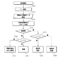
図4は、変曲周波数を探して変曲点の発生頻度数と脳波信号の振幅、脳波信号のFWHMの値を分析し、周波数別人体対応データベースと比較して健康状態を判別するフローチャートである。 FIG. 4 is a flowchart for searching the inflection frequency, analyzing the frequency of occurrence of the inflection point, the amplitude of the electroencephalogram signal, and the value of the FWHM of the electroencephalogram signal, and comparing the human body correspondence database by frequency to determine the health state. .
図4によれば、額部位の前頭葉に2チャンネルの脳波測定器を付着して測定した脳波データを、直列通信方式やUSB(Universal Serial Bus)通信方式でコンピュータに伝送した後、FFT(S300)を行う。測定は、被験者をして開眼状態と閉眼状態とを交互に反復させて、基礎律動の発生と消失及び各バンド別消失状態を観察できるデータを収集し、測定されたデータを一定時間累積して(S310)、各時間別に頂点(変曲)周波数による変曲点を見つけ、反復的に続く変曲周波数を抽出して周波数パターンを分析する(S320)。 According to FIG. 4, the electroencephalogram data measured by attaching a two-channel electroencephalograph to the frontal lobe of the forehead is transmitted to a computer by a serial communication method or a USB (Universal Serial Bus) communication method, and then FFT (S300). I do. The measurement is performed by repeating the eye-opening state and the closed-eye state alternately for the subject, collecting data that can observe the occurrence and disappearance of the basic rhythm and the disappearance state of each band, and accumulating the measured data for a certain period of time. (S310) An inflection point due to a vertex (inflection) frequency is found for each time, an inflection frequency that continues repeatedly is extracted, and a frequency pattern is analyzed (S320).
臨床試験結果で見つけられた各周波数と一対一の対応関係を持つ人体の各部位別データベースによって抽出された変曲周波数から人体の健康状態を判読する。ここで、変曲周波数は、周波数による1次微分値を求めて勾配値が変わる頂点を意味する。 The health condition of the human body is interpreted from the inflection frequency extracted by the database for each part of the human body having a one-to-one correspondence with each frequency found in the clinical test result. Here, the inflection frequency means a vertex at which the gradient value is changed by obtaining a first-order differential value by frequency.
下記の表1は、周波数別人体対応部位データベースの一部例である。

前記表1に記載されたように、各脳波帯域は固有周波数帯域を持ち、それぞれは人体対応点に一対一で対応させうる。0.1−3Hzの脳波帯域はδ波帯域であって、目、まぶた、眼球、眼球筋肉、顔面筋肉、鼻、口、襟首、てんかん、脳損傷に対応し、4−7Hzの脳波帯域は、θ波帯域で肩、耳、目、顔面、鼻、口、耳鳴りに対応し、8−12Hzの脳波帯域は、α波帯域で胃腸、内蔵、生殖器、子宮、腰、腎臓に対応し、12−15Hzの脳波帯域は、SMR波帯域で首、器官支、肺、心臓、皮膚病、アトピー病に対応し、16−20Hzの脳波帯域は、低β波帯域で横隔膜、腎臓、歯牙、あご、頚椎に対応し、21−30Hzの脳波帯域は、高β波帯域で頚椎、手、足、脚に対応する。 As described in Table 1, each electroencephalogram band has a natural frequency band, and each can correspond to a human body corresponding point on a one-to-one basis. The electroencephalogram band of 0.1-3 Hz is the δ wave band, and corresponds to eyes, eyelids, eyeballs, eye muscles, facial muscles, nose, mouth, neck, epilepsy, brain damage, and the electroencephalogram band of 4-7 Hz is Corresponds to shoulder, ear, eye, face, nose, mouth, tinnitus in θ wave band, EEG band of 8-12 Hz corresponds to gastrointestinal, internal organs, genitals, uterus, waist, kidney in α wave band, 12- The 15 Hz EEG band corresponds to the neck, organ support, lung, heart, skin disease, and atopic disease in the SMR wave band, and the 16-20 Hz EEG band corresponds to the diaphragm, kidney, tooth, jaw, cervical spine in the low β wave band. The brain wave band of 21-30 Hz corresponds to the cervical spine, hand, foot, and leg in the high β wave band.
反復的に同じ周波数で頂点が現れるのは、脳や肉体の特徴的な活動を意味することであり、脳波信号は脳の状態を、筋肉信号は肉体の状態を表す。各周波数を人体の各部位と一対一に対応させて人体の健康状態を判断する。健康状態の軽重は、頂点(変曲点)が現れる頻度数と脳波信号の振幅、脳波信号のFWHMなどにより決定される。もし、頂点(変曲点)が連続的に一定の周波数で開眼と閉眼時に継続的に現れるならば症状が激しいと言える。疾病の発生範囲はFWHMにより決定される。もし、FWHMが小さければ局所的に、大きければ広範囲に広がっていることを意味する。これを数式で表現すれば、前述した数式3によってH(n,V,w)=Af(nV/w)に表示され、ここで、nは変曲点の発生頻度数、Vは脳波信号の振幅、wは脳波信号のFWHM、Aは比例定数を意味し、Hは健康(Health)を意味する関数であり、fはnV/wの変数を持つ関数ということを意味する。したがって、一定周波数に対応する人体の対応点での疾病の軽重は、変曲点の発生頻度数及び脳波信号の振幅に比例し、脳波信号のFWHMに反比例するということである。これはあくまでも線形的な関係式を意味し、さらに非線形的な関係式を考慮してもよい。 The appearance of vertices at the same frequency repeatedly means characteristic activities of the brain and the body. The electroencephalogram signal represents the state of the brain, and the muscle signal represents the state of the body. The health state of the human body is determined by associating each frequency with each part of the human body on a one-to-one basis. The weight of the health state is determined by the frequency of appearance of the apex (inflection point), the amplitude of the electroencephalogram signal, the FWHM of the electroencephalogram signal, and the like. If the apex (inflection point) continuously appears at a constant frequency when the eyes are opened and closed, it can be said that the symptom is severe. The range of occurrence of the disease is determined by FWHM. If FWHM is small, it means that it is spread locally, and if it is large, it is spread over a wide range. If this is expressed by a mathematical expression, it is displayed as H (n, V, w) = Af (nV / w) according to the mathematical expression 3 described above, where n is the frequency of occurrence of inflection points and V is the electroencephalogram signal. Amplitude, w is FWHM of the electroencephalogram signal, A is a proportionality constant, H is a function meaning health, and f is a function having a variable of nV / w. Therefore, the severity of the disease at the corresponding point of the human body corresponding to a certain frequency is proportional to the frequency of occurrence of the inflection points and the amplitude of the electroencephalogram signal, and inversely proportional to the FWHM of the electroencephalogram signal. This means a linear relational expression, and a nonlinear relational expression may be considered.
これにより、図4のステップS330で変曲点が現れる頻度数を判断して、頻度数の低い場合にはステップS340に進行して(FWHMに対する振幅×頻度数)、すなわち、変数nV/wの関数値を求めて、高い場合に間歇的痛みまたは異常と判断し(S350)、低い場合に健康状態と判断する(S360)。 Accordingly, the frequency number at which the inflection point appears in step S330 in FIG. 4 is determined. If the frequency number is low, the process proceeds to step S340 (amplitude for FWHM × frequency number), that is, the variable nV / w A function value is obtained, and when it is high, it is determined as intermittent pain or abnormality (S350), and when it is low, it is determined as a healthy state (S360).
一方、図4のステップS330で変曲点が現れる頻度数を判断して、頻度数が高い場合にはステップS370に進行して(FWHMに対する振幅×頻度数)、すなわち、変数nV/wの関数値を求めて、低い場合に局所的異常と判断し(S380)、高い場合に局所的疾病と判断する(S390)。 On the other hand, the frequency number at which the inflection point appears in step S330 in FIG. 4 is determined. If the frequency number is high, the process proceeds to step S370 (amplitude for FWHM × frequency number), that is, a function of the variable nV / w. A value is obtained, and when it is low, it is determined as a local abnormality (S380), and when it is high, it is determined as a local disease (S390).
Claims (5)
測定された脳波を受けて高速フーリエ変換をした後、一定時間データを累積して各周波数別に現れた信号のパターンを分析し、分析された周波数パターンと人体の各部位との一対一の対応関係を設定して健康状態を判別する脳波分析コンピュータを備える脳波パターン分析を利用した健康診断装置。 In the health checkup device that receives the electroencephalogram signal measured by the electroencephalogram measurement device that measures the electroencephalogram through the electroencephalogram measurement sensor attached to the frontal lobe and performs the health check using electroencephalogram pattern analysis,
After receiving the measured electroencephalogram and performing fast Fourier transform, it accumulates data for a certain period of time and analyzes the pattern of the signal that appears for each frequency, and a one-to-one correspondence between the analyzed frequency pattern and each part of the human body A health diagnosis device using electroencephalogram pattern analysis, which includes an electroencephalogram analysis computer that determines the health state by setting the.
前頭葉に2チャンネル脳波測定センサーを付着し、耳たぶを基準電極とし、額の真ん中を参照電極とする方式で脳波を測定するステップと、
測定された脳波を高速フーリエ変換するステップと、
前記高速フーリエ変換されたデータを一定時間累積するステップと、
各周波数別に現れた信号のパターンを分析するステップと、
分析された周波数パターンと人体の各部位との一対一の対応関係を設定して健康状態を判別する健康状態判別ステップと、を含む脳波パターン分析を利用した健康診断方法。 In a health checkup method for performing a health checkup using electroencephalogram pattern analysis,
Attaching a 2-channel electroencephalogram sensor to the frontal lobe, measuring the electroencephalogram using the earlobe as a reference electrode and the middle of the forehead as a reference electrode;
A fast Fourier transform of the measured brain waves;
Accumulating the fast Fourier transformed data for a certain period of time;
Analyzing the pattern of the signal that appears for each frequency;
A health diagnosis method using electroencephalogram pattern analysis, comprising: a health state determination step that sets a one-to-one correspondence between the analyzed frequency pattern and each part of the human body to determine a health state.
Applications Claiming Priority (2)
| Application Number | Priority Date | Filing Date | Title |
|---|---|---|---|
| KR1020050085637A KR100719068B1 (en) | 2005-09-14 | 2005-09-14 | Apparatus and method of diagnosing health using cumulative data pattern analysis via fast Fourier transformation of brain wave data measured from frontal lobe |
| PCT/KR2006/003526 WO2007032617A1 (en) | 2005-09-14 | 2006-09-05 | Apparatus and method of diagnosing health using cumulative data pattern analysis via fast fourier transformation of brain wave data measured from frontal lobe |
Publications (2)
| Publication Number | Publication Date |
|---|---|
| JP2009507601A true JP2009507601A (en) | 2009-02-26 |
| JP2009507601A5 JP2009507601A5 (en) | 2012-06-14 |
Family
ID=37865168
Family Applications (1)
| Application Number | Title | Priority Date | Filing Date |
|---|---|---|---|
| JP2008531007A Pending JP2009507601A (en) | 2005-09-14 | 2006-09-05 | Health diagnosis apparatus and method using pattern analysis of accumulated data through fast Fourier transform of electroencephalogram data measured in frontal lobe |
Country Status (6)
| Country | Link |
|---|---|
| US (1) | US7756575B2 (en) |
| EP (1) | EP1934899A4 (en) |
| JP (1) | JP2009507601A (en) |
| KR (1) | KR100719068B1 (en) |
| CN (1) | CN101273372A (en) |
| WO (1) | WO2007032617A1 (en) |
Families Citing this family (14)
| Publication number | Priority date | Publication date | Assignee | Title |
|---|---|---|---|---|
| US20100324440A1 (en) * | 2009-06-19 | 2010-12-23 | Massachusetts Institute Of Technology | Real time stimulus triggered by brain state to enhance perception and cognition |
| WO2010147599A1 (en) * | 2009-06-19 | 2010-12-23 | Massachusetts Institute Of Technology | Real time stimulus triggered by brain state to enhance perception and cognition |
| KR101081369B1 (en) * | 2010-01-26 | 2011-11-08 | 중앙대학교 산학협력단 | Apparatus and method for feature extraction of bio signal |
| CN102138860B (en) * | 2011-01-10 | 2013-05-22 | 西安交通大学 | Intelligentized rehabilitation training equipment for hand functions of patients suffering from cerebral injury |
| US20130261420A1 (en) * | 2011-06-06 | 2013-10-03 | Semen Kucherov | System and method for non-invasive diagnostic of mammals |
| CN104382592B (en) * | 2014-12-11 | 2016-08-31 | 康泰医学系统(秦皇岛)股份有限公司 | A kind of EEG checking device based on power spectrumanalysis algorithm |
| TWI584219B (en) * | 2016-02-05 | 2017-05-21 | 澤康生物科技股份有限公司 | human body frequency diagnostic analysis apparatus and improvement method OF health assessmenT |
| US11504038B2 (en) | 2016-02-12 | 2022-11-22 | Newton Howard | Early detection of neurodegenerative disease |
| US10799186B2 (en) * | 2016-02-12 | 2020-10-13 | Newton Howard | Detection of disease conditions and comorbidities |
| KR102031958B1 (en) * | 2017-04-06 | 2019-10-14 | 고려대학교 산학협력단 | Prefrontal based cognitive brain-machine interfacing apparatus and method thereof |
| CN108378846A (en) * | 2018-02-08 | 2018-08-10 | 深圳市宏智力科技有限公司 | Based on binary channels brain electric detection method and device |
| WO2020190401A1 (en) * | 2019-03-18 | 2020-09-24 | Exo Neural Network Inc. | Medical therapy arrangement for applying an electrical stimulation to a human or animal subject |
| KR102455070B1 (en) * | 2020-11-25 | 2022-10-14 | 고려대학교 산학협력단 | Apparatus and Method of Seizure Prediction Using Patterns of Functional Connectivity Changes in EEG |
| CN114098764B (en) * | 2021-11-05 | 2024-02-20 | 北京大学 | Data processing method, device, electronic equipment and storage medium |
Family Cites Families (13)
| Publication number | Priority date | Publication date | Assignee | Title |
|---|---|---|---|---|
| US3971366A (en) | 1974-11-25 | 1976-07-27 | Hiroshi Motoyama | Apparatus and method for measuring the condition of the meridians and the corresponding internal organs of the living body |
| US4794533A (en) * | 1986-11-07 | 1988-12-27 | Cns, Inc. | System activity change indicator |
| EP0778001B1 (en) | 1995-05-12 | 2004-04-07 | Seiko Epson Corporation | Apparatus for diagnosing condition of living organism and control unit |
| KR100498794B1 (en) * | 1995-05-12 | 2006-07-19 | 세이코 엡슨 가부시키가이샤 | Apparatus for diagnosing condition of living organism and control unit |
| DE19649991A1 (en) | 1996-11-21 | 1998-06-04 | Axon Gmbh Schmalkalden | Procedure for determining sleep and wake profiles |
| US6097980A (en) | 1998-12-24 | 2000-08-01 | Monastra; Vincent J. | Quantitative electroencephalographic (QEEG) process and apparatus for assessing attention deficit hyperactivity disorder |
| US6358201B1 (en) | 1999-03-02 | 2002-03-19 | Doc L. Childre | Method and apparatus for facilitating physiological coherence and autonomic balance |
| US6317627B1 (en) | 1999-11-02 | 2001-11-13 | Physiometrix, Inc. | Anesthesia monitoring system based on electroencephalographic signals |
| KR20010103920A (en) * | 2000-05-12 | 2001-11-24 | 이일근 | Method and apparatus for remote medicare using biological signal |
| KR100450758B1 (en) * | 2002-01-22 | 2004-10-01 | 한국전자통신연구원 | Apparatus and method for measuring electroencephalogram |
| KR20040021269A (en) * | 2002-09-03 | 2004-03-10 | (주)비알시스템 | Apparatus and method for controlling brain waves by GSR measurement |
| US7177676B2 (en) * | 2004-01-20 | 2007-02-13 | Pt. Hartono Istana Teknologi | System and method for analyzing biofeedback information for performance of an individual in a group |
| WO2005117693A1 (en) * | 2004-05-27 | 2005-12-15 | Children's Medical Center Corporation | Patient-specific seizure onset detection system |
-
2005
- 2005-09-14 KR KR1020050085637A patent/KR100719068B1/en not_active IP Right Cessation
-
2006
- 2006-09-05 EP EP06798666A patent/EP1934899A4/en not_active Withdrawn
- 2006-09-05 WO PCT/KR2006/003526 patent/WO2007032617A1/en active Application Filing
- 2006-09-05 US US12/066,326 patent/US7756575B2/en not_active Expired - Fee Related
- 2006-09-05 CN CNA2006800339391A patent/CN101273372A/en active Pending
- 2006-09-05 JP JP2008531007A patent/JP2009507601A/en active Pending
Also Published As
| Publication number | Publication date |
|---|---|
| US20080200826A1 (en) | 2008-08-21 |
| CN101273372A (en) | 2008-09-24 |
| KR100719068B1 (en) | 2007-05-17 |
| WO2007032617A1 (en) | 2007-03-22 |
| KR20070031501A (en) | 2007-03-20 |
| EP1934899A1 (en) | 2008-06-25 |
| US7756575B2 (en) | 2010-07-13 |
| EP1934899A4 (en) | 2009-03-04 |
Similar Documents
| Publication | Publication Date | Title |
|---|---|---|
| KR100719068B1 (en) | Apparatus and method of diagnosing health using cumulative data pattern analysis via fast Fourier transformation of brain wave data measured from frontal lobe | |
| Lin et al. | IoT-based wireless polysomnography intelligent system for sleep monitoring | |
| US20140275829A1 (en) | Sleep stage annotation device | |
| JP2019528104A (en) | In-ear sensing system and method for monitoring biological signals | |
| Saidatul et al. | Analysis of EEG signals during relaxation and mental stress condition using AR modeling techniques | |
| Sulaiman et al. | EEG-based stress features using spectral centroids technique and k-nearest neighbor classifier | |
| Stochholm et al. | Automatic sleep stage classification using ear-EEG | |
| WO2009064475A1 (en) | Method for real time attitude assessment | |
| JP2006514570A (en) | Anesthesia and sedation monitoring system and method | |
| CN103153183A (en) | Apparatus and method for diagnosing obstructive sleep apnea | |
| WO2002060313A2 (en) | Fetal brain monitor | |
| Yang et al. | A pilot study on fetal heart rate extraction from wearable abdominal inertial sensors | |
| WO2015157253A1 (en) | Stochastic oscillator analysis in neuro diagnostics | |
| Orguc et al. | EMG-based real time facial gesture recognition for stress monitoring | |
| KR20160107390A (en) | Apparatus for measuring bio-signal | |
| RU2252692C2 (en) | Method and device for studying functional state of brain and method for measuring subelectrode resistance | |
| CN111419208A (en) | Acceleration sensor-based unbound real-time heart rate monitoring method and system | |
| JP2023544999A (en) | Method for classifying polysomnography records into defined sleep stages | |
| CN116369853A (en) | Olfactory function standardized evaluation device and method based on brain-computer interaction technology | |
| Plesnik et al. | ECG signal acquisition and analysis for telemonitoring | |
| ES2684533A1 (en) | Method for the exploration of obstructive sleep apnea based on the oxygen saturation signal (Machine-translation by Google Translate, not legally binding) | |
| Tian et al. | Hearables: Heart Rate Variability from Ear Electrocardiogram and Ear Photoplethysmogram (Ear-ECG and Ear-PPG) | |
| Takalokastari et al. | Real time drowsiness detection by a WSN based wearable ECG measurement system | |
| JP2020014611A (en) | Psychogenic non-epileptic fit detection device and method | |
| Liu et al. | The analysis of regularity and synchrony of Parkinsonian tremor using Approximate Entropy and Cross-Approximate Entropy |
Legal Events
| Date | Code | Title | Description |
|---|---|---|---|
| A621 | Written request for application examination |
Free format text: JAPANESE INTERMEDIATE CODE: A621 Effective date: 20090813 |
|
| A131 | Notification of reasons for refusal |
Free format text: JAPANESE INTERMEDIATE CODE: A131 Effective date: 20111125 |
|
| A601 | Written request for extension of time |
Free format text: JAPANESE INTERMEDIATE CODE: A601 Effective date: 20120224 |
|
| A602 | Written permission of extension of time |
Free format text: JAPANESE INTERMEDIATE CODE: A602 Effective date: 20120302 |
|
| A601 | Written request for extension of time |
Free format text: JAPANESE INTERMEDIATE CODE: A601 Effective date: 20120326 |
|
| A602 | Written permission of extension of time |
Free format text: JAPANESE INTERMEDIATE CODE: A602 Effective date: 20120402 |
|
| A521 | Request for written amendment filed |
Free format text: JAPANESE INTERMEDIATE CODE: A523 Effective date: 20120417 |
|
| A524 | Written submission of copy of amendment under article 19 pct |
Free format text: JAPANESE INTERMEDIATE CODE: A524 Effective date: 20120417 |
|
| A131 | Notification of reasons for refusal |
Free format text: JAPANESE INTERMEDIATE CODE: A131 Effective date: 20120706 |
|
| A02 | Decision of refusal |
Free format text: JAPANESE INTERMEDIATE CODE: A02 Effective date: 20130301 |