JP2008517658A - Method and apparatus for measuring analyte concentration in body fluid - Google Patents
Method and apparatus for measuring analyte concentration in body fluid Download PDFInfo
- Publication number
- JP2008517658A JP2008517658A JP2007538065A JP2007538065A JP2008517658A JP 2008517658 A JP2008517658 A JP 2008517658A JP 2007538065 A JP2007538065 A JP 2007538065A JP 2007538065 A JP2007538065 A JP 2007538065A JP 2008517658 A JP2008517658 A JP 2008517658A
- Authority
- JP
- Japan
- Prior art keywords
- skin
- layer
- intervening
- multilayer film
- pressure
- Prior art date
- Legal status (The legal status is an assumption and is not a legal conclusion. Google has not performed a legal analysis and makes no representation as to the accuracy of the status listed.)
- Withdrawn
Links
Images


Classifications
-
- A—HUMAN NECESSITIES
- A61—MEDICAL OR VETERINARY SCIENCE; HYGIENE
- A61B—DIAGNOSIS; SURGERY; IDENTIFICATION
- A61B5/00—Measuring for diagnostic purposes; Identification of persons
- A61B5/15—Devices for taking samples of blood
- A61B5/151—Devices specially adapted for taking samples of capillary blood, e.g. by lancets, needles or blades
- A61B5/15134—Bladeless capillary blood sampling devices, i.e. devices for perforating the skin in order to obtain a blood sample but not using a blade, needle, canula, or lancet, e.g. by laser perforation, suction or pressurized fluids
-
- G—PHYSICS
- G01—MEASURING; TESTING
- G01N—INVESTIGATING OR ANALYSING MATERIALS BY DETERMINING THEIR CHEMICAL OR PHYSICAL PROPERTIES
- G01N33/00—Investigating or analysing materials by specific methods not covered by groups G01N1/00 - G01N31/00
- G01N33/48—Biological material, e.g. blood, urine; Haemocytometers
- G01N33/483—Physical analysis of biological material
- G01N33/487—Physical analysis of biological material of liquid biological material
-
- A—HUMAN NECESSITIES
- A61—MEDICAL OR VETERINARY SCIENCE; HYGIENE
- A61B—DIAGNOSIS; SURGERY; IDENTIFICATION
- A61B5/00—Measuring for diagnostic purposes; Identification of persons
- A61B5/14—Devices for taking samples of blood ; Measuring characteristics of blood in vivo, e.g. gas concentration within the blood, pH-value of blood
-
- A—HUMAN NECESSITIES
- A61—MEDICAL OR VETERINARY SCIENCE; HYGIENE
- A61B—DIAGNOSIS; SURGERY; IDENTIFICATION
- A61B5/00—Measuring for diagnostic purposes; Identification of persons
- A61B5/15—Devices for taking samples of blood
- A61B5/150007—Details
- A61B5/150015—Source of blood
- A61B5/150022—Source of blood for capillary blood or interstitial fluid
-
- A—HUMAN NECESSITIES
- A61—MEDICAL OR VETERINARY SCIENCE; HYGIENE
- A61B—DIAGNOSIS; SURGERY; IDENTIFICATION
- A61B5/00—Measuring for diagnostic purposes; Identification of persons
- A61B5/15—Devices for taking samples of blood
- A61B5/150007—Details
- A61B5/150053—Details for enhanced collection of blood or interstitial fluid at the sample site, e.g. by applying compression, heat, vibration, ultrasound, suction or vacuum to tissue; for reduction of pain or discomfort; Skin piercing elements, e.g. blades, needles, lancets or canulas, with adjustable piercing speed
- A61B5/150061—Means for enhancing collection
- A61B5/150068—Means for enhancing collection by tissue compression, e.g. with specially designed surface of device contacting the skin area to be pierced
-
- A—HUMAN NECESSITIES
- A61—MEDICAL OR VETERINARY SCIENCE; HYGIENE
- A61B—DIAGNOSIS; SURGERY; IDENTIFICATION
- A61B5/00—Measuring for diagnostic purposes; Identification of persons
- A61B5/15—Devices for taking samples of blood
- A61B5/150007—Details
- A61B5/150053—Details for enhanced collection of blood or interstitial fluid at the sample site, e.g. by applying compression, heat, vibration, ultrasound, suction or vacuum to tissue; for reduction of pain or discomfort; Skin piercing elements, e.g. blades, needles, lancets or canulas, with adjustable piercing speed
- A61B5/150106—Means for reducing pain or discomfort applied before puncturing; desensitising the skin at the location where body is to be pierced
- A61B5/150114—Means for reducing pain or discomfort applied before puncturing; desensitising the skin at the location where body is to be pierced by tissue compression, e.g. with specially designed surface of device contacting the skin area to be pierced
-
- A—HUMAN NECESSITIES
- A61—MEDICAL OR VETERINARY SCIENCE; HYGIENE
- A61B—DIAGNOSIS; SURGERY; IDENTIFICATION
- A61B5/00—Measuring for diagnostic purposes; Identification of persons
- A61B5/15—Devices for taking samples of blood
- A61B5/150007—Details
- A61B5/150358—Strips for collecting blood, e.g. absorbent
-
- A—HUMAN NECESSITIES
- A61—MEDICAL OR VETERINARY SCIENCE; HYGIENE
- A61B—DIAGNOSIS; SURGERY; IDENTIFICATION
- A61B5/00—Measuring for diagnostic purposes; Identification of persons
- A61B5/15—Devices for taking samples of blood
- A61B5/150007—Details
- A61B5/150755—Blood sample preparation for further analysis, e.g. by separating blood components or by mixing
-
- A—HUMAN NECESSITIES
- A61—MEDICAL OR VETERINARY SCIENCE; HYGIENE
- A61B—DIAGNOSIS; SURGERY; IDENTIFICATION
- A61B5/00—Measuring for diagnostic purposes; Identification of persons
- A61B5/15—Devices for taking samples of blood
- A61B5/151—Devices specially adapted for taking samples of capillary blood, e.g. by lancets, needles or blades
- A61B5/15134—Bladeless capillary blood sampling devices, i.e. devices for perforating the skin in order to obtain a blood sample but not using a blade, needle, canula, or lancet, e.g. by laser perforation, suction or pressurized fluids
- A61B5/15136—Bladeless capillary blood sampling devices, i.e. devices for perforating the skin in order to obtain a blood sample but not using a blade, needle, canula, or lancet, e.g. by laser perforation, suction or pressurized fluids by use of radiation, e.g. laser
-
- G—PHYSICS
- G01—MEASURING; TESTING
- G01N—INVESTIGATING OR ANALYSING MATERIALS BY DETERMINING THEIR CHEMICAL OR PHYSICAL PROPERTIES
- G01N33/00—Investigating or analysing materials by specific methods not covered by groups G01N1/00 - G01N31/00
- G01N33/48—Biological material, e.g. blood, urine; Haemocytometers
Landscapes
- Health & Medical Sciences (AREA)
- Life Sciences & Earth Sciences (AREA)
- Engineering & Computer Science (AREA)
- Physics & Mathematics (AREA)
- Biomedical Technology (AREA)
- Molecular Biology (AREA)
- Hematology (AREA)
- Pathology (AREA)
- General Health & Medical Sciences (AREA)
- Biophysics (AREA)
- Public Health (AREA)
- Surgery (AREA)
- Animal Behavior & Ethology (AREA)
- Medical Informatics (AREA)
- Heart & Thoracic Surgery (AREA)
- Veterinary Medicine (AREA)
- Dermatology (AREA)
- Pain & Pain Management (AREA)
- Chemical & Material Sciences (AREA)
- Biochemistry (AREA)
- Analytical Chemistry (AREA)
- Medicinal Chemistry (AREA)
- General Physics & Mathematics (AREA)
- Immunology (AREA)
- Food Science & Technology (AREA)
- Urology & Nephrology (AREA)
- Optics & Photonics (AREA)
- Investigating Or Analysing Biological Materials (AREA)
- Measurement Of The Respiration, Hearing Ability, Form, And Blood Characteristics Of Living Organisms (AREA)
- Measuring And Recording Apparatus For Diagnosis (AREA)
- Investigating Or Analysing Materials By Optical Means (AREA)
Abstract
体液サンプル内の分析物濃度を測定するために体液サンプルを採取する方法であって、当該方法は、ユーザーの皮膚に圧力をかけることを含んでいる。かけられた圧力によって体液を裂け傷から流れ出させるように、皮膚が引き伸ばされて皮膚に裂け傷が形成される。裂け傷から流れ出る体液は採集される。 A method of collecting a bodily fluid sample to measure an analyte concentration in the bodily fluid sample, the method including applying pressure to the user's skin. The skin is stretched and a tear is formed in the skin so that the applied pressure causes fluid to flow out of the tear. Body fluids that flow from the laceration are collected.
Description
本発明は、概して、体液内の分析物濃度を測定するための方法及び装置に関する。より特定すると、本発明は、ユーザーに対する痛みの程度を減じるための最少侵襲性技術を使用して分析物濃度を測定するための方法及び装置に関する。 The present invention generally relates to a method and apparatus for measuring an analyte concentration in a body fluid. More particularly, the present invention relates to a method and apparatus for measuring analyte concentration using minimally invasive techniques to reduce the degree of pain to the user.
体液内の分析物濃度の測定は、患者の健康状態を判定し且つ病気の進行又は治療の有効性を監視するための重要な処置である。一つの一般的な方法は、患者の血糖濃度レベルを監視することである。伝統的な糖監視処置は、毛細血管の血液を少なくとも3マイクロリットル(μl)得るために、人の指を切開することを含む。毛細血管の血液は、次いで、反応が起こる試薬ストリップのような器具又は分析装置に適用される。これらの反応は、通常は、酵素によって仲介され、興味のある分析物に対して特有のものである。試薬ストリップとすることができる器具が、血液サンプル内の糖の定量のための血糖計内に挿入される。血液サンプル内の糖濃度は、ストリップのリフレクタンスの変化に基づくか又は器具内に発生される流れに基づいている。 Measuring analyte concentrations in body fluids is an important procedure for determining a patient's health and monitoring disease progression or therapeutic effectiveness. One common method is to monitor a patient's blood glucose level. Traditional sugar monitoring procedures involve incising a person's finger to obtain at least 3 microliters (μl) of capillary blood. The capillary blood is then applied to an instrument or analyzer such as a reagent strip where the reaction takes place. These reactions are usually mediated by enzymes and are specific to the analyte of interest. An instrument, which can be a reagent strip, is inserted into the blood glucose meter for the quantification of sugar in the blood sample. The sugar concentration in the blood sample is based on changes in the reflectance of the strip or based on the flow generated in the instrument.
伝統的な糖の監視は、試験に必要とされる血液の体積及びサンプルが集められる部位により、血液を分析されている人に著しい不快感をもたらすかも知れない。不快感を惹き起こすことに加えて、針及びランセットの使用は、典型的には、血液採集部位に目で見ることができる傷跡を残す。 Traditional sugar monitoring may cause significant discomfort to the person being analyzed for blood, depending on the volume of blood required for the test and the site where the sample is collected. In addition to causing discomfort, the use of a needle and lancet typically leaves a visible scar on the blood collection site.
当該技術において知られている代替的な糖監視技術は、間質液(ISF)内のグルコースの定量を含む侵襲性が極めて低い監視技術を提供することである。この技術は、患者の前腕のような代替的な部位における間質液の採集を可能にする。これは、患者の皮膚の表皮内深さ50乃至400マイクロメータ(μm)の穴を形成することによって行われる。この技術は、皮膚表面に仲介物質を適用することを含んでいる。仲介物質の目的は、電磁エネルギを熱エネルギに変換することである。しかしながら、仲介物質が皮内に貫入する深さは、個人個人で変わり、これは、(a)(穴を開けるための)力の効率、(b)皮膚に形成された穴の深さ、及び(c)患者が感じる痛みに影響を及ぼす。仲介物質はまた、抜き取られた物質と混ぜ合わせられ、分析物の濃度の分析精度に影響を与える。 An alternative sugar monitoring technique known in the art is to provide a very invasive monitoring technique that includes the quantification of glucose in interstitial fluid (ISF). This technique allows for the collection of interstitial fluid at alternative sites, such as the patient's forearm. This is done by creating a hole with a depth of 50 to 400 micrometers (μm) in the epidermis of the patient's skin. This technique involves applying a mediator to the skin surface. The purpose of the mediator is to convert electromagnetic energy into thermal energy. However, the depth at which the mediator penetrates into the skin varies from individual to individual: (a) the efficiency of the force (to make a hole), (b) the depth of the hole formed in the skin, and (C) It affects the pain felt by the patient. The mediator is also mixed with the extracted material and affects the analytical accuracy of the analyte concentration.
当該技術において知られているこの代替的な方法は、通常は、極めて洗練された採集及び貯蔵技術を必要とする小さな体積の流体を生成する。この方法は、通常、極めて痛く且つ長時間の恐怖をもたらすかも知れない。サンプルの損失は、より大きな穴を開ける必要性を生じるか、別の穴を開けるか又はより大きな力の使用を生じさせるかも知れない。これらはいずれも、患者に更に大きな痛みを受けさせるであろう。 This alternative method known in the art typically produces a small volume of fluid that requires very sophisticated collection and storage techniques. This method is usually very painful and may result in prolonged fear. Sample loss may result in the need to drill a larger hole, create another hole, or use a greater force. Both of these will make the patient more painful.
従って、サンプル採集及び汚染に関連する上記の問題を解決した分析物濃度を測定する方法及び装置の必要性が存在する。 Accordingly, there is a need for a method and apparatus for measuring analyte concentrations that solves the above problems associated with sample collection and contamination.
体液サンプル内の分析物濃度を測定するための体液サンプルを得るための一つの方法に従って、ユーザーの皮膚に圧力がかけられる。皮膚が引っ張られ、かけられた圧力によって皮膚に裂け傷を形成し、体液が裂け傷から流れ出るようにする。裂け傷から流れ出る体液は採集される。 According to one method for obtaining a bodily fluid sample for measuring an analyte concentration in the bodily fluid sample, pressure is applied to the user's skin. The skin is pulled and the applied pressure creates a tear in the skin, allowing fluid to flow out of the tear. Body fluids that flow from the laceration are collected.
体液試料内の分析物濃度を測定するための体液サンプルを得るための別の方法に従って、ユーザーの皮膚に圧力がかけられる。皮膚は引っ張られ、加圧部位において皮膚が加熱されて、皮膚に裂け傷が形成されて、体液が裂け傷が流れ出るようにさせる。裂け傷から流れ出る体液は採集される。 Pressure is applied to the user's skin according to another method for obtaining a bodily fluid sample for measuring an analyte concentration in the bodily fluid sample. The skin is pulled and heated at the site of pressurization, creating a laceration in the skin and allowing fluid to flow through the laceration. Body fluids that flow from the laceration are collected.
体液サンプル内の分析物濃度を測定する一つの方法に従って、カップによって多層膜が皮膚のある領域に適用される。カップは、当該多層膜を保持するようになされた第一の端部を備えている。当該多層膜は、採集層と、介在物質とを含んでいる。皮膚の領域は、皮膚の領域をカップの第一の端部と接触させることによって引き伸ばされる。電磁発生源からのエネルギは、多層膜上へと導かれて介在物質が電磁発生源からのエネルギを熱エネルギに変換して多層膜内に穴を形成し且つ皮膚に裂け傷を形成するようになされている。圧力が裂け傷を包囲している皮膚領域にかけられて、体液を裂け傷から穴を介して多層膜の上面へと流れさせる。カップが皮膚領域と接触状態に留まっている間に、多層膜の上面にある多量の体液が採集される。 According to one method of measuring analyte concentration in a body fluid sample, a multilayer membrane is applied to an area of skin by a cup. The cup includes a first end adapted to hold the multilayer film. The multilayer film includes a collection layer and an intervening substance. The area of skin is stretched by bringing the area of skin into contact with the first end of the cup. The energy from the electromagnetic source is directed onto the multilayer film so that the intervening material converts the energy from the electromagnetic source to thermal energy to form holes in the multilayer film and to tear the skin. Has been made. Pressure is applied to the skin area surrounding the laceration, causing fluid to flow from the laceration through the hole and into the upper surface of the multilayer membrane. While the cup remains in contact with the skin area, a large amount of body fluid on the top surface of the multilayer is collected.
一つの実施形態に従って、圧力を適用し且つ皮膚を引っ張って皮膚内に裂け傷を形成し且つ少なくとも1つの分析物濃度の分析のための体液サンプルを採集する準備をするための圧力部材は、弾性材料によって形成されている本体を含んでいる。本体の少なくとも一部分は膜である。当該膜は、介在物質を有している少なくともゲル化層を含んでいる。 According to one embodiment, the pressure member for applying pressure and pulling the skin to form a laceration in the skin and preparing to collect a body fluid sample for analysis of at least one analyte concentration is elastic It includes a body formed of material. At least a portion of the body is a membrane. The film includes at least a gelled layer having an intervening substance.
別の実施形態に従って、皮膚に圧力をかけ且つ皮膚を引っ張って皮膚に裂け傷を形成し、少なくとも1つの分析物濃度の分析のための体液サンプルを採集する準備のための加圧部位は、弾性材料によって形成されている本体を含んでいる。本体の少なくとも一部分は多層膜である。多層膜は、少なくとも1つの採集層と介在層とを含んでいる。採集層と介在層とは相互に隣接している。 According to another embodiment, the pressure site for applying pressure to the skin and pulling the skin to form a tear in the skin and preparing a body fluid sample for analysis of at least one analyte concentration is elastic It includes a body formed of material. At least a portion of the body is a multilayer film. The multilayer film includes at least one collection layer and an intervening layer. The collection layer and the intervening layer are adjacent to each other.
更に別の実施形態に従って、皮膚に圧力をかけ且つ引っ張って少なくとも1つの分析物濃度の分析のために皮膚に裂け傷を形成し且つ体液サンプルを採集する準備をするための圧力部材は、本体及び膜を含んでいる。本体は弾性部材によって形成されている。膜は、少なくとも介在物質を有しているゲル化層を含んでいる。当該膜は本体に取り外し可能に結合されている。 In accordance with yet another embodiment, a pressure member for applying pressure to and pulling on the skin to create a tear in the skin for analysis of at least one analyte concentration and to collect a body fluid sample comprises: Contains a membrane. The main body is formed of an elastic member. The membrane includes a gelled layer having at least an intervening substance. The membrane is removably coupled to the body.
更に別の実施形態に従って、皮膚に圧力を適用し且つ皮膚を引っ張って皮膚な裂け傷を形成し且つ少なくとも1つの分析物濃度の分析のための体液サンプルを採集するための圧力部材は、本体と多層膜とを含んでいる。本体は弾性材料によって形成されている。多層膜は、少なくとも採集層とを含んでいる。採集層と介在層とは相互に隣接している。多層膜は本体に取り外し可能に結合されている。 According to yet another embodiment, a pressure member for applying pressure to the skin and pulling the skin to form a skin laceration and collecting a body fluid sample for analysis of at least one analyte concentration comprises: And a multilayer film. The main body is made of an elastic material. The multilayer film includes at least a collection layer. The collection layer and the intervening layer are adjacent to each other. The multilayer film is removably coupled to the body.
一つの実施形態に従って、体液サンプルを採集し且つ分析するための膜は、ゲル化層を含んでいる。ゲル化層は介在物質を含んでいる。
更に別の実施形態に従って、体液サンプルを採集し且つ分析するための多層膜は、採集層と介在層とを含んでいる。介在層は採集層に隣接している。
According to one embodiment, a membrane for collecting and analyzing a body fluid sample includes a gelled layer. The gelled layer contains an intervening substance.
According to yet another embodiment, a multilayer film for collecting and analyzing a body fluid sample includes a collection layer and an intervening layer. The intervening layer is adjacent to the collection layer.
本発明は種々の変形例及び代替的な形態を受けやすいけれども、ここでは、特別な実施形態が図面に例示されており且つ詳細に説明されている。しかしながら、本発明はここに開示された特別な形態に限定されることを意図されていないことは理解されるべきである。 While the invention is susceptible to various modifications and alternative forms, specific embodiments are illustrated in the drawings and are described in detail herein. However, it should be understood that the invention is not intended to be limited to the particular forms disclosed herein.
本発明の一つの実施形態は、体液サンプルの採集及び分析のために患者の皮膚に裂け傷を形成するための方法及び装置に関する。一つの方法によって採集される体液の量は極めて少量である。例えば、当該極めて少量の流体は、約1000ナノリットル(ηl)未満であっても良い。より典型的には、体液サンプルは約500ηl未満である。採集することができる体液サンプルの量は、50又は30ηl程度の少量であってさえ良い。採集量を減じることは、採集時間を短くするので望ましい。採集する流体量が少ないことは、採集処置に伴う時間及び痛みを減じ得るので、少量の流体を採集するのが望ましい場合が多い。本発明の方法及び装置は、体液サンプルを採集する機能とこれらの流体を分析する機能とを結合している。 One embodiment of the invention relates to a method and apparatus for creating a laceration in a patient's skin for collection and analysis of a body fluid sample. The amount of body fluid collected by one method is very small. For example, the very small amount of fluid may be less than about 1000 nanoliters (ηl). More typically, the body fluid sample is less than about 500 ηl. The amount of body fluid sample that can be collected may even be as small as 50 or 30 ηl. Reducing the amount collected is desirable because it shortens the collection time. Collecting a small amount of fluid is often desirable because a small amount of fluid collected can reduce the time and pain associated with the collection procedure. The method and apparatus of the present invention combines the function of collecting bodily fluid samples with the function of analyzing these fluids.
最初に図1aを参照すると、一つの実施形態に従って、この機能の組み合わせを達成するために、多層膜10は、試験対象者の皮膚に適用され且つ体液サンプルを採集するために使用される。このプロセスは、皮膚の真皮内に見出される体液を分析するために使用される。採集且つ試験することができる幾つかの流体としては、間質液(ISF)又は全血サンプルがある。
Referring initially to FIG. 1a, to achieve this combination of functions, according to one embodiment, the
一つの実施形態によれば、図1aの多層膜10は、採集層12、介在層14及び支持層16を含んでいる。採集層12は、可撓性材料によって作られるのが望ましい。例えば、採集層12は、ポリエチレン又はポリ塩化ビニル(PVC)のようなポリマー材料によって作ることができる。
According to one embodiment, the
多層膜10の採集層12は、反射材料、試薬又はこれらの組み合わせを含んでいても良い。一つの実施形態においては、採集層12の上面(図1aに見ることが出来る)は、反射材によってコーティングしても良い。採集層12を反射材によって作るか又はコーティングすることによって、採集された体液サンプルから反射された光のスペクトル分析が容易になる。当該反射材は、幾つかの領域内の介在層14に光が到達する一方でその他の領域は反射し続けるようにするパターンとすることができる。採集層12内で使用することができる反射材の一つの非限定的な例は、銀コーティングされたポリマーである。採集層12を形成するのに使用することができるもう一つ別の例は、必要ならば、電磁エネルギが介在層へ到達するのを許容する懸濁された二酸化チタンの粒子を使用して反射領域と非反射領域とのパターンを有しているゲル材料である。
The
もう一つ別の実施形態によれば、採集層12は試薬を含んでいても良い。当該試薬は、採集層12上に配置されるか、採集層12上にコーティングされるか又は採集層内に含浸されていても良い。採集された体液サンプル単独の光学的サイン又は試薬と興味のある分析物との間の反応の光学的サインが分析されて、体液サンプル内の分析物濃度が測定される。監視は、電気化学的性質のものであっても良いことが考えられる。採集層12が電気化学的検知プロセスであるように設計されている場合には、電気リード線は、当該採集層から延びるであろう。試薬は、1以上の分析物の分析を同時に行うために、採集層内にパターン化することができることが考えられる。
According to another embodiment, the
本発明の一つの実施形態によれば、採集層12は、概して約5μm乃至約500μmの厚みである。より特別には、採集層12は、典型的には、約5μm乃至100μmの厚みである。当該典型的な厚みは採集層の組成に依存して変化するかも知れない。例えば、採集層が反射材を含んでいる場合には、採集層の典型的な厚みは約5μm乃至約10μmである。採集層が試薬剤を含んでいる場合には、採集層の典型的な厚みは約20μm乃至約100μmである。
According to one embodiment of the invention, the
図1aに示されている多層膜10はまた、介在層14をも含んでいる。多層膜10を形成する際には、一つの実施形態による介在層14は、採集層12にコーティングしても良い。介在層14は、コーティング以外の方法によって採集層12に適用しても良いことが考えられる。例えば、介在層14は、スパッタリング蒸着方法によって採集層12に適用しても良い。
The
以下に詳細に説明するように、介在層14の厚みは、患者の皮膚に裂け傷を形成し且つサンプルを採集するために多層膜10に適用される熱エネルギの量によって変化しても良い。本発明の一つの実施形態に従って、介在層14の厚みは約15μm乃至約50μmである。より特別には、介在層14の厚みは、典型的には約20μm乃至約35μmである。
As described in detail below, the thickness of the intervening
一つの実施形態によれば、介在層14は、ゼラチン及び内部に懸濁された介在物質18を含んでいる。一つの実施形態においては、介在層18はカーボンブラックである。介在物質は、電磁エネルギ吸収物質によって作ることができる。介在層のもう一つ別の例は染料である。一つの実施形態によれば、介在物質18を懸濁する助けとするために、ゼラチンは表面活性剤を含んでいても良い。介在層14に表面活性剤を添加することによって、ゼラチンが極めて小さいサンプルから水分を吸収しないように疎水性を有するゼラチンの形成ももたらされる。表面活性剤は、体液が流れ且つ溜まらせて体液サンプルの採集を補助する。
According to one embodiment, the intervening
多層膜10の支持層16は、可撓性材料によって作られるのが望ましい。例えば、支持層16は、ポリエチレン又はポリ塩化ビニル(PVC)のような高分子材料によって作ることができる。支持層は、セルロースのような他の材料によって作っても良い。支持層16を形成する際の材料は添加物を含んでいても良いことが考えられる。所望の添加物としては、(a)支持強度、(b)支持層材料を引っ張る機能、及び(c)流体の流れを制御する機能のうちの1以上を高めるものがある。支持層16の厚みは、概して、約15μm乃至約100μmである。より特別には、支持層16の厚みは典型的には約15μm乃至約30μmである。
The support layer 16 of the
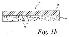
上記した多層膜10以外の他の膜を使用しても良いことが考えられる。例えば、図1bを参照すると、多層膜110は、採集層12及び介在層14を含んでいる。更に別の実施形態によれば、図1cの膜210は介在層214を含んでいても良い。介在層214は、ゼラチンによって形成し且つ内部に介在物質218を含んでいる。採集層12に関して上記したように、介在層214は、反射材、試薬又はこれらの組み合わせを含んでいても良い。
It is conceivable that films other than the
一つの実施形態による膜10,110,210は、体液サンプル内の1以上の分析物の濃度を測定するために、約1000ηl未満の体液好ましくは約500ηl未満のサンプルサイズを採集し且つ分析するために使用される。採集できる体液サンプルの量は、50又は30ηl程度の少量とすることさえできる。一つの測定できる特別なサンプルは間質液内の糖量である。
The
図2及び3を参照すると、本発明の一つの実施形態により、体液サンプルを採集し且つ分析するために膜(例えば、多層膜10)と共に使用するための採集カップ20が示されている。カップ20は、多層膜10に関して説明されているが、膜110及び210のような他の膜がカップと共に使用されても良いことが考えられる。
With reference to FIGS. 2 and 3, a
一般的に、以下に詳細に説明するように、採集カップ20は、体液サンプルを採集するだけでなくサンプルを分析のために保持するために使用される。カップ20はまた、サンプルの採集及び分析において使用される多層膜10を保持し且つ圧力を患者の皮膚に適用する。採集カップ20は、内側面22、外側面24、底部縁26及び頂部又は頂端28を含んでいる。頂部28は、側壁32と底面34とによって結合されている凹部30を含んでいる。図3の底面34は、ほぼ平らなものとして示されている。頂部28はまた、カップ20の内側から凹部30を介してカップ20の外部まで延びている穴36をも形成している。カップ20は、例えばゴム、ポリカーボネート又は成形高分子部品を形成するために使用されるその他の適切な高分子材料のような可撓性の弾性材料によって作ることができる。
In general, as described in detail below, the
更に図4を参照すると、カップ20の頂部28の拡大図が示されている。多層膜10は、当該多層膜10の支持層16が底面34に接触するように凹部30内に配置されている。多層膜10は、凹部30内に配置されている穴36を覆うように配置され且つインサート38によって定位置に摩擦保持されている。インサート38は、凹部30内に嵌合する設計とされており且つ凹部30の形状に適合する形状を有するような設計とされている。インサート38は、多層膜10を底面34に対して保持するために凹部の側壁32に押し付けられる。インサート38は、凹部30内の穴の中心とほぼ整合している中心を有する穴40を形成している。穴40は、インサート38の内側面42を規定している。
Still referring to FIG. 4, an enlarged view of the top 28 of the
図5を参照すると、カップ20の頂部28は、患者の皮膚S内に押し込まれた状態で示されている。一般的に、使用時に、カップ20の頂部28は、皮膚Sの前腕又はその他の領域のような患者の皮膚Sと接触状態とされる。頂部28は、患者の皮膚Sに押し付けられて皮膚Sを変形させて皮膚Sが引き伸ばされ且つ最終的に薄くされる。一つの実施形態によれば、約1.36乃至2.27キログラム(3乃至5ポンド)の力がカップ20にかけられる。次いで、この作用によって、約1.36乃至2.27キログラム(3乃至5ポンド)の力が患者の皮膚Sにかかり、これによって皮膚が引き伸ばされ且つ皮膚に上向きの圧力が形成される。皮膚Sは(図5において)下方に押されつつある頂部28と接触状態にあるけれども、インサート38の内側面42によって結合された皮膚Sの領域は上方へ押される。皮膚の隣接する領域がカップ20の頂部28によって下方へ押し付けられるときにその領域の真皮の下の真皮内に配置されているISFを含む流体は、カップ20の頂部28によって下方へと押し付けられるので、内側面42は上方へと押される。インサート38の内側面42によって結合された皮膚Sの上方に押された領域は、内側面42と接触し且つカップ20によって保持された多層膜10の支持層16に押し付けられる。多層膜10の弾性により、膜10は、皮膚Sが膜10に押し付けられると若干変形する。
Referring to FIG. 5, the top 28 of the
上記したカップ20以外の他の加圧部材を使用しても良いことが考えられる。望ましい加圧部材は、皮膚内に裂け傷を形成し且つ体液サンプルを採集する準備のために、圧力をかけ且つ皮膚を引き伸ばす必要がある。例えば、金属リングのような加圧部材は、類似した方法で皮膚に圧力を給送し且つ皮膚を引き伸ばすように設計することができる。考えられる別の例は、皮膚が持ち上げられ且つ定着部材内に挟まれる方法である。
It is conceivable that pressure members other than the
一つの実施形態によれば、加圧部材は弾性材料によって形成された本体を含んでいる。この実施形態によれば、本体の少なくとも一部分は膜である。このような実施形態においては、本体と膜とは相互に一体化されている。膜は、介在物質を有するゼラチン層のような一つの層を含んでいても良い。しかしながら、本体は、多層膜10,110に関して上記したような多層膜を含んでいても良いことが考えられる。別の実施形態によれば、本体と膜とは、取り外し可能に結合された別個の部材であっても良い。この実施形態における膜はまた、介在物質を有するゼラチン層のような一つの層をも含んでいても良く又は多層膜10,110に関して上記したような多層膜であっても良い。 According to one embodiment, the pressure member includes a body formed of an elastic material. According to this embodiment, at least a portion of the body is a membrane. In such an embodiment, the main body and the membrane are integrated with each other. The film may include a single layer, such as a gelatin layer with an intervening material. However, it is contemplated that the body may include a multilayer film as described above with respect to the multilayer films 10,110. According to another embodiment, the body and the membrane may be separate members that are removably coupled. The film in this embodiment may also include a single layer, such as a gelatin layer with an intervening material, or it may be a multilayer film as described above with respect to multilayer films 10,110.
以上、皮膚Sを多層膜に対して押し付けるための正圧をかける(例えば、カップ20を皮膚Sに対して押し付ける)ことに関する方法を説明した。しかしながら、代替的な実施形態においては、負圧を使用しても良い。例えば、多層膜10のような膜に対して皮膚Sを引き付けるために負圧を使用しても良い。
In the above, the method regarding applying the positive pressure for pressing the skin S against the multilayer film (for example, pressing the
一つの方法によれば、体液サンプルがその分析物濃度を測定するために採取される。ユーザーの皮膚に圧力がかけられ、かけられた圧力によって体液を裂け傷から流れ出させるように皮膚に裂け傷を形成するために皮膚が引き伸ばされる。裂け傷から流れ出す体液が採集される。分析物濃度は、典型的には、例えば、光学的プロセスによって体液サンプル内の分析物濃度を測定するために分析される。かけられた圧力及び引き伸ばしは、例えばカップ20のような加圧部材によって適用することができる。一つの方法によってかけられる力は正圧である。上記したように、かけられる圧力は負圧であっても良いことが考えられる。
According to one method, a body fluid sample is taken to measure its analyte concentration. Pressure is applied to the user's skin and the skin is stretched to form a laceration in the skin so that fluid can flow out of the laceration. Body fluid that flows out of the laceration is collected. The analyte concentration is typically analyzed to determine the analyte concentration in the body fluid sample, for example, by an optical process. The applied pressure and stretching can be applied by a pressure member such as
図6を参照すると、一つの方法によって皮膚Sの角質層を軟化させても良い。例えば、皮膚Sは、電磁放射線源、熱線又は加熱空気のような幾つかの加熱方法のうちの一つを使用することによって軟化させることができる。一つの実施形態においては、レーザーダイオードのような光源52から放射されるレーザービーム50の形態のエネルギが、例えば多層膜10の少なくとも一部分へと導かれる。膜110及び210のような他の膜を幾つかの加熱方法のうちの一つと組み合わせて使用しても良いことが考えられる。
Referring to FIG. 6, the stratum corneum of skin S may be softened by one method. For example, the skin S can be softened by using one of several heating methods such as an electromagnetic radiation source, hot wire or heated air. In one embodiment, energy in the form of a
一つの実施形態によれば、光源は、約120ミリ秒乃至約300ミリ秒の期間に亘って約300乃至約450ミリワットの出力で放射されるナノメータ(nm)のパルスレーザービームを提供する。一つの実施形態によれば、レーザービーム50の直径は、約50μm乃至約500μmである。レーザービーム50からの電磁放射線は、多層膜10の介在層14内の介在物質18によって吸収され且つ熱エネルギに変換される。熱エネルギは、皮膚Sの角質層へと送られる。この加熱プロセスは、角質層を軟化させ且つ弱くし、皮膚と表皮との間の接合部を分裂させる。皮膚Sの表皮層は、引き伸ばされて破壊され且つ裂かれる。カップによって表皮を引き伸ばすことにより且つ熱エネルギによって生じる皮膚と表皮との間の接合部の分裂によって、少なくとも一つの裂け傷が形成される。
According to one embodiment, the light source provides a nanometer (nm) pulsed laser beam emitted at a power of about 300 to about 450 milliwatts over a period of about 120 milliseconds to about 300 milliseconds. According to one embodiment, the
図7を参照すると、患者の表皮に裂け傷を形成する際に、皮膚内で(カップ20の適用によって生じる)圧力を受けている体液が表皮の裂け傷を通って流れ且つレーザービーム50によって多層膜10に形成された穴54内を通る。皮膚の組織学的評価は、標準的な皮膚切断技術の後に観察される長く続く損傷とは異なり皮膚の裂け傷の迅速な修復の証拠となる。このようにして、結果的に得られる裂け傷は、皮膚上の切開部に長く続かない形跡を形成する。
Referring to FIG. 7, in forming a laceration in the patient's epidermis, body fluid under pressure (caused by application of cup 20) flows through the epidermal laceration and multi-layered by
サンプルは、毛管網領域に最も密接な関係がある表皮の上方層から得られる。この領域は、良好な間質液/毛細血管交換を有している。穴54内を流れる体液は多層膜10の頂部に溜まる。間質液の正圧は、表皮の裂け傷を形成することに寄与し、圧力の力及びカップ20によって提供される引き伸ばし、単一の構成部品によってより少ない力が必要とされ、それによって、サンプリングされている人が感じる疼痛を減じる。
Samples are obtained from the upper layer of the epidermis that is most closely related to the capillary network region. This region has good interstitial fluid / capillary exchange. The body fluid flowing in the hole 54 accumulates on the top of the
サンプル流体Fは、多層膜10の採集層12の(図7において)上面に溜まる。一つの方法によって、十分な体積のサンプル流体Fが溜まった後に、サンプル流体Fが光源56からの光によって照射される。光源の非限定的な例としては、単色光、多色光及び赤外光がある。他の集光光源を採用しても良いことが考えられる。1以上の検知器58は、サンプル流体Fから反射された光を集光する。皮膚の種類に応じて、カップ自体の圧力は、光源の必要なく十分なサンプル流体Fを形成することができることも考えられる。
The sample fluid F accumulates on the upper surface (in FIG. 7) of the
少なくとも1つの検知器58は、例えば、シリコン検知器を含んでいても良い。別の方法として、少なくとも1つの検知器58は、集光された反射光を検知器又は分光計に流す光ファイバの束を含んでいても良い。検知器の一つの非限定的な例は、拡大InGaAs検知器である。他の検知器を採用しても良いことが考えられる。検知器は、集光された光を集光された反射光を評価するための装置(図示せず)を示す信号を出力する。この装置は、データを1以上の興味ある分析物の濃度の値に変換するためにスペクトルデータに適用される1以上の埋め込まれているアルゴリズムを備えている。
The at least one
サンプルから反射された光の集光を助けるために、多層膜10の採集層12の(図7において)上面は、反射面(例えば、銀コーティング)を含んでいても良い。更に別の実施形態においては、採集層12は、興味ある分析物に関する光学的サインを形成するために、興味のある分析物と反応する試薬を含んでいる。
In order to help collect light reflected from the sample, the top surface (in FIG. 7) of the
方法及び装置の精度を判定する研究がなされた。電気化学的電流測定糖分析を行い、結果が図8にグラフで示されている。この研究は、ISF(間質液)グルコースと血漿グルコースとの間の相対関係を研究するために使用される耐糖能障害研究であった。図8のグラフは、ISF及び血漿グルコース濃度対時間を示している。図8の結ばれた線の忠実な奇跡は、サンプリング間に著しい変動がないことを示している。 Research has been done to determine the accuracy of the method and apparatus. An electrochemical amperometric sugar analysis was performed and the results are shown graphically in FIG. This study was a glucose intolerance study used to study the relative relationship between ISF (interstitial fluid) glucose and plasma glucose. The graph in FIG. 8 shows ISF and plasma glucose concentration versus time. The faithful miracle of the connected line in FIG. 8 shows that there is no significant variation between samplings.
プロセス1
体液サンプル内の分析物の濃度を測定するために体液サンプルを採取する方法であって、
ユーザーの皮膚に圧力をかけるステップと、
皮膚を引き伸ばして皮膚に裂け傷を形成し、前記かけられた圧力によって体液が裂け傷から流れ出るようにするステップと、
前記裂け傷から流出する体液を採集するステップとを含む方法。
Process 1
A method of collecting a bodily fluid sample to measure the concentration of an analyte in the bodily fluid sample,
Applying pressure to the user's skin;
Stretching the skin to form a tear in the skin and allowing the fluid to flow out of the tear by the applied pressure;
Collecting bodily fluid flowing out of the laceration.
プロセスB
体液サンプル内の分析物濃度を分析するステップを更に含んでいるプロセスAの方法。
プロセスC
前記分析が任意に行われるプロセスBの方法。
Process B
The method of process A, further comprising analyzing the analyte concentration in the sample of body fluid.
Process C
The method of process B, wherein the analysis is optionally performed.
プロセスD
皮膚に圧力をかけるステップおよび皮膚を引き伸ばすステップが、カップを皮膚に押し付けて皮膚に圧力をかけることを含み、前記カップが皮膚接触領域を含んでおり、当該皮膚接触領域が、体液サンプルが採集される穴を形成するプロセスAの方法。
Process D
Applying pressure to the skin and stretching the skin includes pressing the cup against the skin to apply pressure to the skin, the cup including a skin contact area, where the body fluid sample is collected. Method A of forming a hole.
プロセスE
体液サンプルが間質液であるプロセスAの方法。
プロセスF
かけられる前記圧力が正圧であるプロセスAの方法。
Process E
The method of process A, wherein the body fluid sample is an interstitial fluid.
Process F
The method of process A, wherein the pressure applied is positive.
プロセスG
かけられる前記圧力が負圧であるプロセスAの方法。
プロセスH
体液サンプル内の分析物の濃度を測定するために体液サンプルを採取する方法であって、
ユーザーの皮膚に圧力をかけるステップと、
皮膚を引き伸ばすステップと、
前記加圧部位の皮膚を加熱して皮膚に裂け傷を形成し、前記圧力によって、体液が裂け傷から流出するようにさせるステップと、
前記裂け傷から流出する体液を採集するステップとを含む方法。
Process G
The method of process A, wherein the pressure applied is negative.
Process H
A method of collecting a bodily fluid sample to measure the concentration of an analyte in the bodily fluid sample,
Applying pressure to the user's skin;
Stretching the skin,
Heating the skin at the pressurized site to form a tear in the skin, and causing the body fluid to flow out of the tear by the pressure;
Collecting bodily fluid flowing out of the laceration.
プロセスI
体液サンプル内の分析物濃度を分析するステップを更に含んでいるプロセスHの方法。
プロセスJ
前記分析が任意に行われるプロセスIの方法。
Process I
The method of process H further comprising the step of analyzing the analyte concentration in the body fluid sample.
Process J
The method of process I, wherein the analysis is optionally performed.
プロセスK
皮膚に圧力をかけるステップおよび皮膚を引き伸ばすステップが、カップを皮膚に押し付けて皮膚に圧力をかけることを含み、前記カップが皮膚接触領域を含んでおり、当該皮膚接触領域は、体液サンプルが採集される穴を形成するプロセスIの方法。
Process K
Applying pressure to the skin and stretching the skin includes pressing the cup against the skin to apply pressure to the skin, the cup including a skin contact area, where the body fluid sample is collected. Process I method of forming a hole.
プロセスL
皮膚を加熱するステップが、介在層を含んでいる膜を皮膚に当てるステップと、
前記カップの皮膚接触領域によって前記膜を皮膚に押し付けるステップと、
更に、前記膜の少なくとも一部分に向けて電磁放射線を導いて、前記膜に穴を開け且つ皮膚に裂け傷を形成するステップと、を含むプロセスKの方法。
Process L
Heating the skin comprises applying a membrane containing an intervening layer to the skin;
Pressing the membrane against the skin by the skin contact area of the cup;
A method of Process K, further comprising the step of directing electromagnetic radiation toward at least a portion of the membrane to puncture the membrane and form a tear in the skin.
プロセスM
前記介在層が、ゼラチンと介在物質とを含んでおり、当該介在物質が前記ゼラチン内に配置されているプロセスLの方法。
Process M
The method of process L, wherein the intervening layer includes gelatin and an intervening substance, and the intervening substance is disposed in the gelatin.
プロセスN
前記皮膚を加熱するステップが、皮膚に、相互に隣接している採集層と介在層とを含む多層膜を当てることと、前記カップの皮膚接触領域によって前記多層膜を皮膚に押し付けることと、更に、前記多層膜の少なくとも一部分に向けて電磁放射線を導いて前記多層膜内に穴を開け且つ皮膚に裂け傷を形成することと、を含んでいるプロセスKの方法。
Process N
Heating the skin comprises applying to the skin a multilayer film comprising a collection layer and an intervening layer adjacent to each other; pressing the multilayer film against the skin by the skin contact area of the cup; and The method of process K, comprising: directing electromagnetic radiation toward at least a portion of the multilayer film to puncture the multilayer film and form a tear in the skin.
プロセスO
前記多層膜がゼラチンと介在物質とを含んでおり、当該介在物質は前記ゼラチン内に配置されている、プロセスNの方法。
Process O
The method of process N, wherein the multilayer film includes gelatin and an intervening substance, and the intervening substance is disposed in the gelatin.
プロセスP
前記電磁放射線を導くステップが、レーザーを介して電磁放射線を前記多層膜の少なくとも一部分に向けて導くことを含んでいる、プロセスNの方法。
Process P
The method of Process N, wherein directing the electromagnetic radiation includes directing electromagnetic radiation toward at least a portion of the multilayer film via a laser.
プロセスQ
前記皮膚を加熱するステップが、皮膚に、採集層と介在物質を含んでいるゼラチン層と支持層とを含み且つ前記ゼラチン層が前記採集層と前記支持層との間に配置されている多層膜を当てることと、前記カップの皮膚接触領域によって前記多層膜を皮膚に押し付けることと、更に、電磁放射線を前記多層膜の少なくとも一部分に向けて導いて前記多層膜に穴を開け且つ皮膚に裂け傷を形成することと、を含んでいるプロセスKの方法。
Process Q
The step of heating the skin includes a gelatin layer including a collection layer, an intervening substance, and a support layer on the skin, and the gelatin layer is disposed between the collection layer and the support layer. Pressing the multilayer film against the skin by the skin contact area of the cup, and further guiding electromagnetic radiation toward at least a portion of the multilayer film to perforate the multilayer film and tear the skin Forming a method of process K.
プロセスR
前記皮膚を加熱するステップが、レーザーからの電磁放射線を導くことを含んでいる、プロセスHの方法。
Process R
The method of process H, wherein the step of heating the skin includes directing electromagnetic radiation from a laser.
プロセスS
前記体液が間質液である、プロセスHの方法。
プロセスT
かけられる前記圧力が正圧であるプロセスHの方法。
Process S
The method of process H, wherein the body fluid is an interstitial fluid.
Process T
The method of process H, wherein the pressure applied is positive.
プロセスU
かけられる前記圧力が負圧であるプロセスHの方法。
プロセスV
体液サンプル内の分析物濃度を測定するために使用する方法であって、
多層膜を保持するようになされた第一の端部を備えているカップによって、皮膚のある領域に、前記多層膜にして、採集層と介在物質を含んでいる介在層とを含んでいる多層膜を当てるステップと、
前記カップの第一の端部を皮膚の前記領域に接触させることによって前記皮膚の領域を引き伸ばすステップと、
電磁源からのエネルギを前記多層膜に向けて導いて、前記介在物質が、前記電磁源からのエネルギを熱エネルギに変換して、前記多層膜に穴を開け且つ皮膚に裂け傷を形成するようにするステップと、
前記裂け傷を囲んでいる皮膚の領域に圧力をかけて、体液が裂け傷から前記穴を通って前記多層膜の上面へと流れるようにさせるステップと、
前記カップが前記皮膚の領域と接触状態にある間に、多量の体液を前記多層膜の上面上に採集するステップと、を含む方法。
Process U
The method of process H, wherein the pressure applied is negative.
Process V
A method used to measure an analyte concentration in a body fluid sample, comprising:
A multilayer comprising a collection layer and an intervening layer containing an intervening substance in an area of the skin by means of a cup having a first end adapted to hold the multi-layer film. Applying a membrane,
Stretching the area of skin by contacting the first end of the cup to the area of skin;
Directing energy from an electromagnetic source towards the multilayer film, so that the intervening material converts the energy from the electromagnetic source into thermal energy to perforate the multilayer film and form a tear in the skin Step to
Applying pressure to an area of skin surrounding the laceration to cause body fluid to flow from the laceration through the hole to the top surface of the multilayer;
Collecting a volume of bodily fluid on the top surface of the multilayer film while the cup is in contact with the area of the skin.
プロセスW
前記体液サンプル内の分析物と反応させるために、前記多層膜内に試薬を提供するステップを更に含んでいる、プロセスVの方法。
Process W
The method of process V, further comprising providing a reagent in the multilayer film for reacting with an analyte in the body fluid sample.
プロセスX
前記カップが皮膚と接触状態にある間に、採集された体液サンプルの分光分析を行うステップを更に含んでいる、プロセスVの方法。
Process X
The method of process V, further comprising the step of performing spectroscopic analysis of the collected body fluid sample while the cup is in contact with the skin.
プロセスY
前記分光分析を行うステップが前記採集された体液サンプルを照射するステップを含んでいる、プロセスXの方法。
Process Y
The method of Process X, wherein performing the spectroscopic analysis comprises irradiating the collected body fluid sample.
プロセスZ
前記照射するステップが、前記体液サンプルを単色光によって照射することを含んでいる、プロセスYの方法。
Process Z
The method of process Y, wherein the irradiating comprises irradiating the bodily fluid sample with monochromatic light.
プロセスAA
前記照射するステップが、前記体液サンプルを多色光によって照射することを含んでいるプロセスYの方法。
Process AA
The method of process Y, wherein the irradiating comprises irradiating the bodily fluid sample with polychromatic light.
プロセスBB
前記照射するステップが、前記体液サンプルを赤外光によって照射することを含んでいるプロセスYの方法。
Process BB
The method of process Y, wherein the irradiating comprises irradiating the body fluid sample with infrared light.
プロセスCC
前記分光分析を行うステップが、体液サンプルから反射された光を検知することを更に含んでいるプロセスYの方法。
Process CC
The method of process Y, wherein performing the spectroscopic analysis further comprises detecting light reflected from the body fluid sample.
プロセスDD
体液サンプルが採集される前記多層膜の上面が反射材料を含んでいるプロセスCCの方法。
Process DD
The method of process CC, wherein the top surface of the multilayer film from which the body fluid sample is collected includes a reflective material.
プロセスEE
前記介在物質がカーボンブラックであるプロセスVの方法。
プロセスFF
体液が間質液であるプロセスVの方法。
Process EE
The method of process V, wherein the intervening material is carbon black.
Process FF
The method of process V, wherein the body fluid is interstitial fluid.
プロセスGG
前記多層膜が支持層を更に含んでおり、前記介在層が、前記採集層と前記支持層との間に配置されているプロセスVの方法。
Process GG
The method of process V, wherein the multilayer film further comprises a support layer, and the intervening layer is disposed between the collection layer and the support layer.
プロセスHH
前記介在層がゼラチン及び介在物質を含んでいるプロセスVの方法。
プロセスII
前記電磁源がレーザーであるプロセスVの方法。
Process HH
The method of process V, wherein the intervening layer comprises gelatin and intervening material.
Process II
The method of process V, wherein the electromagnetic source is a laser.
実施例JJ
皮膚に裂け傷を形成し且つ少なくとも1つの分析物濃度を分析するために体液サンプルを採集するための準備をするために、皮膚に圧力をかけ且つ皮膚を引き伸ばすための加圧部材であって、弾性材料によって作られた本体にして、当該本体の少なくとも一部分が、介在物質を含んでいるゼラチン層を少なくとも含んでいる膜である本体を含む前記加圧部材。
Example JJ
A pressure member for applying pressure to and stretching the skin to prepare for collecting a sample of body fluid to form a laceration in the skin and analyze at least one analyte concentration; Said pressure member comprising a body made of an elastic material, wherein at least a portion of said body is a membrane comprising at least a gelatin layer containing an intervening substance.
実施例KK
前記介在物質が高電磁エネルギ吸収材である実施例JJの加圧部材。
実施例LL
前記介在物質がカーボンブラックである実施例KKの加圧部材。
Example KK
The pressure member of Example JJ, wherein the intervening material is a high electromagnetic energy absorber.
Example LL
The pressure member of Example KK, wherein the intervening material is carbon black.
実施例MM
前記ゼラチン層が反射材を更に含んでいる実施例JJの加圧部材。
実施例NN
前記ゼラチン層が試薬を更に含んでいる実施例JJの加圧部材。
Example MM
The pressure member of Example JJ, wherein the gelatin layer further comprises a reflective material.
Example NN
The pressure member of Example JJ, wherein the gelatin layer further comprises a reagent.
実施例OO
前記本体が、皮膚を押圧するようになされている開口端を備えている略円錐形のカップを含んでいる実施例JJの加圧部材。
Example OO
The pressure member of Example JJ, wherein the body includes a generally conical cup with an open end adapted to press against the skin.
実施例PP
皮膚に裂け傷を形成し且つ少なくとも1つの分析物濃度を分析するために体液サンプルを採集するための準備をするために、皮膚に圧力をかけ且つ皮膚を引き伸ばすための加圧部材であって、弾性材料によって作られた本体にして、当該本体の少なくとも一部分が、少なくとも1つの採集層と介在層とを含んでいる多層膜である本体を含み、前記採集層と介在層とは相互に隣接している、前記加圧部材。
Example PP
A pressure member for applying pressure to and stretching the skin to prepare for collecting a sample of body fluid to form a laceration in the skin and analyze at least one analyte concentration; A body made of an elastic material, wherein at least a portion of the body is a multilayer film including at least one collection layer and an intervening layer, wherein the collection layer and the interposition layer are adjacent to each other; The pressure member.
実施例QQ
前記介在層がゼラチンと介在物質とを含んでいる実施例PPの加圧部材。
実施例RR
前記介在物質が高電磁エネルギ吸収材である実施例PPの加圧部材。
Example QQ
The pressure member of Example PP, wherein the intervening layer contains gelatin and an intervening substance.
Example RR
The pressure member of Example PP, wherein the intervening material is a high electromagnetic energy absorber.
実施例SS
前記介在物質がカーボンブラックである実施例RRの加圧部材。
実施例TT
前記多層膜が支持層を更に含んでおり、前記介在層が前記採集層と前記支持層との間に配置されている実施例PPの加圧部材。
Example SS
The pressure member of Example RR, wherein the intervening substance is carbon black.
Example TT
The pressure member of Example PP, wherein the multilayer film further includes a support layer, and the intervening layer is disposed between the collection layer and the support layer.
実施例UU
前記採集層が反射材を含んでいる実施例PPの加圧部材。
実施例VV
前記採集層が試薬を含んでいる実施例PPの加圧部材。
Example UU
The pressure member of Example PP, wherein the collection layer includes a reflective material.
Example VV
The pressure member of Example PP, wherein the collection layer contains a reagent.
実施例WW
前記本体が、皮膚を押圧するようになされている開口端を備えている略円錐形のカップを含んでいる実施例PPの加圧部材。
Example WW
The pressure member of embodiment PP, wherein the body includes a generally conical cup with an open end adapted to press against the skin.
実施例XX
皮膚に裂け傷を形成し且つ少なくとも1つの分析物濃度を分析するために体液サンプルを採集するための準備をするために、皮膚に圧力をかけ且つ皮膚を引き伸ばすための加圧部材であって、本体と膜とを含んでおり、当該本体は弾性部材によって形成されており、前記膜は介在物質を含んでいるゼラチン層を少なくとも含んでおり、前記膜は前記本体に取り外し可能に結合されている、前記加圧部材。
Example XX
A pressure member for applying pressure to and stretching the skin to prepare for collecting a sample of body fluid to form a laceration in the skin and analyze at least one analyte concentration; A main body and a film are included, the main body is formed of an elastic member, the film includes at least a gelatin layer including an intervening substance, and the film is detachably coupled to the main body. The pressure member.
実施例YY
前記介在物質が高電磁エネルギ吸収材である実施例XXの加圧部材。
実施例ZZ
前記介在物質がカーボンブラックである実施例YYの加圧部材。
Example YY
The pressure member of Example XX, wherein the intervening material is a high electromagnetic energy absorber.
Example ZZ
The pressure member of Example YY, wherein the intervening material is carbon black.
実施例AAA
前記ゼラチン層が反射材を更に含んでいる実施例XXの加圧部材。
実施例BBB
前記ゼラチン層が試薬を更に含んでいる実施例XXの加圧部材。
Example AAA
The pressure member of Example XX, wherein the gelatin layer further comprises a reflector.
Example BBB
The pressure member of Example XX, wherein the gelatin layer further comprises a reagent.
実施例CCC
前記本体が、皮膚を押圧するようになされている開口端を備えている概して円錐形のカップを含んでいる実施例XXの加圧部材。
Example CCC
The pressure member of Example XX, wherein the body includes a generally conical cup with an open end adapted to press against the skin.
実施例DDD
皮膚に裂け傷を形成し且つ少なくとも1つの分析物濃度を分析するために体液サンプルを採集するための準備をするために、皮膚に圧力をかけ且つ皮膚を引き伸ばすための加圧部材であって、本体と多層膜とを含んでおり、当該本体は弾性部材によって形成されており、前記多層膜は、少なくとも採集層と介在層とを含んでおり、当該採集層と介在層とは相互に隣接しており、前記多層膜は前記本体に取り外し可能に結合されている、前記加圧部材。
Example DDD
A pressure member for applying pressure to and stretching the skin to prepare for collecting a sample of body fluid to form a laceration in the skin and analyze at least one analyte concentration; The main body includes a multilayer film, the main body is formed of an elastic member, and the multilayer film includes at least a collection layer and an interposition layer, and the collection layer and the interposition layer are adjacent to each other. The pressurizing member, wherein the multilayer film is detachably coupled to the body.
実施例EEE
前記介在層がゼラチンと介在物質とを含んでいる実施例DDDの加圧部材。
実施例FFF
前記介在物質が高電磁エネルギ吸収材である実施例DDDの加圧部材。
Example EEE
The pressing member of Example DDD, wherein the intervening layer contains gelatin and an intervening substance.
Example FFF
The pressing member of Example DDD, wherein the intervening substance is a high electromagnetic energy absorber.
実施例GGG
前記介在物質がカーボンブラックである実施例FFFの加圧部材。
実施例HHH
前記多層膜が支持層を更に含んでおり、前記介在層が前記採集層と前記支持層との間に配置されている実施例DDDの加圧部材。
Example GGG
The pressure member of Example FFF, wherein the intervening material is carbon black.
Example HHH
The pressure member of Example DDD, wherein the multilayer film further includes a support layer, and the intervening layer is disposed between the collection layer and the support layer.
実施例III
前記採集層が反射材を含んでいる実施例DDDの加圧部材。
実施例JJJ
前記採集層が試薬を含んでいる実施例DDDの加圧部材。
Example III
The pressure member of Example DDD, wherein the collection layer includes a reflective material.
Example JJJ
The pressure member of Example DDD, wherein the collection layer contains a reagent.
実施例KKK
前記本体が、皮膚を押圧するようになされている開口端を備えている略円錐形のカップを含んでいる実施例DDDの加圧部材。
Example KKK
The pressure member of embodiment DDD, wherein the body includes a generally conical cup with an open end adapted to press against the skin.
実施例LLL
体液サンプルを採集し且つ分析するための膜であって、介在物質を含んでいるゼラチン層を含んでいる膜。
Example LLL
A membrane for collecting and analyzing a body fluid sample, the membrane comprising a gelatin layer containing an intervening substance.
実施例MMM
前記介在物質が高電磁エネルギ吸収材である実施例LLLの膜。
実施例NNN
前記介在物質がカーボンブラックである実施例LLLの膜。
Example MMM
The membrane of Example LLL wherein the intervening material is a high electromagnetic energy absorber.
Example NNN
The membrane of Example LLL wherein the intervening material is carbon black.
実施例OOO
前記ゼラチン層が反射材を更に含んでいる実施例LLLの膜。
実施例PPP
前記ゼラチン層が試薬を更に含んでいる実施例LLLの膜。
Example OOO
The film of Example LLL, wherein the gelatin layer further comprises a reflector.
Example PPP
The film of Example LLL, wherein the gelatin layer further comprises a reagent.
実施例QQQ
体液サンプルの採集及び分析のための多層膜であって、採集層と介在層とを含んでおり、当該介在層が前記採集層に隣接している多層膜。
Example QQQ
A multilayer film for collecting and analyzing a body fluid sample, the multilayer film including a collection layer and an intervening layer, the intervening layer being adjacent to the collection layer.
実施例RRR
前記介在層が、ゼラチン層と介在物質とを含んでいる実施例QQQの多層膜。
実施例SSS
前記介在物質が高電磁エネルギ吸収材である実施例QQQの多層膜。
Example RRR
The multilayer film of Example QQQ, wherein the intervening layer includes a gelatin layer and an intervening substance.
Example SSS
The multilayer film of Example QQQ, wherein the intervening material is a high electromagnetic energy absorber.
実施例TTT
前記介在物質がカーボンブラックである実施例SSSの多層膜。
実施例UUU
支持層を更に含んでおり、前記介在層が前記採集層と前記支持層との間に配置されている実施例QQQの多層膜。
Example TTT
The multilayer film of example SSS, wherein the intervening material is carbon black.
Example UUU
The multilayer film of embodiment QQQ, further comprising a support layer, wherein the intervening layer is disposed between the collection layer and the support layer.
実施例VVV
前記採集層が反射材を更に含んでいる実施例QQQの多層膜。
実施例WWW
前記採集層が試薬を含んでいる実施例QQQの多層膜。
Example VVV
The multilayer film of embodiment QQQ, wherein the collection layer further includes a reflective material.
Example WWW
The multilayer film of Example QQQ, wherein the collection layer contains a reagent.
Claims (75)
ユーザーの皮膚に圧力をかけるステップと、
皮膚を引き伸ばして皮膚に裂け傷を形成し、かけられた前記圧力によって体液が裂け傷から流れ出るようにするステップと、
前記裂け傷から流出する体液を採集するステップとを含む方法。 A method of collecting a bodily fluid sample to measure the concentration of an analyte in the bodily fluid sample,
Applying pressure to the user's skin;
Stretching the skin to form a tear in the skin and allowing the fluid to flow out of the tear by the applied pressure;
Collecting bodily fluid flowing out of the laceration.
ユーザーの皮膚に圧力をかけるステップと、
皮膚を引き伸ばすステップと、
前記圧力をかけた部位の皮膚を加熱して皮膚に裂け傷を形成し、前記圧力によって、体液が裂け傷から流出するようにさせるステップと、
前記裂け傷から流出する体液を採集するステップとを含む方法。 A method of collecting a bodily fluid sample to measure the concentration of an analyte in the bodily fluid sample,
Applying pressure to the user's skin;
Stretching the skin,
Heating the skin of the site to which the pressure is applied to form a tear in the skin, and causing the body fluid to flow out of the tear by the pressure; and
Collecting bodily fluid flowing out of the laceration.
前記カップの皮膚接触領域によって前記膜を皮膚に押し付けるステップと、
更に、前記膜の少なくとも一部分に向けて電磁放射線を導いて、前記膜に穴を開け且つ皮膚に裂け傷を形成するステップと、を含む請求項11の方法。 Heating the skin comprises applying a membrane containing an intervening layer to the skin;
Pressing the membrane against the skin by the skin contact area of the cup;
12. The method of claim 11, further comprising the step of directing electromagnetic radiation toward at least a portion of the membrane to puncture the membrane and create a tear in the skin.
多層膜を保持するようになされた第一の端部を備えているカップによって、皮膚の、ある領域に、前記多層膜にして、採集層と介在物質を含んでいる介在層とを含んでいる前記多層膜を当てるステップと、
前記カップの第一の端部を皮膚の前記領域に接触させることによって前記皮膚の領域を引き伸ばすステップと、
電磁源からのエネルギを前記多層膜に向けて導いて、前記介在物質が、前記電磁源からのエネルギを熱エネルギに変換して、前記多層膜に穴を開け且つ皮膚に裂け傷を形成するようにするステップと、
前記裂け傷を囲んでいる皮膚の領域に圧力をかけて、体液が裂け傷から前記穴を通って前記多層膜の上面へと流れるようにさせるステップと、
前記カップが前記皮膚の領域と接触状態にある間に、多量の体液を前記多層膜の上面上に採集するステップと、を含む方法。 A method used to measure an analyte concentration in a body fluid sample, comprising:
A cup having a first end adapted to hold the multilayer film includes a collection layer and an intervening layer containing an intervening substance in a certain area of the skin as the multilayer film. Applying the multilayer film;
Stretching the area of skin by contacting the first end of the cup to the area of skin;
Directing energy from an electromagnetic source towards the multilayer film, so that the intervening material converts the energy from the electromagnetic source into thermal energy to perforate the multilayer film and form a tear in the skin Step to
Applying pressure to an area of skin surrounding the laceration to cause body fluid to flow from the laceration through the hole to the top surface of the multilayer;
Collecting a volume of bodily fluid on the top surface of the multilayer film while the cup is in contact with the area of the skin.
Applications Claiming Priority (2)
| Application Number | Priority Date | Filing Date | Title |
|---|---|---|---|
| US62083504P | 2004-10-21 | 2004-10-21 | |
| PCT/US2005/037901 WO2006047290A2 (en) | 2004-10-21 | 2005-10-20 | Method of determining the concentration of an analyte in a body fluid and system therefor |
Publications (2)
| Publication Number | Publication Date |
|---|---|
| JP2008517658A true JP2008517658A (en) | 2008-05-29 |
| JP2008517658A5 JP2008517658A5 (en) | 2008-12-11 |
Family
ID=35912173
Family Applications (1)
| Application Number | Title | Priority Date | Filing Date |
|---|---|---|---|
| JP2007538065A Withdrawn JP2008517658A (en) | 2004-10-21 | 2005-10-20 | Method and apparatus for measuring analyte concentration in body fluid |
Country Status (14)
| Country | Link |
|---|---|
| US (1) | US7896819B2 (en) |
| EP (1) | EP1848337A2 (en) |
| JP (1) | JP2008517658A (en) |
| KR (1) | KR20070073918A (en) |
| CN (1) | CN101080200A (en) |
| AU (1) | AU2005299759A1 (en) |
| BR (1) | BRPI0516978A (en) |
| CA (1) | CA2584800A1 (en) |
| MX (1) | MX2007004757A (en) |
| NO (1) | NO20072566L (en) |
| RU (1) | RU2007118684A (en) |
| TW (1) | TW200630070A (en) |
| WO (1) | WO2006047290A2 (en) |
| ZA (1) | ZA200703981B (en) |
Cited By (2)
| Publication number | Priority date | Publication date | Assignee | Title |
|---|---|---|---|---|
| JP2011521709A (en) * | 2008-05-30 | 2011-07-28 | インテュイティ メディカル インコーポレイテッド | Body fluid collection device and collection site interface |
| KR101535216B1 (en) * | 2015-04-02 | 2015-07-09 | (주) 비비비 | Fluidic device and apparatus for analyzing body fluid comprising the same |
Families Citing this family (7)
| Publication number | Priority date | Publication date | Assignee | Title |
|---|---|---|---|---|
| WO2009042810A2 (en) * | 2007-09-25 | 2009-04-02 | Isense Corporation | Method and apparatus for treating skin prior to biosensor insertion |
| JP5178272B2 (en) * | 2008-03-24 | 2013-04-10 | パナソニック株式会社 | Micro device |
| CA2707300C (en) * | 2007-12-10 | 2017-09-26 | Bayer Healthcare Llc | Rapid-read gated amperometry |
| JP2010266203A (en) * | 2008-05-20 | 2010-11-25 | Sony Corp | Method and device for acquisition of biological information, and method and device for measurement of physiologically active substance |
| EP2724139B1 (en) | 2011-06-27 | 2023-06-21 | EMD Millipore Corporation | Method and apparatus for filtering a liquid sample |
| CN110988088B (en) | 2014-03-07 | 2023-01-03 | 安晟信医疗科技控股公司 | Test sensor, method, biosensor and system for determining analyte concentration |
| KR101822417B1 (en) | 2017-06-14 | 2018-01-29 | 주식회사 청도제약 | Means for determination of oxdative stress in human fluid |
Family Cites Families (37)
| Publication number | Priority date | Publication date | Assignee | Title |
|---|---|---|---|---|
| GB1139891A (en) | 1963-12-05 | 1969-01-15 | Gevaert Photo Prod Nv | Heat and pressure-sensitive recording and information-recording processes |
| US4959305A (en) * | 1986-06-18 | 1990-09-25 | Miles Inc. | Reversible immobilization of assay reagents in a multizone test device |
| US5161532A (en) | 1990-04-19 | 1992-11-10 | Teknekron Sensor Development Corporation | Integral interstitial fluid sensor |
| DK0630265T3 (en) | 1992-03-13 | 2003-06-30 | Diatide Inc | Technetium-99m labeled peptides for imaging inflammation |
| US6315772B1 (en) | 1993-09-24 | 2001-11-13 | Transmedica International, Inc. | Laser assisted pharmaceutical delivery and fluid removal |
| US5582184A (en) | 1993-10-13 | 1996-12-10 | Integ Incorporated | Interstitial fluid collection and constituent measurement |
| US5885211A (en) | 1993-11-15 | 1999-03-23 | Spectrix, Inc. | Microporation of human skin for monitoring the concentration of an analyte |
| US20020169394A1 (en) * | 1993-11-15 | 2002-11-14 | Eppstein Jonathan A. | Integrated tissue poration, fluid harvesting and analysis device, and method therefor |
| JP3328061B2 (en) | 1994-04-15 | 2002-09-24 | 富士写真フイルム株式会社 | Cartridge for dry analysis film |
| DE69524108T2 (en) * | 1994-09-08 | 2002-06-06 | Lifescan Inc | ANALYTIC DETECTION STRIP WITH A STANDARD ON THE STRIP |
| AU707065B2 (en) | 1995-08-29 | 1999-07-01 | Nitto Denko Corporation | Microporation of human skin for drug delivery and monitoring applications |
| EP0847248B1 (en) | 1995-08-29 | 2003-04-16 | Philippe Holder | Support element and device for hooking and positioning paintings or the like |
| US5879310A (en) | 1995-09-08 | 1999-03-09 | Integ, Inc. | Body fluid sampler |
| US5879367A (en) | 1995-09-08 | 1999-03-09 | Integ, Inc. | Enhanced interstitial fluid collection |
| US5682233A (en) | 1995-09-08 | 1997-10-28 | Integ, Inc. | Interstitial fluid sampler |
| IL120587A (en) * | 1996-04-04 | 2000-10-31 | Lifescan Inc | Reagent test strip for determination of blood glucose |
| US5879311A (en) | 1996-05-17 | 1999-03-09 | Mercury Diagnostics, Inc. | Body fluid sampling device and methods of use |
| DE69722414T2 (en) | 1996-07-03 | 2004-05-19 | Altea Therapeutics Corp. | MULTIPLE MECHANICAL MICROPERFORATION OF SKIN OR MUCOSA |
| US6071251A (en) * | 1996-12-06 | 2000-06-06 | Abbott Laboratories | Method and apparatus for obtaining blood for diagnostic tests |
| US5913833A (en) | 1997-02-07 | 1999-06-22 | Abbott Laboratories | Method and apparatus for obtaining biological fluids |
| US6027496A (en) | 1997-03-25 | 2000-02-22 | Abbott Laboratories | Removal of stratum corneum by means of light |
| JP3097600B2 (en) | 1997-05-30 | 2000-10-10 | 日本電気株式会社 | Stratum corneum puncture device |
| US5964718A (en) | 1997-11-21 | 1999-10-12 | Mercury Diagnostics, Inc. | Body fluid sampling device |
| US6155992A (en) * | 1997-12-02 | 2000-12-05 | Abbott Laboratories | Method and apparatus for obtaining interstitial fluid for diagnostic tests |
| DE69928229T2 (en) * | 1998-02-17 | 2006-08-03 | Abbott Laboratories, Abbott Park | DEVICE FOR REMOVING AND ANALYZING INTERSTITUTIONAL LIQUID |
| US6022316A (en) * | 1998-03-06 | 2000-02-08 | Spectrx, Inc. | Apparatus and method for electroporation of microporated tissue for enhancing flux rates for monitoring and delivery applications |
| ATE375752T1 (en) | 1998-03-06 | 2007-11-15 | Spectrx Inc | INTEGRATED TISSUE PORATION, FLUID COLLECTION AND ANALYSIS DEVICE |
| US6091975A (en) | 1998-04-01 | 2000-07-18 | Alza Corporation | Minimally invasive detecting device |
| US7384396B2 (en) | 1998-07-21 | 2008-06-10 | Spectrx Inc. | System and method for continuous analyte monitoring |
| US6306152B1 (en) * | 1999-03-08 | 2001-10-23 | Agilent Technologies, Inc. | Lancet device with skin movement control and ballistic preload |
| US6132449A (en) * | 1999-03-08 | 2000-10-17 | Agilent Technologies, Inc. | Extraction and transportation of blood for analysis |
| JP2002542498A (en) * | 1999-04-22 | 2002-12-10 | シグナス, インコーポレイテッド | Methods and devices for removing interfering species |
| DE10030410C1 (en) * | 2000-06-21 | 2002-01-24 | Roche Diagnostics Gmbh | Blood lancet device for drawing blood for diagnostic purposes |
| US6558528B1 (en) * | 2000-12-20 | 2003-05-06 | Lifescan, Inc. | Electrochemical test strip cards that include an integral dessicant |
| US20030143113A2 (en) * | 2002-05-09 | 2003-07-31 | Lifescan, Inc. | Physiological sample collection devices and methods of using the same |
| US20060184189A1 (en) * | 2002-11-15 | 2006-08-17 | Lorin Olson | Cap for a dermal tissue lancing device |
| US20040127818A1 (en) * | 2002-12-27 | 2004-07-01 | Roe Steven N. | Precision depth control lancing tip |
-
2005
- 2005-10-20 CA CA002584800A patent/CA2584800A1/en not_active Abandoned
- 2005-10-20 AU AU2005299759A patent/AU2005299759A1/en not_active Abandoned
- 2005-10-20 US US11/665,118 patent/US7896819B2/en active Active
- 2005-10-20 JP JP2007538065A patent/JP2008517658A/en not_active Withdrawn
- 2005-10-20 WO PCT/US2005/037901 patent/WO2006047290A2/en active Application Filing
- 2005-10-20 CN CNA2005800435286A patent/CN101080200A/en active Pending
- 2005-10-20 MX MX2007004757A patent/MX2007004757A/en unknown
- 2005-10-20 BR BRPI0516978-0A patent/BRPI0516978A/en not_active IP Right Cessation
- 2005-10-20 KR KR1020077011344A patent/KR20070073918A/en not_active Application Discontinuation
- 2005-10-20 EP EP05816007A patent/EP1848337A2/en not_active Withdrawn
- 2005-10-20 RU RU2007118684/14A patent/RU2007118684A/en not_active Application Discontinuation
- 2005-10-21 TW TW094137055A patent/TW200630070A/en unknown
-
2007
- 2007-05-17 ZA ZA200703981A patent/ZA200703981B/en unknown
- 2007-05-21 NO NO20072566A patent/NO20072566L/en not_active Application Discontinuation
Cited By (2)
| Publication number | Priority date | Publication date | Assignee | Title |
|---|---|---|---|---|
| JP2011521709A (en) * | 2008-05-30 | 2011-07-28 | インテュイティ メディカル インコーポレイテッド | Body fluid collection device and collection site interface |
| KR101535216B1 (en) * | 2015-04-02 | 2015-07-09 | (주) 비비비 | Fluidic device and apparatus for analyzing body fluid comprising the same |
Also Published As
| Publication number | Publication date |
|---|---|
| EP1848337A2 (en) | 2007-10-31 |
| ZA200703981B (en) | 2008-08-27 |
| NO20072566L (en) | 2007-07-17 |
| BRPI0516978A (en) | 2008-09-30 |
| WO2006047290A3 (en) | 2006-09-21 |
| CN101080200A (en) | 2007-11-28 |
| US7896819B2 (en) | 2011-03-01 |
| RU2007118684A (en) | 2008-11-27 |
| TW200630070A (en) | 2006-09-01 |
| CA2584800A1 (en) | 2006-05-04 |
| KR20070073918A (en) | 2007-07-10 |
| WO2006047290A2 (en) | 2006-05-04 |
| US20080097240A1 (en) | 2008-04-24 |
| MX2007004757A (en) | 2007-06-25 |
| AU2005299759A1 (en) | 2006-05-04 |
Similar Documents
| Publication | Publication Date | Title |
|---|---|---|
| JP2008517658A (en) | Method and apparatus for measuring analyte concentration in body fluid | |
| US7374949B2 (en) | Diagnostic test strip for collecting and detecting an analyte in a fluid sample | |
| US7625457B2 (en) | Dual blade lancing test strip | |
| US6285448B1 (en) | Clinical analyte determination by infrared spectroscopy | |
| US7264627B2 (en) | Wicking methods and structures for use in sampling bodily fluids | |
| JP4365116B2 (en) | Puncture needle integrated minimal invasive optical format | |
| US8398567B2 (en) | Method and apparatus for measuring an analyte in a body fluid | |
| JP2004529728A (en) | Apparatus and method for sampling body fluid | |
| JP2005046612A (en) | Puncturing device and method of using the same | |
| JP2003159238A (en) | Instrument for sampling physiological fluid | |
| JP3853789B2 (en) | Sampling device and method of using a stepped capillary channel | |
| JPH11155844A (en) | Living body measuring device with constant living body contact area | |
| WO1999043255A1 (en) | Near infrared-transmission spectroscopy of tongue tissue | |
| CA2450106A1 (en) | Sampling devices and methods for bodily fluids | |
| JPH09117437A (en) | Non-aggressive biochemical sensor | |
| MXPA06008847A (en) | Method and apparatus for measuring an analyte in a body fluid |
Legal Events
| Date | Code | Title | Description |
|---|---|---|---|
| A521 | Written amendment |
Free format text: JAPANESE INTERMEDIATE CODE: A523 Effective date: 20081020 |
|
| A621 | Written request for application examination |
Free format text: JAPANESE INTERMEDIATE CODE: A621 Effective date: 20081020 |
|
| A761 | Written withdrawal of application |
Free format text: JAPANESE INTERMEDIATE CODE: A761 Effective date: 20091125 |