JP2007518539A - How to treat glaucoma - Google Patents
How to treat glaucoma Download PDFInfo
- Publication number
- JP2007518539A JP2007518539A JP2006551349A JP2006551349A JP2007518539A JP 2007518539 A JP2007518539 A JP 2007518539A JP 2006551349 A JP2006551349 A JP 2006551349A JP 2006551349 A JP2006551349 A JP 2006551349A JP 2007518539 A JP2007518539 A JP 2007518539A
- Authority
- JP
- Japan
- Prior art keywords
- eye
- laser
- ciliary
- laser light
- sclera
- Prior art date
- Legal status (The legal status is an assumption and is not a legal conclusion. Google has not performed a legal analysis and makes no representation as to the accuracy of the status listed.)
- Pending
Links
Images


Classifications
-
- A—HUMAN NECESSITIES
- A61—MEDICAL OR VETERINARY SCIENCE; HYGIENE
- A61F—FILTERS IMPLANTABLE INTO BLOOD VESSELS; PROSTHESES; DEVICES PROVIDING PATENCY TO, OR PREVENTING COLLAPSING OF, TUBULAR STRUCTURES OF THE BODY, e.g. STENTS; ORTHOPAEDIC, NURSING OR CONTRACEPTIVE DEVICES; FOMENTATION; TREATMENT OR PROTECTION OF EYES OR EARS; BANDAGES, DRESSINGS OR ABSORBENT PADS; FIRST-AID KITS
- A61F9/00—Methods or devices for treatment of the eyes; Devices for putting-in contact lenses; Devices to correct squinting; Apparatus to guide the blind; Protective devices for the eyes, carried on the body or in the hand
- A61F9/007—Methods or devices for eye surgery
- A61F9/00781—Apparatus for modifying intraocular pressure, e.g. for glaucoma treatment
-
- A—HUMAN NECESSITIES
- A61—MEDICAL OR VETERINARY SCIENCE; HYGIENE
- A61F—FILTERS IMPLANTABLE INTO BLOOD VESSELS; PROSTHESES; DEVICES PROVIDING PATENCY TO, OR PREVENTING COLLAPSING OF, TUBULAR STRUCTURES OF THE BODY, e.g. STENTS; ORTHOPAEDIC, NURSING OR CONTRACEPTIVE DEVICES; FOMENTATION; TREATMENT OR PROTECTION OF EYES OR EARS; BANDAGES, DRESSINGS OR ABSORBENT PADS; FIRST-AID KITS
- A61F9/00—Methods or devices for treatment of the eyes; Devices for putting-in contact lenses; Devices to correct squinting; Apparatus to guide the blind; Protective devices for the eyes, carried on the body or in the hand
- A61F9/007—Methods or devices for eye surgery
- A61F9/008—Methods or devices for eye surgery using laser
- A61F9/00802—Methods or devices for eye surgery using laser for photoablation
-
- A—HUMAN NECESSITIES
- A61—MEDICAL OR VETERINARY SCIENCE; HYGIENE
- A61B—DIAGNOSIS; SURGERY; IDENTIFICATION
- A61B18/00—Surgical instruments, devices or methods for transferring non-mechanical forms of energy to or from the body
- A61B18/18—Surgical instruments, devices or methods for transferring non-mechanical forms of energy to or from the body by applying electromagnetic radiation, e.g. microwaves
- A61B18/20—Surgical instruments, devices or methods for transferring non-mechanical forms of energy to or from the body by applying electromagnetic radiation, e.g. microwaves using laser
-
- A—HUMAN NECESSITIES
- A61—MEDICAL OR VETERINARY SCIENCE; HYGIENE
- A61F—FILTERS IMPLANTABLE INTO BLOOD VESSELS; PROSTHESES; DEVICES PROVIDING PATENCY TO, OR PREVENTING COLLAPSING OF, TUBULAR STRUCTURES OF THE BODY, e.g. STENTS; ORTHOPAEDIC, NURSING OR CONTRACEPTIVE DEVICES; FOMENTATION; TREATMENT OR PROTECTION OF EYES OR EARS; BANDAGES, DRESSINGS OR ABSORBENT PADS; FIRST-AID KITS
- A61F9/00—Methods or devices for treatment of the eyes; Devices for putting-in contact lenses; Devices to correct squinting; Apparatus to guide the blind; Protective devices for the eyes, carried on the body or in the hand
- A61F9/007—Methods or devices for eye surgery
- A61F9/008—Methods or devices for eye surgery using laser
- A61F2009/00861—Methods or devices for eye surgery using laser adapted for treatment at a particular location
- A61F2009/00868—Ciliary muscles or trabecular meshwork
-
- A—HUMAN NECESSITIES
- A61—MEDICAL OR VETERINARY SCIENCE; HYGIENE
- A61F—FILTERS IMPLANTABLE INTO BLOOD VESSELS; PROSTHESES; DEVICES PROVIDING PATENCY TO, OR PREVENTING COLLAPSING OF, TUBULAR STRUCTURES OF THE BODY, e.g. STENTS; ORTHOPAEDIC, NURSING OR CONTRACEPTIVE DEVICES; FOMENTATION; TREATMENT OR PROTECTION OF EYES OR EARS; BANDAGES, DRESSINGS OR ABSORBENT PADS; FIRST-AID KITS
- A61F9/00—Methods or devices for treatment of the eyes; Devices for putting-in contact lenses; Devices to correct squinting; Apparatus to guide the blind; Protective devices for the eyes, carried on the body or in the hand
- A61F9/007—Methods or devices for eye surgery
- A61F9/008—Methods or devices for eye surgery using laser
- A61F2009/00885—Methods or devices for eye surgery using laser for treating a particular disease
- A61F2009/00891—Glaucoma
-
- A—HUMAN NECESSITIES
- A61—MEDICAL OR VETERINARY SCIENCE; HYGIENE
- A61F—FILTERS IMPLANTABLE INTO BLOOD VESSELS; PROSTHESES; DEVICES PROVIDING PATENCY TO, OR PREVENTING COLLAPSING OF, TUBULAR STRUCTURES OF THE BODY, e.g. STENTS; ORTHOPAEDIC, NURSING OR CONTRACEPTIVE DEVICES; FOMENTATION; TREATMENT OR PROTECTION OF EYES OR EARS; BANDAGES, DRESSINGS OR ABSORBENT PADS; FIRST-AID KITS
- A61F9/00—Methods or devices for treatment of the eyes; Devices for putting-in contact lenses; Devices to correct squinting; Apparatus to guide the blind; Protective devices for the eyes, carried on the body or in the hand
- A61F9/007—Methods or devices for eye surgery
- A61F9/008—Methods or devices for eye surgery using laser
Abstract
【課題】
【解決手段】 本発明の実施形態は、眼圧を処置する方法およびシステムを提供する。標的となる眼の毛様体領域にレーザ光が向けられる。その光は、毛様体領域を刺激して、その中に詰まった破片を除去する。毛様体への刺激によって、免疫反応が引き起こされてよい。ブドウ膜−強膜流出経路を塞ぐ破片を除去することで、眼の前房からの房水の流出が増大し、眼圧が低減される。
【選択図】 図9【Task】
Embodiments of the present invention provide methods and systems for treating intraocular pressure. Laser light is directed to the target ciliary region of the eye. The light stimulates the ciliary area and removes debris that is clogged therein. An immune response may be caused by stimulation of the ciliary body. By removing debris that blocks the uveoscleral outflow pathway, outflow of aqueous humor from the anterior chamber of the eye is increased and intraocular pressure is reduced.
[Selection] Figure 9
Description
本発明は、光エネルギを用いて前房隅角部の領域において組織の改善を引き起こすことで、ブドウ膜強膜面の経路を通しての房水流出を促進して眼圧(IOP)を低減する緑内障の治療に関する。 The present invention uses glaucoma to reduce intraocular pressure (IOP) by promoting aqueous humor outflow through the pathway of the uveoscleral surface by causing tissue improvement in the region of the anterior chamber corner using light energy. Related to the treatment.
緑内障は、一般的な健康障害であり、米国民の約2%が罹患している。多くの種類の緑内障で、眼内の眼圧(IOP)が上昇し、視神経における病変や、中心視野および周辺視野の欠損を引き起こしうる。長期間にわたって高いIOPが続くと、完全に失明することもある。IOPの上昇は、眼における流体の流入と流出との不均衡によって引き起こされるものであり、治療の第1の目的は、眼圧を低下させることである。 Glaucoma is a common health disorder and affects approximately 2% of the American population. In many types of glaucoma, intraocular pressure (IOP) increases and can cause lesions in the optic nerve and loss of the central and peripheral vision. Long periods of high IOP can lead to complete blindness. The increase in IOP is caused by an imbalance between fluid inflow and outflow in the eye, and the primary purpose of treatment is to reduce intraocular pressure.
眼の前房は、水晶体の周りの毛様体によって絶え間なく産生される透明な流体である房水を含む。眼の前房を通る房水の一定の流れは、2つの異なる経路を通って流れ出る。1つの重要な房水流出経路は、線維柱帯およびシュレム管を通る経路である。線維柱帯は、「隅角」において眼の周囲を360°取り囲む濾過構造である。隅角とは、虹彩、強膜、および角膜の間の接合部分である。線維柱帯は、シュレム管に達する流出を濾過する膠原線維の層からなる。線維柱帯は、濾過構造に捕らえられる細胞外物質を分解するための酵素を産生する単層の線維柱帯細胞を有する。シュレム管は、線維柱帯に隣接して眼の周囲を囲む通路すなわち一連の隔壁である。房水は、シュレム管から上強膜静脈系に流出する一連の集水路を通してシュレム管から流出する。 The anterior chamber of the eye contains aqueous humor, a clear fluid that is constantly produced by the ciliary body around the lens. A constant flow of aqueous humor through the anterior chamber of the eye flows out through two different paths. One important aqueous humor outflow pathway is through the trabecular meshwork and Schlemm's canal. The trabecular meshwork is a filtration structure that surrounds the periphery of the eye by 360 ° at the “corner angle”. A corner angle is the junction between the iris, sclera, and cornea. The trabecular meshwork consists of a layer of collagen fibers that filters the outflow reaching Schlemm's canal. The trabecular meshwork has a single layer of trabecular meshwork cell that produces an enzyme for degrading extracellular material trapped in the filtration structure. Schlemm's canal is a passage or series of septa that surrounds the eye adjacent to the trabecular meshwork. The aqueous humor flows out of Schlemm's canal through a series of water collection channels that flow out of Schlemm's canal into the superior scleral venous system.
第2の房水流出経路は、毛様体およびブドウ膜強膜面の領域を通る経路であり、流体は、組織を通して毛様体の筋線維の間を直接的に外向きに移動する。 The second aqueous humor outflow pathway is the pathway through the region of the ciliary body and the uveoscleral surface, where fluid travels directly through the tissue between the ciliary muscle fibers.
正常な眼では、毛様体によって産生される房水が、流出する房水と等しく、それにより、IOPは、15ないし21mmHgの範囲で一定に保たれる。緑内障を罹患した患者では、流出系における抵抗は、約21mmHgを超える。最も一般的な種類の緑内障である原発開放隅角緑内障(POAG)では、流体の流出に対する主な抵抗は、シュレム管に隣接する線維柱帯の領域付近に集中する。 In normal eyes, the aqueous humor produced by the ciliary body is equal to the outflowing aqueous humor, thereby keeping the IOP constant in the range of 15 to 21 mmHg. In patients with glaucoma, resistance in the outflow system is greater than about 21 mmHg. In primary open-angle glaucoma (POAG), the most common type of glaucoma, the main resistance to fluid outflow is concentrated near the region of the trabecular meshwork adjacent to Schlemm's canal.
眼圧を低下させることによって緑内障を治療するために、多くの治療法が開発されてきた。薬理療法としては、毛様体による房水の産生を低減する、もしくは、ブドウ膜強膜経路を通しての房水の流出を増大させる局所点眼薬や経口薬が挙げられる。しかしながら、薬剤は、視覚低下、アレルギー反応、頭痛、他の薬剤との相互作用の危険性など、深刻な副作用を起こすことがある。 Many therapies have been developed to treat glaucoma by reducing intraocular pressure. Pharmacotherapies include topical eye drops and oral medications that reduce the production of aqueous humor by the ciliary body or increase the outflow of aqueous humor through the uveoscleral pathway. However, drugs can cause serious side effects, such as visual loss, allergic reactions, headaches, and the risk of interaction with other drugs.
開放隅角緑内障を治療するための外科的な方法としては、レーザ線維柱帯形成術、線維柱帯切除術、および房水シャントの移植が挙げられる。 Surgical methods for treating open angle glaucoma include laser trabeculoplasty, trabeculectomy, and aqueous humor shunt implantation.
線維柱帯切除術は、広く行われている手術であり、顕微鏡下手術によって、線維柱帯を切除して、線維柱帯を通して、より迅速に房水が流出することを可能にする。かかる処置は、長期的な成功は限られていることがわかっている。切除術は、しばしば、体の創傷治癒の反応と、線維柱帯における切開の開口部を修復して閉じる線維形成とによって、時間と共に効果がなくなる。切開部分が治癒すると、眼圧は再び上昇する。 Trabeculectomy is a widely performed operation that removes the trabecular meshwork by microscopic surgery, allowing aqueous humor to flow more quickly through the trabecular meshwork. Such treatment has been found to have limited long-term success. Ablation is often ineffective over time due to the body wound healing response and fibrosis that repairs and closes the incision opening in the trabecular meshwork. As the incision heals, the intraocular pressure rises again.
房水が、眼の前房から強膜または結膜下腔に流出する流路を提供するための移植可能なシャントや外科的方法も知られている。 Implantable shunts and surgical methods are also known for providing a flow path for aqueous humor from the anterior chamber of the eye to the sclera or subconjunctival space.
線維柱帯切除術およびシャント術と、その変形物は、いくつかの欠点を有し、成功率があまり高くない。かかる手術は、強膜の全層にわたって結膜下腔まで切開するために、かなりの外科的技量を必要とする。さらに、手術によって、眼に対してかなりの外傷を引き起こす。それらの処置は、一般に、手術室で実行され、長期間の療養期間を必要とする。 Trabeculectomy and shunting and variations thereof have several drawbacks and are not very successful. Such surgery requires significant surgical skill to make an incision through the entire sclera to the subconjunctival space. In addition, surgery causes considerable trauma to the eye. These procedures are generally performed in the operating room and require a long treatment period.
本発明の一実施形態では、ほ乳類の眼の眼圧を処置するための方法であって、眼の毛様体領域にレーザ光を向ける工程と、毛様体領域を刺激する工程と、毛様体領域に詰まった破片を除去する工程と、を備える、方法が提供されている。 In one embodiment of the present invention, a method for treating intraocular pressure in a mammalian eye comprising directing laser light to a ciliary region of the eye, stimulating the ciliary region, Removing debris clogged in the body region.
本発明の別の実施形態では、レーザ光は、50μm以上の透過深さを有する、方法が提供されている。 In another embodiment of the present invention, a method is provided wherein the laser light has a penetration depth of 50 μm or more.
本発明の他の実施形態では、レーザ光は、70μm以上の透過深さを有する、方法が提供されている。 In another embodiment of the present invention, a method is provided wherein the laser light has a penetration depth of 70 μm or more.
本発明のさらに別の実施形態では、レーザ光は、200μm以上の透過深さを有する、方法が提供されている。 In yet another embodiment of the present invention, a method is provided wherein the laser light has a penetration depth of 200 μm or more.
本発明のさらに他の実施形態では、毛様体領域の毛様体線維柱帯をレーザ光の標的にする工程をさらに備える、方法が提供されている。 In yet another embodiment of the present invention, a method is provided that further comprises targeting the ciliary trabecular meshwork of the ciliary region with laser light.
本発明のまた別の実施形態では、免疫反応を引き起こす工程をさらに備える、方法が提供されている。 In yet another embodiment of the invention, a method is provided that further comprises causing an immune response.
本発明のまた他の実施形態では、免疫反応は、サイトカインおよびマクロファージの活性化を含む、方法が提供されている。 In yet other embodiments of the invention, methods are provided wherein the immune response comprises cytokine and macrophage activation.
本発明のまたさらに別の実施形態では、毛様体線維柱帯にレーザ光を隅角鏡によって供給する工程をさらに備える、方法が提供されている。 In yet another embodiment of the present invention, a method is provided, further comprising supplying laser light to the ciliary trabecular meshwork with a corner mirror.
本発明のまたさらに他の実施形態では、毛様体領域内に位置する毛様体を標的にする工程をさらに備える、方法が提供されている。 In yet another embodiment of the invention, a method is provided that further comprises targeting a ciliary body located within the ciliary region.
本発明の一実施形態では、免疫反応を引き起こす工程をさらに備える、方法が提供されている。 In one embodiment of the invention, a method is provided that further comprises causing an immune response.
本発明の別の実施形態では、免疫反応は、サイトカインおよびマクロファージの活性化を含む、方法が提供されている。 In another embodiment of the invention, a method is provided wherein the immune response comprises cytokine and macrophage activation.
本発明の他の実施形態では、眼の強膜をレーザ供給装置で圧迫する工程をさらに備える、方法が提供されている。 In another embodiment of the present invention, a method is provided, further comprising pressing the sclera of the eye with a laser delivery device.
本発明のさらに別の実施形態では、眼の強膜を通してレーザ光を送る工程をさらに備える、方法が提供されている。 In yet another embodiment of the invention, a method is provided that further comprises sending laser light through the sclera of the eye.
本発明のさらに他の実施形態では、強膜上の複数の点を扱う工程をさらに備える、方法が提供されている。 In yet another embodiment of the invention, a method is provided that further comprises the step of handling a plurality of points on the sclera.
本発明のまた別の実施形態では、レーザ光は、600nmより大きい波長を有する、方法が提供されている。 In yet another embodiment of the invention, a method is provided wherein the laser light has a wavelength greater than 600 nm.
本発明のまた他の実施形態では、レーザ光は、700nmから1000nmの範囲の波長を有する、方法が提供されている。 In yet another embodiment of the invention, a method is provided wherein the laser light has a wavelength in the range of 700 nm to 1000 nm.
本発明のまたさらに別の実施形態では、レーザ光は、チタンサファイアレーザによって生成される、方法が提供されている。 In yet another embodiment of the invention, a method is provided wherein the laser light is generated by a titanium sapphire laser.
本発明の一実施形態では、ほ乳類の眼の眼圧を処置するためのシステムであって、50μmより大きい範囲の組織透過性を有するレーザ光を生成するレーザ生成器と、レーザに結合されたレーザ供給装置と、を備え、レーザ光は、眼の毛様体を標的にする、システムが提供されている。 In one embodiment of the present invention, a system for treating intraocular pressure in a mammalian eye, wherein the laser generator generates laser light having a tissue permeability in a range greater than 50 μm, and a laser coupled to the laser. And a supply device, wherein the laser light targets the ciliary body of the eye.
本発明の別の実施形態では、眼の前房の周りに配置された接触リングをさらに備え、レーザ光は、複数の標的点に同時に供給される、システムが提供されている。 In another embodiment of the present invention, a system is provided that further comprises a contact ring disposed around the anterior chamber of the eye, wherein laser light is delivered to multiple target points simultaneously.
本発明の他の実施形態では、レーザ供給装置は、眼の強膜に直接的に接触するよう構成されている、システムが提供されている。 In another embodiment of the invention, a system is provided wherein the laser delivery device is configured to directly contact the sclera of the eye.
本発明の一実施形態では、強膜越しに毛様体形成を行うための方法であって、眼の毛様体領域に近接した患者の眼の強膜を圧迫する工程と、強膜を通して毛様体領域にレーザ光のビームを向ける工程と、免疫反応を引き起こす工程と、を備える、方法が提供されている。 In one embodiment of the present invention, a method for ciliary body formation across the sclera, the method comprising pressing a sclera of a patient's eye proximate to a ciliary region of the eye, and passing the hair through the sclera. A method is provided comprising directing a beam of laser light to a body region and causing an immune response.
本発明の別の実施形態では、レーザ光は、700nmから1000nmの範囲の波長を有する、方法が提供されている。 In another embodiment of the present invention, a method is provided wherein the laser light has a wavelength in the range of 700 nm to 1000 nm.
本発明の他の実施形態では、レーザは、チタンサファイアレーザである、方法が提供されている。 In another embodiment of the invention, a method is provided wherein the laser is a titanium sapphire laser.
本発明のさらに別の実施形態では、免疫反応は、サイトカインおよびマクロファージの活性化を含む、方法が提供されている。 In yet another embodiment of the invention, methods are provided wherein the immune response comprises cytokine and macrophage activation.
本明細書に記載された特徴および利点は、包括的なものではなく、特に、図面、明細書、および請求項から、多くのさらなる特徴および利点が、当業者にとって明らかになる。さらに、本明細書で用いられている用語は、主に読みやすさと説明上の目的から選択されており、本発明の主題の範囲を限定するものではない。 The features and advantages described herein are not exhaustive, and many additional features and advantages will become apparent to those skilled in the art, particularly from the drawings, the specification, and the claims. Moreover, the terminology used herein is selected primarily for readability and explanatory purposes and is not intended to limit the scope of the inventive subject matter.
図1は、人間の眼の前房100に関する解剖学的構造を示している。本発明は、前房の周辺における狭い面内の標的組織領域105にエネルギを加えることにより、前房100からの房水の流出を促進する手段に関する。眼の前房は、毛様体によって絶え間なく産生される透明な流体である房水を含む。眼の前房を通る房水の一定の流れは、以下に示す2つの異なる経路を通って流れ出る。すなわち、(i)角膜−強膜間の線維柱帯およびシュレム管、(ii)ブドウ膜強膜経路、である。
FIG. 1 shows the anatomy of the
ブドウ膜強膜経路の周りの組織に光治療を施すことで、流出を促進できることがわかっている。標的組織は、ブドウ膜と強膜106との境界付近にあり、本明細書では、ブドウ膜−虹彩線維105(図1)と呼ぶこともある。標的組織領域105は、図1に110として示したブドウ膜−強膜面に移行する。眼のブドウ膜は、虹彩112と毛様体114と脈絡膜116とで構成される。図1の虹彩は、瞳孔118を取り囲む眼の覆いである。毛様体114は、虹彩と脈絡膜116とを結合する組織体であり、眼の水晶体(図示せず)に作用して形状を変化させる毛様体筋122の筋群を含む。眼の脈絡膜116は、強膜(白眼)106と網膜(眼底の内側を覆う神経層)との間に位置する眼の薄い血管中間層である。
It has been shown that phototherapy can be promoted by applying light treatment to the tissue surrounding the uveoscleral pathway. The target tissue is near the boundary between the uvea and
上述のように、毛様体104およびブドウ膜強膜面110の領域を通る重要な房水の流出経路があり、流体は、前房100から組織を通して毛様体の筋線維122の間を直接的に外向きに移動する。前房100と毛様体筋122との間には、ほぼ組織壁がない。したがって、房水が、筋束122の間を通り脈絡膜上の空間(すなわち、ブドウ膜強膜面110)内を通ることを妨げる膜や障壁がない。前房の周辺の領域は、虹彩112の最も外側の周辺のブドウ膜−虹彩線維105(すなわち、毛様体の線維柱帯)として説明した線維または組織の領域を含む(図1および2参照)。図2は、虹彩の線維組織領域105および毛様体筋束122を通るブドウ膜強膜流出経路を示している。本明細書で用いられているように、「ブドウ膜強膜流出」という用語は、角膜−強膜間の線維柱帯(小柱網)128およびシュレム管132(図1参照)の後方に位置する組織を通して前房100から流れ出す房水の流出を意味する。換言すれば、房水は、毛様体筋束122、虹彩112の周辺、および、通常は、ブドウ膜−虹彩線維105を通って、ブドウ膜強膜面110に流れる。その後、房水は、静脈系に入り、強膜に浸入もしくは通過することができると考えられている。脈絡膜116および毛様体突起114(図1)の血管は、透過性が高く、そのため、ブドウ膜の血管外の空間におけるタンパク質の濃度は、非常に高い。これらのタンパク質は、強膜(アルブミン)を通して眼の内部から出ることができる。
As described above, there is an important aqueous humor outflow path through the region of the ciliary body 104 and the
図3は、角膜−強膜間の線維柱帯128およびシュレム管132を通る他の主要な房水流出路を示している。房水が、真の360°の管と言うよりも一連の隔壁であるシュレム管132に到達すると、流出は、さらに、外向きに伸びて流体を結膜140の内部の上強膜静脈系に通す集水路136を通り抜ける。本開示の図2および3などでは、これらの房水の流れを「シュレム流出」と呼んでいる。
FIG. 3 shows another major aqueous humor outflow path through the cornea-
図3の分解図は、房水が前房100から流出する2つの異なる流路をよく示している。
The exploded view of FIG. 3 shows well two different channels through which aqueous humor flows out of the
図4は、若く健康な眼を示しており、色素(例えば、水晶体や虹彩に由来するもの)およびその他の細胞破片150が、前房100内を循環し、ブドウ膜強膜流出およびシュレム流出を通して房水と共に自然に流出している。
FIG. 4 shows a young, healthy eye where pigments (eg, those derived from the lens and iris) and
発明者は、若年者の眼と高齢者の緑内障眼とについて、比較形態学的研究を行った。予測通り、これらの研究によって、高齢者の緑内障眼におけるシュレム管の内皮下領域で細胞外物質が増加することがわかった。特に興味深いのは、発明者は、さらに、ブドウ膜強膜面100および筋束122の流出経路における細胞外物質の有意な増加を観察した。一部の研究では、ブドウ膜強膜流出経路の組織内で、ある種のプラークが形成されていると思われる。図5は、高齢者の緑内障眼に蓄積した破片150を示す図である。
The inventor conducted a comparative morphological study on the eyes of young people and glaucoma eyes of elderly people. As expected, these studies showed that extracellular material increased in the endodermal region of Schlemm's canal in glaucomatous eyes in the elderly. Of particular interest, the inventors also observed a significant increase in extracellular material in the uveal
図7および8は、図4および5に対応する若年者の目および高齢者の緑内障眼の画像をそれぞれ示している。図7の画像において、若年者の目は、流体の流出を可能にする十分な筋肉間の空間を有する。図8の緑内障眼の画像においては、密性結合組織およびプラーク様の物質とが存在する。なお、プラーク様の物質は、細胞破片の集積と、結合組織の成長を引き起こす創傷治癒反応とに起因すると思われる。 FIGS. 7 and 8 show images of a young eye and an elderly glaucoma eye corresponding to FIGS. 4 and 5, respectively. In the image of FIG. 7, the young eye has sufficient intermuscular space to allow fluid outflow. In the glaucomatous eye image of FIG. 8, dense connective tissue and plaque-like material are present. Plaque-like substances appear to be due to the accumulation of cell debris and the wound healing response that causes connective tissue growth.
動物モデルに適用した本発明の一実施形態では、図6に示すように、角膜−強膜間の線維柱帯128の後部のブドウ膜および強膜の接合部(図1ないし8の組織領域105、本明細書では、ブドウ膜−虹彩線維または毛様体の線維柱帯組織105とも呼んでいる)の周辺にレーザを照射した結果、ブドウ膜強膜経路を通る流出が増進された。
In one embodiment of the present invention applied to an animal model, as shown in FIG. 6, the posterior uveal and scleral junction (the
本発明の一実施形態は、ブドウ膜−虹彩線維105、周辺虹彩、および、一般的には、前房に露出したブドウ膜−強膜間の界面110に、レーザ照射によってエネルギを供給することに基づいており、それによって、図2および3に示した「ブドウ膜強膜流出」を通しての房水の流れが増進される。
One embodiment of the present invention is to provide energy by laser irradiation to the uvea-
本発明の一実施形態は、前房100に露出したブドウ膜−強膜間の結合部の周囲、すなわち、図6に示すように、ブドウ膜虹彩線維組織105または毛様体の線維柱帯の領域にエネルギを供給する別のレーザ照射利用法を含む。レーザのエネルギは、当業者に周知のように、隅角鏡を用いて供給され、360°バンドであってもよいし、より小さい任意の角度領域であってもよい。以下のエネルギ供給パラメータが、組織の変更、および、組織領域105における流体の透過性および流出の増大を引き起こしうることがわかった。様々な実施形態によると、変更は、バイオショック、微小振動、短バイオショック、バイオウルトラショック、フォトキャビテーション、マイクロキャビテーション、衝撃波、または、熱的効果でよい。
One embodiment of the present invention is the periphery of the uveal-scleral junction exposed in the
本発明の一実施形態では、レーザ照射は、400から1550ナノメートルの間の波長を有しており、750から1000ナノメートルの間の波長であることが、より好ましい。その方法では、1から500ミリジュール(mJ)の範囲のレーザパルス当たりのエネルギレベルを用いており、パルス当たり10から250ミリジュールの範囲のエネルギレベルが、より好ましい。その方法は、1から50マイクロ秒の範囲のレーザパルス幅を用いており、5から25マイクロ秒のパルス幅が、より好ましい。その方法は、約50から200ミクロンの範囲のレーザスポットサイズを用いる。適切なレーザ源の1つとして、785から795nmの範囲の波長を生成する種類のチタンサファイアレーザが挙げられる。 In one embodiment of the present invention, the laser irradiation has a wavelength between 400 and 1550 nanometers, more preferably between 750 and 1000 nanometers. The method uses energy levels per laser pulse in the range of 1 to 500 millijoules (mJ), with energy levels in the range of 10 to 250 millijoules per pulse being more preferred. The method uses a laser pulse width in the range of 1 to 50 microseconds, with a pulse width of 5 to 25 microseconds being more preferred. The method uses a laser spot size in the range of about 50 to 200 microns. One suitable laser source is a type of titanium sapphire laser that produces wavelengths in the range of 785 to 795 nm.
本発明の一実施形態では、毛様体領域を刺激する方法が提供されており、毛様体114およびブドウ膜強膜流出路に、コヒーレント光が照射される。かかる刺激は、生物学的反応を活性化させるものであり、一実施形態では、この生物学的反応は、マクロファージおよびサイトカインの漸増を含む。コヒーレント光は、かかる光のビームを眼の前房100を通して毛様体の線維柱帯に向ける、もしくは、強膜を横断して毛様体にビームを向けることによって導入されてよい。一実施形態では、コヒーレント光は、790nmの波長を有するチタンサファイアレーザによって供給される。本発明の範囲内で、適切で有効な組織透過性を有する他のレーザを用いてよいことは、当業者にとって明らかである。
In one embodiment of the present invention, a method for stimulating a ciliary region is provided, wherein the
眼の前房を通して光が照射される一実施形態では、レーザ光は、前房100内に向けられ、その焦点は、毛様体の線維柱帯105の近傍に合わされてよい。かかる一実施形態では、レーザの焦点は、ブドウ膜強膜流出経路、毛様体、および毛様体の線維柱帯によって規定される毛様体領域に配置されることが望ましい。かかる一実施形態では、光は、隅角レンズ、または、前房の隅角に向けた光の観察や方向に適切なその他の装置を用いて供給されてよい。
In one embodiment where light is emitted through the anterior chamber of the eye, the laser light may be directed into the
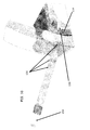
あるいは、図9および図10に示すように、コヒーレント光は、強膜を通して毛様体114に向けられてもよい。強膜に圧力を掛けると、強膜は、特定の波長の光を通すようになる。強膜の特性により、プローブやその他の光供給装置を押しつけて強膜を圧迫した際に、強膜を通してのレーザ光の通過が可能になる。レーザプローブやその他のコヒーレント光供給装置を強膜の外側に押しつけることにより、コヒーレント光は、眼の外側を通して直接的に毛様体の領域に供給されてよい。かかる実施形態では、コヒーレント光は、通例、約200μmの透過を可能にする光源から選択される。一実施形態では、790nmの波長を有するレーザが用いられる。かかる実施形態では、正しい反応を引き起こすために、透過深さは、強膜の外側と標的領域との間に位置する構造の厚さよりも大きい必要がある。
Alternatively, as shown in FIGS. 9 and 10, coherent light may be directed through the sclera to the
かかる実施形態では、光は、使用者が操作するペンまたは鉛筆のようなプローブによって供給されてよい。プローブは、光ファイバの光供給路と、レンズまたは鏡のアレイとからなってよく、それによって、光の焦点が、標的領域に合わせられる。一実施形態では、アレイ156のレンズが、プローブの末端に配置されており、使用の際には、対象となる眼の強膜に接触するように配置される。次いで、使用者は、強膜越しに毛様体形成を行うために角膜の周りの1または複数の位置を選択してよい。あるいは、レーザは、角膜の周りに環状に配置された複数のレンズを通して、角膜の周りの強膜に位置する事前に選択された複数の場所に同時に照射されてもよい。
In such embodiments, the light may be supplied by a probe such as a pen or pencil that is manipulated by the user. The probe may consist of an optical fiber light supply path and an array of lenses or mirrors, whereby the light is focused on the target area. In one embodiment, the lens of
かかる実施形態では、生物学的効果と、毛様体の線維柱帯およびブドウ膜強膜流出経路を塞ぐデトリタスやその他の破片の切除または除去との両方が、流れの改善に寄与することで、上昇した眼圧を低下させる。食細胞、マクロファージ、およびサイトカインの作用は、生物科学分野の当業者によく理解されている。レーザによる毛様体形成は、体の免疫反応を効果的に刺激し、体が破片を除去するよう促す。破片の除去により、房水の流れの増進が実現されるため、眼圧が低減される。 In such embodiments, both the biological effects and the removal or removal of detritus and other debris that block the ciliary trabecular meshwork and uveoscleral outflow pathway contribute to improved flow, Reduces elevated intraocular pressure. The effects of phagocytes, macrophages, and cytokines are well understood by those skilled in the biological sciences. Laser formation of the ciliary body effectively stimulates the body's immune response and encourages the body to remove debris. The removal of debris realizes an increase in the flow of aqueous humor, thus reducing intraocular pressure.
一実施形態では、標的領域は、少なくとも5マイクロ秒の光パルスにされてよい。別の実施形態では、このパルスは、7マイクロ秒であってもよいし、20マイクロ秒以上であってもよい。暴露時間が異なることにより、異なる生物学的反応が引き起こされることは、当業者にとって明らかである。 In one embodiment, the target region may be at least a 5 microsecond light pulse. In another embodiment, the pulse may be 7 microseconds or 20 microseconds or longer. It will be apparent to those skilled in the art that different exposure times cause different biological responses.
本発明の実施形態の説明は、例示と説明のためになされたものである。その説明は、包括的であることも、本発明を開示された形態に限定することも意図されていない。本開示に照らして、多くの変形例や変更例が可能である。本発明の範囲は、この詳細な説明ではなく、添付の特許請求の範囲によって限定されるよう意図されている。 The description of the embodiments of the present invention has been given for purposes of illustration and description. The description is not intended to be exhaustive or to limit the invention to the form disclosed. Many variations and modifications are possible in light of this disclosure. It is intended that the scope of the invention be limited not by this detailed description, but rather by the claims appended hereto.
100…ブドウ膜強膜面
104…毛様体
105…線維組織領域
106…強膜
110…ブドウ膜強膜面
112…虹彩
114…毛様体突起
116…脈絡膜
118…瞳孔
122…毛様体筋束
128…線維柱帯
132…シュレム管
136…集水路
140…結膜
150…細胞破片
150…破片
156…レンズアレイ
DESCRIPTION OF
Claims (24)
前記眼の毛様体領域にレーザ光を向ける工程と、
前記毛様体領域を刺激する工程と、
前記毛様体領域に詰まった破片を除去する工程と、を備える、方法。 A method for treating intraocular pressure in a mammalian eye, comprising:
Directing laser light onto the ciliary region of the eye;
Stimulating the ciliary body region;
Removing the clogged debris in the ciliary body region.
前記レーザ光は、50μm以上の透過深さを有する、方法。 The method of claim 1, comprising:
The laser light has a transmission depth of 50 μm or more.
前記レーザ光は、70μm以上の透過深さを有する、方法。 The method of claim 1, comprising:
The laser beam has a transmission depth of 70 μm or more.
前記レーザ光は、200μm以上の透過深さを有する、方法。 The method of claim 1, comprising:
The laser beam has a transmission depth of 200 μm or more.
前記毛様体領域の毛様体線維柱帯を前記レーザ光の標的にする工程を備える、方法。 The method of claim 1, further comprising:
Targeting the ciliary trabecular meshwork of the ciliary region to the laser light.
免疫反応を引き起こす工程を備える、方法。 The method of claim 5, further comprising:
A method comprising causing an immune response.
前記免疫反応は、サイトカインおよびマクロファージの活性化を含む、方法。 The method of claim 6, comprising:
The method wherein the immune response comprises cytokine and macrophage activation.
前記毛様体線維柱帯に前記レーザ光を隅角鏡によって供給する工程を備える、方法。 The method of claim 5, further comprising:
Supplying the laser light to the ciliary trabecular meshwork with a angle mirror.
前記毛様体領域内に位置する毛様体を標的にする工程を備える、方法。 The method of claim 1, further comprising:
Targeting the ciliary body located within the ciliary region.
免疫反応を引き起こす工程を備える、方法。 The method of claim 9, further comprising:
A method comprising causing an immune response.
前記免疫反応は、サイトカインおよびマクロファージの活性化を含む、方法。 The method of claim 10, comprising:
The method wherein the immune response comprises cytokine and macrophage activation.
前記眼の強膜をレーザ供給装置で圧迫する工程を備える、方法。 The method of claim 9, further comprising:
Pressing the sclera of the eye with a laser delivery device.
前記眼の強膜を通して前記レーザ光を送る工程を備える、方法。 The method of claim 9, further comprising:
Sending the laser light through the sclera of the eye.
前記強膜上の複数の点を扱う工程を備える、方法。 14. The method of claim 13, further comprising:
Treating the plurality of points on the sclera.
前記レーザ光は、600nmより大きい波長を有する、方法。 The method of claim 1, comprising:
The method wherein the laser light has a wavelength greater than 600 nm.
前記レーザ光は、700nmから1000nmの範囲の波長を有する、方法。 The method of claim 1, comprising:
The method wherein the laser light has a wavelength in the range of 700 nm to 1000 nm.
前記レーザ光は、チタンサファイアレーザによって生成される、方法。 The method of claim 1, comprising:
The method wherein the laser light is generated by a titanium sapphire laser.
50μmより大きい範囲の組織透過性を有するレーザ光を生成するレーザ生成器と、
前記レーザに結合されたレーザ供給装置と、を備え、前記レーザ光は、前記眼の毛様体を標的にする、システム。 A system for treating intraocular pressure in a mammalian eye,
A laser generator for generating laser light having tissue permeability in a range greater than 50 μm;
A laser supply device coupled to the laser, wherein the laser light targets the ciliary body of the eye.
前記眼の前房の周りに配置された接触リングを備え、前記レーザ光は、複数の標的点に同時に供給される、システム。 The system of claim 18, further comprising:
A system comprising a contact ring disposed around the anterior chamber of the eye, wherein the laser light is delivered simultaneously to a plurality of target points.
前記装置は、前記眼の強膜に直接的に接触するよう構成されている、システム。 The system of claim 18, further comprising:
The system is configured to directly contact the sclera of the eye.
前記眼の毛様体領域に近接した患者の眼の強膜を圧迫する工程と、
前記強膜を通して前記毛様体領域にレーザ光のビームを向ける工程と、
免疫反応を引き起こす工程と、を備える、方法。 A method for ciliary body formation across the sclera,
Compressing the sclera of the patient's eye proximate to the ciliary region of the eye;
Directing a beam of laser light through the sclera to the ciliary region;
Inducing an immune response.
前記レーザは、700nmから1000nmの範囲の波長を有する、方法。 The method of claim 21, comprising:
The method wherein the laser has a wavelength in the range of 700 nm to 1000 nm.
前記レーザは、チタンサファイアレーザである、方法。 The method of claim 21, comprising:
The method, wherein the laser is a titanium sapphire laser.
前記免疫反応は、サイトカインおよびマクロファージの活性化を含む、方法。 The method of claim 21, comprising:
The method wherein the immune response comprises cytokine and macrophage activation.
Applications Claiming Priority (2)
| Application Number | Priority Date | Filing Date | Title |
|---|---|---|---|
| US53840704P | 2004-01-22 | 2004-01-22 | |
| PCT/US2005/002118 WO2005072294A2 (en) | 2004-01-22 | 2005-01-24 | Glaucoma treatment method |
Publications (2)
| Publication Number | Publication Date |
|---|---|
| JP2007518539A true JP2007518539A (en) | 2007-07-12 |
| JP2007518539A5 JP2007518539A5 (en) | 2008-03-13 |
Family
ID=34825979
Family Applications (1)
| Application Number | Title | Priority Date | Filing Date |
|---|---|---|---|
| JP2006551349A Pending JP2007518539A (en) | 2004-01-22 | 2005-01-24 | How to treat glaucoma |
Country Status (6)
| Country | Link |
|---|---|
| US (1) | US7282046B2 (en) |
| EP (1) | EP1715799A4 (en) |
| JP (1) | JP2007518539A (en) |
| AU (1) | AU2005208820A1 (en) |
| CA (1) | CA2554165A1 (en) |
| WO (1) | WO2005072294A2 (en) |
Cited By (3)
| Publication number | Priority date | Publication date | Assignee | Title |
|---|---|---|---|---|
| JP2019528143A (en) * | 2016-08-16 | 2019-10-10 | ストローマ メディカル コーポレーション | Method and apparatus for predicting perceived iris color after surgery |
| JP2021535788A (en) * | 2018-09-07 | 2021-12-23 | ヴィアレーズ, インコーポレイテッド | Non-invasive and minimally invasive laser surgery for lowering intraocular pressure in the eye |
| JP7449587B2 (en) | 2019-03-13 | 2024-03-14 | ベルキン ヴィジョン リミテッド | automated laser iridotomy |
Families Citing this family (54)
| Publication number | Priority date | Publication date | Assignee | Title |
|---|---|---|---|---|
| US8313454B2 (en) | 1997-11-20 | 2012-11-20 | Optonol Ltd. | Fluid drainage device, delivery device, and associated methods of use and manufacture |
| US6638239B1 (en) | 2000-04-14 | 2003-10-28 | Glaukos Corporation | Apparatus and method for treating glaucoma |
| US7867186B2 (en) | 2002-04-08 | 2011-01-11 | Glaukos Corporation | Devices and methods for treatment of ocular disorders |
| EP1418868B1 (en) | 2001-04-07 | 2008-03-26 | Glaukos Corporation | Glaucoma stent for glaucoma treatment |
| US7331984B2 (en) | 2001-08-28 | 2008-02-19 | Glaukos Corporation | Glaucoma stent for treating glaucoma and methods of use |
| US20060069340A1 (en) * | 2003-06-16 | 2006-03-30 | Solx, Inc. | Shunt for the treatment of glaucoma |
| US8911496B2 (en) | 2006-07-11 | 2014-12-16 | Refocus Group, Inc. | Scleral prosthesis for treating presbyopia and other eye disorders and related devices and methods |
| MX2009000378A (en) | 2006-07-11 | 2009-08-12 | Refocus Group Inc | Scleral prosthesis for treating presbyopia and other eye disorders and related devices and methods. |
| CA2668954C (en) | 2006-11-10 | 2020-09-08 | Glaukos Corporation | Uveoscleral shunt and methods for implanting same |
| DE102007005699A1 (en) * | 2007-02-05 | 2008-08-07 | Carl Zeiss Meditec Ag | coagulation |
| US20170360609A9 (en) | 2007-09-24 | 2017-12-21 | Ivantis, Inc. | Methods and devices for increasing aqueous humor outflow |
| US7740604B2 (en) | 2007-09-24 | 2010-06-22 | Ivantis, Inc. | Ocular implants for placement in schlemm's canal |
| US20090082862A1 (en) | 2007-09-24 | 2009-03-26 | Schieber Andrew T | Ocular Implant Architectures |
| US8734377B2 (en) | 2007-09-24 | 2014-05-27 | Ivantis, Inc. | Ocular implants with asymmetric flexibility |
| US8808222B2 (en) | 2007-11-20 | 2014-08-19 | Ivantis, Inc. | Methods and apparatus for delivering ocular implants into the eye |
| US8512404B2 (en) | 2007-11-20 | 2013-08-20 | Ivantis, Inc. | Ocular implant delivery system and method |
| EP2257250A2 (en) | 2008-01-29 | 2010-12-08 | Gilbert H. Kliman | Drug delivery devices, kits and methods therefor |
| US8109896B2 (en) | 2008-02-11 | 2012-02-07 | Optonol Ltd. | Devices and methods for opening fluid passageways |
| CN101965211A (en) | 2008-03-05 | 2011-02-02 | 伊万提斯公司 | Methods and apparatus for treating glaucoma |
| CA2972136C (en) | 2008-12-05 | 2019-08-06 | Ivantis, Inc. | Cannula for ocular implant delivery system |
| AU2010271274B2 (en) | 2009-07-09 | 2015-05-21 | Alcon Inc. | Single operator device for delivering an ocular implant |
| CN102481404B (en) | 2009-07-09 | 2014-03-05 | 伊万提斯公司 | Ocular implants |
| EP2490621A4 (en) | 2009-10-23 | 2013-04-03 | Ivantis Inc | Ocular implant system and method |
| AU2011232296B2 (en) * | 2010-03-22 | 2013-09-12 | Alpharet Pty Ltd | Laser immunotherapy |
| CN103025282B (en) | 2010-05-10 | 2015-03-11 | 特拉维夫大学拉玛特有限公司 | System for treating glaucoma by directing electromagnetic energy to the limbal area of an eye |
| WO2011163505A1 (en) | 2010-06-23 | 2011-12-29 | Ivantis, Inc. | Ocular implants deployed in schlemm's canal of the eye |
| US8657776B2 (en) | 2011-06-14 | 2014-02-25 | Ivantis, Inc. | Ocular implants for delivery into the eye |
| US20130204236A1 (en) * | 2011-12-01 | 2013-08-08 | Nanophthalmos, Llc | Method and system for laser ocular surgery |
| US8663150B2 (en) | 2011-12-19 | 2014-03-04 | Ivantis, Inc. | Delivering ocular implants into the eye |
| EP2816965B1 (en) * | 2012-02-25 | 2020-08-26 | Thrufocus Optics, Inc. | Devices for improving vision using laser photomiosis |
| WO2013148275A2 (en) | 2012-03-26 | 2013-10-03 | Glaukos Corporation | System and method for delivering multiple ocular implants |
| US9358156B2 (en) | 2012-04-18 | 2016-06-07 | Invantis, Inc. | Ocular implants for delivery into an anterior chamber of the eye |
| US10744034B2 (en) | 2012-04-25 | 2020-08-18 | Gregg S. Homer | Method for laser treatment for glaucoma |
| US10617558B2 (en) | 2012-11-28 | 2020-04-14 | Ivantis, Inc. | Apparatus for delivering ocular implants into an anterior chamber of the eye |
| EP2961364B1 (en) | 2013-02-26 | 2020-05-06 | Belkin Laser Ltd. | System for glaucoma treatment |
| US9592151B2 (en) | 2013-03-15 | 2017-03-14 | Glaukos Corporation | Systems and methods for delivering an ocular implant to the suprachoroidal space within an eye |
| US10493274B2 (en) | 2013-12-09 | 2019-12-03 | Purdue Research Foundation | Optical pressure treatment through electrical stimulation |
| WO2015184173A1 (en) | 2014-05-29 | 2015-12-03 | Dose Medical Corporation | Implants with controlled drug delivery features and methods of using same |
| US10709547B2 (en) | 2014-07-14 | 2020-07-14 | Ivantis, Inc. | Ocular implant delivery system and method |
| EP3334329B1 (en) | 2015-08-14 | 2023-09-13 | Alcon Inc. | Ocular implant with pressure sensor |
| WO2017106517A1 (en) | 2015-12-15 | 2017-06-22 | Ivantis, Inc. | Ocular implant and delivery system |
| AU2018312121B2 (en) | 2017-08-04 | 2023-12-21 | Purdue Research Foundation | Multi-coil wireless power transfer assembly for wireless glaucoma therapy |
| US11116625B2 (en) | 2017-09-28 | 2021-09-14 | Glaukos Corporation | Apparatus and method for controlling placement of intraocular implants |
| EP3691586A2 (en) | 2017-10-06 | 2020-08-12 | Glaukos Corporation | Systems and methods for delivering multiple ocular implants |
| USD846738S1 (en) | 2017-10-27 | 2019-04-23 | Glaukos Corporation | Implant delivery apparatus |
| US11191962B2 (en) | 2018-03-12 | 2021-12-07 | Purdue Research Foundation | Stimulus coil and pulse generator for wireless glaucoma therapy |
| WO2020008323A1 (en) | 2018-07-02 | 2020-01-09 | Belkin Laser Ltd. | Direct selective laser trabeculoplasty |
| US10821023B2 (en) | 2018-07-16 | 2020-11-03 | Vialase, Inc. | Integrated surgical system and method for treatment in the irido-corneal angle of the eye |
| US10821024B2 (en) | 2018-07-16 | 2020-11-03 | Vialase, Inc. | System and method for angled optical access to the irido-corneal angle of the eye |
| US11173067B2 (en) | 2018-09-07 | 2021-11-16 | Vialase, Inc. | Surgical system and procedure for precise intraocular pressure reduction |
| US11246754B2 (en) | 2018-07-16 | 2022-02-15 | Vialase, Inc. | Surgical system and procedure for treatment of the trabecular meshwork and Schlemm's canal using a femtosecond laser |
| US11564567B2 (en) | 2020-02-04 | 2023-01-31 | Vialase, Inc. | System and method for locating a surface of ocular tissue for glaucoma surgery based on dual aiming beams |
| US11612315B2 (en) | 2020-04-09 | 2023-03-28 | Vialase, Inc. | Alignment and diagnostic device and methods for imaging and surgery at the irido-corneal angle of the eye |
| AU2022205382A1 (en) | 2021-01-11 | 2023-06-22 | Alcon Inc. | Systems and methods for viscoelastic delivery |
Citations (3)
| Publication number | Priority date | Publication date | Assignee | Title |
|---|---|---|---|---|
| JP2002282298A (en) * | 2001-03-23 | 2002-10-02 | Nidek Co Ltd | Corpus ciliare light coagulation probe and ophthalmic laser device having it |
| WO2002078556A1 (en) * | 2001-03-30 | 2002-10-10 | Sand Bruce J | Treatment of collagen |
| US6514241B1 (en) * | 1995-03-10 | 2003-02-04 | Candela Corporation | Apparatus and method for treating glaucoma using a gonioscopic laser trabecular ablation procedure |
Family Cites Families (76)
| Publication number | Priority date | Publication date | Assignee | Title |
|---|---|---|---|---|
| US2134152A (en) | 1937-01-06 | 1938-10-25 | Schwarzmayr Ludwig | Wound drain-strip |
| US3159161A (en) * | 1962-11-14 | 1964-12-01 | Ness Richard Alton | Fistulizing canaliculus |
| CS152018B1 (en) * | 1970-05-27 | 1973-12-19 | ||
| US3915172A (en) * | 1970-05-27 | 1975-10-28 | Ceskoslovenska Akademie Ved | Capillary drain for glaucoma |
| SU446981A1 (en) | 1972-08-31 | 1976-07-05 | Предприятие П/Я Г-4147 | Laser ophthalmic installation |
| US3860008A (en) * | 1973-10-03 | 1975-01-14 | Dow Corning | Flat drain |
| DE3069080D1 (en) * | 1979-11-28 | 1984-10-04 | Lasag Ag | Observation device for eye-treatment |
| US4377169A (en) * | 1981-06-10 | 1983-03-22 | Banks Bruce A | Ion beam sputter-etched ventricular catheter for hydrocephalus shunt |
| US4457757A (en) | 1981-07-20 | 1984-07-03 | Molteno Anthony C B | Device for draining aqueous humour |
| US4428746A (en) * | 1981-07-29 | 1984-01-31 | Antonio Mendez | Glaucoma treatment device |
| US4633866A (en) | 1981-11-23 | 1987-01-06 | Gholam Peyman | Ophthalmic laser surgical method |
| JPS59501299A (en) | 1982-06-28 | 1984-07-26 | ザ・ジョンズ・ホプキンス・ユニバ−シティ | A photoelectric device that monitors the instantaneous concentration of singlet oxygen generated during cancer treatment with photochemotherapy |
| US4741730A (en) | 1982-10-04 | 1988-05-03 | American Hospital Supply | Hydrocephalus shunt with in-line filter |
| US4521210A (en) | 1982-12-27 | 1985-06-04 | Wong Vernon G | Eye implant for relieving glaucoma, and device and method for use therewith |
| US4560375A (en) | 1983-06-30 | 1985-12-24 | Pudenz-Schulte Medical Research Corp. | Flow control valve |
| US4558698A (en) * | 1984-03-01 | 1985-12-17 | Dell Lawrence W O | Laser canaliculostomy eye-treatment |
| US4604087A (en) | 1985-02-26 | 1986-08-05 | Joseph Neil H | Aqueous humor drainage device |
| US4791927A (en) | 1985-12-26 | 1988-12-20 | Allied Corporation | Dual-wavelength laser scalpel background of the invention |
| US4722724A (en) | 1986-06-23 | 1988-02-02 | Stanley Schocket | Anterior chamber tube shunt to an encircling band, and related surgical procedure |
| US4850955A (en) | 1986-12-02 | 1989-07-25 | Codman & Shurtleff | Body fluid transfer device |
| US4795437A (en) | 1987-01-29 | 1989-01-03 | Pudenz-Schulte Medical Research Corporation | Siphon control device |
| US4846172A (en) | 1987-05-26 | 1989-07-11 | Berlin Michael S | Laser-delivery eye-treatment method |
| US4957481A (en) | 1987-10-01 | 1990-09-18 | U.S. Bioscience | Photodynamic therapeutic technique |
| US5259380A (en) | 1987-11-04 | 1993-11-09 | Amcor Electronics, Ltd. | Light therapy system |
| US5053006A (en) | 1988-04-19 | 1991-10-01 | Watson Brant D | Method for the permanent occlusion of arteries |
| US5785674A (en) | 1988-10-07 | 1998-07-28 | Mateen; Ahmed Abdul | Device and method for treating glaucoma |
| US5071408A (en) | 1988-10-07 | 1991-12-10 | Ahmed Abdul Mateen | Medical valve |
| US5222952A (en) | 1988-10-28 | 1993-06-29 | Hanspeter Loertscher | Method for laser sclerostomy |
| US5152760A (en) | 1989-03-17 | 1992-10-06 | The General Hospital Corporation | Non-invasive sclerostomy |
| US4966452A (en) * | 1989-04-27 | 1990-10-30 | Ocular Instruments, Inc. | Contact lens for laser surgery |
| US5129895A (en) | 1990-05-16 | 1992-07-14 | Sunrise Technologies, Inc. | Laser sclerostomy procedure |
| US5397300A (en) | 1990-05-31 | 1995-03-14 | Iovision, Inc. | Glaucoma implant |
| JPH0475654A (en) * | 1990-07-19 | 1992-03-10 | Topcon Corp | Laser incising device for lenticular capsule |
| US5292362A (en) | 1990-07-27 | 1994-03-08 | The Trustees Of Columbia University In The City Of New York | Tissue bonding and sealing composition and method of using the same |
| WO1992016259A1 (en) * | 1991-03-13 | 1992-10-01 | Iris Medical Instruments, Inc. | Contact probe for laser cyclophotocoagulation |
| US5454796A (en) | 1991-04-09 | 1995-10-03 | Hood Laboratories | Device and method for controlling intraocular fluid pressure |
| US5169395A (en) | 1991-04-26 | 1992-12-08 | Pdt Cardiovascular, Inc. | Laser delivery system |
| US5171213A (en) | 1991-08-14 | 1992-12-15 | Price Jr Francis W | Technique for fistulization of the eye and an eye filtration prosthesis useful therefor |
| US5346464A (en) | 1992-03-10 | 1994-09-13 | Camras Carl B | Method and apparatus for reducing intraocular pressure |
| US5385541A (en) | 1992-04-24 | 1995-01-31 | Loma Linda University Medical Center | Cerebrospinal fluid shunt capable of minimal invasive revision |
| US5370641A (en) | 1992-05-22 | 1994-12-06 | O'donnell, Jr.; Francis E. | Laser trabeculodissection |
| US5465737A (en) * | 1992-07-15 | 1995-11-14 | Schachar; Ronald A. | Treatment of presbyopia and other eye disorders |
| US5354331A (en) * | 1992-07-15 | 1994-10-11 | Schachar Ronald A | Treatment of presbyopia and other eye disorders |
| TW251236B (en) | 1992-09-10 | 1995-07-11 | Ciba Vision Ag | |
| US5437658A (en) * | 1992-10-07 | 1995-08-01 | Summit Technology, Incorporated | Method and system for laser thermokeratoplasty of the cornea |
| US5688264A (en) * | 1992-10-19 | 1997-11-18 | The University Of Miami | Laser treatment for retinal detachment |
| US5423800A (en) * | 1992-10-19 | 1995-06-13 | The University Of Miami | Laser scleral buckling method and instruments therefor |
| US5370607A (en) | 1992-10-28 | 1994-12-06 | Annuit Coeptis, Inc. | Glaucoma implant device and method for implanting same |
| US5338291A (en) | 1993-02-03 | 1994-08-16 | Pudenz-Schulte Medical Research Corporation | Glaucoma shunt and method for draining aqueous humor |
| US5549596A (en) | 1993-07-07 | 1996-08-27 | The General Hospital Corporation | Selective laser targeting of pigmented ocular cells |
| US5707986A (en) | 1994-03-14 | 1998-01-13 | Miller; Joan W. | Angiographic method using green porphyrins in primate eyes |
| US5434878A (en) | 1994-03-18 | 1995-07-18 | Brown University Research Foundation | Optical gain medium having doped nanocrystals of semiconductors and also optical scatterers |
| US5851225A (en) | 1994-03-18 | 1998-12-22 | Spectra Science Corporation | Photoemitting catheters and other structures suitable for use in photo-dynamic therapy and other applications |
| DE9409616U1 (en) * | 1994-06-17 | 1994-08-04 | Zeiss Carl Fa | Applicator for the treatment of increased intraocular pressure using laser radiation |
| US5704907A (en) | 1994-07-22 | 1998-01-06 | Wound Healing Of Oklahoma | Method and apparatus for lowering the intraocular pressure of an eye |
| US5520621A (en) | 1994-07-25 | 1996-05-28 | Carapace, Inc. | Water-permeable casting or splinting device and method of making same |
| US6572609B1 (en) | 1999-07-14 | 2003-06-03 | Cardiofocus, Inc. | Phototherapeutic waveguide apparatus |
| US5601094A (en) | 1994-11-22 | 1997-02-11 | Reiss; George R. | Ophthalmic shunt |
| US5433701A (en) | 1994-12-21 | 1995-07-18 | Rubinstein; Mark H. | Apparatus for reducing ocular pressure |
| US5576013A (en) | 1995-03-21 | 1996-11-19 | Eastern Virginia Medical School | Treating vascular and neoplastic tissues |
| US5626558A (en) | 1995-05-05 | 1997-05-06 | Suson; John | Adjustable flow rate glaucoma shunt and method of using same |
| US5549598A (en) | 1995-05-22 | 1996-08-27 | O'donnell, Jr.; Francis E. | Glaucoma laser trabeculodissection |
| US5865831A (en) | 1996-04-17 | 1999-02-02 | Premier Laser Systems, Inc. | Laser surgical procedures for treatment of glaucoma |
| US5709653A (en) | 1996-07-25 | 1998-01-20 | Cordis Corporation | Photodynamic therapy balloon catheter with microporous membrane |
| US6050970A (en) | 1997-05-08 | 2000-04-18 | Pharmacia & Upjohn Company | Method and apparatus for inserting a glaucoma implant in an anterior and posterior segment of the eye |
| US6319274B1 (en) * | 1998-06-22 | 2001-11-20 | John H. Shadduck | Devices and techniques for light-mediated stimulation of trabecular meshwork in glaucoma therapy |
| US6241721B1 (en) * | 1998-10-09 | 2001-06-05 | Colette Cozean | Laser surgical procedures for treatment of glaucoma |
| AU775879C (en) | 1998-11-20 | 2005-03-10 | Freedom-2, Inc. | Permanent, removable tissue markings |
| US6186628B1 (en) | 1999-05-23 | 2001-02-13 | Jozek F. Van de Velde | Scanning laser ophthalmoscope for selective therapeutic laser |
| US7088217B2 (en) | 2001-01-15 | 2006-08-08 | Matsushita Electric Works, Ltd. | Shunt resistance and method of adjusting the shunt resistance |
| IL156831A0 (en) * | 2001-01-18 | 2004-02-08 | Univ California | Minimally invasive glaucoma surgical instrument and method |
| US6682523B2 (en) * | 2001-02-21 | 2004-01-27 | John H. Shadduck | Devices and techniques for treating trabecular meshwork |
| US6989007B2 (en) * | 2001-02-21 | 2006-01-24 | Solx, Inc. | Devices and techniques for treating glaucoma |
| JP2004011332A (en) | 2002-06-10 | 2004-01-15 | Shin Meiwa Ind Co Ltd | Pump driving device of cleaning vehicle |
| ES2320121T3 (en) | 2002-07-19 | 2009-05-19 | Yale University | UVEOESCLERAL DRAINAGE DEVICE. |
| EP1613373A4 (en) | 2003-04-08 | 2008-09-10 | Univ Leland Stanford Junior | Implantable arteriovenous shunt device |
-
2005
- 2005-01-24 JP JP2006551349A patent/JP2007518539A/en active Pending
- 2005-01-24 US US11/041,715 patent/US7282046B2/en not_active Expired - Fee Related
- 2005-01-24 AU AU2005208820A patent/AU2005208820A1/en not_active Abandoned
- 2005-01-24 WO PCT/US2005/002118 patent/WO2005072294A2/en active Application Filing
- 2005-01-24 EP EP05706039A patent/EP1715799A4/en not_active Withdrawn
- 2005-01-24 CA CA002554165A patent/CA2554165A1/en not_active Abandoned
Patent Citations (3)
| Publication number | Priority date | Publication date | Assignee | Title |
|---|---|---|---|---|
| US6514241B1 (en) * | 1995-03-10 | 2003-02-04 | Candela Corporation | Apparatus and method for treating glaucoma using a gonioscopic laser trabecular ablation procedure |
| JP2002282298A (en) * | 2001-03-23 | 2002-10-02 | Nidek Co Ltd | Corpus ciliare light coagulation probe and ophthalmic laser device having it |
| WO2002078556A1 (en) * | 2001-03-30 | 2002-10-10 | Sand Bruce J | Treatment of collagen |
Cited By (6)
| Publication number | Priority date | Publication date | Assignee | Title |
|---|---|---|---|---|
| JP2019528143A (en) * | 2016-08-16 | 2019-10-10 | ストローマ メディカル コーポレーション | Method and apparatus for predicting perceived iris color after surgery |
| JP7041154B2 (en) | 2016-08-16 | 2022-03-23 | ストローマ メディカル コーポレーション | Methods and equipment for predicting perceived iridescent color after surgery |
| JP2021535788A (en) * | 2018-09-07 | 2021-12-23 | ヴィアレーズ, インコーポレイテッド | Non-invasive and minimally invasive laser surgery for lowering intraocular pressure in the eye |
| JP7349495B2 (en) | 2018-09-07 | 2023-09-22 | ヴィアレーズ, インコーポレイテッド | Non-invasive and minimally invasive laser surgery for lowering intraocular pressure in the eye |
| JP7441360B2 (en) | 2018-09-07 | 2024-02-29 | ヴィアレーズ, インコーポレイテッド | Non-invasive and minimally invasive laser surgery for lowering intraocular pressure in the eye |
| JP7449587B2 (en) | 2019-03-13 | 2024-03-14 | ベルキン ヴィジョン リミテッド | automated laser iridotomy |
Also Published As
| Publication number | Publication date |
|---|---|
| AU2005208820A1 (en) | 2005-08-11 |
| EP1715799A4 (en) | 2010-06-02 |
| US20050165385A1 (en) | 2005-07-28 |
| WO2005072294A2 (en) | 2005-08-11 |
| CA2554165A1 (en) | 2005-08-11 |
| EP1715799A2 (en) | 2006-11-02 |
| US7282046B2 (en) | 2007-10-16 |
| WO2005072294A3 (en) | 2006-08-31 |
Similar Documents
| Publication | Publication Date | Title |
|---|---|---|
| JP2007518539A (en) | How to treat glaucoma | |
| JP5819372B2 (en) | Method of treating ocular lesions by applying high-intensity focused ultrasound and its apparatus | |
| US7713228B2 (en) | Surgical method | |
| US6679855B2 (en) | Method and apparatus for the correction of presbyopia using high intensity focused ultrasound | |
| JP2006204919A (en) | Surgical operation system | |
| US20060217741A1 (en) | Irrigation tip | |
| JP5490899B2 (en) | Parameters for an ultrasonic device with means for generating a high-density ultrasonic beam | |
| JP2020518317A (en) | Systems and processes for treatment of myopia | |
| CN114206436B (en) | Selective laser stimulation of corneal stem cells | |
| KR20200139161A (en) | Laser methods and systems for handling, alleviating and reversing presbyopia | |
| US20060173077A1 (en) | Surgical method | |
| RU2281743C1 (en) | Method of laser activition of trabecula for treatment of primary open-angled glaucoma | |
| CN112512467B (en) | Probe for laser treatment of the eye having a fiber cone and a fluid collection channel | |
| RU2587857C1 (en) | Method of laser sclerectomy |
Legal Events
| Date | Code | Title | Description |
|---|---|---|---|
| A521 | Written amendment |
Free format text: JAPANESE INTERMEDIATE CODE: A523 Effective date: 20080123 |
|
| A621 | Written request for application examination |
Free format text: JAPANESE INTERMEDIATE CODE: A621 Effective date: 20080123 |
|
| A131 | Notification of reasons for refusal |
Free format text: JAPANESE INTERMEDIATE CODE: A131 Effective date: 20100518 |
|
| A977 | Report on retrieval |
Free format text: JAPANESE INTERMEDIATE CODE: A971007 Effective date: 20100520 |
|
| A601 | Written request for extension of time |
Free format text: JAPANESE INTERMEDIATE CODE: A601 Effective date: 20100816 |
|
| A602 | Written permission of extension of time |
Free format text: JAPANESE INTERMEDIATE CODE: A602 Effective date: 20100823 |
|
| A521 | Written amendment |
Free format text: JAPANESE INTERMEDIATE CODE: A523 Effective date: 20101116 |
|
| A02 | Decision of refusal |
Free format text: JAPANESE INTERMEDIATE CODE: A02 Effective date: 20110607 |