JP2005508157A - 205P1B5 nucleic acid and corresponding protein useful in cancer treatment and detection - Google Patents
205P1B5 nucleic acid and corresponding protein useful in cancer treatment and detection Download PDFInfo
- Publication number
- JP2005508157A JP2005508157A JP2003525655A JP2003525655A JP2005508157A JP 2005508157 A JP2005508157 A JP 2005508157A JP 2003525655 A JP2003525655 A JP 2003525655A JP 2003525655 A JP2003525655 A JP 2003525655A JP 2005508157 A JP2005508157 A JP 2005508157A
- Authority
- JP
- Japan
- Prior art keywords
- protein
- cells
- cell
- antibody
- polynucleotide
- Prior art date
- Legal status (The legal status is an assumption and is not a legal conclusion. Google has not performed a legal analysis and makes no representation as to the accuracy of the status listed.)
- Pending
Links
- 108090000623 proteins and genes Proteins 0.000 title claims abstract description 505
- 102000004169 proteins and genes Human genes 0.000 title claims abstract description 368
- 206010028980 Neoplasm Diseases 0.000 title claims abstract description 212
- 201000011510 cancer Diseases 0.000 title claims abstract description 119
- 150000007523 nucleic acids Chemical class 0.000 title claims description 60
- 102000039446 nucleic acids Human genes 0.000 title claims description 49
- 108020004707 nucleic acids Proteins 0.000 title claims description 49
- 238000011282 treatment Methods 0.000 title description 49
- 238000001514 detection method Methods 0.000 title description 21
- 239000012634 fragment Substances 0.000 claims abstract description 72
- 230000028993 immune response Effects 0.000 claims abstract description 35
- 108090000765 processed proteins & peptides Proteins 0.000 claims description 278
- 238000000034 method Methods 0.000 claims description 220
- 210000004027 cell Anatomy 0.000 claims description 189
- 102000040430 polynucleotide Human genes 0.000 claims description 160
- 108091033319 polynucleotide Proteins 0.000 claims description 160
- 239000002157 polynucleotide Substances 0.000 claims description 160
- 102000004196 processed proteins & peptides Human genes 0.000 claims description 154
- 239000000203 mixture Substances 0.000 claims description 111
- 150000001413 amino acids Chemical class 0.000 claims description 109
- 239000000523 sample Substances 0.000 claims description 78
- 229920001184 polypeptide Polymers 0.000 claims description 73
- 125000003275 alpha amino acid group Chemical group 0.000 claims description 72
- 108020004999 messenger RNA Proteins 0.000 claims description 71
- 210000001151 cytotoxic T lymphocyte Anatomy 0.000 claims description 69
- 239000002299 complementary DNA Substances 0.000 claims description 67
- 239000013598 vector Substances 0.000 claims description 61
- 210000002443 helper t lymphocyte Anatomy 0.000 claims description 43
- 210000001744 T-lymphocyte Anatomy 0.000 claims description 38
- 239000000126 substance Substances 0.000 claims description 30
- 125000003729 nucleotide group Chemical group 0.000 claims description 29
- 239000012472 biological sample Substances 0.000 claims description 28
- 239000002773 nucleotide Substances 0.000 claims description 27
- 241001465754 Metazoa Species 0.000 claims description 25
- 238000012360 testing method Methods 0.000 claims description 24
- 230000000295 complement effect Effects 0.000 claims description 23
- 230000000692 anti-sense effect Effects 0.000 claims description 21
- 108091028043 Nucleic acid sequence Proteins 0.000 claims description 19
- 230000000694 effects Effects 0.000 claims description 19
- 108091026890 Coding region Proteins 0.000 claims description 17
- 239000003814 drug Substances 0.000 claims description 17
- 229940127089 cytotoxic agent Drugs 0.000 claims description 16
- 238000012544 monitoring process Methods 0.000 claims description 15
- 210000000987 immune system Anatomy 0.000 claims description 14
- 230000002401 inhibitory effect Effects 0.000 claims description 14
- 230000001965 increasing effect Effects 0.000 claims description 13
- 102000053642 Catalytic RNA Human genes 0.000 claims description 11
- 108090000994 Catalytic RNA Proteins 0.000 claims description 11
- 239000002254 cytotoxic agent Substances 0.000 claims description 11
- 108091092562 ribozyme Proteins 0.000 claims description 11
- 230000009261 transgenic effect Effects 0.000 claims description 11
- 210000003719 b-lymphocyte Anatomy 0.000 claims description 10
- 230000001105 regulatory effect Effects 0.000 claims description 10
- 231100000599 cytotoxic agent Toxicity 0.000 claims description 9
- 239000008194 pharmaceutical composition Substances 0.000 claims description 9
- 230000036961 partial effect Effects 0.000 claims description 8
- 239000000562 conjugate Substances 0.000 claims description 7
- 230000001472 cytotoxic effect Effects 0.000 claims description 7
- 231100000433 cytotoxic Toxicity 0.000 claims description 6
- 239000002552 dosage form Substances 0.000 claims description 6
- 229940079593 drug Drugs 0.000 claims description 5
- 210000004408 hybridoma Anatomy 0.000 claims description 5
- 102000004127 Cytokines Human genes 0.000 claims description 4
- 108090000695 Cytokines Proteins 0.000 claims description 4
- 230000002159 abnormal effect Effects 0.000 claims description 4
- 239000000032 diagnostic agent Substances 0.000 claims description 3
- 229940039227 diagnostic agent Drugs 0.000 claims description 3
- 239000000611 antibody drug conjugate Substances 0.000 claims description 2
- 229940049595 antibody-drug conjugate Drugs 0.000 claims description 2
- 238000010839 reverse transcription Methods 0.000 claims description 2
- 230000005907 cancer growth Effects 0.000 claims 6
- 230000016784 immunoglobulin production Effects 0.000 claims 1
- 230000014509 gene expression Effects 0.000 abstract description 125
- 210000001519 tissue Anatomy 0.000 abstract description 80
- 206010060862 Prostate cancer Diseases 0.000 abstract description 75
- 208000000236 Prostatic Neoplasms Diseases 0.000 abstract description 75
- 230000001225 therapeutic effect Effects 0.000 abstract description 63
- 230000000069 prophylactic effect Effects 0.000 abstract description 13
- 235000018102 proteins Nutrition 0.000 description 308
- 235000001014 amino acid Nutrition 0.000 description 120
- 229940024606 amino acid Drugs 0.000 description 110
- 108020004414 DNA Proteins 0.000 description 67
- 230000027455 binding Effects 0.000 description 61
- 229960005486 vaccine Drugs 0.000 description 57
- 238000003556 assay Methods 0.000 description 51
- 239000000047 product Substances 0.000 description 40
- 108010072866 Prostate-Specific Antigen Proteins 0.000 description 39
- 102000007066 Prostate-Specific Antigen Human genes 0.000 description 39
- 239000000427 antigen Substances 0.000 description 39
- 108091007433 antigens Proteins 0.000 description 39
- 102000036639 antigens Human genes 0.000 description 39
- 230000002163 immunogen Effects 0.000 description 38
- 108091032973 (ribonucleotides)n+m Proteins 0.000 description 37
- 201000010099 disease Diseases 0.000 description 34
- 208000037265 diseases, disorders, signs and symptoms Diseases 0.000 description 34
- 210000002307 prostate Anatomy 0.000 description 31
- 210000004443 dendritic cell Anatomy 0.000 description 27
- 238000013459 approach Methods 0.000 description 24
- 210000004962 mammalian cell Anatomy 0.000 description 23
- 239000013612 plasmid Substances 0.000 description 23
- 241000588724 Escherichia coli Species 0.000 description 22
- 206010027476 Metastases Diseases 0.000 description 22
- 238000004458 analytical method Methods 0.000 description 22
- 230000000875 corresponding effect Effects 0.000 description 22
- 230000004044 response Effects 0.000 description 22
- 238000006467 substitution reaction Methods 0.000 description 20
- 108091034117 Oligonucleotide Proteins 0.000 description 19
- FAPWRFPIFSIZLT-UHFFFAOYSA-M Sodium chloride Chemical compound [Na+].[Cl-] FAPWRFPIFSIZLT-UHFFFAOYSA-M 0.000 description 19
- 238000009396 hybridization Methods 0.000 description 19
- 230000005847 immunogenicity Effects 0.000 description 19
- 239000003446 ligand Substances 0.000 description 19
- 108010008281 Recombinant Fusion Proteins Proteins 0.000 description 18
- 102000007056 Recombinant Fusion Proteins Human genes 0.000 description 18
- 230000006870 function Effects 0.000 description 18
- 230000004927 fusion Effects 0.000 description 18
- 102000037865 fusion proteins Human genes 0.000 description 18
- 108020001507 fusion proteins Proteins 0.000 description 18
- 230000009401 metastasis Effects 0.000 description 18
- 125000002924 primary amino group Chemical group [H]N([H])* 0.000 description 18
- 238000013518 transcription Methods 0.000 description 18
- 230000035897 transcription Effects 0.000 description 18
- 108020004705 Codon Proteins 0.000 description 17
- 238000002512 chemotherapy Methods 0.000 description 17
- 230000034994 death Effects 0.000 description 17
- 231100000517 death Toxicity 0.000 description 17
- 125000003178 carboxy group Chemical group [H]OC(*)=O 0.000 description 16
- 230000003053 immunization Effects 0.000 description 16
- 238000000338 in vitro Methods 0.000 description 16
- 230000002018 overexpression Effects 0.000 description 16
- 238000006243 chemical reaction Methods 0.000 description 15
- 238000002649 immunization Methods 0.000 description 15
- 230000004048 modification Effects 0.000 description 15
- 238000012986 modification Methods 0.000 description 15
- 238000001356 surgical procedure Methods 0.000 description 15
- 241000124008 Mammalia Species 0.000 description 13
- 210000000612 antigen-presenting cell Anatomy 0.000 description 13
- 239000003153 chemical reaction reagent Substances 0.000 description 13
- 238000001727 in vivo Methods 0.000 description 13
- 238000003757 reverse transcription PCR Methods 0.000 description 13
- 108700026244 Open Reading Frames Proteins 0.000 description 12
- 125000000539 amino acid group Chemical group 0.000 description 12
- 210000004072 lung Anatomy 0.000 description 12
- 230000001404 mediated effect Effects 0.000 description 12
- 235000002639 sodium chloride Nutrition 0.000 description 12
- 230000014616 translation Effects 0.000 description 12
- 210000004881 tumor cell Anatomy 0.000 description 12
- 108700028369 Alleles Proteins 0.000 description 11
- 238000000636 Northern blotting Methods 0.000 description 11
- JLCPHMBAVCMARE-UHFFFAOYSA-N [3-[[3-[[3-[[3-[[3-[[3-[[3-[[3-[[3-[[3-[[3-[[5-(2-amino-6-oxo-1H-purin-9-yl)-3-[[3-[[3-[[3-[[3-[[3-[[5-(2-amino-6-oxo-1H-purin-9-yl)-3-[[5-(2-amino-6-oxo-1H-purin-9-yl)-3-hydroxyoxolan-2-yl]methoxy-hydroxyphosphoryl]oxyoxolan-2-yl]methoxy-hydroxyphosphoryl]oxy-5-(5-methyl-2,4-dioxopyrimidin-1-yl)oxolan-2-yl]methoxy-hydroxyphosphoryl]oxy-5-(6-aminopurin-9-yl)oxolan-2-yl]methoxy-hydroxyphosphoryl]oxy-5-(6-aminopurin-9-yl)oxolan-2-yl]methoxy-hydroxyphosphoryl]oxy-5-(6-aminopurin-9-yl)oxolan-2-yl]methoxy-hydroxyphosphoryl]oxy-5-(6-aminopurin-9-yl)oxolan-2-yl]methoxy-hydroxyphosphoryl]oxyoxolan-2-yl]methoxy-hydroxyphosphoryl]oxy-5-(5-methyl-2,4-dioxopyrimidin-1-yl)oxolan-2-yl]methoxy-hydroxyphosphoryl]oxy-5-(4-amino-2-oxopyrimidin-1-yl)oxolan-2-yl]methoxy-hydroxyphosphoryl]oxy-5-(5-methyl-2,4-dioxopyrimidin-1-yl)oxolan-2-yl]methoxy-hydroxyphosphoryl]oxy-5-(5-methyl-2,4-dioxopyrimidin-1-yl)oxolan-2-yl]methoxy-hydroxyphosphoryl]oxy-5-(6-aminopurin-9-yl)oxolan-2-yl]methoxy-hydroxyphosphoryl]oxy-5-(6-aminopurin-9-yl)oxolan-2-yl]methoxy-hydroxyphosphoryl]oxy-5-(4-amino-2-oxopyrimidin-1-yl)oxolan-2-yl]methoxy-hydroxyphosphoryl]oxy-5-(4-amino-2-oxopyrimidin-1-yl)oxolan-2-yl]methoxy-hydroxyphosphoryl]oxy-5-(4-amino-2-oxopyrimidin-1-yl)oxolan-2-yl]methoxy-hydroxyphosphoryl]oxy-5-(6-aminopurin-9-yl)oxolan-2-yl]methoxy-hydroxyphosphoryl]oxy-5-(4-amino-2-oxopyrimidin-1-yl)oxolan-2-yl]methyl [5-(6-aminopurin-9-yl)-2-(hydroxymethyl)oxolan-3-yl] hydrogen phosphate Polymers Cc1cn(C2CC(OP(O)(=O)OCC3OC(CC3OP(O)(=O)OCC3OC(CC3O)n3cnc4c3nc(N)[nH]c4=O)n3cnc4c3nc(N)[nH]c4=O)C(COP(O)(=O)OC3CC(OC3COP(O)(=O)OC3CC(OC3COP(O)(=O)OC3CC(OC3COP(O)(=O)OC3CC(OC3COP(O)(=O)OC3CC(OC3COP(O)(=O)OC3CC(OC3COP(O)(=O)OC3CC(OC3COP(O)(=O)OC3CC(OC3COP(O)(=O)OC3CC(OC3COP(O)(=O)OC3CC(OC3COP(O)(=O)OC3CC(OC3COP(O)(=O)OC3CC(OC3COP(O)(=O)OC3CC(OC3COP(O)(=O)OC3CC(OC3COP(O)(=O)OC3CC(OC3COP(O)(=O)OC3CC(OC3COP(O)(=O)OC3CC(OC3CO)n3cnc4c(N)ncnc34)n3ccc(N)nc3=O)n3cnc4c(N)ncnc34)n3ccc(N)nc3=O)n3ccc(N)nc3=O)n3ccc(N)nc3=O)n3cnc4c(N)ncnc34)n3cnc4c(N)ncnc34)n3cc(C)c(=O)[nH]c3=O)n3cc(C)c(=O)[nH]c3=O)n3ccc(N)nc3=O)n3cc(C)c(=O)[nH]c3=O)n3cnc4c3nc(N)[nH]c4=O)n3cnc4c(N)ncnc34)n3cnc4c(N)ncnc34)n3cnc4c(N)ncnc34)n3cnc4c(N)ncnc34)O2)c(=O)[nH]c1=O JLCPHMBAVCMARE-UHFFFAOYSA-N 0.000 description 11
- 239000002671 adjuvant Substances 0.000 description 11
- 239000003795 chemical substances by application Substances 0.000 description 11
- 238000010367 cloning Methods 0.000 description 11
- 238000010253 intravenous injection Methods 0.000 description 11
- 239000002502 liposome Substances 0.000 description 11
- 210000001165 lymph node Anatomy 0.000 description 11
- 238000002360 preparation method Methods 0.000 description 11
- 239000011780 sodium chloride Substances 0.000 description 11
- 238000013519 translation Methods 0.000 description 11
- XLYOFNOQVPJJNP-UHFFFAOYSA-N water Substances O XLYOFNOQVPJJNP-UHFFFAOYSA-N 0.000 description 11
- 208000026310 Breast neoplasm Diseases 0.000 description 10
- 241000701022 Cytomegalovirus Species 0.000 description 10
- ZHNUHDYFZUAESO-UHFFFAOYSA-N Formamide Chemical compound NC=O ZHNUHDYFZUAESO-UHFFFAOYSA-N 0.000 description 10
- 230000003321 amplification Effects 0.000 description 10
- 230000005764 inhibitory process Effects 0.000 description 10
- 238000003780 insertion Methods 0.000 description 10
- 230000037431 insertion Effects 0.000 description 10
- 238000004519 manufacturing process Methods 0.000 description 10
- 239000003550 marker Substances 0.000 description 10
- 238000003199 nucleic acid amplification method Methods 0.000 description 10
- 238000001959 radiotherapy Methods 0.000 description 10
- -1 Alternatively Proteins 0.000 description 9
- 206010006187 Breast cancer Diseases 0.000 description 9
- 206010009944 Colon cancer Diseases 0.000 description 9
- 108010070675 Glutathione transferase Proteins 0.000 description 9
- 102100029100 Hematopoietic prostaglandin D synthase Human genes 0.000 description 9
- 229960003767 alanine Drugs 0.000 description 9
- 229960000723 ampicillin Drugs 0.000 description 9
- AVKUERGKIZMTKX-NJBDSQKTSA-N ampicillin Chemical compound C1([C@@H](N)C(=O)N[C@H]2[C@H]3SC([C@@H](N3C2=O)C(O)=O)(C)C)=CC=CC=C1 AVKUERGKIZMTKX-NJBDSQKTSA-N 0.000 description 9
- 150000001875 compounds Chemical class 0.000 description 9
- 239000013604 expression vector Substances 0.000 description 9
- 230000013595 glycosylation Effects 0.000 description 9
- 238000006206 glycosylation reaction Methods 0.000 description 9
- 230000003834 intracellular effect Effects 0.000 description 9
- 238000000746 purification Methods 0.000 description 9
- 230000008685 targeting Effects 0.000 description 9
- 229940124597 therapeutic agent Drugs 0.000 description 9
- 238000002560 therapeutic procedure Methods 0.000 description 9
- DHMQDGOQFOQNFH-UHFFFAOYSA-N Glycine Chemical compound NCC(O)=O DHMQDGOQFOQNFH-UHFFFAOYSA-N 0.000 description 8
- 108060003951 Immunoglobulin Proteins 0.000 description 8
- QNAYBMKLOCPYGJ-REOHCLBHSA-N L-alanine Chemical compound C[C@H](N)C(O)=O QNAYBMKLOCPYGJ-REOHCLBHSA-N 0.000 description 8
- 241000699670 Mus sp. Species 0.000 description 8
- 206010033128 Ovarian cancer Diseases 0.000 description 8
- 208000007097 Urinary Bladder Neoplasms Diseases 0.000 description 8
- 235000004279 alanine Nutrition 0.000 description 8
- 230000001580 bacterial effect Effects 0.000 description 8
- 230000004071 biological effect Effects 0.000 description 8
- 229940022399 cancer vaccine Drugs 0.000 description 8
- 230000001413 cellular effect Effects 0.000 description 8
- 230000007423 decrease Effects 0.000 description 8
- 238000012217 deletion Methods 0.000 description 8
- 230000037430 deletion Effects 0.000 description 8
- 238000011161 development Methods 0.000 description 8
- 230000018109 developmental process Effects 0.000 description 8
- 238000010195 expression analysis Methods 0.000 description 8
- 238000003384 imaging method Methods 0.000 description 8
- 102000018358 immunoglobulin Human genes 0.000 description 8
- 230000003993 interaction Effects 0.000 description 8
- 238000002955 isolation Methods 0.000 description 8
- 150000002632 lipids Chemical class 0.000 description 8
- 239000002245 particle Substances 0.000 description 8
- 230000001575 pathological effect Effects 0.000 description 8
- 230000002829 reductive effect Effects 0.000 description 8
- 238000011160 research Methods 0.000 description 8
- 125000006850 spacer group Chemical group 0.000 description 8
- 206010005003 Bladder cancer Diseases 0.000 description 7
- 102000004190 Enzymes Human genes 0.000 description 7
- 108090000790 Enzymes Proteins 0.000 description 7
- 241000282412 Homo Species 0.000 description 7
- 101800000324 Immunoglobulin A1 protease translocator Proteins 0.000 description 7
- 206010061535 Ovarian neoplasm Diseases 0.000 description 7
- 102100036735 Prostate stem cell antigen Human genes 0.000 description 7
- 108010076504 Protein Sorting Signals Proteins 0.000 description 7
- 230000015572 biosynthetic process Effects 0.000 description 7
- 210000004369 blood Anatomy 0.000 description 7
- 239000008280 blood Substances 0.000 description 7
- 230000010261 cell growth Effects 0.000 description 7
- 230000003247 decreasing effect Effects 0.000 description 7
- 238000005516 engineering process Methods 0.000 description 7
- 238000009472 formulation Methods 0.000 description 7
- 238000007901 in situ hybridization Methods 0.000 description 7
- 238000007918 intramuscular administration Methods 0.000 description 7
- 206010061289 metastatic neoplasm Diseases 0.000 description 7
- 102000005962 receptors Human genes 0.000 description 7
- 108020003175 receptors Proteins 0.000 description 7
- 238000012216 screening Methods 0.000 description 7
- 150000003384 small molecules Chemical class 0.000 description 7
- 201000005112 urinary bladder cancer Diseases 0.000 description 7
- 208000031404 Chromosome Aberrations Diseases 0.000 description 6
- 238000002965 ELISA Methods 0.000 description 6
- 241000699666 Mus <mouse, genus> Species 0.000 description 6
- 241000699660 Mus musculus Species 0.000 description 6
- 108020004511 Recombinant DNA Proteins 0.000 description 6
- ISAKRJDGNUQOIC-UHFFFAOYSA-N Uracil Chemical compound O=C1C=CNC(=O)N1 ISAKRJDGNUQOIC-UHFFFAOYSA-N 0.000 description 6
- 239000003098 androgen Substances 0.000 description 6
- 239000002246 antineoplastic agent Substances 0.000 description 6
- 239000000074 antisense oligonucleotide Substances 0.000 description 6
- 238000012230 antisense oligonucleotides Methods 0.000 description 6
- 108010006025 bovine growth hormone Proteins 0.000 description 6
- 210000000481 breast Anatomy 0.000 description 6
- 238000009566 cancer vaccine Methods 0.000 description 6
- 239000000969 carrier Substances 0.000 description 6
- 239000011651 chromium Substances 0.000 description 6
- 238000003745 diagnosis Methods 0.000 description 6
- 238000002991 immunohistochemical analysis Methods 0.000 description 6
- 210000003734 kidney Anatomy 0.000 description 6
- 210000004185 liver Anatomy 0.000 description 6
- 230000036210 malignancy Effects 0.000 description 6
- 239000000463 material Substances 0.000 description 6
- 230000007246 mechanism Effects 0.000 description 6
- 230000011987 methylation Effects 0.000 description 6
- 238000007069 methylation reaction Methods 0.000 description 6
- 210000000496 pancreas Anatomy 0.000 description 6
- 208000008443 pancreatic carcinoma Diseases 0.000 description 6
- 230000008569 process Effects 0.000 description 6
- 208000023958 prostate neoplasm Diseases 0.000 description 6
- 238000002271 resection Methods 0.000 description 6
- 210000002966 serum Anatomy 0.000 description 6
- 239000000243 solution Substances 0.000 description 6
- 208000024891 symptom Diseases 0.000 description 6
- 239000003053 toxin Substances 0.000 description 6
- 231100000765 toxin Toxicity 0.000 description 6
- 108700012359 toxins Proteins 0.000 description 6
- 102000007469 Actins Human genes 0.000 description 5
- 108010085238 Actins Proteins 0.000 description 5
- 241000894006 Bacteria Species 0.000 description 5
- 208000001333 Colorectal Neoplasms Diseases 0.000 description 5
- 102000053602 DNA Human genes 0.000 description 5
- 108010017213 Granulocyte-Macrophage Colony-Stimulating Factor Proteins 0.000 description 5
- 102100039620 Granulocyte-macrophage colony-stimulating factor Human genes 0.000 description 5
- 108010043121 Green Fluorescent Proteins Proteins 0.000 description 5
- 102000004144 Green Fluorescent Proteins Human genes 0.000 description 5
- 206010058467 Lung neoplasm malignant Diseases 0.000 description 5
- 108091061960 Naked DNA Proteins 0.000 description 5
- 206010061902 Pancreatic neoplasm Diseases 0.000 description 5
- DBMJMQXJHONAFJ-UHFFFAOYSA-M Sodium laurylsulphate Chemical compound [Na+].CCCCCCCCCCCCOS([O-])(=O)=O DBMJMQXJHONAFJ-UHFFFAOYSA-M 0.000 description 5
- 230000024932 T cell mediated immunity Effects 0.000 description 5
- 108700019146 Transgenes Proteins 0.000 description 5
- 239000002253 acid Substances 0.000 description 5
- 230000003302 anti-idiotype Effects 0.000 description 5
- 230000010056 antibody-dependent cellular cytotoxicity Effects 0.000 description 5
- 230000008901 benefit Effects 0.000 description 5
- 238000004422 calculation algorithm Methods 0.000 description 5
- 231100000504 carcinogenesis Toxicity 0.000 description 5
- 230000006037 cell lysis Effects 0.000 description 5
- 230000004663 cell proliferation Effects 0.000 description 5
- 210000002472 endoplasmic reticulum Anatomy 0.000 description 5
- 238000002474 experimental method Methods 0.000 description 5
- 239000005090 green fluorescent protein Substances 0.000 description 5
- 230000036039 immunity Effects 0.000 description 5
- 238000002347 injection Methods 0.000 description 5
- 239000007924 injection Substances 0.000 description 5
- 238000011068 loading method Methods 0.000 description 5
- 208000020816 lung neoplasm Diseases 0.000 description 5
- 210000004698 lymphocyte Anatomy 0.000 description 5
- 238000012423 maintenance Methods 0.000 description 5
- 208000015486 malignant pancreatic neoplasm Diseases 0.000 description 5
- 208000010658 metastatic prostate carcinoma Diseases 0.000 description 5
- 238000010369 molecular cloning Methods 0.000 description 5
- 230000007935 neutral effect Effects 0.000 description 5
- 231100000252 nontoxic Toxicity 0.000 description 5
- 230000003000 nontoxic effect Effects 0.000 description 5
- 210000004940 nucleus Anatomy 0.000 description 5
- 210000000056 organ Anatomy 0.000 description 5
- IPCSVZSSVZVIGE-UHFFFAOYSA-N palmitic acid group Chemical group C(CCCCCCCCCCCCCCC)(=O)O IPCSVZSSVZVIGE-UHFFFAOYSA-N 0.000 description 5
- 201000002528 pancreatic cancer Diseases 0.000 description 5
- 230000026731 phosphorylation Effects 0.000 description 5
- 238000006366 phosphorylation reaction Methods 0.000 description 5
- 229920000642 polymer Polymers 0.000 description 5
- 230000004850 protein–protein interaction Effects 0.000 description 5
- 238000012552 review Methods 0.000 description 5
- 230000028327 secretion Effects 0.000 description 5
- 241000894007 species Species 0.000 description 5
- 238000010561 standard procedure Methods 0.000 description 5
- 230000004936 stimulating effect Effects 0.000 description 5
- 238000011830 transgenic mouse model Methods 0.000 description 5
- 238000001262 western blot Methods 0.000 description 5
- VPFUWHKTPYPNGT-UHFFFAOYSA-N 3-(3,4-dihydroxyphenyl)-1-(5-hydroxy-2,2-dimethylchromen-6-yl)propan-1-one Chemical compound OC1=C2C=CC(C)(C)OC2=CC=C1C(=O)CCC1=CC=C(O)C(O)=C1 VPFUWHKTPYPNGT-UHFFFAOYSA-N 0.000 description 4
- 102100034278 Annexin A6 Human genes 0.000 description 4
- 108090000656 Annexin A6 Proteins 0.000 description 4
- 108020000948 Antisense Oligonucleotides Proteins 0.000 description 4
- 108020004635 Complementary DNA Proteins 0.000 description 4
- 101000782865 Homo sapiens Neuronal acetylcholine receptor subunit alpha-2 Proteins 0.000 description 4
- 208000037396 Intraductal Noninfiltrating Carcinoma Diseases 0.000 description 4
- ROHFNLRQFUQHCH-YFKPBYRVSA-N L-leucine Chemical compound CC(C)C[C@H](N)C(O)=O ROHFNLRQFUQHCH-YFKPBYRVSA-N 0.000 description 4
- KZSNJWFQEVHDMF-BYPYZUCNSA-N L-valine Chemical compound CC(C)[C@H](N)C(O)=O KZSNJWFQEVHDMF-BYPYZUCNSA-N 0.000 description 4
- ROHFNLRQFUQHCH-UHFFFAOYSA-N Leucine Natural products CC(C)CC(N)C(O)=O ROHFNLRQFUQHCH-UHFFFAOYSA-N 0.000 description 4
- 102000043129 MHC class I family Human genes 0.000 description 4
- 108091054437 MHC class I family Proteins 0.000 description 4
- 108700018351 Major Histocompatibility Complex Proteins 0.000 description 4
- 101710175625 Maltose/maltodextrin-binding periplasmic protein Proteins 0.000 description 4
- 229930193140 Neomycin Natural products 0.000 description 4
- 102100035585 Neuronal acetylcholine receptor subunit alpha-2 Human genes 0.000 description 4
- PXHVJJICTQNCMI-UHFFFAOYSA-N Nickel Chemical compound [Ni] PXHVJJICTQNCMI-UHFFFAOYSA-N 0.000 description 4
- WCUXLLCKKVVCTQ-UHFFFAOYSA-M Potassium chloride Chemical compound [Cl-].[K+] WCUXLLCKKVVCTQ-UHFFFAOYSA-M 0.000 description 4
- 238000010240 RT-PCR analysis Methods 0.000 description 4
- 241000700159 Rattus Species 0.000 description 4
- 208000006265 Renal cell carcinoma Diseases 0.000 description 4
- 108700008625 Reporter Genes Proteins 0.000 description 4
- 240000004808 Saccharomyces cerevisiae Species 0.000 description 4
- 230000005867 T cell response Effects 0.000 description 4
- IQFYYKKMVGJFEH-XLPZGREQSA-N Thymidine Chemical compound O=C1NC(=O)C(C)=CN1[C@@H]1O[C@H](CO)[C@@H](O)C1 IQFYYKKMVGJFEH-XLPZGREQSA-N 0.000 description 4
- KZSNJWFQEVHDMF-UHFFFAOYSA-N Valine Natural products CC(C)C(N)C(O)=O KZSNJWFQEVHDMF-UHFFFAOYSA-N 0.000 description 4
- 238000000246 agarose gel electrophoresis Methods 0.000 description 4
- 238000009175 antibody therapy Methods 0.000 description 4
- 238000001574 biopsy Methods 0.000 description 4
- 230000037396 body weight Effects 0.000 description 4
- 210000000988 bone and bone Anatomy 0.000 description 4
- 210000004556 brain Anatomy 0.000 description 4
- 230000002759 chromosomal effect Effects 0.000 description 4
- 210000000349 chromosome Anatomy 0.000 description 4
- 238000003776 cleavage reaction Methods 0.000 description 4
- 210000001072 colon Anatomy 0.000 description 4
- 208000029742 colonic neoplasm Diseases 0.000 description 4
- 230000001419 dependent effect Effects 0.000 description 4
- LOKCTEFSRHRXRJ-UHFFFAOYSA-I dipotassium trisodium dihydrogen phosphate hydrogen phosphate dichloride Chemical compound P(=O)(O)(O)[O-].[K+].P(=O)(O)([O-])[O-].[Na+].[Na+].[Cl-].[K+].[Cl-].[Na+] LOKCTEFSRHRXRJ-UHFFFAOYSA-I 0.000 description 4
- 208000028715 ductal breast carcinoma in situ Diseases 0.000 description 4
- 239000012636 effector Substances 0.000 description 4
- 239000003623 enhancer Substances 0.000 description 4
- 230000002068 genetic effect Effects 0.000 description 4
- 230000012010 growth Effects 0.000 description 4
- HNDVDQJCIGZPNO-UHFFFAOYSA-N histidine Natural products OC(=O)C(N)CC1=CN=CN1 HNDVDQJCIGZPNO-UHFFFAOYSA-N 0.000 description 4
- 230000002998 immunogenetic effect Effects 0.000 description 4
- 238000001114 immunoprecipitation Methods 0.000 description 4
- 238000009169 immunotherapy Methods 0.000 description 4
- 238000011534 incubation Methods 0.000 description 4
- 201000005202 lung cancer Diseases 0.000 description 4
- 230000003211 malignant effect Effects 0.000 description 4
- 238000007726 management method Methods 0.000 description 4
- 239000011159 matrix material Substances 0.000 description 4
- 230000003278 mimic effect Effects 0.000 description 4
- 230000035772 mutation Effects 0.000 description 4
- 229960004927 neomycin Drugs 0.000 description 4
- 238000010606 normalization Methods 0.000 description 4
- 230000007170 pathology Effects 0.000 description 4
- 239000002953 phosphate buffered saline Substances 0.000 description 4
- 230000008488 polyadenylation Effects 0.000 description 4
- 238000000159 protein binding assay Methods 0.000 description 4
- 230000004853 protein function Effects 0.000 description 4
- 230000007017 scission Effects 0.000 description 4
- 239000001509 sodium citrate Substances 0.000 description 4
- 210000002784 stomach Anatomy 0.000 description 4
- 230000020382 suppression by virus of host antigen processing and presentation of peptide antigen via MHC class I Effects 0.000 description 4
- 230000004083 survival effect Effects 0.000 description 4
- 230000005030 transcription termination Effects 0.000 description 4
- 238000001890 transfection Methods 0.000 description 4
- 238000012546 transfer Methods 0.000 description 4
- 238000011277 treatment modality Methods 0.000 description 4
- 210000003932 urinary bladder Anatomy 0.000 description 4
- 239000004474 valine Substances 0.000 description 4
- 239000013603 viral vector Substances 0.000 description 4
- 230000003612 virological effect Effects 0.000 description 4
- 102000002260 Alkaline Phosphatase Human genes 0.000 description 3
- 108020004774 Alkaline Phosphatase Proteins 0.000 description 3
- WVDDGKGOMKODPV-UHFFFAOYSA-N Benzyl alcohol Chemical compound OCC1=CC=CC=C1 WVDDGKGOMKODPV-UHFFFAOYSA-N 0.000 description 3
- 125000001433 C-terminal amino-acid group Chemical group 0.000 description 3
- 206010008805 Chromosomal abnormalities Diseases 0.000 description 3
- 108700010070 Codon Usage Proteins 0.000 description 3
- 108010049894 Cyclic AMP-Dependent Protein Kinases Proteins 0.000 description 3
- 102000008130 Cyclic AMP-Dependent Protein Kinases Human genes 0.000 description 3
- 108010041986 DNA Vaccines Proteins 0.000 description 3
- 229940021995 DNA vaccine Drugs 0.000 description 3
- KCXVZYZYPLLWCC-UHFFFAOYSA-N EDTA Chemical compound OC(=O)CN(CC(O)=O)CCN(CC(O)=O)CC(O)=O KCXVZYZYPLLWCC-UHFFFAOYSA-N 0.000 description 3
- LFQSCWFLJHTTHZ-UHFFFAOYSA-N Ethanol Chemical compound CCO LFQSCWFLJHTTHZ-UHFFFAOYSA-N 0.000 description 3
- 102100041003 Glutamate carboxypeptidase 2 Human genes 0.000 description 3
- PEDCQBHIVMGVHV-UHFFFAOYSA-N Glycerine Chemical compound OCC(O)CO PEDCQBHIVMGVHV-UHFFFAOYSA-N 0.000 description 3
- 239000004471 Glycine Substances 0.000 description 3
- 108010074032 HLA-A2 Antigen Proteins 0.000 description 3
- 102000025850 HLA-A2 Antigen Human genes 0.000 description 3
- 102000018713 Histocompatibility Antigens Class II Human genes 0.000 description 3
- 101000892862 Homo sapiens Glutamate carboxypeptidase 2 Proteins 0.000 description 3
- 102000013462 Interleukin-12 Human genes 0.000 description 3
- 108010065805 Interleukin-12 Proteins 0.000 description 3
- 108010002350 Interleukin-2 Proteins 0.000 description 3
- 102000000588 Interleukin-2 Human genes 0.000 description 3
- 108091092195 Intron Proteins 0.000 description 3
- KDXKERNSBIXSRK-YFKPBYRVSA-N L-lysine Chemical compound NCCCC[C@H](N)C(O)=O KDXKERNSBIXSRK-YFKPBYRVSA-N 0.000 description 3
- FFEARJCKVFRZRR-BYPYZUCNSA-N L-methionine Chemical compound CSCC[C@H](N)C(O)=O FFEARJCKVFRZRR-BYPYZUCNSA-N 0.000 description 3
- 101710128836 Large T antigen Proteins 0.000 description 3
- KDXKERNSBIXSRK-UHFFFAOYSA-N Lysine Natural products NCCCCC(N)C(O)=O KDXKERNSBIXSRK-UHFFFAOYSA-N 0.000 description 3
- 108091054438 MHC class II family Proteins 0.000 description 3
- 241000829100 Macaca mulatta polyomavirus 1 Species 0.000 description 3
- 125000000729 N-terminal amino-acid group Chemical group 0.000 description 3
- 241001195348 Nusa Species 0.000 description 3
- 238000012408 PCR amplification Methods 0.000 description 3
- 238000010222 PCR analysis Methods 0.000 description 3
- 235000021314 Palmitic acid Nutrition 0.000 description 3
- 108010067902 Peptide Library Proteins 0.000 description 3
- 108091093037 Peptide nucleic acid Proteins 0.000 description 3
- 108010029485 Protein Isoforms Proteins 0.000 description 3
- 102000001708 Protein Isoforms Human genes 0.000 description 3
- 208000015634 Rectal Neoplasms Diseases 0.000 description 3
- 238000002105 Southern blotting Methods 0.000 description 3
- 108091023040 Transcription factor Proteins 0.000 description 3
- 241000700605 Viruses Species 0.000 description 3
- 108010084455 Zeocin Proteins 0.000 description 3
- 230000004913 activation Effects 0.000 description 3
- 230000009435 amidation Effects 0.000 description 3
- 238000007112 amidation reaction Methods 0.000 description 3
- 230000000259 anti-tumor effect Effects 0.000 description 3
- 239000008365 aqueous carrier Substances 0.000 description 3
- 230000003385 bacteriostatic effect Effects 0.000 description 3
- 210000004899 c-terminal region Anatomy 0.000 description 3
- 150000001720 carbohydrates Chemical group 0.000 description 3
- 230000000973 chemotherapeutic effect Effects 0.000 description 3
- 230000001086 cytosolic effect Effects 0.000 description 3
- 230000003013 cytotoxicity Effects 0.000 description 3
- 231100000135 cytotoxicity Toxicity 0.000 description 3
- 230000006378 damage Effects 0.000 description 3
- 238000002405 diagnostic procedure Methods 0.000 description 3
- 230000008482 dysregulation Effects 0.000 description 3
- 230000010502 episomal replication Effects 0.000 description 3
- 238000001502 gel electrophoresis Methods 0.000 description 3
- 238000001415 gene therapy Methods 0.000 description 3
- 230000003862 health status Effects 0.000 description 3
- 238000002744 homologous recombination Methods 0.000 description 3
- 230000006801 homologous recombination Effects 0.000 description 3
- 230000028996 humoral immune response Effects 0.000 description 3
- 230000001900 immune effect Effects 0.000 description 3
- 238000010324 immunological assay Methods 0.000 description 3
- 229940121354 immunomodulator Drugs 0.000 description 3
- 230000001976 improved effect Effects 0.000 description 3
- 238000000099 in vitro assay Methods 0.000 description 3
- 238000011065 in-situ storage Methods 0.000 description 3
- 230000006698 induction Effects 0.000 description 3
- 230000010354 integration Effects 0.000 description 3
- 238000007912 intraperitoneal administration Methods 0.000 description 3
- 230000002147 killing effect Effects 0.000 description 3
- 239000002609 medium Substances 0.000 description 3
- 208000037819 metastatic cancer Diseases 0.000 description 3
- 208000011575 metastatic malignant neoplasm Diseases 0.000 description 3
- 229960004452 methionine Drugs 0.000 description 3
- 210000003205 muscle Anatomy 0.000 description 3
- 230000003472 neutralizing effect Effects 0.000 description 3
- 210000001672 ovary Anatomy 0.000 description 3
- 210000005105 peripheral blood lymphocyte Anatomy 0.000 description 3
- 238000002823 phage display Methods 0.000 description 3
- 239000000546 pharmaceutical excipient Substances 0.000 description 3
- CWCMIVBLVUHDHK-ZSNHEYEWSA-N phleomycin D1 Chemical compound N([C@H](C(=O)N[C@H](C)[C@@H](O)[C@H](C)C(=O)N[C@@H]([C@H](O)C)C(=O)NCCC=1SC[C@@H](N=1)C=1SC=C(N=1)C(=O)NCCCCNC(N)=N)[C@@H](O[C@H]1[C@H]([C@@H](O)[C@H](O)[C@H](CO)O1)O[C@@H]1[C@H]([C@@H](OC(N)=O)[C@H](O)[C@@H](CO)O1)O)C=1N=CNC=1)C(=O)C1=NC([C@H](CC(N)=O)NC[C@H](N)C(N)=O)=NC(N)=C1C CWCMIVBLVUHDHK-ZSNHEYEWSA-N 0.000 description 3
- 239000003755 preservative agent Substances 0.000 description 3
- 238000004393 prognosis Methods 0.000 description 3
- 239000003380 propellant Substances 0.000 description 3
- 230000005855 radiation Effects 0.000 description 3
- 230000001177 retroviral effect Effects 0.000 description 3
- 230000035945 sensitivity Effects 0.000 description 3
- 230000000391 smoking effect Effects 0.000 description 3
- NLJMYIDDQXHKNR-UHFFFAOYSA-K sodium citrate Chemical compound O.O.[Na+].[Na+].[Na+].[O-]C(=O)CC(O)(CC([O-])=O)C([O-])=O NLJMYIDDQXHKNR-UHFFFAOYSA-K 0.000 description 3
- 239000007787 solid Substances 0.000 description 3
- 230000001629 suppression Effects 0.000 description 3
- 239000004094 surface-active agent Substances 0.000 description 3
- 238000003786 synthesis reaction Methods 0.000 description 3
- 230000014621 translational initiation Effects 0.000 description 3
- XVQKZSLOGHBCET-INVHGPFASA-N tripalmitoyl-S-glyceryl-cysteinyl-seryl-serine Chemical compound CCCCCCCCCCCCCCCC(=O)N[C@H](C(=O)N[C@@H](CO)C(=O)N[C@@H](CO)C(O)=O)CSCC(OC(=O)CCCCCCCCCCCCCCC)COC(=O)CCCCCCCCCCCCCCC XVQKZSLOGHBCET-INVHGPFASA-N 0.000 description 3
- 230000005740 tumor formation Effects 0.000 description 3
- 241001430294 unidentified retrovirus Species 0.000 description 3
- 229940035893 uracil Drugs 0.000 description 3
- 210000002700 urine Anatomy 0.000 description 3
- MTCFGRXMJLQNBG-REOHCLBHSA-N (2S)-2-Amino-3-hydroxypropansäure Chemical compound OC[C@H](N)C(O)=O MTCFGRXMJLQNBG-REOHCLBHSA-N 0.000 description 2
- IAKHMKGGTNLKSZ-INIZCTEOSA-N (S)-colchicine Chemical compound C1([C@@H](NC(C)=O)CC2)=CC(=O)C(OC)=CC=C1C1=C2C=C(OC)C(OC)=C1OC IAKHMKGGTNLKSZ-INIZCTEOSA-N 0.000 description 2
- 108010066676 Abrin Proteins 0.000 description 2
- 241000972773 Aulopiformes Species 0.000 description 2
- DWRXFEITVBNRMK-UHFFFAOYSA-N Beta-D-1-Arabinofuranosylthymine Natural products O=C1NC(=O)C(C)=CN1C1C(O)C(O)C(CO)O1 DWRXFEITVBNRMK-UHFFFAOYSA-N 0.000 description 2
- 101710132601 Capsid protein Proteins 0.000 description 2
- 208000005623 Carcinogenesis Diseases 0.000 description 2
- 201000009030 Carcinoma Diseases 0.000 description 2
- 102000014914 Carrier Proteins Human genes 0.000 description 2
- 102000052052 Casein Kinase II Human genes 0.000 description 2
- 108010010919 Casein Kinase II Proteins 0.000 description 2
- HEDRZPFGACZZDS-UHFFFAOYSA-N Chloroform Chemical compound ClC(Cl)Cl HEDRZPFGACZZDS-UHFFFAOYSA-N 0.000 description 2
- VYZAMTAEIAYCRO-BJUDXGSMSA-N Chromium-51 Chemical compound [51Cr] VYZAMTAEIAYCRO-BJUDXGSMSA-N 0.000 description 2
- 108010047041 Complementarity Determining Regions Proteins 0.000 description 2
- 108091029523 CpG island Proteins 0.000 description 2
- 108091029430 CpG site Proteins 0.000 description 2
- 108700032819 Croton tiglium crotin II Proteins 0.000 description 2
- 229920001076 Cutan Polymers 0.000 description 2
- QNAYBMKLOCPYGJ-UWTATZPHSA-N D-alanine Chemical compound C[C@@H](N)C(O)=O QNAYBMKLOCPYGJ-UWTATZPHSA-N 0.000 description 2
- QNAYBMKLOCPYGJ-UHFFFAOYSA-N D-alpha-Ala Natural products CC([NH3+])C([O-])=O QNAYBMKLOCPYGJ-UHFFFAOYSA-N 0.000 description 2
- 102000012410 DNA Ligases Human genes 0.000 description 2
- 108010061982 DNA Ligases Proteins 0.000 description 2
- 230000004544 DNA amplification Effects 0.000 description 2
- 241000702421 Dependoparvovirus Species 0.000 description 2
- AOJJSUZBOXZQNB-TZSSRYMLSA-N Doxorubicin Chemical compound O([C@H]1C[C@@](O)(CC=2C(O)=C3C(=O)C=4C=CC=C(C=4C(=O)C3=C(O)C=21)OC)C(=O)CO)[C@H]1C[C@H](N)[C@H](O)[C@H](C)O1 AOJJSUZBOXZQNB-TZSSRYMLSA-N 0.000 description 2
- 241000196324 Embryophyta Species 0.000 description 2
- 241000700662 Fowlpox virus Species 0.000 description 2
- 108700004714 Gelonium multiflorum GEL Proteins 0.000 description 2
- 108010058597 HLA-DR Antigens Proteins 0.000 description 2
- 102000006354 HLA-DR Antigens Human genes 0.000 description 2
- 241000701024 Human betaherpesvirus 5 Species 0.000 description 2
- 108090000862 Ion Channels Proteins 0.000 description 2
- 102000004310 Ion Channels Human genes 0.000 description 2
- AGPKZVBTJJNPAG-WHFBIAKZSA-N L-isoleucine Chemical compound CC[C@H](C)[C@H](N)C(O)=O AGPKZVBTJJNPAG-WHFBIAKZSA-N 0.000 description 2
- 241000713666 Lentivirus Species 0.000 description 2
- 108010028921 Lipopeptides Proteins 0.000 description 2
- 239000004472 Lysine Substances 0.000 description 2
- 206010027452 Metastases to bone Diseases 0.000 description 2
- 241001529936 Murinae Species 0.000 description 2
- 108010046117 N-palmitoyl-5,6-dipalmitoyl-S-glycerylcysteinyl-seryl-serine Proteins 0.000 description 2
- 244000061176 Nicotiana tabacum Species 0.000 description 2
- 235000002637 Nicotiana tabacum Nutrition 0.000 description 2
- 108700020796 Oncogene Proteins 0.000 description 2
- 101150102573 PCR1 gene Proteins 0.000 description 2
- 229930012538 Paclitaxel Natural products 0.000 description 2
- ISWSIDIOOBJBQZ-UHFFFAOYSA-N Phenol Chemical compound OC1=CC=CC=C1 ISWSIDIOOBJBQZ-UHFFFAOYSA-N 0.000 description 2
- 239000002202 Polyethylene glycol Substances 0.000 description 2
- 208000037062 Polyps Diseases 0.000 description 2
- 101000762949 Pseudomonas aeruginosa (strain ATCC 15692 / DSM 22644 / CIP 104116 / JCM 14847 / LMG 12228 / 1C / PRS 101 / PAO1) Exotoxin A Proteins 0.000 description 2
- 235000014680 Saccharomyces cerevisiae Nutrition 0.000 description 2
- XZKQVQKUZMAADP-IMJSIDKUSA-N Ser-Ser Chemical compound OC[C@H](N)C(=O)N[C@@H](CO)C(O)=O XZKQVQKUZMAADP-IMJSIDKUSA-N 0.000 description 2
- MTCFGRXMJLQNBG-UHFFFAOYSA-N Serine Natural products OCC(N)C(O)=O MTCFGRXMJLQNBG-UHFFFAOYSA-N 0.000 description 2
- 230000037453 T cell priming Effects 0.000 description 2
- NKANXQFJJICGDU-QPLCGJKRSA-N Tamoxifen Chemical compound C=1C=CC=CC=1C(/CC)=C(C=1C=CC(OCCN(C)C)=CC=1)/C1=CC=CC=C1 NKANXQFJJICGDU-QPLCGJKRSA-N 0.000 description 2
- 102100036407 Thioredoxin Human genes 0.000 description 2
- 102000040945 Transcription factor Human genes 0.000 description 2
- 230000005856 abnormality Effects 0.000 description 2
- 150000007513 acids Chemical class 0.000 description 2
- 230000003213 activating effect Effects 0.000 description 2
- 239000000443 aerosol Substances 0.000 description 2
- 238000001261 affinity purification Methods 0.000 description 2
- 125000003295 alanine group Chemical group N[C@@H](C)C(=O)* 0.000 description 2
- 238000011230 antibody-based therapy Methods 0.000 description 2
- 230000000890 antigenic effect Effects 0.000 description 2
- 230000001640 apoptogenic effect Effects 0.000 description 2
- 230000006907 apoptotic process Effects 0.000 description 2
- 230000009286 beneficial effect Effects 0.000 description 2
- IQFYYKKMVGJFEH-UHFFFAOYSA-N beta-L-thymidine Natural products O=C1NC(=O)C(C)=CN1C1OC(CO)C(O)C1 IQFYYKKMVGJFEH-UHFFFAOYSA-N 0.000 description 2
- 239000000872 buffer Substances 0.000 description 2
- 239000006172 buffering agent Substances 0.000 description 2
- 229930195731 calicheamicin Natural products 0.000 description 2
- HXCHCVDVKSCDHU-LULTVBGHSA-N calicheamicin Chemical compound C1[C@H](OC)[C@@H](NCC)CO[C@H]1O[C@H]1[C@H](O[C@@H]2C\3=C(NC(=O)OC)C(=O)C[C@](C/3=C/CSSSC)(O)C#C\C=C/C#C2)O[C@H](C)[C@@H](NO[C@@H]2O[C@H](C)[C@@H](SC(=O)C=3C(=C(OC)C(O[C@H]4[C@@H]([C@H](OC)[C@@H](O)[C@H](C)O4)O)=C(I)C=3C)OC)[C@@H](O)C2)[C@@H]1O HXCHCVDVKSCDHU-LULTVBGHSA-N 0.000 description 2
- 230000036952 cancer formation Effects 0.000 description 2
- 239000002775 capsule Substances 0.000 description 2
- 125000002091 cationic group Chemical group 0.000 description 2
- 238000004113 cell culture Methods 0.000 description 2
- 230000003915 cell function Effects 0.000 description 2
- 230000036755 cellular response Effects 0.000 description 2
- 239000002738 chelating agent Substances 0.000 description 2
- 210000000038 chest Anatomy 0.000 description 2
- HVYWMOMLDIMFJA-DPAQBDIFSA-N cholesterol Chemical compound C1C=C2C[C@@H](O)CC[C@]2(C)[C@@H]2[C@@H]1[C@@H]1CC[C@H]([C@H](C)CCCC(C)C)[C@@]1(C)CC2 HVYWMOMLDIMFJA-DPAQBDIFSA-N 0.000 description 2
- RTIXKCRFFJGDFG-UHFFFAOYSA-N chrysin Chemical compound C=1C(O)=CC(O)=C(C(C=2)=O)C=1OC=2C1=CC=CC=C1 RTIXKCRFFJGDFG-UHFFFAOYSA-N 0.000 description 2
- 230000001268 conjugating effect Effects 0.000 description 2
- 230000021615 conjugation Effects 0.000 description 2
- 210000000172 cytosol Anatomy 0.000 description 2
- 230000002950 deficient Effects 0.000 description 2
- 230000017858 demethylation Effects 0.000 description 2
- 238000010520 demethylation reaction Methods 0.000 description 2
- 230000001627 detrimental effect Effects 0.000 description 2
- 229960000633 dextran sulfate Drugs 0.000 description 2
- 239000003085 diluting agent Substances 0.000 description 2
- SLPJGDQJLTYWCI-UHFFFAOYSA-N dimethyl-(4,5,6,7-tetrabromo-1h-benzoimidazol-2-yl)-amine Chemical compound BrC1=C(Br)C(Br)=C2NC(N(C)C)=NC2=C1Br SLPJGDQJLTYWCI-UHFFFAOYSA-N 0.000 description 2
- POULHZVOKOAJMA-UHFFFAOYSA-N dodecanoic acid Chemical compound CCCCCCCCCCCC(O)=O POULHZVOKOAJMA-UHFFFAOYSA-N 0.000 description 2
- 238000004520 electroporation Methods 0.000 description 2
- 210000001671 embryonic stem cell Anatomy 0.000 description 2
- 150000002148 esters Chemical class 0.000 description 2
- 210000003527 eukaryotic cell Anatomy 0.000 description 2
- 238000011156 evaluation Methods 0.000 description 2
- 230000002349 favourable effect Effects 0.000 description 2
- 239000007850 fluorescent dye Substances 0.000 description 2
- 238000001476 gene delivery Methods 0.000 description 2
- 125000005456 glyceride group Chemical group 0.000 description 2
- 239000001963 growth medium Substances 0.000 description 2
- UYTPUPDQBNUYGX-UHFFFAOYSA-N guanine Chemical compound O=C1NC(N)=NC2=C1N=CN2 UYTPUPDQBNUYGX-UHFFFAOYSA-N 0.000 description 2
- 210000002216 heart Anatomy 0.000 description 2
- 229940022353 herceptin Drugs 0.000 description 2
- FUZZWVXGSFPDMH-UHFFFAOYSA-N hexanoic acid Chemical compound CCCCCC(O)=O FUZZWVXGSFPDMH-UHFFFAOYSA-N 0.000 description 2
- 230000003054 hormonal effect Effects 0.000 description 2
- 230000002209 hydrophobic effect Effects 0.000 description 2
- 238000010166 immunofluorescence Methods 0.000 description 2
- 230000002055 immunohistochemical effect Effects 0.000 description 2
- 239000002955 immunomodulating agent Substances 0.000 description 2
- 230000001024 immunotherapeutic effect Effects 0.000 description 2
- 230000006872 improvement Effects 0.000 description 2
- 238000005462 in vivo assay Methods 0.000 description 2
- 238000001802 infusion Methods 0.000 description 2
- 239000004615 ingredient Substances 0.000 description 2
- 239000003112 inhibitor Substances 0.000 description 2
- 230000002452 interceptive effect Effects 0.000 description 2
- 238000007913 intrathecal administration Methods 0.000 description 2
- 230000002601 intratumoral effect Effects 0.000 description 2
- 238000001990 intravenous administration Methods 0.000 description 2
- 229960000310 isoleucine Drugs 0.000 description 2
- AGPKZVBTJJNPAG-UHFFFAOYSA-N isoleucine Natural products CCC(C)C(N)C(O)=O AGPKZVBTJJNPAG-UHFFFAOYSA-N 0.000 description 2
- 230000003902 lesion Effects 0.000 description 2
- 210000000265 leukocyte Anatomy 0.000 description 2
- 230000021633 leukocyte mediated immunity Effects 0.000 description 2
- 230000004807 localization Effects 0.000 description 2
- 235000018977 lysine Nutrition 0.000 description 2
- HQKMJHAJHXVSDF-UHFFFAOYSA-L magnesium stearate Chemical compound [Mg+2].CCCCCCCCCCCCCCCCCC([O-])=O.CCCCCCCCCCCCCCCCCC([O-])=O HQKMJHAJHXVSDF-UHFFFAOYSA-L 0.000 description 2
- 210000001161 mammalian embryo Anatomy 0.000 description 2
- 238000013411 master cell bank Methods 0.000 description 2
- 210000004379 membrane Anatomy 0.000 description 2
- 239000012528 membrane Substances 0.000 description 2
- 229910052751 metal Inorganic materials 0.000 description 2
- 239000002184 metal Substances 0.000 description 2
- 229930182817 methionine Natural products 0.000 description 2
- 238000007855 methylation-specific PCR Methods 0.000 description 2
- 239000000693 micelle Substances 0.000 description 2
- 238000001531 micro-dissection Methods 0.000 description 2
- 238000002703 mutagenesis Methods 0.000 description 2
- 231100000350 mutagenesis Toxicity 0.000 description 2
- 230000007498 myristoylation Effects 0.000 description 2
- WQEPLUUGTLDZJY-UHFFFAOYSA-N n-Pentadecanoic acid Natural products CCCCCCCCCCCCCCC(O)=O WQEPLUUGTLDZJY-UHFFFAOYSA-N 0.000 description 2
- 229910052759 nickel Inorganic materials 0.000 description 2
- 238000007899 nucleic acid hybridization Methods 0.000 description 2
- WWZKQHOCKIZLMA-UHFFFAOYSA-N octanoic acid Chemical compound CCCCCCCC(O)=O WWZKQHOCKIZLMA-UHFFFAOYSA-N 0.000 description 2
- 238000005457 optimization Methods 0.000 description 2
- 230000001582 osteoblastic effect Effects 0.000 description 2
- 238000004806 packaging method and process Methods 0.000 description 2
- 229960001592 paclitaxel Drugs 0.000 description 2
- 230000037361 pathway Effects 0.000 description 2
- 239000013610 patient sample Substances 0.000 description 2
- 230000000737 periodic effect Effects 0.000 description 2
- 210000005259 peripheral blood Anatomy 0.000 description 2
- 239000011886 peripheral blood Substances 0.000 description 2
- 229940124531 pharmaceutical excipient Drugs 0.000 description 2
- 150000003904 phospholipids Chemical class 0.000 description 2
- 230000004962 physiological condition Effects 0.000 description 2
- 230000035790 physiological processes and functions Effects 0.000 description 2
- 229920001223 polyethylene glycol Polymers 0.000 description 2
- 239000001103 potassium chloride Substances 0.000 description 2
- 235000011164 potassium chloride Nutrition 0.000 description 2
- 230000037452 priming Effects 0.000 description 2
- 238000012545 processing Methods 0.000 description 2
- 229940002612 prodrug Drugs 0.000 description 2
- 239000000651 prodrug Substances 0.000 description 2
- 210000005267 prostate cell Anatomy 0.000 description 2
- 230000009145 protein modification Effects 0.000 description 2
- 238000011002 quantification Methods 0.000 description 2
- 230000002285 radioactive effect Effects 0.000 description 2
- 238000003127 radioimmunoassay Methods 0.000 description 2
- 206010038038 rectal cancer Diseases 0.000 description 2
- 201000001275 rectum cancer Diseases 0.000 description 2
- 230000009467 reduction Effects 0.000 description 2
- 208000015347 renal cell adenocarcinoma Diseases 0.000 description 2
- 230000010076 replication Effects 0.000 description 2
- 235000019515 salmon Nutrition 0.000 description 2
- 238000010845 search algorithm Methods 0.000 description 2
- 210000000582 semen Anatomy 0.000 description 2
- 238000012163 sequencing technique Methods 0.000 description 2
- 238000002741 site-directed mutagenesis Methods 0.000 description 2
- 210000002027 skeletal muscle Anatomy 0.000 description 2
- 210000003491 skin Anatomy 0.000 description 2
- 210000000813 small intestine Anatomy 0.000 description 2
- 239000001488 sodium phosphate Substances 0.000 description 2
- 229910000162 sodium phosphate Inorganic materials 0.000 description 2
- 210000004988 splenocyte Anatomy 0.000 description 2
- 230000000087 stabilizing effect Effects 0.000 description 2
- 210000000130 stem cell Anatomy 0.000 description 2
- 229910052717 sulfur Inorganic materials 0.000 description 2
- 230000002194 synthesizing effect Effects 0.000 description 2
- 230000009885 systemic effect Effects 0.000 description 2
- RCINICONZNJXQF-MZXODVADSA-N taxol Chemical compound O([C@@H]1[C@@]2(C[C@@H](C(C)=C(C2(C)C)[C@H](C([C@]2(C)[C@@H](O)C[C@H]3OC[C@]3([C@H]21)OC(C)=O)=O)OC(=O)C)OC(=O)[C@H](O)[C@@H](NC(=O)C=1C=CC=CC=1)C=1C=CC=CC=1)O)C(=O)C1=CC=CC=C1 RCINICONZNJXQF-MZXODVADSA-N 0.000 description 2
- 229960000814 tetanus toxoid Drugs 0.000 description 2
- 108060008226 thioredoxin Proteins 0.000 description 2
- 229940104230 thymidine Drugs 0.000 description 2
- 230000001988 toxicity Effects 0.000 description 2
- 231100000419 toxicity Toxicity 0.000 description 2
- 206010044412 transitional cell carcinoma Diseases 0.000 description 2
- RYFMWSXOAZQYPI-UHFFFAOYSA-K trisodium phosphate Chemical compound [Na+].[Na+].[Na+].[O-]P([O-])([O-])=O RYFMWSXOAZQYPI-UHFFFAOYSA-K 0.000 description 2
- 230000005748 tumor development Effects 0.000 description 2
- 230000004614 tumor growth Effects 0.000 description 2
- 241000701161 unidentified adenovirus Species 0.000 description 2
- 241001515965 unidentified phage Species 0.000 description 2
- 230000002485 urinary effect Effects 0.000 description 2
- 229940125575 vaccine candidate Drugs 0.000 description 2
- 238000005406 washing Methods 0.000 description 2
- 230000003442 weekly effect Effects 0.000 description 2
- 239000000080 wetting agent Substances 0.000 description 2
- 210000005253 yeast cell Anatomy 0.000 description 2
- KIUKXJAPPMFGSW-DNGZLQJQSA-N (2S,3S,4S,5R,6R)-6-[(2S,3R,4R,5S,6R)-3-Acetamido-2-[(2S,3S,4R,5R,6R)-6-[(2R,3R,4R,5S,6R)-3-acetamido-2,5-dihydroxy-6-(hydroxymethyl)oxan-4-yl]oxy-2-carboxy-4,5-dihydroxyoxan-3-yl]oxy-5-hydroxy-6-(hydroxymethyl)oxan-4-yl]oxy-3,4,5-trihydroxyoxane-2-carboxylic acid Chemical compound CC(=O)N[C@H]1[C@H](O)O[C@H](CO)[C@@H](O)[C@@H]1O[C@H]1[C@H](O)[C@@H](O)[C@H](O[C@H]2[C@@H]([C@@H](O[C@H]3[C@@H]([C@@H](O)[C@H](O)[C@H](O3)C(O)=O)O)[C@H](O)[C@@H](CO)O2)NC(C)=O)[C@@H](C(O)=O)O1 KIUKXJAPPMFGSW-DNGZLQJQSA-N 0.000 description 1
- BVAUMRCGVHUWOZ-ZETCQYMHSA-N (2s)-2-(cyclohexylazaniumyl)propanoate Chemical compound OC(=O)[C@H](C)NC1CCCCC1 BVAUMRCGVHUWOZ-ZETCQYMHSA-N 0.000 description 1
- OYHQOLUKZRVURQ-NTGFUMLPSA-N (9Z,12Z)-9,10,12,13-tetratritiooctadeca-9,12-dienoic acid Chemical compound C(CCCCCCC\C(=C(/C\C(=C(/CCCCC)\[3H])\[3H])\[3H])\[3H])(=O)O OYHQOLUKZRVURQ-NTGFUMLPSA-N 0.000 description 1
- WRIDQFICGBMAFQ-UHFFFAOYSA-N (E)-8-Octadecenoic acid Natural products CCCCCCCCCC=CCCCCCCC(O)=O WRIDQFICGBMAFQ-UHFFFAOYSA-N 0.000 description 1
- ICLYJLBTOGPLMC-KVVVOXFISA-N (z)-octadec-9-enoate;tris(2-hydroxyethyl)azanium Chemical compound OCCN(CCO)CCO.CCCCCCCC\C=C/CCCCCCCC(O)=O ICLYJLBTOGPLMC-KVVVOXFISA-N 0.000 description 1
- JUDOLRSMWHVKGX-UHFFFAOYSA-N 1,1-dioxo-1$l^{6},2-benzodithiol-3-one Chemical compound C1=CC=C2C(=O)SS(=O)(=O)C2=C1 JUDOLRSMWHVKGX-UHFFFAOYSA-N 0.000 description 1
- LEBVLXFERQHONN-UHFFFAOYSA-N 1-butyl-N-(2,6-dimethylphenyl)piperidine-2-carboxamide Chemical compound CCCCN1CCCCC1C(=O)NC1=C(C)C=CC=C1C LEBVLXFERQHONN-UHFFFAOYSA-N 0.000 description 1
- IIZPXYDJLKNOIY-JXPKJXOSSA-N 1-palmitoyl-2-arachidonoyl-sn-glycero-3-phosphocholine Chemical compound CCCCCCCCCCCCCCCC(=O)OC[C@H](COP([O-])(=O)OCC[N+](C)(C)C)OC(=O)CCC\C=C/C\C=C/C\C=C/C\C=C/CCCCC IIZPXYDJLKNOIY-JXPKJXOSSA-N 0.000 description 1
- FDSYTWVNUJTPMA-UHFFFAOYSA-N 2-[3,9-bis(carboxymethyl)-3,6,9,15-tetrazabicyclo[9.3.1]pentadeca-1(15),11,13-trien-6-yl]acetic acid Chemical compound C1N(CC(O)=O)CCN(CC(=O)O)CCN(CC(O)=O)CC2=CC=CC1=N2 FDSYTWVNUJTPMA-UHFFFAOYSA-N 0.000 description 1
- JKMHFZQWWAIEOD-UHFFFAOYSA-N 2-[4-(2-hydroxyethyl)piperazin-1-yl]ethanesulfonic acid Chemical compound OCC[NH+]1CCN(CCS([O-])(=O)=O)CC1 JKMHFZQWWAIEOD-UHFFFAOYSA-N 0.000 description 1
- RTQWWZBSTRGEAV-PKHIMPSTSA-N 2-[[(2s)-2-[bis(carboxymethyl)amino]-3-[4-(methylcarbamoylamino)phenyl]propyl]-[2-[bis(carboxymethyl)amino]propyl]amino]acetic acid Chemical compound CNC(=O)NC1=CC=C(C[C@@H](CN(CC(C)N(CC(O)=O)CC(O)=O)CC(O)=O)N(CC(O)=O)CC(O)=O)C=C1 RTQWWZBSTRGEAV-PKHIMPSTSA-N 0.000 description 1
- FBUTXZSKZCQABC-UHFFFAOYSA-N 2-amino-1-methyl-7h-purine-6-thione Chemical compound S=C1N(C)C(N)=NC2=C1NC=N2 FBUTXZSKZCQABC-UHFFFAOYSA-N 0.000 description 1
- QKNYBSVHEMOAJP-UHFFFAOYSA-N 2-amino-2-(hydroxymethyl)propane-1,3-diol;hydron;chloride Chemical compound Cl.OCC(N)(CO)CO QKNYBSVHEMOAJP-UHFFFAOYSA-N 0.000 description 1
- LQJBNNIYVWPHFW-UHFFFAOYSA-N 20:1omega9c fatty acid Natural products CCCCCCCCCCC=CCCCCCCCC(O)=O LQJBNNIYVWPHFW-UHFFFAOYSA-N 0.000 description 1
- CYDQOEWLBCCFJZ-UHFFFAOYSA-N 4-(4-fluorophenyl)oxane-4-carboxylic acid Chemical compound C=1C=C(F)C=CC=1C1(C(=O)O)CCOCC1 CYDQOEWLBCCFJZ-UHFFFAOYSA-N 0.000 description 1
- NYCXYKOXLNBYID-UHFFFAOYSA-N 5,7-Dihydroxychromone Natural products O1C=CC(=O)C=2C1=CC(O)=CC=2O NYCXYKOXLNBYID-UHFFFAOYSA-N 0.000 description 1
- XZIIFPSPUDAGJM-UHFFFAOYSA-N 6-chloro-2-n,2-n-diethylpyrimidine-2,4-diamine Chemical compound CCN(CC)C1=NC(N)=CC(Cl)=N1 XZIIFPSPUDAGJM-UHFFFAOYSA-N 0.000 description 1
- STQGQHZAVUOBTE-UHFFFAOYSA-N 7-Cyan-hept-2t-en-4,6-diinsaeure Natural products C1=2C(O)=C3C(=O)C=4C(OC)=CC=CC=4C(=O)C3=C(O)C=2CC(O)(C(C)=O)CC1OC1CC(N)C(O)C(C)O1 STQGQHZAVUOBTE-UHFFFAOYSA-N 0.000 description 1
- QSBYPNXLFMSGKH-UHFFFAOYSA-N 9-Heptadecensaeure Natural products CCCCCCCC=CCCCCCCCC(O)=O QSBYPNXLFMSGKH-UHFFFAOYSA-N 0.000 description 1
- 208000030507 AIDS Diseases 0.000 description 1
- 208000003200 Adenoma Diseases 0.000 description 1
- 206010001233 Adenoma benign Diseases 0.000 description 1
- 229920001817 Agar Polymers 0.000 description 1
- 102000009027 Albumins Human genes 0.000 description 1
- 108010088751 Albumins Proteins 0.000 description 1
- GUBGYTABKSRVRQ-XLOQQCSPSA-N Alpha-Lactose Chemical compound O[C@@H]1[C@@H](O)[C@@H](O)[C@@H](CO)O[C@H]1O[C@@H]1[C@@H](CO)O[C@H](O)[C@H](O)[C@H]1O GUBGYTABKSRVRQ-XLOQQCSPSA-N 0.000 description 1
- 108020004491 Antisense DNA Proteins 0.000 description 1
- 108020005544 Antisense RNA Proteins 0.000 description 1
- 208000019901 Anxiety disease Diseases 0.000 description 1
- 101100519158 Arabidopsis thaliana PCR2 gene Proteins 0.000 description 1
- 239000004475 Arginine Substances 0.000 description 1
- 208000002109 Argyria Diseases 0.000 description 1
- 229920006048 Arlen™ Polymers 0.000 description 1
- 206010003445 Ascites Diseases 0.000 description 1
- DCXYFEDJOCDNAF-UHFFFAOYSA-N Asparagine Natural products OC(=O)C(N)CC(N)=O DCXYFEDJOCDNAF-UHFFFAOYSA-N 0.000 description 1
- 108090001008 Avidin Proteins 0.000 description 1
- 208000003950 B-cell lymphoma Diseases 0.000 description 1
- 241000193738 Bacillus anthracis Species 0.000 description 1
- 206010061728 Bone lesion Diseases 0.000 description 1
- 241000283690 Bos taurus Species 0.000 description 1
- 108091003079 Bovine Serum Albumin Proteins 0.000 description 1
- 206010055113 Breast cancer metastatic Diseases 0.000 description 1
- UXVMQQNJUSDDNG-UHFFFAOYSA-L Calcium chloride Chemical compound [Cl-].[Cl-].[Ca+2] UXVMQQNJUSDDNG-UHFFFAOYSA-L 0.000 description 1
- 241000178270 Canarypox virus Species 0.000 description 1
- 241000282472 Canis lupus familiaris Species 0.000 description 1
- OKTJSMMVPCPJKN-UHFFFAOYSA-N Carbon Chemical compound [C] OKTJSMMVPCPJKN-UHFFFAOYSA-N 0.000 description 1
- 108010078791 Carrier Proteins Proteins 0.000 description 1
- 102000000844 Cell Surface Receptors Human genes 0.000 description 1
- 108010001857 Cell Surface Receptors Proteins 0.000 description 1
- 108010009685 Cholinergic Receptors Proteins 0.000 description 1
- VYZAMTAEIAYCRO-UHFFFAOYSA-N Chromium Chemical compound [Cr] VYZAMTAEIAYCRO-UHFFFAOYSA-N 0.000 description 1
- 208000017667 Chronic Disease Diseases 0.000 description 1
- 101710094648 Coat protein Proteins 0.000 description 1
- 206010010099 Combined immunodeficiency Diseases 0.000 description 1
- 108091035707 Consensus sequence Proteins 0.000 description 1
- 241000699800 Cricetinae Species 0.000 description 1
- 102000004654 Cyclic GMP-Dependent Protein Kinases Human genes 0.000 description 1
- 108010003591 Cyclic GMP-Dependent Protein Kinases Proteins 0.000 description 1
- FBPFZTCFMRRESA-KVTDHHQDSA-N D-Mannitol Chemical compound OC[C@@H](O)[C@@H](O)[C@H](O)[C@H](O)CO FBPFZTCFMRRESA-KVTDHHQDSA-N 0.000 description 1
- 150000008574 D-amino acids Chemical class 0.000 description 1
- 108020003215 DNA Probes Proteins 0.000 description 1
- 239000003298 DNA probe Substances 0.000 description 1
- 238000001712 DNA sequencing Methods 0.000 description 1
- 102000052510 DNA-Binding Proteins Human genes 0.000 description 1
- 101710096438 DNA-binding protein Proteins 0.000 description 1
- 108010014303 DNA-directed DNA polymerase Proteins 0.000 description 1
- 102000016928 DNA-directed DNA polymerase Human genes 0.000 description 1
- 108010092160 Dactinomycin Proteins 0.000 description 1
- IAZDPXIOMUYVGZ-UHFFFAOYSA-N Dimethylsulphoxide Chemical class CS(C)=O IAZDPXIOMUYVGZ-UHFFFAOYSA-N 0.000 description 1
- 102000016607 Diphtheria Toxin Human genes 0.000 description 1
- 108010053187 Diphtheria Toxin Proteins 0.000 description 1
- 206010061818 Disease progression Diseases 0.000 description 1
- 208000006402 Ductal Carcinoma Diseases 0.000 description 1
- 241000991587 Enterovirus C Species 0.000 description 1
- YQYJSBFKSSDGFO-UHFFFAOYSA-N Epihygromycin Natural products OC1C(O)C(C(=O)C)OC1OC(C(=C1)O)=CC=C1C=C(C)C(=O)NC1C(O)C(O)C2OCOC2C1O YQYJSBFKSSDGFO-UHFFFAOYSA-N 0.000 description 1
- 241000283086 Equidae Species 0.000 description 1
- 241000701959 Escherichia virus Lambda Species 0.000 description 1
- 108010074860 Factor Xa Proteins 0.000 description 1
- 102000009109 Fc receptors Human genes 0.000 description 1
- 108010087819 Fc receptors Proteins 0.000 description 1
- 241000282326 Felis catus Species 0.000 description 1
- 229920001917 Ficoll Polymers 0.000 description 1
- 238000012413 Fluorescence activated cell sorting analysis Methods 0.000 description 1
- 101150066516 GST gene Proteins 0.000 description 1
- 101710113436 GTPase KRas Proteins 0.000 description 1
- 102100039554 Galectin-8 Human genes 0.000 description 1
- 108700028146 Genetic Enhancer Elements Proteins 0.000 description 1
- WQZGKKKJIJFFOK-GASJEMHNSA-N Glucose Natural products OC[C@H]1OC(O)[C@H](O)[C@@H](O)[C@@H]1O WQZGKKKJIJFFOK-GASJEMHNSA-N 0.000 description 1
- WHUUTDBJXJRKMK-UHFFFAOYSA-N Glutamic acid Natural products OC(=O)C(N)CCC(O)=O WHUUTDBJXJRKMK-UHFFFAOYSA-N 0.000 description 1
- 102100031181 Glyceraldehyde-3-phosphate dehydrogenase Human genes 0.000 description 1
- 229930186217 Glycolipid Natural products 0.000 description 1
- 102100021181 Golgi phosphoprotein 3 Human genes 0.000 description 1
- 101150009006 HIS3 gene Proteins 0.000 description 1
- 108010035452 HLA-A1 Antigen Proteins 0.000 description 1
- 241000700721 Hepatitis B virus Species 0.000 description 1
- 241000238631 Hexapoda Species 0.000 description 1
- 101000608769 Homo sapiens Galectin-8 Proteins 0.000 description 1
- 101000972485 Homo sapiens Lupus La protein Proteins 0.000 description 1
- 101000628547 Homo sapiens Metalloreductase STEAP1 Proteins 0.000 description 1
- 108091006905 Human Serum Albumin Proteins 0.000 description 1
- 102000008100 Human Serum Albumin Human genes 0.000 description 1
- DGAQECJNVWCQMB-PUAWFVPOSA-M Ilexoside XXIX Chemical compound C[C@@H]1CC[C@@]2(CC[C@@]3(C(=CC[C@H]4[C@]3(CC[C@@H]5[C@@]4(CC[C@@H](C5(C)C)OS(=O)(=O)[O-])C)C)[C@@H]2[C@]1(C)O)C)C(=O)O[C@H]6[C@@H]([C@H]([C@@H]([C@H](O6)CO)O)O)O.[Na+] DGAQECJNVWCQMB-PUAWFVPOSA-M 0.000 description 1
- 206010061598 Immunodeficiency Diseases 0.000 description 1
- 108010021625 Immunoglobulin Fragments Proteins 0.000 description 1
- 102000008394 Immunoglobulin Fragments Human genes 0.000 description 1
- 108700005091 Immunoglobulin Genes Proteins 0.000 description 1
- 102100034343 Integrase Human genes 0.000 description 1
- 108090000978 Interleukin-4 Proteins 0.000 description 1
- XUJNEKJLAYXESH-REOHCLBHSA-N L-Cysteine Chemical compound SC[C@H](N)C(O)=O XUJNEKJLAYXESH-REOHCLBHSA-N 0.000 description 1
- DCXYFEDJOCDNAF-REOHCLBHSA-N L-asparagine Chemical compound OC(=O)[C@@H](N)CC(N)=O DCXYFEDJOCDNAF-REOHCLBHSA-N 0.000 description 1
- CKLJMWTZIZZHCS-REOHCLBHSA-N L-aspartic acid Chemical compound OC(=O)[C@@H](N)CC(O)=O CKLJMWTZIZZHCS-REOHCLBHSA-N 0.000 description 1
- WHUUTDBJXJRKMK-VKHMYHEASA-N L-glutamic acid Chemical compound OC(=O)[C@@H](N)CCC(O)=O WHUUTDBJXJRKMK-VKHMYHEASA-N 0.000 description 1
- COLNVLDHVKWLRT-QMMMGPOBSA-N L-phenylalanine Chemical compound OC(=O)[C@@H](N)CC1=CC=CC=C1 COLNVLDHVKWLRT-QMMMGPOBSA-N 0.000 description 1
- OUYCCCASQSFEME-QMMMGPOBSA-N L-tyrosine Chemical compound OC(=O)[C@@H](N)CC1=CC=C(O)C=C1 OUYCCCASQSFEME-QMMMGPOBSA-N 0.000 description 1
- GUBGYTABKSRVRQ-QKKXKWKRSA-N Lactose Natural products OC[C@H]1O[C@@H](O[C@H]2[C@H](O)[C@@H](O)C(O)O[C@@H]2CO)[C@H](O)[C@@H](O)[C@H]1O GUBGYTABKSRVRQ-QKKXKWKRSA-N 0.000 description 1
- 239000005639 Lauric acid Substances 0.000 description 1
- 108090001090 Lectins Chemical class 0.000 description 1
- 102000004856 Lectins Human genes 0.000 description 1
- 108010013709 Leukocyte Common Antigens Proteins 0.000 description 1
- 102000017095 Leukocyte Common Antigens Human genes 0.000 description 1
- 108090001030 Lipoproteins Proteins 0.000 description 1
- 102000004895 Lipoproteins Human genes 0.000 description 1
- 208000007433 Lymphatic Metastasis Diseases 0.000 description 1
- 101150028955 MBP gene Proteins 0.000 description 1
- 101710125418 Major capsid protein Proteins 0.000 description 1
- 229930195725 Mannitol Natural products 0.000 description 1
- 102100026712 Metalloreductase STEAP1 Human genes 0.000 description 1
- 206010027459 Metastases to lymph nodes Diseases 0.000 description 1
- 206010050513 Metastatic renal cell carcinoma Diseases 0.000 description 1
- 229930192392 Mitomycin Natural products 0.000 description 1
- 208000034578 Multiple myelomas Diseases 0.000 description 1
- NWIBSHFKIJFRCO-WUDYKRTCSA-N Mytomycin Chemical compound C1N2C(C(C(C)=C(N)C3=O)=O)=C3[C@@H](COC(N)=O)[C@@]2(OC)[C@@H]2[C@H]1N2 NWIBSHFKIJFRCO-WUDYKRTCSA-N 0.000 description 1
- 230000004988 N-glycosylation Effects 0.000 description 1
- 108700019961 Neoplasm Genes Proteins 0.000 description 1
- 102000048850 Neoplasm Genes Human genes 0.000 description 1
- 208000003788 Neoplasm Micrometastasis Diseases 0.000 description 1
- 108010077850 Nuclear Localization Signals Proteins 0.000 description 1
- 108700001237 Nucleic Acid-Based Vaccines Proteins 0.000 description 1
- 101710141454 Nucleoprotein Proteins 0.000 description 1
- 239000005642 Oleic acid Substances 0.000 description 1
- ZQPPMHVWECSIRJ-UHFFFAOYSA-N Oleic acid Natural products CCCCCCCCC=CCCCCCCCC(O)=O ZQPPMHVWECSIRJ-UHFFFAOYSA-N 0.000 description 1
- 241000283973 Oryctolagus cuniculus Species 0.000 description 1
- 208000003076 Osteolysis Diseases 0.000 description 1
- 229910019142 PO4 Inorganic materials 0.000 description 1
- 102000035195 Peptidases Human genes 0.000 description 1
- 108091005804 Peptidases Proteins 0.000 description 1
- 108010033276 Peptide Fragments Proteins 0.000 description 1
- 102000007079 Peptide Fragments Human genes 0.000 description 1
- 206010035226 Plasma cell myeloma Diseases 0.000 description 1
- 241000223960 Plasmodium falciparum Species 0.000 description 1
- 241000276498 Pollachius virens Species 0.000 description 1
- 239000004698 Polyethylene Substances 0.000 description 1
- 101800001357 Potential peptide Proteins 0.000 description 1
- 102400000745 Potential peptide Human genes 0.000 description 1
- 208000006994 Precancerous Conditions Diseases 0.000 description 1
- 101710083689 Probable capsid protein Proteins 0.000 description 1
- 101710120463 Prostate stem cell antigen Proteins 0.000 description 1
- 239000004365 Protease Substances 0.000 description 1
- 102000001253 Protein Kinase Human genes 0.000 description 1
- 102000003923 Protein Kinase C Human genes 0.000 description 1
- 108090000315 Protein Kinase C Proteins 0.000 description 1
- 101710188315 Protein X Proteins 0.000 description 1
- 108010092799 RNA-directed DNA polymerase Proteins 0.000 description 1
- 208000001647 Renal Insufficiency Diseases 0.000 description 1
- 101100394989 Rhodopseudomonas palustris (strain ATCC BAA-98 / CGA009) hisI gene Proteins 0.000 description 1
- 108091028664 Ribonucleotide Proteins 0.000 description 1
- 108010039491 Ricin Proteins 0.000 description 1
- 241000283984 Rodentia Species 0.000 description 1
- 238000011579 SCID mouse model Methods 0.000 description 1
- 241000293871 Salmonella enterica subsp. enterica serovar Typhi Species 0.000 description 1
- 206010070834 Sensitisation Diseases 0.000 description 1
- 238000012300 Sequence Analysis Methods 0.000 description 1
- BQCADISMDOOEFD-UHFFFAOYSA-N Silver Chemical compound [Ag] BQCADISMDOOEFD-UHFFFAOYSA-N 0.000 description 1
- 241000710960 Sindbis virus Species 0.000 description 1
- 206010041067 Small cell lung cancer Diseases 0.000 description 1
- VMHLLURERBWHNL-UHFFFAOYSA-M Sodium acetate Chemical compound [Na+].CC([O-])=O VMHLLURERBWHNL-UHFFFAOYSA-M 0.000 description 1
- DWAQJAXMDSEUJJ-UHFFFAOYSA-M Sodium bisulfite Chemical group [Na+].OS([O-])=O DWAQJAXMDSEUJJ-UHFFFAOYSA-M 0.000 description 1
- 229920002472 Starch Polymers 0.000 description 1
- 235000021355 Stearic acid Nutrition 0.000 description 1
- 229930182558 Sterol Natural products 0.000 description 1
- 208000005718 Stomach Neoplasms Diseases 0.000 description 1
- 108010090804 Streptavidin Proteins 0.000 description 1
- 241000194017 Streptococcus Species 0.000 description 1
- 229930006000 Sucrose Natural products 0.000 description 1
- CZMRCDWAGMRECN-UGDNZRGBSA-N Sucrose Chemical compound O[C@H]1[C@H](O)[C@@H](CO)O[C@@]1(CO)O[C@@H]1[C@H](O)[C@@H](O)[C@H](O)[C@@H](CO)O1 CZMRCDWAGMRECN-UGDNZRGBSA-N 0.000 description 1
- NINIDFKCEFEMDL-UHFFFAOYSA-N Sulfur Chemical compound [S] NINIDFKCEFEMDL-UHFFFAOYSA-N 0.000 description 1
- 108010008038 Synthetic Vaccines Proteins 0.000 description 1
- 230000006052 T cell proliferation Effects 0.000 description 1
- RYYWUUFWQRZTIU-UHFFFAOYSA-N Thiophosphoric acid Chemical class OP(O)(S)=O RYYWUUFWQRZTIU-UHFFFAOYSA-N 0.000 description 1
- AYFVYJQAPQTCCC-UHFFFAOYSA-N Threonine Natural products CC(O)C(N)C(O)=O AYFVYJQAPQTCCC-UHFFFAOYSA-N 0.000 description 1
- 239000004473 Threonine Substances 0.000 description 1
- 108010034949 Thyroglobulin Proteins 0.000 description 1
- 102000009843 Thyroglobulin Human genes 0.000 description 1
- 108090001012 Transforming Growth Factor beta Proteins 0.000 description 1
- 102000004887 Transforming Growth Factor beta Human genes 0.000 description 1
- 206010066901 Treatment failure Diseases 0.000 description 1
- 241000700618 Vaccinia virus Species 0.000 description 1
- 206010046865 Vaccinia virus infection Diseases 0.000 description 1
- JXLYSJRDGCGARV-WWYNWVTFSA-N Vinblastine Natural products O=C(O[C@H]1[C@](O)(C(=O)OC)[C@@H]2N(C)c3c(cc(c(OC)c3)[C@]3(C(=O)OC)c4[nH]c5c(c4CCN4C[C@](O)(CC)C[C@H](C3)C4)cccc5)[C@@]32[C@H]2[C@@]1(CC)C=CCN2CC3)C JXLYSJRDGCGARV-WWYNWVTFSA-N 0.000 description 1
- 108010015780 Viral Core Proteins Proteins 0.000 description 1
- 108700005077 Viral Genes Proteins 0.000 description 1
- 238000002441 X-ray diffraction Methods 0.000 description 1
- 230000003187 abdominal effect Effects 0.000 description 1
- 230000001594 aberrant effect Effects 0.000 description 1
- 238000010317 ablation therapy Methods 0.000 description 1
- 102000034337 acetylcholine receptors Human genes 0.000 description 1
- 229930183665 actinomycin Natural products 0.000 description 1
- 239000004480 active ingredient Substances 0.000 description 1
- 239000008272 agar Substances 0.000 description 1
- 239000000556 agonist Substances 0.000 description 1
- 238000012867 alanine scanning Methods 0.000 description 1
- 125000001931 aliphatic group Chemical group 0.000 description 1
- 125000000217 alkyl group Chemical group 0.000 description 1
- OBETXYAYXDNJHR-UHFFFAOYSA-N alpha-ethylcaproic acid Natural products CCCCC(CC)C(O)=O OBETXYAYXDNJHR-UHFFFAOYSA-N 0.000 description 1
- DTOSIQBPPRVQHS-PDBXOOCHSA-N alpha-linolenic acid Chemical compound CC\C=C/C\C=C/C\C=C/CCCCCCCC(O)=O DTOSIQBPPRVQHS-PDBXOOCHSA-N 0.000 description 1
- 235000020661 alpha-linolenic acid Nutrition 0.000 description 1
- 108010001818 alpha-sarcin Proteins 0.000 description 1
- VREFGVBLTWBCJP-UHFFFAOYSA-N alprazolam Chemical compound C12=CC(Cl)=CC=C2N2C(C)=NN=C2CN=C1C1=CC=CC=C1 VREFGVBLTWBCJP-UHFFFAOYSA-N 0.000 description 1
- 230000004075 alteration Effects 0.000 description 1
- 229940037003 alum Drugs 0.000 description 1
- WNROFYMDJYEPJX-UHFFFAOYSA-K aluminium hydroxide Chemical compound [OH-].[OH-].[OH-].[Al+3] WNROFYMDJYEPJX-UHFFFAOYSA-K 0.000 description 1
- ILRRQNADMUWWFW-UHFFFAOYSA-K aluminium phosphate Chemical compound O1[Al]2OP1(=O)O2 ILRRQNADMUWWFW-UHFFFAOYSA-K 0.000 description 1
- DIZPMCHEQGEION-UHFFFAOYSA-H aluminium sulfate (anhydrous) Chemical compound [Al+3].[Al+3].[O-]S([O-])(=O)=O.[O-]S([O-])(=O)=O.[O-]S([O-])(=O)=O DIZPMCHEQGEION-UHFFFAOYSA-H 0.000 description 1
- 239000002870 angiogenesis inducing agent Substances 0.000 description 1
- 230000002491 angiogenic effect Effects 0.000 description 1
- 210000004102 animal cell Anatomy 0.000 description 1
- 238000010171 animal model Methods 0.000 description 1
- 239000003957 anion exchange resin Substances 0.000 description 1
- 125000000129 anionic group Chemical class 0.000 description 1
- 238000000137 annealing Methods 0.000 description 1
- 239000005557 antagonist Substances 0.000 description 1
- 239000003242 anti bacterial agent Substances 0.000 description 1
- 230000001093 anti-cancer Effects 0.000 description 1
- 230000006023 anti-tumor response Effects 0.000 description 1
- 229940088710 antibiotic agent Drugs 0.000 description 1
- 229940038444 antibody-based vaccine Drugs 0.000 description 1
- 238000011319 anticancer therapy Methods 0.000 description 1
- 238000011394 anticancer treatment Methods 0.000 description 1
- 230000030741 antigen processing and presentation Effects 0.000 description 1
- 239000003816 antisense DNA Substances 0.000 description 1
- 230000036506 anxiety Effects 0.000 description 1
- 239000007864 aqueous solution Substances 0.000 description 1
- ODKSFYDXXFIFQN-UHFFFAOYSA-N arginine Natural products OC(=O)C(N)CCCNC(N)=N ODKSFYDXXFIFQN-UHFFFAOYSA-N 0.000 description 1
- 238000003491 array Methods 0.000 description 1
- 210000004436 artificial bacterial chromosome Anatomy 0.000 description 1
- 210000001106 artificial yeast chromosome Anatomy 0.000 description 1
- 235000009582 asparagine Nutrition 0.000 description 1
- 229960001230 asparagine Drugs 0.000 description 1
- 235000003704 aspartic acid Nutrition 0.000 description 1
- 238000002820 assay format Methods 0.000 description 1
- 229960002756 azacitidine Drugs 0.000 description 1
- 235000019445 benzyl alcohol Nutrition 0.000 description 1
- WQZGKKKJIJFFOK-VFUOTHLCSA-N beta-D-glucose Chemical compound OC[C@H]1O[C@@H](O)[C@H](O)[C@@H](O)[C@@H]1O WQZGKKKJIJFFOK-VFUOTHLCSA-N 0.000 description 1
- OQFSQFPPLPISGP-UHFFFAOYSA-N beta-carboxyaspartic acid Natural products OC(=O)C(N)C(C(O)=O)C(O)=O OQFSQFPPLPISGP-UHFFFAOYSA-N 0.000 description 1
- 108091008324 binding proteins Proteins 0.000 description 1
- 238000003766 bioinformatics method Methods 0.000 description 1
- 239000003181 biological factor Substances 0.000 description 1
- 230000033228 biological regulation Effects 0.000 description 1
- 239000000090 biomarker Substances 0.000 description 1
- 229960000074 biopharmaceutical Drugs 0.000 description 1
- 108700021042 biotin binding protein Proteins 0.000 description 1
- 102000043871 biotin binding protein Human genes 0.000 description 1
- 229910052797 bismuth Inorganic materials 0.000 description 1
- JCXGWMGPZLAOME-UHFFFAOYSA-N bismuth atom Chemical compound [Bi] JCXGWMGPZLAOME-UHFFFAOYSA-N 0.000 description 1
- 210000002459 blastocyst Anatomy 0.000 description 1
- 229940098773 bovine serum albumin Drugs 0.000 description 1
- 229960003150 bupivacaine Drugs 0.000 description 1
- 239000006227 byproduct Substances 0.000 description 1
- 238000010804 cDNA synthesis Methods 0.000 description 1
- 239000001110 calcium chloride Substances 0.000 description 1
- 229910001628 calcium chloride Inorganic materials 0.000 description 1
- 229960002713 calcium chloride Drugs 0.000 description 1
- 235000011148 calcium chloride Nutrition 0.000 description 1
- 238000004364 calculation method Methods 0.000 description 1
- 238000002619 cancer immunotherapy Methods 0.000 description 1
- 235000013877 carbamide Nutrition 0.000 description 1
- 150000001718 carbodiimides Chemical class 0.000 description 1
- 235000014633 carbohydrates Nutrition 0.000 description 1
- 229910052799 carbon Inorganic materials 0.000 description 1
- 125000004432 carbon atom Chemical group C* 0.000 description 1
- 238000012219 cassette mutagenesis Methods 0.000 description 1
- 230000023402 cell communication Effects 0.000 description 1
- 230000030833 cell death Effects 0.000 description 1
- 230000024245 cell differentiation Effects 0.000 description 1
- 239000013592 cell lysate Substances 0.000 description 1
- 210000000170 cell membrane Anatomy 0.000 description 1
- 238000001516 cell proliferation assay Methods 0.000 description 1
- 230000005859 cell recognition Effects 0.000 description 1
- 229940030156 cell vaccine Drugs 0.000 description 1
- 230000007969 cellular immunity Effects 0.000 description 1
- 230000007541 cellular toxicity Effects 0.000 description 1
- 239000001913 cellulose Substances 0.000 description 1
- 229920002678 cellulose Polymers 0.000 description 1
- 230000008859 change Effects 0.000 description 1
- 229940044683 chemotherapy drug Drugs 0.000 description 1
- 210000004978 chinese hamster ovary cell Anatomy 0.000 description 1
- 235000012000 cholesterol Nutrition 0.000 description 1
- 229910052804 chromium Inorganic materials 0.000 description 1
- 230000014107 chromosome localization Effects 0.000 description 1
- 238000003200 chromosome mapping Methods 0.000 description 1
- 229940043370 chrysin Drugs 0.000 description 1
- 235000015838 chrysin Nutrition 0.000 description 1
- 238000004140 cleaning Methods 0.000 description 1
- 238000012411 cloning technique Methods 0.000 description 1
- 238000011260 co-administration Methods 0.000 description 1
- 230000004186 co-expression Effects 0.000 description 1
- 229960001338 colchicine Drugs 0.000 description 1
- 230000009827 complement-dependent cellular cytotoxicity Effects 0.000 description 1
- 239000003184 complementary RNA Substances 0.000 description 1
- 239000000356 contaminant Substances 0.000 description 1
- 208000029078 coronary artery disease Diseases 0.000 description 1
- 230000002596 correlated effect Effects 0.000 description 1
- 239000006071 cream Substances 0.000 description 1
- 238000004132 cross linking Methods 0.000 description 1
- 230000009260 cross reactivity Effects 0.000 description 1
- 238000012258 culturing Methods 0.000 description 1
- 235000018417 cysteine Nutrition 0.000 description 1
- XUJNEKJLAYXESH-UHFFFAOYSA-N cysteine Natural products SCC(N)C(O)=O XUJNEKJLAYXESH-UHFFFAOYSA-N 0.000 description 1
- 230000002380 cytological effect Effects 0.000 description 1
- 230000009089 cytolysis Effects 0.000 description 1
- 210000000805 cytoplasm Anatomy 0.000 description 1
- OPTASPLRGRRNAP-UHFFFAOYSA-N cytosine Chemical class NC=1C=CNC(=O)N=1 OPTASPLRGRRNAP-UHFFFAOYSA-N 0.000 description 1
- STQGQHZAVUOBTE-VGBVRHCVSA-N daunorubicin Chemical compound O([C@H]1C[C@@](O)(CC=2C(O)=C3C(=O)C=4C=CC=C(C=4C(=O)C3=C(O)C=21)OC)C(C)=O)[C@H]1C[C@H](N)[C@H](O)[C@H](C)O1 STQGQHZAVUOBTE-VGBVRHCVSA-N 0.000 description 1
- 229960000975 daunorubicin Drugs 0.000 description 1
- 238000002716 delivery method Methods 0.000 description 1
- 238000004925 denaturation Methods 0.000 description 1
- 230000036425 denaturation Effects 0.000 description 1
- 230000030609 dephosphorylation Effects 0.000 description 1
- 238000006209 dephosphorylation reaction Methods 0.000 description 1
- 238000001212 derivatisation Methods 0.000 description 1
- 238000013461 design Methods 0.000 description 1
- 235000014113 dietary fatty acids Nutrition 0.000 description 1
- 238000007865 diluting Methods 0.000 description 1
- 238000010790 dilution Methods 0.000 description 1
- 239000012895 dilution Substances 0.000 description 1
- 230000005750 disease progression Effects 0.000 description 1
- 239000006185 dispersion Substances 0.000 description 1
- 238000010494 dissociation reaction Methods 0.000 description 1
- 230000005593 dissociations Effects 0.000 description 1
- 229960004679 doxorubicin Drugs 0.000 description 1
- 239000003937 drug carrier Substances 0.000 description 1
- 238000009509 drug development Methods 0.000 description 1
- 238000002651 drug therapy Methods 0.000 description 1
- 210000002308 embryonic cell Anatomy 0.000 description 1
- 239000000839 emulsion Substances 0.000 description 1
- 230000002708 enhancing effect Effects 0.000 description 1
- 108010028531 enomycin Proteins 0.000 description 1
- 230000002255 enzymatic effect Effects 0.000 description 1
- 230000008029 eradication Effects 0.000 description 1
- 229940011871 estrogen Drugs 0.000 description 1
- 239000000262 estrogen Chemical class 0.000 description 1
- ZMMJGEGLRURXTF-UHFFFAOYSA-N ethidium bromide Chemical compound [Br-].C12=CC(N)=CC=C2C2=CC=C(N)C=C2[N+](CC)=C1C1=CC=CC=C1 ZMMJGEGLRURXTF-UHFFFAOYSA-N 0.000 description 1
- 229960005542 ethidium bromide Drugs 0.000 description 1
- VJJPUSNTGOMMGY-MRVIYFEKSA-N etoposide Chemical compound COC1=C(O)C(OC)=CC([C@@H]2C3=CC=4OCOC=4C=C3[C@@H](O[C@H]3[C@@H]([C@@H](O)[C@@H]4O[C@H](C)OC[C@H]4O3)O)[C@@H]3[C@@H]2C(OC3)=O)=C1 VJJPUSNTGOMMGY-MRVIYFEKSA-N 0.000 description 1
- 229960005420 etoposide Drugs 0.000 description 1
- 230000029142 excretion Effects 0.000 description 1
- 208000021045 exocrine pancreatic carcinoma Diseases 0.000 description 1
- 239000013613 expression plasmid Substances 0.000 description 1
- 239000000284 extract Substances 0.000 description 1
- 229930195729 fatty acid Natural products 0.000 description 1
- 239000000194 fatty acid Substances 0.000 description 1
- 150000004665 fatty acids Chemical class 0.000 description 1
- 210000004996 female reproductive system Anatomy 0.000 description 1
- 238000000855 fermentation Methods 0.000 description 1
- 230000004151 fermentation Effects 0.000 description 1
- 230000001605 fetal effect Effects 0.000 description 1
- 239000013020 final formulation Substances 0.000 description 1
- 239000012530 fluid Substances 0.000 description 1
- 238000001943 fluorescence-activated cell sorting Methods 0.000 description 1
- 239000006260 foam Substances 0.000 description 1
- 238000004374 forensic analysis Methods 0.000 description 1
- 230000037433 frameshift Effects 0.000 description 1
- 238000002825 functional assay Methods 0.000 description 1
- 230000002538 fungal effect Effects 0.000 description 1
- 230000000799 fusogenic effect Effects 0.000 description 1
- 206010017758 gastric cancer Diseases 0.000 description 1
- 230000005861 gene abnormality Effects 0.000 description 1
- 230000037442 genomic alteration Effects 0.000 description 1
- 210000004602 germ cell Anatomy 0.000 description 1
- 239000003862 glucocorticoid Substances 0.000 description 1
- 239000008103 glucose Substances 0.000 description 1
- 235000013922 glutamic acid Nutrition 0.000 description 1
- 229960002989 glutamic acid Drugs 0.000 description 1
- 239000004220 glutamic acid Substances 0.000 description 1
- ZDXPYRJPNDTMRX-UHFFFAOYSA-N glutamine Natural products OC(=O)C(N)CCC(N)=O ZDXPYRJPNDTMRX-UHFFFAOYSA-N 0.000 description 1
- 150000002308 glutamine derivatives Chemical class 0.000 description 1
- 108020004445 glyceraldehyde-3-phosphate dehydrogenase Proteins 0.000 description 1
- YMAWOPBAYDPSLA-UHFFFAOYSA-N glycylglycine Chemical compound [NH3+]CC(=O)NCC([O-])=O YMAWOPBAYDPSLA-UHFFFAOYSA-N 0.000 description 1
- PCHJSUWPFVWCPO-UHFFFAOYSA-N gold Chemical compound [Au] PCHJSUWPFVWCPO-UHFFFAOYSA-N 0.000 description 1
- 229910052737 gold Inorganic materials 0.000 description 1
- 239000010931 gold Substances 0.000 description 1
- 239000003102 growth factor Substances 0.000 description 1
- 208000019622 heart disease Diseases 0.000 description 1
- 238000010438 heat treatment Methods 0.000 description 1
- 230000002489 hematologic effect Effects 0.000 description 1
- 229920000140 heteropolymer Polymers 0.000 description 1
- 238000004128 high performance liquid chromatography Methods 0.000 description 1
- 238000000265 homogenisation Methods 0.000 description 1
- 229920001519 homopolymer Polymers 0.000 description 1
- 239000005556 hormone Substances 0.000 description 1
- 229940088597 hormone Drugs 0.000 description 1
- 238000001794 hormone therapy Methods 0.000 description 1
- 230000005745 host immune response Effects 0.000 description 1
- 102000052972 human La Human genes 0.000 description 1
- 210000005260 human cell Anatomy 0.000 description 1
- 210000002758 humerus Anatomy 0.000 description 1
- 230000004727 humoral immunity Effects 0.000 description 1
- 229920002674 hyaluronan Polymers 0.000 description 1
- 229960003160 hyaluronic acid Drugs 0.000 description 1
- 210000004754 hybrid cell Anatomy 0.000 description 1
- 230000006607 hypermethylation Effects 0.000 description 1
- 230000014200 hypermethylation of CpG island Effects 0.000 description 1
- 206010020718 hyperplasia Diseases 0.000 description 1
- 238000009802 hysterectomy Methods 0.000 description 1
- 229960001001 ibritumomab tiuxetan Drugs 0.000 description 1
- 239000012642 immune effector Substances 0.000 description 1
- 229940124452 immunizing agent Drugs 0.000 description 1
- 238000003018 immunoassay Methods 0.000 description 1
- 229940127121 immunoconjugate Drugs 0.000 description 1
- 238000012151 immunohistochemical method Methods 0.000 description 1
- 230000003308 immunostimulating effect Effects 0.000 description 1
- 230000001506 immunosuppresive effect Effects 0.000 description 1
- 239000012535 impurity Substances 0.000 description 1
- 230000006882 induction of apoptosis Effects 0.000 description 1
- 230000001939 inductive effect Effects 0.000 description 1
- 208000015181 infectious disease Diseases 0.000 description 1
- 230000002458 infectious effect Effects 0.000 description 1
- 206010022000 influenza Diseases 0.000 description 1
- 239000013546 insoluble monolayer Substances 0.000 description 1
- 230000000968 intestinal effect Effects 0.000 description 1
- 230000010189 intracellular transport Effects 0.000 description 1
- 208000030776 invasive breast carcinoma Diseases 0.000 description 1
- QXJSBBXBKPUZAA-UHFFFAOYSA-N isooleic acid Natural products CCCCCCCC=CCCCCCCCCC(O)=O QXJSBBXBKPUZAA-UHFFFAOYSA-N 0.000 description 1
- 239000007951 isotonicity adjuster Substances 0.000 description 1
- 229960000318 kanamycin Drugs 0.000 description 1
- 229930027917 kanamycin Natural products 0.000 description 1
- SBUJHOSQTJFQJX-NOAMYHISSA-N kanamycin Chemical compound O[C@@H]1[C@@H](O)[C@H](O)[C@@H](CN)O[C@@H]1O[C@H]1[C@H](O)[C@@H](O[C@@H]2[C@@H]([C@@H](N)[C@H](O)[C@@H](CO)O2)O)[C@H](N)C[C@@H]1N SBUJHOSQTJFQJX-NOAMYHISSA-N 0.000 description 1
- 229930182823 kanamycin A Natural products 0.000 description 1
- 108010045069 keyhole-limpet hemocyanin Proteins 0.000 description 1
- 201000010982 kidney cancer Diseases 0.000 description 1
- 201000006370 kidney failure Diseases 0.000 description 1
- 210000000244 kidney pelvis Anatomy 0.000 description 1
- 238000009533 lab test Methods 0.000 description 1
- 238000002372 labelling Methods 0.000 description 1
- 239000008101 lactose Substances 0.000 description 1
- 239000010410 layer Substances 0.000 description 1
- 239000000787 lecithin Substances 0.000 description 1
- 229940067606 lecithin Drugs 0.000 description 1
- 235000010445 lecithin Nutrition 0.000 description 1
- 239000002523 lectin Chemical class 0.000 description 1
- 231100000518 lethal Toxicity 0.000 description 1
- 230000001665 lethal effect Effects 0.000 description 1
- 208000032839 leukemia Diseases 0.000 description 1
- 108020001756 ligand binding domains Proteins 0.000 description 1
- 229960004488 linolenic acid Drugs 0.000 description 1
- KQQKGWQCNNTQJW-UHFFFAOYSA-N linolenic acid Natural products CC=CCCC=CCC=CCCCCCCCC(O)=O KQQKGWQCNNTQJW-UHFFFAOYSA-N 0.000 description 1
- 230000029226 lipidation Effects 0.000 description 1
- 239000007788 liquid Substances 0.000 description 1
- 239000004973 liquid crystal related substance Substances 0.000 description 1
- 238000009630 liquid culture Methods 0.000 description 1
- 230000007774 longterm Effects 0.000 description 1
- 201000005296 lung carcinoma Diseases 0.000 description 1
- 210000004324 lymphatic system Anatomy 0.000 description 1
- 239000006166 lysate Substances 0.000 description 1
- 125000003588 lysine group Chemical group [H]N([H])C([H])([H])C([H])([H])C([H])([H])C([H])([H])C([H])(N([H])[H])C(*)=O 0.000 description 1
- 208000029791 lytic metastatic bone lesion Diseases 0.000 description 1
- ZLNQQNXFFQJAID-UHFFFAOYSA-L magnesium carbonate Chemical compound [Mg+2].[O-]C([O-])=O ZLNQQNXFFQJAID-UHFFFAOYSA-L 0.000 description 1
- 239000001095 magnesium carbonate Substances 0.000 description 1
- 229910000021 magnesium carbonate Inorganic materials 0.000 description 1
- 235000019359 magnesium stearate Nutrition 0.000 description 1
- 230000014759 maintenance of location Effects 0.000 description 1
- 239000000594 mannitol Substances 0.000 description 1
- 235000010355 mannitol Nutrition 0.000 description 1
- 238000013507 mapping Methods 0.000 description 1
- 238000005259 measurement Methods 0.000 description 1
- 238000002844 melting Methods 0.000 description 1
- 230000008018 melting Effects 0.000 description 1
- 239000004005 microsphere Substances 0.000 description 1
- 230000005012 migration Effects 0.000 description 1
- 238000013508 migration Methods 0.000 description 1
- 239000002480 mineral oil Substances 0.000 description 1
- 235000010446 mineral oil Nutrition 0.000 description 1
- 208000024191 minimally invasive lung adenocarcinoma Diseases 0.000 description 1
- 229960004857 mitomycin Drugs 0.000 description 1
- 108010010621 modeccin Proteins 0.000 description 1
- 108091005573 modified proteins Proteins 0.000 description 1
- 102000035118 modified proteins Human genes 0.000 description 1
- 238000007479 molecular analysis Methods 0.000 description 1
- 230000009149 molecular binding Effects 0.000 description 1
- 125000004573 morpholin-4-yl group Chemical group N1(CCOCC1)* 0.000 description 1
- 210000000663 muscle cell Anatomy 0.000 description 1
- 230000035407 negative regulation of cell proliferation Effects 0.000 description 1
- 230000009826 neoplastic cell growth Effects 0.000 description 1
- 230000001537 neural effect Effects 0.000 description 1
- 229940023146 nucleic acid vaccine Drugs 0.000 description 1
- 239000002777 nucleoside Substances 0.000 description 1
- 230000030648 nucleus localization Effects 0.000 description 1
- 238000011580 nude mouse model Methods 0.000 description 1
- QIQXTHQIDYTFRH-UHFFFAOYSA-N octadecanoic acid Chemical compound CCCCCCCCCCCCCCCCCC(O)=O QIQXTHQIDYTFRH-UHFFFAOYSA-N 0.000 description 1
- OQCDKBAXFALNLD-UHFFFAOYSA-N octadecanoic acid Natural products CCCCCCCC(C)CCCCCCCCC(O)=O OQCDKBAXFALNLD-UHFFFAOYSA-N 0.000 description 1
- ZQPPMHVWECSIRJ-KTKRTIGZSA-N oleic acid Chemical compound CCCCCCCC\C=C/CCCCCCCC(O)=O ZQPPMHVWECSIRJ-KTKRTIGZSA-N 0.000 description 1
- 229940046166 oligodeoxynucleotide Drugs 0.000 description 1
- 230000002246 oncogenic effect Effects 0.000 description 1
- 238000009806 oophorectomy Methods 0.000 description 1
- 230000011164 ossification Effects 0.000 description 1
- 210000003101 oviduct Anatomy 0.000 description 1
- 125000004430 oxygen atom Chemical group O* 0.000 description 1
- 239000003002 pH adjusting agent Substances 0.000 description 1
- 150000002943 palmitic acids Chemical class 0.000 description 1
- 238000007911 parenteral administration Methods 0.000 description 1
- 244000052769 pathogen Species 0.000 description 1
- 230000008506 pathogenesis Effects 0.000 description 1
- 238000012567 pattern recognition method Methods 0.000 description 1
- 210000004197 pelvis Anatomy 0.000 description 1
- 238000010647 peptide synthesis reaction Methods 0.000 description 1
- 210000003819 peripheral blood mononuclear cell Anatomy 0.000 description 1
- 210000001322 periplasm Anatomy 0.000 description 1
- 238000009520 phase I clinical trial Methods 0.000 description 1
- 108010076042 phenomycin Proteins 0.000 description 1
- COLNVLDHVKWLRT-UHFFFAOYSA-N phenylalanine Natural products OC(=O)C(N)CC1=CC=CC=C1 COLNVLDHVKWLRT-UHFFFAOYSA-N 0.000 description 1
- NBIIXXVUZAFLBC-UHFFFAOYSA-K phosphate Chemical compound [O-]P([O-])([O-])=O NBIIXXVUZAFLBC-UHFFFAOYSA-K 0.000 description 1
- 239000010452 phosphate Substances 0.000 description 1
- 125000002467 phosphate group Chemical group [H]OP(=O)(O[H])O[*] 0.000 description 1
- 230000035479 physiological effects, processes and functions Effects 0.000 description 1
- 229920000729 poly(L-lysine) polymer Polymers 0.000 description 1
- 229920001308 poly(aminoacid) Polymers 0.000 description 1
- 108010054442 polyalanine Proteins 0.000 description 1
- 229920000573 polyethylene Polymers 0.000 description 1
- 229920002643 polyglutamic acid Polymers 0.000 description 1
- 229920002704 polyhistidine Polymers 0.000 description 1
- 238000003752 polymerase chain reaction Methods 0.000 description 1
- 102000054765 polymorphisms of proteins Human genes 0.000 description 1
- 210000004896 polypeptide structure Anatomy 0.000 description 1
- 229920001451 polypropylene glycol Polymers 0.000 description 1
- 229920000915 polyvinyl chloride Polymers 0.000 description 1
- 239000004800 polyvinyl chloride Substances 0.000 description 1
- 239000001267 polyvinylpyrrolidone Substances 0.000 description 1
- 229920000036 polyvinylpyrrolidone Polymers 0.000 description 1
- 235000013855 polyvinylpyrrolidone Nutrition 0.000 description 1
- 230000029279 positive regulation of transcription, DNA-dependent Effects 0.000 description 1
- 230000004481 post-translational protein modification Effects 0.000 description 1
- 229960002816 potassium chloride Drugs 0.000 description 1
- 239000000843 powder Substances 0.000 description 1
- 210000004986 primary T-cell Anatomy 0.000 description 1
- 230000002250 progressing effect Effects 0.000 description 1
- 210000001236 prokaryotic cell Anatomy 0.000 description 1
- 230000002062 proliferating effect Effects 0.000 description 1
- 230000002035 prolonged effect Effects 0.000 description 1
- 230000001737 promoting effect Effects 0.000 description 1
- 201000001514 prostate carcinoma Diseases 0.000 description 1
- 208000021046 prostate intraepithelial neoplasia Diseases 0.000 description 1
- 238000011471 prostatectomy Methods 0.000 description 1
- 230000001681 protective effect Effects 0.000 description 1
- 238000002331 protein detection Methods 0.000 description 1
- 108060006633 protein kinase Proteins 0.000 description 1
- 230000007026 protein scission Effects 0.000 description 1
- 230000012743 protein tagging Effects 0.000 description 1
- 230000000722 protumoral effect Effects 0.000 description 1
- 235000021251 pulses Nutrition 0.000 description 1
- 238000009801 radical cystectomy Methods 0.000 description 1
- 238000011472 radical prostatectomy Methods 0.000 description 1
- 150000003254 radicals Chemical class 0.000 description 1
- 239000011535 reaction buffer Substances 0.000 description 1
- 230000009257 reactivity Effects 0.000 description 1
- 238000003753 real-time PCR Methods 0.000 description 1
- 230000008707 rearrangement Effects 0.000 description 1
- 238000010188 recombinant method Methods 0.000 description 1
- 229940124551 recombinant vaccine Drugs 0.000 description 1
- 230000000306 recurrent effect Effects 0.000 description 1
- 230000022983 regulation of cell cycle Effects 0.000 description 1
- 230000009703 regulation of cell differentiation Effects 0.000 description 1
- 201000004460 renal adenoma Diseases 0.000 description 1
- 108091008146 restriction endonucleases Proteins 0.000 description 1
- 239000002336 ribonucleotide Substances 0.000 description 1
- 125000002652 ribonucleotide group Chemical group 0.000 description 1
- 210000003705 ribosome Anatomy 0.000 description 1
- CVHZOJJKTDOEJC-UHFFFAOYSA-N saccharin Chemical compound C1=CC=C2C(=O)NS(=O)(=O)C2=C1 CVHZOJJKTDOEJC-UHFFFAOYSA-N 0.000 description 1
- 150000003839 salts Chemical class 0.000 description 1
- 229930182490 saponin Chemical class 0.000 description 1
- 150000007949 saponins Chemical class 0.000 description 1
- 235000017709 saponins Nutrition 0.000 description 1
- 238000007423 screening assay Methods 0.000 description 1
- 230000009991 second messenger activation Effects 0.000 description 1
- 230000008313 sensitization Effects 0.000 description 1
- 238000000926 separation method Methods 0.000 description 1
- 150000003354 serine derivatives Chemical class 0.000 description 1
- 230000036299 sexual function Effects 0.000 description 1
- 230000019491 signal transduction Effects 0.000 description 1
- 229910052709 silver Inorganic materials 0.000 description 1
- 239000004332 silver Substances 0.000 description 1
- 210000004927 skin cell Anatomy 0.000 description 1
- 210000003625 skull Anatomy 0.000 description 1
- 208000000587 small cell lung carcinoma Diseases 0.000 description 1
- 239000011734 sodium Substances 0.000 description 1
- 229910052708 sodium Inorganic materials 0.000 description 1
- 239000001632 sodium acetate Substances 0.000 description 1
- 235000017281 sodium acetate Nutrition 0.000 description 1
- 229960004249 sodium acetate Drugs 0.000 description 1
- 229960002668 sodium chloride Drugs 0.000 description 1
- FQENQNTWSFEDLI-UHFFFAOYSA-J sodium diphosphate Chemical compound [Na+].[Na+].[Na+].[Na+].[O-]P([O-])(=O)OP([O-])([O-])=O FQENQNTWSFEDLI-UHFFFAOYSA-J 0.000 description 1
- 235000010267 sodium hydrogen sulphite Nutrition 0.000 description 1
- 239000001540 sodium lactate Substances 0.000 description 1
- 229940005581 sodium lactate Drugs 0.000 description 1
- 235000011088 sodium lactate Nutrition 0.000 description 1
- 239000012064 sodium phosphate buffer Substances 0.000 description 1
- 229940048086 sodium pyrophosphate Drugs 0.000 description 1
- 239000008247 solid mixture Substances 0.000 description 1
- 239000007790 solid phase Substances 0.000 description 1
- 229940035044 sorbitan monolaurate Drugs 0.000 description 1
- 230000009870 specific binding Effects 0.000 description 1
- 238000002798 spectrophotometry method Methods 0.000 description 1
- 239000008107 starch Substances 0.000 description 1
- 235000019698 starch Nutrition 0.000 description 1
- 239000008117 stearic acid Substances 0.000 description 1
- 239000008174 sterile solution Substances 0.000 description 1
- 239000008223 sterile water Substances 0.000 description 1
- 230000001954 sterilising effect Effects 0.000 description 1
- 238000004659 sterilization and disinfection Methods 0.000 description 1
- 150000003432 sterols Chemical class 0.000 description 1
- 235000003702 sterols Nutrition 0.000 description 1
- 201000011549 stomach cancer Diseases 0.000 description 1
- WPLOVIFNBMNBPD-ATHMIXSHSA-N subtilin Chemical compound CC1SCC(NC2=O)C(=O)NC(CC(N)=O)C(=O)NC(C(=O)NC(CCCCN)C(=O)NC(C(C)CC)C(=O)NC(=C)C(=O)NC(CCCCN)C(O)=O)CSC(C)C2NC(=O)C(CC(C)C)NC(=O)C1NC(=O)C(CCC(N)=O)NC(=O)C(CC(C)C)NC(=O)C(NC(=O)C1NC(=O)C(=C/C)/NC(=O)C(CCC(N)=O)NC(=O)C(CC(C)C)NC(=O)C(C)NC(=O)CNC(=O)C(NC(=O)C(NC(=O)C2NC(=O)CNC(=O)C3CCCN3C(=O)C(NC(=O)C3NC(=O)C(CC(C)C)NC(=O)C(=C)NC(=O)C(CCC(O)=O)NC(=O)C(NC(=O)C(CCCCN)NC(=O)C(N)CC=4C5=CC=CC=C5NC=4)CSC3)C(C)SC2)C(C)C)C(C)SC1)CC1=CC=CC=C1 WPLOVIFNBMNBPD-ATHMIXSHSA-N 0.000 description 1
- 239000005720 sucrose Substances 0.000 description 1
- 150000005846 sugar alcohols Polymers 0.000 description 1
- 239000011593 sulfur Substances 0.000 description 1
- 125000004434 sulfur atom Chemical group 0.000 description 1
- 239000000725 suspension Substances 0.000 description 1
- 230000002195 synergetic effect Effects 0.000 description 1
- 238000007910 systemic administration Methods 0.000 description 1
- 239000000454 talc Substances 0.000 description 1
- 229910052623 talc Inorganic materials 0.000 description 1
- 229960001603 tamoxifen Drugs 0.000 description 1
- NRUKOCRGYNPUPR-QBPJDGROSA-N teniposide Chemical compound COC1=C(O)C(OC)=CC([C@@H]2C3=CC=4OCOC=4C=C3[C@@H](O[C@H]3[C@@H]([C@@H](O)[C@@H]4O[C@@H](OC[C@H]4O3)C=3SC=CC=3)O)[C@@H]3[C@@H]2C(OC3)=O)=C1 NRUKOCRGYNPUPR-QBPJDGROSA-N 0.000 description 1
- 229960001278 teniposide Drugs 0.000 description 1
- 208000001608 teratocarcinoma Diseases 0.000 description 1
- 235000019818 tetrasodium diphosphate Nutrition 0.000 description 1
- 239000001577 tetrasodium phosphonato phosphate Substances 0.000 description 1
- ZRKFYGHZFMAOKI-QMGMOQQFSA-N tgfbeta Chemical compound C([C@H](NC(=O)[C@H](C(C)C)NC(=O)CNC(=O)[C@H](CCC(O)=O)NC(=O)[C@H](CCCNC(N)=N)NC(=O)[C@H](CC(N)=O)NC(=O)[C@H](CC(C)C)NC(=O)[C@H]([C@@H](C)O)NC(=O)[C@H](CCC(O)=O)NC(=O)[C@H]([C@@H](C)O)NC(=O)[C@H](CC(C)C)NC(=O)CNC(=O)[C@H](C)NC(=O)[C@H](CO)NC(=O)[C@H](CCC(N)=O)NC(=O)[C@@H](NC(=O)[C@H](C)NC(=O)[C@H](C)NC(=O)[C@@H](NC(=O)[C@H](CC(C)C)NC(=O)[C@@H](N)CCSC)C(C)C)[C@@H](C)CC)C(=O)N[C@@H]([C@@H](C)O)C(=O)N[C@@H](C(C)C)C(=O)N[C@@H](CC=1C=CC=CC=1)C(=O)N[C@@H](C)C(=O)N1[C@@H](CCC1)C(=O)N[C@@H]([C@@H](C)O)C(=O)N[C@@H](CC(N)=O)C(=O)N[C@@H](CCC(O)=O)C(=O)N[C@@H](C)C(=O)N[C@@H](CC=1C=CC=CC=1)C(=O)N[C@@H](CCCNC(N)=N)C(=O)N[C@@H](C)C(=O)N[C@@H](CC(C)C)C(=O)N1[C@@H](CCC1)C(=O)N1[C@@H](CCC1)C(=O)N[C@@H](CCCNC(N)=N)C(=O)N[C@@H](CCC(O)=O)C(=O)N[C@@H](CCCNC(N)=N)C(=O)N[C@@H](CO)C(=O)N[C@@H](CCCNC(N)=N)C(=O)N[C@@H](CC(C)C)C(=O)N[C@@H](CC(C)C)C(O)=O)C1=CC=C(O)C=C1 ZRKFYGHZFMAOKI-QMGMOQQFSA-N 0.000 description 1
- 229940021747 therapeutic vaccine Drugs 0.000 description 1
- RYYWUUFWQRZTIU-UHFFFAOYSA-K thiophosphate Chemical class [O-]P([O-])([O-])=S RYYWUUFWQRZTIU-UHFFFAOYSA-K 0.000 description 1
- 229940094937 thioredoxin Drugs 0.000 description 1
- 229960002175 thyroglobulin Drugs 0.000 description 1
- 230000000699 topical effect Effects 0.000 description 1
- 229940100615 topical ointment Drugs 0.000 description 1
- 231100000331 toxic Toxicity 0.000 description 1
- 230000002588 toxic effect Effects 0.000 description 1
- 108091006106 transcriptional activators Proteins 0.000 description 1
- 238000003151 transfection method Methods 0.000 description 1
- 230000007704 transition Effects 0.000 description 1
- 230000005945 translocation Effects 0.000 description 1
- 230000032258 transport Effects 0.000 description 1
- 229960000575 trastuzumab Drugs 0.000 description 1
- 229940117013 triethanolamine oleate Drugs 0.000 description 1
- HRXKRNGNAMMEHJ-UHFFFAOYSA-K trisodium citrate Chemical compound [Na+].[Na+].[Na+].[O-]C(=O)CC(O)(CC([O-])=O)C([O-])=O HRXKRNGNAMMEHJ-UHFFFAOYSA-K 0.000 description 1
- 229940038773 trisodium citrate Drugs 0.000 description 1
- 230000004565 tumor cell growth Effects 0.000 description 1
- 238000003160 two-hybrid assay Methods 0.000 description 1
- OUYCCCASQSFEME-UHFFFAOYSA-N tyrosine Natural products OC(=O)C(N)CC1=CC=C(O)C=C1 OUYCCCASQSFEME-UHFFFAOYSA-N 0.000 description 1
- 238000010798 ubiquitination Methods 0.000 description 1
- 230000034512 ubiquitination Effects 0.000 description 1
- 241000701447 unidentified baculovirus Species 0.000 description 1
- 241000712461 unidentified influenza virus Species 0.000 description 1
- 210000000689 upper leg Anatomy 0.000 description 1
- 150000003672 ureas Chemical class 0.000 description 1
- 210000000626 ureter Anatomy 0.000 description 1
- 208000007089 vaccinia Diseases 0.000 description 1
- 238000012418 validation experiment Methods 0.000 description 1
- 239000003981 vehicle Substances 0.000 description 1
- 229960003048 vinblastine Drugs 0.000 description 1
- JXLYSJRDGCGARV-XQKSVPLYSA-N vincaleukoblastine Chemical compound C([C@@H](C[C@]1(C(=O)OC)C=2C(=CC3=C([C@]45[C@H]([C@@]([C@H](OC(C)=O)[C@]6(CC)C=CCN([C@H]56)CC4)(O)C(=O)OC)N3C)C=2)OC)C[C@@](C2)(O)CC)N2CCC2=C1NC1=CC=CC=C21 JXLYSJRDGCGARV-XQKSVPLYSA-N 0.000 description 1
- 229960004528 vincristine Drugs 0.000 description 1
- OGWKCGZFUXNPDA-XQKSVPLYSA-N vincristine Chemical compound C([N@]1C[C@@H](C[C@]2(C(=O)OC)C=3C(=CC4=C([C@]56[C@H]([C@@]([C@H](OC(C)=O)[C@]7(CC)C=CCN([C@H]67)CC5)(O)C(=O)OC)N4C=O)C=3)OC)C[C@@](C1)(O)CC)CC1=C2NC2=CC=CC=C12 OGWKCGZFUXNPDA-XQKSVPLYSA-N 0.000 description 1
- OGWKCGZFUXNPDA-UHFFFAOYSA-N vincristine Natural products C1C(CC)(O)CC(CC2(C(=O)OC)C=3C(=CC4=C(C56C(C(C(OC(C)=O)C7(CC)C=CCN(C67)CC5)(O)C(=O)OC)N4C=O)C=3)OC)CN1CCC1=C2NC2=CC=CC=C12 OGWKCGZFUXNPDA-UHFFFAOYSA-N 0.000 description 1
- 238000011179 visual inspection Methods 0.000 description 1
- 229910052727 yttrium Inorganic materials 0.000 description 1
- VWQVUPCCIRVNHF-UHFFFAOYSA-N yttrium atom Chemical compound [Y] VWQVUPCCIRVNHF-UHFFFAOYSA-N 0.000 description 1
Images

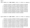


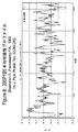
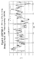


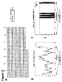
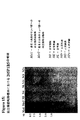

Classifications
-
- C—CHEMISTRY; METALLURGY
- C07—ORGANIC CHEMISTRY
- C07K—PEPTIDES
- C07K14/00—Peptides having more than 20 amino acids; Gastrins; Somatostatins; Melanotropins; Derivatives thereof
- C07K14/435—Peptides having more than 20 amino acids; Gastrins; Somatostatins; Melanotropins; Derivatives thereof from animals; from humans
- C07K14/46—Peptides having more than 20 amino acids; Gastrins; Somatostatins; Melanotropins; Derivatives thereof from animals; from humans from vertebrates
- C07K14/47—Peptides having more than 20 amino acids; Gastrins; Somatostatins; Melanotropins; Derivatives thereof from animals; from humans from vertebrates from mammals
- C07K14/4701—Peptides having more than 20 amino acids; Gastrins; Somatostatins; Melanotropins; Derivatives thereof from animals; from humans from vertebrates from mammals not used
- C07K14/4748—Tumour specific antigens; Tumour rejection antigen precursors [TRAP], e.g. MAGE
-
- A—HUMAN NECESSITIES
- A61—MEDICAL OR VETERINARY SCIENCE; HYGIENE
- A61K—PREPARATIONS FOR MEDICAL, DENTAL OR TOILETRY PURPOSES
- A61K39/00—Medicinal preparations containing antigens or antibodies
- A61K39/46—Cellular immunotherapy
- A61K39/461—Cellular immunotherapy characterised by the cell type used
- A61K39/4611—T-cells, e.g. tumor infiltrating lymphocytes [TIL], lymphokine-activated killer cells [LAK] or regulatory T cells [Treg]
-
- A—HUMAN NECESSITIES
- A61—MEDICAL OR VETERINARY SCIENCE; HYGIENE
- A61K—PREPARATIONS FOR MEDICAL, DENTAL OR TOILETRY PURPOSES
- A61K39/00—Medicinal preparations containing antigens or antibodies
- A61K39/46—Cellular immunotherapy
- A61K39/464—Cellular immunotherapy characterised by the antigen targeted or presented
- A61K39/4643—Vertebrate antigens
- A61K39/4644—Cancer antigens
-
- A—HUMAN NECESSITIES
- A61—MEDICAL OR VETERINARY SCIENCE; HYGIENE
- A61P—SPECIFIC THERAPEUTIC ACTIVITY OF CHEMICAL COMPOUNDS OR MEDICINAL PREPARATIONS
- A61P35/00—Antineoplastic agents
-
- C—CHEMISTRY; METALLURGY
- C07—ORGANIC CHEMISTRY
- C07K—PEPTIDES
- C07K14/00—Peptides having more than 20 amino acids; Gastrins; Somatostatins; Melanotropins; Derivatives thereof
- C07K14/435—Peptides having more than 20 amino acids; Gastrins; Somatostatins; Melanotropins; Derivatives thereof from animals; from humans
- C07K14/46—Peptides having more than 20 amino acids; Gastrins; Somatostatins; Melanotropins; Derivatives thereof from animals; from humans from vertebrates
- C07K14/47—Peptides having more than 20 amino acids; Gastrins; Somatostatins; Melanotropins; Derivatives thereof from animals; from humans from vertebrates from mammals
-
- G—PHYSICS
- G01—MEASURING; TESTING
- G01N—INVESTIGATING OR ANALYSING MATERIALS BY DETERMINING THEIR CHEMICAL OR PHYSICAL PROPERTIES
- G01N33/00—Investigating or analysing materials by specific methods not covered by groups G01N1/00 - G01N31/00
- G01N33/48—Biological material, e.g. blood, urine; Haemocytometers
- G01N33/50—Chemical analysis of biological material, e.g. blood, urine; Testing involving biospecific ligand binding methods; Immunological testing
- G01N33/53—Immunoassay; Biospecific binding assay; Materials therefor
- G01N33/574—Immunoassay; Biospecific binding assay; Materials therefor for cancer
- G01N33/57484—Immunoassay; Biospecific binding assay; Materials therefor for cancer involving compounds serving as markers for tumor, cancer, neoplasia, e.g. cellular determinants, receptors, heat shock/stress proteins, A-protein, oligosaccharides, metabolites
-
- A—HUMAN NECESSITIES
- A61—MEDICAL OR VETERINARY SCIENCE; HYGIENE
- A61K—PREPARATIONS FOR MEDICAL, DENTAL OR TOILETRY PURPOSES
- A61K38/00—Medicinal preparations containing peptides
-
- A—HUMAN NECESSITIES
- A61—MEDICAL OR VETERINARY SCIENCE; HYGIENE
- A61K—PREPARATIONS FOR MEDICAL, DENTAL OR TOILETRY PURPOSES
- A61K39/00—Medicinal preparations containing antigens or antibodies
-
- Y—GENERAL TAGGING OF NEW TECHNOLOGICAL DEVELOPMENTS; GENERAL TAGGING OF CROSS-SECTIONAL TECHNOLOGIES SPANNING OVER SEVERAL SECTIONS OF THE IPC; TECHNICAL SUBJECTS COVERED BY FORMER USPC CROSS-REFERENCE ART COLLECTIONS [XRACs] AND DIGESTS
- Y02—TECHNOLOGIES OR APPLICATIONS FOR MITIGATION OR ADAPTATION AGAINST CLIMATE CHANGE
- Y02A—TECHNOLOGIES FOR ADAPTATION TO CLIMATE CHANGE
- Y02A50/00—TECHNOLOGIES FOR ADAPTATION TO CLIMATE CHANGE in human health protection, e.g. against extreme weather
- Y02A50/30—Against vector-borne diseases, e.g. mosquito-borne, fly-borne, tick-borne or waterborne diseases whose impact is exacerbated by climate change
Landscapes
- Health & Medical Sciences (AREA)
- Life Sciences & Earth Sciences (AREA)
- Chemical & Material Sciences (AREA)
- Immunology (AREA)
- Organic Chemistry (AREA)
- General Health & Medical Sciences (AREA)
- Medicinal Chemistry (AREA)
- Molecular Biology (AREA)
- Cell Biology (AREA)
- Engineering & Computer Science (AREA)
- Biochemistry (AREA)
- Biophysics (AREA)
- Zoology (AREA)
- Microbiology (AREA)
- Proteomics, Peptides & Aminoacids (AREA)
- Hematology (AREA)
- Genetics & Genomics (AREA)
- Gastroenterology & Hepatology (AREA)
- Toxicology (AREA)
- Biomedical Technology (AREA)
- Urology & Nephrology (AREA)
- Pharmacology & Pharmacy (AREA)
- Veterinary Medicine (AREA)
- Public Health (AREA)
- Animal Behavior & Ethology (AREA)
- Oncology (AREA)
- General Physics & Mathematics (AREA)
- Food Science & Technology (AREA)
- Epidemiology (AREA)
- Mycology (AREA)
- Pathology (AREA)
- Biotechnology (AREA)
- Analytical Chemistry (AREA)
- Hospice & Palliative Care (AREA)
- Physics & Mathematics (AREA)
- Nuclear Medicine, Radiotherapy & Molecular Imaging (AREA)
- Chemical Kinetics & Catalysis (AREA)
- General Chemical & Material Sciences (AREA)
- Peptides Or Proteins (AREA)
- Medicines That Contain Protein Lipid Enzymes And Other Medicines (AREA)
Abstract
新規遺伝子(205P1B5と称される)およびそのコードタンパク質が記載される。205P1B5は、正常な成体組織における組織特異的発現を示すとはいえ、これは、前立腺癌において異常に発現される。その結果、205P1B5は、癌に対する診断的、予後的、予防的および/または治療的な標的を提供する。205P1B5遺伝子もしくはそのフラグメント、またはそれらのコードタンパク質もしくはそれらのフラグメントは、免疫応答を惹起するために使用され得る。A novel gene (designated 205P1B5) and its encoded protein are described. Although 205P1B5 shows tissue-specific expression in normal adult tissues, it is abnormally expressed in prostate cancer. As a result, 205P1B5 provides a diagnostic, prognostic, prophylactic and / or therapeutic target for cancer. The 205P1B5 gene or fragments thereof, or their encoded proteins or fragments thereof, can be used to elicit an immune response.
Description
【技術分野】
【0001】
(発明の分野)
本明細書中に記載される発明は、205P1B5と称される新規な遺伝子およびそのコードタンパク質、ならびに205P1B5を発現する種々の癌の管理において有用な診断方法および治療方法、ならびに組成物に関する。
【背景技術】
【0002】
(発明の背景)
癌は、冠状動脈疾患(coronary disease)に次いで、ヒトの第2位の死因である。全世界では、数百万の人々が、毎年癌により死亡している。米国単独では、American Cancer Societyによって報告されるように、癌は毎年50万人をはるかに超える人々の死亡を引き起こしており、毎年120万あまりの新たな症例が診断されている。心臓疾患による死亡は顕著に減少している一方で、癌に起因する死亡は、一般に増加傾向にある。次世紀の初期には、癌は主な死因になると予測されている。
【0003】
全世界で、いくつかの癌が主要な死因(killer)として顕著である。特に、肺、前立腺、胸部、結腸、膵臓、および卵巣の癌腫は、癌による主な死因を代表する。これらおよび事実上全ての他の癌腫は、共通の致死的特徴を共有している。ごくわずかな例外はあるが、癌腫が原因の転移性疾患は、致命的である。さらに、初期段階ではその原発性癌を生き延びた癌患者についてすら、その生活が劇的に変化したという共通の経験が示されている。多くの癌患者は、再発または処置の失敗についての可能性を認識することによって駆り立てられる、強い不安を経験している。多くの癌患者は、処置後に肉体的衰弱を経験している。さらに、多くの癌患者は、再発を経験している。
【0004】
全世界で、前立腺癌は、男性において4番目に最も優勢な癌である。北アメリカおよび北欧では、前立腺癌は、男性において断然最も一般的な癌であり、そして男性における癌による死亡の第2位の原因である。米国単独では、肺癌に次いで、30,000人をはるかに超える男性がこの疾患で毎年死亡している。これらの数字の規模にもかかわらず、転移性の前立腺癌に対する有効な処置は未だ存在しない。外科的前立腺切除、放射線療法、ホルモン除去(hormone ablation)療法、外科的去勢および化学療法は、主要な処置様式であり続けている。不運にも、これらの処置は、多くの者にとって無効であり、かつしばしば、所望されない結果を伴う。
【0005】
診断現場において、初期段階の局所腫瘍を正確に検出し得る前立腺腫瘍マーカーが存在しないことは、この疾患の診断および管理における大きな限界を残している。血清の前立腺特異抗原(PSA)アッセイは非常に有用なツールであるが、その特異性および一般的有用性は、いくつかの重大な点が欠如していると広く考えられている。
【0006】
前立腺癌についてのさらなる特異的マーカーを同定することにおける進歩は、マウスにおいてこの疾患の異なる病期を再現し得る前立腺癌異種移植片の生成により改善されてきた。LAPC(Los Angeles Prostate Cancer)異種移植片は、重症複合型免疫不全(SCID)マウスにおける継代を生き延び、かつアンドロゲン依存性からアンドロゲン非依存性への移行を模倣する能力を示した、前立腺癌異種移植片である(Kleinら、1997、Nat.Med.3:402)。より近年同定された前立腺癌マーカーとしては、以下が挙げられる:PCTA−1(Suら、1996、Proc.Natl.Acad.Sci.USA 93:7252)、前立腺特異膜(PSM)抗原(Pintoら、Clin Cancer Res 1996年9月2日(9):1445−51)、STEAP(Hubertら、Proc Natl Acad Sci USA.1999年12月7日;96(25):14523−8)および前立腺幹細胞抗原(PSCA)(Reiterら、1998、Proc.Natl.Acad.Sci.USA 95:1735)。
【0007】
PSA、PSM、PCTAおよびPSCAのような以前に同定されたマーカーは、前立腺癌を診断してこれを処置する取り組みを促進したが、診断および治療をさらに改善するために、前立腺癌および関連する癌に対するさらなるマーカーおよび治療標的を同定する必要がある。
【0008】
腎臓細胞癌腫(RCC)は、成人悪性疾患の約3%を占める。一旦、腺腫が直径2〜3cmに到達すると、悪性疾患の潜在性が存在する。成人では、2つの主な悪性腎臓腫瘍は、腎臓細胞の腺腫および腎盂または尿管の移行細胞癌腫である。腎臓細胞腺癌の発生数は、米国で29,000症例を超えると見積もられており、そして1998年にこの疾患によって11,600人を超える患者が死亡した。移行細胞癌腫は、それほど頻繁ではなく、米国において1年あたり約500症例ほどの発生数である。
【0009】
手術は、何十年もの間、腎臓細胞腺癌の主な治療法であった。近年まで、転移性疾患は、あらゆる全身性治療に対して難治性であった。全身治療(特に、免疫治療)の近年の開発に伴ない、転移性腎臓細胞癌腫は、耐久性のある応答の可能性を伴って、適切な患者において攻撃的にアプローチされ得る。それにもかかわらず、これらの患者に対する有効な治療の必要性が存在し続けている。
【0010】
米国における癌の全ての新しい症例のうち、膀胱癌は、男性において約5%(最も一般的な新生物の5番目)および女性において3%(最も一般的な新生物の8番目)に相当する。発生数はゆるやかに増加し、高齢人口の増加と一致する。1998年には、男性における39,500症例および女性における15,000症例を含め、推定54,500症例が存在した。米国における年齢調整された(age−adjusted)発生率は、男性について100,000人当たり32人、および女性において100,000人当たり8人である。3:1の歴史的な男性/女性比は、女性における喫煙パターンに関連して減少し得る。1998年に膀胱癌で推定11,000人が死亡した(男性7,800人および女性3,900人)。膀胱癌発生率および死亡率は、年齢に伴って大きく増加し、そして集団がより高齢化するにつれて問題も増す。
【0011】
大部分の膀胱癌は、膀胱中で再発する。膀胱癌は、膀胱の経尿道的切除術(TUR)と膀胱内化学療法または免疫療法との組み合わせを用いて管理される。膀胱癌の多病巣性の性質および再発性の性質は、TURの限界を指摘する。大部分の筋肉侵襲性癌は、TUR単独では治癒しない。根治的膀胱切除術および尿再交通(urinary diversion)は、癌を切除するための最も効果的な手段であるが、尿の機能および性的機能に対して否定できない影響を保有する。膀胱癌患者に有益な処置様式の顕著な必要性が存在し続けている。
【0012】
米国で2000年では、93,800症例の結腸癌および36,400症例の直腸癌を含め、推定130,200症例の結腸直腸癌が生じた。結腸直腸癌は、男性および女性における三番目に一般的な癌である。発生率は、1992年〜1996年の間、有意に減少した(1年あたり−2.1%)。調査は、これらの減少が、ポリープが侵襲性癌へと進行するのを予防する、スクリーニングおよびポリープ除去の増加に起因していることを示唆する。2000年には推定56,300人の死亡者(47,700人は結腸癌、8,600人は直腸癌による)が存在し、米国での癌死亡者全体の約11%を占めた。
【0013】
現在、手術が、結腸直腸癌、および広がっていない癌のための最も一般的な治療形態であり、これは頻繁に治癒的である。化学療法または化学療法に加えての放射線照射は、癌が腸壁に深く穿孔しているかまたはリンパ節に広がっている大部分の患者に対して手術前または手術後に提供される。永久的な人工肛門形成(身体の排泄物を除去するための腹部開口部の形成)は、結腸癌について時折必要とされ、そして直腸癌については頻繁に要求される。結腸直腸癌についての効果的な診断様式および処置様式の必要性が存在し続けている。
【0014】
2000年には、肺癌および気管支癌の推定164,100の新たな症例が存在し、これは、米国癌診断全体の14%を占めた。肺癌および気管支癌の発生率は、1984年の100,000人中86.5人という高さから1996年の70.0人へと、男性において有意に減少している。1990年代、女性の間での増加率は低下し始めた。1996年には、女性における発生率は、100,000人中42.3人であった。
【0015】
肺癌および気管支癌は、2000年に推定156,900人の死亡者を生じ、これは、癌死亡者全体の28%を占めた。1992年〜1996年の間、肺癌による死亡率は、男性の間で有意に減少した(1年当たり−1.7%)一方で、女性についての死亡率は依然として有意に増加している(1年当たり0.9%)。1987年以来、より多くの女性が乳癌よりも肺癌によって毎年死亡しており、40年よりも長期にわたって、女性における癌による死亡の主な原因であった。肺癌発生率および死亡率の減少は、過去30年にわたる喫煙率の減少に起因している可能性が最も高い;しかし、女性の間での喫煙パターンの減少は、男性の減少に遅れている。成体におけるタバコの使用の減少が鈍くなっているが、若者におけるタバコの使用が再度増加していることは、重大である。
【0016】
肺癌および気管支癌についての処置選択肢は、癌の型および病期によって決定され、選択肢としては、手術、放射線療法および化学療法が挙げられる。多くの局所癌については、手術が通常選り抜きの処置である。この疾患は、通常、発見されるときまでに広がっているので、放射線療法および化学治療が、手術と組み合わせてしばしば必要とされる。化学治療単独または放射線療法と組み合わせた化学療法は、小細胞肺癌についての選り抜きの処置であり;このレジメンでは、大きな%の患者が寛解を経験し、これはいくつかの場合において長く持続する。しかし、肺癌および気管支癌についての効果的な処置アプローチおよび診断アプローチの必要性が存在し続けている。
【0017】
乳癌の推定182,800の新しい侵襲性症例が、2000年の間に米国の女性の間で生じると予測された。さらに、乳癌の約1,400の新しい症例が、2000年に男性において診断されると予測された。1980年代における一年当たり約4%の増加後、女性における乳癌発生率は、1990年代に100,000人当たり約110.6症例に頭打ちとなった。
【0018】
米国単独で、乳癌に起因して、2000年に推定41,200人の死亡者が存在した(40,800人が女性、400人が男性)。乳癌は、女性における癌による死亡の2番目に主要なものである。最も最近のデータによると、死亡率は、1992年〜1996年の間に有意に減少しており、白人および黒人の両方の若い女性の間で最も大きく減少していた。これらの減少は、おそらく、より早期の検出および処置の改善の結果であった。
【0019】
医学的環境および患者の好みを考慮にいれると、乳癌の処置は、以下を含み得る:腫瘍摘除(腫瘍の局所的な除去)および脇下リンパ節の除去;乳房切除(乳房の外科的除去)および脇下リンパ節の除去;放射線療法;化学療法;またはホルモン療法。しばしば、2つ以上の方法が組み合わせて使用される。多数の研究が、初期段階の疾患について示されており、腫瘍切除に加えた放射線療法の後の長期生存率は、改変された根本的な乳房切除後の生存率に同様である。再構築技術における顕著な進歩は、乳房切除後の乳房再構築についていくつかの選択肢を提供する。近年、このような再構築は、乳房切除と同時に行われている。
【0020】
適切な量の周囲の正常胸部組織を伴うインサイチュの腺管癌腫(DCIS)の局所切除によって、DCISの局所的な再発を防ぎ得る。胸部への放射線照射および/またはタモキシフェンによって、残っている胸部組織におけるDCISの発生機会が低減され得る。未処置のままであるとDCISは発達して浸潤性乳癌になり得るので、このことは重要である。それにもかかわらず、これらの処置には、深刻な副作用または後遺症が存在する。それゆえ、有効な乳癌処置の必要性が存在する。
【0021】
2000年の米国において卵巣癌の推定23,100の新症例が存在した。卵巣癌は女性における全ての癌の4%を占め、婦人科癌において第2位に位置付けられている。1992年〜1996年の間において、卵巣癌の発生率は、有意に低下した。卵巣癌の結果として、2000年には推定14,000人の死亡者が存在した。卵巣癌は、女性生殖系の他のいかなる癌よりも多数の死亡者を生じる。
【0022】
手術、放射線療法、および化学療法は、卵巣癌についての処置選択肢である。外科的手術としては、通常、卵巣の一方または両方の切除、ファローピウス管の切除(卵管卵巣摘出術)、および子宮の切除(子宮摘出術)が挙げられる。いくつかの非常に初期の腫瘍において、子供を持つことを望む若い女性において特に、関与する卵巣のみが除去される。進行した疾患では、腹腔内の疾患全てを除去して化学療法の効果を増強することが試みられる。卵巣癌についての効果的な処置選択肢についての重要な必要性が存在し続ける。
【0023】
2000年の米国において膵臓癌の推定28,300の新症例が存在した。過去20年間にわたって、膵臓癌の割合は、男性において減少している。女性の間での割合は、おおよそ一定のままであるが、減少し始めるかもしれない。膵臓癌は、2000年に米国において推定28,200の死亡を引き起こした。過去20年間にわたって、男性における死亡率は僅かであるが有意に減少している(1年間で約−0.9%)が、その一方で、その割合が女性においては僅かに上昇した。
【0024】
手術、放射線療法、および化学療法は、膵臓癌についての処置選択肢である。これらの処置選択肢は、多くの患者において生存を長期化し得、そして/またはその症状を軽減し得るが、大部分の人に対して治癒を生み出すようではない。膵臓癌についてのさらなる治療選択肢および診断的選択肢に対しての重大な必要性が存在する。
【発明の開示】
【課題を解決するための手段】
【0025】
(発明の要旨)
本発明は、205P1B5と称される遺伝子に関し、この遺伝子は、表Iに列挙される癌において過剰発現される。205P1B5の2つの改変体が存在する(例えば、図2を参照のこと);文脈が明らかそうではないことを示すのでない限り、205P1B5への本明細書中での言及は、これらの改変体のいずれをもいう。正常組織における205P1B5遺伝子発現のノーザンブロット発現分析は、成人組織における制限された発現パターンを示す。205P1B5のヌクレオチド配列(図2)およびアミノ酸配列(図2および図3)を提供する。正常成人組織における205P1B5の組織関連プロファイルは、前立腺腫瘍において観察された過剰発現とあわせて、205P1B5が少なくともいくつかの癌において異常に過剰発現されており、したがって、表Iで列挙された組織のような組織の癌について有用な診断標的、予防標的、予後診断標的および/または治療標的として役立つことを示す。
【0026】
本発明は、以下を提供する:205P1B5の遺伝子、mRNAおよび/またはコード配列の全てまたは一部分に対応するかまたは相補的である(好ましくは単離された形態の)ポリヌクレオチド(これらとしては、以下が挙げられる:4、5、6、7、8、9、10、11、12、13、14、15、16、17、18、19、20、21、22、23、24、25、または25を超えて連続したアミノ酸の、205P1B5関連タンパク質および205P1B5関連フラグメントをコードするポリヌクレオチド);少なくとも30、35、40、45、50、55、60、65、70、80、85、90、95、100または100を超えて連続したアミノ酸の205P1B5関連タンパク質、ならびにペプチド/タンパク質自体;205P1B5の遺伝子配列もしくはmRNA配列またはそれらの一部に対して相補的であるかまたは少なくとも90%の相同性を有する、DNA、RNA、DNA/RNAハイブリッド、および関連分子、ポリヌクレオチドまたはオリゴヌクレオチド、ならびに205P1B5の遺伝子、mRNAまたは205P1B5コードポリヌクレオチドにハイブリダイズするポリヌクレオチドまたはオリゴヌクレオチド。205P1B5をコードするcDNAおよび遺伝子を単離するための手段もまた提供される。205P1B5ポリヌクレオチドを含む組換えDNA分子、このような分子で形質転換または形質導入された細胞、および205P1B5遺伝子産物の発現のための宿主−ベクター系もまた提供される。本発明はさらに、205P1B5タンパク質およびそれらのポリペプチドフラグメントに結合する抗体を提供し、これらの抗体としては、ポリクローナル抗体およびモノクローナル抗体、マウス抗体および他の哺乳動物抗体、キメラ抗体、ヒト化抗体および完全ヒト抗体、ならびに検出可能なマーカーで標識された抗体が挙げられる。特定の実施形態においては、ただし、図2の核酸配列の全体はコードされない、および/または図2のアミノ酸配列全体は調製されない。特定の実施形態では、図2の核酸配列全体がコードされ、そして/または図2のアミノ酸配列全体が調製され、これらのいずれかは、それぞれのヒト単位投薬形態である。
【0027】
本発明は、種々の生物学的サンプル中での205P1B5のポリヌクレオチドおよびタンパク質の存在および状態を検出するための方法、ならびに205P1B5を発現する細胞を同定するための方法をさらに提供する。本発明の代表的な実施形態は、癌のような何らかの形態の増殖調節不全を有するかまたはそれらを有する疑いのある組織サンプルまたは血液学的サンプルにおいて、205P1B5遺伝子産物をモニタリングするための方法を提供する。
【0028】
本発明は、種々の免疫原性組成物または治療組成物、および205P1B5を発現する癌(例えば、前立腺癌)を処置するためのストラテジー(205P1B5の転写、翻訳、プロセシングまたは機能を阻害することを目的とした治療を含む)ならびに癌ワクチンをさらに提供する。
【0029】
(発明の詳細な説明)
(節の概要)
I.)定義
II.)205P1B5ポリヌクレオチド
II.A.)205P1B5ポリヌクレオチドの用途
II.A.1.)遺伝的異常のモニタリング
II.A.2.)アンチセンスの実施形態
II.A.3.)プライマーおよびプライマー対
II.A.4.)205P1B5コード核酸分子の単離
II.A.5.)組換え核酸分子および宿主−ベクター系
III.)205P1B5関連タンパク質
III.A.)モチーフ保有タンパク質の実施形態
III.B.)205P1B5関連タンパク質の発現
III.C.)205P1B5関連タンパク質の改変
III.D.)205P1B5関連タンパク質の用途
IV.)205P1B5抗体
V.)205P1B5細胞免疫応答
VI.)205P1B5トランスジェニック動物
VII.)205P1B5の検出方法
VIII.)205P1B5関連遺伝子およびそれらの産物の状態をモニタリングするための方法
IX.)205P1B5と相互作用する分子の同定
X.)治療法および組成物
X.A.)抗癌ワクチン
X.B.)抗体ベースの治療のための標的としての205P1B5
X.C.)細胞免疫応答についての標的としての205P1B5
X.C.1.ミニ遺伝子ワクチン
X.C.2.CTLペプチドとヘルパーペプチドとの組み合わせ
X.C.3.CTLペプチドとT細胞プライミング剤との組み合わせ
X.C.4.CTLペプチドおよび/またはHTLペプチドでパルスされたDCを含むワクチン組成物
X.D.)養子免疫療法
X.E.)治療目的または予防目的のためのワクチンの投与
XI.)205P1B5の診断的実施形態および予後的実施形態
XII.)205P1B5タンパク質機能の阻害
XII.A.)細胞内抗体を用いた205P1B5の阻害
XII.B.)組換えタンパク質を用いた205P1B5の阻害
XII.C.)205P1B5の転写または翻訳の阻害
XII.D.)治療的ストラテジーについての一般的考慮事項
XIII.)キット。
【0030】
(I.)定義)
他に特に定義されていない限りは、本明細書中で使用される当該分野の全ての用語、記号、および他の科学的用語または専門用語は、本発明が関係している当業者によって一般的に理解されている意味を有するように意図される。いくつかの場合においては、一般的に理解されている意味を有する用語は、明確さおよび/または容易な参照のために本明細書中で定義され、そして本明細書中でのこのような定義の包含は、当該分野で一般的に理解されている意味を超える実質的な差異を示すようには決して解釈されないはずである。本明細書中に記載されるかまたは参照される技術および手順(例えば、Sambrookら、Molecular Cloning:A Laboratory Manual、第2版、(1989)編、Cold Spring Harbor Laboratory Press、Cold Spring Harbor、N.Y.に記載されている、広範囲に利用されている分子クローニング方法論)は、一般的に十分に理解されており、そして当業者によって、従来の方法論を用いて一般的に使用される。適切である場合には、商業的に入手可能なキットおよび試薬の使用を含む手順が、一般的には、製造業者によって定義されるプロトコルおよび/またはそうでなければ他に記載されているパラメーターに従って行われる。
【0031】
用語「進行した前立腺癌」、「局所的に進行した前立腺癌」、「進行した疾患」、および「局所的に進行した疾患」は、前立腺の莢膜を通じて拡大した前立腺癌を意味し、そしてAmerican Urological Association(AUA)システムのもとでのステージCの疾患、Whitmore−JewettシステムのもとでのステージC1〜C2の疾患、およびTNM(腫瘍、節、転移(tumor、node、metastasis))システムのもとでのステージT3〜T4およびN+の疾患を含むように意味される。一般的には、外科手術は、局所的に進行した疾患を有する患者については推奨されず、そしてこれらの患者は、臨床的に局在化した(器官に限定された)前立腺癌を有している患者と比較して、実質的にあまり好ましくない結果を有する。局所的に進行した疾患は、前立腺の側縁を超えるしこり、あるいは前立腺の基部の上部の非対称性またはしこりの明白な証拠によって、臨床的に同定される。局所的に進行した前立腺癌は、現在は、腫瘍が前立腺の莢膜に侵入または浸潤するか、外科的な縁に伸びるか、あるいは精子の小包に侵入する場合には、根治的な前立腺切除後に病理学的に診断される。
【0032】
「ネイティブなグリコシル化パターンを改変すること」は、本明細書中の目的を意図し、ネイティブ配列の205P1B5において見出される1つ以上の炭水化物部分を除去すること(内在するグリコシル化部位を除去することによってか、または化学的手段および/もしくは酵素的手段によってグリコシル化を除去することによってのいずれかで)、および/あるいはネイティブ配列の205P1B5に存在しない1つ以上のグリコシル化部位を付加することを意味する。さらに、この句は、ネイティブタンパク質のグリコシル化の質的な変化を含み、種々の炭水化物部分の存在の性質および比率の変化に関する。
【0033】
用語「アナログ」は、構造的に類似するか、別の分子と類似する特性または一致する特性を共有する分子をいう(例えば、205P1B5関連タンパク質)。例えば、205P1B5タンパク質のアナログは、205P1B5に特異的に結合する抗体またはT細胞によって特異的に結合され得る。
【0034】
用語「抗体」は、最も広範な意味で使用される。従って、「抗体」は、天然に存在し得るか、または慣習なハイブリドーマ技術によって産生されるモノクローナル抗体のような人工であり得る。抗205P1B5抗体は、モノクローナル抗体およびポリクローナル抗体ならびに抗原結合ドメインおよび/またはこれらの抗体の1つ以上の相補的決定領域を含むフラグメントを含む。
【0035】
「抗体フラグメント」は、標的に結合する免疫グロブリン分子の可変領域の少なくとも一部分(すなわち、抗原結合領域)として定義される。1つの実施形態において、これは、単一の抗205P1B5抗体およびそのクローン(アゴニスト、アンタゴニストおよび中和抗体が挙げられる)ならびにポリエピトープの(polyepitopic)特異性を有する抗205P1B5抗体組成物を特に網羅する。
【0036】
用語「コドン最適化配列」は、少なくとも約20%の使用頻度を有する任意のコドンを置換することによって特定の宿主種を最適化するヌクレオチド配列をいう。偽ポリアデニル化配列の除去、エキソン/イントロンスプライシングシグナルの除去、トランスポソン様リピートの除去および/またはコドン最適化に加えてGC含量の最適化によって、所定の宿主種における発現について最適化されたヌクレオチド配列は、「発現増強配列」として本明細書中で参照される。
【0037】
用語「細胞傷害性薬剤」は、細胞の発現活性、細胞の機能を阻害するかまたは防止し、そして/あるいは細胞の破壊を引き起こす物質をいう。この用語は、放射活性同位体化学療法剤および毒素(例えば、低分子毒素または細菌起源、真菌起源、植物起源または動物起源の酵素的活性毒素)(フラグメントおよび/またはその改変体を含む)を含むことが意図される。細胞傷害性試薬の例として、以下が挙げられるが、これらに限定されない:マイタンシノイド(maytansinoid)、イットリウム、ビスマス、リシン、リシンA鎖、ドキソルビシン、ダウノルビシン、タキソール、エチジウムブロマイド、マイトマイシン、エトポシド、テノポシド、ビンクリスチン、ビンブラスチン、コルヒチン、ジヒドロキシアントラシンジオン、アクチノマイシン、ジフテリア毒素、Pseudomonas外毒素(PE)A、PE40、アブリン、アブリンA鎖、モデシン(modeccin)A鎖、α−サルシン(sarcin)、ゲロニン(gelonin)、ミトゲリン(mitogellin)、レトストリクトシン(retstrictocin)、フェノマイシン、エノマイシン、クリシン(curicin)、クロチン(crotin)、カリケアマイシン(calicheamicin)、sapaonaria officinalisインヒビター、およびグルココルチコイド、ならびに他の化学療法剤、ならびに放射性同位元素(例えばAt211、I131、I125、Y90、Re186、Re188、Sm153、Bi212、P32およびLuの放射性同位体)。抗体はまた、活性形態にプロドラッグを変換し得る抗癌プロドラッグ活性化酵素に結合し得る。
【0038】
用語「ホモログ」は、別の分子と相同性を示す(例えば、対応する位置で同様かまたは類似である化学残基の配列を有する)分子をいう。
【0039】
「ヒト白血球抗原」または「HLA」は、ヒトのクラスIまたはクラスIIの主要組織適合抗原複合体(MHC)タンパク質である(例えば、Stitesら、IMMUNOLOGY,第8編.,Lange Publishing,Los Altos,CA(1994)を参照のこと)。
【0040】
ポリヌクレオチドの状況で使用される、用語「ハイブリダイズする(hybridize)」、「ハイブリダイズする(hybridizing)」、「ハイブリダイズする(hybridizes)」などは、従来のハイブリダイゼーション条件(好ましくは、例えば、50%のホルムアミド/6×SSC/0.1%のSDS/100μg/mlのssDNA中でのハイブリダイゼーション)をいうことが意味される。ここでは、ハイブリダイゼーションの温度は37℃を超え、そして0.1×SSC/0.1%のSDS中での洗浄のための温度は55℃より高い。
【0041】
句「単離された」または「生物学的に純粋」は、ネイティブな状態で見出されるような物質を通常伴う構成要素を実質的にまたは本質的に含まない物質をいう。従って、本発明に従う単離されたペプチドは、好ましくはインサイチュ環境においてペプチドに通常付随する物質を含まない。例えば、ポリヌクレオチドは、205P1B5遺伝子以外の遺伝子に対応するかまたは相補的であるか、または205P1B5遺伝子産物またはそのフラグメント以外のポリペプチドをコードする混入物のポリヌクレオチドから実質的に分離される場合、「単離される」と言われる。当業者は、単離された205P1B5ポリヌクレオチドを得るために核酸単離手順を容易に利用し得る。タンパク質は、例えば、物理的方法、機械的方法または化学的方法が、タンパク質に通常付随する細胞の成分から205P1B5タンパク質を除するするために利用される場合、「単離された」と言われる。当業者は、単離された205P1B5タンパク質を得るために標準的な精製方法を容易に利用し得る。あるいは、単離されたタンパク質は、化学的手段によって調製され得る。
【0042】
用語「哺乳動物」は、哺乳動物(マウス、ラット、ウサギ、イヌ、ネコ、ウシ、ウマおよびヒトを含む)に分類される任意の生物をいう。本発明の1つの実施形態において、哺乳動物はマウスである。本発明の別の実施形態において、哺乳動物はヒトである。
【0043】
用語「転移性の前立腺癌」および「転移性の疾患」は、局所的なリンパ節または離れた部位に拡大した前立腺癌を意味し、そしてAUAシステムのもとでのステージDの疾患、およびTNMシステムのもとでのステージT×N×M+を含むように意味される。局所的に進行した前立腺癌の症例における場合には、外科手術は、一般的には、転移性の疾患を有している患者については意図されず、そしてホルモン(アンドロゲンの切除)治療が、好ましい処置様式である。転移性の前立腺癌を有している患者は、最終的には、処置の開始の12〜18ヶ月以内のアンドロゲン治療不応性状態を発症する。これらのアンドロゲン治療不応性の患者のほぼ半分が、その状態の発症後6ヶ月以内に死亡する。前立腺癌の転移の最も一般的な部位は骨である。前立腺癌の骨転移は、しばしば、骨溶解よりもむしろ骨芽細胞性(すなわち、正味の骨の形成を生じる)である。骨転移は、脊椎においてもっとも頻繁に見出され、大腿骨、骨盤、肋骨郭(rib cage)、頭蓋骨、および上腕骨が続く。他の一般的な転移の部位として、リンパ節、肺、肝臓、および脳が挙げられる。転移性の前立腺癌は、代表的には、開放性骨盤リンパ腺切除または腹腔鏡による骨盤リンパ腺切除、全身の放射性核種スキャン、骨格のレントゲン撮影、および/あるいは骨の病変の生検によって、診断される。
【0044】
用語「モノクローナル抗体」は、実質的に同種の集団から得られる抗体(すなわち、少量存在する、天然に存在する可能性を除いて同一である集団を含む抗体)をいう。
【0045】
205P1B5関連タンパク質の生物学的モチーフとしての「モチーフ」は、タンパク質の一次配列の一部を形成するアミノ酸の任意のパターン(特定の機能(例えば、タンパク質−タンパク質相互作用、タンパク質−DNA相互作用など)、または改変(例えば、リン酸化、グリコシル化、もしくはアミド化)、または局在(例えば、分泌配列、核局在化配列など)に関連する)、あるいは免疫原性(体液性または細胞性のいずれか)と関連する配列をいう。モチーフは、一般的に、特定の機能または特性に関連する特定の位置に隣接し得るか、または整列し得るかのいずれかである。HLAモチーフの文脈において、「モチーフ」とは、特定のHLA分子によって認識される、規定された長さのペプチドにおける残基(通常、クラスI HLAモチーフに対する約8〜約13のアミノ酸、およびクラスII HLAモチーフに対する約6〜約25のアミノ酸からのペプチド)のパターンをいう。HLA結合についてのペプチドモチーフは、代表的に、各ヒトHLA対立遺伝子によってコードされる各タンパク質について異なり、そして一次アンカー残基および二次アンカー残基のパターンにおいて異なる。
【0046】
「薬学的な賦形剤」とは、アジュバント、キャリア、pH調整剤および緩衝剤、等張剤、湿潤剤、保存剤などのような物質を含む。
【0047】
「薬学的に受容可能な」とは、非毒性、不活性および/またはヒトもしくは他の哺乳動物に生理学的に適合する組成物をいう。
【0048】
用語「ポリヌクレオチド」は、少なくとも10個の塩基または塩基対の長さのヌクレオチドの多型性の形態(リボヌクレオチドまたはデオキシヌクレオチドのいずれか、あるいはいずれかのヌクレオチドの型の改変された形態)を意味し、そして一本鎖および二本鎖の形態のDNAおよび/またはRNAを含むことを意味する。当該分野において、この用語は、必要であれば、しばしば「オリゴヌクレオチド」と相互に使用される。ポリヌクレオチドは、本明細書中で開示されたヌクレオチド配列を含み得、ここでチミジン(T)(例えば、配列番号702に示されるような)はまたウラシル(U)であり得る。この説明は、DNAおよびRNAの化学的構造間の違い、特に4つの主な塩基のうち1つがRNAでは、チミジン(T)の代わりにウラシル(U)であるという知見に関連する。
【0049】
用語「ポリペプチド」は、少なくとも約4、5、6、7または8アミノ酸のポリマーを意味する。本明細書を通して、アミノ酸についての標準的な3文字表記または1文字表記が使用される。当該分野において、この用語は、しばしば「ペプチド」または「タンパク質」と相互に使用される。
【0050】
HLAの「一次アンカー残基」は、免疫ペプチドとHLA分子との間に接触点を提供することが理解される、ペプチド配列に沿う特定の位置でのアミノ酸である。1〜3つ、通常2つの、規定された長さのペプチド内の一次アンカー残基は、一般的に、免疫原性ペプチドに対する「モチーフ」を規定する。これらの残基は、HLA分子のペプチド結合グルーブと、結合グローブの特異的ポケットに埋め込まれるそれらの側鎖と密接に接触することが理解される。1つの実施形態において、例えば、HLAクラスI分子についての一次アンカー残基は、本発明に従って、(アミノ末端位置から)2位、そしてカルボキシ末端位置の残基ペプチドエピトープ8位、9位、10位、11位または12位に局在される。別の実施形態において、例えば、HLAクラスII分子に結合するペプチドの一次アンカー残基は、ペプチドの末端ではなく、互いに関連して間隔をおいて配置される。ここで、ペプチドは、一般に少なくとも9アミノ酸長である。各モチーフおよびスーパーモチーフ(supermotif)についての一次アンカー位置は、表IVに示される。例えば、アナログペプチドは、表IVに示される、一次アンカー位置および/または二次アンカー位置における特定の残基の存在または非存在を変更することによって作製され得る。このようなアナログは、特定のHLAモチーフまたはスーパーモチーフを含むペプチドの結合親和性および/または集団範囲(population coverage)を調整するために使用される。
【0051】
「組換え」DNA分子またはRNA分子は、インビトロでの分子操作に供されるDNA分子またはRNA分子である。
【0052】
ハイブリダイゼーション反応の「ストリンジェンシー」は、当業者によって容易に決定可能であり、そして一般的には、プローブの長さ、洗浄温度、および塩濃度に依存する経験的な計算である。一般的には、より長いプローブは、適切なアニーリングのためにより高い温度を必要とし、一方、より短いプローブは、より低い温度を必要とする。ハイブリダイゼーションは、一般的には、それらの融解温度以下の環境下に相補鎖が存在する場合に、変性された核酸が再度アニーリングする能力に依存する。プローブとハイブリダイズ可能な配列との間での所望される相同性の程度が高ければ高いほど、使用され得る相対的な温度は高くなる。結果として、より高い相対的な温度が、反応条件をよりストリンジェントにする傾向があり、一方、より低い温度はあまりそうではないという結果になる。ハイブリダイゼーション反応のストリンジェンシーのさらなる詳細および説明については、Ausubelら、Current Protocols in Molecular Biology、Wiley Interscience Publishers(1995)を参照のこと。
【0053】
「ストリンジェントな条件」、または「高ストリンジェントな条件」は、本明細書中で定義される場合は、以下によって同定され得るがこれらに限定されない:(1)洗浄のために低いイオン強度および高温(例えば、0.015Mの塩化ナトリウム/0.0015Mのクエン酸ナトリウム/0.1%のドデシル硫酸ナトリウム、50℃)を使用する;(2)変性剤(例えば、ホルムアミド)をハイブリダイゼーションの間に使用する(例えば、0.1%のウシの血清アルブミンを有する50%(v/v)のホルムアミド/0.1%のFicoll/0.1%のポリビニルピロリドン/750mMの塩化ナトリウム、75mMのクエン酸ナトリウムを有するpH6.5の50mMの燐酸ナトリウム緩衝液、42℃);または(3)50%のホルムアミド、5×SSC(0.75MのNaCl、0.075Mのクエン酸ナトリウム)、50mMのリン酸ナトリウム(pH6.8)、0.1%のピロリン酸ナトリウム、5×デンハルト溶液、超音波処理したサケの精子のDNA(50μg/ml)、0.1%のSDS、および10%のデキストラン硫酸を42℃で使用し、0.2×SSC(塩化ナトリウム/クエン酸ナトリウム)中で42℃で、そして50%のホルムアミド中で55℃で洗浄し、続いて55℃でEDTAを含有している0.1×SSCから構成される高ストリンジェンシーの洗浄が続く。「中程度のストリンジェントな条件」は、限定されないが、Sambrookら、Molecular Cloning:A Laboratory Manual、New York:Cold Spring Harbor Press、1989によって記載され、そして上記に記載されているハイブリダイゼーション条件よりもストリンジェントの低い洗浄溶液およびハイブリダイゼーション条件(例えば、温度、イオン強度および%SDS)の使用を含む。中程度のストリンジェントの条件の例は、20%のホルムアミド、5×SSC(150mMのNaCl、15mMのクエン酸三ナトリウム)、50mMのリン酸ナトリウム(pH7.6)、5×デンハルト溶液、10%のデキストラン硫酸、および20mg/mLの変性された剪断されたサケの精子のDNAを含有している溶液中で37℃での一晩のインキュベーション、続く1×SSC中での約37℃〜50℃でのフィルターの洗浄である。当業者は、プローブの長さなどの因子に順応させることが必要とされる場合には、温度、イオン強度などを調節するための方法を認識する。
【0054】
HLAの「スーパーモチーフ」は、2つ以上のHLA対立遺伝子によってコードされるHLA分子によって特異的に共有されるペプチド結合である。
【0055】
本明細書中で使用される場合、「処置すること」または「処置的な」および文法的に関連する用語は、疾患の任意の結果(例えば、延長した生存、より少ない罹患率、および/または副作用(代替的な治療形式の副産物;疾患の完全な撲滅が必要とされない))の任意の改善をいう。
【0056】
「トランスジェニック動物」(例えば、マウスまたはラット)は、導入遺伝子を含む細胞を有する動物であり、この導入遺伝子は、動物に導入されたか、または出生前(例えば、胚の段階)に動物の先祖に導入された。「導入遺伝子」は、トランスジェニック動物が発生する細胞のゲノム中に組込まれるDNAである。
【0057】
本明細書中で使用する場合、HLAまたは細胞性免疫応答「ワクチン」は、本発明の1つ以上のペプチドを含むか、またはコードする組成物をいう。このようなワクチン(例えば1つ以上の別個のペプチドのカクテル;ポリエピトープペプチドによって含まれる、本発明の1つ以上のペプチド;あるいはこのような別個のペプチドまたはポリエピトープ(例えば、ポリエピトープペプチドをコードする小遺伝子)をコードする核酸)の多数の実施形態が存在する。「1つ以上のペプチド」は、1〜150またはそれ以上(例えば、本発明の少なくとも、2、3、4、5、6、7、8、9、10、11、12、13、14、15、16、17、18、19、20、21、22、23、24、25、26、27、28、29、30、31、32、33、34、35、36、37、38、39、40、41、42、43、44、45、46、47、48、49、50、55、60、65、70、75、80、85、90、95、100、105、110、115、120、125、130、135、140、145、もしくは150以上)の任意の全整数単位が挙げられ得る。ペプチドまたはポリペプチドは、必要に応じて、(例えば、脂質化、標的化配列または他の配列の付加によって)改変され得る。本発明のHLAクラスIペプチドは、細胞傷害性Tリンパ球およびヘルパーTリンパ球の両方の活性化を促進させるために、HLAクラスII分子と混合または連結され得る。HLAワクチンはまた、ペプチドパルス抗原提示細胞(例えば、樹状細胞)を含み得る。
【0058】
用語「改変体」とは、記載される型または基準(例えば、特に記載されるタンパク質(例えば、図2または図3に示される205P1B5タンパク質)の対応部分に、1つ以上の異なるアミノ酸残基を有するタンパク質)からの改変を示す分子をいう。アナログは、改変タンパク質の例である。スプライシングアイソフォームおよびシングルヌクレオチド多型(SNP)は、さらなる改変体の例である。
【0059】
本発明の205P1B5関連タンパク質としては、本明細書中で特異的に同定されたタンパク質、ならびに対立遺伝子改変体、(本明細書中に概説される方法または当該分野で用意に利用可能な方法に従って実験を実施することなく、単離/生成され得、そして特徴付けられ得る)保存的置換改変体、アナログおよびホモログが挙げられる。異なる205P1B5タンパク質またはそのフラグメントの一部分に結合する融合タンパク質、ならびに205P1B5タンパク質および異種ポリペプチドの融合タンパク質もまた、含まれる。このような205P1B5タンパク質は、本発明のタンパク質である205P1B5関連タンパク質、または205P1B5として集合的に称される。用語「205P1B5関連タンパク質」とは、4、5、6、7、8、9、10、11、12、13、14、15、16、17、18、19、20、21、22、23、24、25または25より多いか、あるいは、少なくとも30、35、40、45、50、55、60、65、70、80、85、90、95、100、105、110、115、120、125、130、135、140、145、150、155、160、165、170、175、180、185、190、195、200、225、250、275、300、325、350、375、400、425、450、475、500、525または529以上のアミノ酸の、ポリペプチドフラグメントまたは205P1B5タンパク質配列をいう。
【0060】
(II.)205P1B5ポリヌクレオチド)
本発明の1つの局面は、205P1B5の遺伝子、mRNAおよび/またはコード配列の全てまたは一部に対応するかまたは相補的な、好ましくは単離形態のポリヌクレオチドを提供し、これには、205P1B5関連タンパク質をコードするポリヌクレオチドおよびそのフラグメント、DNA、RNA、DNA/RNAハイブリッド、および関連分子、205P1B5の遺伝子もしくはmRNA配列に相補的なポリヌクレオチドもしくはオリゴヌクレオチドまたはその一部、ならびに205P1B5の遺伝子、mRNA、もしくは205P1B5コードポリヌクレオチドにハイブリダイズするポリヌクレオチドもしくはオリゴヌクレオチド(集合的に「205P1B5ポリヌクレオチド」)が挙げられる。この節において参照される場合、全ての例において、Tは、図2において、Uでもあり得る。
【0061】
205P1B5ポリヌクレオチドの実施形態としては、以下が挙げられる:図2に示される配列を有する205P1B5ポリヌクレオチド、図2に示されるような205P1B5ヌクレオチド配列(ここで、TはUである);図2に示されるような配列を有するポリヌクレオチドの少なくとも10個連続するヌクレオチド;または図2に示される配列を有するポリヌクレオチドの少なくとも10個連続するヌクレオチド(ここで、TはUである)。例えば、205P1B5ヌクレオチドの実施形態は、限定ではなく、以下を含む:
(a)図2に示されるような配列(配列番号____)を含むか、またはこのような配列からなる、ポリヌクレオチド(ここで、TはUでもあり得る);
(b)図2に示されるような配列(配列番号____)の、ヌクレオチド残基番号555〜ヌクレオチド残基番号2144を含むか、またはこのような配列からなる、ポリヌクレオチド(ここで、TはUでもあり得る);
(c)受託番号____として、American Type Culture Collectionに寄託された、____の名称のプラスミドに含まれるcDNAによってその配列がコードされる、205P1B5関連タンパク質をコードする、ポリヌクレオチド;
(d)図2に示される全長アミノ酸配列(配列番号____)に対して少なくとも90%相同な205P1B5関連タンパク質をコードするポリヌクレオチド;
(e)図2に示される全長アミノ酸配列(配列番号____)に対して少なくとも90%同一な205P1B5関連タンパク質をコードするポリヌクレオチド;
(f)表V〜XVIIIに示される少なくとも1つのペプチドをコードするポリヌクレオチド;
(g)図3のペプチドの、図5の親水性プロファイルにおいて0.5より大きい値を有するアミノ酸位置を含む、少なくとも5アミノ酸から、529までの任意の整数の増分のペプチド領域をコードする、ポリヌクレオチド;
(h)図3のペプチドの、図6の疎水性プロファイルにおいて0.5未満の値を有するアミノ酸位置を含む、少なくとも5アミノ酸から、529までの任意の整数の増分のペプチド領域をコードする、ポリヌクレオチド;
(i)図3のペプチドの、図7の接触可能残基の%のプロファイルにおいて0.5より大きい値を有するアミノ酸位置を含む、少なくとも5アミノ酸から、529までの任意の整数の増分のペプチド領域をコードする、ポリヌクレオチド;
(j)図3のペプチドの、図8の平均可撓性プロファイルにおいて0.5より大きい値を有するアミノ酸位置を含む、少なくとも5アミノ酸から、529までの任意の整数の増分のペプチド領域をコードする、ポリヌクレオチド;
(k)図3のペプチドの、図9のβ−ターンプロファイルにおいて0.5より大きい値を有するアミノ酸位置を含む、少なくとも5アミノ酸から、529までの任意の整数の増分のペプチド領域をコードする、ポリヌクレオチド;
(l)(a)〜(k)のいずれか1つのポリヌクレオチドに完全に相補的なポリヌクレオチド;
(m)(a)〜(l)のポリヌクレオチドに、ストリンジェントな条件下で選択的にハイブリダイズするポリヌクレオチド;
(n)(a)〜(k)のいずれかによってコードされるペプチド;ならびに
(o)薬学的賦形剤と共にか、そして/またはヒト単位用量形態中の、(a)〜(m)のいずれかのポリヌクレオチドまたは(n)のペプチド。
【0062】
本明細書中で使用する場合、ある範囲は、その全単位位置の全てを具体的に開示することが理解される。
【0063】
本明細書中に開示される発明の代表的な実施形態としては、205P1B5のmRNA配列の特定の部分をコードする205P1B5ポリヌクレオチド(およびこのような配列に相補的なポリヌクレオチド)が挙げられる(例えば、205P1B5の、4、5、6、7、8、9、10、11、12、13、14、15、16、17、18、19、20、21、22、23、24、25、30、35、40、45、50、55、60、65、70、80、85、90、95、100、105、110、115、120、125、130、135、140、145、150、155、160、165、170、175、180、185、190、195、200、205、210、215、220、225、250、275、300、325、350、375、400、425、450、475、500、525または529個連続するアミノ酸の、タンパク質および/またはそのフラグメントをコードするポリヌクレオチド)。
【0064】
例えば、本明細書中に開示される発明の代表的な実施形態としては、以下が挙げられる:図2または図3に示されるカルボキシ末端アミノ酸の最後に、約10アミノ酸の増分で、図2または図3に示される205P1B5タンパク質の約アミノ酸1〜約アミノ酸10をコードする、ポリヌクレオチドおよびコードされたペプチド自体、図2または図3に示される205P1B5タンパク質の約アミノ酸10〜約アミノ酸20をコードするポリヌクレオチド、図2または図3に示される205P1B5タンパク質の約アミノ酸20〜約アミノ酸30をコードするポリヌクレオチド、図2または図3に示される205P1B5タンパク質の約アミノ酸30〜約アミノ酸40をコードするポリヌクレオチド、図2または図3に示される205P1B5タンパク質の約アミノ酸40〜約アミノ酸50をコードするポリヌクレオチド、図2または図3に示される205P1B5タンパク質の約アミノ酸50〜約アミノ酸60をコードするポリヌクレオチド、図2または図3に示される205P1B5タンパク質の約アミノ酸60〜約アミノ酸70をコードするポリヌクレオチド、図2または図3に示される205P1B5タンパク質の約アミノ酸70〜約アミノ酸80をコードするポリヌクレオチド、図2または図3に示される205P1B5タンパク質の約アミノ酸90〜約アミノ酸100をコードするポリヌクレオチド。従って、205P1B5タンパク質のアミノ酸100〜カルボキシル末端アミノ酸の、(約10アミノ酸の)アミノ酸配列の部分をコードするポリヌクレオチドは、本発明の実施形態である。ここで、各特定のアミノ酸位置は、±5アミノ酸残基の位置を開示することが理解される。
【0065】
205P1B5タンパク質の比較的長い部分をコードするポリヌクレオチドもまた、本発明の範囲内である。例えば、図2または図3に示される205P1B5タンパク質の、約アミノ酸1(または20または30または40など)〜約アミノ酸20(または30または40または50など)をコードするポリヌクレオチドは、当該分野で周知の種々の技術によって生成され得る。これらのポリヌクレオチドフラグメントは、図2または図3に示されるような205P1B5配列の任意の部分を含み得る。
【0066】
本明細書中に開示される発明のさらなる例示的実施形態としては、205P1B5タンパク質配列内に含まれる1以上の生物学的モチーフをコードする205P1B5ポリヌクレオチドフラグメントが挙げられ、これには、表V〜XVIIIに示される205P1B5タンパク質の、1以上のモチーフ保有部分配列が挙げられる。別の実施形態において、本発明の代表的なポリヌクレオチドフラグメントは、既知の分子に対する相同性を示す205P1B5のタンパク質または改変体の、1以上の領域をコードする。本発明の別の実施形態において、代表的なポリヌクレオチドフラグメントは、205P1B5のタンパク質のN−グリコシル化部位、cAMP依存性およびcGMP依存性のプロテインキナーゼリン酸化部位、カゼインキナーゼIIリン酸化部位またはN−ミリストイル化部位、ならびにアミド化部位のうち1以上をコードし得る。
【0067】
(II.A.)205P1B5ポリヌクレオチドの用途)
(II.A.1.)遺伝子異常のモニタリング)
上記段落のポリヌクレオチドは、多数の異なる特定の用途を有する。ヒト205P1B5遺伝子は、実施3に例示される染色体位置にマッピングされる。例えば、205P1B5遺伝子は、この染色体にマッピングされるので、205P1B5タンパク質の異なる領域をコードするポリヌクレオチドは、この染色体位置の細胞遺伝的異常(例えば、種々の癌に関連するとして同定された異常)を特徴付けるために使用される。特定の遺伝子において、再編成を含む種々の染色体異常が、多数の異なる癌における頻繁な細胞遺伝的異常として同定されている(例えば、Krajinovicら、Mutat.Res.382(3−4):81−83(1998);Johanssonら、Blood 86(10):3905−3914(1995)およびFingerら、P.N.A.S.85(23):9158−9162(1988)を参照のこと)。従って、205P1B5タンパク質の特定の領域をコードするポリヌクレオチドは、悪性の表現型に寄与し得る、205P1B5をコードする染色体領域における細胞遺伝的異常を説明するために使用され得る、以前に可能であったツールよりもより正確な、新たなツールを提供する。この状況において、これらのポリヌクレオチドは、より微妙な染色体異常およびあまり一般的でない染色体異常を同定するために、染色体スクリーニングの感受性を拡大することについての、当該分野における必要性を満たす(例えば、Evansら、Am.J.Obstet.Gynecol 171(4):1055−1057(1994)を参照のこと)。
【0068】
さらに、205P1B5は、前立腺癌および他の癌において高度に発現されることが示されているので、205P1B5ポリヌクレオチドは、正常組織 対 癌性組織における205P1B5遺伝子産物の状態を評価する方法において使用される。代表的に、205P1B5タンパク質の特定の領域をコードするポリヌクレオチドは、205P1B5遺伝子の特定の領域(例えば、1以上のモチーフを含むこのような領域)における乱れ(例えば、抗原の喪失などを生じる欠失、挿入、点変異または変更)の存在を評価するために使用される。例示的なアッセイとしては、RT−PCRアッセイおよび一本鎖コンホメーション多型(SSCP)分析(例えば、Marrogiら、J.Cutan.Pathol.26(8):369−378(1999)を参照のこと)の両方が挙げられ、これらは両方とも、タンパク質内の特定の領域を試験するために、タンパク質内の特定の領域をコードするポリヌクレオチドを利用する。
【0069】
(II.A.2.)アンチセンスの実施形態)
本明細書中に開示される発明の、他の具体的に企図された核酸に関する実施形態は、ゲノムDNA、cDNA、リボザイムおよびアンチセンス分子、ならびに天然供給源由来であるか合成であるかにかかわらない、代替的骨格に基づく核酸分子または代替的塩基を含む核酸分子であり、そしてこれには、205P1B5のRNA発現またはタンパク質発現を阻害し得る分子が含まれる。例えば、アンチセンス分子は、塩基対依存的な様式でDNAまたはRNAに特異的に結合する、ペプチド核酸(PNA)または非核酸分子(例えば、ホスホロチオエート誘導体)を含む、RNAまたは他の分子であり得る。当業者は、本明細書中に開示される205P1B5のポリヌクレオチドおよびポリヌクレオチド配列を使
用して、これらのクラスの核酸分子を容易に獲得し得る。
【0070】
アンチセンス技術は、細胞内に位置する標的ポリヌクレオチドに結合する外因性オリゴヌクレオチドの投与を伴う。用語「アンチセンス」は、このようなオリゴヌクレオチドが、その細胞内標的(例えば、205P1B5)に相補的であるという事実をいう。例えば、Jack Cohen、Oligodeoxynucleotides,Antisense Inhibitiors of Gene Expression、CRC Press、1989;およびSynthesis 1:1−5(1988)を参照のこと。本発明の205P1B5アンチセンスオリゴヌクレオチドとしては、誘導体(例えば、S−オリゴヌクレオチド(ホスホロチオエート誘導体またはS−オリゴ、Jack Cohen(前出)を参照のこと)が挙げられ、これは、増強された癌細胞増殖阻害活性を示す。S−オリゴ(ヌクレオシドホスホロチオエート)は、リン酸基の非架橋酸素原子が硫黄原子によって置換されている、オリゴヌクレオチド(O−オリゴ)の等電子アナログである。本発明のS−オリゴは、3H−1,2−ベンゾジチオール−3−オン−1,1−ジオキシド(これは、硫黄転移試薬である)での対応するO−オリゴの処理によって調製され得る。例えば、Iyer,R.P.ら、J.Org.Chem.55:4693−4698(1990);およびIyer,R.P.ら、J.Am.Chem.Soc.112:1253−1254(1990)。本発明のさらなる205P1B5アンチセンスオリゴヌクレオチドとしては、当該分野で公知のモルホリノアンチセンスオリゴヌクレオチドが挙げられる(例えば、Partridgeら、1996、Antisense & Nucleic Acid Drug Development 6:169−175を参照のこと)。
【0071】
本発明の205P1B5アンチセンスオリゴヌクレオチドは、代表的に、205P1B5ゲノム配列または対応するmRNAの最初の100個の5’側コドンまたは最後の100個の3’側コドンに相補的であり、かつこれらと安定にハイブリダイズする、RNAまたはDNAであり得る。完全な相補性は必要ないが、高度な相補性が好ましい。この領域に相補的なオリゴヌクレオチドの使用によって、205P1B5 mRNAへの選択的なハイブリダイゼーションは可能になるが、プロテインキナーゼの他の調節サブユニットを指定するmRNAへの選択的なハイブリダイゼーションは可能にならない。1つの実施形態において、本発明の205P1B5アンチセンスオリゴヌクレオチドは、205P1B5 mRNAにハイブリダイズする配列を有するアンチセンスDNA分子の15〜30マーのフラグメントである。必要に応じて、205P1B5アンチセンスオリゴヌクレオチドは、205P1B5の最初の10個の5’側コドンまたは最後の10個の3’側コドン中の領域に相補的な30マーのオリゴヌクレオチドである。あるいは、このアンチセンス分子は、205P1B5発現の阻害においてリボザイムを使用するように改変される(例えば、L.A.Couture & D.T.Stinchcomb;Trends Genet 12:510−515(1996)を参照のこと)。
【0072】
(II.A.3.)プライマーおよびプライマー対)
本発明のこのヌクレオチドのさらに特定の実施形態としては、プライマーおよびプライマー対(これらは、本発明のポリヌクレオチドまたはその任意の特定の部分の特異的増幅を可能にする)、ならびにプローブ(これは、本発明の核酸分子またはその任意の部分に選択的にかまたは特異的にハイブリダイズする)が挙げられる。プローブは、検出可能なマーカー(例えば、放射性同位体、蛍光化合物、生体発光化合物、化学発光化合物、金属キレート剤または酵素)で標識され得る。このようなプローブおよびプライマーは、サンプル中の205P1B5ポリヌクレオチドの存在を検出するため、および205P1B5タンパク質を発現する細胞を検出するための手段として使用される。
【0073】
このようなプローブの例としては、図2に示されるヒト205P1B5 cDNA配
列の全てまたは一部を含むポリヌクレオチドが挙げられる。205P1B5 mRNAを特異的に増幅し得るプライマー対の例もまた、実施例に記載される。当業者によって理解されるように、非常に多数の異なるプライマーおよびプローブが、本明細書中に提供される配列に基づいて調製され得、そして205P1B5 mRNAを増幅および/または検出するために有効に使用され得る。
【0074】
本発明の205P1B5ポリヌクレオチドは、種々の目的のために有用であり、その用途としては、限定ではなく以下が挙げられる:205P1B5の遺伝子、mRNAもしくはそのフラグメントの増幅および/または検出のためのプローブおよびプライマーとして;前立腺癌および他の癌の診断および/または予後のための試薬として;205P1B5ポリペプチドの発現を指向し得るコード配列として;205P1B5遺伝子の発現および/または205P1B5転写産物の翻訳を調節または阻害するためのツールとして;そして治療剤として。
【0075】
本発明は、天然に存在する供給源(例えば、ヒトまたは他の哺乳動物)由来の205P1B5核酸配列または205P1B5関連核酸配列を同定および単離するための、本明細書中に記載されるような任意のプローブの使用、ならびに単離された核酸配列自体(これは、使用されるプローブ中に見出される配列の全てまたはほとんどを含む)を包含する。
【0076】
(II.A.4.)205P1B5コード核酸分子の単離)
本明細書中に記載される205P1B5 cDNA配列は、205P1B5遺伝子産物をコードする他のポリヌクレオチドの単離、ならびに205P1B5遺伝子産物ホモログ、選択的スプライシングされたアイソフォーム、対立遺伝子改変体および205P1B5遺伝子産物の変異形態をコードする他のポリヌクレオチドの単離、ならびに205P1B5関連タンパク質のアナログをコードするポリヌクレオチドの単離を可能にする。205P1B5遺伝子をコードする全長cDNAを単離するために使用され得る種々の分子クローニング方法が周知である(例えば、Sambrook,J.ら、Molecular Cloning:A Laboratory Manual、第二版、Cold Spring Harbor Press、New York、1989;Current Protocols in Molecular Biology.Ausubelら、編、Wiley and Sons、1995を参照のこと)。例えば、λファージクローニング方法論は、市販のクローニングシステム(例えば、Lambda ZAP Express、Stratagene)を使用して、簡便に使用され得る。205P1B5遺伝子のcDNAを含むファージクローンは、標識された205P1B5 cDNAまたはそのフラグメントを用いて探索することによって、同定され得る。例えば、1つの実施形態において、205P1B5 cDNA(例えば、図2)またはその一部が、合成され得、そして205P1B5遺伝子に対する重複および205P1B5遺伝子に対応する全長cDNAを検索するためのプローブとして使用され得る。205P1B5遺伝子自体は、ゲノムDNAライブラリー、細菌人工染色体ライブラリー(BAC)、酵母人工染色体ライブラリー(YAC)などを、205P1B5 DNAのプローブまたはプライマーを用いてスクリーニングすることによって、単離され得る。
【0077】
(II.A.5.)組換え核酸分子および宿主−ベクター系)
本発明はまた、205P1B5ポリヌクレオチド、そのフラグメント、アナログ、またはホモログを含む組換えDNA分子またはRNA分子(ファージ、プラスミド、ファージミド、コスミド、YAC、BAC、ならびに当該分野で周知の種々のウイルスベクターおよび非ウイルスベクターを含むがこれらに限定されない)、およびこのような組換えDNA分子またはRNA分子で形質転換またはトランスフェクトされた細胞を提供する。このような分子を生成する方法は、周知である(例えば、Sambrookら、1989、
前出を参照のこと)。
【0078】
本発明はさらに、適切な原核生物宿主細胞または真核生物宿主細胞中に205P1B5ポリヌクレオチド、そのフラグメント、アナログ、またはホモログを含む組換えDNA分子を含む、宿主−ベクター系を提供する。適切な真核生物宿主細胞の例としては、酵母細胞、植物細胞、または動物細胞(例えば、哺乳動物細胞または昆虫細胞(例えば、Sf9細胞またはHighFive細胞のようなバキュロウイルス感染性細胞))が挙げられる。適切な哺乳動物細胞の例としては、種々の前立腺癌細胞株(例えば、DU145およびTsuPr1)、他のトランスフェクト可能または形質導入可能な前立腺癌細胞株、一次細胞(PrEC)、および組換えタンパク質の発現のために慣用的に使用される多くの哺乳動物細胞(例えば、COS細胞、CHO細胞、293細胞、293T細胞)が挙げられる。より詳細には、205P1B5のコード配列を含むポリヌクレオチド、またはそのフラグメント、アナログ、もしくはホモログは、当該分野で慣用的に使用され、そして広範に知られている多数の宿主−ベクター系を用いて、205P1B5タンパク質またはそのフラグメントを生成するために使用され得る。
【0079】
205P1B5タンパク質またはそのフラグメントの発現に適切な広範な範囲の宿主−ベクター系が利用可能である(例えば、Sambrookら、1989、前出;Current Protocols in Molecular Biology、1995、前出を参照のこと)。哺乳動物での発現に好ましいベクターとしては、pcDNA3.1 myc−His−tag(Invitrogen)およびレトロウイルスベクターpSRαtkneo(Mullerら、1991、MCB 11:1785)が挙げられるがこれらに限定されない。これらの発現ベクターを用いて、205P1B5は、いくつかの前立腺癌細胞株および非前立腺細胞株(例えば、293、293T、rat−1、NIH3T3、およびTsuPr1を含む)において発現され得る。本発明の宿主−ベクター系は、205P1B5タンパク質またはそのフラグメントを生成するのに有用である。このような宿主−ベクター系は、205P1B5および205P1B5変異体もしくはアナログの機能的特性を研究するために使用され得る。
【0080】
組換えヒト205P1B5タンパク質またはそのアナログもしくはホモログもしくはフラグメントは、205P1B5関連ヌクレオチドをコードする構築物でトランスフェクトされた哺乳動物細胞により生成され得る。例えば、293T細胞は、205P1B5またはそのフラグメント、アナログ、もしくはホモログをコードする発現プラスミドでトランスフェクトされ得、205P1B5または関連タンパク質が、293T細胞中で発現され、そして組換え205P1B5タンパク質が、標準的な精製方法(例えば、抗205P1B5抗体を用いるアフィニティー精製)を用いて単離される。別の実施形態において、205P1B5コード配列は、レトロウイルスベクターpSRαMSVtkneo中にサブクローニングされ、そして205P1B5発現細胞株を確立するために、種々の哺乳動物細胞株(例えば、NIH3T3、TsuPr1、293、およびrat−1)を感染するために使用される。当該分野で周知の種々の他の発現系もまた、使用され得る。205P1B5コード配列にインフレームで連結されたリーダーペプチドをコードする発現構築物は、分泌形態の組換え205P1B5タンパク質の生成のために使用され得る。
【0081】
本明細書中で議論されるように、遺伝暗号の重複性は、205P1B5遺伝子配列におけるバリエーションを許容する。詳細には、特定の宿主種は、しばしば、特定のコドン優先性を有することが、当該分野で公知であり、従って、当業者は、開示された配列を、所望の宿主で優先されるよう適合し得る。例えば、優先されるアナログコドン配列は、代表的に、より高頻度のコドンと置きかえられた稀なコドン(すなわち、所望の宿主の公知の配列において約20%未満の使用頻度を有するコドン)を有する。特定種についてのコドン優先性は、例えば、インターネット上(例えば、URL www.dna.affrc.go.jp/〜nakamura/codon.html)で利用可能なコドン使用表を用いることによって算出される。
【0082】
さらなる配列の改変は、細胞性宿主におけるタンパク質発現を増強することが知られている。これらとしては、偽ポリアデニル化シグナルをコードする配列の除去、エキソン/イントロンスプライス部位シグナル、トランスポゾン様反復、および/または遺伝子発現に対して有害な他のこのような十分特徴付けられた配列が挙げられる。配列のGC含量は、所定の細胞性宿主について平均的なレベル(その宿主細胞において発現される公知の遺伝子を参照することによって算出される)に調整される。可能な場合、配列は、予測されるヘアピン二次mRNA構造を回避するよう改変される。他の有用な改変としては、Kozak、Mol.Cell.Biol.、9:5073−5080(1989)に記載されるような、オープンリーディングフレームの開始点での翻訳開始コンセンサス配列の追加が挙げられる。当業者は、真核生物リボソームが5’近位のAUGコドンで排他的に翻訳を開始という一般的な役割が、稀な条件下でのみ排除されることを理解する(例えば、Kozak PNAS 92(7):2662−2666、(1995)およびKozak NAR 15(20):8125−8148(1987)を参照のこと)。
【0083】
(III.)205P1B5関連タンパク質)
本発明の別の局面は、205P1B5関連タンパク質を提供する。205P1B5タンパク質の特定の実施形態は、図2または図3に示されるようなヒト205P1B5のアミノ酸配列の全てまたは一部を有するポリペプチドを包含する。あるいは、205P1B5タンパク質の実施形態は、図2または図3に示される205P1B5のアミノ酸配列中に変更を有する改変体、ホモログ、またはアナログポリペプチドを包含する。
【0084】
一般的に、ヒト205P1B5の天然に存在する対立遺伝子改変体は、高い程度の構造的同一性および相同性(例えば、90%以上の相同性)を共有する。代表的に、205P1B5タンパク質の対立遺伝子改変体は、本明細書中に記載される205P1B5配列中に保存的アミノ酸置換を含むか、または205P1B5のホモログにおいて対応する位置からのアミノ酸の置換を含む。205P1B5対立遺伝子の1つのクラスは、特定の205P1B5アミノ酸配列の少なくとも小部分と高い程度の相同性を共有するが、その配列由来の根本的な逸脱(例えば、非保存的置換、短縮、挿入、またはフレームシフト)をさらに含むタンパク質である。タンパク質配列の比較において、用語、類似性、同一性、および相同性は、各々、遺伝学分野で理解されるのと別個の意味を有する。さらに、オルソロジーおよびパラロジーは、ある生物における所定のタンパク質ファミリーのメンバーの、他の生物における同じファミリーのメンバーに対する関係を記載する重要な概念であり得る。
【0085】
アミノ酸略号は、表IIに提供される。保存的アミノ酸置換は、しばしば、そのタンパク質のコンホメーションまたは機能のいずれも変更することなくタンパク質中で起こり得る。本発明のタンパク質は、1、2、3、4、5、6、7、8、9、10、11、12、13、14、または15の保存的置換を含み得る。このような変更としては、イソロイシン(I)、バリン(V)、およびロイシン(L)のいずれかの、これらの疎水性アミノ酸の他のいずれかでの置換;グルタミン酸(E)でのアスパラギン酸(D)の置換およびその逆;アスパラギン(N)でのグルタミン(Q)の置換およびその逆;ならびにスレオニン(T)でのセリン(S)の置換およびその逆が挙げられる。他の置換もまた、特定のアミノ酸の環境およびそのタンパク質の三次元構造におけるその役割に依存して、保存的であるとみなされ得る。例えば、グリシン(G)およびアラニン(A)は、しばしば、交換可能であり、アラニン(A)およびバリン(V)も同様であり得る。比較的疎水性のメチオニン(M)は、しばしば、ロイシンおよびイソロイシンと交換され得、そして時々、バリンと交換され得る。リジン(K)およびアルギニン(R)は、しばしば、そのアミノ酸残基の重要な特徴がその電荷であり、これら2つのアミノ酸残基の異なるpKが重要でない位置で交換可能である。さらに他の変更は、特定の環境で「保存的」であるとみなされ得る(例えば、本明細書中の表III;13〜15頁「Biochemistry」第2版、Lubert Stryer編(Stanford Unibersity);Henikoffら、PNAS 1992 Vol89 10915−10919;Leiら、J Biol Chem 1995 May 19;270(20):11882−6を参照のこと)。
【0086】
本明細書中に開示される発明の実施形態として、205P1B5タンパク質の広範な種々の当該分野で受け入れられる改変体またはアナログ(例えば、アミノ酸の挿入、欠失、および置換を有するポリペプチド)が挙げられる。205P1B5改変体は、当該分野で公知の方法(例えば、部位特異的変異誘発、アラニンスキャンニング、およびPCR変異誘発)を用いて作製され得る。部位特異的変異誘発(Carterら、Nucl.Acids Res.、13:4331(1986);Zollerら、Nucl.Acids Res.、10:6487(1987))、カセット変異誘発(Wellsら、Gene、34:315(1985))、制限選択変異誘発(Wellsら、Philos.Trans.R.Soc.London SerA、317:415(1986))、または他の公知の技術が、205P1B5改変体DNAを生成するために、クローニングしたDNAにおいて実施され得る。
【0087】
スキャンニングアミノ酸分析はまた、特定の生物学的活性(例えば、タンパク質−タンパク質相互作用)に関連する連続配列に沿って1つ以上のアミノ酸を同定するのに使用され得る。比較的小さく、中性のアミノ酸が、好ましいスキャンニングアミノ酸に含まれる。このようなアミノ酸としては、アラニン、グリシン、セリン、およびシステインが挙げられる。アラニンは、代表的に、この群の中で好ましいスキャンニングアミノ酸である。なぜならば、アラニンは、β炭素の後ろの側鎖を排除し、そして改変体の主鎖のコンホメーションを変更する可能性がより低いからである。アラニンはまた、代表的に、それが、最も一般的なアミノ酸であるので好ましい。さらに、アラニンは、しばしば、埋もれた位置および露出した位置の両方において見出される(Creighton、The Proteins、(W.H.Freeman & Co.、N.Y.);Chothia,J.Mol.Biol.、150:1(1976))。アラニン置換が、十分な量の改変体を生じない場合、等比体積のアミノ酸が使用され得る。
【0088】
本明細書中で規定される場合、205P1B5改変体、アナログ、またはホモログは、配列番号703のアミノ酸配列を有する205P1B5タンパク質と「交叉反応性」である少なくとも1つのエピトープを有するという特徴的な特性を有する。この文で使用される場合、「交叉反応性」は、205P1B5改変体に特異的に結合する抗体またはT細胞がまた、配列番号703に示されるアミノ酸配列を有する205P1B5タンパク質に特異的に結合することを意味する。ポリペプチドは、205P1B5タンパク質に特異的に結合する抗体またはT細胞により認識され得るいかなるエピトープも含まない場合、配列番号703に示すタンパク質の改変体でなくなる。当業者は、タンパク質を認識する抗体が、異なる大きさのエピトープに結合し、そして約4または5アミノ酸オーダーの一群(連続していようとしていまいと)が、最小エピトープにおける代表的な数のアミノ酸とみなされることを理解する。例えば、Nairら、J.Immunol 2000 165(12):6949−6955;Hebbesら、Mol Immunol(1989)26(9):865−73;Schwartzら、J Immunol(1985)135(4):2598−608を参照のこと。
【0089】
別のクラスの205P1B5関連タンパク質改変体は、配列番号703のアミノ酸配列またはそのフラグメントと、70%、75%、80%、85%、または90%以上の類似性を共有する。別の特定のクラスの205P1B5タンパク質改変体またはアナログは、本明細書中に記載されるかまたは当該分野で現在公知の205P1B5の生物学的モチーフの1つ以上を含む。従って、最初のフラグメントと比較して変更された機能的(例えば、免疫原性)特性を有する205P1B5フラグメント(核酸またはアミノ酸)のアナログは、本発明に包含される。現在当該分野の一部であるかまたは当該分野の一部となるモチーフが図2または図3の核酸配列またはアミノ酸配列に適用されることが理解される。
【0090】
本明細書中で議論されるように、本発明の実施形態は、図2または図3に示される205P1B5タンパク質の全長未満のアミノ酸配列を含むポリペプチドを含む。例えば、本発明の代表的な実施形態は、図2または図3に示される205P1B5タンパク質の任意の4、5、6、7、8、9、10、11、12、13、14、15、またはそれ以上の連続するアミノ酸を有するペプチド/タンパク質を含む。
【0091】
さらに、本明細書中に開示される本発明の代表的な実施形態は、205P1B5アミノ酸配列全体を通して、図2または図3に示される205P1B5タンパク質のアミノ酸約1〜アミノ酸約10からなるポリペプチド、図2または図3に示される205P1B5タンパク質のアミノ酸約10〜アミノ酸約20からなるポリペプチド、図2または図3に示される205P1B5タンパク質のアミノ酸約20〜アミノ酸約30からなるポリペプチド、図2または図3に示される205P1B5タンパク質のアミノ酸約30〜アミノ酸約40からなるポリペプチド、図2または図3に示される205P1B5タンパク質のアミノ酸約40〜アミノ酸約50からなるポリペプチド、図2または図3に示される205P1B5タンパク質のアミノ酸約50〜アミノ酸約60からなるポリペプチド、図2または図3に示される205P1B5タンパク質のアミノ酸約60〜アミノ酸約70からなるポリペプチド、図2または図3に示される205P1B5タンパク質のアミノ酸約70〜アミノ酸約80からなるポリペプチド、図2または図3に示される205P1B5タンパク質のアミノ酸約80〜アミノ酸約90からなるポリペプチド、図2または図3に示される205P1B5タンパク質のアミノ酸約90〜アミノ酸約100からなるポリペプチドなどを含む。さらに、図2または図3に示される205P1B5タンパク質のアミノ酸約1(または20または30または40など)〜アミノ酸約20(または130または140または150など)からなるポリペプチドが、本発明の実施形態である。この段落における開始位置および停止位置は、特定の位置およびプラス5残基マイナス5残基の位置をいう。
【0092】
205P1B5関連タンパク質は、標準的なペプチド合成技術を用いてかまたは当該分野で周知の化学切断方法を用いて生成される。あるいは、組み換え方法が、205P1B5関連タンパク質をコードする核酸分子を生成するのに使用され得る。1つの実施形態において、核酸分子は、205P1B5タンパク質(またはその改変体、ホモログ、またはアナログ)の規定されたフラグメントを生成する手段を提供する。
【0093】
(III.A.)モチーフ保有タンパク質の実施形態)
本明細書中に開示される本発明のさらなる例示の実施形態は、図2または図3に示される205P1B5ポリペプチド配列中に含まれる1つ以上の生物学的モチーフのアミノ酸残基を含む205P1B5ポリペプチドを包含する。種々のモチーフが当該分野で周知であり、そしてタンパク質は、多くの公に利用可能なインターネットサイト(例えば、URLアドレス:pfam.wustl.edu/;searchlauncher.bcm.tmc.edu/seq−search/struc−predict.html;psort.ims.u−tokyo.ac.jp/;www.cbs.dtu.dk/;www.ebi.ac.uk/interpro/scan.html;www.expasy.ch/tools/scnpsitl.html;EpimatrixTMおよびEpimerTM、Brown University、www.brown.edu/Research/TB−HIV_Lab/epimatrix/epimatrix.html;およびBIMAS、bimas.dcrt.nih.gov/)により、このようなモチーフの存在について評価され得る。
【0094】
205P1B5タンパク質のモチーフ保有部分配列は、表XIXにおいて示されそして同定される。
【0095】
表XXは、pfamサーチ(URLアドレスpfam.wustl.edu/を参照のこと)に基づくいくつかの頻繁に生じるモチーフを示す。表XXのカラムは、(1)モチーフ名の略称、(2)このモチーフファミリーの異なるメンバーの間で見出された同一性(%)、(3)モチーフ名または詳細、および(4)最も一般的な機能を列挙し;位置情報は、そのモチーフが位置に関連性がある場合に含まれる。
【0096】
先に議論された1つ以上の205P1B5モチーフを含むポリペプチドは、先に議論された205P1B5モチーフが増殖調節不全に関連するという観察の観点で、および205P1B5が特定の癌で過剰発現されるという理由から、悪性の表現型の特異的な特徴を解明するのに有用である(例えば、表Iを参照のこと)。カゼインキナーゼII、cAMPおよびcamp依存性プロテインキナーゼ、ならびにプロテインキナーゼCは、例えば、悪性の表現型の発現に関連することが公知である酵素である(例えば、Chenら、Lab Invest.、78(2):165−174(1998);Gaiddonら、Endocrinology 136(10):4331−4338(1995);Hallら、Nucleic Acids Research 24(6):1119−1126(1996);Peterzielら、Oncogene 18(46):6322−6329(1999)、およびO’Brian、Oncol.Rep.5(2):305−309(1998)を参照のこと)。さらに、グリコシル化およびミリストイル化の両方は、癌および癌の進行にも関連するタンパク質修飾である(例えば、Dennisら、Biochem.Biophys.Acta 1473(1):21−34(1999);Rajuら、Exp.Cell Res.235(1):145−154(1997)を参照のこと)。アミド化は、癌および癌の進行にも関連する別のタンパク質修飾である(例えば、Trestonら、J.Natl.Cancer Inst.Monogr.(13):169−175(1992)を参照のこと)。
【0097】
別の実施形態において、本発明のタンパク質は、当該分野で認知された方法に従って同定された1つ以上の免疫反応性エピトープ(例えば、表V〜XVIIIに示されるペプチド)を含む。CTLエピトープは、特定のHLA対立遺伝子に最適に結合し得る205P1B5タンパク質中のペプチドを同定するための特別なアルゴリズムを用いて決定され得る(例えば、表IV:EpimatrixTMおよびEpimerTM、Brown University、URL www.brown.edu/Research/TB−HIV_Lab/epimatrix/epimatrix.html;およびBIMAS、URL bmas.dcrt.nih.gov/)。さらに、HLA分子に対して十分な結合親和性を有し、そして免疫原性エピトープであることに関連するペプチドを同定するためのプロセスが当該分野で周知であり、そして過度の実験を行うことなく実施される。さらに、免疫原性エピトープであるペプチドを同定するためのプロセスが、当該分野で周知であり、そしてインビトロまたはインビボのいずれかで、過度の実験を行わずに実施される。
【0098】
免疫原性を調節するために、このようなエピトープのアナログを作製するための原理もまた、当該分野で公知である。例えば、当業者は、CTLまたはHTLモチーフ(例えば、表IVのHLAクラスIおよびHLAクラスIIモチーフ/スーパーモチーフを参照のこと)を有するエピトープを用いて開始する。このエピトープは、特定の位置のうちの1つのアミノ酸を置換し、そしてそれをその位置のために特定された別のアミノ酸で置き換えることにより、アナログを作製される。例えば、当業者は、任意の他の残基(例えば、表IVに規定されるような優先性の残基)に有利となるように、有害な残基を選択するか;優先性の低い残基を、表IVに規定されるような優先性の残基で置換するか;または最初から存在する優先性の残基を、表IVに規定されるような別の優先性の残基で置換し得る。置換は、ペプチド中の主要なアンカー位置または他の位置で生じ得る;例えば、表IVを参照のこと。
【0099】
種々の参考文献は、目的のタンパク質およびそのアナログにおけるエピトープの同定および生成に関する技術を反映している。例えば、Chesnutらに対するWO97/33602;Sette、Immunogenetics 1999 50(3−4):201−212;Setteら、J.Immunol.2001 166(2):1389−1397;Sidneyら、Hum.Immunol.1997 58(1):12−20;Kondoら、Immunogenetics 1997 45(4):249−258;Sidneyら、J.Immunol.1996 157(8):3480−90;およびFalkら、Nature 351:290−6(1991);Huntら、Science 255:1261−3(1992);Parkerら、J.Immunol.149:3580−7(1992);Parkerら、J.Immunol.152:163−75(1994);Kastら、1994 152(8):3904−12;Borras−Cuestaら、Hum.Immunol.2000 61(3):266−278;Alexanderら、J.Immunol.2000 164(3);164(3):1625−1633;Alexanderら、PMID:7895164、UI:95202582;O’Sullivanら、J.Immunol.1991 147(8):2663−2669;Alexanderら、Immunity 1994 1(9):751−761、およびAlexanderら、Immunol.Res.1998 18(2):79−92を参照のこと。
【0100】
本発明の関連の実施形態は、表XIXに示される異なるモチーフ、ならびに/または表XVIII1つ以上の推定CTLエピトープ、ならびに/または当該分野で公知の1つ以上のT細胞結合モチーフの組み合わせを含むポリペプチドを包含する。好ましい実施形態は、このポリペプチドのモチーフまたは介在配列のいずれにも、挿入、欠失、または置換を含まない。さらに、これらのモチーフのいずれかの側に、多くのN末端および/またはC末端アミノ酸残基のいずれかを含む実施形態が、望ましくあり得る(例えば、モチーフが位置するポリペプチド構造のより大きな部分を含むために)。代表的に、モチーフのいずれかの側のN末端および/またはC末端アミノ酸残基の数は、約1〜約100アミノ酸残基の間、好ましくは、5〜約50アミノ酸残基の間である。
【0101】
205P1B5関連タンパク質は、多くの形態、好ましくは、単離された形態で具体化される。調製された205P1B5タンパク質分子は、抗体、T細胞または他のリガンドへの205P1B5の結合を障害する、他のタンパク質または分子を、実質的に含まない。単離および精製の性質および程度は、意図される用途に依存する。205P1B5関連タンパク質の実施形態は、精製された205P1B5関連タンパク質、および機能的な可溶性205P1B5関連タンパク質を含む。1つの実施形態において、機能的な可溶性205P1B5タンパク質またはそのフラグメントは、抗体、T細胞または他のリガンドによって結合される能力を維持する。
【0102】
本発明はまた、図2または図3に示される205P1B5アミノ酸配列の生物学的に活性なフラグメントを含む205P1B5タンパク質を提供する。このようなタンパク質は、以下のような出発205P1B5タンパク質の特性を示す:205P1B5タンパク質に関連するエピトープに特異的に結合する抗体の産生を誘発する能力;HTLまたはCTLの活性化を誘発する能力;ならびに/あるいは、この出発タンパク質にもまた特異的に結合するHTLまたはCTLによって認識される能力。
【0103】
特定の興味深い構造を含む205P1B5関連ポリペプチドは、当該分野で周知の種々の分析技術を使用してかまたは免疫原性に基づいて、予測および/または同定され得、これらの技術としては、Chou−Fasman、Garnier−Robson、Kyte−Doolittle、Eisenberg、Karplus−SchultzまたはJameson−Wolf分析の方法が挙げられる。このような構造を含むフラグメントは、サブユニット特異的な抗205P1B5抗体またはT細胞の作製において、あるいは、205P1B5に結合する細胞因子の同定において特に有用である。
【0104】
CTLエピトープは、指定されたHLA対立遺伝子に必要に応じて結合し得る205P1B5タンパク質内のポリペプチドを同定するために、特定のアルゴリズムを使用して(例えば、World Wide Web URL syfpeithi.bmi−heidelberg.com/のSYFPEITHIサイト;表IV(A)〜(E)の表;EpimatrixTMおよびEpimerTM、Brown University,URL(World Wide Web URL brown.edu/Research/TB−HIV_Lab/epimatrix/epimatrix.html);ならびに、BIMAS,URL bimas.dcrt.nih.gov/)を使用することによって決定され得る。これを例示すると、ヒトMHCクラスI分子の状況において示される205P1B5由来のペプチドエピトープ(HLA−A1、A2、A3、A11、A24、B7およびB35)が、予測された(例えば、表V〜XVIII)。具体的には、205P1B5タンパク質の完全なアミノ酸配列を、上記のBioinformatics and Molecular Analysis Section(BIMAS)ウェブサイトにおいて見出されるHLA Peptide Motif Searchアルゴリズムに入力した。HLAペプチドモチーフ検索アルゴリズムは、HLAクラスI分子、特は、HLA−A2のグルーブにおける特定のペプチド配列の結合に基づいて、Dr.Ken Parkerによって開発された(例えば、Falkら、Nature 351:290−6(1991);Huntら、Science 255:1261−3(1992);Parkerら、J.Immunol.149:3580−7(1992);Parkerら、J.Immunol.152:163−75(1994)を参照のこと)。このアルゴリズムは、HLA−A2および他の多くのHLAクラスI分子への予測される結合についての完全なタンパク質配列から、8マー、9マー、および10マーのペプチドを位置決定およびランク付けすることを可能にする。多数のHLAクラスI結合ペプチドは、8マー、9マー、10マーまたは11マーである。例えば、クラスI HLA−A2について、エピトープは、好ましくは、2位においてロイシン(L)またはメチオニン(M)を含み、そしてC末端においてバリン(V)またはロイシン(L)を含む(例えば、Parkerら、J.Immunol.149:3580−7(1992)を参照のこと)。205P1B5予測結合ペプチドの選択結果は、本明細書中の表V〜XVIIIに示される。表V〜XVIIIにおいて、上位50の候補物である、各ファミリーメンバーに対する9マーおよび10マーが、それらの位置、各特定のペプチドのアミノ酸配列、および推定結合スコアとともに示される。この結合スコアは、37℃(pH6.5)においてペプチドを含む複合体の解離の、推定半減時間に対応する。最も高い結合スコアを有するペプチドは、最良の期間にわたって、細胞表面上でHLAクラスIに最も密接して結合され、T細胞認識のための最良の免疫原性標的を示すと予測される。
【0105】
HLA対立遺伝子へのペプチドの実際の結合は、抗原プロセシング欠損細胞株T2上でのHLA発現を安定化させることによって評価され得る(例えば、Xueら、Prostate 30:73−8(1997)およびPeshwaら、Prostate 36:129−38(1998)を参照のこと)。特定のペプチドの免疫原性は、抗原提示細胞(例えば、樹状細胞)の存在下におけるCD+8細胞障害性Tリンパ球(CTL)を刺激することによって、インビトロで評価され得る。
【0106】
BIMASサイト、EpimerTMサイトおよびEpimatrixTMサイトによって予測されるか、あるいは当該分野で利用可能であるかまたは表IVに示されるような分野の一部である、HLAクラスIモチーフまたはHLAクラスIIモチーフによって指定される(あるいは、World Wide Web site URL syfpeithi.bmi−heidelberg.com/を使用して決定される)、全てのエピトープは、205P1B5タンパク質に「適用」されることが理解される。この文脈において使用される場合、「適用される」とは、例えば、可視によってかまたは当業者によって理解されるような、コンピューターベースのパターン認定方法によって、205P1B5タンパク質が評価されることを意味する。HLAクラスIモチーフを保有する、8、9、10もしくは11のアミノ酸残基の205P1B5タンパク質の全ての部分配列、またはHLAクラスIIモチーフを保有する9アミノ酸残基以上の部分配列は、本発明の範囲内である。
【0107】
(III.B.)205P1B5関連タンパク質の発現)
以下の実施例において記載される実施形態において、205P1B5は、市販の発現ベクター(例えば、C末端6×HisおよびMYCタグ(pcDNA3.1/mycHIS,InvitrogenまたはTag5,GenHunter Corporation,Nashville TN)を用いて205P1B5をコードする、CMV駆動発現ベクター)でトランスフェクトした細胞(例えば、293T細胞)中で簡便に発現され得る。Tag5ベクターは、IgGK分泌シグナルを提供し、このシグナルを使用して、トランスフェクト細胞中の分泌される205P1B5タンパク質の生成を容易にし得る。培養培地中の分泌されたHISタグ化205P1B5は、例えば、標準技術を使用するニッケルカラムを使用して、精製され得る。
【0108】
(III.C.)205P1B5関連タンパク質の改変)
205P1B5関連タンパク質の改変(例えば、共有結合的改変)は、本発明の範囲内に含まれる。共有結合的改変の1つの型は、205P1B5ポリペプチドの標的化アミノ酸残基を、有機誘導体化因子(これは、205P1B5タンパク質の選択される側鎖残基またはN末端もしくはC末端の残基と反応し得る)と反応させることを包含する。本発明の範囲内に含まれる205P1B5ポリペプチドの共有結合的改変の別の型は、本発明のタンパク質のネイティブなグリコシル化パターンを変化させることを包含する。205P1B5の共有結合的改変の別の型は、205P1B5ポリペプチドを、種々の非タンパク様ポリマー(例えば、ポリエチレングリコール(PEG)、ポリプロピレングリコール、またはポリオキシアルキレン)のうちの1つに、米国特許第4,640,835号;同第4,496,689号;同第4,301,144号;同第4,670,417号;同第号4,791,192;または同第4,179,337号に記載される様式で連結させることを包含する。
【0109】
本発明の205P1B5関連タンパク質はまた、キメラ分子を形成するように改変され得、このキメラ分子は、別の異種ポリペプチドまたはアミノ酸配列に融合された205P1B5を含む。このようなキメラ分子は、化学的にかまたは組換え的に、合成され得る。キメラ分子は、別の腫瘍関連抗原またはそのフラグメントに融合された本発明のタンパク質を有し得る。あるいは、本発明に従うタンパク質は、205P1B5配列のフラグメントの融合体(アミノ酸または核酸)を含み得、その結果、その長さに起因して、図2または図3に示されるアミノ酸配列または核酸配列に直接的に相同でない分子が作製される。このようなキメラ分子は、205P1B5の複数の同じ部分配列を含み得る。キメラ分子は、205P1B5関連タンパク質の、ポリヒスチジンエピトープタグとの融合体を含み得、これは、固定化されたニッケルが選択的に結合し得るエピトープに、サイトカインまたは増殖因子を提供する。エピトープタグは、一般に、205P1B5のアミノ末端またはカルボキシル末端に位置される。代替の実施形態において、キメラ分子は、205P1B5関連タンパク質の、免疫グロブリンまたは免疫グロブリンの特定の領域との融合体を含み得る。二価の形態のキメラ分子(「免疫接着因子(immunoadhesin)とも呼ばれる」)について、このような融合体は、IgG分子のFc領域に対するものであり得る。このIg融合体は、好ましくは、Ig分子内の少なくとも1つの可変領域に代わる、可溶性(膜貫通ドメインを欠失または不活性化した)形態の205P1B5ポリペプチドでの置換を含む。好ましい実施形態において、免疫グロブリン融合体は、IgGI分子のヒンジ領域、CH2領域およびCH3領域、またはヒンジ領域、CH1領域、CH2領域およびCH3領域を含む。免疫グロブリン融合体の産生については、米国特許第5,428,130号(1995年6月27日発行)もまた参照のこと。
【0110】
(III.D.)205P1B5関連タンパク質の用途)
本発明のタンパク質は、多くの異なる特定の用途を有する。205P1B5が前立腺癌および他の癌において高度に発現される場合、205P1B5関連タンパク質は、癌組織に対して、正常な205P1B5遺伝子産物の状態を評価する方法において使用され、それによって悪性表現型が明らかになる。代表的には、205P1B5タンパク質の特定の領域由来のポリペプチドが、それらの領域(例えば、1つ以上のモチーフを含む領域)における摂動(例えば、欠失、挿入、点変異など)の存在を評価するために使用される。例示的なアッセイは、癌性組織に対して正常な領域の特徴を評価するか、またはエピトープに対する免疫応答を惹起させるために、抗体、または205P1B5ポリペプチド配列に含まれる1つ以上の生物学的モチーフのアミノ酸残基を含むT細胞標的化205P1B5関連タンパク質を用いる。あるいは、205P1B5タンパク質において1つ以上の生物学的モチーフのアミノ酸残基を含む205P1B5関連タンパク質を使用して、205P1B5の領域と相互作用する因子についてスクリーニングする。
【0111】
205P1B5タンパク質フラグメント/部分配列は、ドメイン特異的抗体(例えば、205P1B5タンパク質の細胞外エピトープまたは細胞内エピトープを認識する抗体)を作製しそして特徴付ける際に、205P1B5またはその特定の構造ドメインに結合する薬剤または細胞因子を同定するために、そして種々の治療状況および診断状況(診断アッセイ、癌ワクチンおよびこのようなワクチンを調製する方法が挙げられるがこれらの限定されない)において、特に有用である。
【0112】
205P1B5遺伝子によってコードされるタンパク質、またはそれらのアナログ、ホモログもしくはフラグメントによってコードされるタンパク質は、種々の用途を有し、これらの用途としては、抗体の作製、ならびに205P1B5遺伝子産物に結合する、リガンドおよび他の薬剤および細胞構築物を同定するための方法が挙げられるがこれらに限定されない。205P1B5タンパク質またはそのフラグメントに対して惹起される抗体は、診断アッセイおよび予後アッセイ、ならびに205P1B5タンパク質の発現によって特徴付けられるヒト癌(例えば、表1に列挙されるような癌)の管理における画像化方法に有用である。このような抗体は、細胞内で発現され得、そしてこのような癌を有する患者を処置する方法において使用され得る。205P1B5関連核酸または205P1B5関連タンパク質はまた、HTL応答またはCTL応答を生じさせる際に使用される。
【0113】
205P1B5タンパク質を検出するために有用な種々の免疫学的アッセイが、使用され、これらのアッセイとしては、種々の型の放射免疫アッセイ、酵素結合イムノソルベント検定法(ELISA)、酵素結合免疫蛍光アッセイ(ELIFA)、免疫細胞化学方法などが挙げられるがこれらに限定されない。抗体は、205P1B5発現細胞を検出し得る免疫学的画像化試薬として(例えば、放射性核種(radioscintigraphic)画像化方法において)、標識および使用され得る。205P1B5タンパク質はまた、本明細書中でさらに記載されるように、癌ワクチンを作製する際に、特に有用である。
【0114】
(IV.)205P1B5抗体)
本発明の別の局面は、205P1B5関連タンパク質に結合する抗体を提供する。好ましい抗体は、205P1B5関連タンパク質に特異的に結合し、そして205P1B5関連タンパク質ではないペプチドまたはタンパク質には結合しない(または弱く結合する)。例えば、205P1B5に結合する抗体は、205P1B5関連タンパク質(例えば、それらのホモログまたはアナログ)に結合し得る。
【0115】
本発明の205P1B5抗体は、癌の診断アッセイおよび予後アッセイ、および画像化方法論において、特に有用である。同様に、このような抗体は、205P1B5がまた、他の癌において発現されるかまたは過剰に発現される程度まで、これらの他の癌を処置、診断および/または予後診断する際に有用である。さらに、細胞内で発現される抗体(例えば、一本鎖抗体)は、205P1B5の発現が関与する癌(例えば、進行性または転移性の前立腺癌)の処置において、治療学的に有用である。
【0116】
本発明はまた、205P1B5タンパク質および変異205P1B5関連タンパク質の検出および定量化のために有用な種々の免疫学的アッセイを提供する。このようなアッセイは、適切な場合、205P1B5関連タンパク質を認識および結合し得る、1種以上の205P1B5抗体を含み得る。これらのアッセイは、当該分野で周知の種々の免疫学的アッセイ様式の範囲内で実施され得、これらのアッセイとしては、種種の型の放射免疫アッセイ、酵素結合イムノソルベント検定法(ELISA)、酵素結合免疫蛍光アッセイ(ELIFA)などが挙げられるがこれらに限定されない。
【0117】
本発明の免疫学的非抗体アッセイはまた、T細胞免疫原性アッセイ(阻害性または刺激性)ならびに主要組織適合遺伝子複合体(MHC)結合アッセイも含む。
【0118】
さらに、205P1B5を発現する前立腺癌および他の癌を検出し得る免疫学的画像化方法もまた、本発明によって提供され、これらとしては、標識された205P1B5抗体を使用する、放射性核種画像化方法が挙げられるがこれらに限定されない。このようなアッセイは、205P1B5を発現する癌(例えば、前立腺癌)の検出、モニタリング、および予後診断において、臨床学的に有用である。
【0119】
205P1B5抗体はまた、205P1B5関連タンパク質を精製するための方法、ならびに205P1B5ホモログおよび205P1B5関連分子を単離するための方法において使用され得る。例えば、205P1B5関連タンパク質を精製する方法は、以下の工程を包含する:固体マトリクスに結合している205P1B5抗体を、溶解物または205P1B5関連タンパク質を含有する他の溶液とともに、205P1B5抗体が205P1B5関連タンパク質に結合することが可能な条件下でインキュベートする工程;この固体マトリクスを洗浄して、不純物を取り除く工程;ならびに、結合している抗体から、205P1B5関連タンパク質を溶出する工程。本発明に従う、205P1B5抗体の他の用途としては、205P1B5タンパク質を模倣する抗イディオタイプ抗体を作製することが挙げられる。
【0120】
抗体を調製するための種々の方法が、当該分野で周知である。例えば、抗体は、205P1B5関連タンパク質、ペプチド、またはフラグメントを使用して、適切な哺乳動物宿主を免疫することによって、単離形態または免疫結合体化形態で、調製され得る(Antibodies:A Laboratory Manual,CSH Press,編,Harlow,and Lane(1988);Harlow,Antibodies,Cold Spring Harbor Press,NY(1989))。さらに、205P1B5 GST融合タンパク質のような205P1B5融合タンパク質もまた、使用され得る。特定の実施形態において、図2または図3のアミノ酸配列の全てまたは大部分を含むGST融合タンパク質が生成され、次いで、これを免疫原として使用して、適切な抗体を作製する。別の実施形態において、205P1B5関連タンパク質が合成され、そして免疫原として使用される。
【0121】
さらに、当該分野で公知の裸のDNA免疫技術が使用され(精製された205P1B5関連タンパク質または205P1B5発現細胞を用いてかまたは用いずに)、そのコードされた免疫原に対する免疫応答が生じる(総説としては、Donnellyら、1997,Ann.Rev.Immunol.15:617−648を参照のこと)。
【0122】
図2または図3に示される205P1B5タンパク質のアミノ酸配列を分析して抗体を作製するための、205P1B5タンパク質の特定の領域を選択し得る。例えば、205P1B5アミノ酸配列の疎水性分析および親水性分析を使用して、205P1B5構造における親水性領域を同定する。免疫原構造を示す205P1B5タンパク質の領域、ならびに他の領域およびドメインは、当該分野で公知の種々の他の方法(例えば、Chou−Fasman、Garnier−Robson、Kyte−Doolittle、Eisenberg、Karplus−SchultzまたはJameson−Wolf分析)を使用して、容易に同定され得る。従って、これらのプログラムまたは方法のいずれかによって同定される各領域は、本発明の範囲内である。205P1B5抗体を作製するための方法は、本明細書中で提供される実施例によって、さらに例示される。免疫原として使用するためのタンパク質またはポリペプチドを調製するための方法は、当該分野で周知である。キャリア(例えば、BSK、KLHまたは他のキャリアタンパク質)とタンパク質との免疫原性結合体を調製するための方法もまた、当該分野で周知である。いくつかの状況において、例えば、カルボジイミド試薬を使用する直接的結合体化が使用され;他の例においては、Pierce Chemical Co.,Rockford,ILによって供給される結合試薬が有効である。205P1B5免疫原の投与は、当該分野で理解されるように、適切な期間にわたって、適切なアジュバントを使用して注射することによって、しばしば行われる。免疫スケジュールの間、抗体の力価を収集して、抗体形成の妥当性を決定し得る。
【0123】
205P1B5モノクローナル抗体は、当該分野で周知の種々の手段によって生成され得る。例えば、所望なモノクローナル抗体を分泌する不死化細胞株は、KohlerおよびMilsteinの標準的なハイブリドーマ技術、または一般に公知のように、抗体産生B細胞を不死化する改変を使用して、調製される。所望な抗体を分泌する不死化細胞株は、免疫アッセイによってスクリーニングされる(このアッセイにおいて、抗原は、205P1B5関連タンパク質である)。適切な不死化細胞培養物が同定された場合、この細胞が拡大され得、そしてインビトロ培養物または腹水のいずれかから、抗体が生成され得る。
【0124】
本発明の抗体またはフラグメントはまた、組換え手段によっても生成され得る。205P1B5タンパク質の所望な領域に特異的に結合する領域はまた、複数の種起源のキメラ抗体または相補性決定領域(CDR)グラフト化(grafted)抗体の状況下で産生され得る。ヒト化205P1B5抗体またはヒト205P1B5抗体もまた産生され得、そして治療的状況下での使用のために好ましい。1つ以上の非ヒト抗体CDRを、対応するヒト抗体配列と置換することによる、マウス抗体および他の非ヒト抗体をヒト化するための方法は、周知である(例えば、Jonesら、1986、Nature 321:522−525;Riechmannら、1988、Nature 332:323−327;Verhoeyenら、1988、Science 239:1534−1536を参照のこと)。Carterら、1993、Proc.Natl.Acad.Sci.USA 89:4285、およびSimsら、1993、J.Immunol.151:2296もまた参照のこと。
【0125】
完全なヒトモノクローナル抗体を産生するための方法としては、ファージディスプレイ方法およびトランスジェニック方法が挙げられる(概要については、Vaughanら、1998、Nature Biotechnology 16:535−539を参照のこと)。完全なヒト205P1B5モノクローナル抗体は、大きいヒトIg遺伝子コンビナトリアルライブラリー(すなわち、ファージディスプレイ)を使用するクローニング技術を使用して生成され得る(Protein Engineering of Antibody Molecules for Prophylactic and Therapeutic Applications(Man.Clark,M.編)、Nottingham Academic、45−64頁(1993)のGriffithsおよびHoogenboom、Building an in vitro immune system:human antibodies from phage display libraries;BurtonおよびBarbas、Human Antibodies from combinatorial libraries(同書)65−82頁)。完全なヒト205P1B5モノクローナル抗体はまた、1997年12月3日公開のPCT特許出願WO98/24893(KucherlapatiおよびJakobovitsら)に記載されるように、ヒト免疫グロブリン遺伝子座を含むように操作されたトランスジェニックマウスを使用して産生され得る(Jakobovits、1998、Exp.Opin.Invest.Drugs 7(4):607−614;米国特許第6,162,963号(2000年12月19日発行);米国特許第6,150,584号(2000年11月12日発行);および米国特許第6,114,598号(2000年9月5日発行)もまた参照のこと)。この方法は、ファージディスプレイ技術に必要とされるインビトロ操作を回避し、そして高い親和性の真正ヒト抗体を効率的に産生する。
【0126】
205P1B5抗体の205P1B5関連タンパク質との反応性は、多数の周知方法によって確立され得、これらの方法としては、適切な場合、205P1B5関連タンパク質、205P1B5発現細胞またはそれらの抽出物を使用する、ウェスタンブロット、免疫沈降、ELISA、およびFACS分析が挙げられるがこれらに限定されない。205P1B5抗体またはそれらのフラグメントは、検出可能なマーカーで標識され得るか、または第二の分子に結合体化され得る。適切な検出可能なマーカーとしては、放射性同位体、蛍光化合物、生物発光化合物、化学発光化合物、金属キレート剤または酵素が挙げられるがこれらに限定されない。さらに、2つ以上の205P1B5エピトープに特異的な二重特異性(bi−specific)抗体が、当該分野で一般に公知の方法を使用して、作製される。ホモダイマー抗体もまた、当該分野で公知の架橋技術によって、作製され得る(例えば、Wolffら、Cancer Res.53:2560−2565)。
【0127】
(V.)205P1B5細胞免疫応答)
T細胞が抗原を認識する機構は、解明されている。本発明の有効なペプチドエピトープワクチン組成物は、世界中の集団の非常に広範な区分において治療的免疫応答または予防的免疫応答を誘導する。細胞免疫応答を誘導する本発明の組成物の価値および効力の理解のために、免疫学関連技術の簡単な総説を示す。
【0128】
HLA分子とペプチド抗原との複合体は、HLA拘束T細胞によって認識されるリガンドとして作用する(Buus,Sら、Cell 47:1071,1986;Babbitt,B.P.ら,Nature 317:359,1985;Townsend,A.およびBodmer,H.,Annu.Rev.Immunol.7:601,1989;Germain,R.N.,Annu.Rev.Immunol.11:403,1993)。単一のアミノ酸が置換された抗原アナログおよび内因性結合した天然でプロセシングされたペプチドの配列決定の研究を通して、HLA抗原分子への特異的結合に必要なモチーフに対応する重要な残基が、同定されており、表IVに記載される(例えば、Southwoodら、J.Immunol.160:3363,1998;Rammenseeら、Immunogenetics 41:178,1995;Rammenseeら、SYFPEITHI(World Wide Webを介してURL syfpeithi.bmi−heidelberg.com/にアクセスのこと);Sette,A.およびSidney,J.Curr.Opin.Immunol.10:478,1998;Engelhard,V.H.,Curr.Opin.Immunol.6:13,1994;Sette,A.およびGrey,H.M.,Curr.Opin.Immunol.4:79,1992;Sinigaglia,F.およびHammer,J.Curr.Biol.6:52,1994;Ruppertら、Cell 74:929−937,1993;Kondoら、J.Immunol.155:4307−4312,1995;Sidneyら、J.Immunol.157:3480−3490,1996;Sidneyら、Human Immunol.45:79−93,1996;Sette,A.およびSidney,J.Immunogenetics 1999 nov;50(3−4):201−12、総説を参照のこと)。
【0129】
さらに、HLA−ペプチド複合体のX線結晶学的分析は、対立遺伝子特異的様式においてペプチドリガンドにより保有される残基を収容するHLA分子のペプチド結合間隙/溝部内のポケットを明らかにし;次いで、これらの残基は、この残基が存在するペプチドのHLA結合能を決定する(例えば、Madden,D.R.Annu.Rev.Immunol.13:587,1995;Smithら、Immunity 4:203,1996;Fremontら、Immunity 8:305,1998;Sternら、Structure 2:245,1994;Jones,E.Y.Curr.Opin.Immunol.9:75,1997;Brown,J.H.ら、Nature 364:33,1993;Guo,H.C.ら、Proc.Natl.Acad.Sci.USA 90:8053,1993;Guo,H.C.ら、Nature 360:364,1992;Silver,M.L.ら、Nature 360:367,1992;Matsumura,M.ら、Science 257:927,1992;Maddenら、Cell 70:1035,1992;Fremont,D.H.ら、Science 257:919,1992;Saper,M.A,Bjorkman,P.J.およびWiley,D.C.,J.Mol.Biol.219:277,1991を参照のこと)。
【0130】
従って、クラスIおよびクラスIの対立遺伝子特異的HLA結合モチーフ、またはクラスIおよびクラスIIのスーパーモチーフの定義は、特定のHLA抗原への結合と関連するタンパク質内の領域の同定を可能にする。
【0131】
従って、HLAモチーフの同定プロセスによって、エピトープベースのワクチンの候補が、同定されている;このような候補は、エピトープとその対応するHLA分子との会合の結合親和性および/または期間を決定するために、HLA−ペプチド結合アッセイによりさらに評価され得る。これらのワクチン候補の中から、集団適用範囲および/または免疫原性の観点で好ましい特徴を有するエピトープを選択するために、さらなる確証実験が行われ得る。
【0132】
種々のストラテジーが、細胞免疫原性を評価するために使用され得、これには以下が挙げられる:
1)正常な個体由来の一次T細胞培養物の評価(例えば、Wentworth,P.A.ら,Mol,Immunol.32:603,1995;Celis,E.ら、Proc.Natl.Acad.Sci.USA 91:2105,1994;Tsai,V.ら、J.Immunol.158:1796,1997;Kawashima,I.ら、Human Immunol.59:1,1998を参照のこと)。この手順は、抗原提示細胞の存在下で、インビトロで数週間の期間にわたって、正常な被験体由来の末梢血リンパ球(PBL)を試験ペプチドで刺激することを包含する。このペプチドに特異的なT細胞は、この時間の間に活性化され、そして例えば、ペプチドで感作された標的細胞を含むリンホカイン放出アッセイまたは51Cr放出アッセイを使用して、検出される。
【0133】
2)HLAトランスジェニックマウスの免疫(例えば、Wentworth,P.Aら、J.Immunol.26:97,1996;Wentworth,P.A.ら、Int.Immunol.8:651,1996;Alexander,J.ら、J.Immunol.159:4753,1997を参照のこと)。例えば、このような方法において、不完全フロイントアジュバント中のペプチドが、HLAトランスジェニックマウスに皮下投与される。免疫の数週間後、脾細胞が取り出され、そして試験ペプチドの存在下で、約1週間インビトロで培養される。ペプチド特異的T細胞は、例えば、ペプチドで感作された標的細胞および内因的に生成された抗原を発現する標的細胞を含む、51Cr放出アッセイを使用して、検出される。
【0134】
3)効果的にワクチン接種された免疫個体および/または慢性的に病気の患者のいずれか由来のリコールT細胞応答の証明(例えば、Rehermann,B.ら、J.Exp.Med.181:1047,1995;Doolan,D.L.ら、Immunity 7:97,1997;Bertoni,R.ら、J.Clin.Invest.100:503,1997;Threlkeld,S.C.ら、J.Immunol.159:1648,1997;Diepolder,H.M.ら、J.Virol.71:6011,1997を参照のこと)。従って、リコール応答は、疾患に起因して抗原に曝露され、従って「自然に」免疫応答を生成した被験体由来のPBL、または抗原に対してワクチン接種された患者由来のPBLを培養することによって、検出される。被験体由来のPBLは、試験ペプチド+抗原提示細胞(APC)の存在下で1〜2週間、インビトロで培養され、「未刺激」T細胞と比較して「記憶」T細胞の活性化を可能にする。この培養期間の最後に、T細胞活性が、ペプチドで感作した標的を含む51Cr放出、T細胞増殖またはリンホカイン放出を含むアッセイを使用して、検出される。
【0135】
(VI.)205P1B5トランスジェニック動物)
205P1B5関連タンパク質をコードする核酸はまた、トランスジェニック動物または「ノックアウト」動物のいずれかを作製するために使用され得、この動物は、次いで、治療的に有用な試薬の開発およびスクリーニングにおいて有用である。確立された技術によって、205P1B5をコードするcDNAが、205P1B5をコードするゲノムDNAをクローン化するために使用され得る。次いで、このクローン化されたゲノム配列は、205P1B5をコードするDNAを発現する細胞を含むトランスジェニック動物を作製するために使用され得る。トランスジェニック動物(特に、マウスまたはラットのような動物)を作製するための方法は、当該分野で通常になっており、そして例えば、米国特許第4,736,866号(1988年4月12日発行)および同第4,870,009号(1989年9月26日発行)において記載されている。代表的には、特定の細胞が、組織特異的エンハンサーを用いる205P1B5導入遺伝子組込みのために標的化される。
【0136】
205P1B5をコードする1コピーの導入遺伝子を含むトランスジェニック動物は、205P1B5をコードするDNAの発現を増加する効果を試験するために使用され得る。このような動物は、例えば、その過剰発現に関連する病理学的状態からの防御を付与すると考えられる試薬についてのテスター動物として使用され得る。本発明のこの局面によると、動物は、試薬で処置され、そしてこの導入遺伝子を有する未処置の動物と比較して減少した病理学的状態の発生率は、この病理学的状態に対する潜在的な治療的介入を示す。
【0137】
あるいは、205P1B5の非ヒトホモログは、205P1B5「ノックアウト」動物を構築するために使用され得、この205P1B5「ノックアウト」動物は、205P1B5をコードする内在性遺伝子と、この動物の胚性細胞に導入された205P1B5をコードする変化したゲノムDNAとの間の相同組換えの結果として、205P1B5をコードする欠損遺伝子または変化した遺伝子を有する。例えば、205P1B5をコードするcDNAは、確立された技術に従って、205P1B5をコードするゲノムDNAをクローン化するために使用され得る。205P1B5をコードするゲノムDNAの一部は、欠失され得るか、または別の遺伝子(例えば、組込みをモニタリングするために使用され得る選択マーカーをコードする遺伝子)で置換され得る。代表的には、数キロベースの変化していない隣接DNA(5’末端と3’末端との両方)が、ベクター中に含まれる(例えば、相同組換えベクターの説明については、ThomasおよびCapecchi,Cell,51:503(1987)を参照のこと)。このベクターは、胚性幹細胞株に(例えば、エレクトロポレーションによって)導入され、そしてこの導入されたDNAが内因性DNAと相同組換えされた細胞が、選択される(例えば、Liら、Cell,69:915(1992)を参照のこと)。ついで、この選択された細胞は、動物(例えば、マウスまたはラット)の胚盤胞に注入されて、凝集キメラが形成される(例えば、Bradley,Teratocarcinomas and Embryonic Stem Cells:A Practical Approach,E.J.Robertson編(IRL,Oxford,1987),pp.113−152を参照のこと)。キメラ胚は、次いで、適切な偽妊娠雌性代理母動物に移植され得、そしてこの胚は、出産されて、「ノックアウト」動物が作製される。生殖細胞中に相同組換えされたDNAを有する子孫が、標準的な技術によって同定され得、そして全ての細胞が相同組み替えされたDNAを含む動物を育種させるために使用され得る。ノックアウト動物は、例えば、特定の病理学的状態に対して防御する能力について、または205P1B5ポリペプチドの非存在に起因する病理学的状態の発症について、特徴付けされ得る。
【0138】
(VII.)205P1B5の検出方法)
本発明の別の局面は、205P1B5ポリヌクレオチドおよび205P1B5関連タンパク質を検出するための方法、ならびに205P1B5を発現する細胞を同定するための方法に関する。205P1B5の発現プロファイルは、205P1B5を、転移した疾患についての診断マーカーにする。従って、205P1B5遺伝子産物の状態は、進行した病期の疾患に対する感受性、進行速度および/または腫瘍の攻撃性を含む種々の因子を推定するために有用な情報を提供する。本明細書中で詳細に議論されるように、患者サンプル中の205P1B5遺伝子産物の状態が、当該分野で周知の種々のプロトコルによって分析され得、このプロトコルとしては、免疫組織化学分析、種々のノーザンブロット技術(インサイチュハイブリダイゼーションを含む)、RT−PCR分析(例えば、レーザー捕捉微小解剖サンプルについて)、ウェスタンブロット分析および組織アレイ分析が挙げられる。
【0139】
より詳細には、本発明は、生物学的サンプル(例えば、血清、骨、前立腺および他の組織、尿、精液、細胞調製物など)中の205P1B5ポリヌクレオチドの検出のためのアッセイを提供する。検出可能な205P1B5ポリヌクレオチドとしては、例えば、205P1B5遺伝子またはそのフラグメント、205P1B5 mRNA、選択的スプライス改変体205P1B5 mRNA、および205P1B5ポリヌクレオチドを含む組換えDNA分子またはRNA分子が挙げられる。205P1B5ポリヌクレオチドを増幅し、そして/または205P1B5ポリヌクレオチドの存在を検出するための多数の方法が、当該分野で周知であり、本発明のこの局面の実施において用いられ得る。
【0140】
一実施形態において、生物学的サンプル中の205P1B5 mRNAを検出するための方法は、少なくとも1つのプライマーを使用して、逆転写によりこのサンプルからcDNAを生成する工程;センスプライマーおよびアンチセンスプライマーとして205P1B5ポリヌクレオチドを使用して、このようにして生成されたcDNAを増幅して、その中の205P1B5 cDNAを増幅する工程;およびこの増幅された205P1B5 cDNAの存在を検出する工程、を包含する。必要に応じて、この増幅された205P1B5 cDNAの配列が、決定され得る。
【0141】
別の実施形態において、生物学的サンプル中の205P1B5遺伝子を検出する方法は、第1に、このサンプルからゲノムDNAを単離する工程;センスプライマーおよびアンチセンスプライマーとして205P1B5ポリヌクレオチドを使用して、この単離されたDNAを増幅する工程;およびこの増幅された205P1B5遺伝子の存在を検出する工程、を包含する。多数の適切なセンスプローブとアンチセンスプローブとの組合せが、205P1B5について提供されるヌクレオチド配列(図2)から設計され得、そしてこの目的のために使用され得る。
【0142】
本発明はまた、組織または他の生物学的サンプル(例えば、血清、精液、骨、前立腺、尿、細胞調製物など)中の205P1B5タンパク質の存在を検出するためのアッセイを提供する。205P1B5関連タンパク質を検出するための方法もまた、周知であり、これには、例えば、免疫沈降、免疫組織化学分析、ウェスタンブロット分析、分子結合アッセイ、ELISA、ELIFAなどが挙げられる。例えば、生物学的サンプル中の205P1B5関連タンパク質の存在を検出する方法は、第1に、このサンプルと、205P1B5抗体、その205P1B5反応性フラグメント、または205P1B5抗体の抗原結合領域を含む組換えタンパク質とを接触させる工程;次いで、このサンプル中の205P1B5関連タンパク質の結合を検出する工程、を包含する。
【0143】
205P1B5を発現する細胞を同定するための方法もまた、本発明の範囲内である。一実施形態において、205P1B5遺伝子を発現する細胞を同定するためのアッセイは、この細胞中の205P1B5 mRNAの存在を検出する工程を包含する。細胞中の特定のmRNAを検出するための方法は、周知であり、そして例えば、相補的DNAプローブを使用するハイブリダイゼーションアッセイ(例えば、標識された205P1B5リボプローブを使用するインサイチュハイブリダイゼーション、ノーザンブロットおよび関連の技術)、および種々の核酸増幅アッセイ(例えば、205P1B5に対して特異的な相補的プライマーを使用するRT−PCR、および他の増幅型検出方法(例えば、分枝状DNA、SISBA、TMAなど))が挙げられる。あるいは、205P1B5遺伝子を発現する細胞を同定するためのアッセイは、この細胞中に存在するか、またはこの細胞により分泌される205P1B5関連タンパク質の存在を検出する工程を包含する。タンパク質の検出のための種々の方法が、当該分野で周知であり、そして205P1B5関連タンパク質の検出のためおよび205P1B5関連タンパク質を発現する細胞の検出のために用いられる。
【0144】
205P1B5発現分析はまた、205P1B5遺伝子発現を調節する因子を同定および評価するための手段として有用である。例えば、205P1B5発現は、前立腺癌において有意にアップレギュレートされ、そして表Iに列挙される組織の癌において発現される。癌細胞における205P1B5発現または205P1B5過剰発現を阻害する分子または生物学的因子の同定は、治療的価値を有する。例えば、このような因子は、RT−PCR、核酸ハイブリダイゼーションまたは抗体結合により205P1B5発現を定量するスクリーニングを使用することによって、同定され得る。
【0145】
(VIII.)205P1B5関連遺伝子およびそれらの産物の状態をモニタリングするための方法)
腫瘍形成は、細胞増殖が次第に調節不全になり、細胞が正常な生理学的状態から前癌状態まで進行し、次いで癌状態まで進行する、多段階プロセスであることが知られている(例えば、Alersら、Lab Invest.77(5):437−438(1997)およびIsaacsら、Cancer Surv.23:19−32(1995)を参照のこと)。この状況において、生物学的サンプルを調節不全性の細胞増殖の証拠(例えば、癌における異常な205P1B5発現)について試験することにより、癌のような病理学的状態が、治療選択肢がより制限される段階まで進行する前、そして/または予後が悪化する前に、このような異常な生理学を早期に検出することが可能になる。このような試験において、目的の生物学的サンプル中の205P1B5の状態が、例えば、対応する正常なサンプル(例えば、病理により影響を受けていない、この個体または代替の別の個体由来のサンプル)中の205P1B5の状態と比較され得る。生物学的サンプル中の205P1B5の状態における(正常なサンプルと比較した場合の)変化は、調節不全性の細胞増殖の証拠を提供する。病理により影響を受けていない生物学的サンプルを正常なサンプルとして使用することに加えて、所定の規範値(例えば、mRNA発現の所定の正常なレベル)(例えば、Greverら、J.Comp.Neurol.1996 Dec 9;376(2):306−14および米国特許第5,837,501号を参照のこと)もまた、サンプル中の205P1B5の状態を比較するために使用され得る。
【0146】
この文脈において、用語「状態」は、当該分野で認められた意味に従って使用され、遺伝子およびその産物の状態(conditionまたはstate)をいう。代表的には、当業者は、多数のパラメーターを使用して、遺伝子およびその産物の状態(conditionまたはstate)を評価する。これには、発現された遺伝子産物の位置(205P1B5発現細胞の位置を含む)、ならびに発現された遺伝子産物(例えば、205P1B5 mRNA、ポリヌクレオチドおよびポリペプチド)のレベルおよび生物学的活性が挙げられるが、これらに限定されない。代表的には、205P1B5の状態における変化は、205P1B5および/または205P1B5発現細胞の位置における変化、ならびに/あるいは205P1B5 mRNAおよび/または205P1B5タンパク質の発現の増加を含む。
【0147】
サンプル中の205P1B5の状態は、当該分野で周知の多数の手段によって分析され得、この手段としては、免疫組織化学分析、インサイチュハイブリダイゼーション、レーザー捕捉微小解剖サンプルに対するRT−PCR分析、ウェスタンブロット分析および組織アレイ分析が挙げられるが、これらに限定されない。205P1B5遺伝子および遺伝子産物の状態を評価するための代表的なプロトコルは、例えば、Ausubelら編、1995,Current Protocols In Molecular Biology,Units 2(Northern Blotting),4(Southern Blotting),15(Immunoblotting)および18(PCR Analysis)に見出される。従って、生物学的サンプル中の205P1B5の状態は、当業者により使用される種々の方法によって評価され、これらの方法としては、ゲノムサザン分析(例えば、205P1B5遺伝子中の変動(perturbation)を試験するため)、205P1B5 mRNAのノーザン分析および/またはPCR分析(例えば、ポリヌクレオチド配列または205P1B5 mRNAの発現レベルにおける変化を試験するため)、ならびにウェスタン分析および/または免疫組織化学分析(例えば、ポリペプチド配列における変更、サンプル内のポリペプチドの局在化における変化、205P1B5タンパク質の発現レベルにおける変化、および/または205P1B5タンパク質とポリペプチド結合パートナーとの会合を試験するため)が挙げられるが、これらに限定されない。検出可能な205P1B5ポリヌクレオチドとしては、例えば、205P1B5遺伝子またはそのフラグメント、205P1B5 mRNA、選択的スプライス改変体、205P1B5 mRNA、および205P1B5ポリヌクレオチドを含む組換えDNA分子または組換えRNA分子が挙げられる。
【0148】
205P1B5の発現プロファイルは、205P1B5を局所疾患および/または転移性疾患についての診断マーカーとし、そして生物学的サンプルの増殖または腫瘍形成の可能性についての情報を提供する。特に、205P1B5の状態は、特定の疾患段階、進行および/または腫瘍の攻撃性に対する感受性を推定するために有用な情報を提供する。本発明は、205P1B5の状態を決定し、205P1B5を発現する癌(例えば、表Iに列挙される組織の癌)を診断するための方法およびアッセイを提供する。例えば、205P1B5 mRNAは、正常な前立腺組織と比較して前立腺癌および他の癌において高度に発現されるので、生物学的サンプル中の205P1B5 mRNA転写産物または205P1B5タンパク質のレベルを評価するアッセイが、205P1B5の調節不全と関連する疾患を診断するために使用され得、そして適切な治療選択肢を規定する際に有用な予後情報を提供し得る。
【0149】
205P1B5の発現状態は、形成異常細胞、前癌状態の細胞および癌細胞の存在、病期および位置を含む、種々の段階の疾患に対する感受性を予測する情報、および/または腫瘍の攻撃性を評価するための情報を提供する。さらに、発現プロファイルは、205P1B5を、転移性疾患のための画像化試薬として有用にする。結果的に、本発明の局面は、生物学的サンプル(例えば、調節不全性の細胞増殖により特徴付けられる病理(例えば、癌)に罹患している個体由来のサンプル、またはこのような病理に罹患している疑いのある個体由来のサンプル)中の205P1B5の状態を試験するための種々の分子予後方法および分子診断方法に関する。
【0150】
上記のように、生物学的サンプル中の205P1B5の状態は、当該分野で周知の多数の手順によって試験され得る。例えば、身体の特定の位置から採取された生物学的サンプル中の205P1B5の状態は、このサンプルを、205P1B5発現細胞(例えば、205P1B5 mRNAまたは205P1B5タンパク質を発現する細胞)の存在または非存在について評価することによって、試験され得る。この試験は、例えば、205P1B5発現細胞が、通常ではこのような細胞を含まない生物学的サンプル(例えば、リンパ節)中に見出された場合、調節不全性の細胞増殖の証拠を提供し得る。なぜなら、生物学的サンプル中の205P1B5の状態におけるこのような変化は、しばしば、調節不全性の細胞増殖に関連するからである。詳細には、調節不全性の細胞増殖の1つの指標は、起源の器官(例えば、前立腺)から身体の異なる領域(例えば、リンパ節)への癌細胞の転移である。この状況において、調節不全性の細胞増殖の証拠は、重要である。なぜなら、例えば、前立腺癌を有する患者のうちのかなりの比率において、潜伏性リンパ節転移が検出され得、そしてこのような転移は、疾患の進行の既知の予測因子と関連するからである(例えば、Murphyら、Prostate 42(4):315−317(2000);Suら、Semin.Surg.Oncol.18(1):17−28(2000)およびFreemanら、J Urol 1995 Aug 154(2 Pt 1):474−8を参照のこと)。
【0151】
1つの局面において、本発明は、調節不全性の細胞増殖に関連する疾患(例えば、過形成または癌)を有すると疑われる個体由来の細胞により発現される205P1B5遺伝子産物の状態を決定し、次いでこのように決定した状態を、対応する正常なサンプル中の205P1B5遺伝子産物の状態と比較することによって、205P1B5遺伝子産物をモニタリングするための方法を提供する。正常なサンプルと比較して、試験サンプル中の異常な205P1B5遺伝子産物の存在は、個体の細胞内の調節不全性の細胞増殖の存在の指標を提供する。
【0152】
別の局面において、本発明は、個体における癌の存在を決定する際に有用なアッセイを提供し、このアッセイは、対応する正常な細胞または正常な組織における発現レベルと比較して、試験細胞サンプルまたは試験組織サンプル中の205P1B5 mRNAまたはタンパク質の発現の有意な増加を検出する工程を包含する。例えば、205P1B5 mRNAの存在は、表Iに列挙される組織サンプルを包含するがそれに限定されない組織サンプルにおいて評価され得る。これらの組織のいずれかにおける有意な205P1B5発現の存在は、癌の発症、存在および/または重篤度を示すために有用である。なぜなら、対応する正常な組織は、205P1B5 mRNAを発現しないか、これを低レベルでしか発現しないからである。
【0153】
関連の実施形態において、205P1B5の状態は、核酸レベルでよりもむしろタンパク質レベルで決定される。例えば、このような方法は、試験組織サンプル中の細胞により発現される205P1B5タンパク質のレベルを決定し、このように決定されたレベルを、対応する正常なサンプルにおいて発現される205P1B5のレベルと比較する工程を包含する。一実施形態において、205P1B5タンパク質の存在は、例えば、免疫組織化学方法を使用して、評価される。205P1B5タンパク質発現を検出し得る205P1B5抗体または205P1B5結合パートナーが、この目的のために、当該分野で周知の種々のアッセイ様式で使用される。
【0154】
さらなる実施形態において、生物学的サンプル中の205P1B5ヌクレオチド配列および205P1B5アミノ酸配列の状態が、これらの分子の構造における変動を同定するために、評価され得る。これらの変動としては、挿入、欠失、置換などが挙げられ得る。このような評価は、有用である。なぜなら、ヌクレオチド配列およびアミノ酸配列における変動は、増殖調節不全性の表現型と関連する多数のタンパク質において観察されるからである(例えば、Marrogiら、1999,J.Cutan.Pathol.26(8):369−378を参照のこと)。例えば、205P1B5の配列における変異は、腫瘍の存在または発癌補助作用を示し得る。従って、このようなアッセイは、205P1B5における変異が潜在的な機能喪失または腫瘍増殖の増加を示す場合、診断的価値および予測的価値を有する。
【0155】
ヌクレオチド配列およびアミノ酸配列における変動を観察するための広範な種々のアッセイが、当該分野で周知である。例えば、205P1B5遺伝子産物の核酸配列またはアミノ酸配列のサイズおよび構造は、本明細書中で考察されるノーザンプロトコル、サザンプロトコル、ウェスタンプロトコル、PCRプロトコルおよびDNA配列決定プロトコルにより観察される。さらに、ヌクレオチド配列および核酸配列における変動を観察するための他の方法(例えば、単鎖高次構造多型分析)が、当該分野で周知である(例えば、米国特許第5,382,510号(1999年9月7日発行)および同第5,952,170号(1995年1月17日発行)を参照のこと)。
【0156】
さらに、生物学的サンプル中の205P1B5遺伝子のメチル化状態が試験され得る。遺伝子の5’調節領域中のCpGアイランドの異常な脱メチル化および/または過剰メチル化が、しばしば、不死化細胞および形質転換細胞において生じ、種々の遺伝子の変化した発現を生じ得る。例えば、πクラスのグルタチオンS−トランスフェラーゼ(正常な前立腺において発現されるタンパク質であり、前立腺癌の90%より多くにおいては発現されない)のプロモーター過剰メチル化は、この遺伝子の転写を永久的に止めるようであり、最も頻繁に検出される前立腺癌におけるゲノム変化である(De Marzoら、Am.J.Pathol.155(6):1985−1992(1999))。さらに、この変化は、高度な前立腺上皮内新形成(PIN)の症例のうちの少なくとも70%において存在する(Brooksら、Cancer Epidemiol.Biomarkers Prev.,1998,7:531−536)。別の例において、LAGE−I腫瘍特異的遺伝子の発現(これは、正常な前立腺においては発現されないが、前立腺癌の20〜50%において発現される)は、リンパ芽球細胞においてデオキシ−アザシチジンにより誘導され、このことは、腫瘍発現が脱メチル化に起因することを示唆している(Letheら、Int.J.Cancer 76(6):903−908(1998))。遺伝子のメチル化状態を試験するための種々のアッセイが、当該分野で周知である。例えば、サザンハイブリダイゼーションアプローチにおいて、メチル化されたCpG部位を含む配列を切断し得ないメチル化感受性制限酵素を使用して、CpGアイランドのメチル化状態を評価し得る。さらに、MSP(メチル化特異的PCR)は、所定の遺伝子のCpGアイランド中に存在する全てのCpG部位のメチル化状態を容易にプロファイルし得る。この手順は、亜硫酸水素ナトリウム(これはメチル化されていない全てのシトシンをウラシルに変換する)によりまずDNAを改変し、続いてメチル化されていないDNAに対してメチル化されたDNAに特異的なプライマーを使用して増幅することを包含する。メチル化干渉を含むプロトコルはまた、例えば、Current Protocols In Molecular Biology,Unit 12,Frederick M.Ausubelら編、1995において見出され得る。
【0157】
遺伝子増幅は、205P1B5の状態を評価するためのさらなる方法である。遺伝子増幅は、本明細書中で提供される配列に基づいて、例えば、適切に標識されたプローブを使用して、mRNAの転写を定量するための従来のサザンブロッティングまたはノーザンブロッティング(Thomas,1980,Proc.Natl.Acad.Sci.USA,77:5201−5205)、ドットブロッティング(DNA分析)、またはインサイチュハイブリダイゼーションによってサンプル中で直接測定される。あるいは、特定の二重鎖(DNA二重鎖、RNA二重鎖、およびDNA−RNAハイブリッド二重鎖またはDNA−タンパク質二重鎖を含む)を認識する抗体が、使用される。次いで、この抗体は、標識され、そしてアッセイが実施され、このアッセイにおいて、この二重鎖は、表面に結合され、その結果、この表面上で二重鎖が形成されると、この二重鎖に結合した抗体の存在が、検出され得る。
【0158】
生検組織または末梢血が、例えば、ノーザン分析、ドットブロット分析またはRT−PCR分析を使用して、癌細胞の存在について簡便にアッセイされて、205P1B5発現が検出され得る。RT−PCR増幅可能な205P1B5 mRNAの存在は、癌の存在の指標を提供する。RT−PCRアッセイは、当該分野で周知である。末梢血中の腫瘍細胞のためのRT−PCR検出アッセイは、多数のヒト固形腫瘍の診断および管理において使用するために、現在評価されている。前立腺癌の分野において、このアッセイには、PSAおよびPSMを発現する細胞の検出のためのRT−PCRアッセイが挙げられる(Verkaikら、1997,Urol.Res.25:373−384;Ghosseinら,1995,J.Clin.Oncol.13:1195−2000;Hestonら,1995,Clin.Chem.41:1687−1688)。
【0159】
本発明のさらなる局面は、発症中の癌に対して個体が有する感受性の評価である。一実施形態において、癌に対する感受性を予想するための方法は、組織サンプル中の205P1B5 mRNAまたは205P1B5タンパク質(その存在は、癌に対する感受性を示す)を検出する工程を包含し、ここで、205P1B5 mRNA発現の程度は、感受性の程度と相関する。特定の実施形態において、前立腺または他の組織中の205P1B5の存在が、試験され、ここで、このサンプル中の205P1B5の存在は、前立腺癌感受性(または前立腺腫瘍の発症もしくは存在)の指標を提供する。同様に、生物学的サンプル中の205P1B5ヌクレオチド配列および205P1B5アミノ酸配列の完全性を評価して、これらの分子の構造における変動(例えば、挿入、欠失、置換など)を同定し得る。サンプル中の205P1B5遺伝子産物の1つ以上の変動の存在は、癌感受性(または腫瘍の発症もしくは存在)の指標である。
【0160】
本発明はまた、腫瘍の攻撃性を評価するための方法を包含する。一実施形態において、腫瘍の攻撃性を評価するための方法は、腫瘍細胞によって発現される205P1B5 mRNAまたは205P1B5タンパク質のレベルを決定する工程、このように決定したレベルを、同じ個体もしくは正常な組織参照サンプルから採取した対応する正常な組織において発現される205P1B5 mRNAまたは205P1B5タンパク質のレベルと比較する工程を包含し、ここで正常なサンプルに対する腫瘍サンプルにおける205P1B5 mRNAまたは205P1B5タンパク質の発現の程度は、攻撃性の程度を示す。特定の実施形態において、腫瘍の攻撃性は、205P1B5が腫瘍細胞において発現される程度を決定することによって評価され、ここでより高い発現レベルは、より攻撃性の腫瘍を示す。別の実施形態は、205P1B5ヌクレオチドおよびアミノ酸の構造における変動(例えば、挿入、欠失、置換など)を同定するための、生物学的サンプル中のこの205P1B5ヌクレオチドおよびアミノ酸配列の完全性の評価である。1つ以上の変動の存在は、より攻撃性の腫瘍を示す。
【0161】
本発明の別の実施形態は、個体における悪性腫瘍の進行を経時的に観察するための方法に関する。一実施形態において、個体における悪性腫瘍の進行を経時的に観察するための方法は、腫瘍のサンプル中の細胞により発現される205P1B5 mRNAまたは205P1B5タンパク質のレベルを決定する工程、このようにして決定されたレベルを、異なる時間に同じ個体から採取した同等な組織サンプルにおいて発現される205P1B5 mRNAまたは205P1B5タンパク質のレベルと比較する工程、を包含し、ここで、経時的な腫瘍サンプルにおける205P1B5 mRNAまたは205P1B5タンパク質の発現の程度は、癌の進行についての情報を提供する。特定の実施形態において、癌の進行は、腫瘍細胞における205P1B5の発現を経時的に決定することにより評価され、ここで経時的に増加した発現は、癌の進行を示す。また、生物学的サンプル中の205P1B5ヌクレオチド配列および205P1B5アミノ酸配列の完全性を評価して、これらの分子の構造における変動(例えば、挿入、欠失、置換など)を同定し得、ここで、1つ以上の変動の存在は、癌の進行を示す。
【0162】
上記の診断アプローチは、当該分野で公知の広範な種々の予後プロトコルおよび診断プロトコルのいずれか1つと組み合わされ得る。例えば、本発明の別の実施形態は、205P1B5遺伝子の発現および205P1B5遺伝子産物の発現(または205P1B5遺伝子の変動および205P1B5遺伝子産物における変動)、組織サンプルの状態を診断および予想するための手段としての悪性腫瘍に関連する因子との間の同時発生に関する。悪性腫瘍に関連する広範な種々の因子(例えば、悪性腫瘍に関連する遺伝子(例えば、前立腺癌についてのPSA、PSCAおよびPSMの発現など)、ならびに肉眼で見える細胞学的知見)が、使用され得る(例えば、Bockingら、1984,Anal.Quant.Cytol.6(2):74−88;Epstein,1995,Hum.Pathol.26(2):223−9;Thorsonら、1998,Mod.Pathol.11(6):543−51;Baisdenら、1999,Am.J.Surg.Pathol.23(8);918−24を参照のこと)。205P1B5遺伝子の発現および205P1B5遺伝子産物の発現(または、205P1B5遺伝子および205P1B5遺伝子産物における変動)と、悪性腫瘍に関連する別の因子との間の同時発生を観察するための方法は、有用である。なぜなら、例えば、疾患と同時に生じる特定の因子のセットの存在は、組織サンプルの状態を診断および予測するために重要な情報を提供するからである。
【0163】
一実施形態において、205P1B5遺伝子の発現と205P1B5遺伝子産物の発現(または、205P1B5遺伝子および205P1B5遺伝子産物における変動)と、悪性腫瘍に関連する別の因子との間の同時発生を観察するための方法は、組織サンプル中の205P1B5 mRNAおよび205P1B5タンパク質の過剰発現を検出すること、組織サンプル中のPSA mRNAまたはPSAタンパク質の過剰発現(またはPSCA発現もしくはPSM発現)を検出すること、および205P1B5 mRNAもしくは205P1B5タンパク質の過剰発現と、PSA mRNAもしくはPSAタンパク質の過剰発現(またはPSCA発現もしくはPSM発現)との同時発生を観察することを伴う。特定の実施形態において、前立腺組織における205P1B5 mRNAおよびPSA mRNAの発現が、試験され、ここで、このサンプル中の205P1B5 mRNA過剰発現とPSA mRNA過剰発現との同時発生は、前立腺癌の存在、前立腺癌感受性または前立腺腫瘍の発症もしくは状態を示す。
【0164】
205P1B5 mRNAまたはタンパク質の発現を検出および定量するための方法が、本明細書中に記載されており、そして標準核酸およびタンパク質の検出技術および定量技術は、当該分野で周知である。205P1B5 mRNAの検出および定量するための標準方法としては、標識205P1B5リボプローブを使用するインサイチュハイブリダイゼーション、205P1B5ポリヌクレオチドプローブを使用するノーザンブロットおよび関連する技術、205P1B5に対して特異的なプライマーを使用するRT−PCR分析、並びに他の増幅型検出方法(例えば、分枝DNA、SISBA、TMAなど)が挙げられる。特定の実施形態において、半定量的RT−PCRを使用して、205P1B5 mRNA発現を検出および定量する。205P1B5を増幅し得る任意の数のプライマーを、この目的のために使用し得、このプライマーとしては、本明細書中で具体的に記載される種々のプライマーセットが挙げられるが、これらに限定されない。特定の実施形態において、野生型205P1B5タンパク質と特異的に反応するポリクローナル抗体またはモノクローナル抗体を、生検組織の免疫組織化学アッセイにおいて使用し得る。
【0165】
(IX.205P1B5と相互作用する分子の同定)
本明細書中に開示される205P1B5タンパク質および核酸配列は、当業者が、205P1B5と相互作用するタンパク質、低分子および他の薬剤、ならびに種々の分野で受け入れられたプロトコルのうちのいずれか1つを介して、205P1B5によって活性化される経路を同定することを可能にする。例えば、いわゆる相互作用トラップ系(「2ハイブリッドアッセイ」とも呼ばれる)のうちの1つを利用し得る。このような系において、分子は、レポーター遺伝子の発現を指向する転写因子と相互作用し、そして再構成し、一方でこのレポーター遺伝子の発現をアッセイする。他の系は、真核生物転写活性化因子の再構築を介して、インビボでタンパク質−タンパク質相互作用を同定する(例えば、1999年9月21日に発行された米国特許第5,955,280号、1999年7月20日に発行された同第5,925,523号、1998年12月8日に発行された同第5,846,722号、および1999年12月21日に発行された同第6,004,746号を参照のこと)。アルゴリズムもまた、タンパク質機能のゲノムベースの予測のために当該分野で利用可能である(例えば、Marcotteら,Nature 402:1999年11月4日,83−86を参照のこと)。
【0166】
あるいは、205P1B5タンパク質配列と相互作用する分子を同定するために、ペプチドライブラリをスクリーニングし得る。このような方法において、205P1B5に結合するペプチドは、アミノ酸のランダムな収集物または制御された収集物をコードするライブラリーをスクリーニングすることによって同定される。このライブラリーによってコードされたペプチドは、バクテリオファージコートタンパク質の融合タンパク質として発現され、次いで、このバクテリオファージ粒子は、205P1B5タンパク質に対してスクリーニングされる。
【0167】
従って、広範な種々の用途(例えば、治療剤、予後剤または診断剤)を有するペプチドは、予想されるリガンド分子またはレセプター分子の構造に関する以前の情報なしで同定される。205P1B5タンパク質配列と相互作用する分子を同定するために使用され得る、代表的なペプチドライブラリおよびスクリーニング方法は、例えば、1998年3月3日に発行された米国特許第5,723,286号、および1998年3月31日に発行された同第5,733,731号で開示される。
【0168】
あるいは、205P1B5を発現する細胞株は、205P1B5によって媒介されるタンパク質−タンパク質相互作用を同定するために使用される。このような相互作用は、免疫沈降技術(例えば、Hamilton B.J.ら,Biochem.Biophys.Res.Commun,1999,261:646−51を参照のこと)を使用して試験され得る。205P1B5タンパク質は、抗205P1B5抗体を使用する205P1B5発現細胞株から免疫沈降され得る。あるいは、His−tagに対する抗体は、205P1B5およびHis−tag(上記のベクター)の融合物を発現するように操作された細胞株中で使用され得る。この免疫沈降複合体は、ウェスタンブロット、タンパク質の35S−メチオニン標識、タンパク質のマイクロシークエンシング、銀染色および2次ゲル電気泳動のような手順に沿ってタンパク質に関して試験され得る。
【0169】
205P1B5と相互作用する低分子およびリガンドは、このようなスクリーニングアッセイの関連する実施形態を介して同定され得る。例えば、低分子(リン酸化および脱リン酸化を媒介し得る205P1B5の能力に干渉する分子を含む)により、タンパク質機能との干渉が、細胞サイクル、第2メッセンジャーシグナル伝達、または腫瘍形成の調節の指標としてのDNA分子またはRNA分子との相互作用として同定され得る。同様に、205P1B5関連イオンチャネル、タンパク質ポンプ、または細胞連絡機能を調節する低分子を、205P1B5を発現する癌を有するタンパク質を処理するために、同定および使用する(例えば、Hille,B.,Ionic Channels of Excitable Membranes 第2版,Sinauer Assoc.,Sunderland,MA,1992)。さらに、205P1B5機能を調節するリガンドは、205P1B5に結合し、そしてレポーター構築物を活性化するためのそれらの能力に基づいて同定され得る。代表的な方法は、例えば、1999年7月27日に発行された米国特許第5,928,868号において議論されており、そして少なくとも1つのリガンドが低分子である、ハイブリッドリガンドを形成するための方法を含む。例示の実施形態において、205P1B5の融合タンパク質およびDNA結合タンパク質を発現するために操作された細胞を使用して、ハイブリッドリガンド/低分子の融合タンパク質およびcDNAライブラリー転写活性化タンパク質を共発現する。この細胞は、レポーター遺伝子(この発現は、第1融合タンパク質および第2融合タンパク質の互いに隣接して調整される)をさらに含み、この事象は、ハイブリッドリガンドが、両方のハイブリッドタンパク質の標的部位に結合する場合のみ生じる。レポーター遺伝子を発現するこれらの細胞を選択し、そして未知の低細胞または未知のリガンドを同定する。この方法は、205P1B5を活性化または阻害する、調節因子を同定する手段を提供する。
【0170】
本発明の実施形態は、図2または図3に示される205P1B5アミノ酸配列と相互作用する分子についてスクリーニングする方法を含み、この方法は、一群の分子と205P1B5アミノ酸配列とを接触させる工程、上記一群の分子と上記205P1B5アミノ酸配列とを、相互作用を容易にする条件下で相互作用させる工程、205P1B5アミノ酸配列と相互作用する分子の存在を決定する工程、次いで、205P1B5アミノ酸配列と相互作用しない分子を、205P1B5アミノ酸配列と相互作用する分子から分離する工程を包含する。特定の実施形態において、この方法は、205P1B5アミノ酸配列と相互作用する分子を精製する工程、特徴付けする工程および同定する工程をさらに包含する。この同定された分子は、205P1B5によって実施される機能を調節するために使用され得る。好ましい実施形態において、205P1B5アミノ酸配列は、ペプチドライブラリと接触される。
【0171】
(X.治療法および組成物)
限定されたセットの組織において正常に発現されるが、前立腺癌および他の癌においても発現されるタンパク質のような205P1B5の同定により、多数の治療アプローチをこのような癌の処置に広げる。本明細書において企図されるように、205P1B5は、腫瘍促進遺伝子を活性化すること、または腫瘍形成をブロックする遺伝子を抑制することに関連する転写因子として機能する。
【0172】
従って、205P1B5タンパク質の活性を阻害する治療アプローチは、205P1B5を発現する癌に罹患した患者に有用である。このような治療アプローチは、一般に、2つのクラスに入る。一つのクラスは、205P1B5タンパク質とその結合パートナーまたは他のタンパク質との結合または会合を阻害するための種々の方法を含む。別のクラスは、205P1B5遺伝子の転写または205P1B5 mRNAの翻訳を阻害するための種々の方法を含む。
【0173】
(X.A.抗癌ワクチン)
本発明は、205P1B5関連タンパク質または205P1B5関連核酸を含む、癌ワクチンを提供する。205P1B5の発現に関して、癌ワクチンは、非標的組織に対して最小の効果を有するかまたは効果を有さずに、205P1B5発現癌を予防および/または処置する。抗癌治療として、体液性免疫応答および/または細胞媒介免疫応答を生じるワクチンにおける腫瘍抗原の使用は、周知であり、ヒトPSMAおよびげっ歯PAP免疫原を使用して前立腺癌において利用されている(Hodgeら,1995,Int.J.Cancer 63:231−237;Fongら,1997,J.Immunol.159:3113−3117)。
【0174】
このような方法は、205P1B5関連タンパク質、または205P1B5コード核酸分子および205P1B5免疫原(これは、代表的に、多数の抗体またはT細胞エピトープを含む)を発現および提示し得る組換えベクターを利用することによって容易に実施され得る。当業者は、免疫反応性エピトープの送達のための広範な種々のワクチン系が当該分野で周知であることを理解する(例えば、Herylnら,Ann Med 1999 Feb 31(1):66−78;Maruyamaら,Cancer Immunol Immunother 2000 Jun 49(3):123−32)。簡潔には、哺乳動物において、免疫応答(例えば、体液性免疫応答および/または細胞媒介免疫応答)を生じるこのような方法は、以下の工程を包含する:免疫反応性エピトープ(例えば、エピトープは、配列番号703に示される205P1B5タンパク質、またはそのアナログもしくはホモログ中に存在する)に哺乳動物の免疫系を曝露し、その結果、この哺乳動物に、エピトープに対して特異的な免疫応答を生じさせる(例えば、このエピトープを特異的に認識する抗体を生じさせる)工程。好ましい方法において、205P1B5免疫原は、生物学的モチーフを含む(例えば、表V〜XVIII、または図5、図6、図7、図8、および図9に示される205P1B5由来の一定サイズ範囲のペプチドを参照のこと)。
【0175】
205P1B5タンパク質全体、免疫原性領域またはそれらのエピトープを、種々の手段によって合わせ、そして送達し得る。このようなワクチン組成物としては、例えば、以下が挙げられる:リポペプチド(例えば、Vitiello,A.ら,J.Clin.Invest.95:341,1995)、ポリ(DL−ラクチド−コ−グリコリド)(「PLG」)ミクロスフェア中にカプセル化されたペプチド組成物(例えば、Eldridgeら,Molec.Immunol.28:287−294,1991:Alonsoら,Vaccine 12:299−306,1994;Jonesら,Vaccine 13:675−681,1995を参照のこと)、免疫刺激複合体(ISCOMS)に含まれたペプチド組成物(例えば、Takahashiら,Nature 344:873−875,1990;Huら,Clin Exp Immunol.113:235−243,1998を参照のこと)、多抗原ペプチド系(MAP)(例えば、Tam,J.P.,Proc.Natl.Acad.Sci.U.S.A.85:5409−5413,1988;Tam,J.P.,J.Immunol.Methods 196:17−32,1996を参照のこと)、多価ペプチドとして処方されたペプチド、弾道送達系において使用するためのペプチド(代表的に、結晶化したペプチド)、ウイルス送達ベクター(Perkus,M.E.ら,Concepts in vaccine development,Kaufmann,S.H.E.編,第379頁,1996;Chakrabarti,S.ら,Nature 320:535,1986;Hu,S.L.ら,Nature 320:537,1986;Kiney,M.−P.ら,AIDS Bio/Technology 4:790,1986;Top,F.H.ら,J.Infect.Dis.124:148,1971;Chanda,P.K.ら,Virology 175:535,1990)、ウイルス起源または合成起源の粒子(例えば、Kofler,N.ら,J.Immunol.Methods.192:25,1996;Eldrige,J.H.ら,Sem.Hematol.30:16,1993;Falo,L.D.,Jr.ら,Nature Med.7:649,1995)、アジュバント(Warren,H.S.,Vogel,F.R.およびChedid,L.A.Annu.Rev.Immunol.4:369,1986;Gupta,R.K.ら,Vaccine 11:293,1993)、リポソーム(Reddy,R.ら,J.Immunol.148:1585,1992;Rock,K.L.,Immunol.Today 17:131,1996)または裸のcDNAもしくは粒子吸収cDNA(Ulmer,J.B.ら,Science 259:1745,1993;Robinson,H.L.,Hunt,L.A.およびWebster,R.G.,Vaccine 11:957,1993;Shiver,J.W.ら,Concepts in vaccine development,Kaufmann,S.H.E.編,第423頁,1996;Cease,K.B.およびBerzofsky,J.A.,Annu.Rev.Immunol.12:923,1994およびEldridge,J.H.ら,Sem.Hematol.30:16,1993)。毒素標的送達技術(レセプター媒介標的化(例えば、Avant Immunotherapeutics,Inc.(Needham,Massachusetts)のレセプター媒介標的化)としても公知)もまた、使用され得る。
【0176】
205P1B5に関連する癌を有する患者において、本発明のワクチン組成物はまた、癌のために使用される他の処置(例えば、外科手術、化学療法、薬物治療、放射線治療など)と合わせて使用され得る(IL−2、IL−12、GM−CSFなどのような免疫アジュバントと組み合わせた使用を含む)。
【0177】
(細胞ワクチン)
CTLエピトープは、対応するHLA対立遺伝子に結合する205P1B5タンパク質内のペプチドを同定するための特定のアルゴリズムを使用して決定され得る:(例えば表IV;EpimerTMおよびEpimatrixTM,Brown University(URL world wide web URL brown.edu/Research/TB−HIV_Lab/epimatrix/epimatrix.html);およびBIMAS(URL bimas.dcrt.nih.gov/;SYFPEITHI at URL syfpeithi.bim−heidelberg.com/))。好ましい実施形態において、205P1B5免疫原は、当該分野で周知の技術を使用して同定された1種以上のアミノ酸配列を含み、これらは、表V〜XVIIIに示される配列、またはHLAクラスIモチーフ/スーパーモチーフ(例えば、表IV(A)、表IV(D)、または表IV(E)によって特定された8個、9個、10個もしくは11個のアミノ酸のペプチド、および/またはHLAクラスIIモチーフ/スーパーモチーフ(例えば、表IV(B)、または表IV(C)を含む、少なくとも9個のアミノ酸のペプチドである。当該分野で理解されているように、HLAクラスI結合溝は、本質的に、閉鎖的に終結され、その結果、特定のサイズ領域のみのペプチドが、溝に適合し、かつ、結合され得、一般に、HLAクラスIエピトープは、8個、9個、10個、または11個のアミノ酸長である。対照的に、HLAクラスII結合溝は、本質的に、開放的に終結され、それによって、約9個以上のアミノ酸のペプチドは、HLAクラスII分子によって結合され得る。HLAクラスIとHLAクラスIIとの間の結合溝の差異に起因して、HLAクラスIモチーフは、長さ特異的である。すなわち、クラスIの2つの位置は、このペプチドのアミノ方向からカルボキシル方向の第2アミノ酸である。クラスIIモチーフのアミノ酸位置は、互いに対してのみ相関し、全体のペプチドに対して相関していない。すなわち、さらなるアミノ酸は、モチーフ保有配列のアミノ末端および/またはカルボキシル末端に付着され得る。HLAクラスIIエピトープは、しばしば、9アミノ酸長、10アミノ酸長、11アミノ酸長、12アミノ酸長、13アミノ酸長、14アミノ酸長、15アミノ酸長、16アミノ酸長、17アミノ酸長、18アミノ酸長、19アミノ酸長、20アミノ酸長、21アミノ酸長、22アミノ酸長、23アミノ酸長、24アミノ酸長、または25アミノ酸長、または25アミノ酸長よりも長い。
【0178】
(抗体ベースのワクチン)
哺乳動物において免疫応答を発生するための広範な種々の方法は、(例えば、ハイブリドーマの発生における第1工程として)当該分野で公知である。哺乳動物における免疫応答を発生させる方法は、タンパク質(例えば、205P1B5タンパク質)上の免疫原性エピトープに哺乳動物の免疫系を曝露し、その結果、免疫応答を発生させる工程を包含する。代表的な実施形態は、十分な量の少なくとも1つの205P1B5細胞もしくは細胞傷害性T細胞エピトープまたはそれらのアナログを宿主と接触させ;そして少なくとも1回の周期的な間隔の後、205P1B5B細胞もしくは細胞傷害性T細胞エピトープまたはそれらのアナログを宿主と再び接触させることによって、宿主において、205P1B5に対する免疫応答を発生させるための方法からなる。特定の実施形態は、205P1B5関連タンパク質または人工多エピトープペプチドに対して免疫応答を発生する方法からなり、この方法は、205P1B5免疫原(例えば、205P1B5タンパク質またはそのペプチドフラグメント、205P1B5融合タンパク質またはアナログなど)を、ヒトまたは別の哺乳動物に対してワクチン調製物として投与する工程を包含する。代表的に、このようなワクチン調製物は、さらに、適切なアジュバント(例えば、米国特許第6,146,635号を参照のこと)またはPADRETMペプチドのような汎用ヘルパーエピトープ(Epimmune Inc.,San Diego,CA;例えば、Alexanderら,J.Immunol.2000 164(3);164(3):1625−1633;Alexanderら,Immunity 1994 1(9):751−761およびAlexanderら,Immunol.Res.1998 18(2):79−92を参照のこと)を含む。代替方法は、205P1B5免疫原をコードするDNA配列、DNA配列の発現を制御する調節配列に作動可能に連結されたDNA配列を含むDNA分子を、個々の身体の筋肉または皮膚にインビボで投与することによって、205P1B5免疫原に対して個々の免疫応答を発生させる工程を包含し;ここで、このDNA分子は、細胞によって取り込まれ、DNA配列は、この細胞において発現され、そして免疫応答を、免疫原に対して発生する(例えば、米国特許第5,962,428号を参照のこと)。必要に応じて、遺伝子ワクチン促進因子(facilitator)(例えば、アニオン性脂質;サポニン;レクチン;エストロゲン化合物;ヒドロキシル化低級アルキル;ジメチルスルホキシド;および尿素)がまた、投与される。
【0179】
(核酸ワクチン)
本発明のワクチン組成物は、核酸媒介様式を含む。本発明のタンパク質をコードするDNAまたはRNAは、患者に投与され得る。遺伝子免疫方法は、205P1B5を発現する癌細胞に対する、予防的または治療的な体液性免疫応答および細胞性免疫応答を発生させるために利用され得る。205P1B5関連タンパク質/免疫原をコードするDNAを含む構成物および適切な調節配列は、個体の筋肉または皮膚に直接注射され得、その結果、筋肉または皮膚の細胞は、この構成物を取り込み、そしてコードされた205P1B5タンパク質/免疫原を発現する。あるいは、ワクチンは、205P1B5関連タンパク質を含む。205P1B5関連タンパク質免疫原の発現は、205P1B5タンパク質を保有する細胞に対する、予防的または治療的な体液性免疫および細胞性免疫の発生を生じる。当該分野で公知の、種々の予防的および治療的な遺伝子免疫技術は、使用され得る(総説については、インターネットアドレスワールドワイドウェブULR genweb.comで公開されている情報および参考文献を参照のこと)。核酸ベース送達は、例えば、Wolffら,Science 247:1465(1990)および米国特許第5,580,859号;同第5,589,466号;同第5,804,566号;同第5,739,118号;同第5,736,524号;同第5,679,647号;WO 98/04720に記載されている。DNAベースの送達技術の例としては、「裸のDNA」、促進された(ブピバカイン(bupivicaine)、ポリマー、ペプチド媒介)送達、カチオン性脂質複合体、および粒子媒介送達(「遺伝子銃」)または圧力媒介送達(例えば、米国特許第5,922,687号を参照のこと)を含む。
【0180】
治療的または予防的な免疫の目的のために、本発明のタンパク質は、ウイルスベクターまたは細菌ベクターを介して発現され得る。本発明の実施において使用され得る種々のウイルス遺伝子送達系としては、ワクシニアウイルス、鶏痘ウイルス、カナリア痘ウイルス、アデノウイルス、インフルエンザウイルス、ポリオウイルス、アデノ随伴ウイルス、レンチウイルス、およびシンドビスウイルス(例えば、Restifo、1996、Curr.Opin.Immunol.8:658−663;Tsangら,J.Natl.Cancer Inst.87:982−990(1995)を参照のこと)が挙げられるが、これらに限定されない。非ウイルス送達系もまた、抗腫瘍応答を誘導するために患者に、205P1B5関連タンパク質をコードする裸のDNAを(例えば筋内または皮内で)患者に導入することにより利用され得る。
【0181】
ワクチンウイルスは、例えば、本発明のペプチドをコードするヌクレオチド配列を発現するためのベクターとして使用される。宿主への導入の際に、組換えワクチンウイルスは、タンパク質免疫原性ペプチドを発現し、そしてこれによって、宿主免疫応答を惹起する。免疫プロトコルにおいて有用なワクチンベクターおよび方法は、例えば、米国特許第4,722,848号に記載されている。別のベクターは、BCG(Bacille Calmette Guerin)である。BCGベクターは、Stoverら,Nature 351:456−460(1991)に記載されている。本発明のペプチドの治療的投与または免疫のための有用な、広範な種々の他のベクター(例えば、アデノウイルスベクターおよびアデノ関連ウイルスベクター、レトロウイルスベクター、Salmonella typhiベクター、無毒化炭疽毒素ベクターなど)は、本明細書の記載から当業者に明らかである。
【0182】
従って、遺伝子送達系は、205P1B5関連核酸分子を送達するために使用される。一つの実施形態において、全長ヒト205P1B5 cDNAを使用する。別の実施形態において、特異的な細胞傷害性Tリンパ球(CTL)および/または抗体エピトープをコードする205P1B5核酸分子を使用する。
【0183】
(エキソビボワクチン)
種々のエキソビボストラテジーを利用して、免疫応答もまた発生させ得る。一つのアプローチは、患者の免疫系に対して205P1B5抗原を提示する樹状細胞(DC)のような抗原提示細胞(APC)の使用に関する。樹状細胞は、MHCクラスI分子およびMHCクラスII分子、B7補助刺激因子、ならびにIL−12を発現し、従って、高度に特異的な抗原提示細胞である。前立腺癌において、前立腺特異的膜抗原(PSMA)のペプチドによりパルスされる自己樹状細胞が、前立腺癌患者の免疫系を刺激するために、フェーズI臨床試験において使用される(Tjoaら,1996,Prostate 28:65−69;Murphyら,1996,Prostate 29:371−380)。従って、樹状細胞は、MHCクラスI分子またはMHCクラスII分子の内容物中で、205P1B5ペプチドをT細胞に提示するために使用され得る。一つの実施形態において、自己樹状細胞は、MHCクラスI分子および/またはMHCクラスII分子に結合し得る205P1B5ペプチドでパルスされる。別の実施形態において、樹状細胞は、完全205P1B5タンパク質でパルスされる。なお別の実施形態は、当該分野で公知の種々の実行ベクターを使用する樹状細胞中で、205P1B5遺伝子の過剰発現を操作することに関する(例えば、アデノウイルス(Arthurら,1997,Cancer Gene Ther.4:17−25)、レトロウイルス(Hendersonら,1996,Cancer Res.56:3763−3770)、レンチウイルス、アデノ随伴ウイルス、DNAトランスフェクション(Ribasら,1997,Cancer Res.57:2865−2869)、または腫瘍誘導RNAトランスフェクション(Ashleyら,1997,J.Exp.Med.186:1177−1182))。205P1B5を発現する細胞はまた、免疫調節因子(例えば、GM−CSF)を発現するように操作され得、そして免疫剤として使用され得る。
【0184】
(X.B.抗体ベースの治療のための標的としての205P1B5)
205P1B5は、抗体ベースの治療ストラテジーのための魅力的な標的である。多数の抗体ストラテジーは、細胞外分子と細胞内分子との両方を標的するために、当該分野で公知である(例えば、相補体およびADCC媒介の死滅および身体内での使用を参照のこと)。205P1B5は、対応する正常な細胞に対する種々の系統の癌細胞によって発現されるので、免疫活性組成物を非標的器官および組織に結合することによって生じる、毒性効果、非特異的効果および/または非標的効果なしで、優れた感受性を示す、205P1B5免疫活性組成物の全身投与を調製する。205P1B5のドメインと特異的に反応する抗体は、トキシンまたは治療剤と組み合わせてか、または細胞増殖または機能を阻害し得る裸の抗体としてのいずれかで、全身的に205P1B5を発生する癌を処置するために有用である。
【0185】
205P1B5抗体は、抗体が205P1B5に結合し、そして機能(例えば、結合パートナーとの相互作用)を調節し、そして結果として腫瘍細胞の破壊を媒介し、そして/または腫瘍細胞の増殖を阻害するように、患者に導入され得る。このような抗体が治療効果を発揮する機構としては、補体媒介性細胞溶解、抗体依存性細胞性細胞傷害、205P1B5の生理学的機能を調節すること、リガンド結合またはシグナル伝達経路を阻害すること、腫瘍細胞分化を調節すること、腫瘍脈管形成因子プロフィールを変化させること、および/またはアポトーシスを包含し得る。
【0186】
当業者は、抗体が、免疫原性分子(例えば、図2または図3に示される205P1B5配列の免疫原性領域)を特異的に標的および結合するために使用され得ることを理解する。さらに、当業者は、抗体を細胞傷害性薬剤に結合体化することは慣用的であることを理解する(例えば、Sleversら,Blood 93:11 3678−3684(1999年6月1日)を参照のこと)。細胞傷害性薬剤および/または治療剤が、例えば、この細胞(例えば、205P1B5)によって発現される分子に対して特異的な抗体にそれらの薬剤を結合体化させることによって、細胞に直接送達される場合、この細胞傷害性薬剤は、それらの細胞上において公知の生物学的効果(例えば、細胞傷害性)を発揮する。
【0187】
抗体−細胞傷害性薬剤結合体を使用して細胞を死滅させるための広範な種々の組成物および方法が、当該分野で公知である。癌の状況において、代表的な方法は、腫瘍を有する哺乳動物に、生物学的に有効な量の結合体を投与する工程を包含し、この結合体は、発現されたか、結合に接触可能か、または細胞表面上に局在化された、マーカー(例えば、205P1B5)に結合する標的試薬(例えば、抗205P1B5抗体)に連結される、選択された細胞傷害性薬剤および/または治療剤を含む。代表的な実施形態は、細胞傷害性薬剤および/または治療剤を、205P1B5を発現する細胞に送達する方法であり、この方法は、205P1B5エピトープに免疫特異的に結合する抗体に細胞傷害性薬剤を結合体化させる工程、および抗体薬剤結合体に細胞を曝露する工程を包含する。別の例示的な実施形態は、転移癌を患ったと疑われる個体を処置する方法であり、この方法は、細胞傷害性薬剤および/または治療剤に結合体化された、治療有効量の抗体を含む薬学的組成物を個体に非経口的に投与する工程を包含する。
【0188】
抗205P1B5抗体を使用する癌免疫治療は、他の型の癌の処置に首尾良く使用される種々のアプローチに従ってなされ、この癌としては、以下が挙げられるが、これらに限定されない:結腸癌(Arlenら、1998、Crit.Rev.Immunol.18:133〜138)、多発性骨髄腫(Ozakiら、1997、Blood 90:3179〜3186;Tsunenariら、1997、Blood 90:2437〜2444)、胃癌(Kasprzykら、1992、Cancer Res.52:2771〜2776)、B細胞リンパ腫(Funakoshiら、1996、J.Immunother.Emphasis Tumor Immunol.19:903〜101)、白血病(Zhongら、1996、Leuk.Res.20:581〜589)、結腸直腸癌(Mounら、1994、Cancer Res.54:6160〜6166;Veldersら、1995、Cancer Res.55:4398〜4403)、および乳癌(Shepardら、1991、J.Clin.Immunol.11:117〜127)。いくつかの治療アプローチは、毒素への裸の抗体の結合(例えば、抗CD20抗体へのY91またはI131の結合(例えば、ZevalinTM、IDEC Pharmaceuticals Corp.、またはBexxarTM、Coulter Pharmaceuticals)を含むが、一方他のアプローチは、抗体と他の治療薬剤との(例えば、HerceptinTM(トラスツズマブ(trastuzumab)とパクリタキセル(Genentech,Inc.)との)共投与を含む。前立腺癌の処置のために、例えば、205P1B5抗体が、照射、化学療法またはホルモン切除と組み合わせて、投与され得る。
【0189】
205P1B5抗体治療は、癌の全てのステージに対して有用であるが、進行癌または転移癌において特に適切であり得る。本発明の抗体治療を用いた処置は、1つ以上の一連の化学療法を受けている患者に示される。あるいは、本発明の抗体療法は、化学療法処置を受けていない患者のために、化学療法レジメンまたは照射レジメンと組み合わせられる。さらに、抗体療法は、減少した投薬量の共化学療法の使用を、特に、その化学療法薬剤の毒性をあまり許容しない患者にとって、可能にし得る。
【0190】
癌患者は、好ましくは、腫瘍組織の免疫組織化学的評価、定量的205P1B5画像化、または205P1B5発現の存在および程度を確かに示し得る他の技術を使用して、205P1B5発現の存在およびレベルについて評価し得る。腫瘍生検または外科的標本の免疫組織化学分析が、この目的に好ましい。腫瘍組織の免疫組織化学的分析のための方法は、当該分野で周知である。
【0191】
前立腺癌および他の癌を処置する抗205P1B5モノクローナル抗体としては、その腫瘍に対する強力な免疫応答を示し得る抗体、または直接細胞傷害性であり得る抗体が挙げられる。これに関して、抗205P1B5モノクローナル抗体(mAb)は、補体媒介性細胞傷害または抗体依存性細胞傷害(ADCC)のいずれかの機構によって、腫瘍細胞溶解を惹起し得、これらの機構の両方が、補体タンパク質上のエフェクター細胞Fcレセプター部位との相互作用のために、その免疫グロブリン分子のインタクトなFc部分を必要とする。さらに、腫瘍増殖に対して直接の生物学的効果を発揮する抗205P1B5 mAbが、205P1B5を発現する癌を処置するのに有用である。直接的に細胞傷害性mAbが作用する機構としては、細胞増殖の阻害、細胞分化の調節、腫瘍脈管形成因子プロフィールの調節、およびアポトーシスの誘導が、挙げられる。特定の抗205P1B5 mAbが抗腫瘍効果を発揮する機構は、当該分野で一般的に知られているように、ADCC、ADMMC、補体媒介性細胞溶解などの細胞死を評価する、任意の数のインビトロアッセイを使用して、評価される。
【0192】
何人かの患者において、マウスまたは他の非ヒトのモノクローナル抗体の使用、あるいはヒト/マウスキメラmAbの使用が、非ヒト抗体に対する中程度から強力な免疫応答を誘導し得る。これは、循環からの抗体のクリアランスおよび減少した効力をもたらす。最も深刻な場合において、このような免疫応答は、潜在的に腎不全を引き起こし得る、免疫複合体の広範な形成をもたらし得る。従って、本発明の治療法の方法において使用される好ましいモノクローナル抗体は、高い親和性で標的205P1B5抗原に特異的に結合するが、患者において低い抗原性を示すかまたは全く抗原性を示さない、完全にヒトであるかまたはヒト化されているかのいずれかである抗体である。
【0193】
本発明の治療法は、単一の抗205P1B5 mAbの投与、ならびに異なるmAbの組み合わせまたはカクテルの投与を意図する。このようなmAbカクテルは、それらが、異なるエピトープを標的とするmAb含むか、異なるエフェクター機構を使用するか、または免疫エフェクター機能性に依存するmAbと直接細胞傷害性mAbを組み合わせるので、特定の利点を有し得る。組み合わせたこのようなmAbは、相乗的治療効果を示し得る。さらに、抗205P1B5 mAbは、種々の化学療法薬剤、アンドロゲン遮断薬、免疫調節因子(例えば、IL−2、GM−CSF)、手術または放射線を含むがこれらに限定されない、他の治療薬剤と同時に投与され得る。抗205P1B5 mAbは、その「裸」の形態または非結合形態で投与され得るか、またはそれらに結合した治療薬剤を有し得る。
【0194】
抗205P1B5抗体処方物は、腫瘍細胞に抗体を送達し得る任意の経路を介して投与される。投与の経路としては、静脈内経路、腹腔内経路、筋肉内経路、腫瘍内経路、皮内経路などが挙げられるが、これらに限定されない。処置は一般的に、代表的に、約0.1〜約10mg/体重の範囲の用量で、受容可能な投与経路(例えば、静脈内注射(IV))を介する、抗205P1B5抗体調製物の繰り返し投与を含む。一般に、1週間に10〜500mg mAbの範囲の用量が、効果的であり得かつ十分に許容され得る。
【0195】
転移性乳癌の処置においてHerceptin mAbを用いる臨床実験に基づいて、抗205P1B5 mAb調製物の約4mg/kg患者体重(IV)の初回負荷用量に引き続いて、約2mg/kg(IV)の毎週の用量が、受容可能な投薬レジメンを示す。好ましくは、この初回負荷用量が、90分以上の注入として投与される。その初回用量が十分に許容された場合、周期的維持用量が、30分以上の注入として投与される。当業者が理解するように、種々の因子が、特定の場合における理想的な用量レジメンに影響し得る。このような因子としては、例えば、使用されるAbまたはmAbの結合親和性および半減期、患者における205P1B5発現の程度、循環する放出された(shed)205P1B5抗原の程度、所望される安定状態の抗体濃度レベル、処置の頻度、および本発明の処置方法と組み合わせて使用される化学療法薬剤または他の因子の影響ならびに特定の患者の健康状態が、挙げられる。
【0196】
必要に応じて、患者は、最も有効な投与レジメンなどの決定を補助するために、所定のサンプル中の205P1B5のレベル(例えば、循環する205P1B5抗原および/または205P1B5発現細胞のレベル)について評価されるべきである。このような評価はまた、治療全体を通じてのモニタリングの目的で使用され、そして他のパラメーター(例えば、膀胱癌治療における尿細胞学および/またはImmunoCytレベル、あるいは、類推されるところによれば、前立腺癌治療における血清PSAレベル)を評価することと組合せて治療的成功を評価するために有用である。
【0197】
抗イディオタイプ抗205P1B5抗体はまた、抗癌療法において、205P1B5関連タンパク質を発現する細胞に対する免疫応答を惹起するためのワクチンとして使用され得る。詳細には、抗イディオタイプ抗体の生成は、当該分野で周知であり;この方法論は、205P1B5関連タンパク質上のエピトープを模倣する抗イディオタイプ抗205P1B5抗体を生成するように容易に適合され得る(例えば、Wagnerら、1997、Hybridoma 16:33〜40;Foonら、1995、J.Clin.Invest.96:334〜342;Herlynら、1996、Cancer Immunol.Immunother.43:65〜76を参照のこと)。このような抗イディオタイプ抗体は、癌ワクチンストラテジーにおいて使用され得る。
【0198】
(X.C.)細胞免疫応答についての標的としての205P1B5)
本明細書に記載される、免疫原的に有効量の1つ以上のHLA結合ペプチドを含むワクチンおよびこのワクチンを調製する方法は、本発明のさらなる実施形態である。さらに、本発明に従うワクチンは、1つ以上の特許請求されるペプチドの組成物を含む。ペプチドは、ワクチンにおいて個々に存在し得る。あるいは、ペプチドは、同じペプチドの複数コピーを含むホモポリマーとして、または種々のペプチドのヘテロポリマーとして存在し得る。ポリマーは、増加した免疫学的反応の利点を有し、そして異なるペプチドエピトープがポリマーを構成するために使用される場合、免疫応答を標的化する病原性生物または腫瘍関連ペプチドの異なる抗原決定基と反応する抗体および/またはCTLを誘導するというさらなる能力を有する。この組成物は、抗原の天然に存在する領域であり得るか、または例えば、組換えもしくは化学合成によって調製され得る。
【0199】
本発明のワクチンとともに使用され得るキャリアは、当業者に周知であり、そして例えば、サイログロブリン、アルブミン(例えば、ヒト血清アルブミン)、破傷風トキソイド、ポリアミノ酸(例えば、ポリL−リジン、ポリL−グルタミン酸)、インフルエンザ、B型肝炎ウイルスコアタンパク質などが挙げられる。ワクチンは、生理学的に許容可能(すなわち、受容可能)な希釈剤(例えば、水、または生理食塩水、好ましくは、リン酸緩化衝生理食塩水)を含み得る。ワクチンはまた、代表的に、アジュバントを含む。不完全フロイントアジュバント、リン酸アルミニウム、水酸化アルミニウム、またはミョウバンのようなアジュバントは、当該分野で周知の材料の例である。さらに、本明細書中で開示されるように、CTL応答は、本発明のペプチドを脂質(例えば、トリパルミトイル−S−グリセリルシステイニルセリル−セリン(P3CSS))に結合体化することによって、初回刺激され得る。さらに、アジュバント(例えば、合成シトシン−ホスホロチオール化グアニン含有(CpG)オリゴヌクレオチド)は、CTL応答を10〜100倍増加することが見出された(例えば、DavilaおよびCelis、J.Immunol.165:539〜547(2000)を参照のこと)。
【0200】
注射、エアロゾル、経口、経皮、経粘膜、胸内、髄腔内、または他の適切な経路による、本発明に従うペプチド組成物での免疫に際して、宿主の免疫系は、所望の抗原に特異的な、大量のCTLおよび/またはHTLを産生することによってワクチンに応答する。結果として、宿主は、205P1B5抗原を発現するかまたは過剰発現する細胞の後期発生に少なくとも部分的に免疫されるか、あるいは抗原が腫瘍関連であった場合、少なくともいくつかの治療的利益を導く。
【0201】
いくつかの実施形態において、クラスIペプチド成分を、標的抗原に対する中和抗体および/またはヘルパーT細胞応答を誘導するかまたは促進する成分と組み合わせることが望ましくあり得る。このような組成物の好ましい実施形態は、本発明に従うクラスIエピトープおよびクラスIIエピトープを含む。このような組成物の代替の実施形態は、交叉反応性HTLエピトープ(例えば、PADRETM(Epimmune、San Diego、CA)分子(例えば、米国特許第5,736,142号に記載される))とともに、本発明に従うクラスIエピトープおよび/またはクラスIIエピトープを含む。
【0202】
本発明のワクチンはまた、抗原提示細胞(APC)(例えば、樹状細胞(DC))を、本発明のペプチドを提示するためのビヒクルとして含み得る。樹状細胞を動員(mobilization)し、そして収集し、これによって、樹状細胞の負荷をインビトロで生じさせた後に、ワクチン組成物が、インビトロで作製され得る。例えば、樹状細胞は、例えば、本発明に従うミニ遺伝子でトランスフェクトされるか、またはペプチドでパルスされる。次いで、樹状細胞は、インビボで免疫応答を惹起するために患者に投与され得る。ワクチン組成物(DNAベースまたはペプチドベースのいずれか)はまた、樹状細胞動員とともにインビボで投与され得、これによって、樹状細胞の負荷がインビボで生じる。
【0203】
好ましくは、以下の原理が、ワクチンにおける使用のためにポリエピトープ組成物中での封入のために、あるいはワクチンに含まれるべき、そして/または核酸(例えば、ミニ遺伝子)によってコードされるべき別々のエピトープを選択するためにエピトープのアレイを選択する場合に利用される。以下の原理のそれぞれが選択を行うためにバランスをとられることが好ましい。所定のワクチン組成物に組み込まれるマルチエピトープは、エピトープが由来するネイティブ抗原において連続した配列であり得る(しかし、必要ではない)。
【0204】
1.)投与のとき、腫瘍クリアランスと相関して観測された免疫応答を模倣するエピトープが選択される。HLAクラスIについて、これは、少なくとも1つの腫瘍関連抗原(TAA)に由来する3〜4エピトープを含む。HLAクラスIIについて、類似の原理が使用される;再び、3〜4エピトープは、少なくとも1つのTAAから選択される(例えば、Rosenbergら、Science 278:1447〜1450を参照のこと)。1つのTAAからのエピトープは、1つ以上のさらなるTAAからのエピトープと組み合わせて使用し、頻繁に発現されるTAAの種々の発現パターンを有する腫瘍を標的化するワクチンを産生し得る。
【0205】
2.)免疫原性と関連することが確証された必須の結合親和性を有するエピトープが選択される:HLAクラスIについて、500nM以下(しばしば、200nM以下)のIC50;およびクラスIIについて、1000nM以下のIC50。
【0206】
3.)十分なスーパーモチーフ保有ペプチド、または対立遺伝子特異的モチーフ保有ペプチドの十分なアレイが、広い集団範囲を与えるように選択される。例えば、少なくとも80%の集団範囲を有することが好ましい。モンテカルロ分析(当該分野で公知の統計的評価)を使用して、集団範囲の幅または重複性を評価し得る。
【0207】
4.)癌関連抗原由来のエピトープが選択される場合、アナログを選択することがしばしば有用である。なぜなら、患者は、ネイティブのエピトープに対して耐性を発生し得るからである。
【0208】
5.)「ネスト化エピトープ(nested epitope)」と呼ばれるエピトープが、特に関連する。ネスト化エピトープは、少なくとも2つのエピトープが所定のペプチド配列で重なる場合に生じる。ネスト化ペプチド配列は、B細胞エピトープ、HLAクラスIエピトープおよび/またはHLAクラスIIエピトープを含み得る。ネスト化エピトープを提供する場合、一般的な目的は、配列当たり最も多くの数のエピトープを提供することである。従って、1つの局面は、ペプチド中のアミノ末端エピトープのアミノ末端およびカルボキシル末端エピトープのカルボキシル末端よりも長いペプチドを提供することを避けることである。マルチエピトープ配列(例えば、ネスト化エピトープを含む配列)を提供する場合、病理学的なまたは他の有害な生物学的特性を有さないことを保証するために、配列をスクリーニングすることが一般的に重要である。
【0209】
6.)ポリエピトープタンパク質を作製する場合、またはミニ遺伝子を作製する場合、目的は、目的のエピトープを含む最小のペプチドを作製することである。この原理は、ネスト化エピトープを含むペプチドを選択する場合に使用される原理と同じでない場合、類似する。しかし、人工ポリエピトープペプチドを用いる場合、サイズ最小化の目的は、ポリエピトープタンパク質中のエピトープ間に任意のスペーサー配列を組み込む必要性に対してバランスがとられる。例えば、スペーサーアミノ酸残基は、接合エピトープ(junctional epitope)(免疫系によって認識され、標的抗原に存在せず、かつエピトープの人工並置によってのみ作製されるエピトープ)を避けるためにか、またはエピトープ間の切断を容易にし、それによって、エピトープ提示を増強するために導入され得る。接合エピトープは、一般的に、避けられるべきである。なぜなら、レシピエントは、その非ネイティブなエピトープに対する免疫応答を生成し得るからである。「優性エピトープ」である接合エピトープが特に関心が高い。優性エピトープは、他のエピトープに対する免疫応答が減少されるかまたは抑制されるような強い(zealous)応答を導き得る。
【0210】
7.)同じ標的タンパク質の複数の改変体の配列が存在する場合、潜在的なペプチドエピトープはまた、それらの保存性に基づいて選択され得る。例えば、保存性についての基準は、HLAクラスI結合ペプチドの配列全体またはクラスII結合ペプチドの9マーのコア全体が、特定のタンパク質抗原について評価される配列の指定された割合で保存されることを規定し得る。
【0211】
(X.C.1.)ミニ遺伝子ワクチン)
複数のエピトープの共送達を可能にする多くの異なるアプローチが利用可能である。本発明のペプチドをコードする核酸は、本発明の特に有用な実施形態である。ミニ遺伝子中への封入のためのエピトープは、好ましくは、先の節に記載されるガイドラインに従って選択される。本発明のペプチドをコードする核酸を投与する好ましい手段は、本発明の1つのエピトープまたはマルチエピトープを含むペプチドをコードするミニ遺伝子構築物を使用する。
【0212】
マルチエピトープミニ遺伝子の使用は、以下およびIshiokaら、J.Immunol.162:3915〜3925、1999;An,L.およびWhitton,J.L.,J.Virol.71:2292,1997;Thomson,S.A.ら、J.Immunol.157:822,1996;Whitton,J.L.ら、J.Virol.67:348,1993;Hanke,R.ら、Vaccine 16:426,1998に記載される。例えば、205P1B5由来のスーパーモチーフ保有エピトープおよび/またはモチーフ保有エピトープをコードするマルチエピトープDNAプラスミド、PADRE(登録商標)ユニバーサルヘルパーT細胞エピトープ(または205P1B5由来の複数のHTLエピトープ)、および小胞体トランスロケーションシグナル配列が操作され得る。ワクチンはまた、他のTAA由来のエピトープを含み得る。
【0213】
マルチエピトープミニ遺伝子の免疫原性は、試験されるエピトープに対するCTL誘導応答の大きさを評価するためにトランスジェニックマウスで確認され得る。さらに、インビボのDNAコードエピトープの免疫原性は、DNAプラスミドをトランスフェクトされた標的細胞に対する特定のCTL株のインビトロ応答と相関し得る。従って、これらの実験は、ミニ遺伝子が、1)CTL応答を生成すること、および2)誘導されたCTLが、コードされたエピトープを発現する細胞を認識することの両方に役立つことを示し得る。
【0214】
例えば、ヒト細胞における発現のために選択されたエピトープ(ミニ遺伝子)をコードするDNA配列を作製するために、エピトープのアミノ酸配列は、逆翻訳され得る。ヒトコドン使用頻度表は、各アミノ酸に対するコドン選択の指針のために使用され得る。これらのエピトープコードDNA配列は、直接隣接し得、その結果、翻訳された場合に、連続したポリペプチド配列が作製される。発現および/または免疫原性を最適化するために、さらなるエレメントが、ミニ遺伝子設計に組み込まれ得る。ミニ遺伝子配列に逆翻訳され得、そして含まれ得るアミノ酸配列の例としては、以下が挙げられる:HLAクラスIエピトープ、HLAクラスIIエピトープ、抗体エピトープ、ユビキチン化シグナル配列、および/または小胞体標的化シグナル。さらに、CTLおよびHTLエピトープのHLA提示は、CTLまたはHTLエピトープに隣接する、合成(例えば、ポリ−アラニン)または天然に存在する隣接配列を含むことによって改善され得る;エピトープを含むこれらのより大きなペプチドは、本発明の範囲内である。
【0215】
ミニ遺伝子配列は、ミニ遺伝子のプラス鎖およびマイナス鎖をコードするオリゴヌクレオチドを組み立てることによってDNAに変換され得る。重複オリゴヌクレオチド(30〜100塩基長)は、周知技術を使用して、適切な条件下で、合成され得、リン酸化され得、精製され得、そしてアニールされ得る。オリゴヌクレオチドの末端は、例えば、T4 DNAリガーゼを使用して結合され得る。次いで、この合成ミニ遺伝子(エピトープポリペプチドをコードする)は、所望の発現ベクターにクローン化され得る。
【0216】
当業者に周知の標準的な調節配列は、好ましくは、標的細胞における発現を確実にするためにベクターに含まれる。いくつかのベクターエレメントが、望ましい:ミニ遺伝子挿入のための下流クローニング部位を有するプロモーター;効率的な転写終結のためのポリアデニル化シグナル;E.coli複製起点;およびE.coli選択マーカー(例えば、アンピシリン耐性またはカナマイシン耐性)。多くのプロモーター(例えば、ヒトサイトメガロウイルス(hCMV)プロモーター)がこの目的のために使用され得る。他の適切なプロモーター配列について、例えば、米国特許第5,580,859号および同第5,589,466号を参照のこと。
【0217】
さらなるベクター改変は、ミニ遺伝子発現および免疫原性を最適化するために所望され得る。いくつかの場合において、イントロンは、効率的な遺伝子発現に必要とされ、そして1つ以上の合成イントロンまたは天然に存在するイントロンが、ミニ遺伝子の転写領域に組み込まれ得る。mRNA安定化配列および哺乳動物細胞における複製のための配列を含ませることもまた、ミニ遺伝子発現を増加するために考慮され得る。
【0218】
一旦、発現ベクターが選択されると、ミニ遺伝子は、プロモーターの下流のポリリンカー領域にクローン化される。このプラスミドは、適切なE.coli株に形質転換され、そしてDNAは、標準的な技術を使用して調製される。ミニ遺伝子の方向およびDNA配列、ならびにベクターに含まれる全ての他のエレメントは、制限マッピングおよびDNA配列分析を使用して確認される。正しいプラスミドを有する細菌細胞は、マスター細胞バンクおよび作業細胞バンクとして保存され得る。
【0219】
さらに、免疫刺激配列(ISSまたはCpG)は、DNAワクチンの免疫原性において役割を果たすようである。これらの配列は、免疫原性を増強することが望ましい場合に、ミニ遺伝子コード配列の外側で、ベクターに含まれ得る。
【0220】
いくつかの実施形態において、ミニ遺伝子コードエピトープおよび第2タンパク質(免疫原性を増強または減少するために含まれる)の両方の産生を可能にするビシストロニック(bi−cistronic)発現ベクターが使用され得る。共発現される場合に免疫応答を有利に増強し得るタンパク質またはポリペプチドの例としては、サイトカイン(例えば、IL−2、IL−12、GM−CSF)、サイトカイン誘導分子(例えば、LeIF)、共刺激分子、または、HTL応答について、pan−DR結合タンパク質(PADRETM、Epimmune、San Diego、CA)が挙げられる。ヘルパー(HTL)エピトープは、細胞内標的化シグナルに結合され得、そして発現されるCTLエピトープから別々に発現され得る;これは、CTLエピトープとは異なる細胞区画へのHTLエピトープの方向付けを可能にする。必要とされる場合、これは、HTLエピトープにHLAクラスII経路へのより効率的な進入を促進し得、それによって、HTL誘導を改善する。HTL誘導またはCTL誘導と対照的に、免疫抑制分子分子(例えば、TGF−β)の共発現によって免疫応答を特異的に減少させることは、特定の疾患において有益であり得る。
【0221】
治療的な量のプラスミドDNAは、例えば、E.coliにおける発酵、続く精製によって生成され得る。運用中の細胞バンクからのアリコートを使用して、増殖培地に播種し、そして周知技術に従ってシェーカーフラスコまたはバイオリアクターで飽和まで増殖される。プラスミドDNAは、QIAGEN,Inc.(Valencia,California)によって供給される固相アニオン交換樹脂のような標準的な生物分離技術を使用して精製され得る。必要な場合、スーパーコイルDNAは、ゲル電気泳動または他の方法を使用して、開環状形態および線形形態から単離され得る。
【0222】
精製プラスミドDNAは、種々の処方物を使用して注射のために調製され得る。これらのうち最も簡単なものは、滅菌リン酸緩衝生理食塩水(PBS)中での凍結乾燥されたDNAの再構成である。このアプローチ(「裸(naked)のDNA」として公知である)は、臨床試験における筋肉内(IM)投与のために現在使用される。ミニ遺伝子DNAワクチンの免疫治療効果を最大化するために、精製プラスミドDNAを処方するための代替方法が、望ましくあり得る。種々の方法が記載され、そして新規な技術が利用可能となり得る。カチオン性脂質、糖脂質、および膜融合(fusogenic)リポソームもまた、処方物中で使用され得る(例えば、WO93/24640;Mannino & Gould−Fogerite、BioTechniques 6(7):682(1988);米国特許第5,279,833号;WO91/06309;およびFelgnerら、Proc.Nat’l Acad.Sci.USA84:7413(1987)を参照のこと)。さらに、保護性の相互作用性非凝縮化合物(PINC)として総称されるペプチドおよび化合物もまた、安定性、筋肉内分散性、あるいは特定の器官または細胞型への輸送のような変数に影響するように、精製プラスミドDNAに複合化され得る。
【0223】
標的細胞感作は、ミニ遺伝子コードCTLエピトープの発現およびHLAクラスI提示についての機能性アッセイとして使用され得る。例えば、プラスミドDNAは、標準的なCTLクロム放出アッセイのための標的として適切な哺乳動物細胞株に導入される。使用されるトランスフェクション方法は、最終処方物に依存する。エレクトロポレーションは、「裸」のDNAのために使用され得るが、カチオン性脂質は、直接的なインビトロトランスフェクションを可能とする。緑色蛍光タンパク質(GFP)を発現するプラスミドは、蛍光細胞分離分析装置(FACS)を使用して、トランスフェクトされた細胞の濃縮を可能にするために共トランスフェクトされ得る。次いで、これらの細胞は、クロム−51(51Cr)標識され、そしてエピトープ特異的CTL株に対する標的細胞として使用され;細胞溶解(51Cr放出によって検出される)は、ミニ遺伝子コード化CTLエピトープの産生およびHLA提示の両方を示す。HTLエピトープの発現は、HTL活性を評価するためのアッセイを使用して、類似の様式で評価され得る。
【0224】
インビボ免疫原性は、ミニ遺伝子DNA処方物の機能試験についての第2のアプローチである。適切なヒトHLAタンパク質を発現するトランスジェニックマウスを、DNA産物を用いて免疫する。用量および投与経路は、処方物依存である(例えば、PBS中のDNAについてIM、脂質複合化DNAについて腹腔内(i.p.))。免疫の21日後、脾細胞を収集し、そして試験される各エピトープをコードするペプチドの存在下で1週間、再刺激する。その後、CTLエフェクター細胞について、標準的技術を使用して、ペプチド負荷51Cr標識標的細胞の細胞溶解についてのアッセイを行う。ミニ遺伝子コードエピトープに対応するペプチドエピトープを負荷されたHLAによって感作された標的細胞の溶解は、CTLのインビボ導入についてのDNAワクチン機能を実証する。HTLエピトープの免疫原性は、類似の方法で、トランスジェニックマウスにおいて確認される。
【0225】
あるいは、核酸は、例えば、米国特許第5,204,253号に記載されるように、弾道的(ballistic)送達を使用して投与され得る。この技術を使用して、DNAのみから構成される粒子が投与される。さらなる代替の実施形態において、DNAは、粒子(例えば、金粒子)に接着され得る。
【0226】
ミニ遺伝子はまた、当該分野で周知の他の細菌送達系またはウイルス送達系を使用して送達され得る(例えば、本発明のエピトープをコードする発現構築物が、ワクシニアのようなウイルスベクターに組み込まれ得る)。
【0227】
(X.C.2.)CTLペプチドとヘルパーペプチドとの組合せ)
本発明のCTLペプチドを含むワクチン組成物は、所望の性状(例えば、改善された血清半減期、広げられた集団適用範囲、または増強された免疫原性)を提供するために改変(例えば、アナログ化)され得る。
【0228】
例えば、ペプチドがCTL活性を誘導する能力は、ペプチドを、Tヘルパー細胞応答を誘導し得る少なくとも1つのエピトープを含む配列に連結することによって増強され得る。CTLペプチドは直接Tヘルパーペプチドに連結され得るが、しばしば、CTLエピトープ/HTLエピトープ結合体は、スペーサー分子によって連結される。スペーサーは、代表的に、比較的小さな中性の分子(例えば、アミノ酸またはアミノ酸模倣物)(これは、生理学的条件下において、実質的に荷電されていない)から構成される。スペーサーは、代表的に、例えば、Ala、Gly、あるいは非極性アミノ酸または中性極性アミノ酸の他の中性スペーサーから選択される。任意に存在するスペーサーが同じ残基から構成される必要はなく、従って、ヘテロオリゴマーまたはホモオリゴマーであり得ることが理解される。存在する場合、スペーサーは、通常、少なくとも1つまたは2つの残基、より通常には、3〜6個の残基、ときどき、10以上の残基である。CTLペプチドエピトープは、CTLペプチドのアミノ末端またはカルボキシ末端のいずれかにおいて、直接的またはスペーサーを介してのいずれかで、Tヘルパーペプチドに連結され得る。免疫原性ペプチドまたはTヘルパーペプチドのいずれかのアミノ末端は、アシル化され得る。
【0229】
特定の実施形態において、Tヘルパーペプチドは、遺伝的に多様な集団の大部分に存在するTヘルパー細胞によって認識されるペプチドである。これは、多くの、ほとんどの、または全てのHLAクラスII分子に結合するペプチドを選択することによって達成され得る。多くのHLAクラスII分子に結合するこのようなアミノ酸の例は、破傷風トキソイドの830〜843位QYIKANSKFIGITE(配列番号710)、Plasmodium falciparumサーカムスポロゾイト(circumsporozoite)(CS)タンパク質の378〜398位DIEKKIAKMEKASSVFNVVNS(配列番号711)、およびStreptococcus 18kDタンパク質の116〜131位GAVDSILGGVATYGAA(配列番号712)のような抗原由来の配列を含む。他の例としては、DR 1−4−7スーパーモチーフ、またはDR3モチーフのいずれかを有するペプチドを含む。
【0230】
あるいは、天然に見出されないアミノ酸配列を使用して、緩いHLA拘束様式(loosely HLA−restricted fashion)で、Tヘルパーリンパ球を刺激し得る合成ペプチドを調製することが可能である(例えば、PCT公開WO95/07707を参照のこと)。Pan−DR結合エピトープ(例えば、PADRETM、Epimmune,Inc.,San Diego,CA)と呼ばれるこれらの合成化合物は、大部分のHLA−DR(ヒトHLAクラスII)分子に最も好ましく結合するように設計される。例えば、式:aKXVAAWTLKAAa(配列番号713)(ここで、「X」は、シクロヘキシルアラニン、フェニルアラニン、またはチロシンのいずれかであり、そしてaは、D−アラニンまたはL−アラニンのいずれかである)を有するpan−DR結合エピトープペプチドは、大部分のHLA−DR対立遺伝子に結合し、そしてそれらのHLA型に関わりなく、大部分の個体由来のTヘルパーリンパ球の応答を刺激することが見出された。pan−DR結合エピトープの代替物は、全て「L」天然アミノ酸を含み、そしてエピトープをコードする核酸の形態で提供され得る。
【0231】
HTLペプチドエピトープはまた、それらの生物学的特性を変更するために改変され得る。例えば、これらは、プロテアーゼに対するそれらの耐性を増加し、従って、それらの血清半減期を拡大するためにD−アミノ酸を含むように改変され得るか、あるいはこれらは、それらの生物学的活性を増加させるために、脂質、タンパク質、炭水化物などのような他の分子に結合され得る。例えば、Tヘルパーペプチドは、アミノ末端またはカルボキシル末端のいずれかにおいて、1つ以上のパルミチン酸に結合体化され得る。
【0232】
(X.C.3.)CTLペプチドとT細胞プライミング剤との組合せ)
いくつかの実施形態において、本発明の薬学的組成物中に、Bリンパ球またはTリンパ球をプライミングする少なくとも1つの成分を含むことが望ましくあり得る。脂質は、CTLをインビボでプライミングし得る薬剤として同定されている。例えば、パルミチン酸残基は、リジン残基のε−アミノ基およびα−アミノ基に結合され得、次いで、例えば、Gly、Gly−Gly、Ser、Ser−Serなどのような1つ以上の連結残基を介して免疫原性ペプチドに連結され得る。次いで、脂質化ペプチドは、ミセルまたは粒子で直接的に投与され得るか、リポソームに組み込まれ得るか、またはアジュバント(例えば、不完全フロイントアジュバント)中で乳化され得るかのいずれかで投与され得る。好ましい実施形態において、特に有効な免疫原性組成物は、Lysのε−アミノ基およびα−アミノ基に結合されたパルミチン酸(これは、免疫原性ペプチドのアミノ末端に連結(例えば、Ser−Ser)を介して結合される)を含む。
【0233】
CTL応答の脂質プライミングの別の例として、E.coliリポタンパク質(例えば、トリパルミトイル−S−グリセリルシステイニルセリル−セリン(P3CSS))は、適切なペプチドに共有結合される場合、ウイルス特異的CTLをプライミングするために使用され得る(例えば、Deresら、Nature 342:561,1989を参照のこと)。本発明のペプチドは、例えば、P3CSSに結合され得、そしてこのリポペプチドは、標的抗原に免疫応答を特異的にプライミングするために個体に投与され得る。さらに、中和抗体の導入がまたP3CSS結合エピトープを用いてプライミングされ得るので、2つのこのような組成物は、体液性応答および細胞媒介応答の両方をより効率的に惹起するように組み合わせられ得る。
【0234】
(X.C.4.)CTLペプチドおよび/またはHTLペプチドでパルスされたDCを含むワクチン組成物)
本発明に従うワクチン組成物の実施形態は、患者血液由来のPBMC、またはそれら由来の単離されたDCに対する、エピトープ保有ペプチドのカクテルのエキソビボ投与を含む。DCの収集を容易にする薬物(例えば、ProgenipoietinTM(Pharmacia−Monsanto,St.Louis,MO)またはGM−CSF/IL−4)が使用され得る。DCをペプチドでパルスした後、そして患者に再注入する前に、未結合ペプチドを除去するためにDCを洗浄する。この実施形態において、ワクチンは、それらの表面に、HLA分子を複合体化した、パルスされたペプチドエピトープを提示するペプチドパルス化DCを含む。
【0235】
DCは、ペプチドのカクテルを用いてエキソビボでパルスされ得、これらのうちのいくつかは、205P1B5に対するCTL応答を刺激する。必要に応じて、ヘルパーT細胞(HTL)ペプチド(例えば、天然または人工の緩い拘束性HLAクラスIIペプチド)は、CTL応答を促進するために含められ得る。従って、本発明に従うワクチンは、205P1B5を発現するかまたは過剰発現する癌を処置するために使用される。
【0236】
(X.D.)養子免疫療法)
抗原性205P1B5関連ペプチドは、エキソビボでCTL応答および/またはHTL応答もまた惹起するために使用される。得られるCTL細胞またはHTL細胞は、他の従来の形式の治療に応答しないか、または本発明に従う治療ワクチンペプチドまたは核酸に応答しない患者の腫瘍を処置するために使用され得る。特定の抗原に対するエキソビボCTL応答またはHTL応答は、組織培養物中において、患者のまたは遺伝的に適合性のCTL前駆細胞またはHTL前駆細胞を、抗原提示細胞(APC)(例えば、樹状細胞)の供給源および適切な免疫原性ペプチドとともにインキュベートすることによって誘導される。適切なインキュベーション時間(代表的には、約7〜28日)(ここで、前駆細胞が、活性化され、そして効果細胞に増殖する)後に、細胞は、患者に注入して戻され、ここで、これらは、特定の標的細胞(例えば、腫瘍細胞)を破壊する(CTL)か、または破壊を促進する(HTL)。トランスフェクトされた樹状細胞はまた、抗原提示細胞として使用され得る。
【0237】
(X.E.)治療目的または予防目的のためのワクチンの投与)
本発明の薬学的組成物およびワクチン組成物は、代表的に、205P1B5を発現するかまたは過剰発現する癌を処置および/または予防するために使用される。治療適用において、ペプチド組成物および/または核酸組成物は、抗原に対して有効なB細胞応答、CTL応答および/またはHTL応答を誘発し、そして症状および/または合併症を治癒するかあるいは少なくとも部分的に停止または遅延させるのに十分な量で患者に投与される。これを達成するのに適切な量は、「治療的有効用量」として規定される。この使用のための有効量は、例えば、投与される特定の組成物、投与方法、処置される疾患の病期および重篤度、患者の体重および全身状態、ならびに処方する医師の判断に依存する。
【0238】
薬学的組成物について、本発明の免疫原性ペプチド、またはそれらをコードするDNAは、一般的に、205P1B5を発現する腫瘍をすでに有する個体に投与される。ペプチドまたはそれらをコードするDNAは、個々に、または1つ以上のペプチド配列の融合物として投与され得る。患者は、適切なように、別々にまたは他の処置(例えば、外科手術)とともに、免疫原性ペプチドを用いて処置され得る。
【0239】
治療的使用のために、投与は、一般的に、205P1B5関連癌の第1の診断において開始すべきである。これは、少なくとも症状が実質的に停止するまで、そしてその後の一定期間の間、用量をブーストすることによって続けられる。患者に送達されるワクチン組成物の実施形態(すなわち、ペプチドカクテル、ポリエピトープポリペプチド、ミニ遺伝子、またはTAA特異的CTLまたはパルスされた樹状細胞のような実施形態を含むが、これらに限定されない)は、疾患の病期または患者の健康状態に従って、変化し得る。例えば、205P1B5を発現する腫瘍を有する患者において、205P1B5特異的CTLを含むワクチンは、代替の実施形態よりも進行した疾患を有する患者において腫瘍細胞を殺す際に、より有効であり得る。
【0240】
細胞傷害性T細胞応答を効率的に刺激するのに十分な投与の様式によって送達されるペプチドエピトープの量を提供することが一般的に重要である;ヘルパーT細胞応答を刺激する組成物はまた、本発明のこの実施形態に従って、与えられ得る。
【0241】
最初の治療的免疫化のための投薬量は、一般的に、下限値が約1μg、5μg、50μg、500μg、または1,000μgであり、かつ上限値が約10,000μg;20,000μg;30,000μg;または50,000μgである単位用量範囲で存在する。ヒトのための投薬量値は、代表的に、70kgの患者当たり、約500μg〜約50,000μgの範囲である。数週間〜数ヶ月にわたるブーストレジメンに従って、約1.0μg〜約50,000μgの間のブースト投薬量のペプチドが、患者の血液から得られたCTLおよびHTLの比活性を測定することによって決定されるような患者の応答および状態に依存して投与され得る。投与は、少なくとも臨床的症状または実験室の試験によって、新生物形成が、排除されるかまたは減少することを示すまで、そしてその後、一定期間続けられるべきである。投薬量、投与の経路、および用量スケジュールは、当該分野に公知の方法論に従って調整される。
【0242】
特定の実施形態において、本発明のペプチドおよび組成物は、重篤な疾患状態(すなわち、生命を脅かすかまたは潜在的に生命を脅かす状態)で使用される。このような場合、本発明の好ましい組成物では、外来性物質が最小量でありそしてペプチドが比較的非毒性の性質であることの結果として、これらの記載された投薬量と比較してかなり過剰のこれらのペプチド組成物を投与することが可能であり、そして処置する医師によって望ましく感じられら得る。
【0243】
本発明のワクチン組成物はまた、純粋に予防薬剤として使用され得る。一般的に、初回の予防免疫のための投薬量は一般に、低い値が約1、5、50、500または1000μgでありかつ高い値が約10,000;20,000;30,000;または50,000μgである単位投薬量範囲で存在する。ヒトのための投薬量の値は、代表的に、70kgの患者あたりで約500μg〜約50,000μgの範囲である。これに続いて、初回のワクチン投与から約4週間後〜6ヵ月後の規定された間隔で、約1.0μg〜約50,000μgの間のペプチドのブースト投薬量が投与される。ワクチンの免疫原性は、患者の血液サンプルから得られるCTLおよびHTLの比活性を測定することによって評価され得る。
【0244】
治療的処置のための薬学的組成物は、非経口投与、局所的(topical)投与、経口投与、経鼻投与、クモ膜下腔内(intrathecal)投与、または局部的(local)投与(例えば、クリームまたは局所的軟膏として)のために意図される。好ましくは、この薬学的組成物は、非経口的(例えば、静脈内、皮下的、皮内、または筋内)に投与される。従って、本発明は、受容可能なキャリア(好ましくは、水性キャリア)中に溶解または懸濁された免疫原性ペプチドの溶液を含む、非経口投与のための組成物を提供する。
【0245】
種々の水性キャリア(例えば、水、緩衝化水、0.8%生理食塩水、0.3%グリシン、ヒアルロン酸など)が使用され得る。これらの組成物は、従来からの周知の滅菌技術によって滅菌され得るか、または濾過滅菌され得る。得られた水溶液は、使用のためにその状態のままでパッケージされ得るか、または凍結乾燥され得る(この凍結乾燥された調製物は、投与前に滅菌溶液と合わせられる)。
【0246】
この組成物は、必要に応じて、生理的条件に近づけるために、薬学的に受容可能な補助物質(例えば、pH調整剤および緩衝化剤、張度調整剤、湿潤剤、防腐薬など(例えば、酢酸ナトリウム、乳酸ナトリウム、塩化ナトリウム、塩化カリウム、塩化カルシウム、モノラウリン酸ソルビタン、オレイン酸トリエタノールアミンなど))を含み得る。
【0247】
薬学的処方物における本発明のペプチドの濃度は、広範に(すなわち、重量で、約0.1%未満から、通常は、約2%であるかもしくは少なくとも約2%から、20%〜50%ほど多くまで、またはそれより多くまで)変動し得、そして選択された特定の投与様式に従って、主に流体容量、粘度などにより選択される。
【0248】
組成物のヒト単位用量形態は、代表的に、ヒト単位用量の受容可能なキャリア(1つの実施形態では、水性キャリア)を含む薬学的組成物中に含まれ、そしてヒトへのこのような組成物の投与のために使用されることが当業者に公知(例えば、Remington’s Pharmaceutical Sciences,第17版,編者A.Gennaro,Mack Publishing Co.,Easton,Pennsylvania,1985を参照のこと)の容量/量で投与される。例えば、初回免疫のためのペプチド用量は、70kgの患者について、約1〜約50,000μg、一般的には、100〜5,000μgであり得る。例えば、核酸については、初回免疫は、裸の核酸の形態で発現ベクターを使用して実施され得、複数の部位に0.5〜5mgの量でIM(またはSCもしくはID)投与され得る。核酸(0.1〜1000μg)もまた、遺伝子銃を使用して投与され得る。3〜4週間のインキュベーション期間後に、次いで、ブースター用量が投与される。ブースターは、5×107〜5×109pfuの用量で投与される組換え鶏痘ウイルスであり得る。抗体について、処置は一般に、静脈内注射(IV)のような受容可能な投与経路を介して、代表的には約0.1〜約10mg/kg体重の範囲の用量で、抗205P1B5抗体調製物を反復投与することを包含する。一般的には、1週間あたり10〜500mg mAbの範囲の用量が有効であり、かつ十分に許容される。さらに、抗205P1B5 mAb調製物を、IVで約4mg/kg患者体重の初回負荷用量で与え、次いで毎週、IVで約2mg/kgの用量を与えることは、受容可能な投薬レジメンを表す。当業者に理解されるように、種々の要因が、特定の場合における理想的な用量に影響を及ぼし得る。このような要因としては、例えば、組成物の半減期、Abの結合親和性、物質の免疫原性、患者における205P1B5の発現程度、循環しているシェッド(shed)205P1B5抗原の程度、所望される定常状態の濃度レベル、処置の頻度、および本発明の処置方法と組み合わせて使用される化学療法剤または他の薬剤の影響、ならびに特定の患者の健康状態が挙げられる。
【0249】
1つの実施形態では、ポリヌクレオチドのヒト単位投薬形態は、任意の治療的効果を与える適切な投薬量範囲または有効量を含む。当業者に理解されるように、治療的効果は、ポリヌクレオチドの配列、ポリヌクレオチドの分子量、および投与経路を含む多数の要因に依存する。投薬量は一般的に、症状の重篤度、患者の病歴などのような、当該分野で公知の種々のパラメーターに従って、医師または他の医療専門家により選択される。一般的には、約20塩基のポリヌクレオチドについて、投薬量範囲は、例えば、約0.1、0.25、0.5、1、2、5、10、20、30、40、50、60、80、100、200、300、400または500mg/kgのような独立して選択される下限から、その下限よりも大きな約60、80、100、200、300、400、500、750、1000、1500、2000、3000、4000、5000、6000、7000、8000、9000または10,000mg/kgの独立して選択される上限までの中から選択され得る。例えば、用量は、以下のほぼいずれかであり得る:0.1〜100mg/kg、0.1〜50mg/kg、0.1〜25mg/kg、0.1〜10mg/kg、1〜500mg/kg、100〜400mg/kg、200〜300mg/kg、1〜100mg/kg、100〜200mg/kg、300〜400mg/kg、400〜500mg/kg、500〜1000mg/kg、500〜5000mg/kg、または500〜10,000mg/kg。一般的に、非経口的な投与経路は、疾患組織に対するより直接的なポリヌクレオチドの適用(増加する長さのポリヌクレオチドで行う場合)と比較して、より多い用量のポリヌクレオチドを必要とし得る。
【0250】
1つの実施形態では、T細胞のヒト単位投薬形態は、任意の治療的効果を与える適切な投薬量範囲または有効量を含む。当業者に理解されるように、治療的効果は、多数の要因に依存する。投薬量は一般的に、症状の重篤度、患者の病歴などのような、当該分野で公知の種々のパラメーターに従って、医師または他の医療専門家により選択される。用量は、約104細胞〜約106細胞、約106細胞〜約108細胞、約108細胞〜約1011細胞、または約108細胞〜約5×10¹⁰細胞であり得る。用量はまた、約106細胞/m2〜約1010細胞/m2、または約106細胞/m2〜約108細胞/m2であり得る。
【0251】
本発明のタンパク質(1つまたは複数)および/またはそのタンパク質(1つまたは複数)をコードする核酸はまた、リポソームを介して投与され得、それらはまた以下のために役立ち得る:1)リンパ系組織のような特定組織へのタンパク質(1つまたは複数)の標的化;2)疾患細胞に対する選択的な標的化;または、3)ペプチド組成物の半減期の増加。リポソームとしては、エマルジョン、泡沫体(foam)、ミセル、不溶性単層、液晶、リン脂質分散体、層状層などが挙げられる。これらの調製物において、送達されるペプチドは、単独でかあるいはリンパ系細胞に蔓延しているレセプターに結合する分子(例えば、CD45抗原に結合するモノクローナル抗体)と組み合わせてかまたは他の治療用組成物もしくは免疫原性組成物と組み合わせて、リポソームの部分として組み込まれる。従って、本発明の所望のペプチドを充填しているかまたは施されているかのいずれかのリポソームは、リンパ系細胞の部位に指向され得、次いで、このリンパ系細胞に、リポソームはペプチド組成物を送達する。本発明に従って使用するためのリポソームは、標準的な小胞形成脂質から形成され、これは一般的に、中性リン脂質および負に荷電したリン脂質、ならびにステロール(例えば、コレステロール)を含む。脂質の選択は一般的に、例えば、リポソームのサイズ、酸不安定性、および血流中でのリポソームの安定性の考慮により導かれる。リポソームを調製するためには、例えば、Szokaら、Ann.Rev.Biophys.Bioeng.9:467(1980)ならびに米国特許第4,235,871号、同第4,501,728号、同第4,837,028号、および同第5,019,369号に記載されているように、種々の方法が利用可能である。
【0252】
免疫系の細胞を標的化するために、リポソームに取り込まれるべきリガンドとしては、例えば、所望される免疫系細胞の細胞表面決定基に特異的な抗体またはそのフラグメントが挙げられ得る。ペプチドを含むリポソーム懸濁物は、静脈内、局部的、局所的などによって、とりわけ投与様式、送達されるペプチド、および処置される疾患の病期に従って変動する用量において投与され得る。
【0253】
固形組成物については、従来の非毒性の固形キャリアが使用され得、これには、例えば、製薬等級のマンニトール、ラクトース、デンプン、ステアリン酸マグネシウム、ナトリウムサッカリン、タルク、セルロース、グルコース、スクロース、炭酸マグネシウムなどが挙げられる。経口投与のために、薬学的に受容可能な非毒性の組成物は、任意の通常使用される賦形剤(例えば、先に列挙されたキャリアなど)と、一般的に10〜95%の活性成分(すなわち、本発明の1つ以上のペプチド)そしてより好ましくは、25%〜75%の濃度の活性成分とを組み込むことによって形成される。
【0254】
エアロゾル投与のために、免疫原性ペプチドは好ましくは、界面活性剤および噴霧剤と共に細かく分割された形態で供給される。ペプチドの代表的なパーセンテージは、約0.01重量%〜20重量%、好ましくは、約1%〜10%である。当然ながら、界面活性剤は非毒性でなければならず、そして好ましくは、噴霧剤に可溶性でなければならない。このような薬剤の代表例は、脂肪族多価アルコールまたはその環状無水物を有する、約6〜22個の炭素原子を含む脂肪酸(例えば、カプロン酸、オクタン酸、ラウリン酸、パルミチン酸、ステアリン酸、リノール酸、リノレン酸、オレステリック酸(olesteric acid)、およびオレイン酸)のエステルおよび部分エステルである。混合エステル(例えば、混合グリセリドまたは天然のグリセリド)が使用され得る。界面活性剤は、その組成物の約0.1重量%〜20重量%、好ましくは、約0.25〜5%を構成し得る。組成物の残りは通常、噴霧剤である。キャリアもまた、例えば、鼻腔内送達のためのレシチンの場合のように、所望の場合に含まれ得る。
【0255】
(XI.)205P1B5の診断的実施形態および予後的実施形態)
本明細書中で開示されるように、205P1B5ポリヌクレオチド、205P1B5ポリペプチド、205P1B5反応性の細胞傷害性T細胞(CTL)、205P1B5反応性のヘルパーT細胞(HTL)および抗ポリペプチド抗体は、癌のような調節不全性の細胞増殖と関連した状態、特に、表Iに列挙された癌(例えば、その組織発現の特異的パターン、ならびに、例えば、「正常組織、および患者検体における205P1B5の発現分析」と表題を付けられた実施例4に記載されているような特定の癌におけるその過剰発現の両方を参照のこと)を試験する周知の診断的アッセイ、予後的アッセイ、および治療的アッセイにおいて使用される。
【0256】
205P1B5は、前立腺関連抗原であるPSAに類似し得る。PSAは、前立腺癌の存在を同定およびモニターするために数年間にわたり医療従事者により使用されてきた原型マーカーである(例えば、Merrillら,J.Urol.163(2):503−5120(2000);Polascikら,J.Urol.Aug;162(2):293−306(1999)およびFortierら,J.Nat.Cancer Inst.91(19):1635−1640(1999)を参照のこと)。種々の他の診断マーカーもまた、同様の状況下で使用され、これらにはp53およびK−rasが挙げられる(例えば、Tulchinskyら,Int J Mol Med 1999 Jul 4(1):99−102およびMinimotoら,Cancer Detect Prev 2000;24(1):1−12を参照のこと)。従って、205P1B5ポリヌクレオチドおよび205P1B5ポリペプチド(ならびに、これらの分子の存在を同定するために使用される205P1B5ポリヌクレオチドプローブおよび抗205P1B5抗体)ならびにそれらの特性に関する本開示により、当業者は、使用された方法と類似した方法において(例えば、癌に関連した状態を試験することに関する種々の診断アッセイにおいて)これらの分子を利用することが可能となる。
【0257】
205P1B5ポリヌクレオチド、205P1B5ポリペプチド、205P1B5と反応性のT細胞および抗体を利用する診断方法の代表的な実施形態は、例えば、PSAポリヌクレオチド、PSAポリペプチドならびにPSAと反応性のT細胞および抗体を使用する十分に確立された診断アッセイ由来の方法と類似する。例えば、PSAの過剰発現または前立腺癌の転移をモニターする方法において、PSA mRNAの存在および/またはレベルを観察するために、まさしくPSAポリヌクレオチドを、プローブ(例えば、ノーザン分析において(例えば、Shariefら、Biochem.Mol.Biol.Int.33(3):567〜74(1994)を参照のこと))およびプライマー(例えば、PCR分析において(例えば、Okegawaら、J.Urol.163(4):1189〜1190(2000)を参照のこと))として使用するように、本明細書中に記載される205P1B5ポリヌクレオチドは、205P1B5の過剰発現またはこの遺伝子を発現する前立腺癌および他の癌の転移を検出するために、同じ様式で利用され得る。あるいは、まさしくPSAポリペプチドを使用してPSAに特異的な抗体を生成し、次いで、この抗体を、PSAタンパク質の過剰発現(例えば、Stephanら、Urology 55(4):560〜3(2000)を参照のこと)または前立腺細胞の転移(例えば、Alanenら、Pathol.Res.Pract.192(3):233〜7(1996)を参照のこと)をモニターする方法においてPSAタンパク質の存在および/またはレベルを観察するために使用し得るように、本明細書中に記載される205P1B5ポリペプチドは、205P1B5の過剰発現またはこの遺伝子を発現する前立腺細胞および他の癌細胞の転移の検出する際に使用するための抗体を生成するために使用され得る。
【0258】
詳細には、転移は、元々の器官(例えば、肺または前立腺など)から身体の異なる領域(例えば、リンパ節)への癌細胞の移動を包含するので、205P1B5ポリヌクレオチドおよび/または205P1B5ポリペプチドを発現する細胞の存在について生物学的サンプルを試験するアッセイが、転移の証拠を提供するために使用され得る。例えば、205P1B5発現細胞を通常含まない組織(リンパ節)由来の生物学的サンプルが、LAPC4およびLAPC9(それぞれ、リンパ節および骨の転移から単離された異種移植片)に見られる205P1B5発現のような、205P1B5発現細胞を含むことが見出される場合、この発見は転移を示す。
【0259】
あるいは、205P1B5ポリヌクレオチドおよび/または205P1B5ポリペプチドは、例えば、通常205P1B5を発現しないかまたは異なるレベルで205P1B5を発現する生物学的サンプル中の細胞が、205P1B5を発現するかまたは205P1B5の増加した発現を有することが見出された場合(例えば、表Iに列挙された癌および添付の図面に示される患者サンプルなどにおける205P1B5の発現を参照のこと)に、癌の証拠を提供するために使用され得る。このようなアッセイにおいて、当業者は、(205P1B5に加えて)第二の組織制限マーカー(例えば、PSA、PSCAなど)の存在について生物学的サンプルを試験することによって、転移に関する補足的な証拠を生み出すことをさらに所望し得る(例えば、Alanenら、Pathol.Res.Pract.192(3):233−237(1996)を参照のこと)。
【0260】
まさしくPSAポリヌクレオチドフラグメントおよびPSAポリヌクレオチド改変体が、PSAをモニターする方法における使用のために当業者によって利用されるように、205P1B5ポリヌクレオチドフラグメントおよび205P1B5ポリヌクレチド改変体は、類似の様式で使用される。特に、PSAをモニターする方法において使用される代表的なPSAポリヌクレオチドは、PSA cDNA配列のフラグメントから構成されるプローブまたはプライマーである。これを例にとると、PSAポリヌクレオチドをPCR増幅するために使用されるプライマーは、ポリメラーゼ連鎖反応において機能するように、PSA配列全体よりも短い配列を含まなくてはならない。このようなPCR反応の状況において、当業者は一般に、目的のポリヌクレオチドの異なる部分を増幅するためにかまたは増幅反応を最適化するためにプライマーとして使用され得る、種々の異なるポリヌクレオチドフラグメントを作製する(例えば、Caetano−Anolles,G.Biotechniques 25(3):472−476,478−480(1998);Robertsonら,Methods Mol.Biol.98:121−154(1998)を参照のこと)。このようなフラグメントの使用のさらなる例示が、「正常組織、および患者被験体における205P1B5の発現分析」と表題を付けられた実施例4に提供され、ここで、205P1B5ポリヌクレオチドフラグメントは、癌細胞における205P1B5 RNAの発現を示すためのプローブとして使用される。さらに、改変体ポリヌクレオチド配列は、代表的に、PCR分析およびノーザン分析において、対応するmRNAについてのプライマーおよびプローブとして使用される(例えば、Sawaiら,Fetal Diagn.Ther.1996 Nov−Dec;11(6):407−13およびCurrent Protocols In Molecular Biology,Volume 2,Unit 2,Frederick M.Ausubelら編,1995を参照のこと)。ポリヌクレオチドフラグメントおよび改変体は、それらが、高ストリンジェンシー条件下で、標的ポリヌクレオチド配列(例えば、配列番号701に示される205P1B5ポリヌクレオチドまたはその改変体)に結合し得る状況下において有用である。
【0261】
さらに、抗体によって認識され得るエピトープを含むPSAポリペプチドまたはそのエピトープに特異的に結合するT細胞が、PSAをモニターする方法において使用される。205P1B5ポリペプチドフラグメント、および205P1B5ポリペプチドアナログまたは改変体もまた、類似の様式で使用され得る。ポリペプチドフラグメントまたはポリペプチド改変体を使用して抗体(例えば、抗PSA抗体またはT細胞)を生成するこの実施は、開業医によって使用されている融合タンパク質のような広範な種々の系と共に当該分野の技術において代表的である(例えば、Current Protocols In Molecular Biology,Volume 2,Unit 16,Frederick M.Ausubelら編,1995を参照のこと)。この状況において、各エピトープ(単数または複数)は、抗体またはT細胞と反応性である構造体を提供するように機能する。代表的に、当業者は、目的のポリペプチドの異なる部分に特異的な免疫応答を生成するために使用され得る種々の異なるポリペプチドフラグメントを生成する(例えば、米国特許第5,840,501号および米国特許第5,939,533号を参照のこと)。例えば、本明細書中に考察される205P1B5の生物学的モチーフまたは当該分野で利用可能なモチーフに基づいて当業者により容易に同定されるモチーフ保有部分配列の1つを含むポリペプチドを利用することが好適であり得る。ポリペプチドのフラグメント、改変体またはアナログは代表的に、これらが標的ポリペプチド配列(例えば、配列番号703に示される205P1B5ポリペプチド)に特異的な抗体またはT細胞を生成し得るエピトープを含む限りにおいて、この状況で有用である。
【0262】
本明細書中に示されるように、205P1B5ポリヌクレオチドおよび205P1B5ポリペプチド(ならびに、これらの分子の存在を同定するために使用される205P1B5ポリヌクレオチドプローブおよび抗205P1B5抗体またはT細胞)は、表Iに列挙された癌のような癌を診断するにおいてそれらを有用なものとする特異的特性を示す。前立腺癌のような、本明細書中に記載の疾患状態の存在または発症を評価するために、205P1B5遺伝子産物の存在を測定する診断アッセイは、PSAを用いて非常に首尾よく行われているように、予防的な測定またはさらなるモニタリングのための患者を同定するために使用される。さらに、これらの物質は、例えば、前立腺起源の転移の明確な診断がPSAのみについての試験に基づいてなされ得ず(例えば、Alanenら、Pathol.Res.Pract.192(3):233−237(1996)を参照のこと)、そしてその結果、205P1B5ポリヌクレオチドおよび205P1B5ポリペプチド(ならびに、これらの分子の存在を同定するために使用される205P1B5ポリヌクレオチドプローブおよび抗205P1B5抗体)のような物質が、前立腺起源の転移を確認するために使用されることが必要である状況において、PSAに対して類似した特徴または補足的な特徴を有する分子についての当該分野での必要性を満たす。
【0263】
最後に、診断アッセイにおけるそれらの使用に加えて、本明細書中に開示される205P1B5ポリヌクレオチドは、205P1B5遺伝子がマッピングされる染色体領域(以下の実施例3を参照のこと)におけるオンコジーン関連染色体異常の同定におけるそれらの使用のような、多くの他の用途を有する。さらに、診断アッセイにおけるそれらの使用に加えて、本明細書中に開示される205P1B5関連タンパク質および205P1B5関連ポリヌクレオチドは、起源未知の組織の法医学的分析におけるそれらの使用のような他の有用性を有する(例えば、Takahama K Forensic Sci Int 1996 Jun 28;80(1−2):63−9を参照のこと)。
【0264】
さらに、本発明の205P1B5関連タンパク質または205P1B5関連ポリヌクレオチドを使用して、205P1B5の過剰発現により特徴付けられる病理学的状態を処置し得る。例えば、図2もしくは図3のアミノ酸配列もしくは核酸配列、またはいずれかのフラグメントを使用して、205P1B5抗原に対する免疫応答を生成し得る。205P1B5と反応性の抗体または他の分子を使用して、この分子の機能を調節し得、それにより治療的利益を提供し得る。
【0265】
(XII.)205P1B5タンパク質機能の阻害)
本発明は、205P1B5のその結合パートナーへの結合を阻害するためまたは他のタンパク質(単数または複数)とのその結合を阻害するための種々の方法および組成物、ならびに、205P1B5の機能を阻害するための方法を含む。
【0266】
(XII.A.)細胞内抗体を用いた205P1B5の阻害)
1つのアプローチにおいて、205P1B5に特異的に結合する単鎖抗体をコードする組換えベクターが、205P1B5を発現する細胞へと遺伝子移入技術を介して導入される。それにより、そのコードされた単鎖抗205P1B5抗体は細胞内で発現され、205P1B5タンパク質に結合し、そしてそれによりその機能を阻害する。このような細胞内単鎖抗体を操作するための方法は周知である。このような細胞内抗体(「内部抗体(intrabody)」としても公知)は、その細胞内の特定の区画に特異的に標的化され、その処置の阻害活性が焦点をあわせられる箇所に対する制御を与える。この技術は、当該分野で首尾良く適用されている(概説として、RichardsonおよびMarasco、1995、TIBTECH、第13巻を参照のこと)。内部抗体は、他の豊富な細胞表面レセプターの発現を実質的に排除することが示されている(例えば、Richardsonら、1995、Proc.Natl.Acad.Sci.USA 92:3137〜3141;Beerliら、1994、J.Biol.Chem.289:23931〜23936;Deshaneら、1994、Gene Ther.1:332〜337を参照のこと)。
【0267】
単鎖抗体は、可撓性のリンカーポリペプチドにより連結された重鎖および軽鎖の可変ドメインを含み、そして単一のポリペプチドとして発現される。必要に応じて、単鎖抗体は、その軽鎖定常領域に連結された単鎖可変領域フラグメントとして発現される。周知の細胞内輸送シグナルが、内部抗体を所望の細胞内区画に対して正確に標的化するために、このような単鎖抗体をコードする組換えポリヌクレオチドベクター中に操作される。例えば、小胞体(ER)に標的化された内部抗体は、リーダーペプチドを組み込むように、そして必要に応じて、C末端ER保持シグナル(例えば、KDELアミノ酸モチーフ)を組み込むように、操作される。核において活性を発揮することが意図される内部抗体は、核局在化シグナルを含むように操作される。脂質部分が、形質膜の細胞質ゾル側に内部抗体をつなぐために、その内部抗体に連結される。内部抗体はまた、細胞質ゾルにおいて機能を発揮するように標的化され得る。例えば、細胞質ゾル内部抗体を使用して、細胞質ゾル内に因子を隔離し、それによりそれらの因子がその天然での細胞中の目的地に輸送されることを防ぐ。
【0268】
1つの実施形態では、内部抗体を使用して、核に205P1B5を捕捉し、それにより核内でその活性を妨げる。核標的化シグナルが、所望の標的化を達成するために、このような205P1B5内部抗体に操作される。このような205P1B5内部抗体は、特定の205P1B5ドメインに特異的に結合するように設計される。別の実施形態において、205P1B5タンパク質に特異的に結合する細胞質ゾル内部抗体を使用して、205P1B5が核に近づくことを妨げ、それにより、205P1B5が、核内においていかなる生物学的活性を発揮することをも妨げる(例えば、205P1B5が他の因子と転写複合体を形成するのを妨げる)。
【0269】
このような内部抗体の発現を特定の細胞に特異的に指向するために、その内部抗体の転写は、適切な腫瘍特異的プロモーターおよび/またはエンハンサーの調節制御下に配置される。内部抗体の発現を前立腺に特異的に標的化するために、例えば、PSAプロモーターおよび/またはプロモーター/エンハンサーが、利用され得る(例えば、1999年7月6日付けで発行された、米国特許第5,919,652号を参照のこと)。
【0270】
(XII.B.)組換えタンパク質を用いた205P1B5の阻害)
別のアプローチにおいて、組換え分子は、205P1B5に結合し、それにより205P1B5の機能を妨げる。例えば、このような組換え分子は、205P1B5が、その結合パートナー(単数または複数)に接近/結合すること、または他のタンパク質(1つまたは複数)と結合することを防止または阻害する。このような組換え分子は、例えば、205P1B5特異的抗体分子の反応性部分(単数または複数)を含み得る。特定の実施形態において、205P1B5結合パートナーの205P1B5結合ドメインは、二量体融合タンパク質へと操作され、これによってこの融合タンパク質は、ヒトIgG(例えば、ヒトIgG1)のFc部分に連結された2つの205P1B5リガンド結合ドメインを含む。このようなIgG部分は、例えば、CH2ドメインおよびCH3ドメインならびにヒンジ領域を含み得るが、CH1ドメインは含まない。このような二量体融合タンパク質は、205P1B5の発現と関連する癌に罹患している患者に可溶性形態で投与され、それによりこの二量体融合タンパク質は205P1B5に特異的に結合し、そして205P1B5の結合パートナーとの相互作用をブロックする。このような二量体融合タンパク質はさらに、公知の抗体連結技術を使用して、多量体タンパク質へと組み合わされる。
【0271】
(XII.C.)205P1B5の転写または翻訳の阻害)
本発明はまた、205P1B5遺伝子の転写を阻害するための種々の方法および組成物を含む。同様に、本発明はまた、205P1B5 mRNAからタンパク質への翻訳を阻害するための方法および組成物を提供する。
【0272】
1つのアプローチにおいて、205P1B5遺伝子の転写を阻害する方法は、205P1B5遺伝子を205P1B5アンチセンスポリヌクレオチドと接触させる工程を包含する。別のアプローチにおいて、205P1B5 mRNAの翻訳を阻害する方法は、205P1B5 mRNAをアンチセンスポリヌクレオチドと接触させる工程を包含する。別のアプローチにおいて、205P1B5特異的リボザイムが、205P1B5メッセージを切断し、それにより翻訳を阻害するために使用される。このようなアンチセンスに基づく方法およびリボザイムに基づく方法はまた、205P1B5遺伝子の調節領域(例えば、205P1B5プロモーターエレメントおよび/またはエンハンサーエレメント)に関し得る。同様に、205P1B5遺伝子転写因子を阻害し得るタンパク質が、205P1B5 mRNAの転写を阻害するために使用される。上述の方法において有用な種々のポリヌクレオチドおよび組成物が、上記で記載されている。転写および翻訳を阻害するためのアンチセンス分子およびリボザイム分子の使用は、当該分野で周知である。
【0273】
205P1B5の転写活性化を妨害することにより205P1B5の転写を阻害する他の因子もまた、205P1B5を発現する癌を処置するために有用である。同様に、205P1B5のプロセシングに干渉する因子は、205P1B5を発現する癌を処置するために有用である。このような因子を利用する癌の処置方法もまた、本発明の範囲内である。
【0274】
(XII.D.)治療的ストラテジーについての一般的な考慮事項)
遺伝子移入および遺伝子治療の技術が、205P1B5を合成している腫瘍細胞に、治療用ポリヌクレオチド分子(すなわち、アンチセンス、リボザイム、内部抗体をコードするポリヌクレオチド、および他の205P1B5阻害分子)を送達するために使用され得る。多数の遺伝子治療アプローチが、当該分野で公知である。205P1B5アンチセンスポリヌクレオチド、リボザイム、205P1B5の転写に干渉し得る因子などをコードする組換えベクターが、このような遺伝子治療アプローチを使用して標的腫瘍細胞に送達され得る。
【0275】
上記の治療的アプローチは、広範な種々の外科的レジメン、化学療法的レジメンまたは放射線療法レジメンのいずれか1つと組み合わされ得る。本発明の治療的アプローチは、すべての患者に対して、そして特に、化学療法剤の毒性に十分に耐性ではない患者に対して利点となる、化学療法(または他の療法)の減少した投薬量の使用および/またはより低頻度の投与の使用を可能にし得る。
【0276】
特定の組成物(例えば、アンチセンス、リボザイム、内部抗体)、またはこのような組成物の組合せの抗腫瘍活性は、種々のインビトロアッセイ系およびインビボアッセイ系を使用して評価され得る。治療的活性を評価するインビトロアッセイとしては、細胞増殖アッセイ、軟寒天アッセイおよび腫瘍促進活性を示す他のアッセイ、治療用組成物が結合パートナーへの205P1B5の結合を阻害する程度を決定し得る結合アッセイなどが挙げられる。
【0277】
インビボでは、205P1B5治療用組成物の効力は、適切な動物モデルにおいて評価され得る。例えば、異種間前立腺癌モデル(ここで、ヒト前立腺癌の外植片または継代された異種移植片組織が、免疫無防備状態の動物(例えば、ヌードマウスまたはSCIDマウス)に導入される)が使用され得る(Kleinら、1997,Nature Medicine 3:402〜408)。例えば、1998年4月23日に出願された、Sawyersら、PCT特許出願WO98/16628は、原発性腫瘍の発生、微小転移、および疾患の後期段階に特徴的な骨芽細胞性転移の形成を反復し得るヒト前立腺癌の種々の異種移植片モデルを記載する。効力は、腫瘍形成、腫瘍後退または転移などの阻害を測定するアッセイを使用して予測され得る。アポトーシスの促進を評価するインビボアッセイが、治療用組成物の評価において有用である。1つの実施形態において、治療用組成物で処置された腫瘍保有マウス由来の異種移植片は、アポトーシス性病巣の存在について試験され得、そして未処置のコントロール異種移殖片保有マウスと比較され得る。アポトーシス性病巣が処置マウスの腫瘍に見出される度合いが、この組成物の治療的効力の指標を提供する。
【0278】
前述の方法の実施において使用される治療用組成物は、所望の送達方法に適切なキャリアを含む薬学的組成物に処方され得る。適切なキャリアとしては、治療用組成物と組み合わされる場合に、その治療用組成物の抗腫瘍機能を保持し、かつ患者の免疫系と一般に無反応性である、任意の物質が挙げられる。例としては、任意の多数の標準的な薬学的キャリア(例えば、滅菌リン酸緩衝化生理食塩水溶液、静菌水など)が挙げられるがこれに限定されない(一般には、Remington’s Pharmaceutical Sciences(第16版),A.Osal.編、1980を参照のこと)。
【0279】
治療用処方物は、可溶化され得、そして腫瘍部位に治療用組成物を送達し得る任意の経路を介して投与され得る。潜在的に有効な投与経路としては、静脈内、非経口、腹腔内、筋内、腫瘍内、皮内、器官内、同所性(orthotopic)などが挙げられるがこれらに限定されない。静脈内注射に好ましい処方物は、保存静菌水、滅菌非保存水の溶液中および/または注射用0.9%滅菌塩化ナトリウムを含むポリビニルクロリド製またはポリエチレン製のバッグ(USP)に希釈された治療用組成物を含む。治療用タンパク質調製物は、凍結乾燥され得、そして滅菌粉末として、好ましくは減圧下で保存され得、次いで注射する前に、静菌水(例えば、ベンジルアルコール防腐剤を含む)または滅菌水中で再構築され得る。
【0280】
前述の方法を使用する癌の処置についての投薬量および投与プロトコルは、方法および標的の癌とともに変動し、そして一般に、当該分野で理解される多数の他の要因に依存する。
【0281】
(XIII.)キット)
本明細書中で記載される診断適用および治療適用における使用について、キットはまた、本発明の範囲内である。このようなキットは、キャリア、パッケージ、またはバイアル、チューブなどのような1つ以上の容器を収容するために区画化されている容器を含み得、各容器は、本方法において使用される別々の成分のうちの1つを含む。例えば、容器は、検出可能に標識されているかまたは標識され得るプローブを含み得る。このようなプローブは、それぞれ205P1B5関連タンパク質または205P1B5遺伝子もしくはメッセージに特異的な抗体またはポリヌクレオチドであり得る。本方法が標的核酸を検出するために核酸ハイブリダイゼーションを利用する場合、キットはまた、この標的核酸配列増幅のためのヌクレオチドを含む容器、および/またはレポーター分子(例えば、酵素標識、蛍光標識または放射性同位体標識)に結合されたレポーター手段(例えば、アビジンまたはストレプトアビジンのようなビオチン結合タンパク質)を含む容器を有し得る。キットは、図2もしくは図3のアミノ酸配列またはこれらのアナログ、あるいはこのようなアミノ酸配列をコードする核酸分子の全てまたは一部を含み得る。
【0282】
本発明のキットは、代表的には上記の容器、ならびに商業的および使用者の立場から所望される物質を含む1つ以上の他の容器を含み、この物質としては、以下が挙げられる:緩衝液、希釈液、フィルター、針、シリンジおよび使用のための指示書を有するパッケージ挿入物。
【0283】
ラベルは、組成物が特定の治療適用または非治療適用に使用されることを示すために容器上にあり得、そしてインビボ用途またはインビトロ用途(例えば、上記の用途)のいずれであるかについての指示もまた示し得る。指示および/または他の情報はまた、キットと共に含まれる挿入物に含まれ得る。
【実施例】
【0284】
本発明の種々の局面は、以下のいくつかの実施例によってさらに記載および例示され、これらの実施例は、いずれも本発明の範囲を限定することが意図されない。
【0285】
(実施例1:205P1B5遺伝子のcDNAフラグメントのSSH生成単離)
前立腺癌において過剰発現される遺伝子を単離するために、本発明者らは、前立腺癌組織由来のcDNAを使用する抑制サブトラクションハイブリダイゼーション(Suppression Subtractive Hybridization)(SSH)手順を使用した。この205P1B5 SSH cDNA配列を、前立腺癌プール(6および7のGleasonスコアを有する患者)から9種の正常組織(胃、骨格筋、肺、脳、肝臓、腎臓、膵臓、小腸および心臓)由来のcDNAの混合物を除いたものからなるサブトラクションから誘導した。RT−PCRによって、205P1B5 cDNAを、前立腺癌組織プール、前立腺癌異種移植片プール(LAPC4−AD、LAPC4−AI、LAPC9−AD、LAPC9−AI)および転移性癌プールにおいて高度に発現されると同定し、正常な肝臓、腎臓、肺、胃、膵臓および結腸からなる生体組織プールにおいて、発現は観察されなかった。
【0286】
289bpの205P1B5 SSH cDNA配列は、ヒトコリン作用性レセプターである、221/225(98%)の同一性を有するニコチン性αポリペプチド2(ニューロン性:CHRNA2)と一致した(図1)。全長205P1B5/CHRNA2 cDNAおよびORFを、図2に記載し、そしてそのタンパク質配列を、図3に列挙する。
【0287】
(材料および方法)
(RNAの単離)
腫瘍組織を、Trizol試薬(Life Technologies,Gibco BRL)中で、10ml/gの組織または10ml/108の細胞を用いてホモジナイズして、総RNAを単離した。ポリA RNAを、QiagenのOligotex mRNA MiniおよびMidiキットを使用して、総RNAから精製した。総RNAおよびmRNAを、分光光度分析(O.D. 260/280nm)によって定量し、そしてゲル電気泳動によって分析した。
【0288】
(オリゴヌクレオチド)
以下のHPLCで精製したオリゴヌクレオチドを使用した。
【0289】
【化1】

抑制サブトラクションハイブリダイゼーション(SSH)を使用して、前立腺癌において差次的に発現し得る遺伝子に対応するcDNAを同定した。このSSH反応は、6および7のGleasonスコアを有する前立腺癌患者由来のcDNAを使用した。遺伝子205P1B5は、9種の正常な組織を除いた前立腺癌プールのGleason6および7由来であった。SSH DNA配列(図1)を同定した。
【0290】
9種の正常な組織(胃、骨格筋、肺、脳、肝臓、腎臓、膵臓、小腸および心臓)由来のcDNAを、「ドライバー」cDNAの供給源として使用し、一方、6および7のGleasonスコアを有する前立腺癌患者のプール由来のcDNAを、「テスター」cDNAの供給源として使用した。テスターcDNAおよびドライバーcDNAに対応する二重鎖cDNAを、上記のようにして、CLONTECHのPCR−Select cDNA Subtraction Kit、およびプライマーとして1ngのオリゴヌクレオチドDPNCDNを使用して、関連組織から単離されたポリ(A)+RNA(2μg)から合成した。第1および第2の鎖合成を、キットの使用者マニュアルプロトコル(CLONTECHプロトコル番号PT1117−1、カタログ番号K1804−1)に記載されるようにして行った。得られたcDNAを、37℃で3時間、Dpn IIで消化した。消化されたcDNAを、フェノール/クロロホルム(1:1)で抽出し、そしてエタノールで沈殿させた。
【0291】
関連の組織供給源(上記を参照のこと)(400ng)由来のDpnIIで消化したcDNA(1μl)を、5μlの水中に希釈することによって、テスターcDNAを作製した。次いで、この希釈したcDNA(2μl、160ng)を、400uのT4 DNAリガーゼ(CLONTECH)を使用して、別個のライゲーション反応で、10μlの全容量で、16℃で一晩、2μlのAdaptor 1およびAdaptor 2(10μM)へとライゲーションした。1μlの0.2M EDTAを用いて、72℃で5分間加熱して、ライゲーションを終了させた。
【0292】
第1のハイブリダイゼーションを、ドライバーcDNA(1.5μl、600ng)を、1.5μl(20ng)のAdaptor 1連結テスターcDNAおよびAdaptor 2連結テスターcDNAを含む2つのチューブのそれぞれに添加することによって実施した。4μlの最終容量で、これらのサンプルを、鉱油で覆い、MJ Researchサーマルサイクラーで、98℃で1.5分間変性し、次いで68℃で8時間ハイブリダイズさせた。次いで、2つのハイブリダイゼーション産物を、追加の新たな変性ドライバーcDNA(1μl)と混合し、そして68℃で一晩ハイブリダイズさせた。次いで、この第2のハイブリダイゼーション産物を、20mM Hepes(pH8.3)、50mM NaCl、0.2mM EDTA(200μl)中で希釈し、70℃で7分間加熱し、そして−20℃で貯蔵した。
【0293】
(SSHにより作製した遺伝子フラグメントのPCR増幅、クローニングおよび配列決定)
SSH反応から得られた遺伝子フラグメントを増幅するために、2回のPCR増幅を行った。第1のPCR反応において、希釈した最終ハイブリダイゼーション混合物(1μl)を、PCRプライマー1(10μM)、0.5μlのdNTP混合物(10μM)、2.5μlの10×反応緩衝液(CLONTECH)および0.5μlの50×Advantage cDNAポリメラーゼ混合物(CLONTECH)(1μl)に、最終容量25μlで添加した。PCR1を、以下の条件を使用して実施した。75℃で5分間、94℃で25秒、次いで、94℃で10秒、66℃で30秒、75℃で1.5分を27サイクル。5つの別個の第1のPCR反応を、各実験について実施した。その生成物をプールし、そして水で1:10に希釈した。第2のPCR反応について、このプールして希釈した第1のPCR反応からの1μlを、PCRプライマー1の変わりにプライマーNP1およびNP2(10μM)を使用した以外は、PCR1について使用したのと同じ反応混合物に添加した。PCR2を、94℃で10秒、68℃で30秒および72℃で1.5分の10〜12サイクルを使用して実施した。このPCR生成物を、2%アガロースゲル電気泳動を使用して分析した。
【0294】
このPCR生成物を、T/Aベクタークローニングキット(Invitrogen)を使用して、pCR2.1に挿入した。形質転換したE.coliを、青/白選別およびアンピシリン選別に供した。白色コロニーを、選択して採取し、そして96ウェルプレートに配置し、そして液体培養液中で一晩増殖させた。挿入物を同定するために、PCR増幅を、PCR1の条件を使用し、NP1およびNP2をプライマーとして使用して、細菌培養液1mlについて実施した。PCR生成物を、2%アガロースゲル電気泳動を使用して分析した。
【0295】
細菌クローンを、96ウェル様式で、20%グリセロール中で貯蔵した。プラスミドDNAを調製し、配列決定し、そしてGenBank、dBestおよびNCI−CGAPデータベースの核酸相同性検索にかけた。
【0296】
(RT−PCR発現分析)
Gibco−BRL Superscript Preamplificationシステムを使用するオリゴ(dT)12−18のプライミングを用いて、第1の鎖cDNAを、1μgのmRNAから作製し得る。逆転写酵素と共に42℃で50分間インキュベートし、次いで、37℃で20分間、RNAse H処理を行うことを含む製造者のプロトコールを使用した。この反応が完了した後、その容量を、正規化の前に、水で200μlまで増やし得た。16の異なる正常なヒト組織由来の第1の鎖cDNAを、Clontechから入手し得る。
【0297】
複数の組織由来の第1の鎖cDNAの正規化を、以下のプライマー:5’atatcgccgcgctcgtcgtcgacaa3’(配列番号 )および5’agccacacgcagctcattgtagaagg3’(配列番号 )を使用して、β−アクチンを増幅することにより実施した。第1の鎖cDNA(5μl)を、50μlの総容量(0.4μMのプライマー、各々0.2μMのdNTP、1×PCR緩衝液(Clontech,10mM Tris−HCl、1.5mM MgCl2、50mM KCl、pH8.3)および1×Klentaq DNAポリメラーゼ(Clontech)を含む)で増幅した。5μlのPCR反応物を、18サイクル、20サイクルおよび22サイクルで取り出し、そしてアガロースゲル電気泳動に使用し得る。PCRを、MJ Researchサーマルサイクラーを使用して、以下の条件下で実施した:最初の変性工程が、94℃で15秒間、続いて、94℃で15分間、65℃で2分間、72℃で5秒間を18サイクル、20サイクルおよび22サイクルであり得る。72℃での最後の伸長工程を、2分間行った。アガロースゲル電気泳動の後、複数の組織由来の283b.p.のβ−アクチンのバンドのバンド強度を、目視検査により比較した。第1の鎖cDNAの希釈因数を、計算して、22サイクルのPCR後の全ての組織における等しいβ−アクチンバンド強度を得た。22サイクルのPCR後の全ての組織における等しいバンド強度を達成するために、3ラウンドの正規化が必要であり得る。
【0298】
205P1B5遺伝子の発現レベルを決定するために、5μlの正規化した第1の鎖cDNAを、26サイクルおよび30サイクルの増幅を使用するPCRによって分析した。半定量的発現分析を、軽いバンド強度を与えるサイクル数において、PCR産物を比較することによって達成し得る。
【0299】
代表的なRT−PCR発現分析を、図10に示す。RT−PCR発現分析を、複数のサンプル由来の組織のプールを使用して作製した第1の鎖cDNAについて実施した。このcDNAを、β−アクチンPCRを使用して正規化して示した。
【0300】
(実施例2:205P1B5の全長クローニング)
前立腺癌に関与する遺伝子を単離するために、Gleasonスコア6および7を有する前立腺癌患者を使用して実験を行った。
【0301】
遺伝子205P1B5を、9の正常な組織を引いた前立腺癌プールから誘導した。SSH DNA配列(図1)を、205P1B5と呼ぶ。CHRNA2 ORFからなるcDNAクローン205P1B5−クローン1を、正常な前立腺cDNAから同定した。単一塩基対のバリエーションを、CHRNA2配列と比較した場合、Gの代わりにAを有する760位で同定した。
【0302】
(実施例3:染色体の位置決定)
染色体の位置決定は、疾患の病因における遺伝子と関連し得る。いくつかの染色体マッピングアプローチは、当該分野で利用可能であり、これには、蛍光インサイチュハイブリダイゼーション(FISH)、ヒト/ハムスター放射線ハイブリッド(RH)パネル(Walterら、1994;Nature Genetics 7:22;Research Genetics,Huntsville A1)、ヒト−げっ歯類体細胞ハイブリッドパネル(例えば、Coriell Institute(Camden,New Jersey)から入手可能なようなもの)、および配列決定されそしてマッピングされたゲノムクローンに対するBLAST相同性を使用するゲノムビュアー(NCBI,Bethesda,Maryland)が挙げられる。
【0303】
205P1B5は、205P1B5配列およびNCBI BLASTツール(ワールドワイドウェブURL ncbi.nlm.nih.gov/genome/seq/page.cgi?F=HsBlast.html&&ORG=Hs)を使用して、染色体8p21−8p12をマッピングし得る。
【0304】
この領域は、前立腺癌に関連している(Xuら、Am J Hum Genet 2001 Aug;69(2):341−350)。
【0305】
(実施例4:正常な組織および患者標本における205P1B5の発現分析)
RT−PCRによる205P1B5の分析を、図10に示す。第1の鎖cDNAを、生体プール1(VP:肝臓、肺および腎臓)、生体プール2(VP2:膵臓、結腸および胃)、前立腺異種移植片プール(LAPC−4AD、LAPC−4AI、LAPC−9AD、LAPC−9AI)、前立腺癌プール、および癌転移プールから調製した。アクチンおよびGAPDHに対するプライマーを使用するPCRによって、正規化を行った。205P1B5に対するプライマーを使用する半定量的PCRを、26サイクルおよび30サイクルの増幅で行った。結果は、前立腺癌プール、前立腺癌異種移植片プール、癌転移プールにおける205P1B5の発現を示したが、VP1およびVP2では示さなかった。
【0306】
16種のヒト正常組織における205P1B5の広範なノーザンブロット分析は、205P1B5発現が組織制限的であることを示した(図11)。2μgのmRNA/レーンを有する2つの多重組織ノーザンプロット(Clontech)を、205P1B5配列でプローブした。サイズ標準(キロベース(kb))で、サイズを示す。約5kbの転写産物が、前立腺および脳において検出されたが、他の正常な組織のいずれにおいても検出されなかった。約7.5kbの転写産物より大きな205P1B5転写産物のみが、肝臓で検出された。
【0307】
205P1B5の発現を、前立腺癌患者(PCP)から単離された3種の腫瘍のプールおよび正常な組織についてアッセイした(図12)。10μgの総RNA/レーンを含むノーザンブロットを、205P1B5配列でプローブした。サイズ標準(キロベース(kb))で、サイズを示す。205P1B5発現は、前立腺癌プールおよび正常な前立腺において観察されたが、正常な膀胱(NB)、正常な腎臓(NK)、正常な結腸(NC)においては観察されなかった。個々の前立腺癌患者標本および前立腺癌異種移植片に対するノーザンブロット分析を、図13に示す。RNAを、前立腺癌移植片(LAPC−4AD、LAPC−4AI、LAPC−9AD、LAPC−9AI)、前立腺癌細胞株PC3、正常な前立腺(N)、前立腺腫瘍(T)および前立腺癌患者由来の正常な隣接組織(Nat)から抽出した。10μgの総RNA/レーンを含むノーザンブロットを、205P1B5配列でプローブした。サイズ標準(キロベース(kb))で、サイズを示す。結果は、試験した全ての前立腺腫瘍標本における205P1B5の発現を示す。発現はまた、4つの異種移植片の内の3つで観察されるが、PC3細胞株においては観察されない。
【0308】
正常な組織における205P1B5の制限発現および表Iで列挙される癌において検出される発現は、205P1B5が、治療標的および予防標的であり、ヒト癌についての診断マーカーおよび予後マーカーであることを示す。
【0309】
図15は、癌転移得患者標本における205P1B5の発現を示す。RNAを、2人の異なる患者から得た、リンパ節に転移した前立腺癌から、ならびに正常な膀胱(NB)、正常な腎臓(NK)、正常な肺(NL)、正常な乳房(NBr)、正常な卵巣(NO)および正常な膵臓(NPa)から抽出した。10μgの総RNA/レーンを含むノーザンブロットを、205P1B5配列でプローブした。サイズ標準(キロベース(kb))で、サイズを示す。結果は、癌転移サンプルの両方において205P1B5の発現を示すが、試験した正常な組織において205P1B5の発現は示さない。
【0310】
(実施例5:原核生物系における組換え205P1B5の産生)
原核生物細胞において組換え205P1B5を発現させるために、全長または部分長の205P1B5 cDNA配列を、当該分野において公知の種々の発現ベクターのいずれか1つ中にクローニングし得る。205P1B5の以下の領域の1つ以上を、これらの構築物中に発現させる:アミノ酸1〜529;あるいは、205P1B5、その改変体もしくはアナログ由来の任意の8個、9個、10個、11個、12個、13個、14個、15個、16個、17個、18個、19個、20個、21個、22個、23個、24個、25個、26個、27個、28個、29個、30個、またはそれより多く連続するアミノ酸。
【0311】
(A.インビトロでの転写構築物および翻訳構築物)
pCRII:RNAインサイチュ研究のための205P1B5のセンスおよびアンチセンスのRNAプローブを作製するために、pCRII構築物(Invitrogen,Carlsbad CA)を作製する(これは、205P1B5 cDNAの全体またはフラグメントのいずれかをコードする)。pCRIIベクターは、挿入物に隣接するSp6プロモーターおよびT7プロモーターを有し、RNAインサイチュハイブリダイゼーション実験におけるプローブとして使用するための205P1B5 RNAの転写を誘導する。これらのプローブは、RNAレベルでの205P1B5の細胞発現および組織発現を分析するために用いられる。205P1B5遺伝子のcDNAアミノ酸コード領域を表す、転写された205P1B5 RNAを、205P1B5タンパク質を合成するためのTnTTM Coupled Reticulolysate System(Promega,Corp.,Madison,WI)のようなインビトロでの翻訳系において用いる。
【0312】
(B.細菌構築物)
pGEX構築物:細菌において組換え205P1B5タンパク質(これは、グルタチオンS−トランスフェラーゼ(GST)タンパク質に融合される)を産生するために、205P1B5 cDNAタンパク質コード配列の全てまたは一部を、pGEX−6P−1またはpGEXファミリーの任意の他のGST−融合ベクター(Amersham Pharmacia Biotech,Piscataway,NJ)中にクローニングすることによって、GST遺伝子に融合させる。これらの構築物は、アミノ末端で融合したGSTおよびカルボキシル末端の6つのヒスチジンエピトープ(6×His)を有する組換え205P1B5タンパク質配列の制御された発現を可能にする。このGSTおよび6×Hisタグは、適切なアフィニティーマトリクスを用いて誘導された細菌からの組換え融合タンパク質の精製を可能にし、そして抗GST抗体および抗His抗体を用いる融合タンパク質の認識を可能にする。6×Hisタグは、例えば、オープンリーディングフレーム(ORF)の3’末端のクローニングプライマーへの6つのヒスチジンコドンの添加により作製される。タンパク質切断部位(例えば、pGEX−6P−1中のPreScissionTM認識部位)を用いて、205P1B5関連タンパク質からGSTタグの切断を可能にし得る。アンピシリン耐性遺伝子およびpBR322起点は、E.coli中でのpGEXプラスミドの選択および維持を可能にする。
【0313】
pMAL構築物:細菌において、マルトース結合タンパク質(MBP)に融合される組換え205P1B5タンパク質を産生するために、205P1B5 cDNAタンパク質コード配列の全てまたは一部は、pMAL−c2XベクターおよびpMAL−p2Xベクター(New England Biolabs,Beverly,MA)へとクローニングすることによりMBP遺伝子に融合される。これらの構築物は、アミノ末端で融合したMBPおよびカルボキシル末端の6× Hisエピトープタグを有する組換え205P1B5タンパク質配列の制御された発現を可能にする。このMBPおよび6×Hisタグは、適切なアフィニティーマトリクスを用いて誘導された細菌からの組換え融合タンパク質の精製を可能にし、そして抗MBP抗体および抗His抗体を用いる融合タンパク質の認識を可能にする。6×Hisエピトープタグは、3’クローニングプライマーへの6つのヒスチジンコドンの添加により作製される。Xa因子認識部位は、205P1B5からのpMALタグの切断を可能にする。pMAL−c2XベクターおよびpMAL−p2Xベクターは、それぞれ、細胞質または周辺質において組換えタンパク質を発現するように最適化される。周辺質発現は、ジスルフィド結合を有するタンパク質の折畳みを増大させる。
【0314】
pET構築物:細菌細胞において、205P1B5タンパク質を発現させるために、205P1B5cDNAタンパク質コード配列の全てまたは一部は、pETファミリーのベクター(Novagen,Madison,WI)へとクローニングされる。これらのベクターは、可溶性を増大させるタンパク質(例えば、NusAおよびチオレドキシン(Trx))、および組換えタンパク質の精製および検出を補助するエピトープタグ(例えば、6×HisおよびS−タグTM)への融合を用いておよび用いずに、細菌における組換え205P1B5タンパク質の緊密に制御された発現を可能にする。例えば、構築物を、pET NusA融合系43.1を利用して作製して、その結果、205P1B5タンパク質の領域が、NusAへのアミノ末端融合物として発現される。
【0315】
(C.酵母構築物)
pESC構築物:組換えタンパク質の産生および機能的研究のために、酵母種Saccharomyces cerevisiaeにおいて205P1B5を発現させるために、205P1B5 cDNAタンパク質コード配列の全てまたは一部を、pESCファミリーのベクター(これらの各々は、4つの選択マーカー、HIS3、TRP1、LEU2、およびURA3(Stratagene,La Jolla,CA)の内1つを含む)へとクローニングする。これらのベクターは、同じ酵母細胞中にFlagTMまたはMycエピトープタグのいずれかを含む、2つまでの異なる遺伝子またはクローン化された配列の同じプラスミドからの、制御された発現を可能にする。この系は、205P1B5のタンパク質−タンパク質相互作用を確認するために使用される。さらに、酵母における発現は、真核生物細胞において発現される場合に見出される翻訳後修飾に類似の翻訳後修飾(例えば、グリコシル化およびリン酸化)を生じる。
【0316】
pESP構築物:酵母種Saccharomyces pombeにおいて205P1B5を発現させるために、205P1B5 cDNAタンパク質コード配列の全てまたは一部を、pESPファミリーのベクターにクローニングする。これらのベクターは、アミノ末端またはカルボキシル末端のいずれかで、組換えタンパク質の精製を補助するGSTへ融合される、205P1B5タンパク質配列の制御された高レベル発現を可能にする。FlagTMエピトープタグは、抗FlagTM抗体を用いる組換えタンパク質の検出を可能にする。
【0317】
(実施例6:真核生物系における組換え205P1B5の産生)
(A.哺乳動物構築物)
真核生物細胞において組換え205P1B5を発現させるために、205P1B5 cDNA配列の全長または部分長を、当該分野で公知の種々の発現ベクターのいずれか1つへとクローニングし得る。205P1B5の以下の領域の1つ以上が、これらの構築物において発現される:アミノ酸1〜529;または205P1B5、これらの改変体もしくはアナログ由来の任意の8、9、10、11、12、13、14、15、16、17、18、19、20、21、22、23、24、25、26、27、28、29、30以上の連続するアミノ酸。
【0318】
これらの構築物は、293T細胞のような広範な種々の哺乳動物細胞のいずれか1つへとトランスフェクトされ得る。トランスフェクトされた293T細胞溶解産物は、本明細書中に記載される抗205P1B5ポリクローナル血清を用いてプローブされ得る。
【0319】
pcDNA4/HisMax構築物:哺乳動物細胞において205P1B5を発現させるために、205P1B5の、205P1B5 ORFまたはその一部を、pcDNA4/HisMax Version A(Invitrogen,Carlsbad,CA)へとクローン化した。タンパク質発現を、サイトメガロウイルス(CMV)プロモーターおよびSP16翻訳エンハンサーから誘導する。この組換えタンパク質は、アミノ末端に融合されたXpressTMおよび6つのヒスチジン(6×His)エピトープを有する。このpcDNA4/HisMaxベクターはまた、大T抗原を発現する細胞株におけるエピソーム複製および単純なベクターレスキューのためのSV40起源と共に、mRNA安定性を増大するための、ウシ成長ホルモン(BGH)ポリアデニル化シグナルおよび転写終結配列を含む。ゼオシン耐性遺伝子は、タンパク質を発現する哺乳動物細胞の選択を可能にし、そしてアンピシリン耐性遺伝子およびColE1起源は、E.coliにおけるプラスミドの選択および維持を可能にする。
【0320】
pcDNA3.1/MycHis構築物:哺乳動物細胞において205P1B5を発現させるために、コンセンサスKozak翻訳開始部位を有する205P1B5の、205P1B5 ORFまたはその一部を、pcDNA3.1/MycHis Version A(Invitrogen,Carlsbad,CA)へとクローニングした。タンパク質発現は、サイトメガロウイルス(CMV)プロモーターから誘導される。この組換えタンパク質は、カルボキシル末端に融合されたmycエピトープおよび6×Hisエピトープを有する。pcDNA3.1/MycHisベクターはまた、ラージT抗原を発現する細胞株におけるエピソーム複製および単純なベクターレスキューのためのSV40起源と共に、mRNA安定性を増大するための、ウシ成長ホルモン(BGH)ポリアデニル化シグナルおよび転写終結配列を含む。ネオマイシン耐性遺伝子を用い得る。なぜならば、ネオマイシン耐性遺伝子は、タンパク質を発現する哺乳動物細胞の選択を可能にし、そしてアンピシリン耐性遺伝子およびColE1起源は、E.coliにおけるプラスミドの選択および維持を可能にするからである。
【0321】
pcDNA3.1/CT−GFP−TOPO構築物:哺乳動物細胞において205P1B5を発現し、そして蛍光を用いる組換えタンパク質の検出を可能にするために、コンセンサスKozak翻訳開始部位を有する205P1B5の、205P1B5 ORFまたはその一部を、pcDNA3.1/CT−GFP−TOPO(Invitrogen、CA)へとクローン化した。タンパク質発現は、サイトメガロウイルス(CMV)プロモーターから誘導された。組換えタンパク質は、非侵襲的にインビボでの検出および細胞生物学研究を容易にする、カルボキシル末端に融合された緑色蛍光タンパク質(GFP)を有する。pcDNA3.1/CT−GFP−TOPOベクターはまた、大T抗原を発現する細胞株におけるエピソーム複製および単純なベクターレスキューのためのSV40起源と共に、mRNA安定性を増大するための、ウシ成長ホルモン(BGH)ポリアデニル化シグナルおよび転写終結配列を含む。ネオマイシン耐性遺伝子は、タンパク質を発現する哺乳動物細胞の選択を可能にし、そしてアンピシリン耐性遺伝子およびColE1起源は、E.coliにおけるプラスミドの選択および維持を可能にする。アミノ末端GFP融合物を有するさらなる構築物は、205P1B5タンパク質の全体の長さにわたるpcDNA3.1/NT−GFP−TOPOにおいて作製される。
【0322】
PAPtag:205P1B5の、205P1B5 ORF、またはその一部を、pAPtag−5(GenHunter Corp.Nashville,TN)へとクローン化する。この構築物は、205P1B5タンパク質のカルボキシル末端のアルカリホスファターゼ融合物(これは、一方で、アミノ末端にIgGκシグナル配列を融合している)を作製する。アミノ末端IgGκシグナル配列を有するアルカリホスファターゼが205P1B5タンパク質のアミノ末端に融合されている構築物もまた作製される。得られた組換え205P1B5タンパク質は、トランスフェクトされた哺乳動物細胞の培地への分泌に最適化され、そして205P1B5タンパク質と相互作用するリガンドまたはレセプターのようなタンパク質を同定するために用いられ得る。タンパク質発現は、CMVプロモーターから誘導され、そして組換えタンパク質はまた、検出および精製を容易にするカルボキシル末端で融合されたmycおよび6×Hisエピトープを含む。ベクター中に存在するゼオシン耐性遺伝子は、組換えタンパク質を発現する哺乳動物細胞の選択を可能にし、そしてアンピシリン耐性遺伝子は、E.coli中のプラスミドの選択を可能にする。
【0323】
ptag5:205P1B5の205P1B5 ORF、またはその一部を、pTag−5へとクローン化する。このベクターは、pAPtagに類似するが、アルカリホスファターゼ融合物を含まない。この構築物は、アミノ末端IgGκシグナル配列、ならびに検出およびアフィニティー精製を容易にするカルボキシル末端のmycおよび6×Hisエピトープタグを有する、205P1B5タンパク質を産生する。得られた組換え205P1B5タンパク質を、トランスフェクトした哺乳動物細胞の培地への分泌のために最適化し、そして免疫原またはリガンドとして使用して、205P1B5タンパク質と相互作用するリガンドまたはレセプターのようなタンパク質を同定する。タンパク質発現は、CMVプロモーターから誘導される。ベクター中に存在するゼオシン耐性遺伝子は、タンパク質を発現する哺乳動物細胞の選択を可能にし、そしてアンピシリン耐性遺伝子は、E.coli中のプラスミドの選択を可能にする。
【0324】
PsecFc:205P1B5の205P1B5 ORF、またはその一部をまた、psecFcへとクローン化する。このpsecFcベクターをヒト免疫グロブリンG1(IgG)Fc(ヒンジ領域、CH2領域、CH3領域)をpSecTag2(Invitrogen,California)へとクローニングすることにより、アセンブリした。この構築物は、205P1B5タンパク質のカルボキシル末端のIgG1 Fc融合物(一方で、これは、N末端にIgGκシグナル配列を融合する)を作製する。マウスIgG1 Fc領域を利用する205P1B5融合物もまた用いる。得られた組換え205P1B5タンパク質を、トランスフェクトした哺乳動物細胞の培地への分泌のために最適化し、そして免疫原としてか、または205P1B5タンパク質と相互作用するリガンドまたはレセプターのようなタンパク質を同定するために用い得る。タンパク質発現は、CMVプロモーターから誘導される。ベクター中に存在するハイグロマイシン耐性遺伝子は、組換えタンパク質を発現する哺乳動物細胞の選択を可能にし、そしてアンピシリン耐性遺伝子は、E.coli中のプラスミドの選択を可能にする。
【0325】
pSRα構築物:205P1B5を発現する哺乳動物細胞株を、構成的に作製するために、205P1B5の205P1B5 ORFまたはその一部を、pSRα構築物へとクローン化した。アンホトロピックなレトロウイルスおよびエコトロピックなレトロウイルスを、それぞれ、293T−10A1パッケージング株へのpSRα構築物のトランスフェクション、またはpSRαおよびヘルパープラスミド(欠失したパッケージング配列を含む)の293細胞への共トランスフェクションにより作製する。このレトロウイルスを、種々の哺乳動物細胞株を感染させるために用いて、クローン化された遺伝子である205P1B5の宿主細胞株への統合を生じさせる。タンパク質発現は、長い末端反復配列(LTR)に由来する。ベクター中に存在するネオマイシン耐性遺伝子は、タンパク質を発現する哺乳動物細胞の選択を可能にし、そしてアンピシリン耐性遺伝子およびColE1起点は、E.coli中のプラスミドの選択および維持を可能にする。その後、このレトロウイルスベクターは、例えば、PC3細胞、NIH 3T3細胞、TsuPr1細胞、293細胞、またはrat−1細胞を用いる、種々の細胞株の感染および産生のために用いられ得る。
【0326】
さらなるpSRα構築物を、抗Flag抗体を用いる検出を可能にするために、205P1B5配列のカルボキシル末端に、FLAGTMタグのようなエピトープタグを融合して作製する。例えば、FLAGTM配列5’gat tac aag gat gac gac gat aag3’を、ORFの3’末端のクローニングプライマーに添加する。さらなるpSRα構築物を、全長205P1B5タンパク質のアミノ末端およびカルボキシル末端のGFPおよびmyc/6×His融合タンパク質の両方を産生させるために作製する。
【0327】
さらなるウイルスベクター:さらなる構築物を、ウイルス媒介送達および205P1B5の発現のために作製する。205P1B5の高レベル発現につながる高ウイルス力価は、アデノウイルスベクターおよびヘルペスアンプリコンベクターのようなウイルス送達系において達成される。205P1B5コード配列またはそのフラグメントは、PCRにより増幅され、そしてAdEasyシャトルベクター(Stratagene)へとサブクローニングされる。組換えおよびウイルスパッケージングを、アデノウイルスベクターを作製するための製造業者の指示書に従って実施する。あるいは、205P1B5コード配列またはそのフラグメントは、HSV−Iベクター(Imgenex)へとクローニングされて、ヘルペスウイルスベクターを作製する。その後、このウイルスベクターを、PC3細胞、NIH 3T3細胞、293細胞またはrat−1細胞のような種々の細胞株の感染のために用いる。
【0328】
調節された発現系:哺乳動物細胞における205P1B5の発現を制御するために、205P1B5のコード配列またはその一部を、T−Rex System(Invitrogen)、GeneSwitch System(Invitrogen)、およびtightly−regulated Ecdysone System(Stratagene)のような調節された哺乳動物発現系へとクローニングする。これらの系は、組換え205P1B5の時間依存効果および濃度依存効果の研究を可能にする。その後、これらのベクターは、PC3細胞、NIH 3T3細胞、293細胞、またはrat−1細胞のような種々の細胞株における205P1B5の発現を制御するために用いられる。
【0329】
(B.バキュロウイルス発現系)
バキュロウイルス発現系において組換え205P1B5タンパク質を産生するために、205P1B5 ORF、またはその一部を、バキュロウイルス移入ベクターであるpBlueBac 4.5(Invitrogen)(これは、N末端にHisタグを提供する)へとクローニングする。具体的には、pBlueBac−205P1B5は、ヘルパープラスミドpBac−N−Blue(Invitrogen)を用いて、SF9(Spodoptera frugiperda)昆虫細胞へと共トランスフェクトされて、組換えバキュロウイルスを作製する(詳細については、Invitrogenの指示マニュアルを参照のこと)。次いで、バキュロウイルスを、細胞上清から収集し、そしてプラークアッセイにより精製する。
【0330】
次いで、組換え205P1B5タンパク質を、精製したバキュロウイルスを用いるHighFive昆虫細胞(Invitrogen)の感染により、作製する。組換え205P1B5タンパク質を、抗205P1B5または抗Hisタグ抗体を用いて検出し得る。205P1B5タンパク質を、精製し、そして種々の細胞ベースのアッセイにおいて、または205P1B5に特異的なポリクローナル抗体およびモノクローナル抗体を作製するための免疫原として用い得る。
【0331】
(実施例7 抗原性プロファイルおよび二次構造)
図5、図6、図7、図8および図9は、205P1B5アミノ酸配列の5つのアミノ酸プロファイル(各評価は、ExPasy分子生物学サーバのProtScaleウェブサイト(URL world wide web URL expasy.ch/cgi−bin/protscale.pl)にアクセスすることにより利用可能である)を図示する。
【0332】
これらのプロファイル:図5、親水性、(Hopp T.P.,Woods K.R.,1981.Proc.Natl.Acad.Sci.U.S.A.78:3824−3828);図6、ヒドロパシー、(Kyte J.,Doolittle R.F.,1982.J.Mol.Biol.157:105−132);図7、接触可能残基の%、(Janin J.,1979 Nature 277:491−492);図8、平均可撓性、(Bhaskaran R.,およびPonnuswamy P.K.1988.Int.J.Pept.Protein Res.32:242−255);図9、βターン(Deleage,G.,Roux B.1987 Protein Engineering 1:289−294);および、必要に応じて、例えば、ProtScaleウェブサイトにあるような、当該分野で利用可能な他のもの、を用いて、205P1B5タンパク質の抗原領域を同定した。205P1B5の上記のアミノ酸プロファイルの各々を、分析のために以下のProtScaleパラメーターを用いて作製した:1)9のウィンドウサイズ;2)ウィンドウセンターと比較したウィンドウエッジの100%ウェイト;および3)0と1との間にあるように正規化されたアミノ酸プロファイル値。
【0333】
疎水性プロファイル(図5)、ヒドロパシープロファイル(図6)および接触可能残基の%のプロファイル(図7)を使用して、疎水性アミノ酸(すなわち、ヒドロパシープロファイルおよび接触可能残基の%のプロファイルにて値が0.5より大きく、ヒドロパシープロファイルにて値が0.5より小さい)のストレッチを決定した。そのような領域は、水性環境に曝される可能性があり、タンパク質表面上に存在する可能性があり、したがって、(例えば、抗体による)免疫認識に利用可能である。
【0334】
平均可撓性プロファイル(図8)およびβターンプロファイル(図9)は、二次構造(例えば、βシートおよびαヘリックス)に拘束されていないアミノ酸(すなわち、βターンプロファイルおよび平均可撓性プロファイルにて値が0.5より大きい)のストレッチを決定する。そのような領域はまた、タンパク質に対して露出した部分である可能性が高く、従って、(例えば、抗体による)免疫認識に利用可能である。
【0335】
例えば、図5、図6、図7、図8、および/または図9に示されるプロファイルにより示される、205P1B5タンパク質の抗原性配列を使用して、免疫原(ペプチド、またはペプチドをコードする核酸のいずれか)を調製して、治療用および診断用の抗205P1B5抗体を生成する。その免疫原は、5個、6個、7個、8個、9個、10個、11個、12個、13個、14個、15個、16個、17個、18個、19個、20個、21個、22個、23個、24個、25個、30個、35個、40個、45個、50個、または50個より長く連続する、205P1B5タンパク質由来の任意のアミノ酸、またはそれをコードする対応核酸であり得る。特に、本発明のペプチド免疫原は、以下を含み得る:図5の疎水性プロファイルにおいて0.5を超える値を有するアミノ酸位置を含む、図2の少なくとも5個から、任意の整数の増分にて529までのアミノ酸のペプチド領域;図6のヒドロパシープロファイルにおいて0.5未満の値を有するアミノ酸位置を含む、図2の少なくとも5個から、任意の整数の増分にて529までのアミノ酸のペプチド領域;図7の接触可能残基の%のプロファイルにおいて0.5を超える値を有するアミノ酸位置を含む、図2の少なくとも5個から、任意の整数の増分にて529までのアミノ酸のペプチド領域;図8の平均可撓性プロファイルにおいて0.5を超える値を有するアミノ酸位置を含む、図2の少なくとも5個から、任意の整数の増分にて529までのアミノ酸のペプチド領域;および図9のβ−ターンプロファイルにおいて0.5を超える値を有するアミノ酸位置を含む、図2の少なくとも5個から、任意の整数の増分にて529までのアミノ酸のペプチド領域。本発明のペプチド免疫原はまた、上記のいずれかをコードする核酸を含み得る。
【0336】
本発明の免疫原、ペプチド、または核酸はすべて、ヒト単位用量形態で実施され得るか、またはヒトの生理に適合する薬学的賦形剤を含む組成物により含有され得る。
【0337】
205P1B5の二次構造(すなわち、推定されるαヘリックス、伸長鎖、およびランダムコイルの存在および位置)は、HNN−Hierarchical Neural Network法(Guermeur,1997,world wide web URL/pbil.ibcp.fr/cgi−bin/npsa_automat.pl?page=npsa_nn.html)(ExPasy分子生物学サーバ(world wide web URL:expasy.ch/tools/)からアクセスされる)を使用して、一次アミノ酸配列から推定される。この分析は、205P1B5が、41.40%のαヘリックス、12.67%の伸長鎖、および45.94%のランダムコイルから構成されることを示す(図14A)。
【0338】
205P1B5中の膜貫通ドメインの潜在的存在についての分析を、ExPasy分子生物学サーバ(world wide web URL expasy.ch/tools/)からアクセスされる種々の膜貫通推定アルゴリズムを使用して実行した。これらのプログラムは、205P1B5中の5つの膜貫通ドメインの存在を推定し、このことは、Gタンパク質共役レセプターの構造と一致する。図14に図示されるのは、TMpred推定プログラムを使用した分析の結果(図14B)およびTMHMM推定プログラム使用した分析の結果(図14C)であり、5つの膜貫通ドメインの位置を示す。各プログラムの結果(すなわち、膜貫通ドメインをコードするアミノ酸)を、表XXIにまとめる。
【0339】
(実施例8:205P1B5ポリクローナル抗体の生成)
ポリクローナル抗体を、例えば、免疫因子および(所望される場合は)アジュバントの1回以上の注射によって、哺乳動物において惹起し得る。代表的には、その免疫因子および/またはアジュバントを、複数回の皮下注射または腹腔内注射によって、その哺乳動物中に注射する。全長205P1B5タンパク質で免疫することに加えて、アミノ酸配列分析に基づいて、抗原性でありかつ免疫される宿主の免疫系による認識に利用可能である特性を備える免疫原の設計において、コンピューターアルゴリズムが使用される(「抗原性プロファイル」と題する実施例を参照のこと)。そのような領域は、疎水性であり、可撓性であり、β−ターン立体構造の状態であり、そしてそのタンパク質の表面上に露出されていると推定される(例えば、205P1B5のそのような領域を示すアミノ酸プロファイルについて、図5、図6、図7、図8、または図9を参照のこと)。
【0340】
例えば、205P1B5配列の疎水性、可撓性、β−ターン領域(例えば、アミノ酸23〜63)をコードする、205P1B5細菌組換え融合タンパク質または205P1B5細菌組換え融合ペプチドを、New Zealand白色ウサギにおいてポリクローナル抗体を生成するための抗原として使用する。免疫される哺乳動物において免疫原性であることが公知のタンパク質に、その免疫因子を結合体化することが有用である。そのような免疫原性タンパク質の例としては、キーホールリンペットヘモシアニン(KLH)、血清アルブミン、ウシサイログロブリン、およびダイズトリプシンインヒビターが挙げられるが、これらに限定されない。1つの実施形態において、205P1B5のアミノ酸23〜63をコードするペプチドを、KLHに結合体化し、ウサギを免疫するために使用する。あるいは、その免疫因子は、205P1B5タンパク質、そのアナログまたは融合タンパク質のすべてもしくは一部を含み得る。例えば、205P1B5アミノ酸配列を、当該分野で周知の種々の融合タンパク質パートナー(例えば、グルタチオン−S−トランスフェラーゼ(GST)タグ化融合タンパク質およびHISタグ化融合タンパク質)のいずれか1つに、組換えDNA技術を使用して融合し得る。そのような融合タンパク質を、適切な親和性マトリックスを使用して誘導した細菌から精製する。
【0341】
1つの実施形態において、205P1B5の推定される第2の細胞外ループ(アミノ酸27〜271)をコードするGST−融合タンパク質を、生成し、精製し、そして免疫原として使用する(「原核生物系における205P1B5の生成」と題する節を参照のこと。使用し得る他の組換え細菌融合タンパク質としては、マルトース結合タンパク質、LacZ、チオレドキシン、NusA、または免疫グロブリン定常領域が挙げられる(「原核生物系における205P1B5の生成」と題する節、ならびにCurrent Protocols In Molecular Biology,Volume 2,Unit 16,Frederick M.Ausubelら編、1995;Linsley,P.S.,Brady,W.,Urnes,M.,Grosmaire,L.,Damle,N.,およびLedbetter,L.(1991)J.Exp.Med.174,561〜566を参照のこと)。
【0342】
細菌由来融合タンパク質に加えて、哺乳動物により発現されるタンパク質抗原もまた、使用する。これらの抗原を、哺乳動物発現ベクター(例えば、Tag5融合ベクターおよびFc融合ベクター)から発現させる(「真核生物系における組換え205P1B5の生成」と題する節を参照のこと)。これらの抗原は、ネイティブタンパク質で見出される翻訳後修飾(例えば、グリコシル化)を保持する。1つの実施形態において、アミノ酸27〜271を、Tag5哺乳動物分泌ベクター中にクローニングする。その組換えタンパク質を、その組換えベクターを安定に発現する293T細胞の組織培養上清から、金属キレートクロマトグラフィーによって精製する。その後、精製Tag5 205P1B5タンパク質を、免疫原として使用する。
【0343】
この免疫プロトコルの間、宿主動物の免疫応答を増強するアジュバント中に抗原を混合または乳濁することが、有用である。アジュバントの例としては、完全フロイントアジュバント(CFA)およびMPL−TDMアジュバント(モノホスホリルリピドA、合成トレハロースジコリノミコレート)が挙げられるが、これらに限定されない。
【0344】
代表的プロトコルにおいて、ウサギに、完全フロイントアジュバント(CFA)中に混合したKLHに結合体化した、200μgまで(代表的には、100〜200μg)の融合タンパク質または融合ペプチドを、まず皮下免疫する。その後、ウサギに、不完全フロイントアジュバント(IFA)中の200μgまで(代表的には、100〜200μg)の免疫原を、2週間ごとに皮下注射する。各免疫の約7〜10日後に、試験採血を採取し、そしてELISAにより抗血清の力価をモニターするために使用する。
【0345】
免疫血清(例えば、アミノ酸27〜271をコードするTag5 205P1B5による免疫で誘導されたウサギ血清)の反応性および特異性を試験するために、全長205P1B5 cDNAを、pCDNA3.1 myc−his発現ベクター(Invitrogen)中にクローニングする(「真核生物系における組換え205P1B5の生成」と題する実施例を参照のこと)。293T細胞中にその構築物をトランスフェクションした後、細胞溶解物を、抗205P1B5血清および抗His抗体(Santa Cruz Biotechnologies,Santa Cruz,CA)でプローブし、ウェスタンブロット技術を使用して変性205P1B5タンパク質に対する特異的反応性を決定する。293T細胞および他の組換え205P1B5発現細胞の免疫沈降分析およびフローサイトメトリー分析により、その抗血清によるネイティブタンパク質の認識を測定する。さらに、205P1B5を内因的に発現する細胞を使用して、ウェスタンブロット、免疫沈降、蛍光顕微鏡、およびフローサイトメトリー技術を実行して、特異性を試験する。
【0346】
Tag5 205P1B5免疫化ウサギ由来の抗血清を、Affigelマトリックス(BioRad,Hercules,Calif.)に共有結合したTag5抗原から構成されるカラムを通すことによって、親和性精製する。その後、その血清を、プロテインG親和性クロマトグラフィーにより精製して、IgG画分を単離する。融合タンパク質(例えば、GST融合タンパク質およびMBP融合タンパク質)で免疫したウサギ由来の血清を、融合パートナーを単独でかまたは不適切な融合タンパク質の状況で含む親和性カラムを通すことにより、融合パートナー配列に対して反応性である抗体を除去することによって精製する。他のHisタグ化抗原およびペプチド免疫ウサギ由来の血清、ならびに融合パートナー除去血清を、もとのタンパク質免疫原または遊離ペプチドから構成されるカラムマトリックスを通すことによって親和性精製する。
【0347】
(実施例9:205P1B5モノクローナル抗体(mAb)の生成)
1つの実施形態において、205P1B5に対する治療mAbは、205P1B5の生物学的機能を破壊または調節するタンパク質エピトープと反応するmAb、例えば、その生物学的活性を媒介するかまたは関与するリガンドもしくはタンパク質との相互作用を破壊するタンパク質エピトープと反応するmAbを包含する。治療mAbはまた、細胞表面上に露出された205P1B5のエピトープに特異的に結合し、従ってmAb−毒素結合体を標的する際に有用である、mAbを包含する。そのようなmAbの生成のための免疫原としては、205P1B5タンパク質全体をコードするかもしくは含むように設計された免疫原、またはアミノ酸配列のコンピューター分析から抗原性であると推定される205P1B5タンパク質領域をコードするかもしくは含むように設計された免疫原が挙げられる(例えば、図5、図6、図7、図8、または図9、および「抗原性プロファイル」と題する実施例を参照のこと)。免疫原としては、ペプチド、組換え細菌タンパク質、ならびに哺乳動物で発現されるTag5タンパク質、ならびにヒトIgG Fc融合タンパク質およびマウスIgG Fc融合タンパク質が挙げられる。さらに、高レベルの205P1B5を発現する細胞(例えば、293T−205P1B5細胞)を使用して、マウスを免疫する。
【0348】
205P1B5に対するmAbを生成するために、マウスに、代表的には、完全フロイントアジュバント中に混合した10〜50μgのタンパク質免疫原または107個の205P1B5発現細胞を、まず腹腔内(IP)免疫する。その後、マウスに、代表的には、不完全フロイントアジュバント中に混合した10〜50μgのタンパク質免疫原または107個の細胞を、2〜4週間ごとに腹腔内(IP)免疫する。あるいは、MPL−TDMアジュバントを、免疫に使用する。上記のタンパク質ベースの免疫ストラテジーおよび細胞ベースの免疫ストラテジーに加えて、205P1B5配列をコードする哺乳動物発現ベクターを使用してプラスミドDNAの直接注射によりマウスを免疫する、DNAベースの免疫プロトコルを使用する。例えば、205P1B5の推定第2細胞外ループであるアミノ酸27〜271を、Tag5哺乳動物分泌ベクター中にクローニングし、そしてその組換えベクターを免疫原として使用する。別の例において、アミノ酸23〜63(配列分析から、抗原性であると推定される)(例えば、図5、図6、図7、図8、または図9を参照のこと)を、Fc−融合分泌ベクター中にクローニングする。そのベクターにおいて、205P1B5配列がアミノ末端でIgKリーダー配列に融合しており、カルボキシ末端でヒトIgG Fc領域のコード配列に融合している。その後、この組換えベクターを免疫原として使用する。プラスミド免疫プロトコルを、同じベクターから発現された精製タンパク質と、205P1B5を発現する細胞と組み合わせて、使用する。
【0349】
その免疫プロトコルの間に、試験採血を、注射の7〜10日後に行って、その免疫応答の力価および特異性をモニターする。一旦適切な反応性および特異性を、ELISA、ウェスタンブロッティング、免疫沈降、蛍光顕微鏡、およびフローサイトメトリー分析により決定して得ると、融合物およびハイブリドーマの生成を、当該分野で周知の確立された手順(例えば、HarlowおよびLane、1988を参照のこと)を用いて実行する。
【0350】
205P1B5モノクローナル抗体を生成するための1つの実施形態において、アミノ酸27〜271をコードするTag5−205P1B5抗原を、発現させ、そして安定にトランスフェクトした293T細胞から精製する。Balb Cマウスに、最初、完全フロイントアジュバント中に混合した25μgのTag5 205P1B5タンパク質で腹腔内免疫する。その後、マウスに、合計3回の免疫のために、不完全フロイントアジュバント中に混合した25μgの抗原で2週間ごとに免疫する。Tag5抗原を使用するELISAによって、免疫したマウス由来の血清の力価を決定する。全長205P1B5タンパク質に対する血清の反応性および特異性を、205P1B5 cDNAをコードする発現ベクターでトランスフェクトした293T細胞を使用して、ウェスタンブロッティング、免疫沈降、およびフローサイトメトリーによってモニターする(例えば、「真核生物系における組換え205P1B5の生成」と題する実施例を参照のこと)。他の組換え205P1B5発現細胞または205P1B5を内因的に発現する細胞もまた、使用する。最強の反応性を示すマウスを、休息させ、そしてPBS中にあるTag5抗原で最終注射し、そして4日後に屠殺する。屠殺したマウスの脾臓を採取し、そして標準的手順(HarlowおよびLane、1988)を使用してSPO/2ミエローマ細胞に融合させる。HAT選択した増殖ウェル由来の上清を、ELISA、ウェスタンブロット、免疫沈降、蛍光顕微鏡、およびフローサイトメトリーによってスクリーニングして、205P1B5特異的抗体生成クローンを同定する。
【0351】
205P1B5モノクローナル抗体の結合親和性を、標準的技術を使用して決定する。親和性測定によって、エピトープ結合に対する抗体の強度を定量し、そしてその親和性測定を使用して、どの205P1B5モノクローナル抗体が、当業者により認識されるような診断用途または治療用途のために好ましいかを規定するのを補助する。BIAcoreシステム(Uppsala,Sweden)は、結合親和性を決定するために好ましい方法である。このBIAcoreシステムは、表面プラスモン共鳴(SPR,Welford K.1991,Opt.Quant.Elect.23:1;MortonおよびMyszka,1998,Methods in Enzymology 295:268)を使用して、リアルタイムで生体分子相互作用をモニターする。BIAcore分析は、会合速度定数、解離速度定数、平衡解離定数、および親和性定数を簡便に生じる。
【0352】
(実施例10:HLAクラスI結合アッセイおよびHLAクラスII結合アッセイ)
精製HLA分子を使用するHLAクラスI結合アッセイおよびHLAクラスII結合アッセイを、開示されたプロトコル(例えば、PCT公開WO94/20127およびWO94/03205;Sidneyら、Current Protocols in Immunology 18.3.1(1998);Sidneyら、J.Immunol.154:247(1995);Setteら、Mol.Immunol.31:813(1994))に従って、実施する。簡単に述べると、記載されるように、精製MHC分子(5〜500nM)を、種々の非標識ペプチドインヒビターおよび1〜10nMの125I放射標識プローブペプチドとともにインキュベートする。インキュベーション後、MHC−ペプチド複合体を、ゲル濾過により遊離ペプチドから分離し、そして結合ペプチドの画分を、測定する。代表的には、予備実験において、各MHC調製物を、一定量の放射標識ペプチドの存在下で力価決定して、全放射能のうちの10〜20%を結合するのに必要なHLA分子の濃度を決定する。その後のすべての阻害アッセイおよび直接結合アッセイを、これらのHLA濃度を使用して実施する。
【0353】
これらの条件下では[標識]<[HLA]かつIC50≧[HLA]であるので、測定したIC50値は、真のKD値の妥当な近似である。ペプチドインヒビターを、代表的には、120μg/ml〜1.2ng/mlの範囲の濃度で試験し、そして完全に独立した2つ〜4つの実験において試験する。種々の実験において得られたデータの比較を可能にするために、相対的結合数を、各ペプチドについて計算する。この計算は、阻害についてのポジティブコントロールのIC50を、試験した各ペプチドについてのIC50(代表的には、放射標識プローブペプチドの非標識バージョン)で除算することによる。データベース目的のため、そして実験間比較のために、相対的結合値を集計する。これらの値を、その後、IC50nM値へと変換して戻し得る。この変換は、阻害についてのポジティブコントロールのIC50nMを、目的のペプチドの相対的結合によって除算することによる。このデータ集計方法は、正確であり、そして異なる日に試験したペプチドまたは異なるロットの精製MHCを用いて試験したペプチドの比較について、一貫性がある。
【0354】
上記に概略したような結合アッセイを使用して、HLAスーパーモチーフ保有ペプチドおよび/またはHLAモチーフ保有ペプチドを分析し得る。
【0355】
(実施例11:HLAスーパーモチーフ保有CTL候補エピトープおよびHLAモチーフ保有CTL候補エピトープの同定)
本発明のHLAワクチン組成物は、マルチエピトープを含み得る。そのマルチエピトープは、広範な集団範囲を達成するように、複数のHLAスーパーモチーフまたはHLAモチーフを含み得る。本実施例は、そのようなワクチン組成物中に含めるための、スーパーモチーフ保有エピトープおよびモチーフ保有エピトープの同定および確認を示す。集団範囲の計算を、下記のストラテジーを使用して実施する。
【0356】
(スーパーモチーフ保有エピトープおよび/またはモチーフ保有エピトープの同定のためのコンピューター検索およびアルゴリズム)
「抗原性プロフィール」と題する実施例および表V〜XVIIIにおいてモチーフ保有ペプチド配列を同定するために実施する検索は、図2および図3に示される205P1B5の遺伝子産物からのタンパク質配列データを使用する。
【0357】
HLAクラスIスーパーモチーフもしくはHLAクラスIモチーフまたはHLAクラスIIスーパーモチーフもしくはHLAクラスIIモチーフを保有するエピトープについてのコンピューター検索を、以下のように実施する。翻訳される全ての205P1B5タンパク質配列を、文字列検索ソフトウェアプログラムを使用して分析して、適切なHLA結合モチーフを含む可能性のあるペプチド配列を同定する;そのようなプログラムを、公知のモチーフ/スーパーモチーフの開示を考慮して、当該分野における情報に従って容易に作成する。さらに、そのような計算は、頭でなし得る。
【0358】
同定したA2スーパーモチーフ配列、A3スーパーモチーフ配列、およびDRスーパーモチーフ配列を、多項アルゴリズムを使用してスコア付けして、それらのモチーフが、特定のHLA−クラスI分子またはHLA−クラスII分子に結合する能力を推測する。これらの多項アルゴリズムは、種々の位置にある異なるアミノ酸の影響を考慮する。これらの多項アルゴリズムは、ペプチド−HLA分子相互作用の全親和性(またはΔG)は、
「ΔG」=a1i×a2i×a3i...×ani
の型の線形多項関数として近似し得るという前提に本質的に基づき、ここで、ajiは、nアミノ酸のペプチド配列に沿って所定の位置(i)にある所定のアミノ酸(j)の存在の効果を示す係数である。この方法の重大な仮定は、各位置での効果は、互いに本質的に独立している(すなわち、個々の側鎖の独立した結合である)ということである。残基jが、そのペプチド中の位置iに存在する場合、そのペプチドの残りの配列とは関係なく、そのペプチドの結合の自由エネルギーに一定量jiを寄与すると仮定する。
【0359】
特定のアルゴリズム係数の誘導方法は、Gulukotaら、J.Mol.Biol.267:1258〜126,1997に記載されている(Sidneyら、Human Immunol.45:79〜93,1996;およびSouthwoodら、J.Immunol.160:3363〜3373,1998もまた参照のこと)。簡単に述べると、全てのi位置について、アンカーおよび非アンカーも同様に、jを保有する全てのペプチドの平均相対結合(ARB)の相乗平均を、その群の残りに対して計算し、そしてjiの推定値として使用する。クラスIIペプチドについて、多重アライメントが可能である場合、反復手順に従って、最高スコアリングアライメントのみを利用する。試験セット中の所定のペプチドのアルゴリズムスコアを計算するために、そのペプチドの配列に対応するARB値を乗算する。この積が、選択した閾値を超える場合、そのペプチドは結合すると推定される。適切な閾値を、望ましい推定のストリンジェンシーの程度の関数として選択する。
【0360】
(HLA−A2スーパータイプ交叉反応性ペプチドの選択)
205P1B5由来の完全タンパク質配列を、モチーフ同定ソフトウェアを利用してスキャンして、HLA−A2スーパーモチーフ主要アンカー特異性を含む、8マー配列、9マー配列、10マー配列、および11マー配列を同定する。代表的には、次いで、これらの配列を、上記のプロトコルを使用してスコア付けし、そして正のスコアを持つ配列に対応するペプチドを合成し、これらのインビトロでの精製HLA−A*0201分子を結合する能力について試験する(HLA−A*0201は、プロトタイプA2スーパータイプ分子とみなされる)。
【0361】
次いで、これらのペプチドを、さらなるA2スーパータイプ分子(A*0202、A*0203、A*0206、およびA*6802)に結合する能力について試験する。試験した5つのA2スーパータイプ対立遺伝子のうちの少なくとも3つに結合するペプチドを、代表的には、A2スーパータイプ交叉反応性結合体と見なす。好ましいペプチドは、3つ以上のHLA−A2スーパータイプ分子に500nM以下の親和性で結合する。
【0362】
(HLA−A3スーパーモチーフ保有エピトープの選択)
上記でスキャンする205P1B5タンパク質配列を、HLA−A3スーパーモチーフ主要アンカーを有するペプチドの存在についても試験する。次いで、HLA−A3スーパーモチーフ保有配列に対応するペプチドを合成し、そしてHLA−A*0301分子およびHLA−A*1101分子(最も有力な2つのA3スーパータイプ対立遺伝子によりコードされる分子)への結合について試験する。次いで、≦500nM、しばしば≦200nMの結合親和性でその2つの対立遺伝子のうちの少なくとも1つに結合するペプチドを、他の一般的なA3スーパータイプ対立遺伝子(例えば、A*3101、A*3301およびA*6801)に対する結合交叉反応性について試験して、試験する5つのHLA−A3スーパータイプ分子のうちの少なくとも3つに結合し得る分子を同定する。
【0363】
(HLA−B7スーパーモチーフ保有エピトープの選択)
205P1B5タンパク質を、HLA−B7スーパーモチーフを含む、8マーペプチド、9マーペプチド、10マーペプチド、または11マーペプチドの存在についても分析する。対応するペプチドを合成し、そしてHLA−B*0702(最も一般的なB7スーパータイプ対立遺伝子(すなわち、プロトタイプB7スーパータイプ対立遺伝子)によりコードされる分子)への結合について試験する。IC50≦500nMでB*0702を結合するペプチドを、標準的方法を使用して同定する。次いで、これらのペプチドを、他の一般的なB7スーパータイプ分子(例えば、B*3501、B*5101、B*5301およびB*5401)への結合について試験する。それによって、試験する5つのB7スーパータイプ対立遺伝子のうちの3つ以上に結合し得るぺプチドを、同定する。
【0364】
(A1モチーフ保有エピトープおよびA24モチーフ保有エピトープの選択)
集団範囲をさらに増大するために、HLA−A1エピトープおよびHLA−A24エピトープもまた、ワクチン組成物中に組み込み得る。205P1B5タンパク質の分析もまた、HLA−A1モチーフ含有配列およびHLA−A24モチーフ含有配列を同定するために実施し得る。
【0365】
他のモチーフおよび/またはスーパーモチーフを保有する、高親和性結合エピトープおよび/または交叉反応性結合エピトープを、類似する方法論を使用して同定する。
【0366】
(実施例12:免疫原性の確認)
本明細書中に記載されるように同定される交叉反応性候補CTL A2スーパーモチーフ保有ペプチドを、インビトロでの免疫原性を確認するために選択する。以下の方法論を使用して確認を実施する。
【0367】
(細胞スクリーニングのための標的細胞株)
HLA−A2.1遺伝子を、HLA−A、HLA−B、HLA−Cヌル変異体ヒトBリンパ芽球細胞株である721.221に移入することによって生成した.221A2.1細胞株を、HLA−A2.1拘束CTLの活性を測定するためのペプチド負荷標的として使用する。この細胞株を、抗生物質、ピルビン酸ナトリウム、非必須アミノ酸、および10%(v/v)熱非働化FCSを補充したRPMI−1640培地中で増殖させる。目的の抗原を発現する細胞、または目的の抗原をコードする遺伝子を含むトランスフェクタントを、ペプチド特異的CTLが内因性抗原を認識する能力を確認するための標的細胞として使用し得る。
【0368】
(一次CTL誘導培養)
(樹状細胞(DC)の産生) PBMCを、30μg/mlのDNAseを含むRPMI中で解凍し、完全培地(RPMI−1640+5% ABヒト血清、非必須アミノ酸、ピルビン酸ナトリウム、L−グルタミンおよびペニシリン/ストレプトマイシン)で2回洗浄し、この完全培地中に再懸濁する。その単球を、6ウェルプレート中に10×106PBMC/ウェルでプレートすることによって精製する。37℃にて2時間後、そのプレートを穏やかに振盪し、その上清を吸引することによって、非接着細胞を除去する。ウェルを、3mlのRPMIで合計3回洗浄して、非接着細胞および緩く接着する細胞のほとんどを除去する。その後、50ng/mlのGM−CSFおよび1,000U/mlのIL−4を含む3mlの完全培地を、各ウェルに添加する。6日目に、75ng/mlにてTNFαを、DCに添加し、7日目に、その細胞を、CTL誘導培養に使用する。
【0369】
(DCおよびペプチドを用いてのCTLの誘導) CD8+T細胞を、Dynal免疫磁気ビーズ(Dynabeads(登録商標)M−450)およびdetacha−bead(登録商標)試薬を用いるポジティブ選択によって単離する。代表的には、約200×106個〜250×106個のPBMCを、24×106個のCD8+T細胞(48ウェルプレート培養のために十分である)を得るために処理する。簡単に述べると、PBMCを、30μg/mlのDNAseを含むRPMI中で解凍し、そして1%ヒトAB血清を含むPBSで1回洗浄し、そしてPBS/1%AB血清中に、濃度20×106細胞/mlで再懸濁する。磁気ビーズを、PBS/AB血清を用いて3回洗浄し、細胞(140μlビーズ/20×106細胞)に添加し、そして継続して混合しながら4℃で1時間インキュベートする。そのビーズおよび細胞を、PBS/AB血清を用いて4回洗浄して、非接着細胞を除去し、そして100μl/ml detacha−bead(登録商標)試薬および30μg/ml DNAseを含むPBS/AB血清中に、100×106細胞/ml(もとの細胞数に基づく)で懸濁する。その混合物を、継続して混合しながら室温にて1時間インキュベートする。そのビーズを、PBS/AB/DNAseで再び洗浄し、CD8+T細胞を収集する。DCを収集し、そして1300rpmにて5〜7分間遠心分離し、1%BSAを含むPBSで1回洗浄し、計数し、そして3μg/mlのβ2−ミクログロブリンの存在下で、20℃で4時間、細胞濃度1×106〜2×106/mlで、40μg/mlのペプチドを用いてパルスする。その後、そのDCを照射(4,200rad)し、培地で1回洗浄し、そして再び計数する。
【0370】
(誘導培養の設定) 0.25mlのサイトカイン産生DC(1×10⁵細胞/ml)を、10ng/mlのIL−7の存在下で、48ウェルプレートの各ウェル中で0.25mlのCD8+T細胞(2×106細胞/ml)とともに共培養する。翌日、組換えヒトIL−10を、最終濃度10ng/mlで添加し、48時間後に、ヒトIL−2を、10IU/mlで添加する。
【0371】
(ペプチドでパルスした接着細胞を用いる、誘導培養物の再刺激) 一次誘導の7日後および14日後に、その細胞を、ペプチドでパルスした接着細胞を用いて再刺激する。そのPBMCを解凍し、RPMIおよびDNAseで2回洗浄する。その細胞を、5×10⁶細胞/mlで再懸濁し、そして約4200radで照射する。PBMCを、1ウェルあたり0.5ml完全培地中2×106にてプレートし、37℃で2時間インキュベートする。そのプレートを穏やかにタッピングすることによってRPMIで2回洗浄して非接着細胞を除去し、0.25mlのRPMI/5%AB/ウェル中3μg/mlのβ2ミクログロブリンの存在下で10μg/mlのペプチドで接着細胞を、37℃で2時間パルスする。各ウェルからペプチド溶液を吸引し、そのウェルを、RPMIで1回洗浄する。その培地のほとんどを、誘導培養物(CD8+細胞)から吸引し、そして新鮮な培地で0.5mlにする。その後、その細胞を、ペプチドでパルスした接着細胞を含むウェルへと移す。24時間後に、組換えヒトIL−10を、最終濃度10ng/mlで添加し、そして翌日および2〜3日後に再度、組換えヒトIL2を、50IU/mlで添加する(Tsaiら、Critical Reviews in Immunology 18(1〜2):65〜75,1998)。7日後に、その培養物を、51Cr放出アッセイにおいてCTL活性についてアッセイする。いくつかの実験において、その培養物を、2回目の再刺激の際にインサイチュIFNγ ELISAにおけるペプチド特異的認識についてアッセイし、7日後に、内因的認識のアッセイを行う。増殖後、並列比較のために両方のアッセイにおいて活性を測定する。
【0372】
(51Cr放出によるCTL溶解活性の測定)
2回目の再刺激の7日後、単一のE:Tにて個々のウェルをアッセイすることによって、標準的(5時間)51Cr放出アッセイにおいて細胞傷害性を測定する。細胞を10μg/mlペプチドとともに37℃で一晩インキュベートすることによって、ペプチドでパルスした標的を調製する。
【0373】
接着標的細胞を、トリプシン−EDTAを用いて培養フラスコから除去する。標的細胞を、200μCiの51Crクロム酸ナトリウム(Dupont,Wilmington,DE)を用いて37℃で1時間標識する。標識した標的細胞を、1mlあたり106で再懸濁し、K562細胞(非特異的溶解を減少するために使用されるNK感受性赤芽球株)を用いて濃度3.3×10⁶/mlで1:10希釈する。標的細胞(100μl)およびエフェクター(100μl)を、96ウェル丸底プレート中にプレートし、そして37℃で5時間インキュベートする。その時、100μlの上清を各ウェルから収集し、そして式:
[(試験サンプルのcpm−自然発生51Cr放出サンプルのcpm)/(最大51Cr放出サンプルのcpm−自然発生51Cr放出サンプルのcpm)]×100
に従って、溶解パーセントを決定する。
【0374】
最大放出および自然発生放出を、それぞれ、標識標的を、1%Triton X−100および培地単独とともにインキュベートすることによって、決定する。特異性溶解(サンプル−バックグラウンド)が個々のウェルの場合10%以上であり、増殖した培養物をアッセイする場合には最高の2つのE:T比にて15%以上である培養物として、ポジティブ培養物を規定する。
【0375】
(ペプチド特異的かつ内因性認識の指標としてのヒトIFNγ生成のインサイチュ測定)
Immulon2プレートを、マウス抗ヒトIFNγモノクローナル抗体(4μg/ml 0.1M NaHCO3、pH8.2)を使用して、4℃にて一晩コーティングする。このプレートをCa2+、Mg2+非含有PBS/0.05% Tween20で洗浄し、PBS/10% FCSを使用して2時間ブロッキングし、その後、CTL(100μl/ウェル)および標的(100μl/ウェル)を各ウェルに添加し、標準物質およびブランクのためのウェルは、(培地のみを入れ)空にしておく。標的細胞(ペプチドパルスまたは内因性標的いずれか)を1×106細胞/mlの濃度にて使用する。プレートを、5% CO2により37℃にて48時間インキュベートする。
【0376】
組換えヒトIFN−γを、400pgまたは1200pg/100μl/ウェルにて開始して標準ウェルに添加し、プレートを37℃にて2時間インキュベートする。プレートを洗浄し、100μlのビオチン化マウス抗ヒトIFN−γモノクローナル抗体(PBS/3%FCS/0.05%Tween20中に2μg/ml)を添加し、室温にて2時間インキュベートする。再び洗浄した後、100μlのHRP−ストレプトアビジン(1:4000)を添加し、プレートを室温にて1時間インキュベートする。次いで、このプレートを洗浄緩衝液で6回洗浄し、100μl/ウェルの発色溶液(TMB 1:1)を添加し、プレートを5〜15分間、発色させる。50μl/ウェル 1M H3PO4を使用して反応を停止し、OD450にて読み取る。少なくとも50pgのIFN−γ/ウェルが、バックグラウンドより上で測定されかつ発現のバックグラウンドレベルの2倍である場合、培養をポジティブとみなす。
【0377】
(CTL発現)
ペプチドパルスした標的および/または腫瘍標的に対する特異的溶解活性を示す培養物を、抗CD3とともに2週間にわたり増殖させる。簡潔には、5×104 CD8+細胞を、以下を含むT25フラスコに添加する:RPMI−1640(10%(v/v)ヒトAB血清、非必須アミノ酸、ピルビン酸ナトリウム、25μM 2−メルカプトエタノール、L−グルタミンおよびペニシリン/ストレプトマイシンを含有する)1ml中、1×106照射(4,200ラド)PBMC(自己または同種異系)/ml、2×105照射(8,000ラド)EBV形質転換細胞/ml、および30ng/mlのOKT3(抗CD3)。組換えヒトIL2を200IU/mlの最終濃度にて24時間後に添加し、その後3日間毎に、50IU/mlにて新たな培地を加える。細胞濃度が1×106/mlを超えたら細胞を分け、培養物を、51Cr放出アッセイにて30:1、10:1、3:1および1:1のE:T比で13〜15日の間にアッセイするか、または増殖前と同じ標的を使用してインサイチュIFNγアッセイにて1×106/mlでアッセイする。
【0378】
培養物を、以下の通り、抗CD3+の非存在下で増殖させる。ペプチドおよび内因性標的に対する特異的溶解活性を示す培養物を選択し、5×10⁴ CD8+細胞を、以下を含むT25フラスコに添加する:RPMI−1640(10%(v/v)ヒトAB血清、非必須AA、ピルビン酸ナトリウム、25mM 2−ME、L−グルタミンおよびゲンタマイシンを含有する)1ml中、1×10⁶自己PBMC/ml(10μg/ml ペプチドを用いて37℃にて2時間ペプチドパルスし、かつ照射(4,200ラド)した);2×10⁵照射(8,000ラド)EBV形質転換細胞/ml。
【0379】
(A2スーパーモチーフ保有ペプチドの免疫原性)
A2スーパーモチーフ交叉反応性結合ペプチドを、正常個体におけるペプチド特異的CTLを誘導する能力について細胞アッセイにて試験する。この分析において、ペプチドは、代表的には、少なくとも個体におけるペプチド特異的CTLを誘導し、好ましくは、内因的に発現されたペプチドもまた認識する場合、エピトープであると考えられる。
【0380】
205P1B5を発現する腫瘍を保有する患者から単離されたPBMCを使用して、免疫原性もまた確認し得る。簡潔には、PBMCを患者から単離し、ペプチドパルスした単球を用いて再刺激し、ペプチドパルスした標的細胞および抗原を内因的に発現するトランスフェクト細胞を認識する能力についてアッセイする。
【0381】
(A*03/A11免疫原性の評価)
HLA−A3スーパーモチーフ保有交叉反応性結合ペプチドもまた、HLA−A2スーパーモチーフペプチドの免疫原性を評価するために使用される方法と同様の方法を使用して、免疫原性について評価する。
【0382】
(B7免疫原性の評価)
本明細書中で示されるように同定されたB7スーパータイプ交叉反応性結合ペプチドの免疫原性スクリーニングを、A2スーパーモチーフおよびA3スーパーモチーフを保有するペプチドの確認と類似する様式で確認する。
【0383】
他のスーパーモチーフ/モチーフ(例えば、HLA−A1、HLA−A24など)を保有するペプチドもまた、同様の方法論を使用して確認する。
【0384】
(実施例13:アナログを作製することによるネイティブエピトープの結合能力を改善するための伸長したスーパーモチーフの実行)
HLAモチーフおよびスーパーモチーフ(一次残基および/または二次残基を含む)は、本明細書中に実証されるように、高度に交叉反応性のネイティブペプチドの同定および調製において有用である。さらに、HLAモチーフおよびスーパーモチーフの規定はまた、ネイティブペプチド配列内の残基を同定することにより高度に交叉反応性のエピトープを操作することを可能にする。このネイティブペプチド配列は、ペプチドに特定の特性(例えば、スーパータイプを含むHLA分子の群内のより大きな交叉反応性および/またはそれらのHLA分子のいくつかまたは全てに対するより大きな結合親和性)を付与するためにアナログ化され得る(analoged)。調節された結合親和性を示すアナログ化ペプチドの例は、本実施例に示される。
【0385】
(一次アンカー残基でのアナログ化)
ペプチド操作ストラテジーを、エピトープの交叉反応性をさらに増大させるために実行する。例えば、A2スーパーモチーフ保有ペプチドの主要アンカーを、例えば、好ましくは、2位にL、I、VまたはM、およびC末端にIまたはVを導入するために改変する。
【0386】
アナログペプチドの交叉反応性を分析するために、各操作アナログを、最初に、プロトタイプA2スーパータイプ対立遺伝子A*0201に対する結合について、次いで、A*0201結合能力が維持されていれば、A2スーパータイプ交叉反応性について試験する。
【0387】
あるいは、ペプチドを、1つまたは全てのスーパータイプメンバーを結合すると確認し、次いで、スーパータイプメンバーのいずれか1つ(または1以上)に対する結合親和性を調節するためにアナログ化して、集団範囲を付加する。
【0388】
細胞スクリーニング分析における免疫原性についてのアナログ選択は、代表的には、親野生型(WT)ペプチドが、3以上のA2スーパータイプ対立遺伝子に少なくとも弱く結合する(すなわち、5000nM以下のIC50にて結合する)能力によりさらに制限される。この要件についての原理は、WTペプチドが生物学的に関連するに十分な量にて内因的に存在しなければならないということである。アナログ化ペプチドは、親エピトープに特異的なT細胞による増大した免疫原性および交叉反応性を有することが示された(例えば、Parkhurstら、J.Immunol.157:2539,1996;およびPogueら、Proc.Natl.Acad.Sci.USA 92:8166,1995を参照のこと)。
【0389】
これらのペプチドアナログの細胞スクリーニングにおいて、アナログ特異的CTLもまた、野生型ペプチド、および可能である場合は、このエピトープを内因的に発現する標的細胞を認識することが可能であることを確認することが重要である。
【0390】
(HLA−A3およびB7のスーパーモチーフを保有するペプチドのアナログ化)
HLA−A3スーパーモチーフ保有エピトープのアナログは、HLA−A2スーパーモチーフ保有ペプチドのアナログ化において使用されるストラテジーと同様のストラテジーを使用して生成される。例えば、A3スーパータイプ分子の3/5に結合するペプチドは、2位に好ましい残基(V、S、MまたはA)を保有するように一次アンカー残基にて操作される。
【0391】
次いで、このアナログペプチドは、A*03およびA*11(プロトタイプA3スーパータイプ対立遺伝子)を結合する能力について試験される。次いで、≦500nMの結合能を示すペプチドは、A3スーパータイプ交叉反応性を有すると確認される。
【0392】
A2モチーフ保有ペプチドおよびA3モチーフ保有ペプチドに類似して、3以上のB7スーパータイプ対立遺伝子を結合するペプチドを改善して、可能な場合、増大した交叉反応結合性またはより大きな結合親和性もしくは結合半減期を達成し得る。B7スーパーモチーフを保有するペプチドは、Sidneyら(J.Immunol.157:3480−3490,1996)に示されるように、例えば、C末端一次アンカー位置にて好ましい残基(V、I、LまたはF)を有するように操作される。
【0393】
他のモチーフ保有エピトープおよび/またはスーパーモチーフ保有エピトープの一次アンカー残基でのアナログ化は、同様の様式で実施される。
【0394】
次いで、アナログペプチドは、代表的には、細胞スクリーニングアッセイにおいて免疫原性について確認される。繰り返すと、アナログ特異的CTLもまた、野生型ペプチド、および可能な場合、このエピトープを内因的に発現する標的を認識することが可能であることを実証することが一般に重要である。
【0395】
(二次アンカー残基でのアナログ化)
さらに、HLAスーパーモチーフは、高度に交叉反応性のペプチドおよび/または増大した親和性でHLA分子を結合するペプチドを、このような特性と関連する二次アンカー位置にて特定の残基を同定することにより、操作することにおいて価値がある。例えば、1位にF残基を有するB7スーパーモチーフ保有ペプチドの結合能力を分析する。次いで、このペプチドを、例えば、1位のFをLで置換するようにアナログ化する。このアナログ化ペプチドを、増大した結合親和性、結合半減期および/または増大した交叉反応性について評価する。このような手順は、増強した特性を有するアナログ化ペプチドを同定する。
【0396】
十分に改善された結合能力または交叉反応性を有する操作されたアナログを、例えば、IFA免疫またはリポペプチド免疫に従って、HLA−B7トランスジェニックマウスにおいて免疫原性についてもまた試験し得る。アナログ化ペプチドを、205P1B5発現腫瘍を有する患者由来のPBMCを使用してリコール(recall)応答を刺激する能力についてさらに試験する。
【0397】
(他のアナログ化ストラテジー)
ペプチドアナログ化の別の形態は、アンカー位置とは無関係に、システインを、α−アミノ酪酸で置換することを包含する。その化学的性質に起因して、システインは、ジスルフィド架橋を形成し、かつ結合能力を減少させるに十分、ペプチドを構造的に変化させる特性を有する。システインの代わりにα−アミノ酪酸での置換は、この問題を軽減するだけでなく、いくつかの場合において、結合能力および交叉結合能力を改善することもまた示される(例えば、Setteら、Persistent Viral Infections,R.AhmedおよびI.Chen編,John Wiley & Sons,England,1999による総説を参照のこと)。
【0398】
従って、単一アミノ酸置換の使用により、HLAスーパータイプ分子についてのペプチドリガンドの結合特性および/または交叉反応性が調節され得る。
【0399】
(実施例14:HLA−DR結合モチーフを有する205P1B5由来配列の同定および確認)
HLAクラスIIスーパーモチーフまたはHLAクラスIIモチーフを有するペプチドエピトープは、HLAクラスIペプチドについて記載された方法と類似の方法論を使用して、以下に概説されるように同定および確認される。
【0400】
(HLA−DRスーパーモチーフ保有エピトープの選択)
205P1B5由来のHLAクラスII HTLエピトープを同定するために、205P1B5抗原を、HLA−DRモチーフまたはHLA−DRスーパーモチーフを保有する配列の存在について分析する。具体的には、DRスーパーモチーフを含む(9マーのコアおよび3残基のN末端および3残基のC末端隣接領域を含む(計15アミノ酸))15マーの配列を選択する。
【0401】
DR分子に結合するペプチドを推定するためのプロトコルを開発した(Southwoodら,J.Immunol.160:3363−3373,1998)。個々のDR分子に対して特異的なこれらのプロトコルは、9マーコア領域のスコア付けおよび順位付けを可能にする。各プロトコルは、9マーコア内のDRスーパーモチーフ一次アンカー(すなわち、1位および6位)の存在についてペプチド配列をスコア付けするのみならず、二次アンカーの存在についての配列もさらに評価する。対立遺伝子特異的選択表(例えば、Southwoodら,同書を参照のこと)を使用して、これらのプロトコルが、特定のDR分子を結合する確率が高いペプチド配列を効率的に選択することを見いだした。さらに、これらのプロトコルを連携して(in tandem)実施することは、具体的に、DR1、DR4w4、およびDR7に対する実施が、DR交叉反応性ペプチドを効率的に選択し得ることを見いだした。
【0402】
上記で同定された205P1B5由来ペプチドは、種々の一般的なHLA−DR分子についてのそれら結合能力について試験される。全てのペプチドは、一次パネル:DR1、DR4w4およびDR7においてDR分子に対する結合について最初に試験される。次いで、これら3つのDR分子のうちの少なくとも2つを結合するペプチドを、二次アッセイにおいてDR2w2β1、DR2w2β2、DR6w19、およびDR9分子に対する結合について試験する。最後に、この4つの二次パネルDR分子のうちの少なくとも2つを結合するペプチド、従って、7つの異なるDR分子のうちの少なくとも4つを累積的に結合するペプチドは、DR4w15、DR5w11、およびDR8w2分子に対する結合について三次アッセイにおいてスクリーニングされる。一次スクリーニングアッセイ、二次スクリーニングアッセイおよび三次スクリーニングアッセイを含む、この10個のDR分子のうちの少なくとも7つを結合するペプチドは、交叉反応性DR結合因子(binder)と考えられる。一般的なHLA−DR対立遺伝子を結合することが見いだされた205P1B5由来のペプチドは、特に興味深い。
【0403】
(DR3モチーフペプチドの選択)
HLA−DR3が白色人種集団、黒色人種集団およびラテンアメリカ系集団において優勢である対立遺伝子であるので、DR3結合能力は、HTLエピトープの選択において適切な基準である。従って、候補であることが示されたペプチドはまた、それらのDR3結合能力についてアッセイされ得る。しかし、DR3モチーフの結合特異性を考慮すると、DR3に対してのみ結合するペプチドはまた、ワクチン処方物中に含めるための候補物と考えられ得る。
【0404】
DR3を結合するペプチドを効率的に同定するために、標的205P1B5抗原を、Gelukら(J.Immunol.152:5742−5748,1994)により報告された2つのDR3特異的結合モチーフのうちの1つを有する配列について分析する。次いで、対応するペプチドを合成し、1μMまたはより良好な(すなわち、1μM未満)の親和性でDR3を結合する能力を有すると確認する。この結合基準を満たし、HLAクラスII高親和性結合因子とみなされるペプチドが見いだされる。
【0405】
このようにして同定されるDR3結合エピトープは、DRスーパーモチーフ保有ペプチドエピトープとともにワクチン組成物中に含められる。
【0406】
HLAクラスIモチーフ保有ペプチドの場合と同様に、クラスIIモチーフ保有ペプチドは、親和性または交叉反応性を改善するようにアナログ化される。例えば、9マーコア配列の4位のアスパラギン酸は、DR3結合についての最適残基であり、その残基についての置換は、しばしば、DR3結合を改善する。
【0407】
(実施例15:205P1B5由来HTLエピトープの免疫原性)
この実施例は、本明細書中で記載された方法論を使用して同定されたものの中から、免疫原性のDRスーパーモチーフ保有エピトープおよびDR3モチーフ保有エピトープを決定する。
【0408】
HTLエピトープの免疫原性を、HTL応答を刺激する能力を評価することにより、そして/+または適切なトランスジェニックマウスモデルを使用することにより、CTLエピトープの免疫原性の決定に類似の様式にて確認する。免疫原性を、以下についてスクリーニングすることにより決定する:1.)通常PBMCを使用するインビトロ初代誘導または2.)205P1B5発現腫瘍を有する患者からのリコール応答。
【0409】
(実施例16:集団の範囲の幅を決定するための種々の人種バックグラウンドにおけるHLAスーパータイプの表現型頻度の算出)
この実施例は、複数のスーパーモチーフおよび/またはモチーフを含む複数のエピトープから構成されるワクチン組成物の集団範囲の幅の評価を示す。
【0410】
集団範囲を分析するために、HLA対立遺伝子の遺伝子頻度を決定する。各HLA対立遺伝子の遺伝子頻度を、二項分布式gf=1−[SQRT(1−af)]を利用し、抗原または対立遺伝子頻度から計算する(例えば、Sidneyら,Human Immunol.45:79−93,1996を参照のこと)。表現型頻度全体を得るために、累積的遺伝子頻度を計算し、累積的抗原頻度を、逆方程式(inverse formula)[af=1−(1−Cgf)2]を使用することにより導出する。
【0411】
頻度データがDNA型決定のレベルにて利用可能でない場合、血清学的に規定された抗原頻度に対する対応が想定される。潜在的スーパータイプ集団範囲全体を得るために、連鎖不平衡は想定されず、スーパータイプの各々に属すると確認された対立遺伝子のみが含められる(最小評価(minimal estimate))。遺伝子座間の組み合わせにより達成される潜在的な範囲全体の評価を、考えられたB対立遺伝子により包含されると予測され得るAに含まれない集団の割合をA範囲に加えることにより行う(例えば、合計=A+B*(1−A))。A3様スーパータイプの確認されたメンバーは、A3、A11、A31、A*3301、およびA*6801である。A3様スーパータイプは、A34、A66、およびA*7401もまた含み得るが、これらの対立遺伝子は、頻度計算全体には含められなかった。同様に、A2様スーパータイプファミリーの確認されたメンバーは、A*0201、A*0202、A*0203、A*0204、A*0205、A*0206、A*0207、A*6802、およびA*6901である。最後に、B7様スーパータイプの確認された対立遺伝子は、B7、B*3501−03、B51、B*5301、B*5401、B*5501−2、B*5601、B*6701、およびB*7801である(B*1401、B*3504−06、B*4201、およびB*5602も潜在的にはメンバーである)。
【0412】
A2スーパータイプ、A3スーパータイプおよびB7スーパータイプを組み合わせることにより達成される集団範囲は、5つの主要な人種群において約86%である。範囲は、A1モチーフおよびA24モチーフを保有するペプチドを含めることにより拡げられ得る。平均すると、A1は、5つの異なる主要人種群(白人、北アメリカの黒人、中国人、日本人、およびラテンアメリカ系人)にまたがる集団の12%に存在し、A24は、29%に存在する。まとめると、これらの対立遺伝子は、これらの同じ人種集団において39%の平均頻度に相当する。A1およびA24が、A2スーパータイプ対立遺伝子、A3スーパータイプ対立遺伝子およびB7スーパータイプ対立遺伝子の範囲と組み合わせられると、主要人種にまたがる合計範囲は、95%を超える。類似のアプローチが、クラスIIモチーフ保有エピトープの組み合わせにより達成される集団範囲を評価するために使用され得る。
【0413】
ヒトにおける免疫原性研究(例えば、Bertoniら,J.Clin.Invest.100:503,1997;Doolanら,Immunity 7:97,1997;およびThrelkeldら,J.Immunol.159:1648,1997)は、高度に交叉反応性の結合ペプチドが、エピトープとしてほぼ常に認識されることを示した。高度に交叉反応性の結合ペプチドを使用することは、多様な集団において免疫原性であるワクチンに含めるための候補エピトープを同定することにおける重要な選択基準である。
【0414】
(本明細書中開示されるとおりであり、そして当該分野による)十分な数のエピトープを用いることにより、平均集団範囲は、5つの主要人種集団の各々において95%を超えると推定される。ゲームの理論であるモンテカルロシミュレーション分析(これは、当該分野で公知である(例えば、Osborne,M.J.およびRubinstein,A.「A course in game theory」MIT Press,1994を参照のこと))は、白人、北アメリカ黒人、日本人、中国人、およびラテンアメリカ人の人種群から構成される集団中のどの程度の百分率の個体が本明細書中に記載されるワクチンエピトープを認識するかを推定するために使用され得る。好ましい百分率は、90%である。より好ましい百分率は、95%である。
【0415】
(実施例17:プライミング後に内因的にプロセシングされた抗原のCTL認識)
この実施例では、本明細書中で記載されるように同定され、選択されたネイティブペプチドまたはアナログ化ペプチドのエピトープにより誘導されたCTLが、内因的に合成された抗原(すなわち、ネイティブ抗原)を認識することを確認する。
【0416】
ペプチドエピトープ(例えば、HLA−A2スーパーモチーフ保有エピトープ)で免疫したトランスジェニックマウスから単離したエフェクター細胞を、ペプチドでコーティングした刺激性因子細胞(stimulator cell)を用いてインビトロで再刺激する。6日後、エフェクター細胞を、細胞傷害性についてアッセイし、ペプチド特異的細胞傷害性活性を含む細胞株を、さらに再刺激する。さらに6日後、これらの細胞株を、ペプチドの存在下または非存在下で51Cr標識Jurkat−A2.1/Kb標的細胞に対する細胞傷害性活性について試験し、内因的に合成された抗原を有する51Cr標識標的細胞(すなわち、205P1B5発現ベクターで安定にトランスフェクトされた細胞)に対しても試験する。
【0417】
結果は、ペプチドエピトープでプライムされた動物から得られたCTL株が、内因的に合成された205P1B5抗原を認識することを実証する。このような分析のために使用されるトランスジェニックマウスモデルの選択は、評価されるエピトープに依存する。HLA−A*0201/Kbトランスジェニックマウスに加えて、ヒトA11(これはまた、A3エピトープを評価するために使用され得る)およびB7対立遺伝子を有するマウスを含むいくつかの他のトランスジェニックマウスモデルが特徴付けられ、他(例えば、HLA−A1およびA24についてのトランスジェニックマウス)が開発されている。HLA−DR1およびHLA−DR3のマウスモデルもまた開発され、これは、HTLエピトープを評価するために使用され得る。
【0418】
(実施例18:トランスジェニックマウスにおけるCTL−HTL結合体化エピトープの活性)
この実施例は、205P1B5由来のCTLおよびHTLのペプチドワクチン組成物の使用により、トランスジェニックマウスにおけるCTLおよびHTLの誘導を示す。本明細書中で使用されるワクチン組成物は、205P1B5発現腫瘍を有する患者に投与されるペプチドを含む。このペプチド組成物は、複数のCTLエピトープおよび/またはHTLエピトープを含み得る。このエピトープは、本明細書中に記載される方法論を用いて同定される。この実施例はまた、増強された免疫原性が、CTLワクチン組成物中に1以上のHTLエピトープを含めることにより達成され得ることを示す;このようなペプチド組成物は、CTLエピトープに結合体化されたHTLエピトープを含み得る。このCTLエピトープは、500nM以下の親和性にて、複数のHLAファミリーメンバーに結合するエピトープまたはそのエピトープのアナログであり得る。このペプチドは、所望であれば、脂質化(lipidated)され得る。
【0419】
免疫手順:トランスジェニックマウスの免疫を、記載(Alexanderら,J.Immunol.159:4753−4761,1997)のように行う。例えば、A2/Kbマウス(これは、ヒトHLA A2.1対立遺伝子についてトランスジェニックであり、かつHLA−A*0201モチーフまたはHLA−A2スーパーモチーフを保有するエピトープの免疫原性を確認するために使用される)を、フロイント不完全アジュバント中、あるいはこのペプチド組成物が脂質化CTL/HTL結合体である場合は、DMSO/生理食塩水中、またはこのペプチド組成物がポリペプチドである場合はPBSもしくはフロイント不完全アジュバント中で、0.1mlのペプチドとともに皮下で(尾の基部)プライムする。プライムして7日後、これらの動物から得た脾細胞を、ペプチドでコーティングした同系の照射LPS活性化リンパ芽球で再刺激する。
【0420】
細胞株:ペプチド特異的細胞傷害性アッセイのための標的細胞は、HLA−A2.1/Kbキメラ遺伝子(例えば、Vitielloら,J.Exp.Med.173:1007,1991)を使用してトランスフェクトしたJurkat細胞である。
【0421】
インビトロでのCTL活性化:プライムして1週間後、10mlの培養培地/T25フラスコ中にて脾臓細胞(30×106細胞/フラスコ)を、37℃にて同系の照射(3000rad)ペプチドコーティングリンパ芽球(10×106細胞/フラスコ)とともに共存培養する。6日後、エフェクター細胞を採取し、細胞傷害性活性についてアッセイする。
【0422】
細胞傷害性活性についてのアッセイ:標的細胞(1.0〜1.5×106)を、200μlの51Crの存在下で、37℃にてインキュベートする。60分後、細胞を3回洗浄し、R10培地中に再懸濁する。ペプチドを、必要であれば1μg/mlの濃度にて添加する。アッセイのために、104の51Cr標識標的細胞を、U底96ウェルプレート中の異なる濃度のエフェクター細胞(最終容積は200μl)に添加する。37℃にて6時間インキュベートした後、上清の0.1mlのアリコートを各ウェルから取り出し、放射能を、Micromedic自動ガンマカウンタにて測定する。特異的溶解%を、以下の式により決定する:%特異的放出=100×(実験的放出−自発的放出)/(最大放出−自発的放出)。同じ条件下で行った別個のCTLアッセイの間の比較を容易にするために、%51Cr放出データを、溶解単位/106細胞として表す。1溶解単位は、6時間の51Cr放出アッセイにおいて10,000個の標的細胞の30%溶解を達成するために要したエフェクター細胞の数として任意に規定される。特異的溶解単位/106を得るために、ペプチドの非存在下で得た溶解単位/106を、ペプチドの存在下で得た溶解単位/106から差し引く。例えば、30%の51Cr放出が、ペプチドの非存在下では、50:1のエフェクター(E):標的(T)比(すなわち、10,000個の標的に対して5×105個のエフェクター細胞)にて得られ、そしてペプチドの存在下では5:1(すなわち、10,000個の標的に対して5×104個のエフェクター細胞)にて得られる場合、特異的溶解単位は、以下の通りである:[(1/50,000)−(1/500,000)]×106=18LU。
【0423】
結果を分析して、免疫原性CTL/HTL結合体ワクチン調製物を注射した動物のCTL応答の大きさを評価し、実施例標題「免疫原性の確認」において上記で解説したように、例えば、CTLエピトープを使用して達成されたCTL応答の大きさと比較する。これと類似の分析を行って、複数のCTLエピトープおよび/または複数のHTLエピトープを含むペプチド結合体の免疫原性を確認し得る。これらの手順に従って、CTL応答が誘導され、同時に、HTL応答がこのような組成物の投与の際に誘導されることが見いだされる。
【0424】
(実施例19:205P1B5特異的ワクチン中に含めるためのCTLエピトープおよびHTLエピトープの選択)
この実施例は、本発明のワクチン組成物のためのペプチドエピトープを選択するための手順を示す。この組成物中のペプチドは、ペプチドをコードする単一の配列または1以上の配列(すなわち、ミニ遺伝子)のいずれかの核酸配列の形態であり得るか、あるいは単一エピトープおよび/またはポリエピトープのペプチドであり得る。
【0425】
以下の原理を、ワクチン組成物中に含めるための複数のエピトープを選択する場合に利用する。以下の原理の各々を、選択を行うために釣り合わせる。
【0426】
投与の際に、205P1B5クリアランスと相関する免疫応答を模倣するエピトープを選択する。使用されるエピトープの数は、自発的に205P1B5を浄化する患者の観察に依存する。例えば、自発的に205P1B5を浄化する患者が、205P1B5抗原由来の少なくとも3つのエピトープに対する免疫応答を生じることが観察された場合、HLAクラスIについて3つまたは4つ(3〜4)のエピトープが含められるべきである。同様の原理が、HLAクラスIIエピトープを決定するために使用される。
【0427】
しばしば、HLAクラスI分子については500nM以下のIC50、もしくはクラスIIについては1000nM以下のIC50という結合親和性を有するエピトープ;またはBIMASウェブサイト(URL bimas.dcrt.nih.gov/)からの高結合スコアを有するHLAクラスIペプチドが選択される。
【0428】
多様な集団全体を通じたワクチンの広い範囲を達成するために、十分なスーパーモチーフ保有ペプチド、または対立遺伝子特異的モチーフ保有ペプチドの十分なアレイを選択して、広い集団範囲が与えられる。1つの実施形態において、エピトープは、少なくとも80%の集団範囲を提供するように選択される。モンテカルロ分析(当該分野で公知の統計学的評価法)は、集団範囲の幅、すなわち重複を評価するために使用され得る。
【0429】
ポリエピトープ組成物、またはこれをコードするミニ遺伝子を作製する場合、目的のエピトープを含む、可能性のある最も小さなペプチドを生成することが代表的に所望される。使用される原理は、同じでないとしても、ネスト化された(nested)エピトープを含むペプチドを選択する場合に使用されるものと同様である。例えば、ワクチン組成物についてのタンパク質配列は、この配列が、配列内に含まれるエピトープの最大数を有する(すなわち、高濃度のエピトープを有する)ので、選択される。エピトープは、ネスト化されてもよいし、重複(すなわち、互いにフレームシフトしている)していてもよい。例えば、重複するエピトープを使用する場合、2つの9マーエピトープおよび1つの10マーエピトープが、10アミノ酸ペプチド中に存在し得る。各エピトープは露出され得、このようなペプチドの投与の際にHLA分子により結合される。マルチエピトープのペプチドは、合成により、組換えにより、またはネイティブの供給源からの切断により生成され得る。あるいは、このネイティブ配列からアナログが生成され得、それにより1以上のエピトープは、このポリエピトープのペプチドの交叉反応性特性および/または結合親和性特性を変化させる置換を含む。このようなワクチン組成物は、治療目的または予防目的のために投与される。この実施形態は、免疫系の未だ発見されていない局面として、プロセシングをネイティブのネスト化された配列に適用し、それにより治療的または予防的免疫応答誘導ワクチン組成物の生成を容易にするという可能性を提供する。さらに、このような実施形態は、現在未知のHLA構造についてのモチーフ保有エピトープの可能性を提供する。さらに、この実施形態(アナログを作製しない)は、免疫応答を、205P1B5に実際存在する複数のペプチド配列に方向付け、従って、何らかの連結エピトープを評価する必要性が避けられる。最後に、この実施形態は、核酸ワクチン組成物を生成する際のスケールの経済性を提供する。この実施形態に関連して、コンピュータープログラムが当該分野の原理に従って導出され得、このプログラムは、標的配列において配列長さあたりのエピトープの最大数を同定する。
【0430】
選択されたペプチドから構成されるワクチン組成物は、投与される場合、安全で、有効であり、かつ205P1B5を有するか、またはこれを過剰発現する細胞を制御または消去する免疫応答に大きさが類似の免疫応答を惹起する。
【0431】
(実施例20:「ミニ遺伝子」マルチエピトープDNAプラスミドの構築)
本実施例は、ミニ遺伝子発現プラスミドの構築を議論する。ミニ遺伝子プラスミドは、当然、本明細書中に記載される種々の構成のB細胞、CTLおよび/またはHTLのエピトープまたはエピトープアナログを含み得る。
【0432】
ミニ遺伝子発現プラスミドは、代表的に、複数のCTLペプチドエピトープおよびHTLペプチドエピトープを含む。本実施例において、HLA−A2スーパーモチーフ保有ペプチドエピトープ、HLA−A3スーパーモチーフ保有ペプチドエピトープ、HLA−B7スーパーモチーフ保有ペプチドエピトープならびにHLA−A1モチーフ保有ペプチドエピトープおよびHLA−A24モチーフ保有ペプチドエピトープを、DRスーパーモチーフ保有エピトープおよび/またはDR3エピトープと組み合わせて使用する。205P1B5由来のHLAクラスIスーパーモチーフまたはHLAクラスIモチーフを保有するペプチドエピトープを、複数のスーパーモチーフ/モチーフが提示されて広範な集団の範囲を確実にするように、選択する。同様に、HLAクラスIIエピトープを、広範な集団の範囲を提供するように、205P1B5から選択する。すなわち、HLA DR−1−4−7スーパーモチーフ保有エピトープおよびHLA DR−3モチーフ保有エピトープの両方を、ミニ遺伝子構築物に含有させるために選択する。選択されたCTLエピトープおよびHTLエピトープを、次いで、発現ベクターにおける発現のためにミニ遺伝子へと組み込む。
【0433】
このような構築物は、HTLエピトープを小胞体に指向させる配列をさらに含み得る。例えば、Iiタンパク質(Ii protein)は、当該分野において記載されるように1つ以上のHTLエピトープに融合され得、ここでこのIiタンパク質のCLIP配列は、除去され、そしてHLAクラスIIエピトープ配列と置換され、その結果、HLAクラスIIエピトープが、小胞体に指向され、このエピトープは、HLAクラスII分子に結合する。
【0434】
本実施例は、ミニ遺伝子保有発現プラスミドの構築のために使用される方法を例示する。ミニ遺伝子組成物のために使用され得る他の発現ベクターは、当業者に利用可能であり、かつ公知である。
【0435】
本実施例のミニ遺伝子DNAプラスミドは、コンセンサスKozak配列およびコンセンサスマウスκIg軽鎖シグナル配列と、それに続く本明細書中に開示される原理に従い選択されるCTLおよび/またはHTLエピトープを含む。この配列は、pcDNA3.1Myc−HisベクターによりコードされるMycおよびHisの抗体エピトープタグに融合されたオープンリーディングフレームをコードする。
【0436】
例えば、15ヌクレオチドがオーバーラップしている、平均約70ヌクレオチド長であり得るオーバーラップオリゴヌクレオチドを合成し、そしてHPLC精製する。これらのオリゴヌクレオチドは、選択されたペプチドエピトープならびに適切なリンカーヌクレオチド、Kozak配列、およびシグナル配列をコードする。最終的なマルチエピトープミニ遺伝子を、PCRを用いる3セットの反応においてオーバーラップオリゴヌクレオチドを伸長することによってアセンブリする。Perkin/Elmer 9600 PCR機を用い、そして合計30サイクルを、以下の条件を用いて実施する:95℃で15秒、(各々のプライマー対の最も低い計算値Tmより5℃低い)アニーリング温度で30秒、そして72℃で1分間。
【0437】
例えば、ミニ遺伝子を以下のように調製する。第一のPCR反応において、各々5μgの2つのオリゴヌクレオチドを、アニーリングし、そして伸長する。8つのオリゴヌクレオド(すなわち、4対のプライマー)を使用する実施例において、オリゴヌクレオチド1とオリゴヌクレオチド2、オリゴヌクレオチド3とオリゴヌクレオチド4、オリゴヌクレオチド5とオリゴヌクレオチド6、およびオリゴヌクレオチド7とオリゴヌクレオチド8とを、Pfuポリメラーゼ緩衝液(1×=10mM KCL、10mM(NH4)2SO4、20mM Tris−クロリド(pH 8.75)、2mM MgSO4、0.1% Triton X−100、100μg/ml BSA)、各々の0.25mMのdNTP、および2.5UのPfuポリメラーゼを含む100μlの反応物中で合わせる。全長二量体産物を、ゲル精製し、そして1と2および3と4との産物、ならびに5と6および7と8との産物を含む2つの反応物を混合し、アニーリングし、そして10サイクルにわたり伸長する。次いで、2つの反応物の半分を混合し、そして隣接するプライマーを添加して、全長の産物を増幅する前に5サイクルのアニーリングおよび伸長を実施した。この全長産物を、ゲル精製し、pCR−blunt(Invitrogen)にクローニングし、そして個々のクローンを配列決定によりスクリーニングする。
【0438】
(実施例21:プラスミド構築物およびそのプラスミド構築物が誘導する免疫原性の程度)
プラスミド構築物(例えば、前出の実施例に従い構築されたプラスミド)が誘導し得る免疫原性の程度を、エピトープ発現核酸構築物を用いてAPCを形質導入またはトランスフェクトした後、このAPCによるエピトープ提示を決定することによりインビトロで確認する。このような研究により「抗原性」を決定し、そしてヒトAPCの使用を可能とする。このアッセイによりこの細胞表面上のエピトープ−HLAクラスI複合体の密度を定量することによって、T細胞により認識される状態においてAPCにより提示されるエピトープの能力を決定する。定量を、APCから溶出されたペプチドの量を直接測定することにより実施し得る(例えば、Sijtsら、J.Immunol.156:683〜692、1996;Demotzら、Nature 342:682〜684、1989を参照のこと)か;またはペプチド−HLAクラスI複合体の数を、罹患したかまたはトランスフェクトされた標的細胞により誘導された溶解またはリンホカインの放出の量を測定し、次いで等しいレベルの溶解またはリンホカインの放出を得るために必要なペプチドの濃度を決定することにより、評価し得る(例えば、Kageyamaら、J.Immunol.154:567〜576、1995を参照のこと)。
【0439】
あるいは、免疫原性を、マウスへのインビボでの注射、引き続くCTL活性およびHTL活性(これらの活性を、細胞傷害性および細胞増殖性のアッセイ(それぞれ、例えば、Alexanderら、Immunity 1:751〜761、1994において詳述される)を用いて分析する)のインビトロでの評価を介して確認する。
【0440】
例えば、少なくとも1つのHLA−A2スーパーモチーフペプチドを有するDNAミニ遺伝子構築物がCTLをインビボで誘導する能力を確認するために、例えば、HLA−A2.1/Kbトランスジェニックマウスを、100μgの裸のcDNAを用いて筋肉内で免疫する。cDNA免疫により誘導されたCTLのレベルを比較する手段として、コントロール群の動物もまた、ミニ遺伝子によりコードされるような単一のポリペプチドとして合成された複数のエピトープを含む実際のペプチド組成物そのものを用いて免疫する。
【0441】
免疫した動物由来の脾臓細胞を、各々の組成物(ミニ遺伝子においてコードされるペプチドエピトープまたはポリエピトープペプチド)をそれぞれ用いて2回刺激し、次いで51Cr放出アッセイにおいてペプチド特異的な細胞傷害性活性についてアッセイする。これらの結果は、A2拘束エピトープに対するCTL応答の大きさを示し、従ってミニ遺伝子ワクチンおよびポリエピトープワクチンのインビボでの免疫原性を示す。
【0442】
従って、ミニ遺伝子が、ポリエピトープペプチドワクチンが誘発するような、HLA−A2スーパーモチーフペプチドエピトープに対する免疫応答を誘発することが見いだされる。HLA−A3およびHLA−B7のモチーフエピトープまたはスーパーモチーフエピトープによるCTL誘導を評価するために他のHLA−A3およびHLA−B7トランスジェニックマウスモデルを用いて同様の分析もまた実施され、それにより、このミニ遺伝子が、提供されたエピトープに対して指向される適切な免疫応答を誘発することもまた見出される。
【0443】
クラスIIエピトープをコードするミニ遺伝子がHTLをインビボで誘導する能力を確認するためには、DRトランスジェニックマウスにか、または適切なマウスMHC分子と交叉反応するエピトープについては、例えば、I−Ab拘束マウスに、100μgのプラスミドDNAを筋肉内免疫する。DNA免疫により誘導されたHTLのレベルを比較する手段として、コントロール動物の群も、フロイントの完全アジュバンド中に乳化した実際のペプチド組成物で免疫する。CD4+T細胞(すなわちHTL)を、免疫した動物の脾臓細胞から精製し、そして各々の組成物(ミニ遺伝子中にコードされるペプチド)のそれぞれを用いて刺激する。HTL応答を、3H−チミジン取り込み増殖アッセイを用いて測定する(例えば、Alexanderら、Immunity 1:751〜761、1994を参照のこと)。この結果は、HTL応答の大きさを示し、従って、このミニ遺伝子のインビボでの免疫原性を実証する。
【0444】
前出の実施例において記載されるように構築されたDNAミニ遺伝子をまた、プライムブーストプロトコル(prime boost protocol)を用いてブースト剤と組み合わせたワクチンとして確認し得る。このブースト剤は、組み換えタンパク質(例えば、Barnettら、Aids Res.and Human Retroviruses 14、Supplement3:S299〜S309、1998)または例えば、完全な目的のタンパク質をコードするミニ遺伝子もしくはDNAを発現する組み換えワクチン(例えば、Hankeら、Vaccine 16:439〜445、1998;Sedegahら、Proc.Natl.Acad.Sci USA 95:7648〜53、1998;HankeおよびMcMichael、Immunol.Letters 66:177〜181、1999;ならびにRobinsonら、Nature Med.5:526〜34、1999を参照のこと)からなり得る。
【0445】
例えば、プライムブーストプロトコルにて用いられるDNAミニ遺伝子の有効性を、最初に、トランスジェニックマウスにおいて評価する。本実施例において、A2.1/Kbトランスジェニックマウスを、免疫原性ペプチド(少なくとも1つのHLA−A2スーパーモチーフ保有ペプチドを含む)をコードする100μgのDNAミニ遺伝子でIM免疫する。インキュベーション期間(3〜9週間の範囲をとる)の後、このマウスを、DNAミニ遺伝子によりコードされるのと同じ配列を発現する107pfu/マウスの組み換えワクシニアウイルスを用いてIPでブーストする。コントロールマウスを、ミニ遺伝子配列を有さない100μgのDNAもしくは組み換えワクシニアをもちいるか、またはこのミニ遺伝子をコードするDNAを用いるが、ワクシニアブーストを伴わずに免疫する。2週間のさらなるインキュベーション期間の後、このマウス由来の脾臓細胞を、ELISPOTアッセイにおいてペプチド特異的な活性について直ちにアッセイする。さらに、脾臓細胞を、ミニ遺伝子中にコードされるA2拘束ペプチドエピトープおよび組み換えワクシニアを用いてインビトロで刺激し、次いでαIFN ELISA、βIFN ELISA、および/またはγIFN ELISAにおいてペプチド特異的な活性についてアッセイする。
【0446】
プライムブーストプロトコルにおいて利用されるミニ遺伝子が、HLA−A2スーパーモチーフペプチドに対してDNA単独より高い免疫応答を誘発することが見出される。そのような分析をまた、HLA−A3またはHLA−B7のモチーフエピトープまたはスーパーモチーフエピトープによるCTL誘導を評価するために、HLA−A11トランスジェニックマウスモデルまたはHLA−B7トランスジェニックマウスモデルを用いて実施し得る。ヒトにおけるプライムブーストプロトコルの使用を、「プライムブーストプロトコルを用いるCTL応答の誘導」と題した以下の実施例において記載する。
【0447】
(実施例22:予防使用のためのペプチド組成物)
本発明のワクチン組成物を使用して、この抗原を保有する腫瘍についての危険性を有するヒトにおける205P1B5発現を予防し得る。例えば、上記実施例において選択されるエピトープのような複数のCTLエピトープおよびHTLエピトープを含むポリエピトープペプチドエピトープ組成物(またはポリエピトープペプチドエピトープを含む核酸)(これらはまた、集団の80%より多くを標的化するように選択される)を、205P1B5関連腫瘍についての危険性がある個体に投与する。
【0448】
例えば、ペプチドベースの組成物を、複数のエピトープを包含する単一のポリペプチドとして提供する。代表的には、アジュバント(例えば、フロイントの不完全アジュバンド)を含む生理学的溶液中でワクチンを投与する。最初の免疫のためのペプチド用量は、70kgの患者について約1μg〜約50,000μgであり、一般的には100μg〜5,000μgである。ワクチンの最初の投与に引き続いて、4週間目にブースター投与をし、さらに引き続いてPBMCサンプル中のエピトープ特異的CTL集団の存在を決定する技術により患者における免疫応答の大きさの評価をする。さらなるブースター用量を、必要である場合に投与する。この組成物は、安全、かつ205P1B5関連疾患に対する予防薬として効果的、の両方であることが見出される。
【0449】
あるいは、代表的にトランスフェクト薬剤を含む組成物を、当該分野において公知の方法論および本明細書中で開示される方法論に従い、核酸ベースのワクチンの投与のために使用する。
【0450】
(実施例23:ネイティブの205P1B5配列由来のポリエピトープワクチン組成物)
ネイティブの205P1B5ポリタンパク質配列を、複数のエピトープを含むポリタンパク質の「比較的短い」領域を同定するために、好ましくは、各々のクラスIおよび/またはクラスIIのスーパーモチーフまたはモチーフについて規定されるコンピューターアルゴリズムを用いて分析する。好ましくは、この「比較的短い」領域は、全長のネイティブの抗原より長さが短い。複数の別個の、またはオーバーラップする「ネスト化された(nested)」エピトープを含むこの比較的短い領域を、選択し;これをミニ遺伝子構築物を作製するために使用し得る。この構築物を、ネイティブのタンパク質配列に対応するペプチドを発現するように操作する。この「比較的短い」ペプチドは、一般的に250アミノ酸長未満であり、しばしば100アミノ酸長未満であり、好ましくは75アミノ酸長未満であり、より好ましくは50アミノ酸長未満である。このワクチン組成物のタンパク質配列は、この配列中に含有される最大数のエピトープを有する(すなわち、それは、高濃度のエピトープを有する)ので、このワクチン組成物のタンパク質配列を選択する。本明細書中に示されるように、エピトープモチーフは、ネスト化されていても、オーバーラップしてもよい(すなわち、互いに対してフレームシフトしてもよい)。例えば、オーバーラップエピトープを用いて、2つの9マーエピトープおよび1つの10マーエピトープが、10アミノ酸のペプチド中に存在し得る。そのようなワクチン組成物を、治療目的または予防目的のために投与する。
【0451】
このワクチン組成物としては、例えば、205P1B5抗原由来の複数のCTLエピトープおよび少なくとも1つのHTLエピトープが挙げられる。このポリエピトープネイティブ配列を、ペプチドとしてかまたはこのペプチドをコードする核酸配列としてかのいずれかで投与する。あるいは、アナログを、そのネイティブ配列から作製し得、それにより1つ以上のエピトープが、そのポリエピトープペプチドの交叉反応性特性および/または結合親和性特性を変化させる置換を含む。
【0452】
本実施例の実施形態は、免疫系プロセシングの未だ開発されていない局面を、ネイティブのネスト化された配列に適用し、それにより治療的または予防的な免疫応答を誘導するワクチン組成物の産生を容易にする可能性を提供する。さらに、そのような実施形態は、現在未知のHLA構造についてのモチーフ保有エピトープの可能性を提供する。さらに、この実施形態(類似の実施形態を除く)は、免疫応答をネイティブの205P1B5中に実際に存在する複数のペプチド配列に向け、従って、任意の連結するエピトープを評価する必要性を除外する。最後に、この実施形態は、ペプチドワクチン組成物または核酸ワクチン組成物を産生する場合のスケールの経済性を提供する。
【0453】
この実施形態に関して、標的配列において、配列長あたり最も多数のエピトープを同定するために使用され得るコンピュータプログラムが、当該分野において利用可能である。
【0454】
(実施例24:複数の抗原由来のポリエピトープワクチン組成物)
本発明の205P1B5ペプチドエピトープを、他の標的腫瘍関連抗原由来のエピトープと組み合わせて使用して、205P1B5およびそのような他の抗原を発現する癌の予防および処置に有用なワクチン組成物を作製する。例えば、ワクチン組成物を、205P1B5由来の複数のエピトープおよび205P1B5発現に関連する標的の癌でしばしば発現される腫瘍関連抗原を組み込む単一のポリペプチドとして提供し得るか、または1つ以上の別個のエピトープのカクテルを含む組成物として投与し得る。あるいは、このワクチンを、ミニ遺伝子構築物として、またはインビトロでペプチドエピトープを充填した樹状細胞として投与し得る。
【0455】
(実施例25:免疫応答を評価するためのペプチドの使用)
本発明のペプチドを、205P1B5に対する特異的抗体、CTLまたはHTLの存在についての免疫応答を分析するために使用し得る。そのような分析を、Oggら、Science 279:2103〜2106、1998に記載される様式で実施し得る。本実施例において、本発明に従うペプチドを、診断目的または予防目的のための試薬として(免疫原としてではなく)使用する。
【0456】
本実施例において、高度に感受性なヒト白血球抗原四量複合体(「四量体))を、例えば、疾患の異なる病期またはA*0201モチーフを含む205P1B5ペプチドを含む免疫後でのHLA A*0201陽性個体由来の205P1B5 HLA−A*0201特異的CTL頻度の縦断解析のために使用する。四量複合体を、記載される(Museyら、N.Engl.J.Med.337:1267、1997)ように合成する。簡単には、精製したHLA重鎖(本実施例におけるA*0201)およびβ2−ミクログロブリンを、原核生物発現系によって合成する。この重鎖を、膜貫通−細胞質ゾルテールの欠失およびBirA酵素ビオチン化部位を含む配列のCOOH末端付加により改変する。重鎖、β2−ミクログロブリン、およびペプチドを、希釈により再折り畳みをする。この45kDの再折り畳み産物を、高速タンパク質液体クロマトグラフィーを用いて単離し、次いでビオチン(Sigma、St.Louis、Missouri)、アデノシン5’三リン酸およびマグネシウムの存在下でBirAによりビオチン化する。ストレプトアビジン−フィコエリトリン結合体を、1:4のモル比で添加し、四量体産物を1mg/mlまで濃縮する。得られた産物を、四量体フェコエリトリンという。
【0457】
患者の血液サンプルの分析のために、約100万のPBMCを、300gで5分間遠心分離し、50μlの冷リン酸緩衝化生理食塩水に再懸濁する。三色分析を、抗CD8−トリカラー(Tricolor)、および抗CD38と共に四量体−フィコエリスリンを用いて実施する。PBMCを、氷上で四量体および抗体と共に30〜60分間インキュベートし、次いでホルムアルデヒド固定の前に2回洗浄する。99.98%より多くのコントロールサンプルを含むようなゲートを適用する。四量体についてのコントロールは、A*0201陰性の個体およびA*0201陽性の罹患していないドナーの両方を含む。次いで、この四量体を用いて染色された細胞の百分率を、フローサイトメトリーを用いて決定する。この結果は、PBMCサンプル中のエピトープ拘束CTL含有細胞の数を示し、これにより205P1B5エピトープに対する免疫応答の程度、従って、205P1B5への曝露の状態、または予防的応答もしくは治療的応答を誘発するワクチンへの曝露の状態を容易に示す。
【0458】
(実施例26:リコール(recall)応答を評価するためのペプチドエピトープの使用)
本発明のペプチドエピトープを、患者におけるT細胞応答(例えば、急性またはリコール応答)を評価するための試薬として使用する。そのような分析を、205P1B5関連疾患から回復した患者または205P1B5ワクチンでワクチン接種した患者において実施し得る。
【0459】
例えば、ワクチン接種したヒトのクラスI拘束CTL応答を分析し得る。このワクチンは、任意の205P1B5ワクチンでよい。PBMCを、ワクチン接種した個体およびHLA型の個体から回収する。次いで、複合的HLAスーパータイプファミリーメンバーに交叉反応性を提供するためのスーパーモチーフを必要に応じて保有する本発明の適切なペプチドエピトープを、HLA型を保有する個体由来のサンプルの分析のために使用する。
【0460】
ワクチン接種した個体由来のPBMCを、Ficoll−Histopaque密度勾配(Sigma Chemical Co.、St.Louis、MO)上で分離し、HBSS(GIBCO Laboratories)中で3回洗浄し、熱で不活性化した10%のヒトAB血清を含むL−グルタミン(2mM)、ペニシリン(50U/ml)、ストレプトマイシン(50μg/ml)およびHepes(10mM)を補充したRPMI−1640(GIBCO Laboratories)(完全RPMI)中に再懸濁し、マイクロ培養形式を用いてプレートする。本発明のエピトープを含む合成ペプチドを、各々のウェルに10μg/mlで添加し、そしてHBVコア128〜140エピトープを、刺激の第一週目の間、T細胞ヘルプの供給源として各々のウェルに1μg/ml添加する。
【0461】
マイクロ培養形式において、4×10⁵PBMCを、100μl/ウェルの完全RPMIの96ウェルの丸底プレート中の8つの複製培養中のペプチドで刺激する。3日目および10日目に、100μlの完全RPMIおよび20U/mlの最終濃度のrIL−2を、各々のウェルに添加する。7日目に、この培養物を、96ウェルの平底プレートに移し、そしてペプチド、rIL−2および105の放射性(3,000rad)の自己フィーダー細胞を用いて再刺激する。この培養物を、14日目に細胞傷害性について試験する。陽性CTL応答は、以前に記載されるような(Rehermannら、Nature Med.2:1104、1108、1996;Rehermannら、J.Clin.Invest.97:1655〜1665、1996;およびRehermannら、J.Clin.Invest.98:1432〜1440、1996)罹患していないコントロール被験体との比較に基づいて、8つの複製培養物のうち2つ以上が10%より多い特異的51Cr放出を示すことを必要とする。
【0462】
標的細胞株は、自己由来であり、そして同種異系のEBV形質転換したB−LCLであり、これらは、両方American Society for Histocompatibility and Immunogenetics(ASHI、Boston、MA)から購入されたか、または記載されるように(Guilhotら、J.Virol.66:2670〜2678、1992)患者のプールから確立されるかのいずれかである。
【0463】
細胞傷害性アッセイを、以下の様式で実施する。標的細胞は、同種異系のHLAに適合するBリンパ芽球細胞株または自己由来のEBV形質転換Bリンパ芽球細胞株のいずれかからなり、これらを、10μMでの本発明の合成ペプチドエピトープと共に一晩インキュベートし、そして100μCiの51Cr(Amersham Corp.、Arlington Heights、IL)を用いて1時間にわたり標識し、その後これらを、HBSSを用いて4回洗浄する。
【0464】
細胞傷害活性を、3,000標的/ウェルを含むU底の96ウェルプレートを用いる標準的な4時間の、別々のウェルの51Cr放出アッセイにおいて決定する。刺激されたPBMCを、20〜50:1のエフェクター/標的(E/T)の比率で14日目に試験する。細胞傷害性%を、式:100×[(実験的放出−自発的放出)/(最大放出−自発的放出)]から決定する。最大放出を、界面活性剤(2%Triton X−100;Sigma Chemical Co.、St.Louis、MO)による標的の溶解により決定する。自発的放出は、全ての実験について最大放出の25%未満である。
【0465】
このような分析の結果は、HLA拘束CTL集団が、上記の205P1B5または205P1B5ワクチンへの曝露により刺激される程度を示す。
【0466】
同様に、クラスII拘束HTL応答もまた分析し得る。精製されたPBMCを、1.5×10⁵細胞/ウェルの密度での96ウェルの平底プレート中で、10μg/mlの本発明の合成ペプチド、205P1B5抗原全体、またはPHAを用いて刺激する。細胞を、各々の条件について4〜6ウェルの繰り返しで慣用的にプレートする。7日間の培養の後、培地を除去し、そして10U/mlのIL−2を含む新しい培地と置換する。2日後、1μCiの3Hチミジンを、各々のウェルに添加し、そしてさらに18時間にわたりインキュベーションを続ける。次いで、細胞DNAを、ガラスファイバーマット上に回収し、そして3Hチミジン取り込みについて分析する。抗原特異的T細胞増殖を、抗原の存在下での3Hチミジン取り込みを抗原の非存在下での3Hチミジン取り込みで割った比として計算する。
【0467】
(実施例27:ヒトにおける特異的CTL応答の誘導)
本発明のCTLエピトープおよびHTLエピトープを含む免疫原性組成物についてのヒト臨床試験を、INDフェーズI、用量上昇研究として設定し、そして無作為化した二重盲検偽薬制御試験として実施する。そのような試験を、例えば、以下のように設計した:
全体で約27人の個体を登録し、そして3つのグループに分ける;
グループI:3人の被験体に偽薬を注射し、そして6人の被験体に5μgのペプチド組成物を注射する;
グループII:3人の被験体に偽薬を注射し、そして6人の被験体に50μgのペプチド組成物を注射する;
グループIII:3人の被験体に偽薬を注射し、そして6人の被験体に500μgのペプチド組成物を注射する。
【0468】
最初の注射の4週間後に、全ての被験体に同じ投与量で追加免疫接種した。
【0469】
この研究において測定される終点は、このペプチド組成物の安全性および安定性、ならびにその免疫原性に関連する。このペプチド組成物に対する細胞性免疫応答は、このペプチド組成物の固有の活性の指標であり、従って生物学的有効性の尺度として見られ得る。以下は、安全性および有効性の終点に関する臨床データおよび研究室データを要約する。
【0470】
安全性:有害な事象の影響を、偽薬処置群および薬物処置群においてモニターし、そしてその程度および可逆性をもって評価する。
【0471】
ワクチン有効性の評価:ワクチン有効性の評価のために、注射前後に、被験体から採血する。末梢血液単核細胞を、Ficoll Hypaque密度勾配遠心分離により新しいヘパリン化血液から単離し、凍結媒体中にアリコート化し、そして凍結保存する。サンプルを、CTL活性およびHTL活性についてアッセイする。
【0472】
ワクチンが、安全でありかつ有効の両方であることが見出される。
【0473】
(実施例28:205P1B5を発現する患者におけるフェーズII試験)
フェーズII試験を、205P1B5を発現する癌を有する患者へのCTL−HTLペプチド組成物の投与の有効性を研究するために実施する。この試験の主要な目的は、205P1B5を発現する癌患者においてCTLを誘導するために有効な用量とレジメンを決定すること、これらの患者においてCTL応答およびHTL応答を誘導することの安全性を確立すること、ならびに、どの程度のCTLの活性化が、明らかになるような、これらの患者の臨床像を改善するかをみること(例えば、病巣の減少および/または萎縮により)である。このような研究を、例えば、以下のように設計する。
【0474】
この研究を、複数の中枢において実施する。この試験の設計は、オープンラベル(open−label)、未制御、用量上昇プロトコルであり、ここで、このペプチド組成物を、単回容量として投与し、続いて、6週間後に同じ用量の単回の追加免疫注射を行う。この投与量は、注射一回あたり50μg、500μg、および5,000μgである。薬物関連の有害な効果(重篤度および可逆性)を記録する。
【0475】
3つの患者のグループ分けをする。第一のグループに50μgのこのペプチド組成物を注射し、第二および第三のグループにそれぞれ、500μgおよび5,000μgのペプチド組成物を注射する。各々のグループ内の患者は、21歳〜65歳の範囲であり、そして種々の人種集団バックグラウンドを示す。彼らの全ては、205P1B5発現する腫瘍を有する。
【0476】
臨床的発現または抗原特異的T細胞応答を、このペプチド組成物を投与することの効果を評価するためにモニタリングする。このワクチン組成物が、205P1B5関連疾患の処置において安全かつ有効の両方であることが見出される。
【0477】
(実施例29:プライムブーストプロトコルを用いるCTL応答の誘導)
「プラスミド構築物およびそれが誘導する免疫原性の程度」と題された上記実施例において記載されるような、トランスジェニックマウスにおけるDNAワクチンの有効性を確認するために使用される原理の根底にある原理に類似するプライムブーストプロトコルをまた、ヒトにワクチンを投与するために使用し得る。そのようなワクチンレジメンは、例えば、裸のDNAの最初の投与と、そのワクチンをコードする組み換えウイルス、またはアジュバント中で投与される組み換えタンパク質/ポリペプチドもしくはペプチド混合物の投与を用いるその後のブーストを含み得る。
【0478】
例えば、最初の免疫を、発現ベクター(例えば、「「ミニ遺伝子」マルチエピトープDNAプラスミドの構築」と題した実施例において構築されるような発現ベクター)を、複数の部位で0.5mg〜5mgの量でIM(またはSCまたはID)投与される裸の核酸の形態で用いて実施し得る。この核酸(0.1μg〜1000μg)をまた、遺伝子ガンを用いて投与し得る。3〜4週間のインキュベーション期間の後、次いで、ブースター用量を投与する。このブースターは、5×10⁷pfu〜5×10⁹pfuの用量で投与される組み換え鶏痘ウイルスであり得る。代替的な組み換えウイルス(例えば、MVAウイルス、カナリア痘ウイルス、アデノウイルス、またはアデノ随伴ウイルス)をまた、ブースターのために使用し得るか、またはポリエピトープ(polyepitopic)タンパク質もしくはこれらのペプチドの混合物を投与し得る。ワクチン有効性の評価のために、免疫前ならびに最初のワクチンおよびワクチンのブースター用量の投与後間隔を空けて患者の血液サンプルを得る。抹消血液単核細胞を、Ficoll−Hypaque密度勾配遠心分離により新しいヘパリン化血液から単離し、凍結培体中にアリコート化し、そして凍結保存する。サンプルを、CTL活性およびHTL活性についてアッセイする。
【0479】
これらの結果の分析は、205P1B5に対する治療的免疫または保護免疫を達成するのに十分な強度の応答が産生されることを示す。
【0480】
(実施例30:樹状細胞(DC)を用いるワクチン組成物の投与)
本発明のペプチドエピトープを含むワクチンを、APCまたは「プロフェッショナル」APC(例えば、DC)を用いて投与し得る。本実施例において、ペプチドパルスされたDCを、患者に投与して、インビボでのCTL応答を刺激する。この方法において、樹状細胞を、単離し、伸長させ、そして本発明のペプチドCTLエピトープおよびペプチドHTLエピトープを含むワクチンでパルスする。これらの樹状細胞を患者に注入して戻し、インビボでCTL応答およびHLT応答を誘発させる。次いで、誘導されたCTLおよびHTLは、このワクチンおけるエピトープが由来する205P1B5タンパク質を保有する標的細胞をそれぞれ破壊するか、または破壊を促進する。
【0481】
例えば、エピトープ含有ペプチドのカクテルを、PBMCにエキソビボで投与するか、またはそれからDCを単離する。DCの回収を容易にするための医薬(例えば、ProgenipoietinTM(Monsanto、St.Louis、MO)またはGM−CSF/IL−4)を用い得る。ペプチドを用いてDCをパルスした後、および患者への再注入の前に、このDCを洗浄して、結合していないペプチドを除去する。
【0482】
臨床的に理解され、そして臨床的結果に基づいて当業者により容易に決定されるように、患者に再注入されるDCの数は変化し得る(例えば、Nature Med.4:328、1998;Nature Med.2:52、1996およびProstate 32:272、1997を参照のこと)。患者1人あたり2〜50×106DCを代表的に投与するが、より多い数のDC(例えば、107または108)もまた、提供し得る。そのような細胞集団は、代表的に、50%〜90%の間のDCを含む。
【0483】
いくつかの実施形態において、ペプチド充填PBMCを、DCを精製することなく患者に注射する。例えば、薬剤(例えば、ProgenipoietinTM)での処理の後に産生されたPBMCを、DCを精製することなく患者に注射する。投与されるPBMCの総数は、しばしば、108〜1010の範囲をとる。一般的に、患者に注射される細胞用量は、例えば、特異的な抗DC抗体を用いる免疫蛍光分析により決定されるような、各々の患者の血液中のDCのパーセンテージに基づく。従って、例えば、ProgenipoietinTMが所定の患者の末梢血液中の2%のDCを起動し(mobilize)、そしてその患者が5×10⁶DCを受容している場合、次いで、この患者に、合計2.5×10⁸ペプチド充填PBMCを注射する。薬剤(例えば、ProgenipoietinTM)により起動したDC%は、代表的に、2〜10%の間であると評価されるが、当業者に理解されるように変化し得る。
【0484】
(エキソビボでのCTL/HTL応答の活性化)
あるいは、エキソビボの205P1B5抗原に対するCTL応答またはHTL応答は、DCのようなAPCの供給源および免疫原性ペプチドと共に、患者の、または遺伝的に適合性のCTL前駆体細胞またはHTL前駆体細胞を、組織培養中でインキュベーションすることによって誘導され得る。適切なインキュベーション時間(典型的には約7〜28日間)後、その前駆該細胞は活性化され、そしてエフェクター細胞中に拡大された。このエフェクター細胞は患者中に注入され、特異的標的細胞(すなわち、腫瘍細胞)を破壊する(CTL)かまたは破壊を促進する(HTL)。
【0485】
(実施例31:モチーフ保有ペプチドの同定および確認の代替方法)
モチーフ保有ペプチドを同定および確認する別の方法は、規定されたMHC分子を保有する細胞からそのペプチドを溶出することである。例えば、組織型決定に使用されるEBVで形質転換されたB細胞株は、どのHLA分子をこれらが発現するかを決定するように広範に特徴付けられている。特定の場合において、これらの細胞は、HLA分子の単一型のみを発現する。これらの細胞は、目的の抗原(例えば、205P1B5)を発現する核酸でトランスフェクトされ得る。次いで、トランスフェクトの結果として産生されるペプチドの内在性抗原プロセシングによって産生されるペプチドは、細胞内のHLA分子に結合し、そして輸送されて細胞表面に提示される。次いで、温和な酸条件にさらすことによって、ペプチドをHLA分子から溶出し、そしてこれらのアミノ酸配列を決定する(例えば、質量分析器(例えば、Kuboら,J.Immunol.152:3913,1994)を使用して)。特定のHLA分子に結合するペプチドの大部分はモチーフを保有しているために、これは、細胞上で発現される特定のHLA分子と関連するモチーフ保有ペプチドを得るための代替の様式である。
【0486】
あるいは、内在性HLA分子を発現しない細胞株は、単一のHLA対立遺伝子をコードする発現構築物でトランスフェクトされ得る。次いで、これらの細胞は、記載されるように使用され得る(すなわち、これらの細胞は、細胞表面上に提示される205P1B5に対応するペプチドを単離するために、205P1B5をコードする核酸でトランスフェクトされ得る)。このような分析から得られたペプチドは、細胞中で発現される単一のHLA対立遺伝子への結合に対応するモチーフを保有する。
【0487】
当業者に理解されるように、1つより多いHLA対立遺伝子を保有する細胞における同様の分析、そしてそれに続く発現された各々のHLA対立遺伝子に対して特異的なペプチドの決定が実施され得る。さらに、当業者はまた、タンパク質抗原でのローディングのようなトランスフェクション以外の手段が、細胞に対する抗原供給源の提供のために使用され得ることを認識する。
【0488】
(実施例32:相補ポリヌクレオチド)
205P1B5コード配列またはその任意の部分に相補的な配列を使用して、天然に存在する205P1B5の発現を検出するか、減少するか、または阻害する。約15〜30塩基対からなるオリゴヌクレオチドの使用が記載されるが、実質的には、より小さい配列フラグメントまたはより大きい配列フラグメントを用いて同一の手段を使用する。適切なオリゴヌクレオチドは、例えば、OLIGO4.06ソフトウエア(National Biosciences)および205P1B5のコード配列を使用して設計される。転写を阻害するために、相補オリゴヌクレオチドを、最も独特な5’側配列から設計し、そしてその相補オリゴヌクレオチドを使用して、コード配列へのプロモーターの結合を阻止する。翻訳を阻害するために、相補オリゴヌクレオチドを設計して、205P1B5をコードする転写産物へのリボソーム結合を阻止する。
【0489】
(実施例33:205P1B5特異的抗体を使用する、天然に存在する205P1B5または組換え205P1B5の精製)
天然に存在する205P1B5または組換え205P1B5は、205P1B5に特異的な抗体を使用して、免疫親和性クロマトグラフィーによって実質的に精製される。免疫親和性カラムを、活性化されたクロマトグラフィー樹脂(例えば、CNBr−活性化SEPHAROSE(Amersham Pharmacia Biotech))に抗205P1B5抗体を共有結合させることによって構築する。この結合後、樹脂をブロックし、そして製造者の指示書に従って洗浄する。
【0490】
205P1B5を含む培地を、免疫親和性カラムを通過させ、そしてそのカラムを205P1B5を優先的に吸着する条件下(例えば、界面活性剤の存在下における高イオン強度緩衝液)で洗浄する。このカラムを、抗体/205P1B5の結合を妨害する条件下(例えば、pH2〜pH3の緩衝液、または高濃度のカオトロープ(例えば、尿素またはチオシアネートイオン))で溶出し、そしてGCR.Pを回収する。
【0491】
(実施例34:205P1B5と相互作用する分子の同定)
205P1B5または生物学的に活性なそのフラグメントを、121 1 Bolton−Hunter試薬で標識する。(例えば、Boltonら(1973)Biochem.J.133:529.を参照のこと。)マルチウエルプレートのウェル中に事前に並べられた候補分子を、標識した205P1B5とインキュベートし、洗浄し、そして標識された205P1B5複合体を有する任意のウェルをアッセイする。異なる濃度の205P1B5を使用して得られたデータを使用して、205P1B5の数、親和性、および205P1B5と候補分子との会合の値を算出する。
【0492】
(実施例35:205P1B5腫瘍増殖促進についてのインビボアッセイ)
腫瘍細胞増殖に対する205P1B5タンパク質の効果を、腫瘍保有マウスでの遺伝子の過剰発現によってインビボで評価する。例えば、SCIDマウスに、1×10⁶個の、PC3細胞、TSUPR1細胞、またはDU145細胞のいずれか(tkNeo空ベクター(empty vector)または205P1B5を含む)を、各側腹部に皮下注射し得る。以下の少なくとも2つのストラテジーを使用し得る:(1)ポリオーマウイルス、鶏痘ウイルス(UK2,211,504(1989年7月5日公開))、アデノウイルス(例えば、アデノウイルス2)、ウシパピローマウイルス、鳥類肉腫ウイルス、サイトメガロウイルス、レトロウイルス、B型肝炎ウイルスおよびシミアンウイルス40(SV40)のようなウイルスのゲノムまたは異種哺乳動物プロモーター(例えば、アクチンプロモーターまたは免疫グロブリンプロモーター)(これらのプロモーターが宿主細胞系と適合性である限り)から得られる構築プロモーターのようなプロモーターの制御下での構成的205P1B5の発現、ならびに(2)誘導性ベクター系(例えば、エクジソン、tetなど)の制御下での調節された発現(これらのプロモーターが宿主細胞系と適合性である限り)。次いで、腫瘍体積を、明らかな腫瘍の出現においてモニターし、205P1B5発現細胞がより早い速度で増殖し、205P1B5発現細胞によって産生される腫瘍が変化された病原力(例えば、増強された転移、血管新生、化学療法薬物に対する減少された応答)の特徴を示すことを確証する時間続ける。
【0493】
さらに、マウスに1×10⁵個の同じ細胞を同所移植して、205P1B5が、前立腺における局所増殖に対する効果または細胞が肺、リンパ節、および骨髄に特異的に転移する能力に対する効果を有するか否かを決定し得る。
【0494】
このアッセイはまた、候補治療組成物(例えば、205P1B5細胞内発現抗体(intrabody)、205P1B5アンチセンス分子およびリボザイム)の205P1B5阻害効果を決定するために有用である。
【0495】
(実施例36:インビボにおける前立腺腫瘍の205P1B5モノクローナル抗体媒介性阻害)
癌組織における205P1B5の有意な発現、および正常組織において予測された細胞表面発現と共にその制限された発現は、205P1B5を、抗体治療の優れた標的にする。同様に205P1B5は、T細胞ベースの免疫治療のための標的である。従って、ヒト前立腺癌異種移植マウスモデルにおける抗205P1B5mAbの治療有効性を、アンドロゲン非依存性LAPC−4異種移植片およびLAPC−9異種移植片(Craft,N.ら、Cancer Res,1999.59(19)5030−6頁)およびアンドロゲン非依存性組み換え細胞株PC3−205P1B5(例えば、Kaighn,M.E.ら、Invest Urol,1979.17(1):16−23頁を参照のこと)を使用することにより評価する。
【0496】
腫瘍増殖および転移形成における抗体効果は、例えば、マウスの異種移植片モデルにおいて研究される。この抗体は、当業者に理解されるような治療様式と実施例に議論されるように組み合わせられ得ないか、または組み合わせられ得る。抗205P1B5 mAbは、アンドロゲン依存性LAPC−9腫瘍異種移植片およびアンドロゲン依存性PC3−205P1B5腫瘍異種移植片の両方の形成を阻害する。抗205P1B5 mAbはまた、確立された同所性腫瘍の増殖を遅らせ、そして腫瘍保有マウスの生存を延長させる。これらの結果は、前立腺癌の局在的段階および進行した段階の処置における抗205P1B5 mAbの有効性を示す(例えば、Saffran,D.ら,PNAS 10:1073−1078またはworld wide web URL pnas.org/cgi/doi/10.1073/pnas.051624698を参照のこと)。
【0497】
抗205P1B5 mAbの投与は、確立された同所性腫瘍増殖を遅らせ、そして離れた部位への転位を阻害し、腫瘍保有マウスの生存の有意な延長を生じる。これらの研究は、205P1B5が、免疫治療のための魅力的な標的であることを示し、そして局所性および転移性の前立腺癌の処置のための抗205P1B5 mAbの治療可能性を示す。この実施例は、SCIDマウス中で増殖されるヒト前立腺腫瘍異種移植片の増殖を阻害するために、非結合体化205P1B5モノクローナル抗体が有効であることを示し;従って、このような有効なモノクローナル抗体の組み合わせがまた、有効である。
【0498】
(複数の非結合体化205P1B5 mAbを使用する腫瘍阻害)
(材料および方法)
(205P1B5モノクローナル抗体):
モノクローナル抗体は、表題「Generation of 205P1B5 Monoclonal Antibodies(mAbs)」の実施例に記載されるように、205P1B5に対して惹起される。この抗体は、205P1B5に結合し得るそれらの能力について、ELISA、ウェスタンブロット、FACS、および免疫沈降によって特徴付けられる。ELISAおよびウェスタン分析によって測定されるような抗205P1B5 mAbについてのエピトープマッピングデータは、205P1B5タンパク質上のエピトープを認識する。前立腺癌組織および細胞の、これらの抗体を用いる免疫組織化学分析が実施される。
【0499】
モノクローナル抗体が、プロテインGセファロースクロマトグラフィー、PBSに対する透析、フィルター滅菌、および−20℃での貯蔵によって復水またはハイブリドーマ組織培養上清から精製される。タンパク質測定をBrafordアッセイ(Bio−Rad,Hercules,CA)によって実施する。治療モノクローナル抗体または個々のモノクローナル抗体の混合物を含むカクテルを調製し、LAPC−9前立腺腫瘍異種移植片の皮下的または同所的な注射を受けるマウスの処置のために使用する。
【0500】
(前立腺癌異種移植片および細胞株)
LAPC−9異種移植片(野生型アンドロゲンレセプターを発現し、そして前立腺特異的抗原(PSA)を産生する)を、皮下トロカール移植により、6〜8週齢の雄性ICR重篤複合免疫不全(SCID)マウス(Taconic Farms)において継代する(Craft、N.ら、前出)。LAPC−9腫瘍細胞の単一細胞懸濁液を、Craftらに記載されるように調製する。前立腺癌細胞株PC3(American Type Culture Collection)を、L−グルタミン酸および10%(体積/体積)FBSを補充したDMEM中で維持する。
【0501】
PC3−205P1B5細胞集団を、Hubert,R.S.ら,STEAP:ヒト前立腺腫瘍において高度に発現される前立腺特異的細胞表面抗原(Proc Natl Acad Sci U S A,1999.96(25):14523−8頁)に記載されるようにレトロウイルス遺伝子の移送によって作製する。抗205P1B5染色を、FITC結合体化ヤギ抗マウス抗体(Southern Biotechnology Associates)を使用し、次いでCoulter Epics−XLフローサイトメーターで分析することにより検出する。
【0502】
(異種移植片マウスモデル)
皮下(s.c.)腫瘍を、雄SCIDマウスの右脇腹に、Matrigel(Collaborative Research)と1:1の希釈で混合された1×10⁶個のLAPC−9細胞、PC3細胞、PC3−205P1B5細胞を注射することによって作製する。腫瘍形成における抗体効果を試験するために、i.p.抗体注射を、腫瘍細胞注射と同じ日に開始する。コントロールとして、マウスに、精製マウスIgG(ICN)もしくはPBS;またはヒト細胞中に発現されない無関係の抗原を認識する精製モノクローナル抗体のいずれかを注射する。予備研究において、腫瘍増殖におけるマウスIgGまたはPBSの間に差異は見られない。腫瘍サイズは、ノギス測定によって決定され、そして腫瘍体積は、長さ×幅×高さから算出される。1.5cmよりも大きい直径のs.c.腫瘍を有するマウスを屠殺する。PSAレベルを、PSA ELISAキット(Anogen,Mississauga,Ontario)を使用することにより決定する。抗205P1B5 mAbの循環レベルを、捕捉ELISAキット(Bethyl Laboratories,Montgomery,TX)により決定する。(例えば、Saffran,D.ら,PNAS 10:1073−1078またはworld wide web URL pnas.org/cgi/doi/10.1073/pnas.051624698を参照のこと)。
【0503】
同所性の注射をケタミン/キシラジンを使用することによって、麻酔下で実施する。膀胱および精嚢を曝露するために腹筋を通って切開し、次いで同所性研究のために、前立腺背部を曝露するために切開を送達する。Matrigelと混合されたLAPC−9細胞(5×105個)を10μl容量で各背部葉に注射する。腫瘍成長をモニターするために、PSAレベルを測定するために1週間おきにマウスを採血する。このPSAレベルに基づいて、このマウスを適切な処置のためにグループに分離する。樹立された同所腫瘍に対する抗205P1B5 mAbの効果を試験するために、i.p.抗体注射を、PSAレベルが2〜80ng/mlに到達する場合に開始する。
【0504】
(抗205P1B5 mAbは、205P1B5発現前立腺癌腫瘍の増殖を阻害する)
腫瘍形成に対する抗205P1B5 mAbの効果を、LAPC−9同所性モデルを使用して試験する。s.c.腫瘍モデルと比較して、同所性モデルは、マウスの前立腺における腫瘍細胞の直接的な注射を必要とし、局所的腫瘍増殖、遠位部位への転移の発達、マウスの健康の悪化、およびそれに続く死を生じる(Saffran,D.ら,PNAS(前出);Fu,X.ら,Int J Cancer,1992.52(6):p.987−90;Kubota,T.,J Cell Biochem,1994.56(1):p.4−8)。この特徴は、同所性モデルをより代表的なヒトの疾患進行のモデルとし、そして臨床的に関連する終点におけるmAbの治療効果をもたらすことを可能にする。
【0505】
従って、LAPC−9腫瘍細胞をマウスの前立腺に注射し、そして2日後、このマウスを2つのグループに分けて以下のいずれかで処理する:a)50〜2000μg(通常、200〜500μg)の抗205P1B5 Ab、またはb)PBS(2〜5週間の間、1週間に3度)。マウスを、腫瘍成長の指標として循環PSAレベルについて週に一度モニタリングする。
【0506】
同所性前立腺癌モデルの主要な利点は、転移の発達を研究するための能力である。確立された同所性腫瘍を保有するマウス中の転移の形成は、LAPC−9異種移植片において高レベルで発現される前立腺特異的細胞表面タンパク質STEAPに対する抗体を使用して肺切片におけるIHC分析によって研究される(Hurbert,R.S.ら、Proc.Natl.Acad.Sci.USA 1999.96(25):14523−8)。
【0507】
樹立された同所性LAPC−9腫瘍を保有するマウスは、4週間にわたって抗205P1B5 mAbまたはPBSのいずれかの1000μg注射を投与される。両方のグループのマウスは、高い全身腫瘍組織量(300ng/mlより高いPSAレベル)を確立し得、マウスの肺における転移形成を確実に高頻度にさせる。次いで、マウスを殺傷し、それらの前立腺および肺を抗STEAP IHC分析によってLAPC−9細胞の存在について分析する。
【0508】
これらの研究は、異種移植片マウスモデル中の前立腺癌の発生および進行における、抗205P1B5抗体の広範な抗腫瘍効果を示す。抗205P1B5抗体は、アンドロゲン依存性腫瘍およびアンドロゲン非依存性腫瘍の腫瘍形成を阻害し、既に確立された腫瘍の増殖を遅らせ、処置されたマウスの生存を延長させる。さらに、抗205P1B5 mAbは、大きい全身腫瘍組織量の存在下でさえも局所的な前立腺腫瘍の、遠位部位への拡散に対して劇的な阻害効果を示す。従って、抗205P1B5 mAbは、主要な臨床的に関連する終点/PSAレベル(腫瘍増殖)、生存性の延長および健康に対して効果的である。
【0509】
(実施例37:既知の遺伝子との205P1B5の比較)
205P1B5は、59.7kDaの計算された分子量、および5.69のpIを有する529アミノ酸のタンパク質である。図4に示されるように、205P1B5は、ヒトコリン作用性レセプター、ニコチン性αポリペプチド2(gi:12734121)に対して100%の同一性を示す。205P1B5は、イオン輸送体として機能する細胞表面タンパク質であると推定される(Vizi ESおよびLendvai B.Brain Res Brain Res Rev.1999,30:219;Shao ZおよびYakel JL.J Physiol.2000,527:507)。Vizi et Lendvaiにより記載されるように、ニコチン性アセチルコリンレセプターは、シナプス位置および非シナプス位置の両方におけるカルシウムシグナル伝達およびナトリウムシグナル伝達に関与する(Vizi ESおよびLendvai B.Brain Res Brain Res Rev.1999,30:219)。ニコチン性コリン作用性レセプターの発現は、小細胞肺癌において示されており、ここでこれらは、機能的に活性であり、そして刺激に応じてカルシウムフラックスを誘導する(Codignola Aら、FEBS Lett.1994,342:286)。従って、コリン作用性レセプターの存在または効果を調節する物質は、本明細書中に開示される疾患状態(例えば、表Iに列挙される癌)の診断、予防、予後および/または処置に使用される。
【0510】
(実施例38:潜在的なシグナル伝達経路の同定)
多くの哺乳動物のタンパク質は、シグナル伝達分子と相互作用し、そしてシグナル伝達経路を調節する役割を担うことが報告されている(J Neurochem.2001;76:217−223)。免疫沈降技術およびウェスタンブロッティング技術を使用して、205P1B5と結合し、そしてシグナル伝達事象を媒介するタンパク質を同定する。リン脂質経路(例えば、PI3K、AKTなど)、接着経路および移動経路(FAK、Rho、Rac−1などを含む)、ならびにERK、p38などのような有糸分裂/生存カスケードを含む、癌の生物学において役割を果たすことが知られるいくつかの経路が、種々のこれらの遺伝子によって調節され得る(Cell Growth Differ.2000,11:279;J Biol Chem.1999,274:801;Oncogene.2000,19:3003;J.Cell Biol.1997,138:913)。ウェスタンブロッティング技術を使用して、205P1B5がこれらの経路を調節する能力を調べる。205P1B5を発現する細胞およびこれらの遺伝子を欠損した細胞を、未処理のままか、またはイオン、チャネルアクチベーター、または抗体で刺激した、いずれかの状態に置く。ERK、p38、AKT、PI3K、PLCおよび他のシグナル伝達分子のリン酸化および調節を検出するために、抗ホスホ特異的抗体(Cell Signaling,Santa Cruz Biotechnology)を使用して、細胞溶解物を分析する。
【0511】
205P1B5はシグナル伝達経路において、単独であれ協調的であれ、調節の役割を果たすので、これは、診断目的、予防目的および治療目的の標的として使用される。
【0512】
205P1B5が細胞内で、直接的または間接的に既知のシグナル伝達経路を活性化することを確認するために、個々の遺伝子を発現する細胞において、ルシフェラーゼ(luc)ベースの転写レポーターアッセイを実行する。これらの転写レポーターは、十分に特徴付けられたシグナル伝達経路の下流に存在する、既知の転写因子についてのコンセンサス結合部位を含む。レポーターおよびこれらの関連転写因子の例、シグナル伝達経路、ならびに活性化刺激を、以下に列挙する。
【0513】
1.NFκB−luc、NFκB/Rel;Iκ−キナーゼ/SAPK;増殖/アポトーシス/ストレス
2.SRE−luc、SRF/TCF/ELK1;MAPK/SAPK;増殖/分化
3.AP−1−luc、FOS/JUN;MAPK/SAPK/PKC;増殖/アポトーシス/ストレス
4.ARE−luc、アンドロゲンレセプター;ステロイド/MAPK;増殖/分化/アポトーシス
5.p53−luc、p53;SAPK;増殖/分化/アポトーシス
6.CRE−luc、CREB/ATF2;PKA/p38;増殖/アポトーシス/ストレス。
【0514】
遺伝子媒介性の影響を、mRNA発現を示す細胞においてアッセイする。ルシフェラーゼレポータープラスミドを、脂質媒介性トランスフェクション(TFX−50、Promega)によって導入し得る。細胞抽出物をルシフェリン基質と共にインキュベートすることによって、相対的な転写活性の指標であるルシフェラーゼ活性を測定し、そしてこの反応の発光をルミノメーターでモニタリングする。205P1B5によって活性化されるシグナル伝達経路をマップし、そして治療標的の同定および確認のために使用する。これらの遺伝子は、細胞シグナル伝達に関するので、これらは、診断目的、予防目的および治療目的の標的として使用される。
【0515】
(実施例39:腫瘍進行との関与)
205P1B5は、癌細胞の増殖に寄与し得る。腫瘍増殖における205P1B5の役割を、種々の初代細胞株またはトランスフェクトした細胞株(前立腺細胞株、結腸細胞株、膀胱細胞株および腎臓細胞株、ならびに205P1B5を安定に発現するよう操作されたNIH 3T3細胞が挙げられる)において研究する。本発明者らの205P1B5を欠損した親細胞およびこの遺伝子を発現する細胞を、十分に記載された増殖アッセイ(Fraser SP,Grimes JA,Djamgoz MB.Prostate 2000;44:61,Johnson DE,Ochieng J,Evans SL.Anticancer Drugs.1996,7:288)を使用して細胞増殖について評価する。コントロール3T3および3T3−205P1B5の増殖を、Alamarブルーアッセイを使用して比較した。コントロール細胞および205P1B5発現細胞を、0.5%または10%のFBS中で増殖させ、48時間後および72時間後に分析した。図16に示されるように、205P1B5の発現は、205P1B5を安定に発現するNIH3T3細胞の増殖を増強した。これらの結果は、205P1B5が腫瘍細胞増殖において、決定的な役割を果すことを示す。
【0516】
形質転換プロセスにおける205P1B5の役割を確認するために、コロニー形成アッセイにおいて205P1B5の効果を評価する。ストリンジェントな条件下およびより許容的な条件下でのソフトアガーアッセイ(Song Z.ら Cancer Res.2000;60:6730)を使用して、205P1B5を欠損した親NIH 3T3細胞を、205P1B5を発現するNHI−3T3細胞と比較する。205P1B5が、細胞の形質転換を引き起こすことがわかる。
【0517】
癌細胞の侵襲および転移における205P1B5の役割を確認するために、十分に確立されたTranswell Insert Systemアッセイ(Becton Dickinson)(Cancer Res.1999;59:6010))を使用する。コントロール細胞(205P1B5を欠損する前立腺細胞株、結腸細胞株、膀胱細胞株および腎細胞株を含む)を、205P1B5を発現する細胞と比較する。細胞に、蛍光染料(カルセインをロードし、そして細胞を基底膜アナログでコートされたトランスウェル挿入(transwell insert)の上部ウェルにプレーティングする。侵襲を、細胞集団全体の蛍光に対する、下方チャンバー中の細胞の蛍光によって決定する。205P1B5が、侵襲を引き起こすことがわかる。
【0518】
205P1B5はまた、細胞周期およびアポトーシスにおいて役割を果たし得る。親細胞および205P1B5を発現する細胞を、十分に確立されたBrdUアッセイ(Abdel−Malek ZA.J Cell Physiol.1988,136:247)を使用して細胞周期調節における差異について比較する。簡単に言えば、細胞を、最適な条件下(十分な血清)および限定された条件下(低血清)の両方で増殖させ、BrdUで標識し、そして抗BrdU Abおよびヨウ化プロピジウムで染色する。細胞を、細胞周期のG1期、S期およびG2M期への進入について分析する。あるいは、アポトーシスに対するストレスの効果を、コントロール親細胞および考慮中の遺伝子を発現する細胞(正常および腫瘍性の前立腺細胞、結腸細胞および肺細胞を含む)において評価する。操作された細胞および親細胞を、様々な化学療法剤(例えば、エトポシド、フルタミドなど)およびタンパク質合成インヒビター(例えば、シクロヘキシミド)で処理する。細胞を、アネキシンV−FITCで染色し、そして細胞死をFACS分析によって測定する。205P1B5は、細胞周期およびアポトーシスに悪影響を与えることがわかる。
【0519】
205P1B5の機能を、上記の種々の機能アッセイ(例えば、増殖、侵襲および移動)と組み合せたアンチセンスRNA技術を使用して評価する。アンチセンスRNAオリゴヌクレオチドは、205P1B5発現細胞へと導入され得、それによって205P1B5の発現を防止する。コントロール細胞およびアンチセンス含有細胞を、増殖、侵襲、移動、アポトーシスの潜在性および転写の潜在性について分析する。205P1B5発現の欠失の局所的効果ならびに全身の効果を評価する。205P1B5発現は、性質(例えば、増殖、侵襲、移動、アポトーシスおよび転写)に悪影響を与えることがわかる。
【0520】
205P1B5が細胞増殖、形質転換、侵襲またはアポトーシスにおいて役割を果す場合、診断目的、予防目的および治療目的のための標的として使用される。
【0521】
(実施例40:転写の調節)
様々なイオントランスポーターが、真核生物遺伝子の転写調節において役割を果すことが示されている。遺伝子発現の調節は、205P1B5を発現するかまたは欠損する細胞における遺伝子発現を研究することによって評価され得る。この目的のために、2つの型の実験を行う。第1のセットの実験において、親細胞および遺伝子発現細胞由来のRNAを、抽出し、そして市販される遺伝子アレイ(Clontech)にハイブリダイズする(Smid−Koopman Eら Br.J.Cancer.2000.83:246)。休止細胞とイオン、FBSまたはアンドロゲンで処理した細胞とを比較する。差示的に発現した遺伝子を、当該分野で公知の手順に従って同定する。次いで、示差的に発現した遺伝子を、生物学的経路にマッピングする(Chen Kら Thyroid.2001.11:41.を参照のこと)。
【0522】
第2のセットの実験において、特定の転写経路活性化を、市販されるルシフェラーゼレポーター構築物(Stratagene)(NFκB−luc、SRE−luc、ELKl−luc、ARE−luc、p53−lucおよびCRE−lucを含む)を使用して評価する。これらの転写レポーターは、十分に特徴付けられたシグナル変換経路の下流に位置する公知の転写因子に対するコンセンサスな結合部位を含み、そして経路活性化を確かめるためおよび経路活性化のポジティブな調節因子およびネガティブな調節因子ついてスクリーニングするための良好なツールを表す。
【0523】
従って、205P1B5は、遺伝子調節に悪影響を与え、そして診断目的、予後目的、予防目的および治療目的のための標的として使用される。
【0524】
(実施例41:亜細胞の局在化および細胞結合)
生命情報科学分析および機能に基づいて、205P1B5は、細胞表面に局在することが示される。205P1B5の細胞の位置は、細胞生物学に広範に使用される亜細胞分別技術を使用して確認される(Storrie B,らMethods Enzymol.1990;182:203−25)。細胞株のバラエティー(前立腺細胞株、腎臓細胞株および膀胱細胞株を含む)は、核、細胞質および膜画分へと分離され得る。核、重膜(heavy membrane)(リソソーム、ペルオキシソームおよびミトコンドリア)、軽膜(light membrane)(形質膜および小胞体)および可溶性タンパク質画分における遺伝子発現および位置を、ウエスタンブロット技術を使用して試験する。
【0525】
あるいは、293T細胞は、205P1B5 HISタグ(PCDNA 3.1 MYC/HIS,Invitrogen)をコードする発現ベクターを用いてトランスフェクトされ、そして205P1B5の亜細胞局在化は、免疫蛍光によって確認される。あるいは、HISタグ205P1B5の位置決定の後、ウェスタンブロットを行って細胞表面の局在化を確認する。
【0526】
205P1B5が特定の亜細胞の場所(例えば、細胞表面)に局在化される場合、当業者により理解されるように、診断目的、予防目的および治療目的のための標的として使用される
(実施例42:タンパク質およびイオントランスポーター機能)
生命情報科学分析に基づいて、205P1B5は、トランスポターとして機能することが示される。205P1B5がイオンチャネルとして機能することを確認するために、FACS分析および電気生理学技術を使用する(Gergely L,Cook L,Agnello V.Clin Diagn Lab Immunol.1997;4:70;Skryma R,ら J Physiol.2000,527:71)。FACS分析および市販の指標(Molecular Probes)を使用して、親細胞および205P1B5を発現する細胞を、カルシウム、ナトリウムおよびカリウムを輸送する能力についてそれらを比較する。前立腺、結腸、膀胱および腎臓の正常な細胞株および腫瘍細胞株を、これらの研究に使用する。例えば、カルシウム応答性指標(例えば、Fluo4およびFura red)をロードされた細胞を、イオンの存在または非存在下でインキュベートし、そしてフローサイトメトリーによって分析する。
【0527】
これらの手順に由来する情報は、癌細胞が205P1B5によって調節される重要な機構を確認する。205P1B5は、カルシウムの細胞内レベルを調節することによって前立腺癌増殖を調節する。注目すべきは、カルシウムチャネルインヒビターが、特定の癌細胞(前立腺癌細胞株を含む)の死を誘発することを報告している(Batra S,Popper LD,Hartley−Asp B.Prostate.1991,19:299)。
【0528】
改変したローダミン保持アッセイ(Davies JらScience 2000,290:2295;Leith CらBlood 1995,86:2329)を使用して、205P1B5がタンパク質トランスポーターとして機能することを確認する。205P1B5を発現するかまたは欠損する細胞株(例えば、前立腺、結腸、膀胱および腎臓の癌細胞および正常細胞)を、Calcein AM(Molecular Probes)を用いてロードする。細胞を、蛍光顕微鏡またはフルオロメーターを使用して染料輸送について経時的に試験する。定量を、フルオロメーターを使用して行う(Hollo Z.らBiochim.Biophys.Acta.1994.1191:384)。このような実験から得られた情報は、205P1B5が、腫瘍細胞から化学療法薬物(例えば、ドキソルビシン、パクリタキセル、エトポシドなど)を押出すことに役立ち、それによって、薬物含量を低くし、そして処置に対する腫瘍応答性を減少することを決定する。このようなシステムはまた、205P1B5が低分子の輸送に機能することを決定する。
【0529】
205P1B5がトランスポーターとして機能する場合、予防目的、予後目的、診断目的および/または治療目的ならびに薬物感受性/薬物耐性のための標的として使用される。
【0530】
電気生理学を使用して、注入されていない卵母細胞および遺伝子特異的cRNAを用いて注入された卵母細胞を、イオンチャネル活性について比較する。パッチ/電圧クランプアッセイを、選択したイオン(カルシウム、カリウム、ナトリウムなどを含む)の存在または非存在下で卵母細胞に対して行う。イオンチャネルアクチベーター(例えば、cAMP/GMP、フロスコリン、TPAなど)およびインヒビター(例えば、カルシクルジン(calcicludine)、コノトキシン、TEA、テトロドトキシンなど)は、205P1B5がイオンチャネルとして機能することを確認する(Schweitz H.らProc.Natl.Acad.Sci.1994.91:878;Skryma R.らProstate.1997.33:112)。類似の技術を使用して、hCaTが293T細胞でカルシウムフラックスを誘発することを近年証明した(Wissenbach,U.らJ.Biol.Chem.2001,276:19461)。この論文において示されたフラックスの大きさは、図Aに観察される大きさに類似し、hCaTは、前立腺癌細胞において発現された。
【0531】
従って、205P1B5がイオンチャネルとして機能し、そして診断目的、予防目的、予後目的および治療目的のための標的として使用される。
【0532】
(実施例43:細胞−細胞連絡における関与)
細胞−細胞連絡は、器官の完全性および恒常性(この両方は、腫瘍形成および腫瘍増殖の間、調節不全となる)を維持するのに必須である。細胞間連絡は、2つの型のアッセイを使用して測定され得る(J.Biol.Chem.2000,275:25207)。第1のアッセイにおいて、蛍光染料を用いてロードされた細胞を、標識されていないレシピエント細胞の存在下でインキュベートし、そして細胞集団を、蛍光顕微鏡下で調べる。この定性的アッセイは、隣接する細胞の間の染料の交換を測定する。第2のアッセイ系において、ドナー細胞集団およびレシピエント細胞集団を、上記のように処理し、そしてレシピエント細胞集団の定量測定を、FACS分析によって行う。これらの2つのアッセイ系を使用して、205P1B5を発現するかまたは205P1B5を欠損する細胞は、比較され、そして205P1B5が細胞連絡に悪影響を与えることを決定する。このアッセイはまた、細胞−細胞連絡を調節する低分子および/または特定の抗体を同定する。
【0533】
従って、205P1B5が細胞−細胞連絡に悪影響を与え、そして診断目的、予後目的、予防目的および治療目的のための標的として使用される。
【0534】
(実施例44:タンパク質−タンパク質相互作用)
いくつかのイオントランスポーターは、他のタンパク質と相互作用し、それによって、イオン輸送、細胞分裂、遺伝子転写および細胞形質転換を調節し得るタンパク質複合体を形成することが示されている(Biochem Biophys Res Commun.2000,277:611;J Biol Chem.1999;274:20812)。免疫沈降技術および酵母ツーハイブリッドシステムを使用して、205P1B5と会合するタンパク質を同定する。205P1B5を発現する細胞および205P1B5を欠損する細胞からの免疫沈降物を、特定のタンパク質−タンパク質会合について比較する。205P1B5はまた、例えば、エフェクター分子(例えば、アダプタータンパク質、SNAREタンパク質、シグナル分子、シンタキシン、ATPaseサブユニットなど)と会合し得る(J Biol Chem.1999;274:20812;proc Natl Acad Sci USA 1998 95:14523)。205P1B5ポジティブ細胞および205P1B5ネガティブ細胞を比較する研究ならびに刺激されない細胞/休止細胞と上皮細胞アクチベーター(例えば、サイトカイン、成長因子、アンドロゲンおよび抗インテグリンAb)で処理された細胞を比較する研究は、独特の相互作用を明らかにする。
【0535】
さらに、タンパク質−タンパク質相互作用を、酵母ツーハイブリッド方法論を使用して確認する(例えば、Curr Opin Chem Biol.1999,3:64を参照のこと)。転写因子の活性化ドメインに融合したタンパク質のライブラリーを保有するベクターを、205P1B5−DNA−結合ドメイン融合タンパク質およびレポーター構築物を発現する酵母へと導入する。タンパク質−タンパク質相互作用を、比色レポーター活性によって検出する。エフェクター分子と転写因子との特異的会合は、当業者を205P1B5の作用様式に導き、従って、癌に対する治療的、予防的および/または診断的標的を同定する。このアッセイおよび類似のアッセイをまた使用して、205P1B5と相互作用する低分子を同定し、そしてこのような低分子についてスクリーニングする。
【0536】
従って、205P1B5は、タンパク質および低分子と会合し、そして診断的、予後的、予防的および治療的目的のための標的として使用される。
【0537】
(実施例45:205P1B5の転写産物改変体)
転写産物改変体は、選択的転写または選択的スプライシングによる同一遺伝子からの成熟mRNAの改変体である。選択的転写産物は、同一遺伝子からの転写産物であるが、異なる点で転写を開始する。スプライシング改変体は、同一転写産物から差示的にスプライシングされたmRNA改変体である。真核生物において、複数のエキソンを有する遺伝子(multi−exon gene)が、ゲノムDNAから転写される場合、最初のRNAがスプライシングされて、エキソンのみを有しかつアミノ酸配列への翻訳に使用される機能的mRNAを産生する。従って、所定の遺伝子は、0から多くの選択的転写産物を有し得、そして各転写産物は、0から多くのスプライシング改変体を有し得る。各転写産物改変体は、独特のエキソン構成(makeup)を有し、そしてオリジナルの転写産物由来の異なるコード部分および/または非コード部分(5’末端または3’末端)を有し得る。転写産物改変体は、同一または同様の機能を有する同様または異なるタンパク質をコードしても、異なる機能を有するタンパク質をコードしてもよく、そして同一の時点で同一組織に発現しても、同一の時点で異なる組織に発現しても、異なる時点で同一の組織に発現しても、異なる時点で異なる組織に発現してもよい。転写産物改変体によりコードされるタンパク質は、同様または異なる細胞内局在または細胞外局在(例えば、分泌型)を有し得る。
【0538】
転写産物改変体は、当該分野において受容された種々の方法によって同定される。例えば、選択的な転写産物およびスプライシング改変体は、全長クローニング実験によって、または全長転写産物およびEST配列の使用によって、同定される。第1に、全てのヒトESTを、互いに直接的または間接的に同一性を示すクラスターにグループ化した。第2に、同一クラスター中のESTをさらに、サブクラスターにグループ化し、そしてコンセンサス配列に集合させた。オリジナルの遺伝子配列を、コンセンサス配列または他の全長配列と比較する。各コンセンサス配列は、その遺伝子についての潜在的なスプライシング改変体である(例えば、Kan,Z.ら、Gene structure prediction and alternative splicing analysis using genomically aligned ESTs,Genome Research,2001May,11(5):889−900を参照のこと)。全長クローンではない改変体が同定された場合でさえ、その改変体のその部分は、当該分野において公知の技術を使用する抗原生成および全長スプライシング改変体のさらなるクローニングに非常に有用である。
【0539】
さらに、ゲノム配列に基づいて転写産物改変体を同定するコンピュータープログラムが当該分野において利用可能である。ゲノムベースの転写産物改変体同定プログラムとしては、FgenesH(A.SalamovおよびV.Solovyev,「Ab initio gene finding in Drosophila genomic DNA,」Genome Research.2000 April;10(4):516−22);Grail(world wide web URL//compbio.ornl.gov/Grail−bin/EmptyGrailForm)およびGenScan(world wide web URL//genes.mit.edu/GENSCAN.html)が挙げられる。スプライシング改変体同定プロトコルの一般的議論については、例えば、以下を参照のこと:Southan,C.,A genomic perspective on human proteases,FEBS Lett.2001 Jun 8;498(2−3):214−8;de Souza,S.J.ら,Identification of human chromosome 22 transcribed sequences with ORF expressed sequence tags,Proc.Natl Acad Sci USA.2000 Nov 7;97(23):12690−3。
【0540】
転写産物改変体のパラメーターをさらに確認するために、種々の技術(例えば、全長クローニング、タンパク質確認(proteomic validation)、PCRベースの確認、および5’RACE確認など)が当該分野において利用可能である(例えば、タンパク質確認:Brennan,S.O.ら,Albumin banks peninsula:a new termination variant characterized by electrospray mass spectrometry,Biochem Biophys Acta.1999 Aug 17;1433(1−2):321−6;Ferranti Pら,Differential splicing of pre−messenger RNA produces multiple forms of mature caprine alpha(sl)−casein,Eur J Biochem.1997 Oct 1;249(1):1〜7を、PCRベースの確認については:Wellmann Sら,Specific reverse transcription−PCR quantification of vascular endothelial growth factor(VEGF) splice variants by LightCycler technology, Clin Chem.2001 Apr;47(4):654−60;Jia,H.P.ら,Discovery of new human beta−defensins using a genomics−based approach,Gene.2001 Jan 24;263(1−2):211〜8を、PCRベースの確認および5’RACE確認については:Brigle,K.E.ら,Organization of the murine reduced folate carrier gene and identification of variant splice forms,Biochem Biophys Acta.1997 Aug 7;1353(2):191−8を参照のこと)。
【0541】
ゲノム領域が癌において調節されることは、当該分野において公知である。遺伝子マップに対するゲノム領域が特定の癌において調節される場合、その遺伝子の選択的な転写産物またはスプライシング改変体もまた調節される。205P1B5は癌に関連する特定の発現プロファイルを有するということが、本明細書中に開示される。205P1B5の選択的な転写産物およびスプライシング改変体はまた、同一または異なる組織における癌に関与し得、従って腫瘍関連マーカー/抗原として働く。
【0542】
(実施例46:205P1B5の単一ヌクレオチド多型)
単一ヌクレオチド多型(SNP)は、ヌクレオチド配列中の単一塩基対改変である。ゲノムの特定の点で、可能性のある4つのヌクレオチド塩基対(A/T、C/G、G/CおよびT/A)が存在する。遺伝子型は、個々のゲノム中の1つ以上の位置の塩基対構成(make−up)をいう。ハプロタイプは、同一のDNA分子(高等生物における染色体)上の1つより多い変更された位置の塩基対構成をいう。cDNA上に生じるSNPは、cSNPと呼ばれる。これらのcSNPは、その遺伝子によってコードされるタンパク質のアミノ酸を変化し得、従ってそのタンパク質の機能を変化し得る。いくつかのSNPは、遺伝的疾患を生じ、そして他は、個体間の表現型および環境因子(食餌および薬物を含む)に対する応答における定量的バリエーションに寄与する。従って、SNPおよび/または対立遺伝子の組合わせ(ハプロタイプと呼ばれる)は、多くの適用(遺伝的疾患の診断、薬物反応および投薬量の決定、疾患に応答性の遺伝子の同定、および個体間での遺伝的関係を含む)を有する(P.Nowotny,J.M.KwonおよびA.M.Goate,「SNP analysis to dissect human traits」Curr.Opin.Neurobiol.2001 Oct;11(5):637−641;M.PirmohamedおよびB.K.Park,「Genetic susceptibility to adverse drug reactions」Trends Pharmacol.Sci.2001 Jun;22(6):298−305;J.H.Riley,C.J.Allan,E.LaiおよびA.Roses,「The use of single nucleotide polymorphisms in the isolation of common disease genes」Pharmacogenomics.2000 Feb;1(1):39−47;R.Judson,J.C.StephensおよびA.Windemuth,「The predictive power of haplotypes in clinical response」Pharmacogenomics.2000 feb;1(1): 15−26)。
【0543】
SNPは、当該分野において受容された種々の方法によって同定される(P.Bean,「The promising voyage of SNP target discovery」Am.Clin.Lab.2001 Oct−Nov;20(9):18−20;K.M.Weiss,「In search of human variation」Genome Res.1998 Jul;8(7):691−697:M.M.She,「Enabling large−scale pharmacogenetic studies by high−throughput mutation detection and genotyping technologies」Clin.Chem.2001 Feb;47(2):164−172)。例えば、SNPは、ゲルベースの方法(例えば、制限フラグメント長多型(RFLP)および変性勾配ゲル電気泳動(DGGE))によって多型を示すDNAフラグメントを配列決定することによって同定される。これらはまた、異なる個体からプールされたDNAサンプルの直接配列決定によってか、または異なるDNAサンプルからの配列を比較することによって、見出され得る。公的データベースおよび私的データベースにおける配列データの迅速な蓄積を用いて、コンピュータープログラム(Z.Gu,L.HillierおよびP.Y.Kwok,「Single nucleotide polymorphism hunting in cyberspace」Hum.Mutat.1998;12(4):221−225)を使用して配列を比較することによってSNPが見出され得る。SNPが確認され得、そして個々の遺伝型またはハプロタイプは、直接配列決定およびハイスループットマイクロアレイを含む種々の方法によって決定され得る(P.Y.Kwok,「Methods for genotyping single nucleotide polymorphisms」Annu.Rev.Genomics Hum.Genet.2001;2:235−258;M.Kokoris,K.Dix,K.Moynihan,J.Mathis,B.Erwin,P.Grass,B.HinesおよびA.Duesterhoeft,「High−throughput SNP genotyping with the Masscode system」Mol.Diagn.2000 Dec;5(4):329−340)。
【0544】
2つの改変体は、205P1B5について同定される。これらを図2および図3に示す。
【0545】
本出願の全体にわたって、様々なウェブサイトのデータコンテンツ、刊行物、特許出願および特許を参照する。(ウェブサイトは、Uniform Resource LocatorまたはURLによって、World Wide Web上のアドレスを参照される。各々これらの参照の開示は、これらの全体が本明細書中の参考として援用される。)
本発明は、本明細書中に開示される実施形態によって範囲を限定されるべきでなく、この実施形態は、本発明の個々の局面の一つの例示として意図され、そして任意の機能的な等価物も、本発明の範囲内である。本明細書中に記載される改変に加えて、本発明のモデルおよび方法に対する種々の改変が、先の説明および教示から当業者に明らかになり、そして本発明の範囲内にあることが同様に意図される。このような改変または他の実施形態は、本発明の真の範囲および精神から逸脱することなく実施され得る。
【0546】
(表)
表I:悪性前立腺の場合に205P1B5を発現する組織。
【0547】
【表2】
【表3】
【表4】
【表5】
【表6】
【表7】
【表8】
【表9】
【表10】
【表11】
【表12】
【表13】
【表14】
【表15】
【表16】
【表17】
【表18】
【表19】
【表20】
【表21】
【図面の簡単な説明】
【0567】
【図1】図1は、205P1B5 SSH配列である。
【図2A−1】図2は、205P1B5v.1のcDNA配列(配列番号_)およびアミノ酸配列(配列番号_)ならびに205P1B5v.2のcDNA配列(配列番号_)およびアミノ酸配列(配列番号_)である。開始メチオニンは、下線が引かれる。オープンリーディングフレームは、終止コドンを含む核酸555から2144まで延びる。
【図2A−2】図2は、205P1B5v.1のcDNA配列(配列番号_)およびアミノ酸配列(配列番号_)ならびに205P1B5v.2のcDNA配列(配列番号_)およびアミノ酸配列(配列番号_)である。開始メチオニンは、下線が引かれる。オープンリーディングフレームは、終止コドンを含む核酸555から2144まで延びる。
【図2B−1】図2は、205P1B5v.1のcDNA配列(配列番号_)およびアミノ酸配列(配列番号_)ならびに205P1B5v.2のcDNA配列(配列番号_)およびアミノ酸配列(配列番号_)である。開始メチオニンは、下線が引かれる。オープンリーディングフレームは、終止コドンを含む核酸555から2144まで延びる。
【図2B−2】図2は、205P1B5v.1のcDNA配列(配列番号_)およびアミノ酸配列(配列番号_)ならびに205P1B5v.2のcDNA配列(配列番号_)およびアミノ酸配列(配列番号_)である。開始メチオニンは、下線が引かれる。オープンリーディングフレームは、終止コドンを含む核酸555から2144まで延びる。
【図3】図3は、205P1B5v.1のアミノ酸配列(配列番号_)および205P1B5v.2のアミノ酸配列(配列番号_)である。各205P1B5タンパク質は、529アミノ酸を有する。
【図4】図4は、GenBank登録番号(配列番号_)との、205P1B5の配列アライメントである。
【図5】図5は、ExPasy分子生物学サーバーを介してProtscaleウェブサイト(world wide web URL ch/cgi−bin/protscale.pl)上でアクセスしたHoppおよびWoodsの方法(Hopp T.P.,Woods K.R.,1981.Proc.Natl.Acad.Sci.U.S.A.78:3824−3828)を使用してコンピューターアルゴリズム配列分析によって決定した、205P1B5の親水性アミノ酸プロファイルである。
【図6】図6は、ExPasy分子生物学サーバーを介してProtScaleウェブサイト(world wide web URL expasy.ch/cgi−bin/protscale.pl)上でアクセスしたKyteおよびDoolittleの方法(Kyte J.,Doolittle R.F.,1982.J.Mol.Biol.157:105−132)を使用してコンピューターアルゴリズム配列分析によって決定した、205P1B5のヒドロパシーアミノ酸プロファイルである。
【図7】図7は、ExPasy分子生物学サーバーを介してProtScaleウェブサイト(world wide web URL expasy.ch/cgi−bin/protscale.pl)上でアクセスしたJaninの方法(Janin J.,1979 Nature 277:491−492)を使用してコンピューターアルゴリズム配列分析によって決定した、205P1B5の接触可能残基の%のアミノ酸プロファイルである。
【図8】図8は、ExPasy分子生物学サーバーを介してProtScaleウェブサイト(world wide web URL expasy.ch/cgi−bin/protscale.pl)上でアクセスしたBhaskaranおよびPonnuswamyの方法(Bhaskaran R.,およびPonnuswamy P.K.,1988.Int.J.Pept.Protein Res.32:242−255)を使用してコンピューターアルゴリズム配列分析によって決定した、205P1B5の平均可撓性アミノ酸プロファイルである。
【図9】図9は、ExPasy分子生物学サーバーを介してProtScaleウェブサイト(world wide web URL expasy.ch/cgi−bin/protscale.pl)上でアクセスしたDeleasgeおよびRouxの方法(Deleasge,G.,Roux B.1987 Protein Engineering 1:289−294)を使用してコンピューターアルゴリズム配列分析によって決定した、205P1B5のβ−ターンのアミノ酸プロファイルである。
【図10】205P1B5発現のRT−PCR分析。第1鎖cDNAを、バイタルプール1(VP:肝臓、肺および腎臓)、バイタルプール2(VP2:膵臓、結腸および胃)、前立腺異種移植片プール(LAPC−4AD、LAPC−4AI、LAPC−9AD、LAPC−9AI)、前立腺癌プール、ならびに癌転移プールから調製した。正規化を、アクチンおよびGAPDHに対するプライマーを用いてPCRにより実施した。205P1B5に対するプライマーを用いる半定量的PCRを、26サイクルおよび30サイクルの増幅で実施した。結果は、前立腺癌プール、前立腺異種移植片プール、癌転移プールにおける205P1B5の発現を示すが、VP1およびVP2については示さない。
【図11】正常ヒト組織における205P1B5発現。2mgのmRNA/レーンでの2つの多組織ノーザンブロット(Clontech)を、205P1B5配列を用いてプローブした。キロベース(kb)単位でのサイズ標準は、片側に示される。結果は、前立腺における約5kbの205P1B5転写産物の制限された発現(矢印で示す)を示し、そして脳組織におけるより低いレベルを示す。サイズが約7.5kbのより大きい転写産物は、肝臓において検出される。
【図12】ヒト患者癌試料における205P1B5発現。RNAを、3つの前立腺癌腫瘍のプールならびに正常前立腺(NP)、正常膀胱(NB)、正常腎臓(NK)および正常結腸(NC)から抽出した。10mgの全RNA/レーンでのノーザンブロットを、205P1B5配列を用いてプローブした。キロベース(kb)単位でのサイズ標準は、片側に示される。結果は、前立腺癌プールおよび正常前立腺における205P1B5発現を示すが、他の正常組織については示さない。
【図13】前立腺癌異種移植片および前立腺癌患者試料における205P1B5発現。RNAを、前立腺癌患者由来の前立腺癌異種移植片(LAPC−4AD、LAPC−4AI、LAPC−9AD、LAPC−9AI)、前立腺癌細胞株PC3、正常前立腺(N)、前立腺腫瘍(T)および正常隣接組織(Nat)から抽出した。10mgの全RNA/レーンでのノーザンブロットを、205P1B5 SSH配列を用いてプローブした。キロベース(kb)単位でのサイズ標準は、片側に示される。結果は、試験した全ての前立腺腫瘍試料における205P1B5発現を示す。発現はまた、4つの異種移植片のうち3つにおいて見られるが、PC3細胞株においては見られない。
【図14】205P1B5についての二次構造および膜貫通予測。パネルA。205P1B5タンパク質の二次構造を、HNN−Hierarchical Neural Network法(Guermeur,1997,world wide web URL pbil.ibcp.fr/cgi−bin/npsa_automat.pl?page=npsa_nn.html)を使用して示した。この方法は、ExPasy分子生物学的サーバー(world wide web URL expasy.cl/tools/)からアクセスされる。この方法は、タンパク質一次配列からαヘリックス、伸長したストランド、およびランダムコイルの存在および位置を示す。所定の二次構造のタンパク質のパーセントもまた示す。パネルB。TMBASE(K.Hofmann,W.Stoffel.TMBASE−A database of membrane spanning protein segments Biol.Chem.Hoppe−Seyler 374:166,1993)を使用するHofmannおよびStoffelのTMpredアルゴリズムに基づく、205P1B5の膜貫通領域および方向の概略図。パネルC。Sonnhammer,von Heijne,およびKroghのアルゴリズム(Erik L.L.ら、A hidden Markov model for predicting transmembrane helices in protein sequences. In Proc.of Sixth Int.Conf.on Intelligent Systems for Molecular Biology,175−182頁 J.Glasgowら編、Menlo Park,CA:AAAI Press,1998)に基づく205P1B5の膜貫通領域ならびに細胞外方向および細胞内方向の概略図。パネルBおよびパネルCに示される膜貫通プログラムの両方は、205P1B5が、Gタンパク質共役レセプターであることと一致して5つの膜貫通ドメインを含むことを示す。
【図15】癌転移患者標本における205P1B5の発現。RNAを、2人の異なる患者から得られた、リンパ節に転移した前立腺癌、ならびに正常膀胱(NB)、正常腎臓(NK)、正常肺(NL)、正常乳房(NBr)、正常卵巣(NO)および正常膵臓(NPa)から抽出した。10μgの総RNA/レーンを使用したノーザンブロットを、205P1B5配列でプローブした。サイズ標準(キロベース(kb))を、横に示した。結果は、両方の癌転移サンプルにおける205P1B5の発現を示すが、試験された正常サンプルにおける発現は示さない。
【図16】組換え3T3−205P1B5細胞の増大された増殖。このデータについて、コントロール3T3細胞および3T3−205P1B5細胞を、96ウェルプレート中、0.5% FBSまたは10% FBS中で増殖させた。増殖を、48時間後および72時間後にAlamar blueによって測定した。コントロール細胞に対して増大された3T3−205P1B5細胞の増殖が、48時間程度で観察される。【Technical field】
[0001]
(Field of Invention)
The invention described herein relates to a novel gene called 205P1B5 and its encoded protein, as well as diagnostic and therapeutic methods and compositions useful in the management of various cancers expressing 205P1B5.
[Background]
[0002]
(Background of the Invention)
Cancer is the second leading cause of death in humans after coronary disease. Worldwide, millions of people die from cancer each year. In the United States alone, as reported by the American Cancer Society, cancer causes death of well over 500,000 people each year, and more than 1.2 million new cases are diagnosed each year. While deaths from heart disease have decreased significantly, deaths from cancer are generally on the rise. In the early part of the next century, cancer is predicted to be a major cause of death.
[0003]
Worldwide, several cancers are prominent as major killers. In particular, lung, prostate, breast, colon, pancreas, and ovarian carcinomas represent the leading cause of death from cancer. These and virtually all other carcinomas share common lethal characteristics. With very few exceptions, metastatic disease caused by carcinoma is fatal. Furthermore, even in the early stages, even cancer patients who survived the primary cancer have shown a common experience that their lives have changed dramatically. Many cancer patients experience strong anxiety driven by recognizing the possibility of recurrence or treatment failure. Many cancer patients experience physical weakness after treatment. In addition, many cancer patients have experienced recurrence.
[0004]
Worldwide, prostate cancer is the fourth most prevalent cancer in men. In North America and Scandinavia, prostate cancer is by far the most common cancer in men and the second leading cause of cancer death in men. In the United States alone, well after pulmonary cancer, well over 30,000 men die each year from this disease. Despite the scale of these numbers, there is still no effective treatment for metastatic prostate cancer. Surgical prostatectomy, radiation therapy, hormone ablation therapy, surgical castration and chemotherapy continue to be the main treatment modalities. Unfortunately, these procedures are ineffective for many and often have undesirable consequences.
[0005]
The lack of prostate tumor markers that can accurately detect early stage local tumors in the diagnostic setting leaves significant limitations in the diagnosis and management of the disease. Serum prostate specific antigen (PSA) assay is a very useful tool, but its specificity and general utility are widely considered lacking some significant points.
[0006]
Advances in identifying additional specific markers for prostate cancer have been improved by the generation of prostate cancer xenografts that can reproduce different stages of the disease in mice. LAPC (LosAngelesProstateCancer) xenografts are prostate cancer xenografts that have demonstrated the ability to survive passage in severe combined immunodeficiency (SCID) mice and mimic the transition from androgen-dependent to androgen-independent ( Klein et al., 1997, Nat. Med. 3: 402). More recently identified prostate cancer markers include: PCTA-1 (Su et al., 1996, Proc. Natl. Acad. Sci. USA 93: 7252), prostate specific membrane (PSM) antigen (Pinto et al., Clin Cancer Res, September 2, 1996 (9): 1445-51), STEAP (Hubert et al., Proc Natl Acad Sci USA. December 7, 1999; 96 (25): 14523-8) and prostate stem cell antigen ( (PSCA) (Reiter et al., 1998, Proc. Natl. Acad. Sci. USA 95: 1735).
[0007]
Previously identified markers such as PSA, PSM, PCTA and PSCA have facilitated efforts to diagnose and treat prostate cancer, but to further improve diagnosis and therapy, prostate cancer and related cancers There is a need to identify additional markers and therapeutic targets for.
[0008]
Renal cell carcinoma (RCC) accounts for about 3% of adult malignancies. Once the adenoma reaches a diameter of 2-3 cm, there is potential for malignancy. In adults, the two main malignant kidney tumors are renal cell adenomas and transitional cell carcinomas of the renal pelvis or ureter. The incidence of renal cell adenocarcinoma is estimated to exceed 29,000 cases in the United States, and in 1998, more than 11,600 patients died from the disease. Transitional cell carcinoma is less frequent, with an incidence of about 500 cases per year in the United States.
[0009]
Surgery has been the main treatment for renal cell adenocarcinoma for decades. Until recently, metastatic disease was refractory to any systemic treatment. With the recent development of systemic treatment (especially immunotherapy), metastatic renal cell carcinoma can be aggressively approached in appropriate patients with the potential for a durable response. Nevertheless, there remains a need for effective treatments for these patients.
[0010]
Of all new cases of cancer in the United States, bladder cancer represents approximately 5% in men (5th of the most common neoplasms) and 3% (8th of the most common neoplasms) in women . The number of outbreaks increases slowly, consistent with an increase in the elderly population. In 1998, there were an estimated 54,500 cases, including 39,500 cases in men and 15,000 cases in women. The age-adjusted incidence in the United States is 32 per 100,000 for men and 8 per 100,000 for women. The 3: 1 historical male / female ratio may decrease in relation to smoking patterns in women. In 1998, an estimated 11,000 people died of bladder cancer (7,800 men and 3,900 women). Bladder cancer incidence and mortality increase greatly with age, and the problems increase as the population ages.
[0011]
Most bladder cancers recur in the bladder. Bladder cancer is managed using a combination of transurethral resection (TUR) of the bladder and intravesical chemotherapy or immunotherapy. The multifocal and recurrent nature of bladder cancer points to the limitations of TUR. Most muscle invasive cancers are not cured by TUR alone. Radical cystectomy and urinary diversion are the most effective means to remove cancer, but possess undeniable effects on urine and sexual function. There continues to be a significant need for treatment modalities that are beneficial to patients with bladder cancer.
[0012]
In the United States in 2000, an estimated 130,200 colorectal cancers occurred, including 93,800 colon cancers and 36,400 rectal cancers. Colorectal cancer is the third most common cancer in men and women. Incidence decreased significantly between 1992 and 1996 (−2.1% per year). Studies suggest that these reductions are due to increased screening and polyp removal that prevent the polyps from progressing to invasive cancers. In 2000, there were an estimated 56,300 deaths (47,700 due to colon cancer and 8,600 due to rectal cancer), accounting for about 11% of all cancer deaths in the United States.
[0013]
Currently, surgery is the most common form of treatment for colorectal cancer and cancer that has not spread, which is often curative. Chemotherapy or radiation in addition to chemotherapy is provided to most patients whose cancer has penetrated deep into the intestinal wall or spread to lymph nodes before or after surgery. Permanent colostomy (formation of an abdominal opening to remove bodily excretion) is sometimes required for colon cancer and is often required for rectal cancer. There remains a need for effective diagnostic and treatment modalities for colorectal cancer.
[0014]
In 2000, there were an estimated 164,100 new cases of lung and bronchial cancer, accounting for 14% of all US cancer diagnoses. The incidence of lung and bronchial cancer has decreased significantly in men, from as high as 86.5 out of 100,000 in 1984 to 70.0 in 1996. In the 1990s, the rate of increase among women began to decline. In 1996, the incidence in women was 42.3 out of 100,000.
[0015]
Lung and bronchial cancer caused an estimated 156,900 deaths in 2000, accounting for 28% of all cancer deaths. Between 1992 and 1996, mortality from lung cancer decreased significantly among men (-1.7% per year), while mortality for women still increased significantly (1 0.9% per year). Since 1987, more women have died every year due to lung cancer than breast cancer and have been the leading cause of cancer death in women for longer than 40 years. The decrease in lung cancer incidence and mortality is most likely due to a decrease in smoking rates over the past 30 years; however, the decrease in smoking patterns among women lags behind in men. Although the decline in tobacco use in adults has slowed, it is significant that tobacco use in youth has increased again.
[0016]
Treatment options for lung cancer and bronchial cancer are determined by the type and stage of the cancer, and options include surgery, radiation therapy and chemotherapy. For many local cancers, surgery is usually the preferred treatment. Because the disease is usually spread by the time it is discovered, radiation therapy and chemotherapy are often required in combination with surgery. Chemotherapy alone or in combination with radiation therapy is a selective treatment for small cell lung cancer; with this regimen, a large percentage of patients experience remission, which in some cases lasts long. However, there remains a need for effective treatment and diagnostic approaches for lung and bronchial cancer.
[0017]
An estimated 182,800 new invasive cases of breast cancer were predicted to occur among US women during 2000. In addition, approximately 1,400 new cases of breast cancer were predicted to be diagnosed in men in 2000. After an increase of about 4% per year in the 1980s, the incidence of breast cancer in women peaked at about 110.6 cases per 100,000 in the 1990s.
[0018]
In the United States alone, there were an estimated 41,200 deaths in 2000 due to breast cancer (40,800 women and 400 men). Breast cancer is the second leading cause of cancer death in women. According to the most recent data, mortality was significantly decreased between 1992 and 1996, with the greatest decrease among both white and black young women. These reductions were probably the result of earlier detection and treatment improvements.
[0019]
Taking into account the medical environment and patient preferences, breast cancer treatment may include the following: tumor removal (local removal of tumor) and removal of armpit lymph nodes; mastectomy (surgical removal of the breast) And removal of armpit lymph nodes; radiation therapy; chemotherapy; or hormone therapy. Often, two or more methods are used in combination. Numerous studies have been shown for early-stage disease, and long-term survival after radiation therapy in addition to tumor resection is similar to survival after modified radical mastectomy. Significant advances in reconstruction techniques offer several options for breast reconstruction after mastectomy. In recent years, such reconstruction has been performed simultaneously with mastectomy.
[0020]
Local resection of in situ ductal carcinoma (DCIS) with an appropriate amount of surrounding normal breast tissue may prevent local recurrence of DCIS. Irradiation of the chest and / or tamoxifen can reduce the chance of DCIS occurring in the remaining breast tissue. This is important because DCIS can develop into invasive breast cancer if left untreated. Nevertheless, these treatments have serious side effects or sequelae. Therefore, there is a need for effective breast cancer treatment.
[0021]
There were an estimated 23,100 new cases of ovarian cancer in the United States in 2000. Ovarian cancer accounts for 4% of all cancers in women and is ranked second in gynecological cancers. Between 1992 and 1996, the incidence of ovarian cancer decreased significantly. As a result of ovarian cancer, there were an estimated 14,000 deaths in 2000. Ovarian cancer causes more deaths than any other cancer in the female reproductive system.
[0022]
Surgery, radiation therapy, and chemotherapy are treatment options for ovarian cancer. Surgical procedures typically include excision of one or both of the ovaries, resection of the fallopian tube (tubal ovariectomy), and uterine resection (hysterectomy). In some very early tumors, only the involved ovaries are removed, especially in young women who want to have children. In advanced disease, attempts are made to eliminate all intra-abdominal disease and enhance the effectiveness of chemotherapy. There remains an important need for effective treatment options for ovarian cancer.
[0023]
There were an estimated 28,300 new cases of pancreatic cancer in the United States in 2000. Over the past 20 years, the proportion of pancreatic cancer has decreased in men. The proportion among women remains roughly constant but may begin to decrease. Pancreatic cancer caused an estimated 28,200 deaths in the United States in 2000. Over the past 20 years, mortality in men has been modestly reduced (significantly -0.9% per year), while that rate has increased slightly in women.
[0024]
Surgery, radiation therapy, and chemotherapy are treatment options for pancreatic cancer. These treatment options can prolong survival and / or alleviate the symptoms in many patients, but do not appear to produce cure for most people. There is a significant need for additional therapeutic and diagnostic options for pancreatic cancer.
DISCLOSURE OF THE INVENTION
[Means for Solving the Problems]
[0025]
(Summary of the Invention)
The present invention relates to the gene designated 205P1B5, which is overexpressed in the cancers listed in Table I. There are two variants of 205P1B5 (see, eg, FIG. 2); unless the context indicates otherwise, references herein to 205P1B5 are for these variants I say either. Northern blot expression analysis of 205P1B5 gene expression in normal tissues shows a restricted expression pattern in adult tissues. The nucleotide sequence (FIG. 2) and amino acid sequence (FIGS. 2 and 3) of 205P1B5 are provided. The tissue-related profile of 205P1B5 in normal adult tissues, combined with the overexpression observed in prostate tumors, is that 205P1B5 is aberrantly overexpressed in at least some cancers and thus looks like the tissues listed in Table I It is useful as a useful diagnostic, prophylactic, prognostic and / or therapeutic target for cancer of various tissues.
[0026]
The present invention provides: A polynucleotide (preferably in isolated form) that corresponds to or is complementary to all or part of the 205P1B5 gene, mRNA and / or coding sequence, including: : 4, 5, 6, 7, 8, 9, 10, 11, 12, 13, 14, 15, 16, 17, 18, 19, 20, 21, 22, 23, 24, 25, or 25 Polynucleotides encoding 205P1B5-related proteins and 205P1B5-related fragments of contiguous amino acids); at least 30, 35, 40, 45, 50, 55, 60, 65, 70, 80, 85, 90, 95, 100 Or 205P1B5-related protein of more than 100 consecutive amino acids, as well as the peptide / protein itself; 20 DNA, RNA, DNA / RNA hybrids and related molecules, polynucleotides or oligonucleotides that are complementary or have at least 90% homology to the gene sequence or mRNA sequence of P1B5, or a portion thereof, and A polynucleotide or oligonucleotide that hybridizes to a 205P1B5 gene, mRNA or 205P1B5 encoding polynucleotide. Also provided are means for isolating cDNA and genes encoding 205P1B5. Also provided are recombinant DNA molecules comprising 205P1B5 polynucleotides, cells transformed or transduced with such molecules, and host-vector systems for expression of the 205P1B5 gene product. The present invention further provides antibodies that bind to 205P1B5 proteins and polypeptide fragments thereof, which include polyclonal and monoclonal antibodies, murine and other mammalian antibodies, chimeric antibodies, humanized antibodies and complete antibodies. Examples include human antibodies, as well as antibodies labeled with a detectable marker. In certain embodiments, however, the entire nucleic acid sequence of FIG. 2 is not encoded and / or the entire amino acid sequence of FIG. 2 is not prepared. In certain embodiments, the entire nucleic acid sequence of FIG. 2 is encoded and / or the entire amino acid sequence of FIG. 2 is prepared, any of which is a respective human unit dosage form.
[0027]
The present invention further provides methods for detecting the presence and status of 205P1B5 polynucleotides and proteins in various biological samples, and methods for identifying cells that express 205P1B5. Exemplary embodiments of the present invention provide a method for monitoring 205P1B5 gene product in a tissue or hematological sample that has or is suspected of having some form of growth dysregulation such as cancer To do.
[0028]
The present invention aims to inhibit various immunogenic or therapeutic compositions and strategies (205P1B5 transcription, translation, processing or function) for treating 205P1B5 expressing cancers (eg, prostate cancer) As well as cancer vaccines.
[0029]
(Detailed description of the invention)
(Summary of section)
I. Definition
II. ) 205P1B5 polynucleotide
II. A. ) Uses of 205P1B5 polynucleotide
II. A. 1. ) Monitoring genetic abnormalities
II. A. 2. ) Antisense embodiment
II. A. 3. ) Primers and primer pairs
II. A. 4). ) Isolation of 205P1B5-encoding nucleic acid molecule
II. A. 5). ) Recombinant nucleic acid molecules and host-vector systems
III. ) 205P1B5-related protein
III. A. ) Embodiment of motif-bearing protein
III. B. ) Expression of 205P1B5-related protein
III. C. ) Modification of 205P1B5-related protein
III. D. ) Uses of 205P1B5-related proteins
IV. ) 205P1B5 antibody
V. ) 205P1B5 cell immune response
VI. ) 205P1B5 transgenic animal
VII. ) 205P1B5 detection method
VIII. ) Methods for monitoring the status of 205P1B5-related genes and their products
IX. ) Identification of molecules that interact with 205P1B5
X. ) Therapies and compositions
X. A. ) Anti-cancer vaccine
X. B. ) 205P1B5 as a target for antibody-based therapy
X. C. ) 205P1B5 as a target for cellular immune responses
X. C. 1. Mini gene vaccine
X. C. 2. Combination of CTL peptide and helper peptide
X. C. 3. Combination of CTL peptide and T cell priming agent
X. C. 4). Vaccine composition comprising DC pulsed with CTL peptide and / or HTL peptide
X. D. Adoptive immunotherapy
X. E. ) Administration of vaccines for therapeutic or prophylactic purposes
XI. ) 205P1B5 diagnostic and prognostic embodiments
XII. ) Inhibition of 205P1B5 protein function
XII. A. ) Inhibition of 205P1B5 using intracellular antibodies
XII. B. ) Inhibition of 205P1B5 using recombinant protein
XII. C. ) Inhibition of transcription or translation of 205P1B5
XII. D. ) General considerations for therapeutic strategies
XIII. )kit.
[0030]
(I.) Definition)
Unless otherwise defined, all terms, symbols, and other scientific or technical terms used in the art are commonly used by those of ordinary skill in the art to which this invention pertains. Is intended to have the meaning understood by In some cases, terms having a generally understood meaning are defined herein for clarity and / or easy reference, and such definitions are used herein. The inclusion of should not be construed as indicating a substantial difference beyond what is commonly understood in the art. Techniques and procedures described or referenced herein (see, eg, Sambrook et al., Molecular Cloning: A Laboratory Manual, 2nd Edition, (1989), Cold Spring Harbor Laboratory Press, Cold Spring Harbor, N. The widely used molecular cloning methodologies described in Y. are generally well understood and commonly used by those skilled in the art using conventional methodologies. Where appropriate, procedures involving the use of commercially available kits and reagents are generally in accordance with the protocol defined by the manufacturer and / or otherwise described parameters. Done.
[0031]
The terms "advanced prostate cancer", "locally advanced prostate cancer", "advanced disease", and "locally advanced disease" mean prostate cancer that has spread through the prostate capsule and is an American Stage C disease under the Urological Association (AUA) system, stage C1-C2 disease under the Whitmore-Jewett system, and TNM (tumor, node, metastasis) system It is meant to include the original stages T3-T4 and N + disease. In general, surgery is not recommended for patients with locally advanced disease, and these patients have prostate cancer that is clinically localized (organ limited) Have substantially less favorable results compared to existing patients. Locally advanced disease is clinically identified by a lump beyond the lateral edges of the prostate, or by clear evidence of asymmetry or lump above the base of the prostate. Locally advanced prostate cancer is now available after radical prostatectomy if the tumor invades or invades the prostate capsule, extends to the surgical edge, or invades the sperm parcel. Diagnosed pathologically.
[0032]
“Modifying the native glycosylation pattern” is intended for the purposes herein, removing one or more carbohydrate moieties found in the native sequence 205P1B5 (removing an endogenous glycosylation site). Or by removing glycosylation by chemical and / or enzymatic means) and / or adding one or more glycosylation sites not present in the native sequence 205P1B5 To do. Furthermore, this phrase includes qualitative changes in glycosylation of the native protein and relates to changes in the nature and proportion of the presence of various carbohydrate moieties.
[0033]
The term “analog” refers to a molecule that is structurally similar or shares similar or matching properties with another molecule (eg, 205P1B5-related protein). For example, an analog of 205P1B5 protein can be specifically bound by an antibody or T cell that specifically binds to 205P1B5.
[0034]
The term “antibody” is used in the broadest sense. Thus, an “antibody” can be naturally occurring or artificial such as a monoclonal antibody produced by conventional hybridoma technology. Anti-205P1B5 antibodies include monoclonal and polyclonal antibodies as well as fragments that contain an antigen binding domain and / or one or more complementary determining regions of these antibodies.
[0035]
An “antibody fragment” is defined as at least a portion of a variable region of an immunoglobulin molecule that binds to a target (ie, an antigen binding region). In one embodiment, this specifically covers a single anti-205P1B5 antibody and clones thereof (including agonists, antagonists and neutralizing antibodies) and anti-205P1B5 antibody compositions having polyepitopic specificity. .
[0036]
The term “codon optimized sequence” refers to a nucleotide sequence that optimizes a particular host species by replacing any codons having a usage frequency of at least about 20%. Nucleotide sequences optimized for expression in a given host species by removal of pseudopolyadenylation sequences, removal of exon / intron splicing signals, removal of transposon-like repeats and / or optimization of GC content in addition to codon optimization Are referred to herein as “expression enhancing sequences”.
[0037]
The term “cytotoxic agent” refers to a substance that inhibits or prevents cell expression activity, cell function, and / or causes destruction of cells. The term includes radioactive isotope chemotherapeutic agents and toxins (eg, small molecule toxins or enzymatically active toxins of bacterial, fungal, plant or animal origin, including fragments and / or variants thereof). Is intended. Examples of cytotoxic reagents include, but are not limited to: maytansinoid, yttrium, bismuth, lysine, ricin A chain, doxorubicin, daunorubicin, taxol, ethidium bromide, mitomycin, etoposide, tenoposide , Vincristine, vinblastine, colchicine, dihydroxyanthracindione, actinomycin, diphtheria toxin, Pseudomonas exotoxin (PE) A, PE40, abrin, abrin A chain, modeccin A chain, α-sarcin, gelonin ( gelonin), mitogellin (mitogellin), lettostrictin (rettricticin), phenomycin, enomycin, chrysin ( Uricin), crotin (crotin), calicheamicin (calicheamicin), sapaonaria officinalis inhibitor, and glucocorticoid and other chemotherapeutic agents, as well as radioisotopes (e.g., At211, I131, I125, Y90, Re186, Re188, Sm153, Bi212, P32And Lu isotopes). The antibody can also bind to an anti-cancer prodrug activating enzyme that can convert the prodrug to an active form.
[0038]
The term “homologue” refers to a molecule that exhibits homology to another molecule (eg, having a sequence of chemical residues that are similar or similar at corresponding positions).
[0039]
“Human leukocyte antigen” or “HLA” is a human class I or class II major histocompatibility complex (MHC) protein (eg, Stites et al., IMMUNOLOGY, Vol. 8, Langue Publishing, Los Altos, (See CA (1994)).
[0040]
As used in the context of polynucleotides, the terms “hybridize”, “hybridizing”, “hybridize” and the like refer to conventional hybridization conditions (preferably, for example, 50% formamide / 6 × SSC / 0.1% SDS / hybridization in 100 μg / ml ssDNA). Here, the temperature of hybridization is above 37 ° C. and the temperature for washing in 0.1 × SSC / 0.1% SDS is higher than 55 ° C.
[0041]
The phrase “isolated” or “biologically pure” refers to material that is substantially or essentially free from components that normally accompany the material as found in its native state. Thus, an isolated peptide according to the present invention is preferably free of substances normally associated with the peptide in an in situ environment. For example, if the polynucleotide corresponds to or is complementary to a gene other than the 205P1B5 gene, or is substantially separated from a contaminant polynucleotide encoding a polypeptide other than the 205P1B5 gene product or fragment thereof, Said to be “isolated”. One skilled in the art can readily utilize nucleic acid isolation procedures to obtain isolated 205P1B5 polynucleotides. A protein is said to be “isolated” when, for example, physical, mechanical or chemical methods are utilized to remove the 205P1B5 protein from cellular components normally associated with the protein. One skilled in the art can readily utilize standard purification methods to obtain isolated 205P1B5 protein. Alternatively, isolated protein can be prepared by chemical means.
[0042]
The term “mammal” refers to any organism classified as a mammal, including mice, rats, rabbits, dogs, cats, cows, horses and humans. In one embodiment of the invention, the mammal is a mouse. In another embodiment of the invention, the mammal is a human.
[0043]
The terms “metastatic prostate cancer” and “metastatic disease” refer to prostate cancer that has spread to local lymph nodes or distant sites, and stage D disease under the AUA system, and TNM It is meant to include the stage T × N × M + under the system. In the case of locally advanced prostate cancer, surgery is generally not intended for patients with metastatic disease and hormonal (androgen excision) treatment is preferred Treatment style. Patients with metastatic prostate cancer eventually develop an androgen therapy refractory condition within 12-18 months of the start of treatment. Nearly half of these androgen refractory patients die within 6 months after the onset of the condition. The most common site of prostate cancer metastasis is the bone. Prostate cancer bone metastases are often osteoblastic (ie, resulting in net bone formation) rather than osteolysis. Bone metastases are most frequently found in the spine, followed by the femur, pelvis, rib cage, skull, and humerus. Other common sites of metastasis include lymph nodes, lungs, liver, and brain. Metastatic prostate cancer is typically diagnosed by open pelvic lymphadenectomy or laparoscopic pelvic lymphadenectomy, whole body radionuclide scans, skeletal radiographs, and / or biopsy of bone lesions Is done.
[0044]
The term “monoclonal antibody” refers to an antibody that is obtained from a substantially homogeneous population (ie, an antibody that is present in small amounts, including populations that are identical except in nature).
[0045]
A “motif” as a biological motif of a 205P1B5-related protein is any pattern of amino acids that form part of the primary sequence of a protein (specific function (eg, protein-protein interaction, protein-DNA interaction, etc.)). , Or modification (eg, phosphorylation, glycosylation, or amidation), or localization (eg, secretion sequence, nuclear localization sequence, etc.), or immunogenic (either humoral or cellular) )) And related sequences. A motif generally can either be adjacent to or aligned with a particular location associated with a particular function or property. In the context of an HLA motif, a “motif” is a residue in a peptide of a defined length that is recognized by a particular HLA molecule (usually about 8 to about 13 amino acids for a class I HLA motif, and class II Patterns of peptides from about 6 to about 25 amino acids for the HLA motif. The peptide motif for HLA binding is typically different for each protein encoded by each human HLA allele and is different in the pattern of primary and secondary anchor residues.
[0046]
“Pharmaceutical excipients” include substances such as adjuvants, carriers, pH adjusting and buffering agents, isotonic agents, wetting agents, preservatives and the like.
[0047]
“Pharmaceutically acceptable” refers to a composition that is non-toxic, inert, and / or physiologically compatible with humans or other mammals.
[0048]
The term “polynucleotide” refers to a polymorphic form of nucleotides (either ribonucleotides or deoxynucleotides, or a modified form of either nucleotide type) of at least 10 bases or base pairs in length. And is meant to include single and double stranded forms of DNA and / or RNA. In the art, this term is often used interchangeably with “oligonucleotide” if necessary. The polynucleotide can comprise a nucleotide sequence disclosed herein, wherein thymidine (T) (eg, as shown in SEQ ID NO: 702) can also be uracil (U). This explanation relates to the difference between the chemical structures of DNA and RNA, particularly the finding that one of the four main bases in RNA is uracil (U) instead of thymidine (T).
[0049]
The term “polypeptide” means a polymer of at least about 4, 5, 6, 7 or 8 amino acids. Throughout this specification, standard three-letter or single-letter code for amino acids is used. In the art, this term is often used interchangeably with “peptide” or “protein”.
[0050]
A “primary anchor residue” of HLA is an amino acid at a particular position along the peptide sequence that is understood to provide a point of contact between the immune peptide and the HLA molecule. The primary anchor residues within one to three, usually two, defined length peptides generally define a “motif” for an immunogenic peptide. It is understood that these residues are in intimate contact with the peptide binding groove of the HLA molecule and their side chains embedded in the specific pocket of the binding glove. In one embodiment, for example, the primary anchor residue for an HLA class I molecule is in accordance with the present invention at position 2 (from the amino terminal position) and
[0051]
A “recombinant” DNA or RNA molecule is a DNA or RNA molecule that is subjected to molecular manipulation in vitro.
[0052]
The “stringency” of a hybridization reaction can be readily determined by one skilled in the art and is generally an empirical calculation that depends on probe length, wash temperature, and salt concentration. In general, longer probes require higher temperatures for proper annealing, while shorter probes require lower temperatures. Hybridization generally depends on the ability of denatured nucleic acids to reanneal when complementary strands exist in an environment below their melting temperature. The higher the degree of desired homology between the probe and hybridizable sequence, the higher the relative temperature that can be used. As a result, higher relative temperatures tend to make the reaction conditions more stringent, while lower temperatures result in less. For further details and explanation of the stringency of hybridization reactions, see Ausubel et al., Current Protocols in Molecular Biology, Wiley Interscience Publishers (1995).
[0053]
“Stringent conditions” or “high stringent conditions”, as defined herein, may be identified by, but not limited to: (1) low ionic strength and Use high temperature (eg, 0.015 M sodium chloride / 0.0015 M sodium citrate / 0.1% sodium dodecyl sulfate, 50 ° C.); (2) Denaturing agents (eg, formamide) during hybridization (E.g. 50% (v / v) formamide with 0.1% bovine serum albumin / 0.1% Ficoll / 0.1% polyvinylpyrrolidone / 750 mM sodium chloride, 75 mM 50 mM sodium phosphate buffer pH 42 with sodium acid, 42 ° C.); or (3) 50% pH Muamide, 5 × SSC (0.75 M NaCl, 0.075 M sodium citrate), 50 mM sodium phosphate (pH 6.8), 0.1% sodium pyrophosphate, 5 × Denhardt's solution, sonicated Salmon sperm DNA (50 μg / ml), 0.1% SDS, and 10% dextran sulfate were used at 42 ° C., in 42 × 0.2 × SSC (sodium chloride / sodium citrate), It is then washed at 55 ° C. in 50% formamide, followed by a high stringency wash consisting of 0.1 × SSC containing EDTA at 55 ° C. “Moderate stringent conditions” are described by, but not limited to, Sambrook et al., Molecular Cloning: A Laboratory Manual, New York: Cold Spring Harbor Press, 1989, and more than the hybridization conditions described above. Includes the use of low stringency wash solutions and hybridization conditions (eg, temperature, ionic strength and% SDS). Examples of moderate stringency conditions are 20% formamide, 5 × SSC (150 mM NaCl, 15 mM trisodium citrate), 50 mM sodium phosphate (pH 7.6), 5 × Denhardt's solution, 10% Of dextran sulfate and 20 mg / mL denatured sheared salmon sperm DNA overnight incubation at 37 ° C. followed by about 37 ° C.-50 ° C. in 1 × SSC Cleaning the filter with Those skilled in the art will recognize methods for adjusting temperature, ionic strength, etc., as needed to accommodate factors such as probe length.
[0054]
An HLA “supermotif” is a peptide bond specifically shared by HLA molecules encoded by two or more HLA alleles.
[0055]
As used herein, “treating” or “therapeutic” and grammatically related terms refer to any consequence of the disease (eg, prolonged survival, less morbidity, and / or Any improvement in side effects (by-products of alternative treatment modalities; complete eradication of the disease is not required).
[0056]
A “transgenic animal” (eg, a mouse or rat) is an animal that has cells that contain a transgene that has been introduced into the animal or is ancestor of the animal prior to birth (eg, at the embryonic stage). Was introduced. A “transgene” is a DNA that is integrated into the genome of a cell from which a transgenic animal develops.
[0057]
As used herein, an HLA or cellular immune response “vaccine” refers to a composition comprising or encoding one or more peptides of the invention. Such a vaccine (eg a cocktail of one or more distinct peptides; one or more peptides of the invention comprised by the polyepitope peptide; or such a distinct peptide or polyepitope (eg encoding a polyepitope peptide) There are numerous embodiments of nucleic acids) encoding small genes). “One or more peptides” refers to 1 to 150 or more (eg, at least 2, 3, 4, 5, 6, 7, 8, 9, 10, 11, 12, 13, 14, 15 of the present invention). 16, 17, 18, 19, 20, 21, 22, 23, 24, 25, 26, 27, 28, 29, 30, 31, 32, 33, 34, 35, 36, 37, 38, 39, 40 , 41, 42, 43, 44, 45, 46, 47, 48, 49, 50, 55, 60, 65, 70, 75, 80, 85, 90, 95, 100, 105, 110, 115, 120, 125 , 130, 135, 140, 145, or 150 or more). A peptide or polypeptide can be modified as needed (eg, by lipidation, targeting sequences, or addition of other sequences). The HLA class I peptides of the present invention can be mixed or linked with HLA class II molecules to promote the activation of both cytotoxic and helper T lymphocytes. The HLA vaccine may also include peptide pulse antigen presenting cells (eg, dendritic cells).
[0058]
The term “variant” refers to one or more different amino acid residues in the corresponding portion of the type or criterion described (eg, specifically described proteins (eg, 205P1B5 protein shown in FIG. 2 or FIG. 3)). A molecule that exhibits a modification from An analog is an example of a modified protein. Splicing isoforms and single nucleotide polymorphisms (SNPs) are examples of further variants.
[0059]
205P1B5-related proteins of the present invention include proteins specifically identified herein, as well as allelic variants (experimental according to the methods outlined herein or methods readily available in the art). Conservative substitution variants, analogs and homologues that can be isolated / produced and characterized without performing Also included are fusion proteins that bind to portions of different 205P1B5 proteins or fragments thereof, and fusion proteins of 205P1B5 proteins and heterologous polypeptides. Such 205P1B5 protein is collectively referred to as 205P1B5-related protein, or 205P1B5, which is the protein of the present invention. The term “205P1B5-related protein” refers to 4, 5, 6, 7, 8, 9, 10, 11, 12, 13, 14, 15, 16, 17, 18, 19, 20, 21, 22, 23, 24. 25, more than 25, or at least 30, 35, 40, 45, 50, 55, 60, 65, 70, 80, 85, 90, 95, 100, 105, 110, 115, 120, 125, 130 , 135, 140, 145, 150, 155, 160, 165, 170, 175, 180, 185, 190, 195, 200, 225, 250, 275, 300, 325, 350, 375, 400, 425, 450, 475 , 500, 525 or 529 or more amino acid polypeptide fragments or 205P1B5 protein sequences.
[0060]
(II.) 205P1B5 polynucleotide)
One aspect of the present invention provides a polynucleotide, preferably in isolated form, corresponding to or complementary to all or part of the 205P1B5 gene, mRNA and / or coding sequence, which is 205P1B5-related. Polynucleotides encoding proteins and fragments thereof, DNA, RNA, DNA / RNA hybrids, and related molecules, polynucleotides or oligonucleotides complementary to 205P1B5 gene or mRNA sequence, or parts thereof, and 205P1B5 genes, mRNA, Alternatively, polynucleotides or oligonucleotides (collectively “205P1B5 polynucleotides”) that hybridize to 205P1B5 encoding polynucleotides may be mentioned. As referenced in this section, in all examples, T can also be U in FIG.
[0061]
Embodiments of 205P1B5 polynucleotides include: 205P1B5 polynucleotide having the sequence shown in FIG. 2, 205P1B5 nucleotide sequence as shown in FIG. 2 (where T is U); At least 10 contiguous nucleotides of a polynucleotide having the sequence as shown; or at least 10 contiguous nucleotides of a polynucleotide having the sequence shown in FIG. 2 (where T is U). For example, embodiments of 205P1B5 nucleotides include, but are not limited to:
(A) a polynucleotide comprising a sequence as shown in FIG. 2 (SEQ ID NO: ____) or consisting of such a sequence (where T can also be U);
(B) a polynucleotide comprising or consisting of a nucleotide residue number 555 to nucleotide residue number 2144 of a sequence (SEQ ID NO: ____) as shown in FIG. 2, wherein T is U But also possible)
(C) a polynucleotide encoding a 205P1B5-related protein whose sequence is encoded by the cDNA contained in the plasmid named ____ deposited at the American Type Culture Collection as accession number ____;
(D) a polynucleotide encoding a 205P1B5-related protein that is at least 90% homologous to the full-length amino acid sequence shown in FIG. 2 (SEQ ID NO: ____);
(E) a polynucleotide encoding a 205P1B5-related protein that is at least 90% identical to the full length amino acid sequence shown in FIG. 2 (SEQ ID NO: ____);
(F) a polynucleotide encoding at least one peptide shown in Tables V-XVIII;
(G) a poly-encoding peptide region of any integer increment from 5 amino acids to 529, including amino acid positions having a value greater than 0.5 in the hydrophilicity profile of FIG. 5 of the peptide of FIG. nucleotide;
(H) the poly of the peptide of FIG. 3 encoding a peptide region of any integer increment from at least 5 amino acids to 529, including amino acid positions having a value of less than 0.5 in the hydrophobicity profile of FIG. nucleotide;
(I) The peptide region of any integer increments from at least 5 amino acids to 529, including amino acid positions having a value greater than 0.5 in the% accessible residue profile of FIG. 7 of the peptide of FIG. A polynucleotide encoding:
(J) encodes a peptide region of any integer increment from at least 5 amino acids to 529, including amino acid positions having a value greater than 0.5 in the mean flexibility profile of FIG. 8 of the peptide of FIG. A polynucleotide;
(K) encodes a peptide region of any integer increment from at least 5 amino acids to 529, including amino acid positions having a value greater than 0.5 in the β-turn profile of FIG. 9 of the peptide of FIG. A polynucleotide;
(L) a polynucleotide perfectly complementary to any one of the polynucleotides (a) to (k);
(M) a polynucleotide that selectively hybridizes to the polynucleotide of (a) to (l) under stringent conditions;
(N) a peptide encoded by any of (a)-(k); and
(O) The polynucleotide of any of (a)-(m) or the peptide of (n), together with a pharmaceutical excipient and / or in a human unit dosage form.
[0062]
As used herein, a range is understood to specifically disclose all of its unit positions.
[0063]
Exemplary embodiments of the invention disclosed herein include 205P1B5 polynucleotides (and polynucleotides complementary to such sequences) that encode specific portions of the 205P1B5 mRNA sequence (eg, polynucleotides complementary to such sequences). 205P1B5, 4, 5, 6, 7, 8, 9, 10, 11, 12, 13, 14, 15, 16, 17, 18, 19, 20, 21, 22, 23, 24, 25, 30, 35, 40, 45, 50, 55, 60, 65, 70, 80, 85, 90, 95, 100, 105, 110, 115, 120, 125, 130, 135, 140, 145, 150, 155, 160, 165, 170, 175, 180, 185, 190, 195, 200, 205, 210, 215, 220, 225, 250, 275, 300, 325, 3 0,375,400,425,450,475,500,525 or 529 contiguous amino acids, proteins and / or polynucleotides encoding fragments thereof).
[0064]
For example, representative embodiments of the invention disclosed herein include the following: at the end of the carboxy terminal amino acid shown in FIG. 2 or FIG. Polynucleotides encoding about
[0065]
Polynucleotides that encode a relatively long portion of the 205P1B5 protein are also within the scope of the invention. For example, a polynucleotide encoding about amino acid 1 (or 20 or 30 or 40 or the like) to about amino acid 20 (or 30 or 40 or 50 or the like) of the 205P1B5 protein shown in FIG. 2 or FIG. 3 is well known in the art. Can be produced by various techniques. These polynucleotide fragments can comprise any portion of the 205P1B5 sequence as shown in FIG. 2 or FIG.
[0066]
Further exemplary embodiments of the invention disclosed herein include 205P1B5 polynucleotide fragments that encode one or more biological motifs contained within the 205P1B5 protein sequence, which include Tables V- One or more motif-bearing partial sequences of the 205P1B5 protein shown in XVIII can be mentioned. In another embodiment, a representative polynucleotide fragment of the invention encodes one or more regions of a 205P1B5 protein or variant that exhibits homology to a known molecule. In another embodiment of the invention, a representative polynucleotide fragment is an N-glycosylation site, cAMP-dependent and cGMP-dependent protein kinase phosphorylation site, casein kinase II phosphorylation site or N- of the 205P1B5 protein. It may encode one or more of the myristoylation sites, as well as the amidation sites.
[0067]
(II.A.) Use of 205P1B5 polynucleotide)
(II.A.1. Monitoring of gene abnormality)
The polynucleotides of the above paragraphs have a number of different specific uses. The human 205P1B5 gene is mapped to the chromosomal location exemplified in Example 3. For example, since the 205P1B5 gene is mapped to this chromosome, polynucleotides encoding different regions of the 205P1B5 protein may have cytogenetic abnormalities at this chromosomal location (eg, abnormalities identified as being associated with various cancers). Used to characterize. In certain genes, various chromosomal abnormalities, including rearrangements, have been identified as frequent cytogenetic abnormalities in a number of different cancers (eg Krajinovic et al., Mutat. Res. 382 (3-4): 81- 83 (1998); see Johansson et al., Blood 86 (10): 3905-3914 (1995) and Finger et al., PNA S. 85 (23): 9158-9162 (1988)). Thus, a polynucleotide encoding a particular region of the 205P1B5 protein could previously be used to account for cytogenetic abnormalities in the chromosomal region encoding 205P1B5 that may contribute to a malignant phenotype Provide new tools that are more accurate than tools. In this context, these polynucleotides meet the need in the art for expanding the sensitivity of chromosome screening to identify more subtle and less common chromosomal abnormalities (eg, Evans Et al., Am. J. Obstet. Gynecol 171 (4): 1055-1057 (1994)).
[0068]
In addition, since 205P1B5 has been shown to be highly expressed in prostate and other cancers, 205P1B5 polynucleotides are used in methods to assess the status of the 205P1B5 gene product in normal versus cancerous tissues . Typically, a polynucleotide encoding a particular region of the 205P1B5 protein is a deletion that results in perturbation (eg, loss of antigen, etc.) in a particular region of the 205P1B5 gene (eg, such a region containing one or more motifs). , Insertions, point mutations or alterations). Exemplary assays include RT-PCR assays and single strand conformation polymorphism (SSCP) analysis (see, eg, Marrogi et al., J. Cutan. Pathol. 26 (8): 369-378 (1999)). Both of which utilize a polynucleotide encoding a particular region within a protein in order to test a particular region within the protein.
[0069]
(II.A.2. Antisense Embodiment)
Other specifically contemplated nucleic acid embodiments of the invention disclosed herein, whether genomic DNA, cDNA, ribozymes and antisense molecules, and natural sources or synthetic sources. There are no alternative backbone-based nucleic acid molecules or nucleic acid molecules containing alternative bases, and include molecules capable of inhibiting RNA or protein expression of 205P1B5. For example, antisense molecules can be RNA or other molecules, including peptide nucleic acids (PNA) or non-nucleic acid molecules (eg, phosphorothioate derivatives) that specifically bind to DNA or RNA in a base-pair dependent manner. . One skilled in the art will use the 205P1B5 polynucleotides and polynucleotide sequences disclosed herein.
Can be used to easily obtain these classes of nucleic acid molecules.
[0070]
Antisense technology involves the administration of exogenous oligonucleotides that bind to target polynucleotides located within the cell. The term “antisense” refers to the fact that such an oligonucleotide is complementary to its intracellular target (eg, 205P1B5). See, for example, Jack Cohen, Oligodeoxynucleotides, Antisense Inhibitors of Gene Expression, CRC Press, 1989; and Synthesis 1: 1-5 (1988). 205P1B5 antisense oligonucleotides of the invention include derivatives (eg, S-oligonucleotides (see phosphorothioate derivatives or S-oligos, Jack Cohen (supra)), which are enhanced cancer cells. S-oligo (nucleoside phosphorothioate) is an isoelectronic analog of an oligonucleotide (O-oligo) in which the non-bridging oxygen atom of the phosphate group is replaced by a sulfur atom. -Oligos can be prepared by treatment of the corresponding O-oligos with 3H-1,2-benzodithiol-3-one-1,1-dioxide, which is a sulfur transfer reagent, eg, Iyer, RP et al., J. Org. Chem. 55: 4693-4698 (1990); r, R.P., et al., J. Am.Chem.Soc.112: 1253-1254 (1990) Additional 205P1B5 antisense oligonucleotides of the present invention include morpholino antisense oligonucleotides known in the art. (See, for example, Partridge et al., 1996, Antisense & Nucleic Acid Drug Development 6: 169-175).
[0071]
205P1B5 antisense oligonucleotides of the invention are typically complementary to the first 100 5 ′ codons or the last 100 3 ′ codons of a 205P1B5 genomic sequence or corresponding mRNA, and It can be RNA or DNA that hybridizes stably. Although complete complementarity is not required, a high degree of complementarity is preferred. The use of oligonucleotides complementary to this region allows selective hybridization to 205P1B5 mRNA, but does not allow selective hybridization to mRNAs that specify other regulatory subunits of protein kinases. . In one embodiment, the 205P1B5 antisense oligonucleotide of the invention is a 15-30 mer fragment of an antisense DNA molecule having a sequence that hybridizes to 205P1B5 mRNA. Optionally, the 205P1B5 antisense oligonucleotide is a 30-mer oligonucleotide complementary to a region in the first 10 5 'codons or the last 10 3' codons of 205P1B5. Alternatively, this antisense molecule is modified to use ribozymes in the inhibition of 205P1B5 expression (see, eg, LA Couture & DT Stinchcomb; Trends Genet 12: 510-515 (1996)). about).
[0072]
(II.A.3. Primers and primer pairs)
More specific embodiments of this nucleotide of the invention include primers and primer pairs (which allow specific amplification of the polynucleotide of the invention or any particular portion thereof) and probes (which Selectively or specifically hybridizes to the nucleic acid molecules of the invention or any portion thereof. The probe can be labeled with a detectable marker (eg, radioisotope, fluorescent compound, bioluminescent compound, chemiluminescent compound, metal chelator or enzyme). Such probes and primers are used as a means to detect the presence of 205P1B5 polynucleotide in the sample and to detect cells that express 205P1B5 protein.
[0073]
An example of such a probe is the human 205P1B5 cDNA sequence shown in FIG.
Polynucleotides containing all or part of the column are included. Examples of primer pairs that can specifically amplify 205P1B5 mRNA are also described in the Examples. As will be appreciated by those skilled in the art, a large number of different primers and probes can be prepared based on the sequences provided herein and effectively used to amplify and / or detect 205P1B5 mRNA. Can be done.
[0074]
The 205P1B5 polynucleotides of the invention are useful for a variety of purposes, including but not limited to the following: probes for amplification and / or detection of 205P1B5 genes, mRNA or fragments thereof, and As a primer; as a reagent for the diagnosis and / or prognosis of prostate cancer and other cancers; as a coding sequence capable of directing the expression of 205P1B5 polypeptide; to regulate or inhibit expression of 205P1B5 gene and / or translation of 205P1B5 transcript As a tool to do; and as a therapeutic agent.
[0075]
The present invention provides any such as described herein for identifying and isolating 205P1B5 nucleic acid sequences or 205P1B5-related nucleic acid sequences from naturally occurring sources (eg, humans or other mammals). As well as the isolated nucleic acid sequence itself, which includes all or most of the sequences found in the probe used.
[0076]
(II.A.4.) Isolation of 205P1B5-encoding nucleic acid molecule)
The 205P1B5 cDNA sequence described herein includes isolation of other polynucleotides encoding 205P1B5 gene products, as well as 205P1B5 gene product homologs, alternatively spliced isoforms, allelic variants and 205P1B5 gene products. Allows isolation of other polynucleotides encoding mutant forms, as well as polynucleotides encoding analogs of 205P1B5-related proteins. Various molecular cloning methods that can be used to isolate a full length cDNA encoding the 205P1B5 gene are well known (eg, Sambrook, J. et al., Molecular Cloning: A Laboratory Manual, Second Edition, Cold Spring Harbor Press, New York, 1989; Current Protocols in Molecular Biology. Ausubel et al., Ed., Wiley and Sons, 1995). For example, the λ phage cloning methodology can be conveniently used using a commercially available cloning system (eg, Lambda ZAP Express, Stratagene). Phage clones containing the 205P1B5 gene cDNA can be identified by probing with a labeled 205P1B5 cDNA or fragment thereof. For example, in one embodiment, 205P1B5 cDNA (eg, FIG. 2) or a portion thereof can be synthesized and used as a probe to search for overlaps to 205P1B5 gene and full-length cDNA corresponding to 205P1B5 gene. The 205P1B5 gene itself can be isolated by screening a genomic DNA library, a bacterial artificial chromosome library (BAC), a yeast artificial chromosome library (YAC) or the like using a 205P1B5 DNA probe or primer.
[0077]
(II.A.5.) Recombinant nucleic acid molecules and host-vector systems)
The present invention also includes recombinant DNA or RNA molecules comprising 205P1B5 polynucleotides, fragments, analogs, or homologs thereof (phages, plasmids, phagemids, cosmids, YACs, BACs, and various viral vectors and non-well known in the art. Cells, including but not limited to viral vectors), and cells transformed or transfected with such recombinant DNA or RNA molecules. Methods for generating such molecules are well known (eg, Sambrook et al., 1989,
See above).
[0078]
The invention further provides a host-vector system comprising a recombinant DNA molecule comprising a 205P1B5 polynucleotide, fragment, analog, or homolog thereof in a suitable prokaryotic or eukaryotic host cell. Examples of suitable eukaryotic host cells include yeast cells, plant cells, or animal cells (eg, mammalian cells or insect cells (eg, baculovirus infectious cells such as Sf9 cells or High Five cells)). It is done. Examples of suitable mammalian cells include various prostate cancer cell lines (eg, DU145 and TsuPr1), other transfectable or transducible prostate cancer cell lines, primary cells (PrEC), and recombinant proteins There are many mammalian cells conventionally used for expression (eg, COS cells, CHO cells, 293 cells, 293T cells). More particularly, a polynucleotide comprising the coding sequence of 205P1B5, or a fragment, analog or homologue thereof, is used in a number of host-vector systems conventionally used and widely known in the art, It can be used to produce 205P1B5 protein or fragments thereof.
[0079]
A wide range of host-vector systems suitable for the expression of 205P1B5 protein or fragments thereof are available (see, eg, Sambrook et al., 1989, supra; Current Protocols in Molecular Biology, 1995, supra). Preferred vectors for mammalian expression include, but are not limited to, pcDNA3.1 myc-His-tag (Invitrogen) and retroviral vector pSRαtkneo (Muller et al., 1991, MCB 11: 1785). Using these expression vectors, 205P1B5 can be expressed in several prostate cancer cell lines and non-prostate cell lines, including, for example, 293, 293T, rat-1, NIH3T3, and TsuPr1. The host-vector system of the present invention is useful for producing 205P1B5 protein or fragments thereof. Such host-vector systems can be used to study the functional properties of 205P1B5 and 205P1B5 variants or analogs.
[0080]
Recombinant human 205P1B5 protein or analog or homologue or fragment thereof can be produced by mammalian cells transfected with a construct encoding 205P1B5-related nucleotides. For example, 293T cells can be transfected with an expression plasmid encoding 205P1B5 or a fragment, analog, or homologue thereof, 205P1B5 or related proteins are expressed in 293T cells, and recombinant 205P1B5 protein is purified by standard purification. Isolated using methods (eg, affinity purification using anti-205P1B5 antibody). In another embodiment, the 205P1B5 coding sequence is subcloned into the retroviral vector pSRαMSVtkneo and various mammalian cell lines (eg, NIH3T3, TsuPr1, 293, and rat-1) to establish a 205P1B5 expressing cell line. Used to infect). Various other expression systems well known in the art can also be used. An expression construct encoding a leader peptide linked in-frame to the 205P1B5 coding sequence can be used for the production of a secreted form of recombinant 205P1B5 protein.
[0081]
As discussed herein, genetic code redundancy allows for variations in the 205P1B5 gene sequence. In particular, it is known in the art that particular host species often have particular codon preferences, and thus one skilled in the art will adapt the disclosed sequences to be preferred in the desired host. Can do. For example, a preferred analog codon sequence typically has a rare codon (ie, a codon having a usage frequency of less than about 20% in the known sequence of the desired host) replaced with a more frequent codon. . The codon preference for a particular species is calculated, for example, by using a codon usage table available on the Internet (eg, URL www.dna.affrc.go.jp/˜nakamura/codon.html).
[0082]
Further sequence modifications are known to enhance protein expression in cellular hosts. These include removal of sequences encoding pseudopolyadenylation signals, exon / intron splice site signals, transposon-like repeats, and / or other such well-characterized sequences that are detrimental to gene expression. . The GC content of the sequence is adjusted to an average level for a given cellular host (calculated by reference to known genes expressed in that host cell). Where possible, the sequence is modified to avoid the predicted hairpin secondary mRNA structure. Other useful modifications include Kozak, Mol. Cell. Biol. 9: 5073-5080 (1989), including the addition of a translation initiation consensus sequence at the start of the open reading frame. One skilled in the art understands that the general role that eukaryotic ribosomes initiate translation exclusively at the 5 ′ proximal AUG codon is eliminated only under rare conditions (eg, Kozak PNAS 92 ( 7): 2662-2666, (1995) and Kozak NAR 15 (20): 8125-8148 (1987)).
[0083]
(III.) 205P1B5-related protein)
Another aspect of the invention provides 205P1B5-related proteins. Particular embodiments of the 205P1B5 protein include polypeptides having all or part of the amino acid sequence of human 205P1B5 as shown in FIG. 2 or FIG. Alternatively, 205P1B5 protein embodiments include variants, homologs, or analog polypeptides having changes in the amino acid sequence of 205P1B5 shown in FIG. 2 or FIG.
[0084]
In general, naturally occurring allelic variants of human 205P1B5 share a high degree of structural identity and homology (eg, greater than 90% homology). Typically, allelic variants of the 205P1B5 protein contain conservative amino acid substitutions in the 205P1B5 sequences described herein, or contain amino acid substitutions from corresponding positions in 205P1B5 homologs. One class of 205P1B5 alleles shares a high degree of homology with at least a small portion of a particular 205P1B5 amino acid sequence, but fundamental deviations from that sequence (eg, non-conservative substitutions, truncations, insertions, or A protein further comprising a frame shift). In comparing protein sequences, the terms similarity, identity, and homology each have a distinct meaning as understood in the field of genetics. Furthermore, orthology and paralogy can be an important concept that describes the relationship of members of a given protein family in one organism to members of the same family in another organism.
[0085]
Amino acid abbreviations are provided in Table II. Conservative amino acid substitutions can often occur in a protein without altering either the protein's conformation or function. The proteins of the invention may contain 1, 2, 3, 4, 5, 6, 7, 8, 9, 10, 11, 12, 13, 14, or 15 conservative substitutions. Such modifications include substitution of any of these hydrophobic amino acids with any of isoleucine (I), valine (V), and leucine (L); aspartic acid with glutamic acid (E) ( Substitution of D) and vice versa; substitution of glutamine (Q) with asparagine (N) and vice versa; and substitution of serine (S) with threonine (T) and vice versa. Other substitutions can also be considered conservative depending on the environment of the particular amino acid and its role in the three-dimensional structure of the protein. For example, glycine (G) and alanine (A) are often interchangeable, as can alanine (A) and valine (V). The relatively hydrophobic methionine (M) can often be exchanged for leucine and isoleucine, and sometimes can be exchanged for valine. Lysine (K) and arginine (R) are often interchangeable at positions where the important feature of their amino acid residues is their charge and the different pKs of these two amino acid residues are not important. Still other changes may be considered “conservative” in certain circumstances (eg, Table III herein; pages 13-15 “Biochemistry”, 2nd edition, edited by Lubert Stryer (Stanford University); Henikoff et al., PNAS 1992 Vol 89 10915-10919; Lei et al., J Biol Chem 1995 May 19; 270 (20): 11882-6).
[0086]
Embodiments of the invention disclosed herein include a wide variety of art-recognized variants or analogs of the 205P1B5 protein (eg, polypeptides having amino acid insertions, deletions, and substitutions). . 205P1B5 variants can be made using methods known in the art (eg, site-directed mutagenesis, alanine scanning, and PCR mutagenesis). Site-directed mutagenesis (Carter et al., Nucl. Acids Res., 13: 4331 (1986); Zoller et al., Nucl. Acids Res., 10: 6487 (1987)), cassette mutagenesis (Wells et al., Gene, 34: 315 (1985)), restriction selective mutagenesis (Wells et al., Philos. Trans. R. Soc. London SerA, 317: 415 (1986)), or other known techniques to generate 205P1B5 variant DNA Can be performed on the cloned DNA.
[0087]
Scanning amino acid analysis can also be used to identify one or more amino acids along a contiguous sequence associated with a particular biological activity (eg, protein-protein interaction). Relatively small and neutral amino acids are included in preferred scanning amino acids. Such amino acids include alanine, glycine, serine, and cysteine. Alanine is typically the preferred scanning amino acid in this group. This is because alanine is less likely to eliminate the side chain behind the β carbon and alter the conformation of the variant backbone. Alanine is also typically preferred because it is the most common amino acid. In addition, alanine is often found in both buried and exposed positions (Creighton, The Proteins, (WH Freeman & Co., NY); Chothia, J. Mol. Biol., 150: 1 (1976)). If the alanine substitution does not yield a sufficient amount of modification, an equal volume of amino acids can be used.
[0088]
As defined herein, a 205P1B5 variant, analog, or homolog has the characteristic property of having at least one epitope that is “cross-reactive” with the 205P1B5 protein having the amino acid sequence of SEQ ID NO: 703. Have. As used in this sentence, “cross-reactivity” means that an antibody or T cell that specifically binds to a 205P1B5 variant also specifically binds to a 205P1B5 protein having the amino acid sequence set forth in SEQ ID NO: 703. Means. If the polypeptide does not contain an antibody that specifically binds to 205P1B5 protein or any epitope that can be recognized by T cells, it will not be a variant of the protein set forth in SEQ ID NO: 703. Those skilled in the art will recognize that antibodies recognizing proteins bind to epitopes of different sizes, and a group of about 4 or 5 amino acids (whether contiguous or not) has a representative number of amino acids in the minimal epitope. Understand what is considered. For example, Nair et al.
[0089]
Another class of 205P1B5-related protein variants share more than 70%, 75%, 80%, 85%, or 90% similarity with the amino acid sequence of SEQ ID NO: 703 or fragments thereof. Another specific class of 205P1B5 protein variants or analogs includes one or more of the 205P1B5 biological motifs described herein or currently known in the art. Accordingly, analogs of 205P1B5 fragments (nucleic acids or amino acids) with altered functional (eg, immunogenic) properties compared to the original fragment are encompassed by the present invention. It is understood that motifs that are currently part of the field or that are part of the field apply to the nucleic acid or amino acid sequences of FIG. 2 or FIG.
[0090]
As discussed herein, embodiments of the present invention include polypeptides comprising less than the full length amino acid sequence of the 205P1B5 protein shown in FIG. 2 or FIG. For example, exemplary embodiments of the present invention can be any 4, 5, 6, 7, 8, 9, 10, 11, 12, 13, 14, 15, or 205P1B5 protein shown in FIG. 2 or FIG. Includes peptides / proteins with more consecutive amino acids.
[0091]
Further, exemplary embodiments of the invention disclosed herein include a polypeptide comprising about 1 to about 10 amino acids of the 205P1B5 protein shown in FIG. 2 or 3 throughout the 205P1B5 amino acid sequence, 2 or a polypeptide consisting of about 10 to about 20 amino acids of the 205P1B5 protein shown in FIG. 3, a polypeptide consisting of about 20 to about 30 amino acids of the 205P1B5 protein shown in FIG. 2 or FIG. 3, FIG. 2 or FIG. A polypeptide consisting of about 30 to about 40 amino acids of the 205P1B5 protein shown in FIG. 2, a polypeptide consisting of about 40 to about 50 amino acids of the 205P1B5 protein shown in FIG. 2 or 3, and 205P1B5 shown in FIG. About protein amino acids A polypeptide consisting of 0 to about 60 amino acids, a polypeptide consisting of about 60 to about 70 amino acids of the 205P1B5 protein shown in FIG. 2 or FIG. 3, and about 70 to about amino acids of the 205P1B5 protein shown in FIG. 2 or FIG. A polypeptide comprising 80, a polypeptide comprising about 80 to about 90 amino acids of the 205P1B5 protein shown in FIG. 2 or FIG. 3, a poly consisting of about 90 to about 100 amino acids of the 205P1B5 protein shown in FIG. 2 or FIG. Including peptides. Furthermore, a polypeptide consisting of about amino acid 1 (or 20 or 30 or 40 or the like) to about 20 amino acid (or 130 or 140 or 150 or the like) of the 205P1B5 protein shown in FIG. 2 or FIG. 3 is an embodiment of the present invention. is there. The start position and stop position in this paragraph refer to a specific position and a position of
[0092]
205P1B5-related proteins are produced using standard peptide synthesis techniques or using chemical cleavage methods well known in the art. Alternatively, recombinant methods can be used to generate nucleic acid molecules that encode 205P1B5-related proteins. In one embodiment, the nucleic acid molecule provides a means for generating a defined fragment of the 205P1B5 protein (or variants, homologs, or analogs thereof).
[0093]
(III. A.) Embodiment of motif-bearing protein)
A further exemplary embodiment of the invention disclosed herein is a 205P1B5 poly comprising amino acid residues of one or more biological motifs contained in the 205P1B5 polypeptide sequence shown in FIG. 2 or FIG. Includes peptides. Various motifs are well known in the art, and proteins are available from many publicly available internet sites (eg, URL address: pfam.wustl.edu/; searchlauncher.bcm.tmc.edu/seq-search/struc -Predict.html; psport.ims.u-tokyo.ac.jp/;www.cbs.dtu.dk/;www.ebi.ac.uk/interpro/scan.html; www.expasy.ch/tools/scnpshitl .Html; EpimatrixTMAnd EpimerTM, Brown University, www. brown. edu / Research / TB-HIV_Lab / epimatrix / epimatrix. html; and BIMAS, bimas. dcrt. nih. gov /) can be evaluated for the presence of such motifs.
[0094]
The motif-bearing subsequence of 205P1B5 protein is shown and identified in Table XIX.
[0095]
Table XX shows some frequently occurring motifs based on pfam searches (see URL address pfam.wustl.edu/). The columns in Table XX are: (1) Motif name abbreviation, (2) Identity found between different members of this motif family (%), (3) Motif name or details, and (4) Most common The location information is included when the motif is related to location.
[0096]
Polypeptides containing one or more of the 205P1B5 motifs discussed above are in view of the observation that the 205P1B5 motif discussed above is associated with growth dysregulation and why 205P1B5 is overexpressed in certain cancers Are useful for elucidating specific features of malignant phenotypes (see, eg, Table I). Casein kinase II, cAMP and camp dependent protein kinase, and protein kinase C are, for example, enzymes known to be associated with the expression of malignant phenotypes (eg, Chen et al., Lab Invest., 78 (2 ): 165-174 (1998); Gaiddon et al., Endocrinology 136 (10): 4331-4338 (1995); Hall et al., Nucleic Acids Research 24 (6): 1119-1126 (1996); Peterziel et al., Oncogene 1846 ): 6322-6329 (1999), and O'Brian, Oncol. Rep. 5 (2): 305-309 (1998)). Furthermore, both glycosylation and myristoylation are protein modifications that are also associated with cancer and cancer progression (eg, Dennis et al., Biochem. Biophys. Acta 1473 (1): 21-34 (1999); Raju et al., Exp. Cell Res. 235 (1): 145-154 (1997)). Amidation is another protein modification that is also associated with cancer and cancer progression (see, eg, Treston et al., J. Natl. Cancer Inst. Monogr. (13): 169-175 (1992)).
[0097]
In another embodiment, the protein of the invention comprises one or more immunoreactive epitopes identified according to art recognized methods (eg, peptides shown in Tables V-XVIII). CTL epitopes can be determined using special algorithms to identify peptides in the 205P1B5 protein that can optimally bind to a particular HLA allele (eg, Table IV: EpimatrixTMAnd EpimerTM, Brown University, URL www. brown. edu / Research / TB-HIV_Lab / epimatrix / epimatrix. html; and BIMAS, URL bmas. dcrt. nih. gov /). In addition, processes for identifying peptides with sufficient binding affinity for HLA molecules and associated with being immunogenic epitopes are well known in the art and without undue experimentation To be implemented. In addition, processes for identifying peptides that are immunogenic epitopes are well known in the art and are performed without undue experimentation, either in vitro or in vivo.
[0098]
The principles for making analogs of such epitopes to modulate immunogenicity are also known in the art. For example, one of skill in the art starts with an epitope having a CTL or HTL motif (see, eg, HLA class I and HLA class II motif / supermotif in Table IV). This epitope is made analog by replacing one amino acid at a particular position and replacing it with another amino acid specified for that position. For example, one of ordinary skill in the art will select a detrimental residue to favor any other residue (eg, a preferred residue as defined in Table IV); Replace the group with a priority residue as defined in Table IV; or replace an existing priority residue with another priority residue as defined in Table IV Can do. Substitutions can occur at major anchor positions or other positions in the peptide; see, for example, Table IV.
[0099]
Various references reflect techniques relating to the identification and generation of epitopes in the protein of interest and its analogs. See, for example, WO 97/33602 to Chesnut et al; Sette, Immunogenetics 1999 50 (3-4): 201-212; Immunol. 2001 166 (2): 1389-1397; Sidney et al., Hum. Immunol. 1997 58 (1): 12-20; Kondo et al., Immunogenetics 1997 45 (4): 249-258; Sidney et al., J. Biol. Immunol. 1996 157 (8): 3480-90; and Falk et al., Nature 351: 290-6 (1991); Hunt et al., Science 255: 1261-3 (1992); Parker et al., J. Biol. Immunol. 149: 3580-7 (1992); Parker et al., J. Biol. Immunol. 152: 163-75 (1994); Kast et al., 1994 152 (8): 3904-12; Borras-Cuesta et al., Hum. Immunol. 2000 61 (3): 266-278; Alexander et al., J. Biol. Immunol. 2000 164 (3); 164 (3): 1625-1633; Alexander et al., PMID: 7895164, UI: 95202582; O'Sullivan et al., J. Biol. Immunol. 1991 147 (8): 2663-669; Alexander et al., Immunity 1994 1 (9): 751-761, and Alexander et al., Immunol. Res. 1998 18 (2): 79-92.
[0100]
A related embodiment of the invention is a polymorph comprising a combination of different motifs shown in Table XIX, and / or one or more putative CTL epitopes in Table XVIII, and / or one or more T cell binding motifs known in the art. Includes peptides. Preferred embodiments do not include insertions, deletions or substitutions in any of the polypeptide motifs or intervening sequences. Furthermore, embodiments that include any of a number of N-terminal and / or C-terminal amino acid residues on either side of these motifs may be desirable (eg, a larger portion of the polypeptide structure in which the motif is located). To include). Typically, the number of N-terminal and / or C-terminal amino acid residues on either side of the motif is between about 1 and about 100 amino acid residues, preferably between 5 and about 50 amino acid residues. .
[0101]
205P1B5-related proteins are embodied in many forms, preferably in isolated forms. The prepared 205P1B5 protein molecule is substantially free of other proteins or molecules that impair 205P1B5 binding to antibodies, T cells, or other ligands. The nature and degree of isolation and purification will depend on the intended use. Embodiments of 205P1B5-related proteins include purified 205P1B5-related proteins and functional soluble 205P1B5-related proteins. In one embodiment, the functional soluble 205P1B5 protein or fragment thereof maintains the ability to be bound by antibodies, T cells or other ligands.
[0102]
The present invention also provides a 205P1B5 protein comprising a biologically active fragment of the 205P1B5 amino acid sequence shown in FIG. 2 or FIG. Such proteins exhibit the properties of the starting 205P1B5 protein as follows: the ability to induce the production of antibodies that specifically bind to epitopes associated with 205P1B5 protein; the ability to induce activation of HTL or CTL; and / Or the ability to be recognized by HTL or CTL that also specifically binds to this starting protein.
[0103]
205P1B5-related polypeptides containing specific interesting structures can be predicted and / or identified using various analytical techniques well known in the art or based on immunogenicity, including Chou- Examples include methods of Fasman, Garnier-Robson, Kyte-Doolittle, Eisenberg, Karplus-Schultz or Jameson-Wolf analysis. Fragments containing such structures are particularly useful in the production of subunit-specific anti-205P1B5 antibodies or T cells or in the identification of cellular factors that bind to 205P1B5.
[0104]
CTL epitopes are identified using a specific algorithm (see, eg, World Wide Web URL symphpeithi.bmi-heidelberg.) To identify polypeptides within the 205P1B5 protein that can optionally bind to designated HLA alleles. com / SYFPEITHI site; Tables IV (A)-(E); EpimatrixTMAnd EpimerTM, Brown University, URL (World Wide Web URL brown. Edu / Research / TB-HIV_Lab / epimatrix / epimatrix.html); and BIMAS, URL bimas. dcrt. nih. can be determined by using gov /). To illustrate this, 205P1B5-derived peptide epitopes (HLA-A1, A2, A3, A11, A24, B7 and B35) shown in the context of human MHC class I molecules were predicted (eg, Tables V-XVIII) . Specifically, the complete amino acid sequence of 205P1B5 protein was entered into the HLA Peptide Motif Search algorithm found on the Bioinformatics and Molecular Analysis Section (BIMAS) website described above. The HLA peptide motif search algorithm is based on the binding of specific peptide sequences in the HLA class I molecule, in particular the HLA-A2 groove. Developed by Ken Parker (eg, Falk et al., Nature 351: 290-6 (1991); Hunt et al., Science 255: 1261-3 (1992)); Parker et al., J. Immunol. 149: 3580-7 (1992) Parker et al., J. Immunol. 152: 163-75 (1994)). This algorithm locates and ranks 8-mer, 9-mer, and 10-mer peptides from the complete protein sequence for predicted binding to HLA-A2 and many other HLA class I molecules. to enable. A number of HLA class I binding peptides are 8-mer, 9-mer, 10-mer or 11-mer. For example, for class I HLA-A2, the epitope preferably includes leucine (L) or methionine (M) at
[0105]
The actual binding of the peptide to the HLA allele can be assessed by stabilizing HLA expression on the antigen processing-deficient cell line T2 (eg Xue et al., Prostate 30: 73-8 (1997) and Peshwa et al. Prostate 36: 129-38 (1998)). The immunogenicity of a particular peptide can be assessed in vitro by stimulating CD + 8 cytotoxic T lymphocytes (CTLs) in the presence of antigen presenting cells (eg, dendritic cells).
[0106]
BIMAS site, EpimerTMSite and EpimatrixTMSpecified by an HLA class I motif or an HLA class II motif that is predicted by the site or is available in the field or is part of the field as shown in Table IV (or the World Wide Web It is understood that all epitopes are “applied” to the 205P1B5 protein (determined using site URL syffpeithi.bmi-heidelberg.com/). As used in this context, “applied” means that the 205P1B5 protein is evaluated by a computer-based pattern recognition method, for example, visually or as understood by one of ordinary skill in the art. All partial sequences of 205P1B5 protein of 8, 9, 10 or 11 amino acid residues carrying the HLA class I motif, or partial sequences of 9 amino acid residues or more carrying the HLA class II motif are within the scope of the present invention. Is within.
[0107]
(III.B.) Expression of 205P1B5-related protein)
In the embodiments described in the examples below, 205P1B5 is expressed using a commercially available expression vector (eg, C-terminal 6 × His and MYC tag (pcDNA3.1 / mycHIS, Invitrogen or Tag5, GenHunter Corporation, Nashville TN). It can be conveniently expressed in cells (eg, 293T cells) transfected with a CMV driven expression vector encoding 205P1B5. The Tag5 vector provides an IgGK secretion signal that can be used to facilitate the production of secreted 205P1B5 protein in the transfected cells. Secreted HIS-tagged 205P1B5 in the culture medium can be purified, for example, using a nickel column using standard techniques.
[0108]
(III.C.) Modification of 205P1B5-related protein)
Modifications (eg, covalent modifications) of 205P1B5-related proteins are included within the scope of the present invention. One type of covalent modification involves reacting a targeted amino acid residue of a 205P1B5 polypeptide with an organic derivatization factor (which is a selected side chain residue or N-terminal or C-terminal residue of the 205P1B5 protein). And reacting with). Another type of covalent modification of the 205P1B5 polypeptide included within the scope of the invention involves altering the native glycosylation pattern of the protein of the invention. Another type of covalent modification of 205P1B5 is the transfer of 205P1B5 polypeptide to one of a variety of non-proteinaceous polymers (eg, polyethylene glycol (PEG), polypropylene glycol, or polyoxyalkylene). No. 4,640,835; No. 4,496,689; No. 4,301,144; No. 4,670,417; No. 4,791,192; or No. 4,179, Ligation in the manner described in No. 337.
[0109]
The 205P1B5-related proteins of the present invention can also be modified to form a chimeric molecule, which comprises 205P1B5 fused to another heterologous polypeptide or amino acid sequence. Such chimeric molecules can be synthesized chemically or recombinantly. A chimeric molecule may have a protein of the invention fused to another tumor associated antigen or fragment thereof. Alternatively, a protein according to the invention may comprise a fusion (amino acid or nucleic acid) of a fragment of the 205P1B5 sequence, so that due to its length, it is directly in the amino acid sequence or nucleic acid sequence shown in FIG. 2 or FIG. Molecules that are not homologous are created. Such a chimeric molecule may comprise multiple identical partial sequences of 205P1B5. The chimeric molecule may comprise a fusion of a 205P1B5-related protein with a polyhistidine epitope tag, which provides a cytokine or growth factor for the epitope to which immobilized nickel can selectively bind. The epitope tag is generally located at the amino terminus or carboxyl terminus of 205P1B5. In an alternative embodiment, the chimeric molecule may comprise a fusion of a 205P1B5-related protein with an immunoglobulin or a specific region of an immunoglobulin. For the bivalent form of the chimeric molecule (also referred to as "immunoadhesin"), such a fusion may be to the Fc region of an IgG molecule. This Ig fusion preferably comprises a substitution with a soluble (deleted or inactivated transmembrane domain) form of 205P1B5 polypeptide in place of at least one variable region within the Ig molecule. In a preferred embodiment, the immunoglobulin fusion comprises the hinge region, CH2 region and CH3 region, or hinge region, CH1 region, CH2 region and CH3 region of an IgGI molecule. See also US Pat. No. 5,428,130 (issued June 27, 1995) for the production of immunoglobulin fusions.
[0110]
(III.D.) Use of 205P1B5-related protein)
The proteins of the present invention have many different specific uses. When 205P1B5 is highly expressed in prostate cancer and other cancers, 205P1B5-related proteins are used in methods of assessing normal 205P1B5 gene product status against cancer tissue, thereby revealing a malignant phenotype Become. Typically, polypeptides from specific regions of the 205P1B5 protein assess the presence of perturbations (eg, deletions, insertions, point mutations, etc.) in those regions (eg, regions that contain one or more motifs) Used to do. An exemplary assay is one or more biologicals included in an antibody, or 205P1B5 polypeptide sequence, to characterize normal regions against cancerous tissue or to elicit an immune response against an epitope. A T cell targeted 205P1B5-related protein containing the amino acid residues of the motif is used. Alternatively, 205P1B5-related proteins that contain one or more biological motif amino acid residues in the 205P1B5 protein are used to screen for factors that interact with a region of 205P1B5.
[0111]
205P1B5 protein fragments / subsequences are agents that bind to 205P1B5 or a particular structural domain thereof in making and characterizing domain specific antibodies (eg, antibodies that recognize extracellular or intracellular epitopes of 205P1B5 protein) It is particularly useful for identifying cellular factors and in a variety of therapeutic and diagnostic situations, including but not limited to diagnostic assays, cancer vaccines and methods of preparing such vaccines.
[0112]
Proteins encoded by the 205P1B5 gene, or proteins encoded by analogs, homologues or fragments thereof, have a variety of uses, including the generation of antibodies and ligands that bind to the 205P1B5 gene product. Methods include but are not limited to methods for identifying other drugs and cell constructs. Antibodies raised against 205P1B5 protein or fragments thereof are imaging methods in diagnostic and prognostic assays and in the management of human cancers characterized by expression of 205P1B5 protein (eg, cancers as listed in Table 1) Useful for. Such antibodies can be expressed intracellularly and used in methods of treating patients with such cancers. 205P1B5-related nucleic acids or 205P1B5-related proteins are also used in generating an HTL or CTL response.
[0113]
Various immunological assays useful for detecting 205P1B5 protein are used, including various types of radioimmunoassay, enzyme-linked immunosorbent assay (ELISA), enzyme-linked immunofluorescence assay ( ELIF), immunocytochemical methods and the like, but are not limited thereto. The antibody can be labeled and used as an immunological imaging reagent that can detect 205P1B5-expressing cells (eg, in radioscintigraphic imaging methods). The 205P1B5 protein is also particularly useful in making cancer vaccines, as further described herein.
[0114]
(IV.) 205P1B5 antibody)
Another aspect of the invention provides an antibody that binds to 205P1B5-related protein. Preferred antibodies specifically bind to 205P1B5-related proteins and do not bind (or bind weakly) to peptides or proteins that are not 205P1B5-related proteins. For example, an antibody that binds to 205P1B5 can bind to a 205P1B5-related protein (eg, a homolog or analog thereof).
[0115]
The 205P1B5 antibodies of the present invention are particularly useful in cancer diagnostic and prognostic assays, and imaging methodologies. Similarly, such antibodies are useful in treating, diagnosing and / or prognosing these other cancers to the extent that 205P1B5 is also expressed or overexpressed in other cancers. . In addition, antibodies expressed intracellularly (eg, single chain antibodies) are therapeutically useful in the treatment of cancers involving 205P1B5 expression (eg, advanced or metastatic prostate cancer).
[0116]
The present invention also provides various immunological assays useful for the detection and quantification of 205P1B5 proteins and mutant 205P1B5-related proteins. Such assays can include one or more 205P1B5 antibodies, where appropriate, that can recognize and bind 205P1B5-related proteins. These assays can be performed within a variety of immunological assay formats well known in the art, including various types of radioimmunoassays, enzyme-linked immunosorbent assays (ELISAs), enzymes Examples include, but are not limited to, a binding immunofluorescence assay (ELIFA).
[0117]
The immunological non-antibody assays of the invention also include T cell immunogenicity assays (inhibitory or stimulatory) as well as major histocompatibility complex (MHC) binding assays.
[0118]
In addition, immunological imaging methods capable of detecting prostate cancer and other cancers expressing 205P1B5 are also provided by the present invention, including radionuclide imaging methods that use labeled 205P1B5 antibodies. Although it is mentioned, it is not limited to these. Such assays are clinically useful in the detection, monitoring, and prognosis of cancers that express 205P1B5 (eg, prostate cancer).
[0119]
The 205P1B5 antibody can also be used in methods for purifying 205P1B5-related proteins and methods for isolating 205P1B5 homologues and 205P1B5-related molecules. For example, a method for purifying 205P1B5-related protein comprises the following steps: 205P1B5 antibody bound to a solid matrix, together with lysate or other solution containing 205P1B5-related protein, 205P1B5 antibody becomes 205P1B5-related protein. Incubating under conditions capable of binding; washing the solid matrix to remove impurities; and eluting 205P1B5-related protein from the bound antibody. Other uses of the 205P1B5 antibody according to the present invention include making anti-idiotype antibodies that mimic the 205P1B5 protein.
[0120]
Various methods for preparing antibodies are well known in the art. For example, antibodies can be prepared in isolated or immunoconjugate form by immunizing a suitable mammalian host using 205P1B5-related proteins, peptides, or fragments (Antibodies: A Laboratory Manual, CSH Press, Hen, Harlow, and Lane (1988); Harlow, Antibodies, Cold Spring Harbor Press, NY (1989)). In addition, 205P1B5 fusion proteins such as 205P1B5 GST fusion proteins can also be used. In certain embodiments, a GST fusion protein comprising all or most of the amino acid sequence of FIG. 2 or FIG. 3 is generated and then used as an immunogen to generate an appropriate antibody. In another embodiment, 205P1B5-related protein is synthesized and used as an immunogen.
[0121]
In addition, naked DNA immunization techniques known in the art are used (with or without purified 205P1B5-related protein or 205P1B5-expressing cells) to generate an immune response against the encoded immunogen (as a review). (See Donnelly et al., 1997, Ann. Rev. Immunol. 15: 617-648).
[0122]
A particular region of the 205P1B5 protein can be selected for generating antibodies by analyzing the amino acid sequence of the 205P1B5 protein shown in FIG. 2 or FIG. For example, hydrophobicity analysis and hydrophilicity analysis of the 205P1B5 amino acid sequence is used to identify hydrophilic regions in the 205P1B5 structure. Regions of the 205P1B5 protein that exhibit immunogenic structure, as well as other regions and domains, can be obtained by various other methods known in the art (eg, Chou-Fasman, Garnier-Robson, Kyte-Doolittle, Eisenberg, Karplus-Schultz, or Jameson). -Wolf analysis) can be easily identified. Thus, each region identified by any of these programs or methods is within the scope of the present invention. Methods for making 205P1B5 antibodies are further illustrated by the examples provided herein. Methods for preparing proteins or polypeptides for use as immunogens are well known in the art. Methods for preparing immunogenic conjugates of a carrier (eg, BSK, KLH or other carrier protein) and protein are also well known in the art. In some situations, for example, direct conjugation using a carbodiimide reagent is used; in other examples, Pierce Chemical Co. Binding reagents supplied by Rockford, IL are effective. Administration of the 205P1B5 immunogen is often performed by injection using an appropriate adjuvant for an appropriate period of time, as is understood in the art. During the immunization schedule, antibody titers can be collected to determine the validity of antibody formation.
[0123]
205P1B5 monoclonal antibodies can be generated by various means well known in the art. For example, an immortalized cell line that secretes the desired monoclonal antibody is prepared using standard hybridoma technology from Kohler and Milstein, or a modification that immortalizes antibody-producing B cells, as is generally known. Immortalized cell lines that secrete the desired antibody are screened by immunoassay (in which the antigen is a 205P1B5-related protein). If a suitable immortalized cell culture is identified, the cells can be expanded and antibodies can be generated from either in vitro culture or ascites.
[0124]
The antibodies or fragments of the invention can also be produced by recombinant means. Regions that specifically bind to the desired region of the 205P1B5 protein can also be produced in the context of chimeric or complementarity determining region (CDR) grafted antibodies from multiple species. Humanized 205P1B5 antibodies or human 205P1B5 antibodies can also be produced and are preferred for use in therapeutic situations. Methods for humanizing mouse antibodies and other non-human antibodies by replacing one or more non-human antibody CDRs with the corresponding human antibody sequences are well known (eg, Jones et al., 1986, Nature). 321: 522-525; Riechmann et al., 1988, Nature 332: 323-327; Verhoeyen et al., 1988, Science 239: 1534-1536). Carter et al., 1993, Proc. Natl. Acad. Sci. USA 89: 4285, and Sims et al., 1993, J. MoI. Immunol. See also 151: 2296.
[0125]
Methods for producing fully human monoclonal antibodies include phage display methods and transgenic methods (for an overview, see Vaughan et al., 1998, Nature Biotechnology 16: 535-539). Fully human 205P1B5 monoclonal antibodies can be generated using cloning techniques using large human Ig gene combinatorial libraries (ie, phage display) (Protein Engineering of Antibiotics for Proliferative and Therapeutic Applications. Ed.), Nottingham Academic, pages 45-64 (1993), Griffiths and Hoogenboom, Building an in vitro imprint system: human antigens from the United States; rbas, Human Antibodies from combinatorial libraries (ibid.) pp. 65-82). The fully human 205P1B5 monoclonal antibody is also a transgenic engineered to contain human immunoglobulin loci as described in PCT patent application WO 98/24893 (Kucherlapati and Jakobovits et al.) Published December 3, 1997. Can be produced using mice (Jakobovits, 1998, Exp. Opin. Invest. Drugs 7 (4): 607-614; US Pat. No. 6,162,963 (issued December 19, 2000); See also 6,150,584 (issued 12 November 2000); and US Pat. No. 6,114,598 (issued 5 September 2000)). This method avoids the in vitro manipulation required for phage display technology and efficiently produces high affinity authentic human antibodies.
[0126]
The reactivity of 205P1B5 antibodies with 205P1B5-related proteins can be established by a number of well-known methods, including, where appropriate, Western blots using 205P1B5-related proteins, 205P1B5-expressing cells or extracts thereof, Examples include but are not limited to immunoprecipitation, ELISA, and FACS analysis. The 205P1B5 antibody or fragment thereof can be labeled with a detectable marker or conjugated to a second molecule. Suitable detectable markers include, but are not limited to, radioisotopes, fluorescent compounds, bioluminescent compounds, chemiluminescent compounds, metal chelators or enzymes. In addition, bi-specific antibodies specific for two or more 205P1B5 epitopes are generated using methods generally known in the art. Homodimeric antibodies can also be made by cross-linking techniques known in the art (eg, Wolff et al., Cancer Res. 53: 2560-2565).
[0127]
(V.) 205P1B5 cell immune response)
The mechanism by which T cells recognize antigens has been elucidated. An effective peptide epitope vaccine composition of the present invention induces a therapeutic or prophylactic immune response in a very broad segment of the worldwide population. In order to understand the value and efficacy of the compositions of the present invention that induce cellular immune responses, a brief review of immunology related techniques is provided.
[0128]
Complexes of HLA molecules and peptide antigens act as ligands recognized by HLA-restricted T cells (Bus, S et al., Cell 47: 1071, 1986; Babbitt, BP et al., Nature 317: 359, 1985). Townsend, A. and Bodmer, H., Annu. Rev. Immunol.7: 601, 1989; German, RN, Annu.Rev. Immunol.11: 403, 1993). Through sequencing studies of single amino acid substituted antigen analogs and naturally bound naturally processed peptides, key residues corresponding to motifs required for specific binding to HLA antigen molecules are identified And are described in Table IV (see, eg, Southwood et al., J. Immunol. 160: 3363, 1998; Ramsensee et al., Immunogenetics 41: 178, 1995; Ramsensee et al., SYFPEITHI (URL sipe via World Wide Web. bmi-heidelberg.com/); Sette, A. and Sidney, J. Curr. Opin.Immunol.10: 478, 1998; Engelhard, V.H., urr.Opin.Immunol.6: 13,1994; Sette, A. and Grey, HM, Curr.Opin.Immunol.4: 79, 1992; Sinigaglia, F. and Hammer, J. Curr. Biol.6. Ruppert et al., Cell 74: 929-937, 1993; Kondo et al., J. Immunol.155: 4307-4312, 1995; Sidney et al., J. Immunol.157: 3480-3490, 1996; Sidney et al., Human Immunol.45: 79-93, 1996; Sette, A. and Sidney, J. Immunogenetics 1999 nov; 50 (3-4): 201-12, see review).
[0129]
Furthermore, X-ray crystallographic analysis of the HLA-peptide complex reveals a pocket within the peptide binding gap / groove of the HLA molecule that accommodates residues carried by the peptide ligand in an allele-specific manner; These residues determine the HLA binding ability of the peptide in which this residue is present (eg, Madden, DR Annu. Rev. Immunol. 13: 587, 1995; Smith et al., Immunity 4: 203, 1996). Fremont et al., Immunity 8: 305, 1998; Stern et al., Structure 2: 245, 1994; Jones, EY Curr. Opin. Immunol.9: 75, 1997; Brown, J.H. et al., Nature 364: 33, 1993; Guo, H .; Proc. Natl. Acad. Sci. USA 90: 8053, 1993; Guo, HC, et al., Nature 360: 364, 1992; Silver, ML, et al., Nature 360: 367, 1992; M. et al., Science 257: 927, 1992; Madden et al., Cell 70: 1035, 1992; Fremont, DH, et al., Science 257: 919, 1992; Saper, MA, Bjorkman, P.J. and Wiley. , DC, J. Mol. Biol. 219: 277, 1991).
[0130]
Thus, the definition of class I and class I allele-specific HLA binding motifs, or class I and class II supermotifs, allows the identification of regions within proteins that are associated with binding to specific HLA antigens.
[0131]
Thus, the HLA motif identification process has identified epitope-based vaccine candidates; such candidates determine the binding affinity and / or duration of the association of the epitope with its corresponding HLA molecule. In addition, it can be further evaluated by an HLA-peptide binding assay. From these vaccine candidates, further validation experiments can be performed to select epitopes with favorable characteristics in terms of population coverage and / or immunogenicity.
[0132]
A variety of strategies can be used to assess cellular immunogenicity, including the following:
1) Evaluation of primary T cell cultures from normal individuals (eg, Wentworth, PA et al., Mol, Immunol. 32: 603, 1995; Celis, E. et al., Proc. Natl. Acad. Sci. USA). 91: 2105, 1994; Tsai, V. et al., J. Immunol. 158: 1796, 1997; Kawashima, I. et al., Human Immunol. 59: 1, 1998). This procedure involves stimulating peripheral blood lymphocytes (PBL) from normal subjects with a test peptide in the presence of antigen presenting cells for a period of several weeks in vitro. T cells specific for this peptide are activated during this time and, for example, a lymphokine release assay comprising target cells sensitized with the peptide or51Detected using a Cr release assay.
[0133]
2) Immunization of HLA transgenic mice (eg, Wentworth, PA et al., J. Immunol. 26:97, 1996; Wentworth, PA et al., Int. Immunol. 8: 651, 1996; Alexander, J. et al.). Et al., J. Immunol. 159: 4753, 1997). For example, in such a method, peptides in incomplete Freund's adjuvant are administered subcutaneously to HLA transgenic mice. Several weeks after immunization, splenocytes are removed and cultured in vitro in the presence of the test peptide for about a week. Peptide-specific T cells include, for example, target cells sensitized with peptides and target cells that express endogenously generated antigens.51Detected using a Cr release assay.
[0134]
3) Demonstration of recall T cell responses from either effectively vaccinated immunized individuals and / or chronically ill patients (eg, Rehermann, B. et al., J. Exp. Med. 181: 1047, 1995; Doolan, DL, et al., Immunity 7:97, 1997; Bertoni, R., et al., J. Clin. Invest. 100: 503, 1997, Threllkeld, SC, et al., J. Immunol. , 1997; see Dieponder, HM et al., J. Virol. 71: 6011, 1997). Thus, a recall response is achieved by culturing PBL from a subject that has been exposed to an antigen due to disease and thus produced a “naturally” immune response, or from a patient vaccinated against the antigen. Detected. Subject-derived PBLs are cultured in vitro for 1-2 weeks in the presence of test peptide + antigen presenting cells (APCs) and can activate "memory" T cells compared to "unstimulated" T cells To. At the end of this culture period, T cell activity includes a peptide-sensitized target51Detected using assays involving Cr release, T cell proliferation or lymphokine release.
[0135]
(VI.) 205P1B5 transgenic animal)
Nucleic acids encoding 205P1B5-related proteins can also be used to create either transgenic or “knock-out” animals, which are then useful in the development and screening of therapeutically useful reagents. . By established techniques, cDNA encoding 205P1B5 can be used to clone genomic DNA encoding 205P1B5. This cloned genomic sequence can then be used to create transgenic animals containing cells that express DNA encoding 205P1B5. Methods for producing transgenic animals, particularly animals such as mice or rats, are routine in the art and are described, for example, in US Pat. No. 4,736,866 (Apr. 12, 1988). Issue) and 4,870,009 (issued September 26, 1989). Typically, specific cells are targeted for 205P1B5 transgene integration using tissue-specific enhancers.
[0136]
Transgenic animals containing one copy of the transgene encoding 205P1B5 can be used to test the effect of increasing the expression of DNA encoding 205P1B5. Such animals can be used, for example, as tester animals for reagents believed to confer protection from pathological conditions associated with its overexpression. According to this aspect of the invention, the animal is treated with a reagent and the reduced incidence of pathological condition compared to an untreated animal with the transgene is a potential for this pathological condition. Indicates therapeutic intervention.
[0137]
Alternatively, a non-human homologue of 205P1B5 can be used to construct a 205P1B5 “knockout” animal that contains an endogenous gene encoding 205P1B5 and 205P1B5 introduced into the embryonic cells of the animal. As a result of homologous recombination between the altered genomic DNA encoding and having a defective gene or altered gene encoding 205P1B5. For example, cDNA encoding 205P1B5 can be used to clone genomic DNA encoding 205P1B5 according to established techniques. A portion of genomic DNA encoding 205P1B5 can be deleted or replaced with another gene (eg, a gene encoding a selectable marker that can be used to monitor integration). Typically, several kilobases of unaltered flanking DNA (both 5 'and 3' ends) are included in the vector (see, eg, Thomas and Capecchi, for a description of homologous recombination vectors). Cell, 51: 503 (1987)). This vector is introduced into an embryonic stem cell line (eg, by electroporation), and cells in which the introduced DNA is homologously recombined with endogenous DNA are selected (eg, Li et al., Cell, 69: 915 (1992)). The selected cells are then injected into an animal (eg, mouse or rat) blastocyst to form an aggregated chimera (eg, Bradley, Teratocarcinomas and Embryonic Stem Cells: A Practical Approach, E.J. Edited by Robertson (IRL, Oxford, 1987), pp. 113-152). The chimeric embryo can then be transferred to an appropriate pseudopregnant female surrogate mother and the embryo is born to create a “knockout” animal. Offspring with DNA homologously recombined in germ cells can be identified by standard techniques and used to breed animals that contain DNA in which all cells are homologous recombination. Knockout animals can be characterized, for example, for their ability to protect against a particular pathological condition or for the development of a pathological condition resulting from the absence of 205P1B5 polypeptide.
[0138]
(VII.) 205P1B5 Detection Method)
Another aspect of the invention relates to methods for detecting 205P1B5 polynucleotides and 205P1B5-related proteins, and methods for identifying cells that express 205P1B5. The expression profile of 205P1B5 makes 205P1B5 a diagnostic marker for metastatic disease. Thus, the status of the 205P1B5 gene product provides useful information for estimating various factors including susceptibility to advanced stage disease, rate of progression, and / or tumor aggressiveness. As discussed in detail herein, the status of the 205P1B5 gene product in a patient sample can be analyzed by various protocols well known in the art, including immunohistochemical analysis, various Northern Blot techniques (including in situ hybridization), RT-PCR analysis (eg, for laser captured microdissection samples), Western blot analysis and tissue array analysis.
[0139]
More particularly, the present invention provides assays for the detection of 205P1B5 polynucleotides in biological samples (eg, serum, bone, prostate and other tissues, urine, semen, cell preparations, etc.). Detectable 205P1B5 polynucleotides include, for example, recombinant DNA or RNA molecules comprising 205P1B5 gene or fragments thereof, 205P1B5 mRNA, alternative splice variants 205P1B5 mRNA, and 205P1B5 polynucleotides. Numerous methods for amplifying 205P1B5 polynucleotides and / or detecting the presence of 205P1B5 polynucleotides are well known in the art and can be used in the practice of this aspect of the invention.
[0140]
In one embodiment, a method for detecting 205P1B5 mRNA in a biological sample uses at least one primer to generate cDNA from this sample by reverse transcription; 205P1B5 as a sense primer and an antisense primer. Using the polynucleotide to amplify the cDNA thus generated to amplify 205P1B5 cDNA therein; and detecting the presence of the amplified 205P1B5 cDNA. If necessary, the sequence of this amplified 205P1B5 cDNA can be determined.
[0141]
In another embodiment, a method for detecting a 205P1B5 gene in a biological sample first comprises isolating genomic DNA from the sample; using 205P1B5 polynucleotides as sense and antisense primers, Amplifying the isolated DNA; and detecting the presence of the amplified 205P1B5 gene. A number of suitable sense probe and antisense probe combinations can be designed from the nucleotide sequence provided for 205P1B5 (FIG. 2) and used for this purpose.
[0142]
The invention also provides an assay for detecting the presence of 205P1B5 protein in a tissue or other biological sample (eg, serum, semen, bone, prostate, urine, cell preparation, etc.). Methods for detecting 205P1B5-related proteins are also well known and include, for example, immunoprecipitation, immunohistochemical analysis, Western blot analysis, molecular binding assays, ELISA, ELIFA, and the like. For example, a method for detecting the presence of a 205P1B5-related protein in a biological sample first comprises the sample and a 205P1B5 antibody, a 205P1B5 reactive fragment thereof, or a recombinant protein comprising the antigen binding region of the 205P1B5 antibody. Contacting; then detecting the binding of 205P1B5-related protein in the sample.
[0143]
Methods for identifying cells that express 205P1B5 are also within the scope of the present invention. In one embodiment, the assay for identifying a cell that expresses the 205P1B5 gene comprises detecting the presence of 205P1B5 mRNA in the cell. Methods for detecting specific mRNAs in cells are well known and include, for example, hybridization assays using complementary DNA probes (eg, in situ hybridization using labeled 205P1B5 riboprobe, Northern blot and Related technology), and various nucleic acid amplification assays (eg, RT-PCR using complementary primers specific for 205P1B5, and other amplified detection methods (eg, branched DNA, SISBA, TMA, etc.) )). Alternatively, an assay for identifying a cell that expresses the 205P1B5 gene comprises detecting the presence of a 205P1B5-related protein that is present in or secreted by the cell. Various methods for the detection of proteins are well known in the art and are used for the detection of 205P1B5-related proteins and for the detection of cells expressing 205P1B5-related proteins.
[0144]
205P1B5 expression analysis is also useful as a tool for identifying and evaluating factors that regulate 205P1B5 gene expression. For example, 205P1B5 expression is significantly upregulated in prostate cancer and is expressed in cancers of tissues listed in Table I. The identification of molecules or biological factors that inhibit 205P1B5 expression or 205P1B5 overexpression in cancer cells has therapeutic value. For example, such factors can be identified by using a screen that quantifies 205P1B5 expression by RT-PCR, nucleic acid hybridization or antibody binding.
[0145]
(VIII.) Methods for monitoring the status of 205P1B5-related genes and their products)
Tumor formation is known to be a multi-step process in which cell growth becomes progressively dysregulated and cells progress from a normal physiological state to a pre-cancerous state and then to a cancerous state (eg, Allers Et al., Lab Invest. 77 (5): 437-438 (1997) and Isaacs et al., Cancer Surv. 23: 19-32 (1995)). In this situation, testing biological samples for evidence of dysregulated cell growth (eg, abnormal 205P1B5 expression in cancer) makes pathological conditions such as cancer more restrictive in treatment options It is possible to detect such abnormal physiology early before proceeding to a stage and / or before prognosis worsens. In such a test, the status of 205P1B5 in the biological sample of interest is, for example, in the corresponding normal sample (eg a sample from this individual or another alternative individual not affected by pathology). Can be compared with the state of 205P1B5. Changes in the state of 205P1B5 in biological samples (as compared to normal samples) provide evidence of dysregulated cell growth. In addition to using a biological sample unaffected by pathology as a normal sample, a predetermined normative value (eg, a predetermined normal level of mRNA expression) (eg, Grever et al., J. Comp. Neurol). 1996 Dec 9; 376 (2): 306-14 and US Pat. No. 5,837,501) can also be used to compare the state of 205P1B5 in samples.
[0146]
In this context, the term “condition” is used according to the art recognized meaning and refers to the condition of a gene and its products. Typically, a person skilled in the art uses a number of parameters to assess the condition of a gene and its product. This includes the location of the expressed gene product (including the location of 205P1B5 expressing cells), as well as the level and biological activity of the expressed gene product (eg, 205P1B5 mRNA, polynucleotides and polypeptides). However, it is not limited to these. Typically, changes in the state of 205P1B5 include changes in the location of 205P1B5 and / or 205P1B5 expressing cells and / or increased expression of 205P1B5 mRNA and / or 205P1B5 protein.
[0147]
The state of 205P1B5 in the sample can be analyzed by a number of means well known in the art, including immunohistochemical analysis, in situ hybridization, RT-PCR analysis on laser-captured microdissection samples, Western blot analysis and Examples include, but are not limited to, tissue array analysis. Representative protocols for assessing the status of the 205P1B5 gene and gene products are described, for example, in Ausubel et al., 1995, Current Protocols In Molecular Biology, Units 2 (Northern Blotting), 4 (Southern Blotting), 15 (Immuning). 18 (PCR Analysis). Thus, the status of 205P1B5 in a biological sample can be assessed by various methods used by those skilled in the art, including genomic Southern analysis (eg, to test perturbation in the 205P1B5 gene). ), Northern analysis and / or PCR analysis of 205P1B5 mRNA (eg, to test changes in polynucleotide sequence or expression level of 205P1B5 mRNA), and Western analysis and / or immunohistochemical analysis (eg, changes in polypeptide sequence) To test for changes in the localization of polypeptides within the sample, changes in the expression level of 205P1B5 protein, and / or association of 205P1B5 protein with polypeptide binding partners). For example, but not limited to. Detectable 205P1B5 polynucleotides include, for example, recombinant DNA molecules or recombinant RNA molecules comprising 205P1B5 gene or fragments thereof, 205P1B5 mRNA, alternative splice variants, 205P1B5 mRNA, and 205P1B5 polynucleotides.
[0148]
The expression profile of 205P1B5 makes 205P1B5 a diagnostic marker for local and / or metastatic disease and provides information about the potential for growth or tumor formation of biological samples. In particular, the status of 205P1B5 provides information useful for estimating susceptibility to a particular disease stage, progression and / or tumor aggressiveness. The present invention provides methods and assays for determining the status of 205P1B5 and diagnosing cancers that express 205P1B5 (eg, cancers of tissues listed in Table I). For example, since 205P1B5 mRNA is highly expressed in prostate cancer and other cancers compared to normal prostate tissue, an assay that evaluates the level of 205P1B5 mRNA transcript or 205P1B5 protein in a biological sample is 205P1B5. Can be used to diagnose diseases associated with dysregulation of cancer and provide useful prognostic information in defining appropriate treatment options.
[0149]
The expression state of 205P1B5 assesses information to predict susceptibility to various stages of disease, including the presence, stage and location of dysplastic cells, precancerous cells and cancer cells, and / or tumor aggressiveness Provide information for Furthermore, the expression profile makes 205P1B5 useful as an imaging reagent for metastatic disease. Consequently, an aspect of the invention is a sample from an individual suffering from a biological sample (eg, a pathology characterized by dysregulated cell growth (eg, cancer), or suffering from such a pathology. The present invention relates to various molecular prognostic methods and molecular diagnostic methods for testing the status of 205P1B5 in a sample from a suspected individual.
[0150]
As described above, the status of 205P1B5 in a biological sample can be tested by a number of procedures well known in the art. For example, the status of 205P1B5 in a biological sample taken from a specific location in the body evaluates this sample for the presence or absence of 205P1B5 expressing cells (eg, cells expressing 205P1B5 mRNA or 205P1B5 protein). Can be tested. This test can provide evidence of dysregulated cell proliferation, for example, when 205P1B5-expressing cells are found in biological samples that normally do not contain such cells (eg, lymph nodes) . This is because such changes in the state of 205P1B5 in biological samples are often associated with dysregulated cell growth. In particular, one indication of dysregulated cell growth is metastasis of cancer cells from an organ of origin (eg, prostate) to a different region of the body (eg, lymph nodes). In this situation, evidence of dysregulated cell growth is important. For example, in a significant proportion of patients with prostate cancer, latent lymph node metastases can be detected and such metastases are associated with known predictors of disease progression (eg, Murphy et al., Prostate 42 (4): 315-317 (2000); Su et al., Semin. Surg. Oncol. 18 (1): 17-28 (2000) and Freeman et al., J Urol 1995 Aug 154 (2 Pt 1). ): See 474-8).
[0151]
In one aspect, the present invention determines the status of the 205P1B5 gene product expressed by cells from an individual suspected of having a disease (eg, hyperplasia or cancer) associated with dysregulated cell proliferation, A method for monitoring the 205P1B5 gene product is provided by comparing the status determined in this way to the status of the 205P1B5 gene product in the corresponding normal sample. The presence of an abnormal 205P1B5 gene product in the test sample compared to a normal sample provides an indication of the presence of dysregulated cell proliferation in the individual's cells.
[0152]
In another aspect, the present invention provides an assay useful in determining the presence of cancer in an individual, wherein the assay is a test cell sample compared to expression levels in corresponding normal cells or normal tissues. Or detecting a significant increase in expression of 205P1B5 mRNA or protein in the test tissue sample. For example, the presence of 205P1B5 mRNA can be assessed in tissue samples including but not limited to the tissue samples listed in Table I. The presence of significant 205P1B5 expression in any of these tissues is useful to indicate the onset, presence and / or severity of cancer. This is because the corresponding normal tissue does not express 205P1B5 mRNA or only expresses it at a low level.
[0153]
In a related embodiment, 205P1B5 status is determined at the protein level rather than at the nucleic acid level. For example, such methods determine the level of 205P1B5 protein expressed by cells in a test tissue sample and compare the level thus determined to the level of 205P1B5 expressed in the corresponding normal sample. Process. In one embodiment, the presence of 205P1B5 protein is assessed using, for example, immunohistochemical methods. 205P1B5 antibodies or 205P1B5 binding partners capable of detecting 205P1B5 protein expression are used for this purpose in various assay formats well known in the art.
[0154]
In further embodiments, the state of the 205P1B5 nucleotide sequence and 205P1B5 amino acid sequence in the biological sample can be evaluated to identify variations in the structure of these molecules. These variations can include insertions, deletions, substitutions, and the like. Such an evaluation is useful. Because variations in nucleotide and amino acid sequences are observed in a number of proteins associated with growth dysregulated phenotypes (eg, Marrogi et al., 1999, J. Cutan. Pathol. 26 (8): 369-378). For example, a mutation in the sequence of 205P1B5 may indicate the presence of a tumor or an oncogenic effect. Thus, such assays have diagnostic and predictive value when mutations in 205P1B5 indicate potential loss of function or increased tumor growth.
[0155]
A wide variety of assays for observing variations in nucleotide and amino acid sequences are well known in the art. For example, the size and structure of the 205P1B5 gene product nucleic acid or amino acid sequence is observed by the Northern, Southern, Western, PCR, and DNA sequencing protocols discussed herein. In addition, other methods for observing variations in nucleotide and nucleic acid sequences (eg, single chain conformational polymorphism analysis) are well known in the art (eg, US Pat. No. 5,382,510 ( (See September 7, 1999) and 5,952,170 (issued January 17, 1995).
[0156]
In addition, the methylation status of the 205P1B5 gene in a biological sample can be tested. Aberrant demethylation and / or hypermethylation of CpG islands in the 5 'regulatory region of genes often occurs in immortalized and transformed cells and can result in altered expression of various genes. For example, promoter hypermethylation of the π class glutathione S-transferase (a protein that is expressed in normal prostate and not expressed in more than 90% of prostate cancers) seems to permanently stop transcription of this gene. And the most frequently detected genomic alterations in prostate cancer (De Marzo et al., Am. J. Pathol. 155 (6): 1985-1992 (1999)). Furthermore, this change is present in at least 70% of cases of advanced prostate intraepithelial neoplasia (PIN) (Brooks et al., Cancer Epidemiol. Biomarkers Prev., 1998, 7: 531-536). In another example, the expression of a LAGE-I tumor-specific gene (which is not expressed in normal prostate but is expressed in 20-50% of prostate cancer) is caused by deoxy-azacytidine in lymphoblastoid cells Induced, suggesting that tumor expression is due to demethylation (Lethe et al., Int. J. Cancer 76 (6): 903-908 (1998)). Various assays for testing the methylation status of a gene are well known in the art. For example, in a Southern hybridization approach, the methylation status of CpG islands can be assessed using a methylation sensitive restriction enzyme that cannot cleave sequences containing methylated CpG sites. Furthermore, MSP (methylation specific PCR) can easily profile the methylation status of all CpG sites present in a CpG island of a given gene. This procedure is specific for DNA that is first modified with sodium bisulfite (which converts all unmethylated cytosine to uracil) and then methylated against unmethylated DNA. Amplification using simple primers. Protocols involving methylation interference are also described in, for example, Current Protocols In Molecular Biology,
[0157]
Gene amplification is a further method for assessing the status of 205P1B5. Gene amplification is based on the sequences provided herein, eg, conventional Southern blotting or Northern blotting (Thomas, 1980, to quantify mRNA transcription, using appropriately labeled probes). Proc. Natl. Acad. Sci. USA, 77: 5201-5205), dot blotting (DNA analysis), or in situ hybridization. Alternatively, antibodies that recognize specific duplexes (including DNA duplexes, RNA duplexes, and DNA-RNA hybrid duplexes or DNA-protein duplexes) are used. The antibody is then labeled and an assay is performed in which the duplex is bound to a surface such that when a duplex is formed on the surface, the duplex is The presence of antibody bound to can be detected.
[0158]
Biopsy tissue or peripheral blood can be conveniently assayed for the presence of cancer cells using, for example, Northern analysis, dot blot analysis or RT-PCR analysis to detect 205P1B5 expression. The presence of 205P1B5 mRNA capable of RT-PCR amplification provides an indication of the presence of cancer. RT-PCR assays are well known in the art. RT-PCR detection assays for tumor cells in peripheral blood are currently being evaluated for use in the diagnosis and management of many human solid tumors. In the field of prostate cancer, this assay includes RT-PCR assays for the detection of cells expressing PSA and PSM (Verkaik et al., 1997, Urol. Res. 25: 373-384; Ghossein et al., 1995). J. Clin. Oncol. 13: 1195-2000; Heston et al., 1995, Clin. Chem. 41: 1687-1688).
[0159]
A further aspect of the invention is an assessment of the susceptibility an individual has for developing cancer. In one embodiment, the method for predicting susceptibility to cancer comprises detecting 205P1B5 mRNA or 205P1B5 protein (the presence of which indicates susceptibility to cancer) in a tissue sample, wherein 205P1B5 mRNA expression. The degree of correlates with the degree of sensitivity. In certain embodiments, the presence of 205P1B5 in the prostate or other tissue is tested, where the presence of 205P1B5 in this sample provides an indication of prostate cancer susceptibility (or prostate tumor development or presence). . Similarly, the integrity of 205P1B5 nucleotide and 205P1B5 amino acid sequences in biological samples can be assessed to identify variations in the structure of these molecules (eg, insertions, deletions, substitutions, etc.). The presence of one or more variations of the 205P1B5 gene product in the sample is an indication of cancer susceptibility (or tumor development or presence).
[0160]
The invention also includes a method for assessing tumor aggressiveness. In one embodiment, the method for assessing tumor aggressiveness comprises the step of determining the level of 205P1B5 mRNA or 205P1B5 protein expressed by the tumor cells, the level thus determined being referred to the same individual or normal tissue. Comparing the level of 205P1B5 mRNA or 205P1B5 protein expressed in the corresponding normal tissue taken from the sample, wherein the degree of expression of 205P1B5 mRNA or 205P1B5 protein in the tumor sample relative to the normal sample is aggressive Indicates the degree. In certain embodiments, tumor aggressiveness is assessed by determining the extent to which 205P1B5 is expressed in tumor cells, where a higher expression level indicates a more aggressive tumor. Another embodiment is an assessment of the integrity of this 205P1B5 nucleotide and amino acid sequence in a biological sample to identify variations in the structure of 205P1B5 nucleotides and amino acids (eg, insertions, deletions, substitutions, etc.) . The presence of one or more fluctuations indicates a more aggressive tumor.
[0161]
Another embodiment of the invention relates to a method for observing the progression of malignancy in an individual over time. In one embodiment, a method for observing the progression of malignant tumors in an individual over time is determined by determining the level of 205P1B5 mRNA or 205P1B5 protein expressed by cells in a sample of tumors, thus. Comparing the levels of 205P1B5 mRNA or 205P1B5 protein expressed in equivalent tissue samples taken from the same individual at different times, wherein 205P1B5 mRNA or 205P1B5 protein in tumor samples over time The degree of expression provides information about the progression of the cancer. In certain embodiments, cancer progression is assessed by determining 205P1B5 expression in tumor cells over time, where increased expression over time indicates cancer progression. Also, the integrity of 205P1B5 nucleotide and 205P1B5 amino acid sequences in biological samples can be assessed to identify variations in the structure of these molecules (eg, insertions, deletions, substitutions, etc.), where 1 The presence of one or more fluctuations indicates cancer progression.
[0162]
The above diagnostic approaches can be combined with any one of a wide variety of prognostic and diagnostic protocols known in the art. For example, another embodiment of the present invention is a malignancy as a means for diagnosing and predicting 205P1B5 gene expression and 205P1B5 gene product expression (or 205P1B5 gene variation and 205P1B5 gene product variation), tissue sample status. Concurrent with tumor related factors. A wide variety of factors associated with malignant tumors can be used, such as genes associated with malignant tumors (eg, expression of PSA, PSCA and PSM for prostate cancer, and macroscopic cytological findings) (See, eg, Bocking et al., 1984, Anal. Quant. Cytol. 6 (2): 74-88; Epstein, 1995, Hum. Pathol. 26 (2): 223-9; Thorson et al., 1998, Mod. Pathol. 11). (6): 543-51; see Baisden et al., 1999, Am. J. Surg. Pathol. 23 (8); Methods for observing the coincidence between expression of 205P1B5 gene and expression of 205P1B5 gene product (or variation in 205P1B5 gene and 205P1B5 gene product) and another factor associated with malignancy are useful. This is because, for example, the presence of a specific set of factors that coincide with the disease provides important information for diagnosing and predicting the condition of a tissue sample.
[0163]
In one embodiment, the method for observing the co-occurrence between expression of 205P1B5 gene and expression of 205P1B5 gene product (or variation in 205P1B5 gene and 205P1B5 gene product) and another factor associated with malignancy is: Detecting overexpression of 205P1B5 mRNA and 205P1B5 protein in a tissue sample, detecting overexpression of PSA mRNA or PSA protein (or PSCA expression or PSM expression) in a tissue sample, and of 205P1B5 mRNA or 205P1B5 protein It involves observing the coincidence of overexpression and overexpression of PSA mRNA or PSA protein (or PSCA expression or PSM expression). In certain embodiments, the expression of 205P1B5 mRNA and PSA mRNA in prostate tissue is tested, wherein the coincidence of 205P1B5 mRNA overexpression and PSA mRNA overexpression in this sample is indicative of the presence of prostate cancer, prostate cancer Indicates the onset or status of susceptibility or prostate tumor.
[0164]
Methods for detecting and quantifying 205P1B5 mRNA or protein expression are described herein, and standard nucleic acid and protein detection and quantification techniques are well known in the art. Standard methods for detecting and quantifying 205P1B5 mRNA include in situ hybridization using labeled 205P1B5 riboprobe, Northern blot using 205P1B5 polynucleotide probe and related techniques, using primers specific for 205P1B5 RT-PCR analysis, as well as other amplification type detection methods (eg, branched DNA, SISBA, TMA, etc.) can be mentioned. In certain embodiments, semi-quantitative RT-PCR is used to detect and quantify 205P1B5 mRNA expression. Any number of primers capable of amplifying 205P1B5 can be used for this purpose, including but not limited to the various primer sets specifically described herein. . In certain embodiments, polyclonal or monoclonal antibodies that specifically react with wild-type 205P1B5 protein may be used in immunohistochemical assays of biopsy tissues.
[0165]
(Identification of molecules that interact with IX.205P1B5)
The 205P1B5 protein and nucleic acid sequences disclosed herein allow one of ordinary skill in the art to use any one of proteins, small molecules and other agents that interact with 205P1B5, and protocols accepted in various fields. Through which makes it possible to identify the pathway activated by 205P1B5. For example, one of the so-called interaction trap systems (also referred to as “two hybrid assays”) may be utilized. In such a system, the molecule interacts with and reconstitutes a transcription factor that directs expression of the reporter gene, while assaying for expression of this reporter gene. Other systems identify protein-protein interactions in vivo via reconstruction of eukaryotic transcriptional activators (eg, US Pat. No. 5,955,280 issued Sep. 21, 1999). No. 5,925,523 issued July 20, 1999, No. 5,846,722 issued December 8, 1998, and issued December 21, 1999 No. 6,004,746). Algorithms are also available in the art for genome-based prediction of protein function (see, eg, Marcotte et al., Nature 402: November 4, 1999, 83-86).
[0166]
Alternatively, peptide libraries can be screened to identify molecules that interact with the 205P1B5 protein sequence. In such methods, peptides that bind to 205P1B5 are identified by screening a library that encodes a random or controlled collection of amino acids. Peptides encoded by this library are expressed as bacteriophage coat protein fusion proteins, and then the bacteriophage particles are screened against 205P1B5 protein.
[0167]
Thus, peptides having a wide variety of uses (eg, therapeutic, prognostic or diagnostic agents) are identified without previous information regarding the structure of the expected ligand or receptor molecule. Exemplary peptide libraries and screening methods that can be used to identify molecules that interact with the 205P1B5 protein sequence are described, for example, in US Pat. No. 5,723,286, issued Mar. 3, 1998, and This is disclosed in US Pat. No. 5,733,731 issued on March 31, 1998.
[0168]
Alternatively, cell lines expressing 205P1B5 are used to identify protein-protein interactions mediated by 205P1B5. Such interactions can be tested using immunoprecipitation techniques (see, eg, Hamilton BJ et al., Biochem. Biophys. Res. Commun, 1999, 261: 646-51). 205P1B5 protein can be immunoprecipitated from a 205P1B5 expressing cell line using an anti-205P1B5 antibody. Alternatively, antibodies against His-tag can be used in cell lines engineered to express fusions of 205P1B5 and His-tag (the vectors described above). This immunoprecipitation complex is35Proteins can be tested along procedures such as S-methionine labeling, protein microsequencing, silver staining and secondary gel electrophoresis.
[0169]
Small molecules and ligands that interact with 205P1B5 can be identified through related embodiments of such screening assays. For example, small molecules (including molecules that interfere with 205P1B5's ability to mediate phosphorylation and dephosphorylation) may cause interference with protein function to be an indication of the regulation of cell cycle, second messenger signaling, or tumorigenesis As an interaction with a DNA or RNA molecule. Similarly, small molecules that modulate 205P1B5-related ion channels, protein pumps, or cell communication functions are identified and used to process proteins with cancer that express 205P1B5 (eg, Hille, B., Ionic Channels). of Excitable Membranes 2nd edition, Sinauer Assoc., Sunderland, MA, 1992). In addition, ligands that modulate 205P1B5 function can be identified based on their ability to bind 205P1B5 and activate the reporter construct. Exemplary methods are discussed, for example, in US Pat. No. 5,928,868, issued July 27, 1999, and to form a hybrid ligand in which at least one ligand is a small molecule. Including methods. In an exemplary embodiment, cells engineered to express a 205P1B5 fusion protein and a DNA binding protein are used to co-express a hybrid ligand / small molecule fusion protein and a cDNA library transcription activation protein. The cell further comprises a reporter gene (the expression of which is regulated adjacent to each other of the first and second fusion proteins), and this event causes the hybrid ligand to bind to the target sites of both hybrid proteins. Only occurs when Those cells that express the reporter gene are selected and the unknown low cells or unknown ligands are identified. This method provides a means to identify modulators that activate or inhibit 205P1B5.
[0170]
Embodiments of the invention include a method of screening for a molecule that interacts with the 205P1B5 amino acid sequence shown in FIG. 2 or FIG. 3, wherein the method comprises contacting a group of molecules with the 205P1B5 amino acid sequence, Interacting the molecule with the 205P1B5 amino acid sequence under conditions facilitating the interaction, determining the presence of a molecule that interacts with the 205P1B5 amino acid sequence, and then a molecule that does not interact with the 205P1B5 amino acid sequence. Separating from molecules that interact with the 205P1B5 amino acid sequence. In certain embodiments, the method further comprises purifying, characterizing and identifying molecules that interact with the 205P1B5 amino acid sequence. This identified molecule can be used to modulate the function performed by 205P1B5. In a preferred embodiment, the 205P1B5 amino acid sequence is contacted with a peptide library.
[0171]
(X. Treatment methods and compositions)
The identification of 205P1B5, such as a protein that is normally expressed in a limited set of tissues but is also expressed in prostate cancer and other cancers, extends a number of therapeutic approaches to the treatment of such cancers. As contemplated herein, 205P1B5 functions as a transcription factor associated with activating tumor promoting genes or suppressing genes that block tumorigenesis.
[0172]
Accordingly, therapeutic approaches that inhibit the activity of 205P1B5 protein are useful for patients suffering from cancers that express 205P1B5. Such therapeutic approaches generally fall into two classes. One class includes various methods for inhibiting the binding or association of 205P1B5 protein with its binding partner or other proteins. Another class includes various methods for inhibiting transcription of the 205P1B5 gene or translation of 205P1B5 mRNA.
[0173]
(X.A. Anticancer vaccine)
The present invention provides a cancer vaccine comprising 205P1B5-related protein or 205P1B5-related nucleic acid. With regard to the expression of 205P1B5, cancer vaccines prevent and / or treat 205P1B5-expressing cancer with minimal or no effect on non-target tissues. The use of tumor antigens in vaccines that produce humoral and / or cell-mediated immune responses as anti-cancer treatments is well known and is utilized in prostate cancer using human PSMA and rodent PAP immunogens ( Hodge et al., 1995, Int. J. Cancer 63: 231-237; Fong et al., 1997, J. Immunol. 159: 3113-3117).
[0174]
Such a method utilizes a 205P1B5-related protein, or a recombinant vector capable of expressing and displaying a 205P1B5-encoding nucleic acid molecule and a 205P1B5 immunogen, which typically includes multiple antibodies or T cell epitopes. Can be easily implemented. One skilled in the art understands that a wide variety of vaccine systems for the delivery of immunoreactive epitopes are well known in the art (eg, Herlin et al., Ann Med 1999 Feb 31 (1): 66-78; Maruyama). Et al.,
[0175]
The entire 205P1B5 protein, immunogenic regions or epitopes thereof can be combined and delivered by various means. Examples of such vaccine compositions include: lipopeptides (eg, Vitiello, A. et al., J. Clin. Invest. 95: 341, 1995), poly (DL-lactide-co-glycolide). (“PLG”) peptide compositions encapsulated in microspheres (eg, Eldridge et al., Molec. Immunol. 28: 287-294, 1991: Alonso et al., Vaccine 12: 299-306, 1994; Jones et al., Vaccine 13: 675-681, 1995), peptide compositions contained in immune stimulating complexes (ISCOMS) (eg, Takahashi et al., Nature 344: 873-875, 1990; Hu et al., Clin Exp Immunol. 1). 13: 235-243, 1998), multi-antigen peptide systems (MAP) (eg, Tam, JP, Proc. Natl. Acad. Sci. USA 85: 5409-5413, 1988; Tam, JP, J. Immunol. Methods 196: 17-32, 1996), peptides formulated as multivalent peptides, peptides for use in ballistic delivery systems (typically Crystallized peptides), viral delivery vectors (Perkus, ME, et al., Concepts in vaccine development, Kaufmann, SH, Ed.,
[0176]
In patients with cancer associated with 205P1B5, the vaccine compositions of the invention are also used in conjunction with other treatments used for cancer (eg, surgery, chemotherapy, drug therapy, radiation therapy, etc.). (Including use in combination with immune adjuvants such as IL-2, IL-12, GM-CSF, etc.).
[0177]
(Cell vaccine)
CTL epitopes can be determined using a specific algorithm to identify peptides within the 205P1B5 protein that bind to the corresponding HLA allele: (eg Table IV; EpimerTMAnd EpimatrixTM, Brown University (URL world wide web URL brown.edu/Research/TB-HIV_Lab/epimatrix/epimatrix.html.html.hPEt. )). In a preferred embodiment, the 205P1B5 immunogen comprises one or more amino acid sequences identified using techniques well known in the art, which are the sequences shown in Tables V-XVIII, or the HLA class I motif / Super motifs (e.g., 8, 9, 10 or 11 amino acid peptides identified by Table IV (A), Table IV (D), or Table IV (E), and / or HLA class II motifs / A peptide of at least 9 amino acids, including a supermotif (eg, Table IV (B), or Table IV (C). As is understood in the art, the HLA class I binding groove is essentially Closed, so that peptides of only a specific size region can fit into and bind to the groove and are generally HLA class I A taupe is 8, 9, 10, or 11 amino acids long In contrast, the HLA class II binding groove is essentially openly terminated, so that about 9 or more A peptide of amino acids can be bound by HLA class II molecules, due to the difference in binding groove between HLA class I and HLA class II, the HLA class I motif is length specific, ie class The two positions of I are the second amino acid from the amino direction to the carboxyl direction of this peptide, the amino acid positions of the class II motif are correlated only with respect to each other and not with respect to the whole peptide. Additional amino acids can be attached to the amino terminus and / or carboxyl terminus of the motif bearing sequence. 9 amino acids long, 10 amino acids long, 11 amino acids long, 12 amino acids long, 13 amino acids long, 14 amino acids long, 15 amino acids long, 16 amino acids long, 17 amino acids long, 18 amino acids long, 19 amino acids long, 20 amino acids long, 21 amino acids long Long, 22 amino acids long, 23 amino acids long, 24 amino acids long, or 25 amino acids long, or longer than 25 amino acids long.
[0178]
(Antibody-based vaccine)
A wide variety of methods for generating an immune response in a mammal are known in the art (eg, as a first step in the generation of hybridomas). A method of generating an immune response in a mammal includes exposing the mammalian immune system to an immunogenic epitope on a protein (eg, 205P1B5 protein), thereby generating an immune response. Exemplary embodiments contact a host with a sufficient amount of at least one 205P1B5 cell or cytotoxic T cell epitope or analog thereof; and after at least one periodic interval, 205P1B5B cell or cytotoxicity It consists of a method for generating an immune response against 205P1B5 in a host by re-contacting the sex T cell epitope or analog thereof with the host. Certain embodiments consist of a method of generating an immune response against a 205P1B5-related protein or an artificial multi-epitope peptide that includes a 205P1B5 immunogen (eg, 205P1B5 protein or peptide fragment thereof, 205P1B5 fusion protein or analog, etc.) In a vaccine preparation to a human or another mammal. Typically, such vaccine preparations further comprise a suitable adjuvant (see, eg, US Pat. No. 6,146,635) or PADRE.TMGeneric helper epitopes such as peptides (Epimmune Inc., San Diego, CA; see, eg, Alexander et al., J. Immunol. 2000 164 (3); 164 (3): 1625-1633; Alexander et al., Immunity 1994 1 (9) : 751-761 and Alexander et al., Immunol. Res. 1998 18 (2): 79-92). An alternative method involves in vivo administration of a DNA molecule comprising a DNA sequence encoding a 205P1B5 immunogen, a DNA sequence operably linked to a regulatory sequence that controls expression of the DNA sequence, to an individual body muscle or skin. Generating an individual immune response against the 205P1B5 immunogen; wherein the DNA molecule is taken up by the cell, the DNA sequence is expressed in the cell, and the immune response is (See, for example, US Pat. No. 5,962,428). Optionally, genetic vaccine facilitators (eg, anionic lipids; saponins; lectins; estrogen compounds; hydroxylated lower alkyls; dimethyl sulfoxides; and ureas) are also administered.
[0179]
(Nucleic acid vaccine)
The vaccine composition of the present invention comprises a nucleic acid mediated mode. DNA or RNA encoding the protein of the invention can be administered to a patient. Genetic immunization methods can be utilized to generate prophylactic or therapeutic humoral and cellular immune responses against cancer cells expressing 205P1B5. A construct comprising DNA encoding 205P1B5-related protein / immunogen and appropriate regulatory sequences can be injected directly into the muscle or skin of an individual so that the muscle or skin cells take up the construct and code Expressed 205P1B5 protein / immunogen. Alternatively, the vaccine comprises 205P1B5-related protein. Expression of 205P1B5-related protein immunogens results in the development of prophylactic or therapeutic humoral and cellular immunity against cells carrying 205P1B5 protein. Various prophylactic and therapeutic gene immunization techniques known in the art can be used (for review, see information and references published on the Internet address World Wide Web ULR genweb.com) . Nucleic acid-based delivery is described, for example, by Wolff et al., Science 247: 1465 (1990) and US Pat. Nos. 5,580,859; 5,589,466; 5,804,566; 739,118; 5,736,524; 5,679,647; WO 98/04720. Examples of DNA-based delivery techniques include “naked DNA”, facilitated (bupivacaine, polymer, peptide mediated) delivery, cationic lipid complexes, and particle mediated delivery (“gene gun”) or pressure Mediated delivery (see, eg, US Pat. No. 5,922,687).
[0180]
For therapeutic or prophylactic immunization purposes, the proteins of the invention can be expressed via viral or bacterial vectors. Various viral gene delivery systems that can be used in the practice of the present invention include vaccinia virus, fowlpox virus, canarypox virus, adenovirus, influenza virus, poliovirus, adeno-associated virus, lentivirus, and Sindbis virus (eg, Ristifo, 1996, Curr. Opin. Immunol. 8: 658-663; see Tsang et al., J. Natl. Cancer Inst. 87: 982-990 (1995)), but is not limited thereto. Non-viral delivery systems can also be utilized by introducing naked DNA (eg, intramuscularly or intradermally) into a patient to induce an anti-tumor response in the patient, eg, naked DNA encoding a 205P1B5-related protein.
[0181]
Vaccine viruses are used, for example, as vectors for expressing nucleotide sequences encoding the peptides of the present invention. Upon introduction into the host, the recombinant vaccine virus expresses a protein immunogenic peptide and thereby elicits a host immune response. Vaccine vectors and methods useful in immunization protocols are described, for example, in US Pat. No. 4,722,848. Another vector is BCG (Bacile Calmette Guerin). BCG vectors are described in Stover et al., Nature 351: 456-460 (1991). A wide variety of other vectors useful for therapeutic administration or immunization of the peptides of the present invention (eg, adenoviral vectors and adeno-associated viral vectors, retroviral vectors, Salmonella typhi vectors, detoxified anthrax toxin vectors, etc.) Will be apparent to those skilled in the art from the description herein.
[0182]
Thus, gene delivery systems are used to deliver 205P1B5-related nucleic acid molecules. In one embodiment, full length human 205P1B5 cDNA is used. In another embodiment, 205P1B5 nucleic acid molecules encoding specific cytotoxic T lymphocytes (CTLs) and / or antibody epitopes are used.
[0183]
(Ex vivo vaccine)
A variety of ex vivo strategies can also be used to generate an immune response. One approach involves the use of antigen presenting cells (APCs) such as dendritic cells (DC) that present 205P1B5 antigen to the patient's immune system. Dendritic cells express MHC class I and MHC class II molecules, B7 costimulators, and IL-12 and are therefore highly specific antigen presenting cells. In prostate cancer, autologous dendritic cells pulsed with a peptide of prostate specific membrane antigen (PSMA) are used in phase I clinical trials to stimulate the immune system of prostate cancer patients (Tjoa et al., 1996, Prostate 28: 65-69; Murphy et al., 1996, Prostate 29: 371-380). Thus, dendritic cells can be used to present 205P1B5 peptides to T cells in the contents of MHC class I or MHC class II molecules. In one embodiment, autologous dendritic cells are pulsed with 205P1B5 peptides that can bind to MHC class I and / or MHC class II molecules. In another embodiment, dendritic cells are pulsed with complete 205P1B5 protein. Yet another embodiment relates to manipulating the overexpression of the 205P1B5 gene in dendritic cells using various execution vectors known in the art (see, eg, adenovirus (Arthur et al., 1997, Cancer Gene Ther. 4: 17-25), retroviruses (Henderson et al., 1996, Cancer Res. 56: 3763-3770), lentivirus, adeno-associated virus, DNA transfection (Ribas et al., 1997, Cancer Res. 57: 2865-2869). Or tumor-induced RNA transfection (Ashley et al., 1997, J. Exp. Med. 186: 1177-1182)). Cells expressing 205P1B5 can also be engineered to express immunomodulators (eg, GM-CSF) and used as immunizing agents.
[0184]
(205B1B5 as a target for XB antibody-based therapy)
205P1B5 is an attractive target for antibody-based therapeutic strategies. A number of antibody strategies are known in the art to target both extracellular and intracellular molecules (see, eg, complement and ADCC mediated killing and use in the body). 205P1B5 is expressed by various lines of cancer cells against the corresponding normal cells, so that toxic effects, non-specific effects and / or non-targets produced by binding immunoactive compositions to non-target organs and tissues A systemic administration of 205P1B5 immunoactive composition is prepared that exhibits excellent sensitivity without effect. Antibodies that specifically react with the domain of 205P1B5 treat cancers that develop 205P1B5 systemically, either in combination with toxins or therapeutic agents, or as naked antibodies that can inhibit cell proliferation or function Useful for.
[0185]
The 205P1B5 antibody allows the antibody to bind to 205P1B5 and modulate function (eg, interaction with a binding partner) and consequently mediate tumor cell destruction and / or inhibit tumor cell growth. Can be introduced to the patient. The mechanisms by which such antibodies exert therapeutic effects include complement-mediated cell lysis, antibody-dependent cellular cytotoxicity, modulating the physiological function of 205P1B5, inhibiting ligand binding or signal transduction pathways, Modulating tumor cell differentiation, altering tumor angiogenic profile, and / or apoptosis may be involved.
[0186]
One skilled in the art understands that antibodies can be used to specifically target and bind immunogenic molecules (eg, the immunogenic region of the 205P1B5 sequence shown in FIG. 2 or FIG. 3). Furthermore, those skilled in the art understand that it is conventional to conjugate antibodies to cytotoxic agents (see, eg, Slivers et al., Blood 93:11 3678-3684 (June 1, 1999)). ) Cytotoxic agents and / or therapeutic agents are delivered directly to the cells, for example, by conjugating the agents to antibodies specific for molecules expressed by the cells (eg, 205P1B5). In some cases, the cytotoxic agent exerts a known biological effect (eg, cytotoxicity) on those cells.
[0187]
A wide variety of compositions and methods for killing cells using antibody-cytotoxic drug conjugates are known in the art. In the context of cancer, an exemplary method involves administering to a mammal having a tumor a biologically effective amount of a conjugate, whether the conjugate is expressed or accessible for binding. Or a selected cytotoxic agent and / or therapeutic agent linked to a target reagent (eg, anti-205P1B5 antibody) that binds to a marker (eg, 205P1B5) localized on the cell surface. An exemplary embodiment is a method of delivering a cytotoxic agent and / or therapeutic agent to a cell that expresses 205P1B5, wherein the method includes applying a cytotoxic agent to an antibody that immunospecifically binds to a 205P1B5 epitope. Conjugating and exposing the cell to the antibody drug conjugate. Another exemplary embodiment is a method of treating an individual suspected of having metastatic cancer, wherein the method comprises treating a therapeutically effective amount of an antibody conjugated to a cytotoxic agent and / or therapeutic agent. Administering the pharmaceutical composition comprising it to an individual parenterally.
[0188]
Cancer immunotherapy using anti-205P1B5 antibodies is made according to various approaches successfully used for the treatment of other types of cancer, including but not limited to: colon cancer (Arlen). 1998, Crit. Rev. Immunol. 18: 133-138), multiple myeloma (Ozaki et al., 1997, Blood 90: 3179-3186; Tsusunari et al., 1997, Blood 90: 2437-2444), gastric cancer (Kasprzyk). 1992, Cancer Res. 52: 2771-2777), B cell lymphoma (Funakoshi et al., 1996, J. Immunother. Emphasis Tumor Immunol. 19: 903-101), leukemia (Zhong et al., 1996, Leuk.Res. 20: 581-589), colorectal cancer (Moun et al., 1994, Cancer Res. 54: 6160-6166; Velders et al., 1995, Cancer Res. 55: 4398-4403), and breast cancer (Shepard et al., 1991, J. Clin. Immunol. 11: 117-127). Some therapeutic approaches include the binding of naked antibodies to toxins (eg, Y to anti-CD20 antibodies).91Or I131Bond (eg Zevalin)TMIDEC Pharmaceuticals Corp. Or BexxarTM, Coulter Pharmaceuticals), while other approaches include the use of antibodies and other therapeutic agents (eg, Herceptin).TMCo-administration (with trastuzumab and paclitaxel (Genentech, Inc.)). For the treatment of prostate cancer, for example, 205P1B5 antibody can be administered in combination with irradiation, chemotherapy or hormonal resection.
[0189]
205P1B5 antibody therapy is useful for all stages of cancer, but may be particularly appropriate in advanced or metastatic cancers. Treatment with the antibody therapy of the present invention is indicated for patients undergoing one or more series of chemotherapy. Alternatively, the antibody therapy of the invention is combined with a chemotherapeutic or irradiation regimen for patients not receiving chemotherapy treatment. Furthermore, antibody therapy may allow the use of reduced dosage co-chemotherapy, particularly for patients who are less tolerant of the toxicity of the chemotherapeutic agent.
[0190]
Cancer patients are preferably assessed for the presence and level of 205P1B5 expression using immunohistochemical assessment of tumor tissue, quantitative 205P1B5 imaging, or other techniques that can reliably indicate the presence and extent of 205P1B5 expression. Can do. Tumor biopsies or immunohistochemical analysis of surgical specimens are preferred for this purpose. Methods for immunohistochemical analysis of tumor tissue are well known in the art.
[0191]
Anti-205P1B5 monoclonal antibodies that treat prostate cancer and other cancers include antibodies that can show a strong immune response against the tumor or antibodies that can be directly cytotoxic. In this regard, anti-205P1B5 monoclonal antibodies (mAbs) can cause tumor cell lysis by either complement-mediated cytotoxicity or antibody-dependent cellular cytotoxicity (ADCC) mechanisms, both of which are Interaction with the effector cell Fc receptor site on the body protein requires the intact Fc portion of the immunoglobulin molecule. Furthermore, anti-205P1B5 mAbs that exert a direct biological effect on tumor growth are useful for treating cancers that express 205P1B5. The mechanisms by which cytotoxic mAbs act directly include inhibition of cell proliferation, regulation of cell differentiation, regulation of tumor angiogenic factor profiles, and induction of apoptosis. The mechanism by which a particular anti-205P1B5 mAb exerts an anti-tumor effect can be any number of cells that assess cell death such as ADCC, ADMMC, complement-mediated cell lysis, as is generally known in the art. Assessed using in vitro assays.
[0192]
In some patients, the use of murine or other non-human monoclonal antibodies, or the use of human / mouse chimeric mAbs can induce a moderate to strong immune response against non-human antibodies. This results in clearance of the antibody from the circulation and reduced efficacy. In the most severe cases, such an immune response can result in extensive formation of immune complexes that can potentially cause renal failure. Accordingly, preferred monoclonal antibodies used in the therapeutic methods of the invention specifically bind to the target 205P1B5 antigen with high affinity, but exhibit low or no antigenicity in the patient. An antibody that is either human or humanized.
[0193]
The therapeutic methods of the present invention contemplate administration of a single anti-205P1B5 mAb, as well as a combination or cocktail of different mAbs. Such mAb cocktails have particular advantages because they contain mAbs that target different epitopes, use different effector mechanisms, or combine mAbs directly dependent on immune effector functionality with cytotoxic mAbs. Can have. Such mAbs in combination may show a synergistic therapeutic effect. In addition, the anti-205P1B5 mAb is administered concurrently with other therapeutic agents, including but not limited to various chemotherapeutic agents, androgen blockers, immunomodulators (eg, IL-2, GM-CSF), surgery or radiation. Can be done. The anti-205P1B5 mAb can be administered in its “naked” or unbound form, or can have a therapeutic agent bound to them.
[0194]
The anti-205P1B5 antibody formulation is administered via any route that can deliver the antibody to the tumor cells. The route of administration includes, but is not limited to, intravenous route, intraperitoneal route, intramuscular route, intratumoral route, intradermal route and the like. Treatment is generally repeated with an anti-205P1B5 antibody preparation via an acceptable route of administration (eg, intravenous injection (IV)), typically at doses ranging from about 0.1 to about 10 mg / body weight. Including administration. In general, doses in the range of 10-500 mg mAb per week may be effective and well tolerated.
[0195]
Based on clinical experiments with Herceptin mAb in the treatment of metastatic breast cancer, an initial loading dose of about 4 mg / kg patient body weight (IV) of anti-205P1B5 mAb preparation is followed by a weekly dose of about 2 mg / kg (IV) Indicates an acceptable dosing regimen. Preferably, this initial loading dose is administered as a 90 minute or longer infusion. If the initial dose is well tolerated, a periodic maintenance dose is administered as an infusion over 30 minutes. As those skilled in the art will appreciate, various factors can affect the ideal dosage regimen in a particular case. Such factors include, for example, the binding affinity and half-life of the Ab or mAb used, the degree of 205P1B5 expression in the patient, the degree of circulating 205P1B5 antigen, the desired steady state antibody Concentration levels, frequency of treatment, and the effects of chemotherapeutic drugs or other factors used in combination with the treatment methods of the present invention as well as the health status of a particular patient.
[0196]
If necessary, patients are evaluated for the level of 205P1B5 in a given sample (eg, the level of circulating 205P1B5 antigen and / or 205P1B5-expressing cells) to assist in determining the most effective dosing regimen, etc. Should. Such assessments are also used for monitoring purposes throughout the treatment, and other parameters such as urinary cytology and / or ImmunoCyt levels in bladder cancer treatment or, according to analogy, prostate cancer Useful for assessing therapeutic success in combination with assessing serum PSA levels in therapy.
[0197]
Anti-idiotype anti-205P1B5 antibodies can also be used as vaccines to elicit an immune response against cells expressing 205P1B5-related proteins in anti-cancer therapy. In particular, the generation of anti-idiotype antibodies is well known in the art; this methodology can be readily adapted to generate anti-idiotype anti-205P1B5 antibodies that mimic epitopes on 205P1B5-related proteins (eg, Wagner et al., 1997, Hybridoma 16: 33-40; see Foon et al., 1995, J. Clin. Invest. 96: 334-342; Herlyn et al., 1996, Cancer Immunol.Immunother. 43: 65-76). . Such anti-idiotype antibodies can be used in cancer vaccine strategies.
[0198]
(XC) 205P1B5 as a target for cellular immune response)
Vaccines comprising an immunogenically effective amount of one or more HLA-binding peptides and methods for preparing the vaccines described herein are further embodiments of the invention. Furthermore, the vaccine according to the invention comprises a composition of one or more claimed peptides. The peptides can be present individually in the vaccine. Alternatively, the peptides can exist as homopolymers containing multiple copies of the same peptide or as heteropolymers of various peptides. The polymer has the advantage of increased immunological response, and when different peptide epitopes are used to make up the polymer, different antigenic determinants of pathogenic organisms or tumor-related peptides that target the immune response It has the additional ability to induce reactive antibodies and / or CTLs. The composition can be a naturally occurring region of the antigen or can be prepared, for example, by recombinant or chemical synthesis.
[0199]
Carriers that can be used with the vaccines of the invention are well known to those skilled in the art and include, for example, thyroglobulin, albumin (eg, human serum albumin), tetanus toxoid, polyamino acids (eg, poly L-lysine, poly L-glutamic acid). , Influenza, hepatitis B virus core protein and the like. The vaccine may include a physiologically acceptable (ie, acceptable) diluent (eg, water or saline, preferably phosphate-releasing saline). Vaccines also typically include an adjuvant. Adjuvants such as incomplete Freund's adjuvant, aluminum phosphate, aluminum hydroxide, or alum are examples of materials well known in the art. Further, as disclosed herein, a CTL response can cause a peptide of the invention to undergo lipid (eg, tripalmitoyl-S-glycerylcysteinyl seryl-serine (P3Can be primed by conjugation to CSS)). Furthermore, adjuvants (eg, synthetic cytosine-phosphorothiolated guanine-containing (CpG) oligonucleotides) were found to increase the CTL response by 10-100 fold (eg, Davila and Celis, J. Immunol. 165). : 539-547 (2000)).
[0200]
Upon immunization with a peptide composition according to the present invention by injection, aerosol, oral, transdermal, transmucosal, intrathoracic, intrathecal, or other suitable route, the host immune system is specific for the desired antigen. It responds to the vaccine by producing large amounts of CTL and / or HTL. As a result, the host is at least partially immunized for the late development of cells expressing or overexpressing 205P1B5 antigen, or leads to at least some therapeutic benefit if the antigen was tumor associated.
[0201]
In some embodiments, it may be desirable to combine a class I peptide component with a neutralizing antibody to the target antigen and / or a component that induces or promotes a helper T cell response. Preferred embodiments of such compositions comprise class I and class II epitopes according to the present invention. Alternative embodiments of such compositions include cross-reactive HTL epitopes (eg, PADRETM(Epimmune, San Diego, Calif.) Molecules (eg, as described in US Pat. No. 5,736,142)) and class I and / or class II epitopes according to the present invention.
[0202]
The vaccines of the present invention can also include antigen presenting cells (APC) (eg, dendritic cells (DC)) as a vehicle for presenting the peptides of the present invention. After the dendritic cells are mobilized and collected, thereby creating a load of dendritic cells in vitro, a vaccine composition can be made in vitro. For example, dendritic cells are, for example, transfected with a minigene according to the invention or pulsed with a peptide. The dendritic cells can then be administered to the patient to elicit an immune response in vivo. Vaccine compositions (either DNA-based or peptide-based) can also be administered in vivo with dendritic cell mobilization, which causes dendritic cell loading in vivo.
[0203]
Preferably, the following principles are separate for inclusion in a polyepitope composition for use in a vaccine, or to be included in a vaccine and / or encoded by a nucleic acid (eg, a minigene) Used when selecting an array of epitopes to select epitopes. Each of the following principles is preferably balanced to make a selection: The multi-epitope incorporated into a given vaccine composition can be (but is not necessary) a contiguous sequence in the native antigen from which the epitope is derived.
[0204]
1. When administered, epitopes are selected that mimic the observed immune response in correlation with tumor clearance. For HLA class I, this includes 3-4 epitopes derived from at least one tumor associated antigen (TAA). Similar principles are used for HLA class II; again, 3-4 epitopes are selected from at least one TAA (see, eg, Rosenberg et al., Science 278: 1447-1450). Epitopes from one TAA can be used in combination with epitopes from one or more additional TAAs to produce vaccines that target tumors with various expression patterns of TAA that are frequently expressed.
[0205]
2. ) Epitopes with the essential binding affinity established to be associated with immunogenicity are selected: for HLA class I, an IC of 500 nM or less (often 200 nM or less)50And for class II, ICs of 1000 nM or less50.
[0206]
3. ) Sufficient supermotif-bearing peptides, or sufficient arrays of allele-specific motif-bearing peptides are selected to give a broad population range. For example, it is preferable to have a population range of at least 80%. Monte Carlo analysis (statistical assessment known in the art) can be used to assess the breadth or overlap of population ranges.
[0207]
4). ) When epitopes from cancer-associated antigens are selected, it is often useful to select an analog. This is because patients can develop resistance to the native epitope.
[0208]
5). ) Of particular relevance are epitopes referred to as “nested epitopes”. A nested epitope occurs when at least two epitopes overlap with a given peptide sequence. The nested peptide sequence can comprise a B cell epitope, an HLA class I epitope and / or an HLA class II epitope. When providing nested epitopes, the general goal is to provide the highest number of epitopes per sequence. Thus, one aspect is to avoid providing peptides that are longer than the amino terminus of the amino terminal epitope and the carboxyl terminus of the carboxyl terminal epitope in the peptide. When providing multi-epitope sequences (eg, sequences containing nested epitopes), it is common to screen sequences to ensure that they do not have pathological or other harmful biological properties Is important to.
[0209]
6). ) When making a polyepitope protein, or when making a minigene, the goal is to make the smallest peptide that contains the epitope of interest. This principle is similar if it is not the same as that used when selecting peptides containing nested epitopes. However, when using artificial polyepitope peptides, the objective of size minimization is balanced against the need to incorporate any spacer sequence between the epitopes in the polyepitope protein. For example, spacer amino acid residues may be used to avoid junctional epitopes (epitopes that are recognized by the immune system, are not present in the target antigen, and are only created by artificial juxtaposition of epitopes) or between epitopes It can be introduced to facilitate cleavage and thereby enhance epitope presentation. Junction epitopes should generally be avoided. This is because the recipient can generate an immune response against that non-native epitope. Of particular interest are junctional epitopes that are “dominant epitopes”. Dominant epitopes can lead to a strong response such that the immune response to other epitopes is reduced or suppressed.
[0210]
7). If there are multiple variant sequences of the same target protein, potential peptide epitopes can also be selected based on their conservation. For example, a criterion for conservation is that the entire sequence of HLA class I binding peptides or the entire 9-mer core of class II binding peptides is conserved at a specified percentage of the sequence evaluated for a particular protein antigen. Can be defined.
[0211]
(X.C.1.) Minigene vaccine)
Many different approaches are available that allow for co-delivery of multiple epitopes. Nucleic acids encoding the peptides of the present invention are particularly useful embodiments of the present invention. The epitope for inclusion in the minigene is preferably selected according to the guidelines described in the previous section. A preferred means of administering a nucleic acid encoding a peptide of the invention uses a minigene construct that encodes a peptide comprising one epitope or multiple epitopes of the invention.
[0212]
The use of multi-epitope minigenes is described below and by Ishioka et al. Immunol. 162: 3915-3925, 1999; An, L .; And Whitton, J .; L. , J .; Virol. 71: 2292, 1997; Thomson, S .; A. Et al. Immunol. 157: 822, 1996; Whitton, J. et al. L. Et al. Virol. 67: 348, 1993; Hanke, R .; Et al., Vaccine 16: 426, 1998. For example, 205P1B5-derived supermotif-bearing epitopes and / or multi-epitope DNA plasmids encoding motif-bearing epitopes, PADRE® universal helper T cell epitopes (or multiple HTL epitopes from 205P1B5), and endoplasmic reticulum translocation signals The sequence can be manipulated. The vaccine may also include other TAA-derived epitopes.
[0213]
The immunogenicity of the multi-epitope minigene can be confirmed in transgenic mice to assess the magnitude of the CTL-induced response to the epitope being tested. Furthermore, the immunogenicity of DNA-encoded epitopes in vivo can correlate with the in vitro response of specific CTL lines to target cells transfected with a DNA plasmid. Thus, these experiments may show that the minigene serves both to 1) generate a CTL response and 2) to recognize cells that express the encoded epitope.
[0214]
For example, the amino acid sequence of an epitope can be back-translated to create a DNA sequence that encodes an epitope (minigene) selected for expression in human cells. A human codon usage table can be used to guide codon selection for each amino acid. These epitope-encoding DNA sequences can be directly adjacent, resulting in a continuous polypeptide sequence when translated. Additional elements can be incorporated into the minigene design to optimize expression and / or immunogenicity. Examples of amino acid sequences that can be back-translated into and included in a minigene sequence include: HLA class I epitope, HLA class II epitope, antibody epitope, ubiquitination signal sequence, and / or endoplasmic reticulum targeting signal. Furthermore, HLA presentation of CTL and HTL epitopes can be improved by including synthetic (eg, poly-alanine) or naturally occurring flanking sequences flanking the CTL or HTL epitope; these larger peptides containing the epitope Is within the scope of the present invention.
[0215]
Minigene sequences can be converted to DNA by assembling oligonucleotides that encode the plus and minus strands of the minigene. Overlapping oligonucleotides (30-100 bases long) can be synthesized, phosphorylated, purified, and annealed using well-known techniques and under appropriate conditions. The ends of the oligonucleotides can be joined using, for example, T4 DNA ligase. This synthetic minigene (encoding the epitope polypeptide) can then be cloned into the desired expression vector.
[0216]
Standard regulatory sequences well known to those skilled in the art are preferably included in the vector to ensure expression in the target cells. Several vector elements are desirable: a promoter with a downstream cloning site for minigene insertion; a polyadenylation signal for efficient transcription termination; E. coli origin of replication; E. coli selectable markers (eg ampicillin resistance or kanamycin resistance). A number of promoters can be used for this purpose, such as the human cytomegalovirus (hCMV) promoter. See, eg, US Pat. Nos. 5,580,859 and 5,589,466 for other suitable promoter sequences.
[0217]
Further vector modifications may be desired to optimize minigene expression and immunogenicity. In some cases, introns are required for efficient gene expression, and one or more synthetic or naturally occurring introns can be incorporated into the transcription region of the minigene. Inclusion of mRNA stabilizing sequences and sequences for replication in mammalian cells can also be considered to increase minigene expression.
[0218]
Once an expression vector is selected, the minigene is cloned into the polylinker region downstream of the promoter. This plasmid is suitable E. coli. E. coli strains are transformed and DNA is prepared using standard techniques. The orientation and DNA sequence of the minigene and all other elements contained in the vector are confirmed using restriction mapping and DNA sequence analysis. Bacterial cells with the correct plasmid can be stored as a master cell bank and a working cell bank.
[0219]
Furthermore, immunostimulatory sequences (ISS or CpG) appear to play a role in the immunogenicity of DNA vaccines. These sequences can be included in the vector outside the minigene coding sequence if it is desired to enhance immunogenicity.
[0220]
In some embodiments, a bi-cistronic expression vector is used that allows for the production of both a minigene-encoded epitope and a second protein (included to enhance or reduce immunogenicity). obtain. Examples of proteins or polypeptides that can advantageously enhance the immune response when co-expressed include cytokines (eg, IL-2, IL-12, GM-CSF), cytokine-inducing molecules (eg, LeIF), co- For stimulatory molecules or HTL responses, the pan-DR binding protein (PADRETM, Epimmune, San Diego, CA). Helper (HTL) epitopes can be bound to intracellular targeting signals and expressed separately from expressed CTL epitopes; this allows the orientation of HTL epitopes to a different cellular compartment than the CTL epitope To do. If required, this can facilitate more efficient entry of HTL epitopes into the HLA class II pathway, thereby improving HTL induction. In contrast to HTL induction or CTL induction, specifically reducing the immune response by co-expression of an immunosuppressive molecule (eg, TGF-β) may be beneficial in certain diseases.
[0221]
A therapeutic amount of plasmid DNA is described, for example, in E. coli. It can be produced by fermentation in E. coli followed by purification. Aliquots from the operating cell bank are used to inoculate growth media and grown to saturation in shaker flasks or bioreactors according to well known techniques. Plasmid DNA is available from QIAGEN, Inc. It can be purified using standard bioseparation techniques such as solid phase anion exchange resins supplied by (Valencia, California). If necessary, supercoiled DNA can be isolated from open circular and linear forms using gel electrophoresis or other methods.
[0222]
Purified plasmid DNA can be prepared for injection using a variety of formulations. The simplest of these is the reconstitution of lyophilized DNA in sterile phosphate buffered saline (PBS). This approach (known as “naked DNA”) is currently used for intramuscular (IM) administration in clinical trials. Alternative methods for formulating purified plasmid DNA may be desirable in order to maximize the immunotherapeutic effect of the minigene DNA vaccine. Various methods have been described and new techniques can be made available. Cationic lipids, glycolipids, and fusogenic liposomes can also be used in the formulation (eg, WO 93/24640; Mannino & Gold-Fogate, BioTechniques 6 (7): 682 (1988); US Patents). No. 5,279,833; WO 91/06309; and Felgner et al., Proc. Nat'l Acad. Sci. USA 84: 7413 (1987)). In addition, peptides and compounds collectively referred to as protective interactive non-condensing compounds (PINCs) may also affect variables such as stability, intramuscular dispersibility, or transport to specific organs or cell types. Alternatively, it can be complexed to purified plasmid DNA.
[0223]
Target cell sensitization can be used as a functional assay for expression of minigene-encoded CTL epitopes and HLA class I presentation. For example, plasmid DNA is introduced into a mammalian cell line suitable as a target for a standard CTL chromium release assay. The transfection method used will depend on the final formulation. Electroporation can be used for “naked” DNA, but cationic lipids allow direct in vitro transfection. A plasmid expressing green fluorescent protein (GFP) can be co-transfected to allow enrichment of the transfected cells using a fluorescence cell separation analyzer (FACS). These cells then become chromium-51 (51Cr) labeled and used as target cells for epitope-specific CTL lines; cell lysis (51(Detected by Cr release) indicates both the production of minigene-encoded CTL epitopes and HLA presentation. Expression of HTL epitopes can be assessed in a similar manner using an assay to assess HTL activity.
[0224]
In vivo immunogenicity is a second approach for functional testing of minigene DNA formulations. Transgenic mice expressing the appropriate human HLA protein are immunized with the DNA product. The dose and route of administration is formulation dependent (eg IM for DNA in PBS, intraperitoneal (ip) for lipid complexed DNA). After 21 days of immunization, splenocytes are collected and restimulated for 1 week in the presence of peptides encoding each epitope to be tested. Subsequently, for CTL effector cells, peptide loading using standard techniques51Assay for cell lysis of Cr-labeled target cells. Lysis of target cells sensitized by HLA loaded with a peptide epitope corresponding to the minigene-encoded epitope demonstrates DNA vaccine function for in vivo introduction of CTL. The immunogenicity of the HTL epitope is confirmed in transgenic mice in a similar manner.
[0225]
Alternatively, the nucleic acid can be administered using ballistic delivery, eg, as described in US Pat. No. 5,204,253. Using this technique, particles composed solely of DNA are administered. In a further alternative embodiment, the DNA can be attached to particles (eg, gold particles).
[0226]
Minigenes can also be delivered using other bacterial or viral delivery systems well known in the art (eg, an expression construct encoding an epitope of the invention can be incorporated into a viral vector such as vaccinia. ).
[0227]
(XC.2.) Combination of CTL peptide and helper peptide)
Vaccine compositions comprising CTL peptides of the invention are modified (eg, analogs) to provide the desired properties (eg, improved serum half-life, extended population coverage, or enhanced immunogenicity). ).
[0228]
For example, the ability of a peptide to induce CTL activity can be enhanced by linking the peptide to a sequence comprising at least one epitope that can induce a T helper cell response. CTL peptides can be linked directly to T helper peptides, but often CTL epitope / HTL epitope conjugates are linked by a spacer molecule. A spacer is typically composed of a relatively small neutral molecule (eg, an amino acid or amino acid mimetic) that is substantially uncharged under physiological conditions. The spacer is typically selected from, for example, Ala, Gly, or other neutral spacers of nonpolar or neutral polar amino acids. It is understood that the optional spacers need not be composed of the same residues, and thus can be hetero-oligomers or homo-oligomers. When present, the spacer is usually at least one or two residues, more usually 3 to 6 residues, sometimes 10 or more residues. The CTL peptide epitope can be linked to the T helper peptide, either directly or through a spacer, at either the amino terminus or the carboxy terminus of the CTL peptide. The amino terminus of either the immunogenic peptide or the T helper peptide can be acylated.
[0229]
In certain embodiments, T helper peptides are peptides that are recognized by T helper cells that are present in the majority of genetically diverse populations. This can be accomplished by selecting peptides that bind to many, most, or all HLA class II molecules. Examples of such amino acids that bind to many HLA class II molecules are tetanus toxoid positions 830-843 QYIKANSKFIGITE (SEQ ID NO: 710), Plasmodium falciparum circumsporozoite (CS) protein positions 378-398 DEVNKSVNK No. 711), and sequences from antigens such as Streptococcus 18 kD protein at positions 116-131 GAVDSILGGVATYGAA (SEQ ID NO: 712). Other examples include peptides having either the DR 1-4-7 supermotif or the DR3 motif.
[0230]
Alternatively, amino acid sequences not found in nature can be used to prepare synthetic peptides that can stimulate T helper lymphocytes in a loose HLA-restricted fashion (eg, PCT publication). (See WO95 / 07707). Pan-DR binding epitopes (eg, PADRETMEpimmune, Inc. , San Diego, CA), these synthetic compounds are designed to bind most preferably to most HLA-DR (human HLA class II) molecules. For example, the formula: aKXVAAWTLKAAa (SEQ ID NO: 713), where “X” is either cyclohexylalanine, phenylalanine, or tyrosine, and a is either D-alanine or L-alanine. The pan-DR binding epitope peptides that have been found bind to most HLA-DR alleles and stimulate the response of T helper lymphocytes from most individuals, regardless of their HLA type. It was. Alternatives to pan-DR binding epitopes all include the “L” natural amino acid and can be provided in the form of nucleic acids encoding the epitope.
[0231]
HTL peptide epitopes can also be modified to alter their biological properties. For example, they can be modified to include D-amino acids to increase their resistance to proteases and thus increase their serum half-life, or they increase their biological activity. Can be conjugated to other molecules such as lipids, proteins, carbohydrates and the like. For example, a T helper peptide can be conjugated to one or more palmitic acids, either at the amino terminus or the carboxyl terminus.
[0232]
(XC.3.) Combination of CTL peptide and T cell priming agent)
In some embodiments, it may be desirable to include in the pharmaceutical composition of the invention at least one component that primes B lymphocytes or T lymphocytes. Lipids have been identified as agents that can prime CTL in vivo. For example, palmitic acid residues can be linked to the ε-amino and α-amino groups of lysine residues, and then one or more linkages such as, for example, Gly, Gly-Gly, Ser, Ser-Ser, etc. It can be linked to an immunogenic peptide via a residue. The lipidated peptide can then be administered directly in micelles or particles, can be incorporated into liposomes, or can be emulsified in an adjuvant (eg, incomplete Freund's adjuvant). In a preferred embodiment, a particularly effective immunogenic composition comprises palmitic acid attached to the ε-amino and α-amino groups of Lys (which is linked to the amino terminus of the immunogenic peptide (eg, Ser- Ser)).
[0233]
As another example of lipid priming of a CTL response, coli lipoprotein (eg, tripalmitoyl-S-glycerylcysteinyl seryl-serine (P3CSS)) can be used to prime virus-specific CTLs when covalently linked to the appropriate peptide (see, eg, Deres et al., Nature 342: 561, 1989). The peptide of the present invention is, for example, P3The lipopeptide can be bound to CSS and administered to an individual to specifically prime the immune response to the target antigen. Furthermore, the introduction of neutralizing antibodies is also3Since such can be primed with CSS binding epitopes, two such compositions can be combined to elicit both humoral and cell-mediated responses more efficiently.
[0234]
(X.C.4.) Vaccine composition comprising DC pulsed with CTL peptide and / or HTL peptide)
Embodiments of vaccine compositions according to the present invention include ex vivo administration of a cocktail of epitope-bearing peptides to PBMC from patient blood, or isolated DCs derived therefrom. Drugs that facilitate the collection of DCs (eg, ProgenipoietinTM(Pharmacia-Monsanto, St. Louis, MO) or GM-CSF / IL-4) may be used. After DC is pulsed with peptide and before reinfusion into the patient, the DC is washed to remove unbound peptide. In this embodiment, the vaccine comprises peptide-pulsed DC presenting a pulsed peptide epitope complexed with HLA molecules on their surface.
[0235]
DCs can be pulsed ex vivo with a cocktail of peptides, some of which stimulate a CTL response to 205P1B5. If desired, helper T cell (HTL) peptides (eg, natural or artificial loosely restricted HLA class II peptides) can be included to promote CTL responses. Thus, the vaccine according to the invention is used to treat cancers that express or overexpress 205P1B5.
[0236]
(X.D.) Adoptive immunotherapy)
Antigenic 205P1B5-related peptides are used to elicit CTL responses and / or HTL responses ex vivo. The resulting CTL cells or HTL cells can be used to treat tumors in patients who do not respond to other conventional forms of therapy or do not respond to therapeutic vaccine peptides or nucleic acids according to the present invention. An ex vivo CTL response or HTL response to a particular antigen is expressed in a tissue culture by replacing the patient's or genetically compatible CTL or HTL progenitor cells with antigen presenting cells (APCs) (eg, dendritic cells). Induced by incubating with the source and the appropriate immunogenic peptide. After an appropriate incubation time (typically about 7-28 days), where the progenitor cells are activated and grow into effector cells, the cells are injected back into the patient, where These destroy (CTL) or promote destruction (HTL) of specific target cells (eg, tumor cells). Transfected dendritic cells can also be used as antigen presenting cells.
[0237]
(X.E.) Administration of vaccines for therapeutic or prophylactic purposes)
The pharmaceutical and vaccine compositions of the invention are typically used to treat and / or prevent cancers that express or overexpress 205P1B5. In therapeutic applications, the peptide composition and / or nucleic acid composition elicits an effective B cell response, CTL response and / or HTL response to the antigen and cures or at least partially the symptoms and / or complications. Administered to the patient in an amount sufficient to stop or delay. An amount adequate to accomplish this is defined as a “therapeutically effective dose”. Effective amounts for this use will depend, for example, on the particular composition being administered, the method of administration, the stage and severity of the disease being treated, the weight and general condition of the patient, and the judgment of the prescribing physician. .
[0238]
For pharmaceutical compositions, the immunogenic peptides of the invention, or the DNA encoding them, are generally administered to an individual who already has a tumor that expresses 205P1B5. The peptides or the DNA encoding them can be administered individually or as a fusion of one or more peptide sequences. Patients can be treated with immunogenic peptides separately or in conjunction with other treatments (eg, surgery) as appropriate.
[0239]
For therapeutic use, administration should generally begin in the first diagnosis of 205P1B5-related cancer. This is continued by boosting the dose at least until symptoms are substantially stopped and for a period thereafter. Embodiments of vaccine compositions delivered to patients (ie, including but not limited to embodiments such as peptide cocktails, polyepitope polypeptides, minigenes, or TAA-specific CTLs or pulsed dendritic cells) ) May vary according to the stage of the disease or the health status of the patient. For example, in patients with tumors that express 205P1B5, vaccines that include 205P1B5-specific CTLs may be more effective in killing tumor cells in patients with advanced disease than alternative embodiments.
[0240]
It is generally important to provide an amount of peptide epitope delivered by a mode of administration sufficient to efficiently stimulate a cytotoxic T cell response; a composition that stimulates a helper T cell response is also Can be provided according to this embodiment of the present invention.
[0241]
Dosages for initial therapeutic immunization generally have a lower limit of about 1 μg, 5 μg, 50 μg, 500 μg, or 1,000 μg and an upper limit of about 10,000 μg; 20,000 μg; 30 Present in a unit dose range that is 50,000 μg; or 50,000 μg. Dosage values for humans typically range from about 500 μg to about 50,000 μg per 70 kg patient. According to a boost regimen over weeks to months, a boost dosage of between about 1.0 μg to about 50,000 μg of peptide is determined by measuring the specific activity of CTL and HTL obtained from the patient's blood Depending on the patient's response and condition. Administration should continue until at least clinical symptoms or laboratory tests indicate that neoplasia is eliminated or reduced and then for a period of time. Dosages, routes of administration, and dosage schedules are adjusted according to methodologies known in the art.
[0242]
In certain embodiments, the peptides and compositions of the invention are used in severe disease states (ie, life-threatening or potentially life-threatening conditions). In such cases, the preferred composition of the present invention has a substantial excess compared to these stated dosages as a result of the minimal amount of extraneous material and the relatively non-toxic nature of the peptide. Of these peptide compositions can be administered and may be felt as desired by the treating physician.
[0243]
The vaccine composition of the present invention can also be used purely as a prophylactic agent. In general, dosages for initial prophylactic immunization are generally low values of about 1, 5, 50, 500 or 1000 μg and high values of about 10,000; 20,000; 30,000; or 50 Present in a unit dosage range that is 1,000,000 μg. Dosage values for humans typically range from about 500 μg to about 50,000 μg per 70 kg patient. This is followed by a boost dosage of between about 1.0 μg and about 50,000 μg of peptide at defined intervals about 4 weeks to 6 months after the initial vaccine administration. The immunogenicity of a vaccine can be assessed by measuring the specific activity of CTL and HTL obtained from a patient blood sample.
[0244]
Pharmaceutical compositions for therapeutic treatment are parenteral, topical, oral, nasal, intrathecal, or local (e.g., Intended as a cream or topical ointment). Preferably, the pharmaceutical composition is administered parenterally (eg intravenously, subcutaneously, intradermally or intramuscularly). Accordingly, the present invention provides a composition for parenteral administration comprising a solution of an immunogenic peptide dissolved or suspended in an acceptable carrier (preferably an aqueous carrier).
[0245]
A variety of aqueous carriers can be used (eg, water, buffered water, 0.8% saline, 0.3% glycine, hyaluronic acid, etc.). These compositions can be sterilized by conventional, well-known sterilization techniques, or can be sterile filtered. The resulting aqueous solution can be packaged as is for use or lyophilized (the lyophilized preparation is combined with a sterile solution prior to administration).
[0246]
This composition may be pharmaceutically acceptable auxiliary substances (eg, pH and buffering agents, tonicity adjusting agents, wetting agents, preservatives, etc.) as necessary to approximate physiological conditions (eg, , Sodium acetate, sodium lactate, sodium chloride, potassium chloride, calcium chloride, sorbitan monolaurate, triethanolamine oleate, and the like)).
[0247]
The concentration of the peptide of the invention in the pharmaceutical formulation can vary widely (ie, from less than about 0.1% by weight, usually from about 2%, or at least from about 2%, from 20% to 50%. And so on) and is selected primarily by fluid volume, viscosity, etc., according to the particular mode of administration selected.
[0248]
The human unit dosage form of the composition is typically included in a pharmaceutical composition comprising a human unit dose of an acceptable carrier (in one embodiment, an aqueous carrier) and such composition to humans. Volumes known to those skilled in the art to be used for administration of products (see, eg, Remington's Pharmaceutical Sciences, 17th Edition, Editor A. Gennaro, Mack Publishing Co., Easton, Pennsylvania, 1985). / Dose. For example, the peptide dose for the first immunization may be about 1 to about 50,000 μg, generally 100 to 5,000 μg, for a 70 kg patient. For example, for nucleic acids, the initial immunization can be performed using the expression vector in the form of naked nucleic acids and can be administered IM (or SC or ID) in multiple sites in an amount of 0.5-5 mg. Nucleic acids (0.1-1000 μg) can also be administered using a gene gun. After a 3-4 week incubation period, a booster dose is then administered. Booster is 5 × 10⁷~ 5x10⁹It can be a recombinant fowlpox virus administered at a dose of pfu. For antibodies, treatment is generally via an acceptable route of administration, such as intravenous injection (IV), typically at a dose in the range of about 0.1 to about 10 mg / kg body weight to prepare an anti-205P1B5 antibody preparation. Is repeatedly administered. In general, doses in the range of 10-500 mg mAb per week are effective and well tolerated. Further, an anti-205P1B5 mAb preparation given at an initial loading dose of about 4 mg / kg patient body weight IV, followed by weekly IV doses of about 2 mg / kg represents an acceptable dosing regimen. As will be appreciated by those skilled in the art, various factors can affect the ideal dosage in a particular case. Such factors include, for example, the half-life of the composition, the binding affinity of the Ab, the immunogenicity of the substance, the degree of expression of 205P1B5 in the patient, the degree of circulating shed 205P1B5 antigen desired Examples include steady state concentration levels, frequency of treatment, and the effects of chemotherapeutic or other agents used in combination with the treatment methods of the invention, and the health status of a particular patient.
[0249]
In one embodiment, the human unit dosage form of the polynucleotide comprises a suitable dosage range or effective amount that provides any therapeutic effect. As will be appreciated by those skilled in the art, the therapeutic effect depends on a number of factors including the sequence of the polynucleotide, the molecular weight of the polynucleotide, and the route of administration. The dosage is generally selected by a physician or other medical professional according to various parameters known in the art, such as the severity of the symptoms, the patient's medical history, and the like. In general, for polynucleotides of about 20 bases, the dosage ranges are, for example, about 0.1, 0.25, 0.5, 1, 2, 5, 10, 20, 30, 40, 50, 60. From an independently selected lower limit such as 80, 100, 200, 300, 400 or 500 mg / kg, greater than the lower limit by about 60, 80, 100, 200, 300, 400, 500, 750, 1000, It can be selected from up to an independently selected upper limit of 1500, 2000, 3000, 4000, 5000, 6000, 7000, 8000, 9000 or 10,000 mg / kg. For example, the dose can be almost any of the following: 0.1-100 mg / kg, 0.1-50 mg / kg, 0.1-25 mg / kg, 0.1-10 mg / kg, 1-500 mg / kg kg, 100-400 mg / kg, 200-300 mg / kg, 1-100 mg / kg, 100-200 mg / kg, 300-400 mg / kg, 400-500 mg / kg, 500-1000 mg / kg, 500-5000 mg / kg, Or 500-10,000 mg / kg. In general, parenteral routes of administration may require higher doses of polynucleotide compared to more direct polynucleotide application to diseased tissue (when done with increasing lengths of polynucleotide). .
[0250]
In one embodiment, the human unit dosage form of T cells comprises a suitable dosage range or effective amount that provides any therapeutic effect. As will be appreciated by those skilled in the art, the therapeutic effect depends on a number of factors. The dosage is generally selected by a physician or other medical professional according to various parameters known in the art, such as the severity of the symptoms, the patient's medical history, and the like. The dose is about 104Cells to about 106About 10 cells6Cells to about 108About 10 cells8Cells to about 1011Cells, or about 108Cells to about 5 × 10¹⁰It can be a cell. The dose is also about 106Cells / m2~ About 1010Cells / m2Or about 106Cells / m2~ About 108Cells / m2It can be.
[0251]
The protein (s) of the invention and / or nucleic acids encoding the protein (s) can also be administered via liposomes, which can also serve for: 1) the lymphatic system Targeting protein (s) to a specific tissue, such as a tissue; 2) selective targeting to diseased cells; or 3) increasing the half-life of the peptide composition. Liposomes include emulsions, foams, micelles, insoluble monolayers, liquid crystals, phospholipid dispersions, layered layers, and the like. In these preparations, the peptide to be delivered is either alone or in combination with a molecule that binds to a receptor that is prevalent in lymphoid cells (eg, a monoclonal antibody that binds to the CD45 antigen) or other therapeutic composition. In combination with a product or immunogenic composition. Thus, liposomes that are either loaded or applied with the desired peptide of the invention can be directed to the site of lymphoid cells, which then deliver the peptide composition to the lymphoid cells. To do. Liposomes for use in accordance with the present invention are formed from standard vesicle-forming lipids, which generally include neutral and negatively charged phospholipids, and sterols (eg, cholesterol). The choice of lipid is generally guided by considerations of, for example, liposome size, acid instability, and liposome stability in the bloodstream. For preparing liposomes, see, for example, Szoka et al., Ann. Rev. Biophys. Bioeng. 9: 467 (1980) and U.S. Pat. Nos. 4,235,871, 4,501,728, 4,837,028, and 5,019,369. In addition, various methods can be used.
[0252]
In order to target cells of the immune system, the ligand to be incorporated into the liposome can include, for example, an antibody or fragment thereof specific for the cell surface determinant of the desired immune system cell. Liposomal suspensions containing peptides can be administered intravenously, locally, topically, etc., in particular at doses that vary according to the mode of administration, the peptide to be delivered, and the stage of the disease being treated.
[0253]
For solid compositions, conventional non-toxic solid carriers can be used, such as pharmaceutical grade mannitol, lactose, starch, magnesium stearate, sodium saccharin, talc, cellulose, glucose, sucrose, magnesium carbonate Etc. For oral administration, a pharmaceutically acceptable non-toxic composition will generally be 10-95% active with any commonly used excipient, such as the carriers listed above. Formed by incorporating an ingredient (ie one or more peptides of the invention) and more preferably an active ingredient at a concentration of 25% to 75%.
[0254]
For aerosol administration, the immunogenic peptide is preferably supplied in finely divided form with a surfactant and a propellant. A typical percentage of peptides is about 0.01% to 20% by weight, preferably about 1% to 10%. Of course, the surfactant must be non-toxic and preferably soluble in the propellant. Representative examples of such agents are fatty acids containing about 6-22 carbon atoms with aliphatic polyhydric alcohols or cyclic anhydrides thereof (eg, caproic acid, octanoic acid, lauric acid, palmitic acid, stearic acid). , Linoleic acid, linolenic acid, olesteric acid, and oleic acid) and partial esters. Mixed esters such as mixed glycerides or natural glycerides can be used. The surfactant may constitute about 0.1% to 20% by weight of the composition, preferably about 0.25 to 5%. The remainder of the composition is usually a propellant. A carrier may also be included if desired, such as in the case of lecithin for intranasal delivery.
[0255]
(XI.) Diagnostic and prognostic embodiments of 205P1B5)
As disclosed herein, 205P1B5 polynucleotides, 205P1B5 polypeptides, 205P1B5 reactive cytotoxic T cells (CTL), 205P1B5 reactive helper T cells (HTL) and anti-polypeptide antibodies Conditions associated with dysregulated cell proliferation, such as cancers listed in Table I (eg, specific patterns of their tissue expression, and, for example, “analysis of expression of 205P1B5 in normal tissues and patient specimens” For use in well-known diagnostic, prognostic, and therapeutic assays to test both for its overexpression in specific cancers as described in Example 4) Is done.
[0256]
205P1B5 can be similar to PSA, a prostate-related antigen. PSA is a prototype marker that has been used by healthcare professionals for several years to identify and monitor the presence of prostate cancer (eg, Merrill et al., J. Urol. 163 (2): 503-5120 (2000). Polasik et al., J. Urol. Aug; 162 (2): 293-306 (1999) and Fortier et al., J. Nat. Cancer Inst. 91 (19): 1635-1640 (1999)). A variety of other diagnostic markers are also used under similar circumstances, including p53 and K-ras (eg, Tulckinsky et al., Int J Mol Med 1999 Jul 4 (1): 99-102 and Minimoto). Et al., Cancer Detect
[0257]
Exemplary embodiments of diagnostic methods utilizing 205P1B5 polynucleotides, 205P1B5 polypeptides, T cells and antibodies reactive with 205P1B5 include, for example, PSA polynucleotides, PSA polypeptides and T cells reactive with PSA. Similar to methods from well-established diagnostic assays used. For example, in methods of monitoring PSA overexpression or prostate cancer metastasis, to observe the presence and / or level of PSA mRNA, just a PSA polynucleotide can be detected in a probe (eg, Northern analysis (eg, Sharief et al., Biochem.Mol.Biol.Int.33 (3): 567-74 (1994))) and primers (eg, in PCR analysis (eg, Okegawa et al., J. Urol.163 (4): 1189-). 1190 (2000)))), 205P1B5 polynucleotides described herein detect 205P1B5 overexpression or metastasis of prostate cancer and other cancers that express this gene. Can be used in the same mannerAlternatively, just the PSA polypeptide is used to generate an antibody specific for PSA, and then this antibody can be expressed by overexpression of the PSA protein (eg, Stephan et al., Urology 55 (4): 560-3 (2000)). The presence and / or level of PSA protein in methods of monitoring metastasis of prostate cells (see, eg, Alanen et al., Pathol. Res. Pract. 192 (3): 233-7 (1996)). The 205P1B5 polypeptide described herein is used in detecting 205P1B5 overexpression or metastasis of prostate and other cancer cells that express this gene. Can be used to generate antibodies for
[0258]
Specifically, because metastasis involves the migration of cancer cells from the original organ (eg, lung or prostate) to a different region of the body (eg, lymph nodes), 205P1B5 polynucleotide and / or 205P1B5 polypeptide Assays that test biological samples for the presence of expressing cells can be used to provide evidence of metastasis. For example, a biological sample from a tissue that normally does not contain 205P1B5-expressing cells (lymph node), such as 205P1B5 expression found in LAPC4 and LAPC9 (xenografts isolated from lymph node and bone metastasis, respectively) If found to contain 205P1B5-expressing cells, this finding indicates metastasis.
[0259]
Alternatively, the 205P1B5 polynucleotide and / or 205P1B5 polypeptide, for example, cells in a biological sample that normally do not express 205P1B5 or express 205P1B5 at different levels express 205P1B5 or increase expression of 205P1B5. Can be used to provide evidence of cancer when found to have (see, eg, 205P1B5 expression in cancers listed in Table I and patient samples shown in the accompanying drawings) . In such assays, one of skill in the art can provide additional evidence for metastasis by testing biological samples (in addition to 205P1B5) for the presence of a second tissue restriction marker (eg, PSA, PSCA, etc.). It may be further desired to produce (see, for example, Alanen et al., Pathol. Res. Pract. 192 (3): 233-237 (1996)).
[0260]
205P1B5 polynucleotide fragments and 205P1B5 polynucleotide variants are used in a similar manner, just as PSA polynucleotide fragments and PSA polynucleotide variants are utilized by those skilled in the art for use in methods of monitoring PSA. . In particular, typical PSA polynucleotides used in methods for monitoring PSA are probes or primers composed of fragments of PSA cDNA sequences. Taking this as an example, the primer used to PCR amplify a PSA polynucleotide must contain a sequence that is shorter than the entire PSA sequence in order to function in the polymerase chain reaction. In the context of such a PCR reaction, one of ordinary skill in the art generally creates a variety of different polynucleotide fragments that can be used as primers to amplify different portions of the polynucleotide of interest or to optimize the amplification reaction. (See, eg, Caetano-Anoles, G. Biotechniques 25 (3): 472-476, 478-480 (1998); Robertson et al., Methods Mol. Biol. 98: 121-154 (1998)). A further illustration of the use of such a fragment is provided in Example 4, titled “Expression analysis of 205P1B5 in normal tissues and patient subjects,” where the 205P1B5 polynucleotide fragment is expressed in cancer cells. Used as a probe to show expression of 205P1B5 RNA. In addition, variant polynucleotide sequences are typically used as primers and probes for the corresponding mRNA in PCR and Northern analysis (eg, Sawai et al., Fetal Diagnostic. Ther. 1996 Nov-Dec; 11 ( 6): 407-13 and Current Protocols In Molecular Biology,
[0261]
In addition, PSA polypeptides comprising an epitope that can be recognized by an antibody or T cells that specifically bind to that epitope are used in methods of monitoring PSA. 205P1B5 polypeptide fragments, and 205P1B5 polypeptide analogs or variants can also be used in a similar manner. This practice of using polypeptide fragments or polypeptide variants to generate antibodies (eg, anti-PSA antibodies or T cells) is well known in the art along with a wide variety of systems such as fusion proteins used by practitioners. It is representative in the art (see, eg, Current Protocols in Molecular Biology,
[0262]
As shown herein, 205P1B5 and 205P1B5 polypeptides (and 205P1B5 polynucleotide probes and anti-205P1B5 antibodies or T cells used to identify the presence of these molecules) are listed in Table I. The specific properties that make them useful in diagnosing cancers, such as those listed, are shown. Diagnostic assays that measure the presence of the 205P1B5 gene product to assess the presence or development of the disease states described herein, such as prostate cancer, appear to have been performed very successfully using PSA In addition, it is used to identify patients for prophylactic measurements or further monitoring. In addition, these substances cannot be made, for example, with a clear diagnosis of metastases of prostate origin based on testing for PSA alone (eg Alanen et al., Pathol. Res. Pract. 192 (3): 233-237 ( 1996), and as a result, substances such as 205P1B5 and 205P1B5 polypeptides (as well as 205P1B5 polynucleotide probes and anti-205P1B5 antibodies used to identify the presence of these molecules) In situations where it is necessary to be used to confirm metastases of prostate origin, the need in the art for molecules with similar or complementary characteristics to PSA is met.
[0263]
Finally, in addition to their use in diagnostic assays, the 205P1B5 polynucleotides disclosed herein are oncogene-related chromosomal abnormalities in the chromosomal region to which the 205P1B5 gene is mapped (see Example 3 below). Has many other uses, such as their use in the identification of In addition to their use in diagnostic assays, the 205P1B5-related proteins and 205P1B5-related polynucleotides disclosed herein have other utilities such as their use in forensic analysis of tissues of unknown origin. (See, for example, Takahama K Forensic Sci Int 1996 Jun 28; 80 (1-2): 63-9).
[0264]
Furthermore, the 205P1B5-related proteins or 205P1B5-related polynucleotides of the present invention can be used to treat pathological conditions characterized by 205P1B5 overexpression. For example, the amino acid sequence or nucleic acid sequence of FIG. 2 or FIG. 3, or any fragment can be used to generate an immune response against the 205P1B5 antigen. An antibody or other molecule reactive with 205P1B5 can be used to modulate the function of this molecule, thereby providing a therapeutic benefit.
[0265]
(XII.) Inhibition of 205P1B5 protein function)
The present invention provides various methods and compositions for inhibiting the binding of 205P1B5 to its binding partner or for inhibiting its binding to other protein (s) and to inhibit the function of 205P1B5. Including methods.
[0266]
(XII.A.) Inhibition of 205P1B5 using intracellular antibodies)
In one approach, a recombinant vector encoding a single chain antibody that specifically binds 205P1B5 is introduced into cells expressing 205P1B5 via gene transfer techniques. Thereby, the encoded single chain anti-205P1B5 antibody is expressed intracellularly, binds to 205P1B5 protein and thereby inhibits its function. Methods for manipulating such intracellular single chain antibodies are well known. Such intracellular antibodies (also known as “intrabodies”) are specifically targeted to specific compartments within the cell and provide control over where the inhibitory activity of the treatment is focused. . This technique has been successfully applied in the art (for review, see Richardson and Marasco, 1995, TIBTECH, Volume 13). Intrabodies have been shown to substantially eliminate the expression of other abundant cell surface receptors (eg, Richardson et al., 1995, Proc. Natl. Acad. Sci. USA 92: 3137-3141; Beerli et al. 1994, J. Biol. Chem. 289: 23931-23936; Desane et al., 1994, Gene Ther. 1: 332-337).
[0267]
Single chain antibodies comprise heavy and light chain variable domains joined by a flexible linker polypeptide and are expressed as a single polypeptide. If desired, single chain antibodies are expressed as single chain variable region fragments linked to their light chain constant regions. Well-known intracellular transport signals are engineered into recombinant polynucleotide vectors encoding such single chain antibodies to accurately target the internal antibody to the desired intracellular compartment. For example, an internal antibody targeted to the endoplasmic reticulum (ER) is engineered to incorporate a leader peptide and, optionally, a C-terminal ER retention signal (eg, a KDEL amino acid motif). An internal antibody that is intended to exert activity in the nucleus is engineered to contain a nuclear localization signal. A lipid moiety is linked to the internal antibody to connect it to the cytosolic side of the plasma membrane. Intrabodies can also be targeted to exert function in the cytosol. For example, cytosolic internal antibodies are used to sequester factors within the cytosol, thereby preventing them from being transported to their natural destination in the cell.
[0268]
In one embodiment, an internal antibody is used to capture 205P1B5 in the nucleus, thereby preventing its activity in the nucleus. A nuclear targeting signal is engineered into such 205P1B5 internal antibody to achieve the desired targeting. Such 205P1B5 internal antibodies are designed to specifically bind to a particular 205P1B5 domain. In another embodiment, a cytosolic internal antibody that specifically binds to 205P1B5 protein is used to prevent 205P1B5 from approaching the nucleus so that 205P1B5 exerts any biological activity in the nucleus. (Eg, prevent 205P1B5 from forming transcription complexes with other factors).
[0269]
In order to specifically direct the expression of such an internal antibody to a particular cell, the transcription of that internal antibody is placed under the regulatory control of an appropriate tumor-specific promoter and / or enhancer. For example, the PSA promoter and / or promoter / enhancer can be utilized to specifically target expression of the internal antibody to the prostate (eg, US Pat. No. 5, issued July 6, 1999). , 919, 652).
[0270]
(XII.B.) Inhibition of 205P1B5 using recombinant protein)
In another approach, the recombinant molecule binds to 205P1B5, thereby preventing 205P1B5 function. For example, such recombinant molecules prevent or inhibit 205P1B5 from approaching / binding to its binding partner (s) or binding to other protein (s). Such recombinant molecules can include, for example, the reactive portion (s) of a 205P1B5-specific antibody molecule. In certain embodiments, the 205P1B5 binding domain of the 205P1B5 binding partner is engineered into a dimeric fusion protein, whereby the fusion protein is linked to two 205P1B5 linked to the Fc portion of human IgG (eg, human IgG1). Contains a ligand binding domain. Such an IgG moiety is, for example,
[0271]
(XII.C.) Inhibition of transcription or translation of 205P1B5)
The present invention also includes various methods and compositions for inhibiting transcription of the 205P1B5 gene. Similarly, the present invention also provides methods and compositions for inhibiting 205P1B5 mRNA to protein translation.
[0272]
In one approach, the method of inhibiting transcription of the 205P1B5 gene comprises contacting the 205P1B5 gene with a 205P1B5 antisense polynucleotide. In another approach, a method of inhibiting the translation of 205P1B5 mRNA comprises contacting 205P1B5 mRNA with an antisense polynucleotide. In another approach, 205P1B5 specific ribozymes are used to cleave 205P1B5 messages and thereby inhibit translation. Such antisense-based methods and ribozyme-based methods can also relate to regulatory regions of the 205P1B5 gene (eg, 205P1B5 promoter elements and / or enhancer elements). Similarly, proteins that can inhibit the 205P1B5 gene transcription factor are used to inhibit transcription of 205P1B5 mRNA. Various polynucleotides and compositions useful in the above methods are described above. The use of antisense and ribozyme molecules to inhibit transcription and translation is well known in the art.
[0273]
Other factors that inhibit 205P1B5 transcription by interfering with 205P1B5 transcriptional activation are also useful for treating 205P1B5 expressing cancers. Similarly, factors that interfere with the processing of 205P1B5 are useful for treating cancers that express 205P1B5. Cancer treatment methods utilizing such factors are also within the scope of the present invention.
[0274]
(XII.D.) General considerations for therapeutic strategies)
Gene transfer and gene therapy techniques deliver therapeutic polynucleotide molecules (ie, antisense, ribozymes, polynucleotides encoding internal antibodies, and other 205P1B5 inhibitory molecules) to tumor cells synthesizing 205P1B5 Can be used for. A number of gene therapy approaches are known in the art. Recombinant vectors encoding 205P1B5 antisense polynucleotides, ribozymes, factors that can interfere with transcription of 205P1B5, and the like can be delivered to target tumor cells using such gene therapy approaches.
[0275]
The therapeutic approaches described above can be combined with any one of a wide variety of surgical, chemotherapeutic or radiation therapy regimens. The therapeutic approach of the present invention is a reduced dosage of chemotherapy (or other therapy) that is advantageous for all patients and particularly for patients who are not well resistant to the toxicity of chemotherapeutic agents. Use and / or less frequent administration.
[0276]
The antitumor activity of a particular composition (eg, antisense, ribozyme, internal antibody), or a combination of such compositions can be assessed using a variety of in vitro and in vivo assay systems. In vitro assays for evaluating therapeutic activity include cell proliferation assays, soft agar assays and other assays that exhibit tumor promoting activity, binding assays that can determine the extent to which the therapeutic composition inhibits the binding of 205P1B5 to the binding partner. Etc.
[0277]
In vivo, the efficacy of a 205P1B5 therapeutic composition can be evaluated in an appropriate animal model. For example, a cross-species prostate cancer model (where human prostate cancer explants or passaged xenograft tissue is introduced into immunocompromised animals (eg, nude mice or SCID mice)) is used (Klein et al., 1997, Nature Medicine 3: 402-408). For example, Sawyers et al., PCT patent application WO 98/16628, filed April 23, 1998, describes the formation of primary tumors, micrometastasis, and the formation of osteoblastic metastases characteristic of later stages of the disease. Various xenograft models of human prostate cancer that can be repeated are described. Efficacy can be predicted using assays that measure inhibition such as tumor formation, tumor regression or metastasis. In vivo assays that evaluate the promotion of apoptosis are useful in evaluating therapeutic compositions. In one embodiment, xenografts from tumor-bearing mice treated with the therapeutic composition can be tested for the presence of apoptotic lesions and compared to untreated control xenograft-bearing mice. The degree to which apoptotic lesions are found in the tumors of treated mice provides an indication of the therapeutic efficacy of the composition.
[0278]
The therapeutic composition used in the practice of the foregoing methods can be formulated into a pharmaceutical composition comprising a carrier suitable for the desired delivery method. Suitable carriers include any substance that, when combined with a therapeutic composition, retains the antitumor function of the therapeutic composition and is generally unresponsive to the patient's immune system. Examples include, but are not limited to, any number of standard pharmaceutical carriers (eg, sterile phosphate buffered saline, bacteriostatic water, etc.) (generally Remington's Pharmaceutical Sciences (No. 1 16)), A. Osal., 1980).
[0279]
The therapeutic formulation can be solubilized and administered via any route that can deliver the therapeutic composition to the tumor site. Potentially effective routes of administration include, but are not limited to, intravenous, parenteral, intraperitoneal, intramuscular, intratumoral, intradermal, intraorgan, orthotopic and the like. Preferred formulations for intravenous injection are diluted in a solution of preserved bacteriostatic water, sterile non-preserved water and / or in polyvinyl chloride or polyethylene bags (USP) containing 0.9% sterile sodium chloride for injection. Containing a therapeutic composition. The therapeutic protein preparation can be lyophilized and stored as a sterile powder, preferably under reduced pressure, and then reconstituted in bacteriostatic water (eg, containing benzyl alcohol preservatives) or sterile water prior to injection. Can be built.
[0280]
Dosages and administration protocols for the treatment of cancer using the methods described above will vary with the method and target cancer and will generally depend on a number of other factors understood in the art.
[0281]
(XIII.) Kit)
For use in the diagnostic and therapeutic applications described herein, kits are also within the scope of the invention. Such kits may include containers that are compartmentalized to contain one or more containers, such as carriers, packages, or vials, tubes, etc., each container being a separate container used in the method. Contains one of the ingredients. For example, the container can include a probe that is detectably labeled or can be labeled. Such probes can be antibodies or polynucleotides specific for 205P1B5-related protein or 205P1B5 gene or message, respectively. If the method utilizes nucleic acid hybridization to detect the target nucleic acid, the kit can also include a container containing nucleotides for amplification of the target nucleic acid sequence, and / or a reporter molecule (eg, an enzyme label, a fluorescent label or a radioactive label). It may have a container containing reporter means (eg, a biotin-binding protein such as avidin or streptavidin) bound to an isotope label). The kit can include all or part of the amino acid sequence of FIG. 2 or FIG. 3 or an analog thereof, or a nucleic acid molecule encoding such an amino acid sequence.
[0282]
The kits of the present invention typically comprise the containers described above and one or more other containers containing materials desired from a commercial and user standpoint, including the following: Buffers Package insert with liquid, diluent, filter, needle, syringe and instructions for use.
[0283]
The label can be on the container to indicate that the composition is to be used for a particular therapeutic or non-therapeutic application, and indicates whether it is an in vivo or in vitro application (eg, the applications described above) Can also be shown. Instructions and / or other information may also be included in the insert included with the kit.
【Example】
[0284]
Various aspects of the invention are further described and illustrated by the following several examples, none of which are intended to limit the scope of the invention.
[0285]
(Example 1: SSH generation isolation of cDNA fragment of 205P1B5 gene)
To isolate genes that are overexpressed in prostate cancer, we used a suppression subtractive hybridization (SSH) procedure using cDNA from prostate cancer tissue. This 205P1B5 SSH cDNA sequence was derived from 9 normal tissues (stomach, skeletal muscle, lung, brain, liver, kidney, pancreas, small intestine and heart) from a prostate cancer pool (patients with a Gleason score of 6 and 7). Derived from subtraction consisting of a mixture of RT-PCR identifies 205P1B5 cDNA as highly expressed in prostate cancer tissue pool, prostate cancer xenograft pool (LAPC4-AD, LAPC4-AI, LAPC9-AD, LAPC9-AI) and metastatic cancer pool However, no expression was observed in a biological tissue pool consisting of normal liver, kidney, lung, stomach, pancreas and colon.
[0286]
The 289 bp 205P1B5 SSH cDNA sequence was consistent with the human cholinergic receptor nicotinic alpha polypeptide 2 (neuronal: CHRNA2) with 22/225 (98%) identity (FIG. 1). The full length 205P1B5 / CHRNA2 cDNA and ORF are described in FIG. 2 and the protein sequence is listed in FIG.
[0287]
(Materials and methods)
(Isolation of RNA)
Tumor tissue was treated with 10 ml / g tissue or 10 ml / 10 in Trizol reagent (Life Technologies, Gibco BRL).8The total RNA was isolated by homogenization using the cells. Poly A RNA was purified from total RNA using Qiagen's Oligotex mRNA Mini and Midi kits. Total RNA and mRNA were quantified by spectrophotometric analysis (OD 260/280 nm) and analyzed by gel electrophoresis.
[0288]
(Oligonucleotide)
The following HPLC purified oligonucleotides were used.
[0289]
[Chemical 1]
Suppression subtraction hybridization (SSH) was used to identify cDNAs corresponding to genes that could be differentially expressed in prostate cancer. This SSH reaction used cDNA from prostate cancer patients with Gleason scores of 6 and 7. Gene 205P1B5 was derived from
[0290]
CDNA from 9 normal tissues (stomach, skeletal muscle, lung, brain, liver, kidney, pancreas, small intestine and heart) was used as a source of “driver” cDNA, while 6 and 7 Gleason scores A cDNA from a pool of prostate cancer patients with was used as a source of “tester” cDNA. Double-stranded cDNAs corresponding to tester and driver cDNAs were isolated from the relevant tissues using CLONTECH PCR-Select cDNA Subtraction Kit and 1 ng oligonucleotide DPNCN as primers as described above. (A)+Synthesized from RNA (2 μg). First and second strand synthesis was performed as described in the kit user manual protocol (CLONTECH protocol number PT1117-1, catalog number K1804-1). The resulting cDNA was digested with Dpn II for 3 hours at 37 ° C. Digested cDNA was extracted with phenol / chloroform (1: 1) and precipitated with ethanol.
[0291]
Tester cDNA was made by diluting DpnII digested cDNA (1 μl) from the relevant tissue source (see above) (400 ng) in 5 μl of water. This diluted cDNA (2 μl, 160 ng) was then used in a separate ligation reaction using 400 u of T4 DNA ligase (CLONTECH) in a total volume of 10 μl, overnight at 16 ° C., 2
[0292]
The first hybridization was performed by adding driver cDNA (1.5 μl, 600 ng) to each of the two tubes containing 1.5 μl (20 ng) of
[0293]
(PCR amplification, cloning and sequencing of gene fragments generated by SSH)
Two PCR amplifications were performed to amplify the gene fragment obtained from the SSH reaction. In the first PCR reaction, the diluted final hybridization mixture (1 μl) was added to PCR primer 1 (10 μM), 0.5 μl dNTP mixture (10 μM), 2.5 μl 10 × reaction buffer (CLONTECH) and 0. To 5 μl of 50 × Advantage cDNA polymerase mixture (CLONTECH) (1 μl) was added in a final volume of 25 μl. PCR1 was performed using the following conditions. 27 cycles of 75 ° C. for 5 minutes, 94 ° C. for 25 seconds, then 94 ° C. for 10 seconds, 66 ° C. for 30 seconds, and 75 ° C. for 1.5 minutes. Five separate first PCR reactions were performed for each experiment. The product was pooled and diluted 1:10 with water. For the second PCR reaction, 1 μl from this pooled diluted first PCR reaction was the same reaction used for
[0294]
This PCR product was inserted into pCR2.1 using the T / A vector cloning kit (Invitrogen). Transformed E. coli The E. coli was subjected to blue / white sorting and ampicillin sorting. White colonies were picked and placed in 96 well plates and grown overnight in liquid culture. To identify the insert, PCR amplification was performed on 1 ml of bacterial culture using PCR1 conditions and NP1 and NP2 as primers. PCR products were analyzed using 2% agarose gel electrophoresis.
[0295]
Bacterial clones were stored in 20% glycerol in a 96 well format. Plasmid DNA was prepared, sequenced, and subjected to nucleic acid homology searches of the GenBank, dBest and NCI-CGAP databases.
[0296]
(RT-PCR expression analysis)
First strand cDNA can be generated from 1 μg of mRNA using oligo (dT) 12-18 priming using the Gibco-BRL Superscript Preamplification system. The manufacturer's protocol was used, which included incubation with reverse transcriptase at 42 ° C for 50 minutes, followed by RNAse H treatment at 37 ° C for 20 minutes. After the reaction was complete, the volume could be increased to 200 μl with water prior to normalization. First strand cDNA from 16 different normal human tissues can be obtained from Clontech.
[0297]
Normalization of first strand cDNA from multiple tissues is performed by amplifying β-actin using the following primers: 5 'attctgccgcgctcgtcgtcgacaa3' (SEQ ID NO :) and 5'agccacacgacctcattgtagaagg3 '(SEQ ID NO :) did. First strand cDNA (5 μl) was added in a total volume of 50 μl (0.4 μM primer, 0.2 μM dNTP, 1 × PCR buffer (Clontech, 10 mM Tris-HCl, 1.5 mM MgCl).2, 50 mM KCl, pH 8.3) and 1 × Klentaq DNA polymerase (Clontech). 5 μl of the PCR reaction can be removed at 18, 20, and 22 cycles and used for agarose gel electrophoresis. PCR was performed using the MJ Research thermal cycler under the following conditions: The first denaturation step was 94 ° C for 15 seconds, followed by 94 ° C for 15 minutes, 65 ° C for 2 minutes, 72 ° C. 5 seconds can be 18 cycles, 20 cycles and 22 cycles. The final extension step at 72 ° C. was performed for 2 minutes. After agarose gel electrophoresis, 283b. p. The band intensities of the β-actin bands were compared by visual inspection. The dilution factor of the first strand cDNA was calculated to obtain equal β-actin band intensity in all tissues after 22 cycles of PCR. Three rounds of normalization may be necessary to achieve equal band intensity in all tissues after 22 cycles of PCR.
[0298]
To determine the expression level of the 205P1B5 gene, 5 μl of normalized first strand cDNA was analyzed by PCR using 26 and 30 cycles of amplification. Semi-quantitative expression analysis can be accomplished by comparing PCR products in cycle numbers that give light band intensity.
[0299]
A representative RT-PCR expression analysis is shown in FIG. RT-PCR expression analysis was performed on first strand cDNA generated using a pool of tissues from multiple samples. This cDNA was shown normalized using β-actin PCR.
[0300]
(Example 2: Full length cloning of 205P1B5)
To isolate the genes involved in prostate cancer, experiments were performed using prostate cancer patients with
[0301]
The gene 205P1B5 was derived from a prostate cancer pool minus 9 normal tissues. The SSH DNA sequence (FIG. 1) is called 205P1B5. CDNA clone 205P1B5-
[0302]
(Example 3: Determination of chromosome position)
Chromosomal localization can be associated with genes in the pathogenesis of the disease. Several chromosome mapping approaches are available in the art, including fluorescence in situ hybridization (FISH), human / hamster radiation hybrid (RH) panel (Walter et al., 1994; Nature Genetics 7:22; Research Genetics, Huntsville A1), human-rodent somatic cell hybrid panels (such as those available from Coriell Institute (Camden, New Jersey)), and BLAST homology to sequenced and mapped genomic clones. Genome viewers used (NCBI, Bethesda, Maryland) can be mentioned.
[0303]
205P1B5 maps chromosome 8p21-8p12 using 205P1B5 sequence and NCBI BLAST tool (world wide web URL ncbi.nlm.nih.gov/genome/seq/page.cgi?F=HsBlast.html&&ORG=Hs) obtain.
[0304]
This region is associated with prostate cancer (Xu et al., Am J Hum Genet 2001 Aug; 69 (2): 341-350).
[0305]
Example 4: Expression analysis of 205P1B5 in normal tissues and patient specimens
Analysis of 205P1B5 by RT-PCR is shown in FIG. The first strand cDNA is divided into biological pool 1 (VP: liver, lung and kidney), biological pool 2 (VP2: pancreas, colon and stomach), prostate xenograft pool (LAPC-4AD, LAPC-4AI, LAPC-9AD). , LAPC-9AI), prostate cancer pool, and cancer metastasis pool. Normalization was performed by PCR using primers for actin and GAPDH. Semi-quantitative PCR using primers for 205P1B5 was performed with 26 and 30 cycles of amplification. The results showed expression of 205P1B5 in the prostate cancer pool, prostate cancer xenograft pool, and cancer metastasis pool, but not VP1 and VP2.
[0306]
Extensive Northern blot analysis of 205P1B5 in 16 human normal tissues showed that 205P1B5 expression was tissue-restricted (FIG. 11). Two multiple tissue Northern plots (Clontech) with 2 μg mRNA / lane were probed with the 205P1B5 sequence. The size is indicated by the size standard (kilobase (kb)). An approximately 5 kb transcript was detected in the prostate and brain but not in any other normal tissue. Only 205P1B5 transcripts larger than the approximately 7.5 kb transcript were detected in the liver.
[0307]
Expression of 205P1B5 was assayed on a pool of 3 tumors and normal tissue isolated from prostate cancer patients (PCP) (FIG. 12). Northern blots containing 10 μg of total RNA / lane were probed with the 205P1B5 sequence. The size is indicated by the size standard (kilobase (kb)). 205P1B5 expression was observed in the prostate cancer pool and normal prostate, but not in normal bladder (NB), normal kidney (NK), normal colon (NC). Northern blot analysis for individual prostate cancer patient specimens and prostate cancer xenografts is shown in FIG. RNA from prostate cancer grafts (LAPC-4AD, LAPC-4AI, LAPC-9AD, LAPC-9AI), prostate cancer cell line PC3, normal prostate (N), prostate tumor (T) and normal from prostate cancer patients Extracted from adjacent tissue (Nat). Northern blots containing 10 μg of total RNA / lane were probed with the 205P1B5 sequence. The size is indicated by the size standard (kilobase (kb)). The results show 205P1B5 expression in all prostate tumor specimens tested. Expression is also observed in 3 out of 4 xenografts but not in the PC3 cell line.
[0308]
The restricted expression of 205P1B5 in normal tissues and the expression detected in the cancers listed in Table I indicate that 205P1B5 is a therapeutic and prophylactic target and a diagnostic and prognostic marker for human cancer.
[0309]
FIG. 15 shows the expression of 205P1B5 in patient specimens with cancer metastasis. RNA was obtained from two different patients, from prostate cancer metastasized to the lymph nodes, as well as normal bladder (NB), normal kidney (NK), normal lung (NL), normal breast (NBr), Extracted from normal ovary (NO) and normal pancreas (NPa). Northern blots containing 10 μg of total RNA / lane were probed with the 205P1B5 sequence. The size is indicated by the size standard (kilobase (kb)). The results show 205P1B5 expression in both cancer metastasis samples, but no 205P1B5 expression in the normal tissues tested.
[0310]
(Example 5: Production of recombinant 205P1B5 in prokaryotic systems)
To express recombinant 205P1B5 in prokaryotic cells, the full-length or partial-length 205P1B5 cDNA sequence can be cloned into any one of a variety of expression vectors known in the art. One or more of the following regions of 205P1B5 are expressed in these constructs: amino acids 1-529; or any 8, 9, 10, 11, 12 from 205P1B5, variants or analogs thereof 13, 13, 15, 16, 17, 18, 19, 20, 21, 21, 23, 24, 25, 26, 27, 28, 29, 30 or more consecutive amino acids.
[0311]
(A. In vitro transcription and translation constructs)
pCRII: To create 205P1B5 sense and antisense RNA probes for RNA in situ studies, a pCRII construct (Invitrogen, Carlsbad CA) is created (which encodes either the entire 205P1B5 cDNA or a fragment) ). The pCRII vector has Sp6 and T7 promoters adjacent to the insert and induces transcription of 205P1B5 RNA for use as a probe in RNA in situ hybridization experiments. These probes are used to analyze 205P1B5 cell and tissue expression at the RNA level. TnT for synthesizing 205P1B5 protein from transcribed 205P1B5 RNA representing the cDNA amino acid coding region of 205P1B5 geneTM Used in in vitro translation systems such as the Coupled Reticulolysyte System (Promega, Corp., Madison, Wis.).
[0312]
(B. Bacterial construct)
pGEX construct: To produce a recombinant 205P1B5 protein (which is fused to a glutathione S-transferase (GST) protein) in bacteria, all or part of the 205P1B5 cDNA protein coding sequence is transformed into pGEX-6P-1 or It is fused to the GST gene by cloning into any other GST-fusion vector of the pGEX family (Amersham Pharmacia Biotech, Piscataway, NJ). These constructs allow for controlled expression of recombinant 205P1B5 protein sequences with GST fused at the amino terminus and six histidine epitopes at the carboxyl terminus (6 × His). This GST and 6 × His tag allows for purification of recombinant fusion proteins from bacteria derived using an appropriate affinity matrix and allows recognition of fusion proteins using anti-GST and anti-His antibodies. . The 6 × His tag is created, for example, by adding six histidine codons to the cloning primer at the 3 ′ end of the open reading frame (ORF). Protein cleavage site (eg, PreScission in pGEX-6P-1TMRecognition site) may be used to allow cleavage of the GST tag from 205P1B5-related proteins. The ampicillin resistance gene and the pBR322 origin are E. coli. Allows selection and maintenance of the pGEX plasmid in E. coli.
[0313]
pMAL construct: In order to produce recombinant 205P1B5 protein fused to maltose binding protein (MBP) in bacteria, all or part of the 205P1B5 cDNA protein coding sequence can be expressed in pMAL-c2X and pMAL-p2X vectors (New England). Fusion to the MBP gene by cloning into Biolabs, Beverly, MA). These constructs allow controlled expression of recombinant 205P1B5 protein sequences with MBP fused at the amino terminus and a 6 × His epitope tag at the carboxyl terminus. This MBP and 6 × His tag allows for the purification of recombinant fusion proteins from bacteria derived using an appropriate affinity matrix and allows for the recognition of fusion proteins using anti-MBP and anti-His antibodies. . The 6 × His epitope tag is generated by the addition of 6 histidine codons to the 3 ′ cloning primer. The factor Xa recognition site allows cleavage of the pMAL tag from 205P1B5. The pMAL-c2X and pMAL-p2X vectors are each optimized to express the recombinant protein in the cytoplasm or periplasm. Periplasmic expression increases the folding of proteins with disulfide bonds.
[0314]
pET construct: To express 205P1B5 protein in bacterial cells, all or part of the 205P1B5 cDNA protein coding sequence is cloned into a vector of the pET family (Novagen, Madison, WI). These vectors include proteins that increase solubility (eg, NusA and thioredoxin (Trx)), and epitope tags (eg, 6 × His and S-tag) that aid in the purification and detection of recombinant proteins.TM) Allows for tightly controlled expression of recombinant 205P1B5 protein in bacteria with and without fusion. For example, constructs are made utilizing the pET NusA fusion system 43.1 so that the region of 205P1B5 protein is expressed as an amino-terminal fusion to NusA.
[0315]
(C. Yeast construct)
pESC constructs: For the production of recombinant proteins and functional studies, in order to express 205P1B5 in the yeast species Saccharomyces cerevisiae, all or part of the 205P1B5 cDNA protein coding sequence was transformed into a pESC family of vectors (each of which Clone into four selectable markers, including HIS3, TRP1, LEU2, and URA3 (including one of Stratagene, La Jolla, Calif.). These vectors are flagged in the same yeast cell.TMOr allows controlled expression of up to two different genes or cloned sequences from the same plasmid containing either the Myc epitope tag. This system is used to confirm the protein-protein interaction of 205P1B5. Furthermore, expression in yeast results in post-translational modifications (eg, glycosylation and phosphorylation) similar to those found when expressed in eukaryotic cells.
[0316]
pESP construct: In order to express 205P1B5 in the yeast species Saccharomyces pombe, all or part of the 205P1B5 cDNA protein coding sequence is cloned into a vector of the pESP family. These vectors allow for controlled high level expression of 205P1B5 protein sequences, fused at either the amino terminus or carboxyl terminus to GST, which aids in purification of the recombinant protein. FlagTMEpitope tag is anti-FlagTMAllows detection of recombinant proteins using antibodies.
[0317]
(Example 6: Production of recombinant 205P1B5 in a eukaryotic system)
(A. Mammalian construct)
To express recombinant 205P1B5 in eukaryotic cells, the full length or partial length of the 205P1B5 cDNA sequence can be cloned into any one of a variety of expression vectors known in the art. One or more of the following regions of 205P1B5 are expressed in these constructs: amino acids 1-529; or any 8, 9, 10, 11, 12, 13, 14 from 205P1B5, variants or analogs thereof , 15, 16, 17, 18, 19, 20, 21, 22, 23, 24, 25, 26, 27, 28, 29, 30 or more consecutive amino acids.
[0318]
These constructs can be transfected into any one of a wide variety of mammalian cells such as 293T cells. Transfected 293T cell lysates can be probed with the anti-205P1B5 polyclonal sera described herein.
[0319]
pcDNA4 / HisMax construct: To express 205P1B5 in mammalian cells, 205P1B5, 205P1B5 ORF, or a portion thereof, was cloned into pcDNA4 / HisMax Version A (Invitrogen, Carlsbad, Calif.). Protein expression is derived from the cytomegalovirus (CMV) promoter and SP16 translational enhancer. This recombinant protein is Xpress fused to the amino terminus.TMAnd 6 histidine (6 × His) epitopes. This pcDNA4 / HisMax vector also has a bovine growth hormone (BGH) polyadenylation signal to increase mRNA stability, along with SV40 origin for episomal replication and simple vector rescue in cell lines expressing large T antigen. Contains a transcription termination sequence. The zeocin resistance gene allows selection of mammalian cells that express the protein, and the ampicillin resistance gene and ColE1 origin are derived from E. coli. Allows selection and maintenance of plasmids in E. coli.
[0320]
pcDNA3.1 / MycHis construct: To express 205P1B5 in mammalian cells, the 205P1B5 ORF of 205P1B5 with a consensus Kozak translation initiation site, or a portion thereof, pcDNA3.1 / MycHis Version A (Invitrogen, Carlsbad, CA) Cloned into. Protein expression is derived from the cytomegalovirus (CMV) promoter. This recombinant protein has a myc epitope and a 6 × His epitope fused to the carboxyl terminus. The pcDNA3.1 / MycHis vector is also a bovine growth hormone (BGH) polyadenylation signal to increase mRNA stability along with SV40 origin for episomal replication and simple vector rescue in cell lines expressing large T antigen. And a transcription termination sequence. A neomycin resistance gene may be used. This is because the neomycin resistance gene allows selection of mammalian cells that express the protein, and the ampicillin resistance gene and the ColE1 origin are E. coli. This is because it enables selection and maintenance of the plasmid in E. coli.
[0321]
pcDNA3.1 / CT-GFP-TOPO construct: 205P1B5 ORF of 205P1B5 with consensus Kozak translation initiation site to express 205P1B5 in mammalian cells and allow detection of recombinant protein using fluorescence A portion was cloned into pcDNA3.1 / CT-GFP-TOPO (Invitrogen, CA). Protein expression was derived from the cytomegalovirus (CMV) promoter. The recombinant protein has a green fluorescent protein (GFP) fused to the carboxyl terminus that facilitates non-invasive in vivo detection and cell biology studies. The pcDNA3.1 / CT-GFP-TOPO vector is also a bovine growth hormone (BGH) to increase mRNA stability, along with SV40 origin for episomal replication and simple vector rescue in cell lines expressing large T antigen. ) Contains a polyadenylation signal and a transcription termination sequence. The neomycin resistance gene allows selection of mammalian cells that express the protein, and the ampicillin resistance gene and the ColE1 origin are E. coli. Allows selection and maintenance of plasmids in E. coli. Additional constructs with amino terminal GFP fusions are made in pcDNA3.1 / NT-GFP-TOPO spanning the entire length of 205P1B5 protein.
[0322]
PAPtag: 205P1B5, 205P1B5 ORF, or a portion thereof, is cloned into pAPtag-5 (GenHunter Corp. Nashville, TN). This construct creates a carboxyl-terminal alkaline phosphatase fusion of the 205P1B5 protein, which, on the other hand, fuses an IgG kappa signal sequence to the amino terminus. A construct is also made in which an alkaline phosphatase with an amino terminal IgG kappa signal sequence is fused to the amino terminus of the 205P1B5 protein. The resulting recombinant 205P1B5 protein is optimized for secretion into the medium of transfected mammalian cells and can be used to identify proteins such as ligands or receptors that interact with 205P1B5 protein. Protein expression is derived from the CMV promoter and the recombinant protein also contains a myc and 6 × His epitope fused at the carboxyl terminus that facilitates detection and purification. The zeocin resistance gene present in the vector allows selection of mammalian cells expressing the recombinant protein, and the ampicillin resistance gene is E. coli. Allows selection of plasmids in E. coli.
[0323]
ptag5: 205P1B5 ORF of 205P1B5, or part thereof, is cloned into pTag-5. This vector is similar to pAPtag but does not contain an alkaline phosphatase fusion. This construct produces a 205P1B5 protein with an amino-terminal IgGκ signal sequence and a carboxyl-terminal myc and 6 × His epitope tag that facilitates detection and affinity purification. The resulting recombinant 205P1B5 protein is optimized for secretion into the medium of transfected mammalian cells and used as an immunogen or ligand to produce a protein such as a ligand or receptor that interacts with 205P1B5 protein. Identify. Protein expression is derived from the CMV promoter. The zeocin resistance gene present in the vector allows selection of mammalian cells expressing the protein, and the ampicillin resistance gene is E. coli. Allows selection of plasmids in E. coli.
[0324]
PsecFc: 205P1B5 ORF of 205P1B5, or part thereof, is also cloned into psecFc. This psecFc vector was assembled by cloning human immunoglobulin G1 (IgG) Fc (hinge region, CH2 region, CH3 region) into pSecTag2 (Invitrogen, California). This construct creates a carboxyl terminal IgGl Fc fusion of the 205P1B5 protein (while it fuses the IgG kappa signal sequence to the N-terminus). A 205P1B5 fusion utilizing the mouse IgG1 Fc region is also used. To optimize the resulting recombinant 205P1B5 protein for secretion into the medium of transfected mammalian cells and to identify a protein such as a ligand or receptor as an immunogen or interacting with the 205P1B5 protein Can be used. Protein expression is derived from the CMV promoter. The hygromycin resistance gene present in the vector allows selection of mammalian cells expressing the recombinant protein, and the ampicillin resistance gene is E. coli. Allows selection of plasmids in E. coli.
[0325]
pSRα construct: In order to construct a mammalian cell line expressing 205P1B5, the 205P1B5 ORF of 205P1B5 or a portion thereof was cloned into the pSRα construct. Amphotropic and ecotropic retroviruses were transfected with the pSRα construct into the 293T-10A1 packaging strain, respectively, or the pSRα and helper plasmids (including the missing packaging sequences) were co-transfected into 293 cells. Prepare by transfection. This retrovirus is used to infect various mammalian cell lines, resulting in the integration of the cloned gene 205P1B5 into the host cell line. Protein expression is derived from a long terminal repeat (LTR). The neomycin resistance gene present in the vector allows selection of mammalian cells expressing the protein, and the ampicillin resistance gene and the ColE1 origin are E. coli. Allows selection and maintenance of the plasmid in E. coli. This retroviral vector can then be used for infection and production of various cell lines using, for example, PC3 cells, NIH 3T3 cells, TsuPr1 cells, 293 cells, or rat-1 cells.
[0326]
An additional pSRα construct was added at the carboxyl terminus of the 205P1B5 sequence to allow detection using anti-Flag antibodies.TMIt is created by fusing an epitope tag such as a tag. For example, FLAGTMThe sequence 5 'ga tac aag gat gac gac gat aag 3' is added to the cloning primer at the 3 'end of the ORF. Additional pSRα constructs are made to produce both amino- and carboxyl-terminal GFP and myc / 6 × His fusion proteins of the full length 205P1B5 protein.
[0327]
Additional viral vectors: Additional constructs are made for virus-mediated delivery and expression of 205P1B5. High viral titers leading to high level expression of 205P1B5 are achieved in viral delivery systems such as adenoviral vectors and herpes amplicon vectors. The 205P1B5 coding sequence or fragment thereof is amplified by PCR and subcloned into the AdEasy shuttle vector (Stratagene). Recombination and viral packaging are performed according to the manufacturer's instructions for making adenoviral vectors. Alternatively, the 205P1B5 coding sequence or fragment thereof is cloned into the HSV-I vector (Imgenex) to create a herpesvirus vector. This viral vector is then used for infection of various cell lines such as PC3 cells, NIH 3T3 cells, 293 cells or rat-1 cells.
[0328]
Regulated expression system: To control the expression of 205P1B5 in mammalian cells, the coding sequence of 205P1B5, or a part thereof, is transferred to T-Rex System (Invitrogen), GeneSwitch System (Invitrogen), and tightly-regulated Ecdyson Cloning into a regulated mammalian expression system such as Stratagene). These systems allow the study of time-dependent and concentration-dependent effects of recombinant 205P1B5. These vectors are then used to control the expression of 205P1B5 in various cell lines such as PC3 cells, NIH 3T3 cells, 293 cells, or rat-1 cells.
[0329]
(B. Baculovirus expression system)
To produce recombinant 205P1B5 protein in a baculovirus expression system, 205P1B5 ORF, or a portion thereof, is transferred to the baculovirus transfer vector pBlueBac 4.5 (Invitrogen) (which provides a His tag at the N-terminus) Cloning into Specifically, pBlueBac-205P1B5 is co-transfected into SF9 (Spodoptera frugiperda) insect cells using the helper plasmid pBac-N-Blue (Invitrogen) to produce recombinant baculovirus (for details) See Invitrogen's instruction manual). The baculovirus is then collected from the cell supernatant and purified by plaque assay.
[0330]
Recombinant 205P1B5 protein is then generated by infection of High Five insect cells (Invitrogen) with purified baculovirus. Recombinant 205P1B5 protein can be detected using anti-205P1B5 or anti-His tag antibodies. 205P1B5 protein can be purified and used in various cell-based assays or as an immunogen to generate polyclonal and monoclonal antibodies specific for 205P1B5.
[0331]
Example 7 Antigenic profile and secondary structure
5, FIG. 6, FIG. 7, FIG. 8 and FIG. 9 show five amino acid profiles of the 205P1B5 amino acid sequence (each evaluation is performed on the ProtScale molecular biology server ProtScale website (URL word wide web URL expasy. is available by accessing bin / protscale.pl).
[0332]
These profiles: FIG. 5, hydrophilicity, (Hopp TP, Woods KR, 1981. Proc. Natl. Acad. Sci. USA 78: 3824-3828); (Kyte J., Doolittle RF, 1982. J. Mol. Biol. 157: 105-132); FIG. 7,% of accessible residues, (Janin J., 1979 Nature 277: 491-492). FIG. 8, average flexibility (Bhaskaran R., and Ponwaswamy P.K. 1988. Int. J. Pept. Protein Res. 32: 242-255); FIG. 9, β-turn (Deleage, G., Roux) B. 1987 Protein Engineering 1: 289-294); Beauty, if necessary, for example, as in ProtScale website, others available in the art, was used to identify antigenic regions of the 205P1B5 protein. Each of the above amino acid profiles of 205P1B5 was generated for analysis using the following ProtScale parameters: 1) 9 window size; 2) 100% weight of window edge compared to window center; and 3) 0 Amino acid profile value normalized to be between 1.
[0333]
Using the hydrophobicity profile (FIG. 5), the hydropathy profile (FIG. 6) and the% accessible residue profile (FIG. 7), the hydrophobic amino acids (ie, hydropathic profile and% accessible residues) Stretch with a value greater than 0.5 in the profile and less than 0.5 in the hydropathic profile was determined. Such regions can be exposed to an aqueous environment, can be present on the protein surface, and are therefore available for immune recognition (eg, by antibodies).
[0334]
The average flexibility profile (FIG. 8) and β-turn profile (FIG. 9) are amino acids that are not constrained to secondary structures (eg, β-sheet and α-helix) (ie, β-turn profile and average flexibility profile). To determine a stretch with a value greater than 0.5). Such regions are also likely to be exposed portions of the protein and are therefore available for immune recognition (eg, by antibodies).
[0335]
For example, using the antigenic sequence of the 205P1B5 protein, as shown by the profiles shown in FIGS. 5, 6, 7, 8, and / or 9, the immunogen (peptide or nucleic acid encoding the peptide) Either) is prepared to produce therapeutic and diagnostic anti-205P1B5 antibodies. The immunogens are 5, 6, 7, 8, 9, 10, 11, 12, 13, 14, 15, 16, 17, 18, 19, 20, 21, 22, 23, 24, 25, 30, 30, 35, 40, 45, 50, or any amino acid from the 205P1B5 protein continuous for longer than 50, or It can be the corresponding nucleic acid encoding it. In particular, the peptide immunogens of the present invention may comprise: from at least 5 of FIG. 2 in any integer increments, including amino acid positions having a value greater than 0.5 in the hydrophobic profile of FIG. A peptide region of up to 529 amino acids; a peptide region of up to 529 amino acids in any integer increments from at least 5 of FIG. 2, including amino acid positions having a value of less than 0.5 in the hydropathy profile of FIG. A peptide region of amino acids from at least 5 in FIG. 2 to 529 in any integer increments, including amino acid positions having a value greater than 0.5 in the% accessible residue profile of FIG. 7; 5 in any integer increments from at least 5 in FIG. 2, including amino acid positions having a value greater than 0.5 in an average flexibility profile of 8 A peptide region of up to 9 amino acids; and from at least 5 of FIG. 2 up to 529 amino acids in any integer increments, including amino acid positions having a value greater than 0.5 in the β-turn profile of FIG. Peptide region. The peptide immunogens of the invention can also include a nucleic acid encoding any of the above.
[0336]
All of the immunogens, peptides or nucleic acids of the invention can be implemented in human unit dosage form or can be contained by a composition comprising pharmaceutical excipients compatible with human physiology.
[0337]
The secondary structure of 205P1B5 (ie, the presence and location of the putative α-helix, extended strand, and random coil) was determined by the HNN-Hierarchical Neural Network method (Guermeur, 1997, world wide web URL / pbil.ibcp.fr / cgi). -Bin / npsa_automat.pl? Page = npsa_nn.html (accessed from the Expasy molecular biology server (world wide web URL: expasy.ch/tools/)) and deduced from the primary amino acid sequence. This analysis shows that 205P1B5 is composed of 41.40% α-helix, 12.67% extended strand, and 45.94% random coil (FIG. 14A).
[0338]
Analysis for the potential presence of a transmembrane domain in 205P1B5 was performed using various transmembrane estimation algorithms accessed from the Expasy molecular biology server (world wide web URL expasy.ch/tools/). These programs presume the presence of five transmembrane domains in 205P1B5, which is consistent with the structure of the G protein coupled receptor. Illustrated in FIG. 14 are the results of the analysis using the TMpred estimation program (FIG. 14B) and the analysis using the TMHMM estimation program (FIG. 14C), showing the location of the five transmembrane domains. The results of each program (ie, the amino acids encoding the transmembrane domain) are summarized in Table XXI.
[0339]
(Example 8: Production of 205P1B5 polyclonal antibody)
Polyclonal antibodies can be raised in a mammal, for example, by one or more injections of an immune factor and (if desired) an adjuvant. Typically, the immune factor and / or adjuvant is injected into the mammal by multiple subcutaneous or intraperitoneal injections. In addition to immunizing with the full length 205P1B5 protein, computer algorithms are used in the design of immunogens with characteristics that are antigenic and available for recognition by the immune system of the immunized host based on amino acid sequence analysis (See the example entitled “Antigenic Profile”). Such regions are presumed to be hydrophobic, flexible, in a β-turn conformation, and exposed on the surface of the protein (eg, such as 205P1B5 (See FIG. 5, FIG. 6, FIG. 7, FIG. 8, or FIG. 9 for amino acid profiles indicating regions).
[0340]
For example, a 205P1B5 bacterial recombinant fusion protein or 205P1B5 bacterial recombinant fusion peptide encoding a hydrophobic, flexible, β-turn region (eg, amino acids 23-63) of the 205P1B5 sequence is a polyclonal antibody in New Zealand white rabbits. Is used as an antigen to produce It is useful to conjugate the immune factor to a protein known to be immunogenic in the mammal being immunized. Examples of such immunogenic proteins include, but are not limited to, keyhole limpet hemocyanin (KLH), serum albumin, bovine thyroglobulin, and soybean trypsin inhibitor. In one embodiment, a peptide encoding amino acids 23-63 of 205P1B5 is conjugated to KLH and used to immunize rabbits. Alternatively, the immune factor can include all or part of a 205P1B5 protein, analog or fusion protein thereof. For example, the 205P1B5 amino acid sequence can be transferred to any one of a variety of fusion protein partners well known in the art, such as glutathione-S-transferase (GST) tagged and HIS tagged fusion proteins, using recombinant DNA technology. Can be used to fuse. Such fusion proteins are purified from bacteria derived using an appropriate affinity matrix.
[0341]
In one embodiment, a GST-fusion protein encoding the putative second extracellular loop of 205P1B5 (amino acids 27-271) is generated, purified, and used as an immunogen (“in prokaryotic systems” See the section entitled “Generation of 205P1B5.” Other recombinant bacterial fusion proteins that may be used include maltose binding protein, LacZ, thioredoxin, NusA, or immunoglobulin constant regions (see “205P1B5 in prokaryotic systems”). Section entitled "Generation of" and Current Protocols In Molecular Biology,
[0342]
In addition to bacterial-derived fusion proteins, protein antigens expressed by mammals are also used. These antigens are expressed from mammalian expression vectors (eg, Tag5 and Fc fusion vectors) (see section entitled “Production of Recombinant 205P1B5 in Eukaryotic Systems”). These antigens retain post-translational modifications (eg, glycosylation) found in native proteins. In one embodiment, amino acids 27-271 are cloned into a Tag5 mammalian secretion vector. The recombinant protein is purified by metal chelate chromatography from tissue culture supernatants of 293T cells that stably express the recombinant vector. The purified Tag5 205P1B5 protein is then used as an immunogen.
[0343]
During this immunization protocol, it is useful to mix or emulsify the antigen in an adjuvant that enhances the host animal's immune response. Examples of adjuvants include, but are not limited to, complete Freund's adjuvant (CFA) and MPL-TDM adjuvant (monophosphoryl lipid A, synthetic trehalose dicorynomycolate).
[0344]
In a typical protocol, rabbits are first immunized subcutaneously with up to 200 μg (typically 100-200 μg) of fusion protein or peptide conjugated to KLH mixed in complete Freund's adjuvant (CFA). Rabbits are then injected subcutaneously every 2 weeks with up to 200 μg (typically 100-200 μg) of immunogen in incomplete Freund's adjuvant (IFA). Approximately 7-10 days after each immunization, test bleeds are collected and used to monitor antiserum titers by ELISA.
[0345]
To test the reactivity and specificity of immune sera (eg, rabbit sera induced by immunization with Tag5 205P1B5 encoding amino acids 27-271), the full-length 205P1B5 cDNA was transformed into a pCDNA3.1 myc-his expression vector (Invitrogen). ) (See Example entitled “Production of recombinant 205P1B5 in eukaryotic systems”). After transfection of the construct into 293T cells, cell lysates were probed with anti-205P1B5 serum and anti-His antibody (Santa Cruz Biotechnologies, Santa Cruz, CA) and specific for denatured 205P1B5 protein using Western blot technology. To determine mechanical reactivity. Native protein recognition by the antiserum is measured by immunoprecipitation analysis and flow cytometric analysis of 293T cells and other recombinant 205P1B5-expressing cells. In addition, cells that endogenously express 205P1B5 are used to test specificity by performing Western blot, immunoprecipitation, fluorescence microscopy, and flow cytometry techniques.
[0346]
Antisera from Tag5 205P1B5 immunized rabbits are affinity purified by passing through a column composed of Tag5 antigen covalently linked to Affigel matrix (BioRad, Hercules, Calif.). The serum is then purified by protein G affinity chromatography to isolate the IgG fraction. Serum from a rabbit immunized with a fusion protein (eg, GST fusion protein and MBP fusion protein) is passed through an affinity column containing the fusion partner alone or in the context of an inappropriate fusion protein to the fusion partner sequence. Purify by removing antibodies that are reactive against. Sera from other His-tagged antigen and peptide immunized rabbits, as well as fusion partner-removed sera, are affinity purified by passing through a column matrix composed of the original protein immunogen or free peptide.
[0347]
(Example 9: Production of 205P1B5 monoclonal antibody (mAb))
In one embodiment, a therapeutic mAb against 205P1B5 interacts with a mAb that reacts with a protein epitope that disrupts or regulates the biological function of 205P1B5, eg, a ligand or protein that mediates or participates in its biological activity. Includes mAbs that react with protein epitopes that disrupt action. Therapeutic mAbs also include mAbs that specifically bind to an epitope of 205P1B5 exposed on the cell surface and are therefore useful in targeting mAb-toxin conjugates. Immunogens for the production of such mAbs include immunogens designed to encode or contain the entire 205P1B5 protein, or the 205P1B5 protein region predicted to be antigenic from computer analysis of amino acid sequences. Examples include immunogens that are designed to encode or include (see, eg, FIG. 5, FIG. 6, FIG. 7, FIG. 8, or FIG. 9 and the examples entitled “antigenic profiles”). Immunogens include peptides, recombinant bacterial proteins, and Tag5 proteins expressed in mammals, as well as human IgG Fc fusion proteins and mouse IgG Fc fusion proteins. In addition, cells expressing high levels of 205P1B5 (eg, 293T-205P1B5 cells) are used to immunize mice.
[0348]
To generate mAbs against 205P1B5, mice are typically treated with 10-50 μg of protein immunogen or 10% mixed in complete Freund's adjuvant.7The 205P1B5-expressing cells are first immunized intraperitoneally (IP). The mice are then typically treated with 10-50 μg of protein immunogen or 10 10 mixed in incomplete Freund's adjuvant.7Cells are immunized intraperitoneally (IP) every 2-4 weeks. Alternatively, MPL-TDM adjuvant is used for immunization. In addition to the protein-based and cell-based immunization strategies described above, a DNA-based immunization protocol is used in which mice are immunized by direct injection of plasmid DNA using a mammalian expression vector encoding the 205P1B5 sequence. For example, amino acids 27-271, the putative second extracellular loop of 205P1B5, are cloned into a Tag5 mammalian secretion vector and the recombinant vector is used as an immunogen. In another example, amino acids 23-63 (presumed to be antigenic from sequence analysis) (see, eg, FIG. 5, FIG. 6, FIG. 7, FIG. Cloning into a fusion secretion vector. In that vector, the 205P1B5 sequence is fused to the IgK leader sequence at the amino terminus and to the coding sequence of the human IgG Fc region at the carboxy terminus. This recombinant vector is then used as an immunogen. The plasmid immunization protocol is used in combination with purified protein expressed from the same vector and cells expressing 205P1B5.
[0349]
During the immunization protocol, test bleeds are taken 7-10 days after injection to monitor the titer and specificity of the immune response. Once appropriate reactivity and specificity has been determined by ELISA, Western blotting, immunoprecipitation, fluorescence microscopy, and flow cytometry analysis, the generation of fusions and hybridomas is well-established procedures well known in the art. (See, eg, Harlow and Lane, 1988).
[0350]
In one embodiment for generating 205P1B5 monoclonal antibodies, Tag5-205P1B5 antigen encoding amino acids 27-271 is expressed and purified from stably transfected 293T cells. Balb C mice are initially immunized intraperitoneally with 25 μg of Tag5 205P1B5 protein mixed in complete Freund's adjuvant. Mice are then immunized every 2 weeks with 25 μg of antigen mixed in incomplete Freund's adjuvant for a total of 3 immunizations. The titer of sera from immunized mice is determined by ELISA using Tag5 antigen. Serum reactivity and specificity for the full length 205P1B5 protein is monitored by Western blotting, immunoprecipitation, and flow cytometry using 293T cells transfected with an expression vector encoding 205P1B5 cDNA (eg, “eukaryotic” See the example entitled "Production of recombinant 205P1B5 in biological systems"). Other recombinant 205P1B5 expressing cells or cells that endogenously express 205P1B5 are also used. Mice showing the strongest reactivity are rested and final injected with Tag5 antigen in PBS and sacrificed 4 days later. Spleens from sacrificed mice are harvested and fused to SPO / 2 myeloma cells using standard procedures (Harlow and Lane, 1988). Supernatants from HAT selected growth wells are screened by ELISA, Western blot, immunoprecipitation, fluorescence microscopy, and flow cytometry to identify 205P1B5-specific antibody producing clones.
[0351]
The binding affinity of the 205P1B5 monoclonal antibody is determined using standard techniques. The affinity measurement quantifies the strength of the antibody to epitope binding, and using that affinity measurement, which 205P1B5 monoclonal antibody is preferred for diagnostic or therapeutic applications as recognized by one skilled in the art. Assist in prescribing. The BIAcore system (Uppsala, Sweden) is a preferred method for determining binding affinity. This BIAcore system uses surface plasmon resonance (SPR, Welford K. 1991, Opt. Quant. Elect. 23: 1; Morton and Myszka, 1998, Methods in Enzymology 295: 268) in real time. Monitor the effect. BIAcore analysis conveniently generates association rate constants, dissociation rate constants, equilibrium dissociation constants, and affinity constants.
[0352]
Example 10: HLA class I binding assay and HLA class II binding assay
HLA class I and HLA class II binding assays using purified HLA molecules can be performed using the disclosed protocols (eg, PCT Publications WO94 / 201527 and WO94 / 03205; Sidney et al., Current Protocols in Immunology 18.3.1 (1998). Sidney et al., J. Immunol. 154: 247 (1995); Sette et al., Mol. Immunol. 31: 813 (1994)). Briefly, as described, purified MHC molecules (5-500 nM) were converted to various unlabeled peptide inhibitors and 1-10 nM125Incubate with radiolabeled probe peptide. Following incubation, the MHC-peptide complex is separated from free peptide by gel filtration and the fraction of bound peptide is measured. Typically, in preliminary experiments, each MHC preparation is titrated in the presence of a certain amount of radiolabeled peptide and the HLA molecules required to bind 10-20% of the total radioactivity. Determine the concentration of. All subsequent inhibition and direct binding assays are performed using these HLA concentrations.
[0353]
Under these conditions, [label] <[HLA] and IC50≧ [HLA], so measured IC50The value is true KDA reasonable approximation of the value. Peptide inhibitors are typically tested at concentrations ranging from 120 μg / ml to 1.2 ng / ml and are tested in 2 to 4 completely independent experiments. To allow comparison of data obtained in various experiments, the relative number of bonds is calculated for each peptide. This calculation is a positive control IC for inhibition.50IC for each peptide tested50By dividing by (typically an unlabeled version of a radiolabeled probe peptide). Relative binding values are tabulated for database purposes and for comparison between experiments. These values are then IC50It can be converted back to an nM value. This conversion is a positive control IC for inhibition.50By dividing nM by the relative binding of the peptide of interest. This data aggregation method is accurate and consistent for comparison of peptides tested on different days or peptides tested with different lots of purified MHC.
[0354]
Binding assays as outlined above can be used to analyze HLA supermotif and / or HLA motif-bearing peptides.
[0355]
(Example 11: Identification of CTL candidate epitope with HLA supermotif and CTL candidate epitope with HLA motif)
The HLA vaccine composition of the present invention may comprise multiple epitopes. The multi-epitope can include multiple HLA supermotifs or HLA motifs to achieve a broad population range. This example demonstrates the identification and confirmation of supermotif and motif-bearing epitopes for inclusion in such vaccine compositions. Population range calculations are performed using the following strategy.
[0356]
(Computer search and algorithm for identification of supermotif and / or motif-bearing epitopes)
The search performed to identify the motif-bearing peptide sequences in the example entitled “Antigenic Profile” and Tables V-XVIII uses protein sequence data from the 205P1B5 gene product shown in FIGS.
[0357]
A computer search for epitopes carrying an HLA class I super motif or HLA class I motif or HLA class II super motif or HLA class II motif is performed as follows. All 205P1B5 protein sequences that are translated are analyzed using a string search software program to identify peptide sequences that may contain the appropriate HLA binding motif; Considering the supermotif disclosure, it is easily created according to information in the field. Furthermore, such calculations can be done in the head.
[0358]
The identified A2, A3, and DR supermotif sequences are scored using a polynomial algorithm and these motifs bind to specific HLA-class I or HLA-class II molecules Guess the ability to do. These polynomial algorithms consider the effects of different amino acids at various positions. These polynomial algorithms show that the total affinity (or ΔG) of the peptide-HLA molecule interaction is
“ΔG” = a1iXa2iXa3i. . . Xani
Is essentially based on the assumption that it can be approximated as a linear polynomial function of the formjiIs a coefficient indicating the effect of the presence of a given amino acid (j) at a given position (i) along the peptide sequence of n amino acids. A critical assumption of this method is that the effects at each position are essentially independent of each other (ie, independent bonds of individual side chains). When residue j is present at position i in the peptide, a certain amount j is attached to the free energy of binding of the peptide, regardless of the remaining sequence of the peptide.iIs assumed to contribute.
[0359]
Specific algorithm coefficient derivation methods are described in Gulukota et al. Mol. Biol. 267: 1258-126, 1997 (see also Sidney et al., Human Immunol. 45: 79-93, 1996; and Southwood et al., J. Immunol. 160: 3363-3373, 1998). Briefly, for all i positions, the anchor and non-anchor as well, calculate the geometric mean of the average relative binding (ARB) of all peptides carrying j, for the rest of the group, and jiUse as an estimate of. For multiple class II peptides, if multiple alignments are possible, only the highest scoring alignment is utilized according to an iterative procedure. To calculate the algorithm score for a given peptide in the test set, the ARB value corresponding to the sequence of that peptide is multiplied. If this product exceeds the selected threshold, the peptide is presumed to bind. Appropriate threshold values are selected as a function of the desired degree of stringency.
[0360]
(Selection of HLA-A2 supertype cross-reactive peptide)
The complete protein sequence from 205P1B5 is scanned utilizing motif identification software to identify 8-mer, 9-mer, 10-mer, and 11-mer sequences, including the HLA-A2 supermotif major anchor specificity . Typically, these sequences are then scored using the protocol described above, and peptides corresponding to sequences with positive scores are synthesized, and these in vitro purified HLA-A*Test for the ability to bind 0201 molecules (HLA-A*0201 is considered a prototype A2 supertype molecule).
[0361]
These peptides are then combined with additional A2 supertype molecules (A*0202, A*0203, A*0206 and A*6802) for the ability to bind. Peptides that bind to at least three of the five A2 supertype alleles tested are typically considered A2 supertype cross-reactive conjugates. Preferred peptides bind to 3 or more HLA-A2 supertype molecules with an affinity of 500 nM or less.
[0362]
(Selection of epitope bearing HLA-A3 supermotif)
The 205P1B5 protein sequence scanned above is also tested for the presence of peptides with the HLA-A3 supermotif major anchor. A peptide corresponding to the HLA-A3 supermotif carrying sequence is then synthesized and HLA-A*0301 molecule and HLA-A*Test for binding to 1101 molecules (molecules encoded by the two most prominent A3 supertype alleles). A peptide that binds to at least one of its two alleles with a binding affinity of ≦ 500 nM, often ≦ 200 nM, is then transferred to other common A3 supertype alleles (eg, A*3101, A*3301 and A*6801) are identified for binding cross-reactivity to identify molecules that can bind to at least three of the five HLA-A3 supertype molecules tested.
[0363]
(Selection of epitope bearing HLA-B7 supermotif)
The 205P1B5 protein is also analyzed for the presence of 8-mer, 9-mer, 10-mer, or 11-mer peptides containing the HLA-B7 supermotif. Synthesizing the corresponding peptide and HLA-B*Test for binding to 0702 (the molecule encoded by the most common B7 supertype allele (ie, the prototype B7 supertype allele)). IC50≦ 500nM B*Peptides that bind 0702 are identified using standard methods. These peptides can then be combined with other common B7 supertype molecules (e.g. B*3501, B*5101, B*5301 and B*5401). Thereby, peptides that can bind to more than two of the five B7 supertype alleles to be tested are identified.
[0364]
(Selection of A1 motif-bearing epitope and A24 motif-bearing epitope)
To further increase the population range, HLA-A1 and HLA-A24 epitopes can also be incorporated into the vaccine composition. Analysis of the 205P1B5 protein can also be performed to identify HLA-A1 motif-containing sequences and HLA-A24 motif-containing sequences.
[0365]
High affinity and / or cross-reactive binding epitopes carrying other motifs and / or supermotifs are identified using similar methodologies.
[0366]
(Example 12: Confirmation of immunogenicity)
The cross-reactive candidate CTL A2 supermotif-bearing peptide identified as described herein is selected to confirm in vitro immunogenicity. Confirmation is performed using the following methodology.
[0367]
(Target cell line for cell screening)
The HLA-A2.1 gene was generated by transfer to 721.221, an HLA-A, HLA-B, HLA-C null mutant human B lymphoblast cell line. The 221A2.1 cell line is used as a peptide loading target to measure the activity of HLA-A2.1 restricted CTL. This cell line is grown in RPMI-1640 medium supplemented with antibiotics, sodium pyruvate, non-essential amino acids, and 10% (v / v) heat inactivated FCS. A cell expressing the antigen of interest, or a transfectant containing a gene encoding the antigen of interest can be used as a target cell to confirm the ability of the peptide-specific CTL to recognize the endogenous antigen.
[0368]
(Primary CTL induction culture)
(Production of dendritic cells (DC)) PBMCs were thawed in RPMI containing 30 μg / ml DNAse, and complete medium (RPMI-1640 + 5% AB human serum, non-essential amino acids, sodium pyruvate, L-glutamine and penicillin) / Streptomycin) and resuspend in this complete medium. The monocytes are placed in 10 wells in 6 well plates.6Purify by plating with PBMC / well. After 2 hours at 37 ° C., non-adherent cells are removed by gently shaking the plate and aspirating the supernatant. The wells are washed a total of 3 times with 3 ml RPMI to remove most non-adherent and loosely adherent cells. Thereafter, 3 ml of complete medium containing 50 ng / ml GM-CSF and 1,000 U / ml IL-4 is added to each well. On
[0369]
Induction of CTL with DC and peptides CD8 + T cells are isolated by positive selection using Dynal immunomagnetic beads (Dynabeads® M-450) and dechacha-bead® reagents. Typically about 200x106Pieces-250 x 10624 P106CD8+Process to obtain T cells (sufficient for 48-well plate culture). Briefly, PBMCs are thawed in RPMI containing 30 μg / ml DNAse and washed once with PBS containing 1% human AB serum and in PBS / 1% AB serum at a concentration of 20 × 106Resuspend with cells / ml. The magnetic beads were washed 3 times with PBS / AB serum and the cells (140 μl beads / 20 × 10 66Cells) and incubate for 1 hour at 4 ° C. with continuous mixing. The beads and cells were washed 4 times with PBS / AB serum to remove non-adherent cells and in PBS / AB serum containing 100 μl / ml detergent-bead® reagent and 30 μg / ml DNAse. 100 × 106Suspend in cells / ml (based on original cell number). The mixture is incubated for 1 hour at room temperature with continued mixing. The beads are washed again with PBS / AB / DNAse and CD8 + T cells are collected. DCs were collected and centrifuged at 1300 rpm for 5-7 minutes, washed once with PBS containing 1% BSA, counted, and 3 μg / ml β2Cell concentration 1 × 10 4 in the presence of microglobulin at 20 ° C. for 4 hours6~ 2x10⁶Pulse with 40 μg / ml peptide. The DC is then irradiated (4,200 rad), washed once with media and counted again.
[0370]
(Induction culture setting) 0.25 ml of cytokine-producing DC (1 × 10⁵Cells / ml) in the presence of 10 ng / ml IL-7 in each well of a 48-well plate, 0.25 ml CD8 + T cells (2 × 10⁶Cells / ml). The next day, recombinant human IL-10 is added at a final concentration of 10 ng / ml, and 48 hours later, human IL-2 is added at 10 IU / ml.
[0371]
Restimulation of induced cultures using adherent cells pulsed with peptide After 7 and 14 days of primary induction, the cells are restimulated with adherent cells pulsed with peptide. Thaw the PBMC and wash twice with RPMI and DNAse. The cells are 5 × 10⁶Resuspend in cells / ml and irradiate at approximately 4200 rad. PBMCs at 2 x 10 in 0.5 ml complete medium per well6And incubate at 37 ° C. for 2 hours. The plate was washed twice with RPMI by gentle tapping to remove non-adherent cells and 3 μg / ml β in 0.25 ml RPMI / 5% AB / well.2Adherent cells are pulsed for 2 hours at 37 ° C. with 10 μg / ml peptide in the presence of microglobulin. Peptide solution is aspirated from each well and the wells are washed once with RPMI. Most of the medium is aspirated from the induction culture (CD8 + cells) and made up to 0.5 ml with fresh medium. The cells are then transferred to a well containing adherent cells pulsed with the peptide. After 24 hours, recombinant human IL-10 is added at a final concentration of 10 ng / ml, and again the next day and 2-3 days later, recombinant human IL2 is added at 50 IU / ml (Tsai et al., Critical Reviews in Immunology 18 (1-2): 65-75, 1998). After 7 days, the culture51Assay for CTL activity in a Cr release assay. In some experiments, the cultures are assayed for peptide specific recognition in an in situ IFNγ ELISA during the second re-stimulation, and after 7 days, an assay for endogenous recognition is performed. After growth, activity is measured in both assays for side-by-side comparison.
[0372]
(51Measurement of CTL dissolution activity by Cr release)
7 days after the second restimulation, standard (5 hours) by assaying individual wells at a single E: T51Cytotoxicity is measured in a Cr release assay. Peptide pulsed targets are prepared by incubating the cells with 10 μg / ml peptide overnight at 37 ° C.
[0373]
Adherent target cells are removed from the culture flask using trypsin-EDTA. Target cells are 200 μCi51Label with sodium crchromate (Dupont, Wilmington, DE) for 1 hour at 37 ° C. 10 labeled target cells per ml6And resuspended in K562 cells (NK sensitive erythroblast strain used to reduce non-specific lysis) at a concentration of 3.3 × 10⁶Dilute 1:10 at / ml. Target cells (100 μl) and effectors (100 μl) are plated in 96 well round bottom plates and incubated at 37 ° C. for 5 hours. At that time, 100 μl of supernatant is collected from each well and has the formula:
[(Cpm of test sample-spontaneous generation51Cr release sample cpm) / (maximum51Cpm-natural generation of Cr-released samples51Cr release sample cpm)] × 100
Determine the percent dissolution according to:
[0374]
Maximum release and spontaneous release are determined by incubating the labeled target with 1% Triton X-100 and media alone, respectively. Cultures with specific lysis (sample-background)> 10% for individual wells and> 15% at the highest two E: T ratios when assaying grown cultures, Define a positive culture.
[0375]
(In situ measurement of human IFNγ production as an indicator of peptide-specific and endogenous recognition)
[0376]
Recombinant human IFN-γ is added to standard wells starting at 400 pg or 1200 pg / 100 μl / well and the plate is incubated at 37 ° C. for 2 hours. The plate is washed and 100 μl of biotinylated mouse anti-human IFN-γ monoclonal antibody (2 μg / ml in PBS / 3% FCS / 0.05% Tween 20) is added and incubated for 2 hours at room temperature. After washing again, 100 μl HRP-streptavidin (1: 4000) is added and the plate is incubated at room temperature for 1 hour. The plate is then washed 6 times with wash buffer, 100 μl / well of color development solution (TMB 1: 1) is added and the plate is allowed to develop for 5-15 minutes. 50 μl / well 1M H3PO4The reaction is stopped using and read at OD450. A culture is considered positive if at least 50 pg of IFN-γ / well is measured above background and is twice the background level of expression.
[0377]
(CTL expression)
Cultures exhibiting specific lytic activity against peptide pulsed targets and / or tumor targets are grown with anti-CD3 for 2 weeks. For simplicity, 5 × 10⁴ CD8+Cells are added to a T25 flask containing: RPMI-1640 (10% (v / v) human AB serum, nonessential amino acids, sodium pyruvate, 25 μM 2-mercaptoethanol, L-glutamine and penicillin / streptomycin. 1) 10ml in 1ml6Irradiated (4,200 rads) PBMC (autologous or allogeneic) / ml, 2 × 10⁵Irradiated (8,000 rads) EBV transformed cells / ml, and 30 ng / ml OKT3 (anti-CD3). Recombinant human IL2 is added 24 hours later at a final concentration of 200 IU / ml, followed by fresh medium at 50 IU / ml every 3 days thereafter. Cell concentration is 1 × 10⁶When the volume exceeds / ml, divide the cells,51Assay in Cr release assay at 30: 1, 10: 1, 3: 1 and 1: 1 E: T ratios for 13-15 days, or using the same target as before growth, in situ IFNγ assay 1x10 at6Assay at / ml.
[0378]
The culture was anti-CD3 as follows:+Grow in the absence of. Cultures exhibiting specific lytic activity against peptides and endogenous targets are selected and 5 × 10 54 CD8+Cells are added to a T25 flask containing: 1 ml of RPMI-1640 (containing 10% (v / v) human AB serum, non-essential AA, sodium pyruvate, 25 mM 2-ME, L-glutamine and gentamicin) Medium, 1 × 10⁶Autologous PBMC / ml (pulsed with 10 μg / ml peptide for 2 hours at 37 ° C. and irradiated (4,200 rads)); 2 × 10⁵Irradiated (8,000 rads) EBV transformed cells / ml.
[0379]
(Immunogenicity of peptides with A2 supermotif)
A2 supermotif cross-reactive binding peptides are tested in a cellular assay for the ability to induce peptide-specific CTL in normal individuals. In this analysis, a peptide is typically considered an epitope if it induces at least a peptide-specific CTL in the individual and preferably also recognizes an endogenously expressed peptide.
[0380]
Immunogenicity can also be confirmed using PBMCs isolated from patients bearing tumors that express 205P1B5. Briefly, PBMCs are isolated from patients and restimulated with peptide-pulsed monocytes and assayed for their ability to recognize peptide-pulsed target cells and transfected cells that endogenously express the antigen.
[0381]
(A*03 / A11 immunogenicity evaluation)
HLA-A3 supermotif carrying cross-reactive binding peptides are also evaluated for immunogenicity using methods similar to those used to assess the immunogenicity of HLA-A2 supermotif peptides.
[0382]
(Evaluation of B7 immunogenicity)
Immunogenicity screening of B7 supertype cross-reactive binding peptides identified as shown herein is confirmed in a manner similar to the confirmation of peptides carrying A2 and A3 supermotifs.
[0383]
Peptides carrying other supermotifs / motifs (eg, HLA-A1, HLA-A24, etc.) are also confirmed using similar methodology.
[0384]
Example 13: Implementation of an extended supermotif to improve the binding ability of native epitopes by making analogs
HLA motifs and supermotifs (including primary and / or secondary residues) are useful in the identification and preparation of highly cross-reactive native peptides, as demonstrated herein. In addition, the definition of HLA motifs and supermotifs also allows manipulation of highly cross-reactive epitopes by identifying residues within the native peptide sequence. This native peptide sequence imparts certain properties to the peptide (eg, greater cross-reactivity within a group of HLA molecules including supertypes and / or greater binding affinity for some or all of those HLA molecules). To be analogized. Examples of analogized peptides that exhibit modulated binding affinity are shown in this example.
[0385]
(Analogization at the primary anchor residue)
Peptide manipulation strategies are performed to further increase epitope cross-reactivity. For example, the major anchor of the A2 supermotif-bearing peptide is modified, for example, preferably to introduce L, I, V or M at
[0386]
In order to analyze the cross-reactivity of analog peptides, each engineered analog is first converted to prototype A2 supertype allele A.*For binding to 0201, then A*If 0201 binding ability is maintained, test for A2 supertype cross-reactivity.
[0387]
Alternatively, the peptide is confirmed to bind one or all supertype members, and then analogized to modulate the binding affinity for any one (or more) of the supertype members, and the population range Append.
[0388]
Analog selection for immunogenicity in cell screening analysis typically involves the parental wild-type (WT) peptide to bind at least weakly to 3 or more A2 supertype alleles (ie, an IC of 5000 nM or less).50Is further limited by the ability to bind in). The principle for this requirement is that the WT peptide must be endogenously present in an amount sufficient to be biologically relevant. Analogized peptides have been shown to have increased immunogenicity and cross-reactivity with T cells specific for the parent epitope (eg, Parkhurst et al., J. Immunol. 157: 2539, 1996; and Pogue et al., Proc. Natl. Acad. Sci. USA 92: 8166, 1995).
[0389]
In cell screening of these peptide analogs, confirm that analog-specific CTLs can also recognize wild-type peptides and, if possible, target cells that endogenously express this epitope. is important.
[0390]
(Analogization of peptides carrying HLA-A3 and B7 supermotifs)
Analogs of HLA-A3 supermotif carrying epitopes are generated using a strategy similar to that used in the analogization of HLA-A2 supermotif carrying peptides. For example, a peptide that binds to 3/5 of the A3 supertype molecule is engineered with a primary anchor residue to carry a preferred residue (V, S, M or A) at
[0390]
This analog peptide is then converted to A*03 and A*11 (prototype A3 supertype allele) is tested for its ability to bind. Peptides exhibiting ≦ 500 nM binding ability are then confirmed to have A3 supertype cross-reactivity.
[0392]
Similar to A2 motif-bearing peptides and A3 motif-bearing peptides, improved peptides that bind three or more B7 supertype alleles, where possible, increased cross-reactivity binding or greater binding affinity or binding half The period can be achieved. Peptides carrying the B7 supermotif are preferred residues (V, I, L or F, for example, at the C-terminal primary anchor position, as shown in Sidney et al. (J. Immunol. 157: 3480-3490, 1996). ).
[0393]
Analogization at the primary anchor residue of other motif-bearing epitopes and / or supermotif-bearing epitopes is performed in a similar manner.
[0394]
The analog peptide is then typically checked for immunogenicity in a cell screening assay. Again, it is generally important to demonstrate that analog-specific CTLs can also recognize wild-type peptides and, if possible, targets that endogenously express this epitope.
[0395]
(Analogization at secondary anchor residues)
In addition, the HLA supermotif identifies highly cross-reactive peptides and / or peptides that bind HLA molecules with increased affinity at specific secondary anchor positions associated with such properties. It is valuable to operate. For example, the binding ability of a B7 supermotif-containing peptide having an F residue at
[0396]
Engineered analogs with sufficiently improved binding capacity or cross-reactivity can also be tested for immunogenicity in HLA-B7 transgenic mice, eg, according to IFA immunity or lipopeptide immunity. Analogized peptides are further tested for the ability to stimulate a recall response using PBMC from patients with 205P1B5-expressing tumors.
[0397]
(Other analog strategies)
Another form of peptide analogization involves replacing cysteine with α-aminobutyric acid, regardless of anchor position. Due to its chemical nature, cysteine has the property of structurally altering peptides enough to form disulfide bridges and reduce binding capacity. Substitution with α-aminobutyric acid instead of cysteine has been shown not only to alleviate this problem, but in some cases also improves binding and cross-linking capabilities (eg, Sette et al., Persistent Viral). (See review by Infections, R. Ahmed and I. Chen, John Wiley & Sons, England, 1999).
[0398]
Thus, the use of single amino acid substitutions can modulate the binding properties and / or cross-reactivity of peptide ligands for HLA supertype molecules.
[0399]
(Example 14: Identification and confirmation of a 205P1B5-derived sequence having an HLA-DR binding motif)
Peptide epitopes having HLA class II supermotifs or HLA class II motifs are identified and confirmed as outlined below using methodologies similar to those described for HLA class I peptides.
[0400]
(Selection of epitope bearing HLA-DR supermotif)
In order to identify HLA class II HTL epitopes from 205P1B5, the 205P1B5 antigen is analyzed for the presence of sequences carrying the HLA-DR motif or HLA-DR supermotif. Specifically, a 15-mer sequence containing the DR supermotif (including a 9-mer core and a 3-residue N-terminus and a 3-residue C-terminal flanking region (15 amino acids in total)) is selected.
[0401]
A protocol has been developed to estimate peptides that bind to DR molecules (Southwood et al., J. Immunol. 160: 3363-3373, 1998). These protocols specific for individual DR molecules allow for scoring and ranking of 9-mer core regions. Each protocol not only scores the peptide sequence for the presence of the DR supermotif primary anchor (ie, positions 1 and 6) within the 9-mer core, but also evaluates the sequence for the presence of the secondary anchor. Using allele-specific selection tables (see, eg, Southwood et al., Ibid), these protocols have been found to efficiently select peptide sequences that have a high probability of binding a particular DR molecule. . Furthermore, performing these protocols in tandem specifically found that performing on DR1, DR4w4, and DR7 can efficiently select DR cross-reactive peptides.
[0402]
The 205P1B5-derived peptides identified above are tested for their binding ability for various common HLA-DR molecules. All peptides are first tested for binding to DR molecules in the primary panel: DR1, DR4w4 and DR7. Peptides that bind at least two of these three DR molecules are then tested for binding to DR2w2β1, DR2w2β2, DR6w19, and DR9 molecules in a secondary assay. Finally, peptides that bind at least two of the four secondary panel DR molecules, and thus those that cumulatively bind at least four of the seven different DR molecules, are DR4w15, DR5w11, and DR8w2 Screened in a tertiary assay for binding to the molecule. Peptides that bind at least seven of the ten DR molecules, including primary screening assays, secondary screening assays, and tertiary screening assays, are considered cross-reactive DR binders. Of particular interest are peptides derived from 205P1B5 that have been found to bind the common HLA-DR allele.
[0403]
(Selection of DR3 motif peptide)
Since HLA-DR3 is an allele that predominates in the White, Black and Latin American populations, DR3 binding ability is an appropriate criterion in the selection of HTL epitopes. Thus, peptides that have been shown to be candidates can also be assayed for their DR3 binding ability. However, given the binding specificity of the DR3 motif, peptides that bind only to DR3 can also be considered candidates for inclusion in vaccine formulations.
[0404]
To efficiently identify peptides that bind DR3, the target 205P1B5 antigen was identified as one of two DR3-specific binding motifs reported by Geluk et al. (J. Immunol. 152: 5742-5748, 1994). Analyze for sequences with The corresponding peptide is then synthesized and confirmed to have the ability to bind DR3 with an affinity of 1 μM or better (ie, less than 1 μM). Peptides that meet this binding criteria and are considered HLA class II high affinity binding agents are found.
[0405]
The DR3 binding epitope thus identified is included in the vaccine composition along with the DR supermotif-bearing peptide epitope.
[0406]
As with HLA class I motif-carrying peptides, class II motif-carrying peptides are analogized to improve affinity or cross-reactivity. For example, aspartic acid at
[0407]
(Example 15: Immunogenicity of 205P1B5-derived HTL epitope)
This example determines the immunogenic DR supermotif and DR3 motif-bearing epitopes from those identified using the methodology described herein.
[0408]
The immunogenicity of the HTL epitope is assessed in a manner similar to the determination of the immunogenicity of the CTL epitope by assessing its ability to stimulate the HTL response and / or by using an appropriate transgenic mouse model. Confirm. Immunogenicity is determined by screening for the following: 1.) In vitro primary induction, usually using PBMC or ) Recall response from patients with 205P1B5-expressing tumors.
[0409]
Example 16: Calculation of phenotypic frequencies of HLA supertypes in various racial backgrounds to determine the breadth of population range
This example shows an assessment of the breadth of the population range of vaccine compositions composed of multiple supermotifs and / or multiple epitopes containing motifs.
[0410]
To analyze population range, determine the gene frequency of the HLA allele. The gene frequency of each HLA allele is calculated from the antigen or allele frequency using the binomial distribution gf = 1- [SQRT (1-af)] (eg, Sidney et al., Human Immunol. 45: 79-). 93, 1996). To obtain the overall phenotypic frequency, the cumulative gene frequency is calculated and the cumulative antigen frequency is expressed as the inverse equation [af = 1- (1-Cgf)2] Is used.
[0411]
If frequency data is not available at the level of DNA typing, a response to serologically defined antigen frequencies is assumed. In order to obtain the entire potential supertype population range, no linkage disequilibrium is assumed, and only alleles identified as belonging to each of the supertypes are included (minimum estimate). An assessment of the overall potential range achieved by the combination between loci is made by adding to the A range the proportion of populations not included in A that can be predicted to be encompassed by the considered B allele (eg, Total = A + B*(1-A)). The confirmed members of A3-like supertype are A3, A11, A31, A*3301 and A*6801. A3-like supertypes are A34, A66, and A*Although 7401 may also be included, these alleles were not included in the overall frequency calculation. Similarly, confirmed members of the A2-like supertype family are A*0201, A*0202, A*0203, A*0204, A*0205, A*0206, A*0207, A*6802, and A*6901. Finally, confirmed alleles of the B7-like supertype are B7, B*3501-03, B51, B*5301, B*5401, B*5501-2, B*5601, B*6701, and B*7801 (B*1401, B*3504-06, B*4201 and B*5602 is also potentially a member).
[0412]
The population range achieved by combining the A2, A3 and B7 supertypes is approximately 86% in the five major racial groups. The range can be expanded by including peptides carrying the A1 and A24 motifs. On average, A1 is present in 12% of the population spanning five different major race groups (white, North American black, Chinese, Japanese, and Latino), and A24 is present in 29%. . Taken together, these alleles represent an average frequency of 39% in these same racial populations. When A1 and A24 are combined with a range of A2 supertype alleles, A3 supertype alleles and B7 supertype alleles, the total range across major races is over 95%. A similar approach can be used to assess the population range achieved by a combination of class II motif-bearing epitopes.
[0413]
Immunogenicity studies in humans (eg Bertoni et al., J. Clin. Invest. 100: 503, 1997; Doolan et al., Immunity 7:97, 1997; and Threlkeld et al., J. Immunol. 159: 1648, 1997) We have shown that highly cross-reactive binding peptides are almost always recognized as epitopes. The use of highly cross-reactive binding peptides is an important selection criterion in identifying candidate epitopes for inclusion in vaccines that are immunogenic in diverse populations.
[0414]
By using a sufficient number of epitopes (as disclosed herein and according to the art), the average population range is estimated to be greater than 95% in each of the five major racial populations. Monte Carlo simulation analysis, which is the theory of games (this is well known in the art (see, for example, Osborne, MJ and Rubinstein, A. “A course in game theory” MIT Press, 1994)) Estimate the percentage of individuals in the population consisting of ethnic groups of Caucasian, North American Black, Japanese, Chinese, and Latino that recognize the vaccine epitopes described herein Can be used to A preferred percentage is 90%. A more preferred percentage is 95%.
[0415]
Example 17: CTL recognition of endogenously processed antigen after priming
In this example, a CTL induced by an epitope of a selected native peptide or analogized peptide identified as described herein produces an endogenously synthesized antigen (ie, a native antigen). Confirm that it recognizes.
[0416]
Effector cells isolated from transgenic mice immunized with peptide epitopes (eg, epitopes bearing the HLA-A2 supermotif) are restimulated in vitro using peptide-coated stimulator cells. After 6 days, effector cells are assayed for cytotoxicity and cell lines containing peptide-specific cytotoxic activity are further restimulated. After an additional 6 days, these cell lines are removed in the presence or absence of peptides.51Cr-labeled Jurkat-A2.1 / KbTest for cytotoxic activity against target cells and have endogenously synthesized antigen51Also tested against Cr-labeled target cells (ie cells stably transfected with 205P1B5 expression vector).
[0417]
The results demonstrate that CTL lines obtained from animals primed with peptide epitopes recognize the endogenously synthesized 205P1B5 antigen. The selection of the transgenic mouse model used for such analysis depends on the epitope being evaluated. HLA-A*0201 / KbIn addition to transgenic mice, several other transgenic mouse models have been characterized, including human A11 (which can also be used to assess A3 epitopes) and mice with the B7 allele, others ( For example, transgenic mice for HLA-A1 and A24) have been developed. Mouse models of HLA-DR1 and HLA-DR3 have also been developed and can be used to assess HTL epitopes.
[0418]
Example 18: Activity of CTL-HTL conjugated epitopes in transgenic mice
This example demonstrates the induction of CTL and HTL in transgenic mice by use of 205P1B5-derived CTL and HTL peptide vaccine compositions. The vaccine composition used herein comprises a peptide that is administered to a patient having a 205P1B5-expressing tumor. The peptide composition can comprise multiple CTL epitopes and / or HTL epitopes. This epitope is identified using the methodology described herein. This example also shows that enhanced immunogenicity can be achieved by including one or more HTL epitopes in the CTL vaccine composition; such peptide compositions are conjugated to the CTL epitope. HTL epitopes may be included. The CTL epitope can be an epitope that binds to multiple HLA family members or an analog of that epitope with an affinity of 500 nM or less. The peptide can be lipidated if desired.
[0419]
Immunization procedure: Immunization of transgenic mice is performed as described (Alexander et al., J. Immunol. 159: 4753-4761, 1997). For example, A2 / KbMice (which are transgenic for the human HLA A2.1 allele and HLA-A*Used to confirm the immunogenicity of epitopes carrying the 0201 motif or the HLA-A2 supermotif) in Freund's incomplete adjuvant, or if the peptide composition is a lipidated CTL / HTL conjugate Prime in the subcutaneous (tail base) with 0.1 ml peptide in DMSO / saline or in PBS or Freund's incomplete adjuvant if the peptide composition is a polypeptide. Seven days after priming, splenocytes from these animals are restimulated with syngeneic irradiated LPS activated lymphoblasts coated with peptide.
[0420]
Cell lines: Target cells for peptide-specific cytotoxicity assays are HLA-A2.1 / KbJurkat cells transfected using a chimeric gene (eg, Vitiello et al., J. Exp. Med. 173: 1007, 1991).
[0421]
In vitro CTL activation: One week after priming, spleen cells (30 × 10 × in 10 ml culture medium / T25 flask6Cells / flask) at 37 ° C. with syngeneic irradiation (3000 rad) peptide-coated lymphoblasts (10 × 106Cells / flask). After 6 days, effector cells are harvested and assayed for cytotoxic activity.
[0422]
Assay for cytotoxic activity: target cells (1.0-1.5 × 10⁶), 200 μl51Incubate at 37 ° C. in the presence of Cr. After 60 minutes, the cells are washed three times and resuspended in R10 medium. Peptides are added at a concentration of 1 μg / ml if necessary. For the assay, 104of51Cr-labeled target cells are added to different concentrations of effector cells (
[0423]
The results were analyzed to assess the magnitude of the CTL response of the animals injected with the immunogenic CTL / HTL conjugate vaccine preparation, as described above in the example title “Verification of immunogenicity”, for example, Compare the magnitude of the CTL response achieved using the CTL epitope. A similar analysis can be performed to confirm the immunogenicity of peptide conjugates containing multiple CTL epitopes and / or multiple HTL epitopes. According to these procedures, it is found that a CTL response is induced and at the same time an HTL response is induced upon administration of such a composition.
[0424]
Example 19: Selection of CTL and HTL epitopes for inclusion in 205P1B5-specific vaccines
This example shows the procedure for selecting peptide epitopes for the vaccine composition of the invention. The peptides in this composition can be in the form of a nucleic acid sequence, either a single sequence encoding the peptide or one or more sequences (ie, a minigene), or of a single epitope and / or polyepitope. It can be a peptide.
[0425]
The following principles are utilized when selecting multiple epitopes for inclusion in a vaccine composition. Each of the following principles are balanced to make a selection.
[0426]
Upon administration, an epitope is selected that mimics the immune response that correlates with 205P1B5 clearance. The number of epitopes used depends on patient observations that spontaneously clear 205P1B5. For example, if a patient that spontaneously clears 205P1B5 is observed to generate an immune response against at least three epitopes from the 205P1B5 antigen, three or four (3-4) epitopes for HLA class I are included Should be done. A similar principle is used to determine HLA class II epitopes.
[0427]
Often less than 500 nM IC for HLA class I molecules50Or, for class II, IC below 1000nM50HLA class I peptides with high binding scores from the BIMAS website (URL bimas.dcrt.nih.gov/) are selected.
[0428]
In order to achieve a wide range of vaccines across diverse populations, a sufficient population of supermotifs or sufficient arrays of allele-specific motif-bearing peptides can be selected to provide a broad population range. In one embodiment, the epitope is selected to provide a population range of at least 80%. Monte Carlo analysis (statistical evaluation methods known in the art) can be used to assess the breadth of population ranges, ie overlap.
[0429]
When making a polyepitope composition, or minigene encoding it, it is typically desirable to produce the smallest possible peptide containing the epitope of interest. The principles used are similar to those used when selecting peptides containing a nested epitope, if not the same. For example, the protein sequence for a vaccine composition is selected because this sequence has the maximum number of epitopes contained within the sequence (ie, has a high concentration of epitopes). Epitopes may be nested or overlapping (ie, frame shifted from each other). For example, if overlapping epitopes are used, two 9-mer epitopes and one 10-mer epitope can be present in a 10 amino acid peptide. Each epitope can be exposed and is bound by an HLA molecule upon administration of such a peptide. Multi-epitope peptides can be produced synthetically, recombinantly, or by cleavage from native sources. Alternatively, an analog can be generated from the native sequence, whereby one or more epitopes include substitutions that alter the cross-reactivity and / or binding affinity properties of the peptide of the polyepitope. Such vaccine compositions are administered for therapeutic or prophylactic purposes. This embodiment, as an undiscovered aspect of the immune system, allows processing to be applied to native nested sequences, thereby facilitating the generation of therapeutic or prophylactic immune response inducing vaccine compositions Provide sex. Furthermore, such embodiments provide the possibility of motif-bearing epitopes for currently unknown HLA structures. Furthermore, this embodiment (which does not create an analog) directs the immune response to multiple peptide sequences that are actually present in 205P1B5, thus avoiding the need to evaluate any linked epitopes. Finally, this embodiment provides economies of scale in producing nucleic acid vaccine compositions. In connection with this embodiment, a computer program can be derived according to the principles of the art, which identifies the maximum number of epitopes per sequence length in the target sequence.
[0430]
Vaccine compositions composed of selected peptides are safe and effective when administered and are similar in magnitude to an immune response that controls or eliminates cells that have or overexpress 205P1B5 Elicits an immune response.
[0431]
Example 20: Construction of a “minigene” multi-epitope DNA plasmid
This example discusses the construction of a minigene expression plasmid. A minigene plasmid can of course contain B cell, CTL and / or HTL epitopes or epitope analogs of the various configurations described herein.
[0432]
Minigene expression plasmids typically include multiple CTL peptide epitopes and HTL peptide epitopes. In this example, HLA-A2 supermotif carrying peptide epitope, HLA-A3 supermotif carrying peptide epitope, HLA-B7 supermotif carrying peptide epitope, HLA-A1 motif carrying peptide epitope and HLA-A24 motif carrying peptide epitope Used in combination with supermotif-bearing epitopes and / or DR3 epitopes. HLA class I supermotifs derived from 205P1B5 or peptide epitopes carrying HLA class I motifs are selected such that multiple supermotifs / motifs are presented to ensure a broad population range. Similarly, HLA class II epitopes are selected from 205P1B5 to provide a broad population range. That is, both the HLA DR-1-4-7 supermotif-bearing epitope and the HLA DR-3 motif-bearing epitope are selected for inclusion in the minigene construct. Selected CTL and HTL epitopes are then incorporated into a minigene for expression in an expression vector.
[0433]
Such a construct may further comprise a sequence that directs the HTL epitope to the endoplasmic reticulum. For example, an Ii protein (Ii protein) can be fused to one or more HTL epitopes as described in the art, where the CLIP sequence of the Ii protein is removed and replaced with an HLA class II epitope sequence As a result, an HLA class II epitope is directed to the endoplasmic reticulum and this epitope binds to an HLA class II molecule.
[0434]
This example illustrates the method used for the construction of a minigene-bearing expression plasmid. Other expression vectors that can be used for minigene compositions are available and known to those of skill in the art.
[0435]
The minigene DNA plasmid of this example comprises a consensus Kozak sequence and a consensus mouse κIg light chain signal sequence followed by a CTL and / or HTL epitope selected according to the principles disclosed herein. This sequence encodes an open reading frame fused to the Myc and His antibody epitope tags encoded by the pcDNA3.1 Myc-His vector.
[0436]
For example, overlapping oligonucleotides that can be on average about 70 nucleotides long, with 15 nucleotides overlapping, are synthesized and HPLC purified. These oligonucleotides encode selected peptide epitopes as well as appropriate linker nucleotides, Kozak sequences, and signal sequences. The final multi-epitope minigene is assembled by extending overlapping oligonucleotides in three sets of reactions using PCR. A Perkin / Elmer 9600 PCR machine is used and a total of 30 cycles are performed using the following conditions: 95 ° C. for 15 seconds, 30 ° C. annealing temperature (5 ° C. below the lowest calculated Tm for each primer pair). Seconds, and 72 ° C. for 1 minute.
[0437]
For example, a minigene is prepared as follows. In the first PCR reaction, each 5 μg of two oligonucleotides are annealed and extended. In an example using 8 oligonucleotides (ie, 4 pairs of primers),
[0438]
(Example 21: Plasmid construct and degree of immunogenicity induced by the plasmid construct)
The degree of immunogenicity that a plasmid construct (e.g., a plasmid constructed according to the previous example) can be induced is determined by transducing or transfecting APC with the epitope-expressing nucleic acid construct, followed by epitope presentation by this APC. Confirm in vitro by determining. Such studies determine “antigenicity” and allow the use of human APC. By quantifying the density of epitope-HLA class I complexes on the cell surface by this assay, the ability of the epitope presented by APC in a state recognized by T cells is determined. Quantification can be performed by directly measuring the amount of peptide eluted from the APC (see, eg, Sijts et al., J. Immunol. 156: 683-692, 1996; Demotz et al., Nature 342: 682-684, 1989). Or the number of peptide-HLA class I complexes was determined by measuring the amount of lysis or lymphokine release induced by the affected or transfected target cell, and then equal levels of lysis or lymphokine Can be assessed by determining the concentration of peptide required to obtain a release of (see, eg, Kageyama et al., J. Immunol. 154: 567-576, 1995).
[0439]
Alternatively, immunogenicity can be determined by in vivo injection into mice, followed by CTL activity and HTL activity (these activities can be determined by cytotoxicity and cell proliferation assays (eg, Alexander et al., Immunity 1: 751-761 respectively). , As described in detail in 1994).
[0440]
For example, to confirm the ability of a DNA minigene construct having at least one HLA-A2 supermotif peptide to induce CTL in vivo, for example, HLA-A2.1 / KbTransgenic mice are immunized intramuscularly with 100 μg naked cDNA. As a means of comparing the level of CTL induced by cDNA immunization, the control group of animals is also the actual peptide composition itself containing multiple epitopes synthesized as a single polypeptide as encoded by the minigene. Immunize with.
[0441]
Spleen cells from the immunized animal are stimulated twice with each composition (peptide epitope or polyepitope peptide encoded in the minigene), respectively,51Assay for peptide-specific cytotoxic activity in a Cr release assay. These results indicate the magnitude of the CTL response to the A2-restricted epitope, thus indicating the in vivo immunogenicity of the minigene and polyepitope vaccines.
[0442]
Thus, it is found that the minigene elicits an immune response against the HLA-A2 supermotif peptide epitope, such as that induced by a polyepitope peptide vaccine. Similar analyzes were also performed using other HLA-A3 and HLA-B7 transgenic mouse models to assess CTL induction by HLA-A3 and HLA-B7 motif epitopes or supermotif epitopes, whereby It is also found that the minigene elicits an appropriate immune response directed against the provided epitope.
[0443]
To confirm the ability of minigenes encoding class II epitopes to induce HTL in vivo, for epitopes that cross-react with DR transgenic mice or with the appropriate mouse MHC molecule, for example, IAbRestrained mice are immunized intramuscularly with 100 μg of plasmid DNA. As a means of comparing the level of HTL induced by DNA immunization, a group of control animals is also immunized with the actual peptide composition emulsified in Freund's complete adjuvant. CD4 + T cells (ie HTL) are purified from spleen cells of immunized animals and stimulated with each of the respective compositions (peptides encoded in the minigene). HTL response3Measured using an H-thymidine incorporation proliferation assay (see, eg, Alexander et al., Immunity 1: 751-761, 1994). This result indicates the magnitude of the HTL response and thus demonstrates the in vivo immunogenicity of this minigene.
[0444]
A DNA minigene constructed as described in the previous example can also be identified as a vaccine in combination with a boost agent using a prime boost protocol. This boosting agent can be a recombinant protein (eg, Barnett et al., Aids Res. And Human Retroviruses 14, Supplement 3: S299-S309, 1998) or, for example, a recombinant vaccine expressing a minigene or DNA encoding the complete protein of interest ( For example, Hanke et al., Vaccine 16: 439-445, 1998; Sedegah et al., Proc. Natl. Acad. Sci USA 95: 7648-53, 1998; Nature Med.5: 526-34, 1999).
[0445]
For example, the effectiveness of the DNA minigene used in the prime boost protocol is first evaluated in transgenic mice. In this embodiment, A2.1 / KbTransgenic mice are IM immunized with 100 μg of a DNA minigene encoding an immunogenic peptide (including at least one HLA-A2 supermotif-bearing peptide). After an incubation period (ranging from 3 to 9 weeks), the mice express the same sequence encoded by the
[0446]
It is found that the minigene utilized in the prime boost protocol elicits a higher immune response than the DNA alone against the HLA-A2 supermotif peptide. Such analysis is also performed using HLA-A11 or HLA-B7 transgenic mouse models to assess CTL induction by HLA-A3 or HLA-B7 motif or supermotif epitopes. obtain. The use of the prime boost protocol in humans is described in the following example entitled “Induction of CTL Response Using Prime Boost Protocol”.
[0447]
Example 22 Peptide Composition for Prophylactic Use
The vaccine composition of the invention can be used to prevent 205P1B5 expression in humans at risk for tumors carrying this antigen. For example, a polyepitope peptide epitope composition (or a nucleic acid comprising a polyepitope peptide epitope) comprising a plurality of CTL epitopes and HTL epitopes, such as the epitopes selected in the examples above (which also comprises more than 80% of the population Are selected for targeting) to individuals at risk for 205P1B5-related tumors.
[0448]
For example, a peptide-based composition is provided as a single polypeptide that includes multiple epitopes. Typically, the vaccine is administered in a physiological solution containing an adjuvant (eg, Freund's incomplete adjuvant). The peptide dose for the first immunization is about 1 μg to about 50,000 μg for a 70 kg patient, generally 100 μg to 5,000 μg. Following the first dose of vaccine, boosters are administered at 4 weeks, followed by an assessment of the magnitude of the immune response in the patient by a technique that determines the presence of an epitope-specific CTL population in the PBMC sample. Additional booster doses are administered as necessary. This composition is found to be both safe and effective as a preventive against 205P1B5-related diseases.
[0449]
Alternatively, a composition that typically includes a transfected agent is used for the administration of nucleic acid-based vaccines according to methodologies known in the art and disclosed herein.
[0450]
Example 23: Polyepitope vaccine composition derived from native 205P1B5 sequence
Computers preferably defined for each class I and / or class II supermotif or motif to identify the native 205P1B5 polyprotein sequence as a “relatively short” region of a polyprotein containing multiple epitopes Analyze using algorithm. Preferably, this “relatively short” region is shorter than the full length native antigen. This relatively short region containing multiple distinct or overlapping “nested” epitopes is selected; this can be used to create a minigene construct. This construct is engineered to express a peptide corresponding to the native protein sequence. This “relatively short” peptide is generally less than 250 amino acids long, often less than 100 amino acids long, preferably less than 75 amino acids long, and more preferably less than 50 amino acids long. Since the protein sequence of the vaccine composition has the highest number of epitopes contained in the sequence (ie it has a high concentration of epitopes), the protein sequence of the vaccine composition is selected. As shown herein, epitope motifs may be nested or overlap (ie, may be frame shifted relative to each other). For example, using overlapping epitopes, two 9-mer epitopes and one 10-mer epitope can be present in a 10 amino acid peptide. Such vaccine compositions are administered for therapeutic or prophylactic purposes.
[0451]
The vaccine composition includes, for example, a plurality of CTL epitopes derived from 205P1B5 antigen and at least one HTL epitope. The polyepitope native sequence is administered either as a peptide or as a nucleic acid sequence encoding the peptide. Alternatively, an analog can be made from its native sequence, whereby one or more epitopes contain substitutions that alter the cross-reactivity and / or binding affinity properties of the polyepitope peptide.
[0452]
Embodiments of this example apply an undeveloped aspect of immune system processing to native nested sequences, thereby producing a vaccine composition that induces a therapeutic or prophylactic immune response. Provides the possibility to make it easier. Furthermore, such embodiments provide the possibility of motif-bearing epitopes for currently unknown HLA structures. Furthermore, this embodiment (except for similar embodiments) directs the immune response to multiple peptide sequences that are actually present in native 205P1B5, thus eliminating the need to evaluate any linking epitopes. Finally, this embodiment provides economies of scale when producing peptide vaccine compositions or nucleic acid vaccine compositions.
[0453]
For this embodiment, computer programs are available in the art that can be used to identify the largest number of epitopes per sequence length in the target sequence.
[0454]
(Example 24: Polyepitope vaccine composition derived from multiple antigens)
The 205P1B5 peptide epitopes of the present invention are used in combination with epitopes derived from other target tumor associated antigens to make vaccine compositions useful for the prevention and treatment of cancers that express 205P1B5 and such other antigens. For example, the vaccine composition may be provided as a single polypeptide that incorporates multiple epitopes from 205P1B5 and a tumor-associated antigen that is often expressed in a target cancer associated with 205P1B5 expression, or one or more separate It can be administered as a composition comprising a cocktail of epitopes. Alternatively, the vaccine can be administered as a minigene construct or as a dendritic cell loaded with peptide epitopes in vitro.
[0455]
Example 25: Use of peptides to assess immune response
The peptides of the present invention may be used to analyze immune responses for the presence of specific antibodies, CTL or HTL to 205P1B5. Such analysis can be performed in the manner described in Ogg et al., Science 279: 2103-2106, 1998. In this example, the peptide according to the invention is used as a reagent (not as an immunogen) for diagnostic or prophylactic purposes.
[0456]
In this example, a highly sensitive human leukocyte antigen tetramer complex ("tetramer") is used, for example, at different stages of disease or A*HLA A after immunization containing 205P1B5 peptide containing 0201 motif*205P1B5 HLA-A from 0201 positive individuals*Used for longitudinal analysis of 0201 specific CTL frequency. The tetrameric complex is synthesized as described (Musey et al., N. Engl. J. Med. 337: 1267, 1997). Briefly, purified HLA heavy chain (A in this example)*0201) and β2-microglobulin are synthesized by a prokaryotic expression system. This heavy chain is modified by deletion of the transmembrane-cytosolic tail and COOH terminal addition of a sequence containing the BirA enzyme biotinylation site. Heavy chains, β2-microglobulin, and peptides are refolded by dilution. The 45 kD refold product is isolated using high performance protein liquid chromatography and then biotinylated with BirA in the presence of biotin (Sigma, St. Louis, Missouri), adenosine 5 'triphosphate and magnesium. Streptavidin-phycoerythrin conjugate is added at a molar ratio of 1: 4 and the tetramer product is concentrated to 1 mg / ml. The resulting product is called tetrameric fecoerythrin.
[0457]
For analysis of patient blood samples, approximately 1 million PBMCs are centrifuged at 300 g for 5 minutes and resuspended in 50 μl cold phosphate buffered saline. Three-color analysis is performed using tetramer-phycoerythrin with anti-CD8-tricolor, and anti-CD38. PBMC are incubated with tetramer and antibody on ice for 30-60 minutes and then washed twice prior to formaldehyde fixation. Apply a gate that contains more than 99.98% control samples. Control for tetramer is A*0201 negative individuals and A*Includes both 0201 positive unaffected donors. The percentage of cells stained with this tetramer is then determined using flow cytometry. This result indicates the number of epitope-restricted CTL-containing cells in the PBMC sample, thereby the extent of the immune response to the 205P1B5 epitope, and thus the state of exposure to 205P1B5, or to a vaccine that elicits a prophylactic or therapeutic response Easily shows the state of exposure.
[0458]
Example 26: Use of peptide epitopes to assess recall responses
The peptide epitopes of the invention are used as reagents for assessing T cell responses (eg, acute or recall responses) in patients. Such an analysis may be performed in patients who have recovered from 205P1B5-related disease or who have been vaccinated with 205P1B5 vaccine.
[0459]
For example, vaccinated human class I restricted CTL responses can be analyzed. This vaccine may be any 205P1B5 vaccine. PBMCs are collected from vaccinated and HLA type individuals. The appropriate peptide epitope of the present invention, optionally carrying a supermotif to provide cross-reactivity to complex HLA supertype family members, is then analyzed for analysis of samples from individuals carrying the HLA type. use.
[0460]
PBMCs from vaccinated individuals were isolated on a Ficoll-Histopaque density gradient (Sigma Chemical Co., St. Louis, Mo.), washed 3 times in HBSS (GIBCO Laboratories) and heat inactivated 10 Resuspended in RPMI-1640 (GIBCO Laboratories) (complete RPMI) supplemented with L-glutamine (2 mM), penicillin (50 U / ml), streptomycin (50 μg / ml) and Hepes (10 mM) containing 1% human AB serum. Cloudy and plate using microculture format. Synthetic peptides containing the epitopes of the invention are added to each well at 10 μg / ml and the HBV core 128-140 epitope is added to each well as a source of T cell help during the first week of stimulation. Add 1 μg / ml.
[0461]
4 × 10 in micro culture format5PBMC are stimulated with peptides in 8 replicate cultures in 96-well round bottom plates of 100 μl / well complete RPMI. On
[0462]
Target cell lines are autologous and allogeneic EBV transformed B-LCL, both purchased or described from American Society for Histocompatibility and Immunogenetics (ASHI, Boston, Mass.). (Guihot et al., J. Virol. 66: 2670-2678, 1992) either established from a pool of patients.
[0463]
Cytotoxicity assays are performed in the following manner. Target cells consist of either a B lymphoblast cell line compatible with allogeneic HLA or an autologous EBV transformed B lymphoblast cell line, together with the synthetic peptide epitope of the invention at 10 μM. Incubate overnight and 100 μCi51Label with Cr (Amersham Corp., Arlington Heights, IL) for 1 hour, after which they are washed 4 times with HBSS.
[0464]
Cytotoxic activity was measured in a standard 4 hour, separate well using a U-bottom 96 well plate containing 3,000 targets / well.51Determined in Cr release assay. Stimulated PBMC are tested on day 14 at an effector / target (E / T) ratio of 20-50: 1. The% cytotoxicity is determined from the formula: 100 × [(experimental release−spontaneous release) / (maximum release−spontaneous release)]. Maximum release is determined by lysis of the target with surfactant (2% Triton X-100; Sigma Chemical Co., St. Louis, MO). Spontaneous release is less than 25% of maximum release for all experiments.
[0465]
The results of such an analysis indicate the extent to which the HLA-restricted CTL population is stimulated by exposure to the 205P1B5 or 205P1B5 vaccine described above.
[0466]
Similarly, class II constrained HTL responses can also be analyzed. Purified PBMC was added at 1.5 × 10⁵Stimulate with 10 μg / ml of the synthetic peptide of the present invention, whole 205P1B5 antigen, or PHA in 96 well flat bottom plates at cell / well density. Cells are routinely plated in 4-6 well repeats for each condition. After 7 days of culture, the medium is removed and replaced with fresh medium containing 10 U / ml IL-2. 2 days later, 1 μCi3H thymidine is added to each well and incubation is continued for an additional 18 hours. Cellular DNA is then collected on a glass fiber mat and3Analyze for H thymidine incorporation. Antigen-specific T cell proliferation in the presence of antigen3H thymidine uptake in the absence of antigen3Calculated as the ratio divided by H thymidine incorporation.
[0467]
Example 27: Induction of specific CTL responses in humans
Human clinical trials for immunogenic compositions comprising CTL epitopes and HTL epitopes of the present invention are set up as IND Phase I, dose escalation studies and conducted as randomized, double-blind placebo controlled trials. Such a test was designed, for example, as follows:
A total of about 27 individuals are enrolled and divided into three groups;
Group I: 3 subjects are injected with placebo and 6 subjects are injected with 5 μg of the peptide composition;
Group II: 3 subjects are injected with placebo and 6 subjects are injected with 50 μg of the peptide composition;
Group III: 3 subjects are injected with placebo and 6 subjects are injected with 500 μg of the peptide composition.
[0468]
Four weeks after the first injection, all subjects were boosted at the same dose.
[0469]
The endpoint measured in this study is related to the safety and stability of the peptide composition and its immunogenicity. The cellular immune response to the peptide composition is an indicator of the intrinsic activity of the peptide composition and can therefore be seen as a measure of biological effectiveness. The following summarizes clinical and laboratory data regarding safety and efficacy endpoints.
[0470]
Safety: The effects of adverse events are monitored in placebo and drug treatment groups and evaluated with their extent and reversibility.
[0471]
Assessment of vaccine efficacy: Blood is drawn from the subject before and after injection for assessment of vaccine efficacy. Peripheral blood mononuclear cells are isolated from fresh heparinized blood by Ficoll Hyperpaque density gradient centrifugation, aliquoted into freezing media, and stored frozen. Samples are assayed for CTL activity and HTL activity.
[0472]
Vaccines are found to be both safe and effective.
[0473]
Example 28: Phase II study in patients expressing 205P1B5
A phase II study is conducted to study the efficacy of administration of the CTL-HTL peptide composition to patients with cancer that express 205P1B5. The primary objective of this study is to determine the effective dose and regimen to induce CTL in cancer patients expressing 205P1B5 and to establish the safety of inducing CTL and HTL responses in these patients And to see how much CTL activation will improve the clinical picture of these patients as revealed (eg, by lesion reduction and / or atrophy). Such a study is designed as follows, for example.
[0474]
This study will be conducted in multiple centers. The study design is an open-label, uncontrolled, dose escalation protocol in which the peptide composition is administered as a single dose, followed by a single dose of the same dose after 6 weeks. Do a booster injection. This dose is 50 μg, 500 μg, and 5,000 μg per injection. Record adverse drug effects (severity and reversibility).
[0475]
[0476]
Clinical expression or antigen-specific T cell responses are monitored to evaluate the effects of administering this peptide composition. This vaccine composition is found to be both safe and effective in the treatment of 205P1B5-related diseases.
[0477]
Example 29: Induction of CTL Response Using Prime Boost Protocol
Underlying the principles used to confirm the efficacy of DNA vaccines in transgenic mice, as described in the above example entitled “Plasmid construct and the degree of immunogenicity it induces” A prime boost protocol similar to the principle can also be used to administer the vaccine to humans. Such vaccine regimens include, for example, initial boosting of naked DNA and subsequent boosting with the administration of a recombinant virus encoding the vaccine, or a recombinant protein / polypeptide or peptide mixture administered in an adjuvant. obtain.
[0478]
For example, an initial immunization may be performed with an expression vector (eg, an expression vector as constructed in the example entitled “Construction of a“ minigene ”multi-epitope DNA plasmid”) at 0.5 to 5 mg at multiple sites. It can be carried out in the form of a naked nucleic acid that is administered IM (or SC or ID) in an amount. This nucleic acid (0.1 μg to 1000 μg) can also be administered using a gene cancer. After a 3-4 week incubation period, a booster dose is then administered. This booster is 5x10⁷pfu ~ 5 × 10⁹It can be a recombinant fowlpox virus administered at a dose of pfu. Alternative recombinant viruses (eg, MVA virus, canarypox virus, adenovirus, or adeno-associated virus) can also be used for boosters or administered polyepitopeic proteins or mixtures of these peptides Can do. For assessment of vaccine efficacy, patient blood samples are obtained before immunization and at intervals after administration of the initial vaccine and booster dose of vaccine. Peripheral blood mononuclear cells are isolated from fresh heparinized blood by Ficoll-Hypaque density gradient centrifugation, aliquoted into frozen media and stored frozen. Samples are assayed for CTL activity and HTL activity.
[0479]
Analysis of these results indicates that a response is generated that is strong enough to achieve therapeutic or protective immunity to 205P1B5.
[0480]
(Example 30: Administration of vaccine composition using dendritic cells (DC))
Vaccines comprising the peptide epitopes of the invention may be administered using APC or “professional” APC (eg, DC). In this example, peptide-pulsed DC is administered to a patient to stimulate an in vivo CTL response. In this method, dendritic cells are isolated, elongated, and pulsed with a vaccine comprising a peptide CTL epitope and a peptide HTL epitope of the present invention. These dendritic cells are injected back into the patient to elicit CTL and HLT responses in vivo. The induced CTL and HTL then destroy or promote the destruction of the target cells carrying the 205P1B5 protein from which the epitope in this vaccine is derived, respectively.
[0481]
For example, a cocktail of epitope-containing peptides is administered to PBMCs ex vivo, or DCs are isolated therefrom. Drugs that facilitate the recovery of DC (eg,TM(Monsanto, St. Louis, MO) or GM-CSF / IL-4) may be used. After pulsing the DC with the peptide and prior to reinfusion into the patient, the DC is washed to remove unbound peptide.
[0482]
The number of DCs reinjected into the patient can vary (eg, Nature Med. 4: 328, 1998; Nature, as clinically understood and readily determined by those skilled in the art based on clinical results). Med. 2:52, 1996 and Prostate 32: 272, 1997). 2-50x10 per patient6DC is typically administered, but a larger number of DCs (eg, 107Or 108) May also be provided. Such cell populations typically contain between 50% and 90% DC.
[0483]
In some embodiments, peptide loaded PBMC are injected into a patient without purifying DC. For example, a drug (eg, ProgenipoietinTMPBMC produced after treatment in) is injected into the patient without DC purification. The total number of PBMC administered is often 108-1010Take the range. In general, the cell dose injected into a patient is based on the percentage of DC in the blood of each patient, as determined, for example, by immunofluorescence analysis using specific anti-DC antibodies. Thus, for example, ProgenipoietinTMActivates 2% DC in the peripheral blood of a given patient and the patient is 5 × 10⁶If receiving DC, the patient is then given a total of 2.5 × 10⁸Inject peptide-filled PBMC. Drugs (eg, ProgenipoietinTMThe DC% activated by is typically estimated to be between 2-10%, but can vary as will be understood by those skilled in the art.
[0484]
(Activation of CTL / HTL response ex vivo)
Alternatively, an CTL response or HTL response to ex vivo 205P1B5 antigens, along with a source of APC such as DC and an immunogenic peptide, can result in patient or genetically compatible CTL or HTL precursor cells, It can be induced by incubation in tissue culture. After an appropriate incubation time (typically about 7-28 days), the progenitor cells were activated and expanded into effector cells. The effector cells are injected into the patient and destroy (CTL) or promote destruction (HTL) of specific target cells (ie, tumor cells).
[0485]
(Example 31: Alternative method for identification and confirmation of motif-bearing peptide)
Another way to identify and confirm a motif-bearing peptide is to elute the peptide from cells that carry a defined MHC molecule. For example, EBV-transformed B cell lines used for tissue typing have been extensively characterized to determine which HLA molecules they express. In certain cases, these cells express only a single type of HLA molecule. These cells can be transfected with a nucleic acid that expresses the antigen of interest (eg, 205P1B5). The peptide produced by endogenous antigen processing of the peptide produced as a result of transfection then binds to intracellular HLA molecules and is transported and presented on the cell surface. The peptides are then eluted from the HLA molecules by exposure to mild acid conditions and their amino acid sequence determined (eg, a mass spectrometer (eg Kubo et al., J. Immunol. 152: 3913, 1994)). Use). Since most of the peptides that bind to a particular HLA molecule carry a motif, this is an alternative way to obtain a motif-bearing peptide associated with a particular HLA molecule expressed on a cell.
[0486]
Alternatively, cell lines that do not express endogenous HLA molecules can be transfected with an expression construct that encodes a single HLA allele. These cells can then be used as described (ie, these cells are transfected with a nucleic acid encoding 205P1B5 to isolate the peptide corresponding to 205P1B5 presented on the cell surface. Can be). Peptides obtained from such analyzes possess a motif that corresponds to binding to a single HLA allele expressed in cells.
[0487]
As will be appreciated by those skilled in the art, a similar analysis in cells carrying more than one HLA allele, followed by determination of specific peptides for each expressed HLA allele. Furthermore, those skilled in the art will also recognize that means other than transfection, such as loading with protein antigens, can be used to provide a source of antigen to the cells.
[0488]
(Example 32: Complementary polynucleotide)
A sequence complementary to the 205P1B5 coding sequence or any portion thereof is used to detect, reduce or inhibit the expression of naturally occurring 205P1B5. Although the use of oligonucleotides consisting of about 15-30 base pairs is described, substantially the same means are used with smaller or larger sequence fragments. Suitable oligonucleotides are designed using, for example, OLIGO 4.06 software (National Biosciences) and 205P1B5 coding sequences. In order to inhibit transcription, a complementary oligonucleotide is designed from the most unique 5 'sequence and the complementary oligonucleotide is used to prevent promoter binding to the coding sequence. To inhibit translation, a complementary oligonucleotide is designed to prevent ribosome binding to the transcript encoding 205P1B5.
[0489]
Example 33: Purification of naturally occurring 205P1B5 or recombinant 205P1B5 using 205P1B5 specific antibodies
Naturally occurring 205P1B5 or recombinant 205P1B5 is substantially purified by immunoaffinity chromatography using an antibody specific for 205P1B5. An immunoaffinity column is constructed by covalently coupling an anti-205P1B5 antibody to an activated chromatography resin (eg, CNBr-activated SEPHAROSE (Amersham Pharmacia Biotech)). After this bonding, the resin is blocked and washed according to the manufacturer's instructions.
[0490]
The medium containing 205P1B5 is passed through an immunoaffinity column and the column is washed under conditions that preferentially adsorb 205P1B5 (eg, high ionic strength buffer in the presence of detergent). The column is eluted under conditions that interfere with antibody / 205P1B5 binding (eg,
[0491]
Example 34: Identification of molecules that interact with 205P1B5
205P1B5 or a biologically active fragment thereof is labeled with 121 1 Bolton-Hunter reagent. (See, eg, Bolton et al. (1973) Biochem. J. 133: 529.) Candidate molecules pre-ordered in the wells of a multiwell plate are incubated with labeled 205P1B5, washed, and labeled. Any wells that have been subjected to 205P1B5 complexes are assayed. Data obtained using different concentrations of 205P1B5 are used to calculate the number of 205P1B5, the affinity, and the value of association of 205P1B5 with the candidate molecule.
[0492]
Example 35: In vivo assay for promotion of 205P1B5 tumor growth
The effect of 205P1B5 protein on tumor cell growth is assessed in vivo by gene overexpression in tumor-bearing mice. For example, for a SCID mouse, 1 × 10⁶One of PC3 cells, TSUPR1 cells, or DU145 cells (containing tkNeo empty vector or 205P1B5) can be injected subcutaneously into each flank. At least two strategies may be used: (1) polyomavirus, fowlpox virus (UK2, 211,504 (published July 5, 1989)), adenovirus (eg adenovirus 2), bovine papilloma Viral genomes such as viruses, avian sarcoma virus, cytomegalovirus, retrovirus, hepatitis B virus and simian virus 40 (SV40) or heterologous mammalian promoters (eg actin promoter or immunoglobulin promoter) Expression of constitutive 205P1B5 under the control of a promoter such as a constructed promoter obtained from (as long as it is compatible with the host cell system), and (2) under the control of an inducible vector system (eg, ecdysone, tet, etc.) Adjusted emission of (Unless these promoters are compatible with the host cell systems). Tumor volume is then monitored at the appearance of overt tumors, 205P1B5-expressing cells proliferate at a faster rate, and tumors produced by 205P1B5-expressing cells have altered pathogenicity (eg, enhanced metastasis, angiogenesis). Continue time to confirm that it exhibits the characteristics of (reduced response to chemotherapeutic drugs).
[0493]
In addition, 1 × 10⁵The same cells can be transplanted orthotopically to determine whether 205P1B5 has an effect on local proliferation in the prostate or on the ability of the cells to specifically metastasize to the lungs, lymph nodes, and bone marrow.
[0494]
This assay is also useful for determining the 205P1B5 inhibitory effect of candidate therapeutic compositions (eg, 205P1B5 intracellular antibodies, 205P1B5 antisense molecules and ribozymes).
[0495]
Example 36: 205P1B5 monoclonal antibody-mediated inhibition of prostate tumors in vivo
The significant expression of 205P1B5 in cancer tissues and its restricted expression along with the predicted cell surface expression in normal tissues make 205P1B5 an excellent target for antibody therapy. Similarly 205P1B5 is a target for T cell-based immunotherapy. Thus, the therapeutic efficacy of anti-205P1B5 mAb in a human prostate cancer xenograft mouse model has been demonstrated by androgen-independent LAPC-4 and LAPC-9 xenografts (Craft, N. et al., Cancer Res, 1999.59 (19 ) 5030-6) and androgen-independent recombinant cell line PC3-205P1B5 (see, eg, Kaighn, ME et al., Invest Urol, 1979.17 (1): 16-23). To evaluate.
[0496]
Antibody effects on tumor growth and metastasis formation are studied, for example, in a mouse xenograft model. The antibodies may or may not be combined as discussed in the treatment modalities and examples as understood by those skilled in the art. Anti-205P1B5 mAb inhibits the formation of both androgen-dependent LAPC-9 tumor xenografts and androgen-dependent PC3-205P1B5 tumor xenografts. Anti-205P1B5 mAb also slows the growth of established orthotopic tumors and prolongs the survival of tumor-bearing mice. These results demonstrate the effectiveness of anti-205P1B5 mAb in the treatment of localized and advanced stages of prostate cancer (eg, Saffran, D. et al., PNAS 10: 1073-1078 or world wide web URL pnas.org). /Cgi/doi/10.1073/pnas.0551624698).
[0497]
Administration of anti-205P1B5 mAb slows established orthotopic tumor growth and inhibits translocation to distant sites, resulting in a significant increase in survival of tumor-bearing mice. These studies show that 205P1B5 is an attractive target for immunotherapy and shows the therapeutic potential of anti-205P1B5 mAb for the treatment of local and metastatic prostate cancer. This example shows that an unconjugated 205P1B5 monoclonal antibody is effective in inhibiting the growth of human prostate tumor xenografts grown in SCID mice; thus, such an effective monoclonal antibody The combination of is also effective.
[0498]
(Tumor inhibition using multiple unconjugated 205P1B5 mAbs)
(Materials and methods)
(205P1B5 monoclonal antibody):
Monoclonal antibodies are raised against 205P1B5 as described in the example of the title “Generation of 205P1B5 Monoclonal Antibodies (mAbs)”. The antibodies are characterized for their ability to bind 205P1B5 by ELISA, Western blot, FACS, and immunoprecipitation. Epitope mapping data for anti-205P1B5 mAb as measured by ELISA and Western analysis recognizes an epitope on 205P1B5 protein. An immunohistochemical analysis of prostate cancer tissue and cells using these antibodies is performed.
[0499]
Monoclonal antibodies are purified from condensate or hybridoma tissue culture supernatants by Protein G Sepharose chromatography, dialysis against PBS, filter sterilization, and storage at -20 ° C. Protein measurements are performed by Braford assay (Bio-Rad, Hercules, CA). Cocktails containing therapeutic monoclonal antibodies or a mixture of individual monoclonal antibodies are prepared and used for the treatment of mice receiving subcutaneous or orthotopic injections of LAPC-9 prostate tumor xenografts.
[0500]
(Prostate cancer xenografts and cell lines)
LAPC-9 xenografts (expressing wild-type androgen receptor and producing prostate specific antigen (PSA)) were obtained by subcutaneous trocar transplantation at 6-8 weeks old male ICR severe combined immunodeficiency (SCID). Passage in mice (Taconic Farms) (Craft, N. et al., Supra). A single cell suspension of LAPC-9 tumor cells is prepared as described in Craft et al. Prostate cancer cell line PC3 (American Type Culture Collection) is maintained in DMEM supplemented with L-glutamic acid and 10% (volume / volume) FBS.
[0501]
PC3-205P1B5 cell population was obtained from Hubert, R .; S. Et al., STEAP: a prostate-specific cell surface antigen highly expressed in human prostate tumors (Proc Natl Acad Sci USA, 1999.96 (25): 14523-8). Prepare by transfer. Anti-205P1B5 staining is detected by using a FITC-conjugated goat anti-mouse antibody (Southern Biotechnology Associates) and then analyzed on a Coulter Epis-XL flow cytometer.
[0502]
(Xenograft mouse model)
Subcutaneous (sc) tumors were mixed with Matrigel (Collaborative Research) at a 1: 1 dilution on the right flank of male SCID mice.6It is prepared by injecting individual LAPC-9 cells, PC3 cells, PC3-205P1B5 cells. To test antibody effects in tumorigenesis, i. p. Antibody injection begins on the same day as tumor cell injection. As a control, mice are injected with either purified mouse IgG (ICN) or PBS; or a purified monoclonal antibody that recognizes an irrelevant antigen that is not expressed in human cells. In preliminary studies, there is no difference between mouse IgG or PBS in tumor growth. Tumor size is determined by caliper measurements, and tumor volume is calculated from length x width x height. S. Of diameter greater than 1.5 cm. c. Mice with tumors are sacrificed. PSA levels are determined by using a PSA ELISA kit (Anogen, Mississauga, Ontario). Circulating levels of anti-205P1B5 mAb are determined by a capture ELISA kit (Bethyl Laboratories, Montgomery, TX). (See, eg, Saffran, D. et al., PNAS 10: 1073-1078 or world wide web URL pnas.org/cgi/doi/10.1073/pnas.051624698).
[0503]
Orthotopic injections are performed under anesthesia by using ketamine / xylazine. An incision is made through the abdominal muscles to expose the bladder and seminal vesicles, and then an incision is delivered to expose the back of the prostate for orthotopic studies. LAPC-9 cells mixed with Matrigel (5 × 10 55Are injected into each dorsal lobe in a volume of 10 μl. To monitor tumor growth, mice are bled every other week to measure PSA levels. Based on this PSA level, the mice are separated into groups for appropriate treatment. To test the effect of anti-205P1B5 mAb on established orthotopic tumors, i. p. Antibody injection is initiated when PSA levels reach 2-80 ng / ml.
[0504]
(Anti-205P1B5 mAb inhibits the growth of 205P1B5-expressing prostate cancer tumors)
The effect of anti-205P1B5 mAb on tumor formation is tested using the LAPC-9 orthotopic model. s. c. Compared to tumor models, orthotopic models require direct injection of tumor cells in the mouse prostate, local tumor growth, development of metastases to distant sites, deterioration of mouse health, and Subsequent death occurs (Saffran, D. et al., PNAS (supra); Fu, X. et al., Int J Cancer, 1992.52 (6): p. 987-90; Kubota, T., J Cell Biochem, 1994. .56 (1): p.4-8). This feature allows the orthotopic model to be a more representative model of human disease progression and to produce a therapeutic effect of mAb at the clinically relevant endpoint.
[0505]
Thus, LAPC-9 tumor cells are injected into the prostate of a mouse and two days later the mice are divided into two groups and treated with either: a) 50-2000 μg (usually 200-500 μg) of anti 205P1B5 Ab, or b) PBS (3 times a week for 2-5 weeks). Mice are monitored once a week for circulating PSA levels as an indicator of tumor growth.
[0506]
A major advantage of the orthotopic prostate cancer model is the ability to study the development of metastases. The formation of metastases in mice bearing established orthotopic tumors was determined by IHC analysis in lung sections using antibodies against prostate-specific cell surface protein STEAP expressed at high levels in LAPC-9 xenografts. (Hurbert, RS et al., Proc. Natl. Acad. Sci. USA 1999.96 (25): 14523-8).
[0507]
Mice bearing established orthotopic LAPC-9 tumors are administered 1000 μg injections of either anti-205P1B5 mAb or PBS for 4 weeks. Both groups of mice can establish high systemic tumor mass (PSA levels higher than 300 ng / ml), ensuring that metastasis formation in the lungs of mice is frequent. The mice are then sacrificed and their prostate and lungs are analyzed for the presence of LAPC-9 cells by anti-STEAP IHC analysis.
[0508]
These studies show the broad anti-tumor effect of anti-205P1B5 antibody in the development and progression of prostate cancer in a xenograft mouse model. Anti-205P1B5 antibody inhibits tumor formation of androgen-dependent and androgen-independent tumors, delays the growth of already established tumors and prolongs the survival of treated mice. Furthermore, anti-205P1B5 mAb shows a dramatic inhibitory effect on the spread of local prostate tumors to distal sites, even in the presence of large systemic tumor tissue mass. Thus, anti-205P1B5 mAb is effective against major clinically relevant endpoint / PSA levels (tumor growth), increased survival and health.
[0509]
(Example 37: Comparison of 205P1B5 with known genes)
205P1B5 is a 529 amino acid protein with a calculated molecular weight of 59.7 kDa and a pI of 5.69. As shown in FIG. 4, 205P1B5 shows 100% identity to the human cholinergic receptor, nicotinic alpha polypeptide 2 (gi: 12734121). 205P1B5 is presumed to be a cell surface protein that functions as an ion transporter (Vizi ES and Lendvai B. Brain Res Brain Res Rev. 1999, 30: 219; Shao Z and Yakel JL. J Physiol. 2000, 527: 507). As described by Vizi et Lendvai, the nicotinic acetylcholine receptor is involved in calcium and sodium signaling at both synaptic and non-synaptic positions (Vizi ES and Lendvai B. Brain Res Brain Res Rev. 1999, 30: 219). The expression of nicotinic cholinergic receptors has been shown in small cell lung cancer, where they are functionally active and induce calcium flux in response to stimuli (Cosignola A et al., FEBS Lett. 1994). 342: 286). Accordingly, substances that modulate the presence or effect of cholinergic receptors are used in the diagnosis, prevention, prognosis and / or treatment of the disease states disclosed herein (eg, cancers listed in Table I). The
[0510]
Example 38: Identification of potential signaling pathways
Many mammalian proteins have been reported to interact with signaling molecules and play a role in regulating signaling pathways (J Neurochem. 2001; 76: 217-223). Immunoprecipitation and Western blotting techniques are used to identify proteins that bind to 205P1B5 and mediate signaling events. Cancer organisms including phospholipid pathways (eg, PI3K, AKT, etc.), adhesion and migration pathways (including FAK, Rho, Rac-1, etc.), and mitotic / survival cascades such as ERK, p38, etc. Several pathways known to play a role in science can be regulated by a variety of these genes (Cell Growth Differ. 2000, 11: 279; J Biol Chem. 1999, 274: 801; Oncogene. 2000, 19 : 3003; J. Cell Biol. 1997, 138: 913). Western blotting techniques are used to examine the ability of 205P1B5 to regulate these pathways. Cells expressing 205P1B5 and cells deficient in these genes are left untreated or stimulated with ions, channel activators, or antibodies. Analyze cell lysates using anti-phospho specific antibodies (Cell Signaling, Santa Cruz Biotechnology) to detect phosphorylation and regulation of ERK, p38, AKT, PI3K, PLC and other signaling molecules .
[0511]
Since 205P1B5 plays a regulatory role in the signal transduction pathway, whether alone or in concert, it is used as a target for diagnostic, prophylactic and therapeutic purposes.
[0512]
To confirm that 205P1B5 directly or indirectly activates known signaling pathways in cells, a luciferase (luc) based transcription reporter assay is performed in cells expressing individual genes. These transcriptional reporters contain consensus binding sites for known transcription factors that lie downstream of well-characterized signaling pathways. Examples of reporters and their associated transcription factors, signaling pathways, and activation stimuli are listed below.
[0513]
1. NFκB-luc, NFκB / Rel; Iκ-kinase / SAPK; proliferation / apoptosis / stress
2. SRE-luc, SRF / TCF / ELK1; MAPK / SAPK; proliferation / differentiation
3. AP-1-luc, FOS / JUN; MAPK / SAPK / PKC; proliferation / apoptosis / stress
4). ARE-luc, androgen receptor; steroid / MAPK; proliferation / differentiation / apoptosis
5). p53-luc, p53; SAPK; proliferation / differentiation / apoptosis
6). CRE-luc, CREB / ATF2; PKA / p38; proliferation / apoptosis / stress.
[0514]
Gene-mediated effects are assayed in cells exhibiting mRNA expression. A luciferase reporter plasmid can be introduced by lipid-mediated transfection (TFX-50, Promega). By incubating the cell extract with a luciferin substrate, luciferase activity, which is an indicator of relative transcriptional activity, is measured, and the luminescence of the reaction is monitored with a luminometer. The signaling pathway activated by 205P1B5 is mapped and used for therapeutic target identification and confirmation. Since these genes are related to cell signaling, they are used as targets for diagnostic, prophylactic and therapeutic purposes.
[0515]
(Example 39: Involvement with tumor progression)
205P1B5 can contribute to the growth of cancer cells. The role of 205P1B5 in tumor growth was attributed to various primary or transfected cell lines (prostate, colon, bladder and kidney cell lines, and NIH 3T3 cells engineered to stably express 205P1B5. Research). Our parental cells deficient in 205P1B5 and cells expressing this gene were tested for well-described proliferation assays (Fraser SP, Grimes JA, Djamoz MB.
[0516]
To confirm the role of 205P1B5 in the transformation process, the effect of 205P1B5 is evaluated in a colony formation assay. Parental NIH 3T3 cells deficient in 205P1B5 are expressed 205P1B5 using a soft agar assay (Song Z. et al. Cancer Res. 2000; 60: 6730) under stringent and more permissive conditions Compare with NHI-3T3 cells. It can be seen that 205P1B5 causes cell transformation.
[0517]
The well established Transwell Insert System assay (Becton Dickinson) (Cancer Res. 1999; 59: 6010)) is used to confirm the role of 205P1B5 in cancer cell invasion and metastasis. Control cells (including prostate, colon, bladder and kidney cell lines deficient in 205P1B5) are compared to cells expressing 205P1B5. Cells are plated with fluorescent dye (calcein and cells are plated in the upper well of a transwell insert coated with a basement membrane analog. Invasion is induced in the lower chamber for fluorescence of the entire cell population. Determined by cell fluorescence, it can be seen that 205P1B5 causes invasion.
[0518]
205P1B5 may also play a role in the cell cycle and apoptosis. Parental cells and cells expressing 205P1B5 are compared for differences in cell cycle regulation using the well-established BrdU assay (Abdel-Malek ZA. J Cell Physiol. 1988, 136: 247). Briefly, cells are grown under both optimal conditions (sufficient serum) and limited conditions (low serum), labeled with BrdU, and stained with anti-BrdU Ab and propidium iodide. Cells are analyzed for entry into the G1, S and G2M phases of the cell cycle. Alternatively, the effect of stress on apoptosis is assessed in control parental cells and cells expressing the gene under consideration, including normal and neoplastic prostate cells, colon cells and lung cells. Engineered cells and parent cells are treated with various chemotherapeutic agents (eg, etoposide, flutamide, etc.) and protein synthesis inhibitors (eg, cycloheximide). Cells are stained with Annexin V-FITC and cell death is measured by FACS analysis. It can be seen that 205P1B5 adversely affects the cell cycle and apoptosis.
[0519]
The function of 205P1B5 is assessed using antisense RNA technology in combination with the various functional assays described above (eg, proliferation, invasion and migration). Antisense RNA oligonucleotides can be introduced into 205P1B5-expressing cells, thereby preventing 205P1B5 expression. Control cells and antisense-containing cells are analyzed for proliferation, invasion, migration, apoptosis potential and transcription potential. To assess the local as well as systemic effects of deletion of 205P1B5 expression. It can be seen that 205P1B5 expression adversely affects properties (eg, proliferation, invasion, migration, apoptosis and transcription).
[0520]
When 205P1B5 plays a role in cell proliferation, transformation, invasion or apoptosis, it is used as a target for diagnostic, prophylactic and therapeutic purposes.
[0521]
(Example 40: Regulation of transcription)
Various ion transporters have been shown to play a role in the transcriptional regulation of eukaryotic genes. Regulation of gene expression can be assessed by studying gene expression in cells that express or lack 205P1B5. For this purpose, two types of experiments are performed. In the first set of experiments, RNA from parental and gene expression cells is extracted and hybridized to a commercially available gene array (Clontech) (Smid-Koopman E et al. Br. J. Cancer. 200.83). : 246). Compare resting cells with cells treated with ions, FBS or androgen. Differentially expressed genes are identified according to procedures known in the art. The differentially expressed genes are then mapped to biological pathways (see Chen K et al. Thyroid. 2001.11: 41.).
[0522]
In a second set of experiments, specific transcriptional pathway activation was performed using commercially available luciferase reporter constructs (Stratagene) (NFκB-luc, SRE-luc, ELK1-luc, ARE-luc, p53-luc and CRE-luc. Use) to evaluate. These transcriptional reporters contain consensus binding sites for known transcription factors located downstream of well-characterized signal transduction pathways, and to confirm pathway activation and positive regulators and negatives of pathway activation Represents a good tool for screening for various regulators.
[0523]
Thus, 205P1B5 adversely affects gene regulation and is used as a target for diagnostic, prognostic, prophylactic and therapeutic purposes.
[0524]
Example 41 Subcellular Localization and Cell Binding
Based on bioinformatics analysis and function, 205P1B5 is shown to be localized on the cell surface. The location of 205P1B5 cells is confirmed using subcellular sorting techniques widely used in cell biology (Storrie B, et al. Methods Enzymol. 1990; 182: 203-25). A variety of cell lines (including prostate cell lines, kidney cell lines and bladder cell lines) can be segregated into nuclei, cytoplasm and membrane fractions. Gene expression and location in the nucleus, heavy membrane (lysosomes, peroxisomes and mitochondria), light membrane (plasma membrane and endoplasmic reticulum) and soluble protein fractions are tested using Western blot technology .
[0525]
Alternatively, 293T cells are transfected with an expression vector encoding a 205P1B5 HIS tag (PCDNA 3.1 MYC / HIS, Invitrogen), and subcellular localization of 205P1B5 is confirmed by immunofluorescence. Alternatively, after localization of HIS tag 205P1B5, Western blot is performed to confirm cell surface localization.
[0526]
When 205P1B5 is localized to a specific subcellular location (eg, cell surface), it is used as a target for diagnostic, prophylactic and therapeutic purposes, as will be appreciated by those skilled in the art
(Example 42: Protein and ion transporter function)
Based on bioinformatics analysis, 205P1B5 is shown to function as a transpoter. To confirm that 205P1B5 functions as an ion channel, FACS analysis and electrophysiology techniques are used (Gergely L, Cook L, Agello V. Clin Diagnostic Lab Immunol. 1997; 4:70; Skryma R, et al. J Physiol). 2000, 527: 71). Using FACS analysis and commercially available indicators (Molecular Probes), parental cells and cells expressing 205P1B5 are compared for their ability to transport calcium, sodium and potassium. Normal and tumor cell lines of the prostate, colon, bladder and kidney are used for these studies. For example, cells loaded with calcium responsiveness indicators (eg, Fluo4 and Fura red) are incubated in the presence or absence of ions and analyzed by flow cytometry.
[0527]
Information derived from these procedures confirms an important mechanism by which cancer cells are regulated by 205P1B5. 205P1B5 regulates prostate cancer growth by regulating intracellular levels of calcium. Of note, calcium channel inhibitors have been reported to induce death of certain cancer cells, including prostate cancer cell lines (Batra S, Popper LD, Hartley-Asp B. Prostate. 1991, 19). : 299).
[0528]
A modified rhodamine retention assay (Davies J et al.
[0529]
When 205P1B5 functions as a transporter, it is used as a target for prophylactic, prognostic, diagnostic and / or therapeutic purposes and drug sensitivity / drug resistance.
[0530]
Using electrophysiology, uninjected oocytes and oocytes injected with gene-specific cRNA are compared for ion channel activity. Patch / voltage clamp assays are performed on oocytes in the presence or absence of selected ions (including calcium, potassium, sodium, etc.). Ion channel activators (eg, cAMP / GMP, furoscholine, TPA, etc.) and inhibitors (eg, calcicludine, conotoxin, TEA, tetrodotoxin, etc.) confirm that 205P1B5 functions as an ion channel (Schweitz H. et al. Proc. Natl. Acad. Sci. 1994.91: 878; Skrima R. et al. Prostate. 1997.33: 112). Using similar techniques, it has recently been demonstrated that hCaT induces calcium flux in 293T cells (Wissenbach, U. et al. J. Biol. Chem. 2001, 276: 19461). The flux size shown in this paper was similar to that observed in Figure A, and hCaT was expressed in prostate cancer cells.
[0531]
Thus, 205P1B5 functions as an ion channel and is used as a target for diagnostic, prophylactic, prognostic and therapeutic purposes.
[0532]
(Example 43: Involvement in cell-cell communication)
Cell-cell communication is essential to maintain organ integrity and homeostasis, both of which are dysregulated during tumor formation and tumor growth. Cell-cell communication can be measured using two types of assays (J. Biol. Chem. 2000, 275: 25207). In the first assay, cells loaded with a fluorescent dye are incubated in the presence of unlabeled recipient cells and the cell population is examined under a fluorescence microscope. This qualitative assay measures dye exchange between adjacent cells. In the second assay system, the donor cell population and recipient cell population are processed as described above, and quantitative measurement of the recipient cell population is performed by FACS analysis. Using these two assay systems, cells expressing 205P1B5 or deficient in 205P1B5 are compared and determined that 205P1B5 adversely affects cell communication. This assay also identifies small molecules and / or specific antibodies that modulate cell-cell communication.
[0533]
Thus, 205P1B5 adversely affects cell-cell communication and is used as a target for diagnostic, prognostic, prophylactic and therapeutic purposes.
[0534]
(Example 44: Protein-protein interaction)
Some ion transporters have been shown to interact with other proteins, thereby forming protein complexes that can regulate ion transport, cell division, gene transcription and cell transformation (Biochem Biophys Res Commun. 2000, 277: 611; J Biol Chem. 1999; 274: 20812). Using immunoprecipitation techniques and the yeast two-hybrid system, proteins that associate with 205P1B5 are identified. Immunoprecipitates from cells expressing 205P1B5 and cells deficient in 205P1B5 are compared for specific protein-protein associations. 205P1B5 can also associate with, for example, effector molecules (eg, adapter proteins, SNARE proteins, signal molecules, syntaxins, ATPase subunits, etc.) (J Biol Chem. 1999; 274: 20812; proc Natl Acad Sci USA 1998 95: 14523). Studies comparing 205P1B5 positive cells and 205P1B5 negative cells and studies comparing unstimulated cells / resting cells and cells treated with epithelial cell activators (eg, cytokines, growth factors, androgens and anti-integrin Abs) Clarify the interaction.
[0535]
In addition, protein-protein interactions are confirmed using yeast two-hybrid methodologies (see, eg, Curr Opin Chem Biol. 1999, 3:64). A vector carrying a library of proteins fused to the activation domain of a transcription factor is introduced into yeast expressing the 205P1B5-DNA-binding domain fusion protein and reporter construct. Protein-protein interactions are detected by colorimetric reporter activity. The specific association of effector molecules and transcription factors leads one skilled in the art to the mode of action of 205P1B5 and thus identifies therapeutic, prophylactic and / or diagnostic targets for cancer. This assay and similar assays are also used to identify and screen for small molecules that interact with 205P1B5.
[0536]
Thus, 205P1B5 associates with proteins and small molecules and is used as a target for diagnostic, prognostic, prophylactic and therapeutic purposes.
[0537]
(Example 45: Transcription product variant of 205P1B5)
A transcript variant is a variant of the mature mRNA from the same gene by alternative transcription or alternative splicing. Selective transcripts are transcripts from the same gene but initiate transcription at different points. Splicing variants are mRNA variants that are differentially spliced from the same transcript. In eukaryotes, when a multi-exon gene is transcribed from genomic DNA, the first RNA is spliced to have only the exon and be used for translation into an amino acid sequence Produces functional mRNA. Thus, a given gene can have zero to many alternative transcripts, and each transcript can have zero to many splicing variants. Each transcript variant has a unique exon makeup and may have different coding and / or non-coding portions (5'end or 3'end) from the original transcript. Transcript variants may encode similar or different proteins with the same or similar function, may encode proteins with different functions, and may be expressed in the same tissue at the same time point It may be expressed in different tissues at different times, in the same tissue at different times, or in different tissues at different times. Proteins encoded by transcript variants can have similar or different intracellular or extracellular localization (eg, secreted).
[0538]
Transcript variants are identified by various methods accepted in the art. For example, alternative transcripts and splicing variants are identified by full-length cloning experiments or by use of full-length transcripts and EST sequences. First, all human ESTs were grouped into clusters that showed direct or indirect identity with each other. Second, ESTs in the same cluster were further grouped into sub-clusters and assembled into a consensus sequence. The original gene sequence is compared to a consensus sequence or other full-length sequence. Each consensus sequence is a potential splicing variant for that gene (e.g., Kan, Z. et al., Gene structure prediction and alternative splicing analysis using genomically ESTs,
[0539]
In addition, computer programs that identify transcript variants based on genomic sequences are available in the art. As a genome-based transcript variant identification program, FgenesH (A. Salamov and V. Solovyev, “Ab initio gene finding in Drosophila genomic DNA,” Genome Research. 2000 April; 22 (4) 51; (World wide web URL // compbio.ornl.gov / Grail-bin / EmptyGrailForm) and GenScan (world wide web URL // genes.mit.edu / GENSCAN.html). For a general discussion of splicing variant identification protocols see, for example, Southan, C .; , A genomic perspective on human protocols, FEBS Lett. 2001
[0540]
Various techniques are available in the art to further confirm the parameters of the transcript variants (eg, full-length cloning, protein validation, PCR-based confirmation, and 5′RACE confirmation, etc.) ( For example, protein confirmation: Brennan, S.O., et al., Albumin banks peninsula: a new termination variant characterized by electrospray mass spectrometry, Biochem Biophys Acta. Differential splicing of pre-messenger RNA products multiple forms of maturity capsule alpha (sl) -casein, Eur J Biochem. 1997
[0541]
It is known in the art that genomic regions are regulated in cancer. When the genomic region for a genetic map is regulated in a particular cancer, the selective transcript or splicing variant of that gene is also regulated. It is disclosed herein that 205P1B5 has a specific expression profile associated with cancer. Alternative transcripts and splicing variants of 205P1B5 may also be involved in cancer in the same or different tissues and thus serve as tumor associated markers / antigens.
[0542]
Example 46: Single nucleotide polymorphism of 205P1B5
A single nucleotide polymorphism (SNP) is a single base pair modification in a nucleotide sequence. There are four possible nucleotide base pairs (A / T, C / G, G / C and T / A) at a particular point in the genome. Genotype refers to the base-pair configuration (make-up) of one or more positions in an individual genome. A haplotype refers to the base pair organization of more than one altered position on the same DNA molecule (a chromosome in a higher organism). SNPs that occur on cDNA are called cSNPs. These cSNPs can change the amino acids of the protein encoded by the gene and thus change the function of the protein. Some SNPs cause genetic diseases, and others contribute to quantitative variation in phenotype and response to environmental factors (including diet and drugs) between individuals. Thus, combinations of SNPs and / or alleles (called haplotypes) have many applications (diagnostic of genetic diseases, determination of drug response and dosage, identification of genes responsive to diseases, and between individuals (Including genetic relationships) (P. Nowotny, JM Kwon and AM Goate, “SNP analysis to detect human traits” Curr. Opin. Neurobiol. 2001 Oct; 11 (5): 637-641 J. H. M. Pirhamhamed and BK Park, “Genetic Susceptibility to Adverse Drug Reactions” Trends Pharmacol. Sci. 2001 Jun; 22 (6): 298-305; Riley, C. J. Allan, E. Lai, and A. Roses, “The use of single nucleotides in the isolation of common diseases genes,” J. 2000; 1. Pharmacogenics.1; C. Stephens and A. Windemuth, “The predictive power of haplotypes in clinical response” Pharmacogenomics. 2000 feb; 1 (1): 15-26).
[0543]
SNPs are identified by a variety of methods accepted in the art (P. Bean, “The proposing voice of SNP target discovery” Am. Clin. Lab. 2001 Oct-Nov; 20 (9): 18-20; KM Weiss, “In search of human variation” Genome Res. 1998 Jul; 8 (7): 691-697: MM She, and “Enabling large-scale pharmacogenetic chemistry”. Clin. Chem. 2001 Feb; 47 (2): 64-172). For example, SNPs are identified by sequencing DNA fragments that exhibit polymorphism by gel-based methods (eg, restriction fragment length polymorphism (RFLP) and denaturing gradient gel electrophoresis (DGGE)). These can also be found by direct sequencing of DNA samples pooled from different individuals or by comparing sequences from different DNA samples. Using the rapid accumulation of sequence data in public and private databases, computer programs (Z. Gu, L. Hillier and PY Kwok, “Single nucleotide polymorphism hunting in cyberspace” Hum. Mutat. 1998; 12; SNPs can be found by comparing sequences using (4): 221-225). SNPs can be confirmed and individual genotypes or haplotypes can be determined by various methods including direct sequencing and high-throughput microarrays (PY Kwok, “Methods for genetyping single nucleotide polymorphisms” Annu. Rev. Genomics Hum.Genet.2001; 2: 235-258; M.Kokoris, K.Dix, K.Moynihan, J.Mathis, B.Erwin, P.Grass, B.Hines and A.Dusterhoeft, "High-Throughphu. Genotyping with the Masscode system "Mol. Diagnostic. 2000 Dec; 5 (4 : 329-340).
[0544]
Two variants are identified for 205P1B5. These are shown in FIGS.
[0545]
Throughout this application, reference is made to data content, publications, patent applications and patents of various websites. (Websites are referenced by addresses on the World Wide Web by Uniform Resource Locator or URL. The disclosure of each of these references is hereby incorporated by reference in their entirety.)
The present invention should not be limited in scope by the embodiments disclosed herein, which are intended as an illustration of individual aspects of the invention and are not intended to have any functional equivalents. Things are also within the scope of the present invention. In addition to the modifications described herein, various modifications to the models and methods of the present invention will become apparent to those skilled in the art from the foregoing description and teachings, and are similarly within the scope of the present invention. Intended. Such modifications or other embodiments can be practiced without departing from the true scope and spirit of the invention.
[0546]
(table)
Table I: Tissue expressing 205P1B5 in the case of malignant prostate.
[0547]
[Table 2]
[Table 3]
[Table 4]
[Table 5]
[Table 6]
[Table 7]
[Table 8]
[Table 9]
[Table 10]
[Table 11]
[Table 12]
[Table 13]
[Table 14]
[Table 15]
[Table 16]
[Table 17]
[Table 18]
[Table 19]
[Table 20]
[Table 21]
[Brief description of the drawings]
[0567]
FIG. 1 is a 205P1B5 SSH sequence.
FIG. 2 shows the result of 205P1B5v. 1 cDNA sequence (SEQ ID NO_) and amino acid sequence (SEQ ID NO_) and 205P1B5v. 2 is the cDNA sequence (SEQ ID NO_) and amino acid sequence (SEQ ID NO_). The starting methionine is underlined. The open reading frame extends from nucleic acid 555 to 2144 including the stop codon.
FIG. 2 shows 205P1B5v. 1 cDNA sequence (SEQ ID NO_) and amino acid sequence (SEQ ID NO_) and 205P1B5v. 2 is the cDNA sequence (SEQ ID NO_) and amino acid sequence (SEQ ID NO_). The starting methionine is underlined. The open reading frame extends from nucleic acid 555 to 2144 including the stop codon.
FIG. 2 shows the result of 205P1B5v. 1 cDNA sequence (SEQ ID NO_) and amino acid sequence (SEQ ID NO_) and 205P1B5v. 2 is the cDNA sequence (SEQ ID NO_) and amino acid sequence (SEQ ID NO_). The starting methionine is underlined. The open reading frame extends from nucleic acid 555 to 2144 including the stop codon.
FIG. 2 shows the result of 205P1B5v. 1 cDNA sequence (SEQ ID NO_) and amino acid sequence (SEQ ID NO_) and 205P1B5v. 2 is the cDNA sequence (SEQ ID NO_) and amino acid sequence (SEQ ID NO_). The starting methionine is underlined. The open reading frame extends from nucleic acid 555 to 2144 including the stop codon.
FIG. 3 shows 205P1B5v. 1 amino acid sequence (SEQ ID NO: _) and 205P1B5v. It is the amino acid sequence of 2 (sequence number _). Each 205P1B5 protein has 529 amino acids.
FIG. 4 is a sequence alignment of 205P1B5 with GenBank accession number (SEQ ID NO: _).
FIG. 5 shows the Hopp and Woods method (Hopp TP,, accessed on the Protscale website (world wide web URL ch / cgi-bin / protscale.pl) via the ExPasy molecular biology server. Wood KR, 1981. Proc. Natl. Acad. Sci. USA 78: 3824-3828) is a hydrophilic amino acid profile of 205P1B5 as determined by computer algorithm sequence analysis.
FIG. 6 shows the Kyte and Dootle method (Kyte J.,) accessed on the ProtScale website (world wide web URL expasy.ch/cgi-bin/protoscale.pl) via the ExPasy molecular biology server. 2 is a hydropathic amino acid profile of 205P1B5 as determined by computer algorithm sequence analysis using Doolittle RF, 1982. J. Mol. Biol. 157: 105-132).
FIG. 7 shows Janin's method (Janin J., 1979 Nature) accessed on the ProtScale website (world wide web URL expasy.ch/cgi-bin/protscale.pl) via the ExPasy molecular biology server. 277: 491-492) is the amino acid profile of the 205P1B5 accessible residues determined by computer algorithm sequence analysis.
FIG. 8 shows the method of Bhaskaran and Ponwaswamy (Bhaskaran R., Ponswamy) accessed on the ProtScale website (world wide web URL expasy.ch/cgi-bin/protscale.pl) via the ExPasy molecular biology server. And Ponuswamy PK, 1988. Int. J. Pept. Protein Res. 32: 242-255), the average flexible amino acid profile of 205P1B5 as determined by computer algorithm sequence analysis.
FIG. 9 is a method of Delegge and Roux (Deleasge, G., et. , Roux B. 1987 Protein Engineering 1: 289-294), which is the amino acid profile of the 205-P1B5 β-turn determined by computer algorithm sequence analysis.
FIG. 10: RT-PCR analysis of 205P1B5 expression. First strand cDNAs were obtained from vital pool 1 (VP: liver, lung and kidney), vital pool 2 (VP2: pancreas, colon and stomach), prostate xenograft pool (LAPC-4AD, LAPC-4AI, LAPC-9AD, LAPC-9AI), prostate cancer pool, and cancer metastasis pool. Normalization was performed by PCR using primers for actin and GAPDH. Semi-quantitative PCR using primers for 205P1B5 was performed with 26 and 30 cycles of amplification. The results show 205P1B5 expression in the prostate cancer pool, prostate xenograft pool, cancer metastasis pool, but not VP1 and VP2.
FIG. 11. 205P1B5 expression in normal human tissues. Two multi-tissue northern blots (Clontech) at 2 mg mRNA / lane were probed with the 205P1B5 sequence. Size standards in kilobases (kb) are shown on one side. The results show limited expression of the approximately 5 kb 205P1B5 transcript in the prostate (indicated by arrows) and a lower level in brain tissue. Larger transcripts of about 7.5 kb in size are detected in the liver.
FIG. 12: 205P1B5 expression in human patient cancer samples. RNA was extracted from three pools of prostate cancer tumors and normal prostate (NP), normal bladder (NB), normal kidney (NK) and normal colon (NC). Northern blots with 10 mg total RNA / lane were probed with the 205P1B5 sequence. Size standards in kilobases (kb) are shown on one side. The results show 205P1B5 expression in the prostate cancer pool and normal prostate but not for other normal tissues.
FIG. 13: 205P1B5 expression in prostate cancer xenografts and prostate cancer patient samples. RNA was obtained from prostate cancer xenografts (LAPC-4AD, LAPC-4AI, LAPC-9AD, LAPC-9AI), prostate cancer cell line PC3, normal prostate (N), prostate tumor (T) and normal from prostate cancer patients Extracted from adjacent tissue (Nat). Northern blots with 10 mg total RNA / lane were probed with 205P1B5 SSH sequence. Size standards in kilobases (kb) are shown on one side. The results show 205P1B5 expression in all prostate tumor samples tested. Expression is also seen in 3 out of 4 xenografts but not in the PC3 cell line.
FIG. 14 Secondary structure and transmembrane prediction for 205P1B5. Panel A. The secondary structure of the 205P1B5 protein was shown using the HNN-Hierarchical Neural Network method (Guermeur, 1997, world wide web URL pbil.ibcp.fr/cgi-bin/npsa_automat.pl?n = pagen). This method is accessed from the ExParsy molecular biological server (world wide web URL expasy.cl/tools/). This method shows the presence and location of alpha helices, extended strands, and random coils from the primary protein sequence. The percentage of protein of a given secondary structure is also shown. Panel B. Hofmann's tom and Pd's 5 domain based on TMBASE (K. Hofmann, W. Stoffel. TMBASE-A database of membrane spanning protein segments Biol. Chem. Hoppe-Seyler 374: 166, 1993) Schematic of direction. Panel C. Sonnhammer, von Heijne, and Krogh's algorithm (Erik L.L. et al., A hidden Markov model for predicting transforms in the sequence. Schematic of 205P1B5 transmembrane region and extracellular and intracellular directions based on Glasgow et al., Menlo Park, CA: AAAI Press, 1998). Both the transmembrane programs shown in Panel B and Panel C show that 205P1B5 contains five transmembrane domains, consistent with being a G protein coupled receptor.
FIG. 15: Expression of 205P1B5 in cancer metastasis patient specimens. RNA was obtained from two different patients, prostate cancer metastasized to the lymph nodes, and normal bladder (NB), normal kidney (NK), normal lung (NL), normal breast (NBr), normal ovary (NO) ) And normal pancreas (NPa). Northern blots using 10 μg total RNA / lane were probed with the 205P1B5 sequence. The size standard (kilobase (kb)) is shown next to it. The results show 205P1B5 expression in both cancer metastasis samples, but no expression in the normal samples tested.
FIG. 16: Increased proliferation of recombinant 3T3-205P1B5 cells. For this data, control 3T3 cells and 3T3-205P1B5 cells were grown in 0.5% FBS or 10% FBS in 96 well plates. Proliferation was measured by Alamar blue after 48 and 72 hours. Increased proliferation of 3T3-205P1B5 cells relative to control cells is observed in about 48 hours.
Claims (51)
a)図2(配列番号3、5、7、9、11、13、15、17、19および21)のタンパク質の状態を調節する物質、またはb)図2のタンパク質によって調節され、それによって図2のタンパク質を発現する細胞の状態が調節される分子
を含む、組成物。A composition comprising:
a) a substance that regulates the state of the protein of FIG. 2 (SEQ ID NOs: 3, 5, 7, 9, 11, 13, 15, 17, 19 and 21), or b) regulated by the protein of FIG. A composition comprising a molecule in which a state of a cell expressing two proteins is regulated.
請求項4に記載の抗体またはそのフラグメントに結合体化した該細胞傷害性薬剤または該診断剤を提供する工程;および
該細胞を抗体−薬剤結合体またはフラグメント−薬剤結合体に曝露する工程
を包含する、方法。A method of delivering a cytotoxic or diagnostic agent to cells expressing the protein of FIG. 2 (SEQ ID NOs: 3, 5, 7, 9, 11, 13, 15, 17, 19, and 21), the method comprising: :
Providing the cytotoxic agent or diagnostic agent conjugated to the antibody of claim 4 or a fragment thereof; and exposing the cell to the antibody-drug conjugate or fragment-drug conjugate. how to.
a)図2のタンパク質のうちの8個、9個、10個または11個連続したアミノ酸のペプチド;
b)表V〜表XVIII(配列番号46〜1711)に記載のペプチド;
c)表XXII〜表XLVII(配列番号1712〜12547)に記載のペプチド;または
d)表XLVIII〜表LI(配列番号12548〜15017)に記載のペプチド
を含む、組成物。2. The composition of claim 1, wherein the substance is:
a) a peptide of 8, 9, 10 or 11 consecutive amino acids of the protein of FIG. 2;
b) Peptides according to Table V to Table XVIII (SEQ ID NOs: 46-1711);
c) A composition comprising a peptide according to Table XXII to Table XLVII (SEQ ID NO: 1712-12547); or d) a peptide according to Table XLVIII to Table LI (SEQ ID NO: 12548 to 15017).
a)図5のヒドロパシープロファイルにおいて0.5よりも大きい値を有するアミノ酸位置;
b)図6のヒドロパシープロファイルにおいて0.5よりも小さい値を有するアミノ酸位置;
c)図7の接触可能残基の%のプロファイルにおいて0.5よりも大きい値を有するアミノ酸位置;
d)図8の平均可橈性プロファイルにおいて0.5よりも大きい値を有するアミノ酸位置;
e)図9のβ−ターンプロファイルにおいて0.5よりも大きい値を有するアミノ酸位置;
f)a)〜e)のうちの少なくとも2つの組み合わせ;
g)a)〜e)のうちの少なくとも3つの組み合わせ;
h)a)〜e)のうちの少なくとも4つの組み合わせ;または
i)a)〜e)のうちの5つの組み合わせ;
から選択されるアミノ酸位置を含む、請求項18に記載の組成物。A peptide region of at least 5 amino acids of FIG. 2 (SEQ ID NOs: 3, 5, 7, 9, 11, 13, 15, 17, 19 and 21), wherein the antibody epitope increases by an arbitrary integer to the end of the peptide Where the epitope is:
a) an amino acid position having a value greater than 0.5 in the hydropathic profile of FIG. 5;
b) an amino acid position having a value less than 0.5 in the hydropathic profile of FIG. 6;
c) an amino acid position having a value greater than 0.5 in the% accessible residue profile of FIG. 7;
d) amino acid positions having a value greater than 0.5 in the mean flexibility profile of FIG. 8;
e) an amino acid position having a value greater than 0.5 in the β-turn profile of FIG. 9;
f) a combination of at least two of a) -e);
g) a combination of at least three of a) -e);
h) a combination of at least four of a) to e); or i) a combination of five of a) to e);
The composition of claim 18 comprising an amino acid position selected from:
該細胞に請求項1に記載の組成物を投与する工程
を包含する、方法。A method of inhibiting the growth of cancer cells expressing the protein of FIG. 2, comprising:
A method comprising administering to the cell the composition of claim 1.
該細胞に、抗体またはそのフラグメントを投与する工程を包含し、該抗体またはそのフラグメントのいずれかが、205P1B5関連タンパク質に特異的に結合する、方法。34. The method of claim 33, wherein the method inhibits the growth of cancer cells that express the protein of FIG.
Administering the antibody or fragment thereof to the cell, wherein either the antibody or fragment thereof specifically binds to 205P1B5-related protein.
該細胞に、205P1B5関連タンパク質を投与する工程
を包含する、方法。34. The method of claim 33, wherein the method inhibits the growth of cancer cells that express the protein of FIG.
Administering the 205P1B5-related protein to the cells.
該細胞に、205P1B5関連タンパク質のコード配列を含むポリヌクレオチド、または205P1B5関連タンパク質のコード配列に相補的であるポリヌクレオチドを含むポリヌクレオチドを投与する工程
を包含する、方法。34. The method of claim 33, wherein the method inhibits the growth of cancer cells that express the protein of FIG.
Administering a polynucleotide comprising a coding sequence for a 205P1B5-related protein, or a polynucleotide comprising a polynucleotide complementary to the coding sequence for a 205P1B5-related protein, to the cell.
、該方法は:
該細胞に、図2のタンパク質をコードするポリヌクレオチドを切断するリボザイムを投与する工程
を包含する、方法。34. The method of claim 33, wherein the method inhibits the growth of cancer cells that express the protein of FIG.
A method comprising administering to the cell a ribozyme that cleaves a polynucleotide encoding the protein of FIG.
ヒトT細胞を該癌細胞に投与する工程であって、該T細胞が図2のタンパク質のペプチド部分配列を特異的に認識する一方で、該部分配列が特定のHLA分子の状況にある、工程
を包含する、方法。34. The method of claim 33, wherein the method inhibits the growth of cancer cells that express the protein of FIG. 2 and a particular HLA molecule.
Administering human T cells to the cancer cells, wherein the T cells specifically recognize the peptide partial sequence of the protein of FIG. 2 while the partial sequence is in the context of a particular HLA molecule. Including the method.
単鎖モノクローナル抗体をコードするヌクレオチドを送達するベクターを投与し、それにより、該コードされた単鎖抗体が、図2のタンパク質を発現する癌細胞内で細胞内発現される、工程
を包含する、方法。34. The method of claim 33, wherein:
Administering a vector that delivers a nucleotide encoding a single chain monoclonal antibody, whereby the encoded single chain antibody is expressed intracellularly in cancer cells that express the protein of FIG. Method.
該哺乳動物の免疫系の細胞を、以下:
a)205P1B5関連タンパク質および/または
b)該タンパク質をコードするヌクレオチド配列
の一部分に曝露し、それにより、該タンパク質に対する免疫応答が生じる工程
を包含する、方法。A method for generating a mammalian immune response against the protein of FIG. 2, comprising:
The cells of the mammalian immune system are:
A method comprising the steps of: a) exposing a 205P1B5-related protein and / or b) a portion of a nucleotide sequence encoding said protein, thereby generating an immune response against said protein.
少なくとも1つのT細胞エピトープまたは少なくとも1つのB細胞エピトープを含む205P1B5関連タンパク質を提供する工程;および
該エプトープを哺乳動物免疫系のT細胞またはB細胞とそれぞれ接触させ、それによって該T細胞またはB細胞が活性化される工程、
を包含する、方法。41. A method of generating an immune response according to claim 40, the method comprising:
Providing a 205P1B5-related protein comprising at least one T-cell epitope or at least one B-cell epitope; and contacting the etop with a T-cell or B-cell of the mammalian immune system, respectively, thereby causing the T-cell or B-cell to contact The step of being activated,
Including the method.
該サンプルを、205P1B5関連タンパク質または205P1B5関連ポリヌクレオチドとそれぞれ特異的に結合する請求項1に記載の物質と接触させる工程;および
それぞれ該物質と該205P1B5関連タンパク質との複合体、または該物質と該205P1B5関連ポリヌクレオチドとの複合体が存在するか否かを決定する工程、
を包含する、方法。A method for detecting the presence of a 205P1B5-related protein or 205P1B5-related polynucleotide in a sample, the method comprising:
Contacting the sample with a substance according to claim 1 that specifically binds to a 205P1B5-related protein or a 205P1B5-related polynucleotide, respectively; and a complex of the substance and the 205P1B5-related protein, respectively, or the substance and the Determining whether a complex with a 205P1B5-related polynucleotide is present;
Including the method.
該サンプルを、抗体またはそのフラグメントと接触させる工程であって、該抗体またはそのフラグメントのいずれかが該205P1B5関連タンパク質と特異的に結合する、工程;および
該抗体またはそのフラグメントと該205P1B5関連タンパク質との複合体が存在するか否かを決定する工程、
を包含する、方法。46. The method of claim 45 for detecting the presence of 205P1B5-related protein in a sample, the method comprising:
Contacting the sample with an antibody or fragment thereof, wherein either the antibody or fragment thereof specifically binds to the 205P1B5-related protein; and the antibody or fragment thereof and the 205P1B5-related protein; Determining whether a complex of
Including the method.
少なくとも1つのプライマーを用いた逆転写によって該サンプルからcDNAを生成する工程;
205P1B5ポリヌクレオチドをセンスプライマーおよびアンチセンスプライマーとして使用してこのように生成されたcDNAを増幅させる工程であって、該センスプライマーおよび該アンチセンスプライマーとして使用された該205P1B5ポリヌクレオチドが、205P1B5 cDNAを増幅させるために役立つ、工程;ならびに
該増幅された205P1B5 cDNAの存在を検出する工程、
を包含する、方法。46. The method of claim 45 for detecting the presence of mRNA of the protein of FIG. 2 in a sample, wherein:
Generating cDNA from the sample by reverse transcription using at least one primer;
Amplifying the cDNA thus generated using 205P1B5 polynucleotide as a sense primer and an antisense primer, wherein the 205P1B5 polynucleotide used as the sense primer and the antisense primer comprises a 205P1B5 cDNA; Serving to amplify; and detecting the presence of the amplified 205P1B5 cDNA;
Including the method.
個体由来の組織サンプル中の細胞によって発現される1つ以上の205P1B5遺伝子産物の状態を決定する工程;
このように決定された状態を、対応する正常なサンプルにおける1つ以上の205P1B5遺伝子産物の状態と比較する工程;および
該正常なサンプルに対する該サンプル中の205P1B5の1つ以上の異常な遺伝子産物の存在を同定する工程、
を包含する、方法。48. The method of claim 45, for monitoring one or more 205P1B5 gene products in a biological sample from a patient having or suspected of having cancer:
Determining the status of one or more 205P1B5 gene products expressed by cells in a tissue sample from the individual;
Comparing the status determined in this way to the status of one or more 205P1B5 gene products in a corresponding normal sample; and of one or more abnormal gene products of 205P1B5 in the sample relative to the normal sample; Identifying the presence,
Including the method.
Applications Claiming Priority (2)
| Application Number | Priority Date | Filing Date | Title |
|---|---|---|---|
| US31666401P | 2001-08-31 | 2001-08-31 | |
| PCT/US2002/027760 WO2003020954A2 (en) | 2001-08-31 | 2002-08-30 | 205p1b5 in treatment and detection of cancer |
Related Child Applications (1)
| Application Number | Title | Priority Date | Filing Date |
|---|---|---|---|
| JP2007065917A Division JP2007244383A (en) | 2001-08-31 | 2007-03-14 | Nucleic acid and corresponding protein entitled 205p1b5 useful in treatment and detection of cancer |
Publications (2)
| Publication Number | Publication Date |
|---|---|
| JP2005508157A true JP2005508157A (en) | 2005-03-31 |
| JP2005508157A5 JP2005508157A5 (en) | 2007-05-10 |
Family
ID=23230082
Family Applications (2)
| Application Number | Title | Priority Date | Filing Date |
|---|---|---|---|
| JP2003525655A Pending JP2005508157A (en) | 2001-08-31 | 2002-08-30 | 205P1B5 nucleic acid and corresponding protein useful in cancer treatment and detection |
| JP2007065917A Pending JP2007244383A (en) | 2001-08-31 | 2007-03-14 | Nucleic acid and corresponding protein entitled 205p1b5 useful in treatment and detection of cancer |
Family Applications After (1)
| Application Number | Title | Priority Date | Filing Date |
|---|---|---|---|
| JP2007065917A Pending JP2007244383A (en) | 2001-08-31 | 2007-03-14 | Nucleic acid and corresponding protein entitled 205p1b5 useful in treatment and detection of cancer |
Country Status (7)
| Country | Link |
|---|---|
| US (2) | US7700749B2 (en) |
| EP (1) | EP1499350A4 (en) |
| JP (2) | JP2005508157A (en) |
| AU (1) | AU2002324842C9 (en) |
| CA (1) | CA2458915A1 (en) |
| IL (2) | IL160651A0 (en) |
| WO (1) | WO2003020954A2 (en) |
Families Citing this family (3)
| Publication number | Priority date | Publication date | Assignee | Title |
|---|---|---|---|---|
| RU2242241C1 (en) * | 2003-12-10 | 2004-12-20 | Санкт-Петербургская Общественная Организация "Санкт-Петербургский Институт Биорегуляции И Геронтологии Сзо Рамн" | Tetrapeptide regulating glucose level in diabetes mellitus, pharmacological agent based on thereof and method for its applying |
| US7922769B2 (en) * | 2004-09-27 | 2011-04-12 | Depuy Products, Inc. | Modular glenoid prosthesis and associated method |
| US20070031876A1 (en) * | 2005-08-08 | 2007-02-08 | Hsueh-Kung Lin | Methods of providing gene expression profiles for metastatic cancer phenotypes utilizing differentially expressed transcripts associated with circulating tumor cells |
Family Cites Families (26)
| Publication number | Priority date | Publication date | Assignee | Title |
|---|---|---|---|---|
| JPH0692438B2 (en) | 1983-10-05 | 1994-11-16 | 三菱化成株式会社 | peptide |
| DK0463064T3 (en) | 1989-03-14 | 1999-11-29 | Salk Inst For Biological Studi | Neuronal nicotinic acetylcholine receptor compositions containing the beta-4 subunit |
| US6936457B1 (en) | 1990-04-03 | 2005-08-30 | Merck & Co., Inc. | DNA ENCODING HUMAN α AND β SUBUNITS OF NEURONAL NICOTINIC ACETYLCHOLINE RECEPTOR, CELLS TRANSFORMED THEREWITH, AND RECOMBINANT CELL LINE EXPRESSING A HUMAN α AND β SUBUNIT OF NEURONAL NICOTINIC ACETYLCHOLINE RECEPTOR |
| US6440681B1 (en) * | 1990-04-03 | 2002-08-27 | Merck & Co., Inc. | Methods for identifying agonists and antagonists for human neuronal nicotinic acetylcholine receptors |
| US5369028A (en) * | 1990-04-03 | 1994-11-29 | The Salk Institute Biotechnology/Industrial Associates, Inc. | DNA and mRNA encoding human neuronal nicotinic acetylcholine receptor compositions and cells transformed with same |
| EP1508614A1 (en) * | 1993-03-08 | 2005-02-23 | MERCK & CO. INC. | Human neuronal nicotinic acetylcholine receptor compositions and methods employing same |
| AU1091595A (en) * | 1993-11-08 | 1995-05-29 | Salk Institute Biotechnology/Industrial Associates, Inc., The | Human neuronal nicotinic acetylcholine receptor compositions and methods employing same |
| US5708142A (en) * | 1994-05-27 | 1998-01-13 | Genentech, Inc. | Tumor necrosis factor receptor-associated factors |
| US6485967B1 (en) | 1995-06-07 | 2002-11-26 | Merck & Co., Inc. | Human neuronal nicotinic acetylcholine receptor α6 and β3 nucleic acid |
| US6284535B1 (en) * | 1995-09-07 | 2001-09-04 | The Trustees Of Columbia University In The City Of New York | Splice variants of the heregulin gene, nARIA and uses thereof |
| US6261791B1 (en) * | 1997-03-10 | 2001-07-17 | The Regents Of The University Of California | Method for diagnosing cancer using specific PSCA antibodies |
| US6875606B1 (en) | 1997-10-23 | 2005-04-05 | The United States Of America As Represented By The Department Of Veterans Affairs | Human α-7 nicotinic receptor promoter |
| EP1037663A2 (en) | 1997-12-16 | 2000-09-27 | Regents Of The University Of Minnesota | Methods to treat undesirable immune responses |
| WO2001059063A2 (en) | 2000-01-31 | 2001-08-16 | Human Genome Sciences, Inc. | Nucleic acids, proteins, and antibodies |
| ATE373711T1 (en) | 2000-02-24 | 2007-10-15 | Agensys Inc | TISSUE-SPECIFIC PROTEIN 103P2D6, HIGHLY EXPRESSED IN VARIOUS TYPES OF CANCER |
| WO2001071042A2 (en) | 2000-03-23 | 2001-09-27 | Pe Corporation (Ny) | Detection kits, such as nucleic acid arrays, for detecting the expression of 10,000 or more drosophila genes and uses thereof |
| US6436703B1 (en) | 2000-03-31 | 2002-08-20 | Hyseq, Inc. | Nucleic acids and polypeptides |
| WO2001075063A2 (en) | 2000-04-03 | 2001-10-11 | Genaissance Pharmaceuticals, Inc. | Haplotypes of the chrnb3 gene |
| AU2001251243A1 (en) | 2000-04-03 | 2001-10-15 | Genaissance Pharmaceuticals, Inc. | Haplotypes of the chrnb2 gene |
| AUPQ749100A0 (en) | 2000-05-12 | 2000-06-08 | Bionomics Limited | New epilespy gene |
| US7067626B2 (en) | 2000-07-05 | 2006-06-27 | Pharmacia & Upjohn Company | Human ion channel proteins |
| WO2002010449A2 (en) | 2000-07-28 | 2002-02-07 | Compugen Inc. | Oligonucleotide library for detecting rna transcripts and splice variants that populate a transcriptome |
| DE10042177A1 (en) | 2000-08-28 | 2002-03-14 | Bayer Ag | New acetylcholine receptor subunit, useful for identifying plant-protection and therapeutic agents, comprises segment of insect subunit inserted into vertebrate subunit |
| FR2813499B1 (en) | 2000-09-04 | 2004-11-26 | Pasteur Institut | MUTANT NON-HUMAN MAMMAL FOR THE EXPRESSION OF THE ALPHA6 SUBUNIT OF THE NICOTINIC ACETYLCHOLINE RECEPTOR AND USE FOR THE SCREENING OF SUBSTANCES LIKELY TO INTERACT WITH THIS RECEPTOR |
| JP2005506033A (en) | 2000-10-13 | 2005-03-03 | イーオーエス バイオテクノロジー インコーポレイテッド | Prostate cancer diagnostic method, prostate cancer modulator screening composition and method |
| CA2495389A1 (en) * | 2002-08-19 | 2004-02-26 | Genentech, Inc. | Compositions and methods for the diagnosis and treatment of tumor |
-
2002
- 2002-08-30 AU AU2002324842A patent/AU2002324842C9/en not_active Ceased
- 2002-08-30 WO PCT/US2002/027760 patent/WO2003020954A2/en active IP Right Grant
- 2002-08-30 US US10/415,014 patent/US7700749B2/en not_active Expired - Fee Related
- 2002-08-30 CA CA002458915A patent/CA2458915A1/en not_active Abandoned
- 2002-08-30 EP EP02759510A patent/EP1499350A4/en not_active Withdrawn
- 2002-08-30 IL IL16065102A patent/IL160651A0/en unknown
- 2002-08-30 JP JP2003525655A patent/JP2005508157A/en active Pending
-
2004
- 2004-02-29 IL IL160651A patent/IL160651A/en not_active IP Right Cessation
-
2007
- 2007-03-14 JP JP2007065917A patent/JP2007244383A/en active Pending
-
2009
- 2009-11-30 US US12/627,998 patent/US20100086985A1/en not_active Abandoned
Also Published As
| Publication number | Publication date |
|---|---|
| EP1499350A2 (en) | 2005-01-26 |
| JP2007244383A (en) | 2007-09-27 |
| WO2003020954A3 (en) | 2004-10-28 |
| US20100086985A1 (en) | 2010-04-08 |
| EP1499350A4 (en) | 2006-02-08 |
| AU2002324842B2 (en) | 2007-05-10 |
| US20040110674A1 (en) | 2004-06-10 |
| AU2002324842C1 (en) | 2003-03-18 |
| WO2003020954A2 (en) | 2003-03-13 |
| IL160651A0 (en) | 2004-07-25 |
| CA2458915A1 (en) | 2003-03-13 |
| US7700749B2 (en) | 2010-04-20 |
| AU2002324842C9 (en) | 2008-07-10 |
| IL160651A (en) | 2010-12-30 |
Similar Documents
| Publication | Publication Date | Title |
|---|---|---|
| JP5787487B2 (en) | Nucleic acids and corresponding proteins given the name STEAP-1 useful in the treatment and detection of cancer | |
| JP4490502B2 (en) | Nucleic acids and corresponding proteins called 282P1G3 useful in the treatment and detection of cancer | |
| JP2005534287A (en) | Nucleic acids named 98P4B6 and corresponding proteins useful in the treatment and detection of cancer | |
| KR20060021871A (en) | Prostate stem cell antigen (psca) variants and subsequences thereof | |
| US8168187B2 (en) | Nucleic acid and corresponding protein entitled 184P1E2 useful in treatment and detection of cancer | |
| JP2010110325A (en) | Nucleic acid and corresponding protein entitled 193p1e1b and useful in treatment and detection of cancer | |
| US8497351B2 (en) | Nucleic acid and corresponding protein entitled 162P1E6 useful in treatment and detection of cancer | |
| US8173132B2 (en) | Nucleic acid and corresponding protein entitled 213P1F11 useful in treatment and detection of cancer | |
| US7510840B1 (en) | Method of inhibiting growth or survival of a cell by providing an anti-108P5H8 antibody | |
| CA2440658A1 (en) | Nucleic acid and corresponding protein entitled 125p5c8 useful in treatment and detection of cancer | |
| AU2007202977B2 (en) | Nucleic acid and corresponding protein entitled 85P1B3 useful in treatment and detection of cancer | |
| US7601825B2 (en) | Nucleic acid and corresponding protein entitled 121P1F1 useful in treatment and detection of cancer | |
| JP2007244383A (en) | Nucleic acid and corresponding protein entitled 205p1b5 useful in treatment and detection of cancer | |
| JP2005512509A (en) | Nucleic acids and corresponding proteins given the names 101P3A11 or PHOR-1 useful in the treatment and detection of cancer | |
| JP2006508163A (en) | Nucleic acids and corresponding proteins referred to as 24P4C12 useful in the treatment and detection of cancer | |
| CA2480811A1 (en) | Nucleic acid and corresponding protein entitled 213p1f11 useful in treatment and detection of cancer | |
| JP5122592B2 (en) | Nucleic acids and corresponding proteins referred to as 24P4C12 useful in the treatment and detection of cancer | |
| AU2007203659B2 (en) | 205P1B5 in treatment and detection of cancer | |
| JP2005537797A (en) | Nucleic acids and corresponding proteins referred to as 98P4B6 useful in the treatment and detection of cancer |
Legal Events
| Date | Code | Title | Description |
|---|---|---|---|
| A131 | Notification of reasons for refusal |
Free format text: JAPANESE INTERMEDIATE CODE: A131 Effective date: 20061214 |
|
| A524 | Written submission of copy of amendment under article 19 pct |
Free format text: JAPANESE INTERMEDIATE CODE: A524 Effective date: 20070314 |
|
| A02 | Decision of refusal |
Free format text: JAPANESE INTERMEDIATE CODE: A02 Effective date: 20070911 |
|
| A521 | Request for written amendment filed |
Free format text: JAPANESE INTERMEDIATE CODE: A523 Effective date: 20080107 |
|
| RD02 | Notification of acceptance of power of attorney |
Free format text: JAPANESE INTERMEDIATE CODE: A7422 Effective date: 20080107 |
|
| A521 | Request for written amendment filed |
Free format text: JAPANESE INTERMEDIATE CODE: A523 Effective date: 20080226 |
|
| A911 | Transfer to examiner for re-examination before appeal (zenchi) |
Free format text: JAPANESE INTERMEDIATE CODE: A911 Effective date: 20080229 |
|
| RD04 | Notification of resignation of power of attorney |
Free format text: JAPANESE INTERMEDIATE CODE: A7424 Effective date: 20080410 |
|
| A912 | Re-examination (zenchi) completed and case transferred to appeal board |
Free format text: JAPANESE INTERMEDIATE CODE: A912 Effective date: 20080725 |