ES2712732T3 - Methods and kits for the diagnosis of cancer and the prediction of therapeutic value - Google Patents
Methods and kits for the diagnosis of cancer and the prediction of therapeutic value Download PDFInfo
- Publication number
- ES2712732T3 ES2712732T3 ES10705709T ES10705709T ES2712732T3 ES 2712732 T3 ES2712732 T3 ES 2712732T3 ES 10705709 T ES10705709 T ES 10705709T ES 10705709 T ES10705709 T ES 10705709T ES 2712732 T3 ES2712732 T3 ES 2712732T3
- Authority
- ES
- Spain
- Prior art keywords
- patient
- cancer
- binding
- binding agent
- psma
- Prior art date
- Legal status (The legal status is an assumption and is not a legal conclusion. Google has not performed a legal analysis and makes no representation as to the accuracy of the status listed.)
- Active
Links
Classifications
-
- A—HUMAN NECESSITIES
- A61—MEDICAL OR VETERINARY SCIENCE; HYGIENE
- A61K—PREPARATIONS FOR MEDICAL, DENTAL OR TOILETRY PURPOSES
- A61K51/00—Preparations containing radioactive substances for use in therapy or testing in vivo
- A61K51/02—Preparations containing radioactive substances for use in therapy or testing in vivo characterised by the carrier, i.e. characterised by the agent or material covalently linked or complexing the radioactive nucleus
- A61K51/04—Organic compounds
- A61K51/08—Peptides, e.g. proteins, carriers being peptides, polyamino acids, proteins
- A61K51/10—Antibodies or immunoglobulins; Fragments thereof, the carrier being an antibody, an immunoglobulin or a fragment thereof, e.g. a camelised human single domain antibody or the Fc fragment of an antibody
- A61K51/1045—Antibodies or immunoglobulins; Fragments thereof, the carrier being an antibody, an immunoglobulin or a fragment thereof, e.g. a camelised human single domain antibody or the Fc fragment of an antibody against animal or human tumor cells or tumor cell determinants
- A61K51/106—Antibodies or immunoglobulins; Fragments thereof, the carrier being an antibody, an immunoglobulin or a fragment thereof, e.g. a camelised human single domain antibody or the Fc fragment of an antibody against animal or human tumor cells or tumor cell determinants the tumor cell being from kidney or bladder
-
- A—HUMAN NECESSITIES
- A61—MEDICAL OR VETERINARY SCIENCE; HYGIENE
- A61K—PREPARATIONS FOR MEDICAL, DENTAL OR TOILETRY PURPOSES
- A61K51/00—Preparations containing radioactive substances for use in therapy or testing in vivo
- A61K51/02—Preparations containing radioactive substances for use in therapy or testing in vivo characterised by the carrier, i.e. characterised by the agent or material covalently linked or complexing the radioactive nucleus
- A61K51/04—Organic compounds
- A61K51/0474—Organic compounds complexes or complex-forming compounds, i.e. wherein a radioactive metal (e.g. 111In3+) is complexed or chelated by, e.g. a N2S2, N3S, NS3, N4 chelating group
- A61K51/0482—Organic compounds complexes or complex-forming compounds, i.e. wherein a radioactive metal (e.g. 111In3+) is complexed or chelated by, e.g. a N2S2, N3S, NS3, N4 chelating group chelates from cyclic ligands, e.g. DOTA
-
- A—HUMAN NECESSITIES
- A61—MEDICAL OR VETERINARY SCIENCE; HYGIENE
- A61P—SPECIFIC THERAPEUTIC ACTIVITY OF CHEMICAL COMPOUNDS OR MEDICINAL PREPARATIONS
- A61P35/00—Antineoplastic agents
-
- G—PHYSICS
- G01—MEASURING; TESTING
- G01N—INVESTIGATING OR ANALYSING MATERIALS BY DETERMINING THEIR CHEMICAL OR PHYSICAL PROPERTIES
- G01N2800/00—Detection or diagnosis of diseases
- G01N2800/52—Predicting or monitoring the response to treatment, e.g. for selection of therapy based on assay results in personalised medicine; Prognosis
Landscapes
- Health & Medical Sciences (AREA)
- Life Sciences & Earth Sciences (AREA)
- Proteomics, Peptides & Aminoacids (AREA)
- Public Health (AREA)
- Chemical & Material Sciences (AREA)
- Medicinal Chemistry (AREA)
- Pharmacology & Pharmacy (AREA)
- Veterinary Medicine (AREA)
- Animal Behavior & Ethology (AREA)
- General Health & Medical Sciences (AREA)
- Oncology (AREA)
- Optics & Photonics (AREA)
- Epidemiology (AREA)
- Physics & Mathematics (AREA)
- Cell Biology (AREA)
- Urology & Nephrology (AREA)
- Immunology (AREA)
- General Chemical & Material Sciences (AREA)
- Chemical Kinetics & Catalysis (AREA)
- Nuclear Medicine, Radiotherapy & Molecular Imaging (AREA)
- Organic Chemistry (AREA)
- Measuring Or Testing Involving Enzymes Or Micro-Organisms (AREA)
- Medicines That Contain Protein Lipid Enzymes And Other Medicines (AREA)
- Medicines Containing Antibodies Or Antigens For Use As Internal Diagnostic Agents (AREA)
- Investigating Or Analysing Biological Materials (AREA)
- Magnetic Resonance Imaging Apparatus (AREA)
- Measuring And Recording Apparatus For Diagnosis (AREA)
Abstract
Método in vitro para identificar a un paciente que presenta cáncer metastásico que sea adecuado para la terapia dirigida contra el cáncer comprendiendo: proporcionar un primer agente aglutinante capaz de unirse al antígeno prostático específico de membrana (PSMA); calificar a un paciente analizando la cantidad del primer agente aglutinante en un tejido, células o fluido corporal en un paciente; e identificar a un paciente adecuado para la terapia dirigida contra el cáncer, donde dicha terapia comprende una dosis terapéutica de un segundo agente aglutinante capaz de ligar el antígeno prostático específico de membrana (PSMA), donde el paciente se identifica únicamente si la calificación es superior a un umbral predeterminado, donde la calificación superior al umbral indica cáncer susceptible para la terapia dirigida contra el cáncer y donde el umbral predeterminado es superior a la cantidad del primer agente aglutinante en una muestra de control sana.In vitro method for identifying a patient presenting with metastatic cancer that is suitable for targeted cancer therapy comprising: providing a first binding agent capable of binding to the prostate-specific membrane antigen (PSMA); rate a patient by analyzing the amount of the first binding agent in a tissue, cells or body fluid in a patient; and identify a suitable patient for cancer-directed therapy, where said therapy comprises a therapeutic dose of a second binding agent capable of binding the prostate-specific membrane antigen (PSMA), where the patient is identified only if the rating is higher. at a predetermined threshold, where the rating above the threshold indicates susceptible cancer for targeted cancer therapy and where the predetermined threshold is greater than the amount of the first binding agent in a healthy control sample.
Description
DESCRIPCIONDESCRIPTION
Metodos y kits para el diagnostico de cancer y la prediccion de valor terapeuticoMethods and kits for the diagnosis of cancer and the prediction of therapeutic value
CAMPO DE LA INVENCIONFIELD OF THE INVENTION
[0001] La invencion se refiere, por lo general, a metodos para identificar y/o seleccionar a un paciente para el tratamiento de cancer. Mas espedficamente, la invencion se refiere al uso de imagenes cuantitativas o semicuantitativas para predecir el valor terapeutico de diversos tratamientos de cancer.[0001] The invention relates, in general, to methods for identifying and / or selecting a patient for the treatment of cancer. More specifically, the invention refers to the use of quantitative or semiquantitative images to predict the therapeutic value of various cancer treatments.
ANTECEDENTES DE LA INVENCIONBACKGROUND OF THE INVENTION
[0002] El cancer de prostata es una de las causas mas comunes de muertes por cancer en hombres estadounidenses. En 2007, se espera que se diagnostiquen aproximadamente 219000 nuevos casos, asf como que se registren 27000 muertes a causa de esta enfermedad (datos del SEER del NCI; Cancer Facts and Figures, American Cancer Society). Actualmente, existen opciones de tratamiento muy limitadas para pacientes con cancer de prostata una vez el cancer ha producido metastasis (se ha extendido mas alla de la prostata). La terapia sistemica se limita principalmente a diversas formas de privacion de androgenos (hormonas masculinas). A pesar de que la mayona de pacientes mostraran una mejona clmica inicial, resulta practicamente inevitable que se desarrollen celulas independientes de androgenos. Por lo tanto, la terapia endocrina es paliativa, no curativa (Eisenberger M. A., et al. (1998) NEJM 339:1036-42). La media de supervivencia general de estos pacientes en los que se han desarrollado celulas independientes de androgenos fue de 28-52 meses desde el comienzo del tratamiento hormonal (Eisenberger M. A., et al. (1998) supra). Tras el desarrollo de la independencia de androgenos, unicamente la quimioterapia con taxanos (esto es, docetaxel) parece haber aportado un beneficio de supervivencia, con una supervivencia media de 19 meses. En el momento en que los pacientes no logran responder al docetaxel, la supervivencia media es de 12 meses.[0002] Prostate cancer is one of the most common causes of cancer deaths in American men. In 2007, approximately 219,000 new cases are expected to be diagnosed, as well as 27,000 deaths due to this disease (data from the NCI SEER, Cancer Facts and Figures, American Cancer Society). Currently, there are very limited treatment options for patients with prostate cancer once the cancer has metastasized (it has spread beyond the prostate). Systemic therapy is mainly limited to various forms of androgen deprivation (male hormones). Despite the fact that the majority of patients showed an initial clinical mejona, it is practically inevitable that independent cells of androgens will develop. Therefore, endocrine therapy is palliative, not curative (Eisenberger MA, et al. (1998) NEJM 339: 1036-42). The average overall survival of these patients in whom androgen-independent cells have been developed was 28-52 months from the start of hormonal treatment (Eisenberger MA, et al (1998) supra). After the development of the independence of androgens, only the chemotherapy with taxanes (that is, docetaxel) seems to have contributed a survival benefit, with a mean survival of 19 months. By the time patients fail to respond to docetaxel, the median survival is 12 months.
[0003] Cuando el cancer de prostata esta localizado y la esperanza de vida del paciente es de 10 anos o mas, la prostatectoirna radical brinda la mejor oportunidad para la erradicacion de la enfermedad. Tradicionalmente, el inconveniente de este proceso consiste en que muchos canceres se habfan extendido mas alla de los lfmites de la operacion en el momento en que se detectaron los canceres. No obstante, el uso de la prueba de antfgeno prostatico espedfico (PSA) ha permitido la deteccion precoz de cancer de prostata. Como consecuencia, la cirugfa es menos extensa y presenta menos complicaciones. Resulta menos probable que los pacientes con tumores voluminosos y de alto grado sean tratados con exito mediante prostatectom^a radical. La radioterapia ha sido ampliamente utilizada tambien como alternativa a la prostatectom^a radical. Los pacientes que se tratan, por lo general, con radioterapia son los que son mas mayores y estan menos sanos, asf como aquellos que presentan tumores de mayor grado y mas avanzados desde el punto de vista clmico. Sin embargo, si, tras la cirugfa o radioterapia, se encuentran concentraciones detectables de PSA en suero, se indica cancer persistente. En muchos casos, las concentraciones de PSA se pueden reducir mediante tratamiento con radiacion. No obstante, esta concentracion de PSA aumenta de nuevo, a menudo, al cabo de dos anos, siendo un smtoma de la recurrencia de la enfermedad.[0003] When the prostate cancer is localized and the patient's life expectancy is 10 years or more, the radical prostatectomy provides the best opportunity for the eradication of the disease. Traditionally, the drawback of this process is that many cancers had spread beyond the limits of the operation at the time the cancers were detected. However, the use of the specific prostate antigen (PSA) test has allowed the early detection of prostate cancer. As a consequence, the surgery is less extensive and presents fewer complications. Patients with bulky, high-grade tumors are less likely to be successfully treated with radical prostatectomy. Radiotherapy has also been widely used as an alternative to radical prostatectomy. Patients who are treated, generally, with radiotherapy are those who are older and less healthy, as well as those who present tumors of greater degree and more advanced from the clinical point of view. However, if, after surgery or radiotherapy, detectable concentrations of serum PSA are found, persistent cancer is indicated. In many cases, PSA concentrations can be reduced by treatment with radiation. However, this concentration of PSA increases again, often after two years, being a symptom of the recurrence of the disease.
[0004] Para el tratamiento de pacientes con enfermedad localmente avanzada, se ha empleado terapia hormonal antes o despues de la prostatectom^a radical o radioterapia. La orquiectomfa (extirpacion de los testfculos) reduce las concentraciones de testosterona serica, mientras que el tratamiento con estrogenos produce un efecto similar.[0004] For the treatment of patients with locally advanced disease, hormonal therapy has been used before or after radical prostatectomy or radiotherapy. The orchiectomy (removal of the testicles) reduces the concentrations of seric testosterone, while the treatment with estrogens produces a similar effect.
[0005] El antfgeno prostatico espedfico de membrana (PSMA) esta presente en la superficie celular de algunas celulas prostaticas epiteliales normales, celulas tubulares renales proximales normales, el intestino delgado proximal y algunos astrocitos (hallados en el cerebro). El PSMA esta altamente regulado al alza/sobreexpresado en celulas de cancer de prostata (Pca). Los niveles de expresion de PSMA aumentan al mismo tiempo que progresa el cancer de prostata, y unos mayores niveles de PSMA en Pca de etapa temprana pronostican una mayor probabilidad de recurrencia. Por otro lado, practicamente todos los tumores solidos expresan PSMA en su neovasculatura tumoral, mientras que el endotelio vascular normal es PSMA-negativo.[0005] Specific prostatic membrane antigen (PSMA) is present on the cell surface of some normal prostatic epithelial cells, normal proximal renal tubular cells, the proximal small intestine and some astrocytes (found in the brain). The PSMA is highly upregulated / overexpressed in prostate cancer cells (Pca). PSMA expression levels increase at the same time as prostate cancer progresses, and higher levels of PSMA in early stage Pca predict a higher likelihood of recurrence. On the other hand, virtually all solid tumors express PSMA in their tumor neovasculature, whereas the normal vascular endothelium is PSMA-negative.
Se han desarrollado anticuerpos monoclonales que reconocen PSMA, incluyendo 7E11, que se une al dominio intracelular (Horoszewicz et al. (1987) Anticancer Res. 7:927-936; documentos de patentes americanas n.os 5,162,504; 6,107,090; US 6,150,508; y 7,045,605), y otros anticuerpos anti-PSMA que se unen al dominio extracelular. Se han desarrollado tambien anticuerpos modificados, y fragmentos de union al antfgeno de los mismos, que reconocen PSMA, pese a ser menos inmunogenicos, como se describe en el documento WO 2004/098525. En el documento Wo 2004/098525 se describe ademas un metodo de tratamiento de cancer de prostata mediante la administracion al sujeto de dos a veinticuatro dosis de un anticuerpo o fragmento de union al antfgeno de este que se une al dominio extracelular de antfgeno prostatico espedfico de membrana (PSMA), y que se acopla a DM1, donde cada dosis comprende aproximadamente de 10 a 500 mg/m del anticuerpo o fragmento de union al antfgeno de este, para tratar de este modo al sujeto.Monoclonal antibodies recognizing PSMA have been developed, including 7E11, which binds to the intracellular domain (Horoszewicz et al (1987) Anticancer Res. 7: 927-936, US Patent Nos. 5,162,504, 6,107,090, US 6,150,508; 7,045,605), and other anti-PSMA antibodies that bind to the extracellular domain. Modified antibodies, and antigen binding fragments thereof, which recognize PSMA have also been developed, although they are less immunogenic, as described in WO 2004/098525. In the W 2004/098525 document or a method of management of prostate cancer it is further described by administration to the subject of two to twenty-four doses of an antibody or antigen binding fragment of this that binds to the extracellular domain of prostate - antigen espedfico of membrane (PSMA), and which is coupled to DM1, where each dose comprises approximately 10 to 500 mg / m of the antibody or antigen binding fragment thereof, to thereby treat the subject.
[0006] Los tratamientos contra el cancer incluyen a menudo la administracion de agentes terapeuticos que presentan efectos secundarios adversos o inconvenientes de otro tipo, incluida la toxicidad. Ademas, el efecto de un tratamiento determinado en el cancer de un paciente concreto es variable. En algunos pacientes, el tratamiento recibido puede resultar muy efectivo, mientras que, en otros, puede tener un efecto muy pequeno o no tener efecto alguno en el cancer. Por otro lado, debido a los resultados impredecibles y variables del tratamiento, los ensayos clmicos han de ser muy amplios para que sean estadfsticamente significativos. Los ensayos clmicos amplios pueden llegar a ser excesivamente caros o impracticables de otro modo. Por consiguiente, se necesitan tratamientos mejorados que reduzcan la toxicidad, mejoren la probabilidad de obtener un resultado favorable, y faciliten los ensayos clmicos.[0006] Treatments against cancer often include the administration of therapeutic agents that have adverse side effects or other inconveniences, including toxicity. In addition, the effect of A specific treatment in the cancer of a specific patient is variable. In some patients, the treatment received may be very effective, while in others, it may have a very small effect or have no effect on the cancer. On the other hand, due to the unpredictable and variable results of the treatment, the clinical trials have to be very broad to be statistically significant. Wide clinical trials may become excessively expensive or otherwise impracticable. Therefore, improved treatments are needed that reduce toxicity, improve the likelihood of obtaining a favorable result, and facilitate clinical trials.
SUMARIO DE LA INVENCIONSUMMARY OF THE INVENTION
[0007] La invencion incluye metodos y kits para el diagnostico y el tratamiento de cancer. En algunos aspectos, la invencion esta relacionada con metodos para identificar a un paciente o sujeto para terapia contra el cancer. Los metodos comprenden proporcionar un primer agente aglutinante capaz de unirse a una primera diana molecular, donde la presencia de la primera diana molecular esta relacionada con el cancer o es indicativa de cancer. Por lo tanto, se puede examinar la presencia del primer agente aglutinante en un tejido, celula(s) o fluido corporal. Preferiblemente, el primer agente aglutinante comprende una fraccion de marcaje. En algunos aspectos, se evalua la presencia del primer agente aglutinante mediante sistemas de imagenes in vivo, o bien se puede analizar la presencia del primer agente aglutinante en una muestra biologica obtenida del paciente. La presencia del primer agente aglutinante se puede analizar cualitativamente (p. ej., visualmente), semicuantitativamente o cuantitativamente. Cuando sea apropiado, los metodos incluiran ademas la administracion al paciente de una dosis terapeutica de un segundo agente aglutinante que pueda unirse a una diana molecular o celular.[0007] The invention includes methods and kits for the diagnosis and treatment of cancer. In some aspects, the invention is related to methods for identifying a patient or subject for cancer therapy. The methods comprise providing a first binding agent capable of binding to a first molecular target, where the presence of the first molecular target is related to the cancer or is indicative of cancer. Therefore, the presence of the first binding agent in a tissue, cell (s) or body fluid can be examined. Preferably, the first binder comprises a label fraction. In some aspects, the presence of the first binding agent is evaluated by in vivo imaging systems , or the presence of the first binding agent can be analyzed in a biological sample obtained from the patient. The presence of the first binding agent can be analyzed qualitatively (eg, visually), semiquantitatively or quantitatively. When appropriate, the methods will further include administering to the patient a therapeutic dose of a second binding agent that can bind to a molecular or cellular target.
[0008] En algunos aspectos de la invencion, los metodos incluyen la administracion a un paciente de una dosis diagnostica de un primer agente aglutinante marcado de manera detectable. El primer agente aglutinante marcado de manera detectable es capaz de unirse a una diana molecular asociada al cancer (p. ej., una celula cancerosa o una celula relacionada, tal como la neovasculatura de un tumor solido). Cuando sea apropiado, los metodos incluyen ademas la administracion al paciente de una dosis terapeutica de un segundo agente aglutinante que puede unirse a una diana molecular o celular. El paciente o pacientes se selecciona(n) para la terapia en funcion de un resultado de administracion de la dosis diagnostica (p. ej., un diagnostico in vivo no invasivo). Los resultados pueden incluir, por ejemplo, la cuantificacion por imagenes o de otro modo de un nivel y/o localizacion del primer agente aglutinante en el paciente. Los pacientes que no se espera que se beneficien de una terapia concreta pueden identificarse, por lo tanto, antes de la terapia y ahorrarse la terapia y/o dirigirse a una terapia alternativa. En una forma de realizacion, el segundo agente aglutinante se puede unir a la misma molecula diana que el primer agente aglutinante. De manera alternativa, el segundo agente terapeutico aglutinante puede dirigirse a una molecula distinta. Asimismo, el primer y el segundo agente aglutinante pueden dirigirse a moleculas relacionadas desde el punto de vista funcional. Por ejemplo, la primera y segunda diana molecular pueden formar parte de una via comun.[0008] In some aspects of the invention, the methods include administering to a patient a diagnostic dose of a first detectably labeled binding agent. The first detectably labeled binder is capable of binding to a molecular target associated with cancer (eg, a cancer cell or a related cell, such as the neovasculature of a solid tumor). Where appropriate, the methods further include administering to the patient a therapeutic dose of a second binding agent that can bind to a molecular or cellular target. The patient or patients are selected for therapy based on a result of the administration of the diagnostic dose (eg, a non-invasive in vivo diagnosis). The results may include, for example, the quantification by images or otherwise of a level and / or location of the first binding agent in the patient. Patients who are not expected to benefit from a particular therapy can therefore be identified prior to therapy and saved from therapy and / or directed to alternative therapy. In one embodiment, the second binder can be attached to the same target molecule as the first binder. Alternatively, the second binder therapeutic agent may be directed to a different molecule. Also, the first and second binder can be directed to functionally related molecules. For example, the first and second molecular targets can be part of a common pathway.
[0009] En un ejemplo, los metodos descritos para identificar a un paciente para terapia contra el cancer comprenden (i) proporcionar un primer agente aglutinante capaz de unirse a una primera diana molecular, donde la primera diana molecular se selecciona de una via relacionada con el cancer; (ii) evaluar la presencia del primer agente aglutinante en un tejido, celulas o fluido corporal en un paciente; y (iii) seleccionar a un paciente para la administracion de una dosis terapeutica de un segundo agente aglutinante capaz de unirse a una segunda diana molecular, donde el paciente se selecciona si el primer agente aglutinante esta presente.[0009] In one example, the methods described to identify a patient for cancer therapy comprise (i) providing a first binding agent capable of binding to a first molecular target, wherein the first molecular target is selected from a related pathway. cancer; (ii) evaluating the presence of the first binding agent in a tissue, cells or body fluid in a patient; and (iii) selecting a patient for the administration of a therapeutic dose of a second binding agent capable of binding to a second molecular target, wherein the patient is selected if the first binding agent is present.
[0010] Se describen tambien metodos para identificar a un paciente para la terapia contra el cancer que comprenden (i) seleccionar una primera y una segunda diana molecular de una via relacionada con el cancer; (ii) proporcionar un primer agente aglutinante capaz de unirse a una primera diana molecular; (iii) calificar a un paciente evaluando la cantidad del primer agente aglutinante en un tejido, celulas o fluido corporal de un paciente; (iv) seleccionar a un paciente para la administracion de una dosis terapeutica de un segundo agente aglutinante capaz de unirse a una segunda diana molecular, donde el paciente se selecciona si la calificacion supera un umbral y donde la calificacion superior a un umbral es indicativa de cancer.[0010] Methods for identifying a patient for cancer therapy are also described which comprise (i) selecting a first and a second molecular target of a cancer-related pathway; (ii) providing a first binder capable of binding to a first molecular target; (iii) qualifying a patient by evaluating the amount of the first binding agent in a tissue, cells or body fluid of a patient; (iv) selecting a patient for the administration of a therapeutic dose of a second binding agent capable of binding to a second molecular target, where the patient is selected if the rating exceeds a threshold and where the rating above a threshold is indicative of Cancer.
[0011] En un aspecto, se describe un metodo para la identificacion de un paciente para terapia contra el cancer que incluye la administracion a un paciente de una dosis diagnostica de un primer agente aglutinante marcado de manera detectable. El primer agente aglutinante marcado de manera detectable puede unirse a una diana celular o molecular (p. ej., una porcion extracelular de una diana de superficie celular, p. ej., PSMA). El metodo incluye ademas la seleccion de un paciente para la administracion de una dosis terapeutica de un segundo agente aglutinante capaz de unirse a una diana celular, donde el paciente seleccionado presenta una lectura positiva para el primer agente aglutinante marcado de manera detectable.[0011] In one aspect, there is disclosed a method for the identification of a patient for cancer therapy which includes administering to a patient a diagnostic dose of a first detectably labeled binding agent. The first detectably labeled binder can be bound to a cellular or molecular target (eg, an extracellular portion of a cell surface target, eg, PSMA). The method further includes selecting a patient for the administration of a therapeutic dose of a second binding agent capable of binding to a cell target, wherein the selected patient presents a positive reading for the first detectably labeled binder.
[0012] En otro aspecto, se describe un metodo para tratar el cancer de prostata que incluye la administracion a un paciente de una dosis diagnostica de un primer agente aglutinante marcado de manera detectable. El primer agente aglutinante marcado de manera detectable es capaz de unirse a una diana celular o molecular (p. ej., una porcion extracelular de una diana de superficie celular, p. ej., PSMA). El metodo incluye ademas la administracion a un paciente seleccionado de una dosis terapeutica de un segundo agente aglutinante capaz de unirse a una diana celular, donde el paciente seleccionado presenta una lectura positiva para el primer agente aglutinante marcado de manera detectable.[0012] In another aspect, there is disclosed a method of treating prostate cancer that includes administering to a patient a diagnostic dose of a first detectably labeled binding agent. The first detectably labeled binder is capable of binding to a cellular or molecular target (e.g., an extracellular portion of a cell surface target, e.g., PSMA). The method further includes administering to a selected patient a therapeutic dose of a second binding agent capable of join a cell target, where the selected patient presents a positive reading for the first binding agent marked detectably.
[0013] Se describen tambien metodos para tratar el cancer, que incluyen proporcionar instrucciones para administrar a un paciente una dosis diagnostica de un primer agente aglutinante marcado de manera detectable que sea capaz de unirse a una diana celular y/o proporcionar instrucciones para evaluar el nivel de expresion de marcador tumoral o molecula diana en una muestra biologica. En algunas formas de realizacion, se recogen celulas tumorales de muestra de sangre o de muestras de otro fluido corporal y se analizan para determinar la presencia y el nivel de expresion de la molecula diana. Los metodos incluyen ademas proporcionar instrucciones para administrar una dosis terapeutica de un segundo agente aglutinante capaz de unirse a una diana celular a un paciente seleccionado en funcion del nivel de primer agente aglutinante en el paciente o en una muestra biologica obtenida del paciente.[0013] Methods for treating cancer are also described, which include providing instructions for administering to a patient a diagnostic dose of a first detectably labeled binder that is capable of binding to a cell target and / or providing instructions for evaluating the level of expression of tumor marker or target molecule in a biological sample. In some embodiments, tumor sample cells are collected from blood samples or from other body fluid and analyzed to determine the presence and level of expression of the target molecule. The methods further include providing instructions for administering a therapeutic dose of a second binding agent capable of binding to a cell target to a selected patient as a function of the level of first binding agent in the patient or in a biological sample obtained from the patient.
[0014] En algunos aspectos, se describen metodos para mejorar la significacion estadfstica de un ensayo clmico. En un aspecto, se describen metodos para reducir un numero necesario de pacientes para un ensayo clmico. Los metodos pueden incluir la administracion a una pluralidad de pacientes de una dosis diagnostica de un primer agente aglutinante marcado de manera detectable. El primer agente aglutinante marcado de manera detectable es capaz de unirse a una diana celular. El paciente o pacientes se selecciona(n) en funcion del nivel de primer agente aglutinante en el paciente o en una muestra biologica obtenida del paciente. No se seleccionan ciertos pacientes a los que se les ha administrado la dosis diagnostica de un primer agente aglutinante marcado de manera detectable y, por consiguiente, no se les administra la dosis terapeutica del segundo agente aglutinante. El metodo incluye ademas la administracion a los pacientes seleccionados de una dosis terapeutica de un segundo agente aglutinante capaz de unirse a una diana celular. Mediante la seleccion de un paciente o pacientes con una mayor probabilidad de respuesta a la dosis terapeutica del segundo agente aglutinante, y la no seleccion o exclusion de pacientes que presentan una probabilidad de respuesta favorable menor o nula al segundo agente aglutinante terapeutico, se mejora, de este modo, la capacidad del ensayo para lograr significacion estadfstica en un numero mas reducido de pacientes tratados.[0014] In some aspects, methods for improving the statistical significance of a clinical assay are described. In one aspect, methods are described to reduce a necessary number of patients for a clinical trial. The methods may include administering to a plurality of patients a diagnostic dose of a first detectably labeled binder. The first detectably labeled binder is capable of binding to a cell target. The patient or patients are selected according to the level of the first binding agent in the patient or in a biological sample obtained from the patient. Certain patients are not selected who have been administered the diagnostic dose of a first detectably labeled binder agent and, therefore, are not given the therapeutic dose of the second binder. The method further includes administering to selected patients a therapeutic dose of a second binding agent capable of binding to a cell target. By selecting a patient or patients with a higher probability of response to the therapeutic dose of the second binding agent, and not selecting or excluding patients who have a favorable or less favorable response probability to the second therapeutic binder, it is improved, in this way, the ability of the trial to achieve statistical significance in a smaller number of treated patients.
[0015] En otro aspecto, se describen kits para seleccionar a un paciente para la terapia contra el cancer. Los kits pueden incluir instrucciones para la administracion de una dosis diagnostica de un primer agente aglutinante marcado de manera detectable que es capaz de unirse a una diana celular y/o instrucciones para la evaluacion del nivel de expresion de marcador tumoral o molecula diana en una muestra biologica. Los kits incluyen ademas instrucciones para la administracion de una dosis terapeutica de un segundo agente aglutinante a un paciente seleccionado.[0015] In another aspect, kits are described for selecting a patient for cancer therapy. The kits may include instructions for the administration of a diagnostic dose of a detectably labeled first binding agent that is capable of binding to a cell target and / or instructions for the evaluation of the expression level of tumor marker or target molecule in a sample. biological The kits further include instructions for the administration of a therapeutic dose of a second binding agent to a selected patient.
[0016] Los kits pueden incluir tambien instrucciones para evaluar una muestra biologica con un primer agente aglutinante marcado de manera detectable, siendo capaz el agente aglutinante marcado de manera detectable de unirse a una diana molecular, e instrucciones para seleccionar a un paciente para la administracion de una dosis terapeutica de un segundo agente aglutinante capaz de unirse a una segunda diana molecular, donde el paciente seleccionado muestra una lectura positiva para el primer agente aglutinante marcado de manera detectable. El paciente o pacientes se selecciona(n) en funcion del nivel de primer agente aglutinante en el paciente o en una muestra biologica obtenida del paciente.[0016] The kits may also include instructions for evaluating a biological sample with a first detectably labeled binder, the binder being detectably labeled with binding to a molecular target, and instructions for selecting a patient for administration. of a therapeutic dose of a second binding agent capable of binding to a second molecular target, wherein the selected patient shows a positive reading for the first detectably labeled binder. The patient or patients are selected according to the level of the first binding agent in the patient or in a biological sample obtained from the patient.
[0017] Asimismo, se describen metodos para seleccionar a un paciente para la terapia contra el cancer, donde el paciente se selecciona sin administrar un agente terapeutico toxico al paciente. En algunos aspectos, los metodos incluyen la administracion a un paciente de un agente aglutinante marcado de manera detectable. El agente aglutinante marcado de manera detectable es capaz de unirse a una diana celular. Los metodos incluyen ademas la seleccion de un paciente para el tratamiento con un conjugado terapeutico que comprende el agente aglutinante y un agente terapeutico citotoxico o citostatico. El paciente se selecciona en funcion del nivel de primer agente aglutinante en el paciente o en una muestra biologica obtenida del paciente.[0017] Also, methods for selecting a patient for cancer therapy are described, wherein the patient is selected without administering a toxic therapeutic agent to the patient. In some aspects, the methods include the administration to a patient of a detectably labeled binder agent. The detectably labeled binder is capable of binding to a cell target. The methods further include selecting a patient for treatment with a therapeutic conjugate comprising the binding agent and a cytostatic or cytostatic therapeutic agent. The patient is selected according to the level of the first binding agent in the patient or in a biological sample obtained from the patient.
[0018] En otro aspecto, se describen metodos para seleccionar a un grupo de pacientes de tratamiento, donde los pacientes se seleccionan sin la administracion de un agente terapeutico toxico a los pacientes. En algunos aspectos, los metodos incluyen la administracion de un agente aglutinante a un primer grupo de pacientes, donde el agente aglutinante esta marcado de manera detectable y es capaz de unirse a una diana celular, y donde el primer grupo de pacientes presenta o se sospecha que presenta una afeccion.[0018] In another aspect, methods are described for selecting a group of treatment patients, wherein the patients are selected without the administration of a therapeutic agent toxic to the patients. In some aspects, the methods include the administration of a binding agent to a first group of patients, where the binding agent is detectably labeled and capable of binding to a cell target, and where the first group of patients presents or suspects that presents a condition.
[0019] Los metodos incluyen tambien la seleccion de pacientes para el tratamiento de la afeccion con un conjugado terapeutico que comprende el agente aglutinante y un agente terapeutico citotoxico o citostatico. Los pacientes se seleccionan en funcion del nivel de primer agente aglutinante en un paciente determinado o en una muestra biologica obtenida de un paciente determinado.[0019] The methods also include the selection of patients for the treatment of the condition with a therapeutic conjugate comprising the binding agent and a cytotoxic or cytostatic therapeutic agent. Patients are selected according to the level of the first binding agent in a given patient or in a biological sample obtained from a given patient.
[0020] En otro aspecto mas de la invencion, se describen kits para la terapia contra el cancer. Los kits incluyen una dosis diagnostica para un paciente de un primer agente aglutinante marcado de manera detectable, siendo capaz el agente aglutinante marcado de manera detectable de unirse a una diana molecular. Los kits incluyen ademas instrucciones para seleccionar a un paciente para la administracion de una dosis terapeutica de un segundo agente aglutinante capaz de unirse a una diana molecular, donde el paciente seleccionado muestra una lectura positiva para el primer agente aglutinante marcado de manera detectable. En otros aspectos, los kits incluyen un primer agente aglutinante marcado de manera detectable, siendo capaz el agente aglutinante marcado de manera detectable de unirse a una diana molecular, e instrucciones para seleccionar a un paciente para la administracion de una dosis terapeutica de un segundo agente aglutinante capaz de unirse a una segunda diana molecular, donde el paciente seleccionado muestra una lectura positiva para el primer agente aglutinante marcado de manera detectable.[0020] In still another aspect of the invention, kits for cancer therapy are described. The kits include a diagnostic dose for a patient of a first detectably labeled binder agent, the binder being detectably labeled to bind to a molecular target. The kits further include instructions for selecting a patient for the administration of a therapeutic dose of a second binding agent capable of binding to a molecular target, wherein the selected patient shows a positive reading for the first detectably labeled binder. In other aspects, the kits include a detectably labeled first binding agent, the binding agent detectably labeled as binding to a molecular target, and instructions for selecting a patient for administration of a therapeutic dose of a second binding agent capable of binding to a second molecular target, where the selected patient shows a positive reading for the first detectably labeled binder agent.
[0021] En otros aspectos, el metodo o kit descrito en el presente documento puede incluir una o mas de las siguientes caracteffsticas.[0021] In other aspects, the method or kit described herein may include one or more of the following features.
[0022] La diana puede ser una diana intracelular o una diana de superficie celular. En diversas formas de realizacion, la afeccion o cancer es cancer de prostata y la diana de superficie celular es anffgeno prostatico espedfico de membrana (PSMA). De manera alternativa, el cancer no es cancer de prostata, y la diana de superficie celular es un marcador que se sabe que esta presente en las celulas del tipo concreto de cancer. En un ejemplo, el cancer que no es cancer de prostata puede incluir un tumor solido asociado a la neovasculatura que expresa PSMA, y la diana de superficie celular puede ser el PSMA en las celulas neovasculares.[0022] The target may be an intracellular target or a cell surface target. In various embodiments, the condition or cancer is prostate cancer and the cell surface target is membrane-specific prostatic anastomosis (PSMA). Alternatively, cancer is not prostate cancer, and the cell surface target is a marker that is known to be present in cells of the specific type of cancer. In one example, the cancer that is not prostate cancer may include a solid tumor associated with the neovasculature that expresses PSMA, and the cell surface target may be the PSMA in the neovascular cells.
[0023] En un aspecto preferido, la seleccion de un paciente incluye la deteccion de la diana molecular o celular utilizando un primer agente aglutinante. La seleccion de un paciente puede incluir la cuantificacion de una cantidad de la diana molecular o celular en el paciente, o bien puede incluir un ensayo cualitativo de la diana celular en el paciente. Segun se ha descrito en el presente documento, el nivel de expresion de la diana celular o terapeutica se puede medir in vitro (p. ej., ensayo diagnostico) o in vivo (p. ej., diagnostico por imagenes in vivo) utilizando un primer agente aglutinante marcado de manera detectable que es capaz de unirse a la diana. Con frecuencia, se llevan a cabo pruebas diagnosticas en muestras biologicas extrafdas de pacientes. Preferiblemente, estas muestras se obtienen de un modo mmimamente invasivo, por ejemplo, con muestras de suero o de orina. Las tecnologfas de diagnostico por imagenes in vivo ofrecen metodos no invasivos para determinar el estado de una enfermedad concreta en el cuerpo humano. Entre las tecnicas convencionales de diagnostico por imagenes se incluyen, aunque sin caracter limitativo, imagenes por resonancia magnetica, exploracion por tomograffa computarizada, PET, SPECT y similares.[0023] In a preferred aspect, the selection of a patient includes the detection of the molecular or cellular target using a first binding agent. The selection of a patient can include the quantification of a quantity of the molecular or cellular target in the patient, or it can include a qualitative assay of the cell target in the patient. As described herein, the level of expression of the cellular or therapeutic target can be measured in vitro (eg, diagnostic assay) or in vivo (eg, in vivo imaging ) using a first detectably labeled binder agent that is capable of binding to the target. Frequently, diagnostic tests are performed on biological samples taken from patients. Preferably, these samples are obtained in a minimally invasive manner, for example, with serum or urine samples. Diagnostic technologies for in vivo imaging offer non-invasive methods to determine the status of a specific disease in the human body. The conventional techniques of diagnostic imaging include, but are not limited to, magnetic resonance imaging, computed tomography scanning, PET, SPECT and the like.
[0024] En algunas formas de realizacion, el nivel de expresion de la diana celular o terapeutica se mide in vitro en una muestra biologica. Normalmente, el nivel del marcador en una muestra biologica obtenida del paciente es distinto (esto es, ha aumentado o disminuido) del nivel del mismo marcador en una muestra similar obtenida de un individuo sano. La muestra puede derivar de cualquier fuente biologica, como tejidos, extractos, o cultivos celulares, incluyendo celulas (p. ej., celulas tumorales), lisados celulares y fluidos fisiologicos, tales como, por ejemplo, sangre entera, plasma, suero, saliva, fluido del cristalino, lfquido cefalorraqmdeo, sudor, orina, leche, lfquido asdtico, lfquido sinovial, lfquido peritoneal, etc. La muestra se puede tratar antes de su uso, por ejemplo, preparando plasma a partir de sangre, diluyendo fluidos viscosos, etc. Por ejemplo, las celulas o el fluido corporal que contiene celulas obtenidas de un sujeto se pueden poner en contacto con un primer agente aglutinante in vitro marcado radiactivamente, o detectable y/o mensurable de otro modo. En otros aspectos, se administra a un paciente un primer agente aglutinante marcado de manera detectable, y se observa el nivel de marcador detectable in situ. El metodo puede ser un metodo no invasivo in vivo o ex vivo. [0024] In some embodiments, the expression level of the cellular or therapeutic target is measured in vitro in a biological sample. Normally, the level of the marker in a biological sample obtained from the patient is different (that is, it has increased or decreased) from the level of the same marker in a similar sample obtained from a healthy individual. The sample can be derived from any biological source, such as tissues, extracts, or cell cultures, including cells (eg, tumor cells), cell lysates and physiological fluids, such as, for example, whole blood, plasma, serum, saliva , fluid of the lens, cerebrospinal fluid, sweat, urine, milk, asymptotic fluid, synovial fluid, peritoneal fluid, etc. The sample can be treated before use, for example, by preparing plasma from blood, diluting viscous fluids, etc. For example, cells or body fluid containing cells obtained from a subject can be contacted with a first in vitro binder, radioactively labeled, or detectable and / or otherwise measurable. In other aspects, a first detectably labeled binder is administered to a patient, and the level of detectable label in situ is observed . The method can be a non-invasive method in vivo or ex vivo.
[0025] Cualquiera de los metodos diagnosticos (p. ej., identificacion o seleccion) puede incluir tambien la administracion al paciente de una dosis terapeutica del segundo agente aglutinante. El primer y/o segundo agente aglutinante puede ser un anticuerpo o una porcion de union al anffgeno o derivado de este. El anticuerpo o porcion de union al anffgeno o derivado de este puede ser capaz de unirse al dominio extracelular de PSMA.[0025] Any of the diagnostic methods (eg, identification or selection) can also include administering to the patient a therapeutic dose of the second binding agent. The first and / or second binding agent can be an antibody or a binding portion to the affluent or derivative thereof. The antibody or binding portion to the anphigen or derivative thereof may be capable of binding to the extracellular domain of PSMA.
[0026] En algunas formas de realizacion, el marcador detectable incluye un isotopo seleccionado del grupo que consiste en 177Lu, 111 Indio, 67Cu, 18F, 99mTc, 124I, 125I, y 131I. El marcador detectable puede ser un agente de tomograffa por emision de positrones (PET) incluyendo, aunque sin caracter limitativo, 124I, 89Zr, etc. En ciertas formas de realizacion, el primer y/o segundo anticuerpo o porcion de union al anffgeno o derivado de este esta marcado radiactivamente. El marcador radiactivo puede ser al menos uno de 177Lu, 111Indio, 67Cu, 18F, 99mTc, 124I, 125I, 131I, y 99mTc. En otras formas de realizacion, el primer agente aglutinante puede estar marcado con un colorante o con cualquier otro agente detectable conocido por los expertos en la materia.[0026] In some embodiments, the detectable label includes an isotope selected from the group consisting of 177Lu, 111 Indian, 67Cu, 18F, 99mTc, 124I, 125I, and 131I. The detectable label can be a positron emission tomography (PET) agent including, but not limited to, 124 I, 89 Zr, etc. In certain embodiments, the first and / or second antibody or portion of binding to the affluent or derivative thereof is radioactively labeled. The radioactive label can be at least one of 177Lu, 111Indio, 67Cu, 18F, 99mTc, 124I, 125I, 131I, and 99mTc. In other embodiments, the first binder may be labeled with a dye or with any other detectable agent known to those skilled in the art.
[0027] En diversas formas de realizacion, el anticuerpo o porcion de union al anffgeno o derivado de este es un anticuerpo monoclonal o porcion de union al anffgeno o derivado de este producido por un hibridoma seleccionado del grupo que consiste en un hibridoma depositado con el numero de acceso al deposito ATCC HB-12101, un hibridoma depositado con el numero de acceso al deposito ATCC HB-12109, un hibridoma depositado con el numero de acceso al deposito ATCC HB-12127, y un hibridoma depositado con el numero de acceso al deposito ATCC HB-12126. El primer y/o segundo agente aglutinante presenta una afinidad de al menos aproximadamente 10-9 M para la diana (p. ej., diana de superficie celular). El primer agente aglutinante y el segundo agente aglutinante pueden ser sustancialmente el mismo.[0027] In various embodiments, the antibody or binding portion to the anphigen or derivative thereof is a monoclonal antibody or binding portion to the anphigen or derivative thereof produced by a hybridoma selected from the group consisting of a hybridoma deposited with the access number to the ATCC deposit HB-12101, a hybridoma deposited with the access number to the deposit ATCC HB-12109, a hybridoma deposited with the access number to deposit ATCC HB-12127, and a hybridoma deposited with the access number to the ATCC deposit HB-12126. The first and / or second binder have an affinity of at least about 10-9 M for the target (eg, cell surface target). The first binder and the second binder can be substantially the same.
[0028] En algunos aspectos, el hecho de seleccionar a un paciente incluye cuantificar una cantidad de la diana (p. ej., diana de superficie celular) en los sitios del tumor del paciente. Para la cuantificacion, se puede utilizar un metodo cuantitativo o semicuantitativo. La seleccion de un paciente puede incluir imagenes in vivo del primer agente aglutinante marcado de manera detectable. La seleccion de un paciente puede incluir un analisis cualitativo de la diana (p. ej., diana de superficie celular) en el paciente. El metodo puede incluir la administracion de un agente de contraste de imagenes junto con la dosis diagnostica de un primer agente aglutinante marcado de manera detectable. Puede incluir tambien otras formas de modalidades de visualizacion de imagenes anatomicas (p. ej., TC y/o IRM) que se pueden combinar con las imagenes del primer agente aglutinante (p. ej., PET-TC, Sp EcT-TC, RM de imagenes planares, etc.).[0028] In some aspects, selecting a patient includes quantifying a quantity of the target (eg, cell surface target) at the tumor sites of the patient. For quantification, a quantitative or semiquantitative method can be used. The selection of a patient may include in vivo images of the first detectably labeled binder agent. The selection of a patient can include an analysis qualitative of the target (eg, cell surface target) in the patient. The method may include the administration of an image contrast agent together with the diagnostic dose of a first detectably labeled binding agent. It may also include other forms of visualization modalities of anatomical images (eg, CT and / or MRI) that can be combined with the images of the first binding agent (eg, PET-CT, Sp EcT-TC, MR of planar images, etc.).
[0029] En ciertos aspectos, la dosis terapeutica del segundo agente aglutinante se administra al paciente seleccionado sin ninguna restriccion en cuanto al intervalo de tiempo entre el primer agente aglutinante de diagnostico y el segundo agente terapeutico. Preferiblemente, el intervalo debena ser inferior o igual a 3 meses, o inferior o igual a 1 mes o inferior o igual a 2 semanas.[0029] In certain aspects, the therapeutic dose of the second binding agent is administered to the selected patient without any restriction as to the time interval between the first diagnostic binder and the second therapeutic agent. Preferably, the interval should be less than or equal to 3 months, or less than or equal to 1 month or less than or equal to 2 weeks.
[0030] En diversos aspectos, un kit puede incluir (p. ej., en una dosis diagnostica y/o terapeutica) un primer agente aglutinante marcado de manera detectable, un segundo agente aglutinante y/o un agente de contraste de imagenes. El primer agente aglutinante y el segundo agente aglutinante pueden ser sustancialmente el mismo.[0030] In various aspects, a kit can include (eg, in a diagnostic and / or therapeutic dose) a first detectably labeled binder, a second binding agent and / or an image contrast agent. The first binder and the second binder can be substantially the same.
[0031] Las diversas formas de realizacion descritas en el presente documento pueden ser complementarias, y se pueden combinar o utilizar juntas de un modo que conozcan los expertos en la materia a tenor de la descripcion contenida en el presente documento. Otros aspectos de la invencion se desprenderan de los dibujos y la descripcion expuestos a continuacion, los cuales ilustran principios de la invencion unicamente a modo de ejemplo.[0031] The various embodiments described herein may be complementary, and may be combined or used together in a manner known to those skilled in the art in accordance with the description contained herein. Other aspects of the invention will be apparent from the drawings and description set forth below, which illustrate principles of the invention only by way of example.
BREVE DESCRIPCION DE LOS DIBUJOSBRIEF DESCRIPTION OF THE DRAWINGS
[0032] La patente y la solicitud presentada contienen al menos un dibujo realizado a color. La Oficina proporcionara copias de la publicacion de la patente o de la solicitud de patente con dibujos a color previa solicitud y tras abonar la tasa necesaria.[0032] The patent and the filed application contain at least one drawing made in color. The Office will provide copies of the publication of the patent or the patent application with color drawings upon request and after paying the necessary fee.
La figura 1 muestra imagenes de cuerpo entero del mismo paciente tomadas tras la administracion de 99mTc-MDP para una gammagraffa osea y 177Lu-J591 mAb (las dos imagenes de la izquierda y las dos imagenes de la derecha, respectivamente).Figure 1 shows whole-body images of the same patient taken after the administration of 99mTc-MDP for a bone scan and 177Lu-J591 mAb (the two images on the left and the two images on the right, respectively).
La figura 2 muestra un grafico de concentracion de PSA en sangre a lo largo del tiempo del paciente observado en la figura 1. El dfa 0 representa la fecha del tratamiento.Figure 2 shows a plot of blood PSA concentration over the time of the patient observed in figure 1. Day 0 represents the date of treatment.
La figura 3 muestra un grafico de cantidad de PSA a lo largo del tiempo en un paciente. El dfa 0 representa la fecha del tratamiento.Figure 3 shows a graph of amount of PSA over time in a patient. Day 0 represents the date of treatment.
La figura 4 muestra un grafico de cantidad de PSA a lo largo del tiempo en un paciente. El dfa 0 representa la fecha del tratamiento.Figure 4 shows a graph of PSA amount over time in a patient. Day 0 represents the date of treatment.
La figura 5 muestra imagenes de cuerpo entero de escaneres tomadas tras la administracion de 99mTc-MDP (gammagraffa osea) y 177Lu-J591 mAb (las dos imagenes de la izquierda y las dos imagenes de la derecha, respectivamente).Figure 5 shows whole-body images of scans taken after the administration of 99mTc-MDP (bone scintigraphy) and 177Lu-J591 mAb (the two images on the left and the two images on the right, respectively).
La figura 6 muestra un grafico de concentracion de PSA a lo largo del tiempo del paciente observado en la figura 5. El dfa 0 representa la fecha del tratamiento.Figure 6 shows a plot of PSA concentration over time of the patient observed in Figure 5. Day 0 represents the date of treatment.
La figura 7 muestra imagenes de cuerpo entero de escaneres tomadas tras la administracion de 99mTc-MDP (gammagraffa osea) y 177Lu-J591 mAb (las dos imagenes de la izquierda y las dos imagenes de la derecha, respectivamente).Figure 7 shows whole-body images of scans taken after the administration of 99mTc-MDP (bone scintigraphy) and 177Lu-J591 mAb (the two images on the left and the two images on the right, respectively).
Las figuras 8A y 8B muestran graficos semilogantmico y aritmetico de concentracion de PSA a lo largo del tiempo del paciente observado en la figura 7. El dfa 0 representa la fecha del tratamiento.Figures 8A and 8B show semilogantmico and arithmetic charts of PSA concentration over time of the patient observed in Figure 7. Day 0 represents the date of treatment.
La figura 9 muestra imagenes de cuerpo entero de escaneres tomadas tras la administracion de 99mTc-MDP y 177Lu-J591 mAb (las dos imagenes de la izquierda y las dos imagenes de la derecha, respectivamente). Las figuras 10A y 10B muestran graficos semilogantmico y aritmetico de concentracion de PSA a lo largo del tiempo del paciente observado en la figura 9. El dfa 0 representa la fecha del tratamiento.Figure 9 shows whole-body images of scans taken after the administration of 99mTc-MDP and 177Lu-J591 mAb (the two images on the left and the two images on the right, respectively). Figures 10A and 10B show semi-logical and arithmetic charts of PSA concentration over time of the patient observed in Figure 9. Day 0 represents the date of treatment.
La figura 11 muestra un grafico de respuesta a PSA tras el tratamiento frente a la calificacion de imagenes J591 (0-3+).Figure 11 shows a graph of response to PSA after the treatment versus the image qualification J591 (0-3 +).
La figura 12 muestra un grafico que ilustra la relacion entre la calificacion de imagenes J591 (0-3+) y la respuesta a PSA.Figure 12 shows a graph illustrating the relationship between the image qualification J591 (0-3 +) and the response to PSA.
Las figuras 13A y 13B muestran ejemplos de imagenes cuantificadas utilizando el metodo del mdice de direccion tumoral (tumor targeting index, TTI, por sus siglas en ingles).Figures 13A and 13B show examples of quantified images using the method of the tumor targeting index ( TTI, for its acronym in English).
DESCRIPCION DETALLADA DE LA INVENCIONDETAILED DESCRIPTION OF THE INVENTION
[0033] En el presente documento, se dan a conocer metodos de identificacion de un paciente o pacientes adecuado(s) para una terapia, tal como una terapia contra el cancer. Los aspectos de la invencion se dirigen a metodos y kits para diagnosticar la presencia de cancer en un paciente o sujeto, y para seleccionar al paciente o sujeto con cancer para la terapia. Se pueden utilizar aspectos de la invencion con una amplia variedad de tipos de cancer. Entre los tipos de cancer se incluyen, aunque sin caracter limitativo, el cancer pancreatico, melanomas, cancer de mama, cancer de pulmon, cancer de bronquios, cancer colorrectal, cancer de prostata, cancer de pancreas, cancer estomacal, cancer de ovario, cancer de vejiga, cancer cerebral o del sistema nervioso central, cancer del sistema nervioso periferico, cancer de esofago, cancer de cuello uterino, cancer de utero o de endometrio, cancer de la cavidad oral o faringe, cancer de Idgado, cancer de rinon, cancer testicular, cancer de las vfas biliares, cancer de intestino delgado o de apendice, cancer de glandulas salivales, cancer de tiroides, cancer de glandulas suprarrenales, osteosarcoma, condrosarcoma, cancer de tejidos hematologicos, glioma, linfoma y similares.[0033] In the present document, methods of identifying a suitable patient or patients for a therapy, such as a cancer therapy, are disclosed. The aspects of the invention are directed to methods and kits for diagnosing the presence of cancer in a patient or subject, and for selecting the patient or subject with cancer for therapy. Aspects of the invention can be used with a wide variety of types of cancer. Types of cancer include, but are not limited to, pancreatic cancer, melanoma, breast cancer, lung cancer, bronchial cancer, colorectal cancer, prostate cancer, pancreatic cancer, stomach cancer, ovarian cancer, cancer of bladder, brain or central nervous system cancer, peripheral nervous system cancer, esophageal cancer, cervical cancer, uterine or endometrial cancer, cancer of the oral cavity or pharynx, liver cancer, kidney cancer, cancer testicular, bile duct cancer, small bowel or appendix cancer, salivary gland cancer, thyroid cancer, suprarenal gland cancer, osteosarcoma, chondrosarcoma, cancer of blood tissues, glioma, lymphoma and the like.
[0034] Algunos aspectos se dirigen a un metodo para diagnosticar cancer detectando la presencia y el nivel de expresion de una diana molecular, donde la diana molecular es de una via concreta relacionada con el cancer. En algunas formas de realizacion, los metodos comprenden el hecho de calificar la presencia, abundancia o nivel de expresion como superior a cierto umbral, siendo la calificacion indicativa de cancer. Se pueden utilizar los metodos para diagnosticar cancer y para predecir si un paciente o sujeto se beneficiara de la terapia contra el cancer. Los metodos se pueden utilizar como estrategia de diagnostico y de tratamiento para un paciente que se cree que presenta cancer. Las dianas moleculares pueden provenir de cualquier via asociada al cancer, tal como, por ejemplo, una via implicada en la regulacion del cancer. En algunos aspectos, los metodos comprenden la evaluacion del nivel de expresion de un marcador tumoral o diana en un paciente utilizando imagenes in vivo y/o una prueba diagnostica o muestra biologica. Un «marcador» es un acido nucleico o protema que puede estar alterado, donde dicha alteracion esta relacionada con el cancer. La alteracion puede poseer una cantidad, estructura y/o actividad en un tejido canceroso o celula cancerosa, en comparacion con su cantidad, estructura y/o actividad, en una celula o tejido normal o sano (p. ej., un control), y esta asociada a un estado patologico, tal como cancer. Entre las moleculas marcadoras producidas por canceres se incluyen inmunoglobulinas monoclonales, hormonas, protemas sericas secretadas, antfgenos, enzimas e isoenzimas, marcadores de superficie celular, glucoprotemas y carbohidratos, protemas de la matriz extracelular, mucinas y acidos nucleicos (p. ej., ARNm, ADN, microARN). En algunos aspectos, se puede emplear la deteccion del estado de metilacion de uno o varios gen(es) diana. Los marcadores de cancer se pueden considerar marcadores solubles, que aparecen en fluidos corporales tales como sangre, plasma, suero, efusiones y orina. Otros marcadores de cancer son protemas y acidos nucleicos asociados a celulas que, de forma caractenstica, no se liberan al suero ni a otros fluidos corporales en cantidades significativas. Los marcadores tumorales asociados a celulas o a tejidos se pueden detectar en muestras de tejidos que contengan celulas cancerosas, o en muestras biologicas que contengan tales celulas. Segun se utiliza en el presente documento, el termino «marcador tumoral» se utiliza como un indicador de la presencia, o la extension, de un crecimiento canceroso o tumor. Entre los ejemplos de marcadores tumorales se incluye, aunque sin caracter limitativo, PSA para el cancer de prostata, CA 125 para el cancer de ovario, CA 195 para canceres gastrointestinales. Los marcadores tumorales pueden ser marcadores espedficos de cancer (p. ej., CA19-9, CA 125, CEA) o marcadores espedficos de tejido (p. ej., PSA para el cancer de prostata, CA15-3 para el cancer de mama).[0034] Some aspects are directed to a method to diagnose cancer by detecting the presence and level of expression of a molecular target, where the molecular target is from a specific pathway related to cancer. In some embodiments, the methods include the fact of rating the presence, abundance or level of expression as above a certain threshold, the qualification being indicative of cancer. Methods can be used to diagnose cancer and to predict whether a patient or subject will benefit from cancer therapy. The methods can be used as a diagnostic and treatment strategy for a patient believed to have cancer. Molecular targets may come from any pathway associated with cancer, such as, for example, a pathway involved in the regulation of cancer. In some aspects, the methods comprise the evaluation of the level of expression of a tumor marker or target in a patient using images in vivo and / or a diagnostic test or biological sample. A "marker" is a nucleic acid or protein that may be altered, where that alteration is related to cancer. The alteration may possess an amount, structure and / or activity in a cancerous tissue or cancer cell, as compared to its amount, structure and / or activity, in a normal or healthy cell or tissue (eg, a control), and it is associated with a pathological state, such as cancer. Marker molecules produced by cancers include monoclonal immunoglobulins, hormones, secreted serum proteins, antigens, enzymes and isoenzymes, cell surface markers, glycoproteins and carbohydrates, extracellular matrix proteins, mucins and nucleic acids (eg, mRNA). , DNA, microRNA). In some aspects, the detection of the methylation status of one or several target gene (s) can be employed. Cancer markers can be considered soluble markers, which appear in bodily fluids such as blood, plasma, serum, effusions and urine. Other cancer markers are proteins and nucleic acids associated with cells that, characteristically, are not released to serum or other bodily fluids in significant amounts. Tumor markers associated with cells or tissues can be detected in tissue samples containing cancer cells, or in biological samples containing such cells. As used herein, the term "tumor marker" is used as an indicator of the presence, or extent, of a cancerous growth or tumor. Examples of tumor markers include, but are not limited to, PSA for prostate cancer, CA 125 for ovarian cancer, CA 195 for gastrointestinal cancers. Tumor markers can be specific markers of cancer (eg, CA19-9, CA 125, CEA) or specific tissue markers (eg, PSA for prostate cancer, CA15-3 for breast cancer ).
[0035] En algunos aspectos, el paciente o la muestra biologica obtenida del paciente se evalua para determinar la presencia de la diana molecular espedfica. Segun se utiliza en el presente documento, el termino «evaluar» incluye cualquier forma de medicion, e incluye determinar si una molecula se encuentra presente o no. Los terminos «determinar», «medir», «analizar», «evaluar» y «examinar» se utilizan indistintamente, e incluyen determinaciones cuantitativas, cualitativas y de imagenes que establecen la presencia o ausencia de una diana asociada al cancer. La evaluacion puede ser relativa o absoluta. En algunos aspectos, los metodos comprenden la calificacion o cuantificacion del nivel de una primera diana molecular. En algunas formas de realizacion, los metodos comprenden determinar si el nivel es superior o inferior en comparacion con una muestra de referencia (p. ej., un sujeto sano o una muestra biologica obtenida de un sujeto sano), ya sea de manera cuantitativa, semicuantitativa o cualitativa. De manera alternativa, los metodos comprenden determinar un mdice de direccion tumoral (TTI) cuantitativo.[0035] In some aspects, the patient or the biological sample obtained from the patient is evaluated to determine the presence of the specific molecular target. As used in this document, the term "evaluate" includes any form of measurement, and includes determining whether a molecule is present or not. The terms "determine", "measure", "analyze", "evaluate" and "examine" are used interchangeably, and include quantitative, qualitative and imaging determinations that establish the presence or absence of a target associated with cancer. The evaluation can be relative or absolute. In some aspects, the methods comprise the qualification or quantification of the level of a first molecular target. In some embodiments, the methods comprise determining whether the level is higher or lower compared to a reference sample (eg, a healthy subject or a biological sample obtained from a healthy subject), either quantitatively, semiquantitative or qualitative. Alternatively, the methods comprise determining a quantitative tumor direction index (TTI).
[0036] En algunos aspectos, los metodos incluyen la administracion a un paciente de una dosis diagnostica de un primer agente aglutinante marcado de manera detectable. El primer agente aglutinante marcado de manera detectable es capaz de unirse, por ejemplo, a una diana celular o molecular. En algunos aspectos, la diana celular, tambien denominada en el presente documento diana terapeutica, es una porcion extracelular de una diana de superficie celular. Se seleccionan uno o mas pacientes para la administracion de una dosis terapeutica de un segundo agente aglutinante capaz de unirse a una diana molecular o celular (p. ej., la porcion extracelular de la diana de superficie celular). El paciente o pacientes se selecciona(n) en funcion del nivel de primer agente aglutinante en el paciente o en una muestra biologica obtenida del paciente. La invencion incluye, asimismo, kits para su uso en la identificacion y/o el tratamiento de pacientes conforme a los metodos expuestos en el presente documento.[0036] In some aspects, the methods include administering to a patient a diagnostic dose of a first binding agent detectably labeled. The first detectably labeled binder is capable of binding, for example, to a cellular or molecular target. In some aspects, the cell target, also referred to herein as the therapeutic target, is an extracellular portion of a cell surface target. One or more patients are selected for the administration of a therapeutic dose of a second binding agent capable of binding to a molecular or cellular target (eg, the extracellular portion of the cell surface target). The patient or patients are selected according to the level of the first binding agent in the patient or in a biological sample obtained from the patient. The invention also includes kits for use in the identification and / or treatment of patients according to the methods set forth herein.
[0037] En diversos aspectos, los metodos pueden incluir: (i) identificar a un primer grupo de uno o mas pacientes que esten desarrollando, presenten o se crea que presentan un cancer; (ii) administrar a cada paciente del primer grupo una dosis diagnostica de un primer agente aglutinante marcado de manera detectable, donde el primer agente aglutinante marcado de manera detectable es capaz de unirse a una diana molecular o celular para el cancer; (iii) observar el nivel del primer agente aglutinante marcado de manera detectable en cada paciente del primer grupo (p. ej., midiendo la diana de manera cuantitativa o cualitativa); (iv) seleccionar un segundo grupo de pacientes del primer grupo para una terapia contra el cancer, en funcion del nivel observado en el paciente de primer agente aglutinante marcado de manera detectable; y (v) administrar una dosis terapeutica de un segundo agente aglutinante a uno o mas pacientes del segundo grupo. En algunos de los metodos ofrecidos en el presente documento, se administra una dosis diagnostica de un primer agente aglutinante marcado de manera detectable a uno o mas pacientes. A continuacion, se administra una dosis terapeutica de un segundo agente aglutinante a pacientes seleccionados a los que se les habfa administrado el primer agente aglutinante marcado de manera detectable.[0037] In various aspects, the methods may include: (i) identifying a first group of one or more patients who are developing, presenting or believed to have cancer; (ii) administer to each patient of the first group a diagnostic dose of a first detectably labeled binding agent, where the first detectably labeled binder agent is capable of binding to a molecular or cellular target for cancer; (iii) observing the level of the first binding agent detectably labeled in each patient of the first group (eg, by measuring the target quantitatively or qualitatively); (iv) selecting a second group of patients from the first group for a therapy against cancer, depending on the level observed in the patient of the first detectable binding agent; and (v) administering a therapeutic dose of a second binding agent to one or more patients of the second group. In some of the methods offered herein, a diagnostic dose of a first binding agent detectably labeled to one or more patients is administered. Next, a therapeutic dose of a second binding agent is administered to selected patients to whom the first detectably labeled binding agent had been administered.
[0038] Asimismo, se describen metodos que incluyen (i) identificar un primer grupo de uno o mas pacientes que esten desarrollando, presenten o se crea que presentan un cancer; (ii) obtener una muestra biologica para cada paciente del primer grupo (iii) analizar la muestra biologica para determinar la presencia de un primer agente aglutinante en cada paciente del primer grupo, donde el primer agente aglutinante es capaz de unirse a una diana molecular o celular para el cancer; (iv) evaluar el nivel del primer agente aglutinante en cada muestra biologica (p. ej., midiendo la diana de manera cuantitativa o semicuantitativa); (v) seleccionar un segundo grupo de pacientes del primer grupo para una terapia contra el cancer, en funcion del nivel de primer agente aglutinante observado en el paciente; y (vi) administrar una dosis terapeutica de un segundo agente aglutinante a uno o mas pacientes del segundo grupo. En algunos aspectos, el primer agente aglutinante comprende una fraccion de marcaje (p. ej., marcada de manera detectable). La muestra biologica puede ser sangre u otro fluido corporal, tal como orina, lfquido cefalorraqmdeo, semen, etc.[0038] Likewise, methods are described that include (i) identifying a first group of one or more patients who are developing, presenting or believed to have a cancer; (ii) obtain a biological sample for each patient of the first group (iii) analyze the biological sample to determine the presence of a first binding agent in each patient of the first group, where the first binding agent is capable of binding to a molecular target or cell phone for cancer; (iv) evaluating the level of the first binding agent in each biological sample (eg, by measuring the target quantitatively or semiquantitatively); (v) selecting a second group of patients from the first group for a therapy against cancer, depending on the level of the first binding agent observed in the patient; and (vi) administering a therapeutic dose of a second binding agent to one or more patients of the second group. In some aspects, the first binding agent comprises a labeling fraction (eg, detectably labeled). The biological sample can be blood or other body fluid, such as urine, cerebrospinal fluid, semen, etc.
[0039] En diversas formas de realizacion, incluyendo kits y metodos, el primer agente aglutinante y el segundo agente aglutinante o porciones de union a la diana terapeutica de este son, o pueden ser, sustancialmente el mismo. De manera alternativa, el segundo agente aglutinante terapeutico se puede unir a una diana molecular o celular distinta del primer agente aglutinante, donde las dos dianas distintas estan relacionadas desde el punto de vista funcional. En algunos aspectos, el primer agente aglutinante puede reconocer a un miembro o miembros de un distintivo genomico, proteomico, metabolomico o epigenomico asociado a una enfermedad espedfica. Los distintivos incluyen, por ejemplo, la presencia y/o niveles y/o modificacion postraduccional de una protema o conjunto de protemas; la presencia y/o el nivel de un acido nucleico; y el estado de integridad o de metilacion u otro parametro de un acido nucleico. En un ejemplo, el primer agente aglutinante puede identificar el nivel de un metabolito determinado o de otro gen o producto genico en una via funcional, via de senalizacion o via reguladora.[0039] In various embodiments, including kits and methods, the first binder and second binder or therapeutic target binding portions thereof are, or may be, substantially the same. Alternatively, the second therapeutic binder may be attached to a molecular or cellular target other than the first binder, where the two different targets are functionally related. In some aspects, the first binding agent can recognize a member or members of a genomic, proteomic, metabolomic or epigenetic signature associated with a specific disease. The labels include, for example, the presence and / or levels and / or post-translational modification of a protein or set of proteins; the presence and / or the level of a nucleic acid; and the integrity or methylation status or other parameter of a nucleic acid. In one example, the first binding agent can identify the level of a particular metabolite or of another gene or product in a functional way, signaling pathway or regulatory pathway.
[0040] El segundo agente terapeutico se puede destinar a la misma molecula o a una distinta en la via. Por ejemplo, el primer agente aglutinante puede identificar el nivel del receptor androgenico (RA), mientras que el segundo agente terapeutico puede actuar en cualquier punto de la via de senalizacion androgenica. En otro aspecto, el primer agente aglutinante puede reconocer a un miembro o miembros de un distintivo genomico, proteomico, metabolomico o epigenomico asociado a un metodo preferible de tratamiento o terapia dirigida. Por ejemplo, se puede utilizar el primer agente aglutinante, imagenes in vivo o una prueba diagnostica para reflejar la presencia de un distintivo genomico, proteomico, metabolomico o epigenomico pertinente, y para dirigir de este modo la seleccion del enfoque terapeutico para el paciente. En algunos aspectos, la via distintiva se puede dirigir a la combinacion de terapias utilizando multiples compuestos que se dirigen a multiples vfas. En algunos aspectos, el nivel del primer agente aglutinante puede demostrar ser predictivo de la respuesta al segundo agente aglutinante terapeutico.[0040] The second therapeutic agent can be assigned to the same molecule or to a different one in the via. For example, the first binding agent can identify the level of the androgen receptor (AR), while the second therapeutic agent can act at any point of the androgen signaling pathway. In another aspect, the first binding agent can recognize a member or members of a genomic, proteomic, metabolomic or epigenetic label associated with a preferable method of treatment or targeted therapy. For example, the first binding agent, in vivo images or a diagnostic test may be used to reflect the presence of a pertinent genomic, proteomic, metabolomic or epigenetic signature, and to thereby direct the selection of the therapeutic approach for the patient. In some aspects, the distinctive path can be directed to the combination of therapies using multiple compounds that target multiple pathways. In some aspects, the level of the first binder can be shown to be predictive of the response to the second therapeutic binder.
[0041] En algunos aspectos, el primer agente aglutinante marcado de manera detectable y el segundo agente aglutinante son capaces de unirse a una diana terapeutica. La diana terapeutica puede estar asociada, directa o indirectamente, a una afeccion, tal como un cancer. Entre las dianas terapeuticas adecuadas se incluye cualquier molecula o estructura extracelular o intracelular. En algunos aspectos de la descripcion, la diana terapeutica es un antfgeno. La diana terapeutica puede ser, por ejemplo, una porcion extracelular de una molecula de superficie celular (p. ej., PSMA). En otros aspectos, las dianas terapeuticas son enzimas que participan en la transduccion de senales (p. ej., tirosina quinasas).[0041] In some aspects, the first detectably labeled binder and the second binder are capable of binding to a therapeutic target. The therapeutic target may be associated, directly or indirectly, to a condition, such as cancer. Suitable extracellular or intracellular molecules or structures are included among the appropriate therapeutic targets. In some aspects of the description, the therapeutic target is an antigen. The therapeutic target may be, for example, an extracellular portion of a cell surface molecule (e.g., PSMA). In other aspects, therapeutic targets are enzymes that participate in the transduction of signals (eg, tyrosine kinases).
[0042] El primer y/o segundo agente aglutinante puede(n) incluir un miembro de un par de union, tal como receptor/ligando, anticuerpo/antfgeno, enzima/sustrato, pequena molecula/ligando, etc. El primer y/o segundo agente aglutinante puede(n) incluir un peptido o un aptamero disenado y/o seleccionado para unir la diana diagnostica y/o la diana terapeutica. Los aptameros incluyen acidos nucleicos (p. ej., ADN o ARN) o aptameros peptfdicos. En algunos aspectos, el primer y/o segundo agente aglutinante es un ligando para un receptor de superficie celular, o un sustrato para una molecula asociada a la celula, o un anticuerpo o porcion de union al antfgeno o derivado de este que es capaz de unirse a la diana terapeutica. En un aspecto ilustrativo, el primer y/o segundo agente aglutinante son anticuerpos o derivados de estos. En otros aspectos, el primer y/o segundo agente aglutinante son pequenas moleculas o derivados de estas. Las moleculas pequenas presentan la ventaja de atravesar membranas y de alcanzar dianas dentro de las celulas. Por ejemplo, el primer y/o segundo agente aglutinante puede incluir un inhibidor de tirosina quinasa reversible (p. ej., gefitinib o Iressa®) o irreversible. Los inhibidores de tirosina quinasa se unen a los sitios de union a ATP en el receptor de tirosina quinasa, impidiendo la activacion del receptor y la transduccion de senales desde el receptor. Entre los ejemplos de moleculas pequenas se incluyen, aunque sin caracter limitativo, Imatinib o Gleevec®, que se une al sitio de union a ATP de bcr-abl, Gefitinib o Iressa®, que es un inhibidor selectivo del dominio tirosina quinasa del receptor del factor de crecimiento epidermico (EGFR, por sus siglas en ingles), Erlotinib o Tarceva®, que se une de manera reversible al sitio de union al trifosfato de adenosina (ATP) de la tirosina quinasa del receptor del factor de crecimiento epidermico (EGFR), bortezomib o Velcade®, que es un tripeptido que bloquea la union al sitio catafftico del proteasoma 26S o de cualquier otra molecula pequena (p. ej., sintetizada por un diseno racional). En algunos aspectos, el primer y/o segundo agente aglutinante puede ser una hormona, tal como testosterona (p. ej., dihidrotestosterona). Por ejemplo, un primer agente aglutinante marcado de manera detectable puede estar conjugado con dihidrotestosterona para un agente de imagenes, tal como un agente de imagenes PET.[0042] The first and / or second binding agent can (include) a member of a binding pair, such as receptor / ligand, antibody / antigen, enzyme / substrate, small molecule / ligand, etc. The first and / or second binder may include a peptide or aptamer designed and / or selected to bind the diagnostic target and / or the therapeutic target. Aptamers include nucleic acids (e.g., DNA or RNA) or peptide aptamers. In some aspects, the first and / or second binding agent is a ligand for a cell surface receptor, or a substrate for a cell-associated molecule, or an antibody or portion of binding to the antigen or derivative thereof which is capable of join the therapeutic target. In an illustrative aspect, the first and / or second binder are antibodies or derivatives thereof. In other aspects, the first and / or second agglutinating agent are small molecules or derivatives thereof. Small molecules have the advantage of crossing membranes and reaching targets within cells. For example, the first and / or second binder may include a reversible tyrosine kinase inhibitor (eg, gefitinib or Iressa®) or irreversible. Tyrosine kinase inhibitors bind to ATP binding sites at the receptor tyrosine kinase, preventing the activation of the receiver and the transduction of signals from the receiver. Examples of small molecules include, but are not limited to, Imatinib or Gleevec®, which binds to the ATP binding site of bcr-abl, Gefitinib or Iressa®, which is a selective inhibitor of the tyrosine kinase domain of the receptor. epidermal growth factor (EGFR), Erlotinib or Tarceva®, which binds reversibly to the adenosine triphosphate (ATP) site of epidermal growth factor receptor (EGFR) tyrosine kinase , bortezomib or Velcade®, which is a tripeptide that blocks the binding to the cataphytic site of the 26S proteasome or any other small molecule (eg, synthesized by a rational design). In some aspects, the first and / or second binding agent may be a hormone, such as testosterone (eg, dihydrotestosterone). For example, a first detectably labeled binder may be conjugated to dihydrotestosterone for an imaging agent, such as a PET imaging agent.
[0043] En algunos aspectos, el primer y/o segundo agente aglutinante puede presentar una afinidad a al menos una porcion de la diana terapeutica, siendo la afinidad mas fuerte que aproximadamente 10-7, 10-8, 10-9 o 10-10 M. El segundo agente aglutinante puede incluir un agente aglutinante desnudo o un agente aglutinante conjugado, derivado o marcado (p. ej., un agente aglutinante acoplado a un agente terapeutico y/o marcador).[0043] In some aspects, the first and / or second binder may have an affinity to at least a portion of the therapeutic target, with the affinity being stronger than about 10-7, 10-8, 10-9 or 10- M. The second binding agent may include a bare binder or a conjugated, derivatized or labeled binder (eg, a binding agent coupled to a therapeutic agent and / or label).
[0044] La diana terapeutica puede estar localizada dentro de la celula, en lugar de en el exterior de la celula. En algunos aspectos, se selecciona el primer agente aglutinante de tal manera que es capaz de atravesar la membrana celular mediante difusion pasiva o transporte activo a traves de la membrana celular. En algunos aspectos, el primer agente aglutinante es una molecula pequena. En un aspecto preferido, se marca el primer agente aglutinante y, posteriormente, se visualiza al paciente. Una absorcion escasa o nula indica la presencia reducida o la ausencia de la diana; una absorcion moderada o elevada del primer agente aglutinante marcado indica una mayor presencia de la diana. La seleccion de pacientes con una mayor presencia de la diana en lugar de pacientes con una escasa o nula presencia de la diana pronosticana una mayor probabilidad de respuesta a los segundos agentes aglutinantes terapeuticos.[0044] The therapeutic target may be located within the cell, rather than on the outside of the cell. In some aspects, the first binding agent is selected such that it is capable of traversing the cell membrane by passive diffusion or active transport through the cell membrane. In some aspects, the first agglutinating agent is a small molecule. In a preferred aspect, the first binding agent is marked and, subsequently, the patient is visualized. A little or no absorption indicates the reduced presence or absence of the target; moderate or high absorption of the first labeled binder indicates a greater presence of the target. The selection of patients with a greater presence of the target in place of patients with little or no presence of the target predicts a greater probability of response to the second therapeutic binding agents.
[0045] El marcador detectable del primer agente aglutinante marcado puede ser practicamente cualquier agente capaz de detectarse o medirse de manera cualitativa, semicuantitativa o cuantitativa. El marcador detectable puede ser cualquier agente adecuado para la visualizacion de imagenes in vivo. En algunos aspectos, el marcador detectable puede ser cualquier agente adecuado para la visualizacion de imagenes in vitro y/o in vivo. Las imagenes pueden incluir cualesquiera de: imagenes planares con radionuclidos, tomograffa por emision de positrones (PET), imagenes ecoplanares (EPI), tomograffa computarizada por emision de foton unico (SPECT), ecograffas (p. ej., sin radiacion, espedficas del contraste, de alta frecuencia, bidimensionales), imagenes por resonancia magnetica (IRM, tambien denominada tomograffa por resonancia magnetica o TRM), rayos X, tomograffas computarizadas (TC), imagenes fluorescentes, imagenes de infrarrojo cercano y otras tecnicas de imagenes utiles o adaptables desde el punto de vista medico.[0045] The detectable label of the first labeled binder can be practically any agent capable of being detected or measured qualitatively, semiquantitatively or quantitatively. The detectable label can be any agent suitable for the visualization of images in vivo. In some aspects, the detectable label can be any suitable agent for the visualization of in vitro and / or in vivo images. The images may include any of: planar images with radionuclides, positron emission tomography (PET), echoplane images (EPI), single photon emission computed tomography (SPECT), ultrasound (eg, without radiation, specific contrast, high frequency, two-dimensional), magnetic resonance imaging (MRI, also called magnetic resonance tomography or TRM), X-rays, computed tomography (CT), fluorescent images, near-infrared images and other useful or adaptive image techniques from the medical point of view.
[0046] El marcador detectable puede ser un agente adecuado para el metodo de imagenes seleccionado. Por ejemplo, un radiomarcador puede ser al menos uno de 177Lu, 99mTc, 111In, 67Cu, 18F, 124I, 131I y 89Zr, cualquiera de los radiomarcadores descritos mas adelante en relacion con reactivos marcados, o cualquier otro radiomarcador util o adaptable desde el punto de vista medico. En algunos aspectos, el marcador detectable tiene un efecto terapeutico cuando se administra una dosis suficiente. Sin embargo, no es necesario que el marcador detectable o el primer agente aglutinante tenga ningun efecto terapeutico independiente (p. ej., que destruya o extirpe celulas cancerosas). En otros aspectos, las marcas detectables adecuadas pueden incluir, por ejemplo, un marcador fluorescente, un marcador de infrarrojo cercano, un marcador enzimatico biologicamente activo, un marcador luminiscente o un cromoforo.[0046] The detectable label can be a suitable agent for the selected image method. For example, a radiolabel can be at least one of 177Lu, 99mTc, 111In, 67Cu, 18F, 124I, 131I and 89Zr, any of the radiolabels described below in relation to labeled reagents, or any other radiolabel useful or adaptable from the point of medical sight. In some aspects, the detectable marker has a therapeutic effect when a sufficient dose is administered. However, it is not necessary that the detectable label or the first binding agent have any independent therapeutic effect (eg, that it destroys or removes cancer cells). In other aspects, suitable detectable labels may include, for example, a fluorescent label, a near infrared label, a biologically active enzyme label, a luminescent label or a chromophore.
[0047] En algunos aspectos, el marcador detectable del primer agente aglutinante marcado y el agente terapeutico del segundo agente aglutinante conjugado son el mismo. En algunos aspectos, el marcador detectable del primer agente aglutinante marcado y el agente terapeutico del segundo agente aglutinante conjugado son 177Lu. 177Lu es un radionuclido que presenta una baja emision de energfa p (p. ej., aproximadamente 0,497 MeV) con un margen terapeutico relativamente estrecho y una emision y (p. ej., aproximadamente 0,113 y 0,208 MeV) que permite la formacion de imagenes. En algunos casos, se puede utilizar 177Lu (incluso en dosis relativamente altas) sin toxicidad significativa en la medula osea. En algunos casos, 177Lu resulta adecuado para tratar pequenos tumores (p. ej., de aproximadamente 2-3 mm).[0047] In some aspects, the detectable label of the first labeled binding agent and the therapeutic agent of the second conjugated binding agent are the same. In some aspects, the detectable marker of the first labeled binding agent and the therapeutic agent of the second conjugated binding agent are 177Lu. The Luu is a radionuclide that has a low energy emission p (eg, approximately 0.497 MeV) with a relatively narrow therapeutic margin and an emission and (eg, approximately 0.113 and 0.208 MeV) that allows for the formation of images. . In some cases, 177Lu can be used (even in relatively high doses) without significant bone marrow toxicity. In some cases, 177Lu is suitable for treating small tumors (eg, approximately 2-3 mm).
[0048] En diversas formas de realizacion, la afeccion o cancer es cancer de prostata y la diana de superficie celular es anffgeno prostatico espedfico de membrana (PSMA). En otros aspectos de la descripcion, el cancer no es cancer de prostata, y la diana de superficie celular es un marcador que se sabe que esta presente en las celulas del tipo concreto de cancer. En un ejemplo, el cancer que no es cancer de prostata puede incluir un tumor solido asociado a la neovasculatura que expresa PSMA, y la diana de superficie celular puede ser el PSMA en las celulas de la neovasculatura.[0048] In various embodiments, the condition or cancer is prostate cancer and the cell surface target is membrane-specific prostatic anastomosis (PSMA). In other aspects of the description, cancer is not prostate cancer, and the cell surface target is a marker that is known to be present in cells of the specific type of cancer. In one example, the cancer that is not prostate cancer may include a solid tumor associated with the neovasculature that expresses PSMA, and the cell surface target may be the PSMA in the neovasculature cells.
[0049] En una forma de realizacion, tras administrar al paciente la dosis diagnostica del primer agente aglutinante marcado de manera detectable, se observa el nivel de primer agente aglutinante marcado de manera detectable presente en el paciente utilizando un ensayo o metodo de deteccion adecuado para el marcador detectable. Se puede observar el nivel de marcador detectable, por ejemplo, sometiendo al paciente a un metodo adecuado de visualizacion de imagenes. En otra forma de realizacion, tras analizar una muestra biologica obtenida del paciente utilizando un primer agente aglutinante marcado de manera detectable, el nivel de primer agente aglutinante marcado de manera detectable presente en la muestra se evalua utilizando un ensayo o metodo de deteccion adecuado.[0049] In one embodiment, after administering to the patient the diagnostic dose of the first detectably labeled binder, the level of the first detectably detectable binding agent present in the patient is observed using an assay or detection method suitable for the detectable marker. The level of detectable marker can be observed, for example, by subjecting the patient to an adequate method of image visualization. In another embodiment, after analyzing a biological sample obtained from the patient using a detectably labeled first binding agent, the level of detectably labeled first binding agent present in the sample is evaluated using a suitable assay or detection method.
[0050] Se puede observar el nivel de expresion o cantidad de la diana celular detectando el nivel de primer agente aglutinante marcado de manera detectable en el paciente o en una muestra biologica obtenida del paciente. Como se ha expuesto en el presente documento, se muestra que el nivel de primer agente aglutinante que se une o se dirige a la diana terapeutica in vivo predice y guarda relacion con el efecto del tratamiento utilizando un segundo agente aglutinante. Esto permite seleccionar a un paciente o a una poblacion de pacientes con una mayor probabilidad de respuesta al tratamiento dirigido a la diana terapeutica. La correlacion entre el nivel de primer agente aglutinante detectado (efecto de diagnostico) y el efecto del tratamiento resulto inesperada debido a diversos motivos. En primer lugar, se habfa informado de que el 100 % de los canceres de prostata eran PSMA-positivos [Bostwick, D.G., et al., Cancer 82:2256-2261, 1998], que el 98-100 % de todos los Pca metastasicos se visualizaban con mAb para PSMA [Bander et al., J. Clin. Oncol. 2005, 23:4591-4601; Bander, N.H., et al., J. Urol. 2003, 170: 1717-1721], y que, como norma general, todos los Pca metastasicos expresaban niveles mas altos de PSMA que las lesiones localizadas [Wright, G.L. et al., Urol. Oncol. 1995, 1: 18-28; Wright, G.L., et al., Urology 48:326-334, 1996; Sweat, S.D. et al., Urol., 52:637-640, 1998]. Vease tambien Horoszewicz, JS, et al., Anticancer Res. 1987. 7:927-936; Silver D.A. et al., Clin Can Res 1997. 3: 81-85; Chang S.S. et al., Urology, 2001. 57: 1179-83; Bostwick, DG, et al., Cancer 1998. 82: 2256-2261; Ross, JS, et al., Clin Can Res 2003. 9:6357-6362; Mannweiler, S, et al., Pathol. Oncol. Res. 2009. 15:167-172; y Ananias, Hildo J.K. et al., The Prostate 2009 10:1101-1108. Esto es muy coherente con los datos de imagenes en 137 pacientes en ensayos clmicos que recibieron mAb J591 espedfico para PSMA, ninguno de los cuales fue sometido a analisis para determinar la expresion de PSMA como un criterio de entrada (Bander N.H. et al., J. Urol., 2003. 170:1717-1721; Milowsky M.I. et al., J. Clin. Onc., 2004; 22:2522-2531; Bander N.H. et al., J. Clin. Onc., 2005; 23:4591-4601; Scott T. et al., ASCO Proceedings, 2008). En esos pacientes, el mdice de exito de la formacion de imagenes era de “ 95 %. Practicamente en cada paciente, todas las lesiones observadas en gammagraffa osea convencional, TC y/o IRM se observaron en un escaner J591. Este cuerpo de datos in vitro e in vivo de varios grupos indico que no habfa subconjuntos de pacientes identificables en funcion de la expresion de PSMA. Asimismo, debido a la extrema heterogeneidad biologica del cancer de prostata (que oscila desde pacientes que presentan una enfermedad subclmica detectada unicamente en la autopsia hasta el otro extremo, en el que los pacientes mueren rapidamente como consecuencia de su enfermedad metastasica) y a la variabilidad intrmseca de los canceres en general, y del cancer de prostata en concreto, para responder a una terapia determinada, no se esperaba que un estudio diagnostico para la expresion de PSMA (ya sea in vitro o in vivo mediante imagenes) permitiera una seleccion de pretratamiento de los pacientes con mayor (o menor) probabilidad de respuesta a las terapias dirigidas a PSMA. Ademas, se pensaba que la respuesta a un agente terapeutico dirigido (radiomarcado u otras citotoxinas) dependena de muchos factores, incluyendo, aunque sin caracter limitativo, la capacidad del agente aglutinante para alcanzar la diana terapeutica, la capacidad del agente aglutinante terapeutico para penetrar en el tumor, la depuracion del agente aglutinante desde las celulas tumorales, la depuracion del agente aglutinante desde el organismo, asf como la sensibilidad intrmseca relativa o la resistencia del cancer al marcador radiactivo respectivo o a otras citotoxinas. Asimismo, no se esperana predecir la sensibilidad relativa del tumor al marcador radiactivo o a otras citotoxinas mediante la deteccion del nivel de agente dirigido en el tumor o en el organismo. Ademas, anteriormente, los resultados de terapias contra el cancer no han demostrado poder predecirse en funcion de la evaluacion de estudios por imagenes de pretratamiento o del ensayo de union en una muestra biologica.[0050] The level of expression or amount of the cell target can be observed by detecting the level of first binding agent detectably labeled in the patient or in a biological sample obtained from the patient. As set forth herein, it is shown that the level of the first binding agent that binds or targets the therapeutic target in vivo predicts and is related to the effect of the treatment using a second binding agent. This allows to select a patient or a population of patients with a greater probability of response to treatment directed to the therapeutic target. The correlation between the level of first binding agent detected (diagnostic effect) and the effect of the treatment was unexpected due to various reasons. First, it was reported that 100% of prostate cancers were PSMA-positive [Bostwick, DG, et al., Cancer 82: 2256-2261, 1998], that 98-100% of all Pca metastases were visualized with mAb for PSMA [Bander et al., J. Clin. Oncol. 2005, 23: 4591-4601; Bander, NH, et al., J. Urol. 2003, 170: 1717-1721], and that, as a general rule, all metastatic Pca expressed higher levels of PSMA than localized lesions [Wright, GL et al., Urol. Oncol. 1995, 1: 18-28; Wright, GL, et al., Urology 48: 326-334, 1996; Sweat, SD et al., Urol., 52: 637-640, 1998]. See also Horoszewicz, JS, et al., Anticancer Res. 1987. 7: 927-936; Silver DA et al., Clin Can Res 1997. 3: 81-85; Chang SS et al., Urology, 2001. 57: 1179-83; Bostwick, DG, et al., Cancer 1998. 82: 2256-2261; Ross, JS, et al., Clin Can Res 2003. 9: 6357-6362; Mannweiler, S, et al., Pathol. Oncol. Res. 2009. 15: 167-172; and Ananias, Hildo JK et al., The Prostate 2009 10: 1101-1108. This is very consistent with the imaging data in 137 patients in clinical trials who received J591 mAb specifically for PSMA, none of which was subjected to analysis to determine the expression of PSMA as an entry criterion (Bander NH et al., J Urol., 2003. 170: 1717-1721; Milowsky MI et al., J. Clin. Onc., 2004; 22: 2522-2531; Bander NH et al., J. Clin. Onc., 2005; 23: 4591-4601; Scott T. et al., ASCO Proceedings, 2008). In those patients, the success rate of the image formation was "95%. Practically in each patient, all the lesions observed in conventional bone scintigraphy, CT and / or MRI were observed in a J591 scanner. This body of in vitro and in vivo data from several groups indicated that there were no subsets of patients identifiable as a function of PSMA expression. Also, due to the extreme biological heterogeneity of prostate cancer (ranging from patients with a subclinical disease detected only at the autopsy to the other extreme, in which patients die rapidly as a result of their metastatic disease) and intrinsic variability of cancers in general, and prostate cancer in particular, to respond to a certain therapy, it was not expected that a diagnostic study for the expression of PSMA (either in vitro or in vivo through images) would allow a pretreatment selection of Patients with greater (or less) probability of response to therapies directed to PSMA. Furthermore, it was thought that the response to a targeted therapeutic agent (radiolabeled or other cytotoxins) depends on many factors, including, but not limited to, the ability of the binding agent to reach the therapeutic target, the ability of the therapeutic binding agent to penetrate into the therapeutic target. the tumor, the depuration of the binding agent from the tumor cells, the purification of the binding agent from the organism, as well as the relative intrinsic sensitivity or resistance of the cancer to the respective radioactive marker or to other cytotoxins. Likewise, it is not expected to predict the relative sensitivity of the tumor to the radioactive marker or to other cytotoxins by detecting the level of agent directed in the tumor or in the organism. Furthermore, previously, the results of anti-cancer therapies have not proven to be predictable depending on the evaluation of studies by pretreatment images or of the binding assay in a biological sample.
[0051] En el caso de canceres no prostaticos, se expuso que la neovasculatura que expresa PSMA estaba presente en el 95 % de los casos [Liu H. et al., Cancer Res. 1997, 57(17):3629-34; Chang, S.S. et al., Cancer Res., 59:3192-3198, 1999; Chang S.S. et al., Clin. Cancer Res. 1999, 5(10):2674-81] y Morris, M. et al. [Clin. Can. Res. 2007; 13:2707-2713] descubrieron que un anticuerpo radiomarcado con PSMA era capaz de dirigirse in vivo al 95 % de pacientes no seleccionados con tumores solidos metastasicos. Mas recientemente, se ha descubierto que aproximadamente el 65 % de canceres gastricos metastasicos y el 85 % de canceres colorrectales metastasicos contienen neovasculatura que expresa PSMA [Haffner, M et al., Hum. Pathol., 40:1754-1761, 2009] y se obtuvieron resultados similares en canceres de mama, de ovario y de endometrio; es decir, un subconjunto variable de pacientes con tumores que no son Pca presentan neovasculatura PSMA-positiva. Como consecuencia, se puede seleccionar una subpoblacion de pacientes que presenta tumores PSMA-positivos que no son Pca (tal como refleja un estudio de imagenes no invasivo o un analisis de la muestra biologica) para que sea tratada utilizando una terapia dirigida a PSMA, y es mas probable que respondan favorablemente a que lo hagan los pacientes cuyos tumores sean PSMA-negativos.[0051] In the case of non-prostatic cancers, it was stated that the neovasculature expressing PSMA was present in 95% of the cases [Liu H. et al., Cancer Res. 1997, 57 (17): 3629-34; Chang, SS et al., Cancer Res., 59: 3192-3198, 1999; Chang SS et al., Clin. Cancer Res. 1999, 5 (10): 2674-81] and Morris, M. et al. [Clin. Dog. Res. 2007; 13: 2707-2713] found that a radiolabelled antibody with PSMA was able to be targeted in vivo to 95% of patients not selected with solid metastatic tumors. More recently, it has been discovered that approximately 65% of metastatic gastric cancers and 85% of metastatic colorectal cancers contain neovasculature that expresses PSMA [Haffner, M et al., Hum. Pathol., 40: 1754-1761, 2009] and similar results were obtained in breast, ovarian and endometrial cancers; that is, a variable subset of patients with tumors other than Pca present PSMA-positive neovasculature. As a consequence, a subpopulation of patients presenting PSMA-positive tumors that are not Pca can be selected (as reflected by a non-invasive imaging study or an analysis of the biological sample) to be treated using a PSMA-targeted therapy, and they are more likely to respond favorably to patients whose tumors are PSMA-negative.
[0052] La observacion del nivel de una diana terapeutica in vivo utilizando un primer agente aglutinante marcado de manera detectable se puede emplear para seleccionar pacientes que se pronostique que puedan beneficiarse de la terapia y para excluir a pacientes que no se pronostique que puedan beneficiarse de la terapia. Los pacientes que no se pronostique que puedan beneficiarse de la terapia pueden beneficiarse, aun asf, de la invencion debido a que pueden quedar exentos de los potenciales efectos secundarios de la terapia que no es probable que sean beneficiosos, y previamente se pueden dirigir a tratamientos alternativos de los cuales puedan beneficiarse. Los pacientes que se prevea que se pueden beneficiar de la terapia pueden someterse a la terapia. [0052] The observation of the level of a therapeutic target in vivo using a detectably labeled first binding agent can be used to select patients who are predicted to benefit from the therapy and to exclude patients who are not predicted to benefit from the therapy. the therapy. Patients who are not predicted to benefit from the therapy may still benefit from the invention because they may be exempt from the potential side effects of therapy that are not likely to be beneficial, and may previously be directed to treatments alternatives from which they can benefit. Patients who are expected to benefit from the therapy may undergo therapy.
Ademas, los pacientes que se prevea que se puedan beneficiar de la terapia se pueden incluir en un ensayo clmico. En concreto, el hecho de visualizar imagenes o de leer el primer agente aglutinante marcado de manera detectable puede segregar pacientes identificando, por ejemplo, niveles variables de expresion del antigeno diana (p. ej., calificando el nivel o cantidad de la diana). Se puede definir un umbral predeterminado de la diana en funcion de una relacion hipotetica o empmca entre el nivel de diana y la capacidad de respuesta esperada a la terapia (p. ej., la expresion de PSMA por encima de un umbral indica que un paciente puede ser seleccionado para la terapia). Del mismo modo, los pacientes se pueden beneficiar de la invencion debido a que, en diversas formas de realizacion, esta incluye metodos de diagnostico in vivo que no precisan una biopsia de tejido (p. ej., al contrario que ensayos in vitro, tales como inmunohistoqmmica, FISH, PCR, y similares, que se pueden emplear para seleccionar pacientes para tratamientos dirigidos, como trastuzumab y/o anti-EGFr). Otra ventaja se basa en que la invencion ofrece informacion acerca de la totalidad de las lesiones de un paciente en lugar de solo de una unica lesion que se someta a una biopsia invasiva y potencialmente peligrosa. La invencion beneficia ademas a pacientes que no presentan una lesion accesible para la biopsia. Otra ventaja es la capacidad para seleccionar a un paciente para una quimioterapia anterior y/o un agente o regimen quimioterapeutico mas agresivo. Otra ventaja mas es la capacidad para proporcionar un tratamiento conocido de cancer de prostata dirigido a PSMA a pacientes que presenten un cancer que no sea de prostata asociado a la expresion de PSMA (p. ej., en la neovasculatura de tumores solidos).In addition, patients who are expected to benefit from the therapy can be included in a clinical trial. In particular, visualizing images or reading the first detectably labeled binding agent can segregate patients by identifying, for example, varying levels of expression of the target antigen (eg, by rating the level or amount of the target). A predetermined threshold of the target can be defined as a function of a hypothetical or empirical relationship between the target level and the expected response capacity to the therapy (eg, the expression of PSMA above a threshold indicates that a patient can be selected for therapy). Likewise, patients may benefit from the invention because, in various embodiments, this includes in vivo diagnostic methods that do not require a tissue biopsy (eg, in contrast to in vitro assays, such such as immunohistochemistry, FISH, PCR, and the like, which can be used to select patients for targeted treatments, such as trastuzumab and / or anti-EGFr). Another advantage is based on the fact that the invention offers information about the totality of a patient's injuries instead of just a single lesion that undergoes an invasive and potentially dangerous biopsy. The invention also benefits patients who do not present an accessible lesion for the biopsy. Another advantage is the ability to select a patient for prior chemotherapy and / or a more aggressive agent or chemotherapeutic regimen. Yet another advantage is the ability to provide a known treatment of prostate cancer directed to PSMA to patients presenting with a non-prostate cancer associated with the expression of PSMA (eg, in the neovasculature of solid tumors).
[0053] En diversos aspectos descritos en el presente documento, la seleccion de pacientes adecuados para una terapia se puede distinguir de la seleccion de una dosis apropiada para una terapia (p. ej., la radioinmunoterapia utilizando agentes terapeuticos de anticuerpos, tales como Yodo-131 Tositumomab, tambien conocido como BEXXAR®, se limita a determinar una dosis apropiada, y no determina si se debe tratar o no a un paciente ni predice como respondera un paciente a un tratamiento).[0053] In various aspects described herein, the selection of suitable patients for a therapy can be distinguished from the selection of an appropriate dose for a therapy (e.g., radioimmunotherapy using therapeutic antibody agents, such as Iodine. -131 Tositumomab, also known as BEXXAR®, is limited to determining an appropriate dose, and does not determine whether a patient should be treated or not and does not predict how a patient will respond to treatment).
[0054] En diversos aspectos, cualquiera de los metodos diagnosticos (p. ej., identificacion o seleccion) puede incluir tambien la administracion al paciente de una dosis terapeutica del segundo agente aglutinante. El primer y/o segundo agente aglutinante puede ser un anticuerpo o una porcion de union al antfgeno o derivado de este. El anticuerpo o porcion de union al antfgeno o derivado de este puede ser capaz de unirse al dominio extracelular de PSMA.[0054] In various aspects, any of the diagnostic methods (eg, identification or selection) may also include administering to the patient a therapeutic dose of the second binding agent. The first and / or second binding agent can be an antibody or a binding portion to the antigen or derivative thereof. The antibody or portion of binding to the antigen or derivative thereof may be capable of binding to the extracellular domain of PSMA.
[0055] Debena ser evidente que se pueden aplicar los mismos procedimientos cuando la diana este presente en las celulas endoteliales neovasculares y/o en las celulas de tejido conectivo adyacentes a las celulas tumorales. Del mismo, se puede utilizar el primer agente aglutinante marcado para determinar la presencia y la cantidad relativa o absoluta de la molecula diana en un tumor de un paciente concreto, y emplearlo como criterio para seleccionar pacientes con niveles mas elevados de la diana para que se sometan a tratamiento con el segundo agente aglutinante terapeutico, ya que sena mas probable que este subconjunto de pacientes respondiera al segundo agente aglutinante terapeutico.[0055] It should be clear that the same procedures can be applied when the target is present in the neovascular endothelial cells and / or connective tissue cells adjacent to the tumor cells. From it, the first labeled binder can be used to determine the presence and the relative or absolute amount of the target molecule in a tumor of a particular patient, and to use it as a criterion to select patients with higher levels of the target to be targeted. undergo treatment with the second therapeutic binder, since it would be more likely that this subset of patients would respond to the second therapeutic binder.
[0056] En algunas formas de realizacion, el marcador detectable incluye un isotopo seleccionado del grupo que consiste en 177Lu, 111 Indio, 67Cu, 18F, 99mTc, 124I, 125I, y 131I. El marcador detectable puede ser un agente de tomograffa por emision de positrones (PET), incluyendo, sin caracter limitativo, 124I, 89Zr, etc.[0056] In some embodiments, the detectable label includes an isotope selected from the group consisting of 177Lu, 111 Indian, 67Cu, 18F, 99mTc, 124I, 125I, and 131I. The detectable label can be a positron emission tomography (PET) agent, including, but not limited to, 124I, 89Zr, etc.
[0057] En ciertas formas de realizacion, el primer y/o segundo anticuerpo o porcion de union al antfgeno o derivado de este esta radiomarcado. El marcador radiactivo puede ser al menos uno de 177Lu, 111Indio, 67Cu, 18F, 99mTc, 124I, 125I, 131I, y 99mTc. De manera alternativa, el primer agente aglutinante puede estar marcado con un colorante o con cualquier otro agente detectable conocido por los expertos en la materia.[0057] In certain embodiments, the first and / or second antibody or portion of binding to the antigen or derivative thereof is radiolabelled. The radioactive label can be at least one of 177Lu, 111Indio, 67Cu, 18F, 99mTc, 124I, 125I, 131I, and 99mTc. Alternatively, the first binder may be labeled with a dye or with any other detectable agent known to those skilled in the art.
[0058] En diversas formas de realizacion, el anticuerpo o porcion de union al antfgeno o derivado de este es un anticuerpo monoclonal o porcion de union al antfgeno o derivado de este producido por un hibridoma seleccionado del grupo que consiste en un hibridoma depositado con el numero de acceso al deposito ATCC HB-12101, un hibridoma depositado con el numero de acceso al deposito ATCC HB-12109, un hibridoma depositado con el numero de acceso al deposito ATCC HB-12127, y un hibridoma depositado con el numero de acceso al deposito ATCC HB-12126. El primer y/o segundo agente aglutinante presenta una afinidad de al menos aproximadamente 10-9 M para la diana (p. ej., diana de superficie celular). El primer agente aglutinante y el segundo agente aglutinante pueden ser sustancialmente el mismo.[0058] In various embodiments, the antibody or antigen binding portion or derivative thereof is a monoclonal antibody or antigen binding portion or derivative thereof produced by a hybridoma selected from the group consisting of a hybridoma deposited with the antigen. access number to the ATCC deposit HB-12101, a hybridoma deposited with the access number to the deposit ATCC HB-12109, a hybridoma deposited with the access number to deposit ATCC HB-12127, and a hybridoma deposited with the access number to the ATCC deposit HB-12126. The first and / or second binder have an affinity of at least about 10-9 M for the target (eg, cell surface target). The first binder and the second binder can be substantially the same.
[0059] En algunos aspectos, el hecho de seleccionar a un paciente incluye la cuantificacion de una cantidad de la diana (p. ej., diana de superficie celular) en los sitios del tumor del paciente. Para la cuantificacion, se puede utilizar un metodo cuantitativo o semicuantitativo. La seleccion de un paciente puede incluir imagenes in vivo del primer agente aglutinante marcado de manera detectable. La seleccion de un paciente puede incluir un analisis cualitativo de la diana (p. ej., diana de superficie celular) en el paciente. El metodo puede incluir la administracion de un agente de contraste de imagenes junto con la dosis diagnostica de un primer agente aglutinante marcado de manera detectable. Ademas, puede incluir otras formas de modalidades de visualizacion de imagenes anatomicas (p. ej., TC y/o IRM) que se pueden combinar con la visualizacion de imagenes del primer agente aglutinante (p. ej., PET-Tc , SPECT-TC, RM de imagenes planares, etc.).[0059] In some aspects, selecting a patient includes quantifying a quantity of the target (eg, cell surface target) at the tumor sites of the patient. For quantification, a quantitative or semiquantitative method can be used. The selection of a patient may include in vivo images of the first detectably labeled binder agent. The selection of a patient may include a qualitative analysis of the target (eg, cell surface target) in the patient. The method may include the administration of an image contrast agent together with the diagnostic dose of a first detectably labeled binding agent. In addition, it may include other forms of anatomical image display modalities (eg, CT and / or MRI) that can be combined with the visualization of images of the first binding agent (eg, PET-Tc, SPECT- CT, MR of planar images, etc.).
[0060] En diversos aspectos, puede ser necesario un margen de tiempo entre la administracion de la dosis diagnostica del primer agente aglutinante marcado de manera detectable y la observacion del nivel del primer agente aglutinante marcado de manera detectable en el paciente. El tiempo comprendido entre la administracion del primer agente aglutinante marcado de manera detectable y la observacion del nivel del primer agente aglutinante marcado de manera detectable en el paciente puede ser determinado por un experto en la materia en funcion de la naturaleza del primer agente aglutinante y/o del marcador detectable. Por ejemplo, se puede determinar una cantidad adecuada de tiempo para observar el nivel mediante factores, incluyendo la localizacion del primer agente aglutinante marcado de manera detectable, la cantidad de ruido de fondo asociado a la senal del primer agente aglutinante marcado de manera detectable, la afinidad del primer agente aglutinante marcado de manera detectable por el agente terapeutico, el metabolismo del primer agente aglutinante marcado de manera detectable, y el tamano del primer agente aglutinante marcado de manera detectable. La cantidad adecuada de tiempo puede ser de horas a semanas (p. ej., aproximadamente 1 o 2 horas, varios dfas o semanas).[0060] In various aspects, a time margin may be necessary between the administration of the diagnostic dose of the first detectably labeled binder and the observation of the level of the first binder agent detectably labeled in the patient. The time elapsed between the administration of the first detectably labeled binder and the observation of the level of the first binder agent detectably labeled in the patient can be determined by a person skilled in the art depending on the nature of the first binder and / or of the detectable marker. For example, an appropriate amount of time can be determined to observe the level by factors, including the location of the first detectably labeled binder, the amount of background noise associated with the detectably labeled first binding agent signal, affinity of the first binding agent detectably labeled by the therapeutic agent, the metabolism of the first detectably labeled binding agent, and the size of the first detectably labeled binding agent. The appropriate amount of time can be from hours to weeks (eg, about 1 or 2 hours, several days or weeks).
[0061] En algunos aspectos, se describe que se observa el nivel de marcador detectable en el paciente y se asigna una calificacion cualitativa al paciente en funcion de la cantidad relativa de marcador que se considere que esta presente. Por ejemplo, cuando se utilice un metodo de visualizacion de imagenes para observar el marcador detectable en el paciente, la imagen obtenida se puede calificar como 0, 1+, 2+ o 3+, donde 3+ se les asigna a los niveles mas altos detectados de marcador, y 0 se asigna a imagenes de pacientes que no mostraron una direccionalidad visible de la(s) lesion(es). En algunos aspectos, las imagenes se califican de manera visual (p. ej., por un profesional medico, tal como un radiologo u oncologo). En otros aspectos, las imagenes se califican de manera automatica (p. ej., mediante programas informaticos). En un aspecto (por ejemplo, 111In-J591 o 177Lu-J591), las imagenes de pacientes las califica de manera visual un medico del modo siguiente: 0 = tumor no detectable, 1+ = ligera o debil captacion tumoral detectable, 2+ = captacion tumoral moderada (p. ej., menor que el hugado), 3+ = captacion tumoral fuerte (p. ej., equivalente al hugado).[0061] In some aspects, it is described that the level of detectable marker in the patient is observed and a qualitative qualification is assigned to the patient as a function of the relative amount of marker that is considered to be present. For example, when an image viewing method is used to observe the detectable marker in the patient, the obtained image can be qualified as 0, 1+, 2+ or 3+, where 3+ is assigned to the highest levels detected marker, and 0 is assigned to images of patients who did not show a visible directionality of the injury (s). In some aspects, images are scored visually (eg, by a medical professional, such as a radiologist or oncologist). In other aspects, the images are automatically graded (eg, through computer programs). In one aspect (e.g., 111In-J591 or 177Lu-J591), patient images are visually scored by a physician as follows: 0 = no detectable tumor, 1+ = slight or weak detectable tumor uptake, 2+ = moderate tumor uptake (eg, less than the hugado), 3+ = strong tumor uptake (eg, equivalent to hugado).
[0062] En funcion del nivel de marcador detectable en el paciente, se selecciona a un paciente para la administracion de una dosis terapeutica de un segundo agente aglutinante capaz de unirse a una diana extracelular o intracelular. En algunos aspectos, el segundo agente aglutinante es capaz de unirse a una porcion extracelular de la diana de superficie celular. En concreto, cuando el paciente seleccionado muestra una lectura positiva para el primer agente aglutinante marcado de manera detectable, el paciente se selecciona para la administracion de una dosis terapeutica de un segundo agente aglutinante. En diversos aspectos, la lectura positiva puede ser una respuesta predefinida (p. ej., una calificacion superior a cierto umbral). La respuesta positiva puede ser emprnca (p. ej., una calificacion superior a la cual un paciente presenta una probabilidad razonable de respuesta al tratamiento). En ciertos aspectos, una lectura positiva para el primer agente aglutinante marcado de manera detectable puede incluir un ensayo cuantitativo (p. ej., una cantidad o nivel predeterminado de la diana). En algunos aspectos, una lectura positiva para el primer agente aglutinante marcado de manera detectable puede incluir un ensayo cualitativo (p. ej., presencia o ausencia de la diana). Un experto en la materia puede determinar el umbral, por ejemplo, comparando lecturas en una serie de pacientes obtenidas a partir del primer agente aglutinante y comparando esas lecturas con la respuesta derivada de la terapia con el segundo agente aglutinante terapeutico. De este modo, se puede definir y/o ajustar el umbral hasta el punto por debajo del cual la probabilidad de respuesta minimiza el valor de administracion del segundo agente aglutinante terapeutico. Tras seleccionar a un paciente en funcion del nivel observado de marcador detectable, se puede administrar una dosis terapeutica de un segundo agente aglutinante.[0062] Depending on the level of detectable marker in the patient, a patient is selected for the administration of a therapeutic dose of a second binding agent capable of binding to an extracellular or intracellular target. In some aspects, the second binding agent is capable of binding to an extracellular portion of the cell surface target. In particular, when the selected patient shows a positive reading for the first detectably labeled binder, the patient is selected for the administration of a therapeutic dose of a second binder. In various aspects, the positive reading may be a predefined response (eg, a rating above a certain threshold). The positive response may be empirical (eg, a higher score at which a patient has a reasonable likelihood of responding to treatment). In certain aspects, a positive reading for the first detectably labeled binder can include a quantitative assay (eg, a predetermined amount or level of the target). In some aspects, a positive reading for the first detectably labeled binder may include a qualitative assay (eg, presence or absence of the target). One skilled in the art can determine the threshold, for example, by comparing readings in a series of patients obtained from the first binder and comparing those readings with the response derived from the therapy with the second therapeutic binder. In this way, the threshold can be defined and / or adjusted to the point below which the response probability minimizes the administration value of the second therapeutic binder. After selecting a patient according to the observed level of detectable marker, a therapeutic dose of a second binding agent can be administered.
[0063] En diversos aspectos, se permite que transcurra un penodo de tiempo entre la administracion de la dosis terapeutica y la posterior seleccion del paciente. El tiempo transcurrido entre la seleccion del paciente y la administracion de la dosis terapeutica de un segundo agente aglutinante lo puede determinar un experto en la materia, por ejemplo, en funcion de la afeccion del paciente, la naturaliza del primer agente aglutinante y/o la naturaleza del segundo agente aglutinante, incluyendo cualquier agente terapeutico asociado al segundo agente aglutinante. En un aspecto, la dosis terapeutica se puede administrar inmediatamente despues de observar el nivel del marcador detectable en el paciente. Esta eleccion puede incluir un tratamiento rapido del cancer y una posibilidad de reduccion de costes (p. ej., menos desplazamientos al hospital o clmica, y menos tiempo empleado por el medico y el paciente). En algunos aspectos, es posible administrar la dosis diagnostica y la dosis terapeutica durante una unica visita al hospital o clmica. En otro aspecto, puede existir una separacion de dfas, semanas o meses entre la administracion de la dosis terapeutica y el penodo de observacion del nivel de marcador detectable. En ciertas situaciones, dicha separacion puede resultar deseable (p. ej., durante la espera de un paciente hasta ser un mejor candidato para la terapia o, por ejemplo, durante la recuperacion de la anemia o de otra afeccion ffsica).[0063] In various aspects, a period of time is allowed to elapse between the administration of the therapeutic dose and the subsequent selection of the patient. The time elapsed between the selection of the patient and the administration of the therapeutic dose of a second binding agent can be determined by a person skilled in the art, for example, depending on the condition of the patient, the naturalization of the first binding agent and / or the nature of the second binding agent, including any therapeutic agent associated with the second binding agent. In one aspect, the therapeutic dose can be administered immediately after observing the level of the detectable marker in the patient. This choice may include a rapid treatment of the cancer and a possibility of cost reduction (eg, less trips to the hospital or clinic, and less time spent by the doctor and the patient). In some aspects, it is possible to administer the diagnostic dose and the therapeutic dose during a single hospital or clinic visit. In another aspect, there may be a separation of days, weeks or months between the administration of the therapeutic dose and the observation period of the level of detectable marker. In certain situations, such separation may be desirable (eg, during the wait of a patient to be a better candidate for therapy or, for example, during the recovery of anemia or other physical affliction).
[0064] La dosis terapeutica del segundo agente aglutinante se puede administrar a un paciente (tambien denominado en el presente documento sujeto) en una unica dosis o en multiples dosis para tratar o para prevenir el avance de una enfermedad prostatica o cancerosa. La o las dosis o se puede(n) elegir en funcion de distintos parametros; en concreto, en funcion del modo de administracion empleado y el estado del sujeto. Otros factores incluyen el penodo de tratamiento deseado, y si se estan administrando o utilizando al mismo tiempo otras formas de tratamiento junto con la dosis terapeutica.[0064] The therapeutic dose of the second binding agent can be administered to a patient (also referred to herein as "subject") in a single dose or in multiple doses to treat or to prevent the progression of a prostate or cancer disease. The dose or can be chosen according to different parameters; in particular, depending on the administration mode used and the state of the subject. Other factors include the desired length of treatment, and if other forms of treatment are being administered or used at the same time along with the therapeutic dose.
[0065] En algunos aspectos, la dosis terapeutica del segundo agente aglutinante se administra en una cantidad suficiente como para inhibir el crecimiento de las celulas cancerosas o para destruir las celulas cancerosas. Por lo general, cuando el segundo agente aglutinante es un anticuerpo o una porcion de este de union al antigeno, una dosis terapeutica puede oscilar entre aproximadamente 1 y aproximadamente 1000 mg. En algunos aspectos, se administra un anticuerpo al paciente en una cantidad suficiente como para alcanzar una concentracion serica de al menos aproximadamente 0,005-5 pg/ml de anticuerpo en el sujeto. En algunos aspectos, el anticuerpo o la porcion de union al antfgeno o derivado de este se administra en una cantidad suficiente como para alcanzar una concentracion serica de 10, 25, 50, 100 o 200 pg/ml. En algunos aspectos, un anticuerpo o porcion de union al antfgeno o derivado de este es una porcion de union al antfgeno de un anticuerpo, tal como un (Fab')2, y se administra al paciente en una cantidad suficiente como para alcanzar una concentracion serica de entre al menos aproximadamente 0,003 y al menos aproximadamente 3,3 pg/ml de la porcion de union al antfgeno o derivado de este en el sujeto. En algunos aspectos, el (Fab')2 se administra para alcanzar una concentracion serica de 6,6, 10, 20, 40 u 80 pg/ml. En algunos aspectos, el anticuerpo o la porcion de union al antfgeno de este se administra con una cantidad y frecuencia suficientes como para alcanzar una concentracion serica constante de la cantidad deseada.[0065] In some aspects, the therapeutic dose of the second binding agent is administered in an amount sufficient to inhibit the growth of the cancer cells or to destroy the cancer cells. By Generally, when the second binding agent is an antibody or a portion of this antigen binding, a therapeutic dose may range from about 1 to about 1000 mg. In some aspects, an antibody is administered to the patient in an amount sufficient to achieve a serum concentration of at least about 0.005-5 pg / ml of antibody in the subject. In some aspects, the antibody or the binding portion to the antigen or derivative thereof is administered in an amount sufficient to reach a serum concentration of 10, 25, 50, 100 or 200 pg / ml. In some aspects, an antibody or portion of antigen binding or derivative thereof is an antigen binding portion of an antibody, such as a (Fab ') 2 , and is administered to the patient in an amount sufficient to achieve a concentration. Serum of at least about 0.003 and at least about 3.3 pg / ml of the antigen binding portion or derivative thereof in the subject. In some aspects, (Fab ') 2 is administered to reach a seric concentration of 6.6, 10, 20, 40 or 80 pg / ml. In some aspects, the antibody or the antigen binding portion thereof is administered in an amount and frequency sufficient to achieve a constant serum concentration of the desired amount.
[0066] El segundo agente aglutinante se puede administrar al sujeto de tal manera que el nivel serico del segundo agente aglutinante sea constante durante el penodo de tiempo deseado. El nivel serico deseado se puede basar en la cantidad del segundo agente aglutinante que se puede medir en una muestra de sangre, suero o plasma, o se puede basar, por ejemplo, en un resultado deseado, como la inhibicion del crecimiento de las celulas cancerosas (p. ej., efecto citostatico) o la destruccion de las celulas cancerosas (p. ej., efecto citotoxico). Un experto en la materia puede ajustar la dosis del segundo agente aglutinante, por ejemplo, en funcion del tamano del agente aglutinante, y de la afinidad de union del agente aglutinante y la diana. Se puede mantener un nivel adecuado de agente aglutinante en el suero mediante una dosificacion repetida.[0066] The second binding agent can be administered to the subject in such a way that the serum level of the second binding agent is constant during the desired period of time. The desired serum level can be based on the amount of the second binding agent that can be measured in a sample of blood, serum or plasma, or can be based, for example, on a desired result, such as the inhibition of the growth of cancer cells. (eg, cytostatic effect) or the destruction of cancer cells (eg, cytotoxic effect). A person skilled in the art can adjust the dosage of the second binding agent, for example, depending on the size of the binding agent, and the binding affinity of the binding agent and the target. An adequate level of binder in the serum can be maintained by repeated dosing.
[0067] En algunos aspectos, el punto mmimo de suero y/o los niveles maximos de agente aglutinante se pueden determinar con anterioridad a la administracion de la siguiente dosis de agente aglutinante. El punto mmimo y/o los niveles maximos de suero se pueden determinar utilizando tecnicas convencionales conocidas en la tecnica. El punto mmimo y/o los niveles maximos de suero se pueden utilizar para ajustar la dosis prescrita de agente aglutinante en pacientes concretos o grupos de pacientes.[0067] In some aspects, the minimum serum level and / or the maximum levels of binding agent can be determined prior to the administration of the next dose of binder. The minimum point and / or maximum serum levels can be determined using conventional techniques known in the art. The minimum point and / or maximum serum levels can be used to adjust the prescribed dose of binding agent in specific patients or groups of patients.
[0068] Se pueden utilizar diversas vfas para administrar el primer o segundo agente aglutinante. El modo concreto seleccionado dependera del medicamento concreto elegido, de la gravedad del estado patologico que se este tratando y de la dosis diagnostica y terapeutica que se precise. Los metodos de la presente invencion, por lo general, se pueden poner en practica utilizando cualquier modo de administracion que sea medicamente aceptable, es decir, cualquier modo que produzca niveles efectivos de los compuestos activos sin provocar efectos secundarios inaceptables desde el punto de vista clmico. Entre tales modos de administracion se incluye la via oral, rectal, sublingual, topica, nasal, transdermica o parenteral. El termino «parenteral» incluye la via subcutanea, intravenosa, intramuscular, intraperitoneal o infusion intravenosa.[0068] Various routes can be used to administer the first or second binder. The specific mode selected will depend on the specific medication chosen, on the severity of the pathological condition being treated and on the diagnostic and therapeutic dose required. The methods of the present invention, in general, can be practiced using any mode of administration that is medically acceptable, ie, any mode that produces effective levels of the active compounds without causing unacceptable side effects from the clinical point of view. . Such modes of administration include oral, rectal, sublingual, topical, nasal, transdermal or parenteral routes. The term "parenteral" includes the subcutaneous, intravenous, intramuscular, intraperitoneal or intravenous infusion.
La dosis terapeutica del segundo agente aglutinante se puede administrar una vez, de manera continuada, por ejemplo, mediante un bombeo continuo, o a intervalos periodicos. El intervalo periodico puede ser semanal, bisemanal, trisemanal o mensual. La dosificacion puede tener lugar durante el penodo de un mes, dos meses, tres meses o mas para obtener una respuesta terapeutica apropiada. Los intervalos de tiempo deseados para dosis multiples de una composicion concreta se pueden determinar sin experimentacion indebida por parte de un experto en la materia.The therapeutic dose of the second binder can be administered once, continuously, for example, by continuous pumping, or at periodic intervals. The periodical interval can be weekly, biweekly, three-weekly or monthly. The dosage can take place during the period of one month, two months, three months or more to obtain an appropriate therapeutic response. The desired time intervals for multiple doses of a particular composition can be determined without undue experimentation by one skilled in the art.
[0069] En algunos aspectos, se describe que el primer y/o segundo agente aglutinante puede(n) estar conjugado(s) o conectado(s) (p. ej., por medio de un conector escindible o un conector no escindible) con otra entidad molecular, normalmente un marcador detectable o un agente terapeutico (p. ej., citotoxico o citostatico). El primer y/o segundo agente aglutinante puede(n) estar conectado(s) de manera funcional (p. ej., mediante acoplamiento qmmico, fusion genetica, asociacion no covalente o de otro modo) a una o mas entidades moleculares distintas (p. ej., de diagnostico o terapeuticas). El uso de un conector escindible permite la liberacion del agente terapeutico en el citoplasma intracelular tras la asimilacion del segundo agente aglutinante conjugado. Un conector no escindible permitina la liberacion tras la digestion del segundo agente aglutinante conjugado, o se puede utilizar con un agente que no necesite ser liberado del segundo agente aglutinante.[0069] In some aspects, it is described that the first and / or second binding agent can be (are) conjugated (s) or connected (s) (eg, by means of a scissile linker or a non-scissile linker) with another molecular entity, usually a detectable marker or a therapeutic agent (eg, cytotoxic or cytostatic). The first and / or second binder may be functionally connected (eg, by chemical coupling, genetic fusion, non-covalent association or otherwise) to one or more other molecular entities (eg eg, diagnostic or therapeutic). The use of a cleavable linker allows the release of the therapeutic agent in the intracellular cytoplasm upon assimilation of the second conjugated binder. A non-cleavable connector allows release after digestion of the second conjugated binding agent, or can be used with an agent that does not need to be released from the second binding agent.
[0070] En algunos aspectos, el segundo agente aglutinante comprende un agente terapeutico toxico. El agente terapeutico puede ser cualquier compuesto que resulte adecuado para tratar la afeccion, por ejemplo, inhibiendo el crecimiento de las celulas cancerosas, o bien destruyendo las celulas cancerosas. Entre los agentes terapeuticos adecuados se incluye, por ejemplo, una fraccion citotoxica, tal como un medicamento terapeutico, un radioisotopo, moleculas de origen vegetal, fungico o bacteriano, o protemas biologicas (p. ej., toxinas proteicas) o partmulas biologicas (p. ej., nanopartmulas o partmulas vmcas recombinantes, como una protema vmica de la cubierta), o mezclas de estos. El agente terapeutico puede ser un medicamente u otro agente activo a nivel intracelular, tales como emisores de radiacion de corto alcance, incluyendo, por ejemplo, emisores a de alta energfa y de corto alcance, segun se describe en el presente documento. Entre los radioisotopos adecuados se incluye un emisor a, p o y, o un emisor p y y. Entre los radioisotopos que resultan utiles como agentes terapeuticos se incluye el itrio (90Y), lutecio (177Lu), actinio (225Ac), astato (211At), renio (186Re), bismuto (212Bi o 213Bi) y rodio (188Rh). Entre los radioisotopos utiles como marcadores (p. ej., para su uso en diagnosticos) se incluye el yodo (131I, 124I o 125I), indio (111In), tecnecio (99mTc), fosforo (32P), carbono (14C), tritio (3H) y circonio (074 *89Zr), o uno de los isotopos radiactivos listados anteriormente. En algunos aspectos, el segundo agente aglutinante puede estar acoplado a una molecula de origen vegetal, bacteriano o fungico (o derivado de la misma), tal como un maitansinoide (p. ej., maitansinol o el maitansinoide DM1), un taxano, una calicheamicina o una duocarmicina. Si el segundo agente aglutinante es un anticuerpo o una porcion de este de union al antfgeno, el segundo agente aglutinante puede estar conectado a otro anticuerpo o porcion de este de union al antfgeno para formar, por ejemplo, un anticuerpo biespedfico o multiespedfico.[0070] In some aspects, the second binding agent comprises a toxic therapeutic agent. The therapeutic agent can be any compound that is suitable for treating the condition, for example, by inhibiting the growth of the cancer cells, or by destroying the cancer cells. Suitable therapeutic agents include, for example, a cytotoxic fraction, such as a therapeutic drug, a radioisotope, molecules of plant, fungal or bacterial origin, or biological proteins (e.g., protein toxins) or biological particles (e.g. eg, nanoparticles or recombinant vmcas particles, such as a shell cover protein), or mixtures of these. The therapeutic agent can be a medically or other active agent at the intracellular level, such as short-range radiation emitters, including, for example, high-energy and short-range emitters, as described herein. Suitable radioisotopes include an emitter a, poy, or an emitter p and y. Radioisotopes that are useful as therapeutic agents include yttrium (90Y), lutetium (177Lu), actinium (225Ac), astatine (211At), rhenium (186Re), bismuth (212Bi or 213Bi) and rhodium (188Rh). Among radioisotopes useful as markers (eg, for use in diagnostics) are includes iodine (131I, 124I or 125I), indium (111In), technetium (99mTc), phosphorus (32P), carbon (14C), tritium (3H) and zirconium (074 * 89Zr), or one of the radioactive isotopes listed previously. In some aspects, the second binding agent may be coupled to a molecule of plant, bacterial or fungal origin (or derivative thereof), such as a maytansinoid (eg, maytansinol or the maytansinoid DM1), a taxane, a Calicheamicin or a duocarmycin. If the second binding agent is an antibody or a portion of this binding to the antigen, the second binding agent may be connected to another antibody or portion of this binding to the antigen to form, for example, a bispecific or multispecific antibody.
[0071] En algunos aspectos, el segundo agente aglutinante esta acoplado (p. ej., mediante enlace covalente) a un inhibidor de proteosoma o a un inhibidor de topoisomerasa. El acido [(1R)-3-metiM-[[(2S)-1-oxo-3-fenil-2-[(3-mercaptoacetil) amino]propil]amino]butil] boronico es un inhibidor de proteosoma adecuado. N,N'-bis[2-(9-metilfenazina-1-carboxamido)etil]-1,2-etanodiamina es un inhibidor de topoisomerasa adecuado. En algunos aspectos, el segundo agente aglutinante se puede utilizar en combinacion con otras terapias.[0071] In some aspects, the second binding agent is coupled (eg, by covalent bonding) to a proteosome inhibitor or a topoisomerase inhibitor. The acid [(1R) -3-metiM - [[(2S) -1-oxo-3-phenyl-2 - [(3-mercaptoacetyl) amino] propyl] amino] butyl] boronic is a suitable proteasome inhibitor. N, N'-bis [2- (9-methylphenazine-1-carboxamido) ethyl] -1,2-ethanediamine is a suitable topoisomerase inhibitor. In some aspects, the second binding agent can be used in combination with other therapies.
[0072] En algunos aspectos, otras terapias incluyen la administracion de un agente citotoxico o quimioterapeutico al sujeto. Entre los ejemplos de agentes citotoxicos se incluyen agentes antimicrotubulares, inhibidores de topoisomerasa, antimetabolitos, inhibidores mitoticos, agentes alquilantes, agentes intercalantes, agentes capaces de interferir con una via de transduccion de senales, agentes que estimulan la apoptosis, agentes que interfieren en el metabolismo del folato y radiacion. En algunos aspectos, el agente citotoxico puede ser taxol, taxotere, citocalasina B, gramicidina D, bromuro de etidio, emetina, mitomicina, etoposido, teniposido, vincristina, vinblastina, colchicina, doxorrubicina, daunorrubicina, dihidroxiantracindiona, metotrexato, mitoxantrona, mitramicina, actinomicina D, 1-dihidrotestosterona, glucocorticoides, procama, tetracama, lidocama, propranolol, puromicina, un maitansinoide, tal como maitansinol (vease el documento de patente estadounidense n.° 5,208,020), CC-1065 (veanse los documentos de patente estadounidense n.os 5,475,092, 5,585,499, 5,846,545) y/o analogos u homologos de los mismos.[0072] In some aspects, other therapies include the administration of a cytotoxic or chemotherapeutic agent to the subject. Examples of cytotoxic agents include antimicrotubular agents, topoisomerase inhibitors, antimetabolites, mitotic inhibitors, alkylating agents, intercalating agents, agents capable of interfering with a signal transduction pathway, agents that stimulate apoptosis, agents that interfere with metabolism of folate and radiation. In some aspects, the cytotoxic agent may be taxol, taxotere, cytochalasin B, gramicidin D, ethidium bromide, emetine, mitomycin, etoposide, teniposide, vincristine, vinblastine, colchicine, doxorubicin, daunorubicin, dihydroxyantrazione, methotrexate, mitoxantrone, mithramycin, actinomycin. D, 1-dihydrotestosterone, glucocorticoids, procaine, tetracama, lidocame, propranolol, puromycin, a maytansinoid, such as maytansinol (see US Pat. No. 5,208,020), CC-1065 (see US Pat. 5,475,092, 5,585,499, 5,846,545) and / or analogs or homologs thereof.
[0073] En caso de que los metodos y composiciones dados a conocer en el presente documento se utilicen para tratar pacientes que presenten trastornos prostaticos, como cancer de prostata, el segundo agente aglutinante puede ser un anticuerpo o porcion de union al antfgeno o derivado de este que sea capaz de unirse a un dominio extracelular de PSMA, y se puede utilizar en combinacion con modalidades terapeuticas ya existentes, tales como prostatectoirna (focal, parcial o radical), radioterapia, terapia de ablacion prostatica (p. ej., terapia hormonal, criocirugfa, ablacion laser, ultrasonido focalizado de alta intensidad, y similares) y quimioterapia citotoxica, segun se han descrito anteriormente. Normalmente, la terapia hormonal actua para reducir los niveles de androgenos en un paciente, y puede implicar la administracion de un analogo o agonista (p. ej., Lupron®, Zoladex®, leuprolide, buserelina o goserelina) de la hormona liberadora de hormona luteinizante (LHRH, por sus siglas en ingles), asf como antagonistas (p. ej., Abarelix). En la terapia hormonal, se pueden utilizar tambien antiandrogenos no esteroideos, incluyendo flutamida, bicalutamida o nilutamida, asf como antiandrogenos esteroideos (p. ej., acetato de ciproterona o acetato de megestrol), estrogenos (p. ej., dietilestilbestrol), castracion quirurgica, manipulaciones hormonales secundarias o terciarias (por ejemplo, que impliquen el uso de corticosteroides (p. ej., hidrocortisona, prednisona o dexametasona), ketoconazol, abiraterona, MDV3100 y/o aminoglutetimida), inhibidores de 5a-reductasa (p. ej., finasteride, dutasterida), preparados vegetales, hipofisectomfa y adrenalectomfa. Ademas, la terapia hormonal se puede llevar a cabo de manera continua, intermitente o empleando combinaciones de cualquiera de los tratamientos expuestos anteriormente (p. ej., uso combinado de leuprolida y bicalutamida).[0073] In case the methods and compositions disclosed herein are used to treat patients presenting with prostatic disorders, such as prostate cancer, the second binding agent may be an antibody or portion of binding to the antigen or derivative thereof. that is capable of binding to an extracellular domain of PSMA, and can be used in combination with existing therapeutic modalities, such as prostatectoirna (focal, partial or radical), radiotherapy, prostatic ablation therapy (eg, hormonal therapy) , cryosurgery, laser ablation, high intensity focused ultrasound, and the like) and cytotoxic chemotherapy, as described above. Normally, hormone therapy acts to reduce androgen levels in a patient, and may involve the administration of an analogous or agonist (eg, Lupron®, Zoladex®, leuprolide, buserelin, or goserelin) of the hormone-releasing hormone luteinizing (LHRH), as well as antagonists (eg, Abarelix). In hormone therapy, non-steroidal antiandrogens, including flutamide, bicalutamide, or nilutamide, as well as steroidal antiandrogens (eg, cyproterone acetate or megestrol acetate), estrogens (eg, diethylstilbestrol), castration can also be used. Surgical, secondary or tertiary hormonal manipulations (eg, involving the use of corticosteroids (eg, hydrocortisone, prednisone or dexamethasone), ketoconazole, abiraterone, MDV3100 and / or aminoglutethimide), 5a-reductase inhibitors (eg. ., finasteride, dutasteride), vegetable preparations, hypophysectomy and adrenalectomy. In addition, hormone therapy can be carried out continuously, intermittently or using combinations of any of the treatments discussed above (eg, combined use of leuprolide and bicalutamide).
[0074] Los metodos de tratamiento del cancer dados a conocer en el presente documento se pueden emplear para tratar cualquier cancer, tal como cancer de prostata o tumores solidos que comprendan al menos algunas celulas que expresen PSMA en sus superficies celulares. En humanos, el PSMA se expresa en la superficie de celulas epiteliales prostaticas hiperplasicas normales y benignas (p. ej., epitelio acinar de secrecion prostatica benigna), celulas epiteliales prostaticas cancerosas (p. ej., neoplasia intraepitelial prostatica y adenocarcinoma de prostata), y celulas endoteliales vasculares proximas a ciertas celulas cancerosas. Entre estos tipos de cancer que contienen vasculatura PSMA-positiva se incluyen (aunque sin caracter limitativo), por ejemplo, celulas cancerosas renales, uroteliales (p. ej., de vejiga), testiculares, de colon, rectales, pulmonares (p. ej., carcinoma pulmonar de celulas no pequenas), de mama, hepaticas, neurales (p. ej., neuroendocrinas), gliales (p. ej., de glioblastoma), pancreaticas (p. ej., de ducto pancreatico), ovaricas, endometriales, de melanoma (p. ej., melanoma maligno), o de sarcoma de tejidos blandos. Entre los ejemplos de trastornos prostaticos que se pueden tratar o prevenir se incluye, aunque sin caracter limitativo, la inflamacion genitourinaria (p. ej., prostatitis), agrandamiento benigno, por ejemplo, hiperplasia nodular (hipertrofia o hiperplasia benigna de prostata); y cancer (p. ej., adenocarcinoma o carcinoma) de los tumores de prostata y/o de testfculos, incluyendo el cancer de prostata recurrente. La «recurrencia» o un cancer de prostata «recurrente» se refiere a un incremento de los niveles de PSA tras un tratamiento anticanceroso (p. ej., prostatectom^a y/o radiacion) de mas de 0,4 ng/dl (o niveles de PSA inferiores a 0,4 ng/dl, si se utilizan ensayos mas sensibles) en dos pruebas consecutivas separadas por un penodo de un mes. La recurrencia del cancer puede tener lugar en un penodo breve de tiempo desde el tratamiento anticanceroso (p. ej., inmediatamente, unas cuantas semanas o meses despues del tratamiento, o puede tener lugar varios anos despues de un tratamiento anticanceroso). Por ejemplo, en pacientes con cancer de prostata, la recurrencia se puede dar varios anos despues de un tratamiento anticanceroso (p. ej., hasta aproximadamente 4, 5, 6, 7, 8, 9, 10, 12, 14, 15 o mas anos despues del tratamiento). La recurrencia se puede clasificar como «recurrencia local», «recurrencia regional» o «recurrencia distante». La «recurrencia local» se refiere a canceres que reaparecen en tejido u organos adyacentes o proximos al tejido u organo canceroso primario. Por ejemplo, en sujetos que presenten cancer de prostata, la recurrencia local se puede producir en tejido cercano a la prostata, la fosa prostatica, en las vesmulas seminales, los musculos cercanos a la prostata, y/o la pared rectal. La recurrencia regional podna implicar a los ganglios linfaticos circundantes en la pelvis y/o en las paredes de la pelvis. La «recurrencia distante» se refiere a canceres que reaparecen lejos del tejido u organo canceroso. Por ejemplo, en sujetos que presentan cancer de prostata, la recurrencia distante incluye canceres que se propagan a los huesos o a otros organos a cierta distancia del sitio primario. El termino «cancer» incluye todos los tipos de crecimientos cancerosos o procesos oncogenicos, tejidos metastasicos o celulas, tejidos u organos que hayan experimentado una transformacion maligna, sin tener en cuenta el tipo histopatologico o la etapa de invasividad. En algunos aspectos, las celulas cancerosas o las celulas proximas a las celulas cancerosas (tales como las celulas vasculares endoteliales) expresan el PSMA en su superficie celular.[0074] The methods of cancer treatment disclosed herein may be employed to treat any cancer, such as prostate cancer or solid tumors comprising at least some cells that express PSMA on their cell surfaces. In humans, PSMA is expressed on the surface of normal and benign hyperplastic prostatic epithelial cells (eg, acinar epithelium of benign prostatic secretion), prostatic cancer epithelial cells (eg, prostatic intraepithelial neoplasia and prostatic adenocarcinoma) , and vascular endothelial cells proximal to certain cancer cells. These types of cancer containing PSMA-positive vasculature include (but are not limited to), eg, renal, urothelial (eg, bladder), testicular, colon, rectal, pulmonary cancer cells (e.g. ., non-small cell lung carcinoma), breast, hepatic, neural (eg, neuroendocrine), glial (eg, glioblastoma), pancreatic (eg, pancreatic duct), ovarian, endometrial, melanoma (eg, malignant melanoma), or soft tissue sarcoma. Examples of prostatic disorders that can be treated or prevented include, but are not limited to, genitourinary inflammation (eg, prostatitis), benign enlargement, eg, nodular hyperplasia (hypertrophy or benign prostatic hyperplasia); and cancer (eg, adenocarcinoma or carcinoma) of prostate and / or testicular tumors, including recurrent prostate cancer. "Recurrence" or "recurrent" prostate cancer refers to an increase in PSA levels after anticancer treatment (eg, prostatectomy and / or radiation) of more than 0.4 ng / dL (or PSA levels lower than 0.4 ng / dl, if more sensitive tests are used) in two consecutive tests separated by a period of one month. The recurrence of cancer may occur within a short period of time after anticancer treatment (eg, immediately, a few weeks or months after treatment, or may take place several years after anticancer treatment). For example, in patients with prostate cancer, recurrence may occur several years after treatment anticancer (eg, up to about 4, 5, 6, 7, 8, 9, 10, 12, 14, 15 or more years after treatment). Recurrence can be classified as "local recurrence", "regional recurrence" or "distant recurrence". "Local recurrence" refers to cancers that reappear in tissue or organs adjacent to or next to the primary cancerous tissue or organ. For example, in subjects presenting with prostate cancer, local recurrence may occur in tissue close to the prostate, the prostatic fossa, in the seminal vesicles, the muscles near the prostate, and / or the rectal wall. Regional recurrence could involve the surrounding lymph nodes in the pelvis and / or the walls of the pelvis. "Distant recurrence" refers to cancers that reappear far from the cancerous tissue or organ. For example, in subjects who have prostate cancer, distant recurrence includes cancers that spread to the bones or other organs at a distance from the primary site. The term "cancer" includes all types of cancerous growths or oncogenic processes, metastatic tissues or cells, tissues or organs that have undergone a malignant transformation, regardless of the histopathological type or the stage of invasiveness. In some aspects, cancer cells or cells next to cancer cells (such as vascular endothelial cells) express PSMA on their cell surface.
[0075] Los metodos para tratar el cancer expuestos en el presente documento se pueden utilizar para tratar cancer no prostatico. Entre los ejemplos de trastornos cancerosos no prostaticos se incluyen, aunque sin caracter limitativo, tumores solidos, tumores de tejidos blandos y lesiones metastasicas. Entre los ejemplos de tumores solidos se incluyen tumores malignos, tales como sarcomas, adenocarcinomas y carcinomas, de los diversos sistemas de organos, tales como los que afectan al pulmon, pecho, tracto linfatico, tracto gastrointestinal (p. ej., colon) y tracto genitourinario (p. ej., celulas renales, uroteliales), faringe, etc. Los adenocarcinomas incluyen tumores malignos, tales como canceres de colon, cancer rectal, carcinoma de celulas renales, cancer de Idgado, carcinoma pulmonar de celulas no pequenas, cancer de intestino delgado y cancer de esofago. Las lesiones metastasicas de los canceres mencionados anteriormente se pueden tratar o prevenirtambien utilizando los metodos y composiciones dados a conocer en el presente documento.[0075] The methods for treating cancer disclosed herein can be used to treat non-prostatic cancer. Examples of non-prostatic cancerous disorders include, but are not limited to, solid tumors, soft tissue tumors and metastatic lesions. Examples of solid tumors include malignant tumors, such as sarcomas, adenocarcinomas, and carcinomas, of the various organ systems, such as those affecting the lung, breast, lymphatic tract, gastrointestinal tract (eg, colon), and genitourinary tract (eg, renal cells, urothelial), pharynx, etc. Adenocarcinomas include malignant tumors, such as colon cancers, rectal cancer, renal cell carcinoma, Idgado cancer, non-small cell lung carcinoma, small bowel cancer, and esophageal cancer. The metastatic lesions of the above mentioned cancers can be treated or prevented using the methods and compositions disclosed herein.
[0076] El termino «anticuerpo» incluye una protema que comprende al menos una, y preferiblemente dos, regiones variables de cadena pesada (H) de la inmunoglobulina (abreviadas en el presente documento como VH), y al menos una, y preferiblemente dos, regiones variables de cadena ligera (L) de la inmunoglobulina (abreviadas en el presente documento como VL) que son capaces de unirse de manera espedfica a un antfgeno determinado. Segun se utiliza en el presente documento, un «derivado» significa que se modifica uno o mas atomos o porciones de la molecula con respecto a la estructura de referencia. Segun se utiliza en el presente documento, el hecho de «unirse de manera espedfica» hace referencia a la propiedad del anticuerpo para unirse a un antfgeno (p. ej., PSMA) con una afinidad de al menos 107 M-1. En algunos aspectos, la union espedfica se refiere a la capacidad para unirse a un antfgeno espedfico (p. ej., protema PSMA humana) con una afinidad que es de al menos el doble, 10 veces mas, 50 veces mas, 100 veces mas, 1000 veces mas, o mas que su afinidad para unirse a un antfgeno independiente que no sea el antfgeno espedfico o que sea ajeno al antfgeno espedfico. Por consiguiente, en algunos aspectos, la union espedfica a PSMA se refiere a la capacidad para unirse a un PSMA espedfico (p. ej., protema PSMA humana) con una afinidad que es de al menos el doble, 10 veces mas, 50 veces mas, 100 veces mas, 1000 veces mas, o mas que su afinidad para unirse a un antfgeno independiente que no sea PSMA o que sea ajeno a PSMA (p. ej., BSA, casema, etc.).[0076] The term "antibody" includes a protein comprising at least one, and preferably two, heavy chain (H) variable regions of the immunoglobulin (abbreviated herein as VH), and at least one, and preferably two. , light chain (L) variable regions of the immunoglobulin (abbreviated herein as VL) which are capable of specifically binding to a particular antigen. As used herein, a "derivative" means that one or more atoms or portions of the molecule are modified with respect to the reference structure. As used herein, the fact of "binding specifically" refers to the property of the antibody to bind an antigen (eg, PSMA) with an affinity of at least 107 M-1. In some aspects, the specific binding refers to the ability to bind to a specific antigen (eg, human PSMA protein) with an affinity that is at least doubled, 10 times more, 50 times more, 100 times more. , 1000 times more, or more than its affinity to bind to an independent antigen that is not the specific antigen or that is alien to the specific antigen. Therefore, in some aspects, the specific union to PSMA refers to the ability to bind to a specific PSMA (eg, human PSMA protein) with an affinity that is at least double, 10 times more, 50 times more, 100 times more, 1000 times more, or more than its affinity to bind to an independent antigen that is not PSMA or that is foreign to PSMA (eg, BSA, casema, etc.).
[0077] Las regiones VH y VL se pueden subdividir, ademas, en regiones hipervariables, denominadas «regiones determinantes de complementariedad» («CDR»), intercaladas con regiones que estan mas conservadas, denominadas «regiones marco» (FR). La extension de la region marco y las CDR se han definido de manera precisa (vease, Kabat, E. A., et al. (1991) Sequences of proteins of Immunological Interest, 5.a edicion, Departamento de Salud y Servicios Humanos de Estados Unidos, publicacion de NIH n.° 91-3242, y Chothia, C. et al. (1987) J. Mol. Biol. 196:901-917). En algunos aspectos, cada VH y VL se compone de tres CDR y cuatro FR, dispuestas de aminoterminal a carboxiterminal en el siguiente orden: FR1, CDR1, FR2, CDR2, FR3, CDR3, FR4.[0077] The VH and VL regions can be further subdivided into hypervariable regions, termed "complementarity determining regions"("CDR"), interspersed with regions that are more conserved, termed "framework regions" (FR). The extension of the framework region and the CDRs have been defined precisely (see, Kabat, EA, et al. (1991) Sequences of proteins of Immunological Interest, 5th edition, United States Department of Health and Human Services, Publication of NIH No. 91-3242, and Chothia, C. et al. (1987) J. Mol. Biol. 196: 901-917). In some aspects, each VH and VL is composed of three CDRs and four FRs, arranged from aminoterminal to carboxyterminal in the following order: FR1, CDR1, FR2, CDR2, FR3, CDR3, FR4.
[0078] La cadena VH o VL del anticuerpo puede incluir, ademas, la totalidad o parte de las regiones constantes de cadena pesada o ligera de la inmunoglobulina. En un aspecto, el anticuerpo es un tetramero de dos cadenas pesadas de la inmunoglobulina y dos cadenas ligeras de la inmunoglobulina. En algunos aspectos, las cadenas pesada y ligera estan interconectadas mediante enlaces disulfuro. En algunos aspectos, la region constante de cadena pesada consta de tres dominios, CH1, CH2 y CH3. En algunos aspectos, la region constante de cadena ligera consta de un dominio, CL. La region variable de las cadenas pesada y ligera contiene un dominio de union que interactua con un antfgeno, tal como la porcion extracelular de PSMA o una porcion de este. En algunos aspectos, las regiones constantes de los anticuerpos median en la union del anticuerpo con tejidos o factores del hospedador, incluyendo diversas celulas del sistema inmune (p. ej., celulas efectoras) y el primer componente (Clq) del sistema de complemento clasico. De esta manera, el anticuerpo puede provocar una respuesta citotoxica celular dependiente del anticuerpo y/o una citotoxicidad mediada por complemento. El termino «anticuerpo» incluye inmunoglobulinas intactas de los tipos IgA, IgG, IgE, IgD, IgM (asf como subtipos de las mismas), donde las cadenas ligeras de la inmunoglobulina pueden ser de los tipos kappa o lambda.[0078] The VH or VL chain of the antibody can also include all or part of the heavy or light chain constant regions of the immunoglobulin. In one aspect, the antibody is a tetramer of two heavy chains of immunoglobulin and two light chains of immunoglobulin. In some aspects, the heavy and light chains are interconnected by disulfide bonds. In some aspects, the heavy chain constant region consists of three domains, CH1, CH2 and CH3. In some aspects, the light chain constant region consists of a domain, CL. The variable region of the heavy and light chains contains a binding domain that interacts with an antigen, such as the extracellular portion of PSMA or a portion thereof. In some aspects, the constant regions of the antibodies mediate the binding of the antibody with host tissues or factors, including various cells of the immune system (eg, effector cells) and the first component (Clq) of the classical complement system. . In this manner, the antibody can elicit an antibody-dependent cellular cytotoxic response and / or complement-mediated cytotoxicity. The term "antibody" includes intact immunoglobulins of the types IgA, IgG, IgE, IgD, IgM (as well as subtypes thereof), where the immunoglobulin light chains may be of the kappa or lambda types.
[0079] El termino «porcion de union al antfgeno o derivado de este» incluye cualquier porcion de una molecula de anticuerpo que se une espedficamente al antfgeno (tal como PSMA o una porcion extracelular de PSMA). Por ejemplo, una porcion de union al antigeno de un anticuerpo incluye moleculas en las que una o mas cadenas de la inmunoglobulina no es de longitud completa, pero es capaz de unirse de manera espedfica al antigeno. Entre los ejemplos de porciones de union abarcadas en el termino «porcion de union al antigeno o derivado de este» se incluye, por ejemplo, (i) un fragmento Fab, tal como un fragmento monovalente consistente en los dominios VL, VH, CL y CH1; (ii) un fragmento F(ab')2, un fragmento bivalente que comprenda dos fragmentos Fab conectados mediante un puente disulfuro en la region bisagra; (iii) un fragmento Fd que consista en los dominios VH y CH1; (iv) un fragmento Fv que consista en los dominios VL y VH de un solo brazo de un anticuerpo, (v) un fragmento dAb (Ward et al., (1989) Nature 341:544-546) que consista en un dominio VH; y (vi) una region determinante de complementariedad (CDR) aislada que presente un marco suficiente capaz de unirse de manera espedfica al antfgeno o a una porcion de union al antfgeno de una region variable. Una porcion de union al antfgeno de una region variable de cadena ligera y una porcion de union al antfgeno de una region variable de cadena pesada, tales como los dos dominios del fragmento Fv, VL y VH, se pueden unir empleando metodos recombinantes, mediante un conector sintetico que les permita formarse como una unica cadena proteica, en la que las regiones VL y VH se emparejen para formar moleculas monovalentes (conocidas como Fv de cadena simple (scFv); vease, por ejemplo, Bird et al. (1988) Science 242:423-426; y Huston et al. (1988) Proc. Natl. Acad. Sci. USA 85:5879-5883). Se pretende que tales anticuerpos de cadena simple queden abarcados tambien en el termino «porcion de union al antfgeno de este». Tambien dentro del termino «porcion de union al antfgeno o derivado de este», se incluyen variantes estructurales realizadas a partir de anticuerpos, o derivadas de anticuerpos, tales como diacuerpos, minicuerpos, anticuerpos de cadena simple, etc. Segun se utiliza en el presente documento, el termino «minicuerpo» se refiere a un anticuerpo recombinante en el cual las regiones variables de cadena pesada y ligera forman parte de la misma cadena polipeptfdica, que incluye tambien la region bisagra de cadena pesada y un dominio constante de cadena pesada. La cadena simple puede estar dimerizada, por ejemplo, mediante enlaces disulfuro. Segun se utiliza en el presente documento, el termino «diacuerpo» se refiere a un anticuerpo recombinante que comprende las regiones variables de cadena pesada y ligera, unidas mediante un conector peptfdico flexible, siendo el conector lo suficientemente largo como para permitir la separacion de los dominios, de modo que dos de los polipeptidos se puedan ensamblar en un dfmero, provocando que el anticuerpo sea divalente. Estos fragmentos de anticuerpos y/o variantes estructurales de anticuerpos se obtienen utilizando tecnicas convencionales conocidas por los expertos en la materia, y los fragmentos y variantes estructurales se analizan y se seleccionan por su capacidad de union al antfgeno respectivo (p. ej., PSMA).[0079] The term "antigen binding portion or derivative thereof" includes any portion of an antibody molecule that specifically binds to the antigen (such as PSMA or an extracellular portion of PSMA). By For example, an antigen binding portion of an antibody includes molecules in which one or more chains of the immunoglobulin is not full length, but is capable of binding specifically to the antigen. Examples of binding portions encompassed by the term "antigen binding portion or derivative thereof" include, for example, (i) a Fab fragment, such as a monovalent fragment consisting of the VL, VH, CL domains and CH1; (ii) a F (ab ') 2 fragment, a bivalent fragment comprising two Fab fragments connected by a disulfide bridge in the hinge region; (iii) an Fd fragment consisting of the VH and CH1 domains; (iv) a Fv fragment consisting of the VL and VH domains of a single arm of an antibody, (v) a dAb fragment (Ward et al., (1989) Nature 341: 544-546) consisting of a VH domain ; and (vi) an isolated complementarity determining region (CDR) having a sufficient framework capable of specifically binding to the antigen or an antigen binding portion of a variable region. An antigen binding portion of a light chain variable region and an antigen binding portion of a heavy chain variable region, such as the two domains of the Fv, VL and VH fragment, can be joined using recombinant methods, by a synthetic linker that allows them to form as a single protein chain, in which the VL and VH regions pair to form monovalent molecules (known as single chain Fv (scFv), see, for example, Bird et al. (1988) Science 242: 423-426; and Huston et al. (1988) Proc. Natl. Acad. Sci. USA 85: 5879-5883). It is intended that such single chain antibodies are also encompassed in the term "antigen binding portion of this". Also within the term "antigen binding portion or derivative thereof", structural variants made from antibodies, or derived from antibodies, such as diabodies, minibodies, single chain antibodies, etc. are included. As used herein, the term "minibody" refers to a recombinant antibody in which the heavy and light chain variable regions are part of the same polypeptide chain, which also includes the heavy chain hinge region and a domain heavy chain constant. The single chain may be dimerized, for example, by disulfide bonds. As used herein, the term "diabody" refers to a recombinant antibody comprising the heavy and light chain variable regions, joined by a flexible peptide linker, the connector being long enough to allow separation of the domains, so that two of the polypeptides can be assembled into a dimer, causing the antibody to be divalent. These fragments of antibodies and / or structural variants of antibodies are obtained using conventional techniques known to those skilled in the art, and fragments and structural variants are analyzed and selected for their ability to bind to the respective antigen (eg, PSMA). ).
[0080] Muchos tipos de anticuerpos, o porciones de union al antfgeno o derivados de este, resultan utiles en los metodos y composiciones dados a conocer en el presente documento. Los anticuerpos pueden ser de los diversos isotopos, incluyendo: IgG (p. ej., IgG1, IgG2, IgG3, IgG4), IgM, IgA1, IgA2, IgD o IgE. Preferiblemente, el anticuerpo es un isotopo IgG (p. ej., IgG1). Las moleculas de anticuerpo pueden ser de longitud completa (p. ej., un anticuerpo IgG1 o IgG4), o pueden incluir unicamente una porcion de union al antfgeno (p. ej., un fragmento Fab, F(ab')2, Fv o Fv de cadena simple). Entre estos, se incluyen anticuerpos monoclonales, anticuerpos recombinantes, anticuerpos quimericos, anticuerpos humanizados, anticuerpos desinmunizados, y anticuerpos humanos, asf como porciones de union al antfgeno de los anteriores. Preferiblemente, los anticuerpos monoclonales o porciones de union al antfgeno o derivados de estos se unen al dominio extracelular de PSMA o a una porcion de este (p. ej., un epftopo de PSMA situado en el exterior de una membrana celular). Entre los ejemplos de anticuerpos monoclonales preferidos que son capaces de unirse a PSMA se incluye J415, que es producido por la lmea celular de hibridoma que posee un numero de acceso ATCC HB-12101, y J591, que es producido por la lmea celular de hibridoma que posee un numero de acceso ATCC HB-12126.[0080] Many types of antibodies, or antigen binding portions or derivatives thereof, are useful in the methods and compositions disclosed herein. The antibodies can be from the various isotopes, including: IgG (eg, IgG1, IgG2, IgG3, IgG4), IgM, IgA1, IgA2, IgD or IgE. Preferably, the antibody is an isotope IgG (e.g., IgG1). The antibody molecules can be full length (eg, an IgG1 or IgG4 antibody), or they can include only an antigen binding portion (eg, a Fab, F (ab ') 2 , Fv fragment. or simple chain Fv). These include monoclonal antibodies, recombinant antibodies, chimeric antibodies, humanized antibodies, deimmunized antibodies, and human antibodies, as well as antigen binding portions of the foregoing. Preferably, the monoclonal antibodies or antigen binding portions or derivatives thereof bind to the extracellular domain of PSMA or to a portion thereof (eg, an epitope of PSMA located on the outside of a cell membrane). Examples of preferred monoclonal antibodies that are capable of binding to PSMA include J415, which is produced by the hybridoma cell line which possesses an accession number ATCC HB-12101, and J591, which is produced by the hybridoma cell line which has an access number ATCC HB-12126.
[0081] El anticuerpo o porcion de union al antfgeno o derivado de este puede humanizarse mediante metodos conocidos en la tecnica. Una vez obtenidos los anticuerpos murinos, se pueden secuenciar las regiones variables. Puede determinarse la ubicacion de las CDR y los residuos del marco (vease Kabat, E. A., et al. (1991) Sequences of Proteins of Immunological Interest, 5.a edicion, Departamento de Salud y Servicios Humanos de Estados Unidos, publicacion de NIH n.° 91-3242, y Chothia, C. et al. (1987) J. Mol. Biol. 196:901-917). Las regiones variables de cadena ligera y pesada pueden estar ligadas, de manera opcional, a las regiones constantes correspondientes.[0081] The antibody or antigen binding portion or derivative thereof can be humanized by methods known in the art. Once the murine antibodies are obtained, the variable regions can be sequenced. The location of CDRs and residues in the framework can be determined (see Kabat, EA, et al. (1991) Sequences of Proteins of Immunological Interest, 5th edition, US Department of Health and Human Services, NIH publication n No. 91-3242, and Chothia, C. et al. (1987) J. Mol. Biol. 196: 901-917). The light and heavy chain variable regions can be linked, optionally, to the corresponding constant regions.
[0082] El anticuerpo o porcion de union al antfgeno o derivado de este puede modificarse, ademas, para delecionar epftopos de linfocitos T humanos espedficos (a los que tambien se hace referencia en el presente documento como "desinmunizados"). Se exponen metodos adecuados para desinmunizar anticuerpos, por ejemplo, en los documentos WO 98/52976 y WO 00/34317. En resumen, pueden analizarse las regiones variables de cadena ligera y pesada del anticuerpo o porcion de union al antfgeno o derivado de este (por ejemplo, un anticuerpo murino o porcion de union al antfgeno o derivado de este) para determinar la presencia de peptidos que se unan a CMH de clase II; estos peptidos representan epftopos de linfocitos T potenciales (tal como se define en los documentos WO 98/52976 y WO 00/34317). Para la deteccion de epftopos de linfocitos T potenciales, se puede aplicar un enfoque de modelado computarizado, y ademas se puede buscar una base de datos de peptidos de union a CMH de clase II humanos para hallar motivos presentes en las secuencias murinas Vh y Vl. Estos motivos se unen a cualquiera de los 18 alotipos DR principales de CMH de clase II y, por lo tanto, constituyen epftopos de linfocitos T potenciales. Cualquier epftopo de linfocito T potencial detectado puede eliminarse sustituyendo residuos de aminoacidos en las regiones variables o mediante sustituciones de aminoacidos simples. En la medida en que se realizan posibles sustituciones conservadoras, con frecuencia, aunque no exclusivamente, se puede utilizar un aminoacido comun en esta posicion en secuencias de anticuerpos de la lmea germinal humana. Se exponen secuencias de la lmea germinal humana en Tomlinson, I. A. et al. (1992) J. Mol. Biol. 227:776-798; Cook, G. P. et al. (1995) Immunol. Today Vol. 16 (5): 237-242; Chothia, D. et al. (1992) J. Mol. Bio. 227:799-817. El directorio V BASE ofrece un amplio directorio de secuencias de regiones variables de inmunoglobulina humana (recopilado por Tomlinson, I. A. et al. MRC Centre for Protein Engineering, Cambridge, Reino Unido). Tras formarse las secuencias Vh y Vl desinmunizadas, la secuencia variable mutagenizada puede fusionarse, de manera opcional, con una region constante humana.[0082] The antibody or antigen binding portion or derivative thereof can be modified, in addition, to delete specific human T lymphocyte epitopes (also referred to herein as "deimmunized"). Suitable methods for deimmunizing antibodies are discussed, for example, in WO 98/52976 and WO 00/34317. Briefly, the light and heavy chain variable regions of the antibody or antigen-binding portion or derivative thereof (eg, a murine antibody or antigen-binding portion or derivative thereof) can be analyzed for the presence of peptides that join CMH Class II; these peptides represent epitopes of potential T lymphocytes (as defined in WO 98/52976 and WO 00/34317). For the detection of potential T lymphocyte epitopes, a computerized modeling approach can be applied, and a database of human MHC class II binding peptides can be searched to find motifs present in the murine V h and V sequences l . These motifs bind to any of the 18 major DR allotypes of MHC class II and, therefore, constitute potential T cell lymphocyte epitopes. Any detected potential T lymphocyte epitope can be eliminated by replacing amino acid residues in the variable regions or by substitutions of simple amino acids. To the extent that possible conservative substitutions are made, often, though not exclusively, a common amino acid in this position can be used in human germ line antibody sequences. Sequences of the human germ line are exposed in Tomlinson, IA et al. (1992) J. Mol. Biol. 227: 776-798; Cook, GP et al. (1995) Immunol. Today Vol. 16 (5): 237-242; Chothia, D. et al. (1992) J. Mol. Bio. 227: 799-817. The V BASE directory offers a broad directory of human immunoglobulin variable region sequences (compiled by Tomlinson, IA et al, MRC Center for Protein Engineering, Cambridge, UK). After the deimmunized V h and V l sequences are formed, the mutagenized variable sequence can optionally be fused to a human constant region.
[0083] En otros aspectos, el anticuerpo o porcion de union al antfgeno de este puede presentar al menos una, dos, y, preferiblemente, tres CDR de la region variable de cadena ligera o pesada del anticuerpo J591 producidas por la lmea celular que posee un numero de acceso ATCC HB-12126 o el anticuerpo j 591 desinmunizado (deJ591) producido por la lmea celular que posee un numero de acceso ATCC PTA-3709.[0083] In other aspects, the antibody or antigen-binding portion thereof can have at least one, two, and preferably three CDRs from the light or heavy chain variable region of the J591 antibody produced by the cell line that it possesses. an access number ATCC HB-12126 or deimmunized antibody j 591 (deJ591) produced by the cell line that has an ATCC accession number PTA-3709.
[0084] En otros aspectos, el anticuerpo o porcion de union al antfgeno de este puede presentar al menos una, dos, y, preferiblemente, tres CDR de la region variable de cadena ligera o pesada del anticuerpo J415 producidas por la lmea celular que posee un numero de acceso ATCC HB-12109 o el J415 desinmunizado producido por una lmea celular que posee un numero de acceso ATCC PTA-4174.[0084] In other aspects, the antibody or antigen-binding portion thereof can have at least one, two, and preferably three CDRs from the light or heavy chain variable region of the J415 antibody produced by the cell line that it possesses. an access number ATCC HB-12109 or the de-immunized J415 produced by a cell line that has an access number ATCC PTA-4174.
En otros aspectos adicionales, el anticuerpo o porcion de union al antfgeno de este puede presentar al menos una, dos y, preferiblemente, tres CDR de la region variable de cadena ligera o pesada del anticuerpo J533 producidas por la lmea celular que posee un numero de acceso ATCC HB-12127 o el anticuerpo E99 producido por una lmea celular que posee un numero de acceso ATCC HB-12101.In still other aspects, the antibody or antigen binding portion thereof can have at least one, two and, preferably, three CDRs of the light or heavy chain variable region of the J533 antibody produced by the cell line which possesses a number of ATCC access HB-12127 or the E99 antibody produced by a cell line possessing an accession number ATCC HB-12101.
[0085] En algunos aspectos, el anticuerpo o porcion de union al antfgeno o derivado de este se une a la totalidad o a parte del epftopo de un anticuerpo descrito en el presente documento incluyendo J591 (producido por el hibridoma HB-12126 depositado en el ATCC), E99 (producido por el hibridoma HB-12101 depositado en el ATCC), J415 (producido por el hibridoma HB-12107 depositado en el ATCC) y J533 (producido por el hibridoma HB-12127 depositado en el ATCC). El anticuerpo o porcion de union al antfgeno o derivado de este puede inhibir (por ejemplo, inhibir de manera competitiva) la union de un anticuerpo descrito en el presente documento, como J591, E99, J415 y J533, al PSMA humano. En algunos aspectos, el anticuerpo o porcion de union al antfgeno o derivado de este se une al epftopo reconocido por J415. En algunos aspectos, el anticuerpo o porcion de union al antfgeno o derivado de este se une al epftopo reconocido por J591. En algunos aspectos, el anticuerpo o porcion de union al antfgeno o derivado de este se une al epftopo de E99. En algunos aspectos, el anticuerpo o porcion de union al antfgeno o derivado de este se une al epftopo reconocido por J533.[0085] In some aspects, the antibody or antigen binding portion or derivative thereof is attached to all or part of the epitope of an antibody described herein including J591 (produced by hybridoma HB-12126 deposited with the ATCC ), E99 (produced by the hybridoma HB-12101 deposited in the ATCC), J415 (produced by the hybridoma HB-12107 deposited in the ATCC) and J533 (produced by the hybridoma HB-12127 deposited in the ATCC). The antibody or antigen binding portion or derivative thereof can inhibit (e.g., competitively inhibit) the binding of an antibody described herein, such as J591, E99, J415 and J533, to human PSMA. In some aspects, the antibody or binding portion to the antigen or derivative thereof binds to the epitope recognized by J415. In some aspects, the antibody or binding portion to the antigen or derivative thereof binds to the epitope recognized by J591. In some aspects, the antibody or portion of binding to the antigen or derivative thereof binds to the E99 epitope. In some aspects, the antibody or binding portion to the antigen or derivative thereof binds to the epitope recognized by J533.
[0086] Se puede determinar si dos anticuerpos o porciones de union al antfgeno o derivados de este son capaces de unirse espedficamente a los mismos o a epftopos solapados utilizando el analisis Scatchard y/o ensayos de union competitiva. La "union espedfica" de un anticuerpo o porcion de union al antfgeno o derivado de este implica que el anticuerpo muestra una suficiente afinidad a un antfgeno o a un epftopo preferido y, preferiblemente, no exhibe una reactividad cruzada significativa. La "union espedfica" incluye union a anticuerpos, por ejemplo, con una afinidad de al menos 107, 108, 109, o 1010 M-1. Un anticuerpo o porcion de union al antfgeno o derivado de este que no muestre una reactividad cruzada significativa es uno que no se unira de manera evidente a una entidad no deseada (por ejemplo, una entidad protefnica no deseada) en condiciones adecuadas para medir la especificidad del anticuerpo. Por ejemplo, un anticuerpo o porcion de union al antfgeno o derivado de este que se una de manera espedfica al PSMA se unira de manera evidente al PSMA, pero no reaccionara significativamente con peptidos o protemas que no sean de PSMA. Un anticuerpo de una porcion de union al antfgeno o derivado del mismo espedfico para un epftopo preferido, por ejemplo, no producira, de manera significativa, reaccion cruzada ni inhibira de manera competitiva la union a epftopos remotos en la misma protema o peptido. Los anticuerpos o porciones de union al antfgeno o derivados de los mismos que reconocen al mismo epftopo se pueden identificar en un inmunoensayo simple que muestre la capacidad de un anticuerpo para bloquear la union de otro anticuerpo a un antfgeno diana (por ejemplo, un ensayo de union competitiva). La union competitiva se determina en un ensayo en el que el anticuerpo sometido a ensayo inhibe la union espedfica de un anticuerpo de referencia a un antfgeno comun, como el PSMA. Se conocen numerosos tipos de ensayos de union competitiva, por ejemplo: radioinmunoensayo (RIA, por sus siglas en ingles) indirecto o directo de fase solida, inmunoensayo enzimatico (EIA, por sus siglas en ingles) indirecto o directo de fase solida, ensayos competitivos de tipo sandwich (vease Stahli et al., Methods in Enzymology 9:242 (1983), vease tambien Kim, et al., Infect. Immun. 57:944 (1989)); EIA directo con avidina-biotina de fase solida (vease Kirkland et al., J. Immunol. 137:3614 (1986)); ensayos marcados directos de fase solida, ensayos marcados directos de tipo sandwich de fase solida (vease, Harlow y Lane, Antibodies: A Laboratory Manual, Cold Spring Harbor Press (1988) pp. 567-569 y 583); RIA marcado directo de fase solida utilizando un marcador 1-125 (vease Morel et al., Mol. Immunol. 25(1):7 (1988)); EIA directo con avidina-biotina de fase solida (Cheung et al., Virology 176:546 (1990)); RIA marcado directo (Moldenhauer et al., Scand. J. Immunol. 32:77 (1990); y (Belanger L. et al. Clin. Chim. Acta., 48:15-18 (1973)). Normalmente, dicho ensayo implica el uso de un antfgeno purificado unido a una superficie solida o celulas portadoras del antfgeno, una inmunoglobulina de prueba no marcada y una inmunoglobulina de referencia marcada. La inhibicion competitiva se mide al determinar la cantidad de marcador unido al anffgeno en presencia del anticuerpo de ensayo en relacion con la cantidad de marcador unido al anffgeno en ausencia del anticuerpo de ensayo.[0086] It can be determined whether two antibodies or antigen binding portions or derivatives thereof are capable of specifically binding to them or to overlapping epitopes using Scatchard analysis and / or competitive binding assays. The "specific binding" of an antibody or portion of binding to the antigen or derivative thereof implies that the antibody shows sufficient affinity to an antigen or a preferred epitope and, preferably, does not exhibit significant cross-reactivity. The "specific binding" includes binding to antibodies, for example, with an affinity of at least 107, 108, 109, or 1010 M-1. An antibody or portion of binding to the antigen or derivative thereof that does not show significant cross-reactivity is one that will not obviously bind to an undesired entity (eg, an unwanted protein entity) under conditions suitable for measuring specificity of the antibody. For example, an antibody or binding portion to the antigen or derivative thereof that binds specifically to PSMA will bind clearly to PSMA, but will not significantly react with peptides or proteins other than PSMA. An antibody of an antigen binding portion or derivative thereof specific to a preferred epitope, for example, will not, significantly, cross-react or competitively inhibit binding to remote epitopes in the same protein or peptide. Antibodies or antigen binding portions or derivatives thereof that recognize the same epitope can be identified in a simple immunoassay that shows the ability of an antibody to block the binding of another antibody to a target antigen (e.g. competitive union). The competitive binding is determined in an assay in which the antibody under test inhibits the specific binding of a reference antibody to a common antigen, such as PSMA. Numerous types of competitive binding assays are known, for example: indirect or direct solid phase radioimmunoassay (RIA), indirect or direct solid phase enzyme immunoassay (EIA), competitive assays sandwich type (see Stahli et al., Methods in Enzymology 9: 242 (1983), see also Kim, et al., Infect. Immun. 57: 944 (1989)); Direct EIA with solid phase avidin-biotin (see Kirkland et al., J. Immunol., 137: 3614 (1986)); solid phase direct labeled assays, solid phase sandwich labeled direct assays (see, Harlow and Lane, Antibodies: A Laboratory Manual, Cold Spring Harbor Press (1988) pp. 567-569 and 583); RIA direct labeling of solid phase using a 1-125 marker (see Morel et al., Mol.Immunol ., 25 (1): 7 (1988)); Direct EIA with solid phase avidin-biotin (Cheung et al., Virology 176: 546 (1990)); Directly labeled RIA (Moldenhauer et al., Scand. J. Immunol., 32:77 (1990); and (Belanger L. et al., Clin. Chim. Acta., 48: 15-18 (1973)). The assay involves the use of a purified antigen bound to a solid surface or carrier cells of the antigen, an unlabeled test immunoglobulin and a labeled reference immunoglobulin.Competitive inhibition is measured by determining the amount of marker bound to the anphigen in the presence of the test antibody in relation to the amount of marker bound to the anphigen in the absence of the test antibody.
EJEMPLOS EJEMPLO 1EXAMPLES EXAMPLE 1
[0087] Se seleccionaron treinta y dos pacientes para este estudio. Se les administro a los pacientes 99mTc-MDP para una gammagraffa osea convencional, y 177Lu-J591 en distintos dfas. El estudio se dividio en dos cohortes. La cohorte 1 (n = 15) recibio una dosis de 177Lu-J591 de 65 mCi/m2 La cohorte 2 (n = 17) recibio una dosis de 177Lu-J591 de 70 mCi/m2 (p. ej., aproximadamente, la dosis maxima tolerada). Aproximadamente 3 horas despues de la administracion de 99mTc-MDP por via intravenosa para una gammagraffa osea convencional, se sometio a los pacientes a un diagnostico por imagenes planares con radionuclidos en una gammacamara planar. Los procedimientos de diagnostico oseo por imagenes planares con radionuclidos resultan conocidos en la medicina nuclear y para los expertos en la materia.[0087] Thirty-two patients were selected for this study. The patients were administered 99mTc-MDP for a conventional bone scan, and 177Lu-J591 in different days. The study was divided into two cohorts. Cohort 1 (n = 15) received a dose of 177Lu-J591 of 65 mCi / m2 Cohort 2 (n = 17) received a dose of 177Lu-J591 of 70 mCi / m2 (eg, approximately the dose maximum tolerated). Approximately 3 hours after the administration of 99mTc-MDP intravenously for a conventional bone scan, the patients underwent a diagnosis by planar images with radionuclides in a planar gamma camera. The procedures of bone diagnosis by planar images with radionuclides are known in nuclear medicine and for those skilled in the art.
[0088] Se obtuvieron imagenes de cuerpo completo de 177Lu-J591 y 99mTc-MDP para cada paciente. Ademas, los pacientes se sometieron a una tomograffa axial computarizada y/o a una RM para visualizar los tejidos blandos que no se observan en la gammagraffa osea. Las imagenes y/o escaneres con 177Lu-J591 de cada paciente se calificaron/anotaron a ciegas para que el marcador radiactivo se dirija a los sitios de tumor 5-8 dfas tras el tratamiento. El sistema de calificacion de imagenes midio la intensidad de union o absorcion del agente 177Lu-J591 dirigido. Las imagenes se calificaron a ciegas como: 0+ para ninguna focalizacion; 1+ para un diagnostico por imagenes debil de las lesiones; 2+ para un diagnostico por imagenes moderado de las lesiones; y 3+ para un diagnostico por imagenes fuerte/excelente de las lesiones. Los niveles de PSA tambien se obtuvieron de cada paciente antes de la administracion de 177Lu-J591 y 99mTc-MDP, y durante el transcurso del estudio. Tras la calificacion a ciegas, cada calificacion de los pacientes se comparo con la respectiva respuesta al PSA de los pacientes tras el tratamiento (por ejemplo, el cambio en el nivel de PSA en funcion de tiempo). El progreso de la enfermedad se definio como un aumento en el PSA de al menos un 25 % del PSA desde el nadir (al menos 5 ng/ml) o una progresion radiografica (1 nueva lesion en gammagraffa osea o >20 % mediante RECIST (Criterios de Evaluacion de Respuesta en Tumores Solidos) en un diametro de una lesion de tejido blando o en la aparicion de una(s) nueva(s) lesion(es).[0088] Full body images of 177Lu-J591 and 99mTc-MDP were obtained for each patient. In addition, the patients underwent a computerized axial tomography and / or MRI to visualize the soft tissues that are not seen in the bone scintigraphy. Imaging and / or scans with 177Lu-J591 from each patient were scored / annotated blindly so that the radioactive marker was directed to the tumor sites 5-8 days after treatment. The image qualification system measured the intensity of union or absorption of the directed agent 177Lu-J591. Images were blindly scored as: 0+ for no targeting; 1+ for a diagnosis due to weak images of the lesions; 2+ for a diagnosis by moderate images of the lesions; and 3+ for a strong / excellent image diagnosis of the lesions. PSA levels were also obtained from each patient before the administration of 177Lu-J591 and 99mTc-MDP, and during the course of the study. After blind scoring, each patient score was compared to the respective PSA response of patients after treatment (for example, change in PSA level as a function of time). Progress of the disease was defined as an increase in PSA of at least 25% PSA from nadir (at least 5 ng / ml) or radiographic progression (1 new lesion in bone scintigraphy or> 20% by RECIST ( Criteria for Evaluation of Response in Solid Tumors) in a diameter of a soft tissue injury or in the appearance of a new lesion (s).
[0089] Tal como se muestra en las figuras 1-10, los pacientes representaron un amplio espectro de respuesta a la terapia (por ejemplo, la respuesta antitumoral a 177Lu-J591) que vaffa entre una respuesta nula (por ejemplo, una progresion continua de la enfermedad) y aproximadamente un descenso del 90 % en el PSA.[0089] As shown in Figs. 1-10, patients represented a broad spectrum of response to therapy (e.g., antitumor response to 177Lu-J591) which ranges from a null response (e.g., continuous progression). of the disease) and approximately a 90% decrease in PSA.
[0090] La figura 1 representa a un paciente con un escaner J591 de grado 1+ (es decir, un diagnostico por imagenes debil). Tal como se muestra en la gammagraffa osea con 99mTc-MDP y en el escaner con mAb 177Lu-J591, este paciente presenta una gran lesion en el lado derecho de la pelvis y varias lesiones adicionales en la gammagraffa osea posterior (columna, costillas, pelvis) que unicamente se captan levemente en el escaner J591. El tratamiento (por ejemplo, la dosis terapeutica de segundo agente aglutinante) se administro en el dfa 0. La respuesta del paciente al tratamiento, medida como el cambio en el PSA (ng/ml), se muestra en la figura 2. Tal como se muestra en las figuras 1 y 2, el paciente practicamente no respondio al tratamiento (es decir, la trayectoria en el PSA no se vio afectada).[0090] Figure 1 represents a patient with a J591 scan of grade 1+ (ie, a diagnosis by weak images). As shown in the bone scintigraphy with 99mTc-MDP and in the scanner with mAb 177Lu-J591, this patient presents a large lesion on the right side of the pelvis and several additional lesions in the posterior bone scan (spine, ribs, pelvis). ) that are only slightly captured in scanner J591. The treatment (for example, the therapeutic dose of second binding agent) was administered on day 0. The response of the patient to the treatment, measured as the change in PSA (ng / ml), is shown in figure 2. As shown in Figures 1 and 2, the patient practically did not respond to the treatment (ie, the trajectory in the PSA was not affected).
[0091] Las figuras 3 y 4 ilustran los resultados del PSA de dos pacientes adicionales con escaneres J591 de grado 1+ que no respondieron al tratamiento, medidos mediante el cambio o la progresion en el PSA.[0091] Figures 3 and 4 illustrate the PSA results of two additional patients with grade J591 scanners 1+ who did not respond to treatment, as measured by change or progression in the PSA.
[0092] La figura 5 muestra a un paciente con un escaner J591 de grado 2+ (es decir, un diagnostico por imagenes moderado). Tal como se muestra en la gammagraffa osea con 99mTc-MDP y en el escaner con mAb 177Lu-J591, este paciente presenta una gran lesion en el lado derecho de la cadera en la gammagraffa osea (indicado con un gran *) y una lesion mas pequena en el lado izquierdo de la cadera (* pequeno), mostrando ambos una intensidad de imagen moderada. El tratamiento (por ejemplo, la dosis terapeutica del segundo agente aglutinante) se administro en el dfa 0. La respuesta del paciente al tratamiento, medida como el cambio en el PSA, se muestra en la figura 6. Tal como se muestra en las figuras 5 y 6, el paciente mostro una buena respuesta al tratamiento (es decir, la concentracion de PSA dejo de aumentar en el dfa 0, coincidiendo con la administracion de la dosis terapeutica del segundo agente aglutinante).[0092] Figure 5 shows a patient with a grade 2+ J591 scanner (ie, a moderate imaging). As shown in the bone scintigraphy with 99mTc-MDP and in the scanner with mAb 177Lu-J591, this patient presents a large lesion on the right side of the hip in the bone scan (indicated by a large *) and a more serious lesion. small on the left side of the hip (* small), showing both a moderate image intensity. The treatment (for example, the therapeutic dose of the second binding agent) was administered on day 0. The patient's response to the treatment, measured as the change in PSA, is shown in Figure 6. As shown in the figures 5 and 6, the patient showed a good response to the treatment (ie, the concentration of PSA stopped increasing on day 0, coinciding with the administration of the therapeutic dose of the second binding agent).
[0093] La figura 7 ilustra otro paciente con un escaner J591 de grado 2+. Tal como se muestra en la gammagraffa osea con 99mTc-MDP y en el escaner con 177Lu-J591 mAb, se visualizaron dos lesiones distintas en la pelvis. El tratamiento (por ejemplo, la dosis terapeutica del segundo agente aglutinante) se administro en el dfa 0. La respuesta del paciente al tratamiento, medida como el cambio en el PSA, tambien se muestra en graficos semilogafftmico (figura 8A) y aritmetico (figura 8B). El paciente mostro una buena respuesta al tratamiento (es decir, el PSA disminuyo aproximadamente un 58 %).[0093] Figure 7 illustrates another patient with a J591 scanner of grade 2+. As shown in the bone scintigraphy with 99mTc-MDP and in the scanner with 177Lu-J591 mAb, two different lesions were visualized in the pelvis. The treatment (for example, the therapeutic dose of the second binding agent) was administered on day 0. The response of the patient to the treatment, measured as the change in PSA, is also shown in semilogaphtmic (figure 8A) and arithmetic (figure 8B). The patient showed a good response to the treatment (that is, the PSA decreased by approximately 58%).
[0094] La figura 9 representa a un paciente con un escaner J591 de grado 3+ (es decir, un diagnostico por imagenes fuerte/excelente). Tal como se muestra en la gammagraffa osea con 99mTc-MDP y en el escaner con 177Lu-J591 mAb, aparecen multiples lesiones intensas distintas bien marcadas con J591. El tratamiento (por ejemplo, la dosis terapeutica del segundo agente aglutinante) se administro en el dfa 0. La respuesta del paciente al tratamiento, medida como el cambio en el PSA, se muestra tanto en graficos semilogaritmico (figura 10A) como aritmetico (figura 10A). El paciente mostro una respuesta excelente al tratamiento (es decir, el PSA disminuyo aproximadamente un 90 %).[0094] Figure 9 depicts a patient with a grade 3+ J591 scanner (ie, a strong / excellent imaging). As shown in the bone scintigraphy with 99mTc-MDP and in the scanner with 177Lu-J591 mAb, there are multiple distinct intense lesions well marked with J591. The treatment (for example, the therapeutic dose of the second binding agent) was administered on day 0. The response of the Patient to treatment, measured as the change in PSA, is shown in both semilogarithmic (Figure 10A) and arithmetic (Figure 10A) charts. The patient showed an excellent response to treatment (ie PSA decreased by approximately 90%).
[0095] La figura 11 ilustra una relacion entre la calificacion de imagenes J591 y la respuesta tras el tratamiento (reflejada por el cambio en PSA), que permite predecir una respuesta al tratamiento en funcion de la calificacion del escaner J591. La figura 12 tambien ilustra una relacion entre la calificacion de imagenes J591 y la respuesta al PSA tras el tratamiento, que permite predecir una respuesta al tratamiento en funcion de la calificacion del escaner J591 (la respuesta al PSA tras el tratamiento mostrado en el eje y y la calificacion mostrada en el eje x). En concreto, los pacientes con un escaner J591 de grado 1+ presentaron una progresion de la enfermedad (por ejemplo, un incremento del PSA, figura 12, 1201) a pesar del tratamiento, y los pacientes con un escaner de grado 2+ o 3+ muestran respuestas significativas al tratamiento (por ejemplo, una disminucion del PSA, figura 12, 1202).[0095] Figure 11 illustrates a relationship between the image qualification J591 and the response after treatment (reflected by the change in PSA), which allows to predict a response to the treatment as a function of the J591 scanner rating. Figure 12 also illustrates a relationship between the image qualification J591 and the response to PSA after treatment, which allows predicting a response to treatment as a function of the J591 scanner rating (the PSA response after the treatment shown on the y- axis) . and the rating shown on the x-axis). Specifically, patients with a J591 scan of grade 1+ presented a progression of the disease (for example, an increase in PSA, figure 12, 1201) despite treatment, and patients with a scan of grade 2+ or 3 + show significant responses to treatment (for example, a decrease in PSA, figure 12, 1202).
EJEMPLO 2EXAMPLE 2
[0096] Un ejemplo de una alternativa a la escala de calificacion semicuantitativa de 0-3+ es calcular un mdice de direction tumoral (TTI) cuantitativo. Esto puede llevarse a cabo en un escaner J591 de un paciente midiendo la densidad del conteo en la(s) lesion(es) tumoral(es) mas prominente(s) dividida por el area de pixeles de esas respectivas lesiones. A continuation, se corrige la densidad del conteo de la lesion para la densidad del conteo de fondo realizando los mismos calculos (densidad de conteo dividida por area de pixeles en la region de interes (ROI, por sus siglas en ingles) utilizando una zona negativa de lesion generalmente en la cara anterior del muslo derecho). La densidad del cuerpo completo se define como la media geometrica de los conteos de imagenes anteriores y posteriores por pixel. TTI = (densidad de conteo de la ROI de la lesion - densidad de conteo de fondo) / (densidad de conteo corporal total). Los conteos de tumores se calculan para un tumor determinado, sobre el area del tumor:[0096] An example of an alternative to the semiquantitative qualification scale of 0-3 + is to calculate a quantitative tumor direction index (TTI). This can be carried out on a J591 scanner of a patient by measuring the density of the count in the most prominent tumor (s) (s) divided by the pixel area of those respective lesions. Next, the density of the injury count for the density of the background count is corrected by performing the same calculations (count density divided by pixel area in the region of interest (ROI) using a negative area). of injury usually on the anterior aspect of the right thigh). The density of the whole body is defined as the geometric mean of the counts of previous and subsequent images per pixel. TTI = (counting density of the ROI of the lesion - density of bottom counting) / (density of total body counting). Tumor counts are calculated for a given tumor, over the tumor area:

Los conteos de fondo (BG) se calculan para una region determinada, sobre el area de la region:Background counts (BG) are calculated for a specific region, over the area of the region:

Los conteos corporales se calculan para el anterior y el posteriorBody counts are calculated for the previous and the subsequent

[0097] Los valores semicuantitativos del mdice de direccion tumoral (TTI) se midieron en un total de 64 lesiones y se determino la respuesta del paciente a la terapia (reduction o estabilizacion del PSA). La figura 13A muestra un ejemplo de buena focalizacion del tumor (el mismo paciente de la figura 9 y la figura 10), que corresponde a un escaner de grado 3 (calificado tal como se ha indicado anteriormente) y un valor de TTI de 9,8 (calculado tal como se ha indicado anteriormente). La figura 13B muestra un ejemplo de mala focalizacion del tumor, correspondiente a un escaner de grado 1+ y un valor de TTI de 2,4.[0097] The semiquantitative values of the tumor direction index (TTI) were measured in a total of 64 lesions and the patient's response to the therapy was determined (reduction or stabilization of the PSA). Figure 13A shows an example of good tumor targeting (the same patient of Figure 9 and Figure 10), which corresponds to a grade 3 scanner (qualified as indicated above) and a TTI value of 9, 8 (calculated as indicated above). Figure 13B shows an example of poor targeting of the tumor, corresponding to a scanner of grade 1+ and a TTI value of 2.4.
[0098] La reduccion o estabilizacion del PSA tuvo lugar en el 64 % de los pacientes estudiados. Los sujetos que respondieron presentaban un nadir medio de PSA de 67 ± 24 % del punto de referencia. La mayor reduccion de PSA fue del 87%. De los 13 pacientes cuyos escaneres se calificaron con <2, 7 (54%) presentaron una reduccion en el PSA, 3 (23 %) mostraron estabilizacion, tal como se muestra en la Tabla 1. De los 9 pacientes cuyos escaneres se calificaron con <2, 2 (22 %) presentaron una reduccion en el PSA, 2 (22 %) mostraron estabilizacion, tal como se muestra en la Tabla 1. La relacion entre valores de TTI de las 64 lesiones y la respuesta al tratamiento (cambios en los valores de PSA) se muestra en la Tabla 2.[0098] The reduction or stabilization of the PSA took place in 64 % of the patients studied. Responding subjects had an average PSA nadir of 67 ± 24% of the reference point. The largest reduction in PSA was 87%. Of the 13 patients whose scans were graded with <2, 7 (54%) had a reduction in PSA, 3 (23%) showed stabilization, as shown in Table 1. Of the 9 patients whose scans were graded with <2, 2 (22%) showed a reduction in PSA, 2 (22%) showed stabilization, as shown in Table 1. The relationship between TTI values of the 64 lesions and the response to treatment (changes in PSA values) is shown in Table 2.
Tabla 1: Focalizacion con 177Lu-J591, medida mediante escaneres calificados, frente a respuesta clmica.Table 1: Focusing with 177Lu-J591, measured by qualified scanners, compared to a classical response.

Tabla 2: Focalizacion con 177Lu-J591, medida mediante TTI, frente a respuesta clmica.Table 2: Focusing with 177Lu-J591, measured by TTI, versus the response to clusters.
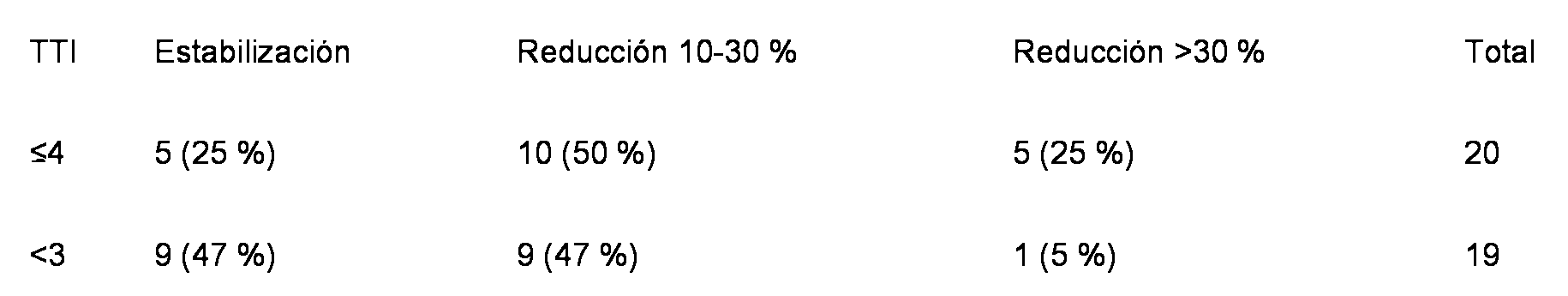
EJEMPLO 3: Uso de tomograffa por emision de positrones (PET) para el diagnostico por imagenesEXAMPLE 3: Use of positron emission tomography (PET) for diagnostic imaging
[0099] De manera similar al uso del diagnostico por imagenes planares descrito anteriormente, o imagenes SPECT (tomograffa computarizada por emision de foton unico), tambien se podna utilizar la tomograffa por emision de positrones (PET) para el diagnostico por imagenes. El diagnostico por imagenes PET proporciona datos cuantitativos y directos que reflejan la captacion del primer agente aglutinante. El diagnostico por imagenes PET se puede llevar a cabo con un agente emisor de positrones, como 124Yodo, 89zirconio, 86itrio, u otros agentes emisores de positrones conocidos por los expertos en la materia, unidos al primer agente aglutinante. Al igual que se ha descrito anteriormente, los "valores estandar de captacion" (SUV) cuantitativos derivados de PET permiten la identificacion de pacientes cuyas lesiones muestran una captacion mas elevada, frente a aquellas con una captacion menor o nula. Se podna predecir que los pacientes con lesiones que muestren SUV mas altos muestran una captacion mas elevada del segundo agente aglutinante terapeutico, seguido, a su vez, por un aumento de la probabilidad de aportar un beneficio terapeutico. El uso de una modalidad de diagnostico por imagenes directamente cuantitativo, como PET, obvia la necesidad de calificar visualmente la captacion (por ejemplo, 0-3+) o de calcular el TTI tal como se ha descrito anteriormente. Al comparar los SUV de pacientes que no responden ni se benefician del segundo agente terapeutico dirigido con los SUV de pacientes que sf responden o se benefician del segundo agente terapeutico dirigido, un experto en la materia puede determinar un umbral por debajo del cual el tratamiento con el segundo agente terapeutico tiene poco valor en comparacion con los pacientes con SUV por encima del umbral. [0099] Similar to the use of the above-described planar imaging, or SPECT (single photon emission computed tomography) images, positron emission tomography (PET) imaging could also be used for image diagnosis. The diagnosis by PET images provides quantitative and direct data that reflect the uptake of the first binding agent. The diagnosis by PET images can be carried out with a positron-emitting agent, such as sodium, zirconium, titanium, or other positron-emitting agents known to those skilled in the art, linked to the first binding agent. As described above, the quantitative "standard values of uptake" (SUV) derived from PET allow the identification of patients whose lesions show a higher uptake, compared to those with less or no uptake. It could be predicted that patients with lesions that show higher SUVs show a higher uptake of the second therapeutic binder, followed, in turn, by an increased likelihood of providing a therapeutic benefit. The use of a directly quantitative imaging modality, such as PET, obviates the need to visually qualify the capture (for example, 0-3 +) or calculate the TTI as described above. When comparing SUVs of patients who do not respond or benefit from the second targeted therapeutic agent with the SUV of patients who respond to or benefit from the second targeted therapeutic agent, a person skilled in the art may determine a threshold below which treatment with the second therapeutic agent has little value compared to patients with SUV above the threshold.
Claims (13)
Applications Claiming Priority (2)
| Application Number | Priority Date | Filing Date | Title |
|---|---|---|---|
| US15313209P | 2009-02-17 | 2009-02-17 | |
| PCT/US2010/024475 WO2010096486A1 (en) | 2009-02-17 | 2010-02-17 | Methods and kits for diagnosis of cancer and prediction of therapeutic value |
Publications (1)
| Publication Number | Publication Date |
|---|---|
| ES2712732T3 true ES2712732T3 (en) | 2019-05-14 |
Family
ID=42314780
Family Applications (1)
| Application Number | Title | Priority Date | Filing Date |
|---|---|---|---|
| ES10705709T Active ES2712732T3 (en) | 2009-02-17 | 2010-02-17 | Methods and kits for the diagnosis of cancer and the prediction of therapeutic value |
Country Status (6)
| Country | Link |
|---|---|
| US (3) | US10517969B2 (en) |
| EP (2) | EP3495000A1 (en) |
| JP (1) | JP5906090B2 (en) |
| CA (1) | CA2752510C (en) |
| ES (1) | ES2712732T3 (en) |
| WO (1) | WO2010096486A1 (en) |
Families Citing this family (16)
| Publication number | Priority date | Publication date | Assignee | Title |
|---|---|---|---|---|
| AU2008289108C1 (en) | 2007-08-17 | 2024-05-23 | Purdue Research Foundation | PSMA binding ligand-linker conjugates and methods for using |
| ES2712732T3 (en) | 2009-02-17 | 2019-05-14 | Cornell Res Foundation Inc | Methods and kits for the diagnosis of cancer and the prediction of therapeutic value |
| BR112012012887A2 (en) * | 2009-12-02 | 2017-05-02 | Imaginab Inc | nucleotide sequence-coded minibody and cys-diabody (cysdb), their use and methods for diagnosing and treating cancer associated with psma expression in a subject. |
| US9951324B2 (en) | 2010-02-25 | 2018-04-24 | Purdue Research Foundation | PSMA binding ligand-linker conjugates and methods for using |
| EP2817629A4 (en) * | 2012-02-24 | 2016-01-13 | Univ Cornell | Elevated psma identifies lethal prostate cancers |
| CA2865774A1 (en) * | 2012-02-28 | 2013-09-06 | Cornell University | Psma as a biomarker for androgen activity in prostate cancer |
| KR20150104092A (en) | 2012-11-15 | 2015-09-14 | 엔도사이트, 인코포레이티드 | Conjugates for treating diseases caused by psma expressing cells |
| SG11201505477TA (en) | 2013-01-14 | 2015-08-28 | Molecular Insight Pharm Inc | Triazine based radiopharmaceuticals and radioimaging agents |
| JP2017500537A (en) * | 2013-10-18 | 2017-01-05 | モレキュラ インサイト ファーマシューティカルズ インコーポレイテッド | Method of using SPECT / CT analysis to determine cancer stage |
| EP3415489A1 (en) | 2013-10-18 | 2018-12-19 | Deutsches Krebsforschungszentrum | Labeled inhibitors of prostate specific membrane antigen (psma), their use as imaging agents and pharmaceutical agents for the treatment of prostate cancer |
| WO2016016329A1 (en) | 2014-07-29 | 2016-02-04 | Vrije Universiteit Brussel | Radio-labelled antibody fragments for use in the prognosis, diagnosis of cancer as well as for the prediction of cancer therapy response |
| US10188759B2 (en) | 2015-01-07 | 2019-01-29 | Endocyte, Inc. | Conjugates for imaging |
| KR20180042226A (en) * | 2015-07-17 | 2018-04-25 | 브리제 유니버시타이트 브루셀 | Radioactive labeled antibody fragments for use in cancer therapy |
| US11254744B2 (en) | 2015-08-07 | 2022-02-22 | Imaginab, Inc. | Antigen binding constructs to target molecules |
| US10340046B2 (en) * | 2016-10-27 | 2019-07-02 | Progenics Pharmaceuticals, Inc. | Network for medical image analysis, decision support system, and related graphical user interface (GUI) applications |
| US11266745B2 (en) | 2017-02-08 | 2022-03-08 | Imaginab, Inc. | Extension sequences for diabodies |
Family Cites Families (251)
| Publication number | Priority date | Publication date | Assignee | Title |
|---|---|---|---|---|
| US2827461A (en) * | 1955-12-15 | 1958-03-18 | Monsanto Chemicals | Synthesis of xanthines |
| US3380956A (en) * | 1963-09-06 | 1968-04-30 | Tenneco Chem | Stabilized vinyl halide resin compositions |
| US3309338A (en) * | 1963-12-17 | 1967-03-14 | Tenneco Chem | Stabilized polyvinyl halide compositions |
| US4197209A (en) * | 1977-03-10 | 1980-04-08 | Ciba-Geigy Corporation | Lubricant compositions containing sulfur-containing esters of phosphoric acid |
| DE3113442A1 (en) * | 1981-04-03 | 1982-10-21 | Henkel KGaA, 4000 Düsseldorf | "STABILIZED POLYVINYL CHLORIDE MOLDS" |
| US5292668A (en) | 1981-12-21 | 1994-03-08 | Boston Biomedical Research Institute, Inc. | Bispecific antibody determinants |
| DE3360646D1 (en) * | 1982-03-25 | 1985-10-03 | Ciba Geigy Ag | Phosphoric-acid compounds as costabilizers for me(ii) carboxylates and/or me(ii) phenolates in polyvinyl chloride |
| DE3212336C1 (en) * | 1982-04-02 | 1983-12-29 | Skw Trostberg Ag, 8223 Trostberg | Means and methods for heat and light stabilization of polyvinyl chloride |
| US5525338A (en) * | 1992-08-21 | 1996-06-11 | Immunomedics, Inc. | Detection and therapy of lesions with biotin/avidin conjugates |
| US5869620A (en) | 1986-09-02 | 1999-02-09 | Enzon, Inc. | Multivalent antigen-binding proteins |
| US5260203A (en) | 1986-09-02 | 1993-11-09 | Enzon, Inc. | Single polypeptide chain binding molecules |
| FR2604092B1 (en) | 1986-09-19 | 1990-04-13 | Immunotech Sa | IMMUNOREACTIVES FOR TARGETING ANIMAL CELLS FOR VISUALIZATION OR DESTRUCTION IN VIVO |
| JP2682859B2 (en) | 1987-07-27 | 1997-11-26 | コモンウェルス サイエンティフィック アンド インダストリアル リサーチ オーガニゼーション | Receptor membrane |
| EP0307358B1 (en) * | 1987-09-11 | 1992-02-12 | Ciba-Geigy Ag | 1,3-diketones containing ethers or thioethers and their use as stabilising agents in chlorinated polymers |
| US4943525A (en) | 1987-11-02 | 1990-07-24 | Bioventures, Inc. | Simultaneous immunoassay for the determination of antigens and antibodies |
| US5869053A (en) | 1988-03-04 | 1999-02-09 | Cancer Research Campaign Technology, Ltd. | 5T4 antigen from human trophoblasts |
| US4892824A (en) | 1988-03-15 | 1990-01-09 | Synbiotics Corporation | Fast track method for producing monoclonal bi-specific immunoglobulins |
| US5120525A (en) | 1988-03-29 | 1992-06-09 | Immunomedics, Inc. | Radiolabeled antibody cytotoxic therapy of cancer |
| US6010902A (en) | 1988-04-04 | 2000-01-04 | Bristol-Meyers Squibb Company | Antibody heteroconjugates and bispecific antibodies for use in regulation of lymphocyte activity |
| US5851527A (en) | 1988-04-18 | 1998-12-22 | Immunomedics, Inc. | Method for antibody targeting of therapeutic agents |
| US5162504A (en) | 1988-06-03 | 1992-11-10 | Cytogen Corporation | Monoclonal antibodies to a new antigenic marker in epithelial prostatic cells and serum of prostatic cancer patients |
| ATE102631T1 (en) | 1988-11-11 | 1994-03-15 | Medical Res Council | CLONING OF IMMUNOGLOBULIN SEQUENCES FROM THE VARIABLE DOMAINS. |
| US5530101A (en) | 1988-12-28 | 1996-06-25 | Protein Design Labs, Inc. | Humanized immunoglobulins |
| DE3920358A1 (en) | 1989-06-22 | 1991-01-17 | Behringwerke Ag | BISPECIFIC AND OLIGO-SPECIFIC, MONO- AND OLIGOVALENT ANTI-BODY CONSTRUCTS, THEIR PRODUCTION AND USE |
| US5980896A (en) | 1989-06-30 | 1999-11-09 | Bristol-Myers Squibb Company | Antibodies reactive with human carcinomas |
| US5332567A (en) | 1989-08-24 | 1994-07-26 | Immunomedics | Detection and treatment of infections with immunoconjugates |
| US5208020A (en) | 1989-10-25 | 1993-05-04 | Immunogen Inc. | Cytotoxic agents comprising maytansinoids and their therapeutic use |
| DE3941902C1 (en) * | 1989-12-19 | 1991-02-28 | Chemische Werke Muenchen Otto Baerlocher Gmbh, 8000 Muenchen, De | |
| GB8928874D0 (en) | 1989-12-21 | 1990-02-28 | Celltech Ltd | Humanised antibodies |
| US5582996A (en) | 1990-12-04 | 1996-12-10 | The Wistar Institute Of Anatomy & Biology | Bifunctional antibodies and method of preparing same |
| US6197298B1 (en) | 1991-04-19 | 2001-03-06 | Tanox, Inc. | Modified binding molecules specific for T lymphocytes and their use as in vivo immune modulators in animals |
| DE4118120A1 (en) | 1991-06-03 | 1992-12-10 | Behringwerke Ag | TETRAVALENT BISPECIFIC RECEPTORS, THEIR PRODUCTION AND USE |
| US5637481A (en) | 1993-02-01 | 1997-06-10 | Bristol-Myers Squibb Company | Expression vectors encoding bispecific fusion proteins and methods of producing biologically active bispecific fusion proteins in a mammalian cell |
| DE4134982A1 (en) | 1991-10-23 | 1993-04-29 | Kernforschungsz Karlsruhe | USE OF ANTIBODY-CONTAINING PREPARATIONS FOR IMMUNE SUPPRESSION |
| US5932448A (en) | 1991-11-29 | 1999-08-03 | Protein Design Labs., Inc. | Bispecific antibody heterodimers |
| DE4140142A1 (en) | 1991-12-05 | 1993-06-09 | Boehringer Mannheim Gmbh, 6800 Mannheim, De | MULTIVALENT DEXTRANREAGENT FOR USE IN PRECIPITATION TESTS |
| EP0618970A1 (en) | 1991-12-10 | 1994-10-12 | Dana Farber Cancer Institute | Reactive neutralizing human anti-gp120 recombinant antibody, dna coding the same and use thereof |
| FR2686899B1 (en) | 1992-01-31 | 1995-09-01 | Rhone Poulenc Rorer Sa | NOVEL BIOLOGICALLY ACTIVE POLYPEPTIDES, THEIR PREPARATION AND PHARMACEUTICAL COMPOSITIONS CONTAINING THEM. |
| WO1993016185A2 (en) | 1992-02-06 | 1993-08-19 | Creative Biomolecules, Inc. | Biosynthetic binding protein for cancer marker |
| US5965132A (en) | 1992-03-05 | 1999-10-12 | Board Of Regents, The University Of Texas System | Methods and compositions for targeting the vasculature of solid tumors |
| ES2149768T3 (en) | 1992-03-25 | 2000-11-16 | Immunogen Inc | CONJUGATES OF BINDING AGENTS OF CELLS DERIVED FROM CC-1065. |
| US6096289A (en) | 1992-05-06 | 2000-08-01 | Immunomedics, Inc. | Intraoperative, intravascular, and endoscopic tumor and lesion detection, biopsy and therapy |
| EP1550729B1 (en) | 1992-09-25 | 2009-05-27 | Avipep Pty Limited | Target binding polypeptide comprising an IG-like VL domain linked to an IG-like VH domain |
| US20080152586A1 (en) | 1992-09-25 | 2008-06-26 | Avipep Pty Limited | High avidity polyvalent and polyspecific reagents |
| WO1994008038A1 (en) | 1992-10-02 | 1994-04-14 | Trustees Of Dartmouth College | Bispecific reagents for redirected targeting of low density lipoprotein |
| EP0627932B1 (en) | 1992-11-04 | 2002-05-08 | City Of Hope | Antibody construct |
| US7070782B1 (en) | 1992-11-05 | 2006-07-04 | Sloan-Kettering Institute For Cancer Research | Prostate-specific membrane antigen |
| US6953668B1 (en) | 1992-11-05 | 2005-10-11 | Sloan-Kettering Institute For Cancer Research | Prostate-specific membrane antigen |
| US7105159B1 (en) | 1992-11-05 | 2006-09-12 | Sloan-Kettering Institute For Cancer Research | Antibodies to prostate-specific membrane antigen |
| WO1994009820A1 (en) | 1992-11-05 | 1994-05-11 | Sloan-Kettering Institute For Cancer Research | Prostate-specific membrane antigen |
| AU5670194A (en) | 1992-11-20 | 1994-06-22 | Enzon, Inc. | Linker for linked fusion polypeptides |
| US5376249A (en) | 1992-11-25 | 1994-12-27 | Perseptive Biosystems, Inc. | Analysis utilizing isoelectric focusing |
| AU690528B2 (en) | 1992-12-04 | 1998-04-30 | Medical Research Council | Multivalent and multispecific binding proteins, their manufacture and use |
| GB9225453D0 (en) | 1992-12-04 | 1993-01-27 | Medical Res Council | Binding proteins |
| ATE187494T1 (en) | 1992-12-11 | 1999-12-15 | Dow Chemical Co | MULTIVALENT SINGLE CHAIN ANTIBODIES |
| US5861156A (en) | 1993-01-08 | 1999-01-19 | Creative Biomolecules | Methods of delivering agents to target cells |
| US5705614A (en) | 1993-04-09 | 1998-01-06 | Chiron Corporation | Methods of producing antigen forks |
| ATE166095T1 (en) * | 1993-04-16 | 1998-05-15 | Ciba Geigy Ag | STABILIZED POLYVINYL CHLORIDE |
| ZA946765B (en) | 1993-09-02 | 1996-02-15 | Dartmouth College | Methods of prolonged suppression of humoral immunity |
| US5869049A (en) | 1993-09-02 | 1999-02-09 | Trustees Of Dartmouth College | Methods of inducing T cell unresponsiveness to bone marrow with gp39 antagonists |
| US6569432B1 (en) | 1995-02-24 | 2003-05-27 | Sloan-Kettering Institute For Cancer Research | Prostate-specific membrane antigen and uses thereof |
| US5935818A (en) | 1995-02-24 | 1999-08-10 | Sloan-Kettering Institute For Cancer Research | Isolated nucleic acid molecule encoding alternatively spliced prostate-specific membrane antigen and uses thereof |
| GB9324807D0 (en) | 1993-12-03 | 1994-01-19 | Cancer Res Campaign Tech | Tumour antibody |
| DE4421391C1 (en) | 1994-06-18 | 1995-11-30 | Gsf Forschungszentrum Umwelt | Use of antibodies against T cells for prolonged immunosuppression |
| US5559099A (en) | 1994-09-08 | 1996-09-24 | Genvec, Inc. | Penton base protein and methods of using same |
| US5541087A (en) | 1994-09-14 | 1996-07-30 | Fuji Immunopharmaceuticals Corporation | Expression and export technology of proteins as immunofusins |
| US5688690A (en) | 1994-09-16 | 1997-11-18 | The Wistar Institute Of Anatomy And Biology | Human cytotoxic lymphocyte signal transduction surface protein (P38) and monoclonal antibodies thereto |
| CA2212846A1 (en) | 1995-02-24 | 1996-08-29 | Sloan-Kettering Institute For Cancer Research | Prostate-specific membrane antigen and uses thereof |
| US5731168A (en) | 1995-03-01 | 1998-03-24 | Genentech, Inc. | Method for making heteromultimeric polypeptides |
| GB9504344D0 (en) | 1995-03-03 | 1995-04-19 | Unilever Plc | Antibody fragment production |
| US6121022A (en) | 1995-04-14 | 2000-09-19 | Genentech, Inc. | Altered polypeptides with increased half-life |
| US5747035A (en) | 1995-04-14 | 1998-05-05 | Genentech, Inc. | Polypeptides with increased half-life for use in treating disorders involving the LFA-1 receptor |
| US6096871A (en) | 1995-04-14 | 2000-08-01 | Genentech, Inc. | Polypeptides altered to contain an epitope from the Fc region of an IgG molecule for increased half-life |
| AUPO591797A0 (en) | 1997-03-27 | 1997-04-24 | Commonwealth Scientific And Industrial Research Organisation | High avidity polyvalent and polyspecific reagents |
| US5773292A (en) | 1995-06-05 | 1998-06-30 | Cornell University | Antibodies binding portions, and probes recognizing an antigen of prostate epithelial cells but not antigens circulating in the blood |
| US5830478A (en) | 1995-06-07 | 1998-11-03 | Boston Biomedical Research Institute | Method for delivering functional domains of diphtheria toxin to a cellular target |
| TW411352B (en) * | 1995-06-30 | 2000-11-11 | Witco Vinyl Additives Gmbh | Composition of containing a stabilised halogen-containing polymer |
| EP1327658A1 (en) * | 1995-10-13 | 2003-07-16 | Crompton Vinyl Additives GmbH | Stabiliser combinations for chlorinated polymers |
| US5989830A (en) | 1995-10-16 | 1999-11-23 | Unilever Patent Holdings Bv | Bifunctional or bivalent antibody fragment analogue |
| JP2001501801A (en) | 1995-10-19 | 2001-02-13 | ブリストル−マイヤーズ スクイッブ カンパニー | Monoclonal antibody BR110 and use thereof |
| US20040253246A1 (en) | 1996-02-23 | 2004-12-16 | Israeli Ron S. | Prostate-specific membrane antigen and uses thereof |
| ES2260788T3 (en) | 1996-03-25 | 2006-11-01 | Medarex, Inc. | SPECIFIC MONOCLONAL ANTIBODIES FOR THE SPECIFIC MEMBRANE ANTIGEN ESTRACELLULAR DOMAIN OF THE PROSTATE. |
| US6962981B1 (en) | 1996-03-25 | 2005-11-08 | Medarex, Inc. | Monoclonal antibodies specific for the extracellular domain of prostate-specific membrane antigen |
| US6150508A (en) | 1996-03-25 | 2000-11-21 | Northwest Biotherapeutics, Inc. | Monoclonal antibodies specific for the extracellular domain of prostate-specific membrane antigen |
| US6107090A (en) | 1996-05-06 | 2000-08-22 | Cornell Research Foundation, Inc. | Treatment and diagnosis of prostate cancer with antibodies to extracellur PSMA domains |
| US6136311A (en) | 1996-05-06 | 2000-10-24 | Cornell Research Foundation, Inc. | Treatment and diagnosis of cancer |
| JP2000503773A (en) | 1996-05-23 | 2000-03-28 | ユニリーバー・ナームローゼ・ベンノートシヤープ | Improved specific binding assays |
| JP2000515735A (en) | 1996-07-03 | 2000-11-28 | ジェネンテック インコーポレーテッド | Hepatocyte growth factor receptor agonist |
| GB9712818D0 (en) | 1996-07-08 | 1997-08-20 | Cambridge Antibody Tech | Labelling and selection of specific binding molecules |
| US5922845A (en) | 1996-07-11 | 1999-07-13 | Medarex, Inc. | Therapeutic multispecific compounds comprised of anti-Fcα receptor antibodies |
| MY114466A (en) * | 1996-09-25 | 2002-10-31 | Crompton Vinyl Additives Gmbh | Rigid pvc stabilised with n, n-dimethyl-6-aminouracils |
| PT932417E (en) | 1996-10-17 | 2003-07-31 | Immunomedics Inc | NAX ANTIGENIC TOXIN CONJUGATE AND SYSTEM FUSEO PROTEIN INTERIORIZATION RECEIVER |
| EP2295988A2 (en) | 1996-12-31 | 2011-03-16 | High Throughput Genomics, Inc. | Multiplexed molecular analysis apparatus and its fabrication method |
| US6541212B2 (en) | 1997-03-10 | 2003-04-01 | The Regents Of The University Of California | Methods for detecting prostate stem cell antigen protein |
| CA2286366C (en) | 1997-04-03 | 2008-10-07 | Universite Laval | Transgenic expression in genital tract and sexual accessory glands |
| GB2339430A (en) | 1997-05-21 | 2000-01-26 | Biovation Ltd | Method for the production of non-immunogenic proteins |
| AU8296098A (en) | 1997-07-08 | 1999-02-08 | Board Of Regents, The University Of Texas System | Compositions and methods for homoconjugates of antibodies which induce growth arrest or apoptosis of tumor cells |
| US6977074B2 (en) | 1997-07-10 | 2005-12-20 | Mannkind Corporation | Method of inducing a CTL response |
| US6994851B1 (en) | 1997-07-10 | 2006-02-07 | Mannkind Corporation | Method of inducing a CTL response |
| ATE353864T1 (en) | 1997-09-03 | 2007-03-15 | Immunomedics Inc | FLUORATION OF PROTEINS AND PEPTIDES FOR POSITRONEMISSION TOMOGRAPHY |
| US6030792A (en) | 1997-11-13 | 2000-02-29 | Pfizer Inc | Assays for measurement of protein fragments in biological media |
| US5972615A (en) * | 1998-01-21 | 1999-10-26 | Urocor, Inc. | Biomarkers and targets for diagnosis, prognosis and management of prostate disease |
| US6709844B1 (en) | 2000-11-16 | 2004-03-23 | Mannkind Corporation | Avoidance of undesirable replication intermediates in plasmid propagation |
| DE19911329A1 (en) | 1998-03-27 | 2000-09-21 | Benes Ivan Friedrich | Radioimmunoconjugate which can be used in human therapy and process for its preparation |
| CZ121599A3 (en) | 1998-04-09 | 1999-10-13 | Aventis Pharma Deutschland Gmbh | Single-chain molecule binding several antigens, process of its preparation and medicament in which the molecule is comprised |
| US20030175884A1 (en) | 2001-08-03 | 2003-09-18 | Pablo Umana | Antibody glycosylation variants having increased antibody-dependent cellular cytotoxicity |
| ES2434961T5 (en) | 1998-04-20 | 2018-01-18 | Roche Glycart Ag | Antibody glycosylation engineering to improve antibody-dependent cell cytotoxicity |
| US6200765B1 (en) | 1998-05-04 | 2001-03-13 | Pacific Northwest Cancer Foundation | Non-invasive methods to detect prostate cancer |
| US6071490A (en) | 1998-05-07 | 2000-06-06 | Immunomedics, Inc. | Position emission tomography using gallium-68 chelates |
| AU742626B2 (en) | 1998-05-20 | 2002-01-10 | Immunomedics Inc. | Therapeutics using a bispecific anti-HLA class II invariant chain X anti-pathogen antibody |
| US20030149531A1 (en) | 2000-12-06 | 2003-08-07 | Hubert Rene S. | Serpentine transmembrane antigens expressed in human cancers and uses thereof |
| US20060052321A1 (en) | 2002-04-05 | 2006-03-09 | Raitano Arthur B | Nucleic acid and corresponding protein entitled 98P4B6 useful in treatment and detection of cancer |
| US6833438B1 (en) | 1999-06-01 | 2004-12-21 | Agensys, Inc. | Serpentine transmembrane antigens expressed in human cancers and uses thereof |
| DK1086223T3 (en) | 1998-06-01 | 2009-11-30 | Agensys Inc | Novel serpentine transmembrane antigens expressed in human cancers and their applications |
| US20040141975A1 (en) | 1998-06-01 | 2004-07-22 | Raitano Arthur B. | Nucleic acid and corresponding protein entitled 98P4B6 useful in treatment and detection of cancer |
| ES2199496T3 (en) * | 1998-06-02 | 2004-02-16 | Crompton Vinyl Additives Gmbh | CIANACETILURES FOR THE STABILIZATION OF POLYMERS WITH A HALOGEN CONTENT. |
| DE59903953D1 (en) * | 1998-06-26 | 2003-02-13 | Crompton Vinyl Additives Gmbh | New NH2-modified 6-aminouracils as stabilizers for halogen-containing polymers |
| ES2188202T3 (en) * | 1998-07-13 | 2003-06-16 | Univ Texas | TREATMENT METHODS FOR CANCER USING THERAPEUTIC CONJUGATES THAT LINK WITH AMINOPHOSPHOLIPIDS. |
| WO2000014234A1 (en) | 1998-09-08 | 2000-03-16 | Urocor, Inc. | Prostate specific promoter and regulation of gene expression |
| US6361774B1 (en) | 1999-09-17 | 2002-03-26 | Immunomedics, Inc. | Methods and compositions for increasing the target-specific toxicity of a chemotherapy drug |
| WO2000018888A1 (en) | 1998-09-29 | 2000-04-06 | The Uab Research Foundation | Immunomodulation by genetic modification of dendritic cells and b-cells |
| CN1202128C (en) | 1998-12-08 | 2005-05-18 | 拜奥威神有限公司 | Method for reducing immunogenicity of proteins |
| AU2051600A (en) | 1998-12-10 | 2000-06-26 | Board Of Regents, The University Of Texas System | Compositions and methods of modulating cholesterol metabolism |
| EP1175616A4 (en) | 1999-04-13 | 2003-08-13 | Northwest Biotherapeutics Inc | Methods for the diagnosis and treatment of metastatic prostate tumors |
| JP4926320B2 (en) | 1999-04-28 | 2012-05-09 | ボード・オブ・リージエンツ,ザ・ユニバーシテイ・オブ・テキサス・システム | Compositions and methods for cancer treatment by selective inhibition of VEGF |
| DE19927978A1 (en) * | 1999-06-18 | 2000-12-21 | Basf Ag | New esters of cyclohexane-1,3- and -1,4-dicarboxylic acid, useful as toxicologically acceptable plasticizers, e.g. for PVC, obtained by hydrogenation of corresponding isophthalate and terephthalate esters |
| WO2001009303A2 (en) | 1999-07-30 | 2001-02-08 | Vical Inc. | Flt-3 LIGAND-ENCODING POLYNUCLEOTIDE AS A POLYNUCLEOTIDE-BASED VACCINE ENHANCER |
| KR20050004240A (en) | 1999-08-31 | 2005-01-12 | 제넨테크, 인크. | Compositions and Methods for the Treatment of Tumor |
| US6342587B1 (en) | 1999-10-22 | 2002-01-29 | Ludwig Institute For Cancer Research | A33 antigen specific immunoglobulin products and uses thereof |
| US6824780B1 (en) | 1999-10-29 | 2004-11-30 | Genentech, Inc. | Anti-tumor antibody compositions and methods of use |
| EP1242401B1 (en) | 1999-11-24 | 2006-12-27 | Immunogen, Inc. | Cytotoxic agents comprising taxanes and their therapeutic use |
| US20020048777A1 (en) * | 1999-12-06 | 2002-04-25 | Shujath Ali | Method of diagnosing monitoring, staging, imaging and treating prostate cancer |
| CN1423700A (en) | 2000-03-24 | 2003-06-11 | 麦克美特股份公司 | Multifunctional polypeptides comprising a binding site to and epitope of the NKG2D receptor complex |
| US6573096B1 (en) | 2000-04-01 | 2003-06-03 | The Research Foundation At State University Of New York | Compositions and methods for inhibition of cancer invasion and angiogenesis |
| MXPA02010011A (en) | 2000-04-11 | 2003-04-25 | Genentech Inc | Multivalent antibodies and uses therefor. |
| US6861234B1 (en) | 2000-04-28 | 2005-03-01 | Mannkind Corporation | Method of epitope discovery |
| ATE474931T1 (en) | 2000-04-28 | 2010-08-15 | Mannkind Corp | EPITOPE SYNCHRONIZATION IN ANTIGEN-PRESENTING CELLS |
| US6375991B1 (en) | 2000-09-08 | 2002-04-23 | Albemarle Corporation | Production of concentrated biocidal solutions |
| DE10045591A1 (en) | 2000-09-15 | 2002-04-04 | Klaus Pfizenmaier | Site-specific, antibody-mediated activation of proapoptotic cytokines - AMAIZe (Antibody-Mediated Apoptosis Inducing Cytokines) |
| CN1537170A (en) * | 2000-11-20 | 2004-10-13 | ����ҽѧԺ | Methods and devices for quantitative detection of prostate specific membrane antigen and other prostatic markers |
| US7468354B2 (en) * | 2000-12-01 | 2008-12-23 | Genspera, Inc. | Tissue specific prodrugs |
| CA2429498C (en) * | 2000-12-13 | 2009-09-08 | Crompton Vinyl Additives Gmbh | Stabilizer system for stabilizing halogen-containing polymers |
| US6972324B2 (en) | 2001-05-18 | 2005-12-06 | Boehringer Ingelheim Pharmaceuticals, Inc. | Antibodies specific for CD44v6 |
| US7514078B2 (en) * | 2001-06-01 | 2009-04-07 | Cornell Research Foundation, Inc. | Methods of treating prostate cancer with anti-prostate specific membrane antigen antibodies |
| EP1392360B1 (en) | 2001-06-01 | 2011-11-30 | Cornell Research Foundation, Inc. | Modified antibodies to prostate-specific membrane antigen and uses thereof |
| US7666414B2 (en) | 2001-06-01 | 2010-02-23 | Cornell Research Foundation, Inc. | Methods for treating prostate cancer using modified antibodies to prostate-specific membrane antigen |
| EP2287186B1 (en) | 2001-09-06 | 2014-12-31 | Agensys, Inc. | Nucleic acid and corresponding protein entitled STEAP-1 useful in treatment and detection of cancer |
| US7494646B2 (en) | 2001-09-06 | 2009-02-24 | Agensys, Inc. | Antibodies and molecules derived therefrom that bind to STEAP-1 proteins |
| CA2452288A1 (en) | 2001-09-20 | 2003-03-27 | Cornell Research Foundation, Inc. | Methods and compositions for treating and preventing skin disorders using binding agents specific for psma |
| AU2002356844C1 (en) * | 2001-10-23 | 2010-03-04 | Amgen Fremont Inc. | PSMA antibodies and protein multimers |
| KR100988949B1 (en) | 2001-10-25 | 2010-10-20 | 제넨테크, 인크. | Glycoprotein compositions |
| GB0126378D0 (en) | 2001-11-02 | 2002-01-02 | Oxford Biomedica Ltd | Antigen |
| DK1453471T3 (en) | 2001-11-07 | 2011-03-28 | Mannkind Corp | Expression vectors encoding epitopes of antigens and method of their construction |
| US20040018519A1 (en) | 2001-11-16 | 2004-01-29 | Wright ,Jr. George L | Methods and devices for quantitative detection of prostate specific membrane antigen and other prostatic markers |
| WO2003062375A2 (en) | 2001-12-19 | 2003-07-31 | Genentech, Inc. | Stabilizing polypeptides which have been exposed to urea |
| EP1472275B1 (en) | 2002-02-05 | 2008-12-17 | Genentech, Inc. | Protein purification |
| AU2003208882A1 (en) | 2002-02-28 | 2003-09-09 | Ciba Specialty Chemicals Holding Inc. | Process for the preparation of cationic azo compounds |
| EP1499353A4 (en) | 2002-04-15 | 2006-04-05 | Human Genome Sciences Inc | Antibodies that specifically bind to tl5 |
| EP1501369B1 (en) | 2002-04-26 | 2015-06-24 | Genentech, Inc. | Non-affinity purification of proteins |
| AU2003278161A1 (en) | 2002-06-21 | 2004-01-06 | Johns Hopkins University School Of Medicine | Membrane associated tumor endothelium markers |
| US9809654B2 (en) | 2002-09-27 | 2017-11-07 | Vaccinex, Inc. | Targeted CD1d molecules |
| US7820166B2 (en) | 2002-10-11 | 2010-10-26 | Micromet Ag | Potent T cell modulating molecules |
| US9701754B1 (en) | 2002-10-23 | 2017-07-11 | City Of Hope | Covalent disulfide-linked diabodies and uses thereof |
| AU2003293556A1 (en) * | 2003-01-10 | 2004-08-10 | Millennium Pharmaceuticals, Inc. | Methods of diagnosing and treating cancer |
| CN103540600B (en) | 2003-01-22 | 2017-12-01 | 罗氏格黎卡特股份公司 | Fusion constructs and its for producing the purposes for the antibody that Fc receptor binding affinities and effector function improve |
| WO2004088279A2 (en) | 2003-03-28 | 2004-10-14 | Iowa State University Research Foundation, Inc. | Allosteric probes and methods |
| GB0308988D0 (en) | 2003-04-17 | 2003-05-28 | Univ Singapore | Molecule |
| US7595379B2 (en) | 2003-05-30 | 2009-09-29 | Agensys, Inc. | Antibodies and related molecules that bind to PSCA proteins |
| US7541442B2 (en) | 2003-05-30 | 2009-06-02 | Agensys, Inc. | Antibodies and related molecules that bind to PSCA proteins |
| DK2319524T3 (en) | 2003-05-30 | 2013-11-11 | Agensys Inc | Prostate stem cell antigen (PSCA) variants and sub-sequences thereof |
| WO2004106380A2 (en) | 2003-05-31 | 2004-12-09 | Micromet Ag | Human-anti-human cd3 binding molecules |
| US20050163782A1 (en) | 2003-06-27 | 2005-07-28 | Biogen Idec Ma Inc. | Modified binding molecules comprising connecting peptides |
| EP3722309A1 (en) | 2003-07-28 | 2020-10-14 | Genentech, Inc. | Reducing protein a leaching during protein a affinity chromatography |
| GB0321805D0 (en) | 2003-09-18 | 2003-10-15 | Univ Wales Medicine | Human tumour growth patterns |
| US7524813B2 (en) | 2003-10-10 | 2009-04-28 | Novo Nordisk Health Care Ag | Selectively conjugated peptides and methods of making the same |
| NZ546173A (en) | 2003-10-16 | 2009-04-30 | Micromet Ag | Multispecific deimmunized CD3-binders |
| EP2633866A3 (en) | 2003-10-17 | 2013-12-18 | Novo Nordisk A/S | Combination therapy |
| ITPD20030264A1 (en) | 2003-10-30 | 2005-04-30 | Xeptagen Spa | HIGHLY SPECIFIC DIAGNOSIS METHOD FOR NEOPLASIA |
| EP1725249B1 (en) | 2003-11-06 | 2014-01-08 | Seattle Genetics, Inc. | Monomethylvaline compounds capable of conjugation to ligands |
| US7235641B2 (en) | 2003-12-22 | 2007-06-26 | Micromet Ag | Bispecific antibodies |
| WO2005068616A2 (en) | 2004-01-16 | 2005-07-28 | Fraunhofer Gesellschaft zur Förderung der angewandten Forschung e.V. | Immunokinases |
| SI1716178T1 (en) | 2004-02-16 | 2010-11-30 | Micromet Ag | Less immunogenic binding molecules |
| JP5064037B2 (en) | 2004-02-23 | 2012-10-31 | ジェネンテック, インコーポレイテッド | Heterocyclic self-destructive linkers and conjugates |
| WO2005094882A1 (en) | 2004-03-03 | 2005-10-13 | Millennium Pharmaceuticals, Inc. | Modified antibodies to prostate-specific membrane antigen and uses thereof |
| EP1740954B1 (en) | 2004-04-07 | 2015-08-19 | Genentech, Inc. | Mass spectrometry of antibody conjugates |
| US8008442B2 (en) | 2004-04-22 | 2011-08-30 | Agensys, Inc. | Antibodies and molecules derived therefrom that bind to STEAP-1 proteins |
| JP5234734B2 (en) | 2004-06-01 | 2013-07-10 | ジェネンテック, インコーポレイテッド | Antibody-drug conjugates and methods |
| WO2005118802A2 (en) | 2004-06-03 | 2005-12-15 | The Regents Of The University Of California | Targeting pseudotyped retroviral vectors |
| US20060159689A1 (en) | 2004-06-17 | 2006-07-20 | Chih-Sheng Chiang | Combinations of tumor-associated antigens in diagnostics for various types of cancers |
| WO2006008096A1 (en) | 2004-07-16 | 2006-01-26 | Micromet Ag | Expression-enhanced polypeptides |
| US20100111856A1 (en) | 2004-09-23 | 2010-05-06 | Herman Gill | Zirconium-radiolabeled, cysteine engineered antibody conjugates |
| ES2669510T3 (en) | 2004-09-23 | 2018-05-28 | Genentech, Inc. | Antibodies and conjugates engineered with cysteine |
| US7947839B2 (en) | 2004-12-01 | 2011-05-24 | Genentech, Inc. | Heterocyclic-substituted bis-1,8 naphthalimide compounds, antibody drug conjugates, and methods of use |
| US20070134243A1 (en) | 2004-12-01 | 2007-06-14 | Gazzard Lewis J | Antibody drug conjugates and methods |
| CA2491067A1 (en) | 2004-12-24 | 2006-06-24 | Stichting Katholieke Universiteit | Mrna rations in urinary sediments and/or urine as a prognostic marker for prostate cancer |
| EP1871810A2 (en) | 2005-04-08 | 2008-01-02 | Cytogen Corporation | Conjugated anti-psma antibodies |
| EP1726650A1 (en) * | 2005-05-27 | 2006-11-29 | Universitätsklinikum Freiburg | Monoclonal antibodies and single chain antibody fragments against cell-surface prostate specific membrane antigen |
| GB2426581A (en) | 2005-05-27 | 2006-11-29 | Univ Nottingham | Immunoassay methods |
| DK2298815T3 (en) | 2005-07-25 | 2015-06-15 | Emergent Product Dev Seattle | B-CELL REDUCTION USING CD37 SPECIFIC AND CD20 SPECIFIC BINDING MOLECULES |
| US20080279850A1 (en) | 2005-07-25 | 2008-11-13 | Trubion Pharmaceuticals, Inc. | B-Cell Reduction Using CD37-Specific and CD20-Specific Binding Molecules |
| US7612181B2 (en) | 2005-08-19 | 2009-11-03 | Abbott Laboratories | Dual variable domain immunoglobulin and uses thereof |
| US20090226465A1 (en) | 2005-10-31 | 2009-09-10 | Jackson David Y | Macrocyclic depsipeptide antibody-drug conjugates and methods |
| US20070212331A1 (en) | 2006-03-07 | 2007-09-13 | Baldassare Joseph J | Methods and compositions for selectively killing cells |
| EP1996716B1 (en) | 2006-03-20 | 2011-05-11 | The Regents of the University of California | Engineered anti-prostate stem cell antigen (psca) antibodies for cancer targeting |
| AU2007226861A1 (en) | 2006-03-21 | 2007-09-27 | Wyeth | Methods and compositions for antagonism of RAGE |
| CA2648099C (en) | 2006-03-31 | 2012-05-29 | The Brigham And Women's Hospital, Inc | System for targeted delivery of therapeutic agents |
| US7566276B2 (en) | 2006-04-14 | 2009-07-28 | Dogleg Right Corporation | Multi-piece putter head having an insert |
| US20110052697A1 (en) | 2006-05-17 | 2011-03-03 | Gwangju Institute Of Science & Technology | Aptamer-Directed Drug Delivery |
| CN101632020B (en) | 2006-09-13 | 2013-11-27 | 昂西免疫有限公司 | Improved immunoassay methods |
| US20100215581A1 (en) | 2006-10-25 | 2010-08-26 | Koninklijke Philips Electronics N.V. | Contrast agents for detecting prostate cancer |
| US8362217B2 (en) | 2006-12-21 | 2013-01-29 | Emd Millipore Corporation | Purification of proteins |
| WO2008079302A2 (en) | 2006-12-21 | 2008-07-03 | Millipore Corporation | Purification of proteins |
| CA2682605A1 (en) | 2007-04-18 | 2008-10-30 | Zymogenetics, Inc. | Single chain fc, methods of making and methods of treatment |
| NZ581395A (en) | 2007-05-14 | 2012-08-31 | Biogen Idec Inc | Single-chain fc (scfc) regions, binding polypeptides comprising same, and methods related thereto |
| WO2009017823A2 (en) | 2007-08-01 | 2009-02-05 | The Government Of The United States Of America, As Represented By The Secretary, Department Of Health Of Human Services, National Institutes Of Health | A fold-back diabody diphtheria toxin immunotoxin and methods of use |
| WO2009032949A2 (en) | 2007-09-04 | 2009-03-12 | The Regents Of The University Of California | High affinity anti-prostate stem cell antigen (psca) antibodies for cancer targeting and detection |
| EP2197908A2 (en) | 2007-09-27 | 2010-06-23 | Dako Denmark A/S | Mhc multimers in tuberculosis diagnostics, vaccine and therapeutics |
| ES2608604T3 (en) | 2007-10-18 | 2017-04-12 | Bavarian Nordic A/S | Use of MVA to treat prostate cancer |
| EP2215122A4 (en) | 2007-10-25 | 2011-03-09 | Viventia Biotech Inc | Antibodies against a cancer-associated epitope of variant hnrnpg and uses thereof |
| US9107858B2 (en) | 2007-12-05 | 2015-08-18 | Wisconsin Alumni Research Foundation | Dendritic cell targeting compositions and uses thereof |
| US8603474B2 (en) | 2008-01-29 | 2013-12-10 | Ludwig Institute For Cancer Research, Ltd. | Membrane transporter NaPi2b (SCL34A1) epitope for antibody therapy, antibodies directed thereto, and target for cancer therapy |
| US8043830B2 (en) | 2008-02-01 | 2011-10-25 | The Regents Of The University Of California | Biotin-ligase system for secretion of biotinylated protein |
| EP2240495B1 (en) | 2008-02-01 | 2015-07-15 | Genentech, Inc. | Nemorubicin metabolite and analog reagents, antibody-drug conjugates and methods |
| TW200947474A (en) | 2008-05-05 | 2009-11-16 | Ren-Huan Pan | Manufacturing equipment and method of quadratic arc alignment dua-turret formation center, and miniature milliohm current sensors manufactured thereof |
| WO2009140242A1 (en) | 2008-05-13 | 2009-11-19 | Genentech, Inc. | Analysis of antibody drug conjugates by bead-based affinity capture and mass spectrometry |
| KR101667062B1 (en) | 2008-07-15 | 2016-10-17 | 제넨테크, 인크. | Anthracycline derivative conjugates, process for their preparation and their use as antitumor compounds |
| US20100069616A1 (en) | 2008-08-06 | 2010-03-18 | The Regents Of The University Of California | Engineered antibody-nanoparticle conjugates |
| WO2010027629A2 (en) | 2008-09-08 | 2010-03-11 | Conocophillips Company | System for incondensable component separation in a liquefied natural gas facility |
| US20110318380A1 (en) | 2008-10-01 | 2011-12-29 | Dako Denmark A/S | MHC Multimers in Cancer Vaccines and Immune Monitoring |
| US20110207155A1 (en) | 2008-10-13 | 2011-08-25 | Xeptagen Spa | Method for the preparation of immunoconjugates and use thereof |
| CA2742324A1 (en) | 2008-10-30 | 2010-06-03 | Caris Life Sciences Luxembourg Holdings, S.A.R.L. | Methods for assessing rna patterns |
| CA2743211A1 (en) | 2008-11-12 | 2010-05-20 | Caris Life Sciences Luxembourg Holdings, S.A.R.L. | Methods and systems of using exosomes for determining phenotypes |
| WO2011109440A1 (en) | 2010-03-01 | 2011-09-09 | Caris Life Sciences Luxembourg Holdings | Biomarkers for theranostics |
| BRPI0921006A2 (en) | 2008-11-13 | 2015-12-15 | Emergent Product Dev Seattle | cd37 combined immunotherapeutic therapies and uses of these |
| WO2010074702A1 (en) | 2008-12-16 | 2010-07-01 | Millipore Corporation | Purification of proteins |
| US8424854B2 (en) | 2008-12-25 | 2013-04-23 | Tokai Rubber Industries, Ltd. | Fluid-filled type vibration damping device |
| CN106995495A (en) | 2009-01-12 | 2017-08-01 | 希托马克斯医疗有限责任公司 | Modified antibodies composition and its preparation and application |
| ES2712732T3 (en) | 2009-02-17 | 2019-05-14 | Cornell Res Foundation Inc | Methods and kits for the diagnosis of cancer and the prediction of therapeutic value |
| US20120004289A1 (en) | 2009-03-06 | 2012-01-05 | The Johns Hopkins University | Annexin a11 and associated genes as biomarkers for cancer |
| WO2010111388A2 (en) | 2009-03-24 | 2010-09-30 | Biocept, Inc. | Devices and methods of cell capture and analysis |
| CN102459346B (en) | 2009-04-27 | 2016-10-26 | 昂考梅德药品有限公司 | The method manufacturing heteromultimers molecule |
| JP5919188B2 (en) | 2009-05-28 | 2016-05-18 | グラクソ グループ リミテッドGlaxo Group Limited | IL-13 binding protein |
| KR101962476B1 (en) | 2009-07-03 | 2019-03-26 | 아비펩 피티와이 리미티트 | Immuno-conjugates and methods for producing them |
| WO2011003171A1 (en) | 2009-07-08 | 2011-01-13 | Smart Technologies Ulc | Three-dimensional widget manipulation on a multi-touch panel |
| TWM383852U (en) | 2009-07-13 | 2010-07-01 | Speedtech Corp | An improvement of the universal serial bus connector |
| CN102573925B (en) | 2009-10-05 | 2015-08-05 | 佳能株式会社 | The acousto-optic imaging method of photoacoustic imaging contrast agent and the described photoacoustic imaging contrast agent of use |
| BR112012012887A2 (en) | 2009-12-02 | 2017-05-02 | Imaginab Inc | nucleotide sequence-coded minibody and cys-diabody (cysdb), their use and methods for diagnosing and treating cancer associated with psma expression in a subject. |
| CA2784610C (en) | 2009-12-23 | 2020-07-14 | Avipep Pty Ltd | Immuno-conjugates and methods for producing them |
| EP3112382A1 (en) | 2009-12-29 | 2017-01-04 | Emergent Product Development Seattle, LLC | Heterodimer binding proteins and uses thereof |
| US9812638B2 (en) | 2010-03-19 | 2017-11-07 | Globalfoundries Inc. | Backend of line (BEOL) compatible high current density access device for high density arrays of electronic components |
| US20120144110A1 (en) | 2010-12-02 | 2012-06-07 | Lsi Corporation | Methods and structure for storage migration using storage array managed server agents |
-
2010
- 2010-02-17 ES ES10705709T patent/ES2712732T3/en active Active
- 2010-02-17 US US12/658,881 patent/US10517969B2/en active Active
- 2010-02-17 CA CA2752510A patent/CA2752510C/en active Active
- 2010-02-17 WO PCT/US2010/024475 patent/WO2010096486A1/en active Application Filing
- 2010-02-17 EP EP18208648.8A patent/EP3495000A1/en not_active Withdrawn
- 2010-02-17 EP EP10705709.3A patent/EP2398504B1/en active Active
- 2010-02-17 JP JP2011551190A patent/JP5906090B2/en active Active
-
2019
- 2019-10-15 US US16/653,655 patent/US20200282085A1/en not_active Abandoned
-
2022
- 2022-01-19 US US17/579,470 patent/US20220323618A1/en active Pending
Also Published As
| Publication number | Publication date |
|---|---|
| US10517969B2 (en) | 2019-12-31 |
| US20220323618A1 (en) | 2022-10-13 |
| EP3495000A1 (en) | 2019-06-12 |
| US20200282085A1 (en) | 2020-09-10 |
| JP2012518187A (en) | 2012-08-09 |
| CA2752510C (en) | 2024-01-23 |
| JP5906090B2 (en) | 2016-04-20 |
| CA2752510A1 (en) | 2010-08-26 |
| US20100209343A1 (en) | 2010-08-19 |
| EP2398504A1 (en) | 2011-12-28 |
| WO2010096486A1 (en) | 2010-08-26 |
| EP2398504B1 (en) | 2018-11-28 |
Similar Documents
| Publication | Publication Date | Title |
|---|---|---|
| ES2712732T3 (en) | Methods and kits for the diagnosis of cancer and the prediction of therapeutic value | |
| ES2363891T3 (en) | ANTIBODIES AGAINST THE ANTIGEN OF TRONCAL CELLS OF THE PROSTATE (PSCA) GENETICALLY MODIFIED FOR ADDRESSING TO CANCER. | |
| US20220001043A1 (en) | Cd8 imaging constructs and methods of use thereof | |
| BR112020000766A2 (en) | anti-cd166 antibodies and their uses | |
| US9694061B2 (en) | Fully human antibodies to high molecular weight-melanoma associated antigen and uses thereof | |
| US20090311174A1 (en) | Method For Treating Cancer | |
| CN101675158A (en) | Cancerous desease modifying antibodies | |
| ES2899360T3 (en) | Compositions and methods for detecting and treating gastric cancer | |
| ES2732504T3 (en) | Procedures and agents for the diagnosis and treatment of hepatocellular carcinoma | |
| US11427648B2 (en) | Anti-CD146 antibodies and uses thereof | |
| CN114423459A (en) | DKK-1 inhibitors for the treatment of cancer | |
| ES2639227T3 (en) | Anti S100A7 antibodies for the treatment and diagnosis of cancer | |
| JP2017214308A (en) | Radioactive pharmaceutical composition, and radiolabeled antibody screening method | |
| JP2013504547A (en) | Selective detection of bone metastases in renal clear cell carcinoma | |
| KR101380057B1 (en) | Diagnostic agent and therapeutic agent for pancreatic cancer | |
| CA3163098A1 (en) | Methods of imaging using multiple imaging agents | |
| AU2009305715B8 (en) | Fully human antibodies to high molecular weight-melanoma associated antigen and uses thereof | |
| Korsen | Molecular Imaging and Radioimmunotherapy Targeting Delta-Like Ligand 3 in Neuroendocrine Prostate Cancer | |
| MICAD Research Team | 99mTc-anti-EGFR monoclonal antibody h-R3 |