EP4194548A1 - Generation of human pluripotent stem cell derived functional beta cells showing a glucose-dependent mitochondrial respiration and two-phase insulin secretion response - Google Patents
Generation of human pluripotent stem cell derived functional beta cells showing a glucose-dependent mitochondrial respiration and two-phase insulin secretion response Download PDFInfo
- Publication number
- EP4194548A1 EP4194548A1 EP22213072.6A EP22213072A EP4194548A1 EP 4194548 A1 EP4194548 A1 EP 4194548A1 EP 22213072 A EP22213072 A EP 22213072A EP 4194548 A1 EP4194548 A1 EP 4194548A1
- Authority
- EP
- European Patent Office
- Prior art keywords
- cells
- stage
- inhibitor
- glucose
- medium
- Prior art date
- Legal status (The legal status is an assumption and is not a legal conclusion. Google has not performed a legal analysis and makes no representation as to the accuracy of the status listed.)
- Pending
Links
- WQZGKKKJIJFFOK-GASJEMHNSA-N Glucose Natural products OC[C@H]1OC(O)[C@H](O)[C@@H](O)[C@@H]1O WQZGKKKJIJFFOK-GASJEMHNSA-N 0.000 title claims abstract description 165
- 210000001778 pluripotent stem cell Anatomy 0.000 title claims abstract description 91
- 239000008103 glucose Substances 0.000 title claims abstract description 71
- 230000003914 insulin secretion Effects 0.000 title claims abstract description 46
- 230000006540 mitochondrial respiration Effects 0.000 title claims abstract description 34
- 230000001419 dependent effect Effects 0.000 title claims abstract description 27
- 210000000227 basophil cell of anterior lobe of hypophysis Anatomy 0.000 title claims description 137
- 230000004044 response Effects 0.000 title claims description 32
- 238000000034 method Methods 0.000 claims abstract description 136
- 101000578254 Homo sapiens Homeobox protein Nkx-6.1 Proteins 0.000 claims abstract description 107
- 101000578258 Homo sapiens Homeobox protein Nkx-6.2 Proteins 0.000 claims abstract description 106
- 101000979205 Homo sapiens Transcription factor MafA Proteins 0.000 claims abstract description 89
- 230000004069 differentiation Effects 0.000 claims abstract description 81
- 102100041030 Pancreas/duodenum homeobox protein 1 Human genes 0.000 claims abstract description 74
- 230000009996 pancreatic endocrine effect Effects 0.000 claims abstract description 69
- 101000939387 Homo sapiens Urocortin-3 Proteins 0.000 claims abstract description 66
- 102100029794 Urocortin-3 Human genes 0.000 claims abstract description 66
- 102100028096 Homeobox protein Nkx-6.2 Human genes 0.000 claims abstract 4
- 101000971533 Homo sapiens Killer cell lectin-like receptor subfamily G member 1 Proteins 0.000 claims abstract 4
- 102100021457 Killer cell lectin-like receptor subfamily G member 1 Human genes 0.000 claims abstract 4
- 101710183548 Pyridoxal 5'-phosphate synthase subunit PdxS Proteins 0.000 claims abstract 4
- OGQSCIYDJSNCMY-UHFFFAOYSA-H iron(3+);methyl-dioxido-oxo-$l^{5}-arsane Chemical compound [Fe+3].[Fe+3].C[As]([O-])([O-])=O.C[As]([O-])([O-])=O.C[As]([O-])([O-])=O OGQSCIYDJSNCMY-UHFFFAOYSA-H 0.000 claims abstract 4
- 210000004027 cell Anatomy 0.000 claims description 1020
- 239000003112 inhibitor Substances 0.000 claims description 178
- NOESYZHRGYRDHS-UHFFFAOYSA-N insulin Chemical compound N1C(=O)C(NC(=O)C(CCC(N)=O)NC(=O)C(CCC(O)=O)NC(=O)C(C(C)C)NC(=O)C(NC(=O)CN)C(C)CC)CSSCC(C(NC(CO)C(=O)NC(CC(C)C)C(=O)NC(CC=2C=CC(O)=CC=2)C(=O)NC(CCC(N)=O)C(=O)NC(CC(C)C)C(=O)NC(CCC(O)=O)C(=O)NC(CC(N)=O)C(=O)NC(CC=2C=CC(O)=CC=2)C(=O)NC(CSSCC(NC(=O)C(C(C)C)NC(=O)C(CC(C)C)NC(=O)C(CC=2C=CC(O)=CC=2)NC(=O)C(CC(C)C)NC(=O)C(C)NC(=O)C(CCC(O)=O)NC(=O)C(C(C)C)NC(=O)C(CC(C)C)NC(=O)C(CC=2NC=NC=2)NC(=O)C(CO)NC(=O)CNC2=O)C(=O)NCC(=O)NC(CCC(O)=O)C(=O)NC(CCCNC(N)=N)C(=O)NCC(=O)NC(CC=3C=CC=CC=3)C(=O)NC(CC=3C=CC=CC=3)C(=O)NC(CC=3C=CC(O)=CC=3)C(=O)NC(C(C)O)C(=O)N3C(CCC3)C(=O)NC(CCCCN)C(=O)NC(C)C(O)=O)C(=O)NC(CC(N)=O)C(O)=O)=O)NC(=O)C(C(C)CC)NC(=O)C(CO)NC(=O)C(C(C)O)NC(=O)C1CSSCC2NC(=O)C(CC(C)C)NC(=O)C(NC(=O)C(CCC(N)=O)NC(=O)C(CC(N)=O)NC(=O)C(NC(=O)C(N)CC=1C=CC=CC=1)C(C)C)CC1=CN=CN1 NOESYZHRGYRDHS-UHFFFAOYSA-N 0.000 claims description 176
- CIWBSHSKHKDKBQ-JLAZNSOCSA-N Ascorbic acid Chemical compound OC[C@H](O)[C@H]1OC(=O)C(O)=C1O CIWBSHSKHKDKBQ-JLAZNSOCSA-N 0.000 claims description 116
- 238000012258 culturing Methods 0.000 claims description 111
- 102000004877 Insulin Human genes 0.000 claims description 88
- 108090001061 Insulin Proteins 0.000 claims description 88
- 229940125396 insulin Drugs 0.000 claims description 88
- 210000004039 endoderm cell Anatomy 0.000 claims description 78
- PWKSKIMOESPYIA-BYPYZUCNSA-N L-N-acetyl-Cysteine Chemical compound CC(=O)N[C@@H](CS)C(O)=O PWKSKIMOESPYIA-BYPYZUCNSA-N 0.000 claims description 64
- 239000007788 liquid Substances 0.000 claims description 60
- 235000010323 ascorbic acid Nutrition 0.000 claims description 55
- 229960005070 ascorbic acid Drugs 0.000 claims description 55
- 239000011668 ascorbic acid Substances 0.000 claims description 55
- 239000000725 suspension Substances 0.000 claims description 55
- 108091006296 SLC2A1 Proteins 0.000 claims description 51
- 150000001875 compounds Chemical class 0.000 claims description 51
- 102100028071 Fibroblast growth factor 7 Human genes 0.000 claims description 50
- FOORCIAZMIWALX-ULJHMMPZSA-N (z)-n-(4-benzylpiperazin-1-yl)-1-(3,5-dimethyl-1-phenylpyrazol-4-yl)methanimine Chemical compound CC1=NN(C=2C=CC=CC=2)C(C)=C1\C=N/N(CC1)CCN1CC1=CC=CC=C1 FOORCIAZMIWALX-ULJHMMPZSA-N 0.000 claims description 48
- 101001060261 Homo sapiens Fibroblast growth factor 7 Proteins 0.000 claims description 43
- -1 A366 Chemical compound 0.000 claims description 41
- HTTJABKRGRZYRN-UHFFFAOYSA-N Heparin Chemical compound OC1C(NC(=O)C)C(O)OC(COS(O)(=O)=O)C1OC1C(OS(O)(=O)=O)C(O)C(OC2C(C(OS(O)(=O)=O)C(OC3C(C(O)C(O)C(O3)C(O)=O)OS(O)(=O)=O)C(CO)O2)NS(O)(=O)=O)C(C(O)=O)O1 HTTJABKRGRZYRN-UHFFFAOYSA-N 0.000 claims description 40
- 229960002897 heparin Drugs 0.000 claims description 39
- 229920000669 heparin Polymers 0.000 claims description 39
- 239000002243 precursor Substances 0.000 claims description 36
- 102100039939 Growth/differentiation factor 8 Human genes 0.000 claims description 33
- 239000000203 mixture Substances 0.000 claims description 32
- OGNYUTNQZVRGMN-UHFFFAOYSA-N ZM447439 Chemical compound N1=CN=C2C=C(OCCCN3CCOCC3)C(OC)=CC2=C1NC(C=C1)=CC=C1NC(=O)C1=CC=CC=C1 OGNYUTNQZVRGMN-UHFFFAOYSA-N 0.000 claims description 24
- SHGAZHPCJJPHSC-YCNIQYBTSA-N all-trans-retinoic acid Chemical compound OC(=O)\C=C(/C)\C=C\C=C(/C)\C=C\C1=C(C)CCCC1(C)C SHGAZHPCJJPHSC-YCNIQYBTSA-N 0.000 claims description 24
- 238000000338 in vitro Methods 0.000 claims description 24
- 229930002330 retinoic acid Natural products 0.000 claims description 24
- 229960001727 tretinoin Drugs 0.000 claims description 24
- 210000002438 upper gastrointestinal tract Anatomy 0.000 claims description 23
- 239000012190 activator Substances 0.000 claims description 22
- 229960004308 acetylcysteine Drugs 0.000 claims description 21
- 210000001647 gastrula Anatomy 0.000 claims description 17
- 238000009472 formulation Methods 0.000 claims description 14
- 230000000638 stimulation Effects 0.000 claims description 14
- 229940125373 Gamma-Secretase Inhibitor Drugs 0.000 claims description 13
- 239000003540 gamma secretase inhibitor Substances 0.000 claims description 13
- RNAMYOYQYRYFQY-UHFFFAOYSA-N 2-(4,4-difluoropiperidin-1-yl)-6-methoxy-n-(1-propan-2-ylpiperidin-4-yl)-7-(3-pyrrolidin-1-ylpropoxy)quinazolin-4-amine Chemical compound N1=C(N2CCC(F)(F)CC2)N=C2C=C(OCCCN3CCCC3)C(OC)=CC2=C1NC1CCN(C(C)C)CC1 RNAMYOYQYRYFQY-UHFFFAOYSA-N 0.000 claims description 12
- SHRCVZJKZJGIHQ-UHFFFAOYSA-N 2-chloro-n-[4-[4-[(2-chloroacetyl)amino]phenyl]sulfonylphenyl]acetamide Chemical compound C1=CC(NC(=O)CCl)=CC=C1S(=O)(=O)C1=CC=C(NC(=O)CCl)C=C1 SHRCVZJKZJGIHQ-UHFFFAOYSA-N 0.000 claims description 12
- TXZPMHLMPKIUGK-UHFFFAOYSA-N 2-methoxy-N-(3-methyl-2-oxo-1,4-dihydroquinazolin-6-yl)benzenesulfonamide Chemical compound COC1=CC=CC=C1S(=O)(=O)NC1=CC=C(NC(=O)N(C)C2)C2=C1 TXZPMHLMPKIUGK-UHFFFAOYSA-N 0.000 claims description 12
- NGAGMBNBKCDCDJ-UHFFFAOYSA-N 8-phenyl-2-(1-piperazinyl)-1-benzopyran-4-one Chemical compound C1=CC=C2C(=O)C=C(N3CCNCC3)OC2=C1C1=CC=CC=C1 NGAGMBNBKCDCDJ-UHFFFAOYSA-N 0.000 claims description 12
- DZTGIRNXWSZBIM-UHFFFAOYSA-N chembl3086883 Chemical compound C1=C(O)C(C)=CC(N=NC=2C=CC(=CC=2)S(=O)(=O)NC=2N=CC=CC=2)=C1N DZTGIRNXWSZBIM-UHFFFAOYSA-N 0.000 claims description 12
- 230000036284 oxygen consumption Effects 0.000 claims description 12
- RYYNGWLOYLRZLK-RBUKOAKNSA-N pf03814735 Chemical compound C1([C@H]2CC[C@@H](C1=CC=1)N2C(=O)CNC(=O)C)=CC=1NC(N=1)=NC=C(C(F)(F)F)C=1NC1CCC1 RYYNGWLOYLRZLK-RBUKOAKNSA-N 0.000 claims description 12
- QOECJCJVIMVJGX-UHFFFAOYSA-N 2-cyclohexyl-6-methoxy-N-(1-propan-2-yl-4-piperidinyl)-7-[3-(1-pyrrolidinyl)propoxy]-4-quinazolinamine Chemical compound N1=C(C2CCCCC2)N=C2C=C(OCCCN3CCCC3)C(OC)=CC2=C1NC1CCN(C(C)C)CC1 QOECJCJVIMVJGX-UHFFFAOYSA-N 0.000 claims description 11
- QQDIFLSJMFDTCQ-UHFFFAOYSA-N MC1568 Chemical compound CN1C(C=CC(=O)NO)=CC=C1C=CC(=O)C1=CC=CC(F)=C1 QQDIFLSJMFDTCQ-UHFFFAOYSA-N 0.000 claims description 11
- 230000028327 secretion Effects 0.000 claims description 11
- PTJGLFIIZFVFJV-UHFFFAOYSA-N N'-hydroxy-N-(3-pyridinyl)octanediamide Chemical compound ONC(=O)CCCCCCC(=O)NC1=CC=CN=C1 PTJGLFIIZFVFJV-UHFFFAOYSA-N 0.000 claims description 10
- WZPBZJONDBGPKJ-VEHQQRBSSA-N aztreonam Chemical compound O=C1N(S([O-])(=O)=O)[C@@H](C)[C@@H]1NC(=O)C(=N/OC(C)(C)C(O)=O)\C1=CSC([NH3+])=N1 WZPBZJONDBGPKJ-VEHQQRBSSA-N 0.000 claims description 8
- 108010056852 Myostatin Proteins 0.000 claims description 7
- OUKWLRHRXOPODD-UHFFFAOYSA-N N-(1-cyclohexyl-4-piperidinyl)-6-methoxy-7-[3-(1-piperidinyl)propoxy]-2-(4-propan-2-yl-1,4-diazepan-1-yl)-4-quinazolinamine Chemical compound N1=C(N2CCN(CCC2)C(C)C)N=C2C=C(OCCCN3CCCCC3)C(OC)=CC2=C1NC(CC1)CCN1C1CCCCC1 OUKWLRHRXOPODD-UHFFFAOYSA-N 0.000 claims description 6
- VAZAPHZUAVEOMC-UHFFFAOYSA-N tacedinaline Chemical compound C1=CC(NC(=O)C)=CC=C1C(=O)NC1=CC=CC=C1N VAZAPHZUAVEOMC-UHFFFAOYSA-N 0.000 claims description 6
- 102000058063 Glucose Transporter Type 1 Human genes 0.000 claims 3
- FGUUSXIOTUKUDN-IBGZPJMESA-N C1(=CC=CC=C1)N1C2=C(NC([C@H](C1)NC=1OC(=NN=1)C1=CC=CC=C1)=O)C=CC=C2 Chemical compound C1(=CC=CC=C1)N1C2=C(NC([C@H](C1)NC=1OC(=NN=1)C1=CC=CC=C1)=O)C=CC=C2 FGUUSXIOTUKUDN-IBGZPJMESA-N 0.000 claims 2
- GNFTZDOKVXKIBK-UHFFFAOYSA-N 3-(2-methoxyethoxy)benzohydrazide Chemical compound COCCOC1=CC=CC(C(=O)NN)=C1 GNFTZDOKVXKIBK-UHFFFAOYSA-N 0.000 claims 1
- 210000003890 endocrine cell Anatomy 0.000 abstract description 40
- 210000002237 B-cell of pancreatic islet Anatomy 0.000 abstract description 12
- 210000004153 islets of langerhan Anatomy 0.000 abstract description 8
- 239000002609 medium Substances 0.000 description 178
- 102100028098 Homeobox protein Nkx-6.1 Human genes 0.000 description 103
- 230000014509 gene expression Effects 0.000 description 90
- 102100023237 Transcription factor MafA Human genes 0.000 description 85
- 238000010790 dilution Methods 0.000 description 80
- 239000012895 dilution Substances 0.000 description 80
- 101000612089 Homo sapiens Pancreas/duodenum homeobox protein 1 Proteins 0.000 description 70
- 238000004114 suspension culture Methods 0.000 description 61
- 210000001900 endoderm Anatomy 0.000 description 59
- LBPKYPYHDKKRFS-UHFFFAOYSA-N 1,5-naphthyridine, 2-[3-(6-methyl-2-pyridinyl)-1h-pyrazol-4-yl]- Chemical compound CC1=CC=CC(C2=C(C=NN2)C=2N=C3C=CC=NC3=CC=2)=N1 LBPKYPYHDKKRFS-UHFFFAOYSA-N 0.000 description 52
- 108091005735 TGF-beta receptors Proteins 0.000 description 49
- 102000016715 Transforming Growth Factor beta Receptors Human genes 0.000 description 49
- 102100023536 Solute carrier family 2, facilitated glucose transporter member 1 Human genes 0.000 description 48
- 239000002953 phosphate buffered saline Substances 0.000 description 47
- UIIMBOGNXHQVGW-DEQYMQKBSA-M Sodium bicarbonate-14C Chemical compound [Na+].O[14C]([O-])=O UIIMBOGNXHQVGW-DEQYMQKBSA-M 0.000 description 46
- 239000001963 growth medium Substances 0.000 description 43
- 108090000623 proteins and genes Proteins 0.000 description 43
- 102000004169 proteins and genes Human genes 0.000 description 38
- 235000018102 proteins Nutrition 0.000 description 36
- 238000011282 treatment Methods 0.000 description 34
- 150000003384 small molecules Chemical class 0.000 description 33
- 239000000758 substrate Substances 0.000 description 29
- 230000035800 maturation Effects 0.000 description 28
- 230000003389 potentiating effect Effects 0.000 description 28
- CDOVNWNANFFLFJ-UHFFFAOYSA-N 4-[6-[4-(1-piperazinyl)phenyl]-3-pyrazolo[1,5-a]pyrimidinyl]quinoline Chemical compound C1CNCCN1C1=CC=C(C2=CN3N=CC(=C3N=C2)C=2C3=CC=CC=C3N=CC=2)C=C1 CDOVNWNANFFLFJ-UHFFFAOYSA-N 0.000 description 27
- 101000886562 Homo sapiens Growth/differentiation factor 8 Proteins 0.000 description 26
- 239000002356 single layer Substances 0.000 description 24
- NWONKYPBYAMBJT-UHFFFAOYSA-L zinc sulfate Chemical compound [Zn+2].[O-]S([O-])(=O)=O NWONKYPBYAMBJT-UHFFFAOYSA-L 0.000 description 24
- 229910000368 zinc sulfate Inorganic materials 0.000 description 24
- 102000051325 Glucagon Human genes 0.000 description 22
- 108060003199 Glucagon Proteins 0.000 description 22
- 102000003923 Protein Kinase C Human genes 0.000 description 22
- 108090000315 Protein Kinase C Proteins 0.000 description 22
- MASNOZXLGMXCHN-ZLPAWPGGSA-N glucagon Chemical compound C([C@@H](C(=O)N[C@H](C(=O)N[C@@H](CCC(N)=O)C(=O)N[C@@H](CC=1C2=CC=CC=C2NC=1)C(=O)N[C@@H](CC(C)C)C(=O)N[C@@H](CCSC)C(=O)N[C@@H](CC(N)=O)C(=O)N[C@@H]([C@@H](C)O)C(O)=O)C(C)C)NC(=O)[C@H](CC(O)=O)NC(=O)[C@H](CCC(N)=O)NC(=O)[C@H](C)NC(=O)[C@H](CCCNC(N)=N)NC(=O)[C@H](CCCNC(N)=N)NC(=O)[C@H](CO)NC(=O)[C@H](CC(O)=O)NC(=O)[C@H](CC(C)C)NC(=O)[C@H](CC=1C=CC(O)=CC=1)NC(=O)[C@H](CCCCN)NC(=O)[C@H](CO)NC(=O)[C@H](CC=1C=CC(O)=CC=1)NC(=O)[C@H](CC(O)=O)NC(=O)[C@H](CO)NC(=O)[C@@H](NC(=O)[C@H](CC=1C=CC=CC=1)NC(=O)[C@@H](NC(=O)CNC(=O)[C@H](CCC(N)=O)NC(=O)[C@H](CO)NC(=O)[C@@H](N)CC=1NC=NC=1)[C@@H](C)O)[C@@H](C)O)C1=CC=CC=C1 MASNOZXLGMXCHN-ZLPAWPGGSA-N 0.000 description 22
- 229960004666 glucagon Drugs 0.000 description 22
- DAEPDZWVDSPTHF-UHFFFAOYSA-M sodium pyruvate Chemical compound [Na+].CC(=O)C([O-])=O DAEPDZWVDSPTHF-UHFFFAOYSA-M 0.000 description 22
- 102100032063 Neurogenic differentiation factor 1 Human genes 0.000 description 21
- 239000005557 antagonist Substances 0.000 description 20
- 210000000130 stem cell Anatomy 0.000 description 19
- IAZDPXIOMUYVGZ-UHFFFAOYSA-N Dimethylsulphoxide Chemical compound CS(C)=O IAZDPXIOMUYVGZ-UHFFFAOYSA-N 0.000 description 18
- 210000001671 embryonic stem cell Anatomy 0.000 description 18
- 239000011686 zinc sulphate Substances 0.000 description 18
- 102000013380 Smoothened Receptor Human genes 0.000 description 17
- 230000003750 conditioning effect Effects 0.000 description 17
- 238000010899 nucleation Methods 0.000 description 17
- 230000008410 smoothened signaling pathway Effects 0.000 description 17
- 102000004190 Enzymes Human genes 0.000 description 16
- 108090000790 Enzymes Proteins 0.000 description 16
- 238000004113 cell culture Methods 0.000 description 16
- 229940088598 enzyme Drugs 0.000 description 16
- 229940121372 histone deacetylase inhibitor Drugs 0.000 description 16
- 239000003276 histone deacetylase inhibitor Substances 0.000 description 16
- 108010038447 Chromogranin A Proteins 0.000 description 15
- 102000010792 Chromogranin A Human genes 0.000 description 15
- 230000015572 biosynthetic process Effects 0.000 description 15
- 238000005516 engineering process Methods 0.000 description 15
- 239000006144 Dulbecco’s modified Eagle's medium Substances 0.000 description 14
- 101150079937 NEUROD1 gene Proteins 0.000 description 14
- 108700020297 NeuroD Proteins 0.000 description 14
- 239000003550 marker Substances 0.000 description 14
- 235000013619 trace mineral Nutrition 0.000 description 14
- 239000011573 trace mineral Substances 0.000 description 14
- 101000687905 Homo sapiens Transcription factor SOX-2 Proteins 0.000 description 13
- 102100024270 Transcription factor SOX-2 Human genes 0.000 description 13
- 108010023082 activin A Proteins 0.000 description 13
- 230000002124 endocrine Effects 0.000 description 13
- 230000001965 increasing effect Effects 0.000 description 13
- 230000002438 mitochondrial effect Effects 0.000 description 13
- 102000004887 Transforming Growth Factor beta Human genes 0.000 description 12
- 108090001012 Transforming Growth Factor beta Proteins 0.000 description 12
- 229940125763 bromodomain inhibitor Drugs 0.000 description 12
- 230000024245 cell differentiation Effects 0.000 description 12
- 239000003234 histone lysine methyltransferase inhibitor Substances 0.000 description 12
- 239000003963 antioxidant agent Substances 0.000 description 11
- 235000006708 antioxidants Nutrition 0.000 description 11
- 238000001943 fluorescence-activated cell sorting Methods 0.000 description 11
- 102000045246 noggin Human genes 0.000 description 11
- 108700007229 noggin Proteins 0.000 description 11
- 230000008569 process Effects 0.000 description 11
- 238000011002 quantification Methods 0.000 description 11
- 229940054269 sodium pyruvate Drugs 0.000 description 11
- GVJHHUAWPYXKBD-UHFFFAOYSA-N (±)-α-Tocopherol Chemical compound OC1=C(C)C(C)=C2OC(CCCC(C)CCCC(C)CCCC(C)C)(C)CCC2=C1C GVJHHUAWPYXKBD-UHFFFAOYSA-N 0.000 description 10
- 108010083123 CDX2 Transcription Factor Proteins 0.000 description 10
- 102100031671 Homeobox protein CDX-2 Human genes 0.000 description 10
- 102000055027 Protein Methyltransferases Human genes 0.000 description 10
- 108700040121 Protein Methyltransferases Proteins 0.000 description 10
- UIIMBOGNXHQVGW-UHFFFAOYSA-M Sodium bicarbonate Chemical compound [Na+].OC([O-])=O UIIMBOGNXHQVGW-UHFFFAOYSA-M 0.000 description 10
- 238000004220 aggregation Methods 0.000 description 10
- 150000002632 lipids Chemical class 0.000 description 10
- 239000000243 solution Substances 0.000 description 10
- QSHGISMANBKLQL-OWJWWREXSA-N (2s)-2-[[2-(3,5-difluorophenyl)acetyl]amino]-n-[(7s)-5-methyl-6-oxo-7h-benzo[d][1]benzazepin-7-yl]propanamide Chemical compound N([C@@H](C)C(=O)N[C@@H]1C(N(C)C2=CC=CC=C2C2=CC=CC=C21)=O)C(=O)CC1=CC(F)=CC(F)=C1 QSHGISMANBKLQL-OWJWWREXSA-N 0.000 description 9
- NMUSYJAQQFHJEW-UHFFFAOYSA-N 5-Azacytidine Natural products O=C1N=C(N)N=CN1C1C(O)C(O)C(CO)O1 NMUSYJAQQFHJEW-UHFFFAOYSA-N 0.000 description 9
- NMUSYJAQQFHJEW-KVTDHHQDSA-N 5-azacytidine Chemical compound O=C1N=C(N)N=CN1[C@H]1[C@H](O)[C@H](O)[C@@H](CO)O1 NMUSYJAQQFHJEW-KVTDHHQDSA-N 0.000 description 9
- 102000018233 Fibroblast Growth Factor Human genes 0.000 description 9
- 108050007372 Fibroblast Growth Factor Proteins 0.000 description 9
- 102100028412 Fibroblast growth factor 10 Human genes 0.000 description 9
- 108090000379 Fibroblast growth factor 2 Proteins 0.000 description 9
- 241000283973 Oryctolagus cuniculus Species 0.000 description 9
- WCUXLLCKKVVCTQ-UHFFFAOYSA-M Potassium chloride Chemical compound [Cl-].[K+] WCUXLLCKKVVCTQ-UHFFFAOYSA-M 0.000 description 9
- 230000002776 aggregation Effects 0.000 description 9
- 230000003078 antioxidant effect Effects 0.000 description 9
- 229960002756 azacitidine Drugs 0.000 description 9
- 235000020776 essential amino acid Nutrition 0.000 description 9
- 239000003797 essential amino acid Substances 0.000 description 9
- 229940126864 fibroblast growth factor Drugs 0.000 description 9
- 229940088597 hormone Drugs 0.000 description 9
- 239000005556 hormone Substances 0.000 description 9
- 238000010186 staining Methods 0.000 description 9
- 239000000126 substance Substances 0.000 description 9
- 108091005625 BRD4 Proteins 0.000 description 8
- 102100029895 Bromodomain-containing protein 4 Human genes 0.000 description 8
- 229940126190 DNA methyltransferase inhibitor Drugs 0.000 description 8
- 102000003974 Fibroblast growth factor 2 Human genes 0.000 description 8
- 101000576323 Homo sapiens Motor neuron and pancreas homeobox protein 1 Proteins 0.000 description 8
- 229940123379 Methyltransferase inhibitor Drugs 0.000 description 8
- 102100025170 Motor neuron and pancreas homeobox protein 1 Human genes 0.000 description 8
- 241000699666 Mus <mouse, genus> Species 0.000 description 8
- FAPWRFPIFSIZLT-UHFFFAOYSA-M Sodium chloride Chemical compound [Na+].[Cl-] FAPWRFPIFSIZLT-UHFFFAOYSA-M 0.000 description 8
- 102000044880 Wnt3A Human genes 0.000 description 8
- 108700013515 Wnt3A Proteins 0.000 description 8
- 239000003719 aurora kinase inhibitor Substances 0.000 description 8
- 239000006285 cell suspension Substances 0.000 description 8
- 238000012512 characterization method Methods 0.000 description 8
- 230000008045 co-localization Effects 0.000 description 8
- 230000001143 conditioned effect Effects 0.000 description 8
- 230000003054 hormonal effect Effects 0.000 description 8
- 229940090044 injection Drugs 0.000 description 8
- 238000002347 injection Methods 0.000 description 8
- 239000007924 injection Substances 0.000 description 8
- CDAISMWEOUEBRE-UHFFFAOYSA-N inositol Chemical compound OC1C(O)C(O)C(O)C(O)C1O CDAISMWEOUEBRE-UHFFFAOYSA-N 0.000 description 8
- 239000003697 methyltransferase inhibitor Substances 0.000 description 8
- 108091006082 receptor inhibitors Proteins 0.000 description 8
- 210000002966 serum Anatomy 0.000 description 8
- ZRKFYGHZFMAOKI-QMGMOQQFSA-N tgfbeta Chemical compound C([C@H](NC(=O)[C@H](C(C)C)NC(=O)CNC(=O)[C@H](CCC(O)=O)NC(=O)[C@H](CCCNC(N)=N)NC(=O)[C@H](CC(N)=O)NC(=O)[C@H](CC(C)C)NC(=O)[C@H]([C@@H](C)O)NC(=O)[C@H](CCC(O)=O)NC(=O)[C@H]([C@@H](C)O)NC(=O)[C@H](CC(C)C)NC(=O)CNC(=O)[C@H](C)NC(=O)[C@H](CO)NC(=O)[C@H](CCC(N)=O)NC(=O)[C@@H](NC(=O)[C@H](C)NC(=O)[C@H](C)NC(=O)[C@@H](NC(=O)[C@H](CC(C)C)NC(=O)[C@@H](N)CCSC)C(C)C)[C@@H](C)CC)C(=O)N[C@@H]([C@@H](C)O)C(=O)N[C@@H](C(C)C)C(=O)N[C@@H](CC=1C=CC=CC=1)C(=O)N[C@@H](C)C(=O)N1[C@@H](CCC1)C(=O)N[C@@H]([C@@H](C)O)C(=O)N[C@@H](CC(N)=O)C(=O)N[C@@H](CCC(O)=O)C(=O)N[C@@H](C)C(=O)N[C@@H](CC=1C=CC=CC=1)C(=O)N[C@@H](CCCNC(N)=N)C(=O)N[C@@H](C)C(=O)N[C@@H](CC(C)C)C(=O)N1[C@@H](CCC1)C(=O)N1[C@@H](CCC1)C(=O)N[C@@H](CCCNC(N)=N)C(=O)N[C@@H](CCC(O)=O)C(=O)N[C@@H](CCCNC(N)=N)C(=O)N[C@@H](CO)C(=O)N[C@@H](CCCNC(N)=N)C(=O)N[C@@H](CC(C)C)C(=O)N[C@@H](CC(C)C)C(O)=O)C1=CC=C(O)C=C1 ZRKFYGHZFMAOKI-QMGMOQQFSA-N 0.000 description 8
- 210000001519 tissue Anatomy 0.000 description 8
- OMKHWTRUYNAGFG-IEBDPFPHSA-N 3-deazaneplanocin a Chemical compound C1=NC=2C(N)=NC=CC=2N1[C@@H]1C=C(CO)[C@@H](O)[C@H]1O OMKHWTRUYNAGFG-IEBDPFPHSA-N 0.000 description 7
- 229940123877 Aurora kinase inhibitor Drugs 0.000 description 7
- 102000001893 Bone Morphogenetic Protein Receptors Human genes 0.000 description 7
- 108010040422 Bone Morphogenetic Protein Receptors Proteins 0.000 description 7
- 101100518002 Danio rerio nkx2.2a gene Proteins 0.000 description 7
- 108090000385 Fibroblast growth factor 7 Proteins 0.000 description 7
- 102100025623 Gap junction delta-2 protein Human genes 0.000 description 7
- 102100036255 Glucose-6-phosphatase 2 Human genes 0.000 description 7
- 108700014808 Homeobox Protein Nkx-2.2 Proteins 0.000 description 7
- 102100027886 Homeobox protein Nkx-2.2 Human genes 0.000 description 7
- 101000930907 Homo sapiens Glucose-6-phosphatase 2 Proteins 0.000 description 7
- 101100460496 Homo sapiens NKX2-2 gene Proteins 0.000 description 7
- 108050000588 Neurogenic differentiation factor 1 Proteins 0.000 description 7
- 238000011529 RT qPCR Methods 0.000 description 7
- 239000006146 Roswell Park Memorial Institute medium Substances 0.000 description 7
- AUYYCJSJGJYCDS-LBPRGKRZSA-N Thyrolar Chemical compound IC1=CC(C[C@H](N)C(O)=O)=CC(I)=C1OC1=CC=C(O)C(I)=C1 AUYYCJSJGJYCDS-LBPRGKRZSA-N 0.000 description 7
- 102000040945 Transcription factor Human genes 0.000 description 7
- 108091023040 Transcription factor Proteins 0.000 description 7
- 239000003968 dna methyltransferase inhibitor Substances 0.000 description 7
- 210000001035 gastrointestinal tract Anatomy 0.000 description 7
- 235000002639 sodium chloride Nutrition 0.000 description 7
- 239000013589 supplement Substances 0.000 description 7
- 229940088594 vitamin Drugs 0.000 description 7
- 229930003231 vitamin Natural products 0.000 description 7
- 235000013343 vitamin Nutrition 0.000 description 7
- 239000011782 vitamin Substances 0.000 description 7
- BERLXWPRSBJFHO-UHFFFAOYSA-N 2-(5-chloro-2-fluorophenyl)-n-pyridin-4-ylpteridin-4-amine Chemical compound FC1=CC=C(Cl)C=C1C1=NC(NC=2C=CN=CC=2)=C(N=CC=N2)C2=N1 BERLXWPRSBJFHO-UHFFFAOYSA-N 0.000 description 6
- 102100032306 Aurora kinase B Human genes 0.000 description 6
- 108090000749 Aurora kinase B Proteins 0.000 description 6
- 102000007345 Chromogranins Human genes 0.000 description 6
- 108010007718 Chromogranins Proteins 0.000 description 6
- 108020004414 DNA Proteins 0.000 description 6
- 238000012413 Fluorescence activated cell sorting analysis Methods 0.000 description 6
- 108010086246 Glucagon-Like Peptide-1 Receptor Proteins 0.000 description 6
- 102100032882 Glucagon-like peptide 1 receptor Human genes 0.000 description 6
- 101000917237 Homo sapiens Fibroblast growth factor 10 Proteins 0.000 description 6
- 101001053263 Homo sapiens Insulin gene enhancer protein ISL-1 Proteins 0.000 description 6
- 102100024392 Insulin gene enhancer protein ISL-1 Human genes 0.000 description 6
- 108010032788 PAX6 Transcription Factor Proteins 0.000 description 6
- 102100037506 Paired box protein Pax-6 Human genes 0.000 description 6
- 235000001014 amino acid Nutrition 0.000 description 6
- 229940024606 amino acid Drugs 0.000 description 6
- 150000001413 amino acids Chemical class 0.000 description 6
- 210000000988 bone and bone Anatomy 0.000 description 6
- 239000003795 chemical substances by application Substances 0.000 description 6
- 102000006533 chordin Human genes 0.000 description 6
- 108010008846 chordin Proteins 0.000 description 6
- 230000002354 daily effect Effects 0.000 description 6
- 239000008187 granular material Substances 0.000 description 6
- 230000000921 morphogenic effect Effects 0.000 description 6
- 235000019195 vitamin supplement Nutrition 0.000 description 6
- 101150068520 wnt3a gene Proteins 0.000 description 6
- 229960001763 zinc sulfate Drugs 0.000 description 6
- IDDDVXIUIXWAGJ-DDSAHXNVSA-N 4-[(1r)-1-aminoethyl]-n-pyridin-4-ylcyclohexane-1-carboxamide;dihydrochloride Chemical compound Cl.Cl.C1CC([C@H](N)C)CCC1C(=O)NC1=CC=NC=C1 IDDDVXIUIXWAGJ-DDSAHXNVSA-N 0.000 description 5
- 108010051975 Glycogen Synthase Kinase 3 beta Proteins 0.000 description 5
- 102000003964 Histone deacetylase Human genes 0.000 description 5
- 108090000353 Histone deacetylase Proteins 0.000 description 5
- 102100027332 Homeobox protein SIX2 Human genes 0.000 description 5
- 101000651912 Homo sapiens Homeobox protein SIX2 Proteins 0.000 description 5
- 101001054725 Homo sapiens Inhibin beta B chain Proteins 0.000 description 5
- 101001117146 Homo sapiens [Pyruvate dehydrogenase (acetyl-transferring)] kinase isozyme 1, mitochondrial Proteins 0.000 description 5
- 102100027003 Inhibin beta B chain Human genes 0.000 description 5
- 102000052651 Pancreatic hormone Human genes 0.000 description 5
- 229930003427 Vitamin E Natural products 0.000 description 5
- 102100024148 [Pyruvate dehydrogenase (acetyl-transferring)] kinase isozyme 1, mitochondrial Human genes 0.000 description 5
- 239000000427 antigen Substances 0.000 description 5
- 108091007433 antigens Proteins 0.000 description 5
- 102000036639 antigens Human genes 0.000 description 5
- 230000002051 biphasic effect Effects 0.000 description 5
- 239000012141 concentrate Substances 0.000 description 5
- 238000011161 development Methods 0.000 description 5
- 230000018109 developmental process Effects 0.000 description 5
- 230000000694 effects Effects 0.000 description 5
- WIGCFUFOHFEKBI-UHFFFAOYSA-N gamma-tocopherol Natural products CC(C)CCCC(C)CCCC(C)CCCC1CCC2C(C)C(O)C(C)C(C)C2O1 WIGCFUFOHFEKBI-UHFFFAOYSA-N 0.000 description 5
- 210000001654 germ layer Anatomy 0.000 description 5
- 238000012606 in vitro cell culture Methods 0.000 description 5
- IMYVCWQAHSYYOO-UHFFFAOYSA-N n-[4-[(6,7-dimethoxyquinazolin-4-yl)amino]phenyl]benzamide Chemical compound C=12C=C(OC)C(OC)=CC2=NC=NC=1NC(C=C1)=CC=C1NC(=O)C1=CC=CC=C1 IMYVCWQAHSYYOO-UHFFFAOYSA-N 0.000 description 5
- 239000004025 pancreas hormone Substances 0.000 description 5
- 229940032957 pancreatic hormone Drugs 0.000 description 5
- 229910000030 sodium bicarbonate Inorganic materials 0.000 description 5
- 159000000000 sodium salts Chemical class 0.000 description 5
- 239000005495 thyroid hormone Substances 0.000 description 5
- 229940036555 thyroid hormone Drugs 0.000 description 5
- 235000019165 vitamin E Nutrition 0.000 description 5
- 229940046009 vitamin E Drugs 0.000 description 5
- 239000011709 vitamin E Substances 0.000 description 5
- YBJHBAHKTGYVGT-ZKWXMUAHSA-N (+)-Biotin Chemical compound N1C(=O)N[C@@H]2[C@H](CCCCC(=O)O)SC[C@@H]21 YBJHBAHKTGYVGT-ZKWXMUAHSA-N 0.000 description 4
- LUZOFMGZMUZSSK-LRDDRELGSA-N (-)-indolactam V Chemical compound C1[C@@H](CO)NC(=O)[C@H](C(C)C)N(C)C2=CC=CC3=C2C1=CN3 LUZOFMGZMUZSSK-LRDDRELGSA-N 0.000 description 4
- FOORCIAZMIWALX-UHFFFAOYSA-N 1-(3,5-dimethyl-1-phenyl-4-pyrazolyl)-N-[4-(phenylmethyl)-1-piperazinyl]methanimine Chemical compound CC1=NN(C=2C=CC=CC=2)C(C)=C1C=NN(CC1)CCN1CC1=CC=CC=C1 FOORCIAZMIWALX-UHFFFAOYSA-N 0.000 description 4
- NRRCQHARLPNLHJ-UHFFFAOYSA-N 4-[1-ethyl-7-[(piperidin-4-ylamino)methyl]imidazo[4,5-c]pyridin-2-yl]-1,2,5-oxadiazol-3-amine;dihydrochloride Chemical compound Cl.Cl.C=12N(CC)C(C=3C(=NON=3)N)=NC2=CN=CC=1CNC1CCNCC1 NRRCQHARLPNLHJ-UHFFFAOYSA-N 0.000 description 4
- PCCDKTWDGDFRME-UHFFFAOYSA-N 4-[6-(4-piperazin-1-ylphenyl)pyrazolo[1,5-a]pyrimidin-3-yl]quinoline;hydrochloride Chemical compound Cl.C1CNCCN1C1=CC=C(C2=CN3N=CC(=C3N=C2)C=2C3=CC=CC=C3N=CC=2)C=C1 PCCDKTWDGDFRME-UHFFFAOYSA-N 0.000 description 4
- ZAINTDRBUHCDPZ-UHFFFAOYSA-M Alexa Fluor 546 Chemical compound [H+].[Na+].CC1CC(C)(C)NC(C(=C2OC3=C(C4=NC(C)(C)CC(C)C4=CC3=3)S([O-])(=O)=O)S([O-])(=O)=O)=C1C=C2C=3C(C(=C(Cl)C=1Cl)C(O)=O)=C(Cl)C=1SCC(=O)NCCCCCC(=O)ON1C(=O)CCC1=O ZAINTDRBUHCDPZ-UHFFFAOYSA-M 0.000 description 4
- 102100033641 Bromodomain-containing protein 2 Human genes 0.000 description 4
- 102100033642 Bromodomain-containing protein 3 Human genes 0.000 description 4
- 241000283707 Capra Species 0.000 description 4
- PHEDXBVPIONUQT-UHFFFAOYSA-N Cocarcinogen A1 Natural products CCCCCCCCCCCCCC(=O)OC1C(C)C2(O)C3C=C(C)C(=O)C3(O)CC(CO)=CC2C2C1(OC(C)=O)C2(C)C PHEDXBVPIONUQT-UHFFFAOYSA-N 0.000 description 4
- 102100031181 Glyceraldehyde-3-phosphate dehydrogenase Human genes 0.000 description 4
- DHMQDGOQFOQNFH-UHFFFAOYSA-N Glycine Chemical compound NCC(O)=O DHMQDGOQFOQNFH-UHFFFAOYSA-N 0.000 description 4
- 102000019058 Glycogen Synthase Kinase 3 beta Human genes 0.000 description 4
- 102100029284 Hepatocyte nuclear factor 3-beta Human genes 0.000 description 4
- 101000871850 Homo sapiens Bromodomain-containing protein 2 Proteins 0.000 description 4
- 101000871851 Homo sapiens Bromodomain-containing protein 3 Proteins 0.000 description 4
- 101001062347 Homo sapiens Hepatocyte nuclear factor 3-beta Proteins 0.000 description 4
- 101000757216 Homo sapiens Protein arginine N-methyltransferase 1 Proteins 0.000 description 4
- 101000757232 Homo sapiens Protein arginine N-methyltransferase 2 Proteins 0.000 description 4
- 101000945096 Homo sapiens Ribosomal protein S6 kinase alpha-5 Proteins 0.000 description 4
- 108090000723 Insulin-Like Growth Factor I Proteins 0.000 description 4
- 102000004218 Insulin-Like Growth Factor I Human genes 0.000 description 4
- CSNNHWWHGAXBCP-UHFFFAOYSA-L Magnesium sulfate Chemical compound [Mg+2].[O-][S+2]([O-])([O-])[O-] CSNNHWWHGAXBCP-UHFFFAOYSA-L 0.000 description 4
- 102100031455 NAD-dependent protein deacetylase sirtuin-1 Human genes 0.000 description 4
- 102100035423 POU domain, class 5, transcription factor 1 Human genes 0.000 description 4
- 101800001268 Pancreatic hormone Proteins 0.000 description 4
- 108091000080 Phosphotransferase Proteins 0.000 description 4
- 102100022985 Protein arginine N-methyltransferase 1 Human genes 0.000 description 4
- AUNGANRZJHBGPY-SCRDCRAPSA-N Riboflavin Chemical compound OC[C@@H](O)[C@@H](O)[C@@H](O)CN1C=2C=C(C)C(C)=CC=2N=C2C1=NC(=O)NC2=O AUNGANRZJHBGPY-SCRDCRAPSA-N 0.000 description 4
- 102100033645 Ribosomal protein S6 kinase alpha-5 Human genes 0.000 description 4
- 108010041191 Sirtuin 1 Proteins 0.000 description 4
- IQFYYKKMVGJFEH-XLPZGREQSA-N Thymidine Chemical compound O=C1NC(=O)C(C)=CN1[C@@H]1O[C@H](CO)[C@@H](O)C1 IQFYYKKMVGJFEH-XLPZGREQSA-N 0.000 description 4
- 238000004115 adherent culture Methods 0.000 description 4
- 238000004458 analytical method Methods 0.000 description 4
- 229960000074 biopharmaceutical Drugs 0.000 description 4
- 230000003366 colagenolytic effect Effects 0.000 description 4
- 229910000366 copper(II) sulfate Inorganic materials 0.000 description 4
- 230000003247 decreasing effect Effects 0.000 description 4
- ULFUJLFTRWWLPO-UHFFFAOYSA-N ethyl 2,7,7-trimethyl-5-oxo-4-(4-phenylphenyl)-1,4,6,8-tetrahydroquinoline-3-carboxylate Chemical compound CCOC(=O)C1=C(C)NC(CC(C)(C)CC2=O)=C2C1C(C=C1)=CC=C1C1=CC=CC=C1 ULFUJLFTRWWLPO-UHFFFAOYSA-N 0.000 description 4
- 229940098448 fibroblast growth factor 7 Drugs 0.000 description 4
- XHEFDIBZLJXQHF-UHFFFAOYSA-N fisetin Chemical compound C=1C(O)=CC=C(C(C=2O)=O)C=1OC=2C1=CC=C(O)C(O)=C1 XHEFDIBZLJXQHF-UHFFFAOYSA-N 0.000 description 4
- 238000007710 freezing Methods 0.000 description 4
- 230000008014 freezing Effects 0.000 description 4
- RWSXRVCMGQZWBV-WDSKDSINSA-N glutathione Chemical compound OC(=O)[C@@H](N)CCC(=O)N[C@@H](CS)C(=O)NCC(O)=O RWSXRVCMGQZWBV-WDSKDSINSA-N 0.000 description 4
- 108020004445 glyceraldehyde-3-phosphate dehydrogenase Proteins 0.000 description 4
- 239000003102 growth factor Substances 0.000 description 4
- 102000046485 human PRMT2 Human genes 0.000 description 4
- 239000003446 ligand Substances 0.000 description 4
- 238000005259 measurement Methods 0.000 description 4
- PHEDXBVPIONUQT-RGYGYFBISA-N phorbol 13-acetate 12-myristate Chemical compound C([C@]1(O)C(=O)C(C)=C[C@H]1[C@@]1(O)[C@H](C)[C@H]2OC(=O)CCCCCCCCCCCCC)C(CO)=C[C@H]1[C@H]1[C@]2(OC(C)=O)C1(C)C PHEDXBVPIONUQT-RGYGYFBISA-N 0.000 description 4
- 102000020233 phosphotransferase Human genes 0.000 description 4
- 238000003757 reverse transcription PCR Methods 0.000 description 4
- 239000011435 rock Substances 0.000 description 4
- 238000010079 rubber tapping Methods 0.000 description 4
- 239000011780 sodium chloride Substances 0.000 description 4
- ZXSWZQSYZYMZKS-UHFFFAOYSA-N 2-methoxyethyl 4-(3-hydroxyphenyl)-7-(2-methoxyphenyl)-2-methyl-5-oxo-4,6,7,8-tetrahydro-1h-quinoline-3-carboxylate Chemical compound COCCOC(=O)C1=C(C)NC(CC(CC2=O)C=3C(=CC=CC=3)OC)=C2C1C1=CC=CC(O)=C1 ZXSWZQSYZYMZKS-UHFFFAOYSA-N 0.000 description 3
- HFDKKNHCYWNNNQ-YOGANYHLSA-N 75976-10-2 Chemical compound C([C@@H](C(=O)N[C@@H]([C@@H](C)CC)C(=O)N[C@@H](CC(N)=O)C(=O)N[C@@H](CCSC)C(=O)N[C@@H](CC(C)C)C(=O)N[C@@H]([C@@H](C)O)C(=O)N[C@@H](CCCNC(N)=N)C(=O)N1[C@@H](CCC1)C(=O)N[C@@H](CCCNC(N)=N)C(=O)N[C@@H](CC=1C=CC(O)=CC=1)C(N)=O)NC(=O)[C@H](CCCNC(N)=N)NC(=O)[C@H](CCCNC(N)=N)NC(=O)[C@H](CC(C)C)NC(=O)[C@H](CC(O)=O)NC(=O)[C@H](C)NC(=O)[C@H](C)NC(=O)[C@H](CC=1C=CC(O)=CC=1)NC(=O)[C@H](CCC(N)=O)NC(=O)[C@H](C)NC(=O)[C@H](CCSC)NC(=O)[C@H](CCC(N)=O)NC(=O)[C@H](CCC(O)=O)NC(=O)[C@H]1N(CCC1)C(=O)[C@@H](NC(=O)[C@H](C)NC(=O)[C@H](CC(N)=O)NC(=O)[C@H](CC(O)=O)NC(=O)CNC(=O)[C@H]1N(CCC1)C(=O)[C@H](CC=1C=CC(O)=CC=1)NC(=O)[C@@H](NC(=O)[C@H]1N(CCC1)C(=O)[C@H](CCC(O)=O)NC(=O)[C@H](CC(C)C)NC(=O)[C@H]1N(CCC1)C(=O)[C@H](C)N)C(C)C)[C@@H](C)O)C1=CC=C(O)C=C1 HFDKKNHCYWNNNQ-YOGANYHLSA-N 0.000 description 3
- HBAQYPYDRFILMT-UHFFFAOYSA-N 8-[3-(1-cyclopropylpyrazol-4-yl)-1H-pyrazolo[4,3-d]pyrimidin-5-yl]-3-methyl-3,8-diazabicyclo[3.2.1]octan-2-one Chemical class C1(CC1)N1N=CC(=C1)C1=NNC2=C1N=C(N=C2)N1C2C(N(CC1CC2)C)=O HBAQYPYDRFILMT-UHFFFAOYSA-N 0.000 description 3
- GFFGJBXGBJISGV-UHFFFAOYSA-N Adenine Chemical compound NC1=NC=NC2=C1N=CN2 GFFGJBXGBJISGV-UHFFFAOYSA-N 0.000 description 3
- 229930024421 Adenine Natural products 0.000 description 3
- 239000012114 Alexa Fluor 647 Substances 0.000 description 3
- 102000004000 Aurora Kinase A Human genes 0.000 description 3
- 108090000461 Aurora Kinase A Proteins 0.000 description 3
- 108091003079 Bovine Serum Albumin Proteins 0.000 description 3
- 102100031650 C-X-C chemokine receptor type 4 Human genes 0.000 description 3
- LXFOLMYKSYSZQS-LURJZOHASA-N CC(C)N(C[C@H]1O[C@H]([C@H](O)[C@@H]1O)n1cnc2c(N)ncnc12)[C@@H]1C[C@H](CCc2nc3cc(ccc3[nH]2)C(C)(C)C)C1 Chemical compound CC(C)N(C[C@H]1O[C@H]([C@H](O)[C@@H]1O)n1cnc2c(N)ncnc12)[C@@H]1C[C@H](CCc2nc3cc(ccc3[nH]2)C(C)(C)C)C1 LXFOLMYKSYSZQS-LURJZOHASA-N 0.000 description 3
- OYPRJOBELJOOCE-UHFFFAOYSA-N Calcium Chemical compound [Ca] OYPRJOBELJOOCE-UHFFFAOYSA-N 0.000 description 3
- ZZZCUOFIHGPKAK-UHFFFAOYSA-N D-erythro-ascorbic acid Natural products OCC1OC(=O)C(O)=C1O ZZZCUOFIHGPKAK-UHFFFAOYSA-N 0.000 description 3
- 241000283074 Equus asinus Species 0.000 description 3
- 108090001047 Fibroblast growth factor 10 Proteins 0.000 description 3
- 101800001586 Ghrelin Proteins 0.000 description 3
- 102400000442 Ghrelin-28 Human genes 0.000 description 3
- 108010074870 Histone Demethylases Proteins 0.000 description 3
- 102000008157 Histone Demethylases Human genes 0.000 description 3
- 102100038885 Histone acetyltransferase p300 Human genes 0.000 description 3
- 101000922348 Homo sapiens C-X-C chemokine receptor type 4 Proteins 0.000 description 3
- 101000882390 Homo sapiens Histone acetyltransferase p300 Proteins 0.000 description 3
- 101001076604 Homo sapiens Inhibin alpha chain Proteins 0.000 description 3
- 101000599951 Homo sapiens Insulin-like growth factor I Proteins 0.000 description 3
- 101000603702 Homo sapiens Neurogenin-3 Proteins 0.000 description 3
- 101000613495 Homo sapiens Paired box protein Pax-4 Proteins 0.000 description 3
- 101000652324 Homo sapiens Transcription factor SOX-17 Proteins 0.000 description 3
- 101000976622 Homo sapiens Zinc finger protein 42 homolog Proteins 0.000 description 3
- 102100025885 Inhibin alpha chain Human genes 0.000 description 3
- 102100023915 Insulin Human genes 0.000 description 3
- 102100037852 Insulin-like growth factor I Human genes 0.000 description 3
- 229930182816 L-glutamine Natural products 0.000 description 3
- 229940124647 MEK inhibitor Drugs 0.000 description 3
- FYYHWMGAXLPEAU-UHFFFAOYSA-N Magnesium Chemical compound [Mg] FYYHWMGAXLPEAU-UHFFFAOYSA-N 0.000 description 3
- 241000699670 Mus sp. Species 0.000 description 3
- 102100022913 NAD-dependent protein deacetylase sirtuin-2 Human genes 0.000 description 3
- 102100038553 Neurogenin-3 Human genes 0.000 description 3
- 102100040909 Paired box protein Pax-4 Human genes 0.000 description 3
- 102000018886 Pancreatic Polypeptide Human genes 0.000 description 3
- 108010041216 Sirtuin 2 Proteins 0.000 description 3
- 102000005157 Somatostatin Human genes 0.000 description 3
- 108010056088 Somatostatin Proteins 0.000 description 3
- 101000983124 Sus scrofa Pancreatic prohormone precursor Proteins 0.000 description 3
- 102100030243 Transcription factor SOX-17 Human genes 0.000 description 3
- 229930003268 Vitamin C Natural products 0.000 description 3
- 102000013814 Wnt Human genes 0.000 description 3
- 108050003627 Wnt Proteins 0.000 description 3
- 102100023550 Zinc finger protein 42 homolog Human genes 0.000 description 3
- 229960000643 adenine Drugs 0.000 description 3
- 230000001464 adherent effect Effects 0.000 description 3
- 210000002469 basement membrane Anatomy 0.000 description 3
- 229940098773 bovine serum albumin Drugs 0.000 description 3
- 239000011575 calcium Substances 0.000 description 3
- 229910052791 calcium Inorganic materials 0.000 description 3
- 239000003153 chemical reaction reagent Substances 0.000 description 3
- 230000008878 coupling Effects 0.000 description 3
- 238000010168 coupling process Methods 0.000 description 3
- 238000005859 coupling reaction Methods 0.000 description 3
- 238000005138 cryopreservation Methods 0.000 description 3
- 206010012601 diabetes mellitus Diseases 0.000 description 3
- 235000014113 dietary fatty acids Nutrition 0.000 description 3
- LOKCTEFSRHRXRJ-UHFFFAOYSA-I dipotassium trisodium dihydrogen phosphate hydrogen phosphate dichloride Chemical compound P(=O)(O)(O)[O-].[K+].P(=O)(O)([O-])[O-].[Na+].[Na+].[Cl-].[K+].[Cl-].[Na+] LOKCTEFSRHRXRJ-UHFFFAOYSA-I 0.000 description 3
- 210000003981 ectoderm Anatomy 0.000 description 3
- 230000028023 exocytosis Effects 0.000 description 3
- 229930195729 fatty acid Natural products 0.000 description 3
- 239000000194 fatty acid Substances 0.000 description 3
- 150000004665 fatty acids Chemical class 0.000 description 3
- GNKDKYIHGQKHHM-RJKLHVOGSA-N ghrelin Chemical compound C([C@H](NC(=O)[C@@H](NC(=O)[C@H](CO)NC(=O)CN)COC(=O)CCCCCCC)C(=O)N[C@@H](CC(C)C)C(=O)N[C@@H](CO)C(=O)N1[C@@H](CCC1)C(=O)N[C@@H](CCC(O)=O)C(=O)N[C@@H](CC=1N=CNC=1)C(=O)N[C@@H](CCC(N)=O)C(=O)N[C@@H](CCCNC(N)=N)C(=O)N[C@@H](C(C)C)C(=O)N[C@@H](CCC(N)=O)C(=O)N[C@@H](CCC(N)=O)C(=O)N[C@@H](CCCNC(N)=N)C(=O)N[C@@H](CCCCN)C(=O)N[C@@H](CCC(O)=O)C(=O)N[C@@H](CO)C(=O)N[C@@H](CCCCN)C(=O)N[C@@H](CCCCN)C(=O)N1[C@@H](CCC1)C(=O)N1[C@@H](CCC1)C(=O)N[C@@H](C)C(=O)N[C@@H](CCCCN)C(=O)N[C@@H](CC(C)C)C(=O)N[C@@H](CCC(N)=O)C(=O)N1[C@@H](CCC1)C(=O)N[C@@H](CCCNC(N)=N)C(O)=O)C1=CC=CC=C1 GNKDKYIHGQKHHM-RJKLHVOGSA-N 0.000 description 3
- 230000012010 growth Effects 0.000 description 3
- 210000004185 liver Anatomy 0.000 description 3
- 239000011777 magnesium Substances 0.000 description 3
- 229910052749 magnesium Inorganic materials 0.000 description 3
- 239000000463 material Substances 0.000 description 3
- 210000003716 mesoderm Anatomy 0.000 description 3
- 102000039446 nucleic acids Human genes 0.000 description 3
- 108020004707 nucleic acids Proteins 0.000 description 3
- 150000007523 nucleic acids Chemical class 0.000 description 3
- 235000015097 nutrients Nutrition 0.000 description 3
- 210000000496 pancreas Anatomy 0.000 description 3
- 230000036961 partial effect Effects 0.000 description 3
- BQJRUJTZSGYBEZ-YVQNUNKESA-N phorbol 12,13-dibutanoate Chemical compound C([C@]1(O)C(=O)C(C)=C[C@H]1[C@@]1(O)[C@H](C)[C@H]2OC(=O)CCC)C(CO)=C[C@H]1[C@H]1[C@]2(OC(=O)CCC)C1(C)C BQJRUJTZSGYBEZ-YVQNUNKESA-N 0.000 description 3
- 229920001184 polypeptide Polymers 0.000 description 3
- 239000001103 potassium chloride Substances 0.000 description 3
- 235000011164 potassium chloride Nutrition 0.000 description 3
- 108090000765 processed proteins & peptides Proteins 0.000 description 3
- 102000004196 processed proteins & peptides Human genes 0.000 description 3
- 230000002797 proteolythic effect Effects 0.000 description 3
- 230000001105 regulatory effect Effects 0.000 description 3
- 150000003839 salts Chemical class 0.000 description 3
- 238000012216 screening Methods 0.000 description 3
- 235000017557 sodium bicarbonate Nutrition 0.000 description 3
- NHXLMOGPVYXJNR-ATOGVRKGSA-N somatostatin Chemical compound C([C@H]1C(=O)N[C@H](C(N[C@@H](CO)C(=O)N[C@@H](CSSC[C@@H](C(=O)N[C@@H](CCCCN)C(=O)N[C@@H](CC(N)=O)C(=O)N[C@@H](CC=2C=CC=CC=2)C(=O)N[C@@H](CC=2C=CC=CC=2)C(=O)N[C@@H](CC=2C3=CC=CC=C3NC=2)C(=O)N[C@@H](CCCCN)C(=O)N[C@H](C(=O)N1)[C@@H](C)O)NC(=O)CNC(=O)[C@H](C)N)C(O)=O)=O)[C@H](O)C)C1=CC=CC=C1 NHXLMOGPVYXJNR-ATOGVRKGSA-N 0.000 description 3
- 229960000553 somatostatin Drugs 0.000 description 3
- 230000007704 transition Effects 0.000 description 3
- 238000002054 transplantation Methods 0.000 description 3
- 235000019154 vitamin C Nutrition 0.000 description 3
- 239000011718 vitamin C Substances 0.000 description 3
- WMBWREPUVVBILR-WIYYLYMNSA-N (-)-Epigallocatechin-3-o-gallate Chemical compound O([C@@H]1CC2=C(O)C=C(C=C2O[C@@H]1C=1C=C(O)C(O)=C(O)C=1)O)C(=O)C1=CC(O)=C(O)C(O)=C1 WMBWREPUVVBILR-WIYYLYMNSA-N 0.000 description 2
- XDIYNQZUNSSENW-UUBOPVPUSA-N (2R,3S,4R,5R)-2,3,4,5,6-pentahydroxyhexanal Chemical compound OC[C@@H](O)[C@@H](O)[C@H](O)[C@@H](O)C=O.OC[C@@H](O)[C@@H](O)[C@H](O)[C@@H](O)C=O XDIYNQZUNSSENW-UUBOPVPUSA-N 0.000 description 2
- AWDORCFLUJZUQS-ZDUSSCGKSA-N (S)-2-methyl-1-(4-methylisoquinoline-5-sulfonyl)-1,4-diazepane Chemical compound C[C@H]1CNCCCN1S(=O)(=O)C1=CC=CC2=CN=CC(C)=C12 AWDORCFLUJZUQS-ZDUSSCGKSA-N 0.000 description 2
- IQCKJUKAQJINMK-HUBRGWSESA-N 1-[3-[[(2r,3s,4r,5r)-5-(4-amino-5-bromopyrrolo[2,3-d]pyrimidin-7-yl)-3,4-dihydroxyoxolan-2-yl]methyl-propan-2-ylamino]propyl]-3-(4-tert-butylphenyl)urea Chemical compound C([C@@H]1[C@H]([C@@H](O)[C@@H](O1)N1C2=NC=NC(N)=C2C(Br)=C1)O)N(C(C)C)CCCNC(=O)NC1=CC=C(C(C)(C)C)C=C1 IQCKJUKAQJINMK-HUBRGWSESA-N 0.000 description 2
- 239000001763 2-hydroxyethyl(trimethyl)azanium Substances 0.000 description 2
- AVZCPICCWKMZDT-UHFFFAOYSA-N 3-[[2-(2-pyridinyl)-6-(1,2,4,5-tetrahydro-3-benzazepin-3-yl)-4-pyrimidinyl]amino]propanoic acid Chemical compound N=1C(NCCC(=O)O)=CC(N2CCC3=CC=CC=C3CC2)=NC=1C1=CC=CC=N1 AVZCPICCWKMZDT-UHFFFAOYSA-N 0.000 description 2
- WBKCKEHGXNWYMO-UHFFFAOYSA-N 3-[[2-(2-pyridinyl)-6-(1,2,4,5-tetrahydro-3-benzazepin-3-yl)-4-pyrimidinyl]amino]propanoic acid ethyl ester Chemical compound N=1C(NCCC(=O)OCC)=CC(N2CCC3=CC=CC=C3CC2)=NC=1C1=CC=CC=N1 WBKCKEHGXNWYMO-UHFFFAOYSA-N 0.000 description 2
- ALYNCZNDIQEVRV-UHFFFAOYSA-N 4-aminobenzoic acid Chemical compound NC1=CC=C(C(O)=O)C=C1 ALYNCZNDIQEVRV-UHFFFAOYSA-N 0.000 description 2
- JUJPKFNFCWJBCX-UHFFFAOYSA-N 6-[(4-bromothiophen-2-yl)methoxy]-7h-purin-2-amine Chemical compound C=12NC=NC2=NC(N)=NC=1OCC1=CC(Br)=CS1 JUJPKFNFCWJBCX-UHFFFAOYSA-N 0.000 description 2
- 229940122485 ATM kinase inhibitor Drugs 0.000 description 2
- 108010059616 Activins Proteins 0.000 description 2
- 102000002260 Alkaline Phosphatase Human genes 0.000 description 2
- 108020004774 Alkaline Phosphatase Proteins 0.000 description 2
- 102000002659 Amyloid Precursor Protein Secretases Human genes 0.000 description 2
- 108010043324 Amyloid Precursor Protein Secretases Proteins 0.000 description 2
- 239000004475 Arginine Substances 0.000 description 2
- IJGRMHOSHXDMSA-UHFFFAOYSA-N Atomic nitrogen Chemical compound N#N IJGRMHOSHXDMSA-UHFFFAOYSA-N 0.000 description 2
- 102000003989 Aurora kinases Human genes 0.000 description 2
- 108090000433 Aurora kinases Proteins 0.000 description 2
- DWRXFEITVBNRMK-UHFFFAOYSA-N Beta-D-1-Arabinofuranosylthymine Natural products O=C1NC(=O)C(C)=CN1C1C(O)C(O)C(CO)O1 DWRXFEITVBNRMK-UHFFFAOYSA-N 0.000 description 2
- 102100021975 CREB-binding protein Human genes 0.000 description 2
- UXVMQQNJUSDDNG-UHFFFAOYSA-L Calcium chloride Chemical compound [Cl-].[Cl-].[Ca+2] UXVMQQNJUSDDNG-UHFFFAOYSA-L 0.000 description 2
- 235000019743 Choline chloride Nutrition 0.000 description 2
- 102000010970 Connexin Human genes 0.000 description 2
- 108050001175 Connexin Proteins 0.000 description 2
- 108010069241 Connexin 43 Proteins 0.000 description 2
- QASFUMOKHFSJGL-LAFRSMQTSA-N Cyclopamine Chemical compound C1C=C2C[C@@H](O)CC[C@]2(C)[C@@H](CC2=C3C)[C@@H]1[C@@H]2CC[C@@]13O[C@@H]2C[C@H](C)CN[C@H]2[C@H]1C QASFUMOKHFSJGL-LAFRSMQTSA-N 0.000 description 2
- AUNGANRZJHBGPY-UHFFFAOYSA-N D-Lyxoflavin Natural products OCC(O)C(O)C(O)CN1C=2C=C(C)C(C)=CC=2N=C2C1=NC(=O)NC2=O AUNGANRZJHBGPY-UHFFFAOYSA-N 0.000 description 2
- DWJXYEABWRJFSP-XOBRGWDASA-N DAPT Chemical compound N([C@@H](C)C(=O)N[C@H](C(=O)OC(C)(C)C)C=1C=CC=CC=1)C(=O)CC1=CC(F)=CC(F)=C1 DWJXYEABWRJFSP-XOBRGWDASA-N 0.000 description 2
- 108010009540 DNA (Cytosine-5-)-Methyltransferase 1 Proteins 0.000 description 2
- 102100036279 DNA (cytosine-5)-methyltransferase 1 Human genes 0.000 description 2
- 229940122680 Demethylase inhibitor Drugs 0.000 description 2
- 102100031480 Dual specificity mitogen-activated protein kinase kinase 1 Human genes 0.000 description 2
- 101710146526 Dual specificity mitogen-activated protein kinase kinase 1 Proteins 0.000 description 2
- 108700039887 Essential Genes Proteins 0.000 description 2
- 108010011459 Exenatide Proteins 0.000 description 2
- 108010037362 Extracellular Matrix Proteins Proteins 0.000 description 2
- 102000010834 Extracellular Matrix Proteins Human genes 0.000 description 2
- OHCQJHSOBUTRHG-KGGHGJDLSA-N FORSKOLIN Chemical compound O=C([C@@]12O)C[C@](C)(C=C)O[C@]1(C)[C@@H](OC(=O)C)[C@@H](O)[C@@H]1[C@]2(C)[C@@H](O)CCC1(C)C OHCQJHSOBUTRHG-KGGHGJDLSA-N 0.000 description 2
- 229910005390 FeSO4-7H2O Inorganic materials 0.000 description 2
- 229910005444 FeSO4—7H2O Inorganic materials 0.000 description 2
- 102100037060 Forkhead box protein D3 Human genes 0.000 description 2
- WMBWREPUVVBILR-UHFFFAOYSA-N GCG Natural products C=1C(O)=C(O)C(O)=CC=1C1OC2=CC(O)=CC(O)=C2CC1OC(=O)C1=CC(O)=C(O)C(O)=C1 WMBWREPUVVBILR-UHFFFAOYSA-N 0.000 description 2
- 102100021337 Gap junction alpha-1 protein Human genes 0.000 description 2
- 102100039290 Gap junction gamma-1 protein Human genes 0.000 description 2
- WHUUTDBJXJRKMK-UHFFFAOYSA-N Glutamic acid Natural products OC(=O)C(N)CCC(O)=O WHUUTDBJXJRKMK-UHFFFAOYSA-N 0.000 description 2
- 108010024636 Glutathione Proteins 0.000 description 2
- 239000004471 Glycine Substances 0.000 description 2
- 241001559542 Hippocampus hippocampus Species 0.000 description 2
- 102100022893 Histone acetyltransferase KAT5 Human genes 0.000 description 2
- 102100021453 Histone deacetylase 5 Human genes 0.000 description 2
- 102100038715 Histone deacetylase 8 Human genes 0.000 description 2
- 101710177327 Histone deacetylase 8 Proteins 0.000 description 2
- 102100032742 Histone-lysine N-methyltransferase SETD2 Human genes 0.000 description 2
- 108010033040 Histones Proteins 0.000 description 2
- 102100031470 Homeobox protein ARX Human genes 0.000 description 2
- 101000896987 Homo sapiens CREB-binding protein Proteins 0.000 description 2
- 101001029308 Homo sapiens Forkhead box protein D3 Proteins 0.000 description 2
- 101000899255 Homo sapiens Histone deacetylase 5 Proteins 0.000 description 2
- 101000923090 Homo sapiens Homeobox protein ARX Proteins 0.000 description 2
- 101001025971 Homo sapiens Lysine-specific demethylase 6B Proteins 0.000 description 2
- 101000906283 Homo sapiens Solute carrier family 2, facilitated glucose transporter member 1 Proteins 0.000 description 2
- 101000712674 Homo sapiens TGF-beta receptor type-1 Proteins 0.000 description 2
- 101000777245 Homo sapiens Undifferentiated embryonic cell transcription factor 1 Proteins 0.000 description 2
- 108050003304 Huntingtin-interacting protein 1 Proteins 0.000 description 2
- 102100026818 Inhibin beta E chain Human genes 0.000 description 2
- ONIBWKKTOPOVIA-BYPYZUCNSA-N L-Proline Chemical compound OC(=O)[C@@H]1CCCN1 ONIBWKKTOPOVIA-BYPYZUCNSA-N 0.000 description 2
- QNAYBMKLOCPYGJ-REOHCLBHSA-N L-alanine Chemical compound C[C@H](N)C(O)=O QNAYBMKLOCPYGJ-REOHCLBHSA-N 0.000 description 2
- DCXYFEDJOCDNAF-REOHCLBHSA-N L-asparagine Chemical compound OC(=O)[C@@H](N)CC(N)=O DCXYFEDJOCDNAF-REOHCLBHSA-N 0.000 description 2
- CKLJMWTZIZZHCS-REOHCLBHSA-N L-aspartic acid Chemical compound OC(=O)[C@@H](N)CC(O)=O CKLJMWTZIZZHCS-REOHCLBHSA-N 0.000 description 2
- ZDXPYRJPNDTMRX-VKHMYHEASA-N L-glutamine Chemical compound OC(=O)[C@@H](N)CCC(N)=O ZDXPYRJPNDTMRX-VKHMYHEASA-N 0.000 description 2
- AGPKZVBTJJNPAG-WHFBIAKZSA-N L-isoleucine Chemical compound CC[C@H](C)[C@H](N)C(O)=O AGPKZVBTJJNPAG-WHFBIAKZSA-N 0.000 description 2
- ROHFNLRQFUQHCH-YFKPBYRVSA-N L-leucine Chemical compound CC(C)C[C@H](N)C(O)=O ROHFNLRQFUQHCH-YFKPBYRVSA-N 0.000 description 2
- BVHLGVCQOALMSV-JEDNCBNOSA-N L-lysine hydrochloride Chemical compound Cl.NCCCC[C@H](N)C(O)=O BVHLGVCQOALMSV-JEDNCBNOSA-N 0.000 description 2
- COLNVLDHVKWLRT-QMMMGPOBSA-N L-phenylalanine Chemical compound OC(=O)[C@@H](N)CC1=CC=CC=C1 COLNVLDHVKWLRT-QMMMGPOBSA-N 0.000 description 2
- QIVBCDIJIAJPQS-VIFPVBQESA-N L-tryptophane Chemical compound C1=CC=C2C(C[C@H](N)C(O)=O)=CNC2=C1 QIVBCDIJIAJPQS-VIFPVBQESA-N 0.000 description 2
- KZSNJWFQEVHDMF-BYPYZUCNSA-N L-valine Chemical compound CC(C)[C@H](N)C(O)=O KZSNJWFQEVHDMF-BYPYZUCNSA-N 0.000 description 2
- 108010085895 Laminin Proteins 0.000 description 2
- 102000007547 Laminin Human genes 0.000 description 2
- ROHFNLRQFUQHCH-UHFFFAOYSA-N Leucine Natural products CC(C)CC(N)C(O)=O ROHFNLRQFUQHCH-UHFFFAOYSA-N 0.000 description 2
- 102100037462 Lysine-specific demethylase 6A Human genes 0.000 description 2
- 108010090306 Member 2 Subfamily G ATP Binding Cassette Transporter Proteins 0.000 description 2
- 102000013013 Member 2 Subfamily G ATP Binding Cassette Transporter Human genes 0.000 description 2
- 102100025825 Methylated-DNA-protein-cysteine methyltransferase Human genes 0.000 description 2
- 229910017621 MgSO4-7H2O Inorganic materials 0.000 description 2
- 101100369076 Mus musculus Tdgf1 gene Proteins 0.000 description 2
- DFPAKSUCGFBDDF-UHFFFAOYSA-N Nicotinamide Chemical compound NC(=O)C1=CC=CN=C1 DFPAKSUCGFBDDF-UHFFFAOYSA-N 0.000 description 2
- 239000012661 PARP inhibitor Substances 0.000 description 2
- 101710126211 POU domain, class 5, transcription factor 1 Proteins 0.000 description 2
- 229930040373 Paraformaldehyde Natural products 0.000 description 2
- BELBBZDIHDAJOR-UHFFFAOYSA-N Phenolsulfonephthalein Chemical compound C1=CC(O)=CC=C1C1(C=2C=CC(O)=CC=2)C2=CC=CC=C2S(=O)(=O)O1 BELBBZDIHDAJOR-UHFFFAOYSA-N 0.000 description 2
- 229940121906 Poly ADP ribose polymerase inhibitor Drugs 0.000 description 2
- 239000004698 Polyethylene Substances 0.000 description 2
- ONIBWKKTOPOVIA-UHFFFAOYSA-N Proline Natural products OC(=O)C1CCCN1 ONIBWKKTOPOVIA-UHFFFAOYSA-N 0.000 description 2
- LCTONWCANYUPML-UHFFFAOYSA-M Pyruvate Chemical compound CC(=O)C([O-])=O LCTONWCANYUPML-UHFFFAOYSA-M 0.000 description 2
- MEFKEPWMEQBLKI-AIRLBKTGSA-N S-adenosyl-L-methioninate Chemical compound O[C@@H]1[C@H](O)[C@@H](C[S+](CC[C@H](N)C([O-])=O)C)O[C@H]1N1C2=NC=NC(N)=C2N=C1 MEFKEPWMEQBLKI-AIRLBKTGSA-N 0.000 description 2
- 101001117144 Saccharomyces cerevisiae (strain ATCC 204508 / S288c) [Pyruvate dehydrogenase (acetyl-transferring)] kinase 1, mitochondrial Proteins 0.000 description 2
- 229940124639 Selective inhibitor Drugs 0.000 description 2
- MTCFGRXMJLQNBG-UHFFFAOYSA-N Serine Natural products OCC(N)C(O)=O MTCFGRXMJLQNBG-UHFFFAOYSA-N 0.000 description 2
- 102100021796 Sonic hedgehog protein Human genes 0.000 description 2
- 102000004874 Synaptophysin Human genes 0.000 description 2
- 108090001076 Synaptophysin Proteins 0.000 description 2
- 102100033456 TGF-beta receptor type-1 Human genes 0.000 description 2
- AYFVYJQAPQTCCC-UHFFFAOYSA-N THREONINE Chemical compound CC(O)C(N)C(O)=O AYFVYJQAPQTCCC-UHFFFAOYSA-N 0.000 description 2
- BPEGJWRSRHCHSN-UHFFFAOYSA-N Temozolomide Chemical compound O=C1N(C)N=NC2=C(C(N)=O)N=CN21 BPEGJWRSRHCHSN-UHFFFAOYSA-N 0.000 description 2
- 102000046299 Transforming Growth Factor beta1 Human genes 0.000 description 2
- 101800002279 Transforming growth factor beta-1 Proteins 0.000 description 2
- QIVBCDIJIAJPQS-UHFFFAOYSA-N Tryptophan Natural products C1=CC=C2C(CC(N)C(O)=O)=CNC2=C1 QIVBCDIJIAJPQS-UHFFFAOYSA-N 0.000 description 2
- 102100031278 Undifferentiated embryonic cell transcription factor 1 Human genes 0.000 description 2
- KZSNJWFQEVHDMF-UHFFFAOYSA-N Valine Natural products CC(C)C(N)C(O)=O KZSNJWFQEVHDMF-UHFFFAOYSA-N 0.000 description 2
- NIJJYAXOARWZEE-UHFFFAOYSA-N Valproic acid Chemical compound CCCC(C(O)=O)CCC NIJJYAXOARWZEE-UHFFFAOYSA-N 0.000 description 2
- 229930003779 Vitamin B12 Natural products 0.000 description 2
- 239000002253 acid Substances 0.000 description 2
- 239000000488 activin Substances 0.000 description 2
- 210000000677 aggregate cell Anatomy 0.000 description 2
- 235000004279 alanine Nutrition 0.000 description 2
- FPIPGXGPPPQFEQ-OVSJKPMPSA-N all-trans-retinol Chemical compound OC\C=C(/C)\C=C\C=C(/C)\C=C\C1=C(C)CCCC1(C)C FPIPGXGPPPQFEQ-OVSJKPMPSA-N 0.000 description 2
- 210000004381 amniotic fluid Anatomy 0.000 description 2
- 239000002246 antineoplastic agent Substances 0.000 description 2
- ODKSFYDXXFIFQN-UHFFFAOYSA-N arginine Natural products OC(=O)C(N)CCCNC(N)=N ODKSFYDXXFIFQN-UHFFFAOYSA-N 0.000 description 2
- 235000009697 arginine Nutrition 0.000 description 2
- 238000003491 array Methods 0.000 description 2
- 235000003704 aspartic acid Nutrition 0.000 description 2
- 238000003556 assay Methods 0.000 description 2
- QVGXLLKOCUKJST-UHFFFAOYSA-N atomic oxygen Chemical compound [O] QVGXLLKOCUKJST-UHFFFAOYSA-N 0.000 description 2
- 208000032740 autosomal recessive 10 intellectual disability Diseases 0.000 description 2
- 230000008901 benefit Effects 0.000 description 2
- IQFYYKKMVGJFEH-UHFFFAOYSA-N beta-L-thymidine Natural products O=C1NC(=O)C(C)=CN1C1OC(CO)C(O)C1 IQFYYKKMVGJFEH-UHFFFAOYSA-N 0.000 description 2
- OQFSQFPPLPISGP-UHFFFAOYSA-N beta-carboxyaspartic acid Natural products OC(=O)C(N)C(C(O)=O)C(O)=O OQFSQFPPLPISGP-UHFFFAOYSA-N 0.000 description 2
- 229960002685 biotin Drugs 0.000 description 2
- 235000020958 biotin Nutrition 0.000 description 2
- 239000011616 biotin Substances 0.000 description 2
- 210000002459 blastocyst Anatomy 0.000 description 2
- 210000004556 brain Anatomy 0.000 description 2
- 239000000872 buffer Substances 0.000 description 2
- FAPWYRCQGJNNSJ-UBKPKTQASA-L calcium D-pantothenic acid Chemical compound [Ca+2].OCC(C)(C)[C@@H](O)C(=O)NCCC([O-])=O.OCC(C)(C)[C@@H](O)C(=O)NCCC([O-])=O FAPWYRCQGJNNSJ-UBKPKTQASA-L 0.000 description 2
- 239000001110 calcium chloride Substances 0.000 description 2
- 229910001628 calcium chloride Inorganic materials 0.000 description 2
- KVUAALJSMIVURS-ZEDZUCNESA-L calcium folinate Chemical compound [Ca+2].C1NC=2NC(N)=NC(=O)C=2N(C=O)C1CNC1=CC=C(C(=O)N[C@@H](CCC([O-])=O)C([O-])=O)C=C1 KVUAALJSMIVURS-ZEDZUCNESA-L 0.000 description 2
- 230000005754 cellular signaling Effects 0.000 description 2
- 230000008859 change Effects 0.000 description 2
- JUFFVKRROAPVBI-PVOYSMBESA-N chembl1210015 Chemical compound C([C@@H](C(=O)N[C@@H]([C@@H](C)CC)C(=O)N[C@@H](CCC(O)=O)C(=O)N[C@@H](CC=1C2=CC=CC=C2NC=1)C(=O)N[C@@H](CC(C)C)C(=O)N[C@@H](CCCCN)C(=O)N[C@@H](CC(=O)N[C@H]1[C@@H]([C@@H](O)[C@H](O[C@H]2[C@@H]([C@@H](O)[C@@H](O)[C@@H](CO[C@]3(O[C@@H](C[C@H](O)[C@H](O)CO)[C@H](NC(C)=O)[C@@H](O)C3)C(O)=O)O2)O)[C@@H](CO)O1)NC(C)=O)C(=O)NCC(=O)NCC(=O)N1[C@@H](CCC1)C(=O)N[C@@H](CO)C(=O)N[C@@H](CO)C(=O)NCC(=O)N[C@@H](C)C(=O)N1[C@@H](CCC1)C(=O)N1[C@@H](CCC1)C(=O)N1[C@@H](CCC1)C(=O)N[C@@H](CO)C(N)=O)NC(=O)[C@H](CC(C)C)NC(=O)[C@H](CCCNC(N)=N)NC(=O)[C@@H](NC(=O)[C@H](C)NC(=O)[C@H](CCC(O)=O)NC(=O)[C@H](CCC(O)=O)NC(=O)[C@H](CCC(O)=O)NC(=O)[C@H](CCSC)NC(=O)[C@H](CCC(N)=O)NC(=O)[C@H](CCCCN)NC(=O)[C@H](CO)NC(=O)[C@H](CC(C)C)NC(=O)[C@H](CC(O)=O)NC(=O)[C@H](CO)NC(=O)[C@@H](NC(=O)[C@H](CC=1C=CC=CC=1)NC(=O)[C@@H](NC(=O)CNC(=O)[C@H](CCC(O)=O)NC(=O)CNC(=O)[C@@H](N)CC=1NC=NC=1)[C@@H](C)O)[C@@H](C)O)C(C)C)C1=CC=CC=C1 JUFFVKRROAPVBI-PVOYSMBESA-N 0.000 description 2
- HVYWMOMLDIMFJA-DPAQBDIFSA-N cholesterol Chemical compound C1C=C2C[C@@H](O)CC[C@]2(C)[C@@H]2[C@@H]1[C@@H]1CC[C@H]([C@H](C)CCCC(C)C)[C@@]1(C)CC2 HVYWMOMLDIMFJA-DPAQBDIFSA-N 0.000 description 2
- SGMZJAMFUVOLNK-UHFFFAOYSA-M choline chloride Chemical compound [Cl-].C[N+](C)(C)CCO SGMZJAMFUVOLNK-UHFFFAOYSA-M 0.000 description 2
- 229960003178 choline chloride Drugs 0.000 description 2
- 230000004186 co-expression Effects 0.000 description 2
- AGVAZMGAQJOSFJ-WZHZPDAFSA-M cobalt(2+);[(2r,3s,4r,5s)-5-(5,6-dimethylbenzimidazol-1-yl)-4-hydroxy-2-(hydroxymethyl)oxolan-3-yl] [(2r)-1-[3-[(1r,2r,3r,4z,7s,9z,12s,13s,14z,17s,18s,19r)-2,13,18-tris(2-amino-2-oxoethyl)-7,12,17-tris(3-amino-3-oxopropyl)-3,5,8,8,13,15,18,19-octamethyl-2 Chemical compound [Co+2].N#[C-].[N-]([C@@H]1[C@H](CC(N)=O)[C@@]2(C)CCC(=O)NC[C@@H](C)OP(O)(=O)O[C@H]3[C@H]([C@H](O[C@@H]3CO)N3C4=CC(C)=C(C)C=C4N=C3)O)\C2=C(C)/C([C@H](C\2(C)C)CCC(N)=O)=N/C/2=C\C([C@H]([C@@]/2(CC(N)=O)C)CCC(N)=O)=N\C\2=C(C)/C2=N[C@]1(C)[C@@](C)(CC(N)=O)[C@@H]2CCC(N)=O AGVAZMGAQJOSFJ-WZHZPDAFSA-M 0.000 description 2
- JNGZXGGOCLZBFB-IVCQMTBJSA-N compound E Chemical compound N([C@@H](C)C(=O)N[C@@H]1C(N(C)C2=CC=CC=C2C(C=2C=CC=CC=2)=N1)=O)C(=O)CC1=CC(F)=CC(F)=C1 JNGZXGGOCLZBFB-IVCQMTBJSA-N 0.000 description 2
- 108010015417 connexin 36 Proteins 0.000 description 2
- 108010015426 connexin 45 Proteins 0.000 description 2
- ARUVKPQLZAKDPS-UHFFFAOYSA-L copper(II) sulfate Chemical compound [Cu+2].[O-][S+2]([O-])([O-])[O-] ARUVKPQLZAKDPS-UHFFFAOYSA-L 0.000 description 2
- QASFUMOKHFSJGL-UHFFFAOYSA-N cyclopamine Natural products C1C=C2CC(O)CCC2(C)C(CC2=C3C)C1C2CCC13OC2CC(C)CNC2C1C QASFUMOKHFSJGL-UHFFFAOYSA-N 0.000 description 2
- 235000018417 cysteine Nutrition 0.000 description 2
- XUJNEKJLAYXESH-UHFFFAOYSA-N cysteine Natural products SCC(N)C(O)=O XUJNEKJLAYXESH-UHFFFAOYSA-N 0.000 description 2
- 238000004163 cytometry Methods 0.000 description 2
- ASIYFCYUCMQNGK-JZGIKJSDSA-L disodium L-tyrosinate Chemical compound [Na+].[Na+].[O-]C(=O)[C@@H](N)CC1=CC=C([O-])C=C1 ASIYFCYUCMQNGK-JZGIKJSDSA-L 0.000 description 2
- BNIILDVGGAEEIG-UHFFFAOYSA-L disodium hydrogen phosphate Chemical compound [Na+].[Na+].OP([O-])([O-])=O BNIILDVGGAEEIG-UHFFFAOYSA-L 0.000 description 2
- 229910000397 disodium phosphate Inorganic materials 0.000 description 2
- 235000019800 disodium phosphate Nutrition 0.000 description 2
- 238000009826 distribution Methods 0.000 description 2
- 238000011977 dual antiplatelet therapy Methods 0.000 description 2
- 230000027721 electron transport chain Effects 0.000 description 2
- 210000002242 embryoid body Anatomy 0.000 description 2
- 238000011156 evaluation Methods 0.000 description 2
- 230000003203 everyday effect Effects 0.000 description 2
- 229960001519 exenatide Drugs 0.000 description 2
- 210000002744 extracellular matrix Anatomy 0.000 description 2
- 235000011990 fisetin Nutrition 0.000 description 2
- 238000000684 flow cytometry Methods 0.000 description 2
- 230000007045 gastrulation Effects 0.000 description 2
- 235000013922 glutamic acid Nutrition 0.000 description 2
- 239000004220 glutamic acid Substances 0.000 description 2
- 229960003180 glutathione Drugs 0.000 description 2
- XLYOFNOQVPJJNP-ZSJDYOACSA-N heavy water Substances [2H]O[2H] XLYOFNOQVPJJNP-ZSJDYOACSA-N 0.000 description 2
- HNDVDQJCIGZPNO-UHFFFAOYSA-N histidine Natural products OC(=O)C(N)CC1=CN=CN1 HNDVDQJCIGZPNO-UHFFFAOYSA-N 0.000 description 2
- 238000010166 immunofluorescence Methods 0.000 description 2
- 230000008676 import Effects 0.000 description 2
- 230000006872 improvement Effects 0.000 description 2
- 238000011534 incubation Methods 0.000 description 2
- 230000006698 induction Effects 0.000 description 2
- 229910052500 inorganic mineral Inorganic materials 0.000 description 2
- CDAISMWEOUEBRE-GPIVLXJGSA-N inositol Chemical compound O[C@H]1[C@H](O)[C@@H](O)[C@H](O)[C@H](O)[C@@H]1O CDAISMWEOUEBRE-GPIVLXJGSA-N 0.000 description 2
- 230000003834 intracellular effect Effects 0.000 description 2
- RUTXIHLAWFEWGM-UHFFFAOYSA-H iron(3+) sulfate Chemical compound [Fe+3].[Fe+3].[O-]S([O-])(=O)=O.[O-]S([O-])(=O)=O.[O-]S([O-])(=O)=O RUTXIHLAWFEWGM-UHFFFAOYSA-H 0.000 description 2
- 229910000360 iron(III) sulfate Inorganic materials 0.000 description 2
- 238000002955 isolation Methods 0.000 description 2
- AGPKZVBTJJNPAG-UHFFFAOYSA-N isoleucine Natural products CCC(C)C(N)C(O)=O AGPKZVBTJJNPAG-UHFFFAOYSA-N 0.000 description 2
- 229960000310 isoleucine Drugs 0.000 description 2
- IYRMWMYZSQPJKC-UHFFFAOYSA-N kaempferol Chemical compound C1=CC(O)=CC=C1C1=C(O)C(=O)C2=C(O)C=C(O)C=C2O1 IYRMWMYZSQPJKC-UHFFFAOYSA-N 0.000 description 2
- 239000010410 layer Substances 0.000 description 2
- 230000000670 limiting effect Effects 0.000 description 2
- AGBQKNBQESQNJD-UHFFFAOYSA-M lipoate Chemical compound [O-]C(=O)CCCCC1CCSS1 AGBQKNBQESQNJD-UHFFFAOYSA-M 0.000 description 2
- 235000019136 lipoic acid Nutrition 0.000 description 2
- 229950003489 lomeguatrib Drugs 0.000 description 2
- 210000004072 lung Anatomy 0.000 description 2
- 229960005337 lysine hydrochloride Drugs 0.000 description 2
- 229910052943 magnesium sulfate Inorganic materials 0.000 description 2
- 235000019341 magnesium sulphate Nutrition 0.000 description 2
- 238000004519 manufacturing process Methods 0.000 description 2
- 230000004060 metabolic process Effects 0.000 description 2
- XAPDRNSTUYCSQC-SGTLLEGYSA-N methyl (2s)-2-[[(2s)-2-[[2-(3,5-difluorophenyl)acetyl]amino]propanoyl]amino]-2-phenylacetate Chemical compound N([C@@H](C)C(=O)N[C@H](C(=O)OC)C=1C=CC=CC=1)C(=O)CC1=CC(F)=CC(F)=C1 XAPDRNSTUYCSQC-SGTLLEGYSA-N 0.000 description 2
- 235000010755 mineral Nutrition 0.000 description 2
- 239000011707 mineral Substances 0.000 description 2
- 229960001156 mitoxantrone Drugs 0.000 description 2
- ZAHQPTJLOCWVPG-UHFFFAOYSA-N mitoxantrone dihydrochloride Chemical compound Cl.Cl.O=C1C2=C(O)C=CC(O)=C2C(=O)C2=C1C(NCCNCCO)=CC=C2NCCNCCO ZAHQPTJLOCWVPG-UHFFFAOYSA-N 0.000 description 2
- 238000004264 monolayer culture Methods 0.000 description 2
- YOVNFNXUCOWYSG-UHFFFAOYSA-N n-[3-[2-(4-amino-1,2,5-oxadiazol-3-yl)-1-ethylimidazo[4,5-c]pyridin-6-yl]oxyphenyl]-4-(2-morpholin-4-ylethoxy)benzamide Chemical compound C1=C2N(CC)C(C=3C(=NON=3)N)=NC2=CN=C1OC(C=1)=CC=CC=1NC(=O)C(C=C1)=CC=C1OCCN1CCOCC1 YOVNFNXUCOWYSG-UHFFFAOYSA-N 0.000 description 2
- QSYLKMKIVWJAAK-UHFFFAOYSA-N n-[4-[(2-amino-6-methylpyrimidin-4-yl)amino]phenyl]-4-(quinolin-4-ylamino)benzamide Chemical compound NC1=NC(C)=CC(NC=2C=CC(NC(=O)C=3C=CC(NC=4C5=CC=CC=C5N=CC=4)=CC=3)=CC=2)=N1 QSYLKMKIVWJAAK-UHFFFAOYSA-N 0.000 description 2
- 229960003966 nicotinamide Drugs 0.000 description 2
- 235000005152 nicotinamide Nutrition 0.000 description 2
- 239000011570 nicotinamide Substances 0.000 description 2
- 239000001301 oxygen Substances 0.000 description 2
- 229910052760 oxygen Inorganic materials 0.000 description 2
- 229920002866 paraformaldehyde Polymers 0.000 description 2
- 230000037361 pathway Effects 0.000 description 2
- 229960003531 phenolsulfonphthalein Drugs 0.000 description 2
- COLNVLDHVKWLRT-UHFFFAOYSA-N phenylalanine Natural products OC(=O)C(N)CC1=CC=CC=C1 COLNVLDHVKWLRT-UHFFFAOYSA-N 0.000 description 2
- 229920000139 polyethylene terephthalate Polymers 0.000 description 2
- 239000005020 polyethylene terephthalate Substances 0.000 description 2
- 238000011533 pre-incubation Methods 0.000 description 2
- 230000035935 pregnancy Effects 0.000 description 2
- 230000037452 priming Effects 0.000 description 2
- 230000002250 progressing effect Effects 0.000 description 2
- 230000000644 propagated effect Effects 0.000 description 2
- 239000012474 protein marker Substances 0.000 description 2
- KIDHWZJUCRJVML-UHFFFAOYSA-N putrescine Chemical compound NCCCCN KIDHWZJUCRJVML-UHFFFAOYSA-N 0.000 description 2
- ZUFQODAHGAHPFQ-UHFFFAOYSA-N pyridoxine hydrochloride Chemical compound Cl.CC1=NC=C(CO)C(CO)=C1O ZUFQODAHGAHPFQ-UHFFFAOYSA-N 0.000 description 2
- 229960004172 pyridoxine hydrochloride Drugs 0.000 description 2
- 235000019171 pyridoxine hydrochloride Nutrition 0.000 description 2
- 239000011764 pyridoxine hydrochloride Substances 0.000 description 2
- 229940076788 pyruvate Drugs 0.000 description 2
- 230000002829 reductive effect Effects 0.000 description 2
- 238000011160 research Methods 0.000 description 2
- 229960002477 riboflavin Drugs 0.000 description 2
- 235000019192 riboflavin Nutrition 0.000 description 2
- 239000002151 riboflavin Substances 0.000 description 2
- 230000003248 secreting effect Effects 0.000 description 2
- 230000011664 signaling Effects 0.000 description 2
- 239000011734 sodium Substances 0.000 description 2
- 210000001082 somatic cell Anatomy 0.000 description 2
- 241000894007 species Species 0.000 description 2
- 210000002784 stomach Anatomy 0.000 description 2
- 229960004964 temozolomide Drugs 0.000 description 2
- 238000010257 thawing Methods 0.000 description 2
- DPJRMOMPQZCRJU-UHFFFAOYSA-M thiamine hydrochloride Chemical compound Cl.[Cl-].CC1=C(CCO)SC=[N+]1CC1=CN=C(C)N=C1N DPJRMOMPQZCRJU-UHFFFAOYSA-M 0.000 description 2
- 229960000344 thiamine hydrochloride Drugs 0.000 description 2
- 235000019190 thiamine hydrochloride Nutrition 0.000 description 2
- 239000011747 thiamine hydrochloride Substances 0.000 description 2
- 229960002663 thioctic acid Drugs 0.000 description 2
- 229940104230 thymidine Drugs 0.000 description 2
- 229940099456 transforming growth factor beta 1 Drugs 0.000 description 2
- 230000005945 translocation Effects 0.000 description 2
- 210000003954 umbilical cord Anatomy 0.000 description 2
- 239000004474 valine Substances 0.000 description 2
- 235000019163 vitamin B12 Nutrition 0.000 description 2
- 239000011715 vitamin B12 Substances 0.000 description 2
- RPQZTTQVRYEKCR-WCTZXXKLSA-N zebularine Chemical compound O[C@@H]1[C@H](O)[C@@H](CO)O[C@H]1N1C(=O)N=CC=C1 RPQZTTQVRYEKCR-WCTZXXKLSA-N 0.000 description 2
- DGVVWUTYPXICAM-UHFFFAOYSA-N β‐Mercaptoethanol Chemical compound OCCS DGVVWUTYPXICAM-UHFFFAOYSA-N 0.000 description 2
- AELCINSCMGFISI-DTWKUNHWSA-N (1R,2S)-tranylcypromine Chemical compound N[C@@H]1C[C@H]1C1=CC=CC=C1 AELCINSCMGFISI-DTWKUNHWSA-N 0.000 description 1
- LXFOLMYKSYSZQS-XKHGBIBOSA-N (2R,3R,4S,5R)-2-(6-aminopurin-9-yl)-5-[[[3-[2-(6-tert-butyl-1H-benzimidazol-2-yl)ethyl]cyclobutyl]-propan-2-ylamino]methyl]oxolane-3,4-diol Chemical compound CC(C)(C)C1=CC=C2NC(CCC3CC(C3)N(C[C@@H]3[C@H]([C@@H](O)[C@@H](O3)N3C4=NC=NC(N)=C4N=C3)O)C(C)C)=NC2=C1 LXFOLMYKSYSZQS-XKHGBIBOSA-N 0.000 description 1
- DPPMBBHOXQOMJI-UHFFFAOYSA-N (3-bromophenyl)-(4-pyrrolidin-1-ylpiperidin-1-yl)methanone;hydrochloride Chemical compound Cl.BrC1=CC=CC(C(=O)N2CCC(CC2)N2CCCC2)=C1 DPPMBBHOXQOMJI-UHFFFAOYSA-N 0.000 description 1
- JWUBBDSIWDLEOM-XHQRYOPUSA-N (3e)-3-[(2e)-2-[1-(6-hydroxy-6-methylheptan-2-yl)-7a-methyl-2,3,3a,5,6,7-hexahydro-1h-inden-4-ylidene]ethylidene]-4-methylidenecyclohexan-1-ol Chemical compound C1CCC2(C)C(C(CCCC(C)(C)O)C)CCC2\C1=C\C=C1/CC(O)CCC1=C JWUBBDSIWDLEOM-XHQRYOPUSA-N 0.000 description 1
- QRDAPCMJAOQZSU-KQQUZDAGSA-N (e)-3-[4-[(e)-3-(3-fluorophenyl)-3-oxoprop-1-enyl]-1-methylpyrrol-2-yl]-n-hydroxyprop-2-enamide Chemical compound C1=C(\C=C\C(=O)NO)N(C)C=C1\C=C\C(=O)C1=CC=CC(F)=C1 QRDAPCMJAOQZSU-KQQUZDAGSA-N 0.000 description 1
- FYCODPVDEFFWSR-UHFFFAOYSA-N 1-(3-chlorophenyl)-3-[5-[2-(thieno[3,2-d]pyrimidin-4-ylamino)ethyl]-1,3-thiazol-2-yl]urea;methanesulfonic acid Chemical compound CS(O)(=O)=O.N=1C=C(CCN=C2C=3SC=CC=3N=CN2)SC=1NC(O)=NC1=CC=CC(Cl)=C1 FYCODPVDEFFWSR-UHFFFAOYSA-N 0.000 description 1
- YKNAKDFZAWQEEO-IBGZPJMESA-N 1-[7-(3,4-dimethoxyphenyl)-9-[[(3s)-1-methylpiperidin-3-yl]methoxy]-3,5-dihydro-2h-1,4-benzoxazepin-4-yl]propan-1-one Chemical compound C1N(C(=O)CC)CCOC2=C1C=C(C=1C=C(OC)C(OC)=CC=1)C=C2OC[C@H]1CCCN(C)C1 YKNAKDFZAWQEEO-IBGZPJMESA-N 0.000 description 1
- FPIPGXGPPPQFEQ-UHFFFAOYSA-N 13-cis retinol Natural products OCC=C(C)C=CC=C(C)C=CC1=C(C)CCCC1(C)C FPIPGXGPPPQFEQ-UHFFFAOYSA-N 0.000 description 1
- XRKYMMUGXMWDAO-UHFFFAOYSA-N 2-(4-morpholinyl)-6-(1-thianthrenyl)-4-pyranone Chemical compound O1C(C=2C=3SC4=CC=CC=C4SC=3C=CC=2)=CC(=O)C=C1N1CCOCC1 XRKYMMUGXMWDAO-UHFFFAOYSA-N 0.000 description 1
- OMHZFEWYVFWVLI-UHFFFAOYSA-N 2-(pyridin-3-ylmethylidene)indene-1,3-dione Chemical compound O=C1C2=CC=CC=C2C(=O)C1=CC1=CC=CN=C1 OMHZFEWYVFWVLI-UHFFFAOYSA-N 0.000 description 1
- SCELLOWTHJGVIC-BGYRXZFFSA-N 2-[(2s,6r)-2,6-dimethylmorpholin-4-yl]-n-[5-(6-morpholin-4-yl-4-oxopyran-2-yl)-9h-thioxanthen-2-yl]acetamide Chemical compound C1[C@@H](C)O[C@@H](C)CN1CC(=O)NC1=CC=C(SC=2C(=CC=CC=2C2)C=3OC(=CC(=O)C=3)N3CCOCC3)C2=C1 SCELLOWTHJGVIC-BGYRXZFFSA-N 0.000 description 1
- JKMHFZQWWAIEOD-UHFFFAOYSA-N 2-[4-(2-hydroxyethyl)piperazin-1-yl]ethanesulfonic acid Chemical compound OCC[NH+]1CCN(CCS([O-])(=O)=O)CC1 JKMHFZQWWAIEOD-UHFFFAOYSA-N 0.000 description 1
- CAOSCCRYLYQBES-UHFFFAOYSA-N 2-[[[4-hydroxy-2-oxo-1-(phenylmethyl)-3-quinolinyl]-oxomethyl]amino]acetic acid Chemical compound O=C1C(C(=O)NCC(=O)O)=C(O)C2=CC=CC=C2N1CC1=CC=CC=C1 CAOSCCRYLYQBES-UHFFFAOYSA-N 0.000 description 1
- WEVWMDXKENHVCZ-UHFFFAOYSA-N 3,4-dihydro-1h-pteridin-2-one Chemical compound C1=CN=C2NC(=O)NCC2=N1 WEVWMDXKENHVCZ-UHFFFAOYSA-N 0.000 description 1
- 125000004211 3,5-difluorophenyl group Chemical group [H]C1=C(F)C([H])=C(*)C([H])=C1F 0.000 description 1
- WONYMNWUJVKVII-UHFFFAOYSA-N 3,5-diiodothyropropionic acid Chemical compound IC1=CC(CCC(=O)O)=CC(I)=C1OC1=CC=C(O)C=C1 WONYMNWUJVKVII-UHFFFAOYSA-N 0.000 description 1
- IYAYHZZWYNXHEQ-UHFFFAOYSA-N 3-(azepan-1-ylsulfonyl)-n-(3-bromophenyl)benzamide Chemical compound BrC1=CC=CC(NC(=O)C=2C=C(C=CC=2)S(=O)(=O)N2CCCCCC2)=C1 IYAYHZZWYNXHEQ-UHFFFAOYSA-N 0.000 description 1
- LJIFOCRGDDQFJF-UHFFFAOYSA-N 3-[[2-(3-pyridinyl)-6-(1,2,4,5-tetrahydro-3-benzazepin-3-yl)-4-pyrimidinyl]amino]propanoic acid Chemical compound N=1C(NCCC(=O)O)=CC(N2CCC3=CC=CC=C3CC2)=NC=1C1=CC=CN=C1 LJIFOCRGDDQFJF-UHFFFAOYSA-N 0.000 description 1
- GSCPDZHWVNUUFI-UHFFFAOYSA-N 3-aminobenzamide Chemical compound NC(=O)C1=CC=CC(N)=C1 GSCPDZHWVNUUFI-UHFFFAOYSA-N 0.000 description 1
- FWBHETKCLVMNFS-UHFFFAOYSA-N 4',6-Diamino-2-phenylindol Chemical compound C1=CC(C(=N)N)=CC=C1C1=CC2=CC=C(C(N)=N)C=C2N1 FWBHETKCLVMNFS-UHFFFAOYSA-N 0.000 description 1
- IYOZTVGMEWJPKR-VOMCLLRMSA-N 4-[(1R)-1-aminoethyl]-N-pyridin-4-yl-1-cyclohexanecarboxamide Chemical compound C1CC([C@H](N)C)CCC1C(=O)NC1=CC=NC=C1 IYOZTVGMEWJPKR-VOMCLLRMSA-N 0.000 description 1
- OBKXEAXTFZPCHS-UHFFFAOYSA-N 4-phenylbutyric acid Chemical compound OC(=O)CCCC1=CC=CC=C1 OBKXEAXTFZPCHS-UHFFFAOYSA-N 0.000 description 1
- XAUDJQYHKZQPEU-KVQBGUIXSA-N 5-aza-2'-deoxycytidine Chemical compound O=C1N=C(N)N=CN1[C@@H]1O[C@H](CO)[C@@H](O)C1 XAUDJQYHKZQPEU-KVQBGUIXSA-N 0.000 description 1
- YHHFKWKMXWRVTJ-OQKWZONESA-N 5-chloro-n-[(e)-[phenyl(pyridin-2-yl)methylidene]amino]pyridin-2-amine Chemical compound N1=CC(Cl)=CC=C1N\N=C(C=1N=CC=CC=1)/C1=CC=CC=C1 YHHFKWKMXWRVTJ-OQKWZONESA-N 0.000 description 1
- RZWRYPGAUIOOMK-UHFFFAOYSA-N 5-nitroso-8-quinolinol Chemical compound C1=CN=C2C(O)=CC=C(N=O)C2=C1 RZWRYPGAUIOOMK-UHFFFAOYSA-N 0.000 description 1
- JTDYUFSDZATMKU-UHFFFAOYSA-N 6-(1,3-dioxo-2-benzo[de]isoquinolinyl)-N-hydroxyhexanamide Chemical compound C1=CC(C(N(CCCCCC(=O)NO)C2=O)=O)=C3C2=CC=CC3=C1 JTDYUFSDZATMKU-UHFFFAOYSA-N 0.000 description 1
- FUZYTVDVLBBXDL-UHFFFAOYSA-N 6-chloro-2,3,4,9-tetrahydro-1H-carbazole-1-carboxamide Chemical compound N1C2=CC=C(Cl)C=C2C2=C1C(C(=O)N)CCC2 FUZYTVDVLBBXDL-UHFFFAOYSA-N 0.000 description 1
- ROIVYBCHGDGQBM-BTQNPOSSSA-N 7-(3,5-dimethyl-1,2-oxazol-4-yl)-8-methoxy-1-[(1r)-1-pyridin-2-ylethyl]-3h-imidazo[4,5-c]quinolin-2-one;hydrochloride Chemical compound Cl.C1([C@@H](C)N2C3=C4C=C(C(=CC4=NC=C3NC2=O)C2=C(ON=C2C)C)OC)=CC=CC=N1 ROIVYBCHGDGQBM-BTQNPOSSSA-N 0.000 description 1
- XIVUGRBSBIXXJE-UHFFFAOYSA-N 7-[3-(dimethylamino)propoxy]-6-methoxy-2-(4-methyl-1,4-diazepan-1-yl)-n-(1-methylpiperidin-4-yl)quinazolin-4-amine Chemical compound N1=C(N2CCN(C)CCC2)N=C2C=C(OCCCN(C)C)C(OC)=CC2=C1NC1CCN(C)CC1 XIVUGRBSBIXXJE-UHFFFAOYSA-N 0.000 description 1
- JGRPKOGHYBAVMW-UHFFFAOYSA-N 8-hydroxy-5-quinolinecarboxylic acid Chemical compound C1=CC=C2C(C(=O)O)=CC=C(O)C2=N1 JGRPKOGHYBAVMW-UHFFFAOYSA-N 0.000 description 1
- 102000019050 90-kDa Ribosomal Protein S6 Kinases Human genes 0.000 description 1
- 108010012196 90-kDa Ribosomal Protein S6 Kinases Proteins 0.000 description 1
- 102100022958 Adenylate kinase 7 Human genes 0.000 description 1
- 108050000848 Adenylate kinase 7 Proteins 0.000 description 1
- 239000012103 Alexa Fluor 488 Substances 0.000 description 1
- 101100472733 Arabidopsis thaliana RING1A gene Proteins 0.000 description 1
- DTEKTGDVSARYDS-UHFFFAOYSA-N BI-D1870 Chemical compound N1=C2N(CCC(C)C)C(C)C(=O)N(C)C2=CN=C1NC1=CC(F)=C(O)C(F)=C1 DTEKTGDVSARYDS-UHFFFAOYSA-N 0.000 description 1
- 101000796998 Bacillus subtilis (strain 168) Methylated-DNA-protein-cysteine methyltransferase, inducible Proteins 0.000 description 1
- 102100037904 CD9 antigen Human genes 0.000 description 1
- 102000024905 CD99 Human genes 0.000 description 1
- 108060001253 CD99 Proteins 0.000 description 1
- 235000021318 Calcifediol Nutrition 0.000 description 1
- 108010078791 Carrier Proteins Proteins 0.000 description 1
- 102000014914 Carrier Proteins Human genes 0.000 description 1
- 102100035882 Catalase Human genes 0.000 description 1
- 108010053835 Catalase Proteins 0.000 description 1
- 241000202252 Cerberus Species 0.000 description 1
- 102100025745 Cerberus Human genes 0.000 description 1
- 101710010675 Cerberus Proteins 0.000 description 1
- 102000008130 Cyclic AMP-Dependent Protein Kinases Human genes 0.000 description 1
- 229940123974 Cytidine deaminase inhibitor Drugs 0.000 description 1
- ZAKOWWREFLAJOT-CEFNRUSXSA-N D-alpha-tocopherylacetate Chemical compound CC(=O)OC1=C(C)C(C)=C2O[C@@](CCC[C@H](C)CCC[C@H](C)CCCC(C)C)(C)CCC2=C1C ZAKOWWREFLAJOT-CEFNRUSXSA-N 0.000 description 1
- XUIIKFGFIJCVMT-GFCCVEGCSA-N D-thyroxine Chemical compound IC1=CC(C[C@@H](N)C(O)=O)=CC(I)=C1OC1=CC(I)=C(O)C(I)=C1 XUIIKFGFIJCVMT-GFCCVEGCSA-N 0.000 description 1
- 239000011626 DL-alpha-tocopherylacetate Substances 0.000 description 1
- 235000001809 DL-alpha-tocopherylacetate Nutrition 0.000 description 1
- 229940124087 DNA topoisomerase II inhibitor Drugs 0.000 description 1
- NOQGZXFMHARMLW-UHFFFAOYSA-N Daminozide Chemical compound CN(C)NC(=O)CCC(O)=O NOQGZXFMHARMLW-UHFFFAOYSA-N 0.000 description 1
- 239000005975 Daminozide Substances 0.000 description 1
- 101000923091 Danio rerio Aristaless-related homeobox protein Proteins 0.000 description 1
- 229940122964 Deacetylase inhibitor Drugs 0.000 description 1
- UBSCDKPKWHYZNX-UHFFFAOYSA-N Demethoxycapillarisin Natural products C1=CC(O)=CC=C1OC1=CC(=O)C2=C(O)C=C(O)C=C2O1 UBSCDKPKWHYZNX-UHFFFAOYSA-N 0.000 description 1
- SUZLHDUTVMZSEV-UHFFFAOYSA-N Deoxycoleonol Natural products C12C(=O)CC(C)(C=C)OC2(C)C(OC(=O)C)C(O)C2C1(C)C(O)CCC2(C)C SUZLHDUTVMZSEV-UHFFFAOYSA-N 0.000 description 1
- 108010008532 Deoxyribonuclease I Proteins 0.000 description 1
- 102100030012 Deoxyribonuclease-1 Human genes 0.000 description 1
- 101100073171 Drosophila melanogaster Kdm2 gene Proteins 0.000 description 1
- 102100023266 Dual specificity mitogen-activated protein kinase kinase 2 Human genes 0.000 description 1
- 101710146529 Dual specificity mitogen-activated protein kinase kinase 2 Proteins 0.000 description 1
- 238000002965 ELISA Methods 0.000 description 1
- 102100031780 Endonuclease Human genes 0.000 description 1
- 102100024785 Fibroblast growth factor 2 Human genes 0.000 description 1
- 102100037362 Fibronectin Human genes 0.000 description 1
- 108010067306 Fibronectins Proteins 0.000 description 1
- 102400000921 Gastrin Human genes 0.000 description 1
- 108010052343 Gastrins Proteins 0.000 description 1
- 102000042092 Glucose transporter family Human genes 0.000 description 1
- 108091052347 Glucose transporter family Proteins 0.000 description 1
- 102100038104 Glycogen synthase kinase-3 beta Human genes 0.000 description 1
- 108700031316 Goosecoid Proteins 0.000 description 1
- 102000050057 Goosecoid Human genes 0.000 description 1
- 239000007995 HEPES buffer Substances 0.000 description 1
- 102000003693 Hedgehog Proteins Human genes 0.000 description 1
- 108090000031 Hedgehog Proteins Proteins 0.000 description 1
- 229920002971 Heparan sulfate Polymers 0.000 description 1
- 108010087745 Hepatocyte Nuclear Factor 3-beta Proteins 0.000 description 1
- 102000009094 Hepatocyte Nuclear Factor 3-beta Human genes 0.000 description 1
- 102100022123 Hepatocyte nuclear factor 1-beta Human genes 0.000 description 1
- 102100029087 Hepatocyte nuclear factor 6 Human genes 0.000 description 1
- 102000017286 Histone H2A Human genes 0.000 description 1
- 108050005231 Histone H2A Proteins 0.000 description 1
- 101710116149 Histone acetyltransferase KAT5 Proteins 0.000 description 1
- 102100021454 Histone deacetylase 4 Human genes 0.000 description 1
- 102100022537 Histone deacetylase 6 Human genes 0.000 description 1
- 102100024208 Homeobox protein MIXL1 Human genes 0.000 description 1
- 102100030634 Homeobox protein OTX2 Human genes 0.000 description 1
- 101000738354 Homo sapiens CD9 antigen Proteins 0.000 description 1
- 101001033280 Homo sapiens Cytokine receptor common subunit beta Proteins 0.000 description 1
- 101001045758 Homo sapiens Hepatocyte nuclear factor 1-beta Proteins 0.000 description 1
- 101000988619 Homo sapiens Hepatocyte nuclear factor 6 Proteins 0.000 description 1
- 101001046996 Homo sapiens Histone acetyltransferase KAT5 Proteins 0.000 description 1
- 101000899259 Homo sapiens Histone deacetylase 4 Proteins 0.000 description 1
- 101000899330 Homo sapiens Histone deacetylase 6 Proteins 0.000 description 1
- 101001052462 Homo sapiens Homeobox protein MIXL1 Proteins 0.000 description 1
- 101000584400 Homo sapiens Homeobox protein OTX2 Proteins 0.000 description 1
- 101001046870 Homo sapiens Hypoxia-inducible factor 1-alpha Proteins 0.000 description 1
- 101001139134 Homo sapiens Krueppel-like factor 4 Proteins 0.000 description 1
- 101000966275 Homo sapiens Lethal(3)malignant brain tumor-like protein 3 Proteins 0.000 description 1
- 101000615488 Homo sapiens Methyl-CpG-binding domain protein 2 Proteins 0.000 description 1
- 101000738523 Homo sapiens Pancreas transcription factor 1 subunit alpha Proteins 0.000 description 1
- 101000702559 Homo sapiens Probable global transcription activator SNF2L2 Proteins 0.000 description 1
- 101001069749 Homo sapiens Prospero homeobox protein 1 Proteins 0.000 description 1
- 101000601770 Homo sapiens Protein polybromo-1 Proteins 0.000 description 1
- 101000945090 Homo sapiens Ribosomal protein S6 kinase alpha-3 Proteins 0.000 description 1
- 101000702545 Homo sapiens Transcription activator BRG1 Proteins 0.000 description 1
- 101000819074 Homo sapiens Transcription factor GATA-4 Proteins 0.000 description 1
- 101000711846 Homo sapiens Transcription factor SOX-9 Proteins 0.000 description 1
- 102100022875 Hypoxia-inducible factor 1-alpha Human genes 0.000 description 1
- LUZOFMGZMUZSSK-UHFFFAOYSA-N Indolactam-V Natural products C1C(CO)NC(=O)C(C(C)C)N(C)C2=CC=CC3=C2C1=CN3 LUZOFMGZMUZSSK-UHFFFAOYSA-N 0.000 description 1
- 229940122199 Insulin secretagogue Drugs 0.000 description 1
- 102100020677 Krueppel-like factor 4 Human genes 0.000 description 1
- ODKSFYDXXFIFQN-BYPYZUCNSA-N L-arginine Chemical compound OC(=O)[C@@H](N)CCCN=C(N)N ODKSFYDXXFIFQN-BYPYZUCNSA-N 0.000 description 1
- 229930064664 L-arginine Natural products 0.000 description 1
- 235000014852 L-arginine Nutrition 0.000 description 1
- FFEARJCKVFRZRR-BYPYZUCNSA-N L-methionine Chemical compound CSCC[C@H](N)C(O)=O FFEARJCKVFRZRR-BYPYZUCNSA-N 0.000 description 1
- 102100030657 Lethal(3)malignant brain tumor-like protein 1 Human genes 0.000 description 1
- 101710173086 Lethal(3)malignant brain tumor-like protein 1 Proteins 0.000 description 1
- 102100040548 Lethal(3)malignant brain tumor-like protein 3 Human genes 0.000 description 1
- KDXKERNSBIXSRK-UHFFFAOYSA-N Lysine Natural products NCCCCC(N)C(O)=O KDXKERNSBIXSRK-UHFFFAOYSA-N 0.000 description 1
- 239000004472 Lysine Substances 0.000 description 1
- 229940123628 Lysine (K)-specific demethylase 1A inhibitor Drugs 0.000 description 1
- 108010033293 Lysine Acetyltransferases Proteins 0.000 description 1
- 102000007077 Lysine Acetyltransferases Human genes 0.000 description 1
- 102100027754 Mast/stem cell growth factor receptor Kit Human genes 0.000 description 1
- 102100021299 Methyl-CpG-binding domain protein 2 Human genes 0.000 description 1
- 102000016397 Methyltransferase Human genes 0.000 description 1
- 108060004795 Methyltransferase Proteins 0.000 description 1
- 101100058550 Mus musculus Bmi1 gene Proteins 0.000 description 1
- 101000978776 Mus musculus Neurogenic locus notch homolog protein 1 Proteins 0.000 description 1
- 101100302187 Mus musculus Ring1 gene Proteins 0.000 description 1
- WTKBRPXPNAKVEQ-UHFFFAOYSA-N N'-(2-aminophenyl)-N-(4-methylphenyl)heptanediamide Chemical class C1=CC(C)=CC=C1NC(=O)CCCCCC(=O)NC1=CC=CC=C1N WTKBRPXPNAKVEQ-UHFFFAOYSA-N 0.000 description 1
- IDQPVOFTURLJPT-UHFFFAOYSA-N N,N'-dihydroxyoctanediamide Chemical compound ONC(=O)CCCCCCC(=O)NO IDQPVOFTURLJPT-UHFFFAOYSA-N 0.000 description 1
- WOWJIWFCOPZFGV-UHFFFAOYSA-N N-[(4-acetamidoanilino)-sulfanylidenemethyl]-4-tert-butylbenzamide Chemical compound C1=CC(NC(=O)C)=CC=C1NC(=S)NC(=O)C1=CC=C(C(C)(C)C)C=C1 WOWJIWFCOPZFGV-UHFFFAOYSA-N 0.000 description 1
- WWGBHDIHIVGYLZ-UHFFFAOYSA-N N-[4-[3-[[[7-(hydroxyamino)-7-oxoheptyl]amino]-oxomethyl]-5-isoxazolyl]phenyl]carbamic acid tert-butyl ester Chemical compound C1=CC(NC(=O)OC(C)(C)C)=CC=C1C1=CC(C(=O)NCCCCCCC(=O)NO)=NO1 WWGBHDIHIVGYLZ-UHFFFAOYSA-N 0.000 description 1
- AJRGHIGYPXNABY-UHFFFAOYSA-N N-hydroxy-1-[(4-methoxyphenyl)methyl]-6-indolecarboxamide Chemical compound C1=CC(OC)=CC=C1CN1C2=CC(C(=O)NO)=CC=C2C=C1 AJRGHIGYPXNABY-UHFFFAOYSA-N 0.000 description 1
- 102100037369 Nidogen-1 Human genes 0.000 description 1
- 238000000636 Northern blotting Methods 0.000 description 1
- 229940121878 P300 inhibitor Drugs 0.000 description 1
- INAICWLVUAKEPB-QSTFCLMHSA-N PFI-3 Chemical compound OC1=CC=CC=C1C(=O)\C=C\N1[C@@H](CN2C=3N=CC=CC=3)C[C@@H]2C1 INAICWLVUAKEPB-QSTFCLMHSA-N 0.000 description 1
- RURAZZMDMNRXMI-UHFFFAOYSA-N PJ34 hydrochloride Chemical compound Cl.C1=CC=C2C3=CC(NC(=O)CN(C)C)=CC=C3NC(=O)C2=C1 RURAZZMDMNRXMI-UHFFFAOYSA-N 0.000 description 1
- 102100037878 Pancreas transcription factor 1 subunit alpha Human genes 0.000 description 1
- 108700020479 Pancreatic hormone Proteins 0.000 description 1
- 102000035195 Peptidases Human genes 0.000 description 1
- 108091005804 Peptidases Proteins 0.000 description 1
- 241000288906 Primates Species 0.000 description 1
- 102100031021 Probable global transcription activator SNF2L2 Human genes 0.000 description 1
- 108010076181 Proinsulin Proteins 0.000 description 1
- 102100033880 Prospero homeobox protein 1 Human genes 0.000 description 1
- 102100037516 Protein polybromo-1 Human genes 0.000 description 1
- 108010067787 Proteoglycans Proteins 0.000 description 1
- 102000016611 Proteoglycans Human genes 0.000 description 1
- 239000005700 Putrescine Substances 0.000 description 1
- 108010092799 RNA-directed DNA polymerase Proteins 0.000 description 1
- QNVSXXGDAPORNA-UHFFFAOYSA-N Resveratrol Natural products OC1=CC=CC(C=CC=2C=C(O)C(O)=CC=2)=C1 QNVSXXGDAPORNA-UHFFFAOYSA-N 0.000 description 1
- 102100033643 Ribosomal protein S6 kinase alpha-3 Human genes 0.000 description 1
- 102000011990 Sirtuin Human genes 0.000 description 1
- 108050002485 Sirtuin Proteins 0.000 description 1
- 108010090739 Smoothened Receptor Proteins 0.000 description 1
- 229930006000 Sucrose Natural products 0.000 description 1
- CZMRCDWAGMRECN-UGDNZRGBSA-N Sucrose Chemical compound O[C@H]1[C@H](O)[C@@H](CO)O[C@@]1(CO)O[C@@H]1[C@H](O)[C@@H](O)[C@H](O)[C@@H](CO)O1 CZMRCDWAGMRECN-UGDNZRGBSA-N 0.000 description 1
- 102000019197 Superoxide Dismutase Human genes 0.000 description 1
- 108010012715 Superoxide dismutase Proteins 0.000 description 1
- 210000001744 T-lymphocyte Anatomy 0.000 description 1
- 206010043276 Teratoma Diseases 0.000 description 1
- 208000024770 Thyroid neoplasm Diseases 0.000 description 1
- 239000000317 Topoisomerase II Inhibitor Substances 0.000 description 1
- LUKBXSAWLPMMSZ-OWOJBTEDSA-N Trans-resveratrol Chemical compound C1=CC(O)=CC=C1\C=C\C1=CC(O)=CC(O)=C1 LUKBXSAWLPMMSZ-OWOJBTEDSA-N 0.000 description 1
- 102100031027 Transcription activator BRG1 Human genes 0.000 description 1
- 102100021380 Transcription factor GATA-4 Human genes 0.000 description 1
- 102100034204 Transcription factor SOX-9 Human genes 0.000 description 1
- 206010067584 Type 1 diabetes mellitus Diseases 0.000 description 1
- 101150020913 USP7 gene Proteins 0.000 description 1
- 102100021013 Ubiquitin carboxyl-terminal hydrolase 7 Human genes 0.000 description 1
- 108700011958 Ubiquitin-Specific Peptidase 7 Proteins 0.000 description 1
- 229940126752 Ubiquitin-specific protease 7 inhibitor Drugs 0.000 description 1
- 102000008219 Uncoupling Protein 2 Human genes 0.000 description 1
- 108010021111 Uncoupling Protein 2 Proteins 0.000 description 1
- 108010059705 Urocortins Proteins 0.000 description 1
- 102000005630 Urocortins Human genes 0.000 description 1
- 241000251539 Vertebrata <Metazoa> Species 0.000 description 1
- FPIPGXGPPPQFEQ-BOOMUCAASA-N Vitamin A Natural products OC/C=C(/C)\C=C\C=C(\C)/C=C/C1=C(C)CCCC1(C)C FPIPGXGPPPQFEQ-BOOMUCAASA-N 0.000 description 1
- PQOOIERVZAXHBP-UHFFFAOYSA-N [3-anilino-4-[oxo-[4-(1-pyrrolidinyl)-1-piperidinyl]methyl]phenyl]-[4-(1-pyrrolidinyl)-1-piperidinyl]methanone Chemical compound C=1C=C(C(=O)N2CCC(CC2)N2CCCC2)C(NC=2C=CC=CC=2)=CC=1C(=O)N(CC1)CCC1N1CCCC1 PQOOIERVZAXHBP-UHFFFAOYSA-N 0.000 description 1
- 108010076089 accutase Proteins 0.000 description 1
- 125000000738 acetamido group Chemical group [H]C([H])([H])C(=O)N([H])[*] 0.000 description 1
- 125000002777 acetyl group Chemical group [H]C([H])([H])C(*)=O 0.000 description 1
- 230000004913 activation Effects 0.000 description 1
- 239000000654 additive Substances 0.000 description 1
- 230000000996 additive effect Effects 0.000 description 1
- 229960001570 ademetionine Drugs 0.000 description 1
- 210000000577 adipose tissue Anatomy 0.000 description 1
- 230000002411 adverse Effects 0.000 description 1
- 230000004931 aggregating effect Effects 0.000 description 1
- 239000000556 agonist Substances 0.000 description 1
- 238000011166 aliquoting Methods 0.000 description 1
- 229940034982 antineoplastic agent Drugs 0.000 description 1
- 230000006907 apoptotic process Effects 0.000 description 1
- 238000013459 approach Methods 0.000 description 1
- 239000005441 aurora Substances 0.000 description 1
- 230000006470 autoimmune attack Effects 0.000 description 1
- 239000007640 basal medium Substances 0.000 description 1
- WQZGKKKJIJFFOK-VFUOTHLCSA-N beta-D-glucose Chemical compound OC[C@H]1O[C@@H](O)[C@H](O)[C@@H](O)[C@@H]1O WQZGKKKJIJFFOK-VFUOTHLCSA-N 0.000 description 1
- 239000002775 capsule Substances 0.000 description 1
- 239000006143 cell culture medium Substances 0.000 description 1
- 230000022131 cell cycle Effects 0.000 description 1
- 230000004663 cell proliferation Effects 0.000 description 1
- 230000003833 cell viability Effects 0.000 description 1
- UXJFDYIHRJGPFS-WPWMEQJKSA-N chembl380797 Chemical compound C=1C=CC=C(\N=C\C=2C3=CC=CC=C3C=CC=2O)C=1C(=O)NC(C)C1=CC=CC=C1 UXJFDYIHRJGPFS-WPWMEQJKSA-N 0.000 description 1
- AOXOCDRNSPFDPE-UKEONUMOSA-N chembl413654 Chemical compound C([C@H](C(=O)NCC(=O)N[C@H](CC=1C2=CC=CC=C2NC=1)C(=O)N[C@H](CCSC)C(=O)N[C@H](CC(O)=O)C(=O)N[C@H](CC=1C=CC=CC=1)C(N)=O)NC(=O)[C@@H](C)NC(=O)[C@@H](CCC(O)=O)NC(=O)[C@@H](CCC(O)=O)NC(=O)[C@@H](CCC(O)=O)NC(=O)[C@H](CCC(O)=O)NC(=O)[C@H](CCC(O)=O)NC(=O)[C@H](CC(C)C)NC(=O)[C@H](CC=1C2=CC=CC=C2NC=1)NC(=O)[C@H]1N(CCC1)C(=O)CNC(=O)[C@@H](N)CCC(O)=O)C1=CC=C(O)C=C1 AOXOCDRNSPFDPE-UKEONUMOSA-N 0.000 description 1
- HQSSEGBEYORUBY-WPWMEQJKSA-N chembl597845 Chemical compound C=1C=CC(\N=C\C=2C3=CC=CC=C3C=CC=2O)=CC=1NC(=O)C(C)C1=CC=CC=C1 HQSSEGBEYORUBY-WPWMEQJKSA-N 0.000 description 1
- 235000012000 cholesterol Nutrition 0.000 description 1
- 239000011248 coating agent Substances 0.000 description 1
- 238000000576 coating method Methods 0.000 description 1
- OHCQJHSOBUTRHG-UHFFFAOYSA-N colforsin Natural products OC12C(=O)CC(C)(C=C)OC1(C)C(OC(=O)C)C(O)C1C2(C)C(O)CCC1(C)C OHCQJHSOBUTRHG-UHFFFAOYSA-N 0.000 description 1
- 238000001816 cooling Methods 0.000 description 1
- 210000004748 cultured cell Anatomy 0.000 description 1
- 125000004122 cyclic group Chemical group 0.000 description 1
- 125000001995 cyclobutyl group Chemical group [H]C1([H])C([H])([H])C([H])(*)C1([H])[H] 0.000 description 1
- 229960003603 decitabine Drugs 0.000 description 1
- 230000007423 decrease Effects 0.000 description 1
- 230000002950 deficient Effects 0.000 description 1
- 230000009504 deubiquitination Effects 0.000 description 1
- KAKKHKRHCKCAGH-UHFFFAOYSA-L disodium;(4-nitrophenyl) phosphate;hexahydrate Chemical compound O.O.O.O.O.O.[Na+].[Na+].[O-][N+](=O)C1=CC=C(OP([O-])([O-])=O)C=C1 KAKKHKRHCKCAGH-UHFFFAOYSA-L 0.000 description 1
- 210000001198 duodenum Anatomy 0.000 description 1
- 229940121647 egfr inhibitor Drugs 0.000 description 1
- 230000013020 embryo development Effects 0.000 description 1
- 210000002308 embryonic cell Anatomy 0.000 description 1
- 230000002708 enhancing effect Effects 0.000 description 1
- 108060002566 ephrin Proteins 0.000 description 1
- 102000012803 ephrin Human genes 0.000 description 1
- 230000001973 epigenetic effect Effects 0.000 description 1
- 210000003238 esophagus Anatomy 0.000 description 1
- LQPGVGSKBNXQDU-UHFFFAOYSA-N ethyl 3-[[2-pyridin-3-yl-6-(1,2,4,5-tetrahydro-3-benzazepin-3-yl)pyrimidin-4-yl]amino]propanoate Chemical compound N=1C(NCCC(=O)OCC)=CC(N2CCC3=CC=CC=C3CC2)=NC=1C1=CC=CN=C1 LQPGVGSKBNXQDU-UHFFFAOYSA-N 0.000 description 1
- 125000001495 ethyl group Chemical group [H]C([H])([H])C([H])([H])* 0.000 description 1
- UYBRROMMFMPJAN-UHFFFAOYSA-N ethyl n-[6-[3-(methanesulfonamido)-4-methylphenyl]-3-methyl-[1,2,4]triazolo[4,3-b]pyridazin-8-yl]carbamate Chemical compound N=1N2C(C)=NN=C2C(NC(=O)OCC)=CC=1C1=CC=C(C)C(NS(C)(=O)=O)=C1 UYBRROMMFMPJAN-UHFFFAOYSA-N 0.000 description 1
- 230000001747 exhibiting effect Effects 0.000 description 1
- 210000003754 fetus Anatomy 0.000 description 1
- 230000004907 flux Effects 0.000 description 1
- 210000000232 gallbladder Anatomy 0.000 description 1
- 239000007789 gas Substances 0.000 description 1
- 239000008246 gaseous mixture Substances 0.000 description 1
- 239000000499 gel Substances 0.000 description 1
- 230000004547 gene signature Effects 0.000 description 1
- 230000030279 gene silencing Effects 0.000 description 1
- 210000004602 germ cell Anatomy 0.000 description 1
- 235000001727 glucose Nutrition 0.000 description 1
- 230000034659 glycolysis Effects 0.000 description 1
- 102000055647 human CSF2RB Human genes 0.000 description 1
- 210000003917 human chromosome Anatomy 0.000 description 1
- 238000003018 immunoassay Methods 0.000 description 1
- 238000002991 immunohistochemical analysis Methods 0.000 description 1
- 230000001506 immunosuppresive effect Effects 0.000 description 1
- 229940125721 immunosuppressive agent Drugs 0.000 description 1
- 239000003018 immunosuppressive agent Substances 0.000 description 1
- 238000007901 in situ hybridization Methods 0.000 description 1
- 238000001727 in vivo Methods 0.000 description 1
- 210000004263 induced pluripotent stem cell Anatomy 0.000 description 1
- 230000001939 inductive effect Effects 0.000 description 1
- 230000002401 inhibitory effect Effects 0.000 description 1
- 230000005764 inhibitory process Effects 0.000 description 1
- HHMSPIAOYISBOU-IYQBICMXSA-N insulin rabbit Chemical compound C([C@@H](C(=O)N[C@@H](CC(C)C)C(=O)N[C@H]1CSSC[C@H]2C(=O)N[C@H](C(=O)N[C@@H](CO)C(=O)N[C@H](C(=O)N[C@H](C(N[C@@H](CO)C(=O)N[C@@H](CC(C)C)C(=O)N[C@@H](CC=3C=CC(O)=CC=3)C(=O)N[C@@H](CCC(N)=O)C(=O)N[C@@H](CC(C)C)C(=O)N[C@@H](CCC(O)=O)C(=O)N[C@@H](CC(N)=O)C(=O)N[C@@H](CC=3C=CC(O)=CC=3)C(=O)N[C@@H](CSSC[C@H](NC(=O)[C@H](C(C)C)NC(=O)[C@H](CC(C)C)NC(=O)[C@H](CC=3C=CC(O)=CC=3)NC(=O)[C@H](CC(C)C)NC(=O)[C@H](C)NC(=O)[C@H](CCC(O)=O)NC(=O)[C@H](C(C)C)NC(=O)[C@H](CC(C)C)NC(=O)[C@H](CC=3NC=NC=3)NC(=O)[C@H](CO)NC(=O)CNC1=O)C(=O)NCC(=O)N[C@@H](CCC(O)=O)C(=O)N[C@@H](CCCNC(N)=N)C(=O)NCC(=O)N[C@@H](CC=1C=CC=CC=1)C(=O)N[C@@H](CC=1C=CC=CC=1)C(=O)N[C@@H](CC=1C=CC(O)=CC=1)C(=O)N[C@@H]([C@@H](C)O)C(=O)N1[C@@H](CCC1)C(=O)N[C@@H](CCCCN)C(=O)N[C@@H](CO)C(O)=O)C(=O)N[C@@H](CC(N)=O)C(O)=O)=O)CSSC[C@@H](C(N2)=O)NC(=O)[C@H](CCC(N)=O)NC(=O)[C@H](CCC(O)=O)NC(=O)[C@H](C(C)C)NC(=O)[C@@H](NC(=O)CN)[C@@H](C)CC)[C@@H](C)CC)[C@@H](C)O)NC(=O)[C@H](CCC(N)=O)NC(=O)[C@H](CC(N)=O)NC(=O)[C@@H](NC(=O)[C@@H](N)CC=1C=CC=CC=1)C(C)C)C1=CN=CN1 HHMSPIAOYISBOU-IYQBICMXSA-N 0.000 description 1
- 210000000936 intestine Anatomy 0.000 description 1
- 239000013038 irreversible inhibitor Substances 0.000 description 1
- 125000001449 isopropyl group Chemical group [H]C([H])([H])C([H])(*)C([H])([H])[H] 0.000 description 1
- 235000008777 kaempferol Nutrition 0.000 description 1
- MWDZOUNAPSSOEL-UHFFFAOYSA-N kaempferol Natural products OC1=C(C(=O)c2cc(O)cc(O)c2O1)c3ccc(O)cc3 MWDZOUNAPSSOEL-UHFFFAOYSA-N 0.000 description 1
- 210000003734 kidney Anatomy 0.000 description 1
- 230000007774 longterm Effects 0.000 description 1
- 229960003646 lysine Drugs 0.000 description 1
- 230000014759 maintenance of location Effects 0.000 description 1
- 230000001404 mediated effect Effects 0.000 description 1
- 108020004999 messenger RNA Proteins 0.000 description 1
- 229930182817 methionine Natural products 0.000 description 1
- 108040008770 methylated-DNA-[protein]-cysteine S-methyltransferase activity proteins Proteins 0.000 description 1
- 125000000325 methylidene group Chemical group [H]C([H])=* 0.000 description 1
- 210000003470 mitochondria Anatomy 0.000 description 1
- 238000003032 molecular docking Methods 0.000 description 1
- UXOUKMQIEVGVLY-UHFFFAOYSA-N morin Natural products OC1=CC(O)=CC(C2=C(C(=O)C3=C(O)C=C(O)C=C3O2)O)=C1 UXOUKMQIEVGVLY-UHFFFAOYSA-N 0.000 description 1
- 210000000663 muscle cell Anatomy 0.000 description 1
- FMURUEPQXKJIPS-UHFFFAOYSA-N n-(1-benzylpiperidin-4-yl)-6,7-dimethoxy-2-(4-methyl-1,4-diazepan-1-yl)quinazolin-4-amine;trihydrochloride Chemical compound Cl.Cl.Cl.C=12C=C(OC)C(OC)=CC2=NC(N2CCN(C)CCC2)=NC=1NC(CC1)CCN1CC1=CC=CC=C1 FMURUEPQXKJIPS-UHFFFAOYSA-N 0.000 description 1
- KVQVEZQDNHMQJV-UHFFFAOYSA-N n-[(3-benzamidophenyl)carbamothioyl]-3,4,5-trimethoxybenzamide Chemical compound COC1=C(OC)C(OC)=CC(C(=O)NC(=S)NC=2C=C(NC(=O)C=3C=CC=CC=3)C=CC=2)=C1 KVQVEZQDNHMQJV-UHFFFAOYSA-N 0.000 description 1
- INVVXEKRUDMEBE-UHFFFAOYSA-N n-[2-[2-(dimethylamino)ethoxy]-4-(1h-pyrazol-4-yl)phenyl]-2,3-dihydro-1,4-benzodioxine-3-carboxamide;dihydrochloride Chemical compound Cl.Cl.C=1C=C(NC(=O)C2OC3=CC=CC=C3OC2)C(OCCN(C)C)=CC=1C=1C=NNC=1 INVVXEKRUDMEBE-UHFFFAOYSA-N 0.000 description 1
- GELOGQJVGPIKAM-WTVBWJGASA-N n-[2-[[(e)-3-(4-bromophenyl)prop-2-enyl]amino]ethyl]isoquinoline-5-sulfonamide;dihydrochloride Chemical compound Cl.Cl.C1=CC(Br)=CC=C1\C=C\CNCCNS(=O)(=O)C1=CC=CC2=CN=CC=C12 GELOGQJVGPIKAM-WTVBWJGASA-N 0.000 description 1
- VRYZCEONIWEUAV-UHFFFAOYSA-N n-[6-(hydroxyamino)-6-oxohexoxy]-3,5-dimethylbenzamide Chemical compound CC1=CC(C)=CC(C(=O)NOCCCCCC(=O)NO)=C1 VRYZCEONIWEUAV-UHFFFAOYSA-N 0.000 description 1
- 210000002569 neuron Anatomy 0.000 description 1
- 238000006386 neutralization reaction Methods 0.000 description 1
- 108010008217 nidogen Proteins 0.000 description 1
- 229910052757 nitrogen Inorganic materials 0.000 description 1
- 230000030147 nuclear export Effects 0.000 description 1
- 239000002777 nucleoside Substances 0.000 description 1
- 210000000056 organ Anatomy 0.000 description 1
- 230000001590 oxidative effect Effects 0.000 description 1
- 230000036542 oxidative stress Effects 0.000 description 1
- 239000000546 pharmaceutical excipient Substances 0.000 description 1
- 229950009215 phenylbutanoic acid Drugs 0.000 description 1
- 230000026731 phosphorylation Effects 0.000 description 1
- 238000006366 phosphorylation reaction Methods 0.000 description 1
- MBWXNTAXLNYFJB-NKFFZRIASA-N phylloquinone Chemical compound C1=CC=C2C(=O)C(C/C=C(C)/CCC[C@H](C)CCC[C@H](C)CCCC(C)C)=C(C)C(=O)C2=C1 MBWXNTAXLNYFJB-NKFFZRIASA-N 0.000 description 1
- IYPZRUYMFDWKSS-UHFFFAOYSA-N piperazin-1-amine Chemical compound NN1CCNCC1 IYPZRUYMFDWKSS-UHFFFAOYSA-N 0.000 description 1
- 230000003169 placental effect Effects 0.000 description 1
- 210000005059 placental tissue Anatomy 0.000 description 1
- 229920000515 polycarbonate Polymers 0.000 description 1
- 239000004417 polycarbonate Substances 0.000 description 1
- 229920000728 polyester Polymers 0.000 description 1
- 229920000642 polymer Polymers 0.000 description 1
- 238000003752 polymerase chain reaction Methods 0.000 description 1
- 239000011148 porous material Substances 0.000 description 1
- 238000002360 preparation method Methods 0.000 description 1
- 125000002924 primary amino group Chemical group [H]N([H])* 0.000 description 1
- 210000001811 primitive streak Anatomy 0.000 description 1
- 125000001500 prolyl group Chemical group [H]N1C([H])(C(=O)[*])C([H])([H])C([H])([H])C1([H])[H] 0.000 description 1
- QLNJFJADRCOGBJ-UHFFFAOYSA-N propionamide Chemical compound CCC(N)=O QLNJFJADRCOGBJ-UHFFFAOYSA-N 0.000 description 1
- 229940080818 propionamide Drugs 0.000 description 1
- 229940043437 protein kinase A inhibitor Drugs 0.000 description 1
- 239000012656 protein kinase A inhibitor Substances 0.000 description 1
- 229940024999 proteolytic enzymes for treatment of wounds and ulcers Drugs 0.000 description 1
- 238000003762 quantitative reverse transcription PCR Methods 0.000 description 1
- 239000000985 reactive dye Substances 0.000 description 1
- 238000003753 real-time PCR Methods 0.000 description 1
- 102000005962 receptors Human genes 0.000 description 1
- 108020003175 receptors Proteins 0.000 description 1
- 238000009256 replacement therapy Methods 0.000 description 1
- 235000021283 resveratrol Nutrition 0.000 description 1
- 229940016667 resveratrol Drugs 0.000 description 1
- 150000004492 retinoid derivatives Chemical class 0.000 description 1
- 229960003471 retinol Drugs 0.000 description 1
- 235000020944 retinol Nutrition 0.000 description 1
- 239000011607 retinol Substances 0.000 description 1
- 102000000568 rho-Associated Kinases Human genes 0.000 description 1
- 108010041788 rho-Associated Kinases Proteins 0.000 description 1
- DQZMLPMCOMRJOR-MOVGKWKRSA-N s-[(6s)-7-(1-adamantylamino)-6-[(2-methylpropan-2-yl)oxycarbonylamino]-7-oxoheptyl] 2-methylpropanethioate Chemical compound C1C(C2)CC3CC2CC1(NC(=O)[C@@H](NC(=O)OC(C)(C)C)CCCCCSC(=O)C(C)C)C3 DQZMLPMCOMRJOR-MOVGKWKRSA-N 0.000 description 1
- 238000000926 separation method Methods 0.000 description 1
- 208000002491 severe combined immunodeficiency Diseases 0.000 description 1
- 230000019491 signal transduction Effects 0.000 description 1
- 239000007787 solid Substances 0.000 description 1
- ISFPDBUKMJDAJH-UHFFFAOYSA-N splitomicin Chemical compound C1=CC2=CC=CC=C2C2=C1OC(=O)CC2 ISFPDBUKMJDAJH-UHFFFAOYSA-N 0.000 description 1
- 230000003068 static effect Effects 0.000 description 1
- 150000003431 steroids Chemical class 0.000 description 1
- 230000004936 stimulating effect Effects 0.000 description 1
- FCENQCVTLJEGOT-KIHVXQRMSA-N stresscopin Chemical compound C([C@@H](C(=O)N[C@@H](CC(C)C)C(=O)N[C@@H](CCSC)C(=O)N[C@@H](C)C(=O)N[C@@H](CCC(N)=O)C(=O)N[C@@H]([C@@H](C)CC)C(N)=O)NC(=O)[C@H](C)NC(=O)[C@H](CC(N)=O)NC(=O)[C@H](C)NC(=O)[C@H](C)NC(=O)[C@H](C)NC(=O)[C@H](CCC(N)=O)NC(=O)[C@H](C)NC(=O)[C@H](CCCNC(N)=N)NC(=O)[C@H](CC(C)C)NC(=O)[C@H](CC(N)=O)NC(=O)[C@H](CCCCN)NC(=O)[C@H](C)NC(=O)[C@H](CCCCN)NC(=O)[C@H](C)NC(=O)[C@@H](NC(=O)[C@H](CC(N)=O)NC(=O)[C@H](CC=1C=CC=CC=1)NC(=O)[C@H](CC(C)C)NC(=O)[C@H](CC(C)C)NC(=O)[C@H](CC(N)=O)NC(=O)[C@H](CCSC)NC(=O)[C@@H](NC(=O)[C@H](CC(N)=O)NC(=O)[C@@H](NC(=O)[C@H]1N(CCC1)C(=O)[C@@H](NC(=O)[C@H](CC(O)=O)NC(=O)[C@H](CC(C)C)NC(=O)[C@H](CO)NC(=O)[C@H](CC(C)C)NC(=O)[C@@H](NC(=O)[C@@H](N)CC=1C=CC=CC=1)[C@@H](C)O)C(C)C)[C@@H](C)O)[C@@H](C)CC)[C@@H](C)CC)C1=CN=CN1 FCENQCVTLJEGOT-KIHVXQRMSA-N 0.000 description 1
- 239000005720 sucrose Substances 0.000 description 1
- 230000000153 supplemental effect Effects 0.000 description 1
- 230000004083 survival effect Effects 0.000 description 1
- 238000003786 synthesis reaction Methods 0.000 description 1
- 108091035539 telomere Proteins 0.000 description 1
- 210000003411 telomere Anatomy 0.000 description 1
- 102000055501 telomere Human genes 0.000 description 1
- SJMDMGHPMLKLHQ-UHFFFAOYSA-N tert-butyl 2-aminoacetate Chemical compound CC(C)(C)OC(=O)CN SJMDMGHPMLKLHQ-UHFFFAOYSA-N 0.000 description 1
- 230000001225 therapeutic effect Effects 0.000 description 1
- 210000001541 thymus gland Anatomy 0.000 description 1
- 201000002510 thyroid cancer Diseases 0.000 description 1
- 210000001685 thyroid gland Anatomy 0.000 description 1
- 229940034208 thyroxine Drugs 0.000 description 1
- XUIIKFGFIJCVMT-UHFFFAOYSA-N thyroxine-binding globulin Natural products IC1=CC(CC([NH3+])C([O-])=O)=CC(I)=C1OC1=CC(I)=C(O)C(I)=C1 XUIIKFGFIJCVMT-UHFFFAOYSA-N 0.000 description 1
- 229940042585 tocopherol acetate Drugs 0.000 description 1
- 231100000331 toxic Toxicity 0.000 description 1
- 230000002588 toxic effect Effects 0.000 description 1
- 229960003741 tranylcypromine Drugs 0.000 description 1
- 150000003628 tricarboxylic acids Chemical class 0.000 description 1
- 229940035722 triiodothyronine Drugs 0.000 description 1
- 210000004881 tumor cell Anatomy 0.000 description 1
- 230000034512 ubiquitination Effects 0.000 description 1
- 238000010798 ubiquitination Methods 0.000 description 1
- 239000000777 urocortin Substances 0.000 description 1
- 229960000604 valproic acid Drugs 0.000 description 1
- 235000019155 vitamin A Nutrition 0.000 description 1
- 239000011719 vitamin A Substances 0.000 description 1
- 239000011534 wash buffer Substances 0.000 description 1
- XLYOFNOQVPJJNP-UHFFFAOYSA-N water Substances O XLYOFNOQVPJJNP-UHFFFAOYSA-N 0.000 description 1
- 238000001262 western blot Methods 0.000 description 1
Images


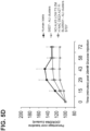


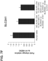


Classifications
-
- C—CHEMISTRY; METALLURGY
- C12—BIOCHEMISTRY; BEER; SPIRITS; WINE; VINEGAR; MICROBIOLOGY; ENZYMOLOGY; MUTATION OR GENETIC ENGINEERING
- C12N—MICROORGANISMS OR ENZYMES; COMPOSITIONS THEREOF; PROPAGATING, PRESERVING, OR MAINTAINING MICROORGANISMS; MUTATION OR GENETIC ENGINEERING; CULTURE MEDIA
- C12N5/00—Undifferentiated human, animal or plant cells, e.g. cell lines; Tissues; Cultivation or maintenance thereof; Culture media therefor
- C12N5/06—Animal cells or tissues; Human cells or tissues
- C12N5/0602—Vertebrate cells
- C12N5/0676—Pancreatic cells
- C12N5/0678—Stem cells; Progenitor cells; Precursor cells
-
- A—HUMAN NECESSITIES
- A61—MEDICAL OR VETERINARY SCIENCE; HYGIENE
- A61K—PREPARATIONS FOR MEDICAL, DENTAL OR TOILETRY PURPOSES
- A61K35/00—Medicinal preparations containing materials or reaction products thereof with undetermined constitution
- A61K35/12—Materials from mammals; Compositions comprising non-specified tissues or cells; Compositions comprising non-embryonic stem cells; Genetically modified cells
- A61K35/37—Digestive system
- A61K35/39—Pancreas; Islets of Langerhans
-
- C—CHEMISTRY; METALLURGY
- C07—ORGANIC CHEMISTRY
- C07K—PEPTIDES
- C07K14/00—Peptides having more than 20 amino acids; Gastrins; Somatostatins; Melanotropins; Derivatives thereof
- C07K14/435—Peptides having more than 20 amino acids; Gastrins; Somatostatins; Melanotropins; Derivatives thereof from animals; from humans
- C07K14/705—Receptors; Cell surface antigens; Cell surface determinants
- C07K14/72—Receptors; Cell surface antigens; Cell surface determinants for hormones
-
- C—CHEMISTRY; METALLURGY
- C12—BIOCHEMISTRY; BEER; SPIRITS; WINE; VINEGAR; MICROBIOLOGY; ENZYMOLOGY; MUTATION OR GENETIC ENGINEERING
- C12N—MICROORGANISMS OR ENZYMES; COMPOSITIONS THEREOF; PROPAGATING, PRESERVING, OR MAINTAINING MICROORGANISMS; MUTATION OR GENETIC ENGINEERING; CULTURE MEDIA
- C12N5/00—Undifferentiated human, animal or plant cells, e.g. cell lines; Tissues; Cultivation or maintenance thereof; Culture media therefor
- C12N5/06—Animal cells or tissues; Human cells or tissues
- C12N5/0602—Vertebrate cells
- C12N5/0676—Pancreatic cells
- C12N5/0677—Three-dimensional culture, tissue culture or organ culture; Encapsulated cells
-
- C—CHEMISTRY; METALLURGY
- C12—BIOCHEMISTRY; BEER; SPIRITS; WINE; VINEGAR; MICROBIOLOGY; ENZYMOLOGY; MUTATION OR GENETIC ENGINEERING
- C12N—MICROORGANISMS OR ENZYMES; COMPOSITIONS THEREOF; PROPAGATING, PRESERVING, OR MAINTAINING MICROORGANISMS; MUTATION OR GENETIC ENGINEERING; CULTURE MEDIA
- C12N5/00—Undifferentiated human, animal or plant cells, e.g. cell lines; Tissues; Cultivation or maintenance thereof; Culture media therefor
- C12N5/06—Animal cells or tissues; Human cells or tissues
- C12N5/0602—Vertebrate cells
- C12N5/0679—Cells of the gastro-intestinal tract
- C12N5/068—Stem cells; Progenitors
-
- C—CHEMISTRY; METALLURGY
- C12—BIOCHEMISTRY; BEER; SPIRITS; WINE; VINEGAR; MICROBIOLOGY; ENZYMOLOGY; MUTATION OR GENETIC ENGINEERING
- C12Q—MEASURING OR TESTING PROCESSES INVOLVING ENZYMES, NUCLEIC ACIDS OR MICROORGANISMS; COMPOSITIONS OR TEST PAPERS THEREFOR; PROCESSES OF PREPARING SUCH COMPOSITIONS; CONDITION-RESPONSIVE CONTROL IN MICROBIOLOGICAL OR ENZYMOLOGICAL PROCESSES
- C12Q1/00—Measuring or testing processes involving enzymes, nucleic acids or microorganisms; Compositions therefor; Processes of preparing such compositions
- C12Q1/68—Measuring or testing processes involving enzymes, nucleic acids or microorganisms; Compositions therefor; Processes of preparing such compositions involving nucleic acids
- C12Q1/6806—Preparing nucleic acids for analysis, e.g. for polymerase chain reaction [PCR] assay
-
- C—CHEMISTRY; METALLURGY
- C12—BIOCHEMISTRY; BEER; SPIRITS; WINE; VINEGAR; MICROBIOLOGY; ENZYMOLOGY; MUTATION OR GENETIC ENGINEERING
- C12N—MICROORGANISMS OR ENZYMES; COMPOSITIONS THEREOF; PROPAGATING, PRESERVING, OR MAINTAINING MICROORGANISMS; MUTATION OR GENETIC ENGINEERING; CULTURE MEDIA
- C12N2500/00—Specific components of cell culture medium
- C12N2500/05—Inorganic components
- C12N2500/10—Metals; Metal chelators
- C12N2500/20—Transition metals
- C12N2500/24—Iron; Fe chelators; Transferrin
- C12N2500/25—Insulin-transferrin; Insulin-transferrin-selenium
-
- C—CHEMISTRY; METALLURGY
- C12—BIOCHEMISTRY; BEER; SPIRITS; WINE; VINEGAR; MICROBIOLOGY; ENZYMOLOGY; MUTATION OR GENETIC ENGINEERING
- C12N—MICROORGANISMS OR ENZYMES; COMPOSITIONS THEREOF; PROPAGATING, PRESERVING, OR MAINTAINING MICROORGANISMS; MUTATION OR GENETIC ENGINEERING; CULTURE MEDIA
- C12N2500/00—Specific components of cell culture medium
- C12N2500/30—Organic components
- C12N2500/32—Amino acids
-
- C—CHEMISTRY; METALLURGY
- C12—BIOCHEMISTRY; BEER; SPIRITS; WINE; VINEGAR; MICROBIOLOGY; ENZYMOLOGY; MUTATION OR GENETIC ENGINEERING
- C12N—MICROORGANISMS OR ENZYMES; COMPOSITIONS THEREOF; PROPAGATING, PRESERVING, OR MAINTAINING MICROORGANISMS; MUTATION OR GENETIC ENGINEERING; CULTURE MEDIA
- C12N2500/00—Specific components of cell culture medium
- C12N2500/30—Organic components
- C12N2500/34—Sugars
-
- C—CHEMISTRY; METALLURGY
- C12—BIOCHEMISTRY; BEER; SPIRITS; WINE; VINEGAR; MICROBIOLOGY; ENZYMOLOGY; MUTATION OR GENETIC ENGINEERING
- C12N—MICROORGANISMS OR ENZYMES; COMPOSITIONS THEREOF; PROPAGATING, PRESERVING, OR MAINTAINING MICROORGANISMS; MUTATION OR GENETIC ENGINEERING; CULTURE MEDIA
- C12N2500/00—Specific components of cell culture medium
- C12N2500/30—Organic components
- C12N2500/38—Vitamins
-
- C—CHEMISTRY; METALLURGY
- C12—BIOCHEMISTRY; BEER; SPIRITS; WINE; VINEGAR; MICROBIOLOGY; ENZYMOLOGY; MUTATION OR GENETIC ENGINEERING
- C12N—MICROORGANISMS OR ENZYMES; COMPOSITIONS THEREOF; PROPAGATING, PRESERVING, OR MAINTAINING MICROORGANISMS; MUTATION OR GENETIC ENGINEERING; CULTURE MEDIA
- C12N2501/00—Active agents used in cell culture processes, e.g. differentation
- C12N2501/10—Growth factors
- C12N2501/105—Insulin-like growth factors [IGF]
-
- C—CHEMISTRY; METALLURGY
- C12—BIOCHEMISTRY; BEER; SPIRITS; WINE; VINEGAR; MICROBIOLOGY; ENZYMOLOGY; MUTATION OR GENETIC ENGINEERING
- C12N—MICROORGANISMS OR ENZYMES; COMPOSITIONS THEREOF; PROPAGATING, PRESERVING, OR MAINTAINING MICROORGANISMS; MUTATION OR GENETIC ENGINEERING; CULTURE MEDIA
- C12N2501/00—Active agents used in cell culture processes, e.g. differentation
- C12N2501/10—Growth factors
- C12N2501/115—Basic fibroblast growth factor (bFGF, FGF-2)
-
- C—CHEMISTRY; METALLURGY
- C12—BIOCHEMISTRY; BEER; SPIRITS; WINE; VINEGAR; MICROBIOLOGY; ENZYMOLOGY; MUTATION OR GENETIC ENGINEERING
- C12N—MICROORGANISMS OR ENZYMES; COMPOSITIONS THEREOF; PROPAGATING, PRESERVING, OR MAINTAINING MICROORGANISMS; MUTATION OR GENETIC ENGINEERING; CULTURE MEDIA
- C12N2501/00—Active agents used in cell culture processes, e.g. differentation
- C12N2501/10—Growth factors
- C12N2501/117—Keratinocyte growth factors (KGF-1, i.e. FGF-7; KGF-2, i.e. FGF-12)
-
- C—CHEMISTRY; METALLURGY
- C12—BIOCHEMISTRY; BEER; SPIRITS; WINE; VINEGAR; MICROBIOLOGY; ENZYMOLOGY; MUTATION OR GENETIC ENGINEERING
- C12N—MICROORGANISMS OR ENZYMES; COMPOSITIONS THEREOF; PROPAGATING, PRESERVING, OR MAINTAINING MICROORGANISMS; MUTATION OR GENETIC ENGINEERING; CULTURE MEDIA
- C12N2501/00—Active agents used in cell culture processes, e.g. differentation
- C12N2501/10—Growth factors
- C12N2501/119—Other fibroblast growth factors, e.g. FGF-4, FGF-8, FGF-10
-
- C—CHEMISTRY; METALLURGY
- C12—BIOCHEMISTRY; BEER; SPIRITS; WINE; VINEGAR; MICROBIOLOGY; ENZYMOLOGY; MUTATION OR GENETIC ENGINEERING
- C12N—MICROORGANISMS OR ENZYMES; COMPOSITIONS THEREOF; PROPAGATING, PRESERVING, OR MAINTAINING MICROORGANISMS; MUTATION OR GENETIC ENGINEERING; CULTURE MEDIA
- C12N2501/00—Active agents used in cell culture processes, e.g. differentation
- C12N2501/10—Growth factors
- C12N2501/15—Transforming growth factor beta (TGF-β)
-
- C—CHEMISTRY; METALLURGY
- C12—BIOCHEMISTRY; BEER; SPIRITS; WINE; VINEGAR; MICROBIOLOGY; ENZYMOLOGY; MUTATION OR GENETIC ENGINEERING
- C12N—MICROORGANISMS OR ENZYMES; COMPOSITIONS THEREOF; PROPAGATING, PRESERVING, OR MAINTAINING MICROORGANISMS; MUTATION OR GENETIC ENGINEERING; CULTURE MEDIA
- C12N2501/00—Active agents used in cell culture processes, e.g. differentation
- C12N2501/30—Hormones
- C12N2501/335—Glucagon; Glucagon-like peptide [GLP]; Exendin
-
- C—CHEMISTRY; METALLURGY
- C12—BIOCHEMISTRY; BEER; SPIRITS; WINE; VINEGAR; MICROBIOLOGY; ENZYMOLOGY; MUTATION OR GENETIC ENGINEERING
- C12N—MICROORGANISMS OR ENZYMES; COMPOSITIONS THEREOF; PROPAGATING, PRESERVING, OR MAINTAINING MICROORGANISMS; MUTATION OR GENETIC ENGINEERING; CULTURE MEDIA
- C12N2501/00—Active agents used in cell culture processes, e.g. differentation
- C12N2501/30—Hormones
- C12N2501/345—Gastrin; Cholecystokinins [CCK]
-
- C—CHEMISTRY; METALLURGY
- C12—BIOCHEMISTRY; BEER; SPIRITS; WINE; VINEGAR; MICROBIOLOGY; ENZYMOLOGY; MUTATION OR GENETIC ENGINEERING
- C12N—MICROORGANISMS OR ENZYMES; COMPOSITIONS THEREOF; PROPAGATING, PRESERVING, OR MAINTAINING MICROORGANISMS; MUTATION OR GENETIC ENGINEERING; CULTURE MEDIA
- C12N2501/00—Active agents used in cell culture processes, e.g. differentation
- C12N2501/30—Hormones
- C12N2501/375—Thyroid stimulating hormone [TSH]
-
- C—CHEMISTRY; METALLURGY
- C12—BIOCHEMISTRY; BEER; SPIRITS; WINE; VINEGAR; MICROBIOLOGY; ENZYMOLOGY; MUTATION OR GENETIC ENGINEERING
- C12N—MICROORGANISMS OR ENZYMES; COMPOSITIONS THEREOF; PROPAGATING, PRESERVING, OR MAINTAINING MICROORGANISMS; MUTATION OR GENETIC ENGINEERING; CULTURE MEDIA
- C12N2501/00—Active agents used in cell culture processes, e.g. differentation
- C12N2501/30—Hormones
- C12N2501/38—Hormones with nuclear receptors
- C12N2501/395—Thyroid hormones
-
- C—CHEMISTRY; METALLURGY
- C12—BIOCHEMISTRY; BEER; SPIRITS; WINE; VINEGAR; MICROBIOLOGY; ENZYMOLOGY; MUTATION OR GENETIC ENGINEERING
- C12N—MICROORGANISMS OR ENZYMES; COMPOSITIONS THEREOF; PROPAGATING, PRESERVING, OR MAINTAINING MICROORGANISMS; MUTATION OR GENETIC ENGINEERING; CULTURE MEDIA
- C12N2501/00—Active agents used in cell culture processes, e.g. differentation
- C12N2501/40—Regulators of development
- C12N2501/415—Wnt; Frizzeled
-
- C—CHEMISTRY; METALLURGY
- C12—BIOCHEMISTRY; BEER; SPIRITS; WINE; VINEGAR; MICROBIOLOGY; ENZYMOLOGY; MUTATION OR GENETIC ENGINEERING
- C12N—MICROORGANISMS OR ENZYMES; COMPOSITIONS THEREOF; PROPAGATING, PRESERVING, OR MAINTAINING MICROORGANISMS; MUTATION OR GENETIC ENGINEERING; CULTURE MEDIA
- C12N2501/00—Active agents used in cell culture processes, e.g. differentation
- C12N2501/70—Enzymes
- C12N2501/72—Transferases (EC 2.)
- C12N2501/727—Kinases (EC 2.7.)
-
- C—CHEMISTRY; METALLURGY
- C12—BIOCHEMISTRY; BEER; SPIRITS; WINE; VINEGAR; MICROBIOLOGY; ENZYMOLOGY; MUTATION OR GENETIC ENGINEERING
- C12N—MICROORGANISMS OR ENZYMES; COMPOSITIONS THEREOF; PROPAGATING, PRESERVING, OR MAINTAINING MICROORGANISMS; MUTATION OR GENETIC ENGINEERING; CULTURE MEDIA
- C12N2501/00—Active agents used in cell culture processes, e.g. differentation
- C12N2501/70—Enzymes
- C12N2501/73—Hydrolases (EC 3.)
-
- C—CHEMISTRY; METALLURGY
- C12—BIOCHEMISTRY; BEER; SPIRITS; WINE; VINEGAR; MICROBIOLOGY; ENZYMOLOGY; MUTATION OR GENETIC ENGINEERING
- C12N—MICROORGANISMS OR ENZYMES; COMPOSITIONS THEREOF; PROPAGATING, PRESERVING, OR MAINTAINING MICROORGANISMS; MUTATION OR GENETIC ENGINEERING; CULTURE MEDIA
- C12N2501/00—Active agents used in cell culture processes, e.g. differentation
- C12N2501/999—Small molecules not provided for elsewhere
-
- C—CHEMISTRY; METALLURGY
- C12—BIOCHEMISTRY; BEER; SPIRITS; WINE; VINEGAR; MICROBIOLOGY; ENZYMOLOGY; MUTATION OR GENETIC ENGINEERING
- C12N—MICROORGANISMS OR ENZYMES; COMPOSITIONS THEREOF; PROPAGATING, PRESERVING, OR MAINTAINING MICROORGANISMS; MUTATION OR GENETIC ENGINEERING; CULTURE MEDIA
- C12N2506/00—Differentiation of animal cells from one lineage to another; Differentiation of pluripotent cells
- C12N2506/02—Differentiation of animal cells from one lineage to another; Differentiation of pluripotent cells from embryonic cells
-
- C—CHEMISTRY; METALLURGY
- C12—BIOCHEMISTRY; BEER; SPIRITS; WINE; VINEGAR; MICROBIOLOGY; ENZYMOLOGY; MUTATION OR GENETIC ENGINEERING
- C12N—MICROORGANISMS OR ENZYMES; COMPOSITIONS THEREOF; PROPAGATING, PRESERVING, OR MAINTAINING MICROORGANISMS; MUTATION OR GENETIC ENGINEERING; CULTURE MEDIA
- C12N2533/00—Supports or coatings for cell culture, characterised by material
- C12N2533/90—Substrates of biological origin, e.g. extracellular matrix, decellularised tissue
Definitions
- the present invention relates to methods for producing in vitro functional pancreatic beta cells, and populations resulting from, the differentiation of pluripotent stem cells.
- the invention relates to beta cells or a population of beta cells that exhibit mitochondrial respiration/activity response and a two-phase insulin secretion response.
- beta ( ⁇ ) cells appropriate for engraftment.
- One approach is the generation of functional beta cells from pluripotent stem cells, such as, embryonic stem cells or induced pluripotent cells.
- a pluripotent cell gives rise to a group of cells comprising three germ layers (ectoderm, mesoderm, and endoderm) in a process known as gastrulation.
- Tissues such as, thyroid, thymus, pancreas, gut, and liver will develop from the endoderm via an intermediate stage.
- D'Amour et al. described the production of enriched cultures of human embryonic stem cell-derived definitive endoderm in the presence of a high concentration of activin and low serum ( Nature Biotechnology 2005, 23:1534-1541 ; U.S. Patent No. 7,704,738 ). Transplantation of these cells under the kidney capsule of mice resulted in differentiation into more mature cells with characteristics of endodermal tissue ( U.S. Patent No. 7,704,738 ). Human embryonic stem cell-derived definitive endoderm cells can be further differentiated into PDX1 positive cells after addition of FGF-10 and retinoic acid ( U.S. Patent App. Pub. No. 2005/0266554 ).
- pancreatic precursor cells were transplanted in the fat pad of immune deficient mice resulted in the formation of functional pancreatic endocrine cells following a 3-4 month maturation phase ( U.S. Patent Nos. 7,534,608 and 7,993,920 ).
- Small molecule inhibitors have been used for induction of pancreatic endocrine precursor cells.
- small molecule inhibitors of TGF- ⁇ receptor and BMP receptors ( Development 2011, 138:861-871 ; Diabetes 2011, 60:239-247 ) have been used to enhance the number of pancreatic endocrine cells.
- small molecule activators have also been used to generate definitive endoderm cells or pancreatic endoderm cells ( Curr. Opin. Cell Biol. 2009, 21:727-732 ; Nature Chem. Biol. 2009, 5:258-265 ).
- GSIS begins by the import of glucose into the beta-cell via a glucose transporter (solute carrier family 2 member 1; SLC2A1; or commonly referred to as glucose transporter 1; GLUT1 for human beta cells), and the metabolism of glucose to pyruvate, through a process named glycolysis.
- a glucose transporter substitute carrier family 2 member 1; SLC2A1; or commonly referred to as glucose transporter 1; GLUT1 for human beta cells
- TCA tricarboxylic acid, and subsequent activation of the electron transport chain
- Biphasic GSIS a key marker of maturation in beta cells, is not detected until the postnatal phase of human development, and is in contrast to the monophasic GSIS seen in immature beta-cells ( Otonkoski et al. Diabetes (1988) 37:286-291 ).
- Type 2 diabetics the 1 st phase of GSIS is absent; and the 2 nd phase of GSIS is also reduced.
- Type 1 diabetics whose beta-cell number is severely reduced via autoimmune attack, are reported to lack a robust biphasic GSIS ( Krogvold et al. Diabetes (2015) 64: 2506-2512 ).
- the invention provides methods for producing in vitro functional beta-cells (functional pancreatic beta-cells), and populations of cells resulting from, the differentiation of pluripotent stem cells.
- the invention relates to generating functional pancreatic beta-cells (insulin producing cells) or a population of functional beta-cells that exhibit mitochondrial respiration/activity response and a two-phase insulin secretion response.
- One aspect of the invention is a method of differentiating pluripotent stem cells into functional beta-cells.
- a particular aspect of the invention is a method of differentiating pancreatic endoderm cells into functional beta-cells expressing PDX1, NKX6.1, MAFA, UCN3 and SLC2A1.
- the method comprises culturing pancreatic endocrine cells in a culture medium supplemented with one or more small molecules of UNC0638, UNC0642, UCN0646, TC-E5003, A366, PF03814735, ZM447439, SB747651A, PFI1, LY303511, MS436, AZT, DEZA, pyroxamide, CI9994 or MC1568.
- An embodiment of the invention is in vitro population of functional pancreatic beta-cells expressing single hormonal insulin, PDX1, NKX6.1 and MAFA obtained by in vitro differentiation of pancreatic endocrine cells.
- the population of functional beta-cells also co-express UCN3 and SLC2A1.
- MAFA expression is increased as compared to immature beta-cells.
- the in vitro population of functional beta cells is obtained by culturing pancreatic endocrine cells in a medium supplemented with one or more small molecules of UNC0638, UNC0642, UCN0646, TC-E5003, A366, PF03814735, ZM447439, SB747651A, PFI1, LY303511, MS436, AZT, DEZA, pyroxamide, CI9994 or MC1568.
- a further embodiment of the invention is a method of differentiating pluripotent stem cells into functional beta cells expressing PDX1, NKX6.1, MAFA, UCN3 and SLC2A comprising the steps of differentiating pluripotent stem cells into pancreatic endocrine cells; and differentiating the pancreatic endocrine cells into functional beta cells expressing PDX1, NKX6.1, MAFA, UCN3 and SLC2A1.
- MAFA expression is increased as compared to immature beta-cells.
- the method comprises culturing pancreatic endocrine cells in a culture medium supplemented with one or more small molecules of UNC0638, UNC0642, UCN0646, TC-E5003, A366, PF03814735, ZM447439, SB747651A, PFI1, LY303511, MS436, AZT, DEZA, pyroxamide, CI9994 or MC1568.
- the culture medium is further supplemented with one or more of ZM447439, heparin, N-Acetyl Cysteine, and Formulation I.
- the culture medium is further supplemented with one or more of T3, T4 and analogue thereof.
- the culture medium is supplemented with an ALK5 inhibitor.
- the culture medium lacks an ALK5 inhibitor.
- the culture medium is supplemented with AZT or DEZA. In some embodiments above, the culture medium is supplemented with both AZT and DEZA.
- the medium is further supplemented with a gamma secretase inhibitor.
- the culture medium is supplemented with T3.
- the T3 is in the range of 1 nM to 1 ⁇ M. In particular embodiments, the T3 is in the range of 1 nM to 100 nM
- the functional beta-cells are obtained by step-wise differentiation of one or more of cells selected from the group: cells expressing markers characteristic of the definitive endoderm; cells expressing markers characteristic of primitive gut tube cells; cells expressing markers characteristic of foregut endoderm cells; cells expressing markers characteristic of pancreatic endoderm cells; pancreatic endocrine precursor cells; and cells expressing markers characteristic of immature beta cells (pancreatic endocrine cells).
- the method comprises culturing the cells at the air-liquid interface. In some embodiments described above, the method comprises culturing the cells in suspension, for example cell clusters in suspension.
- the differentiated functional beta-cell expresses MAFA at a higher level than an immature beta-cell. In embodiments, the differentiated functional beta-cell expresses UCN3 at a higher level than an immature beta-cell. In some embodiments above, expression of MAFA and UCN3 are increased compared to expression in an immature beta-cell.
- the population of functional beta-cells exhibits glucose-stimulated insulin secretion and glucose-dependent mitochondrial respiration.
- the glucose-stimulated insulin secretion and glucose-dependent mitochondrial respiration is similar to that of human islet cells.
- the functional beta-cells secrete insulin in multiple phases.
- the glucose-dependent mitochondrial respiration has a maximum oxygen consumption rate response following glucose stimulation in the range of about 20% to about 80% over basal oxygen consumption rate. In embodiments, the oxygen consumption rate response occurred at least 15 minutes following glucose stimulation.
- the glucose-stimulated insulin secretion comprises a rapid, bi-phasic insulin secretion in response to glucose stimulation.
- a first phase of the bi-phasic insulin secretion has at least a four-fold increase to at least an eight-fold increase over baseline secretion
- a second phase of the bi-phasic insulin secretion has at least a two-fold to at least a four-fold increase over baseline secretion.
- the insulin secretion occurs at least five minutes to at least ten minutes following the glucose stimulation.
- the method of differentiating pluripotent stem cells comprises the steps of: differentiating the pluripotent stem cells into cells expressing markers characteristic of the definitive endoderm ("Stage 1 cells”); differentiating the Stage 1 cells into cells expressing markers characteristic of primitive gut tube cells ("Stage 2 cells”); differentiating the Stage 2 cells into cells expressing markers characteristic of foregut endoderm cells ("Stage 3 cells”); differentiating the Stage 3 cells into cells expressing markers characteristic of pancreatic endoderm cells (“Stage 4 cells”); differentiating the Stage 4 cells into pancreatic endoderm/endocrine precursor cells (cells expressing markers characteristic of one or both of pancreatic endoderm cells and pancreatic endocrine precursor cells; "Stage 5"); differentiating the Stage 5 cells into immature beta-cells (pancreatic endocrine cells; "Stage 6”); and differentiating the Stage 6 cells into functional beta-cells (pancreatic endocrine cells; "Stage 7").
- the method of differentiating comprises differentiating pluripotent stem cells into Stage 1 cells by culturing the pluripotent stem cells in a medium supplemented with MCX compound and GDF-8.
- the method comprises differentiating the Stage 1 cells into Stage 2 cells by culturing the Stage 1 cells in a medium supplemented with FGF7 and ascorbic acid.
- the method comprises differentiating the Stage 2 cells into Stage 3 cells by culturing the Stage 2 cells in a medium supplemented with FGF7, retinoic acid, SANT-1, a PKC activator, a BMP inhibitor and ascorbic acid.
- the method comprises differentiating the Stage 3 cells into Stage 4 cells by culturing the Stage 3 cells in a medium supplemented with FGF7, retinoic acid, SANT-1, a PKC activator, a BMP inhibitor and ascorbic acid.
- the method comprises differentiating the Stage 4 cells into Stage 5 cells by culturing the Stage 4 cells in a medium supplemented with SANT-1, a PKC activator, a BMP inhibitor, ascorbic acid.
- the medium is further supplemented with one or more of T3, T4 or analogue thereof.
- the medium is further supplemented with an ALK5 inhibitor.
- the method comprises culturing the Stage 5 cells into pancreatic endocrine cells by culturing the Stage 5 cells in a medium supplemented with a BMP inhibitor, ascorbic acid, one or more of T3, T4 or analogue thereof.
- the medium is supplemented with T3.
- the T3 is in the range of 1 nM to 1 ⁇ M In certain embodiments, the T3 is in the range of 1nM to 100nM.
- the medium is further supplemented with an ALK 5 inhibitor.
- the culture medium is not supplemented with an ALK5 inhibitor.
- the medium is further supplemented with a gamma secretase inhibitor.
- the medium is supplemented with one or more of ZM447439, heparin, N-Acetyl Cysteine, and Formulation I.
- the culture medium is supplemented with AZT or DEZA.
- the culture medium is supplemented with both AZT and DEZA.
- the method comprises culturing the cells at the air-liquid interface. In some embodiments, the method comprises culturing the cells in suspension, for example cell clusters in suspension.
- the pancreatic endoderm cells (“Stage 4 cells”) may be cryopreserved.
- the method of differentiating pancreatic endoderm cells into pancreatic endocrine precursor cells comprises culturing cryopreserved cells.
- the pluripotent stem cells may be human pluripotent stem cells such as human embryonic stem cells.
- the pluripotent stem cells may be human H1 or H9 cells.
- Another embodiment of the invention is a method of differentiating pluripotent stem cells into functional beta cells expressing PDX1, NKX6.1, MAFA, UCN3 and SLC2A comprising: (a) differentiating pluripotent stem cells into cells expressing markers characteristic of the definitive endoderm obtained by culturing pluripotent stem cells in a medium supplemented with activin A and WNT3A; (b) differentiating the cells expressing markers characteristic of the definitive endoderm into immature beta-cells; and (c) differentiating the immature beta-cells into functional beta-cells expressing PDX1, NKX6.1, MAFA, UCN3 and SLC2A1 by culturing immature beta-cells in a medium supplemented with one or more of UNC0638, UNC0642, UCN0646, TC-E5003, A366, PF03814735, ZM447439, SB747651A, PFI1, LY303511, MS436, AZT
- These functional beta cells may have glucose-dependent mitochondrial respiration or glucose-stimulated insulin secretion similar to human islet cells.
- the functional beta cells may also secrete insulin in multiple phases.
- the culture medium lacks an ALK5 inhibitor.
- the culture medium is further supplemented with heparin, N- Acetyl Cysteine, Formulation I and one or more of T3, T4 or an analogue thereof.
- the culture medium is supplemented with T3.
- the medium is supplemented with ZM447439, heparin, N- Acetyl Cysteine and one or more of T3, T4 or an analogue thereof and contains no ALK5 inhibitor.
- the medium may be supplemented with T3, AZT or DEZA.
- the method may include culturing the immature beta cells at the air-liquid interface, in suspension clusters, in roller bottles or on microcarriers. In one embodiment, the method includes culturing the immature beta cells in roller bottles. In another embodiment, the method includes culturing the immature beta cells in roller bottles on microcarriers.
- the differentiating of the pluripotent stem cells into immature beta-cells in the method may comprise: (a) differentiating the pluripotent stem cells into cells expressing markers characteristic of the definitive endoderm ("Stage 1 cells") by culturing the pluripotent stem cells in a medium supplemented with activin A and WNT3A; (b) differentiating the Stage 1 cells into cells expressing markers characteristic of primitive gut tube cells ("Stage 2 cells”); (c) differentiating the Stage 2 cells into cells expressing markers characteristic of foregut endoderm cells ("Stage 3 cells”); (d) differentiating the Stage 3 cells into cells expressing markers characteristic of pancreatic endoderm cells ("Stage 4 cells”); (e) differentiating the Stage 4 cells into pancreatic endocrine precursor cells ("Stage 5"); and (f) differentiating the Stage 5 cells into the immature beta-cells.
- steps e. and f. include culturing at the air-liquid interface, in suspension clusters, in roller bottles or on micocarriers. In other embodiments, steps e. and f. comprise culturing the cells in roller bottles. In yet other embodiments, the steps e. and f. include culturing the immature beta cells in roller bottles on microcarriers.
- the method includes differentiating the Stage 1 cells into Stage 2 cells by culturing the Stage 1 cells in a medium supplemented with FGF7 and ascorbic acid. In other embodiments, the method also includes differentiating the Stage 2 cells into Stage 3 cells by culturing the Stage 2 cells in a medium supplemented with FGF7, retinoic acid, SANT-1, a PKC activator and ascorbic acid. In certain embodiments, the medium lacks a BMP inhibitor. In yet another embodiment, the method comprises differentiating the Stage 3 cells into Stage 4 cells by culturing the Stage 3 cells in a medium supplemented with FGF7, retinoic acid, SANT-1, a PKC activator and ascorbic acid. Again, in certain embodiments, the medium lacks a BMP inhibitor.
- the method comprises differentiating the Stage 4 cells into Stage 5 cells by culturing the Stage 4 cells in a medium supplemented with SANT-1, a PKC activator, a BMP inhibitor, and ascorbic acid.
- the medium may be further supplemented one or more of T3, T4 or analogue thereof.
- the method comprises culturing the Stage 5 cells into immature beta-cells by culturing the Stage 5 cells in a medium supplemented with a BMP inhibitor, ascorbic acid, one or more of T3, T4 or analogue thereof.
- the medium may be further supplemented with an ALK 5 inhibitor or a gamma secretase inhibitor.
- the method may also include differentiating the immature beta-cells into functional beta cells expressing PDX1, NKX6.1, MAFA, UCN3 and SLC2A by culturing immature beta-cells in a medium lacking an ALK5 inhibitor and supplemented with ZM447439, AZT, N-acetyl cysteine, DEZA, Formulation I and one or more of T3, T4 or analogue thereof.
- the culture may be a suspension culture or a cell aggregate suspension culture.
- the culturing may be carried out in roller bottles, on microcarriers or on microcarriers in roller bottles.
- Stem cells are undifferentiated cells defined by their ability, at the single cell level, to both self-renew and differentiate. Stem cells may produce progeny cells, including self-renewing progenitors, non-renewing progenitors, and terminally differentiated cells. Stem cells are also characterized by their ability to differentiate in vitro into functional cells of various cell lineages from multiple germ layers (endoderm, mesoderm, and ectoderm). Stem cells also give rise to tissues of multiple germ layers following transplantation and contribute substantially to most, if not all, tissues following injection into blastocysts. Stem cells are classified by their developmental potential. Pluripotent stem cells are able to give rise to all embryonic cell types.
- Differentiation is the process by which an unspecialized ("uncommitted") or less specialized cell acquires the features of a specialized cell, for example a nerve cell or a muscle cell.
- a differentiated cell is one that has taken on a more specialized ("committed") position within the lineage of a cell.
- the term "committed”, when applied to the process of differentiation, refers to a cell that has proceeded in the differentiation pathway to a point where, under normal circumstances, it will continue to differentiate into a specific cell type or subset of cell types, and cannot, under normal circumstances, differentiate into a different cell type or revert to a less differentiated cell type.
- Do-differentiation refers to the process by which a cell reverts to a less specialized (or committed) position within the lineage of a cell.
- the lineage of a cell defines the heredity of the cell, i.e. , which cells it came from and to what cells it can give rise.
- the lineage of a cell places the cell within a hereditary scheme of development and differentiation.
- a lineage-specific marker refers to a characteristic specifically associated with the phenotype of cells of a lineage of interest and can be used to assess the differentiation of an uncommitted cell to the lineage of interest.
- Markers are nucleic acid or polypeptide molecules that are differentially expressed in a cell of interest
- differential expression means an increased level for a positive marker and a decreased level for a negative marker as compared to an undifferentiated cell or a cell at another stage of differentiation.
- the detectable level of the marker nucleic acid or polypeptide is sufficiently higher or lower in the cells of interest compared to other cells, such that the cell of interest can be identified and distinguished from other cells using any of a variety of methods known in the art.
- cell density and “seeding density” are used interchangeably herein and refer to the number of cells seeded per unit area of a solid or semisolid planar or curved substrate.
- suspension culture refers to a culture of cells, single cells or clusters, suspended in medium rather than adhering to a surface.
- a cell is "positive for” a specific marker, “positive”, or “+” when the specific marker is sufficiently detected in the cell.
- the cell is “negative for”, “negative” or “ - “ for a specific marker when the specific marker is not sufficiently detected in the cell.
- FACS fluorescence activated cell sorting cytometry
- PCR polymerase chain reaction cytometry
- Stage 1 refers to the first step in the differentiation process, the differentiation of pluripotent stem cells into cells expressing markers characteristic of the definitive endoderm ("Stage 1 cells").
- Stage 2 refers to the second step, the differentiation of cells expressing markers characteristic of the definitive endoderm cells into cells expressing markers characteristic of primitive gut tube cells (“Stage 2 cells”).
- Stage 3 refers to the third step, differentiation of cells expressing markers characteristic of primitive gut tube cells into cells expressing markers characteristic of foregut endoderm cells ("Stage 3 cells”).
- Stage 4" refers to the fourth step, the differentiation of cells expressing markers characteristic of foregut endoderm cells into cells expressing markers characteristic of pancreatic endoderm cells (“Stage 4 cells”).
- Stage 5" refers to the fifth step, the differentiation of cells expressing markers characteristic of pancreatic endoderm cells into cells expressing markers characteristic of one or both of pancreatic endoderm cells and pancreatic endocrine precursor cells (collectively referred to as “Stage 5 cells” or, alternatively, “pancreatic endoderm/endocrine precursor cells”).
- Stage 6 refers to the sixth step, the differentiation of cells expressing markers characteristic of pancreatic endocrine precursor cells into cells expressing markers characteristic of immature beta-cells ("Stage 6 cells").
- Stage 7 refers to differentiation of cells expressing markers characteristic of immature beta-cells (pancreatic endocrine cells) into cells expressing markers characteristic of functional beta-cells and that have a more matured phenotype as compared to Stage 6 cells.
- a pancreatic endocrine cell that, when compared to a Stage 6 cell, is not only single hormonal insulin +, MAFA+, NKX6.1+, UCN3+, SLC2A1+ and PDX1+, but also expresses MAFA at a higher level than a less mature pancreatic endocrine cell, in particular an immature beta-cell.
- Definitive endoderm refers to cells which bear the characteristics of cells arising from the epiblast during gastrulation and which form the gastrointestinal tract and its derivatives.
- Definitive endoderm cells express at least one of the following markers: FOXA2 (also known as hepatocyte nuclear factor 3- ⁇ (“HNF3- ⁇ ”)), GATA4, SOX17, CXCR4, Brachyury, Cerberus, OTX2, goosecoid, C-Kit, CD99, and MIXL1. Markers characteristic of the definitive endoderm cells are CXCR4, FOXA2, and SOX17.
- definitive endoderm cells may be characterized by their expression of CXCR4, FOXA2, and SOX17.
- an increase in HNF4a may be observed.
- Primary gut tube cells refers to cells derived from definitive endoderm and that can give rise to all endodermal organs, such as lungs, liver, pancreas, stomach, and intestine.
- Gut tube cells may be characterized by their substantially increased expression of HNF4a over that expressed by definitive endoderm cells. For example, a ten- to forty-fold increase in mRNA expression of HNF4a may be observed during Stage 2.
- Formgut endoderm cells refers to cells that give rise to the esophagus, lungs, stomach, liver, pancreas, gall bladder, and a portion of the duodenum.
- Foregut endoderm cells express at least one of the following markers: PDX1, FOXA2, CDX2, SOX2, and HNF4a.
- Foregut endoderm cells may be characterized by an increase in expression of PDX1 compared to gut tube cells. For example, greater than fifty percent of the cells in Stage 3 cultures typically express PDX1.
- Pancreatic endoderm cells refers to cells that express at least one of the following markers: PDX1, NKX6.1, HNF1 ⁇ , PTF1 ⁇ , HNF6, HNF4a, SOX9, NGN3, gastrin; HB9, or PROX1. Pancreatic endoderm cells may be characterized by their lack of substantial expression of CDX2 or SOX2.
- Pancreatic endocrine precursor cells refers to pancreatic endoderm cells capable of becoming a pancreatic hormone expressing cell.
- Pancreatic endocrine precursor cells express at least one of the following markers: NGN3; NKX2.2; NeuroD1; ISL1; PAX4; PAX6; or ARX.
- Pancreatic endocrine precursor cells may be characterized by their expression of NKX2.2 and NeuroD1.
- Pantendocrine cells refer to cells capable of expressing at least one of the following hormones: insulin, glucagon, somatostatin, ghrelin, and pancreatic polypeptide.
- markers characteristic of pancreatic endocrine cells include one or more of NeuroD1, ISL1, PDX1, NKX6.1, PAX4, ARX, NKX2.2, HB9 and PAX6.
- Beta cells are pancreatic endocrine cells capable of expressing insulin, but not glucagon, somatostatin, ghrelin, and pancreatic polypeptide.
- Pancreatic endocrine cells expressing markers characteristic of ⁇ cells can be characterized by their expression of insulin and at least one of the following transcription factors: PDX1, NKX2.2, NKX6.1, NeuroD1, ISL1, HNF3 ⁇ , HB9, MAFA and PAX6.
- GSIS glucose-stimulated insulin secretion
- functional beta-cells exhibit at least one of the following characteristics of biphasic GSIS: (i) coupling of mitochondrial respiration/activity with insulin secretion; (ii) rapid insulin secretion response to heightened demand (here defined as high glucose concentration); (iii) ability to rapidly turn off insulin secretion after demand has subsided; (iv) ability for multiple rounds of "on-off' switching of insulin secretion; (v) ability to secrete the correct amount of insulin as dictated by demand; and (vi) ability to respond to multiple insulin secretagogues (for example, Exendin-4, or amino acids - L-Glutamine and L-Arginine).
- biphasic GSIS for example, Exendin-4, or amino acids - L-Glutamine and L-Arginine.
- Functional beta-cells can be characterized by their expression of insulin and at least one of the following transcription factors: PDX1, NKX2.2, NKX6.1, NeuroD1, ISL1, HNF3 ⁇ , HB9, PAX6, MAFA, SLC2A1, UCN3, and GLP1R.
- Immature beta cells are pancreatic endocrine cells that do not display glucose-dependent mitochondrial respiration/activity, and biphasic GSIS. Immature beta cells expressing markers characteristic of ⁇ cells can be characterized by their expression of insulin and at least one of the following transcription factors: PDX1, NKX2.2, NKX6.1, NeuroD1, ISL1, HNF3 ⁇ , HB9, MAFA, and PAX6.
- Air-liquid interface or "ALI”, as used herein, refers to the air-liquid interface that exists in an open culture vessel or a culture vessel partially filled with medium. Although referred to herein as “air” for convenience, the invention is not limited to the mixture of gases and compositions found in the ambient environment The invention specifically contemplates and includes gaseous mixtures having compositions different from the ambient environment including, for example, mixtures enriched for a particular component or in which a particular component has been depleted or eliminated.
- First phase (1 st ) insulin secretion represents the rapid exocytosis of a small pool of bound and readily releasable insulin granules upon a sudden increase in glucose concentration.
- Second phase (2 nd ) of insulin secretion represents the translocation of granules from a reserve pool of granules, and their binding/priming for release.
- OCR is defined as the oxygen consumption rate, and is an indicator of mitochondrial respiration, specifically via the electron transport chain ("ETC"); a direct measure of mitochondrial activity.
- ETC electron transport chain
- Effective amount refers to the amount of the compound that should be present to provide some degree of differentiation of the hESCs or further differentiation of partially differentiated hESCs such as those subjected to one or more previous differentiation stages.
- the compound may be present in the culture medium of the hESCs or may be added to the hESCs' during some stage of growth.
- a compound, agent, small molecule or growth factor is used to produce definitive endoderm, foregut, pancreatic foregut and pancreatic endoderm-lineage cells including pancreatic hormone secreting cells.
- the stem cells may be exposed to the compound, agent, small molecule or growth factor prior to any differentiation or during the first stage of differentiation, whereas in other examples, the stem cell may first be differentiated to an intermediate cell type such as, for example, definitive endoderm, and then exposed to the compound, agent, small molecule or growth factor.
- an intermediate cell type such as, for example, definitive endoderm
- Compounds “small molecule compounds” or equivalents thereof the invention refers to compounds encompassed by generic formulae disclosed herein (e.g. Table XI) and includes any specific compounds within that formula whose structure is disclosed herein or analogues thereof.
- the compounds of the invention may be identified either by their chemical structure or chemical name. When the chemical structure and chemical name conflict, the chemical structure is determinative of the identity of the compound.
- the compounds of the invention may contain one or more chiral centers or double bonds and therefore, may exist as stereoisomers, such as double-bond isomers (i.e., geometric isomers), enantiomers or diastereomers.
- the chemical structures depicted herein encompass all possible enantiomers and stereoisomers of the illustrated compounds including the stereoisomerically pure form (e.g., geometrically pure, enantiomerically pure or diastereomerically pure) and enantiomeric and stereoisomeric mixtures.
- Enantiomeric and stereoisomeric mixtures can be resolved into their component enantiomers or stereoisomers using separation techniques or chiral synthesis techniques well known to the skilled artisan.
- the compounds of the invention also include isotopically labeled compounds where one or more atoms have an atomic mass different from the atomic mass conventionally found in nature.
- isotopes examples include, but are not limited to, 2H, 3H, 13C, 14C, 15N, 180, 170, 31P, 32P, 35S, 18F and 36Cl. Further, it should be understood, when partial structures of the compounds of the invention are illustrated, that brackets indicate the point of attachment of the partial structure to the rest of the molecule.
- Pluripotent stem cells may express one or more of the designated TRA-1-60 and TRA-1-81 antibodies ( Thomson et al. 1998, Science 282:1145-1147 ). Differentiation of pluripotent stem cells in vitro results in the loss of TRA-1-60 and TRA-1-81 expression.
- Undifferentiated pluripotent stem cells typically have alkaline phosphatase activity, which can be detected by fixing the cells with 4% paraformaldehyde, and then developing with an alkaline phosphatase substrate kit sold under the trademark VECTOR ® Red, as described by the manufacturer (Vector Laboratories, Inc., Burlingame, California). Undifferentiated pluripotent stem cells also typically express OCT4 and TERT, as detected by reverse transcription polymerase chain reaction ("RT-PCR").
- RT-PCR reverse transcription polymerase chain reaction
- pluripotent stem cells Another desirable phenotype of propagated pluripotent stem cells is a potential to differentiate into cells of all three germinal layers: endoderm, mesoderm, and ectoderm. Pluripotency of stem cells may be confirmed, for example, by injecting cells into severe combined immunodeficiency ("SCID") mice, fixing the teratomas that form using 4% paraformaldehyde, and then examining histologically for evidence of cell types from these three germ layers. Alternatively, pluripotency may be determined by the creation of embryoid bodies and assessing the embryoid bodies for the presence of markers associated with the three germinal layers.
- SCID severe combined immunodeficiency
- Propagated pluripotent stem cell lines may be karyotyped using a standard G-banding technique and compared to published karyotypes of the corresponding primate species. It is desirable to obtain cells that have a "normal karyotype,” which means that the cells are euploid, wherein all human chromosomes are present and not noticeably altered.
- pluripotent stem cells may be used in the methods of the invention.
- pluripotent stem cells include established lines of pluripotent cells, including pre-embryonic tissue (such as, a blastocyst), embryonic tissue, or fetal tissue taken any time during gestation, typically but not necessarily, before approximately 10 to 12 weeks gestation.
- hESCs human embryonic stem cells
- human embryonic germ cells such as, the human embryonic stem cell lines H1 (NIH Code: WA01), H7 (NIH Code: WA07), H9 (NIH Code: WA09) (WiCell Research Institute, Madison, WI, USA), and SA002 (Cellartis AB Corporation, Goteburg, Sweden).
- IPS Induced pluripotent cells
- reprogrammed pluripotent cells derived from adult somatic cells using forced expression of a number of pluripotent related transcription factors, such as OCT4, NANOG, SOX2, KLF4, and ZFP42 ( Annu Rev Genomics Hum Genet 2011, 12:165-185 ; see also IPS, Cell, 126(4): 663-676 ) may also be used.
- the human embryonic stem cells used in the methods of the invention may also be prepared as described by Thomson et al. (U.S. Patent No.
- pluripotent stem cells suitable for use in the present invention may be derived according to the methods described in: Li et al. (Cell Stem Cell 4: 16-19, 2009 ); Maherali et al.
- pluripotent stem cells suitable for use in the present invention may be considered "naive" and derived according to the methods described in: Gafni et al. (Nature, 504:282, 2013 ), and Ware et al. (PNAS, 111: 4484-4489, 2014 ). All of these references, patents, and patent applications are herein incorporated by reference in their entirety, in particular, as they pertain to the isolation, culture, expansion and differentiation of pluripotent cells.
- pluripotent stem cells include induced pluripotent stem cells (IPS, Cell, 126(4): 663-676 ).
- suitable cells include human umbilical cord tissue-derived cells, human amniotic fluid-derived cells, human placental-derived cells, and human parthenotes.
- the umbilical cord tissue-derived cells may be obtained by the method of U.S. Patent No. 7,510,873 .
- the placental tissue-derived cells may be obtained using the methods of U.S. Patent App. Pub. No. 2005/0058631 .
- the amniotic fluid-derived cells may be obtained using the methods of U.S. Patent App. Pub. No. 2007/0122903 .
- the disclosure of each of these patent applications is incorporated in its entirety herein as it pertains to the isolation and characterization of the cells.
- the pluripotent stem cells may be of non-embryonic origins.
- the pluripotent stem cells may be plated onto a suitable culture substrate.
- the suitable culture substrate is an extracellular matrix component, such as those derived from basement membrane or that may form part of adhesion molecule receptor-ligand couplings.
- a suitable culture substrate is a reconstituted basement membrane sold under the trademark MATRIGEL TM (Corning Incorporated, Corning, New York).
- MATRIGEL TM is a soluble preparation from Engelbreth-Holm Swarm tumor cells that gels at room temperature to form a reconstituted basement membrane.
- extracellular matrix components and component mixtures known in the art are suitable as an alternative. Depending on the cell type being proliferated, this may include laminin, fibronectin, proteoglycan, entactin, heparin sulfate, and the like, alone or in various combinations.
- the pluripotent stem cells may be plated onto the substrate in a suitable distribution and in the presence of a medium, which promotes cell survival, propagation, and retention of the desirable characteristics. All these characteristics benefit from careful attention to the seeding distribution and can readily be determined by one of skill in the art Suitable culture media may be made from the following components, Dulbecco's Modified Eagle's medium (“DMEM”) sold under the trademark GIBCO ® (Catalog No. 11965-092) by Life Technologies Corporation, Grand Island New York; Knockout Dulbecco's Modified Eagle's medium (“KO DMEM') sold under the trademark GIBCO ® (Catalog No.
- DMEM Dulbecco's Modified Eagle's medium
- GIBCO ® Catalog No. 11965-092
- pluripotent cells differentiate towards functional ⁇ cells, they differentiate through various stages each of which may be characterized by the presence or absence of particular markers. Differentiation of the cells into these stages is achieved by the specific culturing conditions including the presence and lack of certain factors added to the culture media. In general, this differentiation may involve differentiation of pluripotent stem cells into definitive endoderm lineage, and definitive endoderm cells. These cells may then be further differentiated into primitive gut tube cells, which in turn may then be differentiated into foregut endoderm cells. Foregut endoderm cells may be differentiated into pancreatic endoderm cells, which may then be further differentiated into pancreatic endocrine precursor cells or pancreatic endoderm/pancreatic endocrine precursor cells.
- pancreatic hormone producing or secreting cells may be differentiated into pancreatic hormone producing or secreting cells.
- This application provides for the staged differentiation of pluripotent stem cells towards pancreatic endocrine cells, preferably by culturing the cells at the air-liquid interface, or in suspension, that exists within a culture vessel partially filled with medium, specifically by culturing cells at the air-liquid interface, or in suspension, in one or more of Stages 5 through 7.
- One or more of the thyroid hormones triiodothyronine (“T3") and thyroxine (“T4"), and analogues thereof, alone or in further combination with an ALK-5 inhibitor may be used in the cell culturing at one or more of Stages 1 through 7 of differentiation, and preferably at each of Stages 5 through 7.
- the ALK-5 inhibitor may be used alone in one or more stages of differentiation, but preferably at each of Stages 5 through 7 and more preferably at each of Stages 5 to 6.
- one or more of the thyroid hormones or their analogues and an ALK5 inhibitor is used in one or more differentiation stages, preferably at each of Stages 5 through 7 and more preferably at each of Stages 5 to 6.
- Suitable thyroid hormone analogues may include, without limitation: GC-1 (Sobertirome) (available from R&D Systems, Inc. Minneapolis, Minnesota); 3,5-diiodothryopropionic acid ("DIPTA”); KB-141 discussed in J. Steroid Biochem. Mol. Biol., 2008, 111: 262-267 and Proc. Natl. Acad. Sci. US 2003, 100: 10067-10072 ; MB07344 discussed in Proc. Natl. Acad. Sci. US 2007, 104: 15490-15495 ; T0681 discussed in J. Lipid Res., May 2009, 50:938 and Endocr. Pract.
- GC-1 Sobertirome
- DIPTA 3,5-diiodothryopropionic acid
- ALK5 inhibitors include: ALK5 inhibitor II (Enzo Life Sciences, Inc., Farmingdale, New York), which is also the preferred ALK5 inhibitor; ALK5i (Axxora, Inc., San Diego, California), SD208 (R&D Systems); TGF- ⁇ inhibitor SB431542 (Xcess Biosciences, Inc., San Diego, California); ITD-1 (Xcess Biosciences); LY2109761 (Xcess Biosciences); A83-01 (Xcess Biosciences); LY2157299 (Xcess Biosciences); TGF- ⁇ receptor inh V (EMD Millipore Chemical, Gibstown, New Jersey);TGF- ⁇ receptor inh I (EMD Millipore); TGF- ⁇ receptor inh IV (EMD Millipore); TGF- ⁇ receptor inh VII (EMD Millipore); TGF- ⁇ receptor inh VIII (EMD Millipore); TGF-
- the methods include treating cells at one or more stages, but preferably treating cells during Stage 7, with a differentiation medium that includes one or both of an antioxidant, such as vitamin E, acetyl cysteine, vitamin C, antioxidant supplement (Catalog No. A1345, Sigma-Aldrich Company, LLC Saint Louis, Missouri), glutathione, superoxide dismutase, catalase and the like and combinations thereof.
- an antioxidant such as vitamin E, acetyl cysteine, vitamin C, antioxidant supplement (Catalog No. A1345, Sigma-Aldrich Company, LLC Saint Louis, Missouri), glutathione, superoxide dismutase, catalase and the like and combinations thereof.
- a gamma secretase inhibitor in carrying out Stage 6, is used, which can be gamma secretase inhibitor XX (EMD Millipore), gamma secretase inhibitor XXI (EMD Millipore), gamma secretase inhibitor XVI (EMD Millipore), N-[(3,5-difluorophenyl)acetyl]-L-alanyl-2-phenyl]glycine-1,1-dimethylethyl ester (“DAPT”) (Catalog No. 2634, Tocris Bioscience, Bristol, United Kingdom), and the like and combinations thereof.
- DAPT N-[(3,5-difluorophenyl)acetyl]-L-alanyl-2-phenyl]glycine-1,1-dimethylethyl ester
- Useful amounts of gamma secretase inhibitor may be about 50 nM to 5000 nM, preferably about 50 nM to 500 nM.
- the amount of antioxidant may be about 0.1 to 100 ⁇ M, alternatively about 0.1 to 20 ⁇ M, and preferably about 1 to 10 ⁇ M.
- useful amounts of antioxidant may be about 100 nM to 5 mM, about 1000 nM to 2 mM, and preferably about 0.1 to 1 mM.
- certain small molecules are used in the medium of one or more stages of differentiation, preferably at one or both of Stages 6 and 7.
- the small molecules of interest are those capable of inhibiting aurora kinase, p90 ribosomal S6 kinase, or methyl transferase receptor DOT1L and preferably are used along with antioxidants that reduce oxidative stress of cultured cells.
- aurora kinase inhibitor II and RSK inhibitor II are examples of particular interest.
- Aurora kinase inhibitor II is a cell permeable compound known by the name of (4-(4'-benzamidoanilino)-6,7-dimethoxyquinazoline).
- RSK inhibitor II is a racemic mixture of a the dihydropteridinone 2-(3,5-difluoro-4-hydroxy-anilino)-8-isopentyl-5,7-dimethyl-7H-pteridin-6-one.
- Other aurora kinase inhibitors of interest include ZM447439 and PF03814735.
- protein methyltransferase receptor inhibitors of DOT1L particularly EPZ-5676.
- EPZ-5676 is an S-adenosyl methionine ("SAM”) inhibitor of protein methyltransferase DOT1L.
- This compound is a DOT1L (disruptor of telomere silencing 1-like) methyltransferase inhibitor shown to inhibit cell proliferation. It is known by the chemical names 9H-Purin-6-amine, 9-[5-deoxy-5-[[cis-3-[2-[6-(1,1-dimethylethyl)-1H-benzimidazol-2-yl]ethyl]cyclobutyl](1-methylethyl)amino]- ⁇ -D-ribofuranosyl]- and (2R,3R,4S,5R)-2-(6-aminopurin-9-yl)-5-[[[3-[2-(6-tert-butyl-1H-benzimidazol-2-yl)ethyl]cyclobutyl]-propan-2-ylamino]methyl]oxolane-3,4-diol.
- 9H-Purin-6-amine 9-[5-deoxy-5-[
- Further inhibitors of interest are a DNA methyltransferase inhibitor such as 5-Azacytidine (“AZT”), histone deacetylase inhibitors, such as Pyroxamide and CI994.
- Additional small molecules of interest include UNC0638, UNC0646, UNC0642 and A366 (G9a and GLP histone lysine methyltransferase inhibitors), TC-E5003 (PRMT1 arginine methyltransferase inhibitor), SB747651A dihydrochloride (MSK1 inhibitor; also inhibits other AGC group kinases), PFI1 (BET bromodomain inhibitor), LY303511 (BRD2, BRD3 and BRD4 inhibitor), MS436 (BRD4 bromodomain inhibitor), and MC1568 (selectively inhibits HDAC class II), and 3-Deazaneplanocin A (“DEZA”).
- the small molecule is used in the medium of one or more of Stage 6 and 7 and more preferably in Stage 7.
- the amount of small molecule useful may be determined by selecting the amount showing the best expression of maturation markers and which amounts are not producing toxic effects.
- the amounts useful will be about 500 nM to 10 ⁇ M, alternatively, about 500 nM to 5 ⁇ M, and preferably about 500 nM to 2 ⁇ M
- pluripotent stem cell markers include, for example, the expression of one or more of the following: ABCG2, cripto, FOXD3, CONNEXIN43, CONNEXIN45, OCT4, SOX2, NANOG, hTERT, UTF1, ZFP42, SSEA-3, SSEA-4, TRA-1-60, TRA-1-81. These may be detectable by RT-PCR
- pluripotent stem cells include the human embryonic stem cell line H9 (NIH code: WA09), the human embryonic stem cell line H1 (NIH code: WA01), the human embryonic stem cell line H7 (NIH code: WA07), and the human embryonic stem cell line SA002. Also suitable are cells that express at least one of the following markers characteristic of pluripotent cells: ABCG2, cripto, CD9, FOXD3, CONNEXIN43, CONNEXIN45, OCT4, SOX2, NANOG, hTERT, UTF1, ZFP42, SSEA-3, SSEA-4, TRA-1-60, and TRA-1-81.
- a cell that expresses at least one of the markers characteristic of the definitive endoderm lineage is a primitive streak precursor cell.
- a cell expressing markers characteristic of the definitive endoderm lineage is a mesendoderm cell.
- a cell expressing markers characteristic of the definitive endoderm lineage is a definitive endoderm cell.
- a cell that expresses at least one of the markers characteristic of the pancreatic endoderm lineage is a pancreatic endoderm cell wherein the expression of PDX1 and NKX6.1 are substantially higher than the expression of CDX2 and SOX2.
- Particularly useful are cells in which the expression of PDX1 and NKX6.1 is at least two-fold higher than the expression of CDX2 or SOX2.
- a cell that expresses at least one of the markers characteristic of the pancreatic endocrine lineage is a pancreatic endocrine cell.
- the pancreatic endocrine cell may be a pancreatic hormone-expressing cell capable of expressing at least one of the following hormones: insulin, glucagon, somatostatin, ghrelin, or pancreatic polypeptide.
- the pancreatic endocrine cell is an insulin-producing ⁇ cell.
- a protocol starting with pluripotent stem cells is employed.
- This protocol includes: Stage 1: Pluripotent stem cells, such as embryonic stem cells obtained from cell culture lines, are treated with the appropriate factors to induce formation of definitive endoderm cells.
- Stage 3 Cells resulting from Stage 2 cells are treated with the appropriate factors to induce further differentiation into cells expressing markers characteristic of foregut endoderm cells.
- Stage 4 Cells resulting from Stage 3 are treated with the appropriate factors to induce further differentiation into cells expressing markers characteristic of pancreatic endoderm cells.
- the cells are optionally further cultured at the air-liquid interface or in suspension culture at late Stage 4; for late Stage 4 cells are transitioned to the air-liquid interface or aggregated as clusters that are cultured in suspension.
- Stage 5 Cells resulting from Stage 4 are treated with the appropriate factors, including in certain embodiments: (i) one or more of T3, T4 or an analogue thereof; (ii) an ALK5 inhibitor; or (iii) both of (i) and (ii) and cultured, optionally at the air-liquid interface or in suspension, to induce differentiation to cells expressing markers characteristic of pancreatic endoderm/endocrine precursor cells.
- Stage 6 Cells resulting from Stage 5 cells are treated with the appropriate factors including in certain embodiments: (i) one or more of T3, T4 or an analogue thereof; (ii) an ALK5 inhibitor; (iii) one or more of a gamma secretase inhibitor, an RSK inhibitor, a bone morphogenic protein (BMG) receptor inhibitor and a protein methyltransferase inhibitor of DOT1L; (iv) both of (i) and (ii); (v) (i), (ii) and (iii); (vi) (i) and (iii); or (vii) (ii) and (iii) and cultured, optionally at the air-liquid interface or in suspension, to induce differentiation into cells expressing markers characteristic of pancreatic endocrine cells, in particular immature beta-cells.
- an ALK5 inhibitor one or more of a gamma secretase inhibitor, an RSK inhibitor, a bone morphogenic protein (BMG) receptor inhibitor and a protein
- Stage 7 Cells resulting from Stage 6 cells are treated with appropriate factors including in certain embodiments: (i) one or more of T3, T4 or thereof; (ii) an ALK5 inhibitor; (iii) an anti-oxidant, (iv) one or more of an aurora kinase inhibitor, an RSK inhibitor and a protein methyltransferase inhibitor of DOT1L; (v) (i) and (ii); (vi) (i) and (iii); (vii) (i) and (iv); (viii) (ii) and (iii); (ix) (ii) and (iv); (x) (i), (ii), and (iii); (xi) (i), (iii), and (iv); (xii) (i), (iii), and (iv); (xiii) (i), (iii), and (iv); (xiii) (i), (ii), and (iv); (xiii) (i), (
- the invention in certain embodiments encompasses differentiating pluripotent stem cells (e.g . pre-Stage 1 cells) to Stage 7 cells, the invention also encompasses differentiating cells at other stages towards Stage 7. In particular, the invention encompasses differentiation of Stage 4 to Stage 7 cells. Although the process is described in discrete stages, the treatment, as well as the progress of the cells through the differentiation process, may be sequential or continuous. Differentiation of pluripotent stem cells to Stage 6 or Stage 7 cells can be carried out in suspension cultures.
- the efficiency of differentiation may be determined by exposing a treated cell population to an agent, such as an antibody, that specifically recognizes a protein marker expressed by the differentiated cells of interest.
- agents such as an antibody
- Methods for assessing expression of protein and nucleic acid markers in cultured or isolated cells are standard in the art. These methods include RT-PCR, Northern blots, in situ hybridization (see, e.g., Current Protocols in Molecular Biology (Ausubel et al., eds.
- the differentiated cells may also be further purified.
- the differentiated cells may be purified by exposing a treated cell population to an agent, such as an antibody, that specifically recognizes a protein marker characteristically expressed by the differentiated cells being purified.
- the differentiated cells are not purified.
- Stages 1 through 7 Any suitable growth medium containing sufficient quantities of vitamins, minerals, salts, glucose, amino acids and carrier proteins desirable for cells differentiation may be used for the various Stages 1 through 7.
- Stage 1- MCDB-131 available from Life Technologies Corporation, Grand Island, NY
- RPMI available from Sigma-Aldrich
- Stage 2 - MCDB-131 or Dulbecco's Modified Eagle's Medium F12 (“DMEM -F12")
- Stages 6 and 7 - BLAR or CMRL Life Technologies.
- the glucose concentration of the medium is kept at or, more preferably, lower than about 10 mM for Stages 1 through 4 and greater than about 10 mM for Stages 5 through 7.
- the medium contains sufficient quantity of vitamins, non-essential amino acid, lipids, sodium pyruvate and trace elements, for example Formulation I.
- Pluripotent stem cells may be differentiated into cells expressing markers characteristic of definitive endoderm cells by methods known in the art, or by methods proposed in the invention. Methods reported as useful for differentiating pluripotent stem cells into cells expressing markers characteristic of the definitive endoderm lineage are disclosed in: D'Amour et al., Nature Biotechnology 23, 1534-1541 (2005 ); Shinozaki et al., Development 131, 1651-1662 (2004 ); McLean et al., Stem Cells 25, 29-38 (2007 ); and D'Amour et al., Nature Biotechnology 24, 1392-1401 (2006 ). Additional suitable differentiation methods are disclosed in: U.S. Patent App. Pub. No. 2007/0254359 ; U.S. Patent App.
- the pluripotent cells are treated with a suitable growth medium, preferably MCDB-131 or RPMI.
- the medium is preferably supplemented with a growth differentiation factor, such as growth differentiation factor 8 ("GDF8"), and a glycogen synthase kinase-3 ⁇ (“GSK3 ⁇ ”) inhibitor, such as the cyclic aniline-pyridintriazine compounds disclosed in U.S. Patent App. Pub. No. 2010/0015711 (incorporated herein in its entirety by reference) to induce differentiation into cells expressing markers characteristic of definitive endoderm cells.
- GDF8 growth differentiation factor 8
- GSK3 ⁇ glycogen synthase kinase-3 ⁇
- a preferred GSK3 ⁇ inhibitor is 14-prop-2-en-1-yl-3,5,7,14,17,23,27-heptaazatetracyclo[19.3.1.1 -2,6-.1-8,12-]heptacosa-1(25),2(27),3,5,8(26),9,11,21,23-nonaen-16-one ("MCX compound").
- Treatment may involve contacting pluripotent stem cells with a medium supplemented with about 50 ng/ml to about 150 ng/ml, alternatively about 75 ng/ml to about 125 ng/ml, preferably about 100 ng/ml of GDF8.
- the treatment may also involve contacting cells with about 0.1 to about 5 ⁇ M, alternatively about 0.5 to about 2.5 ⁇ M, preferably about 1 ⁇ M of MCX compound.
- the pluripotent cells may be cultured for about two to five days, preferably about two to three days, to facilitate differentiation into cells expressing markers characteristic of the definitive endoderm cells.
- the cells are cultured in the presence of GDF8 and MCX compound for one day, followed by culturing in the presence of GDF8 and a lower concentration of MCX compound for one day, followed by culturing in the presence of GDF8 for one day in the absence of MCX compound.
- the cells are cultured in the presence of GDF8 and about 1 ⁇ M MCX compound for one day, followed by culturing in the presence of GDF8 and about 0.1 ⁇ M MCX compound for one day, followed by culturing in the presence of GDF8 for one day in the absence of MCX compound.
- the cells may be cultured in the presence of GDF8 and about 1 ⁇ M MCX compound for one day, followed by culturing in the presence of GDF8 and about 0.1 ⁇ M MCX compound for one day.
- the pluripotent stem cells may be cultured in medium containing activin A in the absence of serum, then culturing the cells with activin A and serum, and then culturing the cells with activin A and serum of different concentration as disclosed in D'Amour et al., Nature Biotechnology 23, 1534-1541 (2005 ).
- the pluripotent stem cells may be differentiated into cells expressing markers characteristic of definitive endoderm cells by culturing the pluripotent stem cells in medium containing activin A in the absence of serum, then culturing the cells with activin A with serum as disclosed in D' Amour et al., Nature Biotechnology, 2005 .
- Pluripotent stem cells may also be differentiated into cells expressing markers characteristic of the definitive endoderm lineage by culturing the pluripotent stem cells in medium containing activin A and a WNT ligand in the absence of serum, then removing the WNT ligand and culturing the cells with activin A with serum as disclosed in D'Amour et al., Nature Biotechnology 24, 1392-1401 (2006 ).
- pluripotent stem cells are treated with activin A and WNT3A to result in the formation of cells expressing markers characteristic of definitive endoderm cells.
- Treatment may involve contacting pluripotent stem cells with about 50 ng/ml to about 150 ng/ml, alternatively about 75 ng/ml to about 125 ng/ml, alternatively about 100 ng/ml of activin A.
- the treatment may also involve contacting the cells with about 10 ng/ml to about 50 ng/ml, alternatively about 15 ng/ml to about 30 ng/ml, alternatively about 20 ng/ml of WNT3A.
- the pluripotent cells may be cultured for approximately three days to arrive at the definitive endoderm cells.
- the cells are cultured in the presence of activin A and WNT3A for one day followed by culturing in the presence of activin A (without WNT3A being present) for the remainder.
- the cells may be tested for the presence of the markers before and after carrying out a particular protocol.
- Pluripotent stem cells typically do not express such markers. Thus, differentiation of pluripotent cells can be detected when cells begin to express markers characteristic of definitive endoderm.
- the cells expressing markers characteristic of the definitive endoderm cells may be further differentiated into cells expressing markers characteristic of gut tube cells in a growth medium, such as MCDB-131 or DMEM-F12.
- a growth medium such as MCDB-131 or DMEM-F12.
- the formation of cells expressing markers characteristic of gut tube cells includes culturing the cells expressing markers characteristic of the definitive endoderm cells with a medium containing fibroblast growth factor ("FGF”), preferably FGF7 or FGF10, to differentiate the cells.
- FGF fibroblast growth factor
- the cell culture may include from about 10 ng/ml to about 75 ng/ml, alternatively from about 25 ng/ml to about 75 ng/ml, still alternatively from about 30 ng/ml to about 60 ng/ml, alternatively about 50 ng/ml of a fibroblast growth factor, preferably FGF7 or FGF10, more preferably FGF7, and most preferably about 25 ng/ml FGF7.
- the cells may be cultured under these conditions for about two to three days, preferably about two days.
- the formation of cells expressing markers characteristic of gut tube cells includes culturing the cells expressing markers characteristic of the definitive endoderm lineage with a fibroblast growth factor, preferably, FGF7 or FGF10, and ascorbic acid (Vitamin C).
- the culture medium may include from about 0.1 mM to about 0.5 mM ascorbic acid, alternatively from about 0.2 mM to about 0.4 mM ascorbic acid, alternatively about 0.25 mM of ascorbic acid.
- the cell culture may also include from about 10 ng/ml to about 35 ng/ml, alternatively from about 15 ng/ml to about 30 ng/ml, alternatively about 25 ng/ml of the fibroblast growth factor, preferably FGF7 or FGF10, more preferably FGF7.
- the cell culture may include about 0.25 mM of ascorbic acid and about 25 ng/ml of FGF7.
- the Stage 1 cells are treated for 2 days with FGF7 and ascorbic acid.
- the primitive gut tube cells resulting from Stage 2 may be further differentiated into Stage 3 cells, or cells expressing markers characteristic of the foregut endoderm, by culturing these cells in a growth medium such as MCDB-131, DMEM, or a custom media such as BLAR (Table I).
- a growth medium such as MCDB-131, DMEM, or a custom media such as BLAR (Table I).
- the medium may be supplemented with: (i) a fibroblast growth factor, preferably, FGF7 or FGF10 and more preferably FGF7; (ii) retinoic acid ("RA”); (iii) a Sonic Hedgehog ("SHH") signaling pathway antagonist (such as Smoothened Antagonist 1 ("SANT-1") which is 1-piperazinamine, N-[(3,5-dimethyl-1-phenyl-1H-pyrazol-4-yl)methylene]-4-(phenylmethyl)- or ((E)-4-benxyl-N-((3,5-dimethyl-1-phenyl-1H-pyrazol-4-yl),ethylene-piperazin-1-amine), HPI-1 which is 2-methoxyethyl 1,4,5,6,7,8-hexahydro-4-(3hydroxyphenyl)-7-(2-methoxyphenyl)-2-methyl-5-oxo-3-quinolinecarboxylate, and preferably SANT-1; (iv) a protein
- a smoothened (“SMO") receptor inhibitor such as MRT10 (N[[[3-benzoylamino)phenyl]amino]thioxomethyl]-3,4,5-trimethoxybenzamide)
- cyclopamine may also be used.
- the cell culture may include from about 100 nM to about 500 nM, alternatively from about 100 nM to about 400 nM, alternatively about 200 nM of a PKC activator.
- the cells may be cultured in the presence of these growth factors, small molecule agonists and antagonists for about two to four days, preferably about two to three days, more preferably about two days.
- Stage 3 cells may be obtained from the Stage 2 cells by culturing these cells in a culture medium supplemented with an SMO receptor inhibitor, SANT-1, retinoic acid and noggin.
- the cells may be cultured for approximately two to four days, preferably about two days.
- the medium is supplemented with: from about 10 ng/ml to about 35 ng/ml, alternatively from about 15 ng/ml to about 30 ng/ml, alternatively about 25 ng/ml of the fibroblast growth factor, preferably FGF7 or FGF10, more preferably FGF7; from about 0.1 mM to about 0.5 mM ascorbic acid, alternatively from about 0.2 mM to about 0.4 mM, alternatively about 0.25 mM of ascorbic acid; from about 0.1 ⁇ M to about 0.4 ⁇ M of SANT-1; from about 100 to about 300 nM of TPB; and from about 50 nM to about 200 nM, and about 100 nM of LDN-193189.
- the medium is supplemented with about 25 ng/ml of FGF-7, about 1 ⁇ M of retinoic acid, about 0.25 ⁇ M of SANT-1, about 200 nM of TPB, about 100 nM of LDN-193189, and about 0.25 mM of ascorbic acid.
- the medium is supplemented with from about 0.1 ⁇ M to about 0.3 ⁇ M of SANT-1, from about 0.5 ⁇ M to about 3 ⁇ M of retinoic acid and from about 75 ng/ml to about 125 ng/ml of noggin.
- the methods of the invention include deriving Stage 4 cells by treating Stage 3 cells with a differentiation medium that may be any suitable growth medium and preferably is MCDB-131, DMEM, or a custom media such as BLAR (Table I).
- the medium may be supplemented with one or more of the following: (a) an ALK5 inhibitor selected from the group consisting of TGF- ⁇ receptor inh V, TGF- ⁇ receptor inh I, TGF- ⁇ receptor inh IV, TGF- ⁇ receptor inh VII, TGF- ⁇ receptor inh VIII, TGF- ⁇ receptor inh II, TGF- ⁇ receptor inh VI, TGF- ⁇ receptor inh III, TGF- ⁇ inhibitor SB431542, SD-208, ITD-1, LY2109761, A83-01, LY2157299, ALK5i and ALK5 inhibitor II; (b) a thyroid hormone selected from the group consisting of T3, T4, analogues of T3, analogues of T4 and mixtures thereof; (c) an SHH signaling pathway antagonist
- a growth medium such as MCDB131 or, and preferably, BLAR may be supplemented with an SHH signaling pathway antagonist (such as SANT-1 or HPI-1), a BMP inhibitor (such as LDN-193189, noggin or chordin), ascorbic acid, and a PKC activator (such as TPB, PDBu, PMA or ILV), to provide a useful differentiation media.
- SHH signaling pathway antagonist such as SANT-1 or HPI-1
- BMP inhibitor such as LDN-193189, noggin or chordin
- ascorbic acid such ascorbic acid
- PKC activator such as TPB, PDBu, PMA or ILV
- Stage 3 cells Culturing Stage 3 cells in such medium for about two to four days, preferably about two to three days, more preferably about three days usually is sufficient to differentiate the Stage 3 cells into Stage 4 cells.
- the medium may be supplemented with an SMO inhibitor and SHH signaling pathway antagonist
- the Stage 3 cells may be treated with a medium supplemented with about 0.25 ⁇ M SANT-1; about 100 nM RA; about 2 ng/ml FGF7; about 100 nM LDN-193189; about 0.25 mM ascorbic acid; and about 200 nM of TPB for three days.
- cells may be cultured at the air-liquid interface, either during the entire stage or after about 2 to 3 days of planar culturing.
- the present invention provides an in vitro cell culture for differentiating cells derived from pluripotent stem cells at the air-liquid interface comprising: (a) a culture vessel; (b) a volume of growth medium within said vessel sufficient to fill only a portion of the volume of the vessel; (c) air within the vessel that fills a portion of the vessel adjoining the medium; (d) a porous substrate located at the interface between the medium and the air; and (e) cells derived from pluripotent stem cells disposed upon the surface of the substrate such that the medium contacts only a portion of the surface of the cells.
- Stage 4 may be carried out entirely in planar culture.
- cells may be aggregated to cell clusters.
- the present invention provides an in vitro cell culture for differentiating cells prepared as aggregated cells such as cell clusters comprising: (a) a culture vessel that facilitates aggregation of cells, or forming cell clusters; (b) a volume of growth medium (culture medium) within said vessel; and (c) cells derived from pluripotent stem cells disposed within the vessel such that the cells are induced to aggregate and form clusters.
- the vessel is a plate with wells or microwells, such as Aggrewell TM plates (microwell plates; STEMCELL Technologies Inc., Vancouver, Canada).
- cell clusters prepared in plates with microwells to form aggregates of cells of uniform size and shape, for example using Aggrewell TM plates.
- about 500 to 2000 cells are seeded per well or microwell to prepare aggregated cells or cell clusters.
- about 50 to about 3000 cells are seeded per well or microwell, preferably about 50 to about 2000 cells, about 50 to 1000 cells, about 50 to about 900 cells, about 50 to about 800 cells, about 100 to about 800 cells, about 250 to about 800 cells, or about 500 to about 800 cells are seeded. More preferably, about 700 to about 800 cells are seeded per well or microwell.
- the cell clusters formed by the method of aggregating cells may be further cultured in suspension (suspension culture), comprising removing the aggregated cells or cell clusters from the wells or microwells and seeding the aggregated cells/cell clusters in a suspension culture vessel.
- aggregated cells/cell clusters are seeded in a suspension culture at a cell density of about 0.75 ⁇ 10 6 cells/ml to about 2.0 ⁇ 10 6 cells/ml, preferably about 1 ⁇ 10 6 cells/ml to about 2.0 ⁇ 10 6 cells/ml, more preferably about 1.5 ⁇ 10 6 cells/ml to about 2.0 ⁇ 10 6 cells/ml.
- the suspension culture system may include seeding aggregated cells or cell clusters in a flask, such as a spinner flask or a spinner wheel flask.
- the cells at the completion of Stage 4 may be treated with a Rho-associated kinase ("ROCK") inhibitor such as Y27632 ((1R,4r)-4-((R)-1-aminoethyl)-N-(pyridin-4-yl)cyclohexanecarboxamide), GSK269962 (N-[3-[[2-(4-Amino-1,2,5-oxadiazol-3-yl)-1-ethyl-1H-imidazo[4,5-c]pyridin-6-yl]oxy]phenyl]-4-[2-(4-morpholinyl)ethoxy]benzamide), H1152 ((S)-(+)-2-Methyl-1-[(4-methyl-5-isoquinolinyl)sulfonyl]homopiperazine, 2HCl,) and, SR3677 ( N -[2-[2-(Dimethylamino)e
- ROCK Rho-associated kinase
- late Stage 4 cells for example, cells that have been cultured for 1 to 2 days or 1 to 3 days in adherent planar cultures, may subsequently be cultured at the air-liquid interface or cultured to aggregated cells, such as cell clusters, for completion of Stage 4.
- late Stage 4 cells that were treated with a ROCK inhibitor are cultured at the air-liquid interface.
- 0.5 to about 0.75 ⁇ 10 5 cells/micro liter are seeded to be cultured at the air-liquid interface; alternatively, about 2 to 6 ⁇ 10 6 cells are seeded to be cultured at the air-liquid interface.
- late Stage 4 cells that were treated with a ROCK inhibitor are cultured to aggregate the cells to form aggregated cells such as cell clusters.
- cells are seeded in a vessel that facilitates aggregation of cells, or cell clusters (three dimensional aggregates of cells).
- the vessel is a plate with wells or microwells.
- cell clusters are prepared in plates with microwells to form aggregates of cells of uniform size and shape, for example using Aggrewell TM plates (STEMCELL Technologies Inc., Vancouver, Canada). In embodiments, about 50 to 3000 cells are seeded per well or microwell to prepare aggregated cells or cell clusters.
- about 50 to about 3000 cells are seeded per well or microwell, preferably about 50 to about 2000 cells, about 50 to 1000 cells, 50 to 900 cells, 50 to 800 cells, 100 to 800 cells, 250 to 800 cells, or about 500 to about 800 cells are seeded. More preferably, about 700 to 800 cells are seeded per well or microwell.
- the aggregated cells or cell clusters are further cultured in suspension.
- the adherent cells in planar culture may be treated with a cell detachment solution, such as a solution containing proteolytic and collagenolytic enzymes such as TrypLE TM , Accutase TM , or Dispase TM prior to culturing at the air-liquid interface or culturing to aggregate cells such as forming cell clusters.
- a cell detachment solution such as a solution containing proteolytic and collagenolytic enzymes such as TrypLE TM , Accutase TM , or Dispase TM prior to culturing at the air-liquid interface or culturing to aggregate cells such as forming cell clusters.
- Stage 4 cells may be obtained from Stage 3 cells by treating the Stage 3 cells with a differentiation medium comprising a growth medium supplemented with an ALK5 inhibitor, noggin, and a PKC activator, such as TPB.
- the medium may be supplemented with about 0.1 ⁇ M ALK5 inhibitor, about 100 ng/mL of noggin, and about 500 nM TPB.
- the cell culture may be in a monolayer format.
- the treatment may last for a total of about three days.
- the cells may be treated for two days and then on the last day the cells may be treated with proteolytic enzymes, collagenolytic enzymes or both to generate a single cell suspension.
- the resulting cells may be seeded at the air-liquid interface or seeded to generate aggregated cells or cell clusters.
- the single cells may be aggregated into cell clusters having a diameter of less than about 100 microns followed by culturing in the presence of an ALK5 inhibitor and LDN-193189.
- the cell clusters having a diameter of less than about 100 microns may be cultured in a medium supplemented with about 200 nM ALK5 inhibitor and about 100 nM LDN-193189.
- culturing Stage 4 cells at the air-liquid interface or in suspension may significantly enhance pancreatic endoderm markers along with endocrine-related markers.
- the methods of the invention include generation of Stage 5 cells by treating Stage 4 cells with a differentiation medium that may be any suitable growth medium and preferably is MCDB-131, DMEM or a custom media such as BLAR (Table I).
- the medium may be supplemented with one or more of the following: (a) an ALK5 inhibitor selected from the group consisting of TGF- ⁇ receptor inh V, TGF- ⁇ receptor inh I, TGF- ⁇ receptor inh IV, TGF- ⁇ receptor inh VII, TGF- ⁇ receptor inh VIII, TGF- ⁇ receptor inh II, TGF- ⁇ receptor inh VI, TGF- ⁇ receptor inh III, TGF- ⁇ inhibitor SB431542, SD-208, ITD-1, LY2109761, A83-01, LY2157299, ALK5i and ALK5 inhibitor II; (b) a thyroid hormone selected from the group consisting of T3, T4, analogues of T3, analogues of T4 and mixtures thereof; (c) an SHH signaling pathway antagonist selected from S
- the growth medium is also supplemented with one or both of an SMO inhibitor (such as MRT10 or cyclopamine) and a fibroblast growth factor selected from FGF-7 or FGF-10.
- SMO inhibitor such as MRT10 or cyclopamine
- a fibroblast growth factor selected from FGF-7 or FGF-10.
- the Stage 4 cells are differentiated into Stage 5 cells by treating the cells with a medium supplemented with from about 0.1 ⁇ M to about 0.4 ⁇ M of SANT-1and preferably about 0.25 ⁇ M SANT-1, about 50 nMRA, from about 0.1 mM to about 0.5 mM ascorbic acid, alternatively from about 0.2 mM to about 0.4 mM and preferably about 0.25 mM ascorbic acid, from about 50 nM to about 200 nM and preferably about 100 nM LDN-193189, about 1 ⁇ M of T3, and about 10000 nM ALK5 inhibitor, more preferably ALK 5 inhibitor II.
- a medium supplemented with from about 0.1 ⁇ M to about 0.4 ⁇ M of SANT-1and preferably about 0.25 ⁇ M SANT-1, about 50 nMRA, from about 0.1 mM to about 0.5 mM ascorbic acid, alternatively from about 0.2 mM to about 0.4 mM and preferably about 0.25
- the cells are optionally also treated with about 1 to 15 ⁇ M, alternatively about 1 to 10 ⁇ M, alternatively about 5 to 10 ⁇ M, preferably about 10 ⁇ M of zinc sulfate (ZnSO 4 ) and about 1 to 100 ⁇ g/ml, preferably about 10 ⁇ g/ml of heparin.
- the treatment of the Stage 4 cells is carried out for about two to four days, preferably about 3 days to differentiate the cells into Stage 5 cells.
- the methods of the invention include obtaining Stage 5 cells by treating Stage 4 cells with a medium supplemented with heparin, an SMO inhibitor or an SHH signaling pathway antagonist, RA, a BMP receptor inhibitor and an ALK5 inhibitor and culturing the cells at the air-liquid interface for about 3 days to differentiate the cells into Stage 5 cells.
- the medium may be supplemented with both an SMO inhibitor and an SHH signaling pathway antagonist, along with RA, a BMP receptor inhibitor and an ALK5 inhibitor.
- the Stage 4 cells may be differentiated into Stage 5 cells by treating the Stage 4 cells with a medium supplemented with heparin, ZnSO 4 , an SMO inhibitor or an SHH signaling pathway antagonist, RA, LDN-193189 and ALK5 inhibitor II.
- the medium may be supplemented with both an SMO inhibitor and an SHH signaling pathway antagonist
- the Stage 4 cells are differentiated into Stage 5 cells by treating the cells with a medium supplemented with about 10 ⁇ g/ml of heparin, about 0.25 ⁇ M SANT-1, about 50 nMRA, about 50 nM LDN-193189, about 10 nM of T3 and about 1000 nM ALK5 inhibitor.
- Suitable ALK5 inhibitors include but are not limited to SD-208, ALK5 inhibitor II, TGF- ⁇ receptor inh V, TGF- ⁇ receptor inh I, TGF- ⁇ receptor inh IV, TGF- ⁇ receptor inh VII, TGF- ⁇ receptor inh VIII, TGF- ⁇ receptor inh II, TGF- ⁇ receptor inh VI, TGF- ⁇ receptor inh III and combinations thereof.
- the treatment of the Stage 4 cells is carried out for about two to four days, preferably about 3 days to differentiate the cells into Stage 5 cells.
- the ALK5 inhibitor is ALK5 inhibitor II. In another preferred embodiment, about 10000 nM of ALK5 inhibitor II is used. In an alternate preferred embodiment, the Stage 4 cells are treated with a medium supplemented with about 10 ⁇ g/ml of heparin, about 0.25 ⁇ M SANT-1, about 50 nM RA, about 100 nM LDN-193189, and about 10,000 nM (10 mM) of ALK5 inhibitor II.
- the methods of the invention include treating Stage 4 cells with a medium supplemented with an SMO inhibitor or an SHH signaling pathway antagonist, RA, and an ALK5 inhibitor and culturing the cells, preferably at the air-liquid interface or in suspension for about two days to four days, preferably about 3 days, differentiate the cells into Stage 5 cells.
- the medium may be supplemented with both an SMO inhibitor and an SHH signaling pathway antagonist.
- the Stage 4 cells are differentiated into Stage 5 cells by treating the cells with a medium supplemented with about 0.25 ⁇ M SANT-1, about 50 nM RA, about 50 nM LDN-193189, about 1 ⁇ M T3 and about 1000 nM of an ALK5 inhibitor.
- the amount of cells seeded for culturing at the air-liquid interface may vary.
- droplets of a single-cell suspension containing about 0.5 to 6 ⁇ 10 5 cells/ ⁇ l may be seeded on a porous substrate (e.g. filter).
- the suspension may contain from about 2 ⁇ 10 5 cells/ ⁇ l to about 6 ⁇ 10 5 cells/ ⁇ l; about 4 ⁇ 10 5 cells/ ⁇ l to about 6 ⁇ 10 5 cells/ ⁇ l; about 5 ⁇ 10 5 cells/ ⁇ l to about 6 ⁇ 10 5 cells/ ⁇ l; about 5 ⁇ 10 5 cells/ ⁇ l to about 6 ⁇ 10 5 cells/ ⁇ l; about 2 ⁇ 10 5 cells/ ⁇ l to about 5 ⁇ 10 5 cells/ ⁇ l; about 2 ⁇ 10 5 cells/ ⁇ l to about 4 ⁇ 10 5 cells/ ⁇ l; or about 3 ⁇ 10 5 cells/ ⁇ l that may be seeded onto a porous substrate such as a filter located at the air-liquid interface.
- a porous substrate such as a filter located at the air-liquid interface
- droplets of a single-cell suspension containing from about 0.5 ⁇ 10 5 cells/ ⁇ l to about 0.75 ⁇ 10 5 cells/ ⁇ l; about 0.6 ⁇ 10 5 cells/ ⁇ l to about 0.75 ⁇ 10 5 cells/ ⁇ l; or about 0.5 ⁇ 10 5 cells/ ⁇ l to about 0.6 ⁇ 10 5 cells/ ⁇ l are seeded onto a porous support to be cultured at the ALI.
- cell clusters are generated in plates with microwells that are then seeded to suspension culture system.
- Aggrewell TM plates are used to induce aggregation in which about 50 to 3000 cells are seeded per well or microwell to prepare aggregated cells or cell clusters.
- about 50 to about 3000 cells are seeded per well or microwell, preferably about 50 to about 2000 cells, about 50 to 1000 cells, about 50 to about 900 cells, about 50 to about 800 cells, about 100 to about 800 cells, about 250 to about 800 cells, or about 500 to about 800 cells are seeded. More preferably, about 700 to about 800 cells are seeded per well or microwell.
- the aggregated cells/cell clusters are seeded in a suspension culture at a cell density of about 0.75 ⁇ 10 6 cells/ml to about 2.0 ⁇ 10 6 cells/ml, preferably about 1 ⁇ 10 6 cells/ml to about 2.0 ⁇ 10 6 cells/ml, about 1.25 ⁇ 10 6 cells/ml to about 2.0 ⁇ 10 6 cells/ml, and more preferably about 1.5 ⁇ 10 6 cells/ml to about 2.0 ⁇ 10 6 cells/ml.
- the methods of the invention include treating Stage 4 cells with a medium supplemented with a BMP receptor inhibitor (e.g. LDN-193189, noggin or chordin) and an ALK5 inhibitor for about 1 day to differentiate Stage 4 cells into Stage 5 cells.
- a BMP receptor inhibitor e.g. LDN-193189, noggin or chordin
- ALK5 inhibitor for about 1 day to differentiate Stage 4 cells into Stage 5 cells.
- the medium may be supplemented with about 100 nM of LDN-193189 and with about 100 nM of ALK5 inhibitor and about 1 ⁇ M T3.
- the cells may be in adherent planar culture or in the form of clusters.
- the cells may be treated with a cell detachment solution, such as a solution containing proteolytic and collagenolytic enzymes prior to culturing at the air-liquid interface or culturing to form aggregated cells or cell clusters.
- the invention further provides a cell culture for differentiating cells expressing markers characteristic of the pancreatic endoderm into cells expressing markers characteristic of pancreatic endocrine precursor cells (pancreatic endoderm/pancreatic endocrine precursor cells) comprising: (a) a culture vessel; (b) a volume of growth medium within said vessel sufficient to fill only a portion of the volume of said vessel; (c) air within said vessel that fills a portion of said vessel adjoining said medium; (d) a porous substrate located at the interface between said medium and said air; and (e) cells expressing markers characteristic of pancreatic endoderm cells derived from pluripotent stem cells disposed upon the surface of said substrate such that said medium contacts only a portion of the surface of said cells.
- the invention further provides a cell culture for differentiating cells expressing markers characteristic of the pancreatic endoderm into cells expressing markers characteristic of pancreatic endocrine precursor cells (pancreatic endoderm/pancreatic endocrine precursor cells) comprising: (a) a culture vessel that facilitates aggregation of cells, or forming cell clusters; (b) a volume of growth medium (culture medium) within said vessel; (c) cells derived from pluripotent stem cells disposed within the vessel such that the cells are induced to aggregate and form clusters; and (d) generated cell clusters disposed in suspension (suspension culture).
- the vessel for cell aggregation is a plate with wells or microwells, such as Aggrewell TM plates (microwell plates; STEMCELL Technologies Inc., Vancouver, Canada).
- the suspension culture system may include seeding aggregated cells or cell clusters in a suspension culture vessel, such as flask, for example a spinner flask or a spinner wheel flask.
- the methods of the invention include obtaining Stage 6 cells by treating Stage 5 cells with a differentiation medium that may be any suitable growth medium, preferably such as MCDB-131 or CMRL, and more preferably, a custom media such as BLAR (Table I).
- a differentiation medium may be any suitable growth medium, preferably such as MCDB-131 or CMRL, and more preferably, a custom media such as BLAR (Table I).
- the medium may be supplemented with one or more of the following: (a) an ALK5 inhibitor selected from the group consisting of: TGF- ⁇ receptor inh V, TGF- ⁇ receptor inh I, TGF- ⁇ receptor inh IV, TGF- ⁇ receptor inh VII, TGF- ⁇ receptor inh VIII, TGF- ⁇ receptor inh II, TGF- ⁇ receptor inh VI, TGF- ⁇ receptor inh III, TGF- ⁇ inhibitor SB431542, SD-208, ITD-1, LY2109761, A83-01, LY2157299, ALK5i and ALK5 inhibitor II; (b) a thyroid hormone selected from the group consisting of T3, T4, analogues of thereof and mixtures thereof; (c) a BMP receptor inhibitor preferably selected from LDN-193189, noggin or chordin; (d) a gamma secretase inhibitor such gamma secretase inhibitor XX, gamma secretase inhibitor XXI, gamma secretase inhibitor
- the cells may be cultured, preferably at the air-liquid interface or in suspension culture, for about two to four, preferably for about three days, to differentiate the Stage 5 cells into Stage 6 cells.
- the medium can further be supplemented with one or more of an SHH signaling pathway antagonist, a smoothened receptor inhibitor, a fibroblast growth factor and retinoic acid.
- the Stage 5 cells may be differentiated into Stage 6 cells by treatment with a medium supplemented with about 50 nM RA, about 0.25 mM ascorbic acid, about 100 nM LDN-193189, about 10,000 nM of ALK5 inhibitor and preferably ALK 5 inhibitor II, 1 ⁇ M T3, about 100 nM of a gamma secretase inhibitor for about seven days.
- Stage 5 cells may be differentiated into Stage 6 cells by treatment with a medium supplemented with about 0.25 ⁇ M SANT-1, about 50 nM RA, about 0.25 mM ascorbic acid, about 1000 nM ALK5 inhibitor and 1 ⁇ M T3 for about three days.
- the cells may be cultured in such media for an additional two days, or more, if desired.
- Stage 5 cells may be differentiated into Stage 6 cells by treatment with a medium supplemented with heparin, an SMO inhibitor or an SHH signaling pathway antagonist, a BMP inhibitor, T3, T4, analogues thereof and mixtures thereof and an ALK5 inhibitor and culturing, preferably at the air-liquid interface or in suspension culture, for about one to seven days, alternatively about six days, alternatively about seven days.
- the medium may be supplemented with both an SMO inhibitor and an SHH signaling pathway antagonist.
- the cells may be cultured in the medium supplemented with about 10 ⁇ g/ml of heparin, about 0.25 ⁇ M SANT-1, about 100 nM LDN-193189, about 1000 nM of T3 and about 500 to about 10,000 nM, alternatively about 500 nM, alternatively about 1000 mM, and alternatively about 10,000 nM of an ALK5 inhibitor.
- Suitable ALK5 inhibitors include but are not limited to SD-208, ALK5 inhibitor II, TGF- ⁇ receptor inh V, TGF- ⁇ receptor inh I, TGF- ⁇ receptor inh IV, TGF- ⁇ receptor inh VII, TGF- ⁇ receptor inh VIII, TGF- ⁇ receptor inh II, TGF- ⁇ receptor inh VI, TGF- ⁇ receptor inh III and combinations thereof.
- the ALK5 inhibitor is ALK5 inhibitor II. In a more preferred embodiment, about 10,000 nM (10 mM) of ALK5 inhibitor II is used. Accordingly, in one embodiment, Stage 5 cells may be differentiated into Stage 6 cells by treatment with a medium supplemented with heparin, an SMO inhibitor or an SHH signaling pathway antagonist, a BMP inhibitor, T3, T4, analogues thereof and mixtures thereof, and ALK5 inhibitor and culturing, preferably at the air-liquid interface or in suspension, preferably for about seven days. In an alternate embodiment, the medium may be supplemented with both an SMO inhibitor and an SHH signaling pathway antagonist. In certain embodiments, the cells may be treated with a cell detachment solution, such as a solution containing proteolytic and collagenolytic enzymes prior to culturing at the air-liquid interface or in suspension.
- a cell detachment solution such as a solution containing proteolytic and collagenolytic enzymes prior to culturing at the air-liquid interface or in suspension
- Stage 5 cells may be differentiated into Stage 6 cells by treatment with a medium supplemented with heparin, an SMO inhibitor or an SHH signaling pathway antagonist, a BMP inhibitor T3, and ALK5 inhibitor II and culturing at the air-liquid interface for about 5 days to about 7 days, alternatively about 5 days, alternatively about 6 days, alternatively about 7 days
- the medium may be supplemented with about 10 ⁇ g/ml of heparin, about 0.25 ⁇ M SANT-1, about 100 nM LDN-193189, about 1000 nM of T3 and about 10,000 nM of ALK5 inhibitor II.
- the medium may be further supplemented with zinc sulfate (ZnSO 4 ).
- ZnSO 4 zinc sulfate
- the medium may be further supplemented with about 10 mM ZnSO 4 .
- the medium may be supplemented with both an SMO inhibitor and an SHH signaling pathway antagonist
- an aurora kinase inhibitor preferably aurora kinase inhibitor II
- an RSK inhibitor preferably RSK inhibitor II
- the protein methyl transferase inhibitor of DOT1L preferably EPZ-5676
- the amount added may be from about 100 to 5000 nM, alternatively about 1000 to 5000 nM, alternatively about 2000 to 5000 nM, alternatively about 3000 to 5000 nM, and preferably about 1000 to 2000 nM for the aurora kinase and RSK inhibitors and about 100 to 1000 nM for the DOT1L inhibitor, and more preferably about 1 ⁇ M to about 10 nM.
- the invention further provides a cell culture for differentiating cells expressing markers characteristic of pancreatic endoderm/endocrine precursor cells into cells expressing markers characteristic of immature beta cells, comprising: (a) a culture vessel; (b) a volume of growth medium within said vessel sufficient to fill only a portion of the volume of said vessel; (c) air within said vessel that fills a portion of said vessel adjoining said medium; (d) a porous substrate located at the interface between said medium and said air; and (e) cells expressing markers characteristic of pancreatic endoderm/ endocrine precursor cells derived from pluripotent stem cells disposed upon the surface of said substrate such that said medium contacts only a portion of the surface of said cells.
- the invention further provides a cell culture for differentiating cells expressing markers characteristic of the pancreatic endocrine precursor cells (or pancreatic endoderm/endocrine precursor cells) into cells expressing markers characteristic of immature beta cells comprising: (a) a culture vessel that facilitates aggregation of cells, or forming cell clusters; (b) a volume of growth medium (culture medium) within said vessel; (c) cells derived from pluripotent stem cells disposed within the vessel such that the cells are induced to aggregate and form clusters; and (d) generated cell clusters disposed in suspension (suspension culture).
- the vessel for cell aggregation is a plate with wells or microwells, such as Aggrewell TM plates.
- the suspension culture system may include seeding aggregated cells or cell clusters in a suspension culture vessel, such as flask, for example a spinner flask or a spinner wheel flask.
- Stage 5 cells cultured according to embodiments of the invention are utilized and differentiated into Stage 6 cells, while in other embodiments Stage 5 cells cultured according to other protocols may be utilized in the current method to obtain Stage 6 and Stage 7 cells.
- the methods of the invention result in the formation of Stage 6 cells that are single-hormone positive.
- the methods of the invention result in Stage 6 cells, which co-express NKX6.1, insulin, chromogranin and PDX1.
- the methods of the invention result in Stage 6 cells, which co-express NKX6.1 and insulin.
- the method employs a custom medium, BLAR ( see Table I), at Stages 4 to 6 or late Stage 4 to 6 or Stages 5 and 6.
- the medium may be exchanged every day or alternatively every other day.
- the invention relates to a method of forming Stage 6 cells co-expressing NKX6.1 and chromogranin comprising culturing Stage 4, preferably late Stage 4 cells to Stage 6 cells at the air-liquid interface or in suspension.
- the invention relates to a method of forming single hormone insulin positive cells expressing NKX6.1 Stage 6 cells by culturing Stage 4, preferably late Stage 4 cells, to Stage 6 cells at the air-liquid interface or in suspension.
- the methods of the invention include treating Stage 6 cells with a differentiation medium that may be any suitable growth medium, preferably such as MCDB-131 or CMRL or, and more preferably, a custom media such as BLAR001 (Table 1) or BLAR004 (Table IV) for seven days.
- a differentiation medium may be any suitable growth medium, preferably such as MCDB-131 or CMRL or, and more preferably, a custom media such as BLAR001 (Table 1) or BLAR004 (Table IV) for seven days.
- the medium is supplemented with one or more of the following: 2.7g/1000 ml sodium bicarbonate, and supplemented with a 1:200 dilution of ITS-X; 1X GlutaMAX TM ; 2% FAF-BSA; 10 ⁇ g/ml of heparin ("H”); 10 nM of T3 ("low T3"); 1 mM N-Acetyl cysteine ("NAC”); 0.5 ⁇ M of ZM447439 (“ZM”); and the following components which constitute Formulation I (“FT') (Table XII) - 1:200 dilution of RPMI Vitamin Supplement; 1:200 dilution of MEM non-essential amino acid supplement; 1:2000 dilution of chemically defined lipid concentrate; 1:200 dilution of sodium pyruvate; 1:2000 dilution of trace elements A (Corning, Catalog No.
- Stage 7 includes about 10 nM of T3 ("low T3"); 5 ⁇ M 5-Azacytidine (“AZT”) (Sigma Aldrich, Catalog No. A2385); or about 1 ⁇ M 3-Deazaneplanocin A (“DEZA”) (Biovision, Inc., Catalog No. 2060).
- the medium does not contain an ALK5 inhibitor.
- the Stage 6 cells may be differentiated into Stage 7 cells by treatment with a medium supplemented with, about 10 nM of T3, about 0.5 ⁇ M of one or more of aurora kinase inhibitor II, and about 1 mM N-acetyl cysteine.
- Stage 6 cells may be differentiated into Stage 7 cells by treatment with a medium supplemented with heparin, T3, T4, analogues thereof or mixtures thereof, an antioxidant, and aurora kinase inhibitor, or mixtures thereof and culturing, at the air-liquid interface or suspension cultures, for about seven to twenty-one days, alternatively about seven to ten days, preferably about seven days.
- the invention further provides a cell culture for differentiating cells expressing markers characteristic of pancreatic endocrine precursor cells into cells expressing markers characteristic of functional beta-cells, comprising: (a) a culture vessel; (b) a volume of growth medium within said vessel sufficient to fill only a portion of the volume of said vessel; (c) air within said vessel that fills a portion of said vessel adjoining said medium; (d) a porous substrate located at the interface between said medium and said air; and (e) cells expressing markers characteristic of pancreatic endoderm/endocrine precursor cells derived from pluripotent stem cells disposed upon the surface of said substrate such that said medium contacts only a portion of the surface of said cells.
- Stage 6 cells cultured according to embodiments of the invention are utilized and differentiated into Stage 7 cells, while in other embodiments Stage 6 cells cultured according to other protocols may be utilized in the current method to obtain Stage 7 cells.
- the methods of the invention result in the formation of Stage 7 cells that are single-hormone positive.
- the methods of the invention result in Stage 7 cells, which co-express NKX6.1, chromogranin, PDX1, UCN3, SLC2A1 and MAFA.
- the methods of the invention result in Stage 7 cells, which co-express NKX6.1, PDX1, insulin, UCN3, SLC2A1 and MAFA.
- At least 10 % of the cells of the resulting cell population express insulin, PDX1, NKX6.1, UCN3, SLC2A1 and MAFA. In other embodiments, at least 20 % of the cells of the population express insulin, PDX1, NKX6.1, UCN3, SLC2A1 and MAFA. In other embodiments, at least 30 % of the cells of the population express insulin, PDX1, NKX6.1, UCN3, SLC2A1 and MAFA. In still other embodiments, at least 40 % of the cells of the population express insulin, PDX1, NKX6.1, UCN3, SLC2A1 and MAFA.
- At least 50 % of the cells of the population express insulin, PDX1, NKX6.1, UCN3, SLC2A1 and MAFA. In still other embodiments, at least 60 %, 70 %, 80% or 90% of the cells express insulin, PDX1, NKX6.1, UCN3, SLC2A1 and MAFA. In alternative embodiments, at least 91, 92, 93, 94, 95, 96, 97, 98, or 99 % of the cells of the population express insulin, PDX1, NKX6.1, UCN3, SLC2A1 and MAFA.
- the method employs a custom medium, BLAR (Table I), at Stages 4 through 7 or late Stage 4 through 7, or Stages 5, 6 and 7.
- the medium may preferably be exchanged every day or alternatively every other day.
- the invention relates to a method of forming Stage 7 cells co-expressing NKX6.1, PDX1, MAFA, UCN3, SLC2A1 and chromogranin comprising culturing Stage 4, preferably late Stage 4 cells to Stage 7 cells at the air-liquid interface, or in suspension.
- the invention relates to a method of forming single hormone insulin positive cells (functional beta-cells) expressing NKX6.1, PDX1, UCN3, SLC2A1 and MAFA Stage 7 cells by culturing Stage 4, preferably late Stage 4 cells, to Stage 7 cells at the air-liquid interface or in suspension.
- the present invention contemplates culturing at the air-liquid interface for all stages in the path from pluripotent cell to pancreatic endocrine cell
- the invention provides for the formation of Stage 1 to Stage 4 cells in planar or submerged culture and Stage 5, 6 and 7 cells by culturing the cells at the air-liquid interface or in suspension culture.
- the present invention relates to a stepwise method of differentiation of pluripotent cells comprising culturing Stage 4, 5 and 6 cells at the air-liquid interface.
- cells cultured during Stages 4 through 7 may be cultured at the air-liquid interface.
- only late Stage 4 to Stage 6 cells, or Stage 5 and Stage 6 cells are cultured at the air-liquid interface or in suspension.
- Stage 1 through 4 are carried out by culturing the cells in planar cultures, and Stage 5 through Stage 7, or Stage 6 through Stage 7, or only Stage 7 are carried out by culturing in suspension culture.
- culturing during one, or all of Stages 5, 6 and 7 is carried out in the presence of one or more of T3, T4, their analogues and an ALK5 inhibitor, or one or more of T3, T4 and their analogues; or an ALK5 inhibitor.
- culturing during one or more, and preferably all of Stages 5, 6 and 7 is carried out in the presence of T3 and an ALK5 inhibitor and more preferably in the presence of T3 and ALK5 inhibitor II.
- culturing during Stage 7 is carried out in the presence of T3 at low concentration.
- culturing during Stage 7 is carried out with no ALK5 inhibitor included.
- the cells When cells are cultured at the air-liquid interface ("ALI"), the cells may be cultured on a porous substrate such that the cells are in contact with air on the top side and with cell culture media at the bottom side.
- a sufficient volume of media may be added to the bottom of a culture vessel containing the porous substrate (e.g. a filter insert) such that the media contacts the bottom surface of cells residing on the substrate but does not encapsulate or submerge them.
- Suitable porous substrates can be formed of any material that will not adversely affect the growth and differentiation of the cells.
- Exemplary porous substrates are made of polymers such as polyethylene terephthalate (“PET”), polyester or polycarbonate. Suitable porous substrates may be coated or uncoated.
- the coating may be MATRIGEL TM .
- the porous substrate is a porous filter insert, which may be coated with MATRIGEL TM .
- the porous substrate is an uncoated filter insert. The porosity of the substrate should be sufficient to maintain cell viability and promote differentiation of the cells.
- Culturing of the cells at the air-liquid interface includes seeding the cells on a porous substrate such as a porous filter insert.
- the substrate pore size may range from about 0.3 to about 3 microns. Seeding may be accomplished by releasing cells as single cells from monolayer cultures or clusters of cells from monolayer cultures into a suspension and subsequently aliquoting the single cell suspension or suspended cell culture onto a porous substrate at the ALI.
- the cells may be seeded onto the porous substrate from a suspension having about 1000 cells/ ⁇ l to about 100,000 cells/ ⁇ l.
- the cells may be seeded as droplets of the cell suspension containing individual cells or aggregates or clusters of cells.
- the cells may be cultured at the air-liquid interface using the methods disclosed in U.S. Patent App. Pub. No. 2014/0186305 , the disclosure of which is herein incorporated as it pertains to the culturing and differentiating pluripotent stem cells at the air liquid interface.
- the media may be exchanged or refreshed every other day or, preferably, daily.
- the cells grown on top of the porous substrate are generally not single cells, but rather they are in the form of a sheet or exist as an aggregate cell cluster. Cells cultured at the ALI may experience higher oxygen tension as compared to cells submerged in media.
- the methods of the invention may be carried out by culturing and differentiating the cells as clusters in a suspension culture.
- Exemplary suitable methods for the suspension culture and differentiation of pluripotent stem cells are disclosed in U.S. Patent App. Pub. Nos. 2014/0242693 and 2014/0295552 , the disclosures of which are herein incorporated as it pertains to the culturing and differentiating pluripotent stem cells using suspension clusters.
- the present invention provides an in vitro cell culture including differentiating cells prepared as aggregated cells such as cell clusters comprising seeding cells in a culture vessel that facilitates aggregation of cells, or forming cell clusters, with a growth medium (culture medium) such that the cells are induced to aggregate and form clusters.
- the vessel useful for inducing aggregation of cells is a plate with wells or microwells, such as Aggrewell TM plates.
- the resulting aggregated cells or cell clusters are further cultured in suspension (suspension culture), comprising removing the aggregated cells/cell clusters from the microwells and seeding them in a suspension culture vessel such that the aggregated cells are differentiated in suspension.
- Embodiments of the present invention encompass formation of late Stage 4 to 7, preferably Stage 5 to 7 cells, at the air-liquid interface or in suspension.
- the cells may be formed by differentiating pluripotent stem cells or by further differentiating Stage 3, 4, 5 or 6 cells.
- Stage 4 cells may be cultured entirely at the air-liquid interface or the cells may be cultured in submerged planar culture during the early portion of Stage 4, (about one to two days) and then cultured at the air-liquid interface or in suspension for the latter portion of Stage 4 (about day two to day three).
- Stage 4 is not carried out at the ALI, or in suspension, but rather in submerged culture.
- the present invention provides a method for producing cells expressing markers characteristic of functional beta-cells from pluripotent stem cells, comprising: culturing pluripotent stem cells; differentiating the pluripotent stem cells into cells expressing markers characteristic of the pancreatic endoderm; and differentiating the cells expressing markers characteristic of the pancreatic endoderm into cells expressing markers characteristic of the pancreatic endocrine cells by culturing, at the air-liquid interface or in suspension.
- the method may include treatment with a medium supplemented with: (i) T3, T4 or their analogues; (ii) an ALK5 inhibitor; or both (i) and (ii).
- the method may include differentiating the cells expressing markers characteristic of foregut endoderm cells (Stage 3 cells) into cells expressing markers characteristic of pancreatic endoderm cells (Stage 4 cells) by treatment with a medium supplemented with: (i) one or both of T3, T4 or their analogues; (ii) ALK5 inhibitor; or both (i) and (ii); and culturing in a planar culture.
- the method may also include differentiating cells expressing markers characteristic of pancreatic endoderm cells (Stage 4 cells) into cells expressing markers characteristic of the immature beta-cells (Stage 6 cells) by treatment with a medium supplemented with: (i) one or both of T3, T4 or their analogues; (ii) ALK5 inhibitor; or both (i) and (ii); and culturing in a planar culture or culturing at the air-liquid interface or in suspension.
- a medium supplemented with: (i) one or both of T3, T4 or their analogues; (ii) ALK5 inhibitor; or both (i) and (ii); and culturing in a planar culture or culturing at the air-liquid interface or in suspension.
- the method further includes differentiating Stage 6 cells into cells expressing markers characteristic of functional beta-cells and that have a more matured phenotype as compared to Stage 6 (Stage 7 cells) by treatment with a medium supplemented with: (i) one or both of T3, T4 or their analogues; (ii) ALK5 inhibitor; or both (i) and (ii); along with an one or more of an aurora kinase inhibitor, an RSK inhibitor, and an inhibitor of protein methyltransferase DOT1L and, optionally an anti-oxidant such as vitamin E or acetyl cysteine.
- the preferred amount of acetyl cysteine that is useful is about 0.1 to about 2 mM.
- the method further includes carrying out Stage 6 by treatment of Stage 5 cells with a medium supplemented with: (i) one or both of T3, T4 or their analogues; (ii) ALK5 inhibitor or; both (i) and (ii); along with one or more of a gamma secretase inhibitor, an RSK inhibitor and an inhibitor of protein methyltransferase DOT1L.
- Stage 6 is carried out by treatment of Stage 5 cells with a medium supplemented with: (i) one or both of T3, T4 or their analogues; (ii) ALK5 inhibitor; or both (i) and (ii); along with one or more of a gamma secretase inhibitor, an RSK inhibitor and an inhibitor of protein methyltransferase DOT1L followed by carrying out Stage 7 by treatment with a medium supplemented with: (i) one or both of T3, T4 or their analogues; (ii) ALK5 inhibitor; or both (i) and (ii); along with an one or more of an aurora kinase inhibitor, an RSK inhibitor, an inhibitor of protein methyltransferase DOT1L and, optionally anti-oxidant such as vitamin E or acetyl cysteine.
- One embodiment of the invention is a method of forming functional beta-cells (pancreatic endocrine cells expressing markers characteristic of a matured phenotype) (Stage 7 cells) comprising differentiating cells expressing markers characteristic of the pancreatic endoderm cells (Stage 4 cells) into cells expressing markers characteristic of Stage 7 cells by culturing at the air-liquid interface or in suspension.
- a cell expressing markers characteristic of functional beta-cells of a more matured phenotype expresses PDX1 and at least one of the following transcription factors: NKX2.2, NKX6.1, NeuroD1, ISL1, HNF3 ⁇ , MAFA, UCN3, SLC2A1, PAX4, HB9 and PAX6.
- the methods of the invention result in the formation of Stage 6 cells, which are positive for NKX6.1, PDX1, HB9 and MAFA.
- the method includes treatment with a medium supplemented with T3, T4 or an analogue thereof, an ALK5 inhibitor, or both.
- the Stage 6 cells may be cells that are positive for NKX6.1, PDX1, HB9 and MAFA.
- the Stage 6 or 7 cells are single hormone positive cells.
- the Stage 6 and 7 cells may be cells that: (a) co-express NKX6.1 and chromogranin; (b) co-express NKX6.1 and insulin; or (c) co-express NKX6.1, PDX1, MAFA and single hormonal insulin.
- the Stage 7 cells express single hormonal insulin and MAFA at increased levels and increased numbers of cells within a cell population as compared to Stage 6 cells.
- the invention provides a method of enhancing the number of single hormone positive cells (e.g. cells that co-express NKX6.1 and insulin or cells that co-express NKX6.1 and chromogranin) by culturing and differentiating a population of PDX1 and NKX6.1 co-expressing cells, preferably at an air-liquid interface or in suspension.
- single hormone positive cells e.g. cells that co-express NKX6.1 and insulin or cells that co-express NKX6.1 and chromogranin
- pancreatic endoderm cells cultured at the air-liquid interface or in suspension are further differentiated to functional beta-cells by treatment with a compound selected from the following: ALK5 inhibitor, BMP inhibitor, gamma-secretase inhibitor, Ephrin ligands, EphB inhibitor, PKC inhibitor, EGFr inhibitor, retinoic acid, vitamin C, T3/T4, glucose, cell cycle regulators, WNT regulators, SHH inhibitor, aurora inhibitor, anti-oxidants, vitamin E, acetyl-cysteine, or combinations thereof.
- the present invention relates to a stepwise method of differentiating pluripotent cells that includes culturing Stage 4 through Stage 6 cells in a media containing sufficient amounts of: (i) one or more of T3, T4 and their analogues; (ii) an ALK5 inhibitor; or both (i) and (ii); and further culturing the Stage 6 cells in a media that optionally contains one or more of an aurora kinase inhibitor, an RSK inhibitor, and an inhibitor of protein methyltransferase DOT1L, and an antioxidant to generate functional beta-cells (pancreatic endocrine cells of a matured phenotype) and populations of functional beta-cells that express insulin, PDX1, NKX6.1, UCN3, SLC2A1 and MAFA.
- a media containing sufficient amounts of: (i) one or more of T3, T4 and their analogues; (ii) an ALK5 inhibitor; or both (i) and (ii); and further culturing the Stage 6
- Stage 6 and 7 cells generated according to the methods described herein are also well-suited for use in screening compounds for their effect on the secretion of pancreatic hormones and endocrine markers.
- Stage 4 through Stage 7 cells cultured at ALI, or in suspension culture can be tested in different culture formats from 384 to 6-well formats.
- Such formats allow for evaluation of a variety of small molecules or biologics at various doses and time intervals on subsequent expression of pancreatic endoderm, pancreatic endocrine precursor, pancreatic endocrine, and pancreatic beta cell markers.
- Such an evaluation may be accomplished by measuring gene expression by PCR, protein expression by FACS or immune staining, or by ELISA for secretion of factors by cells affected by addition of small molecules/biologics.
- the invention provides for Stage 7 cells or a population of Stage 7 cells obtainable by a method of the invention.
- the cells or cell populations are not purified after differentiation.
- these Stage 7 cells express single hormonal insulin and are PDX1, NKX6.1, UCN3, SLC2A1 and MAFA positive; in addition these cells have a higher level of expression of MAFA than the Stage 6 cells.
- the cells express UCN3 at a higher level than Stage 6 cells (immature beta-cells).
- the resulting cell population has a higher percentage of both MAFA positive and single hormonal insulin-expressing cells than the Stage 6 cells.
- the invention also provides an insulin positive cell or population of insulin positive cells, expressing markers characteristic of functional beta-cells (pancreatic endocrine cells of a matured phenotype), characterized by NKX6.1 expression (preferably greater than about 30 %), PDX1 expression (preferably greater than about 30 %), UCN3 expression (preferably greater than about 10 %), SLC2A1 expression (preferably greater than about 10 %), and MAFA expression (preferably greater than about 10 %).
- markers characteristic of functional beta-cells pancreatic endocrine cells of a matured phenotype
- the Stage 7 cells or cell populations are insulin producing cells or a population of insulin producing cells.
- the insulin producing cells are functionally mature beta cells that exhibit mitochondrial respiration/activity and GSIS response to glucose similar to human islet cells.
- functionally mature beta cells are generated in suspension culture.
- the functional beta-cell exhibits glucose-stimulated insulin secretion and glucose-dependent mitochondrial respiration.
- the glucose-stimulated insulin secretion and glucose-dependent mitochondrial respiration is similar to that of human islet cells.
- the functional beta-cell secretes insulin in multiple phases.
- the glucose-dependent mitochondrial respiration has a maximum oxygen consumption rate response following glucose stimulation in the range of about 20% to about 80% over basal oxygen consumption rate, preferably about 20% to about 70%, 20% to about to about 60%, more preferably about 20%, about 25%, about 30%, about 35%, about 40%, about 45%, about 50%, about 55%, about 60%, about 65%, about 70%, about 75% or about 80%.
- the oxygen consumption rate response occurs at least 10 minutes to about 15 minutes following glucose stimulation, preferably about 10 minutes, about 11 minutes, about 12 minutes, about 13 minutes, about 14 minutes, about 15 minutes.
- the higher oxygen consumption rate over basal OCR is maintained for at least 60 minutes to at least 80 minutes, preferably at least 70 minutes to at least 80 minutes, more preferably at least 70, 71, 72, 73, 74, 75, 76, 77, 78, 79 or 80 minutes.
- the glucose-stimulated insulin secretion comprises a rapid, bi-phasic insulin secretion in response to glucose stimulation.
- a first phase of the bi-phasic insulin secretion has at least a four-fold increase to at least an eight-fold increase over baseline secretion, preferably at least a four-fold increase, a five-fold increase, a six-fold increase, a seven-fold increase, or an eight-fold increase over baseline secretion.
- a second phase of the bi-phasic insulin secretion has at least a two-fold to at least a four-fold increase over baseline secretion, preferably at least a two-fold increase, a three-fold increase or a four-fold increase over baseline secretion.
- the insulin secretion occurs at least five minutes to at least ten minutes following glucose stimulation, preferably at least 5 minutes, at least 6 minutes, at least 7 minutes, at least 8 minutes, at least 9 minutes or at least 10 minutes following glucose stimulation.
- Tables A and B illustrate exemplary culture conditions suitable for use in embodiments of methods of the invention.
- MCX is MCX compound
- ALK5 inhibitor is ALK5 inhibitor
- RA is retinoic acid
- Vit. C is ascorbic acid
- inh is inhibitor
- act is activator.
- any one of the treatments at one stage e.g. any one of Stage 1, 2, 3, 4, 5, 6 or 7 may be combined with any one of the treatments at another stage (e.g. any one of Stage 1, 2, 3, 4, 5, 6 or 7).
- Stage 4 cells obtained by methods different from those in Table A may be differentiated into Stage 5 to Stage 7 cells using the culture conditions shown in Table B.
- Stage 5 cells obtained by methods different from those in Tables A and B may be differentiated into Stage 6 or Stage 7 cells using the culture conditions shown in Table B.
- Stage 6 cells obtained by methods different from those in Tables A and B may be differentiated into Stage 7 cells using the culture conditions shown in Table B.
- Table A Exemplary culture conditions to obtain Stage 1 to Stage 4 cells according to embodiments of the invention Stage 1 Stage 2 Stage 3 Stage 4 Treatment of Pluripotent stem cells Stage 1 cells Stage 2 cells Stage 3 cells With at least AA & Wnt3A GDF8 & MCX FGF7 FGF7 & Vit.
- TPB TPB
- BMP inh. e.g . LDN-193189
- Vit C an ALK5 inh.
- ALK5 inh. II T3 SANT-1
- PKC act. e.g . TPB
- a BMP inh. e.g. LDN-193189
- an ALK5 inh. e.g . ALK5 inh. II
- T3 BMP inh. e.g. LDN-193189
- an ALK5 inh. e.g. ALK5 inh. II
- T3, gamma secretase inh., heparin BMP inh. e.g .
- Vit C an ALK5 inh. (e.g .ALK5 inh. II), T3, gamma secretase inh., heparin T3, N-Acetyl Cysteine, ZM447439, heparin, ⁇ ALK5 T3, N-Acetyl Cysteine, ZM447439, heparin, ALK5 + one of UNC0638, UNC0642, TC-E5003, A366, PF03814735 SB747651, PFI1, LY303511, MS436 or MC1568 T3, N-Acetyl Cysteine, ZM447439, heparin, no ALK5 + one of UNC063 8, UNC0642, TC-E5003, A366, PF03814735 SB747651, PFI1, LY303511, MS436 or MC1568 no ALK5, low T3, ZM, H, NAC no ALK5, low T3, ZM, H
- T3 with T4 or thyroid hormone analogue AZT DEZA Duration of Treatment Approximately 2-4 days, preferably about 3 days Approximately 6-8 days, preferably about 7 days Approximately 6-7 days; alternatively approximately 7-14 days; alternatively about 7-23 days
- the following example is directed to identification of small molecules that can enhance the maturation status of a pancreatic beta cell, via increasing the gene expression of mature beta cell markers including MAFA (v-maf avian musculoaponeurotic fibrosarcoma oncogene homolog A) or UCN3 (urocortin 3).
- MAFA v-maf avian musculoaponeurotic fibrosarcoma oncogene homolog A
- UCN3 urocortin 3
- H1-hESC human embryonic stem cell line H1
- EZ8 media at passage 28 were seeded as single cells at 0.094 ⁇ 10 6 cells/cm 2 on MATRIGEL TM at a 1:30 dilution on coated dishes in a media of Dulbecco's Modified Eagle's Medium Nutrient mixture F-12 ("DMEM-F12”), GlutaMAX TM in a 1:100 dilution ("1X concentration"), 0.25mM ascorbic acid, 100ng/ml fibroblast growth factor 2 (“FGF2”), 1 ng/ml of transforming growth factor beta (“TGF ⁇ ”) , insulin-transferrin-selenium-ethanolamine (“ITS-X”) at a 1:100 dilution, 2% fatty-acid free bovine serum albumin (“FAF-BSA”), and 20 ng/ml of insulin-like growth factor-1 ("IGF-1”), supplemented with 10 ⁇ M of Rock Inhibitor Y-27632 ("Y-
- FIGS. 1A to 1H the cultures were differentiated using the following protocol. During Stages 1 through 4 of the protocol, cultures were maintained on planar adherent cultures.
- the cultures were differentiated using the following protocol: during Stages 1 through 4 of the protocol, cultures were maintained on planar adherent cultures.
- BLAR media specifically BLAR001 or BLAR004 media.
- BLAR media was first described in PCT/US13/75939 and U.S. Serial No. 13/998,884, both filed on December 18, 2013 , which claim the benefit of 61/747,662 filed on December 31, 2012 ; and then again in Rezania et al. (2014) Nature Biotech, 32 (11) 1124-1134 , Supplemental Table 3 (published online, September 11, 2014), which references are incorporated herein in their entireties.
- BLAR001 and BLAR004 are different in the concentrations or levels of the listed agent or excipient Table I: List of components of BLAR001 medium Amino Acids Concentration (mM) Glycine 3.0E-02 Alanine 3.0E-02 Arginine 3.0E-01 Aspargine 1.0E-01 Aspartic Acid 1.0E-01 Cysteine 2.0E-01 Glutamic acid 3.0E-02 Histidine 1.1E-01 Isoleucine 1.0E-02 Leucine 9.0E-02 Lysine hydrochloride 1.5E-01 Methiane 3.0E-02 Phenylalanine 3.0E-02 Proline 1.0E-01 Serine 1.0E-01 Theronine 3.0E-02 Tryptophan 2.0E-03 Tyrosine disodium 1.0E-02 Valine 3.0E-02 Vitamins Biotin 3.0E-05 Choline chloride 5.0E-03 D-Calcium pantothenate 1.5E-03 Folinic Acid Calcium salt 2.3E-03 Niacinamide 4.9E-03 Py
- the embodiments herein differentiate human pluripotent cells substantially as described above in Stages 1-7.
- various small molecules were added and their effect was evaluated by quantitative real-time PCR
- Table II lists the small molecules and their targets. These small molecules were evaluated and 2 ⁇ M of each was added at Stage7.
- the chemical names and structures of the compounds tested in this Example are shown in Table XI.
- Table II Small molecules evaluated Molecule Name Target Tocris Biosciences Catalog No.
- FIGS. 1A to 1D are graphs depicting data from quantitative real-time PCR analysis of the expression of MAFA after treatment with small molecules, in which various small molecules increase expression of MAFA compared to S6D7, or untreated or DMSO treated cultures at Stage 7 (S7D7).
- Addition of UNC0638 FIG. 1A ; selective G9a and GLP histone lysine methyltransferase inhibitor
- UNC0646 FIG. 1A ; potent and selective G9a/GLP inhibitor
- UNC0642 FIG. 1A ; potent and selective G9a and GLP histone lysine methyltransferase inhibitor
- TC-E5003 FIG.
- FIG. 1B selective PRMT1 arginine methyltransferase inhibitor), A366 ( FIG. 1B ; potent and selective G9a/GLP histone lysine methyltransferase inhibitor), PF03814735 ( FIG. 1B ; Aurora kinase A and B inhibitor), ZM447439 ( FIG. 1B ; inhibits Aurora kinase B), SB747651A dihydrochloride ( FIG. 1B ; potent MSK1 inhibitor; also inhibits other AGC group kinases), PFI1 ( FIG. 1B ; BET bromodomain inhibitor), LY303511 ( FIG. 1B ; BRD2, BRD3 and BRD4 inhibitor), MS436 ( FIG.
- FIGS. 1E to 1H are graphs depicting data from quantitative real-time PCR analysis of the expression of UCN3 after treatment with small molecules, addition of 5-Azacytidine ("AZT") ( FIG. 1E ; DNA methyltransferase inhibitor), Pyroxamide ( FIG. 1G ; histone deacetylase inhibitor), and CI994 ( FIG. 1G ; histone deacetylase inhibitor) significantly upregulated the expression of UCN3 as compared to S6D7, or untreated or DMSO treated cultures at Stage 7.
- AZA 5-Azacytidine
- FIG. 1E DNA methyltransferase inhibitor
- Pyroxamide FIG. 1G
- CI994 FIG. 1G ; histone deacetylase inhibitor
- FIGS. 2A and 2B demonstrate the robustness of selected small molecules as either MAFA or UCN3 up-regulators, as their effect is maintained across different Stage 7 conditioning protocols.
- the culturing conditions shown in FIGS. 2A and 2B differ from FIG. 1 by the following changes in Stage 7 (S7): (i) removal of ALK5 inhibitor II ("ALK5"); (ii) lowering of T3 concentration ("low T3”); (iii) addition of ZM447439 (“ZM”); and (iv) the addition of a cocktail of vitamins, trace elements, lipids, and amino acids (Formulation I -Table XII). Specifically, the basal condition tested was: (1) no ALK5 inhibitor II ("ALK5"); (2) low T3; (3) low ZM; (4) low H; (5) low NAC; (6) FI; and BLAR 001. AZT was confirmed as an UCN3 ( FIG.
- this example demonstrates the identification of small molecules that either up-regulate maturation markers MAFA, or UCN3 expression during Stage 7.
- C-PEPTIDE short 31 amino acid polypeptide connecting insulin A- and B-chain in pro-insulin molecule
- PDX1 Pancreatic and duodenal homeobox 1
- NKX6.1 NK6 homeobox 1
- MAFA UCN3
- SLC2A1 Solute carrier family 2 member 1; also called GLUT1/Glucose transporter 1
- H1-hESC human embryonic stem cell line H1
- MATRIGEL TM a media of Dulbecco's Modified Eagle's Medium Nutrient mixture F-12
- FGF2 Dulbecco's Modified Eagle's Medium Nutrient mixture F-12
- TGF ⁇ transforming growth factor beta
- ITS-X insulin-transferrin-selenium-ethanolamine
- Stage 1 3 days: Cells were cultured for one day in the following Stage 1 media: MCDB-131 medium containing 2.7g/1000ml sodium bicarbonate, and supplemented with 0.5% FAF-BSA, GlutaMAX TM in a 1:100 dilution ("1X concentration"), 4.5mM D-glucose to obtain a concentration of 10mM of D-glucose, 100 ng/ml growth differentiation factor 8 ("GDF8"), and 1.5 ⁇ M of a 14-Prop-2-en-1-yl-3,5,7,14,17,23,27-heptaazatetracyclo [19.3.1.1 ⁇ 2,6 ⁇ .1 ⁇ 8,12 ⁇ ]heptacosa-1(25),2(27),3,5,8(26),9,11,21
- MCDB-131 medium containing 2.7g/1000ml sodium bicarbonate, and supplemented with 0.5% FAF-BSA, 1X concentration of GlutaMAX TM , 4.5mM D-glucose to obtain a concentration of 10mM D-glucose, 100 ng/ml GDF8, and 0.1 ⁇ M MCX compound.
- Stage 2 Cells were treated for two days with MCDB-131 medium containing 2.7g/1000ml sodium bicarbonate, and supplemented with 0.5% FAF-BSA, 1X GlutaMAX TM , 4.5 mM D-glucose to obtain a concentration of 10mM D-glucose, 0.25 mM ascorbic acid, and 50 ng/ml fibroblast growth factor 7 ("FGF7").
- FGF7 fibroblast growth factor 7
- Stage 3 Cells were treated for two days with BLAR001 custom medium containing 3.6g/1000 ml sodium bicarbonate, and supplemented with a 1:200 dilution of ITS-X; 4.5mM D-glucose to obtain a concentration of 10mM D-glucose; 1X GlutaMAX TM ; 1% FAF-BSA; 25 ng/ml FGF7; 0.25 ⁇ M SANT-1 (N-[(3,5-dimethyl-1-phenyl-1H-pyrazol-4-yl)methylene]-4-(phenylmethyl)-1-piperazineamine); 1 ⁇ M retinoic acid ("RA"); 0.25 mM ascorbic acid; 300 nM of the PKC activator ((2S, SS-(E,E)-8-(5-(4-trifluoromethyl)phenyl-2,4,-pentadienoylamino)benzolactam (“TPB”); and the bone morphogenic PKC activ
- stage 4 (3 days) and ALI-transition at S4D3: Cells were treated with BLAR001 medium containing 3.6g/1000 ml sodium bicarbonate, and supplemented with a 1:200 dilution of ITS-X; 4.5mM D-glucose to obtain a concentration of 10mM D-glucose; 1X concentration of GlutaMAX TM ; 1% FAF-BSA; 0.25 ⁇ M SANT-1; 50 nM RA; 2 ng/ml FGF7; 70 nM LDN-HCl; 0.25 mM ascorbic acid; and 200 nM TPB for three days.
- ALI air-liquid-interface
- cells cultured on planar dishes were seeded on an air-liquid-interface ("ALI").
- ALI air-liquid-interface
- cells were treated for 4 hours with 10 ⁇ M of Y-compound, rinsed with PBS and treated for approximately 2 minutes with the enzyme TrypLE TM Express Enzyme at a concentration of 1X followed by removal of the enzyme, and removal of the cells from the on MATRIGEL TM surface by gentle tapping of the flask.
- the resulting suspension of cells were seeded at a density of 0.5-1.0 ⁇ 10 6 cells (in 5 ⁇ l aliquots) on either 0.4 micron or 3.0 micron porous cell culture filter inserts in 10 cm plates.
- Stage 5 Cells were treated on the ALI with BLAR001 medium containing 2.7g/1000 ml sodium bicarbonate, and supplemented with a 1:200 dilution of ITS-X; 14.5 mM D-glucose to achieve a final concentration of 20 mM D-glucose; 1X GlutaMAX TM ; 2% FAF-BSA; 10 ⁇ g/ml of heparin ("H"); 10 ⁇ M ZnSO 4 ; 0.25 ⁇ M SANT-1; 50 nM RA; 100 nM LDN-HCl; 1 ⁇ M of T3 in the form of 3,3', 5-triiodo-L-thryonine sodium salt (Sigma Aldrich, Catalog No.
- Stage 6 Cells were treated on the ALI with BLAR001 medium containing 2.7g/1000 ml sodium bicarbonate, and supplemented with a 1:200 dilution of ITS-X; 14.5 mM D-glucose to achieve a final concentration of 20 mM D-glucose; 1X GlutaMAX TM ; 2% FAF-BSA; 10 ⁇ g/ml of heparin ("H"); 10 ⁇ M ZnSO 4 ; 100 nM LDN-HCl; 1 ⁇ M of T3; 10 ⁇ M ALK5 inhibitor II; and 100nM (S,S)-2-[2-(3,5-Difluorophenyl)acetylamino]-N-(5-methyl-6-oxo-6,7-dihydro-5H-dibenzo[b,d]azepin-7-yl)propionamide ("gamma secretase inhibitor XX”) (for seven days.
- ITS-X 14.5
- BLAR001 medium (list of components is outlined on Table I) containing 2.7g/1000 ml sodium bicarbonate, and supplemented with a 1:200 dilution of ITS-X; 1X GlutaMAX TM ; 2% FAF-BSA; 10 ⁇ g/ml of heparin ("H”); 10 nM of T3 ("low T3"); 1 mM N-Acetyl cysteine ("NAC”); 0.5 ⁇ M of ZM447439 (“ZM”); and the following components which constitute Formulation I (“FI") - 1:200 dilution of RPMI vitamin supplement; 1:200 dilution of MEM non-essential amino acid supplement; 1:2000 dilution of chemically defined lipid concentrate; 1:200 dilution of sodium pyruvate; 1:2000 dilution of trace elements A;
- Additional compounds added during Stage 7 included either 1 ⁇ M of T3 or 10 nM of T3 ("low T3”); 10 ⁇ M ALK5 inhibitor II; 5 ⁇ M 5-Azacytidine (“AZT”); or 1 ⁇ M or 10 ⁇ M 3-Deazaneplanocin A (“DEZA”).
- the second medium was BLAR004 (list of components is outlined in Table IV) medium containing 2.7g/1000 ml sodium bicarbonate, and supplemented with a 1:200 dilution of ITS-X; 1X GlutaMAX TM ; 2% FAF-BSA; 10 ⁇ g/ml of heparin ("H”); 1 mM N-Acetyl cysteine ("NAC”); 0.5 ⁇ M of ZM447439 (“ZM”); and the following components which constitute Formulation I (“FT”) - 1:200 dilution of RPMI vitamin supplement; 1:200 dilution of MEM non-essential amino acid supplement; 1:2000 dilution of chemically defined lipid concentrate; 1:200 dilution of sodium pyruvate; 1:2000 dilution of trace elements A; 1:2000 dilution of trace elements B for seven days.
- BLAR004 list of components is outlined in Table IV medium containing 2.7g/1000 ml sodium bicarbonate,
- Additional compounds added during Stage 7 included either 1 ⁇ M of T3 or 10 nM of T3 ("low T3”); 10 ⁇ M ALK5 inhibitor II; 5 ⁇ M 5-Azacytidine (“AZT”); or 1 ⁇ M or 10 ⁇ M 3-Deazaneplanocin A (“DEZA”).
- Table IV List of components of BLAR004 medium Amino Acids Concentration (mM) Glycine 2.0E-01 Alanine 2.0E-01 Arginine 5.0E-02 Aspargine 2.0E-02 Aspartic Acid 2.0E-02 Cysteine 3.0E-02 Glutamic acid 5.0E-02 Histidine 8.0E-02 Isoleucine 7.0E-02 Leucine 1.5E-01 Lysine hydrochloride 2.0E-01 Methionine 2.0E-02 Phenylalanine 6.0E-02 Proline 1.0E-01 Serine 1.0E-01 Theronine 1.4E-01 Tryptophan 5.0E-02 Tyrosinedisodium 6.0E-02 Valine 2.0E-01 Vitamins Biotin 3.0E-05 Choline chloride 5.0E-03 D-Calcium pantothenate 1.5E-03 Folinic Acid Calcium salt 2.3E-03 Niacinamide 4.9E-03 Pyridoxine hydrochloride 9.7E-04 Riboflavin 1.0E-05 Thiamine hydrochlor
- H1-hESC-derived cells were prepared and stained as substantially described in Rezania et al. (2014) above, and using the antibodies listed in Table VI herein.
- IF immunofluorescence
- H1-hESC-derived cells were prepared and stained as substantially described in Rezania et al. (2014) above, and using the antibodies listed in Table VI herein.
- cells were rinsed with PBS followed by overnight fixation in 4% PFA at 4°C. Following fixation, 4% PFA was removed, the cells were rinsed twice with PBS, and incubated overnight at 4°C in 30% sucrose solution. The samples were cryopreserved in OCT solution, and 5 ⁇ m sections were placed on Superfrost plus slides (VWR International, LLC, Radnor, PA, Catalog No. 48311-703).
- IF-staining primary antibodies were added at appropriate dilutions overnight at 4 °C, while secondary antibodies were added for 30 min at room temperature followed by rinsing with PBS and adding Vectastain mounting reagent with DAPI (Vector Laboratories Inc., Burlingame, California, Catalog No. H-1200). The sections were visualized using a Nikon Ti fluorescence microscope (Nikon Instruments, Inc., Melville, NY). Table VI: List of antibodies used for IF analysis. Antigen Species Source Dilution C-PEPTIDE Mouse Abeam (Catalog No. ab8297) 1:100 GLUCAGON Rabbit Abcam (Catalog No.
- OCR oxygen consumption rate
- ALI clusters were loaded onto the Seahorse machine in which the following measurements were made: (i) 3X baseline OCR (pre-D-glucose injection); and (ii) 5X post-D-glucose (post-injection incubation time: 72 minutes). All OCR measurements were normalized to DNA content of individual samples of ALI or Aggrewell clusters in spinners. DNA was isolated by the QIAamp DNA MicroKit (Qiagen, Catalog No. 56304), and DNA content measured by the NanoDrop 8000 UV-Vis Spectrophotometer (Thermo Scientific, Catalog No. ND8000).
- FIGS. 3A-3M demonstrate an increase in the gene expression, after seven days of Stage 7 conditioning, of a set of maturation markers in ALI cell clusters to levels observed in human islets.
- a maturation marker is defined here as a gene involved in positively stimulating a rapid glucose-stimulated insulin secretion (GSIS).
- FIG. 3A shows that INHBB was enriched above the expression seen in human islets in conditions utilizing ALK5 inhibitor II.
- TGFB1 Transforming growth factor beta 1
- FIG. 3B shows that INHA expression was observed to increase to human islet levels in ALI clusters by the removal of ALK5 inhibitor II.
- GJD2 Gap junction protein, delta 2; also CX36/connexin 36; FIG. 3I ), SIX2 (SIX homeobox 2; FIG. 31), and PDX1 ( FIG. 3K ) was observed to increase in a gradual step-wise manner, first by removal of ALK5 inhibitor II and second by the addition of AZT/DEZA. No change was observed in expression of NKX6.1 ( FIG. 3L ), and GLP1R (Glucagon like peptide 1 receptor; FIG. 3M ) across conditions, but their expression in ALI clusters were consistently at or above levels observed in human islets.
- FIG4A-4E demonstrate the generation of C-PEPTIDE cells in ALI cell clusters that co-express, in terms of protein presence, the following maturation markers: PDX1 ( FIG. 4A ), NKX6.1 ( FIG. 4B ), MAFA ( FIG. 4C ), SLC2A1 ( FIG. 4D ), and UCN3 ( FIG. 4E ) at S7D7.
- PIX1 FIG. 4A
- NKX6.1 FIG. 4B
- MAFA FIG. 4C
- SLC2A1 FIG. 4D
- UCN3 FIG. 4E
- C-PEPTIDE positive cells were co-positive for PDX1 ( FIG. 4A ) and NKX6.1 ( FIG. 4B ), as seen in human islets.
- a significant portion of C-PEPTIDE positive cells were also co-positive for MAFA ( FIG. 4C ), SLC2A1 ( FIG. 4D ), and UCN3 ( FIG. 4E ) across both BLAR001 and BLAR004 conditions.
- MAFA, SLC2A1 and UCN3 was not restricted to C-PEPTIDE co-positive cells.
- FIGS. 5A- 5E show the generation of C-PEPTIDE cells at S7D7 that exhibit human-islet-similar glucose-dependent mitochondrial respiration kinetics on air-to-liquid interface ("ALI"), specifically by Stage 7 specific ⁇ No ALK5, low T3, ZM, H, NAC, AZT, DEZA, FI, BLAR001' conditioning.
- Mitochondrial respiration or activity represented by the oxidative consumption rate (“OCR"), was measured at baseline and post-20 mM D-glucose injection. Five OCR measurements over a period of 72 minutes were made post-20 mM D-glucose injection, and measurements are depicted as the percentage over baseline of OCR normalized by DNA content of each individual sample.
- OCR oxidative consumption rate
- 5D depicts that within the S7D7 ALI cluster group only the No ALK5, low T3, ZM, H, NAC, AZT/DEZA, FI, BLAR001 condition (grey square line) were observed to exhibit human-islet-similar glucose-dependent mitochondrial respiration kinetics (112.4% ⁇ 3.25 - 15 min ip; 129.5% ⁇ 3.78 - 72 min ip).
- S7D7 ALI cluster conditions black square line
- S6D7 ALI clusters were seen to exhibit glucose-dependent mitochondrial kinetics indistinguishable from immature S6D7 ALI clusters: ALK5, T3, ZM, H, NAC, FI, BLAR001 (100.3% ⁇ 4.04 -15 min ip; 107.8% ⁇ 6.51 - 72 min ip) ( FIG. 5B ); No ALK5, low T3, ZM, H, NAC, FI, BLAR001 (96.9% ⁇ 3.06 -15 min ip; 109.0% ⁇ 4.58 - 72 min ip) ( FIG.
- this example demonstrates that incorporating improvements in Stage 7 conditioning enhances the maturation status of hESC-derived maturing beta-cells on the ALI. Specifically, this example shows the generation on the ALI of C-PEPTIDE cells that co-express a multitude of beta-cell maturation markers, and exhibit beta-cell-specific functionality similar to human islets, such as glucose-dependent mitochondrial respiration.
- the following example demonstrates the generation of either robust pancreatic endoderm or immature beta cells in Aggrewell TM cell cluster format amenable to scalable suspension culture.
- the suspension culture used in this Example was Aggrewell TM clusters cultured in spinner flasks ("Aggrewell TM clusters in spinners").
- H1-hESC human embryonic stem cell line H1
- EZ8 media at passage 28 were seeded as single cells at 0.094 ⁇ 10 6 cells/cm 2 on MATRIGEL TM at a 1:30 dilution coated dishes in a media of Dulbecco's Modified Eagle's Medium Nutrient mixture F-12 ("DMEM-F12”), GlutaMAX TM in a 1:100 dilution ("1X concentration"), 0.25mM ascorbic acid, 100ng/ml fibroblast growth factor 2 (“FGF2”), 1 ng/ml of transforming growth factor beta (“TGF ⁇ ”), insulin-transferrin-selenium-ethanolamine (“ITS-X”) at a 1:100 dilution, 2% fatty-acid free bovine serum albumin (“FAF-BSA”), and 20 ng/ml of insulin-like growth factor-1 ("IGF-1”), supplemented with 10 ⁇ M of Rock Inhibitor Y-27632 ("Y-compound
- Y-compound was added only during the first 24 hours post-seeding. Forty-eight hours post-seeding, the cultures were washed in incomplete PBS (designated as "-/-" meaning phosphate buffered saline without magnesium or calcium).
- FIGS. 6A to 6L the cultures were differentiated using the following protocol.
- S6D6 ALI cell clusters shown in FIGS. 6I to 6L were cultured as described above, with the exception that at Stage 4 day 3, Aggrewell TM cell clusters were prepared either from fresh S4D3 monolayer or cryopreserved S4D3 cells (as described in more detail below) by being cultured for an additional 48 hours (Stage 4 day 5 or S4D5). Media was exchanged daily throughout the differentiation protocol. a.
- Stage 1 Cells were cultured for one day in the following Stage 1 media: MCDB-131 medium containing 2.7g/1000ml sodium bicarbonate, and supplemented with 0.5% FAF-BSA, GlutaMAX TM in a 1:100 dilution ("1X concentration"), 4.5mM D-glucose to obtain a concentration of 10mM of D-glucose, 100 ng/ml growth differentiation factor 8 ("GDF8”), and 1.5 ⁇ M of a 14-Prop-2-en-1-yl-3,5,7,14,17,23,27-heptaazatetracyclo [19.3.1.1 ⁇ 2,6 ⁇ .1 ⁇ 8,12 ⁇ ]heptacosa-1(25),2(27),3,5,8(26),9,11,21,23-nonaen-16-one (“MCX compound”) (a GSK-3 ⁇ inhibitor).
- MCX compound a GSK-3 ⁇ inhibitor
- MCDB-131 medium containing 2.7g/1000ml sodium bicarbonate, and supplemented with 0.5% FAF-BSA, 1X concentration of GlutaMAX TM , 4.5mM D-glucose to obtain a concentration of 10mM D-glucose, 100 ng/ml GDF8, and 0.1 ⁇ M MCX compound.
- Stage 2 Cells were treated for two days with MCDB-131 medium containing 2.7g/1000ml sodium bicarbonate, and supplemented with 0.5% FAF-BSA, 1X GlutaMAX TM , 4.5 mM D-glucose to obtain a concentration of 10mM D-glucose, 0.25 mM Ascorbic acid, and 50 ng/ml Fibroblast growth factor 7 ("FGF7").
- FGF7 Fibroblast growth factor 7
- LDN-HCl bone morphogenic protein
- Stage 4 Cells were treated with BLAR001 medium containing 3.6g/1000 ml sodium bicarbonate, and supplemented with a 1:200 dilution of ITS-X; 4.5mM D-glucose to obtain a concentration of 10mM D-glucose; 1X concentration of GlutaMAX TM ; 1% FAF-BSA; 0.25 ⁇ M SANT-1; 50 nM RA; 2 ng/ml FGF7; 70 nM LDN-HCl; 0.25 mM ascorbic acid; and 200 nM TPB for three days.
- ITS-X 4.5mM D-glucose to obtain a concentration of 10mM D-glucose
- 1X concentration of GlutaMAX TM 1%
- FAF-BSA 0.25 ⁇ M SANT-1
- 50 nM RA 2 ng/ml FGF7
- 70 nM LDN-HCl 0.25 mM ascorbic acid
- 200 nM TPB
- Cryopreservation of Stage 4 Day 3 monolayer Cryopreserved cell banks from S4D3 monolayer were established by the following procedure. Briefly, a S4D3 monolayer was treated for 4 hours with 10 ⁇ M Y-compound. Cells were then released with TrypLE TM Express Enzyme as a single cell suspension, followed by a neutralization of the enzyme with a 'release' medium (Stage 4 complete medium, as detailed in "section k" supplemented with 4 kU/ml DNase I and 10 ⁇ M Y-compound). Single cells were spun down, resuspended in a cold 'release' medium, and counted by the Nucleocounter ® NC-100.
- Cold ⁇ cryopreservation' media (60% KSR; 15% BLAR001; 5% HEPES (1M concentration); 20% DMSO) was added in a 1-to-1 ratio add to the single cell suspension in cold ⁇ release' medium.
- 5.0 ⁇ 10 6 cells in 4.5ml of 1-to-1 'cryoperservation/release' medium were added to a single 5 ml cryopreservation vial.
- Vial(s) were transferred to the CRF (Controlled rate Freezer) (Planar PLC, Catalog No. Kryo 360) in which the cells were frozen by the following freezing profile, and stored long-term in liquid nitrogen.
- the CRF freezing procedure is shown in Table VII. f.
- Thawing of cryopreserved S4D3 monolayer cells Frozen 5 ml vials containing 5.0 ⁇ 10 6 S4D3 monolayer cells were thawed in a 37 °C water bath for 2 minutes. Cells were collected in post-S4D3 medium (detailed below in section "g"), and added at a density of approximately 787 cells per Aggrewell TM well of an Aggrewell TM 400EX plate. g.
- Stage 4 (2 days) for Aggrewell TM cluster transition For Aggrewell TM cluster generation, Stage 4 day 3 monolayer cells, or thawed cryopreserved Stage 4 day 3 cells, were treated with Y-compound for 4 hours, rinsed with PBS, and treated for 3 minutes with Accutase Cell Detachment Solution, followed by removal of the enzyme, and removal of the cells from the on MATRIGEL TM surface by gentle tapping of the flask. The resulting suspension of cells were added at a density of approximately 787 cells per Aggrewell TM well of an Aggrewell TM 400EX plate, and the plate gently spun down at 100 x g to generate Aggrewell TM clusters. The amount of cells per cluster can range from 50 to 3000 cells.
- post-S4D3 medium The medium used to transit Stage 4 day 3 monolayer cells to Aggrewell TM clusters, and 48 hour post-aggregation culturing is as follows ("post-S4D3 medium”): Cells were treated with BLAR001 medium containing 3.6g/1000 ml sodium bicarbonate, and supplemented with a 1:200 dilution of ITS-X; 4.5mM D-glucose to obtain a concentration of 10mM D-glucose; 1X concentration of GlutaMAX TM ; 1% FAF-BSA; 0.25 ⁇ M SANT-1; 50 nM RA; 2 ng/ml FGF7; 70 nM LDN-HCl; 0.25 mM ascorbic acid; and 200 nM TPB for two days.
- Stage 6 (6 days to 8 days): Aggrewell TM clusters were treated in BLAR001 medium containing 2.7g/1000 ml sodium bicarbonate, supplemented with a 1:200 dilution of ITS-X; 14.5 mM D-glucose to achieve a final concentration of 20mM D-glucose; 1X GlutaMAX TM ; 2% FAF-BSA; 10 ⁇ g/ml of heparin ("H"); 10 ⁇ M ZnSO 4 ; 100 nM LDN-HCl; 1 ⁇ M of T3; 10 ⁇ M of ALK5 inhibitor II"), and 100 nM of (S,S)-2-[2-(3,5-Difluorophenyl)acetylamino]-N-(5-methyl-6-oxo-6,7-dihydro-5H-ibenzo[b
- Stage 4 Day 5 Aggrewell TM clusters, and Stage 6 Day 6 Aggrewell TM clusters were harvested and analyzed by reverse transcriptase quantitative polymerase chain reaction ("qRT-PCR"), as described in Nature Biotechnology, (32) 11, 1121-1133.
- qRT-PCR reverse transcriptase quantitative polymerase chain reaction
- Stage 4 Day 5 Aggrewell TM clusters, and Stage 6 D6 Aggrewell TM clusters were harvested and analyzed by fluorescence-activated flow cytometry ("FACS"). FACS staining was conducted as described in Nature Biotechnology, 2014 (32) 11, 1121-1133 , and used the antibodies listed in Table VII. Differentiated cells were incubated in TrypLE TM Express for 5-10 minutes at 37°C, released into a single-cell suspension, after which they were washed twice with a staining buffer of PBS containing 0.2% BSA. Intracellular antibody staining was accomplished by utilizing the LIVE/DEAD Violet Fluorescent reactive dye at 4°C for 30 minutes followed by a single wash in cold PBS.
- FIG. 6A depicts the procedure through which a (i) fresh S4D3 monolayer or S4D3 cryopreserved cells was assembled (ii) via the Aggrewell TM method into (iii) cell clusters.
- FIG. 6B shows that high protein presence of pancreatic endoderm TF PDX1 ( top, left ), and NKX6.1 ( top, middle ), but low protein presence of endocrine TF NEUROD1 ( top, right ), alternate non-pancreatic endoderm lineage allocating TF SOX2 ( bottom, middle ), and CDX2 ( bottom, right ) was detected in S4D5 Aggrewell TM clusters.
- FACS analysis FIG.
- FIG. 6C shows that 99.3 ⁇ 0.1% of the cells were PDX1 + ( left ), 84.410.1% NKX6.1 + ( middle ), but 2.2 ⁇ 0.4% NKX6.1 + NEUROD1 + ( right ) or 1.35 ⁇ 0.55% NKX6.1 + CHGA + ( middle ).
- FIG. 6D shows that the robust pancreatic endoderm characteristics were maintained in S4D4 Aggrewell TM clusters derived from S4D3 cryopreserved cells, as the high gene expression of PDX1 ( top, left ), NKX6.1 ( top, right ), and low expression of NEUROD1 ( bottom, left ), and CHGA ( bottom, right ) was maintained as compared to a S4D3 monolayer.
- Aggrewell TM clusters exhibited a greater degree of non-endocrine pancreatic endoderm characteristics than previous Stage 4 cells reported in Nature Biotechnology, 2014 (32) 11, 1121-1133 .
- the results are significant as Aggrewell TM clusters provide a scalable starting point with which to develop suspension culture based of differentiation towards the beta cell.
- FIG. 6E depicts the procedure through which a (i) fresh S4D3 monolayer or S4D3 cryopreserved cells was assembled (ii) via the Aggrewell TM method into cell clusters, and (iii) differentiated in suspension culture to (iv) immature beta-cells by S6D6.
- FACS analysis of S6D6 Aggrewell TM clusters shows an immature beta-cell protein profile, as 78.5 ⁇ 1.87% of cells were NKX6.1 + CHGA + ( left, FIG. 6F ), 73.6 ⁇ 4.34% NKX6.1 + NEUROD1 + ( right, FIG. 6F ), and 38.4 ⁇ 5.96% NKX6.1 + INSULIN + ( left, FIG. 6G ).
- FIG. 6H shows by IF that at S6D6 the majority of C-PEPTIDE-positive cells in Aggrewell TM clusters were both PDX1 + ( left ) and NKX6.1 + ( middle ).
- Stage 4 cells can be made to aggregate into cell clusters, while maintaining their phenotype of pancreatic endoderm. Moreover, these Stage 4 cell clusters can be further differentiated towards a robust immature beta-cell phenotype (Stage 6) by scalable suspension culture.
- the following example demonstrates the generation by suspension culture of C-PEPTIDE cells that exhibit co-expression and protein presence of the following maturation markers, PDX1, NKX6.1, MAFA, UCN3, and SLC2A1.
- Cells of the H1-hESC cell line cultured with EZ8 media at passage 28 were seeded as single cells at 0.094 ⁇ 10 6 cells/cm 2 on MATRIGEL TM at a 1:30 dilution coated dishes in a media of DMEM-F12, GlutaMAX TM in a 1:100 dilution ("1X concentration"), 0.25mM ascorbic acid, 100ng/ml FGF2, 1 ng/ml of TGF ⁇ , ITS-X at a 1:100 dilution, 2% FAF-BSA, and 20 ng/ml of IGF-1", supplemented with 10 ⁇ M of Y-compound.
- Y-compound was added only during the first 24 hours post-seeding. Forty-eight hours post-seed
- the cultures were differentiated using the following protocol. During Stages 1 through 4 of the protocol, cultures were maintained on planar adherent cultures. Beginning from the S4D3 monolayer, or cryopreserved S4D3 cells, Aggrewell TM cell clusters were prepared, and cultured for an additional 48 hours (Stage 4 day 5 or S4D5). Media was exchanged daily throughout the differentiation protocol. During stages 5, 6, and 7 Aggrewell TM clusters were cultured in suspension culture as described in Example 3, except that Stage 6 occurs over 7 days as compared to 6 days.
- the cell cultures were conditioned by starting with either S4D5 AggrewellTM clusters or S4D3-transitioned ALI clusters.
- S4D5 AggrewellTM clusters were retrieved from AggrewellTM 400EX plates and transferred to PBS0.1MAG spinners at a cell density of 1.5-2.0 million cells/ml with the rotation speed of 27 rpm (rounds per minute).
- Stage 7 Aggrewell TM clusters and human islets were characterized by IF, FACS, and qRT-PCR as described in Examples 2 and 3.
- FIG. 7A depicts the procedure through which a (i) fresh S4D3 monolayer or S4D3 cryopreserved cells were assembled into Aggrewell TM clusters, conditioned by suspension culture through (ii & iii) Stages 5, 6 and 7 to generate (iv) S7D7 Aggrewell TM clusters.
- S6D6 Aggrewell TM clusters FIGS. 6F - 6G
- S7D7 Aggrewell TM clusters cultured by ⁇ NO ALK5 LOW T3 ZM H NAC DEZA AZT FI BLAR001' during Stage 7 maintained a baseline beta-cell protein profile ( FIGS. 7B-7C ). 89% of cells were NKX6.1 + CHGA + ( left, FIG.
- the gene expression of maturation markers MAFA ( FIG. 7D ), UCN3 ( FIG. 7E ), SLC2A1 ( FIG. 7F ), G6PC2 ( FIG. 7G ), INSULIN ( FIG. 7H ), and NKX6.1 ( FIG. 7I ) were at or above human islet levels in Stage 7 ⁇ NO ALK5 LOW T3 ZM H NAC DEZA AZT FI BLAR001 or BLAR004' conditioned S7D7 Aggrewell TM clusters.
- the expression levels at S7D7 of maturation markers such as: MAFA ( FIG. 3C ); G2PC2 ( FIG. 3F ); INSULIN ( FIG. 3H ); and NKX6.1 ( FIG. 3L ) were much greater in Aggrewell TM ( ⁇ NO ALK5 LOW T3 ZM H NAC DEZA AZT FI BLAR001 or BLAR004') then in ALI clusters.
- FIGS. 7J-7M demonstrate that the addition of DEZA and AZT to ⁇ NO ALK5 LOW T3 ZM H NAC FI BLAR004' Stage 7 conditioning generated a significant number of non-GLUCAGON ( FIG. 7J ; bottom, right ) C-PEPTIDE cells that co-expressed at S7D7, in terms of protein presence, PDX1 ( FIG. 7J ; bottom, left), NKX6.1 ( FIG. 7J ; bottom, middle ) , MAFA ( FIG. 7K ; bottom, left ) , UCN3 ( FIG. 7K ; bottom, middle ), and SLC2A1 ( FIG. 7K ; bottom, right ) .
- FIGS. 7L-7M demonstrates the generation, by ⁇ NO ALK5 LOW T3 ZM H NAC FI BLAR004'-specific Stage 7 conditioning, of C-PEPTIDE cells that co-expressed, in terms of protein presence, by S7D7 a partial set of maturation markers: PDX1 ( FIG.
- stage 4 cell clusters can be further differentiated in suspension culture towards functional beta-cells, by changes in Stage 7 conditioning.
- example 4 shows the generation of C-PEPTIDE cells, within cell clusters in suspension culture, that co-express beta-cell maturation markers essential for proper functionality of the beta-cell
- the following example demonstrates the generation of functionally mature beta-cells by suspension culture with human-islet-similar glucose-dependent mitochondrial respiration and glucose stimulated insulin secretion ("GSIS”) kinetics. Culturing conditions are the same as in Example 4. Cells of the H1-hESC cell line cultured with EZ8 media at passage 28 were seeded as single cells at 0.094 ⁇ 10 6 cells/cm 2 on MATRIGEL TM at a 1:30 dilution coated dishes in a media of DMEM-F12, GlutaMAX TM in a 1:100 dilution ("1X concentration"), 0.25mM ascorbic acid, 100ng/ml FGF2, 1 ng/ml of TGF ⁇ , ITS-X at a 1:100 dilution, 2% FAF-BSA, 20 ng/ml of IGF-1 supplemented with 10 ⁇ M of Y-compound. Y-compound was added only during the first 24 hours post-seeding. Forty-eight hours post-s
- the cultures were differentiated using the following protocol. During Stages 1 through 4 of the protocol, cultures were maintained on planar adherent cultures. Beginning from the S4D3 monolayer, or cryopreserved S4D3 cells, Aggrewell TM cell clusters were prepared, and cultured for an additional 48 hours (Stage 4 day 5 or S4D5). Media was exchanged daily throughout the differentiation protocol. During stages 5, 6, and 7 Aggrewell TM clusters were cultured in suspension culture, as described in Example 3. ALI clusters, shown in FIG. 8E to 8F , were cultured as described in Example 2. Briefly:
- ALI clusters For quantification of glucose-dependent mitochondrial activity of S6D7 ALI clusters, Stage 7 Aggrewell TM clusters, and matured human islets were characterized by the Seahorse XF e 24 device as described in Example 2. ALI/ Aggrewell TM clusters, or human islets were harvested, and their oxygen consumption rate ("OCR") measured on the XF e 24 Extracellular Flux Analyzer before and after injection of 20 mM D-glucose.
- OCR oxygen consumption rate
- ALI/ Aggrewell TM clusters, or human islets were removed from their Stage 7 conditioning and incubated for 2 hours in both a 37°C non-CO 2 environment, and medium designed to achieve a baseline OCR
- the pre-incubation medium contains 1 mM D-glucose, 1 mM L-Glutamine, and 1 mM sodium pyruvate in XF Base medium.
- ALI/Aggrewell TM clusters, or human islets were loaded onto the Seahorse machine in which the following measurements were made: (i) 3X baseline OCR (pre-D-glucose injection); (ii) 5X post-D-glucose (post-injection incubation time: 72 minutes). All OCR measurements were normalized to DNA content of individual samples. DNA was isolated by the QIAamp DNA MicroKit, and DNA content measured by the NanoDrop 8000 UV-Vis Spectrophotometer.
- Stage 7 ALI clusters, Aggrewell TM clusters and matured human islets were characterized by a cell perifusion system (BioRep Technologies, Miami, Florida, Catalog No. PERI4-02). Briefly, to normalize insulin secretion to a baseline, ALI/Aggrewell TM clusters or human islets were transferred into warm (37°C) Kreb's Buffer (Table IX). Cells were loaded into perifusion chambers (BioRep Technologies, Catalog No.
- PERI-CHAMBER PERI-CHAMBER
- four sequential Kreb's buffers supplemented with either: (i) 3 mM D-glucose; (ii) 16.7 mM D-glucose ⁇ 100 ng/ml Exendin-4 ("Ex4"); and (iii) 25 mM KCl with 3 mM D-glucose.
- Specific perifusion protocols are indicated in each figure. Perfusate samples were collected every minute, and C-PEPTIDE protein levels (units in ng/ml) detected by a C-PEPTIDE ELISA (Mercodia, Uppsala, Sweden, Catalog No. 10-1136-01).
- Table IX Components of Kreb's Buffer (pH 7.4) Catalog No. Concentration NaCl Sigma-Aldrich S7653 7.5 g/1000ml KCl Sigma-Aldrich B9333 0.357 g/1000ml CaCl 2 Sigma-Aldrich C1016 0.277 g/1000ml MgSO 4 Sigma-Aldrich M7506 0.144 g/1000ml Na 2 HPO 4 Sigma-Aldrich 71643 0.144 g/1000ml KH 2 PO 4 Sigma-Aldrich P5655 0.16g/1000ml NaHCO 3 Sigma-Aldrich S5761 0.42 g/1000ml HEPES Thermo Fisher Scientific15630080 10 mM
- FIGS. 8A and 8B show the mitochondrial response of S7D13 Aggrewell TM clusters in suspension to 20mM D-glucose.
- FIGS. 8A and 8B both show that human islets (black circle line) rapidly responded to high D-glucose, demonstrated by an OCR 123.3% ⁇ 12.92 over baseline after 15 minutes (“min”) post-injection ("ip"), and maintained a high OCR over time (131.5% ⁇ 11.32; 72 min ip).
- S6D7 ALI clusters (grey triangle line), which were enriched for immature C-PEPTIDE positive cells, lacked a rapid OCR response to high D-glucose (104.4% ⁇ 3.37; 15 min ip) and exhibit a relatively weak OCR response over time (113.3% ⁇ 4.51; 72 min ip).
- FIG. 8C demonstrates that matured beta-cells within human islets exhibited the ability for multiple rounds of rapid bi-phasic insulin secretion in response to glucose stimuli.
- the second bi-phasic GSIS response was blunted compared to the first ( ⁇ 7-8 fold first phase of first GSIS response).
- human islets demonstrated the ability for multiple rounds of "on-off" switching of insulin secretion, and the ability for massive insulin granule release upon KCl-mediated membrane depolarization. All conditions tested exhibited a strong insulin secretion response to KCl ( FIG. 8C-8I ).
- Exendin-4 did not increase the amplitude of the GSIS response in human islets shown, but was included for comparison to ALI or Aggrewell TM GSIS profiles ( FIG. 8D ).
- ALI clusters did not have the ability to shut off the second phase of insulin secretion upon re-perifusion of 3mM D-glucose after stimulus.
- S7D14 Aggrewell TM clusters conditioned during Stage 7 in ⁇ No ALK5, low T3, ZM, H, NAC, FI BLAR004,' exhibited a strong first bi-phasic GSIS ( ⁇ 5 fold first phase of first GSIS response) followed by an ability to completely shut-down of GSIS, and a weak second mono-phasic response ( FIG. 8G ).
- FIGS. 8H-8I represent two biological replicates of the ⁇ No ALKS, low T3, ZM, H, NAC, DEZA, AZT BLAR004' S7D14 Aggrewell TM cluster condition.
- DEZA and AZT was only administered during the first four days of Stage 7 conditioning, implying that the robust bi-phasic GSIS responses gained by AZT and DEZA were stable for at least 10 days in Stage 7 Aggrewell TM clusters.
- Example 5 demonstrates the generation of hESC-derived functional beta-cells (Stage 7) by cellular aggregation and suspension culture with human-islet-similar glucose-dependent mitochondrial respiration and GSIS kinetics.
- Table X Suppliers of the materials and compounds used in Examples 1 to 5 Material/Compound Supplier (S,S)-2-[2-(3,5-Difluorophenyl)acetylamino]-N-(5-methyl-6-oxo-6,7-dihydro-5H-dibenzo[b,d]azepin-7-yl)propionamide ("gamma secretase inhibitor XX”) EMD Millipore, Catalog No.
- FGF7 fibroblast growth factor 7
- GDF8 growth differentiation factor 8
- LDN-HCl (6-(4-(2-(piperidin-1-yl)ethoxy)phenyl)-3-(pyridin-4-yl)pyrazolo[1,5-a]pyrimidine, hydrochloride, a BMP receptor) Shanghai ChemPartners Co Ltd., Shanghai, China, Catalog No. CG468-141007, Lot/Batch 20141212 L-Glutamine Gibco by Life Technologies, Catalog No. 25030-081 LIVE/DEAD Violet Fluorescent reactive dye Life Technologies, Carlsbad, California, Catalog No. L34955 MATRIGEL TM Corning Incorporated, Coming, New York, Catalog No.
- NSC 362856, CCRG 81045 Chemical Name: 3,4-Dihydro-3-methyl-4-oxoimidazo[5,1- d ]-1,2,3,5-tetrazine-8-carboxamide Chemical Name: N -[4-[(2-Amino-6-methyl-4-pyrimidin yl)amino]phenyl]-4-(4-quinolinylamino)benzamide L002 C 646 Cat. No. 5045 Cat No.
- pancreatic endoderm derived from CyT49 hESC in multiple types of suspension culture.
- pancreatic endoderm cells derived from human pluripotent stem cells in particular CyT49 human embryonic stem cell line ("hESC"), utilizing a differentiation protocol in multiple suspension culture formats (roller bottle and the PBS Biotech MagLift TM vertical wheel bioreactor).
- Table XIII lists the particular enhancements made to CyT49 hESC-derived pancreatic endoderm.
- the pancreatic endoderm generated from CyT49 hESC exhibits a lower prevalence of both alternative endoderm lineages and endocrine differentiation, both important requirements for further efficient generation of insulin-producing cells, and more akin to H1 hESC-derived pancreatic endoderm (Example 3).
- confluent CyT49 hESC cultures were passaged in the growth media mentioned above.
- confluent CyT49 hESC were released as a single cell suspension by Accumax (Innovative Cell Technologies, San Diego, California, Catalog No. AM105) treatment (3 minutes at 37 °C), and aggregated at a speed of 31 rpm, and at a concentration of one million cells per ml in roller bottles (Coming, Corning, New York, Catalog No. CLS431644) in a media of DMEM-F12 with 1:100X GlutaMAX (Life Technologies, Catalog No.
- FIGS. 9A-9Q the cultures were differentiated using the following protocol (depicted in Fig. 9A ) in suspension culture format.
- suspension cultures were maintained in either 2-liter roller bottles ("RB") at 31 rpm, 0.1 PBS Mini or 0.5 PBS Mini MagLift TM vertical wheel bioreactors (PBSBiotech, Camarillo, Ca) at 45 rpm.
- RB 2-liter roller bottles
- 0.1PBS refers to the 100ml Mini MagLift TM vertical wheel bioreactor
- 0.5PBS refers to the 500ml Mini MagLift TM vertical wheel bioreactor.
- CyT49 hESC aggregates were input at approximately 0.18-0.36 million cells per ml in 500ml media volume of a 2-liter roller bottle, 100ml media volume of a 0.1 PBS Mini, or 500ml media volume of a 0.5 PBS Mini. Cell culture media was exchanged daily.
- Characterization of differentiated cells For quantification of gene expression utilizing 2-liter roller bottle, 0.1 PBS, or 0.5 PBS suspension culture methods, CyT49-hESC, Stage 4 day 3 ("S4D3"), and PEC-01 d15 aggregates were harvested, as described in Nature Biotechnology, 2014, (32) 11, 1121-1133 . Gene expression was assessed in cells using custom Taqman Arrays (Applied Biosystems, Foster City, California). Data were analyzed using Sequence Detection Software (Applied Biosystems, Foster City, California), and normalized using GAPDH as a housekeeping gene to undifferentiated CyT49-hESC using the ⁇ Ct method. Primer details are outlined in Table XV. Table XV: List of qRT-PCR primers.
- AM105 for 20 minutes at room temperature, released into a single-cell suspension, after which they were washed twice with autoMACS Running Buffer (Bergisch, Germany, Miltenyl Biotec, Catalog No. 120-091-221), fixed in 4% Paraformaldehyde ("4% PFA”; Sigma-Aldrich, Catalog No. 158127) for 30 minutes at room temperature followed by two washes in the autoMACS Running buffer. Cells were then incubated with the appropriate antibodies (Table XVI) prior to analysis on the BD FACS Canto II using BD FACS Diva Software with at least 30,000 events being acquired. Non-viable cells were excluded during FACS analysis, and gating was determined by using isotype antibodies ("IgG").
- IgG isotype antibodies
- Table XVI List of antibodies used for FACS analysis. Antigen Species Source/Catalog Number Dilution Alexa Fluor 647 anti-NKX6.1 Mouse BD, Catalog No. 563338 1:3 PE anti-PDX1 Mouse BD, Catalog No. 562161 1:2 Alexa Fluor 405 anti-CHGA Mouse Novus Biologicals, Catalog No. NBP2-33198AF405 1:100 PerCP-Cy TM 5.5 anti-SOX2 Mouse BD, Catalog No. 561506 1:2 PE anti-CDX2 Mouse BD, Catalog No. 563428 1:4
- CyT49-hESC and Stage 4 day 3 (“S4D3") aggregates were harvested, and counted by the Nucleocounter ® NC-100 (Chemometec, Allerod, Denmark, Catalog No. NC-100).
- FIG. 9A depicts how CyT49 hESC were differentiated utilizing a new protocol towards the pancreatic endoderm (e.g. S4D3).
- PEC-01 d12 and PEC-01 d15 are highlighted here as examples of prior art.
- Suspension culture methods were utilized based on their compatibility for commercial scale generation of insulin-producing cells via a pancreatic endoderm intermediate, namely 2-liter roller bottle, or the 0.1 PBS, or 0.5 PBS vertical wheel bioreactors. While the 2-liter roller bottle format is a useful tool for the scale-out of pancreatic endoderm cells, it is not ideal for the eventual commercial scale of insulin-producing cells.
- FIG. 9B demonstrates that, as expected, cell yields at S4D3 reflected a significant expansion of cell number relative to hESC input.
- the ratio of S4D3 cells to hESC input was consistent across all suspension culturing methods utilizing the new protocol; 4.30 ⁇ 0.782 for 2-liter roller bottle, 3.27 ⁇ 0.585 for 0.1PBS, and 4.08 ⁇ 0.854 for 0.5PBS, and markedly increased from prior art (2.23 ⁇ 0.090 PEC-01 d12 cell per hESC input). Furthermore, wedemonstrated in FIG.
- FIG. 9D-9E Cryopreservation, thaw and further three-day culture of PEC-01 d12 was required to reduce PEC-01 aggregates size to the scale seen with 0.1PBS and 0.5PBS (PEC-01 d15; ⁇ 191 ⁇ 45.4 microns) ( FIG. 9D-9E ). It was demonstrated that a robust pancreatic endoderm phenotype was achieved utilizing the new CyT49 hESC-derived differentiation protocol across all suspension culture methods. NKX6.1 ( FIG. 9F ), PTF1A ( FIG. 9G ), PDX1 ( FIG. 9H ) were all significantly induced in 2-liter roller bottle, 0.1PBS, 0.5PBS methods, similarly to PEC-01 d15.
- the new differentiation protocol reduced these non-pancreatic endoderm markers in all suspension culture methods tested in CyT49. Indeed, such an enrichment of non-pancreatic endoderm, decreases the efficiency of pancreatic endoderm production as assessed on the protein-level. Relative to PEC-01 d12 ( ⁇ 38.0%; FIG.
- FIG. 9P The relatively low percentage of pancreatic endoderm seen in PEC-01 d12 was also reflected by the increased prevalence of early endocrine differentiation ( ⁇ 49.6%; FIG. 9M ) relative to all new S4D3 suspension culture methods; 2-liter roller bottle ⁇ 26.9% ( FIG. 9N ), 0.1PBS -26.6% ( FIG. 9O ), and 0.5PBS -30.2% ( FIG. 9P ). Cryopreservation, thaw, and 3 days additional cell culture of PEC-01 d12 was required to slightly increase the co-localization of PDX1 and NKX6.1 (PEC-01 d15; -47.5%; FIG.
- FIG. 9Q by the reduction of SOX2 (-22.5%; FIG. 9Q ) and CHGA (-42.5%; FIG. 9Q ).
- the protein co-localization between NKX6.1 and CDX2 was low across all conditions tested ( FIG. 9M-9Q ), as mirrored by low CDX2 gene expression at S4D3 across all types of suspension culture ( FIG. 9J ).
- the following example demonstrates the improved generation of pancreatic insulin-producing cells derived from the CyT49 human embryonic stem cell line ("hESC") utilizing a new differentiation protocol in multiple suspension culture formats (roller bottle, and both the 0.1PBS and 0.5PBS Biotech MagLift TM vertical wheel bioreactors).
- Table XVII lists the particular enhancements made to CyT49 hESC-derived insulin-producing cells.
- the new CyT49 hESC-derived pancreatic insulin-producing cell protocol exhibits, for the first time, scale-up ability in a bioreactor, which is better able to handle, than scale-out by roller bottle, the eventual commercial cell manufacturing of insulin-producing cells (Example 7).
- Table XVII Improvements to generation of insulin-producing cells.
- Table XVIII outlines major adjustments made to the differentiation protocol for generation of improved CyT49 hESC-derived pancreatic endoderm. Major differences include small molecules and base mediums used. Abbreviations include: Nic - Nicotinamide; MG - Matrigel; GSI - gamma-secretase inhibitor; ROCKi - Rho-associated protein kinase inhibitor. Table VIII: hESC-derived differentiation protocol to insulin-producing cells.
- Stage Stage 5 Stage 6 Stage 7 PEC-01-derived insulin-producing cells (CyT49) (Stem Cell Trans Med, 2015) Noggin, KGF, EGF, GSI, ROCKi, B-27; DMEM/HI Glucose Nic, ROCKi, B-27; CMRL1066 Nic, MG, ROCKi, T3, Nic, B-27; CMRL1066 H1 hESC-derived (Example 2-4) SANT, RA, LDN, ALK5 inh, T3, Heparin; BLAR001 LDN, ALK5 inh, T3, XX, Heparin; BLAR001 T3, Heparin, NAC, Formulation I; BLAR001 New CyT49 hESC-derived (Example 7) SANT, RA, LDN, ALK5 inh, T3, Heparin; BLAR001 LDN, ALK5 inh, T3, XX, Heparin; BLAR001 T3, Heparin, NAC, Formulation I; BLAR001
- CyT49-hESC NIH registration 0041
- WB4A working cell bank 4A
- the cultures were further differentiated during Stages 5-7 using the following protocol (depicted in Fig. 10A ) in suspension culture format.
- suspension cultures were maintained in either roller bottles ("RB") at 31 rpm, 0.1 PBS Mini or 0.5 PBS Mini MagLift TM vertical wheel bioreactors at 45 rpm.
- RB roller bottles
- 0.1 PBS refers to the 100m1 Mini MagLift TM vertical wheel bioreactor
- 0.5PBS refers to the 500ml Mini MagLift TM vertical wheel bioreactor.
- the Stage 7 differentiation protocol is referred to as basal conditioning, as the cocktail for inducing functionality of the insulin-producing cells is not included and is subject of another disclosure.
- media exchanges occurred daily, while at Stage 7, media was exchanged every other day.
- Characterization of differentiated cells For quantification of gene expression utilizing roller bottle, 0.1 PBS, or 0.5 PBS suspension culture methods, CyT49-hESC, Stage 4 day 3 ("S4D3"), Stage 5 day 3 (“S5D3”), Stage 6 day 7 (“S6D7”), and Stage 7 day 7 (“S7D7”) aggregates were harvested, as described in Nature Biotechnology, 2014, (32) 11, 1121-1133 , and Example 1. Primer details are outlined in Table XIX. Table XIX: List of qRT-PCR primers.
- FIG. 10A depicts the new protocol used to differentiate S4D3 CyT49-derived hESC through Stages 5-7 towards an insulin-producing cell.
- the insulin-producing cells derived here exhibited the co-expression and protein presence of PDX1, NKX6.1, CHGA, INSULIN and MAFA, but not GLUCAGON.
- FIG. 10B demonstrates that cell yields at S6D7 were approximately equal to the initial hESC input for 2-liter roller bottle (“RB") (0.95 ⁇ 0.31 S6D7-to-hESC input) and 0.1 PBS (0.92 ⁇ 0.23 S6D7-to-hESC input) suspension culture methods.
- RB 2-liter roller bottle
- S6D7 aggregates suspension cultured with the disclosed protocol using roller bottles exhibited the following diameters; RB ⁇ 265 ⁇ 79.0 microns ( FIG. 10E left), 0.1PBS ⁇ 190 ⁇ 59.9 microns ( FIG. 10E middle), and 0.5PBS ⁇ 227 ⁇ 59.0 microns ( FIG. 10E right). Also, utilizing the PBS Mini MagLift TM vertical wheel bioreactors maintained tight aggregate size, and architecture through S7D14 ( FIG. 10F ), required for human-islet level functionality (Example 7).
- the GLUCGAON + INSULIN + cells were not NKX6.1 + , as shown for PEC-01-derived aggregates in 2-liter roller bottle at S6D7, and new protocol S4D3-derived 0.1PBS at S7D14, 2-liter roller bottle at S7D13 ( FIG. 10V ).
- the robust induction of insulin-producing endocrine cells was mirrored in the IF-analysis. Aggregates whether cultured in either RB or 0.1 PBS formats showed a great enrichment for SYNAPTOPHYSIN + NKX6.1 + INSULIN + cells at both S6D7 (0.1 PBS - FIG. 10W ) and S7D16-D23 (RB - FIG. 10AA ; 0.1 PBS - FIG.
Abstract
Description
- This application claims priority to
U.S. Provisional Application 62/352,968 (filed on June 21, 2016 - The present invention relates to methods for producing in vitro functional pancreatic beta cells, and populations resulting from, the differentiation of pluripotent stem cells. In particular, the invention relates to beta cells or a population of beta cells that exhibit mitochondrial respiration/activity response and a two-phase insulin secretion response.
- Advances in cell-replacement therapy for Type I diabetes mellitus and a shortage of transplantable islets of Langerhans have focused interest on developing sources of insulin-producing cells, or beta (β) cells, appropriate for engraftment. One approach is the generation of functional beta cells from pluripotent stem cells, such as, embryonic stem cells or induced pluripotent cells.
- In vertebrate embryonic development, a pluripotent cell gives rise to a group of cells comprising three germ layers (ectoderm, mesoderm, and endoderm) in a process known as gastrulation. Tissues such as, thyroid, thymus, pancreas, gut, and liver will develop from the endoderm via an intermediate stage.
- D'Amour et al. described the production of enriched cultures of human embryonic stem cell-derived definitive endoderm in the presence of a high concentration of activin and low serum (Nature Biotechnology 2005, 23:1534-1541;
U.S. Patent No. 7,704,738 ). Transplantation of these cells under the kidney capsule of mice resulted in differentiation into more mature cells with characteristics of endodermal tissue (U.S. Patent No. 7,704,738 ). Human embryonic stem cell-derived definitive endoderm cells can be further differentiated into PDX1 positive cells after addition of FGF-10 and retinoic acid (U.S. Patent App. Pub. No. 2005/0266554 ). Subsequent transplantation of these pancreatic precursor cells in the fat pad of immune deficient mice resulted in the formation of functional pancreatic endocrine cells following a 3-4 month maturation phase (U.S. Patent Nos. 7,534,608 and7,993,920 ). - Small molecule inhibitors have been used for induction of pancreatic endocrine precursor cells. For example, small molecule inhibitors of TGF-β receptor and BMP receptors (Development 2011, 138:861-871; Diabetes 2011, 60:239-247) have been used to enhance the number of pancreatic endocrine cells. In addition, small molecule activators have also been used to generate definitive endoderm cells or pancreatic endoderm cells (Curr. Opin. Cell Biol. 2009, 21:727-732; Nature Chem. Biol. 2009, 5:258-265).
- In general, the process of differentiating progenitor cells to functional beta cells progresses through various stages and strides have been made in improving protocols to generate pancreatic cells from progenitor cells, such as human pluripotent stem cells. Despite these advances in research, each step in the process of differentiating progenitor cells presents a unique challenge. As such, there is still a need for further differentiation protocol development for the purpose of producing functional pancreatic endocrine cells and, in particular, functional beta cells. In particular, it is desirable to provide for in vitro generation of glucose responsive insulin-producing cells capable of the rapid and regulated glucose-stimulated insulin secretion ("GSIS") observed with functional beta-cells. Specifically, it is desirable to provide a method of producing in vitro functional beta-cells exhibiting an increase in mitochondrial respiration/activity followed by the first phase and second phase of insulin secretion.
- GSIS begins by the import of glucose into the beta-cell via a glucose transporter (
solute carrier family 2member 1; SLC2A1; or commonly referred to asglucose transporter 1; GLUT1 for human beta cells), and the metabolism of glucose to pyruvate, through a process named glycolysis. The import of pyruvate into the mitochondria, its metabolism through the TCA (tricarboxylic acid, and subsequent activation of the electron transport chain ("ETC" and referred to here as "mitochondrial activity" or "mitochondrial respiration") cycle is tightly coupled with insulin exocytosis to ensure the rapid and correct quantity of insulin release. - Functional beta cells within an islet have been shown to secrete insulin upon a sudden increase in glucose concentration in two sequential phases (Henquin et al. Diabetologia (2009) 52(5):739-751). The amplitude and duration of both phases is regulated by the kinetics of the intracellular Ca2+ signal or additive secretion coupling factors. First phase (1st) insulin secretion represents the exocytosis of a small pool of bound and readily releasable insulin granules. The second phase (2nd) of insulin secretion, lower in amplitude but longer in duration, represents the translocation of granules from a reserve pool of granules, and their docking/priming for release. Biphasic GSIS, a key marker of maturation in beta cells, is not detected until the postnatal phase of human development, and is in contrast to the monophasic GSIS seen in immature beta-cells (Otonkoski et al. Diabetes (1988) 37:286-291).
- In
type 2 diabetics, the 1st phase of GSIS is absent; and the 2nd phase of GSIS is also reduced.Type 1 diabetics, whose beta-cell number is severely reduced via autoimmune attack, are reported to lack a robust biphasic GSIS (Krogvold et al. Diabetes (2015) 64: 2506-2512). - As embodied and fully described, the invention provides methods for producing in vitro functional beta-cells (functional pancreatic beta-cells), and populations of cells resulting from, the differentiation of pluripotent stem cells. In particular, the invention relates to generating functional pancreatic beta-cells (insulin producing cells) or a population of functional beta-cells that exhibit mitochondrial respiration/activity response and a two-phase insulin secretion response.
- One aspect of the invention is a method of differentiating pluripotent stem cells into functional beta-cells. A particular aspect of the invention is a method of differentiating pancreatic endoderm cells into functional beta-cells expressing PDX1, NKX6.1, MAFA, UCN3 and SLC2A1. In embodiments, the method comprises culturing pancreatic endocrine cells in a culture medium supplemented with one or more small molecules of UNC0638, UNC0642, UCN0646, TC-E5003, A366, PF03814735, ZM447439, SB747651A, PFI1, LY303511, MS436, AZT, DEZA, pyroxamide, CI9994 or MC1568.
- An embodiment of the invention is in vitro population of functional pancreatic beta-cells expressing single hormonal insulin, PDX1, NKX6.1 and MAFA obtained by in vitro differentiation of pancreatic endocrine cells. In some embodiments, the population of functional beta-cells also co-express UCN3 and SLC2A1. In embodiments, MAFA expression is increased as compared to immature beta-cells. In embodiments, the in vitro population of functional beta cells is obtained by culturing pancreatic endocrine cells in a medium supplemented with one or more small molecules of UNC0638, UNC0642, UCN0646, TC-E5003, A366, PF03814735, ZM447439, SB747651A, PFI1, LY303511, MS436, AZT, DEZA, pyroxamide, CI9994 or MC1568.
- A further embodiment of the invention is a method of differentiating pluripotent stem cells into functional beta cells expressing PDX1, NKX6.1, MAFA, UCN3 and SLC2A comprising the steps of differentiating pluripotent stem cells into pancreatic endocrine cells; and differentiating the pancreatic endocrine cells into functional beta cells expressing PDX1, NKX6.1, MAFA, UCN3 and SLC2A1. In embodiments, MAFA expression is increased as compared to immature beta-cells. In embodiments, the method comprises culturing pancreatic endocrine cells in a culture medium supplemented with one or more small molecules of UNC0638, UNC0642, UCN0646, TC-E5003, A366, PF03814735, ZM447439, SB747651A, PFI1, LY303511, MS436, AZT, DEZA, pyroxamide, CI9994 or MC1568.
- In each of the embodiments as described above, the culture medium is further supplemented with one or more of ZM447439, heparin, N-Acetyl Cysteine, and Formulation I. In embodiments above, the culture medium is further supplemented with one or more of T3, T4 and analogue thereof. In embodiments above, the culture medium is supplemented with an ALK5 inhibitor. In some embodiments above, the culture medium lacks an ALK5 inhibitor. In embodiments of the invention above, the culture medium is supplemented with AZT or DEZA. In some embodiments above, the culture medium is supplemented with both AZT and DEZA.
- In the embodiments of the invention described above, the medium is further supplemented with a gamma secretase inhibitor. In some of the embodiments above, the culture medium is supplemented with T3. In some of the embodiments, the T3 is in the range of 1 nM to 1 µM. In particular embodiments, the T3 is in the range of 1 nM to 100 nM
- Further in embodiments of the invention described above, the functional beta-cells are obtained by step-wise differentiation of one or more of cells selected from the group: cells expressing markers characteristic of the definitive endoderm; cells expressing markers characteristic of primitive gut tube cells; cells expressing markers characteristic of foregut endoderm cells; cells expressing markers characteristic of pancreatic endoderm cells; pancreatic endocrine precursor cells; and cells expressing markers characteristic of immature beta cells (pancreatic endocrine cells).
- In each of the embodiments described above, the method comprises culturing the cells at the air-liquid interface. In some embodiments described above, the method comprises culturing the cells in suspension, for example cell clusters in suspension.
- In the embodiments above, the differentiated functional beta-cell expresses MAFA at a higher level than an immature beta-cell. In embodiments, the differentiated functional beta-cell expresses UCN3 at a higher level than an immature beta-cell. In some embodiments above, expression of MAFA and UCN3 are increased compared to expression in an immature beta-cell.
- In the embodiments of the invention above, the population of functional beta-cells exhibits glucose-stimulated insulin secretion and glucose-dependent mitochondrial respiration. In embodiments described above, the glucose-stimulated insulin secretion and glucose-dependent mitochondrial respiration is similar to that of human islet cells. In embodiments of the invention described above, the functional beta-cells secrete insulin in multiple phases.
- In the embodiments described above, the glucose-dependent mitochondrial respiration has a maximum oxygen consumption rate response following glucose stimulation in the range of about 20% to about 80% over basal oxygen consumption rate. In embodiments, the oxygen consumption rate response occurred at least 15 minutes following glucose stimulation.
- In the embodiments above, the glucose-stimulated insulin secretion comprises a rapid, bi-phasic insulin secretion in response to glucose stimulation. In embodiments, a first phase of the bi-phasic insulin secretion has at least a four-fold increase to at least an eight-fold increase over baseline secretion, and a second phase of the bi-phasic insulin secretion has at least a two-fold to at least a four-fold increase over baseline secretion. In embodiments, the insulin secretion occurs at least five minutes to at least ten minutes following the glucose stimulation.
- In some embodiments, the method of differentiating pluripotent stem cells comprises the steps of: differentiating the pluripotent stem cells into cells expressing markers characteristic of the definitive endoderm ("
Stage 1 cells"); differentiating theStage 1 cells into cells expressing markers characteristic of primitive gut tube cells ("Stage 2 cells"); differentiating theStage 2 cells into cells expressing markers characteristic of foregut endoderm cells ("Stage 3 cells"); differentiating theStage 3 cells into cells expressing markers characteristic of pancreatic endoderm cells ("Stage 4 cells"); differentiating theStage 4 cells into pancreatic endoderm/endocrine precursor cells (cells expressing markers characteristic of one or both of pancreatic endoderm cells and pancreatic endocrine precursor cells; "Stage 5"); differentiating theStage 5 cells into immature beta-cells (pancreatic endocrine cells; "Stage 6"); and differentiating theStage 6 cells into functional beta-cells (pancreatic endocrine cells; "Stage 7"). - In embodiments, the method of differentiating comprises differentiating pluripotent stem cells into
Stage 1 cells by culturing the pluripotent stem cells in a medium supplemented with MCX compound and GDF-8. - In embodiments, the method comprises differentiating the
Stage 1 cells intoStage 2 cells by culturing theStage 1 cells in a medium supplemented with FGF7 and ascorbic acid. - In embodiments, the method comprises differentiating the
Stage 2 cells intoStage 3 cells by culturing theStage 2 cells in a medium supplemented with FGF7, retinoic acid, SANT-1, a PKC activator, a BMP inhibitor and ascorbic acid. - In embodiments, the method comprises differentiating the
Stage 3 cells intoStage 4 cells by culturing theStage 3 cells in a medium supplemented with FGF7, retinoic acid, SANT-1, a PKC activator, a BMP inhibitor and ascorbic acid. - In embodiments, the method comprises differentiating the
Stage 4 cells intoStage 5 cells by culturing theStage 4 cells in a medium supplemented with SANT-1, a PKC activator, a BMP inhibitor, ascorbic acid. In embodiments, the medium is further supplemented with one or more of T3, T4 or analogue thereof. In some embodiments, the medium is further supplemented with an ALK5 inhibitor. - In embodiments, the method comprises culturing the
Stage 5 cells into pancreatic endocrine cells by culturing theStage 5 cells in a medium supplemented with a BMP inhibitor, ascorbic acid, one or more of T3, T4 or analogue thereof. In embodiments, the medium is supplemented with T3. In some embodiments, the T3 is in the range of 1 nM to 1 µM In certain embodiments, the T3 is in the range of 1nM to 100nM. In some embodiments, the medium is further supplemented with anALK 5 inhibitor. In other embodiments, the culture medium is not supplemented with an ALK5 inhibitor. In some embodiments, the medium is further supplemented with a gamma secretase inhibitor. In certain embodiments, the medium is supplemented with one or more of ZM447439, heparin, N-Acetyl Cysteine, and Formulation I. In embodiments, the culture medium is supplemented with AZT or DEZA. In some embodiments, the culture medium is supplemented with both AZT and DEZA. In the embodiments, the method comprises culturing the cells at the air-liquid interface. In some embodiments, the method comprises culturing the cells in suspension, for example cell clusters in suspension. - In each of the embodiments of the method of differentiation described above, the pancreatic endoderm cells ("
Stage 4 cells") may be cryopreserved. In some embodiments, the method of differentiating pancreatic endoderm cells into pancreatic endocrine precursor cells comprises culturing cryopreserved cells. - In each of the embodiments described above, the pluripotent stem cells may be human pluripotent stem cells such as human embryonic stem cells. In one embodiment, the pluripotent stem cells may be human H1 or H9 cells.
- Another embodiment of the invention is a method of differentiating pluripotent stem cells into functional beta cells expressing PDX1, NKX6.1, MAFA, UCN3 and SLC2A comprising: (a) differentiating pluripotent stem cells into cells expressing markers characteristic of the definitive endoderm obtained by culturing pluripotent stem cells in a medium supplemented with activin A and WNT3A; (b) differentiating the cells expressing markers characteristic of the definitive endoderm into immature beta-cells; and (c) differentiating the immature beta-cells into functional beta-cells expressing PDX1, NKX6.1, MAFA, UCN3 and SLC2A1 by culturing immature beta-cells in a medium supplemented with one or more of UNC0638, UNC0642, UCN0646, TC-E5003, A366, PF03814735, ZM447439, SB747651A, PFI1, LY303511, MS436, AZT, DEZA, pyroxamide, CI9994 or MC1568. The pluripotent stem cells used in this method may be human pluripotent stem cells, such as human embryonic stem cells. In one embodiment, the pluripotent stem stem cells are human CyT49 cells.
- These functional beta cells may have glucose-dependent mitochondrial respiration or glucose-stimulated insulin secretion similar to human islet cells. The functional beta cells may also secrete insulin in multiple phases.
- In certain embodiments of this method, the culture medium lacks an ALK5 inhibitor. In some embodiments, the culture medium is further supplemented with heparin, N- Acetyl Cysteine, Formulation I and one or more of T3, T4 or an analogue thereof. In other embodients, the culture medium is supplemented with T3. In alternate embodiments, the medium is supplemented with ZM447439, heparin, N- Acetyl Cysteine and one or more of T3, T4 or an analogue thereof and contains no ALK5 inhibitor. The medium may be supplemented with T3, AZT or DEZA.
- The method may include culturing the immature beta cells at the air-liquid interface, in suspension clusters, in roller bottles or on microcarriers. In one embodiment, the method includes culturing the immature beta cells in roller bottles. In another embodiment, the method includes culturing the immature beta cells in roller bottles on microcarriers.
- The differentiating of the pluripotent stem cells into immature beta-cells in the method may comprise: (a) differentiating the pluripotent stem cells into cells expressing markers characteristic of the definitive endoderm ("
Stage 1 cells") by culturing the pluripotent stem cells in a medium supplemented with activin A and WNT3A; (b) differentiating theStage 1 cells into cells expressing markers characteristic of primitive gut tube cells ("Stage 2 cells"); (c) differentiating theStage 2 cells into cells expressing markers characteristic of foregut endoderm cells ("Stage 3 cells"); (d) differentiating theStage 3 cells into cells expressing markers characteristic of pancreatic endoderm cells ("Stage 4 cells"); (e) differentiating theStage 4 cells into pancreatic endocrine precursor cells ("Stage 5"); and (f) differentiating theStage 5 cells into the immature beta-cells. In certain embodiments, steps e. and f. include culturing at the air-liquid interface, in suspension clusters, in roller bottles or on micocarriers. In other embodiments, steps e. and f. comprise culturing the cells in roller bottles. In yet other embodiments, the steps e. and f. include culturing the immature beta cells in roller bottles on microcarriers. - In certain embodiments, the method includes differentiating the
Stage 1 cells intoStage 2 cells by culturing theStage 1 cells in a medium supplemented with FGF7 and ascorbic acid. In other embodiments, the method also includes differentiating theStage 2 cells intoStage 3 cells by culturing theStage 2 cells in a medium supplemented with FGF7, retinoic acid, SANT-1, a PKC activator and ascorbic acid. In certain embodiments, the medium lacks a BMP inhibitor. In yet another embodiment, the method comprises differentiating theStage 3 cells intoStage 4 cells by culturing theStage 3 cells in a medium supplemented with FGF7, retinoic acid, SANT-1, a PKC activator and ascorbic acid. Again, in certain embodiments, the medium lacks a BMP inhibitor. - In another embodiment, the method comprises differentiating the
Stage 4 cells intoStage 5 cells by culturing theStage 4 cells in a medium supplemented with SANT-1, a PKC activator, a BMP inhibitor, and ascorbic acid. The medium may be further supplemented one or more of T3, T4 or analogue thereof. - In an alternate embodiment, the method comprises culturing the
Stage 5 cells into immature beta-cells by culturing theStage 5 cells in a medium supplemented with a BMP inhibitor, ascorbic acid, one or more of T3, T4 or analogue thereof. The medium may be further supplemented with anALK 5 inhibitor or a gamma secretase inhibitor. The method may also include differentiating the immature beta-cells into functional beta cells expressing PDX1, NKX6.1, MAFA, UCN3 and SLC2A by culturing immature beta-cells in a medium lacking an ALK5 inhibitor and supplemented with ZM447439, AZT, N-acetyl cysteine, DEZA, Formulation I and one or more of T3, T4 or analogue thereof. - In each of the embodiments of the method of differentiation described above, the culture may be a suspension culture or a cell aggregate suspension culture. In certain embodiments, the culturing may be carried out in roller bottles, on microcarriers or on microcarriers in roller bottles.
-
-
FIGS. 1A- 1H show results of screening for small molecules that upregulate MAFA or UCN3 expression atStage 7.FIGS. 1A to 1D demonstrate that theStage 7 expression of MAFA was significantly upregulated as compared to S6D7, or untreated or DMSO treated cultures atStage 7 after treatment with UNC0638 (selective G9a and GLP histone lysine methyltransferase inhibitor), UNC0646 (potent and selective G9a/GLP inhibitor), UNC0642 (potent and selective G9a and GLP histone lysine methyltransferase inhibitor), TC-E5003 (selective PRMT1 arginine methyltransferase inhibitor), A366 (potent and selective G9a/GLP histone lysine methyltransferase inhibitor), PF03814735 (Aurora kinase A and B inhibitor), ZM447439 (Inhibits Aurora kinase B), SB747651A dihydrochloride (Potent MSK1 inhibitor; also inhibits other AGC group kinases), PFI1 (BET bromodomain inhibitor), LY303511 (BRD2, BRD3 and BRD4 inhibitor), MS436 (Potent and selective BRD4 bromodomain inhibitor), and MC1568 (selectively inhibits HDAC class II (IIa)).FIG. 1E to 1H demonstrate that theStage 7 expression of UCN3 increased as compared to S6D7, or untreated or DMSO treated cultures atStage 7 after the addition of 5-Azacytidine ("AZT") (DNA methyltransferase inhibitor), Pyroxamide (Histone deacetylase inhibitor), and CI994 (Histone deacetylase inhibitor). All conditions S7D7; ALI clusters. -
FIGS. 2A and2B demonstrate the robustness of selected small molecules as either MAFA or UCN3 up-regulators, as their effect was maintained acrossdifferent Stage 7 conditioning protocols. 3-Deazaneplanocin A ("DEZA") was found to be an effective up-regulator of MAFA, but not UCN3 (FIG. 2A ). AZT was confirmed as an UCN3, but not MAFA, up-regulator during Stage 7 (FIG. 2B ). All conditions S7D7; ALI clusters. -
FIGS. 3A-3M demonstrate an increase in gene expression, after seven days ofimproved Stage 7 conditioning, of a set of maturation markers in ALI cell clusters to levels observed in human islets. Improvements utilized duringStage 7 differentiation include: (i) removal of ALK5 inhibitor II; (ii) lowering of T3 concentration; (iii) addition of DEZA; (iv) addition of AZT; (v) addition of ZM; (vi) lowering glucose concentration to 5.56 mM; and (vii) the addition of a defined cocktail of vitamins, non-essential amino acids, lipids, sodium pyruvate, and trace elements (Formulation I - Table XII).FIG. 3A shows that INHBB was enriched above the expression seen in human islets in conditions utilizing ALK5 inhibitor II, and the removal of ALK5 inhibitor in the absence or presence of AZT/DEZA decreased INHBB to levels seen in human islets.FIG. 3B shows that INHA expression was increased to human islet levels in ALI clusters by the removal of ALK5 inhibitor II. While MAFA and SLC2A1 expression, shown inFIGS. 3C and3D respectively, are decreased by the removal of ALK5 inhibitor II, the addition of AZT/DEZA rescues MAFA and SLC2A1 expression to human islet levels. UCN3 (FIG. 3E ), G6PC2 (Glucose-6-phosphatasecatalytic subunit 2;FIG. 3F ), and PDK1 (Pyruvate dehydrogenase kinase 1;FIG. 3G ) expression was increased to levels at or above human islets by the addition of AZT/DEZA and removal of ALK5 inhibitor II in ALI clusters. The expression of INS (Insulin;FIG. 3H ), GJD2 (Gap junction protein,delta 2; also CX36/connexin 36;FIG. 3I ), SIX2 (SIXhomeobox 2;FIG. 3J ), and PDX1 (FIG. 3K ) increased in a gradual step-wise manner, first by removal of ALK5 inhibitor II and second by the addition of AZT/DEZA. The expression of NKX6.1 (FIG. 3L ), and GLP1R (Glucagon likepeptide 1 receptor;FIG. 3M ) did not significantly change across conditions, but their expression in ALI clusters were consistently at or above levels observed in human islets. All conditions S7D7; ALI clusters. -
FIGS. 4A-4E demonstrate the generation of C-PEPTIDE cells in ALI cell clusters that co-express, in terms of protein presence, maturation markers: PDX1 (FIG. 4A ), NKX6.1 (FIG. 4B ), MAFA (FIG. 4C ), SLC2A1 (FIG. 4D ), and UCN3 (FIG. 4E ) at S7D7. Representative human islet staining is shown in the left column. No ALK5, low T3, ZM, H, NAC, AZT/DEZA, FI, BLAR001 condition is shown in the middle column. No ALK5, low T3, ZM, H, NAC, AZT/DEZA, FI, BLAR004 condition is shown in the right column. All conditions S7D7; ALI clusters. -
FIGS. 5A-5F demonstrate the generation of C-PEPTIDE cells at S7D7 that exhibit human-islet-similar glucose-dependent mitochondrial respiration kinetics on ALI, specifically byStage 7 specific `No ALK5, low T3, ZM, H, NAC, AZT, DEZA, FI, BLAR001' conditioning. FIG. 5A shows that human islets rapidly responded to high D-glucose, demonstrated by an OCR 123.3% ± 12.92 over baseline after 15 min post-injection ("ip"), and maintained a high OCR over time (131.5% ± 11.32; 72 min ip). Conversely, S6D7 ALI clusters, which were enriched for immature C-PEPTIDE positive cells, lacked a rapid OCR response to high D-glucose (104.4% ± 3.37; 15 min ip) and exhibit a relatively weak OCR response over time (113.3% ± 4.51; 72 min ip).FIG. 5D demonstrates that within the S7D7 ALI cluster group only the `No ALK5, low T3, ZM, H, NAC, AZT/DEZA, FI, BLAR001' condition exhibited human-islet-similar glucose-dependent mitochondrial respiration kinetics (112.4% ± 3.25 - 15 min ip; 129.5% ± 3.78 - 72 min ip). S7D7 ALI cluster conditions that exhibit a glucose-dependent mitochondrial kinetics indistinguishable from immature S6D7 ALI clusters: `ALKS, T3, ZM, H, NAC, FI, BLAR001' (100.3% ± 4.04 - 15 min ip; 107.8% ± 6.51 - 72 min ip) (FIG. 5B ); `No ALK5, low T3, ZM, H, NAC, FI, BLAR001' (96.9% ± 3.06 -15 min ip; 109.0% ± 4.58 - 72 min ip) (FIG. 5C ); `No ALK5, low T3, ZM, H, NAC, FI, BLAR004' (103.4% ± 4.76 - 15 min ip; 113.6% ± 6.72 - 72 min ip) (FIG. 5E ); and `No ALK5, low T3, ZM, H, NAC, AZT/DEZA, FI, BLAR004' (102.1% ± 4.04 - 15 min ip; 112.7% ± 3.38 - 72 min ip) (FIG. 5F ). -
FIGS. 6A-6L demonstrate Aggrewell™ cell clusters differentiated in suspension culture to immature pancreatic beta cells.FIG. 6A depicts the procedure through which a (i) fresh S4D3 monolayer or S4D3 cryopreserved cells was assembled (ii) via the Aggrewell™ method into (iii) cell clusters.FIG. 6B shows that high protein presence of pancreatic endoderm TF PDX1 (top, left), and NKX6.1 (top, middle), but low protein presence of endocrine TF NEUROD1 (top, right); alternate non-pancreatic endoderm lineage allocating TF SOX2 (bottom, middle), and CDX2 (bottom, right) was detected in S4D5 Aggrewell™ clusters. InFIG. 6C , FACS analysis shows that 99.3±0.1% of the cells were PDX1+ (left), 84.4±0.1% NKX6.1+ (middle), but 2.2±0.4% NKX6.1+ NEUROD1+ (right) or 1.35±0.55% NKX6.1+ CHGA* (middle) (S4DS Aggrewell™ clusters).FIG. 6D shows that the robust pancreatic endoderm characteristics were maintained in S4D4 Aggrewell™ clusters derived from S4D3 cryopreserved cells, as the high gene expression of PDX1 (top, left), NKX6.1 (top, right), and low expression of NEUROD1 (bottom, left), and CHGA (bottom, right) was maintained as compared to a S4D3 monolayer.FIG. 6E depicts the procedure through which a (i) fresh S4D3 monolayer or S4D3 cryopreserved cells was assembled (ii) via the Aggrewell™ method into cell clusters, and (iii) differentiated in suspension culture to (iv) immature beta-cells by S6D6. FACS analysis of S6D6 Aggrewell™ clusters shows a robust immature beta-cell protein profile, as 78.5±1.87% of cells were NKX6.1+ CHGA+ (left,FIG. 6F ), 73.6±4.34% NKX6.1+ NEUROD1+ (right,FIG. 6F ), and 38.4±5.96% NKX6.1+ INSULIN+ (left,FIG. 6G ). The majority of the INSULIN-positive population (50.2±6.92% of total cells were INSULIN+) was NKX6.1+ (left,FIG. 6G ) and not GLUCAGON-positive (7.43±1.49% INSULIN+ GLUCAGON+; (right,FIG. 6G ).FIG. 6H shows by IF that at S6D6 the majority of C-PEPTIDE-positive cells in Aggrewell™ clusters were both PDX1+ (left) and NKX6.1+ (middle).FIG. 6I shows that the protein presence of the maturation gatekeeper TF MAFA (right) was already easily detected at S6D6, and was expressed at a higher level than previous ALI clusters at S6D6-S6D7 (N ature Biotechnology, 2014 (32) 11, 1121-1133) (see alsoFIG. 3C for ALI S7D7 comparison). The expression of INSULIN (FIG. 6J ), PDX1 (FIG. 6K ), and NKX6.1 (FIG. 6L ) was higher than previous ALI clusters at S6D6-S6D7. -
FIGS. 7A-7M demonstrate generation of S7D7 Aggrewell™ clusters by suspension culture to mature pancreatic beta cells.FIG. 7A depicts the procedure through which a (i) fresh S4D3 monolayer or S4D3 cryopreserved cells were assembled into Aggrewell™ clusters, conditioned by suspension culture through (ii & iii) Stages 5, 6 and 7 to generate (iv) S7D7 Aggrewell™ clusters. S7D7 Aggrewell™ clusters cultured by `NO ALK5 LOW T3 ZM H NAC DEZA AZT FI BLAR001' duringStage 7 maintained a baseline beta-cell protein profile (FIG. 7B-7C ). 89% of cells were NKX6.1+ CHGA+ (left,FIG. 7B ), 77.7% NKX6.1+ NEUROD1+ (right,FIG. 7B ), and 40.8% NKX6.1+ INSULIN+ (left,FIG. 7C ). The majority of the INSULIN-positive population (46.4% of total cells were INSULIN+) was NKX6.1+ (left,FIG. 7C ) and not GLUCAGON-positive (3.9% INSULIN+ GLUCAGON+; (right,FIG. 7C ). The gene expression of maturation markers MAFA (FIG. 7D ), UCN3 (FIG. 7E ), SLC2A1 (FIG. 7F ), G6PC2 (FIG. 7G ), INSULIN (FIG. 7H ), and NKX6.1 (FIG. 71), were at or above human islet levels inStage 7 `NO ALK5 LOW T3 ZM H NAC DEZA AZT FI BLAR001 or BLAR004' conditioned S7D7 Aggrewell™ clusters. The expression levels S7D7 of maturation marker such as MAFA (FIG. 3C ), G2PC2 (FIG. 3F ), INSULIN (FIG. 3H ), and NKX6.1 (FIG. 3L ) are much greater in Aggrewell™ (`NO ALK5 LOW T3 ZM H NAC DEZA AZT FI BLAR001 or BLAR004') than in ALI clusters.FIGS. 7J - 7M demonstrate that the addition of DEZA and AZT to `NO ALK5 LOW T3 ZM H NAC FI BLAR004'Stage 7 conditioning, generated a significant number of non-GLUCAGON (FIG. 7J ; bottom, right) C-PEPTIDE cells that co-expressed at S7D7, in terms of protein presence, PDX1 (FIG. 7J ; bottom, left), NKX6.1 (FIG. 7J ; bottom, middle), MAFA (FIG. 7K ; bottom, left), UCN3 (FIG. 7K ; bottom, middle), and SLC2A1 (FIG. 7K ; bottom, right).FIGS. 7L-7M demonstrate the generation, by `NO ALK5 LOW T3 ZM H NAC FI BLAR004'-specific Stage 7 conditioning, of C-PEPTIDE cells that co-expressed, in terms of protein presence, by S7D7 maturation markers: PDX1 (FIG. 7L ; bottom, left), NKX6.1 (FIG. 7L ; bottom, middle), MAFA (FIG. 7M ; bottom, left), SLC2A1 (FIG. 7M ; bottom, right), but not GLUCAGON (FIG. 7L ; bottom, right) or UCN3 (FIG. 7M ; bottom, middle). -
FIGS. 8A-8I demonstrate generation of pluripotent stem-cell derived mature pancreatic beta cells by suspension culture with human islet-similar glucose-dependent mitochondrial respiration and GSIS kinetics.FIGS. 8A-8B both show that human islet cells rapidly responded to high D-glucose, demonstrated by an OCR 123.3% ± 12.92 over baseline after 15 min ip and maintained a high OCR over time (131.5% ± 11.32; 72 min ip). S6D7 ALI clusters, which were enriched for immature C-PEPTIDE positive cells, lacked a rapid OCR response to high D-glucose (104.4% ± 3.37; 15 min ip) and exhibit a relatively weak OCR response over time (113.3% ± 4.51; 72 min ip). Human islet similar glucose-dependent mitochondrial respiration kinetics were observed in the `No ALK5, low T3, ZM, H, NAC, FI, BLAR004' condition at S7D13 in the context of Aggrewell™ clusters in suspension (110.7% ± 2.46 - 15 min ip; 125.9% ± 2.27 - 72 min ip) (FIG. 8A ). `No ALK5, low T3, ZM, H, NAC, AZT, DEZA, FI, BLAR004' Aggrewell™ clusters in suspension consumed oxygen in response to high glucose stimulation above human islet levels (162.0% ± 11.51 - 15 min ip; 177.1% ± 0.99 -72 min ip) (FIG. 8B ).FIG. 8C shows that matured beta cells within human islets exhibited the ability for multiple rounds of rapid bi-phasic insulin secretion in response to glucose stimuli. Human islets demonstrated the ability for multiple rounds of "on-off" switching of insulin secretion. All conditions tested exhibited a strong insulin secretion response to KCl (FIGS. 8C-8I ). The addition of Exendin-4 did not increase the amplitude of the GSIS response in human islets shown, but was included for comparison to ALI or Aggrewell™ GSIS profiles (FIG. 8D ). S7D21- ALI clusters conditioned duringStage 7 in `No ALK5, low T3, ZM, H, NAC FI,' whether in BLAR001 (FIG. 8E ) or BLAR004 (FIG. 8F ), did show a first (-4-10 fold first phase of first GSIS response), but not a second, and a relatively slow, bi-phasic GSIS response. ALI clusters did not have the ability to shut off the second phase of insulin secretion upon re-perifusion of 3mM D-glucose after stimulus. S7D14 Aggrewell™ clusters conditioned duringStage 7 in `No ALK5, low T3, ZM, H, NAC, FI BLAR004,' exhibited a strong first bi-phasic GSIS (~5 fold first phase of first GSIS response) followed by an ability to completely shut-down GSIS, and a weak second mono-phasic response (FIG. 8G ). With the addition of DEZA and AZT to `No ALK5, low T3, ZM, H, NAC, FI BLAR004' conditioning, S7D14 Aggrewell™ clusters exhibited multiple rounds of human-islet-similar bi-phasic GSIS (~5-7 fold first phase of first GSIS response), and the ability to completely shut-down GSIS in between high glucose pulses (FIGS. 8H-8I ). -
FIGS. 9A-9Q demonstrate the generation of pancreatic endoderm derived from CyT49 hESC in multiple types of suspension culture.FIG. 9A depicts the step-wise differentiation protocol to generate CyT49 hESC-derived pancreatic endoderm. Important transcription factors ("TF") are indicated for each stage.FIG. 9B shows the number of S4D3 cells generated relative to hESC input in millions of cells per ml.FIG. 9C shows the S4D3 yields for 0.1 PBS Mini and 0.5 PBS Mini. For example, for the 0.5 PBS suspension culture format generated 4.08±0.854 cells per one hESC cell, markedly higher than PEC-01 d12 (2.23±0.090).FIG. 9C demonstrates an approximately 6-fold increase in S4D3 total cell yield (in millions of cells) when scaled up from 100m1 (0.1 PBS) to 500ml (0.5 PBS) media volume.FIG. 9D and9E depict phase-contrast images and diameter (in microns) of S4D3 aggregates from 2-liter roller bottle (FIG. 9D right), 0.1 PBS (FIG. 9E middle), and 0.5 PBS (FIG. 9E right) suspension formats; 2-liter roller bottle generated PEC-01 d12 (FIG. 9D left), and PEC-01 d15 (FIG. 9E left) are shown as examples of prior art. S4D3 aggregates generated by 0.1 PBS and 0.5 PBS suspension culture are uniformly smaller than roller bottle generated aggregates.FIG. 9F-9L show the gene expression for S4D3 aggregates. The gene expression of NKX6.1 (FIG. 9F ), PTF1A (FIG. 9G ), PDX1 (FIG. 9H ), SOX2 (FIG. 9I ), CDX2 (FIG. 9J ), NEUROD1 (FIG. 9K ), and CHGA (FIG. 9L ), relative to CyT49 hESC is shown for S4D3 aggregates generated in 2-liter roller bottle, 0.1 PBS and 0.5 PBS, and 2-liter roller bottle generated PEC-01 d15.FIG. 9F-9L demonstrates the robust induction of the pancreatic endoderm gene program, while limiting the expression of alternative endoderm lineages and early pancreatic endocrine differentiation, relative to PEC-01 d15 (prior art).FIG. 9N-9P shows the strong protein co-localization of pancreatic endoderm markers (e.g. PDX1, NKX6.1), but not protein markers of alternative endoderm lineages (e.g. SOX2, CDX2) and markers for endocrine differentiation (e.g. CHGA) for S4D3 aggregates generated by the new protocol in 2-liter roller bottle (FIG. 9N ), 0.1 PBS (FIG. 9O ), and 0.5 PBS (FIG. 9P ), relative to 2-liter roller bottle generated PEC-01 d12 aggregates (FIG. 9M ), and PEC-01 d15 aggregates (FIG. 9Q ). -
Figures 10A-10AB demonstrate the generation of insulin producing cells derived from CyT49 hESC in multiple types of suspension culture.FIG. 10A depicts the disclosed step-wise differentiation protocol to generate CyT49 hESC-derived non-functional insulin-producing cells. Important transcription factors ("TF") are indicated for each stage, beginning with S4D3 CyT49 hESC-derived cells input intoStage 5 conditioning.FIG. 10B shows the number of S6D7 cells generated relative to hESC input in millions of cells per ml. For example, for the 0.5 PBS suspension culture format utilizing the new protocol generated 2.4±0.169 S6D7 cells per one hESC cell.FIG. 10C shows the S6D7 yield of 0.1 PBS and 0.5 PBS cultures.FIG. 10C demonstrates an approximately 7.5-fold increase in S6D7 total cell yield (in millions of cells) when scaled up from 100m1 (0.1 PBS) to 500ml (0.5 PBS) media volume.FIG. 10D depicts phase-contrast images of S5D3 aggregates from 2-liter roller bottle (FIG. 10D left), 0.1 PBS (FIG. 10D middle), and 0.5 PBS (FIG. 10D right) suspension formats.FIG. 10E depicts phase-contrast images of S6D7 aggregates from 2-liter roller bottle (FIG. 10E left), 0.1 PBS (FIG. 10E middle), and 0.5 PBS (FIG. 10E right) suspension formats with aggregate diameter indicated below each picture.FIG. 10F depicts phase-contrast images of S7D13 aggregates from 2-liter roller bottle (FIG. 10F left), and 0.1 PBS (FIG. 10F right) suspension formats. Suspension culture by 0.1 PBS and 0.5 PBS maintains tight aggregate architecture throughStage 7. Conversely, Stage 6-7 aggregates loosen to form sheets of cells in 2-liter roller bottle suspension culture.FIG. 10G-10N show the gene expressions at S4D3, S5D3, S6D7 and S7D7.FIG. 10G-10N demonstrate that all suspension culture methods using the new protocol induced a robust pancreatic mono-hormonal insulin-producing cell gene signature; 2-liter roller bottle, 0.1PBS and 0.5 PBS suspension methods relatively equally induced the expression of PDX1 (FIG. 10G ), NKX6.1 (FIG. 10H ), CHGA (FIG. 10I ), NEUROD1 (FIG. 10J ), NGN3 (FIG. 10K ), INSULIN (FIG. 10L ), MAFA (FIG. 10M ), and GLUCAGON (FIG. 10N ).FIG. 10O-10T shows the step-wise formation of protein co-localization of insulin-producing cell markers such as CHGA, NKX6.1, INSULIN, but not GLUCAGON, at S5D3 in 0.5 PBS (FIG. 10O-10P ), at S6D7 in 2-liter roller bottle (FIG. 10Q ), at S6D7 in 0.1 PBS (FIG. 10R ), at S7D13 in 2-liter roller bottle (FIG. 10S ), and at S7D14 in 0.1 PBS (FIG. 10T ).FIG. 10U shows the prevelance of NKX6.1 cells co-expressing CHGA, INSULIN or GLUCAGON as well as the prevelance of INSULIN and GLUCAON co-expressing cells for PEC-01-derived S6D7 cells.FIG. 10U shows high prevalence of poly-hormonal insulin-producing cells, and thus low numbers of mono-hormonal insulin-positive cells, for PEC-01 derived S6D7 aggregates in 2-liter roller bottle.FIG. 10V shows the co-localization of GLUCAGON and NKX6.1 for: PEC-01-derived S6D7; 0.1PBS S7D14 and roller bottle (RB) S7D13.FIG. 10V demonstrates that all INSULIN+ GLUCAGON+ cells are negative for NKX6.1 presence in all conditions tested (PEC-01-derived S6D7; 0.1PBS S7D14 and RB S7D13 utilizing new protocol).FIG. 10W-10AB shows the protein co-localization of insulin-producing cell markers such as SYNAPTOPHYSIN (pan-endocrine marker), NKX6.1, INSULIN, and MAFA at S6D7 in 0.1 PBS (FIG. 10W-10X ), at S7D16 in 0.1 PBS (FIG. 10Y-10Z ), and at S7D23 in 2-liter roller bottle (FIG. 10AA-10AB ). ByStage 7, a robust protein co-localization between SYNAPTOPHYSIN, NKX6.1, INSULIN, and MAFA, indicative of non-functional mono-hormonal insulin-producing cells, was observed. - The following detailed description of the invention will be better understood when read in conjunction with the appended figures. Figures are provided for illustrating certain embodiments of the invention. However, the invention is not limited to the precise arrangements, examples, and instrumentalities shown. For clarity of disclosure, and not by way of limitation, the detailed description of the invention is divided into subsections that describe or illustrate certain features, embodiments or applications of the present invention.
- Stem cells are undifferentiated cells defined by their ability, at the single cell level, to both self-renew and differentiate. Stem cells may produce progeny cells, including self-renewing progenitors, non-renewing progenitors, and terminally differentiated cells. Stem cells are also characterized by their ability to differentiate in vitro into functional cells of various cell lineages from multiple germ layers (endoderm, mesoderm, and ectoderm). Stem cells also give rise to tissues of multiple germ layers following transplantation and contribute substantially to most, if not all, tissues following injection into blastocysts. Stem cells are classified by their developmental potential. Pluripotent stem cells are able to give rise to all embryonic cell types.
- Differentiation is the process by which an unspecialized ("uncommitted") or less specialized cell acquires the features of a specialized cell, for example a nerve cell or a muscle cell. A differentiated cell is one that has taken on a more specialized ("committed") position within the lineage of a cell. The term "committed", when applied to the process of differentiation, refers to a cell that has proceeded in the differentiation pathway to a point where, under normal circumstances, it will continue to differentiate into a specific cell type or subset of cell types, and cannot, under normal circumstances, differentiate into a different cell type or revert to a less differentiated cell type. "Do-differentiation" refers to the process by which a cell reverts to a less specialized (or committed) position within the lineage of a cell. As used herein, the lineage of a cell defines the heredity of the cell, i.e., which cells it came from and to what cells it can give rise. The lineage of a cell places the cell within a hereditary scheme of development and differentiation. A lineage-specific marker refers to a characteristic specifically associated with the phenotype of cells of a lineage of interest and can be used to assess the differentiation of an uncommitted cell to the lineage of interest.
- "Markers", as used herein, are nucleic acid or polypeptide molecules that are differentially expressed in a cell of interest In this context, differential expression means an increased level for a positive marker and a decreased level for a negative marker as compared to an undifferentiated cell or a cell at another stage of differentiation. The detectable level of the marker nucleic acid or polypeptide is sufficiently higher or lower in the cells of interest compared to other cells, such that the cell of interest can be identified and distinguished from other cells using any of a variety of methods known in the art.
- As used herein, "cell density" and "seeding density" are used interchangeably herein and refer to the number of cells seeded per unit area of a solid or semisolid planar or curved substrate.
- As used herein, "suspension culture" refers to a culture of cells, single cells or clusters, suspended in medium rather than adhering to a surface.
- As used herein, a cell is "positive for" a specific marker, "positive", or "+" when the specific marker is sufficiently detected in the cell. Similarly, the cell is "negative for", "negative" or " - " for a specific marker when the specific marker is not sufficiently detected in the cell. In particular, positive by fluorescence activated cell sorting cytometry ("FACS") is usually greater than about 2%, whereas the negative threshold by FACS is usually less than about 1%. Positive by polymerase chain reaction cytometry ("PCR") is usually less than or equal to about 30 cycles (Cts); whereas negative by PCR is usually more than about 31 cycles.
- In attempts to replicate the differentiation of pluripotent stem cells into functional pancreatic endocrine cells in static in vitro cell cultures, the differentiation process is often viewed as progressing through a number of consecutive stages. In particular, the differentiation process is commonly viewed as progressing through multiple stages. In this step-wise differentiation, "
Stage 1" refers to the first step in the differentiation process, the differentiation of pluripotent stem cells into cells expressing markers characteristic of the definitive endoderm ("Stage 1 cells"). "Stage 2" refers to the second step, the differentiation of cells expressing markers characteristic of the definitive endoderm cells into cells expressing markers characteristic of primitive gut tube cells ("Stage 2 cells"). "Stage 3" refers to the third step, differentiation of cells expressing markers characteristic of primitive gut tube cells into cells expressing markers characteristic of foregut endoderm cells ("Stage 3 cells"). "Stage 4" refers to the fourth step, the differentiation of cells expressing markers characteristic of foregut endoderm cells into cells expressing markers characteristic of pancreatic endoderm cells ("Stage 4 cells"). "Stage 5" refers to the fifth step, the differentiation of cells expressing markers characteristic of pancreatic endoderm cells into cells expressing markers characteristic of one or both of pancreatic endoderm cells and pancreatic endocrine precursor cells (collectively referred to as "Stage 5 cells" or, alternatively, "pancreatic endoderm/endocrine precursor cells").Stage 6 refers to the sixth step, the differentiation of cells expressing markers characteristic of pancreatic endocrine precursor cells into cells expressing markers characteristic of immature beta-cells ("Stage 6 cells"). In the process of, and for purposes of producing the cells of and populations of cells of the invention, a seventh step, "Stage 7", is used and refers to differentiation of cells expressing markers characteristic of immature beta-cells (pancreatic endocrine cells) into cells expressing markers characteristic of functional beta-cells and that have a more matured phenotype as compared toStage 6 cells. By "functional beta-cells that have a more matured phenotype" or "Stage 7 cells" is meant a pancreatic endocrine cell that, when compared to aStage 6 cell, is not only single hormonal insulin +, MAFA+, NKX6.1+, UCN3+, SLC2A1+ and PDX1+, but also expresses MAFA at a higher level than a less mature pancreatic endocrine cell, in particular an immature beta-cell. - It is to be noted that not all cells in a particular population progress through these stages at the same rate. Consequently, it is not uncommon in in vitro cell cultures to detect the presence of cells that have progressed less, or more, down the differentiation pathway than the majority of cells present in the population, particularly at the later differentiation stages. For example, it is not uncommon to see the appearance of markers characteristic of pancreatic endocrine cells during the culture of cells at
Stage 5. For purposes of illustrating the present invention, characteristics of the various cell types associated with the above-identified stages are described herein. - "Definitive endoderm" as used herein, refers to cells which bear the characteristics of cells arising from the epiblast during gastrulation and which form the gastrointestinal tract and its derivatives. Definitive endoderm cells express at least one of the following markers: FOXA2 (also known as hepatocyte nuclear factor 3-β ("HNF3-β")), GATA4, SOX17, CXCR4, Brachyury, Cerberus, OTX2, goosecoid, C-Kit, CD99, and MIXL1. Markers characteristic of the definitive endoderm cells are CXCR4, FOXA2, and SOX17. Thus, definitive endoderm cells may be characterized by their expression of CXCR4, FOXA2, and SOX17. In addition, depending on the length of time cells are allowed to remain in
Stage 1, an increase in HNF4a may be observed. - "Primitive gut tube cells", as used herein, refers to cells derived from definitive endoderm and that can give rise to all endodermal organs, such as lungs, liver, pancreas, stomach, and intestine. Gut tube cells may be characterized by their substantially increased expression of HNF4a over that expressed by definitive endoderm cells. For example, a ten- to forty-fold increase in mRNA expression of HNF4a may be observed during
Stage 2. - "Foregut endoderm cells", as used herein, refers to cells that give rise to the esophagus, lungs, stomach, liver, pancreas, gall bladder, and a portion of the duodenum. Foregut endoderm cells express at least one of the following markers: PDX1, FOXA2, CDX2, SOX2, and HNF4a. Foregut endoderm cells may be characterized by an increase in expression of PDX1 compared to gut tube cells. For example, greater than fifty percent of the cells in
Stage 3 cultures typically express PDX1. - "Pancreatic endoderm cells", as used herein, refers to cells that express at least one of the following markers: PDX1, NKX6.1, HNF1β, PTF1α, HNF6, HNF4a, SOX9, NGN3, gastrin; HB9, or PROX1. Pancreatic endoderm cells may be characterized by their lack of substantial expression of CDX2 or SOX2.
- "Pancreatic endocrine precursor cells", as used herein, refers to pancreatic endoderm cells capable of becoming a pancreatic hormone expressing cell. Pancreatic endocrine precursor cells express at least one of the following markers: NGN3; NKX2.2; NeuroD1; ISL1; PAX4; PAX6; or ARX. Pancreatic endocrine precursor cells may be characterized by their expression of NKX2.2 and NeuroD1.
- "Pancreatic endocrine cells", as used herein, refer to cells capable of expressing at least one of the following hormones: insulin, glucagon, somatostatin, ghrelin, and pancreatic polypeptide. In addition to these hormones, markers characteristic of pancreatic endocrine cells include one or more of NeuroD1, ISL1, PDX1, NKX6.1, PAX4, ARX, NKX2.2, HB9 and PAX6.
- "Beta cells" ("β cells") are pancreatic endocrine cells capable of expressing insulin, but not glucagon, somatostatin, ghrelin, and pancreatic polypeptide. Pancreatic endocrine cells expressing markers characteristic of β cells can be characterized by their expression of insulin and at least one of the following transcription factors: PDX1, NKX2.2, NKX6.1, NeuroD1, ISL1, HNF3β, HB9, MAFA and PAX6.
- "Functional beta-cells" are pancreatic endocrine cells that display the well-established processes that ensure rapid and regulated glucose-stimulated insulin secretion ("GSIS"), specifically an increase in mitochondrial respiration/activity followed by the first phase and second phase of insulin secretion ("biphasic GSIS"). In detail, functional beta-cells exhibit at least one of the following characteristics of biphasic GSIS: (i) coupling of mitochondrial respiration/activity with insulin secretion; (ii) rapid insulin secretion response to heightened demand (here defined as high glucose concentration); (iii) ability to rapidly turn off insulin secretion after demand has subsided; (iv) ability for multiple rounds of "on-off' switching of insulin secretion; (v) ability to secrete the correct amount of insulin as dictated by demand; and (vi) ability to respond to multiple insulin secretagogues (for example, Exendin-4, or amino acids - L-Glutamine and L-Arginine). Functional beta-cells can be characterized by their expression of insulin and at least one of the following transcription factors: PDX1, NKX2.2, NKX6.1, NeuroD1, ISL1, HNF3β, HB9, PAX6, MAFA, SLC2A1, UCN3, and GLP1R.
- "Immature beta cells" are pancreatic endocrine cells that do not display glucose-dependent mitochondrial respiration/activity, and biphasic GSIS. Immature beta cells expressing markers characteristic of β cells can be characterized by their expression of insulin and at least one of the following transcription factors: PDX1, NKX2.2, NKX6.1, NeuroD1, ISL1, HNF3β, HB9, MAFA, and PAX6.
- "Air-liquid interface" or "ALI", as used herein, refers to the air-liquid interface that exists in an open culture vessel or a culture vessel partially filled with medium. Although referred to herein as "air" for convenience, the invention is not limited to the mixture of gases and compositions found in the ambient environment The invention specifically contemplates and includes gaseous mixtures having compositions different from the ambient environment including, for example, mixtures enriched for a particular component or in which a particular component has been depleted or eliminated.
- Used interchangeably herein are "d1", "1d", and "
day 1"; "d2", "2d", and "day 2", and so on. These number letter combinations refer to a specific day of incubation in the different stages during the stepwise differentiation protocol of the instant application. - First phase (1st) insulin secretion represents the rapid exocytosis of a small pool of bound and readily releasable insulin granules upon a sudden increase in glucose concentration.
- Second phase (2nd) of insulin secretion, lower in amplitude but longer in duration compared to 1st phase insulin secretion, represents the translocation of granules from a reserve pool of granules, and their binding/priming for release.
- OCR is defined as the oxygen consumption rate, and is an indicator of mitochondrial respiration, specifically via the electron transport chain ("ETC"); a direct measure of mitochondrial activity.
- "Effective amount" or "therapeutic amount" or equivalents therein refers to the amount of the compound that should be present to provide some degree of differentiation of the hESCs or further differentiation of partially differentiated hESCs such as those subjected to one or more previous differentiation stages. In additional examples, the compound may be present in the culture medium of the hESCs or may be added to the hESCs' during some stage of growth. In some embodiments, a compound, agent, small molecule or growth factor is used to produce definitive endoderm, foregut, pancreatic foregut and pancreatic endoderm-lineage cells including pancreatic hormone secreting cells. In certain examples, the stem cells may be exposed to the compound, agent, small molecule or growth factor prior to any differentiation or during the first stage of differentiation, whereas in other examples, the stem cell may first be differentiated to an intermediate cell type such as, for example, definitive endoderm, and then exposed to the compound, agent, small molecule or growth factor.
- "Compounds", "small molecule compounds" or equivalents thereof the invention refers to compounds encompassed by generic formulae disclosed herein (e.g. Table XI) and includes any specific compounds within that formula whose structure is disclosed herein or analogues thereof. The compounds of the invention may be identified either by their chemical structure or chemical name. When the chemical structure and chemical name conflict, the chemical structure is determinative of the identity of the compound. The compounds of the invention may contain one or more chiral centers or double bonds and therefore, may exist as stereoisomers, such as double-bond isomers (i.e., geometric isomers), enantiomers or diastereomers. Accordingly, the chemical structures depicted herein encompass all possible enantiomers and stereoisomers of the illustrated compounds including the stereoisomerically pure form (e.g., geometrically pure, enantiomerically pure or diastereomerically pure) and enantiomeric and stereoisomeric mixtures. Enantiomeric and stereoisomeric mixtures can be resolved into their component enantiomers or stereoisomers using separation techniques or chiral synthesis techniques well known to the skilled artisan. The compounds of the invention also include isotopically labeled compounds where one or more atoms have an atomic mass different from the atomic mass conventionally found in nature. Examples of isotopes that may be incorporated into the compounds of the invention include, but are not limited to, 2H, 3H, 13C, 14C, 15N, 180, 170, 31P, 32P, 35S, 18F and 36Cl. Further, it should be understood, when partial structures of the compounds of the invention are illustrated, that brackets indicate the point of attachment of the partial structure to the rest of the molecule.
- Pluripotent stem cells may express one or more of the designated TRA-1-60 and TRA-1-81 antibodies (Thomson et al. 1998, Science 282:1145-1147). Differentiation of pluripotent stem cells in vitro results in the loss of TRA-1-60 and TRA-1-81 expression. Undifferentiated pluripotent stem cells typically have alkaline phosphatase activity, which can be detected by fixing the cells with 4% paraformaldehyde, and then developing with an alkaline phosphatase substrate kit sold under the trademark VECTOR® Red, as described by the manufacturer (Vector Laboratories, Inc., Burlingame, California). Undifferentiated pluripotent stem cells also typically express OCT4 and TERT, as detected by reverse transcription polymerase chain reaction ("RT-PCR").
- Another desirable phenotype of propagated pluripotent stem cells is a potential to differentiate into cells of all three germinal layers: endoderm, mesoderm, and ectoderm. Pluripotency of stem cells may be confirmed, for example, by injecting cells into severe combined immunodeficiency ("SCID") mice, fixing the teratomas that form using 4% paraformaldehyde, and then examining histologically for evidence of cell types from these three germ layers. Alternatively, pluripotency may be determined by the creation of embryoid bodies and assessing the embryoid bodies for the presence of markers associated with the three germinal layers.
- Propagated pluripotent stem cell lines may be karyotyped using a standard G-banding technique and compared to published karyotypes of the corresponding primate species. It is desirable to obtain cells that have a "normal karyotype," which means that the cells are euploid, wherein all human chromosomes are present and not noticeably altered.
- Any pluripotent stem cells may be used in the methods of the invention. Exemplary types of pluripotent stem cells that may be used include established lines of pluripotent cells, including pre-embryonic tissue (such as, a blastocyst), embryonic tissue, or fetal tissue taken any time during gestation, typically but not necessarily, before approximately 10 to 12 weeks gestation. Nonlimiting examples are established lines of human embryonic stem cells ("hESCs") or human embryonic germ cells, such as, the human embryonic stem cell lines H1 (NIH Code: WA01), H7 (NIH Code: WA07), H9 (NIH Code: WA09) (WiCell Research Institute, Madison, WI, USA), and SA002 (Cellartis AB Corporation, Goteburg, Sweden).
- Cells taken from a pluripotent stem cell population already cultured in the absence of feeder cells are also suitable. Induced pluripotent cells (IPS), or reprogrammed pluripotent cells, derived from adult somatic cells using forced expression of a number of pluripotent related transcription factors, such as OCT4, NANOG, SOX2, KLF4, and ZFP42 (Annu Rev Genomics Hum Genet 2011, 12:165-185; see also IPS, Cell, 126(4): 663-676) may also be used. The human embryonic stem cells used in the methods of the invention may also be prepared as described by
Thomson et al. (U.S. Patent No. 5,843,780 ; Science, 1998, 282:1145-1147; Curr Top Dev Biol 1998, 38:133-165; Proc Natl Acad Sci U.S.A. 1995, 92:7844-7848). Mutant human embryonic stem cell lines, such as, BG01v (BresaGen, Athens, Georgia.), or cells derived from adult human somatic cells, such as, cells disclosed in Takahashi et al., Cell 131: 1-12 (2007) may also be used. In certain embodiments, pluripotent stem cells suitable for use in the present invention may be derived according to the methods described in: Li et al. (Cell Stem Cell 4: 16-19, 2009); Maherali et al. (Cell Stem Cell 1: 55-70, 2007); Stadtfeld et al. (Cell Stem Cell 2: 230-240); Nakagawa et al. (Nature Biotechnol 26: 101-106, 2008); Takahashi et al. (Cell 131: 861-872, 2007); andU.S. Patent App. Pub. No. 2011/0104805 . In certain embodiments, pluripotent stem cells suitable for use in the present invention may be considered "naive" and derived according to the methods described in: Gafni et al. (Nature, 504:282, 2013), and Ware et al. (PNAS, 111: 4484-4489, 2014). All of these references, patents, and patent applications are herein incorporated by reference in their entirety, in particular, as they pertain to the isolation, culture, expansion and differentiation of pluripotent cells. - Other sources of pluripotent stem cells include induced pluripotent stem cells (IPS, Cell, 126(4): 663-676). Yet other sources of suitable cells include human umbilical cord tissue-derived cells, human amniotic fluid-derived cells, human placental-derived cells, and human parthenotes. In one embodiment, the umbilical cord tissue-derived cells may be obtained by the method of
U.S. Patent No. 7,510,873 . In another embodiment, the placental tissue-derived cells may be obtained using the methods ofU.S. Patent App. Pub. No. 2005/0058631 . In another embodiment, the amniotic fluid-derived cells may be obtained using the methods ofU.S. Patent App. Pub. No. 2007/0122903 . The disclosure of each of these patent applications is incorporated in its entirety herein as it pertains to the isolation and characterization of the cells. In certain embodiments, the pluripotent stem cells may be of non-embryonic origins. - Many different known methods of expanding and culturing pluripotent stem cells may be used in the claimed invention. For example, the pluripotent stem cells may be plated onto a suitable culture substrate. In one embodiment, the suitable culture substrate is an extracellular matrix component, such as those derived from basement membrane or that may form part of adhesion molecule receptor-ligand couplings. A suitable culture substrate is a reconstituted basement membrane sold under the trademark MATRIGEL™ (Corning Incorporated, Corning, New York). MATRIGEL™ is a soluble preparation from Engelbreth-Holm Swarm tumor cells that gels at room temperature to form a reconstituted basement membrane.
- Other extracellular matrix components and component mixtures known in the art are suitable as an alternative. Depending on the cell type being proliferated, this may include laminin, fibronectin, proteoglycan, entactin, heparin sulfate, and the like, alone or in various combinations.
- The pluripotent stem cells may be plated onto the substrate in a suitable distribution and in the presence of a medium, which promotes cell survival, propagation, and retention of the desirable characteristics. All these characteristics benefit from careful attention to the seeding distribution and can readily be determined by one of skill in the art Suitable culture media may be made from the following components, Dulbecco's Modified Eagle's medium ("DMEM") sold under the trademark GIBCO® (Catalog No. 11965-092) by Life Technologies Corporation, Grand Island New York; Knockout Dulbecco's Modified Eagle's medium ("KO DMEM') sold under the trademark GIBCO® (Catalog No. 10829-018) by Life Technologies Corporation; Ham's F12J50% DMEM basal medium; 200 mM L-glutamine sold under the trademark GIBCO® (Catalog No.25030-081) by Life Technologies; non-essential amino acid solution sold under the trademark GIBCO® (Catalog No. 11140-050) by Life Technologies; β-mercaptoethanol, Sigma-Aldrich Company, LLC Saint Louis, MO, (Catalog No. M7522); human recombinant basic fibroblast growth factor ("bFGF") sold under the trademark GIBCO® (Catalog No. 13256-029) by Life Technologies. Large-scale expansion and controlled differentiation processes of human embryonic stem cells can also be achieved using suspension bioreactors.
- As pluripotent cells differentiate towards functional β cells, they differentiate through various stages each of which may be characterized by the presence or absence of particular markers. Differentiation of the cells into these stages is achieved by the specific culturing conditions including the presence and lack of certain factors added to the culture media. In general, this differentiation may involve differentiation of pluripotent stem cells into definitive endoderm lineage, and definitive endoderm cells. These cells may then be further differentiated into primitive gut tube cells, which in turn may then be differentiated into foregut endoderm cells. Foregut endoderm cells may be differentiated into pancreatic endoderm cells, which may then be further differentiated into pancreatic endocrine precursor cells or pancreatic endoderm/pancreatic endocrine precursor cells. These cells may be differentiated into pancreatic hormone producing or secreting cells. This application provides for the staged differentiation of pluripotent stem cells towards pancreatic endocrine cells, preferably by culturing the cells at the air-liquid interface, or in suspension, that exists within a culture vessel partially filled with medium, specifically by culturing cells at the air-liquid interface, or in suspension, in one or more of
Stages 5 through 7. - One or more of the thyroid hormones triiodothyronine ("T3") and thyroxine ("T4"), and analogues thereof, alone or in further combination with an ALK-5 inhibitor may be used in the cell culturing at one or more of
Stages 1 through 7 of differentiation, and preferably at each ofStages 5 through 7. Alternatively, the ALK-5 inhibitor may be used alone in one or more stages of differentiation, but preferably at each ofStages 5 through 7 and more preferably at each ofStages 5 to 6. More preferably, one or more of the thyroid hormones or their analogues and an ALK5 inhibitor is used in one or more differentiation stages, preferably at each ofStages 5 through 7 and more preferably at each ofStages 5 to 6. Suitable thyroid hormone analogues may include, without limitation: GC-1 (Sobertirome) (available from R&D Systems, Inc. Minneapolis, Minnesota); 3,5-diiodothryopropionic acid ("DIPTA"); KB-141 discussed in J. Steroid Biochem. Mol. Biol., 2008, 111: 262-267 and Proc. Natl. Acad. Sci. US 2003, 100: 10067-10072; MB07344 discussed in Proc. Natl. Acad. Sci. US 2007, 104: 15490-15495; T0681 discussed in J. Lipid Res., May 2009, 50:938 and Endocr. Pract. 2012, 18(6): 954-964, the disclosures of which are incorporated herein by reference in their entireties. Useful ALK5 inhibitors include: ALK5 inhibitor II (Enzo Life Sciences, Inc., Farmingdale, New York), which is also the preferred ALK5 inhibitor; ALK5i (Axxora, Inc., San Diego, California), SD208 (R&D Systems); TGF-β inhibitor SB431542 (Xcess Biosciences, Inc., San Diego, California); ITD-1 (Xcess Biosciences); LY2109761 (Xcess Biosciences); A83-01 (Xcess Biosciences); LY2157299 (Xcess Biosciences); TGF-β receptor inh V (EMD Millipore Chemical, Gibstown, New Jersey);TGF- β receptor inh I (EMD Millipore); TGF-β receptor inh IV (EMD Millipore); TGF-β receptor inh VII (EMD Millipore); TGF-β receptor inh VIII (EMD Millipore); TGF-β receptor inh II (EMD Millipore); TGF-β receptor inh VI (EMD Millipore); and TGF-β receptor inh VI (EMD Millipore). - In additional preferred embodiments of the invention, the methods include treating cells at one or more stages, but preferably treating cells during
Stage 7, with a differentiation medium that includes one or both of an antioxidant, such as vitamin E, acetyl cysteine, vitamin C, antioxidant supplement (Catalog No. A1345, Sigma-Aldrich Company, LLC Saint Louis, Missouri), glutathione, superoxide dismutase, catalase and the like and combinations thereof. In still more preferred embodiments, in carrying outStage 6, a gamma secretase inhibitor is used, which can be gamma secretase inhibitor XX (EMD Millipore), gamma secretase inhibitor XXI (EMD Millipore), gamma secretase inhibitor XVI (EMD Millipore), N-[(3,5-difluorophenyl)acetyl]-L-alanyl-2-phenyl]glycine-1,1-dimethylethyl ester ("DAPT") (Catalog No. 2634, Tocris Bioscience, Bristol, United Kingdom), and the like and combinations thereof. Useful amounts of gamma secretase inhibitor may be about 50 nM to 5000 nM, preferably about 50 nM to 500 nM. The amount of antioxidant may be about 0.1 to 100 µM, alternatively about 0.1 to 20 µM, and preferably about 1 to 10 µM. Alternatively, useful amounts of antioxidant may be about 100 nM to 5 mM, about 1000 nM to 2 mM, and preferably about 0.1 to 1 mM. - In embodiments of the invention, certain small molecules are used in the medium of one or more stages of differentiation, preferably at one or both of
Stages - In a preferred embodiment of the invention, the small molecule is used in the medium of one or more of
Stage Stage 7. The amount of small molecule useful may be determined by selecting the amount showing the best expression of maturation markers and which amounts are not producing toxic effects. Typically, the amounts useful will be about 500 nM to 10 µM, alternatively, about 500 nM to 5 µM, and preferably about 500 nM to 2 µM - Characteristics of pluripotent stem cells are well known to those skilled in the art, and additional characteristics of pluripotent stem cells continue to be identified. Pluripotent stem cell markers include, for example, the expression of one or more of the following: ABCG2, cripto, FOXD3, CONNEXIN43, CONNEXIN45, OCT4, SOX2, NANOG, hTERT, UTF1, ZFP42, SSEA-3, SSEA-4, TRA-1-60, TRA-1-81. These may be detectable by RT-PCR
- Exemplary pluripotent stem cells include the human embryonic stem cell line H9 (NIH code: WA09), the human embryonic stem cell line H1 (NIH code: WA01), the human embryonic stem cell line H7 (NIH code: WA07), and the human embryonic stem cell line SA002. Also suitable are cells that express at least one of the following markers characteristic of pluripotent cells: ABCG2, cripto, CD9, FOXD3, CONNEXIN43, CONNEXIN45, OCT4, SOX2, NANOG, hTERT, UTF1, ZFP42, SSEA-3, SSEA-4, TRA-1-60, and TRA-1-81.
- Suitable for use in the present invention is a cell that expresses at least one of the markers characteristic of the definitive endoderm lineage. In one aspect of the present invention, a cell expressing markers characteristic of the definitive endoderm lineage is a primitive streak precursor cell. In an alternate aspect, a cell expressing markers characteristic of the definitive endoderm lineage is a mesendoderm cell. In an alternate aspect, a cell expressing markers characteristic of the definitive endoderm lineage is a definitive endoderm cell.
- Also suitable for use in the present invention is a cell that expresses at least one of the markers characteristic of the pancreatic endoderm lineage. In one aspect of the present invention, a cell expressing markers characteristic of the pancreatic endoderm lineage is a pancreatic endoderm cell wherein the expression of PDX1 and NKX6.1 are substantially higher than the expression of CDX2 and SOX2. In certain embodiments, more than 30% of the cells express PDX1 and NKX6.1 and less than 30% of the cells express CDX2 or SOX2 as measured by FACS. Particularly useful are cells in which the expression of PDX1 and NKX6.1 is at least two-fold higher than the expression of CDX2 or SOX2.
- Still also suitable for use in the present invention is a cell that expresses at least one of the markers characteristic of the pancreatic endocrine lineage. In one aspect of the invention, a cell expressing markers characteristic of the pancreatic endocrine lineage is a pancreatic endocrine cell. The pancreatic endocrine cell may be a pancreatic hormone-expressing cell capable of expressing at least one of the following hormones: insulin, glucagon, somatostatin, ghrelin, or pancreatic polypeptide. In a preferred embodiment, the pancreatic endocrine cell is an insulin-producing β cell.
- In certain embodiments of the invention, to arrive at the cells expressing markers characteristic of a functional beta-cell (pancreatic endocrine beta cells of a matured phenotype), a protocol starting with pluripotent stem cells is employed. This protocol includes:
Stage 1: Pluripotent stem cells, such as embryonic stem cells obtained from cell culture lines, are treated with the appropriate factors to induce formation of definitive endoderm cells. Stage 2: Cells resulting from Stage 1 are treated with the appropriate factors to induce formation of cells into markers expressing characteristic of primitive gut tube cells.Stage 3: Cells resulting from Stage 2 cells are treated with the appropriate factors to induce further differentiation into cells expressing markers characteristic of foregut endoderm cells.Stage 4: Cells resulting from Stage 3 are treated with the appropriate factors to induce further differentiation into cells expressing markers characteristic of pancreatic endoderm cells. The cells are optionally further cultured at theair-liquid interface or in suspension culture at late Stage 4; forlate Stage 4 cells are transitioned to the air-liquid interface or aggregated as clusters that are cultured in suspension.Stage 5: Cells resulting from Stage 4 are treated with the appropriate factors, including in certain embodiments: (i) one or more of T3, T4 or an analogue thereof; (ii) an ALK5 inhibitor; or (iii) both of (i) and (ii) and cultured, optionally at the air-liquid interface or in suspension, to induce differentiation to cells expressing markers characteristic of pancreatic endoderm/endocrine precursor cells.Stage 6: Cells resulting from Stage 5 cells are treated with the appropriate factors including in certain embodiments: (i) one or more of T3, T4 or an analogue thereof; (ii) an ALK5 inhibitor; (iii) one or more of a gamma secretase inhibitor, an RSK inhibitor, a bone morphogenic protein (BMG) receptor inhibitor and a protein methyltransferase inhibitor of DOT1L; (iv) both of (i) and (ii); (v) (i), (ii) and (iii); (vi) (i) and (iii); or (vii) (ii) and (iii) and cultured, optionally at the air-liquid interface or in suspension, to induce differentiation into cells expressing markers characteristic of pancreatic endocrine cells, in particular immature beta-cells.Stage 7: Cells resulting from Stage 6 cells are treated with appropriate factors including in certain embodiments: (i) one or more of T3, T4 or thereof; (ii) an ALK5 inhibitor; (iii) an anti-oxidant, (iv) one or more of an aurora kinase inhibitor, an RSK inhibitor and a protein methyltransferase inhibitor of DOT1L; (v) (i) and (ii); (vi) (i) and (iii); (vii) (i) and (iv); (viii) (ii) and (iii); (ix) (ii) and (iv); (x) (i), (ii), and (iii); (xi) (i), (iii), and (iv); (xii) (ii), (iii), and (iv); (xiii) (i), (ii) and (iv); (xiv) (iii) and (iv); or (xv) (i), (ii), (iii) and (iv) and cultured, at the air-liquid interface or in suspension, to induce formation of functional beta-cells, pancreatic endocrine cells that express single hormonal insulin and are PDX1, NKX6.1, UCN3, SLC2A1 and MAFA positive and which have a higher level of expression of MAFA than the Stage 6 cells and the resulting cell population has a higher percentage of both MAFA positive and single hormonal insulin expressing cells than the Stage 6 cells. - While the invention in certain embodiments encompasses differentiating pluripotent stem cells (e.g. pre-Stage 1 cells) to
Stage 7 cells, the invention also encompasses differentiating cells at other stages towardsStage 7. In particular, the invention encompasses differentiation ofStage 4 to Stage 7 cells. Although the process is described in discrete stages, the treatment, as well as the progress of the cells through the differentiation process, may be sequential or continuous. Differentiation of pluripotent stem cells to Stage 6 orStage 7 cells can be carried out in suspension cultures. - The efficiency of differentiation may be determined by exposing a treated cell population to an agent, such as an antibody, that specifically recognizes a protein marker expressed by the differentiated cells of interest. Methods for assessing expression of protein and nucleic acid markers in cultured or isolated cells are standard in the art. These methods include RT-PCR, Northern blots, in situ hybridization (see, e.g., Current Protocols in Molecular Biology (Ausubel et al., eds. 2001 supplement)), and immunoassays such as immunohistochemical analysis of sectioned material, western blotting and for markers that are accessible in intact cells, flow cytometry analysis (FACS) (see, e.g., Harlow and Lane, Using Antibodies: A Laboratory Manual, New York: Cold Spring Harbor Laboratory Press (1998)).
- The differentiated cells may also be further purified. For example, after treating pluripotent stem cells with the methods of the present invention, the differentiated cells may be purified by exposing a treated cell population to an agent, such as an antibody, that specifically recognizes a protein marker characteristically expressed by the differentiated cells being purified. In certain embodiments, the differentiated cells are not purified.
- Any suitable growth medium containing sufficient quantities of vitamins, minerals, salts, glucose, amino acids and carrier proteins desirable for cells differentiation may be used for the
various Stages 1 through 7. Preferably, the following are used: Stage 1- MCDB-131 (available from Life Technologies Corporation, Grand Island, NY) or RPMI (available from Sigma-Aldrich); Stage 2 - MCDB-131 or Dulbecco's Modified Eagle's Medium F12 ("DMEM -F12");Stage 3 through 5-MCDB-131, BLAR (Table 1 and Table IV), or DMEM; andStages 6 and 7 - BLAR or CMRL (Life Technologies). Preferably, the glucose concentration of the medium is kept at or, more preferably, lower than about 10 mM forStages 1 through 4 and greater than about 10 mM forStages 5 through 7. Preferably forStage 7, the medium contains sufficient quantity of vitamins, non-essential amino acid, lipids, sodium pyruvate and trace elements, for example Formulation I. - Pluripotent stem cells may be differentiated into cells expressing markers characteristic of definitive endoderm cells by methods known in the art, or by methods proposed in the invention. Methods reported as useful for differentiating pluripotent stem cells into cells expressing markers characteristic of the definitive endoderm lineage are disclosed in: D'Amour et al., Nature Biotechnology 23, 1534-1541 (2005); Shinozaki et al., Development 131, 1651-1662 (2004); McLean et al., ); and D'Amour et al., ). Additional suitable differentiation methods are disclosed in:
U.S. Patent App. Pub. No. 2007/0254359 ;U.S. Patent App. Pub. 2009/0170198 ;U.S. Patent App. Pub. 2011/0091.971 ;U.S. Patent App. Pub. 2010/0015711 ;U.S. Patent App. Pub. 2012/0190111 ;U.S. Patent App. Pub. 2012/0190112 ; andU.S. Patent App. Pub. 2012/0196365 . These disclosures are incorporated herein by reference in their entireties as they pertain to the differentiation of pluripotent stem cells into definitive endoderm cells. - In one embodiment, the pluripotent cells are treated with a suitable growth medium, preferably MCDB-131 or RPMI. The medium is preferably supplemented with a growth differentiation factor, such as growth differentiation factor 8 ("GDF8"), and a glycogen synthase kinase-3 β ("GSK3β") inhibitor, such as the cyclic aniline-pyridintriazine compounds disclosed in
U.S. Patent App. Pub. No. 2010/0015711 (incorporated herein in its entirety by reference) to induce differentiation into cells expressing markers characteristic of definitive endoderm cells. A preferred GSK3β inhibitor is 14-prop-2-en-1-yl-3,5,7,14,17,23,27-heptaazatetracyclo[19.3.1.1 -2,6-.1-8,12-]heptacosa-1(25),2(27),3,5,8(26),9,11,21,23-nonaen-16-one ("MCX compound"). Treatment may involve contacting pluripotent stem cells with a medium supplemented with about 50 ng/ml to about 150 ng/ml, alternatively about 75 ng/ml to about 125 ng/ml, preferably about 100 ng/ml of GDF8. The treatment may also involve contacting cells with about 0.1 to about 5 µM, alternatively about 0.5 to about 2.5 µM, preferably about 1 µM of MCX compound. The pluripotent cells may be cultured for about two to five days, preferably about two to three days, to facilitate differentiation into cells expressing markers characteristic of the definitive endoderm cells. - In a preferred embodiment, the cells are cultured in the presence of GDF8 and MCX compound for one day, followed by culturing in the presence of GDF8 and a lower concentration of MCX compound for one day, followed by culturing in the presence of GDF8 for one day in the absence of MCX compound. In particular, the cells are cultured in the presence of GDF8 and about 1µM MCX compound for one day, followed by culturing in the presence of GDF8 and about 0.1µM MCX compound for one day, followed by culturing in the presence of GDF8 for one day in the absence of MCX compound. Alternatively, the cells may be cultured in the presence of GDF8 and about 1µM MCX compound for one day, followed by culturing in the presence of GDF8 and about 0.1µM MCX compound for one day.
- Alternatively, the pluripotent stem cells may be cultured in medium containing activin A in the absence of serum, then culturing the cells with activin A and serum, and then culturing the cells with activin A and serum of different concentration as disclosed in D'Amour et al., Nature Biotechnology 23, 1534-1541 (2005). In another alternative, the pluripotent stem cells may be differentiated into cells expressing markers characteristic of definitive endoderm cells by culturing the pluripotent stem cells in medium containing activin A in the absence of serum, then culturing the cells with activin A with serum as disclosed in D' Amour et al., Nature Biotechnology, 2005. Pluripotent stem cells may also be differentiated into cells expressing markers characteristic of the definitive endoderm lineage by culturing the pluripotent stem cells in medium containing activin A and a WNT ligand in the absence of serum, then removing the WNT ligand and culturing the cells with activin A with serum as disclosed in D'Amour et al., ).
- In one embodiment of the invention, pluripotent stem cells are treated with activin A and WNT3A to result in the formation of cells expressing markers characteristic of definitive endoderm cells. Treatment may involve contacting pluripotent stem cells with about 50 ng/ml to about 150 ng/ml, alternatively about 75 ng/ml to about 125 ng/ml, alternatively about 100 ng/ml of activin A. The treatment may also involve contacting the cells with about 10 ng/ml to about 50 ng/ml, alternatively about 15 ng/ml to about 30 ng/ml, alternatively about 20 ng/ml of WNT3A. The pluripotent cells may be cultured for approximately three days to arrive at the definitive endoderm cells. In one embodiment, the cells are cultured in the presence of activin A and WNT3A for one day followed by culturing in the presence of activin A (without WNT3A being present) for the remainder.
- In order to detect formation of cells expressing markers characteristic of definitive endoderm cells, the cells may be tested for the presence of the markers before and after carrying out a particular protocol. Pluripotent stem cells typically do not express such markers. Thus, differentiation of pluripotent cells can be detected when cells begin to express markers characteristic of definitive endoderm.
- The cells expressing markers characteristic of the definitive endoderm cells may be further differentiated into cells expressing markers characteristic of gut tube cells in a growth medium, such as MCDB-131 or DMEM-F12. In one embodiment, the formation of cells expressing markers characteristic of gut tube cells includes culturing the cells expressing markers characteristic of the definitive endoderm cells with a medium containing fibroblast growth factor ("FGF"), preferably FGF7 or FGF10, to differentiate the cells. For example, the cell culture may include from about 10 ng/ml to about 75 ng/ml, alternatively from about 25 ng/ml to about 75 ng/ml, still alternatively from about 30 ng/ml to about 60 ng/ml, alternatively about 50 ng/ml of a fibroblast growth factor, preferably FGF7 or FGF10, more preferably FGF7, and most preferably about 25 ng/ml FGF7. The cells may be cultured under these conditions for about two to three days, preferably about two days.
- In another embodiment, the formation of cells expressing markers characteristic of gut tube cells includes culturing the cells expressing markers characteristic of the definitive endoderm lineage with a fibroblast growth factor, preferably, FGF7 or FGF10, and ascorbic acid (Vitamin C). The culture medium may include from about 0.1 mM to about 0.5 mM ascorbic acid, alternatively from about 0.2 mM to about 0.4 mM ascorbic acid, alternatively about 0.25 mM of ascorbic acid. The cell culture may also include from about 10 ng/ml to about 35 ng/ml, alternatively from about 15 ng/ml to about 30 ng/ml, alternatively about 25 ng/ml of the fibroblast growth factor, preferably FGF7 or FGF10, more preferably FGF7. For example, the cell culture may include about 0.25 mM of ascorbic acid and about 25 ng/ml of FGF7. In one embodiment, the
Stage 1 cells are treated for 2 days with FGF7 and ascorbic acid. - The primitive gut tube cells resulting from
Stage 2 may be further differentiated intoStage 3 cells, or cells expressing markers characteristic of the foregut endoderm, by culturing these cells in a growth medium such as MCDB-131, DMEM, or a custom media such as BLAR (Table I). The medium may be supplemented with: (i) a fibroblast growth factor, preferably, FGF7 or FGF10 and more preferably FGF7; (ii) retinoic acid ("RA"); (iii) a Sonic Hedgehog ("SHH") signaling pathway antagonist (such as Smoothened Antagonist 1 ("SANT-1") which is 1-piperazinamine, N-[(3,5-dimethyl-1-phenyl-1H-pyrazol-4-yl)methylene]-4-(phenylmethyl)- or ((E)-4-benxyl-N-((3,5-dimethyl-1-phenyl-1H-pyrazol-4-yl),ethylene-piperazin-1-amine), HPI-1 which is 2-methoxyethyl 1,4,5,6,7,8-hexahydro-4-(3hydroxyphenyl)-7-(2-methoxyphenyl)-2-methyl-5-oxo-3-quinolinecarboxylate, and preferably SANT-1; (iv) a protein kinase C ("PKC") activator, such as ((2S,SS)-(E,E)-8-(5-(4-(trifluoromethyl)phenyl)-2,4-pentadieneoylamino)benzolactam) ("TPB"), phorbol-12,13-dibutyrate ("PDBu"), phorbol-12-myristate-13-acetate ("PMA") or indolactam V ("ILV") and preferably TPB; (v) a bone morphogenic protein ("BMP") inhibitor, such as LDN-193189, noggin, or chordin and preferably LDN-193189; and (vi) ascorbic acid. Alternatively, a smoothened ("SMO") receptor inhibitor (such as MRT10 (N[[[3-benzoylamino)phenyl]amino]thioxomethyl]-3,4,5-trimethoxybenzamide)) or cyclopamine may also be used. For example, the cell culture may include from about 100 nM to about 500 nM, alternatively from about 100 nM to about 400 nM, alternatively about 200 nM of a PKC activator. The cells may be cultured in the presence of these growth factors, small molecule agonists and antagonists for about two to four days, preferably about two to three days, more preferably about two days. - Alternatively,
Stage 3 cells may be obtained from theStage 2 cells by culturing these cells in a culture medium supplemented with an SMO receptor inhibitor, SANT-1, retinoic acid and noggin. The cells may be cultured for approximately two to four days, preferably about two days. - In one embodiment, the medium is supplemented with: from about 10 ng/ml to about 35 ng/ml, alternatively from about 15 ng/ml to about 30 ng/ml, alternatively about 25 ng/ml of the fibroblast growth factor, preferably FGF7 or FGF10, more preferably FGF7; from about 0.1 mM to about 0.5 mM ascorbic acid, alternatively from about 0.2 mM to about 0.4 mM, alternatively about 0.25 mM of ascorbic acid; from about 0.1 µM to about 0.4 µM of SANT-1; from about 100 to about 300 nM of TPB; and from about 50 nM to about 200 nM, and about 100 nM of LDN-193189. In another embodiment, the medium is supplemented with about 25 ng/ml of FGF-7, about 1 µM of retinoic acid, about 0.25 µM of SANT-1, about 200 nM of TPB, about 100 nM of LDN-193189, and about 0.25 mM of ascorbic acid.
- In one embodiment, the medium is supplemented with from about 0.1 µM to about 0.3 µM of SANT-1, from about 0.5 µM to about 3 µM of retinoic acid and from about 75 ng/ml to about 125 ng/ml of noggin.
- In one embodiment, the methods of the invention include deriving
Stage 4 cells by treatingStage 3 cells with a differentiation medium that may be any suitable growth medium and preferably is MCDB-131, DMEM, or a custom media such as BLAR (Table I). The medium may be supplemented with one or more of the following: (a) an ALK5 inhibitor selected from the group consisting of TGF-β receptor inh V, TGF-β receptor inh I, TGF-β receptor inh IV, TGF-β receptor inh VII, TGF-β receptor inh VIII, TGF-β receptor inh II, TGF-β receptor inh VI, TGF-β receptor inh III, TGF- β inhibitor SB431542, SD-208, ITD-1, LY2109761, A83-01, LY2157299, ALK5i and ALK5 inhibitor II; (b) a thyroid hormone selected from the group consisting of T3, T4, analogues of T3, analogues of T4 and mixtures thereof; (c) an SHH signaling pathway antagonist selected from SANT-1 or HIP-1; (d) a BMP receptor inhibitor selected from LDN-193189, noggin or chordin; (e) a PKC activator selected from TPB, PPBu, PMA and ILV; (f) a fibroblast growth factor selected from FGF-7 or FGF-10; (g) retinoic acid; and (h) ascorbic acid;. For example, a growth medium such as MCDB131 or, and preferably, BLAR may be supplemented with an SHH signaling pathway antagonist (such as SANT-1 or HPI-1), a BMP inhibitor (such as LDN-193189, noggin or chordin), ascorbic acid, and a PKC activator (such as TPB, PDBu, PMA or ILV), to provide a useful differentiation media. -
Culturing Stage 3 cells in such medium for about two to four days, preferably about two to three days, more preferably about three days usually is sufficient to differentiate theStage 3 cells intoStage 4 cells. In another embodiment, the medium may be supplemented with an SMO inhibitor and SHH signaling pathway antagonist In a preferred embodiment, theStage 3 cells may be treated with a medium supplemented with about 0.25 µM SANT-1; about 100 nM RA; about 2 ng/ml FGF7; about 100 nM LDN-193189; about 0.25 mM ascorbic acid; and about 200 nM of TPB for three days. - In
Stage 4, cells may be cultured at the air-liquid interface, either during the entire stage or after about 2 to 3 days of planar culturing. Specifically, the present invention provides an in vitro cell culture for differentiating cells derived from pluripotent stem cells at the air-liquid interface comprising: (a) a culture vessel; (b) a volume of growth medium within said vessel sufficient to fill only a portion of the volume of the vessel; (c) air within the vessel that fills a portion of the vessel adjoining the medium; (d) a porous substrate located at the interface between the medium and the air; and (e) cells derived from pluripotent stem cells disposed upon the surface of the substrate such that the medium contacts only a portion of the surface of the cells. Alternatively,Stage 4 may be carried out entirely in planar culture. - Alternatively in
Stage 4, after about 2 to 3 days of planar culture, cells may be aggregated to cell clusters. Specifically, the present invention provides an in vitro cell culture for differentiating cells prepared as aggregated cells such as cell clusters comprising: (a) a culture vessel that facilitates aggregation of cells, or forming cell clusters; (b) a volume of growth medium (culture medium) within said vessel; and (c) cells derived from pluripotent stem cells disposed within the vessel such that the cells are induced to aggregate and form clusters. In embodiments, the vessel is a plate with wells or microwells, such as Aggrewell™ plates (microwell plates; STEMCELL Technologies Inc., Vancouver, Canada). In particular, cell clusters prepared in plates with microwells to form aggregates of cells of uniform size and shape, for example using Aggrewell™ plates. In embodiments, about 500 to 2000 cells are seeded per well or microwell to prepare aggregated cells or cell clusters. In particular, about 50 to about 3000 cells are seeded per well or microwell, preferably about 50 to about 2000 cells, about 50 to 1000 cells, about 50 to about 900 cells, about 50 to about 800 cells, about 100 to about 800 cells, about 250 to about 800 cells, or about 500 to about 800 cells are seeded. More preferably, about 700 to about 800 cells are seeded per well or microwell. - In a further embodiment, the cell clusters formed by the method of aggregating cells may be further cultured in suspension (suspension culture), comprising removing the aggregated cells or cell clusters from the wells or microwells and seeding the aggregated cells/cell clusters in a suspension culture vessel. In embodiments, aggregated cells/cell clusters are seeded in a suspension culture at a cell density of about 0.75 × 10⁶ cells/ml to about 2.0 × 10⁶ cells/ml, preferably about 1 × 10⁶ cells/ml to about 2.0 × 10⁶ cells/ml, more preferably about 1.5 × 10⁶ cells/ml to about 2.0 × 10⁶ cells/ml. Any suspension culture system known to those skilled in the art may be useful for suspension culturing the aggregated cells. In particular, the suspension culture system may include seeding aggregated cells or cell clusters in a flask, such as a spinner flask or a spinner wheel flask.
- In a further embodiment, the cells at the completion of Stage 4 (after 2 or 3 days of culture) may be treated with a Rho-associated kinase ("ROCK") inhibitor such as Y27632 ((1R,4r)-4-((R)-1-aminoethyl)-N-(pyridin-4-yl)cyclohexanecarboxamide), GSK269962 (N-[3-[[2-(4-Amino-1,2,5-oxadiazol-3-yl)-1-ethyl-1H-imidazo[4,5-c]pyridin-6-yl]oxy]phenyl]-4-[2-(4-morpholinyl)ethoxy]benzamide), H1152 ((S)-(+)-2-Methyl-1-[(4-methyl-5-isoquinolinyl)sulfonyl]homopiperazine, 2HCl,) and, SR3677 (N-[2-[2-(Dimethylamino)ethoxy]-4-(1H-pyrazol-4-yl)phenyl-2,3-dihydro-1,4-benzodioxin-2-carboxamide dihydrochloride). In certain embodiments about 1 to 20 µM, about 1 to 15 µM, about 1 to 10 µM or about 10 µM of the ROCK inhibitor may be used.
- In certain embodiments of the invention herein, only
late Stage 4 cells, for example, cells that have been cultured for 1 to 2 days or 1 to 3 days in adherent planar cultures, may subsequently be cultured at the air-liquid interface or cultured to aggregated cells, such as cell clusters, for completion ofStage 4. In one embodiment of the invention herein,late Stage 4 cells that were treated with a ROCK inhibitor are cultured at the air-liquid interface. In embodiments, 0.5 to about 0.75 × 10⁵ cells/micro liter are seeded to be cultured at the air-liquid interface; alternatively, about 2 to 6 × 10⁶ cells are seeded to be cultured at the air-liquid interface. In another embodiment,late Stage 4 cells that were treated with a ROCK inhibitor are cultured to aggregate the cells to form aggregated cells such as cell clusters. In embodiments, cells are seeded in a vessel that facilitates aggregation of cells, or cell clusters (three dimensional aggregates of cells). In some embodiments, the vessel is a plate with wells or microwells. In particular, cell clusters are prepared in plates with microwells to form aggregates of cells of uniform size and shape, for example using Aggrewell™ plates (STEMCELL Technologies Inc., Vancouver, Canada). In embodiments, about 50 to 3000 cells are seeded per well or microwell to prepare aggregated cells or cell clusters. In particular, about 50 to about 3000 cells are seeded per well or microwell, preferably about 50 to about 2000 cells, about 50 to 1000 cells, 50 to 900 cells, 50 to 800 cells, 100 to 800 cells, 250 to 800 cells, or about 500 to about 800 cells are seeded. More preferably, about 700 to 800 cells are seeded per well or microwell. In another embodiment, the aggregated cells or cell clusters are further cultured in suspension. In certain embodiments, the adherent cells in planar culture may be treated with a cell detachment solution, such as a solution containing proteolytic and collagenolytic enzymes such as TrypLE™, Accutase™, or Dispase™ prior to culturing at the air-liquid interface or culturing to aggregate cells such as forming cell clusters. - In an alternate embodiment,
Stage 4 cells may be obtained fromStage 3 cells by treating theStage 3 cells with a differentiation medium comprising a growth medium supplemented with an ALK5 inhibitor, noggin, and a PKC activator, such as TPB. In certain embodiments, the medium may be supplemented with about 0.1 µM ALK5 inhibitor, about 100 ng/mL of noggin, and about 500 nM TPB. The cell culture may be in a monolayer format. The treatment may last for a total of about three days. In certain embodiments, the cells may be treated for two days and then on the last day the cells may be treated with proteolytic enzymes, collagenolytic enzymes or both to generate a single cell suspension. The resulting cells may be seeded at the air-liquid interface or seeded to generate aggregated cells or cell clusters. The single cells may be aggregated into cell clusters having a diameter of less than about 100 microns followed by culturing in the presence of an ALK5 inhibitor and LDN-193189. In certain embodiments, the cell clusters having a diameter of less than about 100 microns may be cultured in a medium supplemented with about 200 nM ALK5 inhibitor and about 100 nM LDN-193189. In an alternate embodiment,culturing Stage 4 cells at the air-liquid interface or in suspension may significantly enhance pancreatic endoderm markers along with endocrine-related markers. - In one embodiment, the methods of the invention include generation of
Stage 5 cells by treatingStage 4 cells with a differentiation medium that may be any suitable growth medium and preferably is MCDB-131, DMEM or a custom media such as BLAR (Table I). The medium may be supplemented with one or more of the following: (a) an ALK5 inhibitor selected from the group consisting of TGF-β receptor inh V, TGF-β receptor inh I, TGF-β receptor inh IV, TGF-β receptor inh VII, TGF-β receptor inh VIII, TGF-β receptor inh II, TGF-β receptor inh VI, TGF-β receptor inh III, TGF-β inhibitor SB431542, SD-208, ITD-1, LY2109761, A83-01, LY2157299, ALK5i and ALK5 inhibitor II; (b) a thyroid hormone selected from the group consisting of T3, T4, analogues of T3, analogues of T4 and mixtures thereof; (c) an SHH signaling pathway antagonist selected from SANT-1 or HIP-1; (d) a BMP Receptor Inhibitor selected from LDN-193189, noggin or chordin; (e) retinoic acid; (f) ascorbic acid; (g) heparin; and (h) zinc sulfate, and culturing the cells, preferably at the air-liquid interface, for about two to four days, preferably about three days, to differentiate the cells intoStage 5 cells. In another embodiment, the growth medium is also supplemented with one or both of an SMO inhibitor (such as MRT10 or cyclopamine) and a fibroblast growth factor selected from FGF-7 or FGF-10. The treatment of theStage 4 cells is carried out for about two to four days, preferably about three days to differentiate the cells intoStage 5 cells. - In a preferred embodiment, the
Stage 4 cells are differentiated intoStage 5 cells by treating the cells with a medium supplemented with from about 0.1 µM to about 0.4 µM of SANT-1and preferably about 0.25 µM SANT-1, about 50 nMRA, from about 0.1 mM to about 0.5 mM ascorbic acid, alternatively from about 0.2 mM to about 0.4 mM and preferably about 0.25 mM ascorbic acid, from about 50 nM to about 200 nM and preferably about 100 nM LDN-193189, about 1 µM of T3, and about 10000 nM ALK5 inhibitor, more preferably ALK 5 inhibitor II. In still another embodiment, the cells are optionally also treated with about 1 to 15 µM, alternatively about 1 to 10 µM, alternatively about 5 to 10 µM, preferably about 10 µM of zinc sulfate (ZnSO4) and about 1 to 100 µg/ml, preferably about 10 µg/ml of heparin. The treatment of theStage 4 cells is carried out for about two to four days, preferably about 3 days to differentiate the cells intoStage 5 cells. - In yet another embodiment, the methods of the invention include obtaining
Stage 5 cells by treatingStage 4 cells with a medium supplemented with heparin, an SMO inhibitor or an SHH signaling pathway antagonist, RA, a BMP receptor inhibitor and an ALK5 inhibitor and culturing the cells at the air-liquid interface for about 3 days to differentiate the cells intoStage 5 cells. In an alternative embodiment, the medium may be supplemented with both an SMO inhibitor and an SHH signaling pathway antagonist, along with RA, a BMP receptor inhibitor and an ALK5 inhibitor. Thus, in one embodiment, theStage 4 cells may be differentiated intoStage 5 cells by treating theStage 4 cells with a medium supplemented with heparin, ZnSO4, an SMO inhibitor or an SHH signaling pathway antagonist, RA, LDN-193189 and ALK5 inhibitor II. In an alternative embodiment, the medium may be supplemented with both an SMO inhibitor and an SHH signaling pathway antagonist In one embodiment, theStage 4 cells are differentiated intoStage 5 cells by treating the cells with a medium supplemented with about 10 µg/ml of heparin, about 0.25 µM SANT-1, about 50 nMRA, about 50 nM LDN-193189, about 10 nM of T3 and about 1000 nM ALK5 inhibitor. Suitable ALK5 inhibitors include but are not limited to SD-208, ALK5 inhibitor II, TGF-β receptor inh V, TGF-β receptor inh I, TGF-β receptor inh IV, TGF-β receptor inh VII, TGF-β receptor inh VIII, TGF-β receptor inh II, TGF-β receptor inh VI, TGF-β receptor inh III and combinations thereof. The treatment of theStage 4 cells is carried out for about two to four days, preferably about 3 days to differentiate the cells intoStage 5 cells. - In a preferred embodiment, the ALK5 inhibitor is ALK5 inhibitor II. In another preferred embodiment, about 10000 nM of ALK5 inhibitor II is used. In an alternate preferred embodiment, the
Stage 4 cells are treated with a medium supplemented with about 10 µg/ml of heparin, about 0.25 µM SANT-1, about 50 nM RA, about 100 nM LDN-193189, and about 10,000 nM (10 mM) of ALK5 inhibitor II. In yet another alternate embodiment, the methods of the invention include treatingStage 4 cells with a medium supplemented with an SMO inhibitor or an SHH signaling pathway antagonist, RA, and an ALK5 inhibitor and culturing the cells, preferably at the air-liquid interface or in suspension for about two days to four days, preferably about 3 days, differentiate the cells intoStage 5 cells. In an alternate embodiment, the medium may be supplemented with both an SMO inhibitor and an SHH signaling pathway antagonist. In one embodiment, theStage 4 cells are differentiated intoStage 5 cells by treating the cells with a medium supplemented with about 0.25 µM SANT-1, about 50 nM RA, about 50 nM LDN-193189, about 1 µM T3 and about 1000 nM of an ALK5 inhibitor. - The amount of cells seeded for culturing at the air-liquid interface may vary. For example, to culture the cells at the air-liquid interface, droplets of a single-cell suspension containing about 0.5 to 6 × 10⁵ cells/µl may be seeded on a porous substrate (e.g. filter). The suspension may contain from about 2 × 10⁵ cells/µl to about 6 × 10⁵ cells/µl; about 4 × 10⁵ cells/µl to about 6 × 10⁵ cells/µl; about 5 × 10⁵ cells/µl to about 6 × 10⁵ cells/µl; about 5 × 10⁵ cells/µl to about 6 × 10⁵ cells/µl; about 2 × 10⁵ cells/µl to about 5 × 10⁵ cells/µl; about 2 × 10⁵ cells/µl to about 4 × 10⁵ cells/µl; or about 3 × 10⁵ cells/µl that may be seeded onto a porous substrate such as a filter located at the air-liquid interface. In some embodiments, droplets of a single-cell suspension containing from about 0.5 × 10⁵ cells/µl to about 0.75 × 10⁵ cells/µl; about 0.6 × 10⁵ cells/µl to about 0.75 × 10⁵ cells/µl; or about 0.5 × 10⁵ cells/µl to about 0.6 × 10⁵ cells/µl are seeded onto a porous support to be cultured at the ALI.
- For suspension culture, cell clusters are generated in plates with microwells that are then seeded to suspension culture system. In embodiments, Aggrewell™ plates are used to induce aggregation in which about 50 to 3000 cells are seeded per well or microwell to prepare aggregated cells or cell clusters. In particular, about 50 to about 3000 cells are seeded per well or microwell, preferably about 50 to about 2000 cells, about 50 to 1000 cells, about 50 to about 900 cells, about 50 to about 800 cells, about 100 to about 800 cells, about 250 to about 800 cells, or about 500 to about 800 cells are seeded. More preferably, about 700 to about 800 cells are seeded per well or microwell. The aggregated cells/cell clusters are seeded in a suspension culture at a cell density of about 0.75 × 10⁶ cells/ml to about 2.0 × 10⁶ cells/ml, preferably about 1 × 10⁶ cells/ml to about 2.0 × 10⁶ cells/ml, about 1.25 × 10⁶ cells/ml to about 2.0 × 10⁶ cells/ml, and more preferably about 1.5 × 10⁶ cells/ml to about 2.0 × 10⁶ cells/ml.
- In another embodiment, the methods of the invention include treating
Stage 4 cells with a medium supplemented with a BMP receptor inhibitor (e.g. LDN-193189, noggin or chordin) and an ALK5 inhibitor for about 1 day to differentiateStage 4 cells intoStage 5 cells. For example, the medium may be supplemented with about 100 nM of LDN-193189 and with about 100 nM of ALK5 inhibitor and about 1 µM T3. The cells may be in adherent planar culture or in the form of clusters. In certain embodiments, the cells may be treated with a cell detachment solution, such as a solution containing proteolytic and collagenolytic enzymes prior to culturing at the air-liquid interface or culturing to form aggregated cells or cell clusters. - In accordance with the foregoing method, the invention further provides a cell culture for differentiating cells expressing markers characteristic of the pancreatic endoderm into cells expressing markers characteristic of pancreatic endocrine precursor cells (pancreatic endoderm/pancreatic endocrine precursor cells) comprising: (a) a culture vessel; (b) a volume of growth medium within said vessel sufficient to fill only a portion of the volume of said vessel; (c) air within said vessel that fills a portion of said vessel adjoining said medium; (d) a porous substrate located at the interface between said medium and said air; and (e) cells expressing markers characteristic of pancreatic endoderm cells derived from pluripotent stem cells disposed upon the surface of said substrate such that said medium contacts only a portion of the surface of said cells.
- In accordance with the foregoing method, alternatively the invention further provides a cell culture for differentiating cells expressing markers characteristic of the pancreatic endoderm into cells expressing markers characteristic of pancreatic endocrine precursor cells (pancreatic endoderm/pancreatic endocrine precursor cells) comprising: (a) a culture vessel that facilitates aggregation of cells, or forming cell clusters; (b) a volume of growth medium (culture medium) within said vessel; (c) cells derived from pluripotent stem cells disposed within the vessel such that the cells are induced to aggregate and form clusters; and (d) generated cell clusters disposed in suspension (suspension culture). In embodiments, the vessel for cell aggregation is a plate with wells or microwells, such as Aggrewell™ plates (microwell plates; STEMCELL Technologies Inc., Vancouver, Canada). In embodiments, the suspension culture system may include seeding aggregated cells or cell clusters in a suspension culture vessel, such as flask, for example a spinner flask or a spinner wheel flask.
- In one embodiment, the methods of the invention include obtaining
Stage 6 cells by treatingStage 5 cells with a differentiation medium that may be any suitable growth medium, preferably such as MCDB-131 or CMRL, and more preferably, a custom media such as BLAR (Table I). The medium may be supplemented with one or more of the following:
(a) an ALK5 inhibitor selected from the group consisting of: TGF-β receptor inh V, TGF-β receptor inh I, TGF-β receptor inh IV, TGF-β receptor inh VII, TGF-β receptor inh VIII, TGF-β receptor inh II, TGF-β receptor inh VI, TGF-β receptor inh III, TGF-β inhibitor SB431542, SD-208, ITD-1, LY2109761, A83-01, LY2157299, ALK5i and ALK5 inhibitor II; (b) a thyroid hormone selected from the group consisting of T3, T4, analogues of thereof and mixtures thereof; (c) a BMP receptor inhibitor preferably selected from LDN-193189, noggin or chordin; (d) a gamma secretase inhibitor such gamma secretase inhibitor XX, gamma secretase inhibitor XXI, gamma secretase inhibitor XVI, or DAPT; (e) ascorbic acid; (f) heparin; and (g) zinc sulfate. The cells may be cultured, preferably at the air-liquid interface or in suspension culture, for about two to four, preferably for about three days, to differentiate theStage 5 cells intoStage 6 cells. Optionally, the medium can further be supplemented with one or more of an SHH signaling pathway antagonist, a smoothened receptor inhibitor, a fibroblast growth factor and retinoic acid. - In a preferred embodiment, the
Stage 5 cells may be differentiated intoStage 6 cells by treatment with a medium supplemented with about 50 nM RA, about 0.25 mM ascorbic acid, about 100 nM LDN-193189, about 10,000 nM of ALK5 inhibitor and preferablyALK 5 inhibitor II, 1 µM T3, about 100 nM of a gamma secretase inhibitor for about seven days. Alternatively,Stage 5 cells may be differentiated intoStage 6 cells by treatment with a medium supplemented with about 0.25 µM SANT-1, about 50 nM RA, about 0.25 mM ascorbic acid, about 1000 nM ALK5 inhibitor and 1 µM T3 for about three days. The cells may be cultured in such media for an additional two days, or more, if desired. - Alternatively,
Stage 5 cells may be differentiated intoStage 6 cells by treatment with a medium supplemented with heparin, an SMO inhibitor or an SHH signaling pathway antagonist, a BMP inhibitor, T3, T4, analogues thereof and mixtures thereof and an ALK5 inhibitor and culturing, preferably at the air-liquid interface or in suspension culture, for about one to seven days, alternatively about six days, alternatively about seven days. In an alternate embodiment, the medium may be supplemented with both an SMO inhibitor and an SHH signaling pathway antagonist. For example, the cells may be cultured in the medium supplemented with about 10 µg/ml of heparin, about 0.25 µM SANT-1, about 100 nM LDN-193189, about 1000 nM of T3 and about 500 to about 10,000 nM, alternatively about 500 nM, alternatively about 1000 mM, and alternatively about 10,000 nM of an ALK5 inhibitor. Suitable ALK5 inhibitors include but are not limited to SD-208, ALK5 inhibitor II, TGF-β receptor inh V, TGF-β receptor inh I, TGF-β receptor inh IV, TGF-β receptor inh VII, TGF-β receptor inh VIII, TGF-β receptor inh II, TGF-β receptor inh VI, TGF-β receptor inh III and combinations thereof. - In a preferred embodiment, the ALK5 inhibitor is ALK5 inhibitor II. In a more preferred embodiment, about 10,000 nM (10 mM) of ALK5 inhibitor II is used. Accordingly, in one embodiment,
Stage 5 cells may be differentiated intoStage 6 cells by treatment with a medium supplemented with heparin, an SMO inhibitor or an SHH signaling pathway antagonist, a BMP inhibitor, T3, T4, analogues thereof and mixtures thereof, and ALK5 inhibitor and culturing, preferably at the air-liquid interface or in suspension, preferably for about seven days. In an alternate embodiment, the medium may be supplemented with both an SMO inhibitor and an SHH signaling pathway antagonist. In certain embodiments, the cells may be treated with a cell detachment solution, such as a solution containing proteolytic and collagenolytic enzymes prior to culturing at the air-liquid interface or in suspension. - In another embodiment,
Stage 5 cells may be differentiated intoStage 6 cells by treatment with a medium supplemented with heparin, an SMO inhibitor or an SHH signaling pathway antagonist, a BMP inhibitor T3, and ALK5 inhibitor II and culturing at the air-liquid interface for about 5 days to about 7 days, alternatively about 5 days, alternatively about 6 days, alternatively about 7 days In these embodiments, the medium may be supplemented with about 10 µg/ml of heparin, about 0.25 µM SANT-1, about 100 nM LDN-193189, about 1000 nM of T3 and about 10,000 nM of ALK5 inhibitor II. In certain embodiments, the medium may be further supplemented with zinc sulfate (ZnSO4). For example, the medium may be further supplemented with about 10 mM ZnSO4. In an alternate embodiment, the medium may be supplemented with both an SMO inhibitor and an SHH signaling pathway antagonist - In a particularly preferred embodiment of the invention, one or more of an aurora kinase inhibitor, preferably aurora kinase inhibitor II, an RSK inhibitor, preferably RSK inhibitor II and the protein methyl transferase inhibitor of DOT1L, preferably EPZ-5676, is added to the medium. The amount added may be from about 100 to 5000 nM, alternatively about 1000 to 5000 nM, alternatively about 2000 to 5000 nM, alternatively about 3000 to 5000 nM, and preferably about 1000 to 2000 nM for the aurora kinase and RSK inhibitors and about 100 to 1000 nM for the DOT1L inhibitor, and more preferably about 1 µM to about 10 nM.
- In accordance with the foregoing method, the invention further provides a cell culture for differentiating cells expressing markers characteristic of pancreatic endoderm/endocrine precursor cells into cells expressing markers characteristic of immature beta cells, comprising: (a) a culture vessel; (b) a volume of growth medium within said vessel sufficient to fill only a portion of the volume of said vessel; (c) air within said vessel that fills a portion of said vessel adjoining said medium; (d) a porous substrate located at the interface between said medium and said air; and (e) cells expressing markers characteristic of pancreatic endoderm/ endocrine precursor cells derived from pluripotent stem cells disposed upon the surface of said substrate such that said medium contacts only a portion of the surface of said cells.
- In accordance with the foregoing method, alternatively the invention further provides a cell culture for differentiating cells expressing markers characteristic of the pancreatic endocrine precursor cells (or pancreatic endoderm/endocrine precursor cells) into cells expressing markers characteristic of immature beta cells comprising: (a) a culture vessel that facilitates aggregation of cells, or forming cell clusters; (b) a volume of growth medium (culture medium) within said vessel; (c) cells derived from pluripotent stem cells disposed within the vessel such that the cells are induced to aggregate and form clusters; and (d) generated cell clusters disposed in suspension (suspension culture). In embodiments, the vessel for cell aggregation is a plate with wells or microwells, such as Aggrewell™ plates. In embodiments, the suspension culture system may include seeding aggregated cells or cell clusters in a suspension culture vessel, such as flask, for example a spinner flask or a spinner wheel flask.
- In one embodiment,
Stage 5 cells cultured according to embodiments of the invention are utilized and differentiated intoStage 6 cells, while in other embodiments Stage 5 cells cultured according to other protocols may be utilized in the current method to obtainStage 6 andStage 7 cells. - In an embodiment, the methods of the invention result in the formation of
Stage 6 cells that are single-hormone positive. Thus, in one embodiment, the methods of the invention result inStage 6 cells, which co-express NKX6.1, insulin, chromogranin and PDX1. In another embodiment, the methods of the invention result inStage 6 cells, which co-express NKX6.1 and insulin. In certain embodiments of the invention, the method employs a custom medium, BLAR (see Table I), atStages 4 to 6 orlate Stage 4 to 6 orStages - In another embodiment, the invention relates to a method of forming
Stage 6 cells co-expressing NKX6.1 and chromogranin comprisingculturing Stage 4, preferablylate Stage 4 cells to Stage 6 cells at the air-liquid interface or in suspension. In yet another embodiment, the invention relates to a method of forming single hormone insulin positive cells expressing NKX6.1Stage 6 cells byculturing Stage 4, preferablylate Stage 4 cells, to Stage 6 cells at the air-liquid interface or in suspension. - In one embodiment, the methods of the invention include treating
Stage 6 cells with a differentiation medium that may be any suitable growth medium, preferably such as MCDB-131 or CMRL or, and more preferably, a custom media such as BLAR001 (Table 1) or BLAR004 (Table IV) for seven days. - The medium is supplemented with one or more of the following: 2.7g/1000 ml sodium bicarbonate, and supplemented with a 1:200 dilution of ITS-X; 1X GlutaMAX™; 2% FAF-BSA; 10 µg/ml of heparin ("H"); 10 nM of T3 ("low T3"); 1 mM N-Acetyl cysteine ("NAC"); 0.5 µM of ZM447439 ("ZM"); and the following components which constitute Formulation I ("FT') (Table XII) - 1:200 dilution of RPMI Vitamin Supplement; 1:200 dilution of MEM non-essential amino acid supplement; 1:2000 dilution of chemically defined lipid concentrate; 1:200 dilution of sodium pyruvate; 1:2000 dilution of trace elements A (Corning, Catalog No. 25-021); 1:2000 dilution of trace elements B (Corning, Catalog No. 25-022). Additional compounds that may be added to obtain
Stage 7 include about 10 nM of T3 ("low T3"); 5 µM 5-Azacytidine ("AZT") (Sigma Aldrich, Catalog No. A2385); or about 1 µM 3-Deazaneplanocin A ("DEZA") (Biovision, Inc., Catalog No. 2060). In embodiments to obtainStage 7 cells, the medium does not contain an ALK5 inhibitor. - In one embodiment, the
Stage 6 cells may be differentiated intoStage 7 cells by treatment with a medium supplemented with, about 10 nM of T3, about 0.5 µM of one or more of aurora kinase inhibitor II, and about 1 mM N-acetyl cysteine. Alternatively,Stage 6 cells may be differentiated intoStage 7 cells by treatment with a medium supplemented with heparin, T3, T4, analogues thereof or mixtures thereof, an antioxidant, and aurora kinase inhibitor, or mixtures thereof and culturing, at the air-liquid interface or suspension cultures, for about seven to twenty-one days, alternatively about seven to ten days, preferably about seven days. - In accordance with the foregoing method, the invention further provides a cell culture for differentiating cells expressing markers characteristic of pancreatic endocrine precursor cells into cells expressing markers characteristic of functional beta-cells, comprising: (a) a culture vessel; (b) a volume of growth medium within said vessel sufficient to fill only a portion of the volume of said vessel; (c) air within said vessel that fills a portion of said vessel adjoining said medium; (d) a porous substrate located at the interface between said medium and said air; and (e) cells expressing markers characteristic of pancreatic endoderm/endocrine precursor cells derived from pluripotent stem cells disposed upon the surface of said substrate such that said medium contacts only a portion of the surface of said cells.
- In one embodiment,
Stage 6 cells cultured according to embodiments of the invention are utilized and differentiated intoStage 7 cells, while in other embodiments Stage 6 cells cultured according to other protocols may be utilized in the current method to obtainStage 7 cells. In another embodiment, the methods of the invention result in the formation ofStage 7 cells that are single-hormone positive. Thus, in one embodiment, the methods of the invention result inStage 7 cells, which co-express NKX6.1, chromogranin, PDX1, UCN3, SLC2A1 and MAFA. In another embodiment, the methods of the invention result inStage 7 cells, which co-express NKX6.1, PDX1, insulin, UCN3, SLC2A1 and MAFA. In still another embodiment, a population of cells in which each of the cells, at least about 10 %, alternatively at least about 20 %, alternatively at least about 30 %, alternatively at least about 40 %, alternatively at least about 50 %, alternatively at least about 60 %, alternatively at least about 70 %, alternatively at least about 80 %, or alternatively at least about 90 % of the cell population express insulin, PDX1, NKX6.1, UCN3, SLC2A1 and MAFA result. - In some embodiments, at least 10 % of the cells of the resulting cell population express insulin, PDX1, NKX6.1, UCN3, SLC2A1 and MAFA. In other embodiments, at least 20 % of the cells of the population express insulin, PDX1, NKX6.1, UCN3, SLC2A1 and MAFA. In other embodiments, at least 30 % of the cells of the population express insulin, PDX1, NKX6.1, UCN3, SLC2A1 and MAFA. In still other embodiments, at least 40 % of the cells of the population express insulin, PDX1, NKX6.1, UCN3, SLC2A1 and MAFA. In yet other embodiments, at least 50 % of the cells of the population express insulin, PDX1, NKX6.1, UCN3, SLC2A1 and MAFA. In still other embodiments, at least 60 %, 70 %, 80% or 90% of the cells express insulin, PDX1, NKX6.1, UCN3, SLC2A1 and MAFA. In alternative embodiments, at least 91, 92, 93, 94, 95, 96, 97, 98, or 99 % of the cells of the population express insulin, PDX1, NKX6.1, UCN3, SLC2A1 and MAFA.
- In certain and preferred embodiments of the invention, the method employs a custom medium, BLAR (Table I), at
Stages 4 through 7 orlate Stage 4 through 7, orStages - In another embodiment, the invention relates to a method of forming
Stage 7 cells co-expressing NKX6.1, PDX1, MAFA, UCN3, SLC2A1 and chromogranin comprisingculturing Stage 4, preferablylate Stage 4 cells to Stage 7 cells at the air-liquid interface, or in suspension. In yet another embodiment, the invention relates to a method of forming single hormone insulin positive cells (functional beta-cells) expressing NKX6.1, PDX1, UCN3, SLC2A1 andMAFA Stage 7 cells byculturing Stage 4, preferablylate Stage 4 cells, to Stage 7 cells at the air-liquid interface or in suspension. - Although the present invention contemplates culturing at the air-liquid interface for all stages in the path from pluripotent cell to pancreatic endocrine cell, the invention provides for the formation of
Stage 1 to Stage 4 cells in planar or submerged culture andStage culturing Stage Stages 4 through 7 may be cultured at the air-liquid interface. In other embodiments, onlylate Stage 4 to Stage 6 cells, orStage 5 andStage 6 cells, are cultured at the air-liquid interface or in suspension. In yet another alternative embodiment,Stage 1 through 4 are carried out by culturing the cells in planar cultures, andStage 5 throughStage 7, orStage 6 throughStage 7, or onlyStage 7 are carried out by culturing in suspension culture. - Additionally, culturing during one, or all of
Stages Stages Stage 7 is carried out in the presence of T3 at low concentration. In preferred embodiments, culturing duringStage 7 is carried out with no ALK5 inhibitor included. - When cells are cultured at the air-liquid interface ("ALI"), the cells may be cultured on a porous substrate such that the cells are in contact with air on the top side and with cell culture media at the bottom side. For example, a sufficient volume of media may be added to the bottom of a culture vessel containing the porous substrate (e.g. a filter insert) such that the media contacts the bottom surface of cells residing on the substrate but does not encapsulate or submerge them. Suitable porous substrates can be formed of any material that will not adversely affect the growth and differentiation of the cells. Exemplary porous substrates are made of polymers such as polyethylene terephthalate ("PET"), polyester or polycarbonate. Suitable porous substrates may be coated or uncoated. In one embodiment, the coating may be MATRIGEL™. In another embodiment of the invention, the porous substrate is a porous filter insert, which may be coated with MATRIGEL™. In another embodiment of the invention, the porous substrate is an uncoated filter insert. The porosity of the substrate should be sufficient to maintain cell viability and promote differentiation of the cells.
- Culturing of the cells at the air-liquid interface includes seeding the cells on a porous substrate such as a porous filter insert. In certain embodiments, the substrate pore size may range from about 0.3 to about 3 microns. Seeding may be accomplished by releasing cells as single cells from monolayer cultures or clusters of cells from monolayer cultures into a suspension and subsequently aliquoting the single cell suspension or suspended cell culture onto a porous substrate at the ALI. The cells may be seeded onto the porous substrate from a suspension having about 1000 cells/µl to about 100,000 cells/µl. The cells may be seeded as droplets of the cell suspension containing individual cells or aggregates or clusters of cells. In certain embodiment, the cells may be cultured at the air-liquid interface using the methods disclosed in
U.S. Patent App. Pub. No. 2014/0186305 , the disclosure of which is herein incorporated as it pertains to the culturing and differentiating pluripotent stem cells at the air liquid interface. - The media may be exchanged or refreshed every other day or, preferably, daily. The cells grown on top of the porous substrate are generally not single cells, but rather they are in the form of a sheet or exist as an aggregate cell cluster. Cells cultured at the ALI may experience higher oxygen tension as compared to cells submerged in media.
- In certain embodiments, the methods of the invention may be carried out by culturing and differentiating the cells as clusters in a suspension culture. Exemplary suitable methods for the suspension culture and differentiation of pluripotent stem cells are disclosed in
U.S. Patent App. Pub. Nos. 2014/0242693 and2014/0295552 , the disclosures of which are herein incorporated as it pertains to the culturing and differentiating pluripotent stem cells using suspension clusters. In particular embodiments, the present invention provides an in vitro cell culture including differentiating cells prepared as aggregated cells such as cell clusters comprising seeding cells in a culture vessel that facilitates aggregation of cells, or forming cell clusters, with a growth medium (culture medium) such that the cells are induced to aggregate and form clusters. In embodiments, the vessel useful for inducing aggregation of cells is a plate with wells or microwells, such as Aggrewell™ plates. The resulting aggregated cells or cell clusters are further cultured in suspension (suspension culture), comprising removing the aggregated cells/cell clusters from the microwells and seeding them in a suspension culture vessel such that the aggregated cells are differentiated in suspension. - Embodiments of the present invention encompass formation of
late Stage 4 to 7, preferablyStage 5 to 7 cells, at the air-liquid interface or in suspension. The cells may be formed by differentiating pluripotent stem cells or by further differentiatingStage Stage 4 cells may be cultured entirely at the air-liquid interface or the cells may be cultured in submerged planar culture during the early portion ofStage 4, (about one to two days) and then cultured at the air-liquid interface or in suspension for the latter portion of Stage 4 (about day two to day three). Preferably,Stage 4 is not carried out at the ALI, or in suspension, but rather in submerged culture. - In one embodiment, the present invention provides a method for producing cells expressing markers characteristic of functional beta-cells from pluripotent stem cells, comprising: culturing pluripotent stem cells; differentiating the pluripotent stem cells into cells expressing markers characteristic of the pancreatic endoderm; and differentiating the cells expressing markers characteristic of the pancreatic endoderm into cells expressing markers characteristic of the pancreatic endocrine cells by culturing, at the air-liquid interface or in suspension. The method may include treatment with a medium supplemented with: (i) T3, T4 or their analogues; (ii) an ALK5 inhibitor; or both (i) and (ii). The method may include differentiating the cells expressing markers characteristic of foregut endoderm cells (
Stage 3 cells) into cells expressing markers characteristic of pancreatic endoderm cells (Stage 4 cells) by treatment with a medium supplemented with: (i) one or both of T3, T4 or their analogues; (ii) ALK5 inhibitor; or both (i) and (ii); and culturing in a planar culture. The method may also include differentiating cells expressing markers characteristic of pancreatic endoderm cells (Stage 4 cells) into cells expressing markers characteristic of the immature beta-cells (Stage 6 cells) by treatment with a medium supplemented with: (i) one or both of T3, T4 or their analogues; (ii) ALK5 inhibitor; or both (i) and (ii); and culturing in a planar culture or culturing at the air-liquid interface or in suspension. The method further includes differentiatingStage 6 cells into cells expressing markers characteristic of functional beta-cells and that have a more matured phenotype as compared to Stage 6 (Stage 7 cells) by treatment with a medium supplemented with: (i) one or both of T3, T4 or their analogues; (ii) ALK5 inhibitor; or both (i) and (ii); along with an one or more of an aurora kinase inhibitor, an RSK inhibitor, and an inhibitor of protein methyltransferase DOT1L and, optionally an anti-oxidant such as vitamin E or acetyl cysteine. The preferred amount of acetyl cysteine that is useful is about 0.1 to about 2 mM. The preferred amount of vitamin E is about 0.1 to about 10 µM In yet another embodiment, the method further includes carrying outStage 6 by treatment ofStage 5 cells with a medium supplemented with: (i) one or both of T3, T4 or their analogues; (ii) ALK5 inhibitor or; both (i) and (ii); along with one or more of a gamma secretase inhibitor, an RSK inhibitor and an inhibitor of protein methyltransferase DOT1L. In still another embodiment,Stage 6 is carried out by treatment ofStage 5 cells with a medium supplemented with: (i) one or both of T3, T4 or their analogues; (ii) ALK5 inhibitor; or both (i) and (ii); along with one or more of a gamma secretase inhibitor, an RSK inhibitor and an inhibitor of protein methyltransferase DOT1L followed by carrying outStage 7 by treatment with a medium supplemented with: (i) one or both of T3, T4 or their analogues; (ii) ALK5 inhibitor; or both (i) and (ii); along with an one or more of an aurora kinase inhibitor, an RSK inhibitor, an inhibitor of protein methyltransferase DOT1L and, optionally anti-oxidant such as vitamin E or acetyl cysteine. - One embodiment of the invention is a method of forming functional beta-cells (pancreatic endocrine cells expressing markers characteristic of a matured phenotype) (
Stage 7 cells) comprising differentiating cells expressing markers characteristic of the pancreatic endoderm cells (Stage 4 cells) into cells expressing markers characteristic ofStage 7 cells by culturing at the air-liquid interface or in suspension. A cell expressing markers characteristic of functional beta-cells of a more matured phenotype expresses PDX1 and at least one of the following transcription factors: NKX2.2, NKX6.1, NeuroD1, ISL1, HNF3β, MAFA, UCN3, SLC2A1, PAX4, HB9 and PAX6. In one embodiment, the methods of the invention result in the formation ofStage 6 cells, which are positive for NKX6.1, PDX1, HB9 and MAFA. Preferably, at least duringStages 5 through 7, the method includes treatment with a medium supplemented with T3, T4 or an analogue thereof, an ALK5 inhibitor, or both. TheStage 6 cells may be cells that are positive for NKX6.1, PDX1, HB9 and MAFA. In other embodiments, theStage Stage Stage 7 cells express single hormonal insulin and MAFA at increased levels and increased numbers of cells within a cell population as compared toStage 6 cells. - In another embodiment, the invention provides a method of enhancing the number of single hormone positive cells (e.g. cells that co-express NKX6.1 and insulin or cells that co-express NKX6.1 and chromogranin) by culturing and differentiating a population of PDX1 and NKX6.1 co-expressing cells, preferably at an air-liquid interface or in suspension. In another embodiment, pancreatic endoderm cells cultured at the air-liquid interface or in suspension are further differentiated to functional beta-cells by treatment with a compound selected from the following: ALK5 inhibitor, BMP inhibitor, gamma-secretase inhibitor, Ephrin ligands, EphB inhibitor, PKC inhibitor, EGFr inhibitor, retinoic acid, vitamin C, T3/T4, glucose, cell cycle regulators, WNT regulators, SHH inhibitor, aurora inhibitor, anti-oxidants, vitamin E, acetyl-cysteine, or combinations thereof.
- In further embodiments, the present invention relates to a stepwise method of differentiating pluripotent cells that includes
culturing Stage 4 throughStage 6 cells in a media containing sufficient amounts of: (i) one or more of T3, T4 and their analogues; (ii) an ALK5 inhibitor; or both (i) and (ii); and further culturing theStage 6 cells in a media that optionally contains one or more of an aurora kinase inhibitor, an RSK inhibitor, and an inhibitor of protein methyltransferase DOT1L, and an antioxidant to generate functional beta-cells (pancreatic endocrine cells of a matured phenotype) and populations of functional beta-cells that express insulin, PDX1, NKX6.1, UCN3, SLC2A1 and MAFA. -
Stage Stage 4 throughStage 7 cells cultured at ALI, or in suspension culture, can be tested in different culture formats from 384 to 6-well formats. Such formats allow for evaluation of a variety of small molecules or biologics at various doses and time intervals on subsequent expression of pancreatic endoderm, pancreatic endocrine precursor, pancreatic endocrine, and pancreatic beta cell markers. Such an evaluation may be accomplished by measuring gene expression by PCR, protein expression by FACS or immune staining, or by ELISA for secretion of factors by cells affected by addition of small molecules/biologics. - The invention provides for
Stage 7 cells or a population ofStage 7 cells obtainable by a method of the invention. In certain embodiments, the cells or cell populations are not purified after differentiation. In certain embodiments, theseStage 7 cells express single hormonal insulin and are PDX1, NKX6.1, UCN3, SLC2A1 and MAFA positive; in addition these cells have a higher level of expression of MAFA than theStage 6 cells. The cells express UCN3 at a higher level thanStage 6 cells (immature beta-cells). The resulting cell population has a higher percentage of both MAFA positive and single hormonal insulin-expressing cells than theStage 6 cells. The invention also provides an insulin positive cell or population of insulin positive cells, expressing markers characteristic of functional beta-cells (pancreatic endocrine cells of a matured phenotype), characterized by NKX6.1 expression (preferably greater than about 30 %), PDX1 expression (preferably greater than about 30 %), UCN3 expression (preferably greater than about 10 %), SLC2A1 expression (preferably greater than about 10 %), and MAFA expression (preferably greater than about 10 %). - In embodiments of the invention, the
Stage 7 cells or cell populations are insulin producing cells or a population of insulin producing cells. The insulin producing cells are functionally mature beta cells that exhibit mitochondrial respiration/activity and GSIS response to glucose similar to human islet cells. In embodiments of the invention, functionally mature beta cells are generated in suspension culture. - In embodiments, the functional beta-cell exhibits glucose-stimulated insulin secretion and glucose-dependent mitochondrial respiration. In embodiments, the glucose-stimulated insulin secretion and glucose-dependent mitochondrial respiration is similar to that of human islet cells. In embodiments of the invention, the functional beta-cell secretes insulin in multiple phases.
- In the embodiments, the glucose-dependent mitochondrial respiration has a maximum oxygen consumption rate response following glucose stimulation in the range of about 20% to about 80% over basal oxygen consumption rate, preferably about 20% to about 70%, 20% to about to about 60%, more preferably about 20%, about 25%, about 30%, about 35%, about 40%, about 45%, about 50%, about 55%, about 60%, about 65%, about 70%, about 75% or about 80%. In embodiments, the oxygen consumption rate response occurs at least 10 minutes to about 15 minutes following glucose stimulation, preferably about 10 minutes, about 11 minutes, about 12 minutes, about 13 minutes, about 14 minutes, about 15 minutes. In embodiments, the higher oxygen consumption rate over basal OCR is maintained for at least 60 minutes to at least 80 minutes, preferably at least 70 minutes to at least 80 minutes, more preferably at least 70, 71, 72, 73, 74, 75, 76, 77, 78, 79 or 80 minutes.
- In the embodiments above, the glucose-stimulated insulin secretion comprises a rapid, bi-phasic insulin secretion in response to glucose stimulation. In embodiments, a first phase of the bi-phasic insulin secretion has at least a four-fold increase to at least an eight-fold increase over baseline secretion, preferably at least a four-fold increase, a five-fold increase, a six-fold increase, a seven-fold increase, or an eight-fold increase over baseline secretion. In embodiments, a second phase of the bi-phasic insulin secretion has at least a two-fold to at least a four-fold increase over baseline secretion, preferably at least a two-fold increase, a three-fold increase or a four-fold increase over baseline secretion. In embodiments, the insulin secretion occurs at least five minutes to at least ten minutes following glucose stimulation, preferably at least 5 minutes, at least 6 minutes, at least 7 minutes, at least 8 minutes, at least 9 minutes or at least 10 minutes following glucose stimulation.
- Tables A and B illustrate exemplary culture conditions suitable for use in embodiments of methods of the invention. As used in Tables A and B below, "MCX" is MCX compound, "AA" is activin, "ALK5 inh." is ALK5 inhibitor, "RA" is retinoic acid, "Vit. C" is ascorbic acid, "inh." is inhibitor, and "act" is activator. In certain embodiments, any one of the treatments at one stage (e.g. any one of
Stage Stage Stage 4 cells obtained by methods different from those in Table A may be differentiated intoStage 5 to Stage 7 cells using the culture conditions shown in Table B. In other embodiments,Stage 5 cells obtained by methods different from those in Tables A and B may be differentiated intoStage 6 orStage 7 cells using the culture conditions shown in Table B. In yet other embodiments,Stage 6 cells obtained by methods different from those in Tables A and B may be differentiated intoStage 7 cells using the culture conditions shown in Table B.Table A: Exemplary culture conditions to obtain Stage 1 to Stage 4 cells according to embodiments of theinvention Stage 1 Stage 2Stage 3Stage 4Treatment of Pluripotent stem cells Stage 1 cells Stage 2 cells Stage 3 cells With at least AA & Wnt3A GDF8 & MCX FGF7 FGF7 & Vit. C SANT-1, RA & noggin FGF7, retinoic acid, SANT-1, a PKC act (e.g. TPB), a BMP inh. (e.g. LDN-193189), & Vit C FGF7, retinoic acid, SANT-1, a PKC act.(e.g. TPB) & Vit. C FGF7, retinoic acid, SANT-1, a PKC act (e.g. TPB), a BMP inh. (e.g. LDN-193189), & Vit C Other optional components 0.5% FAF-BSA 0.5% FAF-BSA BLAR medium 2% FAF-BSA Duration of Treatment Approximately 2-5 days; preferably about 3-4 days Approximately 2-3 days; preferably about 2 days; preferably about 3 days when AA & Wnt3A at Stage 1Approximately 2-4 days, preferably about 2 days Approximately 2-4 days, preferably about 3 days Culture condition Planar Culture or cell clusters (suspension culture) (roller bottles) Planar Culture or cell clusters (roller bottles) Planar Culture or cell clusters (roller bottles) Planar Culture optionally cells at end of Stage 4 are seeded at the ALI using Y-compound alternatively cells are transitioned to clusters; cell clusters or roller bottlesTable B: Exemplary culture conditions to obtain Stage 5 to Stage 7 cells according to embodiments of theinvention Stage 5 Stage 6Stage 7Treatment of Stage 4cells Stage 5 cells Stage 6 cells With at least SANT-1, a PKC act.(e.g. TPB), a BMP inh. (e.g. LDN-193189), Vit C, an ALK5 inh. (e.g. ALK5 inh. II), T3 SANT-1, a PKC act. (e.g. TPB), a BMP inh. (e.g. LDN-193189), an ALK5 inh. (e.g. ALK5 inh. II), T3 BMP inh. (e.g. LDN-193189), an ALK5 inh. (e.g.ALK5 inh. II), T3, gamma secretase inh., heparin BMP inh. (e.g. LDN-193189), Vit C, an ALK5 inh. (e.g.ALK5 inh. II), T3, gamma secretase inh., heparin T3, N-Acetyl Cysteine, ZM447439, heparin, ± ALK5 T3, N-Acetyl Cysteine, ZM447439, heparin, ALK5 + one of UNC0638, UNC0642, TC-E5003, A366, PF03814735 SB747651, PFI1, LY303511, MS436 or MC1568 T3, N-Acetyl Cysteine, ZM447439, heparin, no ALK5 + one of UNC063 8, UNC0642, TC-E5003, A366, PF03814735 SB747651, PFI1, LY303511, MS436 or MC1568no ALK5, low T3, ZM, H, NAC no ALK5, low T3, ZM, H, NAC, AZT & DEZA Other optional components Replace T3 with T4 or thyroid hormone analogue ZnSO4 ZnSO4 ZnSO4 an ALK5 inh. Replace T3 with T4 or thyroid hormone analogue AZT DEZA Duration of Treatment Approximately 2-4 days, preferably about 3 days Approximately 6-8 days, preferably about 7 days Approximately 6-7 days; alternatively approximately 7-14 days; alternatively about 7-23 days Culture condition ALI or cell clusters (suspension culture) (roller bottles) ALI or cell clusters (roller bottles) ALI or cell clusters (roller bottles) - Publications cited throughout this document are hereby incorporated by reference in their entirety. The present invention is further illustrated, but not limited, by the following non-limiting examples.
- The suppliers of the materials and compounds used in the following examples are identified in Table X.
- The following example is directed to identification of small molecules that can enhance the maturation status of a pancreatic beta cell, via increasing the gene expression of mature beta cell markers including MAFA (v-maf avian musculoaponeurotic fibrosarcoma oncogene homolog A) or UCN3 (urocortin 3). Cells of the human embryonic stem cell line H1 ("H1-hESC") with EZ8 media at
passage 28 were seeded as single cells at 0.094 × 10⁶ cells/cm2 on MATRIGEL™ at a 1:30 dilution on coated dishes in a media of Dulbecco's Modified Eagle's Medium Nutrient mixture F-12 ("DMEM-F12"), GlutaMAX™ in a 1:100 dilution ("1X concentration"), 0.25mM ascorbic acid, 100ng/ml fibroblast growth factor 2 ("FGF2"), 1 ng/ml of transforming growth factor beta ("TGFβ") , insulin-transferrin-selenium-ethanolamine ("ITS-X") at a 1:100 dilution, 2% fatty-acid free bovine serum albumin ("FAF-BSA"), and 20 ng/ml of insulin-like growth factor-1 ("IGF-1"), supplemented with 10µM of Rock Inhibitor Y-27632 ("Y-compound"). Y-compound was added to only during the first 24 hours post-seeding. Forty-eight hours post-seeding, the cultures were washed in incomplete PBS (phosphate buffered saline without magnesium or calcium). - For
FIGS. 1A to 1H the cultures were differentiated using the following protocol. DuringStages 1 through 4 of the protocol, cultures were maintained on planar adherent cultures. - a. Stage 1 (3 days): Cells were cultured for one day in the following
Stage 1 media: MCDB-131 medium containing 2.7g/1000ml sodium bicarbonate (Sigma-Aldrich Co. LLC, St. Louis, Missouri, Catalog No. 5761), and supplemented with 0.5% FAF-BSA, GlutaMAX™ in a 1:100 dilution ("1X concentration"), 4.5mM D-glucose to obtain a concentration of 10mM of D-glucose, 100 ng/ml growth differentiation factor 8 ("GDF8"), and 1.5 µM of a 14-Prop-2-en-1-yl-3,5,7,14,17,23,27-heptaazatetracyclo [19.3.1.1∼2,6∼.1∼8,12∼]heptacosa-1(25),2(27),3,5,8(26),9,11,21,23-nonaen-16-one ("MCX compound"). Cells were then cultured for an additional day in MCDB-131 medium containing 2.7g/1000ml sodium bicarbonate, and supplemented with 0.5% FAF-BSA, 1X concentration of GlutaMAX™, 4.5mM D-glucose to obtain a concentration of 10mM D-glucose, 100 ng/ml GDF8, and 0.1 µM MCX compound. Cells were then cultured for an additional day in MCDB-131 containing 2.7g/1000ml sodium bicarbonate, and supplemented with 0.5% FAF-BSA, 1X concentration of GlutaMAX™, 4.5mM D-glucose to obtain a concentration of 10mM D-glucose, and 100ng/ml GDF8. - b. Stage 2 (2 days): Cells were treated for two days with MCDB-131 medium containing 2.7g/1000ml sodium bicarbonate, and supplemented with 0.5% FAF-BSA, 1X GlutaMAX™, 4.5 mM D-glucose to obtain a concentration of 10mM D-glucose, 0.25 mM Ascorbic acid, and 50 ng/ml fibroblast growth factor 7 ("FGF7").
- c. Stage 3 (2 days): Cells were treated for two days with BLAR001 custom medium (see Table I) containing 3.6g/1000 ml sodium bicarbonate, and supplemented with a 1:200 dilution of ITS-X; 4.5mM D-glucose to obtain a concentration of 10mM D-glucose; 1X GlutaMAX™; 2% FAF-BSA; 25 ng/ml FGF7; 0.25 µM SANT-1 (N-[(3,5-dimethyl-1-phenyl-1H-pyrazol-4-yl)methylene]-4-(phenylmethyl)-1-piperazineamine); 1 µM retinoic acid ("RA"); 0.25 mM ascorbic acid; 300 nM of the PKC activator ((2S, 5S-(E,E)-8-(5-(4-trifluoromethyl)phenyl-2,4,-pentadienoylamino)benzolactam ("TPB"); and the bone morphogenic protein ("BMP") receptor inhibitor LDN-193189-HCl ("LDN-HCl") for two days. The concentration of LDN-HCl used for the first day of
stage 3 was 100 nM, and for the second day ofstage 3 was 10 nM. - d. Stage 4 (3 days): Cells were treated with BLAR001 medium containing 3.6g/1000 ml sodium bicarbonate, and supplemented with a 1:200 dilution of ITS-X; 4.5mM D-glucose to obtain a concentration of 10mM D-glucose; 1X concentration of GlutaMAX™; 2% FAF-BSA; 0.25 µM SANT-1; 50 nMRA; 2 ng/ml FGF7; 50 nM LDN-HCl; 0.25 mM ascorbic acid; and 200 nM TPB for three days. At the end of Stage 4 (3 days), cells cultured on planar dishes were seeded on an air-liquid-interface ("ALI"). Specifically, cells were treated for 4 hours with 10 µM of Y-27632, rinsed with PBS and treated for approximately 2 minutes with the enzyme TrypLE™ Express Enzyme at a concentration of 1X followed by removal of the enzyme, and removal of the cells from the on MATRIGEL™ surface by gentle tapping of the flask. The resulting suspension of cells were seeded at a density of 0.5-1.0 × 10⁶ cells (in 5 µl aliquots) on either 0.4 micron or 3.0 micron porous cell culture filter inserts on 10 cm plates. 8.0 ml of media was added to the bottom of each insert and no further media was added to the apical, or top, side of the filter. The media was replaced daily for the duration of
Stages - e. Stage 5 (3 days): Cells were treated on the ALI with BLAR001 medium containing 2.7g/1000 ml sodium bicarbonate, and supplemented with a 1:200 dilution of ITS-X; 14.5 mM D-glucose to achieve a final concentration of 20 mM D-glucose; 1X GlutaMAX™; 2% FAF-BSA; 10 µg/ml of heparin ("H"); 10 µM ZnSO4; 0.25 µM SANT-1; 50 nM RA; 100 nM LDN-HCl; 1 µM of T3 in the form of 3,3', 5-triiodo-L-thryonine sodium salt; 10 µM of 2-(3-(6-Methylpyridin-2-yl)-1H-pyrazol-4-yl)-1,5-naphthyridine ("ALK5 inhibitor II" or "ALK5") for three days.
- f. Stage 6 (7 days): Cells were treated on the ALI with BLAR001 medium containing 2.7g/1000 ml sodium bicarbonate, and supplemented with a 1:200 dilution of ITS-X; 14.5 mM D-glucose to achieve a final concentration of 20 mM D-glucose; 1X GlutaMAX™; 2% FAF-BSA; 10 µg/ml of heparin; 10 µM ZnSO4; 100 nM LDN-HCl; 1 µM of T3; 10 µM ALK5 inhibitor II; and 100nM (S,S)-2-[2-(3,5-Difluorophenyl)acetylamino]-N-(5-methyl-6-oxo-6,7-dihydro-5H-dibenzo[b,d]azepin-7-yl)propionamide ("gamma secretase inhibitor XX") for seven days.
- g. Stage 7 (7 days): Cells were treated on the ALI with BLAR001 medium containing 2.7g/1000 ml sodium bicarbonate, and supplemented with a 1:200 dilution of ITS-X; 1X GlutaMAX™; 2% FAF-BSA; 10 µg/ml of heparin ("H"); 10 µM ZnSO4; 1 µM of T3; 10 µM of ALK5 inhibitor II; 1 mMN-Acetyl cysteine ("NAC").
In addition, during Stage 7 (S7), forFIGS. 1A-1H , the cells were conditioned at 20 mM D-glucose plus BME Vitamin supplement (1:100 dilution of 100X) and exposed to all 79 small molecules as listed and described in Table XI. Of the 79 small molecules, the following compounds induced expression of the MAFA or UCN3: (i) Zebularine; (ii) Lomeguatrib; (iii) 5-Azacytidine; (iv) Mitoxantrone dihydrochloride; (v) EGCG; (vi) Fisetin; (vii) SGI 1027; (viii) Temozolomide; (ix) L002; (x) C646; and (xi) SGC0946. - For
FIGS. 2A to 2B the cultures were differentiated using the following protocol: duringStages 1 through 4 of the protocol, cultures were maintained on planar adherent cultures. - a. Stage 1 (3 days): Cells were cultured for one day in the following
Stage 1 media: MCDB-131 medium containing 2.7g/1000ml sodium bicarbonate, and supplemented with 0.5% FAF-BSA, GlutaMAX™ in a 1:100 dilution ("1X concentration"), 4.5mM D-to obtain a concentration of 10mM of D-glucose, 100 ng/ml growth differentiation factor 8 ("GDF8"), and 1.5 µM of a 14-Prop-2-en-1-yl-3,5,7,14,17,23,27-heptaazatetracyclo [19.3.1.1~2,6~.1~8,12~]heptacosa-1(25),2(27),3,5,8(26),9,11,21,23-nonaen-16-one ("MCX compound"). Cells were then cultured for an additional day in MCDB-131 medium containing 2.7g/1000ml sodium bicarbonate, and supplemented with 0.5% FAF-BSA, 1X concentration of GlutaMAX™, 4.5mM D-glucose to obtain a concentration of 10mM D-glucose, 100 ng/ml GDF8, and 0.1 µM MCX compound. Cells were then cultured for an additional day in MCDB-131 containing 2.7g/1000ml sodium bicarbonate, and supplemented with 0.5% FAF-BSA, 1X concentration of GlutaMAX™, 4.5mM D-glucose to obtain a concentration of 10mM D-glucose, and 100ng/ml GDF8. - b. Stage 2 (2 days): Cells were treated for two days with MCDB-131 medium containing 2.7g/1000ml sodium bicarbonate, and supplemented with 0.5% FAF-BSA, 1X GlutaMAX™, 4.5 mM D-glucose to obtain a concentration of 10mM D-glucose, 0.25 mM Ascorbic acid, and 50 ng/ml Fibroblast growth factor 7 ("FGF7").
- c. Stage 3 (2 days): Cells were treated for two days with BLAR001 custom medium containing 3.6g/1000 ml sodium bicarbonate, and supplemented with a 1:200 dilution of ITS-X; 4.5mM D-glucose to obtain a concentration of 10mM D-glucose; 1X GlutaMAX™; 1% FAF-BSA; 25 ng/ml FGF7; 0.25 µM SANT-1 (N-[(3,5-dimethyl-1-phenyl-1H-pyrazol-4-yl)methylene]-4-(phenylmethyl)-1-piperazineamine); 1 µM retinoic acid ("RA"); 0.25 mM ascorbic acid; 300 nM of the PKC activator ((2S, 5S-(E,E)-8-(5-(4-trifluoromethyl)phenyl-2,4,-pentadienoylamino)benzolactam ("TPB"); and the bone morphogenic protein ("BMP") receptor inhibitor LDN-193189-HCl ("LDN-HCl") for two days. The concentration of LDN-HCl used for the first day of
stage 3 was 100nM, and for the second day ofstage 3 was 50nM. - d. Stage 4 (3 days): Cells were treated with BLAR001 medium containing 3.6g/1000 ml sodium bicarbonate, and supplemented with a 1:200 dilution of ITS-X; 4.5mM D-glucose to obtain a concentration of 10mM D-glucose; 1X concentration of GlutaMAX™; 1% FAF-BSA; 0.25 µM SANT-1; 50 nM RA; 2 ng/ml FGF7; 70 nM LDN-HCl; 0.25 mM ascorbic acid; and 200 nM TPB for three days. At the end of Stage 4 (3 days), cells cultured on planar dishes were seeded on an air-liquid-interface ("ALI"). Specifically, the cells were treated for 4 hours with 10 µM of Y27632, rinsed with PBS and treated for approximately 2 minutes with the enzyme TrypLE™ Express Enzyme at a concentration of 1X followed by removal of the enzyme, and removal of the cells from the on MATRIGEL™ surface by gentle tapping of the flask. The resulting suspension of cells were seeded at a density of 0.5-1.0 × 10⁶ cells (in 5 µl aliquots) on either 0.4 micron or 3.0 micron porous cell culture filter inserts in 10 cm plates. 8.0 ml of media was added to the bottom of each insert and no further media was added to the apical, or top, side of the filter. The media was replaced daily for the duration of
Stages - e. Stage 5 (3 days): Cells were treated on the ALI with BLAR001 medium containing 2.7g/1000 ml sodium bicarbonate, and supplemented with a 1:200 dilution of ITS-X; 14.5 mM D-glucose to achieve a final concentration of 20 mM D-glucose; 1X GlutaMAX™; 2% FAF-BSA; 10 µg/ml of heparin ("H"); 10 µM ZnSO4; 0.25 µM SANT-1; 50 nM RA; 100 nM LDN-HCl; 1 µM of T3 in the form of 3,3', 5-triiodo-L-thryonine sodium salt; 10 µM of 2-(3-(6-Methylpyridin-2-yl)-1H-pyrazol-4-yl)-1,5-naphthyridine ("ALK5 inhibitor II" or "ALK5") for three days.
- f. Stage 6 (7 days): Cells were treated on the ALI with BLAR001 medium containing 2.7g/1000 ml sodium bicarbonate, and supplemented with a 1:200 dilution of ITS-X; 14.5 mM D-glucose to achieve a final concentration of 20 mM D-glucose; 1X GlutaMAX™; 2% FAF-BSA; 10 µg/ml of heparin ("H"); 10 µM ZnSO4; 100 nM LDN-HCl; 1 µM of T3; 10 µM ALK5 inhibitor II; and 100nM (S,S)-2-[2-(3,5-Difluorophenyl)acetylamino]-N-(5-methyl-6-oxo-6,7-dihydro-5H-dibenzo[b,d]azepin-7-yl)propionamide ("gamma secretase inhibitor XX") for seven days.
- g. Stage 7 (7 days): Cells were treated on the ALI with BLAR001 medium containing 2.7g/1000 ml sodium bicarbonate, and supplemented with a 1:200 dilution of ITS-X; 1X GlutaMAX™; 2% FAF-BSA; 10 µg/ml of heparin ("H"); 10 nM of T3 ("low T3"); 1 mMN-Acetyl cysteine ("NAC").
Further forFIGS. 2A and2B , 0.5 µM of ZM447439 ("ZM"); and the following components which constitute Formulation I ("FT') (Table XII) - 1:200 dilution of RPMI vitamin supplement; 1:200 dilution of MEM non-essential amino acid supplement; 1:2000 dilution of chemically defined lipid concentrate; 1:200 dilution of sodium pyruvate; 1:2000 dilution of trace elements A; and 1:2000 dilution of trace elements B were added for seven days. Additional compounds added duringStage 7 included either 5 µM 5-Azacytidine ("AZT"); or 1 µM or 10 µM 3-Deazaneplanocin A ("DEZA"). - The embodiments described herein use Applicant's proprietary BLAR media, specifically BLAR001 or BLAR004 media. BLAR media was first described in
PCT/US13/75939 andU.S. Serial No. 13/998,884, both filed on December 18, 2013 61/747,662 filed on December 31, 2012 Table I: List of components of BLAR001 medium Amino Acids Concentration (mM) Glycine 3.0E-02 Alanine 3.0E-02 Arginine 3.0E-01 Aspargine 1.0E-01 Aspartic Acid 1.0E-01 Cysteine 2.0E-01 Glutamic acid 3.0E-02 Histidine 1.1E-01 Isoleucine 1.0E-02 Leucine 9.0E-02 Lysine hydrochloride 1.5E-01 Methiane 3.0E-02 Phenylalanine 3.0E-02 Proline 1.0E-01 Serine 1.0E-01 Theronine 3.0E-02 Tryptophan 2.0E-03 Tyrosine disodium 1.0E-02 Valine 3.0E-02 Vitamins Biotin 3.0E-05 Choline chloride 5.0E-03 D-Calcium pantothenate 1.5E-03 Folinic Acid Calcium salt 2.3E-03 Niacinamide 4.9E-03 Pyridoxine hydrochloride 9.7E-04 Riboflavin 1.0E-05 Thiamine hydrochloride 3.0E-03 Vitamin B12 3.7E-06 i-Inositol 2.8E-03 Salts/Minerals Calcium Chloride (CaCl2-2H2O) 3.0E-01 Cupric sulfate (CuSO4-5H2O) 4.8E-06 Ferric sulfate (FeSO4-7H2O) 1.0E-03 Magnesium Sulfate (MgSO4-7H2O) 4.1E-01 Potassium Chloride (KCl) 3.8E+00 Sodium Bicarbonate (NaHCO3) 1.4E+01 Sodium Chloride (NaCl) 1.1E+02 Sodium Phosphate dibasic (Na2HPO4-7H2O) 5.0E-01 Zinc Sulfate (ZnSO4-H2O) 1.0E-04 Other Adenine 1.0E-03 D-glucose (Dextrose) 5.0E+00 Lipoic Acid 1.2E-05 Phenol Red 1.0E-02 Table I: List of components of BLAR001 medium Sodium Pyruvate 1.0E+00 Thymidine 9.8E-05 - In general, the embodiments herein differentiate human pluripotent cells substantially as described above in Stages 1-7. For
Stage 7, various small molecules were added and their effect was evaluated by quantitative real-time PCR Table II lists the small molecules and their targets. These small molecules were evaluated and 2 µM of each was added at Stage7. The chemical names and structures of the compounds tested in this Example are shown in Table XI.Table II: Small molecules evaluated Molecule Name Target Tocris Biosciences Catalog No. Zebularine DNA methyltransferase and cytidine deaminase inhibitor 2293 Decitabine DNA methyltransferase inhibitor 2624 RG 108 Non-nucleoside DNA methyltransferase inhibitor 3295 Lomeguatrib O6-methylguanine-DNA methyltransferase (MGMT) inhibitor 4359 5-Azacytidine DNA methyltransferase inhibitor 3842 mitoxantrone dihydrochloride Topoisomerase II inhibitor; immunosuppressive and antineoplastic agent 4250 EGCG DNMT1 inhibitor 4524 Fisetin DNMT1 inhibitor 5016 SGI 1027 DNA methyltransferase inhibitor 5155 Temozolomide DNA-methylating antitumor agent 2706 L002 p300 inhibitor 5045 C 646 Selective p300/CBP HAT inhibitor 4200 SGC 0946 Highly potent and selective DOT1L inhibitor; cell permeable 4541 UNC 0224 Potent G9a histone lysine methyltransferase inhibitor 3861 UNC 0638 Selective G9a and GLP histone lysine methyltransferase inhibitor 4343 BIX 01294 G9a-like protein and G9a histone lysine methyltransferase inhibitor 3364 UNC 0646 Potent and selective G9a/GLP inhibitor 4342 UNC 0642 Potent and selective G9a and GLP histone lysine methyltransferase inhibitor 5132 TC-E 5003 Selective PRMT1 arginine methyltransferase inhibitor 5099 A 366 Potent and selective G9a/GLP histone lysine methyltransferase inhibitor 5163 KU 55933 Potent and selective ATM kinase inhibitor 3544 KU 60019 Potent ATM kinase inhibitor 4176 PF 03814735 Aurora kinase A and B inhibitor 4821 ZM 447439 Inhibits Aurora kinase B 2458 U0126 Potent, selective inhibitor of MEK1 and 2 1144 SL 327 Selective inhibitor of MEK1 and MEK2; brain penetrant 1969 H 89 dihydrochloride Protein kinase A inhibitor 2910 SB 747651 A dihydrochloride Potent MSK1 inhibitor; also inhibits other AGC group kinases 4630 SNS 314 mesylate Potent pan-Aurora kinase inhibitor 4584 Kaempferol RSK2 inhibitor; blocks histone H3Ser10 phosphorylation 3603 PRT 4165 Inhibitor of Bmi1/Ring1A; blocks histone H2A ubiquitination 5047 P 22077 USP7 inhibitor; blocks deubiquitination of Tip60 (KAT5) histone lysine acetyltransferase 4485 PFI 1 BET bromodomain inhibitor 4445 I-BET 151 hydrochloride BET bromodomain inhibitor 4650 LY 303511 BRD2, BRD3 and BRD4 inhibitor 2418 MS 436 Potent and selective BRD4 bromodomain inhibitor 5173 SGC-CBP 30 Potent CREBBP/EP300 bromodomain inhibitor 4889 I-CBP 112 Selective CREBBP/EP300 bromodomain inhibitor 4891 Bromosporine Broad spectrum bromodomain inhibitor 4758 PFI3 Potent and selective SMARCA4 and polybromo 1 inhibitor; also inhibits SMARCA2 5072 UNC 926 hydrochloride L3MBTL1 domain inhibitor 4516 UNC 1215 Potent inhibitor of L3MBTL3 Kme reader domain; cell permeable 4666 TC-H 106 Class I histone deacetylase inhibitor 4270 MC 1568 Selectively inhibits HDAC class II (IIa) 4077 Pyroxamide Histone deacetylase inhibitor 4403 CI 994 Histone deacetylase inhibitor 2952 SBHA Histone deacetylase inhibitor 3810 KD 5170 Histone deacetylase inhibitor 4001 LMK 235 Selective HDAC4/HDAC5 inhibitor 4830 TCS HDAC6 20b Selective HDAC6 inhibitor 4805 PCI 34051 Potent and selective histone deacetylase 8 (HDAC8) inhibitor; induces apoptosis in T cell-derived cell lines 4643 Scriptaid Histone deacetylase inhibitor 2421 NSC 3852 Histone deacetylase inhibitor 2521 Sodium 4-Phenylbutyrate Histone deacetylase inhibitor 2682 M 344 Histone deacetylase inhibitor 2771 Valproic acid, sodium salt Histone deacetylase inhibitor 2815 SAHA Class I and II HDAC inhibitor 4652 GSK J2 Inactive isomer of GSK-J1 (Cat. No. 4593) 4688 GSK J5 Inactive isomer of GSK J4 (Cat. No. 4594); cell permeable 4689 GSK J1 Potent histone demethylase JMJD3/UTX inhibitor 4593 GSK J4 Histone demethylase JMJD3/UTX inhibitor; cell permeable 4594 Daminozide Selective histone demethylase KDM2/7 subfamily inhibitor 4684 Tranylcypromine hydrochloride Irreversible inhibitor of lysine-specific demethylase 1 (LSD1); also inhibits MAO 3852 RN 1 dihydrochlorideLSD1 inhibitor 4977 IOX 1Histone demethylase inhibitor; cell permeable 4464 JIB 04Pan Jumonji histone demethylase inhibitor; active in vivo 4972 Sirtinol Selective sirtuin family deacetylase inhibitor 3521 Splitomicin Sir2p inhibitor 1542 Resveratrol SIRT1 activator 1418 EX 527 Selective SIRT1 inhibitor 2780 Tenovin-1 SIRT1 and SIRT2 inhibitor; activates p53 3365 Salermide SIRT1 and SIRT2 inhibitor 4127 AK 7Selective SIRT2 inhibitor; brain penetrant 4754 3-Aminobenzamide PARP inhibitor; demethylates DNA 0788 PJ 34 hydrochloride Potent PARP inhibitor; alters epigenetic marks in thyroid cancer cells 3255 IOX 2Potent, selective HIF-1α prolyl hydroxylase-2 (PHD2) inhibitor 4451 Forskolin PKA activator; blocks nuclear export of HDAC5 1099 Retinoic acid Endogenous retinoid; alters HDAC-mediated gene repression 0695 - Quantification and characterization of differentiated cells: For quantification of gene expression at various stages, human islets, H1-hESC,
Stage 6 day 7 (S6D7), andStage 7day 7 to Stage 7 day 14 (S7D7-S7D14) cells were harvested as ALI clusters, substantially as described in Rezania et al. (2014), supra. Gene expression was assessed in cells using custom Taqman Arrays (Applied Biosystems, Foster City, California). Data were analyzed using Sequence Detection Software (Applied Biosystems, Foster City, California), and normalized using GAPDH as a housekeeping gene to undifferentiated H1-hESC using the ΔΔCt method. Primer details are outlined in Table III.Table III: List of RT-qPCR primers. Gene Assay ID 1 MAFA Hs04419861_sH 2 UCN3 Hs00846499_s1 3 GAPDH Hs99999905_ml -
FIGS. 1A to 1D are graphs depicting data from quantitative real-time PCR analysis of the expression of MAFA after treatment with small molecules, in which various small molecules increase expression of MAFA compared to S6D7, or untreated or DMSO treated cultures at Stage 7 (S7D7). Addition of UNC0638 (FIG. 1A ; selective G9a and GLP histone lysine methyltransferase inhibitor), UNC0646 (FIG. 1A ; potent and selective G9a/GLP inhibitor), UNC0642 (FIG. 1A ; potent and selective G9a and GLP histone lysine methyltransferase inhibitor), TC-E5003 (FIG. 1B ; selective PRMT1 arginine methyltransferase inhibitor), A366 (FIG. 1B ; potent and selective G9a/GLP histone lysine methyltransferase inhibitor), PF03814735 (FIG. 1B ; Aurora kinase A and B inhibitor), ZM447439 (FIG. 1B ; inhibits Aurora kinase B), SB747651A dihydrochloride (FIG. 1B ; potent MSK1 inhibitor; also inhibits other AGC group kinases), PFI1 (FIG. 1B ; BET bromodomain inhibitor), LY303511 (FIG. 1B ; BRD2, BRD3 and BRD4 inhibitor), MS436 (FIG. 1B ; Potent and selective BRD4 bromodomain inhibitor), and MC1568 (FIG. 1C ; selectively inhibits HDAC class II (IIa)) significantly upregulated the expression of MAFA as compared to S6D7, or untreated or DMSO treated cultures atStage 7.FIGS. 1E to 1H are graphs depicting data from quantitative real-time PCR analysis of the expression of UCN3 after treatment with small molecules, addition of 5-Azacytidine ("AZT") (FIG. 1E ; DNA methyltransferase inhibitor), Pyroxamide (FIG. 1G ; histone deacetylase inhibitor), and CI994 (FIG. 1G ; histone deacetylase inhibitor) significantly upregulated the expression of UCN3 as compared to S6D7, or untreated or DMSO treated cultures atStage 7. -
FIGS. 2A and2B demonstrate the robustness of selected small molecules as either MAFA or UCN3 up-regulators, as their effect is maintained acrossdifferent Stage 7 conditioning protocols. - The culturing conditions shown in
FIGS. 2A and2B differ fromFIG. 1 by the following changes in Stage 7 (S7): (i) removal of ALK5 inhibitor II ("ALK5"); (ii) lowering of T3 concentration ("low T3"); (iii) addition of ZM447439 ("ZM"); and (iv) the addition of a cocktail of vitamins, trace elements, lipids, and amino acids (Formulation I -Table XII). Specifically, the basal condition tested was: (1) no ALK5 inhibitor II ("ALK5"); (2) low T3; (3) low ZM; (4) low H; (5) low NAC; (6) FI; and BLAR 001. AZT was confirmed as an UCN3 (FIG. 2B ), but not MAFA (FIG. 2A ), up-regulator duringStage 7. In addition to the small molecules tested, 3-Deazaneplanocin A ("DEZA";FIG. 2A ) was found to be an effective up-regulator of MAFA, but not UCN3 (FIG. 2B ), expression, even in the presence of ZM447439. - In summary, this example demonstrates the identification of small molecules that either up-regulate maturation markers MAFA, or UCN3 expression during
Stage 7. - The following example demonstrates the generation of C-PEPTIDE (short 31 amino acid polypeptide connecting insulin A- and B-chain in pro-insulin molecule) cells that co-express the following maturation markers, PDX1 (Pancreatic and duodenal homeobox 1); NKX6.1 (NK6 homeobox 1); MAFA; UCN3; SLC2A1 (
Solute carrier family 2member 1; also called GLUT1/Glucose transporter 1); and show glucose-dependent mitochondrial respiration kinetics similar to human islet cells. Cells of the human embryonic stem cell line H1 ("H1-hESC") (with EZ8 media atpassage 28 were seeded as single cells at 0.094 × 10⁶ cells/cm2 on MATRIGEL™ at a 1:30 dilution coated dishes in a media of Dulbecco's Modified Eagle's Medium Nutrient mixture F-12 ("DMEM-F12"), GlutaMAX™ in a 1:100 dilution ("1X concentration"), 0.25mM ascorbic acid, 100ng/ml fibroblast growth factor 2 ("FGF2"), 1 ng/ml of transforming growth factor beta ("TGFβ"), insulin-transferrin-selenium-ethanolamine ("ITS-X") at a 1:100 dilution, 2% fatty-acid free bovine serum albumin ("FAF-BSA"), and 20 ng/ml of insulin-like growth factor-1 ("IGF-1"), supplemented with 10µM of Rock Inhibitor Y-27632 ("Y-compound"). Forty-eight hours post-seeding, the cultures were washed in incomplete PBS (phosphate buffered saline without magnesium or calcium). - For
FIGS. 3A-3M ,4A-4E and 5A-5F the cultures were differentiated using the following protocol. DuringStages 1 through 4 of the protocol, cultures were maintained on planar adherent cultures.
a. Stage 1 (3 days): Cells were cultured for one day in the followingStage 1 media: MCDB-131 medium containing 2.7g/1000ml sodium bicarbonate, and supplemented with 0.5% FAF-BSA, GlutaMAX™ in a 1:100 dilution ("1X concentration"), 4.5mM D-glucose to obtain a concentration of 10mM of D-glucose, 100 ng/ml growth differentiation factor 8 ("GDF8"), and 1.5 µM of a 14-Prop-2-en-1-yl-3,5,7,14,17,23,27-heptaazatetracyclo [19.3.1.1~2,6~.1~8,12~]heptacosa-1(25),2(27),3,5,8(26),9,11,21,23-nonaen-16-one ("MCX compound"). Cells were then cultured for an additional day in MCDB-131 medium containing 2.7g/1000ml sodium bicarbonate, and supplemented with 0.5% FAF-BSA, 1X concentration of GlutaMAX™, 4.5mM D-glucose to obtain a concentration of 10mM D-glucose, 100 ng/ml GDF8, and 0.1 µM MCX compound. Cells were then cultured for an additional day in MCDB-131 containing 2.7g/1000ml sodium bicarbonate, and supplemented with 0.5% FAF-BSA, 1X concentration of GlutaMAX™, 4.5mM D-glucose to obtain a concentration of 10mM D-glucose, and 100ng/ml GDF8.
b. Stage 2 (2 days): Cells were treated for two days with MCDB-131 medium containing 2.7g/1000ml sodium bicarbonate, and supplemented with 0.5% FAF-BSA, 1X GlutaMAX™, 4.5 mM D-glucose to obtain a concentration of 10mM D-glucose, 0.25 mM ascorbic acid, and 50 ng/ml fibroblast growth factor 7 ("FGF7").
c. Stage 3 (2 days): Cells were treated for two days with BLAR001 custom medium containing 3.6g/1000 ml sodium bicarbonate, and supplemented with a 1:200 dilution of ITS-X; 4.5mM D-glucose to obtain a concentration of 10mM D-glucose; 1X GlutaMAX™; 1% FAF-BSA; 25 ng/ml FGF7; 0.25 µM SANT-1 (N-[(3,5-dimethyl-1-phenyl-1H-pyrazol-4-yl)methylene]-4-(phenylmethyl)-1-piperazineamine); 1 µM retinoic acid ("RA"); 0.25 mM ascorbic acid; 300 nM of the PKC activator ((2S, SS-(E,E)-8-(5-(4-trifluoromethyl)phenyl-2,4,-pentadienoylamino)benzolactam ("TPB"); and the bone morphogenic protein ("BMP") receptor inhibitor LDN-193189-HCl ("LDN-HCl") for two days. The concentration of LDN-HCl used for the first day ofstage 3 was 100nM, and for the second day ofstage 3 was 50nM.
d. Stage 4 (3 days) and ALI-transition at S4D3: Cells were treated with BLAR001 medium containing 3.6g/1000 ml sodium bicarbonate, and supplemented with a 1:200 dilution of ITS-X; 4.5mM D-glucose to obtain a concentration of 10mM D-glucose; 1X concentration of GlutaMAX™; 1% FAF-BSA; 0.25 µM SANT-1; 50 nM RA; 2 ng/ml FGF7; 70 nM LDN-HCl; 0.25 mM ascorbic acid; and 200 nM TPB for three days. At the end of Stage 4 (3 days), cells cultured on planar dishes were seeded on an air-liquid-interface ("ALI"). For the ALI transition, cells were treated for 4 hours with 10 µM of Y-compound, rinsed with PBS and treated for approximately 2 minutes with the enzyme TrypLE™ Express Enzyme at a concentration of 1X followed by removal of the enzyme, and removal of the cells from the on MATRIGEL™ surface by gentle tapping of the flask. The resulting suspension of cells were seeded at a density of 0.5-1.0 × 10⁶ cells (in 5 µl aliquots) on either 0.4 micron or 3.0 micron porous cell culture filter inserts in 10 cm plates. 8.0 ml of media was added to the bottom of each insert and no further media was added to the apical, or top, side of the filter. The media was replaced daily for the duration ofStages
e. Stage 5 (3 days): Cells were treated on the ALI with BLAR001 medium containing 2.7g/1000 ml sodium bicarbonate, and supplemented with a 1:200 dilution of ITS-X; 14.5 mM D-glucose to achieve a final concentration of 20 mM D-glucose; 1X GlutaMAX™; 2% FAF-BSA; 10 µg/ml of heparin ("H"); 10 µM ZnSO4; 0.25 µM SANT-1; 50 nM RA; 100 nM LDN-HCl; 1 µM of T3 in the form of 3,3', 5-triiodo-L-thryonine sodium salt (Sigma Aldrich, Catalog No. T6397); 10 µM of 2-(3-(6-Methylpyridin-2-yl)-1H-pyrazol-4-yl)-1,5-naphthyridine ("ALK5 inhibitor II" or "ALK5") for three days.
f. Stage 6 (7 days): Cells were treated on the ALI with BLAR001 medium containing 2.7g/1000 ml sodium bicarbonate, and supplemented with a 1:200 dilution of ITS-X; 14.5 mM D-glucose to achieve a final concentration of 20 mM D-glucose; 1X GlutaMAX™; 2% FAF-BSA; 10 µg/ml of heparin ("H"); 10 µM ZnSO4; 100 nM LDN-HCl; 1 µM of T3; 10 µM ALK5 inhibitor II; and 100nM (S,S)-2-[2-(3,5-Difluorophenyl)acetylamino]-N-(5-methyl-6-oxo-6,7-dihydro-5H-dibenzo[b,d]azepin-7-yl)propionamide ("gamma secretase inhibitor XX") (for seven days.
g. Stage 7 (7 days): Cells on the ALI were treated with two kinds of custom BLAR media: BLAR001 and BLAR004. The first medium is BLAR001 medium (list of components is outlined on Table I) containing 2.7g/1000 ml sodium bicarbonate, and supplemented with a 1:200 dilution of ITS-X; 1X GlutaMAX™; 2% FAF-BSA; 10 µg/ml of heparin ("H"); 10 nM of T3 ("low T3"); 1 mM N-Acetyl cysteine ("NAC"); 0.5 µM of ZM447439 ("ZM"); and the following components which constitute Formulation I ("FI") - 1:200 dilution of RPMI vitamin supplement; 1:200 dilution of MEM non-essential amino acid supplement; 1:2000 dilution of chemically defined lipid concentrate; 1:200 dilution of sodium pyruvate; 1:2000 dilution of trace elements A; 1:2000 dilution of trace elements B for seven days. Additional compounds added duringStage 7 included either 1 µM of T3 or 10 nM of T3 ("low T3"); 10 µM ALK5 inhibitor II; 5 µM 5-Azacytidine ("AZT"); or 1 µM or 10 µM 3-Deazaneplanocin A ("DEZA").
The second medium was BLAR004 (list of components is outlined in Table IV) medium containing 2.7g/1000 ml sodium bicarbonate, and supplemented with a 1:200 dilution of ITS-X; 1X GlutaMAX™; 2% FAF-BSA; 10 µg/ml of heparin ("H"); 1 mM N-Acetyl cysteine ("NAC"); 0.5 µM of ZM447439 ("ZM"); and the following components which constitute Formulation I ("FT") - 1:200 dilution of RPMI vitamin supplement; 1:200 dilution of MEM non-essential amino acid supplement; 1:2000 dilution of chemically defined lipid concentrate; 1:200 dilution of sodium pyruvate; 1:2000 dilution of trace elements A; 1:2000 dilution of trace elements B for seven days. Additional compounds added duringStage 7 included either 1 µM of T3 or 10 nM of T3 ("low T3"); 10 µM ALK5 inhibitor II; 5 µM 5-Azacytidine ("AZT"); or 1 µM or 10 µM 3-Deazaneplanocin A ("DEZA").Table IV: List of components of BLAR004 medium Amino Acids Concentration (mM) Glycine 2.0E-01 Alanine 2.0E-01 Arginine 5.0E-02 Aspargine 2.0E-02 Aspartic Acid 2.0E-02 Cysteine 3.0E-02 Glutamic acid 5.0E-02 Histidine 8.0E-02 Isoleucine 7.0E-02 Leucine 1.5E-01 Lysine hydrochloride 2.0E-01 Methionine 2.0E-02 Phenylalanine 6.0E-02 Proline 1.0E-01 Serine 1.0E-01 Theronine 1.4E-01 Tryptophan 5.0E-02 Tyrosinedisodium 6.0E-02 Valine 2.0E-01 Vitamins Biotin 3.0E-05 Choline chloride 5.0E-03 D-Calcium pantothenate 1.5E-03 Folinic Acid Calcium salt 2.3E-03 Niacinamide 4.9E-03 Pyridoxine hydrochloride 9.7E-04 Riboflavin 1.0E-05 Thiamine hydrochloride 3.0E-03 Vitamin B12 3.7E-06 i-Inositol 2.8E-03 DL-alpha-Tocopherol Acetate 1.0E-03 Asocrbic acid 5.0E-02 Retinol, all trans (vit. A) 1.0E-04 25-hydroxyvitamin D 3.0E-05 Vitamin K1- (phylloquinone) 1.0E-09 Para-Aminobenzoic Acid 3.0E-04 Cholesterol 1.0E-04 Salts Calcium Chloride (CaCl2-2H2O) 3.0E-01 Cupric sulfate (CuSO4-5H2O) 4.8E-06 Ferric sulfate (FeSO4-7H2O) 1.0E-03 Magnesium Sulfate (MgSO4-7H2O) 4.1E-01 Potassium Chloride (KCl) 3.8E+00 Sodium Bicarbonate (NaHCO3) 1.4E+01 Table IV: List of components of BLAR004 medium Sodium Chloride (NaCl) 1.1E+02 Sodium Phosphate dibasic (Na2HPO4-7H2O) 5.0E-01 Zinc Sulfate (ZnSO4-H2O) 1.0E-04 Other Adenine 1.0E-03 D-glucose (Dextrose) 5.0E+00 Lipoic Acid 1.2E-05 Phenol Red 1.0E-02 Sodium Pyruvate 1.0E-01 Thymidine 9.8E-05 Putrescine 1.0E-02 Glutathione 1.0E-03 - Characterization and quantification of differentiated cells: For quantification of gene expression at various stages, human islets, H1-hESC,
Stage 6 day 7 (S6D7), andStage 7day 7 to Stage 7 day 14 (S7D7-S7D14) cells were harvested as ALI clusters, as described in Rezania et al., Nature Biotechnology, 2014; 32(11): 1121-1133. Gene expression was assessed in cells using custom Taqman Arrays (Applied Biosystems, Foster City, California). Data were analyzed using Sequence Detection Software (Applied Biosystems, Foster City, California), and normalized using GAPDH as a housekeeping gene to undifferentiated H1-hESC using the ΔΔCt method. Primer details are outlined in Table V.Table V: List of qRT-PCR primers. Gene Assay ID 1 GJD2 Hs00950432_m1 2 GLP1R Hs00157705_m1 3 INHA Hs00171410_m1 4 INHBB Hs00173582_m1 5 PDK1 Hs00176853P_m1 6 SIX2 Hs00195590_m1 7 UCP2 Hs01075227_m1 8 SLC2A1 Hs00892681_m1 9 G6PC2 Hs01549773_m1 10 INS Hs00355773_m1 11 NKX6.1 Hs00232355_m1 12 PDX1 Hs00236830_m1 13 NEUROD1 Hs00159598_m1 14 CHGA Hs00154441_m1 15 MAFA Hs04419861_sH 16 UCN3 Hs00846499_s1 17 GAPDH Hs99999905_m1 - For quantification of protein co-localization at various stages, human islets, and S7D7 cells were harvested as a ALI cell clusters and analyzed by immunofluorescence ("IF"). H1-hESC-derived cells were prepared and stained as substantially described in Rezania et al. (2014) above, and using the antibodies listed in Table VI herein. For cryosectioning, cells were rinsed with PBS followed by overnight fixation in 4% PFA at 4°C. Following fixation, 4% PFA was removed, the cells were rinsed twice with PBS, and incubated overnight at 4°C in 30% sucrose solution. The samples were cryopreserved in OCT solution, and 5 µm sections were placed on Superfrost plus slides (VWR International, LLC, Radnor, PA, Catalog No. 48311-703).
- For IF-staining, primary antibodies were added at appropriate dilutions overnight at 4 °C, while secondary antibodies were added for 30 min at room temperature followed by rinsing with PBS and adding Vectastain mounting reagent with DAPI (Vector Laboratories Inc., Burlingame, California, Catalog No. H-1200). The sections were visualized using a Nikon Ti fluorescence microscope (Nikon Instruments, Inc., Melville, NY).
Table VI: List of antibodies used for IF analysis. Antigen Species Source Dilution C-PEPTIDE Mouse Abeam (Catalog No. ab8297) 1:100 GLUCAGON Rabbit Abcam (Catalog No. ab92517) 1:100 PDX1 Goat R&D Systems (Catalog No. AF2419) 1:33 MAFA Rabbit Life Span Biosciences (Catalog No. LP9872) 1:100 (Antigen retrieval) GLUT1 Rabbit Abcam (Catalog No. ab115730) 1:300 NKX6.1 Rabbit Novus Biologicals (Catalog No. NBP1-49672) 1:100 UCN3 Rabbit Novus Biologicals (Catalog No. NBP1-80732) 1:100 (Antigen retrieval) NEUROD1 Rabbit Abcam (Catalog No. 16508) 1:100 KI67 Rabbit Abeam (Catalog No. ab16667) 1:100 (Antigen retrieval) SOX2 Goat Sigma (Catalog No. sc-17320) 1:50 CDX2 Mouse BioGenex (Catalog No. MU392A-UC) 1:50 Donkey anti-mouse IgG (H+L) Secondary antibody, Alexa Fluor 488 Mouse Life Technologies (Catalog No. A21202) 1:50 Donkey anti-rabbit IgG (H+L) Secondary Antibody, Alexa Fluor 546 Rabbit Life Technologies (Catalog No. A10040) 1:100-1:200 Donkey anti-goat IgG (H+L) Secondary Antibody, Alexa Fluor 546 Goat Life Technologies (Catalog No. A11056) 1:50 - For quantification of glucose-dependent mitochondrial activity at various stages, matured human islets, S6D7 ALI clusters, and S7D7 ALI clusters were harvested, and their oxygen consumption rate ("OCR") measured on the
XF e24 Extracellular Flux Analyzer (Seahorse Bioscience, Catalog No. 102238-100) before and after injection of 20 mM D-glucose. ALI clusters were removed from theirStage 7 conditioning and incubated for 2 hours in both a 37°C non-CO2 environment and medium designed to achieve a baseline OCR The pre-incubation medium contains 1 mM D-glucose, 1 mM L-glutamine, and 1 mM sodium pyruvate in XF Base medium. After pre-incubation, ALI clusters were loaded onto the Seahorse machine in which the following measurements were made: (i) 3X baseline OCR (pre-D-glucose injection); and (ii) 5X post-D-glucose (post-injection incubation time: 72 minutes). All OCR measurements were normalized to DNA content of individual samples of ALI or Aggrewell clusters in spinners. DNA was isolated by the QIAamp DNA MicroKit (Qiagen, Catalog No. 56304), and DNA content measured by theNanoDrop 8000 UV-Vis Spectrophotometer (Thermo Scientific, Catalog No. ND8000). -
FIGS. 3A-3M demonstrate an increase in the gene expression, after seven days ofStage 7 conditioning, of a set of maturation markers in ALI cell clusters to levels observed in human islets. A maturation marker is defined here as a gene involved in positively stimulating a rapid glucose-stimulated insulin secretion (GSIS). The following list details the changes in theStage 7 differentiation protocol that were observed to improve the gene expression of maturation markers by S7D7 in ALI clusters: (i) removal of ALK5 inhibitor II; (ii) lowering of T3 concentration; (iii) addition of DEZA; (iv) addition of AZT; (v) addition of ZM; (vi) lowering glucose concentration to 5.56 mM; and (vii) the addition of a defined cocktail of vitamins, non-essential amino acids, lipids, sodium pyruvate, and trace elements (Formulation I - "FI"). Similar observations and results were seen regarding the gene expression signature of maturation genes for both BLAR001- and BLAR004-based conditioning duringStage 7.FIG. 3A shows that INHBB was enriched above the expression seen in human islets in conditions utilizing ALK5 inhibitor II. The observation suggests that in S7-cells, the inhibition of TGFB1 (Transforming growth factor beta 1) by ALK5 inhibitor II elicits a negative TGF-beta signaling profile that inhibits GSIS processes. Removal of ALK5 inhibitor in the absence or presence of AZT/DEZA was observed to decrease INHBB to levels seen in human islets, thus eliminating the negative TGF-beta signaling influences on GSIS duringStage 7. In addition,FIG. 3B shows that INHA expression was observed to increase to human islet levels in ALI clusters by the removal of ALK5 inhibitor II. - While MAFA and SLC2A1 expression, shown in
FIG. 3C and3D respectively, was observed to have decreased by the removal of ALK5 inhibitor II, the addition of AZT/DEZA rescues MAFA and SLC2A1 expression to human islet levels. UCN3 (FIG. 3E ), G6PC2 (Glucose-6-phosphatasecatalytic subunit 2;FIG. 3F ), and PDK1 (Pyruvate dehydrogenase kinase 1;FIG. 3G ) expression was increased to levels at or above human islets by the addition of AZT/DEZA and removal of ALK5 inhibitor II in ALI clusters. The expression of INS (Insulin;FIG. 3H ), GJD2 (Gap junction protein,delta 2; also CX36/connexin 36;FIG. 3I ), SIX2 (SIXhomeobox 2; FIG. 31), and PDX1 (FIG. 3K ) was observed to increase in a gradual step-wise manner, first by removal of ALK5 inhibitor II and second by the addition of AZT/DEZA. No change was observed in expression of NKX6.1 (FIG. 3L ), and GLP1R (Glucagon likepeptide 1 receptor;FIG. 3M ) across conditions, but their expression in ALI clusters were consistently at or above levels observed in human islets. -
FIG4A-4E demonstrate the generation of C-PEPTIDE cells in ALI cell clusters that co-express, in terms of protein presence, the following maturation markers: PDX1 (FIG. 4A ), NKX6.1 (FIG. 4B ), MAFA (FIG. 4C ), SLC2A1 (FIG. 4D ), and UCN3 (FIG. 4E ) at S7D7. For each ofFIG4A-4E , IF-staining is shown as single channels with C-PEPTIDE on the top row and maturation protein of interest in the bottom row. Representative human islet staining is shown in the left column, No ALK5, low T3, ZM, H, NAC, AZT/DEZA , FI, BLAR001 condition in the middle column, and No ALK5, low T3, ZM, H, NAC, AZT/DEZA, FI, BLAR004 condition in the right column. Most, if not all, C-PEPTIDE positive cells were co-positive for PDX1 (FIG. 4A ) and NKX6.1 (FIG. 4B ), as seen in human islets. A significant portion of C-PEPTIDE positive cells were also co-positive for MAFA (FIG. 4C ), SLC2A1 (FIG. 4D ), and UCN3 (FIG. 4E ) across both BLAR001 and BLAR004 conditions. However, the expression of MAFA, SLC2A1 and UCN3 was not restricted to C-PEPTIDE co-positive cells. - FIGS. 5A-
5E show the generation of C-PEPTIDE cells at S7D7 that exhibit human-islet-similar glucose-dependent mitochondrial respiration kinetics on air-to-liquid interface ("ALI"), specifically byStage 7 specific `No ALK5, low T3, ZM, H, NAC, AZT, DEZA, FI, BLAR001' conditioning. Mitochondrial respiration or activity, represented by the oxidative consumption rate ("OCR"), was measured at baseline and post-20 mM D-glucose injection. Five OCR measurements over a period of 72 minutes were made post-20 mM D-glucose injection, and measurements are depicted as the percentage over baseline of OCR normalized by DNA content of each individual sample. FIG. 5A shows that human islets (black circle line) rapidly responded to high D-glucose, demonstrated by an OCR 123.3% ± 12.92 over baseline after 15 minutes ("min") post-injection ("ip"), and maintained a high OCR over time (131.5% ± 11.32; 72 min ip). Conversely, S6D7 ALI clusters (grey triangle line), which were enriched for immature C-PEPTIDE positive cells were observed to lack a rapid OCR response to high D-glucose (104.4% ± 3.37; 15 min ip) and exhibit a relatively weak OCR response over time (113.3% ± 4.51; 72 min ip).FIG. 5D depicts that within the S7D7 ALI cluster group only the No ALK5, low T3, ZM, H, NAC, AZT/DEZA, FI, BLAR001 condition (grey square line) were observed to exhibit human-islet-similar glucose-dependent mitochondrial respiration kinetics (112.4% ± 3.25 - 15 min ip; 129.5% ± 3.78 - 72 min ip). The following S7D7 ALI cluster conditions (black square line) were seen to exhibit glucose-dependent mitochondrial kinetics indistinguishable from immature S6D7 ALI clusters: ALK5, T3, ZM, H, NAC, FI, BLAR001 (100.3% ± 4.04 -15 min ip; 107.8% ± 6.51 - 72 min ip) (FIG. 5B ); No ALK5, low T3, ZM, H, NAC, FI, BLAR001 (96.9% ± 3.06 -15 min ip; 109.0% ± 4.58 - 72 min ip) (FIG. 5C ); No ALK5, low T3, ZM, H, NAC, FI, BLAR004 (103.4% ± 4.76 -15 min ip; 113.6% ± 6.72 - 72 min ip) (FIG. 5E ); and No ALK5, low T3, ZM, H, NAC, AZT/DEZA, FI, BLAR004 (102.1% ± 4.04 -15 min ip; 112.7% ± 3.38 - 72 min ip) (FIG. 5F ). - In summary, this example demonstrates that incorporating improvements in
Stage 7 conditioning enhances the maturation status of hESC-derived maturing beta-cells on the ALI. Specifically, this example shows the generation on the ALI of C-PEPTIDE cells that co-express a multitude of beta-cell maturation markers, and exhibit beta-cell-specific functionality similar to human islets, such as glucose-dependent mitochondrial respiration. - The following example demonstrates the generation of either robust pancreatic endoderm or immature beta cells in Aggrewell™ cell cluster format amenable to scalable suspension culture. The suspension culture used in this Example was Aggrewell™ clusters cultured in spinner flasks ("Aggrewell™ clusters in spinners"). Cells of the human embryonic stem cell line H1 ("H1-hESC") with EZ8 media at
passage 28 were seeded as single cells at 0.094 × 10⁶ cells/cm2 on MATRIGEL™ at a 1:30 dilution coated dishes in a media of Dulbecco's Modified Eagle's Medium Nutrient mixture F-12 ("DMEM-F12"), GlutaMAX™ in a 1:100 dilution ("1X concentration"), 0.25mM ascorbic acid, 100ng/ml fibroblast growth factor 2 ("FGF2"), 1 ng/ml of transforming growth factor beta ("TGFβ"), insulin-transferrin-selenium-ethanolamine ("ITS-X") at a 1:100 dilution, 2% fatty-acid free bovine serum albumin ("FAF-BSA"), and 20 ng/ml of insulin-like growth factor-1 ("IGF-1"), supplemented with 10µM of Rock Inhibitor Y-27632 ("Y-compound"). Y-compound was added only during the first 24 hours post-seeding. Forty-eight hours post-seeding, the cultures were washed in incomplete PBS (designated as "-/-" meaning phosphate buffered saline without magnesium or calcium). - For
FIGS. 6A to 6L , the cultures were differentiated using the following protocol. S6D6 ALI cell clusters shown inFIGS. 6I to 6L were cultured as described above, with the exception that atStage 4day 3, Aggrewell™ cell clusters were prepared either from fresh S4D3 monolayer or cryopreserved S4D3 cells (as described in more detail below) by being cultured for an additional 48 hours (Stage 4day 5 or S4D5). Media was exchanged daily throughout the differentiation protocol.
a. Stage 1 (3 days): Cells were cultured for one day in the followingStage 1 media: MCDB-131 medium containing 2.7g/1000ml sodium bicarbonate, and supplemented with 0.5% FAF-BSA, GlutaMAX™ in a 1:100 dilution ("1X concentration"), 4.5mM D-glucose to obtain a concentration of 10mM of D-glucose, 100 ng/ml growth differentiation factor 8 ("GDF8"), and 1.5 µM of a 14-Prop-2-en-1-yl-3,5,7,14,17,23,27-heptaazatetracyclo [19.3.1.1~2,6~.1~8,12~]heptacosa-1(25),2(27),3,5,8(26),9,11,21,23-nonaen-16-one ("MCX compound") (a GSK-3β inhibitor). Cells were then cultured for an additional day in MCDB-131 medium containing 2.7g/1000ml sodium bicarbonate, and supplemented with 0.5% FAF-BSA, 1X concentration of GlutaMAX™, 4.5mM D-glucose to obtain a concentration of 10mM D-glucose, 100 ng/ml GDF8, and 0.1 µM MCX compound. Cells were then cultured for an additional day in MCDB-131 containing 2.7g/1000ml sodium bicarbonate, and supplemented with 0.5% FAF-BSA, 1X concentration of GlutaMAX™, 4.5mM D-glucose to obtain a concentration of 10mM D-glucose, and 100ng/ml GDF8.
b. Stage 2 (2 days): Cells were treated for two days with MCDB-131 medium containing 2.7g/1000ml sodium bicarbonate, and supplemented with 0.5% FAF-BSA, 1X GlutaMAX™, 4.5 mM D-glucose to obtain a concentration of 10mM D-glucose, 0.25 mM Ascorbic acid, and 50 ng/ml Fibroblast growth factor 7 ("FGF7").
c. Stage 3 (2 days): Cells were treated for two days with BLAR001 custom medium (see Table I) containing 3.6g/1000 ml sodium bicarbonate, and supplemented with a 1:200 dilution of ITS-X; 4.5mM D-glucose to obtain a concentration of 10mM D-glucose; 1X GlutaMAX™; 1% FAF-BSA; 25 ng/ml FGF7; 0.25 µM SANT-1 (N-[(3,5-dimethyl-1-phenyl-1H-pyrazol-4-yl)methylene]-4-(phenylmethyl)-1-piperazineamine); 1 µM retinoic acid ("RA") (Sigma Aldrich, Catalog No. R2625); 0.25 mM ascorbic acid; 300 nM of the PKC activator (2S,5S-(E,E)-8-(5-(4-trifluoromethyl)phenyl-2,4,-pentadienoylamino)benzolactam ("TPB"); and the bone morphogenic protein ("BMP") receptor inhibitor LDN-193189-HCl ("LDN-HCl") for two days. The concentrations of LDN-HCl used for the first day ofstage 3 were 100 nM, and for the second day ofstage 3 was 10 nM
d. Stage 4 (3 days): Cells were treated with BLAR001 medium containing 3.6g/1000 ml sodium bicarbonate, and supplemented with a 1:200 dilution of ITS-X; 4.5mM D-glucose to obtain a concentration of 10mM D-glucose; 1X concentration of GlutaMAX™; 1% FAF-BSA; 0.25 µM SANT-1; 50 nM RA; 2 ng/ml FGF7; 70 nM LDN-HCl; 0.25 mM ascorbic acid; and 200 nM TPB for three days.
e. Cryopreservation ofStage 4Day 3 monolayer: Cryopreserved cell banks from S4D3 monolayer were established by the following procedure. Briefly, a S4D3 monolayer was treated for 4 hours with 10 µM Y-compound. Cells were then released with TrypLE™ Express Enzyme as a single cell suspension, followed by a neutralization of the enzyme with a 'release' medium (Stage 4 complete medium, as detailed in "section k" supplemented with 4 kU/ml DNase I and 10 µM Y-compound). Single cells were spun down, resuspended in a cold 'release' medium, and counted by the Nucleocounter® NC-100. Cold `cryopreservation' media (60% KSR; 15% BLAR001; 5% HEPES (1M concentration); 20% DMSO) was added in a 1-to-1 ratio add to the single cell suspension in cold `release' medium. 5.0 × 10⁶ cells in 4.5ml of 1-to-1 'cryoperservation/release' medium were added to a single 5 ml cryopreservation vial. Vial(s) were transferred to the CRF (Controlled rate Freezer) (Planar PLC, Catalog No. Kryo 360) in which the cells were frozen by the following freezing profile, and stored long-term in liquid nitrogen. The CRF freezing procedure is shown in Table VII.
f. Thawing of cryopreserved S4D3 monolayer cells:Frozen 5 ml vials containing 5.0 × 10⁶ S4D3 monolayer cells were thawed in a 37 °C water bath for 2 minutes. Cells were collected in post-S4D3 medium (detailed below in section "g"), and added at a density of approximately 787 cells per Aggrewell™ well of an Aggrewell™ 400EX plate.
g. Stage 4 (2 days) for Aggrewell™ cluster transition: For Aggrewell™ cluster generation,Stage 4day 3 monolayer cells, or thawed cryopreservedStage 4day 3 cells, were treated with Y-compound for 4 hours, rinsed with PBS, and treated for 3 minutes with Accutase Cell Detachment Solution, followed by removal of the enzyme, and removal of the cells from the on MATRIGEL™ surface by gentle tapping of the flask. The resulting suspension of cells were added at a density of approximately 787 cells per Aggrewell™ well of an Aggrewell™ 400EX plate, and the plate gently spun down at 100 x g to generate Aggrewell™ clusters. The amount of cells per cluster can range from 50 to 3000 cells. The medium used totransit Stage 4day 3 monolayer cells to Aggrewell™ clusters, and 48 hour post-aggregation culturing is as follows ("post-S4D3 medium"): Cells were treated with BLAR001 medium containing 3.6g/1000 ml sodium bicarbonate, and supplemented with a 1:200 dilution of ITS-X; 4.5mM D-glucose to obtain a concentration of 10mM D-glucose; 1X concentration of GlutaMAX™; 1% FAF-BSA; 0.25 µM SANT-1; 50 nM RA; 2 ng/ml FGF7; 70 nM LDN-HCl; 0.25 mM ascorbic acid; and 200 nM TPB for two days. Ten (10) µM Y-compound and 2 µg/ml human recombinant laminin was added to the medium only during the first 24 hours. After two days, here referred to as S4D5, the Aggrewell™ clusters were removed from the wells of the Aggrewell™ plates, and transferred to a PBS0.1MAG spinners ("Aggrewell clusters in suspension").
h. Stage 5 (3 days): S4D5 Aggrewell™ clusters were retrieved from Aggrewell™ 400EX plates and transferred to PBS0.1MAG spinners at a cell density of 1.5-2.0 million cells/ml with the rotation speed of 27 rpm (rounds per minute) inStage 5 medium for three days. Cells were treated with BLAR001 medium containing 2.7g/1000 ml sodium bicarbonate, supplemented with a 1:200 dilution of ITS-X; 14.5 mM D-glucose to achieve a final concentration of 20 mM D-glucose; 1X GlutaMAX™; 2% FAF-BSA; 10 µg/ml of heparin ("H"); 10 µM ZnSO4; 0.25 µM SANT-1; 50 nM RA; 100 nM LDN-HCl; 1 µM of T3 in the form of 3,3', 5-triiodo-L-thryonine sodium salt; and 10 µM of 2-(3-(6-Methylpyridin-2-yl)-1H-pyrazol-4-yl)-1,5-naphthyridine ("ALK5 inhibitor II" or "ALK5"). 4 kU/ml DNaseI and 5 µM Y-compound were supplemented only onStage 5day 1.
i. Stage 6 (6 days to 8 days): Aggrewell™ clusters were treated in BLAR001 medium containing 2.7g/1000 ml sodium bicarbonate, supplemented with a 1:200 dilution of ITS-X; 14.5 mM D-glucose to achieve a final concentration of 20mM D-glucose; 1X GlutaMAX™; 2% FAF-BSA; 10 µg/ml of heparin ("H"); 10 µM ZnSO4; 100 nM LDN-HCl; 1 µM of T3; 10 µM of ALK5 inhibitor II"), and 100 nM of (S,S)-2-[2-(3,5-Difluorophenyl)acetylamino]-N-(5-methyl-6-oxo-6,7-dihydro-5H-ibenzo[b,d]azepin-7-yl)propionamide ("gamma secretase inhibitor "XX").Table VII: CRF Freezing procedure. S4D3 monolayer Freezing Profile Step Cooling Rate (°C/min) Target Temp (°C) Cool To 1 WAIT 4 N/ A 2 WAIT for TEMPERATURE 4 Chamber 6 Sample 3 1 -7 Sample 4 25 -45 Chamber 5 10 -25 Chamber 6 0.2 -45 Sample 7 25 -160 Chamber 8 HOLD (20 min) -160 N/A - Quantification and characterization of differentiated cells: For quantification of protein co-localization at various stages, S4D5 Aggrewell™ clusters, and S6D6 Aggrewell™ clusters were harvested and analyzed by immunofluorescence ("IF"). The characterization procedure and reagents used were as shown Table VI of Example 2.
- For quantification of gene expression at various stages,
Stage 4Day 5 Aggrewell™ clusters, andStage 6Day 6 Aggrewell™ clusters were harvested and analyzed by reverse transcriptase quantitative polymerase chain reaction ("qRT-PCR"), as described in Nature Biotechnology, (32) 11, 1121-1133. The characterization procedure and reagents used were as shown in Table V of Example 2. - For quantification of protein presence co-localization,
Stage 4Day 5 Aggrewell™ clusters, andStage 6 D6 Aggrewell™ clusters were harvested and analyzed by fluorescence-activated flow cytometry ("FACS"). FACS staining was conducted as described in Nature Biotechnology, 2014 (32) 11, 1121-1133, and used the antibodies listed in Table VII. Differentiated cells were incubated in TrypLE™ Express for 5-10 minutes at 37°C, released into a single-cell suspension, after which they were washed twice with a staining buffer of PBS containing 0.2% BSA. Intracellular antibody staining was accomplished by utilizing the LIVE/DEAD Violet Fluorescent reactive dye at 4°C for 30 minutes followed by a single wash in cold PBS. Fixing of cells was in 300µl of Cytofix/Cytoperm Buffer followed by two washes in Perm/Wash Buffer. Cells were then incubated with the appropriate antibodies at 4°C for 30 minutes (for unconjugated antibodies) or 1 hour (for conjugated antibodies), and then washed twice prior to analysis on the BD FACS Canto II using BD FACS Diva Software with at least 30,000 events being acquired. Non-viable cells were excluded during FACS analysis, and gating was determined by using isotype antibodies ("IgG").Table VIII: List of antibodies used for FACS analysis. Antigen Species Source/Catalog Number Dilution Alexa Fluor 647 anti-KI67 Mouse BD, Catalog No. 561126 Neat PE anti-PDX1 Mouse BD, Catalog No. 562161 Neat Alexa Fluor 647 anti-INSULIN Rabbit Cell Signaling, Catalog No. 9008S 1:80 INSULIN Rabbit Cell Signaling, Catalog No. 3014S 1:10 PE anti-NKX6.1 Mouse BD, Catalog No. 563023 1:40 CHGA Rabbit DAKO, Catalog No. IS502 1:10 PE anti-NEUROD1 Mouse BD, Catalog No. 563001 1:40 GLUCAGON Mouse Sigma-Aldrich, Catalog No. G2654 1:250 Anti-Mouse IgG(H+L) Secondary Antibody, Alexa Fluor 647 conjugate Goat Life technology, Catalog No. A21235 1:4000 F(ab')2 anti-rabbit IgG(H+L) Secondary Antibody, RPE conjugate Goat Life technology, Catalog No. A10542 Neat -
FIG. 6A depicts the procedure through which a (i) fresh S4D3 monolayer or S4D3 cryopreserved cells was assembled (ii) via the Aggrewell™ method into (iii) cell clusters.FIG. 6B shows that high protein presence of pancreatic endoderm TF PDX1 (top, left), and NKX6.1 (top, middle), but low protein presence of endocrine TF NEUROD1 (top, right), alternate non-pancreatic endoderm lineage allocating TF SOX2 (bottom, middle), and CDX2 (bottom, right) was detected in S4D5 Aggrewell™ clusters. FACS analysis (FIG. 6C ) shows that 99.3±0.1% of the cells were PDX1+ (left), 84.410.1% NKX6.1+ (middle), but 2.2±0.4% NKX6.1+ NEUROD1+ (right) or 1.35±0.55% NKX6.1+ CHGA+ (middle).FIG. 6D shows that the robust pancreatic endoderm characteristics were maintained in S4D4 Aggrewell™ clusters derived from S4D3 cryopreserved cells, as the high gene expression of PDX1 (top, left), NKX6.1 (top, right), and low expression of NEUROD1 (bottom, left), and CHGA (bottom, right) was maintained as compared to a S4D3 monolayer. Overall, S4D5 Aggrewell™ clusters exhibited a greater degree of non-endocrine pancreatic endoderm characteristics thanprevious Stage 4 cells reported in Nature Biotechnology, 2014 (32) 11, 1121-1133. The results are significant as Aggrewell™ clusters provide a scalable starting point with which to develop suspension culture based of differentiation towards the beta cell. -
FIG. 6E depicts the procedure through which a (i) fresh S4D3 monolayer or S4D3 cryopreserved cells was assembled (ii) via the Aggrewell™ method into cell clusters, and (iii) differentiated in suspension culture to (iv) immature beta-cells by S6D6. FACS analysis of S6D6 Aggrewell™ clusters shows an immature beta-cell protein profile, as 78.5±1.87% of cells were NKX6.1+ CHGA+ (left,FIG. 6F ), 73.6±4.34% NKX6.1+ NEUROD1+ (right,FIG. 6F ), and 38.4±5.96% NKX6.1+ INSULIN+ (left,FIG. 6G ). The majority of the INSULIN-positive population (50.2±6.92% of total cells were INSULIN+) was NKX6.1+ (left,FIG. 6G ) and not GLUCAGON-positive (7.43±1.49% INSULIN+ GLUCAGON+; (right,FIG. 6G ).FIG. 6H shows by IF that at S6D6 the majority of C-PEPTIDE-positive cells in Aggrewell™ clusters were both PDX1+ (left) and NKX6.1+ (middle). Surprisingly, the protein presence of the maturation gatekeeper TF MAFA (right) was already easily detected at S6D6, and was expressed at a higher level than previous ALI clusters at S6D6-S6D7 (Nature Biotechnology, 2014 (32) 11, 1121-1133) (FIG. 6I ; see alsoFIG. 3C for ALI S7D7 comparison). Similarly, the expression of INSULIN (FIG. 6J ), PDX1 (FIG. 6K ), and NKX6.1 (FIG. 6L ) was higher than previous ALI clusters at S6D6-S6D7. The results prove that S4D5 Aggrewell™ clusters can be cultured in suspension culture towards an immature beta-cell state, providing a scalable and primed starting point towards suspension culture based differentiation to a functional, mature beta-cell. - In summary, this example demonstrates that
Stage 4 cells can be made to aggregate into cell clusters, while maintaining their phenotype of pancreatic endoderm. Moreover, theseStage 4 cell clusters can be further differentiated towards a robust immature beta-cell phenotype (Stage 6) by scalable suspension culture. - The following example demonstrates the generation by suspension culture of C-PEPTIDE cells that exhibit co-expression and protein presence of the following maturation markers, PDX1, NKX6.1, MAFA, UCN3, and SLC2A1. Cells of the H1-hESC cell line cultured with EZ8 media at
passage 28 were seeded as single cells at 0.094 × 10⁶ cells/cm2 on MATRIGEL™ at a 1:30 dilution coated dishes in a media of DMEM-F12, GlutaMAX™ in a 1:100 dilution ("1X concentration"), 0.25mM ascorbic acid, 100ng/ml FGF2, 1 ng/ml of TGFβ, ITS-X at a 1:100 dilution, 2% FAF-BSA, and 20 ng/ml of IGF-1", supplemented with 10µM of Y-compound. Y-compound was added only during the first 24 hours post-seeding. Forty-eight hours post-seeding, the cultures were washed in PBS (-/-). - For
FIGS. 7A to 7M , the cultures were differentiated using the following protocol. DuringStages 1 through 4 of the protocol, cultures were maintained on planar adherent cultures. Beginning from the S4D3 monolayer, or cryopreserved S4D3 cells, Aggrewell™ cell clusters were prepared, and cultured for an additional 48 hours (Stage 4day 5 or S4D5). Media was exchanged daily throughout the differentiation protocol. Duringstages Stage 6 occurs over 7 days as compared to 6 days. - a. Stage 1 (3 days): Cells were cultured for one day in the following
Stage 1 media: MCDB-131 medium containing 2.7g/1000ml sodium bicarbonate, and supplemented with 0.5% FAF-BSA, GlutaMAX™ in a 1:100 dilution ("1X concentration"), 4.5mM D-glucose to obtain a concentration of 10mM of D-glucose, 100 ng/ml GDF8, and 1.5 µM of MCX compound. Cells were then cultured for an additional day in MCDB-131 medium containing 2.7g/1000ml sodium bicarbonate, and supplemented with 0.5% FAF-BSA, 1X concentration of GlutaMAX™, 4.5mM D-glucose to obtain a concentration of 10mM D-glucose, 100 ng/ml GDF8, and 0.1 µM GSK-3β inhibitor. Cells were then cultured for an additional day in MCDB-131 containing 2.7g/1000ml sodium bicarbonate, and supplemented with 0.5% FAF-BSA, 1X concentration of GlutaMAX™, 4.5mM D-glucose to obtain a concentration of 10mM D-glucose, and 100ng/ml GDF8. - b. Stage 2 (2 days): Cells were treated for two days with MCDB-131 medium containing 2.7g/1000ml sodium bicarbonate, and supplemented with 0.5% FAF-BSA, 1X GlutaMAX™, 4.5 mM D-glucose to obtain a concentration of 10mM D-glucose, 0.25 mM Ascorbic acid, and 50 ng/ml FGF7.
- c. Stage 3 (2 days): Cells were treated for two days with BLAR001 custom medium containing 3.6g/1000 ml sodium bicarbonate, and supplemented with a 1:200 dilution of ITS-X; 4.5mM D-glucose to obtain a concentration of 10mM D-glucose; 1X GlutaMAX™; 1% FAF-BSA; 25 ng/ml FGF7; 0.25 µM SANT-1; 1 µM RA; 0.25 mM ascorbic acid; 300 nM of TPB"); and LDN-HCl for two days. The concentration of LDN-HCl used for the first day of
stage 3 were 100 nM, and for the second day ofstage 3 was 10 nM. - d. Stage 4 (3 days): Cells were treated with BLAR001 medium containing 3.6g/1000 ml sodium bicarbonate, and supplemented with a 1:200 dilution of ITS-X; 4.5mM D-glucose to obtain a concentration of 10mM D-glucose; 1X concentration of GlutaMAX™; 1% FAF-BSA; 0.25 µM SANT-1; 50 nM RA; 2 ng/ml FGF7; 70 nM LDN-HCl; 0.25 mM ascorbic acid; and 200 nM TPB for three days.
Cryopreservation ofStage 4Day 3 monolayer: Cryopreserved cell banks from S4D3 monolayer were established by the procedure outlined in Example 3 and Table VII. - e. Thawing of cryopreserved S4D3 monolayer cells:
Frozen 5 ml vials containing 5.0 ×10⁶ S4D3 monolayer cells were thawed as described in Example 3. - f. Stage 4 (2 days) for Aggrewell™ cluster transition: Aggrewell™ clusters were generated from
Stage 4day 3 monolayer cells, or thawed cryopreservedStage 4day 3 cells, as described in Example 3. - g. Stage 5 (3 days): S4D5 Aggrewell™ clusters were retrieved from Aggrewell™ 400EX plates and transferred to PBS0.1MAG spinners (at a cell density of 1.5-2.0 million cells/ml with the rotation speed of 27 rpm (rounds per minute) in
Stage 5 medium for three days. Cells were treated with BLAR001 medium containing 2.7g/1000 ml sodium bicarbonate, supplemented with a 1:200 dilution of ITS-X; 14.5 mM D-glucose to achieve a final concentration of 20 mM D-glucose; 1X GlutaMAX™; 2% FAF-BSA; 10 µg/ml of heparin ("H"); 10 µM ZnSO4; 0.25 µM SANT-1; 50 nM RA; 100 nM LDN-HCl; 1 µM of T3; and 10 µM of ALK5 inhibitor II. 4 kU/ml DNaseI and 5 µM Y-compound were supplemented only onStage 5day 1. - h. Stage 6 (7 days): Aggrewell™ clusters were treated in BLAR001 medium containing 2.7g/1000 ml sodium bicarbonate, supplemented with a 1:200 dilution of ITS-X; 14.5 mM D-glucose to achieve a final concentration of 20mM D-glucose; 1X GlutaMAX™; 2% FAF-BSA; 10 µg/ml of heparin ("H"); 10 µM ZnSO4; 100 nM LDN-HCl; 1 µM of T3; 10 µM of ALK5 inhibitor II, and 100 nM of gamma secretase inhibitor XX.
- i. Stage 7 (6 days to 7 days): Aggrewell™ clusters were treated in either BLAR001 or BLAR004 medium containing 2.7g/1000 ml sodium bicarbonate, and supplemented with a 1:200 dilution of ITS-X; 1X GlutaMAX™; 2% FAF-BSA; 10 µg/ml of heparin ("H"), 10 nM of T3 ("low T3"); 1 mM NAC; 0.5 µM ZM'); and the following components which constitute Formulation I ("FT") - 1:200 dilution of RPMI vitamin supplement; 1:200 dilution of MEM non-essential amino acid supplement; 1:2000 dilution of chemically defined lipid concentrate; 1:200 dilution of sodium pyruvate; 1:2000 dilution of trace elements A; 1:2000 dilution of trace elements B for seven days. Additional compounds added during
Stage 7 included either 4 µM AZT; or 1 µM DEZA. For clarity, Aggrewell™ clusters in suspension were cultured duringStage 7 in two conditions utilizing the concentrations described above (in either BLAR001 or BLAR004 base medium): (i) no ALK5, low T3, ZM, H, NAC; (ii) no ALK5, low T3, ZM, H, NAC, AZT, DEZA. - For
Stages - Quantification and characterization of differentiated cells:
Stage 7 Aggrewell™ clusters and human islets were characterized by IF, FACS, and qRT-PCR as described in Examples 2 and 3. -
FIG. 7A depicts the procedure through which a (i) fresh S4D3 monolayer or S4D3 cryopreserved cells were assembled into Aggrewell™ clusters, conditioned by suspension culture through (ii & iii) Stages 5, 6 and 7 to generate (iv) S7D7 Aggrewell™ clusters. As observed in S6D6 Aggrewell™ clusters (FIGS. 6F - 6G ), S7D7 Aggrewell™ clusters cultured by `NO ALK5 LOW T3 ZM H NAC DEZA AZT FI BLAR001' duringStage 7 maintained a baseline beta-cell protein profile (FIGS. 7B-7C ). 89% of cells were NKX6.1+ CHGA+ (left,FIG. 7B ), 77.7% NKX6.1+ NEUROD1+ (right,FIG. 7B ), and 40.8% NKX6.1+ INSULIN+ (left,FIG. 7C ). The majority of the INSULIN-positive population (46.4% of total cells were INSULIN+) was NKX6.1+ (left,FIG. 7C ) and not GLUCAGON-positive (3.9% INSULIN+ GLUCAGON+; (right,FIG. 7C ). - In addition to the baseline beta-cell profile, the gene expression of maturation markers MAFA (
FIG. 7D ), UCN3 (FIG. 7E ), SLC2A1 (FIG. 7F ), G6PC2 (FIG. 7G ), INSULIN (FIG. 7H ), and NKX6.1 (FIG. 7I ), were at or above human islet levels inStage 7 `NO ALK5 LOW T3 ZM H NAC DEZA AZT FI BLAR001 or BLAR004' conditioned S7D7 Aggrewell™ clusters. The expression levels at S7D7 of maturation markers such as: MAFA (FIG. 3C ); G2PC2 (FIG. 3F ); INSULIN (FIG. 3H ); and NKX6.1 (FIG. 3L ) were much greater in Aggrewell™ (`NO ALK5 LOW T3 ZM H NAC DEZA AZT FI BLAR001 or BLAR004') then in ALI clusters. - Indeed,
FIGS. 7J-7M demonstrate that the addition of DEZA and AZT to `NO ALK5 LOW T3 ZM H NAC FI BLAR004'Stage 7 conditioning generated a significant number of non-GLUCAGON (FIG. 7J ; bottom, right) C-PEPTIDE cells that co-expressed at S7D7, in terms of protein presence, PDX1 (FIG. 7J ; bottom, left), NKX6.1 (FIG. 7J ; bottom, middle), MAFA (FIG. 7K ; bottom, left), UCN3 (FIG. 7K ; bottom, middle), and SLC2A1 (FIG. 7K ; bottom, right). For each figure, IF-staining is shown as single channels with C-PEPTIDE on the top row. While the protein presence of MAFA, SLC2A1 and UCN3 is not restricted to C-PEPTIDE positive cells, it is expected that at least about 10% of the cell population would show the co-expression of C-PEPTIDE, PDX1, NKX6.1, MAFA, SLC2A1, and UCN3. In contrast,FIGS. 7L-7M , demonstrates the generation, by `NO ALK5 LOW T3 ZM H NAC FI BLAR004'-specific Stage 7 conditioning, of C-PEPTIDE cells that co-expressed, in terms of protein presence, by S7D7 a partial set of maturation markers: PDX1 (FIG. 7L ; bottom, left), NKX6.1 (FIG. 7L ; bottom, middle), MAFA (FIG. 7M ; bottom, left), SLC2A1 (FIG. 7M ; bottom, right), but not GLUCAGON (FIG. 7L ; bottom, right) and UCN3 (FIG. 7M ; bottom, middle). Stage 7 'NO ALK5 LOW T3 ZM H NAC DEZA AZT FI BLAR004' conditioning of Aggrewell™ clusters, generated a significant number of mature beta-cells, similar to matured adult human islets, as assessed by gene expression and protein presence. - In summary, this example demonstrates that
Stage 4 cell clusters can be further differentiated in suspension culture towards functional beta-cells, by changes inStage 7 conditioning. Specifically, example 4 shows the generation of C-PEPTIDE cells, within cell clusters in suspension culture, that co-express beta-cell maturation markers essential for proper functionality of the beta-cell - The following example demonstrates the generation of functionally mature beta-cells by suspension culture with human-islet-similar glucose-dependent mitochondrial respiration and glucose stimulated insulin secretion ("GSIS") kinetics. Culturing conditions are the same as in Example 4. Cells of the H1-hESC cell line cultured with EZ8 media at
passage 28 were seeded as single cells at 0.094 × 10⁶ cells/cm2 on MATRIGEL™ at a 1:30 dilution coated dishes in a media of DMEM-F12, GlutaMAX™ in a 1:100 dilution ("1X concentration"), 0.25mM ascorbic acid, 100ng/ml FGF2, 1 ng/ml of TGFβ, ITS-X at a 1:100 dilution, 2% FAF-BSA, 20 ng/ml of IGF-1 supplemented with 10µM of Y-compound. Y-compound was added only during the first 24 hours post-seeding. Forty-eight hours post-seeding, the cultures were washed in PBS (-/-). - For
FIGS 8A to 8I , the cultures were differentiated using the following protocol. DuringStages 1 through 4 of the protocol, cultures were maintained on planar adherent cultures. Beginning from the S4D3 monolayer, or cryopreserved S4D3 cells, Aggrewell™ cell clusters were prepared, and cultured for an additional 48 hours (Stage 4day 5 or S4D5). Media was exchanged daily throughout the differentiation protocol. Duringstages FIG. 8E to 8F , were cultured as described in Example 2. Briefly: - a. Stage 1 (3 days): Cells were cultured for one day in the following
Stage 1 media: MCDB-131 medium containing 2.7g/1000ml sodium bicarbonate, and supplemented with 0.5% FAF-BSA, GlutaMAX™ in a 1:100 dilution ("1X concentration"), 4.5mM D-glucose to obtain a concentration of 10mM of D-glucose, 100 ng/ml GDF8, and 1.5 µM of MCX compound. Cells were then cultured for an additional day in MCDB-131 medium containing 2.7g/1000ml sodium bicarbonate, and supplemented with 0.5% FAF-BSA, 1X concentration of GlutaMAX™, 4.5mM D-glucose to obtain a concentration of 10mM D-glucose, 100 ng/ml GDF8, and 0.1 µM GSK-3β inhibitor. Cells were then cultured for an additional day in MCDB-131 containing 2.7g/1000ml sodium bicarbonate, and supplemented with 0.5% FAF-BSA, 1X concentration of GlutaMAX™, 4.5mM D-glucose to obtain a concentration of 10mM D-glucose, and 100ng/ml GDF8. - b. Stage 2 (2 days): Cells were treated for two days with MCDB-131 medium containing 2.7g/1000ml sodium bicarbonate, and supplemented with 0.5% FAF-BSA, 1X GlutaMAX™, 4.5 mM D-glucose to obtain a concentration of 10mM D-glucose, 0.25 mM Ascorbic acid, and 50 ng/ml FGF7.
- c. Stage 3 (2 days): Cells were treated for two days with BLAR001 custom medium containing 3.6g/1000 ml sodium bicarbonate, and supplemented with a 1:200 dilution of ITS-X; 4.5mM D-glucose to obtain a concentration of 10mM D-glucose; 1X GlutaMAX™; 1% FAF-BSA; 25 ng/ml FGF7; 0.25 µM SANT-1; 1 µM RA; 0.25 mM ascorbic acid; 300 nM of TPB"); and LDN-HCl for two days. The concentration of LDN-HCl used for the first day of
stage 3 were 100 nM, and for the second day ofstage 3 was 10 nM. - d. Stage 4 (3 days): Cells were treated with BLAR001 medium containing 3.6g/1000 ml sodium bicarbonate, and supplemented with a 1:200 dilution of ITS-X; 4.5mM D-glucose to obtain a concentration of 10mM D-glucose; 1X concentration of GlutaMAX™; 1% FAF-BSA; 0.25 µM SANT-1; 50 nM RA; 2 ng/ml FGF7; 70 nM LDN-HCl; 0.25 mM ascorbic acid; and 200 nM TPB for three days.
- e. Cryopreservation of
Stage 4Day 3 monolayer: Cryopreserved cell banks from S4D3 monolayer were established using the procedure outlined in Example 3 and Table VII. - f. Thawing of cryopreserved S4D3 monolayer cells:
Frozen 5 ml vials containing 5.0 X10⁶ S4D3 monolayer cells were thawed as described in Example 3. The same procedure can be applied to transition to either ALI or Aggrewell™ clusters. - g. Stage 4 (2 days) for Aggrewell™ cluster transition or S4D3 ALI cluster transition: Aggrewell™ clusters were generated from
Stage 4day 3 monolayer cells, or thawed cryopreservedStage 4day 3 cells, as described in Example 3. ALI clusters were generated atStage 4day 3 cells, as described in Example 2.
Similar to Example 4, forStages - h. Stage 5 (3 days): Aggrewell™ or ALI clusters were treated with BLAR001 medium containing 2.7g/1000 ml sodium bicarbonate, supplemented with a 1:200 dilution of ITS-X; 14.5 mM D-glucose to achieve a final concentration of 20 mM D-glucose; 1X GlutaMAX™; 2% FAF-BSA; 10 µg/ml of heparin ("H"); 10 µM ZnSO4; 0.25 µM SANT-1; 50 nM RA; 100 nM LDN-HCl; 1 µM of T3; and 10 µM of ALK5 inhibitor II for three days. 4 kU/ml DNaseI and 5 µM Y-compound were supplemented only on
Stage 5day 1 for Aggrewell™ clusters. - i. Stage 6 (7 days): Aggrewell™ or ALI clusters were treated in BLAR001 medium containing 2.7g/1000 ml sodium bicarbonate, supplemented with a 1:200 dilution of ITS-X; 14.5 mM D-glucose to achieve a final concentration of 20mM D-glucose; 1X GlutaMAX™; 2% FAF-BSA; 10 µg/ml of heparin ("H"); 10 µM ZnSO4; 100 nM LDN-HCl; 1 µM of T3; 10 µM of ALK5 inhibitor II, and 100 nM of gamma secretase XX.
- j. Stage 7 (7 days to 14 days): Aggrewell™ or ALI clusters were treated in either BLAR001 or BLAR004 medium containing 2.7g/1000 ml sodium bicarbonate, and supplemented with a 1:200 dilution of ITS-X; 1X GlutaMAX™; 2% FAF-BSA; 10 µg/ml of heparin ("H"), 10 nM of T3 ("low T3"); 1 mM NAC; 0.5 µM ZM"); and the following components which constitute Formulation I ("FT") - 1:200 dilution of RPMI vitamin supplement; 1:200 dilution of MEM non-essential amino acid supplement; 1:2000 dilution of chemically defined lipid concentrate; 1:200 dilution of sodium pyruvate; 1:2000 dilution of trace elements A; 1:2000 dilution of trace elements B for seven days. Additional compounds added during
Stage 7 included either 4 µM AZT; or 1 µM DEZA. For clarity, Aggrewell™ or ALI clusters in suspension were cultured duringStage 7 in two conditions utilizing the concentrations described above (in either BLAR001 or BLAR004 base medium): (i) No ALK5, Low T3, ZM, H, NAC; (ii) No ALK5, Low T3, ZM, H, NAC, AZT, DEZA.FIG 8A ,8B ,8H , 81: DuringStage 7, Aggrewell clusters in suspension were cultured to S7D13 or S7D14 in two conditions: (i) No ALK5, Low T3, 0.5µM ZM, 10 µg/ml H, 1 mM NAC; (ii) S7D1 to S7D5 - No ALK5, Low T3, 0.5µM ZM, 10 µg/ml H, 1 mM NAC; 5 µM AZT; 1µM DEZA; S7D6 to S7D13 or S7D14 - No ALK5, Low T3, 0.5µM ZM, 10 µg/ml H, 1 mM NAC. - Quantification and characterization of differentiated cells: For quantification of glucose-dependent mitochondrial activity of S6D7 ALI clusters,
Stage 7 Aggrewell™ clusters, and matured human islets were characterized by theSeahorse XF e24 device as described in Example 2. ALI/ Aggrewell™ clusters, or human islets were harvested, and their oxygen consumption rate ("OCR") measured on theXF e24 Extracellular Flux Analyzer before and after injection of 20 mM D-glucose. ALI/ Aggrewell™ clusters, or human islets were removed from theirStage 7 conditioning and incubated for 2 hours in both a 37°C non-CO2 environment, and medium designed to achieve a baseline OCR The pre-incubation medium contains 1 mM D-glucose, 1 mM L-Glutamine, and 1 mM sodium pyruvate in XF Base medium. After pre-incubation, ALI/Aggrewell™ clusters, or human islets were loaded onto the Seahorse machine in which the following measurements were made: (i) 3X baseline OCR (pre-D-glucose injection); (ii) 5X post-D-glucose (post-injection incubation time: 72 minutes). All OCR measurements were normalized to DNA content of individual samples. DNA was isolated by the QIAamp DNA MicroKit, and DNA content measured by theNanoDrop 8000 UV-Vis Spectrophotometer. - For quantification of glucose-dependent insulin secretion,
Stage 7 ALI clusters, Aggrewell™ clusters and matured human islets were characterized by a cell perifusion system (BioRep Technologies, Miami, Florida, Catalog No. PERI4-02). Briefly, to normalize insulin secretion to a baseline, ALI/Aggrewell™ clusters or human islets were transferred into warm (37°C) Kreb's Buffer (Table IX). Cells were loaded into perifusion chambers (BioRep Technologies, Catalog No. PERI-CHAMBER), and were continuously perfused (flow rate of 100 µl/min) with four sequential Kreb's buffers supplemented with either: (i) 3 mM D-glucose; (ii) 16.7 mM D-glucose ±100 ng/ml Exendin-4 ("Ex4"); and (iii) 25 mM KCl with 3 mM D-glucose. Specific perifusion protocols are indicated in each figure. Perfusate samples were collected every minute, and C-PEPTIDE protein levels (units in ng/ml) detected by a C-PEPTIDE ELISA (Mercodia, Uppsala, Sweden, Catalog No. 10-1136-01).Table IX: Components of Kreb's Buffer (pH 7.4) Catalog No. Concentration NaCl Sigma-Aldrich S7653 7.5 g/1000ml KCl Sigma-Aldrich B9333 0.357 g/1000ml CaCl2 Sigma-Aldrich C1016 0.277 g/1000ml MgSO4 Sigma-Aldrich M7506 0.144 g/1000ml Na2HPO4 Sigma-Aldrich 71643 0.144 g/1000ml KH2PO4 Sigma-Aldrich P5655 0.16g/1000ml NaHCO3 Sigma-Aldrich S5761 0.42 g/1000ml HEPES Thermo Fisher Scientific15630080 10 mM -
FIGS. 8A and8B show the mitochondrial response of S7D13 Aggrewell™ clusters in suspension to 20mM D-glucose.FIGS. 8A and8B both show that human islets (black circle line) rapidly responded to high D-glucose, demonstrated by an OCR 123.3% ± 12.92 over baseline after 15 minutes ("min") post-injection ("ip"), and maintained a high OCR over time (131.5% ± 11.32; 72 min ip). Conversely, S6D7 ALI clusters (grey triangle line), which were enriched for immature C-PEPTIDE positive cells, lacked a rapid OCR response to high D-glucose (104.4% ± 3.37; 15 min ip) and exhibit a relatively weak OCR response over time (113.3% ± 4.51; 72 min ip). Human islet similar glucose-dependent mitochondrial respiration kinetics were observed in the `No ALK5, low T3, ZM, H, NAC, FI, BLAR004' condition at S7D13 in the context of Aggrewell™ clusters in suspension (1 10.7% ± 2.46 - 15 min ip; 125.9% ± 2.27 - 72 min ip) (grey square line) (FIG. 8A ). In contrast, `No ALK5, low T3, ZM, H, NAC, FI BLAR001 or BLAR004' S7D7 ALI clusters did not display human-islet level glucose-dependent mitochondrial respiration kinetics (FIG. 5C and5E ). In addition, `No ALK5, low T3, ZM, H, NAC, AZT, DEZA, FI, BLAR004' Aggrewell™ clusters in suspension consumed oxygen in response to high glucose stimulation significantly above human islet levels (162.0% ± 11.51 -15 min ip; 177.1% ± 0.99 - 72 min ip) (grey square line) (FIG. 8B ). DEZA and AZT were only administered during the first four days ofStage 7 conditioning, implying that the functional mitochondrial responsiveness gained by AZT and DEZA were stable for at least 10 days inStage 7 Aggrewell™ clusters. -
FIG. 8C demonstrates that matured beta-cells within human islets exhibited the ability for multiple rounds of rapid bi-phasic insulin secretion in response to glucose stimuli. The second bi-phasic GSIS response was blunted compared to the first (~7-8 fold first phase of first GSIS response). Also, human islets demonstrated the ability for multiple rounds of "on-off" switching of insulin secretion, and the ability for massive insulin granule release upon KCl-mediated membrane depolarization. All conditions tested exhibited a strong insulin secretion response to KCl (FIG. 8C-8I ). The addition of Exendin-4 ("Ex4") did not increase the amplitude of the GSIS response in human islets shown, but was included for comparison to ALI or Aggrewell™ GSIS profiles (FIG. 8D ). S7D8-S7D10 ALI clusters conditioned duringStage 7 in `No ALK5, low T3, ZM, H, NAC FI,' whether in BLAR001 (FIG. 8E ) or BLAR004 (FIG. 8F ), did show a single (-4-10 fold first phase of first GSIS response), but not a second, and a relatively slow bi-phasic GSIS response. Also, ALI clusters did not have the ability to shut off the second phase of insulin secretion upon re-perifusion of 3mM D-glucose after stimulus. In contrast, S7D14 Aggrewell™ clusters conditioned duringStage 7 in `No ALK5, low T3, ZM, H, NAC, FI BLAR004,' exhibited a strong first bi-phasic GSIS (~5 fold first phase of first GSIS response) followed by an ability to completely shut-down of GSIS, and a weak second mono-phasic response (FIG. 8G ). With the addition of DEZA and AZT to `No ALK5, low T3, ZM, H, NAC, FI BLAR004' conditioning, S7D14 Aggrewell™ clusters exhibited multiple rounds of human-islet-similar bi-phasic GSIS (~5-7 fold first phase of first GSIS response), and the ability to completely shut-down GSIS in between high glucose pulses (FIGS. 8H-8I ).FIGS. 8H and8I represent two biological replicates of the `No ALKS, low T3, ZM, H, NAC, DEZA, AZT BLAR004' S7D14 Aggrewell™ cluster condition. InFIGS. 8H-8I DEZA and AZT was only administered during the first four days ofStage 7 conditioning, implying that the robust bi-phasic GSIS responses gained by AZT and DEZA were stable for at least 10 days inStage 7 Aggrewell™ clusters. - In summary, Example 5 demonstrates the generation of hESC-derived functional beta-cells (Stage 7) by cellular aggregation and suspension culture with human-islet-similar glucose-dependent mitochondrial respiration and GSIS kinetics.
Table X: Suppliers of the materials and compounds used in Examples 1 to 5 Material/Compound Supplier (S,S)-2-[2-(3,5-Difluorophenyl)acetylamino]-N-(5-methyl-6-oxo-6,7-dihydro-5H-dibenzo[b,d]azepin-7-yl)propionamide ("gamma secretase inhibitor XX") EMD Millipore, Catalog No. 565789 0.2% BSA BD Biosciences, San Jose, California, Catalog No. 554657 2-(3-(6-Methylpyridin-2-yl)-1H-pyrazol-4-yl)-1,5-naphthyridine ("ALK5 inhibitor II" or "ALK5") Enzo Life Sciences, Inc., Farmingdale, New York, Catalog No. ALX-270-445 3,3', 5-triiodo-L-thryonine sodium Sigma Aldrich, Catalog No. T6397 30% sucrose solution Amresco, Solon, Ohio, Catalog No. 0335 4% PFA Sigma Aldrich, St Louis, Missouri, Catalog No. 158127 5 ml cryopreservation vial Thermo Fisher Scientific, Catalog No. 5000-0050 5-Azacytidine ("AZT") Sigma Aldrich, Catalog No. A2385 Accutase Cell Detachment Solution Stem Cell Technologies, Catalog No. 07920 Aggrewell™ 400EX plate STEMCELL Technologies Inc.; Vancouver, Canada; Catalog No. 27840 ascorbic acid Sigma Aldrich Co. LLC, St Louis, Missouri, Catalog No. A4544 BME Vitamin supplement Sigma Aldrich, Catalog No. B6891 chemically defined Lipid concentrate Gibco by Life Technologies, Catalog No. 11905 Cytofix/Cytoperm Buffer BD Biosciences, San Jose, California, Catalog No. 554723 Deazaneplanocin A ("DEZA") Biovision, Inc., Catalog No. 2060 D-glucose Sigma-Aldrich Co. LLC, St Louis, Missouri, Catalog No. G8769 DMEM-F12 Life Technologies Corporation, Carlsbad, California, Catalog No. 1 1330-032 DMSO Sigma-Aldrich, Catalog No. D2650 DNase! Sigma-Aldrich, Catalog No. 9003-98-9 Exendin-4 ("Ex4") Sigma-Aldrich, Catalog No. E7144 EZ8 media Gibco by Thermo Fisher Scientific, Catalog No. A151690 fatty-acid free bovine serum albumin Proliant, Inc., Boone, Idaho, Catalog No. 68700 fibroblast growth factor 2 R & D Systems Inc., Minneapolis, Minnesota, Catalog No. 233-FB-025 fibroblast growth factor 7 ("FGF7") R&D Systems, Inc., Minneapolis, Minnesota, Catalog No. 251-KG GlutaMAX™ Life Technologies Corporation, Carlsbad, California, Catalog No. 35050-079 growth differentiation factor 8 ("GDF8") Peprotech, Rocky Hill, New Jersey, Catalog No. 120-00 H1-hESC WA01 cells, WiCell Research Institute, Madison, Wisconsin heparin ("H") Sigma Aldrich, Catalog No. H3149 HEPES Thermo Fisher Scientific, Catalog No. 15630080 human recombinant laminin 521 BioLamina, Catalog No. LN-521-03 - Human Recombinant Laminin 521 incomplete PBS (phosphate buffered saline without magnesium or calcium) Life Technologies, Carlsbad, California, Catalog No. 14190 insulin-like growth factor-1 ("IGF-1") R & D Systems Inc., Minneapolis, Minnesota, Catalog No. 291-G1-200 insulin-transferrin-selenium-ethanolamine Life Technologies, Carlsbad, California, Catalog No. 51500056 KSR Thermo Fisher Scientific, Catalog No. 10828028 LDN-193189-HCl ("LDN-HCl") (6-(4-(2-(piperidin-1-yl)ethoxy)phenyl)-3-(pyridin-4-yl)pyrazolo[1,5-a]pyrimidine, hydrochloride, a BMP receptor) Shanghai ChemPartners Co Ltd., Shanghai, China, Catalog No. CG468-141007, Lot/Batch 20141212 L-Glutamine Gibco by Life Technologies, Catalog No. 25030-081 LIVE/DEAD Violet Fluorescent reactive dye Life Technologies, Carlsbad, California, Catalog No. L34955 MATRIGEL™ Corning Incorporated, Coming, New York, Catalog No. 356231 MCDB-131 medium Life Technologies, Carlsbad, California, Catalog No. ME120219L2 MEM Non-essential Amino Acid supplement Gibco by Life Technologies, Catalog No. 111400 N-Acetyl cysteine ("NAC") Sigma Aldrich, Catalog No. A9165 Nucleocounter® NC-100 Chemometec, Alleroed, Denmark, Catalog No. 900-004 OCT solution Sakura Finetek USA Inc., Torrance, California, Catalog No. 4583 PBS0.1MAG spinners PBS Biotech Inc., Camarillo, CA, Catalog No. 1A-0.1-D-001 Perm/Wash Buffer BD Biosciences, San Jose, California, Catalog No. 554722 porous cell culture filter insert, 0.4 micron Corning, Catalog No. 3419 porous cell culture filter insert, 3.0 micron Corning, Catalog No. 3420 retinoic acid ("RA") Sigma Aldrich, Catalog No. R2625 RPMI vitamin supplement Sigma Aldrich, Catalog No. R7256 SANT-1 (N-[(3,5-dimethyl-1-phenyl-1H-pyrazol-4-yl)methylene]-4-(phenylmethyl)-1-piperazineamine) Sigma Aldrich, Catalog No. S4572 sodium bicarbonate Sigma-Aldrich Co. LLC, St. Louis, Missouri, Catalog No. 5761 sodium pyruvate Gibco by Life Technologies, Catalog No. 11360 TPB Shanghai ChemPartners Co Ltd., Shanghai, China, Catalog No. Custom, Lot/Batch CP-0007242-101 trace elements A Corning, Catalog No. 25-021 trace elements B Corning, Catalog No. 25-022 transforming growth factor beta ("TGF-β") R & D Systems Inc., Minneapolis, Minnesota, Catalog No. 240-B-002 TrypLE™ Express Enzyme Life Technologies Corporation, Catalog No. 12604-013 XF Base medium Seahorse Bioscience, Catalog No. 102340-100 Y-27632 ("Y-compound") Sigma Aldrich Co. LLC, St. Louis, Missouri, Catalog No. Y-0503 ZM447439 ("ZM") Selleckchem.com, Catalog No. S1103 ZnSO4 Sigma Aldrich, Catalog No. Z0251 Table XI: Chemical names and structures of compounds tested in Example 1 Zebularine Decitabine Cat. No. 2293 Cat. No. 2624 

Alternative Name: NSC 309132 Chemical Name: 1-β-D-Ribofuranosyl-2(1H)-pyramidinone Alternative Names: 2'-Deoxy-6-azacytidine, 5-Aza-2'-deoxycytidine, NSC 127716 Chemical Name: 4-Amino-1-(2-deoxy-β-D-erythro-pento furanosyl)-1,3,5-triazin-2(1H)- one RG 108 Lomeguatrib Cat. No. 3295 Cat. No. 4359 

Alternative Name: PaTrin-2 Chemical Name: 6-[(4-Bromo-2-thienyl)methoxy]-9H-purin 2-amine Chemical Name: N-Phthalyl-L-typlophan 5-Azacytidine Mitoxantrone dihydrochloride Cat. No. 3842 Cat. No. 4250 

Chemical Name: 4-Amino-1-β-D-ribofuranosyl-1,3,5-triazin-2(1H)-one Chemical Name: 1,4-Dihydroxy-5,8-bis[(2-[(2-hydrox yethyl)amino]ethyl)amino]-9,10-anthracenedione dihydrochloride EGCG Fisetin Cat. No. 4524 Cat. No. 5016 

Alternative Name: Epigallocatechin gallate Chemical Name: 2-(3,4-Dihydroxyphenyl)-3,7-dihydroxy- 4H-1-benzopyran-4-one Chemical Name: 3,4,5-Trihydroxybenzoic acid (2R,3R)-3,4-dlhydro-5,7-dlhydroxy-2-(3,4,5-trihydroxyphenyl)-2H-1- SGI1027 Temozolomide Cat. No. 5155 Cat. No. 2706 

Alternative Names: NSC 362856, CCRG 81045 Chemical Name: 3,4-Dihydro-3-methyl-4-oxoimidazo[5,1-d]-1,2,3,5-tetrazine-8-carboxamide Chemical Name: N-[4-[(2-Amino-6-methyl-4-pyrimidin yl)amino]phenyl]-4-(4-quinolinylamino)benzamide L002 C 646 Cat. No. 5045 Cat No. 4200 

Chemical Name: 4-[O[(4-Methoxyphenyl)sulfonyl]oxy me]-2,6-dimethyl-2,5-cyclohexadiene-1,4-dione Chemical Name: 4-[4-[(5-{4,5-Dimethyl-2-furanyl]methylene]-4,5-dihydro-3-methyl-5-oxo-1H-pyrazol-1-yl]benzoic acid SGC 0946 UNC 0224 Cat. No. 4541 Cat. No. 3861 

Chemical Name: 1-[3-[[(2R,3S,4R,5R)-5-(4-Amino-5-bromo-7H-pyrrolo[2,3-d]pyrimidin-7-yl)-3,4-dihydro xyletrahydrofuran-2-yl]methyl](isopropyl)amino]propyl]-3-[4-(2,2-dimethylethyl)phenyl]urea Chemical Name: 7-[3-(Dimethylamino)propoxy]-2-(hex ahydro-4-methyl-1H-1,4-diazepin-1-yl)-6-methoxy-N-(1-methyl-4-piperidinyl)-4-quinazolinamine UNC 0638 BIX 01294 Cat. No. 4343 Cat. No. 3364 

Chemical Name: 2-Cyclohexyl-6-methoxy-N-[1-(1-meth ylethyl)-4-piperidinyl]-7-[3-(1-pyrrolidinyl)propoxy]-4-quinazolinamine Chemical Name: 2-(Hexahydro-4-methyl-1H-1,4-diazepin-1-yl)-6,7-dimethoxy-N-[1-(phenylmethyl)-4-piperidinyl]-4-quinazolinamine trihydrochloride UNC 0646 UNC 0642 Cat. No. 4342 Cat. No. 5132 

Chemical Name: 2-(4,4-Difluoro-1-piperidinyl)-6-methoxy-N-[1-(1-methylethyl)-4-piperidinyl]-7-[3-(1-pyrrolidinyl)propoxy]-4-quinazolinamine Chemical Name: N-(1-Cyclohexyl-4-piperidinyl)-2-[h exahydro-4-(1-methylethyl)-1H-1,4-diazepin-1-yl]-6-methoxy-7-[3-(1-piperidinyl)propoxy]-4-quinazolinamine TC-E 5003 A 366 Cat. No. 5099 Cat. No. 5163 

Chemical Name: N,N'-(Sulfonydi-4,1-phenylene)bis(2-chloroacelamide) Chemical Name: 5'-Methoxy-6'-[3-(1-pyrrolidinyl)prop oxy]spiro[cyclobutane-1,3'-[3H]indol]-2'-amine KU 55933 KU 60019 Cat. No. 3544 Cat. No. 4176 

Chemical Name: 2-(4-Morpholinyl)-6-(1-thianthrenyl)-4H pyran-4-one Chemical Name: (2R,6S-rel)-2,6-Dimethyl-N-[5-[6-(4-morpholinyl)-4-oxo-4H-pyran-2-yl]-9H-thioxanthen-2-yl-4-morpholineacetamide PF 03814735 ZM 447439 Cat. No. 4821 Cat. No. 2458 

Chemical Name: N-[2-[(1S,4R)-6-[[4-(Cyclobutylamino)-5-(trifluoromethyl)-2-pyrimidinyl]amino]-1,2,3,4-tetrahydronaphthalen-1,4-imin-9-yl]-2-oxoethyl]-acetamide Chemical Name: N-[4-[[6-Methoxy-7-[3-(4-morpholiny l)propoxy]-4-quinazolinyl]amino]phenyl]benzamide U0126 SL 327 Cat. No. 1144 Cat. No. 1969 

Chemical Name: a-[Amino[(4-aminophenyl)thio]methyle ne]-2-(trifluoromethyl)benzaneacelonitrile Chemical Name: 1,4-Diamino-2,3-dicyano-1,4-bis[2-a minophenylthio]butadiene H 89 dihydrochloride SB 747651A dihydrochloride Cat. No. 2910 Cat. No. 4630 

Chemical Name: N-[2-[[3-(4-Bromophenyl)-2-propenyl ]amino]ethyl]-5-isoquinolinesulfonamidedihydrochloride Chemical Name: 2-(4-Amino-1,2,5-oxadiazol-3-yl)-1-ethyl-N-4-piperidinyl-1H-imidazo[4,5-c]pyridine-7-methanamine dihydrochloride SNS 314 mesylate Kaempferol Cat. No. 4584 Cat. No. 3603 

Chemical Name: N-(3-Chlorophenyl)-N'-[5-[2-(thleno[3,2-d]pyrimidin-4-ylamino)ethyl]-2-thiazolyl]urea methanesulfonate Alternative Name: Kempferol Chemical Name: 3,5,7-Trihydroxy-2-(4-hydroxyphenyl)-4H-1-benzopyran-4-one PRT 4165 P 22077 Cat. No. 5047 Cat. No. 4485 

Chemical Name: 2-(3-Pyridinylmethylene)-1H-indene-1,3(2H)-dione Chemical Name: 1-[5-[(2,4-Difluorophenyl)thlo]-4-nitro-2-thienyl]-ethanone PFI 1 I-BET 151 dihydrochloride Cat. No. 4445 Cat. No. 4650 

Chemical Name: 2-Methoxy-N-(3-methyl-2-oxo-1,2,3,4-tetrahydroquinazolin-6-yl)benzanosulfonamide Chemical Name: 7-3,5-Dimethyl-4-isoxazolyl)-1,3-d ihydroxy-8-methoxy-1-[(1R)-1-(2-pyridinyl)ethyl]-2H-imidazo[4,5-c]quinalin-2-onedihydrochloride LY 303511 MS 436 Cat. No. 2418 Cat. No. 5173 

Chemical Name: 2-(1-Piperazinyl)-8-phenyl-4H-1-ben zopyran-4-one Chemical Name: (E)-4-[2-[2-Amino-4-hydroxy-5-methy iphenyl)diazenyl]-N-2-pyridinylbenzenesulfonamide SGC-CBP30 I-CBP 112 Cat. No. 4889 Cat. No. 4891 

Chemical Name: 2-[2-(3-Chloro-4-methoxyphenyl)ethyl]-5-(dimethyl-1,2-oxazol-4-yl)-1-[(2S)-2-(morpholin-4-y|)propyl]-1H-1,3-benzodiazole Chemical Name: 1-[7-(3,4-Dimethoxyphenyl)-9-[[(3S)-1-\ methylpiperidin-3-yl]methoxy]-2,3,4,5-tetrahydro-1,4-benzoxazepin-4-yl]propan-1- one Bromosporine PFI 3 Cat. No. 4758 Cat. No. 5072 

Chemical Name: N-[(6-3-Methanesulfonamido-4-methyl phenyl)-3-methyl-[1,2,4]triazolo[4,3-b]pyridazin-8-yl]carbamale Chemical Name: (2E)-1-(2-Hydroxyphenyl)-3-[(1R,4R)-5-(pyridin-2-yl)-2,5-diazabicyclo[2.2,1]heptan-2-yl]prop-2-en-1-one UNC 926 hydrochloride UNC 1215 Cat. No. 4516 Cat. No. 4666 

Chemical Name: (3-Bromophenyl)[4-(1-prrolidinyl)-1-piperidinyl]methanone hydrochloride Chemical Name: 2-Phenylamino-1,4-[4-(pyrrolidinyl) piperidinyl)benzamide TC- H 106MC 1568 Cat. No. 4270 Cat. No. 4077 

Chemical Name: N1-(2-Aminophenyl)-N7-(4-methylphen yl)heptandiamide Chemical Name: 3-[5-(3-(3-Fluorophenyl)-3-oxopropen-1-yl)-1-methyl-1H-pyrrol-2-yl]-N-hydroxy-2-propenamide Pyroxamide CI 994 Cat. No. 4403 Cat No. 2002 

Chemical Name: N-Hydroxy-N'-3-pyridinyloctadiamide Alternative Name: N-acetyldinaline Chemical Name: 4-(Acetylamino)-N-(2-aminophenyl)be nzamide SBHA KD 5170 Cat. No. 3810 Cat. No. 4001 

Chemical Name: N,N'-Dihydroxyoclanediamide Chemical Name: S-[2-[6-[[(4-[3-(Dimethylamino)prop oxy]phenyl]sulfonyl]amino]-3-pyridinyl]-2-oxoethyl ]ethanethloc acid ester LMK 235 TCS HDAC6 20b Cat. No. 4830 Cat. No. 4805 

Chemical Name: N-[[6-(Hydroxyamino)-6-oxohexyl]oxy ]-3,5-dimethylbenzamide Chemical Name: 2-Methylpropanethiolc acid-S-[(6S)-6-[[(1,1-dimethylethoxy)carbonyl]amino]-7-oxo-7-(tricyclo[3,3,1,13,7]dec-1-ylamino)heptyl]ester PCI 34051 Scriptaid Cat. No. 4643 Cat. No. 2421 

Chemical Name: N-Hydroxy-1-[(4-methoxyphenyl)methy l]-1H-indole-6-carboxamide Chemical Name: N-Hydroxy-1,3-dioxo-1H-benz[de]isoq uinoline-2(3H)-hexanamide NSC 3852 Sodium 4-Phenylbutyrate Cat. No. 2521 Cat. No. 2682 

Alternative Names: 4-PB, Sodiumphenylbutyrate Chemical Name: 5-Nitroso-8-quinolinol Chemical Name: 4-Phenylbutynic acid,sodium MM4 Valproic acid, sodium salt Cat. No. 2771 Cat. No. 2815 

Chemical Name: 4-(Diothylamino)-N-[7-(hydroxyamino)-7-oxoheptyl]benzamide Alternative Names: VPA, Sodium Valproate Chemical Name: Sodium 2-propylpentanoate SAHA GSK J2 Cat. No. 4652 Cat. No. 4688 

Alternative Name: Vorinoalat Chemical Name: N-Hydroxy-N'-phenyloctanedlamide Chemical Name: N-[2-(3-Pyradinyl)-6-(1,2,4,5-tetrahydro-3H-3-benzazepin-3-yl)-4-pyrimidinyl]-β-alanine ethyl ester GSK J5 GSK J1 Cat. No. 4689 Cat. No. 4593 

Chemical Name: N-[2-(3-Pyridinyl)-6-(1,2,4,5-tetrahydro-3H-3-benzazepin-3-yl)-4-pyramidinyl]-β-alanine ethyl ester Chemical Name: N-[2-(2-Pyridinyl)-6-(1,2,4,5-tetrahydro-3H-3-bezazepin-3-yl)-4-pyrimidinyl]-β-alanine G8K J4 Daminozide Cat. No. 4594 Cat. No. 4684 

Chemical Name: N-[2-(2-Pyridinyl)-6-(1,2,4,5-tetrahydro-3H-3-benzazepin-3-yl)-4-pyrimidinyl-β-alanine ethyl ester Chemical Name: 1-(2,2-Dimethylhydrazid)butanedioic acid Tranylcypromine hydrochloride RN 1 dihydrochloride Cat. No. 3852 Cat. No. 4977 

Alternative Name: 2-PCPA Chemical Name: (±)-trans-2-Phenylcyclopropylamine hydrochloride Chemical Name: 1-(4-methyl-1-piperazinyl)-2-[(1R +,2S +)-2-(4-phenylmethoxy)phenyl]cyclopropyl]amino]ethanone dihydrochloride IOX 1JIB 04Cat. No. 4464 Cat. No. 4972 

Alternative Name: 5-carboxy-8HQ Chemical Name: 5-Chloro-2-[(E)-2-[phenyl(pyridin-2-yl)methylidene]hydrazin-1-yl]pyridine Chemical Name: 8-Hydroxy-5-quinolinecarboxylic acid Sirtinol Splitomicin Cat. No. 3521 Cat. No. 1542 

Chemical Name: 1,2-Dihydro-3H-naphto[2,1-b]pyran-3-Me Chemical Name: 2-[[(2-Hydroxy-1-naphthalenyl)methy lene]amino]-N-(1-phenylethyl)benzamide Resveratrol EX 527 Cat. No. 1418 Cat. No. 2780 

Chemical Name: 5-[(1E)-2-(4-Hydroxyphenyl)ethenyl] -1,3,benzenediol Chemical Name: 6-Chloro-2,3,4,9-tetrahydro-1H-carb azole-1-carboxamide Tenovin-1 Salermide Cat. No. 3365 Cat. No. 4127 

Chemical Name: N-[[[4-(Acetylamino)phenyl]amino]th ioxomethyl]-4-(1,1-dimethylethyl)benzamide Chemical Name: N-[3-[[(2-Hydroxy-1-naphtalenyl)me thyllene)amino]phenyl]-α- methylbenzeneacetamide AK 7 3-Aminobenzamide Cat. No. 4754 Cat. No. 0788 

Chemical Name: N-(3-Bromophenyl)-3-[(hexahydro-1H-azepin-1-yl)sulfonyl]benzamide Alternative Name: 3-ABA PJ 34 hydrochloride IOX 2 Cat. No. 3255 Cat No. 4451 

Chemical Name: N-(5,6-Dihydro-6-oxo-2-phenanthridi nyl)-2-acetamide hydrochloride Chemical Name: N-[[1,2-Dihydro-4-hydroxy-2-oxo-1-( phenylmethyl)-3-quinolinyl]carbonyl]glycine Forskolin Retinoic acid Cat. No. 1099 Cat. No. 0695 

Chemical Name: [3R-(3α,4aβ,5β,6β,6aα,10α,10aβ,10bα)]-5-(Acetyloxy)-3-ethenyldodecahydro-6,10,10b-trihydroxy-3,4a,7,7,10a-pentamethyl-1H-naphtho[2,1-b]pyran-1-one Alternative Names: Tretinoin, ATRA Chemical Name: 3,7-Dimethyl-9-(2,6,6-trimethyl-1-c yclohexen-1-yl)-2E,4E,6E,8E,-nonatetraenoic acid Table XII Component of Formulation I Component Concentration Supplier RPMI Vitamin Supplement 1:200 dilution Sigma Aldrich, Catalog No. R7256 MEM non-essential amino acid supplement 1:200 dilution Life Technologies, Catalog No. 111400 Chemically defined lipid concentrate 1:2000 dilution Gibco by Life Technologies, Catalog No. 11905 sodium pyruvate 1:200 dilution Life Technologies, Catalog No. 111360 Trace elements A 1:2000 dilution Corning, Catalog No. 25-021 Trace elements B 1:2000 dilution Corning, Catalog No. 25-022 - The above examples describe various small molecules, which typically function as cell cycle inhibitors, but unexpectedly until described in the invention herein, they induce expression of key mature beta-cell markers including PDX1, NKX6.1, MAFA, UCN3 and SLC2A1.
- The following example demonstrates the improved generation of pancreatic endoderm cells derived from human pluripotent stem cells, in particular CyT49 human embryonic stem cell line ("hESC"), utilizing a differentiation protocol in multiple suspension culture formats (roller bottle and the PBS Biotech MagLift™ vertical wheel bioreactor). Table XIII lists the particular enhancements made to CyT49 hESC-derived pancreatic endoderm. Specially, the pancreatic endoderm generated from CyT49 hESC exhibits a lower prevalence of both alternative endoderm lineages and endocrine differentiation, both important requirements for further efficient generation of insulin-producing cells, and more akin to H1 hESC-derived pancreatic endoderm (Example 3). Moreover, the time required to generate CyT49 hESC-derived pancreatic endoderm is reduced by 5 days. The new CyT49 hESC-derived pancreatic endoderm protocol can be applied to roller bottle and bioreactor formats allowing for both scale-out and scale up manufacturing.
Table XIII: Improvements to generation of pancreatic endoderm. CyT49 hESC-derived PEC-01 H1 hESC-derived (Example 3) New CyT49 hESC-derived (Example 6) Protocol duration 15 days 10 days 10 days Prevalence of other endoderm lineages Intermediate Low Low Prevalence of endocrine differentiation Intermediate Low Low Format Roller bottle, bioreactors (8,895,300; 8,445,273) Scale-up; bioreactor (Current disclosure) Roller bottle, bioreactor (Current disclosure) Table XIV: hESC-derived differentiation protocol to pancreatic endoderm. Stage Stage 1 Stage 2Stage 3Stage 4CyT49 hESC-derived PEC-01 Wnt3A, AA, ITS; RPMI1640 FGF7, TGF-beta inh IV, ITS; RPMI1640 TTNPB, KAAD-Cyclopamine, Noggin, B27, ITS; DMEM/HI Glucose EGF, FGF7, Noggin, B27; DMEM/HI Glucose H1-derived (Example 3) MCX, GDF8; MCDB131 FGF7; MCDB131 FGF7, SANT, TPPB, LDN, RA, ITSX; BLAR001 FGF7, SANT, TPPB, LDN, RA, ITSX; BLAR001 New CyT49 hESC-derived (Example 6) Wnt3A, AA, ITSX; MCDB131 FGF7; MCDB131 FGF7, SANT, TPPB, RA, ITSX; BLAR001 FGF7, SANT, TPPB, RA, ITSX; BLAR001 - Cells of the human embryonic stem cell line CyT49 ("CyT49-hESC"; NIH registration 0041) at passage 27 of working cell bank 4A ("WCB4A") were seeded as single cells at 0.033 × 10⁶ cells/cm2 on tissue culture treated dishes in a growth media of Dulbecco's Modified Eagle's Medium Nutrient (DMEM) mixture F-12 with 1: 100X GlutaMAX ("DMEM-F12"; Life Technologies, Carlsbad, California, Catalog No. 10565), XenoFree ("XF") Knockout Serum Replacement ("KSR"; Life Technologies, Catalog No. 12618) in a 1:10 dilution ("10% concentration"), MEM Non-essential amino acids in a 1:100 dilution ("1X concentration") Penicillin-Streptomycin in a 1:100 dilution ("1X concentration"), 10ng/ml of human Activin A (R&D Systems, Minneapolis, Minnesota, Catalog No. 338-AC), and 10ng/ml human Heregulin-beta 1 (PeproTech, Rocky Hill, New Jersey, Catalog No. 100-03). Human serum AB in a 1:10 dilution ("10% concentration"; Valley Biomedical, Winchester, Virginia, Catalog No. HP-1022) was used only during the first 24 hours post-seeding. Ninety-six hours post-seeding, the confluent CyT49 hESC cultures were passaged in the growth media mentioned above. Alternatively, confluent CyT49 hESC were released as a single cell suspension by Accumax (Innovative Cell Technologies, San Diego, California, Catalog No. AM105) treatment (3 minutes at 37 °C), and aggregated at a speed of 31 rpm, and at a concentration of one million cells per ml in roller bottles (Coming, Corning, New York, Catalog No. CLS431644) in a media of DMEM-F12 with 1:100X GlutaMAX (Life Technologies, Catalog No. 10565), 1X concentration of Penicillin-Streptomycin, StemPro® Supplement in a 1:50 dilution (Life Technologies, Catalog No. ME130070L1), 10ng/ml human Activin A, 10ng/ml human Heregulin-
beta 1, and 200ng/ml LR3-IGF1 (Cell Sciences, Newburyport, Massachusetts, Catalog No. LRM001). - For
FIGS. 9A-9Q the cultures were differentiated using the following protocol (depicted inFig. 9A ) in suspension culture format. DuringStages 1 through 4 of the protocol, suspension cultures were maintained in either 2-liter roller bottles ("RB") at 31 rpm, 0.1 PBS Mini or 0.5 PBS Mini MagLift™ vertical wheel bioreactors (PBSBiotech, Camarillo, Ca) at 45 rpm. "0.1PBS" refers to the 100ml Mini MagLift™ vertical wheel bioreactor, and "0.5PBS" refers to the 500ml Mini MagLift™ vertical wheel bioreactor. CyT49 hESC aggregates were input at approximately 0.18-0.36 million cells per ml in 500ml media volume of a 2-liter roller bottle, 100ml media volume of a 0.1 PBS Mini, or 500ml media volume of a 0.5 PBS Mini. Cell culture media was exchanged daily. - a. Stage 1 (2 days): Undifferentiated ES aggregates were cultured for one day in the following
Stage 1 media: MCDB-131 medium containing 2.7g/1000ml sodium bicarbonate, and supplemented with 0.5% FAF-BSA, GlutaMAX™ in a 1:100 dilution ("1X concentration"), 4.5mM D-glucose to obtain a final concentration of 10mM of D-glucose, 100ng/ml human Activin A, 50ng/ml mouse Wnt3A (R and D Systems, Catalog No. 1324-WN), and ITS-X ("Insulin-Transferrin-Selenium-Ethanolamine"; ThermoFisher Scientific, Waltham, Massachusetts, Catalog No. 51500056) in a 1:10000 dilution. Cells were then cultured for an additional day in MCDB-131 medium containing 2.7g/1000ml sodium bicarbonate, and supplemented with 0.5% FAF-BSA, 1X concentration of GlutaMAX™, 4.5mM D-glucose to obtain a concentration of 10mM D-glucose, 100ng/ml human Activin A, and ITS-X in a 1:10000 dilution. - b. Stage 2 (3 days): Aggregates were treated for two days with MCDB-131 medium containing 2.7g/1000ml sodium bicarbonate, and supplemented with 0.5% FAF-BSA, 1X GlutaMAX™, 4.5 mM D-glucose to obtain a concentration of 10mm D-glucose, 0.25 mM ascorbic acid, and 50 ng/ml fibroblast growth factor 7 ("FGF7"; PeproTech, Catalog No. AF-100-19).
- c. Stage 3 (2 days): Aggregates were treated for two days with BLAR001 custom medium containing 3.6g/1000 ml sodium bicarbonate, and supplemented with a 1:200 dilution of ITS-X; 4.5mM D-glucose to obtain a concentration of 10mM D-glucose; 1X GlutaMAX™; 0.25% FAF-BSA; 25 ng/ml FGF7; 0.25 µM SANT-1 (N-[(3,5-dimethyl-1-phenyl-1H-pyrazol-4-yl)methylene]-4-(phenylmethyl)-1-piperazineamine); 1 µM retinoic acid ("RA"); 0.25 mM ascorbic acid; and 300 nM of the PKC activator ((2S, 5S-(E,E)-8-(5-(4-trifluoromethyl)phenyl-2,4,-pentadienoylamino)benzolactam ("TPB"); for two days.
- d. Stage 4 (3 days): Aggregates were treated with BLAR001 medium containing 3.6g/1000 ml sodium bicarbonate, and supplemented with a 1:200 dilution of ITS-X; 4.5mM D-glucose to obtain a concentration of 10mM D-glucose; 1X concentration of GlutaMAX™; 0.25% FAF-BSA; 0.25 µM SANT-1; 50 nMRA; 2 ng/ml FGF7; 0.25 mM ascorbic acid; and 300 nM TPB for three days.
-
U.S. Patent Nos. 8,445,273 ;8,895,300 incorporated herein by reference in their entireties describe PEC-01 ("Pancreatic endoderm cell - 01") d15 generated by at least the protocol listed below. CyT49 hESC aggregates were input at 1.0 µl to 2.0 µl aggregate pellet volume ("APV") per ml.Stages 1 through 4 of the protocol below, were maintained as suspension cultures in 2-liter roller bottles in a 500ml volume rotating at 31 rpm. - a. Stage 1 (2 davs: d1-d2): Undifferentiated ES aggregates were treated for one day with RPMI1640 medium (Life Technologies, Catalog No. 21870-076), supplemented with 1X concentration of GlutaMAX™; 0.2% Fetal Bovine Serum ("FBS"; HyClone, Logan Utah, Catalog No. SH30070.03); 1X concentration of Penicillin-Streptomycin; 1:5000 dilution of ITS (Invitrogen, Waltham, Massachusetts, Catalog No. 41400); 100ng/ml Activin A; and 50ng/ml Wnt3A. Cells were then cultured for an additional day in RPMI1640 medium, supplemented with 1X concentration of GlutaMAX™; 0.2% Fetal Bovine Serum; 1X concentration of Penicillin-Streptomycin; 1:5000 dilution of ITS; 100ng/ml Activin A; and 50ng/ml Wnt3A.
- b. Stage 2 (3 days, d3-d5): Aggregates were treated for one day with RPMI1640 medium, supplemented with 1X concentration of GlutaMAX™; 0.2% Fetal Bovine Serum; 1X concentration of Penicillin-Streptomycin; 1:1000 dilution of ITS; 25ng/ml human FGF7; and 2.5uM Transforming Growth Factor-beta inhibitor IV ("TGF-β"; EMD Bioscience, Billerica, Massachusetts, Catalog No. 616454). Cells were then cultured for an additional two days in RPMI1640 medium, supplemented with 1X concentration of GlutaMAX™; 0.2% Fetal Bovine Serum; 1X concentration of Penicillin-Streptomycin; 1:1000 dilution of ITS; and 25ng/ml human FGF7.
- c. Stage 3 (3 days, d6-d8): Aggregates were treated for three days with DMEM-HI Glucose medium (Invitrogen, Catalog No. 11960-051), supplemented with B-27 Supplement in a 1:50 dilution (Invitrogen, Catalog No. 17504-044); 1X concentration of Penicillin-Streptomycin; 1X concentration of GlutaMAX™; 1:1000 dilution of ITS; 3nM TTNPB ("4-[(E)-2-(5,6,7,8-Tetrahydro-5,5,8,8-tetramethyl-2-naphthalenyl)-1-propenyl] benzoic acid"; Sigma-Aldrich, St. Louis, Missouri, Catalog No. T3757); 0.25uM KAAD-Cyclopamine ("3-Keto-N-aminoethyl-N'-aminocaproyldihydrocinnamoyl Cyclopamine"; Toronto Research Chemicals, North York, Canada, Catalog No. K171000); and 50ng/ml human Noggin (R and D Systems, Catalog No. 3344-NG).
- d. Stage 4 (3 days: d9-d12: d12 is referred to as "PEC-01 d12"): Aggregates were treated for three days with DMEM-HI Glucose medium, supplemented with B-27 Supplement in a 1:50 dilution; 1X concentration of Penicillin-Streptomycin; 1X concentration of GlutaMAX™; 50ng/ml human epidermal growth factor ("EGF"; PeproTech, Catalog No. AF-100-15); 50ng/ml FGF7; and 50ng/ml human Noggin.
- e. Cryonreservation at d12: Aggregates were resuspended in DMEM-HI Glucose medium, supplemented with 1X concentration of GlutaMAX™; 15% concentration DMSO ("Dimethyl sulfoxide"; Origen Biomedical, Austin, Texas, Catalog No. CP-10); 60% concentration XF-free KSR; and 25mM HEPES buffer solution ("N-2-hydroxyethylpiperazine-N-2-ethane sulfonic acid"; ThermoFisher Scientific, Catalog No. 15630080). The d12 aggregates were cryopreserved in a controlled rate freezer ("CRF"; Planer PLC, Shepperton Sunbury-on-Thames, United Kingdom, Catalog No. Kryo 560-16) using the following protocol: (1) start temperature -2.0C; (2) - 2.0C/min to -9.0C; (3) at -9.0C for 10 minutes and manually initiate freezing; (4) - 0.2C/min to -40C; (5) -25C/min to -150C; (6) transferred to liquid nitrogen storage.
- f. Post-cryonreservation to PEC-01 (3 days: d13-d15): Aggregates were treated for three days with DMEM-HI Glucose medium, supplemented with B-27 Supplement in a 1:50 dilution; 1X concentration of Penicillin-Streptomycin; 1X concentration of GlutaMAX™; 50ng/ml human epidermal growth factor ("EGF"; PeproTech, Catalog No. AF-100-15); 50ng/ml FGF7; and 50ng/ml human Noggin. On the first day only 10U/ml DNase I (Roche, Basel, Switzerland, Catalog No. 04536282001) is supplemented to the media.
- Characterization of differentiated cells: For quantification of gene expression utilizing 2-liter roller bottle, 0.1 PBS, or 0.5 PBS suspension culture methods, CyT49-hESC,
Stage 4 day 3 ("S4D3"), and PEC-01 d15 aggregates were harvested, as described in Nature Biotechnology, 2014, (32) 11, 1121-1133. Gene expression was assessed in cells using custom Taqman Arrays (Applied Biosystems, Foster City, California). Data were analyzed using Sequence Detection Software (Applied Biosystems, Foster City, California), and normalized using GAPDH as a housekeeping gene to undifferentiated CyT49-hESC using the ΔΔCt method. Primer details are outlined in Table XV.Table XV: List of qRT-PCR primers. Gene Assay ID 1 PTF1A Hs00603586_g1 2 CDX2 Hs00230919_m1 3 SOX2 Hs01053049_s1 4 NKX6.1 Hs00232355_m1 5 PDX1 Hs00236830_m1 6 NEUROD1 Hs00159598_m1 7 CHGA Hs00154441_m1 8 GAPDH Hs99999905_m1 - For quantification of protein presence co-localization utilizing 2-liter roller bottle, 0.1 PBS, or 0.5 PBS suspension culture methods, CyT49-hESC,
Stage 4 day 3 ("S4D3"), and PEC-01 d15 aggregates were harvested and analyzed by fluorescence-activated flow cytometry ("FACS"). FACS staining was conducted as described in Nature Biotechnology, 2014 (32) 11, 1121-1133, and used the antibodies listed in Table XVI. Differentiated cells were incubated in Accumax (Innovative Cell Technologies, San Diego, California, Catalog No. AM105) for 20 minutes at room temperature, released into a single-cell suspension, after which they were washed twice with autoMACS Running Buffer (Bergisch, Germany, Miltenyl Biotec, Catalog No. 120-091-221), fixed in 4% Paraformaldehyde ("4% PFA"; Sigma-Aldrich, Catalog No. 158127) for 30 minutes at room temperature followed by two washes in the autoMACS Running buffer. Cells were then incubated with the appropriate antibodies (Table XVI) prior to analysis on the BD FACS Canto II using BD FACS Diva Software with at least 30,000 events being acquired. Non-viable cells were excluded during FACS analysis, and gating was determined by using isotype antibodies ("IgG").Table XVI: List of antibodies used for FACS analysis. Antigen Species Source/Catalog Number Dilution Alexa Fluor 647 anti-NKX6.1 Mouse BD, Catalog No. 563338 1:3 PE anti-PDX1 Mouse BD, Catalog No. 562161 1:2 Alexa Fluor 405 anti-CHGA Mouse Novus Biologicals, Catalog No. NBP2-33198AF405 1:100 PerCP-Cy™5.5 anti-SOX2 Mouse BD, Catalog No. 561506 1:2 PE anti-CDX2 Mouse BD, Catalog No. 563428 1:4 - For quantification of cell yields utilizing 2-liter roller bottle, 0.1 PBS, or 0.5 PBS suspension culture methods, CyT49-hESC and
Stage 4 day 3 ("S4D3") aggregates were harvested, and counted by the Nucleocounter® NC-100 (Chemometec, Allerod, Denmark, Catalog No. NC-100). -
FIG. 9A depicts how CyT49 hESC were differentiated utilizing a new protocol towards the pancreatic endoderm (e.g. S4D3). PEC-01 d12 and PEC-01 d15 are highlighted here as examples of prior art. Suspension culture methods were utilized based on their compatibility for commercial scale generation of insulin-producing cells via a pancreatic endoderm intermediate, namely 2-liter roller bottle, or the 0.1 PBS, or 0.5 PBS vertical wheel bioreactors. While the 2-liter roller bottle format is a useful tool for the scale-out of pancreatic endoderm cells, it is not ideal for the eventual commercial scale of insulin-producing cells. The PBS Biotech MagLift™ vertical wheel bioreactor system is an alternative scale-up system that would provide the ability to progressively improve insulin-producing cell production while increasing volume, and maintaining small / compact aggregate size over time.FIG. 9B demonstrates that, as expected, cell yields at S4D3 reflected a significant expansion of cell number relative to hESC input. The ratio of S4D3 cells to hESC input was consistent across all suspension culturing methods utilizing the new protocol; 4.30±0.782 for 2-liter roller bottle, 3.27±0.585 for 0.1PBS, and 4.08±0.854 for 0.5PBS, and markedly increased from prior art (2.23±0.090 PEC-01 d12 cell per hESC input). Furthermore, wedemonstrated inFIG. 9C that the disclosed differentiation protocol can be effectively scaled-up using the PBS MagLift™ vertical wheel bioreactor system. When the media volume was increased from 100ml (0.1PBS) to 500ml (0.5PBS) an approximately 6-fold increase in S4D3 yield (FIG. 9C ) was observed. In addition, utilizing either the 0.1PBS (~150±33.2 microns) or 0.5PBS (~153±37.6 microns) suspension format resulted in uniformly smaller S4D3 aggregates relative to roller bottle based S4D3 (~274±92.5 microns), and PEC-01 d12 (~259±55.3 microns). Cryopreservation, thaw and further three-day culture of PEC-01 d12 was required to reduce PEC-01 aggregates size to the scale seen with 0.1PBS and 0.5PBS (PEC-01 d15; ~191±45.4 microns) (FIG. 9D-9E ). It was demonstrated that a robust pancreatic endoderm phenotype was achieved utilizing the new CyT49 hESC-derived differentiation protocol across all suspension culture methods. NKX6.1 (FIG. 9F ), PTF1A (FIG. 9G ), PDX1 (FIG. 9H ) were all significantly induced in 2-liter roller bottle, 0.1PBS, 0.5PBS methods, similarly to PEC-01 d15. Unlike the PEC-01 d15 differentiation protocol, which resulted in the heightened expression of SOX2 (alternative endoderm lineage;FIG. 9I ), NEUROD1 (early endocrine differentiation;FIG. 9K ), and CHGA (early endocrine differentiation;FIG. 9L ), the new differentiation protocol reduced these non-pancreatic endoderm markers in all suspension culture methods tested in CyT49. Indeed, such an enrichment of non-pancreatic endoderm, decreases the efficiency of pancreatic endoderm production as assessed on the protein-level. Relative to PEC-01 d12 (~38.0%;FIG. 9M ), the protein co-localization between PDX1 and NKX6.1 was enhanced across all new S4D3 suspension culture methods utilizing the new CyT49 hESC-derived differentiation protocol; 2-liter roller bottle -64.6% (FIG. 9N ), 0.1PBS -48.9% (FIG. 9O ), and 0.5PBS -65.3% (FIG. 9P ). Moreover, ~49% of PDX1+ NKX6.1+ pancreatic endoderm observed in PEC-01 d12 also expressed SOX2+ (FIG. 9M ), unlike all new S4D3 suspension culture methods utilizing the new CyT49 hESC-derived differentiation protocol; 2-liter roller bottle -2.0% (FIG. 9N ), 0.1PBS -10.0% (FIG. 9O ), and 0.5PBS -6.2% (FIG. 9P ). The relatively low percentage of pancreatic endoderm seen in PEC-01 d12 was also reflected by the increased prevalence of early endocrine differentiation (∼49.6%;FIG. 9M ) relative to all new S4D3 suspension culture methods; 2-liter roller bottle ~26.9% (FIG. 9N ), 0.1PBS -26.6% (FIG. 9O ), and 0.5PBS -30.2% (FIG. 9P ). Cryopreservation, thaw, and 3 days additional cell culture of PEC-01 d12 was required to slightly increase the co-localization of PDX1 and NKX6.1 (PEC-01 d15; -47.5%;FIG. 9Q ) by the reduction of SOX2 (-22.5%;FIG. 9Q ) and CHGA (-42.5%;FIG. 9Q ). The protein co-localization between NKX6.1 and CDX2 was low across all conditions tested (FIG. 9M-9Q ), as mirrored by low CDX2 gene expression at S4D3 across all types of suspension culture (FIG. 9J ). - The following example demonstrates the improved generation of pancreatic insulin-producing cells derived from the CyT49 human embryonic stem cell line ("hESC") utilizing a new differentiation protocol in multiple suspension culture formats (roller bottle, and both the 0.1PBS and 0.5PBS Biotech MagLift™ vertical wheel bioreactors). Table XVII lists the particular enhancements made to CyT49 hESC-derived insulin-producing cells. While, similar in duration to PEC-01-derived and PEC-01-similar-derived insulin producing cells (prior art is Stem Cells Transl Med, 2015 (10) 4, 1214-1222), the new differentiation protocol used to produce insulin-positives cells from CyT49 hESC exhibit the enhanced production of mono-hormonal insulin-producing cells (INS+ only), via a significant reduction of poly-hormonal insulin-producing cells (both INS+ GCG+), and the protein presence of PDX1, NKX6.1, CHGA, MAFA in mono-hormonal insulin-producing cells. The lack of GCG-presence, and induction of NKX6.1, MAFA-presence in mono-hormonal insulin-positive cells is a requirement for eventual acquisition of human-islet-similar functionality (Examples 2-5). Moreover, the new CyT49 hESC-derived pancreatic insulin-producing cell protocol exhibits, for the first time, scale-up ability in a bioreactor, which is better able to handle, than scale-out by roller bottle, the eventual commercial cell manufacturing of insulin-producing cells (Example 7).
Table XVII: Improvements to generation of insulin-producing cells. PEC-01-similar derived insulin-producing cells (CyT49) H1 hESC-derived (Example 2-4) New CyT49 hESC-derived (Example 7) Protocol duration 12-21 days (Stage 7) 17 days (Stage 7) 17-25 days (Stage 7) Prevalence of total INS + cells ∼45% ∼50% ∼50% Prevalence of INS + GCG + cells Intermediate; ∼15% Low; ∼4% Low; ∼6.6% MAFA presence in INS + NKX6.1 + cells? Not assessed but very low gene expression Yes Yes Format Rotating 6-well plates bioreactor (Current disclosure) Roller bottle, bioreactor (Current disclosure) - Table XVIII outlines major adjustments made to the differentiation protocol for generation of improved CyT49 hESC-derived pancreatic endoderm. Major differences include small molecules and base mediums used. Abbreviations include: Nic - Nicotinamide; MG - Matrigel; GSI - gamma-secretase inhibitor; ROCKi - Rho-associated protein kinase inhibitor.
Table VIII: hESC-derived differentiation protocol to insulin-producing cells. Stage Stage 5 Stage 6Stage 7PEC-01-derived insulin-producing cells (CyT49) (Stem Cell Trans Med, 2015) Noggin, KGF, EGF, GSI, ROCKi, B-27; DMEM/HI Glucose Nic, ROCKi, B-27; CMRL1066 Nic, MG, ROCKi, T3, Nic, B-27; CMRL1066 H1 hESC-derived (Example 2-4) SANT, RA, LDN, ALK5 inh, T3, Heparin; BLAR001 LDN, ALK5 inh, T3, XX, Heparin; BLAR001 T3, Heparin, NAC, Formulation I; BLAR001 New CyT49 hESC-derived (Example 7) SANT, RA, LDN, ALK5 inh, T3, Heparin; BLAR001 LDN, ALK5 inh, T3, XX, Heparin; BLAR001 T3, Heparin, NAC, Formulation I; BLAR001 - Cells of the human embryonic stem cell line CyT49 ("CyT49-hESC"; NIH registration 0041) at passage 27 of working cell bank 4A ("WCB4A") were seeded and maintained as described in Example 6. Also, CyT49 hESC aggregation, and differentiation through Stages 1-4 was performed as described in Example 6.
- For
FIGS. 10A-10AB , the cultures were further differentiated during Stages 5-7 using the following protocol (depicted inFig. 10A ) in suspension culture format. DuringStages 5 through 7 of the protocol, suspension cultures were maintained in either roller bottles ("RB") at 31 rpm, 0.1 PBS Mini or 0.5 PBS Mini MagLift™ vertical wheel bioreactors at 45 rpm. "0.1 PBS" refers to the 100m1 Mini MagLift™ vertical wheel bioreactor, and "0.5PBS" refers to the 500ml Mini MagLift™ vertical wheel bioreactor. Note, that theStage 7 differentiation protocol is referred to as basal conditioning, as the cocktail for inducing functionality of the insulin-producing cells is not included and is subject of another disclosure. During Stage 5-6, media exchanges occurred daily, while atStage 7, media was exchanged every other day. - a. Stage 5 (3 days): S4D3 CyT49 (Example 6 - new CyT49 hESC-derived protocol) or PEC-01 d15 derived aggregates were treated with BLAR001 medium containing 2.7g/1000 ml sodium bicarbonate, supplemented with a 1:200 dilution of ITS-X; 14.5 mM D-glucose to achieve a final concentration of 20 mM D-glucose; 1X GlutaMAX™; 2% FAF-BSA; 10 µg/ml of heparin ("H"); 10 µM ZnSO4; 0.25 µM SANT-1; 50 nM RA; 100 nM LDN-HGl; 1 µM of T3; and 10 µM of ALK5 inhibitor II.
- b. Stage 6 (7 days): Aggregates were treated in BLAR001 medium containing 2.7g/1000 ml sodium bicarbonate, supplemented with a 1:200 dilution of ITS-X; 14.5 mM D-glucose to achieve a final concentration of 20mM D-glucose; 1X GlutaMAX™; 2% FAF-BSA; 10 µg/ml of heparin ("H"); 10 µM ZnSO4; 100 nM LDN-HCl; 1 µM of T3; 10 µM of ALK5 inhibitor II, and 100 nM of gamma secretase inhibitor XX. S5D3 aggregates in 0.5 PBS suspension culture were transferred to 0.1 PBS suspension culture for Stages 6-7.
- c. Stage 7 (7 to 23 days): Aggregates were treated in BLAR001 medium containing 2.7g/1000 ml sodium bicarbonate, and supplemented with a 1:200 dilution of ITS-X; 1X GlutaMAX™; 2% FAF-BSA; 10 µg/ml of heparin ("H"), 10 nM of T3 ("low T3"); 1 mM NAC"); and the following components which constitute Formulation I ("FT') - 1:200 dilution of RPMI vitamin supplement; 1:200 dilution of MEM non-essential amino acid supplement; 1:2000 dilution of chemically defined lipid concentrate; 1:200 dilution of sodium pyruvate; 1:2000 dilution of trace elements A; 1:2000 dilution of trace elements B.
- Characterization of differentiated cells: For quantification of gene expression utilizing roller bottle, 0.1 PBS, or 0.5 PBS suspension culture methods, CyT49-hESC,
Stage 4 day 3 ("S4D3"),Stage 5 day 3 ("S5D3"),Stage 6 day 7 ("S6D7"), andStage 7 day 7 ("S7D7") aggregates were harvested, as described in Nature Biotechnology, 2014, (32) 11, 1121-1133, and Example 1. Primer details are outlined in Table XIX.Table XIX: List of qRT-PCR primers. Gene Assay ID 1 NGN3 Hs00360700_g1 2 INSULIN Hs00355773_m1 3 MAFA Hs01651425_s1 4 NKX6.1 Hs00232355_m1 5 PDX1 Hs00236830_m1 6 NEUROD1 Hs00159598_m1 7 CHGA Hs00154441_m1 8 GAPDH Hs99999905_m1 - For quantification of protein presence co-localization utilizing roller bottle, 0.1 PBS, or 0.5 PBS suspension culture methods CyT49-hESC,
Stage 5 day 3 ("S5D3"),Stage 6 day 7 ("S6D7"),Stage 7 day 13 ("S7D13"), andStage 7 day 14 ("S7D14") aggregates were harvested and analyzed by fluorescence-activated flow cytometry ("FACS") as described in Example 1. List of antibodies used for FACS analysis are indicated in Table XX.Table XX: List of antibodies used for FACS analysis. Antigen Species Source/Catalog Number Dilution Alexa Fluor 647 anti-NKX6.1 Mouse BD, Catalog No. 563338 1:3 anti-PAX4 (unconjugated) Goat R and D Systems, Catalog No. AF2614 1:40 Alexa Fluor 405 anti-CHGA Mouse Novus Biologicals, Catalog No. NBP2-33198AF405 1:100 Alexa Fluor 488 anti-INSULIN Mouse Novus Biologicals, Catalog No. NBP2-34738AF488 1:10 BV421 anti-GLUCAGON Mouse BD, Catalog No. 565891 1:10 Alexa Fluor 488 Anti-goat IgG Goat Jackson Laboratories, Catalog No. 705-546-147 1:400 - For quantification of cell yields utilizing roller bottle, 0.1 PBS, or 0.5 PBS suspension culture methods, CyT49-hESC, and
Stage 6 day 7 ("S6D7") aggregates were harvested, and counted by the Nucleocounter® NC-100 (Chemometec, Allerod, Denmark, Catalog No. NC-100). - For quantification of protein co-localization utilizing roller bottle, 0.1 PBS, or 0.5 PBS suspension
culture methods Stage 6 day 7 ("S6D7"),Stage 7 day 16 ("S7D16"), andStage 7 day 23 ("S7D23") aggregates were harvested and analyzed by immunofluorescence ("IF"). CyT49-hESC-derived aggregates were prepared as described in both Nature Biotechnology, 2014, (32) 11, 1121-1133 and in previous examples, and using the antibodies listed in Table XXI herein.Table XXI: List of antibodies used for IF analysis. Antigen Species Source Dilution INSULIN Mouse Cell Signaling (Catalog No. 8138BF) 1:250 MAFA Rabbit Life Span Biosciences (Catalog No. LP9872) 1:100 (Antigen retrieval) SYNAPTOPHYSIN Rabbit Novus Biologicals (Catalog No. NB120-16659) 1:50 NKX6.1 Rabbit Life Span Biosciences (Catalog No. LP9878) 1:1000 -
FIG. 10A depicts the new protocol used to differentiate S4D3 CyT49-derived hESC through Stages 5-7 towards an insulin-producing cell. Specifically, the insulin-producing cells derived here exhibited the co-expression and protein presence of PDX1, NKX6.1, CHGA, INSULIN and MAFA, but not GLUCAGON.FIG. 10B demonstrates that cell yields at S6D7 were approximately equal to the initial hESC input for 2-liter roller bottle ("RB") (0.95±0.31 S6D7-to-hESC input) and 0.1 PBS (0.92±0.23 S6D7-to-hESC input) suspension culture methods. Strikingly, suspension culturing using the 0.5 PBS format exhibited the highest cell yields at S6D7; for one hESC cell input 2.4±0.169 S6D7 cells were generated. Moreover, it was demonstrated (seeFIG. 10C ) that the Stages 5-7 differentiation protocol can be effectively scaled-up using the MagLift™ vertical wheel bioreactor system. When the media volume was increased from 100ml (0.1 PBS) to 500ml (0.5 PBS) an approximately 7.5-fold increase in S6D7 yield (FIG. 10C ) was observed. In addition, utilizing either the 0.1 PBS or 0.5 PBS suspension format resulted in uniformly smaller S5D3 (FIG. 10D ), S6D7 (FIG. 10E ) and S7D13-S7D14 (FIG. 10F ) aggregates relative to 2-liter roller bottle based aggregates. Briefly, S6D7 aggregates suspension cultured with the disclosed protocol using roller bottles exhibited the following diameters; RB ~265±79.0 microns (FIG. 10E left), 0.1PBS ~190±59.9 microns (FIG. 10E middle), and 0.5PBS ~227±59.0 microns (FIG. 10E right). Also, utilizing the PBS Mini MagLift™ vertical wheel bioreactors maintained tight aggregate size, and architecture through S7D14 (FIG. 10F ), required for human-islet level functionality (Example 7). Conversely, S7D13 aggregates in 2-liter roller bottle suspension eventually lost their tight aggregate architecture, and formed loose cellular sheets (FIG. 10F ). It was demonstrated that all suspension culture methods using the new protocol induced a robust pancreatic insulin-producing cell gene signature; RB, 0.1 PBS and 0.5 PBS induce the maintain/increase the expression of beta-cell transcription factors PDX1 (FIG. 10G ), NKX6.1 (FIG. 10H ); endocrine markers / transcription factors CHGA (FIG. 10I ), NEUROD1 (FIG. 10J ), NGN3 (FIG. 10K ); beta-cell hormone INSULIN (FIG. 10L ); and early beta-cell maturation transcription factor MAFA (FIG. 10M ). Conversely, when PEC-01 d15 aggregates were differentiated to insulin-producing cells using the new differentiation protocol in 2-liter roller bottles ("PEC-01-derived"), a significant induction of GLUCAGON together with INSULIN expression (FIG. 10L ,10N ) was observed. Indeed, protein presence analysis demonstrated that, as described previously in Nature Biotechnology, 2014, (32) 11, 1121-1133, the new differentiation protocol induced robust pancreatic endocrine pre-cursors (Stage 5) leading to production of mono-hormonal, insulin producing cells (Stages 6-7) in the CyT49 hESC line.FIG. 10O showed that by S5D3 approximately 54.1% of aggregates cultured in 0.5PBS were CHGA+ NKX6.1+ (increased from -1% CHGA+ NKX6.1+ at S4D3 for all suspension formats;FIG. 9N-9P ), a result of the high rate of endocrine differentiation - indicated by enrichment for PAX4+ CHGA+ cells at S5D3. PAX4 is a direct target gene of NGN3, the driver of endocrine differentiation in the pancreas. By S7D13-S7D14 the total number of NKX6.1+ INSULIN+ cells increased dramatically in all suspension formats; from approximately 15.4% at S5D3 (FIG. 10P ) to ~40.3% in RB at S6D7 (FIG. 10Q ); ∼44.7% in 0.1PBS at S6D7 (FIG. 10R ); -51.8% in RB at S7D13 (FIG. 10S ); and -48.9% in 0.1PBS at S7D14 (FIG. 101). Similarly, by S7D13-S7D13 the percentage of NKX6.1+ CHGA+ cells increased from -1% at S4D3 (FIG. 9N-9P ) to -81.0% in RB at S6D7 (FIG. 10Q ); -88.7% in 0.1PBS at S6D7 (FIG. 10R ); -79.6% in RB at S7D13 (FIG. 10S ); and -80.9% in 0.1PBS at S7D14 (FIG. 10T ). It was demonstrated that the majority of NKX6.1+ INSULIN+ cells produced by the new protocol atStage 7 were mono-hormonal, and were not enriched for the alpha-cell hormone GLUCAGON; ~18.8% INSULIN+ GLUCAGON+ in RB at S6D7 (FIG. 10Q ); ~14.4% in 0.1PBS at S6D7 (FIG. 10R ); ~7.7% in RB at S7D13 (FIG. 10S ); and --6.5% in 0.1PBS at S7D14 (FIG. 10T ). Moreover, it was demonstrated that the poly-hormonal cells constituted only a small fraction of total cells by Stage 7 (FIG. 10S-10T ), likely originating from the CHGA+ NKX6.1- population, inherently higher in number in the CyT49-derived S4D3 cells relative to H1-derived S4D3 cells (Example 3). Strikingly, PEC-01-derived aggregates in 2-liter roller bottles at S6D7 exhibited ∼41.9% co-localization between INSULIN and GLUCAGON. In all conditions tested, the GLUCGAON+ INSULIN+ cells were not NKX6.1+, as shown for PEC-01-derived aggregates in 2-liter roller bottle at S6D7, and new protocol S4D3-derived 0.1PBS at S7D14, 2-liter roller bottle at S7D13 (FIG. 10V ). The robust induction of insulin-producing endocrine cells was mirrored in the IF-analysis. Aggregates whether cultured in either RB or 0.1 PBS formats showed a great enrichment for SYNAPTOPHYSIN+ NKX6.1+ INSULIN+ cells at both S6D7 (0.1 PBS -FIG. 10W ) and S7D16-D23 (RB -FIG. 10AA ; 0.1 PBS -FIG. 10Y ). Furthermore, the early maturation transcription factor MAFA was already induced in 0.1 PBS at S6D7 (FIG. 10X ), and became co-present with INSULIN-positive cells atStage 7 in RB at S7D23 (FIG. 10AB ), and 0.1 PBS at S7D16 (FIG. 10Z ). - While the invention has been described and illustrated herein by references to various specific materials, procedures and examples, it is understood that the invention is not restricted to the particular combinations of material and procedures selected for that purpose. Numerous variations of such details can be implied as will be appreciated by those skilled in the art. It is intended that the specification and examples be considered as exemplary, only, with the true scope and spirit of the invention being indicated by the following claims. All references, patents, and patent applications referred to in this application are herein incorporated by reference in their entirety.
-
- 1. A method of differentiating pancreatic endoderm cells into functional beta cells expressing PDX1, NKX6.1, MAFA, UCN3 and SLC2A1 comprising culturing pancreatic endocrine cells in a medium supplemented with one or more of UNC0638, UNC0642, UCN0646, TC-E5003, A366, PF03814735, ZM447439, SB747651A, PFI1, LY303511, MS436, AZT, DEZA, pyroxamide, CI9994 or MC1568.
- 2. The method of
item 1, wherein the culture medium is further supplemented with heparin, N-Acetyl Cysteine, Formulation I and one or more of T3, T4 or analogue thereof. - 3. The method of
item - 4. The method of
item - 5. The method of
item 1, wherein the medium is supplemented with ZM447439, heparin, N-Acetyl Cysteine and one or more of T3, T4 or analogue thereof and wherein the medium contains no ALK5 inhibitor. - 6. The method of
item 5, wherein the medium is supplemented with T3. - 7. The method of
item 6, wherein the medium is supplemented with AZT. - 8. The method of
item 7, wherein the medium is supplemented with DEZA. - 9. The method of
item 1, wherein the functional beta-cells are obtained by step-wise differentiation of one or more of cells expressing markers characteristic of the definitive endoderm, cells expressing markers characteristic of primitive gut tube cells, cells expressing markers characteristic of foregut endoderm cells, cells expressing markers characteristic of pancreatic endoderm cells, cells expressing markers characteristic of pancreatic endocrine precursor cells, cells expressing markers characteristic of immature beta-cells, and cells expressing markers characteristic of functional beta-cells. - 10. The method of
item 1, wherein the method comprises culturing the cells at the air-liquid interface. - 11. The method of
item 1, wherein the method comprises culturing the cells in suspension clusters. - 12. An in vitro population of functional beta cells expressing single hormonal insulin, PDX1, NKX6.1 and MAFA obtained by in vitro differentiation of pancreatic endocrine cells.
- 13. The population of
item 12, wherein the cells also express UCN3 and SLC2A1. - 14. The population of
item 12, wherein the cells exhibit glucose-stimulated insulin secretion and glucose-dependent mitochondrial respiration. - 15. The population of
item 14, wherein the glucose-stimulated insulin secretion and glucose-dependent mitochondrial respiration is similar to that of human islet cells. - 16. An in vitro population of pancreatic beta cells that express single hormonal insulin, PDX1, NKX6.1 and MAFA obtained by culturing pancreatic endocrine cells in a medium supplemented with one or more of UNC0638, UNC0642, UCN0646, TC-E5003, A366, PF03814735, ZM447439, SB747651A, PFI1, LY303511, MS436, AZT, DEZA, pyroxamide, CI9994 or MC1568.
- 17. The population of
item 16, wherein the culture medium is further supplemented with heparin, N- Acetyl Cysteine, Formulation I and one or more of T3, T4 or analogue thereof. - 18. The population of
item 16 or 17, wherein the culture medium lacks an ALK5 inhibitor. - 19. The population of
item 16 or 17, wherein the culture medium is supplemented with T3. - 20. The population of
item 16, wherein the medium is supplemented with ZM447439, heparin, N- Acetyl Cysteine, Formulation I and one or more of T3, T4 or analogue thereof and wherein the medium contains no ALK5 inhibitor. - 21. The population of
item 16, wherein the medium is supplemented with T3. - 22. The population of
item 21, wherein the medium is supplemented with AZT. - 23. The population of item 22, wherein the medium is supplemented with DEZA.
- 24. The population of
item 16, wherein the cells also express UCN3 and SLC2A1. - 25. The population of
item 16, wherein the cells exhibit glucose-stimulated insulin secretion and glucose-dependent mitochondrial respiration. - 26. The population of
item 16, wherein the glucose-stimulated insulin secretion and glucose-dependent mitochondrial respiration is similar to that of islet cells. - 27. A method of differentiating pluripotent stem cells into functional beta cells expressing PDX1, NKX6.1, MAFA, UCN3 and SLC2A:
- a. Differentiating pluripotent stem cells into immature beta-cells; and
- b. Differentiating the immature beta-cells into functional beta-cells expressing PDX1, NKX6.1, MAFA, UCN3 and SLC2A1 by culturing immature beta-cells in a medium supplemented with one or more of UNC0638, UNC0642, UCN0646, TC-E5003, A366, PF03814735, ZM447439, SB747651A, PFI1, LY303511, MS436, AZT, DEZA, pyroxamide, CI9994 or MC1568.
- 28. The method of item 27, wherein the functional beta cells have glucose-dependent mitochondrial respiration similar to human islet cells.
- 29. The method of item 27, wherein the functional beta cells have glucose-stimulated insulin secretion similar to human islet cells.
- 30. The method of item 27, wherein the functional beta cells secrete insulin in multiple phases.
- 31. The method of item 27, wherein the culture medium is further supplemented with heparin, N- Acetyl Cysteine, Formulation I and one or more of T3, T4 or analogue thereof.
- 32. The method of item 27 or 31, wherein the culture medium lacks an ALK5 inhibitor.
- 33. The method of item 27 or 31, wherein the culture medium is supplemented with T3.
- 34. The method of item 27, wherein the medium is supplemented with ZM447439, heparin, N-Acetyl Cysteine and one or more of T3, T4 or analogue thereof and wherein the medium contains no ALK5 inhibitor.
- 35. The method of item 34, wherein the medium is supplemented with T3.
- 36. The method of
item 35, wherein the medium is supplemented with AZT. - 37. The method of
item 36, wherein the medium is supplemented with DEZA. - 38. The method of item 27, wherein the method comprises culturing the immature beta cells at the air-liquid interface.
- 39. The method of item 27, wherein the method comprises culturing the the immature beta cells in suspension clusters.
- 40. The method of item 27, wherein the step of differentiating pluripotent stem cells comprises:
- a. Differentiating the pluripotent stem cells into cells expressing markers characteristic of the definitive endoderm ("
Stage 1 cells"); - b. Differentiating the
Stage 1 cells into cells expressing markers characteristic of primitive gut tube cells ("Stage 2 cells"); - c. Differentiating the
Stage 2 cells into cells expressing markers characteristic of foregut endoderm cells ("Stage 3 cells"); - d. Differentiating the
Stage 3 cells into cells expressing markers characteristic of pancreatic endoderm cells ("Stage 4 cells"); - e. Differentiating the
Stage 4 cells into pancreatic endocrine precursor cells ("Stage 5"); and - f. Differentiating the
Stage 5 cells into the immature beta-cells.
- a. Differentiating the pluripotent stem cells into cells expressing markers characteristic of the definitive endoderm ("
- 41. The method of
item 40, wherein steps e. and f. comprise culturing at the air-liquid interface. - 42. The method of
item 40, wherein the steps e. and f. comprise culturing the cells in suspension clusters. - 43. The method of
item 40, wherein the method comprises differentiating pluripotent stem cells intoStage 1 cells by culturing the pluripotent stem cells in a medium supplemented with MCX compound and GDF-8. - 44. The method of
item 40, wherein the method comprises differentiating theStage 1 cells intoStage 2 cells by culturing theStage 1 cells in a medium supplemented with FGF7 and ascorbic acid. - 45. The method of
item 40, wherein the method comprises differentiating theStage 2 cells intoStage 3 cells by culturing theStage 2 cells in a medium supplemented with FGF7, retinoic acid, SANT-1, a PKC activator, a BMP inhibitor and ascorbic acid. - 46. The method of
item 40, wherein the method comprises differentiating theStage 3 cells intoStage 4 cells by culturing theStage 3 cells in a medium supplemented with FGF7, retinoic acid, SANT-1, a PKC activator, a BMP inhibitor and ascorbic acid. - 47. The method of
item 40, wherein the method comprises differentiating theStage 4 cells intoStage 5 cells by culturing theStage 4 cells in a medium supplemented with SANT-1, a PKC activator, a BMP inhibitor, and ascorbic acid. - 48. The method of item 47, wherein the medium is further supplemented one or more of T3, T4 or analogue thereof.
- 49. The method of
item 40, wherein the method comprises culturing theStage 5 cells into immature beta-cells by culturing theStage 5 cells in a medium supplemented with a BMP inhibitor, ascorbic acid, one or more of T3, T4 or analogue thereof. - 50. The method of
item 49, wherein the medium is further supplemented with anALK 5 inhibitor or a gamma secretase inhibitor. - 51. The method of item 27, wherein the method comprises differentiating the immature beta-cells into functional beta cells expressing PDX1, NKX6.1, MAFA, UCN3 and SLC2A by culturing immature beta-cells in a medium lacking an ALK5 inhibitor and supplemented with ZM447439, AZT, N-acetyl cysteine, DEZA, Formulation I and one or more of T3, T4 or analogue thereof.
- 52. The method of
items - 53. The method of item 52, wherein the oxygen consumption rate response occurred at least 15 minutes following glucose stimulation.
- 54. The method of items, 14, 25 and 28, wherein the glucose-stimulated insulin secretion comprises a rapid, bi-phase insulin secretion in response to glucose stimulation.
- 55. The method of
item 54, wherein a first phase of the bi-phasic insulin secretion has at least a four-fold increase to at least an eight-fold increase over baseline secretion, and a second phase has at least a two-fold increase to at least a four-fold increase over the baseline secretion. - 56. The method of
item 54, wherein the insulin secretion occurs at least five minutes to at least ten minutes following the glucose stimulation. - 57. The method of item 51, wherein the functional beta cells have glucose-dependent mitochondrial respiration similar to human islet cells.
- 58. The method of item 51, wherein the functional beta cells have glucose-stimulated insulin secretion similar to islet cells.
- 59. The method of item 57, wherein the glucose-dependent mitochondrial respiration has an oxygen consumption rate response following glucose stimulation in the range of about 20% to about 80% over basal oxygen consumption rate.
- 60. The method of item 59, wherein the oxygen consumption rate response occurred at least 15 minutes following glucose stimulation.
- 61. The method of
item 58, wherein the glucose-stimulated insulin secretion comprises a rapid, bi-phasic insulin secretion in response to glucose stimulation. - 62. The method of item 61, wherein a first phase of the bi-phasic insulin secretion has at least a four-fold increase to at least an eight-fold increase over baseline secretion, and a second phase has at least a two-fold increase to at least a four-fold increase over the baseline secretion.
- 63. The method of item 61, wherein the insulin secretion occurs at least five minutes to at least ten minutes following the glucose stimulation.
- 64. The method of
item 40, wherein theStage 4 cells are cryopreserved. - 65. The method of
item 40, wherein step e comprisesculturing Stage 4 cryopreserved cells. - 66. The method of
items - 67. The method of
items 48 to 50, wherein the medium is supplemented with T3, and wherein the T3 is in the range of 1 nM to 1 µM - 68. A method of differentiating pluripotent stem cells into functional beta cells expressing PDX1, NKX6.1, MAFA, UCN3 and SLC2A:
- a. Differentiating pluripotent stem cells into cells expressing markers characteristic of the definitive endoderm obtained by culturing pluripotent stem cells in a medium supplemented with activin A and WNT3A;
- b. Differentiating the cells expressing markers characteristic of the definitive endoderm into immature beta-cells; and
- c. Differentiating the immature beta-cells into functional beta-cells expressing PDX1, NKX6.1, MAFA, UCN3 and SLC2A1 by culturing immature beta-cells in a medium supplemented with one or more of UNC0638, UNC0642, UCN0646, TC-E5003, A366, PF03814735, ZM447439, SB747651A, PFI1, LY303511, MS436, AZT, DEZA, pyroxamide, CI9994 or MC1568.
- 69. The method of
item 68, wherein the functional beta cells have glucose-dependent mitochondrial respiration similar to human islet cells. - 70. The method of
item 68, wherein the functional beta cells have glucose-stimulated insulin secretion similar to human islet cells. - 71. The method of
item 68, wherein the functional beta cells secrete insulin in multiple phases. - 72. The method of
item 68, wherein the culture medium is further supplemented with heparin, N- Acetyl Cysteine, Formulation I and one or more of T3, T4 or analogue thereof. - 73. The method of
item - 74. The method of
item - 75. The method of
item 68, wherein the medium is supplemented with ZM447439, heparin, N-Acetyl Cysteine and one or more of T3, T4 or analogue thereof and wherein the medium contains no ALK5 inhibitor. - 76. The method of item 75, wherein the medium is supplemented with T3.
- 77. The method of item 76, wherein the medium is supplemented with AZT.
- 78. The method of item 77, wherein the medium is supplemented with DEZA.
- 79. The method of
item 68, wherein the method comprises culturing the immature beta cells at the air-liquid interface, in suspension clusters, in roller bottles or on microcarriers. - 80. The method of
item 68, wherein the method comprises culturing the immature beta cells in roller bottles. - 81. The method of
item 80, wherein the method comprises culturing the immature beta cells in roller bottles on microcarriers. - 82. The method of
item 68, wherein the step of differentiating pluripotent stem cells comprises:- a. Differentiating the pluripotent stem cells into cells expressing markers characteristic of the definitive endoderm ("
Stage 1 cells") by culturing the pluripotent stem cells in a medium supplemented with activin A and WNT3A; - b. Differentiating the
Stage 1 cells into cells expressing markers characteristic of primitive gut tube cells ("Stage 2 cells"); - c. Differentiating the
Stage 2 cells into cells expressing markers characteristic of foregut endoderm cells ("Stage 3 cells"); - d. Differentiating the
Stage 3 cells into cells expressing markers characteristic of pancreatic endoderm cells ("Stage 4 cells"); - e. Differentiating the
Stage 4 cells into pancreatic endocrine precursor cells ("Stage 5"); and - f. Differentiating the
Stage 5 cells into the immature beta-cells.
- a. Differentiating the pluripotent stem cells into cells expressing markers characteristic of the definitive endoderm ("
- 83. The method of item 82, wherein steps e. and f. comprise culturing at the air-liquid interface, in suspension clusters, in roller bottles or on micocarriers.
- 84. The method of item 82, wherein the steps e. and f. comprise culturing the cells in roller bottles.
- 85. The method of item 82, wherein the steps e. and f. comprise culturing the immature beta cells in roller bottles on microcarriers.
- 86. The method of item 82, wherein the method comprises differentiating the
Stage 1 cells intoStage 2 cells by culturing theStage 1 cells in a medium supplemented with FGF7 and ascorbic acid. - 87. The method of item 82, wherein the method comprises differentiating the
Stage 2 cells intoStage 3 cells by culturing theStage 2 cells in a medium supplemented with FGF7, retinoic acid, SANT-1, a PKC activator and ascorbic acid. - 88. The method of item 87, wherein the medium lacks a BMP inhibitor.
- 89. The method of item 82, wherein the method comprises differentiating the
Stage 3 cells intoStage 4 cells by culturing theStage 3 cells in a medium supplemented with FGF7, retinoic acid, SANT-1, a PKC activator and ascorbic acid. - 90. The method of item 89, wherein the medium lacks a BMP inhibitor.
- 91. The method of item 82, wherein the method comprises differentiating the
Stage 4 cells intoStage 5 cells by culturing theStage 4 cells in a medium supplemented with SANT-1, a PKC activator, a BMP inhibitor, and ascorbic acid. - 92. The method of item 91, wherein the medium is further supplemented one or more of T3, T4 or analogue thereof.
- 93. The method of item 82, wherein the method comprises culturing the
Stage 5 cells into immature beta-cells by culturing theStage 5 cells in a medium supplemented with a BMP inhibitor, ascorbic acid, one or more of T3, T4 or analogue thereof. - 94. The method of item 93, wherein the medium is further supplemented with an
ALK 5 inhibitor or a gamma secretase inhibitor. - 95. The method of
item 68, wherein the method comprises differentiating the immature beta-cells into functional beta cells expressing PDX1, NKX6.1, MAFA, UCN3 and SLC2A by culturing immature beta-cells in a medium lacking an ALK5 inhibitor and supplemented with ZM447439, AZT, N-acetyl cysteine, DEZA, Formulation I and one or more of T3, T4 or analogue thereof. - 96. The method of item 27, wherein the pluripotent stem cells are human H1 or H9 cells.
- 97. The method of
item 68, wherein the pluripotent stem cells are human CyT49 cells.
Claims (15)
- An in vitro population of cells comprising immature beta cells and a medium supplemented with one or more of UNC0638, UNC0642, UNC0646, TC-E5003, A366, PF03814735, ZM447439, SB747651A, PFI1, LY303511, MS436, AZT, DEZA, pyroxamide, CI994 or MC1568.
- The in vitro population of cells of claim 1, further comprising mature/functional beta cells expressing PDX1, NKX6.1, MAFA, UCN3 and SLC2A1.
- The in vitro population of cells of claim 1 or claim 2, wherein the medium is further supplemented with an ALK5 inhibitor.
- The in vitro population of cells of any one of claims 1-3, wherein the immature beta cells are obtained by step-wise differentiation of definitive endoderm cells, primitive gut tube cells, foregut endoderm cells, pancreatic endoderm cells, or pancreatic endocrine precursor cells.
- An in vitro population of cells comprising immature beta cells as defined in any one of claims 1 to 4 and mature/functional beta cells expressing PDX1, NKX6.1, MAFA, UCN3 and SLC2A1, wherein said mature/functional beta cells are produced by a method comprising:a) differentiating pluripotent stem cells into definitive endoderm cells;b) differentiating the definitive endoderm cells of step a) into primitive gut tube cells;c) differentiating the primitive gut tube cells of step b) into foregut endoderm cells;d) differentiating the foregut endoderm cells of step c) into pancreatic endoderm cells;e) differentiating the pancreatic endoderm cells of step d) into pancreatic endocrine precursor cells;f) differentiating the pancreatic endocrine precursor cells of step e) into immature beta cells; andg) culturing the immature beta-cells of step f) in a medium supplemented with one or more of UNC0638, UNC0642, UNC0646, TC-E5003, A366, PF03814735, ZM447439, SB747651A, PFI1, LY303511, MS436, AZT, DEZA, pyroxamide, CI994 or MC1568 to mature the immature beta cells into the mature/functional beta cells expressing PDX1, NKX6.1, MAFA, UCN3 and SLC2A1optionally, wherein the pluripotent stem cells are human H1 or H9 cells.
- The in vitro population of cells of any one of claims 2-5, whereini) the mature/functional beta cells exhibit glucose-dependent mitochondrial respiration;ii) the mature/functional beta cells exhibit glucose-stimulated insulin secretion; and/oriii) the mature/functional beta cells secrete insulin in multiple phases.
- The in vitro population of cells of any one of claims 1-6, wherein the medium is further supplemented with: i) heparin; ii) N-Acetyl Cysteine; and iii) one or more of T3, T4 or an analogue thereof.
- The in vitro population of cells any one of claims 1-2 or 4-7, wherein the medium lacks an ALK5 inhibitor.
- The in vitro population of cells of any one of claims 5-8, wherein the method comprisesi) culturing the immature/functional beta cells at the air-liquid interface; orii) culturing the immature/functional beta cells in suspension clusters.
- The in vitro population of cells of any one of claims 5-9, whereini) step a) comprises differentiating the pluripotent stem cells into the definitive endoderm cells by culturing the pluripotent stem cells in a medium supplemented with MCX compound and GDF-8;ii) step b) comprises differentiating the definitive endoderm cells into the primitive gut tube cells by culturing the definitive endoderm cells in a medium supplemented with FGF7 and ascorbic acid;iii) step c) comprises differentiating the primitive gut tube cells into the foregut endoderm cells by culturing the primitive gut tube cells in a medium supplemented with FGF7, retinoic acid, SANT-1, a PKC activator, a BMP inhibitor and ascorbic acid; and/oriv) step d) comprises differentiating the foregut endoderm cells into the pancreatic endoderm cells by culturing the foregut endoderm cells in a medium supplemented with FGF7, retinoic acid, SANT-1, a PKC activator, a BMP inhibitor and ascorbic acid;v) step e) comprises differentiating the pancreatic endoderm cells into pancreatic endocrine precursor cells by culturing the pancreatic endoderm cells in a medium supplemented with SANT-1, a PKC activator, a BMP inhibitor, and ascorbic acid, optionally, wherein the medium is further supplemented with one or more of T3, T4 or an analogue thereof, optionally wherein the medium is supplemented 1 nM to 1 µM T3;vi) step f) comprises culturing the pancreatic endocrine precursor cells into immature beta cells by culturing the pancreatic endocrine precursor cells in a medium supplemented with a i) a BMP inhibitor, ii) ascorbic acid, and iii) one or more of T3, T4 and an analogue thereof, optionally, wherein the medium is further supplemented with an ALK 5 inhibitor or a gamma secretase inhibitor; and/orv) step g) comprises culturing immature beta cells in a medium lacking an ALK5 inhibitor and supplemented with ZM447439, AZT, N-acetyl cysteine, DEZA, Formulation I and one or more of T3, T4 and an analogue thereof, optionally, wherein the mature/functional beta cells exhibit glucose-dependent mitochondrial respiration, and/or optionally, wherein the mature beta cells exhibit glucose-stimulated insulin secretion.
- The in vitro population of cells of claim 10, wherein the glucose-dependent mitochondrial respiration has an oxygen consumption rate response following glucose stimulation in the range of about 20% to about 80% over basal oxygen consumption rate.
- The in vitro population of cells of claim 11, wherein the oxygen consumption rate response occurred at least 15 minutes following glucose stimulation.
- The in vitro population of cells of claim 10, wherein the glucose-stimulated insulin secretion comprises a bi-phasic insulin secretion in response to glucose stimulation.
- The in vitro population of cells of claim 13, wherein a first phase of the bi-phasic insulin secretion has at least a four-fold increase to at least an eight-fold increase over baseline secretion, and a second phase has at least a two-fold increase to at least a four-fold increase over the baseline secretion.
- The in vitro population of cells of claim 14, wherein the insulin secretion occurs at least five minutes to at least ten minutes following the glucose stimulation.
Applications Claiming Priority (3)
| Application Number | Priority Date | Filing Date | Title |
|---|---|---|---|
| US201662352968P | 2016-06-21 | 2016-06-21 | |
| PCT/US2017/037373 WO2017222879A1 (en) | 2016-06-21 | 2017-06-14 | Generation of human pluripotent stem cell derived functional beta cells showing a glucose-dependent mitochondrial respiration and two-phase insulin secretion response |
| EP17815945.5A EP3472303B1 (en) | 2016-06-21 | 2017-06-14 | Generation of human pluripotent stem cell derived functional beta cells showing a glucose-dependent mitochondrial respiration and two-phase insulin secretion response |
Related Parent Applications (1)
| Application Number | Title | Priority Date | Filing Date |
|---|---|---|---|
| EP17815945.5A Division EP3472303B1 (en) | 2016-06-21 | 2017-06-14 | Generation of human pluripotent stem cell derived functional beta cells showing a glucose-dependent mitochondrial respiration and two-phase insulin secretion response |
Publications (1)
| Publication Number | Publication Date |
|---|---|
| EP4194548A1 true EP4194548A1 (en) | 2023-06-14 |
Family
ID=60659269
Family Applications (2)
| Application Number | Title | Priority Date | Filing Date |
|---|---|---|---|
| EP22213072.6A Pending EP4194548A1 (en) | 2016-06-21 | 2017-06-14 | Generation of human pluripotent stem cell derived functional beta cells showing a glucose-dependent mitochondrial respiration and two-phase insulin secretion response |
| EP17815945.5A Active EP3472303B1 (en) | 2016-06-21 | 2017-06-14 | Generation of human pluripotent stem cell derived functional beta cells showing a glucose-dependent mitochondrial respiration and two-phase insulin secretion response |
Family Applications After (1)
| Application Number | Title | Priority Date | Filing Date |
|---|---|---|---|
| EP17815945.5A Active EP3472303B1 (en) | 2016-06-21 | 2017-06-14 | Generation of human pluripotent stem cell derived functional beta cells showing a glucose-dependent mitochondrial respiration and two-phase insulin secretion response |
Country Status (15)
| Country | Link |
|---|---|
| US (2) | US10633635B2 (en) |
| EP (2) | EP4194548A1 (en) |
| JP (3) | JP7044726B2 (en) |
| KR (4) | KR102607813B1 (en) |
| CN (2) | CN116286663A (en) |
| AR (1) | AR108847A1 (en) |
| AU (2) | AU2017282638C1 (en) |
| CA (1) | CA3027879A1 (en) |
| DK (1) | DK3472303T3 (en) |
| ES (1) | ES2939233T3 (en) |
| MA (1) | MA45502A (en) |
| RU (1) | RU2752499C2 (en) |
| SG (1) | SG11201810550WA (en) |
| TW (1) | TW201809266A (en) |
| WO (1) | WO2017222879A1 (en) |
Families Citing this family (19)
| Publication number | Priority date | Publication date | Assignee | Title |
|---|---|---|---|---|
| IL271195B2 (en) | 2017-06-14 | 2024-04-01 | Semma Therapeutics Inc | Devices and methods for delivering therapeutics |
| IL305391A (en) | 2017-11-15 | 2023-10-01 | Vertex Pharma | Islet cell manufacturing compositions and methods of use |
| IL277025B2 (en) * | 2018-03-02 | 2024-04-01 | Vertex Pharma | Methods of enhancing stem cell differentiation into beta cells |
| US20210246427A1 (en) * | 2018-04-27 | 2021-08-12 | Kaneka Corporation | Method for producing pancreatic beta cells |
| EP3790956A4 (en) * | 2018-05-11 | 2022-03-16 | Cornell University | Method of differentiation of human pluripotent stem cells to monohormonal cells |
| AU2019320072A1 (en) | 2018-08-10 | 2021-02-25 | Vertex Pharmaceuticals Incorporated | Stem cell derived islet differentiation |
| CN114173837A (en) | 2019-05-31 | 2022-03-11 | W.L.戈尔及同仁股份有限公司 | Biocompatible film composite |
| CA3139591C (en) | 2019-05-31 | 2024-01-16 | Timothy M. BRUHN | A biocompatible membrane composite |
| EP3975926A1 (en) | 2019-05-31 | 2022-04-06 | W.L. Gore & Associates, Inc. | A biocompatible membrane composite |
| CN114401752B (en) | 2019-05-31 | 2023-04-04 | W.L.戈尔及同仁股份有限公司 | Cell encapsulation device with controlled oxygen diffusion distance |
| JP2022539169A (en) * | 2019-06-25 | 2022-09-07 | バーテックス ファーマシューティカルズ インコーポレイテッド | Enhanced beta cell differentiation |
| AU2020303575A1 (en) * | 2019-06-27 | 2021-11-25 | Dana-Farber Cancer Institute, Inc. | Compounds, compositions, and methods for protein degradation |
| WO2021102305A1 (en) * | 2019-11-20 | 2021-05-27 | Washington University | Methods and compositions for generating functionally mature beta cells and uses thereof |
| WO2021233116A1 (en) * | 2020-05-21 | 2021-11-25 | 北京大学 | Chemically-defined and xeno-free culture system for pluripotent stem cells |
| WO2022144855A1 (en) | 2020-12-31 | 2022-07-07 | Crispr Therapeutics Ag | Universal donor cells |
| WO2022204377A1 (en) * | 2021-03-24 | 2022-09-29 | Salk Institute For Biological Studies | NOVEL CHEMICAL COMBINATIONS AND METHODS OF USE THEREOF, TOWARDS DIFFERENTIATION OF HUMAN PROGENITOR CELLS INTO FUNCTIONAL β CELLS |
| WO2022236173A1 (en) * | 2021-05-07 | 2022-11-10 | Mayo Foundation For Medical Education And Research | Treating liver disease |
| US20220403340A1 (en) * | 2021-06-11 | 2022-12-22 | Washington University | Media for cryopreserved cells and methods of making and using same |
| CN114317401B (en) * | 2021-12-07 | 2022-10-04 | 浙江大学 | Method for promoting long-term stable passage of islet precursor cells |
Citations (18)
| Publication number | Priority date | Publication date | Assignee | Title |
|---|---|---|---|---|
| US5843780A (en) | 1995-01-20 | 1998-12-01 | Wisconsin Alumni Research Foundation | Primate embryonic stem cells |
| US20050058631A1 (en) | 2003-06-27 | 2005-03-17 | Kihm Anthony J. | Postpartum cells derived from placental tissue, and methods of making and using the same |
| US20050266554A1 (en) | 2004-04-27 | 2005-12-01 | D Amour Kevin A | PDX1 expressing endoderm |
| US20070122903A1 (en) | 2005-05-27 | 2007-05-31 | Alireza Rezania | Amniotic fluid derived cells |
| US20070254359A1 (en) | 2006-04-28 | 2007-11-01 | Lifescan, Inc. | Differentiation of human embryonic stem cells |
| US7534608B2 (en) | 2006-07-26 | 2009-05-19 | Cythera, Inc. | Methods of producing pancreatic hormones |
| US20090170198A1 (en) | 2007-11-27 | 2009-07-02 | Alireza Rezania | Differentiation of human embryonic stem cells |
| US20100015711A1 (en) | 2008-06-30 | 2010-01-21 | Janet Davis | Differentiation of Pluripotent Stem Cells |
| US7704738B2 (en) | 2003-12-23 | 2010-04-27 | Cythera, Inc. | Definitive endoderm |
| US20110091971A1 (en) | 2008-06-30 | 2011-04-21 | Janet Davis | Differentiation of Pluripotent Stem Cells |
| US20110104805A1 (en) | 2009-10-29 | 2011-05-05 | Centocor Ortho Biotech Inc. | Pluripotent Stem Cells |
| US7993920B2 (en) | 2006-03-02 | 2011-08-09 | Viacyte, Inc. | Methods of producing pancreatic hormones |
| US8445273B2 (en) | 2008-11-04 | 2013-05-21 | Viacyte, Inc. | Stem cell aggregate suspension compositions and methods of differentiation thereof |
| US20140186305A1 (en) | 2012-12-31 | 2014-07-03 | Janssen Biotech, Inc. | Culturing of Human Embryonic Stem Cells At The Air-Liquid Interface For Differentiation Into Pancreatic Endocrine Cells |
| WO2014104403A1 (en) * | 2012-12-26 | 2014-07-03 | Kyoto University | Method for Inducing Exocrine Pancreatic Cells |
| US20140295552A1 (en) | 2012-12-31 | 2014-10-02 | Janssen Biotech, Inc. | Suspension and clustering of human pluripotent stem cells for differentiation into pancreatic endocrine cells |
| US8895300B2 (en) | 2008-11-04 | 2014-11-25 | Viacyte, Inc. | Scalable primate pluripotent stem cell aggregate suspension culture and differentiation thereof |
| WO2017144695A1 (en) * | 2016-02-24 | 2017-08-31 | Novo Nordisk A/S | Generation of functional beta cells from human pluripotent stem cell-derived endocrine progenitors |
Family Cites Families (17)
| Publication number | Priority date | Publication date | Assignee | Title |
|---|---|---|---|---|
| US8586357B2 (en) * | 2003-12-23 | 2013-11-19 | Viacyte, Inc. | Markers of definitive endoderm |
| CN103356704B (en) | 2005-03-31 | 2015-09-30 | 斯丹姆涅恩有限公司 | Prepare the method for the medicine in order to treat the ulcer caused by diabetic neuropathy |
| US7807459B2 (en) | 2005-09-27 | 2010-10-05 | Reneuron, Inc. | EphA4-positive human adult pancreatic endocrine progenitor cells |
| WO2008011518A2 (en) * | 2006-07-19 | 2008-01-24 | Diakine Therapeutics, Inc. | Encapsulation system |
| US8338170B2 (en) | 2008-04-21 | 2012-12-25 | Viacyte, Inc. | Methods for purifying endoderm and pancreatic endoderm cells derived from human embryonic stem cells |
| US8728812B2 (en) | 2008-04-22 | 2014-05-20 | President And Fellows Of Harvard College | Compositions and methods for promoting the generation of PDX1+ pancreatic cells |
| BRPI0919885A2 (en) * | 2008-10-31 | 2015-08-11 | Centocor Ortho Biotech Inc | Differentiation of human embryonic stem cells into pancreatic endocrine lineage |
| CN102741395B (en) | 2009-12-23 | 2016-03-16 | 詹森生物科技公司 | The differentiation of human embryo stem cell |
| JP5875517B2 (en) | 2010-08-09 | 2016-03-02 | 武田薬品工業株式会社 | Method for producing pancreatic hormone-producing cells |
| AU2012355698B2 (en) * | 2011-12-22 | 2018-11-29 | Janssen Biotech, Inc. | Differentiation of human embryonic stem cells into single hormonal insulin positive cells |
| CN108103006A (en) * | 2012-06-08 | 2018-06-01 | 詹森生物科技公司 | Differentiation of the human embryo stem cell to pancreatic endocrine cell |
| US20150247123A1 (en) | 2012-09-03 | 2015-09-03 | Novo Nordisk A/S | Generation of pancreatic endoderm from Pluripotent Stem cells using small molecules |
| KR102036780B1 (en) | 2012-12-31 | 2019-10-25 | 얀센 바이오테크 인코포레이티드 | Differentiation of human embryonic stem cells into pancreatic endocrine cells using hb9 regulators |
| JP6529440B2 (en) | 2012-12-31 | 2019-06-12 | ヤンセン バイオテツク,インコーポレーテツド | Suspension and clustering of human pluripotent cells for differentiation to pancreatic endocrine cells |
| US20140329704A1 (en) * | 2013-03-28 | 2014-11-06 | President And Fellows Of Harvard College | Markers for mature beta-cells and methods of using the same |
| RU2016100219A (en) | 2013-06-11 | 2017-07-17 | Президент Энд Феллоус Оф Гарвард Колледж | SC-β CELLS AND COMPOSITIONS AND METHODS FOR THEIR CREATION |
| CA2949056A1 (en) | 2014-05-16 | 2015-11-19 | Janssen Biotech, Inc. | Use of small molecules to enhance mafa expression in pancreatic endocrine cells |
-
0
- MA MA045502A patent/MA45502A/en unknown
-
2017
- 2017-06-14 CN CN202310204715.8A patent/CN116286663A/en active Pending
- 2017-06-14 ES ES17815945T patent/ES2939233T3/en active Active
- 2017-06-14 CN CN201780038877.1A patent/CN109415693B/en active Active
- 2017-06-14 KR KR1020237001597A patent/KR102607813B1/en active IP Right Grant
- 2017-06-14 KR KR1020237040656A patent/KR20230169395A/en not_active Application Discontinuation
- 2017-06-14 SG SG11201810550WA patent/SG11201810550WA/en unknown
- 2017-06-14 JP JP2018566957A patent/JP7044726B2/en active Active
- 2017-06-14 EP EP22213072.6A patent/EP4194548A1/en active Pending
- 2017-06-14 RU RU2019101200A patent/RU2752499C2/en active
- 2017-06-14 KR KR1020227008775A patent/KR102489946B1/en active IP Right Grant
- 2017-06-14 CA CA3027879A patent/CA3027879A1/en active Pending
- 2017-06-14 EP EP17815945.5A patent/EP3472303B1/en active Active
- 2017-06-14 DK DK17815945.5T patent/DK3472303T3/en active
- 2017-06-14 KR KR1020187037170A patent/KR102376911B1/en active IP Right Grant
- 2017-06-14 US US15/622,931 patent/US10633635B2/en active Active
- 2017-06-14 WO PCT/US2017/037373 patent/WO2017222879A1/en unknown
- 2017-06-14 AU AU2017282638A patent/AU2017282638C1/en active Active
- 2017-06-19 TW TW106120303A patent/TW201809266A/en unknown
- 2017-06-21 AR ARP170101710A patent/AR108847A1/en unknown
-
2020
- 2020-03-12 US US16/817,413 patent/US11952591B2/en active Active
-
2021
- 2021-12-03 JP JP2021196949A patent/JP7429680B2/en active Active
-
2022
- 2022-12-23 AU AU2022291632A patent/AU2022291632A1/en active Pending
-
2023
- 2023-10-05 JP JP2023173813A patent/JP2024012312A/en active Pending
Patent Citations (22)
| Publication number | Priority date | Publication date | Assignee | Title |
|---|---|---|---|---|
| US5843780A (en) | 1995-01-20 | 1998-12-01 | Wisconsin Alumni Research Foundation | Primate embryonic stem cells |
| US20050058631A1 (en) | 2003-06-27 | 2005-03-17 | Kihm Anthony J. | Postpartum cells derived from placental tissue, and methods of making and using the same |
| US7510873B2 (en) | 2003-06-27 | 2009-03-31 | Ethicon, Incorporated | Postpartum cells isolated from umbilical cord tissue, and methods of making and using the same |
| US7704738B2 (en) | 2003-12-23 | 2010-04-27 | Cythera, Inc. | Definitive endoderm |
| US20050266554A1 (en) | 2004-04-27 | 2005-12-01 | D Amour Kevin A | PDX1 expressing endoderm |
| US20070122903A1 (en) | 2005-05-27 | 2007-05-31 | Alireza Rezania | Amniotic fluid derived cells |
| US7993920B2 (en) | 2006-03-02 | 2011-08-09 | Viacyte, Inc. | Methods of producing pancreatic hormones |
| US20070254359A1 (en) | 2006-04-28 | 2007-11-01 | Lifescan, Inc. | Differentiation of human embryonic stem cells |
| US7534608B2 (en) | 2006-07-26 | 2009-05-19 | Cythera, Inc. | Methods of producing pancreatic hormones |
| US20090170198A1 (en) | 2007-11-27 | 2009-07-02 | Alireza Rezania | Differentiation of human embryonic stem cells |
| US20110091971A1 (en) | 2008-06-30 | 2011-04-21 | Janet Davis | Differentiation of Pluripotent Stem Cells |
| US20100015711A1 (en) | 2008-06-30 | 2010-01-21 | Janet Davis | Differentiation of Pluripotent Stem Cells |
| US20120190112A1 (en) | 2008-06-30 | 2012-07-26 | Janet Davis | Differentiation of Pluripotent Stem Cells |
| US20120190111A1 (en) | 2008-06-30 | 2012-07-26 | Janet Davis | Differentiation of Pluripotent Stem Cells |
| US20120196365A1 (en) | 2008-06-30 | 2012-08-02 | Janet Davis | Differentiation of Pluripotent Stem Cells |
| US8445273B2 (en) | 2008-11-04 | 2013-05-21 | Viacyte, Inc. | Stem cell aggregate suspension compositions and methods of differentiation thereof |
| US8895300B2 (en) | 2008-11-04 | 2014-11-25 | Viacyte, Inc. | Scalable primate pluripotent stem cell aggregate suspension culture and differentiation thereof |
| US20110104805A1 (en) | 2009-10-29 | 2011-05-05 | Centocor Ortho Biotech Inc. | Pluripotent Stem Cells |
| WO2014104403A1 (en) * | 2012-12-26 | 2014-07-03 | Kyoto University | Method for Inducing Exocrine Pancreatic Cells |
| US20140186305A1 (en) | 2012-12-31 | 2014-07-03 | Janssen Biotech, Inc. | Culturing of Human Embryonic Stem Cells At The Air-Liquid Interface For Differentiation Into Pancreatic Endocrine Cells |
| US20140295552A1 (en) | 2012-12-31 | 2014-10-02 | Janssen Biotech, Inc. | Suspension and clustering of human pluripotent stem cells for differentiation into pancreatic endocrine cells |
| WO2017144695A1 (en) * | 2016-02-24 | 2017-08-31 | Novo Nordisk A/S | Generation of functional beta cells from human pluripotent stem cell-derived endocrine progenitors |
Non-Patent Citations (39)
| Title |
|---|
| "Current Protocols in Molecular Biology", 2001 |
| ANNU REV GENOMICS HUM GENET, vol. 12, 2011, pages 165 - 185 |
| BIOTECHNOLOGY, vol. 11, no. 32, 2014, pages 1121 - 1133 |
| CELLSTEM CELL, vol. 1, 2007, pages 55 - 70 |
| CHENG-RAN XU ET AL: "Dynamics of genomic H3K27me3 domains and role of EZH2 during pancreatic endocrine specification", THE EMBO JOURNAL, vol. 33, no. 19, 8 August 2014 (2014-08-08), pages 2157 - 2170, XP055648626, DOI: 10.15252/embj.201488671 * |
| CURR TOP DEVBIOL, vol. 38, 1998, pages 133 - 165 |
| CURR. OPIN. CELL BIOL., vol. 21, 2009, pages 727 - 732 |
| D' AMOUR ET AL., NATURE BIOTECHNOLOGY, vol. 23, 2005, pages 1534 - 1541 |
| D' AMOUR ET AL., NATURE BIOTECHNOLOGY, vol. 24, 2006, pages 1392 - 1401 |
| DEVELOPMENT, vol. 138, 2011, pages 861 - 871 |
| DIABETES, vol. 60, 2011, pages 239 - 247 |
| ENDOCR. PRACT., vol. 18, no. 6, 2012, pages 954 - 964 |
| GAFNI, NATURE, vol. 504, 2013, pages 282 |
| HARLOWLANE: "Using Antibodies: A Laboratory Manual", 1998, COLD SPRING HARBOR LABORATORY PRESS |
| HENQUIN ET AL., DIABETOLOGIA, vol. 52, no. 5, 2009, pages 739 - 751 |
| I. LIPID RES., vol. 50, May 2009 (2009-05-01), pages 938 |
| J. STEROID BIOCHEM. MOL. BIOL., vol. 111, 2008, pages 262 - 267 |
| KROGVOLD ET AL., DIABETES, vol. 64, 2015, pages 2506 - 2512 |
| LI ET AL., CELL STEM CELL, vol. 4, 2009, pages 16 - 19 |
| MCLEAN ET AL., STEM CELLS, vol. 25, 2007, pages 29 - 38 |
| NAKAGAWA, NATURE BIOTECHNOL, vol. 26, 2008, pages 101 - 106 |
| NATURE BIOTECHNOLOGY, vol. 11, no. 32, pages 1121 - 1133 |
| NATURE CHEM. BIOL., vol. 5, 2009, pages 258 - 265 |
| NATUREBIOTECHNOLOGY, vol. 11, no. 32, 2014, pages 1121 - 1133 |
| OTONKOSKI ET AL., DIABETES, vol. 37, 1988, pages 286 - 291 |
| PAGLIUCA FELICIA W ET AL: "Generation of Functional Human Pancreatic [beta] Cells In Vi", CELL, ELSEVIER, AMSTERDAM, NL, vol. 159, no. 2, 9 October 2014 (2014-10-09), pages 428 - 439, XP029073423, ISSN: 0092-8674, DOI: 10.1016/J.CELL.2014.09.040 * |
| PROC NATL ACAD SCI U.S.A., vol. 92, 1995, pages 7844 - 7848 |
| PROC. NATL. ACAD. SCI. US, vol. 100, 2003, pages 10067 - 10072 |
| PROC. NATL. ACAD. SCI. US, vol. 104, 2007, pages 15490 - 15495 |
| REZANIA ET AL., NATURE BIOTECH, vol. 32, no. 11, 2014, pages 1124 - 1134 |
| REZANIA ET AL., NATURE BIOTECHNOLOGY, vol. 11, no. 32, 2014, pages 1121 - 1133 |
| S. KUBICEK ET AL: "Chromatin-targeting small molecules cause class-specific transcriptional changes in pancreatic endocrine cells", PROCEEDINGS OF THE NATIONAL ACADEMY OF SCIENCES, vol. 109, no. 14, 20 March 2012 (2012-03-20), pages 5364 - 5369, XP055648076, ISSN: 0027-8424, DOI: 10.1073/pnas.1201079109 * |
| SHINOZAKI ET AL., DEVELOPMENT, vol. 131, 2004, pages 1651 - 1662 |
| STADTFELD ET AL., CELL STEM CELL, vol. 2, pages 230 - 240 |
| STEM CELLS TRΑRISL MED, vol. 4, no. 10, 2015, pages 1214 - 1222 |
| TAKAHASHI ET AL., CELL, vol. 131, no. 4, 2007, pages 861 - 872 |
| THOMSON ET AL., SCIENCE, vol. 282, 1998, pages 1145 - 1147 |
| WARE ET AL., PNAS, vol. 111, 2014, pages 4484 - 4489 |
| XIAOJIE MA ET AL: "Chemical strategies for pancreatic [beta] cell differentiation, reprogramming, and regeneration", ACTA BIOCHIMICA ET BIOPHYSICA SINICA, vol. 49, no. 4, 22 February 2017 (2017-02-22), US, pages 289 - 301, XP055647725, ISSN: 1672-9145, DOI: 10.1093/abbs/gmx008 * |
Also Published As
Similar Documents
| Publication | Publication Date | Title |
|---|---|---|
| EP3472303B1 (en) | Generation of human pluripotent stem cell derived functional beta cells showing a glucose-dependent mitochondrial respiration and two-phase insulin secretion response | |
| AU2021203995B2 (en) | Use of small molecules to enhance MAFA expression in pancreatic endocrine cells | |
| US11085027B2 (en) | Serum-free in vitro directed differentiation protocol for generating stem cell-derived beta cells and uses thereof | |
| US11981929B2 (en) | Use of small molecules to enhance MAFA expression in pancreatic endocrine cells |
Legal Events
| Date | Code | Title | Description |
|---|---|---|---|
| PUAI | Public reference made under article 153(3) epc to a published international application that has entered the european phase |
Free format text: ORIGINAL CODE: 0009012 |
|
| STAA | Information on the status of an ep patent application or granted ep patent |
Free format text: STATUS: THE APPLICATION HAS BEEN PUBLISHED |
|
| AC | Divisional application: reference to earlier application |
Ref document number: 3472303 Country of ref document: EP Kind code of ref document: P |
|
| AK | Designated contracting states |
Kind code of ref document: A1 Designated state(s): AL AT BE BG CH CY CZ DE DK EE ES FI FR GB GR HR HU IE IS IT LI LT LU LV MC MK MT NL NO PL PT RO RS SE SI SK SM TR |
|
| STAA | Information on the status of an ep patent application or granted ep patent |
Free format text: STATUS: REQUEST FOR EXAMINATION WAS MADE |
|
| 17P | Request for examination filed |
Effective date: 20231214 |
|
| RAV | Requested validation state of the european patent: fee paid |
Extension state: MD Effective date: 20231214 Extension state: MA Effective date: 20231214 |
|
| RAX | Requested extension states of the european patent have changed |
Extension state: ME Payment date: 20231214 Extension state: BA Payment date: 20231214 |
|
| RBV | Designated contracting states (corrected) |
Designated state(s): AL AT BE BG CH CY CZ DE DK EE ES FI FR GB GR HR HU IE IS IT LI LT LU LV MC MK MT NL NO PL PT RO RS SE SI SK SM TR |
|
| STAA | Information on the status of an ep patent application or granted ep patent |
Free format text: STATUS: EXAMINATION IS IN PROGRESS |