EP3910645A1 - Image retrieval - Google Patents
Image retrieval Download PDFInfo
- Publication number
- EP3910645A1 EP3910645A1 EP20174500.7A EP20174500A EP3910645A1 EP 3910645 A1 EP3910645 A1 EP 3910645A1 EP 20174500 A EP20174500 A EP 20174500A EP 3910645 A1 EP3910645 A1 EP 3910645A1
- Authority
- EP
- European Patent Office
- Prior art keywords
- image
- candidate
- medical
- probability
- indicated
- Prior art date
- Legal status (The legal status is an assumption and is not a legal conclusion. Google has not performed a legal analysis and makes no representation as to the accuracy of the status listed.)
- Pending
Links
- 230000005856 abnormality Effects 0.000 claims abstract description 129
- 238000000034 method Methods 0.000 claims abstract description 53
- 238000009826 distribution Methods 0.000 claims description 37
- 239000013598 vector Substances 0.000 claims description 18
- 238000004590 computer program Methods 0.000 claims description 4
- 208000037265 diseases, disorders, signs and symptoms Diseases 0.000 description 22
- 201000010099 disease Diseases 0.000 description 21
- 208000027418 Wounds and injury Diseases 0.000 description 8
- 230000006378 damage Effects 0.000 description 8
- 208000014674 injury Diseases 0.000 description 7
- 238000004364 calculation method Methods 0.000 description 6
- 230000006870 function Effects 0.000 description 6
- 230000000007 visual effect Effects 0.000 description 5
- 238000004891 communication Methods 0.000 description 4
- 238000003745 diagnosis Methods 0.000 description 4
- 230000000391 smoking effect Effects 0.000 description 4
- 208000033116 Asbestos intoxication Diseases 0.000 description 3
- 206010003441 asbestosis Diseases 0.000 description 3
- 238000010348 incorporation Methods 0.000 description 3
- 238000004422 calculation algorithm Methods 0.000 description 2
- 238000002591 computed tomography Methods 0.000 description 2
- 238000010586 diagram Methods 0.000 description 2
- 238000005538 encapsulation Methods 0.000 description 2
- 201000003838 Idiopathic interstitial pneumonia Diseases 0.000 description 1
- 206010028980 Neoplasm Diseases 0.000 description 1
- 208000025865 Ulcer Diseases 0.000 description 1
- 206010052428 Wound Diseases 0.000 description 1
- 206010000269 abscess Diseases 0.000 description 1
- 230000001174 ascending effect Effects 0.000 description 1
- 230000036772 blood pressure Effects 0.000 description 1
- 239000000470 constituent Substances 0.000 description 1
- 238000000354 decomposition reaction Methods 0.000 description 1
- 230000003247 decreasing effect Effects 0.000 description 1
- 238000009795 derivation Methods 0.000 description 1
- 238000002405 diagnostic procedure Methods 0.000 description 1
- 229940079593 drug Drugs 0.000 description 1
- 239000003814 drug Substances 0.000 description 1
- 230000000694 effects Effects 0.000 description 1
- 238000003384 imaging method Methods 0.000 description 1
- 230000003902 lesion Effects 0.000 description 1
- 208000005158 lymphoid interstitial pneumonia Diseases 0.000 description 1
- 238000002595 magnetic resonance imaging Methods 0.000 description 1
- 239000000203 mixture Substances 0.000 description 1
- 238000012986 modification Methods 0.000 description 1
- 230000004048 modification Effects 0.000 description 1
- 238000010606 normalization Methods 0.000 description 1
- 210000000056 organ Anatomy 0.000 description 1
- 208000024891 symptom Diseases 0.000 description 1
- 231100000397 ulcer Toxicity 0.000 description 1
Images


Classifications
-
- G—PHYSICS
- G16—INFORMATION AND COMMUNICATION TECHNOLOGY [ICT] SPECIALLY ADAPTED FOR SPECIFIC APPLICATION FIELDS
- G16H—HEALTHCARE INFORMATICS, i.e. INFORMATION AND COMMUNICATION TECHNOLOGY [ICT] SPECIALLY ADAPTED FOR THE HANDLING OR PROCESSING OF MEDICAL OR HEALTHCARE DATA
- G16H30/00—ICT specially adapted for the handling or processing of medical images
- G16H30/40—ICT specially adapted for the handling or processing of medical images for processing medical images, e.g. editing
-
- G—PHYSICS
- G06—COMPUTING; CALCULATING OR COUNTING
- G06F—ELECTRIC DIGITAL DATA PROCESSING
- G06F16/00—Information retrieval; Database structures therefor; File system structures therefor
- G06F16/50—Information retrieval; Database structures therefor; File system structures therefor of still image data
- G06F16/53—Querying
- G06F16/532—Query formulation, e.g. graphical querying
-
- G—PHYSICS
- G06—COMPUTING; CALCULATING OR COUNTING
- G06F—ELECTRIC DIGITAL DATA PROCESSING
- G06F16/00—Information retrieval; Database structures therefor; File system structures therefor
- G06F16/50—Information retrieval; Database structures therefor; File system structures therefor of still image data
- G06F16/55—Clustering; Classification
-
- G—PHYSICS
- G06—COMPUTING; CALCULATING OR COUNTING
- G06F—ELECTRIC DIGITAL DATA PROCESSING
- G06F16/00—Information retrieval; Database structures therefor; File system structures therefor
- G06F16/50—Information retrieval; Database structures therefor; File system structures therefor of still image data
- G06F16/58—Retrieval characterised by using metadata, e.g. metadata not derived from the content or metadata generated manually
- G06F16/5866—Retrieval characterised by using metadata, e.g. metadata not derived from the content or metadata generated manually using information manually generated, e.g. tags, keywords, comments, manually generated location and time information
-
- G—PHYSICS
- G06—COMPUTING; CALCULATING OR COUNTING
- G06T—IMAGE DATA PROCESSING OR GENERATION, IN GENERAL
- G06T7/00—Image analysis
- G06T7/0002—Inspection of images, e.g. flaw detection
- G06T7/0012—Biomedical image inspection
- G06T7/0014—Biomedical image inspection using an image reference approach
-
- G—PHYSICS
- G06—COMPUTING; CALCULATING OR COUNTING
- G06T—IMAGE DATA PROCESSING OR GENERATION, IN GENERAL
- G06T2207/00—Indexing scheme for image analysis or image enhancement
- G06T2207/20—Special algorithmic details
- G06T2207/20076—Probabilistic image processing
-
- G—PHYSICS
- G06—COMPUTING; CALCULATING OR COUNTING
- G06T—IMAGE DATA PROCESSING OR GENERATION, IN GENERAL
- G06T2207/00—Indexing scheme for image analysis or image enhancement
- G06T2207/30—Subject of image; Context of image processing
- G06T2207/30004—Biomedical image processing
-
- G—PHYSICS
- G06—COMPUTING; CALCULATING OR COUNTING
- G06V—IMAGE OR VIDEO RECOGNITION OR UNDERSTANDING
- G06V2201/00—Indexing scheme relating to image or video recognition or understanding
- G06V2201/03—Recognition of patterns in medical or anatomical images
Definitions
- the invention relates to a method and an apparatus for image retrieval.
- a radiologist reads a medical image of a patient to consider whether it shows the presence of a particular medical abnormality, such as a disease.
- a radiologist may consider different medical abnormalities that present similarly to as shown in the medical image of the patient. To aid this process, the radiologist may look (e.g. in medical text books) for similar prior medical images for each of which the medical abnormality is known. This may help the radiologist to decide whether or not the current medical image does or does not indicate the presence of a particular medical abnormality.
- a computer implemented retrieval of prior medical images for each of which the medical abnormality is known and which are similar or relevant to a query medical image is therefore desirable to aid the diagnosis process.
- a computer implemented image retrieval method comprising: receiving an image descriptor of a query image and non-image patient data associated with the query image, the query image indicating a medical abnormality; receiving, for each of a plurality of candidate images stored in a database, an image descriptor of the candidate image and medical abnormality data indicating a medical abnormality known to be indicated by the candidate image; determining, for each of the plurality of candidate images, a similarity metric representing a similarity between the image descriptor of the query image and the image descriptor of the candidate image; determining, for each of the plurality of candidate images, a first probability of the medical abnormality indicated by the query image being the medical abnormality known to be indicated by the candidate image given the non-image patient data associated with the query image; determining, for each of the plurality of candidate images, based on the determined similarity metric and the determined first probability, a score; and retrieving one or more of the candidate images from the database in accordance with the
- the first probability is determined using Bayesian inference.
- the first probability is determined based on a first likelihood of observing the non-image patient data given the medical abnormality known to be indicated by the candidate image.
- the method comprises determining the first likelihood from first distribution data representing a first distribution of the medical abnormality known to be indicated by the candidate image among said non-image patient data of a population of patients.
- the first distribution has been derived empirically.
- the non-image patient data comprises one or both of demographic data and clinical data.
- the first probability is determined based on a second probability of an occurrence of the medical abnormality known to be indicated by the candidate image among medical abnormalities.
- the method comprises determining the second probability based on a ratio of the number of candidate images in the database that show the medical abnormality known to be indicated by the candidate image to the total number of candidate images in the database.
- the first probability P(D D I
- N P N
- the similarity metric is a distance in a vector space between the image descriptor of the query image and the image descriptor of the candidate image.
- N where q is the image descriptor of the query image, i is the image descriptor of the candidate image, ⁇ is an adjustable parameter, and P(D D I
- the method comprises outputting the retrieved candidate images for display on a display device.
- the method comprises displaying the retrieved candidate images on a display device in accordance with the determined scores.
- apparatus arranged to perform the method according to the first aspect.
- a computer program which when executed by a computer causes the computer to perform the method according to the first aspect.
- the method comprises:
- non-image patient data N such as specific demographics of the patient, are incorporated into a scoring of the relevance of candidate images I to a query image Q , and hence into the retrieval of candidate images I from the database. That is, candidate images I are retrieved from a database not only based on the similarity of the image descriptors of the query image Q and the previous images I for which the medical abnormality is known, but also non-image patient data N associated with the patient of the query image Q . This may allow for candidate images I to be retrieved which are more relevant to the query image Q (i.e. more likely to indicate the same medical abnormality as the query image Q), for example as compared to if the score was based on the similarity of image descriptors alone.
- the inventors have realized that the relevance of a candidate image I indicating a known medical abnormality D I to the query image Q is influenced by the probability of that medical abnormality D I occurring in a patient having the non-image patient data N of the patient of the query image Q , and that this can be effectively determined and incorporated into a relevance score, for example using Bayesian inference.
- This probability can be based on real-world and/or empirical observations of medical abnormality prevalence given non-image patient data, such as gender, age, and/or smoking status and the like.
- Using this score to retrieve candidate images I allows for candidate images I to be retrieved which are more relevant to the query image Q (i.e.
- certain medical abnormalities can be disambiguated automatically as part of the image retrieval process. This may improve the relevance of the candidate images retrieved, and in turn the efficiency of the diagnostic process.
- this first probability can be based on real-world and/or empirical observations of medical abnormality prevalence given non-image patient data.
- the improved relevance of the retrieved candidate images I is therefore based on an encapsulation of natural phenomena occurring in the medical field, therefore providing an objective basis on which the relevance is improved.
- the method comprises receiving an image descriptor of a query image Q and non-image patient data N associated with the query image Q , the query image Q indicating a medical abnormality D .
- the query image Q may be a medical scan, such as a Computed Tomography (CT) scan or a Magnetic Resonance Imaging (MRI) scan, or an X-ray image, or the like.
- CT Computed Tomography
- MRI Magnetic Resonance Imaging
- the medical abnormality D indicated by the query image Q may be a medical abnormality D to be identified or diagnosed, for example by a radiologist or another medical practitioner.
- the medical abnormality D may be a disease.
- the query image Q may show a manifestation of a disease thereby indicating the disease.
- the medical abnormality may be an injury.
- the query image Q may show a manifestation of the injury thereby indicating the injury.
- the query image Q may show a lesion, i.e. a region in an organ or tissue which has suffered damage through injury or disease, such as a wound, ulcer, abscess, or tumor.
- the image descriptor may be a vector representing or characterising the query image Q in a particular vector space.
- the image descriptor may be determined by encoding the query image Q using a particular image descriptor encoding algorithm.
- Features of the query image Q such as the manifestation of the medical abnormality indicated by the query image Q , are represented or characterized by the image descriptor.
- the non-image patient data N associated with the query image Q is data of the patient who is the subject of the query image Q .
- the non-image patient data N may comprise, for example, one or both of demographic data and clinical data of the patient.
- the non-image patient data N may comprise one or more of the age, gender, smoking status and clinical status of the patient.
- clinical status of the patient may comprise clinical symptoms of the patient and/or clinical data such as blood pressure and/or pre-existing medical conditions.
- the non-image patient data N associated with the query image Q may be obtained or derived from header information of the query image Q itself.
- the query image Q may comprise a DICOM (Digital Imaging and Communications in Medicine) header comprising patient information such as age and gender, and parts or all of this patient information may be extracted from the header to form the non-image patient data.
- DICOM Digital Imaging and Communications in Medicine
- the image descriptor of the query image Q and the non-image patient data associated with the query image Q may be received from an input terminal, for example over wired or wireless communication channels, and/or may be extracted from a memory of a computing device.
- a radiologist may specify a query image Q
- the computer implemented method may comprise determining the image descriptor for the query image Q (for example by running an appropriate encoding algorithm or by extracting a pre-determined image descriptor associated with the image from a memory) and determining the non-image patient data (for example by extracting non-image patient data from a header of the image and/or from a memory storing the non-image patient data) .
- the method comprises receiving, for each of a plurality of candidate images I stored in a database, an image descriptor of the candidate image I and medical abnormality data indicating a medical abnormality D I known to be indicated by the candidate image I .
- a database may store candidate images I , for example hundreds or thousands of candidate images I, each in association with an image descriptor of the candidate image, and data indicating the medical abnormality known to be indicated by the candidate image I .
- the medical abnormality may be as identified or diagnosed by a medical practitioner.
- the medical abnormality may be a disease or an injury.
- the candidate image I may show a manifestation of the medical abnormality, such as a disease or injury, thereby indicating the medical abnormality.
- the medical abnormality data associated with a given candidate image I may include the name of the medical abnormality, such as the name of the disease or injury, known to be indicated in the given candidate image (e.g. as previously determined by a medical professional).
- the image descriptor of each candidate image I may be a vector representing or characterising the candidate image I in a particular vector space, for example in the same vector space in which the image descriptor of the query image Q is represented.
- features of the candidate image such as the manifestation of the medical abnormality indicated by the candidate image I, are represented or characterized by the image descriptor.
- the medical abnormality data and the image descriptor of each candidate image I may be extracted from the database.
- the database may be a medical records database of a hospital or the like.
- the plurality of candidate images I for each of which the image descriptor and the medical abnormality data are received may represent a subset or only some of the candidate images I stored in the database.
- the method described herein need not be applied for each and every one of the candidate images I stored in the database, and in some examples may be applied for less than all of the candidate images I stored in the database.
- the method comprises determining, for each of the plurality of candidate images I, a similarity metric representing a similarity between the image descriptor of the query image Q and the image descriptor of the candidate image I .
- the similarity metric may be a distance in vector space between the image descriptor of the query image Q and the image descriptor of the candidate image I .
- the distance may be the Euclidean distance between the two points in vector space that the candidate and query image descriptors represent.
- the similarity metric may be the L 1 norm of the candidate image descriptor vector and the query image descriptor vector.
- other similarity metrics may be used, such as a cosine similarity between the image descriptor (e.g. vector) of the query image Q and the image descriptor (e.g. vector) of the candidate image I .
- the similarity metric may represent how visually similar the candidate image is to the query image Q.
- the first probability P(D D I
- N) may be determined using Bayesian inference.
- the first probability P(D D I
- N) may be determined based on a first likelihood P(N
- D D I ) of observing the non-image patient data N given the medical abnormality D I known to be indicated by the candidate image I .
- the first distribution may have been derived empirically.
- the first probability ( P(D D I
- the first probability P(D D I
- N P N
- N), i.e. set to 1. In other examples this term may be calculated and taken into account in the calculation of P(D D I
- an approximation may be made that every medical abnormality has an equal probability of occurring.
- the distributions may be binomial, such as for age (i.e. male and female) or smoking status (i.e. smoker and non-smoker).
- age i.e. male and female
- smoking status i.e. smoker and non-smoker
- IIP_LIP Lymphocytic Interstitial Pneumonia
- the distributions may be continuous or quasi continuous, such as for age (e.g. distribution over age in years of the patients).
- age e.g. distribution over age in years of the patients.
- KDE Kernel Density Estimation
- the bars illustrate the asbestosis disease likelihood for each age bracket (e.g. in 4-year groupings), and the line illustrates an estimate of the continuous distribution of KDE for asbestosis among age.
- the distribution may be derived from a cohort of patient information, for example the patient information stored in association with the candidate images I in the database.
- patient records where the medical abnormality has been recorded may be extracted, and the distribution of the medical abnormality among particular classifications or types of non-image patient data may be determined.
- the non-image patient data may be gender, and the distribution of each medical abnormality among males and females may be determined.
- the non-image patient data may be age, and the distribution of each medical abnormality across different ages may be determined.
- the distribution may be determined from empirical studies, for example as published in medical journals or text books. In some examples, the distribution may be determined by consolidating empirical distribution information from a plurality of sources, for example from medical articles. In some examples, the distribution may be available in structured form from a third party, and the distribution may be obtained from a third party, such as a medical publisher.
- the distributions may be determined by a medical expert or a group of medical experts.
- a medical expert or group of medical experts may provide, based on their knowledge and experience, probabilities of the prevalence of a given medical abnormality among certain demographics, such as among males and females, and/or among smokers and non-smokers.
- the distribution may be derived from a medical expert, or a group of medical experts, adjusting an editable graph to recreate, according to their knowledge and experience, a continuous distribution of the medical abnormality among the non-image patient data, such as across age ranges.
- An example calculation of the first probability P(D D I
- N) for three different candidate images 1, 2, 3 is given for illustrative purposes.
- Each candidate image indicates a different disease D1, D2, D3, respectively.
- the non-image patient data N of the query image Q indicates that the patient is a female.
- the distribution of the diseases D1, D2, and D3 across gender is given in the following table.
- the marginal likelihood P(N) is 0.5, on the basis that there are an equal number of male and female patients.
- the first probability P(D D I
- N) of the medical abnormality D indicated by the query image Q being the medical abnormality D I known to be indicated by the candidate image I given the non-image patient data N associated with the query image Q is calculated as per the following table.
- Image P (D D I
- the candidate image (image 3) indicating the medical abnormality (disease 3) with the highest likelihood (0.8) of occurring in Females i.e. which the non-image patient data N specifies the patient is
- has the highest (8/15) first probability P(D D I
- N) is multiplied. This parameter may be adjusted to affect the degree to which the first probability P(D D I
- the parameter may be user set, for example from experience of the function or effectiveness of the score to retrieve relevant candidate images I .
- q is the image descriptor of the query image Q
- i is the image descriptor of the candidate image I
- ⁇ is an adjustable parameter
- P(D D I
- N) is the first probability of the medical abnormality D indicated by the query image Q being the medical abnormality D I known to be indicated by the candidate image I given the non-image patient data N associated with the query image.
- the smaller the distance between the candidate and query image vectors, the smaller the score, and the larger the first probability P(D D I
- a candidate image I with a small score is taken as having a high relevance to the query image Q .
- the parameter may be user set, for example from experience of the function or effectiveness of the score to retrieve relevant candidate images I .
- the scoring equation (2) results from the application of a Bayes framework to combine evidence from image descriptors and non-image patient data.
- the derivation of equation (2) using this Bayes framework is described in the following:
- Q,N) of a candidate image I being relevant given the query image Q and the non-image patient data N can be factorized as: P I
- Q , N P Q
- N) is the same for all candidate images I and hence does not affect a relative scoring of the candidate images I, and hence is ignored (e.g. set to 1).
- the normal distribution assumption is used, which sets: P Q
- I def 1 2 ⁇ ⁇ 2 e ⁇ ⁇ q ⁇ i ⁇ 2 2 ⁇ 2
- q is the image descriptor of the query image Q
- i is the image descriptor of the candidate image I
- ⁇ is a fixed covariance parameter.
- the likelihood of observing a query image Q given a candidate image I is defined as a normal distribution centered on the candidate image descriptor i .
- Equation (6) can be re-written as: P I
- N) can be calculated as described above. It is noted that in cases where P(D D I ) is calculated as N D / N as described above, this N D cancels with that of equation (7).
- Equation (9) The objective is to determine a relative score S for each candidate image I rather than the probability P(I
- equation (10) is the same as equation (2). Accordingly, if a previous image retrieval technique were based on a score function based only on the distance between image descriptors of the query image and candidate image, then equation (2)/(10) would update that score function with scaled non-image patient data log probability. Accordingly, the non-image patient data N may be effectively incorporated into candidate image retrieval.
- the use of the Bayesian framework allows a mathematically proper definition of a score function that takes into account both image and non-image data.
- the score which is based on both the similarity metric (i.e. representative of visual similarity) and on the first probability (i.e. representative of the effect of the non-image patient data), may alternatively or additionally be expressed explicitly as the constituent parts, i.e. the similarity metric part and the first probability part.
- This may allow for tractability of the score, i.e. allow a user or administrator to inspect why a particular candidate image I has been given a particular score (e.g. due to the visual similarity, the first probability, or a mixture of both).
- This may provide for improved tractability, transparency and/or usability, for example as compared to a black box' type system where such a decomposition is not possible or less readily available.
- the method comprises, in step 112, retrieving one or more of the candidate images I from the database in accordance with the determined scores.
- the method may comprise ranking the candidate images I in order of the determined score S ; and retrieving the one or more candidate images I from the database may be based on the ranking. For example, in cases where a low score indicates high relevance (such as with scoring function of equation (2)) the candidate images I may be ranked in order of increasing score (such that a low score has a high rank), and only those candidate images I having a rank higher than a predetermined rank may be retrieved from the database. For example, only those candidate images with a score in the lowest 10 scores may be retrieved from the database.
- the candidate images I may be ranked in order of decreasing score (such that a high score has a high rank), and only those candidate images I having a rank higher than a predetermined rank may be retrieved from the database. For example, only those candidate images with a score in the highest 10 scores may be retrieved from the database.
- the method may comprise determining which candidate images I have a score above a predetermined threshold, and retrieving only those candidate images I with a score above the predetermined threshold from the database.
- the method may comprise retrieving (only) the candidate image I having the highest (in examples where a high score indicates high relevance) or lowest (in examples where a low score indicated high relevance) score. In this way, only the most relevant image may be retrieved.
- Retrieving candidate image(s) based the determined score may allow for only those candidate images I that are relevant to the query image Q to be retrieved from the database, which may provide a more efficient resources for example as compared to retrieving all of the candidate images I from the database. Further, incorporation of the non-image patient data N into the determination of the score may provide that the retrieved images are more relevant to a given query image Q for example as compared to if only visual similarity were used for the score.
- the method may comprise outputting the retrieved candidate images I for display on a display device.
- the retrieved candidate images I may be rendered on a computer monitor or other display screen.
- the retrieved candidate images I may each be output for display in association with data (such as text) indicating the medical abnormality D known to be indicated in the candidate image I .
- the radiologist or other medical professional may compare the query image Q with the output candidate image or images I for which the medical abnormality D is known. This may assist the radiologist or other medical professional to determine or diagnose the medical abnormality indicated in the query image Q.
- Incorporation of the non-image patient data N into the determination of the score may provide that the retrieved and accordingly displayed images are more relevant to a given query image Q for example as compared to if only visual similarity were used for the score. Since the displayed images I are more relevant to the query image (i.e. a more likely to indicate the medical abnormality indicated in the query image Q) the medical professional may more efficiently and effectively determine or diagnose the medical abnormality in the query image.
- the candidate images I, along with their associated medical abnormality data, being retrieved based on the score determined based on both the similarity metric and the first probability may allow to narrow a list of medical abnormalities that the medical professional need consider as a diagnosis for the medical abnormality indicated in the query image Q , for example as compared to if the score was based on the similarity metric alone.
- the apparatus 400 may be a computer.
- the apparatus 400 may be configured to perform the method described with reference to Figures 1 to 3 .
- the apparatus 400 comprises a processor 442 and a memory 444.
- the processor 442 may be configured to execute the method described with reference to Figures 1 to 3 .
- the memory 444 may store a computer program which when executed by the processor 442 causes the processor 442 to perform any of the example methods described with reference to Figures 1 to 3 .
- the computer program may be provided on a computer readable medium.
- the apparatus 400 comprises a first interface 446 and a second interface 448.
- the first interface 446 is configured to communicate with a database 300.
- the communication may be via wired or wireless communication channels.
- the database 300 may store the candidate images I, the medical abnormality data associated with each candidate image I, and the image descriptor associated with each candidate image I, for example as described above.
- the first interface 446 may be configured to receive (for example as a result of a request from the apparatus 400), for each of a plurality of candidate images I stored in a database 300 (for example for all of the candidate images I stored in the database 300) the image descriptor of the candidate image I and medical abnormality data indicating the medical abnormality D I known to be indicated by the candidate image I .
- the first interface 446 may also receive the one or more candidate images I retrieved from the database 300 (e.g. requested to be retrieved from the database by the processor 442) in accordance with the determined scores.
- the second interface 448 is configured to communicate with a user interface device 332.
- the user interface device 332 may comprise a display device such as a computer monitor, and/or a user input device such as a keyboard and mouse and/or a touch screen, or the like.
- the second interface 448 may be configured to receive the query image Q , or the image descriptor of a query image Q , and the non-image patient data N associated with the query image Q , from the user interface device 448.
- the medical practitioner may specify the query image Q and the non-image patient data N associated with the query image using the user interface device 332, and this may be communicated to the second interface 448.
- the processor 442 may apply encoding to the query image to determine the query image descriptor.
- the query image descriptor may be pre-calculated and communicated to the processor along with the non-image patient data N.
- the second interface 448 may be configured to output the retrieved candidate images I, and the medical abnormality data associated with those retrieved candidate images, to the user interface device 332.
- the second interface 448 may output the retrieved candidate images I for display on the display device of the user interface device 332, for example in association with the medical abnormality data associated with each of the retrieved candidate images I .
- the apparatus 400 allows for retrieval and for example display of candidate images I that are more relevant to a query image Q (i.e. a more likely to indicate the medical abnormality indicated in the query image Q ) for example as compared to retrieval based on visual similarity alone.
- the apparatus 400 and method described above may allow for a medical professional to more efficiently and effectively determine or diagnose the medical abnormality in the query image Q .
- the first probability P(D D I
- N) can be based on real-world and/or empirical observations of medical abnormality prevalence given non-image patient data.
- the improved relevance of the retrieved candidate images I is therefore based on an encapsulation of natural phenomena occurring in the medical field, therefore providing an objective basis on which the relevance is improved.
Abstract
Description
- The invention relates to a method and an apparatus for image retrieval.
- A radiologist reads a medical image of a patient to consider whether it shows the presence of a particular medical abnormality, such as a disease.
- In considering a possible diagnosis, a radiologist may consider different medical abnormalities that present similarly to as shown in the medical image of the patient. To aid this process, the radiologist may look (e.g. in medical text books) for similar prior medical images for each of which the medical abnormality is known. This may help the radiologist to decide whether or not the current medical image does or does not indicate the presence of a particular medical abnormality.
- A computer implemented retrieval of prior medical images for each of which the medical abnormality is known and which are similar or relevant to a query medical image is therefore desirable to aid the diagnosis process.
- According to a first aspect of the present invention, there is provided a computer implemented image retrieval method comprising: receiving an image descriptor of a query image and non-image patient data associated with the query image, the query image indicating a medical abnormality; receiving, for each of a plurality of candidate images stored in a database, an image descriptor of the candidate image and medical abnormality data indicating a medical abnormality known to be indicated by the candidate image; determining, for each of the plurality of candidate images, a similarity metric representing a similarity between the image descriptor of the query image and the image descriptor of the candidate image; determining, for each of the plurality of candidate images, a first probability of the medical abnormality indicated by the query image being the medical abnormality known to be indicated by the candidate image given the non-image patient data associated with the query image; determining, for each of the plurality of candidate images, based on the determined similarity metric and the determined first probability, a score; and retrieving one or more of the candidate images from the database in accordance with the determined scores.
- Optionally, the first probability is determined using Bayesian inference.
- Optionally, for each of the plurality of candidate images, the first probability is determined based on a first likelihood of observing the non-image patient data given the medical abnormality known to be indicated by the candidate image.
- Optionally, the method comprises determining the first likelihood from first distribution data representing a first distribution of the medical abnormality known to be indicated by the candidate image among said non-image patient data of a population of patients.
- Optionally, the first distribution has been derived empirically.
- Optionally, the non-image patient data comprises one or both of demographic data and clinical data.
- Optionally, the first probability is determined based on a second probability of an occurrence of the medical abnormality known to be indicated by the candidate image among medical abnormalities.
- Optionally, the method comprises determining the second probability based on a ratio of the number of candidate images in the database that show the medical abnormality known to be indicated by the candidate image to the total number of candidate images in the database.
- Optionally, the first probability P(D=DI |N) is determined using an equation:

- Optionally, the similarity metric is a distance in a vector space between the image descriptor of the query image and the image descriptor of the candidate image.
- Optionally, the score S is determined based on a scoring equation:

- Optionally, the method comprises outputting the retrieved candidate images for display on a display device.
- Optionally, the method comprises displaying the retrieved candidate images on a display device in accordance with the determined scores.
- According to a second aspect of the present invention, there is provided apparatus arranged to perform the method according to the first aspect.
- According to a third aspect of the present invention, there is provided a computer program which when executed by a computer causes the computer to perform the method according to the first aspect.
-
-
Figure 1 is a flow diagram illustrating schematically a method according to an example; -
Figure 2 is a graph illustrating schematically a distribution of a disease across gender, according to an example; -
Figure 3 is a graph illustrating schematically a distribution of a disease across age, according to an example; and -
Figure 4 is a diagram illustrating schematically an apparatus according to an example. - Referring to
Figure 1 , there is illustrated a computer implemented image retrieval method. In broad overview, the method comprises: - in
step 102, receiving an image descriptor of a query image Q and non-image patient data N associated with the query image Q, the query image Q indicating a medical abnormality D; - in
step 104, receiving, for each of a plurality of candidate images I stored in a database, an image descriptor of the candidate image I and medical abnormality data indicating a medical abnormality DI known to be indicated by the candidate image I; - in
step 106, determining, for each of the plurality of candidate images I, a similarity metric representing a similarity between the image descriptor of the query image Q and the image descriptor of the candidate image I; - in
step 108, determining, for each of the plurality of candidate images I a first probability P(D=DI |N) of the medical abnormality D indicated by the query image Q being the medical abnormality DI known to be indicated by the candidate image I given the non-image patient data N associated with the query image Q; - in
step 110, determining, for each of the plurality of candidate images I, based on the determined similarity metric and the determined first probability P(D=DI |N), a score; and - in
step 112, retrieving one or more of the candidate images I from the database in accordance with the determined scores. - Accordingly, non-image patient data N, such as specific demographics of the patient, are incorporated into a scoring of the relevance of candidate images I to a query image Q, and hence into the retrieval of candidate images I from the database. That is, candidate images I are retrieved from a database not only based on the similarity of the image descriptors of the query image Q and the previous images I for which the medical abnormality is known, but also non-image patient data N associated with the patient of the query image Q. This may allow for candidate images I to be retrieved which are more relevant to the query image Q (i.e. more likely to indicate the same medical abnormality as the query image Q), for example as compared to if the score was based on the similarity of image descriptors alone.
- The inventors have realized that the relevance of a candidate image I indicating a known medical abnormality DI to the query image Q is influenced by the probability of that medical abnormality DI occurring in a patient having the non-image patient data N of the patient of the query image Q, and that this can be effectively determined and incorporated into a relevance score, for example using Bayesian inference. This probability can be based on real-world and/or empirical observations of medical abnormality prevalence given non-image patient data, such as gender, age, and/or smoking status and the like. Using this score to retrieve candidate images I allows for candidate images I to be retrieved which are more relevant to the query image Q (i.e. more likely to indicate the same medical abnormality as the query image Q) , for example as compared to if the score was based on the similarity of image descriptors alone. This may, in turn, allow a radiologist to be presented with candidate images I, for which the medical abnormality is known, that are more relevant to the query image Q. This may allow for a precise and efficient diagnosis of the medical abnormality indicated in the query image Q.
- In other words, through the incorporation of the non-image patient data into the image retrieval process, certain medical abnormalities can be disambiguated automatically as part of the image retrieval process. This may improve the relevance of the candidate images retrieved, and in turn the efficiency of the diagnostic process.
- Moreover, the improved relevance of the retrieved images I is provided for by incorporating into the score the first probability P(D=DI |N) which encapsulates the natural phenomenon of certain medical abnormalities such as diseases being linked to (i.e. having particular prevalence in) certain non-image patient data such as age, gender, and the like. For example, this first probability can be based on real-world and/or empirical observations of medical abnormality prevalence given non-image patient data. The improved relevance of the retrieved candidate images I is therefore based on an encapsulation of natural phenomena occurring in the medical field, therefore providing an objective basis on which the relevance is improved.
- As mentioned, in
step 102, the method comprises receiving an image descriptor of a query image Q and non-image patient data N associated with the query image Q, the query image Q indicating a medical abnormality D. - In some examples, the query image Q may be a medical scan, such as a Computed Tomography (CT) scan or a Magnetic Resonance Imaging (MRI) scan, or an X-ray image, or the like.
- The medical abnormality D indicated by the query image Q may be a medical abnormality D to be identified or diagnosed, for example by a radiologist or another medical practitioner. In some examples, the medical abnormality D may be a disease. The query image Q may show a manifestation of a disease thereby indicating the disease. In some examples, the medical abnormality may be an injury. The query image Q may show a manifestation of the injury thereby indicating the injury. For example, the query image Q may show a lesion, i.e. a region in an organ or tissue which has suffered damage through injury or disease, such as a wound, ulcer, abscess, or tumor.
- In some examples, the image descriptor may be a vector representing or characterising the query image Q in a particular vector space. In some examples, the image descriptor may be determined by encoding the query image Q using a particular image descriptor encoding algorithm. Features of the query image Q, such as the manifestation of the medical abnormality indicated by the query image Q, are represented or characterized by the image descriptor.
- The non-image patient data N associated with the query image Q is data of the patient who is the subject of the query image Q. The non-image patient data N may comprise, for example, one or both of demographic data and clinical data of the patient. For example, the non-image patient data N may comprise one or more of the age, gender, smoking status and clinical status of the patient. For example, clinical status of the patient may comprise clinical symptoms of the patient and/or clinical data such as blood pressure and/or pre-existing medical conditions.
- In some examples, the non-image patient data N associated with the query image Q may be obtained or derived from header information of the query image Q itself. For example, the query image Q may comprise a DICOM (Digital Imaging and Communications in Medicine) header comprising patient information such as age and gender, and parts or all of this patient information may be extracted from the header to form the non-image patient data.
- In some examples, the image descriptor of the query image Q and the non-image patient data associated with the query image Q may be received from an input terminal, for example over wired or wireless communication channels, and/or may be extracted from a memory of a computing device.
- In some examples, a radiologist may specify a query image Q, and the computer implemented method may comprise determining the image descriptor for the query image Q (for example by running an appropriate encoding algorithm or by extracting a pre-determined image descriptor associated with the image from a memory) and determining the non-image patient data (for example by extracting non-image patient data from a header of the image and/or from a memory storing the non-image patient data) .
- As mentioned, in
step 104, the method comprises receiving, for each of a plurality of candidate images I stored in a database, an image descriptor of the candidate image I and medical abnormality data indicating a medical abnormality DI known to be indicated by the candidate image I. - For example, a database may store candidate images I, for example hundreds or thousands of candidate images I, each in association with an image descriptor of the candidate image, and data indicating the medical abnormality known to be indicated by the candidate image I. For example, the medical abnormality may be as identified or diagnosed by a medical practitioner. The medical abnormality may be a disease or an injury. The candidate image I may show a manifestation of the medical abnormality, such as a disease or injury, thereby indicating the medical abnormality. The medical abnormality data associated with a given candidate image I may include the name of the medical abnormality, such as the name of the disease or injury, known to be indicated in the given candidate image (e.g. as previously determined by a medical professional). The image descriptor of each candidate image I may be a vector representing or characterising the candidate image I in a particular vector space, for example in the same vector space in which the image descriptor of the query image Q is represented. For each candidate image I, features of the candidate image, such as the manifestation of the medical abnormality indicated by the candidate image I, are represented or characterized by the image descriptor. The medical abnormality data and the image descriptor of each candidate image I may be extracted from the database. In some examples, the database may be a medical records database of a hospital or the like.
- It will be appreciated that in some examples, the plurality of candidate images I for each of which the image descriptor and the medical abnormality data are received may represent a subset or only some of the candidate images I stored in the database. In other words, the method described herein need not be applied for each and every one of the candidate images I stored in the database, and in some examples may be applied for less than all of the candidate images I stored in the database.
- As mentioned, in
step 106, the method comprises determining, for each of the plurality of candidate images I, a similarity metric representing a similarity between the image descriptor of the query image Q and the image descriptor of the candidate image I. - In some examples, the similarity metric may be a distance in vector space between the image descriptor of the query image Q and the image descriptor of the candidate image I. For example, the distance may be the Euclidean distance between the two points in vector space that the candidate and query image descriptors represent. For example, if q represents the vector of the query image and i represents the vector of the candidate image, the distance between the vectors may be calculated as ∥q-i∥2. In some examples, the similarity metric may be the L1 norm of the candidate image descriptor vector and the query image descriptor vector. In some examples, other similarity metrics may be used, such as a cosine similarity between the image descriptor (e.g. vector) of the query image Q and the image descriptor (e.g. vector) of the candidate image I. For each candidate image I, the similarity metric may represent how visually similar the candidate image is to the query image Q.
- As mentioned, in
step 108, the method comprises determining, for each of the plurality of candidate images I a first probability P(D=DI |N) of the medical abnormality D indicated by the query image Q being the medical abnormality DI known to be indicated by the candidate image I given the non-image patient data N associated with the query image Q. For example, the first probability P(D=DI |N) may be determined using Bayesian inference. - In some examples, for each of the plurality of candidate images I, the first probability P(D=DI |N) may be determined based on a first likelihood P(N|D=DI) of observing the non-image patient data N given the medical abnormality DI known to be indicated by the candidate image I. For example, the first likelihood P(N|D=DI) may be determined from first distribution data representing a first distribution of the medical abnormality DI known to be indicated by the candidate image I among said non-image patient data of a population of patients. For example, the first distribution may have been derived empirically.
- In some examples, for each of the plurality of candidate images I, the first probability (P(D=DI |N)) may be determined based on a second probability (P(D=DI)) of observing the medical abnormality (DI ) known to be indicated by the candidate image (I) among other medical abnormalities.
- For example, according to Bayesian inference or calculation, the first probability P(D=DI |N) of the medical abnormality D indicated by the query image Q being the medical abnormality DI known to be indicated by the candidate image I given the non-image patient data N associated with the query image Q, may be calculated as:

- The marginal likelihood P(N) is a normalization term, and is the same in the calculation for all candidate images I. This term may therefore be ignored for the purposes of determining of a relative score for each of the candidate images I. In some examples, this term is ignored in the calculation of P(D=DI |N), i.e. set to 1. In other examples this term may be calculated and taken into account in the calculation of P(D=DI |N). For example, in the case of gender, where the non-image patient data N includes a gender of male, the marginal likelihood may be taken as 0.5 (e.g. on the basis that there are an equal number of male and female patients).
- In some examples, an approximation may be made that every medical abnormality has an equal probability of occurring. Under this approximation, the second probability P(D=DI) referred to above is the same in the calculation for all candidate images I and the medical abnormality indicated thereby. Under this approximation, this term can be ignored for the purposes of determining a relative score for each of the candidate images I.
- In other examples, individual second probabilities P(D=DI) may be determined for different ones of the medical abnormalities. For example, the method may comprise determining the second probability P(D=DI) based on a ratio of the number ND of candidate images in the database that show the medical abnormality (DI ) known to be indicated by the candidate image (I) to the total number N of candidate images in the database. For example, the second probability P(D=DI) may be calculated using the formula P(D=DI) = ND /NT, where ND is the number of candidate images in the database indicating the medical abnormality D, and NT is the total number of images in the database. This may help provide that candidate images I indicating more common medical abnormalities (and hence which may have a higher probability of being relevant to any given query image Q) have a higher first probability P(D=DI |N), and hence may be more likely to be retrieved as relevant images.
- The first likelihood P(N| D=DI) (i.e. the likelihood of observing the non-image patient data N given the medical abnormality DI known to be indicated by the candidate image I) may be determined from distribution data representing a distribution of the medical abnormality DI known to be indicated by the candidate image I among said non-image patient data of a population of patients. These distributions may be derived empirically. This may help provide that candidate images I indicating medical abnormalities that have a higher likelihood of occurring for the specific non-image patient data N associated with the query image Q (and hence which may have a higher probability of being relevant to that query image Q) have a higher first probability P(D=DI |N), and hence may be more likely to be retrieved as relevant images.
- In some examples, the distributions may be binomial, such as for age (i.e. male and female) or smoking status (i.e. smoker and non-smoker). For example, referring briefly to
Figure 2 , there is illustrated schematically the distribution of Idiopathic Interstitial Pneumonia, specifically Lymphocytic Interstitial Pneumonia (IIP_LIP) among gender (i.e. male and female). As shown in the Figure, prevalence of this disease in females is three times that in males. - In some examples, the distributions may be continuous or quasi continuous, such as for age (e.g. distribution over age in years of the patients). For example, referring briefly to
Figure 3 , there is illustrated schematically the distribution of Kernel Density Estimation (KDE), i.e. a measure of disease likelihood, for Asbestosis amongages 1 to 100. The bars illustrate the asbestosis disease likelihood for each age bracket (e.g. in 4-year groupings), and the line illustrates an estimate of the continuous distribution of KDE for asbestosis among age. - In some examples, the distribution may be derived from a cohort of patient information, for example the patient information stored in association with the candidate images I in the database. For example, for each of a plurality of medical abnormalities, patient records where the medical abnormality has been recorded may be extracted, and the distribution of the medical abnormality among particular classifications or types of non-image patient data may be determined. For example, the non-image patient data may be gender, and the distribution of each medical abnormality among males and females may be determined. As another example, the non-image patient data may be age, and the distribution of each medical abnormality across different ages may be determined.
- In some examples, the distribution may be determined from empirical studies, for example as published in medical journals or text books. In some examples, the distribution may be determined by consolidating empirical distribution information from a plurality of sources, for example from medical articles. In some examples, the distribution may be available in structured form from a third party, and the distribution may be obtained from a third party, such as a medical publisher.
- In some examples, for example where no empirical data exists or is available, the distributions may be determined by a medical expert or a group of medical experts. For example, in the case of binomial distributions such as gender or smoking status, a medical expert or group of medical experts may provide, based on their knowledge and experience, probabilities of the prevalence of a given medical abnormality among certain demographics, such as among males and females, and/or among smokers and non-smokers. In the case of continuous or quasi continuous distributions such as age, the distribution may be derived from a medical expert, or a group of medical experts, adjusting an editable graph to recreate, according to their knowledge and experience, a continuous distribution of the medical abnormality among the non-image patient data, such as across age ranges.
- An example calculation of the first probability P(D=DI |N) for three
different candidate images 1, 2, 3 is given for illustrative purposes. Each candidate image indicates a different disease D1, D2, D3, respectively. In this example, the non-image patient data N of the query image Q indicates that the patient is a female. The distribution of the diseases D1, D2, and D3 across gender (e.g. empirically derived) is given in the following table.Image Disease Female Male 1 D1 0.2 0.8 2 D2 0.5 0.5 3 D3 0.8 0.2 - The likelihood P(N|D=DI) of observing the non-image patient data N (i.e. Female) given the disease DI known to be indicated by the candidate image I is 0.2 for
image 1, 0.5 for image 2 D2, and 0.8 for image 3. In this example, it is assumed that the diseases D1, D2, and D3 are equally likely to occur in nature, and hence the second probability P(D=DI) is 1/3 for each ofimages 1 to 3. In this example, the marginal likelihood P(N) is 0.5, on the basis that there are an equal number of male and female patients. Accordingly, using equation (1), the first probability P(D=DI |N) of the medical abnormality D indicated by the query image Q being the medical abnormality DI known to be indicated by the candidate image I given the non-image patient data N associated with the query image Q, is calculated as per the following table.Image P (D=DI | N) 1 2/15 2 5/15 3 8/15 - Accordingly, the candidate image (image 3) indicating the medical abnormality (disease 3) with the highest likelihood (0.8) of occurring in Females (i.e. which the non-image patient data N specifies the patient is) has the highest (8/15) first probability P(D=DI |N). On the other hand, the candidate image (image 1) indicating the medical abnormality (disease 1) with the lowest likelihood (0.2) of occurring in Females (i.e. which the non-image patient data N specifies the patient is) has the lowest (2/15) first probability P(D=DI |N). Basing retrieval of the candidate images additionally on the first probability may therefore help to improve the chance that the medical abnormality indicated in the candidate image is the same as that of the query image, and hence improve the relevance of the retrieved candidate images I to the query image Q.
- As mentioned, in
step 110, the method comprises determining, for each of the plurality of candidate images I, based on the determined similarity metric and the determined first probability P(D=DI |N), a score S. - In some examples, the score may be a simple addition or other combination of the similarity metric and the determined first probability P(D=DI |N). For example, the score may be based on the addition of a similarity metric based on the reciprocal of the distance between the query and candidate image descriptors, and the first probability P(D=DI |N). In this example, the smaller the distance between the candidate and query image vectors, the larger the score, and the larger the first probability P(D=DI |N), the larger the score (and vice versa). In some examples, the equation for calculating the score may have an adjustable parameter, for example a parameter by which the first probability P(D=DI |N) is multiplied. This parameter may be adjusted to affect the degree to which the first probability P(D=DI |N) influences the score S. The parameter may be user set, for example from experience of the function or effectiveness of the score to retrieve relevant candidate images I.
- In some examples, the score S may be determined based on the scoring equation

- In this example, the smaller the distance between the candidate and query image vectors, the smaller the score, and the larger the first probability P(D=DI |N), the smaller the score (and vice versa). In this example, a candidate image I with a small score is taken as having a high relevance to the query image Q. The adjustable parameter λ may be adjusted to affect the degree to which the first probability P(D=DI |N) influences the score S. The parameter may be user set, for example from experience of the function or effectiveness of the score to retrieve relevant candidate images I.
- The scoring equation (2) results from the application of a Bayes framework to combine evidence from image descriptors and non-image patient data. The derivation of equation (2) using this Bayes framework is described in the following:
According to Bayesian inference, the probability P(I|Q,N) of a candidate image I being relevant given the query image Q and the non-image patient data N can be factorized as:
- The denominator P(Q|N) is the same for all candidate images I and hence does not affect a relative scoring of the candidate images I, and hence is ignored (e.g. set to 1).
- Regarding the P(Q|I,N) term of equation (3), in order to make equation (3) tractable, the naive Bayes assumption is used, which sets:

- Further, the normal distribution assumption is used, which sets:

- Regarding the P(I|N) term of equation (3), this can be further decomposed:

- However, the medical abnormality D is known for each candidate image I. Therefore, P(I|D) is only non-zero in the case where D = DI. Further, it is assumed that all images I given a particular medical abnormality are equally likely. Accordingly, equation (6) can be re-written as:

- To determine the first probability P(D=DI |N) of the medical abnormality D indicated by the query image Q being the medical abnormality DI known to be indicated by the candidate image I given the non-image patient data N associated with the query image Q, Bayesian inference may be applied:

- Equation (8) is the same as equation (1) described above, and the first probability P(D=DI |N) can be calculated as described above. It is noted that in cases where P(D=DI) is calculated as ND /N as described above, this ND cancels with that of equation (7).
- Inserting equations (5), (4), and (7) and (8) into equation (3) returns:

- The objective is to determine a relative score S for each candidate image I rather than the probability P(I|Q,N). Taking the natural logarithm of equation (9) will not change the order or ranking of the scores S. Further, equation (9) can be negated to score images in ascending order of relevance. Further, the covariance parameter σ terms can be replaced by an overall adjustable parameter λ because both are constants. Applying these operations, equation (9) becomes a scoring equation that can be written as:

- Note that equation (10) is the same as equation (2). Accordingly, if a previous image retrieval technique were based on a score function based only on the distance between image descriptors of the query image and candidate image, then equation (2)/(10) would update that score function with scaled non-image patient data log probability. Accordingly, the non-image patient data N may be effectively incorporated into candidate image retrieval. The use of the Bayesian framework allows a mathematically proper definition of a score function that takes into account both image and non-image data.
- In some examples, the score, which is based on both the similarity metric (i.e. representative of visual similarity) and on the first probability (i.e. representative of the effect of the non-image patient data), may alternatively or additionally be expressed explicitly as the constituent parts, i.e. the similarity metric part and the first probability part. This may allow for tractability of the score, i.e. allow a user or administrator to inspect why a particular candidate image I has been given a particular score (e.g. due to the visual similarity, the first probability, or a mixture of both). This may provide for improved tractability, transparency and/or usability, for example as compared to a black box' type system where such a decomposition is not possible or less readily available.
- As mentioned, the method comprises, in
step 112, retrieving one or more of the candidate images I from the database in accordance with the determined scores. - In some examples, the method may comprise ranking the candidate images I in order of the determined score S; and retrieving the one or more candidate images I from the database may be based on the ranking. For example, in cases where a low score indicates high relevance (such as with scoring function of equation (2)) the candidate images I may be ranked in order of increasing score (such that a low score has a high rank), and only those candidate images I having a rank higher than a predetermined rank may be retrieved from the database. For example, only those candidate images with a score in the lowest 10 scores may be retrieved from the database. As another example, in cases where a high score indicates high relevance (such as where the similarity metric and the first probability are added together) the candidate images I may be ranked in order of decreasing score (such that a high score has a high rank), and only those candidate images I having a rank higher than a predetermined rank may be retrieved from the database. For example, only those candidate images with a score in the highest 10 scores may be retrieved from the database.
- In some examples, the method may comprise determining which candidate images I have a score above a predetermined threshold, and retrieving only those candidate images I with a score above the predetermined threshold from the database.
- In some examples, the method may comprise retrieving (only) the candidate image I having the highest (in examples where a high score indicates high relevance) or lowest (in examples where a low score indicated high relevance) score. In this way, only the most relevant image may be retrieved.
- Retrieving candidate image(s) based the determined score may allow for only those candidate images I that are relevant to the query image Q to be retrieved from the database, which may provide a more efficient resources for example as compared to retrieving all of the candidate images I from the database. Further, incorporation of the non-image patient data N into the determination of the score may provide that the retrieved images are more relevant to a given query image Q for example as compared to if only visual similarity were used for the score.
- In some examples, the method may comprise outputting the retrieved candidate images I for display on a display device. For example, the retrieved candidate images I may be rendered on a computer monitor or other display screen. The retrieved candidate images I may each be output for display in association with data (such as text) indicating the medical abnormality D known to be indicated in the candidate image I. The radiologist or other medical professional may compare the query image Q with the output candidate image or images I for which the medical abnormality D is known. This may assist the radiologist or other medical professional to determine or diagnose the medical abnormality indicated in the query image Q. Incorporation of the non-image patient data N into the determination of the score may provide that the retrieved and accordingly displayed images are more relevant to a given query image Q for example as compared to if only visual similarity were used for the score. Since the displayed images I are more relevant to the query image (i.e. a more likely to indicate the medical abnormality indicated in the query image Q) the medical professional may more efficiently and effectively determine or diagnose the medical abnormality in the query image.
- The candidate images I, along with their associated medical abnormality data, being retrieved based on the score determined based on both the similarity metric and the first probability may allow to narrow a list of medical abnormalities that the medical professional need consider as a diagnosis for the medical abnormality indicated in the query image Q, for example as compared to if the score was based on the similarity metric alone.
- Referring to
Figure 4 , there is illustrated anexample apparatus 400. For example, theapparatus 400 may be a computer. Theapparatus 400 may be configured to perform the method described with reference toFigures 1 to 3 . Theapparatus 400 comprises aprocessor 442 and amemory 444. Theprocessor 442 may be configured to execute the method described with reference toFigures 1 to 3 . Thememory 444 may store a computer program which when executed by theprocessor 442 causes theprocessor 442 to perform any of the example methods described with reference toFigures 1 to 3 . In some examples, the computer program may be provided on a computer readable medium. - In this example, the
apparatus 400 comprises afirst interface 446 and asecond interface 448. - The
first interface 446 is configured to communicate with adatabase 300. The communication may be via wired or wireless communication channels. Thedatabase 300 may store the candidate images I, the medical abnormality data associated with each candidate image I, and the image descriptor associated with each candidate image I, for example as described above. Thefirst interface 446 may be configured to receive (for example as a result of a request from the apparatus 400), for each of a plurality of candidate images I stored in a database 300 (for example for all of the candidate images I stored in the database 300) the image descriptor of the candidate image I and medical abnormality data indicating the medical abnormality DI known to be indicated by the candidate image I. Thefirst interface 446 may also receive the one or more candidate images I retrieved from the database 300 (e.g. requested to be retrieved from the database by the processor 442) in accordance with the determined scores. - The
second interface 448 is configured to communicate with auser interface device 332. For example, theuser interface device 332 may comprise a display device such as a computer monitor, and/or a user input device such as a keyboard and mouse and/or a touch screen, or the like. - The
second interface 448 may be configured to receive the query image Q, or the image descriptor of a query image Q, and the non-image patient data N associated with the query image Q, from theuser interface device 448. For example, the medical practitioner may specify the query image Q and the non-image patient data N associated with the query image using theuser interface device 332, and this may be communicated to thesecond interface 448. As mentioned, in some examples, theprocessor 442 may apply encoding to the query image to determine the query image descriptor. In other examples, the query image descriptor may be pre-calculated and communicated to the processor along with the non-image patient data N. - The
second interface 448 may be configured to output the retrieved candidate images I, and the medical abnormality data associated with those retrieved candidate images, to theuser interface device 332. For example, thesecond interface 448 may output the retrieved candidate images I for display on the display device of theuser interface device 332, for example in association with the medical abnormality data associated with each of the retrieved candidate images I. - Similarly to as described above, the
apparatus 400 allows for retrieval and for example display of candidate images I that are more relevant to a query image Q (i.e. a more likely to indicate the medical abnormality indicated in the query image Q) for example as compared to retrieval based on visual similarity alone. - The
apparatus 400 and method described above may allow for a medical professional to more efficiently and effectively determine or diagnose the medical abnormality in the query image Q. Moreover, the improved relevance of the retrieved images I is provided for by incorporating into the score the first probability P(D=DI | N) which encapsulates the natural phenomenon of certain medical abnormalities such as diseases being linked to (i.e. having particular prevalence in) certain non-image patient data such as age, gender, and the like. For example, the first probability P(D=DI |N) can be based on real-world and/or empirical observations of medical abnormality prevalence given non-image patient data. The improved relevance of the retrieved candidate images I is therefore based on an encapsulation of natural phenomena occurring in the medical field, therefore providing an objective basis on which the relevance is improved. - The above examples are to be understood as illustrative examples of the invention. It is to be understood that any feature described in relation to any one example may be used alone, or in combination with other features described, and may also be used in combination with one or more features of any other of the examples, or any combination of any other of the examples. Furthermore, equivalents and modifications not described above may also be employed without departing from the scope of the invention, which is defined in the accompanying claims.
Claims (15)
- A computer implemented image retrieval method comprising:receiving an image descriptor of a query image and non-image patient data associated with the query image, the query image indicating a medical abnormality;receiving, for each of a plurality of candidate images stored in a database, an image descriptor of the candidate image and medical abnormality data indicating a medical abnormality known to be indicated by the candidate image;determining, for each of the plurality of candidate images, a similarity metric representing a similarity between the image descriptor of the query image and the image descriptor of the candidate image;determining, for each of the plurality of candidate images, a first probability of the medical abnormality indicated by the query image being the medical abnormality known to be indicated by the candidate image given the non-image patient data associated with the query image;determining, for each of the plurality of candidate images, based on the determined similarity metric and the determined first probability, a score; andretrieving one or more of the candidate images from the database in accordance with the determined scores.
- The method according to claim 1, wherein the first probability is determined using Bayesian inference.
- The method according to claim 1 or claim 2, wherein, for each of the plurality of candidate images, the first probability is determined based on a first likelihood of observing the non-image patient data given the medical abnormality known to be indicated by the candidate image.
- The method according to claim 3, wherein the method comprises determining the first likelihood from first distribution data representing a first distribution of the medical abnormality known to be indicated by the candidate image among said non-image patient data of a population of patients.
- The method according to claim 4, wherein the first distribution has been derived empirically.
- The method according to any one of claim 1 to claim 5, wherein the non-image patient data comprises one or both of demographic data and clinical data.
- The method according to any one of claim 1 to 6, wherein the first probability is determined based on a second probability of an occurrence of the medical abnormality known to be indicated by the candidate image among medical abnormalities.
- The method according to claim 7, wherein the method comprises determining the second probability based on a ratio of the number of candidate images in the database that show the medical abnormality known to be indicated by the candidate image to the total number of candidate images in the database.
- The method according to any one of claim 1 to claim 8, wherein the first probability P(D=DI |N) is determined using an equation:
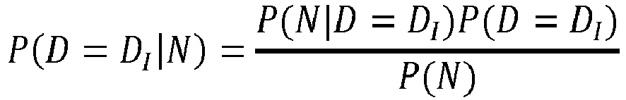
- The method according to any one of claim 1 to claim 9, wherein the similarity metric is a distance in a vector space between the image descriptor of the query image and the image descriptor of the candidate image.
- The method according to any one of claim 1 to claim 10, wherein the score S is determined based on a scoring equation:

- The method according to any one of claim 1 to claim 11, wherein the method comprises outputting the retrieved candidate images for display on a display device.
- The method according to any one of claim 1 to claim 12, wherein the method comprises displaying the retrieved candidate images on a display device in accordance with the determined scores.
- Apparatus arranged to perform the method according to any one of claim 1 to claim 13.
- A computer program which when executed by a computer causes the computer to perform the method according to any one of claim 1 to claim 13.
Priority Applications (3)
| Application Number | Priority Date | Filing Date | Title |
|---|---|---|---|
| EP20174500.7A EP3910645A1 (en) | 2020-05-13 | 2020-05-13 | Image retrieval |
| US17/236,273 US20210358600A1 (en) | 2020-05-13 | 2021-04-21 | Image retrieval |
| CN202110522314.8A CN113742503A (en) | 2020-05-13 | 2021-05-13 | Image retrieval |
Applications Claiming Priority (1)
| Application Number | Priority Date | Filing Date | Title |
|---|---|---|---|
| EP20174500.7A EP3910645A1 (en) | 2020-05-13 | 2020-05-13 | Image retrieval |
Publications (1)
| Publication Number | Publication Date |
|---|---|
| EP3910645A1 true EP3910645A1 (en) | 2021-11-17 |
Family
ID=70736604
Family Applications (1)
| Application Number | Title | Priority Date | Filing Date |
|---|---|---|---|
| EP20174500.7A Pending EP3910645A1 (en) | 2020-05-13 | 2020-05-13 | Image retrieval |
Country Status (3)
| Country | Link |
|---|---|
| US (1) | US20210358600A1 (en) |
| EP (1) | EP3910645A1 (en) |
| CN (1) | CN113742503A (en) |
Citations (5)
| Publication number | Priority date | Publication date | Assignee | Title |
|---|---|---|---|---|
| WO2007082218A2 (en) * | 2006-01-11 | 2007-07-19 | Boston Scientific Scimed, Inc. | Medical image retrieval |
| WO2013119400A1 (en) * | 2012-02-06 | 2013-08-15 | Microsoft Corporation | System and method for semantically annotating images |
| WO2014139021A1 (en) * | 2013-03-15 | 2014-09-18 | Synaptive Medical (Barbados) Inc. | Intramodal synchronization of surgical data |
| WO2017124116A1 (en) * | 2016-01-15 | 2017-07-20 | Bao Sheng | Searching, supplementing and navigating media |
| WO2020013814A1 (en) * | 2018-07-11 | 2020-01-16 | Google Llc | Similar image search for radiology |
Family Cites Families (6)
| Publication number | Priority date | Publication date | Assignee | Title |
|---|---|---|---|---|
| CN101765843A (en) * | 2007-08-01 | 2010-06-30 | 皇家飞利浦电子股份有限公司 | Accessing medical image detabases using medically relevant terms |
| JP5676597B2 (en) * | 2009-06-30 | 2015-02-25 | コーニンクレッカ フィリップス エヌ ヴェ | Relevance feedback on content-based image retrieval |
| CN105359161A (en) * | 2013-04-13 | 2016-02-24 | 宾夕法尼亚大学托管会 | System and method for medical image analysis and probabilistic diagnosis |
| WO2015134530A1 (en) * | 2014-03-03 | 2015-09-11 | Semanticmd, Inc. | Personalized content-based patient retrieval system |
| US11488712B2 (en) * | 2017-08-31 | 2022-11-01 | Google Llc | Diagnostic effectiveness tool |
| US11379516B2 (en) * | 2018-03-29 | 2022-07-05 | Google Llc | Similar medical image search |
-
2020
- 2020-05-13 EP EP20174500.7A patent/EP3910645A1/en active Pending
-
2021
- 2021-04-21 US US17/236,273 patent/US20210358600A1/en active Pending
- 2021-05-13 CN CN202110522314.8A patent/CN113742503A/en active Pending
Patent Citations (5)
| Publication number | Priority date | Publication date | Assignee | Title |
|---|---|---|---|---|
| WO2007082218A2 (en) * | 2006-01-11 | 2007-07-19 | Boston Scientific Scimed, Inc. | Medical image retrieval |
| WO2013119400A1 (en) * | 2012-02-06 | 2013-08-15 | Microsoft Corporation | System and method for semantically annotating images |
| WO2014139021A1 (en) * | 2013-03-15 | 2014-09-18 | Synaptive Medical (Barbados) Inc. | Intramodal synchronization of surgical data |
| WO2017124116A1 (en) * | 2016-01-15 | 2017-07-20 | Bao Sheng | Searching, supplementing and navigating media |
| WO2020013814A1 (en) * | 2018-07-11 | 2020-01-16 | Google Llc | Similar image search for radiology |
Non-Patent Citations (1)
| Title |
|---|
| MENG-SUNG WU ET AL: "A Term Association Translation Model for Naive Bayes Text Classification", 29 May 2012, ADVANCES IN KNOWLEDGE DISCOVERY AND DATA MINING, SPRINGER BERLIN HEIDELBERG, BERLIN, HEIDELBERG, PAGE(S) 243 - 253, ISBN: 978-3-642-30216-9, XP019176513 * |
Also Published As
| Publication number | Publication date |
|---|---|
| CN113742503A (en) | 2021-12-03 |
| US20210358600A1 (en) | 2021-11-18 |
Similar Documents
| Publication | Publication Date | Title |
|---|---|---|
| US11170900B2 (en) | Method and apparatus for refining similar case search | |
| JP5383431B2 (en) | Information processing apparatus, information processing method, and program | |
| US8880455B2 (en) | Medical decision making support apparatus and control method for the same | |
| US8260810B2 (en) | Case image registration apparatus, method and recording medium, and case image search apparatus, method, recording medium and system | |
| JP6818424B2 (en) | Diagnostic support device, information processing method, diagnostic support system and program | |
| US10318709B2 (en) | Method and system for cross-modality case-based computer-aided diagnosis | |
| JP7021215B2 (en) | CAD System Personalization Methods and Means for Providing Confidence Level Indicators for CAD System Recommendations | |
| US9053213B2 (en) | Interactive optimization of scan databases for statistical testing | |
| US20200105414A1 (en) | Information processing apparatus, information processing system, information processing method, and non-transitory computer-readable storage medium | |
| CN111640480B (en) | Medical report generation method, computer device, and storage medium | |
| US20170154167A1 (en) | A system and a related method for automatically selecting a hanging protocol for a medical study | |
| US20220037019A1 (en) | Medical scan artifact detection system and methods for use therewith | |
| Ornelas-Aguirre et al. | Concordance between premortem and postmortem diagnosis in the autopsy: results of a 10-year study in a tertiary care center | |
| US10282516B2 (en) | Medical imaging reference retrieval | |
| Jiménez-Sánchez et al. | Detecting shortcuts in medical images-a case study in chest x-rays | |
| EP3910645A1 (en) | Image retrieval | |
| EP3667674A1 (en) | Method and system for evaluating images of different patients, computer program and electronically readable storage medium | |
| US20100131514A1 (en) | Real-time automatic searching system for medical image and method for using the same | |
| US20220076842A1 (en) | Medical information processing system and method | |
| US20230290451A1 (en) | Medical data storage and retrieval system and method thereof | |
| US20040045560A1 (en) | Computer-assisted multi-dimensional patient-selection | |
| CN116682558A (en) | User health management method and device, computer equipment and storage medium | |
| CN116403672A (en) | Method, apparatus and medium for medical report composition | |
| CN116895376A (en) | Method and data processing system for providing stroke information | |
| CN115881266A (en) | Computer-implemented method for providing medical action recommendations and action recommendation device |
Legal Events
| Date | Code | Title | Description |
|---|---|---|---|
| PUAI | Public reference made under article 153(3) epc to a published international application that has entered the european phase |
Free format text: ORIGINAL CODE: 0009012 |
|
| STAA | Information on the status of an ep patent application or granted ep patent |
Free format text: STATUS: REQUEST FOR EXAMINATION WAS MADE |
|
| 17P | Request for examination filed |
Effective date: 20200513 |
|
| AK | Designated contracting states |
Kind code of ref document: A1 Designated state(s): AL AT BE BG CH CY CZ DE DK EE ES FI FR GB GR HR HU IE IS IT LI LT LU LV MC MK MT NL NO PL PT RO RS SE SI SK SM TR |
|
| B565 | Issuance of search results under rule 164(2) epc |
Effective date: 20201029 |
|
| RBV | Designated contracting states (corrected) |
Designated state(s): AL AT BE BG CH CY CZ DE DK EE ES FI FR GB GR HR HU IE IS IT LI LT LU LV MC MK MT NL NO PL PT RO RS SE SI SK SM TR |
|
| RAP1 | Party data changed (applicant data changed or rights of an application transferred) |
Owner name: SIEMENS HEALTHINEERS AG |