EP3889257A1 - High efficiency construction of dna libraries - Google Patents
High efficiency construction of dna libraries Download PDFInfo
- Publication number
- EP3889257A1 EP3889257A1 EP21152311.3A EP21152311A EP3889257A1 EP 3889257 A1 EP3889257 A1 EP 3889257A1 EP 21152311 A EP21152311 A EP 21152311A EP 3889257 A1 EP3889257 A1 EP 3889257A1
- Authority
- EP
- European Patent Office
- Prior art keywords
- dna
- sample
- ligation
- oligonucleotide
- nucleotides
- Prior art date
- Legal status (The legal status is an assumption and is not a legal conclusion. Google has not performed a legal analysis and makes no representation as to the accuracy of the status listed.)
- Pending
Links
- 238000010276 construction Methods 0.000 title abstract description 9
- 238000000034 method Methods 0.000 claims abstract description 175
- 238000012252 genetic analysis Methods 0.000 claims abstract description 52
- 108020004414 DNA Proteins 0.000 claims description 505
- 108091034117 Oligonucleotide Proteins 0.000 claims description 273
- 239000000523 sample Substances 0.000 claims description 202
- 230000002068 genetic effect Effects 0.000 claims description 119
- 239000012634 fragment Substances 0.000 claims description 109
- 230000008439 repair process Effects 0.000 claims description 107
- 230000027455 binding Effects 0.000 claims description 64
- 238000012163 sequencing technique Methods 0.000 claims description 63
- 102000053602 DNA Human genes 0.000 claims description 43
- 102000004190 Enzymes Human genes 0.000 claims description 41
- 108090000790 Enzymes Proteins 0.000 claims description 41
- 230000000295 complement effect Effects 0.000 claims description 29
- 239000012472 biological sample Substances 0.000 claims description 26
- 108010061982 DNA Ligases Proteins 0.000 claims description 23
- 102000012410 DNA Ligases Human genes 0.000 claims description 23
- 108010014303 DNA-directed DNA polymerase Proteins 0.000 claims description 21
- 102000016928 DNA-directed DNA polymerase Human genes 0.000 claims description 21
- 125000002467 phosphate group Chemical group [H]OP(=O)(O[H])O[*] 0.000 claims description 19
- 210000000582 semen Anatomy 0.000 claims description 19
- 210000004369 blood Anatomy 0.000 claims description 17
- 239000008280 blood Substances 0.000 claims description 17
- 108020004635 Complementary DNA Proteins 0.000 claims description 16
- 230000015572 biosynthetic process Effects 0.000 claims description 16
- 238000010804 cDNA synthesis Methods 0.000 claims description 16
- 239000002299 complementary DNA Substances 0.000 claims description 16
- 239000012530 fluid Substances 0.000 claims description 16
- -1 and a formalin-fixed Substances 0.000 claims description 15
- 210000002381 plasma Anatomy 0.000 claims description 13
- 229910019142 PO4 Inorganic materials 0.000 claims description 12
- 210000001519 tissue Anatomy 0.000 claims description 12
- 238000001712 DNA sequencing Methods 0.000 claims description 11
- 210000001175 cerebrospinal fluid Anatomy 0.000 claims description 11
- 210000003296 saliva Anatomy 0.000 claims description 11
- 210000004243 sweat Anatomy 0.000 claims description 11
- 210000002700 urine Anatomy 0.000 claims description 11
- 102000003960 Ligases Human genes 0.000 claims description 10
- 108090000364 Ligases Proteins 0.000 claims description 10
- 238000007622 bioinformatic analysis Methods 0.000 claims description 10
- 239000000539 dimer Substances 0.000 claims description 10
- 230000004048 modification Effects 0.000 claims description 9
- 238000012986 modification Methods 0.000 claims description 9
- 238000012408 PCR amplification Methods 0.000 claims description 8
- 210000004381 amniotic fluid Anatomy 0.000 claims description 8
- 238000001574 biopsy Methods 0.000 claims description 8
- 230000001926 lymphatic effect Effects 0.000 claims description 8
- 239000010452 phosphate Substances 0.000 claims description 8
- 210000004909 pre-ejaculatory fluid Anatomy 0.000 claims description 8
- 210000002966 serum Anatomy 0.000 claims description 8
- NBIIXXVUZAFLBC-UHFFFAOYSA-K phosphate Chemical compound [O-]P([O-])([O-])=O NBIIXXVUZAFLBC-UHFFFAOYSA-K 0.000 claims description 7
- 108010021757 Polynucleotide 5'-Hydroxyl-Kinase Proteins 0.000 claims description 6
- 102000008422 Polynucleotide 5'-hydroxyl-kinase Human genes 0.000 claims description 6
- 239000012188 paraffin wax Substances 0.000 claims description 5
- 206010003445 Ascites Diseases 0.000 claims description 4
- 210000000981 epithelium Anatomy 0.000 claims description 4
- 210000003608 fece Anatomy 0.000 claims description 4
- 210000003780 hair follicle Anatomy 0.000 claims description 4
- 210000002751 lymph Anatomy 0.000 claims description 4
- 210000004908 prostatic fluid Anatomy 0.000 claims description 4
- 210000003491 skin Anatomy 0.000 claims description 4
- 210000001138 tear Anatomy 0.000 claims description 4
- 231100000640 hair analysis Toxicity 0.000 claims 1
- 239000002773 nucleotide Substances 0.000 description 251
- 125000003729 nucleotide group Chemical group 0.000 description 251
- 239000000203 mixture Substances 0.000 description 51
- 230000003902 lesion Effects 0.000 description 34
- 238000006243 chemical reaction Methods 0.000 description 33
- 208000026350 Inborn Genetic disease Diseases 0.000 description 27
- 238000004458 analytical method Methods 0.000 description 27
- 208000016361 genetic disease Diseases 0.000 description 27
- 206010028980 Neoplasm Diseases 0.000 description 26
- 108090000623 proteins and genes Proteins 0.000 description 24
- 230000003321 amplification Effects 0.000 description 23
- 238000003199 nucleic acid amplification method Methods 0.000 description 23
- 108091028043 Nucleic acid sequence Proteins 0.000 description 21
- 241000700605 Viruses Species 0.000 description 20
- 238000012544 monitoring process Methods 0.000 description 20
- JLCPHMBAVCMARE-UHFFFAOYSA-N [3-[[3-[[3-[[3-[[3-[[3-[[3-[[3-[[3-[[3-[[3-[[5-(2-amino-6-oxo-1H-purin-9-yl)-3-[[3-[[3-[[3-[[3-[[3-[[5-(2-amino-6-oxo-1H-purin-9-yl)-3-[[5-(2-amino-6-oxo-1H-purin-9-yl)-3-hydroxyoxolan-2-yl]methoxy-hydroxyphosphoryl]oxyoxolan-2-yl]methoxy-hydroxyphosphoryl]oxy-5-(5-methyl-2,4-dioxopyrimidin-1-yl)oxolan-2-yl]methoxy-hydroxyphosphoryl]oxy-5-(6-aminopurin-9-yl)oxolan-2-yl]methoxy-hydroxyphosphoryl]oxy-5-(6-aminopurin-9-yl)oxolan-2-yl]methoxy-hydroxyphosphoryl]oxy-5-(6-aminopurin-9-yl)oxolan-2-yl]methoxy-hydroxyphosphoryl]oxy-5-(6-aminopurin-9-yl)oxolan-2-yl]methoxy-hydroxyphosphoryl]oxyoxolan-2-yl]methoxy-hydroxyphosphoryl]oxy-5-(5-methyl-2,4-dioxopyrimidin-1-yl)oxolan-2-yl]methoxy-hydroxyphosphoryl]oxy-5-(4-amino-2-oxopyrimidin-1-yl)oxolan-2-yl]methoxy-hydroxyphosphoryl]oxy-5-(5-methyl-2,4-dioxopyrimidin-1-yl)oxolan-2-yl]methoxy-hydroxyphosphoryl]oxy-5-(5-methyl-2,4-dioxopyrimidin-1-yl)oxolan-2-yl]methoxy-hydroxyphosphoryl]oxy-5-(6-aminopurin-9-yl)oxolan-2-yl]methoxy-hydroxyphosphoryl]oxy-5-(6-aminopurin-9-yl)oxolan-2-yl]methoxy-hydroxyphosphoryl]oxy-5-(4-amino-2-oxopyrimidin-1-yl)oxolan-2-yl]methoxy-hydroxyphosphoryl]oxy-5-(4-amino-2-oxopyrimidin-1-yl)oxolan-2-yl]methoxy-hydroxyphosphoryl]oxy-5-(4-amino-2-oxopyrimidin-1-yl)oxolan-2-yl]methoxy-hydroxyphosphoryl]oxy-5-(6-aminopurin-9-yl)oxolan-2-yl]methoxy-hydroxyphosphoryl]oxy-5-(4-amino-2-oxopyrimidin-1-yl)oxolan-2-yl]methyl [5-(6-aminopurin-9-yl)-2-(hydroxymethyl)oxolan-3-yl] hydrogen phosphate Polymers Cc1cn(C2CC(OP(O)(=O)OCC3OC(CC3OP(O)(=O)OCC3OC(CC3O)n3cnc4c3nc(N)[nH]c4=O)n3cnc4c3nc(N)[nH]c4=O)C(COP(O)(=O)OC3CC(OC3COP(O)(=O)OC3CC(OC3COP(O)(=O)OC3CC(OC3COP(O)(=O)OC3CC(OC3COP(O)(=O)OC3CC(OC3COP(O)(=O)OC3CC(OC3COP(O)(=O)OC3CC(OC3COP(O)(=O)OC3CC(OC3COP(O)(=O)OC3CC(OC3COP(O)(=O)OC3CC(OC3COP(O)(=O)OC3CC(OC3COP(O)(=O)OC3CC(OC3COP(O)(=O)OC3CC(OC3COP(O)(=O)OC3CC(OC3COP(O)(=O)OC3CC(OC3COP(O)(=O)OC3CC(OC3COP(O)(=O)OC3CC(OC3CO)n3cnc4c(N)ncnc34)n3ccc(N)nc3=O)n3cnc4c(N)ncnc34)n3ccc(N)nc3=O)n3ccc(N)nc3=O)n3ccc(N)nc3=O)n3cnc4c(N)ncnc34)n3cnc4c(N)ncnc34)n3cc(C)c(=O)[nH]c3=O)n3cc(C)c(=O)[nH]c3=O)n3ccc(N)nc3=O)n3cc(C)c(=O)[nH]c3=O)n3cnc4c3nc(N)[nH]c4=O)n3cnc4c(N)ncnc34)n3cnc4c(N)ncnc34)n3cnc4c(N)ncnc34)n3cnc4c(N)ncnc34)O2)c(=O)[nH]c1=O JLCPHMBAVCMARE-UHFFFAOYSA-N 0.000 description 19
- 238000003556 assay Methods 0.000 description 17
- 230000004927 fusion Effects 0.000 description 17
- 238000012360 testing method Methods 0.000 description 17
- 238000001514 detection method Methods 0.000 description 15
- 201000011510 cancer Diseases 0.000 description 14
- 238000003753 real-time PCR Methods 0.000 description 14
- 102000039446 nucleic acids Human genes 0.000 description 13
- 108020004707 nucleic acids Proteins 0.000 description 13
- 150000007523 nucleic acids Chemical class 0.000 description 13
- 108060002716 Exonuclease Proteins 0.000 description 12
- 230000008859 change Effects 0.000 description 12
- 102000013165 exonuclease Human genes 0.000 description 12
- 238000012217 deletion Methods 0.000 description 11
- 230000037430 deletion Effects 0.000 description 11
- 201000010099 disease Diseases 0.000 description 11
- 208000037265 diseases, disorders, signs and symptoms Diseases 0.000 description 11
- 230000000694 effects Effects 0.000 description 11
- 238000003780 insertion Methods 0.000 description 11
- 230000037431 insertion Effects 0.000 description 11
- 235000021317 phosphate Nutrition 0.000 description 11
- 230000035945 sensitivity Effects 0.000 description 11
- 238000013461 design Methods 0.000 description 10
- 230000008707 rearrangement Effects 0.000 description 10
- 230000007704 transition Effects 0.000 description 10
- 239000000090 biomarker Substances 0.000 description 9
- 230000001605 fetal effect Effects 0.000 description 9
- UYTPUPDQBNUYGX-UHFFFAOYSA-N guanine Chemical compound O=C1NC(N)=NC2=C1N=CN2 UYTPUPDQBNUYGX-UHFFFAOYSA-N 0.000 description 9
- 241000894007 species Species 0.000 description 9
- 238000011282 treatment Methods 0.000 description 9
- YBJHBAHKTGYVGT-ZKWXMUAHSA-N (+)-Biotin Chemical compound N1C(=O)N[C@@H]2[C@H](CCCCC(=O)O)SC[C@@H]21 YBJHBAHKTGYVGT-ZKWXMUAHSA-N 0.000 description 8
- ISAKRJDGNUQOIC-UHFFFAOYSA-N Uracil Chemical compound O=C1C=CNC(=O)N1 ISAKRJDGNUQOIC-UHFFFAOYSA-N 0.000 description 8
- 230000006378 damage Effects 0.000 description 8
- 230000002255 enzymatic effect Effects 0.000 description 8
- 230000000670 limiting effect Effects 0.000 description 8
- 239000011324 bead Substances 0.000 description 7
- 244000005700 microbiome Species 0.000 description 7
- 210000000056 organ Anatomy 0.000 description 7
- 244000052769 pathogen Species 0.000 description 7
- 102000040430 polynucleotide Human genes 0.000 description 7
- 108091033319 polynucleotide Proteins 0.000 description 7
- 239000002157 polynucleotide Substances 0.000 description 7
- 238000012545 processing Methods 0.000 description 7
- 238000012216 screening Methods 0.000 description 7
- 230000005778 DNA damage Effects 0.000 description 6
- 231100000277 DNA damage Toxicity 0.000 description 6
- LFQSCWFLJHTTHZ-UHFFFAOYSA-N Ethanol Chemical compound CCO LFQSCWFLJHTTHZ-UHFFFAOYSA-N 0.000 description 6
- 206010068052 Mosaicism Diseases 0.000 description 6
- 108090000608 Phosphoric Monoester Hydrolases Proteins 0.000 description 6
- 102000004160 Phosphoric Monoester Hydrolases Human genes 0.000 description 6
- 229940079593 drug Drugs 0.000 description 6
- 239000003814 drug Substances 0.000 description 6
- 238000009396 hybridization Methods 0.000 description 6
- 230000035772 mutation Effects 0.000 description 6
- 230000001717 pathogenic effect Effects 0.000 description 6
- 230000004044 response Effects 0.000 description 6
- 208000035657 Abasia Diseases 0.000 description 5
- 108091092878 Microsatellite Proteins 0.000 description 5
- 230000009471 action Effects 0.000 description 5
- 238000003766 bioinformatics method Methods 0.000 description 5
- 238000010367 cloning Methods 0.000 description 5
- 238000005516 engineering process Methods 0.000 description 5
- 238000005259 measurement Methods 0.000 description 5
- 239000000758 substrate Substances 0.000 description 5
- RWQNBRDOKXIBIV-UHFFFAOYSA-N thymine Chemical class CC1=CNC(=O)NC1=O RWQNBRDOKXIBIV-UHFFFAOYSA-N 0.000 description 5
- 108091032973 (ribonucleotides)n+m Proteins 0.000 description 4
- 102000002260 Alkaline Phosphatase Human genes 0.000 description 4
- 108020004774 Alkaline Phosphatase Proteins 0.000 description 4
- 108091023043 Alu Element Proteins 0.000 description 4
- 102100021519 Hemoglobin subunit beta Human genes 0.000 description 4
- TWRXJAOTZQYOKJ-UHFFFAOYSA-L Magnesium chloride Chemical compound [Mg+2].[Cl-].[Cl-] TWRXJAOTZQYOKJ-UHFFFAOYSA-L 0.000 description 4
- 241001465754 Metazoa Species 0.000 description 4
- 208000003019 Neurofibromatosis 1 Diseases 0.000 description 4
- 108091093078 Pyrimidine dimer Proteins 0.000 description 4
- 238000012300 Sequence Analysis Methods 0.000 description 4
- 102000006943 Uracil-DNA Glycosidase Human genes 0.000 description 4
- 108010072685 Uracil-DNA Glycosidase Proteins 0.000 description 4
- 229960002685 biotin Drugs 0.000 description 4
- 235000020958 biotin Nutrition 0.000 description 4
- 239000011616 biotin Substances 0.000 description 4
- 230000000903 blocking effect Effects 0.000 description 4
- 239000000872 buffer Substances 0.000 description 4
- 210000004027 cell Anatomy 0.000 description 4
- 210000004209 hair Anatomy 0.000 description 4
- 238000002955 isolation Methods 0.000 description 4
- 238000002844 melting Methods 0.000 description 4
- 230000008018 melting Effects 0.000 description 4
- 238000010369 molecular cloning Methods 0.000 description 4
- 150000003013 phosphoric acid derivatives Chemical class 0.000 description 4
- 238000002864 sequence alignment Methods 0.000 description 4
- 239000000243 solution Substances 0.000 description 4
- 108010068698 spleen exonuclease Proteins 0.000 description 4
- WYWHKKSPHMUBEB-UHFFFAOYSA-N tioguanine Chemical compound N1C(N)=NC(=S)C2=C1N=CN2 WYWHKKSPHMUBEB-UHFFFAOYSA-N 0.000 description 4
- 229940035893 uracil Drugs 0.000 description 4
- 102100033350 ATP-dependent translocase ABCB1 Human genes 0.000 description 3
- 102100025401 Breast cancer type 1 susceptibility protein Human genes 0.000 description 3
- 102100029363 Cytochrome P450 2C19 Human genes 0.000 description 3
- 102100021704 Cytochrome P450 2D6 Human genes 0.000 description 3
- 102100039205 Cytochrome P450 3A4 Human genes 0.000 description 3
- 102100039208 Cytochrome P450 3A5 Human genes 0.000 description 3
- 102100033215 DNA nucleotidylexotransferase Human genes 0.000 description 3
- 102100022334 Dihydropyrimidine dehydrogenase [NADP(+)] Human genes 0.000 description 3
- KCXVZYZYPLLWCC-UHFFFAOYSA-N EDTA Chemical compound OC(=O)CN(CC(O)=O)CCN(CC(O)=O)CC(O)=O KCXVZYZYPLLWCC-UHFFFAOYSA-N 0.000 description 3
- GHASVSINZRGABV-UHFFFAOYSA-N Fluorouracil Chemical compound FC1=CNC(=O)NC1=O GHASVSINZRGABV-UHFFFAOYSA-N 0.000 description 3
- 102000007648 Glutathione S-Transferase pi Human genes 0.000 description 3
- 108010007355 Glutathione S-Transferase pi Proteins 0.000 description 3
- 101000934870 Homo sapiens Breast cancer type 1 susceptibility protein Proteins 0.000 description 3
- 101000919361 Homo sapiens Cytochrome P450 2C19 Proteins 0.000 description 3
- 101000896586 Homo sapiens Cytochrome P450 2D6 Proteins 0.000 description 3
- 101000745711 Homo sapiens Cytochrome P450 3A4 Proteins 0.000 description 3
- 101000745710 Homo sapiens Cytochrome P450 3A5 Proteins 0.000 description 3
- 101000973778 Homo sapiens NAD(P)H dehydrogenase [quinone] 1 Proteins 0.000 description 3
- 101000826399 Homo sapiens Sulfotransferase 1A1 Proteins 0.000 description 3
- 241000725303 Human immunodeficiency virus Species 0.000 description 3
- FBOZXECLQNJBKD-ZDUSSCGKSA-N L-methotrexate Chemical compound C=1N=C2N=C(N)N=C(N)C2=NC=1CN(C)C1=CC=C(C(=O)N[C@@H](CCC(O)=O)C(O)=O)C=C1 FBOZXECLQNJBKD-ZDUSSCGKSA-N 0.000 description 3
- 102100029684 Methylenetetrahydrofolate reductase Human genes 0.000 description 3
- 241001529936 Murinae Species 0.000 description 3
- 102100022365 NAD(P)H dehydrogenase [quinone] 1 Human genes 0.000 description 3
- 208000024834 Neurofibromatosis type 1 Diseases 0.000 description 3
- 102000007530 Neurofibromin 1 Human genes 0.000 description 3
- 108010085793 Neurofibromin 1 Proteins 0.000 description 3
- 206010035226 Plasma cell myeloma Diseases 0.000 description 3
- 102100022807 Potassium voltage-gated channel subfamily H member 2 Human genes 0.000 description 3
- 102100029753 Reduced folate transporter Human genes 0.000 description 3
- 102100023986 Sulfotransferase 1A1 Human genes 0.000 description 3
- ASJWEHCPLGMOJE-LJMGSBPFSA-N ac1l3rvh Chemical compound N1C(=O)NC(=O)[C@@]2(C)[C@@]3(C)C(=O)NC(=O)N[C@H]3[C@H]21 ASJWEHCPLGMOJE-LJMGSBPFSA-N 0.000 description 3
- 239000002253 acid Substances 0.000 description 3
- 238000011319 anticancer therapy Methods 0.000 description 3
- 238000013459 approach Methods 0.000 description 3
- OPTASPLRGRRNAP-UHFFFAOYSA-N cytosine Chemical class NC=1C=CNC(=O)N=1 OPTASPLRGRRNAP-UHFFFAOYSA-N 0.000 description 3
- 238000003745 diagnosis Methods 0.000 description 3
- 230000009977 dual effect Effects 0.000 description 3
- 229960002949 fluorouracil Drugs 0.000 description 3
- 238000013467 fragmentation Methods 0.000 description 3
- 238000006062 fragmentation reaction Methods 0.000 description 3
- 239000000463 material Substances 0.000 description 3
- GLVAUDGFNGKCSF-UHFFFAOYSA-N mercaptopurine Chemical compound S=C1NC=NC2=C1NC=N2 GLVAUDGFNGKCSF-UHFFFAOYSA-N 0.000 description 3
- 229960000485 methotrexate Drugs 0.000 description 3
- 230000011987 methylation Effects 0.000 description 3
- 238000007069 methylation reaction Methods 0.000 description 3
- KKZJGLLVHKMTCM-UHFFFAOYSA-N mitoxantrone Chemical compound O=C1C2=C(O)C=CC(O)=C2C(=O)C2=C1C(NCCNCCO)=CC=C2NCCNCCO KKZJGLLVHKMTCM-UHFFFAOYSA-N 0.000 description 3
- 238000007481 next generation sequencing Methods 0.000 description 3
- 150000003839 salts Chemical class 0.000 description 3
- 102100029077 3-hydroxy-3-methylglutaryl-coenzyme A reductase Human genes 0.000 description 2
- 101710158485 3-hydroxy-3-methylglutaryl-coenzyme A reductase Proteins 0.000 description 2
- 101150023956 ALK gene Proteins 0.000 description 2
- 102100024643 ATP-binding cassette sub-family D member 1 Human genes 0.000 description 2
- 208000024893 Acute lymphoblastic leukemia Diseases 0.000 description 2
- 208000014697 Acute lymphocytic leukaemia Diseases 0.000 description 2
- 208000031261 Acute myeloid leukaemia Diseases 0.000 description 2
- 229930024421 Adenine Natural products 0.000 description 2
- GFFGJBXGBJISGV-UHFFFAOYSA-N Adenine Chemical compound NC1=NC=NC2=C1N=CN2 GFFGJBXGBJISGV-UHFFFAOYSA-N 0.000 description 2
- 102100034035 Alcohol dehydrogenase 1A Human genes 0.000 description 2
- 102100034042 Alcohol dehydrogenase 1C Human genes 0.000 description 2
- 102100040069 Aldehyde dehydrogenase 1A1 Human genes 0.000 description 2
- 241000143060 Americamysis bahia Species 0.000 description 2
- 108090000448 Aryl Hydrocarbon Receptors Proteins 0.000 description 2
- 102100026792 Aryl hydrocarbon receptor Human genes 0.000 description 2
- IJGRMHOSHXDMSA-UHFFFAOYSA-N Atomic nitrogen Chemical compound N#N IJGRMHOSHXDMSA-UHFFFAOYSA-N 0.000 description 2
- 208000032791 BCR-ABL1 positive chronic myelogenous leukemia Diseases 0.000 description 2
- 102100040794 Beta-1 adrenergic receptor Human genes 0.000 description 2
- 102100039705 Beta-2 adrenergic receptor Human genes 0.000 description 2
- 102100022548 Beta-hexosaminidase subunit alpha Human genes 0.000 description 2
- 241000283690 Bos taurus Species 0.000 description 2
- 206010006187 Breast cancer Diseases 0.000 description 2
- GAGWJHPBXLXJQN-UORFTKCHSA-N Capecitabine Chemical compound C1=C(F)C(NC(=O)OCCCCC)=NC(=O)N1[C@H]1[C@H](O)[C@H](O)[C@@H](C)O1 GAGWJHPBXLXJQN-UORFTKCHSA-N 0.000 description 2
- 241000713756 Caprine arthritis encephalitis virus Species 0.000 description 2
- 102000006378 Catechol O-methyltransferase Human genes 0.000 description 2
- 108020002739 Catechol O-methyltransferase Proteins 0.000 description 2
- 208000010833 Chronic myeloid leukaemia Diseases 0.000 description 2
- 108091026890 Coding region Proteins 0.000 description 2
- 206010009944 Colon cancer Diseases 0.000 description 2
- CMSMOCZEIVJLDB-UHFFFAOYSA-N Cyclophosphamide Chemical compound ClCCN(CCCl)P1(=O)NCCCO1 CMSMOCZEIVJLDB-UHFFFAOYSA-N 0.000 description 2
- UHDGCWIWMRVCDJ-CCXZUQQUSA-N Cytarabine Chemical compound O=C1N=C(N)C=CN1[C@H]1[C@@H](O)[C@H](O)[C@@H](CO)O1 UHDGCWIWMRVCDJ-CCXZUQQUSA-N 0.000 description 2
- 102000002237 Cytochrome P-450 CYP2A6 Human genes 0.000 description 2
- 108010000080 Cytochrome P-450 CYP2A6 Proteins 0.000 description 2
- 102100038739 Cytochrome P450 2B6 Human genes 0.000 description 2
- 102100029358 Cytochrome P450 2C9 Human genes 0.000 description 2
- 102100031461 Cytochrome P450 2J2 Human genes 0.000 description 2
- 102100020756 D(2) dopamine receptor Human genes 0.000 description 2
- 108010017826 DNA Polymerase I Proteins 0.000 description 2
- 102000004594 DNA Polymerase I Human genes 0.000 description 2
- 108010092160 Dactinomycin Proteins 0.000 description 2
- 108010082610 Deoxyribonuclease (Pyrimidine Dimer) Proteins 0.000 description 2
- 102000004099 Deoxyribonuclease (Pyrimidine Dimer) Human genes 0.000 description 2
- 108010036364 Deoxyribonuclease IV (Phage T4-Induced) Proteins 0.000 description 2
- 201000010374 Down Syndrome Diseases 0.000 description 2
- AOJJSUZBOXZQNB-TZSSRYMLSA-N Doxorubicin Chemical compound O([C@H]1C[C@@](O)(CC=2C(O)=C3C(=O)C=4C=CC=C(C=4C(=O)C3=C(O)C=21)OC)C(=O)CO)[C@H]1C[C@H](N)[C@H](O)[C@H](C)O1 AOJJSUZBOXZQNB-TZSSRYMLSA-N 0.000 description 2
- 206010013801 Duchenne Muscular Dystrophy Diseases 0.000 description 2
- 102100024108 Dystrophin Human genes 0.000 description 2
- 201000006360 Edwards syndrome Diseases 0.000 description 2
- 101000877447 Enterobacteria phage T4 Endonuclease V Proteins 0.000 description 2
- 241000713730 Equine infectious anemia virus Species 0.000 description 2
- 241000713800 Feline immunodeficiency virus Species 0.000 description 2
- 241000714165 Feline leukemia virus Species 0.000 description 2
- 102100027842 Fibroblast growth factor receptor 3 Human genes 0.000 description 2
- 101710182396 Fibroblast growth factor receptor 3 Proteins 0.000 description 2
- 241000711950 Filoviridae Species 0.000 description 2
- 108090000652 Flap endonucleases Proteins 0.000 description 2
- 102000004150 Flap endonucleases Human genes 0.000 description 2
- 241000713813 Gibbon ape leukemia virus Species 0.000 description 2
- 102100031561 Hamartin Human genes 0.000 description 2
- 241000713858 Harvey murine sarcoma virus Species 0.000 description 2
- 108091005904 Hemoglobin subunit beta Proteins 0.000 description 2
- 208000032087 Hereditary Leber Optic Atrophy Diseases 0.000 description 2
- 101000614701 Homo sapiens ATP-sensitive inward rectifier potassium channel 11 Proteins 0.000 description 2
- 101000780443 Homo sapiens Alcohol dehydrogenase 1A Proteins 0.000 description 2
- 101000780463 Homo sapiens Alcohol dehydrogenase 1C Proteins 0.000 description 2
- 101000890570 Homo sapiens Aldehyde dehydrogenase 1A1 Proteins 0.000 description 2
- 101000892264 Homo sapiens Beta-1 adrenergic receptor Proteins 0.000 description 2
- 101000959437 Homo sapiens Beta-2 adrenergic receptor Proteins 0.000 description 2
- 101000957383 Homo sapiens Cytochrome P450 2B6 Proteins 0.000 description 2
- 101000919359 Homo sapiens Cytochrome P450 2C9 Proteins 0.000 description 2
- 101000941723 Homo sapiens Cytochrome P450 2J2 Proteins 0.000 description 2
- 101000931901 Homo sapiens D(2) dopamine receptor Proteins 0.000 description 2
- 101000902632 Homo sapiens Dihydropyrimidine dehydrogenase [NADP(+)] Proteins 0.000 description 2
- 101000587058 Homo sapiens Methylenetetrahydrofolate reductase Proteins 0.000 description 2
- 101001047090 Homo sapiens Potassium voltage-gated channel subfamily H member 2 Proteins 0.000 description 2
- 101000694017 Homo sapiens Sodium channel protein type 5 subunit alpha Proteins 0.000 description 2
- 101000836291 Homo sapiens Solute carrier organic anion transporter family member 1B1 Proteins 0.000 description 2
- 101000799388 Homo sapiens Thiopurine S-methyltransferase Proteins 0.000 description 2
- 101000809797 Homo sapiens Thymidylate synthase Proteins 0.000 description 2
- 201000000639 Leber hereditary optic neuropathy Diseases 0.000 description 2
- 102100024640 Low-density lipoprotein receptor Human genes 0.000 description 2
- 241000712899 Lymphocytic choriomeningitis mammarenavirus Species 0.000 description 2
- 102100033342 Lysosomal acid glucosylceramidase Human genes 0.000 description 2
- 108010047230 Member 1 Subfamily B ATP Binding Cassette Transporter Proteins 0.000 description 2
- 108010049137 Member 1 Subfamily D ATP Binding Cassette Transporter Proteins 0.000 description 2
- 241000713869 Moloney murine leukemia virus Species 0.000 description 2
- 241000713862 Moloney murine sarcoma virus Species 0.000 description 2
- 208000034578 Multiple myelomas Diseases 0.000 description 2
- 201000003793 Myelodysplastic syndrome Diseases 0.000 description 2
- 208000033761 Myelogenous Chronic BCR-ABL Positive Leukemia Diseases 0.000 description 2
- 208000033776 Myeloid Acute Leukemia Diseases 0.000 description 2
- 208000014767 Myeloproliferative disease Diseases 0.000 description 2
- 201000007224 Myeloproliferative neoplasm Diseases 0.000 description 2
- NWIBSHFKIJFRCO-WUDYKRTCSA-N Mytomycin Chemical compound C1N2C(C(C(C)=C(N)C3=O)=O)=C3[C@@H](COC(N)=O)[C@@]2(OC)[C@@H]2[C@H]1N2 NWIBSHFKIJFRCO-WUDYKRTCSA-N 0.000 description 2
- 208000015914 Non-Hodgkin lymphomas Diseases 0.000 description 2
- 102100037600 P2Y purinoceptor 1 Human genes 0.000 description 2
- 102100026171 P2Y purinoceptor 12 Human genes 0.000 description 2
- 201000009928 Patau syndrome Diseases 0.000 description 2
- 102000004270 Peptidyl-Dipeptidase A Human genes 0.000 description 2
- 108090000882 Peptidyl-Dipeptidase A Proteins 0.000 description 2
- 108010010677 Phosphodiesterase I Proteins 0.000 description 2
- 229920002594 Polyethylene Glycol 8000 Polymers 0.000 description 2
- 229920001213 Polysorbate 20 Polymers 0.000 description 2
- 102100022364 Polyunsaturated fatty acid 5-lipoxygenase Human genes 0.000 description 2
- 208000006664 Precursor Cell Lymphoblastic Leukemia-Lymphoma Diseases 0.000 description 2
- 108010028935 Purinergic P2Y1 Receptors Proteins 0.000 description 2
- 108020004511 Recombinant DNA Proteins 0.000 description 2
- 201000000582 Retinoblastoma Diseases 0.000 description 2
- 241000714474 Rous sarcoma virus Species 0.000 description 2
- 108091006778 SLC19A1 Proteins 0.000 description 2
- 241000713311 Simian immunodeficiency virus Species 0.000 description 2
- 208000001203 Smallpox Diseases 0.000 description 2
- 102100027198 Sodium channel protein type 5 subunit alpha Human genes 0.000 description 2
- FAPWRFPIFSIZLT-UHFFFAOYSA-M Sodium chloride Chemical compound [Na+].[Cl-] FAPWRFPIFSIZLT-UHFFFAOYSA-M 0.000 description 2
- 102100027233 Solute carrier organic anion transporter family member 1B1 Human genes 0.000 description 2
- 208000005718 Stomach Neoplasms Diseases 0.000 description 2
- 108010090804 Streptavidin Proteins 0.000 description 2
- NKANXQFJJICGDU-QPLCGJKRSA-N Tamoxifen Chemical compound C=1C=CC=CC=1C(/CC)=C(C=1C=CC(OCCN(C)C)=CC=1)/C1=CC=CC=C1 NKANXQFJJICGDU-QPLCGJKRSA-N 0.000 description 2
- 102100034162 Thiopurine S-methyltransferase Human genes 0.000 description 2
- FOCVUCIESVLUNU-UHFFFAOYSA-N Thiotepa Chemical compound C1CN1P(N1CC1)(=S)N1CC1 FOCVUCIESVLUNU-UHFFFAOYSA-N 0.000 description 2
- 102100038618 Thymidylate synthase Human genes 0.000 description 2
- 239000007983 Tris buffer Substances 0.000 description 2
- 206010044686 Trisomy 13 Diseases 0.000 description 2
- 208000006284 Trisomy 13 Syndrome Diseases 0.000 description 2
- 208000007159 Trisomy 18 Syndrome Diseases 0.000 description 2
- 102100031638 Tuberin Human genes 0.000 description 2
- 102100030434 Ubiquitin-protein ligase E3A Human genes 0.000 description 2
- 241000711975 Vesicular stomatitis virus Species 0.000 description 2
- 150000007513 acids Chemical class 0.000 description 2
- RJURFGZVJUQBHK-UHFFFAOYSA-N actinomycin D Natural products CC1OC(=O)C(C(C)C)N(C)C(=O)CN(C)C(=O)C2CCCN2C(=O)C(C(C)C)NC(=O)C1NC(=O)C1=C(N)C(=O)C(C)=C2OC(C(C)=CC=C3C(=O)NC4C(=O)NC(C(N5CCCC5C(=O)N(C)CC(=O)N(C)C(C(C)C)C(=O)OC4C)=O)C(C)C)=C3N=C21 RJURFGZVJUQBHK-UHFFFAOYSA-N 0.000 description 2
- 229960000643 adenine Drugs 0.000 description 2
- 208000009956 adenocarcinoma Diseases 0.000 description 2
- 230000004075 alteration Effects 0.000 description 2
- 238000000137 annealing Methods 0.000 description 2
- 239000002246 antineoplastic agent Substances 0.000 description 2
- 229940041181 antineoplastic drug Drugs 0.000 description 2
- JJWKPURADFRFRB-UHFFFAOYSA-N carbonyl sulfide Chemical compound O=C=S JJWKPURADFRFRB-UHFFFAOYSA-N 0.000 description 2
- 230000001364 causal effect Effects 0.000 description 2
- 239000002738 chelating agent Substances 0.000 description 2
- 238000007385 chemical modification Methods 0.000 description 2
- 229960004630 chlorambucil Drugs 0.000 description 2
- JCKYGMPEJWAADB-UHFFFAOYSA-N chlorambucil Chemical compound OC(=O)CCCC1=CC=C(N(CCCl)CCCl)C=C1 JCKYGMPEJWAADB-UHFFFAOYSA-N 0.000 description 2
- 230000002153 concerted effect Effects 0.000 description 2
- 238000007796 conventional method Methods 0.000 description 2
- 229960004397 cyclophosphamide Drugs 0.000 description 2
- 238000002405 diagnostic procedure Methods 0.000 description 2
- OVBPIULPVIDEAO-LBPRGKRZSA-N folic acid Chemical compound C=1N=C2NC(N)=NC(=O)C2=NC=1CNC1=CC=C(C(=O)N[C@@H](CCC(O)=O)C(O)=O)C=C1 OVBPIULPVIDEAO-LBPRGKRZSA-N 0.000 description 2
- CHPZKNULDCNCBW-UHFFFAOYSA-N gallium nitrate Chemical compound [Ga+3].[O-][N+]([O-])=O.[O-][N+]([O-])=O.[O-][N+]([O-])=O CHPZKNULDCNCBW-UHFFFAOYSA-N 0.000 description 2
- 206010017758 gastric cancer Diseases 0.000 description 2
- 206010073071 hepatocellular carcinoma Diseases 0.000 description 2
- FDGQSTZJBFJUBT-UHFFFAOYSA-N hypoxanthine Chemical compound O=C1NC=NC2=C1NC=N2 FDGQSTZJBFJUBT-UHFFFAOYSA-N 0.000 description 2
- 229960001101 ifosfamide Drugs 0.000 description 2
- HOMGKSMUEGBAAB-UHFFFAOYSA-N ifosfamide Chemical compound ClCCNP1(=O)OCCCN1CCCl HOMGKSMUEGBAAB-UHFFFAOYSA-N 0.000 description 2
- 239000002955 immunomodulating agent Substances 0.000 description 2
- 229940121354 immunomodulator Drugs 0.000 description 2
- 238000011528 liquid biopsy Methods 0.000 description 2
- 229910001629 magnesium chloride Inorganic materials 0.000 description 2
- 229960001428 mercaptopurine Drugs 0.000 description 2
- 229960001156 mitoxantrone Drugs 0.000 description 2
- 208000002154 non-small cell lung carcinoma Diseases 0.000 description 2
- 201000008968 osteosarcoma Diseases 0.000 description 2
- 239000013612 plasmid Substances 0.000 description 2
- BASFCYQUMIYNBI-UHFFFAOYSA-N platinum Chemical compound [Pt] BASFCYQUMIYNBI-UHFFFAOYSA-N 0.000 description 2
- 201000008519 polycystic kidney disease 1 Diseases 0.000 description 2
- 238000006116 polymerization reaction Methods 0.000 description 2
- 235000010486 polyoxyethylene sorbitan monolaurate Nutrition 0.000 description 2
- 239000000256 polyoxyethylene sorbitan monolaurate Substances 0.000 description 2
- 229920001184 polypeptide Polymers 0.000 description 2
- 238000002360 preparation method Methods 0.000 description 2
- 102000004196 processed proteins & peptides Human genes 0.000 description 2
- 108090000765 processed proteins & peptides Proteins 0.000 description 2
- 238000004393 prognosis Methods 0.000 description 2
- KAQKFAOMNZTLHT-OZUDYXHBSA-N prostaglandin I2 Chemical compound O1\C(=C/CCCC(O)=O)C[C@@H]2[C@@H](/C=C/[C@@H](O)CCCCC)[C@H](O)C[C@@H]21 KAQKFAOMNZTLHT-OZUDYXHBSA-N 0.000 description 2
- 238000000746 purification Methods 0.000 description 2
- RXWNCPJZOCPEPQ-NVWDDTSBSA-N puromycin Chemical compound C1=CC(OC)=CC=C1C[C@H](N)C(=O)N[C@H]1[C@@H](O)[C@H](N2C3=NC=NC(=C3N=C2)N(C)C)O[C@@H]1CO RXWNCPJZOCPEPQ-NVWDDTSBSA-N 0.000 description 2
- 229960004622 raloxifene Drugs 0.000 description 2
- GZUITABIAKMVPG-UHFFFAOYSA-N raloxifene Chemical compound C1=CC(O)=CC=C1C1=C(C(=O)C=2C=CC(OCCN3CCCCC3)=CC=2)C2=CC=C(O)C=C2S1 GZUITABIAKMVPG-UHFFFAOYSA-N 0.000 description 2
- 239000011541 reaction mixture Substances 0.000 description 2
- 108091008146 restriction endonucleases Proteins 0.000 description 2
- 238000007789 sealing Methods 0.000 description 2
- 201000011549 stomach cancer Diseases 0.000 description 2
- PVYJZLYGTZKPJE-UHFFFAOYSA-N streptonigrin Chemical compound C=1C=C2C(=O)C(OC)=C(N)C(=O)C2=NC=1C(C=1N)=NC(C(O)=O)=C(C)C=1C1=CC=C(OC)C(OC)=C1O PVYJZLYGTZKPJE-UHFFFAOYSA-N 0.000 description 2
- 238000003786 synthesis reaction Methods 0.000 description 2
- 229960001196 thiotepa Drugs 0.000 description 2
- 229940113082 thymine Drugs 0.000 description 2
- 201000002510 thyroid cancer Diseases 0.000 description 2
- 229960003087 tioguanine Drugs 0.000 description 2
- LENZDBCJOHFCAS-UHFFFAOYSA-N tris Chemical compound OCC(N)(CO)CO LENZDBCJOHFCAS-UHFFFAOYSA-N 0.000 description 2
- 206010053884 trisomy 18 Diseases 0.000 description 2
- 241001529453 unidentified herpesvirus Species 0.000 description 2
- XLYOFNOQVPJJNP-UHFFFAOYSA-N water Substances O XLYOFNOQVPJJNP-UHFFFAOYSA-N 0.000 description 2
- NNJPGOLRFBJNIW-HNNXBMFYSA-N (-)-demecolcine Chemical compound C1=C(OC)C(=O)C=C2[C@@H](NC)CCC3=CC(OC)=C(OC)C(OC)=C3C2=C1 NNJPGOLRFBJNIW-HNNXBMFYSA-N 0.000 description 1
- WDQLRUYAYXDIFW-RWKIJVEZSA-N (2r,3r,4s,5r,6r)-4-[(2s,3r,4s,5r,6r)-3,5-dihydroxy-4-[(2r,3r,4s,5s,6r)-3,4,5-trihydroxy-6-(hydroxymethyl)oxan-2-yl]oxy-6-[[(2r,3r,4s,5s,6r)-3,4,5-trihydroxy-6-(hydroxymethyl)oxan-2-yl]oxymethyl]oxan-2-yl]oxy-6-(hydroxymethyl)oxane-2,3,5-triol Chemical compound O[C@@H]1[C@@H](CO)O[C@@H](O)[C@H](O)[C@H]1O[C@H]1[C@H](O)[C@@H](O[C@H]2[C@@H]([C@@H](O)[C@H](O)[C@@H](CO)O2)O)[C@H](O)[C@@H](CO[C@H]2[C@@H]([C@@H](O)[C@H](O)[C@@H](CO)O2)O)O1 WDQLRUYAYXDIFW-RWKIJVEZSA-N 0.000 description 1
- FLWWDYNPWOSLEO-HQVZTVAUSA-N (2s)-2-[[4-[1-(2-amino-4-oxo-1h-pteridin-6-yl)ethyl-methylamino]benzoyl]amino]pentanedioic acid Chemical compound C=1N=C2NC(N)=NC(=O)C2=NC=1C(C)N(C)C1=CC=C(C(=O)N[C@@H](CCC(O)=O)C(O)=O)C=C1 FLWWDYNPWOSLEO-HQVZTVAUSA-N 0.000 description 1
- CGMTUJFWROPELF-YPAAEMCBSA-N (3E,5S)-5-[(2S)-butan-2-yl]-3-(1-hydroxyethylidene)pyrrolidine-2,4-dione Chemical compound CC[C@H](C)[C@@H]1NC(=O)\C(=C(/C)O)C1=O CGMTUJFWROPELF-YPAAEMCBSA-N 0.000 description 1
- LFRDGHVRPSURMV-YFKPBYRVSA-N (4s)-4,5-dihydroxypentanal Chemical class OC[C@@H](O)CCC=O LFRDGHVRPSURMV-YFKPBYRVSA-N 0.000 description 1
- XRBSKUSTLXISAB-XVVDYKMHSA-N (5r,6r,7r,8r)-8-hydroxy-7-(hydroxymethyl)-5-(3,4,5-trimethoxyphenyl)-5,6,7,8-tetrahydrobenzo[f][1,3]benzodioxole-6-carboxylic acid Chemical compound COC1=C(OC)C(OC)=CC([C@@H]2C3=CC=4OCOC=4C=C3[C@H](O)[C@@H](CO)[C@@H]2C(O)=O)=C1 XRBSKUSTLXISAB-XVVDYKMHSA-N 0.000 description 1
- XRBSKUSTLXISAB-UHFFFAOYSA-N (7R,7'R,8R,8'R)-form-Podophyllic acid Natural products COC1=C(OC)C(OC)=CC(C2C3=CC=4OCOC=4C=C3C(O)C(CO)C2C(O)=O)=C1 XRBSKUSTLXISAB-UHFFFAOYSA-N 0.000 description 1
- AESVUZLWRXEGEX-DKCAWCKPSA-N (7S,9R)-7-[(2S,4R,5R,6R)-4-amino-5-hydroxy-6-methyloxan-2-yl]oxy-6,9,11-trihydroxy-9-(2-hydroxyacetyl)-4-methoxy-8,10-dihydro-7H-tetracene-5,12-dione iron(3+) Chemical compound [Fe+3].COc1cccc2C(=O)c3c(O)c4C[C@@](O)(C[C@H](O[C@@H]5C[C@@H](N)[C@@H](O)[C@@H](C)O5)c4c(O)c3C(=O)c12)C(=O)CO AESVUZLWRXEGEX-DKCAWCKPSA-N 0.000 description 1
- JXVAMODRWBNUSF-KZQKBALLSA-N (7s,9r,10r)-7-[(2r,4s,5s,6s)-5-[[(2s,4as,5as,7s,9s,9ar,10ar)-2,9-dimethyl-3-oxo-4,4a,5a,6,7,9,9a,10a-octahydrodipyrano[4,2-a:4',3'-e][1,4]dioxin-7-yl]oxy]-4-(dimethylamino)-6-methyloxan-2-yl]oxy-10-[(2s,4s,5s,6s)-4-(dimethylamino)-5-hydroxy-6-methyloxan-2 Chemical compound O([C@@H]1C2=C(O)C=3C(=O)C4=CC=CC(O)=C4C(=O)C=3C(O)=C2[C@@H](O[C@@H]2O[C@@H](C)[C@@H](O[C@@H]3O[C@@H](C)[C@H]4O[C@@H]5O[C@@H](C)C(=O)C[C@@H]5O[C@H]4C3)[C@H](C2)N(C)C)C[C@]1(O)CC)[C@H]1C[C@H](N(C)C)[C@H](O)[C@H](C)O1 JXVAMODRWBNUSF-KZQKBALLSA-N 0.000 description 1
- FPVKHBSQESCIEP-UHFFFAOYSA-N (8S)-3-(2-deoxy-beta-D-erythro-pentofuranosyl)-3,6,7,8-tetrahydroimidazo[4,5-d][1,3]diazepin-8-ol Natural products C1C(O)C(CO)OC1N1C(NC=NCC2O)=C2N=C1 FPVKHBSQESCIEP-UHFFFAOYSA-N 0.000 description 1
- IEXUMDBQLIVNHZ-YOUGDJEHSA-N (8s,11r,13r,14s,17s)-11-[4-(dimethylamino)phenyl]-17-hydroxy-17-(3-hydroxypropyl)-13-methyl-1,2,6,7,8,11,12,14,15,16-decahydrocyclopenta[a]phenanthren-3-one Chemical compound C1=CC(N(C)C)=CC=C1[C@@H]1C2=C3CCC(=O)C=C3CC[C@H]2[C@H](CC[C@]2(O)CCCO)[C@@]2(C)C1 IEXUMDBQLIVNHZ-YOUGDJEHSA-N 0.000 description 1
- FDKXTQMXEQVLRF-ZHACJKMWSA-N (E)-dacarbazine Chemical compound CN(C)\N=N\c1[nH]cnc1C(N)=O FDKXTQMXEQVLRF-ZHACJKMWSA-N 0.000 description 1
- LKJPYSCBVHEWIU-KRWDZBQOSA-N (R)-bicalutamide Chemical compound C([C@@](O)(C)C(=O)NC=1C=C(C(C#N)=CC=1)C(F)(F)F)S(=O)(=O)C1=CC=C(F)C=C1 LKJPYSCBVHEWIU-KRWDZBQOSA-N 0.000 description 1
- AGNGYMCLFWQVGX-AGFFZDDWSA-N (e)-1-[(2s)-2-amino-2-carboxyethoxy]-2-diazonioethenolate Chemical compound OC(=O)[C@@H](N)CO\C([O-])=C\[N+]#N AGNGYMCLFWQVGX-AGFFZDDWSA-N 0.000 description 1
- GDHFOVCRYCPOTK-QBFSEMIESA-N (z)-2-cyano-3-cyclopropyl-3-hydroxy-n-[3-methyl-4-(trifluoromethyl)phenyl]prop-2-enamide Chemical compound C1=C(C(F)(F)F)C(C)=CC(NC(=O)C(\C#N)=C(/O)C2CC2)=C1 GDHFOVCRYCPOTK-QBFSEMIESA-N 0.000 description 1
- 108091064702 1 family Proteins 0.000 description 1
- CDKIEBFIMCSCBB-UHFFFAOYSA-N 1-(6,7-dimethoxy-3,4-dihydro-1h-isoquinolin-2-yl)-3-(1-methyl-2-phenylpyrrolo[2,3-b]pyridin-3-yl)prop-2-en-1-one;hydrochloride Chemical compound Cl.C1C=2C=C(OC)C(OC)=CC=2CCN1C(=O)C=CC(C1=CC=CN=C1N1C)=C1C1=CC=CC=C1 CDKIEBFIMCSCBB-UHFFFAOYSA-N 0.000 description 1
- 102100026205 1-phosphatidylinositol 4,5-bisphosphate phosphodiesterase gamma-1 Human genes 0.000 description 1
- BTOTXLJHDSNXMW-POYBYMJQSA-N 2,3-dideoxyuridine Chemical compound O1[C@H](CO)CC[C@@H]1N1C(=O)NC(=O)C=C1 BTOTXLJHDSNXMW-POYBYMJQSA-N 0.000 description 1
- UFBJCMHMOXMLKC-UHFFFAOYSA-N 2,4-dinitrophenol Chemical compound OC1=CC=C([N+]([O-])=O)C=C1[N+]([O-])=O UFBJCMHMOXMLKC-UHFFFAOYSA-N 0.000 description 1
- BOMZMNZEXMAQQW-UHFFFAOYSA-N 2,5,11-trimethyl-6h-pyrido[4,3-b]carbazol-2-ium-9-ol;acetate Chemical compound CC([O-])=O.C[N+]1=CC=C2C(C)=C(NC=3C4=CC(O)=CC=3)C4=C(C)C2=C1 BOMZMNZEXMAQQW-UHFFFAOYSA-N 0.000 description 1
- QCXJFISCRQIYID-IAEPZHFASA-N 2-amino-1-n-[(3s,6s,7r,10s,16s)-3-[(2s)-butan-2-yl]-7,11,14-trimethyl-2,5,9,12,15-pentaoxo-10-propan-2-yl-8-oxa-1,4,11,14-tetrazabicyclo[14.3.0]nonadecan-6-yl]-4,6-dimethyl-3-oxo-9-n-[(3s,6s,7r,10s,16s)-7,11,14-trimethyl-2,5,9,12,15-pentaoxo-3,10-di(propa Chemical compound C[C@H]1OC(=O)[C@H](C(C)C)N(C)C(=O)CN(C)C(=O)[C@@H]2CCCN2C(=O)[C@H](C(C)C)NC(=O)[C@H]1NC(=O)C1=C(N=C2C(C(=O)N[C@@H]3C(=O)N[C@H](C(N4CCC[C@H]4C(=O)N(C)CC(=O)N(C)[C@@H](C(C)C)C(=O)O[C@@H]3C)=O)[C@@H](C)CC)=C(N)C(=O)C(C)=C2O2)C2=C(C)C=C1 QCXJFISCRQIYID-IAEPZHFASA-N 0.000 description 1
- VNBAOSVONFJBKP-UHFFFAOYSA-N 2-chloro-n,n-bis(2-chloroethyl)propan-1-amine;hydrochloride Chemical compound Cl.CC(Cl)CN(CCCl)CCCl VNBAOSVONFJBKP-UHFFFAOYSA-N 0.000 description 1
- 208000010543 22q11.2 deletion syndrome Diseases 0.000 description 1
- NDMPLJNOPCLANR-UHFFFAOYSA-N 3,4-dihydroxy-15-(4-hydroxy-18-methoxycarbonyl-5,18-seco-ibogamin-18-yl)-16-methoxy-1-methyl-6,7-didehydro-aspidospermidine-3-carboxylic acid methyl ester Natural products C1C(CC)(O)CC(CC2(C(=O)OC)C=3C(=CC4=C(C56C(C(C(O)C7(CC)C=CCN(C67)CC5)(O)C(=O)OC)N4C)C=3)OC)CN1CCC1=C2NC2=CC=CC=C12 NDMPLJNOPCLANR-UHFFFAOYSA-N 0.000 description 1
- PWMYMKOUNYTVQN-UHFFFAOYSA-N 3-(8,8-diethyl-2-aza-8-germaspiro[4.5]decan-2-yl)-n,n-dimethylpropan-1-amine Chemical compound C1C[Ge](CC)(CC)CCC11CN(CCCN(C)C)CC1 PWMYMKOUNYTVQN-UHFFFAOYSA-N 0.000 description 1
- AOJJSUZBOXZQNB-VTZDEGQISA-N 4'-epidoxorubicin Chemical compound O([C@H]1C[C@@](O)(CC=2C(O)=C3C(=O)C=4C=CC=C(C=4C(=O)C3=C(O)C=21)OC)C(=O)CO)[C@H]1C[C@H](N)[C@@H](O)[C@H](C)O1 AOJJSUZBOXZQNB-VTZDEGQISA-N 0.000 description 1
- DODQJNMQWMSYGS-QPLCGJKRSA-N 4-[(z)-1-[4-[2-(dimethylamino)ethoxy]phenyl]-1-phenylbut-1-en-2-yl]phenol Chemical compound C=1C=C(O)C=CC=1C(/CC)=C(C=1C=CC(OCCN(C)C)=CC=1)/C1=CC=CC=C1 DODQJNMQWMSYGS-QPLCGJKRSA-N 0.000 description 1
- TVZGACDUOSZQKY-LBPRGKRZSA-N 4-aminofolic acid Chemical compound C1=NC2=NC(N)=NC(N)=C2N=C1CNC1=CC=C(C(=O)N[C@@H](CCC(O)=O)C(O)=O)C=C1 TVZGACDUOSZQKY-LBPRGKRZSA-N 0.000 description 1
- 208000026817 47,XYY syndrome Diseases 0.000 description 1
- UHPMCKVQTMMPCG-UHFFFAOYSA-N 5,8-dihydroxy-2-methoxy-6-methyl-7-(2-oxopropyl)naphthalene-1,4-dione Chemical compound CC1=C(CC(C)=O)C(O)=C2C(=O)C(OC)=CC(=O)C2=C1O UHPMCKVQTMMPCG-UHFFFAOYSA-N 0.000 description 1
- IDPUKCWIGUEADI-UHFFFAOYSA-N 5-[bis(2-chloroethyl)amino]uracil Chemical compound ClCCN(CCCl)C1=CNC(=O)NC1=O IDPUKCWIGUEADI-UHFFFAOYSA-N 0.000 description 1
- NMUSYJAQQFHJEW-KVTDHHQDSA-N 5-azacytidine Chemical compound O=C1N=C(N)N=CN1[C@H]1[C@H](O)[C@H](O)[C@@H](CO)O1 NMUSYJAQQFHJEW-KVTDHHQDSA-N 0.000 description 1
- WYXSYVWAUAUWLD-SHUUEZRQSA-N 6-azauridine Chemical compound O[C@@H]1[C@H](O)[C@@H](CO)O[C@H]1N1C(=O)NC(=O)C=N1 WYXSYVWAUAUWLD-SHUUEZRQSA-N 0.000 description 1
- YCWQAMGASJSUIP-YFKPBYRVSA-N 6-diazo-5-oxo-L-norleucine Chemical compound OC(=O)[C@@H](N)CCC(=O)C=[N+]=[N-] YCWQAMGASJSUIP-YFKPBYRVSA-N 0.000 description 1
- 229960005538 6-diazo-5-oxo-L-norleucine Drugs 0.000 description 1
- STQGQHZAVUOBTE-UHFFFAOYSA-N 7-Cyan-hept-2t-en-4,6-diinsaeure Natural products C1=2C(O)=C3C(=O)C=4C(OC)=CC=CC=4C(=O)C3=C(O)C=2CC(O)(C(C)=O)CC1OC1CC(N)C(O)C(C)O1 STQGQHZAVUOBTE-UHFFFAOYSA-N 0.000 description 1
- ZGXJTSGNIOSYLO-UHFFFAOYSA-N 88755TAZ87 Chemical compound NCC(=O)CCC(O)=O ZGXJTSGNIOSYLO-UHFFFAOYSA-N 0.000 description 1
- SHGAZHPCJJPHSC-ZVCIMWCZSA-N 9-cis-retinoic acid Chemical compound OC(=O)/C=C(\C)/C=C/C=C(/C)\C=C\C1=C(C)CCCC1(C)C SHGAZHPCJJPHSC-ZVCIMWCZSA-N 0.000 description 1
- HDZZVAMISRMYHH-UHFFFAOYSA-N 9beta-Ribofuranosyl-7-deazaadenin Natural products C1=CC=2C(N)=NC=NC=2N1C1OC(CO)C(O)C1O HDZZVAMISRMYHH-UHFFFAOYSA-N 0.000 description 1
- 102100038776 ADP-ribosylation factor-related protein 1 Human genes 0.000 description 1
- 102100033793 ALK tyrosine kinase receptor Human genes 0.000 description 1
- 102100034580 AT-rich interactive domain-containing protein 1A Human genes 0.000 description 1
- 102000000872 ATM Human genes 0.000 description 1
- 102100028161 ATP-binding cassette sub-family C member 2 Human genes 0.000 description 1
- 102100028163 ATP-binding cassette sub-family C member 4 Human genes 0.000 description 1
- 102100021177 ATP-sensitive inward rectifier potassium channel 11 Human genes 0.000 description 1
- 102100039164 Acetyl-CoA carboxylase 1 Human genes 0.000 description 1
- 101710159080 Aconitate hydratase A Proteins 0.000 description 1
- 101710159078 Aconitate hydratase B Proteins 0.000 description 1
- 102100035886 Adenine DNA glycosylase Human genes 0.000 description 1
- 102100034540 Adenomatous polyposis coli protein Human genes 0.000 description 1
- 102100024439 Adhesion G protein-coupled receptor A2 Human genes 0.000 description 1
- 201000011452 Adrenoleukodystrophy Diseases 0.000 description 1
- 108010021809 Alcohol dehydrogenase Proteins 0.000 description 1
- 102000007698 Alcohol dehydrogenase Human genes 0.000 description 1
- 102100034044 All-trans-retinol dehydrogenase [NAD(+)] ADH1B Human genes 0.000 description 1
- 108700028369 Alleles Proteins 0.000 description 1
- 241000710929 Alphavirus Species 0.000 description 1
- CEIZFXOZIQNICU-UHFFFAOYSA-N Alternaria alternata Crofton-weed toxin Natural products CCC(C)C1NC(=O)C(C(C)=O)=C1O CEIZFXOZIQNICU-UHFFFAOYSA-N 0.000 description 1
- 208000024827 Alzheimer disease Diseases 0.000 description 1
- 102000007325 Amelogenin Human genes 0.000 description 1
- 108010007570 Amelogenin Proteins 0.000 description 1
- 208000003829 American Hemorrhagic Fever Diseases 0.000 description 1
- 208000009575 Angelman syndrome Diseases 0.000 description 1
- 206010002383 Angina Pectoris Diseases 0.000 description 1
- 201000003076 Angiosarcoma Diseases 0.000 description 1
- 102100021569 Apoptosis regulator Bcl-2 Human genes 0.000 description 1
- 206010073360 Appendix cancer Diseases 0.000 description 1
- 108010093579 Arachidonate 5-lipoxygenase Proteins 0.000 description 1
- 241000712891 Arenavirus Species 0.000 description 1
- 201000009695 Argentine hemorrhagic fever Diseases 0.000 description 1
- 102100029361 Aromatase Human genes 0.000 description 1
- 108010078554 Aromatase Proteins 0.000 description 1
- 241001480043 Arthrodermataceae Species 0.000 description 1
- 206010003571 Astrocytoma Diseases 0.000 description 1
- 108010004586 Ataxia Telangiectasia Mutated Proteins Proteins 0.000 description 1
- 201000001320 Atherosclerosis Diseases 0.000 description 1
- 102000004000 Aurora Kinase A Human genes 0.000 description 1
- 108090000461 Aurora Kinase A Proteins 0.000 description 1
- 102100032306 Aurora kinase B Human genes 0.000 description 1
- 208000010061 Autosomal Dominant Polycystic Kidney Diseases 0.000 description 1
- NOWKCMXCCJGMRR-UHFFFAOYSA-N Aziridine Chemical class C1CN1 NOWKCMXCCJGMRR-UHFFFAOYSA-N 0.000 description 1
- 208000010839 B-cell chronic lymphocytic leukemia Diseases 0.000 description 1
- 102100021631 B-cell lymphoma 6 protein Human genes 0.000 description 1
- 108091012583 BCL2 Proteins 0.000 description 1
- 102000052609 BRCA2 Human genes 0.000 description 1
- 108700020462 BRCA2 Proteins 0.000 description 1
- 241000894006 Bacteria Species 0.000 description 1
- 206010004146 Basal cell carcinoma Diseases 0.000 description 1
- 102100026596 Bcl-2-like protein 1 Human genes 0.000 description 1
- 102100023932 Bcl-2-like protein 2 Human genes 0.000 description 1
- 102100021334 Bcl-2-related protein A1 Human genes 0.000 description 1
- 101150008012 Bcl2l1 gene Proteins 0.000 description 1
- VGGGPCQERPFHOB-MCIONIFRSA-N Bestatin Chemical compound CC(C)C[C@H](C(O)=O)NC(=O)[C@@H](O)[C@H](N)CC1=CC=CC=C1 VGGGPCQERPFHOB-MCIONIFRSA-N 0.000 description 1
- 206010004593 Bile duct cancer Diseases 0.000 description 1
- 206010005003 Bladder cancer Diseases 0.000 description 1
- 241000335423 Blastomyces Species 0.000 description 1
- 108010006654 Bleomycin Proteins 0.000 description 1
- 208000034200 Bolivian hemorrhagic fever Diseases 0.000 description 1
- 101001042041 Bos taurus Isocitrate dehydrogenase [NAD] subunit beta, mitochondrial Proteins 0.000 description 1
- 101150008921 Brca2 gene Proteins 0.000 description 1
- 208000026310 Breast neoplasm Diseases 0.000 description 1
- 102100022595 Broad substrate specificity ATP-binding cassette transporter ABCG2 Human genes 0.000 description 1
- COVZYZSDYWQREU-UHFFFAOYSA-N Busulfan Chemical compound CS(=O)(=O)OCCCCOS(C)(=O)=O COVZYZSDYWQREU-UHFFFAOYSA-N 0.000 description 1
- 101710098191 C-4 methylsterol oxidase ERG25 Proteins 0.000 description 1
- 102100034808 CCAAT/enhancer-binding protein alpha Human genes 0.000 description 1
- 102100036364 Cadherin-2 Human genes 0.000 description 1
- 102100029761 Cadherin-5 Human genes 0.000 description 1
- 241000589876 Campylobacter Species 0.000 description 1
- 241000222120 Candida <Saccharomycetales> Species 0.000 description 1
- GAGWJHPBXLXJQN-UHFFFAOYSA-N Capecitabine Natural products C1=C(F)C(NC(=O)OCCCCC)=NC(=O)N1C1C(O)C(O)C(C)O1 GAGWJHPBXLXJQN-UHFFFAOYSA-N 0.000 description 1
- SHHKQEUPHAENFK-UHFFFAOYSA-N Carboquone Chemical compound O=C1C(C)=C(N2CC2)C(=O)C(C(COC(N)=O)OC)=C1N1CC1 SHHKQEUPHAENFK-UHFFFAOYSA-N 0.000 description 1
- 201000009030 Carcinoma Diseases 0.000 description 1
- AOCCBINRVIKJHY-UHFFFAOYSA-N Carmofur Chemical compound CCCCCCNC(=O)N1C=C(F)C(=O)NC1=O AOCCBINRVIKJHY-UHFFFAOYSA-N 0.000 description 1
- DLGOEMSEDOSKAD-UHFFFAOYSA-N Carmustine Chemical compound ClCCNC(=O)N(N=O)CCCl DLGOEMSEDOSKAD-UHFFFAOYSA-N 0.000 description 1
- 102100024965 Caspase recruitment domain-containing protein 11 Human genes 0.000 description 1
- 102100028914 Catenin beta-1 Human genes 0.000 description 1
- 102100037182 Cation-independent mannose-6-phosphate receptor Human genes 0.000 description 1
- 241000700199 Cavia porcellus Species 0.000 description 1
- ZEOWTGPWHLSLOG-UHFFFAOYSA-N Cc1ccc(cc1-c1ccc2c(n[nH]c2c1)-c1cnn(c1)C1CC1)C(=O)Nc1cccc(c1)C(F)(F)F Chemical compound Cc1ccc(cc1-c1ccc2c(n[nH]c2c1)-c1cnn(c1)C1CC1)C(=O)Nc1cccc(c1)C(F)(F)F ZEOWTGPWHLSLOG-UHFFFAOYSA-N 0.000 description 1
- 108091007854 Cdh1/Fizzy-related Proteins 0.000 description 1
- 102000038594 Cdh1/Fizzy-related Human genes 0.000 description 1
- 206010008342 Cervix carcinoma Diseases 0.000 description 1
- 208000010693 Charcot-Marie-Tooth Disease Diseases 0.000 description 1
- JWBOIMRXGHLCPP-UHFFFAOYSA-N Chloditan Chemical compound C=1C=CC=C(Cl)C=1C(C(Cl)Cl)C1=CC=C(Cl)C=C1 JWBOIMRXGHLCPP-UHFFFAOYSA-N 0.000 description 1
- 108010062745 Chloride Channels Proteins 0.000 description 1
- 102000011045 Chloride Channels Human genes 0.000 description 1
- XCDXSSFOJZZGQC-UHFFFAOYSA-N Chlornaphazine Chemical compound C1=CC=CC2=CC(N(CCCl)CCCl)=CC=C21 XCDXSSFOJZZGQC-UHFFFAOYSA-N 0.000 description 1
- MKQWTWSXVILIKJ-LXGUWJNJSA-N Chlorozotocin Chemical compound OC[C@@H](O)[C@@H](O)[C@H](O)[C@H](C=O)NC(=O)N(N=O)CCCl MKQWTWSXVILIKJ-LXGUWJNJSA-N 0.000 description 1
- 206010008723 Chondrodystrophy Diseases 0.000 description 1
- 208000005243 Chondrosarcoma Diseases 0.000 description 1
- 201000009047 Chordoma Diseases 0.000 description 1
- 208000006332 Choriocarcinoma Diseases 0.000 description 1
- 241000222290 Cladosporium Species 0.000 description 1
- 241000193403 Clostridium Species 0.000 description 1
- 102100037529 Coagulation factor V Human genes 0.000 description 1
- 102100026735 Coagulation factor VIII Human genes 0.000 description 1
- 208000001333 Colorectal Neoplasms Diseases 0.000 description 1
- 108010043471 Core Binding Factor Alpha 2 Subunit Proteins 0.000 description 1
- 241000711573 Coronaviridae Species 0.000 description 1
- 241000186216 Corynebacterium Species 0.000 description 1
- 208000009798 Craniopharyngioma Diseases 0.000 description 1
- 241000150230 Crimean-Congo hemorrhagic fever orthonairovirus Species 0.000 description 1
- 102100029375 Crk-like protein Human genes 0.000 description 1
- 241001337994 Cryptococcus <scale insect> Species 0.000 description 1
- 108010058546 Cyclin D1 Proteins 0.000 description 1
- 108010025464 Cyclin-Dependent Kinase 4 Proteins 0.000 description 1
- 108010025468 Cyclin-Dependent Kinase 6 Proteins 0.000 description 1
- 108010009356 Cyclin-Dependent Kinase Inhibitor p15 Proteins 0.000 description 1
- 102000009512 Cyclin-Dependent Kinase Inhibitor p15 Human genes 0.000 description 1
- 108010009392 Cyclin-Dependent Kinase Inhibitor p16 Proteins 0.000 description 1
- 108010009367 Cyclin-Dependent Kinase Inhibitor p18 Proteins 0.000 description 1
- 102000009503 Cyclin-Dependent Kinase Inhibitor p18 Human genes 0.000 description 1
- 102100036252 Cyclin-dependent kinase 4 Human genes 0.000 description 1
- 102100026804 Cyclin-dependent kinase 6 Human genes 0.000 description 1
- 102100024456 Cyclin-dependent kinase 8 Human genes 0.000 description 1
- 102100024458 Cyclin-dependent kinase inhibitor 2A Human genes 0.000 description 1
- PMATZTZNYRCHOR-CGLBZJNRSA-N Cyclosporin A Chemical compound CC[C@@H]1NC(=O)[C@H]([C@H](O)[C@H](C)C\C=C\C)N(C)C(=O)[C@H](C(C)C)N(C)C(=O)[C@H](CC(C)C)N(C)C(=O)[C@H](CC(C)C)N(C)C(=O)[C@@H](C)NC(=O)[C@H](C)NC(=O)[C@H](CC(C)C)N(C)C(=O)[C@H](C(C)C)NC(=O)[C@H](CC(C)C)N(C)C(=O)CN(C)C1=O PMATZTZNYRCHOR-CGLBZJNRSA-N 0.000 description 1
- 108010036949 Cyclosporine Proteins 0.000 description 1
- 108010076010 Cystathionine beta-lyase Proteins 0.000 description 1
- 201000003883 Cystic fibrosis Diseases 0.000 description 1
- 108010000561 Cytochrome P-450 CYP2C8 Proteins 0.000 description 1
- 102100027417 Cytochrome P450 1B1 Human genes 0.000 description 1
- 102100029359 Cytochrome P450 2C8 Human genes 0.000 description 1
- 102100038497 Cytokine receptor-like factor 2 Human genes 0.000 description 1
- 241000701022 Cytomegalovirus Species 0.000 description 1
- HMFHBZSHGGEWLO-SOOFDHNKSA-N D-ribofuranose Chemical compound OC[C@H]1OC(O)[C@H](O)[C@@H]1O HMFHBZSHGGEWLO-SOOFDHNKSA-N 0.000 description 1
- 102100024812 DNA (cytosine-5)-methyltransferase 3A Human genes 0.000 description 1
- 108010024491 DNA Methyltransferase 3A Proteins 0.000 description 1
- 230000004544 DNA amplification Effects 0.000 description 1
- 102100034157 DNA mismatch repair protein Msh2 Human genes 0.000 description 1
- 102100021147 DNA mismatch repair protein Msh6 Human genes 0.000 description 1
- 108010008286 DNA nucleotidylexotransferase Proteins 0.000 description 1
- 102100024607 DNA topoisomerase 1 Human genes 0.000 description 1
- 108010063362 DNA-(Apurinic or Apyrimidinic Site) Lyase Proteins 0.000 description 1
- 102100035619 DNA-(apurinic or apyrimidinic site) lyase Human genes 0.000 description 1
- 102100037799 DNA-binding protein Ikaros Human genes 0.000 description 1
- 102100022204 DNA-dependent protein kinase catalytic subunit Human genes 0.000 description 1
- 102100022283 Delta-1-pyrroline-5-carboxylate dehydrogenase, mitochondrial Human genes 0.000 description 1
- NNJPGOLRFBJNIW-UHFFFAOYSA-N Demecolcine Natural products C1=C(OC)C(=O)C=C2C(NC)CCC3=CC(OC)=C(OC)C(OC)=C3C2=C1 NNJPGOLRFBJNIW-UHFFFAOYSA-N 0.000 description 1
- 208000001490 Dengue Diseases 0.000 description 1
- 206010012310 Dengue fever Diseases 0.000 description 1
- 208000000398 DiGeorge Syndrome Diseases 0.000 description 1
- 101100226017 Dictyostelium discoideum repD gene Proteins 0.000 description 1
- SHIBSTMRCDJXLN-UHFFFAOYSA-N Digoxigenin Natural products C1CC(C2C(C3(C)CCC(O)CC3CC2)CC2O)(O)C2(C)C1C1=CC(=O)OC1 SHIBSTMRCDJXLN-UHFFFAOYSA-N 0.000 description 1
- 108010066455 Dihydrouracil Dehydrogenase (NADP) Proteins 0.000 description 1
- 102100031480 Dual specificity mitogen-activated protein kinase kinase 1 Human genes 0.000 description 1
- 102100023266 Dual specificity mitogen-activated protein kinase kinase 2 Human genes 0.000 description 1
- 102100023274 Dual specificity mitogen-activated protein kinase kinase 4 Human genes 0.000 description 1
- 241001520695 Duvenhage lyssavirus Species 0.000 description 1
- 108010069091 Dystrophin Proteins 0.000 description 1
- 102100035813 E3 ubiquitin-protein ligase CBL Human genes 0.000 description 1
- 102000012199 E3 ubiquitin-protein ligase Mdm2 Human genes 0.000 description 1
- 108050002772 E3 ubiquitin-protein ligase Mdm2 Proteins 0.000 description 1
- 101150016325 EPHA3 gene Proteins 0.000 description 1
- 101150105460 ERCC2 gene Proteins 0.000 description 1
- 102100039563 ETS translocation variant 1 Human genes 0.000 description 1
- 102100039578 ETS translocation variant 4 Human genes 0.000 description 1
- 102100039577 ETS translocation variant 5 Human genes 0.000 description 1
- 241000710945 Eastern equine encephalitis virus Species 0.000 description 1
- 201000011001 Ebola Hemorrhagic Fever Diseases 0.000 description 1
- 208000030820 Ebola disease Diseases 0.000 description 1
- 201000009051 Embryonal Carcinoma Diseases 0.000 description 1
- 206010014733 Endometrial cancer Diseases 0.000 description 1
- 206010014759 Endometrial neoplasm Diseases 0.000 description 1
- 102100021771 Endoplasmic reticulum mannosyl-oligosaccharide 1,2-alpha-mannosidase Human genes 0.000 description 1
- SAMRUMKYXPVKPA-VFKOLLTISA-N Enocitabine Chemical compound O=C1N=C(NC(=O)CCCCCCCCCCCCCCCCCCCCC)C=CN1[C@H]1[C@@H](O)[C@H](O)[C@@H](CO)O1 SAMRUMKYXPVKPA-VFKOLLTISA-N 0.000 description 1
- 206010014950 Eosinophilia Diseases 0.000 description 1
- 206010014967 Ependymoma Diseases 0.000 description 1
- 108010055323 EphB4 Receptor Proteins 0.000 description 1
- 101150025643 Epha5 gene Proteins 0.000 description 1
- 241001455610 Ephemerovirus Species 0.000 description 1
- 102100030324 Ephrin type-A receptor 3 Human genes 0.000 description 1
- 102100021605 Ephrin type-A receptor 5 Human genes 0.000 description 1
- 102100021604 Ephrin type-A receptor 6 Human genes 0.000 description 1
- 102100021606 Ephrin type-A receptor 7 Human genes 0.000 description 1
- 102100030779 Ephrin type-B receptor 1 Human genes 0.000 description 1
- 102100031983 Ephrin type-B receptor 4 Human genes 0.000 description 1
- 102100031984 Ephrin type-B receptor 6 Human genes 0.000 description 1
- HTIJFSOGRVMCQR-UHFFFAOYSA-N Epirubicin Natural products COc1cccc2C(=O)c3c(O)c4CC(O)(CC(OC5CC(N)C(=O)C(C)O5)c4c(O)c3C(=O)c12)C(=O)CO HTIJFSOGRVMCQR-UHFFFAOYSA-N 0.000 description 1
- OBMLHUPNRURLOK-XGRAFVIBSA-N Epitiostanol Chemical compound C1[C@@H]2S[C@@H]2C[C@]2(C)[C@H]3CC[C@](C)([C@H](CC4)O)[C@@H]4[C@@H]3CC[C@H]21 OBMLHUPNRURLOK-XGRAFVIBSA-N 0.000 description 1
- 102100025403 Epoxide hydrolase 1 Human genes 0.000 description 1
- 241000283086 Equidae Species 0.000 description 1
- 102100031690 Erythroid transcription factor Human genes 0.000 description 1
- 241000588722 Escherichia Species 0.000 description 1
- 241000588724 Escherichia coli Species 0.000 description 1
- 208000000461 Esophageal Neoplasms Diseases 0.000 description 1
- 229930189413 Esperamicin Natural products 0.000 description 1
- 208000032027 Essential Thrombocythemia Diseases 0.000 description 1
- 102100038595 Estrogen receptor Human genes 0.000 description 1
- 102100029951 Estrogen receptor beta Human genes 0.000 description 1
- JOYRKODLDBILNP-UHFFFAOYSA-N Ethyl urethane Chemical compound CCOC(N)=O JOYRKODLDBILNP-UHFFFAOYSA-N 0.000 description 1
- 241000579695 European bat 1 lyssavirus Species 0.000 description 1
- 208000006168 Ewing Sarcoma Diseases 0.000 description 1
- 101710105178 F-box/WD repeat-containing protein 7 Proteins 0.000 description 1
- 102100028138 F-box/WD repeat-containing protein 7 Human genes 0.000 description 1
- 108091008794 FGF receptors Proteins 0.000 description 1
- 108010014172 Factor V Proteins 0.000 description 1
- 108010054218 Factor VIII Proteins 0.000 description 1
- 102000001690 Factor VIII Human genes 0.000 description 1
- 201000003542 Factor VIII deficiency Diseases 0.000 description 1
- 102000009095 Fanconi Anemia Complementation Group A protein Human genes 0.000 description 1
- 108010087740 Fanconi Anemia Complementation Group A protein Proteins 0.000 description 1
- 241000282326 Felis catus Species 0.000 description 1
- 102000044168 Fibroblast Growth Factor Receptor Human genes 0.000 description 1
- 102100023593 Fibroblast growth factor receptor 1 Human genes 0.000 description 1
- 101710182386 Fibroblast growth factor receptor 1 Proteins 0.000 description 1
- 102100023600 Fibroblast growth factor receptor 2 Human genes 0.000 description 1
- 101710182389 Fibroblast growth factor receptor 2 Proteins 0.000 description 1
- 102100027844 Fibroblast growth factor receptor 4 Human genes 0.000 description 1
- 102100032596 Fibrocystin Human genes 0.000 description 1
- 201000008808 Fibrosarcoma Diseases 0.000 description 1
- 241000710781 Flaviviridae Species 0.000 description 1
- 102100027579 Forkhead box protein P4 Human genes 0.000 description 1
- 208000001914 Fragile X syndrome Diseases 0.000 description 1
- 241000714188 Friend murine leukemia virus Species 0.000 description 1
- 241000233866 Fungi Species 0.000 description 1
- 241000223218 Fusarium Species 0.000 description 1
- 102100024165 G1/S-specific cyclin-D1 Human genes 0.000 description 1
- 102100024185 G1/S-specific cyclin-D2 Human genes 0.000 description 1
- 102100037859 G1/S-specific cyclin-D3 Human genes 0.000 description 1
- 102100037858 G1/S-specific cyclin-E1 Human genes 0.000 description 1
- 102100029974 GTPase HRas Human genes 0.000 description 1
- 102100030708 GTPase KRas Human genes 0.000 description 1
- 102100039788 GTPase NRas Human genes 0.000 description 1
- 101001077417 Gallus gallus Potassium voltage-gated channel subfamily H member 6 Proteins 0.000 description 1
- 208000015872 Gaucher disease Diseases 0.000 description 1
- 102100035184 General transcription and DNA repair factor IIH helicase subunit XPD Human genes 0.000 description 1
- 208000032612 Glial tumor Diseases 0.000 description 1
- 206010018338 Glioma Diseases 0.000 description 1
- 108010017544 Glucosylceramidase Proteins 0.000 description 1
- 102000016354 Glucuronosyltransferase Human genes 0.000 description 1
- 108010092364 Glucuronosyltransferase Proteins 0.000 description 1
- BLCLNMBMMGCOAS-URPVMXJPSA-N Goserelin Chemical compound C([C@@H](C(=O)N[C@H](COC(C)(C)C)C(=O)N[C@@H](CC(C)C)C(=O)N[C@@H](CCCN=C(N)N)C(=O)N1[C@@H](CCC1)C(=O)NNC(N)=O)NC(=O)[C@H](CO)NC(=O)[C@H](CC=1C2=CC=CC=C2NC=1)NC(=O)[C@H](CC=1NC=NC=1)NC(=O)[C@H]1NC(=O)CC1)C1=CC=C(O)C=C1 BLCLNMBMMGCOAS-URPVMXJPSA-N 0.000 description 1
- 108010069236 Goserelin Proteins 0.000 description 1
- 102100025334 Guanine nucleotide-binding protein G(q) subunit alpha Human genes 0.000 description 1
- 102100032610 Guanine nucleotide-binding protein G(s) subunit alpha isoforms XLas Human genes 0.000 description 1
- 102100036738 Guanine nucleotide-binding protein subunit alpha-11 Human genes 0.000 description 1
- 102100040735 Guanylate cyclase soluble subunit alpha-2 Human genes 0.000 description 1
- 102100028972 HLA class I histocompatibility antigen, A alpha chain Human genes 0.000 description 1
- 102100028976 HLA class I histocompatibility antigen, B alpha chain Human genes 0.000 description 1
- 102100028971 HLA class I histocompatibility antigen, C alpha chain Human genes 0.000 description 1
- 108010075704 HLA-A Antigens Proteins 0.000 description 1
- 108010058607 HLA-B Antigens Proteins 0.000 description 1
- 108010052199 HLA-C Antigens Proteins 0.000 description 1
- 108010010378 HLA-DP Antigens Proteins 0.000 description 1
- 102000015789 HLA-DP Antigens Human genes 0.000 description 1
- 108010062347 HLA-DQ Antigens Proteins 0.000 description 1
- 108010058597 HLA-DR Antigens Proteins 0.000 description 1
- 102000006354 HLA-DR Antigens Human genes 0.000 description 1
- 206010061192 Haemorrhagic fever Diseases 0.000 description 1
- 102100034051 Heat shock protein HSP 90-alpha Human genes 0.000 description 1
- 208000001258 Hemangiosarcoma Diseases 0.000 description 1
- 208000032982 Hemorrhagic Fever with Renal Syndrome Diseases 0.000 description 1
- 241000893570 Hendra henipavirus Species 0.000 description 1
- 241000711549 Hepacivirus C Species 0.000 description 1
- 241000700721 Hepatitis B virus Species 0.000 description 1
- 241000724675 Hepatitis E virus Species 0.000 description 1
- 208000037262 Hepatitis delta Diseases 0.000 description 1
- 241000724709 Hepatitis delta virus Species 0.000 description 1
- 241000709721 Hepatovirus A Species 0.000 description 1
- 208000007514 Herpes zoster Diseases 0.000 description 1
- 102000016871 Hexosaminidase A Human genes 0.000 description 1
- 108010053317 Hexosaminidase A Proteins 0.000 description 1
- 102100035108 High affinity nerve growth factor receptor Human genes 0.000 description 1
- 102100022103 Histone-lysine N-methyltransferase 2A Human genes 0.000 description 1
- 102100038970 Histone-lysine N-methyltransferase EZH2 Human genes 0.000 description 1
- 102100039489 Histone-lysine N-methyltransferase, H3 lysine-79 specific Human genes 0.000 description 1
- 241000228402 Histoplasma Species 0.000 description 1
- 208000017604 Hodgkin disease Diseases 0.000 description 1
- 208000021519 Hodgkin lymphoma Diseases 0.000 description 1
- 208000010747 Hodgkins lymphoma Diseases 0.000 description 1
- 102100039541 Homeobox protein Hox-A3 Human genes 0.000 description 1
- 102100027893 Homeobox protein Nkx-2.1 Human genes 0.000 description 1
- 101000691599 Homo sapiens 1-phosphatidylinositol 4,5-bisphosphate phosphodiesterase gamma-1 Proteins 0.000 description 1
- 101000809413 Homo sapiens ADP-ribosylation factor-related protein 1 Proteins 0.000 description 1
- 101000779641 Homo sapiens ALK tyrosine kinase receptor Proteins 0.000 description 1
- 101000924266 Homo sapiens AT-rich interactive domain-containing protein 1A Proteins 0.000 description 1
- 101000986629 Homo sapiens ATP-binding cassette sub-family C member 4 Proteins 0.000 description 1
- 101001017818 Homo sapiens ATP-dependent translocase ABCB1 Proteins 0.000 description 1
- 101000963424 Homo sapiens Acetyl-CoA carboxylase 1 Proteins 0.000 description 1
- 101001000351 Homo sapiens Adenine DNA glycosylase Proteins 0.000 description 1
- 101000924577 Homo sapiens Adenomatous polyposis coli protein Proteins 0.000 description 1
- 101000833358 Homo sapiens Adhesion G protein-coupled receptor A2 Proteins 0.000 description 1
- 101000780453 Homo sapiens All-trans-retinol dehydrogenase [NAD(+)] ADH1B Proteins 0.000 description 1
- 101000798306 Homo sapiens Aurora kinase B Proteins 0.000 description 1
- 101000971234 Homo sapiens B-cell lymphoma 6 protein Proteins 0.000 description 1
- 101000904691 Homo sapiens Bcl-2-like protein 2 Proteins 0.000 description 1
- 101000894929 Homo sapiens Bcl-2-related protein A1 Proteins 0.000 description 1
- 101000945515 Homo sapiens CCAAT/enhancer-binding protein alpha Proteins 0.000 description 1
- 101000714537 Homo sapiens Cadherin-2 Proteins 0.000 description 1
- 101000899459 Homo sapiens Cadherin-20 Proteins 0.000 description 1
- 101000794587 Homo sapiens Cadherin-5 Proteins 0.000 description 1
- 101000761179 Homo sapiens Caspase recruitment domain-containing protein 11 Proteins 0.000 description 1
- 101000916173 Homo sapiens Catenin beta-1 Proteins 0.000 description 1
- 101001028831 Homo sapiens Cation-independent mannose-6-phosphate receptor Proteins 0.000 description 1
- 101000919315 Homo sapiens Crk-like protein Proteins 0.000 description 1
- 101000980937 Homo sapiens Cyclin-dependent kinase 8 Proteins 0.000 description 1
- 101000725164 Homo sapiens Cytochrome P450 1B1 Proteins 0.000 description 1
- 101000956427 Homo sapiens Cytokine receptor-like factor 2 Proteins 0.000 description 1
- 101001134036 Homo sapiens DNA mismatch repair protein Msh2 Proteins 0.000 description 1
- 101000968658 Homo sapiens DNA mismatch repair protein Msh6 Proteins 0.000 description 1
- 101000830681 Homo sapiens DNA topoisomerase 1 Proteins 0.000 description 1
- 101000599038 Homo sapiens DNA-binding protein Ikaros Proteins 0.000 description 1
- 101000619536 Homo sapiens DNA-dependent protein kinase catalytic subunit Proteins 0.000 description 1
- 101000755868 Homo sapiens Delta-1-pyrroline-5-carboxylate dehydrogenase, mitochondrial Proteins 0.000 description 1
- 101001115395 Homo sapiens Dual specificity mitogen-activated protein kinase kinase 4 Proteins 0.000 description 1
- 101000813729 Homo sapiens ETS translocation variant 1 Proteins 0.000 description 1
- 101000813747 Homo sapiens ETS translocation variant 4 Proteins 0.000 description 1
- 101000813745 Homo sapiens ETS translocation variant 5 Proteins 0.000 description 1
- 101000615944 Homo sapiens Endoplasmic reticulum mannosyl-oligosaccharide 1,2-alpha-mannosidase Proteins 0.000 description 1
- 101000967216 Homo sapiens Eosinophil cationic protein Proteins 0.000 description 1
- 101000898696 Homo sapiens Ephrin type-A receptor 6 Proteins 0.000 description 1
- 101000898708 Homo sapiens Ephrin type-A receptor 7 Proteins 0.000 description 1
- 101001064150 Homo sapiens Ephrin type-B receptor 1 Proteins 0.000 description 1
- 101001064451 Homo sapiens Ephrin type-B receptor 6 Proteins 0.000 description 1
- 101001077852 Homo sapiens Epoxide hydrolase 1 Proteins 0.000 description 1
- 101001066268 Homo sapiens Erythroid transcription factor Proteins 0.000 description 1
- 101000882584 Homo sapiens Estrogen receptor Proteins 0.000 description 1
- 101001010910 Homo sapiens Estrogen receptor beta Proteins 0.000 description 1
- 101000896557 Homo sapiens Eukaryotic translation initiation factor 3 subunit B Proteins 0.000 description 1
- 101000917134 Homo sapiens Fibroblast growth factor receptor 4 Proteins 0.000 description 1
- 101000730595 Homo sapiens Fibrocystin Proteins 0.000 description 1
- 101000861403 Homo sapiens Forkhead box protein P4 Proteins 0.000 description 1
- 101000980741 Homo sapiens G1/S-specific cyclin-D2 Proteins 0.000 description 1
- 101000738559 Homo sapiens G1/S-specific cyclin-D3 Proteins 0.000 description 1
- 101000738568 Homo sapiens G1/S-specific cyclin-E1 Proteins 0.000 description 1
- 101000584633 Homo sapiens GTPase HRas Proteins 0.000 description 1
- 101000584612 Homo sapiens GTPase KRas Proteins 0.000 description 1
- 101000744505 Homo sapiens GTPase NRas Proteins 0.000 description 1
- 101000857888 Homo sapiens Guanine nucleotide-binding protein G(q) subunit alpha Proteins 0.000 description 1
- 101001014590 Homo sapiens Guanine nucleotide-binding protein G(s) subunit alpha isoforms XLas Proteins 0.000 description 1
- 101001014594 Homo sapiens Guanine nucleotide-binding protein G(s) subunit alpha isoforms short Proteins 0.000 description 1
- 101001072407 Homo sapiens Guanine nucleotide-binding protein subunit alpha-11 Proteins 0.000 description 1
- 101001038749 Homo sapiens Guanylate cyclase soluble subunit alpha-2 Proteins 0.000 description 1
- 101000795643 Homo sapiens Hamartin Proteins 0.000 description 1
- 101001016865 Homo sapiens Heat shock protein HSP 90-alpha Proteins 0.000 description 1
- 101000596894 Homo sapiens High affinity nerve growth factor receptor Proteins 0.000 description 1
- 101001045846 Homo sapiens Histone-lysine N-methyltransferase 2A Proteins 0.000 description 1
- 101000882127 Homo sapiens Histone-lysine N-methyltransferase EZH2 Proteins 0.000 description 1
- 101000963360 Homo sapiens Histone-lysine N-methyltransferase, H3 lysine-79 specific Proteins 0.000 description 1
- 101000962622 Homo sapiens Homeobox protein Hox-A3 Proteins 0.000 description 1
- 101000632178 Homo sapiens Homeobox protein Nkx-2.1 Proteins 0.000 description 1
- 101000988834 Homo sapiens Hypoxanthine-guanine phosphoribosyltransferase Proteins 0.000 description 1
- 101100508538 Homo sapiens IKBKE gene Proteins 0.000 description 1
- 101001056180 Homo sapiens Induced myeloid leukemia cell differentiation protein Mcl-1 Proteins 0.000 description 1
- 101001056794 Homo sapiens Inosine triphosphate pyrophosphatase Proteins 0.000 description 1
- 101001077600 Homo sapiens Insulin receptor substrate 2 Proteins 0.000 description 1
- 101001034652 Homo sapiens Insulin-like growth factor 1 receptor Proteins 0.000 description 1
- 101000960234 Homo sapiens Isocitrate dehydrogenase [NADP] cytoplasmic Proteins 0.000 description 1
- 101000599886 Homo sapiens Isocitrate dehydrogenase [NADP], mitochondrial Proteins 0.000 description 1
- 101000917858 Homo sapiens Low affinity immunoglobulin gamma Fc region receptor III-A Proteins 0.000 description 1
- 101001051093 Homo sapiens Low-density lipoprotein receptor Proteins 0.000 description 1
- 101000984620 Homo sapiens Low-density lipoprotein receptor-related protein 1B Proteins 0.000 description 1
- 101001043562 Homo sapiens Low-density lipoprotein receptor-related protein 2 Proteins 0.000 description 1
- 101000582631 Homo sapiens Menin Proteins 0.000 description 1
- 101000954986 Homo sapiens Merlin Proteins 0.000 description 1
- 101000653374 Homo sapiens Methylcytosine dioxygenase TET2 Proteins 0.000 description 1
- 101001030211 Homo sapiens Myc proto-oncogene protein Proteins 0.000 description 1
- 101001014610 Homo sapiens Neuroendocrine secretory protein 55 Proteins 0.000 description 1
- 101001109719 Homo sapiens Nucleophosmin Proteins 0.000 description 1
- 101001120086 Homo sapiens P2Y purinoceptor 12 Proteins 0.000 description 1
- 101000601724 Homo sapiens Paired box protein Pax-5 Proteins 0.000 description 1
- 101001120056 Homo sapiens Phosphatidylinositol 3-kinase regulatory subunit alpha Proteins 0.000 description 1
- 101000605639 Homo sapiens Phosphatidylinositol 4,5-bisphosphate 3-kinase catalytic subunit alpha isoform Proteins 0.000 description 1
- 101001126417 Homo sapiens Platelet-derived growth factor receptor alpha Proteins 0.000 description 1
- 101000620009 Homo sapiens Polyunsaturated fatty acid 5-lipoxygenase Proteins 0.000 description 1
- 101000808592 Homo sapiens Probable ubiquitin carboxyl-terminal hydrolase FAF-X Proteins 0.000 description 1
- 101001135385 Homo sapiens Prostacyclin synthase Proteins 0.000 description 1
- 101000797903 Homo sapiens Protein ALEX Proteins 0.000 description 1
- 101000585703 Homo sapiens Protein L-Myc Proteins 0.000 description 1
- 101000579425 Homo sapiens Proto-oncogene tyrosine-protein kinase receptor Ret Proteins 0.000 description 1
- 101000602015 Homo sapiens Protocadherin gamma-B4 Proteins 0.000 description 1
- 101000779418 Homo sapiens RAC-alpha serine/threonine-protein kinase Proteins 0.000 description 1
- 101000798015 Homo sapiens RAC-beta serine/threonine-protein kinase Proteins 0.000 description 1
- 101000798007 Homo sapiens RAC-gamma serine/threonine-protein kinase Proteins 0.000 description 1
- 101000712530 Homo sapiens RAF proto-oncogene serine/threonine-protein kinase Proteins 0.000 description 1
- 101100087590 Homo sapiens RICTOR gene Proteins 0.000 description 1
- 101001012157 Homo sapiens Receptor tyrosine-protein kinase erbB-2 Proteins 0.000 description 1
- 101000932478 Homo sapiens Receptor-type tyrosine-protein kinase FLT3 Proteins 0.000 description 1
- 101000606537 Homo sapiens Receptor-type tyrosine-protein phosphatase delta Proteins 0.000 description 1
- 101000727836 Homo sapiens Reduced folate transporter Proteins 0.000 description 1
- 101000742859 Homo sapiens Retinoblastoma-associated protein Proteins 0.000 description 1
- 101001112293 Homo sapiens Retinoic acid receptor alpha Proteins 0.000 description 1
- 101000927796 Homo sapiens Rho guanine nucleotide exchange factor 7 Proteins 0.000 description 1
- 101000771237 Homo sapiens Serine/threonine-protein kinase A-Raf Proteins 0.000 description 1
- 101000984753 Homo sapiens Serine/threonine-protein kinase B-raf Proteins 0.000 description 1
- 101000777293 Homo sapiens Serine/threonine-protein kinase Chk1 Proteins 0.000 description 1
- 101000777277 Homo sapiens Serine/threonine-protein kinase Chk2 Proteins 0.000 description 1
- 101000987315 Homo sapiens Serine/threonine-protein kinase PAK 3 Proteins 0.000 description 1
- 101000628562 Homo sapiens Serine/threonine-protein kinase STK11 Proteins 0.000 description 1
- 101000828537 Homo sapiens Synaptic functional regulator FMR1 Proteins 0.000 description 1
- 101000713600 Homo sapiens T-box transcription factor TBX22 Proteins 0.000 description 1
- 101000799466 Homo sapiens Thrombopoietin receptor Proteins 0.000 description 1
- 101000702545 Homo sapiens Transcription activator BRG1 Proteins 0.000 description 1
- 101000813738 Homo sapiens Transcription factor ETV6 Proteins 0.000 description 1
- 101000664703 Homo sapiens Transcription factor SOX-10 Proteins 0.000 description 1
- 101000687905 Homo sapiens Transcription factor SOX-2 Proteins 0.000 description 1
- 101001010792 Homo sapiens Transcriptional regulator ERG Proteins 0.000 description 1
- 101000638154 Homo sapiens Transmembrane protease serine 2 Proteins 0.000 description 1
- 101000795659 Homo sapiens Tuberin Proteins 0.000 description 1
- 101000648507 Homo sapiens Tumor necrosis factor receptor superfamily member 14 Proteins 0.000 description 1
- 101000823316 Homo sapiens Tyrosine-protein kinase ABL1 Proteins 0.000 description 1
- 101000823271 Homo sapiens Tyrosine-protein kinase ABL2 Proteins 0.000 description 1
- 101000997835 Homo sapiens Tyrosine-protein kinase JAK1 Proteins 0.000 description 1
- 101000997832 Homo sapiens Tyrosine-protein kinase JAK2 Proteins 0.000 description 1
- 101000934996 Homo sapiens Tyrosine-protein kinase JAK3 Proteins 0.000 description 1
- 101001087416 Homo sapiens Tyrosine-protein phosphatase non-receptor type 11 Proteins 0.000 description 1
- 101000772888 Homo sapiens Ubiquitin-protein ligase E3A Proteins 0.000 description 1
- 101000851018 Homo sapiens Vascular endothelial growth factor receptor 1 Proteins 0.000 description 1
- 241001502974 Human gammaherpesvirus 8 Species 0.000 description 1
- 241000701027 Human herpesvirus 6 Species 0.000 description 1
- 241000713340 Human immunodeficiency virus 2 Species 0.000 description 1
- 208000023105 Huntington disease Diseases 0.000 description 1
- 102000004867 Hydro-Lyases Human genes 0.000 description 1
- 108090001042 Hydro-Lyases Proteins 0.000 description 1
- VSNHCAURESNICA-UHFFFAOYSA-N Hydroxyurea Chemical compound NC(=O)NO VSNHCAURESNICA-UHFFFAOYSA-N 0.000 description 1
- 206010048643 Hypereosinophilic syndrome Diseases 0.000 description 1
- 206010020772 Hypertension Diseases 0.000 description 1
- UGQMRVRMYYASKQ-UHFFFAOYSA-N Hypoxanthine nucleoside Natural products OC1C(O)C(CO)OC1N1C(NC=NC2=O)=C2N=C1 UGQMRVRMYYASKQ-UHFFFAOYSA-N 0.000 description 1
- 102100029098 Hypoxanthine-guanine phosphoribosyltransferase Human genes 0.000 description 1
- MPBVHIBUJCELCL-UHFFFAOYSA-N Ibandronate Chemical compound CCCCCN(C)CCC(O)(P(O)(O)=O)P(O)(O)=O MPBVHIBUJCELCL-UHFFFAOYSA-N 0.000 description 1
- XDXDZDZNSLXDNA-TZNDIEGXSA-N Idarubicin Chemical compound C1[C@H](N)[C@H](O)[C@H](C)O[C@H]1O[C@@H]1C2=C(O)C(C(=O)C3=CC=CC=C3C3=O)=C3C(O)=C2C[C@@](O)(C(C)=O)C1 XDXDZDZNSLXDNA-TZNDIEGXSA-N 0.000 description 1
- XDXDZDZNSLXDNA-UHFFFAOYSA-N Idarubicin Natural products C1C(N)C(O)C(C)OC1OC1C2=C(O)C(C(=O)C3=CC=CC=C3C3=O)=C3C(O)=C2CC(O)(C(C)=O)C1 XDXDZDZNSLXDNA-UHFFFAOYSA-N 0.000 description 1
- 102100026539 Induced myeloid leukemia cell differentiation protein Mcl-1 Human genes 0.000 description 1
- 201000003803 Inflammatory myofibroblastic tumor Diseases 0.000 description 1
- 206010067917 Inflammatory myofibroblastic tumour Diseases 0.000 description 1
- 241000713297 Influenza C virus Species 0.000 description 1
- 102100027004 Inhibin beta A chain Human genes 0.000 description 1
- 102100021857 Inhibitor of nuclear factor kappa-B kinase subunit epsilon Human genes 0.000 description 1
- 102100025458 Inosine triphosphate pyrophosphatase Human genes 0.000 description 1
- 102100025092 Insulin receptor substrate 2 Human genes 0.000 description 1
- 102100039688 Insulin-like growth factor 1 receptor Human genes 0.000 description 1
- 102100039905 Isocitrate dehydrogenase [NADP] cytoplasmic Human genes 0.000 description 1
- 102100037845 Isocitrate dehydrogenase [NADP], mitochondrial Human genes 0.000 description 1
- 102000017792 KCNJ11 Human genes 0.000 description 1
- 208000008839 Kidney Neoplasms Diseases 0.000 description 1
- 208000017924 Klinefelter Syndrome Diseases 0.000 description 1
- COLNVLDHVKWLRT-QMMMGPOBSA-N L-phenylalanine Chemical compound OC(=O)[C@@H](N)CC1=CC=CC=C1 COLNVLDHVKWLRT-QMMMGPOBSA-N 0.000 description 1
- 108010001831 LDL receptors Proteins 0.000 description 1
- JLERVPBPJHKRBJ-UHFFFAOYSA-N LY 117018 Chemical compound C1=CC(O)=CC=C1C1=C(C(=O)C=2C=CC(OCCN3CCCC3)=CC=2)C2=CC=C(O)C=C2S1 JLERVPBPJHKRBJ-UHFFFAOYSA-N 0.000 description 1
- 241001520693 Lagos bat lyssavirus Species 0.000 description 1
- 208000031671 Large B-Cell Diffuse Lymphoma Diseases 0.000 description 1
- 206010023927 Lassa fever Diseases 0.000 description 1
- 208000018142 Leiomyosarcoma Diseases 0.000 description 1
- 229920001491 Lentinan Polymers 0.000 description 1
- 208000009625 Lesch-Nyhan syndrome Diseases 0.000 description 1
- 108010000817 Leuprolide Proteins 0.000 description 1
- GQYIWUVLTXOXAJ-UHFFFAOYSA-N Lomustine Chemical compound ClCCN(N=O)C(=O)NC1CCCCC1 GQYIWUVLTXOXAJ-UHFFFAOYSA-N 0.000 description 1
- 102100029193 Low affinity immunoglobulin gamma Fc region receptor III-A Human genes 0.000 description 1
- 102100027121 Low-density lipoprotein receptor-related protein 1B Human genes 0.000 description 1
- 102100021922 Low-density lipoprotein receptor-related protein 2 Human genes 0.000 description 1
- 206010058467 Lung neoplasm malignant Diseases 0.000 description 1
- 241000711828 Lyssavirus Species 0.000 description 1
- 108010068342 MAP Kinase Kinase 1 Proteins 0.000 description 1
- 108010068353 MAP Kinase Kinase 2 Proteins 0.000 description 1
- 102000017274 MDM4 Human genes 0.000 description 1
- 108050005300 MDM4 Proteins 0.000 description 1
- 102000046961 MRE11 Homologue Human genes 0.000 description 1
- 108700019589 MRE11 Homologue Proteins 0.000 description 1
- 229910015837 MSH2 Inorganic materials 0.000 description 1
- 108700012912 MYCN Proteins 0.000 description 1
- 101150022024 MYCN gene Proteins 0.000 description 1
- 241000712898 Machupo mammarenavirus Species 0.000 description 1
- 241000124008 Mammalia Species 0.000 description 1
- 208000025205 Mantle-Cell Lymphoma Diseases 0.000 description 1
- 208000000932 Marburg Virus Disease Diseases 0.000 description 1
- 201000011013 Marburg hemorrhagic fever Diseases 0.000 description 1
- VJRAUFKOOPNFIQ-UHFFFAOYSA-N Marcellomycin Natural products C12=C(O)C=3C(=O)C4=C(O)C=CC(O)=C4C(=O)C=3C=C2C(C(=O)OC)C(CC)(O)CC1OC(OC1C)CC(N(C)C)C1OC(OC1C)CC(O)C1OC1CC(O)C(O)C(C)O1 VJRAUFKOOPNFIQ-UHFFFAOYSA-N 0.000 description 1
- 208000007054 Medullary Carcinoma Diseases 0.000 description 1
- 208000000172 Medulloblastoma Diseases 0.000 description 1
- 108010090306 Member 2 Subfamily G ATP Binding Cassette Transporter Proteins 0.000 description 1
- 102100030550 Menin Human genes 0.000 description 1
- IVDYZAAPOLNZKG-KWHRADDSSA-N Mepitiostane Chemical compound O([C@@H]1[C@]2(CC[C@@H]3[C@@]4(C)C[C@H]5S[C@H]5C[C@@H]4CC[C@H]3[C@@H]2CC1)C)C1(OC)CCCC1 IVDYZAAPOLNZKG-KWHRADDSSA-N 0.000 description 1
- 102100037106 Merlin Human genes 0.000 description 1
- 206010027406 Mesothelioma Diseases 0.000 description 1
- 102100030803 Methylcytosine dioxygenase TET2 Human genes 0.000 description 1
- 108010030837 Methylenetetrahydrofolate Reductase (NADPH2) Proteins 0.000 description 1
- 108010050345 Microphthalmia-Associated Transcription Factor Proteins 0.000 description 1
- 102100030157 Microphthalmia-associated transcription factor Human genes 0.000 description 1
- 241001480037 Microsporum Species 0.000 description 1
- VFKZTMPDYBFSTM-KVTDHHQDSA-N Mitobronitol Chemical compound BrC[C@@H](O)[C@@H](O)[C@H](O)[C@H](O)CBr VFKZTMPDYBFSTM-KVTDHHQDSA-N 0.000 description 1
- 229930192392 Mitomycin Natural products 0.000 description 1
- 241000725171 Mokola lyssavirus Species 0.000 description 1
- 241000711513 Mononegavirales Species 0.000 description 1
- 102100025751 Mothers against decapentaplegic homolog 2 Human genes 0.000 description 1
- 101710143123 Mothers against decapentaplegic homolog 2 Proteins 0.000 description 1
- 102100025748 Mothers against decapentaplegic homolog 3 Human genes 0.000 description 1
- 101710143111 Mothers against decapentaplegic homolog 3 Proteins 0.000 description 1
- 102100025725 Mothers against decapentaplegic homolog 4 Human genes 0.000 description 1
- 101710143112 Mothers against decapentaplegic homolog 4 Proteins 0.000 description 1
- 101150097381 Mtor gene Proteins 0.000 description 1
- 241000761989 Mucoromycotina Species 0.000 description 1
- 108010066419 Multidrug Resistance-Associated Protein 2 Proteins 0.000 description 1
- 241000699666 Mus <mouse, genus> Species 0.000 description 1
- 102000013609 MutL Protein Homolog 1 Human genes 0.000 description 1
- 108010026664 MutL Protein Homolog 1 Proteins 0.000 description 1
- 102100038895 Myc proto-oncogene protein Human genes 0.000 description 1
- 241000186359 Mycobacterium Species 0.000 description 1
- 206010068871 Myotonic dystrophy Diseases 0.000 description 1
- 108700026495 N-Myc Proto-Oncogene Proteins 0.000 description 1
- OVBPIULPVIDEAO-UHFFFAOYSA-N N-Pteroyl-L-glutaminsaeure Natural products C=1N=C2NC(N)=NC(=O)C2=NC=1CNC1=CC=C(C(=O)NC(CCC(O)=O)C(O)=O)C=C1 OVBPIULPVIDEAO-UHFFFAOYSA-N 0.000 description 1
- ZDZOTLJHXYCWBA-VCVYQWHSSA-N N-debenzoyl-N-(tert-butoxycarbonyl)-10-deacetyltaxol Chemical compound O([C@H]1[C@H]2[C@@](C([C@H](O)C3=C(C)[C@@H](OC(=O)[C@H](O)[C@@H](NC(=O)OC(C)(C)C)C=4C=CC=CC=4)C[C@]1(O)C3(C)C)=O)(C)[C@@H](O)C[C@H]1OC[C@]12OC(=O)C)C(=O)C1=CC=CC=C1 ZDZOTLJHXYCWBA-VCVYQWHSSA-N 0.000 description 1
- 102100030124 N-myc proto-oncogene protein Human genes 0.000 description 1
- BAWFJGJZGIEFAR-NNYOXOHSSA-O NAD(+) Chemical compound NC(=O)C1=CC=C[N+]([C@H]2[C@@H]([C@H](O)[C@@H](COP(O)(=O)OP(O)(=O)OC[C@@H]3[C@H]([C@@H](O)[C@@H](O3)N3C4=NC=NC(N)=C4N=C3)O)O2)O)=C1 BAWFJGJZGIEFAR-NNYOXOHSSA-O 0.000 description 1
- 102100029166 NT-3 growth factor receptor Human genes 0.000 description 1
- 206010029260 Neuroblastoma Diseases 0.000 description 1
- 108090000770 Neuropilin-2 Proteins 0.000 description 1
- 241000526636 Nipah henipavirus Species 0.000 description 1
- SYNHCENRCUAUNM-UHFFFAOYSA-N Nitrogen mustard N-oxide hydrochloride Chemical compound Cl.ClCC[N+]([O-])(C)CCCl SYNHCENRCUAUNM-UHFFFAOYSA-N 0.000 description 1
- KGTDRFCXGRULNK-UHFFFAOYSA-N Nogalamycin Natural products COC1C(OC)(C)C(OC)C(C)OC1OC1C2=C(O)C(C(=O)C3=C(O)C=C4C5(C)OC(C(C(C5O)N(C)C)O)OC4=C3C3=O)=C3C=C2C(C(=O)OC)C(C)(O)C1 KGTDRFCXGRULNK-UHFFFAOYSA-N 0.000 description 1
- 241000714209 Norwalk virus Species 0.000 description 1
- 102000001759 Notch1 Receptor Human genes 0.000 description 1
- 108010029755 Notch1 Receptor Proteins 0.000 description 1
- 102100022678 Nucleophosmin Human genes 0.000 description 1
- 206010030155 Oesophageal carcinoma Diseases 0.000 description 1
- 201000010133 Oligodendroglioma Diseases 0.000 description 1
- 229930187135 Olivomycin Natural products 0.000 description 1
- 241000725177 Omsk hemorrhagic fever virus Species 0.000 description 1
- 241000150452 Orthohantavirus Species 0.000 description 1
- 241000283973 Oryctolagus cuniculus Species 0.000 description 1
- 206010033128 Ovarian cancer Diseases 0.000 description 1
- 206010061535 Ovarian neoplasm Diseases 0.000 description 1
- 108010011536 PTEN Phosphohydrolase Proteins 0.000 description 1
- 102000014160 PTEN Phosphohydrolase Human genes 0.000 description 1
- 229930012538 Paclitaxel Natural products 0.000 description 1
- 102100037504 Paired box protein Pax-5 Human genes 0.000 description 1
- 206010061902 Pancreatic neoplasm Diseases 0.000 description 1
- 241001631646 Papillomaviridae Species 0.000 description 1
- 241000711504 Paramyxoviridae Species 0.000 description 1
- 108010065129 Patched-1 Receptor Proteins 0.000 description 1
- 102000012850 Patched-1 Receptor Human genes 0.000 description 1
- 241001494479 Pecora Species 0.000 description 1
- 108010057150 Peplomycin Proteins 0.000 description 1
- 201000011252 Phenylketonuria Diseases 0.000 description 1
- 102100026169 Phosphatidylinositol 3-kinase regulatory subunit alpha Human genes 0.000 description 1
- 102100038332 Phosphatidylinositol 4,5-bisphosphate 3-kinase catalytic subunit alpha isoform Human genes 0.000 description 1
- 108091000080 Phosphotransferase Proteins 0.000 description 1
- 208000007641 Pinealoma Diseases 0.000 description 1
- KMSKQZKKOZQFFG-HSUXVGOQSA-N Pirarubicin Chemical compound O([C@H]1[C@@H](N)C[C@@H](O[C@H]1C)O[C@H]1C[C@@](O)(CC=2C(O)=C3C(=O)C=4C=CC=C(C=4C(=O)C3=C(O)C=21)OC)C(=O)CO)[C@H]1CCCCO1 KMSKQZKKOZQFFG-HSUXVGOQSA-N 0.000 description 1
- 108010051742 Platelet-Derived Growth Factor beta Receptor Proteins 0.000 description 1
- 102100030485 Platelet-derived growth factor receptor alpha Human genes 0.000 description 1
- 102100026547 Platelet-derived growth factor receptor beta Human genes 0.000 description 1
- 241000233870 Pneumocystis Species 0.000 description 1
- 201000010769 Prader-Willi syndrome Diseases 0.000 description 1
- HFVNWDWLWUCIHC-GUPDPFMOSA-N Prednimustine Chemical compound O=C([C@@]1(O)CC[C@H]2[C@H]3[C@@H]([C@]4(C=CC(=O)C=C4CC3)C)[C@@H](O)C[C@@]21C)COC(=O)CCCC1=CC=C(N(CCCl)CCCl)C=C1 HFVNWDWLWUCIHC-GUPDPFMOSA-N 0.000 description 1
- 102100038603 Probable ubiquitin carboxyl-terminal hydrolase FAF-X Human genes 0.000 description 1
- 102100033075 Prostacyclin synthase Human genes 0.000 description 1
- 206010060862 Prostate cancer Diseases 0.000 description 1
- 208000000236 Prostatic Neoplasms Diseases 0.000 description 1
- 102100030128 Protein L-Myc Human genes 0.000 description 1
- 102100034433 Protein kinase C-binding protein NELL2 Human genes 0.000 description 1
- 102100028286 Proto-oncogene tyrosine-protein kinase receptor Ret Human genes 0.000 description 1
- 102100037554 Protocadherin gamma-B4 Human genes 0.000 description 1
- 241000125945 Protoparvovirus Species 0.000 description 1
- 241000589516 Pseudomonas Species 0.000 description 1
- 108010014270 Purinergic P2Y12 Receptors Proteins 0.000 description 1
- 102100033810 RAC-alpha serine/threonine-protein kinase Human genes 0.000 description 1
- 102100032315 RAC-beta serine/threonine-protein kinase Human genes 0.000 description 1
- 102100032314 RAC-gamma serine/threonine-protein kinase Human genes 0.000 description 1
- 102100033479 RAF proto-oncogene serine/threonine-protein kinase Human genes 0.000 description 1
- 102000044126 RNA-Binding Proteins Human genes 0.000 description 1
- 101710105008 RNA-binding protein Proteins 0.000 description 1
- 102000004229 RNA-binding protein EWS Human genes 0.000 description 1
- 108090000740 RNA-binding protein EWS Proteins 0.000 description 1
- 241000711798 Rabies lyssavirus Species 0.000 description 1
- AHHFEZNOXOZZQA-ZEBDFXRSSA-N Ranimustine Chemical compound CO[C@H]1O[C@H](CNC(=O)N(CCCl)N=O)[C@@H](O)[C@H](O)[C@H]1O AHHFEZNOXOZZQA-ZEBDFXRSSA-N 0.000 description 1
- 102000046941 Rapamycin-Insensitive Companion of mTOR Human genes 0.000 description 1
- 108700019586 Rapamycin-Insensitive Companion of mTOR Proteins 0.000 description 1
- 241000700159 Rattus Species 0.000 description 1
- 102100030086 Receptor tyrosine-protein kinase erbB-2 Human genes 0.000 description 1
- 102100029986 Receptor tyrosine-protein kinase erbB-3 Human genes 0.000 description 1
- 101710100969 Receptor tyrosine-protein kinase erbB-3 Proteins 0.000 description 1
- 102100029981 Receptor tyrosine-protein kinase erbB-4 Human genes 0.000 description 1
- 101710100963 Receptor tyrosine-protein kinase erbB-4 Proteins 0.000 description 1
- 102100020718 Receptor-type tyrosine-protein kinase FLT3 Human genes 0.000 description 1
- 102100039666 Receptor-type tyrosine-protein phosphatase delta Human genes 0.000 description 1
- 108010029031 Regulatory-Associated Protein of mTOR Proteins 0.000 description 1
- 102100040969 Regulatory-associated protein of mTOR Human genes 0.000 description 1
- 206010038389 Renal cancer Diseases 0.000 description 1
- 208000006265 Renal cell carcinoma Diseases 0.000 description 1
- 102100038042 Retinoblastoma-associated protein Human genes 0.000 description 1
- 102100023606 Retinoic acid receptor alpha Human genes 0.000 description 1
- PYMYPHUHKUWMLA-LMVFSUKVSA-N Ribose Natural products OC[C@@H](O)[C@@H](O)[C@@H](O)C=O PYMYPHUHKUWMLA-LMVFSUKVSA-N 0.000 description 1
- 241000713124 Rift Valley fever virus Species 0.000 description 1
- 241000702670 Rotavirus Species 0.000 description 1
- 102100025373 Runt-related transcription factor 1 Human genes 0.000 description 1
- 108091006735 SLC22A2 Proteins 0.000 description 1
- 108091006730 SLCO1B3 Proteins 0.000 description 1
- 108700028341 SMARCB1 Proteins 0.000 description 1
- 101150008214 SMARCB1 gene Proteins 0.000 description 1
- 102000001332 SRC Human genes 0.000 description 1
- 108060006706 SRC Proteins 0.000 description 1
- 102100025746 SWI/SNF-related matrix-associated actin-dependent regulator of chromatin subfamily B member 1 Human genes 0.000 description 1
- 241000736032 Sabia <angiosperm> Species 0.000 description 1
- 208000004337 Salivary Gland Neoplasms Diseases 0.000 description 1
- 206010061934 Salivary gland cancer Diseases 0.000 description 1
- 241000607142 Salmonella Species 0.000 description 1
- 206010039491 Sarcoma Diseases 0.000 description 1
- 201000010208 Seminoma Diseases 0.000 description 1
- 102100029437 Serine/threonine-protein kinase A-Raf Human genes 0.000 description 1
- 102100027103 Serine/threonine-protein kinase B-raf Human genes 0.000 description 1
- 102100031081 Serine/threonine-protein kinase Chk1 Human genes 0.000 description 1
- 102100031075 Serine/threonine-protein kinase Chk2 Human genes 0.000 description 1
- 102100027911 Serine/threonine-protein kinase PAK 3 Human genes 0.000 description 1
- 102100026715 Serine/threonine-protein kinase STK11 Human genes 0.000 description 1
- 102100023085 Serine/threonine-protein kinase mTOR Human genes 0.000 description 1
- 241000607768 Shigella Species 0.000 description 1
- 241000700584 Simplexvirus Species 0.000 description 1
- 229920000519 Sizofiran Polymers 0.000 description 1
- 102000013380 Smoothened Receptor Human genes 0.000 description 1
- 101710090597 Smoothened homolog Proteins 0.000 description 1
- 208000021712 Soft tissue sarcoma Diseases 0.000 description 1
- 102100032417 Solute carrier family 22 member 2 Human genes 0.000 description 1
- 102100027239 Solute carrier organic anion transporter family member 1B3 Human genes 0.000 description 1
- 241001149962 Sporothrix Species 0.000 description 1
- 241000713675 Spumavirus Species 0.000 description 1
- 241000191940 Staphylococcus Species 0.000 description 1
- 241000194017 Streptococcus Species 0.000 description 1
- 208000006011 Stroke Diseases 0.000 description 1
- 241000282887 Suidae Species 0.000 description 1
- 102100032891 Superoxide dismutase [Mn], mitochondrial Human genes 0.000 description 1
- 108700025695 Suppressor Genes Proteins 0.000 description 1
- 102100023532 Synaptic functional regulator FMR1 Human genes 0.000 description 1
- 201000008736 Systemic mastocytosis Diseases 0.000 description 1
- 102100036839 T-box transcription factor TBX22 Human genes 0.000 description 1
- 102100033455 TGF-beta receptor type-2 Human genes 0.000 description 1
- 108010004408 TRPP Cation Channels Proteins 0.000 description 1
- QJJXYPPXXYFBGM-LFZNUXCKSA-N Tacrolimus Chemical compound C1C[C@@H](O)[C@H](OC)C[C@@H]1\C=C(/C)[C@@H]1[C@H](C)[C@@H](O)CC(=O)[C@H](CC=C)/C=C(C)/C[C@H](C)C[C@H](OC)[C@H]([C@H](C[C@H]2C)OC)O[C@@]2(O)C(=O)C(=O)N2CCCC[C@H]2C(=O)O1 QJJXYPPXXYFBGM-LFZNUXCKSA-N 0.000 description 1
- 108010006785 Taq Polymerase Proteins 0.000 description 1
- NAVMQTYZDKMPEU-UHFFFAOYSA-N Targretin Chemical compound CC1=CC(C(CCC2(C)C)(C)C)=C2C=C1C(=C)C1=CC=C(C(O)=O)C=C1 NAVMQTYZDKMPEU-UHFFFAOYSA-N 0.000 description 1
- 208000022292 Tay-Sachs disease Diseases 0.000 description 1
- CGMTUJFWROPELF-UHFFFAOYSA-N Tenuazonic acid Natural products CCC(C)C1NC(=O)C(=C(C)/O)C1=O CGMTUJFWROPELF-UHFFFAOYSA-N 0.000 description 1
- 208000024313 Testicular Neoplasms Diseases 0.000 description 1
- 206010057644 Testis cancer Diseases 0.000 description 1
- 206010043391 Thalassaemia beta Diseases 0.000 description 1
- 102000004377 Thiopurine S-methyltransferases Human genes 0.000 description 1
- 108090000958 Thiopurine S-methyltransferases Proteins 0.000 description 1
- 108010022394 Threonine synthase Proteins 0.000 description 1
- 102100034196 Thrombopoietin receptor Human genes 0.000 description 1
- IQFYYKKMVGJFEH-XLPZGREQSA-N Thymidine Chemical class O=C1NC(=O)C(C)=CN1[C@@H]1O[C@H](CO)[C@@H](O)C1 IQFYYKKMVGJFEH-XLPZGREQSA-N 0.000 description 1
- 102000005497 Thymidylate Synthase Human genes 0.000 description 1
- 208000024770 Thyroid neoplasm Diseases 0.000 description 1
- 208000004006 Tick-borne encephalitis Diseases 0.000 description 1
- 241000130764 Tinea Species 0.000 description 1
- 208000002474 Tinea Diseases 0.000 description 1
- IWEQQRMGNVVKQW-OQKDUQJOSA-N Toremifene citrate Chemical compound OC(=O)CC(O)(C(O)=O)CC(O)=O.C1=CC(OCCN(C)C)=CC=C1C(\C=1C=CC=CC=1)=C(\CCCl)C1=CC=CC=C1 IWEQQRMGNVVKQW-OQKDUQJOSA-N 0.000 description 1
- 208000035317 Total hypoxanthine-guanine phosphoribosyl transferase deficiency Diseases 0.000 description 1
- 102100031027 Transcription activator BRG1 Human genes 0.000 description 1
- 102100039580 Transcription factor ETV6 Human genes 0.000 description 1
- 102100038808 Transcription factor SOX-10 Human genes 0.000 description 1
- 102100024270 Transcription factor SOX-2 Human genes 0.000 description 1
- 108010082684 Transforming Growth Factor-beta Type II Receptor Proteins 0.000 description 1
- 208000032109 Transient ischaemic attack Diseases 0.000 description 1
- 102100031989 Transmembrane protease serine 2 Human genes 0.000 description 1
- 208000030886 Traumatic Brain injury Diseases 0.000 description 1
- 241000589886 Treponema Species 0.000 description 1
- 241000223238 Trichophyton Species 0.000 description 1
- UMILHIMHKXVDGH-UHFFFAOYSA-N Triethylene glycol diglycidyl ether Chemical compound C1OC1COCCOCCOCCOCC1CO1 UMILHIMHKXVDGH-UHFFFAOYSA-N 0.000 description 1
- 208000037280 Trisomy Diseases 0.000 description 1
- 206010044688 Trisomy 21 Diseases 0.000 description 1
- 206010053871 Trisomy 8 Diseases 0.000 description 1
- 108050009309 Tuberin Proteins 0.000 description 1
- 208000026911 Tuberous sclerosis complex Diseases 0.000 description 1
- 108010078814 Tumor Suppressor Protein p53 Proteins 0.000 description 1
- 102000015098 Tumor Suppressor Protein p53 Human genes 0.000 description 1
- 102100028785 Tumor necrosis factor receptor superfamily member 14 Human genes 0.000 description 1
- 208000026928 Turner syndrome Diseases 0.000 description 1
- 206010045261 Type IIa hyperlipidaemia Diseases 0.000 description 1
- 102100022596 Tyrosine-protein kinase ABL1 Human genes 0.000 description 1
- 102100022651 Tyrosine-protein kinase ABL2 Human genes 0.000 description 1
- 102100033438 Tyrosine-protein kinase JAK1 Human genes 0.000 description 1
- 102100033444 Tyrosine-protein kinase JAK2 Human genes 0.000 description 1
- 102100025387 Tyrosine-protein kinase JAK3 Human genes 0.000 description 1
- 102100033019 Tyrosine-protein phosphatase non-receptor type 11 Human genes 0.000 description 1
- 101710188886 Ubiquitin-protein ligase E3A Proteins 0.000 description 1
- 208000007097 Urinary Bladder Neoplasms Diseases 0.000 description 1
- 208000006105 Uterine Cervical Neoplasms Diseases 0.000 description 1
- 208000002495 Uterine Neoplasms Diseases 0.000 description 1
- 241000700647 Variola virus Species 0.000 description 1
- 108010053100 Vascular Endothelial Growth Factor Receptor-3 Proteins 0.000 description 1
- 102100033178 Vascular endothelial growth factor receptor 1 Human genes 0.000 description 1
- 102100033179 Vascular endothelial growth factor receptor 3 Human genes 0.000 description 1
- 241000710959 Venezuelan equine encephalitis virus Species 0.000 description 1
- 201000009693 Venezuelan hemorrhagic fever Diseases 0.000 description 1
- 241000711970 Vesiculovirus Species 0.000 description 1
- 208000014070 Vestibular schwannoma Diseases 0.000 description 1
- JXLYSJRDGCGARV-WWYNWVTFSA-N Vinblastine Natural products O=C(O[C@H]1[C@](O)(C(=O)OC)[C@@H]2N(C)c3c(cc(c(OC)c3)[C@]3(C(=O)OC)c4[nH]c5c(c4CCN4C[C@](O)(CC)C[C@H](C3)C4)cccc5)[C@@]32[C@H]2[C@@]1(CC)C=CCN2CC3)C JXLYSJRDGCGARV-WWYNWVTFSA-N 0.000 description 1
- 208000010094 Visna Diseases 0.000 description 1
- 102100033364 Vitamin D3 receptor Human genes 0.000 description 1
- 101710203223 Vitamin D3 receptor Proteins 0.000 description 1
- 102000004210 Vitamin K Epoxide Reductases Human genes 0.000 description 1
- 108090000779 Vitamin K Epoxide Reductases Proteins 0.000 description 1
- 241000710886 West Nile virus Species 0.000 description 1
- 241000710951 Western equine encephalitis virus Species 0.000 description 1
- 208000008383 Wilms tumor Diseases 0.000 description 1
- 208000006254 Wolf-Hirschhorn Syndrome Diseases 0.000 description 1
- 206010056894 XYY syndrome Diseases 0.000 description 1
- 108700031763 Xeroderma Pigmentosum Group D Proteins 0.000 description 1
- ZYVSOIYQKUDENJ-ASUJBHBQSA-N [(2R,3R,4R,6R)-6-[[(6S,7S)-6-[(2S,4R,5R,6R)-4-[(2R,4R,5R,6R)-4-[(2S,4S,5S,6S)-5-acetyloxy-4-hydroxy-4,6-dimethyloxan-2-yl]oxy-5-hydroxy-6-methyloxan-2-yl]oxy-5-hydroxy-6-methyloxan-2-yl]oxy-7-[(3S,4R)-3,4-dihydroxy-1-methoxy-2-oxopentyl]-4,10-dihydroxy-3-methyl-5-oxo-7,8-dihydro-6H-anthracen-2-yl]oxy]-4-[(2R,4R,5R,6R)-4-hydroxy-5-methoxy-6-methyloxan-2-yl]oxy-2-methyloxan-3-yl] acetate Chemical class COC([C@@H]1Cc2cc3cc(O[C@@H]4C[C@@H](O[C@@H]5C[C@@H](O)[C@@H](OC)[C@@H](C)O5)[C@H](OC(C)=O)[C@@H](C)O4)c(C)c(O)c3c(O)c2C(=O)[C@H]1O[C@H]1C[C@@H](O[C@@H]2C[C@@H](O[C@H]3C[C@](C)(O)[C@@H](OC(C)=O)[C@H](C)O3)[C@H](O)[C@@H](C)O2)[C@H](O)[C@@H](C)O1)C(=O)[C@@H](O)[C@@H](C)O ZYVSOIYQKUDENJ-ASUJBHBQSA-N 0.000 description 1
- SPJCRMJCFSJKDE-ZWBUGVOYSA-N [(3s,8s,9s,10r,13r,14s,17r)-10,13-dimethyl-17-[(2r)-6-methylheptan-2-yl]-2,3,4,7,8,9,11,12,14,15,16,17-dodecahydro-1h-cyclopenta[a]phenanthren-3-yl] 2-[4-[bis(2-chloroethyl)amino]phenyl]acetate Chemical compound O([C@@H]1CC2=CC[C@H]3[C@@H]4CC[C@@H]([C@]4(CC[C@@H]3[C@@]2(C)CC1)C)[C@H](C)CCCC(C)C)C(=O)CC1=CC=C(N(CCCl)CCCl)C=C1 SPJCRMJCFSJKDE-ZWBUGVOYSA-N 0.000 description 1
- IFJUINDAXYAPTO-UUBSBJJBSA-N [(8r,9s,13s,14s,17s)-17-[2-[4-[4-[bis(2-chloroethyl)amino]phenyl]butanoyloxy]acetyl]oxy-13-methyl-6,7,8,9,11,12,14,15,16,17-decahydrocyclopenta[a]phenanthren-3-yl] benzoate Chemical compound C([C@@H]1[C@@H](C2=CC=3)CC[C@]4([C@H]1CC[C@@H]4OC(=O)COC(=O)CCCC=1C=CC(=CC=1)N(CCCl)CCCl)C)CC2=CC=3OC(=O)C1=CC=CC=C1 IFJUINDAXYAPTO-UUBSBJJBSA-N 0.000 description 1
- IHGLINDYFMDHJG-UHFFFAOYSA-N [2-(4-methoxyphenyl)-3,4-dihydronaphthalen-1-yl]-[4-(2-pyrrolidin-1-ylethoxy)phenyl]methanone Chemical compound C1=CC(OC)=CC=C1C(CCC1=CC=CC=C11)=C1C(=O)C(C=C1)=CC=C1OCCN1CCCC1 IHGLINDYFMDHJG-UHFFFAOYSA-N 0.000 description 1
- XZSRRNFBEIOBDA-CFNBKWCHSA-N [2-[(2s,4s)-4-[(2r,4s,5s,6s)-4-amino-5-hydroxy-6-methyloxan-2-yl]oxy-2,5,12-trihydroxy-7-methoxy-6,11-dioxo-3,4-dihydro-1h-tetracen-2-yl]-2-oxoethyl] 2,2-diethoxyacetate Chemical compound O([C@H]1C[C@](CC2=C(O)C=3C(=O)C4=CC=CC(OC)=C4C(=O)C=3C(O)=C21)(O)C(=O)COC(=O)C(OCC)OCC)[C@H]1C[C@H](N)[C@H](O)[C@H](C)O1 XZSRRNFBEIOBDA-CFNBKWCHSA-N 0.000 description 1
- LVBMFPUTQOHXQE-UHFFFAOYSA-N [2-[6-(diaminomethylideneamino)hexylamino]-2-oxoethyl] n-[4-(3-aminopropylamino)butyl]carbamate Chemical compound NCCCNCCCCNC(=O)OCC(=O)NCCCCCCN=C(N)N LVBMFPUTQOHXQE-UHFFFAOYSA-N 0.000 description 1
- ZOZKYEHVNDEUCO-XUTVFYLZSA-N aceglatone Chemical compound O1C(=O)[C@H](OC(C)=O)[C@@H]2OC(=O)[C@@H](OC(=O)C)[C@@H]21 ZOZKYEHVNDEUCO-XUTVFYLZSA-N 0.000 description 1
- 229950002684 aceglatone Drugs 0.000 description 1
- 208000008919 achondroplasia Diseases 0.000 description 1
- 208000004064 acoustic neuroma Diseases 0.000 description 1
- 208000017733 acquired polycythemia vera Diseases 0.000 description 1
- 229930183665 actinomycin Natural products 0.000 description 1
- RJURFGZVJUQBHK-IIXSONLDSA-N actinomycin D Chemical compound C[C@H]1OC(=O)[C@H](C(C)C)N(C)C(=O)CN(C)C(=O)[C@@H]2CCCN2C(=O)[C@@H](C(C)C)NC(=O)[C@H]1NC(=O)C1=C(N)C(=O)C(C)=C2OC(C(C)=CC=C3C(=O)N[C@@H]4C(=O)N[C@@H](C(N5CCC[C@H]5C(=O)N(C)CC(=O)N(C)[C@@H](C(C)C)C(=O)O[C@@H]4C)=O)C(C)C)=C3N=C21 RJURFGZVJUQBHK-IIXSONLDSA-N 0.000 description 1
- 230000001154 acute effect Effects 0.000 description 1
- 239000012082 adaptor molecule Substances 0.000 description 1
- 201000005188 adrenal gland cancer Diseases 0.000 description 1
- 208000024447 adrenal gland neoplasm Diseases 0.000 description 1
- 239000011543 agarose gel Substances 0.000 description 1
- 229960001445 alitretinoin Drugs 0.000 description 1
- 229940045714 alkyl sulfonate alkylating agent Drugs 0.000 description 1
- 150000008052 alkyl sulfonates Chemical class 0.000 description 1
- 229940100198 alkylating agent Drugs 0.000 description 1
- 239000002168 alkylating agent Substances 0.000 description 1
- HMFHBZSHGGEWLO-UHFFFAOYSA-N alpha-D-Furanose-Ribose Natural products OCC1OC(O)C(O)C1O HMFHBZSHGGEWLO-UHFFFAOYSA-N 0.000 description 1
- 229960000473 altretamine Drugs 0.000 description 1
- 229960003437 aminoglutethimide Drugs 0.000 description 1
- ROBVIMPUHSLWNV-UHFFFAOYSA-N aminoglutethimide Chemical compound C=1C=C(N)C=CC=1C1(CC)CCC(=O)NC1=O ROBVIMPUHSLWNV-UHFFFAOYSA-N 0.000 description 1
- 229960002749 aminolevulinic acid Drugs 0.000 description 1
- 229960003896 aminopterin Drugs 0.000 description 1
- 229960001220 amsacrine Drugs 0.000 description 1
- XCPGHVQEEXUHNC-UHFFFAOYSA-N amsacrine Chemical compound COC1=CC(NS(C)(=O)=O)=CC=C1NC1=C(C=CC=C2)C2=NC2=CC=CC=C12 XCPGHVQEEXUHNC-UHFFFAOYSA-N 0.000 description 1
- 239000012491 analyte Substances 0.000 description 1
- BBDAGFIXKZCXAH-CCXZUQQUSA-N ancitabine Chemical compound N=C1C=CN2[C@@H]3O[C@H](CO)[C@@H](O)[C@@H]3OC2=N1 BBDAGFIXKZCXAH-CCXZUQQUSA-N 0.000 description 1
- 229950000242 ancitabine Drugs 0.000 description 1
- 239000003098 androgen Substances 0.000 description 1
- 229940030486 androgens Drugs 0.000 description 1
- 238000010171 animal model Methods 0.000 description 1
- 239000003242 anti bacterial agent Substances 0.000 description 1
- 230000002280 anti-androgenic effect Effects 0.000 description 1
- 229940046836 anti-estrogen Drugs 0.000 description 1
- 230000001833 anti-estrogenic effect Effects 0.000 description 1
- 230000000340 anti-metabolite Effects 0.000 description 1
- 239000000051 antiandrogen Substances 0.000 description 1
- 229940030495 antiandrogen sex hormone and modulator of the genital system Drugs 0.000 description 1
- 229940088710 antibiotic agent Drugs 0.000 description 1
- 239000013059 antihormonal agent Substances 0.000 description 1
- 229940100197 antimetabolite Drugs 0.000 description 1
- 239000002256 antimetabolite Substances 0.000 description 1
- 229940045687 antimetabolites folic acid analogs Drugs 0.000 description 1
- 230000001640 apoptogenic effect Effects 0.000 description 1
- 208000021780 appendiceal neoplasm Diseases 0.000 description 1
- 150000008209 arabinosides Chemical class 0.000 description 1
- 238000003491 array Methods 0.000 description 1
- 206010064097 avian influenza Diseases 0.000 description 1
- 229960002756 azacitidine Drugs 0.000 description 1
- VSRXQHXAPYXROS-UHFFFAOYSA-N azanide;cyclobutane-1,1-dicarboxylic acid;platinum(2+) Chemical compound [NH2-].[NH2-].[Pt+2].OC(=O)C1(C(O)=O)CCC1 VSRXQHXAPYXROS-UHFFFAOYSA-N 0.000 description 1
- 229950011321 azaserine Drugs 0.000 description 1
- 150000001541 aziridines Chemical class 0.000 description 1
- 210000003719 b-lymphocyte Anatomy 0.000 description 1
- 230000001580 bacterial effect Effects 0.000 description 1
- 108700000711 bcl-X Proteins 0.000 description 1
- 229960002938 bexarotene Drugs 0.000 description 1
- 229960000997 bicalutamide Drugs 0.000 description 1
- 201000007180 bile duct carcinoma Diseases 0.000 description 1
- 201000009036 biliary tract cancer Diseases 0.000 description 1
- 208000020790 biliary tract neoplasm Diseases 0.000 description 1
- 230000031018 biological processes and functions Effects 0.000 description 1
- 229950008548 bisantrene Drugs 0.000 description 1
- 201000001531 bladder carcinoma Diseases 0.000 description 1
- OYVAGSVQBOHSSS-UAPAGMARSA-O bleomycin A2 Chemical class N([C@H](C(=O)N[C@H](C)[C@@H](O)[C@H](C)C(=O)N[C@@H]([C@H](O)C)C(=O)NCCC=1SC=C(N=1)C=1SC=C(N=1)C(=O)NCCC[S+](C)C)[C@@H](O[C@H]1[C@H]([C@@H](O)[C@H](O)[C@H](CO)O1)O[C@@H]1[C@H]([C@@H](OC(N)=O)[C@H](O)[C@@H](CO)O1)O)C=1N=CNC=1)C(=O)C1=NC([C@H](CC(N)=O)NC[C@H](N)C(N)=O)=NC(N)=C1C OYVAGSVQBOHSSS-UAPAGMARSA-O 0.000 description 1
- 210000000601 blood cell Anatomy 0.000 description 1
- 210000001124 body fluid Anatomy 0.000 description 1
- 210000000988 bone and bone Anatomy 0.000 description 1
- 201000000220 brain stem cancer Diseases 0.000 description 1
- 210000005013 brain tissue Anatomy 0.000 description 1
- 208000003362 bronchogenic carcinoma Diseases 0.000 description 1
- 201000005200 bronchus cancer Diseases 0.000 description 1
- 229960002092 busulfan Drugs 0.000 description 1
- 108700002839 cactinomycin Proteins 0.000 description 1
- 229950009908 cactinomycin Drugs 0.000 description 1
- 229930195731 calicheamicin Natural products 0.000 description 1
- HXCHCVDVKSCDHU-LULTVBGHSA-N calicheamicin Chemical compound C1[C@H](OC)[C@@H](NCC)CO[C@H]1O[C@H]1[C@H](O[C@@H]2C\3=C(NC(=O)OC)C(=O)C[C@](C/3=C/CSSSC)(O)C#C\C=C/C#C2)O[C@H](C)[C@@H](NO[C@@H]2O[C@H](C)[C@@H](SC(=O)C=3C(=C(OC)C(O[C@H]4[C@@H]([C@H](OC)[C@@H](O)[C@H](C)O4)O)=C(I)C=3C)OC)[C@@H](O)C2)[C@@H]1O HXCHCVDVKSCDHU-LULTVBGHSA-N 0.000 description 1
- IVFYLRMMHVYGJH-PVPPCFLZSA-N calusterone Chemical compound C1C[C@]2(C)[C@](O)(C)CC[C@H]2[C@@H]2[C@@H](C)CC3=CC(=O)CC[C@]3(C)[C@H]21 IVFYLRMMHVYGJH-PVPPCFLZSA-N 0.000 description 1
- 229950009823 calusterone Drugs 0.000 description 1
- 229960004117 capecitabine Drugs 0.000 description 1
- 229960004562 carboplatin Drugs 0.000 description 1
- 229960002115 carboquone Drugs 0.000 description 1
- 208000002458 carcinoid tumor Diseases 0.000 description 1
- 229930188550 carminomycin Natural products 0.000 description 1
- XREUEWVEMYWFFA-CSKJXFQVSA-N carminomycin Chemical compound C1[C@H](N)[C@H](O)[C@H](C)O[C@H]1O[C@@H]1C2=C(O)C(C(=O)C3=C(O)C=CC=C3C3=O)=C3C(O)=C2C[C@@](O)(C(C)=O)C1 XREUEWVEMYWFFA-CSKJXFQVSA-N 0.000 description 1
- XREUEWVEMYWFFA-UHFFFAOYSA-N carminomycin I Natural products C1C(N)C(O)C(C)OC1OC1C2=C(O)C(C(=O)C3=C(O)C=CC=C3C3=O)=C3C(O)=C2CC(O)(C(C)=O)C1 XREUEWVEMYWFFA-UHFFFAOYSA-N 0.000 description 1
- 229960003261 carmofur Drugs 0.000 description 1
- 229960005243 carmustine Drugs 0.000 description 1
- 229950001725 carubicin Drugs 0.000 description 1
- 108010047060 carzinophilin Proteins 0.000 description 1
- 150000001768 cations Chemical class 0.000 description 1
- 201000007455 central nervous system cancer Diseases 0.000 description 1
- 201000010881 cervical cancer Diseases 0.000 description 1
- 229950008249 chlornaphazine Drugs 0.000 description 1
- 229960001480 chlorozotocin Drugs 0.000 description 1
- 230000002759 chromosomal effect Effects 0.000 description 1
- 230000008711 chromosomal rearrangement Effects 0.000 description 1
- 210000000349 chromosome Anatomy 0.000 description 1
- 208000021668 chronic eosinophilic leukemia Diseases 0.000 description 1
- 229960001265 ciclosporin Drugs 0.000 description 1
- 108091092240 circulating cell-free DNA Proteins 0.000 description 1
- 229960004316 cisplatin Drugs 0.000 description 1
- DQLATGHUWYMOKM-UHFFFAOYSA-L cisplatin Chemical compound N[Pt](N)(Cl)Cl DQLATGHUWYMOKM-UHFFFAOYSA-L 0.000 description 1
- 239000013599 cloning vector Substances 0.000 description 1
- 210000001072 colon Anatomy 0.000 description 1
- 208000029742 colonic neoplasm Diseases 0.000 description 1
- 230000002596 correlated effect Effects 0.000 description 1
- 230000000875 corresponding effect Effects 0.000 description 1
- 229930182912 cyclosporin Natural products 0.000 description 1
- 229960000684 cytarabine Drugs 0.000 description 1
- 229960003901 dacarbazine Drugs 0.000 description 1
- 229960000640 dactinomycin Drugs 0.000 description 1
- 229960000975 daunorubicin Drugs 0.000 description 1
- STQGQHZAVUOBTE-VGBVRHCVSA-N daunorubicin Chemical compound O([C@H]1C[C@@](O)(CC=2C(O)=C3C(=O)C=4C=CC=C(C=4C(=O)C3=C(O)C=21)OC)C(C)=O)[C@H]1C[C@H](N)[C@H](O)[C@H](C)O1 STQGQHZAVUOBTE-VGBVRHCVSA-N 0.000 description 1
- 230000009615 deamination Effects 0.000 description 1
- 238000006481 deamination reaction Methods 0.000 description 1
- 230000003247 decreasing effect Effects 0.000 description 1
- 230000007812 deficiency Effects 0.000 description 1
- 229960005052 demecolcine Drugs 0.000 description 1
- 208000025729 dengue disease Diseases 0.000 description 1
- 229960002923 denileukin diftitox Drugs 0.000 description 1
- 108010017271 denileukin diftitox Proteins 0.000 description 1
- 230000030609 dephosphorylation Effects 0.000 description 1
- 238000006209 dephosphorylation reaction Methods 0.000 description 1
- 230000037304 dermatophytes Effects 0.000 description 1
- 229950003913 detorubicin Drugs 0.000 description 1
- WVYXNIXAMZOZFK-UHFFFAOYSA-N diaziquone Chemical compound O=C1C(NC(=O)OCC)=C(N2CC2)C(=O)C(NC(=O)OCC)=C1N1CC1 WVYXNIXAMZOZFK-UHFFFAOYSA-N 0.000 description 1
- 229950002389 diaziquone Drugs 0.000 description 1
- 206010012818 diffuse large B-cell lymphoma Diseases 0.000 description 1
- 230000029087 digestion Effects 0.000 description 1
- QONQRTHLHBTMGP-UHFFFAOYSA-N digitoxigenin Natural products CC12CCC(C3(CCC(O)CC3CC3)C)C3C11OC1CC2C1=CC(=O)OC1 QONQRTHLHBTMGP-UHFFFAOYSA-N 0.000 description 1
- SHIBSTMRCDJXLN-KCZCNTNESA-N digoxigenin Chemical compound C1([C@@H]2[C@@]3([C@@](CC2)(O)[C@H]2[C@@H]([C@@]4(C)CC[C@H](O)C[C@H]4CC2)C[C@H]3O)C)=CC(=O)OC1 SHIBSTMRCDJXLN-KCZCNTNESA-N 0.000 description 1
- VHJLVAABSRFDPM-QWWZWVQMSA-N dithiothreitol Chemical compound SC[C@@H](O)[C@H](O)CS VHJLVAABSRFDPM-QWWZWVQMSA-N 0.000 description 1
- 239000003534 dna topoisomerase inhibitor Substances 0.000 description 1
- 230000005782 double-strand break Effects 0.000 description 1
- ZWAOHEXOSAUJHY-ZIYNGMLESA-N doxifluridine Chemical compound O[C@@H]1[C@H](O)[C@@H](C)O[C@H]1N1C(=O)NC(=O)C(F)=C1 ZWAOHEXOSAUJHY-ZIYNGMLESA-N 0.000 description 1
- 229950005454 doxifluridine Drugs 0.000 description 1
- 229960004679 doxorubicin Drugs 0.000 description 1
- NOTIQUSPUUHHEH-UXOVVSIBSA-N dromostanolone propionate Chemical compound C([C@@H]1CC2)C(=O)[C@H](C)C[C@]1(C)[C@@H]1[C@@H]2[C@@H]2CC[C@H](OC(=O)CC)[C@@]2(C)CC1 NOTIQUSPUUHHEH-UXOVVSIBSA-N 0.000 description 1
- 229950004683 drostanolone propionate Drugs 0.000 description 1
- 229950000549 elliptinium acetate Drugs 0.000 description 1
- 101150068690 eml4 gene Proteins 0.000 description 1
- 230000002616 endonucleolytic effect Effects 0.000 description 1
- 229950011487 enocitabine Drugs 0.000 description 1
- 230000006862 enzymatic digestion Effects 0.000 description 1
- 102000052116 epidermal growth factor receptor activity proteins Human genes 0.000 description 1
- 108700015053 epidermal growth factor receptor activity proteins Proteins 0.000 description 1
- 229960001904 epirubicin Drugs 0.000 description 1
- 208000037828 epithelial carcinoma Diseases 0.000 description 1
- 229950002973 epitiostanol Drugs 0.000 description 1
- 229960001123 epoprostenol Drugs 0.000 description 1
- 201000004101 esophageal cancer Diseases 0.000 description 1
- 229950002017 esorubicin Drugs 0.000 description 1
- ITSGNOIFAJAQHJ-BMFNZSJVSA-N esorubicin Chemical compound O([C@H]1C[C@@](O)(CC=2C(O)=C3C(=O)C=4C=CC=C(C=4C(=O)C3=C(O)C=21)OC)C(=O)CO)[C@H]1C[C@H](N)C[C@H](C)O1 ITSGNOIFAJAQHJ-BMFNZSJVSA-N 0.000 description 1
- 229960001842 estramustine Drugs 0.000 description 1
- FRPJXPJMRWBBIH-RBRWEJTLSA-N estramustine Chemical compound ClCCN(CCCl)C(=O)OC1=CC=C2[C@H]3CC[C@](C)([C@H](CC4)O)[C@@H]4[C@@H]3CCC2=C1 FRPJXPJMRWBBIH-RBRWEJTLSA-N 0.000 description 1
- 239000000328 estrogen antagonist Substances 0.000 description 1
- QSRLNKCNOLVZIR-KRWDZBQOSA-N ethyl (2s)-2-[[2-[4-[bis(2-chloroethyl)amino]phenyl]acetyl]amino]-4-methylsulfanylbutanoate Chemical compound CCOC(=O)[C@H](CCSC)NC(=O)CC1=CC=C(N(CCCl)CCCl)C=C1 QSRLNKCNOLVZIR-KRWDZBQOSA-N 0.000 description 1
- DEFVIWRASFVYLL-UHFFFAOYSA-N ethylene glycol bis(2-aminoethyl)tetraacetic acid Chemical compound OC(=O)CN(CC(O)=O)CCOCCOCCN(CC(O)=O)CC(O)=O DEFVIWRASFVYLL-UHFFFAOYSA-N 0.000 description 1
- 229960005237 etoglucid Drugs 0.000 description 1
- VJJPUSNTGOMMGY-MRVIYFEKSA-N etoposide Chemical compound COC1=C(O)C(OC)=CC([C@@H]2C3=CC=4OCOC=4C=C3[C@@H](O[C@H]3[C@@H]([C@@H](O)[C@@H]4O[C@H](C)OC[C@H]4O3)O)[C@@H]3[C@@H]2C(OC3)=O)=C1 VJJPUSNTGOMMGY-MRVIYFEKSA-N 0.000 description 1
- 230000007717 exclusion Effects 0.000 description 1
- 230000001036 exonucleolytic effect Effects 0.000 description 1
- 238000002474 experimental method Methods 0.000 description 1
- 229940043168 fareston Drugs 0.000 description 1
- 229960000961 floxuridine Drugs 0.000 description 1
- ODKNJVUHOIMIIZ-RRKCRQDMSA-N floxuridine Chemical compound C1[C@H](O)[C@@H](CO)O[C@H]1N1C(=O)NC(=O)C(F)=C1 ODKNJVUHOIMIIZ-RRKCRQDMSA-N 0.000 description 1
- 229960000390 fludarabine Drugs 0.000 description 1
- GIUYCYHIANZCFB-FJFJXFQQSA-N fludarabine phosphate Chemical compound C1=NC=2C(N)=NC(F)=NC=2N1[C@@H]1O[C@H](COP(O)(O)=O)[C@@H](O)[C@@H]1O GIUYCYHIANZCFB-FJFJXFQQSA-N 0.000 description 1
- 229960002074 flutamide Drugs 0.000 description 1
- MKXKFYHWDHIYRV-UHFFFAOYSA-N flutamide Chemical compound CC(C)C(=O)NC1=CC=C([N+]([O-])=O)C(C(F)(F)F)=C1 MKXKFYHWDHIYRV-UHFFFAOYSA-N 0.000 description 1
- 235000019152 folic acid Nutrition 0.000 description 1
- 239000011724 folic acid Substances 0.000 description 1
- 229960000304 folic acid Drugs 0.000 description 1
- 150000002224 folic acids Chemical class 0.000 description 1
- 201000003444 follicular lymphoma Diseases 0.000 description 1
- 238000009472 formulation Methods 0.000 description 1
- 229960004783 fotemustine Drugs 0.000 description 1
- YAKWPXVTIGTRJH-UHFFFAOYSA-N fotemustine Chemical compound CCOP(=O)(OCC)C(C)NC(=O)N(CCCl)N=O YAKWPXVTIGTRJH-UHFFFAOYSA-N 0.000 description 1
- 230000008014 freezing Effects 0.000 description 1
- 238000007710 freezing Methods 0.000 description 1
- 230000002538 fungal effect Effects 0.000 description 1
- 229940044658 gallium nitrate Drugs 0.000 description 1
- 230000002496 gastric effect Effects 0.000 description 1
- 201000011243 gastrointestinal stromal tumor Diseases 0.000 description 1
- 229960005277 gemcitabine Drugs 0.000 description 1
- SDUQYLNIPVEERB-QPPQHZFASA-N gemcitabine Chemical compound O=C1N=C(N)C=CN1[C@H]1C(F)(F)[C@H](O)[C@@H](CO)O1 SDUQYLNIPVEERB-QPPQHZFASA-N 0.000 description 1
- 229930182470 glycoside Natural products 0.000 description 1
- 229960002913 goserelin Drugs 0.000 description 1
- 201000010536 head and neck cancer Diseases 0.000 description 1
- 208000014829 head and neck neoplasm Diseases 0.000 description 1
- 208000019622 heart disease Diseases 0.000 description 1
- 210000005003 heart tissue Anatomy 0.000 description 1
- 201000002222 hemangioblastoma Diseases 0.000 description 1
- 230000002489 hematologic effect Effects 0.000 description 1
- 231100000844 hepatocellular carcinoma Toxicity 0.000 description 1
- UUVWYPNAQBNQJQ-UHFFFAOYSA-N hexamethylmelamine Chemical compound CN(C)C1=NC(N(C)C)=NC(N(C)C)=N1 UUVWYPNAQBNQJQ-UHFFFAOYSA-N 0.000 description 1
- 229940088597 hormone Drugs 0.000 description 1
- 239000005556 hormone Substances 0.000 description 1
- 229960001330 hydroxycarbamide Drugs 0.000 description 1
- 229940015872 ibandronate Drugs 0.000 description 1
- 229960000908 idarubicin Drugs 0.000 description 1
- 229960002751 imiquimod Drugs 0.000 description 1
- DOUYETYNHWVLEO-UHFFFAOYSA-N imiquimod Chemical compound C1=CC=CC2=C3N(CC(C)C)C=NC3=C(N)N=C21 DOUYETYNHWVLEO-UHFFFAOYSA-N 0.000 description 1
- DBIGHPPNXATHOF-UHFFFAOYSA-N improsulfan Chemical compound CS(=O)(=O)OCCCNCCCOS(C)(=O)=O DBIGHPPNXATHOF-UHFFFAOYSA-N 0.000 description 1
- 229950008097 improsulfan Drugs 0.000 description 1
- 208000037797 influenza A Diseases 0.000 description 1
- 208000037798 influenza B Diseases 0.000 description 1
- 108010019691 inhibin beta A subunit Proteins 0.000 description 1
- 230000002401 inhibitory effect Effects 0.000 description 1
- 230000000968 intestinal effect Effects 0.000 description 1
- 150000002500 ions Chemical class 0.000 description 1
- UWKQSNNFCGGAFS-XIFFEERXSA-N irinotecan Chemical compound C1=C2C(CC)=C3CN(C(C4=C([C@@](C(=O)OC4)(O)CC)C=4)=O)C=4C3=NC2=CC=C1OC(=O)N(CC1)CCC1N1CCCCC1 UWKQSNNFCGGAFS-XIFFEERXSA-N 0.000 description 1
- 201000010982 kidney cancer Diseases 0.000 description 1
- 229950009700 laflunimus Drugs 0.000 description 1
- GKWPCEFFIHSJOE-UHFFFAOYSA-N laquinimod Chemical compound OC=1C2=C(Cl)C=CC=C2N(C)C(=O)C=1C(=O)N(CC)C1=CC=CC=C1 GKWPCEFFIHSJOE-UHFFFAOYSA-N 0.000 description 1
- 229960004577 laquinimod Drugs 0.000 description 1
- 229940115286 lentinan Drugs 0.000 description 1
- GFIJNRVAKGFPGQ-LIJARHBVSA-N leuprolide Chemical compound CCNC(=O)[C@@H]1CCCN1C(=O)[C@H](CCCNC(N)=N)NC(=O)[C@H](CC(C)C)NC(=O)[C@@H](CC(C)C)NC(=O)[C@@H](NC(=O)[C@H](CO)NC(=O)[C@H](CC=1C2=CC=CC=C2NC=1)NC(=O)[C@H](CC=1N=CNC=1)NC(=O)[C@H]1NC(=O)CC1)CC1=CC=C(O)C=C1 GFIJNRVAKGFPGQ-LIJARHBVSA-N 0.000 description 1
- 229960004338 leuprorelin Drugs 0.000 description 1
- 239000003446 ligand Substances 0.000 description 1
- 206010024627 liposarcoma Diseases 0.000 description 1
- 201000007270 liver cancer Diseases 0.000 description 1
- 208000014018 liver neoplasm Diseases 0.000 description 1
- 210000005228 liver tissue Anatomy 0.000 description 1
- 229960002247 lomustine Drugs 0.000 description 1
- 229960003538 lonidamine Drugs 0.000 description 1
- WDRYRZXSPDWGEB-UHFFFAOYSA-N lonidamine Chemical compound C12=CC=CC=C2C(C(=O)O)=NN1CC1=CC=C(Cl)C=C1Cl WDRYRZXSPDWGEB-UHFFFAOYSA-N 0.000 description 1
- 210000004072 lung Anatomy 0.000 description 1
- 201000005202 lung cancer Diseases 0.000 description 1
- 208000020816 lung neoplasm Diseases 0.000 description 1
- 208000037829 lymphangioendotheliosarcoma Diseases 0.000 description 1
- 208000012804 lymphangiosarcoma Diseases 0.000 description 1
- 208000015486 malignant pancreatic neoplasm Diseases 0.000 description 1
- MQXVYODZCMMZEM-ZYUZMQFOSA-N mannomustine Chemical compound ClCCNC[C@@H](O)[C@@H](O)[C@H](O)[C@H](O)CNCCCl MQXVYODZCMMZEM-ZYUZMQFOSA-N 0.000 description 1
- 229950008612 mannomustine Drugs 0.000 description 1
- 229960004961 mechlorethamine Drugs 0.000 description 1
- HAWPXGHAZFHHAD-UHFFFAOYSA-N mechlorethamine Chemical compound ClCCN(C)CCCl HAWPXGHAZFHHAD-UHFFFAOYSA-N 0.000 description 1
- 230000001404 mediated effect Effects 0.000 description 1
- 208000023356 medullary thyroid gland carcinoma Diseases 0.000 description 1
- 201000001441 melanoma Diseases 0.000 description 1
- 229960001924 melphalan Drugs 0.000 description 1
- SGDBTWWWUNNDEQ-LBPRGKRZSA-N melphalan Chemical compound OC(=O)[C@@H](N)CC1=CC=C(N(CCCl)CCCl)C=C1 SGDBTWWWUNNDEQ-LBPRGKRZSA-N 0.000 description 1
- 206010027191 meningioma Diseases 0.000 description 1
- 230000003340 mental effect Effects 0.000 description 1
- 229950009246 mepitiostane Drugs 0.000 description 1
- VJRAUFKOOPNFIQ-TVEKBUMESA-N methyl (1r,2r,4s)-4-[(2r,4s,5s,6s)-5-[(2s,4s,5s,6s)-5-[(2s,4s,5s,6s)-4,5-dihydroxy-6-methyloxan-2-yl]oxy-4-hydroxy-6-methyloxan-2-yl]oxy-4-(dimethylamino)-6-methyloxan-2-yl]oxy-2-ethyl-2,5,7,10-tetrahydroxy-6,11-dioxo-3,4-dihydro-1h-tetracene-1-carboxylat Chemical compound O([C@H]1[C@@H](O)C[C@@H](O[C@H]1C)O[C@H]1[C@H](C[C@@H](O[C@H]1C)O[C@H]1C[C@]([C@@H](C2=CC=3C(=O)C4=C(O)C=CC(O)=C4C(=O)C=3C(O)=C21)C(=O)OC)(O)CC)N(C)C)[C@H]1C[C@H](O)[C@H](O)[C@H](C)O1 VJRAUFKOOPNFIQ-TVEKBUMESA-N 0.000 description 1
- HPNSFSBZBAHARI-UHFFFAOYSA-N micophenolic acid Natural products OC1=C(CC=C(C)CCC(O)=O)C(OC)=C(C)C2=C1C(=O)OC2 HPNSFSBZBAHARI-UHFFFAOYSA-N 0.000 description 1
- 238000002493 microarray Methods 0.000 description 1
- 229960005485 mitobronitol Drugs 0.000 description 1
- 229960003539 mitoguazone Drugs 0.000 description 1
- MXWHMTNPTTVWDM-NXOFHUPFSA-N mitoguazone Chemical compound NC(N)=N\N=C(/C)\C=N\N=C(N)N MXWHMTNPTTVWDM-NXOFHUPFSA-N 0.000 description 1
- VFKZTMPDYBFSTM-GUCUJZIJSA-N mitolactol Chemical compound BrC[C@H](O)[C@@H](O)[C@@H](O)[C@H](O)CBr VFKZTMPDYBFSTM-GUCUJZIJSA-N 0.000 description 1
- 229950010913 mitolactol Drugs 0.000 description 1
- 229960004857 mitomycin Drugs 0.000 description 1
- 229960000350 mitotane Drugs 0.000 description 1
- 208000005871 monkeypox Diseases 0.000 description 1
- FOYWNSCCNCUEPU-UHFFFAOYSA-N mopidamol Chemical compound C12=NC(N(CCO)CCO)=NC=C2N=C(N(CCO)CCO)N=C1N1CCCCC1 FOYWNSCCNCUEPU-UHFFFAOYSA-N 0.000 description 1
- 229950010718 mopidamol Drugs 0.000 description 1
- 210000000214 mouth Anatomy 0.000 description 1
- 101150071637 mre11 gene Proteins 0.000 description 1
- 210000003205 muscle Anatomy 0.000 description 1
- 229960000951 mycophenolic acid Drugs 0.000 description 1
- HPNSFSBZBAHARI-RUDMXATFSA-N mycophenolic acid Chemical compound OC1=C(C\C=C(/C)CCC(O)=O)C(OC)=C(C)C2=C1C(=O)OC2 HPNSFSBZBAHARI-RUDMXATFSA-N 0.000 description 1
- 206010028537 myelofibrosis Diseases 0.000 description 1
- 201000000050 myeloid neoplasm Diseases 0.000 description 1
- 208000010125 myocardial infarction Diseases 0.000 description 1
- 208000001611 myxosarcoma Diseases 0.000 description 1
- NJSMWLQOCQIOPE-OCHFTUDZSA-N n-[(e)-[10-[(e)-(4,5-dihydro-1h-imidazol-2-ylhydrazinylidene)methyl]anthracen-9-yl]methylideneamino]-4,5-dihydro-1h-imidazol-2-amine Chemical compound N1CCN=C1N\N=C\C(C1=CC=CC=C11)=C(C=CC=C2)C2=C1\C=N\NC1=NCCN1 NJSMWLQOCQIOPE-OCHFTUDZSA-N 0.000 description 1
- YOHYSYJDKVYCJI-UHFFFAOYSA-N n-[3-[[6-[3-(trifluoromethyl)anilino]pyrimidin-4-yl]amino]phenyl]cyclopropanecarboxamide Chemical compound FC(F)(F)C1=CC=CC(NC=2N=CN=C(NC=3C=C(NC(=O)C4CC4)C=CC=3)C=2)=C1 YOHYSYJDKVYCJI-UHFFFAOYSA-N 0.000 description 1
- 229940086322 navelbine Drugs 0.000 description 1
- 230000001338 necrotic effect Effects 0.000 description 1
- QZGIWPZCWHMVQL-UIYAJPBUSA-N neocarzinostatin chromophore Chemical compound O1[C@H](C)[C@H](O)[C@H](O)[C@@H](NC)[C@H]1O[C@@H]1C/2=C/C#C[C@H]3O[C@@]3([C@@H]3OC(=O)OC3)C#CC\2=C[C@H]1OC(=O)C1=C(O)C=CC2=C(C)C=C(OC)C=C12 QZGIWPZCWHMVQL-UIYAJPBUSA-N 0.000 description 1
- 201000002120 neuroendocrine carcinoma Diseases 0.000 description 1
- 229960002653 nilutamide Drugs 0.000 description 1
- XWXYUMMDTVBTOU-UHFFFAOYSA-N nilutamide Chemical compound O=C1C(C)(C)NC(=O)N1C1=CC=C([N+]([O-])=O)C(C(F)(F)F)=C1 XWXYUMMDTVBTOU-UHFFFAOYSA-N 0.000 description 1
- 229960001420 nimustine Drugs 0.000 description 1
- VFEDRRNHLBGPNN-UHFFFAOYSA-N nimustine Chemical compound CC1=NC=C(CNC(=O)N(CCCl)N=O)C(N)=N1 VFEDRRNHLBGPNN-UHFFFAOYSA-N 0.000 description 1
- YMVWGSQGCWCDGW-UHFFFAOYSA-N nitracrine Chemical compound C1=CC([N+]([O-])=O)=C2C(NCCCN(C)C)=C(C=CC=C3)C3=NC2=C1 YMVWGSQGCWCDGW-UHFFFAOYSA-N 0.000 description 1
- 229950008607 nitracrine Drugs 0.000 description 1
- 229910052757 nitrogen Inorganic materials 0.000 description 1
- 229950009266 nogalamycin Drugs 0.000 description 1
- KGTDRFCXGRULNK-JYOBTZKQSA-N nogalamycin Chemical compound CO[C@@H]1[C@@](OC)(C)[C@@H](OC)[C@H](C)O[C@H]1O[C@@H]1C2=C(O)C(C(=O)C3=C(O)C=C4[C@@]5(C)O[C@H]([C@H]([C@@H]([C@H]5O)N(C)C)O)OC4=C3C3=O)=C3C=C2[C@@H](C(=O)OC)[C@@](C)(O)C1 KGTDRFCXGRULNK-JYOBTZKQSA-N 0.000 description 1
- CZDBNBLGZNWKMC-MWQNXGTOSA-N olivomycin Chemical class O([C@@H]1C[C@@H](O[C@H](C)[C@@H]1O)OC=1C=C2C=C3C[C@H]([C@@H](C(=O)C3=C(O)C2=C(O)C=1)O[C@H]1O[C@@H](C)[C@H](O)[C@@H](OC2O[C@@H](C)[C@H](O)[C@@H](O)C2)C1)[C@H](OC)C(=O)[C@@H](O)[C@@H](C)O)[C@H]1C[C@H](O)[C@H](OC)[C@H](C)O1 CZDBNBLGZNWKMC-MWQNXGTOSA-N 0.000 description 1
- 229950011093 onapristone Drugs 0.000 description 1
- 201000003738 orofaciodigital syndrome VIII Diseases 0.000 description 1
- 230000003647 oxidation Effects 0.000 description 1
- 238000007254 oxidation reaction Methods 0.000 description 1
- 238000006464 oxidative addition reaction Methods 0.000 description 1
- 229960001592 paclitaxel Drugs 0.000 description 1
- 201000002528 pancreatic cancer Diseases 0.000 description 1
- 208000008443 pancreatic carcinoma Diseases 0.000 description 1
- 210000004923 pancreatic tissue Anatomy 0.000 description 1
- 208000004019 papillary adenocarcinoma Diseases 0.000 description 1
- 201000010198 papillary carcinoma Diseases 0.000 description 1
- 230000036961 partial effect Effects 0.000 description 1
- 229960002340 pentostatin Drugs 0.000 description 1
- FPVKHBSQESCIEP-JQCXWYLXSA-N pentostatin Chemical compound C1[C@H](O)[C@@H](CO)O[C@H]1N1C(N=CNC[C@H]2O)=C2N=C1 FPVKHBSQESCIEP-JQCXWYLXSA-N 0.000 description 1
- QIMGFXOHTOXMQP-GFAGFCTOSA-N peplomycin Chemical compound N([C@H](C(=O)N[C@H](C)[C@@H](O)[C@H](C)C(=O)N[C@@H]([C@H](O)C)C(=O)NCCC=1SC=C(N=1)C=1SC=C(N=1)C(=O)NCCCN[C@@H](C)C=1C=CC=CC=1)[C@@H](O[C@H]1[C@H]([C@@H](O)[C@H](O)[C@H](CO)O1)O[C@@H]1[C@H]([C@@H](OC(N)=O)[C@H](O)[C@@H](CO)O1)O)C=1NC=NC=1)C(=O)C1=NC([C@H](CC(N)=O)NC[C@H](N)C(N)=O)=NC(N)=C1C QIMGFXOHTOXMQP-GFAGFCTOSA-N 0.000 description 1
- 229950003180 peplomycin Drugs 0.000 description 1
- 208000029255 peripheral nervous system cancer Diseases 0.000 description 1
- 210000003800 pharynx Anatomy 0.000 description 1
- COLNVLDHVKWLRT-UHFFFAOYSA-N phenylalanine Natural products OC(=O)C(N)CC1=CC=CC=C1 COLNVLDHVKWLRT-UHFFFAOYSA-N 0.000 description 1
- 102000020233 phosphotransferase Human genes 0.000 description 1
- KASDHRXLYQOAKZ-ZPSXYTITSA-N pimecrolimus Chemical compound C/C([C@H]1OC(=O)[C@@H]2CCCCN2C(=O)C(=O)[C@]2(O)O[C@@H]([C@H](C[C@H]2C)OC)[C@@H](OC)C[C@@H](C)C/C(C)=C/[C@H](C(C[C@H](O)[C@H]1C)=O)CC)=C\[C@@H]1CC[C@@H](Cl)[C@H](OC)C1 KASDHRXLYQOAKZ-ZPSXYTITSA-N 0.000 description 1
- 229960005330 pimecrolimus Drugs 0.000 description 1
- 208000024724 pineal body neoplasm Diseases 0.000 description 1
- 201000004123 pineal gland cancer Diseases 0.000 description 1
- 229960000952 pipobroman Drugs 0.000 description 1
- NJBFOOCLYDNZJN-UHFFFAOYSA-N pipobroman Chemical compound BrCCC(=O)N1CCN(C(=O)CCBr)CC1 NJBFOOCLYDNZJN-UHFFFAOYSA-N 0.000 description 1
- NUKCGLDCWQXYOQ-UHFFFAOYSA-N piposulfan Chemical compound CS(=O)(=O)OCCC(=O)N1CCN(C(=O)CCOS(C)(=O)=O)CC1 NUKCGLDCWQXYOQ-UHFFFAOYSA-N 0.000 description 1
- 229950001100 piposulfan Drugs 0.000 description 1
- 229960001221 pirarubicin Drugs 0.000 description 1
- 150000003057 platinum Chemical class 0.000 description 1
- 229910052697 platinum Inorganic materials 0.000 description 1
- 201000000317 pneumocystosis Diseases 0.000 description 1
- 208000037244 polycythemia vera Diseases 0.000 description 1
- 230000037048 polymerization activity Effects 0.000 description 1
- 229960004694 prednimustine Drugs 0.000 description 1
- 208000003476 primary myelofibrosis Diseases 0.000 description 1
- CPTBDICYNRMXFX-UHFFFAOYSA-N procarbazine Chemical compound CNNCC1=CC=C(C(=O)NC(C)C)C=C1 CPTBDICYNRMXFX-UHFFFAOYSA-N 0.000 description 1
- 229960000624 procarbazine Drugs 0.000 description 1
- 230000008569 process Effects 0.000 description 1
- WOLQREOUPKZMEX-UHFFFAOYSA-N pteroyltriglutamic acid Chemical compound C=1N=C2NC(N)=NC(=O)C2=NC=1CNC1=CC=C(C(=O)NC(CCC(=O)NC(CCC(=O)NC(CCC(O)=O)C(O)=O)C(O)=O)C(O)=O)C=C1 WOLQREOUPKZMEX-UHFFFAOYSA-N 0.000 description 1
- 150000003212 purines Chemical class 0.000 description 1
- 229950010131 puromycin Drugs 0.000 description 1
- 150000003230 pyrimidines Chemical class 0.000 description 1
- 238000011002 quantification Methods 0.000 description 1
- 239000002096 quantum dot Substances 0.000 description 1
- 230000005855 radiation Effects 0.000 description 1
- 229960002185 ranimustine Drugs 0.000 description 1
- ZAHRKKWIAAJSAO-UHFFFAOYSA-N rapamycin Natural products COCC(O)C(=C/C(C)C(=O)CC(OC(=O)C1CCCCN1C(=O)C(=O)C2(O)OC(CC(OC)C(=CC=CC=CC(C)CC(C)C(=O)C)C)CCC2C)C(C)CC3CCC(O)C(C3)OC)C ZAHRKKWIAAJSAO-UHFFFAOYSA-N 0.000 description 1
- BMKDZUISNHGIBY-UHFFFAOYSA-N razoxane Chemical compound C1C(=O)NC(=O)CN1C(C)CN1CC(=O)NC(=O)C1 BMKDZUISNHGIBY-UHFFFAOYSA-N 0.000 description 1
- 229960000460 razoxane Drugs 0.000 description 1
- 210000005084 renal tissue Anatomy 0.000 description 1
- 238000012958 reprocessing Methods 0.000 description 1
- SHGAZHPCJJPHSC-YCNIQYBTSA-N retinoic acid group Chemical class C\C(=C/C(=O)O)\C=C\C=C(\C=C\C1=C(CCCC1(C)C)C)/C SHGAZHPCJJPHSC-YCNIQYBTSA-N 0.000 description 1
- 230000002441 reversible effect Effects 0.000 description 1
- 201000009410 rhabdomyosarcoma Diseases 0.000 description 1
- 238000013432 robust analysis Methods 0.000 description 1
- 229950004892 rodorubicin Drugs 0.000 description 1
- VHXNKPBCCMUMSW-FQEVSTJZSA-N rubitecan Chemical compound C1=CC([N+]([O-])=O)=C2C=C(CN3C4=CC5=C(C3=O)COC(=O)[C@]5(O)CC)C4=NC2=C1 VHXNKPBCCMUMSW-FQEVSTJZSA-N 0.000 description 1
- 201000008407 sebaceous adenocarcinoma Diseases 0.000 description 1
- 238000012772 sequence design Methods 0.000 description 1
- 238000010008 shearing Methods 0.000 description 1
- 208000007056 sickle cell anemia Diseases 0.000 description 1
- 229960002930 sirolimus Drugs 0.000 description 1
- QFJCIRLUMZQUOT-HPLJOQBZSA-N sirolimus Chemical compound C1C[C@@H](O)[C@H](OC)C[C@@H]1C[C@@H](C)[C@H]1OC(=O)[C@@H]2CCCCN2C(=O)C(=O)[C@](O)(O2)[C@H](C)CC[C@H]2C[C@H](OC)/C(C)=C/C=C/C=C/[C@@H](C)C[C@@H](C)C(=O)[C@H](OC)[C@H](O)/C(C)=C/[C@@H](C)C(=O)C1 QFJCIRLUMZQUOT-HPLJOQBZSA-N 0.000 description 1
- 229950001403 sizofiran Drugs 0.000 description 1
- 208000000649 small cell carcinoma Diseases 0.000 description 1
- 239000011780 sodium chloride Substances 0.000 description 1
- 239000002904 solvent Substances 0.000 description 1
- 238000000527 sonication Methods 0.000 description 1
- 229950006315 spirogermanium Drugs 0.000 description 1
- 206010041823 squamous cell carcinoma Diseases 0.000 description 1
- 210000000130 stem cell Anatomy 0.000 description 1
- 229960001052 streptozocin Drugs 0.000 description 1
- ZSJLQEPLLKMAKR-GKHCUFPYSA-N streptozocin Chemical compound O=NN(C)C(=O)N[C@H]1[C@@H](O)O[C@H](CO)[C@@H](O)[C@@H]1O ZSJLQEPLLKMAKR-GKHCUFPYSA-N 0.000 description 1
- 108010045815 superoxide dismutase 2 Proteins 0.000 description 1
- 238000001356 surgical procedure Methods 0.000 description 1
- 201000010965 sweat gland carcinoma Diseases 0.000 description 1
- 208000024891 symptom Diseases 0.000 description 1
- 206010042863 synovial sarcoma Diseases 0.000 description 1
- 229960001967 tacrolimus Drugs 0.000 description 1
- QJJXYPPXXYFBGM-SHYZHZOCSA-N tacrolimus Natural products CO[C@H]1C[C@H](CC[C@@H]1O)C=C(C)[C@H]2OC(=O)[C@H]3CCCCN3C(=O)C(=O)[C@@]4(O)O[C@@H]([C@H](C[C@H]4C)OC)[C@@H](C[C@H](C)CC(=C[C@@H](CC=C)C(=O)C[C@H](O)[C@H]2C)C)OC QJJXYPPXXYFBGM-SHYZHZOCSA-N 0.000 description 1
- 229960001603 tamoxifen Drugs 0.000 description 1
- 230000008685 targeting Effects 0.000 description 1
- RCINICONZNJXQF-MZXODVADSA-N taxol Chemical compound O([C@@H]1[C@@]2(C[C@@H](C(C)=C(C2(C)C)[C@H](C([C@]2(C)[C@@H](O)C[C@H]3OC[C@]3([C@H]21)OC(C)=O)=O)OC(=O)C)OC(=O)[C@H](O)[C@@H](NC(=O)C=1C=CC=CC=1)C=1C=CC=CC=1)O)C(=O)C1=CC=CC=C1 RCINICONZNJXQF-MZXODVADSA-N 0.000 description 1
- RCINICONZNJXQF-XAZOAEDWSA-N taxol® Chemical compound O([C@@H]1[C@@]2(CC(C(C)=C(C2(C)C)[C@H](C([C@]2(C)[C@@H](O)C[C@H]3OC[C@]3(C21)OC(C)=O)=O)OC(=O)C)OC(=O)[C@H](O)[C@@H](NC(=O)C=1C=CC=CC=1)C=1C=CC=CC=1)O)C(=O)C1=CC=CC=C1 RCINICONZNJXQF-XAZOAEDWSA-N 0.000 description 1
- 229940063683 taxotere Drugs 0.000 description 1
- NRUKOCRGYNPUPR-QBPJDGROSA-N teniposide Chemical compound COC1=C(O)C(OC)=CC([C@@H]2C3=CC=4OCOC=4C=C3[C@@H](O[C@H]3[C@@H]([C@@H](O)[C@@H]4O[C@@H](OC[C@H]4O3)C=3SC=CC=3)O)[C@@H]3[C@@H]2C(OC3)=O)=C1 NRUKOCRGYNPUPR-QBPJDGROSA-N 0.000 description 1
- 229960001278 teniposide Drugs 0.000 description 1
- 201000003120 testicular cancer Diseases 0.000 description 1
- 229960005353 testolactone Drugs 0.000 description 1
- BPEWUONYVDABNZ-DZBHQSCQSA-N testolactone Chemical compound O=C1C=C[C@]2(C)[C@H]3CC[C@](C)(OC(=O)CC4)[C@@H]4[C@@H]3CCC2=C1 BPEWUONYVDABNZ-DZBHQSCQSA-N 0.000 description 1
- 208000013066 thyroid gland cancer Diseases 0.000 description 1
- YFTWHEBLORWGNI-UHFFFAOYSA-N tiamiprine Chemical compound CN1C=NC([N+]([O-])=O)=C1SC1=NC(N)=NC2=C1NC=N2 YFTWHEBLORWGNI-UHFFFAOYSA-N 0.000 description 1
- 229950011457 tiamiprine Drugs 0.000 description 1
- 229940044693 topoisomerase inhibitor Drugs 0.000 description 1
- XFCLJVABOIYOMF-QPLCGJKRSA-N toremifene Chemical compound C1=CC(OCCN(C)C)=CC=C1C(\C=1C=CC=CC=1)=C(\CCCl)C1=CC=CC=C1 XFCLJVABOIYOMF-QPLCGJKRSA-N 0.000 description 1
- 229960005026 toremifene Drugs 0.000 description 1
- 238000013518 transcription Methods 0.000 description 1
- 230000035897 transcription Effects 0.000 description 1
- 201000010875 transient cerebral ischemia Diseases 0.000 description 1
- 238000013519 translation Methods 0.000 description 1
- 230000005945 translocation Effects 0.000 description 1
- 230000009529 traumatic brain injury Effects 0.000 description 1
- 229950007229 tresperimus Drugs 0.000 description 1
- 229950001353 tretamine Drugs 0.000 description 1
- IUCJMVBFZDHPDX-UHFFFAOYSA-N tretamine Chemical compound C1CN1C1=NC(N2CC2)=NC(N2CC2)=N1 IUCJMVBFZDHPDX-UHFFFAOYSA-N 0.000 description 1
- 229960004560 triaziquone Drugs 0.000 description 1
- PXSOHRWMIRDKMP-UHFFFAOYSA-N triaziquone Chemical compound O=C1C(N2CC2)=C(N2CC2)C(=O)C=C1N1CC1 PXSOHRWMIRDKMP-UHFFFAOYSA-N 0.000 description 1
- 229960001670 trilostane Drugs 0.000 description 1
- KVJXBPDAXMEYOA-CXANFOAXSA-N trilostane Chemical compound OC1=C(C#N)C[C@]2(C)[C@H]3CC[C@](C)([C@H](CC4)O)[C@@H]4[C@@H]3CC[C@@]32O[C@@H]31 KVJXBPDAXMEYOA-CXANFOAXSA-N 0.000 description 1
- NOYPYLRCIDNJJB-UHFFFAOYSA-N trimetrexate Chemical compound COC1=C(OC)C(OC)=CC(NCC=2C(=C3C(N)=NC(N)=NC3=CC=2)C)=C1 NOYPYLRCIDNJJB-UHFFFAOYSA-N 0.000 description 1
- 229960001099 trimetrexate Drugs 0.000 description 1
- 229950000212 trioxifene Drugs 0.000 description 1
- 208000026485 trisomy X Diseases 0.000 description 1
- 208000034298 trisomy chromosome 8 Diseases 0.000 description 1
- 108010064892 trkC Receptor Proteins 0.000 description 1
- 229960000875 trofosfamide Drugs 0.000 description 1
- UMKFEPPTGMDVMI-UHFFFAOYSA-N trofosfamide Chemical compound ClCCN(CCCl)P1(=O)OCCCN1CCCl UMKFEPPTGMDVMI-UHFFFAOYSA-N 0.000 description 1
- HDZZVAMISRMYHH-LITAXDCLSA-N tubercidin Chemical compound C1=CC=2C(N)=NC=NC=2N1[C@@H]1O[C@@H](CO)[C@H](O)[C@H]1O HDZZVAMISRMYHH-LITAXDCLSA-N 0.000 description 1
- SZCZSKMCTGEJKI-UHFFFAOYSA-N tuberin Natural products COC1=CC=C(C=CNC=O)C=C1 SZCZSKMCTGEJKI-UHFFFAOYSA-N 0.000 description 1
- 208000009999 tuberous sclerosis Diseases 0.000 description 1
- 229950009811 ubenimex Drugs 0.000 description 1
- 241000701161 unidentified adenovirus Species 0.000 description 1
- 238000011144 upstream manufacturing Methods 0.000 description 1
- 229960001055 uracil mustard Drugs 0.000 description 1
- 201000005112 urinary bladder cancer Diseases 0.000 description 1
- 208000010570 urinary bladder carcinoma Diseases 0.000 description 1
- 206010046766 uterine cancer Diseases 0.000 description 1
- 201000006266 variola major Diseases 0.000 description 1
- 201000000627 variola minor Diseases 0.000 description 1
- 208000014016 variola minor infection Diseases 0.000 description 1
- 239000013598 vector Substances 0.000 description 1
- 229960003048 vinblastine Drugs 0.000 description 1
- JXLYSJRDGCGARV-XQKSVPLYSA-N vincaleukoblastine Chemical compound C([C@@H](C[C@]1(C(=O)OC)C=2C(=CC3=C([C@]45[C@H]([C@@]([C@H](OC(C)=O)[C@]6(CC)C=CCN([C@H]56)CC4)(O)C(=O)OC)N3C)C=2)OC)C[C@@](C2)(O)CC)N2CCC2=C1NC1=CC=CC=C21 JXLYSJRDGCGARV-XQKSVPLYSA-N 0.000 description 1
- 229960004528 vincristine Drugs 0.000 description 1
- OGWKCGZFUXNPDA-XQKSVPLYSA-N vincristine Chemical compound C([N@]1C[C@@H](C[C@]2(C(=O)OC)C=3C(=CC4=C([C@]56[C@H]([C@@]([C@H](OC(C)=O)[C@]7(CC)C=CCN([C@H]67)CC5)(O)C(=O)OC)N4C=O)C=3)OC)C[C@@](C1)(O)CC)CC1=C2NC2=CC=CC=C12 OGWKCGZFUXNPDA-XQKSVPLYSA-N 0.000 description 1
- OGWKCGZFUXNPDA-UHFFFAOYSA-N vincristine Natural products C1C(CC)(O)CC(CC2(C(=O)OC)C=3C(=CC4=C(C56C(C(C(OC(C)=O)C7(CC)C=CCN(C67)CC5)(O)C(=O)OC)N4C=O)C=3)OC)CN1CCC1=C2NC2=CC=CC=C12 OGWKCGZFUXNPDA-UHFFFAOYSA-N 0.000 description 1
- 229960004355 vindesine Drugs 0.000 description 1
- UGGWPQSBPIFKDZ-KOTLKJBCSA-N vindesine Chemical compound C([C@@H](C[C@]1(C(=O)OC)C=2C(=CC3=C([C@]45[C@H]([C@@]([C@H](O)[C@]6(CC)C=CCN([C@H]56)CC4)(O)C(N)=O)N3C)C=2)OC)C[C@@](C2)(O)CC)N2CCC2=C1N=C1[C]2C=CC=C1 UGGWPQSBPIFKDZ-KOTLKJBCSA-N 0.000 description 1
- GBABOYUKABKIAF-IELIFDKJSA-N vinorelbine Chemical compound C1N(CC=2C3=CC=CC=C3NC=22)CC(CC)=C[C@H]1C[C@]2(C(=O)OC)C1=CC([C@]23[C@H]([C@@]([C@H](OC(C)=O)[C@]4(CC)C=CCN([C@H]34)CC2)(O)C(=O)OC)N2C)=C2C=C1OC GBABOYUKABKIAF-IELIFDKJSA-N 0.000 description 1
- 229960002066 vinorelbine Drugs 0.000 description 1
- CILBMBUYJCWATM-PYGJLNRPSA-N vinorelbine ditartrate Chemical compound OC(=O)[C@H](O)[C@@H](O)C(O)=O.OC(=O)[C@H](O)[C@@H](O)C(O)=O.C1N(CC=2C3=CC=CC=C3NC=22)CC(CC)=C[C@H]1C[C@]2(C(=O)OC)C1=CC([C@]23[C@H]([C@@]([C@H](OC(C)=O)[C@]4(CC)C=CCN([C@H]34)CC2)(O)C(=O)OC)N2C)=C2C=C1OC CILBMBUYJCWATM-PYGJLNRPSA-N 0.000 description 1
- 102000009310 vitamin D receptors Human genes 0.000 description 1
- 108050000156 vitamin D receptors Proteins 0.000 description 1
- 229940053867 xeloda Drugs 0.000 description 1
- 229950009268 zinostatin Drugs 0.000 description 1
- 229960000641 zorubicin Drugs 0.000 description 1
- FBTUMDXHSRTGRV-ALTNURHMSA-N zorubicin Chemical compound O([C@H]1C[C@@](O)(CC=2C(O)=C3C(=O)C=4C=CC=C(C=4C(=O)C3=C(O)C=21)OC)C(\C)=N\NC(=O)C=1C=CC=CC=1)[C@H]1C[C@H](N)[C@H](O)[C@H](C)O1 FBTUMDXHSRTGRV-ALTNURHMSA-N 0.000 description 1
Images

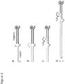

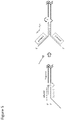
Classifications
-
- C—CHEMISTRY; METALLURGY
- C12—BIOCHEMISTRY; BEER; SPIRITS; WINE; VINEGAR; MICROBIOLOGY; ENZYMOLOGY; MUTATION OR GENETIC ENGINEERING
- C12Q—MEASURING OR TESTING PROCESSES INVOLVING ENZYMES, NUCLEIC ACIDS OR MICROORGANISMS; COMPOSITIONS OR TEST PAPERS THEREFOR; PROCESSES OF PREPARING SUCH COMPOSITIONS; CONDITION-RESPONSIVE CONTROL IN MICROBIOLOGICAL OR ENZYMOLOGICAL PROCESSES
- C12Q1/00—Measuring or testing processes involving enzymes, nucleic acids or microorganisms; Compositions therefor; Processes of preparing such compositions
- C12Q1/68—Measuring or testing processes involving enzymes, nucleic acids or microorganisms; Compositions therefor; Processes of preparing such compositions involving nucleic acids
- C12Q1/6806—Preparing nucleic acids for analysis, e.g. for polymerase chain reaction [PCR] assay
-
- C—CHEMISTRY; METALLURGY
- C12—BIOCHEMISTRY; BEER; SPIRITS; WINE; VINEGAR; MICROBIOLOGY; ENZYMOLOGY; MUTATION OR GENETIC ENGINEERING
- C12N—MICROORGANISMS OR ENZYMES; COMPOSITIONS THEREOF; PROPAGATING, PRESERVING, OR MAINTAINING MICROORGANISMS; MUTATION OR GENETIC ENGINEERING; CULTURE MEDIA
- C12N15/00—Mutation or genetic engineering; DNA or RNA concerning genetic engineering, vectors, e.g. plasmids, or their isolation, preparation or purification; Use of hosts therefor
- C12N15/09—Recombinant DNA-technology
- C12N15/10—Processes for the isolation, preparation or purification of DNA or RNA
- C12N15/1034—Isolating an individual clone by screening libraries
- C12N15/1093—General methods of preparing gene libraries, not provided for in other subgroups
-
- C—CHEMISTRY; METALLURGY
- C12—BIOCHEMISTRY; BEER; SPIRITS; WINE; VINEGAR; MICROBIOLOGY; ENZYMOLOGY; MUTATION OR GENETIC ENGINEERING
- C12N—MICROORGANISMS OR ENZYMES; COMPOSITIONS THEREOF; PROPAGATING, PRESERVING, OR MAINTAINING MICROORGANISMS; MUTATION OR GENETIC ENGINEERING; CULTURE MEDIA
- C12N15/00—Mutation or genetic engineering; DNA or RNA concerning genetic engineering, vectors, e.g. plasmids, or their isolation, preparation or purification; Use of hosts therefor
- C12N15/09—Recombinant DNA-technology
- C12N15/63—Introduction of foreign genetic material using vectors; Vectors; Use of hosts therefor; Regulation of expression
- C12N15/66—General methods for inserting a gene into a vector to form a recombinant vector using cleavage and ligation; Use of non-functional linkers or adaptors, e.g. linkers containing the sequence for a restriction endonuclease
-
- C—CHEMISTRY; METALLURGY
- C12—BIOCHEMISTRY; BEER; SPIRITS; WINE; VINEGAR; MICROBIOLOGY; ENZYMOLOGY; MUTATION OR GENETIC ENGINEERING
- C12Q—MEASURING OR TESTING PROCESSES INVOLVING ENZYMES, NUCLEIC ACIDS OR MICROORGANISMS; COMPOSITIONS OR TEST PAPERS THEREFOR; PROCESSES OF PREPARING SUCH COMPOSITIONS; CONDITION-RESPONSIVE CONTROL IN MICROBIOLOGICAL OR ENZYMOLOGICAL PROCESSES
- C12Q1/00—Measuring or testing processes involving enzymes, nucleic acids or microorganisms; Compositions therefor; Processes of preparing such compositions
- C12Q1/68—Measuring or testing processes involving enzymes, nucleic acids or microorganisms; Compositions therefor; Processes of preparing such compositions involving nucleic acids
- C12Q1/6813—Hybridisation assays
- C12Q1/6827—Hybridisation assays for detection of mutation or polymorphism
-
- C—CHEMISTRY; METALLURGY
- C12—BIOCHEMISTRY; BEER; SPIRITS; WINE; VINEGAR; MICROBIOLOGY; ENZYMOLOGY; MUTATION OR GENETIC ENGINEERING
- C12Q—MEASURING OR TESTING PROCESSES INVOLVING ENZYMES, NUCLEIC ACIDS OR MICROORGANISMS; COMPOSITIONS OR TEST PAPERS THEREFOR; PROCESSES OF PREPARING SUCH COMPOSITIONS; CONDITION-RESPONSIVE CONTROL IN MICROBIOLOGICAL OR ENZYMOLOGICAL PROCESSES
- C12Q1/00—Measuring or testing processes involving enzymes, nucleic acids or microorganisms; Compositions therefor; Processes of preparing such compositions
- C12Q1/68—Measuring or testing processes involving enzymes, nucleic acids or microorganisms; Compositions therefor; Processes of preparing such compositions involving nucleic acids
- C12Q1/6844—Nucleic acid amplification reactions
- C12Q1/6853—Nucleic acid amplification reactions using modified primers or templates
- C12Q1/6855—Ligating adaptors
-
- C—CHEMISTRY; METALLURGY
- C12—BIOCHEMISTRY; BEER; SPIRITS; WINE; VINEGAR; MICROBIOLOGY; ENZYMOLOGY; MUTATION OR GENETIC ENGINEERING
- C12Q—MEASURING OR TESTING PROCESSES INVOLVING ENZYMES, NUCLEIC ACIDS OR MICROORGANISMS; COMPOSITIONS OR TEST PAPERS THEREFOR; PROCESSES OF PREPARING SUCH COMPOSITIONS; CONDITION-RESPONSIVE CONTROL IN MICROBIOLOGICAL OR ENZYMOLOGICAL PROCESSES
- C12Q1/00—Measuring or testing processes involving enzymes, nucleic acids or microorganisms; Compositions therefor; Processes of preparing such compositions
- C12Q1/68—Measuring or testing processes involving enzymes, nucleic acids or microorganisms; Compositions therefor; Processes of preparing such compositions involving nucleic acids
- C12Q1/6844—Nucleic acid amplification reactions
- C12Q1/686—Polymerase chain reaction [PCR]
-
- C—CHEMISTRY; METALLURGY
- C12—BIOCHEMISTRY; BEER; SPIRITS; WINE; VINEGAR; MICROBIOLOGY; ENZYMOLOGY; MUTATION OR GENETIC ENGINEERING
- C12Q—MEASURING OR TESTING PROCESSES INVOLVING ENZYMES, NUCLEIC ACIDS OR MICROORGANISMS; COMPOSITIONS OR TEST PAPERS THEREFOR; PROCESSES OF PREPARING SUCH COMPOSITIONS; CONDITION-RESPONSIVE CONTROL IN MICROBIOLOGICAL OR ENZYMOLOGICAL PROCESSES
- C12Q1/00—Measuring or testing processes involving enzymes, nucleic acids or microorganisms; Compositions therefor; Processes of preparing such compositions
- C12Q1/68—Measuring or testing processes involving enzymes, nucleic acids or microorganisms; Compositions therefor; Processes of preparing such compositions involving nucleic acids
- C12Q1/6869—Methods for sequencing
-
- C—CHEMISTRY; METALLURGY
- C12—BIOCHEMISTRY; BEER; SPIRITS; WINE; VINEGAR; MICROBIOLOGY; ENZYMOLOGY; MUTATION OR GENETIC ENGINEERING
- C12Q—MEASURING OR TESTING PROCESSES INVOLVING ENZYMES, NUCLEIC ACIDS OR MICROORGANISMS; COMPOSITIONS OR TEST PAPERS THEREFOR; PROCESSES OF PREPARING SUCH COMPOSITIONS; CONDITION-RESPONSIVE CONTROL IN MICROBIOLOGICAL OR ENZYMOLOGICAL PROCESSES
- C12Q1/00—Measuring or testing processes involving enzymes, nucleic acids or microorganisms; Compositions therefor; Processes of preparing such compositions
- C12Q1/68—Measuring or testing processes involving enzymes, nucleic acids or microorganisms; Compositions therefor; Processes of preparing such compositions involving nucleic acids
- C12Q1/6876—Nucleic acid products used in the analysis of nucleic acids, e.g. primers or probes
- C12Q1/6883—Nucleic acid products used in the analysis of nucleic acids, e.g. primers or probes for diseases caused by alterations of genetic material
- C12Q1/6886—Nucleic acid products used in the analysis of nucleic acids, e.g. primers or probes for diseases caused by alterations of genetic material for cancer
-
- C—CHEMISTRY; METALLURGY
- C40—COMBINATORIAL TECHNOLOGY
- C40B—COMBINATORIAL CHEMISTRY; LIBRARIES, e.g. CHEMICAL LIBRARIES
- C40B50/00—Methods of creating libraries, e.g. combinatorial synthesis
- C40B50/06—Biochemical methods, e.g. using enzymes or whole viable microorganisms
-
- C—CHEMISTRY; METALLURGY
- C12—BIOCHEMISTRY; BEER; SPIRITS; WINE; VINEGAR; MICROBIOLOGY; ENZYMOLOGY; MUTATION OR GENETIC ENGINEERING
- C12Q—MEASURING OR TESTING PROCESSES INVOLVING ENZYMES, NUCLEIC ACIDS OR MICROORGANISMS; COMPOSITIONS OR TEST PAPERS THEREFOR; PROCESSES OF PREPARING SUCH COMPOSITIONS; CONDITION-RESPONSIVE CONTROL IN MICROBIOLOGICAL OR ENZYMOLOGICAL PROCESSES
- C12Q2525/00—Reactions involving modified oligonucleotides, nucleic acids, or nucleotides
- C12Q2525/10—Modifications characterised by
- C12Q2525/191—Modifications characterised by incorporating an adaptor
Definitions
- cfDNA cell-free DNA
- a method for increasing the efficiency of adaptor ligation to one or more DNA fragments comprising: removing the terminal phosphate residues of one or more DNA fragments; treating the dephosphorylated DNA fragments with one or more end-repair enzymes to generate end-repaired DNA; ligating one or more double-stranded DNA (dsDNA) pre-adaptors to the 3' end of each strand of the end-repaired DNA to form pre-adaptor/end-repaired DNA complexes, wherein each dsDNA pre-adaptor comprises a ligation strand oligonucleotide that is ligated to the 3' end of each strand of the end-repaired DNA, and a non-ligation partner strand oligonucleotide; displacing the non-ligation partner strand oligonucleotide from the pre-adaptor/end-repaired DNA complexes with a repair oligonucleotide, to form adaptor/end-repaired DNA complexe
- the source of the DNA is a biological sample selected from the group consisting of: amniotic fluid, blood, plasma, serum, semen, lymphatic fluid, cerebral spinal fluid, ocular fluid, urine, saliva, stool, mucous, and sweat.
- the ligation strand oligonucleotide comprises an anchor sequence, a read code, or a PCR primer binding site.
- quantitative genetic analysis comprises hybridizing one or more capture probes to a target genetic locus to form capture probe module-DNA clone complexes.
- Chemical modifications of the 3' end of the partner stand that can block ligation include, but are not limited to dideoxy ribose nucleotide analogs, 2-hydroxyl deoxyribose ribose analogs, and a broad variety of chemical modification to the ribose sugar.
- the ligation strand oligonucleotide is about 35 to about 60 nucleotides and the repair oligonucleotide is completely complementary to the ligation strand oligonucleotide.
- Identical PCR primer binding sites allows for a single primer library amplification strategy. See, e.g., Figure 4A .
- each read code of 5, 6, 7, 8, 9, 10 or more nucleotides may differ by 2, 3, 4, or 5 nucleotides from every other read code.
- the sample code is about three nucleotides and differs from every other sample code used in other samples by two nucleotides.
- a repair oligonucleotide comprises a one or more anchor sequences complementary to the one or more anchor sequences of the ligation strand oligonucleotide.
- methods contemplated herein comprise amplification of a DNA library to generate a DNA clone library or a library of DNA clones.
- the DNA is cfDNA.
- Each molecule of the DNA library comprises an adaptor ligated to each end of an end-repaired DNA, and each adaptor comprises one or more PCR primer binding sites.
- different adaptors are ligated to different ends of the end-repaired DNA.
- picograms of the initial DNA library are amplified into micrograms of DNA clones, implying a 10,000-fold amplification.
- the amount of amplified product can be measured using methods known in the art, e.g. , quantification on a Qubit 2.0 or Nanodrop instrument.
- the genome equivalents in a DNA clone library are determined using a quantitative PCR (qPCR) assay.
- qPCR quantitative PCR
- a standard library of known concentration is used to construct a standard curve and the measurements from the qPCR assay are fit to the resulting standard curve and a value for genome equivalents is derived from the fit.
- the number of genome equivalents measured by the repeat-based assays provides a more consistent library-to-library performance and a better alignment between qPCR estimates of genome equivalents and bioinformatically counted tag equivalents in sequencing runs.
- the methods comprise creating a hybrid capture probe-isolated tagged DNA target molecule through the concerted action of a 5' FLAP endonuclease, DNA polymerization and nick closure by a DNA ligase.
- a variety of enzymes can be employed for the 3'-5' exonuclease enzymatic processing of the isolated tagged DNA library-multifunctional capture probe module complex.
- suitable enzymes which exhibit 3'-5' exonuclease enzymatic activity, that can be employed in particular embodiments include, but are not limited to: T4 or Exonucleases I, III, V ( see also, Shevelev IV, Hübscher U., "The 3' 5' exonucleases,” Nat Rev Mol Cell Biol. 3(5):364-76 (2002 )).
- the enzyme comprising 3'-5' exonuclease activity is T4.
- an enzyme which exhibits 3'-5' exonuclease enzymatic activity and is capable of primer template extension can be employed, including for example T4 or Exonucleases I, III, V. Id.
- the present invention contemplates a method of detecting, identifying, predicting, diagnosing, or monitoring a condition or disease in a subject.
- genes that harbor one or more genetic lesions associated with cancer that can be detected, identified, predicted, diagnosed, or monitored with the compositions and methods contemplated herein include, but are not limited to ABCB1, ABCC2, ABCC4, ABCG2, ABL1, ABL2, AKT1, AKT2, AKT3, ALDH4A1, ALK, APC, AR, ARAF, ARFRP1, ARID1A, ATM, ATR, AURKA, AURKB, BCL2, BCL2A1, BCL2L1, BCL2L2, BCL6, BRAF, BRCA1, BRCA2, Clorf144, CARD11, CBL, CCND1, CCND2, CCND3, CCNE1, CDH1, CDH2, CDH20, CDH5, CDK4, CDK6, CDK8, CDKN2A, CDKN2B, CDKN2C, CEBPA, CHEK1, CHEK2, CRKL, CRLF2, CTNNB1, CYP1B1, CYP2C19, CYP2C
- Illustrative examples of conditions suitable for fetal testing that can be detected, identified, predicted, diagnosed, or monitored with the compositions and methods contemplated herein include but are not limited to: Down Syndrome (Trisomy 21), Edwards Syndrome (Trisomy 18), Patau Syndrome (Trisomy 13), Klinefelter's Syndrome (XXY), Triple X syndrome, XYY syndrome, Trisomy 8, Trisomy 16, Turner Syndrome (XO), Robertsonian translocation, DiGeorge Syndrome and Wolf-Hirschhorn Syndrome.
- a pre-adaptor was ligated to end-repaired fragments generated in step I of this example.
- 25 ⁇ l of end-repaired fragment was combined with 10 ⁇ l of 10 ⁇ M adaptor.
- 1 to 4 ligation reactions were performed depending on the number of separate adaptors added to the reaction.
- 15 ⁇ l of ligation cocktail (5 ⁇ l of 10X T4-DNA ligation buffer, 7.5 ⁇ l of 50% PEG8000, and 2.5 ⁇ l of HC T4 DNA ligase (NEB; M0202) was added to each ligation reaction in a final volume of 50 ⁇ l.
- the reaction was mixed and incubated at 20°C for 60 min., then at 65°C for 10 min, then cooled to room temperature.
Landscapes
- Chemical & Material Sciences (AREA)
- Life Sciences & Earth Sciences (AREA)
- Health & Medical Sciences (AREA)
- Organic Chemistry (AREA)
- Engineering & Computer Science (AREA)
- Genetics & Genomics (AREA)
- Wood Science & Technology (AREA)
- Zoology (AREA)
- Proteomics, Peptides & Aminoacids (AREA)
- Biotechnology (AREA)
- Bioinformatics & Cheminformatics (AREA)
- General Engineering & Computer Science (AREA)
- Molecular Biology (AREA)
- Biochemistry (AREA)
- Microbiology (AREA)
- General Health & Medical Sciences (AREA)
- Biophysics (AREA)
- Physics & Mathematics (AREA)
- Analytical Chemistry (AREA)
- Immunology (AREA)
- Biomedical Technology (AREA)
- Chemical Kinetics & Catalysis (AREA)
- Pathology (AREA)
- Plant Pathology (AREA)
- Crystallography & Structural Chemistry (AREA)
- Bioinformatics & Computational Biology (AREA)
- Hospice & Palliative Care (AREA)
- Oncology (AREA)
- General Chemical & Material Sciences (AREA)
- Medicinal Chemistry (AREA)
- Measuring Or Testing Involving Enzymes Or Micro-Organisms (AREA)
- Preparation Of Compounds By Using Micro-Organisms (AREA)
Abstract
Description
- This application claims priority to
U.S. Provisional Application No. 62/254,110, filed November 11, 2015 - The contents of the text file submitted electronically herewith are incorporated herein by reference in their entirety: A computer readable format copy of the Sequence Listing (file name: CLFK_003_01WO_ST25, date recorded: November 10, 2016, file size: 23 kB).
- The invention relates generally to improved compositions and methods for constructing DNA libraries. In particular, the present invention relates to efficiently constructing DNA clone libraries for quantitative genetic analyses.
- Various DNA specimens that are of interest for downstream analysis are collected in minute quantities. By way of example, cell-free DNA (cfDNA) collected from the plasma fraction of whole blood is generally present in nanogram quantities per mL of plasma. Given that one diploid human genome weighs 6 picograms, this means there are a few hundred to a few thousand total genomes of information that can be isolated from a single blood draw.
- In cancer patients, tumor DNA is shed into the bloodstream in highly variable quantities ranging from ≤ 0.1% to ≥ 10% total circulating DNA. Blood draws contain only a few nanograms of DNA and if tumor genomes are present at 0.1% of the total circulating DNA, then only 1 to 10 total copies of the tumor genome are present. To unambiguously identify tumor DNA by sequence analysis, it is necessary to observe two or more copies of a tumor-specific genetic lesion. However, the need to maximize the detection sensitivity of DNA, meaning accurate detection of tumor DNA in the 0.1% range has yet to be achieved
- These considerations illuminate the fundamental problem that reliable genetic analysis of solid tumors using blood is governed, in part, by the ability isolate and analyze rare genomic fragments. Moreover, many therapeutically actionable tumor lesions involve gene fusions, significant insertions or deletions of DNA sequence and/or changes in gene copy number. Such alterations are refractory to analysis by PCR, where two adjacent primer binding sites must be known and where copy variation is obscured by many rounds of target amplification.
- At present, target retrieval methods are used for comprehensive analysis of potential lesions in circulating tumor DNA. Such retrieval methods rely on the creation of DNA clone libraries. Unfortunately, current methods for creating these DNA libraries are inefficient, with only a small percentage of DNA fragments being successfully converted to useful library clones.
- The invention relates generally to compositions and methods for high efficiency attachment of DNA adaptors to DNA fragments to generate DNA libraries for quantitative genetic analyses.
- In various embodiments, a method for increasing the efficiency of adaptor ligation to one or more DNA fragments comprising: removing the terminal phosphate residues of one or more DNA fragments; treating the dephosphorylated DNA fragments with one or more end-repair enzymes to generate end-repaired DNA; ligating one or more double-stranded DNA (dsDNA) pre-adaptors to the 3' end of each strand of the end-repaired DNA to form pre-adaptor/end-repaired DNA complexes, wherein each dsDNA pre-adaptor comprises a ligation strand oligonucleotide that is ligated to the 3' end of each strand of the end-repaired DNA, and a non-ligation partner strand oligonucleotide; displacing the non-ligation partner strand oligonucleotide from the pre-adaptor/end-repaired DNA complexes with a repair oligonucleotide, to form adaptor/end-repaired DNA complexes, wherein each adaptor comprises the ligation strand oligonucleotide and the repair oligonucleotide; and treating the adaptor/end-repaired DNA complexes with one or more enzymes to form a contiguous, double-stranded, DNA library; wherein the efficiency of adapter ligation is increased compared to a method wherein dephosphorylated adapter molecules are ligated to phosphorylated DNA fragments is provided.
- In various embodiments, a method for constructing a DNA library comprising: removing the terminal phosphate residues of one or more DNA fragments; treating the dephosphorylated DNA fragments with one or more end-repair enzymes to generate end-repaired DNA; ligating one or more double-stranded DNA (dsDNA) pre-adaptors to the 3' end of each strand of the end-repaired DNA to form pre-adaptor/end-repaired DNA complexes, wherein each dsDNA pre-adaptor comprises a ligation strand oligonucleotide that is ligated to the 3' end of each strand of the end-repaired DNA, and a non-ligation partner strand oligonucleotide; displacing the non-ligation partner strand oligonucleotide from the pre-adaptor/end-repaired DNA complexes with a repair oligonucleotide, to form adaptor/end-repaired DNA complexes, wherein each adaptor comprises the ligation strand oligonucleotide and the repair oligonucleotide; and treating the adaptor/end-repaired DNA complexes with one or more enzymes to form a contiguous, double-stranded, DNA library is provided.
- In particular embodiments, the non-ligation partner strand oligonucleotide comprises a modification at the 3' terminus that prevents its ligation to the 5' end of the end-repaired DNA and/or adaptor dimer formation.
- In certain embodiments, the source of the one or more DNA fragments is DNA selected from the group consisting of: genomic DNA (gDNA), complementary DNA (cDNA), and cell-free DNA (cfDNA).
- In further embodiments, the source of the DNA is a biological sample selected from the group consisting of: blood, skin, hair, hair follicles, saliva, oral mucous, vaginal mucous, sweat, tears, epithelial tissues, urine, semen, seminal fluid, seminal plasma, prostatic fluid, pre-ejaculatory fluid (Cowper's fluid), excreta, biopsy, ascites, cerebrospinal fluid, lymph, and tissue extract sample or biopsy sample.
- In particular embodiments, the source of the DNA is a biological sample selected from the group consisting of: amniotic fluid, blood, plasma, serum, semen, lymphatic fluid, cerebral spinal fluid, ocular fluid, urine, saliva, stool, mucous, and sweat.
- In further embodiments, the methods further comprise isolating the DNA from a biological sample of a subject.
- In some embodiments, the methods further comprise fragmenting the DNA from a biological sample of a subject.
- In certain embodiments, the methods further comprise repairing damage of the one or more DNA fragments prior to ligation.
- In particular embodiments, the damage is a deaminated cytosine (Uracil), an abasic site, methylation of guanine to O6MeG, DNA nicks, gaps, or a thymine dimer.
- In various embodiments, a method constructing a cfDNA library comprising: isolating or obtaining cfDNA from a biological sample of a subject; removing the terminal phosphate residues of the cfDNA; treating the dephosphorylated cfDNA with one or more end-repair enzymes to generate end-repaired cfDNA, and optionally to repair DNA damage; ligating one or more double-stranded DNA (dsDNA) pre-adaptors to the 3' end of each strand of the end-repaired cfDNA to form pre-adaptor/end-repaired cfDNA complexes, wherein each dsDNA pre-adaptor comprises a ligation strand oligonucleotide that is ligated to the 3' end of each strand of the end-repaired cfDNA, and a non-ligation partner strand oligonucleotide; displacing the non-ligation partner strand oligonucleotide from the pre-adaptor/end-repaired cfDNA complexes with a repair oligonucleotide, to form adaptor/end-repaired cfDNA complexes, wherein each adaptor comprises the ligation strand oligonucleotide and the repair oligonucleotide; treating the adaptor/end-repaiired cfDNA complexes with one or more enzymes to form a contiguous, double-stranded, cfDNA library; and amplifying the cfDNA library to generate a cell-free DNA clone library is provided.
- In particular embodiments, the ligation strand oligonucleotide comprises one or more modifications to prevent adaptor dimer formation, optionally wherein the modification of the 3' end of the no-ligation partner strand oligonucleotide prevents adaptor dimer formation.
- In certain embodiments, the ligation strand oligonucleotide comprises an anchor sequence, a read code, or a PCR primer binding site.
- In further embodiments, the ligation strand oligonucleotide comprises an anchor sequence, a read code, and a PCR primer binding site.
- In some embodiments, the ligation strand oligonucleotide comprises one or more PCR primer binding sites for PCR amplification of the one or more contiguous, double-stranded, DNA library molecules.
- In particular embodiments, the ligation strand oligonucleotide comprises one or more unique read codes.
- In particular embodiments, the ligation strand oligonucleotide comprises one or more sample codes for sample multiplexing.
- In certain embodiments, the ligation strand oligonucleotide comprises one or more sequences for DNA sequencing.
- In further embodiments, the ligation strand oligonucleotide comprises an anchor sequence.
- In further embodiments, the repair oligonucleotide comprises an anchor sequence, a read code, or a PCR primer binding site.
- In certain embodiments, the repair oligonucleotide comprises an anchor sequence, a read code, and a PCR primer binding site.
- In particular embodiments, the repair oligonucleotide comprises one or more primer binding sites for PCR amplification of the one or more contiguous, double-stranded, DNA library molecules.
- In some embodiments, the repair oligonucleotide comprises one or more unique read codes.
- In certain embodiments, the repair oligonucleotide comprises one or more sample codes for sample multiplexing.
- In particular embodiments, the repair oligonucleotide comprises one or more sequences for DNA sequencing.
- In further embodiments, the ligation strand oligonucleotide is complementary to the repair oligonucleotide.
- In particular embodiments, the anchor sequence of the ligation strand oligonucleotide is complementary to the anchor sequence of the repair oligonucleotide.
- In further embodiments, the PCR primer binding site of the ligation strand oligonucleotide is complementary to the PCR primer binding site of the repair oligonucleotide.
- In particular embodiments, the one or more adaptors comprises a plurality of ligation strand oligonucleotide species.
- In some embodiments, the one or more adaptors comprises a plurality of repair oligonucleotide species.
- In particular embodiments, the primer binding site of the ligation strand oligonucleotide is not complementary to the primer binding site of the repair oligonucleotide.
- In certain embodiments, the primer binding site of the ligation strand oligonucleotide is substantially different from the primer binding site of the repair oligonucleotide.
- In certain embodiments, a primer that binds the primer binding site of the ligation strand oligonucleotide does not substantially bind the primer binding site of the repair oligonucleotide.
- In particular embodiments, the DNA library is amplified to generate a DNA clone library.
- In further embodiments, qPCR is performed on the DNA clone library and a qPCR measurement is compared to standards of known genome equivalents to determine the genome equivalents of the DNA clone library.
- In particular embodiments, the qPCR is performed with a primer that binds to an Alu sequence and a primer that binds to a sequence in an adaptor.
- In some embodiments, quantitative genetic analysis is performed on a plurality of genetic loci in the DNA clone library.
- In particular embodiments, quantitative genetic analysis is performed on a plurality of genetic loci in a plurality of DNA clone libraries.
- In particular embodiments, quantitative genetic analysis comprises hybridizing one or more capture probes to a target genetic locus to form capture probe module-DNA clone complexes.
- In certain embodiments, quantitative genetic analysis comprises isolating the capture probe -DNA clone complexes.
- In further embodiments, the quantitative genetic analysis comprises amplification of the DNA clone sequence in the isolated capture probe-DNA clone complexes.
- In particular embodiments, quantitative genetic analysis comprises DNA sequencing to generate a plurality of sequencing reads.
- In further embodiments, the methods further comprise bioinformatic analysis of the plurality of sequencing reads.
- In particular embodiments, quantitative genetic analysis is performed on a plurality of genetic loci in the DNA clone library and wherein bioinformatic analysis is used: to quantify the number of genome equivalents analyzed in the DNA clone library; to detect genetic variants in a target genetic locus; to detect mutations within a target genetic locus; to detect genetic fusions within a target genetic locus; and/or to measure copy number fluctuations within a target genetic locus.
- In certain embodiments, the quantitative genetic analysis is used to identify or detect one or more genetic lesions that cause or associated with the genetic disease.
- In particular embodiments, the genetic lesion comprises a nucleotide transition or transversion, a nucleotide insertion or deletion, a genomic rearrangement, a change in copy number, or a gene fusion.
- In certain embodiments, the genetic disease is cancer.
- In further embodiments, the quantitative genetic analysis is used to identify or detect one or more genetic variants or genetic lesions of one or more target genetic loci in fetal cfDNA.
- In some embodiments, the capture probe is a component of a capture probe module that is optionally duplexed with a hapten-labeled partner oligonucleotide that hybridizes to a tail sequence in the capture probe module.
- In various embodiments, a method of predicting, diagnosing, or monitoring a genetic disease in a subject comprising: isolating or obtaining DNA from a biological sample of a subject; removing the terminal phosphate residues of the DNA; treating the dephosphorylated DNA with one or more end-repair enzymes to generate end-repaired DNA; ligating one or more double-stranded DNA (dsDNA) pre-adaptors to the 3' end of each strand of the end-repaired DNA to form pre-adaptor/end-repaired DNA complexes, wherein each dsDNA pre-adaptor comprises a ligation strand oligonucleotide that is ligated to the 3' end of each strand of the end-repaired DNA, and a non-ligation partner strand oligonucleotide; displacing the non-ligation partner strand oligonucleotide from the pre-adaptor/end-repaired DNA complexes with a repair oligonucleotide, to form adaptor/end-repaired DNA complexes, wherein each adaptor comprises the ligation strand oligonucleotide and the repair oligonucleotide; treating the adaptor/end-repaired DNA complexes with one or more enzymes to form a contiguous, double-stranded, DNA library; amplifying the DNA library to generate a DNA clone library; determining the number of genome equivalents in the DNA clone library; and performing a quantitative genetic analysis of one or more target genetic loci associated with the genetic disease in the DNA clone library, wherein the identification or detection of one or more genetic lesions in the one or more target genetic loci is prognostic for, diagnostic of, or monitors the progression of the genetic disease is provided.
- In certain embodiments, the DNA is genomic DNA, DNA from formalin-fixed, paraffin embedded (FFPE) samples, cDNA, or cfDNA.
- In particular embodiments, the cfDNA. is isolated from a biological sample selected from the group of: amniotic fluid, blood, plasma, serum, semen, lymphatic fluid, cerebral spinal fluid, ocular fluid, urine, saliva, stool, mucous, and sweat.
- In further embodiments, the genetic lesion comprises a nucleotide transition or transversion, a nucleotide insertion or deletion, a genomic rearrangement, a change in copy number, or a gene fusion.
- In particular embodiments, the genetic disease is cancer.
- In various embodiments, a companion diagnostic for a genetic disease comprising: isolating or obtaining DNA from a biological sample of a subject; removing the terminal phosphate residues of the DNA; treating the dephosphorylated DNA with one or more end-repair enzymes to generate end-repaired DNA; ligating one or more double-stranded DNA (dsDNA) pre-adaptors to the 3' end of each strand of the end-repaired DNA to form pre-adaptor/end-repaired DNA complexes, wherein each dsDNA pre-adaptor comprises a ligation strand oligonucleotide that is ligated to the 3' end of each strand of the end-repaired DNA, and a non-ligation partner strand oligonucleotide; displacing the non-ligation partner strand oligonucleotide from the pre-adaptor/end-repaired DNA complexes with a repair oligonucleotide, to form adaptor/end-repaired DNA complexes, wherein each adaptor comprises the ligation strand oligonucleotide and the repair oligonucleotide; treating the adaptor/end-repaired DNA complexes with one or more enzymes to form a contiguous, double-stranded, DNA library; amplifying the DNA library to generate a DNA clone library; determining the number of genome equivalents in the DNA clone library; and performing a quantitative genetic analysis of one or more biomarkers associated with the genetic disease in the DNA clone library, wherein detection of, or failure to detect, at least one of the one or more biomarkers indicates whether the subject should be treated for the genetic disease is provided.
- In further embodiments, the DNA is genomic DNA, DNA from formalin-fixed, paraffin embedded (FFPE) samples, cDNA, or cfDNA.
- In particular embodiments, the cfDNA is isolated from a biological sample selected from the group consisting of: amniotic fluid, blood, plasma, serum, semen, lymphatic fluid, cerebral spinal fluid, ocular fluid, urine, saliva, stool, mucous, and sweat.
- In particular embodiments, the biomarker is a genetic lesion.
- In further embodiments, the genetic lesion comprises a nucleotide transition or transversion, a nucleotide insertion or deletion, a genomic rearrangement, a change in copy number, or a gene fusion.
- In certain embodiments, the genetic disease is cancer.
-
-
Figure 1 shows conventional versus high efficiency (HE) ligation technology. (A) Target DNA fragments are 5' phosphorylated prior to DNA ligation with unphosphorylated duplex adapters. (B) A common inefficiency with conventional ligation approaches is that 5' target DNA fragment ends lacking a phosphate group fails to ligate with unphosphorylated duplex adapters. (C) Ligation of target DNA fragments to one another. (D) Duplex adaptor comprising a 5' phosphate group required is ligated to 3' end of target DNA fragment The hatched circle one the partner strand oligonucleotide of the adaptor represents a 3' blocking group. (E) The 3' blocking group also prevents adaptors from ligating to one another. (F) Occasional adaptor duplexes lacking a 5' phosphate fail to ligate to target fragments. -
Figure 2 shows a representative image of an agarose gel of the complete ligation of RsaI digested, dephosphorylated pUC19 plasmid to a series of nine different HE adaptors. Unligated fragments are indicated in the control lane, which is next to the molecular weight (MW) markers of 740, 500, 300, and 150 bp (top to bottom). The complete shift in the mobility of the three vector fragments (arrows) indicates the complete ligation of adaptors to both ends across all nine adaptors. These results show that the HE ligation technology is generalizable. -
Figure 3 shows exemplary methods of completing HE ligated constructs. (A) The HE ligation product is a 3' extended adaptor oligonucleotide attached to the 3' end of fragments. (B) One strategy for "repairing" the initial ligation product is to add a repair oligonucleotide (top strand; green); T4 polynucleotide kinase can add a phosphate (P) to the 5' end of the target fragment, and a nick sealing ligase such as Taq DNA ligase can be used to ligate the repair oligonucleotide to the target fragment. (C) An alternative strategy is to combine the complementary adaptor oligonucleotide, a DNA polymerase that has 5' to 3' exonuclease activity (e.g., BstI DNA polymerase), and a Taq DNA ligase. The BstI DNA polymerase extends the repair oligonucleotide by removing 5' bases from the target fragment to expose 5' phosphates that enable nick-sealing ligation by the Taq DNA ligase. (D) Complementary adaptor oligonucleotides can also be designed to introduce additional sequence features into the original HE ligation strand with the BstI DNA polymerase. -
Figure 4 shows the preparation of DNA libraries using HE ligation technology. (A) DNA fragments can possess heterogeneous, "ragged" ends that may or may not possess phosphate groups (P). Treatment of the DNA fragments with a phosphatase removes exposed 5' and 3' phosphates. The DNA can then be treated with enzymes that repair DNA damage such as deaminated cytosines (U), abasic sites (↑), and thymine dimers, and that "polish" 5' or 3' overhangs to blunt ends. (B) Adaptors are added to DNA fragments in two steps. First, a duplex adaptor comprising a 5' phosphorylated ligation strand and a 3' blocked partner strand is ligated to the target fragments. The partner strand, which has a melting temperature of ∼ 30°C, is removed in subsequent steps that occur at temperatures ≥ 37°C. Second, repair oligonucleotides are annealed to the adaptor ligated fragments; the repair oligonucleotides become covalently attached to the 5' end of the target fragment either using a kinase/ligase strategy or a polymerase/exonuclease/ligase strategy (Figure 3 ). Primer extension of the initial ligation strand copies the repair oligo information into a full-length adaptor duplex that is suitable for downstream analysis. -
Figure 5 shows a HE ligation technology strategy for generating dual PCR primer adapted DNA fragments. In this scheme, the ligation strand carries additional sequences (primer 2) that serve as an independent primer binding site. The repair oligonucleotide, while complementary to a portion of the ligation strand, has its own divergent sequence that serves as a second PCR primer binding site (primer 1). The fully completed adaptors allow amplification of the DNA specimen fragments using more conventional, universal dual-primer PCR methods. - The present invention contemplates, in part, compositions and methods to address the acute need in the field of quantitative genetic analyses for improved, highly-efficient methods to clone such DNA fragments for downstream analysis.
- Current methods for DNA analysis comprise ligation of specialized adaptors to DNA fragments (
Figure 1 ). In conventional techniques, target DNA fragments are 5' phosphorylated prior to DNA ligation to enable covalent ligation with unphosphorylated duplex adapters. The target DNA fragment and adaptor may be blunt-ended, or they may share complementary overhangs (e.g., T/A). (Figure 1A ). This is a serious drawback because it is not possible to ensure that both ends of all target DNA fragments are phosphorylated, and unphosphorylated ends are incapable of ligation and these target fragments are lost from subsequent libraries. (Figure IB). By way of a non-limiting example, if 70% of target DNA fragment ends possess a 5' phosphate, then only 49% of fragments (0.7 x 0.7 x 100%) at the maximum, could be ligated on both ends of the fragment and ligation to both ends is required for cloning. Additionally, the presence of 5' phosphates on target DNA fragments promotes a separate undesirable artifact in which DNA fragments can ligate to one another (Figure 1C ). This creates artifactual chromosomal sequence fusion events that can confound detection of disease-specific chromosomal rearrangements. - In various embodiments, the present invention contemplates, in part, compositions and methods for efficiently attaching adaptor sequences to target DNA fragments. In particular embodiments, phosphates are removed from both the 5' and 3' termini of target DNA fragments. These dephosphorylated fragments are then treated with enzymes that create blunt DNA ends and optionally with enzymes that that repair many types of DNA damage that may have been inflicted on the DNA, e.g., deaminated cytosine (Uracil), an abasic site, methylation of guanine to O6MeG, nicks, double strand breaks, or a thymine dimer. The adaptor comprises a ligation strand oligonucleotide duplexed with a non-ligation partner strand oligonucleotide. The ligation strand of the adaptor carries the 5' phosphate group required for ligation to target DNA fragments and the partner strand comprises a 3' blocking group (Figure ID). The 3' blocking group prevents the formation of adaptor:adaptor dimers (Figure IE). As with DNA fragments, not all adaptor sequences will possess a 5' phosphate (solvent exposed terminal phosphate bonds are inherently chemically labile). While such unphosphorylated adaptors will be present, they will only transiently engage the ligation machinery (Figure IF); unproductive pairing of such adaptors with fragments rapidly dissociate and are replaced by adaptor:target DNA fragment pairings that can provide productive covalent attachment. Eventually ∼100% of target DNA fragments become attached on both ends with adaptor molecules, which illustrates the high efficiency of the compositions and methods contemplated herein for constructing DNA libraries.
- In various embodiments, compositions and methods contemplated herein for high efficiency construction of DNA libraries provide a novel comprehensive framework address molecular genetic analysis using DNA available from a variety of biological sources. Cloning of purified DNA introduces tagged DNA sequences that inform downstream analysis and enable amplification of the resulting clone libraries. Hybrid capture with target specific oligonucleotides is used to retrieve specific sequences for subsequent analysis. Independent measurements of the number of genomes present in the library are applied to each sample, and these assays provide a means to estimate the assay's sensitivity. The assays contemplated herein provide reliable, reproducible, and robust methods for the analysis, detection, diagnosis, or monitoring of genetic states, conditions, or disease.
- The practice of particular embodiments of the invention will employ, unless indicated specifically to the contrary, conventional methods of chemistry, biochemistry, organic chemistry, molecular biology, microbiology, recombinant DNA techniques, genetics, immunology, and cell biology that are within the skill of the art, many of which are described below for the purpose of illustration. Such techniques are explained fully in the literature. See, e.g., Sambrook, et al., Molecular Cloning: A Laboratory Manual (3rd Edition, 2001); Sambrook, et al., Molecular Cloning: A Laboratory Manual (2nd Edition, 1989); Maniatis et al., Molecular Cloning: A Laboratory Manual (1982); Ausubel et al., Current Protocols in Molecular Biology (John Wiley and Sons, updated July 2008); Short Protocols in Molecular Biology: A Compendium of Methods from Current Protocols in Molecular Biology, Greene Pub. Associates and Wiley-Interscience; Glover, DNA Cloning: A Practical Approach, vol. I & II (IRL Press, Oxford, 1985); Anand, Techniques for the Analysis of Complex Genomes, (Academic Press, New York, 1992); Transcription and Translation (B. Hames & S. Higgins, Eds., 1984); Perbal, A Practical Guide to Molecular Cloning (1984); and Harlow and Lane, Antibodies, (Cold Spring Harbor Laboratory Press, Cold Spring Harbor, N.Y., 1998).
- Unless defined otherwise, all technical and scientific terms used herein have the same meaning as commonly understood by those of ordinary skill in the art to which the invention belongs. Although any methods and materials similar or equivalent to those described herein can be used in the practice or testing of the present invention, preferred embodiments of compositions, methods and materials are described herein. For the purposes of the present invention, the following terms are defined below.
- The articles "a, " "an, " and "the" are used herein to refer to one or to more than one (i.e. to at least one) of the grammatical object of the article. By way of example, "an element" means one element or more than one element.
- The use of the alternative (e.g., "or") should be understood to mean either one, both, or any combination thereof of the alternatives.
- The term "and/or" should be understood to mean either one, or both of the alternatives.
- As used herein, the term "about" or "approximately" refers to a quantity, level, value, number, frequency, percentage, dimension, size, amount, weight or length that varies by as much as 15%, 10%, 9%, 8%, 7%, 6%, 5%, 4%, 3%, 2% or 1% to a reference quantity, level, value, number, frequency, percentage, dimension, size, amount, weight or length. In one embodiment, the term "about" or "approximately" refers a range of quantity, level, value, number, frequency, percentage, dimension, size, amount, weight or length ± 15%, ± 10%, ± 9%, ± 8%, ± 7%, ± 6%, ± 5%, ± 4%, ± 3%, ± 2%, or ± 1% about a reference quantity, level, value, number, frequency, percentage, dimension, size, amount, weight or length.
- Throughout this specification, unless the context requires otherwise, the words "comprise", "comprises" and "comprising" will be understood to imply the inclusion of a stated step or element or group of steps or elements but not the exclusion of any other step or element or group of steps or elements. In particular embodiments, the terms "include," "has," "contains," and "comprise" are used synonymously.
- By "consisting of' is meant including, and limited to, whatever follows the phrase "consisting of." Thus, the phrase "consisting of' indicates that the listed elements are required or mandatory, and that no other elements may be present.
- By "consisting essentially of' is meant including any elements listed after the phrase, and limited to other elements that do not interfere with or contribute to the activity or action specified in the disclosure for the listed elements. Thus, the phrase "consisting essentially of' indicates that the listed elements are required or mandatory, but that no other elements are optional and may or may not be present depending upon whether or not they affect the activity or action of the listed elements.
- Reference throughout this specification to "one embodiment," "an embodiment," "a particular embodiment," "a related embodiment," "a certain embodiment," "an additional embodiment," or "a further embodiment" or combinations thereof means that a particular feature, structure or characteristic described in connection with the embodiment is included in at least one embodiment of the present invention. Thus, the appearances of the foregoing phrases in various places throughout this specification are not necessarily all referring to the same embodiment. Furthermore, the particular features, structures, or characteristics may be combined in any suitable manner in one or more embodiments.
- As used herein, the term "isolated" means material that is substantially or essentially free from components that normally accompany it in its native state. In particular embodiments, the term "obtained" or "derived" is used synonymously with isolated.
- As used herein, the term "DNA" refers to deoxyribonucleic acid. In various embodiments, the term DNA refers to genomic DNA, recombinant DNA, synthetic DNA, complementary DNA (cDNA), or cell-free DNA (cfDNA). In one embodiment, DNA refers to genomic DNA or cDNA. In one embodiment, DNA refers to cfDNA. In particular embodiments, the DNA is a DNA fragment that comprises a "target region," which is also referred to as a target DNA fragment in certain embodiments. DNA libraries contemplated herein include genomic DNA libraries, cfDNA libraries, and cDNA libraries constructed from RNA, e.g., an RNA expression library. In various embodiments, the DNA libraries comprise one or more additional DNA sequences and/or tags.
- A "target genetic locus" or "DNA target region" refers to a region of interest within a DNA sequence. In various embodiments, targeted genetic analyses are performed on the target genetic locus. In particular embodiments, the DNA target region is a region of a gene that is associated with a particular genetic state, genetic condition, genetic diseases; fetal testing; genetic mosaicism, paternity testing; predicting response to drug treatment; diagnosing or monitoring a medical condition; microbiome profiling; pathogen screening; or organ transplant monitoring.
- As used herein, the terms "circulating DNA," "circulating cell-free DNA" and "cell-free DNA" are often used interchangeably and refer to DNA that is extracellular DNA, DNA that has been extruded from cells, or DNA that has been released from necrotic or apoptotic cells.
- A "subject," "individual," or "patient" as used herein, includes any animal that exhibits a symptom of a condition that can be detected or identified with compositions contemplated herein. Suitable subjects include laboratory animals (such as mouse, rat, rabbit, or guinea pig), farm animals (such as horses, cows, sheep, pigs), and domestic animals or pets (such as a cat or dog). In particular embodiments, the subject is a mammal. In certain embodiments, the subject is a non-human primate and, in preferred embodiments, the subject is a human.
- A "reaction vessel" means a container suitable for carrying out one of the reactions contemplated herein. Illustrative examples of reaction vessels suitable for use in particular embodiments include, but are not limited to test tubes, microfuge tubes (e.g., PCR tubes), microtitre plates (e.g., 96 well plates, 384 well plates, 1536 well plates), slides, plates, arrays, and microarrays.
- In particular embodiments, methods of constructing DNA libraries contemplated herein comprise high efficiency ligation of adaptors to target DNA fragments.
- The methods and compositions contemplated herein are designed to efficiently analyze, detect, diagnose, and/or monitor genetic states, genetic conditions, genetic diseases, genetic mosaicism, fetal diagnostics, paternity testing, microbiome profiling, pathogen screening, and organ transplant monitoring using DNA as an analyte. DNA suitable for use in the compositions and methods contemplated herein can come from any source known to those of skill in the art. In particular embodiments, the DNA is genomic DNA isolated from any source, copy DNA (cDNA) synthesized from RNA, or cell-free DNA (cfDNA).
- In some embodiments, the DNA is high molecular weight DNA (>1000 bp). Use of high molecular weight DNA in the compositions and methods contemplated herein often comprises a fragmentation step. The high molecular weight DNA can be fragmented to about 25 to about 750 base pairs, about 25 to about 500 base pairs, about 25 to about 250 base pairs, about 25 to about 200 base pairs, about 25 to about 150 base pairs, about 25 to about 100 base pairs, about 25 to about 50 base pairs, about 100 to about 200 base pairs, about 150 to about 180 base pairs, about 150 base pairs, about 155 base pairs, about 160 base pairs, about 165 base pairs, about 170 base pairs, about 175 base pairs, or about, about 180 base pairs.
- Illustrative methods for fragmenting DNA suitable for use in particular embodiments of the compositions and methods contemplated herein include, but are not limited to: shearing, sonication, enzymatic digestion; including restriction digests, as well as other methods. In particular embodiments, any method known in the art for fragmenting DNA can be employed with the present invention.
- Illustrative sources of genomic DNA and RNA (to generate cDNA) suitable for use in particular embodiments of the compositions and methods contemplated herein include, but are not limited to biological samples selected from the group consisting of: brain tissue, bone tissue, ocular tissue, olfactory tissue, muscles tissue, heart tissue, lung tissue, liver tissue, pancreatic tissue, kidney tissue, gastric tissue, intestinal tissue, colon tissue, blood, skin, hair, hair follicles, saliva, oral mucous, vaginal mucous, sweat, tears, epithelial tissues, urine, semen, seminal fluid, seminal plasma, prostatic fluid, pre-ejaculatory fluid (Cowper's fluid), excreta, biopsy, ascites, cerebrospinal fluid, lymph, and tissue extract sample or biopsy sample, and the like.
- In particular embodiments, the DNA is cfDNA. The size distribution of cfDNA ranges from about 150 bp to about 180 bp fragments. Fragmentation may be the result of endonucleolytic and/or exonucleolytic activity and presents a formidable challenge to the accurate, reliable, and robust analysis of cfDNA. Another challenge for analyzing cfDNA is its short half-life in the blood stream, on the order of about 15 minutes. Without wishing to be bound to any particular theory, the present invention contemplates, in part, that analysis of cfDNA is like a "liquid biopsy" and is a real-time snapshot of current biological processes.
- In some embodiments, cfDNA isolated from the blood plasma fraction can be substantially contaminated with long (>10 kilobase pair), high-molecular weight genomic DNA that is liberated from nucleated blood cells that lyse during the collection protocol. This long, contaminating DNA, if left unfragmented, does not clone and amplify well and is therefore lost during downstream library preparation. However, in particular embodiments, in the absence of DNA fragmentation, the high-efficiency DNA library construction methods contemplated herein selectively clones shorter (<1000 bp) fragments from a collection of fragment sizes present in a DNA specimen. Without wishing to be bound by any particular theory, the selective cloning of short cfDNA fragments from a DNA specimen that is a blend of long and short fragments is advantageous in the construction of a liquid biopsy.
- Illustrative examples of biological samples that are suitable sources from which to isolate cfDNA in particular embodiments include, but are not limited to amniotic fluid, blood, plasma, serum, semen, lymphatic fluid, cerebral spinal fluid, ocular fluid, urine, saliva, mucous, and sweat.
- In particular embodiments, the biological sample is blood or blood plasma.
- In certain embodiments, the DNA sample could be derived from embedded tissues such as FFPE or fine needle aspirates, from swabs intended to interrogate microbiome sequences present, from forensic samples such as hair, clothing, fingerprints, etc. or from any other source of DNA requiring the library construction methods contemplated herein that are especially efficient for constructing libraries from low-input DNA samples.
- In certain embodiments, commercially available kits and other methods known to the skilled artisan can used to isolate cfDNA directly from the biological samples of a patient or from a previously obtained and optionally stabilized biological sample, e.g., by freezing and/or addition of enzyme chelating agents including, but not limited to EDTA, EGTA, or other chelating agents specific for divalent cations.
- In particular embodiments, the input DNA, e.g., target DNA fragments, is first treated with a thermo-labile phosphatase that removes terminal phosphate residues. See, e.g.,
Figure 4A . - Illustrative examples of thermo-labile phosphatases that are suitable for use in particular embodiments of the compositions and methods contemplated herein include, but are not limited to APex™ Heat-Labile Alkaline Phosphatase (Epicentre Biotechnologies), NTPhos™ Thermolabile Phosphatase (Epicentre Biotechnologies), HK™ Thermolabile Phosphatase (Epicentre Biotechnologies), and Shrimp Alkaline Phosphatase (SAP; NEB).
- In one embodiment, the thermo-labile phosphatase is SAP.
- In particular embodiments, the input DNA or dephosphorylated DNA is also treated with one or more enzymes that reverse common sources of DNA damage such as deamination of cytosines to uracil, oxidative addition to guanines, thymidine dimers, loss of bases leading to abasic sites, nicks or gaps on one strand of duplex DNA, etc. See, e.g.,
Figure 4A . - In one embodiment, the internal damage to the DNA is reversed using a composition comprising one or more of the following enzymes: Taq DNA Ligase, Endonuclease IV, Bst DNA Polymerase, Fpg (8-oxoguanine DNA glycosylase), Uracil-DNA Glycosylase (UDG), T4 PDG (T4 Endonuclease V), Endonuclease VIII, and T4 DNA polymerase.
- In one embodiment, the internal damage to the DNA is reversed using a composition comprising Taq DNA Ligase, Endonuclease IV, Bst DNA Polymerase, Fpg, Uracil-DNA Glycosylase (UDG), T4 PDG (T4 Endonuclease V), Endonuclease VIII, and T4 DNA polymerase.
- In particular embodiments, the compositions and methods contemplated herein comprise generating end-repaired DNA fragments. In certain embodiments, the DNA fragments are end-repaired to generate end-repaired DNA fragments with blunt ends, 5'-overhangs, or 3'-overhangs. See, e.g.,
Figure 4A . In particular embodiments, the DNA is cfDNA. - In some embodiments, the end-repaired DNA contains blunt ends. In some embodiments, the end-repaired DNA is processed to contain blunt ends. In preferred embodiments, the DNA fragments are end-repaired by one or more end-repair enzymes to generate end-repaired DNA fragments with blunt ends.
- Illustrative examples of end-repair enzymes suitable for generating blunt-ended DNA fragments in particular embodiments of the compositions and methods contemplated herein include DNA polymerases which retains polymerization activity and 3' → 5' exonuclease activity, but that lacks 5' → 3' exonuclease activity (e.g., T4 DNA polymerase, Klenow fragment of DNA polymerase I, etc.). The DNA polymerase is used to either fill in 5' overhangs or "chew back" 3' overhangs, leaving the DNA fragments with blunt ends.
- In some embodiments, the blunt ends of the end-repaired DNA are further modified to contain a single base pair overhang. In some embodiments, end-repaired DNA containing blunt ends can be further processed to contain adenine (A)/thymine (T) overhang. In some embodiments, end-repaired DNA containing blunt ends can be further processed to contain adenine (A)/thymine (T) overhang as the single base pair overhang. In some embodiments, the end-repaired DNA has non-templated 3' overhangs. In some embodiments, the end-repaired DNA is processed to contain 3' overhangs. In some embodiments, the end-repaired DNA is processed with terminal transferase (TdT) to contain 3' overhangs. In some embodiments, a G-tail can be added by TdT. In some embodiments, the end-repaired DNA is processed to contain overhang ends using partial digestion with any known restriction enzymes (e.g., with the enzyme Sau3A, and the like).
- In particular embodiments, the compositions and methods contemplated herein comprise ligating a duplex DNA pre-adaptor to each end of the end-repaired DNA.
- As used herein, the term "pre-adaptor" refers to a double-stranded DNA molecule or DNA duplex that comprises a ligation strand oligonucleotide and a partner strand oligonucleotide. The pre-adapter may be ligated to the end-repaired DNA fragments using any suitable ligase. In one embodiment, the ligase is T4 DNA ligase. See, e.g.,
Figures 4B and5 . - The "ligation strand oligonucleotide" is a polynucleotide that comprises a 5' phosphate and is capable of being ligated to each 3' end of the end-repaired DNA fragment.
- The "partner strand oligonucleotide" is complementary to, and anneals to, a portion of, or to all of, the nucleotides of the ligation strand oligonucleotide. The partner strand oligonucleotide comprises a modification at its 3' end that prevents or substantially inhibits the partner strand oligonucleotide from being ligated to another adapter or to a phosphorylated 5' end of a target DNA fragment. Chemical modifications of the 3' end of the partner stand that can block ligation include, but are not limited to dideoxy ribose nucleotide analogs, 2-hydroxyl deoxyribose ribose analogs, and a broad variety of chemical modification to the ribose sugar.
- Several considerations go into the sequence design and content of the ligation strand oligonucleotides used in the pre-adaptor. The ligation strand oligonucleotide can vary in length from the minimum length required to form a stable DNA duplex at temperatures where DNA ligase is active (∼5 nt) to oligonucleotides that push the limits of current synthesis capabilities (>200 nt). In particular embodiments, the ligation strand oligonucleotide is about 8 to about 60 nucleotides or about 8 to about 15 nucleotides.
- As an additional consideration related to the NGS analysis of DNA fragments, the DNA bases incorporated by the ligation strand are used by the sequencing instrument to calibrate DNA base calls throughout the DNA sequencing run. The instrument and software of these instruments require that all four DNA bases be present at every base position throughout the length of the initial 8 - 15 nucleotides sequenced, and this often includes bases embedded in the ligation adaptor strand. For this reason, sets of four ligation strands that mutually possess all four bases across the length of the ligation strand sequence are often used. Non-limiting examples of such ligation strand oligonucleotides are shown in Tables 1 and 2.
- In various other embodiments, the ligation strand oligonucleotide comprises the following elements: (i) a PCR primer binding site for the single-primer library amplification; (ii) a 5 nucleotide read code that acts to uniquely identified each sequencing read; (iii) an 8 to 15 nucleotide anchor sequence that acts as a sample identification sequence, enables sample multiplexing within a sequencing run; enables calibration of proper base calls in sequencing reads, and acts as an anchor for hybridization to a partner strand oligonucleotide.
- In various other embodiments, the ligation strand oligonucleotide comprises an 8 to 15 nucleotide anchor sequence that acts as a sample identification sequence, enables sample multiplexing within a sequencing run; enables calibration of proper base calls in sequencing reads, and acts as an anchor for hybridization to a partner strand oligonucleotide.
- In particular embodiments, a ligation strand oligonucleotide comprises one or more PCR primer sequences, one or more read codes, one or more sample codes, one or more anchor sequences, or two or more 3'nucleotides that are efficient ligation substrates. In additional embodiments, the ligation strand oligonucleotide further comprises one or more sequencing primer binding sites.
- In particular embodiments, a ligation strand oligonucleotide comprises one or more PCR primer binding sequences for amplification of a DNA library. In one embodiment, the PCR primer binding sequence is about 12 to about 40 nucleotides, about 18 to about 40 nucleotides, about 20 to about 35 nucleotides, or about 20 to about 30 nucleotides. In another embodiment, the PCR primer binding sequence is about 12 nucleotides, about 13 nucleotides, about 14 nucleotides, about 15 nucleotides, about 16 nucleotides, about 17 nucleotides, about 18 nucleotides, about 19 nucleotides, about 20 nucleotides, about 21 nucleotides, about 22 nucleotides, about 23 nucleotides, about 24 nucleotides, about 25 nucleotides, about 26 nucleotides, about 27 nucleotides, about 28 nucleotides, about 29 nucleotides, about 30 nucleotides, about 31 nucleotides, about 32 nucleotides, about 33 nucleotides, about 34 nucleotides, about 35 nucleotides, about 36 nucleotides, about 37 nucleotides, about 38 nucleotides, about 39 nucleotides, or about 40 nucleotides or more.
- In one embodiment, the PCR primer binding sequence is about 25 nucleotides.
- In particular embodiments, a ligation strand oligonucleotide comprises one or more read code sequences. As used herein, the term "read code" refers to a polynucleotide that is used to identify unique sequencing reads. In one embodiment, the read code is a random sequence of nucleotides. In one embodiment, the read code is about 1 nucleotide, about 2 nucleotides, about 3 nucleotides, about 4 nucleotides, about 5 nucleotides, about 6 nucleotides, about 7 nucleotides, about 8 nucleotides, about 9 nucleotides, about 10 nucleotides, or more.
- By way of a non-limiting example, a 5 nucleotide read code consists of 256 possible unique sequences where each code chosen is 2 nucleotides different from every other code in the set. This feature enables unique and distinct reads to be differentiated from reads that appear to be unique owing to a sequencing error in the code region. In particular embodiments, codes that have been empirically determined to interfere with adaptor function, owing to particular sequence combinations, may be excluded from use, e.g., seven codes of the 256 had an overrepresentation of G nucleotides and were excluded.
- In other embodiments, each read code of 5, 6, 7, 8, 9, 10 or more nucleotides may differ by 2, 3, 4, or 5 nucleotides from every other read code.
- In one embodiment, the read code is about 5 nucleotides and optionally, differs from every other read code by 2 nucleotides.
- In particular embodiments, a ligation strand oligonucleotide comprises one or more sample code sequences. As used herein, the term "sample code" refers to a polynucleotide that is used to identify the sample. The sample code is also useful in establishing multiplex sequencing reactions because each sample code is unique to the sample and thus, can be used to identify a read from a particular sample within a multiplexed sequencing reaction.
- In one embodiment, the sample code comprises sequence that is about 1, about 2 nucleotides, about 3 nucleotides, about 4 nucleotides, or about 5 nucleotides, or more. In another embodiment, each sample code of 2, 3, 4, 5 or more nucleotides may differ from every other sample code by 2, 3, 4, or 5 nucleotides.
- In one embodiment, the sample code is about three nucleotides and differs from every other sample code used in other samples by two nucleotides.
- In particular embodiments, a ligation strand oligonucleotide comprises a one or more anchor sequences. As used herein, an "anchor sequence" refers to a nucleotide sequence of at least 8 nucleotides, at least 10 nucleotides, at least 12 nucleotides, at least 14 nucleotides, or at least 16 nucleotides that hybridizes to a partner strand oligonucleotide and that comprises the following properties: (1) each anchor sequence is part of a family of four anchor sequences that collectively represent each of the four possible DNA bases at each site within extension; this feature, balanced base representation, is useful to calibrate proper base calling in sequencing reads in particular embodiments; and (2) each anchor sequence is composed of equal numbers of A+C and G+T, and thus, each anchor sequence shares roughly the same melting temperature and duplex stability as every other anchor sequence in a set of four. In one embodiment, the anchor sequence or a portion thereof also serves to identify the sample, enables sample multiplexing within a sequencing run, enable calibration of proper base calls in sequencing reads, and act as an anchor for hybridization to a partner strand oligonucleotide.
- In addition, several considerations are involved in the design of the non-ligating partner strand oligonucleotide. The partner strand oligonucleotide is at least partially complementary (>5 nt) to the ligation strand oligonucleotide in the region that forms the phosphorylated blunt end. Second, the 3' end of the partner strand oligonucleotide is modified to block or substantially inhibit the oligonucleotide from becoming a ligation substrate, particular in the formation of self-ligated adaptor dimers. The partner strand oligonucleotide is designed to form a stable duplex with the ligation strand at temperatures where ligations are performed (≤ 22°C) but is also designed to dissociate from the ligation strand oligonucleotide at temperatures where a repair oligonucleotide is incorporated into the adaptor (≥ 37°C). This design consideration is depicted as the dissociated partner strand oligonucleotide shown in
Figures 4B and5 in the generation of the adaptor/end-repaired DNA complexes, as the reaction is shifted from ligation to the adaptor completion step that is mediated by repair oligonucleotides. - In particular embodiments, the compositions and methods contemplated herein comprise a ligation step wherein a pre-adaptor is ligated to the end-repaired DNA to generate a "tagged" DNA library. In some embodiments, a single species of pre-adaptor is employed. In some embodiments, two, three, four or five species of pre-adaptors are employed. In some embodiments, a pre-adaptor of identical sequence is ligated to each end of the fragmented end-repaired DNA.
- In one embodiment, a plurality of pre-adaptor species is ligated to an end-repaired DNA library. Each of the plurality of pre-adaptors may comprise one or more primer binding site for amplification of the DNA library, one or more read code sequences, one or more sequences for sample multiplexing, one or more anchor sequences, or one or more sequences for DNA sequencing.
- In particular embodiments, the compositions and methods contemplated herein comprise displacing the partner strand oligonucleotide from the pre-adaptor/end-repaired DNA complex and replacing the displaced partner strand oligonucleotide with a repair oligonucleotide to generate an adaptor/end-repaired DNA complex. See, e.g.,
Figure 3 . In particular embodiments, the design of the adaptor can be manipulated to enable single primer or dual primer amplification strategies. See, e.g.,Figures 4A , and5 . - In particular embodiments, the compositions and methods contemplated herein comprise a ligation step wherein an adaptor comprising a ligation strand oligonucleotide and a repair oligonucleotide is ligated to the end-repaired DNA to generate a "tagged" DNA library. In some embodiments, a single species of adaptor is employed. In some embodiments, two, three, four or five species of adaptors are employed. In some embodiments, an adaptor of identical sequence is ligated to each end of the fragmented end-repaired DNA.
- The design considerations of the partner strand oligonucleotide allow it to be displaced from the pre-adaptor/end-repaired DNA complex because it dissociates from the ligation strand oligonucleotide at temperatures at which the repair oligonucleotide anneals to the ligation strand oligonucleotide (e.g., > 37°C) and at temperatures at which the enzymatic steps are carried out to incorporate the repair oligonucleotide into an adaptor/end-repaired DNA complex generate a contiguous, double-stranded, DNA library molecule (e.g., > 37°C).
- As used herein, the term "repair oligonucleotide" refers to a polynucleotide sequence that is complementary to, and anneals to, a portion of, or to all of, the nucleotides of the ligation strand oligonucleotide. The repair oligonucleotide can vary in length from the minimum length required to form a stable DNA duplex at temperatures where DNA ligase is active (∼8 nt) to oligonucleotides that push the limits of current synthesis capabilities (>200 nt). In particular embodiments, the "repair oligonucleotide" includes additional, functional DNA sequences that are not necessarily present in the ligation strand oligonucleotide.
- In particular embodiments, the ligation strand oligonucleotide is about 8 to about 15 nucleotides and the repair oligonucleotide is 35 to 60 nucleotides. In this design, the sequence of the ligand strand oligonucleotide is extended by primer extension and generates a nucleotide sequence complementary to the repair oligonucleotide. This design would yield identical PCR primer binding sites. Identical PCR primer binding sites allows for a single primer library amplification strategy. See, e.g.,
Figures 3D and4A . - In particular embodiments, the ligation strand oligonucleotide is about 35 to about 60 nucleotides and the repair oligonucleotide is completely complementary to the ligation strand oligonucleotide. Identical PCR primer binding sites allows for a single primer library amplification strategy. See, e.g.,
Figure 4A . - In particular embodiments, the ligation strand oligonucleotide is about 35 to about 60 nucleotides and the repair oligonucleotide is about 35 to about 60 nucleotides and the two oligonucleotides are complementary but for the PCR primer binding sites. Different PCR primer binding sites allows for a dual primer library amplification strategy. See, e.g.,
Figure 5 . - In preferred embodiments, the ligation strand oligonucleotide comprises the following elements: (i) a PCR primer binding site for the single-primer library amplification; (ii) a 5 nucleotide read code that acts to uniquely identified each sequencing read; (iii) an 8 to 15 nucleotide anchor sequence that is partially or completely complementary to the anchor sequence of the ligation strand oligonucleotide.
- In other embodiments, the ligation strand oligonucleotide comprises an 8 to 15 nucleotide anchor sequence that is partially or completely complementary to the anchor sequence of the ligation strand oligonucleotide.
- In particular embodiments, a repair oligonucleotide comprises one or more PCR primer sequences, one or more read codes, one or more sample codes, one or more anchor sequences, or two or more 3'nucleotides that are efficient ligation substrates. In additional embodiments, the repair oligonucleotide further comprises one or more sequencing primer binding sites.
- In particular embodiments, a repair oligonucleotide comprises (i) one or more PCR primer binding sequences that are complementary to the PCR primer binding sites in the ligation strand oligonucleotide (enables single-primer DNA library amplification) or (ii) one or more PCR primer binding sequences that are not complementary to the PCR primer binding sites in the ligation strand oligonucleotide (enables dual-primer DNA library amplification. In one embodiment, the PCR primer binding sequence is about 12 to about 40 nucleotides, about 18 to about 40 nucleotides, about 20 to about 35 nucleotides, or about 20 to about 30 nucleotides. In another embodiment, the PCR primer binding sequence is about 12 nucleotides, about 13 nucleotides, about 14 nucleotides, about 15 nucleotides, about 16 nucleotides, about 17 nucleotides, about 18 nucleotides, about 19 nucleotides, about 20 nucleotides, about 21 nucleotides, about 22 nucleotides, about 23 nucleotides, about 24 nucleotides, about 25 nucleotides, about 26 nucleotides, about 27 nucleotides, about 28 nucleotides, about 29 nucleotides, about 30 nucleotides, about 31 nucleotides, about 32 nucleotides, about 33 nucleotides, about 34 nucleotides, about 35 nucleotides, about 36 nucleotides, about 37 nucleotides, about 38 nucleotides, about 39 nucleotides, or about 40 nucleotides or more.
- In one embodiment, the PCR primer binding sequence is about 25 nucleotides.
- In particular embodiments, a repair oligonucleotide comprises one or more read code sequences. In one embodiment, the read code is a random sequence of nucleotides. In one embodiment, the read code is about 1 nucleotide, about 2 nucleotides, about 3 nucleotides, about 4 nucleotides, about 5 nucleotides, about 6 nucleotides, about 7 nucleotides, about 8 nucleotides, about 9 nucleotides, about 10 nucleotides, or more.
- By way of a non-limiting example, a 5 nucleotide read code consists of 256 possible unique sequences where each code chosen is 2 nucleotides different from every other code in the set. This feature enables unique and distinct reads to be differentiated from reads that appear to be unique owing to a sequencing error in the code region. In particular embodiments, codes that have been empirically determined to interfere with adaptor function, owing to particular sequence combinations, may be excluded from use, e.g., seven codes of the 256 had an overrepresentation of G nucleotides and were excluded.
- In other embodiments, each read code of 5, 6, 7, 8, 9, 10 or more nucleotides may differ by 2, 3, 4, or 5 nucleotides from every other read code.
- In one embodiment, the read code is about 5 nucleotides and optionally, differs from every other read code by 2 nucleotides.
- In particular embodiments, a repair oligonucleotide comprises one or more sample code sequences. In one embodiment, the sample code comprises sequence that is about 1, about 2 nucleotides, about 3 nucleotides, about 4 nucleotides, or about 5 nucleotides, or more. In another embodiment, each sample code of 2, 3, 4, 5 or more nucleotides may differ from every other sample code by 2, 3, 4, or 5 nucleotides.
- In one embodiment, the sample code is about three nucleotides and differs from every other sample code used in other samples by two nucleotides.
- In particular embodiments, a repair oligonucleotide comprises a one or more anchor sequences complementary to the one or more anchor sequences of the ligation strand oligonucleotide.
- Without wishing to be bound by any particular theory, at least two exemplary strategies are contemplated for incorporating the repair oligonucleotide into an adaptor/end-repair DNA complex.
- In one embodiment, the partner strand oligonucleotide is displaced from the pre-adaptor/end-repaired DNA complex; repair oligonucleotide is added and allowed to anneal to the ligation strand; polynucleotide kinase, e.g., T4 polynucleotide kinase, is used to add a phosphate group to the 5' end of the end-repaired DNA fragment; and DNA ligase is used to repair the nick that exists between the 5' end of the repair oligonucleotide and the 3' end of the end-repaired DNA fragment. In particular embodiments, the DNA ligase is a thermostable nick-specific ligase that has activity across a broad range of temperatures, including, but not limited to Taq DNA ligase, E. coli DNA ligase, 9° North ligase (NEB), and any other ligase that can seal a phosphorylated nick. See, e.g.,
Figures 3A and 3B . - In another embodiment, the partner strand oligonucleotide is displaced from the pre-adaptor/end-repaired DNA complex; repair oligonucleotide is added and allowed to anneal to the ligation strand; a low processivity DNA polymerase that has a 5' → 3' exonuclease activity (and no intrinsic 3' → 5' exonuclease activity) extends the 3' end of ligation strand oligonucleotide and in addition, removes the dephosphorylated 5' terminal nucleotide and adjacent nucleotides with a 5' → 3' exonuclease activity thereby exposing ligatable 5' phosphate groups and replacing them with incorporated bases that leave behind a nick when the enzyme dissociates; and DNA ligase, e.g., Taq DNA ligase, is used to repair the nicks.
- Illustrative examples of low processivity DNA polymerases suitable for use in particular embodiments of the compositions and methods contemplated herein include, but are not limited to Taq DNA polymerase, and BstI DNA polymerase.
- In particular embodiments, methods contemplated herein comprise amplification of a DNA library to generate a DNA clone library or a library of DNA clones. In particular embodiments, the DNA is cfDNA. Each molecule of the DNA library comprises an adaptor ligated to each end of an end-repaired DNA, and each adaptor comprises one or more PCR primer binding sites. In one embodiment, different adaptors are ligated to different ends of the end-repaired DNA.
- In one embodiment, the same adaptor is ligated to both ends of the DNA. Ligation of the same adaptor to both ends of end-repaired DNA allows for PCR amplification with a single primer sequence. In particular embodiments, a portion of the adaptor ligated-DNA library will be amplified using standard PCR techniques with a single primer sequence driving amplification. In one embodiment, the single primer sequence is about 25 nucleotides, optionally with a projected Tm of ≥ 55° C under standard ionic strength conditions.
- In one embodiment, the adaptor ligated to the 3' end of an end-repaired DNA fragment comprises a different PCR primer binding site from the adaptor ligated to the 5' end of the end-repaired DNA fragment. In particular embodiments, a portion of the adaptor ligated-DNA library will be amplified using standard PCR techniques with two primers riving amplification.
- In particular embodiments, picograms of the initial DNA library are amplified into micrograms of DNA clones, implying a 10,000-fold amplification. The amount of amplified product can be measured using methods known in the art, e.g., quantification on a Qubit 2.0 or Nanodrop instrument.
- In various embodiments, a method for genetic analysis of DNA is provided. In particular embodiments, the DNA is cfDNA. cfDNA is cell-free DNA that is found in plasma or other bodily fluids.
- In particular embodiments, a method for genetic analysis of DNA comprises: generating and amplifying a DNA library, determining the number of genome equivalents in the DNA library; and performing a quantitative genetic analysis of one or more genomic target loci.
- In various embodiments, a method for genetic analysis of DNA comprises determining the number of genome equivalents in the DNA clone library. As used herein, the term "genome equivalent" refers to the number of genome copies in each library. An important challenge met by the compositions and methods contemplated herein is achieving sufficient assay sensitivity to detect and analysis rare genetic mutations or differences in genetic sequence. To determine assay sensitivity value on a sample-by-sample basis, the numbers of different and distinct sequences that are present in each sample are measured, by measuring the number of genome equivalents that are present in a sequencing library. To establish sensitivity, the number of genome equivalents must be measured for each sample library.
- The number of genome equivalents can be determined by qPCR assay or by using bioinformatics-based counting after sequencing is performed. In the process flow of clinical samples, qPCR measurement of genome equivalents is used as a QC step for DNA libraries. It establishes an expectation for assay sensitivity prior to sequence analysis and allows a sample to be excluded from analysis if its corresponding DNA clone library lacks the required depth of genome equivalents. Ultimately, the bioinformatics-based counting of genome equivalents is also used to identify the genome equivalents - and hence the assay sensitivity and false negative estimates - for each given DNA clone library.
- The empirical qPCR assay and statistical counting assays should be well correlated. In cases where sequencing fails to reveal the sequence depth in a DNA clone library, reprocessing of the DNA clone library and/or additional sequencing may be required.
- In one embodiment, the genome equivalents in a DNA clone library are determined using a quantitative PCR (qPCR) assay. In a particular embodiment, a standard library of known concentration is used to construct a standard curve and the measurements from the qPCR assay are fit to the resulting standard curve and a value for genome equivalents is derived from the fit. The number of genome equivalents measured by the repeat-based assays provides a more consistent library-to-library performance and a better alignment between qPCR estimates of genome equivalents and bioinformatically counted tag equivalents in sequencing runs.
- Illustrative examples of repeats suitable for use in the repeat-based genome equivalent assays contemplated herein include, but not limited to: short interspersed nuclear elements (SINEs), e.g., Alu repeats; long interspersed nuclear elements (LINEs), e.g., LINE1, LINE2, LINE3; microsatellite repeat elements, e.g., short tandem repeats (STRs), simple sequence repeats (SSRs); and mammalian-wide interspersed repeats (MIRs).
- In one embodiment, the repeat is an Alu repeat.
- In various embodiments, a method for genetic analysis of DNA comprises quantitative genetic analysis of one or more target genetic loci of the DNA library clones. Quantitative genetic analysis comprises one or more of, or all of, the following steps: capturing DNA clones comprising a target genetic locus; amplification of the captured targeted genetic locus; sequencing of the amplified captured targeted genetic locus; and bioinformatic analysis of the resulting sequence reads.
- The present invention contemplates, in part, a capture probe module that is multifunctional and designed to retain the efficiency and reliability of larger probes but that minimizes uninformative sequence generation in a DNA clone library. A "capture probe module" refers to a polynucleotide that comprises a capture probe sequence and a tail sequence. In particular embodiments, the capture probe module sequence or a portion thereof serves as a primer binding site for one or more sequencing primers.
- In particular embodiments, a capture probe module comprises a capture probe. As used herein a "capture probe" refers to a polynucleotide comprising a region capable of hybridizing to a specific DNA target region. Because the average size of DNA is relatively small and is highly fragmented, the compositions and methods contemplated herein comprise the use of high density and relatively short capture probes to interrogate DNA target regions of interest.
- In particular embodiments, a capture probe module is combined with a partner oligonucleotide that optionally comprises a hapten and that hybridizes the tail sequence to generate a capture probe module duplex.
- One particular concern with using high density capture probes is that generally capture probes are designed using specific "sequence rules." For example, regions of redundant sequence or that exhibit extreme base composition biases are generally excluded in designing capture probes. However, the present inventors have discovered that the lack of flexibility in capture probe design rules does not substantially impact probe performance. In contrast, capture probes chosen strictly by positional constraint provided on-target sequence information; exhibit very little off-target and unmappable read capture; and yield uniform, useful, on-target reads with only few exceptions. Moreover, the high redundancy at close probe spacing more than compensates for occasional poor-performing capture probes.
- In particular embodiments, a target region is targeted by a plurality of capture probes, wherein any two or more capture probes are designed to bind to the target region within 10 nucleotides of each other, within 15 nucleotides of each other, within 20 nucleotides of each other, within 25 nucleotides of each other, within 30 nucleotides of each other, within 35 nucleotides of each other, within 40 nucleotides of each other, within 45 nucleotides of each other, or within 50 nucleotides or more of each other, as well as all intervening nucleotide lengths.
- In one embodiment, the capture probe is about 25 nucleotides, about 26 nucleotides, about 27 nucleotides, about 28 nucleotides, about 29 nucleotides, about 30 nucleotides, about 31 nucleotides, about 32 nucleotides, about 33 nucleotides, about 34 nucleotides, about 35 nucleotides, about 36 nucleotides, about 37 nucleotides, about 38 nucleotides, about 39 nucleotides, about 40 nucleotides, about 41 nucleotides, about 42 nucleotides, about 43 nucleotides, about 44 nucleotides, or about 45 nucleotides.
- In one embodiment, the capture probe is about 100 nucleotides, about 200 nucleotides, about 300 nucleotides, about 400 nucleotides, or about 100 nucleotides. In another embodiment, the capture probe is from about 100 nucleotides to about 500 nucleotides, about 200 nucleotides to about 500 nucleotides, about 300 nucleotides to about 500 nucleotides, or about 400 nucleotides to about 500 nucleotides, or any intervening range thereof.
- In a particular embodiment, the capture probe is 60 nucleotides.
- In a particular embodiment, the capture probe is not 60 nucleotides.
- In another embodiment, the capture probe is substantially smaller than 60 nucleotides but hybridizes comparably, as well as, or better than a 60 nucleotide capture probe targeting the same DNA target region.
- In a certain embodiment, the capture probe is 40 nucleotides.
- In certain embodiments, a capture probe module comprises a tail sequence. As used herein, the term "tail sequence" refers to a polynucleotide at the 5' end of the capture probe module, which in particular embodiments can serve as a primer binding site. In particular embodiments, a sequencing primer binds to the primer binding site in the tail region.
- In particular embodiments, the tail sequence is about 5 to about 100 nucleotides, about 10 to about 100 nucleotides, about 5 to about 75 nucleotides, about 5 to about 50 nucleotides, about 5 to about 25 nucleotides, or about 5 to about 20 nucleotides. In certain embodiments, the third region is from about 10 to about 50 nucleotides, about 15 to about 40 nucleotides, about 20 to about 30 nucleotides or about 20 nucleotides, or any intervening number of nucleotides.
- In particular embodiments, the tail sequence is about 30 nucleotides, about 31 nucleotides, about 32 nucleotides, about 33 nucleotides, about 34 nucleotides, about 35 nucleotides, about 36 nucleotides, about 37 nucleotides, about 38 nucleotides, about 39 nucleotides, or about 40 nucleotides.
- In various embodiments, the capture probe module comprises a specific member of a binding pair to enable isolation and/or purification of one or more captured fragments of a tagged and or amplified DNA library that hybridizes to the capture probe. In particular embodiments, the capture probe module is conjugate to biotin or another suitable hapten, e.g., dinitrophenol, digoxigenin.
- In various embodiments, the capture probe module is hybridized to a tagged and optionally amplified DNA library to form a complex. In some embodiments, the multifunctional capture probe module substantially hybridizes to a specific genomic target region in the DNA library.
- Hybridization or hybridizing conditions can include any reaction conditions where two nucleotide sequences form a stable complex; for example, the tagged DNA library and capture probe module forming a stable tagged DNA library -capture probe module complex. Such reaction conditions are well known in the art and those of skill in the art will appreciated that such conditions can be modified as appropriate, e.g., decreased annealing temperatures with shorter length capture probes, and within the scope of the present invention. Substantial hybridization can occur when the second region of the capture probe complex exhibits 100%, 99%, 98%, 97%, 96%, 95%, 94%, 93%, 92% 91%, 90%, 89%, 88%, 85%, 80%, 75%, or 70% sequence identity, homology or complementarity to a region of the tagged DNA library.
- In particular embodiments, the capture probe is about 40 nucleotides and has an optimal annealing temperature of about 44° C o about 47° C.
- In certain embodiments, the methods contemplated herein comprise isolating a tagged DNA library-capture probe module complex. In particular embodiments, methods for isolating DNA complexes are well known to those skilled in the art and any methods deemed appropriate by one of skill in the art can be employed with the methods of the present invention (Ausubel et al., Current Protocols in Molecular Biology, 2007-2012). In particular embodiments, the complexes are isolated using biotin-streptavidin isolation techniques. In some embodiments, the capture partner oligonucleotide capable of hybridizing to the tail sequence of the multifunctional capture probe module is modified to contain a biotin at the 5'-end or 3'-end which is capable of interacting with streptavidin linked to a column, bead or other substrate for use in DNA complex isolation methods.
- In one embodiment, the capture partner oligonucleotide capable of hybridizing to the tail sequence of the multifunctional capture probe module is modified to contain a biotin at the 3'-end which is capable of interacting with streptavidin linked to a column, bead or other substrate for use in DNA complex isolation methods.
- In particular embodiments, a tail sequence of a multifunctional capture probe module is bound to a capture partner oligonucleotide. In some embodiments, the multifunctional capture probe module is bound to the capture partner oligonucleotide prior to formation of a tagged DNA library-multifunctional capture probe module complex. In some embodiments, the multifunctional capture probe module is bound to the capture partner oligonucleotide after the formation of a tagged DNA library-multifunctional capture probe module complex. In some embodiments, the multifunctional capture probe module is bound to the capture partner oligonucleotide simultaneously with the formation of a tagged DNA library-multifunctional capture probe module complex. In some embodiments, the capture partner oligonucleotide is chemically modified. In one embodiment, the capture partner oligonucleotide is modified by adding a hapten to the 5' or 3' end. In one embodiment the hapten is biotin.
- In particular embodiments, removal of the single stranded 3'-ends from the isolated tagged DNA library- capture probe module complex is contemplated. In certain embodiments, the methods comprise 3'-5' exonuclease enzymatic processing of the isolated tagged DNA library-multifunctional capture probe module complex to remove the single stranded 3' ends.
- In certain other embodiments, the methods comprise performing 5'-3' DNA polymerase extension of multifunctional capture probe utilizing the isolated tagged DNA library fragments as template.
- In certain other embodiments, the methods comprise creating a hybrid capture probe-isolated tagged DNA target molecule through the concerted action of a 5' FLAP endonuclease, DNA polymerization and nick closure by a DNA ligase.
- A variety of enzymes can be employed for the 3'-5' exonuclease enzymatic processing of the isolated tagged DNA library-multifunctional capture probe module complex. Illustrative examples of suitable enzymes, which exhibit 3'-5' exonuclease enzymatic activity, that can be employed in particular embodiments include, but are not limited to: T4 or Exonucleases I, III, V (see also, Shevelev IV, Hübscher U., "The 3' 5' exonucleases," Nat Rev Mol Cell Biol. 3(5):364-76 (2002)). In particular embodiments, the enzyme comprising 3'-5' exonuclease activity is T4. In particular embodiments, an enzyme which exhibits 3'-5' exonuclease enzymatic activity and is capable of primer template extension can be employed, including for example T4 or Exonucleases I, III, V. Id.
- In some embodiments, the methods contemplated herein comprise performing sequencing and/or PCR on the 3'-5' exonuclease enzymatically processed complex discussed supra and elsewhere herein. In particular embodiments, a tail portion of a capture probe molecule is copied in order to generate a hybrid nucleic acid molecule. In one embodiment, the hybrid nucleic acid molecule generated comprises the target region capable of hybridizing to the capture probe module and the complement of the capture probe module tail sequence.
- In a particular embodiment, genetic analysis comprises a) hybridizing one or more capture probe modules to one or more target genetic loci in a plurality of DNA library clones to form one or more capture probe module-DNA library clone complexes; b) isolating the one or more capture probe module-DNA library clone complexes from a); c) enzymatically processing the one or more isolated capture probe module-DNA library clone complexes from step b); d) performing PCR on the enzymatically processed complex from c) wherein the tail portion of the capture probe molecule is copied in order to generate amplified hybrid nucleic acid molecules, wherein the amplified hybrid nucleic acid molecules comprise a target sequence in the target genomic locus capable of hybridizing to the capture probe and the complement of the capture probe module tail sequence; and e) performing quantitative genetic analysis on the amplified hybrid nucleic acid molecules from d).
- In a particular embodiment, methods for determining copy number of a specific target genetic locus are contemplated comprising: a) hybridizing one or more capture probe modules to one or more target genetic loci in a plurality of DNA library clones to form one or more capture probe module-DNA library clone complexes; b) isolating the one or more capture probe module-DNA library clone complexes from a); c) enzymatically processing the one or more isolated capture probe module-DNA library clone complexes from step b); d) performing PCR on the enzymatically processed complex from c) wherein the tail portion of the capture probe molecule is copied in order to generate amplified hybrid nucleic acid molecules, wherein the amplified hybrid nucleic acid molecules comprise a target sequence in the target genetic locus capable of hybridizing to the capture probe and the complement of the capture probe module tail sequence; e) performing PCR amplification of the amplified hybrid nucleic acid molecules in d); and f) quantitating the PCR reaction in e), wherein the quantitation allows for a determination of copy number of the specific target region.
- In one embodiment, the enzymatic processing of step c) comprises performing 3'-5' exonuclease enzymatic processing on the one or more capture probe module-DNA library clone complexes from b) using an enzyme with 3'-5' exonuclease activity to remove the single stranded 3' ends; creating one or more hybrid capture probe module-DNA library clone molecules through the concerted action of a 5' FLAP endonuclease, DNA polymerization and nick closure by a DNA ligase; or performing 5'-3' DNA polymerase extension of the capture probe using the isolated DNA clone in the complex as a template.
- In one embodiment, the enzymatic processing of step c) comprises performing 5'-3' DNA polymerase extension of the capture probe using the isolated DNA clone in the complex as a template.
- In particular embodiments, PCR can be performed using any standard PCR reaction conditions well known to those of skill in the art. In certain embodiments, the PCR reaction in e) employs two PCR primers. In one embodiment, the PCR reaction in e) employs a first PCR primer that hybridizes to a repeat within the target genetic locus. In a particular embodiment, the PCR reaction in e) employs a second PCR primer that hybridizes to the hybrid nucleic acid molecules at the target genetic locus/tail junction. In certain embodiments, the PCR reaction in e) employs a first PCR primer that hybridizes to the target genetic locus and a second PCR primer hybridizes to the amplified hybrid nucleic acid molecules at the target genetic locus/tail junction. In particular embodiments, the second primer hybridizes to the target genetic locus/tail junction such that at least one or more nucleotides of the primer hybridize to the target genetic locus and at least one or more nucleotides of the primer hybridize to the tail sequence.
- In certain embodiments, the amplified hybrid nucleic acid molecules obtained from step e) are sequenced and the sequences aligned horizontally, i.e., aligned to one another but not aligned to a reference sequence. In particular embodiments, steps a) through e) are repeated one or more times with one or more capture probe modules. The capture probe modules can be the same or different and designed to target either DNA strand of a target genetic locus. In some embodiments, when the capture probes are different, they hybridize at overlapping or adjacent target sequences within a target genetic locus in the tagged DNA clone library. In one embodiment, a high density capture probe strategy is used wherein a plurality of capture probes hybridize to a target genetic locus, and wherein each of the plurality of capture probes hybridizes to the target genetic locus within about 5, 10, 15, 20, 25, 30, 35, 40, 45, 50, 100, 200, bp of any other capture probe that hybridizes to the target genetic locus in a tagged DNA clone library, including all intervening distances.
- In some embodiments, the method can be performed using two capture probe modules per target genetic locus, wherein one hybridizes to the "Watson" strand (non-coding or template strand) upstream of the target region and one hybridizes to the "Crick" strand (coding or non-template strand) downstream of the target region.
- In particular embodiments, the methods contemplated herein can further be performed multiple times with any number of capture probe modules, for example 2, 3, 4, 5, 6, 7, 8, 9, or 10 or more capture probe modules per target genetic locus any number of which hybridize to the Watson or Crick strand in any combination. In some embodiments, the sequences obtained can be aligned to one another in order to identify any of a number of differences.
- In certain embodiments, a plurality of target genetic loci are interrogated, e.g., 100, 200, 300, 400, 500, 600, 700, 800, 900, 1000, 1500, 2000, 2500, 3000, 3500, 4000, 4500, 5000, 10000, 50000, 100000, 500000 or more in a single reaction, using one or more capture probe modules.
- In particular embodiments, the quantitative genetic analysis comprises sequencing a plurality of hybrid nucleic acid molecules, as discussed elsewhere herein, supra, to generate sufficient sequencing depths to obtain a plurality of unique sequencing reads. A unique read is defined as the single consensus read from a "family" of reads that all share the same read code and sequence start point within DNA. Each capture probe yields a set of unique reads that are computationally distilled from total reads by grouping into families. The unique reads for a given sample are then computed as the average of all the unique reads observed on a probe-by-probe basis. Cases where there is an obvious copy number change are excluded from the data set used to compute the average. Unique reads are important because each unique read must be derived from a unique DNA clone. Each unique read represents the input and analysis of a haploid equivalent of genomic DNA. The sum of unique reads is the sum of haploid genomes analyzed. The number of genomes analyzed, in turn, defines the sensitivity of the sequencing assay. By way of a non-limiting example, if the average unique read count is 100 genome equivalents, then that particular assay has a sensitivity of being able to detect one mutant read in 100, or 1%. Any observation less than this is not defensible.
- In particular embodiments, the quantitative genetic analysis comprises multiplex sequencing of hybrid nucleic acid molecules derived from a plurality of samples.
- In various embodiments, the quantitative genetic analysis comprises obtaining one or more or a plurality of tagged DNA library clones, each clone comprising a first DNA sequence and a second DNA sequence, wherein the first DNA sequence comprises a sequence in a targeted genetic locus and the second DNA sequence comprises a capture probe sequence; performing a paired end sequencing reaction on the one or more clones and obtaining one or more sequencing reads or performing a sequencing reaction on the one or more clones in which a single long sequencing read of greater than about 100, 200, 300, 400, 500 or more nucleotides is obtained, wherein the read is sufficient to identify both the first DNA sequence and the second DNA sequence; and ordering or clustering the sequencing reads of the one or more clones according to the probe sequences of the sequencing reads.
- In various embodiments, the quantitative genetic analysis further comprises bioinformatic analysis of the sequencing reads. Bioinformatic analysis excludes any purely mental analysis performed in the absence of a composition or method for sequencing. In certain embodiments, bioinformatics analysis includes, but is not limited to: sequence alignments; genome equivalents analysis; single nucleotide variant (SNV) analysis; gene copy number variation (CNV) analysis; and detection of genetic lesions. In particular embodiments, bioinformatics analysis is useful to quantify the number of genome equivalents analyzed in the DNA clone library; to detect the genetic state of a target genetic locus; to detect genetic lesions in a target genetic locus; and to measure copy number fluctuations within a target genetic locus.
- Sequence alignments may be performed between the sequence reads and one or more human reference DNA sequences. In particular embodiments, sequencing alignments can be used to detect genetic lesions in a target genetic locus including, but not limited to detection of a nucleotide transition or transversion, a nucleotide insertion or deletion, a genomic rearrangement, a change in copy number, or a gene fusion. Detection of genetic lesions that are causal or prognostic indicators may be useful in the diagnosis, prognosis, treatment, and/or monitoring of a particular genetic condition or disease.
- Also contemplated herein, are methods for sequence alignment analysis that can be performed without the need for alignment to a reference sequence, referred to herein as horizontal sequence analysis. Such analysis can be performed on any sequences generated by the methods contemplated herein or any other methods. In particular embodiments, the sequence analysis comprises performing sequence alignments on the reads obtained by the methods contemplated herein.
- In one embodiment, the genome equivalents in a DNA clone library are determined using bioinformatics-based counting after sequencing is performed. Each sequencing read is associated with a particular capture probe, and the collection of reads assigned to each capture probe is parsed into groups. Within a group, sets of individual reads share the same read code and the same DNA sequence start position within genomic sequence. These individual reads are grouped into a "family" and a single consensus representative of this family is carried forward as a "unique read." All of the individual reads that constituted a family are derived from a single ligation event and thus, they are amplification-derived "siblings" of one another. Each unique read is considered a unique ligation event and the sum of unique reads is considered equivalent to the number of genome equivalents analyzed.
- As the number of unique clones approaches the total number of possible sequence combinations, probability dictates that the same code and start site combinations will be created by independent events and that these independent events will be inappropriately grouped within single families. The net result will be an underestimate of genome equivalents analyzed, and rare mutant reads may be discarded as sequencing errors because they overlap with wild-type reads bearing the same identifiers.
- In particular embodiments, to provide an accurate analysis for DNA clone libraries, the number of genome equivalents analyzed is about 1/10, about 1/12, about 1/14, about 1/16, about 1/18, about 1/20, about 1/25 or less the number of possible unique clones. It should be understood that the procedure outlined above is merely illustrative and not limiting.
- In some embodiments, the number of genome equivalents to be analyzed may need to be increased. To expand the depth of genome equivalents, at least two solutions are contemplated. The first solution is to use more than one adaptor set per sample. By combining adaptors, it is possible to multiplicatively expand the total number of possible clones and therefore, expand the comfortable limits of genomic input. The second solution is to expand the read code by 1, 2, 3, 4, or 5 or more bases. The number of possible read codes that differ by at least 2 bases from every other read code scales as 4(n-1) where n is the number of bases within a read code. Thus, in a non-limiting example, if a read code is 5 nucleotides and 4(5-1) = 256; therefore, the inclusion of additional bases expands the available repertoire by a factor of four for each additional base.
- In one embodiment, quantitative genetic analysis comprises bioinformatic analysis of sequencing reads to identify rare single nucleotide variants (SNV).
- Next-generation sequencing has an inherent error rate of roughly 0.02-0.02%, meaning that anywhere from 1/200 to 1/500 base calls are incorrect. To detect variants and other mutations that occur at frequencies lower than this, for example at frequencies of 1 per 1000 sequences, it is necessary to invoke molecular annotation strategies. By way of a non-limiting example, analysis of 5000 unique molecules using targeted sequence capture technology would generate - at sufficient sequencing depths of >50,000 reads - a collection of 5000 unique reads, with each unique read belonging to a "family" of reads that all possess the same read code. A SNV that occurs within a family is a candidate for being a rare variant. When this same variant is observed in more than one family, it becomes a very strong candidate for being a rare variant that exists within the starting sample. In contrast, variants that occur sporadically within families are likely to be sequencing errors and variants that occur within one and only one family are either rare or the result of a base alteration that occurred ex vivo (e.g., oxidation of a DNA base or PCR-introduced errors).
- In one embodiment, the methods of detecting SNVs comprise introducing 10-fold more genomic input (genomes or genome equivalents) as the desired target sensitivity of the assay. In one non-limiting example, if the desired sensitivity is 2% (2 in 100), then the experimental target is an input of 2000 genomes.
- In particular embodiments, bioinformatics analysis of sequencing data is used to detect or identify SNV associated with a genetic state, condition or disease, genetic mosaicism, fetal testing, paternity testing, predicting response to drug treatment, diagnosing or monitoring a medical condition, microbiome profiling, pathogen screening, and monitoring organ transplants.
- In various embodiments, a method for copy number determination analysis is provided comprising obtaining one or more or a plurality of clones, each clone comprising a first DNA sequence and a second DNA sequence, wherein the first DNA sequence comprises a sequence in a targeted genetic locus and the second DNA sequence comprises a capture probe sequence. In related embodiments, a paired end sequencing reaction on the one or more clones is performed and one or more sequencing reads are obtained. In another embodiment, a sequencing reaction on the one or more clones is performed in which a single long sequencing read of greater than about 100 nucleotides is obtained, wherein the read is sufficient to identify both the first DNA sequence and the second DNA sequence. The sequencing reads of the one or more clones can be ordered or clustered according to the probe sequence of the sequencing reads.
- Copy number analyses include, but are not limited to analyses, that examine the number of copies of a particular gene or mutation that occurs in a given genomic DNA sample and can further include quantitative determination of the number of copies of a given gene or sequence differences in a given sample. In particular embodiments, copy number analysis is used to detect or identify gene amplification associated with genetic states, conditions, or diseases, fetal testing, genetic mosaicism, paternity testing, predicting response to drug treatment, diagnosing or monitoring a medical condition, microbiome profiling, pathogen screening, and monitoring organ transplants.
- In particular embodiments, bioinformatics analysis of sequencing data is used to detect or identify one or more sequences or genetic lesions in a target locus including, but not limited to detection of a nucleotide transition or transversion, a nucleotide insertion or deletion, a genomic rearrangement, a change in copy number, or a gene fusion. Detection of genetic lesions that are causal or prognostic indicators may be useful in the diagnosis, prognosis, treatment, and/or monitoring of a particular genetic condition or disease. In one embodiment, genetic lesions are associated with genetic states, conditions, or diseases, fetal testing, genetic mosaicism, paternity testing, predicting response to drug treatment, diagnosing or monitoring a medical condition, microbiome profiling, pathogen screening, and monitoring organ transplants.
- In various embodiments, the present invention contemplates a method of detecting, identifying, predicting, diagnosing, or monitoring a condition or disease in a subject.
- In particular embodiments, a method of detecting, identifying, predicting, diagnosing, or monitoring a genetic state, condition or disease in a subject comprises performing a quantitative genetic analysis of one or more target genetic loci in a DNA clone library to detect or identify a change in the sequence at the one or more target genetic loci. In one embodiment, the DNA is cfDNA.
- In particular embodiments, a method of detecting, identifying, predicting, diagnosing, or monitoring a genetic state, or genetic condition or disease selected from the group consisting of: genetic diseases; genetic mosaicism; fetal testing; paternity testing; paternity testing; predicting response to drug treatment; diagnosing or monitoring a medical condition; microbiome profiling; pathogen screening; and organ transplant monitoring comprises performing a quantitative genetic analysis of one or more target genetic loci in a DNA clone library to detect or identify a nucleotide transition or transversion, a nucleotide insertion or deletion, a genomic rearrangement, a change in copy number, or a gene fusion in the sequence at the one or more target genetic loci.
- Illustrative examples of genetic diseases that can be detected, identified, predicted, diagnosed, or monitored with the compositions and methods contemplated herein include, but are not limited to cancer, Alzheimer's disease (APOE1), Charcot-Marie-Tooth disease, Leber hereditary optic neuropathy (LHON), Angelman syndrome (UBE3A, ubiquitin-protein ligase E3A), Prader-Willi syndrome (region in chromosome 15), β-Thalassaemia (HBB, β-Globin), Gaucher disease (type I) (GBA, Glucocerebrosidase), Cystic fibrosis (CFTR Epithelial chloride channel), Sickle cell disease (HBB, β-Globin), Tay-Sachs disease (HEXA, Hexosaminidase A), Phenylketonuria (PAH, Phenylalanine hydrolyase), Familial hypercholesterolaemia (LDLR, Low density lipoprotein receptor), Adult polycystic kidney disease (PKD1, Polycystin), Huntington disease (HDD, Huntingtin), Neurofibromatosis type I (NF1, NF1 tumour suppressor gene), Myotonic dystrophy (DM, Myotonin), Tuberous sclerosis (TSC1, Tuberin), Achondroplasia (FGFR3, Fibroblast growth factor receptor), Fragile X syndrome (FMR1, RNA-binding protein), Duchenne muscular dystrophy (DMD, Dystrophin), Haemophilia A (F8C, Blood coagulation factor VIII), Lesch-Nyhan syndrome (HPRT1, Hypoxanthine guanine ribosyltransferase 1), and Adrenoleukodystrophy (ABCD1).
- Illustrative examples of cancers that can be detected, identified, predicted, diagnosed, or monitored with the compositions and methods contemplated herein include, but are not limited to: B cell cancer, e.g., multiple myeloma, melanomas, breast cancer, lung cancer (such as non-small cell lung carcinoma or NSCLC), bronchus cancer, colorectal cancer, prostate cancer, pancreatic cancer, stomach cancer, ovarian cancer, urinary bladder cancer, brain or central nervous system cancer, peripheral nervous system cancer, esophageal cancer, cervical cancer, uterine or endometrial cancer, cancer of the oral cavity or pharynx, liver cancer, kidney cancer, testicular cancer, biliary tract cancer, small bowel or appendix cancer, salivary gland cancer, thyroid gland cancer, adrenal gland cancer, osteosarcoma, chondrosarcoma, cancer of hematological tissues, adenocarcinomas, inflammatory myofibroblastic tumors, gastrointestinal stromal tumor (GIST), colon cancer, multiple myeloma (MM), myelodysplastic syndrome (MDS), myeloproliferative disorder (MPD), acute lymphocytic leukemia (ALL), acute myelocytic leukemia (AML), chronic myelocytic leukemia (CML), chronic lymphocytic leukemia (CLL), polycythemia Vera, Hodgkin lymphoma, non-Hodgkin lymphoma (NHL), soft-tissue sarcoma, fibrosarcoma, myxosarcoma, liposarcoma, osteogenic sarcoma, chordoma, angiosarcoma, endotheliosarcoma, lymphangiosarcoma, lymphangioendotheliosarcoma, synovioma, mesothelioma, Ewing's tumor, leiomyosarcoma, rhabdomyosarcoma, squamous cell carcinoma, basal cell carcinoma, adenocarcinoma, sweat gland carcinoma, sebaceous gland carcinoma, papillary carcinoma, papillary adenocarcinomas, medullary carcinoma, bronchogenic carcinoma, renal cell carcinoma, hepatoma, bile duct carcinoma, choriocarcinoma, seminoma, embryonal carcinoma, Wilms' tumor, bladder carcinoma, epithelial carcinoma, glioma, astrocytoma, medulloblastoma, craniopharyngioma, ependymoma, pinealoma, hemangioblastoma, acoustic neuroma, oligodendroglioma, meningioma, neuroblastoma, retinoblastoma, follicular lymphoma, diffuse large B-cell lymphoma, mantle cell lymphoma, hepatocellular carcinoma, thyroid cancer, gastric cancer, head and neck cancer, small cell cancers, essential thrombocythemia, agnogenic myeloid metaplasia, hypereosinophilic syndrome, systemic mastocytosis, familiar hypereosinophilia, chronic eosinophilic leukemia, neuroendocrine cancers, carcinoid tumors, and the like.
- In one embodiment, the genetic lesion is a lesion annotated in the Cosmic database (the lesions and sequence data can be downloaded from cancer.sanger.ac.uk/cosmic/census) or a lesion annotated in the Cancer Genome Atlas (the lesions and sequence data can be downloaded from tcga-data.nci.nih.gov/tcga/tcgaDownload.jsp).
- Illustrative examples of genes that harbor one or more genetic lesions associated with cancer that can be detected, identified, predicted, diagnosed, or monitored with the compositions and methods contemplated herein include, but are not limited to ABCB1, ABCC2, ABCC4, ABCG2, ABL1, ABL2, AKT1, AKT2, AKT3, ALDH4A1, ALK, APC, AR, ARAF, ARFRP1, ARID1A, ATM, ATR, AURKA, AURKB, BCL2, BCL2A1, BCL2L1, BCL2L2, BCL6, BRAF, BRCA1, BRCA2, Clorf144, CARD11, CBL, CCND1, CCND2, CCND3, CCNE1, CDH1, CDH2, CDH20, CDH5, CDK4, CDK6, CDK8, CDKN2A, CDKN2B, CDKN2C, CEBPA, CHEK1, CHEK2, CRKL, CRLF2, CTNNB1, CYP1B1, CYP2C19, CYP2C8, CYP2D6, CYP3A4, CYP3A5, DNMT3A, DOT1L, DPYD, EGFR, EPHA3, EPHA5, EPHA6, EPHA7, EPHB1, EPHB4, EPHB6, EPHX1, ERBB2, ERBB3, ERBB4, ERCC2, ERG, ESR1, ESR2, ETV1, ETV4, ETV5, ETV6, EWSR1, EZH2, FANCA, FBXW7, FCGR3A, FGFR1, FGFR2, FGFR3, FGFR4, FLT1, FLT3, FLT4, FOXP4, GATA1, GNA11, GNAQ, GNAS, GPR124, GSTP1, GUCY1A2, HOXA3, HRAS, HSP90AA1, IDH1, IDH2, IGF1R, IGF2R, IKBKE, IKZF1, INHBA, IRS2, ITPA, JAK1, JAK2, JAK3, JUN, KDR, KIT, KRAS, LRP1B, LRP2, LTK, MAN1B1, MAP2K1, MAP2K2, MAP2K4, MCL1, MDM2, MDM4, MEN1, MET, MITF, MLH1, MLL, MPL, MRE11A, MSH2, MSH6, MTHFR, MTOR, MUTYH, MYC, MYCL1, MYCN, NF1, NF2, NKX2-1, NOTCH1, NPM1, NQO1, NRAS, NRP2, NTRK1, NTRK3, PAK3, PAX5, PDGFRA, PDGFRB, PIK3CA, PIK3R1, PKHD1, PLCG1, PRKDC, PTCH1, PTEN, PTPN11, PTPRD, RAF1, RARA, RB1, RET, RICTOR, RPTOR, RUNX1, SLC19A1, SLC22A2, SLCO1B3, SMAD2, SMAD3, SMAD4, SMARCA4, SMARCB1, SMO, SOD2, SOX10, SOX2, SRC, STK11, SULT1A1, TBX22, TET2, TGFBR2, TMPRSS2, TNFRSF14, TOP1, TP53, TPMT, TSC1, TSC2, TYMS, UGT1A1, UMPS, USP9X, VHL, and WT1.
- In particular embodiments, the genetic lesion comprises a nucleotide transition or transversion, a nucleotide insertion or deletion, a genomic rearrangement, a change in copy number, or a gene fusion.
- In one embodiment, the genetic lesion is a gene fusion that fuses the 3' coding region of the ALK gene to another gene.
- In one embodiment, the genetic lesion is a gene fusion that fuses the 3' coding region of the ALK gene to the EML4 gene.
- Illustrative examples of conditions suitable for fetal testing that can be detected, identified, predicted, diagnosed, or monitored with the compositions and methods contemplated herein include but are not limited to: Down Syndrome (Trisomy 21), Edwards Syndrome (Trisomy 18), Patau Syndrome (Trisomy 13), Klinefelter's Syndrome (XXY), Triple X syndrome, XYY syndrome,
Trisomy 8, Trisomy 16, Turner Syndrome (XO), Robertsonian translocation, DiGeorge Syndrome and Wolf-Hirschhorn Syndrome. - Illustrative examples of alleles suitable for paternity testing that can be detected, identified, predicted, diagnosed, or monitored with the compositions and methods contemplated herein include but are not limited to 16 or more of: D20S1082, D6S474, D12ATA63, D22S1045, D10S1248, D1S1677, D11S4463, D4S2364, D9S1122, D2S1776, D10S1425, D3S3053, D5S2500, D1S1627, D3S4529, D2S441, D17S974, D6S1017, D4S2408, D9S2157, Amelogenin, D17S1301, D1GATA113, D18S853, D20S482, and D14S1434.
- Illustrative examples of genes suitable for predicting the response to drug treatment that can be detected, identified, predicted, diagnosed, or monitored with the compositions and methods contemplated herein include, but are not limited to, one or more of the following genes: ABCB1 (ATP-binding cassette, sub-family B (MDR/TAP), member 1), ACE (angiotensin I converting enzyme), ADH1A (alcohol dehydrogenase 1A (class I), alpha polypeptide), ADH1B (alcohol dehydrogenase IB (class I), beta polypeptide), ADH1C (alcohol dehydrogenase 1C (class I), gamma polypeptide), ADRB1 (adrenergic, beta-1-, receptor), ADRB2 (adrenergic, beta-2-, receptor, surface), AHR (aryl hydrocarbon receptor), ALDH1A1 (aldehyde dehydrogenase 1 family, member A1), ALOX5 (arachidonate 5-lipoxygenase), BRCA1 (breast cancer 1, early onset), COMT (catechol-O-methyltransferase), CYP2A6 (cytochrome P450, family 2, subfamily A, polypeptide 6), CYP2B6 (cytochrome P450, family 2, subfamily B, polypeptide 6), CYP2C9 (cytochrome P450, family 2, subfamily C, polypeptide 9), CYP2C19 (cytochrome P450, family 2, subfamily C, polypeptide 19), CYP2D6 (cytochrome P450, family 2, subfamily D, polypeptide 6), CYP2J2 (cytochrome P450, family 2, subfamily J, polypeptide 2), CYP3A4 (cytochrome P450, family 3, subfamily A, polypeptide 4), CYP3A5 (cytochrome P450, family 3, subfamily A, polypeptide 5), DPYD (dihydropyrimidine dehydrogenase), DRD2 (dopamine receptor D2), F5 (coagulation factor V), GSTP1 (glutathione S-transferase pi), HMGCR (3-hydroxy-3-methylglutaryl-Coenzyme A reductase), KCNH2 (potassium voltage-gated channel, subfamily H (eag-related), member 2), KCNJ11 (potassium inwardly-rectifying channel, subfamily J, member 11), MTHFR (5,10-methylenetetrahydrofolate reductase (NADPH)), NQO1 (NAD(P)H dehydrogenase, quinone 1), P2RY1 (purinergic receptor P2Y, G-protein coupled, 1), P2RY12 (purinergic receptor P2Y, G-protein coupled, 12), PTGIS (prostaglandin 12 (prostacyclin) synthase), SCN5A (sodium channel, voltage-gated, type V, alpha (long QT syndrome 3)), SLC19A1 (solute carrier family 19 (folate transporter), member 1), SLCO1B1 (solute carrier organic anion transporter family, member 1B1), SULT1A1 (sulfotransferase family, cytosolic, 1A, phenol-preferring, member 1), TPMT (thiopurine S-methyltransferase), TYMS (thymidylate synthetase), UGT1A1 (UDP glucuronosyltransferase 1 family, polypeptide Al), VDR (vitamin D (1,25- dihydroxyvitamin D3) receptor), VKORC1 (vitamin K epoxide reductase complex, subunit 1).
- Illustrative examples of medical conditions that can be detected, identified, predicted, diagnosed, or monitored with the compositions and methods contemplated herein include, but are not limited to: stroke, transient ischemic attack, traumatic brain injury, heart disease, heart attack, angina, atherosclerosis, and high blood pressure.
- Illustrative examples of pathogens that can be screened for with the compositions and methods contemplated herein include, but are not limited to: bacteria fungi, and viruses.
- Illustrative examples of bacterial species that can be screened for with the compositions and methods contemplated herein include, but are not limited to: a Mycobacterium spp., a Pneumococcus spp., an Escherichia spp., a Campylobacter spp., a Corynebacterium spp., a Clostridium spp., a Streptococcus spp., a Staphylococcus spp., a Pseudomonas spp., a Shigella spp., a Treponema spp., or a Salmonella spp.
- Illustrative examples of fungal species that can be screened for with the compositions and methods contemplated herein include, but are not limited to: an Aspergillis spp., a Blastomyces spp., a Candida spp., a Coccicioides spp., a Cryptococcus spp., dermatophytes, a Tinea spp., a Trichophyton spp., a Microsporum spp., a Fusarium spp., a Histoplasma spp., a Mucoromycotina spp., a Pneumocystis spp., a Sporothrix spp., an Exserophilum spp., or a Cladosporium spp.
- Illustrative examples of viruses that can be screened for with the compositions and methods contemplated herein include, but are not limited to: Influenza A such as H1N1, H1N2, H3N2 and H5N1 (bird flu), Influenza B, Influenza C virus, Hepatitis A virus, Hepatitis B virus, Hepatitis C virus, Hepatitis D virus, Hepatitis E virus, Rotavirus, any virus of the Norwalk virus group, enteric adenoviruses, parvovirus, Dengue fever virus, Monkey pox, Mononegavirales, Lyssavirus such as rabies virus, Lagos bat virus, Mokola virus, Duvenhage virus, European bat virus 1 & 2 and Australian bat virus, Ephemerovirus, Vesiculovirus, Vesicular Stomatitis Virus (VSV), Herpesviruses such as Herpes simplex virus types 1 and 2, varicella zoster, cytomegalovirus, Epstein-Bar virus (EBV), human herpesviruses (HHV), human herpesvirus type 6 and 8, Moloney murine leukemia virus (M-MuLV), Moloney murine sarcoma virus (MoMSV), Harvey murine sarcoma virus (HaMuSV), murine mammary tumor virus (MuMTV), gibbon ape leukemia virus (GaLV), feline leukemia virus (FLV), spumavirus, Friend murine leukemia virus, Murine Stem Cell Virus (MSCV) and Rous Sarcoma Virus (RSV), HIV (human immunodeficiency virus; including HIV type 1, and HIV type 2), visna-maedi virus (VMV) virus, the caprine arthritis-encephalitis virus (CAEV), equine infectious anemia virus (EIAV), feline immunodeficiency virus (FIV), bovine immune deficiency virus (BIV), and simian immunodeficiency virus (SIV), papilloma virus, murine gammaherpesvirus, Arenaviruses such as Argentine hemorrhagic fever virus, Bolivian hemorrhagic fever virus, Sabia-associated hemorrhagic fever virus, Venezuelan hemorrhagic fever virus, Lassa fever virus, Machupo virus, Lymphocytic choriomeningitis virus (LCMV), Bunyaviridiae such as Crimean-Congo hemorrhagic fever virus, Hantavirus, hemorrhagic fever with renal syndrome causing virus, Rift Valley fever virus, Filoviridae (filovirus) including Ebola hemorrhagic fever and Marburg hemorrhagic fever, Flaviviridae including Kaysanur Forest disease virus, Omsk hemorrhagic fever virus, Tick-borne encephalitis causing virus and Paramyxoviridae such as Hendra virus and Nipah virus, variola major and variola minor (smallpox), alphaviruses such as Venezuelan equine encephalitis virus, eastern equine encephalitis virus, western equine encephalitis virus, SARS-associated coronavirus (SARS-CoV), West Nile virus, and any encephaliltis causing virus.
- Illustrative examples of genes suitable for monitoring an organ transplant in a transplant recipient that can be detected, identified, predicted, diagnosed, or monitored with the compositions and methods contemplated herein include, but are not limited to, one or more of the following genes: HLA-A, HLA-B, HLA-C, HLA-DR, HLA-DP, and HLA-DQ.
- In particular embodiments, a bioinformatic analysis is used to quantify the number of genome equivalents analyzed in the DNA clone library; detect genetic variants in a target genetic locus; detect mutations within a target genetic locus; detect genetic fusions within a target genetic locus; or measure copy number fluctuations within a target genetic locus.
- In various embodiments, a companion diagnostic for a genetic disease is provided, comprising: isolating or obtaining DNA, e.g., cfDNA from a biological sample of a subject; removing the terminal phosphate residues of the DNA; treating the dephosphorylated DNA with one or more end-repair enzymes to generate end-repaired DNA; ligating one or more double-stranded DNA (dsDNA) pre-adaptors to the 3' end of each strand of the end-repaired DNA to form pre-adaptor/end-repaired DNA complexes, wherein each dsDNA pre-adaptor comprises a ligation strand oligonucleotide that is ligated to the 3' end of each strand of the end-repaired DNA, and a non-ligation partner strand oligonucleotide; displacing the non-ligation partner strand oligonucleotide from the pre-adaptor/end-repaired DNA complexes with a repair oligonucleotide, to form adaptor/end-repaired DNA complexes, wherein each adaptor comprises the ligation strand oligonucleotide and the repair oligonucleotide; and treating the adaptor/end-repaired DNA complexes with one or more enzymes to form a contiguous, double-stranded, DNA library; amplifying the DNA library to generate a DNA clone library; determining the number of genome equivalents in the DNA clone library; and performing a quantitative genetic analysis of one or more biomarkers associated with the genetic disease in the DNA clone library, wherein detection of, or failure to detect, at least one of the one or more biomarkers indicates whether the subject should be treated for the genetic disease.
- As used herein, the term "companion diagnostic" refers to a diagnostic test that is linked to a particular anti-cancer therapy. In a particular embodiment, the diagnostic methods comprise detection of genetic lesion in a biomarker associated with in a biological sample, thereby allowing for prompt identification of patients should or should not be treated with the anti-cancer therapy.
- Anti-cancer therapy includes, but is not limited to surgery, radiation, chemotherapeutics, anti-cancer drugs, and immunomodulators.
- Illustrative examples of anti-cancer drugs include, but are not limited to: alkylating agents such as thiotepa and cyclophosphamide (CYTOXAN™); alkyl sulfonates such as busulfan, improsulfan and piposulfan; aziridines such as benzodopa, carboquone, meturedopa, and uredopa; ethylenimines and methylamelamines including altretamine, triethylenemelamine, trietylenephosphoramide, triethylenethiophosphaoramide and trimethylolomelamine resume; nitrogen mustards such as chlorambucil, chlornaphazine, cholophosphamide, estramustine, ifosfamide, mechlorethamine, mechlorethamine oxide hydrochloride, melphalan, novembichin, phenesterine, prednimustine, trofosfamide, uracil mustard; nitrosureas such as carmustine, chlorozotocin, fotemustine, lomustine, nimustine, ranimustine; antibiotics such as aclacinomysins, actinomycin, authramycin, azaserine, bleomycins, cactinomycin, calicheamicin, carabicin, carminomycin, carzinophilin, chromomycins, dactinomycin, daunorubicin, detorubicin, 6-diazo-5-oxo-L-norleucine, doxorubicin and its pegylated formulations, epirubicin, esorubicin, idarubicin, marcellomycin, mitomycins, mycophenolic acid, nogalamycin, olivomycins, peplomycin, potfiromycin, puromycin, quelamycin, rodorubicin, streptonigrin, streptozocin, tubercidin, ubenimex, zinostatin, zorubicin; anti-metabolites such as methotrexate and 5-fluorouracil (5-FU); folic acid analogues such as denopterin, methotrexate, pteropterin, trimetrexate; purine analogs such as fludarabine, 6-mercaptopurine, thiamiprine, thioguanine; pyrimidine analogs such as ancitabine, azacitidine, 6-azauridine, carmofur, cytarabine, dideoxyuridine, doxifluridine, enocitabine, floxuridine, 5-FU; androgens such as calusterone, dromostanolone propionate, epitiostanol, mepitiostane, testolactone; anti-adrenals such as aminoglutethimide, mitotane, trilostane; folic acid replenisher such as frolinic acid; aceglatone; aldophosphamide glycoside; aminolevulinic acid; amsacrine; bestrabucil; bisantrene; edatraxate; defofamine; demecolcine; diaziquone; elformithine; elliptinium acetate; etoglucid; gallium nitrate; hydroxyurea; lentinan; lonidamine; mitoguazone; mitoxantrone; mopidamol; nitracrine; pentostatin; phenamet; pirarubicin; podophyllinic acid; 2-ethylhydrazide; procarbazine; PSK®; razoxane; sizofiran; spirogermanium; tenuazonic acid; triaziquone; 2, 2',2"-trichlorotriethylamine; urethan; vindesine; dacarbazine; mannomustine; mitobronitol; mitolactol; pipobroman; gacytosine; arabinoside ("Ara-C"); cyclophosphamide; thiotepa; taxoids, e.g., paclitaxel (TAXOL®, Bristol-Myers Squibb Oncology, Princeton, N.J.) and doxetaxel (TAXOTERE®., Rhne-Poulenc Rorer, Antony, France); chlorambucil; gemcitabine; 6-thioguanine; mercaptopurine; methotrexate; platinum analogs such as cisplatin and carboplatin; vinblastine; platinum; etoposide (VP-16); ifosfamide; mitomycin C; mitoxantrone; vincristine; vinorelbine; navelbine; novantrone; teniposide; aminopterin; xeloda; ibandronate; CPT-11; topoisomerase inhibitor RFS 2000; difluoromethylomithine (DMFO); retinoic acid derivatives such as Targretin™ (bexarotene), Panretin™ (alitretinoin); ONTAK™ (denileukin diftitox); esperamicins; capecitabine; and pharmaceutically acceptable salts, acids or derivatives of any of the above. Also included in this definition are anti-hormonal agents that act to regulate or inhibit hormone action on cancers such as anti-estrogens including for example tamoxifen, raloxifene, aromatase inhibiting 4(5)-imidazoles, 4-hydroxytamoxifen, trioxifene, keoxifene, LY117018, onapristone, and toremifene (Fareston); and anti-androgens such as flutamide, nilutamide, bicalutamide, leuprolide, and goserelin; and pharmaceutically acceptable salts, acids or derivatives of any of the above.
- Illustrative examples of immunomodulators include, but are not limited to: cyclosporine, tacrolimus, tresperimus, pimecrolimus, sirolimus, verolimus, laflunimus, laquinimod and imiquimod, as well as analogs, derivatives, salts, ions and complexes thereof.
- All publications, patent applications, and issued patents cited in this specification are herein incorporated by reference as if each individual publication, patent application, or issued patent were specifically and individually indicated to be incorporated by reference.
U.S. Patent Application Nos: 14/102,285, filed December 10, 2013 14/466,741, filed August 22, 2014 - Although the foregoing invention has been described in some detail by way of illustration and example for purposes of clarity of understanding, it will be readily apparent to one of ordinary skill in the art in light of the teachings of this invention that certain changes and modifications may be made thereto without departing from the spirit or scope of the appended claims. The following examples are provided by way of illustration only and not by way of limitation. Those of skill in the art will readily recognize a variety of noncritical parameters that could be changed or modified to yield essentially similar results.
- This example provides direct quantitatively evidence that the high efficiency ligation strategies contemplated herein result in ligation to both ends of the DNA fragment in the absence of adaptor dimer formation (
Figure 2 ). - Plasmid DNA from the cloning vector pUC19 was digested with the restriction enzyme RsaI to generate blunt ends and dephosphorylated with Antarctic Alkaline Phosphatase. These blunt-ended, dephosphorylated DNA fragments were ligated to a collection of nine different high-efficiency adaptors (Table 1). In all cases, a quantitative shift in fragment mobility was observed following the ligation reaction (arrows), and the shift in mobility was equivalent to the attachment of an adaptor to each end of each DNA fragment. This example provides a proof of principle that the compositions and methods contemplated herein result in high efficiency ligation of adapters to DNA fragments, thereby increasing the overall efficiency of constructing DNA libraries.
Table 1 Ligation strand and partner strand oligonucleotide sequences used to create test he adaptors SEQ ID NO: Adaptor ID Ligation (5' phosphate, 5' to 3') SEQ NO: Partner strand to 3',3' ddC) 1 1 GAGGGTCTACCTTCTTNNNNNTGTATTCGAATTCTCTGGTCCTGCA 10 AAGGTAGACCCT 2 2 GCTCTAGACGTCATCGNNNNNTGTATTCGAATTCTCTGGTCCTGCA 11 TGACGTCTAGAG 3 3 GGATACTCGTAGCGGCNNNNNTGTATTCGAATTCTCTGGTCCTGCA 12 GCTACGAGTATC 4 4 GTCACGAGTAGAGAAANNNNNTGTATTCGAATTCTCTGGTCCTGCA 13 CTCTACTCGTGA 5 5 GAGGGTCTACCTTAGTNNNNNTGTATTCGAATTCTCTGGTCCTGCA 14 AAGGTAGACCCT 6 6 GCTCTAGACGTCAGAGNNNNNTGTATTCGAATTCTCTGGTCCTGCA 15 TGACGTCTAGAG 7 7 GGATACTCGTAGCTTCNNNNNTGTATTCGAATTCTCTGGTCCTGCA 16 GCTACGAGTATC 8 8 GTCACGAGTAGAGCCANNNNNTGTATTCGAATTCTCTGGTCCTGCA 17 CTCTACTCGTGA 9 9 GAGGGTCTACCTTGCTNNNNNTGTATTCGAATTCTCTGGTCCTGCA 18 AAGGTAGACCCT - Cell-free DNA (cfDNA) fragment ends were dephosphorylated, internal damage to the DNA duplex was repaired and DNA termini was "polished" to blunt-ends. The resulting fragments are referred to as "end-repaired fragments."
- The cfDNA fragment ends were dephosphorylated by combining 81 µl of purified cfDNA, 10 µl of New England Biolabs (NEB) CutSmart buffer (B7204S), and 5 µl of NEB Shrimp Alkaline Phosphatase (M0371). The reaction mixture was incubated at 37°C for 15 min. and then at 65°C for 5 min.
- The internal damage to dephosphorylated cfDNA fragments was repaired by adding 4 µl of the following mixture, prepared on ice: 1.1 µl of 10 mM dNTP mix (NEB N0447), 2.2 µl of PreCR enzyme mix (M0309) and 1.1 µl of T4 DNA polymerase (M0203). This reaction mixture was incubated at 20°C for 15 min. and then at 70°C for 10 min. cfDNA fragments repaired and polished in this manner are ready for direct use in a DNA ligation reaction.
- Several design considerations were used to generate the pre-adaptors, which are made up of a ligation strand oligonucleotide and a complementary partner strand oligonucleotide with a blocked 3' end.
- The pre-adaptors used in this example were designed to have the following features: a ligation strand oligonucleotide that is 10 nt in length; an equal balance of A/T or G/C residues; each of the four DNA bases represented at each base position within each set of pre-adaptors; a predicted melting temperature of the 10 base sequence that is ∼37°C in 50 mM Na+ (or K+), 10 mM MgCl2; both an A/T and a G/C nt as the first two bases of each pre-adaptor sequence; a complementary partner oligonucleotide sequence that is 8 nt in length, that is chemically blocked with by using 2-hydroxyl ribose-modified DNA bases (MWG Eurofins), and that has a melting temperature of ∼25°C in 50 mM Na+ or K+, 10 mM MgCl2.
- Even with design constrains in place, empirical performance screening of adaptor sets was performed. In the current experiment, five sets of four adaptors having acceptable performance were identified (see, e.g., Table 2). The column labeled "score" shows the percent cloning efficiency of each adaptor relative to the best performing adaptor (set 6-2).
Table 2. Empirically validated adaptor sets. SEQ ID NO: Ligation strand oligonucleotides Partner strand oligonucleotides Name Sequence Name Sequence Score 19 he_lig_ 5-1 /5Phos/CTGAGCTAGT he_part 5-1 TAGCTCA[3-dG-Q] 60 20 he_ lig_ 5-2 /5Phos/GACTCGATAG he_part_ 5-2 A TCGAGT[3-dC-Q] 97 21 he_lig_ 5-3 /5Phos/TCAGATCGTC he_part_ 5-3 CGATCTG[3-dA-Q] 27 22 he_ lig_5-4 /5Phos/AGTCTAGCCA he_part_ 5-4 GCTAGAC[3-dT-Q] 84 23 he _lig_6-1 /5Phos/GGATTACCCT he_part_ 6-1 GGTAATC[3-dC-Q] 70 24 he _lig_6-2 /5Phos/CTTACGGATG he_part_ 6-2 TCCGTAA[3-dG-Q] 100 25 he_ lig_6-3 /5Phos/ACCGATTGAC he_part_ 6-3 CAATCGG[3-dT-Q] 74 26 he_lig_ 6-4 /5Phos/TAGCGCATGA he_part_ 6-4 A TGCGCT[3-dA-Q] 48 27 he_lig_ 8-1 /5Phos/A TGTCCAGCT he_part_ 8-1 CTGGACA[3-dT-Q] 24 28 he _lig_8-2 /5Phos/CACAGGTTAG he_part_ 8-2 AACCTGT[3-dG-Q] 46 29 he_lig_ 8-3 /5Phos/TGACATGCTC he_part_ 8-3 GCATGTC[3-dA-Q] 53 30 he_lig_ 8-4 /5Phos/GCTGTACAGA he_part_ 8-4 TGTACAG[3-dC-Q] 48 31 he_lig_11-1 /5Phos/TCAAGTCGGT he_part_11-1 CGACTTG[3-dA-Q] 83 32 he_ lig_11-2 /5Phos/GTTCAGACTG he_part_ 11-2 GTCTGAA[3-dC-Q] 95 33 he_lig_ 11-3 /5Phos/CAGGTCTAAC he_part_11-3 TAGACCT[3-dG-Q] 33 34 he_lig_ 11-4 /5Phos/AGCTCAGTCA he_part_11-4 ACTGAGC[3-dT-Q] 98 35 he_ lig_12-1 /5Phos/GATCCGTACT he_part_ 12-1 TACGGAT[3-dC-Q] 91 36 he _lig_12-2 /5Phos/ACAGTCGTAG he_part_ 12-2 ACGACTG[3-dT-Q] 98 37 he _lig_12-3 /5Phos/TGGTAACCTC he_part_ 12-3 GGTTACC[3-dA-Q] 85 38 he_lig_ 12-4 /5Phos/CTCAGTAGGA he_part_ 12-4 CTACTGA[3-dG-Q] 93 - A pre-adaptor was ligated to end-repaired fragments generated in step I of this example. 25 µl of end-repaired fragment was combined with 10 µl of 10 µM adaptor. Typically, 1 to 4 ligation reactions were performed depending on the number of separate adaptors added to the reaction. 15 µl of ligation cocktail (5 µl of 10X T4-DNA ligation buffer, 7.5 µl of 50% PEG8000, and 2.5 µl of HC T4 DNA ligase (NEB; M0202)) was added to each ligation reaction in a final volume of 50 µl. The reaction was mixed and incubated at 20°C for 60 min., then at 65°C for 10 min, then cooled to room temperature.
- After the ligation reaction, 50 ul of TEzero (10 mM Tris pH 8.0, 0.1 mM EDTA, 0.05% Tween 20). and 120 ul of DNA purification beads were added to each reaction and mixed well. The reaction was incubated for 10 min. at room temperature, then the beads were washed two times with 200 µl of 70% ethanol/water (v/v), air-dried briefly (∼5 min.), and eluted with 20 µl of TEzero.
- A complete listing of repair oligonucleotides used in this example is shown in Table 3. Each repair oligonucleotide is a pool of 249 individual oligonucleotides. The invariant sequence in each repair oligonucleotide represents a PCR primer binding site and is shown in the left hand portion of Table 3.
- Each of the 249 oligonucleotides comprises a 5 nucleotide sample code, shown as an "XXXXX" in repair oligonucleotide sequence. The 5 nucleotide sample are shown in the right hand portion of Table 3. The 5 nucleotide codes consist of 256 possible unique sequences that were chosen to be 2 base changes different from every other code in the set. This feature enabled unique and distinct reads to be differentiated from reads that appeared to be unique owing to a sequencing error in the code region. Seven codes in which G residues are over-represented and that were shown empirically to interfere with adaptor function were removed, leaving 249 random codes.
Table 3. Repair oligos and their associated read codes SEQ ID NO: Full length repair oligos (RO) compatible with ligaton oligos Name Sequence 39 RO_5-1 TGCAGGACCAGAGAATTCGAATACAXXXXXACTAGCTCAG 40 RO_5-2 TGCAGGACCAGAGAATTCGAATACAXXXXXCTATCGAGTC 41 RO_5-3 TGCAGGACCAGAGAATTCGAATACAXXXXXGACGATCTGA 42 RO_5-4 TGCAGGACCAGAGAATTCGAATACAXXXXXTGGCTAGACT 43 RO_6-1 TGCAGGACCAGAGAATTCGAATACAXXXXXAGGGTAATCC 44 RO_6-2 TGCAGGACCAGAGAATTCGAATACAXXXXXCATCCGTAAG 45 RO_6-3 TGCAGGACCAGAGAATTCGAATACAXXXXXGTCAATCGGT 46 RO_6-4 TGCAGGACCAGAGAATTCGAATACAXXXXXTCATGCGCTA 47 RO_7-1 TGCAGGACCAGAGAATTCGAATACAXXXXXACTGCTAGCA 48 RO_7-2 TGCAGGACCAGAGAATTCGAATACAXXXXXCAGCGATCAT 49 RO_7-3 TGCAGGACCAGAGAATTCGAATACAXXXXXGTCATCGATG 50 RO_7-4 TGCAGGACCAGAGAATTCGAATACAXXXXXTGATAGCTGC 51 RO_8-1 TGCAGGACCAGAGAATTCGAATACAXXXXXAGCTGGACAT 52 RO_8-2 TGCAGGACCAGAGAATTCGAATACAXXXXXCTAACCTGTG 53 RO_8-3 TGCAGGACCAGAGAATTCGAATACAXXXXXGAGCATGTCA 54 RO_8-4 TGCAGGACCAGAGAATTCGAATACAXXXXXTCTGTACAGC 55 RO_11-1 TGCAGGACCAGAGAATTCGAATACAXXXXXACCGACTTGA 56 RO_11-2 TGCAGGACCAGAGAATTCGAATACAXXXXXCAGTCTGAAC 57 RO_11-3 TGCAGGACCAGAGAATTCGAATACAXXXXXGTTAGACCTG 58 RO_11-4 TGCAGGACCAGAGAATTCGAATACAXXXXXTGACTGAGCT 59 RO_12-1 TGCAGGACCAGAGAATTCGAATACAXXXXXAGTACGGATC 60 RO_12-2 TGCAGGACCAGAGAATTCGAATACAXXXXXCTACGACTGT 61 RO_12-3 TGCAGGACCAGAGAATTCGAATACAXXXXXGAGGTTACCA 62 RO_12-4 TGCAGGACCAGAGAATTCGAATACAXXXXXTCCTACTGAG XXXXX - Difference 2 sequence codes Seq Seq Seq Seq Seq Seq Seq CGGGT CACGG GTGAT ATCAG GATAC TGTCT CCAAA CGGTG CAGCG GTGTA ATCGA GATCA TGTTC CCACC CGTGG CAGGC GTTAG ATGAC GCAAT TTCGT CCATT GCGGT CCAGG GTTGA ATGCA GCATA TTCTG CCCAC GCGTG CCGAG TAGGT CAAGT GCCCT TTGCT CCCCA GCTGG CCGGA TAGTG CAATG GCCTC TTGTC CCTAT GGCGT CGACG TATGG CAGAT GCTAA TTTCG CCTTA GGCTG CGAGC TGAGT CAGTA GCTCC TTTGC CTACT GGGCT CGCAG TGATG CATAG GCTTT AAAAA CTATC GGGTC CGCGA TGGAT CATGA GTAAC AAACC CTCAT GGTCG CGGAC TGGTA CCCGT GTACA AAATT CTCTA GGTGC CGGCA TGTAG CCCTG GTCAA AACAC CTTAC GTCGG GAAAG TGTGA CCGCT GTCCC AACCA CTTCA GTGCG GAAGA TTAGG CCGTC GTCTT AATAT TAAAT GTGGC GACCG TTGAG CCTCG GTTCT AATTA TAATA TGCGG GACGC TTGGA CCTGC GTTTC ACAAC TACCT TGGCG GAGAA AACGT CGAAT TAACG ACACA TACTC TGGGC GAGCC AACTG CGATA TAAGC ACCAA TATAA AAAGG GAGTT AAGCT CGCCT TACAG ACCCC TATCC AAGAG GATGT AAGTC CGCTC TACGA ACCTT TATTT AAGGA GATTG AATCG CGTAA TAGAC ACTCT TCACT ACCGG GCACG AATGC CGTCC TAGCA ACTTC TCATC ACGCG GCAGC ACAGT CGTTT TCAAG ATAAT TCCAT ACGGC GCCAG ACATG CTAAG TCAGA ATATA TCCTA AGAAG GCCGA ACGAT CTAGA TCCCG ATCCT TCTAC AGAGA GCGAC ACGTA CTCCG TCCGC ATCTC TCTCA AGCCG GCGCA ACTAG CTCGC TCGAA ATTAA TTAAA AGCGC GGAAA ACTGA CTGAA TCGCC ATTCC TTACC AGGAA GGACC AGACT CTGCC TCGTT ATTTT TTATT AGGCC GGATT AGATC CTGTT TCTGT CAAAC TTCAC AGGTT GGCAC AGCAT CTTGT TCTTG CAACA TTCCA AGTGT GGCCA AGCTA CTTTG TGAAC CACAA TTTAT AGTTG GGTAT AGTAC GAACT TGACA CACCC TTTTA ATGGT GGTTA AGTCA GAATC TGCAA CACTT ATGTG GTAGT ATACG GACAT TGCCC CATCT ATTGG GTATG ATAGC GACTA TGCTT CATTC - Library construction was completed by adding repair oligonucleotides to the adaptor. The repair oligonucleotides illustrated in this example contain a PCR primer binding site; sample codes; and an anchor sequence, which is a random sequence label that acts as a means to identify the sequence, that enables calibration of proper base calls in sequencing reads, and that acts as an anchor for hybridization to the ligation strand oligonucleotide.
- 4 µl of a 1 µM pool of repair oligonucleotide (see, e.g., Table 3) was added to 20 µl of purified ligation mix from step III of this example.
- Next, a 40 µl repair oligonucleotide reaction was prepared by combining 24 µl repair oligonucleotide/ligase mixture with 16 µl of the following mixture, on ice: 11 µl of water, 4.4 µl of buffer "B" (190 µl of CutSmart buffer (NEB; B7204) and 10 µl of 1M dithiothreitol (DTT; Sigma-Aldrich 646563), 1.32 µl of nucleotide mix "N" (combine 50 µl of 10 mM dNTP mixture (NEB; N0447) with 25 µl of 100X NAD+(NEB; B9007) and 0.88 µl of enzyme mix "E" (combine 20 µl of T4 polynucleotide kinase (NEB; M0201), 10 µl of full-length BstI polymerase (NEB; M0328) and 10 µl of Taq DNA ligase (NEB; M0208). The reaction was mixed and incubated at 37°C for 15 min. then at 60°C for 15 min.
- The reactions were removed from the thermal cycler and 48 ul of bead resuspension solution (19% PEG8000, 2M NaCl, 10 mM Tris pH 8.0, 10 mM EDTA, 0.1% Tween 20) was added to the reaction and incubated at room temperature for 10 min. The beads were washed twice with 200 µl of 70% ethanol, air-dried briefly (∼5 min.) and resuspended in 25 µl of TEzero. A magnet was used to localize the beads and the clarified DNA library was transferred to a fresh reaction vessel.
- The resulting DNA libraries constructed using the methods contemplated herein throughout and described in Example 1 are amplification ready and suitable for next generation sequencing, qPCR analysis, and other quantitative genetic analyses of one or more target genetic loci.
- In general, in the following claims, the terms used should not be construed to limit the claims to the specific embodiments disclosed in the specification and the claims, but should be construed to include all possible embodiments along with the full scope of equivalents to which such claims are entitled. Accordingly, the claims are not limited by the disclosure.
-
- 1. A method for increasing the efficiency of adaptor ligation to one or more DNA fragments comprising:
- (a) removing the terminal phosphate residues of one or more DNA fragments;
- (b) treating the dephosphorylated DNA fragments with one or more end-repair enzymes to generate end-repaired DNA;
- (c) ligating one or more double-stranded DNA (dsDNA) pre-adaptors to the 3' end of each strand of the end-repaired DNA to form pre-adaptor/end-repaired DNA complexes, wherein each dsDNA pre-adaptor comprises a ligation strand oligonucleotide that is ligated to the 3' end of each strand of the end-repaired DNA, and a non-ligation partner strand oligonucleotide;
- (d) displacing the non-ligation partner strand oligonucleotide from the pre-adaptor/end-repaired DNA complexes with a repair oligonucleotide, to form adaptor/end-repaired DNA complexes, wherein each adaptor comprises the ligation strand oligonucleotide and the repair oligonucleotide; and
- (e) treating the adaptor/end-repaired DNA complexes with one or more enzymes to form a contiguous, double-stranded, DNA library;
- 2. A method for constructing a DNA library comprising:
- (a) removing the terminal phosphate residues of one or more DNA fragments;
- (b) treating the dephosphorylated DNA fragments with one or more end-repair enzymes to generate end-repaired DNA;
- (c) ligating one or more double-stranded DNA (dsDNA) pre-adaptors to the 3' end of each strand of the end-repaired DNA to form pre-adaptor/end-repaired DNA complexes, wherein each dsDNA pre-adaptor comprises a ligation strand oligonucleotide that is ligated to the 3' end of each strand of the end-repaired DNA, and a non-ligation partner strand oligonucleotide;
- (d) displacing the non-ligation partner strand oligonucleotide from the pre-adaptor/end-repaired DNA complexes with a repair oligonucleotide, to form adaptor/end-repaired DNA complexes, wherein each adaptor comprises the ligation strand oligonucleotide and the repair oligonucleotide; and
- (e) treating the adaptor/end-repaired DNA complexes with one or more enzymes to form a contiguous, double-stranded, DNA library.
- 3. The method of
clause 1 orclause 2, wherein the non-ligation partner strand oligonucleotide comprises a modification at the 3' terminus that prevents its ligation to the 5' end of the end-repaired DNA and/or adaptor dimer formation. - 4. The method of any one of clauses 1-3, wherein the source of the one or more DNA fragments is DNA selected from the group consisting of: genomic DNA (gDNA), complementary DNA (cDNA), and cell-free DNA (cfDNA).
- 5. The method of
clause 4, wherein the source of the DNA is a biological sample selected from the group consisting of: blood, skin, hair, hair follicles, saliva, oral mucous, vaginal mucous, sweat, tears, epithelial tissues, urine, semen, seminal fluid, seminal plasma, prostatic fluid, pre-ejaculatory fluid (Cowper's fluid), excreta, biopsy, ascites, cerebrospinal fluid, lymph, and tissue extract sample or biopsy sample. - 6. The method of
clause 4, wherein the source of the DNA is a biological sample selected from the group consisting of: amniotic fluid, blood, plasma, serum, semen, lymphatic fluid, cerebral spinal fluid, ocular fluid, urine, saliva, stool, mucous, and sweat. - 7. The method of clause 5 or
clause 6, further comprising isolating the DNA from a biological sample of a subject. - 8. The method of clause 5 or
clause 6, further comprising fragmenting the DNA from a biological sample of a subject. - 9. The method of any one of the preceding clauses, further comprising repairing damage of the one or more DNA fragments prior to step (c).
- 10. The method of clause 9, wherein the damage is a deaminated cytosine (Uracil), an abasic site, methylation of guanine to O6MeG, DNA nicks, gaps, or a thymine dimer.
- 11. A method constructing a cfDNA library comprising:
- (a) isolating or obtaining cfDNA from a biological sample of a subject;
- (a) removing the terminal phosphate residues of the cfDNA;
- (b) treating the dephosphorylated cfDNA with one or more end-repair enzymes to generate end-repaired cfDNA, and optionally to repair DNA damage;
- (c) ligating one or more double-stranded DNA (dsDNA) pre-adaptors to the 3' end of each strand of the end-repaired cfDNA to form pre-adaptor/end-repaired cfDNA complexes, wherein each dsDNA pre-adaptor comprises a ligation strand oligonucleotide that is ligated to the 3' end of each strand of the end-repaired cfDNA, and a non-ligation partner strand oligonucleotide;
- (d) displacing the non-ligation partner strand oligonucleotide from the pre-adaptor/end-repaired cfDNA complexes with a repair oligonucleotide, to form adaptor/end-repaired cfDNA complexes, wherein each adaptor comprises the ligation strand oligonucleotide and the repair oligonucleotide;
- (e) treating the adaptor/end-repaired cfDNA complexes with one or more enzymes to form a contiguous, double-stranded, cfDNA library; and
- (f) amplifying the cfDNA library to generate a cell-free DNA clone library.
- 12. The method of any one of the preceding clauses, wherein the ligation strand oligonucleotide comprises one or more modifications to prevent adaptor dimer formation, optionally wherein the modification of the 3' end of the no-ligation partner strand oligonucleotide prevents adaptor dimer formation.
- 13. The method of any one of the preceding clauses, wherein the ligation strand oligonucleotide comprises an anchor sequence, a read code, or a PCR primer binding site.
- 14. The method of any one of the preceding clauses, wherein the ligation strand oligonucleotide comprises an anchor sequence, a read code, and a PCR primer binding site.
- 15. The method of any one of preceding clauses, wherein the ligation strand oligonucleotide comprises one or more PCR primer binding sites for PCR amplification of the one or more contiguous, double-stranded, DNA library molecules.
- 16. The method of any one of preceding clauses, wherein the ligation strand oligonucleotide comprises one or more unique read codes.
- 17. The method of any one of preceding clauses, wherein the ligation strand oligonucleotide comprises one or more sample codes for sample multiplexing.
- 18. The method of any one of preceding clauses, wherein the ligation strand oligonucleotide comprises one or more sequences for DNA sequencing.
- 19. The method of any one of the preceding clauses, wherein the ligation strand oligonucleotide comprises an anchor sequence.
- 20. The method of any one of the preceding clauses, wherein the repair oligonucleotide comprises an anchor sequence, a read code, or a PCR primer binding site.
- 21. The method of any one of the preceding clauses, wherein the repair oligonucleotide comprises an anchor sequence, a read code, and a PCR primer binding site.
- 22. The method of any one of preceding clauses, wherein the repair oligonucleotide comprises one or more primer binding sites for PCR amplification of the one or more contiguous, double-stranded, DNA library molecules.
- 23. The method of any one of preceding clauses, wherein the repair oligonucleotide comprises one or more unique read codes.
- 24. The method of any one of preceding clauses, wherein the repair oligonucleotide comprises one or more sample codes for sample multiplexing.
- 25. The method of any one of preceding clauses, wherein the repair oligonucleotide comprises one or more sequences for DNA sequencing.
- 26. The method of any one of the preceding clauses, wherein the ligation strand oligonucleotide is complementary to the repair oligonucleotide.
- 27. The method of any one of the preceding clauses, wherein the anchor sequence of the ligation strand oligonucleotide is complementary to the anchor sequence of the repair oligonucleotide.
- 28. The method of any one of the preceding clauses, wherein the PCR primer binding site of the ligation strand oligonucleotide is complementary to the PCR primer binding site of the repair oligonucleotide.
- 29. The method of any one of the preceding clauses, wherein the one or more adaptors comprises a plurality of ligation strand oligonucleotide species.
- 30. The method of any one of the preceding clauses, wherein the one or more adaptors comprises a plurality of repair oligonucleotide species.
- 31. The method of any one of clauses 1-26, wherein the primer binding site of the ligation strand oligonucleotide is not complementary to the primer binding site of the repair oligonucleotide.
- 32. The method of clause 31, wherein the primer binding site of the ligation strand oligonucleotide is substantially different from the primer binding site of the repair oligonucleotide.
- 33. The method of clause 31, wherein a primer that binds the primer binding site of the ligation strand oligonucleotide does not substantially bind the primer binding site of the repair oligonucleotide.
- 34. The method of any one of the preceding clauses wherein the DNA library is amplified to generate a DNA clone library.
- 35. The method of any one of clauses 1-34, wherein qPCR is performed on the DNA clone library and a qPCR measurement is compared to standards of known genome equivalents to determine the genome equivalents of the DNA clone library.
- 36. The method of clause 34, wherein the qPCR is performed with a primer that binds to an Alu sequence and a primer that binds to a sequence in an adaptor.
- 37. The method of any one of the preceding clause, wherein quantitative genetic analysis is performed on a plurality of genetic loci in the DNA clone library.
- 38. The method of any one of the preceding clauses, wherein quantitative genetic analysis is performed on a plurality of genetic loci in a plurality of DNA clone libraries.
- 39. The method of any one of the preceding clauses, wherein quantitative genetic analysis comprises hybridizing one or more capture probes to a target genetic locus to form capture probe module-DNA clone complexes.
- 40. The method of clause 39, wherein quantitative genetic analysis comprises isolating the capture probe -DNA clone complexes.
- 41. The method of clause 49, wherein the quantitative genetic analysis comprises amplification of the DNA clone sequence in the isolated capture probe-DNA clone complexes.
- 42. The method of any one of the preceding clauses, wherein quantitative genetic analysis comprises DNA sequencing to generate a plurality of sequencing reads.
- 43. The method of 42, further comprising bioinformatic analysis of the plurality of sequencing reads.
- 44. The method of any one of the preceding clauses, wherein quantitative genetic analysis is performed on a plurality of genetic loci in the DNA clone library and wherein bioinformatic analysis is used:
- (a) to quantify the number of genome equivalents analyzed in the DNA clone library;
- (b) to detect genetic variants in a target genetic locus;
- (c) to detect mutations within a target genetic locus;
- (d) to detect genetic fusions within a target genetic locus; and/or
- (e) to measure copy number fluctuations within a target genetic locus.
- 45. The method of clause 44, wherein the quantitative genetic analysis is used to identify or detect one or more genetic lesions that cause or associated with the genetic disease.
- 46. The method of clause 45, wherein the genetic lesion comprises a nucleotide transition or transversion, a nucleotide insertion or deletion, a genomic rearrangement, a change in copy number, or a gene fusion.
- 47. The method of clause 45, wherein the genetic disease is cancer.
- 48. The method of clause 44, wherein the quantitative genetic analysis is used identify or to detect one or more genetic variants or genetic lesions of one or more target genetic loci in fetal cfDNA.
- 49. The method of any one of clauses 38-48, wherein the capture probe is a component of a capture probe module that is optionally duplexed with a hapten-labeled partner oligonucleotide that hybridizes to a tail sequence in the capture probe module.
- 50. A method of predicting, diagnosing, or monitoring a genetic disease in a subject comprising:
- (a) isolating or obtaining DNA from a biological sample of a subject;
- (b) removing the terminal phosphate residues of the DNA;
- (c) treating the dephosphorylated DNA with one or more end-repair enzymes to generate end-repaired DNA;
- (d) ligating one or more double-stranded DNA (dsDNA) pre-adaptors to the 3' end of each strand of the end-repaired DNA to form pre-adaptor/end-repaired DNA complexes, wherein each dsDNA pre-adaptor comprises a ligation strand oligonucleotide that is ligated to the 3' end of each strand of the end-repaired DNA, and a non-ligation partner strand oligonucleotide;
- (e) displacing the non-ligation partner strand oligonucleotide from the pre-adaptor/end-repaired DNA complexes with a repair oligonucleotide, to form adaptor/end-repaired DNA complexes, wherein each adaptor comprises the ligation strand oligonucleotide and the repair oligonucleotide;
- (f) treating the adaptor/end-repaired DNA complexes with one or more enzymes to form a contiguous, double-stranded, DNA library;
- (g) amplifying the DNA library to generate a DNA clone library;
- (h) determining the number of genome equivalents in the DNA clone library; and
- (i) performing a quantitative genetic analysis of one or more target genetic loci associated with the genetic disease in the DNA clone library, wherein the identification or detection of one or more genetic lesions in the one or more target genetic loci is prognostic for, diagnostic of, or monitors the progression of the genetic disease.
- 51. The method of clause 50, wherein the DNA is genomic DNA, DNA from formalin-fixed, paraffin embedded (FFPE) samples, cDNA, or cfDNA.
- 52. The method of clause 51, wherein the cfDNA is isolated from a biological sample selected from the group of: amniotic fluid, blood, plasma, serum, semen, lymphatic fluid, cerebral spinal fluid, ocular fluid, urine, saliva, stool, mucous, and sweat.
- 53. The method of clause 51, wherein the genetic lesion comprises a nucleotide transition or transversion, a nucleotide insertion or deletion, a genomic rearrangement, a change in copy number, or a gene fusion.
- 54. The method of clause 50, wherein the genetic disease is cancer.
- 55. A companion diagnostic for a genetic disease comprising:
- (a) isolating or obtaining DNA from a biological sample of a subject;
- (b) removing the terminal phosphate residues of the DNA;
- (c) treating the dephosphorylated DNA with one or more end-repair enzymes to generate end-repaired DNA;
- (d) ligating one or more double-stranded DNA (dsDNA) pre-adaptors to the 3' end of each strand of the end-repaired DNA to form pre-adaptor/end-repaired DNA complexes, wherein each dsDNA pre-adaptor comprises a ligation strand oligonucleotide that is ligated to the 3' end of each strand of the end-repaired DNA, and a non-ligation partner strand oligonucleotide;
- (e) displacing the non-ligation partner strand oligonucleotide from the pre-adaptor/end-repaired DNA complexes with a repair oligonucleotide, to form adaptor/end-repaired DNA complexes, wherein each adaptor comprises the ligation strand oligonucleotide and the repair oligonucleotide;
- (f) treating the adaptor/end-repaired DNA complexes with one or more enzymes to form a contiguous, double-stranded, DNA library;
- (g) amplifying the DNA library to generate a DNA clone library;
- (h) determining the number of genome equivalents in the DNA clone library; and
- (i) performing a quantitative genetic analysis of one or more biomarkers associated with the genetic disease in the DNA clone library, wherein detection of, or failure to detect, at least one of the one or more biomarkers indicates whether the subject should be treated for the genetic disease.
- 56. The method of clause 55, wherein the DNA is genomic DNA, DNA from formalin-fixed, paraffin embedded (FFPE) samples, cDNA, or cfDNA.
- 57. The method of clause 56, wherein the cfDNA is isolated from a biological sample selected from the group consisting of: amniotic fluid, blood, plasma, serum, semen, lymphatic fluid, cerebral spinal fluid, ocular fluid, urine, saliva, stool, mucous, and sweat.
- 58. The method of clause 55, wherein the biomarker is a genetic lesion.
- 59. The method of clause 58, wherein the genetic lesion comprises a transition or nucleotide transversion, a nucleotide insertion or deletion, a genomic rearrangement, a change in copy number, or a gene fusion.
- 60. The method of clause 55, wherein the genetic disease is cancer.


Claims (18)
- A method for constructing a DNA library comprising:(a) removing the terminal phosphate residues of one or more DNA fragments;(b) treating the dephosphorylated DNA fragments with one or more end-repair enzymes to generate end-repaired DNA;(c) ligating one or more double-stranded DNA (dsDNA) pre-adaptors to the 3' end of each strand of the end-repaired DNA fragments to form pre-adaptor/end-repaired DNA complexes, wherein each dsDNA pre-adaptor comprises:(i) a ligation strand oligonucleotide that is ligated to the 3' end of each strand of the end-repaired DNA and comprises an anchor sequence; and(ii) a non-ligation partner strand oligonucleotide;(d) displacing the non-ligation partner strand oligonucleotide from the pre-adaptor/end-repaired DNA complexes with a repair oligonucleotide, to form adaptor/end-repaired DNA complexes, wherein each adaptor comprises the ligation strand oligonucleotide and the repair oligonucleotide;(e) treating the adaptor/end-repaired DNA complexes with one or more enzymes to form a library of contiguous dsDNA fragments.
- The method of claim 1, wherein step (e) comprises:treating the adaptor/end-repaired DNA complexes with a polynucleotide kinase, a second DNA ligase, and a DNA polymerase to form a library of contiguous dsDNA fragments;wherein the polynucleotide kinase adds a phosphate to the 5' end of the end-repaired DNA fragment, the second DNA ligase ligates the repair oligonucleotide to the 5' end of each strand of the end-repaired DNA fragments, and the DNA polymerase extends the ligation strand oligonucleotide by primer extension using the repair oligonucleotide as a template.
- The method of claim 1 or claim 2, wherein the non-ligation partner strand oligonucleotide comprises a modification at the 3' terminus that prevents ligation to the 5' end of the end-repaired DNA fragments and/or adaptor dimer formation.
- The method of any one of claims 1 to 3, wherein the source of the one or more DNA fragments is DNA selected from the group consisting of genomic DNA (gDNA), complementary DNA (cDNA), and cell-free DNA (cfDNA).
- The method of claim 4, wherein the source of the DNA is a biological sample selected from the group consisting of and amniotic fluid sample, a blood sample, a skin sample, a hair sample, a hair follicle sample, a saliva sample, a mucous sample, an oral mucous sample, a vaginal mucous sample, a sweat sample, a tear sample, an epithelial tissue sample, a urine sample, a semen sample, a seminal fluid sample, a seminal plasma sample, a prostatic fluid sample, a pre-ejaculatory fluid (Cowper's fluid) sample, an excreta sample, a biopsy sample, an ascites sample, a cerebrospinal fluid sample, a lymph sample, a tissue extract sample, a biopsy sample, a plasma sample, a serum sample, lymphatic fluid sample, an ocular fluid sample, a stool sample, and a formalin-fixed, paraffin embedded (FFPE) sample.
- The method of any one of the preceding claims, wherein the ligation strand oligonucleotide further comprises one or more of:(i) one or more unique read codes;(ii) a PCR primer binding site for PCR amplification of the one or more contiguous dsDNA library molecules;(iii) one or more sample codes for sample multiplexing;(iv) one or more primer binding sites for DNA sequencing.
- The method of any one of the preceding claims, wherein the repair oligonucleotide comprises an anchor sequence and one or more of:(i) one or more unique read codes;(ii) one or more primer binding sites for PCR amplification of the one or more contiguous dsDNA library molecules;(iii) one or more sample codes for sample multiplexing; or(iv) one or more primer binding sites for DNA sequencing.
- The method of any one of the preceding claims, wherein each of the ligation strand oligonucleotide and the repair oligonucleotide comprises an anchor sequence and wherein the anchor sequence of the ligation strand is at least partially complementary to the anchor sequence of the repair oligonucleotide.
- The method of any one of the preceding claims, wherein the ligation strand oligonucleotide is shorter than the repair oligonucleotide.
- The method of any one of the preceding claims, wherein the first DNA ligase is different from the second DNA ligase, optionally wherein the second DNA ligase is a thermostable nick-specific ligase.
- The method of any one of the preceding claims, wherein step (b) generates end-repaired DNA fragments comprising blunt ends, 5'-overhangs, or 3'-overhangs.
- The method of any one of the preceding claims, wherein the one or more adaptors comprises a plurality of ligation strand oligonucleotide species and/or a plurality of repair oligonucleotide species.
- The method of any one of the preceding claims, wherein the efficiency of adapter ligation is increased compared to a method wherein dephosphorylated adapter molecules are ligated to phosphorylated DNA fragments.
- The method of any one of the preceding claims, wherein the DNA library is amplified to generate a DNA clone library.
- A DNA clone library constructed according to claim 14.
- A method of quantitative genetic analysis, comprising:(a) hybridizing one or more capture probes to a target genetic locus in the DNA clone library of claim 14 to form capture probe module-DNA clone complexes, wherein the capture probe module comprises a capture probe sequence and a tail sequence; and(b) isolating the capture probe module-DNA clone complexes.
- The method of claim 16, comprising performing 5'-3' DNA polymerase extension of the capture probe using the isolated DNA clone in the complex as a template.
- The method of claim 16 or claim 17, wherein the quantitative genetic analysis comprises DNA sequencing to generate a plurality of sequencing reads and further comprises bioinformatic analysis of the plurality of sequencing reads.
Applications Claiming Priority (3)
| Application Number | Priority Date | Filing Date | Title |
|---|---|---|---|
| US201562254110P | 2015-11-11 | 2015-11-11 | |
| EP16865029.9A EP3374525B1 (en) | 2015-11-11 | 2016-11-10 | High efficiency construction of dna libraries |
| PCT/US2016/061395 WO2017083562A1 (en) | 2015-11-11 | 2016-11-10 | High efficiency construction of dna libraries |
Related Parent Applications (1)
| Application Number | Title | Priority Date | Filing Date |
|---|---|---|---|
| EP16865029.9A Division EP3374525B1 (en) | 2015-11-11 | 2016-11-10 | High efficiency construction of dna libraries |
Publications (1)
| Publication Number | Publication Date |
|---|---|
| EP3889257A1 true EP3889257A1 (en) | 2021-10-06 |
Family
ID=58695623
Family Applications (2)
| Application Number | Title | Priority Date | Filing Date |
|---|---|---|---|
| EP21152311.3A Pending EP3889257A1 (en) | 2015-11-11 | 2016-11-10 | High efficiency construction of dna libraries |
| EP16865029.9A Active EP3374525B1 (en) | 2015-11-11 | 2016-11-10 | High efficiency construction of dna libraries |
Family Applications After (1)
| Application Number | Title | Priority Date | Filing Date |
|---|---|---|---|
| EP16865029.9A Active EP3374525B1 (en) | 2015-11-11 | 2016-11-10 | High efficiency construction of dna libraries |
Country Status (14)
| Country | Link |
|---|---|
| US (2) | US11339391B2 (en) |
| EP (2) | EP3889257A1 (en) |
| JP (4) | JP6913089B2 (en) |
| KR (1) | KR20180113973A (en) |
| CN (2) | CN108431233B (en) |
| AU (1) | AU2016353133B2 (en) |
| BR (1) | BR112018009606A8 (en) |
| CA (1) | CA3004435A1 (en) |
| DK (1) | DK3374525T3 (en) |
| ES (1) | ES2856598T3 (en) |
| IL (2) | IL259237B (en) |
| MX (2) | MX2018005858A (en) |
| RU (1) | RU2018121254A (en) |
| WO (1) | WO2017083562A1 (en) |
Families Citing this family (17)
| Publication number | Priority date | Publication date | Assignee | Title |
|---|---|---|---|---|
| WO2014093330A1 (en) | 2012-12-10 | 2014-06-19 | Clearfork Bioscience, Inc. | Methods for targeted genomic analysis |
| MX2018005858A (en) | 2015-11-11 | 2019-02-20 | Resolution Bioscience Inc | High efficiency construction of dna libraries. |
| BR112019003704A2 (en) | 2016-08-25 | 2019-05-28 | Resolution Bioscience Inc | Methods for Detecting Genomic Copy Changes in DNA Samples |
| CA3068446A1 (en) * | 2017-06-30 | 2019-01-02 | The Regents Of The University Of California | Methods and systems for evaluating dna methylation in cell-free dna |
| EP3682005A4 (en) | 2017-09-11 | 2021-05-26 | Ludwig Institute for Cancer Research Ltd. | Selective labeling of 5-methylcytosine in circulating cell-free dna |
| CA3080117A1 (en) | 2017-10-27 | 2019-05-02 | Juno Diagnostics, Inc. | Devices, systems and methods for ultra-low volume liquid biopsy |
| EP3762513A1 (en) | 2018-03-08 | 2021-01-13 | St. Johns University | Circulating serum cell-free dna biomarkers and methods |
| WO2020023493A1 (en) * | 2018-07-24 | 2020-01-30 | Ripple Biosolutions Llc | Methods and composition for targeted genomic analysis |
| CA3111887A1 (en) | 2018-09-27 | 2020-04-02 | Grail, Inc. | Methylation markers and targeted methylation probe panel |
| US11781169B2 (en) * | 2018-10-05 | 2023-10-10 | Microsoft Technology Licensing, Llc | Enzymatic DNA repair |
| CN111074353B (en) * | 2018-10-18 | 2023-10-13 | 深圳华大智造科技股份有限公司 | Single-chain library construction method of whole genome methylation library and obtained whole genome methylation library |
| US11926821B2 (en) * | 2018-10-22 | 2024-03-12 | The Chinese University Of Hong Kong | Cell-free DNA quality |
| CN109576799B (en) * | 2018-11-30 | 2022-04-26 | 深圳安吉康尔医学检验实验室 | Construction method of FH (sequencing by Happy (FH) sequencing library, primer group and kit |
| WO2020198312A1 (en) * | 2019-03-27 | 2020-10-01 | Juno Diagnostics, Inc. | Optimized ultra-low volume liquid biopsy methods, systems, and devices |
| GB202013141D0 (en) * | 2020-08-21 | 2020-10-07 | Univ College Cardiff Consultants Ltd | A screening method for the detection of DNA damage |
| KR20230065295A (en) * | 2020-09-08 | 2023-05-11 | 레졸루션 바이오사이언스, 인크. | Adapters and methods for high-efficiency construction of gene libraries and genetic analysis |
| CN114317528A (en) * | 2020-09-30 | 2022-04-12 | 北京吉因加科技有限公司 | Specific molecular label UMI group, mixed specific molecular label joint and application |
Citations (9)
| Publication number | Priority date | Publication date | Assignee | Title |
|---|---|---|---|---|
| WO2009076238A2 (en) * | 2007-12-05 | 2009-06-18 | Complete Genomics, Inc. | Efficient base determination in sequencing reactions |
| WO2014093330A1 (en) * | 2012-12-10 | 2014-06-19 | Clearfork Bioscience, Inc. | Methods for targeted genomic analysis |
| US20140274740A1 (en) * | 2013-03-15 | 2014-09-18 | Verinata Health, Inc. | Generating cell-free dna libraries directly from blood |
| WO2015117040A1 (en) * | 2014-01-31 | 2015-08-06 | Swift Biosciences, Inc. | Improved methods for processing dna substrates |
| WO2015134552A1 (en) * | 2014-03-03 | 2015-09-11 | Swift Biosciences, Inc. | Enhanced adaptor ligation |
| WO2016028316A1 (en) * | 2014-08-22 | 2016-02-25 | Resolution Bioscience, Inc. | Methods for quantitative genetic analysis of cell free dna |
| WO2016037389A1 (en) * | 2014-09-12 | 2016-03-17 | Bgi Genomics Co., Limited | Method for constructing sequencing library based on blood sample and usage thereof in determining fetal genetic abnormality |
| WO2016040901A1 (en) * | 2014-09-12 | 2016-03-17 | The Board Of Trustees Of The Leland Stanford Junior University | Identification and use of circulating nucleic acids |
| WO2018094183A1 (en) * | 2016-11-17 | 2018-05-24 | Seracare Life Sciences, Inc. | Methods for preparing dna reference material and controls |
Family Cites Families (96)
| Publication number | Priority date | Publication date | Assignee | Title |
|---|---|---|---|---|
| US5591582A (en) | 1985-07-23 | 1997-01-07 | The Board Of Rijks Universiteit Leiden | Methods for detecting activated RAS oncogenes |
| US5888731A (en) | 1995-08-30 | 1999-03-30 | Visible Genetics Inc. | Method for identification of mutations using ligation of multiple oligonucleotide probes |
| WO1999011819A1 (en) | 1997-08-28 | 1999-03-11 | The Perkin-Elmer Corporation | Improved detection of mutations in nucleic acids by chemical cleavage |
| US6480791B1 (en) | 1998-10-28 | 2002-11-12 | Michael P. Strathmann | Parallel methods for genomic analysis |
| US6812341B1 (en) | 2001-05-11 | 2004-11-02 | Ambion, Inc. | High efficiency mRNA isolation methods and compositions |
| US7183052B2 (en) | 2001-07-17 | 2007-02-27 | Stratagene California | Methods for detection of a target nucleic acid using multi-subunit probes |
| AU2002322518A1 (en) | 2001-08-31 | 2003-03-18 | Rosetta Inpharmactis Llc | Methods for preparing nucleic acid samples |
| US7361821B2 (en) | 2002-09-20 | 2008-04-22 | Intel Corporation | Controlled alignment of nanobarcodes encoding specific information for scanning probe microscopy (SPM) reading |
| WO2004053127A1 (en) * | 2002-12-11 | 2004-06-24 | Keio University | Method of constructing assigned molecule and library comprising the same and method of using the same |
| TW200506052A (en) | 2003-05-07 | 2005-02-16 | Takara Bio Inc | Method of analyzing DNA region |
| US7393665B2 (en) | 2005-02-10 | 2008-07-01 | Population Genetics Technologies Ltd | Methods and compositions for tagging and identifying polynucleotides |
| AU2006269820B2 (en) | 2005-07-07 | 2012-01-19 | Athlomics Pty Ltd | Polynucleotide marker genes and their expression, for diagnosis of endotoxemia |
| GB0522310D0 (en) | 2005-11-01 | 2005-12-07 | Solexa Ltd | Methods of preparing libraries of template polynucleotides |
| EP1989318B1 (en) | 2006-01-06 | 2014-07-30 | Agilent Technologies, Inc. | Reaction buffer composition for nucleic acid replication with packed dna polymerases |
| WO2007087312A2 (en) | 2006-01-23 | 2007-08-02 | Population Genetics Technologies Ltd. | Molecular counting |
| US8383338B2 (en) | 2006-04-24 | 2013-02-26 | Roche Nimblegen, Inc. | Methods and systems for uniform enrichment of genomic regions |
| US7754429B2 (en) | 2006-10-06 | 2010-07-13 | Illumina Cambridge Limited | Method for pair-wise sequencing a plurity of target polynucleotides |
| EP2076609A1 (en) | 2006-10-10 | 2009-07-08 | Illumina Inc. | Compositions and methods for representational selection of nucleic acids fro complex mixtures using hybridization |
| US20100112590A1 (en) | 2007-07-23 | 2010-05-06 | The Chinese University Of Hong Kong | Diagnosing Fetal Chromosomal Aneuploidy Using Genomic Sequencing With Enrichment |
| US8067164B2 (en) | 2007-08-12 | 2011-11-29 | Integrated Dna Technologies, Inc. | Microarray system with improved sequence specificity |
| US9388457B2 (en) | 2007-09-14 | 2016-07-12 | Affymetrix, Inc. | Locus specific amplification using array probes |
| WO2009091798A1 (en) | 2008-01-16 | 2009-07-23 | Helicos Biosciences Corporation | Quantitative genetic analysis |
| US8202691B2 (en) | 2008-01-25 | 2012-06-19 | Illumina, Inc. | Uniform fragmentation of DNA using binding proteins |
| US8383345B2 (en) | 2008-09-12 | 2013-02-26 | University Of Washington | Sequence tag directed subassembly of short sequencing reads into long sequencing reads |
| US8728764B2 (en) | 2008-10-02 | 2014-05-20 | Illumina Cambridge Limited | Nucleic acid sample enrichment for sequencing applications |
| WO2010068856A1 (en) | 2008-12-11 | 2010-06-17 | Trustees Of Boston University | Isolation of rna |
| SG10201402770YA (en) | 2009-04-02 | 2014-08-28 | Fluidigm Corp | Multi-primer amplification method for barcoding of target nucleic acids |
| WO2010127186A1 (en) | 2009-04-30 | 2010-11-04 | Prognosys Biosciences, Inc. | Nucleic acid constructs and methods of use |
| US20110003301A1 (en) | 2009-05-08 | 2011-01-06 | Life Technologies Corporation | Methods for detecting genetic variations in dna samples |
| EP2470669B1 (en) | 2009-08-25 | 2014-06-18 | Illumina, Inc. | Methods for selecting and amplifying polynucleotides |
| US9315857B2 (en) | 2009-12-15 | 2016-04-19 | Cellular Research, Inc. | Digital counting of individual molecules by stochastic attachment of diverse label-tags |
| US8835358B2 (en) | 2009-12-15 | 2014-09-16 | Cellular Research, Inc. | Digital counting of individual molecules by stochastic attachment of diverse labels |
| US10388403B2 (en) | 2010-01-19 | 2019-08-20 | Verinata Health, Inc. | Analyzing copy number variation in the detection of cancer |
| US10378064B1 (en) | 2010-04-16 | 2019-08-13 | Chronix Biomedical | Analyzing circulating nucleic acids to identify a biomarker representative of cancer presented by a patient population |
| US9677118B2 (en) | 2014-04-21 | 2017-06-13 | Natera, Inc. | Methods for simultaneous amplification of target loci |
| US8828688B2 (en) | 2010-05-27 | 2014-09-09 | Affymetrix, Inc. | Multiplex amplification methods |
| EP2580378A4 (en) | 2010-06-08 | 2014-01-01 | Nugen Technologies Inc | Methods and composition for multiplex sequencing |
| EP2426217A1 (en) | 2010-09-03 | 2012-03-07 | Centre National de la Recherche Scientifique (CNRS) | Analytical methods for cell free nucleic acids and applications |
| EP3572528A1 (en) | 2010-09-24 | 2019-11-27 | The Board of Trustees of the Leland Stanford Junior University | Direct capture, amplification and sequencing of target dna using immobilized primers |
| AU2011335891B2 (en) | 2010-11-30 | 2015-03-26 | The Chinese University Of Hong Kong | Detection of genetic or molecular aberrations associated with cancer |
| KR20210131432A (en) | 2010-12-30 | 2021-11-02 | 파운데이션 메디신 인코포레이티드 | Optimization of multigene analysis of tumor samples |
| WO2012108920A1 (en) | 2011-02-09 | 2012-08-16 | Natera, Inc | Methods for non-invasive prenatal ploidy calling |
| US9260753B2 (en) | 2011-03-24 | 2016-02-16 | President And Fellows Of Harvard College | Single cell nucleic acid detection and analysis |
| CA2832468C (en) | 2011-04-12 | 2023-10-31 | Verinata Health, Inc. | Resolving genome fractions using polymorphism counts |
| ES2625288T3 (en) | 2011-04-15 | 2017-07-19 | The Johns Hopkins University | Secure Sequencing System |
| WO2013056178A2 (en) | 2011-10-14 | 2013-04-18 | Foundation Medicine, Inc. | Novel estrogen receptor mutations and uses thereof |
| CN103103624B (en) * | 2011-11-15 | 2014-12-31 | 深圳华大基因科技服务有限公司 | Method for establishing high-throughput sequencing library and application thereof |
| US10227587B2 (en) | 2012-01-10 | 2019-03-12 | Berry Genomics Co., Ltd. | Method for constructing a plasma DNA sequencing library |
| US9892230B2 (en) | 2012-03-08 | 2018-02-13 | The Chinese University Of Hong Kong | Size-based analysis of fetal or tumor DNA fraction in plasma |
| ES2828661T3 (en) | 2012-03-20 | 2021-05-27 | Univ Washington Through Its Center For Commercialization | Methods to Reduce the Error Rate of Parallel Massive DNA Sequencing Using Double-stranded Consensus Sequence Sequencing |
| US20130288915A1 (en) | 2012-04-27 | 2013-10-31 | Htg Molecular Diagnostics, Inc. | Compositions and methods for alk molecular testing |
| WO2013181170A1 (en) | 2012-05-31 | 2013-12-05 | Board Of Regents, The University Of Texas System | Method for accurate sequencing of dna |
| DK2872629T3 (en) | 2012-07-03 | 2019-12-09 | Integrated Dna Tech Inc | TM AMPLIFIED BLOCK OIGONUCLEOTIDES AND LURGETS FOR IMPROVED TARGET-ENHANCED AND REDUCED OFF-TARGET SELECTION |
| DK2880447T3 (en) | 2012-07-31 | 2019-08-12 | Novartis Ag | Markers associated with sensitivity to human double minute 2 inhibitors (MDM2) |
| US8673972B2 (en) | 2012-08-03 | 2014-03-18 | Foundation Medicine, Inc. | Human papilloma virus as predictor of cancer prognosis |
| US20160040229A1 (en) | 2013-08-16 | 2016-02-11 | Guardant Health, Inc. | Systems and methods to detect rare mutations and copy number variation |
| US10876152B2 (en) | 2012-09-04 | 2020-12-29 | Guardant Health, Inc. | Systems and methods to detect rare mutations and copy number variation |
| SG11201501662TA (en) | 2012-09-04 | 2015-05-28 | Guardant Health Inc | Systems and methods to detect rare mutations and copy number variation |
| CA2886605A1 (en) | 2012-09-28 | 2014-04-03 | Cepheid | Two-primer pcr for microrna multiplex assay |
| US10482994B2 (en) | 2012-10-04 | 2019-11-19 | Sequenom, Inc. | Methods and processes for non-invasive assessment of genetic variations |
| EP2904534B1 (en) | 2012-10-04 | 2021-12-15 | Sequenom, Inc. | Methods and processes for non-invasive assessment of genetic variations |
| WO2014071295A1 (en) | 2012-11-02 | 2014-05-08 | Enzymatics Inc. | Methods and kits for nucleic acid sample preparation for sequencing |
| EP2914621B1 (en) | 2012-11-05 | 2023-06-07 | Foundation Medicine, Inc. | Novel ntrk1 fusion molecules and uses thereof |
| WO2014093825A1 (en) | 2012-12-14 | 2014-06-19 | Chronix Biomedical | Personalized biomarkers for cancer |
| US11158425B2 (en) | 2013-01-05 | 2021-10-26 | Foundation Medicine, Inc. | System and method for managing genomic information |
| EP2958574A4 (en) | 2013-01-23 | 2016-11-02 | Reproductive Genetics And Technology Solutions Llc | Compositions and methods for genetic analysis of embryos |
| US10745686B2 (en) | 2013-02-08 | 2020-08-18 | Qiagen Gmbh | Method for separating DNA by size |
| EP2971152B1 (en) | 2013-03-15 | 2018-08-01 | The Board Of Trustees Of The Leland Stanford Junior University | Identification and use of circulating nucleic acid tumor markers |
| WO2014183078A1 (en) | 2013-05-10 | 2014-11-13 | Foundation Medicine, Inc. | Analysis of genetic variants |
| US20150072344A1 (en) | 2013-09-10 | 2015-03-12 | Imdaptive Incorporated | Barcoded Universal Marker Indicator (BUMI) Tags |
| US10851414B2 (en) | 2013-10-18 | 2020-12-01 | Good Start Genetics, Inc. | Methods for determining carrier status |
| WO2015073711A1 (en) | 2013-11-13 | 2015-05-21 | Nugen Technologies, Inc. | Compositions and methods for identification of a duplicate sequencing read |
| CN103668471B (en) * | 2013-12-19 | 2015-09-30 | 上海交通大学 | A kind of method of constructed dna high-throughput sequencing library and matched reagent box thereof |
| EP3524694B1 (en) | 2013-12-28 | 2020-07-15 | Guardant Health, Inc. | Methods and systems for detecting genetic variants |
| US10975371B2 (en) | 2014-04-29 | 2021-04-13 | Illumina, Inc. | Nucleic acid sequence analysis from single cells |
| EP3161162A4 (en) | 2014-06-26 | 2018-01-10 | 10X Genomics, Inc. | Analysis of nucleic acid sequences |
| US10102337B2 (en) | 2014-08-06 | 2018-10-16 | Nugen Technologies, Inc. | Digital measurements from targeted sequencing |
| US20160053301A1 (en) | 2014-08-22 | 2016-02-25 | Clearfork Bioscience, Inc. | Methods for quantitative genetic analysis of cell free dna |
| EP3330386A1 (en) * | 2014-09-05 | 2018-06-06 | QIAGEN GmbH | Preparation of adapter-ligated amplicons |
| EP3227464B1 (en) | 2014-12-05 | 2022-04-20 | Foundation Medicine, Inc. | Multigene analysis of tumor samples |
| US11072814B2 (en) | 2014-12-12 | 2021-07-27 | Verinata Health, Inc. | Using cell-free DNA fragment size to determine copy number variations |
| EP3766986B1 (en) | 2014-12-31 | 2022-06-01 | Guardant Health, Inc. | Detection and treatment of disease exhibiting disease cell heterogeneity and systems and methods for communicating test results |
| DK3256605T3 (en) | 2015-02-10 | 2022-03-14 | Univ Hong Kong Chinese | Detection of mutations for cancer screening and fetal analysis |
| KR20180031742A (en) | 2015-07-23 | 2018-03-28 | 더 차이니즈 유니버시티 오브 홍콩 | Fragmentation pattern analysis of cell-free DNA |
| US11756655B2 (en) | 2015-10-09 | 2023-09-12 | Guardant Health, Inc. | Population based treatment recommender using cell free DNA |
| ES2796501T3 (en) | 2015-10-10 | 2020-11-27 | Guardant Health Inc | Methods and applications of gene fusion detection in cell-free DNA analysis |
| MX2018005858A (en) | 2015-11-11 | 2019-02-20 | Resolution Bioscience Inc | High efficiency construction of dna libraries. |
| US10095831B2 (en) | 2016-02-03 | 2018-10-09 | Verinata Health, Inc. | Using cell-free DNA fragment size to determine copy number variations |
| JP6930992B2 (en) | 2016-02-29 | 2021-09-01 | ファウンデーション・メディシン・インコーポレイテッド | Methods and systems for assessing tumor mutation loading |
| US11708574B2 (en) | 2016-06-10 | 2023-07-25 | Myriad Women's Health, Inc. | Nucleic acid sequencing adapters and uses thereof |
| BR112019003704A2 (en) | 2016-08-25 | 2019-05-28 | Resolution Bioscience Inc | Methods for Detecting Genomic Copy Changes in DNA Samples |
| EP3461274B1 (en) | 2016-09-30 | 2020-11-04 | Guardant Health, Inc. | Methods for multi-resolution analysis of cell-free nucleic acids |
| US9850523B1 (en) | 2016-09-30 | 2017-12-26 | Guardant Health, Inc. | Methods for multi-resolution analysis of cell-free nucleic acids |
| JP7048609B2 (en) | 2016-12-09 | 2022-04-05 | ボリアル ジェノミクス, インコーポレイテッド | Linked ligation |
| EP3610032B1 (en) | 2017-04-14 | 2021-03-10 | Guardant Health, Inc. | Methods of attaching adapters to sample nucleic acids |
| CN114901818A (en) | 2018-11-21 | 2022-08-12 | 艾维达生物医学公司 | Methods of targeted nucleic acid library formation |
-
2016
- 2016-11-10 MX MX2018005858A patent/MX2018005858A/en unknown
- 2016-11-10 EP EP21152311.3A patent/EP3889257A1/en active Pending
- 2016-11-10 ES ES16865029T patent/ES2856598T3/en active Active
- 2016-11-10 BR BR112018009606A patent/BR112018009606A8/en active Search and Examination
- 2016-11-10 JP JP2018524250A patent/JP6913089B2/en active Active
- 2016-11-10 CN CN201680076637.6A patent/CN108431233B/en active Active
- 2016-11-10 RU RU2018121254A patent/RU2018121254A/en not_active Application Discontinuation
- 2016-11-10 WO PCT/US2016/061395 patent/WO2017083562A1/en active Application Filing
- 2016-11-10 KR KR1020187016475A patent/KR20180113973A/en unknown
- 2016-11-10 CA CA3004435A patent/CA3004435A1/en active Pending
- 2016-11-10 CN CN202210236083.9A patent/CN115044645A/en active Pending
- 2016-11-10 AU AU2016353133A patent/AU2016353133B2/en active Active
- 2016-11-10 EP EP16865029.9A patent/EP3374525B1/en active Active
- 2016-11-10 DK DK16865029.9T patent/DK3374525T3/en active
-
2018
- 2018-04-27 US US15/965,048 patent/US11339391B2/en active Active
- 2018-05-09 MX MX2023008338A patent/MX2023008338A/en unknown
- 2018-05-09 IL IL259237A patent/IL259237B/en unknown
-
2021
- 2021-01-26 JP JP2021010196A patent/JP7223788B2/en active Active
- 2021-07-28 IL IL285202A patent/IL285202A/en unknown
-
2022
- 2022-01-28 US US17/587,230 patent/US20220267763A1/en active Pending
- 2022-04-21 JP JP2022070030A patent/JP7318054B2/en active Active
-
2023
- 2023-06-16 JP JP2023099273A patent/JP2023113960A/en active Pending
Patent Citations (12)
| Publication number | Priority date | Publication date | Assignee | Title |
|---|---|---|---|---|
| WO2009076238A2 (en) * | 2007-12-05 | 2009-06-18 | Complete Genomics, Inc. | Efficient base determination in sequencing reactions |
| WO2014093330A1 (en) * | 2012-12-10 | 2014-06-19 | Clearfork Bioscience, Inc. | Methods for targeted genomic analysis |
| US20140274731A1 (en) * | 2012-12-10 | 2014-09-18 | Clearfork Bioscience, Inc. | Methods for targeted genomic analysis |
| US20140274740A1 (en) * | 2013-03-15 | 2014-09-18 | Verinata Health, Inc. | Generating cell-free dna libraries directly from blood |
| WO2015117040A1 (en) * | 2014-01-31 | 2015-08-06 | Swift Biosciences, Inc. | Improved methods for processing dna substrates |
| EP3363904A2 (en) * | 2014-01-31 | 2018-08-22 | Swift Biosciences, Inc. | Improved methods for processing dna substrates |
| WO2015134552A1 (en) * | 2014-03-03 | 2015-09-11 | Swift Biosciences, Inc. | Enhanced adaptor ligation |
| WO2016028316A1 (en) * | 2014-08-22 | 2016-02-25 | Resolution Bioscience, Inc. | Methods for quantitative genetic analysis of cell free dna |
| WO2016037389A1 (en) * | 2014-09-12 | 2016-03-17 | Bgi Genomics Co., Limited | Method for constructing sequencing library based on blood sample and usage thereof in determining fetal genetic abnormality |
| WO2016040901A1 (en) * | 2014-09-12 | 2016-03-17 | The Board Of Trustees Of The Leland Stanford Junior University | Identification and use of circulating nucleic acids |
| EP3192869A1 (en) * | 2014-09-12 | 2017-07-19 | BGI Shenzhen Co., Limited | Isolated oligonucleotide and use thereof in nucleic acid sequencing |
| WO2018094183A1 (en) * | 2016-11-17 | 2018-05-24 | Seracare Life Sciences, Inc. | Methods for preparing dna reference material and controls |
Non-Patent Citations (9)
| Title |
|---|
| ANAND: "Techniques for the Analysis of Complex Genomes", 1992, ACADEMIC PRESS |
| AUSUBEL ET AL., CURRENT PROTOCOLS IN MOLECULAR BIOLOGY, 2007 |
| AUSUBEL ET AL.: "Current Protocols in Molecular Biology", July 2008, JOHN WILEY AND SONS |
| GLOVER: "Short Protocols in Molecular Biology: A Compendium of Methods from Current Protocols in Molecular Biology", vol. I,II, 1985, GREENE PUB. ASSOCIATES AND WILEY-INTERSCIENCE |
| HARLOWLANE: "Antibodies", 1998, COLD SPRING HARBOR LABORATORY PRESS |
| MANIATIS ET AL., MOLECULAR CLONING: A LABORATORY MANUAL, 1982 |
| PERBAL: "A Practical Guide to Molecular Cloning", 1984 |
| SAMBROOK ET AL.: "Molecular Cloning: A Laboratory Manual", 1989 |
| SHEVELEV IVHIIBSCHER U.: "The 3' 5' exonucleases", NAT REV MOL CELL BIOL, vol. 3, no. 5, 2002, pages 364 - 76, XP008098898, DOI: 10.1038/nrm801 |
Also Published As
| Publication number | Publication date |
|---|---|
| CN115044645A (en) | 2022-09-13 |
| BR112018009606A8 (en) | 2019-02-26 |
| IL285202A (en) | 2021-08-31 |
| EP3374525B1 (en) | 2021-01-20 |
| BR112018009606A2 (en) | 2018-11-06 |
| JP7223788B2 (en) | 2023-02-16 |
| US20220267763A1 (en) | 2022-08-25 |
| DK3374525T3 (en) | 2021-04-06 |
| KR20180113973A (en) | 2018-10-17 |
| CA3004435A1 (en) | 2017-05-18 |
| AU2016353133B2 (en) | 2022-12-08 |
| JP2022105062A (en) | 2022-07-12 |
| US20180245072A1 (en) | 2018-08-30 |
| CN108431233A (en) | 2018-08-21 |
| ES2856598T3 (en) | 2021-09-27 |
| MX2018005858A (en) | 2019-02-20 |
| MX2023008338A (en) | 2023-07-25 |
| JP2021073252A (en) | 2021-05-13 |
| EP3374525A4 (en) | 2019-05-29 |
| AU2016353133A1 (en) | 2018-05-24 |
| IL259237B (en) | 2021-08-31 |
| IL259237A (en) | 2018-07-31 |
| JP2019504618A (en) | 2019-02-21 |
| JP6913089B2 (en) | 2021-08-04 |
| RU2018121254A (en) | 2019-12-16 |
| US11339391B2 (en) | 2022-05-24 |
| EP3374525A1 (en) | 2018-09-19 |
| WO2017083562A1 (en) | 2017-05-18 |
| JP7318054B2 (en) | 2023-07-31 |
| JP2023113960A (en) | 2023-08-16 |
| CN108431233B (en) | 2022-04-01 |
Similar Documents
| Publication | Publication Date | Title |
|---|---|---|
| US20220267763A1 (en) | High efficiency construction of dna libraries | |
| US20220325353A1 (en) | Methods for the detection of genomic copy changes in dna samples | |
| US20180179578A1 (en) | Methods for quantitative genetic analysis of cell free dna | |
| WO2016028316A1 (en) | Methods for quantitative genetic analysis of cell free dna | |
| US20220073906A1 (en) | Adaptors and methods for high efficiency construction of genetic libraries and genetic analysis | |
| CA3214221A1 (en) | Compositions and methods for simultaneous genetic analysis of multiple libraries | |
| NZ791679A (en) | Methods for the detection of genomic copy changes in dna samples |
Legal Events
| Date | Code | Title | Description |
|---|---|---|---|
| PUAI | Public reference made under article 153(3) epc to a published international application that has entered the european phase |
Free format text: ORIGINAL CODE: 0009012 |
|
| STAA | Information on the status of an ep patent application or granted ep patent |
Free format text: STATUS: THE APPLICATION HAS BEEN PUBLISHED |
|
| AC | Divisional application: reference to earlier application |
Ref document number: 3374525 Country of ref document: EP Kind code of ref document: P |
|
| AK | Designated contracting states |
Kind code of ref document: A1 Designated state(s): AL AT BE BG CH CY CZ DE DK EE ES FI FR GB GR HR HU IE IS IT LI LT LU LV MC MK MT NL NO PL PT RO RS SE SI SK SM TR |
|
| STAA | Information on the status of an ep patent application or granted ep patent |
Free format text: STATUS: REQUEST FOR EXAMINATION WAS MADE |
|
| 17P | Request for examination filed |
Effective date: 20220406 |
|
| RBV | Designated contracting states (corrected) |
Designated state(s): AL AT BE BG CH CY CZ DE DK EE ES FI FR GB GR HR HU IE IS IT LI LT LU LV MC MK MT NL NO PL PT RO RS SE SI SK SM TR |
|
| REG | Reference to a national code |
Ref country code: HK Ref legal event code: DE Ref document number: 40061892 Country of ref document: HK |
|
| P01 | Opt-out of the competence of the unified patent court (upc) registered |
Effective date: 20231122 |