EP3735986A1 - Anticorps anti-icos - Google Patents
Anticorps anti-icos Download PDFInfo
- Publication number
- EP3735986A1 EP3735986A1 EP20167918.0A EP20167918A EP3735986A1 EP 3735986 A1 EP3735986 A1 EP 3735986A1 EP 20167918 A EP20167918 A EP 20167918A EP 3735986 A1 EP3735986 A1 EP 3735986A1
- Authority
- EP
- European Patent Office
- Prior art keywords
- seq
- amino acid
- acid sequence
- antibody
- icos
- Prior art date
- Legal status (The legal status is an assumption and is not a legal conclusion. Google has not performed a legal analysis and makes no representation as to the accuracy of the status listed.)
- Pending
Links
- 206010028980 Neoplasm Diseases 0.000 claims abstract description 215
- 238000000034 method Methods 0.000 claims abstract description 148
- 201000011510 cancer Diseases 0.000 claims abstract description 73
- 125000003275 alpha amino acid group Chemical group 0.000 claims description 1029
- 210000004027 cell Anatomy 0.000 claims description 334
- 241000282414 Homo sapiens Species 0.000 claims description 216
- 210000001744 T-lymphocyte Anatomy 0.000 claims description 107
- 208000002154 non-small cell lung carcinoma Diseases 0.000 claims description 88
- 208000029729 tumor suppressor gene on chromosome 11 Diseases 0.000 claims description 88
- 230000014509 gene expression Effects 0.000 claims description 86
- 108010074708 B7-H1 Antigen Proteins 0.000 claims description 85
- 210000003289 regulatory T cell Anatomy 0.000 claims description 75
- 241000124008 Mammalia Species 0.000 claims description 68
- 208000003721 Triple Negative Breast Neoplasms Diseases 0.000 claims description 68
- 208000022679 triple-negative breast carcinoma Diseases 0.000 claims description 68
- 238000010186 staining Methods 0.000 claims description 54
- 239000003814 drug Substances 0.000 claims description 53
- 239000012636 effector Substances 0.000 claims description 46
- 229940124597 therapeutic agent Drugs 0.000 claims description 43
- 239000013598 vector Substances 0.000 claims description 36
- 210000001266 CD8-positive T-lymphocyte Anatomy 0.000 claims description 35
- 238000003364 immunohistochemistry Methods 0.000 claims description 35
- 206010041823 squamous cell carcinoma Diseases 0.000 claims description 35
- 229940022399 cancer vaccine Drugs 0.000 claims description 34
- 238000009566 cancer vaccine Methods 0.000 claims description 34
- 201000001441 melanoma Diseases 0.000 claims description 34
- 208000017572 squamous cell neoplasm Diseases 0.000 claims description 32
- 208000031671 Large B-Cell Diffuse Lymphoma Diseases 0.000 claims description 30
- 208000006265 Renal cell carcinoma Diseases 0.000 claims description 30
- 208000005718 Stomach Neoplasms Diseases 0.000 claims description 30
- 206010012818 diffuse large B-cell lymphoma Diseases 0.000 claims description 30
- 206010017758 gastric cancer Diseases 0.000 claims description 30
- 201000011549 stomach cancer Diseases 0.000 claims description 30
- 150000007523 nucleic acids Chemical class 0.000 claims description 27
- 102000039446 nucleic acids Human genes 0.000 claims description 26
- 108020004707 nucleic acids Proteins 0.000 claims description 26
- 206010033128 Ovarian cancer Diseases 0.000 claims description 22
- 206010061535 Ovarian neoplasm Diseases 0.000 claims description 22
- 230000007423 decrease Effects 0.000 claims description 22
- 206010005003 Bladder cancer Diseases 0.000 claims description 18
- 208000007097 Urinary Bladder Neoplasms Diseases 0.000 claims description 18
- 201000005112 urinary bladder cancer Diseases 0.000 claims description 18
- 208000017604 Hodgkin disease Diseases 0.000 claims description 15
- 208000021519 Hodgkin lymphoma Diseases 0.000 claims description 15
- 208000010747 Hodgkins lymphoma Diseases 0.000 claims description 15
- 230000004913 activation Effects 0.000 claims description 12
- 230000001105 regulatory effect Effects 0.000 claims description 12
- 229960005486 vaccine Drugs 0.000 claims description 10
- 241000700605 Viruses Species 0.000 claims description 8
- 108010041986 DNA Vaccines Proteins 0.000 claims description 6
- 229940021995 DNA vaccine Drugs 0.000 claims description 6
- 229940030325 tumor cell vaccine Drugs 0.000 claims description 6
- 102000008096 B7-H1 Antigen Human genes 0.000 claims description 5
- 238000012258 culturing Methods 0.000 claims description 3
- 239000000556 agonist Substances 0.000 abstract description 25
- 101710205775 Inducible T-cell costimulator Proteins 0.000 description 255
- 102100021317 Inducible T-cell costimulator Human genes 0.000 description 255
- 235000014966 Eragrostis abyssinica Nutrition 0.000 description 110
- 108090000765 processed proteins & peptides Proteins 0.000 description 95
- 102000004196 processed proteins & peptides Human genes 0.000 description 92
- 101000716102 Homo sapiens T-cell surface glycoprotein CD4 Proteins 0.000 description 90
- 102100036011 T-cell surface glycoprotein CD4 Human genes 0.000 description 90
- 230000027455 binding Effects 0.000 description 90
- 229920001184 polypeptide Polymers 0.000 description 88
- 102100024216 Programmed cell death 1 ligand 1 Human genes 0.000 description 80
- 238000011282 treatment Methods 0.000 description 79
- 108010047041 Complementarity Determining Regions Proteins 0.000 description 70
- 238000003556 assay Methods 0.000 description 66
- 108010012236 Chemokines Proteins 0.000 description 55
- 239000000427 antigen Substances 0.000 description 55
- 102000019034 Chemokines Human genes 0.000 description 53
- 101100519207 Mus musculus Pdcd1 gene Proteins 0.000 description 52
- 108091007433 antigens Proteins 0.000 description 52
- 102000036639 antigens Human genes 0.000 description 52
- 241000699670 Mus sp. Species 0.000 description 43
- 230000037396 body weight Effects 0.000 description 42
- 230000000694 effects Effects 0.000 description 42
- 108090000623 proteins and genes Proteins 0.000 description 42
- 235000001014 amino acid Nutrition 0.000 description 41
- 239000000203 mixture Substances 0.000 description 38
- 239000000523 sample Substances 0.000 description 38
- 241000699666 Mus <mouse, genus> Species 0.000 description 37
- 208000037265 diseases, disorders, signs and symptoms Diseases 0.000 description 35
- 235000018102 proteins Nutrition 0.000 description 34
- 102000004169 proteins and genes Human genes 0.000 description 34
- 238000002560 therapeutic procedure Methods 0.000 description 32
- 108091033319 polynucleotide Proteins 0.000 description 31
- 102000040430 polynucleotide Human genes 0.000 description 31
- 239000002157 polynucleotide Substances 0.000 description 31
- 238000006467 substitution reaction Methods 0.000 description 31
- 241000699800 Cricetinae Species 0.000 description 30
- 102100034980 ICOS ligand Human genes 0.000 description 30
- 230000010056 antibody-dependent cellular cytotoxicity Effects 0.000 description 30
- 230000006870 function Effects 0.000 description 29
- 239000008194 pharmaceutical composition Substances 0.000 description 29
- 201000010099 disease Diseases 0.000 description 28
- 210000001519 tissue Anatomy 0.000 description 28
- 229940024606 amino acid Drugs 0.000 description 27
- 150000001413 amino acids Chemical class 0.000 description 27
- 101000946843 Homo sapiens T-cell surface glycoprotein CD8 alpha chain Proteins 0.000 description 25
- 101710093458 ICOS ligand Proteins 0.000 description 25
- 102100034922 T-cell surface glycoprotein CD8 alpha chain Human genes 0.000 description 25
- 239000012634 fragment Substances 0.000 description 23
- 102000005962 receptors Human genes 0.000 description 23
- 108020003175 receptors Proteins 0.000 description 23
- 230000004481 post-translational protein modification Effects 0.000 description 22
- 102100025279 C-X-C motif chemokine 11 Human genes 0.000 description 21
- 101000858060 Homo sapiens C-X-C motif chemokine 11 Proteins 0.000 description 21
- 108020004999 messenger RNA Proteins 0.000 description 21
- 230000001225 therapeutic effect Effects 0.000 description 21
- 241000282567 Macaca fascicularis Species 0.000 description 20
- 102000004127 Cytokines Human genes 0.000 description 19
- 108090000695 Cytokines Proteins 0.000 description 19
- 230000001965 increasing effect Effects 0.000 description 19
- 208000020816 lung neoplasm Diseases 0.000 description 18
- 101001042104 Homo sapiens Inducible T-cell costimulator Proteins 0.000 description 17
- 108060003951 Immunoglobulin Proteins 0.000 description 17
- 229940127089 cytotoxic agent Drugs 0.000 description 17
- 102000018358 immunoglobulin Human genes 0.000 description 17
- 102000006639 indoleamine 2,3-dioxygenase Human genes 0.000 description 17
- 108020004201 indoleamine 2,3-dioxygenase Proteins 0.000 description 17
- 108010076504 Protein Sorting Signals Proteins 0.000 description 16
- 125000000539 amino acid group Chemical group 0.000 description 16
- 238000001727 in vivo Methods 0.000 description 16
- 210000003819 peripheral blood mononuclear cell Anatomy 0.000 description 16
- 102100039620 Granulocyte-macrophage colony-stimulating factor Human genes 0.000 description 15
- 108010002350 Interleukin-2 Proteins 0.000 description 15
- 102000000588 Interleukin-2 Human genes 0.000 description 15
- 241001465754 Metazoa Species 0.000 description 15
- 239000003795 chemical substances by application Substances 0.000 description 15
- 210000004978 chinese hamster ovary cell Anatomy 0.000 description 15
- 241000894007 species Species 0.000 description 15
- 101100179075 Mus musculus Icos gene Proteins 0.000 description 14
- 210000004671 cell-free system Anatomy 0.000 description 14
- 230000003247 decreasing effect Effects 0.000 description 14
- 102000043396 human ICOS Human genes 0.000 description 14
- 208000037841 lung tumor Diseases 0.000 description 14
- 238000004519 manufacturing process Methods 0.000 description 14
- 210000004379 membrane Anatomy 0.000 description 14
- 239000012528 membrane Substances 0.000 description 14
- 210000004881 tumor cell Anatomy 0.000 description 14
- 230000004614 tumor growth Effects 0.000 description 14
- 102100025248 C-X-C motif chemokine 10 Human genes 0.000 description 13
- 102100036170 C-X-C motif chemokine 9 Human genes 0.000 description 13
- 101000858088 Homo sapiens C-X-C motif chemokine 10 Proteins 0.000 description 13
- 101000947172 Homo sapiens C-X-C motif chemokine 9 Proteins 0.000 description 13
- 101100179076 Rattus norvegicus Icos gene Proteins 0.000 description 13
- 231100000599 cytotoxic agent Toxicity 0.000 description 13
- 238000003018 immunoassay Methods 0.000 description 13
- 239000000126 substance Substances 0.000 description 13
- 108010087819 Fc receptors Proteins 0.000 description 12
- 102000009109 Fc receptors Human genes 0.000 description 12
- 101000746373 Homo sapiens Granulocyte-macrophage colony-stimulating factor Proteins 0.000 description 12
- 108010021625 Immunoglobulin Fragments Proteins 0.000 description 12
- 102000008394 Immunoglobulin Fragments Human genes 0.000 description 12
- 241000700159 Rattus Species 0.000 description 12
- 238000012575 bio-layer interferometry Methods 0.000 description 12
- 239000002254 cytotoxic agent Substances 0.000 description 12
- FOCAHLGSDWHSAH-UHFFFAOYSA-N difluoromethanethione Chemical compound FC(F)=S FOCAHLGSDWHSAH-UHFFFAOYSA-N 0.000 description 12
- 238000002474 experimental method Methods 0.000 description 12
- 239000003112 inhibitor Substances 0.000 description 12
- 239000002773 nucleotide Substances 0.000 description 12
- 125000003729 nucleotide group Chemical group 0.000 description 12
- 229960002621 pembrolizumab Drugs 0.000 description 12
- 206010006187 Breast cancer Diseases 0.000 description 11
- 208000026310 Breast neoplasm Diseases 0.000 description 11
- 229940045513 CTLA4 antagonist Drugs 0.000 description 11
- 102100039498 Cytotoxic T-lymphocyte protein 4 Human genes 0.000 description 11
- 101150063370 Gzmb gene Proteins 0.000 description 11
- 238000013459 approach Methods 0.000 description 11
- 238000012217 deletion Methods 0.000 description 11
- 230000037430 deletion Effects 0.000 description 11
- 238000000684 flow cytometry Methods 0.000 description 11
- 238000000338 in vitro Methods 0.000 description 11
- 239000003446 ligand Substances 0.000 description 11
- 229960003301 nivolumab Drugs 0.000 description 11
- 101100005713 Homo sapiens CD4 gene Proteins 0.000 description 10
- 102100029185 Low affinity immunoglobulin gamma Fc region receptor III-B Human genes 0.000 description 10
- 210000004369 blood Anatomy 0.000 description 10
- 239000008280 blood Substances 0.000 description 10
- 238000005516 engineering process Methods 0.000 description 10
- XLYOFNOQVPJJNP-UHFFFAOYSA-N water Substances O XLYOFNOQVPJJNP-UHFFFAOYSA-N 0.000 description 10
- 102100025277 C-X-C motif chemokine 13 Human genes 0.000 description 9
- 108020004414 DNA Proteins 0.000 description 9
- 101000858064 Homo sapiens C-X-C motif chemokine 13 Proteins 0.000 description 9
- 230000003213 activating effect Effects 0.000 description 9
- 230000001270 agonistic effect Effects 0.000 description 9
- 238000004458 analytical method Methods 0.000 description 9
- 239000000872 buffer Substances 0.000 description 9
- 230000004540 complement-dependent cytotoxicity Effects 0.000 description 9
- 238000010494 dissociation reaction Methods 0.000 description 9
- 230000005593 dissociations Effects 0.000 description 9
- 210000004408 hybridoma Anatomy 0.000 description 9
- 230000002401 inhibitory effect Effects 0.000 description 9
- 239000013641 positive control Substances 0.000 description 9
- 230000009467 reduction Effects 0.000 description 9
- 108010021064 CTLA-4 Antigen Proteins 0.000 description 8
- 108010004217 Natural Cytotoxicity Triggering Receptor 1 Proteins 0.000 description 8
- 102100032870 Natural cytotoxicity triggering receptor 1 Human genes 0.000 description 8
- 238000011161 development Methods 0.000 description 8
- 230000018109 developmental process Effects 0.000 description 8
- 239000003937 drug carrier Substances 0.000 description 8
- 238000009472 formulation Methods 0.000 description 8
- 230000006698 induction Effects 0.000 description 8
- 230000008595 infiltration Effects 0.000 description 8
- 238000001764 infiltration Methods 0.000 description 8
- 210000000822 natural killer cell Anatomy 0.000 description 8
- 239000002243 precursor Substances 0.000 description 8
- 230000035755 proliferation Effects 0.000 description 8
- 230000004044 response Effects 0.000 description 8
- 238000012216 screening Methods 0.000 description 8
- 101000914514 Homo sapiens T-cell-specific surface glycoprotein CD28 Proteins 0.000 description 7
- ROHFNLRQFUQHCH-YFKPBYRVSA-N L-leucine Chemical compound CC(C)C[C@H](N)C(O)=O ROHFNLRQFUQHCH-YFKPBYRVSA-N 0.000 description 7
- 102100027213 T-cell-specific surface glycoprotein CD28 Human genes 0.000 description 7
- 210000003719 b-lymphocyte Anatomy 0.000 description 7
- 238000001514 detection method Methods 0.000 description 7
- 208000035475 disorder Diseases 0.000 description 7
- 238000011156 evaluation Methods 0.000 description 7
- 238000003780 insertion Methods 0.000 description 7
- 230000037431 insertion Effects 0.000 description 7
- 230000003993 interaction Effects 0.000 description 7
- 210000004962 mammalian cell Anatomy 0.000 description 7
- 150000002482 oligosaccharides Chemical class 0.000 description 7
- ZADWXFSZEAPBJS-SNVBAGLBSA-N (2r)-2-amino-3-(1-methylindol-3-yl)propanoic acid Chemical compound C1=CC=C2N(C)C=C(C[C@@H](N)C(O)=O)C2=C1 ZADWXFSZEAPBJS-SNVBAGLBSA-N 0.000 description 6
- 206010009944 Colon cancer Diseases 0.000 description 6
- SHZGCJCMOBCMKK-UHFFFAOYSA-N D-mannomethylose Natural products CC1OC(O)C(O)C(O)C1O SHZGCJCMOBCMKK-UHFFFAOYSA-N 0.000 description 6
- AOJJSUZBOXZQNB-TZSSRYMLSA-N Doxorubicin Chemical compound O([C@H]1C[C@@](O)(CC=2C(O)=C3C(=O)C=4C=CC=C(C=4C(=O)C3=C(O)C=21)OC)C(=O)CO)[C@H]1C[C@H](N)[C@H](O)[C@H](C)O1 AOJJSUZBOXZQNB-TZSSRYMLSA-N 0.000 description 6
- PNNNRSAQSRJVSB-SLPGGIOYSA-N Fucose Natural products C[C@H](O)[C@@H](O)[C@H](O)[C@H](O)C=O PNNNRSAQSRJVSB-SLPGGIOYSA-N 0.000 description 6
- 241000282412 Homo Species 0.000 description 6
- 101001019455 Homo sapiens ICOS ligand Proteins 0.000 description 6
- 101000917858 Homo sapiens Low affinity immunoglobulin gamma Fc region receptor III-A Proteins 0.000 description 6
- 101000917839 Homo sapiens Low affinity immunoglobulin gamma Fc region receptor III-B Proteins 0.000 description 6
- 101000738771 Homo sapiens Receptor-type tyrosine-protein phosphatase C Proteins 0.000 description 6
- SHZGCJCMOBCMKK-DHVFOXMCSA-N L-fucopyranose Chemical compound C[C@@H]1OC(O)[C@@H](O)[C@H](O)[C@@H]1O SHZGCJCMOBCMKK-DHVFOXMCSA-N 0.000 description 6
- 102100037422 Receptor-type tyrosine-protein phosphatase C Human genes 0.000 description 6
- PXIPVTKHYLBLMZ-UHFFFAOYSA-N Sodium azide Chemical compound [Na+].[N-]=[N+]=[N-] PXIPVTKHYLBLMZ-UHFFFAOYSA-N 0.000 description 6
- 210000000662 T-lymphocyte subset Anatomy 0.000 description 6
- 230000004075 alteration Effects 0.000 description 6
- 239000002246 antineoplastic agent Substances 0.000 description 6
- 230000004071 biological effect Effects 0.000 description 6
- 239000000306 component Substances 0.000 description 6
- 230000004927 fusion Effects 0.000 description 6
- 230000036541 health Effects 0.000 description 6
- 238000010166 immunofluorescence Methods 0.000 description 6
- 230000001976 improved effect Effects 0.000 description 6
- 210000004072 lung Anatomy 0.000 description 6
- 230000004048 modification Effects 0.000 description 6
- 238000012986 modification Methods 0.000 description 6
- 229920001542 oligosaccharide Polymers 0.000 description 6
- 238000003752 polymerase chain reaction Methods 0.000 description 6
- 230000002829 reductive effect Effects 0.000 description 6
- 230000009870 specific binding Effects 0.000 description 6
- 210000000952 spleen Anatomy 0.000 description 6
- 235000000346 sugar Nutrition 0.000 description 6
- 230000002483 superagonistic effect Effects 0.000 description 6
- 230000003827 upregulation Effects 0.000 description 6
- 238000002965 ELISA Methods 0.000 description 5
- 201000008808 Fibrosarcoma Diseases 0.000 description 5
- 206010058467 Lung neoplasm malignant Diseases 0.000 description 5
- KDXKERNSBIXSRK-UHFFFAOYSA-N Lysine Natural products NCCCCC(N)C(O)=O KDXKERNSBIXSRK-UHFFFAOYSA-N 0.000 description 5
- 241000283984 Rodentia Species 0.000 description 5
- 230000006052 T cell proliferation Effects 0.000 description 5
- 238000007792 addition Methods 0.000 description 5
- 208000009956 adenocarcinoma Diseases 0.000 description 5
- 239000012491 analyte Substances 0.000 description 5
- 239000005557 antagonist Substances 0.000 description 5
- 230000002596 correlated effect Effects 0.000 description 5
- 230000000875 corresponding effect Effects 0.000 description 5
- 210000004443 dendritic cell Anatomy 0.000 description 5
- 230000001419 dependent effect Effects 0.000 description 5
- 229940079593 drug Drugs 0.000 description 5
- 229950009791 durvalumab Drugs 0.000 description 5
- 230000002255 enzymatic effect Effects 0.000 description 5
- 230000012010 growth Effects 0.000 description 5
- 230000028993 immune response Effects 0.000 description 5
- 230000001939 inductive effect Effects 0.000 description 5
- 201000005202 lung cancer Diseases 0.000 description 5
- 238000002794 lymphocyte assay Methods 0.000 description 5
- 239000003550 marker Substances 0.000 description 5
- 229960001592 paclitaxel Drugs 0.000 description 5
- -1 semisolid Substances 0.000 description 5
- 238000000926 separation method Methods 0.000 description 5
- 230000011664 signaling Effects 0.000 description 5
- RCINICONZNJXQF-MZXODVADSA-N taxol Chemical compound O([C@@H]1[C@@]2(C[C@@H](C(C)=C(C2(C)C)[C@H](C([C@]2(C)[C@@H](O)C[C@H]3OC[C@]3([C@H]21)OC(C)=O)=O)OC(=O)C)OC(=O)[C@H](O)[C@@H](NC(=O)C=1C=CC=CC=1)C=1C=CC=CC=1)O)C(=O)C1=CC=CC=C1 RCINICONZNJXQF-MZXODVADSA-N 0.000 description 5
- 108091032973 (ribonucleotides)n+m Proteins 0.000 description 4
- NFGXHKASABOEEW-UHFFFAOYSA-N 1-methylethyl 11-methoxy-3,7,11-trimethyl-2,4-dodecadienoate Chemical compound COC(C)(C)CCCC(C)CC=CC(C)=CC(=O)OC(C)C NFGXHKASABOEEW-UHFFFAOYSA-N 0.000 description 4
- CMSMOCZEIVJLDB-UHFFFAOYSA-N Cyclophosphamide Chemical compound ClCCN(CCCl)P1(=O)NCCCO1 CMSMOCZEIVJLDB-UHFFFAOYSA-N 0.000 description 4
- 102100027581 Forkhead box protein P3 Human genes 0.000 description 4
- 101000861452 Homo sapiens Forkhead box protein P3 Proteins 0.000 description 4
- 108010073807 IgG Receptors Proteins 0.000 description 4
- DCXYFEDJOCDNAF-REOHCLBHSA-N L-asparagine Chemical compound OC(=O)[C@@H](N)CC(N)=O DCXYFEDJOCDNAF-REOHCLBHSA-N 0.000 description 4
- CKLJMWTZIZZHCS-REOHCLBHSA-N L-aspartic acid Chemical compound OC(=O)[C@@H](N)CC(O)=O CKLJMWTZIZZHCS-REOHCLBHSA-N 0.000 description 4
- LRQKBLKVPFOOQJ-YFKPBYRVSA-N L-norleucine Chemical compound CCCC[C@H]([NH3+])C([O-])=O LRQKBLKVPFOOQJ-YFKPBYRVSA-N 0.000 description 4
- OUYCCCASQSFEME-QMMMGPOBSA-N L-tyrosine Chemical compound OC(=O)[C@@H](N)CC1=CC=C(O)C=C1 OUYCCCASQSFEME-QMMMGPOBSA-N 0.000 description 4
- 101000686985 Mouse mammary tumor virus (strain C3H) Protein PR73 Proteins 0.000 description 4
- 210000004460 N cell Anatomy 0.000 description 4
- OVRNDRQMDRJTHS-UHFFFAOYSA-N N-acelyl-D-glucosamine Natural products CC(=O)NC1C(O)OC(CO)C(O)C1O OVRNDRQMDRJTHS-UHFFFAOYSA-N 0.000 description 4
- MBLBDJOUHNCFQT-LXGUWJNJSA-N N-acetylglucosamine Natural products CC(=O)N[C@@H](C=O)[C@@H](O)[C@H](O)[C@H](O)CO MBLBDJOUHNCFQT-LXGUWJNJSA-N 0.000 description 4
- 241000283973 Oryctolagus cuniculus Species 0.000 description 4
- 240000004808 Saccharomyces cerevisiae Species 0.000 description 4
- 108010003723 Single-Domain Antibodies Proteins 0.000 description 4
- 238000010171 animal model Methods 0.000 description 4
- 230000000890 antigenic effect Effects 0.000 description 4
- 230000009286 beneficial effect Effects 0.000 description 4
- 102000023732 binding proteins Human genes 0.000 description 4
- 108091008324 binding proteins Proteins 0.000 description 4
- 150000001720 carbohydrates Chemical class 0.000 description 4
- 239000003153 chemical reaction reagent Substances 0.000 description 4
- 150000001875 compounds Chemical class 0.000 description 4
- 230000021615 conjugation Effects 0.000 description 4
- 238000004132 cross linking Methods 0.000 description 4
- 229960004397 cyclophosphamide Drugs 0.000 description 4
- 210000003527 eukaryotic cell Anatomy 0.000 description 4
- 230000003053 immunization Effects 0.000 description 4
- 238000002649 immunization Methods 0.000 description 4
- 238000011534 incubation Methods 0.000 description 4
- 239000007924 injection Substances 0.000 description 4
- 238000002347 injection Methods 0.000 description 4
- 238000007912 intraperitoneal administration Methods 0.000 description 4
- 229940043355 kinase inhibitor Drugs 0.000 description 4
- 230000003287 optical effect Effects 0.000 description 4
- 230000008823 permeabilization Effects 0.000 description 4
- 238000002823 phage display Methods 0.000 description 4
- 239000003757 phosphotransferase inhibitor Substances 0.000 description 4
- 229920000642 polymer Polymers 0.000 description 4
- 239000000047 product Substances 0.000 description 4
- 230000028327 secretion Effects 0.000 description 4
- 210000002966 serum Anatomy 0.000 description 4
- 150000008163 sugars Chemical group 0.000 description 4
- 231100000617 superantigen Toxicity 0.000 description 4
- 239000006228 supernatant Substances 0.000 description 4
- 238000002198 surface plasmon resonance spectroscopy Methods 0.000 description 4
- 208000024891 symptom Diseases 0.000 description 4
- 238000012360 testing method Methods 0.000 description 4
- 238000011277 treatment modality Methods 0.000 description 4
- 210000003171 tumor-infiltrating lymphocyte Anatomy 0.000 description 4
- 239000003981 vehicle Substances 0.000 description 4
- STQGQHZAVUOBTE-UHFFFAOYSA-N 7-Cyan-hept-2t-en-4,6-diinsaeure Natural products C1=2C(O)=C3C(=O)C=4C(OC)=CC=CC=4C(=O)C3=C(O)C=2CC(O)(C(C)=O)CC1OC1CC(N)C(O)C(C)O1 STQGQHZAVUOBTE-UHFFFAOYSA-N 0.000 description 3
- 238000013296 A/J mouse Methods 0.000 description 3
- 102100029822 B- and T-lymphocyte attenuator Human genes 0.000 description 3
- 108091003079 Bovine Serum Albumin Proteins 0.000 description 3
- 201000009030 Carcinoma Diseases 0.000 description 3
- 108010019670 Chimeric Antigen Receptors Proteins 0.000 description 3
- 208000001333 Colorectal Neoplasms Diseases 0.000 description 3
- 239000004971 Cross linker Substances 0.000 description 3
- 102000004190 Enzymes Human genes 0.000 description 3
- 108090000790 Enzymes Proteins 0.000 description 3
- LFQSCWFLJHTTHZ-UHFFFAOYSA-N Ethanol Chemical compound CCO LFQSCWFLJHTTHZ-UHFFFAOYSA-N 0.000 description 3
- PEDCQBHIVMGVHV-UHFFFAOYSA-N Glycerine Chemical compound OCC(O)CO PEDCQBHIVMGVHV-UHFFFAOYSA-N 0.000 description 3
- 108010017213 Granulocyte-Macrophage Colony-Stimulating Factor Proteins 0.000 description 3
- 101000864344 Homo sapiens B- and T-lymphocyte attenuator Proteins 0.000 description 3
- 101000889276 Homo sapiens Cytotoxic T-lymphocyte protein 4 Proteins 0.000 description 3
- 101001057504 Homo sapiens Interferon-stimulated gene 20 kDa protein Proteins 0.000 description 3
- 101001055144 Homo sapiens Interleukin-2 receptor subunit alpha Proteins 0.000 description 3
- 101150106931 IFNG gene Proteins 0.000 description 3
- 102100027268 Interferon-stimulated gene 20 kDa protein Human genes 0.000 description 3
- 206010027476 Metastases Diseases 0.000 description 3
- FBKMWOJEPMPVTQ-UHFFFAOYSA-N N'-(3-bromo-4-fluorophenyl)-N-hydroxy-4-[2-(sulfamoylamino)ethylamino]-1,2,5-oxadiazole-3-carboximidamide Chemical compound NS(=O)(=O)NCCNC1=NON=C1C(=NO)NC1=CC=C(F)C(Br)=C1 FBKMWOJEPMPVTQ-UHFFFAOYSA-N 0.000 description 3
- OVRNDRQMDRJTHS-RTRLPJTCSA-N N-acetyl-D-glucosamine Chemical compound CC(=O)N[C@H]1C(O)O[C@H](CO)[C@@H](O)[C@@H]1O OVRNDRQMDRJTHS-RTRLPJTCSA-N 0.000 description 3
- YGACXVRLDHEXKY-WXRXAMBDSA-N O[C@H](C[C@H]1c2c(cccc2F)-c2cncn12)[C@H]1CC[C@H](O)CC1 Chemical compound O[C@H](C[C@H]1c2c(cccc2F)-c2cncn12)[C@H]1CC[C@H](O)CC1 YGACXVRLDHEXKY-WXRXAMBDSA-N 0.000 description 3
- 229930012538 Paclitaxel Natural products 0.000 description 3
- 241000288906 Primates Species 0.000 description 3
- 102100033810 RAC-alpha serine/threonine-protein kinase Human genes 0.000 description 3
- 108091023040 Transcription factor Proteins 0.000 description 3
- 102000040945 Transcription factor Human genes 0.000 description 3
- 229940028652 abraxane Drugs 0.000 description 3
- 210000004102 animal cell Anatomy 0.000 description 3
- 230000000259 anti-tumor effect Effects 0.000 description 3
- 238000002820 assay format Methods 0.000 description 3
- 229950002916 avelumab Drugs 0.000 description 3
- 230000008901 benefit Effects 0.000 description 3
- 235000014633 carbohydrates Nutrition 0.000 description 3
- 239000002771 cell marker Substances 0.000 description 3
- 230000008859 change Effects 0.000 description 3
- 238000012512 characterization method Methods 0.000 description 3
- 238000006243 chemical reaction Methods 0.000 description 3
- 238000002648 combination therapy Methods 0.000 description 3
- 230000000295 complement effect Effects 0.000 description 3
- 230000001086 cytosolic effect Effects 0.000 description 3
- 231100000433 cytotoxic Toxicity 0.000 description 3
- 230000001472 cytotoxic effect Effects 0.000 description 3
- 229960000975 daunorubicin Drugs 0.000 description 3
- STQGQHZAVUOBTE-VGBVRHCVSA-N daunorubicin Chemical compound O([C@H]1C[C@@](O)(CC=2C(O)=C3C(=O)C=4C=CC=C(C=4C(=O)C3=C(O)C=21)OC)C(C)=O)[C@H]1C[C@H](N)[C@H](O)[C@H](C)O1 STQGQHZAVUOBTE-VGBVRHCVSA-N 0.000 description 3
- 230000002950 deficient Effects 0.000 description 3
- 230000000779 depleting effect Effects 0.000 description 3
- 238000009826 distribution Methods 0.000 description 3
- 239000002552 dosage form Substances 0.000 description 3
- 231100000673 dose–response relationship Toxicity 0.000 description 3
- 229960004679 doxorubicin Drugs 0.000 description 3
- 229940088598 enzyme Drugs 0.000 description 3
- 239000013604 expression vector Substances 0.000 description 3
- 239000012091 fetal bovine serum Substances 0.000 description 3
- 230000033581 fucosylation Effects 0.000 description 3
- 230000013595 glycosylation Effects 0.000 description 3
- 238000006206 glycosylation reaction Methods 0.000 description 3
- 210000002865 immune cell Anatomy 0.000 description 3
- 230000001900 immune effect Effects 0.000 description 3
- 230000002163 immunogen Effects 0.000 description 3
- 229940072221 immunoglobulins Drugs 0.000 description 3
- 230000006872 improvement Effects 0.000 description 3
- 229950009034 indoximod Drugs 0.000 description 3
- 230000005764 inhibitory process Effects 0.000 description 3
- 210000000265 leukocyte Anatomy 0.000 description 3
- 230000000670 limiting effect Effects 0.000 description 3
- 150000002632 lipids Chemical class 0.000 description 3
- 208000014018 liver neoplasm Diseases 0.000 description 3
- 210000001165 lymph node Anatomy 0.000 description 3
- 238000005259 measurement Methods 0.000 description 3
- 230000001404 mediated effect Effects 0.000 description 3
- 230000009401 metastasis Effects 0.000 description 3
- 229960001156 mitoxantrone Drugs 0.000 description 3
- KKZJGLLVHKMTCM-UHFFFAOYSA-N mitoxantrone Chemical compound O=C1C2=C(O)C=CC(O)=C2C(=O)C2=C1C(NCCNCCO)=CC=C2NCCNCCO KKZJGLLVHKMTCM-UHFFFAOYSA-N 0.000 description 3
- 210000001616 monocyte Anatomy 0.000 description 3
- 230000035772 mutation Effects 0.000 description 3
- 239000013642 negative control Substances 0.000 description 3
- 210000000440 neutrophil Anatomy 0.000 description 3
- 230000036961 partial effect Effects 0.000 description 3
- 230000037361 pathway Effects 0.000 description 3
- 230000008569 process Effects 0.000 description 3
- 230000002062 proliferating effect Effects 0.000 description 3
- 230000000069 prophylactic effect Effects 0.000 description 3
- 238000011321 prophylaxis Methods 0.000 description 3
- 238000001959 radiotherapy Methods 0.000 description 3
- 230000035945 sensitivity Effects 0.000 description 3
- 239000012128 staining reagent Substances 0.000 description 3
- 230000000638 stimulation Effects 0.000 description 3
- 238000001356 surgical procedure Methods 0.000 description 3
- 238000001890 transfection Methods 0.000 description 3
- 238000012546 transfer Methods 0.000 description 3
- 230000035899 viability Effects 0.000 description 3
- 229960004528 vincristine Drugs 0.000 description 3
- OGWKCGZFUXNPDA-XQKSVPLYSA-N vincristine Chemical compound C([N@]1C[C@@H](C[C@]2(C(=O)OC)C=3C(=CC4=C([C@]56[C@H]([C@@]([C@H](OC(C)=O)[C@]7(CC)C=CCN([C@H]67)CC5)(O)C(=O)OC)N4C=O)C=3)OC)C[C@@](C1)(O)CC)CC1=C2NC2=CC=CC=C12 OGWKCGZFUXNPDA-XQKSVPLYSA-N 0.000 description 3
- OGWKCGZFUXNPDA-UHFFFAOYSA-N vincristine Natural products C1C(CC)(O)CC(CC2(C(=O)OC)C=3C(=CC4=C(C56C(C(C(OC(C)=O)C7(CC)C=CCN(C67)CC5)(O)C(=O)OC)N4C=O)C=3)OC)CN1CCC1=C2NC2=CC=CC=C12 OGWKCGZFUXNPDA-UHFFFAOYSA-N 0.000 description 3
- 230000003442 weekly effect Effects 0.000 description 3
- YBJHBAHKTGYVGT-ZKWXMUAHSA-N (+)-Biotin Chemical compound N1C(=O)N[C@@H]2[C@H](CCCCC(=O)O)SC[C@@H]21 YBJHBAHKTGYVGT-ZKWXMUAHSA-N 0.000 description 2
- FDKXTQMXEQVLRF-ZHACJKMWSA-N (E)-dacarbazine Chemical compound CN(C)\N=N\c1[nH]cnc1C(N)=O FDKXTQMXEQVLRF-ZHACJKMWSA-N 0.000 description 2
- 108010058566 130-nm albumin-bound paclitaxel Proteins 0.000 description 2
- UEJJHQNACJXSKW-UHFFFAOYSA-N 2-(2,6-dioxopiperidin-3-yl)-1H-isoindole-1,3(2H)-dione Chemical compound O=C1C2=CC=CC=C2C(=O)N1C1CCC(=O)NC1=O UEJJHQNACJXSKW-UHFFFAOYSA-N 0.000 description 2
- HZAXFHJVJLSVMW-UHFFFAOYSA-N 2-Aminoethan-1-ol Chemical compound NCCO HZAXFHJVJLSVMW-UHFFFAOYSA-N 0.000 description 2
- FPQQSJJWHUJYPU-UHFFFAOYSA-N 3-(dimethylamino)propyliminomethylidene-ethylazanium;chloride Chemical compound Cl.CCN=C=NCCCN(C)C FPQQSJJWHUJYPU-UHFFFAOYSA-N 0.000 description 2
- AOJJSUZBOXZQNB-VTZDEGQISA-N 4'-epidoxorubicin Chemical compound O([C@H]1C[C@@](O)(CC=2C(O)=C3C(=O)C=4C=CC=C(C=4C(=O)C3=C(O)C=21)OC)C(=O)CO)[C@H]1C[C@H](N)[C@@H](O)[C@H](C)O1 AOJJSUZBOXZQNB-VTZDEGQISA-N 0.000 description 2
- 108010012934 Albumin-Bound Paclitaxel Proteins 0.000 description 2
- 102000002260 Alkaline Phosphatase Human genes 0.000 description 2
- 108020004774 Alkaline Phosphatase Proteins 0.000 description 2
- 102100021266 Alpha-(1,6)-fucosyltransferase Human genes 0.000 description 2
- 108010032595 Antibody Binding Sites Proteins 0.000 description 2
- 239000004475 Arginine Substances 0.000 description 2
- 108091008875 B cell receptors Proteins 0.000 description 2
- 241000894006 Bacteria Species 0.000 description 2
- 208000003174 Brain Neoplasms Diseases 0.000 description 2
- 102000000844 Cell Surface Receptors Human genes 0.000 description 2
- 108010001857 Cell Surface Receptors Proteins 0.000 description 2
- 241000282693 Cercopithecidae Species 0.000 description 2
- 108091035707 Consensus sequence Proteins 0.000 description 2
- 241000699679 Cricetulus migratorius Species 0.000 description 2
- WQZGKKKJIJFFOK-QTVWNMPRSA-N D-mannopyranose Chemical compound OC[C@H]1OC(O)[C@@H](O)[C@@H](O)[C@@H]1O WQZGKKKJIJFFOK-QTVWNMPRSA-N 0.000 description 2
- KCXVZYZYPLLWCC-UHFFFAOYSA-N EDTA Chemical compound OC(=O)CN(CC(O)=O)CCN(CC(O)=O)CC(O)=O KCXVZYZYPLLWCC-UHFFFAOYSA-N 0.000 description 2
- 241000196324 Embryophyta Species 0.000 description 2
- 206010014733 Endometrial cancer Diseases 0.000 description 2
- 206010014759 Endometrial neoplasm Diseases 0.000 description 2
- HTIJFSOGRVMCQR-UHFFFAOYSA-N Epirubicin Natural products COc1cccc2C(=O)c3c(O)c4CC(O)(CC(OC5CC(N)C(=O)C(C)O5)c4c(O)c3C(=O)c12)C(=O)CO HTIJFSOGRVMCQR-UHFFFAOYSA-N 0.000 description 2
- 241000283073 Equus caballus Species 0.000 description 2
- 108010021468 Fc gamma receptor IIA Proteins 0.000 description 2
- GHASVSINZRGABV-UHFFFAOYSA-N Fluorouracil Chemical compound FC1=CNC(=O)NC1=O GHASVSINZRGABV-UHFFFAOYSA-N 0.000 description 2
- ZHNUHDYFZUAESO-UHFFFAOYSA-N Formamide Chemical compound NC=O ZHNUHDYFZUAESO-UHFFFAOYSA-N 0.000 description 2
- 229940032072 GVAX vaccine Drugs 0.000 description 2
- 241000238631 Hexapoda Species 0.000 description 2
- 101000819490 Homo sapiens Alpha-(1,6)-fucosyltransferase Proteins 0.000 description 2
- 108010001336 Horseradish Peroxidase Proteins 0.000 description 2
- 102100026120 IgG receptor FcRn large subunit p51 Human genes 0.000 description 2
- 101710177940 IgG receptor FcRn large subunit p51 Proteins 0.000 description 2
- 208000008839 Kidney Neoplasms Diseases 0.000 description 2
- ODKSFYDXXFIFQN-BYPYZUCNSA-P L-argininium(2+) Chemical compound NC(=[NH2+])NCCC[C@H]([NH3+])C(O)=O ODKSFYDXXFIFQN-BYPYZUCNSA-P 0.000 description 2
- FBOZXECLQNJBKD-ZDUSSCGKSA-N L-methotrexate Chemical compound C=1N=C2N=C(N)N=C(N)C2=NC=1CN(C)C1=CC=C(C(=O)N[C@@H](CCC(O)=O)C(O)=O)C=C1 FBOZXECLQNJBKD-ZDUSSCGKSA-N 0.000 description 2
- 239000005411 L01XE02 - Gefitinib Substances 0.000 description 2
- 239000005551 L01XE03 - Erlotinib Substances 0.000 description 2
- 239000002146 L01XE16 - Crizotinib Substances 0.000 description 2
- 108060001084 Luciferase Proteins 0.000 description 2
- 239000005089 Luciferase Substances 0.000 description 2
- 241000699660 Mus musculus Species 0.000 description 2
- ZDZOTLJHXYCWBA-VCVYQWHSSA-N N-debenzoyl-N-(tert-butoxycarbonyl)-10-deacetyltaxol Chemical compound O([C@H]1[C@H]2[C@@](C([C@H](O)C3=C(C)[C@@H](OC(=O)[C@H](O)[C@@H](NC(=O)OC(C)(C)C)C=4C=CC=CC=4)C[C@]1(O)C3(C)C)=O)(C)[C@@H](O)C[C@H]1OC[C@]12OC(=O)C)C(=O)C1=CC=CC=C1 ZDZOTLJHXYCWBA-VCVYQWHSSA-N 0.000 description 2
- 108091007491 NSP3 Papain-like protease domains Proteins 0.000 description 2
- 229940124060 PD-1 antagonist Drugs 0.000 description 2
- 102000038030 PI3Ks Human genes 0.000 description 2
- 108091007960 PI3Ks Proteins 0.000 description 2
- 229940032310 PROSTVAC vaccine Drugs 0.000 description 2
- 229930040373 Paraformaldehyde Natural products 0.000 description 2
- 206010057249 Phagocytosis Diseases 0.000 description 2
- 238000003559 RNA-seq method Methods 0.000 description 2
- 238000011529 RT qPCR Methods 0.000 description 2
- 108020004511 Recombinant DNA Proteins 0.000 description 2
- 206010038389 Renal cancer Diseases 0.000 description 2
- 206010039491 Sarcoma Diseases 0.000 description 2
- 206010041067 Small cell lung cancer Diseases 0.000 description 2
- FAPWRFPIFSIZLT-UHFFFAOYSA-M Sodium chloride Chemical compound [Na+].[Cl-] FAPWRFPIFSIZLT-UHFFFAOYSA-M 0.000 description 2
- 229930006000 Sucrose Natural products 0.000 description 2
- CZMRCDWAGMRECN-UGDNZRGBSA-N Sucrose Chemical compound O[C@H]1[C@H](O)[C@@H](CO)O[C@@]1(CO)O[C@@H]1[C@H](O)[C@@H](O)[C@H](O)[C@@H](CO)O1 CZMRCDWAGMRECN-UGDNZRGBSA-N 0.000 description 2
- 229940126547 T-cell immunoglobulin mucin-3 Drugs 0.000 description 2
- BPEGJWRSRHCHSN-UHFFFAOYSA-N Temozolomide Chemical compound O=C1N(C)N=NC2=C(C(N)=O)N=CN21 BPEGJWRSRHCHSN-UHFFFAOYSA-N 0.000 description 2
- 210000000447 Th1 cell Anatomy 0.000 description 2
- 210000004241 Th2 cell Anatomy 0.000 description 2
- RJURFGZVJUQBHK-UHFFFAOYSA-N actinomycin D Natural products CC1OC(=O)C(C(C)C)N(C)C(=O)CN(C)C(=O)C2CCCN2C(=O)C(C(C)C)NC(=O)C1NC(=O)C1=C(N)C(=O)C(C)=C2OC(C(C)=CC=C3C(=O)NC4C(=O)NC(C(N5CCCC5C(=O)N(C)CC(=O)N(C)C(C(C)C)C(=O)OC4C)=O)C(C)C)=C3N=C21 RJURFGZVJUQBHK-UHFFFAOYSA-N 0.000 description 2
- 229960001686 afatinib Drugs 0.000 description 2
- ULXXDDBFHOBEHA-CWDCEQMOSA-N afatinib Chemical compound N1=CN=C2C=C(O[C@@H]3COCC3)C(NC(=O)/C=C/CN(C)C)=CC2=C1NC1=CC=C(F)C(Cl)=C1 ULXXDDBFHOBEHA-CWDCEQMOSA-N 0.000 description 2
- 238000005349 anion exchange Methods 0.000 description 2
- 230000006023 anti-tumor response Effects 0.000 description 2
- 210000000612 antigen-presenting cell Anatomy 0.000 description 2
- 229940127079 antineoplastic immunimodulatory agent Drugs 0.000 description 2
- 230000006907 apoptotic process Effects 0.000 description 2
- ODKSFYDXXFIFQN-UHFFFAOYSA-N arginine Natural products OC(=O)C(N)CCCNC(N)=N ODKSFYDXXFIFQN-UHFFFAOYSA-N 0.000 description 2
- 229960003852 atezolizumab Drugs 0.000 description 2
- 230000003115 biocidal effect Effects 0.000 description 2
- 238000003766 bioinformatics method Methods 0.000 description 2
- 230000015572 biosynthetic process Effects 0.000 description 2
- 125000004057 biotinyl group Chemical group [H]N1C(=O)N([H])[C@]2([H])[C@@]([H])(SC([H])([H])[C@]12[H])C([H])([H])C([H])([H])C([H])([H])C([H])([H])C(*)=O 0.000 description 2
- 230000000903 blocking effect Effects 0.000 description 2
- 229960004562 carboplatin Drugs 0.000 description 2
- 190000008236 carboplatin Chemical compound 0.000 description 2
- 230000003197 catalytic effect Effects 0.000 description 2
- 238000005341 cation exchange Methods 0.000 description 2
- 238000004113 cell culture Methods 0.000 description 2
- 230000010261 cell growth Effects 0.000 description 2
- 238000002659 cell therapy Methods 0.000 description 2
- 229960004316 cisplatin Drugs 0.000 description 2
- DQLATGHUWYMOKM-UHFFFAOYSA-L cisplatin Chemical compound N[Pt](N)(Cl)Cl DQLATGHUWYMOKM-UHFFFAOYSA-L 0.000 description 2
- 210000001072 colon Anatomy 0.000 description 2
- 230000024203 complement activation Effects 0.000 description 2
- 230000001268 conjugating effect Effects 0.000 description 2
- 230000004940 costimulation Effects 0.000 description 2
- 229960005061 crizotinib Drugs 0.000 description 2
- KTEIFNKAUNYNJU-GFCCVEGCSA-N crizotinib Chemical compound O([C@H](C)C=1C(=C(F)C=CC=1Cl)Cl)C(C(=NC=1)N)=CC=1C(=C1)C=NN1C1CCNCC1 KTEIFNKAUNYNJU-GFCCVEGCSA-N 0.000 description 2
- 230000009260 cross reactivity Effects 0.000 description 2
- 229960002465 dabrafenib Drugs 0.000 description 2
- BFSMGDJOXZAERB-UHFFFAOYSA-N dabrafenib Chemical compound S1C(C(C)(C)C)=NC(C=2C(=C(NS(=O)(=O)C=3C(=CC=CC=3F)F)C=CC=2)F)=C1C1=CC=NC(N)=N1 BFSMGDJOXZAERB-UHFFFAOYSA-N 0.000 description 2
- 229960003901 dacarbazine Drugs 0.000 description 2
- 238000013461 design Methods 0.000 description 2
- 238000003745 diagnosis Methods 0.000 description 2
- 239000003085 diluting agent Substances 0.000 description 2
- 229960003668 docetaxel Drugs 0.000 description 2
- 230000003828 downregulation Effects 0.000 description 2
- 229960001904 epirubicin Drugs 0.000 description 2
- 229960003649 eribulin Drugs 0.000 description 2
- UFNVPOGXISZXJD-XJPMSQCNSA-N eribulin Chemical compound C([C@H]1CC[C@@H]2O[C@@H]3[C@H]4O[C@H]5C[C@](O[C@H]4[C@H]2O1)(O[C@@H]53)CC[C@@H]1O[C@H](C(C1)=C)CC1)C(=O)C[C@@H]2[C@@H](OC)[C@@H](C[C@H](O)CN)O[C@H]2C[C@@H]2C(=C)[C@H](C)C[C@H]1O2 UFNVPOGXISZXJD-XJPMSQCNSA-N 0.000 description 2
- 229960001433 erlotinib Drugs 0.000 description 2
- AAKJLRGGTJKAMG-UHFFFAOYSA-N erlotinib Chemical compound C=12C=C(OCCOC)C(OCCOC)=CC2=NC=NC=1NC1=CC=CC(C#C)=C1 AAKJLRGGTJKAMG-UHFFFAOYSA-N 0.000 description 2
- 238000009459 flexible packaging Methods 0.000 description 2
- GNBHRKFJIUUOQI-UHFFFAOYSA-N fluorescein Chemical compound O1C(=O)C2=CC=CC=C2C21C1=CC=C(O)C=C1OC1=CC(O)=CC=C21 GNBHRKFJIUUOQI-UHFFFAOYSA-N 0.000 description 2
- MHMNJMPURVTYEJ-UHFFFAOYSA-N fluorescein-5-isothiocyanate Chemical compound O1C(=O)C2=CC(N=C=S)=CC=C2C21C1=CC=C(O)C=C1OC1=CC(O)=CC=C21 MHMNJMPURVTYEJ-UHFFFAOYSA-N 0.000 description 2
- 238000001943 fluorescence-activated cell sorting Methods 0.000 description 2
- 229960002949 fluorouracil Drugs 0.000 description 2
- 230000002538 fungal effect Effects 0.000 description 2
- 229960002584 gefitinib Drugs 0.000 description 2
- XGALLCVXEZPNRQ-UHFFFAOYSA-N gefitinib Chemical compound C=12C=C(OCCCN3CCOCC3)C(OC)=CC2=NC=NC=1NC1=CC=C(F)C(Cl)=C1 XGALLCVXEZPNRQ-UHFFFAOYSA-N 0.000 description 2
- SDUQYLNIPVEERB-QPPQHZFASA-N gemcitabine Chemical compound O=C1N=C(N)C=CN1[C@H]1C(F)(F)[C@H](O)[C@@H](CO)O1 SDUQYLNIPVEERB-QPPQHZFASA-N 0.000 description 2
- 229960005277 gemcitabine Drugs 0.000 description 2
- 208000005017 glioblastoma Diseases 0.000 description 2
- 239000001963 growth medium Substances 0.000 description 2
- 206010073071 hepatocellular carcinoma Diseases 0.000 description 2
- 230000013632 homeostatic process Effects 0.000 description 2
- 239000000710 homodimer Substances 0.000 description 2
- 238000009396 hybridization Methods 0.000 description 2
- 230000002209 hydrophobic effect Effects 0.000 description 2
- 238000011493 immune profiling Methods 0.000 description 2
- 230000036039 immunity Effects 0.000 description 2
- 230000005847 immunogenicity Effects 0.000 description 2
- 238000013388 immunohistochemistry analysis Methods 0.000 description 2
- 230000001771 impaired effect Effects 0.000 description 2
- 238000001990 intravenous administration Methods 0.000 description 2
- 229960005386 ipilimumab Drugs 0.000 description 2
- 229960004768 irinotecan Drugs 0.000 description 2
- UWKQSNNFCGGAFS-XIFFEERXSA-N irinotecan Chemical compound C1=C2C(CC)=C3CN(C(C4=C([C@@](C(=O)OC4)(O)CC)C=4)=O)C=4C3=NC2=CC=C1OC(=O)N(CC1)CCC1N1CCCCC1 UWKQSNNFCGGAFS-XIFFEERXSA-N 0.000 description 2
- 230000002427 irreversible effect Effects 0.000 description 2
- 229960002014 ixabepilone Drugs 0.000 description 2
- FABUFPQFXZVHFB-CFWQTKTJSA-N ixabepilone Chemical compound C/C([C@@H]1C[C@@H]2O[C@]2(C)CCC[C@@H]([C@@H]([C@H](C)C(=O)C(C)(C)[C@H](O)CC(=O)N1)O)C)=C\C1=CSC(C)=N1 FABUFPQFXZVHFB-CFWQTKTJSA-N 0.000 description 2
- 201000010982 kidney cancer Diseases 0.000 description 2
- 229960004942 lenalidomide Drugs 0.000 description 2
- GOTYRUGSSMKFNF-UHFFFAOYSA-N lenalidomide Chemical compound C1C=2C(N)=CC=CC=2C(=O)N1C1CCC(=O)NC1=O GOTYRUGSSMKFNF-UHFFFAOYSA-N 0.000 description 2
- 239000007788 liquid Substances 0.000 description 2
- 201000007270 liver cancer Diseases 0.000 description 2
- 210000002540 macrophage Anatomy 0.000 description 2
- 230000003211 malignant effect Effects 0.000 description 2
- 238000013507 mapping Methods 0.000 description 2
- 239000000463 material Substances 0.000 description 2
- 229910052751 metal Inorganic materials 0.000 description 2
- 239000002184 metal Substances 0.000 description 2
- 229960000485 methotrexate Drugs 0.000 description 2
- 238000010369 molecular cloning Methods 0.000 description 2
- 238000010172 mouse model Methods 0.000 description 2
- 231100000252 nontoxic Toxicity 0.000 description 2
- 230000003000 nontoxic effect Effects 0.000 description 2
- 210000000056 organ Anatomy 0.000 description 2
- 230000002611 ovarian Effects 0.000 description 2
- DWAFYCQODLXJNR-BNTLRKBRSA-L oxaliplatin Chemical compound O1C(=O)C(=O)O[Pt]11N[C@@H]2CCCC[C@H]2N1 DWAFYCQODLXJNR-BNTLRKBRSA-L 0.000 description 2
- 229960001756 oxaliplatin Drugs 0.000 description 2
- 238000004806 packaging method and process Methods 0.000 description 2
- 210000002741 palatine tonsil Anatomy 0.000 description 2
- 229920002866 paraformaldehyde Polymers 0.000 description 2
- 230000001575 pathological effect Effects 0.000 description 2
- 229960005079 pemetrexed Drugs 0.000 description 2
- QOFFJEBXNKRSPX-ZDUSSCGKSA-N pemetrexed Chemical compound C1=N[C]2NC(N)=NC(=O)C2=C1CCC1=CC=C(C(=O)N[C@@H](CCC(O)=O)C(O)=O)C=C1 QOFFJEBXNKRSPX-ZDUSSCGKSA-N 0.000 description 2
- 230000008782 phagocytosis Effects 0.000 description 2
- 230000003285 pharmacodynamic effect Effects 0.000 description 2
- 229950010773 pidilizumab Drugs 0.000 description 2
- 229960000688 pomalidomide Drugs 0.000 description 2
- UVSMNLNDYGZFPF-UHFFFAOYSA-N pomalidomide Chemical compound O=C1C=2C(N)=CC=CC=2C(=O)N1C1CCC(=O)NC1=O UVSMNLNDYGZFPF-UHFFFAOYSA-N 0.000 description 2
- 238000002360 preparation method Methods 0.000 description 2
- 230000002265 prevention Effects 0.000 description 2
- 238000012545 processing Methods 0.000 description 2
- 210000001236 prokaryotic cell Anatomy 0.000 description 2
- AQHHHDLHHXJYJD-UHFFFAOYSA-N propranolol Chemical compound C1=CC=C2C(OCC(O)CNC(C)C)=CC=CC2=C1 AQHHHDLHHXJYJD-UHFFFAOYSA-N 0.000 description 2
- 230000005180 public health Effects 0.000 description 2
- RXWNCPJZOCPEPQ-NVWDDTSBSA-N puromycin Chemical compound C1=CC(OC)=CC=C1C[C@H](N)C(=O)N[C@H]1[C@@H](O)[C@H](N2C3=NC=NC(=C3N=C2)N(C)C)O[C@@H]1CO RXWNCPJZOCPEPQ-NVWDDTSBSA-N 0.000 description 2
- 238000011002 quantification Methods 0.000 description 2
- 238000003127 radioimmunoassay Methods 0.000 description 2
- 230000010076 replication Effects 0.000 description 2
- 238000011160 research Methods 0.000 description 2
- 230000000717 retained effect Effects 0.000 description 2
- 230000002441 reversible effect Effects 0.000 description 2
- PYWVYCXTNDRMGF-UHFFFAOYSA-N rhodamine B Chemical compound [Cl-].C=12C=CC(=[N+](CC)CC)C=C2OC2=CC(N(CC)CC)=CC=C2C=1C1=CC=CC=C1C(O)=O PYWVYCXTNDRMGF-UHFFFAOYSA-N 0.000 description 2
- 229950011529 rilimogene galvacirepvec Drugs 0.000 description 2
- 229950000431 rilimogene glafolivec Drugs 0.000 description 2
- 238000007423 screening assay Methods 0.000 description 2
- 238000013207 serial dilution Methods 0.000 description 2
- 230000009450 sialylation Effects 0.000 description 2
- 229960000714 sipuleucel-t Drugs 0.000 description 2
- 239000011780 sodium chloride Substances 0.000 description 2
- 239000007787 solid Substances 0.000 description 2
- 239000000243 solution Substances 0.000 description 2
- 239000002904 solvent Substances 0.000 description 2
- UCSJYZPVAKXKNQ-HZYVHMACSA-N streptomycin Chemical compound CN[C@H]1[C@H](O)[C@@H](O)[C@H](CO)O[C@H]1O[C@@H]1[C@](C=O)(O)[C@H](C)O[C@H]1O[C@@H]1[C@@H](NC(N)=N)[C@H](O)[C@@H](NC(N)=N)[C@H](O)[C@H]1O UCSJYZPVAKXKNQ-HZYVHMACSA-N 0.000 description 2
- 238000007920 subcutaneous administration Methods 0.000 description 2
- 239000005720 sucrose Substances 0.000 description 2
- 230000002459 sustained effect Effects 0.000 description 2
- 229960004964 temozolomide Drugs 0.000 description 2
- 229960003433 thalidomide Drugs 0.000 description 2
- 231100000331 toxic Toxicity 0.000 description 2
- 230000002588 toxic effect Effects 0.000 description 2
- 239000003053 toxin Substances 0.000 description 2
- 231100000765 toxin Toxicity 0.000 description 2
- 108700012359 toxins Proteins 0.000 description 2
- 229960004066 trametinib Drugs 0.000 description 2
- LIRYPHYGHXZJBZ-UHFFFAOYSA-N trametinib Chemical compound CC(=O)NC1=CC=CC(N2C(N(C3CC3)C(=O)C3=C(NC=4C(=CC(I)=CC=4)F)N(C)C(=O)C(C)=C32)=O)=C1 LIRYPHYGHXZJBZ-UHFFFAOYSA-N 0.000 description 2
- 230000009261 transgenic effect Effects 0.000 description 2
- 238000011830 transgenic mouse model Methods 0.000 description 2
- OUYCCCASQSFEME-UHFFFAOYSA-N tyrosine Natural products OC(=O)C(N)CC1=CC=C(O)C=C1 OUYCCCASQSFEME-UHFFFAOYSA-N 0.000 description 2
- 229960003862 vemurafenib Drugs 0.000 description 2
- GPXBXXGIAQBQNI-UHFFFAOYSA-N vemurafenib Chemical compound CCCS(=O)(=O)NC1=CC=C(F)C(C(=O)C=2C3=CC(=CN=C3NC=2)C=2C=CC(Cl)=CC=2)=C1F GPXBXXGIAQBQNI-UHFFFAOYSA-N 0.000 description 2
- GBABOYUKABKIAF-GHYRFKGUSA-N vinorelbine Chemical compound C1N(CC=2C3=CC=CC=C3NC=22)CC(CC)=C[C@H]1C[C@]2(C(=O)OC)C1=CC([C@]23[C@H]([C@]([C@H](OC(C)=O)[C@]4(CC)C=CCN([C@H]34)CC2)(O)C(=O)OC)N2C)=C2C=C1OC GBABOYUKABKIAF-GHYRFKGUSA-N 0.000 description 2
- 229960002066 vinorelbine Drugs 0.000 description 2
- OZFAFGSSMRRTDW-UHFFFAOYSA-N (2,4-dichlorophenyl) benzenesulfonate Chemical compound ClC1=CC(Cl)=CC=C1OS(=O)(=O)C1=CC=CC=C1 OZFAFGSSMRRTDW-UHFFFAOYSA-N 0.000 description 1
- ALBODLTZUXKBGZ-JUUVMNCLSA-N (2s)-2-amino-3-phenylpropanoic acid;(2s)-2,6-diaminohexanoic acid Chemical compound NCCCC[C@H](N)C(O)=O.OC(=O)[C@@H](N)CC1=CC=CC=C1 ALBODLTZUXKBGZ-JUUVMNCLSA-N 0.000 description 1
- IAKHMKGGTNLKSZ-INIZCTEOSA-N (S)-colchicine Chemical compound C1([C@@H](NC(C)=O)CC2)=CC(=O)C(OC)=CC=C1C1=C2C=C(OC)C(OC)=C1OC IAKHMKGGTNLKSZ-INIZCTEOSA-N 0.000 description 1
- 101150066838 12 gene Proteins 0.000 description 1
- FWBHETKCLVMNFS-UHFFFAOYSA-N 4',6-Diamino-2-phenylindol Chemical compound C1=CC(C(=N)N)=CC=C1C1=CC2=CC=C(C(N)=N)C=C2N1 FWBHETKCLVMNFS-UHFFFAOYSA-N 0.000 description 1
- FWMNVWWHGCHHJJ-SKKKGAJSSA-N 4-amino-1-[(2r)-6-amino-2-[[(2r)-2-[[(2r)-2-[[(2r)-2-amino-3-phenylpropanoyl]amino]-3-phenylpropanoyl]amino]-4-methylpentanoyl]amino]hexanoyl]piperidine-4-carboxylic acid Chemical compound C([C@H](C(=O)N[C@H](CC(C)C)C(=O)N[C@H](CCCCN)C(=O)N1CCC(N)(CC1)C(O)=O)NC(=O)[C@H](N)CC=1C=CC=CC=1)C1=CC=CC=C1 FWMNVWWHGCHHJJ-SKKKGAJSSA-N 0.000 description 1
- 229920001817 Agar Polymers 0.000 description 1
- 230000007730 Akt signaling Effects 0.000 description 1
- HJCMDXDYPOUFDY-WHFBIAKZSA-N Ala-Gln Chemical compound C[C@H](N)C(=O)N[C@H](C(O)=O)CCC(N)=O HJCMDXDYPOUFDY-WHFBIAKZSA-N 0.000 description 1
- XZWXFWBHYRFLEF-FSPLSTOPSA-N Ala-His Chemical compound C[C@H](N)C(=O)N[C@H](C(O)=O)CC1=CN=CN1 XZWXFWBHYRFLEF-FSPLSTOPSA-N 0.000 description 1
- IPWKGIFRRBGCJO-IMJSIDKUSA-N Ala-Ser Chemical compound C[C@H]([NH3+])C(=O)N[C@@H](CO)C([O-])=O IPWKGIFRRBGCJO-IMJSIDKUSA-N 0.000 description 1
- 101001007348 Arachis hypogaea Galactose-binding lectin Proteins 0.000 description 1
- RJUHZPRQRQLCFL-IMJSIDKUSA-N Asn-Asn Chemical compound NC(=O)C[C@H](N)C(=O)N[C@@H](CC(N)=O)C(O)=O RJUHZPRQRQLCFL-IMJSIDKUSA-N 0.000 description 1
- TWXZVVXRRRRSLT-IMJSIDKUSA-N Asn-Cys Chemical compound NC(=O)C[C@H](N)C(=O)N[C@@H](CS)C(O)=O TWXZVVXRRRRSLT-IMJSIDKUSA-N 0.000 description 1
- IQTUDDBANZYMAR-WDSKDSINSA-N Asn-Met Chemical compound CSCC[C@@H](C(O)=O)NC(=O)[C@@H](N)CC(N)=O IQTUDDBANZYMAR-WDSKDSINSA-N 0.000 description 1
- 206010003571 Astrocytoma Diseases 0.000 description 1
- 108090001008 Avidin Proteins 0.000 description 1
- 230000003844 B-cell-activation Effects 0.000 description 1
- 102100024222 B-lymphocyte antigen CD19 Human genes 0.000 description 1
- 102100022005 B-lymphocyte antigen CD20 Human genes 0.000 description 1
- 244000063299 Bacillus subtilis Species 0.000 description 1
- 102100026189 Beta-galactosidase Human genes 0.000 description 1
- 102000004506 Blood Proteins Human genes 0.000 description 1
- 108010017384 Blood Proteins Proteins 0.000 description 1
- 229920002799 BoPET Polymers 0.000 description 1
- 241000283690 Bos taurus Species 0.000 description 1
- 102000017420 CD3 protein, epsilon/gamma/delta subunit Human genes 0.000 description 1
- 108050005493 CD3 protein, epsilon/gamma/delta subunit Proteins 0.000 description 1
- 241000282465 Canis Species 0.000 description 1
- 101710167800 Capsid assembly scaffolding protein Proteins 0.000 description 1
- 101150015280 Cel gene Proteins 0.000 description 1
- 206010008342 Cervix carcinoma Diseases 0.000 description 1
- 238000000116 DAPI staining Methods 0.000 description 1
- 108010092160 Dactinomycin Proteins 0.000 description 1
- 229920002307 Dextran Polymers 0.000 description 1
- BWGNESOTFCXPMA-UHFFFAOYSA-N Dihydrogen disulfide Chemical compound SS BWGNESOTFCXPMA-UHFFFAOYSA-N 0.000 description 1
- 206010061818 Disease progression Diseases 0.000 description 1
- 239000012591 Dulbecco’s Phosphate Buffered Saline Substances 0.000 description 1
- 206010058314 Dysplasia Diseases 0.000 description 1
- MBYXEBXZARTUSS-QLWBXOBMSA-N Emetamine Natural products O(C)c1c(OC)cc2c(c(C[C@@H]3[C@H](CC)CN4[C@H](c5c(cc(OC)c(OC)c5)CC4)C3)ncc2)c1 MBYXEBXZARTUSS-QLWBXOBMSA-N 0.000 description 1
- 241000588724 Escherichia coli Species 0.000 description 1
- 101000867232 Escherichia coli Heat-stable enterotoxin II Proteins 0.000 description 1
- 208000000461 Esophageal Neoplasms Diseases 0.000 description 1
- 108010021472 Fc gamma receptor IIB Proteins 0.000 description 1
- 241000282324 Felis Species 0.000 description 1
- 241000282326 Felis catus Species 0.000 description 1
- 229920001917 Ficoll Polymers 0.000 description 1
- 229910052688 Gadolinium Inorganic materials 0.000 description 1
- 101001066288 Gallus gallus GATA-binding factor 3 Proteins 0.000 description 1
- 206010017993 Gastrointestinal neoplasms Diseases 0.000 description 1
- 201000010915 Glioblastoma multiforme Diseases 0.000 description 1
- JEFZIKRIDLHOIF-BYPYZUCNSA-N Gln-Gly Chemical compound NC(=O)CC[C@H](N)C(=O)NCC(O)=O JEFZIKRIDLHOIF-BYPYZUCNSA-N 0.000 description 1
- KOSRFJWDECSPRO-WDSKDSINSA-N Glu-Glu Chemical compound OC(=O)CC[C@H](N)C(=O)N[C@@H](CCC(O)=O)C(O)=O KOSRFJWDECSPRO-WDSKDSINSA-N 0.000 description 1
- WQZGKKKJIJFFOK-GASJEMHNSA-N Glucose Natural products OC[C@H]1OC(O)[C@H](O)[C@@H](O)[C@@H]1O WQZGKKKJIJFFOK-GASJEMHNSA-N 0.000 description 1
- 108010026389 Gramicidin Proteins 0.000 description 1
- 102100030386 Granzyme A Human genes 0.000 description 1
- 102100030385 Granzyme B Human genes 0.000 description 1
- 102100028972 HLA class I histocompatibility antigen, A alpha chain Human genes 0.000 description 1
- HTTJABKRGRZYRN-UHFFFAOYSA-N Heparin Chemical compound OC1C(NC(=O)C)C(O)OC(COS(O)(=O)=O)C1OC1C(OS(O)(=O)=O)C(O)C(OC2C(C(OS(O)(=O)=O)C(OC3C(C(O)C(O)C(O3)C(O)=O)OS(O)(=O)=O)C(CO)O2)NS(O)(=O)=O)C(C(O)=O)O1 HTTJABKRGRZYRN-UHFFFAOYSA-N 0.000 description 1
- 101000980825 Homo sapiens B-lymphocyte antigen CD19 Proteins 0.000 description 1
- 101000897405 Homo sapiens B-lymphocyte antigen CD20 Proteins 0.000 description 1
- 101100166600 Homo sapiens CD28 gene Proteins 0.000 description 1
- 101001009599 Homo sapiens Granzyme A Proteins 0.000 description 1
- 101001009603 Homo sapiens Granzyme B Proteins 0.000 description 1
- 101001103039 Homo sapiens Inactive tyrosine-protein kinase transmembrane receptor ROR1 Proteins 0.000 description 1
- 101001037261 Homo sapiens Indoleamine 2,3-dioxygenase 2 Proteins 0.000 description 1
- 101001002657 Homo sapiens Interleukin-2 Proteins 0.000 description 1
- 101000878605 Homo sapiens Low affinity immunoglobulin epsilon Fc receptor Proteins 0.000 description 1
- 101001005719 Homo sapiens Melanoma-associated antigen 3 Proteins 0.000 description 1
- 101001133056 Homo sapiens Mucin-1 Proteins 0.000 description 1
- 101000581981 Homo sapiens Neural cell adhesion molecule 1 Proteins 0.000 description 1
- 101001103036 Homo sapiens Nuclear receptor ROR-alpha Proteins 0.000 description 1
- 101000987581 Homo sapiens Perforin-1 Proteins 0.000 description 1
- 101000713602 Homo sapiens T-box transcription factor TBX21 Proteins 0.000 description 1
- 101000946833 Homo sapiens T-cell surface glycoprotein CD8 beta chain Proteins 0.000 description 1
- 102000003839 Human Proteins Human genes 0.000 description 1
- 108090000144 Human Proteins Proteins 0.000 description 1
- 238000009015 Human TaqMan MicroRNA Assay kit Methods 0.000 description 1
- WMDZARSFSMZOQO-DRZSPHRISA-N Ile-Phe Chemical compound CC[C@H](C)[C@H](N)C(=O)N[C@H](C(O)=O)CC1=CC=CC=C1 WMDZARSFSMZOQO-DRZSPHRISA-N 0.000 description 1
- 108010058683 Immobilized Proteins Proteins 0.000 description 1
- 102000009786 Immunoglobulin Constant Regions Human genes 0.000 description 1
- 108010009817 Immunoglobulin Constant Regions Proteins 0.000 description 1
- 108010054477 Immunoglobulin Fab Fragments Proteins 0.000 description 1
- 102000001706 Immunoglobulin Fab Fragments Human genes 0.000 description 1
- 108700005091 Immunoglobulin Genes Proteins 0.000 description 1
- 102000006496 Immunoglobulin Heavy Chains Human genes 0.000 description 1
- 108010019476 Immunoglobulin Heavy Chains Proteins 0.000 description 1
- 108010079585 Immunoglobulin Subunits Proteins 0.000 description 1
- 102000012745 Immunoglobulin Subunits Human genes 0.000 description 1
- 206010062016 Immunosuppression Diseases 0.000 description 1
- 102100039615 Inactive tyrosine-protein kinase transmembrane receptor ROR1 Human genes 0.000 description 1
- 102100040062 Indoleamine 2,3-dioxygenase 2 Human genes 0.000 description 1
- 241001138401 Kluyveromyces lactis Species 0.000 description 1
- FADYJNXDPBKVCA-UHFFFAOYSA-N L-Phenylalanyl-L-lysin Natural products NCCCCC(C(O)=O)NC(=O)C(N)CC1=CC=CC=C1 FADYJNXDPBKVCA-UHFFFAOYSA-N 0.000 description 1
- QNAYBMKLOCPYGJ-REOHCLBHSA-N L-alanine Chemical compound C[C@H](N)C(O)=O QNAYBMKLOCPYGJ-REOHCLBHSA-N 0.000 description 1
- FFEARJCKVFRZRR-BYPYZUCNSA-N L-methionine Chemical compound CSCC[C@H](N)C(O)=O FFEARJCKVFRZRR-BYPYZUCNSA-N 0.000 description 1
- QIVBCDIJIAJPQS-VIFPVBQESA-N L-tryptophane Chemical compound C1=CC=C2C(C[C@H](N)C(O)=O)=CNC2=C1 QIVBCDIJIAJPQS-VIFPVBQESA-N 0.000 description 1
- 241000880493 Leptailurus serval Species 0.000 description 1
- ROHFNLRQFUQHCH-UHFFFAOYSA-N Leucine Natural products CC(C)CC(N)C(O)=O ROHFNLRQFUQHCH-UHFFFAOYSA-N 0.000 description 1
- NNJVILVZKWQKPM-UHFFFAOYSA-N Lidocaine Chemical compound CCN(CC)CC(=O)NC1=C(C)C=CC=C1C NNJVILVZKWQKPM-UHFFFAOYSA-N 0.000 description 1
- 102100038007 Low affinity immunoglobulin epsilon Fc receptor Human genes 0.000 description 1
- 102100029204 Low affinity immunoglobulin gamma Fc region receptor II-a Human genes 0.000 description 1
- 102100029205 Low affinity immunoglobulin gamma Fc region receptor II-b Human genes 0.000 description 1
- 206010025323 Lymphomas Diseases 0.000 description 1
- 239000004472 Lysine Substances 0.000 description 1
- 108700018351 Major Histocompatibility Complex Proteins 0.000 description 1
- 206010025654 Malignant melanoma of sites other than skin Diseases 0.000 description 1
- 102100025082 Melanoma-associated antigen 3 Human genes 0.000 description 1
- 241000699673 Mesocricetus auratus Species 0.000 description 1
- 108020005196 Mitochondrial DNA Proteins 0.000 description 1
- 229930192392 Mitomycin Natural products 0.000 description 1
- 108090000143 Mouse Proteins Proteins 0.000 description 1
- 102100034256 Mucin-1 Human genes 0.000 description 1
- 241001529936 Murinae Species 0.000 description 1
- 101100179071 Mus musculus Icoslg gene Proteins 0.000 description 1
- 101100407308 Mus musculus Pdcd1lg2 gene Proteins 0.000 description 1
- 239000005041 Mylar™ Substances 0.000 description 1
- NWIBSHFKIJFRCO-WUDYKRTCSA-N Mytomycin Chemical compound C1N2C(C(C(C)=C(N)C3=O)=O)=C3[C@@H](COC(N)=O)[C@@]2(OC)[C@@H]2[C@H]1N2 NWIBSHFKIJFRCO-WUDYKRTCSA-N 0.000 description 1
- NQTADLQHYWFPDB-UHFFFAOYSA-N N-Hydroxysuccinimide Chemical compound ON1C(=O)CCC1=O NQTADLQHYWFPDB-UHFFFAOYSA-N 0.000 description 1
- OVRNDRQMDRJTHS-FMDGEEDCSA-N N-acetyl-beta-D-glucosamine Chemical compound CC(=O)N[C@H]1[C@H](O)O[C@H](CO)[C@@H](O)[C@@H]1O OVRNDRQMDRJTHS-FMDGEEDCSA-N 0.000 description 1
- 108010057466 NF-kappa B Proteins 0.000 description 1
- 102000003945 NF-kappa B Human genes 0.000 description 1
- 102100027347 Neural cell adhesion molecule 1 Human genes 0.000 description 1
- 108091028043 Nucleic acid sequence Proteins 0.000 description 1
- 206010030155 Oesophageal carcinoma Diseases 0.000 description 1
- 102000015636 Oligopeptides Human genes 0.000 description 1
- 108010038807 Oligopeptides Proteins 0.000 description 1
- 238000012408 PCR amplification Methods 0.000 description 1
- 229910019142 PO4 Inorganic materials 0.000 description 1
- 206010061902 Pancreatic neoplasm Diseases 0.000 description 1
- 108090000526 Papain Proteins 0.000 description 1
- 229930182555 Penicillin Natural products 0.000 description 1
- JGSARLDLIJGVTE-MBNYWOFBSA-N Penicillin G Chemical compound N([C@H]1[C@H]2SC([C@@H](N2C1=O)C(O)=O)(C)C)C(=O)CC1=CC=CC=C1 JGSARLDLIJGVTE-MBNYWOFBSA-N 0.000 description 1
- 102000057297 Pepsin A Human genes 0.000 description 1
- 108090000284 Pepsin A Proteins 0.000 description 1
- 102100028467 Perforin-1 Human genes 0.000 description 1
- 241000009328 Perro Species 0.000 description 1
- 108010081690 Pertussis Toxin Proteins 0.000 description 1
- FSXRLASFHBWESK-HOTGVXAUSA-N Phe-Tyr Chemical compound C([C@H](N)C(=O)N[C@@H](CC=1C=CC(O)=CC=1)C(O)=O)C1=CC=CC=C1 FSXRLASFHBWESK-HOTGVXAUSA-N 0.000 description 1
- 108010004729 Phycoerythrin Proteins 0.000 description 1
- 208000007913 Pituitary Neoplasms Diseases 0.000 description 1
- 206010035226 Plasma cell myeloma Diseases 0.000 description 1
- 241000276498 Pollachius virens Species 0.000 description 1
- 101710130420 Probable capsid assembly scaffolding protein Proteins 0.000 description 1
- 108700030875 Programmed Cell Death 1 Ligand 2 Proteins 0.000 description 1
- 102100024213 Programmed cell death 1 ligand 2 Human genes 0.000 description 1
- 206010060862 Prostate cancer Diseases 0.000 description 1
- 208000000236 Prostatic Neoplasms Diseases 0.000 description 1
- 102100035703 Prostatic acid phosphatase Human genes 0.000 description 1
- 239000004365 Protease Substances 0.000 description 1
- 238000002123 RNA extraction Methods 0.000 description 1
- 238000011530 RNeasy Mini Kit Methods 0.000 description 1
- 239000012980 RPMI-1640 medium Substances 0.000 description 1
- 108010008281 Recombinant Fusion Proteins Proteins 0.000 description 1
- 102000007056 Recombinant Fusion Proteins Human genes 0.000 description 1
- 239000006146 Roswell Park Memorial Institute medium Substances 0.000 description 1
- AUVVAXYIELKVAI-UHFFFAOYSA-N SJ000285215 Natural products N1CCC2=CC(OC)=C(OC)C=C2C1CC1CC2C3=CC(OC)=C(OC)C=C3CCN2CC1CC AUVVAXYIELKVAI-UHFFFAOYSA-N 0.000 description 1
- 206010061934 Salivary gland cancer Diseases 0.000 description 1
- 101710204410 Scaffold protein Proteins 0.000 description 1
- LZLREEUGSYITMX-JQWIXIFHSA-N Ser-Trp Chemical compound C1=CC=C2C(C[C@H](NC(=O)[C@H](CO)N)C(O)=O)=CNC2=C1 LZLREEUGSYITMX-JQWIXIFHSA-N 0.000 description 1
- ILVGMCVCQBJPSH-WDSKDSINSA-N Ser-Val Chemical compound CC(C)[C@@H](C(O)=O)NC(=O)[C@@H](N)CO ILVGMCVCQBJPSH-WDSKDSINSA-N 0.000 description 1
- VMHLLURERBWHNL-UHFFFAOYSA-M Sodium acetate Chemical compound [Na+].CC([O-])=O VMHLLURERBWHNL-UHFFFAOYSA-M 0.000 description 1
- 208000021712 Soft tissue sarcoma Diseases 0.000 description 1
- 108010090804 Streptavidin Proteins 0.000 description 1
- 230000006044 T cell activation Effects 0.000 description 1
- 230000005867 T cell response Effects 0.000 description 1
- 102100036840 T-box transcription factor TBX21 Human genes 0.000 description 1
- 102100034928 T-cell surface glycoprotein CD8 beta chain Human genes 0.000 description 1
- 208000024313 Testicular Neoplasms Diseases 0.000 description 1
- 206010057644 Testis cancer Diseases 0.000 description 1
- DSGIVWSDDRDJIO-ZXXMMSQZSA-N Thr-Thr Chemical compound C[C@@H](O)[C@H](N)C(=O)N[C@@H]([C@@H](C)O)C(O)=O DSGIVWSDDRDJIO-ZXXMMSQZSA-N 0.000 description 1
- 208000024770 Thyroid neoplasm Diseases 0.000 description 1
- 101710120037 Toxin CcdB Proteins 0.000 description 1
- QIVBCDIJIAJPQS-UHFFFAOYSA-N Tryptophan Natural products C1=CC=C2C(CC(N)C(O)=O)=CNC2=C1 QIVBCDIJIAJPQS-UHFFFAOYSA-N 0.000 description 1
- VNYDHJARLHNEGA-RYUDHWBXSA-N Tyr-Pro Chemical compound C([C@H](N)C(=O)N1[C@@H](CCC1)C(O)=O)C1=CC=C(O)C=C1 VNYDHJARLHNEGA-RYUDHWBXSA-N 0.000 description 1
- 229940127174 UCHT1 Drugs 0.000 description 1
- GBOGMAARMMDZGR-UHFFFAOYSA-N UNPD149280 Natural products N1C(=O)C23OC(=O)C=CC(O)CCCC(C)CC=CC3C(O)C(=C)C(C)C2C1CC1=CC=CC=C1 GBOGMAARMMDZGR-UHFFFAOYSA-N 0.000 description 1
- 208000025865 Ulcer Diseases 0.000 description 1
- 208000006105 Uterine Cervical Neoplasms Diseases 0.000 description 1
- 101150117115 V gene Proteins 0.000 description 1
- 241000251539 Vertebrata <Metazoa> Species 0.000 description 1
- JXLYSJRDGCGARV-WWYNWVTFSA-N Vinblastine Natural products O=C(O[C@H]1[C@](O)(C(=O)OC)[C@@H]2N(C)c3c(cc(c(OC)c3)[C@]3(C(=O)OC)c4[nH]c5c(c4CCN4C[C@](O)(CC)C[C@H](C3)C4)cccc5)[C@@]32[C@H]2[C@@]1(CC)C=CCN2CC3)C JXLYSJRDGCGARV-WWYNWVTFSA-N 0.000 description 1
- 206010047741 Vulval cancer Diseases 0.000 description 1
- 238000012452 Xenomouse strains Methods 0.000 description 1
- 230000002159 abnormal effect Effects 0.000 description 1
- 230000021736 acetylation Effects 0.000 description 1
- 238000006640 acetylation reaction Methods 0.000 description 1
- 230000002378 acidificating effect Effects 0.000 description 1
- DZBUGLKDJFMEHC-UHFFFAOYSA-N acridine Chemical class C1=CC=CC2=CC3=CC=CC=C3N=C21 DZBUGLKDJFMEHC-UHFFFAOYSA-N 0.000 description 1
- RJURFGZVJUQBHK-IIXSONLDSA-N actinomycin D Chemical compound C[C@H]1OC(=O)[C@H](C(C)C)N(C)C(=O)CN(C)C(=O)[C@@H]2CCCN2C(=O)[C@@H](C(C)C)NC(=O)[C@H]1NC(=O)C1=C(N)C(=O)C(C)=C2OC(C(C)=CC=C3C(=O)N[C@@H]4C(=O)N[C@@H](C(N5CCC[C@H]5C(=O)N(C)CC(=O)N(C)[C@@H](C(C)C)C(=O)O[C@@H]4C)=O)C(C)C)=C3N=C21 RJURFGZVJUQBHK-IIXSONLDSA-N 0.000 description 1
- 239000012190 activator Substances 0.000 description 1
- 239000004480 active ingredient Substances 0.000 description 1
- 239000002671 adjuvant Substances 0.000 description 1
- 239000008272 agar Substances 0.000 description 1
- 208000037883 airway inflammation Diseases 0.000 description 1
- 235000004279 alanine Nutrition 0.000 description 1
- 108010044940 alanylglutamine Proteins 0.000 description 1
- 108010070944 alanylhistidine Proteins 0.000 description 1
- 229940100198 alkylating agent Drugs 0.000 description 1
- 239000002168 alkylating agent Substances 0.000 description 1
- 230000000735 allogeneic effect Effects 0.000 description 1
- WQZGKKKJIJFFOK-PHYPRBDBSA-N alpha-D-galactose Chemical compound OC[C@H]1O[C@H](O)[C@H](O)[C@@H](O)[C@H]1O WQZGKKKJIJFFOK-PHYPRBDBSA-N 0.000 description 1
- KOSRFJWDECSPRO-UHFFFAOYSA-N alpha-L-glutamyl-L-glutamic acid Natural products OC(=O)CCC(N)C(=O)NC(CCC(O)=O)C(O)=O KOSRFJWDECSPRO-UHFFFAOYSA-N 0.000 description 1
- 210000004381 amniotic fluid Anatomy 0.000 description 1
- 238000000540 analysis of variance Methods 0.000 description 1
- 239000004037 angiogenesis inhibitor Substances 0.000 description 1
- 238000005571 anion exchange chromatography Methods 0.000 description 1
- MWPLVEDNUUSJAV-UHFFFAOYSA-N anthracene Chemical compound C1=CC=CC2=CC3=CC=CC=C3C=C21 MWPLVEDNUUSJAV-UHFFFAOYSA-N 0.000 description 1
- 229940045799 anthracyclines and related substance Drugs 0.000 description 1
- 239000003242 anti bacterial agent Substances 0.000 description 1
- 230000000340 anti-metabolite Effects 0.000 description 1
- 230000003097 anti-respiratory effect Effects 0.000 description 1
- 230000009830 antibody antigen interaction Effects 0.000 description 1
- 238000009175 antibody therapy Methods 0.000 description 1
- 229940100197 antimetabolite Drugs 0.000 description 1
- 239000002256 antimetabolite Substances 0.000 description 1
- 239000003080 antimitotic agent Substances 0.000 description 1
- 230000001640 apoptogenic effect Effects 0.000 description 1
- 125000003118 aryl group Chemical group 0.000 description 1
- 238000011190 asparagine deamidation Methods 0.000 description 1
- 125000000613 asparagine group Chemical group N[C@@H](CC(N)=O)C(=O)* 0.000 description 1
- 229940009098 aspartate Drugs 0.000 description 1
- 230000001363 autoimmune Effects 0.000 description 1
- 230000001580 bacterial effect Effects 0.000 description 1
- 239000011324 bead Substances 0.000 description 1
- WQZGKKKJIJFFOK-VFUOTHLCSA-N beta-D-glucose Chemical compound OC[C@H]1O[C@@H](O)[C@H](O)[C@@H](O)[C@@H]1O WQZGKKKJIJFFOK-VFUOTHLCSA-N 0.000 description 1
- 108010005774 beta-Galactosidase Proteins 0.000 description 1
- SQVRNKJHWKZAKO-UHFFFAOYSA-N beta-N-Acetyl-D-neuraminic acid Natural products CC(=O)NC1C(O)CC(O)(C(O)=O)OC1C(O)C(O)CO SQVRNKJHWKZAKO-UHFFFAOYSA-N 0.000 description 1
- 239000012620 biological material Substances 0.000 description 1
- 230000033228 biological regulation Effects 0.000 description 1
- 239000012472 biological sample Substances 0.000 description 1
- 239000000090 biomarker Substances 0.000 description 1
- 229960002685 biotin Drugs 0.000 description 1
- 235000020958 biotin Nutrition 0.000 description 1
- 239000011616 biotin Substances 0.000 description 1
- 201000000053 blastoma Diseases 0.000 description 1
- 239000012503 blood component Substances 0.000 description 1
- RSIHSRDYCUFFLA-DYKIIFRCSA-N boldenone Chemical compound O=C1C=C[C@]2(C)[C@H]3CC[C@](C)([C@H](CC4)O)[C@@H]4[C@@H]3CCC2=C1 RSIHSRDYCUFFLA-DYKIIFRCSA-N 0.000 description 1
- 210000000988 bone and bone Anatomy 0.000 description 1
- 239000007975 buffered saline Substances 0.000 description 1
- 239000006172 buffering agent Substances 0.000 description 1
- 125000000484 butyl group Chemical group [H]C([*])([H])C([H])([H])C([H])([H])C([H])([H])[H] 0.000 description 1
- 238000010805 cDNA synthesis kit Methods 0.000 description 1
- 239000001506 calcium phosphate Substances 0.000 description 1
- 229910000389 calcium phosphate Inorganic materials 0.000 description 1
- 235000011010 calcium phosphates Nutrition 0.000 description 1
- 230000005907 cancer growth Effects 0.000 description 1
- 239000000969 carrier Substances 0.000 description 1
- 238000005277 cation exchange chromatography Methods 0.000 description 1
- 239000012930 cell culture fluid Substances 0.000 description 1
- 239000006143 cell culture medium Substances 0.000 description 1
- 230000022131 cell cycle Effects 0.000 description 1
- 230000024245 cell differentiation Effects 0.000 description 1
- 230000003915 cell function Effects 0.000 description 1
- 239000013592 cell lysate Substances 0.000 description 1
- 230000004663 cell proliferation Effects 0.000 description 1
- 238000001516 cell proliferation assay Methods 0.000 description 1
- 238000005119 centrifugation Methods 0.000 description 1
- 201000010881 cervical cancer Diseases 0.000 description 1
- NDAYQJDHGXTBJL-MWWSRJDJSA-N chembl557217 Chemical compound C1=CC=C2C(C[C@H](NC(=O)[C@@H](CC(C)C)NC(=O)[C@H](CC=3C4=CC=CC=C4NC=3)NC(=O)[C@@H](CC(C)C)NC(=O)[C@H](CC=3C4=CC=CC=C4NC=3)NC(=O)[C@@H](CC(C)C)NC(=O)[C@H](CC=3C4=CC=CC=C4NC=3)NC(=O)[C@@H](C(C)C)NC(=O)[C@H](C(C)C)NC(=O)[C@@H](C(C)C)NC(=O)[C@H](C)NC(=O)[C@H](NC(=O)CNC(=O)[C@@H](NC=O)C(C)C)CC(C)C)C(=O)NCCO)=CNC2=C1 NDAYQJDHGXTBJL-MWWSRJDJSA-N 0.000 description 1
- 238000002144 chemical decomposition reaction Methods 0.000 description 1
- 238000002512 chemotherapy Methods 0.000 description 1
- 208000006990 cholangiocarcinoma Diseases 0.000 description 1
- 238000004587 chromatography analysis Methods 0.000 description 1
- 210000000349 chromosome Anatomy 0.000 description 1
- 238000010367 cloning Methods 0.000 description 1
- 239000011248 coating agent Substances 0.000 description 1
- 238000000576 coating method Methods 0.000 description 1
- 208000029742 colonic neoplasm Diseases 0.000 description 1
- 238000004737 colorimetric analysis Methods 0.000 description 1
- 238000007398 colorimetric assay Methods 0.000 description 1
- 238000012875 competitive assay Methods 0.000 description 1
- 230000009137 competitive binding Effects 0.000 description 1
- 230000004154 complement system Effects 0.000 description 1
- 230000009918 complex formation Effects 0.000 description 1
- 238000012790 confirmation Methods 0.000 description 1
- 239000000356 contaminant Substances 0.000 description 1
- 230000000139 costimulatory effect Effects 0.000 description 1
- 239000003431 cross linking reagent Substances 0.000 description 1
- 235000018417 cysteine Nutrition 0.000 description 1
- 125000000151 cysteine group Chemical class N[C@@H](CS)C(=O)* 0.000 description 1
- GBOGMAARMMDZGR-TYHYBEHESA-N cytochalasin B Chemical compound C([C@H]1[C@@H]2[C@@H](C([C@@H](O)[C@@H]3/C=C/C[C@H](C)CCC[C@@H](O)/C=C/C(=O)O[C@@]23C(=O)N1)=C)C)C1=CC=CC=C1 GBOGMAARMMDZGR-TYHYBEHESA-N 0.000 description 1
- GBOGMAARMMDZGR-JREHFAHYSA-N cytochalasin B Natural products C[C@H]1CCC[C@@H](O)C=CC(=O)O[C@@]23[C@H](C=CC1)[C@H](O)C(=C)[C@@H](C)[C@@H]2[C@H](Cc4ccccc4)NC3=O GBOGMAARMMDZGR-JREHFAHYSA-N 0.000 description 1
- 230000016396 cytokine production Effects 0.000 description 1
- 230000009089 cytolysis Effects 0.000 description 1
- 210000001151 cytotoxic T lymphocyte Anatomy 0.000 description 1
- 230000003013 cytotoxicity Effects 0.000 description 1
- 231100000135 cytotoxicity Toxicity 0.000 description 1
- 239000002619 cytotoxin Substances 0.000 description 1
- 229960000640 dactinomycin Drugs 0.000 description 1
- 230000007547 defect Effects 0.000 description 1
- 230000007812 deficiency Effects 0.000 description 1
- RSIHSRDYCUFFLA-UHFFFAOYSA-N dehydrotestosterone Natural products O=C1C=CC2(C)C3CCC(C)(C(CC4)O)C4C3CCC2=C1 RSIHSRDYCUFFLA-UHFFFAOYSA-N 0.000 description 1
- 230000003111 delayed effect Effects 0.000 description 1
- 229940029030 dendritic cell vaccine Drugs 0.000 description 1
- CFCUWKMKBJTWLW-UHFFFAOYSA-N deoliosyl-3C-alpha-L-digitoxosyl-MTM Natural products CC=1C(O)=C2C(O)=C3C(=O)C(OC4OC(C)C(O)C(OC5OC(C)C(O)C(OC6OC(C)C(O)C(C)(O)C6)C5)C4)C(C(OC)C(=O)C(O)C(C)O)CC3=CC2=CC=1OC(OC(C)C1O)CC1OC1CC(O)C(O)C(C)O1 CFCUWKMKBJTWLW-UHFFFAOYSA-N 0.000 description 1
- 230000001627 detrimental effect Effects 0.000 description 1
- 239000008121 dextrose Substances 0.000 description 1
- 206010012601 diabetes mellitus Diseases 0.000 description 1
- 230000029087 digestion Effects 0.000 description 1
- 238000010790 dilution Methods 0.000 description 1
- 239000012895 dilution Substances 0.000 description 1
- 239000000539 dimer Substances 0.000 description 1
- 230000003292 diminished effect Effects 0.000 description 1
- FSXRLASFHBWESK-UHFFFAOYSA-N dipeptide phenylalanyl-tyrosine Natural products C=1C=C(O)C=CC=1CC(C(O)=O)NC(=O)C(N)CC1=CC=CC=C1 FSXRLASFHBWESK-UHFFFAOYSA-N 0.000 description 1
- 230000005750 disease progression Effects 0.000 description 1
- 230000009429 distress Effects 0.000 description 1
- 238000012377 drug delivery Methods 0.000 description 1
- 238000004520 electroporation Methods 0.000 description 1
- 201000008184 embryoma Diseases 0.000 description 1
- AUVVAXYIELKVAI-CKBKHPSWSA-N emetine Chemical compound N1CCC2=CC(OC)=C(OC)C=C2[C@H]1C[C@H]1C[C@H]2C3=CC(OC)=C(OC)C=C3CCN2C[C@@H]1CC AUVVAXYIELKVAI-CKBKHPSWSA-N 0.000 description 1
- 229960002694 emetine Drugs 0.000 description 1
- AUVVAXYIELKVAI-UWBTVBNJSA-N emetine Natural products N1CCC2=CC(OC)=C(OC)C=C2[C@H]1C[C@H]1C[C@H]2C3=CC(OC)=C(OC)C=C3CCN2C[C@H]1CC AUVVAXYIELKVAI-UWBTVBNJSA-N 0.000 description 1
- 201000003914 endometrial carcinoma Diseases 0.000 description 1
- 230000002357 endometrial effect Effects 0.000 description 1
- 210000002889 endothelial cell Anatomy 0.000 description 1
- 239000003623 enhancer Substances 0.000 description 1
- 231100000655 enterotoxin Toxicity 0.000 description 1
- 210000003743 erythrocyte Anatomy 0.000 description 1
- 201000004101 esophageal cancer Diseases 0.000 description 1
- ZMMJGEGLRURXTF-UHFFFAOYSA-N ethidium bromide Chemical compound [Br-].C12=CC(N)=CC=C2C2=CC=C(N)C=C2[N+](CC)=C1C1=CC=CC=C1 ZMMJGEGLRURXTF-UHFFFAOYSA-N 0.000 description 1
- 229960005542 ethidium bromide Drugs 0.000 description 1
- VJJPUSNTGOMMGY-MRVIYFEKSA-N etoposide Chemical compound COC1=C(O)C(OC)=CC([C@@H]2C3=CC=4OCOC=4C=C3[C@@H](O[C@H]3[C@@H]([C@@H](O)[C@@H]4O[C@H](C)OC[C@H]4O3)O)[C@@H]3[C@@H]2C(OC3)=O)=C1 VJJPUSNTGOMMGY-MRVIYFEKSA-N 0.000 description 1
- 229960005420 etoposide Drugs 0.000 description 1
- 230000005284 excitation Effects 0.000 description 1
- 239000012894 fetal calf serum Substances 0.000 description 1
- 210000003754 fetus Anatomy 0.000 description 1
- 239000000945 filler Substances 0.000 description 1
- 238000002875 fluorescence polarization Methods 0.000 description 1
- 230000003325 follicular Effects 0.000 description 1
- UIWYJDYFSGRHKR-UHFFFAOYSA-N gadolinium atom Chemical compound [Gd] UIWYJDYFSGRHKR-UHFFFAOYSA-N 0.000 description 1
- 229930182830 galactose Natural products 0.000 description 1
- 201000010175 gallbladder cancer Diseases 0.000 description 1
- 201000007487 gallbladder carcinoma Diseases 0.000 description 1
- 201000006585 gastric adenocarcinoma Diseases 0.000 description 1
- 210000004602 germ cell Anatomy 0.000 description 1
- 210000001280 germinal center Anatomy 0.000 description 1
- 230000012178 germinal center formation Effects 0.000 description 1
- 239000003862 glucocorticoid Substances 0.000 description 1
- 108010078144 glutaminyl-glycine Proteins 0.000 description 1
- 108010055341 glutamyl-glutamic acid Proteins 0.000 description 1
- 230000036252 glycation Effects 0.000 description 1
- 210000003714 granulocyte Anatomy 0.000 description 1
- 239000003102 growth factor Substances 0.000 description 1
- 201000010536 head and neck cancer Diseases 0.000 description 1
- 208000014829 head and neck neoplasm Diseases 0.000 description 1
- 210000002443 helper t lymphocyte Anatomy 0.000 description 1
- 230000002489 hematologic effect Effects 0.000 description 1
- 210000003958 hematopoietic stem cell Anatomy 0.000 description 1
- 229960002897 heparin Drugs 0.000 description 1
- 229920000669 heparin Polymers 0.000 description 1
- 230000002440 hepatic effect Effects 0.000 description 1
- 102000046492 human ICOSLG Human genes 0.000 description 1
- 229910052739 hydrogen Inorganic materials 0.000 description 1
- 239000001257 hydrogen Substances 0.000 description 1
- 238000004191 hydrophobic interaction chromatography Methods 0.000 description 1
- 210000000987 immune system Anatomy 0.000 description 1
- 229940127121 immunoconjugate Drugs 0.000 description 1
- 230000016784 immunoglobulin production Effects 0.000 description 1
- 230000002055 immunohistochemical effect Effects 0.000 description 1
- 238000001114 immunoprecipitation Methods 0.000 description 1
- 230000001506 immunosuppresive effect Effects 0.000 description 1
- 230000001024 immunotherapeutic effect Effects 0.000 description 1
- 238000002513 implantation Methods 0.000 description 1
- 238000000099 in vitro assay Methods 0.000 description 1
- 238000011503 in vivo imaging Methods 0.000 description 1
- 238000010348 incorporation Methods 0.000 description 1
- 208000015181 infectious disease Diseases 0.000 description 1
- 238000001802 infusion Methods 0.000 description 1
- 239000004615 ingredient Substances 0.000 description 1
- 230000002452 interceptive effect Effects 0.000 description 1
- 230000014828 interferon-gamma production Effects 0.000 description 1
- 238000001361 intraarterial administration Methods 0.000 description 1
- 230000003834 intracellular effect Effects 0.000 description 1
- 238000010212 intracellular staining Methods 0.000 description 1
- 239000007928 intraperitoneal injection Substances 0.000 description 1
- 230000002601 intratumoral effect Effects 0.000 description 1
- 238000011835 investigation Methods 0.000 description 1
- 238000004255 ion exchange chromatography Methods 0.000 description 1
- 238000006317 isomerization reaction Methods 0.000 description 1
- 238000012004 kinetic exclusion assay Methods 0.000 description 1
- 229910052747 lanthanoid Inorganic materials 0.000 description 1
- 150000002602 lanthanoids Chemical class 0.000 description 1
- 208000032839 leukemia Diseases 0.000 description 1
- 229960004194 lidocaine Drugs 0.000 description 1
- 201000005249 lung adenocarcinoma Diseases 0.000 description 1
- 210000002751 lymph Anatomy 0.000 description 1
- 230000001926 lymphatic effect Effects 0.000 description 1
- 210000005210 lymphoid organ Anatomy 0.000 description 1
- 239000008176 lyophilized powder Substances 0.000 description 1
- 239000006166 lysate Substances 0.000 description 1
- 229920002521 macromolecule Polymers 0.000 description 1
- 208000015486 malignant pancreatic neoplasm Diseases 0.000 description 1
- 238000004949 mass spectrometry Methods 0.000 description 1
- 230000008774 maternal effect Effects 0.000 description 1
- 239000011159 matrix material Substances 0.000 description 1
- 238000001840 matrix-assisted laser desorption--ionisation time-of-flight mass spectrometry Methods 0.000 description 1
- 239000002609 medium Substances 0.000 description 1
- 238000002844 melting Methods 0.000 description 1
- 230000008018 melting Effects 0.000 description 1
- 229930182817 methionine Natural products 0.000 description 1
- 238000002493 microarray Methods 0.000 description 1
- 244000005700 microbiome Species 0.000 description 1
- 230000003278 mimic effect Effects 0.000 description 1
- CFCUWKMKBJTWLW-BKHRDMLASA-N mithramycin Chemical compound O([C@@H]1C[C@@H](O[C@H](C)[C@H]1O)OC=1C=C2C=C3C[C@H]([C@@H](C(=O)C3=C(O)C2=C(O)C=1C)O[C@@H]1O[C@H](C)[C@@H](O)[C@H](O[C@@H]2O[C@H](C)[C@H](O)[C@H](O[C@@H]3O[C@H](C)[C@@H](O)[C@@](C)(O)C3)C2)C1)[C@H](OC)C(=O)[C@@H](O)[C@@H](C)O)[C@H]1C[C@@H](O)[C@H](O)[C@@H](C)O1 CFCUWKMKBJTWLW-BKHRDMLASA-N 0.000 description 1
- 229960004857 mitomycin Drugs 0.000 description 1
- 238000012434 mixed-mode chromatography Methods 0.000 description 1
- 239000003607 modifier Substances 0.000 description 1
- 238000012544 monitoring process Methods 0.000 description 1
- 230000000869 mutational effect Effects 0.000 description 1
- 201000000050 myeloid neoplasm Diseases 0.000 description 1
- 229950006780 n-acetylglucosamine Drugs 0.000 description 1
- 239000002105 nanoparticle Substances 0.000 description 1
- 108010068617 neonatal Fc receptor Proteins 0.000 description 1
- 230000002314 neuroinflammatory effect Effects 0.000 description 1
- 230000007935 neutral effect Effects 0.000 description 1
- 210000004940 nucleus Anatomy 0.000 description 1
- 238000002515 oligonucleotide synthesis Methods 0.000 description 1
- 244000309459 oncolytic virus Species 0.000 description 1
- 239000003002 pH adjusting agent Substances 0.000 description 1
- 201000002528 pancreatic cancer Diseases 0.000 description 1
- 208000008443 pancreatic carcinoma Diseases 0.000 description 1
- 229940055729 papain Drugs 0.000 description 1
- 235000019834 papain Nutrition 0.000 description 1
- 239000012188 paraffin wax Substances 0.000 description 1
- 230000007170 pathology Effects 0.000 description 1
- 239000013610 patient sample Substances 0.000 description 1
- 229940049954 penicillin Drugs 0.000 description 1
- 229940111202 pepsin Drugs 0.000 description 1
- 229940023041 peptide vaccine Drugs 0.000 description 1
- 210000005259 peripheral blood Anatomy 0.000 description 1
- 239000011886 peripheral blood Substances 0.000 description 1
- 201000002628 peritoneum cancer Diseases 0.000 description 1
- 239000000546 pharmaceutical excipient Substances 0.000 description 1
- 229940124531 pharmaceutical excipient Drugs 0.000 description 1
- 125000001997 phenyl group Chemical group [H]C1=C([H])C([H])=C(*)C([H])=C1[H] 0.000 description 1
- NBIIXXVUZAFLBC-UHFFFAOYSA-K phosphate Chemical compound [O-]P([O-])([O-])=O NBIIXXVUZAFLBC-UHFFFAOYSA-K 0.000 description 1
- 239000010452 phosphate Substances 0.000 description 1
- 230000026731 phosphorylation Effects 0.000 description 1
- 238000006366 phosphorylation reaction Methods 0.000 description 1
- 201000002511 pituitary cancer Diseases 0.000 description 1
- 239000000902 placebo Substances 0.000 description 1
- 229940068196 placebo Drugs 0.000 description 1
- 210000002381 plasma Anatomy 0.000 description 1
- 239000004033 plastic Substances 0.000 description 1
- 229960003171 plicamycin Drugs 0.000 description 1
- 229940071643 prefilled syringe Drugs 0.000 description 1
- 229960004919 procaine Drugs 0.000 description 1
- MFDFERRIHVXMIY-UHFFFAOYSA-N procaine Chemical compound CCN(CC)CCOC(=O)C1=CC=C(N)C=C1 MFDFERRIHVXMIY-UHFFFAOYSA-N 0.000 description 1
- 230000001737 promoting effect Effects 0.000 description 1
- 230000000644 propagated effect Effects 0.000 description 1
- 229960003712 propranolol Drugs 0.000 description 1
- 210000002307 prostate Anatomy 0.000 description 1
- 108010043671 prostatic acid phosphatase Proteins 0.000 description 1
- 230000004845 protein aggregation Effects 0.000 description 1
- 238000001742 protein purification Methods 0.000 description 1
- 238000000746 purification Methods 0.000 description 1
- 229950010131 puromycin Drugs 0.000 description 1
- 239000002096 quantum dot Substances 0.000 description 1
- 238000010791 quenching Methods 0.000 description 1
- 230000000171 quenching effect Effects 0.000 description 1
- 230000008707 rearrangement Effects 0.000 description 1
- 230000000284 resting effect Effects 0.000 description 1
- 230000001177 retroviral effect Effects 0.000 description 1
- 238000010839 reverse transcription Methods 0.000 description 1
- 238000012552 review Methods 0.000 description 1
- 229960005560 rindopepimut Drugs 0.000 description 1
- 201000003804 salivary gland carcinoma Diseases 0.000 description 1
- 238000004904 shortening Methods 0.000 description 1
- SQVRNKJHWKZAKO-OQPLDHBCSA-N sialic acid Chemical compound CC(=O)N[C@@H]1[C@@H](O)C[C@@](O)(C(O)=O)OC1[C@H](O)[C@H](O)CO SQVRNKJHWKZAKO-OQPLDHBCSA-N 0.000 description 1
- 230000007781 signaling event Effects 0.000 description 1
- 238000002741 site-directed mutagenesis Methods 0.000 description 1
- 208000000587 small cell lung carcinoma Diseases 0.000 description 1
- 239000001632 sodium acetate Substances 0.000 description 1
- 235000017281 sodium acetate Nutrition 0.000 description 1
- 210000004872 soft tissue Anatomy 0.000 description 1
- 239000003381 stabilizer Substances 0.000 description 1
- 238000010561 standard procedure Methods 0.000 description 1
- 238000007619 statistical method Methods 0.000 description 1
- 210000000130 stem cell Anatomy 0.000 description 1
- 239000008223 sterile water Substances 0.000 description 1
- 238000003860 storage Methods 0.000 description 1
- 229960005322 streptomycin Drugs 0.000 description 1
- 239000000758 substrate Substances 0.000 description 1
- 230000003319 supportive effect Effects 0.000 description 1
- 239000004094 surface-active agent Substances 0.000 description 1
- 230000004083 survival effect Effects 0.000 description 1
- 210000001179 synovial fluid Anatomy 0.000 description 1
- 229940037128 systemic glucocorticoids Drugs 0.000 description 1
- 230000008685 targeting Effects 0.000 description 1
- NRUKOCRGYNPUPR-QBPJDGROSA-N teniposide Chemical compound COC1=C(O)C(OC)=CC([C@@H]2C3=CC=4OCOC=4C=C3[C@@H](O[C@H]3[C@@H]([C@@H](O)[C@@H]4O[C@@H](OC[C@H]4O3)C=3SC=CC=3)O)[C@@H]3[C@@H]2C(OC3)=O)=C1 NRUKOCRGYNPUPR-QBPJDGROSA-N 0.000 description 1
- 229960001278 teniposide Drugs 0.000 description 1
- 201000003120 testicular cancer Diseases 0.000 description 1
- 229960002372 tetracaine Drugs 0.000 description 1
- GKCBAIGFKIBETG-UHFFFAOYSA-N tetracaine Chemical compound CCCCNC1=CC=C(C(=O)OCCN(C)C)C=C1 GKCBAIGFKIBETG-UHFFFAOYSA-N 0.000 description 1
- 201000002510 thyroid cancer Diseases 0.000 description 1
- 230000008467 tissue growth Effects 0.000 description 1
- 230000002463 transducing effect Effects 0.000 description 1
- 238000010361 transduction Methods 0.000 description 1
- 230000026683 transduction Effects 0.000 description 1
- 238000003146 transient transfection Methods 0.000 description 1
- 229960000575 trastuzumab Drugs 0.000 description 1
- QORWJWZARLRLPR-UHFFFAOYSA-H tricalcium bis(phosphate) Chemical compound [Ca+2].[Ca+2].[Ca+2].[O-]P([O-])([O-])=O.[O-]P([O-])([O-])=O QORWJWZARLRLPR-UHFFFAOYSA-H 0.000 description 1
- 239000013638 trimer Substances 0.000 description 1
- 229960001005 tuberculin Drugs 0.000 description 1
- 108010020532 tyrosyl-proline Proteins 0.000 description 1
- 230000036269 ulceration Effects 0.000 description 1
- 238000011144 upstream manufacturing Methods 0.000 description 1
- 210000002700 urine Anatomy 0.000 description 1
- 206010046766 uterine cancer Diseases 0.000 description 1
- 208000012991 uterine carcinoma Diseases 0.000 description 1
- 238000002255 vaccination Methods 0.000 description 1
- 238000010200 validation analysis Methods 0.000 description 1
- 229960003048 vinblastine Drugs 0.000 description 1
- JXLYSJRDGCGARV-XQKSVPLYSA-N vincaleukoblastine Chemical compound C([C@@H](C[C@]1(C(=O)OC)C=2C(=CC3=C([C@]45[C@H]([C@@]([C@H](OC(C)=O)[C@]6(CC)C=CCN([C@H]56)CC4)(O)C(=O)OC)N3C)C=2)OC)C[C@@](C2)(O)CC)N2CCC2=C1NC1=CC=CC=C21 JXLYSJRDGCGARV-XQKSVPLYSA-N 0.000 description 1
- 239000013603 viral vector Substances 0.000 description 1
- 230000000007 visual effect Effects 0.000 description 1
- 201000005102 vulva cancer Diseases 0.000 description 1
- 238000005406 washing Methods 0.000 description 1
- 239000000080 wetting agent Substances 0.000 description 1
- 229940055760 yervoy Drugs 0.000 description 1
Images
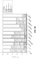
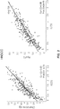


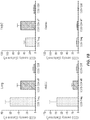

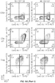
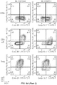
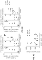

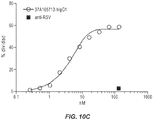
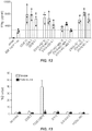


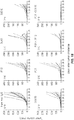
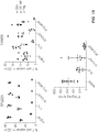
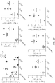
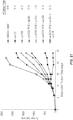


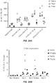


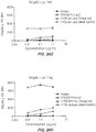
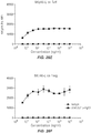

Classifications
-
- C—CHEMISTRY; METALLURGY
- C07—ORGANIC CHEMISTRY
- C07K—PEPTIDES
- C07K16/00—Immunoglobulins [IGs], e.g. monoclonal or polyclonal antibodies
- C07K16/18—Immunoglobulins [IGs], e.g. monoclonal or polyclonal antibodies against material from animals or humans
- C07K16/28—Immunoglobulins [IGs], e.g. monoclonal or polyclonal antibodies against material from animals or humans against receptors, cell surface antigens or cell surface determinants
- C07K16/2803—Immunoglobulins [IGs], e.g. monoclonal or polyclonal antibodies against material from animals or humans against receptors, cell surface antigens or cell surface determinants against the immunoglobulin superfamily
- C07K16/2818—Immunoglobulins [IGs], e.g. monoclonal or polyclonal antibodies against material from animals or humans against receptors, cell surface antigens or cell surface determinants against the immunoglobulin superfamily against CD28 or CD152
-
- A—HUMAN NECESSITIES
- A61—MEDICAL OR VETERINARY SCIENCE; HYGIENE
- A61P—SPECIFIC THERAPEUTIC ACTIVITY OF CHEMICAL COMPOUNDS OR MEDICINAL PREPARATIONS
- A61P35/00—Antineoplastic agents
-
- A—HUMAN NECESSITIES
- A61—MEDICAL OR VETERINARY SCIENCE; HYGIENE
- A61K—PREPARATIONS FOR MEDICAL, DENTAL OR TOILETRY PURPOSES
- A61K39/00—Medicinal preparations containing antigens or antibodies
- A61K2039/505—Medicinal preparations containing antigens or antibodies comprising antibodies
-
- A—HUMAN NECESSITIES
- A61—MEDICAL OR VETERINARY SCIENCE; HYGIENE
- A61K—PREPARATIONS FOR MEDICAL, DENTAL OR TOILETRY PURPOSES
- A61K39/00—Medicinal preparations containing antigens or antibodies
- A61K2039/505—Medicinal preparations containing antigens or antibodies comprising antibodies
- A61K2039/507—Comprising a combination of two or more separate antibodies
-
- C—CHEMISTRY; METALLURGY
- C07—ORGANIC CHEMISTRY
- C07K—PEPTIDES
- C07K2317/00—Immunoglobulins specific features
- C07K2317/20—Immunoglobulins specific features characterized by taxonomic origin
- C07K2317/24—Immunoglobulins specific features characterized by taxonomic origin containing regions, domains or residues from different species, e.g. chimeric, humanized or veneered
-
- C—CHEMISTRY; METALLURGY
- C07—ORGANIC CHEMISTRY
- C07K—PEPTIDES
- C07K2317/00—Immunoglobulins specific features
- C07K2317/30—Immunoglobulins specific features characterized by aspects of specificity or valency
- C07K2317/33—Crossreactivity, e.g. for species or epitope, or lack of said crossreactivity
-
- C—CHEMISTRY; METALLURGY
- C07—ORGANIC CHEMISTRY
- C07K—PEPTIDES
- C07K2317/00—Immunoglobulins specific features
- C07K2317/50—Immunoglobulins specific features characterized by immunoglobulin fragments
- C07K2317/56—Immunoglobulins specific features characterized by immunoglobulin fragments variable (Fv) region, i.e. VH and/or VL
- C07K2317/565—Complementarity determining region [CDR]
-
- C—CHEMISTRY; METALLURGY
- C07—ORGANIC CHEMISTRY
- C07K—PEPTIDES
- C07K2317/00—Immunoglobulins specific features
- C07K2317/70—Immunoglobulins specific features characterized by effect upon binding to a cell or to an antigen
- C07K2317/75—Agonist effect on antigen
-
- C—CHEMISTRY; METALLURGY
- C07—ORGANIC CHEMISTRY
- C07K—PEPTIDES
- C07K2317/00—Immunoglobulins specific features
- C07K2317/90—Immunoglobulins specific features characterized by (pharmaco)kinetic aspects or by stability of the immunoglobulin
- C07K2317/92—Affinity (KD), association rate (Ka), dissociation rate (Kd) or EC50 value
-
- C—CHEMISTRY; METALLURGY
- C07—ORGANIC CHEMISTRY
- C07K—PEPTIDES
- C07K2319/00—Fusion polypeptide
- C07K2319/30—Non-immunoglobulin-derived peptide or protein having an immunoglobulin constant or Fc region, or a fragment thereof, attached thereto
Definitions
- Antibodies that bind to Inducible T-Cell Costimulator (ICOS) are provided. Methods of treatment comprising administering anti-ICOS antibodies are also provided.
- ICOS is a member of the B7/CD28/CTLA-4 immunoglobulin superfamily and is specifically expressed on T cells. Unlike CD28, which is constitutively expressed on T cells and provides co-stimulatory signals necessary for full activation of resting T cells, ICOS is expressed only after initial T cell activation.
- ICOS has been implicated in diverse aspects of T cell responses (reviewed in Simpson et al., 2010, Curr. Opin. Immunol., 22: 326-332 ). It plays a role in the formation of germinal centers, T/B cell collaboration, and immunoglobulin class switching. ICOS-deficient mice show impaired germinal center formation and have decreased production of interleukin IL-10. These defects have been specifically linked to deficiencies in T follicular helper cells.
- ICOS also plays a role in the development and function of other T cell subsets, including Th1, Th2, and Th17.
- ICOS co-stimulates T cell proliferation and cytokine secretion associated with both Th1 and Th2 cells.
- ICOS KO mice demonstrate impaired development of autoimmune phenotypes in a variety of disease models, including diabetes (Th1), airway inflammation (Th2) and EAE neuro-inflammatory models (Th17).
- ICOS In addition to its role in modulating T effector (Teff) cell function, ICOS also modulates T regulatory cells (Tregs). ICOS is expressed at high levels on Tregs, and has been implicated in Treg homeostasis and function.
- ICOS a disulfide-linked homodimer
- PI3K and AKT pathways a signal through the PI3K and AKT pathways.
- Lineage specific transcription factors e.g., T-bet, GATA-3
- ICOS ligand (ICOSL; B7-H2; B7RP1; CD275; GL50), also a member of the B7 superfamily, is the only ligand for ICOS and is expressed on the cell surface of B cells, macrophages and dendritic cells.
- ICOSL functions as a non-covalently linked homodimer on the cell surface in its interaction with ICOS.
- an isolated antibody that binds ICOS is provided, wherein the antibody is an agonist of CD4 T cells (such as CD4 T effector (Teff) cells).
- an isolated antibody that binds ICOS is provided, wherein the antibody is an agonist of CD4 T cells (such as CD4 Teff cells) and depletes T regulatory (Treg) cells.
- an isolated antibody that binds ICOS is provided, wherein the antibody depletes Treg cells, but does not deplete Teff cells.
- an isolated antibody that binds ICOS is provided, wherein the antibody induces pAKT signaling on CD4 T cells.
- an isolated antibody that binds ICOS is provided, wherein the antibody induces pAKT signaling on CD4 T cells and depletes Treg cells.
- an isolated antibody that binds to ICOS is provided, wherein the antibody comprises:
- an antibody that binds to ICOS comprises a heavy chain variable region (V H ) and a light chain variable region (V L ), wherein:
- an antibody that binds to ICOS comprises a heavy chain variable region (V H ) and a light chain variable region (V L ), wherein:
- the antibody is a monoclonal antibody. In some embodiments, the antibody is a chimeric antibody or a humanized antibody. In some embodiments, the antibody is an antibody fragment selected from a Fab, Fab', Fv, scFv or (Fab') 2 fragment. In some embodiments, the antibody is a full length antibody.
- an antibody that binds to ICOS comprises a heavy chain comprising the amino acid sequence of SEQ ID NO: 188 and a light chain comprising the amino acid sequence of SEQ ID NO: 189.
- administration of the antibody to a mammal results in an increase in T effector (Teff) cells in the mammal. In some embodiments, administration of the antibody to a mammal results in activation of T effector (Teff) cells in the mammal. In some embodiments, administration of the antibody to a mammal increases the ratio of Teff cells to Treg cells.
- the Teff cells are CD4+ FoxP3- T cells. In some embodiments, the Teff cells are CD4+ FoxP3- T cells and CD8+ T cells. In some embodiments, the Teff cells are CD8+ T cells. In some embodiments, administration of the antibody to a mammal results in a decrease in T regulatory (Treg) cells in the mammal. In some embodiments, the Treg cells are CD4+ FoxP3+ T cells.
- an isolated antibody that binds to human ICOS, wherein the antibody also binds to mouse ICOS and/or rat ICOS. In some embodiments, the isolated antibody binds to human ICOS with an affinity (K D ) of less than 5 nM. In some embodiments, the isolated antibody binds to rat ICOS with an affinity (K D ) of less than 10 nM. In some embodiments, affinity is determined using biolayer interferometry ( see, e.g. , Abdiche et al., 2008, Anal Biochem, 377: 209-217 ; and ForteBio Octet® system).
- the antibody binds to human ICOS, mouse ICOS, and rat ICOS. In some embodiments, the antibody binds to cynomolgus monkey ICOS. In some embodiments, the antibody is a monoclonal antibody. In some embodiments, the antibody is a chimeric antibody or a humanized antibody. In some embodiments, the antibody is an antibody fragment selected from a Fab, Fab', Fv, scFv or (Fab') 2 fragment. In some embodiments, the antibody is a full length antibody. In some embodiments, administration of the antibody to a mammal results in an increase in T effector (Teff) cells in the mammal.
- T effector T effector
- administration of the antibody to a mammal results in activation of T effector (Teff) cells in the mammal.
- Teff cells are CD4+ FoxP3- T cells.
- the Teff cells are CD4+ FoxP3- T cells and CD8+ T cells.
- the Teff cells are CD8+ T cells.
- administration of the antibody to a mammal results in a decrease in T regulatory (Treg) cells in the mammal.
- the Treg cells are CD4+ FoxP3+ T cells.
- the mammal is selected from a mouse, rat, cynomolgus monkey, and human.
- the level of a chemokine or cytokine selected from GZMa, GZMb, CSF2, IL2, CXCL9, CXCL10, CXCL11, and CXCL13 is at least a 2-fold, or at least 3-fold higher than the level of the chemokine following treatment of the lung tumor tissue with a control antibody.
- the level is an mRNA level.
- the level is a protein level.
- a control antibody is an isotype-matched antibody that binds to an unrelated antigen, and which is not expected to have an effect on the chemokine levels.
- the level of the chemokine is measured 1 hour, 2 hours, 3 hours, 4 hours, 5 hours, or 6 hours after treatment. In some embodiments, the chemokine is CXCL11. In some embodiments, the level of the chemokine is measured 6 hours, 8 hours, 10 hours, 12 hours, 14 hours, 16 hours, 18 hours, 20 hours, 22 hours, 24 hours, 36 hours, or 48 hours after treatment. In some embodiments, the level of the chemokine is measured 24 hours after treatment. In some embodiments, the lung tumor tissue is human lung tumor tissue.
- an antibody provided herein increases the level of at least one chemokine and/or cytokine selected from GZMa, GZMb, CSF2, IL2, CXCL9, CXCL10, CXCL11, and CXCL13 in a mammal that has been administered the antibody by at least a 2-fold.
- the level of the chemokine is measured 1 hour, 2 hours, 3 hours, 4 hours, 5 hours, or 6 hours after administration of the antibody.
- the level is an mRNA level.
- the level is a protein level.
- the chemokine is CXCL11.
- the level of the chemokine is measured 6 hours, 8 hours, 10 hours, 12 hours, 14 hours, 16 hours, 18 hours, 20 hours, 22 hours, 24 hours, 36 hours, or 48 hours after administration of the antibody. In some embodiments, the level of the chemokine is measured 24 hours after administration of the antibody.
- an anti-ICOS agonist antibody is provided, wherein the antibody increases the level of at least one chemokine and/or cytokine selected from GZMa, GZMb, CSF2, IL2, CXCL9, CXCL10, CXCL11, and CXCL13 in a mammal that has been administered the antibody by at least a 2-fold.
- an anti-ICOS agonist antibody is provided, wherein the antibody increases the level of at least one chemokine and/or cytokine selected from GZMa, GZMb, CSF2, IL2, CXCL9, CXCL10, and CXCL11 in a mammal that has been administered the antibody by at least a 2-fold.
- the level of the chemokine is measured 1 hour, 2 hours, 3 hours, 4 hours, 5 hours, or 6 hours after administration of the antibody. In some embodiments, the level is an mRNA level. In some embodiments, the level is a protein level. In some embodiments, the chemokine is CXCL11. In some embodiments, the level of the chemokine is measured 6 hours, 8 hours, 10 hours, 12 hours, 14 hours, 16 hours, 18 hours, 20 hours, 22 hours, 24 hours, 36 hours, or 48 hours after administration of the antibody. In some embodiments, the level of the chemokine is measured 24 hours after administration of the antibody. In some embodiments, the mammal is a human. In some embodiments, the human has cancer.
- the cancer is selected from melanoma, non-small cell lung cancer (NSCLC), renal cell carcinoma (RCC), gastric cancer, bladder cancer, diffuse large B-cell lymphoma (DLBCL), Hodgkin's lymphoma, ovarian cancer, head & neck squamous cell cancer (HNSCC), and triple negative breast cancer (TNBC).
- NSCLC non-small cell lung cancer
- RNC renal cell carcinoma
- gastric cancer bladder cancer
- DLBCL diffuse large B-cell lymphoma
- HNSCC head & neck squamous cell cancer
- TNBC triple negative breast cancer
- an antibody that binds to ICOS is provided, wherein the antibody increases the level of the ligand for NKp46 (NKp46-L) on T cells.
- the increased level of NKp46-L on T cells is determined using a soluble NKp46 extracellular domain in a flow cytometry assay.
- the antibody increases the level of NKp46-L on Treg cells more than the antibody increases the level of NKp46-L on Teff cells.
- the antibody increases CD16 shedding on NK cells.
- an isolated nucleic acid encoding an antibody described herein is provided.
- a vector comprising the nucleic acid is provided.
- a host cell comprising the vector is provided.
- a host cell that produces an antibody described herein is provided.
- a method for making an anti-ICOS antibody is provided, comprising culturing the host cell under conditions suitable for expression of the antibody. In some embodiments, the method comprises recovering the antibody produced by the host cell.
- a pharmaceutical composition which comprises an anti-ICOS antibody described herein and a pharmaceutically acceptable carrier.
- methods of treating cancer comprising administering an effective amount of an anti-ICOS antibody described herein or a pharmaceutical composition described herein.
- the cancer is selected from melanoma, non-small cell lung cancer (NSCLC), renal cell carcinoma (RCC), gastric cancer, bladder cancer, diffuse large B-cell lymphoma (DLBCL), Hodgkin's lymphoma, ovarian cancer, head & neck squamous cell cancer (HNSCC), and triple negative breast cancer (TNBC).
- the cancer is selected from melanoma, gastric cancer, head & neck squamous cell cancer (HNSCC), non-small cell lung cancer (NSCLC), and triple negative breast cancer (TNBC).
- a method of increasing the number of T effector (Teff) cells in a mammal comprising administering an effective amount of an anti-ICOS antibody described herein or a pharmaceutical composition described herein.
- the method further comprises activating Teff cells.
- a method of activating T effector (Teff) cells in a mammal comprising administering an effective amount of an anti-ICOS antibody described herein or a pharmaceutical composition described herein.
- a method of increasing the ratio of Teff cells to Treg cells in a mammal comprising administering an effective amount of an anti-ICOS antibody described herein or a pharmaceutical composition described herein.
- the Teff cells are CD4+ FoxP3- T cells. In some embodiments, the Teff cells are CD4+ FoxP3- T cells and CD8+ T cells. In some embodiments, the Teff cells are CD8+ T cells. In some embodiments, the method further comprises decreasing the number of T regulatory (Treg) cells.
- Treg T regulatory
- a method of decreasing the number of T regulatory (Treg) cells in a mammal comprising administering an effective amount of an anti-ICOS antibody described herein or a pharmaceutical composition described herein.
- the Treg cells are CD4+ FoxP3+ T cells.
- a method of increasing the level of at least one chemokine and/or cytokine selected from GZMa, GZMb, CSF2, IL2, CXCL9, CXCL10, CXCL11, and CXCL13 in a mammal comprising administering to the mammal an antibody provided herein.
- the level of at least one chemokine is increased by at least 2-fold or at least 3-fold.
- the level of the chemokine is measured 1 hour, 2 hours, 3 hours, 4 hours, 5 hours, or 6 hours after administration of the antibody.
- the level is an mRNA level.
- the level is a protein level.
- the chemokine is CXCL11. In some embodiments, the level of the chemokine is measured 6 hours, 8 hours, 10 hours, 12 hours, 14 hours, 16 hours, 18 hours, 20 hours, 22 hours, 24 hours, 36 hours, or 48 hours after administration of the antibody. In some embodiments, the level of the chemokine is measured 24 hours after administration of the antibody. In some embodiments, the mammal is a human. In some embodiments, the human has cancer.
- the cancer is selected from melanoma, non-small cell lung cancer (NSCLC), renal cell carcinoma (RCC), gastric cancer, bladder cancer, diffuse large B-cell lymphoma (DLBCL), Hodgkin's lymphoma, ovarian cancer, head & neck squamous cell cancer (HNSCC), and triple negative breast cancer (TNBC).
- NSCLC non-small cell lung cancer
- RNC renal cell carcinoma
- gastric cancer bladder cancer
- DLBCL diffuse large B-cell lymphoma
- HNSCC head & neck squamous cell cancer
- TNBC triple negative breast cancer
- the mammal is a human.
- the mammal is administered at least one additional therapeutic agent.
- the additional therapeutic agent is administered concurrently or sequentially with the anti-ICOS antibody.
- the additional therapeutic agent is a PD-1 therapy.
- the additional therapeutic agent is selected from an anti-PD-1 antibody and an anti-PD-Ll antibody.
- an anti-ICOS antibody provided herein is administered with nivolumab.
- an anti-ICOS antibody provided herein is administered with pembrolizumab.
- an anti-ICOS antibody provided herein is administered with atezolizumab.
- an anti-ICOS antibody provided herein is administered with avelumab.
- an anti-ICOS antibody provided herein is administered with durvalumab.
- the additional therapeutic is a cancer vaccine.
- the cancer vaccine is developed using a neoantigen.
- the cancer vaccine is a DNA vaccine.
- the cancer vaccine is an engineered virus comprising a cancer antigen, such as PROSTVAC (rilimogene galvacirepvec/rilimogene glafolivec).
- the cancer vaccine comprises engineered tumor cells, such as GVAX.
- the anti-ICOS antibody provided herein is administered with an agonist anti-OX40 antibody. In some embodiments, the anti-ICOS antibody provided herein is administered with an anti-CTLA4 antibody. In some embodiments, the anti-ICOS antibody provided herein is administered with ipilimumab.
- the additional therapeutic is a chemotherapeutic agent.
- chemotherapeutic agents include capectiabine, cyclophosphamide, dacarbazine, temozolomide, cyclophosphamide, docetaxel, doxorubicin, daunorubicin, cisplatin, carboplatin, epirubicin, eribulin, 5-FU, gemcitabine, irinotecan, ixabepilone, methotrexate, mitoxantrone, oxaliplatin, paclitaxel, nab-paclitaxel, ABRAXANE® (protein-bound paclitaxel), pemetrexed, vinorelbine, and vincristine.
- the anti-ICOS antibody provided herein is administered with ABRAXANE® (Celgene).
- an anti-ICOS antibody provided herein is administered with at least one kinase inhibitor.
- Nonlimiting exemplary kinase inhibitors include erlotinib, afatinib, gefitinib, crizotinib, dabrafenib, trametinib, vemurafenib, and cobimetanib.
- the additional therapeutic agent is an IDO inhibitor.
- IDO inhibitors include Indoximod (New Link Genetics), INCB024360 (Incyte Corp), 1-methyl-D-tryptophan (New Link Genetics), and GDC-0919 (Genentech).
- the additional therapeutic agent is an immune-modifying drug (IMiD).
- IMDs include thalidomide, lenalidomide, and pomalidomide.
- the mammal receives CAR-T therapy in addition to administration of anti-ICOS an antibody described herein.
- the mammal undergoes surgery and/or radiation therapy in addition to administration of an anti-ICOS antibody described herein, with or without an additional therapeutic agent.
- the mammal undergoes radiation therapy in addition to administration of anti-ICOS an antibody described herein, with or without an additional therapeutic agent.
- the cancer is selected from melanoma, non-small cell lung cancer (NSCLC), renal cell carcinoma (RCC), gastric cancer, bladder cancer, diffuse large B-cell lymphoma (DLBCL), Hodgkin's lymphoma, ovarian cancer, head & neck squamous cell cancer (HNSCC), and triple negative breast cancer (TNBC).
- the cancer is selected from melanoma, gastric cancer, head & neck squamous cell cancer (HNSCC), non-small cell lung cancer (NSCLC), and triple negative breast cancer (TNBC).
- the medicament is for administration with at least one additional therapeutic agent.
- the additional therapeutic agent is selected from an anti-PD-1 antibody and an anti-PD-Ll antibody.
- the cancer is selected from melanoma, non-small cell lung cancer (NSCLC), renal cell carcinoma (RCC), gastric cancer, bladder cancer, diffuse large B-cell lymphoma (DLBCL), Hodgkin's lymphoma, ovarian cancer, head & neck squamous cell cancer (HNSCC), and triple negative breast cancer (TNBC).
- the cancer is selected from melanoma, gastric cancer, head & neck squamous cell cancer (HNSCC), non-small cell lung cancer (NSCLC), and triple negative breast cancer (TNBC).
- the cancer is selected from melanoma, non-small cell lung cancer (NSCLC), renal cell carcinoma (RCC), gastric cancer, bladder cancer, diffuse large B-cell lymphoma (DLBCL), Hodgkin's lymphoma, ovarian cancer, head & neck squamous cell cancer (HNSCC), and triple negative breast cancer (TNBC).
- NSCLC non-small cell lung cancer
- RNC renal cell carcinoma
- TNBC triple negative breast cancer
- the cancer is selected from melanoma, gastric cancer, head & neck squamous cell cancer (HNSCC), non-small cell lung cancer (NSCLC), and triple negative breast cancer (TNBC).
- an antibody described herein or a pharmaceutical composition described herein is provided for use in treating cancer.
- the cancer is selected from melanoma, non-small cell lung cancer (NSCLC), renal cell carcinoma (RCC), gastric cancer, bladder cancer, diffuse large B-cell lymphoma (DLBCL), Hodgkin's lymphoma, ovarian cancer, head & neck squamous cell cancer (HNSCC), and triple negative breast cancer (TNBC).
- the cancer is selected from melanoma, gastric cancer, head & neck squamous cell cancer (HNSCC), non-small cell lung cancer (NSCLC), and triple negative breast cancer (TNBC).
- Antibodies that bind ICOS are provided. Antibody heavy chains and light chains that are capable of forming antibodies that bind ICOS are also provided. In addition, antibodies, heavy chains, and light chains comprising one or more particular complementarity determining regions (CDRs) are provided. Polynucleotides encoding antibodies to ICOS are provided. Polynucleotides encoding antibody heavy chains or lights chains are also provided. Methods of producing and/or purifying antibodies to ICOS are provided. Methods of treatment using antibodies to ICOS are provided. Such methods include, but are not limited to, methods of treating cancer. Methods of detecting ICOS are provided. Such methods include methods to identify an individual who may benefit from treatment with an anti-ICOS antibody, to monitor treatment of an individual with an anti-ICOS antibody and to improve therapeutic efficacy of an anti-ICOS antibody in an individual.
- CDRs complementarity determining regions
- nucleic acid molecule refers to a polymer of nucleotides.
- polymers of nucleotides may contain natural and/or non-natural nucleotides, and include, but are not limited to, DNA, RNA, and PNA.
- Nucleic acid sequence refers to the linear sequence of nucleotides that comprise the nucleic acid molecule or polynucleotide.
- polypeptide and protein are used interchangeably to refer to a polymer of amino acid residues, and are not limited to a minimum length. Such polymers of amino acid residues may contain natural or non-natural amino acid residues, and include, but are not limited to, peptides, oligopeptides, dimers, trimers, and multimers of amino acid residues. Both full-length proteins and fragments thereof are encompassed by the definition.
- the terms also include post-expression modifications of the polypeptide, for example, glycosylation, sialylation, acetylation, phosphorylation, and the like.
- polypeptide refers to a protein which includes modifications, such as deletions, additions, and substitutions (generally conservative in nature), to the native sequence, as long as the protein maintains the desired activity. These modifications may be deliberate, as through site-directed mutagenesis, or may be accidental, such as through mutations of hosts which produce the proteins or errors due to PCR amplification.
- ICOS and "inducible T-cell costimulatory” as used herein refer to any native ICOS that results from expression and processing of ICOS in a cell.
- the term includes ICOS from any vertebrate source, including mammals such as primates (e.g., humans and cynomolgus monkeys) and rodents (e.g., mice and rats), unless otherwise indicated.
- the term also includes naturally occurring variants of ICOS, e.g., splice variants or allelic variants.
- the amino acid sequence of an exemplary human ICOS precursor protein, with signal sequence (with signal sequence, amino acids 1-20) is shown in SEQ ID NO: 1.
- the amino acid sequence of an exemplary mature human ICOS is shown in SEQ ID NO: 2.
- the amino acid sequence of an exemplary mouse ICOS precursor protein, with signal sequence (with signal sequence, amino acids 1-20) is shown in SEQ ID NO: 3.
- the amino acid sequence of an exemplary mature mouse ICOS is shown in SEQ ID NO: 4.
- the amino acid sequence of an exemplary rat ICOS precursor protein, with signal sequence (with signal sequence, amino acids 1-20) is shown in SEQ ID NO: 190.
- the amino acid sequence of an exemplary mature rat ICOS is shown in SEQ ID NO: 191.
- the amino acid sequence of an exemplary cynomolgus monkey ICOS precursor protein, with signal sequence (with signal sequence, amino acids 1-20) is shown in SEQ ID NO: 5.
- the amino acid sequence of an exemplary mature cynomolgus monkey ICOS is shown in SEQ ID NO: 6.
- the term “specifically binds" to an antigen or epitope is a term that is well understood in the art, and methods to determine such specific binding are also well known in the art.
- a molecule is said to exhibit "specific binding” or “preferential binding” if it reacts or associates more frequently, more rapidly, with greater duration and/or with greater affinity with a particular cell or substance than it does with alternative cells or substances.
- An antibody “specifically binds” or “preferentially binds” to a target if it binds with greater affinity, avidity, more readily, and/or with greater duration than it binds to other substances.
- an antibody that specifically or preferentially binds to an ICOS epitope is an antibody that binds this epitope with greater affinity, avidity, more readily, and/or with greater duration than it binds to other ICOS epitopes or non-ICOS epitopes. It is also understood by reading this definition that, for example, an antibody (or moiety or epitope) that specifically or preferentially binds to a first target may or may not specifically or preferentially bind to a second target. As such, “specific binding” or “preferential binding” does not necessarily require (although it can include) exclusive binding. Generally, but not necessarily, reference to binding means preferential binding. "Specificity" refers to the ability of a binding protein to selectively bind an antigen.
- substantially pure refers to material which is at least 50% pure (that is, free from contaminants), more preferably, at least 90% pure, more preferably, at least 95% pure, yet more preferably, at least 98% pure, and most preferably, at least 99% pure.
- epitope refers to a site on a target molecule (for example, an antigen, such as a protein, nucleic acid, carbohydrate or lipid) to which an antigen-binding molecule (for example, an antibody, antibody fragment, or scaffold protein containing antibody binding regions) binds.
- a target molecule for example, an antigen, such as a protein, nucleic acid, carbohydrate or lipid
- an antigen-binding molecule for example, an antibody, antibody fragment, or scaffold protein containing antibody binding regions
- Epitopes often include a chemically active surface grouping of molecules such as amino acids, polypeptides or sugar side chains and have specific three-dimensional structural characteristics as well as specific charge characteristics. Epitopes can be formed both from contiguous and/or juxtaposed noncontiguous residues (for example, amino acids, nucleotides, sugars, lipid moiety) of the target molecule.
- Epitopes formed from contiguous residues typically are retained on exposure to denaturing solvents whereas epitopes formed by tertiary folding typically are lost on treatment with denaturing solvents.
- An epitope may include but is not limited to at least 3, at least 5 or 8-10 residues (for example, amino acids or nucleotides). In some examples an epitope is less than 20 residues (for example, amino acids or nucleotides) in length, less than 15 residues or less than 12 residues. Two antibodies may bind the same epitope within an antigen if they exhibit competitive binding for the antigen.
- an epitope can be identified by a certain minimal distance to a CDR residue on the antigen-binding molecule. In some embodiments, an epitope can be identified by the above distance, and further limited to those residues involved in a bond (for example, a hydrogen bond) between an antibody residue and an antigen residue. An epitope can be identified by various scans as well, for example an alanine or arginine scan can indicate one or more residues that the antigen-binding molecule can interact with. Unless explicitly denoted, a set of residues as an epitope does not exclude other residues from being part of the epitope for a particular antibody.
- a set of residues identified as an epitope designates a minimal epitope of relevance for the antigen, rather than an exclusive list of residues for an epitope on an antigen.
- a “nonlinear epitope” or “conformational epitope” comprises noncontiguous polypeptides, amino acids and/or sugars within the antigenic protein to which an antibody specific to the epitope binds.
- at least one of the residues will be noncontiguous with the other noted residues of the epitope; however, one or more of the residues can also be contiguous with the other residues.
- linear epitope comprises contiguous polypeptides, amino acids and/or sugars within the antigenic protein to which an antibody specific to the epitope binds. It is noted that, in some embodiments, not every one of the residues within the linear epitope need be directly bound (or involved in a bond) with the antibody. In some embodiments, linear epitopes can be from immunizations with a peptide that effectively consisted of the sequence of the linear epitope, or from structural sections of a protein that are relatively isolated from the remainder of the protein (such that the antibody can interact, at least primarily), just with that sequence section.
- antibody herein is used in the broadest sense and encompasses various antibody structures, including but not limited to monoclonal antibodies, polyclonal antibodies, multispecific antibodies (for example, bispecific(such as Bi-specific T-cell engagers) and trispecific antibodies), and antibody fragments so long as they exhibit the desired antigen-binding activity.
- antibody includes, but is not limited to, fragments that are capable of binding to an antigen, such as Fv, single-chain Fv (scFv), Fab, Fab', di-scFv, sdAb (single domain antibody) and (Fab') 2 (including a chemically linked F(ab') 2 ).
- an antigen such as Fv, single-chain Fv (scFv), Fab, Fab', di-scFv, sdAb (single domain antibody) and (Fab') 2 (including a chemically linked F(ab') 2 ).
- Papain digestion of antibodies produces two identical antigen-binding fragments, called “Fab” fragments, each with a single antigen-binding site, and a residual "Fc” fragment, whose name reflects its ability to crystallize readily.
- Pepsin treatment yields an F(ab') 2 fragment that has two antigen-combining sites and is still capable of cross-linking antigen.
- antibody also includes, but is not limited to, chimeric antibodies, humanized antibodies, and antibodies of various species such as mouse, human, cynomolgus monkey, etc. Furthermore, for all antibody constructs provided herein, variants having the sequences from other organisms are also contemplated. Thus, if a human version of an antibody is disclosed, one of skill in the art will appreciate how to transform the human sequence based antibody into a mouse, rat, cat, dog, horse, etc. sequence. Antibody fragments also include either orientation of single chain scFvs, tandem di-scFv, diabodies, tandem tri-sdcFv, minibodies, etc.
- Antibody fragments also include nanobodies (sdAb, an antibody having a single, monomeric domain, such as a pair of variable domains of heavy chains, without a light chain).
- An antibody fragment can be referred to as being a specific species in some embodiments (for example, human scFv or a mouse scFv). This denotes the sequences of at least part of the non-CDR regions, rather than the source of the construct.
- monoclonal antibody refers to an antibody of a substantially homogeneous population of antibodies, that is, the individual antibodies comprising the population are identical except for possible naturally-occurring mutations that may be present in minor amounts. Monoclonal antibodies are highly specific, being directed against a single antigenic site. Furthermore, in contrast to polyclonal antibody preparations, which typically include different antibodies directed against different determinants (epitopes), each monoclonal antibody is directed against a single determinant on the antigen. Thus, a sample of monoclonal antibodies can bind to the same epitope on the antigen.
- the modifier "monoclonal” indicates the character of the antibody as being obtained from a substantially homogeneous population of antibodies, and is not to be construed as requiring production of the antibody by any particular method.
- the monoclonal antibodies may be made by the hybridoma method first described by Kohler and Milstein, 1975, Nature 256:495 , or may be made by recombinant DNA methods such as described in U.S. Pat. No. 4,816,567 .
- the monoclonal antibodies may also be isolated from phage libraries generated using the techniques described in McCafferty et al., 1990, Nature 348:552-554 , for example.
- CDR denotes a complementarity determining region as defined by at least one manner of identification to one of skill in the art.
- CDRs can be defined in accordance with any of the Chothia numbering schemes, the Kabat numbering scheme, a combination of Kabat and Chothia, the AbM definition, the contact definition, and/or a combination of the Kabat, Chothia, AbM, and/or contact definitions.
- Exemplary CDRs (CDR-L1, CDR-L2, CDR-L3, CDR-H1, CDR-H2, and CDR-H3) occur at amino acid residues 24-34 of L1, 50-56 of L2, 89-97 of L3, 31-35B of H1, 50-65 of H2, and 95-102 of H3.
- the AbM definition can include, for example, CDRs (CDR-L1, CDR-L2, CDR-L3, CDR-H1, CDR-H2, and CDR-H3) at amino acid residues 24-34 of L1, 50-56 of L2, 89-97 of L3, H26-H35B of H1, 50-58 of H2, and 95-102 of H3.
- the Contact definition can include, for example, CDRs (CDR-L1, CDR-L2, CDR-L3, CDR-H1, CDR-H2, and CDR-H3) at amino acid residues 30-36 of L1, 46-55 of L2, 89-96 of L3, 30-35 of H1, 47-58 of H2, and 93-101 of H3.
- the Chothia definition can include, for example, CDRs (CDR-L1, CDR-L2, CDR-L3, CDR-H1, CDR-H2, and CDR-H3) at amino acid residues 24-34 of L1, 50-56 of L2, 89-97 of L3, 26-32... 34 of H1, 52-56 of H2, and 95-102 of H3.
- CDRs can also be provided as shown in any one or more of the accompanying figures. With the exception of CDR1 in V H , CDRs generally comprise the amino acid residues that form the hypervariable loops.
- the various CDRs within an antibody can be designated by their appropriate number and chain type, including, without limitation as: a) CDR-L1, CDR-L2, CDR-L3, CDR-H1, CDR-H2, and CDR-H3; b) CDRL1, CDRL2, CDRL3, CDRH1, CDRH2, and CDRH3; c) LCDR-1, LCDR-2, LCDR-3, HCDR-1, HCDR-2, and HCDR-3; or d) LCDR1, LCDR2, LCDR3, HCDR1, HCDR2, and HCDR3; etc.
- CDR is used herein to also encompass HVR or a "hyper variable region", including hypervariable loops.
- Exemplary hypervariable loops occur at amino acid residues 26-32 (L1), 50-52 (L2), 91-96 (L3), 26-32 (HI), 53-55 (H2), and 96-101 (H3).
- heavy chain variable region refers to a region comprising at least three heavy chain CDRs.
- the heavy chain variable region includes the three CDRs and at least FR2 and FR3.
- the heavy chain variable region includes at least heavy chain HCDR1, framework (FR) 2, HCDR2, FR3, and HCDR3.
- a heavy chain variable region also comprises at least a portion of an FR1 and/or at least a portion of an FR4.
- heavy chain constant region refers to a region comprising at least three heavy chain constant domains, C H 1, C H 2, and C H 3.
- Nonlimiting exemplary heavy chain constant regions include ⁇ , ⁇ , and ⁇ .
- Nonlimiting exemplary heavy chain constant regions also include ⁇ and ⁇ .
- Each heavy constant region corresponds to an antibody isotype.
- an antibody comprising a ⁇ constant region is an IgG antibody
- an antibody comprising a ⁇ constant region is an IgD antibody
- an antibody comprising an ⁇ constant region is an IgA antibody.
- an antibody comprising a ⁇ constant region is an IgM antibody
- an antibody comprising an ⁇ constant region is an IgE antibody.
- IgG antibodies include, but are not limited to, IgG1 (comprising a ⁇ 1 constant region), IgG2 (comprising a ⁇ 2 constant region), IgG3 (comprising a ⁇ 3 constant region), and IgG4 (comprising a ⁇ 4 constant region) antibodies
- IgA antibodies include, but are not limited to, IgA1 (comprising an ⁇ 1 constant region) and IgA2 (comprising an ⁇ 2 constant region) antibodies
- IgM antibodies include, but are not limited to, IgM1 and IgM2.
- heavy chain refers to a polypeptide comprising at least a heavy chain variable region, with or without a leader sequence.
- a heavy chain comprises at least a portion of a heavy chain constant region.
- full-length heavy chain refers to a polypeptide comprising a heavy chain variable region and a heavy chain constant region, with or without a leader sequence.
- light chain variable region refers to a region comprising at least three light chain CDRs.
- the light chain variable region includes the three CDRs and at least FR2 and FR3.
- the light chain variable region includes at least light chain LCDR1, framework (FR) 2, LCDR2, FR3, and LCDR3.
- a light chain variable region may comprise light chain CDR1, framework (FR) 2, CDR2, FR3, and CDR3.
- a light chain variable region also comprises at least a portion of an FR1 and/or at least a portion of an FR4.
- light chain constant region refers to a region comprising a light chain constant domain, C L .
- Nonlimiting exemplary light chain constant regions include ⁇ and ⁇ .
- non-function-altering deletions and alterations within the domains are encompassed within the scope of the term “light chain constant region,” unless designated otherwise.
- light chain refers to a polypeptide comprising at least a light chain variable region, with or without a leader sequence.
- a light chain comprises at least a portion of a light chain constant region.
- full-length light chain refers to a polypeptide comprising a light chain variable region and a light chain constant region, with or without a leader sequence.
- acceptor human framework for the purposes herein is a framework comprising the amino acid sequence of a light chain variable domain (V L ) framework or a heavy chain variable domain (V H ) framework derived from a human immunoglobulin framework or a human consensus framework, as defined below.
- An acceptor human framework derived from a human immunoglobulin framework or a human consensus framework can comprise the same amino acid sequence thereof, or it can contain amino acid sequence changes. In some embodiments, the number of amino acid changes are 10 or less, 9 or less, 8 or less, 7 or less, 6 or less, 5 or less, 4 or less, 3 or less, or 2 or less.
- the V L acceptor human framework is identical in sequence to the V L human immunoglobulin framework sequence or human consensus framework sequence.
- Affinity refers to the strength of the sum total of noncovalent interactions between a single binding site of a molecule (for example, an antibody) and its binding partner (for example, an antigen).
- the affinity of a molecule X for its partner Y can generally be represented by the dissociation constant (K D ).
- Affinity can be measured by common methods known in the art (such as, for example, ELISA K D , KinExA, bio-layer interferometry (BLI), and/or surface plasmon resonance devices (such as a BIAcore® device), including those described herein).
- K D refers to the equilibrium dissociation constant of an antibody-antigen interaction.
- the "K D ,” “K d ,” “Kd” or “Kd value” of the antibody is measured by using surface plasmon resonance assays using a BIACORE®-2000 or a BIACORE®-3000 (BIAcore, Inc., Piscataway, N.J.) at 25 °C with immobilized antigen CM5 chips at ⁇ 10 response units (RU). Briefly, carboxymethylated dextran biosensor chips (CM5, BIACORE, Inc.) are activated with N-ethyl-N'-(3-dimethylaminopropyl)-carbodiimide hydrochloride (EDC) and N-hydroxysuccinimide (NHS) according to the supplier's instructions.
- CM5 carboxymethylated dextran biosensor chips
- EDC N-ethyl-N'-(3-dimethylaminopropyl)-carbodiimide hydrochloride
- NHS N-hydroxysuccinimide
- Antigen is diluted with 10 mM sodium acetate, pH 4.8, to 5 ⁇ g/ml ( ⁇ 0.2 ⁇ M) before injection at a flow rate of 5 ⁇ L/minute to achieve approximately 10 response units (RU) of coupled protein. Following the injection of antigen, 1 M ethanolamine is injected to block unreacted groups. For kinetics measurements, serial dilutions of polypeptide, for example, full length antibody, are injected in PBS with 0.05% TWEEN-20TM surfactant (PBST) at 25 °C at a flow rate of approximately 25 ⁇ L/min.
- PBST TWEEN-20TM surfactant
- association rates (k on ) and dissociation rates (k off ) are calculated using a simple one-to-one Langmuir binding model (BIACORE® Evaluation Software version 3.2) by simultaneously fitting the association and dissociation sensorgrams.
- the equilibrium dissociation constant (Kd) is calculated as the ratio k off /k on . See, for example, Chen et al., J. Mol. Biol. 293:865-881 (1999 ).
- the difference between said two values is substantially the same, for example, less than about 50%, less than about 40%, less than about 30%, less than about 20%, and/or less than about 10% as a function of the reference/comparator value.
- the difference between said two values is substantially different, for example, greater than about 10%, greater than about 20%, greater than about 30%, greater than about 40%, and/or greater than about 50% as a function of the value for the reference/comparator molecule.
- “Surface plasmon resonance” denotes an optical phenomenon that allows for the analysis of real-time biospecific interactions by detection of alterations in protein concentrations within a biosensor matrix, for example using the BIAcoreTM system (BIAcore International AB, a GE Healthcare company, Uppsala, Sweden and Piscataway, N.J.). For further descriptions, see Jonsson et al. (1993) Ann. Biol. Clin. 51:19-26 .
- Biolayer interferometry refers to an optical analytical technique that analyzes the interference pattern of light reflected from a layer of immobilized protein on a biosensor tip and an internal reference layer. Changes in the number of molecules bound to the biosensor tip cause shifts in the interference pattern that can be measured in real-time.
- a nonlimiting exemplary device for biolayer interferometry is ForteBio Octet® RED96 system (Pall Corporation). See , e.g ., Abdiche et al., 2008, Anal. Biochem. 377: 209-277 .
- k on refers to the rate constant for association of an antibody to an antigen. Specifically, the rate constants (k on and k off ) and equilibrium dissociation constants are measured using IgGs (bivalent) with monovalent ICOS antigen. "K on ", "k on “, “association rate constant”, or “ka”, are used interchangeably herein. The value indicates the binding rate of a binding protein to its target antigen or the rate of complex formation between an antibody and antigen, shown by the equation: Antibody("Ab")+Antigen("Ag") ⁇ Ab-Ag.
- k off refers to the rate constant for dissociation of an antibody from the antibody/antigen complex.
- k off is also denoted as "K off " or the "dissociation rate constant”. This value indicates the dissociation rate of an antibody from its target antigen or separation of Ab-Ag complex over time into free antibody and antigen as shown by the equation: Ab+Ag ⁇ Ab-Ag.
- biological activity refers to any one or more biological properties of a molecule (whether present naturally as found in vivo , or provided or enabled by recombinant means). Biological properties include, but are not limited to, binding a receptor, inducing cell proliferation, inhibiting cell growth, inducing other cytokines, inducing apoptosis, and enzymatic activity.
- biological activity of an ICOS protein includes, for example, costimulation of T cell proliferation and cytokine secretion associated with Th1 and Th2 cells; modulation of Treg cells; effects on T cell differentiation including modulation of transcription factor gene expression; induction of signaling through PI3K and AKT pathways; and mediating ADCC.
- an “affinity matured” antibody refers to an antibody with one or more alterations in one or more CDRs compared to a parent antibody which does not possess such alterations, such alterations resulting in an improvement in the affinity of the antibody for antigen.
- a “chimeric antibody” as used herein refers to an antibody in which a portion of the heavy and/or light chain is derived from a particular source or species, while at least a part of the remainder of the heavy and/or light chain is derived from a different source or species.
- a chimeric antibody refers to an antibody comprising at least one variable region from a first species (such as mouse, rat, cynomolgus monkey, etc. ) and at least one constant region from a second species (such as human, cynomolgus monkey, etc. ).
- a chimeric antibody comprises at least one mouse variable region and at least one human constant region.
- a chimeric antibody comprises at least one cynomolgus variable region and at least one human constant region. In some embodiments, all of the variable regions of a chimeric antibody are from a first species and all of the constant regions of the chimeric antibody are from a second species.
- the chimeric construct can also be a functional fragment, as noted above.
- a “humanized antibody” as used herein refers to an antibody in which at least one amino acid in a framework region of a non-human variable region has been replaced with the corresponding amino acid from a human variable region.
- a humanized antibody comprises at least one human constant region or fragment thereof.
- a humanized antibody is an antibody fragment, such as Fab, an scFv, a (Fab') 2 , etc.
- humanized also denotes forms of non-human (for example, murine) antibodies that are chimeric immunoglobulins, immunoglobulin chains, or fragments thereof (such as Fv, Fab, Fab', F(ab') 2 or other antigen-binding subsequences of antibodies) that contain minimal sequence of non-human immunoglobulin.
- Humanized antibodies can include human immunoglobulins (recipient antibody) in which residues from a complementary determining region (CDR) of the recipient are substituted by residues from a CDR of a non-human species (donor antibody) such as mouse, rat, or rabbit having the desired specificity, affinity, and capacity.
- CDR complementary determining region
- Fv framework region (FR) residues of the human immunoglobulin are replaced by corresponding non-human residues.
- the humanized antibody can comprise residues that are found neither in the recipient antibody nor in the imported CDR or framework sequences, but are included to further refine and optimize antibody performance.
- the humanized antibody can comprise substantially all of at least one, and typically two, variable domains, in which all or substantially all of the CDR regions correspond to those of a non-human immunoglobulin and all or substantially all of the FR regions are those of a human immunoglobulin consensus sequence.
- the humanized antibody can also comprise at least a portion of an immunoglobulin constant region or domain (Fc), typically that of a human immunoglobulin.
- humanized antibodies have one or more CDRs (CDR L1, CDR L2, CDR L3, CDR H1, CDR H2, and/or CDR H3) which are altered with respect to the original antibody, which are also termed one or more CDRs "derived from" one or more CDRs from the original antibody.
- CDR L1, CDR L2, CDR L3, CDR H1, CDR H2, and/or CDR H3 CDR L1, CDR L2, CDR L3, CDR H1, CDR H2, and/or CDR H3
- CDR-grafted antibody refers to a humanized antibody in which one or more complementarity determining regions (CDRs) of a first (non-human) species have been grafted onto the framework regions (FRs) of a second (human) species.
- a "human antibody” as used herein encompasses antibodies produced in humans, antibodies produced in non-human animals that comprise human immunoglobulin genes, such as XenoMouse® mice, and antibodies selected using in vitro methods, such as phage display ( Vaughan et al., 1996, Nature Biotechnology, 14:309-314 ; Sheets et al., 1998, Proc. Natl. Acad. Sci. (USA) 95:6157-6162 ; Hoogenboom and Winter, 1991, J. Mol. Biol., 227:381 ; Marks et al., 1991, J. Mol. Biol., 222:581 ), wherein the antibody repertoire is based on a human immunoglobulin sequence.
- the term "human antibody” denotes the genus of sequences that are human sequences. Thus, the term is not designating the process by which the antibody was created, but the genus of sequences that are relevant.
- a “functional Fc region” possesses an “effector function” of a native sequence Fc region.
- effector functions include Fc receptor binding; C1q binding; CDC; ADCC; phagocytosis; down regulation of cell surface receptors (for example B cell receptor; BCR), etc.
- Such effector functions generally require the Fc region to be combined with a binding domain (for example, an antibody variable domain) and can be assessed using various assays.
- a “native sequence Fc region” comprises an amino acid sequence identical to the amino acid sequence of an Fc region found in nature.
- Native sequence human Fc regions include a native sequence human IgG1 Fc region (non-A and A allotypes); native sequence human IgG2 Fc region; native sequence human IgG3 Fc region; and native sequence human IgG4 Fc region as well as naturally occurring variants thereof.
- a “variant Fc region” comprises an amino acid sequence which differs from that of a native sequence Fc region by virtue of at least one amino acid modification.
- a “variant Fc region” comprises an amino acid sequence which differs from that of a native sequence Fc region by virtue of at least one amino acid modification, yet retains at least one effector function of the native sequence Fc region.
- the variant Fc region has at least one amino acid substitution compared to a native sequence Fc region or to the Fc region of a parent polypeptide, for example, from about one to about ten amino acid substitutions, and preferably, from about one to about five amino acid substitutions in a native sequence Fc region or in the Fc region of the parent polypeptide.
- the variant Fc region herein will possess at least about 80% sequence identity with a native sequence Fc region and/or with an Fc region of a parent polypeptide, at least about 90% sequence identity therewith, at least about 95%, at least about 96%, at least about 97%, at least about 98%, or at least about 99% sequence identity therewith.
- Fc receptor or “FcR” describes a receptor that binds to the Fc region of an antibody.
- an Fc ⁇ R is a native human FcR.
- an FcR is one which binds an IgG antibody (a gamma receptor) and includes receptors of the Fc ⁇ RI, Fc ⁇ RII, and Fc ⁇ RIII subclasses, including allelic variants and alternatively spliced forms of those receptors.
- Fc ⁇ RII receptors include Fc ⁇ RIIA (an "activating receptor") and Fc ⁇ RIIB (an “inhibiting receptor”), which have similar amino acid sequences that differ primarily in the cytoplasmic domains thereof.
- Activating receptor Fc ⁇ RIIA contains an immunoreceptor tyrosine-based activation motif (ITAM) in its cytoplasmic domain
- Inhibiting receptor FcyRIIB contains an immunoreceptor tyrosine-based inhibition motif (ITIM) in its cytoplasmic domain.
- ITAM immunoreceptor tyrosine-based activation motif
- ITIM immunoreceptor tyrosine-based inhibition motif
- Fc receptor or “FcR” also includes the neonatal receptor, FcRn, which is responsible for the transfer of maternal IgGs to the fetus ( Guyer et al., J. Immunol. 117:587 (1976 ) and Kim et al., J. Immunol. 24:249 (1994 )) and regulation of homeostasis of immunoglobulins. Methods of measuring binding to FcRn are known (see, for example, Ghetie and Ward., Immunol. Today 18(12):592-598 (1997 ); Ghetie et al., Nature Biotechnology, 15(7):637-640 (1997 ); Hinton et al., J. Biol. Chem. 279(8):6213-6216 (2004 ); WO 2004/92219 (Hinton et al. ).
- Antibody effector functions refer to biological activities attributable to the Fc region of an antibody, which vary with the antibody isotype. Examples of antibody effector functions include: Clq binding and complement dependent cytotoxicity (CDC); Fc receptor binding; antibody-dependent cell-mediated cytotoxicity (ADCC); phagocytosis; down regulation of cell surface receptors (for example B cell receptor); and B cell activation.
- Human effector cells are leukocytes which express one or more FcRs and perform effector functions. In some embodiments, the cells express at least Fc ⁇ RIII and perform ADCC effector function(s). Examples of human leukocytes which mediate ADCC include peripheral blood mononuclear cells (PBMC), natural killer (NK) cells, monocytes, cytotoxic T cells, and neutrophils.
- PBMC peripheral blood mononuclear cells
- NK natural killer cells
- monocytes monocytes
- cytotoxic T cells cytotoxic T cells
- neutrophils neutrophils.
- the effector cells may be isolated from a native source, for example, from blood.
- ADCC antibody-dependent cell-mediated cytotoxicity
- FcRs Fc receptors
- cytotoxic cells for example NK cells, neutrophils, and macrophages
- FcR expression on hematopoietic cells is summarized in Table 3 on page 464 of Ravetch and Kinet, Annu. Rev.
- ADCC activity of a molecule of interest may be assessed in vitro, for example, in an animal model such as that disclosed in Clynes et al. Proc. Natl. Acad. Sci. (USA) 95:652-656 (1998 ).
- polypeptide variants with altered Fc region amino acid sequences are described, for example, in U.S. Pat. No. 7,923,538 , and U.S. Pat. No. 7,994,290 .
- “Complement dependent cytotoxicity” or “CDC” refers to the lysis of a target cell in the presence of complement. Activation of the classical complement pathway is initiated by the binding of the first component of the complement system (C1q) to antibodies (of the appropriate subclass), which are bound to their cognate antigen.
- C1q first component of the complement system
- a CDC assay for example, as described in Gazzano-Santoro et al., J. Immunol. Methods 202:163 (1996 ), may be performed.
- Polypeptide variants with altered Fc region amino acid sequences polypeptides with a variant Fc region
- increased or decreased C1q binding capability are described, for example, in U.S. Pat. No.
- a polypeptide variant with "altered” FcR binding affinity or ADCC activity is one which has either enhanced or diminished FcR binding activity and/or ADCC activity compared to a parent polypeptide or to a polypeptide comprising a native sequence Fc region.
- the polypeptide variant which "displays increased binding" to an FcR binds at least one FcR with better affinity than the parent polypeptide.
- the polypeptide variant which "displays decreased binding" to an FcR binds at least one FcR with lower affinity than a parent polypeptide.
- Such variants which display decreased binding to an FcR may possess little or no appreciable binding to an FcR, for example, 0-20% binding to the FcR compared to a native sequence IgG Fc region.
- polypeptide variant which "mediates antibody-dependent cell-mediated cytotoxicity (ADCC) in the presence of human effector cells more effectively" than a parent antibody is one which in vitro or in vivo is more effective at mediating ADCC, when the amounts of polypeptide variant and parent antibody used in the assay are essentially the same.
- ADCC antibody-dependent cell-mediated cytotoxicity
- such variants will be identified using the in vitro ADCC assay as herein disclosed, but other assays or methods for determining ADCC activity, for example in an animal model etc. , are contemplated.
- substantially similar denotes a sufficiently high degree of similarity between two or more numeric values such that one of skill in the art would consider the difference between the two or more values to be of little or no biological and/or statistical significance within the context of the biological characteristic measured by said value.
- the two or more substantially similar values differ by no more than about any one of 5%, 10%, 15%, 20%, 25%, or 50%.
- substantially different denotes a sufficiently high degree of difference between two numeric values such that one of skill in the art would consider the difference between the two values to be of statistical significance within the context of the biological characteristic measured by said values.
- the two substantially different numeric values differ by greater than about any one of 10%, 20%, 25%, 30%, 35%, 40%, 45%, 50%, 60%, 70%, 80%, 90%, or 100%.
- substantially reduced denotes a sufficiently high degree of reduction between a numeric value and a reference numeric value such that one of skill in the art would consider the difference between the two values to be of statistical significance within the context of the biological characteristic measured by said values.
- the substantially reduced numeric values is reduced by greater than about any one of 10%, 20%, 25%, 30%, 35%, 40%, 45%, 50%, 60%, 70%, 80%, 90%, or 100% compared to the reference value.
- leader sequence refers to a sequence of amino acid residues located at the N-terminus of a polypeptide that facilitates secretion of a polypeptide from a mammalian cell.
- a leader sequence can be cleaved upon export of the polypeptide from the mammalian cell, forming a mature protein.
- Leader sequences can be natural or synthetic, and they can be heterologous or homologous to the protein to which they are attached.
- a “native sequence” polypeptide comprises a polypeptide having the same amino acid sequence as a polypeptide found in nature.
- a native sequence polypeptide can have the amino acid sequence of naturally occurring polypeptide from any mammal.
- Such native sequence polypeptide can be isolated from nature or can be produced by recombinant or synthetic means.
- the term "native sequence” polypeptide specifically encompasses naturally occurring truncated or secreted forms of the polypeptide (for example, an extracellular domain sequence), naturally occurring variant forms (for example, alternatively spliced forms) and naturally occurring allelic variants of the polypeptide.
- a polypeptide "variant” means a biologically active polypeptide having at least about 80% amino acid sequence identity with the native sequence polypeptide after aligning the sequences and introducing gaps, if necessary, to achieve the maximum percent sequence identity, and not considering any conservative substitutions as part of the sequence identity.
- Such variants include, for instance, polypeptides wherein one or more amino acid residues are added, or deleted, at the N- or C-terminus of the polypeptide.
- a variant will have at least about 80% amino acid sequence identity.
- a variant will have at least about 90% amino acid sequence identity.
- a variant will have at least about 95% amino acid sequence identity with the native sequence polypeptide.
- Percent (%) amino acid sequence identity and “homology” with respect to a peptide, polypeptide or antibody sequence are defined as the percentage of amino acid residues in a candidate sequence that are identical with the amino acid residues in the specific peptide or polypeptide sequence, after aligning the sequences and introducing gaps, if necessary, to achieve the maximum percent sequence identity, and not considering any conservative substitutions as part of the sequence identity. Alignment for purposes of determining percent amino acid sequence identity can be achieved in various ways that are within the skill in the art, for instance, using publicly available computer software such as BLAST, BLAST-2, ALIGN or MEGALIGNTM (DNASTAR) software. Those skilled in the art can determine appropriate parameters for measuring alignment, including any algorithms needed to achieve maximal alignment over the full length of the sequences being compared.
- amino acid substitution may include but are not limited to the replacement of one amino acid in a polypeptide with another amino acid. Exemplary substitutions are shown in Table 1. Amino acid substitutions may be introduced into an antibody of interest and the products screened for a desired activity, for example, retained/improved antigen binding, decreased immunogenicity, or improved ADCC or CDC.
- Amino acids may be grouped according to common side-chain properties:
- Non-conservative substitutions will entail exchanging a member of one of these classes for another class.
- vector is used to describe a polynucleotide that can be engineered to contain a cloned polynucleotide or polynucleotides that can be propagated in a host cell.
- a vector can include one or more of the following elements: an origin of replication, one or more regulatory sequences (such as, for example, promoters and/or enhancers) that regulate the expression of the polypeptide of interest, and/or one or more selectable marker genes (such as, for example, antibiotic resistance genes and genes that can be used in colorimetric assays, for example, ⁇ -galactosidase).
- expression vector refers to a vector that is used to express a polypeptide of interest in a host cell.
- a "host cell” refers to a cell that may be or has been a recipient of a vector or isolated polynucleotide.
- Host cells may be prokaryotic cells or eukaryotic cells.
- Exemplary eukaryotic cells include mammalian cells, such as primate or non-primate animal cells; fungal cells, such as yeast; plant cells; and insect cells.
- Nonlimiting exemplary mammalian cells include, but are not limited to, NSO cells, PER.C6® cells (Crucell), and 293 and CHO cells, and their derivatives, such as 293-6E and DG44 cells, respectively.
- Host cells include progeny of a single host cell, and the progeny may not necessarily be completely identical (in morphology or in genomic DNA complement) to the original parent cell due to natural, accidental, or deliberate mutation.
- a host cell includes cells transfected in vivo with a polynucleotide(s) a provided herein.
- isolated refers to a molecule that has been separated from at least some of the components with which it is typically found in nature or produced.
- a polypeptide is referred to as “isolated” when it is separated from at least some of the components of the cell in which it was produced.
- a polypeptide is secreted by a cell after expression, physically separating the supernatant containing the polypeptide from the cell that produced it is considered to be “isolating" the polypeptide.
- a polynucleotide is referred to as "isolated" when it is not part of the larger polynucleotide (such as, for example, genomic DNA or mitochondrial DNA, in the case of a DNA polynucleotide) in which it is typically found in nature, or is separated from at least some of the components of the cell in which it was produced, for example, in the case of an RNA polynucleotide.
- a DNA polynucleotide that is contained in a vector inside a host cell may be referred to as "isolated".
- mammals including, but not limited to, humans, rodents, simians, felines, canines, equines, bovines, porcines, ovines, caprines, mammalian laboratory animals, mammalian farm animals, mammalian sport animals, and mammalian pets.
- an "individual” or “subject” refers to an individual or subject in need of treatment for a disease or disorder.
- the subject to receive the treatment can be a patient, designating the fact that the subject has been identified as having a disorder of relevance to the treatment, or being at adequate risk of contracting the disorder.
- a “disease” or “disorder” as used herein refers to a condition where treatment is needed and/or desired.
- cancer and tumor are interchangeable terms that refer to any abnormal cell or tissue growth or proliferation in an animal.
- cancer encompass solid and hematological/lymphatic cancers and also encompass malignant, pre-malignant, and benign growth, such as dysplasia. Examples of cancer include but are not limited to, carcinoma, lymphoma, blastoma, sarcoma, and leukemia.
- cancers include squamous cell cancer, small-cell lung cancer, pituitary cancer, esophageal cancer, astrocytoma, soft tissue sarcoma, non-small cell lung cancer, adenocarcinoma of the lung, squamous carcinoma of the lung, cancer of the peritoneum, hepatocellular cancer, gastrointestinal cancer, pancreatic cancer, glioblastoma, cervical cancer, ovarian cancer, liver cancer, bladder cancer, hepatoma, breast cancer, colon cancer, colorectal cancer, endometrial or uterine carcinoma, salivary gland carcinoma, kidney cancer, renal cancer, liver cancer, prostate cancer, vulval cancer, thyroid cancer, hepatic carcinoma, brain cancer, endometrial cancer, testis cancer, cholangiocarcinoma, gallbladder carcinoma, gastric cancer, melanoma, and various types of head and neck cancer.
- treatment is an approach for obtaining beneficial or desired clinical results.
- Treatment covers any administration or application of a therapeutic for disease in a mammal, including a human.
- beneficial or desired clinical results include, but are not limited to, any one or more of: alleviation of one or more symptoms, diminishment of extent of disease, preventing or delaying spread (for example, metastasis, for example metastasis to the lung or to the lymph node) of disease, preventing or delaying recurrence of disease, delay or slowing of disease progression, amelioration of the disease state, inhibiting the disease or progression of the disease, inhibiting or slowing the disease or its progression, arresting its development, and remission (whether partial or total).
- treatment is a reduction of pathological consequence of a proliferative disease.
- the methods provided herein contemplate any one or more of these aspects of treatment. In-line with the above, the term treatment does not require one-hundred percent removal of all aspects of the disorder.
- “Ameliorating” means a lessening or improvement of one or more symptoms as compared to not administering an anti-ICOS antibody. “Ameliorating” also includes shortening or reduction in duration of a symptom.
- treating includes any or all of: inhibiting growth of cancer cells, inhibiting replication of cancer cells, lessening of overall tumor burden and ameliorating one or more symptoms associated with the disease.
- biological sample means a quantity of a substance from a living thing or formerly living thing.
- substances include, but are not limited to, blood, (for example, whole blood), plasma, serum, urine, amniotic fluid, synovial fluid, endothelial cells, leukocytes, monocytes, other cells, organs, tissues, bone marrow, lymph nodes and spleen.
- an "elevated level of ICOS” is one in which 1% of the cells within a tumor sample show staining for ICOS.
- a "high level” in regard to ICOS is 1% or more staining, for example, 1, 5, 10, 20, 30, 40, 50, 60, 70, 80, 90, or 100% of the cells within the tumor sample show staining.
- the ICOS levels can be measured by chromogenic IHC or immunofluorescence IHC (Aqua scoring).
- a sample that "expresses ICOS" or has “positive staining for ICOS” or is “ICOS positive” means that 1% or more of the cells in a sample show staining for ICOS.
- a sample that is ICOS positive displays at least weak, moderate, and/or strong cell staining (based on membrane expression of ICOS).
- a sample with moderate or strong cell staining for ICOS is also considered to be "ICOS HIGH .”
- a sample that has a "low level of PD-L1" or expresses "PD-L1 at a low level” or is "PD-L1 LOW " means that the level of PD-L1 is below the threshold level of expression for a cancer that is normally indicated for treatment with a PD-1 therapy.
- a "low level of PD-L1" is one in which less than 5% of the cells in the tumor show membrane staining for PD-L1.
- a "low level” in regard to PD-L1 is less than 5% staining, for example, 4%, 3%, 2%, 1%, or 0% of the cells of the tumor show staining.
- the PD-L1 levels can be measured by chromogenic IHC or immunofluorescence IHC (Aqua scoring). A sample that expresses no detectable PD-L1 can also be said to "express a low level of PD-L1.” Thus, no detectable PD-L1 is encompassed within the term "low.”
- an "elevated level of PD-L1” is one in which 5% of the cells in the tumor or more have membrane staining of PD-L1.
- a "high level” in regard to PD-L1 is 5% or more staining, for example, 5, 10, 20, 30, 40, 50, 60, 70, 80, 90, or 100% of the cells of the tumor show staining.
- the PD-L1 levels can be measured by chromogenic IHC or immunofluorescence IHC (Aqua scoring).
- a sample that "expresses PD-L1" or has “positive staining for PD-L1” or is "PD-L1 positive” means that 1% or more of the cells have membrane staining for PD-L1.
- a sample that is PD-L1 positive displays at least weak, moderate, and/or strong cell staining (based on membrane expression of PD-L1).
- a sample with moderate or strong cell staining for PD-L1 is also considered to be "PD-L1 HIGH .”
- a sample that "lacks PD-L1 expression” or has “negative staining for PD-L1" or is "PD-L1 negative” means that PD-L1 expression on the surface of cells of the sample is undetectable by IHC, such as chromogenic IHC or immunofluorescence IHC (Aqua scoring).
- IHC such as chromogenic IHC or immunofluorescence IHC (Aqua scoring).
- a PD-L1 negative sample is also be considered to be "PD-L1 LOW .”
- any method for measuring the level of PD-L1 can be employed.
- this can include using the PD-L1 IHC 22C3 pharmDx test (Dako Inc., Carpinteria, CA), which is a clinically validated and FDA approved test for evaluation of PD-L1 expression in NSCLC.
- PD-L1 IHC 22C3 pharmDx is a qualitative immunohistochemical assay using monoclonal mouse anti-PD-Ll antibody, (clone 22C3), that can be used in the detection of PD-L1 protein in formalin-fixed paraffin-embedded (FFPE) Non-Small Cell Lung Cancer (NSCLC) tissues.
- FFPE formalin-fixed paraffin-embedded
- NSCLC Non-Small Cell Lung Cancer
- the assay can be performed on Autostainer Link 48 system and visualized using the EnVision FLEX system.
- PD-L1 protein expression is qualified using Tumor Proportion Score (TPS), which is the percentage of viable tumor cells showing partial or complete membrane staining.
- TPS Tumor Proportion Score
- the specimen is considered PD-L1 positive if TPS ⁇ 50% of the viable tumor cells exhibit membrane staining at any intensity.
- PD-L1 IHC 22C3 pharmDx is indicated as an aid in identifying NSCLC patients for treatment with KEYTRUDA® (pembrolizumab). Additional details on the scoring system and response to pembrolizumab are described in the article by Garon et al. (N Engl J Med 2015;372:2018-28 ).
- control refers to a composition known to not contain an analyte ("negative control") or to contain analyte ("positive control”).
- a positive control can comprise a known concentration of analyte.
- Control “positive control,” and “calibrator” may be used interchangeably herein to refer to a composition comprising a known concentration of analyte.
- a "positive control” can be used to establish assay performance characteristics and is a useful indicator of the integrity of reagents (for example, analytes).
- Predetermined cutoff and predetermined level refer generally to an assay cutoff value that is used to assess diagnostic/prognostic/therapeutic efficacy results by comparing the assay results against the predetermined cutoff/level, where the predetermined cutoff/level already has been linked or associated with various clinical parameters (for example, severity of disease, progression/nonprogression/improvement, etc.). While the present disclosure may provide exemplary predetermined levels, it is well-known that cutoff values may vary depending on the nature of the immunoassay (for example, antibodies employed, etc.). It further is well within the skill of one of ordinary skill in the art to adapt the disclosure herein for other immunoassays to obtain immunoassay-specific cutoff values for those other immunoassays based on this disclosure. Whereas the precise value of the predetermined cutoff/level may vary between assays, correlations as described herein (if any) may be generally applicable.
- inhibitortion refers to a decrease or cessation of any phenotypic characteristic or to the decrease or cessation in the incidence, degree, or likelihood of that characteristic.
- To “reduce” or “inhibit” is to decrease, reduce or arrest an activity, function, and/or amount as compared to a reference.
- by “reduce” or “inhibit” is meant the ability to cause an overall decrease of 20% or greater.
- by “reduce” or “inhibit” is meant the ability to cause an overall decrease of 50% or greater.
- by “reduce” or “inhibit” is meant the ability to cause an overall decrease of 75%, 85%, 90%, 95%, or greater.
- the amount noted above is inhibited or decreased over a period of time, relative to a control dose (such as a placebo) over the same period of time.
- a "reference" as used herein refers to any sample, standard, or level that is used for comparison purposes.
- a reference may be obtained from a healthy and/or non-diseased sample.
- a reference may be obtained from an untreated sample.
- a reference is obtained from a non-diseased on non-treated sample of a subject individual.
- a reference is obtained from one or more healthy individuals who are not the subject or patient.
- delay development of a disease means to defer, hinder, slow, retard, stabilize, suppress and/or postpone development of the disease (such as cancer). This delay can be of varying lengths of time, depending on the history of the disease and/or individual being treated. As is evident to one skilled in the art, a sufficient or significant delay can, in effect, encompass prevention, in that the individual does not develop the disease. For example, a late stage cancer, such as development of metastasis, may be delayed.
- Preventing includes providing prophylaxis with respect to the occurrence or recurrence of a disease in a subject that may be predisposed to the disease but has not yet been diagnosed with the disease. Unless otherwise specified, the terms “reduce”, “inhibit”, or “prevent” do not denote or require complete prevention over all time.
- a function or activity is to reduce the function or activity when compared to otherwise same conditions except for a condition or parameter of interest, or alternatively, as compared to another condition.
- an antibody which suppresses tumor growth reduces the rate of growth of the tumor compared to the rate of growth of the tumor in the absence of the antibody.
- a "therapeutically effective amount" of a substance/molecule, agonist or antagonist may vary according to factors such as the disease state, age, sex, and weight of the individual, and the ability of the substance/molecule, agonist or antagonist to elicit a desired response in the individual.
- a therapeutically effective amount is also one in which any toxic or detrimental effects of the substance/molecule, agonist or antagonist are outweighed by the therapeutically beneficial effects.
- a therapeutically effective amount may be delivered in one or more administrations.
- a therapeutically effective amount refers to an amount effective, at dosages and for periods of time necessary, to achieve the desired therapeutic and/or prophylactic result.
- prophylactically effective amount refers to an amount effective, at dosages and for periods of time necessary, to achieve the desired prophylactic result. Typically but not necessarily, since a prophylactic dose is used in subjects prior to or at an earlier stage of disease, the prophylactically effective amount will be less than the therapeutically effective amount.
- composition refers to a preparation which is in such form as to permit the biological activity of the active ingredient(s) to be effective, and which contains no additional components which are unacceptably toxic to a subject to which the formulation would be administered.
- Such formulations may be sterile.
- a “pharmaceutically acceptable carrier” refers to a non-toxic solid, semisolid, or liquid filler, diluent, encapsulating material, formulation auxiliary, or carrier conventional in the art for use with a therapeutic agent that together comprise a "pharmaceutical composition" for administration to a subject.
- a pharmaceutically acceptable carrier is non-toxic to recipients at the dosages and concentrations employed and is compatible with other ingredients of the formulation. The pharmaceutically acceptable carrier is appropriate for the formulation employed.
- a "sterile" formulation is aseptic or essentially free from living microorganisms and their spores.
- a "PD-1 therapy” encompasses any therapy that modulates PD-1 binding to PD-L1 and/or PD-L2.
- PD-1 therapies may, for example, directly interact with PD-1 and/or PD-L1.
- a PD-1 therapy includes a molecule that directly binds to and/or influences the activity of PD-1.
- a PD-1 therapy includes a molecule that directly binds to and/or influences the activity of PD-L1.
- an antibody that binds to PD-1 or PD-L1 and blocks the interaction of PD-1 to PD-L1 is a PD-1 therapeutic.
- PD-1 specific for a therapy involving a molecule that interacts directly with PD-1
- PD-L1 specific for a molecule that interacts directly with PD-L1
- Nonlimiting exemplary PD-1 therapies include nivolumab (anti-PD-1 antibody; BMS-936558, MDX-1106, ONO-4538; OPDIVO®; Bristol-Myers Squibb); pidilizumab (anti-PD-1 antibody, CureTech), pembrolizumab (anti-PD-1 antibody; KEYTRUDA®, MK-3475, lambrolizumab); durvalumab (anti-PD-Ll antibody, MEDI-4736; AstraZeneca/MedImmune); RG-7446; MSB-0010718C; AMP-224; BMS-936559 (an anti-PD-Ll antibody; Bristol-Myers Squibb); AMP-514; MDX-1105; ANB-011; anti-LAG-3/PD-1; anti-PD-1 Ab (CoStim); anti-PD-1 Ab (Kadmon Pharm.); anti-PD-1 Ab (Immunovo); anti-TIM-3/PD-1 Ab (An
- IDO inhibitor refers to an agent capable of inhibiting the activity of indoleamine 2,3-dioxygenase (IDO) and thereby reversing IDO-mediated immunosuppression.
- the IDO inhibitor may inhibit IDOl and/or IDO2 (INDOL1).
- An IDO inhibitor may be a reversible or irreversible IDO inhibitor.
- a "reversible IDO inhibitor” is a compound that reversibly inhibits IDO enzyme activity either at the catalytic site or at a non-catalytic site and an "irreversible IDO inhibitor” is a compound that irreversibly inhibits IDO enzyme activity by forming a covalent bond with the enzyme.
- Nonlimiting exemplary IDO inhibitors include Indoximod (New Link Genetics), INCB024360 (Incyte Corp.), 1-methyl-D-tryptophan (New Link Genetics), and GDC-0919 (Genentech).
- a "chimeric antigen receptor T cell therapy” or “CAR-T therapy” refers to a therapeutic agent comprising a T cell genetically modified to express a receptor that recognizes an antigen expressed by tumor cell.
- the antigen may be an antigen specifically expressed by the tumor or an antigen expressed by both cancerous cells and healthy tissue.
- CAR-T therapy is adoptive CAR-T therapy, in which a patients T cells are removed and modified to express the chimeric antigen receptor, and then returned to the patient.
- Administration "in combination with” one or more further therapeutic agents includes simultaneous (concurrent) and consecutive or sequential administration in any order.
- the term “concurrently” is used herein to refer to administration of two or more therapeutic agents, where at least part of the administration overlaps in time or where the administration of one therapeutic agent falls within a short period of time relative to administration of the other therapeutic agent.
- the two or more therapeutic agents are administered with a time separation of no more than about a specified number of minutes.
- administration of two or more therapeutic agents where the administration of one or more agent(s) continues after discontinuing the administration of one or more other agent(s), or wherein administration of one or more agent(s) begins before the administration of one or more other agent(s).
- administration of the two or more therapeutic agents are administered with a time separation of more than about a specified number of minutes.
- conjunction with refers to administration of one treatment modality in addition to another treatment modality.
- in conjunction with refers to administration of one treatment modality before, during or after administration of the other treatment modality to the individual.
- package insert is used to refer to instructions customarily included in commercial packages of therapeutic products, that contain information about the indications, usage, dosage, administration, combination therapy, contraindications and/or warnings concerning the use of such therapeutic products.
- An "article of manufacture” is any manufacture (for example, a package or container) or kit comprising at least one reagent, for example, a medicament for treatment of a disease or disorder (for example, cancer), or a probe for specifically detecting a biomarker described herein.
- the manufacture or kit is promoted, distributed, or sold as a unit for performing the methods described herein.
- label and “detectable label” mean a moiety attached to an antibody or its analyte to render a reaction (for example, binding) between the members of the specific binding pair, detectable.
- the labeled member of the specific binding pair is referred to as “detectably labeled.”
- detectably labeled refers to a protein with a label incorporated that provides for the identification of the binding protein.
- the label is a detectable marker that can produce a signal that is detectable by visual or instrumental means, for example, incorporation of a radiolabeled amino acid or attachment to a polypeptide of biotinyl moieties that can be detected by marked avidin (for example, streptavidin containing a fluorescent marker or enzymatic activity that can be detected by optical or colorimetric methods).
- marked avidin for example, streptavidin containing a fluorescent marker or enzymatic activity that can be detected by optical or colorimetric methods.
- labels for polypeptides include, but are not limited to, the following: radioisotopes or radionuclides (for example, 3 H, 14 C, 35 S, 90 Y, 99 Tc, 111 In, 125 I, 131 I, 177 Lu, 166 Ho, or 153 Sm); chromogens, fluorescent labels (for example, FITC, rhodamine, lanthanide phosphors), enzymatic labels (for example, horseradish peroxidase, luciferase, alkaline phosphatase); chemiluminescent markers; biotinyl groups; predetermined polypeptide epitopes recognized by a secondary reporter (for example, leucine zipper pair sequences, binding sites for secondary antibodies, metal binding domains, epitope tags); and magnetic agents, such as gadolinium chelates.
- radioisotopes or radionuclides for example, 3 H, 14 C, 35 S, 90 Y, 99 Tc, 111 In, 125
- labels commonly employed for immunoassays include moieties that produce light, for example, acridinium compounds, and moieties that produce fluorescence, for example, fluorescein.
- the moiety itself may not be detectably labeled but may become detectable upon reaction with yet another moiety.
- conjugate refers to an antibody that is chemically linked to a second chemical moiety, such as a therapeutic or cytotoxic agent.
- agent includes a chemical compound, a mixture of chemical compounds, a biological macromolecule, or an extract made from biological materials.
- the therapeutic or cytotoxic agents include, but are not limited to, pertussis toxin, taxol, cytochalasin B, gramicidin D, ethidium bromide, emetine, mitomycin, etoposide, tenoposide, vincristine, vinblastine, colchicin, doxorubicin, daunorubicin, dihydroxy anthracin dione, mitoxantrone, mithramycin, actinomycin D, 1-dehydrotestosterone, glucocorticoids, procaine, tetracaine, lidocaine, propranolol, and puromycin and analogs or homologs thereof.
- the conjugate antibody may be a detectably labeled antibody used as the detection antibody.
- Anti-ICOS antibodies include, but are not limited to, humanized antibodies, chimeric antibodies, mouse antibodies, human antibodies, and antibodies comprising the heavy chain and/or light chain CDRs discussed herein.
- an isolated antibody that binds to ICOS is provided.
- a monoclonal antibody that binds to ICOS is provided.
- an anti-ICOS antibody is an agonist anti-ICOS antibody.
- administration of the anti-ICOS antibodies described herein increases the number of Teff cells; activates Teff cells; depletes Treg cells in a subject; and/or increases the ratio of Teff cells to Treg cells.
- the Treg cells are CD4+ FoxP3+ T cells. In some embodiments, the Teff cells are CD8+ T cells. In some embodiments, the Teff cells are CD4+ FoxP3- T cells and CD8+ T cells.
- an anti-ICOS antibody comprises at least one, two, three, four, five, or six CDRs selected from (a) HCDR1 comprising the amino acid sequence of SEQ ID NO: 12; (b) HCDR2 comprising the amino acid sequence of SEQ ID NO: 13; (c) HCDR3 comprising the amino acid sequence of SEQ ID NO: 14; (d) LCDR1 comprising the amino acid sequence of SEQ ID NO: 15; (e) LCDR2 comprising the amino acid sequence of SEQ ID NO: 16; and (f) LCDR3 comprising the amino acid sequence of SEQ ID NO: 17.
- an anti-ICOS antibody comprises at least one, two, three, four, five, or six CDRs selected from (a) HCDR1 comprising the amino acid sequence of SEQ ID NO: 42; (b) HCDR2 comprising the amino acid sequence of SEQ ID NO: 43; (c) HCDR3 comprising the amino acid sequence of SEQ ID NO: 44; (d) LCDR1 comprising the amino acid sequence of SEQ ID NO: 45; (e) LCDR2 comprising the amino acid sequence of SEQ ID NO: 46; and (f) LCDR3 comprising the amino acid sequence of SEQ ID NO: 47.
- the anti-ICOS antibody comprises six CDRs including (a) HCDR1 comprising the amino acid sequence of SEQ ID NO: 62; (b) HCDR2 comprising the amino acid sequence of SEQ ID NO: 63; (c) HCDR3 comprising the amino acid sequence of SEQ ID NO: 64; (d) LCDR1 comprising the amino acid sequence of SEQ ID NO: 65; (e) LCDR2 comprising the amino acid sequence of SEQ ID NO: 66; and (f) LCDR3 comprising the amino acid sequence of SEQ ID NO: 67.
- an anti-ICOS antibody comprises at least one, two, three, four, five, or six CDRs selected from (a) HCDR1 comprising an amino acid sequence selected from SEQ ID NOs: 22, 62, 72, 82, 92, 102, and 112; (b) HCDR2 comprising an amino acid sequence selected from SEQ ID NOs: 23, 63, 73, 83, 93, 103, and 113; (c) HCDR3 comprising an amino acid sequence selected from SEQ ID NOs: 24, 64, 74, 84, 94, 104, and 114; (d) LCDR1 comprising an amino acid sequence selected from SEQ ID NOs: 25, 65, 75, 85, 95, 105, and 115; (e) LCDR2 comprising an amino acid sequence selected from SEQ ID NOs: 26, 66, 76, 86, 96, 106, and 116; and (f) LCDR3 comprising an amino acid sequence selected from SEQ ID NOs: 27, 67, 77
- an anti-ICOS antibody comprises at least one, two, three, four, five, or six CDRs selected from (a) HCDR1 comprising an amino acid sequence selected from SEQ ID NOs: 32, 162, 172, and 182; (b) HCDR2 comprising an amino acid sequence selected from SEQ ID NOs: 33, 163, 173, and 183; (c) HCDR3 comprising an amino acid sequence selected from SEQ ID NOs: 34, 164, 174, and 184; (d) LCDR1 comprising an amino acid sequence selected from SEQ ID NOs: 35, 165, 175, and 185; (e) LCDR2 comprising an amino acid sequence selected from SEQ ID NOs: 36, 166, 176, and 186; and (f) LCDR3 comprising an amino acid sequence selected from SEQ ID NOs: 37, 167, 177, and 187.
- an anti-ICOS antibody comprises at least one, two, three, four, five, or six CDRs selected from (a) HCDR1 comprising an amino acid sequence selected from SEQ ID NOs: 52, 122, 132, 142, and 152; (b) HCDR2 comprising an amino acid sequence selected from SEQ ID NOs: 53, 123, 133, 143, and 153; (c) HCDR3 comprising an amino acid sequence selected from SEQ ID NOs: 54, 124, 134, 144, and 154; (d) LCDR1 comprising an amino acid sequence selected from SEQ ID NOs: 55, 125, 135, 145, and 155; (e) LCDR2 comprising an amino acid sequence selected from SEQ ID NOs: 56, 126, 136, 146, and 156; and (f) LCDR3 comprising an amino acid sequence selected from SEQ ID NOs: 57, 127, 137, 147, and 157.
- an anti-ICOS antibody comprises a heavy chain variable region and a light chain variable region. In some embodiments, an anti-ICOS antibody comprises at least one heavy chain comprising a heavy chain variable region and at least a portion of a heavy chain constant region, and at least one light chain comprising a light chain variable region and at least a portion of a light chain constant region. In some embodiments, an anti-ICOS antibody comprises two heavy chains, wherein each heavy chain comprises a heavy chain variable region and at least a portion of a heavy chain constant region, and two light chains, wherein each light chain comprises a light chain variable region and at least a portion of a light chain constant region.
- a single-chain Fv or any other antibody that comprises, for example, a single polypeptide chain comprising all six CDRs (three heavy chain CDRs and three light chain CDRs) is considered to have a heavy chain and a light chain.
- the heavy chain is the region of the anti-ICOS antibody that comprises the three heavy chain CDRs.
- the light chain is the region of the anti-ICOS antibody that comprises the three light chain CDRs.
- the anti-ICOS antibody comprises six CDRs including (a) HCDR1 comprising the amino acid sequence of SEQ ID NO: 12; (b) HCDR2 comprising the amino acid sequence of SEQ ID NO: 13; (c) HCDR3 comprising the amino acid sequence of SEQ ID NO: 14; (d) LCDR1 comprising the amino acid sequence of SEQ ID NO: 15; (e) LCDR2 comprising the amino acid sequence of SEQ ID NO: 16; and (f) LCDR3 comprising the amino acid sequence of SEQ ID NO: 17.
- the anti-ICOS antibody comprises six CDRs including (a) HCDR1 comprising the amino acid sequence of SEQ ID NO: 22; (b) HCDR2 comprising the amino acid sequence of SEQ ID NO: 23; (c) HCDR3 comprising the amino acid sequence of SEQ ID NO: 24; (d) LCDR1 comprising the amino acid sequence of SEQ ID NO: 25; (e) LCDR2 comprising the amino acid sequence of SEQ ID NO: 26; and (f) LCDR3 comprising the amino acid sequence of SEQ ID NO: 27.
- the anti-ICOS antibody comprises six CDRs including (a) HCDR1 comprising the amino acid sequence of SEQ ID NO: 32; (b) HCDR2 comprising the amino acid sequence of SEQ ID NO: 33; (c) HCDR3 comprising the amino acid sequence of SEQ ID NO: 34; (d) LCDR1 comprising the amino acid sequence of SEQ ID NO: 35; (e) LCDR2 comprising the amino acid sequence of SEQ ID NO: 36; and (f) LCDR3 comprising the amino acid sequence of SEQ ID NO: 37.
- the anti-ICOS antibody comprises six CDRs including (a) HCDR1 comprising the amino acid sequence of SEQ ID NO: 42; (b) HCDR2 comprising the amino acid sequence of SEQ ID NO: 43; (c) HCDR3 comprising the amino acid sequence of SEQ ID NO: 44; (d) LCDR1 comprising the amino acid sequence of SEQ ID NO: 45; (e) LCDR2 comprising the amino acid sequence of SEQ ID NO: 46; and (f) LCDR3 comprising the amino acid sequence of SEQ ID NO: 47.
- the anti-ICOS antibody comprises six CDRs including (a) HCDR1 comprising the amino acid sequence of SEQ ID NO: 52; (b) HCDR2 comprising the amino acid sequence of SEQ ID NO: 53; (c) HCDR3 comprising the amino acid sequence of SEQ ID NO: 54; (d) LCDR1 comprising the amino acid sequence of SEQ ID NO: 55; (e) LCDR2 comprising the amino acid sequence of SEQ ID NO: 56; and (f) LCDR3 comprising the amino acid sequence of SEQ ID NO: 57.
- the anti-ICOS antibody comprises six CDRs including (a) HCDR1 comprising the amino acid sequence of SEQ ID NO: 72; (b) HCDR2 comprising the amino acid sequence of SEQ ID NO: 73; (c) HCDR3 comprising the amino acid sequence of SEQ ID NO: 74; (d) LCDR1 comprising the amino acid sequence of SEQ ID NO: 75; (e) LCDR2 comprising the amino acid sequence of SEQ ID NO: 76; and (f) LCDR3 comprising the amino acid sequence of SEQ ID NO: 77.
- the anti-ICOS antibody comprises six CDRs including (a) HCDR1 comprising the amino acid sequence of SEQ ID NO: 82; (b) HCDR2 comprising the amino acid sequence of SEQ ID NO: 83; (c) HCDR3 comprising the amino acid sequence of SEQ ID NO: 84; (d) LCDR1 comprising the amino acid sequence of SEQ ID NO: 85; (e) LCDR2 comprising the amino acid sequence of SEQ ID NO: 86; and (f) LCDR3 comprising the amino acid sequence of SEQ ID NO: 87.
- the anti-ICOS antibody comprises six CDRs including (a) HCDR1 comprising the amino acid sequence of SEQ ID NO: 92; (b) HCDR2 comprising the amino acid sequence of SEQ ID NO: 93; (c) HCDR3 comprising the amino acid sequence of SEQ ID NO: 94; (d) LCDR1 comprising the amino acid sequence of SEQ ID NO: 95; (e) LCDR2 comprising the amino acid sequence of SEQ ID NO: 96; and (f) LCDR3 comprising the amino acid sequence of SEQ ID NO: 97.
- the anti-ICOS antibody comprises six CDRs including (a) HCDR1 comprising the amino acid sequence of SEQ ID NO: 102; (b) HCDR2 comprising the amino acid sequence of SEQ ID NO: 103; (c) HCDR3 comprising the amino acid sequence of SEQ ID NO: 104; (d) LCDR1 comprising the amino acid sequence of SEQ ID NO: 105; (e) LCDR2 comprising the amino acid sequence of SEQ ID NO: 106; and (f) LCDR3 comprising the amino acid sequence of SEQ ID NO: 107.
- the anti-ICOS antibody comprises six CDRs including (a) HCDR1 comprising the amino acid sequence of SEQ ID NO: 112; (b) HCDR2 comprising the amino acid sequence of SEQ ID NO: 113; (c) HCDR3 comprising the amino acid sequence of SEQ ID NO: 114; (d) LCDR1 comprising the amino acid sequence of SEQ ID NO: 115; (e) LCDR2 comprising the amino acid sequence of SEQ ID NO: 116; and (f) LCDR3 comprising the amino acid sequence of SEQ ID NO: 117.
- the anti-ICOS antibody comprises six CDRs including (a) HCDR1 comprising the amino acid sequence of SEQ ID NO: 122; (b) HCDR2 comprising the amino acid sequence of SEQ ID NO: 123; (c) HCDR3 comprising the amino acid sequence of SEQ ID NO: 124; (d) LCDR1 comprising the amino acid sequence of SEQ ID NO: 125; (e) LCDR2 comprising the amino acid sequence of SEQ ID NO: 126; and (f) LCDR3 comprising the amino acid sequence of SEQ ID NO: 127.
- the anti-ICOS antibody comprises six CDRs including (a) HCDR1 comprising the amino acid sequence of SEQ ID NO: 132; (b) HCDR2 comprising the amino acid sequence of SEQ ID NO: 133; (c) HCDR3 comprising the amino acid sequence of SEQ ID NO: 134; (d) LCDR1 comprising the amino acid sequence of SEQ ID NO: 135; (e) LCDR2 comprising the amino acid sequence of SEQ ID NO: 136; and (f) LCDR3 comprising the amino acid sequence of SEQ ID NO: 137.
- the anti-ICOS antibody comprises six CDRs including (a) HCDR1 comprising the amino acid sequence of SEQ ID NO: 142; (b) HCDR2 comprising the amino acid sequence of SEQ ID NO: 143; (c) HCDR3 comprising the amino acid sequence of SEQ ID NO: 144; (d) LCDR1 comprising the amino acid sequence of SEQ ID NO: 145; (e) LCDR2 comprising the amino acid sequence of SEQ ID NO: 146; and (f) LCDR3 comprising the amino acid sequence of SEQ ID NO: 147.
- the anti-ICOS antibody comprises six CDRs including (a) HCDR1 comprising the amino acid sequence of SEQ ID NO: 152; (b) HCDR2 comprising the amino acid sequence of SEQ ID NO: 153; (c) HCDR3 comprising the amino acid sequence of SEQ ID NO: 154; (d) LCDR1 comprising the amino acid sequence of SEQ ID NO: 155; (e) LCDR2 comprising the amino acid sequence of SEQ ID NO: 156; and (f) LCDR3 comprising the amino acid sequence of SEQ ID NO: 157.
- the anti-ICOS antibody comprises six CDRs including (a) HCDR1 comprising the amino acid sequence of SEQ ID NO: 162; (b) HCDR2 comprising the amino acid sequence of SEQ ID NO: 163; (c) HCDR3 comprising the amino acid sequence of SEQ ID NO: 164; (d) LCDR1 comprising the amino acid sequence of SEQ ID NO: 165; (e) LCDR2 comprising the amino acid sequence of SEQ ID NO: 166; and (f) LCDR3 comprising the amino acid sequence of SEQ ID NO: 167.
- the anti-ICOS antibody comprises six CDRs including (a) HCDR1 comprising the amino acid sequence of SEQ ID NO: 172; (b) HCDR2 comprising the amino acid sequence of SEQ ID NO: 173; (c) HCDR3 comprising the amino acid sequence of SEQ ID NO: 174; (d) LCDR1 comprising the amino acid sequence of SEQ ID NO: 175; (e) LCDR2 comprising the amino acid sequence of SEQ ID NO: 176; and (f) LCDR3 comprising the amino acid sequence of SEQ ID NO: 177.
- the anti-ICOS antibody comprises six CDRs including (a) HCDR1 comprising the amino acid sequence of SEQ ID NO: 182; (b) HCDR2 comprising the amino acid sequence of SEQ ID NO: 183; (c) HCDR3 comprising the amino acid sequence of SEQ ID NO: 184; (d) LCDR1 comprising the amino acid sequence of SEQ ID NO: 185; (e) LCDR2 comprising the amino acid sequence of SEQ ID NO: 186; and (f) LCDR3 comprising the amino acid sequence of SEQ ID NO: 187.
- the anti-ICOS antibody comprises the six CDRs as described above and binds to ICOS. In some embodiments, the anti-ICOS antibody comprises the six CDRs as described above, binds to ICOS and increases the number of Teff cells and/or activates Teff cells and/or decreases the number of Treg cells and/or increases the ratio of Teff cells to Treg cells in a mammal, such as a human.
- the Treg cells are CD4+ FoxP3+ T cells.
- the Teff cells are CD8+ T cells. In some embodiments, the Teff cells are CD4+ FoxP3- T cells and/or CD8+ T cells.
- an anti-ICOS antibody that competes with an anti-ICOS antibody described herein for binding to ICOS.
- an antibody that competes for binding with any of the antibodies provided herein can be made and/or used.
- the anti-ICOS antibody comprises at least one, at least two, or all three VH CDR sequences selected from (a) HCDR1 comprising the amino acid sequence of SEQ ID NO: 12; (b) HCDR2 comprising the amino acid sequence of SEQ ID NO: 13; and (c) HCDR3 comprising the amino acid sequence of SEQ ID NO: 14.
- the anti-ICOS antibody comprises at least one, at least two, or all three VH CDR sequences selected from (a) HCDR1 comprising the amino acid sequence of SEQ ID NO: 22; (b) HCDR2 comprising the amino acid sequence of SEQ ID NO: 23; and (c) HCDR3 comprising the amino acid sequence of SEQ ID NO: 24.
- the anti-ICOS antibody comprises at least one, at least two, or all three VH CDR sequences selected from (a) HCDR1 comprising the amino acid sequence of SEQ ID NO: 32; (b) HCDR2 comprising the amino acid sequence of SEQ ID NO: 33; and (c) HCDR3 comprising the amino acid sequence of SEQ ID NO: 34.
- the anti-ICOS antibody comprises at least one, at least two, or all three VH CDR sequences selected from (a) HCDR1 comprising the amino acid sequence of SEQ ID NO: 42; (b) HCDR2 comprising the amino acid sequence of SEQ ID NO: 43; and (c) HCDR3 comprising the amino acid sequence of SEQ ID NO: 44.
- the anti-ICOS antibody comprises at least one, at least two, or all three VH CDR sequences selected from (a) HCDR1 comprising the amino acid sequence of SEQ ID NO: 52; (b) HCDR2 comprising the amino acid sequence of SEQ ID NO: 53; and (c) HCDR3 comprising the amino acid sequence of SEQ ID NO: 54.
- the anti-ICOS antibody comprises at least one, at least two, or all three VH CDR sequences selected from (a) HCDR1 comprising the amino acid sequence of SEQ ID NO: 62; (b) HCDR2 comprising the amino acid sequence of SEQ ID NO: 63; and (c) HCDR3 comprising the amino acid sequence of SEQ ID NO: 64.
- the anti-ICOS antibody comprises at least one, at least two, or all three VH CDR sequences selected from (a) HCDR1 comprising the amino acid sequence of SEQ ID NO: 72; (b) HCDR2 comprising the amino acid sequence of SEQ ID NO: 73; and (c) HCDR3 comprising the amino acid sequence of SEQ ID NO: 74.
- the anti-ICOS antibody comprises at least one, at least two, or all three VH CDR sequences selected from (a) HCDR1 comprising the amino acid sequence of SEQ ID NO: 82; (b) HCDR2 comprising the amino acid sequence of SEQ ID NO: 83; and (c) HCDR3 comprising the amino acid sequence of SEQ ID NO: 84.
- the anti-ICOS antibody comprises at least one, at least two, or all three VH CDR sequences selected from (a) HCDR1 comprising the amino acid sequence of SEQ ID NO: 92; (b) HCDR2 comprising the amino acid sequence of SEQ ID NO: 93; and (c) HCDR3 comprising the amino acid sequence of SEQ ID NO: 94.
- the anti-ICOS antibody comprises at least one, at least two, or all three VH CDR sequences selected from (a) HCDR1 comprising the amino acid sequence of SEQ ID NO: 102; (b) HCDR2 comprising the amino acid sequence of SEQ ID NO: 103; and (c) HCDR3 comprising the amino acid sequence of SEQ ID NO: 104.
- the anti-ICOS antibody comprises at least one, at least two, or all three VH CDR sequences selected from (a) HCDR1 comprising the amino acid sequence of SEQ ID NO: 112; (b) HCDR2 comprising the amino acid sequence of SEQ ID NO: 113; and (c) HCDR3 comprising the amino acid sequence of SEQ ID NO: 114.
- the anti-ICOS antibody comprises at least one, at least two, or all three VH CDR sequences selected from (a) HCDR1 comprising the amino acid sequence of SEQ ID NO: 122; (b) HCDR2 comprising the amino acid sequence of SEQ ID NO: 123; and (c) HCDR3 comprising the amino acid sequence of SEQ ID NO: 124.
- the anti-ICOS antibody comprises at least one, at least two, or all three VH CDR sequences selected from (a) HCDR1 comprising the amino acid sequence of SEQ ID NO: 132; (b) HCDR2 comprising the amino acid sequence of SEQ ID NO: 133; and (c) HCDR3 comprising the amino acid sequence of SEQ ID NO: 134.
- the anti-ICOS antibody comprises at least one, at least two, or all three VH CDR sequences selected from (a) HCDR1 comprising the amino acid sequence of SEQ ID NO: 142; (b) HCDR2 comprising the amino acid sequence of SEQ ID NO: 143; and (c) HCDR3 comprising the amino acid sequence of SEQ ID NO: 144.
- the anti-ICOS antibody comprises at least one, at least two, or all three VH CDR sequences selected from (a) HCDR1 comprising the amino acid sequence of SEQ ID NO: 152; (b) HCDR2 comprising the amino acid sequence of SEQ ID NO: 153; and (c) HCDR3 comprising the amino acid sequence of SEQ ID NO: 154.
- the anti-ICOS antibody comprises at least one, at least two, or all three VH CDR sequences selected from (a) HCDR1 comprising the amino acid sequence of SEQ ID NO: 162; (b) HCDR2 comprising the amino acid sequence of SEQ ID NO: 163; and (c) HCDR3 comprising the amino acid sequence of SEQ ID NO: 164.
- the anti-ICOS antibody comprises at least one, at least two, or all three VH CDR sequences selected from (a) HCDR1 comprising the amino acid sequence of SEQ ID NO: 172; (b) HCDR2 comprising the amino acid sequence of SEQ ID NO: 173; and (c) HCDR3 comprising the amino acid sequence of SEQ ID NO: 174.
- the anti-ICOS antibody comprises at least one, at least two, or all three VH CDR sequences selected from (a) HCDR1 comprising the amino acid sequence of SEQ ID NO: 182; (b) HCDR2 comprising the amino acid sequence of SEQ ID NO: 183; and (c) HCDR3 comprising the amino acid sequence of SEQ ID NO: 184.
- the antibody comprises at least one, at least two, or all three VL CDR sequences selected from (a) LCDR1 comprising the amino acid sequence of SEQ ID NO: 15; (b) LCDR2 comprising the amino acid sequence of SEQ ID NO: 16; and (c) LCDR3 comprising the amino acid sequence of SEQ ID NO: 17.
- the antibody comprises at least one, at least two, or all three VL CDR sequences selected from (a) LCDR1 comprising the amino acid sequence of SEQ ID NO: 25; (b) LCDR2 comprising the amino acid sequence of SEQ ID NO: 26; and (c) LCDR3 comprising the amino acid sequence of SEQ ID NO: 27.
- the antibody comprises at least one, at least two, or all three VL CDR sequences selected from (a) LCDR1 comprising the amino acid sequence of SEQ ID NO: 35; (b) LCDR2 comprising the amino acid sequence of SEQ ID NO: 36; and (c) LCDR3 comprising the amino acid sequence of SEQ ID NO: 37.
- the antibody comprises at least one, at least two, or all three VL CDR sequences selected from (a) LCDR1 comprising the amino acid sequence of SEQ ID NO: 45; (b) LCDR2 comprising the amino acid sequence of SEQ ID NO: 46; and (c) LCDR3 comprising the amino acid sequence of SEQ ID NO: 47.
- the antibody comprises at least one, at least two, or all three VL CDR sequences selected from (a) LCDR1 comprising the amino acid sequence of SEQ ID NO: 55; (b) LCDR2 comprising the amino acid sequence of SEQ ID NO: 56; and (c) LCDR3 comprising the amino acid sequence of SEQ ID NO: 57.
- the antibody comprises at least one, at least two, or all three VL CDR sequences selected from (a) LCDR1 comprising the amino acid sequence of SEQ ID NO: 65; (b) LCDR2 comprising the amino acid sequence of SEQ ID NO: 66; and (c) LCDR3 comprising the amino acid sequence of SEQ ID NO: 67.
- the antibody comprises at least one, at least two, or all three VL CDR sequences selected from (a) LCDR1 comprising the amino acid sequence of SEQ ID NO: 75; (b) LCDR2 comprising the amino acid sequence of SEQ ID NO: 76; and (c) LCDR3 comprising the amino acid sequence of SEQ ID NO: 77.
- the antibody comprises at least one, at least two, or all three VL CDR sequences selected from (a) LCDR1 comprising the amino acid sequence of SEQ ID NO: 85; (b) LCDR2 comprising the amino acid sequence of SEQ ID NO: 86; and (c) LCDR3 comprising the amino acid sequence of SEQ ID NO: 87.
- the antibody comprises at least one, at least two, or all three VL CDR sequences selected from (a) LCDR1 comprising the amino acid sequence of SEQ ID NO: 95; (b) LCDR2 comprising the amino acid sequence of SEQ ID NO: 96; and (c) LCDR3 comprising the amino acid sequence of SEQ ID NO: 97.
- the antibody comprises at least one, at least two, or all three VL CDR sequences selected from (a) LCDR1 comprising the amino acid sequence of SEQ ID NO: 105; (b) LCDR2 comprising the amino acid sequence of SEQ ID NO: 106; and (c) LCDR3 comprising the amino acid sequence of SEQ ID NO: 107.
- the antibody comprises at least one, at least two, or all three VL CDR sequences selected from (a) LCDR1 comprising the amino acid sequence of SEQ ID NO: 115; (b) LCDR2 comprising the amino acid sequence of SEQ ID NO: 116; and (c) LCDR3 comprising the amino acid sequence of SEQ ID NO: 117.
- the antibody comprises at least one, at least two, or all three VL CDR sequences selected from (a) LCDR1 comprising the amino acid sequence of SEQ ID NO: 125; (b) LCDR2 comprising the amino acid sequence of SEQ ID NO: 126; and (c) LCDR3 comprising the amino acid sequence of SEQ ID NO: 127.
- the antibody comprises at least one, at least two, or all three VL CDR sequences selected from (a) LCDR1 comprising the amino acid sequence of SEQ ID NO: 135; (b) LCDR2 comprising the amino acid sequence of SEQ ID NO: 136; and (c) LCDR3 comprising the amino acid sequence of SEQ ID NO: 137.
- the antibody comprises at least one, at least two, or all three VL CDR sequences selected from (a) LCDR1 comprising the amino acid sequence of SEQ ID NO: 145; (b) LCDR2 comprising the amino acid sequence of SEQ ID NO: 146; and (c) LCDR3 comprising the amino acid sequence of SEQ ID NO: 147.
- the antibody comprises at least one, at least two, or all three VL CDR sequences selected from (a) LCDR1 comprising the amino acid sequence of SEQ ID NO: 155; (b) LCDR2 comprising the amino acid sequence of SEQ ID NO: 156; and (c) LCDR3 comprising the amino acid sequence of SEQ ID NO: 157.
- the antibody comprises at least one, at least two, or all three VL CDR sequences selected from (a) LCDR1 comprising the amino acid sequence of SEQ ID NO: 165; (b) LCDR2 comprising the amino acid sequence of SEQ ID NO: 166; and (c) LCDR3 comprising the amino acid sequence of SEQ ID NO: 167.
- the antibody comprises at least one, at least two, or all three VL CDR sequences selected from (a) LCDR1 comprising the amino acid sequence of SEQ ID NO: 175; (b) LCDR2 comprising the amino acid sequence of SEQ ID NO: 176; and (c) LCDR3 comprising the amino acid sequence of SEQ ID NO: 177.
- the antibody comprises at least one, at least two, or all three VL CDR sequences selected from (a) LCDR1 comprising the amino acid sequence of SEQ ID NO: 185; (b) LCDR2 comprising the amino acid sequence of SEQ ID NO: 186; and (c) LCDR3 comprising the amino acid sequence of SEQ ID NO: 187.
- any of the six CDRs provided herein can be combined as subparts with any of the other CDRs provided herein, for a total of six CDRs in a construct.
- two CDRs from a first antibody for example, HCDR1 and HCDR2
- four CDRs from a second antibody HCDR3, LCDR1, LCDR2, and LCDR3
- two or fewer residues in one or more of the CDRs can be replaced to obtain a variant thereof.
- two or fewer residues can be replaced in 1, 2, 3, 4, 5, or 6 of the CDRs.
- the anti-ICOS antibody comprises (I) a VH domain comprising at least one, at least two, or all three VH CDR sequences selected from (a) HCDR1 comprising the amino acid sequence of SEQ ID NO: 12; (b) HCDR2 comprising the amino acid sequence of SEQ ID NO: 13; and (c) HCDR3 comprising the amino acid sequence of SEQ ID NO: 14; and (II) a VL domain comprising at least one, at least two, or all three VL CDR sequences selected from (d) LCDR1 comprising the amino acid sequence of SEQ ID NO: 15; (e) LCDR2 comprising the amino acid sequence of SEQ ID NO: 16; and (f) LCDR3 comprising the amino acid sequence of SEQ ID NO: 17.
- the anti-ICOS antibody comprises (I) a VH domain comprising at least one, at least two, or all three VH CDR sequences selected from (a) HCDR1 comprising the amino acid sequence of SEQ ID NO: 22; (b) HCDR2 comprising the amino acid sequence of SEQ ID NO: 23; and (c) HCDR3 comprising the amino acid sequence of SEQ ID NO: 24; and (II) a VL domain comprising at least one, at least two, or all three VL CDR sequences selected from (d) LCDR1 comprising the amino acid sequence of SEQ ID NO: 25; (e) LCDR2 comprising the amino acid sequence of SEQ ID NO: 26; and (f) LCDR3 comprising the amino acid sequence of SEQ ID NO: 27.
- the anti-ICOS antibody comprises (I) a VH domain comprising at least one, at least two, or all three VH CDR sequences selected from (a) HCDR1 comprising the amino acid sequence of SEQ ID NO: 32; (b) HCDR2 comprising the amino acid sequence of SEQ ID NO: 33; and (c) HCDR3 comprising the amino acid sequence of SEQ ID NO: 34; and (II) a VL domain comprising at least one, at least two, or all three VL CDR sequences selected from (d) LCDR1 comprising the amino acid sequence of SEQ ID NO: 35; (e) LCDR2 comprising the amino acid sequence of SEQ ID NO: 36; and (f) LCDR3 comprising the amino acid sequence of SEQ ID NO: 37.
- the anti-ICOS antibody comprises (I) a VH domain comprising at least one, at least two, or all three VH CDR sequences selected from (a) HCDR1 comprising the amino acid sequence of SEQ ID NO: 42; (b) HCDR2 comprising the amino acid sequence of SEQ ID NO: 43; and (c) HCDR3 comprising the amino acid sequence of SEQ ID NO: 44; and (II) a VL domain comprising at least one, at least two, or all three VL CDR sequences selected from (d) LCDR1 comprising the amino acid sequence of SEQ ID NO: 45; (e) LCDR2 comprising the amino acid sequence of SEQ ID NO: 46; and (f) LCDR3 comprising the amino acid sequence of SEQ ID NO: 47.
- the anti-ICOS antibody comprises (I) a VH domain comprising at least one, at least two, or all three VH CDR sequences selected from (a) HCDR1 comprising the amino acid sequence of SEQ ID NO: 52; (b) HCDR2 comprising the amino acid sequence of SEQ ID NO: 53; and (c) HCDR3 comprising the amino acid sequence of SEQ ID NO: 54; and (II) a VL domain comprising at least one, at least two, or all three VL CDR sequences selected from (d) LCDR1 comprising the amino acid sequence of SEQ ID NO: 55; (e) LCDR2 comprising the amino acid sequence of SEQ ID NO: 56; and (f) LCDR3 comprising the amino acid sequence of SEQ ID NO: 57.
- the anti-ICOS antibody comprises (I) a VH domain comprising at least one, at least two, or all three VH CDR sequences selected from (a) HCDR1 comprising the amino acid sequence of SEQ ID NO: 62; (b) HCDR2 comprising the amino acid sequence of SEQ ID NO: 63; and (c) HCDR3 comprising the amino acid sequence of SEQ ID NO: 64; and (II) a VL domain comprising at least one, at least two, or all three VL CDR sequences selected from (d) LCDR1 comprising the amino acid sequence of SEQ ID NO: 65; (e) LCDR2 comprising the amino acid sequence of SEQ ID NO: 66; and (f) LCDR3 comprising the amino acid sequence of SEQ ID NO: 67.
- the anti-ICOS antibody comprises (I) a VH domain comprising at least one, at least two, or all three VH CDR sequences selected from (a) HCDR1 comprising the amino acid sequence of SEQ ID NO: 72; (b) HCDR2 comprising the amino acid sequence of SEQ ID NO: 73; and (c) HCDR3 comprising the amino acid sequence of SEQ ID NO: 74; and (II) a VL domain comprising at least one, at least two, or all three VL CDR sequences selected from (d) LCDR1 comprising the amino acid sequence of SEQ ID NO: 75; (e) LCDR2 comprising the amino acid sequence of SEQ ID NO: 76; and (f) LCDR3 comprising the amino acid sequence of SEQ ID NO: 77.
- the anti-ICOS antibody comprises (I) a VH domain comprising at least one, at least two, or all three VH CDR sequences selected from (a) HCDR1 comprising the amino acid sequence of SEQ ID NO: 82; (b) HCDR2 comprising the amino acid sequence of SEQ ID NO: 83; and (c) HCDR3 comprising the amino acid sequence of SEQ ID NO: 84; and (II) a VL domain comprising at least one, at least two, or all three VL CDR sequences selected from (d) LCDR1 comprising the amino acid sequence of SEQ ID NO: 85; (e) LCDR2 comprising the amino acid sequence of SEQ ID NO: 86; and (f) LCDR3 comprising the amino acid sequence of SEQ ID NO: 87.
- the anti-ICOS antibody comprises (I) a VH domain comprising at least one, at least two, or all three VH CDR sequences selected from (a) HCDR1 comprising the amino acid sequence of SEQ ID NO: 92; (b) HCDR2 comprising the amino acid sequence of SEQ ID NO: 93; and (c) HCDR3 comprising the amino acid sequence of SEQ ID NO: 94; and (II) a VL domain comprising at least one, at least two, or all three VL CDR sequences selected from (d) LCDR1 comprising the amino acid sequence of SEQ ID NO: 95; (e) LCDR2 comprising the amino acid sequence of SEQ ID NO: 96; and (f) LCDR3 comprising the amino acid sequence of SEQ ID NO: 97.
- the anti-ICOS antibody comprises (I) a VH domain comprising at least one, at least two, or all three VH CDR sequences selected from (a) HCDR1 comprising the amino acid sequence of SEQ ID NO: 102; (b) HCDR2 comprising the amino acid sequence of SEQ ID NO: 103; and (c) HCDR3 comprising the amino acid sequence of SEQ ID NO: 104; and (II) a VL domain comprising at least one, at least two, or all three VL CDR sequences selected from (d) LCDR1 comprising the amino acid sequence of SEQ ID NO: 105; (e) LCDR2 comprising the amino acid sequence of SEQ ID NO: 106; and (f) LCDR3 comprising the amino acid sequence of SEQ ID NO: 107.
- the anti-ICOS antibody comprises (I) a VH domain comprising at least one, at least two, or all three VH CDR sequences selected from (a) HCDR1 comprising the amino acid sequence of SEQ ID NO: 112; (b) HCDR2 comprising the amino acid sequence of SEQ ID NO: 113; and (c) HCDR3 comprising the amino acid sequence of SEQ ID NO: 114; and (II) a VL domain comprising at least one, at least two, or all three VL CDR sequences selected from (d) LCDR1 comprising the amino acid sequence of SEQ ID NO: 115; (e) LCDR2 comprising the amino acid sequence of SEQ ID NO: 116; and (f) LCDR3 comprising the amino acid sequence of SEQ ID NO: 117.
- the anti-ICOS antibody comprises (I) a VH domain comprising at least one, at least two, or all three VH CDR sequences selected from (a) HCDR1 comprising the amino acid sequence of SEQ ID NO: 122; (b) HCDR2 comprising the amino acid sequence of SEQ ID NO: 123; and (c) HCDR3 comprising the amino acid sequence of SEQ ID NO: 124; and (II) a VL domain comprising at least one, at least two, or all three VL CDR sequences selected from (d) LCDR1 comprising the amino acid sequence of SEQ ID NO: 125; (e) LCDR2 comprising the amino acid sequence of SEQ ID NO: 126; and (f) LCDR3 comprising the amino acid sequence of SEQ ID NO: 127.
- the anti-ICOS antibody comprises (I) a VH domain comprising at least one, at least two, or all three VH CDR sequences selected from (a) HCDR1 comprising the amino acid sequence of SEQ ID NO: 132; (b) HCDR2 comprising the amino acid sequence of SEQ ID NO: 133; and (c) HCDR3 comprising the amino acid sequence of SEQ ID NO: 134; and (II) a VL domain comprising at least one, at least two, or all three VL CDR sequences selected from (d) LCDR1 comprising the amino acid sequence of SEQ ID NO: 135; (e) LCDR2 comprising the amino acid sequence of SEQ ID NO: 136; and (f) LCDR3 comprising the amino acid sequence of SEQ ID NO: 137.
- the anti-ICOS antibody comprises (I) a VH domain comprising at least one, at least two, or all three VH CDR sequences selected from (a) HCDR1 comprising the amino acid sequence of SEQ ID NO: 142; (b) HCDR2 comprising the amino acid sequence of SEQ ID NO: 143; and (c) HCDR3 comprising the amino acid sequence of SEQ ID NO: 144; and (II) a VL domain comprising at least one, at least two, or all three VL CDR sequences selected from (d) LCDR1 comprising the amino acid sequence of SEQ ID NO: 145; (e) LCDR2 comprising the amino acid sequence of SEQ ID NO: 146; and (f) LCDR3 comprising the amino acid sequence of SEQ ID NO: 147.
- the anti-ICOS antibody comprises (I) a VH domain comprising at least one, at least two, or all three VH CDR sequences selected from (a) HCDR1 comprising the amino acid sequence of SEQ ID NO: 152; (b) HCDR2 comprising the amino acid sequence of SEQ ID NO: 153; and (c) HCDR3 comprising the amino acid sequence of SEQ ID NO: 154; and (II) a VL domain comprising at least one, at least two, or all three VL CDR sequences selected from (d) LCDR1 comprising the amino acid sequence of SEQ ID NO: 155; (e) LCDR2 comprising the amino acid sequence of SEQ ID NO: 156; and (f) LCDR3 comprising the amino acid sequence of SEQ ID NO: 157.
- the anti-ICOS antibody comprises (I) a VH domain comprising at least one, at least two, or all three VH CDR sequences selected from (a) HCDR1 comprising the amino acid sequence of SEQ ID NO: 162; (b) HCDR2 comprising the amino acid sequence of SEQ ID NO: 163; and (c) HCDR3 comprising the amino acid sequence of SEQ ID NO: 164; and (II) a VL domain comprising at least one, at least two, or all three VL CDR sequences selected from (d) LCDR1 comprising the amino acid sequence of SEQ ID NO: 165; (e) LCDR2 comprising the amino acid sequence of SEQ ID NO: 166; and (f) LCDR3 comprising the amino acid sequence of SEQ ID NO: 167.
- the anti-ICOS antibody comprises (I) a VH domain comprising at least one, at least two, or all three VH CDR sequences selected from (a) HCDR1 comprising the amino acid sequence of SEQ ID NO: 172; (b) HCDR2 comprising the amino acid sequence of SEQ ID NO: 173; and (c) HCDR3 comprising the amino acid sequence of SEQ ID NO: 174; and (II) a VL domain comprising at least one, at least two, or all three VL CDR sequences selected from (d) LCDR1 comprising the amino acid sequence of SEQ ID NO: 175; (e) LCDR2 comprising the amino acid sequence of SEQ ID NO: 176; and (f) LCDR3 comprising the amino acid sequence of SEQ ID NO: 177.
- the anti-ICOS antibody comprises (I) a VH domain comprising at least one, at least two, or all three V H CDR sequences selected from (a) HCDR1 comprising the amino acid sequence of SEQ ID NO: 182; (b) HCDR2 comprising the amino acid sequence of SEQ ID NO: 183; and (c) HCDR3 comprising the amino acid sequence of SEQ ID NO: 184; and (II) a V L domain comprising at least one, at least two, or all three V L CDR sequences selected from (d) LCDR1 comprising the amino acid sequence of SEQ ID NO: 185; (e) LCDR2 comprising the amino acid sequence of SEQ ID NO: 186; and (f) LCDR3 comprising the amino acid sequence of SEQ ID NO: 187.
- an anti-ICOS antibody comprises a heavy chain variable domain (VH) sequence having at least 90%, 91%, 92%, 93%, 94%, 95%, 96%, 97%, 98%, 99%, or 100% sequence identity to the amino acid sequence of SEQ ID NO: 10, 20, 30, 40, 50, 60, 70, 80, 90, 100, 110, 120, 130, 140, 150, 160, 170, or 180.
- VH heavy chain variable domain
- a VH sequence having at least 90%, 91%, 92%, 93%, 94%, 95%, 96%, 97%, 98%, or 99% identity contains substitutions (for example, conservative substitutions), insertions, or deletions relative to the reference sequence, but an anti-ICOS antibody comprising that sequence retains the ability to bind to ICOS.
- a total of 1 to 10 amino acids have been substituted, inserted and/or deleted in SEQ ID NO: 10, 20, 30, 40, 50, 60, 70, 80, 90, 100, 110, 120, 130, 140, 150, 160, 170, or 180.
- substitutions, insertions, or deletions occur in regions outside the CDRs (that is, in the FRs).
- the anti-ICOS antibody comprises the VH sequence in SEQ ID NO: 10, 20, 30, 40, 50, 60, 70, 80, 90, 100, 110, 120, 130, 140, 150, 160, 170, or 180, including post-translational modifications of that sequence.
- the VH comprises: (a) HCDR1 comprising the amino acid sequence of SEQ ID NO: 12; (b) HCDR2 comprising the amino acid sequence of SEQ ID NO: 13; and (c) HCDR3 comprising the amino acid sequence of SEQ ID NO: 14.
- the VH comprises: (a) HCDR1 comprising the amino acid sequence of SEQ ID NO: 22; (b) HCDR2 comprising the amino acid sequence of SEQ ID NO: 23; and (c) HCDR3 comprising the amino acid sequence of SEQ ID NO: 24.
- the VH comprises: (a) HCDR1 comprising the amino acid sequence of SEQ ID NO: 32; (b) HCDR2 comprising the amino acid sequence of SEQ ID NO: 33; and (c) HCDR3 comprising the amino acid sequence of SEQ ID NO: 34.
- the VH comprises: (a) HCDR1 comprising the amino acid sequence of SEQ ID NO: 42; (b) HCDR2 comprising the amino acid sequence of SEQ ID NO: 43; and (c) HCDR3 comprising the amino acid sequence of SEQ ID NO: 44.
- the VH comprises: (a) HCDR1 comprising the amino acid sequence of SEQ ID NO: 52; (b) HCDR2 comprising the amino acid sequence of SEQ ID NO: 53; and (c) HCDR3 comprising the amino acid sequence of SEQ ID NO: 54.
- the VH comprises: (a) HCDR1 comprising the amino acid sequence of SEQ ID NO: 62; (b) HCDR2 comprising the amino acid sequence of SEQ ID NO: 63; and (c) HCDR3 comprising the amino acid sequence of SEQ ID NO: 64.
- the VH comprises: (a) HCDR1 comprising the amino acid sequence of SEQ ID NO: 72; (b) HCDR2 comprising the amino acid sequence of SEQ ID NO: 73; and (c) HCDR3 comprising the amino acid sequence of SEQ ID NO: 74.
- the VH comprises: (a) HCDR1 comprising the amino acid sequence of SEQ ID NO: 82; (b) HCDR2 comprising the amino acid sequence of SEQ ID NO: 83; and (c) HCDR3 comprising the amino acid sequence of SEQ ID NO: 84.
- the VH comprises: (a) HCDR1 comprising the amino acid sequence of SEQ ID NO: 92; (b) HCDR2 comprising the amino acid sequence of SEQ ID NO: 93; and (c) HCDR3 comprising the amino acid sequence of SEQ ID NO: 94.
- the VH comprises: (a) HCDR1 comprising the amino acid sequence of SEQ ID NO: 102; (b) HCDR2 comprising the amino acid sequence of SEQ ID NO: 103; and (c) HCDR3 comprising the amino acid sequence of SEQ ID NO: 104.
- the VH comprises: (a) HCDR1 comprising the amino acid sequence of SEQ ID NO: 112; (b) HCDR2 comprising the amino acid sequence of SEQ ID NO: 113; and (c) HCDR3 comprising the amino acid sequence of SEQ ID NO: 114.
- the VH comprises: (a) HCDR1 comprising the amino acid sequence of SEQ ID NO: 122; (b) HCDR2 comprising the amino acid sequence of SEQ ID NO: 123; and (c) HCDR3 comprising the amino acid sequence of SEQ ID NO: 124.
- the VH comprises: (a) HCDR1 comprising the amino acid sequence of SEQ ID NO: 132; (b) HCDR2 comprising the amino acid sequence of SEQ ID NO: 133; and (c) HCDR3 comprising the amino acid sequence of SEQ ID NO: 134.
- the VH comprises: (a) HCDR1 comprising the amino acid sequence of SEQ ID NO: 142; (b) HCDR2 comprising the amino acid sequence of SEQ ID NO: 143; and (c) HCDR3 comprising the amino acid sequence of SEQ ID NO: 144.
- the VH comprises: (a) HCDR1 comprising the amino acid sequence of SEQ ID NO: 152; (b) HCDR2 comprising the amino acid sequence of SEQ ID NO: 153; and (c) HCDR3 comprising the amino acid sequence of SEQ ID NO: 154.
- the VH comprises: (a) HCDR1 comprising the amino acid sequence of SEQ ID NO: 162; (b) HCDR2 comprising the amino acid sequence of SEQ ID NO: 163; and (c) HCDR3 comprising the amino acid sequence of SEQ ID NO: 164.
- the VH comprises: (a) HCDR1 comprising the amino acid sequence of SEQ ID NO: 172; (b) HCDR2 comprising the amino acid sequence of SEQ ID NO: 173; and (c) HCDR3 comprising the amino acid sequence of SEQ ID NO: 174.
- the VH comprises: (a) HCDR1 comprising the amino acid sequence of SEQ ID NO: 182; (b) HCDR2 comprising the amino acid sequence of SEQ ID NO: 183; and (c) HCDR3 comprising the amino acid sequence of SEQ ID NO: 184.
- an anti-ICOS antibody comprising a light chain variable domain (VL) having at least 90%, 91%, 92%, 93%, 94%, 95%, 96%, 97%, 98%, 99%, or 100% sequence identity to the amino acid sequence of SEQ ID NO: 11, 21, 31, 41, 51, 61, 71, 81, 91, 101, 111, 121, 131, 141, 151, 161, 171, or 181.
- VL light chain variable domain
- a VL sequence having at least 90%, 91%, 92%, 93%, 94%, 95%, 96%, 97%, 98%, or 99% identity contains substitutions (for example, conservative substitutions), insertions, or deletions relative to the reference sequence, but an anti-ICOS antibody comprising that sequence retains the ability to bind to ICOS.
- a total of 1 to 10 amino acids have been substituted, inserted and/or deleted in SEQ ID NO: 11, 21, 31, 41, 51, 61, 71, 81, 91, 101, 111, 121, 131, 141, 151, 161, 171, or 181.
- the substitutions, insertions, or deletions occur in regions outside the CDRs (that is, in the FRs).
- the anti-ICOS antibody comprises the VL sequence in SEQ ID NO: 11, 21, 31, 41, 51, 61, 71, 81, 91, 101, 111, 121, 131, 141, 151, 161, 171, or 181, including post-translational modifications of that sequence.
- the VL comprises: (a) LCDR1 comprising the amino acid sequence of SEQ ID NO: 15; (b) LCDR2 comprising the amino acid sequence of SEQ ID NO: 16; and (c) LCDR3 comprising the amino acid sequence of SEQ ID NO: 17.
- the VL comprises: (a) LCDR1 comprising the amino acid sequence of SEQ ID NO: 25; (b) LCDR2 comprising the amino acid sequence of SEQ ID NO: 26; and (c) LCDR3 comprising the amino acid sequence of SEQ ID NO: 27.
- the VL comprises: (a) LCDR1 comprising the amino acid sequence of SEQ ID NO: 35; (b) LCDR2 comprising the amino acid sequence of SEQ ID NO: 36; and (c) LCDR3 comprising the amino acid sequence of SEQ ID NO: 37.
- the VL comprises: (a) LCDR1 comprising the amino acid sequence of SEQ ID NO: 45; (b) LCDR2 comprising the amino acid sequence of SEQ ID NO: 46; and (c) LCDR3 comprising the amino acid sequence of SEQ ID NO: 47.
- the VL comprises: (a) LCDR1 comprising the amino acid sequence of SEQ ID NO: 55; (b) LCDR2 comprising the amino acid sequence of SEQ ID NO: 56; and (c) LCDR3 comprising the amino acid sequence of SEQ ID NO: 57.
- the VL comprises: (a) LCDR1 comprising the amino acid sequence of SEQ ID NO: 65; (b) LCDR2 comprising the amino acid sequence of SEQ ID NO: 66; and (c) LCDR3 comprising the amino acid sequence of SEQ ID NO: 67.
- the VL comprises: (a) LCDR1 comprising the amino acid sequence of SEQ ID NO: 75; (b) LCDR2 comprising the amino acid sequence of SEQ ID NO: 76; and (c) LCDR3 comprising the amino acid sequence of SEQ ID NO: 77.
- the VL comprises: (a) LCDR1 comprising the amino acid sequence of SEQ ID NO: 85; (b) LCDR2 comprising the amino acid sequence of SEQ ID NO: 86; and (c) LCDR3 comprising the amino acid sequence of SEQ ID NO: 87.
- the VL comprises: (a) LCDR1 comprising the amino acid sequence of SEQ ID NO: 95; (b) LCDR2 comprising the amino acid sequence of SEQ ID NO: 96; and (c) LCDR3 comprising the amino acid sequence of SEQ ID NO: 97.
- the VL comprises: (a) LCDR1 comprising the amino acid sequence of SEQ ID NO: 105; (b) LCDR2 comprising the amino acid sequence of SEQ ID NO: 106; and (c) LCDR3 comprising the amino acid sequence of SEQ ID NO: 107.
- the VL comprises: (a) LCDR1 comprising the amino acid sequence of SEQ ID NO: 115; (b) LCDR2 comprising the amino acid sequence of SEQ ID NO: 116; and (c) LCDR3 comprising the amino acid sequence of SEQ ID NO: 117.
- the VL comprises: (a) LCDR1 comprising the amino acid sequence of SEQ ID NO: 125; (b) LCDR2 comprising the amino acid sequence of SEQ ID NO: 126; and (c) LCDR3 comprising the amino acid sequence of SEQ ID NO: 127.
- the VL comprises: (a) LCDR1 comprising the amino acid sequence of SEQ ID NO: 135; (b) LCDR2 comprising the amino acid sequence of SEQ ID NO: 136; and (c) LCDR3 comprising the amino acid sequence of SEQ ID NO: 137.
- the VL comprises: (a) LCDR1 comprising the amino acid sequence of SEQ ID NO: 145; (b) LCDR2 comprising the amino acid sequence of SEQ ID NO: 146; and (c) LCDR3 comprising the amino acid sequence of SEQ ID NO: 147.
- the VL comprises: (a) LCDR1 comprising the amino acid sequence of SEQ ID NO: 155; (b) LCDR2 comprising the amino acid sequence of SEQ ID NO: 156; and (c) LCDR3 comprising the amino acid sequence of SEQ ID NO: 157.
- the VL comprises: (a) LCDR1 comprising the amino acid sequence of SEQ ID NO: 165; (b) LCDR2 comprising the amino acid sequence of SEQ ID NO: 166; and (c) LCDR3 comprising the amino acid sequence of SEQ ID NO: 167.
- the VL comprises: (a) LCDR1 comprising the amino acid sequence of SEQ ID NO: 175; (b) LCDR2 comprising the amino acid sequence of SEQ ID NO: 176; and (c) LCDR3 comprising the amino acid sequence of SEQ ID NO: 177.
- the VL comprises: (a) LCDR1 comprising the amino acid sequence of SEQ ID NO: 185; (b) LCDR2 comprising the amino acid sequence of SEQ ID NO: 186; and (c) LCDR3 comprising the amino acid sequence of SEQ ID NO: 187.
- an anti-ICOS antibody comprises a heavy chain variable domain (VH) sequence having at least 90%, 91%, 92%, 93%, 94%, 95%, 96%, 97%, 98%, 99%, or 100% sequence identity to the amino acid sequence of SEQ ID NO: 10, 20, 30, 40, 50, 60, 70, 80, 90, 100, 110, 120, 130, 140, 150, 160, 170, or 180 and a light chain variable domain (VL) having at least 90%, 91%, 92%, 93%, 94%, 95%, 96%, 97%, 98%, 99%, or 100% sequence identity to the amino acid sequence of SEQ ID NO: 11, 21, 31, 41, 51, 61, 71, 81, 91, 101, 111, 121, 131, 141, 151, 161, 171, or 181.
- VH heavy chain variable domain
- a VH sequence having at least 90%, 91%, 92%, 93%, 94%, 95%, 96%, 97%, 98%, or 99% identity contains substitutions (for example, conservative substitutions), insertions, or deletions relative to the reference sequence
- a VL sequence having at least 90%, 91%, 92%, 93%, 94%, 95%, 96%, 97%, 98%, or 99% identity contains substitutions (for example, conservative substitutions), insertions, or deletions relative to the reference sequence, but an anti-ICOS antibody comprising that sequence retains the ability to bind to ICOS.
- a total of 1 to 10 amino acids have been substituted, inserted and/or deleted in SEQ ID NO: 10, 20, 30, 40, 50, 60, 70, 80, 90, 100, 110, 120, 130, 140, 150, 160, 170, or 180.
- a total of 1 to 10 amino acids have been substituted, inserted and/or deleted in SEQ ID NO: 11, 21, 31, 41, 51, 61, 71, 81, 91, 101, 111, 121, 131, 141, 151, 161, 171, or 181.
- substitutions, insertions, or deletions occur in regions outside the CDRs (that is, in the FRs).
- the anti-ICOS antibody comprises the VH sequence in SEQ ID NO: 10, 20, 30, 40, 50, 60, 70, 80, 90, 100, 110, 120, 130, 140, 150, 160, 170, or 180 and the VL sequence of SEQ ID NO: 11, 21, 31, 41, 51, 61, 71, 81, 91, 101, 111, 121, 131, 141, 151, 161, 171, or 181, including post-translational modifications of one or both sequence.
- the anti-ICOS antibody comprises (I) a VH domain comprising: (a) HCDR1 comprising the amino acid sequence of SEQ ID NO: 12; (b) HCDR2 comprising the amino acid sequence of SEQ ID NO: 13; and (c) HCDR3 comprising the amino acid sequence of SEQ ID NO: 14; and (II) a VL domain comprising: (d) LCDR1 comprising the amino acid sequence of SEQ ID NO: 15; (e) LCDR2 comprising the amino acid sequence of SEQ ID NO: 16; and (f) LCDR3 comprising the amino acid sequence of SEQ ID NO: 17.
- the anti-ICOS antibody comprises (I) a VH domain comprising: (a) HCDR1 comprising the amino acid sequence of SEQ ID NO: 22; (b) HCDR2 comprising the amino acid sequence of SEQ ID NO: 23; and (c) HCDR3 comprising the amino acid sequence of SEQ ID NO: 24; and (II) a VL domain comprising: (d) LCDR1 comprising the amino acid sequence of SEQ ID NO: 25; (e) LCDR2 comprising the amino acid sequence of SEQ ID NO: 26; and (f) LCDR3 comprising the amino acid sequence of SEQ ID NO: 27.
- the anti-ICOS antibody comprises (I) a VH domain comprising: (a) HCDR1 comprising the amino acid sequence of SEQ ID NO: 32; (b) HCDR2 comprising the amino acid sequence of SEQ ID NO: 33; and (c) HCDR3 comprising the amino acid sequence of SEQ ID NO: 34; and (II) a VL domain comprising: (d) LCDR1 comprising the amino acid sequence of SEQ ID NO: 35; (e) LCDR2 comprising the amino acid sequence of SEQ ID NO: 36; and (f) LCDR3 comprising the amino acid sequence of SEQ ID NO: 37.
- the anti-ICOS antibody comprises (I) a VH domain comprising: (a) HCDR1 comprising the amino acid sequence of SEQ ID NO: 42; (b) HCDR2 comprising the amino acid sequence of SEQ ID NO: 43; and (c) HCDR3 comprising the amino acid sequence of SEQ ID NO: 44; and (II) a VL domain comprising: (d) LCDR1 comprising the amino acid sequence of SEQ ID NO: 45; (e) LCDR2 comprising the amino acid sequence of SEQ ID NO: 46; and (f) LCDR3 comprising the amino acid sequence of SEQ ID NO: 47.
- the anti-ICOS antibody comprises (I) a VH domain comprising: (a) HCDR1 comprising the amino acid sequence of SEQ ID NO: 52; (b) HCDR2 comprising the amino acid sequence of SEQ ID NO: 53; and (c) HCDR3 comprising the amino acid sequence of SEQ ID NO: 54; and (II) a VL domain comprising: (d) LCDR1 comprising the amino acid sequence of SEQ ID NO: 55; (e) LCDR2 comprising the amino acid sequence of SEQ ID NO: 56; and (f) LCDR3 comprising the amino acid sequence of SEQ ID NO: 57.
- the anti-ICOS antibody comprises (I) a VH domain comprising: (a) HCDR1 comprising the amino acid sequence of SEQ ID NO: 62; (b) HCDR2 comprising the amino acid sequence of SEQ ID NO: 63; and (c) HCDR3 comprising the amino acid sequence of SEQ ID NO: 64; and (II) a VL domain comprising: (d) LCDR1 comprising the amino acid sequence of SEQ ID NO: 65; (e) LCDR2 comprising the amino acid sequence of SEQ ID NO: 66; and (f) LCDR3 comprising the amino acid sequence of SEQ ID NO: 67.
- the anti-ICOS antibody comprises (I) a VH domain comprising: (a) HCDR1 comprising the amino acid sequence of SEQ ID NO: 72; (b) HCDR2 comprising the amino acid sequence of SEQ ID NO: 73; and (c) HCDR3 comprising the amino acid sequence of SEQ ID NO: 74; and (II) a VL domain comprising: (d) LCDR1 comprising the amino acid sequence of SEQ ID NO: 75; (e) LCDR2 comprising the amino acid sequence of SEQ ID NO: 76; and (f) LCDR3 comprising the amino acid sequence of SEQ ID NO: 77.
- the anti-ICOS antibody comprises (I) a VH domain comprising: (a) HCDR1 comprising the amino acid sequence of SEQ ID NO: 82; (b) HCDR2 comprising the amino acid sequence of SEQ ID NO: 83; and (c) HCDR3 comprising the amino acid sequence of SEQ ID NO: 84; and (II) a VL domain comprising: (d) LCDR1 comprising the amino acid sequence of SEQ ID NO: 85; (e) LCDR2 comprising the amino acid sequence of SEQ ID NO: 86; and (f) LCDR3 comprising the amino acid sequence of SEQ ID NO: 87.
- the anti-ICOS antibody comprises (I) a VH domain comprising: (a) HCDR1 comprising the amino acid sequence of SEQ ID NO: 92; (b) HCDR2 comprising the amino acid sequence of SEQ ID NO: 93; and (c) HCDR3 comprising the amino acid sequence of SEQ ID NO: 94; and (II) a VL domain comprising: (d) LCDR1 comprising the amino acid sequence of SEQ ID NO: 95; (e) LCDR2 comprising the amino acid sequence of SEQ ID NO: 96; and (f) LCDR3 comprising the amino acid sequence of SEQ ID NO: 97.
- the anti-ICOS antibody comprises (I) a VH domain comprising: (a) HCDR1 comprising the amino acid sequence of SEQ ID NO: 102; (b) HCDR2 comprising the amino acid sequence of SEQ ID NO: 103; and (c) HCDR3 comprising the amino acid sequence of SEQ ID NO: 104; and (II) a VL domain comprising: (d) LCDR1 comprising the amino acid sequence of SEQ ID NO: 105; (e) LCDR2 comprising the amino acid sequence of SEQ ID NO: 106; and (f) LCDR3 comprising the amino acid sequence of SEQ ID NO: 107.
- the anti-ICOS antibody comprises (I) a VH domain comprising: (a) HCDR1 comprising the amino acid sequence of SEQ ID NO: 112; (b) HCDR2 comprising the amino acid sequence of SEQ ID NO: 113; and (c) HCDR3 comprising the amino acid sequence of SEQ ID NO: 114; and (II) a VL domain comprising: (d) LCDR1 comprising the amino acid sequence of SEQ ID NO: 115; (e) LCDR2 comprising the amino acid sequence of SEQ ID NO: 116; and (f) LCDR3 comprising the amino acid sequence of SEQ ID NO: 117.
- the anti-ICOS antibody comprises (I) a VH domain comprising: (a) HCDR1 comprising the amino acid sequence of SEQ ID NO: 122; (b) HCDR2 comprising the amino acid sequence of SEQ ID NO: 123; and (c) HCDR3 comprising the amino acid sequence of SEQ ID NO: 124; and (II) a VL domain comprising: (d) LCDR1 comprising the amino acid sequence of SEQ ID NO: 125; (e) LCDR2 comprising the amino acid sequence of SEQ ID NO: 126; and (f) LCDR3 comprising the amino acid sequence of SEQ ID NO: 127.
- the anti-ICOS antibody comprises (I) a V H domain comprising: (a) HCDR1 comprising the amino acid sequence of SEQ ID NO: 132; (b) HCDR2 comprising the amino acid sequence of SEQ ID NO: 133; and (c) HCDR3 comprising the amino acid sequence of SEQ ID NO: 134; and (II) a VL domain comprising: (d) LCDR1 comprising the amino acid sequence of SEQ ID NO: 135; (e) LCDR2 comprising the amino acid sequence of SEQ ID NO: 136; and (f) LCDR3 comprising the amino acid sequence of SEQ ID NO: 137.
- the anti-ICOS antibody comprises (I) a VH domain comprising: (a) HCDR1 comprising the amino acid sequence of SEQ ID NO: 142; (b) HCDR2 comprising the amino acid sequence of SEQ ID NO: 143; and (c) HCDR3 comprising the amino acid sequence of SEQ ID NO: 144; and (II) a VL domain comprising: (d) LCDR1 comprising the amino acid sequence of SEQ ID NO: 145; (e) LCDR2 comprising the amino acid sequence of SEQ ID NO: 146; and (f) LCDR3 comprising the amino acid sequence of SEQ ID NO: 147.
- the anti-ICOS antibody comprises (I) a VH domain comprising: (a) HCDR1 comprising the amino acid sequence of SEQ ID NO: 152; (b) HCDR2 comprising the amino acid sequence of SEQ ID NO: 153; and (c) HCDR3 comprising the amino acid sequence of SEQ ID NO: 154; and (II) a VL domain comprising: (d) LCDR1 comprising the amino acid sequence of SEQ ID NO: 155; (e) LCDR2 comprising the amino acid sequence of SEQ ID NO: 156; and (f) LCDR3 comprising the amino acid sequence of SEQ ID NO: 157.
- the anti-ICOS antibody comprises (I) a VH domain comprising: (a) HCDR1 comprising the amino acid sequence of SEQ ID NO: 162; (b) HCDR2 comprising the amino acid sequence of SEQ ID NO: 163; and (c) HCDR3 comprising the amino acid sequence of SEQ ID NO: 164; and (II) a VL domain comprising: (d) LCDR1 comprising the amino acid sequence of SEQ ID NO: 165; (e) LCDR2 comprising the amino acid sequence of SEQ ID NO: 166; and (f) LCDR3 comprising the amino acid sequence of SEQ ID NO: 167.
- the anti-ICOS antibody comprises (I) a V H domain comprising: (a) HCDR1 comprising the amino acid sequence of SEQ ID NO: 172; (b) HCDR2 comprising the amino acid sequence of SEQ ID NO: 173; and (c) HCDR3 comprising the amino acid sequence of SEQ ID NO: 174; and (II) a VL domain comprising: (d) LCDR1 comprising the amino acid sequence of SEQ ID NO: 175; (e) LCDR2 comprising the amino acid sequence of SEQ ID NO: 176; and (f) LCDR3 comprising the amino acid sequence of SEQ ID NO: 177.
- the anti-ICOS antibody comprises (I) a VH domain comprising: (a) HCDR1 comprising the amino acid sequence of SEQ ID NO: 182; (b) HCDR2 comprising the amino acid sequence of SEQ ID NO: 183; and (c) HCDR3 comprising the amino acid sequence of SEQ ID NO: 184; and (II) a VL domain comprising: (d) LCDR1 comprising the amino acid sequence of SEQ ID NO: 185; (e) LCDR2 comprising the amino acid sequence of SEQ ID NO: 186; and (f) LCDR3 comprising the amino acid sequence of SEQ ID NO: 187.
- an anti-ICOS antibody comprises a VH as in any of the embodiments provided herein, and a VL as in any of the embodiments provided herein.
- the antibody comprises the VH and VL sequences in SEQ ID NO: 10 and SEQ ID NO: 11, respectively, including post-translational modifications of those sequences.
- the antibody comprises the VH and VL sequences in SEQ ID NO: 20 and SEQ ID NO: 21, respectively, including post-translational modifications of those sequences.
- the antibody comprises the VH and VL sequences in SEQ ID NO: 30 and SEQ ID NO: 31, respectively, including post-translational modifications of those sequences.
- the antibody comprises the VH and VL sequences in SEQ ID NO: 40 and SEQ ID NO: 41, respectively, including post-translational modifications of those sequences. In some embodiments, the antibody comprises the VH and VL sequences in SEQ ID NO: 50 and SEQ ID NO: 51, respectively, including post-translational modifications of those sequences. In some embodiments, the antibody comprises the VH and VL sequences in SEQ ID NO: 60 and SEQ ID NO: 61, respectively, including post-translational modifications of those sequences. In some embodiments, the antibody comprises the VH and VL sequences in SEQ ID NO: 70 and SEQ ID NO: 71, respectively, including post-translational modifications of those sequences.
- the antibody comprises the VH and VL sequences in SEQ ID NO: 80 and SEQ ID NO: 81, respectively, including post-translational modifications of those sequences. In some embodiments, the antibody comprises the VH and VL sequences in SEQ ID NO: 90 and SEQ ID NO: 91, respectively, including post-translational modifications of those sequences. In some embodiments, the antibody comprises the VH and VL sequences in SEQ ID NO: 100 and SEQ ID NO: 101, respectively, including post-translational modifications of those sequences. In some embodiments, the antibody comprises the VH and VL sequences in SEQ ID NO: 110 and SEQ ID NO: 111, respectively, including post-translational modifications of those sequences.
- the antibody comprises the VH and VL sequences in SEQ ID NO: 120 and SEQ ID NO: 121, respectively, including post-translational modifications of those sequences. In some embodiments, the antibody comprises the VH and VL sequences in SEQ ID NO: 130 and SEQ ID NO: 131, respectively, including post-translational modifications of those sequences. In some embodiments, the antibody comprises the VH and VL sequences in SEQ ID NO: 140 and SEQ ID NO: 141, respectively, including post-translational modifications of those sequences. In some embodiments, the antibody comprises the VH and VL sequences in SEQ ID NO: 150 and SEQ ID NO: 151, respectively, including post-translational modifications of those sequences.
- the antibody comprises the VH and VL sequences in SEQ ID NO: 160 and SEQ ID NO: 161, respectively, including post-translational modifications of those sequences. In some embodiments, the antibody comprises the VH and VL sequences in SEQ ID NO: 170 and SEQ ID NO: 171, respectively, including post-translational modifications of those sequences. In some embodiments, the antibody comprises the VH and VL sequences in SEQ ID NO: 180 and SEQ ID NO: 181, respectively, including post-translational modifications of those sequences.
- antibodies which compete with the anti-ICOS antibodies provided herein for binding to ICOS are provided. In some embodiments, antibodies compete with the anti-ICOS antibodies provided herein for binding to an epitope on ICOS.
- competition assays may be used to identify a monoclonal antibody that competes with an anti-ICOS antibody described herein (such as 7F12, 37A10, 35A9, 36E10, 16G10, 37A10S713, 37A10S714, 37A10S715, 37A10S716, 37A10S717, 37A10S718, 16G10S71, 16G10S72, 16G10S73, 16G10S83, 35A9S79, 35A9S710, or 35A9S89) for binding to ICOS.
- an anti-ICOS antibody described herein such as 7F12, 37A10, 35A9, 36E10, 16G10, 37A10S713, 37A10S714, 37A10S715, 37A10S716, 37A10S717, 37A10S718, 16G10S71, 16G10S72, 16G10S73, 16G10S83, 35A9S79, 35A9S710, or 35A9S89
- Competition assays can be used to determine whether two antibodies bind the same epitope by recognizing identical or sterically overlapping epitopes or one antibody competitively inhibits binding of another antibody to the antigen. In some embodiments, such a competing antibody binds to the same epitope that is bound by an antibody described herein.
- Exemplary competition assays include, but are not limited to, routine assays such as those provided in Harlow and Lane (1988) Antibodies: A Laboratory Manual ch. 14 (Cold Spring Harbor Laboratory, Cold Spring Harbor, N.Y .). Detailed exemplary methods for mapping an epitope to which an antibody binds are provided in Morris (1996) "Epitope Mapping Protocols," in Methods in Molecular Biology vol.
- two antibodies are said to bind to the same epitope if each blocks binding of the other by 50% or more.
- the antibody that competes with an anti-ICOS antibody described herein is a chimeric, humanized or human antibody.
- an antibody that competes with a chimeric, humanized, or human anti-ICOS antibody as described herein is provided.
- antibodies that bind to any one or more of the epitopes that the antibodies provided herein are provided. In some embodiments, antibodies that bind and overlap an epitope to which the present antibodies bind to are provided. In some embodiments, an antibody is provided that competes with at least one of the antibodies provided herein. In some embodiments, an antibody is provided that competes with at least two of the antibodies provided herein. In some embodiments, an antibody is provided that competes with at least three of the antibodies provided herein. In some embodiments, the antibody binds to an overlapping epitope as an antibody described in the examples herein. In some embodiments, the entire epitope is bound and/or obstructed by the competing antibody.
- a part of the epitope is bound and/or obstructed by the competing antibody.
- the competing antibody's paratope binds to at least a part of the epitope of an antibody provided herein.
- the competing antibody's paratope binds the target, and a different section of the competing antibody's structure obstruct at least a part of the epitope of an antibody provided herein.
- an antibody provided herein is a chimeric antibody.
- Certain chimeric antibodies are described, for example, in U.S. Patent No. 4,816,567 ; and Morrison et al., (1984) Proc. Natl. Acad. Sci. USA, 81 :6851-6855 (1984 )).
- a chimeric antibody comprises a non-human variable region (for example, a variable region derived from a mouse, rat, hamster, rabbit, or non-human primate, such as a monkey) and a human constant region.
- a chimeric antibody is a "class switched" antibody in which the class or subclass has been changed from that of the parent antibody. Chimeric antibodies include antigen-binding fragments thereof.
- Nonlimiting exemplary chimeric antibodies include chimeric antibodies comprising the heavy and/or light chain variable regions of an antibody selected from 7F12, 37A10, 35A9, 36E10, 16G10, 37A10S713, 37A10S714, 37A10S715, 37A10S716, 37A10S717, 37A10S718, 16G10S71, 16G10S72, 16G10S73, 16G10S83, 35A9S79, 35A9S710, or 35A9S89.
- an antibody selected from 7F12, 37A10, 35A9, 36E10, 16G10, 37A10S713, 37A10S714, 37A10S715, 37A10S716, 37A10S717, 37A10S718, 16G10S71, 16G10S72, 16G10S73, 16G10S83, 35A9S79, 35A9S710, or 35A9S89.
- Additional nonlimiting exemplary chimeric antibodies include chimeric antibodies comprising heavy chain CDR1, CDR2, and CDR3, and/or light chain CDR1, CDR2, and CDR3 of an antibody selected from 7F12, 37A10, 35A9, 36E10, 16G10, 37A10S713, 37A10S714, 37A10S715, 37A10S716, 37A10S717, 37A10S718, 16G10S71, 16G10S72, 16G10S73, 16G10S83, 35A9S79, 35A9S710, or 35A9S89.
- chimeric antibodies include chimeric antibodies comprising heavy chain CDR1, CDR2, and CDR3, and/or light chain CDR1, CDR2, and CDR3 of an antibody selected from 7F12, 37A10, 35A9, 36E10, 16G10, 37A10S713, 37A10S714, 37A10S715, 37A10S716, 37A10S717, 37A10S718, 16G10S71, 16G10S72, 16G10S73, 16G10S83, 35A9S79, 35A9S710, or 35A9S89.
- the chimeric anti-ICOS antibody comprises the variable regions described above and binds to ICOS.
- the chimeric anti-ICOS antibody comprises the variable regions described above, binds to ICOS, and increases the number of Teff cells and/or activates Teff cells and/or decreases the number of Treg cells and/or increases the ratio of Teff cells to Treg cells.
- the Treg cells are CD4+ FoxP3+ T cells.
- the Teff cells are CD4+ FoxP3- T cells.
- the Teff cells are CD8+ T cells.
- the Teff cells are CD4+ FoxP3- T cells and CD8+ T cells.
- a chimeric anti-ICOS antibody comprises a heavy chain comprising a variable region sequence that is at least 90%, at least 91%, at least 92%, at least 93%, at least 94%, at least 95%, at least 96%, at least 97%, at least 98%, or at least 99% identical to a sequence selected from SEQ ID NOs: 10, 20, 30, 40, 50, 60, 70, 80, 90, 100, 110, 120, 130, 140, 150, 160, 170, or 180, wherein the antibody binds ICOS.
- a chimeric anti-ICOS antibody comprises a light chain comprising a variable region sequence that is at least 90%, at least 91%, at least 92%, at least 93%, at least 94%, at least 95%, at least 96%, at least 97%, at least 98%, or at least 99% identical to a sequence selected from SEQ ID NOs: 11, 21, 31, 41, 51, 61, 71, 81, 91, 101, 111, 121, 131, 141, 151, 161, 171, or 181, wherein the antibody binds ICOS.
- a chimeric anti-ICOS antibody comprises a heavy chain comprising a variable region sequence that is at least 90%, at least 91%, at least 92%, at least 93%, at least 94%, at least 95%, at least 96%, at least 97%, at least 98%, or at least 99% identical to a sequence selected from SEQ ID NOs: 10, 20, 30, 40, 50, 60, 70, 80, 90, 100, 110, 120, 130, 140, 150, 160, 170, and 180; and alight chain comprising a variable region sequence that is at least 90%, at least 91%, at least 92%, at least 93%, at least 94%, at least 95%, at least 96%, at least 97%, at least 98%, or at least 99% identical to a sequence selected from SEQ ID NOs: 11, 21, 31, 41, 51, 61, 71, 81, 91, 101, 111, 121, 131, 141, 151, 161, 171, and 181; wherein the antibody binds ICO
- Exemplary chimeric anti-ICOS antibodies also include chimeric antibodies that compete for binding to ICOS with an antibody or fragment thereof described herein.
- a chimeric anti-ICOS antibody is provided that competes for binding to ICOS with an antibody selected from 7F12, 37A10, 35A9, 36E10, 16G10, 37A10S713, 37A10S714, 37A10S715, 37A10S716, 37A10S717, 37A10S718, 16G10S71, 16G10S72, 16G10S73, 16G10S83, 35A9S79, 35A9S710, and 35A9S89, or fragment thereof.
- the antibody competes for binding to ICOS and increases the number of Teff cells and/or activates Teff cells and/or decreases the number of Treg cells and/or increases the ratio of Teff cells to Treg cells.
- the Treg cells are CD4+ FoxP3+ T cells.
- the Teff cells are CD8+ T cells.
- the Teff cells are CD4+ FoxP3- T cells and CD8+ T cells.
- a chimeric antibody described herein comprises one or more human constant regions.
- the human heavy chain constant region is of an isotype selected from IgA, IgG, and IgD.
- the human light chain constant region is of an isotype selected from ⁇ and ⁇ .
- a chimeric antibody described herein comprises a human IgG constant region.
- a chimeric antibody described herein comprises a human IgG4 heavy chain constant region.
- a chimeric antibody described herein comprises a human IgG4 constant region and a human ⁇ light chain.
- effector function may depend on the particular method of treatment intended for an antibody.
- a chimeric anti-ICOS antibody comprising a human IgG1 heavy chain constant region or a human IgG3 heavy chain constant region is selected.
- a chimeric anti-ICOS antibody comprising a human IgG4 or IgG2 heavy chain constant region is selected.
- humanized antibodies that bind ICOS are provided.
- Humanized antibodies are useful as therapeutic molecules because humanized antibodies reduce or eliminate the human immune response as compared to non-human antibodies, which can result in an immune response to an antibody therapeutic (such as the human anti-mouse antibody (HAMA) response), and decreased effectiveness of the therapeutic.
- HAMA human anti-mouse antibody
- a chimeric antibody is a humanized antibody.
- a non-human antibody is humanized to reduce immunogenicity to humans, while retaining the specificity and affinity of the parental non-human antibody.
- a humanized antibody comprises one or more variable domains in which CDRs, (or portions thereof) are derived from a non-human antibody, and FRs (or portions thereof) are derived from human antibody sequences.
- a humanized antibody optionally will also comprise at least a portion of a human constant region.
- some FR residues in a humanized antibody are substituted with corresponding residues from a non-human antibody (for example, the antibody from which the CDR residues are derived), for example, to restore or improve antibody specificity or affinity.
- Human framework regions that can be used for humanization include but are not limited to: framework regions selected using the "best-fit" method (see, for example, Sims et al. (1993) J. Immunol. 151 :2296 ); framework regions derived from the consensus sequence of human antibodies of a particular subgroup of light or heavy chain variable regions (see, for example, Carter et al. (1992) Proc. Natl. Acad. Sci. USA, 89:4285 ; and Presta et al. (1993) J. Immunol, 151:2623 ); human mature (somatically mutated) framework regions or human germline framework regions (see, for example, Almagro and Fransson, (2008) Front. Biosci.
- Nonlimiting exemplary humanized antibodies include 37A10S713, 37A10S714, 37A10S715, 37A10S716, 37A10S717, 37A10S718, 16G10S71, 16G10S72, 16G10S73, 16G10S83, 35A9S79, 35A9S710, and 35A9S89, described herein.
- Nonlimiting exemplary humanized antibodies also include antibodies comprising a heavy chain variable region of an antibody selected from 37A10S713, 37A10S714, 37A10S715, 37A10S716, 37A10S717, 37A10S718, 16G10S71, 16G10S72, 16G10S73, 16G10S83, 35A9S79, 35A9S710, and 35A9S89 and/or a light chain variable region of an antibody selected from 37A10S713, 37A10S714, 37A10S715, 37A10S716, 37A10S717, 37A10S718, 16G10S71, 16G10S72, 16G10S73, 16G10S83, 35A9S79, 35A9S710, and 35A9S89.
- Nonlimiting exemplary humanized antibodies include antibodies comprising a heavy chain variable region selected from SEQ ID NOs: 60, 70, 80, 90, 100, 110, 120, 130, 140, 150, 160, 170, and 180 and/or a light chain variable region selected from SEQ ID NOs: 61, 71, 81, 91, 101, 111, 121, 131, 141, 151, 161, 171, and 181.
- Exemplary humanized antibodies also include, but are not limited to, humanized antibodies comprising heavy chain CDR1, CDR2, and CDR3, and/or light chain CDR1, CDR2, and CDR3 of an antibody selected from 7F12, 37A10, 35A9, 36E10, 16G10, 37A10S713, 37A10S714, 37A10S715, 37A10S716, 37A10S717, 37A10S718, 16G10S71, 16G10S72, 16G10S73, 16G10S83, 35A9S79, 35A9S710, and 35A9S89.
- the humanized anti-ICOS antibody comprises the CDRs described above and binds to ICOS.
- the humanized anti-ICOS antibody comprises the CDRs described above, binds to ICOS and increases the number of Teff cells and/or activates Teff cells and/or decreases the number of Treg cells and/or increases the ratio of Teff cells to Treg cells.
- the Treg cells are CD4+ FoxP3+ T cells.
- the Teff cells are CD8+ T cells.
- the Teff cells are CD4+ FoxP3- T cells and CD8+ T cells.
- a humanized anti-ICOS antibody comprises a heavy chain CDR1, CDR2, and CDR3 and/or a light chain CDR1, CDR2, and CDR3 of an antibody selected from 7F12, 37A10, 35A9, 36E10, 16G10, 37A10S713, 37A10S714, 37A10S715, 37A10S716, 37A10S717, 37A10S718, 16G10S71, 16G10S72, 16G10S73, 16G10S83, 35A9S79, 35A9S710, and 35A9S89.
- a humanized anti-ICOS antibody comprises a heavy chain comprising a variable region sequence that is at least 90%, at least 91%, at least 92%, at least 93%, at least 94%, at least 95%, at least 96%, at least 97%, at least 98%, or at least 99% identical to a sequence selected from SEQ ID NOs: 60, 70, 80, 90, 100, 110, 120, 130, 140, 150, 160, 170, and 180, and wherein the antibody binds ICOS.
- a humanized anti-ICOS antibody comprises a light chain comprising a variable region sequence that is at least 90%, at least 91%, at least 92%, at least 93%, at least 94%, at least 95%, at least 96%, at least 97%, at least 98%, or at least 99% identical to a sequence selected from SEQ ID NOs: 61, 71, 81, 91, 101, 111, 121, 131, 141, 151, 161, 171, and 181, wherein the antibody binds ICOS.
- a humanized anti-ICOS antibody comprises a heavy chain comprising a variable region sequence that is at least 90%, at least 91%, at least 92%, at least 93%, at least 94%, at least 95%, at least 96%, at least 97%, at least 98%, or at least 99% identical to a sequence selected from SEQ ID NOs: 60, 70, 80, 90, 100, 110, 120, 130, 140, 150, 160, 170, and 180; and a light chain comprising a variable region sequence that is at least 90%, at least 91%, at least 92%, at least 93%, at least 94%, at least 95%, at least 96%, at least 97%, at least 98%, or at least 99% identical to a sequence selected from SEQ ID NOs: 61, 71, 81, 91, 101, 111, 121, 131, 141, 151, 161, 171, and 181; wherein the antibody binds ICOS.
- any one or more of the CDR sequences provided herein are maintained, while the remain heavy, light, or heavy and light chain region (that is, FR1, FR2, FR3, and FR4) is at least 90%, at least 91%, at least 92%, at least 93%, at least 94%, at least 95%, at least 96%, at least 97%, at least 98%, or at least 99% identical to a sequence selected from SEQ ID NOs: 10, 20, 30, 40, 50, 60, 70, 80, 90, 100, 110, 120, 130, 140, 150, 160, 170, 180, 11, 21, 31, 41, 51, 61, 71, 81, 91, 101, 111, 121, 131, 141, 151, 161, 171, and 181.
- a humanized anti-ICOS antibody comprises at least one of the CDRs discussed herein. That is, in some embodiments, a humanized anti-ICOS antibody comprises at least one CDR selected from a heavy chain CDR1 discussed herein, a heavy chain CDR2 discussed herein, a heavy chain CDR3 discussed herein, a light chain CDR1 discussed herein, a light chain CDR2 discussed herein, and a light chain CDR3 discussed herein. Further, in some embodiments, a humanized anti-ICOS antibody comprises at least one mutated CDR based on a CDR discussed herein, wherein the mutated CDR comprises 1, 2, 3, or 4 amino acid substitutions relative to the CDR discussed herein.
- one or more of the amino acid substitutions are conservative amino acid substitutions.
- One skilled in the art can select one or more suitable conservative amino acid substitutions for a particular CDR sequence, wherein the suitable conservative amino acid substitutions are not predicted to significantly alter the binding properties of the antibody comprising the mutated CDR.
- Exemplary humanized anti-ICOS antibodies also include antibodies that compete for binding to ICOS with an antibody or fragment thereof described herein.
- a humanized anti-ICOS antibody is provided that competes for binding to ICOS with an antibody or fragment thereof selected from 7F12, 37A10, 35A9, 36E10, 16G10, 37A10S713, 37A10S714, 37A10S715, 37A10S716, 37A10S717, 37A10S718, 16G10S71, 16G10S72, 16G10S73, 16G10S83, 35A9S79, 35A9S710, and 35A9S89.
- a humanized anti-ICOS antibody that competes for binding to ICOS with an antibody or fragment thereof selected from 7F12, 37A10, 35A9, 36E10, 16G10, 37A10S713, 37A10S714, 37A10S715, 37A10S716, 37A10S717, 37A10S718, 16G10S71, 16G10S72, 16G10S73, 16G10S83, 35A9S79, 35A9S710, and 35A9S89 and increases the number of Teff cells and/or activates Teff cells and/or decreases the number of Treg cells and/or increases the ratio of Teff cells to Treg cells.
- the Treg cells are CD4+ FoxP3+ T cells. In some embodiments, the Teff cells are CD8+ T cells. In some embodiments, the Teff cells are CD4+ FoxP3- T cells and CD8+ T cells.
- a humanized anti-ICOS antibody comprises a heavy chain comprising the amino acid sequence of SEQ ID NO: 188 and a light chain comprising the amino acid sequence of SEQ ID NO: 189.
- an anti-ICOS antibody provided herein is a human antibody.
- Human antibodies can be produced using various techniques known in the art. Human antibodies are described generally in van Dijk and van de Winkel, (2001) Curr. Opin. Pharmacol. 5:368-374 and Lonberg, (2008) Curr. Opin. Immunol. 20:450-459 .
- the human antibody is not a naturally occurring antibody.
- the human antibody is a monoclonal antibody; thus, in some embodiments, each of the human antibodies in a set can bind to the same epitope on the antigen.
- Human antibodies can be prepared by administering an immunogen to a transgenic animal that has been modified to produce intact human antibodies or intact antibodies with human variable regions in response to antigenic challenge.
- Such animals typically contain all or a portion of the human immunoglobulin loci, which replace the endogenous immunoglobulin loci, or which are present extrachromosomally or integrated randomly into the animal's chromosomes. In such transgenic mice, the endogenous immunoglobulin loci have generally been inactivated.
- Human antibodies can also be made by hybridoma-based methods. Human myeloma and mouse-human heteromyeloma cell lines for the production of human monoclonal antibodies have been described. (See, for example, Kozbor (1984) J. Immunol, 133: 3001 ; Brodeur et al., Monoclonal Antibody Production Techniques and Applications, pp. 51-63 (Marcel Dekker, Inc., New York, 1987 ); and Boerner et al, (1991) J. Immunol., 147:86 ). Human antibodies generated via human B-cell hybridoma technology are also described in Li et al., (2006) Proc. Natl. Acad. Sci. USA, 103:3557-3562 .
- Additional methods include those described, for example, in U.S. Patent No. 7,189,826 (describing production of monoclonal human IgM antibodies from hybridoma cell lines) and Ni, (2006) Xiandai Mianyixue, 26(4):265-268 (describing human-human hybridomas).
- Human hybridoma technology Trioma technology
- Vollmers and Brandlein (2005) Histology and Histopathology, 20(3):927-937 (2005 ) and Vollmers and Brandlein, (2005) Methods and Findings in Experimental and Clinical Pharmacology, 27(3): 185-191 .
- Human antibodies can also be generated by isolating Fv clone variable domain sequences selected from human-derived phage display libraries. Such variable domain sequences may then be combined with a desired human constant domain. Techniques for selecting human antibodies from antibody libraries are described below.
- Antibodies may be isolated by screening combinatorial libraries for antibodies with the desired activity or activities. For example, a variety of methods are known in the art for generating phage display libraries and screening such libraries for antibodies possessing the desired binding characteristics. Such methods are reviewed, for example, in Hoogenboom et al. in Methods in Molecular Biology 178: 1-37 (O'Brien et al., ed., Human Press, Totowa, NJ, 2001 ) and further described, for example, in the McCafferty et al, (1990) Nature 348:552-554 ; Clackson et al, (1991) Nature 352: 624-628 ; Marks et al, (1992) J. Mol.
- repertoires of V H and V L genes are separately cloned by polymerase chain reaction (PCR) and recombined randomly in phage libraries, which can then be screened for antigen-binding phage as described in Winter et al., (1994) Ann. Rev. Immunol., 12:433-455 .
- Phage typically display antibody fragments, either as single-chain Fv (scFv) fragments or as Fab fragments.
- naive repertoire can be cloned (for example, from human) to provide a single source of antibodies to a wide range of non-self and also self-antigens without any immunization as described by Griffiths et al., (1993) EMBO J 12:725-734 .
- naive libraries can also be made synthetically by cloning unrearranged V-gene segments from stem cells, and using PCR primers containing random sequence to encode the highly variable CDR3 regions and to accomplish rearrangement in vitro, as described by Hoogenboom and Winter (1992), J. Mol. Biol, 227:381-388 .
- Patent publications describing human antibody phage libraries include, for example: US Patent No.
- a human anti-ICOS antibody binds to a polypeptide having the sequence of SEQ ID NO: 1 or 2. In some embodiments, the human anti-ICOS antibody binds to ICOS and increases the number of Teff cells and/or activates Teff cells and/or decreases the number of Treg cells and/or increases the ratio of Teff cells to Treg cells. In some embodiments, the Treg cells are CD4+ FoxP3+ T cells. In some embodiments, the Teff cells are CD8+ T cells. In some embodiments, the Teff cells are CD4+ FoxP3- T cells and CD8+ T cells.
- Exemplary human anti-ICOS antibodies also include antibodies that compete for binding to ICOS with a human antibody or fragment thereof described herein.
- a human anti-ICOS antibody is provided that competes for binding to ICOS with an antibody or fragment thereof selected from 7F12, 37A10, 35A9, 36E10, 16G10, 37A10S713, 37A10S714, 37A10S715, 37A10S716, 37A10S717, 37A10S718, 16G10S71, 16G10S72, 16G10S73, 16G10S83, 35A9S79, 35A9S710, and 35A9S89.
- a human anti-ICOS antibody that competes for binding to ICOS with an antibody or fragment thereof selected from 7F12, 37A10, 35A9, 36E10, 16G10, 37A10S713, 37A10S714, 37A10S715, 37A10S716, 37A10S717, 37A10S718, 16G10S71, 16G10S72, 16G10S73, 16G10S83, 35A9S79, 35A9S710, and 35A9S89 and increases the number of Teff cells and/or activates Teff cells and/or decreases the number of Treg cells and/or increases the ratio of Teff cells to Treg cells.
- the Treg cells are CD4+ FoxP3+ T cells. In some embodiments, the Teff cells are CD8+ T cells. In some embodiments, the Teff cells are CD4+ FoxP3- T cells and CD8+ T cells.
- a chimeric human anti-ICOS antibody comprising the variable region from a human antibody that binds ICOS and the constant region from a different human antibody.
- a chimeric human anti-ICOS antibody where the antibody comprises the CDRs from a human antibody that binds ICOS and a framework from a different human antibody is provided. In some embodiments, the antibody is not a naturally occurring human antibody.
- a human anti-ICOS antibody comprises one or more human constant regions.
- the human heavy chain constant region is of an isotype selected from IgA, IgG, and IgD.
- the human light chain constant region is of an isotype selected from ⁇ and ⁇ .
- a human antibody described herein comprises a human IgG constant region.
- a human antibody described herein comprises a human IgG4 heavy chain constant region.
- a human antibody described herein comprises a human IgG4 constant region and a human ⁇ light chain.
- a human anti-ICOS antibody comprising a human IgG1 heavy chain constant region or a human IgG3 heavy chain constant region is selected. In some embodiments, when effector function is not desirable, a human anti-ICOS antibody comprising a human IgG4 or IgG2 heavy chain constant region is selected.
- human antibody denotes the genus of possible sequences for the antibody construct, rather than a source of the antibody.
- an antibody described herein comprises one or more human constant regions.
- the human heavy chain constant region is of an isotype selected from IgA, IgG, and IgD.
- the human light chain constant region is of an isotype selected from ⁇ and ⁇ .
- an antibody described herein comprises a human IgG constant region.
- an antibody described herein comprises a human IgG4 heavy chain constant region.
- an antibody described herein comprises a human IgG4 constant region and a human ⁇ light chain.
- the numbering of the residues in an immunoglobulin heavy chain is that of the EU index as in Kabat et al., Sequences of Proteins of Immunological Interest, 5th Ed. Public Health Service, National Institutes of Health, Bethesda, Md. (1991 ), expressly incorporated herein by reference.
- the "EU index as in Kabat” refers to the residue numbering of the human IgG1 EU antibody.
- effector function may depend on the particular method of treatment intended for an antibody.
- an anti-ICOS antibody comprising a human IgG1 heavy chain constant region or a human IgG3 heavy chain constant region is selected.
- an anti-ICOS antibody comprising a human IgG4 or IgG2 heavy chain constant region is selected.
- an antibody comprises a variant Fc region has at least one amino acid substitution compared to the Fc region of a wild-type IgG or a wild-type antibody.
- the variant Fc region has two or more amino acid substitutions in the Fc region of the wild-type antibody.
- the variant Fc region has three or more amino acid substitutions in the Fc region of the wild-type antibody.
- the variant Fc region has at least one, two or three or more Fc region amino acid substitutions described herein.
- the variant Fc region herein will possess at least about 80% homology with a native sequence Fc region and/or with an Fc region of a parent polypeptide.
- the variant Fc region herein will possess at least about 90% homology with a native sequence Fc region and/or with an Fc region of a parent polypeptide. In some embodiments, the variant Fc region herein will possess at least about 95% homology with a native sequence Fc region and/or with an Fc region of a parent polypeptide.
- an antibody provided herein is altered to increase or decrease the extent to which the antibody is glycosylated.
- Addition or deletion of glycosylation sites to an antibody may be conveniently accomplished by altering the amino acid sequence such that one or more glycosylation sites is created or removed.
- the carbohydrate attached thereto may be altered.
- Native antibodies produced by mammalian cells typically comprise a branched, biantennary oligosaccharide that is generally attached by an N-linkage to Asn297 of the CH2 domain of the Fc region. See, for example, Wright et al. TIBTECH 15:26-32 (1997 ).
- the oligosaccharide may include various carbohydrates, for example, mannose, N-acetyl glucosamine (GlcNAc), galactose, and sialic acid, as well as a fucose attached to a GlcNAc in the "stem" of the biantennary oligosaccharide structure.
- modifications of the oligosaccharide in an antibody may be made in order to create antibody variants with certain improved properties.
- antibody variants having a carbohydrate structure that lacks fucose attached (directly or indirectly) to an Fc region.
- the amount of fucose in such antibody may be from 1% to 80%, from 1% to 65%, from 5% to 65% or from 20% to 40%.
- the amount of fucose is determined by calculating the average amount of fucose within the sugar chain at Asn297, relative to the sum of all glycostructures attached to Asn 297 (for example, complex, hybrid and high mannose structures) as measured by MALDI-TOF mass spectrometry, as described in WO 2008/077546 , for example.
- Asn297 refers to the asparagine residue located at about position 297 in the Fc region (EU numbering of Fc region residues); however, Asn297 may also be located about ⁇ 3 amino acids upstream or downstream of position 297, that is, between positions 294 and 300, due to minor sequence variations in antibodies. Such fucosylation variants may have improved ADCC function. See, for example, US Patent Publication Nos. US 2003/0157108 (Presta, L. ); US 2004/0093621 (Kyowa Hakko Kogyo Co., Ltd).
- Examples of publications related to "defucosylated” or “fucose-deficient” antibody variants include: US 2003/0157108 ; WO 2000/61739 ; WO 2001/29246 ; US 2003/0115614 ; US 2002/0164328 ; US 2004/0093621 ; US 2004/0132140 ; US 2004/0110704 ; US 2004/0110282 ; US 2004/0109865 ; WO 2003/085119 ; WO 2003/084570 ; WO 2005/035586 ; WO 2005/035778 ; WO2005/053742 ; WO2002/031140 ; Okazaki et al. J. Mol. Biol.
- Examples of cell lines capable of producing defucosylated antibodies include Lec13 CHO cells deficient in protein fucosylation ( Ripka et al. Arch. Biochem. Biophys. 249:533-545 (1986 ); US Patent Application No. US 2003/0157108 A1, Presta, L ; and WO 2004/056312 A1, Adams et al. , especially at Example 11), and knockout cell lines, such as alpha-1,6-fucosyltransferase gene, FUT8, knockout CHO cells (see, for example, Yamane-Ohnuki et al. Biotech. Bioeng. 87: 614 (2004 ); Kanda, Y. et al., Biotechnol. Bioeng., 94(4):680-688 (2006 ); and WO2003/085107 ).
- Antibody variants are further provided with bisected oligosaccharides, for example, in which a biantennary oligosaccharide attached to the Fc region of the antibody is bisected by GlcNAc. Such antibody variants may have reduced fucosylation and/or improved ADCC function. Examples of such antibody variants are described, for example, in WO 2003/011878 (Jean-Mairet et al. ); US Patent No. 6,602,684 (Umana et al. ); and US 2005/0123546 (Umana et al. ). Antibody variants with at least one galactose residue in the oligosaccharide attached to the Fc region are also provided. Such antibody variants may have improved CDC function. Such antibody variants are described, for example, in WO 1997/30087 (Patel et al. ); WO 1998/58964 (Raju, S. ); and WO 1999/22764 (Raju, S. ).
- Antibody variants are also provided with amino-terminal leader extensions.
- one or more amino acid residues of the amino-terminal leader sequence are present at the amino-terminus of any one or more heavy or light chains of an antibody.
- An exemplary amino-terminal leader extension comprises or consists of three amino acid residues, VHS, present on one or both light chains of an antibody variant.
- human FcRn high affinity binding polypeptides can be assayed, for example, in transgenic mice, in humans, or in non-human primates to which the polypeptides with a variant Fc region are administered. See also, for example, Petkova et al. International Immunology 18(12): 1759-1769 (2006 ).
- the antibody variant mediates ADCC in the presence of human effector cells more effectively than a parent antibody. In some embodiments, the antibody variant is substantially more effective at mediating ADCC in vitro, when the amounts of polypeptide variant and parent antibody used in the assay are essentially the same. In some embodiments, the antibody variant is substantially more effective at mediating ADCC in vivo, when the amounts of polypeptide variant and parent antibody used in the assay are essentially the same. Generally, such variants will be identified using the in vitro ADCC assay as herein disclosed, but other assays or methods for determining ADCC activity, for example in an animal model etc., are contemplated.
- an anti-ICOS antibody is conjugated to another molecule.
- the additional molecule can be a detectable marker, such as a label.
- the additional molecule can be a therapeutic molecule, such as a cytotoxic agent.
- a label and/or a cytotoxic agent can be conjugated to the antibody.
- a label is a moiety that facilitates detection of the antibody and/or facilitates detection of a molecule to which the antibody binds.
- Nonlimiting exemplary labels include, but are not limited to, radioisotopes, fluorescent groups, enzymatic groups, chemiluminescent groups, biotin, epitope tags, metal-binding tags, etc. One skilled in the art can select a suitable label according to the specific application.
- a cytotoxic agent is a moiety that reduces the proliferative capacity of one or more cells.
- a cell has reduced proliferative capacity when the cell becomes less able to proliferate, for example, because the cell undergoes apoptosis or otherwise dies, the cell fails to proceed through the cell cycle and/or fails to divide, the cell differentiates, etc.
- Nonlimiting exemplary cytotoxic agents include, but are not limited to, radioisotopes, toxins, and chemotherapeutic agents.
- One skilled in the art can select a suitable cytotoxic according to the intended application.
- the cytotoxic agent is at least one of an antimetabolite, an alkylating agent, an antibiotic, a growth factor, a cytokine, an anti-angiogenic agent, an anti-mitotic agent, an anthracycline, toxin, or an apoptotic agent
- a label and/or a cytotoxic agent is conjugated to an antibody using chemical methods in vitro.
- Nonlimiting exemplary chemical methods of conjugation are known in the art, and include services, methods and/or reagents commercially available from, for example, Thermo Scientific Life Science Research Produces (formerly Pierce; Rockford, Ill.), Prozyme (Hayward, Calif.), SACRI Antibody Services (Calgary, Canada), AbD Serotec (Raleigh, N.C.), etc.
- the label and/or cytotoxic agent when a label and/or cytotoxic agent is a polypeptide, the label and/or cytotoxic agent can be expressed from the same expression vector with at least one antibody chain to produce a polypeptide comprising the label and/or cytotoxic agent fused to an antibody chain.
- a suitable method for conjugating a label and/or cytotoxic agent to an antibody can be selected from the same expression vector with at least one antibody chain to produce a polypeptide comprising the label and/or cytotoxic agent fused to an antibody chain.
- conjugation can be covalent. In some embodiments, conjugation can be non-covalent. In some embodiments, conjugation can be via a specific binding interaction, for example, through the binding of a secondary antibody.
- a leader sequence from a heterologous protein may be desirable.
- employing heterologous leader sequences can be advantageous in that a resulting mature polypeptide can remain unaltered as the leader sequence is removed in the ER during the secretion process.
- the addition of a heterologous leader sequence can be useful to express and secrete some proteins.
- leader sequence sequences are described, for example, in the online Leader sequence Database maintained by the Department of Biochemistry, National University of Singapore. See Choo et al., BMC Bioinformatics, 6: 249 (2005 ); and PCT Publication No. WO 2006/081430 .
- nucleic acid molecules comprising polynucleotides that encode one or more chains of an anti-ICOS antibody are provided herein.
- a nucleic acid molecule comprises a polynucleotide that encodes a heavy chain or a light chain of an anti-ICOS antibody.
- a nucleic acid molecule comprises both a polynucleotide that encodes a heavy chain and a polynucleotide that encodes a light chain, of an anti-ICOS antibody.
- a first nucleic acid molecule comprises a first polynucleotide that encodes a heavy chain and a second nucleic acid molecule comprises a second polynucleotide that encodes a light chain.
- the heavy chain and the light chain are expressed from one nucleic acid molecule, or from two separate nucleic acid molecules, as two separate polypeptides.
- a single polynucleotide encodes a single polypeptide comprising both a heavy chain and a light chain linked together.
- a polynucleotide encoding a heavy chain or light chain of an anti-ICOS antibody comprises a nucleotide sequence that encodes at least one of the CDRs provided herein. In some embodiments, a polynucleotide encoding a heavy chain or light chain of an anti-ICOS antibody comprises a nucleotide sequence that encodes at least 3 of the CDRs provided herein. In some embodiments, a polynucleotide encoding a heavy chain or light chain of an anti-ICOS antibody comprises a nucleotide sequence that encodes at least 6 of the CDRs provided herein.
- a polynucleotide encoding a heavy chain or light chain of an anti-ICOS antibody comprises a nucleotide sequence that encodes a leader sequence, which, when translated, is located at the N terminus of the heavy chain or light chain.
- the leader sequence may be the native heavy or light chain leader sequence, or may be another heterologous leader sequence.
- the nucleic acid is one that encodes for any of the amino acid sequences for the antibodies in the Sequence Table herein. In some embodiments, the nucleic acid is one that is at least 80% identical to a nucleic acid encoding any of the amino acid sequences for the antibodies in the Sequence Table herein, for example, at least 80, 85, 90, 91, 92, 93, 94, 95, 96, 97, 98, or 99% identical. In some embodiments, the nucleic acid is one that hybridizes to any one or more of the nucleic acid sequences provided herein. In some of the embodiments, the hybridization is under moderate conditions.
- the hybridization is under highly stringent conditions, such as: at least about 6X SSC and 1% SDS at 65°C, with a first wash for 10 minutes at about 42°C with about 20% (v/v) formamide in 0.1X SSC, and with a subsequent wash with 0.2 X SSC and 0.1% SDS at 65°C.
- highly stringent conditions such as: at least about 6X SSC and 1% SDS at 65°C, with a first wash for 10 minutes at about 42°C with about 20% (v/v) formamide in 0.1X SSC, and with a subsequent wash with 0.2 X SSC and 0.1% SDS at 65°C.
- nucleic acid molecules can be constructed using recombinant DNA techniques conventional in the art.
- a nucleic acid molecule is an expression vector that is suitable for expression in a selected host cell.
- Vectors comprising polynucleotides that encode anti-ICOS heavy chains and/or anti-ICOS light chains are provided. Vectors comprising polynucleotides that encode anti-ICOS heavy chains and/or anti-ICOS light chains are also provided. Such vectors include, but are not limited to, DNA vectors, phage vectors, viral vectors, retroviral vectors, etc.
- a vector comprises a first polynucleotide sequence encoding a heavy chain and a second polynucleotide sequence encoding a light chain.
- the heavy chain and light chain are expressed from the vector as two separate polypeptides. In some embodiments, the heavy chain and light chain are expressed as part of a single polypeptide, such as, for example, when the antibody is an scFv.
- a first vector comprises a polynucleotide that encodes a heavy chain and a second vector comprises a polynucleotide that encodes a light chain.
- the first vector and second vector are transfected into host cells in similar amounts (such as similar molar amounts or similar mass amounts).
- a mole- or mass-ratio of between 5:1 and 1:5 of the first vector and the second vector is transfected into host cells.
- a mass ratio of between 1:1 and 1:5 for the vector encoding the heavy chain and the vector encoding the light chain is used.
- a mass ratio of 1:2 for the vector encoding the heavy chain and the vector encoding the light chain is used.
- a vector is selected that is optimized for expression of polypeptides in CHO or CHO-derived cells, or in NSO cells. Exemplary such vectors are described, for example, in Running Deer et al., Biotechnol. Prog. 20:880-889 (2004 ).
- anti-ICOS antibody heavy chains and/or anti-ICOS antibody light chains may be expressed in prokaryotic cells, such as bacterial cells; or in eukaryotic cells, such as fungal cells (such as yeast), plant cells, insect cells, and mammalian cells. Such expression may be carried out, for example, according to procedures known in the art.
- exemplary eukaryotic cells that may be used to express polypeptides include, but are not limited to, COS cells, including COS 7 cells; 293 cells, including 293-6E cells; CHO cells, including CHO-S, DG44. Lec13 CHO cells, and FUT8 CHO cells; PER.C6® cells (Crucell); and NSO cells.
- anti-ICOS antibody heavy chains and/or anti-ICOS antibody light chains may be expressed in yeast. See, for example, U.S. Publication No. US 2006/0270045 A1 .
- a particular eukaryotic host cell is selected based on its ability to make desired post-translational modifications to the anti-ICOS antibody heavy chains and/or anti-ICOS antibody light chains.
- CHO cells produce polypeptides that have a higher level of sialylation than the same polypeptide produced in 293 cells.
- nucleic acids into a desired host cell may be accomplished by any method, including but not limited to, calcium phosphate transfection, DEAE-dextran mediated transfection, cationic lipid-mediated transfection, electroporation, transduction, infection, etc.
- Nonlimiting exemplary methods are described, for example, in Sambrook et al., Molecular Cloning, A Laboratory Manual, 3rd ed. Cold Spring Harbor Laboratory Press (2001 ).
- Nucleic acids may be transiently or stably transfected in the desired host cells, according to any suitable method.
- Host cells comprising any of the polynucleotides or vectors described herein are also provided.
- a host cell comprising an anti-ICOS antibody is provided. Any host cells capable of over-expressing heterologous DNAs can be used for the purpose of isolating the genes encoding the antibody, polypeptide or protein of interest.
- mammalian host cells include but not limited to COS, HeLa, and CHO cells. See also PCT Publication No. WO 87/04462 .
- Suitable non-mammalian host cells include prokaryotes (such as E. coli or B. subtillis ) and yeast (such as S. cerevisae, S. pombe ; or K. lactis ).
- Anti-ICOS antibodies can be purified by any suitable method. Such methods include, but are not limited to, the use of affinity matrices or hydrophobic interaction chromatography. Suitable affinity ligands include the ROR1 ECD and ligands that bind antibody constant regions. For example, a Protein A, Protein G, Protein A/G, or an antibody affinity column may be used to bind the constant region and to purify an anti-ICOS antibody. Hydrophobic interactive chromatography, for example, a butyl or phenyl column, may also suitable for purifying some polypeptides such as antibodies. Ion exchange chromatography (for example anion exchange chromatography and/or cation exchange chromatography) may also suitable for purifying some polypeptides such as antibodies.
- Mixed-mode chromatography for example reversed phase/anion exchange, reversed phase/cation exchange, hydrophilic interaction/anion exchange, hydrophilic interaction/cation exchange, etc.
- an anti-ICOS antibody is produced in a cell-free system.
- a cell-free system Nonlimiting exemplary cell-free systems are described, for example, in Sitaraman et al., Methods Mol. Biol. 498: 229-44 (2009 ); Spirin, Trends Biotechnol. 22: 538-45 (2004 ); Endo et al., Biotechnol. Adv. 21: 695-713 (2003 ).
- antibodies prepared by the methods described above are provided.
- the antibody is prepared in a host cell.
- the antibody is prepared in a cell-free system.
- the antibody is purified.
- the antibody prepared in a host cell or a cell-free system is a chimeric antibody.
- the antibody prepared in a host cell or a cell-free system is a humanized antibody.
- the antibody prepared in a host cell or a cell-free system is a human antibody.
- a cell culture media comprising an anti-ICOS antibody is provided.
- a host cell culture fluid comprising an anti-ICOS antibody is provided.
- compositions comprising antibodies prepared by the methods described above are provided.
- the composition comprises an antibody prepared in a host cell.
- the composition comprises an antibody prepared in a cell-free system.
- the composition comprises a purified antibody.
- the composition comprises a chimeric antibody prepared in a host cell or a cell-free system.
- the composition comprises a humanized antibody prepared in a host cell or a cell-free system.
- the composition comprises a human antibody prepared in a host cell or a cell-free system.
- a composition comprising anti-ICOS antibody at a concentration of more than about any one of 10 mg/mL, 20 mg/mL, 30 mg/mL, 40 mg/mL, 50 mg/mL, 60 mg/mL, 70 mg/mL, 80 mg/mL, 90 mg/mL, 100 mg/mL, 125 mg/mL, 150 mg/mL, 175 mg/mL, 200 mg/mL, 225 mg/mL, or 250 mg/mL is provided.
- the composition comprises a chimeric antibody prepared in a host cell or a cell-free system.
- the composition comprises a humanized antibody prepared in a host cell or a cell-free system.
- the composition comprises a human antibody prepared in a host cell or a cell-free system.
- Antibodies and compositions comprising antibodies are provided for use in methods of treatment for humans or animals. Methods of treating disease comprising administering anti-ICOS antibodies are also provided. Nonlimiting exemplary diseases that can be treated with anti-ICOS antibodies include, but are not limited to cancer.
- a method of treating a tumor wherein cells within a sample of the tumor express ICOS.
- the tumor may be considered to be ICOS-positive, or to express ICOS.
- Expression of ICOS may be determined by IHC, e.g., as discussed herein.
- a tumor is considered to express ICOS when a sample from the tumor shows 1+, 2+, or 3+ staining of ICOS by IHC.
- the sample from the tumor shows 2+ or 3+ staining of ICOS by IHC.
- a tumor sample from a subject is analyzed for ICOS expression and the subject is selected for treatment with an antibody described herein if the tumor sample shows ICOS expression. In some embodiments, the subject is selected if the tumor sample shows elevated expression of ICOS.
- a subject is selected for treatment with an anti-ICOS antibody provided herein if the subject's tumor is PD-L1 LOW . In some embodiments, a subject is selected for treatment with an anti-ICOS antibody provided herein if the subject's tumor is ICOS HIGH /PD-L1 LOW . In some embodiments, a subject is selected for treatment with an anti-ICOS antibody provided herein if the subject's tumor is ICOS HIGH /PD-L1 HIGH .
- the anti-ICOS antibody can be administered as needed to subjects. Determination of the frequency of administration can be made by persons skilled in the art, such as an attending physician based on considerations of the condition being treated, age of the subject being treated, severity of the condition being treated, general state of health of the subject being treated and the like.
- an effective dose of an anti-ICOS antibody is administered to a subject one or more times. In some embodiments, an effective dose of an anti-ICOS antibody is administered to the subject once a month, less than once a month, such as, for example, every two months or every three months. In some embodiments, an effective dose of an anti-ICOS antibody is administered less than once a month, such as, for example, once every three weeks, once every two weeks, or once every week. An effective dose of an anti-ICOS antibody is administered to the subject at least once. In some embodiments, the effective dose of an anti-ICOS antibody may be administered multiple times, including for periods of at least a month, at least six months, or at least a year.
- compositions are administered in an amount effective for treatment of (including prophylaxis of) cancer.
- the therapeutically effective amount is typically dependent on the weight of the subject being treated, his or her physical or health condition, the extensiveness of the condition to be treated, or the age of the subject being treated.
- anti-ICOS antibodies may be administered in an amount in the range of about 10 ⁇ g/kg body weight to about 100 mg/kg body weight per dose.
- anti-ICOS antibodies may be administered in an amount in the range of about 50 ⁇ g/kg body weight to about 5 mg/kg body weight per dose.
- anti-ICOS antibodies may be administered in an amount in the range of about 100 ⁇ g/kg body weight to about 10 mg/kg body weight per dose.
- anti-ICOS antibodies may be administered in an amount in the range of about 100 ⁇ g/kg body weight to about 20 mg/kg body weight per dose. In some embodiments, anti-ICOS antibodies may be administered in an amount in the range of about 0.5 mg/kg body weight to about 20 mg/kg body weight per dose.
- compositions are administered in an amount effective for increasing the number of Teff cells; activating Teff cells; depleting Treg cells; and/or increasing the ratio of Teff cells to Treg cells.
- the Treg cells are CD4+ FoxP3+ T cells.
- the Teff cells are CD8+ T cells.
- the Teff cells are CD4+ FoxP3- T cells and CD8+ T cells.
- treatment with anti-ICOS antibody results in a pharmacodynamics readout, such as up-regulation of ICOS ligand (ICOSL).
- up-regulation of ICOSL is observed on the surface of B cells.
- up-regulation of ICOSL is observed on the surface of granulocytes.
- up-regulation of ICOSL is observed on the surface of neutrophils.
- Up-regulation of ICOSL may be observed on cells in the tumor; on cells in the spleen; on cells in peripheral blood. Up-regulation of ICOSL on the cell surface can be detected, for example, by flow cytometry.
- soluble ICOSL is increased in the serum following treatment with anti-ICOS antibody.
- Soluble ICOSL can be detected by methods including, but not limited to, ELISA, MSD, and mass spectrometry.
- ICOS target engagement as measured by availability of free-receptor, by anti-ICOS antibodies may also be used as a pharmacodynamics readout.
- the number of ICOS receptors on the surface of T lymphocytes that are free to bind additional antibodies may be quantified. Decrease in observed available receptors may serve as an indication that anti-ICOS antibodies are binding their target molecule.
- anti-ICOS antibodies may be administered in an amount in the range of about 10 ⁇ g/kg body weight to about 100 mg/kg body weight per dose. In some embodiments, anti-ICOS antibodies may be administered in an amount in the range of about 50 ⁇ g/kg body weight to about 5 mg/kg body weight per dose. In some embodiments, anti-ICOS antibodies may be administered in an amount in the range of about 100 ⁇ g/kg body weight to about 10 mg/kg body weight per dose.
- anti-ICOS antibodies may be administered in an amount in the range of about 100 ⁇ g/kg body weight to about 20 mg/kg body weight per dose. In some embodiments, anti-ICOS antibodies may be administered in an amount in the range of about 0.5 mg/kg body weight to about 20 mg/kg body weight per dose.
- compositions comprising anti-ICOS antibodies are provided in formulations with a wide variety of pharmaceutically acceptable carriers (see, for example, Gennaro, Remington: The Science and Practice of Pharmacy with Facts and Comparisons: Drugfacts Plus, 20th ed. (2003 ); Ansel et al., Pharmaceutical Dosage Forms and Drug Delivery Systems, 7th ed., Lippencott Williams and Wilkins (2004 ); Kibbe et al., Handbook of Pharmaceutical Excipients, 3rd ed., Pharmaceutical Press (2000 )).
- Various pharmaceutically acceptable carriers which include vehicles, adjuvants, and diluents, are available.
- Non-limiting exemplary carriers include saline, buffered saline, dextrose, water, glycerol, ethanol, and combinations thereof.
- a pharmaceutical composition comprising an anti-ICOS antibody.
- the pharmaceutical composition comprises a chimeric antibody.
- the pharmaceutical composition comprises a humanized antibody.
- the pharmaceutical composition comprises an antibody prepared in a host cell or cell-free system as described herein.
- the pharmaceutical composition comprises pharmaceutically acceptable carrier.
- compositions are administered in an amount effective for treatment of (including prophylaxis of) cancer.
- the therapeutically effective amount is typically dependent on the weight of the subject being treated, his or her physical or health condition, the extensiveness of the condition to be treated, or the age of the subject being treated.
- anti-ICOS antibodies may be administered in an amount in the range of about 0.05 mg/kg body weight to about 100 mg/kg body weight per dose.
- anti-ICOS antibodies may be administered in an amount in the range of about 10 ⁇ g/kg body weight to about 100 mg/kg body weight per dose.
- anti-ICOS antibodies may be administered in an amount in the range of about 50 ⁇ g/kg body weight to about 5 mg/kg body weight per dose.
- anti-ICOS antibodies may be administered in an amount in the range of about 100 ⁇ g/kg body weight to about 10 mg/kg body weight per dose. In some embodiments, anti-ICOS antibodies may be administered in an amount in the range of about 100 ⁇ g/kg body weight to about 20 mg/kg body weight per dose. In some embodiments, anti-ICOS antibodies may be administered in an amount in the range of about 0.5 mg/kg body weight to about 20 mg/kg body weight per dose. In some embodiments, anti-ICOS antibodies may be administered in an amount in the range of about 0.5 mg/kg body weight to about 10 mg/kg body weight per dose.
- anti-ICOS antibodies may be administered in an amount in the range of about 0.05 mg/kg body weight to about 20 mg/kg body weight per dose. In some embodiments, anti-ICOS antibodies may be administered in an amount in the range of about 0.05 mg/kg body weight to about 10 mg/kg body weight per dose. In some embodiments, anti-ICOS antibodies may be administered in an amount in the range of about 5 mg/kg body weight or lower, for example less than 4, less than 3, less than 2, or less than 1 mg/kg of the antibody.
- anti-ICOS antibodies can be present in an amount in the range of about 50 ⁇ g/kg body weight to about 5 mg/kg body weight per dose.
- a dose for a 20 kg person can be within a range of about 1 mg to about 100 mg.
- the dose can be within a range of 2 mg to 200 mg of the anti-ICOS antibody.
- the dose can be within a range of 10 mg to 400 mg of the anti-ICOS antibody.
- anti-ICOS antibodies can be administered in vivo by various routes, including, but not limited to, intravenous, intra-arterial, parenteral, intratumoral, intraperitoneal or subcutaneous.
- routes including, but not limited to, intravenous, intra-arterial, parenteral, intratumoral, intraperitoneal or subcutaneous.
- the appropriate formulation and route of administration may be selected according to the intended application.
- Anti-ICOS antibodies can be administered alone or with other modes of treatment. They can be provided before, substantially contemporaneous with, and/or after other modes of treatment, for example, surgery, chemotherapy, radiation therapy, or the administration of a biologic, such as another therapeutic antibody. In some embodiments, an anti-ICOS antibody is administered in conjunction with another anti-cancer agent.
- the anti-ICOS antibody is given concurrently with a second therapeutic agent.
- the two or more therapeutic agents are administered with a time separation of no more than about 60 minutes, such as no more than about any of 30, 15, 10, 5, or 1 minutes.
- the anti-ICOS antibody is administered sequentially with a second therapeutic agent.
- administration of the two or more therapeutic agents are administered with a time separation of more than about 15 minutes, such as about any of 20, 30, 40, 50, or 60 minutes, 1 day, 2 days, 3 days, 1 week, 2 weeks, or 1 month, or longer.
- the anti-ICOS antibody is administered with a second therapeutic method for treatment.
- the administration of an antibody provided herein can be in combination with another system of treatment.
- an anti-ICOS antibody provided herein is administered with a PD-1 therapy.
- PD-1 therapies include, but are not limited to, nivolumab (OPDIVO®, BMS-936558, MDX-1106, ONO-4538); pidilizumab, lambrolizumab/pembrolizumab (KEYTRUDA, MK-3475); durvalumab (anti-PD-Ll antibody, MEDI-4736; AstraZeneca/MedImmune); RG-7446; avelumab (anti-PD-Ll antibody; MSB-0010718C; Pfizer); AMP-224; BMS-936559 (anti-PD-Ll antibody); AMP-514; MDX-1105; ANB-011; anti-LAG-3/PD-1; anti-PD-1 antibody (CoStim); anti-PD-1 antibody (Kadmon Pharm.); anti-PD-1 antibody (Immunovo); anti-TIM-3/PD-1
- a subject is selected for treatment with an anti-ICOS antibody provided herein and a PD-1 therapy if the subject's tumor expresses PD-L1. In some embodiments, a subject is selected for treatment with an anti-ICOS antibody provided herein and a PD-1 therapy if the subject's tumor is PD-L1 HIGH . In some embodiments, a subject is selected for treatment with an anti-ICOS antibody provided herein and a PD-1 therapy if the subject's tumor expresses ICOS and PD-L1. In some embodiments, a subject is selected for treatment with an anti-ICOS antibody provided herein and a PD-1 therapy if the subject's tumor is ICOS HIGH /PD-L1 HIGH .
- Determining the level of PD-L1 and/or ICOS may be determined, for example, using IHC.
- a patient's tumor is considered to express PD-L1, in some embodiments, when 1% or more, or 5% or more, of the tumor cells in a sample show PD-L1 membrane staining by IHC. In some embodiments, more than 50% of the tumor cells in a sample show PD-L1 membrane staining by IHC. In some such embodiments, the subject's tumor is considered to be PD-L1 HIGH .
- a patient's tumor is considered to express ICOS, in some embodiments, when 1% or more of the cells in a tumor sample show ICOS staining by IHC.
- a subject is first treated with a PD-1 therapy, and is later treated with an anti-ICOS antibody provided herein, with or without continuing the PD-1 therapy.
- methods provided herein include treatment of a subject with an anti-ICOS antibody, wherein the subject has previously been treated with a PD-1 therapy.
- the anti-ICOS antibody provided herein is administered with an agonist anti-OX40 antibody (such as Medi6469, MedImmune; MOXR0916/RG7888, Roche).
- the anti-ICOS antibody provided herein is administered with an anti-CTLA4 antibody (such as ipilimumab, YERVOY®, BMS).
- an additional therapeutic agent is a chemotherapeutic agent.
- chemotherapeutic agents that may be combined with the anti-ICOS antibodies provided herein include, but are not limited to, capectiabine, cyclophosphamide, dacarbazine, temozolomide, cyclophosphamide, docetaxel, doxorubicin, daunorubicin, cisplatin, carboplatin, epirubicin, eribulin, 5-FU, gemcitabine, irinotecan, ixabepilone, methotrexate, mitoxantrone, oxaliplatin, paclitaxel, nab-paclitaxel, ABRAXANE® (protein-bound paclitaxel), pemetrexed, vinorelbine, and vincristine.
- an anti-ICOS antibody provided herein is administered with at least one kinase inhibitor.
- kinase inhibitors include erlotinib, afatinib, gefitinib, crizotinib, dabrafenib, trametinib, vemurafenib, and cobimetanib.
- the additional therapeutic agent is an IDO inhibitor.
- IDO inhibitors are described, e.g., in US 2016/0060237 ; and US 2015/0352206 .
- Nonlimiting exemplary IDO inhibitors include Indoximod (New Link Genetics), INCB024360 (Incyte Corp), 1-methyl-D-tryptophan (New Link Genetics), and GDC-0919 (Genentech).
- an anti-ICOS antibody provided herein is administered in combination with an immune-modifying drug (IMiD).
- IMDs include thalidomide, lenalidomide, and pomalidomide.
- an additional therapeutic agent is a cancer vaccine.
- Cancer vaccines have been investigated as a potential approach for antigen transfer and activation of dendritic cells.
- vaccination in combination with immunologic checkpoints or agonists for co-stimulatory pathways have shown evidence of overcoming tolerance and generating increased anti-tumor response.
- a range of cancer vaccines have been tested that employ different approaches to promoting an immune response against the tumor ( see, e.g ., Emens LA, Expert Opin Emerg Drugs 13(2): 295-308 (2008 )).
- Approaches have been designed to enhance the response of B cells, T cells, or professional antigen-presenting cells against tumors.
- cancer vaccines include, but are not limited to, peptide-based vaccines that employ targeting distinct tumor antigens, which may be delivered as peptides/proteins or as genetically-engineered DNA vectors, viruses, bacteria, or the like; and cell biology approaches, for example, for cancer vaccine development against less well-defined targets, including, but not limited to, vaccines developed from patient-derived dendritic cells, autologous tumor cells or tumor cell lysates, allogeneic tumor cells, and the like.
- the anti-ICOS antibodies provided herein may be used in combination with a cancer vaccine.
- exemplary cancer vaccines include, but are not limited to, dendritic cell vaccines, oncolytic viruses, tumor cell vaccines, etc. In some embodiments, such vaccines augment the anti-tumor response.
- cancer vaccines that can be used in combination with anti-ICOS antibodies provided herein include, but are not limited to, MAGE3 vaccine (e.g., for melanoma and bladder cancer), MUC1 vaccine (e.g., for breast cancer), EGFRv3 (such as Rindopepimut, e.g., for brain cancer, including glioblastoma multiforme), or ALVAC-CEA (e.g., for CEA+ cancers).
- MAGE3 vaccine e.g., for melanoma and bladder cancer
- MUC1 vaccine e.g., for breast cancer
- EGFRv3 such as Rindopepimut, e.g., for brain cancer, including glioblastoma multiforme
- ALVAC-CEA e.g., for CEA+ cancers.
- Nonlimiting exemplary cancer vaccines also include Sipuleucel-T, which is derived from autologous peripheral-blood mononuclear cells (PBMCs) that include antigen-presenting cells (see, e.g., Kantoff PW et al., N Engl J Med 363:411-22 (2010 )).
- PBMCs peripheral-blood mononuclear cells
- antigen-presenting cells see, e.g., Kantoff PW et al., N Engl J Med 363:411-22 (2010 )
- the patient's PBMCs are activated ex vivo with PA2024, a recombinant fusion protein of prostatic acid phosphatase (a prostate antigen) and granulocyte-macrophage colony-stimulating factor (an immune-cell activator).
- neoantigens may, in some embodiments, be referred to as neoantigens.
- neoantigens As a nonlimiting example of the use of neoantigens in tumor vaccines, neoantigens in the tumor predicted to bind the major histocompatibility complex protein HLA-A ⁇ 02:01 are identified for individual patients with a cancer, such as melanoma.
- Dendritic cells from the patient are matured ex vivo, then incubated with neoantigens.
- the activated dendritic cells are then administered to the patient.
- robust T-cell immunity against the neoantigen is detectable.
- the cancer vaccine is developed using a neoantigen.
- the cancer vaccine is a DNA vaccine.
- the cancer vaccine is an engineered virus comprising a cancer antigen, such as PROSTVAC (rilimogene galvacirepvec/rilimogene glafolivec).
- the cancer vaccine comprises engineered tumor cells, such as GVAX, which is a granulocyte-macrophage colony-stimulating factor (GM-CSF) gene-transfected tumor cell vaccine (see, e.g., Nemunaitis, 2005, Expert Rev Vaccines, 4: 259-74 ).
- GVAX granulocyte-macrophage colony-stimulating factor
- an anti-ICOS antibody described herein is administered before, concurrently, and/or after a cancer vaccine.
- cancer vaccines developed using neoantigens are used in combination with the anti-ICOS antibodies described herein.
- the combination is used to treat a cancer with a high mutational burden, such as melanoma, lung, bladder, or colorectal cancer.
- an anti-ICOS antibody provided herein is administered in combination with a chimeric antigen receptor T cell therapy (CAR-T therapy).
- CAR-T therapy chimeric antigen receptor T cell therapy
- kits for using the anti-ICOS antibodies, polypeptides and polynucleotides for detection, diagnosis and monitoring of a disease, disorder or condition associated with the anti-ICOS antibody epitope expression (either increased or decreased relative to a normal sample, and/or inappropriate expression, such as presence of expression in tissues(s) and/or cell(s) that normally lack the epitope expression).
- methods of determining whether a patient will respond to anti-ICOS antibody therapy are provided herein.
- the method comprises detecting whether the patient has cells that express ICOS using an anti-ICOS antibody.
- the method of detection comprises contacting the sample with an antibody, polypeptide, or polynucleotide and determining whether the level of binding differs from that of a reference or comparison sample (such as a control).
- the method may be useful to determine whether the antibodies or polypeptides described herein are an appropriate treatment for the subject.
- the cells or cell/tissue lysate are contacted with an anti-ICOS antibody and the binding between the antibody and the cell is determined.
- the test cells are shown binding activity as compared to a reference cell of the same tissue type, it may indicate that the subject would benefit from treatment with an anti-ICOS antibody.
- the test cells are from human tissues.
- exemplary immunoassays which can be conducted include fluorescence polarization immunoassay (FPIA), fluorescence immunoassay (FIA), enzyme immunoassay (EIA), nephelometric inhibition immunoassay (NIA), enzyme linked immunosorbent assay (ELISA), and radioimmunoassay (RIA).
- FPIA fluorescence polarization immunoassay
- FIA fluorescence immunoassay
- EIA enzyme immunoassay
- NIA nephelometric inhibition immunoassay
- ELISA enzyme linked immunosorbent assay
- RIA radioimmunoassay
- An indicator moiety, or label group can be attached to the subject antibodies and is selected so as to meet the needs of various uses of the method which are often dictated by the availability of assay equipment and compatible immunoassay procedures.
- Appropriate labels include, without limitation, radionuclides (for example 125 I, 131 I, 35 S, 3 H, or 32 P), enzymes (for example, alkaline phosphatase, horseradish peroxidase, luciferase, or ⁇ -glactosidase), fluorescent moieties or proteins (for example, fluorescein, rhodamine, phycoerythrin, GFP, or BFP), or luminescent moieties (for example, QdotTM nanoparticles supplied by the Quantum Dot Corporation, Palo Alto, Calif.).
- radionuclides for example 125 I, 131 I, 35 S, 3 H, or 32 P
- enzymes for example, alkaline phosphatase, horseradish peroxidase, luciferase, or ⁇ -glactosidase
- fluorescent moieties or proteins for example, fluorescein, rhodamine, phycoerythrin, G
- the polypeptide including antibodies can be labeled with a detectable moiety including but not limited to radioisotopes, fluorescent labels, and various enzyme-substrate labels know in the art. Methods of conjugating labels to an antibody are known in the art.
- the anti-ICOS antibodies need not be labeled, and the presence thereof can be detected using a second labeled antibody which binds to the first anti-ICOS antibody.
- the anti-ICOS antibody can be employed in any known assay method, such as competitive binding assays, direct and indirect sandwich assays, and immunoprecipitation assays. Zola, Monoclonal Antibodies: A Manual of Techniques, pp. 147-158 (CRC Press, Inc. 1987 ).
- the anti-ICOS antibodies and polypeptides can also be used for in vivo diagnostic assays, such as in vivo imaging.
- the antibody or the polypeptide is labeled with a radionuclide (such as 111 In, 99 Tc, 14 C, 131 I, 125 I, 3 H, or any other radionuclide label, including those outlined herein) so that the cells or tissue of interest can be localized using immunoscintiography.
- a radionuclide such as 111 In, 99 Tc, 14 C, 131 I, 125 I, 3 H, or any other radionuclide label, including those outlined herein
- the antibody may also be used as staining reagent in pathology using techniques well known in the art.
- a first antibody is used for a diagnostic and a second antibody is used as a therapeutic.
- the first and second antibodies are different.
- the first antibody is from a non-human, while the therapeutic is from a human.
- the first and second antibodies can both bind to the antigen at the same time, by binding to separate epitopes.
- kits, medicines, compositions, and unit dosage forms for use in any of the methods described herein.
- Kits can include one or more containers comprising an anti-ICOS antibody (or unit dosage forms and/or articles of manufacture).
- a unit dosage is provided wherein the unit dosage contains a predetermined amount of a composition comprising an anti-ICOS antibody, with or without one or more additional agents.
- such a unit dosage is supplied in single-use prefilled syringe for injection.
- the composition contained in the unit dosage can comprise saline, sucrose, or the like; a buffer, such as phosphate, or the like; and/or be formulated within a stable and effective pH range.
- the composition can be provided as a lyophilized powder that may be reconstituted upon addition of an appropriate liquid, for example, sterile water.
- the composition comprises one or more substances that inhibit protein aggregation, including, but not limited to, sucrose and arginine.
- a composition comprises heparin and/or a proteoglycan.
- the amount of the anti-ICOS antibody used in the unit dose can be any of the amounts provided herein for the various methods and/or compositions described.
- kits further comprise instructions for use in the treatment of cancer in accordance with any of the methods described herein.
- the kit may further comprise a description of selection an individual suitable or treatment. Instructions supplied in the kits are typically written instructions on a label or package insert (for example, a paper sheet included in the kit), but machine-readable instructions (for example, instructions carried on a magnetic or optical storage disk) are also acceptable.
- the kit further comprises another therapeutic agent.
- kits are in suitable packaging.
- suitable packaging includes, but is not limited to, vials, bottles, jars, flexible packaging (for example, sealed Mylar or plastic bags), and the like. Kits may optionally provide additional components such as buffers and interpretative information.
- the present application thus also provides articles of manufacture, which include vials (such as sealed vials), bottles, jars, flexible packaging, and the like.
- Example 1 Bioinformatics Analysis of ICOS mRNA Expression in Human Tumors.
- HNSCC head & neck squamous cell cancer
- NSCLC non-small cell lung cancer
- TNBC triple negative breast cancer
- the weaker correlation with PD-L1 and ICOS suggest that there could be a subset of ICOS high patients that could be PD-L1 low or negative. These data are supportive of ICOS as a single agent in PD-L1 negative patients and also a combination strategy with anti-PD-1 or anti-CTLA-4 therapies.
- ICOS protein expression levels were determined using an immunohistochemistry (IHC) assay.
- IHC immunohistochemistry
- This assay using a rabbit anti-ICOS monoclonal antibody (SP98, Spring Biosciences, Pleasanton, CA), was validated for assay specificity, assay precision (intra-run and inter-run, and lot-to-lot reproducibility), and sensitivity.
- the validation studies were performed using formalin-fixed, paraffin embedded (FFPE) tissue sections and control cell lines (CHO engineered to express human ICOS (positive control) or non-ICOS expressing CHO vector control cell line (negative control)).
- the assay was performed on a Leica-Bond Rx automated staining platform and specific staining of ICOS was detected in the positive control CHO cells and in T cell subsets in normal human tonsil.
- Tissue microarrays from 11 different tumor types were stained and classified using this scoring system. See Figure 1B .
- the IHC data confirmed the mRNA based data in that HNSCC, NSCLC and TNBC contained the greatest percentage of high ICOS+ immune cell infiltration (i.e., 3+).
- ICOS+ immune cell infiltration i.e. 3+.
- subsets of patients from melanoma, colorectal cancer and gastric adenocarcinoma had moderate levels of ICOS-positive cell infiltration. See Figure 1B .
- a multiplex immuno-fluorescence IHC assay for detecting ICOS, FOXP3 and CD8 was developed.
- a DNA marker (DAPI) to count the total number of nuclei in the human tumor sections was utilized.
- ICOS expression was determined using immunohistochemistry analysis with the rabbit monoclonal antibody clone that recognizes human ICOS (Spring Biosciences Inc. Pleasanton, CA). The specificity and sensitivity of the ICOS IHC assay was confirmed using human tonsil and cell lines constitutively over-expressing ICOS. The staining intensity was scored by a trained pathologist using the following criteria. All positive staining was score based on membrane expression in at least two-thirds of the cells.
- FIG. 4 A representative image for the scoring schema is shown in Figure 4 .
- the scoring was performed based on the following criteria for immunofluorescence:
- HNSCC HNSCC
- TNBC triple negative subtype of breast cancer
- the HNSCC and NSCLC tumors had a significantly higher density of ICOS positive tumors as compared to ovarian or breast cancer.
- ICOS expression is low in breast cancer
- high levels of ICOS expression was observed in the TNBC subtype, which constitutes about 10% of breast cancer. See Figure 5 .
- This TNBC subtype of breast cancer is the most aggressive subtype with limited treatment options and highest unmet medical need.
- NSCLC ICOS expressing tumor types
- a multiplex IHC with ICOS, FoxP3 (a Treg cell marker) and CD8 from the human tumor samples described above was analyzed and quantified using image analysis software (Strataquest from Tissuegnostics Inc., Tarzana, CA).
- image analysis software (Strataquest from Tissuegnostics Inc., Tarzana, CA).
- a representative image of the multiplex ICOS staining with these T cell markers is shown in Figure 7A .
- the ICOS positive cells that are also FOXP3 positive are called ICOS+ CD4 Treg cells.
- the ICOS positive and CD8 positive cells are called ICOS+ CD8 cells.
- the ICOS positive but negative for CD8 and FoxP3 are called ICOS+ CD4 Teffs.
- the density of the different sub-sets of ICOS positive T cells were quantified using the image analysis software as described above.
- ICOS expression is directly associated with PD-L1 status
- PD-L1, ICOS and PD-1 levels were evaluated by a multiplex IHC.
- a representative image of the multiplex IHC from a PD-L1 high and low lung tumor is shown. See Figure 8 .
- PD-L1 and ICOS in 154 adenocarcinoma sub-type of NSCLC tumors were evaluated. The tumors were subdivided into PD-L1 high or low tumors based on 5% of the cells that are PD-L1 positive. Results indicate that PD-L1 positive tumors had a higher density of ICOS expression.
- ICOS expression in tumor infiltrating lymphocytes was evaluated using multi-color flow cytometry. Samples from four HNSCC patients, three lung cancer, and four ovarian cancer patients were analyzed. Consistent with the IHC data, ICOS expression was predominantly observed on CD4 T cells. See Figure 9 (HNSCC). The frequency of ICOS positive cells in the CD8 population is very low in a majority of these tumors. We also observed that the majority of CD4 effector cells co-express ICOS and PD-1. These data support developing an ICOS therapeutic in the clinic alone or in combination with anti-PD-1 therapies.
- the mean fluorescence intensity (MFI) of ICOS staining provides a measure of ICOS expression in the different T-cell population.
- the MFI of ICOS positive cells in the Treg cells was 2-3 fold higher than the CD4 effectors. See Figure 9C . It should be noted that there are small populations of CD4 effectors that have high ICOS MFI. Confirmation of the difference in ICOS receptor densities in Teffs versus Tregs, coupled with data from ongoing ADCC assays to evaluate differential Teff and Treg sensitivity to depletion, would support development of agonistic antibody with an active Fc that could potentially deplete the Treg cells.
- ICOS expression is correlated with expression of other check-point regulators such as CTLA-4 and PD-1. Analysis of the T cell subsets showed that ICOS expression is predominantly restricted to the CD4 T cell compartment. ICOS is expressed in both FoxP3 positive Treg cells as well as CD4 Teff cells. Studies show, consistent with the literature, that the level of ICOS expression is higher in the Treg cells as compared to CD4 Teff cells.
- ICOS proteins representing human, mouse, rat, and cynomolgus species were produced as homodimeric, Fc fusions (IgG1 backbone), and the human and mouse ICOS-hFc were used as antigens for rodent immunizations.
- Human ICOS-hFc included human ICOS amino acids 1 to 141 (21 to 141 in mature construct); mouse ICOS-hFc included mouse ICOS amino acids 1 to 142 (21 to 142 in mature construct).
- ICOS-Fc was generated as both bivalent and monovalent Fc fusion molecules to assess avid and monovalent binding of antibodies to ICOS, respectively.
- CHO stable cell lines over-expressing full length human or mouse ICOS (ICOS+ CHO cells, or "CHO-ICOS cells") were generated as were constructs encoding full length cynomolgus monkey or rat ICOS to enable screening following transient transfection.
- Rodent campaigns were performed at Precision Antibody. Mice (10), rats (6), Syrian hamsters (6), and Armenian hamsters (6) were immunized with hICOS-hFc or mICOS-hFc. Hybridomas were generated, and supernatants screened by ELISA for binding to hICOS and mICOS, as well as multiplex screening by FACS for binding to CHO-hICOS and CHO-mICOS cells. Hybridomas were additionally assessed for the ability to block ICOSL binding to the CHO-ICOS cells. Clones that scored positive for binding to mouse and human ICOS were selected for protein purification. Purified antibodies were subsequently re-screened in the binding and blocking assays, and antibodies that scored positive proceeded to in vitro assessment as outlined below. All antibodies selected for further investigation from the immunization approach originated from Armenian hamster fusions.
- Affinity measurements were conducted using Bio-Layer Interferometry (BLI) technology (ForteBio Octet® RED96).
- the monovalent affinities were generated with IgG versions of the antibodies with monovalent, heterodimeric forms of the ICOS receptor.
- the avid affinities were generated using full-length IgGs against homodimeric forms of the ICOS receptor.
- the monomeric and bivalent hICOS affinities of the selected hamster antibodies are shown in Table 3. Table 3. Hamster-derived antibody affinity.
- Binding affinity of the antibodies for cynomolgus monkey, mouse, and rat ICOS was also determined, and the antibodies were found to bind all species with comparable affinity.
- Cross-reactivity measurements were conducted using BLI technology with antibody panels being screened for binding to human, mouse, and cynomolgus ICOS-Fc fusions (homodimeric, bivalent forms).
- Table 4 shows representative binding data for several hamster antibodies for human and cynomolgus monkey ICOS.
- Table 4 Bivalent binding affinities of several hamster antibodies to human and cyno ICOS-Fc.
- the antibodies were screened for binding to additional members of the CD28 protein family (CD28, BTLA, PD-1 and CTLA-4). No cross-reactivity was observed for the selected antibodies to human or mouse CD28, BTLA, PD-1 or CTLA4. Specifically, binding to Fc fusions (of CD28, BTLA, PD-1 and CTLA-4) in dimeric forms was evaluated, and no binding was observed. For a subset of the CD28 family members, mouse or human protein was over-expressed on the surface of HEK293 and CHOK1 cells. No binding of antibody above background relative to untransduced parental cells was observed.
- Antibodies were also found to not bind to abundant serum proteins nor to platelets or red blood cells.
- Epitope binning was conducted using BLI. Antibodies were also binned against an ICOSL-Fc fusion (homodimeric, bivalent form). All of the selected antibodies were found to be in the same epitope bin, and all blocked binding of ICOS to ICOS ligand.
- Selected antibodies were humanized by performing homology studies between the antibody variable framework regions of human and hamster origin. A panel of primary designs for analysis was provided and antibodies were then produced for comparison with wild-type antibody (hamster or chimera form). Once humanized panels were produced, leads were characterized and ranked based on affinity and in vitro activity. Additional humanization designs were executed to reduce sequence liabilities and low-scoring immunogenic sites resulting in minor sequence variations. The sequence liabilities considered included the presence of free cysteines and potential sites for chemical degradation (asparagine deamidation, aspartate isomerization, methionine/tryptophan, and non-enzymatic lysine glycation).
- the affinity of a humanized antibody having the variable regions of 37A10S713 for monomeric forms of ICOS from human, cynomolgus monkey and rat was measured by Bio-Layer Interferometry (BLI) technology (ForteBio Octet® RED96) and K D 's are shown in Table 5. The K D 's were deemed comparable across the species as they were within 2 to 5-fold. Functionality of binding was confirmed by assessing potency of inducing proliferation of primary CD4+ T cells isolated from each species. Table 5. Monovalent binding affinity of 37A10S713 to human, cynomolgus monkey, and rat ICOS.
- a number of cell-based in vitro assays were used to assess the activity of the anti-ICOS mAbs.
- the main purpose was to screen for antibodies with agonistic/co-stimulatory properties. Since the cell system (primary cell versus transfected cell line) and the method of antibody presentation (soluble versus plate-bound or cross-linked) can influence agonistic activity, a number of different assay formats were employed. In addition, an assay to detect undesired super-agonistic activity ( see Suntharalingam et al., 2006, N. Eng. J. Med., 355: 1018-28 ) was also used.
- the assays designed to look for agonistic/co-stimulatory activity of the anti-ICOS mAbs were performed on the cell types outlined below, with the first signal to the T cells (signal 1) provided using either sub-optimal concentrations of anti-CD3 or PMA or in the PBMC assay with stimulation with super-antigen (SEB):
- Panels of hamster anti-ICOS mAbs were screened in the above assays to identify antibodies with agonistic properties.
- An example of the agonistic activity observed in an assay using primary human CD4+ T cells co-stimulated with suboptimal anti-CD3 with addition of plate-bound anti-ICOS antibody is shown in Figure 10A for a selection of hamster anti-ICOS antibodies.
- human CD4+ T cell isolated from PBMCs are activated with suboptimal plate-bound anti-CD3 in the presence of plate-bound hamster anti-ICOS antibodies (7F12, 37A10 and 16G10) at four concentrations ( ⁇ g/ml).
- Plate-bound hICOSL and soluble anti-CD28 in the presence of anti-CD3 are used as positive controls. % divided cells is graphed. Multiple anti-ICOS antibodies exhibit activity in this assay.
- Figure 10B shows the results of the assay using soluble antibody and costimulation with sub-optimal PMA.
- Human CD4+ T cells were isolated from PBMCs by negative selection and labeled with CFSE. Cells were activated in 96-well plates with suboptimal PMA (0.25 ng/ml) alone or in the presence of different Fc versions (hamster, mG1, mG2a, mG1Agly or hG1) of anti-ICOS antibody 37A10 at indicated concentrations ( ⁇ g/ml).
- Soluble anti-CD28 antibody was used as a control. Proliferation was analyzed on day 3 by flow cytometry. The mouse IgG1 and mouse IgG1-agly versions showed activity in the assay, along with the parental fully hamster antibody. At least antibody 37A10 showed agonist activity in both soluble and plate-bound formats. See Figures 10A and 10B .
- Figure 10C shows the agonist activity of results of 37A10S713 antibody with a human IgG1 in the primary human CD4+ T cell assay.
- CD4+ T cells were isolated from PBMC from 4 healthy donors, labeled with CFSE dye and then incubated in plates coated with a sub-optimal concentration of anti-CD3 and various concentrations of either 37A10S713-hIgG1 or a negative control human IgG1 antibody (anti-respiratory syncytial virus (RSV)). After 3 days, the percentage of divided cells was determined using flow cytometry. The EC50 values ranged from 4.27- 49.75nM for the 4 donors tested. Proliferation is plotted as the percentage of divided cells (measured by CFSE dilution using flow cytometry) and are means of duplicates.
- Figure 10C shows data from a representative donor.
- FIG. 11 Another example of an assay in which the hamster antibodies demonstrated agonistic activity is the Jurkat reporter assay.
- a Jurkat reporter assay was developed in-house by transducing a hICOS-hCD28 chimeric expression construct into a Jurkat NFkB reporter cell line.
- Jurkat-hICOS-hCD28 reporter cells were activated for 5 hours with PMA and soluble hamster anti-ICOS 37A10 antibody with different Fc ends at 11 concentrations ( ⁇ g/ml). Soluble anti-CD28 and hICOSL-Fc are used as controls. % GFP+ cells is graphed. Representative data from the Jurkat reporter assay using the hamster anti-ICOS antibodies is shown in Figure 11 .
- Figure 11A shows the results for different Fc versions of anti-ICOS antibody 37A10 (hG1, mG1, mG2a, mG1Agly) at the indicated concentrations ( ⁇ g/ml). All Fc versions of the anti-ICOS antibody, including the mG1-agly version (i.e. minimal Fc effector function) shows activity in this assay.
- Figure 11B shows the results for humanized antibodies 37A10S713, 37A10S715, 37A10S716, and 37A10S718 at the indicated concentrations ( ⁇ g/ml). All four humanized antibodies tested showed agonist activity in the assay.
- Another assay format showing agonistic activity is the PBMC assay with the superantigen staphylococcal enterotoxin B (SEB) using cytokine production (e.g., IFNg) as a readout.
- SEB superantigen staphylococcal enterotoxin B
- cytokine production e.g., IFNg
- This assay typically has a small window of a 1.5 - 3 fold effect on cytokine release. Consistent with published anti-PD-1 data, cytokine induction with the anti-ICOS antibodies is ⁇ 2 fold but is reproducibly observed in this assay across multiple donors. See, e.g., Korman et al., 2014, Cancer Res., 2: 846-856 .
- a representative assay is shown in Figure 12 .
- PBMCs from healthy donors were stimulated with SEB and soluble anti-ICOS 37A10 antibody (with mG1, mG1-agly orhG1 Fc) at indicated concentrations ( ⁇ g/ml) for 3 days.
- Supernatants were collected and IFNg levels were measured by cytokine bead array using flow cytometry.
- Anti-CD28 and mouse IgG1 isotype antibodies were used as controls.
- IFNg is induced by PBMCs following stimulation with SEB in the presence of soluble anti-ICOS antibodies. In this assay format, an mG1-agly version of 37A10 showed reduced activity.
- ICOS can signal through the AKT signaling pathway (reviewed in Simpson et al., 2010, Curr. Opin. Immunol., 22: 326-332 ).
- the ability of the anti-ICOS antibody to induce signaling through AKT was evaluated as an additional means to demonstrate agonistic activity of the antibody.
- CD4 T cells isolated from human PBMCs were stimulated for 24 hours with anti-CD3/anti-CD28, and then rested for 24 hours in culture media. Cells were then incubated with anti-ICOS 37A10-mG2a, hICOSL-mG2a Fc or PBS for 2, 5, 15 or 30 minutes with or without anti-mouse IgG cross-linking antibody. Following incubation, cells were fixed, permeabilized and then stained with an anti-phospho-AKT antibody. The percentage of pAKT-positive cells was analyzed by flow cytometry.
- pAKT was induced on CD4 T cells following treatment with 37A10-mG2a with similar kinetics as compared to treatment with hICOSL-mG2a. Induction of pAKT signaling was only observed in the presence of secondary cross-linking agent.
- Antibodies selected from the screening assays described above were evaluated in vivo using syngeneic tumor models.
- the SalN fibrosarcoma model ( Ostrand-Rosenberg, 2001, Curr. Protoc. Immunol., Chapter 20 ) may be used for evaluating anti-ICOS antibodies in vivo.
- Immune profiling of the Sa1/N mouse model shows that it is highly infiltrated with CD4 T cells, and that the CD4 T cells express high levels of ICOS.
- This immune profile is similar to immune profiles of the NSCLC patient samples in which we observed high levels of CD4 infiltration, with ICOS expression largely restricted to the CD4 compartment.
- a second model used for evaluating efficacy of anti-ICOS antibodies is the CT26 colon carcinoma model ( Wang et al., 1995, J. Immunol., 9: 4685-4692 ). Immune profiling of the CT26 mouse model showed high levels of CD8 infiltration. ICOS expression was observed in the CD8 T cell subset in this model. A small proportion of human NSCLC samples similarly show ICOS expression in the CD8 T cells.
- Human IgG1 can bind across multiple Fc receptors, including strong binding to the activating Fc receptors which are capable of receptor cross-linking and mediating ADCC and CDC. Given the ability to bind the activating Fc receptors, hIgG1 is typically capable of depleting cells that express a high level of target.
- the closest mouse equivalent to hIgG1 is mouse IgG2a (mIgG2a).
- mIgG2a mouse IgG2a
- hIgG4 Human IgG4
- mIgG1 mouse IgG1
- mIgG1 mouse IgG1
- the hamster antibodies were initially evaluated in vivo as fully hamster Abs.
- the hamster Abs have a hamster IgG1, which has FcR binding characteristics similar to mIgG1.
- Hamster antibodies of interest were cloned as chimeras with mouse Fc regions, either as mIgG2a or mIgG1.
- the primary in vivo model used for screening anti-ICOS antibody candidates for efficacy is the SalN fibrosarcoma model.
- antibodies selected from the screening assays described above were assessed in the SalN model.
- several hamster antibodies (clones 7F12, 36E10, 37A10, 16G10 and 35A9) demonstrated robust anti-tumor activity when administered as single agents at an 8 mg/kg dose in the SalN model. See Figure 15 .
- SalN fibrosarcoma cells (1x10 6 ) were injected s.c. to right flank of naive A/J mice (6-8 weeks old, female). When tumor volumes reached 50-100 mm 3 on day 7, mice were randomized.
- a potentially beneficial feature of a cancer immunotherapeutic is the ability to mount a sustained and durable immune response against the tumor.
- the hamster antibodies were cloned as chimeric antibodies with mouse Fc regions (mG1 or mG2a) to enable assessment of the contribution of Fc effector function to in vivo activity.
- Mice received a total of 4 doses biweekly of 4 mg/kg antibody starting on day 11.
- Anti-CTLA-4 antibody was included as a positive control.
- the initial screening experiment was performed at a dose of 4 mg/kg dose, and efficacy was observed with both the mG1 and mG2a formats.
- Representative data for one of the hamster antibodies, 37A10, is shown in Figure 17 .
- the colon CT26 syngeneic tumor model was used both to assess single agent activity, as well as combination therapy with anti-PD-1 antibody.
- CT26 model several of the anti-ICOS hamster antibodies (e.g., 7F12, 35A9, 36E10, 37A10) exhibited single agent activity. See Figure 18 .
- the CT26 model was also used to assess potential combination activity with anti-PD-1. When the anti-ICOS antibodies were combined with an anti-PD-1 antibody, anti-tumor efficacy was markedly enhanced.
- CT26 tumor-bearing mice were treated biweekly (4 doses starting on day 3) with hamster anti-ICOS antibodies (8 mg/kg) alone or in combination with anti-PD-1 antibody (8 mg/kg). Notably, combination of anti-PD-1 with anti-ICOS antibody 37A10 resulted in 9/10 mice tumor free. See Figure 18 .
- TIL tumor-infiltrating lymphocytes
- mice were depleted of CD8 T cells, CD4 T cells, or a combination of CD4 + CD8 T cells.
- Anti-ICOS antibody 7F12 was administered at 8 mg/kg antibody on day 7, 10, 14, and 17. Tumor growth was monitored twice weekly.
- Example 8 Selective Treg Reduction by humanized anti-ICOS antibody
- Human PBMCs were incubated at 37°C with 100ng/ml recombinant human IL-2 for 48 hours in a humidified incubator with 5% CO 2 . After 48 hours, antibody 37A10S713 was added at the indicated concentrations. Antibody was prepared as 10-fold serial dilutions in culture media containing IL-2. The antibody/cell mixture was allowed to incubate an additional 72 hours. Following incubation, cells were stained for CD3, CD4, CD8, CD25, and FoxP3 per standard methods and analyzed by flow cytometry. Quantification of Treg (CD4+ CD25+ FoxP3+) and Teff (CD4+ CD25- FoxP3-) cells was carried out for each concentration and treatment. Data were normalized to percent of each subset in a trastuzumab-treated group for each concentration.
- Example 9 Tumor re-challenge following treatment with anti-ICOS antibody
- mice Six to eight week old female A/J mice were inoculated subcutaneously on the right flank with 1x10 6 Sa1/N cells in 100 ⁇ l PBS using tuberculin syringes with 27-guage needles. Tumor growth was monitored and on day 7, animals were redistributed into new cages after normalizing the average tumor volume to 100-150mm 3 for each treatment group. Ten mice were included in each treatment group. Animals were treated with antibodies via intraperitoneal injections of 0.25 mg/kg anti-ICOS antibody (37A10S713 VH and VL (SEQ ID NOs: 60 and 61) with a mouse IgG2a) or an isotype control. Dosing was performed on day 7 for single dose or days 7 and 14 for 2 doses. Tumor growth and animal body weights were monitored twice weekly. Mice were sacrificed when tumor volumes reached ⁇ 2000 mm 3 or if there were signs of clinical distress such as severe ulcerations as pursuant to IACUC protocol.
- a tumor re-challenge experiment was performed to assess durability of the response.
- the 7 mice previously challenged with Sa1/N cells whose tumors were eradicated with a single dose or two doses of 0.25mg/kg 37A10S713-mIgG2a antibody were re-challenged on the contralateral flank with Sa1/N cells 10 weeks after the initial tumor challenge.
- Example 10 ICOS ligand (ICOSL) expression in Sa1/N tumor-bearing mice and cynomolgus monkeys administered anti-ICOS antibody
- mice Eight week old female A/J mice were inoculated with Sa1/N tumor cells at day zero. At day 7, when tumors reached ⁇ 100mm 3 , mice were administered a single 5 or 100 ⁇ g i.p. dose of antibody 37A10 with either a mouse IgG1 or IgG2a, or isotype control antibody. Mice were administered a subsequent dose of antibody on day 10, and tissue (blood, spleen, and tumor) were harvested at day 12.
- Fc block 5% reconstituted normal rat, mouse and human serum, 5% Fetal Calf Serum, 0.1mg/mL Fc blocking Ab 2.4G2, 0.01% sodium azide
- Fc block 5% FBS, 0.01% sodium azide in 1xPBS
- Fc block cells were stained with an extracellular staining cocktail (anti-CD45-BV510, anti-CD19-BV605, anti-ICOSL-PE, Fixable Viability Dye eFluor 780) in FSB for 1hr on ice. Cells were washed twice with FSB. Cells were fixed with Fixation/Permeabilization solution for 30min on ice.
- Antibody 37A10S713 with a human IgG1 was administered via 1 hour intravenous infusion to three cynolmolgus monkeys per dose group (0.5 mg/kg, 5 mg/kg, 75 mg/kg, and vehicle alone). Blood was obtained pre-first dose (day 1), 48 hours post-first dose (day 3), 7 days post-first dose (pre-second dose; day 8), and 48 hours post-second dose (day 10). 95 ⁇ L samples of whole blood were first Fc blocked with 5 ⁇ L Human TruStain for 15min on ice.
- ICOS target engagement may also be assessed as measured by receptor availability assay, for example, as follows. Naive mice were i.p. injected with either the isotype control mIgG2a at 2.5mg/kg or 37A10S713 with a mouse IgG2a at 2.5mg/kg. At various time-points post-injection, blood was collected in EDTA coated microtubes via submandibular draw.
- Teff cells were identified as live CD45+ CD3+ CD4+ Foxp3-.
- CD8+ cells were identified as live CD45+ CD3+ CD8+.
- Fluorescently labeled 37A10S713-mG2a (DyLight 650 conjugated) was used as the staining reagent for ICOS.
- Example 11 Induction of Th-1 chemokines and cytokines following anti-ICOS antibody treatment
- Fresh patient lung tumors were obtained 24 hours post-surgery.
- Soft tissue was manually removed from the tumor, and the remaining solid tumor was embedded in 4% low melting agar in a cast container and allowed to solidify on ice.
- the gel-embedded tumor was cut by vibratome (Leica) (speed: 2, frequency: 9) to generate slices with a thickness of 300 ⁇ m. If the tumor was too soft and unable to be sliced by vibratome, the tissue was manually cut with a blade.
- Tumor slices were placed in a 40 ⁇ m transwell filter (Millipore) ( ⁇ 1 slice/well), and the unit was moved to the wells of a 6-well plate, which contained 1.5mL of histoculture media (complete RPMI 1640/AIM-V). The appropriate treatment was then added into the medium of the corresponding well.
- Treatments included 10 ⁇ g/mL anti-RSV hIgG1 (Lake Pharma, lot# 3086-849598) as an isotype control, 10 ⁇ g/mL antibody 37A10S713 with a human IgG1 (SEQ ID NOs: 188 and 189), or 10 ⁇ g/ml of anti-PD-1 (IgG4) antibody.
- Replicate plates were prepared for various time points, ranging from 6-72 hours. The plates were placed in an incubator at 37°C, 5% CO 2 .
- Example 12 Induction of NKp46 ligand on Treg cells following agonist anti-ICOS antibody treatment
- Peripheral blood mononuclear cells were isolated from healthy human donors (Research Blood Components) using Ficoll (GE Life Sciences) centrifugation, frozen in BamBanker (Wako-Chem) and stored at -150°C until use.
- PBMCs were incubated with soluble anti-ICOS antibody and plate bound anti-human CD3 (1 ⁇ g/ml coating, Biolegend, OKT3) at 37°C in RPMI (Gibco) supplemented with 10% fetal bovine serum (Sigma-Aldrich) and 1% penicillin/streptomycin (Gibco).
- RPMI Gibco
- fetal bovine serum Sigma-Aldrich
- penicillin/streptomycin Gibco
- Three antibodies were tested in the assay: the strong agonist antibody 37A10S713, a weak agonist antibody, and a weak antagonist antibody.
- PBMCs were gently scraped from plates and washed with DPBS (Gibco) containing 1% fetal bovine serum, 0.05% sodium azide (Ricco) and 2mM EDTA (Ambion). Cells were then blocked with Human TruStain FcX (Biolegend). To detect NKp46 ligand, cells were incubated with 2 ⁇ g/mL NKp46-hIgG1 Fc (R&D Systems, 1850-NK). NKp46-hIgG1 Fc bound to cells was detected using a PE conjugated anti-human IgG (Biolegned, polyclonal).
- the agonist anti-ICOS antibody 37A10S713 significantly increases NKp46 ligand levels on Treg cells and also activates NK cells, leading to selective Treg depletion.
Applications Claiming Priority (7)
| Application Number | Priority Date | Filing Date | Title |
|---|---|---|---|
| US201562137034P | 2015-03-23 | 2015-03-23 | |
| US201562147484P | 2015-04-14 | 2015-04-14 | |
| US201562156588P | 2015-05-04 | 2015-05-04 | |
| US201562242489P | 2015-10-16 | 2015-10-16 | |
| US201562255635P | 2015-11-16 | 2015-11-16 | |
| EP16769535.2A EP3273992B1 (fr) | 2015-03-23 | 2016-03-22 | Anticorps anti-icos |
| PCT/US2016/023524 WO2016154177A2 (fr) | 2015-03-23 | 2016-03-22 | Anticorps anti-icos |
Related Parent Applications (2)
| Application Number | Title | Priority Date | Filing Date |
|---|---|---|---|
| EP16769535.2A Division EP3273992B1 (fr) | 2015-03-23 | 2016-03-22 | Anticorps anti-icos |
| EP16769535.2A Division-Into EP3273992B1 (fr) | 2015-03-23 | 2016-03-22 | Anticorps anti-icos |
Publications (1)
| Publication Number | Publication Date |
|---|---|
| EP3735986A1 true EP3735986A1 (fr) | 2020-11-11 |
Family
ID=56978549
Family Applications (2)
| Application Number | Title | Priority Date | Filing Date |
|---|---|---|---|
| EP16769535.2A Active EP3273992B1 (fr) | 2015-03-23 | 2016-03-22 | Anticorps anti-icos |
| EP20167918.0A Pending EP3735986A1 (fr) | 2015-03-23 | 2016-03-22 | Anticorps anti-icos |
Family Applications Before (1)
| Application Number | Title | Priority Date | Filing Date |
|---|---|---|---|
| EP16769535.2A Active EP3273992B1 (fr) | 2015-03-23 | 2016-03-22 | Anticorps anti-icos |
Country Status (34)
| Country | Link |
|---|---|
| US (3) | US10023635B2 (fr) |
| EP (2) | EP3273992B1 (fr) |
| JP (2) | JP6944924B2 (fr) |
| KR (2) | KR20170129902A (fr) |
| CN (2) | CN107530428B (fr) |
| AU (2) | AU2016235362B2 (fr) |
| BR (1) | BR112017020054A2 (fr) |
| CA (1) | CA2978185A1 (fr) |
| CL (1) | CL2017002401A1 (fr) |
| CO (1) | CO2017010618A2 (fr) |
| CY (1) | CY1123409T1 (fr) |
| DK (1) | DK3273992T3 (fr) |
| EA (1) | EA037621B1 (fr) |
| EC (1) | ECSP17070399A (fr) |
| ES (1) | ES2811345T3 (fr) |
| HK (1) | HK1248127A1 (fr) |
| HR (1) | HRP20201090T1 (fr) |
| HU (1) | HUE049938T2 (fr) |
| IL (2) | IL254338B (fr) |
| LT (1) | LT3273992T (fr) |
| MA (1) | MA53297A (fr) |
| MD (1) | MD3273992T2 (fr) |
| ME (1) | ME03819B (fr) |
| MX (2) | MX2017012129A (fr) |
| PH (1) | PH12017501748A1 (fr) |
| PL (1) | PL3273992T3 (fr) |
| PT (1) | PT3273992T (fr) |
| RS (1) | RS60614B1 (fr) |
| RU (1) | RU2742241C2 (fr) |
| SG (2) | SG10202001019TA (fr) |
| SI (1) | SI3273992T1 (fr) |
| TW (1) | TWI719970B (fr) |
| WO (1) | WO2016154177A2 (fr) |
| ZA (1) | ZA201705920B (fr) |
Families Citing this family (86)
| Publication number | Priority date | Publication date | Assignee | Title |
|---|---|---|---|---|
| JP5960597B2 (ja) | 2009-09-30 | 2016-08-02 | メモリアル スローン−ケタリング キャンサー センター | 癌治療のための併用免疫療法 |
| EA027623B1 (ru) | 2011-03-31 | 2017-08-31 | Инсэрм (Инститют Насиональ Де Ля Сантэ Э Де Ля Решерш Медикаль) | Антитела, направленные против icos, и их применения |
| GB201403775D0 (en) | 2014-03-04 | 2014-04-16 | Kymab Ltd | Antibodies, uses & methods |
| AU2016205215B2 (en) | 2015-01-09 | 2021-03-11 | Etubics Corporation | Methods and compositions for combination immunotherapy |
| PL3273992T3 (pl) * | 2015-03-23 | 2020-11-16 | Jounce Therapeutics, Inc. | Przeciwciała przeciwko icos |
| EP3286213B1 (fr) * | 2015-04-20 | 2021-08-04 | Etubics Corporation | Méthodes et compositions d'immunothérapie combinée |
| GB201516047D0 (en) | 2015-09-10 | 2015-10-28 | Cancer Rec Tech Ltd | Method |
| AU2016342269A1 (en) | 2015-10-22 | 2018-03-29 | Jounce Therapeutics, Inc. | Gene signatures for determining icos expression |
| EP3440072B1 (fr) | 2016-04-07 | 2020-01-29 | GlaxoSmithKline Intellectual Property Development Ltd | Amides hétérocycliques utiles en tant que modulateurs de protéine |
| SG10201900628RA (en) | 2016-04-07 | 2019-02-27 | Glaxosmithkline Ip Dev Ltd | Heterocyclic amides useful as protein modulators |
| WO2018029474A2 (fr) | 2016-08-09 | 2018-02-15 | Kymab Limited | Anticorps anti-icos |
| IL263834B2 (en) | 2016-06-20 | 2024-01-01 | Kymab Ltd | Antibodies against PD-L1 |
| US9567399B1 (en) | 2016-06-20 | 2017-02-14 | Kymab Limited | Antibodies and immunocytokines |
| KR20230044038A (ko) * | 2016-08-09 | 2023-03-31 | 키맵 리미티드 | 항-icos 항체 |
| EP3534947A1 (fr) | 2016-11-03 | 2019-09-11 | Kymab Limited | Anticorps, combinaisons comprenant des anticorps, biomarqueurs, utilisations et procédés |
| WO2018115859A1 (fr) * | 2016-12-20 | 2018-06-28 | Kymab Limited | Anticorps multispécifique avec polythérapie pour l'immuno-oncologie |
| US20200190191A1 (en) * | 2016-12-20 | 2020-06-18 | Kymab Limited | Multispecific antibody with combination therapy for immuno-oncology |
| WO2018154520A1 (fr) | 2017-02-27 | 2018-08-30 | Glaxosmithkline Intellectual Property Development Limited | Amides hétérocycliques en tant qu'inhibiteurs de kinase |
| US20200024351A1 (en) * | 2017-04-03 | 2020-01-23 | Jounce Therapeutics, Inc. | Compositions and Methods for the Treatment of Cancer |
| TWI788340B (zh) * | 2017-04-07 | 2023-01-01 | 美商必治妥美雅史谷比公司 | 抗icos促效劑抗體及其用途 |
| CA3066038A1 (fr) | 2017-06-09 | 2018-12-13 | Glaxosmithkline Intellectual Property Development Limited | Polytherapie avec un agoniste icos et un agoniste ox40 pour traiter le cancer |
| EP3635011A1 (fr) | 2017-06-09 | 2020-04-15 | GlaxoSmithKline Intellectual Property Development Limited | Polythérapie à l'aide d'un agoniste icos et d'un agoniste ox40 pour le traitement du cancer |
| EP3634483A1 (fr) | 2017-06-09 | 2020-04-15 | GlaxoSmithKline Intellectual Property Development Limited | Polythérapie |
| WO2018236728A1 (fr) * | 2017-06-18 | 2018-12-27 | Kindred Biosciences, Inc. | Anticorps et antagonistes d'il17a à usage vétérinaire |
| GB201709808D0 (en) | 2017-06-20 | 2017-08-02 | Kymab Ltd | Antibodies |
| US20200129482A1 (en) * | 2017-06-26 | 2020-04-30 | Abbvie Inc. | Treatment of non-small cell lung cancer |
| GB201712032D0 (en) | 2017-07-26 | 2017-09-06 | Bioinvent Int Ab | Antibodies and uses thereof |
| WO2019033043A2 (fr) * | 2017-08-11 | 2019-02-14 | Genentech, Inc. | Anticorps anti-cd8 et utilisations associées |
| WO2019053613A2 (fr) | 2017-09-14 | 2019-03-21 | Glaxosmithkline Intellectual Property Development Limited | Polythérapie pour le traitement du cancer |
| CA3076367A1 (fr) | 2017-09-22 | 2019-03-28 | University Of Washington | Marquage combinatoire in situ de molecules cellulaires |
| JP7291130B2 (ja) | 2017-10-05 | 2023-06-14 | グラクソスミスクライン、インテレクチュアル、プロパティー、ディベロップメント、リミテッド | インターフェロン遺伝子の刺激物質(sting)の調節物質 |
| TW201927771A (zh) | 2017-10-05 | 2019-07-16 | 英商葛蘭素史密斯克藍智慧財產發展有限公司 | 可作為蛋白質調節劑之雜環醯胺及其使用方法 |
| US11629189B2 (en) | 2017-12-19 | 2023-04-18 | Kymab Limited | Bispecific antibody for ICOS and PD-L1 |
| GB201721338D0 (en) | 2017-12-19 | 2018-01-31 | Kymab Ltd | Anti-icos Antibodies |
| MX2020006668A (es) | 2017-12-22 | 2020-10-07 | Jounce Therapeutics Inc | Anticuerpos para lilrb2. |
| TW202000891A (zh) | 2018-03-07 | 2020-01-01 | 英商葛蘭素史克智慧財產發展有限公司 | 純化抗體之方法 |
| CA3093407A1 (fr) | 2018-03-23 | 2019-09-26 | Bristol-Myers Squibb Company | Anticorps contre mica et/ou micb et leurs utilisations |
| WO2019222188A1 (fr) | 2018-05-14 | 2019-11-21 | Jounce Therapeutics, Inc. | Méthodes de traitement du cancer |
| GB201807924D0 (en) | 2018-05-16 | 2018-06-27 | Ctxt Pty Ltd | Compounds |
| BR112020023459A2 (pt) | 2018-05-31 | 2021-02-23 | Glaxosmithkline Intellectual Property Development Limited | terapia combinada com proteínas de ligação a icos e inibidores de arginina metiltransferase |
| US20210267973A1 (en) | 2018-05-31 | 2021-09-02 | Glaxosmithkline Intellectual Property Development Limited | Combination of a type ii protein arginine methyltransferase inhibitor and an icos binding protein to treat cancer |
| GB201811410D0 (en) | 2018-07-12 | 2018-08-29 | F Star Beta Ltd | OX40 Binding molecules |
| WO2020031087A1 (fr) | 2018-08-06 | 2020-02-13 | Glaxosmithkline Intellectual Property Development Limited | Polythérapie |
| US20210353750A1 (en) * | 2018-10-18 | 2021-11-18 | Jounce Therapeutics, Inc. | Methods of Treating Cancer |
| WO2020086479A1 (fr) | 2018-10-22 | 2020-04-30 | Glaxosmithkline Intellectual Property Development Limited | Dosage |
| WO2020086802A1 (fr) | 2018-10-24 | 2020-04-30 | Jounce Therapeutics, Inc. | Procédés et compositions pour le traitement du cancer et de maladies infectieuses |
| US20210355222A1 (en) | 2018-10-26 | 2021-11-18 | Jounce Therapeutics, Inc. | Methods of Treating Cancer |
| EP3938396A1 (fr) | 2019-03-11 | 2022-01-19 | Jounce Therapeutics, Inc. | Anticorps anti-icos pour le traitement du cancer |
| EP3969452A1 (fr) | 2019-05-16 | 2022-03-23 | Stingthera, Inc. | Dérivés d'acide acétique benzo[b][1,8]naphtyridine et leur procédés d'utilisation |
| WO2020232375A1 (fr) | 2019-05-16 | 2020-11-19 | Silicon Swat, Inc. | Dérivés d'acide oxoacridinyle acétique et procédés d'utilisation |
| US20220233691A1 (en) | 2019-05-30 | 2022-07-28 | Bristol-Myers Squibb Company | Cell localization signature and combination therapy |
| EP3976831A1 (fr) | 2019-05-30 | 2022-04-06 | Bristol-Myers Squibb Company | Signatures géniques multi-tumorales destinées à être adaptées à une thérapie immuno-oncologique |
| KR20220016155A (ko) | 2019-05-30 | 2022-02-08 | 브리스톨-마이어스 스큅 컴퍼니 | 면역-종양학 (i-o) 요법에 적합한 대상체를 확인하는 방법 |
| GB201910305D0 (en) | 2019-07-18 | 2019-09-04 | Ctxt Pty Ltd | Compounds |
| GB201910304D0 (en) | 2019-07-18 | 2019-09-04 | Ctxt Pty Ltd | Compounds |
| WO2021018941A1 (fr) | 2019-07-31 | 2021-02-04 | Glaxosmithkline Intellectual Property Development Limited | Méthodes de traitement du cancer |
| WO2021043961A1 (fr) | 2019-09-06 | 2021-03-11 | Glaxosmithkline Intellectual Property Development Limited | Schéma posologique pour le traitement du cancer avec un anticorps agoniste anti-icos et une chimiothérapie |
| WO2021046293A1 (fr) | 2019-09-06 | 2021-03-11 | Glaxosmithkline Intellectual Property Development Limited | Schéma posologique pour le traitement du cancer avec un anticorps agoniste anti-icos et du trémélimumab |
| US11878009B2 (en) | 2019-09-10 | 2024-01-23 | Great Novel Therapeutics Biotech & Medicals Corporation | Anticancer combination of chidamide and celecoxib salts |
| WO2021056025A2 (fr) * | 2019-09-20 | 2021-03-25 | Board Of Regents, The University Of Texas System | Anticorps anti-epha10 et leurs procédés d'utilisation |
| JP2022554270A (ja) | 2019-11-05 | 2022-12-28 | ジョンス セラピューティクス, インコーポレイテッド | 抗pd-1抗体による癌を治療する方法 |
| CA3161733A1 (fr) | 2020-01-07 | 2021-05-15 | Everett Stone | Variants d'enzyme de depletion d'adenosine/methylthioadenosine humaine pour le traitement du cancer |
| EP4090771A1 (fr) | 2020-01-13 | 2022-11-23 | Jounce Therapeutics, Inc. | Méthodes de traitement du cancer |
| JP2023521465A (ja) | 2020-04-14 | 2023-05-24 | グラクソスミスクライン、インテレクチュアル、プロパティー、ディベロップメント、リミテッド | ICOS抗体とPD-L1抗体TGF-β-受容体融合タンパク質とに基づく癌の併用治療 |
| EP4136112A1 (fr) | 2020-04-14 | 2023-02-22 | GlaxoSmithKline Intellectual Property Development Limited | Traitement combiné pour le cancer |
| US20230140694A1 (en) | 2020-04-14 | 2023-05-04 | GlaxoSmithKline Intellectual Property Developement Limited | Combination treatment for cancer involving anti-icos and anti-pd1 antibodies, optionally further involving anti-tim3 antibodies |
| EP4139358A1 (fr) | 2020-04-20 | 2023-03-01 | Jounce Therapeutics, Inc. | Compositions et méthodes pour la vaccination et le traitement de maladies infectieuses |
| GB202007099D0 (en) | 2020-05-14 | 2020-07-01 | Kymab Ltd | Tumour biomarkers for immunotherapy |
| WO2022047412A1 (fr) | 2020-08-31 | 2022-03-03 | Bristol-Myers Squibb Company | Signature de localisation cellulaire et immunothérapie |
| US20230398150A1 (en) * | 2020-10-16 | 2023-12-14 | Fred Hutchinson Cancer Center | Specific targeting of tumor-infiltrating regulatory t cells (tregs) using icos and il-1r |
| US20240018248A1 (en) | 2020-12-02 | 2024-01-18 | Vib Vzw | An ltbr agonist in combination therapy against cancer |
| WO2022120179A1 (fr) | 2020-12-03 | 2022-06-09 | Bristol-Myers Squibb Company | Signatures géniques multi-tumorales et leurs utilisations |
| JP2024501029A (ja) | 2020-12-28 | 2024-01-10 | ブリストル-マイヤーズ スクイブ カンパニー | Pd1/pd-l1抗体の皮下投与 |
| EP4267105A1 (fr) | 2020-12-28 | 2023-11-01 | Bristol-Myers Squibb Company | Compositions d'anticorps et leurs procédés d'utilisation |
| CA3209479A1 (fr) | 2021-02-03 | 2022-08-11 | Mozart Therapeutics, Inc. | Agents de liaison et leurs methodes d'utilisation |
| JP2024514530A (ja) | 2021-04-02 | 2024-04-02 | ザ リージェンツ オブ ザ ユニバーシティ オブ カリフォルニア | 切断型cdcp1に対する抗体およびその使用 |
| WO2022243378A1 (fr) | 2021-05-18 | 2022-11-24 | Kymab Limited | Utilisations d'anticorps anti-icos |
| GB202107994D0 (en) | 2021-06-04 | 2021-07-21 | Kymab Ltd | Treatment of cancer |
| WO2023076876A1 (fr) | 2021-10-26 | 2023-05-04 | Mozart Therapeutics, Inc. | Modulation de réponses immunitaires à des vecteurs viraux |
| WO2023178192A1 (fr) | 2022-03-15 | 2023-09-21 | Compugen Ltd. | Anticorps antagonistes de l'il-18bp et leur utilisation en monothérapie et polythérapie dans le traitement du cancer |
| WO2023178329A1 (fr) | 2022-03-18 | 2023-09-21 | Bristol-Myers Squibb Company | Procédés d'isolement de polypeptides |
| WO2023222854A1 (fr) | 2022-05-18 | 2023-11-23 | Kymab Limited | Utilisations d'anticorps anti-icos |
| WO2023235699A1 (fr) | 2022-05-31 | 2023-12-07 | Jounce Therapeutics, Inc. | Anticorps dirigés contre lilrb4 et leurs utilisations |
| WO2023235847A1 (fr) | 2022-06-02 | 2023-12-07 | Bristol-Myers Squibb Company | Compositions d'anticorps et leurs procédés d'utilisation |
| WO2024040194A1 (fr) | 2022-08-17 | 2024-02-22 | Capstan Therapeutics, Inc. | Conditionnement pour l'ingénierie de cellules immunitaires in vivo |
| WO2024054992A1 (fr) | 2022-09-09 | 2024-03-14 | Bristol-Myers Squibb Company | Procédés de séparation d'agent chélateur |
Citations (59)
| Publication number | Priority date | Publication date | Assignee | Title |
|---|---|---|---|---|
| WO1987004462A1 (fr) | 1986-01-23 | 1987-07-30 | Celltech Limited | Sequences d'adn recombinant, vecteurs les contenant et procede d'utilisation de ces sequences |
| US4816567A (en) | 1983-04-08 | 1989-03-28 | Genentech, Inc. | Recombinant immunoglobin preparations |
| US5500362A (en) | 1987-01-08 | 1996-03-19 | Xoma Corporation | Chimeric antibody with specificity to human B cell surface antigen |
| WO1997030087A1 (fr) | 1996-02-16 | 1997-08-21 | Glaxo Group Limited | Preparation d'anticorps glycosyles |
| US5750373A (en) | 1990-12-03 | 1998-05-12 | Genentech, Inc. | Enrichment method for variant proteins having altered binding properties, M13 phagemids, and growth hormone variants |
| US5770429A (en) | 1990-08-29 | 1998-06-23 | Genpharm International, Inc. | Transgenic non-human animals capable of producing heterologous antibodies |
| US5821337A (en) | 1991-06-14 | 1998-10-13 | Genentech, Inc. | Immunoglobulin variants |
| WO1998058964A1 (fr) | 1997-06-24 | 1998-12-30 | Genentech, Inc. | Procedes et compositions concernant des glycoproteines galactosylees |
| WO1999010494A2 (fr) | 1997-08-25 | 1999-03-04 | Genentech, Inc. | Anticorps agonistes envers le recepteur de thrombopoietine, et utilisation therapeutique de ces anticorps |
| WO1999022764A1 (fr) | 1997-10-31 | 1999-05-14 | Genentech, Inc. | Compositions renfermant des glycoformes de glycoproteine et methodes afferentes |
| WO1999051642A1 (fr) | 1998-04-02 | 1999-10-14 | Genentech, Inc. | Variants d'anticorps et fragments de ceux-ci |
| US6075181A (en) | 1990-01-12 | 2000-06-13 | Abgenix, Inc. | Human antibodies derived from immunized xenomice |
| WO2000061739A1 (fr) | 1999-04-09 | 2000-10-19 | Kyowa Hakko Kogyo Co., Ltd. | Methode de regulation de l'activite d'une molecule immunologiquement fonctionnelle |
| US6150584A (en) | 1990-01-12 | 2000-11-21 | Abgenix, Inc. | Human antibodies derived from immunized xenomice |
| US6194551B1 (en) | 1998-04-02 | 2001-02-27 | Genentech, Inc. | Polypeptide variants |
| WO2001029246A1 (fr) | 1999-10-19 | 2001-04-26 | Kyowa Hakko Kogyo Co., Ltd. | Procede de production d'un polypeptide |
| WO2002031140A1 (fr) | 2000-10-06 | 2002-04-18 | Kyowa Hakko Kogyo Co., Ltd. | Cellules produisant des compositions d'anticorps |
| US20020164328A1 (en) | 2000-10-06 | 2002-11-07 | Toyohide Shinkawa | Process for purifying antibody |
| WO2003011878A2 (fr) | 2001-08-03 | 2003-02-13 | Glycart Biotechnology Ag | Variants de glycosylation d'anticorps presentant une cytotoxicite cellulaire accrue dependante des anticorps |
| US20030115614A1 (en) | 2000-10-06 | 2003-06-19 | Yutaka Kanda | Antibody composition-producing cell |
| US6602684B1 (en) | 1998-04-20 | 2003-08-05 | Glycart Biotechnology Ag | Glycosylation engineering of antibodies for improving antibody-dependent cellular cytotoxicity |
| US20030157108A1 (en) | 2001-10-25 | 2003-08-21 | Genentech, Inc. | Glycoprotein compositions |
| WO2003085107A1 (fr) | 2002-04-09 | 2003-10-16 | Kyowa Hakko Kogyo Co., Ltd. | Cellules à génome modifié |
| WO2003085119A1 (fr) | 2002-04-09 | 2003-10-16 | Kyowa Hakko Kogyo Co., Ltd. | Procede d'amelioration de l'activite d'une composition d'anticorps de liaison avec le recepteur fc$g(g) iiia |
| WO2003084570A1 (fr) | 2002-04-09 | 2003-10-16 | Kyowa Hakko Kogyo Co., Ltd. | Medicament contenant une composition d'anticorps appropriee au patient souffrant de polymorphisme fc$g(g)riiia |
| US20040093621A1 (en) | 2001-12-25 | 2004-05-13 | Kyowa Hakko Kogyo Co., Ltd | Antibody composition which specifically binds to CD20 |
| US6737056B1 (en) | 1999-01-15 | 2004-05-18 | Genentech, Inc. | Polypeptide variants with altered effector function |
| US20040110282A1 (en) | 2002-04-09 | 2004-06-10 | Kyowa Hakko Kogyo Co., Ltd. | Cells in which activity of the protein involved in transportation of GDP-fucose is reduced or lost |
| US20040109865A1 (en) | 2002-04-09 | 2004-06-10 | Kyowa Hakko Kogyo Co., Ltd. | Antibody composition-containing medicament |
| US20040132140A1 (en) | 2002-04-09 | 2004-07-08 | Kyowa Hakko Kogyo Co., Ltd. | Production process for antibody composition |
| WO2004056312A2 (fr) | 2002-12-16 | 2004-07-08 | Genentech, Inc. | Variants d'immunoglobuline et utilisations |
| WO2004092219A2 (fr) | 2003-04-10 | 2004-10-28 | Protein Design Labs, Inc | Modification d'affinites de liaison pour fcrn ou de demi-vies de serum d'anticorps par mutagenese |
| US20050079574A1 (en) | 2003-01-16 | 2005-04-14 | Genentech, Inc. | Synthetic antibody phage libraries |
| WO2005035586A1 (fr) | 2003-10-08 | 2005-04-21 | Kyowa Hakko Kogyo Co., Ltd. | Composition proteique hybride |
| WO2005035778A1 (fr) | 2003-10-09 | 2005-04-21 | Kyowa Hakko Kogyo Co., Ltd. | Procede permettant de produire une composition d'anticorps par inhibition par l'arn de la fonction de $g(a)1,6-fucosyltransferase |
| US20050119455A1 (en) | 2002-06-03 | 2005-06-02 | Genentech, Inc. | Synthetic antibody phage libraries |
| US20050123546A1 (en) | 2003-11-05 | 2005-06-09 | Glycart Biotechnology Ag | Antigen binding molecules with increased Fc receptor binding affinity and effector function |
| WO2005053742A1 (fr) | 2003-12-04 | 2005-06-16 | Kyowa Hakko Kogyo Co., Ltd. | Medicament contenant une composition a base d'anticorps |
| US20050266000A1 (en) | 2004-04-09 | 2005-12-01 | Genentech, Inc. | Variable domain library and uses |
| US6982321B2 (en) | 1986-03-27 | 2006-01-03 | Medical Research Council | Altered antibodies |
| US7041870B2 (en) | 2000-11-30 | 2006-05-09 | Medarex, Inc. | Transgenic transchromosomal rodents for making human antibodies |
| WO2006081430A2 (fr) | 2005-01-27 | 2006-08-03 | Five Prime Therapeutics, Inc. | Sequences de tete de conduite de la secretion de polypeptides et methodes de production associees |
| US7087409B2 (en) | 1997-12-05 | 2006-08-08 | The Scripps Research Institute | Humanization of murine antibody |
| US20060270045A1 (en) | 2003-10-22 | 2006-11-30 | Keck Graduate Institute | Methods of synthesizing heteromultimeric polypeptides in yeast using a haploid mating strategy |
| US7189826B2 (en) | 1997-11-24 | 2007-03-13 | Institute For Human Genetics And Biochemistry | Monoclonal human natural antibodies |
| US20070061900A1 (en) | 2000-10-31 | 2007-03-15 | Murphy Andrew J | Methods of modifying eukaryotic cells |
| US20070117126A1 (en) | 1999-12-15 | 2007-05-24 | Genentech, Inc. | Shotgun scanning |
| US20070160598A1 (en) | 2005-11-07 | 2007-07-12 | Dennis Mark S | Binding polypeptides with diversified and consensus vh/vl hypervariable sequences |
| US20070237764A1 (en) | 2005-12-02 | 2007-10-11 | Genentech, Inc. | Binding polypeptides with restricted diversity sequences |
| US20070292936A1 (en) | 2006-05-09 | 2007-12-20 | Genentech, Inc. | Binding polypeptides with optimized scaffolds |
| WO2008077546A1 (fr) | 2006-12-22 | 2008-07-03 | F. Hoffmann-La Roche Ag | Anticorps contre le récepteur du facteur de croissance i de type insuline et leurs utilisations |
| US20090002360A1 (en) | 2007-05-25 | 2009-01-01 | Innolux Display Corp. | Liquid crystal display device and method for driving same |
| US7527791B2 (en) | 2004-03-31 | 2009-05-05 | Genentech, Inc. | Humanized anti-TGF-beta antibodies |
| WO2011020024A2 (fr) * | 2009-08-13 | 2011-02-17 | The Johns Hopkins University | Méthodes de modulation de la fonction immunitaire |
| US7923538B2 (en) | 2005-07-22 | 2011-04-12 | Kyowa Hakko Kirin Co., Ltd | Recombinant antibody composition |
| US7994290B2 (en) | 2007-01-24 | 2011-08-09 | Kyowa Hakko Kirin Co., Ltd | Effector function enhanced recombinant antibody composition |
| US20150352206A1 (en) | 2012-10-26 | 2015-12-10 | The University Of Chicago | Synergistic combination of immunologic inhibitors for the treatment of cancer |
| US20160060237A1 (en) | 2013-03-15 | 2016-03-03 | Bristol-Myers Squibb Company | Ido inhibitors |
| WO2017070423A1 (fr) * | 2015-10-22 | 2017-04-27 | Jounce Therapeutics, Inc. | Signatures géniques pour déterminer l'expression d'icos |
Family Cites Families (34)
| Publication number | Priority date | Publication date | Assignee | Title |
|---|---|---|---|---|
| AU7247191A (en) | 1990-01-11 | 1991-08-05 | Molecular Affinities Corporation | Production of antibodies using gene libraries |
| US6051227A (en) | 1995-07-25 | 2000-04-18 | The Regents Of The University Of California, Office Of Technology Transfer | Blockade of T lymphocyte down-regulation associated with CTLA-4 signaling |
| DE19821060A1 (de) | 1997-09-23 | 1999-04-15 | Bundesrepublik Deutschland Let | Ko-stimulierendes Polypeptid von T-Zellen, monoklonale Antikörper sowie die Herstellung und deren Verwendung |
| DE59813875D1 (de) | 1997-09-23 | 2007-02-22 | Bundesrepublik Deutschland Let | Ko-stimulierendes polypeptid von t-zellen, monoklonale antikörper sowie die herstellung und deren verwendung |
| CA2352783C (fr) | 1998-12-03 | 2012-04-10 | The Regents Of The University Of California | Stimulation de lymphocytes t contre des auto-antigenes au moyen d'agents bloquants ctla-4 |
| SI1149114T1 (sl) | 1999-02-03 | 2014-08-29 | Amgen Inc. | Polipeptidi, vkljuäśeni v imunskem odgovoru |
| PL354286A1 (en) | 1999-08-23 | 2003-12-29 | Dana-Farber Cancer Institutedana-Farber Cancer Institute | Pd-1, a receptor for b7-4, and uses therefor |
| KR100996759B1 (ko) | 1999-08-24 | 2010-11-25 | 메다렉스, 인코포레이티드 | 인간 씨티엘에이-4 항체 및 그의 용도 |
| US7605238B2 (en) | 1999-08-24 | 2009-10-20 | Medarex, Inc. | Human CTLA-4 antibodies and their uses |
| EP1218504B1 (fr) | 1999-09-21 | 2007-07-11 | Genetics Institute, LLC | Molecules gl50 et leurs utilisations |
| JP3597140B2 (ja) * | 2000-05-18 | 2004-12-02 | 日本たばこ産業株式会社 | 副刺激伝達分子ailimに対するヒトモノクローナル抗体及びその医薬用途 |
| EP1320599A2 (fr) | 2000-06-28 | 2003-06-25 | Genetics Institute, LLC | Molecules pd-l2: nouveaux ligands pour pd-1 et leurs utilisations |
| US20030124149A1 (en) | 2001-07-06 | 2003-07-03 | Shalaby Shalaby W. | Bioactive absorbable microparticles as therapeutic vaccines |
| CA2466530A1 (fr) | 2001-11-09 | 2003-05-15 | Medigene Aktiengesellschaft | Vaccin allogenique comportant une cellule tumorale qui exprime un polypeptide co-stimulant |
| EP2206517B1 (fr) | 2002-07-03 | 2023-08-02 | Ono Pharmaceutical Co., Ltd. | Compositions immunostimulantes contenant un anticorps anti-PD-L1 |
| ATE514713T1 (de) | 2002-12-23 | 2011-07-15 | Wyeth Llc | Antikörper gegen pd-1 und ihre verwendung |
| EP1740617B1 (fr) * | 2004-04-23 | 2013-10-16 | BUNDESREPUBLIK DEUTSCHLAND letztvertreten durch das Robert Koch-Institut vertreten durch seinen Präsidenten | Methode de traitement d'etats induits par des lymphocytes t consistant en une depletion de cellules a icos positif in vivo |
| AU2005259958A1 (en) | 2004-06-29 | 2006-01-12 | The Johns Hopkins University | Amelioration of drug-induced toxicity |
| JP2006265155A (ja) | 2005-03-23 | 2006-10-05 | Link Genomics Kk | 癌の免疫療法 |
| NZ601439A (en) | 2005-06-08 | 2012-11-30 | Brigham & Womens Hospital | Methods and compositions for the treatment of persistent infections and cancer by inhibiting the programmed cell death 1 (pd-1) pathway |
| EP2007423A2 (fr) | 2006-04-05 | 2008-12-31 | Pfizer Products Incorporated | Polythérapie à base d'un anticorps anti-ctla4 |
| BRPI0811466A2 (pt) * | 2007-05-07 | 2014-10-14 | Medimmune Llc | Anticorpo anti-icos isolado, ácido nucleico, vetor, célula isolada, métodos para produzir um anticorpo, para tratar uma doença ou distúrbio, para tratar ou prevenir a rejeição em um paciente de transplante humano, para tratar uma malignidade de célula t em um ser humano, para esgotar células t que expressam icos em um paciente humano, para romper a arquitetura do centro germinal em um órgão linfóide secundário de um primata, para esgotar células b centrais germinais de órgão linfóide secundário de um primata, e para esgotar células b comutadas em classes circulantes em um primata, e, composição farmacêutica. |
| JP5960597B2 (ja) | 2009-09-30 | 2016-08-02 | メモリアル スローン−ケタリング キャンサー センター | 癌治療のための併用免疫療法 |
| WO2011071871A1 (fr) | 2009-12-07 | 2011-06-16 | The Board Of Trustees Of The Leland Stanford Junior University | Méthodes d'amplification d'un traitement à base d'anticorps anticancéreux |
| WO2011097477A1 (fr) * | 2010-02-04 | 2011-08-11 | The Trustees Of The University Of Pennsylvania | Icos régule de façon critique l'expansion et la fonction des cellules inflammatoires humaines th17 |
| US9410205B2 (en) | 2010-02-18 | 2016-08-09 | New York University | Methods for predicting survival in metastatic melanoma patients |
| CN105001219A (zh) * | 2011-02-25 | 2015-10-28 | 默沙东公司 | 用作抗糖尿病药剂的新的环状氮杂苯并咪唑衍生物 |
| EA027623B1 (ru) * | 2011-03-31 | 2017-08-31 | Инсэрм (Инститют Насиональ Де Ля Сантэ Э Де Ля Решерш Медикаль) | Антитела, направленные против icos, и их применения |
| EP2872646B1 (fr) | 2012-07-12 | 2017-08-30 | Institut National de la Sante et de la Recherche Medicale (INSERM) | Procédés permettant de prédire le temps de survie et la réactivité à un traitement chez un patient souffrant d'un cancer solide avec une signature de 21 gènes |
| SG11201507333XA (en) | 2013-03-15 | 2015-10-29 | Genentech Inc | Biomarkers and methods of treating pd-1 and pd-l1 related conditions |
| MA41414A (fr) | 2015-01-28 | 2017-12-05 | Centre Nat Rech Scient | Protéines de liaison agonistes d' icos |
| PL3273992T3 (pl) * | 2015-03-23 | 2020-11-16 | Jounce Therapeutics, Inc. | Przeciwciała przeciwko icos |
| KR20190017950A (ko) * | 2016-06-10 | 2019-02-20 | 아이오 테라퓨틱스, 인크. | 암 면역요법을 위한 수용체 선택적 레티노이드 및 렉시노이드 화합물 및 면역 조절제 |
| US9567399B1 (en) * | 2016-06-20 | 2017-02-14 | Kymab Limited | Antibodies and immunocytokines |
-
2016
- 2016-03-22 PL PL16769535T patent/PL3273992T3/pl unknown
- 2016-03-22 RS RS20200941A patent/RS60614B1/sr unknown
- 2016-03-22 KR KR1020177030281A patent/KR20170129902A/ko active IP Right Grant
- 2016-03-22 JP JP2018501142A patent/JP6944924B2/ja active Active
- 2016-03-22 CN CN201680026223.2A patent/CN107530428B/zh active Active
- 2016-03-22 DK DK16769535.2T patent/DK3273992T3/da active
- 2016-03-22 TW TW105108884A patent/TWI719970B/zh active
- 2016-03-22 ES ES16769535T patent/ES2811345T3/es active Active
- 2016-03-22 MA MA053297A patent/MA53297A/fr unknown
- 2016-03-22 US US15/076,867 patent/US10023635B2/en active Active
- 2016-03-22 KR KR1020237040603A patent/KR20230164243A/ko active Application Filing
- 2016-03-22 PT PT167695352T patent/PT3273992T/pt unknown
- 2016-03-22 SI SI201630870T patent/SI3273992T1/sl unknown
- 2016-03-22 EA EA201792078A patent/EA037621B1/ru unknown
- 2016-03-22 HU HUE16769535A patent/HUE049938T2/hu unknown
- 2016-03-22 SG SG10202001019TA patent/SG10202001019TA/en unknown
- 2016-03-22 WO PCT/US2016/023524 patent/WO2016154177A2/fr active Application Filing
- 2016-03-22 EP EP16769535.2A patent/EP3273992B1/fr active Active
- 2016-03-22 ME MEP-2020-166A patent/ME03819B/fr unknown
- 2016-03-22 EP EP20167918.0A patent/EP3735986A1/fr active Pending
- 2016-03-22 MD MDE20180112T patent/MD3273992T2/ro unknown
- 2016-03-22 CA CA2978185A patent/CA2978185A1/fr active Pending
- 2016-03-22 RU RU2017137020A patent/RU2742241C2/ru active
- 2016-03-22 LT LTEP16769535.2T patent/LT3273992T/lt unknown
- 2016-03-22 BR BR112017020054A patent/BR112017020054A2/pt active Search and Examination
- 2016-03-22 MX MX2017012129A patent/MX2017012129A/es unknown
- 2016-03-22 SG SG11201707769VA patent/SG11201707769VA/en unknown
- 2016-03-22 AU AU2016235362A patent/AU2016235362B2/en active Active
- 2016-03-22 CN CN202210469652.4A patent/CN114907481A/zh active Pending
-
2017
- 2017-08-31 ZA ZA201705920A patent/ZA201705920B/en unknown
- 2017-09-05 IL IL254338A patent/IL254338B/en unknown
- 2017-09-22 PH PH12017501748A patent/PH12017501748A1/en unknown
- 2017-09-22 CL CL2017002401A patent/CL2017002401A1/es unknown
- 2017-09-25 MX MX2021011667A patent/MX2021011667A/es unknown
- 2017-10-18 CO CONC2017/0010618A patent/CO2017010618A2/es unknown
- 2017-10-23 EC ECIEPI201770399A patent/ECSP17070399A/es unknown
-
2018
- 2018-06-14 HK HK18107710.4A patent/HK1248127A1/zh unknown
- 2018-06-20 US US16/012,867 patent/US10570203B2/en active Active
-
2020
- 2020-07-13 HR HRP20201090TT patent/HRP20201090T1/hr unknown
- 2020-08-12 CY CY20201100755T patent/CY1123409T1/el unknown
- 2020-09-16 US US17/022,856 patent/US20210171635A1/en not_active Abandoned
-
2021
- 2021-09-13 JP JP2021148509A patent/JP2022002520A/ja active Pending
- 2021-09-29 IL IL286807A patent/IL286807A/en unknown
-
2022
- 2022-03-01 AU AU2022201408A patent/AU2022201408A1/en active Pending
Patent Citations (60)
| Publication number | Priority date | Publication date | Assignee | Title |
|---|---|---|---|---|
| US4816567A (en) | 1983-04-08 | 1989-03-28 | Genentech, Inc. | Recombinant immunoglobin preparations |
| WO1987004462A1 (fr) | 1986-01-23 | 1987-07-30 | Celltech Limited | Sequences d'adn recombinant, vecteurs les contenant et procede d'utilisation de ces sequences |
| US6982321B2 (en) | 1986-03-27 | 2006-01-03 | Medical Research Council | Altered antibodies |
| US5500362A (en) | 1987-01-08 | 1996-03-19 | Xoma Corporation | Chimeric antibody with specificity to human B cell surface antigen |
| US6075181A (en) | 1990-01-12 | 2000-06-13 | Abgenix, Inc. | Human antibodies derived from immunized xenomice |
| US6150584A (en) | 1990-01-12 | 2000-11-21 | Abgenix, Inc. | Human antibodies derived from immunized xenomice |
| US5770429A (en) | 1990-08-29 | 1998-06-23 | Genpharm International, Inc. | Transgenic non-human animals capable of producing heterologous antibodies |
| US5750373A (en) | 1990-12-03 | 1998-05-12 | Genentech, Inc. | Enrichment method for variant proteins having altered binding properties, M13 phagemids, and growth hormone variants |
| US5821337A (en) | 1991-06-14 | 1998-10-13 | Genentech, Inc. | Immunoglobulin variants |
| WO1997030087A1 (fr) | 1996-02-16 | 1997-08-21 | Glaxo Group Limited | Preparation d'anticorps glycosyles |
| WO1998058964A1 (fr) | 1997-06-24 | 1998-12-30 | Genentech, Inc. | Procedes et compositions concernant des glycoproteines galactosylees |
| WO1999010494A2 (fr) | 1997-08-25 | 1999-03-04 | Genentech, Inc. | Anticorps agonistes envers le recepteur de thrombopoietine, et utilisation therapeutique de ces anticorps |
| WO1999022764A1 (fr) | 1997-10-31 | 1999-05-14 | Genentech, Inc. | Compositions renfermant des glycoformes de glycoproteine et methodes afferentes |
| US7189826B2 (en) | 1997-11-24 | 2007-03-13 | Institute For Human Genetics And Biochemistry | Monoclonal human natural antibodies |
| US7087409B2 (en) | 1997-12-05 | 2006-08-08 | The Scripps Research Institute | Humanization of murine antibody |
| WO1999051642A1 (fr) | 1998-04-02 | 1999-10-14 | Genentech, Inc. | Variants d'anticorps et fragments de ceux-ci |
| US6194551B1 (en) | 1998-04-02 | 2001-02-27 | Genentech, Inc. | Polypeptide variants |
| US6602684B1 (en) | 1998-04-20 | 2003-08-05 | Glycart Biotechnology Ag | Glycosylation engineering of antibodies for improving antibody-dependent cellular cytotoxicity |
| US6737056B1 (en) | 1999-01-15 | 2004-05-18 | Genentech, Inc. | Polypeptide variants with altered effector function |
| WO2000061739A1 (fr) | 1999-04-09 | 2000-10-19 | Kyowa Hakko Kogyo Co., Ltd. | Methode de regulation de l'activite d'une molecule immunologiquement fonctionnelle |
| WO2001029246A1 (fr) | 1999-10-19 | 2001-04-26 | Kyowa Hakko Kogyo Co., Ltd. | Procede de production d'un polypeptide |
| US20070117126A1 (en) | 1999-12-15 | 2007-05-24 | Genentech, Inc. | Shotgun scanning |
| US20030115614A1 (en) | 2000-10-06 | 2003-06-19 | Yutaka Kanda | Antibody composition-producing cell |
| US20020164328A1 (en) | 2000-10-06 | 2002-11-07 | Toyohide Shinkawa | Process for purifying antibody |
| WO2002031140A1 (fr) | 2000-10-06 | 2002-04-18 | Kyowa Hakko Kogyo Co., Ltd. | Cellules produisant des compositions d'anticorps |
| US20070061900A1 (en) | 2000-10-31 | 2007-03-15 | Murphy Andrew J | Methods of modifying eukaryotic cells |
| US7041870B2 (en) | 2000-11-30 | 2006-05-09 | Medarex, Inc. | Transgenic transchromosomal rodents for making human antibodies |
| WO2003011878A2 (fr) | 2001-08-03 | 2003-02-13 | Glycart Biotechnology Ag | Variants de glycosylation d'anticorps presentant une cytotoxicite cellulaire accrue dependante des anticorps |
| US20030157108A1 (en) | 2001-10-25 | 2003-08-21 | Genentech, Inc. | Glycoprotein compositions |
| US20040093621A1 (en) | 2001-12-25 | 2004-05-13 | Kyowa Hakko Kogyo Co., Ltd | Antibody composition which specifically binds to CD20 |
| WO2003084570A1 (fr) | 2002-04-09 | 2003-10-16 | Kyowa Hakko Kogyo Co., Ltd. | Medicament contenant une composition d'anticorps appropriee au patient souffrant de polymorphisme fc$g(g)riiia |
| US20040110282A1 (en) | 2002-04-09 | 2004-06-10 | Kyowa Hakko Kogyo Co., Ltd. | Cells in which activity of the protein involved in transportation of GDP-fucose is reduced or lost |
| US20040109865A1 (en) | 2002-04-09 | 2004-06-10 | Kyowa Hakko Kogyo Co., Ltd. | Antibody composition-containing medicament |
| US20040110704A1 (en) | 2002-04-09 | 2004-06-10 | Kyowa Hakko Kogyo Co., Ltd. | Cells of which genome is modified |
| US20040132140A1 (en) | 2002-04-09 | 2004-07-08 | Kyowa Hakko Kogyo Co., Ltd. | Production process for antibody composition |
| WO2003085107A1 (fr) | 2002-04-09 | 2003-10-16 | Kyowa Hakko Kogyo Co., Ltd. | Cellules à génome modifié |
| WO2003085119A1 (fr) | 2002-04-09 | 2003-10-16 | Kyowa Hakko Kogyo Co., Ltd. | Procede d'amelioration de l'activite d'une composition d'anticorps de liaison avec le recepteur fc$g(g) iiia |
| US20050119455A1 (en) | 2002-06-03 | 2005-06-02 | Genentech, Inc. | Synthetic antibody phage libraries |
| WO2004056312A2 (fr) | 2002-12-16 | 2004-07-08 | Genentech, Inc. | Variants d'immunoglobuline et utilisations |
| US20050079574A1 (en) | 2003-01-16 | 2005-04-14 | Genentech, Inc. | Synthetic antibody phage libraries |
| WO2004092219A2 (fr) | 2003-04-10 | 2004-10-28 | Protein Design Labs, Inc | Modification d'affinites de liaison pour fcrn ou de demi-vies de serum d'anticorps par mutagenese |
| WO2005035586A1 (fr) | 2003-10-08 | 2005-04-21 | Kyowa Hakko Kogyo Co., Ltd. | Composition proteique hybride |
| WO2005035778A1 (fr) | 2003-10-09 | 2005-04-21 | Kyowa Hakko Kogyo Co., Ltd. | Procede permettant de produire une composition d'anticorps par inhibition par l'arn de la fonction de $g(a)1,6-fucosyltransferase |
| US20060270045A1 (en) | 2003-10-22 | 2006-11-30 | Keck Graduate Institute | Methods of synthesizing heteromultimeric polypeptides in yeast using a haploid mating strategy |
| US20050123546A1 (en) | 2003-11-05 | 2005-06-09 | Glycart Biotechnology Ag | Antigen binding molecules with increased Fc receptor binding affinity and effector function |
| WO2005053742A1 (fr) | 2003-12-04 | 2005-06-16 | Kyowa Hakko Kogyo Co., Ltd. | Medicament contenant une composition a base d'anticorps |
| US7527791B2 (en) | 2004-03-31 | 2009-05-05 | Genentech, Inc. | Humanized anti-TGF-beta antibodies |
| US20050266000A1 (en) | 2004-04-09 | 2005-12-01 | Genentech, Inc. | Variable domain library and uses |
| WO2006081430A2 (fr) | 2005-01-27 | 2006-08-03 | Five Prime Therapeutics, Inc. | Sequences de tete de conduite de la secretion de polypeptides et methodes de production associees |
| US7923538B2 (en) | 2005-07-22 | 2011-04-12 | Kyowa Hakko Kirin Co., Ltd | Recombinant antibody composition |
| US20070160598A1 (en) | 2005-11-07 | 2007-07-12 | Dennis Mark S | Binding polypeptides with diversified and consensus vh/vl hypervariable sequences |
| US20070237764A1 (en) | 2005-12-02 | 2007-10-11 | Genentech, Inc. | Binding polypeptides with restricted diversity sequences |
| US20070292936A1 (en) | 2006-05-09 | 2007-12-20 | Genentech, Inc. | Binding polypeptides with optimized scaffolds |
| WO2008077546A1 (fr) | 2006-12-22 | 2008-07-03 | F. Hoffmann-La Roche Ag | Anticorps contre le récepteur du facteur de croissance i de type insuline et leurs utilisations |
| US7994290B2 (en) | 2007-01-24 | 2011-08-09 | Kyowa Hakko Kirin Co., Ltd | Effector function enhanced recombinant antibody composition |
| US20090002360A1 (en) | 2007-05-25 | 2009-01-01 | Innolux Display Corp. | Liquid crystal display device and method for driving same |
| WO2011020024A2 (fr) * | 2009-08-13 | 2011-02-17 | The Johns Hopkins University | Méthodes de modulation de la fonction immunitaire |
| US20150352206A1 (en) | 2012-10-26 | 2015-12-10 | The University Of Chicago | Synergistic combination of immunologic inhibitors for the treatment of cancer |
| US20160060237A1 (en) | 2013-03-15 | 2016-03-03 | Bristol-Myers Squibb Company | Ido inhibitors |
| WO2017070423A1 (fr) * | 2015-10-22 | 2017-04-27 | Jounce Therapeutics, Inc. | Signatures géniques pour déterminer l'expression d'icos |
Non-Patent Citations (85)
| Title |
|---|
| "METHODS IN ENZYMOLOGY", 1995, ACADEMIC PRESS, INC., article "PCR 2: A PRACTICAL APPROACH" |
| ABDICHE ET AL., ANAL BIOCHEM, vol. 377, 2008, pages 209 - 217 |
| ABDICHE ET AL., ANAL. BIOCHEM., vol. 377, 2008, pages 209 - 277 |
| ALMAGROFRANSSON, FRONT. BIOSCI., vol. 13, 2008, pages 1619 - 1633 |
| ANSEL ET AL.: "Pharmaceutical Dosage Forms and Drug Delivery Systems", 2004, LIPPENCOTT WILLIAMS AND WILKINS |
| BACA ET AL., J. BIOL. CHEM., vol. 272, 1997, pages 10678 - 10684 |
| BOERNER, J. IMMUNOL., vol. 147, 1991, pages 86 |
| BRODEUR ET AL.: "Monoclonal Antibody Production Techniques and Applications", 1987, MARCEL DEKKER, INC., pages: 147 - 158 |
| C. A. JANEWAYP. TRAVERS, IMMUNOBIOLOGY, 1997 |
| CAPEL ET AL., IMMUNOMETHODS, vol. 4, 1994, pages 25 - 34 |
| CARRENO BM ET AL., SCIENCE, vol. 348, 2015, pages 6236 |
| CARTER ET AL., PROC. NATL. ACAD. SCI. USA, vol. 89, 1992, pages 4285 |
| CHEN ET AL., J. MOL. BIOL., vol. 293, 1999, pages 865 - 881 |
| CHOO ET AL., BMC BIOINFORMATICS, vol. 6, 2005, pages 249 |
| CHOTHIALESK, J. MOL. BIOL., vol. 196, 1987, pages 901 - 917 |
| CLACKSON ET AL., NATURE, vol. 352, 1991, pages 624 - 628 |
| CLYNES ET AL., PROC. NATL. ACAD. SCI. (USA), vol. 95, 1998, pages 652 - 656 |
| DAERON, ANNU. REV. IMMUNOL., vol. 15, 1997, pages 203 - 234 |
| DAI ET AL., JNATL CANCER INST, vol. 108, no. 7, 2016, pages djv439 |
| DALL'ACQUA ET AL., METHODS, vol. 36, 2005, pages 61 - 68 |
| DE HAAS ET AL., J. LAB. CLIN. MED., vol. 126, 1995, pages 330 - 41 |
| DENG ZHONG-BIN ET AL: "An agonist human ICOS monoclonal antibody that induces T cell activation and inhibits proliferation of a myeloma cell line", HYBRIDOMA AND HYBRIDOMICS, MARY ANN LIEBERT, NEW YORK, NY, US, vol. 23, no. 3, 1 June 2004 (2004-06-01), pages 176 - 182, XP002657735, ISSN: 1536-8599 * |
| EMENS LA, EXPERT OPIN EMERG DRUGS, vol. 13, no. 2, 2008, pages 295 - 308 |
| ENDO ET AL., BIOTECHNOL. ADV., vol. 248, 2003, pages 695 - 713 |
| FELLOUSE, PROC. NATL. ACAD. SCI. USA, vol. 101, no. 34, 2004, pages 12467 - 12472 |
| GARON ET AL., N ENGL J MED, vol. 372, 2015, pages 2018 - 28 |
| GAZZANO-SANTORO ET AL., J. IMMUNOL. METHODS, vol. 202, 1996, pages 163 |
| GENNARO: "Remington: The Science and Practice of Pharmacy with Facts and Comparisons: Drugfacts Plus", 2003 |
| GHETIE ET AL., NATURE BIOTECHNOLOGY, vol. 15, no. 7, 1997, pages 637 - 640 |
| GHETIEWARD, IMMUNOL. TODAY, vol. 18, no. 12, 1997, pages 592 - 598 |
| GILL ET AL., BLOOD REV, 2015 |
| GILL ET AL., IMMUNOL REV, vol. 263, no. 1, 2015, pages 68 - 89 |
| GRIFFITHS ET AL., EMBO J, vol. 12, 1993, pages 725 - 734 |
| GUYER ET AL., J. IMMUNOL., vol. 117, 1976, pages 587 |
| HINTON ET AL., J. BIOL. CHEM., vol. 279, no. 8, 2004, pages 6213 - 6216 |
| HOOGENBOOM ET AL.: "Methods in Molecular Biology", vol. 178, 2001, HUMANA PRESS, article "Epitope Mapping Protocols", pages: 1 - 37 |
| HOOGENBOOMWINTER, J. MOL. BIOL, vol. 227, 1992, pages 381 - 388 |
| HOOGENBOOMWINTER, J. MOL. BIOL., vol. 222, 1991, pages 581 |
| IDUSOGIE ET AL., J. IMMUNOL., vol. 164, 2000, pages 4178 - 4184 |
| JONSSON ET AL., ANN. BIOL. CLIN., vol. 51, 1993, pages 19 - 26 |
| JULIEN FAGET ET AL: "ICOS is associated with poor prognosis in breast cancer as it promotes the amplification of immunosuppressive CD4 + T cells by plasmacytoid dendritic cells", ONCOIMMUNOLOGY, vol. 2, no. 3, 1 March 2013 (2013-03-01), pages e23185, XP055328805, DOI: 10.4161/onci.23185 * |
| KANDA, Y. ET AL., BIOTECHNOL. BIOENG., vol. 94, no. 4, 2006, pages 680 - 688 |
| KANTOFF PW ET AL., NENGL JMED, vol. 363, 2010, pages 411 - 22 |
| KIBBE ET AL.: "Handbook of Pharmaceutical Excipients", 2000, PHARMACEUTICAL PRESS |
| KIM ET AL., J. IMMUNOL., vol. 24, 1994, pages 249 |
| KLIMKA ET AL., BR. J. CANCER, vol. 83, 2000, pages 252 - 260 |
| KOHLERMILSTEIN, NATURE, vol. 256, 1975, pages 495 |
| KORMAN ET AL., CANCER RES., vol. 2, 2014, pages 846 - 856 |
| KOZBOR, J. IMMUNOL, vol. 133, 1984, pages 3001 |
| LEE ET AL., J. IMMUNOL. METHODS, vol. 284, no. 1-2, 2004, pages 119 - 132 |
| LI ET AL., PROC. NATL. ACAD. SCI. USA, vol. 103, 2006, pages 3557 - 3562 |
| LONBERG, CURR. OPIN. IMMUNOL., vol. 20, 2008, pages 450 - 459 |
| LONBERG, NAT. BIOTECH., vol. 23, 2005, pages 1117 - 1125 |
| MCCAFFERTY ET AL., NATURE, vol. 348, 1990, pages 552 - 554 |
| MESSINA ET AL., SCI REPORTS, vol. 2, 2012, pages 765 - 771 |
| MORRISON ET AL., PROC. NATL. ACAD. SCI. USA, vol. 81, 1984, pages 6851 - 6855 |
| NEMUNAITIS, EXPERT REV VACCINES, vol. 4, 2005, pages 259 - 74 |
| NI, XIANDAI MIANYIXUE, vol. 26, no. 4, 2006, pages 265 - 268 |
| OKAZAKI ET AL., J. MOL. BIOL., vol. 336, no. 5, 2004, pages 1239 - 1249 |
| OSTRAND-ROSENBERG: "Molecular Cloning, A Laboratory Manual", 2001, COLD SPRING HARBOR LABORATORY PRESS |
| P. FINCH, ANTIBODIES, 1997 |
| PADLAN, MOL. IMMUNOL., vol. 28, 1991, pages 489 - 498 |
| PETKOVA ET AL., INTERNATIONAL IMMUNOLOGY, vol. 18, no. 12, 2006, pages 1759 - 1769 |
| PRESTA ET AL., J. IMMUNOL, vol. 151, 1993, pages 2623 |
| QUEEN ET AL., PROC. NATL ACAD. SCI. USA, vol. 86, 1989, pages 10029 - 10033 |
| RAVETCHKINET, ANNU. REV. IMMUNOL, vol. 9, 1991, pages 457 - 92 |
| RIECHMANN ET AL., NATURE, vol. 332, 1988, pages 323 - 329 |
| RIPKA ET AL., ARCH. BIOCHEM. BIOPHYS., vol. 249, 1986, pages 533 - 545 |
| ROSOK ET AL., J. BIOL. CHEM., vol. 271, 1996, pages 22611 - 22618 |
| RUNNING DEER ET AL., BIOTECHNOL. PROG., vol. 20, 2004, pages 880 - 889 |
| SHEETS ET AL., PROC. NATL. ACAD. SCI. (USA, vol. 95, 1998, pages 6157 - 6162 |
| SIMPSON ET AL., CURR. OPIN. IMMUNOL., vol. 22, 2010, pages 326 - 332 |
| SIMS ET AL., J. IMMUNOL., vol. 151, 1993, pages 2296 |
| SITARAMAN ET AL., METHODS MOL. BIOL., vol. 498, 2009, pages 229 - 44 |
| SPIRIN, TRENDS BIOTECHNOL., vol. 22, 2004, pages 538 - 45 |
| SUNTHARALINGAM ET AL., N. ENG. J. MED., vol. 355, 2006, pages 1018 - 28 |
| VAN DIJKVAN DE WINKEL, CURR. OPIN. PHARMACOL., vol. 5, 2001, pages 368 - 374 |
| VAUGHAN ET AL., NATURE BIOTECHNOLOGY, vol. 14, 1996, pages 309 - 314 |
| VOLLMERSBRANDLEIN, HISTOLOGY AND HISTOPATHOLOGY, vol. 20, no. 3, 2005, pages 927 - 937 |
| VOLLMERSBRANDLEIN, METHODS AND FINDINGS IN EXPERIMENTAL AND CLINICAL PHARMACOLOGY, vol. 27, no. 3, 2005, pages 185 - 191 |
| WANG ET AL., J. IMMUNOL., vol. 9, 1995, pages 4685 - 4692 |
| WINTER ET AL., ANN. REV. IMMUNOL., vol. 12, 1994, pages 433 - 455 |
| WRIGHT ET AL., TIBTECH, vol. 15, 1997, pages 26 - 32 |
| YAMANE-OHNUKI ET AL., BIOTECH. BIOENG., vol. 87, 2004, pages 614 |
| YAO ET AL., IMMUNITY, vol. 34, 2011, pages 729 - 740 |
Also Published As
Similar Documents
| Publication | Publication Date | Title |
|---|---|---|
| US20210171635A1 (en) | Antibodies to ICOS | |
| US20210340256A1 (en) | Gene Signatures for Determining ICOS Expression | |
| EP3535298B1 (fr) | Anticorps anti-pd1 et leurs utilisations | |
| US20220281978A1 (en) | Compositions and Methods for the Treatment of Cancer (Anti-ICOS Antibody Dosing) | |
| US20200339690A1 (en) | Antibodies to lilrb2 | |
| US10626176B2 (en) | Methods of treating conditions with antibodies that bind B7-H4 | |
| US20220135682A1 (en) | Anti-ICOS Antibodies for the Treatment of Cancer | |
| WO2023235699A1 (fr) | Anticorps dirigés contre lilrb4 et leurs utilisations |
Legal Events
| Date | Code | Title | Description |
|---|---|---|---|
| PUAI | Public reference made under article 153(3) epc to a published international application that has entered the european phase |
Free format text: ORIGINAL CODE: 0009012 |
|
| STAA | Information on the status of an ep patent application or granted ep patent |
Free format text: STATUS: THE APPLICATION HAS BEEN PUBLISHED |
|
| AC | Divisional application: reference to earlier application |
Ref document number: 3273992 Country of ref document: EP Kind code of ref document: P |
|
| AK | Designated contracting states |
Kind code of ref document: A1 Designated state(s): AL AT BE BG CH CY CZ DE DK EE ES FI FR GB GR HR HU IE IS IT LI LT LU LV MC MK MT NL NO PL PT RO RS SE SI SK SM TR |
|
| AX | Request for extension of the european patent |
Extension state: BA ME |
|
| STAA | Information on the status of an ep patent application or granted ep patent |
Free format text: STATUS: REQUEST FOR EXAMINATION WAS MADE |
|
| 17P | Request for examination filed |
Effective date: 20210510 |
|
| RAV | Requested validation state of the european patent: fee paid |
Extension state: MA Effective date: 20210510 Extension state: MD Effective date: 20210510 |
|
| RAX | Requested extension states of the european patent have changed |
Extension state: BA Payment date: 20210510 Extension state: ME Payment date: 20210510 |
|
| RBV | Designated contracting states (corrected) |
Designated state(s): AL AT BE BG CH CY CZ DE DK EE ES FI FR GB GR HR HU IE IS IT LI LT LU LV MC MK MT NL NO PL PT RO RS SE SI SK SM TR |
|
| REG | Reference to a national code |
Ref country code: HK Ref legal event code: DE Ref document number: 40039832 Country of ref document: HK |
|
| STAA | Information on the status of an ep patent application or granted ep patent |
Free format text: STATUS: EXAMINATION IS IN PROGRESS |
|
| 17Q | First examination report despatched |
Effective date: 20220330 |