EP3456741B1 - Il-17a/f il-23 bispecific antibodies and their uses - Google Patents
Il-17a/f il-23 bispecific antibodies and their uses Download PDFInfo
- Publication number
- EP3456741B1 EP3456741B1 EP18202356.4A EP18202356A EP3456741B1 EP 3456741 B1 EP3456741 B1 EP 3456741B1 EP 18202356 A EP18202356 A EP 18202356A EP 3456741 B1 EP3456741 B1 EP 3456741B1
- Authority
- EP
- European Patent Office
- Prior art keywords
- seq
- antibody
- disease
- human
- amino acid
- Prior art date
- Legal status (The legal status is an assumption and is not a legal conclusion. Google has not performed a legal analysis and makes no representation as to the accuracy of the status listed.)
- Active
Links
- 102000013691 Interleukin-17 Human genes 0.000 claims description 461
- 108050003558 Interleukin-17 Proteins 0.000 claims description 461
- 108010065637 Interleukin-23 Proteins 0.000 claims description 202
- 210000004027 cell Anatomy 0.000 claims description 146
- 241000282414 Homo sapiens Species 0.000 claims description 121
- 150000007523 nucleic acids Chemical class 0.000 claims description 42
- 102000039446 nucleic acids Human genes 0.000 claims description 41
- 108020004707 nucleic acids Proteins 0.000 claims description 41
- 208000007118 chronic progressive multiple sclerosis Diseases 0.000 claims description 39
- 208000024908 graft versus host disease Diseases 0.000 claims description 39
- 238000000034 method Methods 0.000 claims description 37
- 239000000203 mixture Substances 0.000 claims description 34
- 208000037265 diseases, disorders, signs and symptoms Diseases 0.000 claims description 32
- 238000013518 transcription Methods 0.000 claims description 32
- 230000035897 transcription Effects 0.000 claims description 32
- 201000006417 multiple sclerosis Diseases 0.000 claims description 31
- 208000009329 Graft vs Host Disease Diseases 0.000 claims description 30
- 150000001413 amino acids Chemical group 0.000 claims description 27
- 201000010099 disease Diseases 0.000 claims description 25
- 238000011282 treatment Methods 0.000 claims description 25
- 208000022559 Inflammatory bowel disease Diseases 0.000 claims description 24
- 208000003510 Nephrogenic Fibrosing Dermopathy Diseases 0.000 claims description 24
- 206010067467 Nephrogenic systemic fibrosis Diseases 0.000 claims description 24
- 206010053869 POEMS syndrome Diseases 0.000 claims description 24
- 206010039073 rheumatoid arthritis Diseases 0.000 claims description 23
- 210000004185 liver Anatomy 0.000 claims description 21
- 201000000596 systemic lupus erythematosus Diseases 0.000 claims description 20
- 239000013604 expression vector Substances 0.000 claims description 17
- 208000020084 Bone disease Diseases 0.000 claims description 16
- 206010014954 Eosinophilic fasciitis Diseases 0.000 claims description 16
- 206010018374 Glomerulonephritis minimal lesion Diseases 0.000 claims description 16
- 208000004883 Lipoid Nephrosis Diseases 0.000 claims description 16
- 230000000735 allogeneic effect Effects 0.000 claims description 16
- 201000005206 focal segmental glomerulosclerosis Diseases 0.000 claims description 16
- 231100000854 focal segmental glomerulosclerosis Toxicity 0.000 claims description 16
- 206010061218 Inflammation Diseases 0.000 claims description 15
- 230000014509 gene expression Effects 0.000 claims description 15
- 230000004054 inflammatory process Effects 0.000 claims description 15
- 208000006673 asthma Diseases 0.000 claims description 14
- 208000002551 irritable bowel syndrome Diseases 0.000 claims description 14
- 206010029164 Nephrotic syndrome Diseases 0.000 claims description 13
- 206010035226 Plasma cell myeloma Diseases 0.000 claims description 13
- 206010067063 Progressive relapsing multiple sclerosis Diseases 0.000 claims description 13
- 208000007400 Relapsing-Remitting Multiple Sclerosis Diseases 0.000 claims description 13
- 206010063401 primary progressive multiple sclerosis Diseases 0.000 claims description 13
- 201000008628 secondary progressive multiple sclerosis Diseases 0.000 claims description 13
- 201000004681 Psoriasis Diseases 0.000 claims description 12
- 206010064930 age-related macular degeneration Diseases 0.000 claims description 12
- 230000001086 cytosolic effect Effects 0.000 claims description 12
- 230000002401 inhibitory effect Effects 0.000 claims description 12
- 230000002101 lytic effect Effects 0.000 claims description 12
- 102000040430 polynucleotide Human genes 0.000 claims description 12
- 108091033319 polynucleotide Proteins 0.000 claims description 12
- 239000002157 polynucleotide Substances 0.000 claims description 12
- 208000007465 Giant cell arteritis Diseases 0.000 claims description 11
- 241000124008 Mammalia Species 0.000 claims description 11
- 201000009594 Systemic Scleroderma Diseases 0.000 claims description 11
- 206010042953 Systemic sclerosis Diseases 0.000 claims description 11
- 206010003246 arthritis Diseases 0.000 claims description 11
- 210000003734 kidney Anatomy 0.000 claims description 11
- 210000004072 lung Anatomy 0.000 claims description 11
- 206010043207 temporal arteritis Diseases 0.000 claims description 11
- 208000011231 Crohn disease Diseases 0.000 claims description 10
- 206010047115 Vasculitis Diseases 0.000 claims description 10
- 210000004369 blood Anatomy 0.000 claims description 10
- 239000008280 blood Substances 0.000 claims description 10
- 239000003937 drug carrier Substances 0.000 claims description 10
- 230000002757 inflammatory effect Effects 0.000 claims description 10
- 230000035772 mutation Effects 0.000 claims description 10
- NFGXHKASABOEEW-UHFFFAOYSA-N 1-methylethyl 11-methoxy-3,7,11-trimethyl-2,4-dodecadienoate Chemical compound COC(C)(C)CCCC(C)CC=CC(C)=CC(=O)OC(C)C NFGXHKASABOEEW-UHFFFAOYSA-N 0.000 claims description 9
- 206010009900 Colitis ulcerative Diseases 0.000 claims description 9
- 201000003883 Cystic fibrosis Diseases 0.000 claims description 9
- 208000010159 IgA glomerulonephritis Diseases 0.000 claims description 9
- 206010021263 IgA nephropathy Diseases 0.000 claims description 9
- 208000034578 Multiple myelomas Diseases 0.000 claims description 9
- 206010039710 Scleroderma Diseases 0.000 claims description 9
- 208000021386 Sjogren Syndrome Diseases 0.000 claims description 9
- 201000006704 Ulcerative Colitis Diseases 0.000 claims description 9
- 210000001185 bone marrow Anatomy 0.000 claims description 9
- 210000000496 pancreas Anatomy 0.000 claims description 9
- 210000003491 skin Anatomy 0.000 claims description 9
- 208000007342 Diabetic Nephropathies Diseases 0.000 claims description 8
- 206010019754 Hepatitis cholestatic Diseases 0.000 claims description 8
- 208000007820 Lichen Sclerosus et Atrophicus Diseases 0.000 claims description 8
- 206010024434 Lichen sclerosus Diseases 0.000 claims description 8
- 208000005777 Lupus Nephritis Diseases 0.000 claims description 8
- 201000001263 Psoriatic Arthritis Diseases 0.000 claims description 8
- 208000036824 Psoriatic arthropathy Diseases 0.000 claims description 8
- 208000001941 Scleromyxedema Diseases 0.000 claims description 8
- 230000002411 adverse Effects 0.000 claims description 8
- 210000001367 artery Anatomy 0.000 claims description 8
- 210000004204 blood vessel Anatomy 0.000 claims description 8
- 210000000988 bone and bone Anatomy 0.000 claims description 8
- 231100000838 cholestatic hepatitis Toxicity 0.000 claims description 8
- 210000004087 cornea Anatomy 0.000 claims description 8
- 208000024526 cutaneous mucinosis Diseases 0.000 claims description 8
- 208000033679 diabetic kidney disease Diseases 0.000 claims description 8
- 210000002216 heart Anatomy 0.000 claims description 8
- 210000003709 heart valve Anatomy 0.000 claims description 8
- 210000000936 intestine Anatomy 0.000 claims description 8
- 210000002429 large intestine Anatomy 0.000 claims description 8
- 230000001404 mediated effect Effects 0.000 claims description 8
- 201000010241 mucinoses Diseases 0.000 claims description 8
- 210000000813 small intestine Anatomy 0.000 claims description 8
- 210000000130 stem cell Anatomy 0.000 claims description 8
- 210000002784 stomach Anatomy 0.000 claims description 8
- 210000002435 tendon Anatomy 0.000 claims description 8
- 210000001550 testis Anatomy 0.000 claims description 8
- 210000001541 thymus gland Anatomy 0.000 claims description 8
- 210000003462 vein Anatomy 0.000 claims description 8
- 201000001320 Atherosclerosis Diseases 0.000 claims description 7
- 208000035895 Guillain-Barré syndrome Diseases 0.000 claims description 7
- 206010019280 Heart failures Diseases 0.000 claims description 7
- 201000009794 Idiopathic Pulmonary Fibrosis Diseases 0.000 claims description 7
- 206010049567 Miller Fisher syndrome Diseases 0.000 claims description 7
- 208000009525 Myocarditis Diseases 0.000 claims description 7
- 206010063837 Reperfusion injury Diseases 0.000 claims description 7
- 230000009787 cardiac fibrosis Effects 0.000 claims description 7
- 208000019425 cirrhosis of liver Diseases 0.000 claims description 7
- 208000036971 interstitial lung disease 2 Diseases 0.000 claims description 7
- 230000002107 myocardial effect Effects 0.000 claims description 7
- 208000031225 myocardial ischemia Diseases 0.000 claims description 7
- 208000005069 pulmonary fibrosis Diseases 0.000 claims description 7
- 238000007634 remodeling Methods 0.000 claims description 7
- 208000023504 respiratory system disease Diseases 0.000 claims description 7
- 201000004624 Dermatitis Diseases 0.000 claims description 6
- 206010012438 Dermatitis atopic Diseases 0.000 claims description 6
- 206010040070 Septic Shock Diseases 0.000 claims description 6
- MTCFGRXMJLQNBG-UHFFFAOYSA-N Serine Natural products OCC(N)C(O)=O MTCFGRXMJLQNBG-UHFFFAOYSA-N 0.000 claims description 6
- 206010052779 Transplant rejections Diseases 0.000 claims description 6
- 208000000208 Wet Macular Degeneration Diseases 0.000 claims description 6
- 201000008937 atopic dermatitis Diseases 0.000 claims description 6
- 206010009887 colitis Diseases 0.000 claims description 6
- 208000011325 dry age related macular degeneration Diseases 0.000 claims description 6
- 208000015181 infectious disease Diseases 0.000 claims description 6
- 210000000056 organ Anatomy 0.000 claims description 6
- 201000008482 osteoarthritis Diseases 0.000 claims description 6
- 208000006545 Chronic Obstructive Pulmonary Disease Diseases 0.000 claims description 5
- 208000015943 Coeliac disease Diseases 0.000 claims description 5
- 206010012442 Dermatitis contact Diseases 0.000 claims description 5
- 206010019375 Helicobacter infections Diseases 0.000 claims description 5
- ONIBWKKTOPOVIA-BYPYZUCNSA-N L-Proline Chemical compound OC(=O)[C@@H]1CCCN1 ONIBWKKTOPOVIA-BYPYZUCNSA-N 0.000 claims description 5
- 208000004852 Lung Injury Diseases 0.000 claims description 5
- 208000034486 Multi-organ failure Diseases 0.000 claims description 5
- 208000010718 Multiple Organ Failure Diseases 0.000 claims description 5
- ONIBWKKTOPOVIA-UHFFFAOYSA-N Proline Natural products OC(=O)C1CCCN1 ONIBWKKTOPOVIA-UHFFFAOYSA-N 0.000 claims description 5
- 208000002200 Respiratory Hypersensitivity Diseases 0.000 claims description 5
- 206010069363 Traumatic lung injury Diseases 0.000 claims description 5
- 208000027418 Wounds and injury Diseases 0.000 claims description 5
- 206010000269 abscess Diseases 0.000 claims description 5
- 230000010085 airway hyperresponsiveness Effects 0.000 claims description 5
- 201000009961 allergic asthma Diseases 0.000 claims description 5
- 208000010668 atopic eczema Diseases 0.000 claims description 5
- 208000010247 contact dermatitis Diseases 0.000 claims description 5
- 230000006378 damage Effects 0.000 claims description 5
- 206010061811 demyelinating polyneuropathy Diseases 0.000 claims description 5
- 208000014674 injury Diseases 0.000 claims description 5
- 208000017169 kidney disease Diseases 0.000 claims description 5
- 231100000515 lung injury Toxicity 0.000 claims description 5
- 208000029744 multiple organ dysfunction syndrome Diseases 0.000 claims description 5
- 230000036303 septic shock Effects 0.000 claims description 5
- 206010006458 Bronchitis chronic Diseases 0.000 claims description 4
- 208000037487 Endotoxemia Diseases 0.000 claims description 4
- 208000011200 Kawasaki disease Diseases 0.000 claims description 4
- 206010006451 bronchitis Diseases 0.000 claims description 4
- 210000002421 cell wall Anatomy 0.000 claims description 4
- 208000007451 chronic bronchitis Diseases 0.000 claims description 4
- 208000007565 gingivitis Diseases 0.000 claims description 4
- 208000001725 mucocutaneous lymph node syndrome Diseases 0.000 claims description 4
- 201000001245 periodontitis Diseases 0.000 claims description 4
- 208000037803 restenosis Diseases 0.000 claims description 4
- 208000025889 stromal keratitis Diseases 0.000 claims description 4
- 208000002780 macular degeneration Diseases 0.000 claims description 3
- 102000013264 Interleukin-23 Human genes 0.000 claims description 2
- 238000012258 culturing Methods 0.000 claims description 2
- 230000027455 binding Effects 0.000 description 303
- 102100036705 Interleukin-23 subunit alpha Human genes 0.000 description 235
- 125000003275 alpha amino acid group Chemical group 0.000 description 124
- 239000000427 antigen Substances 0.000 description 92
- 108091007433 antigens Proteins 0.000 description 89
- 102000036639 antigens Human genes 0.000 description 89
- 239000012634 fragment Substances 0.000 description 79
- 108090000623 proteins and genes Proteins 0.000 description 72
- 238000003556 assay Methods 0.000 description 52
- 108090000765 processed proteins & peptides Proteins 0.000 description 51
- 101000998146 Homo sapiens Interleukin-17A Proteins 0.000 description 44
- 230000000694 effects Effects 0.000 description 44
- 102000004169 proteins and genes Human genes 0.000 description 43
- 102000053162 human IL17A Human genes 0.000 description 42
- 239000003112 inhibitor Substances 0.000 description 42
- 235000018102 proteins Nutrition 0.000 description 42
- 125000000539 amino acid group Chemical group 0.000 description 40
- 239000005557 antagonist Substances 0.000 description 39
- 102000004196 processed proteins & peptides Human genes 0.000 description 39
- 108010076561 Interleukin-23 Subunit p19 Proteins 0.000 description 38
- 239000002502 liposome Substances 0.000 description 37
- 229920001184 polypeptide Polymers 0.000 description 37
- 102000004127 Cytokines Human genes 0.000 description 35
- 108090000695 Cytokines Proteins 0.000 description 35
- 102000053602 DNA Human genes 0.000 description 34
- 239000003795 chemical substances by application Substances 0.000 description 34
- 108020004414 DNA Proteins 0.000 description 33
- -1 IL-1β Proteins 0.000 description 31
- 108060003951 Immunoglobulin Proteins 0.000 description 31
- 102000018358 immunoglobulin Human genes 0.000 description 31
- 239000003446 ligand Substances 0.000 description 30
- 239000003814 drug Substances 0.000 description 29
- 230000003993 interaction Effects 0.000 description 26
- 102000005962 receptors Human genes 0.000 description 26
- 108020003175 receptors Proteins 0.000 description 26
- 241000699666 Mus <mouse, genus> Species 0.000 description 24
- 108060008682 Tumor Necrosis Factor Proteins 0.000 description 22
- 102000000852 Tumor Necrosis Factor-alpha Human genes 0.000 description 22
- 210000004408 hybridoma Anatomy 0.000 description 22
- 230000006698 induction Effects 0.000 description 22
- 241001529936 Murinae Species 0.000 description 20
- 210000001744 T-lymphocyte Anatomy 0.000 description 20
- 238000013207 serial dilution Methods 0.000 description 20
- 235000001014 amino acid Nutrition 0.000 description 19
- 108010054477 Immunoglobulin Fab Fragments Proteins 0.000 description 18
- 102000001706 Immunoglobulin Fab Fragments Human genes 0.000 description 18
- 239000000833 heterodimer Substances 0.000 description 18
- 229940024606 amino acid Drugs 0.000 description 17
- 239000003102 growth factor Substances 0.000 description 17
- 239000000710 homodimer Substances 0.000 description 17
- 238000006386 neutralization reaction Methods 0.000 description 17
- 238000002198 surface plasmon resonance spectroscopy Methods 0.000 description 17
- 238000004166 bioassay Methods 0.000 description 16
- 238000010494 dissociation reaction Methods 0.000 description 16
- 230000005593 dissociations Effects 0.000 description 16
- 210000002950 fibroblast Anatomy 0.000 description 16
- 238000002347 injection Methods 0.000 description 16
- 239000007924 injection Substances 0.000 description 16
- 239000006228 supernatant Substances 0.000 description 16
- 108010047041 Complementarity Determining Regions Proteins 0.000 description 15
- 101000980932 Homo sapiens Cyclin-dependent kinase inhibitor 2A Proteins 0.000 description 15
- 102000013462 Interleukin-12 Human genes 0.000 description 15
- 108010065805 Interleukin-12 Proteins 0.000 description 15
- QFJCIRLUMZQUOT-HPLJOQBZSA-N sirolimus Chemical compound C1C[C@@H](O)[C@H](OC)C[C@@H]1C[C@@H](C)[C@H]1OC(=O)[C@@H]2CCCCN2C(=O)C(=O)[C@](O)(O2)[C@H](C)CC[C@H]2C[C@H](OC)/C(C)=C/C=C/C=C/[C@@H](C)C[C@@H](C)C(=O)[C@H](OC)[C@H](O)/C(C)=C/[C@@H](C)C(=O)C1 QFJCIRLUMZQUOT-HPLJOQBZSA-N 0.000 description 15
- 102100033254 Tumor suppressor ARF Human genes 0.000 description 14
- 230000000295 complement effect Effects 0.000 description 14
- 238000012360 testing method Methods 0.000 description 14
- 206010028980 Neoplasm Diseases 0.000 description 13
- FAPWRFPIFSIZLT-UHFFFAOYSA-M Sodium chloride Chemical compound [Na+].[Cl-] FAPWRFPIFSIZLT-UHFFFAOYSA-M 0.000 description 13
- 238000007792 addition Methods 0.000 description 13
- 238000002474 experimental method Methods 0.000 description 13
- 230000011664 signaling Effects 0.000 description 13
- 238000002965 ELISA Methods 0.000 description 12
- 239000000556 agonist Substances 0.000 description 12
- 229940079593 drug Drugs 0.000 description 12
- 125000003729 nucleotide group Chemical group 0.000 description 12
- 229940124597 therapeutic agent Drugs 0.000 description 12
- 238000012384 transportation and delivery Methods 0.000 description 12
- 208000023275 Autoimmune disease Diseases 0.000 description 11
- 102000004889 Interleukin-6 Human genes 0.000 description 11
- 108090001005 Interleukin-6 Proteins 0.000 description 11
- FBOZXECLQNJBKD-ZDUSSCGKSA-N L-methotrexate Chemical compound C=1N=C2N=C(N)N=C(N)C2=NC=1CN(C)C1=CC=C(C(=O)N[C@@H](CCC(O)=O)C(O)=O)C=C1 FBOZXECLQNJBKD-ZDUSSCGKSA-N 0.000 description 11
- 102000003945 NF-kappa B Human genes 0.000 description 11
- 108010057466 NF-kappa B Proteins 0.000 description 11
- 239000002773 nucleotide Substances 0.000 description 11
- ZAHRKKWIAAJSAO-UHFFFAOYSA-N rapamycin Natural products COCC(O)C(=C/C(C)C(=O)CC(OC(=O)C1CCCCN1C(=O)C(=O)C2(O)OC(CC(OC)C(=CC=CC=CC(C)CC(C)C(=O)C)C)CCC2C)C(C)CC3CCC(O)C(C3)OC)C ZAHRKKWIAAJSAO-UHFFFAOYSA-N 0.000 description 11
- 230000001105 regulatory effect Effects 0.000 description 11
- 229960002930 sirolimus Drugs 0.000 description 11
- PMATZTZNYRCHOR-CGLBZJNRSA-N Cyclosporin A Chemical compound CC[C@@H]1NC(=O)[C@H]([C@H](O)[C@H](C)C\C=C\C)N(C)C(=O)[C@H](C(C)C)N(C)C(=O)[C@H](CC(C)C)N(C)C(=O)[C@H](CC(C)C)N(C)C(=O)[C@@H](C)NC(=O)[C@H](C)NC(=O)[C@H](CC(C)C)N(C)C(=O)[C@H](C(C)C)NC(=O)[C@H](CC(C)C)N(C)C(=O)CN(C)C1=O PMATZTZNYRCHOR-CGLBZJNRSA-N 0.000 description 10
- 108010036949 Cyclosporine Proteins 0.000 description 10
- 108010017324 STAT3 Transcription Factor Proteins 0.000 description 10
- 102000004495 STAT3 Transcription Factor Human genes 0.000 description 10
- 239000011324 bead Substances 0.000 description 10
- 239000000872 buffer Substances 0.000 description 10
- 201000011510 cancer Diseases 0.000 description 10
- 208000004921 cutaneous lupus erythematosus Diseases 0.000 description 10
- 102000002467 interleukin receptors Human genes 0.000 description 10
- 108010093036 interleukin receptors Proteins 0.000 description 10
- 238000004519 manufacturing process Methods 0.000 description 10
- 229960000485 methotrexate Drugs 0.000 description 10
- 230000001225 therapeutic effect Effects 0.000 description 10
- KCXVZYZYPLLWCC-UHFFFAOYSA-N EDTA Chemical compound OC(=O)CN(CC(O)=O)CCN(CC(O)=O)CC(O)=O KCXVZYZYPLLWCC-UHFFFAOYSA-N 0.000 description 9
- 102000000589 Interleukin-1 Human genes 0.000 description 9
- 108010002352 Interleukin-1 Proteins 0.000 description 9
- 102100026878 Interleukin-2 receptor subunit alpha Human genes 0.000 description 9
- 108010003723 Single-Domain Antibodies Proteins 0.000 description 9
- 229960001265 ciclosporin Drugs 0.000 description 9
- 238000009472 formulation Methods 0.000 description 9
- 108020001507 fusion proteins Proteins 0.000 description 9
- 102000037865 fusion proteins Human genes 0.000 description 9
- 208000027866 inflammatory disease Diseases 0.000 description 9
- 210000004988 splenocyte Anatomy 0.000 description 9
- 210000001519 tissue Anatomy 0.000 description 9
- HZAXFHJVJLSVMW-UHFFFAOYSA-N 2-Aminoethan-1-ol Chemical compound NCCO HZAXFHJVJLSVMW-UHFFFAOYSA-N 0.000 description 8
- 108020004635 Complementary DNA Proteins 0.000 description 8
- 101000914484 Homo sapiens T-lymphocyte activation antigen CD80 Proteins 0.000 description 8
- VEXZGXHMUGYJMC-UHFFFAOYSA-N Hydrochloric acid Chemical compound Cl VEXZGXHMUGYJMC-UHFFFAOYSA-N 0.000 description 8
- 108010017515 Interleukin-12 Receptors Proteins 0.000 description 8
- 102000004560 Interleukin-12 Receptors Human genes 0.000 description 8
- 102100036701 Interleukin-12 subunit beta Human genes 0.000 description 8
- 101710187487 Interleukin-12 subunit beta Proteins 0.000 description 8
- 108090000176 Interleukin-13 Proteins 0.000 description 8
- 102000003816 Interleukin-13 Human genes 0.000 description 8
- 102100027222 T-lymphocyte activation antigen CD80 Human genes 0.000 description 8
- 230000001413 cellular effect Effects 0.000 description 8
- 239000003246 corticosteroid Substances 0.000 description 8
- 229930182912 cyclosporin Natural products 0.000 description 8
- 229940127089 cytotoxic agent Drugs 0.000 description 8
- 230000005764 inhibitory process Effects 0.000 description 8
- 206010025135 lupus erythematosus Diseases 0.000 description 8
- 239000006166 lysate Substances 0.000 description 8
- 239000012528 membrane Substances 0.000 description 8
- 230000000770 proinflammatory effect Effects 0.000 description 8
- DAEPDZWVDSPTHF-UHFFFAOYSA-M sodium pyruvate Chemical compound [Na+].CC(=O)C([O-])=O DAEPDZWVDSPTHF-UHFFFAOYSA-M 0.000 description 8
- JKMHFZQWWAIEOD-UHFFFAOYSA-N 2-[4-(2-hydroxyethyl)piperazin-1-yl]ethanesulfonic acid Chemical compound OCC[NH+]1CCN(CCS([O-])(=O)=O)CC1 JKMHFZQWWAIEOD-UHFFFAOYSA-N 0.000 description 7
- 108010017080 Granulocyte Colony-Stimulating Factor Proteins 0.000 description 7
- 102000004269 Granulocyte Colony-Stimulating Factor Human genes 0.000 description 7
- 239000007995 HEPES buffer Substances 0.000 description 7
- 108090000172 Interleukin-15 Proteins 0.000 description 7
- ZDXPYRJPNDTMRX-VKHMYHEASA-N L-glutamine Chemical compound OC(=O)[C@@H](N)CCC(N)=O ZDXPYRJPNDTMRX-VKHMYHEASA-N 0.000 description 7
- 229930182816 L-glutamine Natural products 0.000 description 7
- 102100035914 S-adenosylmethionine decarboxylase proenzyme Human genes 0.000 description 7
- 238000001042 affinity chromatography Methods 0.000 description 7
- 210000001552 airway epithelial cell Anatomy 0.000 description 7
- LMEKQMALGUDUQG-UHFFFAOYSA-N azathioprine Chemical compound CN1C=NC([N+]([O-])=O)=C1SC1=NC=NC2=C1NC=N2 LMEKQMALGUDUQG-UHFFFAOYSA-N 0.000 description 7
- 229960002170 azathioprine Drugs 0.000 description 7
- 210000004899 c-terminal region Anatomy 0.000 description 7
- 238000010804 cDNA synthesis Methods 0.000 description 7
- 239000002299 complementary DNA Substances 0.000 description 7
- 239000003636 conditioned culture medium Substances 0.000 description 7
- 229960001334 corticosteroids Drugs 0.000 description 7
- 230000007423 decrease Effects 0.000 description 7
- 208000035475 disorder Diseases 0.000 description 7
- 231100000673 dose–response relationship Toxicity 0.000 description 7
- 238000005516 engineering process Methods 0.000 description 7
- 210000004602 germ cell Anatomy 0.000 description 7
- 230000001965 increasing effect Effects 0.000 description 7
- 238000001990 intravenous administration Methods 0.000 description 7
- 239000000463 material Substances 0.000 description 7
- 239000002953 phosphate buffered saline Substances 0.000 description 7
- 230000026731 phosphorylation Effects 0.000 description 7
- 238000006366 phosphorylation reaction Methods 0.000 description 7
- 239000011780 sodium chloride Substances 0.000 description 7
- XLYOFNOQVPJJNP-UHFFFAOYSA-N water Substances O XLYOFNOQVPJJNP-UHFFFAOYSA-N 0.000 description 7
- YBJHBAHKTGYVGT-ZKWXMUAHSA-N (+)-Biotin Chemical compound N1C(=O)N[C@@H]2[C@H](CCCCC(=O)O)SC[C@@H]21 YBJHBAHKTGYVGT-ZKWXMUAHSA-N 0.000 description 6
- 108091007505 ADAM17 Proteins 0.000 description 6
- 108091003079 Bovine Serum Albumin Proteins 0.000 description 6
- 241000283707 Capra Species 0.000 description 6
- 208000006926 Discoid Lupus Erythematosus Diseases 0.000 description 6
- 102100031111 Disintegrin and metalloproteinase domain-containing protein 17 Human genes 0.000 description 6
- 102000008394 Immunoglobulin Fragments Human genes 0.000 description 6
- 108010021625 Immunoglobulin Fragments Proteins 0.000 description 6
- 102000003812 Interleukin-15 Human genes 0.000 description 6
- 108010002350 Interleukin-2 Proteins 0.000 description 6
- 102000000588 Interleukin-2 Human genes 0.000 description 6
- QJJXYPPXXYFBGM-LFZNUXCKSA-N Tacrolimus Chemical compound C1C[C@@H](O)[C@H](OC)C[C@@H]1\C=C(/C)[C@@H]1[C@H](C)[C@@H](O)CC(=O)[C@H](CC=C)/C=C(C)/C[C@H](C)C[C@H](OC)[C@H]([C@H](C[C@H]2C)OC)O[C@@]2(O)C(=O)C(=O)N2CCCC[C@H]2C(=O)O1 QJJXYPPXXYFBGM-LFZNUXCKSA-N 0.000 description 6
- 230000004913 activation Effects 0.000 description 6
- 150000001412 amines Chemical class 0.000 description 6
- 210000000628 antibody-producing cell Anatomy 0.000 description 6
- 102000003675 cytokine receptors Human genes 0.000 description 6
- 108010057085 cytokine receptors Proteins 0.000 description 6
- 238000012217 deletion Methods 0.000 description 6
- 230000037430 deletion Effects 0.000 description 6
- 238000012377 drug delivery Methods 0.000 description 6
- 239000012636 effector Substances 0.000 description 6
- 230000006870 function Effects 0.000 description 6
- 230000004927 fusion Effects 0.000 description 6
- 238000011534 incubation Methods 0.000 description 6
- 229960000681 leflunomide Drugs 0.000 description 6
- VHOGYURTWQBHIL-UHFFFAOYSA-N leflunomide Chemical compound O1N=CC(C(=O)NC=2C=CC(=CC=2)C(F)(F)F)=C1C VHOGYURTWQBHIL-UHFFFAOYSA-N 0.000 description 6
- 150000002632 lipids Chemical class 0.000 description 6
- 239000004005 microsphere Substances 0.000 description 6
- 239000000041 non-steroidal anti-inflammatory agent Substances 0.000 description 6
- 229940021182 non-steroidal anti-inflammatory drug Drugs 0.000 description 6
- 229920002477 rna polymer Polymers 0.000 description 6
- 150000003384 small molecules Chemical class 0.000 description 6
- 241000894007 species Species 0.000 description 6
- ZFXYFBGIUFBOJW-UHFFFAOYSA-N theophylline Chemical compound O=C1N(C)C(=O)N(C)C2=C1NC=N2 ZFXYFBGIUFBOJW-UHFFFAOYSA-N 0.000 description 6
- 239000013598 vector Substances 0.000 description 6
- 239000011534 wash buffer Substances 0.000 description 6
- 206010009944 Colon cancer Diseases 0.000 description 5
- 108010008165 Etanercept Proteins 0.000 description 5
- 102100031547 HLA class II histocompatibility antigen, DO alpha chain Human genes 0.000 description 5
- 101000746367 Homo sapiens Granulocyte colony-stimulating factor Proteins 0.000 description 5
- 101000866278 Homo sapiens HLA class II histocompatibility antigen, DO alpha chain Proteins 0.000 description 5
- 101001076408 Homo sapiens Interleukin-6 Proteins 0.000 description 5
- 102000012745 Immunoglobulin Subunits Human genes 0.000 description 5
- 108010079585 Immunoglobulin Subunits Proteins 0.000 description 5
- 102000005755 Intercellular Signaling Peptides and Proteins Human genes 0.000 description 5
- 108010070716 Intercellular Signaling Peptides and Proteins Proteins 0.000 description 5
- 102100037877 Intercellular adhesion molecule 1 Human genes 0.000 description 5
- 102000003814 Interleukin-10 Human genes 0.000 description 5
- 108090000174 Interleukin-10 Proteins 0.000 description 5
- 102000003810 Interleukin-18 Human genes 0.000 description 5
- 108090000171 Interleukin-18 Proteins 0.000 description 5
- QNAYBMKLOCPYGJ-REOHCLBHSA-N L-alanine Chemical compound C[C@H](N)C(O)=O QNAYBMKLOCPYGJ-REOHCLBHSA-N 0.000 description 5
- 108060001084 Luciferase Proteins 0.000 description 5
- 239000005089 Luciferase Substances 0.000 description 5
- 239000004472 Lysine Substances 0.000 description 5
- KDXKERNSBIXSRK-UHFFFAOYSA-N Lysine Natural products NCCCCC(N)C(O)=O KDXKERNSBIXSRK-UHFFFAOYSA-N 0.000 description 5
- 108010076504 Protein Sorting Signals Proteins 0.000 description 5
- 206010067584 Type 1 diabetes mellitus Diseases 0.000 description 5
- 235000004279 alanine Nutrition 0.000 description 5
- 230000002391 anti-complement effect Effects 0.000 description 5
- 230000003110 anti-inflammatory effect Effects 0.000 description 5
- 108010008730 anticomplement Proteins 0.000 description 5
- 230000000890 antigenic effect Effects 0.000 description 5
- 238000013459 approach Methods 0.000 description 5
- 229940098773 bovine serum albumin Drugs 0.000 description 5
- 150000001875 compounds Chemical class 0.000 description 5
- 230000008878 coupling Effects 0.000 description 5
- 238000010168 coupling process Methods 0.000 description 5
- 238000005859 coupling reaction Methods 0.000 description 5
- 239000002254 cytotoxic agent Substances 0.000 description 5
- 238000001514 detection method Methods 0.000 description 5
- 238000011161 development Methods 0.000 description 5
- 201000002491 encephalomyelitis Diseases 0.000 description 5
- 239000012091 fetal bovine serum Substances 0.000 description 5
- 102000052611 human IL6 Human genes 0.000 description 5
- 230000001939 inductive effect Effects 0.000 description 5
- 238000003468 luciferase reporter gene assay Methods 0.000 description 5
- 125000003588 lysine group Chemical group [H]N([H])C([H])([H])C([H])([H])C([H])([H])C([H])([H])C([H])(N([H])[H])C(*)=O 0.000 description 5
- 239000008194 pharmaceutical composition Substances 0.000 description 5
- 230000002829 reductive effect Effects 0.000 description 5
- 238000012163 sequencing technique Methods 0.000 description 5
- 210000002966 serum Anatomy 0.000 description 5
- 238000006467 substitution reaction Methods 0.000 description 5
- 235000000346 sugar Nutrition 0.000 description 5
- 229960001940 sulfasalazine Drugs 0.000 description 5
- NCEXYHBECQHGNR-QZQOTICOSA-N sulfasalazine Chemical compound C1=C(O)C(C(=O)O)=CC(\N=N\C=2C=CC(=CC=2)S(=O)(=O)NC=2N=CC=CC=2)=C1 NCEXYHBECQHGNR-QZQOTICOSA-N 0.000 description 5
- 239000004094 surface-active agent Substances 0.000 description 5
- 239000000725 suspension Substances 0.000 description 5
- 230000008685 targeting Effects 0.000 description 5
- 238000005406 washing Methods 0.000 description 5
- MZOFCQQQCNRIBI-VMXHOPILSA-N (3s)-4-[[(2s)-1-[[(2s)-1-[[(1s)-1-carboxy-2-hydroxyethyl]amino]-4-methyl-1-oxopentan-2-yl]amino]-5-(diaminomethylideneamino)-1-oxopentan-2-yl]amino]-3-[[2-[[(2s)-2,6-diaminohexanoyl]amino]acetyl]amino]-4-oxobutanoic acid Chemical compound OC[C@@H](C(O)=O)NC(=O)[C@H](CC(C)C)NC(=O)[C@H](CCCN=C(N)N)NC(=O)[C@H](CC(O)=O)NC(=O)CNC(=O)[C@@H](N)CCCCN MZOFCQQQCNRIBI-VMXHOPILSA-N 0.000 description 4
- 102100032937 CD40 ligand Human genes 0.000 description 4
- 108090000426 Caspase-1 Proteins 0.000 description 4
- 229930105110 Cyclosporin A Natural products 0.000 description 4
- 102000004190 Enzymes Human genes 0.000 description 4
- 108090000790 Enzymes Proteins 0.000 description 4
- 208000001640 Fibromyalgia Diseases 0.000 description 4
- 101000998151 Homo sapiens Interleukin-17F Proteins 0.000 description 4
- 206010020751 Hypersensitivity Diseases 0.000 description 4
- 108010064593 Intercellular Adhesion Molecule-1 Proteins 0.000 description 4
- 101710195550 Interleukin-23 receptor Proteins 0.000 description 4
- 102000004388 Interleukin-4 Human genes 0.000 description 4
- 108090000978 Interleukin-4 Proteins 0.000 description 4
- UETNIIAIRMUTSM-UHFFFAOYSA-N Jacareubin Natural products CC1(C)OC2=CC3Oc4c(O)c(O)ccc4C(=O)C3C(=C2C=C1)O UETNIIAIRMUTSM-UHFFFAOYSA-N 0.000 description 4
- 208000003456 Juvenile Arthritis Diseases 0.000 description 4
- 241001465754 Metazoa Species 0.000 description 4
- 241000699670 Mus sp. Species 0.000 description 4
- 108091028043 Nucleic acid sequence Proteins 0.000 description 4
- 239000012980 RPMI-1640 medium Substances 0.000 description 4
- CBPNZQVSJQDFBE-FUXHJELOSA-N Temsirolimus Chemical compound C1C[C@@H](OC(=O)C(C)(CO)CO)[C@H](OC)C[C@@H]1C[C@@H](C)[C@H]1OC(=O)[C@@H]2CCCCN2C(=O)C(=O)[C@](O)(O2)[C@H](C)CC[C@H]2C[C@H](OC)/C(C)=C/C=C/C=C/[C@@H](C)C[C@@H](C)C(=O)[C@H](OC)[C@H](O)/C(C)=C/[C@@H](C)C(=O)C1 CBPNZQVSJQDFBE-FUXHJELOSA-N 0.000 description 4
- 108060008683 Tumor Necrosis Factor Receptor Proteins 0.000 description 4
- 239000002671 adjuvant Substances 0.000 description 4
- 239000013566 allergen Substances 0.000 description 4
- 208000026935 allergic disease Diseases 0.000 description 4
- 239000012491 analyte Substances 0.000 description 4
- 230000033115 angiogenesis Effects 0.000 description 4
- 230000003042 antagnostic effect Effects 0.000 description 4
- 239000002260 anti-inflammatory agent Substances 0.000 description 4
- 239000002246 antineoplastic agent Substances 0.000 description 4
- 230000001363 autoimmune Effects 0.000 description 4
- 201000003710 autoimmune thrombocytopenic purpura Diseases 0.000 description 4
- 230000015572 biosynthetic process Effects 0.000 description 4
- 230000000903 blocking effect Effects 0.000 description 4
- 244000309464 bull Species 0.000 description 4
- 230000005754 cellular signaling Effects 0.000 description 4
- 238000010367 cloning Methods 0.000 description 4
- 238000002648 combination therapy Methods 0.000 description 4
- 238000013270 controlled release Methods 0.000 description 4
- 230000009260 cross reactivity Effects 0.000 description 4
- 231100000433 cytotoxic Toxicity 0.000 description 4
- 230000007850 degeneration Effects 0.000 description 4
- 230000018109 developmental process Effects 0.000 description 4
- LDCRTTXIJACKKU-ONEGZZNKSA-N dimethyl fumarate Chemical compound COC(=O)\C=C\C(=O)OC LDCRTTXIJACKKU-ONEGZZNKSA-N 0.000 description 4
- 229960004419 dimethyl fumarate Drugs 0.000 description 4
- 229940073621 enbrel Drugs 0.000 description 4
- 229940088598 enzyme Drugs 0.000 description 4
- 102000056946 human IL17F Human genes 0.000 description 4
- 230000028993 immune response Effects 0.000 description 4
- NOESYZHRGYRDHS-UHFFFAOYSA-N insulin Chemical compound N1C(=O)C(NC(=O)C(CCC(N)=O)NC(=O)C(CCC(O)=O)NC(=O)C(C(C)C)NC(=O)C(NC(=O)CN)C(C)CC)CSSCC(C(NC(CO)C(=O)NC(CC(C)C)C(=O)NC(CC=2C=CC(O)=CC=2)C(=O)NC(CCC(N)=O)C(=O)NC(CC(C)C)C(=O)NC(CCC(O)=O)C(=O)NC(CC(N)=O)C(=O)NC(CC=2C=CC(O)=CC=2)C(=O)NC(CSSCC(NC(=O)C(C(C)C)NC(=O)C(CC(C)C)NC(=O)C(CC=2C=CC(O)=CC=2)NC(=O)C(CC(C)C)NC(=O)C(C)NC(=O)C(CCC(O)=O)NC(=O)C(C(C)C)NC(=O)C(CC(C)C)NC(=O)C(CC=2NC=NC=2)NC(=O)C(CO)NC(=O)CNC2=O)C(=O)NCC(=O)NC(CCC(O)=O)C(=O)NC(CCCNC(N)=N)C(=O)NCC(=O)NC(CC=3C=CC=CC=3)C(=O)NC(CC=3C=CC=CC=3)C(=O)NC(CC=3C=CC(O)=CC=3)C(=O)NC(C(C)O)C(=O)N3C(CCC3)C(=O)NC(CCCCN)C(=O)NC(C)C(O)=O)C(=O)NC(CC(N)=O)C(O)=O)=O)NC(=O)C(C(C)CC)NC(=O)C(CO)NC(=O)C(C(C)O)NC(=O)C1CSSCC2NC(=O)C(CC(C)C)NC(=O)C(NC(=O)C(CCC(N)=O)NC(=O)C(CC(N)=O)NC(=O)C(NC(=O)C(N)CC=1C=CC=CC=1)C(C)C)CC1=CN=CN1 NOESYZHRGYRDHS-UHFFFAOYSA-N 0.000 description 4
- 210000003292 kidney cell Anatomy 0.000 description 4
- 210000000265 leukocyte Anatomy 0.000 description 4
- 210000005229 liver cell Anatomy 0.000 description 4
- 210000004962 mammalian cell Anatomy 0.000 description 4
- GLVAUDGFNGKCSF-UHFFFAOYSA-N mercaptopurine Chemical class S=C1NC=NC2=C1NC=N2 GLVAUDGFNGKCSF-UHFFFAOYSA-N 0.000 description 4
- 108020004999 messenger RNA Proteins 0.000 description 4
- 230000002503 metabolic effect Effects 0.000 description 4
- 210000001616 monocyte Anatomy 0.000 description 4
- 210000000865 mononuclear phagocyte system Anatomy 0.000 description 4
- RTGDFNSFWBGLEC-SYZQJQIISA-N mycophenolate mofetil Chemical compound COC1=C(C)C=2COC(=O)C=2C(O)=C1C\C=C(/C)CCC(=O)OCCN1CCOCC1 RTGDFNSFWBGLEC-SYZQJQIISA-N 0.000 description 4
- 229960004866 mycophenolate mofetil Drugs 0.000 description 4
- 201000000050 myeloid neoplasm Diseases 0.000 description 4
- 230000003472 neutralizing effect Effects 0.000 description 4
- 230000003389 potentiating effect Effects 0.000 description 4
- 229960005205 prednisolone Drugs 0.000 description 4
- OIGNJSKKLXVSLS-VWUMJDOOSA-N prednisolone Chemical compound O=C1C=C[C@]2(C)[C@H]3[C@@H](O)C[C@](C)([C@@](CC4)(O)C(=O)CO)[C@@H]4[C@@H]3CCC2=C1 OIGNJSKKLXVSLS-VWUMJDOOSA-N 0.000 description 4
- 238000002360 preparation method Methods 0.000 description 4
- 230000008569 process Effects 0.000 description 4
- 238000000746 purification Methods 0.000 description 4
- 230000008929 regeneration Effects 0.000 description 4
- 238000011069 regeneration method Methods 0.000 description 4
- 238000011160 research Methods 0.000 description 4
- 230000004044 response Effects 0.000 description 4
- 229940054269 sodium pyruvate Drugs 0.000 description 4
- 230000009870 specific binding Effects 0.000 description 4
- 239000000126 substance Substances 0.000 description 4
- 239000000758 substrate Substances 0.000 description 4
- NCEXYHBECQHGNR-UHFFFAOYSA-N sulfasalazine Natural products C1=C(O)C(C(=O)O)=CC(N=NC=2C=CC(=CC=2)S(=O)(=O)NC=2N=CC=CC=2)=C1 NCEXYHBECQHGNR-UHFFFAOYSA-N 0.000 description 4
- 229960000235 temsirolimus Drugs 0.000 description 4
- 238000002560 therapeutic procedure Methods 0.000 description 4
- 102000003298 tumor necrosis factor receptor Human genes 0.000 description 4
- 208000035408 type 1 diabetes mellitus 1 Diseases 0.000 description 4
- 229960005486 vaccine Drugs 0.000 description 4
- KDCGOANMDULRCW-UHFFFAOYSA-N 7H-purine Chemical group N1=CNC2=NC=NC2=C1 KDCGOANMDULRCW-UHFFFAOYSA-N 0.000 description 3
- PTOAARAWEBMLNO-KVQBGUIXSA-N Cladribine Chemical compound C1=NC=2C(N)=NC(Cl)=NC=2N1[C@H]1C[C@H](O)[C@@H](CO)O1 PTOAARAWEBMLNO-KVQBGUIXSA-N 0.000 description 3
- 108010052167 Dihydroorotate Dehydrogenase Proteins 0.000 description 3
- 102100032823 Dihydroorotate dehydrogenase (quinone), mitochondrial Human genes 0.000 description 3
- IAZDPXIOMUYVGZ-UHFFFAOYSA-N Dimethylsulphoxide Chemical compound CS(C)=O IAZDPXIOMUYVGZ-UHFFFAOYSA-N 0.000 description 3
- 239000006144 Dulbecco’s modified Eagle's medium Substances 0.000 description 3
- 241000588724 Escherichia coli Species 0.000 description 3
- 208000035186 Hemolytic Autoimmune Anemia Diseases 0.000 description 3
- 101001057504 Homo sapiens Interferon-stimulated gene 20 kDa protein Proteins 0.000 description 3
- 101001055144 Homo sapiens Interleukin-2 receptor subunit alpha Proteins 0.000 description 3
- 101000738771 Homo sapiens Receptor-type tyrosine-protein phosphatase C Proteins 0.000 description 3
- 101000716102 Homo sapiens T-cell surface glycoprotein CD4 Proteins 0.000 description 3
- 101000914514 Homo sapiens T-cell-specific surface glycoprotein CD28 Proteins 0.000 description 3
- 101000611183 Homo sapiens Tumor necrosis factor Proteins 0.000 description 3
- 102100035018 Interleukin-17 receptor A Human genes 0.000 description 3
- 101710186083 Interleukin-17 receptor A Proteins 0.000 description 3
- 102100030703 Interleukin-22 Human genes 0.000 description 3
- 102100036672 Interleukin-23 receptor Human genes 0.000 description 3
- 102000015696 Interleukins Human genes 0.000 description 3
- 108010063738 Interleukins Proteins 0.000 description 3
- 206010059176 Juvenile idiopathic arthritis Diseases 0.000 description 3
- OKKJLVBELUTLKV-UHFFFAOYSA-N Methanol Chemical compound OC OKKJLVBELUTLKV-UHFFFAOYSA-N 0.000 description 3
- 108091007491 NSP3 Papain-like protease domains Proteins 0.000 description 3
- 102100024952 Protein CBFA2T1 Human genes 0.000 description 3
- 102100037422 Receptor-type tyrosine-protein phosphatase C Human genes 0.000 description 3
- 108010090804 Streptavidin Proteins 0.000 description 3
- 102100036011 T-cell surface glycoprotein CD4 Human genes 0.000 description 3
- 102100027213 T-cell-specific surface glycoprotein CD28 Human genes 0.000 description 3
- 208000031981 Thrombocytopenic Idiopathic Purpura Diseases 0.000 description 3
- 108010034265 Vascular Endothelial Growth Factor Receptors Proteins 0.000 description 3
- YYAZJTUGSQOFHG-IAVNQIGZSA-N [(6s,8s,10s,11s,13s,14s,16r,17r)-6,9-difluoro-17-(fluoromethylsulfanylcarbonyl)-11-hydroxy-10,13,16-trimethyl-3-oxo-6,7,8,11,12,14,15,16-octahydrocyclopenta[a]phenanthren-17-yl] propanoate;2-(hydroxymethyl)-4-[1-hydroxy-2-[6-(4-phenylbutoxy)hexylamino]eth Chemical compound C1=C(O)C(CO)=CC(C(O)CNCCCCCCOCCCCC=2C=CC=CC=2)=C1.C1([C@@H](F)C2)=CC(=O)C=C[C@]1(C)C1(F)[C@@H]2[C@@H]2C[C@@H](C)[C@@](C(=O)SCF)(OC(=O)CC)[C@@]2(C)C[C@@H]1O YYAZJTUGSQOFHG-IAVNQIGZSA-N 0.000 description 3
- NDAUXUAQIAJITI-UHFFFAOYSA-N albuterol Chemical compound CC(C)(C)NCC(O)C1=CC=C(O)C(CO)=C1 NDAUXUAQIAJITI-UHFFFAOYSA-N 0.000 description 3
- 229940121363 anti-inflammatory agent Drugs 0.000 description 3
- 229940124599 anti-inflammatory drug Drugs 0.000 description 3
- 230000000692 anti-sense effect Effects 0.000 description 3
- 230000002917 arthritic effect Effects 0.000 description 3
- 108010044715 asialofetuin Proteins 0.000 description 3
- 230000004071 biological effect Effects 0.000 description 3
- 229960002685 biotin Drugs 0.000 description 3
- 235000020958 biotin Nutrition 0.000 description 3
- 239000011616 biotin Substances 0.000 description 3
- 229940082638 cardiac stimulant phosphodiesterase inhibitors Drugs 0.000 description 3
- 239000000969 carrier Substances 0.000 description 3
- 238000005277 cation exchange chromatography Methods 0.000 description 3
- 230000006037 cell lysis Effects 0.000 description 3
- 238000006243 chemical reaction Methods 0.000 description 3
- HVYWMOMLDIMFJA-DPAQBDIFSA-N cholesterol Chemical compound C1C=C2C[C@@H](O)CC[C@]2(C)[C@@H]2[C@@H]1[C@@H]1CC[C@H]([C@H](C)CCCC(C)C)[C@@]1(C)CC2 HVYWMOMLDIMFJA-DPAQBDIFSA-N 0.000 description 3
- 208000037976 chronic inflammation Diseases 0.000 description 3
- 229940111134 coxibs Drugs 0.000 description 3
- IMZMKUWMOSJXDT-UHFFFAOYSA-N cromoglycic acid Chemical compound O1C(C(O)=O)=CC(=O)C2=C1C=CC=C2OCC(O)COC1=CC=CC2=C1C(=O)C=C(C(O)=O)O2 IMZMKUWMOSJXDT-UHFFFAOYSA-N 0.000 description 3
- 239000003255 cyclooxygenase 2 inhibitor Substances 0.000 description 3
- 230000001472 cytotoxic effect Effects 0.000 description 3
- 201000001981 dermatomyositis Diseases 0.000 description 3
- 238000013461 design Methods 0.000 description 3
- 238000004520 electroporation Methods 0.000 description 3
- 230000002255 enzymatic effect Effects 0.000 description 3
- 238000001914 filtration Methods 0.000 description 3
- 230000036541 health Effects 0.000 description 3
- 102000053391 human F Human genes 0.000 description 3
- 108700031895 human F Proteins 0.000 description 3
- XXSMGPRMXLTPCZ-UHFFFAOYSA-N hydroxychloroquine Chemical compound ClC1=CC=C2C(NC(C)CCCN(CCO)CC)=CC=NC2=C1 XXSMGPRMXLTPCZ-UHFFFAOYSA-N 0.000 description 3
- 210000002865 immune cell Anatomy 0.000 description 3
- 230000002163 immunogen Effects 0.000 description 3
- 229940072221 immunoglobulins Drugs 0.000 description 3
- 239000003018 immunosuppressive agent Substances 0.000 description 3
- 239000007943 implant Substances 0.000 description 3
- 230000028709 inflammatory response Effects 0.000 description 3
- 238000001802 infusion Methods 0.000 description 3
- 108010074109 interleukin-22 Proteins 0.000 description 3
- 230000017306 interleukin-6 production Effects 0.000 description 3
- 230000003834 intracellular effect Effects 0.000 description 3
- OEXHQOGQTVQTAT-JRNQLAHRSA-N ipratropium Chemical compound O([C@H]1C[C@H]2CC[C@@H](C1)[N@@+]2(C)C(C)C)C(=O)C(CO)C1=CC=CC=C1 OEXHQOGQTVQTAT-JRNQLAHRSA-N 0.000 description 3
- 229960001888 ipratropium Drugs 0.000 description 3
- 201000002215 juvenile rheumatoid arthritis Diseases 0.000 description 3
- 239000007788 liquid Substances 0.000 description 3
- 210000002540 macrophage Anatomy 0.000 description 3
- 241001515942 marmosets Species 0.000 description 3
- KBOPZPXVLCULAV-UHFFFAOYSA-N mesalamine Chemical compound NC1=CC=C(O)C(C(O)=O)=C1 KBOPZPXVLCULAV-UHFFFAOYSA-N 0.000 description 3
- 229960004963 mesalazine Drugs 0.000 description 3
- 125000002496 methyl group Chemical group [H]C([H])([H])* 0.000 description 3
- 229960004398 nedocromil Drugs 0.000 description 3
- RQTOOFIXOKYGAN-UHFFFAOYSA-N nedocromil Chemical compound CCN1C(C(O)=O)=CC(=O)C2=C1C(CCC)=C1OC(C(O)=O)=CC(=O)C1=C2 RQTOOFIXOKYGAN-UHFFFAOYSA-N 0.000 description 3
- 239000008188 pellet Substances 0.000 description 3
- 210000003819 peripheral blood mononuclear cell Anatomy 0.000 description 3
- 239000002571 phosphodiesterase inhibitor Substances 0.000 description 3
- 150000003904 phospholipids Chemical class 0.000 description 3
- 239000013612 plasmid Substances 0.000 description 3
- 229920001223 polyethylene glycol Polymers 0.000 description 3
- 229920000642 polymer Polymers 0.000 description 3
- 238000003752 polymerase chain reaction Methods 0.000 description 3
- 239000013641 positive control Substances 0.000 description 3
- 239000000843 powder Substances 0.000 description 3
- 238000011533 pre-incubation Methods 0.000 description 3
- 239000000047 product Substances 0.000 description 3
- 230000001603 reducing effect Effects 0.000 description 3
- 229960002052 salbutamol Drugs 0.000 description 3
- 238000001542 size-exclusion chromatography Methods 0.000 description 3
- 238000002415 sodium dodecyl sulfate polyacrylamide gel electrophoresis Methods 0.000 description 3
- 239000007787 solid Substances 0.000 description 3
- 210000000952 spleen Anatomy 0.000 description 3
- QJJXYPPXXYFBGM-SHYZHZOCSA-N tacrolimus Natural products CO[C@H]1C[C@H](CC[C@@H]1O)C=C(C)[C@H]2OC(=O)[C@H]3CCCCN3C(=O)C(=O)[C@@]4(O)O[C@@H]([C@H](C[C@H]4C)OC)[C@@H](C[C@H](C)CC(=C[C@@H](CC=C)C(=O)C[C@H](O)[C@H]2C)C)OC QJJXYPPXXYFBGM-SHYZHZOCSA-N 0.000 description 3
- 229960000278 theophylline Drugs 0.000 description 3
- RYYWUUFWQRZTIU-UHFFFAOYSA-K thiophosphate Chemical compound [O-]P([O-])([O-])=S RYYWUUFWQRZTIU-UHFFFAOYSA-K 0.000 description 3
- 239000003053 toxin Substances 0.000 description 3
- 238000001890 transfection Methods 0.000 description 3
- 230000010474 transient expression Effects 0.000 description 3
- 239000012137 tryptone Substances 0.000 description 3
- 238000002424 x-ray crystallography Methods 0.000 description 3
- DGVVWUTYPXICAM-UHFFFAOYSA-N β‐Mercaptoethanol Chemical compound OCCS DGVVWUTYPXICAM-UHFFFAOYSA-N 0.000 description 3
- XWTYSIMOBUGWOL-UHFFFAOYSA-N (+-)-Terbutaline Chemical compound CC(C)(C)NCC(O)C1=CC(O)=CC(O)=C1 XWTYSIMOBUGWOL-UHFFFAOYSA-N 0.000 description 2
- KWGRBVOPPLSCSI-WPRPVWTQSA-N (-)-ephedrine Chemical compound CN[C@@H](C)[C@H](O)C1=CC=CC=C1 KWGRBVOPPLSCSI-WPRPVWTQSA-N 0.000 description 2
- RJNRORZRFGUAKL-ADMBVFOFSA-N (1r)-1-[(3ar,5r,6s,6ar)-6-[3-(dimethylamino)propoxy]-2,2-dimethyl-3a,5,6,6a-tetrahydrofuro[2,3-d][1,3]dioxol-5-yl]ethane-1,2-diol;hydrochloride Chemical compound Cl.O1C(C)(C)O[C@@H]2[C@@H](OCCCN(C)C)[C@@H]([C@H](O)CO)O[C@@H]21 RJNRORZRFGUAKL-ADMBVFOFSA-N 0.000 description 2
- FFTVPQUHLQBXQZ-KVUCHLLUSA-N (4s,4as,5ar,12ar)-4,7-bis(dimethylamino)-1,10,11,12a-tetrahydroxy-3,12-dioxo-4a,5,5a,6-tetrahydro-4h-tetracene-2-carboxamide Chemical compound C1C2=C(N(C)C)C=CC(O)=C2C(O)=C2[C@@H]1C[C@H]1[C@H](N(C)C)C(=O)C(C(N)=O)=C(O)[C@@]1(O)C2=O FFTVPQUHLQBXQZ-KVUCHLLUSA-N 0.000 description 2
- WHTVZRBIWZFKQO-AWEZNQCLSA-N (S)-chloroquine Chemical compound ClC1=CC=C2C(N[C@@H](C)CCCN(CC)CC)=CC=NC2=C1 WHTVZRBIWZFKQO-AWEZNQCLSA-N 0.000 description 2
- KILNVBDSWZSGLL-KXQOOQHDSA-N 1,2-dihexadecanoyl-sn-glycero-3-phosphocholine Chemical compound CCCCCCCCCCCCCCCC(=O)OC[C@H](COP([O-])(=O)OCC[N+](C)(C)C)OC(=O)CCCCCCCCCCCCCCC KILNVBDSWZSGLL-KXQOOQHDSA-N 0.000 description 2
- ZIIUUSVHCHPIQD-UHFFFAOYSA-N 2,4,6-trimethyl-N-[3-(trifluoromethyl)phenyl]benzenesulfonamide Chemical compound CC1=CC(C)=CC(C)=C1S(=O)(=O)NC1=CC=CC(C(F)(F)F)=C1 ZIIUUSVHCHPIQD-UHFFFAOYSA-N 0.000 description 2
- UEJJHQNACJXSKW-UHFFFAOYSA-N 2-(2,6-dioxopiperidin-3-yl)-1H-isoindole-1,3(2H)-dione Chemical compound O=C1C2=CC=CC=C2C(=O)N1C1CCC(=O)NC1=O UEJJHQNACJXSKW-UHFFFAOYSA-N 0.000 description 2
- HVAUUPRFYPCOCA-AREMUKBSSA-N 2-O-acetyl-1-O-hexadecyl-sn-glycero-3-phosphocholine Chemical compound CCCCCCCCCCCCCCCCOC[C@@H](OC(C)=O)COP([O-])(=O)OCC[N+](C)(C)C HVAUUPRFYPCOCA-AREMUKBSSA-N 0.000 description 2
- 239000005541 ACE inhibitor Substances 0.000 description 2
- 208000030507 AIDS Diseases 0.000 description 2
- 208000024827 Alzheimer disease Diseases 0.000 description 2
- 206010003827 Autoimmune hepatitis Diseases 0.000 description 2
- 208000000659 Autoimmune lymphoproliferative syndrome Diseases 0.000 description 2
- 102100022005 B-lymphocyte antigen CD20 Human genes 0.000 description 2
- 238000011725 BALB/c mouse Methods 0.000 description 2
- 208000023328 Basedow disease Diseases 0.000 description 2
- 208000008439 Biliary Liver Cirrhosis Diseases 0.000 description 2
- 208000033222 Biliary cirrhosis primary Diseases 0.000 description 2
- VOVIALXJUBGFJZ-KWVAZRHASA-N Budesonide Chemical compound C1CC2=CC(=O)C=C[C@]2(C)[C@@H]2[C@@H]1[C@@H]1C[C@H]3OC(CCC)O[C@@]3(C(=O)CO)[C@@]1(C)C[C@@H]2O VOVIALXJUBGFJZ-KWVAZRHASA-N 0.000 description 2
- PJFHZKIDENOSJB-UHFFFAOYSA-N Budesonide/formoterol Chemical compound C1=CC(OC)=CC=C1CC(C)NCC(O)C1=CC=C(O)C(NC=O)=C1.C1CC2=CC(=O)C=CC2(C)C2C1C1CC3OC(CCC)OC3(C(=O)CO)C1(C)CC2O PJFHZKIDENOSJB-UHFFFAOYSA-N 0.000 description 2
- 102000017420 CD3 protein, epsilon/gamma/delta subunit Human genes 0.000 description 2
- 108050005493 CD3 protein, epsilon/gamma/delta subunit Proteins 0.000 description 2
- 108010029697 CD40 Ligand Proteins 0.000 description 2
- 101150013553 CD40 gene Proteins 0.000 description 2
- 102000014914 Carrier Proteins Human genes 0.000 description 2
- 201000003762 Chilblain lupus Diseases 0.000 description 2
- 208000030939 Chronic inflammatory demyelinating polyneuropathy Diseases 0.000 description 2
- 108020004705 Codon Proteins 0.000 description 2
- 208000019707 Cryoglobulinemic vasculitis Diseases 0.000 description 2
- 102000005636 Cyclic AMP Response Element-Binding Protein Human genes 0.000 description 2
- 108010045171 Cyclic AMP Response Element-Binding Protein Proteins 0.000 description 2
- IVOMOUWHDPKRLL-KQYNXXCUSA-N Cyclic adenosine monophosphate Chemical compound C([C@H]1O2)OP(O)(=O)O[C@H]1[C@@H](O)[C@@H]2N1C(N=CN=C2N)=C2N=C1 IVOMOUWHDPKRLL-KQYNXXCUSA-N 0.000 description 2
- CMSMOCZEIVJLDB-UHFFFAOYSA-N Cyclophosphamide Chemical compound ClCCN(CCCl)P1(=O)NCCCO1 CMSMOCZEIVJLDB-UHFFFAOYSA-N 0.000 description 2
- 102100039498 Cytotoxic T-lymphocyte protein 4 Human genes 0.000 description 2
- 201000003066 Diffuse Scleroderma Diseases 0.000 description 2
- AOJJSUZBOXZQNB-TZSSRYMLSA-N Doxorubicin Chemical compound O([C@H]1C[C@@](O)(CC=2C(O)=C3C(=O)C=4C=CC=C(C=4C(=O)C3=C(O)C=21)OC)C(=O)CO)[C@H]1C[C@H](N)[C@H](O)[C@H](C)O1 AOJJSUZBOXZQNB-TZSSRYMLSA-N 0.000 description 2
- 102100025137 Early activation antigen CD69 Human genes 0.000 description 2
- 102100031780 Endonuclease Human genes 0.000 description 2
- 108010041356 Estrogen Receptor beta Proteins 0.000 description 2
- 102100029951 Estrogen receptor beta Human genes 0.000 description 2
- 208000009386 Experimental Arthritis Diseases 0.000 description 2
- 229940123414 Folate antagonist Drugs 0.000 description 2
- 108010072051 Glatiramer Acetate Proteins 0.000 description 2
- WQZGKKKJIJFFOK-GASJEMHNSA-N Glucose Natural products OC[C@H]1OC(O)[C@H](O)[C@@H](O)[C@@H]1O WQZGKKKJIJFFOK-GASJEMHNSA-N 0.000 description 2
- DHMQDGOQFOQNFH-UHFFFAOYSA-N Glycine Chemical compound NCC(O)=O DHMQDGOQFOQNFH-UHFFFAOYSA-N 0.000 description 2
- 244000068988 Glycine max Species 0.000 description 2
- 235000010469 Glycine max Nutrition 0.000 description 2
- 102000003886 Glycoproteins Human genes 0.000 description 2
- 108090000288 Glycoproteins Proteins 0.000 description 2
- 108010017213 Granulocyte-Macrophage Colony-Stimulating Factor Proteins 0.000 description 2
- 102100039620 Granulocyte-macrophage colony-stimulating factor Human genes 0.000 description 2
- 208000015023 Graves' disease Diseases 0.000 description 2
- 208000030836 Hashimoto thyroiditis Diseases 0.000 description 2
- 101000897405 Homo sapiens B-lymphocyte antigen CD20 Proteins 0.000 description 2
- 101000868215 Homo sapiens CD40 ligand Proteins 0.000 description 2
- 101000889276 Homo sapiens Cytotoxic T-lymphocyte protein 4 Proteins 0.000 description 2
- 101000934374 Homo sapiens Early activation antigen CD69 Proteins 0.000 description 2
- 101000998181 Homo sapiens Interleukin-17B Proteins 0.000 description 2
- 101000800116 Homo sapiens Thy-1 membrane glycoprotein Proteins 0.000 description 2
- 101000851376 Homo sapiens Tumor necrosis factor receptor superfamily member 8 Proteins 0.000 description 2
- VSNHCAURESNICA-UHFFFAOYSA-N Hydroxyurea Chemical compound NC(=O)NO VSNHCAURESNICA-UHFFFAOYSA-N 0.000 description 2
- HEFNNWSXXWATRW-UHFFFAOYSA-N Ibuprofen Chemical compound CC(C)CC1=CC=C(C(C)C(O)=O)C=C1 HEFNNWSXXWATRW-UHFFFAOYSA-N 0.000 description 2
- 108010009817 Immunoglobulin Constant Regions Proteins 0.000 description 2
- 102000009786 Immunoglobulin Constant Regions Human genes 0.000 description 2
- 102000004877 Insulin Human genes 0.000 description 2
- 108090001061 Insulin Proteins 0.000 description 2
- 102100025390 Integrin beta-2 Human genes 0.000 description 2
- 102000019223 Interleukin-1 receptor Human genes 0.000 description 2
- 108050006617 Interleukin-1 receptor Proteins 0.000 description 2
- 102000003815 Interleukin-11 Human genes 0.000 description 2
- 108090000177 Interleukin-11 Proteins 0.000 description 2
- 101710103841 Interleukin-12 receptor subunit beta-1 Proteins 0.000 description 2
- 102100020790 Interleukin-12 receptor subunit beta-1 Human genes 0.000 description 2
- 101800003050 Interleukin-16 Proteins 0.000 description 2
- 102000049772 Interleukin-16 Human genes 0.000 description 2
- 101710186068 Interleukin-17 receptor C Proteins 0.000 description 2
- 102100035012 Interleukin-17 receptor C Human genes 0.000 description 2
- 102100033101 Interleukin-17B Human genes 0.000 description 2
- 102100021596 Interleukin-31 Human genes 0.000 description 2
- 101710181613 Interleukin-31 Proteins 0.000 description 2
- 102000004890 Interleukin-8 Human genes 0.000 description 2
- 108090001007 Interleukin-8 Proteins 0.000 description 2
- OUYCCCASQSFEME-QMMMGPOBSA-N L-tyrosine Chemical compound OC(=O)[C@@H](N)CC1=CC=C(O)C=C1 OUYCCCASQSFEME-QMMMGPOBSA-N 0.000 description 2
- 208000006802 Lupus erythematosus panniculitis Diseases 0.000 description 2
- 108010064548 Lymphocyte Function-Associated Antigen-1 Proteins 0.000 description 2
- 239000012515 MabSelect SuRe Substances 0.000 description 2
- TWRXJAOTZQYOKJ-UHFFFAOYSA-L Magnesium chloride Chemical compound [Mg+2].[Cl-].[Cl-] TWRXJAOTZQYOKJ-UHFFFAOYSA-L 0.000 description 2
- CSNNHWWHGAXBCP-UHFFFAOYSA-L Magnesium sulfate Chemical compound [Mg+2].[O-][S+2]([O-])([O-])[O-] CSNNHWWHGAXBCP-UHFFFAOYSA-L 0.000 description 2
- 101100268066 Mus musculus Zap70 gene Proteins 0.000 description 2
- 108091034117 Oligonucleotide Proteins 0.000 description 2
- 206010068786 Overlap syndrome Diseases 0.000 description 2
- 108010035766 P-Selectin Proteins 0.000 description 2
- 102100023472 P-selectin Human genes 0.000 description 2
- 239000012826 P38 inhibitor Substances 0.000 description 2
- 102000015439 Phospholipases Human genes 0.000 description 2
- 108010064785 Phospholipases Proteins 0.000 description 2
- 102000007982 Phosphoproteins Human genes 0.000 description 2
- 108010089430 Phosphoproteins Proteins 0.000 description 2
- 108010003541 Platelet Activating Factor Proteins 0.000 description 2
- 239000004698 Polyethylene Substances 0.000 description 2
- 239000002202 Polyethylene glycol Substances 0.000 description 2
- 229920002873 Polyethylenimine Polymers 0.000 description 2
- 208000012654 Primary biliary cholangitis Diseases 0.000 description 2
- CZPWVGJYEJSRLH-UHFFFAOYSA-N Pyrimidine Chemical compound C1=CN=CN=C1 CZPWVGJYEJSRLH-UHFFFAOYSA-N 0.000 description 2
- 241000700159 Rattus Species 0.000 description 2
- 108020004511 Recombinant DNA Proteins 0.000 description 2
- 108091027981 Response element Proteins 0.000 description 2
- 240000004808 Saccharomyces cerevisiae Species 0.000 description 2
- 235000014680 Saccharomyces cerevisiae Nutrition 0.000 description 2
- 108020004682 Single-Stranded DNA Proteins 0.000 description 2
- VMHLLURERBWHNL-UHFFFAOYSA-M Sodium acetate Chemical compound [Na+].CC([O-])=O VMHLLURERBWHNL-UHFFFAOYSA-M 0.000 description 2
- 239000012505 Superdex™ Substances 0.000 description 2
- 102100025237 T-cell surface antigen CD2 Human genes 0.000 description 2
- 102100034922 T-cell surface glycoprotein CD8 alpha chain Human genes 0.000 description 2
- 102100033523 Thy-1 membrane glycoprotein Human genes 0.000 description 2
- 102000009618 Transforming Growth Factors Human genes 0.000 description 2
- 108010009583 Transforming Growth Factors Proteins 0.000 description 2
- 102100040245 Tumor necrosis factor receptor superfamily member 5 Human genes 0.000 description 2
- 102100036857 Tumor necrosis factor receptor superfamily member 8 Human genes 0.000 description 2
- IVOMOUWHDPKRLL-UHFFFAOYSA-N UNPD107823 Natural products O1C2COP(O)(=O)OC2C(O)C1N1C(N=CN=C2N)=C2N=C1 IVOMOUWHDPKRLL-UHFFFAOYSA-N 0.000 description 2
- 102000009484 Vascular Endothelial Growth Factor Receptors Human genes 0.000 description 2
- 241000251539 Vertebrata <Metazoa> Species 0.000 description 2
- JLCPHMBAVCMARE-UHFFFAOYSA-N [3-[[3-[[3-[[3-[[3-[[3-[[3-[[3-[[3-[[3-[[3-[[5-(2-amino-6-oxo-1H-purin-9-yl)-3-[[3-[[3-[[3-[[3-[[3-[[5-(2-amino-6-oxo-1H-purin-9-yl)-3-[[5-(2-amino-6-oxo-1H-purin-9-yl)-3-hydroxyoxolan-2-yl]methoxy-hydroxyphosphoryl]oxyoxolan-2-yl]methoxy-hydroxyphosphoryl]oxy-5-(5-methyl-2,4-dioxopyrimidin-1-yl)oxolan-2-yl]methoxy-hydroxyphosphoryl]oxy-5-(6-aminopurin-9-yl)oxolan-2-yl]methoxy-hydroxyphosphoryl]oxy-5-(6-aminopurin-9-yl)oxolan-2-yl]methoxy-hydroxyphosphoryl]oxy-5-(6-aminopurin-9-yl)oxolan-2-yl]methoxy-hydroxyphosphoryl]oxy-5-(6-aminopurin-9-yl)oxolan-2-yl]methoxy-hydroxyphosphoryl]oxyoxolan-2-yl]methoxy-hydroxyphosphoryl]oxy-5-(5-methyl-2,4-dioxopyrimidin-1-yl)oxolan-2-yl]methoxy-hydroxyphosphoryl]oxy-5-(4-amino-2-oxopyrimidin-1-yl)oxolan-2-yl]methoxy-hydroxyphosphoryl]oxy-5-(5-methyl-2,4-dioxopyrimidin-1-yl)oxolan-2-yl]methoxy-hydroxyphosphoryl]oxy-5-(5-methyl-2,4-dioxopyrimidin-1-yl)oxolan-2-yl]methoxy-hydroxyphosphoryl]oxy-5-(6-aminopurin-9-yl)oxolan-2-yl]methoxy-hydroxyphosphoryl]oxy-5-(6-aminopurin-9-yl)oxolan-2-yl]methoxy-hydroxyphosphoryl]oxy-5-(4-amino-2-oxopyrimidin-1-yl)oxolan-2-yl]methoxy-hydroxyphosphoryl]oxy-5-(4-amino-2-oxopyrimidin-1-yl)oxolan-2-yl]methoxy-hydroxyphosphoryl]oxy-5-(4-amino-2-oxopyrimidin-1-yl)oxolan-2-yl]methoxy-hydroxyphosphoryl]oxy-5-(6-aminopurin-9-yl)oxolan-2-yl]methoxy-hydroxyphosphoryl]oxy-5-(4-amino-2-oxopyrimidin-1-yl)oxolan-2-yl]methyl [5-(6-aminopurin-9-yl)-2-(hydroxymethyl)oxolan-3-yl] hydrogen phosphate Polymers Cc1cn(C2CC(OP(O)(=O)OCC3OC(CC3OP(O)(=O)OCC3OC(CC3O)n3cnc4c3nc(N)[nH]c4=O)n3cnc4c3nc(N)[nH]c4=O)C(COP(O)(=O)OC3CC(OC3COP(O)(=O)OC3CC(OC3COP(O)(=O)OC3CC(OC3COP(O)(=O)OC3CC(OC3COP(O)(=O)OC3CC(OC3COP(O)(=O)OC3CC(OC3COP(O)(=O)OC3CC(OC3COP(O)(=O)OC3CC(OC3COP(O)(=O)OC3CC(OC3COP(O)(=O)OC3CC(OC3COP(O)(=O)OC3CC(OC3COP(O)(=O)OC3CC(OC3COP(O)(=O)OC3CC(OC3COP(O)(=O)OC3CC(OC3COP(O)(=O)OC3CC(OC3COP(O)(=O)OC3CC(OC3COP(O)(=O)OC3CC(OC3CO)n3cnc4c(N)ncnc34)n3ccc(N)nc3=O)n3cnc4c(N)ncnc34)n3ccc(N)nc3=O)n3ccc(N)nc3=O)n3ccc(N)nc3=O)n3cnc4c(N)ncnc34)n3cnc4c(N)ncnc34)n3cc(C)c(=O)[nH]c3=O)n3cc(C)c(=O)[nH]c3=O)n3ccc(N)nc3=O)n3cc(C)c(=O)[nH]c3=O)n3cnc4c3nc(N)[nH]c4=O)n3cnc4c(N)ncnc34)n3cnc4c(N)ncnc34)n3cnc4c(N)ncnc34)n3cnc4c(N)ncnc34)O2)c(=O)[nH]c1=O JLCPHMBAVCMARE-UHFFFAOYSA-N 0.000 description 2
- 238000002835 absorbance Methods 0.000 description 2
- 230000035508 accumulation Effects 0.000 description 2
- 238000009825 accumulation Methods 0.000 description 2
- 230000009471 action Effects 0.000 description 2
- 239000000464 adrenergic agent Substances 0.000 description 2
- JMLGXYWHNOKLBE-HOTXNYTESA-A alicaforsen sodium Chemical compound [Na+].[Na+].[Na+].[Na+].[Na+].[Na+].[Na+].[Na+].[Na+].[Na+].[Na+].[Na+].[Na+].[Na+].[Na+].[Na+].[Na+].[Na+].[Na+].O=C1NC(=O)C(C)=CN1[C@@H]1O[C@H](COP([O-])(=S)O[C@@H]2[C@H](O[C@H](C2)N2C(N=C(N)C=C2)=O)COP([O-])(=S)O[C@@H]2[C@H](O[C@H](C2)N2C3=C(C(NC(N)=N3)=O)N=C2)COP([O-])(=S)O[C@@H]2[C@H](O[C@H](C2)N2C3=NC=NC(N)=C3N=C2)COP([O-])(=S)O[C@@H]2[C@H](O[C@H](C2)N2C3=NC=NC(N)=C3N=C2)COP([O-])(=S)O[C@@H]2[C@H](O[C@H](C2)N2C(N=C(N)C=C2)=O)COP([O-])(=S)O[C@@H]2[C@H](O[C@H](C2)N2C(N=C(N)C=C2)=O)COP([O-])(=S)O[C@@H]2[C@H](O[C@H](C2)N2C(N=C(N)C=C2)=O)COP([O-])(=S)O[C@@H]2[C@H](O[C@H](C2)N2C3=C(C(NC(N)=N3)=O)N=C2)CO)[C@@H](OP([O-])(=S)OC[C@@H]2[C@H](C[C@@H](O2)N2C3=C(C(NC(N)=N3)=O)N=C2)OP([O-])(=S)OC[C@@H]2[C@H](C[C@@H](O2)N2C3=C(C(NC(N)=N3)=O)N=C2)OP([O-])(=S)OC[C@@H]2[C@H](C[C@@H](O2)N2C(N=C(N)C=C2)=O)OP([O-])(=S)OC[C@@H]2[C@H](C[C@@H](O2)N2C3=NC=NC(N)=C3N=C2)OP([O-])(=S)OC[C@@H]2[C@H](C[C@@H](O2)N2C(NC(=O)C(C)=C2)=O)OP([O-])(=S)OC[C@@H]2[C@H](C[C@@H](O2)N2C(N=C(N)C=C2)=O)OP([O-])(=S)OC[C@@H]2[C@H](C[C@@H](O2)N2C(N=C(N)C=C2)=O)OP([O-])(=S)OC[C@@H]2[C@H](C[C@@H](O2)N2C3=C(C(NC(N)=N3)=O)N=C2)OP([O-])(=S)OC[C@@H]2[C@H](C[C@@H](O2)N2C(NC(=O)C(C)=C2)=O)OP([O-])(=S)OC[C@@H]2[C@H](C[C@@H](O2)N2C(N=C(N)C=C2)=O)OP([O-])(=S)OC[C@@H]2[C@H](C[C@@H](O2)N2C3=NC=NC(N)=C3N=C2)O)C1 JMLGXYWHNOKLBE-HOTXNYTESA-A 0.000 description 2
- 230000007815 allergy Effects 0.000 description 2
- 230000004075 alteration Effects 0.000 description 2
- XAGFODPZIPBFFR-UHFFFAOYSA-N aluminium Chemical compound [Al] XAGFODPZIPBFFR-UHFFFAOYSA-N 0.000 description 2
- 229910052782 aluminium Inorganic materials 0.000 description 2
- FQPFAHBPWDRTLU-UHFFFAOYSA-N aminophylline Chemical compound NCCN.O=C1N(C)C(=O)N(C)C2=C1NC=N2.O=C1N(C)C(=O)N(C)C2=C1NC=N2 FQPFAHBPWDRTLU-UHFFFAOYSA-N 0.000 description 2
- 229960003556 aminophylline Drugs 0.000 description 2
- 238000005349 anion exchange Methods 0.000 description 2
- 238000005571 anion exchange chromatography Methods 0.000 description 2
- 230000003171 anti-complementary effect Effects 0.000 description 2
- 230000009830 antibody antigen interaction Effects 0.000 description 2
- 238000011091 antibody purification Methods 0.000 description 2
- 229940065524 anticholinergics inhalants for obstructive airway diseases Drugs 0.000 description 2
- 239000003146 anticoagulant agent Substances 0.000 description 2
- 229960004676 antithrombotic agent Drugs 0.000 description 2
- 238000002617 apheresis Methods 0.000 description 2
- 125000004429 atom Chemical group 0.000 description 2
- 201000000448 autoimmune hemolytic anemia Diseases 0.000 description 2
- 208000027625 autoimmune inner ear disease Diseases 0.000 description 2
- 210000003719 b-lymphocyte Anatomy 0.000 description 2
- 102000014974 beta2-adrenergic receptor activity proteins Human genes 0.000 description 2
- 108040006828 beta2-adrenergic receptor activity proteins Proteins 0.000 description 2
- 230000033228 biological regulation Effects 0.000 description 2
- 229960004436 budesonide Drugs 0.000 description 2
- 150000001720 carbohydrates Chemical class 0.000 description 2
- 125000003178 carboxy group Chemical group [H]OC(*)=O 0.000 description 2
- 229960000590 celecoxib Drugs 0.000 description 2
- RZEKVGVHFLEQIL-UHFFFAOYSA-N celecoxib Chemical compound C1=CC(C)=CC=C1C1=CC(C(F)(F)F)=NN1C1=CC=C(S(N)(=O)=O)C=C1 RZEKVGVHFLEQIL-UHFFFAOYSA-N 0.000 description 2
- 238000000423 cell based assay Methods 0.000 description 2
- 239000006285 cell suspension Substances 0.000 description 2
- 210000003169 central nervous system Anatomy 0.000 description 2
- 230000008859 change Effects 0.000 description 2
- 239000002738 chelating agent Substances 0.000 description 2
- 239000003153 chemical reaction reagent Substances 0.000 description 2
- 229960003677 chloroquine Drugs 0.000 description 2
- WHTVZRBIWZFKQO-UHFFFAOYSA-N chloroquine Natural products ClC1=CC=C2C(NC(C)CCCN(CC)CC)=CC=NC2=C1 WHTVZRBIWZFKQO-UHFFFAOYSA-N 0.000 description 2
- 239000000812 cholinergic antagonist Substances 0.000 description 2
- 208000019069 chronic childhood arthritis Diseases 0.000 description 2
- 230000001684 chronic effect Effects 0.000 description 2
- 230000006020 chronic inflammation Effects 0.000 description 2
- 201000005795 chronic inflammatory demyelinating polyneuritis Diseases 0.000 description 2
- 208000020832 chronic kidney disease Diseases 0.000 description 2
- MYSWGUAQZAJSOK-UHFFFAOYSA-N ciprofloxacin Chemical compound C12=CC(N3CCNCC3)=C(F)C=C2C(=O)C(C(=O)O)=CN1C1CC1 MYSWGUAQZAJSOK-UHFFFAOYSA-N 0.000 description 2
- 238000002983 circular dichroism Methods 0.000 description 2
- 229960002436 cladribine Drugs 0.000 description 2
- 239000004074 complement inhibitor Substances 0.000 description 2
- 108010047295 complement receptors Proteins 0.000 description 2
- 102000006834 complement receptors Human genes 0.000 description 2
- 230000009918 complex formation Effects 0.000 description 2
- 230000021615 conjugation Effects 0.000 description 2
- 238000010276 construction Methods 0.000 description 2
- 239000000356 contaminant Substances 0.000 description 2
- 229940038717 copaxone Drugs 0.000 description 2
- 229960000265 cromoglicic acid Drugs 0.000 description 2
- 201000003278 cryoglobulinemia Diseases 0.000 description 2
- 229940095074 cyclic amp Drugs 0.000 description 2
- 229960004397 cyclophosphamide Drugs 0.000 description 2
- 231100000599 cytotoxic agent Toxicity 0.000 description 2
- 230000003247 decreasing effect Effects 0.000 description 2
- 229920006237 degradable polymer Polymers 0.000 description 2
- 206010012601 diabetes mellitus Diseases 0.000 description 2
- 230000004069 differentiation Effects 0.000 description 2
- 208000010643 digestive system disease Diseases 0.000 description 2
- 238000010790 dilution Methods 0.000 description 2
- 239000012895 dilution Substances 0.000 description 2
- 238000009826 distribution Methods 0.000 description 2
- 230000009977 dual effect Effects 0.000 description 2
- 239000003623 enhancer Substances 0.000 description 2
- 230000005714 functional activity Effects 0.000 description 2
- ZZUFCTLCJUWOSV-UHFFFAOYSA-N furosemide Chemical compound C1=C(Cl)C(S(=O)(=O)N)=CC(C(O)=O)=C1NCC1=CC=CO1 ZZUFCTLCJUWOSV-UHFFFAOYSA-N 0.000 description 2
- 230000002068 genetic effect Effects 0.000 description 2
- 239000003862 glucocorticoid Substances 0.000 description 2
- 239000001963 growth medium Substances 0.000 description 2
- VBZWSGALLODQNC-UHFFFAOYSA-N hexafluoroacetone Chemical class FC(F)(F)C(=O)C(F)(F)F VBZWSGALLODQNC-UHFFFAOYSA-N 0.000 description 2
- 102000057041 human TNF Human genes 0.000 description 2
- 229940048921 humira Drugs 0.000 description 2
- 230000007062 hydrolysis Effects 0.000 description 2
- 238000006460 hydrolysis reaction Methods 0.000 description 2
- 229960004171 hydroxychloroquine Drugs 0.000 description 2
- 229960001680 ibuprofen Drugs 0.000 description 2
- 230000001900 immune effect Effects 0.000 description 2
- 229960003444 immunosuppressant agent Drugs 0.000 description 2
- 230000001976 improved effect Effects 0.000 description 2
- CGIGDMFJXJATDK-UHFFFAOYSA-N indomethacin Chemical compound CC1=C(CC(O)=O)C2=CC(OC)=CC=C2N1C(=O)C1=CC=C(Cl)C=C1 CGIGDMFJXJATDK-UHFFFAOYSA-N 0.000 description 2
- 230000000977 initiatory effect Effects 0.000 description 2
- 229940125396 insulin Drugs 0.000 description 2
- 108090000681 interleukin 20 Proteins 0.000 description 2
- 230000005651 interleukin-17A production Effects 0.000 description 2
- 230000013462 interleukin-17F production Effects 0.000 description 2
- 102000044166 interleukin-18 binding protein Human genes 0.000 description 2
- 108010070145 interleukin-18 binding protein Proteins 0.000 description 2
- 229940047122 interleukins Drugs 0.000 description 2
- 238000007918 intramuscular administration Methods 0.000 description 2
- 150000002605 large molecules Chemical class 0.000 description 2
- 201000011486 lichen planus Diseases 0.000 description 2
- 108020001756 ligand binding domains Proteins 0.000 description 2
- 239000008263 liquid aerosol Substances 0.000 description 2
- 239000012139 lysis buffer Substances 0.000 description 2
- 229940124302 mTOR inhibitor Drugs 0.000 description 2
- 229920002521 macromolecule Polymers 0.000 description 2
- 238000002595 magnetic resonance imaging Methods 0.000 description 2
- 239000003628 mammalian target of rapamycin inhibitor Substances 0.000 description 2
- 238000005259 measurement Methods 0.000 description 2
- 239000002609 medium Substances 0.000 description 2
- ZAHQPTJLOCWVPG-UHFFFAOYSA-N mitoxantrone dihydrochloride Chemical compound Cl.Cl.O=C1C2=C(O)C=CC(O)=C2C(=O)C2=C1C(NCCNCCO)=CC=C2NCCNCCO ZAHQPTJLOCWVPG-UHFFFAOYSA-N 0.000 description 2
- 230000004048 modification Effects 0.000 description 2
- 238000012986 modification Methods 0.000 description 2
- 239000000178 monomer Substances 0.000 description 2
- 239000003149 muscarinic antagonist Substances 0.000 description 2
- 238000002703 mutagenesis Methods 0.000 description 2
- 231100000350 mutagenesis Toxicity 0.000 description 2
- 208000029766 myalgic encephalomeyelitis/chronic fatigue syndrome Diseases 0.000 description 2
- 206010028417 myasthenia gravis Diseases 0.000 description 2
- 210000000822 natural killer cell Anatomy 0.000 description 2
- 230000001613 neoplastic effect Effects 0.000 description 2
- 229940046166 oligodeoxynucleotide Drugs 0.000 description 2
- 229960004110 olsalazine Drugs 0.000 description 2
- QQBDLJCYGRGAKP-FOCLMDBBSA-N olsalazine Chemical compound C1=C(O)C(C(=O)O)=CC(\N=N\C=2C=C(C(O)=CC=2)C(O)=O)=C1 QQBDLJCYGRGAKP-FOCLMDBBSA-N 0.000 description 2
- 210000003463 organelle Anatomy 0.000 description 2
- NVOYVOBDTVTBDX-PMEUIYRNSA-N oxitropium Chemical compound CC[N+]1(C)[C@H]2C[C@@H](C[C@@H]1[C@H]1O[C@@H]21)OC(=O)[C@H](CO)C1=CC=CC=C1 NVOYVOBDTVTBDX-PMEUIYRNSA-N 0.000 description 2
- 229960000797 oxitropium Drugs 0.000 description 2
- 239000002245 particle Substances 0.000 description 2
- 238000005192 partition Methods 0.000 description 2
- 230000001575 pathological effect Effects 0.000 description 2
- 239000008177 pharmaceutical agent Substances 0.000 description 2
- 239000003428 phospholipase inhibitor Substances 0.000 description 2
- 239000000906 photoactive agent Substances 0.000 description 2
- 238000001126 phototherapy Methods 0.000 description 2
- 229920001606 poly(lactic acid-co-glycolic acid) Polymers 0.000 description 2
- 229920000136 polysorbate Polymers 0.000 description 2
- 210000002307 prostate Anatomy 0.000 description 2
- ZCCUUQDIBDJBTK-UHFFFAOYSA-N psoralen Chemical compound C1=C2OC(=O)C=CC2=CC2=C1OC=C2 ZCCUUQDIBDJBTK-UHFFFAOYSA-N 0.000 description 2
- 239000003379 purinergic P1 receptor agonist Substances 0.000 description 2
- 150000003212 purines Chemical class 0.000 description 2
- 150000003230 pyrimidines Chemical class 0.000 description 2
- 229940044551 receptor antagonist Drugs 0.000 description 2
- 239000002464 receptor antagonist Substances 0.000 description 2
- 229940116176 remicade Drugs 0.000 description 2
- 108091008146 restriction endonucleases Proteins 0.000 description 2
- 229960004641 rituximab Drugs 0.000 description 2
- 201000000306 sarcoidosis Diseases 0.000 description 2
- 230000007017 scission Effects 0.000 description 2
- 238000003998 size exclusion chromatography high performance liquid chromatography Methods 0.000 description 2
- 239000001632 sodium acetate Substances 0.000 description 2
- 235000017281 sodium acetate Nutrition 0.000 description 2
- 239000000243 solution Substances 0.000 description 2
- 239000002904 solvent Substances 0.000 description 2
- 229940071598 stelara Drugs 0.000 description 2
- 150000003431 steroids Chemical class 0.000 description 2
- 238000003860 storage Methods 0.000 description 2
- 208000011834 subacute cutaneous lupus erythematosus Diseases 0.000 description 2
- 238000007920 subcutaneous administration Methods 0.000 description 2
- 230000009885 systemic effect Effects 0.000 description 2
- 239000003826 tablet Substances 0.000 description 2
- 229960000195 terbutaline Drugs 0.000 description 2
- 229960003433 thalidomide Drugs 0.000 description 2
- WYWHKKSPHMUBEB-UHFFFAOYSA-N tioguanine Chemical compound N1C(N)=NC(=S)C2=C1N=CN2 WYWHKKSPHMUBEB-UHFFFAOYSA-N 0.000 description 2
- LERNTVKEWCAPOY-DZZGSBJMSA-N tiotropium Chemical compound O([C@H]1C[C@@H]2[N+]([C@H](C1)[C@@H]1[C@H]2O1)(C)C)C(=O)C(O)(C=1SC=CC=1)C1=CC=CS1 LERNTVKEWCAPOY-DZZGSBJMSA-N 0.000 description 2
- 229940110309 tiotropium Drugs 0.000 description 2
- 231100000765 toxin Toxicity 0.000 description 2
- 108700012359 toxins Proteins 0.000 description 2
- 230000009261 transgenic effect Effects 0.000 description 2
- 239000002447 tumor necrosis factor alpha converting enzyme inhibitor Substances 0.000 description 2
- 208000001072 type 2 diabetes mellitus Diseases 0.000 description 2
- 238000013060 ultrafiltration and diafiltration Methods 0.000 description 2
- 238000001262 western blot Methods 0.000 description 2
- 210000005253 yeast cell Anatomy 0.000 description 2
- MWLSOWXNZPKENC-SSDOTTSWSA-N zileuton Chemical compound C1=CC=C2SC([C@H](N(O)C(N)=O)C)=CC2=C1 MWLSOWXNZPKENC-SSDOTTSWSA-N 0.000 description 2
- 229960005332 zileuton Drugs 0.000 description 2
- GVJHHUAWPYXKBD-IEOSBIPESA-N α-tocopherol Chemical compound OC1=C(C)C(C)=C2O[C@@](CCC[C@H](C)CCC[C@H](C)CCCC(C)C)(C)CCC2=C1C GVJHHUAWPYXKBD-IEOSBIPESA-N 0.000 description 2
- MTCFGRXMJLQNBG-REOHCLBHSA-N (2S)-2-Amino-3-hydroxypropansäure Chemical compound OC[C@H](N)C(O)=O MTCFGRXMJLQNBG-REOHCLBHSA-N 0.000 description 1
- LJRDOKAZOAKLDU-UDXJMMFXSA-N (2s,3s,4r,5r,6r)-5-amino-2-(aminomethyl)-6-[(2r,3s,4r,5s)-5-[(1r,2r,3s,5r,6s)-3,5-diamino-2-[(2s,3r,4r,5s,6r)-3-amino-4,5-dihydroxy-6-(hydroxymethyl)oxan-2-yl]oxy-6-hydroxycyclohexyl]oxy-4-hydroxy-2-(hydroxymethyl)oxolan-3-yl]oxyoxane-3,4-diol;sulfuric ac Chemical compound OS(O)(=O)=O.N[C@@H]1[C@@H](O)[C@H](O)[C@H](CN)O[C@@H]1O[C@H]1[C@@H](O)[C@H](O[C@H]2[C@@H]([C@@H](N)C[C@@H](N)[C@@H]2O)O[C@@H]2[C@@H]([C@@H](O)[C@H](O)[C@@H](CO)O2)N)O[C@@H]1CO LJRDOKAZOAKLDU-UDXJMMFXSA-N 0.000 description 1
- GMVPRGQOIOIIMI-UHFFFAOYSA-N (8R,11R,12R,13E,15S)-11,15-Dihydroxy-9-oxo-13-prostenoic acid Natural products CCCCCC(O)C=CC1C(O)CC(=O)C1CCCCCCC(O)=O GMVPRGQOIOIIMI-UHFFFAOYSA-N 0.000 description 1
- UCTWMZQNUQWSLP-VIFPVBQESA-N (R)-adrenaline Chemical compound CNC[C@H](O)C1=CC=C(O)C(O)=C1 UCTWMZQNUQWSLP-VIFPVBQESA-N 0.000 description 1
- 229930182837 (R)-adrenaline Natural products 0.000 description 1
- NDAUXUAQIAJITI-LBPRGKRZSA-N (R)-salbutamol Chemical compound CC(C)(C)NC[C@H](O)C1=CC=C(O)C(CO)=C1 NDAUXUAQIAJITI-LBPRGKRZSA-N 0.000 description 1
- PNDPGZBMCMUPRI-HVTJNCQCSA-N 10043-66-0 Chemical compound [131I][131I] PNDPGZBMCMUPRI-HVTJNCQCSA-N 0.000 description 1
- WUAPFZMCVAUBPE-NJFSPNSNSA-N 188Re Chemical compound [188Re] WUAPFZMCVAUBPE-NJFSPNSNSA-N 0.000 description 1
- SZXUTTGMFUSMCE-UHFFFAOYSA-N 2-(1h-imidazol-2-yl)pyridine Chemical class C1=CNC(C=2N=CC=CC=2)=N1 SZXUTTGMFUSMCE-UHFFFAOYSA-N 0.000 description 1
- KISWVXRQTGLFGD-UHFFFAOYSA-N 2-[[2-[[6-amino-2-[[2-[[2-[[5-amino-2-[[2-[[1-[2-[[6-amino-2-[(2,5-diamino-5-oxopentanoyl)amino]hexanoyl]amino]-5-(diaminomethylideneamino)pentanoyl]pyrrolidine-2-carbonyl]amino]-3-hydroxypropanoyl]amino]-5-oxopentanoyl]amino]-5-(diaminomethylideneamino)p Chemical compound C1CCN(C(=O)C(CCCN=C(N)N)NC(=O)C(CCCCN)NC(=O)C(N)CCC(N)=O)C1C(=O)NC(CO)C(=O)NC(CCC(N)=O)C(=O)NC(CCCN=C(N)N)C(=O)NC(CO)C(=O)NC(CCCCN)C(=O)NC(C(=O)NC(CC(C)C)C(O)=O)CC1=CC=C(O)C=C1 KISWVXRQTGLFGD-UHFFFAOYSA-N 0.000 description 1
- NBGAYCYFNGPNPV-UHFFFAOYSA-N 2-aminooxybenzoic acid Chemical class NOC1=CC=CC=C1C(O)=O NBGAYCYFNGPNPV-UHFFFAOYSA-N 0.000 description 1
- VXGRJERITKFWPL-UHFFFAOYSA-N 4',5'-Dihydropsoralen Natural products C1=C2OC(=O)C=CC2=CC2=C1OCC2 VXGRJERITKFWPL-UHFFFAOYSA-N 0.000 description 1
- QFVHZQCOUORWEI-UHFFFAOYSA-N 4-[(4-anilino-5-sulfonaphthalen-1-yl)diazenyl]-5-hydroxynaphthalene-2,7-disulfonic acid Chemical compound C=12C(O)=CC(S(O)(=O)=O)=CC2=CC(S(O)(=O)=O)=CC=1N=NC(C1=CC=CC(=C11)S(O)(=O)=O)=CC=C1NC1=CC=CC=C1 QFVHZQCOUORWEI-UHFFFAOYSA-N 0.000 description 1
- NUKYPUAOHBNCPY-UHFFFAOYSA-N 4-aminopyridine Chemical compound NC1=CC=NC=C1 NUKYPUAOHBNCPY-UHFFFAOYSA-N 0.000 description 1
- MJZJYWCQPMNPRM-UHFFFAOYSA-N 6,6-dimethyl-1-[3-(2,4,5-trichlorophenoxy)propoxy]-1,6-dihydro-1,3,5-triazine-2,4-diamine Chemical compound CC1(C)N=C(N)N=C(N)N1OCCCOC1=CC(Cl)=C(Cl)C=C1Cl MJZJYWCQPMNPRM-UHFFFAOYSA-N 0.000 description 1
- FVFVNNKYKYZTJU-UHFFFAOYSA-N 6-chloro-1,3,5-triazine-2,4-diamine Chemical group NC1=NC(N)=NC(Cl)=N1 FVFVNNKYKYZTJU-UHFFFAOYSA-N 0.000 description 1
- VHRSUDSXCMQTMA-PJHHCJLFSA-N 6alpha-methylprednisolone Chemical compound C([C@@]12C)=CC(=O)C=C1[C@@H](C)C[C@@H]1[C@@H]2[C@@H](O)C[C@]2(C)[C@@](O)(C(=O)CO)CC[C@H]21 VHRSUDSXCMQTMA-PJHHCJLFSA-N 0.000 description 1
- RTAPDZBZLSXHQQ-UHFFFAOYSA-N 8-methyl-3,7-dihydropurine-2,6-dione Chemical class N1C(=O)NC(=O)C2=C1N=C(C)N2 RTAPDZBZLSXHQQ-UHFFFAOYSA-N 0.000 description 1
- LRFVTYWOQMYALW-UHFFFAOYSA-N 9H-xanthine Chemical class O=C1NC(=O)NC2=C1NC=N2 LRFVTYWOQMYALW-UHFFFAOYSA-N 0.000 description 1
- 108010066676 Abrin Proteins 0.000 description 1
- 241000251468 Actinopterygii Species 0.000 description 1
- 206010048998 Acute phase reaction Diseases 0.000 description 1
- 208000008190 Agammaglobulinemia Diseases 0.000 description 1
- 229920001817 Agar Polymers 0.000 description 1
- 102000009027 Albumins Human genes 0.000 description 1
- 108010088751 Albumins Proteins 0.000 description 1
- NLXLAEXVIDQMFP-UHFFFAOYSA-N Ammonia chloride Chemical compound [NH4+].[Cl-] NLXLAEXVIDQMFP-UHFFFAOYSA-N 0.000 description 1
- 102000009091 Amyloidogenic Proteins Human genes 0.000 description 1
- 108010048112 Amyloidogenic Proteins Proteins 0.000 description 1
- 206010002556 Ankylosing Spondylitis Diseases 0.000 description 1
- 208000003343 Antiphospholipid Syndrome Diseases 0.000 description 1
- 108010002913 Asialoglycoproteins Proteins 0.000 description 1
- 206010003645 Atopy Diseases 0.000 description 1
- 206010050245 Autoimmune thrombocytopenia Diseases 0.000 description 1
- 108090001008 Avidin Proteins 0.000 description 1
- 208000009137 Behcet syndrome Diseases 0.000 description 1
- 102100039705 Beta-2 adrenergic receptor Human genes 0.000 description 1
- 101710152983 Beta-2 adrenergic receptor Proteins 0.000 description 1
- ZOXJGFHDIHLPTG-BJUDXGSMSA-N Boron-10 Chemical compound [10B] ZOXJGFHDIHLPTG-BJUDXGSMSA-N 0.000 description 1
- 206010006187 Breast cancer Diseases 0.000 description 1
- WKBOTKDWSSQWDR-OIOBTWANSA-N Bromine-77 Chemical compound [77Br] WKBOTKDWSSQWDR-OIOBTWANSA-N 0.000 description 1
- 101150071146 COX2 gene Proteins 0.000 description 1
- 201000002829 CREST Syndrome Diseases 0.000 description 1
- 101100114534 Caenorhabditis elegans ctc-2 gene Proteins 0.000 description 1
- OYPRJOBELJOOCE-UHFFFAOYSA-N Calcium Chemical compound [Ca] OYPRJOBELJOOCE-UHFFFAOYSA-N 0.000 description 1
- 208000031229 Cardiomyopathies Diseases 0.000 description 1
- 108010078791 Carrier Proteins Proteins 0.000 description 1
- 241000282693 Cercopithecidae Species 0.000 description 1
- 206010008342 Cervix carcinoma Diseases 0.000 description 1
- 102000019034 Chemokines Human genes 0.000 description 1
- 108010012236 Chemokines Proteins 0.000 description 1
- 241001227713 Chiron Species 0.000 description 1
- 241000282552 Chlorocebus aethiops Species 0.000 description 1
- 206010008609 Cholangitis sclerosing Diseases 0.000 description 1
- LUKZNWIVRBCLON-GXOBDPJESA-N Ciclesonide Chemical compound C1([C@H]2O[C@@]3([C@H](O2)C[C@@H]2[C@@]3(C[C@H](O)[C@@H]3[C@@]4(C)C=CC(=O)C=C4CC[C@H]32)C)C(=O)COC(=O)C(C)C)CCCCC1 LUKZNWIVRBCLON-GXOBDPJESA-N 0.000 description 1
- 208000011038 Cold agglutinin disease Diseases 0.000 description 1
- 206010009868 Cold type haemolytic anaemia Diseases 0.000 description 1
- 102000008186 Collagen Human genes 0.000 description 1
- 108010035532 Collagen Proteins 0.000 description 1
- 241000557626 Corvus corax Species 0.000 description 1
- 241000699800 Cricetinae Species 0.000 description 1
- 241000699802 Cricetulus griseus Species 0.000 description 1
- 238000001712 DNA sequencing Methods 0.000 description 1
- 102000004163 DNA-directed RNA polymerases Human genes 0.000 description 1
- 108090000626 DNA-directed RNA polymerases Proteins 0.000 description 1
- 208000016192 Demyelinating disease Diseases 0.000 description 1
- 102100040606 Dermatan-sulfate epimerase Human genes 0.000 description 1
- 229920002307 Dextran Polymers 0.000 description 1
- 206010012689 Diabetic retinopathy Diseases 0.000 description 1
- BWGNESOTFCXPMA-UHFFFAOYSA-N Dihydrogen disulfide Chemical compound SS BWGNESOTFCXPMA-UHFFFAOYSA-N 0.000 description 1
- 102000016607 Diphtheria Toxin Human genes 0.000 description 1
- 108010053187 Diphtheria Toxin Proteins 0.000 description 1
- 241000196324 Embryophyta Species 0.000 description 1
- 206010060742 Endocrine ophthalmopathy Diseases 0.000 description 1
- 108010042407 Endonucleases Proteins 0.000 description 1
- 102400000792 Endothelial monocyte-activating polypeptide 2 Human genes 0.000 description 1
- 101710146739 Enterotoxin Proteins 0.000 description 1
- 102400001368 Epidermal growth factor Human genes 0.000 description 1
- 101800003838 Epidermal growth factor Proteins 0.000 description 1
- 206010015218 Erythema multiforme Diseases 0.000 description 1
- 108010075944 Erythropoietin Receptors Proteins 0.000 description 1
- 102100036509 Erythropoietin receptor Human genes 0.000 description 1
- 101000759376 Escherichia phage Mu Tail sheath protein Proteins 0.000 description 1
- 108091029865 Exogenous DNA Proteins 0.000 description 1
- 108060002716 Exonuclease Proteins 0.000 description 1
- 101150021185 FGF gene Proteins 0.000 description 1
- 241000282326 Felis catus Species 0.000 description 1
- 108090000386 Fibroblast Growth Factor 1 Proteins 0.000 description 1
- 102100031706 Fibroblast growth factor 1 Human genes 0.000 description 1
- 229920001917 Ficoll Polymers 0.000 description 1
- GHASVSINZRGABV-UHFFFAOYSA-N Fluorouracil Chemical compound FC1=CNC(=O)NC1=O GHASVSINZRGABV-UHFFFAOYSA-N 0.000 description 1
- 208000004262 Food Hypersensitivity Diseases 0.000 description 1
- 206010016946 Food allergy Diseases 0.000 description 1
- VZCYOOQTPOCHFL-OWOJBTEDSA-N Fumaric acid Chemical class OC(=O)\C=C\C(O)=O VZCYOOQTPOCHFL-OWOJBTEDSA-N 0.000 description 1
- 102000006395 Globulins Human genes 0.000 description 1
- 108010044091 Globulins Proteins 0.000 description 1
- 206010018364 Glomerulonephritis Diseases 0.000 description 1
- KOSRFJWDECSPRO-WDSKDSINSA-N Glu-Glu Chemical compound OC(=O)CC[C@H](N)C(=O)N[C@@H](CCC(O)=O)C(O)=O KOSRFJWDECSPRO-WDSKDSINSA-N 0.000 description 1
- 102000005720 Glutathione transferase Human genes 0.000 description 1
- 108010070675 Glutathione transferase Proteins 0.000 description 1
- 239000004471 Glycine Substances 0.000 description 1
- 229920002683 Glycosaminoglycan Polymers 0.000 description 1
- 108010054017 Granulocyte Colony-Stimulating Factor Receptors Proteins 0.000 description 1
- 102100039622 Granulocyte colony-stimulating factor receptor Human genes 0.000 description 1
- 108010092372 Granulocyte-Macrophage Colony-Stimulating Factor Receptors Proteins 0.000 description 1
- 102000016355 Granulocyte-Macrophage Colony-Stimulating Factor Receptors Human genes 0.000 description 1
- 206010072579 Granulomatosis with polyangiitis Diseases 0.000 description 1
- 102000009465 Growth Factor Receptors Human genes 0.000 description 1
- 102100020948 Growth hormone receptor Human genes 0.000 description 1
- 241000238631 Hexapoda Species 0.000 description 1
- 241000282412 Homo Species 0.000 description 1
- 101000816698 Homo sapiens Dermatan-sulfate epimerase Proteins 0.000 description 1
- 101001019455 Homo sapiens ICOS ligand Proteins 0.000 description 1
- 101000599852 Homo sapiens Intercellular adhesion molecule 1 Proteins 0.000 description 1
- 101001003142 Homo sapiens Interleukin-12 receptor subunit beta-1 Proteins 0.000 description 1
- 101001055157 Homo sapiens Interleukin-15 Proteins 0.000 description 1
- 101000998176 Homo sapiens Interleukin-17D Proteins 0.000 description 1
- 101000960954 Homo sapiens Interleukin-18 Proteins 0.000 description 1
- 101001002657 Homo sapiens Interleukin-2 Proteins 0.000 description 1
- 101000853012 Homo sapiens Interleukin-23 receptor Proteins 0.000 description 1
- 101000853002 Homo sapiens Interleukin-25 Proteins 0.000 description 1
- 101000934346 Homo sapiens T-cell surface antigen CD2 Proteins 0.000 description 1
- 101000946843 Homo sapiens T-cell surface glycoprotein CD8 alpha chain Proteins 0.000 description 1
- 108090000144 Human Proteins Proteins 0.000 description 1
- 102000003839 Human Proteins Human genes 0.000 description 1
- 206010020983 Hypogammaglobulinaemia Diseases 0.000 description 1
- 239000003458 I kappa b kinase inhibitor Substances 0.000 description 1
- 102100034980 ICOS ligand Human genes 0.000 description 1
- 102000009490 IgG Receptors Human genes 0.000 description 1
- 108010073807 IgG Receptors Proteins 0.000 description 1
- 108700005091 Immunoglobulin Genes Proteins 0.000 description 1
- 108010067060 Immunoglobulin Variable Region Proteins 0.000 description 1
- 102000017727 Immunoglobulin Variable Region Human genes 0.000 description 1
- 108010008212 Integrin alpha4beta1 Proteins 0.000 description 1
- 108010005716 Interferon beta-1a Proteins 0.000 description 1
- 108010005714 Interferon beta-1b Proteins 0.000 description 1
- 102100037850 Interferon gamma Human genes 0.000 description 1
- 102000003996 Interferon-beta Human genes 0.000 description 1
- 108090000467 Interferon-beta Proteins 0.000 description 1
- 108010074328 Interferon-gamma Proteins 0.000 description 1
- 108010050904 Interferons Proteins 0.000 description 1
- 102000014150 Interferons Human genes 0.000 description 1
- 102000051628 Interleukin-1 receptor antagonist Human genes 0.000 description 1
- 108700021006 Interleukin-1 receptor antagonist Proteins 0.000 description 1
- 102000014158 Interleukin-12 Subunit p40 Human genes 0.000 description 1
- 108010011429 Interleukin-12 Subunit p40 Proteins 0.000 description 1
- 102000004559 Interleukin-13 Receptors Human genes 0.000 description 1
- 108010017511 Interleukin-13 Receptors Proteins 0.000 description 1
- 102000004556 Interleukin-15 Receptors Human genes 0.000 description 1
- 108010017535 Interleukin-15 Receptors Proteins 0.000 description 1
- 102100033105 Interleukin-17C Human genes 0.000 description 1
- 102100033096 Interleukin-17D Human genes 0.000 description 1
- 102000004557 Interleukin-18 Receptors Human genes 0.000 description 1
- 108010017537 Interleukin-18 Receptors Proteins 0.000 description 1
- 108010066979 Interleukin-27 Proteins 0.000 description 1
- 108010038452 Interleukin-3 Receptors Proteins 0.000 description 1
- 102000010790 Interleukin-3 Receptors Human genes 0.000 description 1
- 102000010781 Interleukin-6 Receptors Human genes 0.000 description 1
- 108010038501 Interleukin-6 Receptors Proteins 0.000 description 1
- 108010002586 Interleukin-7 Proteins 0.000 description 1
- 102000000704 Interleukin-7 Human genes 0.000 description 1
- ZCYVEMRRCGMTRW-AHCXROLUSA-N Iodine-123 Chemical compound [123I] ZCYVEMRRCGMTRW-AHCXROLUSA-N 0.000 description 1
- SHGAZHPCJJPHSC-NUEINMDLSA-N Isotretinoin Chemical compound OC(=O)C=C(C)/C=C/C=C(C)C=CC1=C(C)CCCC1(C)C SHGAZHPCJJPHSC-NUEINMDLSA-N 0.000 description 1
- ZCVMWBYGMWKGHF-UHFFFAOYSA-N Ketotifene Chemical compound C1CN(C)CCC1=C1C2=CC=CC=C2CC(=O)C2=C1C=CS2 ZCVMWBYGMWKGHF-UHFFFAOYSA-N 0.000 description 1
- 208000008839 Kidney Neoplasms Diseases 0.000 description 1
- 241000235058 Komagataella pastoris Species 0.000 description 1
- AYFVYJQAPQTCCC-GBXIJSLDSA-N L-threonine Chemical compound C[C@@H](O)[C@H](N)C(O)=O AYFVYJQAPQTCCC-GBXIJSLDSA-N 0.000 description 1
- NNJVILVZKWQKPM-UHFFFAOYSA-N Lidocaine Chemical compound CCN(CC)CC(=O)NC1=C(C)C=CC=C1C NNJVILVZKWQKPM-UHFFFAOYSA-N 0.000 description 1
- 239000000232 Lipid Bilayer Substances 0.000 description 1
- 239000000867 Lipoxygenase Inhibitor Substances 0.000 description 1
- QPJBONAWFAURGB-UHFFFAOYSA-L Lobenzarit disodium Chemical compound [Na+].[Na+].[O-]C(=O)C1=CC=CC=C1NC1=CC(Cl)=CC=C1C([O-])=O QPJBONAWFAURGB-UHFFFAOYSA-L 0.000 description 1
- 208000019693 Lung disease Diseases 0.000 description 1
- 239000006137 Luria-Bertani broth Substances 0.000 description 1
- 208000016604 Lyme disease Diseases 0.000 description 1
- SBDNJUWAMKYJOX-UHFFFAOYSA-N Meclofenamic Acid Chemical compound CC1=CC=C(Cl)C(NC=2C(=CC=CC=2)C(O)=O)=C1Cl SBDNJUWAMKYJOX-UHFFFAOYSA-N 0.000 description 1
- ZRVUJXDFFKFLMG-UHFFFAOYSA-N Meloxicam Chemical compound OC=1C2=CC=CC=C2S(=O)(=O)N(C)C=1C(=O)NC1=NC=C(C)S1 ZRVUJXDFFKFLMG-UHFFFAOYSA-N 0.000 description 1
- 208000027530 Meniere disease Diseases 0.000 description 1
- 102100026907 Mitogen-activated protein kinase kinase kinase 8 Human genes 0.000 description 1
- 101710164353 Mitogen-activated protein kinase kinase kinase 8 Proteins 0.000 description 1
- 208000003250 Mixed connective tissue disease Diseases 0.000 description 1
- UCHDWCPVSPXUMX-TZIWLTJVSA-N Montelukast Chemical compound CC(C)(O)C1=CC=CC=C1CC[C@H](C=1C=C(\C=C\C=2N=C3C=C(Cl)C=CC3=CC=2)C=CC=1)SCC1(CC(O)=O)CC1 UCHDWCPVSPXUMX-TZIWLTJVSA-N 0.000 description 1
- 229940121948 Muscarinic receptor antagonist Drugs 0.000 description 1
- 102000047918 Myelin Basic Human genes 0.000 description 1
- 101710107068 Myelin basic protein Proteins 0.000 description 1
- 201000002481 Myositis Diseases 0.000 description 1
- 125000001429 N-terminal alpha-amino-acid group Chemical group 0.000 description 1
- CMWTZPSULFXXJA-UHFFFAOYSA-N Naproxen Natural products C1=C(C(C)C(O)=O)C=CC2=CC(OC)=CC=C21 CMWTZPSULFXXJA-UHFFFAOYSA-N 0.000 description 1
- 108091092724 Noncoding DNA Proteins 0.000 description 1
- 241000283973 Oryctolagus cuniculus Species 0.000 description 1
- 206010033128 Ovarian cancer Diseases 0.000 description 1
- 206010061535 Ovarian neoplasm Diseases 0.000 description 1
- 102100034925 P-selectin glycoprotein ligand 1 Human genes 0.000 description 1
- 101710137390 P-selectin glycoprotein ligand 1 Proteins 0.000 description 1
- 108091008606 PDGF receptors Proteins 0.000 description 1
- 101150000187 PTGS2 gene Proteins 0.000 description 1
- 208000002193 Pain Diseases 0.000 description 1
- 206010033645 Pancreatitis Diseases 0.000 description 1
- 229930182555 Penicillin Natural products 0.000 description 1
- JGSARLDLIJGVTE-MBNYWOFBSA-N Penicillin G Chemical compound N([C@H]1[C@H]2SC([C@@H](N2C1=O)C(O)=O)(C)C)C(=O)CC1=CC=CC=C1 JGSARLDLIJGVTE-MBNYWOFBSA-N 0.000 description 1
- 102000035195 Peptidases Human genes 0.000 description 1
- 108091005804 Peptidases Proteins 0.000 description 1
- 102000002508 Peptide Elongation Factors Human genes 0.000 description 1
- 108010068204 Peptide Elongation Factors Proteins 0.000 description 1
- 108091093037 Peptide nucleic acid Proteins 0.000 description 1
- 102000011653 Platelet-Derived Growth Factor Receptors Human genes 0.000 description 1
- 239000004952 Polyamide Substances 0.000 description 1
- 229920002732 Polyanhydride Polymers 0.000 description 1
- 206010065159 Polychondritis Diseases 0.000 description 1
- 208000007048 Polymyalgia Rheumatica Diseases 0.000 description 1
- 229920001710 Polyorthoester Polymers 0.000 description 1
- 208000033759 Prolymphocytic T-Cell Leukemia Diseases 0.000 description 1
- 206010060862 Prostate cancer Diseases 0.000 description 1
- 208000000236 Prostatic Neoplasms Diseases 0.000 description 1
- 239000004365 Protease Substances 0.000 description 1
- 102000004022 Protein-Tyrosine Kinases Human genes 0.000 description 1
- 108090000412 Protein-Tyrosine Kinases Proteins 0.000 description 1
- 241000589516 Pseudomonas Species 0.000 description 1
- 101000762949 Pseudomonas aeruginosa (strain ATCC 15692 / DSM 22644 / CIP 104116 / JCM 14847 / LMG 12228 / 1C / PRS 101 / PAO1) Exotoxin A Proteins 0.000 description 1
- 239000012564 Q sepharose fast flow resin Substances 0.000 description 1
- 108010092799 RNA-directed DNA polymerase Proteins 0.000 description 1
- 241000700157 Rattus norvegicus Species 0.000 description 1
- 208000012322 Raynaud phenomenon Diseases 0.000 description 1
- 208000033464 Reiter syndrome Diseases 0.000 description 1
- 206010038389 Renal cancer Diseases 0.000 description 1
- 206010039085 Rhinitis allergic Diseases 0.000 description 1
- 108010073443 Ribi adjuvant Proteins 0.000 description 1
- 108010039491 Ricin Proteins 0.000 description 1
- 241000235347 Schizosaccharomyces pombe Species 0.000 description 1
- 208000034189 Sclerosis Diseases 0.000 description 1
- 229920002684 Sepharose Polymers 0.000 description 1
- 206010040047 Sepsis Diseases 0.000 description 1
- 238000012300 Sequence Analysis Methods 0.000 description 1
- GIIZNNXWQWCKIB-UHFFFAOYSA-N Serevent Chemical compound C1=C(O)C(CO)=CC(C(O)CNCCCCCCOCCCCC=2C=CC=CC=2)=C1 GIIZNNXWQWCKIB-UHFFFAOYSA-N 0.000 description 1
- 108700025832 Serum Response Element Proteins 0.000 description 1
- BQCADISMDOOEFD-UHFFFAOYSA-N Silver Chemical compound [Ag] BQCADISMDOOEFD-UHFFFAOYSA-N 0.000 description 1
- 108010068542 Somatotropin Receptors Proteins 0.000 description 1
- 206010041660 Splenomegaly Diseases 0.000 description 1
- 241000256251 Spodoptera frugiperda Species 0.000 description 1
- 208000006045 Spondylarthropathies Diseases 0.000 description 1
- 206010072148 Stiff-Person syndrome Diseases 0.000 description 1
- 208000004732 Systemic Vasculitis Diseases 0.000 description 1
- 230000024932 T cell mediated immunity Effects 0.000 description 1
- 230000006052 T cell proliferation Effects 0.000 description 1
- 108091008874 T cell receptors Proteins 0.000 description 1
- 102000016266 T-Cell Antigen Receptors Human genes 0.000 description 1
- 206010042970 T-cell chronic lymphocytic leukaemia Diseases 0.000 description 1
- 208000026651 T-cell prolymphocytic leukemia Diseases 0.000 description 1
- 208000001106 Takayasu Arteritis Diseases 0.000 description 1
- 210000000068 Th17 cell Anatomy 0.000 description 1
- AYFVYJQAPQTCCC-UHFFFAOYSA-N Threonine Natural products CC(O)C(N)C(O)=O AYFVYJQAPQTCCC-UHFFFAOYSA-N 0.000 description 1
- 239000004473 Threonine Substances 0.000 description 1
- 108090000253 Thyrotropin Receptors Proteins 0.000 description 1
- 102100029337 Thyrotropin receptor Human genes 0.000 description 1
- 206010044248 Toxic shock syndrome Diseases 0.000 description 1
- 231100000650 Toxic shock syndrome Toxicity 0.000 description 1
- 102000040945 Transcription factor Human genes 0.000 description 1
- 108091023040 Transcription factor Proteins 0.000 description 1
- 102000004887 Transforming Growth Factor beta Human genes 0.000 description 1
- 108090001012 Transforming Growth Factor beta Proteins 0.000 description 1
- 102400001320 Transforming growth factor alpha Human genes 0.000 description 1
- 101800004564 Transforming growth factor alpha Proteins 0.000 description 1
- 102100040247 Tumor necrosis factor Human genes 0.000 description 1
- 208000024780 Urticaria Diseases 0.000 description 1
- 208000006105 Uterine Cervical Neoplasms Diseases 0.000 description 1
- 206010046851 Uveitis Diseases 0.000 description 1
- 229940124674 VEGF-R inhibitor Drugs 0.000 description 1
- 108010000134 Vascular Cell Adhesion Molecule-1 Proteins 0.000 description 1
- 102100023543 Vascular cell adhesion protein 1 Human genes 0.000 description 1
- 208000010285 Ventilator-Induced Lung Injury Diseases 0.000 description 1
- 208000012886 Vertigo Diseases 0.000 description 1
- 241000700605 Viruses Species 0.000 description 1
- 206010047642 Vitiligo Diseases 0.000 description 1
- 208000027207 Whipple disease Diseases 0.000 description 1
- VWQVUPCCIRVNHF-OUBTZVSYSA-N Yttrium-90 Chemical compound [90Y] VWQVUPCCIRVNHF-OUBTZVSYSA-N 0.000 description 1
- YEEZWCHGZNKEEK-UHFFFAOYSA-N Zafirlukast Chemical compound COC1=CC(C(=O)NS(=O)(=O)C=2C(=CC=CC=2)C)=CC=C1CC(C1=C2)=CN(C)C1=CC=C2NC(=O)OC1CCCC1 YEEZWCHGZNKEEK-UHFFFAOYSA-N 0.000 description 1
- 229920002494 Zein Polymers 0.000 description 1
- PNDPGZBMCMUPRI-XXSWNUTMSA-N [125I][125I] Chemical compound [125I][125I] PNDPGZBMCMUPRI-XXSWNUTMSA-N 0.000 description 1
- 229960003697 abatacept Drugs 0.000 description 1
- 239000002253 acid Substances 0.000 description 1
- 208000033017 acquired idiopathic inflammatory myopathy Diseases 0.000 description 1
- 229910052768 actinide Inorganic materials 0.000 description 1
- 150000001255 actinides Chemical class 0.000 description 1
- 239000004480 active ingredient Substances 0.000 description 1
- 208000038016 acute inflammation Diseases 0.000 description 1
- 230000006022 acute inflammation Effects 0.000 description 1
- 230000004658 acute-phase response Effects 0.000 description 1
- 229960002964 adalimumab Drugs 0.000 description 1
- 230000003044 adaptive effect Effects 0.000 description 1
- 239000000048 adrenergic agonist Substances 0.000 description 1
- 229940009456 adriamycin Drugs 0.000 description 1
- 229940090167 advair Drugs 0.000 description 1
- 239000000443 aerosol Substances 0.000 description 1
- 239000008272 agar Substances 0.000 description 1
- 125000000217 alkyl group Chemical group 0.000 description 1
- IHUNBGSDBOWDMA-AQFIFDHZSA-N all-trans-acitretin Chemical compound COC1=CC(C)=C(\C=C\C(\C)=C\C=C\C(\C)=C\C(O)=O)C(C)=C1C IHUNBGSDBOWDMA-AQFIFDHZSA-N 0.000 description 1
- 201000010105 allergic rhinitis Diseases 0.000 description 1
- 230000000961 alloantigen Effects 0.000 description 1
- 208000004631 alopecia areata Diseases 0.000 description 1
- 229940087168 alpha tocopherol Drugs 0.000 description 1
- KOSRFJWDECSPRO-UHFFFAOYSA-N alpha-L-glutamyl-L-glutamic acid Natural products OC(=O)CCC(N)C(=O)NC(CCC(O)=O)C(O)=O KOSRFJWDECSPRO-UHFFFAOYSA-N 0.000 description 1
- 229960000711 alprostadil Drugs 0.000 description 1
- 229950010999 amiprilose Drugs 0.000 description 1
- AVKUERGKIZMTKX-NJBDSQKTSA-N ampicillin Chemical compound C1([C@@H](N)C(=O)N[C@H]2[C@H]3SC([C@@H](N3C2=O)C(O)=O)(C)C)=CC=CC=C1 AVKUERGKIZMTKX-NJBDSQKTSA-N 0.000 description 1
- 229960000723 ampicillin Drugs 0.000 description 1
- 238000012197 amplification kit Methods 0.000 description 1
- 230000003941 amyloidogenesis Effects 0.000 description 1
- 230000003942 amyloidogenic effect Effects 0.000 description 1
- 206010002022 amyloidosis Diseases 0.000 description 1
- 238000004458 analytical method Methods 0.000 description 1
- 208000007502 anemia Diseases 0.000 description 1
- 229940044094 angiotensin-converting-enzyme inhibitor Drugs 0.000 description 1
- 230000001078 anti-cholinergic effect Effects 0.000 description 1
- 230000001022 anti-muscarinic effect Effects 0.000 description 1
- 230000001494 anti-thymocyte effect Effects 0.000 description 1
- 230000005875 antibody response Effects 0.000 description 1
- 229940125715 antihistaminic agent Drugs 0.000 description 1
- 239000000739 antihistaminic agent Substances 0.000 description 1
- 239000003963 antioxidant agent Substances 0.000 description 1
- 235000006708 antioxidants Nutrition 0.000 description 1
- 210000004507 artificial chromosome Anatomy 0.000 description 1
- RYXHOMYVWAEKHL-OUBTZVSYSA-N astatine-211 Chemical compound [211At] RYXHOMYVWAEKHL-OUBTZVSYSA-N 0.000 description 1
- QVGXLLKOCUKJST-UHFFFAOYSA-N atomic oxygen Chemical compound [O] QVGXLLKOCUKJST-UHFFFAOYSA-N 0.000 description 1
- AUJRCFUBUPVWSZ-XTZHGVARSA-M auranofin Chemical compound CCP(CC)(CC)=[Au]S[C@@H]1O[C@H](COC(C)=O)[C@@H](OC(C)=O)[C@H](OC(C)=O)[C@H]1OC(C)=O AUJRCFUBUPVWSZ-XTZHGVARSA-M 0.000 description 1
- 229960005207 auranofin Drugs 0.000 description 1
- 208000037979 autoimmune inflammatory disease Diseases 0.000 description 1
- 208000036923 autoimmune primary adrenal insufficiency Diseases 0.000 description 1
- 230000005784 autoimmunity Effects 0.000 description 1
- 229940003504 avonex Drugs 0.000 description 1
- OHDRQQURAXLVGJ-HLVWOLMTSA-N azane;(2e)-3-ethyl-2-[(e)-(3-ethyl-6-sulfo-1,3-benzothiazol-2-ylidene)hydrazinylidene]-1,3-benzothiazole-6-sulfonic acid Chemical compound [NH4+].[NH4+].S/1C2=CC(S([O-])(=O)=O)=CC=C2N(CC)C\1=N/N=C1/SC2=CC(S([O-])(=O)=O)=CC=C2N1CC OHDRQQURAXLVGJ-HLVWOLMTSA-N 0.000 description 1
- QQOBRRFOVWGIMD-OJAKKHQRSA-N azaribine Chemical compound CC(=O)O[C@@H]1[C@H](OC(C)=O)[C@@H](COC(=O)C)O[C@H]1N1C(=O)NC(=O)C=N1 QQOBRRFOVWGIMD-OJAKKHQRSA-N 0.000 description 1
- 229950010054 azaribine Drugs 0.000 description 1
- 125000000852 azido group Chemical group *N=[N+]=[N-] 0.000 description 1
- 229960004168 balsalazide Drugs 0.000 description 1
- IPOKCKJONYRRHP-FMQUCBEESA-N balsalazide Chemical compound C1=CC(C(=O)NCCC(=O)O)=CC=C1\N=N\C1=CC=C(O)C(C(O)=O)=C1 IPOKCKJONYRRHP-FMQUCBEESA-N 0.000 description 1
- 229960003060 bambuterol Drugs 0.000 description 1
- ANZXOIAKUNOVQU-UHFFFAOYSA-N bambuterol Chemical compound CN(C)C(=O)OC1=CC(OC(=O)N(C)C)=CC(C(O)CNC(C)(C)C)=C1 ANZXOIAKUNOVQU-UHFFFAOYSA-N 0.000 description 1
- 229940092705 beclomethasone Drugs 0.000 description 1
- NBMKJKDGKREAPL-DVTGEIKXSA-N beclomethasone Chemical compound C1CC2=CC(=O)C=C[C@]2(C)[C@]2(Cl)[C@@H]1[C@@H]1C[C@H](C)[C@@](C(=O)CO)(O)[C@@]1(C)C[C@@H]2O NBMKJKDGKREAPL-DVTGEIKXSA-N 0.000 description 1
- 230000008901 benefit Effects 0.000 description 1
- 102000012740 beta Adrenergic Receptors Human genes 0.000 description 1
- 108010079452 beta Adrenergic Receptors Proteins 0.000 description 1
- 229940021459 betaseron Drugs 0.000 description 1
- 230000001588 bifunctional effect Effects 0.000 description 1
- 239000011230 binding agent Substances 0.000 description 1
- 108091008324 binding proteins Proteins 0.000 description 1
- 230000000975 bioactive effect Effects 0.000 description 1
- 229960000074 biopharmaceutical Drugs 0.000 description 1
- JCXGWMGPZLAOME-AKLPVKDBSA-N bismuth-212 Chemical compound [212Bi] JCXGWMGPZLAOME-AKLPVKDBSA-N 0.000 description 1
- 229960004620 bitolterol Drugs 0.000 description 1
- FZGVEKPRDOIXJY-UHFFFAOYSA-N bitolterol Chemical compound C1=CC(C)=CC=C1C(=O)OC1=CC=C(C(O)CNC(C)(C)C)C=C1OC(=O)C1=CC=C(C)C=C1 FZGVEKPRDOIXJY-UHFFFAOYSA-N 0.000 description 1
- 230000037396 body weight Effects 0.000 description 1
- 238000010504 bond cleavage reaction Methods 0.000 description 1
- 150000001639 boron compounds Chemical class 0.000 description 1
- 229940080593 budesonide / formoterol Drugs 0.000 description 1
- 239000007975 buffered saline Substances 0.000 description 1
- 239000006172 buffering agent Substances 0.000 description 1
- 208000019748 bullous skin disease Diseases 0.000 description 1
- 239000011575 calcium Substances 0.000 description 1
- 229910052791 calcium Inorganic materials 0.000 description 1
- 229930195731 calicheamicin Natural products 0.000 description 1
- HXCHCVDVKSCDHU-LULTVBGHSA-N calicheamicin Chemical compound C1[C@H](OC)[C@@H](NCC)CO[C@H]1O[C@H]1[C@H](O[C@@H]2C\3=C(NC(=O)OC)C(=O)C[C@](C/3=C/CSSSC)(O)C#C\C=C/C#C2)O[C@H](C)[C@@H](NO[C@@H]2O[C@H](C)[C@@H](SC(=O)C=3C(=C(OC)C(O[C@H]4[C@@H]([C@H](OC)[C@@H](O)[C@H](C)O4)O)=C(I)C=3C)OC)[C@@H](O)C2)[C@@H]1O HXCHCVDVKSCDHU-LULTVBGHSA-N 0.000 description 1
- 229940041514 candida albicans extract Drugs 0.000 description 1
- 239000002775 capsule Substances 0.000 description 1
- 125000002837 carbocyclic group Chemical group 0.000 description 1
- 235000014633 carbohydrates Nutrition 0.000 description 1
- 239000004359 castor oil Substances 0.000 description 1
- 235000019438 castor oil Nutrition 0.000 description 1
- 230000015556 catabolic process Effects 0.000 description 1
- 230000021164 cell adhesion Effects 0.000 description 1
- 230000011712 cell development Effects 0.000 description 1
- 230000024245 cell differentiation Effects 0.000 description 1
- 230000007910 cell fusion Effects 0.000 description 1
- 230000004663 cell proliferation Effects 0.000 description 1
- 229960003115 certolizumab pegol Drugs 0.000 description 1
- 201000010881 cervical cancer Diseases 0.000 description 1
- 208000019065 cervical carcinoma Diseases 0.000 description 1
- 238000012512 characterization method Methods 0.000 description 1
- 238000012412 chemical coupling Methods 0.000 description 1
- 210000004978 chinese hamster ovary cell Anatomy 0.000 description 1
- 229960004630 chlorambucil Drugs 0.000 description 1
- JCKYGMPEJWAADB-UHFFFAOYSA-N chlorambucil Chemical compound OC(=O)CCCC1=CC=C(N(CCCl)CCCl)C=C1 JCKYGMPEJWAADB-UHFFFAOYSA-N 0.000 description 1
- 235000012000 cholesterol Nutrition 0.000 description 1
- 210000000349 chromosome Anatomy 0.000 description 1
- 208000037893 chronic inflammatory disorder Diseases 0.000 description 1
- 229960003728 ciclesonide Drugs 0.000 description 1
- 229960003405 ciprofloxacin Drugs 0.000 description 1
- 238000003776 cleavage reaction Methods 0.000 description 1
- 239000013599 cloning vector Substances 0.000 description 1
- 239000011248 coating agent Substances 0.000 description 1
- 238000000576 coating method Methods 0.000 description 1
- 229920001436 collagen Polymers 0.000 description 1
- 210000001072 colon Anatomy 0.000 description 1
- 208000029742 colonic neoplasm Diseases 0.000 description 1
- 238000004440 column chromatography Methods 0.000 description 1
- 239000000470 constituent Substances 0.000 description 1
- 238000007796 conventional method Methods 0.000 description 1
- 229920001577 copolymer Polymers 0.000 description 1
- 230000000139 costimulatory effect Effects 0.000 description 1
- 229940124446 critical care medicine Drugs 0.000 description 1
- 229940109248 cromoglycate Drugs 0.000 description 1
- 239000012228 culture supernatant Substances 0.000 description 1
- 239000000824 cytostatic agent Substances 0.000 description 1
- KWGRBVOPPLSCSI-UHFFFAOYSA-N d-ephedrine Natural products CNC(C)C(O)C1=CC=CC=C1 KWGRBVOPPLSCSI-UHFFFAOYSA-N 0.000 description 1
- 238000006731 degradation reaction Methods 0.000 description 1
- 210000004443 dendritic cell Anatomy 0.000 description 1
- 230000030609 dephosphorylation Effects 0.000 description 1
- 238000006209 dephosphorylation reaction Methods 0.000 description 1
- 239000003599 detergent Substances 0.000 description 1
- 229960003957 dexamethasone Drugs 0.000 description 1
- UREBDLICKHMUKA-CXSFZGCWSA-N dexamethasone Chemical compound C1CC2=CC(=O)C=C[C@]2(C)[C@]2(F)[C@@H]1[C@@H]1C[C@@H](C)[C@@](C(=O)CO)(O)[C@@]1(C)C[C@@H]2O UREBDLICKHMUKA-CXSFZGCWSA-N 0.000 description 1
- 239000008121 dextrose Substances 0.000 description 1
- 238000003745 diagnosis Methods 0.000 description 1
- 229960001259 diclofenac Drugs 0.000 description 1
- DCOPUUMXTXDBNB-UHFFFAOYSA-N diclofenac Chemical compound OC(=O)CC1=CC=CC=C1NC1=C(Cl)C=CC=C1Cl DCOPUUMXTXDBNB-UHFFFAOYSA-N 0.000 description 1
- 235000014113 dietary fatty acids Nutrition 0.000 description 1
- 238000002050 diffraction method Methods 0.000 description 1
- PGUYAANYCROBRT-UHFFFAOYSA-N dihydroxy-selanyl-selanylidene-lambda5-phosphane Chemical compound OP(O)([SeH])=[Se] PGUYAANYCROBRT-UHFFFAOYSA-N 0.000 description 1
- NAGJZTKCGNOGPW-UHFFFAOYSA-K dioxido-sulfanylidene-sulfido-$l^{5}-phosphane Chemical compound [O-]P([O-])([S-])=S NAGJZTKCGNOGPW-UHFFFAOYSA-K 0.000 description 1
- LOKCTEFSRHRXRJ-UHFFFAOYSA-I dipotassium trisodium dihydrogen phosphate hydrogen phosphate dichloride Chemical compound P(=O)(O)(O)[O-].[K+].P(=O)(O)([O-])[O-].[Na+].[Na+].[Cl-].[K+].[Cl-].[Na+] LOKCTEFSRHRXRJ-UHFFFAOYSA-I 0.000 description 1
- 239000000975 dye Substances 0.000 description 1
- 239000003602 elastase inhibitor Substances 0.000 description 1
- 230000005684 electric field Effects 0.000 description 1
- 230000008030 elimination Effects 0.000 description 1
- 238000003379 elimination reaction Methods 0.000 description 1
- 230000012202 endocytosis Effects 0.000 description 1
- 239000000147 enterotoxin Substances 0.000 description 1
- 231100000655 enterotoxin Toxicity 0.000 description 1
- 239000002532 enzyme inhibitor Substances 0.000 description 1
- 201000009580 eosinophilic pneumonia Diseases 0.000 description 1
- 229960002179 ephedrine Drugs 0.000 description 1
- 229940116977 epidermal growth factor Drugs 0.000 description 1
- 229960005139 epinephrine Drugs 0.000 description 1
- 210000002919 epithelial cell Anatomy 0.000 description 1
- 230000008472 epithelial growth Effects 0.000 description 1
- 210000003743 erythrocyte Anatomy 0.000 description 1
- 239000003797 essential amino acid Substances 0.000 description 1
- 235000020776 essential amino acid Nutrition 0.000 description 1
- 150000002148 esters Chemical class 0.000 description 1
- 229960000403 etanercept Drugs 0.000 description 1
- 150000002170 ethers Chemical class 0.000 description 1
- BFMKFCLXZSUVPI-UHFFFAOYSA-N ethyl but-3-enoate Chemical compound CCOC(=O)CC=C BFMKFCLXZSUVPI-UHFFFAOYSA-N 0.000 description 1
- 210000003527 eukaryotic cell Anatomy 0.000 description 1
- 208000021045 exocrine pancreatic carcinoma Diseases 0.000 description 1
- 102000013165 exonuclease Human genes 0.000 description 1
- 201000001155 extrinsic allergic alveolitis Diseases 0.000 description 1
- 229960004979 fampridine Drugs 0.000 description 1
- 229930195729 fatty acid Natural products 0.000 description 1
- 239000000194 fatty acid Substances 0.000 description 1
- 150000004665 fatty acids Chemical class 0.000 description 1
- 229960004369 flufenamic acid Drugs 0.000 description 1
- LPEPZBJOKDYZAD-UHFFFAOYSA-N flufenamic acid Chemical compound OC(=O)C1=CC=CC=C1NC1=CC=CC(C(F)(F)F)=C1 LPEPZBJOKDYZAD-UHFFFAOYSA-N 0.000 description 1
- 229960000676 flunisolide Drugs 0.000 description 1
- 239000007850 fluorescent dye Substances 0.000 description 1
- 229960002949 fluorouracil Drugs 0.000 description 1
- 229960002714 fluticasone Drugs 0.000 description 1
- MGNNYOODZCAHBA-GQKYHHCASA-N fluticasone Chemical compound C1([C@@H](F)C2)=CC(=O)C=C[C@]1(C)[C@]1(F)[C@@H]2[C@@H]2C[C@@H](C)[C@@](C(=O)SCF)(O)[C@@]2(C)C[C@@H]1O MGNNYOODZCAHBA-GQKYHHCASA-N 0.000 description 1
- 229940114006 fluticasone / salmeterol Drugs 0.000 description 1
- 229960002848 formoterol Drugs 0.000 description 1
- BPZSYCZIITTYBL-UHFFFAOYSA-N formoterol Chemical compound C1=CC(OC)=CC=C1CC(C)NCC(O)C1=CC=C(O)C(NC=O)=C1 BPZSYCZIITTYBL-UHFFFAOYSA-N 0.000 description 1
- 239000012595 freezing medium Substances 0.000 description 1
- 230000033581 fucosylation Effects 0.000 description 1
- 230000002538 fungal effect Effects 0.000 description 1
- 229930182830 galactose Natural products 0.000 description 1
- 238000002523 gelfiltration Methods 0.000 description 1
- 239000008103 glucose Substances 0.000 description 1
- 108010055341 glutamyl-glutamic acid Proteins 0.000 description 1
- ZEMPKEQAKRGZGQ-XOQCFJPHSA-N glycerol triricinoleate Natural products CCCCCC[C@@H](O)CC=CCCCCCCCC(=O)OC[C@@H](COC(=O)CCCCCCCC=CC[C@@H](O)CCCCCC)OC(=O)CCCCCCCC=CC[C@H](O)CCCCCC ZEMPKEQAKRGZGQ-XOQCFJPHSA-N 0.000 description 1
- 150000002337 glycosamines Chemical group 0.000 description 1
- PCHJSUWPFVWCPO-UHFFFAOYSA-N gold Chemical compound [Au] PCHJSUWPFVWCPO-UHFFFAOYSA-N 0.000 description 1
- 229910052737 gold Inorganic materials 0.000 description 1
- 239000010931 gold Substances 0.000 description 1
- 230000007085 granulocyte colony-stimulating factor production Effects 0.000 description 1
- 201000007192 granulomatous hepatitis Diseases 0.000 description 1
- 230000012010 growth Effects 0.000 description 1
- 229910052736 halogen Inorganic materials 0.000 description 1
- 150000002367 halogens Chemical class 0.000 description 1
- 238000003306 harvesting Methods 0.000 description 1
- 208000007475 hemolytic anemia Diseases 0.000 description 1
- 230000002440 hepatic effect Effects 0.000 description 1
- 210000003494 hepatocyte Anatomy 0.000 description 1
- 125000000623 heterocyclic group Chemical group 0.000 description 1
- 210000003630 histaminocyte Anatomy 0.000 description 1
- 229940088597 hormone Drugs 0.000 description 1
- 239000005556 hormone Substances 0.000 description 1
- 102000056003 human IL15 Human genes 0.000 description 1
- 102000056373 human IL17D Human genes 0.000 description 1
- 102000043959 human IL18 Human genes 0.000 description 1
- 102000057111 human IL23R Human genes 0.000 description 1
- 102000055377 human IL25 Human genes 0.000 description 1
- 229940096120 hydrea Drugs 0.000 description 1
- 230000002209 hydrophobic effect Effects 0.000 description 1
- 125000002887 hydroxy group Chemical group [H]O* 0.000 description 1
- 229960001330 hydroxycarbamide Drugs 0.000 description 1
- 230000009610 hypersensitivity Effects 0.000 description 1
- 208000022098 hypersensitivity pneumonitis Diseases 0.000 description 1
- HIFJCPQKFCZDDL-ACWOEMLNSA-N iloprost Chemical compound C1\C(=C/CCCC(O)=O)C[C@@H]2[C@@H](/C=C/[C@@H](O)C(C)CC#CC)[C@H](O)C[C@@H]21 HIFJCPQKFCZDDL-ACWOEMLNSA-N 0.000 description 1
- 229960002240 iloprost Drugs 0.000 description 1
- 230000036737 immune function Effects 0.000 description 1
- 208000026278 immune system disease Diseases 0.000 description 1
- 230000036039 immunity Effects 0.000 description 1
- 230000003053 immunization Effects 0.000 description 1
- 238000002649 immunization Methods 0.000 description 1
- 238000003018 immunoassay Methods 0.000 description 1
- 229940127121 immunoconjugate Drugs 0.000 description 1
- 230000005847 immunogenicity Effects 0.000 description 1
- 230000016784 immunoglobulin production Effects 0.000 description 1
- 239000002955 immunomodulating agent Substances 0.000 description 1
- 229940121354 immunomodulator Drugs 0.000 description 1
- 229940125721 immunosuppressive agent Drugs 0.000 description 1
- 238000011065 in-situ storage Methods 0.000 description 1
- 230000002779 inactivation Effects 0.000 description 1
- 238000010348 incorporation Methods 0.000 description 1
- APFVFJFRJDLVQX-AHCXROLUSA-N indium-111 Chemical compound [111In] APFVFJFRJDLVQX-AHCXROLUSA-N 0.000 description 1
- 229940055742 indium-111 Drugs 0.000 description 1
- 229960000905 indomethacin Drugs 0.000 description 1
- 230000002458 infectious effect Effects 0.000 description 1
- 229960000598 infliximab Drugs 0.000 description 1
- 208000030603 inherited susceptibility to asthma Diseases 0.000 description 1
- 229940102223 injectable solution Drugs 0.000 description 1
- 150000004001 inositols Chemical class 0.000 description 1
- 239000002919 insect venom Substances 0.000 description 1
- 229960001388 interferon-beta Drugs 0.000 description 1
- 229940047124 interferons Drugs 0.000 description 1
- 108040006849 interleukin-2 receptor activity proteins Proteins 0.000 description 1
- 108010074108 interleukin-21 Proteins 0.000 description 1
- 238000001361 intraarterial administration Methods 0.000 description 1
- 238000007912 intraperitoneal administration Methods 0.000 description 1
- 238000007913 intrathecal administration Methods 0.000 description 1
- 229940044173 iodine-125 Drugs 0.000 description 1
- 150000002500 ions Chemical class 0.000 description 1
- 229960001361 ipratropium bromide Drugs 0.000 description 1
- KEWHKYJURDBRMN-ZEODDXGYSA-M ipratropium bromide hydrate Chemical compound O.[Br-].O([C@H]1C[C@H]2CC[C@@H](C1)[N@@+]2(C)C(C)C)C(=O)C(CO)C1=CC=CC=C1 KEWHKYJURDBRMN-ZEODDXGYSA-M 0.000 description 1
- 229960005280 isotretinoin Drugs 0.000 description 1
- 229960004958 ketotifen Drugs 0.000 description 1
- 201000010982 kidney cancer Diseases 0.000 description 1
- 229940043355 kinase inhibitor Drugs 0.000 description 1
- 229940054136 kineret Drugs 0.000 description 1
- 238000012933 kinetic analysis Methods 0.000 description 1
- 238000011813 knockout mouse model Methods 0.000 description 1
- 238000002372 labelling Methods 0.000 description 1
- WABPQHHGFIMREM-BKFZFHPZSA-N lead-212 Chemical compound [212Pb] WABPQHHGFIMREM-BKFZFHPZSA-N 0.000 description 1
- 229950007278 lenercept Drugs 0.000 description 1
- 230000003902 lesion Effects 0.000 description 1
- 208000032839 leukemia Diseases 0.000 description 1
- 150000002617 leukotrienes Chemical class 0.000 description 1
- 229950008204 levosalbutamol Drugs 0.000 description 1
- 229960004194 lidocaine Drugs 0.000 description 1
- 230000007108 local immune response Effects 0.000 description 1
- 229940125386 long-acting bronchodilator Drugs 0.000 description 1
- 210000005265 lung cell Anatomy 0.000 description 1
- 210000002751 lymph Anatomy 0.000 description 1
- 210000001165 lymph node Anatomy 0.000 description 1
- 210000004698 lymphocyte Anatomy 0.000 description 1
- 229910001629 magnesium chloride Inorganic materials 0.000 description 1
- 229910052943 magnesium sulfate Inorganic materials 0.000 description 1
- 239000006249 magnetic particle Substances 0.000 description 1
- 238000012423 maintenance Methods 0.000 description 1
- 238000007726 management method Methods 0.000 description 1
- 239000003550 marker Substances 0.000 description 1
- 239000011159 matrix material Substances 0.000 description 1
- 229960003803 meclofenamic acid Drugs 0.000 description 1
- 238000002483 medication Methods 0.000 description 1
- 229960001929 meloxicam Drugs 0.000 description 1
- 108010000525 member 1 small inducible cytokine subfamily E Proteins 0.000 description 1
- 229960001428 mercaptopurine Drugs 0.000 description 1
- 230000004060 metabolic process Effects 0.000 description 1
- 239000003475 metalloproteinase inhibitor Substances 0.000 description 1
- MYWUZJCMWCOHBA-VIFPVBQESA-N methamphetamine Chemical compound CN[C@@H](C)CC1=CC=CC=C1 MYWUZJCMWCOHBA-VIFPVBQESA-N 0.000 description 1
- CWWARWOPSKGELM-SARDKLJWSA-N methyl (2s)-2-[[(2s)-2-[[2-[[(2s)-2-[[(2s)-2-[[(2s)-5-amino-2-[[(2s)-5-amino-2-[[(2s)-1-[(2s)-6-amino-2-[[(2s)-1-[(2s)-2-amino-5-(diaminomethylideneamino)pentanoyl]pyrrolidine-2-carbonyl]amino]hexanoyl]pyrrolidine-2-carbonyl]amino]-5-oxopentanoyl]amino]-5 Chemical compound C([C@@H](C(=O)NCC(=O)N[C@@H](CC(C)C)C(=O)N[C@@H](CCSC)C(=O)OC)NC(=O)[C@H](CC=1C=CC=CC=1)NC(=O)[C@H](CCC(N)=O)NC(=O)[C@H](CCC(N)=O)NC(=O)[C@H]1N(CCC1)C(=O)[C@H](CCCCN)NC(=O)[C@H]1N(CCC1)C(=O)[C@@H](N)CCCN=C(N)N)C1=CC=CC=C1 CWWARWOPSKGELM-SARDKLJWSA-N 0.000 description 1
- 229960004584 methylprednisolone Drugs 0.000 description 1
- 229960000282 metronidazole Drugs 0.000 description 1
- VAOCPAMSLUNLGC-UHFFFAOYSA-N metronidazole Chemical compound CC1=NC=C([N+]([O-])=O)N1CCO VAOCPAMSLUNLGC-UHFFFAOYSA-N 0.000 description 1
- HPNSFSBZBAHARI-UHFFFAOYSA-N micophenolic acid Natural products OC1=C(CC=C(C)CCC(O)=O)C(OC)=C(C)C2=C1C(=O)OC2 HPNSFSBZBAHARI-UHFFFAOYSA-N 0.000 description 1
- 230000005012 migration Effects 0.000 description 1
- 238000013508 migration Methods 0.000 description 1
- 229940110254 minocin Drugs 0.000 description 1
- 229960004023 minocycline Drugs 0.000 description 1
- 239000002829 mitogen activated protein kinase inhibitor Substances 0.000 description 1
- 238000002156 mixing Methods 0.000 description 1
- 239000003607 modifier Substances 0.000 description 1
- 238000000302 molecular modelling Methods 0.000 description 1
- 229960001664 mometasone Drugs 0.000 description 1
- QLIIKPVHVRXHRI-CXSFZGCWSA-N mometasone Chemical compound C1CC2=CC(=O)C=C[C@]2(C)[C@]2(Cl)[C@@H]1[C@@H]1C[C@@H](C)[C@@](C(=O)CCl)(O)[C@@]1(C)C[C@@H]2O QLIIKPVHVRXHRI-CXSFZGCWSA-N 0.000 description 1
- 229960005127 montelukast Drugs 0.000 description 1
- 229940126619 mouse monoclonal antibody Drugs 0.000 description 1
- 229960000951 mycophenolic acid Drugs 0.000 description 1
- HPNSFSBZBAHARI-RUDMXATFSA-N mycophenolic acid Chemical compound OC1=C(C\C=C(/C)CCC(O)=O)C(OC)=C(C)C2=C1C(=O)OC2 HPNSFSBZBAHARI-RUDMXATFSA-N 0.000 description 1
- 210000003098 myoblast Anatomy 0.000 description 1
- KDGKTJGPFXIBEB-UHFFFAOYSA-N n-hydroxyformamide Chemical compound ONC=O KDGKTJGPFXIBEB-UHFFFAOYSA-N 0.000 description 1
- 239000002077 nanosphere Substances 0.000 description 1
- 229960002009 naproxen Drugs 0.000 description 1
- CMWTZPSULFXXJA-VIFPVBQESA-N naproxen Chemical compound C1=C([C@H](C)C(O)=O)C=CC2=CC(OC)=CC=C21 CMWTZPSULFXXJA-VIFPVBQESA-N 0.000 description 1
- 239000006199 nebulizer Substances 0.000 description 1
- 239000013642 negative control Substances 0.000 description 1
- 210000002569 neuron Anatomy 0.000 description 1
- 210000000440 neutrophil Anatomy 0.000 description 1
- IJGRMHOSHXDMSA-UHFFFAOYSA-N nitrogen Substances N#N IJGRMHOSHXDMSA-UHFFFAOYSA-N 0.000 description 1
- 229910052757 nitrogen Inorganic materials 0.000 description 1
- 239000002736 nonionic surfactant Substances 0.000 description 1
- 231100001221 nontumorigenic Toxicity 0.000 description 1
- 230000000414 obstructive effect Effects 0.000 description 1
- 229920001542 oligosaccharide Polymers 0.000 description 1
- 150000002482 oligosaccharides Chemical class 0.000 description 1
- 229960000470 omalizumab Drugs 0.000 description 1
- 210000001328 optic nerve Anatomy 0.000 description 1
- 229940035567 orencia Drugs 0.000 description 1
- 229960004534 orgotein Drugs 0.000 description 1
- 108010070915 orgotein Proteins 0.000 description 1
- 230000002611 ovarian Effects 0.000 description 1
- 210000001672 ovary Anatomy 0.000 description 1
- 229910052760 oxygen Inorganic materials 0.000 description 1
- 239000001301 oxygen Substances 0.000 description 1
- 230000000242 pagocytic effect Effects 0.000 description 1
- 230000036407 pain Effects 0.000 description 1
- 208000008443 pancreatic carcinoma Diseases 0.000 description 1
- 230000005298 paramagnetic effect Effects 0.000 description 1
- 230000036961 partial effect Effects 0.000 description 1
- 244000052769 pathogen Species 0.000 description 1
- 230000037361 pathway Effects 0.000 description 1
- 229960001639 penicillamine Drugs 0.000 description 1
- 229940049954 penicillin Drugs 0.000 description 1
- 102000014187 peptide receptors Human genes 0.000 description 1
- 108010011903 peptide receptors Proteins 0.000 description 1
- 230000010412 perfusion Effects 0.000 description 1
- 210000001428 peripheral nervous system Anatomy 0.000 description 1
- 239000000546 pharmaceutical excipient Substances 0.000 description 1
- 230000000144 pharmacologic effect Effects 0.000 description 1
- 229960002895 phenylbutazone Drugs 0.000 description 1
- VYMDGNCVAMGZFE-UHFFFAOYSA-N phenylbutazonum Chemical compound O=C1C(CCCC)C(=O)N(C=2C=CC=CC=2)N1C1=CC=CC=C1 VYMDGNCVAMGZFE-UHFFFAOYSA-N 0.000 description 1
- 239000002587 phosphodiesterase IV inhibitor Substances 0.000 description 1
- 150000004713 phosphodiesters Chemical class 0.000 description 1
- PTMHPRAIXMAOOB-UHFFFAOYSA-L phosphoramidate Chemical compound NP([O-])([O-])=O PTMHPRAIXMAOOB-UHFFFAOYSA-L 0.000 description 1
- 239000003757 phosphotransferase inhibitor Substances 0.000 description 1
- 230000035479 physiological effects, processes and functions Effects 0.000 description 1
- 108010086662 phytohemagglutinin-M Proteins 0.000 description 1
- 229960002702 piroxicam Drugs 0.000 description 1
- QYSPLQLAKJAUJT-UHFFFAOYSA-N piroxicam Chemical compound OC=1C2=CC=CC=C2S(=O)(=O)N(C)C=1C(=O)NC1=CC=CC=N1 QYSPLQLAKJAUJT-UHFFFAOYSA-N 0.000 description 1
- 230000001817 pituitary effect Effects 0.000 description 1
- 229940072689 plaquenil Drugs 0.000 description 1
- 230000033885 plasminogen activation Effects 0.000 description 1
- 239000004033 plastic Substances 0.000 description 1
- 229920003023 plastic Polymers 0.000 description 1
- 238000007747 plating Methods 0.000 description 1
- 108010070958 polyaconitylated human serum albumin Proteins 0.000 description 1
- 229920002647 polyamide Polymers 0.000 description 1
- 201000006292 polyarteritis nodosa Diseases 0.000 description 1
- 229920002721 polycyanoacrylate Polymers 0.000 description 1
- 229920000728 polyester Polymers 0.000 description 1
- 229920002704 polyhistidine Polymers 0.000 description 1
- 208000005987 polymyositis Diseases 0.000 description 1
- 231100000683 possible toxicity Toxicity 0.000 description 1
- 239000011736 potassium bicarbonate Substances 0.000 description 1
- 229910000028 potassium bicarbonate Inorganic materials 0.000 description 1
- TYJJADVDDVDEDZ-UHFFFAOYSA-M potassium hydrogencarbonate Chemical compound [K+].OC([O-])=O TYJJADVDDVDEDZ-UHFFFAOYSA-M 0.000 description 1
- UAJUXJSXCLUTNU-UHFFFAOYSA-N pranlukast Chemical compound C=1C=C(OCCCCC=2C=CC=CC=2)C=CC=1C(=O)NC(C=1)=CC=C(C(C=2)=O)C=1OC=2C=1N=NNN=1 UAJUXJSXCLUTNU-UHFFFAOYSA-N 0.000 description 1
- 229960004583 pranlukast Drugs 0.000 description 1
- 229960004618 prednisone Drugs 0.000 description 1
- XOFYZVNMUHMLCC-ZPOLXVRWSA-N prednisone Chemical compound O=C1C=C[C@]2(C)[C@H]3C(=O)C[C@](C)([C@@](CC4)(O)C(=O)CO)[C@@H]4[C@@H]3CCC2=C1 XOFYZVNMUHMLCC-ZPOLXVRWSA-N 0.000 description 1
- 239000003755 preservative agent Substances 0.000 description 1
- 210000004986 primary T-cell Anatomy 0.000 description 1
- 229940002612 prodrug Drugs 0.000 description 1
- 239000000651 prodrug Substances 0.000 description 1
- 210000001236 prokaryotic cell Anatomy 0.000 description 1
- 230000001737 promoting effect Effects 0.000 description 1
- GMVPRGQOIOIIMI-DWKJAMRDSA-N prostaglandin E1 Chemical compound CCCCC[C@H](O)\C=C\[C@H]1[C@H](O)CC(=O)[C@@H]1CCCCCCC(O)=O GMVPRGQOIOIIMI-DWKJAMRDSA-N 0.000 description 1
- XEYBRNLFEZDVAW-UHFFFAOYSA-N prostaglandin E2 Natural products CCCCCC(O)C=CC1C(O)CC(=O)C1CC=CCCCC(O)=O XEYBRNLFEZDVAW-UHFFFAOYSA-N 0.000 description 1
- 230000006337 proteolytic cleavage Effects 0.000 description 1
- 230000005180 public health Effects 0.000 description 1
- 230000002685 pulmonary effect Effects 0.000 description 1
- 201000009732 pulmonary eosinophilia Diseases 0.000 description 1
- MIXMJCQRHVAJIO-TZHJZOAOSA-N qk4dys664x Chemical compound O.C1([C@@H](F)C2)=CC(=O)C=C[C@]1(C)[C@@H]1[C@@H]2[C@@H]2C[C@H]3OC(C)(C)O[C@@]3(C(=O)CO)[C@@]2(C)C[C@@H]1O.C1([C@@H](F)C2)=CC(=O)C=C[C@]1(C)[C@@H]1[C@@H]2[C@@H]2C[C@H]3OC(C)(C)O[C@@]3(C(=O)CO)[C@@]2(C)C[C@@H]1O MIXMJCQRHVAJIO-TZHJZOAOSA-N 0.000 description 1
- 229940099538 rapamune Drugs 0.000 description 1
- 208000002574 reactive arthritis Diseases 0.000 description 1
- 230000007115 recruitment Effects 0.000 description 1
- 230000009467 reduction Effects 0.000 description 1
- 230000010076 replication Effects 0.000 description 1
- 230000000241 respiratory effect Effects 0.000 description 1
- 230000000717 retained effect Effects 0.000 description 1
- 238000010839 reverse transcription Methods 0.000 description 1
- 230000002441 reversible effect Effects 0.000 description 1
- WUAPFZMCVAUBPE-IGMARMGPSA-N rhenium-186 Chemical compound [186Re] WUAPFZMCVAUBPE-IGMARMGPSA-N 0.000 description 1
- 201000003068 rheumatic fever Diseases 0.000 description 1
- 210000003705 ribosome Anatomy 0.000 description 1
- 229960000371 rofecoxib Drugs 0.000 description 1
- RZJQGNCSTQAWON-UHFFFAOYSA-N rofecoxib Chemical compound C1=CC(S(=O)(=O)C)=CC=C1C1=C(C=2C=CC=CC=2)C(=O)OC1 RZJQGNCSTQAWON-UHFFFAOYSA-N 0.000 description 1
- 229960004017 salmeterol Drugs 0.000 description 1
- 238000013341 scale-up Methods 0.000 description 1
- SIXSYDAISGFNSX-NJFSPNSNSA-N scandium-47 Chemical compound [47Sc] SIXSYDAISGFNSX-NJFSPNSNSA-N 0.000 description 1
- 208000010157 sclerosing cholangitis Diseases 0.000 description 1
- JRPHGDYSKGJTKZ-UHFFFAOYSA-K selenophosphate Chemical compound [O-]P([O-])([O-])=[Se] JRPHGDYSKGJTKZ-UHFFFAOYSA-K 0.000 description 1
- 210000000717 sertoli cell Anatomy 0.000 description 1
- 230000019491 signal transduction Effects 0.000 description 1
- 229910052709 silver Inorganic materials 0.000 description 1
- 239000004332 silver Substances 0.000 description 1
- 238000009097 single-agent therapy Methods 0.000 description 1
- 208000017520 skin disease Diseases 0.000 description 1
- 229940072291 soriatane Drugs 0.000 description 1
- VIDRYROWYFWGSY-UHFFFAOYSA-N sotalol hydrochloride Chemical compound Cl.CC(C)NCC(O)C1=CC=C(NS(C)(=O)=O)C=C1 VIDRYROWYFWGSY-UHFFFAOYSA-N 0.000 description 1
- 238000009987 spinning Methods 0.000 description 1
- 201000005671 spondyloarthropathy Diseases 0.000 description 1
- 239000003381 stabilizer Substances 0.000 description 1
- 238000010561 standard procedure Methods 0.000 description 1
- 108010018381 streptavidin-binding peptide Proteins 0.000 description 1
- 125000001424 substituent group Chemical group 0.000 description 1
- 150000008163 sugars Chemical class 0.000 description 1
- 239000013595 supernatant sample Substances 0.000 description 1
- 230000001629 suppression Effects 0.000 description 1
- 230000004083 survival effect Effects 0.000 description 1
- 229940035073 symbicort Drugs 0.000 description 1
- 208000011580 syndromic disease Diseases 0.000 description 1
- 229940065721 systemic for obstructive airway disease xanthines Drugs 0.000 description 1
- 229940037128 systemic glucocorticoids Drugs 0.000 description 1
- 229960001967 tacrolimus Drugs 0.000 description 1
- 229960003676 tenidap Drugs 0.000 description 1
- LXIKEPCNDFVJKC-QXMHVHEDSA-N tenidap Chemical compound C12=CC(Cl)=CC=C2N(C(=O)N)C(=O)\C1=C(/O)C1=CC=CS1 LXIKEPCNDFVJKC-QXMHVHEDSA-N 0.000 description 1
- TUGDLVFMIQZYPA-UHFFFAOYSA-N tetracopper;tetrazinc Chemical compound [Cu+2].[Cu+2].[Cu+2].[Cu+2].[Zn+2].[Zn+2].[Zn+2].[Zn+2] TUGDLVFMIQZYPA-UHFFFAOYSA-N 0.000 description 1
- ZRKFYGHZFMAOKI-QMGMOQQFSA-N tgfbeta Chemical compound C([C@H](NC(=O)[C@H](C(C)C)NC(=O)CNC(=O)[C@H](CCC(O)=O)NC(=O)[C@H](CCCNC(N)=N)NC(=O)[C@H](CC(N)=O)NC(=O)[C@H](CC(C)C)NC(=O)[C@H]([C@@H](C)O)NC(=O)[C@H](CCC(O)=O)NC(=O)[C@H]([C@@H](C)O)NC(=O)[C@H](CC(C)C)NC(=O)CNC(=O)[C@H](C)NC(=O)[C@H](CO)NC(=O)[C@H](CCC(N)=O)NC(=O)[C@@H](NC(=O)[C@H](C)NC(=O)[C@H](C)NC(=O)[C@@H](NC(=O)[C@H](CC(C)C)NC(=O)[C@@H](N)CCSC)C(C)C)[C@@H](C)CC)C(=O)N[C@@H]([C@@H](C)O)C(=O)N[C@@H](C(C)C)C(=O)N[C@@H](CC=1C=CC=CC=1)C(=O)N[C@@H](C)C(=O)N1[C@@H](CCC1)C(=O)N[C@@H]([C@@H](C)O)C(=O)N[C@@H](CC(N)=O)C(=O)N[C@@H](CCC(O)=O)C(=O)N[C@@H](C)C(=O)N[C@@H](CC=1C=CC=CC=1)C(=O)N[C@@H](CCCNC(N)=N)C(=O)N[C@@H](C)C(=O)N[C@@H](CC(C)C)C(=O)N1[C@@H](CCC1)C(=O)N1[C@@H](CCC1)C(=O)N[C@@H](CCCNC(N)=N)C(=O)N[C@@H](CCC(O)=O)C(=O)N[C@@H](CCCNC(N)=N)C(=O)N[C@@H](CO)C(=O)N[C@@H](CCCNC(N)=N)C(=O)N[C@@H](CC(C)C)C(=O)N[C@@H](CC(C)C)C(O)=O)C1=CC=C(O)C=C1 ZRKFYGHZFMAOKI-QMGMOQQFSA-N 0.000 description 1
- 238000011287 therapeutic dose Methods 0.000 description 1
- RZWIIPASKMUIAC-VQTJNVASSA-N thromboxane Chemical compound CCCCCCCC[C@H]1OCCC[C@@H]1CCCCCCC RZWIIPASKMUIAC-VQTJNVASSA-N 0.000 description 1
- 206010043778 thyroiditis Diseases 0.000 description 1
- 229960003087 tioguanine Drugs 0.000 description 1
- XFYDIVBRZNQMJC-UHFFFAOYSA-N tizanidine Chemical compound ClC=1C=CC2=NSN=C2C=1NC1=NCCN1 XFYDIVBRZNQMJC-UHFFFAOYSA-N 0.000 description 1
- 229960000488 tizanidine Drugs 0.000 description 1
- 229960000984 tocofersolan Drugs 0.000 description 1
- GYDJEQRTZSCIOI-LJGSYFOKSA-N tranexamic acid Chemical compound NC[C@H]1CC[C@H](C(O)=O)CC1 GYDJEQRTZSCIOI-LJGSYFOKSA-N 0.000 description 1
- 229960000401 tranexamic acid Drugs 0.000 description 1
- 230000002103 transcriptional effect Effects 0.000 description 1
- 230000037317 transdermal delivery Effects 0.000 description 1
- 238000012546 transfer Methods 0.000 description 1
- 230000009466 transformation Effects 0.000 description 1
- 238000013519 translation Methods 0.000 description 1
- 238000002054 transplantation Methods 0.000 description 1
- 229960005294 triamcinolone Drugs 0.000 description 1
- GFNANZIMVAIWHM-OBYCQNJPSA-N triamcinolone Chemical compound O=C1C=C[C@]2(C)[C@@]3(F)[C@@H](O)C[C@](C)([C@@]([C@H](O)C4)(O)C(=O)CO)[C@@H]4[C@@H]3CCC2=C1 GFNANZIMVAIWHM-OBYCQNJPSA-N 0.000 description 1
- 210000004881 tumor cell Anatomy 0.000 description 1
- 229940046728 tumor necrosis factor alpha inhibitor Drugs 0.000 description 1
- 239000002451 tumor necrosis factor inhibitor Substances 0.000 description 1
- 238000000108 ultra-filtration Methods 0.000 description 1
- 238000002604 ultrasonography Methods 0.000 description 1
- VBEQCZHXXJYVRD-GACYYNSASA-N uroanthelone Chemical compound C([C@@H](C(=O)N[C@H](C(=O)N[C@@H](CS)C(=O)N[C@@H](CC(N)=O)C(=O)N[C@@H](CS)C(=O)N[C@H](C(=O)N[C@@H]([C@@H](C)CC)C(=O)NCC(=O)N[C@@H](CC=1C=CC(O)=CC=1)C(=O)N[C@@H](CO)C(=O)NCC(=O)N[C@@H](CC(O)=O)C(=O)N[C@@H](CCCNC(N)=N)C(=O)N[C@@H](CS)C(=O)N[C@@H](CCC(N)=O)C(=O)N[C@@H]([C@@H](C)O)C(=O)N[C@@H](CCCNC(N)=N)C(=O)N[C@@H](CC(O)=O)C(=O)N[C@@H](CC(C)C)C(=O)N[C@@H](CCCNC(N)=N)C(=O)N[C@@H](CC=1C2=CC=CC=C2NC=1)C(=O)N[C@@H](CC=1C2=CC=CC=C2NC=1)C(=O)N[C@@H](CCC(O)=O)C(=O)N[C@@H](CC(C)C)C(=O)N[C@@H](CCCNC(N)=N)C(O)=O)C(C)C)[C@@H](C)O)NC(=O)[C@H](CO)NC(=O)[C@H](CC(O)=O)NC(=O)[C@H](CC(C)C)NC(=O)[C@H](CO)NC(=O)[C@H](CCC(O)=O)NC(=O)[C@@H](NC(=O)[C@H](CC=1NC=NC=1)NC(=O)[C@H](CCSC)NC(=O)[C@H](CS)NC(=O)[C@@H](NC(=O)CNC(=O)CNC(=O)[C@H](CC(N)=O)NC(=O)[C@H](CC(C)C)NC(=O)[C@H](CS)NC(=O)[C@H](CC=1C=CC(O)=CC=1)NC(=O)CNC(=O)[C@H](CC(O)=O)NC(=O)[C@H](CC=1C=CC(O)=CC=1)NC(=O)[C@H](CO)NC(=O)[C@H](CO)NC(=O)[C@H]1N(CCC1)C(=O)[C@H](CS)NC(=O)CNC(=O)[C@H]1N(CCC1)C(=O)[C@H](CC=1C=CC(O)=CC=1)NC(=O)[C@H](CO)NC(=O)[C@@H](N)CC(N)=O)C(C)C)[C@@H](C)CC)C1=CC=C(O)C=C1 VBEQCZHXXJYVRD-GACYYNSASA-N 0.000 description 1
- 229960003824 ustekinumab Drugs 0.000 description 1
- 239000012646 vaccine adjuvant Substances 0.000 description 1
- 229940124931 vaccine adjuvant Drugs 0.000 description 1
- 210000003556 vascular endothelial cell Anatomy 0.000 description 1
- 239000002525 vasculotropin inhibitor Substances 0.000 description 1
- 231100000611 venom Toxicity 0.000 description 1
- 230000003612 virological effect Effects 0.000 description 1
- 229940088594 vitamin Drugs 0.000 description 1
- 239000011782 vitamin Substances 0.000 description 1
- 235000013343 vitamin Nutrition 0.000 description 1
- 229930003231 vitamin Natural products 0.000 description 1
- 239000011647 vitamin D3 Substances 0.000 description 1
- QYSXJUFSXHHAJI-YRZJJWOYSA-N vitamin D3 Chemical compound C1(/[C@@H]2CC[C@@H]([C@]2(CCC1)C)[C@H](C)CCCC(C)C)=C\C=C1\C[C@@H](O)CCC1=C QYSXJUFSXHHAJI-YRZJJWOYSA-N 0.000 description 1
- 230000004584 weight gain Effects 0.000 description 1
- 235000019786 weight gain Nutrition 0.000 description 1
- 230000036642 wellbeing Effects 0.000 description 1
- 239000012138 yeast extract Substances 0.000 description 1
- 229960004764 zafirlukast Drugs 0.000 description 1
- 239000005019 zein Substances 0.000 description 1
- 229940093612 zein Drugs 0.000 description 1
- 239000002076 α-tocopherol Substances 0.000 description 1
- 235000004835 α-tocopherol Nutrition 0.000 description 1
Images
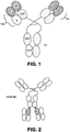
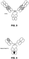


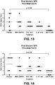

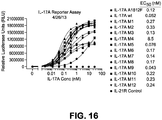


Classifications
-
- C—CHEMISTRY; METALLURGY
- C07—ORGANIC CHEMISTRY
- C07K—PEPTIDES
- C07K16/00—Immunoglobulins [IGs], e.g. monoclonal or polyclonal antibodies
- C07K16/18—Immunoglobulins [IGs], e.g. monoclonal or polyclonal antibodies against material from animals or humans
- C07K16/24—Immunoglobulins [IGs], e.g. monoclonal or polyclonal antibodies against material from animals or humans against cytokines, lymphokines or interferons
- C07K16/244—Interleukins [IL]
-
- A—HUMAN NECESSITIES
- A61—MEDICAL OR VETERINARY SCIENCE; HYGIENE
- A61P—SPECIFIC THERAPEUTIC ACTIVITY OF CHEMICAL COMPOUNDS OR MEDICINAL PREPARATIONS
- A61P1/00—Drugs for disorders of the alimentary tract or the digestive system
-
- A—HUMAN NECESSITIES
- A61—MEDICAL OR VETERINARY SCIENCE; HYGIENE
- A61P—SPECIFIC THERAPEUTIC ACTIVITY OF CHEMICAL COMPOUNDS OR MEDICINAL PREPARATIONS
- A61P1/00—Drugs for disorders of the alimentary tract or the digestive system
- A61P1/02—Stomatological preparations, e.g. drugs for caries, aphtae, periodontitis
-
- A—HUMAN NECESSITIES
- A61—MEDICAL OR VETERINARY SCIENCE; HYGIENE
- A61P—SPECIFIC THERAPEUTIC ACTIVITY OF CHEMICAL COMPOUNDS OR MEDICINAL PREPARATIONS
- A61P11/00—Drugs for disorders of the respiratory system
-
- A—HUMAN NECESSITIES
- A61—MEDICAL OR VETERINARY SCIENCE; HYGIENE
- A61P—SPECIFIC THERAPEUTIC ACTIVITY OF CHEMICAL COMPOUNDS OR MEDICINAL PREPARATIONS
- A61P11/00—Drugs for disorders of the respiratory system
- A61P11/06—Antiasthmatics
-
- A—HUMAN NECESSITIES
- A61—MEDICAL OR VETERINARY SCIENCE; HYGIENE
- A61P—SPECIFIC THERAPEUTIC ACTIVITY OF CHEMICAL COMPOUNDS OR MEDICINAL PREPARATIONS
- A61P13/00—Drugs for disorders of the urinary system
- A61P13/12—Drugs for disorders of the urinary system of the kidneys
-
- A—HUMAN NECESSITIES
- A61—MEDICAL OR VETERINARY SCIENCE; HYGIENE
- A61P—SPECIFIC THERAPEUTIC ACTIVITY OF CHEMICAL COMPOUNDS OR MEDICINAL PREPARATIONS
- A61P17/00—Drugs for dermatological disorders
-
- A—HUMAN NECESSITIES
- A61—MEDICAL OR VETERINARY SCIENCE; HYGIENE
- A61P—SPECIFIC THERAPEUTIC ACTIVITY OF CHEMICAL COMPOUNDS OR MEDICINAL PREPARATIONS
- A61P17/00—Drugs for dermatological disorders
- A61P17/06—Antipsoriatics
-
- A—HUMAN NECESSITIES
- A61—MEDICAL OR VETERINARY SCIENCE; HYGIENE
- A61P—SPECIFIC THERAPEUTIC ACTIVITY OF CHEMICAL COMPOUNDS OR MEDICINAL PREPARATIONS
- A61P19/00—Drugs for skeletal disorders
-
- A—HUMAN NECESSITIES
- A61—MEDICAL OR VETERINARY SCIENCE; HYGIENE
- A61P—SPECIFIC THERAPEUTIC ACTIVITY OF CHEMICAL COMPOUNDS OR MEDICINAL PREPARATIONS
- A61P19/00—Drugs for skeletal disorders
- A61P19/02—Drugs for skeletal disorders for joint disorders, e.g. arthritis, arthrosis
-
- A—HUMAN NECESSITIES
- A61—MEDICAL OR VETERINARY SCIENCE; HYGIENE
- A61P—SPECIFIC THERAPEUTIC ACTIVITY OF CHEMICAL COMPOUNDS OR MEDICINAL PREPARATIONS
- A61P25/00—Drugs for disorders of the nervous system
-
- A—HUMAN NECESSITIES
- A61—MEDICAL OR VETERINARY SCIENCE; HYGIENE
- A61P—SPECIFIC THERAPEUTIC ACTIVITY OF CHEMICAL COMPOUNDS OR MEDICINAL PREPARATIONS
- A61P27/00—Drugs for disorders of the senses
- A61P27/02—Ophthalmic agents
-
- A—HUMAN NECESSITIES
- A61—MEDICAL OR VETERINARY SCIENCE; HYGIENE
- A61P—SPECIFIC THERAPEUTIC ACTIVITY OF CHEMICAL COMPOUNDS OR MEDICINAL PREPARATIONS
- A61P29/00—Non-central analgesic, antipyretic or antiinflammatory agents, e.g. antirheumatic agents; Non-steroidal antiinflammatory drugs [NSAID]
-
- A—HUMAN NECESSITIES
- A61—MEDICAL OR VETERINARY SCIENCE; HYGIENE
- A61P—SPECIFIC THERAPEUTIC ACTIVITY OF CHEMICAL COMPOUNDS OR MEDICINAL PREPARATIONS
- A61P37/00—Drugs for immunological or allergic disorders
-
- A—HUMAN NECESSITIES
- A61—MEDICAL OR VETERINARY SCIENCE; HYGIENE
- A61P—SPECIFIC THERAPEUTIC ACTIVITY OF CHEMICAL COMPOUNDS OR MEDICINAL PREPARATIONS
- A61P37/00—Drugs for immunological or allergic disorders
- A61P37/02—Immunomodulators
-
- A—HUMAN NECESSITIES
- A61—MEDICAL OR VETERINARY SCIENCE; HYGIENE
- A61P—SPECIFIC THERAPEUTIC ACTIVITY OF CHEMICAL COMPOUNDS OR MEDICINAL PREPARATIONS
- A61P37/00—Drugs for immunological or allergic disorders
- A61P37/02—Immunomodulators
- A61P37/06—Immunosuppressants, e.g. drugs for graft rejection
-
- A—HUMAN NECESSITIES
- A61—MEDICAL OR VETERINARY SCIENCE; HYGIENE
- A61P—SPECIFIC THERAPEUTIC ACTIVITY OF CHEMICAL COMPOUNDS OR MEDICINAL PREPARATIONS
- A61P9/00—Drugs for disorders of the cardiovascular system
-
- A—HUMAN NECESSITIES
- A61—MEDICAL OR VETERINARY SCIENCE; HYGIENE
- A61P—SPECIFIC THERAPEUTIC ACTIVITY OF CHEMICAL COMPOUNDS OR MEDICINAL PREPARATIONS
- A61P9/00—Drugs for disorders of the cardiovascular system
- A61P9/10—Drugs for disorders of the cardiovascular system for treating ischaemic or atherosclerotic diseases, e.g. antianginal drugs, coronary vasodilators, drugs for myocardial infarction, retinopathy, cerebrovascula insufficiency, renal arteriosclerosis
-
- C—CHEMISTRY; METALLURGY
- C07—ORGANIC CHEMISTRY
- C07K—PEPTIDES
- C07K16/00—Immunoglobulins [IGs], e.g. monoclonal or polyclonal antibodies
- C07K16/46—Hybrid immunoglobulins
- C07K16/468—Immunoglobulins having two or more different antigen binding sites, e.g. multifunctional antibodies
-
- A—HUMAN NECESSITIES
- A61—MEDICAL OR VETERINARY SCIENCE; HYGIENE
- A61K—PREPARATIONS FOR MEDICAL, DENTAL OR TOILETRY PURPOSES
- A61K39/00—Medicinal preparations containing antigens or antibodies
- A61K2039/505—Medicinal preparations containing antigens or antibodies comprising antibodies
-
- C—CHEMISTRY; METALLURGY
- C07—ORGANIC CHEMISTRY
- C07K—PEPTIDES
- C07K2317/00—Immunoglobulins specific features
- C07K2317/20—Immunoglobulins specific features characterized by taxonomic origin
- C07K2317/21—Immunoglobulins specific features characterized by taxonomic origin from primates, e.g. man
-
- C—CHEMISTRY; METALLURGY
- C07—ORGANIC CHEMISTRY
- C07K—PEPTIDES
- C07K2317/00—Immunoglobulins specific features
- C07K2317/20—Immunoglobulins specific features characterized by taxonomic origin
- C07K2317/24—Immunoglobulins specific features characterized by taxonomic origin containing regions, domains or residues from different species, e.g. chimeric, humanized or veneered
-
- C—CHEMISTRY; METALLURGY
- C07—ORGANIC CHEMISTRY
- C07K—PEPTIDES
- C07K2317/00—Immunoglobulins specific features
- C07K2317/30—Immunoglobulins specific features characterized by aspects of specificity or valency
- C07K2317/31—Immunoglobulins specific features characterized by aspects of specificity or valency multispecific
-
- C—CHEMISTRY; METALLURGY
- C07—ORGANIC CHEMISTRY
- C07K—PEPTIDES
- C07K2317/00—Immunoglobulins specific features
- C07K2317/30—Immunoglobulins specific features characterized by aspects of specificity or valency
- C07K2317/34—Identification of a linear epitope shorter than 20 amino acid residues or of a conformational epitope defined by amino acid residues
-
- C—CHEMISTRY; METALLURGY
- C07—ORGANIC CHEMISTRY
- C07K—PEPTIDES
- C07K2317/00—Immunoglobulins specific features
- C07K2317/50—Immunoglobulins specific features characterized by immunoglobulin fragments
- C07K2317/55—Fab or Fab'
-
- C—CHEMISTRY; METALLURGY
- C07—ORGANIC CHEMISTRY
- C07K—PEPTIDES
- C07K2317/00—Immunoglobulins specific features
- C07K2317/50—Immunoglobulins specific features characterized by immunoglobulin fragments
- C07K2317/56—Immunoglobulins specific features characterized by immunoglobulin fragments variable (Fv) region, i.e. VH and/or VL
- C07K2317/565—Complementarity determining region [CDR]
-
- C—CHEMISTRY; METALLURGY
- C07—ORGANIC CHEMISTRY
- C07K—PEPTIDES
- C07K2317/00—Immunoglobulins specific features
- C07K2317/70—Immunoglobulins specific features characterized by effect upon binding to a cell or to an antigen
- C07K2317/76—Antagonist effect on antigen, e.g. neutralization or inhibition of binding
-
- C—CHEMISTRY; METALLURGY
- C07—ORGANIC CHEMISTRY
- C07K—PEPTIDES
- C07K2317/00—Immunoglobulins specific features
- C07K2317/90—Immunoglobulins specific features characterized by (pharmaco)kinetic aspects or by stability of the immunoglobulin
- C07K2317/92—Affinity (KD), association rate (Ka), dissociation rate (Kd) or EC50 value
Definitions
- Cytokines are soluble, small proteins that mediate a variety of biological effects, including the induction of immune cell proliferation, development, differentiation, and/or migration, as well as the regulation of the growth and differentiation of many cell types (see, for example, Arai et al., Annu. Rev. Biochem. 59:783 (1990 ); Mosmann, Curr. Opin. Immunol. 3:311 (1991 ); Paul et al., Cell, 76:241 (1994 )). Cytokine-induced immune functions can also include an inflammatory response, characterized by a systemic or local accumulation of immune cells.
- IL-17A, IL-17F and IL-23 are cytokines involved in inflammation.
- IL-17A induces the production of inflammatory cytokines such as IL-1 ⁇ , TNF- ⁇ , IL-6, and IL-23 by synovial fibroblasts, monocytes, and macrophages, all of which promote inflammation and Th17 development.
- IL-17A also induces an array of chemokines, including CXCL-1, CXCL-2, CXCL-5, CXCL-8, CCL-2, and CCL-20, leading to recruitment of T cells, B cells, monocytes, and neutrophils. Lundy, S.K., Arthritis Res. Ther., 9:202 (2007 ).
- IL-17F shares the greatest homology (55%) with IL-17A and is also a proinflammatory cytokine. Both IL-17A and IL-17F are produced by Th17 cells, whereas the other IL-17 family members, IL-17B, IL-17C, and IL-17D, are produced by non-T cell sources. IL-17A and IL-17F can exist as IL-17A homodimers and IL-17F homodimers or as IL-17A/F heterodimers. Liang, S.C. et al., J. Immunol., 179:7791-7799 (2007 ).
- IL-17A is increased in rheumatoid arthritis sera and synovial fluid, and is present in the T-cell rich areas of the synovium. Shahrara, S., Arthritis Res. Ther., 10:R93 (2005 ). IL-17A can also orchestrate bone and cartilage damage. An effective blockade of IL-17 will need to neutralize IL-17A homodimers, IL-17F homodimers and IL-17A/F heterodimers.
- IL-23 is a type-1 heterodimer, comprising a 19 kilodalton (kD) fourfold helical core ⁇ subunit (IL-23p19), disulfide linked to an additional 40kD distinct ⁇ subunit (IL-12p40).
- IL-23 is a key cytokine in bridging the innate and adaptive arms of the immune response; it is produced early in response to an antigen challenge, and is essential for driving early local immune responses. Furthermore, IL-23 plays a central role in the activation of NK cells, the enhancement of T cell proliferation and the regulation of antibody production. IL-23 also regulates pro-inflammatory cytokines (e.g., IFN- ⁇ ), which are important in cell-mediated immunity against intracellular pathogens.
- pro-inflammatory cytokines e.g., IFN- ⁇
- IL-23p19 knock-out mice were resistant to autoimmune encephalomyelitis (EAE), collagen-induced arthritis (CIA) and central nervous system autoimmune induction.
- EAE autoimmune encephalomyelitis
- CIA collagen-induced arthritis
- IL-23 is not essential for the development of human Th17 cells, but appears to be required for their survival and/or expansion. Paradowska-Gorycka, A., Scandinavian Journal of Immunology, 71:134-145 (2010 ).
- the present invention provides, in one embodiment, bispecific antibodies as defined in the claims comprising an IL-17A/F binding entity that binds to IL-17A and IL-17F and an IL-23 binding entity that binds to IL-23 via p19.
- the invention also includes isolated nucleic acids encoding the heavy chains and light chains of the bispecific antibodies of the invention, as well as vectors comprising the nucleic acids, host cells comprising the vectors, methods of making the bispecific antibodies, as defined in the claims.
- the present invention provides compositions comprising the bispecific antibodies as defined in the claims.
- the disclosure provides kits comprising the bispecific antibodies, as well as articles of manufacture comprising the bispecific antibodies.
- the bispecific antibodies of the present invention are useful for the inhibition of proinflammatory cytokines, e.g., IL-17A, IL-17F and IL-23p19.
- the antibodies can be used to reduce, limit, neutralize, or block the proinflammatory effects of the IL-17A homodimer, the IL-17F homodimer, or the IL-17A/F heterodimer.
- the antibodies can be used to reduce, limit, neutralize, or block the pro-cancerous effects of the IL-17A homodimer, the IL-17F homodimer, or the IL-17A/F heterodimer.
- the anti-IL-23p19 portion of the antibody is used to reduce, limit, neutralize, or block production of new T cells that would produce IL-17A and/or IL-17F, including homodimers and heterodimers.
- the bispecific antibodies described herein can be used to treat inflammatory disorders and autoimmune diseases, such as multiple sclerosis (e.g., relapsing-remitting multiple sclerosis, secondary-progressive multiple sclerosis, primary-progressive multiple sclerosis and progressive-relapsing multiple sclerosis), inflammatory bowel disease, psoriasis, systemic sclerosis, systemic lupus erythematosus, antineutrophil cytoplasmic antibodies (ANCA)-associated vasculitis (AAV) and giant cell arteritis.
- multiple sclerosis e.g., relapsing-remitting multiple sclerosis, secondary-progressive multiple sclerosis, primary-progressive multiple sclerosis and progressive-relapsing multiple s
- the bispecific antibodies described herein can also be used to treat cancer, including angiogenesis.
- the bispecific antibodies as described herein can be used to treat multiple-myeloma-induced lytic bone disease ( Sotomayor, E.M., Blood, 116(18):3380-3382 (2010 )).
- Antibodies are glycoproteins having the same structural characteristics. While antibodies exhibit binding specificity to a specific antigen, immunoglobulins include both antibodies and other antibody-like molecules that lack antigen specificity. Polypeptides of the latter kind are, for example, produced at low levels by the lymph system and at increased levels by myelomas.
- antibody or “antibody peptide(s)” refers to an intact antibody, or an antigen-binding fragment thereof that competes with the intact antibody for specific binding and includes chimeric, humanized, fully human, and bispecific antibodies.
- antigen-binding fragments are produced, for example, by recombinant DNA techniques. In additional embodiments, antigen-binding fragments are produced by enzymatic or chemical cleavage of intact antibodies. Antigen-binding fragments include, but are not limited to, Fab, Fab', F(ab) 2 , F(ab') 2 , Fv, and single-chain antibodies.
- isolated antibody refers to an antibody that has been identified and separated and/or recovered from a component of its natural environment. Contaminant components of its natural environment are materials which would interfere with diagnostic or therapeutic uses for the antibody, and may include enzymes, hormones, and other proteinaceous or nonproteinaceous solutes.
- the antibody will be purified (1) to greater than 95% by weight of antibody as determined by the Lowry method, and most preferably more than 99% by weight, (2) to a degree sufficient to obtain at least 15 residues of N-terminal or internal amino acid sequence by use of a spinning cup sequenator, or (3) to homogeneity by SDS-PAGE under reducing or nonreducing conditions using Coomassie blue or, preferably, silver stain.
- Isolated antibody includes the antibody in situ within recombinant cells since at least one component of the antibody's natural environment will not be present. Ordinarily, however, isolated antibody will be prepared by at least one purification step.
- agonist refers to any compound including a protein, polypeptide, peptide, antibody, antibody fragment, large molecule, or small molecule (less than 10 kD), that increases the activity, activation or function of another molecule.
- antagonist refers to any compound including a protein, polypeptide, peptide, antibody, antibody fragment, large molecule, or small molecule (less than 10 kD), that decreases the activity, activation or function of another molecule.
- binding of a polypeptide includes, but is not limited to, the binding of a ligand polypeptide of the present disclosure to a receptor; the binding of a receptor polypeptide of the present disclosure to a ligand; the binding of an antibody of the present invention to an antigen or epitope; the binding of an antigen or epitope of the present disclosure to an antibody; the binding of an antibody of the present invention to an anti-idiotypic antibody; the binding of an anti-idiotypic antibody of the present disclosure to a ligand; the binding of an anti-idiotypic antibody of the present disclosure to a receptor; the binding of an anti-anti-idiotypic antibody of the present disclosure to a ligand, receptor or antibody, etc.
- bispecific or “bifunctional” antibody is a hybrid antibody having two different heavy/light chain pairs and two different binding sites.
- Bispecific antibodies may be produced by a variety of methods including, but not limited to, fusion of hybridomas or linking of Fab' fragments. See, e.g., Songsivilai et al., Clin. Exp. Immunol., 79:315-321 (1990 ); Kostelny et al., J. Immunol., 148:1547-1553 (1992 ).
- epitope refers to the portion of an antigen to which an antibody specifically binds.
- epitopic determinants usually consist of chemically active surface groupings of molecules such as amino acids or sugar side chains and usually have specific three dimensional structural characteristics, as well as specific charge characteristics.
- IL-17 epitope refers to a portion of the corresponding polypeptide having antigenic or immunogenic activity in an animal, preferably in a mammal, and most preferably in a mouse or a human.
- An epitope having immunogenic activity is a portion of, for example, an IL-17A or IL-17F or IL-23/p19 polypeptide that elicits an antibody response in an animal.
- An epitope having antigenic activity is a portion of, for example, an IL-17A or IL-17F or IL-23/p 19 polypeptide to which an antibody immunospecifically binds as determined by any method well known in the art, for example, by immunoassays, protease digest, crystallography or H/D-Exchange.
- Antigenic epitopes need not necessarily be immunogenic. Such epitopes can be linear in nature or can be a discontinuous epitope.
- the term "conformational epitope" refers to a discontinuous epitope formed by a spatial relationship between amino acids of an antigen other than an unbroken series of amino acids.
- immunoglobulin refers to a protein consisting of one or more polypeptides substantially encoded by immunoglobulin genes.
- One form of immunoglobulin constitutes the basic structural unit of an antibody. This form is a tetramer and consists of two identical pairs of immunoglobulin chains, each pair having one light and one heavy chain. In each pair, the light and heavy chain variable regions are together responsible for binding to an antigen, and the constant regions are responsible for the antibody effector functions.
- Full-length immunoglobulin "light chains” (about 25 Kd or about 214 amino acids) are encoded by a variable region gene at the NH2-terminus (about 110 amino acids) and a kappa or lambda constant region gene at the COOH--terminus.
- Full-length immunoglobulin "heavy chains” (about 50 Kd or about 446 amino acids), are similarly encoded by a variable region gene (about 116 amino acids) and one of the other aforementioned constant region genes (about 330 amino acids).
- Heavy chains are classified as gamma, mu, alpha, delta, or epsilon, and define the antibody's isotype as IgG (such as IgG1, IgG2, IgG3 and IgG4), IgM, IgA, IgD and IgE, respectively.
- IgG such as IgG1, IgG2, IgG3 and IgG4
- IgM IgA, IgD and IgE
- J variable and constant regions
- the heavy chain also including a "D” region of about 10 more amino acids.
- An immunoglobulin light or heavy chain variable region consists of a "framework" region interrupted by three hypervariable regions.
- hypervariable region refers to the amino acid residues of an antibody which are responsible for antigen binding.
- the hypervariable region comprises amino acid residues from a "Complementarity Determining Region” or "CDR" ( Kabat et al., Sequences of Proteins of Immunological Interest, 5th Edition, Public Health Service, National Institutes of Health, Bethesda, MD (1991 )) and/or those residues from a "hypervariable loop" ( Chothia et al., J. Mol. Biol. 196: 901-917 (1987 )).
- Framework Region or "FR” residues are those variable domain residues other than the hypervariable region residues as herein defined.
- the sequences of the framework regions of different light or heavy chains are relatively conserved within a species.
- a "human framework region” is a framework region that is substantially identical (about 85% or more, usually 90-95% or more) to the framework region of a naturally occurring human immunoglobulin.
- the framework region of an antibody that is the combined framework regions of the constituent light and heavy chains, serves to position and align the CDR's. The CDR's are primarily responsible for binding to an epitope of an antigen.
- humanized immunoglobulin refers to an immunoglobulin comprising a human framework region and one or more CDR's from a non-human (usually a mouse or rat) immunoglobulin.
- the non-human immunoglobulin providing the CDR's is called the "donor” and the human immunoglobulin providing the framework is called the "acceptor”.
- Constant regions need not be present, but if they are, they must be substantially identical to human immunoglobulin constant regions, i.e., at least about 85-90%, preferably about 95% or more identical.
- all parts of a humanized immunoglobulin, except possibly the CDR's are substantially identical to corresponding parts of natural human immunoglobulin sequences.
- one or more residues in the human framework region may be back mutated to the parental sequence to retain optimal antigen-binding affinity and specificity. In this way, certain framework residues from the non-human parent antibody are retained in the humanized antibody in order to retain the binding properties of the parent antibody while minimizing its immunogenicity.
- the term "human framework region” as used herein includes regions with such back mutations.
- a "humanized antibody” is an antibody comprising a humanized light chain and a humanized heavy chain immunoglobulin. For example, a humanized antibody would not encompass a typical chimeric antibody as defined above, e.g., because the entire variable region of a chimeric antibody is non-human.
- humanized immunoglobulin refers to an immunoglobulin comprising a human framework region and one or more CDR's from a non-human, e.g., mouse, rat or rabbit, immunoglobulin.
- the non-human immunoglobulin providing the CDR's is called the "donor” and the human immunoglobulin providing the framework is called the "acceptor”.
- Constant regions need not be present, but if they are, they must be substantially identical to human immunoglobulin constant regions, i.e., at least about 85-90%, preferably about 95% or more identical.
- humanized immunoglobulin all parts of a humanized immunoglobulin, except possibly the CDR's and possibly a few back-mutated amino acid residues in the framework region (e.g., 1-15 residues), are substantially identical to corresponding parts of natural human immunoglobulin sequences.
- a "humanized antibody” is an antibody comprising a humanized light chain and a humanized heavy chain immunoglobulin.
- a humanized antibody would not encompass a typical chimeric antibody as defined above, e.g., because the entire variable region of a chimeric antibody is non-human.
- human antibody includes an antibody that has an amino acid sequence of a human immunoglobulin and includes antibodies isolated from human immunoglobulin libraries or from animals transgenic for one or more human immunoglobulin and that do not express endogenous immunoglobulins, as described, for example, by Kucherlapati et al. in U.S. Patent No. 5,939,598 .
- the term "genetically altered antibodies” means antibodies wherein the amino acid sequence has been varied from that of a native antibody. Because of the relevance of recombinant DNA techniques in the generation of antibodies, one need not be confined to the sequences of amino acids found in natural antibodies; antibodies can be redesigned to obtain desired characteristics. The possible variations are many and range from the changing of just one or a few amino acids to the complete redesign of, for example, the variable and/or constant region. Changes in the constant region will, in general, be made in order to improve or alter characteristics, such as complement fixation, interaction with membranes and other effector functions. Changes in the variable region will be made in order to improve the antigen binding characteristics.
- a "Fab fragment” is comprised of one light chain and the C H1 and variable regions of one heavy chain.
- the heavy chain of a Fab molecule cannot form a disulfide bond with another heavy chain molecule.
- a “Fab' fragment” contains one light chain and one heavy chain that contains more of the constant region, between the C H1 and C H2 domains, such that an interchain disulfide bond can be formed between two heavy chains to form a F(ab') 2 molecule.
- a “F(ab') 2 fragment” contains two light chains and two heavy chains containing a portion of the constant region between the C H1 and C H2 domains, such that an interchain disulfide bond is formed between two heavy chains.
- a "Fv fragment” contains the variable regions from both heavy and light chains but lacks the constant regions.
- a “single domain antibody” is an antibody fragment consisting of a single domain Fv unit, e.g., V H or V L . Like a whole antibody, it is able to bind selectively to a specific antigen. With a molecular weight of only 12-15 kDa, single-domain antibodies are much smaller than common antibodies (150-160 kDa) which are composed of two heavy protein chains and two light chains, and even smaller than Fab fragments ( ⁇ 50 kDa, one light chain and half a heavy chain) and single-chain variable fragments ( ⁇ 25 kDa, two variable domains, one from a light and one from a heavy chain). The first single-domain antibodies were engineered from heavy-chain antibodies found in camelids. Although most research into single-domain antibodies is currently based on heavy chain variable domains, light chain variable domains and nanobodies derived from light chains have also been shown to bind specifically to target epitopes.
- the term “monoclonal antibody” or “mAb” or “MAb” or “Mab” or “mab” as used herein is not limited to antibodies produced through hybridoma technology.
- the term “monoclonal antibody” or “mAb” or “MAb” or “Mab” or “mab” refer to an antibody that is derived from a single clone, including any eukaryotic, prokaryotic, or phage clone, and not the method by which it is produced.
- nucleic acid or “nucleic acid molecule” refers to polynucleotides, such as deoxyribonucleic acid (DNA) or ribonucleic acid (RNA), oligonucleotides, fragments generated by the polymerase chain reaction (PCR), and fragments generated by any of ligation, scission, endonuclease action, and exonuclease action.
- DNA deoxyribonucleic acid
- RNA ribonucleic acid
- PCR polymerase chain reaction
- Nucleic acid molecules can be composed of monomers that are naturally-occurring nucleotides (such as DNA and RNA), or analogs of naturally-occurring nucleotides (e.g ., ⁇ -enantiomeric forms of naturally-occurring nucleotides), or a combination of both.
- Modified nucleotides can have alterations in sugar moieties and/or in pyrimidine or purine base moieties.
- Sugar modifications include, for example, replacement of one or more hydroxyl groups with halogens, alkyl groups, amines, and azido groups, or sugars can be functionalized as ethers or esters.
- the entire sugar moiety can be replaced with sterically and electronically similar structures, such as aza-sugars and carbocyclic sugar analogs.
- modifications in a base moiety include alkylated purines and pyrimidines, acylated purines or pyrimidines, or other well-known heterocyclic substitutes.
- Nucleic acid monomers can be linked by phosphodiester bonds or analogs of such linkages. Analogs of phosphodiester linkages include phosphorothioate, phosphorodithioate, phosphoroselenoate, phosphorodiselenoate, phosphoroanilothioate, phosphoranilidate, phosphoramidate, and the like.
- nucleic acid molecule also includes so-called “peptide nucleic acids”, which comprise naturally-occurring or modified nucleic acid bases attached to a polyamide backbone. Nucleic acids can be either single stranded or double stranded.
- nucleic acid molecule refers to a nucleic acid molecule having a complementary nucleotide sequence and reverse orientation as compared to a reference nucleotide sequence.
- degenerate nucleotide sequence denotes a sequence of nucleotides that includes one or more degenerate codons as compared to a reference nucleic acid molecule that encodes a polypeptide.
- Degenerate codons contain different triplets of nucleotides, but encode the same amino acid residue (i.e., GAU and GAC triplets each encode Asp).
- an "isolated nucleic acid molecule” is a nucleic acid molecule that is not integrated in the genomic DNA of an organism.
- a DNA molecule that encodes a growth factor that has been separated from the genomic DNA of a cell is an isolated DNA molecule.
- Another example of an isolated nucleic acid molecule is a chemically-synthesized nucleic acid molecule that is not integrated in the genome of an organism.
- a nucleic acid molecule that has been isolated from a particular species is smaller than the complete DNA molecule of a chromosome from that species.
- nucleic acid molecule construct is a nucleic acid molecule, either single- or double-stranded, that has been modified through human intervention to contain segments of nucleic acid combined and juxtaposed in an arrangement not existing in nature.
- Codon DNA is a single-stranded DNA molecule that is formed from an mRNA template by the enzyme reverse transcriptase. Typically, a primer complementary to portions of mRNA is employed for the initiation of reverse transcription.
- cDNA refers to a double-stranded DNA molecule consisting of such a single-stranded DNA molecule and its complementary DNA strand.
- cDNA also refers to a clone of a cDNA molecule synthesized from an RNA template.
- a “promoter” is a nucleotide sequence that directs the transcription of a structural gene.
- a promoter is located in the 5' non-coding region of a gene, proximal to the transcriptional start site of a structural gene.
- Sequence elements within promoters that function in the initiation of transcription are often characterized by consensus nucleotide sequences. These promoter elements include RNA polymerase binding sites, TATA sequences, CAAT sequences, differentiation-specific elements (DSEs; McGehee et al., Mol.
- CREs cyclic AMP response elements
- SREs serum response elements
- GREs glucocorticoid response elements
- binding sites for other transcription factors such as CRE/ATF ( O'Reilly et al., J. Biol. Chem., 267:19938 (1992 )), AP2 ( Ye et al., J. Biol.
- a promoter is an inducible promoter, then the rate of transcription increases in response to an inducing agent. In contrast, the rate of transcription is not regulated by an inducing agent if the promoter is a constitutive promoter. Repressible promoters are also known.
- a “regulatory element” is a nucleotide sequence that modulates the activity of a core promoter.
- a regulatory element may contain a nucleotide sequence that binds with cellular factors enabling transcription exclusively or preferentially in particular cells, tissues, or organelles. These types of regulatory elements are normally associated with genes that are expressed in a "cell-specific”, “tissue-specific”, or “organelle-specific” manner.
- An “enhancer” is a type of regulatory element that can increase the efficiency of transcription, regardless of the distance or orientation of the enhancer relative to the start site of transcription.
- Heterologous DNA refers to a DNA molecule, or a population of DNA molecules, that does not exist naturally within a given host cell.
- DNA molecules heterologous to a particular host cell may contain DNA derived from the host cell species (i.e., endogenous DNA) so long as that host DNA is combined with non-host DNA (i.e., exogenous DNA).
- a DNA molecule containing a non-host DNA segment encoding a polypeptide operably linked to a host DNA segment comprising a transcription promoter is considered to be a heterologous DNA molecule.
- a heterologous DNA molecule can comprise an endogenous gene operably linked with an exogenous promoter.
- a DNA molecule comprising a gene derived from a wild-type cell is considered to be heterologous DNA if that DNA molecule is introduced into a mutant cell that lacks the wild-type gene.
- an “expression vector” is a nucleic acid molecule encoding a gene that is expressed in a host cell.
- an expression vector comprises a transcription promoter, a gene, and a transcription terminator. Gene expression is usually placed under the control of a promoter, and such a gene is said to be “operably linked to” the promoter.
- a regulatory element and a core promoter are operably linked if the regulatory element modulates the activity of the core promoter.
- a “recombinant host” is a cell that contains a heterologous nucleic acid molecule, such as a cloning vector or expression vector.
- a recombinant host is a cell that produces an antagonist of the present invention from an expression vector.
- an antagonist can be produced by a cell that is a "natural source" of said antagonist, and that lacks an expression vector.
- amino-terminal and “carboxyl-terminal” are used herein to denote positions within polypeptides. Where the context allows, these terms are used with reference to a particular sequence or portion of a polypeptide to denote proximity or relative position. For example, a certain sequence positioned carboxyl-terminal to a reference sequence within a polypeptide is located proximal to the carboxyl terminus of the reference sequence, but is not necessarily at the carboxyl terminus of the complete polypeptide.
- a “fusion protein” is a hybrid protein expressed by a nucleic acid molecule comprising nucleotide sequences of at least two genes.
- a fusion protein can comprise at least part of a IL-17RA polypeptide fused with a polypeptide that binds an affinity matrix.
- Such a fusion protein provides a means to isolate large quantities of IL-17A using affinity chromatography.
- Receptor denotes a cell-associated protein that binds to a bioactive molecule termed a "ligand.” This interaction mediates the effect of the ligand on the cell.
- Receptors can be membrane bound, cytosolic or nuclear; monomeric (e.g., thyroid stimulating hormone receptor, beta-adrenergic receptor) or multimeric (e.g., PDGF receptor, growth hormone receptor, IL-3 receptor, GM-CSF receptor, G-CSF receptor, erythropoietin receptor and IL-6 receptor).
- Membrane-bound receptors are characterized by a multi-domain structure comprising an extracellular ligand-binding domain and an intracellular effector domain that is typically involved in signal transduction. In certain membrane-bound receptors, the extracellular ligand-binding domain and the intracellular effector domain are located in separate polypeptides that comprise the complete functional receptor.
- the binding of ligand to receptor results in a conformational change in the receptor that causes an interaction between the effector domain and other molecule(s) in the cell, which in turn leads to an alteration in the metabolism of the cell.
- Metabolic events that are often linked to receptor-ligand interactions include gene transcription, phosphorylation, dephosphorylation, increases in cyclic AMP production, mobilization of cellular calcium, mobilization of membrane lipids, cell adhesion, hydrolysis of inositol lipids and hydrolysis of phospholipids.
- expression refers to the biosynthesis of a gene product.
- expression involves transcription of the structural gene into mRNA and the translation of mRNA into one or more polypeptides.
- complement/anti-complement pair denotes non-identical moieties that form a non-covalently associated, stable pair under appropriate conditions.
- biotin and avidin are prototypical members of a complement/anti-complement pair.
- Other exemplary complement/anti-complement pairs include receptor/ligand pairs, antibody/antigen (or hapten or epitope) pairs, sense/antisense polynucleotide pairs, and the like.
- the complement/anti-complement pair preferably has a binding affinity of less than 10 9 M -1 .
- a "therapeutic agent” is a molecule or atom which is conjugated to an antibody moiety to produce a conjugate which is useful for therapy.
- therapeutic agents include drugs, toxins, immunomodulators, chelators, boron compounds, photoactive agents or dyes, and radioisotopes.
- a “detectable label” is a molecule or atom which can be conjugated to an antibody moiety to produce a molecule useful for diagnosis.
- detectable labels include chelators, photoactive agents, radioisotopes, fluorescent agents, paramagnetic ions, or other marker moieties.
- affinity tag is used herein to denote a polypeptide segment that can be attached to a second polypeptide to provide for purification or detection of the second polypeptide or provide sites for attachment of the second polypeptide to a substrate.
- any peptide or protein for which an antibody or other specific binding agent is available can be used as an affinity tag.
- Affinity tags include a poly-histidine tract, protein A ( Nilsson et al., EMBO J., 4:1075 (1985 ); Nilsson et al., Methods Enzymol., 198:3 (1991 )), glutathione S transferase ( Smith et al., Gene, 67:31 (1988 )), Glu-Glu affinity tag ( Grussenmeyer et al., Proc. Natl. Acad. Sci. USA, 82:7952 (1985 )), substance P, FLAG® peptide ( Hopp et al., Biotechnology, 6:1204 (1988 )), streptavidin binding peptide, or other antigenic epitope or binding domain. See, in general, Ford et al., Protein Expression and Purification, 2:95 (1991 ). DNA molecules encoding affinity tags are available from commercial suppliers (e.g., Pharmacia Biotech, Piscataway, NJ).
- IL-17A binding entity is a binding entity, such as an antibody, that specifically binds to IL-17A in its homodimeric form (IL-17A/IL-17A) and in its heterodimeric form (IL-17A/IL-17F).
- IL-17F binding entity is a binding entity, such as an antibody, that specifically binds to IL-17F in its homodimeric form (IL-17F/IL-17F) and in its heterodimeric form (IL-17A/IL-17F).
- an "IL-17A/F binding entity” is a binding entity, such as an antibody, that specifically binds to IL-17A and IL-17F by recognizing and binding to the same or similar epitope, e.g., continuous or discontinuous epitope, shared by IL-17A and IL-17F.
- the IL-17A/F binding entity binds to the IL-17A homodimer, IL-17F homodimer and IL-17A/IL-17F heterodimer.
- IL-17A, IL-17F and IL-23p 19 are overexpressed in and implicated in the cause and/or sustainability of several inflammatory and/or autoimmune disorders.
- One solution several embodiments of the present invention provide is to inhibit or reduce the ability of these cytokines to cell-signal using a bispecific antibody as defined in the claims, which binds to each cytokine and inhibits or reduces each cytokine, e.g., homodimeric or heterodimeric form, from signaling through their cognate receptor.
- bispecific IL-17A/F and IL-23p19 bispecific antibody e.g, biAb3
- difficulties were encountered to generate a bispecific antibody which had high affinity to IL-17A and was an effective neutralizer, e.g., IC50, of IL-17A.
- the mouse parent antibody e.g, chimeric 339.15, 339.15.3.5, 339.15.5.3 or 339.15.3.6
- Table 1 had an IL-17A/F IC50 of 0.30nM and an IL-17F IC50 of 0.26nM, but only had an IL-17A IC50 of 11nM.
- mouse parent chimeric 339.15 and humanized parent 339.134 had a similar binding affinity towards IL-17A. Furthermore, as shown in Table 8, humanized parent 339.134 ability to inhibit IL-17A (IC50 of 1.3nM) was still significantly reduced as compared to its ability to inhibit IL-17A/F (IC50 of 0.27nM) and IL-17F (IC50 of 0.24nM). This problem was surprisingly overcome by utilizing the light chain of the IL-23p 19 antibody of biAb3 (SEQ ID NO:17).
- the resulting monoclonal antibody significantly increased its ability to inhibit IL-17A from signaling (see Table 9) and significantly increased its affinity for IL-17A (see Table 10).
- the critical residue in the now shared IL-23p19 / IL-17A/F light chain of, for example, biAb3, that may have provided this enhanced affinity and neutralization ability may be Y108 or Tyr108 of SEQ ID NO:2 as evidence by the X-ray crystallography and alanine mutant studies of Example 9.
- the present invention provides bispecific antibodies as defined in the claims.
- the bispecific antibodies of the disclosure comprise an IL-17A binding entity that binds to IL-17A and an IL-23 binding entity that binds to IL-23 via p19.
- the bispecific antibodies of the disclosure comprise an IL-17F binding entity that binds to IL-17F and a IL-23 binding entity that binds to IL-23 via p19.
- the bispecific antibodies of the invention comprise an IL-17A/F binding entity that binds to IL-17A and IL-17F and a IL-23 binding entity that binds to IL-23 via p19, as defined in the claims.
- the binding entity that binds to IL-23 via p 19 is referred to hereinafter as a binding entity that binds to IL-23 or an "IL-23 binding entity".
- the polynucleotide sequence of the human IL-17A is shown in SEQ ID NO:1 and the corresponding polypeptide sequence is shown in SEQ ID NO:2.
- the signal sequence of the IL-17A polypeptide is amino acid residues 1-23 of SEQ ID NO:2.
- amino acid residues 24-155 of SEQ ID NO:2 constitute the mature IL-17A polypeptide.
- Antibodies (and antigen-binding fragments thereof) and bispecific antibodies disclosed herein that bind to IL-17A bind to the mature IL-17A polypeptide (amino acid residues 24-155 of SEQ ID NO:2).
- the polynucleotide sequence of the human IL-17F is shown in SEQ ID NO:3 and the corresponding polypeptide sequence is shown in SEQ ID NO:4.
- the signal sequence of the IL-17F polypeptide is amino acid residues 1-30 of SEQ ID NO:4.
- amino acid residues 31-163 of SEQ ID NO:4 constitute the mature IL-17F polypeptide.
- the polynucleotide sequence of the human p19 subunit of IL-23 is shown in SEQ ID NO:5 and the corresponding polypeptide sequence is shown in SEQ ID NO:6.
- the signal sequence of the IL-23p19 polypeptide is amino acid residues 1-19 of SEQ ID NO:6.
- amino acid residues 20-189 of SEQ ID NO:6 constitute the mature IL-23p19 polypeptide.
- Antibodies (and antigen-binding fragments thereof) and bispecific antibodies disclosed herein that bind to IL-23p19 bind to the mature IL-23p19 polypeptide (amino acid residues 20-189 of SEQ ID NO:6).
- the IL-17A/F binding entity comprises an antibody, i.e., two pairs of immunoglobulin chains, each pair having one light and one heavy chain
- the IL-23 binding entity comprises two Fab fragments each comprising a light chain and the C H1 and variable regions of a heavy chain
- the Fab fragments of the IL-23 binding entity are linked to the C-termini of the heavy chains (Fc) of the IL-17A/F binding entity.
- This bispecific antibody format is referred to herein as biAbFabL (see Fig. 2 ).
- each of the light chain and the C H1 and variable regions of the heavy chain comprising the Fab fragments of the IL-23 binding entity are linked to the N-termini of the light chains and heavy chains, respectively, of the IL-17A/F binding entity.
- This bispecific antibody format is referred to herein as taFab (see Fig. 3 ).
- the IL-23 binding entity comprises an antibody, i.e., two pairs of immunoglobulin chains, each pair having one light and one heavy chain
- the IL-17A/F binding entity comprises two Fab fragments each comprising a light chain and the C H1 and variable regions of a heavy chain
- the Fab fragments of the IL-17A/F binding entity are linked to the C-termini of the heavy chains (Fc) of the IL-23 binding entity.
- This bispecific antibody format is referred to herein as biAbFabL (See Fig. 2 ).
- each of the light chain and the C H1 and variable regions of the heavy chain comprising the Fab fragments of the IL-17A/F binding entity are linked to the N-termini of the light chain and heavy chain, respectively, of the IL-23 binding entity.
- This bispecific antibody format is referred to herein as taFab (see Fig. 3 ).
- the IL-23 binding entity comprises a light chain and an IL-23 heavy chain and the IL-17A/F binding entity comprises a light chain and an IL-17A/F heavy chain.
- This bispecific antibody resembles a traditional antibody except that it comprises two different heavy chains that associate through an electrostatic complementarity association in the C H3 regions. It utilizes a common light chain.
- This bispecific antibody format is referred to herein as Heterodimeric Fc (see Fig. 4 ).
- the present disclosure provides bispecific antibodies comprising a first binding entity comprising an antibody, i.e., two pairs of immunoglobulin chains, each pair having one light chain and one heavy chain, and a second binding entity comprising an Fv unit, i.e., the variable domains from a heavy and a light chain, and in which the second binding entity comprising the Fv unit is positioned between the Fab region and the hinge of the first binding entity as shown in Fig. 5 .
- the Fv unit is linked to the Fab region of the first binding entity by linker molecules. More specifically, the Fv unit comprises a variable light domain which is linked to the light chain constant region of the Fab fragment, and a variable heavy domain which is linked to the C H1 region of the Fab fragment.
- This bispecific antibody format is referred to herein as VCVFc.
- the first binding entity and second binding entity of a VCVFc do not have to share a common light chain, while the first binding entity and second binding entity of a biAbFabL do have to share a common light chain.
- the first binding entity specifically binds a lymphocyte antigen, cytokine, cytokine receptor, growth factor, growth factor receptor, interleukin (e.g., IL-17A, IL-17F, IL-17A/F and IL-23) or interleukin receptor and the second binding entity specifically binds a lymphocyte antigen, cytokine, cytokine receptor, growth factor, growth factor receptor, interleukin (e.g., IL-17A, IL-17F, IL-17A/F and IL-23) or interleukin receptor.
- the first binding entity is an IL-17A/F binding entity and the second binding entity is an IL-23 binding entity.
- the first binding entity is an IL-23 binding entity and the second binding entity is an IL-17A/F binding entity.
- the present disclosure provides bispecific antibodies comprising a first binding entity comprising an antibody, i.e., two pairs of immunoglobulin chains, each pair having one light chain and one heavy chain, and a second binding entity comprising a single domain antibody.
- This bispecific antibody format is referred to herein as VCDFc.
- An illustration of a VCDFc bispecific antibody is shown in Fig. 6 .
- the second binding entity comprising the single domain antibody is positioned between the Fab region, more specifically the C H1 region of the Fab fragment, and the hinge of the first binding entity.
- the single domain antibody is linked to the C H1 region of the Fab of the first binding entity by linker molecules (for example, but not limited to, 10mer G 4 S, which is represented by the equation (G 4 S) 2 , or SSASTKGPS (SEQ ID NO:86)).
- linker molecules for example, but not limited to, 10mer G 4 S, which is represented by the equation (G 4 S) 2 , or SSASTKGPS (SEQ ID NO:86)).
- the first binding entity specifically binds a lymphocyte antigen, cytokine, cytokine receptor, growth factor, growth factor receptor, interleukin (e.g., IL-17A, IL-17F, IL-17A/F and IL-23) or interleukin receptor and the second binding entity specifically binds a lymphocyte antigen, cytokine, cytokine receptor, growth factor, growth factor receptor, interleukin (e.g., IL-17A, IL-17F, IL-17A/F and IL-23) or interleukin receptor.
- the first binding entity is an IL-23 binding entity and the second binding entity is an IL-17A/F binding entity.
- the first binding entity is an IL-17A/F binding entity and the second binding entity is an IL-23 binding entity.
- the amino acid sequences of the binding entities are preferably based upon the sequences of human and/or humanized monoclonal antibodies against a lymphocyte antigen, cytokine, cytokine receptor, growth factor, growth factor receptor, interleukin (e.g., IL-17A, IL-17F, IL-17A/F and IL-23) or interleukin receptor.
- the light chains of the IL-17A/F binding entity and the IL-23 binding entity of the bispecific antibody each comprise a variable domain comprising a CDR1 having the amino acid sequence of SEQ ID NO:22, a CDR2 having the amino acid sequence of SEQ ID NO:23, and a CDR3 having the sequence of SEQ ID NO:24.
- the light chains of the IL-17A/F binding entity and the IL-23 binding entity each comprise a variable domain comprising the amino acid sequence of SEQ ID NO:9.
- the light chains of the IL-17A/F binding entity and the IL-23 binding entity each comprise a constant domain comprising the amino acid sequence of SEQ ID NO:10.
- the light chains of the IL-17A/F binding entity and the IL-23 binding entity each comprise a variable domain comprising the amino acid sequence of SEQ ID NO:9 and a constant domain comprising the amino acid sequence of SEQ ID NO:10.
- the heavy chain of the IL-17A/F binding entity of the bispecific antibody comprises a variable domain comprising a CDR1 having the amino acid sequence of SEQ ID NO:25, a CDR2 having the amino acid sequence of SEQ ID NO:26, and a CDR3 having the amino acid sequence of SEQ ID NO:27.
- the heavy chain of the IL-17A/F binding entity comprises a variable domain comprising the amino acid sequence of SEQ ID NO:13.
- the heavy chain constant domain comprises the amino acid sequence of SEQ ID NO:8, SEQ ID NO:11, SEQ ID NO:127 or SEQ ID NO:128.
- the C H1 region of the heavy chain comprises the amino acid sequence of SEQ ID NO:14 or SEQ ID NO:15.
- the IL-17A/F binding entity of the bispecific antibody as defined in the claims comprises a heavy chain variable domain having at least 90%, at least 91%, at least 92%, at least 93%, at least 94%, at least 95%, at least 96%, at least 97%, at least 98%, at least 99% sequence identity with the amino acid sequence of SEQ ID NO:13.
- all of the substitutions, additions or deletions are within the framework region of the heavy chain variable domain.
- the IL-17A/F binding entity of the bispecific antibody comprises a heavy chain variable domain having at least 90%, at least 91%, at least 92%, at least 93%, at least 94%, at least 95%, at least 96%, at least 97%, at least 98%, at least 99% sequence identity with the amino acid sequence of SEQ ID NO:13, wherein the variable domain comprises a CDR1 having the amino acid sequence of SEQ ID NO:25, a CDR2 having the amino acid sequence of SEQ ID NO:26, and a CDR3 having the amino acid sequence of SEQ ID NO:27.
- the heavy chain variable domain comprises the amino acid sequence of SEQ ID NO:13.
- the three IL-17A/F heavy chain variable domain CDRs include a CDR1 region comprising an amino acid sequence having at least 90%, at least 91%, at least 92%, at least 93%, at least 94%, at least 95%, at least 96%, at least 97%, at least 98%, at least 99% sequence identity with the amino acid sequence of SEQ ID NO:25; a CDR2 region comprising an amino acid sequence having at least 90%, at least 91%, at least 92%, at least 93%, at least 94%, at least 95%, at least 96%, at least 97%, at least 98%, at least 99% sequence identity with the amino acid sequence of SEQ ID NO:26; and a CDR3 region comprising an amino acid sequence having at least 90%, at least 91%, at least 92%, at least 93%, at least 94%, at least 95%, at least 96%, at least 97%, at least 98%, at least 99% sequence identity with the amino acid sequence of SEQ ID NO:27.
- the IL-17A/F heavy chain variable domain CDR1 has the amino acid sequence of SEQ ID NO:25; the heavy chain variable domain CDR2 has the amino acid sequence of SEQ ID NO:26; and the heavy chain variable domain CDR3 has the amino acid sequence of SEQ ID NO:27.
- the IL-17A/F and/or IL-23p19 binding entity as defined in the claims may comprise a light chain variable domain having at least 90%, at least 91%, at least 92%, at least 93%, at least 94%, at least 95%, at least 96%, at least 97%, at least 98%, at least 99% sequence identity with the amino acid sequence of SEQ ID NO:9.
- the IL-17A/F and/or IL-23p 19 binding entity of the bispecific antibody as defined in the claims comprises a light chain variable domain having at least 90%, at least 91%, at least 92%, at least 93%, at least 94%, at least 95%, at least 96%, at least 97%, at least 98%, at least 99% sequence identity with the amino acid sequence of SEQ ID NO:9, wherein the variable domain comprises a CDR1 having the amino acid sequence of SEQ ID NO:22, a CDR2 having the amino acid sequence of SEQ ID NO:23, and a CDR3 having the amino acid sequence of SEQ ID NO:24.
- the light chain variable domain comprises the amino acid sequence of SEQ ID NO:9.
- the three IL-17A/F and/or IL-23p19 light chain variable domain CDRs include a CDR1 region comprising an amino acid sequence having at least 90%, at least 91%, at least 92%, at least 93%, at least 94%, at least 95%, at least 96%, at least 97%, at least 98%, at least 99% sequence identity with the amino acid sequence of SEQ ID NO:22; a CDR2 region comprising an amino acid sequence having at least 90%, at least 91%, at least 92%, at least 93%, at least 94%, at least 95%, at least 96%, at least 97%, at least 98%, at least 99% sequence identity with the amino acid sequence of SEQ ID NO:23; and a CDR3 region comprising an amino acid sequence having at least 90%, at least 91%, at least 92%, at least 93%, at least 94%, at least 95%, at least 96%, at least 9 least 9
- the IL-17A/F and/or IL-23p19 light chain variable domain CDR1 has the amino acid sequence of SEQ ID NO:22;
- the IL-17A/F and/or IL-23p19 light chain variable domain CDR2 has the amino acid sequence of SEQ ID NO:23;
- the IL-17A/F and/or IL-23p19 light chain variable domain CDR3 has the amino acid sequence of SEQ ID NO:24.
- the IL-23p19 binding entity comprises a heavy chain variable domain having at least 90%, at least 91%, at least 92%, at least 93%, at least 94%, at least 95%, at least 96%, at least 97%, at least 98%, at least 99% sequence identity with the amino acid sequence of SEQ ID NO:7.
- all of the substitutions, additions or deletions are within the framework region of the IL-23p19 heavy chain variable domain.
- the IL-23p19 binding entity of the bispecific antibody of the disclosure comprises a heavy chain variable domain having at least 90%, at least 91%, at least 92%, at least 93%, at least 94%, at least 95%, at least 96%, at least 97%, at least 98%, at least 99% sequence identity with the amino acid sequence of SEQ ID NO:7, wherein the variable domain comprises a CDR1 having the amino acid sequence of SEQ ID NO:19, a CDR2 having the amino acid sequence of SEQ ID NO:20, and a CDR3 having the amino acid sequence of SEQ ID NO:21.
- the IL-23p19 heavy chain variable domain comprises the amino acid sequence of SEQ ID NO:7.
- the three IL-23p19 heavy chain variable domain CDRs include a CDR1 region comprising an amino acid sequence having at least 90%, at least 91%, at least 92%, at least 93%, at least 94%, at least 95%, at least 96%, at least 97%, at least 98%, at least 99% sequence identity with the amino acid sequence of SEQ ID NO:19; a CDR2 region comprising an amino acid sequence having at least 90%, at least 91%, at least 92%, at least 93%, at least 94%, at least 95%, at least 96%, at least 97%, at least 98%, at least 99% sequence identity with the amino acid sequence of SEQ ID NO:20; and a CDR3 region comprising an amino acid sequence having at least 90%, at least 91%, at least 92%, at least 93%, at least 94%, at least 95%, at least 96%, at least 97%, at least 98%, at least 99% sequence identity with the amino acid sequence of SEQ ID NO:21.
- the heavy chain of the IL-23 binding entity of the bispecific antibody comprises a variable domain comprising a CDR1 having the amino acid sequence of SEQ ID NO:19, a CDR2 having the amino acid sequence of SEQ ID NO:20, and a CDR3 having the amino acid sequence of SEQ ID NO:21.
- the heavy chain of the IL-23 binding entity comprises a variable domain comprising the amino acid sequence of SEQ ID NO:7.
- the heavy chain constant domain comprises the amino acid sequence of SEQ ID NO:8, SEQ ID NO:11, SEQ ID NO:127 or SEQ ID NO:128.
- the C-terminal lysine of SEQ ID NO:8 has been cleaved, and so the heavy chain constant domain comprises the amino acid sequence of residues 1-326 of SEQ ID NO:8.
- the C H1 region of the heavy chain comprises the amino acid sequence of SEQ ID NO:14 or SEQ ID NO:15.
- variable domain of the light chain comprises a CDR1 having the amino acid sequence of SEQ ID NO:22, a CDR2 having the amino acid sequence of SEQ ID NO:23, and a CDR3 having the sequence of SEQ ID NO:24.
- the light chains of the IL-17A/F binding entity and the IL-23 binding entity each comprise a variable domain comprising the amino acid sequence of SEQ ID NO:9.
- variable domain of the heavy chain comprises a CDR1 having the amino acid sequence of SEQ ID NO:25, a CDR2 having the amino acid sequence of SEQ ID NO:26, and a CDR3 having the amino acid sequence of SEQ ID NO:27.
- the heavy chain of the IL-17A/F binding entity comprises a variable domain comprising the amino acid sequence of SEQ ID NO: 13.
- variable domain of the heavy chain comprises a CDR1 having the amino acid sequence of SEQ ID NO:19, a CDR2 having the amino acid sequence of SEQ ID NO:20, and a CDR3 having the amino acid sequence of SEQ ID NO:21.
- the heavy chain of the IL-23 binding entity comprises a variable domain comprising the amino acid sequence of SEQ ID NO:7.
- the Fab fragments of the IL-23 binding entity of the bispecific antibody are linked to the C-termini of the heavy chains (Fc) of the IL-17A/F binding entity, or the Fab fragments of the IL-17A/F binding entity are linked, for example, to the C-termini of the heavy chains (Fc) of the IL-23 binding entity by a linker molecule (see, for example, Fig. 2 ).
- each of the light chain and the C H1 and variable regions of the heavy chain comprising the Fab fragments of the IL-23 binding entity are linked to the N-termini of the light chain and heavy chain, respectively, of the IL-17A/F binding entity, or each of the light chain and the C H1 and variable regions of the heavy chain comprising the Fab fragments of the IL-17A/F binding entity are linked to the N-termini of the light chain and heavy chain, respectively, of the IL-23 binding entity by a linker molecule (see, for example, Fig. 3 ).
- each of the light chain variable region and the heavy chain variable region of the Fv unit comprising the second binding entity are linked to each of the light chain constant region and the C H1 region, respectively, of the Fab fragment of the first binding entity by a linker molecule (see Fig. 5 ).
- Suitable linker molecules are known in the art and include, for example, short polypeptides.
- a suitable linker may include a short polypeptide, which contains glycine, which confers flexibility, and serine or threonine, which confer solubility.
- a suitable linker may comprise Gly 4 Ser 1 units.
- the linker may be (Gly 4 Ser 1 ) x , wherein x is 1, 2, or 3.
- the linker polypeptide has the amino acid sequence of SEQ ID NO:12.
- the linker for the light chain has the amino acid sequence of SEQ ID NO:85 and the linker for the heavy chain has the amino acid sequence of SEQ ID NO:86.
- the bispecific antibody comprises a pair of heavy chains each comprising the amino acid sequence of SEQ ID NO:16, SEQ ID NO:18, SEQ ID NO:28, SEQ ID NO:29, SEQ ID NO:74, or SEQ ID NO:84 and a pair of light chains each comprising the amino acid sequence of SEQ ID NO:17.
- the bispecific antibody comprises a pair of heavy chains comprising the amino acid sequence of SEQ ID NO:74 and a pair of light chains each comprising the amino acid sequence of SEQ ID NO:17.
- an IL-17A/F antibody or an antigen-binding fragment thereof or the IL-17A/F binding entity of the bispecific antibody, such as a biAbFabL (see Fig. 2 ), a taFab (see Fig. 3 ), a heterodimeric Fc (see Fig. 4 ), a VCVFc (see Fig. 5 ) or a VCDFc (see Fig.
- an IL-23p19 antibody (or an antigen-binding fragment thereof) or the IL-23p 19 binding entity of the bispecific antibody such as a biAbFabL (see Fig. 2 ), a taFab (see Fig. 3 ), a heterodimeric Fc (see Fig. 4 ), a VCVFc (see Fig. 5 ) or a VCDFc (see Fig.
- binding affinity K D1
- K D1 a binding affinity of at least 1 x 10 -9 M, at least 5 x 10 -9 M, at least 1 x 10 -10 M, at least 2 x 10 -10 M, at least 3 x 10 -10 M, at least 4 x 10 -10 M, at least 5 x 10 -10 , at least 6 x 10 -10 , at least 7 x 10 -10 , at least 8 x 10 -10 or at least 9 x 10 -10 , at least 1 x 10 -11 , wherein the binding affinity is measured by surface plasmon resonance, such as Biacore.
- the IL-17A/F binding entity of the bispecific antibody such as a biAbFabL (see Fig. 2 ), a taFab (see Fig. 3 ), a heterodimeric Fc (see Fig. 4 ), a VCVFc (see Fig. 5 ) or a VCDFc (see Fig.
- an IL-17A/F antibody (or an antigen-binding fragment thereof) or the IL-17A/F binding entity of the bispecific antibody neutralizes or inhibits (a) IL-17A induction of G-CSF in primary human small airway epithelial cells (SAEC) with an IC 50 of 0.5 pm or less; (b) IL-17F induction G-CSF in primary human small airway epithelial cells (SAEC) with an IC 50 of 2.0 nM or less, 1.5 nM or less, 1.4 nM or less, 1.3 nM or less, 1.2 nM or less, 1.1 nM or less, or 1.0 nM or less; and/or (c) IL-17A/F induction G-CSF in primary human small airway epithelial cells (SAEC) with an IC 50 of 1.3 nM or less, 1.2 nM or less, 1.1 nM or less, 1.0 nM or less, 0.9 nM or less
- an IL-17A/F antibody (or an antigen-binding fragment thereof) or the IL-17A/F binding entity of the bispecific antibody neutralizes or inhibits (a) IL-17A induction of IL-6 in human primary fibroblast cells (HFFF) with an IC 50 of 0.5 nM or less, 0.4 nM or less, 0.3 nM or less, 0.2 nM or less, 0.1 nM or less, 0.09 nM or less, 0.08 nM or less, 0.07 nM or less, 0.06 nM or less, 0.05 nM or less, 0.04 nM or less, 0.03 nM or less, 0.02 nM or less, or 0.01 nM or less; (b) IL-17F induction of IL-6 in human primary fibroblast cells (HFFF) with an IC 50 of 30 nM or less, 28 nM or less, 26 nM or less, 25 nM or less, 22 nM or less,
- an IL-23p19 antibody (or an antigen-binding fragment thereof) or the IL-23p19 binding entity of the bispecific antibody neutralizes or inhibits (a) IL-23 induced IL-17A and IL-17F production in murine splenocytes with an IC 50 of 0.5 nM or less, 0.4 nM or less, 0.3 nM or less, 0.2 nM or less, 0.1 nM or less, 0.09 nM or less, 0.08 nM or less, 0.07 nM or less, or 0.06 nM or less.
- an IL-23p19 antibody (or an antigen-binding fragment thereof) or the IL-23p19 binding entity of the bispecific antibody neutralizes or inhibits IL-23 induced STAT3 phosphorylation in activated primary human T cells with an IC 50 of 0.1 nM or less, 0.2 nM or less, 0.3 nM or less, 0.4 nM or less, 0.5 nM or less, 0.8 nM or less, 0.9 nM or less, 0.01 nM or less, 0.02 nM or less, 0.03 nM or less, 0.04 nM or less, or 0.05 nM or less.
- the IL-17A/F binding entity of the bispecific antibody such as a biAbFabL (see Fig. 2 ), a taFab (see Fig. 3 ), a heterodimeric Fc (see Fig. 4 ), a VCVFc (see Fig. 5 ) or a VCDFc (see Fig.
- the IL-23p19 binding entity of the bispecific antibody binds IL-23p19 with a binding affinity (K D1 ) of at least 1 x 10 -9 M, at least 5 x 10 -9 M, at least 1 x 10 -10 M, at least 2 x 10 -10 M, at least 3 x 10 -10 M, at least 4 x 10 -10 M, at least 5 x 10 -10 , at least 6 x 10 -10 , at least 7 x 10 -10 , at least 8 x 10 -10 or at least 9 x 10 -10 , at least 1 x 10 -11 , wherein the binding affinity is measured by surface plasmon resonance, such as Biacore.
- the IL-17A/F binding entity of the bispecific antibody neutralizes or inhibits (a) IL-17A induction of G-CSF in primary human small airway epithelial cells (SAEC) with an IC 50 of 0.5 pm or less; (b) IL-17F induction G-CSF in primary human small airway epithelial cells (SAEC) with an IC 50 of 2.0 nM or less, 1.5 nM or less, 1.4 nM or less, 1.3 nM or less, 1.2 nM or less, 1.1 nM or less, or 1.0 nM or less; and/or (c) IL-17A/F induction G-CSF in primary human small airway epithelial cells (SAEC) with an IC 50 of 1.3 nM or less, 1.2 nM or less, 1.1 nM or less, 1.0 nM or less, 0.9 nM or less, 0.8 nM or less, 0.7 nM or less, 0.6 nM or less
- the IL-17A/F binding entity of the bispecific antibody neutralizes or inhibits (a) IL-17A induction of IL-6 in human primary fibroblast cells (HFFF) with an IC 50 of 0.5 nM or less, 0.4 nM or less, 0.3 nM or less, 0.2 nM or less, 0.1 nM or less, 0.09 nM or less, 0.08 nM or less, 0.07 nM or less, 0.06 nM or less, 0.05 nM or less, 0.04 nM or less, 0.03 nM or less, 0.02 nM or less, or 0.01 nM or less; (b) IL-17F induction of IL-6 in human primary fibroblast cells (HFFF) with an IC 50 of 30 nM or less, 28 nM or less, 26 nM or less, 25 nM or less, 22 nM or less, 20 nM or less, 19 nM or less, 18 nM or less, 17 nM
- the IL-23p19 binding entity of the bispecific antibody neutralizes or inhibits IL-23 induced STAT3 phosphorylation in activated primary human T cells with an IC 50 of 0.1 nM or less, 0.2 nM or less, 0.3 nM or less, 0.4 nM or less, 0.5 nM or less, 0.8 nM or less, 0.9 nM or less, 0.01 nM or less, 0.02 nM or less, 0.03 nM or less, 0.04 nM or less, or 0.05 nM or less.
- the bispecific antibody comprises a pair of heavy chains comprising the amino acid sequence of SEQ ID NO:28 and a pair of light chains comprising the amino acid sequence of SEQ ID NO:17 is referred to herein as "biAb1", “bAb1” or "23/17bAb1".
- a bispecific antibody comprising a pair of heavy chains each comprising the amino acid sequence of SEQ ID NO:18 and a pair of light chains each comprising the amino acid sequence of SEQ ID NO:17 is referred to herein as "biAb2", “bAb2" or "23/17bAb2".
- a bispecific antibody comprising a pair of heavy chains each comprising the amino acid sequence of SEQ ID NO:74 and a pair of light chains each comprising the amino acid sequence of SEQ ID NO:17 is referred to herein as "biAb3", “bAb3” or “23/17bAb3”.
- a bispecific antibody comprising a pair of heavy chains comprising the amino acid sequence of SEQ ID NO:29 and a pair of light chains each comprising the amino acid sequence of SEQ ID NO:17 is referred to herein as "biAb4", "bAb4" or "23/17bAb4".
- the bispecific antibody comprises a pair of heavy chains each comprising the amino acid sequence of SEQ ID NO:77 and a pair of light chains each comprising the amino acid sequence of SEQ ID NO:79 and is referred to herein as "taFab1".
- the bispecific antibody comprises an IL-23 heavy chain comprising the amino acid sequence of SEQ ID NO:63, an IL-17A/F heavy chain comprising the amino acid sequence of SEQ ID NO:65, and a pair of light chains each comprising the sequence of SEQ ID NO: 17, and is referred to herein as "hetero1".
- the bispecific antibody comprises an IL-23 heavy chain comprising the amino acid sequence of SEQ ID NO:61, an IL-17A/F heavy chain comprising the amino acid sequence of SEQ ID NO:81, and a pair of light chains each comprising the sequence of SEQ ID NO: 17, and is referred to herein as "hetero2".
- the bispecific antibody in the VCVFc format has a pair of heavy chains each comprising the amino acid sequence of SEQ ID NO:88 and a pair of light chains each comprising the amino acid sequence of SEQ ID NO:90, or a pair of heavy chains each comprising the amino acid sequence of SEQ ID NO:92 and a pair of light chains each comprising the amino acid sequence of SEQ ID NO:94, or a pair of heavy chains each comprising the amino acid sequence of SEQ ID NO:96 and a pair of light chains each comprising the amino acid sequence of SEQ ID NO:90, or a pair of heavy chains each comprising the amino acid sequence of SEQ ID NO:98 and a pair of light chains each comprising the amino acid sequence of SEQ ID NO:94, or a pair of heavy chains each comprising the amino acid sequence of SEQ ID NO: 100 and a pair of light chains each comprising the amino acid sequence of SEQ ID NO:102, or a pair of heavy chains each comprising the amino acid sequence
- an isolated monoclonal antibody or antigen-binding fragment thereof that specifically binds to IL-17A (SEQ ID NO:2) and IL-17F (SEQ ID NO:4) comprising a heavy chain variable domain and a light chain variable domain, wherein the heavy chain variable domain comprises the amino acid residues of SEQ ID NO:13 and a light chain variable domain comprises the amino acid residues of SEQ ID NO:9.
- the monoclonal antibody comprises a human constant region, e.g., IgG1, IgG2, IgG3 or IgG4.
- the IgG4 human constant region may have a Serine to Proline mutation at position 241 according to Kabat.
- the heavy chain comprises the amino acid residues of SEQ ID NOs:16, 18, 28, 29 or 74.
- the light chain comprises the amino acid residues of SEQ ID NO:17.
- the heavy chain comprises the amino acid residues of SEQ ID NOs:16, 18, 28, 29 or 74, and the light chain comprises the amino acid residues of SEQ ID NO:17.
- a bispecific antibody comprises the monoclonal antibody.
- Heavy chain and light chain constant regions include, IgG1.1 (SEQ ID NO:11, which may be encoded by SEQ ID NO:82), IgG1.1f without a C-terminal Lysine (SEQ ID NO:127), IgG1.1f with a C-terminal Lysine (SEQ ID NO:128), human kappa constant region (SEQ ID NO:10, which may be encoded by SEQ ID NO:83), or IgG4.1 (SEQ ID NO:8).
- the IgG4 heavy chain constant domain may include a variant of wild-type IgG4 that has a mutation in the hinge region, S228P (EU index numbering system) or S241P (Kabat numbering system).
- an isolated monoclonal antibody or antigen-binding fragment thereof that specifically binds to IL-23p19 comprising a heavy chain variable domain and a light chain variable domain, wherein the heavy chain variable domain comprises the amino acid residues of SEQ ID NO:7, and wherein the light chain variable domain comprises the amino acid residues of SEQ ID NO:9.
- the monoclonal antibody comprises a human constant region, e.g., IgG1, IgG2, IgG3 or IgG4.
- the IgG4 human constant region has a Serine to Proline mutation at position 241 according to Kabat.
- the heavy chain comprises the amino acid residues of SEQ ID NOs:16, 18, 28, 29 or 74.
- the light chain comprises the amino acid residues of SEQ ID NO:17.
- the heavy chain comprises the amino acid residues of SEQ ID NOs:16, 18, 28, 29 or 74, and the light chain comprises the amino acid residues of SEQ ID NO:17.
- a bispecific antibody comprises the monoclonal antibody.
- the antibody, bispecific antibody, or antigen-binding fragment thereof specifically binds IL-23p19, wherein the antibody or antigen-binding fragment binds a discontinuous epitope on IL-23p19 comprising a first epitope and a second epitope, wherein the first epitope consists of at least one amino acid of amino acid residues 33-59 of SEQ ID NO:6 and the second epitope consists of at least one amino acid of amino acid residues 89-125 of SEQ ID NO:6.
- the antibody, bispecific antibody, or antigen-binding fragment thereof binds to at least amino acid residue 54 of SEQ ID NO:6 of the first epitope.
- the antibody, bispecific antibody, or antigen-binding fragment thereof binds to at least amino acid residue 55 of SEQ ID NO:6 of the first epitope.
- the antibody, bispecific antibody, or antigen-binding fragment thereof binds to at least amino acid residues 54 and 55 of SEQ ID NO:6 of the first epitope.
- the antibody, bispecific antibody, or antigen-binding fragment thereof binds to at least amino acid residue 116 of SEQ ID NO:6 of the second epitope.
- the antibody, bispecific antibody, or antigen-binding fragment thereof binds to at least amino acid residues 54 and 55 of SEQ ID NO:6 of the first epitope, and to least amino acid residue 116 of SEQ ID NO:6 of the second epitope.
- the antibody, bispecific antibody, or antigen-binding fragment thereof specifically binds IL-23p19, wherein the antibody or antigen-binding fragment binds a discontinuous epitope on IL-23p19 comprising a first epitope and a second epitope, wherein the antibody or antigen-binding fragment binds to at least amino acid residues 54 and 55 of SEQ ID NO:6 of the first epitope, and to least amino acid residue 116 of SEQ ID NO:6 of the second epitope.
- an IL-17A/F binding entity specifically binds IL-17A (IL-17A/IL-17A homodimer and IL-17A/IL-17F heterodimer) at an epitope comprising at least amino acid residue 108 (Tyr) of SEQ ID NO:2, wherein the IL-17A/F binding entity is a monoclonal antibody or antigen-binding fragment thereof.
- the epitope on IL-17A is determined by alanine mutagenesis and/or X-ray crystallography.
- the epitope on which the IL-17A/F binding entity binds IL-17A may be a continuous or a discontinuous epitope.
- the IL-17A/F cross-reactive monoclonal antibody or an tigen-binding fragment thereof binds IL-17A at an epitope comprising at least amino acid residue 108 (Tyr) of SEQ ID NO:2.
- the epitope on IL-17A is determined by alanine mutagenesis and/or X-ray crystallography.
- the epitope on which the IL-17A/F cross-reactive monoclonal antibody or an tigen-binding fragment thereof binds IL-17A may be a continuous or a discontinuous epitope.
- the bispecific antibodies of the invention may be used alone or as immunoconjugates with a cytotoxic agent.
- the agent is a chemotherapeutic agent.
- the agent is a radioisotope, including, but not limited to Lead-212, Bismuth-212, Astatine-211, Iodine-131, Scandium-47, Rhenium-186, Rhenium-188, Yttrium-90, Iodine-123, Iodine-125, Bromine-77, Indium-111, and fissionable nuclides such as Boron-10 or an Actinide.
- the agent is a toxin or cytotoxic drug, including but not limited to ricin, abrin, modified Pseudomonas enterotoxin A, Pseudomonas exotoxin, calicheamicin, adriamycin, 5-fluorouracil, diphtheria toxin, and the like.
- ricin abrin
- modified Pseudomonas enterotoxin A Pseudomonas exotoxin
- calicheamicin adriamycin
- 5-fluorouracil 5-fluorouracil
- diphtheria toxin and the like.
- Suitable detectable molecules may be directly or indirectly attached to the antibodies of the present invention.
- Suitable detectable molecules include radionuclides, enzymes, substrates, cofactors, inhibitors, fluorescent markers, chemiluminescent markers, magnetic particles and the like.
- the detectable or cytotoxic molecule can be conjugated with a member of a complementary/ anticomplementary pair, where the other member is bound to the binding polypeptide or antibody portion.
- biotin/streptavidin is an exemplary complementary/ anticomplementary pair.
- the bispecific antibodies, antibodies and antigen-binding fragments of the invention also include derivatives that are modified, e.g., by the covalent attachment of any type of molecule to the antibody such that covalent attachment does not prevent the antibody from binding to its epitope.
- suitable derivatives include, but are not limited to fucosylated antibodies, glycosylated antibodies, acetylated antibodies, pegylated antibodies, phosphorylated antibodies, and amidated antibodies.
- the antibodies and derivatives thereof of the invention may themselves by derivatized by known protecting/blocking groups, proteolytic cleavage, linkage to a cellular ligand or other proteins, and the like.
- at least one heavy chain of the antibody is fucosylated.
- the fucosylation is N-linked.
- at least one heavy chain of the antibody comprises a fucosylated, N-linked oligosaccharide.
- the bispecific antibodies, antibodies and antigen-binding fragments of the invention include variants within the scope of the claims having single or multiple amino acid substitutions, deletions, additions, or replacements that retain the biological properties (e.g., block the binding of IL-17A or IL-17F and/or IL-23 to their respective receptors, inhibit the biological activity of IL-17A or IL-17F and IL-23) of the antibodies of the invention.
- the skilled person can produce variants having single or multiple amino acid substitutions, deletions, additions or replacements.
- variants may include, inter alia: (a) variants in which one or more amino acid residues are substituted with conservative or nonconservative amino acids, (b) variants in which one or more amino acids are added to or deleted from the polypeptide, (c) variants in which one or more amino acids include a substituent group, and (d) variants in which the polypeptide is fused with another peptide or polypeptide such as a fusion partner, a protein tag or other chemical moiety, that may confer useful properties to the polypeptide, such as, for example, an epitope for an antibody, a polyhistidine sequence, a biotin moiety and the like.
- Antibodies of the invention as defined in the claims may include variants in which amino acid residues from one species are substituted for the corresponding residue in another species, either at the conserved or nonconserved positions. In another embodiment, amino acid residues at nonconserved positions are substituted with conservative or nonconservative residues.
- the techniques for obtaining these variants including genetic (suppressions, deletions, mutations, etc.), chemical, and enzymatic techniques, are known to the person having ordinary skill in the art.
- the invention also includes isolated nucleic acids as defined in the claims encoding the bispecific antibodies of the invention, which includes, for instance, the light chain, light chain variable region, light chain constant region, heavy chain, heavy chain variable region, heavy chain constant region, linkers, and any and all components and combinations thereof of the bispecific antibodies disclosed herein.
- Nucleic acids of the invention include nucleic acids having at least 80%, more preferably at least about 90%, more preferably at least about 95%, and most preferably at least about 98% homology to nucleic acids of the invention. The terms “percent similarity”, “percent identity” and “percent homology” when referring to a particular sequence are used as set forth in the University of Wisconsin GCG® software program. Nucleic acids of the invention also include complementary nucleic acids.
- sequences will be fully complementary (no mismatches) when aligned. In other instances, there may be up to about a 20% mismatch in the sequences.
- nucleic acids encoding both a heavy chain and a light chain of an antibody of the invention.
- Nucleic acids of the invention can be cloned into a vector, such as a plasmid, cosmid, bacmid, phage, artificial chromosome (BAC, YAC) or virus, into which another genetic sequence or element (either DNA or RNA) may be inserted so as to bring about the replication of the attached sequence or element.
- the expression vector contains a constitutively active promoter segment (such as but not limited to CMV, SV40, Elongation Factor or LTR sequences) or an inducible promoter sequence such as the steroid inducible pIND vector (Invitrogen), where the expression of the nucleic acid can be regulated.
- Expression vectors of the invention may further comprise regulatory sequences, for example, an internal ribosomal entry site. The expression vector can be introduced into a cell by transfection, for example.
- the present invention provides an expression vector as defined in the claims comprising the following operably linked elements; a transcription promoter; a nucleic acid molecule encoding the heavy chain of a bispecific antibody of the invention; and a transcription terminator.
- the present invention provides an expression vector as defined in the claims comprising the following operably linked elements; a transcription promoter; a nucleic acid molecule encoding the light chain of a bispecific antibody of the invention; and a transcription terminator.
- Recombinant host cells comprising such vectors and expressing the heavy and light chains are also provided.
- the present invention provides an expression vector as defined in the claims comprising the following operably linked elements; a transcription promoter; a first nucleic acid molecule encoding the heavy chain of a bispecific antibody, antibody or antigen-binding fragment of the invention; a second nucleic acid molecule encoding the light chain of a bispecific antibody, antibody or antigen-binding fragment of the invention; and a transcription terminator.
- the present invention provides an expression vector as defined in the claims comprising the following operably linked elements; a first transcription promoter; a first nucleic acid molecule encoding the heavy chain of a bispecific antibody, antibody or antigen-binding fragment of the invention; a first transcription terminator; a second transcription promoter a second nucleic acid molecule encoding the light chain of a bispecific antibody, antibody or antigen-binding fragment of the invention; and a second transcription terminator.
- Recombinant host cells comprising such vectors and expressing the heavy and light chains are also provided.
- Antibody-producing cells containing a nucleic acid encoding the heavy chain and a nucleic acid encoding the light chain of the bispecific antibodies, antibodies or antigen-binding fragments of the invention can be used to produce the bispecific antibodies, antibodies or antigen-binding fragments in accordance with techniques known in the art.
- the present invention in one embodiment, provides a method of producing a bispecific antibody, antibody or antigen-binding fragment of the invention, comprising culturing a recombinant host cell expressing the heavy and light chains and isolating the bispecific antibody, antibody or antigen-binding fragment produced by the cell, as defined in the claims.
- the recombinant host cell may be a prokaryotic cell, for example a E. coli cell, or a eukaryotic cell, for example a mammalian cell or a yeast cell.
- Yeast cells include Saccharomyces cerevisiae, Schizosaccharomyces pombe, and Pichia pastoris cells.
- Mammalian cells include VERO, HeLa, Chinese hamster Ovary (CHO), W138, baby hamster kidney (BHK), COS-7, MDCK, human embryonic kidney line 293, normal dog kidney cell lines, normal cat kidney cell lines, monkey kidney cells, African green monkey kidney cells, COS cells, and non-tumorigenic mouse myoblast G8 cells, fibroblast cell lines, myeloma cell lines, mouse NIH/3T3 cells, LMTK31 cells, mouse sertoli cells, human cervical carcinoma cells, buffalo rat liver cells, human lung cells, human liver cells, mouse mammary tumor cells, TRI cells, MRC 5 cells, and FS4 cells.
- Antibody-producing cells of the invention also include any insect expression cell line known, such as for example, Spodoptera frugiperda cells.
- the cells are mammalian cells.
- the mammalian cells are CHO cells.
- the antibody-producing cells preferably are substantially free of IL-17A, IL-17F and IL-23 binding competitors.
- the antibody-producing cells comprise less than about 10%, preferably less than about 5%, more preferably less than about 1%, more preferably less than about 0.5%, more preferably less than about 0.1%, and most preferably 0% by weight IL-17A, IL-17F, or IL-23 binding competitors.
- the antibodies produced by the antibody-producing cells are substantially free of IL-17A, IL-17F, and IL-23 competitors.
- antibodies produced by the antibody-producing cells comprise less than about 10%, preferably less than about 5%, more preferably less than about 1%, more preferably less than about 0.5%, more preferably less than about 0.1%, and most preferably 0% by weight both IL-17 and IL-23 binding competitors.
- methods for antibody purification include filtration, affinity column chromatography, cation exchange chromatography, anion exchange chromatography, and concentration.
- the filtration step preferably comprises ultrafiltration, and more preferably ultrafiltration and diafiltration. Filtration is preferably performed at least about 5-50 times, more preferably 10 to 30 times, and most preferably 14 to 27 times.
- Affinity column chromatography may be performed using, for example, PROSEP® Affinity Chromatography (Millipore, Billerica, Mass.).
- the affinity chromatography step comprises PROSEP®-vA column chromatography. Eluate may be washed in a solvent detergent.
- Cation exchange chromatography may include, for example, SP-Sepharose Cation Exchange Chromatography.
- Anion exchange chromatography may include, for example but not limited to, Q-Sepharose Fast Flow Anion Exchange.
- the anion exchange step is preferably non-binding, thereby allowing removal of contaminants including DNA and BSA.
- the antibody product is preferably nanofiltered, for example, using a Pall DV 20 Nanofilter.
- the antibody product may be concentrated, for example, using ultrafiltration and diafiltration.
- the method may further comprise a step of size exclusion chromatography to remove aggregates.
- bispecific antibodies, antibodies or antigen-binding fragments may also be produced by other methods known in the art, for example by chemical coupling of antibodies and antibody fragments.
- the bispecific antibodies, antibodies or antigen-binding fragments of the present invention are useful, for example, for the inhibition of proinflammatory cytokines, such as IL-17A, IL-17F and IL-23/p19.
- the antibodies can be used to reduce, limit, neutralize, or block the proinflammatory effects of the IL-17A homodimer, the IL-17F homodimer, and/or the IL-17A/F heterodimer.
- the antibodies can be used to reduce, limit, neutralize, or block the pro-cancerous effects of the IL-17A homodimer, the IL-17F homodimer, or the IL-17A/F heterodimer.
- the anti-IL-23p19 portion of the antibody is used to reduce, limit, neutralize, or block production of new T cells that would produce IL-17A and/or IL-17F, including homodimers and heterodimers.
- the bispecific antibodies, antibodies or antigen-binding fragments described herein can be used to treat inflammatory disorders and autoimmune diseases, such as multiple sclerosis (e.g., relapsing-remitting multiple sclerosis, secondary-progressive multiple sclerosis, primary-progressive multiple sclerosis and progressive-relapsing multiple sclerosis), cystic fibrosis, inflammatory bowel disease, psoriasis, systemic sclerosis, systemic lupus erythematosus, lupus nephritis, IgA nephropathy, diabetic kidney disease, minimal change disease (lipoid nephrosis), focal segmental glomerulosclerosis (FSGS), nephrogenic systemic fibrosis (NSF
- the bispecific antibodies, antibodies or antigen-binding fragments of the present invention inhibit the activity of IL-17A and/or IL-17F and IL-23 (via the p19 subunit), and thus, inhibit the production, maintenance, and activity of new and existing IL-17A and IL-17F and IL-17-producing T cells (Th17).
- the invention further concerns the bispecific antibodies, antibodies or antigen-binding fragments of the present invention as defined in the claims for use in the treatment of inflammatory diseases characterized by the presence of elevated levels of IL-17A, IL-17F, and/or IL-23, and in the treatment of cancers characterized by the presence of elevated levels of IL-17A, IL-17F, and/or IL-23.
- bispecific antibodies, antibodies or antigen-binding fragments of the present invention may block, inhibit, reduce, antagonize or neutralize the activity of IL-17A, IL-17F, (including both homodimers and the heterodimer), and IL-23/p19 thus are advantageous over therapies that target only one or two of these three cytokines.
- bispecific antibodies of the invention are thus useful to:
- the bispecific antibodies, antibodies or antigen-binding fragments of the present invention are useful in therapeutic treatment of inflammatory diseases, particularly as antagonists to IL-17A, IL-17F, and IL-23/p19, in the treatment of inflammatory diseases such as multiple sclerosis (MS) (e.g., relapsing-remitting multiple sclerosis, secondary-progressive multiple sclerosis, primary-progressive multiple sclerosis and progressive-relapsing multiple sclerosis), inflammatory bowel disease (IBD), and cancer.
- MS multiple sclerosis
- IBD inflammatory bowel disease
- antagonists are capable of binding, blocking, inhibiting, reducing, antagonizing or neutralizing IL-17A, IL-17F, their homodimers and heterodimers, and IL-23 (via p19) (either individually or together) in the treatment of atopic and contact dermatitis, systemic sclerosis, systemic lupus erythematosus (SLE), antineutrophil cytoplasmic antibodies (ANCA)-associated vasculitis (AAV), giant cell arteritis, multiple sclerosis (MS) (e.g., relapsing-remitting multiple sclerosis, secondary-progressive multiple sclerosis, primary-progressive multiple sclerosis and progressive-relapsing multiple sclerosis), colitis, endotoxemia, arthritis, rheumatoid arthritis (RA), Sjögren's syndrome, psoriatic arthritis, adult respiratory disease (ARD), septic shock, multiple organ failure, inflammatory lung injury such as idiopathic pulmonary fibro
- lytic bone disease e.g., multiple-myeloma-induced lytic bone disease
- Sotomayor E.M., Blood, 116(18):3380-3382 (2010 )
- organ allograft rejection streptococcal cell wall (SCW)-induced arthritis
- osteoarthritis e.g., osteoarthritis
- gingivitis/periodontitis e.g., herpetic stromal keratitis
- restenosis e.g., Kawasaki disease
- AMD age-related macular degeneration
- cancers/neoplastic diseases that are characterized by IL-17 and/or IL-23 expression, including but not limited to prostate, renal, colon, ovarian and cervical cancer, and leukemias ( Tartour et al., Cancer Res., 59:3698 (1999 ); Kato et al., Biochem. Biophys. Res. Commun., 282:735 (2001 ); Steiner et al., Prostate, 56:171 (2003 ); Langowksi et al., Nature, May 10 [Epub ahead of print], (2006)).
- the bispecific antibodies, antibodies or antigen-binding fragments of the present invention are useful, e.g., antagonists to IL-17A, IL-17F, and IL-23/p19, in therapeutic treatment of inflammatory diseases, particularly in the treatment of Acquired Immunodeficiency Syndrome (AIDS, which is a viral disease with an autoimmune component), alopecia areata, ankylosing spondylitis, antiphospholipid syndrome, autoimmune Addison's disease, autoimmune hemolytic anemia, autoimmune hepatitis, autoimmune inner ear disease (AIED), autoimmune lymphoproliferative syndrome (ALPS), autoimmune thrombocytopenic purpura (ATP), Behcet's disease, cardiomyopathy, celiac sprue-dermatitis hepetiformis; chronic fatigue immune dysfunction syndrome (CFIDS), chronic inflammatory demyelinating polyneuropathy (CIPD), cicatricial pemphigold, cold agglutinin disease, crest syndrome, Dego
- the present disclosure provides a method of inhibiting one or more of proinflammatory cytokines, e.g., IL-17A, IL-17F and IL-23, in a mammal in need of such treatment comprising administering a therapeutically effective amount of a bispecific antibody, antibody or antigen-binding fragment to a mammal in need of such treatment.
- the mammal is a human.
- the method may be used to treat a disorder characterized by elevated expression of IL-17A, IL-17F, or IL-23.
- the bispecific antibody, antibody or antigen-binding fragment maybe administered with another pharmaceutical agent, either in the same formulation or separately.
- the present disclosure provides a method of treating an immune related disorder in a mammal in need thereof, comprising administering to the mammal a therapeutically effective amount of an IL-17A/F polypeptide, an agonist thereof, or an antagonist (such as an IL-17A/F binding entity which includes an IL-17A/F cross-reactive antibody) thereto.
- the immune related disorder is selected form the group consisting of: systemic lupus erythematosis (SLE), rheumatoid arthritis (RA), osteoarthritis, juvenile chronic arthritis, spondyloarthropathies, systemic sclerosis, idiopathic inflammatory myopathies, Sjögren's syndrome, systemic vasculitis, sarcoidosis, autoimmune hemolytic anemia, autoimmune thrombocytopenia, thyroiditis, diabetes mellitus, immune-mediated renal disease, demyelinating diseases of the central and peripheral nervous systems such as multiple sclerosis (MS) (e.g., relapsing-remitting multiple sclerosis, secondary-progressive multiple sclerosis, primary-progressive multiple sclerosis and progressive-relapsing multiple sclerosis), idiopathic demyelinating polyneuropathy or Guillain-Barre syndrome, and chronic inflammatory demyelinating polyneuropathy, hepatobil
- MS multiple
- the present invention provides for inhibiting inflammation in a mammal in need of such treatment using a therapeutically effective amount of a bispecific antibody, antibody or antigen-binding fragment of the invention to a mammal in need of such treatment.
- the mammal is a human.
- the inflammation may be associated with a disease selected from the group consisting of multiple sclerosis (MS) (e.g., relapsing-remitting multiple sclerosis, secondary-progressive multiple sclerosis, primary-progressive multiple sclerosis and progressive-relapsing multiple sclerosis), chronic inflammation, Sjögren's syndrome, autoimmune diabetes, rheumatoid arthritis (RA) and other arthritic conditions, asthma, systemic sclerosis, atopic dermatitis, antineutrophil cytoplasmic antibodies (ANCA)-associated vasculitis (AAV), giant cell arteritis, systemic lupus erythematosus (SLE), Degos' disease, dermatomyositis-juvenile, discoid lupus (e.g., childhood discoid lupus erythematosus, generalized discoid lupus erythematosus and localized discoid lupus erythemat
- the present invention provides a composition comprising an antibody as defined in the claims and a pharmaceutically acceptable carrier.
- a pharmaceutical composition comprising an antibody, e.g., a bispecific antibody, of the invention can be formulated according to known methods to prepare pharmaceutically useful compositions, whereby the therapeutic proteins are combined in a mixture with a pharmaceutically acceptable carrier.
- a composition is said to be a "pharmaceutically acceptable carrier” if its administration can be tolerated by a recipient patient.
- Sterile phosphate-buffered saline is one example of a pharmaceutically acceptable carrier.
- Other suitable carriers are well-known to those in the art. See, for example, Gennaro, ed., Remington's Pharmaceutical Sciences, 19th Edition, Mack Publishing Company (1995 ).
- an antibody e.g., a bispecific antibody
- parenteral particularly intravenous or subcutaneous, delivery according to conventional methods.
- Intravenous administration may be by bolus injection, controlled release, e.g., using mini-pumps or other appropriate technology, or by infusion over a typical period of one to several hours.
- pharmaceutical formulations will include an antibody, e.g., a bispecific antibody, of the invention in combination with a pharmaceutically acceptable carrier, such as saline, buffered saline, 5% dextrose in water or the like.
- Formulations may further include one or more excipients, preservatives, solubilizers, buffering agents, albumin to prevent protein loss on vial surfaces, etc.
- the antibodies which include bispecific antibodies, may be combined in a single formulation or may be administered in separate formulations. Methods of formulation are well known in the art and are disclosed, for example, in Gennaro, ed., Remington's Pharmaceutical Sciences, Mack Publishing Co., Easton PA (1990 ).
- Therapeutic doses will generally be in the range of 0.1 to 100 mg/kg of patient weight per day, preferably 0.5-20 mg/kg per day, with the exact dose determined by the clinician according to accepted standards, taking into account the nature and severity of the condition to be treated, patient traits, etc.
- the antibodies will be administered over one week or less, often over a period of one to three days.
- the dosage of administered antibodies will vary depending upon such factors as the patient's age, weight, height, sex, general medical condition and previous medical history.
- an antibody e.g., bispecific antibody
- Administration of an antibody, e.g., bispecific antibody, of the invention to a subject can be intravenous, intraarterial, intraperitoneal, intramuscular, subcutaneous, intrapleural, intrathecal, by perfusion through a regional catheter, or by direct intralesional injection.
- the administration may be by continuous infusion or by single or multiple boluses.
- Additional routes of administration include oral, mucosal-membrane, pulmonary, and transcutaneous.
- Oral delivery is suitable for polyester microspheres, zein microspheres, proteinoid microspheres, polycyanoacrylate microspheres, and lipid-based systems (see, for example, DiBase et al., "Oral Delivery of Microencapsulated Proteins", in Sanders et al., eds., Protein Delivery: Physical Systems, pp. 255-288, Plenum Press (1997 )).
- the feasibility of an intranasal delivery is exemplified by such a mode of insulin administration (see, for example, Hinchcliffe et al., Adv. Drug Deliv. Rev., 35:199 (1999 )).
- Dry or liquid particles comprising antibodies of the invention can be prepared and inhaled with the aid of dry-powder dispersers, liquid aerosol generators, or nebulizers (e.g., Pettit et al., TIBTECH, 16:343 (1998 ); Patton et al., Adv. Drug Deliv. Rev., 35:235 (1999 )).
- dry-powder dispersers liquid aerosol generators
- nebulizers e.g., Pettit et al., TIBTECH, 16:343 (1998 ); Patton et al., Adv. Drug Deliv. Rev., 35:235 (1999 )
- AERX® diabetes management system is a hand-held electronic inhaler that delivers aerosolized insulin into the lungs.
- compositions comprising an antibody, e.g., a bispecific antibody, of the invention and a pharmaceutically acceptable carrier are administered to a patient in a therapeutically effective amount.
- An agent is physiologically significant if its presence results in a detectable change in the physiology of a recipient patient.
- an agent used to treat inflammation is physiologically significant if its presence alleviates the inflammatory response.
- Effective treatment may be assessed in a variety of ways. In one embodiment, effective treatment is determined by reduced inflammation. In other embodiments, effective treatment is marked by inhibition of inflammation. In still other embodiments, effective therapy is measured by increased well-being of the patient including such signs as weight gain, regained strength, decreased pain, thriving, and subjective indications from the patient of better health.
- a pharmaceutical composition comprising an antibody, e.g., a bispecific antibody, of the invention can be furnished in liquid form, in an aerosol, or in solid form.
- Liquid forms are illustrated by injectable solutions and oral suspensions.
- Exemplary solid forms include capsules, tablets, and controlled-release forms. The latter form is illustrated by miniosmotic pumps and implants ( Bremer et al., Pharm. Biotechnol., 10:239 (1997 ); Ranade, "Implants in Drug Delivery", in Ranade et al., eds., Drug Delivery Systems, pp.
- Liposomes provide one means to deliver therapeutic polypeptides to a subject intravenously, intraperitoneally, intrathecally, intramuscularly, subcutaneously, or via oral administration, inhalation, or intranasal administration.
- Liposomes are microscopic vesicles that consist of one or more lipid bilayers surrounding aqueous compartments (see, generally, Bakker-Woudenberg et al., Eur. J. Clin. Microbiol. Infect. Dis., 12(Suppl.
- Liposomes are similar in composition to cellular membranes and as a result, liposomes can be administered safely and are biodegradable. Depending on the method of preparation, liposomes may be unilamellar or multilamellar, and liposomes can vary in size with diameters ranging from 0.02 ⁇ m to greater than 10 ⁇ m.
- a variety of agents can be encapsulated in liposomes: hydrophobic agents partition in the bilayers and hydrophilic agents partition within the inner aqueous space(s) (see, for example, Machy et al., Liposomes in Cell Biology and Pharmacology, John Libbey (1987 ), and Ostro et al., American J. Hosp. Pharm., 46:1576 (1989 )). Moreover, it is possible to control the therapeutic availability of the encapsulated agent by varying liposome size, the number of bilayers, lipid composition, as well as the charge and surface characteristics of the liposomes.
- Liposomes can absorb to virtually any type of cell and then slowly release the encapsulated agent.
- an absorbed liposome may be endocytosed by cells that are phagocytic. Endocytosis is followed by intralysosomal degradation of liposomal lipids and release of the encapsulated agents ( Scherphof et al., Ann. N.Y. Acad. Sci., 446:368 (1985 )).
- small liposomes (0.1 to 1.0 ⁇ m) are typically taken up by cells of the reticuloendothelial system, located principally in the liver and spleen, whereas liposomes larger than 3.0 ⁇ m are deposited in the lung. This preferential uptake of smaller liposomes by the cells of the reticuloendothelial system has been used to deliver chemotherapeutic agents to macrophages and to tumors of the liver.
- the reticuloendothelial system can be circumvented by several methods including saturation with large doses of liposome particles, or selective macrophage inactivation by pharmacological means ( Claassen et al., Biochim. Biophys. Acta, 802:428 (1984 )).
- incorporation of glycolipid- or polyethelene glycol-derivatized phospholipids into liposome membranes has been shown to result in a significantly reduced uptake by the reticuloendothelial system ( Allen et al., Biochim. Biophys. Acta, 1068:133 (1991 ); Allen et al., Biochim. Biophys. Acta, 1150:9 (1993 )).
- Liposomes can also be prepared to target particular cells or organs by varying phospholipid composition or by inserting receptors or ligands into the liposomes.
- liposomes prepared with a high content of a nonionic surfactant, have been used to target the liver (Hayakawa et al., Japanese Patent No. 04-244,018 ; Kato et al., Biol. Pharm. Bull., 16:960 (1993 )).
- These formulations were prepared by mixing soybean phospatidylcholine, ⁇ -tocopherol, and ethoxylated hydrogenated castor oil (HCO-60) in methanol, concentrating the mixture under vacuum, and then reconstituting the mixture with water.
- DPPC dipalmitoylphosphatidylcholine
- SG soybean-derived sterylglucoside mixture
- Cho cholesterol
- liposomes can be modified with branched type galactosyllipid derivatives to target asialoglycoprotein (galactose) receptors, which are exclusively expressed on the surface of liver cells ( Kato et al., Crit. Rev. Ther. Drug Carrier Syst., 14:287 (1997 ); Murahashi et al., Biol. Pharm. Bull., 20:259 (1997 )).
- galactose asialoglycoprotein
- target cells are prelabeled with biotinylated antibodies specific for a ligand expressed by the target cell ( Harasym et al., Adv. Drug Deliv. Rev., 32:99 (1998 )). After plasma elimination of free antibody, streptavidin-conjugated liposomes are administered. In another approach, targeting antibodies are directly attached to liposomes ( Harasym et al., Adv. Drug Deliv. Rev., 32:99 (1998 )).
- Antibodies can be encapsulated within liposomes using standard techniques of protein microencapsulation (see, for example, Anderson et al., Infect. Immun., 31:1099 (1981 ), Anderson et al., Cancer Res., 50:1853 (1990 ), and Cohen et al., Biochim. Biophys. Acta, 1063:95 (1991 ), Alving et al. "Preparation and Use of Liposomes in Immunological Studies", in Gregoriadis, ed., Liposome Technology, 2nd Edition, Vol. III, p. 317, CRC Press (1993 ), Wassef et al., Meth. Enzymol., 149:124 (1987 )).
- liposomes may contain a variety of components.
- liposomes may comprise lipid derivatives of poly(ethylene glycol) ( Allen et al., Biochim. Biophys. Acta, 1150:9 (1993 )).
- Degradable polymer microspheres have been designed to maintain high systemic levels of therapeutic proteins.
- Microspheres are prepared from degradable polymers such as poly(lactide-co-glycolide) (PLG), polyanhydrides, poly (ortho esters), nonbiodegradable ethylvinyl acetate polymers, in which proteins are entrapped in the polymer ( Gombotz et al., Bioconjugate Chem., 6:332 (1995 ); Ranade, "Role of Polymers in Drug Delivery", in Ranade et al., eds., Drug Delivery Systems, pp.
- PLG poly(lactide-co-glycolide)
- polyanhydrides poly (ortho esters)
- nonbiodegradable ethylvinyl acetate polymers in which proteins are entrapped in the polymer ( Gombotz et al., Bioconjugate Chem., 6:332 (1995 ); Rana
- the formulation can also contain more than one active compound as necessary for the particular indication being treated, preferably those with complementary activities that do not adversely affect each other.
- the composition can comprise an agent that enhances its function, such as, for example, a cytotoxic agent, cytokine, chemotherapeutic agent, or growth-inhibitory agent.
- cytotoxic agent such as, for example, a cytotoxic agent, cytokine, chemotherapeutic agent, or growth-inhibitory agent.
- Such molecules are suitably present in combination in amounts that are effective for the purpose intended.
- an antibody e.g., a bispecific antibody
- combination therapy i.e., combined with other agents, e.g., therapeutic agents, that are useful for treating pathological conditions or disorders, such as autoimmune disorders and inflammatory diseases.
- agents e.g., therapeutic agents
- the term "in combination" in this context means that the agents are given substantially contemporaneously, either simultaneously or sequentially. If given sequentially, at the onset of administration of the second compound, the first of the two compounds is preferably still detectable at effective concentrations at the site of treatment.
- the combination therapy can include one or more an antibodies, e.g., bispecific antibodies, of the invention coformulated with, and/or coadministered with, one or more additional therapeutic agents, e.g., one or more cytokine and growth factor inhibitors, immunosuppressants, anti-inflammatory agents, metabolic inhibitors, enzyme inhibitors, and/or cytotoxic or cytostatic agents, as described in more detail below.
- one or more antibodies, e.g., bispecific antibodies, described herein may be used in combination with two or more of the therapeutic agents described herein.
- Such combination therapies may advantageously utilize lower dosages of the administered therapeutic agents, thus avoiding possible toxicities or complications associated with the various monotherapies.
- Preferred therapeutic agents used in combination with an antibody, e.g., bispecific antibody, of the invention are those agents that interfere at different stages in an inflammatory response.
- one or more antibodies, e.g., bispecific antibodies, described herein may be coformulated with, and/or coadministered with, one or more additional agents such as other cytokine or growth factor antagonists (e.g., soluble receptors, peptide inhibitors, small molecules, ligand fusions); or antibodies or antigen binding fragments thereof that bind to other targets (e.g., antibodies that bind to other cytokines or growth factors, their receptors, or other cell surface molecules); and anti-inflammatory cytokines or agonists thereof.
- cytokine or growth factor antagonists e.g., soluble receptors, peptide inhibitors, small molecules, ligand fusions
- antibodies or antigen binding fragments thereof that bind to other targets (e.g., antibodies that bind to other cytokines or growth factors, their receptors
- Nonlimiting examples of the agents that can be used in combination with the antibodies described herein include, but are not limited to, antagonists of one or more interleukins (ILs) or their receptors, e.g., antagonists of IL-1, IL-2, IL-6, IL-7, IL-8, IL-12, IL-13, IL-15, IL-16, IL-18, IL-20, IL-21, IL-22 and IL-31; antagonists of cytokines or growth factors or their receptors, such as tumor necrosis factor (TNF), LT, EMAP-II, GM-CSF, FGF and PDGF.
- TNF tumor necrosis factor
- LT LT
- EMAP-II GM-CSF
- FGF PDGF
- Antibodies of the invention can also be combined with inhibitors of, e.g., antibodies to, cell surface molecules such as CD2, CD3, CD4, CD8, CD20 (e.g., the CD20 inhibitor rituximab (RITUXAN®)), CD25, CD28, CD30, CD40, CD45, CD69, CD80 (B7.1), CD86 (B7.2), CD90, or their ligands, including CD154 (gp39 or CD40L), or LFA-1/ICAM-1 and VLA-4/VCAM-1 ( Yusuf-Makagiansar et al., Med. Res. Rev., 22:146-167 (2002 )).
- CD2, CD3, CD4, CD8, CD20 e.g., the CD20 inhibitor rituximab (RITUXAN®)
- CD25, CD28, CD30, CD40, CD45, CD69, CD80 (B7.1), CD86 (B7.2), CD90 or their ligands, including CD154 (gp39 or CD40L
- Preferred antagonists that can be used in combination with one or more antibodies include antagonists of IL-1, IL-6, IL-12, TNF-alpha, IL-15, IL-18, IL-20, IL-22 and IL-31.
- IL-12 antagonists such as chimeric, humanized, human or in vitro -generated antibodies (or antigen binding fragments thereof) that bind to IL-12 (preferably human IL-12), e.g., the antibody disclosed in WO 00/56772 ; IL-12 receptor inhibitors, e.g., antibodies to human IL-12 receptor; and soluble fragments of the IL-12 receptor, e.g., human IL-12 receptor.
- IL-15 antagonists include antibodies (or antigen binding fragments thereof) against IL-15 or its receptor, e.g., chimeric, humanized, human or in vitro -generated antibodies to human IL-15 or its receptor, soluble fragments of the IL-15 receptor, and IL-15-binding proteins.
- IL-18 antagonists include antibodies, e.g., chimeric, humanized, human or in vitro -generated antibodies (or antigen binding fragments thereof), to human IL-18, soluble fragments of the IL-18 receptor, and IL-18 binding proteins (IL-18BP).
- IL-1 antagonists include Interleukin-1-converting enzyme (ICE) inhibitors, such as Vx740, IL-1 antagonists, e.g., IL-1 RA (anikinra, KINERET®, Amgen), sIL1RII (Immunex), and anti-IL-1 receptor antibodies (or antigen binding fragments thereof).
- ICE Interleukin-1-converting enzyme
- TNF antagonists include chimeric, humanized, human or in vitro -generated antibodies (or antigen binding fragments thereof) to TNF (e.g., human TNF-alpha), such as (HUMIRA®, D2E7, human TNF-alpha antibody), CDP-571/CDP-870/BAY-10-3356 (humanized anti-TNF-alpha antibody; Celltech/Pharmacia), cA2 (chimeric anti-TNF-alpha antibody; REMICADE®, Centocor); anti-TNF antibody fragments (e.g., CPD870); soluble fragments of the TNF receptors, e.g., p55 or p75 human TNF receptors or derivatives thereof, e.g., 75 kdTNFR-IgG (75 kD TNF receptor-IgG fusion protein, ENBREL®; Immunex), p55 kdTNFR-IgG (55 kD TNF receptor-IgG fusion protein (Lenercept), p
- TNF antagonists are soluble fragments of the TNF receptors, e.g., p55 or p75 human TNF receptors or derivatives thereof, e.g., 75 kdTNFR-IgG, and TNF-alpha converting enzyme (TACE) inhibitors.
- TNF receptors e.g., p55 or p75 human TNF receptors or derivatives thereof, e.g., 75 kdTNFR-IgG, and TNF-alpha converting enzyme (TACE) inhibitors.
- TACE TNF-alpha converting enzyme
- one or more antibodies may be administered in combination with one or more of the following: IL-13 antagonists, e.g., soluble IL-13 receptors (sIL-13) and/or antibodies against IL-13; IL-2 antagonists, e.g., DAB 486-IL-2 and/or DAB 389-IL-2 (IL-2 fusion proteins, Seragen), and/or antibodies to IL-2R, e.g., anti-Tac (humanized anti-IL-2R, Protein Design Labs).
- IL-13 antagonists e.g., soluble IL-13 receptors (sIL-13) and/or antibodies against IL-13
- IL-2 antagonists e.g., DAB 486-IL-2 and/or DAB 389-IL-2 (IL-2 fusion proteins, Seragen)
- IL-2R e.g., anti-Tac (humanized anti-IL-2R, Protein Design Labs).
- Yet another combination includes one or more antibodies, e.g., bispecific antibodies, of the invention, antagonistic small molecules, and/or inhibitory antibodies in combination with nondepleting anti-CD4 inhibitors (DEC-CE9.1/SB 210396; nondepleting primatized anti-CD4 antibody; IDEC/SmithKline).
- antibodies e.g., bispecific antibodies, of the invention, antagonistic small molecules, and/or inhibitory antibodies in combination with nondepleting anti-CD4 inhibitors (DEC-CE9.1/SB 210396; nondepleting primatized anti-CD4 antibody; IDEC/SmithKline).
- Yet other preferred combinations include antagonists of the costimulatory pathway CD80 (B7.1) or CD86 (B7.2), including antibodies, soluble receptors or antagonistic ligands; as well as p-selectin glycoprotein ligand (PSGL), anti-inflammatory cytokines, e.g., IL-4 (DNAX/Schering); IL-10 (SCH 52000; recombinant IL-10 DNAX/Schering); IL-13 and TGF-beta, and agonists thereof (e.g., agonist antibodies).
- IL-4 DNAX/Schering
- IL-10 SCH 52000
- recombinant IL-10 DNAX/Schering IL-13
- TGF-beta TGF-beta
- one or more antibodies, e.g., bispecific antibodies, of the invention can be coformulated with, and/or coadministered with, one or more anti-inflammatory drugs, immunosuppressants, or metabolic or enzymatic inhibitors.
- drugs or inhibitors that can be used in combination with the antibodies described herein, include, but are not limited to, one or more of: nonsteroidal anti-inflammatory drug(s) (NSAIDs), e.g., ibuprofen, tenidap, naproxen, meloxicam, piroxicam, diclofenac, and indomethacin; sulfasalazine; corticosteroids such.
- NSAIDs nonsteroidal anti-inflammatory drug(s)
- ibuprofen e.g., ibuprofen, tenidap, naproxen, meloxicam, piroxicam, diclofenac, and indomethacin
- cytokine suppressive anti-inflammatory drug(s) CSAIDs
- inhibitors of nucleotide biosynthesis e.g., inhibitors of purine biosynthesis, folate antagonists (e.g., methotrexate (N-[4-[[(2,4-diamino-6-pteridinyl)methyl]methylamino] benzoyl]-L-glutamic acid)
- folate antagonists e.g., methotrexate (N-[4-[[(2,4-diamino-6-pteridinyl)methyl]methylamino] benzoyl]-L-glutamic acid
- inhibitors of pyrimidine biosynthesis e.g., dihydroorotate dehydrogenase (DHODH) inhibitors.
- DHODH dihydroorotate dehydrogenase
- Preferred therapeutic agents for use in combination with one or more antibodies, e.g., bispecific antibodies, of the invention include NSAIDs, CSAIDs, (DHODH) inhibitors (e.g., leflunomide), and folate antagonists (e.g., methotrexate).
- NSAIDs e.g., CSAIDs
- DHODH DHODH
- leflunomide e.g., leflunomide
- folate antagonists e.g., methotrexate
- additional inhibitors include one or more of: corticosteroids (oral, inhaled and local injection); immunosuppresants, e.g., cyclosporin, tacrolimus (FK-506); and mTOR inhibitors, e.g., sirolimus (rapamycin - RAPAMUNE® or rapamycin derivatives, e.g., soluble rapamycin derivatives ( e.g., ester rapamycin derivatives, e.g., CCI-779); agents which interfere with signaling by proinflammatory cytokines such as TNF-alpha or IL-1 ( e.g., IRAK, NIK, IKK, p38 or MAP kinase inhibitors); COX2 inhibitors, e.g., celecoxib, rofecoxib, and variants thereof; phosphodiesterase inhibitors, e.g., R973401 (phosphodiesterase Type IV inhibitor); phospholipase inhibitors, e
- Preferred therapeutic agents for use in combination with the antibodies of the invention are immunosuppresants, e.g., cyclosporin, tacrolimus (FK-506); mTOR inhibitors, e.g., sirolimus (rapamycin) or rapamycin derivatives, e.g., soluble rapamycin derivatives (e.g., ester rapamycin derivatives, e.g., CCI-779); COX2 inhibitors, e.g., celecoxib and variants thereof; and phospholipase inhibitors, e.g., inhibitors of cytosolic phospholipase 2 (cPLA2), e.g., trifluoromethyl ketone analogs.
- immunosuppresants e.g., cyclosporin, tacrolimus (FK-506)
- mTOR inhibitors e.g., sirolimus (rapamycin) or rapamycin derivatives, e.g., soluble rap
- therapeutic agents that can be combined with an antibody, e.g., bispecific antibody, of the invention include one or more of: 6-mercaptopurines (6-MP); azathioprine sulphasalazine; mesalazine; olsalazine; chloroquine/hydroxychloroquine (PLAQUENIL®); pencillamine; aurothiornalate (intramuscular and oral); azathioprine; coichicine; beta-2 adrenoreceptor agonists (salbutamol, terbutaline, salmeteral); xanthines (theophylline, aminophylline); cromoglycate; nedocromil; ketotifen; ipratropium and oxitropium; mycophenolate mofetil; adenosine agonists; antithrombotic agents; complement inhibitors; and adrenergic agents.
- 6-MP 6-mercaptopurines
- Nonlimiting examples of agents for treating or preventing arthritic disorders include one or more of the following: IL-12 antagonists as described herein; NSAIDs; CSAIDs; TNFs, e.g., TNF-alpha, antagonists as described herein; nondepleting anti-CD4 antibodies as described herein; IL-2 antagonists as described herein; anti-inflammatory cytokines, e.g., IL-4, IL-10, IL-13 and TGF-alpha, or agonists thereof; IL-1 or IL-1 receptor antagonists as described herein; phosphodiesterase inhibitors as described herein; Cox-2 inhibitors as described herein; iloprost: methotrexate; thali
- TNF antagonists e.g., chimeric, humanized, human or in vitro -generated antibodies, or antigen binding fragments thereof, that bind to TNF; soluble fragments of a TNF receptor, e.g., p55 or p75 human TNF receptor or derivatives thereof, e.g., 75 kdTNFR-IgG (75 kD TNF receptor-IgG fusion protein, ENBREL®), p55 kD TNF receptor-IgG fusion protein; TNF enzyme antagonists, e.g., TNF-alpha converting enzyme (TACE) inhibitors); antagonists of IL-12, IL-15, IL-18, IL-22; T cell and B cell-depleting agents (e.g., anti-CD4 or anti-CD22 antibodies); small molecule inhibitors, e.g., methot
- a soluble fragment of a TNF receptor e.g., p55 or p75 human TNF receptor or derivatives thereof, e.g., 75 kdTNFR-IgG (75 kD TNF receptor-IgG fusion protein, ENBREL®); methotrexate, leflunomide, or a sirolimus (rapamycin) or an analog thereof, e.g., CCI-779.
- Nonlimiting examples of agents for treating or preventing multiple sclerosis e.g., relapsing-remitting multiple sclerosis, secondary-progressive multiple sclerosis, primary-progressive multiple sclerosis and progressive-relapsing multiple sclerosis
- agents for treating or preventing multiple sclerosis e.g., relapsing-remitting multiple sclerosis, secondary-progressive multiple sclerosis, primary-progressive multiple sclerosis and progressive-relapsing multiple sclerosis
- interferons e.g., interferon-alphala (e.g., AVONEX®, Biogen) and interferon-lb (BETASERON®, Chiron/Berlex)
- Copolymer 1 Cop-1; COPAXONE®, Teva Pharmaceutical Industries, Inc.
- dimethyl fumarate e.g., BG-12; Biogen
- hyperbaric oxygen intravenous immunoglobulin; cladribine; TNF antagonists as described herein;
- Additional antagonists that can be used in combination with antibodies of the invention include antibodies to or antagonists of other human cytokines or growth factors, for example, TNF, LT, IL-1, IL-2, IL-6, EL-7, IL-8, IL-12 IL-15, IL-16, IL-18, EMAP-11, GM-CSF, FGF, and PDGF.
- Antibodies as described herein can be combined with antibodies to cell surface molecules such as CD2, CD3, CD4, CD8, CD25, CD28, CD30, CD40, CD45, CD69, CD80, CD86, CD90 or their ligands.
- One or more antibodies, e.g., bispecific antibodies, of the invention may also be combined with agents, such as methotrexate, cyclosporine, FK506, rapamycin, mycophenolate mofetil, leflunomide, NSAIDs, for example, ibuprofen, corticosteroids such as prednisolone, phosphodiesterase inhibitors, adenosine agonists, antithrombotic agents, complement inhibitors, adrenergic agents, agents which interfere with signaling by proinflammatory cytokines as described herein, IL-1b converting enzyme inhibitors ( e.g., Vx740), anti-P7s, PSGL, TACE inhibitors, T-cell signaling inhibitors such as kinase inhibitors, metalloproteinase inhibitors, sulfasalazine, azathioprine, 6-mercaptopurines, angiotensin converting enzyme inhibitors, soluble cytokine receptors and derivatives thereof
- Preferred examples of therapeutic agents for multiple sclerosis include dimethyl fumarate (e.g., BG-12; Biogen), interferon-beta, for example, IFN-beta-1a and IFN-beta-1b; COPAXONE®, corticosteroids, IL-1 inhibitors, TNF inhibitors, antibodies to CD40 ligand and CD80, IL-12 antagonists.
- dimethyl fumarate e.g., BG-12; Biogen
- interferon-beta for example, IFN-beta-1a and IFN-beta-1b
- COPAXONE® corticosteroids
- IL-1 inhibitors for example, TNF inhibitors, antibodies to CD40 ligand and CD80, IL-12 antagonists.
- Nonlimiting examples of agents for treating or preventing inflammatory bowel disease with which an antibody, e.g., bispecific antibody, of the invention can be combined include the following: budenoside; epidermal growth factor; corticosteroids; cyclosporine; sulfasalazine; aminosalicylates; 6-mercaptopurine; azathioprine; metronidazole; lipoxygenase inhibitors; mesalamine; olsalazine; balsalazide; antioxidants; thromboxane inhibitors; IL-1 receptor antagonists; anti-IL-1 monoclonal antibodies; anti-IL-6 monoclonal antibodies ( e.g., anti-IL-6 receptor antibodies and anti-IL-6 antibodies); growth factors; elastase inhibitors; pyridinyl-imidazole compounds; TNF antagonists as described herein; IL-4, IL-10, IL-13 and/
- cytokines or agonists thereof e.g., agonist antibodies
- IL-11 glucuronide- or dextran-conjugated prodrugs of prednisolone, dexamethasone or budesonide
- ICAM-1 antisense phosphorothioate oligodeoxynucleotides ISIS 2302; Isis Pharmaceuticals, Inc.
- soluble complement receptor 1 TP10; T Cell Sciences, Inc.
- slow-release mesalazine methotrexate
- antagonists of platelet activating factor (PAF) ciprofloxacin
- lignocaine e.g., agonist antibodies
- IL-11 glucuronide- or dextran-conjugated prodrugs of prednisolone, dexamethasone or budesonide
- ICAM-1 antisense phosphorothioate oligodeoxynucleotides ISIS 2302; Isis Pharmaceuticals, Inc.
- Nonlimiting examples of agents for treating or preventing psoriasis with which an antibody, e.g., bispecific antibody, of the invention can be combined include the following: corticosteroids; vitamin D 3 and analogs thereof; retinoiods (e.g., soriatane); methotrexate; cyclosporine, 6-thioguanine; Accutane; hydrea; hydroxyurea; sulfasalazine; mycophenolate mofetil; azathioprine; tacrolimus; fumaric acid esters; biologics such as AMEYIYE®, ENBREL®, HUMIRA®, Raptiva and REMICADE®, Ustekinmab, and XP-828L; phototherapy; and photochemotherapy (e.g., psoralen and ultraviolet phototherapy combined).
- corticosteroids e.g., soriatane
- retinoiods e.g., sori
- Nonlimiting examples of agents for treating or preventing inflammatory airway/respiratory disease e.g., chronic obstructive pulmonary disorder, asthma
- an antibody e.g., bispecific antibody
- beta2-adrenoceptor agonists e.g., salbutamol (albuterol USAN), levalbuterol, terbutaline, bitolterol
- long-acting beta2-adrenoceptor agonists e.g., salmeterol, formoterol, bambuterol
- adrenergic agonists e.g., inhaled epinephrine and ephedrine tablets
- anticholinergic medications e.g., ipratropium bromide
- combinations of inhaled steroids and long-acting bronchodilators e.g., fluticasone/salmeterol (ADVAIR® in the United States, and Seretide
- budesonide/formoterol SYMBICORT®
- inhaled glucocorticoids e.g., ciclesonide, beclomethasone, budesonide, flunisolide, fluticasone, mometasone, triamcinolone
- leukotriene modifiers e.g., montelukast, zafirlukast, pranlukast, and zileuton
- mast cell stabilizers e.g., cromoglicate (cromolyn), and nedocromil
- antimuscarinics/anticholinergics e.g., ipratropium, oxitropium, tiotropium
- methylxanthines e.g., theophylline, aminophylline
- antihistamines IgE blockers (e.g., Omalizumab); M.sub.3 muscarinic antagonists (anticholinergics)
- an antibody e.g., bispecific antibody
- an antibody can be used in combination with one or more antibodies directed at other targets involved in regulating immune responses, e.g., transplant rejection.
- Nonlimiting examples of agents for treating or preventing immune responses with which an antibody, e.g., bispecific antibody, of the invention can be combined include the following: antibodies against other cell surface molecules, including but not limited to CD25 (interleukin-2 receptor-a), CD11a (LFA-1), CD54 (ICAM-1), CD4, CD45, CD28/CTLA4 (CD80 (B7.1), e.g., CTLA4 Ig -abatacept (ORENCIA®)), ICOSL, ICOS and/or CD86 (B7.2).
- an antibody of the invention is used in combination with one or more general immunosuppressive agents, such as cyclosporin A or FK506.
- antibodies are used as vaccine adjuvants against autoimmune disorders, inflammatory diseases, etc.
- the combination of adjuvants for treatment of these types of disorders are suitable for use in combination with a wide variety of antigens from targeted self-antigens, i.e., autoantigens, involved in autoimmunity, e.g., myelin basic protein; inflammatory self-antigens, e.g., amyloid peptide protein, or transplant antigens, e.g., alloantigens.
- the antigen may comprise peptides or polypeptides derived from proteins, as well as fragments of any of the following: saccharides, proteins, polynucleotides or oligonucleotides, autoantigens, amyloid peptide protein, transplant antigens, allergens, or other macromolecular components. In some instances, more than one antigen is included in the antigenic composition.
- desirable vaccines for moderating responses to allergens in a vertebrate host include those containing an allergen or fragment thereof.
- allergens are described in U.S. Patent No. 5,830,877 and PCT Publication No. WO 99/51259 , and include pollen, insect venoms, animal dander, fungal spores and drugs (such as penicillin).
- the vaccines interfere with the production of IgE antibodies, a known cause of allergic reactions.
- desirable vaccines for preventing or treating disease characterized by amyloid deposition in a vertebrate host, which contain the adjuvant combinations of this disclosure include those containing portions of amyloid peptide protein (APP).
- APP amyloid peptide protein
- the vaccines disclosed herein include the adjuvant combinations of this disclosure plus A ⁇ peptide, as well as fragments of A ⁇ peptide and antibodies to A ⁇ peptide or fragments thereof.
- compositions may be supplied as a kit comprising a container that comprises an antibody, bispecific antibody or antigen-binding fragment of the invention.
- Antibodies, e.g., bispecific antibodies, of the invention can be provided in the form of an injectable solution for single or multiple doses, or as a sterile powder that will be reconstituted before injection.
- a kit can include a dry-powder disperser, liquid aerosol generator, or nebulizer for administration of the antibody, e.g., bispecific antibody.
- Such a kit may further comprise written information on indications and usage of the pharmaceutical composition. Moreover, such information may include a statement that the antibody composition is contraindicated in patients with known hypersensitivity to IL-17 and IL-23.
- the disclosure provides an article of manufacture, comprising: (a) a composition of matter comprising an antibody, bispecific antibody or antigen-binding fragment as described herein; (b) a container containing said composition; and (c) a label affixed to said container, or a package insert included in said container referring to the use of said antibody in the treatment of an immune related disease.
- the composition comprises a further active ingredient, which may, for example, be a further antibody or an anti-inflammatory, cytotoxic or chemotherapeutic agent.
- a further active ingredient which may, for example, be a further antibody or an anti-inflammatory, cytotoxic or chemotherapeutic agent.
- the composition is sterile.
- the antibodies, bispecific antibodies and antigen-binding fragments as described herein are also useful to prepare medicines and medicaments for the treatment of immune-related and inflammatory diseases, including for example, multiple sclerosis (MS) (e.g., relapsing-remitting multiple sclerosis, secondary-progressive multiple sclerosis, primary-progressive multiple sclerosis and progressive-relapsing multiple sclerosis), irritable bowel syndrome (IBS), inflammatory bowel disease (IBD) such as ulcerative colitis and Crohn's disease, atopic dermatitis, contact dermatitis, systemic sclerosis, systemic lupus erythematosus (SLE), antineutrophil cytoplasmic antibodies (ANCA)-associated vasculitis (AAV), giant cell arteritis, multiple sclerosis (MS), colitis, endotoxemia, arthritis, rheumatoid arthritis (RA), osteoarthritis, Sjögren's syndrome, psoriasis, ps
- GVHD host disease
- lupus nephritis IgA nephropathy, diabetic kidney disease, minimal change disease (lipoid nephrosis), focal segmental glomerulosclerosis (FSGS), nephrogenic systemic fibrosis (NSF), nephrogenic fibrosing dermopathy, fibrosing cholestatic hepatitis, eosinophilic fasciitis (Shulman's syndrome), scleromyxedema (popular mucinosis), scleroderma, lichen sclerosusetatrophicus, POEMs syndrome (Crow-Fukase syndrome, Takatsuki disease or PEP syndrome), nephrotic syndrome, graft-versus-host-disease (GVHD), graft-versus-host-disease (GVHD) (from a transplant, such as blood, bone marrow, kidney, pancreas, liver, orthotopic liver, lung, heart, intestine, small
- Recombinant human proteins IL-17A, IL-17A/F, and IL-17F were produced using an HEK293 transient expression system at ZymoGenetics Inc., a Bristol-Myers Squibb Company (Seattle, WA, USA).
- BALB/c mice (Charles River Laboratories, Wilmington, MA) were immunized and boosted with recombinant human IL-17F conjugated to BSA followed by immunizations with recombinant human IL-17A conjugated to BSA.
- the mice with sera containing the highest anti-IL-17F and anti-IL-17A antibody binding activity were given a final pre-fusion boost of IL-17F.
- Hybridoma culture supernatants were screened for IL-17F and IL-17A binding by plate based ELISA and IL-17F and IL-17A neutralization in the IL-17A/F cell-based assay.
- Hybridoma cells corresponding to the supernatant sample that bound and neutralized both IL-17F and IL-17A were cloned in order to isolate a monoclonal hybridoma, 339.15.3.5 (designated as 339.15) producing the neutralizing monoclonal antibody of interest.
- Hybridoma 339.15 was isotyped using the ISOSTRIP® Mouse Monoclonal Antibody Isotyping Kit (Roche, Indianapolis, IN, USA) and RNA was isolated using the QIAGEN® RNeasy kit (Qiagen, Valencia, CA, USA). Variable regions were cloned using the SMART RACE cDNA Amplification Kit (Clontech, Mountain View, CA, USA), utilizing 5' RACE technology and gene specific 3' primers designed to mouse constant region sequences. Heavy and light variable region sequences were cloned using the TOPO® TA Cloning Kit for Sequencing (Invitrogen, Carlsbad, CA, USA). Gene sequences were verified by comparing the sequence to the N-terminal amino acid sequencing performed on antibody purified from hybridoma 339.15.
- Variable region sequences were cloned from 339.15.3.5, 339.15.5.3 and 339.15.3.6 and were shown to contain the exact same variable region sequences.
- the sequence from 339.15.3.5 was used for subsequent humanization, and the 339.15.3.6 hybridoma clone was deposited on November 7, 2006, with the American Type Tissue Culture Collection (ATCC, 10801 University Boulevard, Manassas, VA 20110-2209) patent depository as original deposits under the Budapest Treaty and was given ATCC® Patent Deposit Designation PTA-7988.
- ATCC American Type Tissue Culture Collection
- Hybridoma clone 339.15.3.6 (ATCC® Patent Deposit Designation PTA-7988) and 339.15.5.3 (ATCC Patent Deposit Designation PTA-7987) are also disclosed, for example, in U.S. Patent Nos. 7,790,163 , 7,910,703 and 8,333,968 .
- variable region models were constructed and viewed using the MOE Software Suite, Version 2008 (Chemical Computing Group, Montreal, Canada).
- Murine complementarity determining regions were grafted onto human germline framework sequences. The sequences were compared to germline amino acid sequences in V-Base (MRC, Center for Protein Engineering, UK). One germline gene was chosen for the variable heavy region, VH1-03. Several germline genes were chosen for the variable light region; VKVI A26, VKI A20, VKVI A14, VKIII L6, and VKI L14. The VKVI germline gene family showed the highest homology to the murine sequence, however, being an under represented germline family in the human antibody repertoire, other germline families with high homology were also considered. Murine Kabat defined CDR regions were grafted on human Kabat defined framework regions for both the heavy and light chains.
- Humanized variable region sequences were ordered from GeneART, Inc. (GeneART, Inc. Burlingame, CA, USA). Humanized and murine variable region sequences were fused to human kappa constant region (SEQ ID NO:10) or IgG1.1 (SEQ ID NO:11), an effector minus variant of wild-type IgG1 that has mutations resulting in the reduction of Fc ⁇ receptor I binding and ability to fix complement ( Gross et al., Immunity, 15:289-302 (2001 )), utilizing overlap PCR ( Horton et al., Gene, 77:61-68 (1989 )) and/or restriction enzyme cloning into pTT5, an HEK293-6E transient expression vector (NCR Biotechnology Research Institute, Ottawa, ON, CAN).
- HEK293-6E suspension cells were transfected with expression constructs using polyethylenimine reagent and cultivated in F17 medium (Invitrogen, Grand Island, NY, USA) with the addition of 5 mM L-glutamine and 25 ⁇ g/mL G418.
- Tryptone NI Organotechnie SAS, La Courneuve, FR
- conditioned media was harvested and passed through a 0.2 ⁇ m filter.
- Protein was purified from the filtered conditioned media using a combination of Mab Select SuRe Affinity Chromatography (GE Healthcare, Piscataway, NJ, USA) and SUPERDEX® 200 Size Exclusion Chromatography (GE Healthcare, Piscataway, NJ, USA). Content was estimated by absorbance at UV-A280nm and quality evaluated by analytical size exclusion high performance liquid chromatography, SDS PAGE, and western blot.
- a murine fibroblast cell line (NIH/3T3, ATCC® #CRL-1658) was stably transfected with an NF- ⁇ B luciferase reporter designated KZ170 and cloned out.
- NIH/3T3/KZ170 clone 1 cells were seeded at 10,000 cells/well in plating media (DMEM plus 3% FBS, ImM sodium pyruvate, 2mM L-glutamine (HyClone Laboratories, South Logan, UT)) in 96-well, white opaque, solid bottom luciferase plates (Corning Incorporated, Corning, NY) and incubated overnight at 37 °C, 5% CO 2 .
- plating media DMEM plus 3% FBS, ImM sodium pyruvate, 2mM L-glutamine (HyClone Laboratories, South Logan, UT)
- a half maximal concentration (EC 50 , effective concentration at 50 percent) of human IL-17A, IL-17A/F or IL-17F was combined with serial dilutions of anti-human IL-17A/F antibodies described herein in assay media and added to the plates containing the cells and incubated together at 37 °C, 5% CO 2 for 4 hours. Following incubation the media was removed and cells lysed before being read on the Berthold Centro XS 3 Luminometer (Berthold Technologies, Wildbad, Germany) using flash substrate (Promega Corporation, Madison, WI) according to manufacturer's instructions.
- IL-17A, IL-17A/F and IL-17F induce activation of the NF- ⁇ B luciferase reporter in a dose dependent manner with an EC 50 concentration determined to be 0.15nM for IL-17A, 0.50 nM for IL-17A/F and 0.50 nM for IL-17F.
- Tables 1 and 2 present example IC 50 data for the anti-IL-17A/F antibodies described herein.
- Humanized anti-human IL-17A/F monoclonal antibodies were evaluated for their binding affinity to human IL-17A, human IL-17A/F, and human IL-17F using surface plasmon resonance.
- the association rate constant (k a (M -1 s -1 )) is a value that reflects the rate of the antigen-antibody complex formation.
- the dissociation rate constant (k d (s -1 )) is a value that reflects the stability of this complex.
- Binding kinetics and affinity studies were performed on a BIACORE® T100 system (GE Healthcare, Piscataway, NJ). Methods for the BIACORE® T100 were programmed using BIACORE® T100 Control Software, v 2.0.
- the humanized anti-human IL-17A/F antibodies were captured onto a CM4 sensor chip via either goat anti-human IgG Fc-gamma antibody (Jackson ImmunoResearch, West Grove, PA) or goat anti-mouse IgG Fc-gamma antibody (Jackson ImmunoResearch).
- Binding experiments with the IL-17 molecules were performed at 25 °C in a buffer of 10 mM HEPES, 150 mM NaCl, 3 mM EDTA, 0.05% Surfactant P20 (GE Healthcare), 1 mg/mL bovine serum albumin, pH 7.4.
- the capture antibody goat anti-human IgG Fc-gamma
- the capture antibody was diluted to concentration of 20 ⁇ g/mL in 10 mM sodium acetate pH 5.0, and then covalently immobilized to all four flow cells of a CM4 sensor chip using amine coupling chemistry (EDC:NHS). After immobilization of the antibody, the remaining active sites on the flow cell were blocked with 1 M ethanolamine. A capture antibody density of approximately 5000 RU was obtained.
- the humanized anti-human IL-17A/F antibodies were captured onto flow cell 2, 3, or 4 of the CM4 chip at a density ranging from 60-150 RU. Capture of the test antibodies to the immobilized surface was performed at a flow rate of 10 ⁇ L/min.
- the BIACORE® instrument measures the mass of protein bound to the sensor chip surface, and thus, capture of the test antibody was verified for each cycle.
- Serial dilutions of human IL-17A, IL-17A/F, or IL-17F were prepared from 100 nM - 0.032 nM (1:5 serial dilutions). The serial dilutions were injected over the surface and allowed to specifically bind to the test antibody captured on the sensor chip. Duplicate injections of each antigen concentration were performed with an association time of 7 minutes and dissociation time of 15 minutes.
- Kinetic binding studies were performed with a flow rate of 50 ⁇ L/min. In between cycles, the flow cell was washed with 20 mM hydrochloric acid to regenerate the surface. This wash step removed both the captured test antibody and any bound antigen from the immobilized antibody surface. The test antibody was subsequently captured again in the next cycle.
- bivalent analyte binding interaction model was determined to be appropriate for interactions with the IL-17 molecules.
- the bivalent analyte model is previously described in West, A.P. et al., Biochemistry, 39:9698-9708 (2000 ); and West, A.P. et al., J. Mol. Biol., 313:385-397 (2001 ).
- An affinity constant (K D1 ) under the bivalent analyte model may be calculated from the ratio of rate constants (k d1 /k a1 ) as determined by surface plasmon resonance.
- the reference subtracted binding curves were globally fit to the appropriate binding model with a multiple Rmax and with the RI set to zero.
- the data fit well to the binding models with good agreement between the experimental and theoretical binding curves.
- the chi 2 and standard errors associated the fits were low. There was no trending in the residuals.
- Antibodies were grouped and selected based on their ability to:
- Materials such as antibodies with previously known selectivity for p 19 or p40 subdomains of IL-23 and IL-23R or IL-12R were all chosen to be coated on a BIACORE® CM5 chip.
- the coating density varied between 500 to 8000 Resonance Units (RUs).
- Antibodies that were to be binned were titrated serially (1:2 or 1:3) to 8 concentrations, from starting concentrations that ranged from 10 to 100 ⁇ g/mL in a 96-well ELISA plate. To each of the well, 10nM of IL-23 antigen was added. The antibodies on plate were allowed to form a complex with antigen and reach equilibrium overnight at 4 °C.
- the complexes were injected over the CM5 chip at a flow rate of 20 ⁇ L/min for two minutes.
- the signal, as binding resonance units (RUs) at end of two minutes was noted.
- the antibody-antigen complex was able complete or not compete with the material that was coated on the chip. If the antibody in complex with antigen was able to compete with the material on chip, with increasing concentration of the antibody, the binding RU decreased and if it did not compete, the binding RU increased. Based on this observation, all anti-IL23 antibodies were binned according to their binding selectivities and competing abilities.
- IL-23 specific antibodies Sera from immunized mice were tested for expression of IL-23 specific antibodies by a modified indirect dual ELISA. Briefly, microtiter plates (COSTAR®, 96-well flat bottom, #9018) were coated with mouse anti-his protein at 2.5 ⁇ g/ml in PBS, 50 ⁇ l/well, incubated at 4 °C overnight, and then blocked with 1% BSA in PBS. HuIL-23 at 2.5 ⁇ g/ml or HuIL-12 was added to plates for capture at 50 ⁇ l/well and incubated at room temperature for one hour. Plates were washed with PBS Tween, and dilutions of sera were added and incubated for 1 hour.
- Mouse 215094 Genotype Mouse ID Sex Date of birth Genotype 215094 M 10/11/2009 HCo12(15087)+ ⁇ ;JHD++;JKD++;KCo5(9272)+ ⁇ ;
- mice 215094 The spleen from mouse 215094 was used to generate hybridomas with mouse myeloma cells (ATCC CRL-1580) by electric field based electrofusion using a CytoPulse large chamber cell fusion electroporation device in a procedure designated fusion 2378.
- Conditioned media from the resulting hybridomas were initially screened for expression of human IgG ⁇ / ⁇ in a standard automated assay followed by ELISA for IL-23 binding with a counter screen ELISA on IL-12 to identify specific clones as previously described.
- Hybridoma selection criteria for testing were samples with OD's greater than 1.5 on huIL23 plates and less than 0.15 on huIL12.
- Fusion 2378 generated total of 827 human IgG positive hybridomas of which, 128 were IL-23 specific.
- Hybridoma 7B7 was selected for further testing based on its strong binding to IL-23 and lack of cross reactivity to IL-12, when compared to anti-p19 and anti-p40 positive control antibodies; an example of hybridomas, including 7B7 selected by ELISA is given in Fig. 7 .
- the isotype of subclone 7B7.D4 was confirmed as human IgG1, kappa by ELISA.
- Hybridoma conditioned medium from all IL-23p-19 specific MAbs were screened for IL-23 neutralizing activity in a cell-based assay.
- Kit225 a human T-cell line established from a patient with T-cell chronic lymphocytic leukemia, have been shown to respond to IL-23 with dose dependant STAT3 phosphorylation (pSTAT3).
- Human IL-23 at EC 50 with and without the addition of hybridoma conditioned medium or a control neutralizing anti-p19 antibody was used to stimulate cells for 15 minutes. Cells were lysed and inhibition of IL-23 dependant STAT3 phosphorylation was assessed by ELISA (Cell Signaling Technology, PATHSCAN® Cat #7300) where reduced O.D. indicates reduced levels of pSTAT3.
- Hybridoma 7B7 was selected for sub-cloning and further characterization based upon the potent neutralization of IL-23 signaling observed in the Kit225 assay and as shown in Fig. 8 .
- the biAbFabL is a bispecific antibody which contains a whole antibody with a C-terminal Fab unit of the second arm of the bispecific attached via a linker (e.g., 10mer G 4 S) and utilizes a common light chain (see Fig. 2 ).
- the taFab is a bispecific antibody which contains a whole antibody with an N-terminal Fab unit of the second arm of the bispecific attached via a linker, such as (Gly 4 Ser 1 ) x , wherein x is 1, 2 or 3, and the linker of SEQ ID NO:12.
- the Heterodimeric Fc is a bispecific antibody that resembles a traditional antibody, however, contains two different heavy chains which associate through an electrostatic complementarity association in the C H3 region.
- the Heterodimeric Fc utilizes a common light chain (see Fig. 4 ).
- Heavy chain and light chain constant regions include, IgG1.1 (SEQ ID NO:11, which may be encoded by SEQ ID NO:82), IgG1.1f without a C-terminal Lysine (SEQ ID NO:127), IgG1.1f with a C-terminal Lysine (SEQ ID NO:128), human kappa constant region (SEQ ID NO:10, which may be encoded by SEQ ID NO:83), or IgG4.1 (SEQ ID NO:8).
- the IgG4 heavy chain constant domain may be a variant of wild-type IgG4 that has a mutation in the hinge region, S228P (EU index numbering system) or S241P (Kabat numbering system).
- Transformation of electrocompetent E. coli host cells was performed using 1 ⁇ l of the yeast DNA preparation and 20 ⁇ l of E. coli cells. The cells were electropulsed at 2.0 kV, 25 ⁇ F, and 400 ohms.
- DNA sequencing was performed using ABI PRISM® BigDye Terminator v3.1 Cycle Sequencing Kit (Applied Biosystems, Foster City, CA). Sequencing reactions were purified using Edge BioSystems Preforma Centriflex Gel Filtration Cartridges (Gaithersburg, MD) and run on an Applied Biosystems 3730 DNA Analyzer (Applied Biosystems, Foster City, CA). Resultant sequence data was assembled and edited using SEQUENCHER® v4.6 software (GeneCodes Corporation, Ann Arbor, MI.). One clone containing the correct sequence was selected and large-scale plasmid DNA was isolated using a commercially available kit (QIAGEN® Plasmid Mega Kit, Qiagen, Valencia, CA) according to manufacturer's instructions.
- the HEK293-6E suspension cells were transfected with expression constructs using polyethylenimine reagent and cultivated in F17 medium (Invitrogen, Grand Island, NY, USA) with the addition of 5 mM L-glutamine and 25 ⁇ g/mL G418. After 24 hours, 1/40th volume of 20% Tryptone NI (Organotechnie SAS, La Courneuve, FR) was added. At approximately 120 hours post transfection, conditioned media was harvested and passed through a 0.2 ⁇ m filter.
- F17 medium Invitrogen, Grand Island, NY, USA
- Tryptone NI Organotechnie SAS, La Courneuve, FR
- Protein was purified from the filtered conditioned media using a combination of Mab Select SuRe Affinity Chromatography (GE Healthcare, Piscataway, NJ, USA) and SUPERDEX® 200 Size Exclusion Chromatography (GE Healthcare, Piscataway, NJ, USA). Content was estimated by absorbance at UV-A280nm and quality evaluated by analytical size exclusion high performance liquid chromatography, SDS PAGE, and western blot.
- FIG. 1 A whole antibody and its modular components is depicted in Fig. 1 .
- the biAbFabL format is depicted in Fig. 2 .
- the taFab format is depicted in Fig. 3 .
- the heterodimeric Fc format is depicted in Fig. 4 .
- the VCVFc format is depicted in Fig. 5 .
- the VCDFc format is depicted in Fig. 6 .
- a murine bone marrow derived cell line (Baf3) was stably transfected with human IL-23R ⁇ and human IL-12R ⁇ 1 and cloned.
- Baf3/huIL-23R ⁇ /huIL-12R ⁇ 1 clone 6 cells were washed three times with assay media (RPMI 1640 plus 10% fetal bovine serum, 2mM L-Glutamine, 1mM Sodium Pyruvate (HyClone Laboratories, South Logan, UT), and 2 ⁇ M ⁇ -Mercaptoethanol (Sigma-Aldrich, St. Louis, MO)) before being plated out at 50,000 cells/well in 96-well, round-bottom tissue culture plates.
- assay media RPMI 1640 plus 10% fetal bovine serum, 2mM L-Glutamine, 1mM Sodium Pyruvate (HyClone Laboratories, South Logan, UT), and 2 ⁇ M ⁇ -Mercaptoethanol (Sigma-Ald
- Capture beads (BIO-PLEX® Phospho-STAT3 Assay, Bio-Rad Laboratories) were combined with 50 ⁇ L of 1:1 diluted lysates and added to a 96-well filter plate according to manufacturer's instructions (BIO-PLEX® Phosphoprotein Detection Kit, Bio-Rad Laboratories). The aluminum foil-covered plate was incubated overnight at room temperature, with shaking at 300 rpm. The plate was transferred to a microtiter vacuum apparatus and washed three times with wash buffer. After addition of 25 ⁇ L/well detection antibody, the foil-covered plate was incubated at room temperature for 30 minutes with shaking at 300 rpm. The plate was filtered and washed three times with wash buffer.
- Streptavidin-PE 50 ⁇ L/well was added, and the foil-covered plate was incubated at room temperature for 15 minutes with shaking at 300 rpm. The plate was filtered and washed three times with bead resuspension buffer. After the final wash, beads were resuspended in 125 ⁇ L/well of bead suspension buffer, shaken for 30 seconds, and read on an array reader (BIO-PLEX® 100, Bio-Rad Laboratories) according to the manufacturer's instructions. Data was analyzed using analytical software (BIO-PLEX® Manager 4.1, Bio-Rad Laboratories).
- Human IL-17A, IL-17A/F and IL-17F induce activation of the NF- ⁇ B luciferase reporter in a dose dependent manner with an EC 50 concentration determined to be 0.33nM for IL-17A, InM for IL-17A/F and InM for IL-17F and IL-23 induces STAT3 phosphorylation in a dose dependent manner with an EC 50 concentration determined to be 0.02nM.
- the IC 50 data for the anti-human IL-23/IL-17A/F bispecific antibodies is shown below in Table 8.
- SAEC Primary human small airway epithelial cells
- SAGM Small Airway Epithelial Growth Medium
- IL-17A/F IL-17A/F
- IL-17F ZymoGenetics, A Bristol-Myers Squibb Company, Seattle, WA, USA
- IL-17A, IL-17A/F and IL-17F activity were used to measure neutralization of IL-17A, IL-17A/F and IL-17F activity.
- a half maximal concentration (EC 50 , effective concentration at 50 percent) of IL-17A, IL-17A/F or IL-17F was combined with serial dilutions of anti-human IL-23/IL-17A/F bispecific antibodies described herein in SAGM media and added to the plates containing the cells and incubated together at 37 °C, 5% CO 2 for 24 hours. After incubation the supernatants were spun down, collected and frozen at -80 °C until ready to process.
- Human G-CSF protein levels in the supernatants were measured using a commercial bead based human G-CSF cytokine ELISA according to manufactures instructions (Procarta/Affymetrix, Santa Clara, CA). Increases in human G-CSF levels in the supernatant were indicative of a human IL-17A, IL-17A/F, IL-17F receptor-ligand interaction. Decreases in human G-CSF levels in the supernatant were indicative of neutralization of the human IL-17A, IL-17A/F, IL-17F receptor-ligand interaction. IC 50 (inhibitory concentration at 50 percent) values were calculated using GraphPad Prism 4 software (GraphPad Software, Inc., San Diego CA) for each anti-human IL-23/IL-17A/F bispecific antibody.
- Human IL-17A, IL-17A/F and IL-17F induce human G-CSF production in a dose dependent manner with an EC 50 concentration determined to be 0.03nM for IL-17A, 3nM for IL-17A/F and 3nM for IL-17F.
- Bispecific antibodies tested include 23/17bAb1 (SEQ ID NO:28 and SEQ ID NO:17), 23/17bAb2 (SEQ ID NO:18 and SEQ ID NO:17), 23/17bAb3 (SEQ ID NO:74 and SEQ ID NO:17), 23/17bAb4 (SEQ ID NO:29 and SEQ ID NO:17).
- the humanized anti-human IL-17A/F antibody 339-134 mAb (SEQ ID NO:65 and SEQ ID NO:67) was also tested.
- the IC 50 data for the anti-human IL-23/IL-17A/F bispecific antibodies is shown below in Table 9. This data indicates that the anti-IL-23/IL-17A/F bispecific antibodies inhibit human IL-17A, IL-17A/F, IL-17F mediated IL-6 production were equally potent.
- a primary human fibroblast cell line (HFFF2, Cat #86031405, Health Protection Agency Culture Collections, Porton Down Salisbury, UK) was seeded at 5,000 cells/well in assay media (DMEM plus 10% FBS and 2mM L-glutamine (HyClone Laboratories, South Logan, UT)) in 96-well flat bottom plates (Corning Incorporated, Corning, NY) and incubated overnight at 37 °C, 5% CO 2 .
- a half maximal concentration (EC 50 , effective concentration at 50 percent) of human IL-17A, IL-17A/F or IL-17F was combined with serial dilutions of anti-human IL-23/IL-17A/F antibodies described herein in assay media and added to the plates containing the cells and incubated together at 37 °C, 5% CO 2 for 24 hours. After incubation the supernatants were spun down, collected and frozen at -80 °C until ready to process. Human IL-6 protein levels in the supernatants were measured using a commercial bead based human IL-6 cytokine ELISA according to manufactures instructions (Bio-Rad Laboratories, Hercules, CA).
- Human IL-17A, IL-17A/F and IL-17F induce human IL-6 production in a dose dependent manner with an EC 50 concentration determined to be 0.08nM for IL-17A, 25nM for IL-17AF and 25nM for IL-17F.
- Bispecific antibodies tested include 23/17bAb1 (SEQ ID NO:28 and SEQ ID NO:17), 23/17bAb2 (SEQ ID NO:18 and SEQ ID NO:17), 23/17bAb3 (SEQ ID NO:74 and SEQ ID NO:17), 23/17bAb4 (SEQ ID NO:29 and SEQ ID NO:17).
- Humanized anti-human IL-17A/F antibody 339-134 mAb (SEQ ID NO:64 and SEQ ID NO:66) was also tested.
- the IC 50 data for the anti-human IL-23/IL-17A/F bispecific antibodies is shown below in Table 9. These data indicate that the anti-human IL-23/IL-17A/F bispecific antibodies inhibit human IL-17A, IL-17A/F, IL-17F mediated IL-6 production were equally potent.
- a single cell suspension of murine splenocytes was prepared from whole spleens harvested from BALB/c mice. After red blood cell lysis with ACK buffer (0.010 M KHCO 3 , 0.0001 M EDTA, 0.150 M NH 4 Cl, pH 7.2) splenocytes were washed and resuspended in assay media (RPMI 1640 plus 10% FBS, non-essential amino acids, 1mM Sodium Pyruvate, 2mM L-glutamine, 10mM HEPES, 100 units/mL Pen/Strep (HyClone Laboratories, South Logan, UT), 50 ⁇ M 2-mercaptoethanol (Sigma-Aldrich, St.
- a half maximal concentration (EC 50 , effective concentration at 50 percent) of human IL-23 was combined with serial dilutions of anti-human IL-23/IL-17A/F bispecific antibodies described herein and incubated together at 37 °C, 5% CO 2 for 15 minutes in assay media prior to addition to cells. Following pre-incubation, treatments were added to the plates containing the cells and incubated together at 37 °C, 5% CO 2 for 24 hours. After incubation the supernatants were spun down, collected and frozen at -80 °C until ready to process.
- the protein levels of murine IL-17A and IL-17F in the supernatants were measured using commercial plate based murine IL-17A and IL-17F ELISA's according to manufacturer's instructions (eBiosciences, San Diego, CA). Increases in murine IL-17A and IL-17F levels in the supernatant were indicative of an IL-23 receptor-ligand interaction. Decreases in murine IL-17A and IL-17F levels in the supernatant were indicative of neutralization of the IL-23 receptor-ligand interaction. IC 50 (inhibitory concentration at 50 percent) values were calculated using GraphPad Prism 4 software (GraphPad Software, Inc., San Diego CA) for each anti-human IL-23/IL-17A/F bispecific antibody.
- Bispecific antibodies tested include 23/17bAb1 (SEQ ID NO:28 and SEQ ID NO:17), 23/17bAb2 (SEQ ID NO:18 and SEQ ID NO:17), 23/17bAb3 (SEQ ID NO:74 and SEQ ID NO:17), 23/17bAb4 (SEQ ID NO:29 and SEQ ID NO:17).
- the anti-human IL-23.6 (7B7) mAb (SEQ ID NO:68 and SEQ ID NO:17) was also tested.
- the IC 50 data for the anti-human IL-23/IL-17A/F bispecific antibodies is shown below in Table 9. This data indicates that the anti-human IL-23/IL-17A/F bispecific antibodies inhibit human IL-23 induced murine IL-17A and IL-17F production.
- Leukopheresis PBMC Normal human donors (ZymoGenetics' normal donor pool) were selected at random and were voluntarily apheresed at the FHCRC (Seattle, WA). The leukopheresis PBMC were delivered to ZymoGenetics in a sterile blood-collection bag. The cells were poured into a sterile 500 mL plastic bottle, diluted to 400 mL with room temperature PBS plus 1 mM EDTA (HyClone Laboratories, South Logan, UT) and transferred to 250 mL conical tubes. The 250 mL tubes were centrifuged at 1500 rpm for 10 minutes to pellet the cells. The cell supernatant was then removed and discarded.
- the cell pellets were then combined and suspended in 400 mL PBS plus 1 mM EDTA.
- the cell suspension (25 mL/tube) was overlaid onto FICOLL® (20 mL/tube) in 50 mL conical tubes (total of 16 tubes).
- the tubes were centrifuged at 2000 rpm for 20 minutes at room temperature.
- the interface layer ("buffy coat") containing the white blood cells and residual platelets was collected, pooled and washed repeatedly with PBS plus 1 mM EDTA until the majority of the platelets had been removed.
- the white blood cells were then suspended in 100 mL of ice-cold Cryopreservation medium (70% RPMI 1640, 20% FCS, 10% DMSO (HyClone Laboratories)) and distributed into sterile cryovials (1 mL cells/vial). The cryovials placed in a -80 °C freezer for 24 hours before transfer to a liquid-nitrogen freezer.
- the white blood-cell yield from a typical apheresis is 0.5 - 1.0 x 10 10 cells.
- Apheresis cells processed in this manner contain T cells, B cells, NK cells, monocytes and dendritic cells.
- T cells must be activated in order to express the IL-12 receptor and be able to respond to IL-12 and IL-23.
- Cryopreserved leukopheresis PBMC were thawed, transferred to a sterile 50 mL conical tube, and washed with 50 mL of warm assay media (RPMI 1640 plus 10% FBS (HyClone Laboratories)) and incubated in a 37 °C water bath for 1 hour to allow the cells to recover. The cells were then centrifuged and the cell-supernatant discarded.
- the cell pellet was resuspended in assay media and distributed into sterile 162 cm 2 tissue culture flasks at 2 x 10 7 cells per flask in 90 mL assay media containing 5 ⁇ g/mL PHA-M (Roche, Basel, Switzerland). The cells were then cultured at 37 °C in a humidified incubator for a total of 5 days. The cells were "rested” by harvesting on the afternoon of day 4, replacing the culture medium with fresh assay media without PHA and returning to the incubator for the remainder of the 5 day culture period.
- Phospho-STAT3 Assay Activated human T cells were harvested on day 5 of culture and resuspended in fresh assay media and were plated out at 2 x 10 5 cells/well in U-bottom 96-well plates. Serial dilutions of recombinant human IL-23 (BDC 50220AN087 heterodimer material) were made up in assay media and added to the plates containing the cells and incubated together at 37 °C, 5% CO 2 for 15 minutes. Additionally the assay was also used to measure neutralization of IL-23 activity.
- a half maximal concentration (EC 50 , effective concentration at 50 percent) of IL-23 was combined with serial dilutions of anti-human IL-23/IL-17AF antibodies described herein and incubated together at 37 °C, 5% CO 2 for 15 minutes in assay media prior to addition to cells. Following pre-incubation, treatments were added to the plates containing the cells and incubated together at 37 °C, 5% CO 2 for 15 minutes. Following incubation, cells were washed with ice-cold wash buffer and put on ice to stop the reaction according to manufacturer's instructions (BIO-PLEX® Cell Lysis Kit, Bio-Rad Laboratories, Hercules, CA).
- Capture beads (BIO-PLEX® Phospho-STAT3 Assay, Bio-Rad Laboratories) were combined with 50 ⁇ L of 1:1 diluted lysates and added to a 96-well filter plate according to manufacturer's instructions (BIO-PLEX® Phosphoprotein Detection Kit, Bio-Rad Laboratories). The aluminum foil-covered plate was incubated overnight at room temperature, with shaking at 300 rpm. The plate was transferred to a microtiter vacuum apparatus and washed three times with wash buffer. After addition of 25 ⁇ L/well detection antibody, the foil-covered plate was incubated at room temperature for 30 minutes with shaking at 300 rpm. The plate was filtered and washed three times with wash buffer.
- Streptavidin-PE 50 ⁇ L/well was added, and the foil-covered plate was incubated at room temperature for 15 minutes with shaking at 300 rpm. The plate was filtered and washed three times with bead resuspension buffer. After the final wash, beads were resuspended in 125 ⁇ L/well of bead suspension buffer, shaken for 30 seconds, and read on an array reader (BIO-PLEX® 100, Bio-Rad Laboratories) according to the manufacturer's instructions. Data was analyzed using analytical software (BIO-PLEX® Manager 4.1, Bio-Rad Laboratories).
- Human IL-23 induces STAT3 phosphorylation in a dose dependent manner with an EC 50 concentration determined to be 0.02nM.
- Bispecific antibodies tested include 23/17bAb1 (SEQ ID NO:28 and SEQ ID NO:17), 23/17bAb2 (SEQ ID NO:18 and SEQ ID NO:17), 23/17bAb3 (SEQ ID NO:74 and SEQ ID NO:17), 23/17bAb4 (SEQ ID NO:29 and SEQ ID NO:17).
- the anti-human IL-23.6 (7B7) mAb (SEQ ID NO:68 and SEQ ID NO:17) was also tested.
- the IC 50 data for the anti-human IL-23/IL-17A/F antibodies is shown below in Table 9.
- Anti-Human IL-23 / IL-17A / F Bispecific Antibody Bioassay Activity Primary Human SAEC Assay, Primary Human Fibroblast Assay, Murine Splenocyte Assay and Primary Human T Cell Phospho-STAT3 Assay Results
- SAEC primary epithelial cells
- HFFF primary fibroblast cells
- Anti-Human IL-23/IL-17A/F Bispecific Antibodies Co-Binding Activity Primary Human Fibroblast Assay to measure the inhibition of human IL-17A, IL-7A/F, or IL-F while simultaneously bound to human IL-23.
- the primary human fibroblast assay was run in the presence of excess amounts of IL-23 at 30 nM.
- the primary human T cell phospho-STAT3 assay was run in the presence of excess amounts of IL-17A, IL-17A/F, IL-17F at 30 nM.
- Bispecific antibodies tested include 23/17bAb1 (SEQ ID NO:28 and SEQ ID NO:17), 23/17bAb2 (SEQ ID NO:18 and SEQ ID NO:17), 23/17bAb3 (SEQ ID NO:74 and SEQ ID NO:17), 23/17bAb4 (SEQ ID NO:29 and SEQ ID NO:17), and 23/17taFab1 (SEQ ID NO:76 and SEQ ID NO:78).
- the anti-human IL-23/IL-17A/F bispecific antibodies when examined in the presence of human IL-23 did not interfere with human IL-17A, IL-17A/F, IL-17F inhibition.
- the anti-human IL-23/IL-17A/F bispecific antibodies when examined in the presence of human IL-17A, IL-17A/F, IL-17F did not interfere with human IL-23 inhibition.
- Anti-human IL-23/IL-17A/F bispecific antibodies were evaluated for their binding affinity to human IL-17A, human IL-17A/F, human IL-17F, and human IL-23 using surface plasmon resonance.
- the association rate constant (k a (M -1 s -1 )) is a value that reflects the rate of the antigen-antibody complex formation.
- the dissociation rate constant (k d (s -1 )) is a value that reflects the stability of this complex.
- Binding kinetics and affinity studies were performed on a BIACORE® T100 system (GE Healthcare, Piscataway, NJ). Methods for the BIACORE® T100 were programmed using BIACORE® T100 Control Software, v 2.0. For these experiments, the monoclonal and bispecific antibodies were captured onto a CM4 sensor chip via goat anti-human IgG Fc-gamma antibody (Jackson ImmunoResearch, West Grove, PA). Binding experiments with the human IL-17 molecules were performed at 25 °C in a buffer of 10 mM HEPES, 150 mM NaCl, 3 mM EDTA, 0.05% Surfactant P20 (GE Healthcare), 1 mg/mL bovine serum albumin, pH 7.4.
- Binding experiments with the IL-23/IL-12B heterodimer were performed at 25 °C in a buffer of 10 mM HEPES, 500 mM NaCl, 3 mM EDTA, 0.05% Surfactant P20 (Biacore), 1 mg/mL bovine serum albumin, pH 7.4.
- the capture antibody goat anti-human IgG Fc-gamma
- the capture antibody was diluted to concentration of 20 ⁇ g/mL in 10 mM sodium acetate pH 5.0, and then covalently immobilized to all four flow cells of a CM4 sensor chip using amine coupling chemistry (EDC:NHS). After immobilization of the antibody, the remaining active sites on the flow cell were blocked with 1 M ethanolamine. A capture antibody density of approximately 5000 RU was obtained.
- the anti-human IL-23/IL-17A/F antibodies were captured onto flow cell 2, 3, or 4 of the CM4 chip at a density ranging from 60-150 RU. Capture of the test antibodies to the immobilized surface was performed at a flow rate of 10 ⁇ L/min.
- the BIACORE® instrument measures the mass of protein bound to the sensor chip surface, and thus, capture of the test antibody was verified for each cycle.
- Serial dilutions of human recombinant IL-17A, IL-17A/F, or IL-17F were prepared from 100 nM - 0.032 nM (1:5 serial dilutions), while serial dilutions of human recombinant IL-23 (ZymoGenetics, A Bristol-Myers Squibb Company, Seattle, WA, USA) were prepared from 200 nM - 0.064 nM (1:5 serial dilutions).
- serial dilutions were injected over the surface and allowed to specifically bind to the test antibody captured on the sensor chip.
- Duplicate injections of each antigen concentration were performed with an association time of 7 minutes and dissociation time of 15 minutes.
- Kinetic binding studies were performed with a flow rate of 50 ⁇ L/min. In between cycles, the flow cell was washed with 20 mM hydrochloric acid to regenerate the surface. This wash step removed both the captured test antibody and any bound antigen from the immobilized antibody surface. The test antibody was subsequently captured again in the next cycle.
- Anti-Human IL-23/IL-17A/F bispecific antibodies were evaluated via surface plasmon resonance for ability to simultaneously co-bind both IL-23 and IL-17A/F.
- the human IL-17 molecules were covalently immobilized to flow cells 2-4 of a CM5 sensor chip using amine coupling chemistry (EDC:NHS). After immobilization, the remaining active sites on the flow cells were blocked with 1 M ethanolamine.
- Human IL-17A, IL-17A/F, and IL-17F were immobilized onto flow cells 2, 3, or 4 respectively. The immobilization levels of these molecules ranged from 4500 - 5200 RU.
- Flow cell 1 was used as the reference surface.
- the bispecific antibodies were subsequently diluted to either 25 or 50 ⁇ g/mL, flowed over the surface, and captured onto flow cells 2-4 of the sensor chip.
- the IL-23/IL-12B heterodimer ZymoGenetics, A Bristol-Myers Squibb Company, Seattle, WA, USA
- Binding studies were performed with a flow rate of 10 ⁇ L/min, an association time of 10 minutes, and a dissociation time of 5 minutes.
- a mouse anti-human IL-12 (p40/p70) monoclonal antibody (BD Pharmingen, San Jose, CA) was covalently immobilized onto flow cells 1-4 of a CM5 sensor chip using amine coupling chemistry (EDC:NHS). After immobilization, the remaining active sites on the flow cells were blocked with 1 M ethanolamine.
- the human IL-23/IL-12B heterodimer (ZymoGenetics, A Bristol-Myers Squibb Company, Seattle, WA, USA) was diluted to 500 nM and captured onto flow cells 1-4 via the IL-12B subunit. The capture level of the IL-23/IL-12B was approximately 4000 RU.
- the bispecific antibodies were subsequently diluted to either 25 or 50 ⁇ g/mL, flowed over the surface, and captured via the human IL-23 subunit onto flow cells 2-4 of the sensor chip.
- Flow cell 1 was used as the reference surface.
- human IL-17A, IL-17A/F, and IL-17F (ZymoGenetics, A Bristol-Myers Squibb Company, Seattle, WA, USA) were diluted to 500 nM and flowed over the surface to demonstrate co-binding. Binding studies were performed with a flow rate of 10 ⁇ L/min, an association time of 10 minutes, and a dissociation time of 5 minutes.
- Bispecific antibodies tested include 23/17bAb1 (SEQ ID NO:28 and SEQ ID NO:17), 23/17bAb2 (SEQ ID NO:18 and SEQ ID NO:17), 23/17bAb3 (SEQ ID NO:74 and SEQ ID NO:17), 23/17bAb4 (SEQ ID NO:29 and SEQ ID NO:17), and 23/17taFab1 (SEQ ID NO:76 and SEQ ID NO:78). All bispecific antibodies were able to simultaneously co-bind both human IL-23 and human IL-17A/F, demonstrating that both arms of the bispecific antibodies were functional.
- Anti-Human IL-23/IL-17A/F bispecific antibodies were evaluated via surface plasmon resonance for lack of cross reactivity to human IL-17B, human IL-17C, human IL-17D, and human IL-17E (ZymoGenetics, A Bristol-Myers Squibb Company, Seattle, WA, USA).
- Binding studies were performed on a BIACORE® T100 (GE Healthcare, Piscataway, NJ). Methods were programmed using BIACORE® T100 Control Software, v 2.0. Goat anti-human IgG Fc-gamma specific antibody (Jackson ImmunoResearch, West Grove, PA) was covalently immobilized to flow cells 1-3 of a CM4 sensor chip using amine coupling chemistry (EDC:NHS). The purified bispecific antibodies were subsequently captured onto either flow cell 2 or flow cell 3 of the sensor chip at a density of approximately 150 RU. Flow cell 1 was used as the reference surface.
- Human IL-17B, IL-17C, IL-17D, and IL-17E were injected over the captured antibody surface (flow cell 2) and the reference flow cell (flow cell 1) at concentrations of 500, 100, 20, and 4 nM.
- human IL-23 was injected at concentrations of 100, 20, 4 and 0.8 nM. Binding studies were performed with a flow rate of 50 ⁇ L/min, an association time of 5 minutes, and a dissociation time of 5 minutes.
- Bispecific antibodies tested include 23/17bAb1 (SEQ ID NO:28 and SEQ ID NO:17), 23/17bAb2 (SEQ ID NO:18 and SEQ ID NO:17), 23/17bAb3 (SEQ ID NO:74 and SEQ ID NO:17), 23/17bAb4 (SEQ ID NO:29 and SEQ ID NO:17).
- the IL-23 positive control demonstrated a dose dependent binding that was consistent with the previous studies.
- Anti-Human IL-23 / 17A / F bAbs Prevent Human IL-17A, F and AF-Mediated Increases in Serum Concentrations of Murine KC (CXCL1) in Mice
- IL-17A, F and AF are able to induce the production of a number of downstream factors that in turn play a role in host defense, but also contribute to disease pathology, especially when produced at abnormally high levels or under chronic conditions.
- One of these downstream mediators is CXCL1 (also known as GRO- ⁇ in human, or KC in mice), a chemokine that has important neutrophil chemoattractant activity and plays a role in inflammation.
- bAbs anti-human IL23/17A/F bispecific antibodies
- mice Female BALB/c mice (age 7 - 9 wk) were used. At time 18 hours, the mice received an intra-peritoneal (i.p.) injection of either the vehicle (PBS) or a dose of one of the anti-human IL-23/17A/F bAbs as shown in Table 14 and 15, in the left-hand column. At time 0, they received a subcutaneous (s.c.) injection of one of the following recombinant human proteins: 0.175 mg/kg hIL-17A, 0.9 mg/kg hIL-17F or 0.5 mg/kg hIL-17AF. Control mice received a s.c. injection of the vehicle (PBS) instead of one of the hIL-17 proteins.
- PBS intra-peritoneal
- a dose of one of the anti-human IL-23/17A/F bAbs as shown in Table 14 and 15, in the left-hand column.
- mice received a subcutaneous (s.c.) injection of one of the following recombinant human proteins
- mice Two hours later, the mice were bled via the retro-orbital sinus under isoflurane gas anesthesia, serum was collected following centrifugation of the blood, and the serum was then stored at 80 °C until analyzed for serum KC concentrations using a commercial ELISA as per the manufacturer's instructions (Quantikine Mouse CXCL1/KC Immunoassay, R&D Systems, Inc., Minneapolis, MN).
- mice treated with the bAbs showed a dose-dependent increase in the inhibition of hIL-17A, F or AF-induced serum KC (CXCL1) concentrations indicating that the bAbs were efficacious in reducing the activities mediated by these IL-17 ligands.
- CXCL1 is just one example of a biological readout in response to IL-17A, F or AF; there are numerous other important downstream readouts that also play a role in diseases in which IL-17A, F or AF play a role that could be used as endpoint measurement.
- Table 14 Percent Inhibition of Human IL-17A or F-mediated Increases in Serum Concentrations of Murine KC by i.p.
- Anti-Human IL-23 / 17A / F bAbs Prevent Human IL-23-Mediated Increases in Serum Concentrations of Mouse IL-17AF and F in Mice
- IL-23 is able to induce the differentiation of Th17 cells which in turn, can lead to the production of IL-17A, IL-17F and IL-17AF.
- cytokines are implicated in a number of diseases and therapeutics that can inhibit IL-23 and IL-17A, F and AF would be efficacious in the treatment of these diseases.
- the ability of anti-human IL23/17A/F bispecific antibodies (bAbs) to reduce IL-23-mediated increases in IL-17A, F and AF in mice was evaluated in order to show that the bAbs would be efficacious against IL-23-induced activities in an in vivo setting, and thus that the bAbs would be useful in treating human diseases in which IL-23 and Th17 cells play a role.
- mice because these bAbs do not cross react with mouse IL-23 it was necessary to deliver human (h) IL-23 to mice to induce the production of mouse IL-17 F and AF which could then be neutralized in the presence of the anti-human IL23/17A/F bAbs. Concentrations of mouse IL-17A were too low to accurately measure in the mouse serum but the trends were expected to be similar as compared to the trends observed for serum IL-17F and AF.
- mice Female C57BL/6 mice (age 7 - 9 wk) were used. At 10:30 am on day 1, they each received 5 micrograms of mouse (m) IL-2 via an intra-peritoneal (i.p.) injection. At 8:30 am on day 2, the mice received an i.p. injection of either the vehicle (PBS) or a dose of one of the anti-human IL-23/17A/F bAbs as shown in Table 16, in the left-hand column. At 11 am on day 2 the mice each received 5 micrograms of mIL-2 and 10 micrograms of hIL-23, and at 5:20 pm on day 2, the mice received 10 micrograms each of mIL-2 and hIL-23 via i.p. injections.
- PBS vehicle
- hIL-23 the mice received 10 micrograms each of mIL-2 and hIL-23 via i.p. injections.
- mice received another 5 micrograms of mIL-2 and 10 micrograms of hIL-23 by i.p. injection.
- mice were bled via the retro-orbital sinus under isoflurane gas anesthesia, serum was collected following centrifugation of the blood, and the serum stored at -80 °C until analyzed for serum concentrations of mouse IL-17F and AF using ELISAs and luminex assays that specifically measured these components.
- mice treated with the bAbs showed a dose-dependent increase in the inhibition of hIL-23 induced serum concentrations of mouse 17F or AF indicating that the bAbs were efficacious in reducing the activities mediated by hIL-23.
- Table 16 Percent Inhibition of Human IL-23 Mediated Increases in Serum Concentrations of Mouse IL-17F or AF by i.p.
- the VCVFc is a bispecific antibody which contains a whole antibody with a Fv unit of the second arm of the bispecific inserted between the Fab region and the hinge via a linker (for example, but not limited to, 10mer G 4 S for either chain, or RTVAAPS (SEQ ID NO:85) for the light chain and SSASTKGPS (SEQ ID NO:86) for the heavy chain).
- a linker for example, but not limited to, 10mer G 4 S for either chain, or RTVAAPS (SEQ ID NO:85) for the light chain and SSASTKGPS (SEQ ID NO:86) for the heavy chain.
- the HEK293-6E suspension cells were transfected with expression constructs using polyethylenimine reagent and cultivated in F17 medium (Invitrogen, Grand Island, NY, USA) with the addition of 5 mM L-glutamine and 25 ⁇ g/mL G418. After 24 hours, 1/40th volume of 20% Tryptone NI (Organotechnie SAS, La Courneuve, FR) was added. At approximately 120 hours post transfection, conditioned media was harvested and passed through a 0.2 ⁇ m filter.
- F17 medium Invitrogen, Grand Island, NY, USA
- Tryptone NI Organotechnie SAS, La Courneuve, FR
- Protein was purified from the filtered conditioned media using a combination of Mab Select SuRe Affinity Chromatography (GE Healthcare, Piscataway, NJ, USA) and SUPERDEX® 200 Size Exclusion Chromatography (GE Healthcare, Piscataway, NJ, USA). Content was estimated by absorbance at UV-A280nm and quality evaluated by analytical size exclusion high performance liquid chromatography, SDS PAGE, and western blot.
- IL-23 / IL-17A / F VCVFc Bispecific Antibodies Bioassay Activity; NIH / 3T3 / KZ170 NF- ⁇ B Luciferase Reporter Assay to Measure Human IL-17A, IL-17A / F, and IL-17F Activity by NF- ⁇ B Induction
- the bioassay was performed as described in Example 1 hereinabove.
- the bioassay was performed as described in Example 3 hereinabove.
- PDGF-C / PDGF-D VCVFc Bispecific Antibodies Bioassay Activity; Normal Human Lung Fibroblasts (NHLF) Proliferation Assay to Measure Human PDGF-C and PDGF-D Mitogenic Activity
- a primary normal human lung fibroblast cell line (NHLF, CC-2512, Lonza, Walkersville, MD) was seeded at 1,000 cells/well in growth media (FGM-2 BulletKit, Lonza, Walkersville, MD) and incubated overnight at 37 °C, 5% CO 2 . The following day media was removed and serial dilutions of recombinant human PDGF-C and PDGF-D (ZymoGenetics) were made up in assay media (FBM plus 0.1% BSA, Lonza, Walkersville, MD) and added to the plates containing the cells and incubated together at 37 °C, 5% CO 2 for 48 hours. Additionally the assay was used to measure neutralization PDGF-C and PDGF-D activity.
- a sub maximal concentration of PDGF-C or PDGF-D was combined with serial dilutions of anti-human PDGF-C/D or anti-human PDGFR ⁇ / ⁇ VCVFc antibodies described herein in assay media and added to the plates containing the cells and incubated together at 37 °C, 5% CO 2 for 48 hours.
- Cells were pulsed with 1 ⁇ Ci/well of Thymidine [Methyl- 3 H] (PerkinElmer, Waltham, MA) and incubated at 37 °C, 5% CO 2 for an additional 24 hours. Following incubation mitogenic activity was assessed by measuring the amount of 3 H-Thymidine incorporation.
- IC 50 inhibitor concentration at 50 percent
- Human IL-17A, IL-17A/F and IL-17F induce activation of the NF- ⁇ B luciferase reporter in a dose dependent manner with an EC 50 concentration determined to be 0.15nM for IL-17A, 0.5nM for IL-17A/F and 0.5nM for IL-17F and IL-23 induces STAT3 phosphorylation in a dose dependent manner with an EC 50 concentration determined to be 0.02nM.
- the IC 50 data for the anti-human IL-23/IL-17A/F VCVFc bispecific antibodies are shown below in Tables 17, 18 and 19.
- PDGF-C and PDGF-D induce proliferation of the NHLF cells in a dose dependent manner with a sub maximal concentration determined to be 0.1nM for PDGF-C and 6nM for PDGF-D.
- Table 20 and Table 21 present IC 50 data for the PDGF-C/PDGF-D or PDGFR ⁇ /PDGFR ⁇ VCVFc bispecific antibody described herein.
- IL-23 / IL-17A / F VCVFc Bispecific Antibodies Bioassay Activity
- the bioassay was pertextured as described in Example 3 hereinabove.
- Human IL-17A, IL-17A/F and IL-17F induce human IL-6 production in a dose dependent manner with an EC 50 concentration determined to be 0.08nM for IL-17A, 25nM for IL-17A/F and 25nM for IL-17F.
- Anti-human IL-23/IL-17A/F VCVFc bispecific antibody 23/17VCV2 (SEQ ID NO:91 and SEQ ID NO:93). Table 22 presents example IC 50 data for the IL-23/IL-17A/F VCVFc bispecific antibody described herein.
- HFFF primary fibroblast cells
- IL-23 / IL-17A / F VCVFc Bispecific Antibodies Co-Binding Activity
- Primary Human Fibroblast Assay to measure the inhibition of human IL-17A, IL-7A / F, or IL-F while simultaneously bound to human IL-23.
- the Primary Human T Cell Phospho-STAT3 Assay to measure the inhibition of human IL-23 while simultaneously bound to human IL-17A, IL-7A / F, or IL-17F.
- the primary human fibroblast assay was run in the presence of excess amounts of IL-23 at 30nM.
- the primary human T cell phospho-STAT3 assay was run in the presence of excess amounts of IL-17A, IL-17A/F, and IL-17F at 30nM.
- Bispecific antibody 23/17VCV2 (SEQ ID NO:91 and SEQ ID NO:93) when examined in the presence of human IL-23 did not interfere with human IL-17A, IL-17A/F, IL-17F inhibition.
- Binding activities were determined as described in Example 3 hereinabove.
- Bispecific antibody 23/17VCV2 (SEQ ID NO:91 and SEQ ID NO:93) was able to simultaneously co-bind both human IL-23 and human IL-17A/F, demonstrating that both arms of the bispecific antibodies were functional.
- the analysis described in this Example 7 aims to identify the epitopic residues on IL-23p19 for which the IL-23p19 antibody (7B7 antibody or Mab, 7B7 Fab and biAb3, all of which have a heavy chain variable domain as shown in SEQ ID NO:7 and light chain variable domain as shown in SEQ ID NO:9) binds.
- Fab 7B7, 7B7 antibody and biAb3 have all been used in the binding studies at various stages because they are interchangeable as far as their epitope on IL-23p19.
- Mass spectrometry epitope sequence analysis of the IL-23p19 antibody was based on both epitope extraction and epitope excision methods. ( Parker et al., "MALDI/MS-based epitope mapping of antigens bound to immobilized antibodies", Mol. Biotechnol., 20(1):49-62 (Jan. 2002 )). In both cases the IL-23p19 antibody was directly immobilized via primary amines of the antibody on surface-activated beads at an average density of 2 mg mAb per 1 ml bed volume. Peptides from IL-23 his-tag antigen were generated with or without reduction and alkylation.
- Reduction of the antigen IL-23 was performed by incubating with 50 mM dithiothreitol in PBS and 4M guanidine HCl for 1 hour at 37 °C. This was followed by alkylation with 100 mM iodoacetamide for 30 minutes at room temperature. Reduced and alkylated IL-23 was dialyzed against PBS overnight prior to fragmentation.
- antigen peptides were generated by proteolytic digestion with the endoproteinases trypsin, chymotrypsin, lys-C, arg-C, asp-N and or glu-C with an enzyme to antigen ratio of up to 2% (w/w).
- Incubations were performed at 37 °C with incubation times ranging from 2 hours to overnight.
- the resulting peptides were mixed with antibody resin at room temperature for 30 minutes. This resin was then washed three times to remove any non-specifically bound peptides. All digestion, incubation, and wash steps were performed in PBS pH 7. The same protocol was followed for epitope excision except that the intact antigen was incubated with the antibody for 30 minutes at room temperature prior to enzymatic digestion. In both methods antibody bound peptides were eluted and analyzed on ESI-MS.
- IL-23p19 antibody has a discontinuous epitope comprised of three peptide regions in IL-23p19. Synthetic peptides were generated to further examine these three peptide regions, and their binding was tested and analyzed by both ELISA and mass spectrometry. Based on these observations, it is suggested that the following peptides represent the sequences of the IL-23p19 antibody epitope:
- Hydrogen/deuterium exchange mass spectrometry (HDX-MS) method probes protein conformation and conformational dynamics in solution by monitoring the rate and extent of deuterium exchange of backbone amide hydrogen atoms.
- the level of HDX depends on the solvent accessibility of backbone amide hydrogen atoms and the conformation of the protein.
- the mass increase of the protein upon HDX can be precisely measured by MS.
- this technique is paired with enzymatic digestion, structural features at the peptide level can be resolved, enabling differentiation of surface exposed peptides from those folded inside.
- the deuterium labeling and subsequent quenching experiments are performed, followed by online pepsin digestion, peptide separation, and MS analysis.
- each sample (IL-23 or IL-23/BMS-986113 (1:1 molar ratio) was mixed with 65 ⁇ L HDX labeling buffer (10 mM phosphate buffer in D 2 O, pD 7.0) to start the labeling reactions at room temperature ( ⁇ 25 °C).
- the reactions were carried out for different periods of time: 20 sec, 1 min, 10 min, 60 min and 240 min.
- the reaction was quenched by adding quenching buffer (1:1, v/v) and the quenched sample was injected into Waters HDX-MS system for analysis.
- Epitope mapping of anti-IL-23 7B7 Fab in complex with IL-23 and biAb3 with IL-23 indicate that biAb3 has a discontinuous epitope comprised of five peptide regions in IL-23p19. Based on relative deuterium uptake levels, five peptide regions can be ranked as region 1 > 2 > 3 > 4 > 5 with region 1 having the most significant changes in deuterium uptakes and region 5 having the least significant changes in deuterium uptakes.
- the five peptide regions on IL-23p19 as determined by HDX-MS for the IL-23p19 antibody were determined as follows:
- Alanine shave mutagenesis is a strategy for mutating multiple residues in the same construct to alanine to remove the amino acid side chains in epitope of binding ( Wells, J.A., "Systemic mutational analyses of protein-protein interfaces", Enzym., 202:390-411 (1991 )). Multiple sources of information on the involvement of residues in a potential epitope with the 7B7 Fab and biAb3 were combined to produce a targeted list of regional alanine shave mutants.
- Regions A, B and C The residues contained in the overlapping regions between both the HDX (see above in this Example 7) and the proteolytic digest peptide mapping (see above in this Example 7) were mapped onto the sequence of the IL-23p19 domain and three linear regions of common residues were identified as Regions A, B and C.
- Region A corresponds to amino acid residues 33-59 of SEQ ID NO:6.
- Region B corresponds to amino acid residues 89-125 of SEQ ID NO:6.
- Region C corresponds to amino acid residues 144-173 of SEQ ID NO:6.
- the second calculation performed was ODA (Optimal Docking Area) which is useful for predicting likely protein-protein interaction surfaces.
- ODA Optimal Docking Area
- the method identifies optimal surface patches with the lowest docking desolvation energy.
- the ODA was calculated for the p19 domain of IL-23 and these results used to prioritize residues for mutagenesis.
- Residues in these three regions were prioritized based upon a high score in both the ODA and SASA calculations and also weighted based on the extent of hydrogen-deuterium exchange relative to the uncomplexed IL-23 (peptide #1 >#2>#3>#4>#5).
- Regions with non-identity to mouse were cloned as mouse swap mutants, instead of alanine shave mutants, which did not show an impact on binding in small scale testing.
- M7 which contains a linear sequence of residues in an extended loop
- residues were then combined into non-linear epitopes based on mapping them to the X-ray crystallographic structure of IL-23 (M5, M6, M8).
- Table 27 Alanine Shave Mutants of IL-23p19 Name Region Mutated IL-23p19 (SEQ ID NO:6) Residues Mutated to Alanine M5 A and B H53A, M54A, E112A, L116A and D118A M6 A and C T42A, W45A, H48A, F163A and Q170A M7 C W142A, E143A, T144A, Q145A and Q146A M8 A, B and C H53A, E112A, Q154A and W156A M9 B L116A, D118A and Q123A M10 A H53A, M54A, D55A and F163A M11 C W142A, T144A and Q146A
- Non-tagged wild-type IL-23 p40 subunit entry vector construct was generated by PCR and the fidelity of the PCR fragment was confirmed by sequencing.
- the transient expression construct was generated by Gateway LR recombination and sequence confirmed.
- the His-tagged wild-type IL-23 p19 subunit construct and all mutant constructs were generated by PCR and cloned into the transient expression vector directly. The fidelity of all PCR fragments was confirmed by sequencing.
- To generate the wild-type control non-tagged wild-type IL-23 P40 subunit was co-expressed with the His-tagged wild-type P19 subunit transiently in HEK293-6E cells at 4 L scale for IL-23 complex purification.
- HEK293-6E cells at 1 ⁇ 10 6 cells/ml were transfected with expression plasmids/PEI complex at the ratio of 0.5 (p19)/0.5 (p40)/1.5 (PEI). Tryptone N1 feed was added 24 hours later and cells harvested on 120 hours post transfection. The conditioned media was filtered with 0.2 ⁇ M filters. Seven His-tagged IL-23 p19 mutant constructs were co-transfected with the non-tagged wild-type IL-23 p40 subunit at the 30 ml scale following the same transfection protocol described above. The conditioned media were transferred for analysis and the expression of all mutants was confirmed by anti-His Western-blot.
- mutants M5, M7, M9, and M10 were selected and scaled up at 2 L scale with the same transfection protocol.
- the wild-type was also scaled up at 2L.
- the scale-ups of wild-type and mutants of IL-23 at 2 liters of HEK cells were harvested and the supernatants were concentrated and buffer exchanged to PBS by tangential flow filtration with a 10 kDa membrane.
- the proteins were then purified by immobilized nickel affinity chromatography.
- the wild-type was eluted with 40mM imidazole and then buffer exchanged by desalting gel filtration chromatography to PBS (5.6mM Na 2 HPO 4 , 1.1mM KH 2 PO 4 , 154mM NaCl, pH 7.4). The purity of the wild-type was determined by SDS-PAGE to be >95%.
- the mutants were washed with 40mM imidazole followed by elution at 500 mM Imidazole. The elution pools were then buffer exchanged by dialysis to PBS (7mM Na 2 HPO 4 , 3mM NaH 2 PO 4 , 130mM NaCl, pH 7.1). The purity of the mutants was determined by SDS-PAGE to be >95%.
- Single alanine mutants of his-tagged IL-23p19 at key residues identified by alanine shave mutagenesis were generated by gene synthesis and then cloned into the transient transfection vector. Expression and purification were similar to the alanine shave mutants with the exception of M35A which was an affinity purified.
- the binding of the 30mL small scale expression of all seven (7) alanine mutants was measured by surface Plasmon resonance (SPR, Biacore)) on a BIACORE® T100 in PBST (7mM Na 2 HPO 4 , 3mM NaH 2 PO 4 , 130mM NaCl, 0.05% Tween 20, pH 7.4) at 25 °C.
- the relevant antibodies and receptors were captured at a level of about 60 RUs by protein A immobilized at 2000 RUs on a CM5 sensor chip.
- Merck's IL-23 p19 mAb (7G10) and STELARA® IL-12/IL-23 p40 mAb
- hIL-23R-Fc and hIL-12R ⁇ 1-Fc both from R&D Systems.
- the supernatants were diluted 1:5 into PBST and injected at 30 ⁇ L /min over the mAb or receptor surface for 3 minutes and, after a dissociation time, regenerated with 10mM Glycine, pH 2.0. Binding to a reference surface of Protein A without any captured antibody was subtracted from all specific binding curves before analysis. The results shown in Fig. 9 show the response 10 seconds before the end of the injection.
- the Biacore results demonstrate that the IL-23 alanine shave mutants maintain binding to the p40 specific antibody and the p19 specific IL-23 receptor (except for M8 which potentially describes the receptor binding site). Binding of another IL-23p19 mAb and IL-12R ⁇ 1-Fc was also performed and is consistent with the results of Fig. 9 .
- the M5 mutant shows a dramatic loss of binding to 7B7 Mab while M9 & M10 show partial loss of binding to 7B7 Mab (see Fig. 10 ). M7 does not show any impact to binding to the antibody or controls.
- these four mutants were chosen for scale-up and purification, the three mutants that lose binding to the IL-23p19 antibody (7B7 antibody or Mab, 7B7 Fab and biAb3, all of which have a heavy chain variable domain as shown in SEQ ID NO:7 and light chain variable domain as shown in SEQ ID NO:9) and the M7 as a control.
- Biacore analysis of the purified mutants used the same assay format as the supernatants except that the wild-type and mutant IL-23 was diluted to 25 nM and serially titrated 1:2 down to 1.5nM. All results obtained with the purified IL-23 mutants confirmed the data obtained with the expression supernatants. The data were fit to a 1:1 Langmuir binding model to determine the Kd values shown in Fig. 10 and Table 28.
- the 1-2 RU of reference subtracted binding observed for M5 binding to the IL-23p19 antibody (7B7 antibody or Mab, 7B7 Fab and biAb3, all of which have a heavy chain variable domain as shown in SEQ ID NO:7 and light chain variable domain as shown in SEQ ID NO:9) was simulated using the BIAsimulation software 2.1 using the average Rmax determined from kinetic analysis of M9 & M10.
- the affinity was estimated to be ⁇ 330 ⁇ M with a 1500 fold weaker Kd than wild-type as shown in Table 28. Table 28.
- Biacore analysis of single alanine mutants was performed to confirm the non-linear epitope demonstrated by the M5, M9, and M10 alanine shave mutants. Most single alanine mutants in the three linear regions A, B, and C showed no change in binding to the 7B7 mAb or biAb3. Only three of the fourteen single alanine mutant of IL-23 tested showed a significant decrease in binding affinity greater than 1 kcal/mole.
- a murine bone marrow derived cell line (BaF3) was stably transfected with human IL-23R ⁇ and human IL-12R ⁇ 1 full length receptors and cloned.
- IL-23 induction of phosphorylation of STAT3 was monitored by ELISA for IL-23 Alanine shave mutants.
- BaF3/huIL-23R ⁇ /huIL-12R ⁇ 1 (clone 6) cells were washed three times with assay media before being plated at 50,000 cells per well in 96-well round-bottom tissue culture plates.
- BaF3/huIL-23R ⁇ /huIL-12R ⁇ 1 cells respond to IL-23 in a dose dependant manner by phosphorylation of STAT3.
- an EC 50 concentration of 20pM IL-23 was premixed with three-fold serial dilutions of each antibody from 33 nM to 0.56 pM and incubated at 37 °C for 15 minutes in assay media prior to addition to the cells. Following pre-incubation, treatments were added in duplicate to plates containing cells and incubated at 37 °C for 15 minutes to stimulate phosphorylation of STAT3. Stimulation was stopped with the addition of ice-cold wash buffer and cells lysed according to manufacturer's instructions (Bio-Rad Laboratories Cell Lysis kit, Cat #171-304012).
- Phosphorylated STAT3 levels were determined by ELISA (Bio-Rad Laboratories Phospho-STAT3 (Tyr705) kit, Cat #171-V22552) according to manufacturer's instructions. Data was analyzed and IC 50 values were calculated using GraphPad Prism 4 software. All the IL-23 Alanine shave mutants and all the IL-23 Alanine single mutants are active and equal potent as wt IL-23 (untagged and tagged) at inducing pSTAT3 activity on BaF3/huIL-23R ⁇ /huIL-12R ⁇ 1 transfectants (Table 30).
- biAb3 neutralizes the biological activity of wt IL-23 and IL-23 M7 Alanine shave mutant with equal potency, M9 and M10 with reduced potency, and does not neutralize the biological activity of M5 on BaF3/huIL-23R ⁇ /huIL-12R ⁇ 1 transfectants.
- STELARA® IL-12 p40 mAb neutralizes the biological activity of wt IL-23 and all the IL-23 Alanine shave mutants with equal potency on BaF3/huIL-23R ⁇ /huIL-12R ⁇ 1 transfectants.
- Positive control Merck IL-23 p19 mAb (7G10) neutralizes the biological activity of wt IL-23, M7, M9 and M10 Alanine shave mutants with equal potency and does not neutralize the biological activity of M5 on BaF3/huIL-23R ⁇ /huIL-12R ⁇ 1 transfectants. Table 30.
- Single IL-23p19 mutations (H53A, M54A and D55A) were constructed and IC 50 Values for 7B7, STELARA® (IL-12 /IL-23p40 mAb), and Merck's IL-23p19 antibody (7G10) (Table 31) to inhibit the single IL-23p19 mutated polypeptides to induce pSTAT3 in BaF3/huIL-23R ⁇ /huIL-12R ⁇ 1 transfectants was determined.
- the 7B7 mAb neutralizes the biological activity of IL-23 Alanine single mutants H53A, E112A, and D118A with equal potency, M54A and D55A mutants with significantly reduced potency, and does not neutralize the biological activity of L116A mutant compared to wt IL-23 on BaF3/huIL-23R ⁇ /huIL-12R ⁇ 1 transfectants (Table 31).
- STELARA® IL-12 p40 mAb neutralizes the biological activity of all the IL-23 Alanine single mutants with equal potency compared to wt IL-23 on BaF3/huIL-23R ⁇ /huIL-12R ⁇ 1 transfectants.
- 7B7 mAb inhibition of IL-23 requires amino acids residues M54, D55, and L116, but does not require amino acid residues H53, E112, or D118.
- STELARA® and Merck antibodies were able inhibit the IL-23 Alanine single mutants M54A, D55A and L116A with no loss in activity, suggesting the loss in activity seen with IL-23 M54, D55 and L116 Alanine mutants are specific to the 7B7 mAb and the biAb3.
- the proteins were studied by analytical size-exclusion chromatography (SEC-MALS) separation using an AGILENT® 1100 series HPLC fitted with a diode-array absorbance detector, on a Shodex Protein KW-803 column in buffer (0.1 um filtered) containing 200 mM K 2 PO 4 (pH 6.8 with HCl), 150mM NaCl, and 0.02% sodium azide, at a flow rate of 0.5mL/min.
- SEC-MALS analytical size-exclusion chromatography
- a Wyatt Technology MINIDAWNTM TREOS® laser light scattering instrument was plumbed downstream from the HPLC, followed by a Wyatt OPTILAB® T-REXTM differential refractometer.
- Sixty (60) ⁇ g of each IL-23 sample was injected after filtering at a concentration of 10.3 ⁇ M.
- 60 ⁇ g of the 7B7 Fab was premixed with a 6% molar excess and incubated with the IL-23 protein at a concentration of 10.9 ⁇ M at room temperature for at 3-6 hours before chromatographic separation.
- Particulates were removed from protein samples with a spin filter (NANOSEP® MF, 0.2 ⁇ m, Pall Corporation) prior to injection.
- the thermal stability profile for the wild type and mutant IL-23 heterodimers was measured by differential scanning calorimetry (DSC) using a MICROCAL® VP-capillary DSC instrument.
- DSC differential scanning calorimetry
- 0.7 mg/ml protein samples in PBS 7mM Na 2 HPO 4 , 3mM NaH 2 PO 4 , 130mM NaCl, pH 7.1
- PBS 7mM Na 2 HPO 4 , 3mM NaH 2 PO 4 , 130mM NaCl, pH 7.1
- the denaturation of each molecule was characterized by two unfolding transitions and the fitted transition midpoint (Tm) values of each transition were within 1-4 °C of the wild type.
- Tm transition midpoint
- Proton NMR is a highly sensitive technique that allows one to assess the conformational state at atomic detail.
- 1D 1 H NMR spectra were acquired for each of four mutant (M5, M7, M9, M10) and wild type IL23 proteins. All proteins were dialyzed simultaneously against NMR buffer (PBS in 8% D 2 O/92% H 2 O) to eliminate potential differences resulting from sample preparation. In addition, 1 H signal intensities were corrected for differences in protein concentration by normalizing to the wild type spectrum. All NMR data was collected at 32 °C on a Bruker Avance 3 spectrometer operating at 600 MHz.
- ID NMR spectra were acquired using the standard bruker pulse sequence (ZG) optimized for solvent and excipient suppression using the Watergate, WET, and Water flipback selective excitation pulse schemes. Two thousand forty-eight (2048) scans were signal averaged for each spectrum. Cosine squared apodization was applied prior to Fourier transformation, and a first order polynomial baseline correction was used to flatten the appearance of the baseline. Examination of each of the spectra for the individual proteins reveals that each mutant protein was properly folded, as evidenced by the well-dispersed resonances in both the high field ( ⁇ 0.5 ppm) and low field (>6.5 ppM) regions of the spectra.
- ZG standard bruker pulse sequence
- the high field methyl resonances indicated the presence of an intact hydrophobic core; the downfield amide protons reflected the existence of well-formed secondary structure (alpha helices and beta sheets). Comparison of the spectra with that for wild type IL23 indicated a very close match, precluding the existence of large conformational changes in the protein structure induced by the amino acid substitutions. In addition, the NMR results also indicated that extra loss in activity in M5 is unlikely due to an extra large disruption in structural integrity at the mutation sites in M5.
- M5 was considerably closer to the wild-type protein by principle component analysis than M9 suggests, that the following M5-mutations which are missing in M9, i.e., H53A, M54A and E112A do not cause much of a disruption in the M5-structure. It was also observed that mutants M7 & M9 which contain the elimination of an aromatic residue appeared most similar to each other in the principle component analysis. All single mutants appear similar to wild-type with the exception of D118A and M54A, however other controls demonstrate that these mutants maintain stability and activity similar to wild-type.
- the methods of Alanine Shave and Single Mutagenesis have been used to map the epitope of hIL-23 for the 7B7 Fab contained in both the 7B7 Fab, 7B7 antibody and biAb3.
- the targeted mutagenesis strategy was performed using epitope information generated by proteolytic digest LCMS analysis, peptide binding, hydrogen/deuterium exchange mass spectrometry, solvent accessible surface area calculations and docking algorithm calculations. Small scale screening by SPR and scale-up of select purified mutants demonstrated a specific epitope for 7B7/biAb3 binding hIL-23.
- the M5 mutant shows dramatic decrease in binding to 7B7/biAb3, while maintaining functional activity, binding to p40 and p19 specific reagents, monomeric oligomeric state, thermal stability, and secondary structural features similar to the wild-type.
- the M5 mutant showed a dramatic loss of binding to 7B7/biAb3 while M9 & M10, which each contained two of the same mutated residues as M5, each show partial loss of binding to 7B7/biAb3.
- the affinity of the M5 IL-23 mutant was ⁇ 300nM for 7B7/biAb3 which is approximately 1,500-fold weaker than wild-type IL-23 demonstrating that the residues in the M5 mutant define the epitope for 7B7/biAb3 binding to IL-23.
- the 7B7/biAb3 antibody binds to a discontinuous epitope on the p19 subunit of IL-23.
- This discontinuous epitope on IL-23p19 comprises at least two epitope regions (region A (amino acid residues 33-59 of SEQ ID NO:6) and region B (amino acid residues 89-125 of SEQ ID NO:6)).
- amino acid residues 54 (Met) and 55 (Asp) of SEQ ID NO:6 contribute a significant amount of binding energy to 7B7/biAb3's ability to bind IL-23p19.
- amino acid residue 116 (Leu) is the primary residue within Region B energetically contributing to 7B7/biAb3's ability to bind IL-23p19.
- MS Multiple sclerosis
- CNS central nerve system
- T cell reactivity/autoimmunity to several myelin proteins have been implicated in the induction and perpetuation of disease state and pathology of multiple sclerosis.
- myelin basic protein MBP
- proteolipid protein PBP
- myelin oligodendrocyte protein MAG
- This interaction of autoreactive T cells and myelin proteins can result in the release of proinflammatory cytokines, including TNF-alpha, IFN-gamma, and IL-17, among others.
- Additional consequences are the proliferation of T cells, activation of B cells and macrophages, upregulation of chemokines and adhesion molecules, and the disruption of the blood-brain barrier.
- the ensuing pathology is a loss of oligodendrocytes and axons, and the formation of a demyelinated "plaque".
- the plaque consists of a lesion in which the myelin sheath is now absent and the demyelinated axons are embedded within glial scar tissue. Demyelination can also occur as the result of specific recognition and opsinization of myelin antigens by autoantibodies, followed by complement- and/or activated macrophage-mediated destruction. It is this axonal loss and neurodegeneration that is thought to be primarily responsible for the irreversible neurological impairment that is observed in progressive multiple sclerosis.
- MS Multiple sclerosis
- This worsening of disease in these cases is classified as secondary-progressive multiple sclerosis, and occurs in approximately 10 - 15% of multiple sclerosis patients. Another 10 - 15% of patients are diagnosed with primary-progressive multiple sclerosis, in which disease symptoms and physical impairment progress at a steady rate throughout the disease process.
- Both IL-23 and IL-17 are overexpressed in the central nervous system of humans with multiple sclerosis and in mice undergoing an animal model of multiple sclerosis, experimental autoimmune encephalomyelitis (EAE).
- EAE experimental autoimmune encephalomyelitis
- MOG myelin oligodendrocyte glycoprotein
- PGP proteolipid peptide
- EAE Experimental autoimmune encephalomyelitis
- rHuMOG human myelin oligodendrocyte glycoprotein
- the surrogate bispecific antibody was a biAbFabL (see Fig. 2 ), having the same IL-17 A/F binding entity as biAb-1, biAb-2, biAb-3 and biAb-4, while having a surrogate IL-23 binding entity as the IL-23 binding entity of biAb-3, for example, has reduced affinity for marmoset IL-23p19. Accordingly, an alternative IL-23 binding entity was utilized in the surrogate bispecific antibody.
- the alternative IL-23 binding entity utilized in the surrogate bispecific antibody was the same IL-23 binding entity identified as IL-23 mAb in Fig. 13 and Fig. 14 .
- rHuMOG BlueSky Biotech, Worcester, MA
- IFA incomplete Freund's adjuvant
- H37RA H37RA
- 300 ⁇ L of the final CFA/MOG mixture containing 0.33 mg/mL of MOG and 2.5 mg/ml of H37RA was injected at 2 sites on the shaved shoulder area of each animal (150 ⁇ l x 2 injection sites). All marmosets were immunized with CFA/MOG on the same day (Day 0).
- the animals received a booster immunization of IFA/MOG, (prepared as above but without the H37 RA) injected at 2 injection sites over the shaved lumbar/hip area.
- Marmosets were anesthetized with Ketamine (15 mg/kg) via IM injection for the prime and boost inoculations.
- the surrogate IL-23/17 AF bAb (IgG4.1k) was formulated in 20mM succinic acid, 150mM arginine buffer (pH 5.6). Dosing was started 1 day prior to the priming inoculation with CFA/MOG. Animals were given either placebo (PBS, SC, 2x/week) or IL-23/17 AF bAb (7 mg/kg, SC, 2x/week).
- BMS-938790 (IL-23 adnectin; 3 mg/kg, SC, 2x/week), ADX_PRD1651 (IL-23/17 binectin, 10 mg/kg, SC, 2x/week), and a surrogate IL-23 mAb IgG4.1 (IL-23.15-g4P, 9 mg/kg, SC, 2x/week).
- Doses were administered on Tuesdays and Fridays throughout the study period. Individual body weights were recorded weekly and animals were dosed based on individual body weight. All SC doses were administered into the flank area following topical swabbing with alcohol. The dose sites were alternated (left or right side) for each dose administration.
- Treatment groups at the start of the study consisted of 8 animals per group (mixed males and females). Animals were conscious and hand-restrained at the time of drug injection.
- the number of animals with signs/symptoms of EAE disease per groups was as follows:
- Fig. 12 depicts the mean clinical severity score for each treatment group over the course of the study.
- Group A PBS
- All other groups displayed lower mean clinical scores, with Group E (IL-23/17 AF bAb) showing only a brief period ( ⁇ 1wk) when any signs of EAE disease were evident. Due to the limited numbers of animals in each group and the inter-animal variability in EAE disease scores among animals within a group, none of the differences between Group A (PBS) and the other treatment groups was statistically significant (Mann-Whitney U Test).
- the analysis described in this Example 9 aims to identify the epitopic residues on IL-17A and IL-17F for which the IL-17A/F binding entity of biAb3 (heavy chain variable domain as shown in SEQ ID 13 and light chain variable domains as shown in SEQ ID NO:9) binds.
- the strategy to identify the key differences between the epitope of the biAb3 and the mouse parent (339.15.5.3) utilized X-ray crystallography, site-directed mutagenesis, in-silico mutagenesis, and binding and functional assays to analyze the mutants.
- the heavy chain variable domain of the IL-17A/F binding entity of biAb3 is a humanized version of the heavy chain variable domain of clone 339.15.5.3, clone 339.15.3.6 or clone 339.15.5.3.
- the heavy chain CDR residues of the IL-17A/F binding entity of biAb3 and clone 339.15.5.3, clone 339.15.3.6 or clone 339.15.5.3 are identical.
- the two mAbs differ in the framework region of their heavy chain variable domains.
- the two mAbs have different light chains therefore the residues of IL-17 in contact with the light chain of each mAb was the focus of this study.
- IL17A Fab complex and IL17F Fab complex crystallography was performed to determine the contact interface residues for contact surface modeling for epitope / paratope residue predictions, and buried surface determination.
- the three-dimensional structures were also used for Protein Mutant energetic calculations based on modeled crystal structure complexes.
- Fabs of each antibody were cloned and expressed periplasmically in E.coli strain BL21 (humanized 339-134) or BL21 Star (BiAb). Purification for both included IMAC followed by SEC. Both IL-17A and IL-17F were expressed in E.coli strain W3110 as inclusion bodies and refolded, followed by a several step purification.
- the Fab derived from the IL-17 arms of the biAb3 (heavy chain variable domain of SEQ ID NO:13 and heavy chain CH1 domain of SEQ ID NO:15; and a light chain of SEQ ID NO:17) and the Fab derived from the humanized lead of the parent (humanized anti-human IL-17A/F antibody 339-134 mAb (SEQ ID NO:65 and SEQ ID NO:67) were complexed with either human IL-17A or IL-17F.
- Complex formation of either IL-17A or IL-17F with the BiAb3 and the humanized anti-human IL-17A/F antibody 339-134 mAb was monitored by SEC.
- the IL-17A search model was derived from PDB ID 2VXS.
- the IL-17F search model was taken from PDB ID 1JPY.
- search models were generated for the constant and variable domains using the humanized Fab from PDB ID 3IDX with the hypervariable loops deleted. The final models were obtained after iterative rounds of refinement in REFMAC5 and manual model building in Coot. In addition, additional rounds of refinement and map generation in Autobuster were performed for model building and refinement of all of the lower resolution structures. Crystallographic statistics for data collection and refinement are shown for the humanized parental Fab structures in Table 34 and the humanized lead Fab structures in Table 35.
- Each Fab primarily bound to one half-site of the IL-17 homodimer primarily through its heavy chain, and to a lesser extent through the light chain. Differences in binding of the humanized parent and humanized lead Fab appear consistent with higher affinity of the humanized lead Fab.
- each of the IL-17/BMS Fab complexes exhibited the same overall structure in which one Fab recognized one half-site of the IL-17 homodimer ( Fig. 15 ).
- the binding stoichiometry is shown crystallographically to be two (2) Fabs to one (1) IL-17 homodimer (or two (2) Fabs to two (2) IL-17 protomers).
- the majority of the interactions with the interleukin are formed by the heavy chain with the hypervariable loop3 (or complementary determining region CDR3) forming the heart of the antibody-antigen interaction.
- a number of residues of CRD2 of the heavy chain interact with the interleukin.
- CDR1 of the heavy chain does not appear to interact with the interleukin significantly.
- CDR3 is involved in the recognition of IL-17.
- CDR1 of the light chain also appears to provide a weak, longer range binding interaction.
- the main interaction of the Fab with IL-17A appears to be to residues L97 and a stretch of residues from H109-N111 ( Fig. 15 ).
- I100, G98, N111, S112, V113, and P114 are conserved between IL-17A and IL-17F (amino acid residues I100, G98, N111, S112, V113, and P114 of SEQ ID NO:2 or IL-17A correspond to amino acid residues I108, G106, N119, S120, V121, and P122 of SEQ ID NO:4 or IL-17F).
- the C-terminus of IL-17A also appears to weakly interact with the Fab through residues P149 and I150, which correspond to residues P157 and V158 of IL-17F (SEQ ID NO:4).
- the differences between IL-17A and IL-17F would not be expected to alter the global antibody-antigen interaction significantly.
- the 3.4 ⁇ resolution of the humanized lead Fab exhibits very similar interactions between the heavy chain and the interleukin as expected given the same sequence for the heavy chain.
- the light chains between these two Fabs are completely different and as expected, these residues coordinate the interleukin in a different manner.
- the biAb Fab has one fewer residue in CDR3 which allows the loop to stretch out over the interleukin, allowing the backbone oxygen of G93 (G93 of SEQ ID NO:9 or G4 of SEQ ID NO:27) of the biAb Fab to form a direct hydrogen bond with the backbone nitrogen of Y108 of SEQ ID NO:2 (IL-17A).
- the same atom backbone oxygen of N91 of SEQ ID NO:67 in the humanized parent Fab was 2.3 ⁇ away from the location in the parent Fab and was unable to hydrogen bond with the interleukin.
- Y33 of CDR1 (Y33 of SEQ ID NO:9) to approach the interleukin by 5.1 ⁇ relative to its position in the lower affinity parent Fab thereby gaining additional packing interactions with H109 of IL-17A (SEQ ID NO:2).
- Y96 (Y96 of SEQ ID NO:9) of the biAb Fab may form a hydrogen bond with N106 (N106 of SEQ ID NO:13) of the heavy chain, assisting to align it in a hydrogen bond with the Y108 (Y108 of SEQ ID NO:2) backbone oxygen of the interleukin (IL-17A).
- the biAb bound to the interleukin can be captured by calculating the total surface area buried by the interacting regions of the IL17/Fab complexes. This analysis was carried out using the MS algorithm for the IL-17A Fab complexes, as they were the highest resolution, and should provide the most reliable comparison. The two structures with IL-17F were over 3.5 ⁇ and not considered reliable for this analysis.
- the parental Fab buried 720 ⁇ 2 on IL-17A, while the biAb Fab buried 820 ⁇ 2 (see Table 37). This difference (100 ⁇ 2 ) in surface area is supported by the measured increased binding affinity of the lead Ab for IL-17A.
- residues in the binding interfaces identified by X-ray crystallography were selected for site specific mutation to further distinguish differences between the binding of the human parent mAb and biAb.
- residue positions where homologous IL-17 A/F residues differ were also selected because this may indicate tolerance to change for Fab binding, or may contribute to different binding affinities relative to the parental and biAb Fabs.
- the interface was modeled with the proposed mutants to look for differences in energetics in complex with parental vs. biAb Fabs. Clusters of contact residues (alanine shave mutants) were combined to generate mutants with additive or synergistic changes in binding affinity.
- the mutants of each interleukin which were generated for experimental analysis are shown in Table 38 and 39.
- Table 38 IL-17A mutants IL17A-WT Residues 24-155 of SEQ ID NO:2 IL17A-M1 N105A, Y108A, H109A IL17A-M2 L97A, Y108A, N111A IL17A-M3 K61A IL17A-M4 K61A, S64A, R69A IL17A-M5 N105A IL17A-M6 Y108A IL17A-M7 H109A IL17A-M8 V106A, D107A IL17A-M9 I150A, V151A IL17A-M10 Y108A, H109A, V151A, H152A IL17A-M11 Y108A, V151A IL17A-M12 H109A, I150A, H152A Table 39: IL-17F mutants IL17F-WT Residues 31-163 of SEQ ID NO:4 IL17F-M1 L105A, I116
- mutant constructs of IL-17A and IL-17F were generated by gene synthesis and then cloned into the transient transfection vector for expression in HEK293-6E cells.
- 30mL cultures of HEK293-6E cells at 1 ⁇ 10 6 cells/ml were transfected with expression plasmids/PEI complex and cells harvested on 120 hours post transfection.
- the concentration of each alanine mutant of IL-17A and IL-17F in the HEK harvest supernatants was quantitated by the level of capture on an anti-his Fab Biacore sensor surface. Protein A and huIgG surfaces were also immobilized as controls to assess non-specific binding of supernatants (no non-specific binding was observed).
- the concentration in each supernatant was quantitated using a standard curve of purified IL-17A-his or IL-17F-his ranging from 80ug/mL to 0.039 ug/mL.
- the calibration curve was fit to a linear curve for the data from 0.3125 to 0.0039ug/mL.
- the supernatants were run at 1:20, 1:60 and 1:180 dilution to allow multiple measurements in the linear range of the standard curve.
- IL-17A mutant 8 had significantly reduced expression (only about 10% of the expression level of the wild-type).
- IL-17F mutants M2, M3, M7, and M9 had significantly reduced expression.
- the M9 mutant of IL-17F was virtually undetectable while the IL-17F M2 and M7 mutants were at less than 5% of the wild-type expression level.
- Table 40 Expression levels of IL-17A and IL-17F constructs in HEK supernatants IL-17 variant ⁇ g/mL expression level in HEK supernatants IL17A-WT 7.2 IL17A-M1 6.3 IL17A-M2 13.0 IL17A-M3 15.3 IL17A-M4 18.6 IL17A-M5 10.3 IL17A-M6 11.7 IL17A-M7 12.4 IL17A-M8 0.6 IL17A-M9 13.6 IL17A-M10 14.8 IL17A-M11 13.2 IL17A-M12 12.1 IL17F-WT 13.6 IL17F-M1 16.2 IL17F-M2 0.3 IL17F-M3 1.4 IL17F-M4 12.2 IL17F-M5 6.8 IL17F-M6 7.1 IL17F-M7 0.3 IL17F-M8 10.3 IL17F-M9 ⁇ 0.1 IL
- a murine fibroblast cell line (NIH/3T3, ATCC# CRL-1658) was stably transfected with an NF- ⁇ B luciferase reporter designated KZ170 and cloned out.
- NIH/3T3/KZ170 clone 1 cells were seeded at 10,000 cells/well in 96-well white opaque luciferase plates and incubated overnight at 37°C. The following day serial dilutions of recombinant human IL-17A, IL-17F, IL-17A Alanine mutants, and IL-17F Alanine mutants, using the Biacore determined concentrations, were made up in assay media and added to the plates containing the cells and incubated at 37°C for 4 hours.
- IL-17A mutants show cell functional activity, though some have lost a few fold in activity with respect to wild-type ( Fig. 16 ). All IL-17F mutants except M3 show cell functional activity, though 3 of the mutants (M2, M5, M10) show significantly reduced activity ( Fig. 17 ).
- the antibodies used for the Biacore binding assay were the biAb3 (heavy chain variable domain as shown in SEQ ID NO:7 and light chain variable domain as shown in SEQ ID NO:9) and the mouse parent antibody 339.15.5.3 (which contains the same variable region sequences as 339.15.3.5 and 339.15.3.6).
- Isotyping using the IsoStrip Mouse Monoclonal Antibody Isotyping Kit demonstrates that the 339.15.5.3 antibody is the IgG2a/kappa just like the 339.15.3.5 used for humanization. No sequence or isotype differences have been determined between 339.15.3.5 and 339.15.5.3.
- IL-17A was used as a control: hIL-17RA-Fc (from R&D Systems) though a suitable IL-17RC reagent was not identified for this assay.
- the supernatants were diluted based on the anti-his quantitation determined concentration into HBS-EP at a concentration of 9 nM and diluted serially 1:3 and injected at 30uL/min over the mAb or receptor surface for 90 seconds and, after a dissociation time, regenerated with 10mM Glycine, pH 1.5.
- IL-17A M2 and IL-17F M1 were also run at higher concentration (in the 400-500 nM range at the expression level of the supernatant) because little to no binding signal was observed for the biAb captured surface. Binding to a reference surface of Protein A without any captured antibody was subtracted from all specific binding curves before analysis. All titration curves were fit to a 1:1 Langmuir binding model to determine the Kd values shown in Fig. 18 and Tables 41 and 42.
- the Biacore results demonstrate that the IL-17A mutants M1, M2, M6, M10, and M11 show a reduction in binding affinity for the BiAb and contain residues that contribute to the binding epitope differences compared with the parent antibody. Most of these mutants show a 3-15 fold reduction (45-fold for M1) in binding to the IL-17RA-Fc likely because the receptor binding site has been altered. However, the cellular potency was maintained within a few fold for all mutants that impacted the BiAb3 interaction suggesting that these receptor disruptions are not significant for function.
- the IL-17F mutants M1, M2, M7, and M8 show a reduction in binding affinity for the biAb however these same mutants show a similar reduced binding affinity for the parent antibody indicating that the epitope change between parent and biAb in IL-17A does not translate to IL-17F.
- These four mutants maintain potency in the cell functional assay similar to wild-type IL-17F, however many of the other mutants do not which limits our interpretation of the IL-17F mutagenesis.
- Energetic analyses were preformed for the IL-17A-Parent Fab, IL-17A-Lead Fab, and IL-17F-Lead Fab. Because the X-ray structures of the complexes were incomplete protein modeling was used to complete the structural models by building in the missing amino acid side chains using standard protocols (Maestro protein preparation wizard and Prime side chain modeling). The structural models were then used to calculate interaction energies for wildtype protein (IL-17A or IL-17F) or mutant IL-17 molecules with the Fabs. The interaction energies are a measure of the calculated affinity of the Fab towards the IL-17 (treated as ligand). In order to calculate the stability and delta-stability of mutant proteins the software MOE (ver. 2012.10, Chemical Computing Group) was used. The Residue Scanning protocol was used to perform computational site-directed mutagenesis to generate mutations and perform the affinity and stability calculations.
- FIG. 19 A comparison of the computational predicted binding energies with the calculated ⁇ G values determined from the Biacore affinities for IL-17A mutants are shown in Fig. 19 .
- the IL-17A mutants identified in Fig. 19 are mislabeled.
- the SEQ ID NO:2 numbering of IL-17A mutants identified in Fig. 19 are one less than they should be.
- the mutants are as identified as in Table 38. For example, N104A, Y107A and H108A should be N105A, Y108A and H109A and so on across the Figure. This comparison shows that the trend across the panel of mutants for the Biacore energetics are in agreement with the computational energetic prediction of the binding interface.
- the strategy to identify the key differences between the epitope of the BiAb3 and the mouse parent (339.15.5.3) utilized X-ray crystallography, site-directed mutagenesis, in-silico mutagenesis, and binding and functional assays to analyze the mutants.
- the two mAbs differ in their light chain therefore the residues in contact with the light chain of each mAb were the focus of this study.
- the IL-17A mutagenesis resulted in a panel of mutants with good expression and reasonable cellular potency.
- a significant change in the ⁇ G for binding to the biAb3 was measured for all mutants that contain Y108A with a 0.5 kcal/mole change measured for the Y108A single point mutant. These changes in ⁇ G were not observed for binding to the mouse parent antibody indicating that the Y108 is new residue in the epitope of IL-17A for the biAb3 compared with the mouse parent mAb.
- the energetic data is consistent with the interface analysis of the crystal structures showing that this residue is interacting with the light chain which is different in biAb3 compared with the mouse parent and that Y108A is brought into closer proximity to the biAb3 due to differences in the CDR3 of the biAb3 light chain compared with the mouse parent.
- the only IL-17A mutant shown here that impacts the binding of the mouse parent antibody is one that interacts with a heavy chain residue indicating that the light chain does not play much of a role in binding of the mouse parent antibody to IL-17A.
- the IL-17F mutagensis resulted in a panel of mutants with more variable and lower expression levels and significant losses in potency.
- the crystal structures obtained for IL-17F were also at poorer resolution than the IL-17A structures. This suggests that IL-17F is not as amenable to mutagenesis and potentially more dynamic in nature.
- the mutants with reduced binding to the biAb3 also reduced binding to the mouse parent mAb. And, all of the these mutants with reduced mAb binding were of reasonable potency and lower, but sufficient, expression levels for the analysis.
Landscapes
- Health & Medical Sciences (AREA)
- Chemical & Material Sciences (AREA)
- Organic Chemistry (AREA)
- General Health & Medical Sciences (AREA)
- Life Sciences & Earth Sciences (AREA)
- Medicinal Chemistry (AREA)
- Engineering & Computer Science (AREA)
- Bioinformatics & Cheminformatics (AREA)
- Pharmacology & Pharmacy (AREA)
- Veterinary Medicine (AREA)
- Nuclear Medicine, Radiotherapy & Molecular Imaging (AREA)
- General Chemical & Material Sciences (AREA)
- Animal Behavior & Ethology (AREA)
- Chemical Kinetics & Catalysis (AREA)
- Public Health (AREA)
- Immunology (AREA)
- Proteomics, Peptides & Aminoacids (AREA)
- Molecular Biology (AREA)
- Genetics & Genomics (AREA)
- Biophysics (AREA)
- Biochemistry (AREA)
- Pulmonology (AREA)
- Dermatology (AREA)
- Urology & Nephrology (AREA)
- Cardiology (AREA)
- Physical Education & Sports Medicine (AREA)
- Rheumatology (AREA)
- Heart & Thoracic Surgery (AREA)
- Biomedical Technology (AREA)
- Neurosurgery (AREA)
- Pain & Pain Management (AREA)
- Transplantation (AREA)
- Vascular Medicine (AREA)
- Neurology (AREA)
- Ophthalmology & Optometry (AREA)
- Orthopedic Medicine & Surgery (AREA)
- Peptides Or Proteins (AREA)
- Medicines Containing Antibodies Or Antigens For Use As Internal Diagnostic Agents (AREA)
- Micro-Organisms Or Cultivation Processes Thereof (AREA)
- Preparation Of Compounds By Using Micro-Organisms (AREA)
Description
- The detailed technical disclosure set out below may in some respects go beyond the disclosure of the invention per se, and may also provide technical background for related technical developments. It will be appreciated that the additional technical background provided is not intended to define the invention as such (which is defined exclusively by the appended claims), but rather to place it in a broader technical context. Accordingly, it will be appreciated that the terms "embodiments", "aspects", "subject-matter" reflect specific details of the disclosure, but insofar as they refer to a part of the additional technical background, are not intended to define as part of the invention subject-matter that does not fall within the scope of the appended claims
- Cytokines are soluble, small proteins that mediate a variety of biological effects, including the induction of immune cell proliferation, development, differentiation, and/or migration, as well as the regulation of the growth and differentiation of many cell types (see, for example, Arai et al., Annu. Rev. Biochem. 59:783 (1990); Mosmann, Curr. Opin. Immunol. 3:311 (1991); Paul et al., Cell, 76:241 (1994)). Cytokine-induced immune functions can also include an inflammatory response, characterized by a systemic or local accumulation of immune cells. Although they do have host-protective effects, these immune responses can produce pathological consequences when the response involves excessive and/or chronic inflammation, as in autoimmune disorders (such as multiple sclerosis) and cancer/neoplastic diseases (Oppenheim et al., eds., Cytokine Reference, Academic Press, San Diego, CA (2001); von Andrian et al., New Engl. J. Med., 343:1020 (2000); Davidson et al., New Engl. J. Med., 345:340 (2001); Lu et al., Mol. Cancer Res., 4:221 (2006); Dalgleish et al., Cancer Treat Res., 130:1 (2006)).
- IL-17A, IL-17F and IL-23 are cytokines involved in inflammation. IL-17A induces the production of inflammatory cytokines such as IL-1β, TNF-α, IL-6, and IL-23 by synovial fibroblasts, monocytes, and macrophages, all of which promote inflammation and Th17 development. IL-17A also induces an array of chemokines, including CXCL-1, CXCL-2, CXCL-5, CXCL-8, CCL-2, and CCL-20, leading to recruitment of T cells, B cells, monocytes, and neutrophils. Lundy, S.K., Arthritis Res. Ther., 9:202 (2007). IL-17F shares the greatest homology (55%) with IL-17A and is also a proinflammatory cytokine. Both IL-17A and IL-17F are produced by Th17 cells, whereas the other IL-17 family members, IL-17B, IL-17C, and IL-17D, are produced by non-T cell sources. IL-17A and IL-17F can exist as IL-17A homodimers and IL-17F homodimers or as IL-17A/F heterodimers. Liang, S.C. et al., J. Immunol., 179:7791-7799 (2007). IL-17A is increased in rheumatoid arthritis sera and synovial fluid, and is present in the T-cell rich areas of the synovium. Shahrara, S., Arthritis Res. Ther., 10:R93 (2005). IL-17A can also orchestrate bone and cartilage damage. An effective blockade of IL-17 will need to neutralize IL-17A homodimers, IL-17F homodimers and IL-17A/F heterodimers.
- IL-23 is a type-1 heterodimer, comprising a 19 kilodalton (kD) fourfold helical core α subunit (IL-23p19), disulfide linked to an additional 40kD distinct β subunit (IL-12p40). IL-23 is a key cytokine in bridging the innate and adaptive arms of the immune response; it is produced early in response to an antigen challenge, and is essential for driving early local immune responses. Furthermore, IL-23 plays a central role in the activation of NK cells, the enhancement of T cell proliferation and the regulation of antibody production. IL-23 also regulates pro-inflammatory cytokines (e.g., IFN-γ), which are important in cell-mediated immunity against intracellular pathogens. Recent reports have indicated that in humans increased amounts of IL-23 have been associated with several autoimmune diseases including rheumatoid arthritis (RA), Lyme arthritis, inflammatory bowel disease (IBD), Crohn's disease (CD), psoriasis and multiple sclerosis (MS). IL-23p19 knock-out mice were resistant to autoimmune encephalomyelitis (EAE), collagen-induced arthritis (CIA) and central nervous system autoimmune induction. IL-23 is not essential for the development of human Th17 cells, but appears to be required for their survival and/or expansion. Paradowska-Gorycka, A., Scandinavian Journal of Immunology, 71:134-145 (2010). Genetic studies revealed an association between IL-23 receptor genes and susceptibility to several autoimmune diseases including CD, RA and Graves' ophthalmopathy. The IL-23-Th17 axis is crucial to autoimmune disease development. Leng et al., Archives of Medical Research, 41:221-225 (2010). Mabry R. et al., Protein Engineering, Design and Selection, Vol. 23(3), March 2010, pages 115-127, describes the engineering of stable bispecific antibodies targeting IL-17A and IL-23.
- The demonstrated activities of IL-17A, IL-17F and IL-23p19 in mediating and promoting several autoimmune diseases illustrate the clinical potential of, and need for, molecules which can antagonize these targets. The present invention, as set forth in the claims, meets these and other needs.
-
-
Fig. 1 is a schematic illustration of a whole antibody and its modular components. -
Fig. 2 depicts a model of a bispecific antibody designated biAbFabL which contains a whole antibody with a C-terminal Fab unit of the second arm of the bispecific antibody attached via a linker, and which utilizes a common light chain. -
Fig. 3 depicts a model of a bispecific antibody designated taFab which contains a whole antibody with an N-terminal Fab unit of the second arm of the bispecific antibody attached via a linker. As with the heavy chain portion, there are two light chains for each arm of the bispecific attached via a linker. -
Fig. 4 depicts a model of a bispecific antibody designated Heterodimeric Fc, that resembles a traditional antibody, however, contains two different heavy chains which associate through an electrostatic complementarity association in the CH3 region. The Heterodimeric Fc utilizes a common light chain. -
Fig. 5 depicts a model of a bispecific antibody designated VCVFc which contains a whole antibody with a Fv unit of the second arm of the bispecific antibody inserted between the Fab region and the hinge via linkers. -
Fig. 6 depicts a model of a bispecific antibody designated VCDFc which contains a whole antibody with a single domain antibody for the second arm of the bispecific antibody inserted between the Fab region and the hinge via linkers. -
Fig. 7 illustrates the ELISA results showing strong antibody binding to IL-23p19 and lack of cross reactivity to IL-12. -
Fig. 8 illustrates the potent neutralization of IL-23 signaling as observed in the kit225 assay. -
Fig. 9 shows the Biacore results of 7B7, STELARA® (ustekinumab, an anti-IL-23p40 antibody) and human IL-23 receptor ability to bind the various IL-23p 19 alanine shaved mutants, wild-type and purified wild-type L-23p19 (positive control) and a negative control. 7B7 mAb binding is shown in the left column, STELARA® is shown in the center column and hIL-23R-Fc binding is shown in the right column. The four mutants shown with the star were chosen for scale-up to confirm these results. -
Fig. 10 shows a Biacore kinetic analysis of IL-23p 19 antibody binding to the IL-23 alanine mutants. -
Fig. 11 schematically illustrates the process used to identify and select the 7B7 antibody (anti-IL-23p19). -
Fig. 12 schematically illustrates the clinical disease scores over time in the marmoset EAE model. -
Fig. 13 schematically illustrates the MRI lesion score in the marmoset EAE model. -
Fig. 14 schematically illustrates the MRI optic nerve score in the marmoset EAE model. -
Fig. 15 shows the overlay of all four structures of the Fab with IL-17A or IL-17F, aligned by the interleukin. -
Fig. 16 graphically shows the cell functional activity of the IL-17A mutants. -
Fig. 17 graphically shows the cell functional activity of the IL-17F mutants. -
Fig. 18 is a schematic overly of the 9nM IL-17A mutants binding BiAb3 demonstrating the accelerated off-rate of mutants containing Y108A mutation and combinations thereof. -
Fig. 19 shows the computational energetic analysis of IL-17A mutants binding to BiAb3. - The present invention provides, in one embodiment, bispecific antibodies as defined in the claims comprising an IL-17A/F binding entity that binds to IL-17A and IL-17F and an IL-23 binding entity that binds to IL-23 via p19. The invention also includes isolated nucleic acids encoding the heavy chains and light chains of the bispecific antibodies of the invention, as well as vectors comprising the nucleic acids, host cells comprising the vectors, methods of making the bispecific antibodies, as defined in the claims.
- In other embodiments, the present invention provides compositions comprising the bispecific antibodies as defined in the claims. The disclosure provides kits comprising the bispecific antibodies, as well as articles of manufacture comprising the bispecific antibodies.
- The bispecific antibodies of the present invention are useful for the inhibition of proinflammatory cytokines, e.g., IL-17A, IL-17F and IL-23p19. The antibodies can be used to reduce, limit, neutralize, or block the proinflammatory effects of the IL-17A homodimer, the IL-17F homodimer, or the IL-17A/F heterodimer. Likewise, the antibodies can be used to reduce, limit, neutralize, or block the pro-cancerous effects of the IL-17A homodimer, the IL-17F homodimer, or the IL-17A/F heterodimer. In such cases, the anti-IL-23p19 portion of the antibody is used to reduce, limit, neutralize, or block production of new T cells that would produce IL-17A and/or IL-17F, including homodimers and heterodimers. The bispecific antibodies described herein can be used to treat inflammatory disorders and autoimmune diseases, such as multiple sclerosis (e.g., relapsing-remitting multiple sclerosis, secondary-progressive multiple sclerosis, primary-progressive multiple sclerosis and progressive-relapsing multiple sclerosis), inflammatory bowel disease, psoriasis, systemic sclerosis, systemic lupus erythematosus, antineutrophil cytoplasmic antibodies (ANCA)-associated vasculitis (AAV) and giant cell arteritis. The bispecific antibodies described herein can also be used to treat cancer, including angiogenesis. For instance, the bispecific antibodies as described herein can be used to treat multiple-myeloma-induced lytic bone disease (Sotomayor, E.M., Blood, 116(18):3380-3382 (2010)).
- In the description that follows, a number of terms are used extensively. The following definitions are provided to facilitate understanding of the invention.
- Unless otherwise specified, "a", "an", "the", and "at least one" are used interchangeably and mean one or more than one.
- "Antibodies" (Abs) and "immunoglobulins" (Igs) are glycoproteins having the same structural characteristics. While antibodies exhibit binding specificity to a specific antigen, immunoglobulins include both antibodies and other antibody-like molecules that lack antigen specificity. Polypeptides of the latter kind are, for example, produced at low levels by the lymph system and at increased levels by myelomas. Thus, as used herein, the term "antibody" or "antibody peptide(s)" refers to an intact antibody, or an antigen-binding fragment thereof that competes with the intact antibody for specific binding and includes chimeric, humanized, fully human, and bispecific antibodies. In certain embodiments, antigen-binding fragments are produced, for example, by recombinant DNA techniques. In additional embodiments, antigen-binding fragments are produced by enzymatic or chemical cleavage of intact antibodies. Antigen-binding fragments include, but are not limited to, Fab, Fab', F(ab)2, F(ab')2, Fv, and single-chain antibodies.
- The term "isolated antibody" as used herein refers to an antibody that has been identified and separated and/or recovered from a component of its natural environment. Contaminant components of its natural environment are materials which would interfere with diagnostic or therapeutic uses for the antibody, and may include enzymes, hormones, and other proteinaceous or nonproteinaceous solutes. In preferred embodiments, the antibody will be purified (1) to greater than 95% by weight of antibody as determined by the Lowry method, and most preferably more than 99% by weight, (2) to a degree sufficient to obtain at least 15 residues of N-terminal or internal amino acid sequence by use of a spinning cup sequenator, or (3) to homogeneity by SDS-PAGE under reducing or nonreducing conditions using Coomassie blue or, preferably, silver stain. Isolated antibody includes the antibody in situ within recombinant cells since at least one component of the antibody's natural environment will not be present. Ordinarily, however, isolated antibody will be prepared by at least one purification step.
- The term "agonist" refers to any compound including a protein, polypeptide, peptide, antibody, antibody fragment, large molecule, or small molecule (less than 10 kD), that increases the activity, activation or function of another molecule.
- The term "antagonist" refers to any compound including a protein, polypeptide, peptide, antibody, antibody fragment, large molecule, or small molecule (less than 10 kD), that decreases the activity, activation or function of another molecule.
- The term "bind(ing) of a polypeptide " includes, but is not limited to, the binding of a ligand polypeptide of the present disclosure to a receptor; the binding of a receptor polypeptide of the present disclosure to a ligand; the binding of an antibody of the present invention to an antigen or epitope; the binding of an antigen or epitope of the present disclosure to an antibody; the binding of an antibody of the present invention to an anti-idiotypic antibody; the binding of an anti-idiotypic antibody of the present disclosure to a ligand; the binding of an anti-idiotypic antibody of the present disclosure to a receptor; the binding of an anti-anti-idiotypic antibody of the present disclosure to a ligand, receptor or antibody, etc.
- A "bispecific" or "bifunctional" antibody is a hybrid antibody having two different heavy/light chain pairs and two different binding sites. Bispecific antibodies may be produced by a variety of methods including, but not limited to, fusion of hybridomas or linking of Fab' fragments. See, e.g., Songsivilai et al., Clin. Exp. Immunol., 79:315-321 (1990); Kostelny et al., J. Immunol., 148:1547-1553 (1992).
- As used herein, the term "epitope" refers to the portion of an antigen to which an antibody specifically binds. Thus, the term "epitope" includes any protein determinant capable of specific binding to an immunoglobulin or T-cell receptor. Epitopic determinants usually consist of chemically active surface groupings of molecules such as amino acids or sugar side chains and usually have specific three dimensional structural characteristics, as well as specific charge characteristics. More specifically, the term "IL-17 epitope", "IL-23 epitope" and/or "IL-23/p19 epitope" as used herein refers to a portion of the corresponding polypeptide having antigenic or immunogenic activity in an animal, preferably in a mammal, and most preferably in a mouse or a human. An epitope having immunogenic activity is a portion of, for example, an IL-17A or IL-17F or IL-23/p19 polypeptide that elicits an antibody response in an animal. An epitope having antigenic activity is a portion of, for example, an IL-17A or IL-17F or IL-23/p 19 polypeptide to which an antibody immunospecifically binds as determined by any method well known in the art, for example, by immunoassays, protease digest, crystallography or H/D-Exchange. Antigenic epitopes need not necessarily be immunogenic. Such epitopes can be linear in nature or can be a discontinuous epitope. Thus, as used herein, the term "conformational epitope" refers to a discontinuous epitope formed by a spatial relationship between amino acids of an antigen other than an unbroken series of amino acids.
- As used herein, the term "immunoglobulin" refers to a protein consisting of one or more polypeptides substantially encoded by immunoglobulin genes. One form of immunoglobulin constitutes the basic structural unit of an antibody. This form is a tetramer and consists of two identical pairs of immunoglobulin chains, each pair having one light and one heavy chain. In each pair, the light and heavy chain variable regions are together responsible for binding to an antigen, and the constant regions are responsible for the antibody effector functions.
- Full-length immunoglobulin "light chains" (about 25 Kd or about 214 amino acids) are encoded by a variable region gene at the NH2-terminus (about 110 amino acids) and a kappa or lambda constant region gene at the COOH--terminus. Full-length immunoglobulin "heavy chains" (about 50 Kd or about 446 amino acids), are similarly encoded by a variable region gene (about 116 amino acids) and one of the other aforementioned constant region genes (about 330 amino acids). Heavy chains are classified as gamma, mu, alpha, delta, or epsilon, and define the antibody's isotype as IgG (such as IgG1, IgG2, IgG3 and IgG4), IgM, IgA, IgD and IgE, respectively. Within light and heavy chains, the variable and constant regions are joined by a "J" region of about 12 or more amino acids, with the heavy chain also including a "D" region of about 10 more amino acids. (See generally, Fundamental Immunology (Paul, W., ed., 2nd Edition, Raven Press, NY (1989)), .
- An immunoglobulin light or heavy chain variable region consists of a "framework" region interrupted by three hypervariable regions. Thus, the term "hypervariable region" refers to the amino acid residues of an antibody which are responsible for antigen binding. The hypervariable region comprises amino acid residues from a "Complementarity Determining Region" or "CDR" (Kabat et al., Sequences of Proteins of Immunological Interest, 5th Edition, Public Health Service, National Institutes of Health, Bethesda, MD (1991)) and/or those residues from a "hypervariable loop" (Chothia et al., J. Mol. Biol. 196: 901-917 (1987)). "Framework Region" or "FR" residues are those variable domain residues other than the hypervariable region residues as herein defined. The sequences of the framework regions of different light or heavy chains are relatively conserved within a species. Thus, a "human framework region" is a framework region that is substantially identical (about 85% or more, usually 90-95% or more) to the framework region of a naturally occurring human immunoglobulin. The framework region of an antibody, that is the combined framework regions of the constituent light and heavy chains, serves to position and align the CDR's. The CDR's are primarily responsible for binding to an epitope of an antigen. Accordingly, the term "humanized" immunoglobulin refers to an immunoglobulin comprising a human framework region and one or more CDR's from a non-human (usually a mouse or rat) immunoglobulin. The non-human immunoglobulin providing the CDR's is called the "donor" and the human immunoglobulin providing the framework is called the "acceptor". Constant regions need not be present, but if they are, they must be substantially identical to human immunoglobulin constant regions, i.e., at least about 85-90%, preferably about 95% or more identical. Hence, all parts of a humanized immunoglobulin, except possibly the CDR's, are substantially identical to corresponding parts of natural human immunoglobulin sequences. Further, one or more residues in the human framework region may be back mutated to the parental sequence to retain optimal antigen-binding affinity and specificity. In this way, certain framework residues from the non-human parent antibody are retained in the humanized antibody in order to retain the binding properties of the parent antibody while minimizing its immunogenicity. The term "human framework region" as used herein includes regions with such back mutations. A "humanized antibody" is an antibody comprising a humanized light chain and a humanized heavy chain immunoglobulin. For example, a humanized antibody would not encompass a typical chimeric antibody as defined above, e.g., because the entire variable region of a chimeric antibody is non-human.
- The term "humanized" immunoglobulin refers to an immunoglobulin comprising a human framework region and one or more CDR's from a non-human, e.g., mouse, rat or rabbit, immunoglobulin. The non-human immunoglobulin providing the CDR's is called the "donor" and the human immunoglobulin providing the framework is called the "acceptor". Constant regions need not be present, but if they are, they must be substantially identical to human immunoglobulin constant regions, i.e., at least about 85-90%, preferably about 95% or more identical. Hence, all parts of a humanized immunoglobulin, except possibly the CDR's and possibly a few back-mutated amino acid residues in the framework region (e.g., 1-15 residues), are substantially identical to corresponding parts of natural human immunoglobulin sequences. A "humanized antibody" is an antibody comprising a humanized light chain and a humanized heavy chain immunoglobulin. For example, a humanized antibody would not encompass a typical chimeric antibody as defined above, e.g., because the entire variable region of a chimeric antibody is non-human.
- As used herein, the term "human antibody" includes an antibody that has an amino acid sequence of a human immunoglobulin and includes antibodies isolated from human immunoglobulin libraries or from animals transgenic for one or more human immunoglobulin and that do not express endogenous immunoglobulins, as described, for example, by
Kucherlapati et al. in U.S. Patent No. 5,939,598 . - The term "genetically altered antibodies" means antibodies wherein the amino acid sequence has been varied from that of a native antibody. Because of the relevance of recombinant DNA techniques in the generation of antibodies, one need not be confined to the sequences of amino acids found in natural antibodies; antibodies can be redesigned to obtain desired characteristics. The possible variations are many and range from the changing of just one or a few amino acids to the complete redesign of, for example, the variable and/or constant region. Changes in the constant region will, in general, be made in order to improve or alter characteristics, such as complement fixation, interaction with membranes and other effector functions. Changes in the variable region will be made in order to improve the antigen binding characteristics.
- A "Fab fragment" is comprised of one light chain and the CH1 and variable regions of one heavy chain. The heavy chain of a Fab molecule cannot form a disulfide bond with another heavy chain molecule.
- A "Fab' fragment" contains one light chain and one heavy chain that contains more of the constant region, between the CH1 and CH2 domains, such that an interchain disulfide bond can be formed between two heavy chains to form a F(ab')2 molecule.
- A "F(ab')2 fragment" contains two light chains and two heavy chains containing a portion of the constant region between the CH1 and CH2 domains, such that an interchain disulfide bond is formed between two heavy chains.
- A "Fv fragment" contains the variable regions from both heavy and light chains but lacks the constant regions.
- A "single domain antibody" is an antibody fragment consisting of a single domain Fv unit, e.g., VH or VL. Like a whole antibody, it is able to bind selectively to a specific antigen. With a molecular weight of only 12-15 kDa, single-domain antibodies are much smaller than common antibodies (150-160 kDa) which are composed of two heavy protein chains and two light chains, and even smaller than Fab fragments (∼50 kDa, one light chain and half a heavy chain) and single-chain variable fragments (∼25 kDa, two variable domains, one from a light and one from a heavy chain). The first single-domain antibodies were engineered from heavy-chain antibodies found in camelids. Although most research into single-domain antibodies is currently based on heavy chain variable domains, light chain variable domains and nanobodies derived from light chains have also been shown to bind specifically to target epitopes.
- The term "monoclonal antibody" or "mAb" or "MAb" or "Mab" or "mab" as used herein is not limited to antibodies produced through hybridoma technology. The term "monoclonal antibody" or "mAb" or "MAb" or "Mab" or "mab" refer to an antibody that is derived from a single clone, including any eukaryotic, prokaryotic, or phage clone, and not the method by which it is produced.
- As used herein, "nucleic acid" or "nucleic acid molecule" refers to polynucleotides, such as deoxyribonucleic acid (DNA) or ribonucleic acid (RNA), oligonucleotides, fragments generated by the polymerase chain reaction (PCR), and fragments generated by any of ligation, scission, endonuclease action, and exonuclease action. Nucleic acid molecules can be composed of monomers that are naturally-occurring nucleotides (such as DNA and RNA), or analogs of naturally-occurring nucleotides (e.g., α-enantiomeric forms of naturally-occurring nucleotides), or a combination of both. Modified nucleotides can have alterations in sugar moieties and/or in pyrimidine or purine base moieties. Sugar modifications include, for example, replacement of one or more hydroxyl groups with halogens, alkyl groups, amines, and azido groups, or sugars can be functionalized as ethers or esters. Moreover, the entire sugar moiety can be replaced with sterically and electronically similar structures, such as aza-sugars and carbocyclic sugar analogs. Examples of modifications in a base moiety include alkylated purines and pyrimidines, acylated purines or pyrimidines, or other well-known heterocyclic substitutes. Nucleic acid monomers can be linked by phosphodiester bonds or analogs of such linkages. Analogs of phosphodiester linkages include phosphorothioate, phosphorodithioate, phosphoroselenoate, phosphorodiselenoate, phosphoroanilothioate, phosphoranilidate, phosphoramidate, and the like. The term "nucleic acid molecule" also includes so-called "peptide nucleic acids", which comprise naturally-occurring or modified nucleic acid bases attached to a polyamide backbone. Nucleic acids can be either single stranded or double stranded.
- The term "complement of a nucleic acid molecule" refers to a nucleic acid molecule having a complementary nucleotide sequence and reverse orientation as compared to a reference nucleotide sequence.
- The term "degenerate nucleotide sequence" denotes a sequence of nucleotides that includes one or more degenerate codons as compared to a reference nucleic acid molecule that encodes a polypeptide. Degenerate codons contain different triplets of nucleotides, but encode the same amino acid residue (i.e., GAU and GAC triplets each encode Asp).
- An "isolated nucleic acid molecule" is a nucleic acid molecule that is not integrated in the genomic DNA of an organism. For example, a DNA molecule that encodes a growth factor that has been separated from the genomic DNA of a cell is an isolated DNA molecule. Another example of an isolated nucleic acid molecule is a chemically-synthesized nucleic acid molecule that is not integrated in the genome of an organism. A nucleic acid molecule that has been isolated from a particular species is smaller than the complete DNA molecule of a chromosome from that species.
- A "nucleic acid molecule construct" is a nucleic acid molecule, either single- or double-stranded, that has been modified through human intervention to contain segments of nucleic acid combined and juxtaposed in an arrangement not existing in nature.
- "Complementary DNA (cDNA)" is a single-stranded DNA molecule that is formed from an mRNA template by the enzyme reverse transcriptase. Typically, a primer complementary to portions of mRNA is employed for the initiation of reverse transcription. Those skilled in the art also use the term "cDNA" to refer to a double-stranded DNA molecule consisting of such a single-stranded DNA molecule and its complementary DNA strand. The term "cDNA" also refers to a clone of a cDNA molecule synthesized from an RNA template.
- A "promoter" is a nucleotide sequence that directs the transcription of a structural gene. Typically, a promoter is located in the 5' non-coding region of a gene, proximal to the transcriptional start site of a structural gene. Sequence elements within promoters that function in the initiation of transcription are often characterized by consensus nucleotide sequences. These promoter elements include RNA polymerase binding sites, TATA sequences, CAAT sequences, differentiation-specific elements (DSEs; McGehee et al., Mol. Endocrinol., 7:551 (1993)), cyclic AMP response elements (CREs), serum response elements (SREs; Treisman, Seminars in Cancer Biol., 1:47 (1990)), glucocorticoid response elements (GREs), and binding sites for other transcription factors, such as CRE/ATF (O'Reilly et al., J. Biol. Chem., 267:19938 (1992)), AP2 (Ye et al., J. Biol. Chem., 269:25728 (1994)), SP1, cAMP response element binding protein (CREB; Loeken, Gene Expr., 3:253 (1993)) and octamer factors (see, in general, Watson et al., eds., Molecular Biology of the Gene, 4th Edition, The Benjamin/Cummings Publishing Company, Inc. (1987), and Lemaigre et al., Biochem. J., 303:1 (1994)). If a promoter is an inducible promoter, then the rate of transcription increases in response to an inducing agent. In contrast, the rate of transcription is not regulated by an inducing agent if the promoter is a constitutive promoter. Repressible promoters are also known.
- A "regulatory element" is a nucleotide sequence that modulates the activity of a core promoter. For example, a regulatory element may contain a nucleotide sequence that binds with cellular factors enabling transcription exclusively or preferentially in particular cells, tissues, or organelles. These types of regulatory elements are normally associated with genes that are expressed in a "cell-specific", "tissue-specific", or "organelle-specific" manner.
- An "enhancer" is a type of regulatory element that can increase the efficiency of transcription, regardless of the distance or orientation of the enhancer relative to the start site of transcription.
- "Heterologous DNA" refers to a DNA molecule, or a population of DNA molecules, that does not exist naturally within a given host cell. DNA molecules heterologous to a particular host cell may contain DNA derived from the host cell species (i.e., endogenous DNA) so long as that host DNA is combined with non-host DNA (i.e., exogenous DNA). For example, a DNA molecule containing a non-host DNA segment encoding a polypeptide operably linked to a host DNA segment comprising a transcription promoter is considered to be a heterologous DNA molecule. Conversely, a heterologous DNA molecule can comprise an endogenous gene operably linked with an exogenous promoter. As another illustration, a DNA molecule comprising a gene derived from a wild-type cell is considered to be heterologous DNA if that DNA molecule is introduced into a mutant cell that lacks the wild-type gene.
- An "expression vector" is a nucleic acid molecule encoding a gene that is expressed in a host cell. Typically, an expression vector comprises a transcription promoter, a gene, and a transcription terminator. Gene expression is usually placed under the control of a promoter, and such a gene is said to be "operably linked to" the promoter. Similarly, a regulatory element and a core promoter are operably linked if the regulatory element modulates the activity of the core promoter.
- A "recombinant host" is a cell that contains a heterologous nucleic acid molecule, such as a cloning vector or expression vector. In the present context, an example of a recombinant host is a cell that produces an antagonist of the present invention from an expression vector. In contrast, such an antagonist can be produced by a cell that is a "natural source" of said antagonist, and that lacks an expression vector.
- The terms "amino-terminal" and "carboxyl-terminal" are used herein to denote positions within polypeptides. Where the context allows, these terms are used with reference to a particular sequence or portion of a polypeptide to denote proximity or relative position. For example, a certain sequence positioned carboxyl-terminal to a reference sequence within a polypeptide is located proximal to the carboxyl terminus of the reference sequence, but is not necessarily at the carboxyl terminus of the complete polypeptide.
- A "fusion protein" is a hybrid protein expressed by a nucleic acid molecule comprising nucleotide sequences of at least two genes. For example, a fusion protein can comprise at least part of a IL-17RA polypeptide fused with a polypeptide that binds an affinity matrix. Such a fusion protein provides a means to isolate large quantities of IL-17A using affinity chromatography.
- The term "receptor" denotes a cell-associated protein that binds to a bioactive molecule termed a "ligand." This interaction mediates the effect of the ligand on the cell. Receptors can be membrane bound, cytosolic or nuclear; monomeric (e.g., thyroid stimulating hormone receptor, beta-adrenergic receptor) or multimeric (e.g., PDGF receptor, growth hormone receptor, IL-3 receptor, GM-CSF receptor, G-CSF receptor, erythropoietin receptor and IL-6 receptor). Membrane-bound receptors are characterized by a multi-domain structure comprising an extracellular ligand-binding domain and an intracellular effector domain that is typically involved in signal transduction. In certain membrane-bound receptors, the extracellular ligand-binding domain and the intracellular effector domain are located in separate polypeptides that comprise the complete functional receptor.
- In general, the binding of ligand to receptor results in a conformational change in the receptor that causes an interaction between the effector domain and other molecule(s) in the cell, which in turn leads to an alteration in the metabolism of the cell. Metabolic events that are often linked to receptor-ligand interactions include gene transcription, phosphorylation, dephosphorylation, increases in cyclic AMP production, mobilization of cellular calcium, mobilization of membrane lipids, cell adhesion, hydrolysis of inositol lipids and hydrolysis of phospholipids.
- The term "expression" refers to the biosynthesis of a gene product. For example, in the case of a structural gene, expression involves transcription of the structural gene into mRNA and the translation of mRNA into one or more polypeptides.
- The term "complement/anti-complement pair" denotes non-identical moieties that form a non-covalently associated, stable pair under appropriate conditions. For instance, biotin and avidin (or streptavidin) are prototypical members of a complement/anti-complement pair. Other exemplary complement/anti-complement pairs include receptor/ligand pairs, antibody/antigen (or hapten or epitope) pairs, sense/antisense polynucleotide pairs, and the like. Where subsequent dissociation of the complement/anti-complement pair is desirable, the complement/anti-complement pair preferably has a binding affinity of less than 109 M-1.
- As used herein, a "therapeutic agent" is a molecule or atom which is conjugated to an antibody moiety to produce a conjugate which is useful for therapy. Examples of therapeutic agents include drugs, toxins, immunomodulators, chelators, boron compounds, photoactive agents or dyes, and radioisotopes.
- A "detectable label" is a molecule or atom which can be conjugated to an antibody moiety to produce a molecule useful for diagnosis. Examples of detectable labels include chelators, photoactive agents, radioisotopes, fluorescent agents, paramagnetic ions, or other marker moieties.
- The term "affinity tag" is used herein to denote a polypeptide segment that can be attached to a second polypeptide to provide for purification or detection of the second polypeptide or provide sites for attachment of the second polypeptide to a substrate. In principal, any peptide or protein for which an antibody or other specific binding agent is available can be used as an affinity tag. Affinity tags include a poly-histidine tract, protein A (Nilsson et al., EMBO J., 4:1075 (1985); Nilsson et al., Methods Enzymol., 198:3 (1991)), glutathione S transferase (Smith et al., Gene, 67:31 (1988)), Glu-Glu affinity tag (Grussenmeyer et al., Proc. Natl. Acad. Sci. USA, 82:7952 (1985)), substance P, FLAG® peptide (Hopp et al., Biotechnology, 6:1204 (1988)), streptavidin binding peptide, or other antigenic epitope or binding domain. See, in general, Ford et al., Protein Expression and Purification, 2:95 (1991). DNA molecules encoding affinity tags are available from commercial suppliers (e.g., Pharmacia Biotech, Piscataway, NJ).
- An "IL-17A binding entity" is a binding entity, such as an antibody, that specifically binds to IL-17A in its homodimeric form (IL-17A/IL-17A) and in its heterodimeric form (IL-17A/IL-17F).
- An "IL-17F binding entity" is a binding entity, such as an antibody, that specifically binds to IL-17F in its homodimeric form (IL-17F/IL-17F) and in its heterodimeric form (IL-17A/IL-17F).
- An "IL-17A/F binding entity" is a binding entity, such as an antibody, that specifically binds to IL-17A and IL-17F by recognizing and binding to the same or similar epitope, e.g., continuous or discontinuous epitope, shared by IL-17A and IL-17F. The IL-17A/F binding entity binds to the IL-17A homodimer, IL-17F homodimer and IL-17A/IL-17F heterodimer.
- One problem is that IL-17A, IL-17F and IL-23p 19 are overexpressed in and implicated in the cause and/or sustainability of several inflammatory and/or autoimmune disorders. One solution several embodiments of the present invention provide is to inhibit or reduce the ability of these cytokines to cell-signal using a bispecific antibody as defined in the claims, which binds to each cytokine and inhibits or reduces each cytokine, e.g., homodimeric or heterodimeric form, from signaling through their cognate receptor.
- Another problem is that in creating a bispecific IL-17A/F and IL-23p19 bispecific antibody, e.g, biAb3, difficulties were encountered to generate a bispecific antibody which had high affinity to IL-17A and was an effective neutralizer, e.g., IC50, of IL-17A. The mouse parent antibody (e.g, chimeric 339.15, 339.15.3.5, 339.15.5.3 or 339.15.3.6) as shown in Table 1 had an IL-17A/F IC50 of 0.30nM and an IL-17F IC50 of 0.26nM, but only had an IL-17A IC50 of 11nM. The antibodies ability to inhibit or neutralize IL-17A from signaling needed to be enhanced. As shown in Table 3, mouse parent chimeric 339.15 and humanized parent 339.134 had a similar binding affinity towards IL-17A. Furthermore, as shown in Table 8, humanized parent 339.134 ability to inhibit IL-17A (IC50 of 1.3nM) was still significantly reduced as compared to its ability to inhibit IL-17A/F (IC50 of 0.27nM) and IL-17F (IC50 of 0.24nM). This problem was surprisingly overcome by utilizing the light chain of the IL-23p 19 antibody of biAb3 (SEQ ID NO:17). When the IL-23p19 light chain was paired with the humanized heavy chain of 339.134, the resulting monoclonal antibody significantly increased its ability to inhibit IL-17A from signaling (see Table 9) and significantly increased its affinity for IL-17A (see Table 10). The critical residue in the now shared IL-23p19 / IL-17A/F light chain of, for example, biAb3, that may have provided this enhanced affinity and neutralization ability may be Y108 or Tyr108 of SEQ ID NO:2 as evidence by the X-ray crystallography and alanine mutant studies of Example 9.
- In one embodiment, the present invention provides bispecific antibodies as defined in the claims. The bispecific antibodies of the disclosure comprise an IL-17A binding entity that binds to IL-17A and an IL-23 binding entity that binds to IL-23 via p19. In another aspect, the bispecific antibodies of the disclosure comprise an IL-17F binding entity that binds to IL-17F and a IL-23 binding entity that binds to IL-23 via p19. The bispecific antibodies of the invention comprise an IL-17A/F binding entity that binds to IL-17A and IL-17F and a IL-23 binding entity that binds to IL-23 via p19, as defined in the claims. The binding entity that binds to IL-23 via p 19 is referred to hereinafter as a binding entity that binds to IL-23 or an "IL-23 binding entity". The polynucleotide sequence of the human IL-17A is shown in SEQ ID NO:1 and the corresponding polypeptide sequence is shown in SEQ ID NO:2. The signal sequence of the IL-17A polypeptide is amino acid residues 1-23 of SEQ ID NO:2. Thus, amino acid residues 24-155 of SEQ ID NO:2 constitute the mature IL-17A polypeptide. Antibodies (and antigen-binding fragments thereof) and bispecific antibodies disclosed herein that bind to IL-17A bind to the mature IL-17A polypeptide (amino acid residues 24-155 of SEQ ID NO:2). The polynucleotide sequence of the human IL-17F is shown in SEQ ID NO:3 and the corresponding polypeptide sequence is shown in SEQ ID NO:4. The signal sequence of the IL-17F polypeptide is amino acid residues 1-30 of SEQ ID NO:4. Thus, amino acid residues 31-163 of SEQ ID NO:4 constitute the mature IL-17F polypeptide. Antibodies (and antigen-binding fragments thereof) and bispecific antibodies disclosed herein that bind to IL-17F bind to the mature IL-17F polypeptide (amino acid residues 31-163 of SEQ ID NO:4). The polynucleotide sequence of the human p19 subunit of IL-23 is shown in SEQ ID NO:5 and the corresponding polypeptide sequence is shown in SEQ ID NO:6. The signal sequence of the IL-23p19 polypeptide is amino acid residues 1-19 of SEQ ID NO:6. Thus, amino acid residues 20-189 of SEQ ID NO:6 constitute the mature IL-23p19 polypeptide. Antibodies (and antigen-binding fragments thereof) and bispecific antibodies disclosed herein that bind to IL-23p19 bind to the mature IL-23p19 polypeptide (amino acid residues 20-189 of SEQ ID NO:6).
- In one aspect of the disclosure, the IL-17A/F binding entity comprises an antibody, i.e., two pairs of immunoglobulin chains, each pair having one light and one heavy chain, and the IL-23 binding entity comprises two Fab fragments each comprising a light chain and the CH1 and variable regions of a heavy chain, and the Fab fragments of the IL-23 binding entity are linked to the C-termini of the heavy chains (Fc) of the IL-17A/F binding entity. This bispecific antibody format is referred to herein as biAbFabL (see
Fig. 2 ). In another embodiment, each of the light chain and the CH1 and variable regions of the heavy chain comprising the Fab fragments of the IL-23 binding entity are linked to the N-termini of the light chains and heavy chains, respectively, of the IL-17A/F binding entity. This bispecific antibody format is referred to herein as taFab (seeFig. 3 ). - In another aspect of the disclosure, the IL-23 binding entity comprises an antibody, i.e., two pairs of immunoglobulin chains, each pair having one light and one heavy chain, and the IL-17A/F binding entity comprises two Fab fragments each comprising a light chain and the CH1 and variable regions of a heavy chain, and the Fab fragments of the IL-17A/F binding entity are linked to the C-termini of the heavy chains (Fc) of the IL-23 binding entity. This bispecific antibody format is referred to herein as biAbFabL (See
Fig. 2 ). In another embodiment, each of the light chain and the CH1 and variable regions of the heavy chain comprising the Fab fragments of the IL-17A/F binding entity are linked to the N-termini of the light chain and heavy chain, respectively, of the IL-23 binding entity. This bispecific antibody format is referred to herein as taFab (seeFig. 3 ). - In another aspect of the disclosure, the IL-23 binding entity comprises a light chain and an IL-23 heavy chain and the IL-17A/F binding entity comprises a light chain and an IL-17A/F heavy chain. This bispecific antibody resembles a traditional antibody except that it comprises two different heavy chains that associate through an electrostatic complementarity association in the CH3 regions. It utilizes a common light chain. This bispecific antibody format is referred to herein as Heterodimeric Fc (see
Fig. 4 ). - In another embodiment, the present disclosure provides bispecific antibodies comprising a first binding entity comprising an antibody, i.e., two pairs of immunoglobulin chains, each pair having one light chain and one heavy chain, and a second binding entity comprising an Fv unit, i.e., the variable domains from a heavy and a light chain, and in which the second binding entity comprising the Fv unit is positioned between the Fab region and the hinge of the first binding entity as shown in
Fig. 5 . The Fv unit is linked to the Fab region of the first binding entity by linker molecules. More specifically, the Fv unit comprises a variable light domain which is linked to the light chain constant region of the Fab fragment, and a variable heavy domain which is linked to the CH1 region of the Fab fragment. This bispecific antibody format is referred to herein as VCVFc. The first binding entity and second binding entity of a VCVFc do not have to share a common light chain, while the first binding entity and second binding entity of a biAbFabL do have to share a common light chain. In one aspect of this embodiment of the disclosure, the first binding entity specifically binds a lymphocyte antigen, cytokine, cytokine receptor, growth factor, growth factor receptor, interleukin (e.g., IL-17A, IL-17F, IL-17A/F and IL-23) or interleukin receptor and the second binding entity specifically binds a lymphocyte antigen, cytokine, cytokine receptor, growth factor, growth factor receptor, interleukin (e.g., IL-17A, IL-17F, IL-17A/F and IL-23) or interleukin receptor. In another aspect of this embodiment of the disclosure, the first binding entity is an IL-17A/F binding entity and the second binding entity is an IL-23 binding entity. In another aspect of this embodiment of the disclosure, the first binding entity is an IL-23 binding entity and the second binding entity is an IL-17A/F binding entity. - In another embodiment, the present disclosure provides bispecific antibodies comprising a first binding entity comprising an antibody, i.e., two pairs of immunoglobulin chains, each pair having one light chain and one heavy chain, and a second binding entity comprising a single domain antibody. This bispecific antibody format is referred to herein as VCDFc. An illustration of a VCDFc bispecific antibody is shown in
Fig. 6 . The second binding entity comprising the single domain antibody is positioned between the Fab region, more specifically the CH1 region of the Fab fragment, and the hinge of the first binding entity. The single domain antibody is linked to the CH1 region of the Fab of the first binding entity by linker molecules (for example, but not limited to, 10mer G4S, which is represented by the equation (G4S)2, or SSASTKGPS (SEQ ID NO:86)). In one aspect of this embodiment of the disclosure, the first binding entity specifically binds a lymphocyte antigen, cytokine, cytokine receptor, growth factor, growth factor receptor, interleukin (e.g., IL-17A, IL-17F, IL-17A/F and IL-23) or interleukin receptor and the second binding entity specifically binds a lymphocyte antigen, cytokine, cytokine receptor, growth factor, growth factor receptor, interleukin (e.g., IL-17A, IL-17F, IL-17A/F and IL-23) or interleukin receptor. In one aspect of this embodiment of the disclosure, the first binding entity is an IL-23 binding entity and the second binding entity is an IL-17A/F binding entity. In another aspect of this embodiment of the disclosure, the first binding entity is an IL-17A/F binding entity and the second binding entity is an IL-23 binding entity. - The amino acid sequences of the binding entities are preferably based upon the sequences of human and/or humanized monoclonal antibodies against a lymphocyte antigen, cytokine, cytokine receptor, growth factor, growth factor receptor, interleukin (e.g., IL-17A, IL-17F, IL-17A/F and IL-23) or interleukin receptor.
- In the invention, the light chains of the IL-17A/F binding entity and the IL-23 binding entity of the bispecific antibody each comprise a variable domain comprising a CDR1 having the amino acid sequence of SEQ ID NO:22, a CDR2 having the amino acid sequence of SEQ ID NO:23, and a CDR3 having the sequence of SEQ ID NO:24. In one embodiment, the light chains of the IL-17A/F binding entity and the IL-23 binding entity each comprise a variable domain comprising the amino acid sequence of SEQ ID NO:9. In another embodiment, the light chains of the IL-17A/F binding entity and the IL-23 binding entity each comprise a constant domain comprising the amino acid sequence of SEQ ID NO:10. In another embodiment, the light chains of the IL-17A/F binding entity and the IL-23 binding entity each comprise a variable domain comprising the amino acid sequence of SEQ ID NO:9 and a constant domain comprising the amino acid sequence of SEQ ID NO:10.
- In the invention, the heavy chain of the IL-17A/F binding entity of the bispecific antibody comprises a variable domain comprising a CDR1 having the amino acid sequence of SEQ ID NO:25, a CDR2 having the amino acid sequence of SEQ ID NO:26, and a CDR3 having the amino acid sequence of SEQ ID NO:27. In one embodiment, the heavy chain of the IL-17A/F binding entity comprises a variable domain comprising the amino acid sequence of SEQ ID NO:13. In another embodiment, when the IL-17A/F binding entity comprises an antibody, the heavy chain constant domain comprises the amino acid sequence of SEQ ID NO:8, SEQ ID NO:11, SEQ ID NO:127 or SEQ ID NO:128. In another embodiment, when the IL-17A/F binding entity comprises a Fab fragment, the CH1 region of the heavy chain comprises the amino acid sequence of SEQ ID NO:14 or SEQ ID NO:15.
- In another embodiment of the foregoing aspects of the invention, the IL-17A/F binding entity of the bispecific antibody as defined in the claims comprises a heavy chain variable domain having at least 90%, at least 91%, at least 92%, at least 93%, at least 94%, at least 95%, at least 96%, at least 97%, at least 98%, at least 99% sequence identity with the amino acid sequence of SEQ ID NO:13. Optionally, all of the substitutions, additions or deletions are within the framework region of the heavy chain variable domain. Optionally, the IL-17A/F binding entity of the bispecific antibody comprises a heavy chain variable domain having at least 90%, at least 91%, at least 92%, at least 93%, at least 94%, at least 95%, at least 96%, at least 97%, at least 98%, at least 99% sequence identity with the amino acid sequence of SEQ ID NO:13, wherein the variable domain comprises a CDR1 having the amino acid sequence of SEQ ID NO:25, a CDR2 having the amino acid sequence of SEQ ID NO:26, and a CDR3 having the amino acid sequence of SEQ ID NO:27. In one embodiment of the disclosure, the heavy chain variable domain comprises the amino acid sequence of SEQ ID NO:13. Optionally, the three IL-17A/F heavy chain variable domain CDRs include a CDR1 region comprising an amino acid sequence having at least 90%, at least 91%, at least 92%, at least 93%, at least 94%, at least 95%, at least 96%, at least 97%, at least 98%, at least 99% sequence identity with the amino acid sequence of SEQ ID NO:25; a CDR2 region comprising an amino acid sequence having at least 90%, at least 91%, at least 92%, at least 93%, at least 94%, at least 95%, at least 96%, at least 97%, at least 98%, at least 99% sequence identity with the amino acid sequence of SEQ ID NO:26; and a CDR3 region comprising an amino acid sequence having at least 90%, at least 91%, at least 92%, at least 93%, at least 94%, at least 95%, at least 96%, at least 97%, at least 98%, at least 99% sequence identity with the amino acid sequence of SEQ ID NO:27. In the invention, the IL-17A/F heavy chain variable domain CDR1 has the amino acid sequence of SEQ ID NO:25; the heavy chain variable domain CDR2 has the amino acid sequence of SEQ ID NO:26; and the heavy chain variable domain CDR3 has the amino acid sequence of SEQ ID NO:27. The IL-17A/F and/or IL-23p19 binding entity as defined in the claims may comprise a light chain variable domain having at least 90%, at least 91%, at least 92%, at least 93%, at least 94%, at least 95%, at least 96%, at least 97%, at least 98%, at least 99% sequence identity with the amino acid sequence of SEQ ID NO:9. Optionally, all of the substitutions, additions or deletions are within the framework region of the light chain variable domain. Optionally, the IL-17A/F and/or IL-23p 19 binding entity of the bispecific antibody as defined in the claims comprises a light chain variable domain having at least 90%, at least 91%, at least 92%, at least 93%, at least 94%, at least 95%, at least 96%, at least 97%, at least 98%, at least 99% sequence identity with the amino acid sequence of SEQ ID NO:9, wherein the variable domain comprises a CDR1 having the amino acid sequence of SEQ ID NO:22, a CDR2 having the amino acid sequence of SEQ ID NO:23, and a CDR3 having the amino acid sequence of SEQ ID NO:24. Optionally, the light chain variable domain comprises the amino acid sequence of SEQ ID NO:9. In an embodiment of the disclosure, the three IL-17A/F and/or IL-23p19 light chain variable domain CDRs include a CDR1 region comprising an amino acid sequence having at least 90%, at least 91%, at least 92%, at least 93%, at least 94%, at least 95%, at least 96%, at least 97%, at least 98%, at least 99% sequence identity with the amino acid sequence of SEQ ID NO:22; a CDR2 region comprising an amino acid sequence having at least 90%, at least 91%, at least 92%, at least 93%, at least 94%, at least 95%, at least 96%, at least 97%, at least 98%, at least 99% sequence identity with the amino acid sequence of SEQ ID NO:23; and a CDR3 region comprising an amino acid sequence having at least 90%, at least 91%, at least 92%, at least 93%, at least 94%, at least 95%, at least 96%, at least 97%, at least 98%, at least 99% sequence identity with the amino acid sequence of SEQ ID NO:24. In the invention, the IL-17A/F and/or IL-23p19 light chain variable domain CDR1 has the amino acid sequence of SEQ ID NO:22; the IL-17A/F and/or IL-23p19 light chain variable domain CDR2 has the amino acid sequence of SEQ ID NO:23; and the IL-17A/F and/or IL-23p19 light chain variable domain CDR3 has the amino acid sequence of SEQ ID NO:24. In one embodiment of the disclosure, the IL-23p19 binding entity comprises a heavy chain variable domain having at least 90%, at least 91%, at least 92%, at least 93%, at least 94%, at least 95%, at least 96%, at least 97%, at least 98%, at least 99% sequence identity with the amino acid sequence of SEQ ID NO:7. Optionally, all of the substitutions, additions or deletions are within the framework region of the IL-23p19 heavy chain variable domain. Optionally, the IL-23p19 binding entity of the bispecific antibody of the disclosure comprises a heavy chain variable domain having at least 90%, at least 91%, at least 92%, at least 93%, at least 94%, at least 95%, at least 96%, at least 97%, at least 98%, at least 99% sequence identity with the amino acid sequence of SEQ ID NO:7, wherein the variable domain comprises a CDR1 having the amino acid sequence of SEQ ID NO:19, a CDR2 having the amino acid sequence of SEQ ID NO:20, and a CDR3 having the amino acid sequence of SEQ ID NO:21. Optionally, the IL-23p19 heavy chain variable domain comprises the amino acid sequence of SEQ ID NO:7. Optionally, the three IL-23p19 heavy chain variable domain CDRs include a CDR1 region comprising an amino acid sequence having at least 90%, at least 91%, at least 92%, at least 93%, at least 94%, at least 95%, at least 96%, at least 97%, at least 98%, at least 99% sequence identity with the amino acid sequence of SEQ ID NO:19; a CDR2 region comprising an amino acid sequence having at least 90%, at least 91%, at least 92%, at least 93%, at least 94%, at least 95%, at least 96%, at least 97%, at least 98%, at least 99% sequence identity with the amino acid sequence of SEQ ID NO:20; and a CDR3 region comprising an amino acid sequence having at least 90%, at least 91%, at least 92%, at least 93%, at least 94%, at least 95%, at least 96%, at least 97%, at least 98%, at least 99% sequence identity with the amino acid sequence of SEQ ID NO:21. Optionally, the IL-23p19 heavy chain variable domain CDR1 has the amino acid sequence of SEQ ID NO:19; the heavy chain variable domain CDR2 has the amino acid sequence of SEQ ID NO:20; and the heavy chain variable domain CDR3 has the amino acid sequence of SEQ ID NO:21.
- In another embodiment of the foregoing aspects of the disclosure, the heavy chain of the IL-23 binding entity of the bispecific antibody comprises a variable domain comprising a CDR1 having the amino acid sequence of SEQ ID NO:19, a CDR2 having the amino acid sequence of SEQ ID NO:20, and a CDR3 having the amino acid sequence of SEQ ID NO:21. In another embodiment, the heavy chain of the IL-23 binding entity comprises a variable domain comprising the amino acid sequence of SEQ ID NO:7. In another embodiment, when the IL-23 binding entity comprises an antibody, the heavy chain constant domain comprises the amino acid sequence of SEQ ID NO:8, SEQ ID NO:11, SEQ ID NO:127 or SEQ ID NO:128. In some embodiments, the C-terminal lysine of SEQ ID NO:8 has been cleaved, and so the heavy chain constant domain comprises the amino acid sequence of residues 1-326 of SEQ ID NO:8. In another embodiment, when the IL-23 binding entity comprises a Fab fragment, the CH1 region of the heavy chain comprises the amino acid sequence of SEQ ID NO:14 or SEQ ID NO:15.
- In another embodiment of the foregoing aspects of the invention, when the IL-23 binding entity or IL-17A/F binding entity of the bispecific antibody is an Fv unit, the variable domain of the light chain comprises a CDR1 having the amino acid sequence of SEQ ID NO:22, a CDR2 having the amino acid sequence of SEQ ID NO:23, and a CDR3 having the sequence of SEQ ID NO:24. In another embodiment, the light chains of the IL-17A/F binding entity and the IL-23 binding entity each comprise a variable domain comprising the amino acid sequence of SEQ ID NO:9.
- In another embodiment of the foregoing aspects of the invention, when the IL-17A/F binding entity of the bispecific antibody is an Fv unit, the variable domain of the heavy chain comprises a CDR1 having the amino acid sequence of SEQ ID NO:25, a CDR2 having the amino acid sequence of SEQ ID NO:26, and a CDR3 having the amino acid sequence of SEQ ID NO:27. In another embodiment, the heavy chain of the IL-17A/F binding entity comprises a variable domain comprising the amino acid sequence of SEQ ID NO: 13.
- In another embodiment of the foregoing aspects of the disclosure, when the IL-23 binding entity of the bispecific antibody is an Fv unit, the variable domain of the heavy chain comprises a CDR1 having the amino acid sequence of SEQ ID NO:19, a CDR2 having the amino acid sequence of SEQ ID NO:20, and a CDR3 having the amino acid sequence of SEQ ID NO:21. In another embodiment, the heavy chain of the IL-23 binding entity comprises a variable domain comprising the amino acid sequence of SEQ ID NO:7.
- In another embodiment of the foregoing aspects of the invention, the Fab fragments of the IL-23 binding entity of the bispecific antibody are linked to the C-termini of the heavy chains (Fc) of the IL-17A/F binding entity, or the Fab fragments of the IL-17A/F binding entity are linked, for example, to the C-termini of the heavy chains (Fc) of the IL-23 binding entity by a linker molecule (see, for example,
Fig. 2 ). In another embodiment, each of the light chain and the CH1 and variable regions of the heavy chain comprising the Fab fragments of the IL-23 binding entity are linked to the N-termini of the light chain and heavy chain, respectively, of the IL-17A/F binding entity, or each of the light chain and the CH1 and variable regions of the heavy chain comprising the Fab fragments of the IL-17A/F binding entity are linked to the N-termini of the light chain and heavy chain, respectively, of the IL-23 binding entity by a linker molecule (see, for example,Fig. 3 ). In another embodiment of the VCVFc bispecific antibody, each of the light chain variable region and the heavy chain variable region of the Fv unit comprising the second binding entity are linked to each of the light chain constant region and the CH1 region, respectively, of the Fab fragment of the first binding entity by a linker molecule (seeFig. 5 ). Suitable linker molecules are known in the art and include, for example, short polypeptides. A suitable linker may include a short polypeptide, which contains glycine, which confers flexibility, and serine or threonine, which confer solubility. A suitable linker may comprise Gly4Ser1 units. For example, the linker may be (Gly4Ser1)x, wherein x is 1, 2, or 3. Optionally, the linker polypeptide has the amino acid sequence of SEQ ID NO:12. In another embodiment of the VCVFc bispecific antibodies, the linker for the light chain has the amino acid sequence of SEQ ID NO:85 and the linker for the heavy chain has the amino acid sequence of SEQ ID NO:86. - In another embodiment of the foregoing aspects of the disclosure, the bispecific antibody comprises a pair of heavy chains each comprising the amino acid sequence of SEQ ID NO:16, SEQ ID NO:18, SEQ ID NO:28, SEQ ID NO:29, SEQ ID NO:74, or SEQ ID NO:84 and a pair of light chains each comprising the amino acid sequence of SEQ ID NO:17. In a preferred embodiment of the invention, the bispecific antibody comprises a pair of heavy chains comprising the amino acid sequence of SEQ ID NO:74 and a pair of light chains each comprising the amino acid sequence of SEQ ID NO:17.
- In another embodiment disclosed herein, an IL-17A/F antibody (or an antigen-binding fragment thereof) or the IL-17A/F binding entity of the bispecific antibody, such as a biAbFabL (see
Fig. 2 ), a taFab (seeFig. 3 ), a heterodimeric Fc (seeFig. 4 ), a VCVFc (seeFig. 5 ) or a VCDFc (seeFig. 6 ) binds (a) an IL-17A homodimer with a binding affinity (KD1) of at least 1 x 10⁻⁹ M, at least 5 x 10⁻⁹ M, at least 1 x 10⁻¹⁰ M, at least 5 x 10⁻¹⁰ M, at least 8 x 10⁻¹⁰ M or at least at least 1 x 10-11 M; (b) an IL-17F homodimer with a binding affinity (KD1) of at least 1 x 10⁻⁹ M, at least 5 x 10⁻⁹ M, at least 1 x 10⁻¹⁰ M, at least 2 x 10⁻¹⁰ M, at least 3 x 10⁻¹⁰ M, at least 4 x 10⁻¹⁰ M, at least 5 x 10⁻¹⁰ M or at least 1 x 10-11 M; and/or (c) an IL-17A/F heterodimer with a binding affinity (KD1) of at least 1 x 10⁻⁸ M, at least 5 x 10⁻⁸ M, at least 1 x 10⁻⁹ M, at least 2 x 10⁻⁹ M, at least 3 x 10⁻⁹ M, at least 4 x 10⁻⁹ M, at least 5 x 10⁻⁹ M, at least 6 x 10⁻⁹ M, at least 7 x 10⁻⁹ M, at least 9 x 10⁻⁹ M, at least 1 x 10⁻¹⁰ M or at least 5 x 10⁻¹⁰ M, wherein the binding affinity is measured by surface plasmon resonance, such as Biacore. - In another embodiment disclosed herein, an IL-23p19 antibody (or an antigen-binding fragment thereof) or the IL-23p 19 binding entity of the bispecific antibody, such as a biAbFabL (see
Fig. 2 ), a taFab (seeFig. 3 ), a heterodimeric Fc (seeFig. 4 ), a VCVFc (seeFig. 5 ) or a VCDFc (seeFig. 6 ) binds IL-23p19 with a binding affinity (KD1) of at least 1 x 10⁻⁹ M, at least 5 x 10⁻⁹ M, at least 1 x 10⁻¹⁰ M, at least 2 x 10⁻¹⁰ M, at least 3 x 10⁻¹⁰ M, at least 4 x 10⁻¹⁰ M, at least 5 x 10⁻¹⁰, at least 6 x 10⁻¹⁰, at least 7 x 10⁻¹⁰, at least 8 x 10⁻¹⁰ or at least 9 x 10⁻¹⁰, at least 1 x 10-11, wherein the binding affinity is measured by surface plasmon resonance, such as Biacore. - In another embodiment disclosed herein, the IL-17A/F binding entity of the bispecific antibody, such as a biAbFabL (see
Fig. 2 ), a taFab (seeFig. 3 ), a heterodimeric Fc (seeFig. 4 ), a VCVFc (seeFig. 5 ) or a VCDFc (seeFig. 6 ) binds (a) an IL-17A homodimer with a binding affinity (KD1) of at least 1 x 10⁻⁹ M, at least 5 x 10⁻⁹ M, at least 1 x 10⁻¹⁰ M, at least 5 x 10⁻¹⁰ M, at least 8 x 10⁻¹⁰ M or at least at least 1 x 10-11 M; (b) an IL-17F homodimer with a binding affinity (KD1) of at least 1 x 10⁻⁹ M, at least 5 x 10⁻⁹ M, at least 1 x 10⁻¹⁰ M, at least 2 x 10⁻¹⁰ M, at least 3 x 10⁻¹⁰ M, at least 4 x 10⁻¹⁰ M, at least 5 x 10⁻¹⁰ M or at least 1 x 10-11 M; and/or (c) an IL-17A/F heterodimer with a binding affinity (KD1) of at least 1 x 10⁻⁸ M, at least 5 x 10⁻⁸ M, at least 1 x 10⁻⁹ M, at least 2 x 10⁻⁹ M, at least 3 x 10⁻⁹ M, at least 4 x 10⁻⁹ M, at least 5 x 10⁻⁹ M, at least 6 x 10⁻⁹ M, at least 7 x 10⁻⁹ M, at least 9 x 10⁻⁹ M, at least 1 x 10⁻¹⁰ M or at least 5 x 10⁻¹⁰ M; and the IL-23p 19 binding entity of the bispecific antibody binds IL-23p 19 with a binding affinity (KD1) of at least 1 x 10⁻⁹ M, at least 5 x 10⁻⁹ M, at least 1 x 10⁻¹⁰ M, at least 2 x 10⁻¹⁰ M, at least 3 x 10⁻¹⁰ M, at least 4 x 10⁻¹⁰ M, at least 5 x 10⁻¹⁰, at least 6 x 10⁻¹⁰, at least 7 x 10⁻¹⁰, at least 8 x 10⁻¹⁰ or at least 9 x 10⁻¹⁰, at least 1 x 10-11, wherein the binding affinity is measured by surface plasmon resonance, such as Biacore. - In another embodiment disclosed herein, an IL-17A/F antibody (or an antigen-binding fragment thereof) or the IL-17A/F binding entity of the bispecific antibody neutralizes or inhibits (a) IL-17A induction of G-CSF in primary human small airway epithelial cells (SAEC) with an IC50 of 0.5 pm or less; (b) IL-17F induction G-CSF in primary human small airway epithelial cells (SAEC) with an IC50 of 2.0 nM or less, 1.5 nM or less, 1.4 nM or less, 1.3 nM or less, 1.2 nM or less, 1.1 nM or less, or 1.0 nM or less; and/or (c) IL-17A/F induction G-CSF in primary human small airway epithelial cells (SAEC) with an IC50 of 1.3 nM or less, 1.2 nM or less, 1.1 nM or less, 1.0 nM or less, 0.9 nM or less, 0.8 nM or less, 0.7 nM or less, 0.6 nM or less, or 0.5 nM or less.
- In another embodiment disclosed herein, an IL-17A/F antibody (or an antigen-binding fragment thereof) or the IL-17A/F binding entity of the bispecific antibody neutralizes or inhibits (a) IL-17A induction of IL-6 in human primary fibroblast cells (HFFF) with an IC50 of 0.5 nM or less, 0.4 nM or less, 0.3 nM or less, 0.2 nM or less, 0.1 nM or less, 0.09 nM or less, 0.08 nM or less, 0.07 nM or less, 0.06 nM or less, 0.05 nM or less, 0.04 nM or less, 0.03 nM or less, 0.02 nM or less, or 0.01 nM or less; (b) IL-17F induction of IL-6 in human primary fibroblast cells (HFFF) with an IC50 of 30 nM or less, 28 nM or less, 26 nM or less, 25 nM or less, 22 nM or less, 20 nM or less, 19 nM or less, 18 nM or less, 17 nM or less, 16 nM or less, 15 nM or less, 14 nM or less, 13 nM or less, 12 nM or less, 11 nM or less, or 10 nM or less; and/or (c) IL-17A/F induction of IL-6 in human primary fibroblast cells (HFFF) with an IC50 of 30 nM or less, 28 nM or less, 26 nM or less, 22 nM or less, 20 nM or less, 18 nM or less, 17 nM or less, 16 nM or less, 15 nM or less, 14 nM or less, 13 nM or less, 12 nM or less, 11 nM or less, 10 nM or less, 9.5 nM or less, 9.4 nM or less, 9.3 nM or less, 9.2 nM or less, 9.1 nM or less, or 9.0 nM or less.
- In another embodiment disclosed herein, an IL-23p19 antibody (or an antigen-binding fragment thereof) or the IL-23p19 binding entity of the bispecific antibody neutralizes or inhibits (a) IL-23 induced IL-17A and IL-17F production in murine splenocytes with an IC50 of 0.5 nM or less, 0.4 nM or less, 0.3 nM or less, 0.2 nM or less, 0.1 nM or less, 0.09 nM or less, 0.08 nM or less, 0.07 nM or less, or 0.06 nM or less.
- In another embodiment disclosed herein, an IL-23p19 antibody (or an antigen-binding fragment thereof) or the IL-23p19 binding entity of the bispecific antibody neutralizes or inhibits IL-23 induced STAT3 phosphorylation in activated primary human T cells with an IC50 of 0.1 nM or less, 0.2 nM or less, 0.3 nM or less, 0.4 nM or less, 0.5 nM or less, 0.8 nM or less, 0.9 nM or less, 0.01 nM or less, 0.02 nM or less, 0.03 nM or less, 0.04 nM or less, or 0.05 nM or less.
- In another embodiment disclosed herein, the IL-17A/F binding entity of the bispecific antibody, such as a biAbFabL (see
Fig. 2 ), a taFab (seeFig. 3 ), a heterodimeric Fc (seeFig. 4 ), a VCVFc (seeFig. 5 ) or a VCDFc (seeFig. 6 ) binds (a) an IL-17A homodimer with a binding affinity (KD1) of at least 1 x 10⁻⁹ M, at least 5 x 10⁻⁹ M, at least 1 x 10⁻¹⁰ M, at least 5 x 10⁻¹⁰ M, at least 8 x 10⁻¹⁰ M or at least at least 1 x 10-11 M; (b) an IL-17F homodimer with a binding affinity (KD1) of at least 1 x 10⁻⁹ M, at least 5 x 10⁻⁹ M, at least 1 x 10⁻¹⁰ M, at least 2 x 10⁻¹⁰ M, at least 3 x 10⁻¹⁰ M, at least 4 x 10⁻¹⁰ M, at least 5 x 10⁻¹⁰ M or at least 1 x 10-11 M; and/or (c) an IL-17A/F heterodimer with a binding affinity (KD1) of at least 1 x 10⁻⁸ M, at least 5 x 10⁻⁸ M, at least 1 x 10⁻⁹ M, at least 2 x 10⁻⁹ M, at least 3 x 10⁻⁹ M, at least 4 x 10⁻⁹ M, at least 5 x 10⁻⁹ M, at least 6 x 10⁻⁹ M, at least 7 x 10⁻⁹ M, at least 9 x 10⁻⁹ M, at least 1 x 10⁻¹⁰ M or at least 5 x 10⁻¹⁰ M. Optionally, the IL-23p19 binding entity of the bispecific antibody binds IL-23p19 with a binding affinity (KD1) of at least 1 x 10⁻⁹ M, at least 5 x 10⁻⁹ M, at least 1 x 10⁻¹⁰ M, at least 2 x 10⁻¹⁰ M, at least 3 x 10⁻¹⁰ M, at least 4 x 10⁻¹⁰ M, at least 5 x 10⁻¹⁰, at least 6 x 10⁻¹⁰, at least 7 x 10⁻¹⁰, at least 8 x 10⁻¹⁰ or at least 9 x 10⁻¹⁰, at least 1 x 10-11, wherein the binding affinity is measured by surface plasmon resonance, such as Biacore. Optionally, the IL-17A/F binding entity of the bispecific antibody neutralizes or inhibits (a) IL-17A induction of G-CSF in primary human small airway epithelial cells (SAEC) with an IC50 of 0.5 pm or less; (b) IL-17F induction G-CSF in primary human small airway epithelial cells (SAEC) with an IC50 of 2.0 nM or less, 1.5 nM or less, 1.4 nM or less, 1.3 nM or less, 1.2 nM or less, 1.1 nM or less, or 1.0 nM or less; and/or (c) IL-17A/F induction G-CSF in primary human small airway epithelial cells (SAEC) with an IC50 of 1.3 nM or less, 1.2 nM or less, 1.1 nM or less, 1.0 nM or less, 0.9 nM or less, 0.8 nM or less, 0.7 nM or less, 0.6 nM or less, or 0.5 nM or less. Optionally, the IL-17A/F binding entity of the bispecific antibody neutralizes or inhibits (a) IL-17A induction of IL-6 in human primary fibroblast cells (HFFF) with an IC50 of 0.5 nM or less, 0.4 nM or less, 0.3 nM or less, 0.2 nM or less, 0.1 nM or less, 0.09 nM or less, 0.08 nM or less, 0.07 nM or less, 0.06 nM or less, 0.05 nM or less, 0.04 nM or less, 0.03 nM or less, 0.02 nM or less, or 0.01 nM or less; (b) IL-17F induction of IL-6 in human primary fibroblast cells (HFFF) with an IC50 of 30 nM or less, 28 nM or less, 26 nM or less, 25 nM or less, 22 nM or less, 20 nM or less, 19 nM or less, 18 nM or less, 17 nM or less, 16 nM or less, 15 nM or less, 14 nM or less, 13 nM or less, 12 nM or less, 11 nM or less, or 10 nM or less; and/or (c) IL-17A/F induction of IL-6 in human primary fibroblast cells (HFFF) with an IC50 of 30 nM or less, 28 nM or less, 26 nM or less, 22 nM or less, 20 nM or less, 18 nM or less, 17 nM or less, 16 nM or less, 15 nM or less, 14 nM or less, 13 nM or less, 12 nM or less, 11 nM or less, 10 nM or less, 9.5 nM or less, 9.4 nM or less, 9.3 nM or less, 9.2 nM or less, 9.1 nM or less, or 9.0 nM or less. Optionally, the IL-23p19 binding entity of the bispecific antibody neutralizes or inhibits IL-23 induced STAT3 phosphorylation in activated primary human T cells with an IC50 of 0.1 nM or less, 0.2 nM or less, 0.3 nM or less, 0.4 nM or less, 0.5 nM or less, 0.8 nM or less, 0.9 nM or less, 0.01 nM or less, 0.02 nM or less, 0.03 nM or less, 0.04 nM or less, or 0.05 nM or less. - In another embodiment discosed herein, the bispecific antibody comprises a pair of heavy chains comprising the amino acid sequence of SEQ ID NO:28 and a pair of light chains comprising the amino acid sequence of SEQ ID NO:17 is referred to herein as "biAb1", "bAb1" or "23/17bAb1". A bispecific antibody comprising a pair of heavy chains each comprising the amino acid sequence of SEQ ID NO:18 and a pair of light chains each comprising the amino acid sequence of SEQ ID NO:17 is referred to herein as "biAb2", "bAb2" or "23/17bAb2". A bispecific antibody comprising a pair of heavy chains each comprising the amino acid sequence of SEQ ID NO:74 and a pair of light chains each comprising the amino acid sequence of SEQ ID NO:17 is referred to herein as "biAb3", "bAb3" or "23/17bAb3". A bispecific antibody comprising a pair of heavy chains comprising the amino acid sequence of SEQ ID NO:29 and a pair of light chains each comprising the amino acid sequence of SEQ ID NO:17 is referred to herein as "biAb4", "bAb4" or "23/17bAb4".
- In another embodiment disclosed herein, the bispecific antibody comprises a pair of heavy chains each comprising the amino acid sequence of SEQ ID NO:77 and a pair of light chains each comprising the amino acid sequence of SEQ ID NO:79 and is referred to herein as "taFab1".
- In another embodiment disclosed herein, the bispecific antibody comprises an IL-23 heavy chain comprising the amino acid sequence of SEQ ID NO:63, an IL-17A/F heavy chain comprising the amino acid sequence of SEQ ID NO:65, and a pair of light chains each comprising the sequence of SEQ ID NO: 17, and is referred to herein as "hetero1". In another embodiment, the bispecific antibody comprises an IL-23 heavy chain comprising the amino acid sequence of SEQ ID NO:61, an IL-17A/F heavy chain comprising the amino acid sequence of SEQ ID NO:81, and a pair of light chains each comprising the sequence of SEQ ID NO: 17, and is referred to herein as "hetero2".
- In another embodiment disclosed herein, the bispecific antibody in the VCVFc format, see
Fig. 5 , has a pair of heavy chains each comprising the amino acid sequence of SEQ ID NO:88 and a pair of light chains each comprising the amino acid sequence of SEQ ID NO:90, or a pair of heavy chains each comprising the amino acid sequence of SEQ ID NO:92 and a pair of light chains each comprising the amino acid sequence of SEQ ID NO:94, or a pair of heavy chains each comprising the amino acid sequence of SEQ ID NO:96 and a pair of light chains each comprising the amino acid sequence of SEQ ID NO:90, or a pair of heavy chains each comprising the amino acid sequence of SEQ ID NO:98 and a pair of light chains each comprising the amino acid sequence of SEQ ID NO:94, or a pair of heavy chains each comprising the amino acid sequence of SEQ ID NO: 100 and a pair of light chains each comprising the amino acid sequence of SEQ ID NO:102, or a pair of heavy chains each comprising the amino acid sequence of SEQ ID NO:104 and a pair of light chains each comprising the amino acid sequence of SEQ ID NO:106, or a pair of heavy chains each comprising the amino acid sequence of SEQ ID NO:112 and a pair of light chains each comprising the amino acid sequence of SEQ ID NO:114, or a pair of heavy chains each comprising the amino acid sequence of SEQ ID NO:116 and a pair of light chains each comprising the amino acid sequence of SEQ ID NO:118. - In another embodiment of the foregoing aspects of the invention, an isolated monoclonal antibody or antigen-binding fragment thereof that specifically binds to IL-17A (SEQ ID NO:2) and IL-17F (SEQ ID NO:4) comprising a heavy chain variable domain and a light chain variable domain, wherein the heavy chain variable domain comprises the amino acid residues of SEQ ID NO:13 and a light chain variable domain comprises the amino acid residues of SEQ ID NO:9. Optionally, the monoclonal antibody comprises a human constant region, e.g., IgG1, IgG2, IgG3 or IgG4. The IgG4 human constant region may have a Serine to Proline mutation at position 241 according to Kabat. Optionally, the heavy chain comprises the amino acid residues of SEQ ID NOs:16, 18, 28, 29 or 74. Optionally, the light chain comprises the amino acid residues of SEQ ID NO:17. Optionally, the heavy chain comprises the amino acid residues of SEQ ID NOs:16, 18, 28, 29 or 74, and the light chain comprises the amino acid residues of SEQ ID NO:17. Optionally, a bispecific antibody comprises the monoclonal antibody.
- Heavy chain and light chain constant regions include, IgG1.1 (SEQ ID NO:11, which may be encoded by SEQ ID NO:82), IgG1.1f without a C-terminal Lysine (SEQ ID NO:127), IgG1.1f with a C-terminal Lysine (SEQ ID NO:128), human kappa constant region (SEQ ID NO:10, which may be encoded by SEQ ID NO:83), or IgG4.1 (SEQ ID NO:8). The IgG4 heavy chain constant domain may include a variant of wild-type IgG4 that has a mutation in the hinge region, S228P (EU index numbering system) or S241P (Kabat numbering system). Changing the serine at 241 (Kabat) to proline (found at that position in IgG1 and IgG2) in a mouse/human chimeric heavy chain leads to the production of a homogeneous antibody and abolishes the heterogeneity. Further, the variant IgG4 has significantly extended serum half-life and shows an improved tissue distribution compared to the original chimeric IgG4. Angal et al., Molecular Immunology, 30(1):105-108 (1993); Schuurman et al., Molecular Immunology, 38:1-8 (2001); Lewis et al., Molecular Immunology, 46:3488-3494 (2009).
- In another embodiment disclosed herein, an isolated monoclonal antibody or antigen-binding fragment thereof that specifically binds to IL-23p19 (SEQ ID NO:6) comprising a heavy chain variable domain and a light chain variable domain, wherein the heavy chain variable domain comprises the amino acid residues of SEQ ID NO:7, and wherein the light chain variable domain comprises the amino acid residues of SEQ ID NO:9. Optionally, the monoclonal antibody comprises a human constant region, e.g., IgG1, IgG2, IgG3 or IgG4. Optionally, the IgG4 human constant region has a Serine to Proline mutation at position 241 according to Kabat. Optionally, the heavy chain comprises the amino acid residues of SEQ ID NOs:16, 18, 28, 29 or 74. Optionally, the light chain comprises the amino acid residues of SEQ ID NO:17. Optionally, the heavy chain comprises the amino acid residues of SEQ ID NOs:16, 18, 28, 29 or 74, and the light chain comprises the amino acid residues of SEQ ID NO:17. Optionally, a bispecific antibody comprises the monoclonal antibody.
- In another embodiment disclosed herein, the antibody, bispecific antibody, or antigen-binding fragment thereof, specifically binds IL-23p19, wherein the antibody or antigen-binding fragment binds a discontinuous epitope on IL-23p19 comprising a first epitope and a second epitope, wherein the first epitope consists of at least one amino acid of amino acid residues 33-59 of SEQ ID NO:6 and the second epitope consists of at least one amino acid of amino acid residues 89-125 of SEQ ID NO:6. Optionally, the antibody, bispecific antibody, or antigen-binding fragment thereof binds to at least amino acid residue 54 of SEQ ID NO:6 of the first epitope. Optionally, the antibody, bispecific antibody, or antigen-binding fragment thereof binds to at least amino acid residue 55 of SEQ ID NO:6 of the first epitope. Optionally, the antibody, bispecific antibody, or antigen-binding fragment thereof binds to at least amino acid residues 54 and 55 of SEQ ID NO:6 of the first epitope. Optionally, the antibody, bispecific antibody, or antigen-binding fragment thereof binds to at least amino acid residue 116 of SEQ ID NO:6 of the second epitope. Optionally, the antibody, bispecific antibody, or antigen-binding fragment thereof binds to at least amino acid residues 54 and 55 of SEQ ID NO:6 of the first epitope, and to least amino acid residue 116 of SEQ ID NO:6 of the second epitope.
- In another embodiment disclosed herein, the antibody, bispecific antibody, or antigen-binding fragment thereof specifically binds IL-23p19, wherein the antibody or antigen-binding fragment binds a discontinuous epitope on IL-23p19 comprising a first epitope and a second epitope, wherein the antibody or antigen-binding fragment binds to at least amino acid residues 54 and 55 of SEQ ID NO:6 of the first epitope, and to least amino acid residue 116 of SEQ ID NO:6 of the second epitope.
- In another embodiment disclosed herein, an IL-17A/F binding entity specifically binds IL-17A (IL-17A/IL-17A homodimer and IL-17A/IL-17F heterodimer) at an epitope comprising at least amino acid residue 108 (Tyr) of SEQ ID NO:2, wherein the IL-17A/F binding entity is a monoclonal antibody or antigen-binding fragment thereof. Optionally, the epitope on IL-17A is determined by alanine mutagenesis and/or X-ray crystallography. The epitope on which the IL-17A/F binding entity binds IL-17A may be a continuous or a discontinuous epitope.
- In another embodiment disclosed herein, the IL-17A/F cross-reactive monoclonal antibody or an tigen-binding fragment thereof binds IL-17A at an epitope comprising at least amino acid residue 108 (Tyr) of SEQ ID NO:2. Optionally, the epitope on IL-17A is determined by alanine mutagenesis and/or X-ray crystallography. The epitope on which the IL-17A/F cross-reactive monoclonal antibody or an tigen-binding fragment thereof binds IL-17A may be a continuous or a discontinuous epitope.
- The bispecific antibodies of the invention may be used alone or as immunoconjugates with a cytotoxic agent. In some embodiments, the agent is a chemotherapeutic agent. In some embodiments, the agent is a radioisotope, including, but not limited to Lead-212, Bismuth-212, Astatine-211, Iodine-131, Scandium-47, Rhenium-186, Rhenium-188, Yttrium-90, Iodine-123, Iodine-125, Bromine-77, Indium-111, and fissionable nuclides such as Boron-10 or an Actinide. In other embodiments, the agent is a toxin or cytotoxic drug, including but not limited to ricin, abrin, modified Pseudomonas enterotoxin A, Pseudomonas exotoxin, calicheamicin, adriamycin, 5-fluorouracil, diphtheria toxin, and the like. Methods of conjugation of antibodies to such agents are known in the literature, and include direct and indirect conjugation.
- Suitable detectable molecules may be directly or indirectly attached to the antibodies of the present invention. Suitable detectable molecules include radionuclides, enzymes, substrates, cofactors, inhibitors, fluorescent markers, chemiluminescent markers, magnetic particles and the like. For indirect attachment of a detectable or cytotoxic molecule, the detectable or cytotoxic molecule can be conjugated with a member of a complementary/ anticomplementary pair, where the other member is bound to the binding polypeptide or antibody portion. For these purposes, biotin/streptavidin is an exemplary complementary/ anticomplementary pair.
- The bispecific antibodies, antibodies and antigen-binding fragments of the invention also include derivatives that are modified, e.g., by the covalent attachment of any type of molecule to the antibody such that covalent attachment does not prevent the antibody from binding to its epitope. Examples of suitable derivatives include, but are not limited to fucosylated antibodies, glycosylated antibodies, acetylated antibodies, pegylated antibodies, phosphorylated antibodies, and amidated antibodies. The antibodies and derivatives thereof of the invention may themselves by derivatized by known protecting/blocking groups, proteolytic cleavage, linkage to a cellular ligand or other proteins, and the like. In some embodiments of the invention, at least one heavy chain of the antibody is fucosylated. In some embodiments, the fucosylation is N-linked. In some preferred embodiments, at least one heavy chain of the antibody comprises a fucosylated, N-linked oligosaccharide.
- The bispecific antibodies, antibodies and antigen-binding fragments of the invention include variants within the scope of the claims having single or multiple amino acid substitutions, deletions, additions, or replacements that retain the biological properties (e.g., block the binding of IL-17A or IL-17F and/or IL-23 to their respective receptors, inhibit the biological activity of IL-17A or IL-17F and IL-23) of the antibodies of the invention. The skilled person can produce variants having single or multiple amino acid substitutions, deletions, additions or replacements. These variants may include, inter alia: (a) variants in which one or more amino acid residues are substituted with conservative or nonconservative amino acids, (b) variants in which one or more amino acids are added to or deleted from the polypeptide, (c) variants in which one or more amino acids include a substituent group, and (d) variants in which the polypeptide is fused with another peptide or polypeptide such as a fusion partner, a protein tag or other chemical moiety, that may confer useful properties to the polypeptide, such as, for example, an epitope for an antibody, a polyhistidine sequence, a biotin moiety and the like. Antibodies of the invention as defined in the claims may include variants in which amino acid residues from one species are substituted for the corresponding residue in another species, either at the conserved or nonconserved positions. In another embodiment, amino acid residues at nonconserved positions are substituted with conservative or nonconservative residues. The techniques for obtaining these variants, including genetic (suppressions, deletions, mutations, etc.), chemical, and enzymatic techniques, are known to the person having ordinary skill in the art.
- The invention also includes isolated nucleic acids as defined in the claims encoding the bispecific antibodies of the invention, which includes, for instance, the light chain, light chain variable region, light chain constant region, heavy chain, heavy chain variable region, heavy chain constant region, linkers, and any and all components and combinations thereof of the bispecific antibodies disclosed herein. Nucleic acids of the invention include nucleic acids having at least 80%, more preferably at least about 90%, more preferably at least about 95%, and most preferably at least about 98% homology to nucleic acids of the invention. The terms "percent similarity", "percent identity" and "percent homology" when referring to a particular sequence are used as set forth in the University of Wisconsin GCG® software program. Nucleic acids of the invention also include complementary nucleic acids. In some instances, the sequences will be fully complementary (no mismatches) when aligned. In other instances, there may be up to about a 20% mismatch in the sequences. In some embodiments of the invention are provided nucleic acids encoding both a heavy chain and a light chain of an antibody of the invention.
- Nucleic acids of the invention can be cloned into a vector, such as a plasmid, cosmid, bacmid, phage, artificial chromosome (BAC, YAC) or virus, into which another genetic sequence or element (either DNA or RNA) may be inserted so as to bring about the replication of the attached sequence or element. In some embodiments, the expression vector contains a constitutively active promoter segment (such as but not limited to CMV, SV40, Elongation Factor or LTR sequences) or an inducible promoter sequence such as the steroid inducible pIND vector (Invitrogen), where the expression of the nucleic acid can be regulated. Expression vectors of the invention may further comprise regulatory sequences, for example, an internal ribosomal entry site. The expression vector can be introduced into a cell by transfection, for example.
- Thus in another embodiment, the present invention provides an expression vector as defined in the claims comprising the following operably linked elements; a transcription promoter; a nucleic acid molecule encoding the heavy chain of a bispecific antibody of the invention; and a transcription terminator. In another embodiment, the present invention provides an expression vector as defined in the claims comprising the following operably linked elements; a transcription promoter; a nucleic acid molecule encoding the light chain of a bispecific antibody of the invention; and a transcription terminator. Recombinant host cells comprising such vectors and expressing the heavy and light chains are also provided.
- In another embodiment, the present invention provides an expression vector as defined in the claims comprising the following operably linked elements; a transcription promoter; a first nucleic acid molecule encoding the heavy chain of a bispecific antibody, antibody or antigen-binding fragment of the invention; a second nucleic acid molecule encoding the light chain of a bispecific antibody, antibody or antigen-binding fragment of the invention; and a transcription terminator. In another embodiment, the present invention provides an expression vector as defined in the claims comprising the following operably linked elements; a first transcription promoter; a first nucleic acid molecule encoding the heavy chain of a bispecific antibody, antibody or antigen-binding fragment of the invention; a first transcription terminator; a second transcription promoter a second nucleic acid molecule encoding the light chain of a bispecific antibody, antibody or antigen-binding fragment of the invention; and a second transcription terminator. Recombinant host cells comprising such vectors and expressing the heavy and light chains are also provided.
- Antibody-producing cells containing a nucleic acid encoding the heavy chain and a nucleic acid encoding the light chain of the bispecific antibodies, antibodies or antigen-binding fragments of the invention can be used to produce the bispecific antibodies, antibodies or antigen-binding fragments in accordance with techniques known in the art. The present invention, in one embodiment, provides a method of producing a bispecific antibody, antibody or antigen-binding fragment of the invention, comprising culturing a recombinant host cell expressing the heavy and light chains and isolating the bispecific antibody, antibody or antigen-binding fragment produced by the cell, as defined in the claims.
- The recombinant host cell may be a prokaryotic cell, for example a E. coli cell, or a eukaryotic cell, for example a mammalian cell or a yeast cell. Yeast cells include Saccharomyces cerevisiae, Schizosaccharomyces pombe, and Pichia pastoris cells. Mammalian cells include VERO, HeLa, Chinese hamster Ovary (CHO), W138, baby hamster kidney (BHK), COS-7, MDCK, human embryonic kidney line 293, normal dog kidney cell lines, normal cat kidney cell lines, monkey kidney cells, African green monkey kidney cells, COS cells, and non-tumorigenic mouse myoblast G8 cells, fibroblast cell lines, myeloma cell lines, mouse NIH/3T3 cells, LMTK31 cells, mouse sertoli cells, human cervical carcinoma cells, buffalo rat liver cells, human lung cells, human liver cells, mouse mammary tumor cells, TRI cells,
MRC 5 cells, and FS4 cells. Antibody-producing cells of the invention also include any insect expression cell line known, such as for example, Spodoptera frugiperda cells. In a preferred embodiment, the cells are mammalian cells. In another preferred embodiment, the mammalian cells are CHO cells. - The antibody-producing cells preferably are substantially free of IL-17A, IL-17F and IL-23 binding competitors. In preferred embodiments, the antibody-producing cells comprise less than about 10%, preferably less than about 5%, more preferably less than about 1%, more preferably less than about 0.5%, more preferably less than about 0.1%, and most preferably 0% by weight IL-17A, IL-17F, or IL-23 binding competitors. In some embodiments, the antibodies produced by the antibody-producing cells are substantially free of IL-17A, IL-17F, and IL-23 competitors. In preferred embodiments, antibodies produced by the antibody-producing cells comprise less than about 10%, preferably less than about 5%, more preferably less than about 1%, more preferably less than about 0.5%, more preferably less than about 0.1%, and most preferably 0% by weight both IL-17 and IL-23 binding competitors.
- Methods of antibody purification are known in the art. In some embodiments disclosed herein, methods for antibody purification include filtration, affinity column chromatography, cation exchange chromatography, anion exchange chromatography, and concentration. The filtration step preferably comprises ultrafiltration, and more preferably ultrafiltration and diafiltration. Filtration is preferably performed at least about 5-50 times, more preferably 10 to 30 times, and most preferably 14 to 27 times. Affinity column chromatography, may be performed using, for example, PROSEP® Affinity Chromatography (Millipore, Billerica, Mass.). In a preferred embodiment, the affinity chromatography step comprises PROSEP®-vA column chromatography. Eluate may be washed in a solvent detergent. Cation exchange chromatography may include, for example, SP-Sepharose Cation Exchange Chromatography. Anion exchange chromatography may include, for example but not limited to, Q-Sepharose Fast Flow Anion Exchange. The anion exchange step is preferably non-binding, thereby allowing removal of contaminants including DNA and BSA. The antibody product is preferably nanofiltered, for example, using a
Pall DV 20 Nanofilter. The antibody product may be concentrated, for example, using ultrafiltration and diafiltration. The method may further comprise a step of size exclusion chromatography to remove aggregates. - The bispecific antibodies, antibodies or antigen-binding fragments may also be produced by other methods known in the art, for example by chemical coupling of antibodies and antibody fragments.
- The bispecific antibodies, antibodies or antigen-binding fragments of the present invention are useful, for example, for the inhibition of proinflammatory cytokines, such as IL-17A, IL-17F and IL-23/p19. The antibodies can be used to reduce, limit, neutralize, or block the proinflammatory effects of the IL-17A homodimer, the IL-17F homodimer, and/or the IL-17A/F heterodimer. Likewise, the antibodies can be used to reduce, limit, neutralize, or block the pro-cancerous effects of the IL-17A homodimer, the IL-17F homodimer, or the IL-17A/F heterodimer. In such cases, the anti-IL-23p19 portion of the antibody is used to reduce, limit, neutralize, or block production of new T cells that would produce IL-17A and/or IL-17F, including homodimers and heterodimers. The bispecific antibodies, antibodies or antigen-binding fragments described herein can be used to treat inflammatory disorders and autoimmune diseases, such as multiple sclerosis (e.g., relapsing-remitting multiple sclerosis, secondary-progressive multiple sclerosis, primary-progressive multiple sclerosis and progressive-relapsing multiple sclerosis), cystic fibrosis, inflammatory bowel disease, psoriasis, systemic sclerosis, systemic lupus erythematosus, lupus nephritis, IgA nephropathy, diabetic kidney disease, minimal change disease (lipoid nephrosis), focal segmental glomerulosclerosis (FSGS), nephrogenic systemic fibrosis (NSF), nephrogenic fibrosing dermopathy, fibrosing cholestatic hepatitis, eosinophilic fasciitis (Shulman's syndrome), scleromyxedema (popular mucinosis), scleroderma, lichen sclerosusetatrophicus, POEMs syndrome (Crow-Fukase syndrome, Takatsuki disease or PEP syndrome), nephrotic syndrome, graft-versus-host-disease (GVHD), graft-versus-host-disease (GVHD) (from a transplant, such as blood, bone marrow, kidney, pancreas, liver, orthotopic liver, lung, heart, intestine, small intestine, large intestine, thymus, allogeneic stem cell, reduced-intensity allogeneic, bone, tendon, cornea, skin, heart valves, veins, arteries, blood vessels, stomach and testis), antineutrophil cytoplasmic antibodies (ANCA)-associated vasculitis (AAV), giant cell arteritis and multiple-myeloma-induced lytic bone disease. The bispecific antibodies, antibodies or antigen-binding fragments described herein can also be used to treat cancer, including angiogenesis.
- The bispecific antibodies, antibodies or antigen-binding fragments of the present invention inhibit the activity of IL-17A and/or IL-17F and IL-23 (via the p19 subunit), and thus, inhibit the production, maintenance, and activity of new and existing IL-17A and IL-17F and IL-17-producing T cells (Th17). The invention further concerns the bispecific antibodies, antibodies or antigen-binding fragments of the present invention as defined in the claims for use in the treatment of inflammatory diseases characterized by the presence of elevated levels of IL-17A, IL-17F, and/or IL-23, and in the treatment of cancers characterized by the presence of elevated levels of IL-17A, IL-17F, and/or IL-23.
- The bispecific antibodies, antibodies or antigen-binding fragments of the present invention may block, inhibit, reduce, antagonize or neutralize the activity of IL-17A, IL-17F, (including both homodimers and the heterodimer), and IL-23/p19 thus are advantageous over therapies that target only one or two of these three cytokines.
- The bispecific antibodies of the invention are thus useful to:
- (1) Block, inhibit, reduce, antagonize or neutralize signaling via IL-17A or IL-17F and IL-23 in the treatment of cancer, acute inflammation, and chronic inflammatory diseases such as inflammatory bowel disease (IBD), Crohn's disease, ulcerative colitis, irritable bowel syndrome (IBS), cystic fibrosis, chronic colitis, Sjögren's syndrome, splenomegaly, inflammation in chronic kidney disease (CKD), psoriasis, psoriatic arthritis, rheumatoid arthritis, and other diseases associated with the induction of acute-phase response.
- (2) Block, inhibit, reduce, antagonize or neutralize signaling via IL-17A or IL-17F or IL-23 in the treatment of autoimmune diseases such as insulin-dependent diabetes mellitus (IDDM), multiple sclerosis (MS) (e.g., relapsing-remitting multiple sclerosis, secondary-progressive multiple sclerosis, primary-progressive multiple sclerosis and progressive-relapsing multiple sclerosis), systemic Lupus erythematosus (SLE), myasthenia gravis, rheumatoid arthritis, Sjögren's syndrome, IBS and IBD to prevent or inhibit signaling in immune cells (e.g., lymphocytes, monocytes, leukocytes) via their receptors (e.g., IL-23Rα, IL-12Rβ1, IL-17RA and IL-17RC). Blocking, inhibiting, reducing, or antagonizing signaling via IL-23Rα, IL-12Rβ1, IL-17RA and IL-17RC, using the antibodies of the present invention, also benefits diseases of the pancreas, kidney, pituitary and neuronal cells and may be used to treat IDDM, non-insulin dependent diabetes mellitus (NIDDM), pancreatitis, and pancreatic carcinoma.
- For example, the bispecific antibodies, antibodies or antigen-binding fragments of the present invention are useful in therapeutic treatment of inflammatory diseases, particularly as antagonists to IL-17A, IL-17F, and IL-23/p19, in the treatment of inflammatory diseases such as multiple sclerosis (MS) (e.g., relapsing-remitting multiple sclerosis, secondary-progressive multiple sclerosis, primary-progressive multiple sclerosis and progressive-relapsing multiple sclerosis), inflammatory bowel disease (IBD), and cancer. These antagonists are capable of binding, blocking, inhibiting, reducing, antagonizing or neutralizing IL-17A, IL-17F, their homodimers and heterodimers, and IL-23 (via p19) (either individually or together) in the treatment of atopic and contact dermatitis, systemic sclerosis, systemic lupus erythematosus (SLE), antineutrophil cytoplasmic antibodies (ANCA)-associated vasculitis (AAV), giant cell arteritis, multiple sclerosis (MS) (e.g., relapsing-remitting multiple sclerosis, secondary-progressive multiple sclerosis, primary-progressive multiple sclerosis and progressive-relapsing multiple sclerosis), colitis, endotoxemia, arthritis, rheumatoid arthritis (RA), Sjögren's syndrome, psoriatic arthritis, adult respiratory disease (ARD), septic shock, multiple organ failure, inflammatory lung injury such as idiopathic pulmonary fibrosis, asthma, chronic obstructive pulmonary disease (COPD), airway hyper-responsiveness, chronic bronchitis, allergic asthma, psoriasis, eczema, IBS and inflammatory bowel disease (IBD) such as ulcerative colitis and Crohn's disease, Helicobacter pylori infection, lupus nephritis, IgA nephropathy, diabetic kidney disease, minimal change disease (lipoid nephrosis), focal segmental glomerulosclerosis (FSGS), nephrogenic systemic fibrosis (NSF), nephrogenic fibrosing dermopathy, fibrosing cholestatic hepatitis, eosinophilic fasciitis (Shulman's syndrome), scleromyxedema (popular mucinosis), scleroderma, lichen sclerosusetatrophicus, POEMs syndrome (Crow-Fukase syndrome, Takatsuki disease or PEP syndrome), nephrotic syndrome, transplant rejection, graft-versus-host-disease (GVHD), graft-versus-host-disease (GVHD) (from a transplant, such as blood, bone marrow, kidney, pancreas, liver, orthotopic liver, lung, heart, intestine, small intestine, large intestine, thymus, allogeneic stem cell, reduced-intensity allogeneic, bone, tendon, cornea, skin, heart valves, veins, arteries, blood vessels, stomach and testis), intraabdominal adhesions and/or abscesses as results of peritoneal inflammation (e.g., from infection, injury, etc.), nephrotic syndrome, cystic fibrosis (Tan, H.-L. et al., American Journal of Respiratory and Critical Care Medicine, 184(2):252-258 (2011)), lytic bone disease (e.g., multiple-myeloma-induced lytic bone disease) (Sotomayor, E.M., Blood, 116(18):3380-3382 (2010)), organ allograft rejection, streptococcal cell wall (SCW)-induced arthritis, osteoarthritis, gingivitis/periodontitis, herpetic stromal keratitis, restenosis, Kawasaki disease, age-related macular degeneration (AMD; e.g., wet form of AMD and dry form of AMD) (Wei, L. et al., Cell Reports, 2:1151-1158 (Nov. 29, 2012), immune mediated renal diseases, liver fibrosis (Meng, F. et al., Gastroenterology, 143:765-776 (2012), pulmonary fibrosis (Meng, F. et al., Gastroenterology, 143:765-776 (2012), hepatobiliary diseases, myocarditis (Ding, H.-S., Mol. Biol. Rep., 39(7):7473-7478 (Feb. 14, 2012); Valente, A.J. et al., Cellular Signalling, 24:560-568 (2012)), cardiac fibrosis (Valente, A.J. et al., Cellular Signalling, 24:560-568 (2012)), adverse myocardial remodeling (Valente, A.J. et al., Cellular Signalling, 24:560-568 (2012)), atherosclerosis (Ding, H.-S., Mol. Biol. Rep., 39(7):7473-7478 (Feb. 14, 2012), cardiac ischemia/reperfusion injury (Ding, H.-S., Mol. Biol. Rep., 39(7):7473-7478 (Feb. 14, 2012), heart failure (Ding, H.-S., Mol. Biol. Rep., 39(7):7473-7478 (Feb. 14, 2012) and cancers/neoplastic diseases that are characterized by IL-17 and/or IL-23 expression, including but not limited to prostate, renal, colon, ovarian and cervical cancer, and leukemias (Tartour et al., Cancer Res., 59:3698 (1999); Kato et al., Biochem. Biophys. Res. Commun., 282:735 (2001); Steiner et al., Prostate, 56:171 (2003); Langowksi et al., Nature, May 10 [Epub ahead of print], (2006)).
- For example, the bispecific antibodies, antibodies or antigen-binding fragments of the present invention are useful, e.g., antagonists to IL-17A, IL-17F, and IL-23/p19, in therapeutic treatment of inflammatory diseases, particularly in the treatment of Acquired Immunodeficiency Syndrome (AIDS, which is a viral disease with an autoimmune component), alopecia areata, ankylosing spondylitis, antiphospholipid syndrome, autoimmune Addison's disease, autoimmune hemolytic anemia, autoimmune hepatitis, autoimmune inner ear disease (AIED), autoimmune lymphoproliferative syndrome (ALPS), autoimmune thrombocytopenic purpura (ATP), Behcet's disease, cardiomyopathy, celiac sprue-dermatitis hepetiformis; chronic fatigue immune dysfunction syndrome (CFIDS), chronic inflammatory demyelinating polyneuropathy (CIPD), cicatricial pemphigold, cold agglutinin disease, crest syndrome, Degos' disease, dermatomyositis-juvenile, discoid lupus (e.g., childhood discoid lupus erythematosus, generalized discoid lupus erythematosus and localized discoid lupus erythematosus), chilblain lupus erythematosus, lupus erythematosus-lichen planus overlap syndrome, lupus erythematosus panniculitis, tumid lupus erythematosus, verrucous lupus erythematosus cutaneous, systemic lupus erythematosus, subacute cutaneous lupus erythematosus, acute cutaneous lupus erythematosus, essential mixed cryoglobulinemia, fibromyalgia-fibromyositis, Graves' disease, Guillain-Barre syndrome, Hashimoto's thyroiditis, idiopathic pulmonary fibrosis, idiopathic thrombocytopenia purpura (ITP), IgA nephropathy, insulin-dependent diabetes mellitus, juvenile chronic arthritis (Still's disease), juvenile rheumatoid arthritis, rheumatoid arthritis (RA), Meniere's disease, mixed connective tissue disease, multiple sclerosis (MS) (e.g., relapsing-remitting multiple sclerosis, secondary-progressive multiple sclerosis, primary-progressive multiple sclerosis and progressive-relapsing multiple sclerosis), myasthenia gravis, pernacious anemia, polyarteritis nodosa, polychondritis, polyglandular syndromes, polymyalgia rheumatica, polymyositis and dermatomyositis, primary agammaglobulinemia, primary biliary cirrhosis, eczema, psoriasis, psoriatic arthritis, Raynaud's phenomena, Reiter's syndrome, adult respiratory disease (ARD), rheumatic fever, arthritis, sarcoidosis, scleroderma (e.g., progressive systemic sclerosis (PSS), also known as systemic sclerosis (SS)), Sjögren's syndrome, stiff-man syndrome, systemic lupus erythematosus (SLE), antineutrophil cytoplasmic antibodies (ANCA)-associated vasculitis (AAV), giant cell arteritis, Takayasu arteritis, temporal arteritis/giant cell arteritis, endotoxia, sepsis or septic shock, toxic shock syndrome, multiple organ failure, inflammatory lung injury such as idiopathic pulmonary fibrosis, colitis, inflammatory bowel disease (IBD) such as ulcerative colitis and Crohn's disease, irritable bowel syndrome (IBS), uveitis, vitiligo, Wegener's granulomatosis, Alzheimer's disease, atopic allergy, allergy, asthma, bronchial asthma, chronic obstructive pulmonary disease (COPD), airway hyper-responsiveness, allergic asthma, glomerulonephritis, hemolytic anemias, Helicobacter pylori infection, intraabdominal adhesions and/or abscesses as results of peritoneal inflammation (e.g., from infection, injury, etc.), nephrotic syndrome, idiopathic demyelinating polyneuropathy, Guillain-Barre syndrome, organ allograft rejection, lupus nephritis, IgA nephropathy, diabetic kidney disease, minimal change disease (lipoid nephrosis), focal segmental glomerulosclerosis (FSGS), nephrogenic systemic fibrosis (NSF), nephrogenic fibrosing dermopathy, fibrosing cholestatic hepatitis, eosinophilic fasciitis (Shulman's syndrome), scleromyxedema (popular mucinosis), scleroderma, lichen sclerosusetatrophicus, POEMs syndrome (Crow-Fukase syndrome, Takatsuki disease or PEP syndrome), nephrotic syndrome, graft-versus-host-disease (GVHD), graft-versus-host-disease (GVHD) (from a transplant, such as blood, bone marrow, kidney, pancreas, liver, orthotopic liver, lung, heart, intestine, small intestine, large intestine, thymus, allogeneic stem cell, reduced-intensity allogeneic, bone, tendon, cornea, skin, heart valves, veins, arteries, blood vessels, stomach and testis), lytic bone disease (e.g., multiple myeloma-induced lytice bone disease), cystic fibrosis, age-related mascular degeneration (AMD; e.g., wet AMD and dry AMD), liver fibrosis, pulmonary fibrosis, atherosclerosis, cardiac ischemia/reperfusion injury, heart failure, myocarditis, cardiac fibrosis, adverse myocardial remodeling, diabetic retinopathy and ventilator induced lung injury.
- Accordingly, in one embodiment, the present disclosure provides a method of inhibiting one or more of proinflammatory cytokines, e.g., IL-17A, IL-17F and IL-23, in a mammal in need of such treatment comprising administering a therapeutically effective amount of a bispecific antibody, antibody or antigen-binding fragment to a mammal in need of such treatment. In a preferred embodiment, the mammal is a human. The method may be used to treat a disorder characterized by elevated expression of IL-17A, IL-17F, or IL-23. The bispecific antibody, antibody or antigen-binding fragment maybe administered with another pharmaceutical agent, either in the same formulation or separately.
- In another embodiment, the present disclosure provides a method of treating an immune related disorder in a mammal in need thereof, comprising administering to the mammal a therapeutically effective amount of an IL-17A/F polypeptide, an agonist thereof, or an antagonist (such as an IL-17A/F binding entity which includes an IL-17A/F cross-reactive antibody) thereto. In a preferred aspect, the immune related disorder is selected form the group consisting of: systemic lupus erythematosis (SLE), rheumatoid arthritis (RA), osteoarthritis, juvenile chronic arthritis, spondyloarthropathies, systemic sclerosis, idiopathic inflammatory myopathies, Sjögren's syndrome, systemic vasculitis, sarcoidosis, autoimmune hemolytic anemia, autoimmune thrombocytopenia, thyroiditis, diabetes mellitus, immune-mediated renal disease, demyelinating diseases of the central and peripheral nervous systems such as multiple sclerosis (MS) (e.g., relapsing-remitting multiple sclerosis, secondary-progressive multiple sclerosis, primary-progressive multiple sclerosis and progressive-relapsing multiple sclerosis), idiopathic demyelinating polyneuropathy or Guillain-Barre syndrome, and chronic inflammatory demyelinating polyneuropathy, hepatobiliary diseases such as infectious, autoimmune chronic active hepatitis, primary biliary cirrhosis, granulomatous hepatitis, and sclerosing cholangitis, inflammatory bowel disease (IBD), Crohn's disease, ulcerative colitis, gluten-sensitive enteropathy, and Whipple's disease, autoimmune or immune-mediated skin diseases including bullous skin diseases, erythema multiforme and contact dermatitis, antineutrophil cytoplasmic antibodies (ANCA)-associated vasculitis (AAV), giant cell arteritis, psoriasis, psoriatic arthritis, allergic diseases such as asthma, allergic rhinitis, atopic dermatitis, food hypersensitivity and urticaria, immunologic diseases of the lung such as eosinophilic pneumonia, idiopathic pulmonary fibrosis and hypersensitivity pneumonitis, lupus nephritis, IgA nephropathy, diabetic kidney disease, minimal change disease (lipoid nephrosis), focal segmental glomerulosclerosis (FSGS), nephrogenic systemic fibrosis (NSF), nephrogenic fibrosing dermopathy, fibrosing cholestatic hepatitis, eosinophilic fasciitis (Shulman's syndrome), scleromyxedema (popular mucinosis), scleroderma, lichen sclerosusetatrophicus, POEMs syndrome (Crow-Fukase syndrome, Takatsuki disease or PEP syndrome), nephrotic syndrome, graft-versus-host-disease (GVHD), graft-versus-host-disease (GVHD) (from a transplant, such as blood, bone marrow, kidney, pancreas, liver, orthotopic liver, lung, heart, intestine, small intestine, large intestine, thymus, allogeneic stem cell, reduced-intensity allogeneic, bone, tendon, cornea, skin, heart valves, veins, arteries, blood vessels, stomach and testis), lytic bone disease (e.g., multiple myeloma-induced lytice bone disease), cystic fibrosis, age-related mascular degeneration (AMD; e.g., wet AMD and dry AMD), liver fibrosis, pulmonary fibrosis, atherosclerosis, cardiac ischemia/reperfusion injury, heart failure, myocarditis, cardiac fibrosis, adverse myocardial remodeling, transplantation associated diseases including graft rejection and graft-versus-host-disease.
- In another embodiment, the present invention provides for inhibiting inflammation in a mammal in need of such treatment using a therapeutically effective amount of a bispecific antibody, antibody or antigen-binding fragment of the invention to a mammal in need of such treatment. In a preferred embodiment, the mammal is a human. The inflammation may be associated with a disease selected from the group consisting of multiple sclerosis (MS) (e.g., relapsing-remitting multiple sclerosis, secondary-progressive multiple sclerosis, primary-progressive multiple sclerosis and progressive-relapsing multiple sclerosis), chronic inflammation, Sjögren's syndrome, autoimmune diabetes, rheumatoid arthritis (RA) and other arthritic conditions, asthma, systemic sclerosis, atopic dermatitis, antineutrophil cytoplasmic antibodies (ANCA)-associated vasculitis (AAV), giant cell arteritis, systemic lupus erythematosus (SLE), Degos' disease, dermatomyositis-juvenile, discoid lupus (e.g., childhood discoid lupus erythematosus, generalized discoid lupus erythematosus and localized discoid lupus erythematosus), chilblain lupus erythematosus, lupus erythematosus-lichen planus overlap syndrome, lupus erythematosus panniculitis, tumid lupus erythematosus, verrucous lupus erythematosus cutaneous, systemic lupus erythematosus, subacute cutaneous lupus erythematosus, acute cutaneous lupus erythematosus, essential mixed cryoglobulinemia, fibromyalgia-fibromyositis, Graves' disease, lytic bone disease (e.g., multiple myeloma-induced lytice bone disease), cystic fibrosis, age-related mascular degeneration (AMD; e.g., wet AMD and dry AMD), liver fibrosis, pulmonary fibrosis, atherosclerosis, cardiac ischemia/reperfusion injury, heart failure, myocarditis, cardiac fibrosis, adverse myocardial remodeling, Guillain-Barre syndrome, Hashimoto's thyroiditis, psoriasis, psoritic arthritis, Crohn's Disease, ulcerative colitis, irritable bowel syndrome (IBS), inflammatory bowel disease (IBD), lupus nephritis, IgA nephropathy, diabetic kidney disease, minimal change disease (lipoid nephrosis), focal segmental glomerulosclerosis (FSGS), nephrogenic systemic fibrosis (NSF), nephrogenic fibrosing dermopathy, fibrosing cholestatic hepatitis, eosinophilic fasciitis (Shulman's syndrome), scleromyxedema (popular mucinosis), scleroderma, lichen sclerosusetatrophicus, POEMs syndrome (Crow-Fukase syndrome, Takatsuki disease or PEP syndrome), nephrotic syndrome, graft-versus-host-disease (GVHD), graft-versus-host-disease (GVHD) (from a transplant, such as blood, bone marrow, kidney, pancreas, liver, orthotopic liver, lung, heart, intestine, small intestine, large intestine, thymus, allogeneic stem cell, reduced-intensity allogeneic, bone, tendon, cornea, skin, heart valves, veins, arteries, blood vessels, stomach and testis). The bispecific antibody, antibody or antigen-binding fragment made be administered with another pharmaceutical agent, for example an anti-inflammatory agent, either in the same formulation or separately.
- In another embodiment, the present invention provides a composition comprising an antibody as defined in the claims and a pharmaceutically acceptable carrier. A pharmaceutical composition comprising an antibody, e.g., a bispecific antibody, of the invention can be formulated according to known methods to prepare pharmaceutically useful compositions, whereby the therapeutic proteins are combined in a mixture with a pharmaceutically acceptable carrier. A composition is said to be a "pharmaceutically acceptable carrier" if its administration can be tolerated by a recipient patient. Sterile phosphate-buffered saline is one example of a pharmaceutically acceptable carrier. Other suitable carriers are well-known to those in the art. See, for example, Gennaro, ed., Remington's Pharmaceutical Sciences, 19th Edition, Mack Publishing Company (1995).
- For pharmaceutical use, an antibody, e.g., a bispecific antibody, of the present invention are formulated for parenteral, particularly intravenous or subcutaneous, delivery according to conventional methods. Intravenous administration may be by bolus injection, controlled release, e.g., using mini-pumps or other appropriate technology, or by infusion over a typical period of one to several hours. In general, pharmaceutical formulations will include an antibody, e.g., a bispecific antibody, of the invention in combination with a pharmaceutically acceptable carrier, such as saline, buffered saline, 5% dextrose in water or the like. Formulations may further include one or more excipients, preservatives, solubilizers, buffering agents, albumin to prevent protein loss on vial surfaces, etc. When utilizing such a combination therapy, the antibodies, which include bispecific antibodies, may be combined in a single formulation or may be administered in separate formulations. Methods of formulation are well known in the art and are disclosed, for example, in Gennaro, ed., Remington's Pharmaceutical Sciences, Mack Publishing Co., Easton PA (1990). Therapeutic doses will generally be in the range of 0.1 to 100 mg/kg of patient weight per day, preferably 0.5-20 mg/kg per day, with the exact dose determined by the clinician according to accepted standards, taking into account the nature and severity of the condition to be treated, patient traits, etc. Determination of dose is within the level of ordinary skill in the art. More commonly, the antibodies will be administered over one week or less, often over a period of one to three days. Generally, the dosage of administered antibodies will vary depending upon such factors as the patient's age, weight, height, sex, general medical condition and previous medical history. Typically, it is desirable to provide the recipient with a dosage of antibodies which is in the range of from about 1 pg/kg to 10 mg/kg (amount of agent/body weight of patient), although a lower or higher dosage also may be administered as circumstances dictate.
- Administration of an antibody, e.g., bispecific antibody, of the invention to a subject can be intravenous, intraarterial, intraperitoneal, intramuscular, subcutaneous, intrapleural, intrathecal, by perfusion through a regional catheter, or by direct intralesional injection. When administering therapeutic antibodies by injection, the administration may be by continuous infusion or by single or multiple boluses.
- Additional routes of administration include oral, mucosal-membrane, pulmonary, and transcutaneous. Oral delivery is suitable for polyester microspheres, zein microspheres, proteinoid microspheres, polycyanoacrylate microspheres, and lipid-based systems (see, for example, DiBase et al., "Oral Delivery of Microencapsulated Proteins", in Sanders et al., eds., Protein Delivery: Physical Systems, pp. 255-288, Plenum Press (1997)). The feasibility of an intranasal delivery is exemplified by such a mode of insulin administration (see, for example, Hinchcliffe et al., Adv. Drug Deliv. Rev., 35:199 (1999)). Dry or liquid particles comprising antibodies of the invention can be prepared and inhaled with the aid of dry-powder dispersers, liquid aerosol generators, or nebulizers (e.g., Pettit et al., TIBTECH, 16:343 (1998); Patton et al., Adv. Drug Deliv. Rev., 35:235 (1999)). This approach is illustrated by the AERX® diabetes management system, which is a hand-held electronic inhaler that delivers aerosolized insulin into the lungs. Studies have shown that proteins as large as 48,000 kDa have been delivered across skin at therapeutic concentrations with the aid of low-frequency ultrasound, which illustrates the feasibility of trascutaneous administration (Mitragotri et al., Science, 269:850 (1995)). Transdermal delivery using electroporation provides another means to administer a molecule having IL-17 and IL-23/p19 binding activity (Potts et al., Pharm. Biotechnol., 10:213 (1997)).
- For purposes of therapy, compositions comprising an antibody, e.g., a bispecific antibody, of the invention and a pharmaceutically acceptable carrier are administered to a patient in a therapeutically effective amount. A combination of an antibody, e.g., a bispecific antibody, of the present invention and a pharmaceutically acceptable carrier is said to be administered in a "therapeutically effective amount" if the amount administered is physiologically significant. An agent is physiologically significant if its presence results in a detectable change in the physiology of a recipient patient. For example, an agent used to treat inflammation is physiologically significant if its presence alleviates the inflammatory response. Effective treatment may be assessed in a variety of ways. In one embodiment, effective treatment is determined by reduced inflammation. In other embodiments, effective treatment is marked by inhibition of inflammation. In still other embodiments, effective therapy is measured by increased well-being of the patient including such signs as weight gain, regained strength, decreased pain, thriving, and subjective indications from the patient of better health.
- A pharmaceutical composition comprising an antibody, e.g., a bispecific antibody, of the invention can be furnished in liquid form, in an aerosol, or in solid form. Liquid forms, are illustrated by injectable solutions and oral suspensions. Exemplary solid forms include capsules, tablets, and controlled-release forms. The latter form is illustrated by miniosmotic pumps and implants (Bremer et al., Pharm. Biotechnol., 10:239 (1997); Ranade, "Implants in Drug Delivery", in Ranade et al., eds., Drug Delivery Systems, pp. 95-123, CRC Press (1995); Bremer et al., "Protein Delivery with Infusion Pumps", in Sanders et al., eds., Protein Delivery: Physical Systems, pp. 239-254, Plenum Press (1997); Yewey et al., "Delivery of Proteins from a Controlled Release Injectable Implant", in Sanders et al., eds., Protein Delivery: Physical Systems, pp. 93-117, Plenum Press (1997).
- Liposomes provide one means to deliver therapeutic polypeptides to a subject intravenously, intraperitoneally, intrathecally, intramuscularly, subcutaneously, or via oral administration, inhalation, or intranasal administration. Liposomes are microscopic vesicles that consist of one or more lipid bilayers surrounding aqueous compartments (see, generally, Bakker-Woudenberg et al., Eur. J. Clin. Microbiol. Infect. Dis., 12(Suppl. 1):S61 (1993), Kim, Drugs, 46:618 (1993), and Ranade, "Site-Specific Drug Delivery Using Liposomes as Carriers", in Ranade et al., eds., Drug Delivery Systems, pp. 3-24, CRC Press (1995)). Liposomes are similar in composition to cellular membranes and as a result, liposomes can be administered safely and are biodegradable. Depending on the method of preparation, liposomes may be unilamellar or multilamellar, and liposomes can vary in size with diameters ranging from 0.02 µm to greater than 10 µm. A variety of agents can be encapsulated in liposomes: hydrophobic agents partition in the bilayers and hydrophilic agents partition within the inner aqueous space(s) (see, for example, Machy et al., Liposomes in Cell Biology and Pharmacology, John Libbey (1987), and Ostro et al., American J. Hosp. Pharm., 46:1576 (1989)). Moreover, it is possible to control the therapeutic availability of the encapsulated agent by varying liposome size, the number of bilayers, lipid composition, as well as the charge and surface characteristics of the liposomes.
- Liposomes can absorb to virtually any type of cell and then slowly release the encapsulated agent. Alternatively, an absorbed liposome may be endocytosed by cells that are phagocytic. Endocytosis is followed by intralysosomal degradation of liposomal lipids and release of the encapsulated agents (Scherphof et al., Ann. N.Y. Acad. Sci., 446:368 (1985)). After intravenous administration, small liposomes (0.1 to 1.0 µm) are typically taken up by cells of the reticuloendothelial system, located principally in the liver and spleen, whereas liposomes larger than 3.0 µm are deposited in the lung. This preferential uptake of smaller liposomes by the cells of the reticuloendothelial system has been used to deliver chemotherapeutic agents to macrophages and to tumors of the liver.
- The reticuloendothelial system can be circumvented by several methods including saturation with large doses of liposome particles, or selective macrophage inactivation by pharmacological means (Claassen et al., Biochim. Biophys. Acta, 802:428 (1984)). In addition, incorporation of glycolipid- or polyethelene glycol-derivatized phospholipids into liposome membranes has been shown to result in a significantly reduced uptake by the reticuloendothelial system (Allen et al., Biochim. Biophys. Acta, 1068:133 (1991); Allen et al., Biochim. Biophys. Acta, 1150:9 (1993)).
- Liposomes can also be prepared to target particular cells or organs by varying phospholipid composition or by inserting receptors or ligands into the liposomes. For example, liposomes, prepared with a high content of a nonionic surfactant, have been used to target the liver (Hayakawa et al., Japanese Patent No.
04-244,018 - Alternatively, various targeting ligands can be bound to the surface of the liposome, such as antibodies, antibody fragments, carbohydrates, vitamins, and transport proteins. For example, liposomes can be modified with branched type galactosyllipid derivatives to target asialoglycoprotein (galactose) receptors, which are exclusively expressed on the surface of liver cells (Kato et al., Crit. Rev. Ther. Drug Carrier Syst., 14:287 (1997); Murahashi et al., Biol. Pharm. Bull., 20:259 (1997)). Similarly, Wu et al., Hepatology, 27:772 (1998), have shown that labeling liposomes with asialofetuin led to a shortened liposome plasma half-life and greatly enhanced uptake of asialofetuin-labeled liposome by hepatocytes. On the other hand, hepatic accumulation of liposomes comprising branched type galactosyllipid derivatives can be inhibited by preinjection of asialofetuin (Murahashi et al., Biol. Pharm. Bull., 20:259 (1997)). Polyaconitylated human serum albumin liposomes provide another approach for targeting liposomes to liver cells (Kamps et al., Proc. Nat'l Acad. Sci. USA, 94:11681 (1997)). Moreover,
Geho et al. U.S. Patent No. 4,603,044 , describe a hepatocyte-directed liposome vesicle delivery system, which has specificity for hepatobiliary receptors associated with the specialized metabolic cells of the liver. - In a more general approach to tissue targeting, target cells are prelabeled with biotinylated antibodies specific for a ligand expressed by the target cell (Harasym et al., Adv. Drug Deliv. Rev., 32:99 (1998)). After plasma elimination of free antibody, streptavidin-conjugated liposomes are administered. In another approach, targeting antibodies are directly attached to liposomes (Harasym et al., Adv. Drug Deliv. Rev., 32:99 (1998)).
- Antibodies can be encapsulated within liposomes using standard techniques of protein microencapsulation (see, for example, Anderson et al., Infect. Immun., 31:1099 (1981), Anderson et al., Cancer Res., 50:1853 (1990), and Cohen et al., Biochim. Biophys. Acta, 1063:95 (1991), Alving et al. "Preparation and Use of Liposomes in Immunological Studies", in Gregoriadis, ed., Liposome Technology, 2nd Edition, Vol. III, p. 317, CRC Press (1993), Wassef et al., Meth. Enzymol., 149:124 (1987)). As noted above, therapeutically useful liposomes may contain a variety of components. For example, liposomes may comprise lipid derivatives of poly(ethylene glycol) (Allen et al., Biochim. Biophys. Acta, 1150:9 (1993)).
- Degradable polymer microspheres have been designed to maintain high systemic levels of therapeutic proteins. Microspheres are prepared from degradable polymers such as poly(lactide-co-glycolide) (PLG), polyanhydrides, poly (ortho esters), nonbiodegradable ethylvinyl acetate polymers, in which proteins are entrapped in the polymer (Gombotz et al., Bioconjugate Chem., 6:332 (1995); Ranade, "Role of Polymers in Drug Delivery", in Ranade et al., eds., Drug Delivery Systems, pp. 51-93, CRC Press (1995); Roskos et al., "Degradable Controlled Release Systems Useful for Protein Delivery", in Sanders et al., eds., Protein Delivery: Physical Systems, pp. 45-92, Plenum Press (1997); Bartus et al., Science, 281:1161 (1998); Putney et al., Nature Biotechnology, 16:153 (1998); Putney, Curr. Opin. Chem. Biol., 2:548 (1998)). Polyethylene glycol (PEG)-coated nanospheres can also provide carriers for intravenous administration of therapeutic proteins (see, for example, Gref et al., Pharm. Biotechnol., 10:167 (1996).
- The formulation can also contain more than one active compound as necessary for the particular indication being treated, preferably those with complementary activities that do not adversely affect each other. Alternatively, or in addition, the composition can comprise an agent that enhances its function, such as, for example, a cytotoxic agent, cytokine, chemotherapeutic agent, or growth-inhibitory agent. Such molecules are suitably present in combination in amounts that are effective for the purpose intended.
- In one embodiment, an antibody, e.g., a bispecific antibody, of the invention is administered in combination therapy, i.e., combined with other agents, e.g., therapeutic agents, that are useful for treating pathological conditions or disorders, such as autoimmune disorders and inflammatory diseases. The term "in combination" in this context means that the agents are given substantially contemporaneously, either simultaneously or sequentially. If given sequentially, at the onset of administration of the second compound, the first of the two compounds is preferably still detectable at effective concentrations at the site of treatment.
- For example, the combination therapy can include one or more an antibodies, e.g., bispecific antibodies, of the invention coformulated with, and/or coadministered with, one or more additional therapeutic agents, e.g., one or more cytokine and growth factor inhibitors, immunosuppressants, anti-inflammatory agents, metabolic inhibitors, enzyme inhibitors, and/or cytotoxic or cytostatic agents, as described in more detail below. Furthermore, one or more antibodies, e.g., bispecific antibodies, described herein may be used in combination with two or more of the therapeutic agents described herein. Such combination therapies may advantageously utilize lower dosages of the administered therapeutic agents, thus avoiding possible toxicities or complications associated with the various monotherapies.
- Preferred therapeutic agents used in combination with an antibody, e.g., bispecific antibody, of the invention are those agents that interfere at different stages in an inflammatory response. In one embodiment, one or more antibodies, e.g., bispecific antibodies, described herein may be coformulated with, and/or coadministered with, one or more additional agents such as other cytokine or growth factor antagonists (e.g., soluble receptors, peptide inhibitors, small molecules, ligand fusions); or antibodies or antigen binding fragments thereof that bind to other targets (e.g., antibodies that bind to other cytokines or growth factors, their receptors, or other cell surface molecules); and anti-inflammatory cytokines or agonists thereof. Nonlimiting examples of the agents that can be used in combination with the antibodies described herein, include, but are not limited to, antagonists of one or more interleukins (ILs) or their receptors, e.g., antagonists of IL-1, IL-2, IL-6, IL-7, IL-8, IL-12, IL-13, IL-15, IL-16, IL-18, IL-20, IL-21, IL-22 and IL-31; antagonists of cytokines or growth factors or their receptors, such as tumor necrosis factor (TNF), LT, EMAP-II, GM-CSF, FGF and PDGF. Antibodies of the invention can also be combined with inhibitors of, e.g., antibodies to, cell surface molecules such as CD2, CD3, CD4, CD8, CD20 (e.g., the CD20 inhibitor rituximab (RITUXAN®)), CD25, CD28, CD30, CD40, CD45, CD69, CD80 (B7.1), CD86 (B7.2), CD90, or their ligands, including CD154 (gp39 or CD40L), or LFA-1/ICAM-1 and VLA-4/VCAM-1 (Yusuf-Makagiansar et al., Med. Res. Rev., 22:146-167 (2002)). Preferred antagonists that can be used in combination with one or more antibodies, e.g., bispecific antibodies, described herein include antagonists of IL-1, IL-6, IL-12, TNF-alpha, IL-15, IL-18, IL-20, IL-22 and IL-31.
- Examples of those agents include IL-12 antagonists, such as chimeric, humanized, human or in vitro-generated antibodies (or antigen binding fragments thereof) that bind to IL-12 (preferably human IL-12), e.g., the antibody disclosed in
WO 00/56772 - Examples of TNF antagonists include chimeric, humanized, human or in vitro-generated antibodies (or antigen binding fragments thereof) to TNF (e.g., human TNF-alpha), such as (HUMIRA®, D2E7, human TNF-alpha antibody), CDP-571/CDP-870/BAY-10-3356 (humanized anti-TNF-alpha antibody; Celltech/Pharmacia), cA2 (chimeric anti-TNF-alpha antibody; REMICADE®, Centocor); anti-TNF antibody fragments (e.g., CPD870); soluble fragments of the TNF receptors, e.g., p55 or p75 human TNF receptors or derivatives thereof, e.g., 75 kdTNFR-IgG (75 kD TNF receptor-IgG fusion protein, ENBREL®; Immunex), p55 kdTNFR-IgG (55 kD TNF receptor-IgG fusion protein (Lenercept)); enzyme antagonists, e.g., TNF-alpha converting enzyme (TACE) inhibitors (e.g., an alpha-sulfonyl hydroxamic acid derivative, and N-hydroxyformamide TACE inhibitor GW 3333, -005, or -022); and TNF-bp/s-TNFR (soluble TNF binding protein). Preferred TNF antagonists are soluble fragments of the TNF receptors, e.g., p55 or p75 human TNF receptors or derivatives thereof, e.g., 75 kdTNFR-IgG, and TNF-alpha converting enzyme (TACE) inhibitors.
- In other embodiments, one or more antibodies, e.g., bispecific antibodies, described herein may be administered in combination with one or more of the following: IL-13 antagonists, e.g., soluble IL-13 receptors (sIL-13) and/or antibodies against IL-13; IL-2 antagonists, e.g., DAB 486-IL-2 and/or DAB 389-IL-2 (IL-2 fusion proteins, Seragen), and/or antibodies to IL-2R, e.g., anti-Tac (humanized anti-IL-2R, Protein Design Labs). Yet another combination includes one or more antibodies, e.g., bispecific antibodies, of the invention, antagonistic small molecules, and/or inhibitory antibodies in combination with nondepleting anti-CD4 inhibitors (DEC-CE9.1/SB 210396; nondepleting primatized anti-CD4 antibody; IDEC/SmithKline). Yet other preferred combinations include antagonists of the costimulatory pathway CD80 (B7.1) or CD86 (B7.2), including antibodies, soluble receptors or antagonistic ligands; as well as p-selectin glycoprotein ligand (PSGL), anti-inflammatory cytokines, e.g., IL-4 (DNAX/Schering); IL-10 (SCH 52000; recombinant IL-10 DNAX/Schering); IL-13 and TGF-beta, and agonists thereof (e.g., agonist antibodies).
- In other embodiments, one or more antibodies, e.g., bispecific antibodies, of the invention can be coformulated with, and/or coadministered with, one or more anti-inflammatory drugs, immunosuppressants, or metabolic or enzymatic inhibitors. Nonlimiting examples of the drugs or inhibitors that can be used in combination with the antibodies described herein, include, but are not limited to, one or more of: nonsteroidal anti-inflammatory drug(s) (NSAIDs), e.g., ibuprofen, tenidap, naproxen, meloxicam, piroxicam, diclofenac, and indomethacin; sulfasalazine; corticosteroids such. as prednisolone; cytokine suppressive anti-inflammatory drug(s) (CSAIDs); inhibitors of nucleotide biosynthesis, e.g., inhibitors of purine biosynthesis, folate antagonists (e.g., methotrexate (N-[4-[[(2,4-diamino-6-pteridinyl)methyl]methylamino] benzoyl]-L-glutamic acid); and inhibitors of pyrimidine biosynthesis, e.g., dihydroorotate dehydrogenase (DHODH) inhibitors. Preferred therapeutic agents for use in combination with one or more antibodies, e.g., bispecific antibodies, of the invention include NSAIDs, CSAIDs, (DHODH) inhibitors (e.g., leflunomide), and folate antagonists (e.g., methotrexate).
- Examples of additional inhibitors include one or more of: corticosteroids (oral, inhaled and local injection); immunosuppresants, e.g., cyclosporin, tacrolimus (FK-506); and mTOR inhibitors, e.g., sirolimus (rapamycin - RAPAMUNE® or rapamycin derivatives, e.g., soluble rapamycin derivatives (e.g., ester rapamycin derivatives, e.g., CCI-779); agents which interfere with signaling by proinflammatory cytokines such as TNF-alpha or IL-1 (e.g., IRAK, NIK, IKK, p38 or MAP kinase inhibitors); COX2 inhibitors, e.g., celecoxib, rofecoxib, and variants thereof; phosphodiesterase inhibitors, e.g., R973401 (phosphodiesterase Type IV inhibitor); phospholipase inhibitors, e.g., inhibitors of cytosolic phospholipase 2 (cPLA2) (e.g., trifluoromethyl ketone analogs); inhibitors of vascular endothelial cell growth factor or growth factor receptor, e.g., VEGF inhibitor and/or VEGF-R inhibitor; and inhibitors of angiogenesis. Preferred therapeutic agents for use in combination with the antibodies of the invention are immunosuppresants, e.g., cyclosporin, tacrolimus (FK-506); mTOR inhibitors, e.g., sirolimus (rapamycin) or rapamycin derivatives, e.g., soluble rapamycin derivatives (e.g., ester rapamycin derivatives, e.g., CCI-779); COX2 inhibitors, e.g., celecoxib and variants thereof; and phospholipase inhibitors, e.g., inhibitors of cytosolic phospholipase 2 (cPLA2), e.g., trifluoromethyl ketone analogs.
- Additional examples of therapeutic agents that can be combined with an antibody, e.g., bispecific antibody, of the invention include one or more of: 6-mercaptopurines (6-MP); azathioprine sulphasalazine; mesalazine; olsalazine; chloroquine/hydroxychloroquine (PLAQUENIL®); pencillamine; aurothiornalate (intramuscular and oral); azathioprine; coichicine; beta-2 adrenoreceptor agonists (salbutamol, terbutaline, salmeteral); xanthines (theophylline, aminophylline); cromoglycate; nedocromil; ketotifen; ipratropium and oxitropium; mycophenolate mofetil; adenosine agonists; antithrombotic agents; complement inhibitors; and adrenergic agents.
- Nonlimiting examples of agents for treating or preventing arthritic disorders (e.g., rheumatoid arthritis, inflammatory arthritis, rheumatoid arthritis, juvenile rheumatoid arthritis, osteoarthritis and psoriatic arthritis), with which an antibody, e.g., bispecific antibody, of the invention may be combined include one or more of the following: IL-12 antagonists as described herein; NSAIDs; CSAIDs; TNFs, e.g., TNF-alpha, antagonists as described herein; nondepleting anti-CD4 antibodies as described herein; IL-2 antagonists as described herein; anti-inflammatory cytokines, e.g., IL-4, IL-10, IL-13 and TGF-alpha, or agonists thereof; IL-1 or IL-1 receptor antagonists as described herein; phosphodiesterase inhibitors as described herein; Cox-2 inhibitors as described herein; iloprost: methotrexate; thalidomide and thalidomide-related drugs (e.g., Celgen); leflunomide; inhibitor of plasminogen activation, e.g., tranexamic acid; cytokine inhibitor, e.g., T-614; prostaglandin E1; azathioprine; an inhibitor of interleukin-1 converting enzyme (ICE); zap-70 and/or 1ck inhibitor (inhibitor of the tyrosine kinase zap-70 or 1ck); an inhibitor of vascular endothelial cell growth factor or vascular endothelial cell growth factor receptor as described herein; an inhibitor of angiogenesis as described herein; corticosteroid anti-inflammatory drugs (e.g., SB203580); TNF-convertase inhibitors; IL-11; IL-13; IL-17 inhibitors; gold; penicillamine; chloroquine; hydroxychloroquine; chlorambucil; cyclophosphamide; cyclosporine; total lymphoid irradiation; antithymocyte globulin; CD5-toxins; orally administered peptides and collagen; lobenzarit disodium; cytokine regulating agents (CRAs) HP228 and HP466 (Houghten Pharmaceuticals, Inc.); ICAM-1 antisense phosphorothioate oligodeoxynucleotides (ISIS 2302; Isis Pharmaceuticals, Inc.); soluble complement receptor 1 (TP 10; T Cell Sciences, Inc.); prednisone; orgotein; glycosaminoglycan polysulphate; minocycline (MINOCIN®); anti-IL2R antibodies; marine and botanical lipids (fish and plant seed fatty acids); auranofin; phenylbutazone; meclofenamic acid; flufenamic acid; intravenous immune globulin; zileuton; mycophenolic acid (RS-61443); tacrolimus (FK-506); sirolimus (rapamycin); amiprilose (therafectin); cladribine (2-chlorodeoxyadenosine); and azaribine. Preferred combinations include one or more antibodies, e.g., bispecific antibodies, of the invention in combination with methotrexate or leflunomide, and in moderate or severe rheumatoid arthritis cases, cyclosporine.
- Preferred examples of inhibitors to use in combination with one or more antibodies, e.g., bispecific antibodies, of the invention to treat arthritic disorders include TNF antagonists (e.g., chimeric, humanized, human or in vitro-generated antibodies, or antigen binding fragments thereof, that bind to TNF; soluble fragments of a TNF receptor, e.g., p55 or p75 human TNF receptor or derivatives thereof, e.g., 75 kdTNFR-IgG (75 kD TNF receptor-IgG fusion protein, ENBREL®), p55 kD TNF receptor-IgG fusion protein; TNF enzyme antagonists, e.g., TNF-alpha converting enzyme (TACE) inhibitors); antagonists of IL-12, IL-15, IL-18, IL-22; T cell and B cell-depleting agents (e.g., anti-CD4 or anti-CD22 antibodies); small molecule inhibitors, e.g., methotrexate and leflunomide; sirolimus (rapamycin) and analogs thereof, e.g., CCI-779; cox-2 and cPLA2 inhibitors; NSAIDs; p38 inhibitors, TPL-2, Mk-2 and NPκB inhibitors; RAGE or soluble RAGE; P-selectin or PSGL-1 inhibitors (e.g., small molecule inhibitors, antibodies thereto, e.g., antibodies to P-selectin); estrogen receptor beta (ERB) agonists or ERB-NPκB antagonists. Most preferred additional therapeutic agents that can be coadministered and/or coformulated with one or more antibodies, e.g., bispecific antibodies, of the invention include one or more of: a soluble fragment of a TNF receptor, e.g., p55 or p75 human TNF receptor or derivatives thereof, e.g., 75 kdTNFR-IgG (75 kD TNF receptor-IgG fusion protein, ENBREL®); methotrexate, leflunomide, or a sirolimus (rapamycin) or an analog thereof, e.g., CCI-779.
- Nonlimiting examples of agents for treating or preventing multiple sclerosis (e.g., relapsing-remitting multiple sclerosis, secondary-progressive multiple sclerosis, primary-progressive multiple sclerosis and progressive-relapsing multiple sclerosis) with one or more antibodies, e.g., bispecific antibodies, of the invention can be combined include the following: interferons, e.g., interferon-alphala (e.g., AVONEX®, Biogen) and interferon-lb (BETASERON®, Chiron/Berlex); Copolymer 1 (Cop-1; COPAXONE®, Teva Pharmaceutical Industries, Inc.); dimethyl fumarate (e.g., BG-12; Biogen); hyperbaric oxygen; intravenous immunoglobulin; cladribine; TNF antagonists as described herein; corticosteroids; prednisolone; methylprednisolone; azathioprine; cyclophosphamide; cyclosporine; cyclosporine A, methotrexate; 4-aminopyridine; and tizanidine. Additional antagonists that can be used in combination with antibodies of the invention include antibodies to or antagonists of other human cytokines or growth factors, for example, TNF, LT, IL-1, IL-2, IL-6, EL-7, IL-8, IL-12 IL-15, IL-16, IL-18, EMAP-11, GM-CSF, FGF, and PDGF. Antibodies as described herein can be combined with antibodies to cell surface molecules such as CD2, CD3, CD4, CD8, CD25, CD28, CD30, CD40, CD45, CD69, CD80, CD86, CD90 or their ligands. One or more antibodies, e.g., bispecific antibodies, of the invention may also be combined with agents, such as methotrexate, cyclosporine, FK506, rapamycin, mycophenolate mofetil, leflunomide, NSAIDs, for example, ibuprofen, corticosteroids such as prednisolone, phosphodiesterase inhibitors, adenosine agonists, antithrombotic agents, complement inhibitors, adrenergic agents, agents which interfere with signaling by proinflammatory cytokines as described herein, IL-1b converting enzyme inhibitors (e.g., Vx740), anti-P7s, PSGL, TACE inhibitors, T-cell signaling inhibitors such as kinase inhibitors, metalloproteinase inhibitors, sulfasalazine, azathioprine, 6-mercaptopurines, angiotensin converting enzyme inhibitors, soluble cytokine receptors and derivatives thereof, as described herein, and anti-inflammatory cytokines (e.g., IL-4, IL-10, IL-13 and TGF).
- Preferred examples of therapeutic agents for multiple sclerosis (e.g., relapsing-remitting multiple sclerosis, secondary-progressive multiple sclerosis, primary-progressive multiple sclerosis and progressive-relapsing multiple sclerosis) with which the antibodies of the invention can be combined include dimethyl fumarate (e.g., BG-12; Biogen), interferon-beta, for example, IFN-beta-1a and IFN-beta-1b; COPAXONE®, corticosteroids, IL-1 inhibitors, TNF inhibitors, antibodies to CD40 ligand and CD80, IL-12 antagonists.
- Nonlimiting examples of agents for treating or preventing inflammatory bowel disease (e.g., Crohn's disease, ulcerative colitis) with which an antibody, e.g., bispecific antibody, of the invention can be combined include the following: budenoside; epidermal growth factor; corticosteroids; cyclosporine; sulfasalazine; aminosalicylates; 6-mercaptopurine; azathioprine; metronidazole; lipoxygenase inhibitors; mesalamine; olsalazine; balsalazide; antioxidants; thromboxane inhibitors; IL-1 receptor antagonists; anti-IL-1 monoclonal antibodies; anti-IL-6 monoclonal antibodies (e.g., anti-IL-6 receptor antibodies and anti-IL-6 antibodies); growth factors; elastase inhibitors; pyridinyl-imidazole compounds; TNF antagonists as described herein; IL-4, IL-10, IL-13 and/or TGF.beta. cytokines or agonists thereof (e.g., agonist antibodies); IL-11; glucuronide- or dextran-conjugated prodrugs of prednisolone, dexamethasone or budesonide; ICAM-1 antisense phosphorothioate oligodeoxynucleotides (ISIS 2302; Isis Pharmaceuticals, Inc.); soluble complement receptor 1 (TP10; T Cell Sciences, Inc.); slow-release mesalazine; methotrexate; antagonists of platelet activating factor (PAF); ciprofloxacin; and lignocaine.
- Nonlimiting examples of agents for treating or preventing psoriasis with which an antibody, e.g., bispecific antibody, of the invention can be combined include the following: corticosteroids; vitamin D3 and analogs thereof; retinoiods (e.g., soriatane); methotrexate; cyclosporine, 6-thioguanine; Accutane; hydrea; hydroxyurea; sulfasalazine; mycophenolate mofetil; azathioprine; tacrolimus; fumaric acid esters; biologics such as AMEYIYE®, ENBREL®, HUMIRA®, Raptiva and REMICADE®, Ustekinmab, and XP-828L; phototherapy; and photochemotherapy (e.g., psoralen and ultraviolet phototherapy combined).
- Nonlimiting examples of agents for treating or preventing inflammatory airway/respiratory disease (e.g., chronic obstructive pulmonary disorder, asthma) with which an antibody, e.g., bispecific antibody, of the invention can be combined include the following: beta2-adrenoceptor agonists (e.g., salbutamol (albuterol USAN), levalbuterol, terbutaline, bitolterol); long-acting beta2-adrenoceptor agonists (e.g., salmeterol, formoterol, bambuterol); adrenergic agonists (e.g., inhaled epinephrine and ephedrine tablets); anticholinergic medications (e.g., ipratropium bromide); combinations of inhaled steroids and long-acting bronchodilators (e.g., fluticasone/salmeterol (ADVAIR® in the United States, and Seretide in the United Kingdom)) or. budesonide/formoterol (SYMBICORT®)); inhaled glucocorticoids (e.g., ciclesonide, beclomethasone, budesonide, flunisolide, fluticasone, mometasone, triamcinolone); leukotriene modifiers (e.g., montelukast, zafirlukast, pranlukast, and zileuton); mast cell stabilizers (e.g., cromoglicate (cromolyn), and nedocromil); antimuscarinics/anticholinergics (e.g., ipratropium, oxitropium, tiotropium); methylxanthines (e.g., theophylline, aminophylline); antihistamines; IgE blockers (e.g., Omalizumab); M.sub.3 muscarinic antagonists (anticholinergics) (e.g., ipratropium, tiotropium); cromones (e.g., chromoglicate, nedocromil); zanthines (e.g., theophylline); and TNF antagonists (e.g., infliximab, adalimumab and etanercept).
- In one embodiment, an antibody, e.g., bispecific antibody, of the invention can be used in combination with one or more antibodies directed at other targets involved in regulating immune responses, e.g., transplant rejection.
- Nonlimiting examples of agents for treating or preventing immune responses with which an antibody, e.g., bispecific antibody, of the invention can be combined include the following: antibodies against other cell surface molecules, including but not limited to CD25 (interleukin-2 receptor-a), CD11a (LFA-1), CD54 (ICAM-1), CD4, CD45, CD28/CTLA4 (CD80 (B7.1), e.g., CTLA4 Ig -abatacept (ORENCIA®)), ICOSL, ICOS and/or CD86 (B7.2). In yet another embodiment, an antibody of the invention is used in combination with one or more general immunosuppressive agents, such as cyclosporin A or FK506.
- In other embodiments, antibodies are used as vaccine adjuvants against autoimmune disorders, inflammatory diseases, etc. The combination of adjuvants for treatment of these types of disorders are suitable for use in combination with a wide variety of antigens from targeted self-antigens, i.e., autoantigens, involved in autoimmunity, e.g., myelin basic protein; inflammatory self-antigens, e.g., amyloid peptide protein, or transplant antigens, e.g., alloantigens. The antigen may comprise peptides or polypeptides derived from proteins, as well as fragments of any of the following: saccharides, proteins, polynucleotides or oligonucleotides, autoantigens, amyloid peptide protein, transplant antigens, allergens, or other macromolecular components. In some instances, more than one antigen is included in the antigenic composition.
- For example, desirable vaccines for moderating responses to allergens in a vertebrate host, which contain the adjuvant combinations disclosed herein, include those containing an allergen or fragment thereof. Examples of such allergens are described in
U.S. Patent No. 5,830,877 andPCT Publication No. WO 99/51259 - In another embodiment, pharmaceutical compositions may be supplied as a kit comprising a container that comprises an antibody, bispecific antibody or antigen-binding fragment of the invention. Antibodies, e.g., bispecific antibodies, of the invention can be provided in the form of an injectable solution for single or multiple doses, or as a sterile powder that will be reconstituted before injection. Alternatively, such a kit can include a dry-powder disperser, liquid aerosol generator, or nebulizer for administration of the antibody, e.g., bispecific antibody. Such a kit may further comprise written information on indications and usage of the pharmaceutical composition. Moreover, such information may include a statement that the antibody composition is contraindicated in patients with known hypersensitivity to IL-17 and IL-23.
- In a further embodiment, the disclosure provides an article of manufacture, comprising: (a) a composition of matter comprising an antibody, bispecific antibody or antigen-binding fragment as described herein; (b) a container containing said composition; and (c) a label affixed to said container, or a package insert included in said container referring to the use of said antibody in the treatment of an immune related disease.
- In another aspect, the composition comprises a further active ingredient, which may, for example, be a further antibody or an anti-inflammatory, cytotoxic or chemotherapeutic agent. Preferably, the composition is sterile.
- The antibodies, bispecific antibodies and antigen-binding fragments as described herein are also useful to prepare medicines and medicaments for the treatment of immune-related and inflammatory diseases, including for example, multiple sclerosis (MS) (e.g., relapsing-remitting multiple sclerosis, secondary-progressive multiple sclerosis, primary-progressive multiple sclerosis and progressive-relapsing multiple sclerosis), irritable bowel syndrome (IBS), inflammatory bowel disease (IBD) such as ulcerative colitis and Crohn's disease, atopic dermatitis, contact dermatitis, systemic sclerosis, systemic lupus erythematosus (SLE), antineutrophil cytoplasmic antibodies (ANCA)-associated vasculitis (AAV), giant cell arteritis, multiple sclerosis (MS), colitis, endotoxemia, arthritis, rheumatoid arthritis (RA), osteoarthritis, Sjögren's syndrome, psoriasis, psoriatic arthritis, adult respiratory disease (ARD), septic shock, multiple organ failure, inflammatory lung injury such as idiopathic pulmonary fibrosis, asthma, chronic obstructive pulmonary disease (COPD), airway hyper-responsiveness, chronic bronchitis, allergic asthma, eczema, Helicobacter pylori infection, intraabdominal adhesions and/or abscesses as results of peritoneal inflammation (e.g., from infection, injury, etc.), nephrotic syndrome, idiopathic demyelinating polyneuropathy, Guillain-Barre syndrome, organ allograft rejection, graft vs. host disease (GVHD), lupus nephritis, IgA nephropathy, diabetic kidney disease, minimal change disease (lipoid nephrosis), focal segmental glomerulosclerosis (FSGS), nephrogenic systemic fibrosis (NSF), nephrogenic fibrosing dermopathy, fibrosing cholestatic hepatitis, eosinophilic fasciitis (Shulman's syndrome), scleromyxedema (popular mucinosis), scleroderma, lichen sclerosusetatrophicus, POEMs syndrome (Crow-Fukase syndrome, Takatsuki disease or PEP syndrome), nephrotic syndrome, graft-versus-host-disease (GVHD), graft-versus-host-disease (GVHD) (from a transplant, such as blood, bone marrow, kidney, pancreas, liver, orthotopic liver, lung, heart, intestine, small intestine, large intestine, thymus, allogeneic stem cell, reduced-intensity allogeneic, bone, tendon, cornea, skin, heart valves, veins, arteries, blood vessels, stomach and testis), lytic bone disease (e.g., multiple myeloma-induced lytice bone disease), cystic fibrosis, age-related mascular degeneration (AMD; e.g., wet AMD and dry AMD), liver fibrosis, pulmonary fibrosis, atherosclerosis, cardiac ischemia/reperfusion injury, heart failure, myocarditis, cardiac fibrosis, adverse myocardial remodeling, transplant rejection, streptococcal cell wall (SCW)-induced arthritis, gingivitis/periodontitis, herpetic stromal keratitis, gluten-sensitive enteropathy restenosis, Kawasaki disease, and immune mediated renal diseases. In a specific aspect, such medicines and medicaments comprise a therapeutically effective amount of a bispecific antibody, antibody or antigen-binding fragment of the invention with a pharmaceutically acceptable carrier. In an embodiment, the admixture is sterile.
- The foregoing detailed description and examples have been given for clarity of understanding only. No unnecessary limitations are to be understood therefrom. The invention is as defined in the claims. The invention therefore is not limited to the exact details shown and described, for variations obvious to one skilled in the art will be included within the invention defined by the claims.
- Recombinant human proteins IL-17A, IL-17A/F, and IL-17F were produced using an HEK293 transient expression system at ZymoGenetics Inc., a Bristol-Myers Squibb Company (Seattle, WA, USA). BALB/c mice (Charles River Laboratories, Wilmington, MA) were immunized and boosted with recombinant human IL-17F conjugated to BSA followed by immunizations with recombinant human IL-17A conjugated to BSA. The mice with sera containing the highest anti-IL-17F and anti-IL-17A antibody binding activity were given a final pre-fusion boost of IL-17F. Four days later, the splenocytes and lymph node cells were fused with Ag8.653 myeloma cells to generate antibody producing hybridomas. Hybridoma culture supernatants were screened for IL-17F and IL-17A binding by plate based ELISA and IL-17F and IL-17A neutralization in the IL-17A/F cell-based assay. Hybridoma cells corresponding to the supernatant sample that bound and neutralized both IL-17F and IL-17A were cloned in order to isolate a monoclonal hybridoma, 339.15.3.5 (designated as 339.15) producing the neutralizing monoclonal antibody of interest. Hybridoma 339.15 was isotyped using the ISOSTRIP® Mouse Monoclonal Antibody Isotyping Kit (Roche, Indianapolis, IN, USA) and RNA was isolated using the QIAGEN® RNeasy kit (Qiagen, Valencia, CA, USA). Variable regions were cloned using the SMART RACE cDNA Amplification Kit (Clontech, Mountain View, CA, USA), utilizing 5' RACE technology and gene specific 3' primers designed to mouse constant region sequences. Heavy and light variable region sequences were cloned using the TOPO® TA Cloning Kit for Sequencing (Invitrogen, Carlsbad, CA, USA). Gene sequences were verified by comparing the sequence to the N-terminal amino acid sequencing performed on antibody purified from hybridoma 339.15.
- Variable region sequences were cloned from 339.15.3.5, 339.15.5.3 and 339.15.3.6 and were shown to contain the exact same variable region sequences. The sequence from 339.15.3.5 was used for subsequent humanization, and the 339.15.3.6 hybridoma clone was deposited on November 7, 2006, with the American Type Tissue Culture Collection (ATCC, 10801 University Blvd, Manassas, VA 20110-2209) patent depository as original deposits under the Budapest Treaty and was given ATCC® Patent Deposit Designation PTA-7988. Hybridoma clone 339.15.3.6 (ATCC® Patent Deposit Designation PTA-7988) and 339.15.5.3 (ATCC Patent Deposit Designation PTA-7987) are also disclosed, for example, in
U.S. Patent Nos. 7,790,163 ,7,910,703 and8,333,968 . - All variable region models were constructed and viewed using the MOE Software Suite, Version 2008 (Chemical Computing Group, Montreal, Canada).
- Murine complementarity determining regions (CDR) were grafted onto human germline framework sequences. The sequences were compared to germline amino acid sequences in V-Base (MRC, Center for Protein Engineering, UK). One germline gene was chosen for the variable heavy region, VH1-03. Several germline genes were chosen for the variable light region; VKVI A26, VKI A20, VKVI A14, VKIII L6, and VKI L14. The VKVI germline gene family showed the highest homology to the murine sequence, however, being an under represented germline family in the human antibody repertoire, other germline families with high homology were also considered. Murine Kabat defined CDR regions were grafted on human Kabat defined framework regions for both the heavy and light chains.
- Humanized variable region sequences were ordered from GeneART, Inc. (GeneART, Inc. Burlingame, CA, USA). Humanized and murine variable region sequences were fused to human kappa constant region (SEQ ID NO:10) or IgG1.1 (SEQ ID NO:11), an effector minus variant of wild-type IgG1 that has mutations resulting in the reduction of Fc γ receptor I binding and ability to fix complement (Gross et al., Immunity, 15:289-302 (2001)), utilizing overlap PCR (Horton et al., Gene, 77:61-68 (1989)) and/or restriction enzyme cloning into pTT5, an HEK293-6E transient expression vector (NCR Biotechnology Research Institute, Ottawa, ON, CAN). All constructs were expressed using the mod2610 (ATGCGGCGGAGAGGCTGGTCCTGGATCTTCCTGTTTCTGCTGAGCGGAACAGCCGGC GTGCTGAGC, SEQ ID NO:30) signal sequence, although any nucleic acid sequence that encodes the amino acid sequence MRRRGWSWIFLFLLSGTAGVLS (SEQ ID NO:31) may be used. The HEK293-6E suspension cells were transfected with expression constructs using polyethylenimine reagent and cultivated in F17 medium (Invitrogen, Grand Island, NY, USA) with the addition of 5 mM L-glutamine and 25 µg/mL G418. After 24 hours, 1/40th volume of 20% Tryptone NI (Organotechnie SAS, La Courneuve, FR) was added. At approximately 120 hours post transfection, conditioned media was harvested and passed through a 0.2 µm filter. Protein was purified from the filtered conditioned media using a combination of Mab Select SuRe Affinity Chromatography (GE Healthcare, Piscataway, NJ, USA) and SUPERDEX® 200 Size Exclusion Chromatography (GE Healthcare, Piscataway, NJ, USA). Content was estimated by absorbance at UV-A280nm and quality evaluated by analytical size exclusion high performance liquid chromatography, SDS PAGE, and western blot.
- A murine fibroblast cell line (NIH/3T3, ATCC® #CRL-1658) was stably transfected with an NF-κB luciferase reporter designated KZ170 and cloned out. NIH/3T3/
KZ170 clone 1 cells were seeded at 10,000 cells/well in plating media (DMEM plus 3% FBS, ImM sodium pyruvate, 2mM L-glutamine (HyClone Laboratories, South Logan, UT)) in 96-well, white opaque, solid bottom luciferase plates (Corning Incorporated, Corning, NY) and incubated overnight at 37 °C, 5% CO2. The following day serial dilutions of recombinant human IL-17A, IL-17A/F, or IL-17F (ZymoGenetics, A Bristol-Myers Squibb Company, Seattle, WA, USA) were made up in assay media (DMEM plus 0.5% BSA, 1mM sodium pyruvate, 2mM L-glutamine, 10mM HEPES (HyClone Laboratories, South Logan, UT)) and added to the plates containing the cells and incubated together at 37 °C, 5% CO2 for 4 hours. Additionally the assay was used to measure neutralization of human IL-17A, IL-17A/F and IL-17F activity. A half maximal concentration (EC50, effective concentration at 50 percent) of human IL-17A, IL-17A/F or IL-17F was combined with serial dilutions of anti-human IL-17A/F antibodies described herein in assay media and added to the plates containing the cells and incubated together at 37 °C, 5% CO2 for 4 hours. Following incubation the media was removed and cells lysed before being read on the Berthold Centro XS3 Luminometer (Berthold Technologies, Wildbad, Germany) using flash substrate (Promega Corporation, Madison, WI) according to manufacturer's instructions. Increases in mean fluorescence intensity (via activation of the NF-κB luciferase reporter) were indicative of a human IL-17A, IL-17A/F, IL-17F receptor-ligand interaction. Decreases in mean fluorescence intensity were indicative of neutralization of the human IL-17A, IL-17A/F, IL-17F receptor-ligand interaction. IC50 (inhibitory concentration at 50 percent) values were calculated usingGraphPad Prism 4 software (GraphPad Software, Inc., San Diego CA) for each anti-human IL-17A/F antibody. - IL-17A, IL-17A/F and IL-17F induce activation of the NF-κB luciferase reporter in a dose dependent manner with an EC50 concentration determined to be 0.15nM for IL-17A, 0.50 nM for IL-17A/F and 0.50 nM for IL-17F. Tables 1 and 2 present example IC50 data for the anti-IL-17A/F antibodies described herein.
Table 1 Name VH VL IL-17A IL-17A/F IL-17F MVC# MVC# IC50 nM IC50 nM IC50 nM SEQ ID NO: SEQ ID NO: Chimeric Ms VH VR370 Ms VL VR371 11 0.30 0.26 339.15 MVC823 MVC824 SEQ ID NO:32 SEQ ID NO:34 339-07 VR370e3 VH1-03 Ms VL >600 31 5.5 MVC840 MVC824 SEQ ID NO:36 SEQ ID NO:34 339-08 Ms VH VR370 VR371e3 VKVI 1.5 0.96 0.81 MVC823 A26 SEQ ID NO:32 MVC841 SEQ ID NO:38 339-02 Ms VH VR370 VR371e2 VKI A20 1.1 0.80 0.79 MVC823 MVC717 SEQ ID NO:32 SEQ ID NO:40 339-01 Ms VH VR370 VR371e1 VKVI 9.6 0.26 0.20 MVC823 A14 SEQ ID NO:32 MVC716 SEQ ID NO:42 339-09 Ms VH VR370 VR371e4 VKIII L6 7.2 0.20 0.21 MVC823 MVC842 SEQ ID NO:32 SEQ ID NO:44 339-32 Ms VH VR370 VR371e10 VKI L14 7.0 1.5 0.35 MVC823 MVC856 SEQ ID NO:32 SEQ ID NO:46 339-33 VR370e3 VH1-03 VR371e3 VKVI >600 9.7 1.7 MVC840 A26 SEQ ID NO:36 MVC841 SEQ ID NO:38 339-126 VR370e3 VH1-03 VR371e2 VKI A20 >600 24 1.6 MVC840 MVC717 SEQ ID NO:36 SEQ ID NO:40 -
Table 2 Name VH VL IL-17A IL-17A/F IL-17F MVC# MVC# IC50 nM IC50 nM IC50 nM SEQ ID NO: SEQ ID NO: 339-35 VR370e4 NKSH VR371e3 VKVI A26 >600 2.4 0.64 MVC850 MVC841 SEQ ID NO:48 SEQ ID NO:38 339-71 VR370e41 KALV VR371e3 VKVI A26 16 0.37 0.20 MVC869 MVC841 SEQ ID NO:50 SEQ ID NO:38 339-37 VR370e6 SF VR371e3 VKVI A26 >600 20 3.5 MVC852 MVC841 SEQ ID NO:52 SEQ ID NO:38 339-38 VR370e7 VR371e3 VKVI A26 8.2 0.27 0.27 NKSH KALV MVC841 MVC853 SEQ ID NO:38 SEQ ID NO:54 339-39 VR370e8 VR371e3 VKVI A26 7.6 0.23 0.25 NKSH KALV SF MVC841 MVC854 SEQ ID NO:38 SEQ ID NO:56 339-127 VR370e4 NKSH VR371e2 VKI A20 190 3.4 0.50 MVC850 MVC717 SEQ ID NO:48 SEQ ID NO:40 339-128 VR370e41 KALV VR371e2 VKI A20 5.1 0.41 0.25 MVC869 MVC717 SEQ ID NO:50 SEQ ID NO:40 339-105 VR370e6 SF VR371e2 VKI A20 >600 23 2.6 MVC852 MVC717 SEQ ID NO:52 SEQ ID NO:40 339-125 VR370e7 VR371e2 VKI A20 1.5 0.81 0.83 NKSH KALV MVC717 MVC853 SEQ ID NO:40 SEQ ID NO:54 339-104 VR370e8 VR371e2 VKI A20 1.5 0.83 0.83 NKSH KALV SF MVC717 MVC854 SEQ ID NO:40 SEQ ID NO:56 339-134 VR370e96 VR371e2 VKI A20 1.4 0.26 0.24 NK KALV MVC717 MVC978 SEQ ID NO:40 SEQ ID NO:58 - Humanized anti-human IL-17A/F monoclonal antibodies were evaluated for their binding affinity to human IL-17A, human IL-17A/F, and human IL-17F using surface plasmon resonance.
- Kinetic rate constants and equilibrium dissociation constants were measured for the interaction of the humanized anti-human IL-17A/F antibodies with human IL-17A, IL-17A/F, and IL-17F via surface plasmon resonance. The association rate constant (ka (M-1s-1)) is a value that reflects the rate of the antigen-antibody complex formation. The dissociation rate constant (kd(s-1)) is a value that reflects the stability of this complex. By dividing the dissociation rate constant by the association rate constant (kd/ka) the equilibrium dissociation constant (KD (M)) is obtained. This value describes the binding affinity of the interaction. Antibodies with similar KD can have widely variable association and dissociation rate constants. Consequently, measuring both the ka and kd of antibodies helps to more uniquely describe the affinity of the antibody-antigen interaction.
- Binding kinetics and affinity studies were performed on a BIACORE® T100 system (GE Healthcare, Piscataway, NJ). Methods for the BIACORE® T100 were programmed using BIACORE® T100 Control Software, v 2.0. For these experiments, the humanized anti-human IL-17A/F antibodies were captured onto a CM4 sensor chip via either goat anti-human IgG Fc-gamma antibody (Jackson ImmunoResearch, West Grove, PA) or goat anti-mouse IgG Fc-gamma antibody (Jackson ImmunoResearch). Binding experiments with the IL-17 molecules were performed at 25 °C in a buffer of 10 mM HEPES, 150 mM NaCl, 3 mM EDTA, 0.05% Surfactant P20 (GE Healthcare), 1 mg/mL bovine serum albumin, pH 7.4.
- The capture antibody, goat anti-human IgG Fc-gamma, was diluted to concentration of 20 µg/mL in 10 mM sodium acetate pH 5.0, and then covalently immobilized to all four flow cells of a CM4 sensor chip using amine coupling chemistry (EDC:NHS). After immobilization of the antibody, the remaining active sites on the flow cell were blocked with 1 M ethanolamine. A capture antibody density of approximately 5000 RU was obtained. The humanized anti-human IL-17A/F antibodies were captured onto
flow cell - Data was compiled using the BIACORE® T100 Evaluation software (version 2.0). Data was processed by subtracting reference flow cell and blank injections. Baseline stability was assessed to ensure that the regeneration step provided a consistent binding surface throughout the sequence of injections. Duplicate injection curves were checked for reproducibility. Based on the binding of the bivalent IL-17 molecules to a bivalent antibody, the bivalent analyte binding interaction model was determined to be appropriate for interactions with the IL-17 molecules. The bivalent analyte model is previously described in West, A.P. et al., Biochemistry, 39:9698-9708 (2000); and West, A.P. et al., J. Mol. Biol., 313:385-397 (2001). An affinity constant (KD1) under the bivalent analyte model may be calculated from the ratio of rate constants (kd1/ka1) as determined by surface plasmon resonance. The reference subtracted binding curves were globally fit to the appropriate binding model with a multiple Rmax and with the RI set to zero. The data fit well to the binding models with good agreement between the experimental and theoretical binding curves. The chi2 and standard errors associated the fits were low. There was no trending in the residuals.
- The results of the binding experiments with human IL-17A, IL-17A/F, and IL-17F are in Tables 3, 4, and 5 respectively.
-
Table 3 Name ka1 (M-1s-1) kd1 (s-1) KD1 (M) Mouse 4.E+06 7.E-03 2.E-09 339.15 339-02 2.E+06 6.E-03 3.E-09 SEQ ID NO:32 SEQ ID NO:40 Chimeric 3.E+06 1.E-02 4.E-09 339.15 SEQ ID NO:32 SEQ ID NO:34 339-38 4.E+06 5.E-03 1.E-9 SEQ ID NO:54 SEQ ID NO:38 339-125 2.E+06 3.E-03 1.E-9 SEQ ID NO:54 SEQ ID NO:40 339-134 2.E+06 3.E-03 1.E-9 SEQ ID NO:58 SEQ ID NO:40 -
Table 4 Name ka1 (M-1s-1) kd1 (s-1) KD1 (M) Mouse 1.E+06 2.E-04 2.E-10 339.15 339-02 Not Determined SEQ ID NO:32 SEQ ID NO:40 Chimeric Not Determined 339.15 SEQ ID NO:32 SEQ ID NO:33 339-38 1.E+06 4.E-04 4.E-10 SEQ ID NO:54 SEQ ID NO:40 339-125 2.E+06 6.E-04 3.E-10 SEQ ID NO:54 SEQ ID NO:40 339-134 2.E+06 5.E-04 2.E-10 SEQ ID NO:58 SEQ ID NO:40 -
Table 5 Name ka1 (M-1s-1) kd1 (s-1) KD1 (M) Mouse 2.E+06 1.E-04 5.E-11 339.15 339-02 1.E+06 5.E-04 4E-10 SEQ ID NO:32 SEQ ID NO:40 Chimeric 1.E+06 5.E-04 4E-10 339.15 SEQ ID NO:32 SEQ ID NO:34 339-38 2.E+06 6.E-04 3.E-10 SEQ ID NO:54 SEQ ID NO:40 339-125 2.E+06 2.E-04 1.E-10 SEQ ID NO:54 SEQ ID NO:40 339-134 2.E+06 2.E-04 1.E-10 SEQ ID NO:58 SEQ ID NO:40 - Antibodies were grouped and selected based on their ability to:
- 1. Specifically bind p19 subdomain only of 1L-23;
- 2. Specifically block only IL-23 receptor (IL-23R) and not block IL-12 receptor (IL-12R); and
- 3. Not compete with any antibody that could bind specifically to p40 subdomain of IL-23.
- Materials such as antibodies with previously known selectivity for p 19 or p40 subdomains of IL-23 and IL-23R or IL-12R were all chosen to be coated on a BIACORE® CM5 chip. The coating density varied between 500 to 8000 Resonance Units (RUs). Antibodies that were to be binned were titrated serially (1:2 or 1:3) to 8 concentrations, from starting concentrations that ranged from 10 to 100 µg/mL in a 96-well ELISA plate. To each of the well, 10nM of IL-23 antigen was added. The antibodies on plate were allowed to form a complex with antigen and reach equilibrium overnight at 4 °C. The complexes were injected over the CM5 chip at a flow rate of 20 µL/min for two minutes. The signal, as binding resonance units (RUs) at end of two minutes was noted. The antibody-antigen complex was able complete or not compete with the material that was coated on the chip. If the antibody in complex with antigen was able to compete with the material on chip, with increasing concentration of the antibody, the binding RU decreased and if it did not compete, the binding RU increased. Based on this observation, all anti-IL23 antibodies were binned according to their binding selectivities and competing abilities.
- Sera from immunized mice were tested for expression of IL-23 specific antibodies by a modified indirect dual ELISA. Briefly, microtiter plates (COSTAR®, 96-well flat bottom, #9018) were coated with mouse anti-his protein at 2.5 µg/ml in PBS, 50 µl/well, incubated at 4 °C overnight, and then blocked with 1% BSA in PBS. HuIL-23 at 2.5µg/ml or HuIL-12 was added to plates for capture at 50 µl/well and incubated at room temperature for one hour. Plates were washed with PBS Tween, and dilutions of sera were added and incubated for 1 hour. The plates were washed with PBS-Tween and incubated with goat-anti-human gamma heavy chain conjugated with HRP (Jackson ImmunoResearch Cat. 109-036-098) for 1 hour. After 3x washing, the plates were developed with ABTS (Moss, CAT #ABTS-1000) substrate and OD's analyzed at 415nm. Data were analyzed and expressed as serum titer which is defined as the highest dilution of serum which results in an antigen positive signal of at least twice background. Mouse 215094 was selected for hybridoma generation based upon relatively high titers on IL-23 with lower cross reactivity to IL-12 when compared to other mice in the cohort (see Table 6).
Table 6. Serum Titers Mouse ID Genotype Hu IL23-his Hu IL12-his 215088 HCo 12:01 [J/K] >109, 350 >109, 350 215090 HCo 12:01 [J/K] >109, 350 >109, 350 215092 HCo 12:01 [J/K] >109, 350 >109, 350 215094 HCo 12:01 [J/K] >109, 350 12, 150 215096 HCo 12:01 [J/K] >109, 350 36, 450 215098 HCo 12:01 [J/K] 36, 450 1,350 215089 HCo 12:01 [J/K] >109, 350 1,350 215091 HCo 12:01 [J/K] >109, 350 >109, 350 215093 HCo 12:01 [J/K] >109, 350 4,050 215095 HCo 12:01 [J/K] >109, 350 >109, 350 215097 HCo 12:01 [J/K] >109, 350 >109, 350 215099 HCo 12:01 [J/K] 12150 12, 150 - The genotype of Mouse 215094 is provided below in Table 7.
Table 7. Mouse 215094 Genotype Mouse ID Sex Date of birth Genotype 215094 M 10/11/2009 HCo12(15087)+^;JHD++;JKD++;KCo5(9272)+^; - The spleen from mouse 215094 was used to generate hybridomas with mouse myeloma cells (ATCC CRL-1580) by electric field based electrofusion using a CytoPulse large chamber cell fusion electroporation device in a procedure designated fusion 2378.
- Conditioned media from the resulting hybridomas were initially screened for expression of human IgG γ/κ in a standard automated assay followed by ELISA for IL-23 binding with a counter screen ELISA on IL-12 to identify specific clones as previously described. Hybridoma selection criteria for testing were samples with OD's greater than 1.5 on huIL23 plates and less than 0.15 on huIL12.
- Fusion 2378 generated total of 827 human IgG positive hybridomas of which, 128 were IL-23 specific. Hybridoma 7B7 was selected for further testing based on its strong binding to IL-23 and lack of cross reactivity to IL-12, when compared to anti-p19 and anti-p40 positive control antibodies; an example of hybridomas, including 7B7 selected by ELISA is given in
Fig. 7 . The isotype of subclone 7B7.D4 was confirmed as human IgG1, kappa by ELISA. - Hybridoma conditioned medium from all IL-23p-19 specific MAbs were screened for IL-23 neutralizing activity in a cell-based assay. Kit225, a human T-cell line established from a patient with T-cell chronic lymphocytic leukemia, have been shown to respond to IL-23 with dose dependant STAT3 phosphorylation (pSTAT3). Human IL-23 at EC50 with and without the addition of hybridoma conditioned medium or a control neutralizing anti-p19 antibody was used to stimulate cells for 15 minutes. Cells were lysed and inhibition of IL-23 dependant STAT3 phosphorylation was assessed by ELISA (Cell Signaling Technology, PATHSCAN® Cat #7300) where reduced O.D. indicates reduced levels of pSTAT3. Hybridoma 7B7 was selected for sub-cloning and further characterization based upon the potent neutralization of IL-23 signaling observed in the Kit225 assay and as shown in
Fig. 8 . - Using assays similar to those described above, selective binding of IL-23 and neutralization of IL-23 signaling was demonstrated for the 7B7 subclone 1413.2378.7B7.D4.H2 which was subsequently submitted for sequencing (IL-23p19 7B7 heavy chain variable domain is shown in SEQ ID NO:7, and the light chain variable domain is shown in SEQ ID NO:9).
- Partial or whole genes were synthesized at GeneART, Inc. (GeneART, Inc. Burlingame, CA, USA) or GenScript (GenScript, Piscataway, NJ, USA) and inserted into pTT5, an HEK293-6E transient expression vector (NCR Biotechnology Research Institute, Ottawa, ON, Canada) via restriction enzyme cloning. MVC1059 (SEQ ID NO:62), and MVC1061 (SEQ ID NO:60) were ordered as complete constructs from GenScript (GenScript, Piscataway, NJ, USA). All constructs were expressed using the mod2610 (SEQ ID NO:30) signal sequence. The biAbFabL is a bispecific antibody which contains a whole antibody with a C-terminal Fab unit of the second arm of the bispecific attached via a linker (e.g., 10mer G4S) and utilizes a common light chain (see
Fig. 2 ). The taFab is a bispecific antibody which contains a whole antibody with an N-terminal Fab unit of the second arm of the bispecific attached via a linker, such as (Gly4Ser1)x, wherein x is 1, 2 or 3, and the linker of SEQ ID NO:12. As with the heavy chain portion, there are two light chains for each arm of the bispecific attached via a linker, such as (Gly4Ser1)x, wherein x is 1, 2 or 3, and the linker of SEQ ID NO:12 (seeFig. 3 ). The Heterodimeric Fc is a bispecific antibody that resembles a traditional antibody, however, contains two different heavy chains which associate through an electrostatic complementarity association in the CH3 region. The Heterodimeric Fc utilizes a common light chain (seeFig. 4 ). Heavy chain and light chain constant regions include, IgG1.1 (SEQ ID NO:11, which may be encoded by SEQ ID NO:82), IgG1.1f without a C-terminal Lysine (SEQ ID NO:127), IgG1.1f with a C-terminal Lysine (SEQ ID NO:128), human kappa constant region (SEQ ID NO:10, which may be encoded by SEQ ID NO:83), or IgG4.1 (SEQ ID NO:8). The IgG4 heavy chain constant domain may be a variant of wild-type IgG4 that has a mutation in the hinge region, S228P (EU index numbering system) or S241P (Kabat numbering system). Changing the serine at 241 (Kabat) to proline (found at that position in IgG1 and IgG2) in a mouse/human chimeric heavy chain leads to the production of a homogeneous antibody and abolishes the heterogeneity. Further, the variant IgG4 has significantly extended serum half-life and shows an improved tissue distribution compared to the original chimeric IgG4. Angal et al., Molecular Immunology, 30(1):105-108 (1993); Schuurman et al., Molecular Immunology, 38:1-8 (2001); Lewis et al., Molecular Immunology, 46:3488-3494 (2009). - Transformation of electrocompetent E. coli host cells (DH10B) was performed using 1 µl of the yeast DNA preparation and 20 µl of E. coli cells. The cells were electropulsed at 2.0 kV, 25 µF, and 400 ohms. Following electroporation, 600 µl SOC (2% BACTO® Tryptone (Difco, Detroit, MI), 0.5% yeast extract (Difco), 10 mM NaCl, 2.5 mM KCl, 10 mM MgCl2, 10 mM MgSO4, 20 mM glucose) was added and the cells were plated in 50 µl and 550 µl aliquots on two LB AMP plates (LB broth (Lennox), 1.8% BACTO® Agar (Difco), 100 mg/L Ampicillin).
- Five colonies from each construct were subjected to sequence analysis. One clone containing the correct sequence was selected. DNA sequencing was performed using ABI PRISM® BigDye Terminator v3.1 Cycle Sequencing Kit (Applied Biosystems, Foster City, CA). Sequencing reactions were purified using Edge BioSystems Preforma Centriflex Gel Filtration Cartridges (Gaithersburg, MD) and run on an Applied Biosystems 3730 DNA Analyzer (Applied Biosystems, Foster City, CA). Resultant sequence data was assembled and edited using SEQUENCHER® v4.6 software (GeneCodes Corporation, Ann Arbor, MI.). One clone containing the correct sequence was selected and large-scale plasmid DNA was isolated using a commercially available kit (QIAGEN® Plasmid Mega Kit, Qiagen, Valencia, CA) according to manufacturer's instructions.
- The HEK293-6E suspension cells were transfected with expression constructs using polyethylenimine reagent and cultivated in F17 medium (Invitrogen, Grand Island, NY, USA) with the addition of 5 mM L-glutamine and 25 µg/mL G418. After 24 hours, 1/40th volume of 20% Tryptone NI (Organotechnie SAS, La Courneuve, FR) was added. At approximately 120 hours post transfection, conditioned media was harvested and passed through a 0.2 µm filter. Protein was purified from the filtered conditioned media using a combination of Mab Select SuRe Affinity Chromatography (GE Healthcare, Piscataway, NJ, USA) and SUPERDEX® 200 Size Exclusion Chromatography (GE Healthcare, Piscataway, NJ, USA). Content was estimated by absorbance at UV-A280nm and quality evaluated by analytical size exclusion high performance liquid chromatography, SDS PAGE, and western blot.
- A whole antibody and its modular components is depicted in
Fig. 1 . The biAbFabL format is depicted inFig. 2 . The taFab format is depicted inFig. 3 . The heterodimeric Fc format is depicted inFig. 4 . The VCVFc format is depicted inFig. 5 . The VCDFc format is depicted inFig. 6 . - The material and methods for this assay are described in Example 1 hereinabove.
- A murine bone marrow derived cell line (Baf3) was stably transfected with human IL-23Rα and human IL-12Rβ1 and cloned. Baf3/huIL-23Rα/huIL-
12Rβ1 clone 6 cells were washed three times with assay media (RPMI 1640 plus 10% fetal bovine serum, 2mM L-Glutamine, 1mM Sodium Pyruvate (HyClone Laboratories, South Logan, UT), and 2µM β-Mercaptoethanol (Sigma-Aldrich, St. Louis, MO)) before being plated out at 50,000 cells/well in 96-well, round-bottom tissue culture plates. Serial dilutions of recombinant human IL-23 (ZymoGenetics, A Bristol-Myers Squibb Company, Seattle WA, USA) were made up in assay media and added to the plates containing the cells and incubated together at 37 °C, 5% CO2 for 15 minutes. Additionally the assay was also used to measure neutralization of IL-23 activity. A half maximal concentration (EC50, effective concentration at 50 percent) of IL-23 was combined with serial dilutions of anti-human IL-23/IL-17A/F antibodies described herein and incubated together at 37 °C, 5% CO2 for 15 minutes in assay media prior to addition to cells. Following pre-incubation, treatments were added to the plates containing the cells and incubated together at 37 °C, 5% CO2 for 15 minutes. Following incubation, cells were washed with ice-cold wash buffer and put on ice to stop the reaction according to manufacturer's instructions (BIO-PLEX® Cell Lysis Kit, Bio-Rad Laboratories, Hercules, CA). Cells were then spun down at 2000 rpm at 4 °C for 5 minutes prior to dumping the media. Fifty µL/well lysis buffer was added to each well; lysates were pipetted up and down five times while on ice, then agitated on a plate shaker for 20 minutes at 300 rpm and 4 °C. Plates were centrifuged at 3200 rpm at 4 °C for 20 minutes. Supernatants were collected and transferred to a new micro titer plate for storage at -80 °C. - Capture beads (BIO-PLEX® Phospho-STAT3 Assay, Bio-Rad Laboratories) were combined with 50 µL of 1:1 diluted lysates and added to a 96-well filter plate according to manufacturer's instructions (BIO-PLEX® Phosphoprotein Detection Kit, Bio-Rad Laboratories). The aluminum foil-covered plate was incubated overnight at room temperature, with shaking at 300 rpm. The plate was transferred to a microtiter vacuum apparatus and washed three times with wash buffer. After addition of 25 µL/well detection antibody, the foil-covered plate was incubated at room temperature for 30 minutes with shaking at 300 rpm. The plate was filtered and washed three times with wash buffer. Streptavidin-PE (50 µL/well) was added, and the foil-covered plate was incubated at room temperature for 15 minutes with shaking at 300 rpm. The plate was filtered and washed three times with bead resuspension buffer. After the final wash, beads were resuspended in 125 µL/well of bead suspension buffer, shaken for 30 seconds, and read on an array reader (
BIO-PLEX® 100, Bio-Rad Laboratories) according to the manufacturer's instructions. Data was analyzed using analytical software (BIO-PLEX® Manager 4.1, Bio-Rad Laboratories). Increases in the level of the phosphorylated STAT3 transcription factor present in the lysates were indicative of an IL-23 receptor-ligand interaction. Decreases in the level of the phosphorylated STAT3 transcription factor present in the lysates were indicative of neutralization of the IL-23 receptor-ligand interaction. IC50 (inhibitory concentration at 50 percent) values were calculated usingGraphPad Prism 4 software (GraphPad Software, Inc., San Diego CA) for each anti-human IL-23/IL-17A/F antibody. - Human IL-17A, IL-17A/F and IL-17F induce activation of the NF-κB luciferase reporter in a dose dependent manner with an EC50 concentration determined to be 0.33nM for IL-17A, InM for IL-17A/F and InM for IL-17F and IL-23 induces STAT3 phosphorylation in a dose dependent manner with an EC50 concentration determined to be 0.02nM. The IC50 data for the anti-human IL-23/IL-17A/F bispecific antibodies is shown below in Table 8.
-
Table 8 Name Heavy Chain MVC# SEQ ID NO: Light Chain MVC# SEQ ID NO: IL-17A IC50 nM IL-17A/F IC50 nM IL-17F IC50 nM IL-23 IC50 nM 339-134 mAb IgG1.1 MVC978 SEQ ID NO:64 MVC717 SEQ ID NO:66 1.3 0.27 0.24 Not Done IL23.6 (7B7) mAb IgG1.1 MVC1003 SEQ ID NO:68 MVC1002 SEQ ID NO:17* Not Done Not Done Not Done 0.014 23/17bAb1 IgG1.1 MVC1006 SEQ ID NO:28* MVC1002 SEQ ID NO:17* 0.064 0.76 0.96 0.015 23/17bAb2 IgG1.1 MVC1007 SEQ ID NO:18* MVC1002 SEQ ID NO:17* 0.052 0.43 0.44 0.041 23/17bAb3 IgG4.1 MVC1036 SEQ ID NO:74 MVC1002 SEQ ID NO:17* 0.022 0.20 0.23 0.012 23/17bAb4 IgG4.1 MVC1037 SEQ ID NO:29* MVC1002 SEQ ID NO:17* 0.035 0.18 0.87 0.048 23/17taFab1 IgG1.1 MVC 1008 SEQ ID NO:76 MVC 1009 SEQ ID NO:78 1.5 3.9 2.3 0.018 23/17hetero1 IgG1.1 MVC1059 SEQ ID NO:62 MVC1002 SEQ ID NO:17* 0.34 0.78 0.33 0.060 MVC1060 SEQ ID NO:64 23/17hetero2 IgG1.1 MVC1061 SEQ ID NO:60 MVC1002 SEQ ID NO:17* 0.71 2.33 0.96 0.055 MVC1062 SEQ ID NO:80 *The amino acid sequence of SEQ ID NO:17 may be encoded by the sequence of SEQ ID NO:70; the amino acid sequence of SEQ ID NO:28 may be encoded by the sequence of SEQ ID NO:71; the amino acid sequence of SEQ ID NO:18 may be encoded by the sequence of SEQ ID NO:72; the amino acid sequence of SEQ ID NO:29 may be encoded by the sequence of SEQ ID NO:75. - Primary human small airway epithelial cells (SAEC) were seeded at 8,000 cells/well in Small Airway Epithelial Growth Medium (SAGM) (cells and media: Lonza, Walkersville, MD) in 96-well flat bottom tissue culture plates (Corning Incorporated, Corning, NY) and incubated overnight at 37 °C, 5% CO2. The following day serial dilutions of human IL-17A, IL-17A/F, or IL-17F (ZymoGenetics, A Bristol-Myers Squibb Company, Seattle, WA, USA) were made up in SAGM media and added to the plates containing the cells and incubated together at 37 °C, 5% CO2 for 24 hours. Additionally the assay was used to measure neutralization of IL-17A, IL-17A/F and IL-17F activity. A half maximal concentration (EC50, effective concentration at 50 percent) of IL-17A, IL-17A/F or IL-17F was combined with serial dilutions of anti-human IL-23/IL-17A/F bispecific antibodies described herein in SAGM media and added to the plates containing the cells and incubated together at 37 °C, 5% CO2 for 24 hours. After incubation the supernatants were spun down, collected and frozen at -80 °C until ready to process. Human G-CSF protein levels in the supernatants were measured using a commercial bead based human G-CSF cytokine ELISA according to manufactures instructions (Procarta/Affymetrix, Santa Clara, CA). Increases in human G-CSF levels in the supernatant were indicative of a human IL-17A, IL-17A/F, IL-17F receptor-ligand interaction. Decreases in human G-CSF levels in the supernatant were indicative of neutralization of the human IL-17A, IL-17A/F, IL-17F receptor-ligand interaction. IC50 (inhibitory concentration at 50 percent) values were calculated using
GraphPad Prism 4 software (GraphPad Software, Inc., San Diego CA) for each anti-human IL-23/IL-17A/F bispecific antibody. - Human IL-17A, IL-17A/F and IL-17F induce human G-CSF production in a dose dependent manner with an EC50 concentration determined to be 0.03nM for IL-17A, 3nM for IL-17A/F and 3nM for IL-17F. Bispecific antibodies tested include 23/17bAb1 (SEQ ID NO:28 and SEQ ID NO:17), 23/17bAb2 (SEQ ID NO:18 and SEQ ID NO:17), 23/17bAb3 (SEQ ID NO:74 and SEQ ID NO:17), 23/17bAb4 (SEQ ID NO:29 and SEQ ID NO:17). The humanized anti-human IL-17A/F antibody 339-134 mAb (SEQ ID NO:65 and SEQ ID NO:67) was also tested. The IC50 data for the anti-human IL-23/IL-17A/F bispecific antibodies is shown below in Table 9. This data indicates that the anti-IL-23/IL-17A/F bispecific antibodies inhibit human IL-17A, IL-17A/F, IL-17F mediated IL-6 production were equally potent.
- A primary human fibroblast cell line (HFFF2, Cat #86031405, Health Protection Agency Culture Collections, Porton Down Salisbury, UK) was seeded at 5,000 cells/well in assay media (DMEM plus 10% FBS and 2mM L-glutamine (HyClone Laboratories, South Logan, UT)) in 96-well flat bottom plates (Corning Incorporated, Corning, NY) and incubated overnight at 37 °C, 5% CO2. The following day serial dilutions of recombinant human IL-17A, IL-17AF, or IL-17F (ZymoGenetics, A Bristol-Myers Squibb Company, Seattle, WA 98117) were made up in assay media and added to the plates containing the cells and incubated together at 37 °C, 5% CO2 for 24 hours. Additionally the assay was used to measure neutralization of human IL-17A, IL-17A/F and IL-17F activity. A half maximal concentration (EC50, effective concentration at 50 percent) of human IL-17A, IL-17A/F or IL-17F was combined with serial dilutions of anti-human IL-23/IL-17A/F antibodies described herein in assay media and added to the plates containing the cells and incubated together at 37 °C, 5% CO2 for 24 hours. After incubation the supernatants were spun down, collected and frozen at -80 °C until ready to process. Human IL-6 protein levels in the supernatants were measured using a commercial bead based human IL-6 cytokine ELISA according to manufactures instructions (Bio-Rad Laboratories, Hercules, CA). Increases in human IL-6 levels in the supernatant were indicative of a human IL-17A, IL-17A/F, IL-17F receptor-ligand interaction. Decreases in human IL-6 levels in the supernatant were indicative of neutralization of the human IL-17A, IL-17A/F, IL-17F receptor-ligand interaction. IC50 (inhibitory concentration at 50 percent) values were calculated using
GraphPad Prism 4 software (GraphPad Software, Inc., San Diego CA) for each anti-human IL-23/IL-17A/F bispecific antibody. - Human IL-17A, IL-17A/F and IL-17F induce human IL-6 production in a dose dependent manner with an EC50 concentration determined to be 0.08nM for IL-17A, 25nM for IL-17AF and 25nM for IL-17F. Bispecific antibodies tested include 23/17bAb1 (SEQ ID NO:28 and SEQ ID NO:17), 23/17bAb2 (SEQ ID NO:18 and SEQ ID NO:17), 23/17bAb3 (SEQ ID NO:74 and SEQ ID NO:17), 23/17bAb4 (SEQ ID NO:29 and SEQ ID NO:17). Humanized anti-human IL-17A/F antibody 339-134 mAb (SEQ ID NO:64 and SEQ ID NO:66) was also tested. The IC50 data for the anti-human IL-23/IL-17A/F bispecific antibodies is shown below in Table 9. These data indicate that the anti-human IL-23/IL-17A/F bispecific antibodies inhibit human IL-17A, IL-17A/F, IL-17F mediated IL-6 production were equally potent.
- A single cell suspension of murine splenocytes was prepared from whole spleens harvested from BALB/c mice. After red blood cell lysis with ACK buffer (0.010 M KHCO3, 0.0001 M EDTA, 0.150 M NH4Cl, pH 7.2) splenocytes were washed and resuspended in assay media (RPMI 1640 plus 10% FBS, non-essential amino acids, 1mM Sodium Pyruvate, 2mM L-glutamine, 10mM HEPES, 100 units/mL Pen/Strep (HyClone Laboratories, South Logan, UT), 50µM 2-mercaptoethanol (Sigma-Aldrich, St. Louis, MO), and 50 ng/ml human IL-2 (R&D Systems, Minneapolis, MN)). Splenocytes were seeded at 500,000 cells per well in 96-well round bottom plates. Serial dilutions of recombinant human IL-23 (BDC 50220AN087 heterodimer material) were made up in assay media and added to the plates containing the cells and incubated together at 37 °C, 5% CO2 for 24 hours. Additionally the assay was also used to measure neutralization of human IL-23 activity. A half maximal concentration (EC50, effective concentration at 50 percent) of human IL-23 was combined with serial dilutions of anti-human IL-23/IL-17A/F bispecific antibodies described herein and incubated together at 37 °C, 5% CO2 for 15 minutes in assay media prior to addition to cells. Following pre-incubation, treatments were added to the plates containing the cells and incubated together at 37 °C, 5% CO2 for 24 hours. After incubation the supernatants were spun down, collected and frozen at -80 °C until ready to process. The protein levels of murine IL-17A and IL-17F in the supernatants were measured using commercial plate based murine IL-17A and IL-17F ELISA's according to manufacturer's instructions (eBiosciences, San Diego, CA). Increases in murine IL-17A and IL-17F levels in the supernatant were indicative of an IL-23 receptor-ligand interaction. Decreases in murine IL-17A and IL-17F levels in the supernatant were indicative of neutralization of the IL-23 receptor-ligand interaction. IC50 (inhibitory concentration at 50 percent) values were calculated using
GraphPad Prism 4 software (GraphPad Software, Inc., San Diego CA) for each anti-human IL-23/IL-17A/F bispecific antibody. - Human IL-23 induced murine IL-17A and IL-17F in a dose dependent manner with an EC50 concentration determined to be 0.01nM. Bispecific antibodies tested include 23/17bAb1 (SEQ ID NO:28 and SEQ ID NO:17), 23/17bAb2 (SEQ ID NO:18 and SEQ ID NO:17), 23/17bAb3 (SEQ ID NO:74 and SEQ ID NO:17), 23/17bAb4 (SEQ ID NO:29 and SEQ ID NO:17). The anti-human IL-23.6 (7B7) mAb (SEQ ID NO:68 and SEQ ID NO:17) was also tested. The IC50 data for the anti-human IL-23/IL-17A/F bispecific antibodies is shown below in Table 9. This data indicates that the anti-human IL-23/IL-17A/F bispecific antibodies inhibit human IL-23 induced murine IL-17A and IL-17F production.
- Leukopheresis PBMC: Normal human donors (ZymoGenetics' normal donor pool) were selected at random and were voluntarily apheresed at the FHCRC (Seattle, WA). The leukopheresis PBMC were delivered to ZymoGenetics in a sterile blood-collection bag. The cells were poured into a sterile 500 mL plastic bottle, diluted to 400 mL with room temperature PBS plus 1 mM EDTA (HyClone Laboratories, South Logan, UT) and transferred to 250 mL conical tubes. The 250 mL tubes were centrifuged at 1500 rpm for 10 minutes to pellet the cells. The cell supernatant was then removed and discarded. The cell pellets were then combined and suspended in 400 mL PBS plus 1 mM EDTA. The cell suspension (25 mL/tube) was overlaid onto FICOLL® (20 mL/tube) in 50 mL conical tubes (total of 16 tubes). The tubes were centrifuged at 2000 rpm for 20 minutes at room temperature. The interface layer ("buffy coat") containing the white blood cells and residual platelets was collected, pooled and washed repeatedly with PBS plus 1 mM EDTA until the majority of the platelets had been removed. The white blood cells were then suspended in 100 mL of ice-cold Cryopreservation medium (70
% RPMI 1640, 20% FCS, 10% DMSO (HyClone Laboratories)) and distributed into sterile cryovials (1 mL cells/vial). The cryovials placed in a -80 °C freezer for 24 hours before transfer to a liquid-nitrogen freezer. The white blood-cell yield from a typical apheresis is 0.5 - 1.0x 10¹⁰ cells. Apheresis cells processed in this manner contain T cells, B cells, NK cells, monocytes and dendritic cells. - Preparation of Activated T Cells: T cells must be activated in order to express the IL-12 receptor and be able to respond to IL-12 and IL-23. Cryopreserved leukopheresis PBMC were thawed, transferred to a sterile 50 mL conical tube, and washed with 50 mL of warm assay media (RPMI 1640 plus 10% FBS (HyClone Laboratories)) and incubated in a 37 °C water bath for 1 hour to allow the cells to recover. The cells were then centrifuged and the cell-supernatant discarded. The cell pellet was resuspended in assay media and distributed into sterile 162 cm2 tissue culture flasks at 2 x 10⁷ cells per flask in 90 mL assay media containing 5 µg/mL PHA-M (Roche, Basel, Switzerland). The cells were then cultured at 37 °C in a humidified incubator for a total of 5 days. The cells were "rested" by harvesting on the afternoon of
day 4, replacing the culture medium with fresh assay media without PHA and returning to the incubator for the remainder of the 5 day culture period. - Phospho-STAT3 Assay: Activated human T cells were harvested on
day 5 of culture and resuspended in fresh assay media and were plated out at 2 x 10⁵ cells/well in U-bottom 96-well plates. Serial dilutions of recombinant human IL-23 (BDC 50220AN087 heterodimer material) were made up in assay media and added to the plates containing the cells and incubated together at 37 °C, 5% CO2 for 15 minutes. Additionally the assay was also used to measure neutralization of IL-23 activity. A half maximal concentration (EC50, effective concentration at 50 percent) of IL-23 was combined with serial dilutions of anti-human IL-23/IL-17AF antibodies described herein and incubated together at 37 °C, 5% CO2 for 15 minutes in assay media prior to addition to cells. Following pre-incubation, treatments were added to the plates containing the cells and incubated together at 37 °C, 5% CO2 for 15 minutes. Following incubation, cells were washed with ice-cold wash buffer and put on ice to stop the reaction according to manufacturer's instructions (BIO-PLEX® Cell Lysis Kit, Bio-Rad Laboratories, Hercules, CA). Cells were then spun down at 2000 rpm at 4 °C for 5 minutes prior to dumping the media. Fifty µL/well lysis buffer was added to each well; lysates were pipetted up and down five times while on ice, then agitated on a plate shaker for 20 minutes at 300 rpm and 4 °C. Plates were centrifuged at 3200 rpm at 4 °C for 20 minutes. Supernatants were collected and transferred to a new micro titer plate for storage at -80 °C. - Capture beads (BIO-PLEX® Phospho-STAT3 Assay, Bio-Rad Laboratories) were combined with 50 µL of 1:1 diluted lysates and added to a 96-well filter plate according to manufacturer's instructions (BIO-PLEX® Phosphoprotein Detection Kit, Bio-Rad Laboratories). The aluminum foil-covered plate was incubated overnight at room temperature, with shaking at 300 rpm. The plate was transferred to a microtiter vacuum apparatus and washed three times with wash buffer. After addition of 25 µL/well detection antibody, the foil-covered plate was incubated at room temperature for 30 minutes with shaking at 300 rpm. The plate was filtered and washed three times with wash buffer. Streptavidin-PE (50 µL/well) was added, and the foil-covered plate was incubated at room temperature for 15 minutes with shaking at 300 rpm. The plate was filtered and washed three times with bead resuspension buffer. After the final wash, beads were resuspended in 125 µL/well of bead suspension buffer, shaken for 30 seconds, and read on an array reader (
BIO-PLEX® 100, Bio-Rad Laboratories) according to the manufacturer's instructions. Data was analyzed using analytical software (BIO-PLEX® Manager 4.1, Bio-Rad Laboratories). Increases in the level of the phosphorylated STAT3 transcription factor present in the lysates were indicative of an IL-23 receptor-ligand interaction. Decreases in the level of the phosphorylated STAT3 transcription factor present in the lysates were indicative of neutralization of the IL-23 receptor-ligand interaction. IC50 (inhibitory concentration at 50 percent) values were calculated usingGraphPad Prism 4 software (GraphPad Software, Inc., San Diego CA) for each anti-human IL-23/IL-17A/F bispecific antibody. - Human IL-23 induces STAT3 phosphorylation in a dose dependent manner with an EC50 concentration determined to be 0.02nM. Bispecific antibodies tested include 23/17bAb1 (SEQ ID NO:28 and SEQ ID NO:17), 23/17bAb2 (SEQ ID NO:18 and SEQ ID NO:17), 23/17bAb3 (SEQ ID NO:74 and SEQ ID NO:17), 23/17bAb4 (SEQ ID NO:29 and SEQ ID NO:17). The anti-human IL-23.6 (7B7) mAb (SEQ ID NO:68 and SEQ ID NO:17) was also tested. The IC50 data for the anti-human IL-23/IL-17A/F antibodies is shown below in Table 9.
-
Table 9 Profile 23/17bAb1 IgG1.1 SEQ ID NO:28 SEQ ID NO:17 23/17bAb2 IgG1.1 SEQ ID NO:18 SEQ ID NO:17 23/17bAb3 IgG4.1 SEQ ID NO:74 SEQ ID NO:17 23/17bAb4 IgG4.1 SEQ ID NO:29 SEQ ID NO:17 339-134 mAbIgG1.1 SEQ ID NO:64 SEQ ID NO:66 IL23.6(7B7) mAbIgG1.1 SEQ ID NO:68 SEQ ID NO:17 Cellular Potency Hu. primary epithelial cells (SAEC) IC50 IL-17A EC50 = 0.03 nM < 0.5 pM < 0.5 pM < 0.5 pM ≤ 0.5 pM 0.5 nM Not Done IL-17AF EC50 = 3 nM 1.4 nM 1.3 nM 0.5 nM 1.4 nM 1.3 nM Not Done IL-17F EC50 = 3 nM 0.8 nM 1.6 nM 1.0 nM 1.3 nM 1.1 nM Not Done Cellular Potency Hu. primary fibroblast cells (HFFF) IC50 IL-17A EC50 = 0.08 nM 0.07 nM 0.07 nM 0.03 nM 0.1 nM 0.9 nM Not Done IL-17AF EC50 = 25 nM 17 nM 12 nM 9.4 nM 9.1 nM 13 nM Not Done IL-17F EC50 = 25 nM 19 nM 15 nM 10 nM 12 nM 15 nM Not Done Cellular potency Murine splenocyte assay IC50 IL-23 EC50 = 0.01 nM 0.1 nM 0.06 nM 0.1 nM 0.08 nM Not Done 0.09 nM Cellular potency Primary T cell assay IC50 IL-23 EC50 = 0.02 nM 0.04 nM 0.05nM 0.04 nM 0.1 nM Not Done 0.04 nM - Anti-Human IL-23/IL-17A/F Bispecific Antibodies Co-Binding Activity; Primary Human Fibroblast Assay to measure the inhibition of human IL-17A, IL-7A/F, or IL-F while simultaneously bound to human IL-23. The Primary Human T Cell Phospho-STAT3 Assay to measure the inhibition of human IL-23 while simultaneously bound to human IL-17A, IL-7A/F, or IL-17F.
- The primary human fibroblast assay was run in the presence of excess amounts of IL-23 at 30 nM. The primary human T cell phospho-STAT3 assay was run in the presence of excess amounts of IL-17A, IL-17A/F, IL-17F at 30 nM.
- Bispecific antibodies tested include 23/17bAb1 (SEQ ID NO:28 and SEQ ID NO:17), 23/17bAb2 (SEQ ID NO:18 and SEQ ID NO:17), 23/17bAb3 (SEQ ID NO:74 and SEQ ID NO:17), 23/17bAb4 (SEQ ID NO:29 and SEQ ID NO:17), and 23/17taFab1 (SEQ ID NO:76 and SEQ ID NO:78). The anti-human IL-23/IL-17A/F bispecific antibodies when examined in the presence of human IL-23 did not interfere with human IL-17A, IL-17A/F, IL-17F inhibition. The anti-human IL-23/IL-17A/F bispecific antibodies when examined in the presence of human IL-17A, IL-17A/F, IL-17F did not interfere with human IL-23 inhibition.
- Anti-human IL-23/IL-17A/F bispecific antibodies were evaluated for their binding affinity to human IL-17A, human IL-17A/F, human IL-17F, and human IL-23 using surface plasmon resonance.
- Kinetic rate constants and equilibrium dissociation constants were measured for the interaction of the anti-human IL-23/IL-17A/F bispecific antibodies with human IL-17A, IL-17A/F, IL-17F, and human IL-23 via surface plasmon resonance. The association rate constant (ka (M-1s-1)) is a value that reflects the rate of the antigen-antibody complex formation. The dissociation rate constant (kd (s-1)) is a value that reflects the stability of this complex. By dividing the dissociation rate constant by the association rate constant (kd/ka) the equilibrium dissociation constant (KD (M)) is obtained. This value describes the binding affinity of the interaction. Antibodies with similar KD can have widely variable association and dissociation rate constants. Consequently, measuring both the ka and kd of antibodies helps to more uniquely describe the affinity of the antibody-antigen interaction.
- Binding kinetics and affinity studies were performed on a BIACORE® T100 system (GE Healthcare, Piscataway, NJ). Methods for the BIACORE® T100 were programmed using BIACORE® T100 Control Software, v 2.0. For these experiments, the monoclonal and bispecific antibodies were captured onto a CM4 sensor chip via goat anti-human IgG Fc-gamma antibody (Jackson ImmunoResearch, West Grove, PA). Binding experiments with the human IL-17 molecules were performed at 25 °C in a buffer of 10 mM HEPES, 150 mM NaCl, 3 mM EDTA, 0.05% Surfactant P20 (GE Healthcare), 1 mg/mL bovine serum albumin, pH 7.4. Binding experiments with the IL-23/IL-12B heterodimer were performed at 25 °C in a buffer of 10 mM HEPES, 500 mM NaCl, 3 mM EDTA, 0.05% Surfactant P20 (Biacore), 1 mg/mL bovine serum albumin, pH 7.4.
- The capture antibody, goat anti-human IgG Fc-gamma, was diluted to concentration of 20 µg/mL in 10 mM sodium acetate pH 5.0, and then covalently immobilized to all four flow cells of a CM4 sensor chip using amine coupling chemistry (EDC:NHS). After immobilization of the antibody, the remaining active sites on the flow cell were blocked with 1 M ethanolamine. A capture antibody density of approximately 5000 RU was obtained. The anti-human IL-23/IL-17A/F antibodies were captured onto
flow cell - Data was compiled using the BIACORE® T100 Evaluation software (version 2.0). Data was processed by subtracting reference flow cell and blank injections. Baseline stability was assessed to ensure that the regeneration step provided a consistent binding surface throughout the sequence of injections. Duplicate injection curves were checked for reproducibility. Based on the binding of the bivalent IL-17 molecules to a bivalent antibody, the bivalent analyte binding interaction model was determined to be appropriate for interactions with the IL-17 molecules. Based on the binding of the IL-23/IL-12B heterodimer to a bivalent antibody, the 1:1 binding interaction model was determined to be appropriate for interactions with the IL-23 molecule. The reference subtracted binding curves were globally fit to the appropriate binding model with a multiple Rmax and with the RI set to zero. The data fit well to the binding models with good agreement between the experimental and theoretical binding curves. The chi2 and standard errors associated the fits were low. There was no trending in the residuals.
- The results of the binding experiments with human IL-17A, IL-17A/F, and IL-17F are shown in Tables 10, 11, and 12, respectively. The results of the binding experiments with the human IL-23/IL-12B heterodimer are shown in Table 13.
-
Table 10 Bispecific Antibody ka1 (M-1s-1) kd1 (s-1) KD1 (M) 23/17bAb1 2.E+05 6.E-05 3.E-10 IgG1.1 SEQ ID NO:28 SEQ ID NO:17 23/17bAb2 5.E+05 4.E-04 8.E-10 IgG1.1 SEQ ID NO:18 SEQ ID NO:17 23/17bAb3 4.E+05 5.E-05 1.E-10 IgG4.1 SEQ ID NO:74 SEQ ID NO:17 23/17bAb4 5.E+05 3.E-04 6.E-10 IgG4.1 SEQ ID NO:29 SEQ ID NO:17 23/17taFab1 3.E+05 2.E-03 7.E-9 IgG1.1 SEQ ID NO:76 SEQ ID NO:78 -
Table 11 Bispecific Antibody ka1 (M-1s-1) kd1 (s-1) KD1 (M) 23/17bAb1 2.E+05 9.E-05 4.E-10 IgG1.1 SEQ ID NO:28 SEQ ID NO:17 23/17bAb2 4.E+05 7.E-04 2.E-9 IgG1.1 SEQ ID NO:18 SEQ ID NO:17 23/17bAb3 2.E+05 2.E-04 1.E-9 IgG4.1 SEQ ID NO:74 SEQ ID NO:17 23/17bAb4 3.E+05 1.E-03 3.E-9 IgG4.1 SEQ ID NO:29 SEQ ID NO:17 23/17taFab1 1.E+05 5.E-04 5.E-9 IgG1.1 SEQ ID NO:76 SEQ ID NO:78 -
Table 12 Bispecific Antibody ka1 (M-1s-1) kd1 (s-1) KD1 (M) 23/17bAb1 8.E+05 3.E-04 4.E-10 IgG1.1 SEQ ID NO:28 SEQ ID NO:17 23/17bAb2 3.E+06 7.E-04 2.E-10 IgG1.1 SEQ ID NO:18 SEQ ID NO:17 23/17bAb3 6.E+05 2.E-04 3.E-10 IgG4.1 SEQ ID NO:74 SEQ ID NO:17 23/17bAb4 2.E+06 7.E-04 4.E-10 IgG4.1 SEQ ID NO:29 SEQ ID NO:17 23/17taFab1 3.E+05 7.E-04 2.E-9 IgG1.1 SEQ ID NO:76 SEQ ID NO:78 -
Table 13 Antibody or Bispecific Antibody ka1 (M-1s-1) kd1 (s-1) KD1 (M) 7B7Mab 3.E+05 2.E-04 7.E-10 SEQ ID NO:68 SEQ ID NO: 17 23/17bAb1 4.E+05 2.E-04 5.E-10 IgG1.1 SEQ ID NO:28 SEQ ID NO:17 23/17bAb2 2.E+05 8.E-05 4.E-10 IgG1.1 SEQ ID NO:18 SEQ ID NO:17 23/17bAb3 4.E+05 2.E-04 5.E-10 IgG4.1 SEQ ID NO:74 SEQ ID NO: 17 23/17bAb4 7.E+04 7.E-05 1.E-9 IgG4.1 SEQ ID NO:29 SEQ ID NO:17 23/17taFab1 3.E+05 2.E-04 7.E-10 IgG1.1 SEQ ID NO:76 SEQ ID NO:78 - Anti-Human IL-23/IL-17A/F bispecific antibodies were evaluated via surface plasmon resonance for ability to simultaneously co-bind both IL-23 and IL-17A/F.
- For co-binding experiments in the first orientation, the human IL-17 molecules were covalently immobilized to flow cells 2-4 of a CM5 sensor chip using amine coupling chemistry (EDC:NHS). After immobilization, the remaining active sites on the flow cells were blocked with 1 M ethanolamine. Human IL-17A, IL-17A/F, and IL-17F (ZymoGenetics, A Bristol-Myers Squibb Company, Seattle, WA, USA) were immobilized onto
flow cells cell 1 was used as the reference surface. The bispecific antibodies were subsequently diluted to either 25 or 50 µg/mL, flowed over the surface, and captured onto flow cells 2-4 of the sensor chip. Following capture of the bispecific antibody, the IL-23/IL-12B heterodimer (ZymoGenetics, A Bristol-Myers Squibb Company, Seattle, WA, USA) was diluted to 500 nM and flowed over the surface to demonstrate co-binding. Binding studies were performed with a flow rate of 10 µL/min, an association time of 10 minutes, and a dissociation time of 5 minutes. - For co-binding experiments in the second orientation, a mouse anti-human IL-12 (p40/p70) monoclonal antibody (BD Pharmingen, San Jose, CA) was covalently immobilized onto flow cells 1-4 of a CM5 sensor chip using amine coupling chemistry (EDC:NHS). After immobilization, the remaining active sites on the flow cells were blocked with 1 M ethanolamine. The human IL-23/IL-12B heterodimer (ZymoGenetics, A Bristol-Myers Squibb Company, Seattle, WA, USA) was diluted to 500 nM and captured onto flow cells 1-4 via the IL-12B subunit. The capture level of the IL-23/IL-12B was approximately 4000 RU. The bispecific antibodies were subsequently diluted to either 25 or 50 µg/mL, flowed over the surface, and captured via the human IL-23 subunit onto flow cells 2-4 of the sensor chip. Flow
cell 1 was used as the reference surface. Following capture of the bispecific antibody, human IL-17A, IL-17A/F, and IL-17F (ZymoGenetics, A Bristol-Myers Squibb Company, Seattle, WA, USA) were diluted to 500 nM and flowed over the surface to demonstrate co-binding. Binding studies were performed with a flow rate of 10 µL/min, an association time of 10 minutes, and a dissociation time of 5 minutes. - All binding experiments were performed at 25 °C in a buffer of 10 mM HEPES, 500 mM NaCl, 3 mM EDTA, 0.05% Surfactant P20 (GE Healthcare), 1 mg/mL bovine serum albumin, pH 7.4. Between cycles, the flow cell was washed with 20 mM hydrochloric acid to regenerate the surface. This wash step removed both the captured test antibody and any bound antigen from the chip surface. Data was compiled using BIACORE® T100 Evaluation software (version 2.0). Data was processed by subtracting reference flow cell and blank injections. Baseline stability was assessed to ensure that the regeneration step provided a consistent binding surface throughout the sequence of injections.
- Bispecific antibodies tested include 23/17bAb1 (SEQ ID NO:28 and SEQ ID NO:17), 23/17bAb2 (SEQ ID NO:18 and SEQ ID NO:17), 23/17bAb3 (SEQ ID NO:74 and SEQ ID NO:17), 23/17bAb4 (SEQ ID NO:29 and SEQ ID NO:17), and 23/17taFab1 (SEQ ID NO:76 and SEQ ID NO:78). All bispecific antibodies were able to simultaneously co-bind both human IL-23 and human IL-17A/F, demonstrating that both arms of the bispecific antibodies were functional.
- Anti-Human IL-23/IL-17A/F bispecific antibodies were evaluated via surface plasmon resonance for lack of cross reactivity to human IL-17B, human IL-17C, human IL-17D, and human IL-17E (ZymoGenetics, A Bristol-Myers Squibb Company, Seattle, WA, USA).
- Binding studies were performed on a BIACORE® T100 (GE Healthcare, Piscataway, NJ). Methods were programmed using BIACORE® T100 Control Software, v 2.0. Goat anti-human IgG Fc-gamma specific antibody (Jackson ImmunoResearch, West Grove, PA) was covalently immobilized to flow cells 1-3 of a CM4 sensor chip using amine coupling chemistry (EDC:NHS). The purified bispecific antibodies were subsequently captured onto either flow
cell 2 or flowcell 3 of the sensor chip at a density of approximately 150 RU. Flowcell 1 was used as the reference surface. - Human IL-17B, IL-17C, IL-17D, and IL-17E (ZymoGenetics, A Bristol-Myers Squibb Company, Seattle, WA, USA) were injected over the captured antibody surface (flow cell 2) and the reference flow cell (flow cell 1) at concentrations of 500, 100, 20, and 4 nM. As a positive control for this set of experiments, human IL-23 (ZymoGenetics, A Bristol-Myers Squibb Company, Seattle, WA, USA) was injected at concentrations of 100, 20, 4 and 0.8 nM. Binding studies were performed with a flow rate of 50 µL/min, an association time of 5 minutes, and a dissociation time of 5 minutes. All binding experiments were performed at 25 °C in a buffer of 10 mM HEPES, 500 mM NaCl, 3 mM EDTA, 0.05% Surfactant P20 (GE Healthcare), 1 mg/mL bovine serum albumin, pH 7.4. Between cycles, the flow cell was washed with 20 mM hydrochloric acid to regenerate the surface. This wash step removed both the captured test antibody and any bound antigen from the chip surface. Data was compiled using BIACORE® T100 Evaluation software (version 2.0). Data was processed by subtracting reference flow cell and blank injections. Baseline stability was assessed to ensure that the regeneration step provided a consistent binding surface throughout the sequence of injections.
- No binding of human IL-17B, IL-17C, IL-17D, or IL17E to the bispecific antibodies was observed. Bispecific antibodies tested include 23/17bAb1 (SEQ ID NO:28 and SEQ ID NO:17), 23/17bAb2 (SEQ ID NO:18 and SEQ ID NO:17), 23/17bAb3 (SEQ ID NO:74 and SEQ ID NO:17), 23/17bAb4 (SEQ ID NO:29 and SEQ ID NO:17). In contrast, the IL-23 positive control demonstrated a dose dependent binding that was consistent with the previous studies.
- IL-17A, F and AF are able to induce the production of a number of downstream factors that in turn play a role in host defense, but also contribute to disease pathology, especially when produced at abnormally high levels or under chronic conditions. One of these downstream mediators is CXCL1 (also known as GRO-α in human, or KC in mice), a chemokine that has important neutrophil chemoattractant activity and plays a role in inflammation. The ability of anti-human IL23/17A/F bispecific antibodies (bAbs) to reduce IL-17A, F and AF-mediated increases in GRO-α in mice was evaluated in order to show that the bAbs would be efficacious against IL-17-induced activities in an in vivo setting and thus that the bAbs would be useful in treating human diseases in which IL-17A, F or AF play a role. However, because these bAbs do not cross react with mouse IL-17A, F or AF, it was necessary to deliver human (h) IL-17A, F or AF to mice to induce the production of GRO-α (or in the case of mice, induce the production of KC, which is the murine analogue of GRO-α) which could then be neutralized in the presence of the anti-human IL23/17A/F bAbs.
- For these experiments, female BALB/c mice (age 7 - 9 wk) were used. At time 18 hours, the mice received an intra-peritoneal (i.p.) injection of either the vehicle (PBS) or a dose of one of the anti-human IL-23/17A/F bAbs as shown in Table 14 and 15, in the left-hand column. At
time 0, they received a subcutaneous (s.c.) injection of one of the following recombinant human proteins: 0.175 mg/kg hIL-17A, 0.9 mg/kg hIL-17F or 0.5 mg/kg hIL-17AF. Control mice received a s.c. injection of the vehicle (PBS) instead of one of the hIL-17 proteins. Two hours later, the mice were bled via the retro-orbital sinus under isoflurane gas anesthesia, serum was collected following centrifugation of the blood, and the serum was then stored at 80 °C until analyzed for serum KC concentrations using a commercial ELISA as per the manufacturer's instructions (Quantikine Mouse CXCL1/KC Immunoassay, R&D Systems, Inc., Minneapolis, MN). - As shown in Table 14 and 15, mice treated with the bAbs showed a dose-dependent increase in the inhibition of hIL-17A, F or AF-induced serum KC (CXCL1) concentrations indicating that the bAbs were efficacious in reducing the activities mediated by these IL-17 ligands. CXCL1 is just one example of a biological readout in response to IL-17A, F or AF; there are numerous other important downstream readouts that also play a role in diseases in which IL-17A, F or AF play a role that could be used as endpoint measurement.
Table 14. Percent Inhibition of Human IL-17A or F-mediated Increases in Serum Concentrations of Murine KC by i.p. bAbs, Relative to the Concentrations of Vehicle-Treated Mice (n = 3-4 per Group) % Inhibition of IL-17A-Mediated Serum KC Levels % Inhibition of IL-17F-Mediated Serum KC Levels Vehicle (PBS) 0 0 1 mg/kg bAb1 81 76 5 mg/kg bAb1 90 88 12 mg/ kg bAb1 100 97 1 mg/ kg bAb2 91 68 5 mg/kg bAb2 93 83 12 mg/ kg bAb2 84 90 1 mg/kg bAb3 78 95 5 mg/kg bAb3 94 89 12 mg/kg bAb3 93 90 1 mg/kg bAb4 94 51 5 mg/kg bAb4 87 89 12 mg/kg bAb4 94 92 Table 15. Percent Inhibition of Human IL-17AF-mediated Increases in Serum Concentrations of Murine KC (pg/mL) by i.p. bAbs, Relative to the Concentrations of Vehicle-Treated Mice (n = 4 per Group) % Inhibition of IL-17AF-Mediated Serum KC Levels Vehicle (PBS) 0 0.3 mg/kg bAb1 55 10 mg/ kg bAb 1100 0.3 mg/ kg bAb2 40 10 mg/kg bAb2 90 0.3 mg/ kg bAb3 70 10 mg/kg bAb3 96 0.3 mg/ kg bAb4 10 10 mg/kg bAb4 72 - IL-23 is able to induce the differentiation of Th17 cells which in turn, can lead to the production of IL-17A, IL-17F and IL-17AF. These cytokines are implicated in a number of diseases and therapeutics that can inhibit IL-23 and IL-17A, F and AF would be efficacious in the treatment of these diseases. The ability of anti-human IL23/17A/F bispecific antibodies (bAbs) to reduce IL-23-mediated increases in IL-17A, F and AF in mice was evaluated in order to show that the bAbs would be efficacious against IL-23-induced activities in an in vivo setting, and thus that the bAbs would be useful in treating human diseases in which IL-23 and Th17 cells play a role. However, because these bAbs do not cross react with mouse IL-23 it was necessary to deliver human (h) IL-23 to mice to induce the production of mouse IL-17 F and AF which could then be neutralized in the presence of the anti-human IL23/17A/F bAbs. Concentrations of mouse IL-17A were too low to accurately measure in the mouse serum but the trends were expected to be similar as compared to the trends observed for serum IL-17F and AF.
- For these experiments, female C57BL/6 mice (age 7 - 9 wk) were used. At 10:30 am on
day 1, they each received 5 micrograms of mouse (m) IL-2 via an intra-peritoneal (i.p.) injection. At 8:30 am onday 2, the mice received an i.p. injection of either the vehicle (PBS) or a dose of one of the anti-human IL-23/17A/F bAbs as shown in Table 16, in the left-hand column. At 11 am onday 2 the mice each received 5 micrograms of mIL-2 and 10 micrograms of hIL-23, and at 5:20 pm onday 2, the mice received 10 micrograms each of mIL-2 and hIL-23 via i.p. injections. At 9:30 am onday 3, each of the mice received another 5 micrograms of mIL-2 and 10 micrograms of hIL-23 by i.p. injection. At 4:30 pm onday 3, the mice were bled via the retro-orbital sinus under isoflurane gas anesthesia, serum was collected following centrifugation of the blood, and the serum stored at -80 °C until analyzed for serum concentrations of mouse IL-17F and AF using ELISAs and luminex assays that specifically measured these components. - As shown in Table 16, mice treated with the bAbs showed a dose-dependent increase in the inhibition of hIL-23 induced serum concentrations of
mouse 17F or AF indicating that the bAbs were efficacious in reducing the activities mediated by hIL-23.Table 16. Percent Inhibition of Human IL-23 Mediated Increases in Serum Concentrations of Mouse IL-17F or AF by i.p. bAbs, Relative to the Concentrations of Vehicle-Treated Mice (n = 3 per Group) % Inhibition of IL-23-Mediated Serum mIL-17F Levels % Inhibition of IL-23-Mediated Serum mIL-17AF Levels Vehicle (PBS) 0 0 1 mg/ kg bAb1 49 22 5 mg/kg bAb1 99 94 12 mg/kg bAb1 96 94 1 mg/ kg bAb2 21 17 5 mg/kg bAb2 82 71 12 mg/kg bAb2 67 97 1 mg/ kg bAb3 0 45 5 mg/kg bAb3 38 91 12 mg/kg bAb3 65 95 1 mg/ kg bAb4 0 62 5 mg/kg bAb4 27 74 12 mg/ kg bAb4 49 97 - Whole genes were synthesized at GenScript (GenScript, Piscataway, NJ, USA) and inserted into pTT5, an HEK293-6E transient expression vector (NCR Biotechnology Research Institute, Ottawa, ON, CAN) via restriction enzyme cloning. Most constructs were expressed using the mod2610 (SEQ ID NO:30) signal sequence. The VCVFc is a bispecific antibody which contains a whole antibody with a Fv unit of the second arm of the bispecific inserted between the Fab region and the hinge via a linker (for example, but not limited to, 10mer G4S for either chain, or RTVAAPS (SEQ ID NO:85) for the light chain and SSASTKGPS (SEQ ID NO:86) for the heavy chain). An illustration of a VCVFc bispecific antibody is shown in
Fig. 5 . - The HEK293-6E suspension cells were transfected with expression constructs using polyethylenimine reagent and cultivated in F17 medium (Invitrogen, Grand Island, NY, USA) with the addition of 5 mM L-glutamine and 25 µg/mL G418. After 24 hours, 1/40th volume of 20% Tryptone NI (Organotechnie SAS, La Courneuve, FR) was added. At approximately 120 hours post transfection, conditioned media was harvested and passed through a 0.2 µm filter. Protein was purified from the filtered conditioned media using a combination of Mab Select SuRe Affinity Chromatography (GE Healthcare, Piscataway, NJ, USA) and SUPERDEX® 200 Size Exclusion Chromatography (GE Healthcare, Piscataway, NJ, USA). Content was estimated by absorbance at UV-A280nm and quality evaluated by analytical size exclusion high performance liquid chromatography, SDS PAGE, and western blot.
- The bioassay was performed as described in Example 1 hereinabove.
- The bioassay was performed as described in Example 3 hereinabove.
- A primary normal human lung fibroblast cell line (NHLF, CC-2512, Lonza, Walkersville, MD) was seeded at 1,000 cells/well in growth media (FGM-2 BulletKit, Lonza, Walkersville, MD) and incubated overnight at 37 °C, 5% CO2. The following day media was removed and serial dilutions of recombinant human PDGF-C and PDGF-D (ZymoGenetics) were made up in assay media (FBM plus 0.1% BSA, Lonza, Walkersville, MD) and added to the plates containing the cells and incubated together at 37 °C, 5% CO2 for 48 hours. Additionally the assay was used to measure neutralization PDGF-C and PDGF-D activity. A sub maximal concentration of PDGF-C or PDGF-D was combined with serial dilutions of anti-human PDGF-C/D or anti-human PDGFRα/β VCVFc antibodies described herein in assay media and added to the plates containing the cells and incubated together at 37 °C, 5% CO2 for 48 hours. Cells were pulsed with 1µCi/well of Thymidine [Methyl-3H] (PerkinElmer, Waltham, MA) and incubated at 37 °C, 5% CO2 for an additional 24 hours. Following incubation mitogenic activity was assessed by measuring the amount of 3H-Thymidine incorporation. Media was removed and cells trypsinized for 10 minutes at 37 °C before being harvested on FilterMate harvester (Packard Instrument Co., Meriden, CT) and read on TOPCOUNT® microplate scintillation counter (Packard Instrument Co., Meriden, CT) according to manufactures instructions. Increases in 3H-Thymidine incorporation were indicative of a PDGF-C or PDGF-D receptor-ligand interaction. Decreases in 3H-Thymidine incorporation were indicative of neutralization of the PDGF-C or PDGF-D receptor-ligand interaction. IC50 (inhibitory concentration at 50 percent) values were calculated using
GraphPad Prism 4 software (GraphPad Software, Inc., San Diego CA) for each PDGF-C/PDGF-D or PDGFRα/PDGFRβ VCVFc bispecific antibody. - Human IL-17A, IL-17A/F and IL-17F induce activation of the NF-κB luciferase reporter in a dose dependent manner with an EC50 concentration determined to be 0.15nM for IL-17A, 0.5nM for IL-17A/F and 0.5nM for IL-17F and IL-23 induces STAT3 phosphorylation in a dose dependent manner with an EC50 concentration determined to be 0.02nM. The IC50 data for the anti-human IL-23/IL-17A/F VCVFc bispecific antibodies are shown below in Tables 17, 18 and 19.
-
Table 17 Name Heavy Chain MVC# SEQ ID NO: Light Chain MVC# SEQ ID NO: IL-17A IC50 nM IL-17A/F IC50 nM IL-17F IC50 nM IL-23 IC50 nM 339-134 mAb IgG1.1 MVC978 SEQ ID NO:64 MVC717 SEQ ID NO:66 3 0.9 0.4 Not Done IL23.6 (7B7) mAb IgG1.1 MVC 1003 SEQ ID NO:68 MVC1002 SEQ ID NO:17 Not Done Not Done Not Done 0.2 23/17VCV1 IgG1.1 MVC1020 SEQ ID NO:87 MVC1021 SEQ ID NO:89 20 3 3 0.008 23/17VCV2 IgG1.1 MVC1022 SEQ ID NO:91 MVC 1023 SEQ ID NO:93 0.4 0.4 0.9 0.3 Table 18 Name Heavy Chain MVC# SEQ ID NO: Light Chain MVC# SEQ ID NO: IL-17A IC50 nM IL-17A/F IC50 nM IL-17F IC50 nM IL-23 IC50 nM 339-134 mAb IgG1.1 MVC978 SEQ ID NO:64 MVC717 SEQ ID NO:66 1.2 0.23 0.28 Not Done IL23.6 (7B7) mAb IgG1.1 MVC1003 SEQ ID NO:68 MVC1002 SEQ ID NO:17 Not Done Not Done Not Done 0.0030 23/17VCV3 IgG4.1 MVC1119 SEQ ID NO:95 MVC1021 SEQ ID NO:89 16 9.7 6.0 0.011 23/17VCV4 IgG4.1 MVC1120 SEQ ID NO:97 MVC1023 SEQ ID NO:93 0.20 0.34 0.20 0.47 23/17VCV5 IgG1.1 MVC1122 SEQ ID NO:99 MVC1121 SEQ ID NO:101 15 8.7 7.0 0.0038 23/17VCV6 IgG1.1 MVC1124 SEQ ID NO:103 MVC1123 SEQ ID NO:105 0.38 0.35 0.29 0.043 Table 19 Name Heavy Chain MVC# SEQ ID NO: Light Chain MVC# SEQ ID NO: IL-17A IC50 nM IL-17A/F IC50 nM IL-17F IC50 nM IL-23 IC50 nM 339.15.3.6 mAb Hybridoma line lot E10915 N/A N/A 9.8 0.34 0.32 Not Done IL23.4 mAb IgG4.1 - BDC Lot PC-1413-32 SEQ ID NO:107 SEQ ID NO:109 Not Done Not Done Not Done 0.029 23/17VCV7 IgG1.1 MVC1108 SEQ ID NO:111 MVC1107 SEQ ID NO:113 24 13 5.9 0.053 23/17VCV8 IgG1.1 MVC1110 SEQ ID NO:115 MVC1109 SEQ ID NO:117 2.4 0.34 0.31 2.6 - PDGF-C and PDGF-D induce proliferation of the NHLF cells in a dose dependent manner with a sub maximal concentration determined to be 0.1nM for PDGF-C and 6nM for PDGF-D. Table 20 and Table 21 present IC50 data for the PDGF-C/PDGF-D or PDGFRα/PDGFRβ VCVFc bispecific antibody described herein.
-
Table 20 Name Heavy Chain MVC# SEQ ID NO: Light Chain MVC# SEQ ID NO: PDGFC IC50 nM PDGFD IC50 nM PDGFC mAb Hybridoma Lot-E2826 N/A N/A .083 Not Done PDGFD mAb Hybridoma Lot-E4342 N/A N/A Not Done 3.5 C/DVCV1 IgG1.1 MVC1112 SEQ ID NO:119 MVC1111 SEQ ID NO:121 0.090 20 -
Table 21 Name Heavy Chain MVC# SEQ ID NO: Light Chain MVC# SEQ ID NO: PDGFC % Inhibition PDGFD % Inhibition PDGFRα mAb Hybridoma Lot-C5161 N/A N/ A 100% 30% PDGFRβ mAb Hybridoma Lot-C8938 N/A N/ A 50% 100% α/βVCV2 IgG1.1 MVC1118 SEQ ID NO:123 MVC1117 SEQ ID NO:125 70% 100% - The bioassay was pertextured as described in Example 3 hereinabove.
- Human IL-17A, IL-17A/F and IL-17F induce human IL-6 production in a dose dependent manner with an EC50 concentration determined to be 0.08nM for IL-17A, 25nM for IL-17A/F and 25nM for IL-17F. Anti-human IL-23/IL-17A/F VCVFc bispecific
antibody 23/17VCV2 (SEQ ID NO:91 and SEQ ID NO:93). Table 22 presents example IC50 data for the IL-23/IL-17A/F VCVFc bispecific antibody described herein.Table 22 Profile 23/17VCV2 IgG1.1 SEQ ID NO:91 SEQ ID NO:93 339-134 mAbIgG1.1 SEQ ID NO:64 SEQ ID NO:66 IL23.6(7B7) mAbIgG1.1 SEQ ID NO:68 SEQ ID NO:17 Cellular Potency Hu. primary fibroblast cells (HFFF) IC50 IL-17A EC50 = 0.08 nM 0.3 nM 2 nM Not Done IL-17AF EC50 = 25 nM 26 nM 22 nM Not Done IL-17F EC50 = 25 nM 25 nM 23 nM Not Done Cellular potency IL-23 0.4 nM Not Done 0.02 nM Primary T cell assay IC50 EC50 = 0.02 nM - The primary human fibroblast assay was run in the presence of excess amounts of IL-23 at 30nM. The primary human T cell phospho-STAT3 assay was run in the presence of excess amounts of IL-17A, IL-17A/F, and IL-17F at 30nM.
-
Bispecific antibody 23/17VCV2 (SEQ ID NO:91 and SEQ ID NO:93) when examined in the presence of human IL-23 did not interfere with human IL-17A, IL-17A/F, IL-17F inhibition.Bispecific antibody 23/17VCV2 when examined in the presence of human IL-17A, IL-17A/F, IL-17F did not interfere with human IL-23 inhibition. - Binding activities were determined as described in Example 3 hereinabove.
- The results of the binding experiments with human IL-17A, IL-17A/F, and IL-17F are shown in Tables 23, 24, and 25, respectively. The results of the binding experiments with the human IL-23/IL-12B heterodimer are shown in Table 26.
-
Table 23 Bispecific Antibody ka1 (M-1s-1) kd1 (s-1) KD1 (M) KD1 (nM) 339-134 mAb 2.E+06 3.E-03 1.E-9 1.0 IgG1.1 SEQ ID NO:64 SEQ ID NO:66 23/17VCV2 3.8E+06 3.4E-03 8.9E-10 0.9 IgG1.1 SEQ ID NO:91 SEQ ID NO:93 23/17VCV4 5.4E+06 5.4E+03 1.0E+09 1.0 IgG4.1 SEQ ID NO:97 SEQ ID NO:93 23/17VCV6 4.0E+06 4.7E+03 1.2E+09 1.2 IgG1.1 SEQ ID NO:103 SEQ ID NO:105 -
Table 24 Bispecific Antibody ka1 (M-1s-1) kd1 (s-1) KD1 (M) KD1 (nM) 339-134 mAb 2.E+06 5.E-04 2.E-10 0.2 IgG1.1 SEQ ID NO:64 SEQ ID NO:66 23/17VCV2 1.8E+06 7.1E-04 3.9E-10 0.4 IgG1.1 SEQ ID NO:91 SEQ ID NO:93 23/17VCV4 1.5E+06 7.7E+04 5.1E+10 0.5 IgG4.1 SEQ ID NO:97 SEQ ID NO:93 23/17VCV6 1.2E+06 7.9E+04 6.6E+10 0.7 IgG1.1 SEQ ID NO:103 SEQ ID NO:105 -
Table 25 Bispecific Antibody ka1 (M-1s-1) kd1 (s-1) KD1 (M) KD1 (nM) 339-134 mAb 2.E+06 2.E-04 1.E-10 0.1 IgG1.1 SEQ ID NO:64 SEQ ID NO:66 23/17VCV2 2.4E+06 5.1E-04 2.1E-10 0.2 IgG1.1 SEQ ID NO:91 SEQ ID NO:93 23/17VCV4 2.2E+06 3.5E+04 1.6E+10 0.2 IgG4.1 SEQ ID NO:97 SEQ ID NO:93 23/17VCV6 3.4E+06 1.2E+04 3.5E+11 0.04 IgG1.1 SEQ ID NO:103 SEQ ID NO:105 -
Table 26 Antibody or Bispecific Antibody ka1 (M-1s-1) kd1 (s-1) KD1 (M) KD1 (nM) 7B7Mab 3.E+05 2.E-04 7.E-10 0.7 SEQ ID NO:68 SEQ ID NO:17 23/17VCV2 4.7E+04 2.1E-04 4.5E-09 4.5 IgG1.1 SEQ ID NO:91 SEQ ID NO:93 23/17VCV4 No Binding No Binding No Binding No Binding IgG4.1 SEQ ID NO:97 SEQ ID NO:93 23/17VCV6 5.6E+04 1.1E+04 2.0E+09 2.0 IgG1.1 SEQ ID NO:103 SEQ ID NO:105 - This assay was performed as described in Example 3 hereinabove.
-
Bispecific antibody 23/17VCV2 (SEQ ID NO:91 and SEQ ID NO:93) was able to simultaneously co-bind both human IL-23 and human IL-17A/F, demonstrating that both arms of the bispecific antibodies were functional. - The analysis described in this Example 7 aims to identify the epitopic residues on IL-23p19 for which the IL-23p19 antibody (7B7 antibody or Mab, 7B7 Fab and biAb3, all of which have a heavy chain variable domain as shown in SEQ ID NO:7 and light chain variable domain as shown in SEQ ID NO:9) binds. Fab 7B7, 7B7 antibody and biAb3 have all been used in the binding studies at various stages because they are interchangeable as far as their epitope on IL-23p19.
- Mass spectrometry epitope sequence analysis of the IL-23p19 antibody was based on both epitope extraction and epitope excision methods. (Parker et al., "MALDI/MS-based epitope mapping of antigens bound to immobilized antibodies", Mol. Biotechnol., 20(1):49-62 (Jan. 2002)). In both cases the IL-23p19 antibody was directly immobilized via primary amines of the antibody on surface-activated beads at an average density of 2 mg mAb per 1 ml bed volume. Peptides from IL-23 his-tag antigen were generated with or without reduction and alkylation. Reduction of the antigen IL-23 was performed by incubating with 50 mM dithiothreitol in PBS and 4M guanidine HCl for 1 hour at 37 °C. This was followed by alkylation with 100 mM iodoacetamide for 30 minutes at room temperature. Reduced and alkylated IL-23 was dialyzed against PBS overnight prior to fragmentation. For epitope extraction, antigen peptides were generated by proteolytic digestion with the endoproteinases trypsin, chymotrypsin, lys-C, arg-C, asp-N and or glu-C with an enzyme to antigen ratio of up to 2% (w/w). Incubations were performed at 37 °C with incubation times ranging from 2 hours to overnight. The resulting peptides were mixed with antibody resin at room temperature for 30 minutes. This resin was then washed three times to remove any non-specifically bound peptides. All digestion, incubation, and wash steps were performed in
PBS pH 7. The same protocol was followed for epitope excision except that the intact antigen was incubated with the antibody for 30 minutes at room temperature prior to enzymatic digestion. In both methods antibody bound peptides were eluted and analyzed on ESI-MS. - These data indicate that IL-23p19 antibody has a discontinuous epitope comprised of three peptide regions in IL-23p19. Synthetic peptides were generated to further examine these three peptide regions, and their binding was tested and analyzed by both ELISA and mass spectrometry. Based on these observations, it is suggested that the following peptides represent the sequences of the IL-23p19 antibody epitope:
- Peptide 1: WQRLLLRFKILR (residues 156-167 of SEQ ID NO:6)
- Peptide 2: SAHPLVGHMDLR (residues 46-57 of SEQ ID NO:6)
- Peptide 3: IHQGLIFYEKLLGSDIFTGEPSLLP (residues 93-117 of SEQ ID NO:6).
- Hydrogen/deuterium exchange mass spectrometry (HDX-MS) method probes protein conformation and conformational dynamics in solution by monitoring the rate and extent of deuterium exchange of backbone amide hydrogen atoms. The level of HDX depends on the solvent accessibility of backbone amide hydrogen atoms and the conformation of the protein. The mass increase of the protein upon HDX can be precisely measured by MS. When this technique is paired with enzymatic digestion, structural features at the peptide level can be resolved, enabling differentiation of surface exposed peptides from those folded inside. Typically, the deuterium labeling and subsequent quenching experiments are performed, followed by online pepsin digestion, peptide separation, and MS analysis. Prior to epitope mapping of BMS-986113 in IL-23 by HDX-MS, non-deuteriated experiments were performed to generate a list of common peptic peptides for IL-23 (4.4 mg/mL) and IL-23/BMS-986113 (1:1 molar ratio, 4.4 mg/mL & 3.36 mg/mL), achieving a sequence coverage of 97% for IL-23. In this experiment, 10 mM phosphate buffer (pH 7.0) was used during the labeling step, followed by adding quenching buffer (200 mM phosphate buffer with 1.5M GdnCl and 0.5M TCEP, pH 2.5, 1:1, v/v). For epitope mapping experiments, 5 µL of each sample (IL-23 or IL-23/BMS-986113 (1:1 molar ratio)) was mixed with 65 µL HDX labeling buffer (10 mM phosphate buffer in D2O, pD 7.0) to start the labeling reactions at room temperature (∼25 °C). The reactions were carried out for different periods of time: 20 sec, 1 min, 10 min, 60 min and 240 min. By the end of each labeling reaction period, the reaction was quenched by adding quenching buffer (1:1, v/v) and the quenched sample was injected into Waters HDX-MS system for analysis. The observed common peptic peptides were monitored for their deuterium uptake levels in the absence/presence of BMS-986113. The same protocol was followed for epitope mapping of anti-IL-23 7B7 Fab (4.91 mg/mL) in IL-23 by HDX-MS.
- Epitope mapping of anti-IL-23 7B7 Fab in complex with IL-23 and biAb3 with IL-23 indicate that biAb3 has a discontinuous epitope comprised of five peptide regions in IL-23p19. Based on relative deuterium uptake levels, five peptide regions can be ranked as
region 1 > 2 > 3 > 4 > 5 withregion 1 having the most significant changes in deuterium uptakes andregion 5 having the least significant changes in deuterium uptakes. The five peptide regions on IL-23p19 as determined by HDX-MS for the IL-23p19 antibody were determined as follows: - Region 1: PDSPVGQL (residues 117-124 of SEQ ID NO:6);
- Region 2: IFTGEPSLL (residues 108-116 of SEQ ID NO:6);
- Region 3: KILRSLQAF (residues 164-172 of SEQ ID NO:6);
- Region 4: QQLSQKLCTLAWSAHPLVGHMD (residues 34-55 of SEQ ID NO:6); and
- Region 5: CLQRIHQGLIFYEKLLG (residues 89-105 of SEQ ID NO:6).
- Alanine shave mutagenesis is a strategy for mutating multiple residues in the same construct to alanine to remove the amino acid side chains in epitope of binding (Wells, J.A., "Systemic mutational analyses of protein-protein interfaces", Enzym., 202:390-411 (1991)). Multiple sources of information on the involvement of residues in a potential epitope with the 7B7 Fab and biAb3 were combined to produce a targeted list of regional alanine shave mutants. The residues contained in the overlapping regions between both the HDX (see above in this Example 7) and the proteolytic digest peptide mapping (see above in this Example 7) were mapped onto the sequence of the IL-23p19 domain and three linear regions of common residues were identified as Regions A, B and C. Region A corresponds to amino acid residues 33-59 of SEQ ID NO:6. Region B corresponds to amino acid residues 89-125 of SEQ ID NO:6. Region C corresponds to amino acid residues 144-173 of SEQ ID NO:6. In order to calculate the residues whose side chains are exposed (solvent accessible surface area, SASA) and would therefore be located on the protein surface of the p19 domain of IL-23, an in-house structure of the IL-23 heterodimer was used. For each residue in the p 19 domain of IL-23 the ratio of accessible surface to the standard exposed surface for the amino acid type was calculated and residues were grouped into bins. Residues were placed in accessibility bins as follows: <30%, 30-40%, 40-50%, 50-60%, 60-70%, 70-80%, > 90% exposed. The standard residue accessibilities for each amino acid type were calculated in the extended tripeptide Gly-X-Gly. The second calculation performed was ODA (Optimal Docking Area) which is useful for predicting likely protein-protein interaction surfaces. The method identifies optimal surface patches with the lowest docking desolvation energy. The ODA was calculated for the p19 domain of IL-23 and these results used to prioritize residues for mutagenesis.
- Residues in these three regions (A, B, C) were prioritized based upon a high score in both the ODA and SASA calculations and also weighted based on the extent of hydrogen-deuterium exchange relative to the uncomplexed IL-23 (
peptide # 1 >#2>#3>#4>#5). Regions with non-identity to mouse were cloned as mouse swap mutants, instead of alanine shave mutants, which did not show an impact on binding in small scale testing. With the exception of M7 which contains a linear sequence of residues in an extended loop, residues were then combined into non-linear epitopes based on mapping them to the X-ray crystallographic structure of IL-23 (M5, M6, M8). Additional backup mutants were generated with the sub-epitopes of predominantly linear residues (M9, M10, M11). The alanine shave mutants designed by this method are shown below in Table 27.Table 27: Alanine Shave Mutants of IL-23p19 Name Region Mutated IL-23p19 (SEQ ID NO:6) Residues Mutated to Alanine M5 A and B H53A, M54A, E112A, L116A and D118A M6 A and C T42A, W45A, H48A, F163A and Q170A M7 C W142A, E143A, T144A, Q145A and Q146A M8 A, B and C H53A, E112A, Q154A and W156A M9 B L116A, D118A and Q123A M10 A H53A, M54A, D55A and F163A M11 C W142A, T144A and Q146A - Non-tagged wild-type IL-23 p40 subunit entry vector construct was generated by PCR and the fidelity of the PCR fragment was confirmed by sequencing. The transient expression construct was generated by Gateway LR recombination and sequence confirmed. The His-tagged wild-type IL-23 p19 subunit construct and all mutant constructs were generated by PCR and cloned into the transient expression vector directly. The fidelity of all PCR fragments was confirmed by sequencing. To generate the wild-type control, non-tagged wild-type IL-23 P40 subunit was co-expressed with the His-tagged wild-type P19 subunit transiently in HEK293-6E cells at 4 L scale for IL-23 complex purification. Briefly, HEK293-6E cells at 1 × 10⁶ cells/ml were transfected with expression plasmids/PEI complex at the ratio of 0.5 (p19)/0.5 (p40)/1.5 (PEI). Tryptone N1 feed was added 24 hours later and cells harvested on 120 hours post transfection. The conditioned media was filtered with 0.2 µM filters. Seven His-tagged IL-23 p19 mutant constructs were co-transfected with the non-tagged wild-type IL-23 p40 subunit at the 30 ml scale following the same transfection protocol described above. The conditioned media were transferred for analysis and the expression of all mutants was confirmed by anti-His Western-blot. Based on preliminary binding results, mutants M5, M7, M9, and M10 were selected and scaled up at 2 L scale with the same transfection protocol. The wild-type was also scaled up at 2L. The scale-ups of wild-type and mutants of IL-23 at 2 liters of HEK cells were harvested and the supernatants were concentrated and buffer exchanged to PBS by tangential flow filtration with a 10 kDa membrane. The proteins were then purified by immobilized nickel affinity chromatography. The wild-type was eluted with 40mM imidazole and then buffer exchanged by desalting gel filtration chromatography to PBS (5.6mM Na2HPO4, 1.1mM KH2PO4, 154mM NaCl, pH 7.4). The purity of the wild-type was determined by SDS-PAGE to be >95%. The mutants were washed with 40mM imidazole followed by elution at 500 mM Imidazole. The elution pools were then buffer exchanged by dialysis to PBS (7mM Na2HPO4, 3mM NaH2PO4, 130mM NaCl, pH 7.1). The purity of the mutants was determined by SDS-PAGE to be >95%. Single alanine mutants of his-tagged IL-23p19 at key residues identified by alanine shave mutagenesis were generated by gene synthesis and then cloned into the transient transfection vector. Expression and purification were similar to the alanine shave mutants with the exception of M35A which was an affinity purified.
- The binding of the 30mL small scale expression of all seven (7) alanine mutants (see Table 27) was measured by surface Plasmon resonance (SPR, Biacore)) on a BIACORE® T100 in PBST (7mM Na2HPO4, 3mM NaH2PO4, 130mM NaCl, 0.05
% Tween 20, pH 7.4) at 25 °C. The relevant antibodies and receptors were captured at a level of about 60 RUs by protein A immobilized at 2000 RUs on a CM5 sensor chip. In addition to the biAb3, Merck's IL-23 p19 mAb (7G10) and STELARA® (IL-12/IL-23 p40 mAb) were used as controls for domain binding. In addition, the commercial receptors for IL-23 were used as controls: hIL-23R-Fc and hIL-12Rβ1-Fc (both from R&D Systems). The supernatants were diluted 1:5 into PBST and injected at 30µL /min over the mAb or receptor surface for 3 minutes and, after a dissociation time, regenerated with 10mM Glycine, pH 2.0. Binding to a reference surface of Protein A without any captured antibody was subtracted from all specific binding curves before analysis. The results shown inFig. 9 show theresponse 10 seconds before the end of the injection. - The Biacore results demonstrate that the IL-23 alanine shave mutants maintain binding to the p40 specific antibody and the p19 specific IL-23 receptor (except for M8 which potentially describes the receptor binding site). Binding of another IL-23p19 mAb and IL-12Rβ1-Fc was also performed and is consistent with the results of
Fig. 9 . The M5 mutant shows a dramatic loss of binding to 7B7 Mab while M9 & M10 show partial loss of binding to 7B7 Mab (seeFig. 10 ). M7 does not show any impact to binding to the antibody or controls. Therefore, these four mutants were chosen for scale-up and purification, the three mutants that lose binding to the IL-23p19 antibody (7B7 antibody or Mab, 7B7 Fab and biAb3, all of which have a heavy chain variable domain as shown in SEQ ID NO:7 and light chain variable domain as shown in SEQ ID NO:9) and the M7 as a control. - Biacore analysis of the purified mutants used the same assay format as the supernatants except that the wild-type and mutant IL-23 was diluted to 25 nM and serially titrated 1:2 down to 1.5nM. All results obtained with the purified IL-23 mutants confirmed the data obtained with the expression supernatants. The data were fit to a 1:1 Langmuir binding model to determine the Kd values shown in
Fig. 10 and Table 28. The 1-2 RU of reference subtracted binding observed for M5 binding to the IL-23p19 antibody (7B7 antibody or Mab, 7B7 Fab and biAb3, all of which have a heavy chain variable domain as shown in SEQ ID NO:7 and light chain variable domain as shown in SEQ ID NO:9) was simulated using the BIAsimulation software 2.1 using the average Rmax determined from kinetic analysis of M9 & M10. The affinity was estimated to be ≥330µM with a 1500 fold weaker Kd than wild-type as shown in Table 28.Table 28. Biacore Kinetic Analysis of IL-23 Alanine Mutants Binding IL-23p19 Antibody 7B7 mab BiAb3 STELARA® Merck 7G10 Variant Kd (nM) Kd-shift (from WT) ΔΔG (kcal/mole) ΔΔG (kcal/mole) ΔΔG (kcal/mole) ΔΔG (kcal/mole) WT 0.14 NA NA NA NA NA M5 ≥ 300 1500 4.6 3.8 0.1 1.9 M7 0.4 3 0.5 NM 0.1 0.2 M9 25 140 2.9 2.3 0.1 0.2 M10 43 230 3.2 2.3 0.3 0.4 - Biacore analysis of single alanine mutants was performed to confirm the non-linear epitope demonstrated by the M5, M9, and M10 alanine shave mutants. Most single alanine mutants in the three linear regions A, B, and C showed no change in binding to the 7B7 mAb or biAb3. Only three of the fourteen single alanine mutant of IL-23 tested showed a significant decrease in binding affinity greater than 1 kcal/mole. The affinity and the ΔΔG values for the key residues overlapping between alanine shave mutants M5, M9, and M10 shown in Table 29 and demonstrate that one major residue in linear region B and two residues in linear region A contribute predominantly to the binding energy of the IL-23p19 antibody-IL-23 complex.
Table 29. Biacore Kinetic Analysis of IL-23 Single Alanine Mutants Binding IL-23p19 Antibody 7B7 mab BiAb3 STELARA® Merck 7G10 Variant Kd (nM) Kd-shift (from WT) ΔΔG (kcal/mole) ΔΔG (kcal/mole) ΔΔG (kcal/mole) ΔΔG (kcal/mole) WT 0.14 NA NA NA NA NA His53 (region A) 0.2 0 0.2 0.2 0.3 0 Met 54 (region A) 6.8 48 2.3 2.2 0.3 1.0 Asp55 (region A) 9.6 68 2.5 2.3 0.5 0.7 Glu112 (region B) 0.7 5 0.9 1.2 0.3 1.7 Leu116 (region B) 46 330 3.4 3.0 0.5 0.2 Asp118 (region B) 0.1 0 0 -0.4 -0.3 -0.3 - A murine bone marrow derived cell line (BaF3) was stably transfected with human IL-23Rα and human IL-12Rβ1 full length receptors and cloned. IL-23 induction of phosphorylation of STAT3 was monitored by ELISA for IL-23 Alanine shave mutants. BaF3/huIL-23Rα/huIL-12Rβ1 (clone 6) cells were washed three times with assay media before being plated at 50,000 cells per well in 96-well round-bottom tissue culture plates. BaF3/huIL-23Rα/huIL-12Rβ1 cells respond to IL-23 in a dose dependant manner by phosphorylation of STAT3. To assess antibody inhibition of IL-23 signaling, an EC50 concentration of 20pM IL-23 was premixed with three-fold serial dilutions of each antibody from 33 nM to 0.56 pM and incubated at 37 °C for 15 minutes in assay media prior to addition to the cells. Following pre-incubation, treatments were added in duplicate to plates containing cells and incubated at 37 °C for 15 minutes to stimulate phosphorylation of STAT3. Stimulation was stopped with the addition of ice-cold wash buffer and cells lysed according to manufacturer's instructions (Bio-Rad Laboratories Cell Lysis kit, Cat #171-304012). Phosphorylated STAT3 levels were determined by ELISA (Bio-Rad Laboratories Phospho-STAT3(Tyr705) kit, Cat #171-V22552) according to manufacturer's instructions. Data was analyzed and IC50 values were calculated using
GraphPad Prism 4 software. All the IL-23 Alanine shave mutants and all the IL-23 Alanine single mutants are active and equal potent as wt IL-23 (untagged and tagged) at inducing pSTAT3 activity on BaF3/huIL-23Rα/huIL-12Rβ1 transfectants (Table 30). Results for biAb3, STELARA® (IL-12/IL-23 p40 mAb), and Merck's IL-23p19 antibody (7G10) inhibition of IL-23 Alanine shave mutant induced pSTAT3 are shown in Table 31. biAb3 neutralizes the biological activity of wt IL-23 and IL-23 M7 Alanine shave mutant with equal potency, M9 and M10 with reduced potency, and does not neutralize the biological activity of M5 on BaF3/huIL-23Rα/huIL-12Rβ1 transfectants. STELARA® IL-12 p40 mAb neutralizes the biological activity of wt IL-23 and all the IL-23 Alanine shave mutants with equal potency on BaF3/huIL-23Rα/huIL-12Rβ1 transfectants. Positive control Merck IL-23 p19 mAb (7G10) neutralizes the biological activity of wt IL-23, M7, M9 and M10 Alanine shave mutants with equal potency and does not neutralize the biological activity of M5 on BaF3/huIL-23Rα/huIL-12Rβ1 transfectants.Table 30. EC50 values for IL-23 Alanine shave mutants and IC50 Values for biAb3, STELARA® (IL-12/IL-23 p40 mAb), and Merck's IL-23p19 antibody (7G10) inhibition of IL-23 Alanine shave mutants induced pSTAT3 in BaF3/huIL-23Rα/huIL-12Rβ1 transfectants Antibody wt human IL-23 IL-23 M5 IL-23 M7 IL-23 M9 IL-23 M10 None (EC50) 21 pM 26 pM 21 pM 33 pM 19 pM biAb3 19 pM NA 17 pM 2400 pM 5300 pM STELARA® 79 pM 62 pM 59 pM 67 pM 71 pM Merck 7G10 380 pM NA 310 pM 260 pM 350 pM - Single IL-23p19 mutations (H53A, M54A and D55A) were constructed and IC50 Values for 7B7, STELARA® (IL-12 /IL-23p40 mAb), and Merck's IL-23p19 antibody (7G10) (Table 31) to inhibit the single IL-23p19 mutated polypeptides to induce pSTAT3 in BaF3/huIL-23Rα/huIL-12Rβ1 transfectants was determined. The 7B7 mAb neutralizes the biological activity of IL-23 Alanine single mutants H53A, E112A, and D118A with equal potency, M54A and D55A mutants with significantly reduced potency, and does not neutralize the biological activity of L116A mutant compared to wt IL-23 on BaF3/huIL-23Rα/huIL-12Rβ1 transfectants (Table 31). STELARA® IL-12 p40 mAb neutralizes the biological activity of all the IL-23 Alanine single mutants with equal potency compared to wt IL-23 on BaF3/huIL-23Rα/huIL-12Rβ1 transfectants. Merck IL-23 p19 mAb neutralizes the biological activity of all the IL-23 Alanine single mutants with equal potency compared to wt IL-23 on BaF3/huIL-23Rα/huIL-12Rβ1 transfectants, except for IL-23 E112A mutant which it does not neutralize.
Table 31: IC50 Values for 7B7, STELARA® (IL-12 /IL-23p40 mAb), and Merck's IL-23p19 Antibody (7G10) to Inhibit IL-23p19 Single Mutations (H53A, M54A and D55A) to Induce pSTAT3 in BaF3/huIL-23Rα/huIL-12Rβ1 Transfectants Antibody IL-23 wt IC50 (nM) IL-23 H53A IC50 (nM) IL-23 M54A IC50 (nM) IL-23 D55A IC50 (nM) IL-23 E112A IC50 (nM) IL-23 L116A IC50 (nM) IL-23 D118A IC50 (nM) 7B7/biAb3 0.020 0.015 0.71 1.2 0.025 >3 0.012 STELARA® p40 0.067 0.078 0.081 0.077 0.033 0.054 0.052 Merck 7G10 0.18 0.19 0.27 0.18 >3 0.24 0.22 - Taking all these results together, 7B7 mAb inhibition of IL-23 requires amino acids residues M54, D55, and L116, but does not require amino acid residues H53, E112, or D118. STELARA® and Merck antibodies were able inhibit the IL-23 Alanine single mutants M54A, D55A and L116A with no loss in activity, suggesting the loss in activity seen with IL-23 M54, D55 and L116 Alanine mutants are specific to the 7B7 mAb and the biAb3.
- To confirm the oligomeric state of the heterodimeric IL-23 mutants and their ability to complex with the 7B7 Fab, the proteins were studied by analytical size-exclusion chromatography (SEC-MALS) separation using an
AGILENT® 1100 series HPLC fitted with a diode-array absorbance detector, on a Shodex Protein KW-803 column in buffer (0.1 um filtered) containing 200 mM K2PO4 (pH 6.8 with HCl), 150mM NaCl, and 0.02% sodium azide, at a flow rate of 0.5mL/min. A Wyatt Technology MINIDAWN™ TREOS® laser light scattering instrument was plumbed downstream from the HPLC, followed by a Wyatt OPTILAB® T-REX™ differential refractometer. Sixty (60) µg of each IL-23 sample was injected after filtering at a concentration of 10.3µM. For complex formation, 60µg of the 7B7 Fab was premixed with a 6% molar excess and incubated with the IL-23 protein at a concentration of 10.9µM at room temperature for at 3-6 hours before chromatographic separation. Particulates were removed from protein samples with a spin filter (NANOSEP® MF, 0.2µm, Pall Corporation) prior to injection. Data were analyzed with ASTRA® 6 (Wyatt) and Chemstation (Agilent). All mutants were mostly monomeric, similar to the wild-type. The M5 mutant did not show significant complex formation after pre-incubation with the Fab and eluted close to where the M5 IL-23 alone elutes demonstrating that little if any complex was formed in the 10µM concentration range of this experiment. M7 complexed and eluted similar to the wild-type. The M9 and M10 mutants form complex at these concentrations, but the mass was somewhat less than that of the wild-type and the retention time was slightly later than the wild-type and M7, suggesting that their affinity for 7B7 was weaker than the wild-type and M7. SEC-MALS analysis of the 14 single alanine mutants showed that all were mostly monomeric, similar to the wild-type, and only the L116A mutant showed a late-shifted elution time and a slight reduction in the expected mass of the complex suggesting that the affinity of the IL-23 for the 7B7 Fab was reduced. These results are consistent with the Biacore shift in Kd for these L116A. The effect of the reduced affinity of the complex of D54A and M55A with the Fab was not able to be resolved under the conditions of this assay. - The thermal stability profile for the wild type and mutant IL-23 heterodimers was measured by differential scanning calorimetry (DSC) using a MICROCAL® VP-capillary DSC instrument. 0.7 mg/ml protein samples in PBS (7mM Na2HPO4, 3mM NaH2PO4, 130mM NaCl, pH 7.1) were scanned from 10-100 °C at 90o/hr, and the resulting thermograms were subjected to a buffer blank subtraction and fitting procedure. The denaturation of each molecule was characterized by two unfolding transitions and the fitted transition midpoint (Tm) values of each transition were within 1-4 °C of the wild type. The results show that none of the alanine shave mutants nor the 14 single alanine mutants show significant thermal destabilization relative to the wild type.
- Secondary structure comparison of the alanine shave mutants and wild type of IL-23 was performed using a FT-IR spectroscopy on Biotools Prota FT-IR instrument with CaF2 windows with a pathlength of ∼7 µm and a Ne-He laser at 632.8nm. Data were collected with a resolution of 2 cm-1 and analyzed with Prota/Bomem-GRAMS/31 AI software. Duplicate measurements were made for each sample and the method variability is about 3%. Secondary structure content was calculated using Amide I peak as it is structure sensitive. Approximately an equal quantity of α-Helix and β-sheet was observed in all IL-23 samples as indicated by peaks at 1637 cm-1 for α-Helix and peak at 1637 cm-1 as well as shoulder at 1687 cm-1 for β-sheet. Overall no significant difference in FT-IR spectrum and calculated secondary structure result was observed in the alanine shave mutants compared to the wild type IL-23.
- Secondary structure comparison of the alanine shave mutants and wild type of IL-23 using CD spectroscopy was performed using a Jasco J-815 Spectrophotometer. The spectra were collected at 0.25 mg/mL IL-23 protein concentration in PBS pH7.1 using a 1mm path length cells at 25 °C from 300-190 nm with a data interval of 0.1nm, a 50 nm/min scanning speed, a 1nm bandwidth, and 2 accumulations. Overall no significant difference in secondary structure profile was observed in the alanine shave mutants compared to the wild type IL-23 using circular dichroism.
- Proton NMR is a highly sensitive technique that allows one to assess the conformational state at atomic detail. 1D 1H NMR spectra were acquired for each of four mutant (M5, M7, M9, M10) and wild type IL23 proteins. All proteins were dialyzed simultaneously against NMR buffer (PBS in 8% D2O/92% H2O) to eliminate potential differences resulting from sample preparation. In addition, 1H signal intensities were corrected for differences in protein concentration by normalizing to the wild type spectrum. All NMR data was collected at 32 °C on a
Bruker Avance 3 spectrometer operating at 600 MHz. ID NMR spectra were acquired using the standard bruker pulse sequence (ZG) optimized for solvent and excipient suppression using the Watergate, WET, and Water flipback selective excitation pulse schemes. Two thousand forty-eight (2048) scans were signal averaged for each spectrum. Cosine squared apodization was applied prior to Fourier transformation, and a first order polynomial baseline correction was used to flatten the appearance of the baseline. Examination of each of the spectra for the individual proteins reveals that each mutant protein was properly folded, as evidenced by the well-dispersed resonances in both the high field (< 0.5 ppm) and low field (>6.5 ppM) regions of the spectra. The high field methyl resonances indicated the presence of an intact hydrophobic core; the downfield amide protons reflected the existence of well-formed secondary structure (alpha helices and beta sheets). Comparison of the spectra with that for wild type IL23 indicated a very close match, precluding the existence of large conformational changes in the protein structure induced by the amino acid substitutions. In addition, the NMR results also indicated that extra loss in activity in M5 is unlikely due to an extra large disruption in structural integrity at the mutation sites in M5. The fact that M5 was considerably closer to the wild-type protein by principle component analysis than M9 suggests, that the following M5-mutations which are missing in M9, i.e., H53A, M54A and E112A do not cause much of a disruption in the M5-structure. It was also observed that mutants M7 & M9 which contain the elimination of an aromatic residue appeared most similar to each other in the principle component analysis. All single mutants appear similar to wild-type with the exception of D118A and M54A, however other controls demonstrate that these mutants maintain stability and activity similar to wild-type. - The methods of Alanine Shave and Single Mutagenesis have been used to map the epitope of hIL-23 for the 7B7 Fab contained in both the 7B7 Fab, 7B7 antibody and biAb3. The targeted mutagenesis strategy was performed using epitope information generated by proteolytic digest LCMS analysis, peptide binding, hydrogen/deuterium exchange mass spectrometry, solvent accessible surface area calculations and docking algorithm calculations. Small scale screening by SPR and scale-up of select purified mutants demonstrated a specific epitope for 7B7/biAb3 binding hIL-23. The M5 mutant shows dramatic decrease in binding to 7B7/biAb3, while maintaining functional activity, binding to p40 and p19 specific reagents, monomeric oligomeric state, thermal stability, and secondary structural features similar to the wild-type. The M5 mutant showed a dramatic loss of binding to 7B7/biAb3 while M9 & M10, which each contained two of the same mutated residues as M5, each show partial loss of binding to 7B7/biAb3. The affinity of the M5 IL-23 mutant was ≥ 300nM for 7B7/biAb3 which is approximately 1,500-fold weaker than wild-type IL-23 demonstrating that the residues in the M5 mutant define the epitope for 7B7/biAb3 binding to IL-23. Thus, the data suggests that the 7B7/biAb3 antibody binds to a discontinuous epitope on the p19 subunit of IL-23. This discontinuous epitope on IL-23p19 comprises at least two epitope regions (region A (amino acid residues 33-59 of SEQ ID NO:6) and region B (amino acid residues 89-125 of SEQ ID NO:6)). Specifically, with respect to the first epitope or region A of IL-23p19, amino acid residues 54 (Met) and 55 (Asp) of SEQ ID NO:6 contribute a significant amount of binding energy to 7B7/biAb3's ability to bind IL-23p19. With respect to the second epitope or region B of IL-23p19, amino acid residue 116 (Leu) is the primary residue within Region B energetically contributing to 7B7/biAb3's ability to bind IL-23p19.
- Multiple sclerosis (MS) is a chronic autoimmune, inflammatory, neurodegenerative disease of the central nerve system (CNS) characterized by a loss of myelin in the brain and spinal cord. Although the mechanisms underlying disease initiation are not clearly understood, the disease processes that contribute to clinical progression of multiple sclerosis are inflammation, demyelination, and axonal loss, or neurodegeneration. Macrophages and microglia are the main immune cells of the CNS. These cells, as well as T cells, neutrophils, astrocytes, and microglia, can contribute to the immune-related pathology of, e.g., multiple sclerosis. Furthermore, T cell reactivity/autoimmunity to several myelin proteins, including myelin basic protein (MBP), proteolipid protein (PLP), myelin oligodendrocyte protein (MOG), and perhaps other myelin proteins, have been implicated in the induction and perpetuation of disease state and pathology of multiple sclerosis. This interaction of autoreactive T cells and myelin proteins can result in the release of proinflammatory cytokines, including TNF-alpha, IFN-gamma, and IL-17, among others. Additional consequences are the proliferation of T cells, activation of B cells and macrophages, upregulation of chemokines and adhesion molecules, and the disruption of the blood-brain barrier. The ensuing pathology is a loss of oligodendrocytes and axons, and the formation of a demyelinated "plaque". The plaque consists of a lesion in which the myelin sheath is now absent and the demyelinated axons are embedded within glial scar tissue. Demyelination can also occur as the result of specific recognition and opsinization of myelin antigens by autoantibodies, followed by complement- and/or activated macrophage-mediated destruction. It is this axonal loss and neurodegeneration that is thought to be primarily responsible for the irreversible neurological impairment that is observed in progressive multiple sclerosis.
- Multiple sclerosis (MS) is classified into four types, characterized by the disease's progression.
- (1) Relapsing-remitting MS (RRMS). RRMS is characterized by relapse (attacks of symptom flare-ups) followed by remission (periods of recovery). Symptoms may vary from mild to severe, and relapses and remissions may last for days or months. More than 80 percent of people who have MS begin with relapsing-remitting cycles.
- (2) Secondary-progressive MS (SPMS). SPMS often develops in people who have relapsing-remitting MS. In SPMS, relapses and partial recoveries occur, but the disability doesn't fade away between cycles. Instead, it progressively worsens until a steady progression of disability replaces the cycles of attacks.
- (3) Primary-progressive MS (PPMS). PPMS progresses slowly and steadily from its onset. There are no periods of remission and symptoms generally do not decrease in intensity. About 15 percent of people who have MS have PPMS.
- (4) Progressive-relapsing MS (PRMS). In this relatively rare type of MS, people experience both steadily worsening symptoms and attacks during periods of remission.
- Mayo Clinic (located on the internet at mayoclinic.org/multiple-sclerosis/types).
- There is a large amount of clinical and pathological heterogeneity in the course of human multiple sclerosis. Symptoms most often begin between the ages of 18 and 50 years old, but can begin at any age. The clinical symptoms of multiple sclerosis can vary from mild vision disturbances and headaches, to blindness, severe ataxia and paralysis. The majority of the patients (approximately 70 - 75%) have relapsing-remitting multiple sclerosis, in which disease symptoms can recur within a matter of hours to days, followed by a much slower recovery; the absence of symptoms during stages of remission is not uncommon. The incidence and frequency of relapses and remissions can vary greatly, but as time progresses, the recovery phases can be incomplete and slow to occur. This worsening of disease in these cases is classified as secondary-progressive multiple sclerosis, and occurs in approximately 10 - 15% of multiple sclerosis patients. Another 10 - 15% of patients are diagnosed with primary-progressive multiple sclerosis, in which disease symptoms and physical impairment progress at a steady rate throughout the disease process.
- Both IL-23 and IL-17 are overexpressed in the central nervous system of humans with multiple sclerosis and in mice undergoing an animal model of multiple sclerosis, experimental autoimmune encephalomyelitis (EAE). The overexpression is observed in mice when the EAE is induced by either myelin oligodendrocyte glycoprotein (MOG) 35-55 peptide- or proteolipid peptide (PLP). Furthermore, neutralization of either IL-23/p19 or IL-17 results in amelioration of EAE symptoms in mice (Park et al, Nat Immunol. 6:1133 (2005); and Chen et al, J Clin Invest. 116:1317 (2006)).
- Experimental autoimmune encephalomyelitis (EAE) is a well-characterized and reproducible animal model which replicates certain aspects of MS. EAE is inducible in rodents and non-human primates, such as the common marmoset. Genain, C. P. and Hauser, S. L., "Creation of a model for multiple sclerosis in Callithrix jacchus marmosets," J. Mol. Med., 75:187-197 (1997). In marmoset EAE, animals are immunized with a recombinant human myelin oligodendrocyte glycoprotein (rHuMOG) to induce the development of a disease closely resembling human multiple sclerosis. Published studies of this model have employed MOG emulsified in complete Freund's adjuvant (CFA) as the immunogen, in a single inoculation. Our early attempts to induce EAE in the marmoset using the published methodology led to onset of clinical symptoms between 23 to 142 days after MOG injection (data not shown). Since this timeline was considered to be too long for pre-clinical efficacy studies, a modified protocol employing an initial priming dose followed by a booster immunization to induce disease was employed. Using the prime/boost protocol as described in this study, we evaluated the activity of a surrogate bispecific antibody having an IL-17A/F binding entity and an IL-23 binding entity in this non-human primate model of EAE. The surrogate bispecific antibody was a biAbFabL (see
Fig. 2 ), having the same IL-17 A/F binding entity as biAb-1, biAb-2, biAb-3 and biAb-4, while having a surrogate IL-23 binding entity as the IL-23 binding entity of biAb-3, for example, has reduced affinity for marmoset IL-23p19. Accordingly, an alternative IL-23 binding entity was utilized in the surrogate bispecific antibody. The alternative IL-23 binding entity utilized in the surrogate bispecific antibody was the same IL-23 binding entity identified as IL-23 mAb inFig. 13 and Fig. 14 . - For induction of EAE, rHuMOG (BlueSky Biotech, Worcester, MA) was diluted in sterile PBS to 0.66 mg/ml and then emulsified with an equal volume of incomplete Freund's adjuvant (IFA, Sigma #F5506) containing 5 mg/mL of M. tuberculosis H37 RA (Difco # 231141). 300 µL of the final CFA/MOG mixture (containing 0.33 mg/mL of MOG and 2.5 mg/ml of H37RA) was injected at 2 sites on the shaved shoulder area of each animal (150 µl x 2 injection sites). All marmosets were immunized with CFA/MOG on the same day (Day 0).
- On
Day 21, the animals received a booster immunization of IFA/MOG, (prepared as above but without the H37 RA) injected at 2 injection sites over the shaved lumbar/hip area. Marmosets were anesthetized with Ketamine (15 mg/kg) via IM injection for the prime and boost inoculations. - The surrogate IL-23/17 AF bAb (IgG4.1k) was formulated in 20mM succinic acid, 150mM arginine buffer (pH 5.6). Dosing was started 1 day prior to the priming inoculation with CFA/MOG. Animals were given either placebo (PBS, SC, 2x/week) or IL-23/17 AF bAb (7 mg/kg, SC, 2x/week). Other treatment groups included BMS-938790 (IL-23 adnectin; 3 mg/kg, SC, 2x/week), ADX_PRD1651 (IL-23/17 binectin, 10 mg/kg, SC, 2x/week), and a surrogate IL-23 mAb IgG4.1 (IL-23.15-g4P, 9 mg/kg, SC, 2x/week). Doses were administered on Tuesdays and Fridays throughout the study period. Individual body weights were recorded weekly and animals were dosed based on individual body weight. All SC doses were administered into the flank area following topical swabbing with alcohol. The dose sites were alternated (left or right side) for each dose administration. Treatment groups at the start of the study consisted of 8 animals per group (mixed males and females). Animals were conscious and hand-restrained at the time of drug injection.
- Visual assessments of disease status were made daily beginning on
Day 12. Assessments were made and recorded by consensus of two investigators. Clinical scores were based on a modification of published disease score as described in Table 32 below.Table 32 - Marmoset EAE clinical scoring system 0 = No Clinical signs 0.5 = reduced alertness/slow movements, or losing appetite 1.0 = weight loss > 1 0% (initial wt. at beginning of study Day 0) 1.5 = unilateral or bilateral visual defects, dissociated gaze abnormalities 2.0 = vision impairment and/or balance is weak, ataxia, or abnormal gait 2.5 = mono or paraparesis of front or back limbs and/or sensory loss and/or brain stem syndrome 3.0 = hemi- or paraplegia (paralysis of the posterior half of one side) (can also include 1 leg and 1 hand paralysis at same time) 4.0 = quadriplegia 5.0 = moribund or spontaneous death - The number of animals with signs/symptoms of EAE disease per groups was as follows:
- Group A (PBS): 5 of 7 marmosets (71%) showed EAE-like disease and/or neurologic dysfunction at some point during the study. Disease presentation consisted of blindness and paralysis.
- Group B (IL-23 adnectin, BMS-938790): 1 of 6 marmosets (17%) showed EAE-like disease. Disease presentation consisted of progressive blindness and paralysis.
- Group C (IL-23/17 binectin, ADX_PRD1651): 6 of 7 marmosets (86%) showed EAE-like disease. However, 3 of the animals showed evidence of EAE signs/symptoms on only a single observation day.
- Group D (IL-23 mAb): 3 of 6 marmosets (50%) showed EAE-like disease. Blindness was prevalent.
- Group E (IL-23/17 AF bAb): 4 of 8 marmosets (50%) showed EAE-like disease; 3 of these 4 animals had transient symptoms with a "positive" score on only a single observation day. Disease consisted of mostly mild symptoms such as slow movement or reduced alertness.
- Compared to the disease incidence in Group A (PBS), the difference in disease incidence in each of the other treatment groups was not statistically significant (p>0.05, Fisher's Exact Test).
- A small subset of animals in the study did develop more severe signs/symptoms of EAE, with several marmosets requiring euthanasia to comply with pre-established humane endpoints. The number of animals in each group requiring euthanasia was as follows: Group A (2); Group B (1); Group C (1); Group D (0); Group E (0).
- Animals that were euthanized for humane reasons as a result of EAE symptoms were assigned a clinical severity score of "4" at the time of euthanasia which was carried throughout the remainder of the study for the purpose of calculating a group mean clinical severity score.
Fig. 12 depicts the mean clinical severity score for each treatment group over the course of the study. Group A (PBS) reached a peak score of approximately 1.6 by the end of the study. All other groups displayed lower mean clinical scores, with Group E (IL-23/17 AF bAb) showing only a brief period (∼1wk) when any signs of EAE disease were evident. Due to the limited numbers of animals in each group and the inter-animal variability in EAE disease scores among animals within a group, none of the differences between Group A (PBS) and the other treatment groups was statistically significant (Mann-Whitney U Test). - In summary, this study showed a trend toward reduced disease severity and reduced disease incidence in the marmoset EAE model by several of the compounds evaluated. Overall, the animals treated with IL-23/17 AF bAb (IgG4.1k) appeared to experience the most beneficial outcome (
Fig. 12 ), though the differences between groups were not statistically significant. The finding that the animals treated with the IL-23/17 AF bAb were better protected than the animals treated with just the IL-23 mAb, suggests that dual targeting of both the IL-23 and IL-17AF pathways will result in greater efficacy in the treatment of human diseases in which these cytokine pathways play a role, including but not limited to multiple sclerosis. - At the end of the study, the surviving marmosets were submitted for necropsy. The skull cap was removed and the brain was fixed in situ in formalin for 3 weeks. To assess lesions in white matter and the optic tracks, T2W and proton density MRI scans were conducted with the Bruker Biospec 7T system with a 72mm Quad RF coil, using the following parameters: Total scan time ∼ 15 - 20 mins per sample, 23 axial images were collected, TR/TE = 5000/20ms, slice thickness = 1.2mm, FOV = 4cm, matrix = 256x256, in-plane resolution = 156mm2.
- Scans were reviewed and semi-quantitative interpretations were performed by consensus reading of 3 radiologists after review of all MRI images for each group of animals. Lesion scoring was based on lesion count in the white matter covering the entire brain. Optic nerve scoring was based on swelling and increased signal intensity reflecting inflammation in the optic tract and nerve.
- Overall, a significant (p < 0.05) reduction in lesion load was observed for the IL-23/17 AF bAb group compared to the vehicle group (
Fig. 13 ). The optic tract score was also significantly lower in the IL-23/17 AF bAb group compared to vehicle (Fig. 14 ). None of the other treatment groups were significantly different than vehicle-treated group for either of these MRI measurements. Thus, consistent with the clinical disease scores, the group of animals treated with the IL-23/17 AF bAb had the lowest mean MRI values again suggesting that dual targeting of both the IL-23 and IL-17AF pathways will result in greater efficacy in the treatment of diseases in humans. - The analysis described in this Example 9 aims to identify the epitopic residues on IL-17A and IL-17F for which the IL-17A/F binding entity of biAb3 (heavy chain variable domain as shown in SEQ ID 13 and light chain variable domains as shown in SEQ ID NO:9) binds.
- The strategy to identify the key differences between the epitope of the biAb3 and the mouse parent (339.15.5.3) utilized X-ray crystallography, site-directed mutagenesis, in-silico mutagenesis, and binding and functional assays to analyze the mutants. The heavy chain variable domain of the IL-17A/F binding entity of biAb3 is a humanized version of the heavy chain variable domain of clone 339.15.5.3, clone 339.15.3.6 or clone 339.15.5.3. The heavy chain CDR residues of the IL-17A/F binding entity of biAb3 and clone 339.15.5.3, clone 339.15.3.6 or clone 339.15.5.3 are identical. However, the two mAbs differ in the framework region of their heavy chain variable domains. The two mAbs have different light chains therefore the residues of IL-17 in contact with the light chain of each mAb was the focus of this study.
- IL17A Fab complex and IL17F Fab complex crystallography was performed to determine the contact interface residues for contact surface modeling for epitope / paratope residue predictions, and buried surface determination. The three-dimensional structures were also used for Protein Mutant energetic calculations based on modeled crystal structure complexes.
- The Fabs of each antibody were cloned and expressed periplasmically in E.coli strain BL21 (humanized 339-134) or BL21 Star (BiAb). Purification for both included IMAC followed by SEC. Both IL-17A and IL-17F were expressed in E.coli strain W3110 as inclusion bodies and refolded, followed by a several step purification.
- The Fab derived from the IL-17 arms of the biAb3 (heavy chain variable domain of SEQ ID NO:13 and heavy chain CH1 domain of SEQ ID NO:15; and a light chain of SEQ ID NO:17) and the Fab derived from the humanized lead of the parent (humanized anti-human IL-17A/F antibody 339-134 mAb (SEQ ID NO:65 and SEQ ID NO:67) were complexed with either human IL-17A or IL-17F. Complex formation of either IL-17A or IL-17F with the BiAb3 and the humanized anti-human IL-17A/F antibody 339-134 mAb was monitored by SEC.
- Co-crystals of each of the four complexes were generated by broad screening followed by optimization of crystallization conditions. A data set on each sample was collected at the APS LS-CAT beamlines.
Table 33 - Summary of X-ray crystallography dataset resolution. Complex Resolution IL-17A A2999F/Parent Fab A3052F 2.85 Å IL-17F A2768F/Parent Fab A3052F 3.75 Å IL-17A A2999F/Lead Fab A3185F 3.4 Å IL-17F A2768F/Lead Fab A3185F 4.25 Å - Each structure was determined by molecular replacement using Phaser MR from the CCP4 suite. The IL-17A search model was derived from PDB ID 2VXS. The IL-17F search model was taken from PDB ID 1JPY. For each Fab, search models were generated for the constant and variable domains using the humanized Fab from PDB ID 3IDX with the hypervariable loops deleted. The final models were obtained after iterative rounds of refinement in REFMAC5 and manual model building in Coot. In addition, additional rounds of refinement and map generation in Autobuster were performed for model building and refinement of all of the lower resolution structures. Crystallographic statistics for data collection and refinement are shown for the humanized parental Fab structures in Table 34 and the humanized lead Fab structures in Table 35. Each structure was assessed for geometry using MolProbity which had been downloaded to run on an internal server.
Table 34 - Crystallographic statistics for the humanized parental Fab A3052F in complex with either IL-17A or IL-17F IL-17A/Parent Fab A3052F IL-17F/Parent Fab A3052F Overall Highest shell Overall Highest shell Crystal ID 238318e7 238699f10 Unique puck ID oyg0-1 apa7-6 Collection date 1-Nov-2012 18-Dec-2012 Δφ 0.5° 1.0° Images 71-260 (95°) 1-180 (180°) Wavelength 0.97856 Å 0.97856 Å Space Group P3221 C2 Unit Cell a = b = 141.9 Å, c = 91.2 Å a = 226.1 Å, b = 62.3 Å, c = 117.3 Å α = β = 90°, γ = 120° α = γ = 90°, β = 104.4° Solvent content 70% 60% Vm 4.1 Å3/Da 3.1 Å3/Da Resolution 50-2.85 Å 2.91-2.85 50-3.75 Å 3.84-3.75 I/σ 18.2 2.3 16.7 2.0 Completeness 99.1% 99.3% 97.4% 97.2% Rmerge 0.062 0.538 0.066 0.709 Multiplicity 5.9 6.0 3.4 3.5 Reflections 24,828 1821 16,223 1198 Mosaicity 0.4 0.3-0.8 Refinement R 0.269 0.251 Rfree 0.309 0.300 Validation Ramachandran favored 91.9% 86.8% Ramachandran outliers 1.0% 3.6% Rotamer outliers 3.2% 10.3% Clash score 3.78 (100th) 10.08 (97th) Molprobitv score 2.04 (99th) 2.92 (91st) Table 35 - Crystallographic statistics for the humanized lead Fab A3185F alone or in complex with IL-17A or IL-17F Lead Fab A3185F IL-17A/Lead Fab A3185F IL-17F/Lead Fab A3185F Overall Highest shell Overall Highest shell Overall Highest shell Crystal ID 243072a6 240719f6 238860g7 Unique puck ID koc5-3 jsm6-5 cum1-2 Beamline APS 21 ID-D APS 21 ID-D APS 21 ID-G Collection date 18-April-2013 18-April-2013 30-Nov-2012 Δφ 1.0° 1.0° 0.5° Images 1-257 (257°) 1-180 (180°) 1-180 (180°) Wavelength 0.93005 Å 0.93005 Å 0.97856 Å Space Group C2 C2221 P21 Unit Cell a = 92.1 Å, a = 54.6 Å, b = 83.6 Å, c = 248.9 Å, α = β = γ = 90° a = 115.0 Å, b = 61.8 Å, c = 124.9 Å, α = γ = 90°, β = 92.3° b = 60.1 Å, c = 73.0 Å, α = γ = 90°, β = 94.9° Solvent content 39% 51% 64% Vm 2.0 Å3/Da 2.5 Å3/Da 3.4 Å3/Da Resolution 50-2.1 Å 2.14-2.10 Å 50-3.4 Å 3.48-3.40 Å 50-4.25 Å 4.35-4.25 Å I/σ 13.2 3.9 18.6 3.1 13.7 2.8 Completeness 99.6% 99.4% 98.1% 97.5% 98.2% 98.9% Rmerge 0.108 0.450 0.090 0.583 0.066 0.556 Multiplicity 5.2 5.3 4.5 4.9 3.6 3.7 Reflections 23,262 1708 9200 653 12,522 931 Mosaicity 0.3 0.5 0.3 Refinement R 0.175 0.243 0.285 Rfree 0.227 0.313 0.347 Validation Ramachandran favored 97.3% 94.1% 92.5% Ramachandran outliers 0.0% 0.8% 1.4% Rotamer outliers 0.8% 5.3% 4.4% Clash score 2.91 (99th) 3.40 (100th) 3.98 (100th) Molprobity score 1.22 (100th) 2.08 (100th) 2.14 (100th) - Each Fab primarily bound to one half-site of the IL-17 homodimer primarily through its heavy chain, and to a lesser extent through the light chain. Differences in binding of the humanized parent and humanized lead Fab appear consistent with higher affinity of the humanized lead Fab.
- Globally, each of the IL-17/BMS Fab complexes exhibited the same overall structure in which one Fab recognized one half-site of the IL-17 homodimer (
Fig. 15 ). Thus, the binding stoichiometry is shown crystallographically to be two (2) Fabs to one (1) IL-17 homodimer (or two (2) Fabs to two (2) IL-17 protomers). The majority of the interactions with the interleukin are formed by the heavy chain with the hypervariable loop3 (or complementary determining region CDR3) forming the heart of the antibody-antigen interaction. In addition, a number of residues of CRD2 of the heavy chain interact with the interleukin. CDR1 of the heavy chain does not appear to interact with the interleukin significantly. For the light chain, CDR3 is involved in the recognition of IL-17. CDR1 of the light chain also appears to provide a weak, longer range binding interaction. - The crystal structure coordinates were used to define the contact interface as previously described (S. Sheriff, "Some Methods for Examining the Interaction between Two Molecules," Immunomethods, 3:191-196 (1993)), where a minimal definition, defined as residues in contact, was derived from the program CONTACSYM (Sheriff, S., Hendrickson, W.A., and Smith, J.L. (1987). Structure of Myohemerythrin in the Azidomet State at 1.7/1.3 Å Resolution. J. Mol. Biol. 197, 273-296.). A maximal definition of the interface, defined as residues at least partially buried by the interaction, was derived from Program MS (Connolly, M.L. (1983). Analytical Molecular Surface Calculation. J. Appl. Crystallogr. 16, 548-558).


- The main interaction of the Fab with IL-17A (SEQ ID NO:2) appears to be to residues L97 and a stretch of residues from H109-N111 (
Fig. 15 ). I100, G98, N111, S112, V113, and P114 are conserved between IL-17A and IL-17F (amino acid residues I100, G98, N111, S112, V113, and P114 of SEQ ID NO:2 or IL-17A correspond to amino acid residues I108, G106, N119, S120, V121, and P122 of SEQ ID NO:4 or IL-17F). Thus, after obtaining this initial structure, it was expected that the humanized parent Fab A3052F would bind IL-17F in essentially an identical manner as that observed with IL-17A. However, near these residues there are several residues which differ between IL-17A (SEQ ID NO:2) and IL-17F (SEQ ID NO:4), such as K93 in IL-17A (Q101 in 17F), H95 in IL-17A (N104 in 17F), Y108 in IL-17A (I116 in 17F), and H109 in IL-17A (S117 in 17F). The C-terminus of IL-17A (SEQ ID NO:2) also appears to weakly interact with the Fab through residues P149 and I150, which correspond to residues P157 and V158 of IL-17F (SEQ ID NO:4). However, the differences between IL-17A and IL-17F would not be expected to alter the global antibody-antigen interaction significantly. In comparison with the 2.85 Å resolution of the humanized parent Fab, the 3.4 Å resolution of the humanized lead Fab exhibits very similar interactions between the heavy chain and the interleukin as expected given the same sequence for the heavy chain. In contrast, the light chains between these two Fabs are completely different and as expected, these residues coordinate the interleukin in a different manner. The biAb Fab has one fewer residue in CDR3 which allows the loop to stretch out over the interleukin, allowing the backbone oxygen of G93 (G93 of SEQ ID NO:9 or G4 of SEQ ID NO:27) of the biAb Fab to form a direct hydrogen bond with the backbone nitrogen of Y108 of SEQ ID NO:2 (IL-17A). The same atom (backbone oxygen of N91 of SEQ ID NO:67 in the humanized parent Fab) was 2.3 Å away from the location in the parent Fab and was unable to hydrogen bond with the interleukin. Furthermore, the change of WN to YG allows Y33 of CDR1 (Y33 of SEQ ID NO:9) to approach the interleukin by 5.1 Å relative to its position in the lower affinity parent Fab thereby gaining additional packing interactions with H109 of IL-17A (SEQ ID NO:2). Finally, Y96 (Y96 of SEQ ID NO:9) of the biAb Fab may form a hydrogen bond with N106 (N106 of SEQ ID NO:13) of the heavy chain, assisting to align it in a hydrogen bond with the Y108 (Y108 of SEQ ID NO:2) backbone oxygen of the interleukin (IL-17A). Overall, these changes in the interface of CDR3 of the biAb Fab appear consistent with its higher affinity and have been tested by site-directed mutagenesis as shown below. Unfortunately, crystal structures were not obtained for a complex consisting of the mouse parental Fab with either IL-17A or IL-17F. This suggests that the conditions for optimizing the interactions of these complexes are different from those used for successful crystallization of the humanized parental and lead Fabs. - In addition to interpretation of individual residue contacts, another measure of the differences in the humanized parent Fab vs. the biAb bound to the interleukin can be captured by calculating the total surface area buried by the interacting regions of the IL17/Fab complexes. This analysis was carried out using the MS algorithm for the IL-17A Fab complexes, as they were the highest resolution, and should provide the most reliable comparison. The two structures with IL-17F were over 3.5 Å and not considered reliable for this analysis. The parental Fab buried 720 Å2 on IL-17A, while the biAb Fab buried 820 Å2 (see Table 37). This difference (100 Å2) in surface area is supported by the measured increased binding affinity of the lead Ab for IL-17A. Interestingly, the surface area of due to an extended side chain conformation for many amino acids is less than 100 Å2 (A,N,D,C,G,L,P,S,T,V), see Atlas of Protein Side-Chain Interactions V1. Singh and Thornton 1992, 6-11). So, in aggregate by this measurement, the binding epitope difference on IL-17A for the lead vs. the parental structures might be described as approximately one (1) residue equivalent.
Table 37 - Surface Area Complex IL-17A / Parent IL-17A / Lead Resolution 2.85 Å 3.4 Å Buried on IL-17A 720 820 Buried on FAB 740 830 - To provide detailed characterization of the different contributions of IL-17A and IL-17F epitope residues to the binding kinetics of the Parental and Lead Fabs, residues in the binding interfaces identified by X-ray crystallography were selected for site specific mutation to further distinguish differences between the binding of the human parent mAb and biAb.
- A variety of criteria for selection of individual and multiple mutations on a single molecule were employed to select a set of representative and informative mutants to test. Crystal structures of IL-17A/Parental Fab, IL-17F/Lead Fab, described earlier herein were used to inform selection of residues to mutate. Residues at the ligand- Fab interaction interface were selected, while focusing on residues in contact with Fab light chain residues. Regions of poor definition in the crystal structure for one Fab but not the other were also selected because t his may suggest a difference in residue mobility within the complex in parent vs. biAb. In addition, residue positions where homologous IL-17 A/F residues differ were also selected because this may indicate tolerance to change for Fab binding, or may contribute to different binding affinities relative to the parental and biAb Fabs. The interface was modeled with the proposed mutants to look for differences in energetics in complex with parental vs. biAb Fabs. Clusters of contact residues (alanine shave mutants) were combined to generate mutants with additive or synergistic changes in binding affinity. The mutants of each interleukin which were generated for experimental analysis are shown in Table 38 and 39.
Table 38: IL-17A mutants IL17A-WT Residues 24-155 of SEQ ID NO:2 IL17A-M1 N105A, Y108A, H109A IL17A-M2 L97A, Y108A, N111A IL17A-M3 K61A IL17A-M4 K61A, S64A, R69A IL17A-M5 N105A IL17A-M6 Y108A IL17A-M7 H109A IL17A-M8 V106A, D107A IL17A-M9 I150A, V151A IL17A-M10 Y108A, H109A, V151A, H152A IL17A-M11 Y108A, V151A IL17A-M12 H109A, I150A, H152A Table 39: IL-17F mutants IL17F-WT Residues 31-163 of SEQ ID NO:4 IL17F-M1 L105A, I116A, N119A IL17F-M2 K113A, E114A, I116A IL17F-M3 S69A, R72A, R77A IL17F-M4 S69A IL17F-M5 R72A IL17F-M6 K113A IL17F-M7 E114A IL17F-M8 I116A IL17F-M9 D115A, S117A IL17F-M10 V158A, I159A - The mutant constructs of IL-17A and IL-17F were generated by gene synthesis and then cloned into the transient transfection vector for expression in HEK293-6E cells. 30mL cultures of HEK293-6E cells at 1 × 10⁶ cells/ml were transfected with expression plasmids/PEI complex and cells harvested on 120 hours post transfection.
- The concentration of each alanine mutant of IL-17A and IL-17F in the HEK harvest supernatants was quantitated by the level of capture on an anti-his Fab Biacore sensor surface. Protein A and huIgG surfaces were also immobilized as controls to assess non-specific binding of supernatants (no non-specific binding was observed). The concentration in each supernatant was quantitated using a standard curve of purified IL-17A-his or IL-17F-his ranging from 80ug/mL to 0.039 ug/mL. The calibration curve was fit to a linear curve for the data from 0.3125 to 0.0039ug/mL. The supernatants were run at 1:20, 1:60 and 1:180 dilution to allow multiple measurements in the linear range of the standard curve.
- IL-17A mutant 8 had significantly reduced expression (only about 10% of the expression level of the wild-type). IL-17F mutants M2, M3, M7, and M9 had significantly reduced expression. The M9 mutant of IL-17F was virtually undetectable while the IL-17F M2 and M7 mutants were at less than 5% of the wild-type expression level.
Table 40: Expression levels of IL-17A and IL-17F constructs in HEK supernatants IL-17 variant µg/mL expression level in HEK supernatants IL17A-WT 7.2 IL17A-M1 6.3 IL17A-M2 13.0 IL17A-M3 15.3 IL17A-M4 18.6 IL17A-M5 10.3 IL17A-M6 11.7 IL17A-M7 12.4 IL17A-M8 0.6 IL17A-M9 13.6 IL17A-M10 14.8 IL17A-M11 13.2 IL17A-M12 12.1 IL17F-WT 13.6 IL17F-M1 16.2 IL17F-M2 0.3 IL17F-M3 1.4 IL17F-M4 12.2 IL17F-M5 6.8 IL17F-M6 7.1 IL17F-M7 0.3 IL17F-M8 10.3 IL17F-M9 <0.1 IL17F-M10 15.0 - A murine fibroblast cell line (NIH/3T3, ATCC# CRL-1658) was stably transfected with an NF-κB luciferase reporter designated KZ170 and cloned out. NIH/3T3/
KZ170 clone 1 cells were seeded at 10,000 cells/well in 96-well white opaque luciferase plates and incubated overnight at 37°C. The following day serial dilutions of recombinant human IL-17A, IL-17F, IL-17A Alanine mutants, and IL-17F Alanine mutants, using the Biacore determined concentrations, were made up in assay media and added to the plates containing the cells and incubated at 37°C for 4 hours. Following incubation the media was removed and cells lysed before being read on the Berthold Centro XS3 Luminometer using flash substrate according to manufactures instructions. Increases in mean fluorescence intensity (via activation of the NF-κB luciferase reporter) were indicative of an IL-17A or IL-17F receptor-ligand interaction. EC50 (effective concentration at 50 percent) values were calculated usingGraphPad Prism® 4 software for each IL-17A and IL-17F Alanine mutant. - All of the IL-17A mutants show cell functional activity, though some have lost a few fold in activity with respect to wild-type (
Fig. 16 ). All IL-17F mutants except M3 show cell functional activity, though 3 of the mutants (M2, M5, M10) show significantly reduced activity (Fig. 17 ). - The antibodies used for the Biacore binding assay were the biAb3 (heavy chain variable domain as shown in SEQ ID NO:7 and light chain variable domain as shown in SEQ ID NO:9) and the mouse parent antibody 339.15.5.3 (which contains the same variable region sequences as 339.15.3.5 and 339.15.3.6). Isotyping using the IsoStrip Mouse Monoclonal Antibody Isotyping Kit (Roche, Indianapolis, IN, USA) demonstrates that the 339.15.5.3 antibody is the IgG2a/kappa just like the 339.15.3.5 used for humanization. No sequence or isotype differences have been determined between 339.15.3.5 and 339.15.5.3.
- The binding of the supernatants from the 30mL expression of all 12 IL-17A and 10 IL-17F alanine mutants and a wild-type control supernatant for each (see Table 41 and Table 42) was measured by surface Plasmon resonance (SPR, Biacore)) on a Biacore T100 in HBS-EP (10mM HEPES, 3mM EDTA, 150mM NaCl, 0.05
% Tween 20, pH 7.4) at 25°C. The relevant antibodies and receptors were captured at a level of about 150-250 RUs by protein A immobilized at 3000 RUs on a CM5 sensor chip. In addition to the biAb3 and the parent mAb (339.15.3.5), other anti-IL-17 mAbs were used as controls for domain binding. In addition, the commercial receptor for IL-17A was used as a control: hIL-17RA-Fc (from R&D Systems) though a suitable IL-17RC reagent was not identified for this assay. The supernatants were diluted based on the anti-his quantitation determined concentration into HBS-EP at a concentration of 9 nM and diluted serially 1:3 and injected at 30uL/min over the mAb or receptor surface for 90 seconds and, after a dissociation time, regenerated with 10mM Glycine, pH 1.5. IL-17A M2 and IL-17F M1 were also run at higher concentration (in the 400-500 nM range at the expression level of the supernatant) because little to no binding signal was observed for the biAb captured surface. Binding to a reference surface of Protein A without any captured antibody was subtracted from all specific binding curves before analysis. All titration curves were fit to a 1:1 Langmuir binding model to determine the Kd values shown inFig. 18 and Tables 41 and 42. - The Biacore results demonstrate that the IL-17A mutants M1, M2, M6, M10, and M11 show a reduction in binding affinity for the BiAb and contain residues that contribute to the binding epitope differences compared with the parent antibody. Most of these mutants show a 3-15 fold reduction (45-fold for M1) in binding to the IL-17RA-Fc likely because the receptor binding site has been altered. However, the cellular potency was maintained within a few fold for all mutants that impacted the BiAb3 interaction suggesting that these receptor disruptions are not significant for function.
- The IL-17F mutants M1, M2, M7, and M8 show a reduction in binding affinity for the biAb however these same mutants show a similar reduced binding affinity for the parent antibody indicating that the epitope change between parent and biAb in IL-17A does not translate to IL-17F. These four mutants maintain potency in the cell functional assay similar to wild-type IL-17F, however many of the other mutants do not which limits our interpretation of the IL-17F mutagenesis.
Table 41: Biacore Kinetic Analysis of IL-17A Alanine Mutants Binding biAb3 and mouse parent antibodies biAb3 mouse parent Variant Kd (nM) Kd-shift (from WT) ΔΔG (kcal/mole) Kd (nM) Kd-shift (from WT) ΔΔG (kcal/mole) WT 0.05 none 0 0.03 none 0 M1 0.23 4.5 0.9 0.02 none -0.3 M2 >1uM >20,000 >5.9 >1uM >35,000 >6.2 M3 0.05 none 0 0.02 none -0.4 M4 0.09 2 0.4 0.02 none -0.3 M5 0.04 none -0.2 0.01 none -0.9 M6 0.11 2.2 0.5 0.02 none -0.3 M7 0.04 none -0.2 0.01 none -0.4 M8 0.05 none 0 0.03 none 0.1 M9 0.04 none -0.1 0.01 none -0.4 M10 0.3 5.7 1.0 0.02 none -0.3 M11 0.11 2.2 0.5 0.01 none -0.4 M12 0.06 none 0.1 0.02 none -0.3 Table 42: Biacore Kinetic Analysis of IL-17F Alanine Mutants Binding biAb3 and mouse parent antibodies biAb3 Mouse parent Variant Kd (nM) Kd-shift (from WT) ΔΔG (kcal/ mole) Kd (nM) Kd-shift (from WT) ΔΔG (kcal/mole) WT 0.08 none 0 0.005 none 0 M1 >1uM >12,000 >5.6 >1uM >100,000 >7.3 M2 0.7 9.0 1.3 0.01 2 0.5 M3 0.15 2.0 0.4 <0.001 none -1.3 M4 0.08 none 0 <0.001 none -0.8 M5 0.11 none 0.2 <0.001 none -2.1 M6 0.08 none 0 <0.001 none -1.6 M7 0.55 7.0 1.2 0.03 5.9 1.1 M8 0.2 2.5 0.6 0.02 3.7 0.8 M9 NM NM NM NM NM NM M10 0.09 none 0 <0.001 none -1.6 NM - not measured because the mutant was not expressed at detectable levels. - Energetic analyses were preformed for the IL-17A-Parent Fab, IL-17A-Lead Fab, and IL-17F-Lead Fab. Because the X-ray structures of the complexes were incomplete protein modeling was used to complete the structural models by building in the missing amino acid side chains using standard protocols (Maestro protein preparation wizard and Prime side chain modeling). The structural models were then used to calculate interaction energies for wildtype protein (IL-17A or IL-17F) or mutant IL-17 molecules with the Fabs. The interaction energies are a measure of the calculated affinity of the Fab towards the IL-17 (treated as ligand). In order to calculate the stability and delta-stability of mutant proteins the software MOE (ver. 2012.10, Chemical Computing Group) was used. The Residue Scanning protocol was used to perform computational site-directed mutagenesis to generate mutations and perform the affinity and stability calculations.
- A comparison of the computational predicted binding energies with the calculated ΔΔG values determined from the Biacore affinities for IL-17A mutants are shown in
Fig. 19 . The IL-17A mutants identified inFig. 19 are mislabeled. The SEQ ID NO:2 numbering of IL-17A mutants identified inFig. 19 are one less than they should be. The mutants are as identified as in Table 38. For example, N104A, Y107A and H108A should be N105A, Y108A and H109A and so on across the Figure. This comparison shows that the trend across the panel of mutants for the Biacore energetics are in agreement with the computational energetic prediction of the binding interface. Analysis of the IL-17A binding to the mouse parent were not possible because that complex failed to crystallize and an acceptable model could not be generated. An attempt to generate results for IL-17F mutants binding to the BiAb3 produced inconsistent results, likely because the structures used for these were at poor resolution and missing necessary residue information. - The strategy to identify the key differences between the epitope of the BiAb3 and the mouse parent (339.15.5.3) utilized X-ray crystallography, site-directed mutagenesis, in-silico mutagenesis, and binding and functional assays to analyze the mutants. The two mAbs differ in their light chain therefore the residues in contact with the light chain of each mAb were the focus of this study.
- The IL-17A mutagenesis resulted in a panel of mutants with good expression and reasonable cellular potency. A significant change in the ΔΔG for binding to the biAb3 was measured for all mutants that contain Y108A with a 0.5 kcal/mole change measured for the Y108A single point mutant. These changes in ΔΔG were not observed for binding to the mouse parent antibody indicating that the Y108 is new residue in the epitope of IL-17A for the biAb3 compared with the mouse parent mAb. The energetic data is consistent with the interface analysis of the crystal structures showing that this residue is interacting with the light chain which is different in biAb3 compared with the mouse parent and that Y108A is brought into closer proximity to the biAb3 due to differences in the CDR3 of the biAb3 light chain compared with the mouse parent. The only IL-17A mutant shown here that impacts the binding of the mouse parent antibody is one that interacts with a heavy chain residue indicating that the light chain does not play much of a role in binding of the mouse parent antibody to IL-17A.
- The IL-17F mutagensis resulted in a panel of mutants with more variable and lower expression levels and significant losses in potency. The crystal structures obtained for IL-17F were also at poorer resolution than the IL-17A structures. This suggests that IL-17F is not as amenable to mutagenesis and potentially more dynamic in nature. However, the mutants with reduced binding to the biAb3 also reduced binding to the mouse parent mAb. And, all of the these mutants with reduced mAb binding were of reasonable potency and lower, but sufficient, expression levels for the analysis.
-
- <110> Bristol-Myers Squibb Company
- <120> BISPECIFIC ANTIBODIES AND METHODS OF USING THE SAME
- <130> P44041ep-d1-pct
- <140>
EP13726375.2
<141> 2013-05-21 - <150>
US 61/650286
<151> 2012-05-22 - <150>
US 61/784600
<151> 2013-03-14 - <150>
US 61/787890
<151> 2013-03-15 - <160> 128
- <170> PatentIn version 3.5
- <210> 1
<211> 468
<212> DNA
<213> Homo sapiens - <220>
<221> CDS
<222> (1)..(468) - <220>
<221> sig_peptide
<222> (1)..(69) - <400> 1


- <210> 2
<211> 155
<212> PRT
<213> Homo sapiens - <400> 2

- <210> 3
<211> 492
<212> DNA
<213> Homo sapiens - <220>
<221> CDS
<222> (1)..(492) - <220>
<221> sig_peptide
<222> (1)..(90) - <400> 3

- <210> 4
<211> 163
<212> PRT
<213> Homo sapiens - <400> 4

- <210> 5
<211> 570
<212> DNA
<213> Homo sapiens - <220>
<221> CDS
<222> (1)..(570) - <220>
<221> sig_peptide
<222> (1)..(57) - <400> 5

- <210> 6
<211> 189
<212> PRT
<213> Homo sapiens - <400> 6


- <210> 7
<211> 122
<212> PRT
<213> Homo sapiens - <220>
<221> MISC_FEATURE
<222> (1)..(30)
<223> FR1 - <220>
<221> MISC_FEATURE
<222> (31)..(35)
<223> CDR1 - <220>
<221> MISC_FEATURE
<222> (36)..(49)
<223> FR2 - <220>
<221> MISC_FEATURE
<222> (50)..(66)
<223> CDR2 - <220>
<221> MISC_FEATURE
<222> (67)..(98)
<223> FR3 - <220>
<221> MISC_FEATURE
<222> (99)..(111)
<223> CDR3 - <220>
<221> MISC_FEATURE
<222> (112)..(122)
<223> FR4 - <400> 7

- <210> 8
<211> 327
<212> PRT
<213> Homo sapiens - <400> 8


- <210> 9
<211> 107
<212> PRT
<213> Homo sapiens - <220>
<221> MISC_FEATURE
<222> (1)..(23)
<223> FR1 - <220>
<221> MISC_FEATURE
<222> (24)..(35)
<223> CDR1 - <220>
<221> MISC_FEATURE
<222> (36)..(50)
<223> FR2 - <220>
<221> MISC_FEATURE
<222> (51)..(57)
<223> CDR2 - <220>
<221> MISC_FEATURE
<222> (58)..(89)
<223> FR3 - <220>
<221> MISC_FEATURE
<222> (90)..(97)
<223> CDR3 - <220>
<221> MISC_FEATURE
<222> (98)..(107)
<223> FR4 - <400> 9


- <210> 10
<211> 107
<212> PRT
<213> Homo sapiens - <400> 10

- <210> 11
<211> 329
<212> PRT
<213> Homo sapiens - <400> 11


- <210> 12
<211> 10
<212> PRT
<213> Homo sapiens - <400> 12

- <210> 13
<211> 123
<212> PRT
<213> Homo sapiens - <220>
<221> MISC_FEATURE
<222> (1)..(30)
<223> FR1 - <220>
<221> MISC_FEATURE
<222> (31)..(35)
<223> CDR1 - <220>
<221> MISC_FEATURE
<222> (36)..(49)
<223> FR2 - <220>
<221> MISC_FEATURE
<222> (50)..(66)
<223> CDR2 - <220>
<221> MISC_FEATURE
<222> (67)..(98)
<223> FR3 - <220>
<221> MISC_FEATURE
<222> (99)..(112)
<223> CDR3 - <220>
<221> MISC_FEATURE
<222> (113)..(123)
<223> FR4 - <400> 13

- <210> 14
<211> 103
<212> PRT
<213> Homo sapiens - <220>
<221> MISC_FEATURE
<222> (1)..(103)
<223> IgG1 CH1 - <400> 14

- <210> 15
<211> 98
<212> PRT
<213> Homo sapiens - <220>
<221> MISC_FEATURE
<222> (1)..(98)
<223> IgG4 CH1 - <400> 15


- <210> 16
<211> 680
<212> PRT
<213> Homo sapiens - <400> 16


- <210> 17
<211> 214
<212> PRT
<213> Homo sapiens - <400> 17

- <210> 18
<211> 687
<212> PRT
<213> Homo sapiens - <400> 18


- <210> 19
<211> 5
<212> PRT
<213> Homo sapiens - <400> 19

- <210> 20
<211> 17
<212> PRT
<213> Homo sapiens - <400> 20

- <210> 21
<211> 13
<212> PRT
<213> Homo sapiens - <400> 21

- <210> 22
<211> 12
<212> PRT
<213> Homo sapiens - <400> 22

- <210> 23
<211> 7
<212> PRT
<213> Homo sapiens - <400> 23

- <210> 24
<211> 8
<212> PRT
<213> Homo sapiens - <400> 24

- <210> 25
<211> 5
<212> PRT
<213> Homo sapiens - <400> 25

- <210> 26
<211> 17
<212> PRT
<213> Homo sapiens - <400> 26

- <210> 27
<211> 14
<212> PRT
<213> Homo sapiens - <400> 27

- <210> 28
<211> 687
<212> PRT
<213> Homo sapiens - <220>
<221> MISC_FEATURE
<222> (1)..(30)
<223> FR1 - <220>
<221> MISC_FEATURE
<222> (31)..(35)
<223> CDR1 - <220>
<221> MISC_FEATURE
<222> (36)..(49)
<223> FR2 - <220>
<221> MISC_FEATURE
<222> (50)..(66)
<223> CDR2 - <220>
<221> MISC_FEATURE
<222> (67)..(98)
<223> FR3 - <220>
<221> MISC_FEATURE
<222> (99)..(111)
<223> CDR3 - <220>
<221> MISC_FEATURE
<222> (112)..(122)
<223> FR4 - <220>
<221> MISC_FEATURE
<222> (462)..(491)
<223> FR1 - <220>
<221> MISC_FEATURE
<222> (492)..(496)
<223> CDR1 - <220>
<221> MISC_FEATURE
<222> (497)..(510)
<223> FR2 - <220>
<221> MISC_FEATURE
<222> (511)..(527)
<223> CDR2 - <220>
<221> MISC_FEATURE
<222> (528)..(559)
<223> FR3 - <220>
<221> MISC_FEATURE
<222> (560)..(573)
<223> CDR3 - <220>
<221> MISC_FEATURE
<222> (574)..(584)
<223> FR4 - <220>
<221> MISC_FEATURE
<222> (585)..(687)
<223> IgG1 CH1 - <400> 28


- <210> 29
<211> 679
<212> PRT
<213> Homo sapiens - <220>
<221> MISC_FEATURE
<222> (1)..(30)
<223> FR1 - <220>
<221> MISC_FEATURE
<222> (31)..(35)
<223> CDR1 - <220>
<221> MISC_FEATURE
<222> (36)..(49)
<223> FR2 - <220>
<221> MISC_FEATURE
<222> (50)..(66)
<223> CDR2 - <220>
<221> MISC_FEATURE
<222> (67)..(98)
<223> FR3 - <220>
<221> MISC_FEATURE
<222> (99)..(112)
<223> CDR3 - <220>
<221> MISC_FEATURE
<222> (113)..(123)
<223> FR4 - <220>
<221> MISC_FEATURE
<222> (461)..(490)
<223> FR1 - <220>
<221> MISC_FEATURE
<222> (491)..(495)
<223> CDR1 - <220>
<221> MISC_FEATURE
<222> (496)..(499)
<223> FR2 - <220>
<221> MISC_FEATURE
<222> (510)..(526)
<223> CDR2 - <220>
<221> MISC_FEATURE
<222> (527)..(558)
<223> FR3 - <220>
<221> MISC_FEATURE
<222> (559)..(571)
<223> CDR3 - <220>
<221> MISC_FEATURE
<222> (572)..(582)
<223> FR4 - <220>
<221> MISC_FEATURE
<222> (583)..(680)
<223> IgG4 CH1 - <400> 29


- <210> 30
<211> 66
<212> DNA
<213> Homo sapiens - <400> 30

- <210> 31
<211> 22
<212> PRT
<213> Homo sapiens - <400> 31

- <210> 32
<211> 369
<212> DNA
<213> Homo sapiens - <220>
<221> CDS
<222> (1)..(369) - <400> 32

- <210> 33
<211> 123
<212> PRT
<213> Homo sapiens - <400> 33


- <210> 34
<211> 318
<212> DNA
<213> Homo sapiens - <220>
<221> CDS
<222> (1)..(318) - <400> 34

- <210> 35
<211> 106
<212> PRT
<213> Homo sapiens - <400> 35

- <210> 36
<211> 369
<212> DNA
<213> Homo sapiens - <220>
<221> CDS
<222> (1)..(369) - <400> 36


- <210> 37
<211> 123
<212> PRT
<213> Homo sapiens - <400> 37

- <210> 38
<211> 318
<212> DNA
<213> Homo sapiens - <220>
<221> CDS
<222> (1)..(318) - <400> 38


- <210> 39
<211> 106
<212> PRT
<213> Homo sapiens - <400> 39

- <210> 40
<211> 318
<212> DNA
<213> Homo sapiens - <220>
<221> CDS
<222> (1)..(318) - <400> 40

- <210> 41
<211> 106
<212> PRT
<213> Homo sapiens - <400> 41


- <210> 42
<211> 318
<212> DNA
<213> Homo sapiens - <220>
<221> CDS
<222> (1)..(318) - <400> 42

- <210> 43
<211> 106
<212> PRT
<213> Homo sapiens - <400> 43


- <210> 44
<211> 318
<212> DNA
<213> Homo sapiens - <220>
<221> CDS
<222> (1)..(318) - <400> 44

- <210> 45
<211> 106
<212> PRT
<213> Homo sapiens - <400> 45

- <210> 46
<211> 318
<212> DNA
<213> Homo sapiens - <220>
<221> CDS
<222> (1)..(318) - <400> 46


- <210> 47
<211> 106
<212> PRT
<213> Homo sapiens - <400> 47

- <210> 48
<211> 369
<212> DNA
<213> Homo sapiens - <220>
<221> CDS
<222> (1)..(369) - <400> 48


- <210> 49
<211> 123
<212> PRT
<213> Homo sapiens - <400> 49


- <210> 50
<211> 369
<212> DNA
<213> Homo sapiens - <220>
<221> CDS
<222> (1)..(369) - <400> 50

- <210> 51
<211> 123
<212> PRT
<213> Homo sapiens - <400> 51


- <210> 52
<211> 369
<212> DNA
<213> Homo sapiens - <220>
<221> CDS
<222> (1)..(369) - <400> 52


- <210> 53
<211> 123
<212> PRT
<213> Homo sapiens - <400> 53

- <210> 54
<211> 369
<212> DNA
<213> Homo sapiens - <220>
<221> CDS
<222> (1)..(369) - <400> 54


- <210> 55
<211> 123
<212> PRT
<213> Homo sapiens - <400> 55

- <210> 56
<211> 369
<212> DNA
<213> Homo sapiens - <220>
<221> CDS
<222> (1)..(369) - <400> 56

- <210> 57
<211> 123
<212> PRT
<213> Homo sapiens - <400> 57


- <210> 58
<211> 369
<212> DNA
<213> Homo sapiens - <220>
<221> CDS
<222> (1)..(369) - <400> 58


- <210> 59
<211> 123
<212> PRT
<213> Homo sapiens - <400> 59

- <210> 60
<211> 1347
<212> DNA
<213> Homo sapiens - <220>
<221> CDS
<222> (1)..(1347) - <400> 60


- <210> 61
<211> 449
<212> PRT
<213> Homo sapiens - <400> 61


- <210> 62
<211> 1356
<212> DNA
<213> Homo sapiens - <220>
<221> CDS
<222> (1)..(1356) - <400> 62


- <210> 63
<211> 452
<212> PRT
<213> Homo sapiens - <400> 63


- <210> 64
<211> 1359
<212> DNA
<213> Homo sapiens - <220>
<221> CDS
<222> (1)..(1359) - <400> 64


- <210> 65
<211> 453
<212> PRT
<213> Homo sapiens - <400> 65


- <210> 66
<211> 639
<212> DNA
<213> Homo sapiens - <220>
<221> CDS
<222> (1)..(639) - <400> 66


- <210> 67
<211> 213
<212> PRT
<213> Homo sapiens - <400> 67


- <210> 68
<211> 1356
<212> DNA
<213> Homo sapiens - <220>
<221> CDS
<222> (1)..(1356) - <400> 68


- <210> 69
<211> 452
<212> PRT
<213> Homo sapiens - <400> 69


- <210> 70
<211> 642
<212> DNA
<213> Homo sapiens - <400> 70


- <210> 71
<211> 2061
<212> DNA
<213> Homo sapiens - <400> 71


- <210> 72
<211> 2061
<212> DNA
<213> Homo sapiens - <400> 72


- <210> 73
<211> 2037
<212> DNA
<213> Homo sapiens - <220>
<221> CDS
<222> (1)..(2037) - <400> 73


- <210> 74
<211> 679
<212> PRT
<213> Homo sapiens - <400> 74


- <210> 75
<211> 2037
<212> DNA
<213> Homo sapiens - <400> 75


- <210> 76
<211> 2064
<212> DNA
<213> Homo sapiens - <220>
<221> CDS
<222> (1)..(2064) - <400> 76


- <210> 77
<211> 688
<212> PRT
<213> Homo sapiens - <400> 77


- <210> 78
<211> 1311
<212> DNA
<213> Homo sapiens - <220>
<221> CDS
<222> (1)..(1311) - <400> 78


- <210> 79
<211> 437
<212> PRT
<213> Homo sapiens - <400> 79


- <210> 80
<211> 1350
<212> DNA
<213> Homo sapiens - <220>
<221> CDS
<222> (1)..(1350) - <400> 80


- <210> 81
<211> 450
<212> PRT
<213> Homo sapiens - <400> 81


- <210> 82
<211> 990
<212> DNA
<213> Homo sapiens - <400> 82

- <210> 83
<211> 321
<212> DNA
<213> Homo sapiens - <400> 83


- <210> 84
<211> 679
<212> PRT
<213> Homo sapiens - <220>
<221> MISC_FEATURE
<222> (1)..(30)
<223> FR1 - <220>
<221> MISC_FEATURE
<222> (31)..(35)
<223> CDR1 - <220>
<221> MISC_FEATURE
<222> (36)..(49)
<223> FR2 - <220>
<221> MISC_FEATURE
<222> (50)..(66)
<223> CDR2 - <220>
<221> MISC_FEATURE
<222> (67)..(98)
<223> FR3 - <220>
<221> MISC_FEATURE
<222> (99)..(112)
<223> CDR3 - <220>
<221> MISC_FEATURE
<222> (113)..(123)
<223> FR4 - <220>
<221> MISC_FEATURE
<222> (460)..(489)
<223> FR1 - <220>
<221> MISC_FEATURE
<222> (490)..(494)
<223> CDR1 - <220>
<221> MISC_FEATURE
<222> (495)..(498)
<223> FR2 - <220>
<221> MISC_FEATURE
<222> (509)..(525)
<223> CDR2 - <220>
<221> MISC_FEATURE
<222> (526)..(557)
<223> FR3 - <220>
<221> MISC_FEATURE
<222> (558)..(570)
<223> CDR3 - <220>
<221> MISC_FEATURE
<222> (571)..(581)
<223> FR4 - <220>
<221> MISC_FEATURE
<222> (584)..(679)
<223> IgG4 CH1 - <400> 84


- <210> 85
<211> 7
<212> PRT
<213> Homo sapiens - <400> 85

- <210> 86
<211> 9
<212> PRT
<213> Homo sapiens - <400> 86

- <210> 87
<211> 1770
<212> DNA
<213> Homo sapiens - <220>
<221> CDS
<222> (1)..(1770) - <400> 87


- <210> 88
<211> 590
<212> PRT
<213> Homo sapiens - <400> 88


- <210> 89
<211> 990
<212> DNA
<213> Homo sapiens - <220>
<221> CDS
<222> (1)..(990) - <400> 89


- <210> 90
<211> 330
<212> PRT
<213> Homo sapiens - <400> 90


- <210> 91
<211> 1770
<212> DNA
<213> Homo sapiens - <220>
<221> CDS
<222> (1)..(1770) - <400> 91


- <210> 92
<211> 590
<212> PRT
<213> Homo sapiens - <400> 92


- <210> 93
<211> 990
<212> DNA
<213> Homo sapiens - <220>
<221> CDS
<222> (1)..(990) - <400> 93


- <210> 94
<211> 330
<212> PRT
<213> Homo sapiens - <400> 94


- <210> 95
<211> 1746
<212> DNA
<213> Homo sapiens - <220>
<221> CDS
<222> (1)..(1746) - <400> 95


- <210> 96
<211> 582
<212> PRT
<213> Homo sapiens - <400> 96


- <210> 97
<211> 1746
<212> DNA
<213> Homo sapiens - <220>
<221> CDS
<222> (1)..(1746) - <400> 97


- <210> 98
<211> 582
<212> PRT
<213> Homo sapiens - <400> 98


- <210> 99
<211> 1767
<212> DNA
<213> Homo sapiens - <220>
<221> CDS
<222> (1)..(1767) - <400> 99


- <210> 100
<211> 589
<212> PRT
<213> Homo sapiens - <400> 100


- <210> 101
<211> 981
<212> DNA
<213> Homo sapiens - <220>
<221> CDS
<222> (1)..(981) - <400> 101


- <210> 102
<211> 327
<212> PRT
<213> Homo sapiens - <400> 102


- <210> 103
<211> 1767
<212> DNA
<213> Homo sapiens - <220>
<221> CDS
<222> (1)..(1767) - <400> 103


- <210> 104
<211> 589
<212> PRT
<213> Homo sapiens - <400> 104


- <210> 105
<211> 981
<212> DNA
<213> Homo sapiens - <220>
<221> CDS
<222> (1)..(981) - <400> 105


- <210> 106
<211> 327
<212> PRT
<213> Homo sapiens - <400> 106


- <210> 107
<211> 1350
<212> DNA
<213> Homo sapiens - <220>
<221> CDS
<222> (1)..(1350) - <400> 107


- <210> 108
<211> 449
<212> PRT
<213> Homo sapiens - <400> 108


- <210> 109
<211> 645
<212> DNA
<213> Homo sapiens - <220>
<221> CDS
<222> (1)..(645) - <400> 109


- <210> 110
<211> 214
<212> PRT
<213> Homo sapiens - <400> 110


- <210> 111
<211> 1770
<212> DNA
<213> Homo sapiens - <220>
<221> CDS
<222> (1)..(1770) - <400> 111


- <210> 112
<211> 590
<212> PRT
<213> Homo sapiens - <400> 112


- <210> 113
<211> 990
<212> DNA
<213> Homo sapiens - <220>
<221> CDS
<222> (1)..(990) - <400> 113


- <210> 114
<211> 330
<212> PRT
<213> Homo sapiens - <400> 114


- <210> 115
<211> 1770
<212> DNA
<213> Homo sapiens - <220>
<221> CDS
<222> (1)..(1770) - <400> 115


- <210> 116
<211> 590
<212> PRT
<213> Homo sapiens - <400> 116


- <210> 117
<211> 990
<212> DNA
<213> Homo sapiens - <220>
<221> CDS
<222> (1)..(990) - <400> 117


- <210> 118
<211> 330
<212> PRT
<213> Homo sapiens - <400> 118


- <210> 119
<211> 1776
<212> DNA
<213> Homo sapiens - <220>
<221> CDS
<222> (1)..(1776) - <400> 119


- <210> 120
<211> 592
<212> PRT
<213> Homo sapiens - <400> 120


- <210> 121
<211> 1005
<212> DNA
<213> Homo sapiens - <220>
<221> CDS
<222> (1)..(1005) - <400> 121


- <210> 122
<211> 335
<212> PRT
<213> Homo sapiens - <400> 122


- <210> 123
<211> 1737
<212> DNA
<213> Homo sapiens - <220>
<221> CDS
<222> (1)..(1737) - <400> 123


- <210> 124
<211> 579
<212> PRT
<213> Homo sapiens - <400> 124


- <210> 125
<211> 1008
<212> DNA
<213> Homo sapiens - <220>
<221> CDS
<222> (1)..(1008) - <400> 125


- <210> 126
<211> 336
<212> PRT
<213> Homo sapiens - <400> 126


- <210> 127
<211> 329
<212> PRT
<213> Homo sapiens - <220>
<221> VARIANT
<222> (97)..(97)
<223> allotype variance - <220>
<221> MOD_RES
<222> (117)..(117)
<223> L117A - <220>
<221> MOD_RES
<222> (118)..(118)
<223> L118E - <220>
<221> MOD_RES
<222> (120)..(120)
<223> G120A - <220>
<221> MOD_RES
<222> (213)..(213)
<223> A213S - <220>
<221> MOD_RES
<222> (214)..(214)
<223> P214S - <220>
<221> VARIANT
<222> (239)..(239)
<223> allotype variance - <220>
<221> VARIANT
<222> (241)..(241)
<223> allotype variance - <400> 127


- <210> 128
<211> 330
<212> PRT
<213> Homo sapiens - <220>
<221> VARIANT
<222> (97)..(97)
<223> allotype variance - <220>
<221> MOD_RES
<222> (117)..(117)
<223> L117A - <220>
<221> MOD_RES
<222> (118)..(118)
<223> L117E - <220>
<221> MOD_RES
<222> (120)..(120)
<223> G120A - <220>
<221> MOD_RES
<222> (213)..(213)
<223> A213S - <220>
<221> MOD_RES
<222> (214)..(214)
<223> P214S - <220>
<221> VARIANT
<222> (239)..(239)
<223> allotype variance - <220>
<221> VARIANT
<222> (241)..(241)
<223> allotype variance - <400> 128


Claims (15)
- An isolated monoclonal antibody that specifically binds to IL-17A (SEQ ID NO:2) and IL-17F (SEQ ID NO:4) comprising a heavy chain variable domain and a light chain variable domain, wherein the heavy chain variable domain comprises a CDR1 having the amino acid sequence of SEQ ID NO:25, a CDR2 having the amino acid sequence of SEQ ID NO:26 and a CDR3 having the amino acid sequence of SEQ ID NO:27, and wherein the light chain variable domain comprises a CDR1 having the amino acid sequence of SEQ ID NO:22, a CDR2 having the amino acid sequence of SEQ ID NO:23 and a CDR3 having the amino acid sequence of SEQ ID NO:24.
- The antibody of Claim 1, wherein the heavy chain variable domain comprises the amino acid sequence of SEQ ID NO:13.
- The antibody of Claim 1 or Claim 2, wherein the light chain variable domain comprises the amino acid sequence of SEQ ID NO:9.
- The antibody according to any one of Claims 1-3, wherein the antibody comprises a human constant region.
- The antibody according to any one of Claims 1-4, wherein the isotype of the heavy chain is IgG1, IgG2, IgG3 or IgG4; such as wherein the isotype of the heavy chain is IgG4, and wherein the IgG4 heavy chain has a Serine to Proline mutation at position 241 according to Kabat.
- The antibody according to any one of Claims 1-5, wherein the heavy chain comprises the amino acid sequence of SEQ ID NO:74 and the light chain comprises the amino acid sequence of SEQ ID NO: 17.
- An isolated nucleic acid encoding the heavy chain and the light chain of the antibody according to any one of Claims 1-6.
- An expression vector comprising the following operably linked elements:(a) a transcription promoter;
a polynucleotide encoding the heavy chain of the antibody any one of Claims 1-6; and
a transcription terminator;(b) a transcription promoter;
a polynucleotide encoding the light chain of the antibody of any one of Claims 1-6; and
a transcription terminator;(c) a transcription promoter;
a first polynucleotide encoding the heavy chain of the antibody of any one of Claims 1-6;
a second polynucleotide encoding the light chain of the antibody of any one of Claims 1-6; and
a transcription terminator;
or(d) a first transcription promoter;
a first polynucleotide encoding the heavy chain of the antibody of any one of Claims 1-6; and
a first transcription terminator; and
a second transcription promoter;
a second polynucleotide encoding the light chain of the antibody of any one of Claims 1-6; and
a second transcription terminator. - A recombinant host cell, comprising:the expression vectors of Claim 8 (a) and (b), wherein the cell expresses the heavy chain and light chain; orthe expression vector of Claim 8 (c) or (d), wherein the cell expresses the heavy chain and light chain.
- A method of producing the antibody according to any one of Claims 1-6, the method comprising:culturing the cell according to Claim 9; andisolating the antibody produced by the cell.
- A composition comprising the antibody according to any one of Claims 1-6 and a pharmaceutically acceptable carrier.
- The antibody according to any one of Claims 1-6 or the composition according to Claim 11, for use in a method of treating a disease characterized by elevated expression of one or more of IL-17A, IL-17F and IL-23 in a mammal in need of such treatment.
- The antibody according to any one of Claims 1-6 or the composition according to Claim 11 for use according to Claim 12, wherein the disease is multiple sclerosis (MS), irritable bowel syndrome (IBS), inflammatory bowel disease (IBD) such as ulcerative colitis and Crohn's disease, atopic dermatitis, contact dermatitis, systemic sclerosis, systemic lupus erythematosus (SLE), antineutrophil cytoplasmic antibodies (ANCA)-associated vasculitis (AAV), giant cell arteritis, multiple sclerosis (MS) (e.g., relapsing-remitting multiple sclerosis, secondary-progressive multiple sclerosis, primary-progressive multiple sclerosis and/or progressive-relapsing multiple sclerosis), colitis, endotoxemia, arthritis, rheumatoid arthritis (RA), osteoarthritis, Sjögren's syndrome, psoriasis, psoriatic arthritis, adult respiratory disease (ARD), septic shock, multiple organ failure, inflammatory lung injury such as idiopathic pulmonary fibrosis, asthma, chronic obstructive pulmonary disease (COPD), airway hyper-responsiveness, chronic bronchitis, allergic asthma, eczema, Helicobacter pylori infection, intraabdominal adhesions and/or abscesses as results of peritoneal inflammation (e.g., from infection, injury, etc.), nephrotic syndrome, idiopathic demyelinating polyneuropathy, Guillain-Barre syndrome, organ allograft rejection, graft vs. host disease (GVHD), lupus nephritis, IgA nephropathy, diabetic kidney disease, minimal change disease (lipoid nephrosis), focal segmental glomerulosclerosis (FSGS), nephrogenic systemic fibrosis (NSF), nephrogenic fibrosing dermopathy, fibrosing cholestatic hepatitis, eosinophilic fasciitis (Shulman's syndrome), scleromyxedema (popular mucinosis), scleroderma, lichen sclerosusetatrophicus, POEMs syndrome (Crow-Fukase syndrome, Takatsuki disease or PEP syndrome), nephrotic syndrome, graft-versus-host-disease (GVHD), graft-versus-host-disease (GVHD) (from a transplant, such as blood, bone marrow, kidney, pancreas, liver, orthotopic liver, lung, heart, intestine, small intestine, large intestine, thymus, allogeneic stem cell, reduced-intensity allogeneic, bone, tendon, cornea, skin, heart valves, veins, arteries, blood vessels, stomach and testis), lytic bone disease (e.g., multiple myeloma-induced lytic bone disease), cystic fibrosis, age-related macular degeneration (AMD; e.g., wet AMD and dry AMD), liver fibrosis, pulmonary fibrosis, atherosclerosis, cardiac ischemia/reperfusion injury, heart failure, myocarditis, cardiac fibrosis, adverse myocardial remodeling, transplant rejection, streptococcal cell wall (SCW)-induced arthritis, gingivitis/periodontitis, herpetic stromal keratitis, gluten-sensitive enteropathy restenosis, Kawasaki disease, or an immune mediated renal disease.
- The antibody according to any one of Claims 1-6 or the composition according to Claim 11, for use in a method for inhibiting inflammation in a mammal in need of such treatment.
- The antibody according to any one of Claims 1-6 or the composition according to Claim 11 for use according to Claim 14, wherein the inflammation is associated with a disease selected from the group consisting of multiple sclerosis (MS) (e.g., relapsing-remitting multiple sclerosis, secondary-progressive multiple sclerosis, primary-progressive multiple sclerosis and/or progressive-relapsing multiple sclerosis), irritable bowel syndrome (IBS), inflammatory bowel disease (IBD) such as ulcerative colitis and Crohn's disease, atopic dermatitis, contact dermatitis, systemic sclerosis, systemic lupus erythematosus (SLE), antineutrophil cytoplasmic antibodies (ANCA)-associated vasculitis (AAV), giant cell arteritis, multiple sclerosis (MS), colitis, endotoxemia, arthritis, rheumatoid arthritis (RA), osteoarthritis, Sjögren's syndrome, psoriasis, psoriatic arthritis, adult respiratory disease (ARD), septic shock, multiple organ failure, inflammatory lung injury such as idiopathic pulmonary fibrosis, asthma, chronic obstructive pulmonary disease (COPD), airway hyper-responsiveness, chronic bronchitis, allergic asthma, eczema, Helicobacter pylori infection, intraabdominal adhesions and/or abscesses as results of peritoneal inflammation (e.g., from infection, injury, etc.), nephrotic syndrome, idiopathic demyelinating polyneuropathy, Guillain-Barre syndrome, organ allograft rejection, graft vs. host disease (GVHD), lupus nephritis, IgA nephropathy, diabetic kidney disease, minimal change disease (lipoid nephrosis), focal segmental glomerulosclerosis (FSGS), nephrogenic systemic fibrosis (NSF), nephrogenic fibrosing dermopathy, fibrosing cholestatic hepatitis, eosinophilic fasciitis (Shulman's syndrome), scleromyxedema (popular mucinosis), scleroderma, lichen sclerosusetatrophicus, POEMs syndrome (Crow-Fukase syndrome, Takatsuki disease or PEP syndrome), nephrotic syndrome, graft-versus-host-disease (GVHD), graft-versus-host-disease (GVHD) (from a transplant, such as blood, bone marrow, kidney, pancreas, liver, orthotopic liver, lung, heart, intestine, small intestine, large intestine, thymus, allogeneic stem cell, reduced-intensity allogeneic, bone, tendon, cornea, skin, heart valves, veins, arteries, blood vessels, stomach and testis), lytic bone disease (e.g., multiple myeloma-induced lytic bone disease), cystic fibrosis, age-related macular degeneration (AMD; e.g., wet AMD and dry AMD), liver fibrosis, pulmonary fibrosis, atherosclerosis, cardiac ischemia/reperfusion injury, heart failure, myocarditis, cardiac fibrosis, adverse myocardial remodeling, transplant rejection, streptococcal cell wall (SCW)-induced arthritis, gingivitis/periodontitis, herpetic stromal keratitis, gluten-sensitive enteropathy restenosis, Kawasaki disease, and an immune mediated renal disease.
Applications Claiming Priority (5)
| Application Number | Priority Date | Filing Date | Title |
|---|---|---|---|
| US201261650286P | 2012-05-22 | 2012-05-22 | |
| US201361784600P | 2013-03-14 | 2013-03-14 | |
| US201361787890P | 2013-03-15 | 2013-03-15 | |
| PCT/US2013/041928 WO2013177101A2 (en) | 2012-05-22 | 2013-05-21 | Bispecific antibodies and methods of using the same |
| EP13726375.2A EP2852615B1 (en) | 2012-05-22 | 2013-05-21 | Il-17a/f il-23 bispecific antibodies and their uses |
Related Parent Applications (2)
| Application Number | Title | Priority Date | Filing Date |
|---|---|---|---|
| EP13726375.2A Division EP2852615B1 (en) | 2012-05-22 | 2013-05-21 | Il-17a/f il-23 bispecific antibodies and their uses |
| EP13726375.2A Division-Into EP2852615B1 (en) | 2012-05-22 | 2013-05-21 | Il-17a/f il-23 bispecific antibodies and their uses |
Publications (2)
| Publication Number | Publication Date |
|---|---|
| EP3456741A1 EP3456741A1 (en) | 2019-03-20 |
| EP3456741B1 true EP3456741B1 (en) | 2020-12-16 |
Family
ID=48539425
Family Applications (2)
| Application Number | Title | Priority Date | Filing Date |
|---|---|---|---|
| EP13726375.2A Active EP2852615B1 (en) | 2012-05-22 | 2013-05-21 | Il-17a/f il-23 bispecific antibodies and their uses |
| EP18202356.4A Active EP3456741B1 (en) | 2012-05-22 | 2013-05-21 | Il-17a/f il-23 bispecific antibodies and their uses |
Family Applications Before (1)
| Application Number | Title | Priority Date | Filing Date |
|---|---|---|---|
| EP13726375.2A Active EP2852615B1 (en) | 2012-05-22 | 2013-05-21 | Il-17a/f il-23 bispecific antibodies and their uses |
Country Status (11)
| Country | Link |
|---|---|
| US (8) | US8945553B2 (en) |
| EP (2) | EP2852615B1 (en) |
| JP (1) | JP6362587B2 (en) |
| CN (1) | CN104884473B (en) |
| AR (1) | AR091116A1 (en) |
| ES (2) | ES2846623T3 (en) |
| LT (1) | LT2852615T (en) |
| SI (1) | SI2852615T1 (en) |
| TW (1) | TWI609882B (en) |
| UY (1) | UY34815A (en) |
| WO (1) | WO2013177101A2 (en) |
Families Citing this family (40)
| Publication number | Priority date | Publication date | Assignee | Title |
|---|---|---|---|---|
| US7790163B2 (en) | 2006-03-10 | 2010-09-07 | Zymogenetics, Inc. | Antibodies that bind both IL-17A and IL-17F and methods of using the same |
| US7910703B2 (en) | 2006-03-10 | 2011-03-22 | Zymogenetics, Inc. | Antagonists to IL-17A, IL-17F, and IL-23P19 and methods of use |
| AU2007226627B2 (en) | 2006-03-10 | 2012-09-20 | Zymogenetics, Inc. | Antibodies that bind both IL-17A and IL-17F and methods of using the same |
| US20190202898A9 (en) * | 2012-04-03 | 2019-07-04 | Novelmed Therapeutics, Inc. | AGLYCOSYLATED ANTI-C3b ANTIBODIES AND USES THEREOF |
| WO2018170480A1 (en) * | 2017-03-16 | 2018-09-20 | Blaze Bioscience, Inc. | Cartilage-homing peptide conjugates and methods of use thereof |
| US11559580B1 (en) | 2013-09-17 | 2023-01-24 | Blaze Bioscience, Inc. | Tissue-homing peptide conjugates and methods of use thereof |
| WO2015143123A2 (en) * | 2014-03-19 | 2015-09-24 | Mackay Memorial Hospital Of Taiwan Presbyterian Church And Mackay Memorial Social Work Foundation | Antibodies against immunogenic glycopeptides, composition comprising the same and use thereof |
| US20170022291A1 (en) | 2014-04-01 | 2017-01-26 | Adimab, Llc | Multispecific antibody analogs comprising a common light chain, and methods of their preparation and use |
| JP6974943B2 (en) * | 2014-05-13 | 2021-12-01 | ザ・トラステイーズ・オブ・ザ・ユニバーシテイ・オブ・ペンシルベニア | Compositions comprising AAV expressing two antibody constructs and their use |
| DK3151921T3 (en) | 2014-06-06 | 2019-12-02 | Bristol Myers Squibb Co | ANTIBODIES AGAINST GLUCOCORTICOID-INDUCED TUMOR CANCER FACTOR RECEPTORS (GITR) AND APPLICATIONS THEREOF |
| CA2955214C (en) * | 2014-07-24 | 2023-03-21 | Academia Sinica | Diagnosis and treatment of kawasaki disease |
| CA2968141C (en) * | 2015-01-16 | 2020-08-04 | Hsing-Mao CHU | Molecular constructs with targeting and effector elements |
| CN107407677B (en) * | 2015-01-28 | 2020-07-17 | 豪夫迈·罗氏有限公司 | Gene expression markers and treatment of multiple sclerosis |
| EP3304079B1 (en) | 2015-06-03 | 2020-06-03 | Bristol-Myers Squibb Company | Anti-gitr antibodies for cancer diagnostics |
| EP3317299A1 (en) * | 2015-06-30 | 2018-05-09 | Boehringer Ingelheim International GmbH | Multi-specific binding proteins |
| TW202144411A (en) | 2015-07-23 | 2021-12-01 | 德商包林格因蓋爾漢國際股份有限公司 | Compound targeting il-23a and b-cell activating factor (baff) and uses thereof |
| CN108135970B (en) | 2015-09-09 | 2023-09-01 | 弗莱德哈钦森癌症中心 | Cartilage homing peptides |
| BR112018010172A2 (en) | 2015-11-19 | 2018-11-21 | Bristol Myers Squibb Co | antibodies to glucocorticoid-induced tumor necrosis factor receptor (gitr) and their uses |
| CR20180365A (en) | 2015-12-16 | 2018-09-28 | Amgen Inc | PROTEINS OF UNION TO THE ANTI-TL1A / ANTI-TNF-a BISPECTIVE ANTIGEN AND ITS USES |
| US11028410B2 (en) | 2016-01-27 | 2021-06-08 | Just-Evotec Biologics, Inc. | Hybrid promoter and uses thereof |
| US11261462B2 (en) | 2016-01-27 | 2022-03-01 | Just-Evotec Biologics, Inc. | Inducible expression from transposon-based vectors and uses |
| US11098310B2 (en) | 2016-01-27 | 2021-08-24 | Just-Evotec Biologics, Inc. | Expression from transposon-based vectors and uses |
| KR102329490B1 (en) * | 2016-09-14 | 2021-11-23 | 베이징 한미 파마슈티컬 컴퍼니 리미티드 | Antibodies and functional fragments thereof that specifically bind to L-17A |
| US10858427B2 (en) | 2017-03-10 | 2020-12-08 | Suzhou Kanova Biopharmaceutical Co., Ltd. | Monoclonal antibody against both IL-17A and IL-17F and use of the same |
| US11685787B2 (en) | 2017-05-16 | 2023-06-27 | Bristol-Myers Squibb Company | Treatment of cancer with anti-GITR agonist antibodies |
| CN107384960B (en) * | 2017-07-25 | 2020-06-12 | 吉优诺(上海)基因科技有限公司 | Recombinant adenovirus carrying IL-17 binding molecule transgenic vector based on replication defect, construction method and application thereof |
| CN107854490A (en) * | 2017-09-26 | 2018-03-30 | 首都医科大学附属北京世纪坛医院 | A kind of T cell and its application through modification |
| AU2018361975A1 (en) | 2017-11-02 | 2020-05-07 | Novartis Ag | Method of treating tendinopathy using interleukin-17 (IL-17) antagonists |
| KR102048475B1 (en) * | 2017-11-10 | 2019-11-26 | 주식회사 와이바이오로직스 | Antibodies Specifically Binding Against IL-17A and Uses Thereof |
| CN110305210B (en) * | 2018-03-27 | 2023-02-28 | 信达生物制药(苏州)有限公司 | Novel antibody molecules, methods of making and uses thereof |
| CN108929903B (en) * | 2018-04-23 | 2021-07-16 | 东莞市第五人民医院(东莞市太平人民医院) | Marker and kit for screening, diagnosing and evaluating curative effect of chronic periodontitis |
| TWI779253B (en) * | 2018-11-27 | 2022-10-01 | 大陸商信達生物製藥(蘇州)有限公司 | ANTI-IL-23p19 ANTIBODY AND USE THEREOF |
| WO2020132574A1 (en) * | 2018-12-21 | 2020-06-25 | CentryMed Pharmaceutical Inc. | Protease cleavable bispecific antibodies and uses thereof |
| CN110330566A (en) * | 2019-06-11 | 2019-10-15 | 南京华岩生物技术有限公司 | A kind of immunoglobulin that the bispecific with dual variable domains combines |
| BR112022021789A2 (en) * | 2020-04-27 | 2023-03-07 | Twist Bioscience Corp | CORONAVIRUS VARIANT NUCLEIC ACID LIBRARIES |
| WO2021238932A1 (en) * | 2020-05-26 | 2021-12-02 | 百奥泰生物制药股份有限公司 | Multi-specific antibody and application thereof |
| CN112807428A (en) * | 2020-06-12 | 2021-05-18 | 江苏荃信生物医药有限公司 | Pharmaceutical composition comprising anti-human interleukin 23 monoclonal antibody |
| CN111944046B (en) * | 2020-08-28 | 2021-04-09 | 江苏荃信生物医药有限公司 | Preparation method of high-concentration low-viscosity anti-human IL-23 monoclonal antibody solution |
| WO2023223211A1 (en) | 2022-05-16 | 2023-11-23 | Novartis Ag | Methods of treating giant cell arteritis using interleukin-17 (il-17) antagonists |
| WO2023223263A1 (en) | 2022-05-18 | 2023-11-23 | Novartis Ag | Methods of selectively treating tendinopathy using interleukin-17 (il-17) antagonists |
Family Cites Families (46)
| Publication number | Priority date | Publication date | Assignee | Title |
|---|---|---|---|---|
| US4603044A (en) | 1983-01-06 | 1986-07-29 | Technology Unlimited, Inc. | Hepatocyte Directed Vesicle delivery system |
| EP0463151B1 (en) | 1990-01-12 | 1996-06-12 | Cell Genesys, Inc. | Generation of xenogeneic antibodies |
| JP3202999B2 (en) | 1991-01-31 | 2001-08-27 | 協和醗酵工業株式会社 | Hepatic liposome preparation |
| US5830877A (en) | 1993-08-26 | 1998-11-03 | The Regents Of The University Of California | Method, compositions and devices for administration of naked polynucleotides which encode antigens and immunostimulatory |
| US6291189B1 (en) | 1994-05-10 | 2001-09-18 | Carta Proteomics, Inc. | Methods for the high-resolution identification of solvent-accessible amide hydrogens in polypeptides or proteins and for characterization of the fine structure of protein binding sites |
| US5658739A (en) | 1994-05-10 | 1997-08-19 | The Regents Of The University Of California | Method for characterization of the fine structure of protein binding sites |
| US6902735B1 (en) | 1995-07-19 | 2005-06-07 | Genetics Institute, Llc | Antibodies to human IL-17F and other CTLA-8-related proteins |
| WO1999051259A2 (en) | 1998-04-03 | 1999-10-14 | University Of Iowa Research Foundation | Methods and products for stimulating the immune system using immunotherapeutic oligonucleotides and cytokines |
| WO1999054357A1 (en) | 1998-04-14 | 1999-10-28 | Chugai Research Institute For Molecular Medicine, Inc. | Novel cytokine-like protein |
| MXPA01009645A (en) | 1999-03-25 | 2003-09-04 | Abbott Gmbh & Co Kg | Human antibodies that bind human il-12 and methods for producing. |
| US20030199041A1 (en) | 1999-07-07 | 2003-10-23 | Presnell Scott R. | Interleukin-17 receptor homologue |
| MXPA02010011A (en) | 2000-04-11 | 2003-04-25 | Genentech Inc | Multivalent antibodies and uses therefor. |
| WO2004071517A2 (en) | 2003-02-06 | 2004-08-26 | Schering Corporation | Uses of il-23 related reagents |
| EP3594228A1 (en) | 2003-07-08 | 2020-01-15 | Genentech, Inc. | Il-17a/f heterologous polypedtides and therapeutic uses thereof |
| TWI385180B (en) * | 2003-07-15 | 2013-02-11 | Amgen Inc | Human anti-ngf neutralizing antibodies as selective ngf pathway inhibitors |
| CN102174581B (en) | 2003-12-09 | 2013-04-17 | 诺维信股份有限公司 | Methods for eliminating or reducing the expression of genes in filamentous fungal strains |
| BRPI0510617A (en) | 2004-05-03 | 2007-10-30 | Schering Corp | use of il-17 expression to predict skin inflammation; treatment processes |
| US20050287593A1 (en) | 2004-05-03 | 2005-12-29 | Schering Corporation | Use of cytokine expression to predict skin inflammation; methods of treatment |
| AU2005254998B2 (en) | 2004-06-10 | 2011-10-27 | Zymogenetics, Inc. | Soluble ZcytoR14, anti-ZcytoR14 antibodies and binding partners and methods of using in inflammation |
| GB0417487D0 (en) | 2004-08-05 | 2004-09-08 | Novartis Ag | Organic compound |
| RU2007129263A (en) | 2005-02-14 | 2009-03-20 | Вайет (Us) | ANTIBODIES TO INTERLEUKIN-17F AND OTHER IL-17F MEDIA-BASED ANTAGONISTS SIGNAL TRANSMISSION AND THEIR APPLICATION |
| US20120237442A1 (en) | 2005-04-06 | 2012-09-20 | Ibc Pharmaceuticals, Inc. | Design and Construction of Novel Multivalent Antibodies |
| ES2410406T3 (en) | 2005-06-30 | 2013-07-01 | Janssen Biotech, Inc. | Anti-IL-23 antibodies, compositions, procedures and uses |
| US7612181B2 (en) | 2005-08-19 | 2009-11-03 | Abbott Laboratories | Dual variable domain immunoglobulin and uses thereof |
| PT1937721E (en) | 2005-08-25 | 2010-09-17 | Lilly Co Eli | Anti-il-23 antibodies |
| JP5350793B2 (en) | 2005-08-31 | 2013-11-27 | メルク・シャープ・アンド・ドーム・コーポレーション | Modified anti-IL-23 antibody |
| CA2621086A1 (en) | 2005-09-01 | 2007-03-08 | Schering Corporation | Use of il-23 and il-17 antagonists to treat autoimmune ocular inflammatory disease |
| PL2481753T3 (en) * | 2005-12-13 | 2018-09-28 | Eli Lilly And Company | Anti-IL-17 Antibodies |
| CA2637166A1 (en) | 2006-01-31 | 2007-10-18 | Novartis Ag | Il-17 antagonistic antibodies for treating cancer |
| US7790163B2 (en) | 2006-03-10 | 2010-09-07 | Zymogenetics, Inc. | Antibodies that bind both IL-17A and IL-17F and methods of using the same |
| AU2007226627B2 (en) * | 2006-03-10 | 2012-09-20 | Zymogenetics, Inc. | Antibodies that bind both IL-17A and IL-17F and methods of using the same |
| US7910703B2 (en) | 2006-03-10 | 2011-03-22 | Zymogenetics, Inc. | Antagonists to IL-17A, IL-17F, and IL-23P19 and methods of use |
| TWI390034B (en) * | 2006-04-06 | 2013-03-21 | Kyowa Hakko Kirin Co Ltd | Novel anti-CD98 antibody |
| CA2655372A1 (en) * | 2006-06-13 | 2007-12-21 | Zymogenetics, Inc. | Il-17 and il-23 antagonists and methods of using the same |
| TW200815469A (en) | 2006-06-23 | 2008-04-01 | Astrazeneca Ab | Compounds |
| KR101218484B1 (en) | 2006-08-11 | 2013-01-04 | 머크 샤프 앤드 돔 코포레이션 | Antibodies to il-17a |
| AU2007352412B2 (en) | 2007-04-27 | 2013-05-16 | Zymogenetics, Inc. | Antibodies that bind both IL-17A and IL-17F and methods of using the same |
| WO2008134659A2 (en) * | 2007-04-27 | 2008-11-06 | Zymogenetics, Inc. | Antagonists to il-17a, il-17f, and il-23p19 and methods of use |
| WO2009040562A1 (en) * | 2007-09-26 | 2009-04-02 | Ucb Pharma S.A. | Dual specificity antibody fusions |
| WO2009082624A2 (en) * | 2007-12-10 | 2009-07-02 | Zymogenetics, Inc. | Antagonists of il-17a, il-17f, and il-23 and methods of using the same |
| EP2281845B1 (en) | 2008-04-25 | 2020-09-02 | Kyowa Kirin Co., Ltd. | Stable polyvalent antibody |
| US20090317400A1 (en) | 2008-05-05 | 2009-12-24 | Krzysztof Masternak | Anti-IL 17A/IL-17F Cross-Reactive Antibodies and Methods of Use Thereof |
| KR20110016958A (en) * | 2008-06-03 | 2011-02-18 | 아보트 러보러터리즈 | Dual variable domain immunoglobulins and uses thereof |
| KR101579771B1 (en) | 2009-03-05 | 2015-12-28 | 애브비 인코포레이티드 | IL-17 binding proteins |
| US9127057B2 (en) * | 2010-07-20 | 2015-09-08 | Teva Pharmaceuticals Ausralia Pty Ltd | Anti-IL-23 heterodimer specific antibodies |
| CA2854806A1 (en) * | 2011-11-07 | 2013-05-16 | Medimmune, Llc | Multispecific and multivalent binding proteins and uses thereof |
-
2013
- 2013-05-21 SI SI201331310T patent/SI2852615T1/en unknown
- 2013-05-21 JP JP2015514099A patent/JP6362587B2/en active Active
- 2013-05-21 WO PCT/US2013/041928 patent/WO2013177101A2/en active Application Filing
- 2013-05-21 LT LTEP13726375.2T patent/LT2852615T/en unknown
- 2013-05-21 EP EP13726375.2A patent/EP2852615B1/en active Active
- 2013-05-21 ES ES18202356T patent/ES2846623T3/en active Active
- 2013-05-21 ES ES13726375T patent/ES2711554T3/en active Active
- 2013-05-21 EP EP18202356.4A patent/EP3456741B1/en active Active
- 2013-05-21 CN CN201380038828.XA patent/CN104884473B/en active Active
- 2013-05-21 US US13/898,544 patent/US8945553B2/en active Active
- 2013-05-21 UY UY0001034815A patent/UY34815A/en unknown
- 2013-05-21 AR ARP130101765 patent/AR091116A1/en unknown
- 2013-05-21 US US14/402,322 patent/US9708401B2/en active Active
- 2013-05-21 TW TW102117991A patent/TWI609882B/en not_active IP Right Cessation
-
2014
- 2014-12-19 US US14/576,278 patent/US9783606B2/en active Active
-
2016
- 2016-10-10 US US15/289,463 patent/US20170022272A1/en not_active Abandoned
-
2017
- 2017-06-12 US US15/620,358 patent/US10358489B2/en active Active
- 2017-09-01 US US15/694,374 patent/US10358490B2/en active Active
-
2019
- 2019-06-10 US US16/435,960 patent/US11254740B2/en active Active
-
2022
- 2022-01-07 US US17/570,954 patent/US20220185877A1/en active Pending
Non-Patent Citations (1)
| Title |
|---|
| None * |
Also Published As
| Publication number | Publication date |
|---|---|
| US20170022272A1 (en) | 2017-01-26 |
| SI2852615T1 (en) | 2019-02-28 |
| LT2852615T (en) | 2019-02-11 |
| US10358489B2 (en) | 2019-07-23 |
| US20170275357A1 (en) | 2017-09-28 |
| US20180037645A1 (en) | 2018-02-08 |
| TWI609882B (en) | 2018-01-01 |
| CN104884473A (en) | 2015-09-02 |
| WO2013177101A3 (en) | 2014-05-15 |
| AR091116A1 (en) | 2015-01-14 |
| EP3456741A1 (en) | 2019-03-20 |
| US20130315911A1 (en) | 2013-11-28 |
| WO2013177101A2 (en) | 2013-11-28 |
| US9708401B2 (en) | 2017-07-18 |
| US8945553B2 (en) | 2015-02-03 |
| TW201350503A (en) | 2013-12-16 |
| US11254740B2 (en) | 2022-02-22 |
| US20200040072A1 (en) | 2020-02-06 |
| US10358490B2 (en) | 2019-07-23 |
| JP2015520169A (en) | 2015-07-16 |
| EP2852615B1 (en) | 2018-12-05 |
| UY34815A (en) | 2013-11-29 |
| EP2852615A2 (en) | 2015-04-01 |
| JP6362587B2 (en) | 2018-07-25 |
| US20150086552A1 (en) | 2015-03-26 |
| US9783606B2 (en) | 2017-10-10 |
| ES2846623T3 (en) | 2021-07-28 |
| US20220185877A1 (en) | 2022-06-16 |
| US20150099278A1 (en) | 2015-04-09 |
| CN104884473B (en) | 2019-12-03 |
| ES2711554T3 (en) | 2019-05-06 |
Similar Documents
| Publication | Publication Date | Title |
|---|---|---|
| US11254740B2 (en) | Methods of treatment using IL-23p19 monoclonal antibodies | |
| AU2016370659B2 (en) | Anti-TL1A/anti-TNF-alpha bispecific antigen binding proteins and uses thereof | |
| PL207133B1 (en) | Antibodies to human mcp-1 | |
| KR20110043786A (en) | Engineered anti-il-13 antibodies, compositions, methods and uses | |
| EP3808774A1 (en) | Human il-4r binding antibody, antigen binding fragment thereof, and medical use thereof | |
| JP2004536605A (en) | Interleukin 1β antibody | |
| TWI840399B (en) | Antibody binding to human il-4r, antigen-binding fragment thereof and medical use thereof | |
| WO2021241616A1 (en) | Anti-human nr1 antibody derivative | |
| CN114980926A (en) | anti-IL-4R antibody pharmaceutical composition and application thereof | |
| CN117337306A (en) | Specific binding proteins targeting PD-L1 and CD73 |
Legal Events
| Date | Code | Title | Description |
|---|---|---|---|
| PUAI | Public reference made under article 153(3) epc to a published international application that has entered the european phase |
Free format text: ORIGINAL CODE: 0009012 |
|
| STAA | Information on the status of an ep patent application or granted ep patent |
Free format text: STATUS: REQUEST FOR EXAMINATION WAS MADE |
|
| 17P | Request for examination filed |
Effective date: 20181024 |
|
| AC | Divisional application: reference to earlier application |
Ref document number: 2852615 Country of ref document: EP Kind code of ref document: P |
|
| AK | Designated contracting states |
Kind code of ref document: A1 Designated state(s): AL AT BE BG CH CY CZ DE DK EE ES FI FR GB GR HR HU IE IS IT LI LT LU LV MC MK MT NL NO PL PT RO RS SE SI SK SM TR |
|
| RIC1 | Information provided on ipc code assigned before grant |
Ipc: C07K 16/46 20060101ALI20190402BHEP Ipc: A61K 39/00 20060101ALI20190402BHEP Ipc: C07K 16/24 20060101AFI20190402BHEP |
|
| RIN1 | Information on inventor provided before grant (corrected) |
Inventor name: WONG, SUSAN C. Inventor name: DITTO, NOAH Inventor name: ATKINS, KATERI Inventor name: RIXON, MARK W. Inventor name: KEMPE, THOMAS D. Inventor name: HAMACHER, NELS B. Inventor name: SHERIFF, STEVEN Inventor name: WITTE, ALISON Inventor name: CARDARELLI, JOSEPHINE M. Inventor name: SHEPPARD, PAUL O. Inventor name: EDWARDS, THOMAS Inventor name: STEVENS, BRENDA L. Inventor name: KRYSTEK, STANLEY R. Inventor name: GAO, MIAN Inventor name: SCHNEEWEIS, LUMELLE A. Inventor name: CHAKRABORTY, INDRANI Inventor name: CHEN, GUODONG Inventor name: PRESNELL, SCOTT R. Inventor name: WEI, HUI Inventor name: SRINIVASAN, MOHAN Inventor name: ARAKAKI, TRACY |
|
| GRAP | Despatch of communication of intention to grant a patent |
Free format text: ORIGINAL CODE: EPIDOSNIGR1 |
|
| STAA | Information on the status of an ep patent application or granted ep patent |
Free format text: STATUS: GRANT OF PATENT IS INTENDED |
|
| RIC1 | Information provided on ipc code assigned before grant |
Ipc: A61P 17/06 20060101ALI20200609BHEP Ipc: C07K 16/46 20060101ALI20200609BHEP Ipc: A61P 37/02 20060101ALI20200609BHEP Ipc: A61P 9/10 20060101ALI20200609BHEP Ipc: A61P 11/06 20060101ALI20200609BHEP Ipc: A61P 19/00 20060101ALI20200609BHEP Ipc: C07K 16/24 20060101AFI20200609BHEP Ipc: A61K 39/00 20060101ALI20200609BHEP Ipc: A61P 1/02 20060101ALI20200609BHEP Ipc: A61P 11/00 20060101ALI20200609BHEP Ipc: A61P 37/00 20060101ALI20200609BHEP Ipc: A61P 17/00 20060101ALI20200609BHEP Ipc: A61P 13/12 20060101ALI20200609BHEP Ipc: A61P 29/00 20060101ALI20200609BHEP Ipc: A61P 1/00 20060101ALI20200609BHEP Ipc: A61P 27/02 20060101ALI20200609BHEP Ipc: A61P 25/00 20060101ALI20200609BHEP Ipc: A61P 37/06 20060101ALI20200609BHEP Ipc: A61P 9/00 20060101ALI20200609BHEP Ipc: A61P 19/02 20060101ALI20200609BHEP |
|
| INTG | Intention to grant announced |
Effective date: 20200710 |
|
| GRAS | Grant fee paid |
Free format text: ORIGINAL CODE: EPIDOSNIGR3 |
|
| GRAA | (expected) grant |
Free format text: ORIGINAL CODE: 0009210 |
|
| STAA | Information on the status of an ep patent application or granted ep patent |
Free format text: STATUS: THE PATENT HAS BEEN GRANTED |
|
| AC | Divisional application: reference to earlier application |
Ref document number: 2852615 Country of ref document: EP Kind code of ref document: P |
|
| AK | Designated contracting states |
Kind code of ref document: B1 Designated state(s): AL AT BE BG CH CY CZ DE DK EE ES FI FR GB GR HR HU IE IS IT LI LT LU LV MC MK MT NL NO PL PT RO RS SE SI SK SM TR |
|
| REG | Reference to a national code |
Ref country code: GB Ref legal event code: FG4D |
|
| RIN1 | Information on inventor provided before grant (corrected) |
Inventor name: WITTE, ALISON Inventor name: KEMPE, THOMAS D. Inventor name: SRINIVASAN, MOHAN Inventor name: CHAKRABORTY, INDRANI Inventor name: SCHNEEWEIS, LUMELLE A. Inventor name: KRYSTEK, STANLEY R. Inventor name: WEI, HUI Inventor name: PRESNELL, SCOTT R. Inventor name: ARAKAKI, TRACY Inventor name: WONG, SUSAN C. Inventor name: CARDARELLI, JOSEPHINE M. Inventor name: RIXON, MARK W. Inventor name: DITTO, NOAH Inventor name: EDWARDS, THOMAS Inventor name: CHEN, GUODONG Inventor name: HAMACHER, NELS B. Inventor name: ATKINS, KATERI Inventor name: SHEPPARD, PAUL O. Inventor name: STEVENS, BRENDA L. Inventor name: SHERIFF, STEVEN Inventor name: GAO, MIAN |
|
| REG | Reference to a national code |
Ref country code: IE Ref legal event code: FG4D |
|
| REG | Reference to a national code |
Ref country code: DE Ref legal event code: R096 Ref document number: 602013074821 Country of ref document: DE |
|
| REG | Reference to a national code |
Ref country code: AT Ref legal event code: REF Ref document number: 1345503 Country of ref document: AT Kind code of ref document: T Effective date: 20210115 |
|
| PG25 | Lapsed in a contracting state [announced via postgrant information from national office to epo] |
Ref country code: RS Free format text: LAPSE BECAUSE OF FAILURE TO SUBMIT A TRANSLATION OF THE DESCRIPTION OR TO PAY THE FEE WITHIN THE PRESCRIBED TIME-LIMIT Effective date: 20201216 Ref country code: FI Free format text: LAPSE BECAUSE OF FAILURE TO SUBMIT A TRANSLATION OF THE DESCRIPTION OR TO PAY THE FEE WITHIN THE PRESCRIBED TIME-LIMIT Effective date: 20201216 Ref country code: NO Free format text: LAPSE BECAUSE OF FAILURE TO SUBMIT A TRANSLATION OF THE DESCRIPTION OR TO PAY THE FEE WITHIN THE PRESCRIBED TIME-LIMIT Effective date: 20210316 Ref country code: GR Free format text: LAPSE BECAUSE OF FAILURE TO SUBMIT A TRANSLATION OF THE DESCRIPTION OR TO PAY THE FEE WITHIN THE PRESCRIBED TIME-LIMIT Effective date: 20210317 |
|
| REG | Reference to a national code |
Ref country code: AT Ref legal event code: MK05 Ref document number: 1345503 Country of ref document: AT Kind code of ref document: T Effective date: 20201216 |
|
| REG | Reference to a national code |
Ref country code: NL Ref legal event code: MP Effective date: 20201216 |
|
| PG25 | Lapsed in a contracting state [announced via postgrant information from national office to epo] |
Ref country code: BG Free format text: LAPSE BECAUSE OF FAILURE TO SUBMIT A TRANSLATION OF THE DESCRIPTION OR TO PAY THE FEE WITHIN THE PRESCRIBED TIME-LIMIT Effective date: 20210316 Ref country code: LV Free format text: LAPSE BECAUSE OF FAILURE TO SUBMIT A TRANSLATION OF THE DESCRIPTION OR TO PAY THE FEE WITHIN THE PRESCRIBED TIME-LIMIT Effective date: 20201216 Ref country code: SE Free format text: LAPSE BECAUSE OF FAILURE TO SUBMIT A TRANSLATION OF THE DESCRIPTION OR TO PAY THE FEE WITHIN THE PRESCRIBED TIME-LIMIT Effective date: 20201216 |
|
| PG25 | Lapsed in a contracting state [announced via postgrant information from national office to epo] |
Ref country code: HR Free format text: LAPSE BECAUSE OF FAILURE TO SUBMIT A TRANSLATION OF THE DESCRIPTION OR TO PAY THE FEE WITHIN THE PRESCRIBED TIME-LIMIT Effective date: 20201216 Ref country code: NL Free format text: LAPSE BECAUSE OF FAILURE TO SUBMIT A TRANSLATION OF THE DESCRIPTION OR TO PAY THE FEE WITHIN THE PRESCRIBED TIME-LIMIT Effective date: 20201216 |
|
| REG | Reference to a national code |
Ref country code: LT Ref legal event code: MG9D |
|
| REG | Reference to a national code |
Ref country code: ES Ref legal event code: FG2A Ref document number: 2846623 Country of ref document: ES Kind code of ref document: T3 Effective date: 20210728 |
|
| PG25 | Lapsed in a contracting state [announced via postgrant information from national office to epo] |
Ref country code: LT Free format text: LAPSE BECAUSE OF FAILURE TO SUBMIT A TRANSLATION OF THE DESCRIPTION OR TO PAY THE FEE WITHIN THE PRESCRIBED TIME-LIMIT Effective date: 20201216 Ref country code: RO Free format text: LAPSE BECAUSE OF FAILURE TO SUBMIT A TRANSLATION OF THE DESCRIPTION OR TO PAY THE FEE WITHIN THE PRESCRIBED TIME-LIMIT Effective date: 20201216 Ref country code: PT Free format text: LAPSE BECAUSE OF FAILURE TO SUBMIT A TRANSLATION OF THE DESCRIPTION OR TO PAY THE FEE WITHIN THE PRESCRIBED TIME-LIMIT Effective date: 20210416 Ref country code: EE Free format text: LAPSE BECAUSE OF FAILURE TO SUBMIT A TRANSLATION OF THE DESCRIPTION OR TO PAY THE FEE WITHIN THE PRESCRIBED TIME-LIMIT Effective date: 20201216 Ref country code: CZ Free format text: LAPSE BECAUSE OF FAILURE TO SUBMIT A TRANSLATION OF THE DESCRIPTION OR TO PAY THE FEE WITHIN THE PRESCRIBED TIME-LIMIT Effective date: 20201216 Ref country code: SM Free format text: LAPSE BECAUSE OF FAILURE TO SUBMIT A TRANSLATION OF THE DESCRIPTION OR TO PAY THE FEE WITHIN THE PRESCRIBED TIME-LIMIT Effective date: 20201216 Ref country code: SK Free format text: LAPSE BECAUSE OF FAILURE TO SUBMIT A TRANSLATION OF THE DESCRIPTION OR TO PAY THE FEE WITHIN THE PRESCRIBED TIME-LIMIT Effective date: 20201216 |
|
| PG25 | Lapsed in a contracting state [announced via postgrant information from national office to epo] |
Ref country code: AT Free format text: LAPSE BECAUSE OF FAILURE TO SUBMIT A TRANSLATION OF THE DESCRIPTION OR TO PAY THE FEE WITHIN THE PRESCRIBED TIME-LIMIT Effective date: 20201216 Ref country code: PL Free format text: LAPSE BECAUSE OF FAILURE TO SUBMIT A TRANSLATION OF THE DESCRIPTION OR TO PAY THE FEE WITHIN THE PRESCRIBED TIME-LIMIT Effective date: 20201216 |
|
| REG | Reference to a national code |
Ref country code: DE Ref legal event code: R097 Ref document number: 602013074821 Country of ref document: DE |
|
| PG25 | Lapsed in a contracting state [announced via postgrant information from national office to epo] |
Ref country code: IS Free format text: LAPSE BECAUSE OF FAILURE TO SUBMIT A TRANSLATION OF THE DESCRIPTION OR TO PAY THE FEE WITHIN THE PRESCRIBED TIME-LIMIT Effective date: 20210416 |
|
| PLBE | No opposition filed within time limit |
Free format text: ORIGINAL CODE: 0009261 |
|
| STAA | Information on the status of an ep patent application or granted ep patent |
Free format text: STATUS: NO OPPOSITION FILED WITHIN TIME LIMIT |
|
| PG25 | Lapsed in a contracting state [announced via postgrant information from national office to epo] |
Ref country code: AL Free format text: LAPSE BECAUSE OF FAILURE TO SUBMIT A TRANSLATION OF THE DESCRIPTION OR TO PAY THE FEE WITHIN THE PRESCRIBED TIME-LIMIT Effective date: 20201216 |
|
| 26N | No opposition filed |
Effective date: 20210917 |
|
| PG25 | Lapsed in a contracting state [announced via postgrant information from national office to epo] |
Ref country code: DK Free format text: LAPSE BECAUSE OF FAILURE TO SUBMIT A TRANSLATION OF THE DESCRIPTION OR TO PAY THE FEE WITHIN THE PRESCRIBED TIME-LIMIT Effective date: 20201216 |
|
| REG | Reference to a national code |
Ref country code: CH Ref legal event code: PL |
|
| PG25 | Lapsed in a contracting state [announced via postgrant information from national office to epo] |
Ref country code: LU Free format text: LAPSE BECAUSE OF NON-PAYMENT OF DUE FEES Effective date: 20210521 Ref country code: MC Free format text: LAPSE BECAUSE OF FAILURE TO SUBMIT A TRANSLATION OF THE DESCRIPTION OR TO PAY THE FEE WITHIN THE PRESCRIBED TIME-LIMIT Effective date: 20201216 Ref country code: LI Free format text: LAPSE BECAUSE OF NON-PAYMENT OF DUE FEES Effective date: 20210531 Ref country code: CH Free format text: LAPSE BECAUSE OF NON-PAYMENT OF DUE FEES Effective date: 20210531 |
|
| REG | Reference to a national code |
Ref country code: BE Ref legal event code: MM Effective date: 20210531 |
|
| PG25 | Lapsed in a contracting state [announced via postgrant information from national office to epo] |
Ref country code: SI Free format text: LAPSE BECAUSE OF FAILURE TO SUBMIT A TRANSLATION OF THE DESCRIPTION OR TO PAY THE FEE WITHIN THE PRESCRIBED TIME-LIMIT Effective date: 20201216 |
|
| PG25 | Lapsed in a contracting state [announced via postgrant information from national office to epo] |
Ref country code: IE Free format text: LAPSE BECAUSE OF NON-PAYMENT OF DUE FEES Effective date: 20210521 |
|
| PG25 | Lapsed in a contracting state [announced via postgrant information from national office to epo] |
Ref country code: IS Free format text: LAPSE BECAUSE OF FAILURE TO SUBMIT A TRANSLATION OF THE DESCRIPTION OR TO PAY THE FEE WITHIN THE PRESCRIBED TIME-LIMIT Effective date: 20210416 |
|
| PG25 | Lapsed in a contracting state [announced via postgrant information from national office to epo] |
Ref country code: BE Free format text: LAPSE BECAUSE OF NON-PAYMENT OF DUE FEES Effective date: 20210531 |
|
| PGFP | Annual fee paid to national office [announced via postgrant information from national office to epo] |
Ref country code: GB Payment date: 20230330 Year of fee payment: 11 |
|
| P01 | Opt-out of the competence of the unified patent court (upc) registered |
Effective date: 20230510 |
|
| PG25 | Lapsed in a contracting state [announced via postgrant information from national office to epo] |
Ref country code: CY Free format text: LAPSE BECAUSE OF FAILURE TO SUBMIT A TRANSLATION OF THE DESCRIPTION OR TO PAY THE FEE WITHIN THE PRESCRIBED TIME-LIMIT Effective date: 20201216 |
|
| PG25 | Lapsed in a contracting state [announced via postgrant information from national office to epo] |
Ref country code: HU Free format text: LAPSE BECAUSE OF FAILURE TO SUBMIT A TRANSLATION OF THE DESCRIPTION OR TO PAY THE FEE WITHIN THE PRESCRIBED TIME-LIMIT; INVALID AB INITIO Effective date: 20130521 |
|
| PGFP | Annual fee paid to national office [announced via postgrant information from national office to epo] |
Ref country code: IT Payment date: 20230412 Year of fee payment: 11 Ref country code: DE Payment date: 20230331 Year of fee payment: 11 Ref country code: FR Payment date: 20230411 Year of fee payment: 11 Ref country code: ES Payment date: 20230605 Year of fee payment: 11 |
|
| PG25 | Lapsed in a contracting state [announced via postgrant information from national office to epo] |
Ref country code: MK Free format text: LAPSE BECAUSE OF FAILURE TO SUBMIT A TRANSLATION OF THE DESCRIPTION OR TO PAY THE FEE WITHIN THE PRESCRIBED TIME-LIMIT Effective date: 20201216 |