EP3437559B1 - Determination of a functional parameter relating to a local tissue function for multiple tissue areas - Google Patents
Determination of a functional parameter relating to a local tissue function for multiple tissue areas Download PDFInfo
- Publication number
- EP3437559B1 EP3437559B1 EP17184699.1A EP17184699A EP3437559B1 EP 3437559 B1 EP3437559 B1 EP 3437559B1 EP 17184699 A EP17184699 A EP 17184699A EP 3437559 B1 EP3437559 B1 EP 3437559B1
- Authority
- EP
- European Patent Office
- Prior art keywords
- tissue
- recording
- function
- image
- computer
- Prior art date
- Legal status (The legal status is an assumption and is not a legal conclusion. Google has not performed a legal analysis and makes no representation as to the accuracy of the status listed.)
- Active
Links
- 238000000034 method Methods 0.000 claims description 73
- 230000006870 function Effects 0.000 claims description 67
- 230000003902 lesion Effects 0.000 claims description 53
- 206010028980 Neoplasm Diseases 0.000 claims description 34
- 230000011218 segmentation Effects 0.000 claims description 21
- 238000004458 analytical method Methods 0.000 claims description 18
- 238000002591 computed tomography Methods 0.000 claims description 17
- 238000002059 diagnostic imaging Methods 0.000 claims description 17
- 238000004590 computer program Methods 0.000 claims description 16
- 230000017531 blood circulation Effects 0.000 claims description 10
- 238000010801 machine learning Methods 0.000 claims description 9
- XEEYBQQBJWHFJM-UHFFFAOYSA-N Iron Chemical compound [Fe] XEEYBQQBJWHFJM-UHFFFAOYSA-N 0.000 claims description 8
- 210000000746 body region Anatomy 0.000 claims description 8
- 230000000877 morphologic effect Effects 0.000 claims description 5
- OYPRJOBELJOOCE-UHFFFAOYSA-N Calcium Chemical compound [Ca] OYPRJOBELJOOCE-UHFFFAOYSA-N 0.000 claims description 4
- 238000009825 accumulation Methods 0.000 claims description 4
- 229910052791 calcium Inorganic materials 0.000 claims description 4
- 239000011575 calcium Substances 0.000 claims description 4
- 229910052742 iron Inorganic materials 0.000 claims description 4
- 238000002604 ultrasonography Methods 0.000 claims description 3
- 210000001519 tissue Anatomy 0.000 description 184
- 239000002872 contrast media Substances 0.000 description 14
- 238000012545 processing Methods 0.000 description 11
- 238000002560 therapeutic procedure Methods 0.000 description 11
- 238000011282 treatment Methods 0.000 description 11
- ZCYVEMRRCGMTRW-UHFFFAOYSA-N 7553-56-2 Chemical compound [I] ZCYVEMRRCGMTRW-UHFFFAOYSA-N 0.000 description 9
- 239000008280 blood Substances 0.000 description 9
- 210000004369 blood Anatomy 0.000 description 9
- 239000011630 iodine Substances 0.000 description 9
- 229910052740 iodine Inorganic materials 0.000 description 9
- 238000005259 measurement Methods 0.000 description 9
- 230000010412 perfusion Effects 0.000 description 9
- 230000004044 response Effects 0.000 description 9
- 230000000875 corresponding effect Effects 0.000 description 8
- 238000011156 evaluation Methods 0.000 description 8
- 238000004422 calculation algorithm Methods 0.000 description 7
- 239000004744 fabric Substances 0.000 description 7
- 230000015654 memory Effects 0.000 description 6
- 230000006399 behavior Effects 0.000 description 4
- 230000036770 blood supply Effects 0.000 description 4
- 230000008859 change Effects 0.000 description 4
- 230000005855 radiation Effects 0.000 description 4
- 239000000126 substance Substances 0.000 description 4
- 210000004881 tumor cell Anatomy 0.000 description 4
- 230000002159 abnormal effect Effects 0.000 description 3
- 230000008901 benefit Effects 0.000 description 3
- 201000010099 disease Diseases 0.000 description 3
- 208000037265 diseases, disorders, signs and symptoms Diseases 0.000 description 3
- 238000003384 imaging method Methods 0.000 description 3
- 239000000463 material Substances 0.000 description 3
- 238000001228 spectrum Methods 0.000 description 3
- 230000003527 anti-angiogenesis Effects 0.000 description 2
- 238000000354 decomposition reaction Methods 0.000 description 2
- 230000001419 dependent effect Effects 0.000 description 2
- 238000013461 design Methods 0.000 description 2
- 238000010586 diagram Methods 0.000 description 2
- 230000009977 dual effect Effects 0.000 description 2
- 230000003628 erosive effect Effects 0.000 description 2
- 238000009169 immunotherapy Methods 0.000 description 2
- 230000007246 mechanism Effects 0.000 description 2
- 238000012544 monitoring process Methods 0.000 description 2
- 230000008569 process Effects 0.000 description 2
- 208000037821 progressive disease Diseases 0.000 description 2
- 230000009467 reduction Effects 0.000 description 2
- 238000003325 tomography Methods 0.000 description 2
- 238000012549 training Methods 0.000 description 2
- 208000001132 Osteoporosis Diseases 0.000 description 1
- 210000001015 abdomen Anatomy 0.000 description 1
- 238000010521 absorption reaction Methods 0.000 description 1
- 230000009471 action Effects 0.000 description 1
- 238000013459 approach Methods 0.000 description 1
- 238000013528 artificial neural network Methods 0.000 description 1
- 210000004204 blood vessel Anatomy 0.000 description 1
- 230000037182 bone density Effects 0.000 description 1
- 210000004556 brain Anatomy 0.000 description 1
- 238000004364 calculation method Methods 0.000 description 1
- 210000004027 cell Anatomy 0.000 description 1
- 230000030833 cell death Effects 0.000 description 1
- 238000012512 characterization method Methods 0.000 description 1
- 238000006243 chemical reaction Methods 0.000 description 1
- 238000002512 chemotherapy Methods 0.000 description 1
- 230000002596 correlated effect Effects 0.000 description 1
- 239000000824 cytostatic agent Substances 0.000 description 1
- 230000001085 cytostatic effect Effects 0.000 description 1
- 231100000599 cytotoxic agent Toxicity 0.000 description 1
- 239000002619 cytotoxin Substances 0.000 description 1
- 230000034994 death Effects 0.000 description 1
- 230000004069 differentiation Effects 0.000 description 1
- 230000010339 dilation Effects 0.000 description 1
- 239000003814 drug Substances 0.000 description 1
- 230000000694 effects Effects 0.000 description 1
- 238000005516 engineering process Methods 0.000 description 1
- 210000002216 heart Anatomy 0.000 description 1
- 238000010191 image analysis Methods 0.000 description 1
- 238000003702 image correction Methods 0.000 description 1
- 230000028993 immune response Effects 0.000 description 1
- 210000000987 immune system Anatomy 0.000 description 1
- 238000010253 intravenous injection Methods 0.000 description 1
- 210000004185 liver Anatomy 0.000 description 1
- 230000008338 local blood flow Effects 0.000 description 1
- 239000011159 matrix material Substances 0.000 description 1
- 230000002503 metabolic effect Effects 0.000 description 1
- 230000017074 necrotic cell death Effects 0.000 description 1
- 238000011275 oncology therapy Methods 0.000 description 1
- 239000002245 particle Substances 0.000 description 1
- 230000008807 pathological lesion Effects 0.000 description 1
- 230000002093 peripheral effect Effects 0.000 description 1
- 230000035699 permeability Effects 0.000 description 1
- 238000002600 positron emission tomography Methods 0.000 description 1
- 238000003672 processing method Methods 0.000 description 1
- 238000001959 radiotherapy Methods 0.000 description 1
- 238000002603 single-photon emission computed tomography Methods 0.000 description 1
- 210000003625 skull Anatomy 0.000 description 1
- 239000000243 solution Substances 0.000 description 1
- 230000001629 suppression Effects 0.000 description 1
- 238000001356 surgical procedure Methods 0.000 description 1
- 230000002123 temporal effect Effects 0.000 description 1
- 238000012360 testing method Methods 0.000 description 1
- 230000004614 tumor growth Effects 0.000 description 1
- 230000003936 working memory Effects 0.000 description 1
Images


Classifications
-
- A—HUMAN NECESSITIES
- A61—MEDICAL OR VETERINARY SCIENCE; HYGIENE
- A61B—DIAGNOSIS; SURGERY; IDENTIFICATION
- A61B6/00—Apparatus or devices for radiation diagnosis; Apparatus or devices for radiation diagnosis combined with radiation therapy equipment
- A61B6/02—Arrangements for diagnosis sequentially in different planes; Stereoscopic radiation diagnosis
- A61B6/03—Computed tomography [CT]
- A61B6/032—Transmission computed tomography [CT]
-
- A—HUMAN NECESSITIES
- A61—MEDICAL OR VETERINARY SCIENCE; HYGIENE
- A61N—ELECTROTHERAPY; MAGNETOTHERAPY; RADIATION THERAPY; ULTRASOUND THERAPY
- A61N5/00—Radiation therapy
- A61N5/10—X-ray therapy; Gamma-ray therapy; Particle-irradiation therapy
- A61N5/103—Treatment planning systems
- A61N5/1031—Treatment planning systems using a specific method of dose optimization
-
- A—HUMAN NECESSITIES
- A61—MEDICAL OR VETERINARY SCIENCE; HYGIENE
- A61B—DIAGNOSIS; SURGERY; IDENTIFICATION
- A61B5/00—Measuring for diagnostic purposes; Identification of persons
- A61B5/05—Detecting, measuring or recording for diagnosis by means of electric currents or magnetic fields; Measuring using microwaves or radio waves
- A61B5/055—Detecting, measuring or recording for diagnosis by means of electric currents or magnetic fields; Measuring using microwaves or radio waves involving electronic [EMR] or nuclear [NMR] magnetic resonance, e.g. magnetic resonance imaging
-
- A—HUMAN NECESSITIES
- A61—MEDICAL OR VETERINARY SCIENCE; HYGIENE
- A61B—DIAGNOSIS; SURGERY; IDENTIFICATION
- A61B5/00—Measuring for diagnostic purposes; Identification of persons
- A61B5/45—For evaluating or diagnosing the musculoskeletal system or teeth
- A61B5/4504—Bones
-
- A—HUMAN NECESSITIES
- A61—MEDICAL OR VETERINARY SCIENCE; HYGIENE
- A61B—DIAGNOSIS; SURGERY; IDENTIFICATION
- A61B6/00—Apparatus or devices for radiation diagnosis; Apparatus or devices for radiation diagnosis combined with radiation therapy equipment
- A61B6/50—Apparatus or devices for radiation diagnosis; Apparatus or devices for radiation diagnosis combined with radiation therapy equipment specially adapted for specific body parts; specially adapted for specific clinical applications
- A61B6/505—Apparatus or devices for radiation diagnosis; Apparatus or devices for radiation diagnosis combined with radiation therapy equipment specially adapted for specific body parts; specially adapted for specific clinical applications for diagnosis of bone
-
- A—HUMAN NECESSITIES
- A61—MEDICAL OR VETERINARY SCIENCE; HYGIENE
- A61B—DIAGNOSIS; SURGERY; IDENTIFICATION
- A61B6/00—Apparatus or devices for radiation diagnosis; Apparatus or devices for radiation diagnosis combined with radiation therapy equipment
- A61B6/52—Devices using data or image processing specially adapted for radiation diagnosis
- A61B6/5211—Devices using data or image processing specially adapted for radiation diagnosis involving processing of medical diagnostic data
- A61B6/5217—Devices using data or image processing specially adapted for radiation diagnosis involving processing of medical diagnostic data extracting a diagnostic or physiological parameter from medical diagnostic data
-
- A—HUMAN NECESSITIES
- A61—MEDICAL OR VETERINARY SCIENCE; HYGIENE
- A61B—DIAGNOSIS; SURGERY; IDENTIFICATION
- A61B8/00—Diagnosis using ultrasonic, sonic or infrasonic waves
- A61B8/08—Detecting organic movements or changes, e.g. tumours, cysts, swellings
- A61B8/0875—Detecting organic movements or changes, e.g. tumours, cysts, swellings for diagnosis of bone
-
- A—HUMAN NECESSITIES
- A61—MEDICAL OR VETERINARY SCIENCE; HYGIENE
- A61B—DIAGNOSIS; SURGERY; IDENTIFICATION
- A61B8/00—Diagnosis using ultrasonic, sonic or infrasonic waves
- A61B8/52—Devices using data or image processing specially adapted for diagnosis using ultrasonic, sonic or infrasonic waves
- A61B8/5215—Devices using data or image processing specially adapted for diagnosis using ultrasonic, sonic or infrasonic waves involving processing of medical diagnostic data
- A61B8/5223—Devices using data or image processing specially adapted for diagnosis using ultrasonic, sonic or infrasonic waves involving processing of medical diagnostic data for extracting a diagnostic or physiological parameter from medical diagnostic data
-
- A—HUMAN NECESSITIES
- A61—MEDICAL OR VETERINARY SCIENCE; HYGIENE
- A61N—ELECTROTHERAPY; MAGNETOTHERAPY; RADIATION THERAPY; ULTRASOUND THERAPY
- A61N5/00—Radiation therapy
- A61N5/10—X-ray therapy; Gamma-ray therapy; Particle-irradiation therapy
- A61N5/103—Treatment planning systems
- A61N5/1039—Treatment planning systems using functional images, e.g. PET or MRI
-
- A—HUMAN NECESSITIES
- A61—MEDICAL OR VETERINARY SCIENCE; HYGIENE
- A61N—ELECTROTHERAPY; MAGNETOTHERAPY; RADIATION THERAPY; ULTRASOUND THERAPY
- A61N5/00—Radiation therapy
- A61N5/10—X-ray therapy; Gamma-ray therapy; Particle-irradiation therapy
- A61N5/1048—Monitoring, verifying, controlling systems and methods
- A61N5/1049—Monitoring, verifying, controlling systems and methods for verifying the position of the patient with respect to the radiation beam
-
- G—PHYSICS
- G06—COMPUTING; CALCULATING OR COUNTING
- G06T—IMAGE DATA PROCESSING OR GENERATION, IN GENERAL
- G06T7/00—Image analysis
- G06T7/0002—Inspection of images, e.g. flaw detection
- G06T7/0012—Biomedical image inspection
- G06T7/0014—Biomedical image inspection using an image reference approach
-
- G—PHYSICS
- G06—COMPUTING; CALCULATING OR COUNTING
- G06T—IMAGE DATA PROCESSING OR GENERATION, IN GENERAL
- G06T7/00—Image analysis
- G06T7/10—Segmentation; Edge detection
- G06T7/12—Edge-based segmentation
-
- G—PHYSICS
- G16—INFORMATION AND COMMUNICATION TECHNOLOGY [ICT] SPECIALLY ADAPTED FOR SPECIFIC APPLICATION FIELDS
- G16H—HEALTHCARE INFORMATICS, i.e. INFORMATION AND COMMUNICATION TECHNOLOGY [ICT] SPECIALLY ADAPTED FOR THE HANDLING OR PROCESSING OF MEDICAL OR HEALTHCARE DATA
- G16H50/00—ICT specially adapted for medical diagnosis, medical simulation or medical data mining; ICT specially adapted for detecting, monitoring or modelling epidemics or pandemics
- G16H50/30—ICT specially adapted for medical diagnosis, medical simulation or medical data mining; ICT specially adapted for detecting, monitoring or modelling epidemics or pandemics for calculating health indices; for individual health risk assessment
-
- A—HUMAN NECESSITIES
- A61—MEDICAL OR VETERINARY SCIENCE; HYGIENE
- A61B—DIAGNOSIS; SURGERY; IDENTIFICATION
- A61B6/00—Apparatus or devices for radiation diagnosis; Apparatus or devices for radiation diagnosis combined with radiation therapy equipment
- A61B6/48—Diagnostic techniques
- A61B6/481—Diagnostic techniques involving the use of contrast agents
-
- A—HUMAN NECESSITIES
- A61—MEDICAL OR VETERINARY SCIENCE; HYGIENE
- A61B—DIAGNOSIS; SURGERY; IDENTIFICATION
- A61B8/00—Diagnosis using ultrasonic, sonic or infrasonic waves
- A61B8/08—Detecting organic movements or changes, e.g. tumours, cysts, swellings
- A61B8/0833—Detecting organic movements or changes, e.g. tumours, cysts, swellings involving detecting or locating foreign bodies or organic structures
- A61B8/085—Detecting organic movements or changes, e.g. tumours, cysts, swellings involving detecting or locating foreign bodies or organic structures for locating body or organic structures, e.g. tumours, calculi, blood vessels, nodules
-
- A—HUMAN NECESSITIES
- A61—MEDICAL OR VETERINARY SCIENCE; HYGIENE
- A61N—ELECTROTHERAPY; MAGNETOTHERAPY; RADIATION THERAPY; ULTRASOUND THERAPY
- A61N5/00—Radiation therapy
- A61N5/10—X-ray therapy; Gamma-ray therapy; Particle-irradiation therapy
- A61N5/1048—Monitoring, verifying, controlling systems and methods
- A61N5/1049—Monitoring, verifying, controlling systems and methods for verifying the position of the patient with respect to the radiation beam
- A61N2005/1052—Monitoring, verifying, controlling systems and methods for verifying the position of the patient with respect to the radiation beam using positron emission tomography [PET] single photon emission computer tomography [SPECT] imaging
-
- A—HUMAN NECESSITIES
- A61—MEDICAL OR VETERINARY SCIENCE; HYGIENE
- A61N—ELECTROTHERAPY; MAGNETOTHERAPY; RADIATION THERAPY; ULTRASOUND THERAPY
- A61N5/00—Radiation therapy
- A61N5/10—X-ray therapy; Gamma-ray therapy; Particle-irradiation therapy
- A61N5/1048—Monitoring, verifying, controlling systems and methods
- A61N5/1049—Monitoring, verifying, controlling systems and methods for verifying the position of the patient with respect to the radiation beam
- A61N2005/1058—Monitoring, verifying, controlling systems and methods for verifying the position of the patient with respect to the radiation beam using ultrasound imaging
-
- A—HUMAN NECESSITIES
- A61—MEDICAL OR VETERINARY SCIENCE; HYGIENE
- A61N—ELECTROTHERAPY; MAGNETOTHERAPY; RADIATION THERAPY; ULTRASOUND THERAPY
- A61N5/00—Radiation therapy
- A61N5/10—X-ray therapy; Gamma-ray therapy; Particle-irradiation therapy
- A61N5/1048—Monitoring, verifying, controlling systems and methods
- A61N5/1049—Monitoring, verifying, controlling systems and methods for verifying the position of the patient with respect to the radiation beam
- A61N2005/1061—Monitoring, verifying, controlling systems and methods for verifying the position of the patient with respect to the radiation beam using an x-ray imaging system having a separate imaging source
-
- G—PHYSICS
- G06—COMPUTING; CALCULATING OR COUNTING
- G06T—IMAGE DATA PROCESSING OR GENERATION, IN GENERAL
- G06T2207/00—Indexing scheme for image analysis or image enhancement
- G06T2207/10—Image acquisition modality
- G06T2207/10072—Tomographic images
- G06T2207/10088—Magnetic resonance imaging [MRI]
-
- G—PHYSICS
- G06—COMPUTING; CALCULATING OR COUNTING
- G06T—IMAGE DATA PROCESSING OR GENERATION, IN GENERAL
- G06T2207/00—Indexing scheme for image analysis or image enhancement
- G06T2207/30—Subject of image; Context of image processing
- G06T2207/30004—Biomedical image processing
- G06T2207/30008—Bone
Definitions
- classic tumor therapy includes chemotherapy, in which cytotoxins or cytostatics are administered to a patient, which are intended to cause targeted tumor cell death or to prevent tumor growth. It also includes radiation therapy, in which a tumor is treated with ionizing or particle radiation, which should also lead to the death of tumor cells.
- So-called RECIST criteria are often used to determine the response behavior of a tumor. Common RECIST criteria are, for example, a maximum diameter (LAD) of a tumor or its short axis diameter (SAD), i.e. essentially information about the size of the tumor, measured at a certain point in time.
- LAD maximum diameter
- SAD short axis diameter
- the RECIST criteria are used to determine whether the tumor has disappeared or shrunk over a treatment period, in particular without new lesions appearing, this is a sign of a complete (CR-Complete Response) or partial (PR-Partial Response) Tumor response.
- the tumor responds to the therapy used.
- the treatment is successful. If a constant tumor size is determined over the treatment period, this is a sign of an unchanged disease (SD stable disease), which can also correspond to the success of the treatment. If the tumor grows during treatment or new lesions appear, this is a sign of a progressive disease (PD-Progressive Disease) and ultimately of a failure of the chosen treatment method.
- SD stable disease an unchanged disease
- PD-Progressive Disease progressive disease
- Anti-angiogenesis is designed to medicamentally curb the blood supply to tumor tissue by preventing vascularization is suppressed within the tumor.
- immunotherapy the body's immune system is supported to destroy the tumor cells, which would otherwise elude the body's immune response.
- the classic treatment methods cause the tumor size to remain the same or to decrease.
- new types of treatment often have no effect on the size of the tumor, at least for a transitional period, or can even cause the tumor to grow. The desired reduction only becomes apparent when the treatment duration is longer.
- newer forms of therapy can cause structural changes in the tumor tissue, for example necrosis inside the tumor.
- CT computer tomography
- dual energy dual energy
- dynamic perfusion CT for example by displaying the accumulated in the tumor tissue
- Amount of contrast medium is determined.
- the document EP 2810598 A1 discloses a method of supporting an operation in which an abnormal region is extracted in a target tissue region and an excision method is determined for the abnormal region.
- ROIs areas of interest
- the present invention relates to a method for determining a local tissue function of a tissue in a body region of interest of an examination object.
- the process comprises a number of steps.
- the method according to the invention aims to determine a tissue function of a tissue in a body region of interest of an examination object.
- the method pays particular attention to the individual determination of this tissue function for local areas of the tissue.
- the method allows a representation of local differences in the tissue function.
- a tissue function in the sense of this invention denotes a physical, chemical, functional and / or structural property of a tissue, such as, for example, a material density, a substance or substance content or proportion, a substance enrichment rate (in dynamic measurements) or the like.
- a tissue is to be understood to mean a large number of similarly formed, generally connected body cells which perform the same or similar function.
- a tissue corresponds to a tissue type or a tissue type, the tissue being a tumor tissue.
- a tissue in the sense of the invention can also comprise or form a lesion of unknown nature with suspicion of tumor tissue, that is to say an abnormal change.
- a fabric in the sense of the invention can comprise only one or more types of fabric.
- the tissue is in a region of the body of interest.
- this corresponds to the body part or body region to be examined, for example the abdomen or the skull.
- the body region to be examined corresponds to the part of an examination object which is to be examined or imaged by means of a medical imaging system, for example a computer tomograph.
- a patient is assumed as the object to be examined without restricting generality, and it is usually a human being. In principle, the patient can also be an animal. In the following, the two terms "examination object” and “patient” are therefore used synonymously.
- the object under examination can alternatively be a plant or a non-living object, e.g. a historical artifact or the like.
- the segmentation of an outer contour of the tissue on the basis of at least one medical image recording representing the body region of interest of the examination object comprising the tissue corresponds to a first method step.
- a medical image recording is a multidimensional, in particular a two-dimensional or three-dimensional, image of sensor data belonging to the region of interest that is used by means of a medical imaging system were generated.
- Medical imaging can be computer tomography, MRI, x-ray, positron emission tomography (PET), x-ray C-arm, ultrasound, single photon emission tomography (SPECT) Recording or the like.
- the medical image acquisition can be a single exposure, but it can also be a partial object of a multilayer exposure series.
- the dimension of the image recordings indicates whether a selected layer with a defined layer thickness (2D) of the region of interest or a selected volume (3D) is shown.
- the imaging of the tissue can be three- or four-dimensional, then with or without a time course (3D or 4D).
- Medical image recordings with a time course are usually functional or dynamic image recordings, for example a perfusion measurement.
- individual recordings of different energy spectra can also be included.
- a medical image recording can also be a constructed or composed image which has been composed of several individual images, in particular from different recording techniques, or which comprises image information from several individual images.
- a medical image recording can be designed as at least one recording from the following group of recordings: a two-dimensional, tomographic image, a three-dimensional image, a four-dimensional image or a multi-spectral image.
- the image acquisition can correlate in time with the implementation of the method according to the invention.
- the medical image recording can have been recorded at any time before the method is carried out.
- the outer contour of the tissue under consideration is segmented in the medical image recording.
- the tissue to be examined is delimited from the outside and its outer contour is defined.
- the outer contour can be a depending on the size of the the image capture selected field of view (FoV) be open or closed line (2D) or area (3D). This separates the tissue from surrounding structures.
- the segmentation can be carried out automatically, semi-automatically on the basis of segmentation algorithms known in specialist circles or manually in the image acquisition by a user.
- a second step of the method according to the invention is the subdivision of the segmented tissue into at least two tissue areas.
- at least two subregions are formed for the tissue and arranged within the segmented outer contour of the tissue, which thus represent a substructure of the tissue.
- the number and the arrangement of the tissue areas with respect to one another are variable and can depend in particular on the size or type of the tissue examined. Alternatively or additionally, the number and position of the tissue areas can be determined depending on the choice of the functional parameter to be determined below.
- the subdivision is preferably automatic, but can also be semi-automatic, for example by first displaying a suggestion or a selection of suggestions to the user, which the user can confirm or select.
- the division is further preferably based on experience or reference values.
- standard subdivisions of the segmented tissue for certain types of tissue, tissue sizes, considered functional parameters and / or the like can be stored in a database, for example, which flow into the subdivision as reference or reference values or are taken into account in the process. If known, information, empirical values or assumptions about a spatial distribution of the functional parameter to be determined below preferably flow into the definition of the position and / or size of the tissue areas. In this way, the method according to the invention advantageously ensures that local fluctuations in the tissue function are displayed.
- a function parameter relating to the tissue function is determined for each of the at least two tissue areas.
- the function parameter is a measure, a representative or a parameter for the tissue function.
- a function parameter identifies a specific tissue function or allows conclusions to be drawn about this tissue function.
- the determination comprises a quantitative and / or qualitative evaluation of image information contained in the medical image recordings, in each case based on the individual tissue areas. For example, an evaluation of the brightness values of the image elements (HU values) can be carried out for each tissue area when a CT image is taken with contrast agent, so that a conclusion can be drawn about a (medium) blood flow to the tissue in each of the tissue areas.
- more than just one functional parameter can be determined per tissue area.
- several tissue functions can be examined in parallel with the method according to the invention.
- the inventors have thus recognized that the method according to the invention enables spatial fluctuations or differences of a tissue function to be represented by a function parameter characteristic of a tissue function being determined individually for different, ie at least two tissue areas.
- the local values of the functional parameter determined in this way for the various tissue areas thus serve as a measure of the homogeneity or inhomogeneity of a tissue function across the tissue examined.
- the method according to the invention is therefore suitable for a large number of different applications in medicine.
- the method according to the invention is particularly suitable for use in examining the response behavior of tumor diseases, for monitoring any other course of the disease, such as, for example, osteoporosis, or more generally for carrying out control examinations at a later point in time, because the subdivision of the segmented tissue and the determination of the functional parameter according to the invention, as was carried out during an initial initial examination, can be transferred robustly and reproducibly to image recordings of a later control measurement and thus provides data comparable to the initial examination.
- the described method steps can be pixel-based and / or voxel-based.
- the at least two fabric areas are arranged in layers such that a first fabric area completely surrounds a second fabric area.
- the tissue areas are arranged in a ring-like or nested manner, so that only an outer tissue area lies on the outside of the segmented outer contour of the tissue or encloses it and internally encompasses the other tissue areas.
- This arrangement corresponds to an onion-layer-like structure. It is particularly suitable for the examination of tissue functions of tumor tissue or of lesions of unknown nature with presumption of tumor tissue, because these often show both structural and functional differences between the core and peripheral areas. These differences can be taken into account or resolved particularly advantageously with the proposed arrangement of the tissue areas.
- the outer contour of the at least two tissue areas each has the same shape as the outer contour of the segmented tissue on.
- the shape of the outer contour of each tissue area is morphologically identical to the shape of the outer contour of the segmented tissue. Only the size of the outer contours differs.
- the segmentation of the segmented tissue is carried out using a morphological operation.
- Morphological operations are known in the art as image processing mechanisms. These are operations that are usually applied to the shape of an object with the aim of changing this shape, eliminating disturbances such as those that occur after segmentation, calculating certain shape features or certain shapes in one Detect image.
- the object corresponds to the examined tissue, the outer contour of which has already been segmented.
- the segmented outer contour corresponds to the shape of the object.
- the object should advantageously be available as a binary image for processing using a morphological operator.
- an erosion operator can be used in the present case to determine the courses of the outer contours of a large number of tissue areas in order to achieve a desired reduction in the size of the segmented outer contour.
- the contour profiles generated in this way for the individual tissue areas can then be transferred or integrated into the medical image recording, for example by means of image addition or superimposition.
- the outer contours of the at least two tissue areas are at a distance from one another which is in the range from 0.2 mm to 2.0 mm.
- the outer contours of two adjacently arranged tissue areas are each spaced apart as indicated above.
- the specific position and size of the tissue areas depends on various factors of the individual examination object, such as, for example, size of the tissue, structure of the tissue, question of the examination, functional parameters to be evaluated or the like.
- tissue areas with a radial expansion in a range between 0.2 mm and 2.0 mm provide particularly good results with regard to a spatial inhomogeneity of a tissue function.
- the segmented tissue comprises a medical lesion.
- the segmented tissue can include healthy and tissue from a lesion.
- only one lesion forms the segmented tissue.
- the segmented tissue does not include a lesion.
- a lesion is to be understood as a spatially extensive structure which is conspicuous in the medical sense, such as, for example, atypical or unexpected imaging properties in medical imaging and / or an atypical function, for example a changed metabolic activity.
- a lesion can in particular be a tumor, but other and in particular pathological lesions are also included.
- the functional parameter is at least one of the following Group of parameters: blood flow, iodine accumulation, iron accumulation, calcium density, fat percentage.
- the first three functional parameters mentioned can be determined particularly well if a CT perfusion image is used as the medical image image.
- CT perfusion the blood flow to tissue, for example the brain, liver or heart, is measured with the aid of contrast agents, in particular contrast agents containing iodine or iron, and special post-processing software.
- contrast agents in particular contrast agents containing iodine or iron, and special post-processing software.
- the area of interest is repeatedly scanned over a certain period of time, e.g. 40 s, during CT perfusion after the intravenous injection of contrast medium, which results in a 4-dimensional image data set that provides information about the inflow and outflow of the contrast medium.
- blood flow which indicates how much volume of blood (ml) flows per mass of tissue (g) per time (min)
- other parameters such as blood volume, i.e. how much volume of blood (ml) per mass of tissue, can also be measured (g) is to be found, or a tissue permeability, i.e. how much volume of blood (ml) per mass of tissue (g) per time (min) reaches the tissue of interest, evaluated and, for example, displayed in color in a color map.
- Functional parameters such as calcium density or fat content can preferably be derived based on spectrally resolved medical images using the method of material decomposition known in specialist circles.
- Spectrally resolved image recordings can be generated in particular by means of multispectral computer tomography, for example using dual source tomography.
- the method according to the invention provides in particular information about the spatial distribution and inhomogeneity of a blood supply to the examined tissue, which in particular provides information about the evaluation of one of the function parameters mentioned Allows conclusions to be drawn about a response or reaction of the tissue to a therapy measure carried out.
- the functional parameter is at least one parameter typical of texture analysis from the following group of parameters: average density, moment, heterogeneity, entropy, fractal dimension.
- Texture analysis in the sense of the invention generally includes a characterization of certain regions or areas of a medical image recording according to their texture content, in other words a texture analysis evaluates a function of a spatial distribution or change of pixel or voxel intensity values.
- a texture analysis corresponds to a quantitative measurement of quantities such as entropy, curvature or skewness, which are derived from the aforementioned intensity values with respect to a surface shown in the image.
- textures in medical image recordings can be, for example, different (average) tissue densities, different tissues or tissue types, different surface properties or the like.
- Texture analysis is typically carried out in at least one color channel, for example a grayscale image or a red, green and / or blue color channel of a medical image recording.
- a color channel can in particular characterize a special recording technique, for example different spectra of detected radiation.
- the function parameter determined according to this aspect can be a texture metric in the sense of a pixel intensity metric, a pixel variance metric, a pixel correlation metric, a metric with regard to a spatial change and / or a metric with regard to a frequency change.
- the determination of a functional parameter comprises a texture analysis, which in turn comprises, based on intensity values of image elements of the medical image acquisition determine a feature vector, apply a machine learning method to the feature vector and generate the functional parameter as a result of the machine learning method.
- the method according to the invention provides for machine learning algorithms to be applied to image elements, that is to say pixels or voxels of medical image recording, in order to determine the functional parameter.
- an algorithm can be trained before use using training images with or without the support of a user in order to recognize special features and to derive a function parameter.
- the machine learning method can be, for example, an artificial neural network known in specialist circles, a deep belief method or the like.
- medical image recording is an image recording from the following group of image recordings: single-energy recording, multi-spectral computer tomography recording, perfusion computer tomography recording, ultrasound recording, magnetic resonance recording and perfusion magnetic resonance recording or the like.
- contrast agent administration can be involved in the data acquisition.
- the invention further relates to a computing unit for determining a tissue function of a tissue in a region of interest of an examination object, comprising means for carrying out the method according to the invention.
- the invention also relates to a medical imaging system with a computing unit according to the invention.
- the computing unit is advantageously integrated into the medical imaging system.
- the computing unit can also be arranged remotely or remotely.
- the computing unit can be designed, in particular the step of determining a function parameter relating to the tissue function for everyone of at least two tissue areas, but also the entire method according to the invention, for a medical imaging system or for a large number of systems, for example in a radiology center or hospital, comprising a plurality of magnetic resonance systems.
- the invention further relates to a computer program with program code in order to carry out the method according to the invention for determining a tissue function of a tissue in a region of interest of an examination object when the computer program is executed on a computer.
- the invention further relates to a computer-readable data carrier with program code of a computer program, for the method according to the invention for determining a tissue function of a tissue in a region of interest of an examination object when the computer program is executed on a computer.
- the determination of a function parameter relating to the tissue function for each of at least two tissue areas can advantageously be carried out on a computer, for example in a computing unit of a medical imaging system.
- Figure 1 shows a medical imaging system in the form of a computer tomograph.
- the computer tomograph shown here has a recording unit 17, comprising an x-ray radiation source 8 and an x-ray radiation detector 9.
- the recording unit 17 rotates around a system axis 5 during the recording of x-ray projections, and the x-ray source 8 emits x-rays 2 during the recording.
- a patient 3 lies on a patient bed 6 when recording x-ray projections.
- the patient bed 6 is connected to a bed base 4 in such a way that he carries the patient bed 6 with the patient 3.
- the patient couch 6 is designed to move the patient 3 along a recording direction through the opening 10 of the recording unit 17.
- the recording direction is generally given by the system axis 5 about which the recording unit 17 rotates when recording X-ray projections.
- the body axis of the patient 3 is equal to the system axis 5.
- the patient bed 6 is moved continuously through the opening 10, while the recording unit 17 rotates around the patient 3 and records X-ray projections.
- the X-rays 2 thus describe a spiral on the surface of the patient 3.
- the computer tomograph has a data processing device 12 in the form of a computer, which is equipped with a display unit 11, for example for the graphic display of medical image recordings, here in the form of computer tomography recordings and an input unit 7.
- the display unit 11 can be, for example, an LCD, plasma or OLED screen. It can also be a touch-sensitive screen, which is also designed as an input unit 7. Such a touch-sensitive screen can be integrated into the imaging device or can be designed as part of a mobile device.
- the input unit 7 is, for example, a keyboard, a mouse, a so-called "touch screen" or a microphone for voice input.
- the input unit 7 can also be set up to recognize movements of a user and to translate them into corresponding commands.
- a user can confirm a segmentation of an outer contour of a tissue in a region of interest that is automatically performed by computer 12.
- a user can also confirm or adapt an automatically carried out subdivision of the segmented tissue into a plurality of tissue regions or manually subdivide it into tissue regions.
- the computer 12 is connected to the rotatable receiving unit 17 for data exchange.
- control signals for data acquisition from the computer 12 are transmitted to the recording unit 17 via the connection 14, on the other hand, projection data recorded for the patient 3 can be transmitted to the computer 12 for image reconstruction by means of common reconstruction methods.
- the connection 14 is implemented in a known manner in a wired or wireless manner.
- the data processing device 12 in the form of the computer according to this exemplary embodiment has a locally arranged computing unit 16.
- the computing unit 16 is designed as an image or image data processing unit. It is set up in particular, all in relation to the invention To carry out method-related computing steps on a medical image recorded with the recording unit 17.
- the medical image recording can also be made available to the computing unit 16 by another medical imaging system and does not have to have been acquired by the computing unit 16 directly before further processing of the image data set.
- the image data record can be supplied to the computing unit 16 in a manner known per se via a mobile, computer-readable data carrier known per se, via a hospital or radiology information system (HIS or RIS) or via the Internet.
- HIS radiology information system
- the computing unit 16 comprises a segmentation unit 21 for segmenting the outer contour of the tissue to be examined in the medical image recording.
- the segmentation takes place automatically or semi-automatically, but it can also be carried out completely manually by a user, for example depending on the quality of the medical image recording or depending on the function parameter to be determined.
- the computing unit 16 also comprises a subdivision unit 23, which is set up to subdivide the segmented tissue into at least two tissue areas.
- the subdivision unit can have a data connection to the computer 12 in order to receive information about the measurement protocol for the data acquisition or the underlying medical question, in order to automatically determine the number, size and / or position or the like for the tissue areas.
- the computing unit 16 also includes a determination unit 22, which is set up to determine a function parameter relating to the local tissue function for each of the tissue areas.
- the determination unit 22 evaluates image information, in particular intensity values, contained on the pixel level, that is to say pixel- or voxel-based, and converts this for each of the tissue areas into a value for the functional parameter under consideration.
- a different one can be used Analysis rule or analysis function for evaluation in a memory, in particular a memory of the data processing system 12 (not shown), to which the determination unit 22 has access and selects the appropriate rule for the image analysis.
- the computing unit 16 can interact with a computer-readable data carrier 13, in particular in order to carry out a method according to the invention by means of a computer program with program code. Furthermore, the computer program can be stored on the machine-readable carrier so that it can be called up.
- the machine-readable carrier can be a CD, DVD, Blu-Ray disc, a memory stick or a hard disk.
- the computing unit 16, and thus also its sub-components, can be designed in the form of hardware or in the form of software.
- the computing unit 16 is designed as a so-called FPGA (acronym for the English-language "Field Programmable Gate Array") or comprises an arithmetic logic unit. Individual or all sub-components can alternatively be arranged decentrally, e.g. individual calculation steps of the method can be carried out in a central data center of a medical service facility, e.g. a hospital, or run in the cloud. Data and patient protection in particular must be taken into account when exchanging data.
- At least one computer program is stored in a memory of the data processing system 12, which carries out all method steps of the method according to the invention when the computer program is executed on the computer 12.
- the computer program for executing the method steps of the method according to the invention comprises program code.
- the computer program can be designed as an executable file and / or can be stored on a computer system other than the computer 12.
- the computer tomograph can be designed such that the computer 12 executes the computer program of the method according to the invention loads into its internal working memory via an intranet or via the Internet.
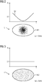
- FIG 2 shows an example of a lesion L1 in a medical image recording according to the prior art.
- the lesion can be tumor-like tissue, for example.
- the medical image depicting the lesion L1 corresponds to a computed tomography slice, which was acquired with the addition of iodine-containing contrast medium.
- Figure 3 shows an example of a lesion L2, which does not differ in shape and size from the lesion L1 and which has been imaged with the same recording technique and procedure for the sake of simplicity and for the purpose of illustration.
- Both lesions L1 and L2 were segmented using known segmentation algorithms, the outer contour AK1, AK2 is known in each case. Surrounding tissue is not shown for simplicity.
- an x-ray absorption representing G1, G2 (in Houndsfield Units HU) can be determined for each lesion, taking into account all image element values encompassed by the outer contour AK1, AK2, or here pixel entries. This corresponds to an average iodine contrast or an average iodine content for each of the lesions. It can be seen that lesion L1 has a darker area in its center, which continuously increases in brightness towards the outside. This spatial grayscale distribution GV is in Figure 2A exemplified in the diagram above the lesion L1.
- lesion L2 shows an essentially homogeneous gray scale distribution, which indicates an even contrast medium distribution.
- the mean grayscale values G1, G2 determined can be the same or almost the same for both lesions L1 and L2.
- a profound differentiation of a blood flow state or a contrast medium distribution of the two lesions L1 and L2 is based on the known Approach not possible or only possible qualitatively.
- a dedicated decision as to whether, for example, lesion L1 responds to a selected form of therapy, is not possible using the known procedure described, since local differences in the gray level are 'averaged out' within the lesion.
- Figures 4 and 5 show, by way of example, lesions L3 and L4, each in a medical image recording according to exemplary embodiments of the invention.
- the medical image recordings depicting the lesions L3, L4 also correspond to computer tomography slice recordings which were acquired with the addition of iodine-containing contrast medium.
- Lesion L4 is also identical in shape and size to lesion L3.
- lesion L3 Corresponding to lesion L1, lesion L3 has a darker area at its center, which, comparable to the situation with lesion L1, is an indicator of a reduced contrast agent uptake.
- This grayscale distribution GV is exemplified in the diagram over lesion L3 in Figure 3A shown.
- Lesion L4, like lesion L2, shows an even grayscale distribution.
- the outer contours AK3, AK4 are also known by means of segmentation. Surrounding tissue is not shown for simplicity. According to the invention, the lesions L3, L4 are divided into five tissue areas GB31, GB32, GB33, GB34, GB35 and GB41, GB42, GB43, GB44, GB45.
- the subdivision that is to say the definition of size, position, arrangement and / or the number of tissue areas, is preferably carried out automatically and / or taking into account size, position or, if known, the type or presumed type of lesion.
- a functional parameter FP to be determined below or the image recording technology used can influence the subdivision.
- an onion-layer-like division in which a tissue area located further outwards, for example GB31, completely encloses an inner tissue area, for example GB32.
- the outer contours of the individual tissue areas have been chosen to be morphologically identical to the outer contours AK3, AK4 of the lesions L3, L4.
- a functional parameter FP for each of the individual tissue areas GB31, GB32, GB33, GB34, GB35 and GB41, GB42, GB43, GB44, GB45 derived from medical imaging.
- the pixel entries included in each case give an average gray scale value or iodine contrast G31, G32, G33, G34, G35 or G41, G42, G43, G44, G45 determined.
- the averaged iodine contrast values provide information about a local tissue function GF or give a measure of the tissue function GF, in this case tissue perfusion, illustrated by the gray scale distribution GF. In other words, an increase in the spatial resolution of a tissue function GF is achieved according to the invention.
- the image recording on which the evaluation according to the invention is based can correspond to a representation of a region of interest obtained using any recording technique, or it can be composed of several individual images. According to the invention, several image recordings can also be evaluated, in particular if functional information is available in the image recordings.
- Step S1 comprises receiving at least one medical image B of a region of interest of an examination object 3, for example a computed tomography image or an MRI image in a computing unit 16.
- the medical image B can be recorded in a time correlated manner with the image evaluation according to the invention or, if it has already been acquired beforehand, can be loaded into the computing unit 16 from a local or remote memory, for example a PACS system of a hospital.
- the medical image recording B can be generated by combining image information from at least two different images of the region of interest.
- Common image processing methods such as material decomposition, image correction methods such as noise suppression, registration methods or the like can be used, if necessary.
- an outer contour of a tissue to be examined, preferably a lesion, contained in the medical image recording is segmented in the computing unit 16, in particular the segmentation unit 21.
- individual image elements are divided into the tissue to be examined or the surrounding tissue.
- Image element or edge-based segmentation methods which are known per se, are preferably used.
- additional information ZI can be recorded for the segmentation or before the segmentation by the computing unit 16 about the question underlying the examination or an initial suspicion or about the measurement protocol used for the image data acquisition in order to decide which body region of interest and thus which tissue types have been imaged and / or which structure or which tissue and / or which quality is to be segmented in the medical image recording.
- the segmentation algorithm used can be selected, for example. Alternatively, a corresponding entry regarding the additional information ZI are made available to the computing unit 16 by a user on request or on the initiative via user interface 7, 11.
- the size that is the length extension in different spatial directions, the volume, the shape of the outer contour AK or the like of the tissue to be examined is known.
- This size information GI can also be used in subsequent steps.
- the segmented tissue is subdivided into a plurality of tissue regions GB. More than two, in particular four to five, tissue regions GB are preferably defined. The greater the number of tissue areas GB, the greater the meaningfulness of the function parameter FP determined below via a local tissue function GF within the segmented tissue.
- size information GI can also be used for step S4 flow into the definition of the individual tissue areas GB via the size, volume and / or shape and / or the like of the tissue to be examined.
- a subdivision unit 23 comprised by the computing unit 16 can be in data exchange with the segmentation unit 21 and / or the data processing device 12, which also carries out the control of a medical imaging system.
- the individual tissue areas GB are preferably nested in one another or, to put it another way, have a layered structure and / or each have the same outer contour as the segmented tissue.
- the subdivision unit 23 can define the individual tissue areas by means of morphological operations, such as an erosion and / or dilation operator.
- the tissue areas have a distance from the outer contour to the outer contour or a radial extension of 5 mm.
- a function parameter FP relating to a tissue function GF is determined for each of the individual tissue areas GB.
- a value for the function parameter FP is derived individually, that is locally for each tissue area GB.
- This evaluation can result in different function parameter values for the individual tissue areas, which when viewed together represent a spatial distribution of a tissue function, it being possible to resolve local differences in the tissue function.
- the determination of the function parameter is carried out by the determination unit 22. This is set up to individually extract and analyze image information contained in the medical image acquisition for each of the individual tissue areas.
- Functional parameters can be: blood flow, iodine enrichment, iron enrichment, calcium density, fat content.
- Function parameters can also be used for texture analysis, such as parameters such as mean density, moments, heterogeneity, entropy, fractal dimension.
- the determination step can include a texture analysis.
- the determination unit 22 can preferably determine a feature vector based on intensity values of the image elements in each individual tissue area.
- the determination unit 22 can furthermore preferably use machine learning methods in order to generate at least one function parameter based on picture element entries and / or the feature vector.
- a computing model can be stored in a memory, locally or centrally, which can be trained by using a machine learning algorithm before the method according to the invention is carried out.
- the training takes place, for example, by means of test images and / or inputs by a user, so that the algorithm is then able to independently detect features in image recordings or to derive function parameters.
- Step S6 may thus include creating a texture metric for each tissue area GB.
- the texture analysis includes to analyze pixel intensity values for the tissue area under consideration and to derive a spatial distribution of the same.
- the texture metric represents a measure of this spatial distribution, for example the texture metric is different moments of these picture element intensities.
- step S5 can include carrying out a texture analysis based on the multiple recordings or the multiple image recordings, in particular in cases in which different medical image recordings correspond to different recording aspects, for example temporal phases, that is to say different time phases of an image recording, or else detect different ones Energy bands.
- the various image recordings may or may not have been acquired with the addition of contrast medium.
- a feature vector can comprise a plurality of texture metrics for a plurality of different parts or areas of the tissue to be examined, in particular of several tissue areas.
- a texture metric in the sense of the invention can comprise: an average, maximum, minimum picture element intensity, a uniformity measure, an entropy in the sense of an irregularity via a gray value histogram, a standard deviation of a gray value histogram, a skewness in the sense of an asymmetry of a Gray value histogram, a curvature or flatness of a gray value histogram, an energy measure or an area moment of inertia (e.g. pixel repetition rate and / or an order measure), correlation (e.g. a measure for a linear dependence on gray values), run length matrix (e.g. a pixel texture in a certain spatial direction), contrast, roughness (for example as a measure for an edge density), heterogeneity (for example as a measure for the presence of edges) or the like.
- the texture metric is preferably determined for each of the tissue areas GB in the medical image recording.
- Image element in the sense of the invention should include both pixels and voxels.
- the method described can optionally branch off according to the invention into a repetition loop R, the method being repeated with steps S1 to S5 until all medical image recordings necessary for deriving the local tissue function of the tissue to be examined are used Are available, were evaluated.
- a comparison of function parameters determined according to the invention can take place, the values for the function parameters for medical image recordings being determined, which were recorded at different times.
- the same functional parameter for medical image recordings of an initial examination can be compared to medical recordings of a follow-up examination at a later point in time.
- the comparison makes it possible to identify local, even the smallest, changes in the examined tissue over the observation period.
- the comparison can preferably be made using machine learning methods.
- the described method is particularly suitable for monitoring the course of tissue conditions, e.g. the fat content or bone density and in particular for Follow-up of a new type of tumor therapy without using previous evaluation criteria such as RECIST.
Landscapes
- Health & Medical Sciences (AREA)
- Engineering & Computer Science (AREA)
- Life Sciences & Earth Sciences (AREA)
- Medical Informatics (AREA)
- Biomedical Technology (AREA)
- General Health & Medical Sciences (AREA)
- Nuclear Medicine, Radiotherapy & Molecular Imaging (AREA)
- Public Health (AREA)
- Physics & Mathematics (AREA)
- Pathology (AREA)
- Radiology & Medical Imaging (AREA)
- Animal Behavior & Ethology (AREA)
- Veterinary Medicine (AREA)
- Biophysics (AREA)
- Molecular Biology (AREA)
- Heart & Thoracic Surgery (AREA)
- Surgery (AREA)
- High Energy & Nuclear Physics (AREA)
- Computer Vision & Pattern Recognition (AREA)
- Optics & Photonics (AREA)
- Theoretical Computer Science (AREA)
- Orthopedic Medicine & Surgery (AREA)
- Physiology (AREA)
- Rheumatology (AREA)
- General Physics & Mathematics (AREA)
- Dentistry (AREA)
- Oral & Maxillofacial Surgery (AREA)
- Pulmonology (AREA)
- Quality & Reliability (AREA)
- Databases & Information Systems (AREA)
- Data Mining & Analysis (AREA)
- Epidemiology (AREA)
- Primary Health Care (AREA)
- Apparatus For Radiation Diagnosis (AREA)
- Magnetic Resonance Imaging Apparatus (AREA)
Description
Die klassische Tumortherapie umfasst neben der operativen Entfernung eines Tumors die Chemotherapie, bei der einem Patienten Zytotoxine oder Zytostatika verabreicht werden, welche ein gezieltes Absterben von Tumorzellen bewirken oder ein Tumorwachstum verhindern sollen. Sie umfasst auch die Strahlentherapie, bei der ein Tumor mit ionisierender oder Partikelstrahlung behandelt wird, was ebenfalls zum Absterben von Tumorzellen führen soll. Zur Ermittlung des Ansprechverhaltens eines Tumors wird häufig auf sogenannte RECIST-Kriterien zurückgegriffen. Gängige RECIST-Kriterien sind zum Beispiel ein maximaler Durchmesser (LAD) eines Tumors oder sein Kurzachsendurchmesser (SAD), also im Wesentlichen Angaben über die Größe des Tumors, gemessen zu einem bestimmten Betrachtungszeitpunkt. Ermittelt man anhand der RECIST-Kriterien über einen Behandlungszeitraum hinweg ein Verschwinden bzw. eine Verkleinerung des Tumors, insbesondere ohne das Erscheinen von neuen Läsionen, ist das ein Anzeichen für eine vollständigen (CR-Complete Response) bzw. teilweise (PR-Partial Response) Tumorantwort. Anders ausgedrückt, der Tumor spricht auf die eingesetzte Therapie an. Die Behandlung hat Erfolg. Wird über den Behandlungszeitraum hinweg eine gleichbleibende Tumorgröße ermittelt, ist das ein Anzeichen für eine unveränderte Erkrankung (SD-Stable Disease), was ebenfalls einem Behandlungserfolg entsprechen kann. Vergrößert sich der Tumor während der Behandlung bzw. kommen neue Läsionen hinzu, ist dies ein Anzeichen für eine fortschreitende Erkrankung (PD-Progressive Disease) und letztlich für einen Misserfolg der gewählten Behandlungsmethode.In addition to the surgical removal of a tumor, classic tumor therapy includes chemotherapy, in which cytotoxins or cytostatics are administered to a patient, which are intended to cause targeted tumor cell death or to prevent tumor growth. It also includes radiation therapy, in which a tumor is treated with ionizing or particle radiation, which should also lead to the death of tumor cells. So-called RECIST criteria are often used to determine the response behavior of a tumor. Common RECIST criteria are, for example, a maximum diameter (LAD) of a tumor or its short axis diameter (SAD), i.e. essentially information about the size of the tumor, measured at a certain point in time. If the RECIST criteria are used to determine whether the tumor has disappeared or shrunk over a treatment period, in particular without new lesions appearing, this is a sign of a complete (CR-Complete Response) or partial (PR-Partial Response) Tumor response. In other words, the tumor responds to the therapy used. The treatment is successful. If a constant tumor size is determined over the treatment period, this is a sign of an unchanged disease (SD stable disease), which can also correspond to the success of the treatment. If the tumor grows during treatment or new lesions appear, this is a sign of a progressive disease (PD-Progressive Disease) and ultimately of a failure of the chosen treatment method.
Daneben gibt es modernere Tumorbehandlungsmethoden wie zum Beispiel die Anti-Angiogenese oder die Immuno-Therapie. Die Anti-Angiogenese ist darauf gerichtet, medikamentös die Blutversorgung von Tumorgewebe einzudämmen, indem die Gefäßbildung innerhalb des Tumors unterdrückt wird. Bei der Immuno-Therapie wird das körpereigene Immunsystem unterstützt, die Tumorzellen, die sich ansonsten einer körpereigenen Immunantwort entziehen würden, zu zerstören.There are also more modern tumor treatment methods such as anti-angiogenesis or immunotherapy. Anti-angiogenesis is designed to medicamentally curb the blood supply to tumor tissue by preventing vascularization is suppressed within the tumor. In immunotherapy, the body's immune system is supported to destroy the tumor cells, which would otherwise elude the body's immune response.
Die klassischen Behandlungsmethoden verursachen ein Gleichbleiben oder eine Verringerung der Tumorgröße. Neuartige Behandlungsmethoden haben jedoch oft zumindest für eine Übergangszeit keine Auswirkung auf die Tumorgröße oder können sogar ein Wachstum des Tumors bewirken. Erst bei länger anhaltender Behandlungsdauer zeigt sich die angestrebte Verkleinerung. Darüber hinaus können neuere Therapieformen strukturelle Veränderungen des Tumorgewebes verursachen, bspw. Nekrosen im Inneren des Tumors.The classic treatment methods cause the tumor size to remain the same or to decrease. However, new types of treatment often have no effect on the size of the tumor, at least for a transitional period, or can even cause the tumor to grow. The desired reduction only becomes apparent when the treatment duration is longer. In addition, newer forms of therapy can cause structural changes in the tumor tissue, for example necrosis inside the tumor.
Nicht zuletzt aus wirtschaftlicher Sicht ist Zeit in der Krebstherapie ein kritischer Faktor, sodass das Ansprechverhalten auf eine gewählte Therapieform schnellstmöglich ermittelt werden sollte. Bei neuartigen Therapien erweisen sich aufgrund ihrer veränderten Wirkweise die üblichen Kriterien wie RECIST als ungenau oder sogar falsch, sodass diese für den vorgegebenen Zeitrahmen nicht oder nur eingeschränkt herangezogen werden können. Für die neuartigen Therapieverfahren werden daher Kriterien benötigt, die einen Rückschluss auf das Ansprechverhalten von Tumorzellen auf eine Behandlung, bspw. ihre lokale Blutversorgung, zulassen.Last but not least, from an economic perspective, time is a critical factor in cancer therapy, so that the response to a chosen form of therapy should be determined as quickly as possible. In the case of novel therapies, due to their changed mode of action, the usual criteria such as RECIST prove to be inaccurate or even incorrect, so that they cannot be used or can only be used to a limited extent for the specified time frame. Criteria are therefore required for the novel therapeutic methods which allow conclusions to be drawn about the response behavior of tumor cells to a treatment, for example their local blood supply.
Es ist bekannt, unter Kontrastmittelgabe mittels quantitativer, medizinischer Bildgebungsverfahren wie zum Beispiel der Computertomographie (CT) mit zwei unterschiedlichen Energiespektren (Dual Energy) oder der dynamischen Perfusions-CT die Durchblutung bzw. das Blutvolumen in Tumorgewebe darzustellen, indem bspw. die im Tumorgewebe angereicherte Kontrastmittelmenge ermittelt wird. Bislang wird das Blutvolumen für die gesamte Läsion ausgewertet, sodass lokale Unterschiede der Blutversorgung oder lokale Veränderungen der Blutgefäßstruktur nicht berücksichtigt werden. Mit anderen Worten entspricht das derart ermittelte Blutvolumen einem mittleren Blutvolumen über die gesamte Läsion hinweg.It is known to display the blood flow or the blood volume in tumor tissue, for example by using quantitative, medical imaging methods such as, for example, computer tomography (CT) with two different energy spectra (dual energy) or dynamic perfusion CT, for example by displaying the accumulated in the tumor tissue Amount of contrast medium is determined. Up to now, the blood volume has been evaluated for the entire lesion, so that local differences in the blood supply or local changes in the blood vessel structure are not taken into account. In other words the blood volume determined in this way is an average blood volume over the entire lesion.
Das Dokument
Eine manuelle Definition bzw. Auswahl von interessierenden Bereichen (ROIs) zur Analyse eines lokalen Blutvolumens kann die lokalen Unterschiede zwar aufzeigen, ist jedoch stark Nutzer-abhängig und dadurch schlecht reproduzierbar und grundsätzlich fehleranfällig.A manual definition or selection of areas of interest (ROIs) for the analysis of a local blood volume can show the local differences, but it is strongly user-dependent and therefore difficult to reproduce and generally prone to errors.
Alternative Analyse-Verfahren wie beispielsweise bekannte Texturanalyse-Verfahren wären unter Auswertung der strukturellen Eigenschaften des abgebildeten Tumorgewebes im Grunde in der Lage, sog. ,Perfusion Maps', also mehrdimensionale Durchblutungskarten für einen Tumor zu erzeugen, reagieren jedoch sehr empfindlich auf kleinste Änderungen in der Bildverarbeitungskette (Rekonstruktionskern, Schichtdicke, Rauschfilter, etc). Eine Vergleichbarkeit von Initial- und Kontrollmessung wäre auch hier nicht gegeben.Alternative analysis methods, such as known texture analysis methods, would in principle be able to generate so-called 'perfusion maps', i.e. multidimensional blood circulation maps for a tumor, by evaluating the structural properties of the displayed tumor tissue, but are very sensitive to the smallest changes in the Image processing chain (reconstruction core, layer thickness, noise filter, etc.). A comparison of the initial and control measurements would also not be possible here.
Demgegenüber ist es Aufgabe der vorliegenden Erfindung, alternative Mittel bereit zu stellen, die es erlauben, zuverlässig und reproduzierbar Rückschlüsse auf eine lokale Gewebefunktion abzuleiten. Insbesondere ist es Aufgabe der vorliegenden Erfindung, frühzeitig nach Therapiebeginn Information über eine lokale Durchblutung von Tumorgewebe abzuleiten.In contrast, it is an object of the present invention to provide alternative means which allow reliable and reproducible conclusions to be drawn about a local tissue function. In particular, it is an object of the present invention to derive information about local blood flow to tumor tissue early after the start of therapy.
Diese Aufgabe wird gelöst durch ein Verfahren zum Bestimmen einer Gewebefunktion eines Gewebes, entsprechende Recheneinheit und medizinische Bildgebungsanlage, entsprechendes Computerprogramm und entsprechenden computerlesbaren Datenträger gemäß den unabhängigen Ansprüchen. Bevorzugte und/oder alternative, vorteilhafte Ausgestaltungsvarianten sind Gegenstand der abhängigen Ansprüche.This object is achieved by a method for determining a tissue function of a tissue, corresponding computing unit and medical imaging system, corresponding computer program and corresponding computer-readable data carrier in accordance with the independent claims. Preferred and / or alternative, Advantageous design variants are the subject of the dependent claims.
Nachstehend wird die erfindungsgemäße Lösung der Aufgabe in Bezug auf das beanspruchte Verfahren als auch in Bezug auf die beanspruchten Vorrichtungen beschrieben. Hierbei erwähnte Merkmale, Vorteile oder alternative Ausführungsformen sind ebenso auch auf die anderen beanspruchten Gegenstände zu übertragen und umgekehrt. Mit anderen Worten können gegenständliche Ansprüche (die beispielsweise auf ein Verfahren gerichtet sind) auch mit Merkmalen, die in Zusammenhang mit einer der Vorrichtungen beschrieben oder beansprucht sind, weitergebildet sein. Die entsprechenden funktionalen Merkmale des Verfahrens werden dabei durch entsprechende gegenständliche Module oder Einheiten ausgebildet.The solution to the object according to the invention is described below with reference to the claimed method and also in relation to the claimed devices. Features, advantages or alternative embodiments mentioned here are also to be transferred to the other claimed objects and vice versa. In other words, objective claims (which are directed, for example, to a method) can also be developed with features that are described or claimed in connection with one of the devices. The corresponding functional features of the method are formed by corresponding objective modules or units.
Die vorliegende Erfindung betrifft in einem ersten Aspekt ein Verfahren zum Bestimmen einer lokalen Gewebefunktion eines Gewebes in einer interessierenden Körperregion eines Untersuchungsobjektes. Das Verfahren umfasst eine Vielzahl von Schritten.In a first aspect, the present invention relates to a method for determining a local tissue function of a tissue in a body region of interest of an examination object. The process comprises a number of steps.
Das erfindungsgemäße Verfahren zielt darauf ab, eine Gewebefunktion eines Gewebes in einer interessierenden Körperregion eines Untersuchungsobjektes zu ermitteln. Dabei legt das Verfahren besonderes Augenmerk auf die individuelle Ermittlung dieser Gewebefunktion für lokale Bereiche des Gewebes. Mit anderen Worten erlaubt das Verfahren eine Darstellung lokaler Unterschiede in der Gewebefunktion.The method according to the invention aims to determine a tissue function of a tissue in a body region of interest of an examination object. The method pays particular attention to the individual determination of this tissue function for local areas of the tissue. In other words, the method allows a representation of local differences in the tissue function.
Eine Gewebefunktion im Sinne dieser Erfindung bezeichnet eine physikalische, chemische, funktionelle und/oder strukturelle Eigenschaft eines Gewebes, wie zum Beispiel eine Materialdichte, einen Stoff- bzw. Substanzgehalt oder -anteil, eine Substanz-Anreicherungsrate (bei dynamischen Messungen) oder dergleichen.A tissue function in the sense of this invention denotes a physical, chemical, functional and / or structural property of a tissue, such as, for example, a material density, a substance or substance content or proportion, a substance enrichment rate (in dynamic measurements) or the like.
Unter einem Gewebe ist im Sinne der Erfindung eine Vielzahl gleichartig ausgebildeter, in der Regel zusammenhängender Körperzellen zu verstehen, die eine gleiche bzw. ähnliche Funktion ausüben. Mit anderen Worten entspricht ein Gewebe einem Gewebetyp oder einer Gewebeart, wobei das Gewebe ein Tumorgewebe ist. Ein Gewebe im Sinne der Erfindung kann auch eine Läsion unbekannter Beschaffenheit mit Vermutung auf Tumorgewebe, also eine anomale Veränderung, umfassen oder bilden. Ein Gewebe im Sinne der Erfindung kann nur einen oder mehrere Gewebetypen umfassen.For the purposes of the invention, a tissue is to be understood to mean a large number of similarly formed, generally connected body cells which perform the same or similar function. In other words, a tissue corresponds to a tissue type or a tissue type, the tissue being a tumor tissue. A tissue in the sense of the invention can also comprise or form a lesion of unknown nature with suspicion of tumor tissue, that is to say an abnormal change. A fabric in the sense of the invention can comprise only one or more types of fabric.
Das Gewebe befindet sich in einer interessierenden Körperregion. Diese entspricht im Sinne der hiesigen Erfindung dem zu untersuchenden Körperteil bzw. der zu untersuchenden Körperregion, bspw. dem Abdomen oder dem Schädel. In diesem Sinne entspricht die zu untersuchende Körperregion dem Teil eines Untersuchungsobjektes, welches mittels einer medizinischen Bildgebungsanlage, zum Beispiel eines Computertomographen, untersucht bzw. abgebildet werden soll.The tissue is in a region of the body of interest. For the purposes of the present invention, this corresponds to the body part or body region to be examined, for example the abdomen or the skull. In this sense, the body region to be examined corresponds to the part of an examination object which is to be examined or imaged by means of a medical imaging system, for example a computer tomograph.
Insofern wird im Folgenden ohne Beschränkung der Allgemeinheit von einem Patienten als Untersuchungsobjekt ausgegangen, wobei es sich meist um einen Menschen handelt. Grundsätzlich kann der Patient auch ein Tier sein. Im Folgenden werden daher die beiden Begriffe "Untersuchungsobjekt" und "Patient" synonym verwendet. Das Untersuchungsobjekt kann alternativ eine Pflanze oder ein nicht-lebender Gegenstand, z.B. ein historisches Artefakt oder dergleichen sein.In this respect, a patient is assumed as the object to be examined without restricting generality, and it is usually a human being. In principle, the patient can also be an animal. In the following, the two terms "examination object" and "patient" are therefore used synonymously. The object under examination can alternatively be a plant or a non-living object, e.g. a historical artifact or the like.
Das Segmentieren einer Außenkontur des Gewebes anhand von wenigstens einer medizinischen Bildaufnahme darstellend die interessierende Körperregion des Untersuchungsobjektes umfassend das Gewebe entspricht einem ersten Verfahrensschritt.The segmentation of an outer contour of the tissue on the basis of at least one medical image recording representing the body region of interest of the examination object comprising the tissue corresponds to a first method step.
Eine medizinische Bildaufnahme ist eine mehrdimensionale, insbesondere eine zwei-dimensionale oder drei-dimensionale Abbildung von zu der interessierenden Körperregion gehörigen Sensor-Daten, die mittels einer medizinischen Bildgebungsanlage erzeugt wurden. Die medizinische Bildaufnahme kann eine Computer-Tomographie-Aufnahme, MRT-Aufnahme, Röntgenaufnahme, Positronen-Emissions-Tomographie(PET)-Aufnahme, Röntgen-C-Bogen-Aufnahme, Ultraschall-Aufnahme, Single-Photon-Emissions-Tomographie (SPECT)-Aufnahme oder dergleichen sein. Die medizinische Bildaufnahme kann eine Einzelaufnahme, aber auch Teilobjekt einer mehrschichtigen Aufnahmeserie sein.
Die Dimension der Bildaufnahmen gibt an, ob eine ausgewählte Schicht mit einer definierten Schichtdicke (2D) der interessierenden Region oder ein ausgewähltes Volumen (3D) dargestellt ist. Im Falle mehrerer medizinischer Bildaufnahmen kann die Abbildung des Gewebes drei- oder vier-dimensional sein, dann jeweils mit oder ohne Zeitverlauf (3D oder 4D). Bei medizinischen Bildaufnahmen mit Zeitverlauf handelt es sich in der Regel um funktionelle bzw. dynamische Bildaufnahmen, zum Beispiel um eine Perfusionsmessung. Im Falle mehrerer medizinischer Bildaufnahmen können auch Einzelaufnahmen verschiedener Energiespektren umfasst sein. Eine medizinische Bildaufnahme kann auch ein konstruiertes bzw. zusammengesetztes Bild sein, welches aus mehreren Einzelaufnahmen, insbesondere aus verschiedenen Aufnahmetechniken, zusammengesetzt wurde bzw. welches Bildinformation aus mehreren Einzelaufnahmen umfasst. Mit anderen Worten kann eine medizinische Bildaufnahme als wenigstens eine Aufnahme aus der folgenden Gruppe von Aufnahmen ausgebildet sein: ein zweidimensionales, tomographisches Bild, ein dreidimensionales Bild, ein vierdimensionales Bild oder ein multi-spektrales Bild.A medical image recording is a multidimensional, in particular a two-dimensional or three-dimensional, image of sensor data belonging to the region of interest that is used by means of a medical imaging system were generated. Medical imaging can be computer tomography, MRI, x-ray, positron emission tomography (PET), x-ray C-arm, ultrasound, single photon emission tomography (SPECT) Recording or the like. The medical image acquisition can be a single exposure, but it can also be a partial object of a multilayer exposure series.
The dimension of the image recordings indicates whether a selected layer with a defined layer thickness (2D) of the region of interest or a selected volume (3D) is shown. In the case of several medical image recordings, the imaging of the tissue can be three- or four-dimensional, then with or without a time course (3D or 4D). Medical image recordings with a time course are usually functional or dynamic image recordings, for example a perfusion measurement. In the case of several medical image recordings, individual recordings of different energy spectra can also be included. A medical image recording can also be a constructed or composed image which has been composed of several individual images, in particular from different recording techniques, or which comprises image information from several individual images. In other words, a medical image recording can be designed as at least one recording from the following group of recordings: a two-dimensional, tomographic image, a three-dimensional image, a four-dimensional image or a multi-spectral image.
Die Bilderfassung kann zeitlich mit der Durchführung des erfindungsgemäßen Verfahrens korrelieren. Alternativ kann die medizinische Bildaufnahme zu einem beliebigen Zeitpunkt vor der Ausführung des Verfahrens erfasst worden sein.The image acquisition can correlate in time with the implementation of the method according to the invention. Alternatively, the medical image recording can have been recorded at any time before the method is carried out.
Gemäß diesem ersten Schritt erfolgt ein Segmentieren der Außenkontur des betrachteten Gewebes in der medizinischen Bildaufnahme. Mit anderen Worten, wird das zu untersuchende Gewebe nach außen hin abgegrenzt und seine Außenkontur definiert. Die Außenkontur kann eine in Abhängigkeit der Größe des bei der Bilderfassung gewählten Sichtfeldes (FoV) offene oder geschlossene Linie (2D) oder Fläche (3D) sein. Diese trennt das Gewebe von umliegenden Strukturen. Das Segmentieren kann automatisch, semi-automatisch anhand in Fachkreisen bekannter Segmentierungsalgorithmen oder manuell in der Bildaufnahme durch einen Benutzer durchgeführt werden.According to this first step, the outer contour of the tissue under consideration is segmented in the medical image recording. In other words, the tissue to be examined is delimited from the outside and its outer contour is defined. The outer contour can be a depending on the size of the the image capture selected field of view (FoV) be open or closed line (2D) or area (3D). This separates the tissue from surrounding structures. The segmentation can be carried out automatically, semi-automatically on the basis of segmentation algorithms known in specialist circles or manually in the image acquisition by a user.
Einen zweiten Schritt des erfindungsgemäßen Verfahrens bildet das Unterteilen des segmentierten Gewebes in wenigstens zwei Gewebebereiche. Mit anderen Worten werden für das Gewebe wenigstens zwei Teilbereiche gebildet und innerhalb der segmentierten Außenkontur des Gewebes angeordnet, die derart eine Substruktur des Gewebes darstellen. Die Anzahl sowie die Anordnung der Gewebebereiche zu einander sind variabel und können insbesondere von der Größe bzw. der Art des untersuchten Gewebes abhängen. Alternativ oder zusätzlich können Anzahl und Lage der Gewebebereiche in Abhängigkeit der Wahl des im Folgenden zu ermittelnden Funktionsparameters bestimmt werden. Die Unterteilung erfolgt bevorzugt automatisch, kann aber auch semi-automatisch erfolgen, zum Beispiel, indem dem Nutzer zunächst ein Vorschlag oder eine Auswahl von Vorschlägen angezeigt wird, den bzw. die dieser bestätigen oder auswählen kann. Die Unterteilung erfolgt weiter bevorzugt basierend auf Erfahrungs- bzw. Referenzwerten. Mit anderen Worten können zum Beispiel in einer Datenbank Standard-Unterteilungen des segmentierten Gewebes für bestimmte Gewebearten, Gewebegrößen, betrachtete Funktionsparameter und/oder dergleichen hinterlegt sein, die als Referenz bzw. Referenzwerte in die Unterteilung einfließen bzw. dabei berücksichtigt werden. Bevorzugt fließen in die Definition der Lage und/oder Größe der Gewebebereiche, sofern bekannt, Informationen, Erfahrungswerte bzw. Annahmen über eine räumliche Verteilung des im Folgenden zu ermittelnden Funktionsparameters mit ein. Derart stellt das erfindungsgemäße verfahren vorteilhaft sicher, dass lokale Schwankungen der Gewebefunktion dargestellt werden.A second step of the method according to the invention is the subdivision of the segmented tissue into at least two tissue areas. In other words, at least two subregions are formed for the tissue and arranged within the segmented outer contour of the tissue, which thus represent a substructure of the tissue. The number and the arrangement of the tissue areas with respect to one another are variable and can depend in particular on the size or type of the tissue examined. Alternatively or additionally, the number and position of the tissue areas can be determined depending on the choice of the functional parameter to be determined below. The subdivision is preferably automatic, but can also be semi-automatic, for example by first displaying a suggestion or a selection of suggestions to the user, which the user can confirm or select. The division is further preferably based on experience or reference values. In other words, standard subdivisions of the segmented tissue for certain types of tissue, tissue sizes, considered functional parameters and / or the like can be stored in a database, for example, which flow into the subdivision as reference or reference values or are taken into account in the process. If known, information, empirical values or assumptions about a spatial distribution of the functional parameter to be determined below preferably flow into the definition of the position and / or size of the tissue areas. In this way, the method according to the invention advantageously ensures that local fluctuations in the tissue function are displayed.
In einem dritten Schritt wird, wie oben bereits angedeutet, ein Funktionsparameter betreffend die Gewebefunktion für jeden der wenigstens zwei Gewebebereiche ermittelt. Der Funktionsparameter ist ein Maß, ein Repräsentant bzw. eine Kenngröße für die Gewebefunktion. Mit anderen Worten kennzeichnet ein Funktionsparameter eine bestimmte Gewebefunktion bzw. lässt Rückschlüsse auf diese Gewebefunktion zu. Das Ermitteln umfasst erfindungsgemäß eine quantitative und/oder qualitative Auswertung von in den medizinischen Bildaufnahmen enthaltener Bildinformation, jeweils bezogen auf die einzelnen Gewebebereiche. Zum Beispiel kann bei einer CT-Bildaufnahme unter Kontrastmittelgabe für jeden Gewebebereich eine Auswertung der Helligkeitswerte der Bildelemente (HU-Werte) erfolgen, sodass auf ein (mittlere) Durchblutung des Gewebes in jedem der Gewebebereiche zurückgeschlossen werden kann.In a third step, as already indicated above, a function parameter relating to the tissue function is determined for each of the at least two tissue areas. The function parameter is a measure, a representative or a parameter for the tissue function. In other words, a function parameter identifies a specific tissue function or allows conclusions to be drawn about this tissue function. According to the invention, the determination comprises a quantitative and / or qualitative evaluation of image information contained in the medical image recordings, in each case based on the individual tissue areas. For example, an evaluation of the brightness values of the image elements (HU values) can be carried out for each tissue area when a CT image is taken with contrast agent, so that a conclusion can be drawn about a (medium) blood flow to the tissue in each of the tissue areas.
Alternativ können auch mehr als nur ein Funktionsparameter pro Gewebebereich ermittelt werden. Insofern können mit dem erfindungsgemäßen Verfahren mehrere Gewebefunktionen parallel untersucht werden.Alternatively, more than just one functional parameter can be determined per tissue area. In this respect, several tissue functions can be examined in parallel with the method according to the invention.
Zusammenfasend haben die Erfinder also erkannt, dass das erfindungsgemäße Verfahren ermöglicht, räumliche Schwankungen bzw. Unterschiede einer Gewebefunktion darzustellen, indem ein für eine Gewebefunktion charakteristischer Funktionsparameter individuell für verschiedene, also wenigstens zwei Gewebebereiche bestimmt wird. Die derart für die verschiedenen Gewebebereiche ermittelten, lokalen Werte des Funktionsparameters dienen also Maß für die Homogenität bzw. Inhomogenität einer Gewebefunktion über das untersuchte Gewebe hinweg. Das erfindungsgemäße Verfahren eignet sich damit für eine Vielzahl von verschiedenen Anwendungen in der Medizin.In summary, the inventors have thus recognized that the method according to the invention enables spatial fluctuations or differences of a tissue function to be represented by a function parameter characteristic of a tissue function being determined individually for different, ie at least two tissue areas. The local values of the functional parameter determined in this way for the various tissue areas thus serve as a measure of the homogeneity or inhomogeneity of a tissue function across the tissue examined. The method according to the invention is therefore suitable for a large number of different applications in medicine.
Das erfindungsgemäße Verfahren eignet sich insbesondere zur Anwendung bei der Untersuchung des Ansprechverhaltens von Tumorerkrankungen, zur Überwachung eines beliebigen anderen Krankheitsverlaufs, wie bspw. Osteoporose, oder aber allgemeiner auch für die Durchführung von Kontrolluntersuchungen zu einem späteren Zeitpunkt, denn die Unterteilung des segmentierten Gewebes sowie das erfindungsgemäße Ermitteln des Funktionsparameters, wie es bei einer initialen Erstuntersuchung vorgenommen wurde, kann auf Bildaufnahmen einer späteren Kontrollmessung robust und reproduzierbar übertragen werden und liefert so zu der Erstuntersuchung vergleichbare Daten.The method according to the invention is particularly suitable for use in examining the response behavior of tumor diseases, for monitoring any other course of the disease, such as, for example, osteoporosis, or more generally for carrying out control examinations at a later point in time, because the subdivision of the segmented tissue and the determination of the functional parameter according to the invention, as was carried out during an initial initial examination, can be transferred robustly and reproducibly to image recordings of a later control measurement and thus provides data comparable to the initial examination.
Es sei abschließend nochmals erwähnt, dass die beschriebenen Verfahrensschritte, also das Segmentieren, das Unterteilen sowie das Ermitteln in den medizinischen Bildaufnahmen pixel- und/oder voxel-basiert erfolgen können.Finally, it should be mentioned again that the described method steps, that is to say the segmentation, the subdivision and the determination in the medical image recordings, can be pixel-based and / or voxel-based.
Gemäß der vorliegenden Erfindung sind die wenigstens zwei Gewebebereiche derart schichtartig angeordnet, dass ein erster Gewebebereich einen zweiten Gewebebereich vollständig umschließt. Mit anderen Worten werden die Gewebebereiche ringartig bzw. verschachtelt angeordnet, sodass lediglich ein äußerer Gewebebereich äußerlich an der segmentierten Außenkontur des Gewebes anliegt bzw. diese einschließt und innerlich die übrigen Gewebebereiche umfasst. Diese Anordnung entspricht einem zwiebelschicht-artigen Aufbau. Sie eignet sich besonders für die Untersuchung von Gewebefunktionen von Tumorgewebe bzw. von Läsionen unbekannter Beschaffenheit mit Vermutung auf Tumorgewebe, denn diese weisen häufig sowohl strukturelle als auch funktionelle Unterschiede zwischen Kern- und Randbereichen auf. Diese Unterschiede können mit der vorgeschlagenen Anordnung der Gewebebereiche besonders vorteilhaft berücksichtigt bzw. aufgelöst werden.According to the present invention, the at least two fabric areas are arranged in layers such that a first fabric area completely surrounds a second fabric area. In other words, the tissue areas are arranged in a ring-like or nested manner, so that only an outer tissue area lies on the outside of the segmented outer contour of the tissue or encloses it and internally encompasses the other tissue areas. This arrangement corresponds to an onion-layer-like structure. It is particularly suitable for the examination of tissue functions of tumor tissue or of lesions of unknown nature with presumption of tumor tissue, because these often show both structural and functional differences between the core and peripheral areas. These differences can be taken into account or resolved particularly advantageously with the proposed arrangement of the tissue areas.
Alternative Anordnungen, wie bspw. eine quadranten-artige oder schicht-artige Einteilung der Gewebebereiche, sind ebenfalls denkbar und im Sinne der Erfindung, sofern sie sich für den konkreten Anwendungsfall als geeignet erweisen.Alternative arrangements, such as, for example, a quadrant-like or layer-like division of the tissue areas, are also conceivable and within the meaning of the invention, provided that they prove suitable for the specific application.
Gemäß einem weiteren Aspekt der vorliegenden Erfindung weist die Außenkontur der wenigstens zwei Gewebebereiche jeweils die gleiche Form wir die Außenkontur des segmentierten Gewebes auf. Dies bedeutet, dass der Verlauf der Außenkontur eines jeden Gewebebereichs morphologisch identisch zum Verlauf der Außenkontur des segmentierten Gewebes ist. Lediglich die Größe der Außenkonturen unterscheidet sich. Dies entspricht einer besonders bevorzugten Variante der Erfindung, denn derart ist eine Unterteilung in die Gewebebereiche besonders einfach, da die Form bzw. der Verlauf der Außenkonturen mit der Segmentierung des Gewebes bereits festgelegt ist. Diese Vorgehensweise findet insbesondere dann Anwendung, wenn die Außenkontur in der medizinischen Bildaufnahme mittels Segmentierung eindeutig definiert werden kann.According to a further aspect of the present invention, the outer contour of the at least two tissue areas each has the same shape as the outer contour of the segmented tissue on. This means that the shape of the outer contour of each tissue area is morphologically identical to the shape of the outer contour of the segmented tissue. Only the size of the outer contours differs. This corresponds to a particularly preferred variant of the invention, because such a subdivision into the tissue areas is particularly simple, since the shape or the course of the outer contours is already fixed with the segmentation of the tissue. This procedure is used in particular when the outer contour in medical imaging can be clearly defined by means of segmentation.
Insofern entspricht es einem weiteren besonders bevorzugten Aspekt der Erfindung, dass das Unterteilen des segmentierten Gewebes unter Verwendung einer morphologischen Operation erfolgt. Morphologische Operationen sind in Fachkreisen als Bildbearbeitungsmechanismen bekannt. Es handelt sich dabei um Operationen, die in der Regel mit dem Ziel auf die Gestalt eines Objektes angewandt werden, Veränderung dieser Gestalt zu bewirken, Störungen wie sie bspw. nach einer Segmentierung auftreten, zu beseitigen, bestimmte Formmerkmale zu berechnen oder bestimmte Formen in einem Bild zu detektieren. Das Objekt entspricht vorliegend dem untersuchten Gewebe, dessen Außenkontur bereits segmentiert wurde. Die segmentierte Außenkontur entspricht dabei der Gestalt des Objektes. Das Objekt sollte für die Bearbeitung mittels morphologischem Operator vorteilhaft als Binärbild vorliegen. Zur Bestimmung der Verläufe von Außenkonturen von einer Vielzahl von Gewebebereichen kann vorliegend bspw. ein Erosionsoperator angewandt werden, um eine gewünschte Verkleinerung der segmentierten Außenkontur zu erzielen. Die derart erzeugten Konturverläufe für die einzelnen Gewebebereiche können dann bspw. mittels Bildaddition oder Überlagerung in die medizinische Bildaufnahme übertragen bzw. integriert werden.In this respect, it corresponds to a further particularly preferred aspect of the invention that the segmentation of the segmented tissue is carried out using a morphological operation. Morphological operations are known in the art as image processing mechanisms. These are operations that are usually applied to the shape of an object with the aim of changing this shape, eliminating disturbances such as those that occur after segmentation, calculating certain shape features or certain shapes in one Detect image. In the present case, the object corresponds to the examined tissue, the outer contour of which has already been segmented. The segmented outer contour corresponds to the shape of the object. The object should advantageously be available as a binary image for processing using a morphological operator. For example, an erosion operator can be used in the present case to determine the courses of the outer contours of a large number of tissue areas in order to achieve a desired reduction in the size of the segmented outer contour. The contour profiles generated in this way for the individual tissue areas can then be transferred or integrated into the medical image recording, for example by means of image addition or superimposition.
Diese Gestaltung bzw. Einteilung und Anordnung der Gewebebereiche entsprechend der Form bzw. des Verlaufs der segmentierten Außenkontur des Gewebes stellt eine bestmögliche Berücksichtigung der Geometrie des Gewebes.This design or division and arrangement of the tissue areas according to the shape or the course of the segmented The outer contour of the fabric provides the best possible consideration of the geometry of the fabric.
Gemäß einem anderen bevorzugten Aspekt weisen die Außenkonturen der wenigstens zwei Gewebebereiche einen Abstand zu einander auf, der im Bereich von 0,2 mm bis 2,0 mm liegt. Mit anderen Worten sind die Außenkonturen von zwei benachbart angeordneten Gewebebereichen jeweils wie oben angegeben beabstandet. Wie eingangs bereits erwähnt, hängt die konkrete Lage und Größe der Gewebebereiche von verschiedenen Faktoren des einzelnen Untersuchungsobjektes ab, wie bspw. Größe des Gewebes, Struktur des Gewebes, Fragestellung der Untersuchung, auszuwertender Funktionsparameter oder dergleichen. Jedoch hat sich in der Praxis gezeigt, dass Gewebebereiche mit einer radialen Ausdehnung in einem Bereich zwischen 0,2 mm und 2,0 mm besonders gute Ergebnisse im Hinblick auf eine räumliche Inhomogenität einer Gewebefunktion liefern.According to another preferred aspect, the outer contours of the at least two tissue areas are at a distance from one another which is in the range from 0.2 mm to 2.0 mm. In other words, the outer contours of two adjacently arranged tissue areas are each spaced apart as indicated above. As already mentioned at the beginning, the specific position and size of the tissue areas depends on various factors of the individual examination object, such as, for example, size of the tissue, structure of the tissue, question of the examination, functional parameters to be evaluated or the like. However, it has been shown in practice that tissue areas with a radial expansion in a range between 0.2 mm and 2.0 mm provide particularly good results with regard to a spatial inhomogeneity of a tissue function.
Gemäß einem weiteren bevorzugten Aspekt der vorliegenden Erfindung umfasst das segmentierte Gewebe eine medizinische Läsion. Mit anderen Worten kann das segmentierte Gewebe gesundes und Gewebe einer Läsion umfassen. Alternativ und entsprechend einem Hauptanwendungsfall der vorliegenden Erfindung bildet ausschließlich eine Läsion das segmentierte Gewebe. In einem weiteren Beispiel der vorliegenden Erfindung umfasst das segmentierte Gewebe keine Läsion. Im Sinne der Erfindung ist mit Läsion eine räumlich ausgedehnte Struktur zu verstehen, die im medizinischen Sinne auffällig ist, wie bspw. untypische bzw. unerwartete Abbildungseigenschaften bei einer medizinischen Bildgebung und/oder eine atypische Funktion, bspw. eine veränderte metabolische Aktivität. Bei einer Läsion kann es sich insbesondere um einen Tumor handeln, andere und insbesondere krankhafte Läsionen sind jedoch ebenfalls umfasst.According to a further preferred aspect of the present invention, the segmented tissue comprises a medical lesion. In other words, the segmented tissue can include healthy and tissue from a lesion. Alternatively and according to a main application of the present invention, only one lesion forms the segmented tissue. In another example of the present invention, the segmented tissue does not include a lesion. In the sense of the invention, a lesion is to be understood as a spatially extensive structure which is conspicuous in the medical sense, such as, for example, atypical or unexpected imaging properties in medical imaging and / or an atypical function, for example a changed metabolic activity. A lesion can in particular be a tumor, but other and in particular pathological lesions are also included.
Gemäß einem anderen Aspekt der vorliegenden Erfindung ist der Funktionsparameter wenigstens ein Parameter aus der folgenden Gruppe von Parametern: Blutfluss, Iod-Anreicherung, Eisenanreicherung, Calcium-Dichte, Fettanteil.According to another aspect of the present invention, the functional parameter is at least one of the following Group of parameters: blood flow, iodine accumulation, iron accumulation, calcium density, fat percentage.
Die drei erstgenannten Funktionsparameter lassen sich besonders gut ermitteln, wenn als medizinische Bildaufnahme eine CT-Perfusions-Aufnahme verwendet wird. Bei der CT-Perfusion wird mit Hilfe von Kontrastmittel, insbesondere Iod- oder Eisenhaltigem Kontrastmittel, und einer speziellen Post-Processing Software die Durchblutung von Gewebe, bspw. des Gehirns, der Leber oder des Herzens gemessen. Die interessierende Körperregion wird bei der CT-Perfusion nach der intravenösen Kontrastmittelinjektion widerholt über einen bestimmten Zeitraum, bspw. 40 s, hinweg gescannt, woraus ein 4-dimensionaler Bilddatensatz resultiert, der Aufschluss über Anflutung und Abflutung des Kontrakstmittels gibt.The first three functional parameters mentioned can be determined particularly well if a CT perfusion image is used as the medical image image. In the case of CT perfusion, the blood flow to tissue, for example the brain, liver or heart, is measured with the aid of contrast agents, in particular contrast agents containing iodine or iron, and special post-processing software. The area of interest is repeatedly scanned over a certain period of time, e.g. 40 s, during CT perfusion after the intravenous injection of contrast medium, which results in a 4-dimensional image data set that provides information about the inflow and outflow of the contrast medium.
Neben dem Blutfluss, der angibt, wie viel Volumen Blut (ml) pro Masse Gewebe (g) pro Zeit (min) fließt, können auch weitere Parameter, wie zum Beispiel das Blutvolumen, also, wie viel Volumen Blut (ml) pro Masse Gewebe (g) vorzufinden ist, oder eine Gewebe-Permeabilität also, wie viel Volumen Blut (ml) pro Masse Gewebe (g) pro Zeit (min) in das interessierende Gewebe gelangt, ausgewertet und bspw. farblich in einer Color-Map dargestellt werden.In addition to the blood flow, which indicates how much volume of blood (ml) flows per mass of tissue (g) per time (min), other parameters such as blood volume, i.e. how much volume of blood (ml) per mass of tissue, can also be measured (g) is to be found, or a tissue permeability, i.e. how much volume of blood (ml) per mass of tissue (g) per time (min) reaches the tissue of interest, evaluated and, for example, displayed in color in a color map.
Funktionsparameter wie Calcium-Dichte oder Fettanteil lassen sich bevorzugt basierend auf spektral aufgelösten medizinischen Bildaufnahmen mittels der in Fachkreisen bekannten Methode der Materialzerlegung ableiten. Spektral aufgelöste Bildaufnahmen können dabei insbesondere mittels multispektraler Computertomographie, bspw. mit Dual Source Tomographie, erzeugt werden.Functional parameters such as calcium density or fat content can preferably be derived based on spectrally resolved medical images using the method of material decomposition known in specialist circles. Spectrally resolved image recordings can be generated in particular by means of multispectral computer tomography, for example using dual source tomography.
Das erfindungsgemäße Verfahren liefert über die Auswertung eines der genannten Funktionsparameter insbesondere Aufschluss über die räumliche Verteilung und Inhomogenität einer Blutversorgung des untersuchten Gewebes, welche insbesondere Rückschlüsse auf eine Antwort bzw. Reaktion des Gewebes auf eine durchgeführte Therapiemaßnahme zulässt.The method according to the invention provides in particular information about the spatial distribution and inhomogeneity of a blood supply to the examined tissue, which in particular provides information about the evaluation of one of the function parameters mentioned Allows conclusions to be drawn about a response or reaction of the tissue to a therapy measure carried out.
Gemäß einem weiteren Aspekt der Erfindung ist der Funktionsparameter wenigstens ein für Texturanalyse typischer Parameter aus der folgenden Gruppe von Parametern ist: mittlere Dichte, Moment, Heterogenität, Entropie, fraktale Dimension.According to a further aspect of the invention, the functional parameter is at least one parameter typical of texture analysis from the following group of parameters: average density, moment, heterogeneity, entropy, fractal dimension.
Texturanalyse im Sinne der Erfindung umfasst generell eine Charakterisierung bestimmter Regionen bzw. Bereiche einer medizinischen Bildaufnahme nach ihrem Texturgehalt, mit anderen Worten wertet eine Texturanalyse eine Funktion einer räumlichen Verteilung bzw. Veränderung von Pixel- oder Voxel-Intensitätswerten aus. Beispielsweise entspricht eine Texturanalyse einer quantitativen Messung von Größen wie Entropie, Wölbung oder Schiefe, die in Bezug auf eine in der Bildaufnahme dargestellte Oberfläche aus den genannten Intensitätswerten abgeleitet werden. Texturen in medizinischen Bildaufnahmen können, wie oben zum Teil schon erwähnt, bspw. unterschiedlichen (mittleren) Gewebedichten, verschiedenen Geweben bzw. Gewebetypen, verschiedenen Oberflächeneigenschaften oder dergleichen sein. Texturanalyse wird typischerweise in wenigstens einem Farbkanal, bspw. eines Graustufenbildes oder einem roten, grünen und/oder blauen Farbkanal einer medizinischen Bildaufnahme durchgeführt. Ein Farbkanal kann dabei insbesondere eine spezielle Aufnahmetechnik charakterisieren, bspw. verschiedene Spektren detektierter Strahlung.Texture analysis in the sense of the invention generally includes a characterization of certain regions or areas of a medical image recording according to their texture content, in other words a texture analysis evaluates a function of a spatial distribution or change of pixel or voxel intensity values. For example, a texture analysis corresponds to a quantitative measurement of quantities such as entropy, curvature or skewness, which are derived from the aforementioned intensity values with respect to a surface shown in the image. As already mentioned in part above, textures in medical image recordings can be, for example, different (average) tissue densities, different tissues or tissue types, different surface properties or the like. Texture analysis is typically carried out in at least one color channel, for example a grayscale image or a red, green and / or blue color channel of a medical image recording. A color channel can in particular characterize a special recording technique, for example different spectra of detected radiation.
Insofern kann der gemäß diesem Aspekt ermittelte Funktionsparameter eine Texturmetrik im Sinne einer Pixelintensitätsmetrik, einer Pixel-Varianzmetrik, einer Pixel-Korrelationsmetrik, einer Metrik bezüglich einer räumlichen Veränderung und/oder einer Metrik bezüglich einer Frequenz-Änderung sein.In this respect, the function parameter determined according to this aspect can be a texture metric in the sense of a pixel intensity metric, a pixel variance metric, a pixel correlation metric, a metric with regard to a spatial change and / or a metric with regard to a frequency change.
Gemäß einem besonders bevorzugten Aspekt der vorliegenden Erfindung umfasst das Ermitteln eines Funktionsparameters eine Texturanalyse, welche wiederum umfasst, basierend auf Intensitätswerten von Bildelementen der medizinischen Bildaufnahme einen Merkmalsvektor zu bestimmen, ein maschinelles Lernverfahren auf den Merkmalsvektor anzuwenden und den Funktionsparameter als Ergebnis des maschinellen Lernverfahrens zu erzeugen.According to a particularly preferred aspect of the present invention, the determination of a functional parameter comprises a texture analysis, which in turn comprises, based on intensity values of image elements of the medical image acquisition determine a feature vector, apply a machine learning method to the feature vector and generate the functional parameter as a result of the machine learning method.
Mit anderen Worten sieht das erfindungsgemäße Verfahren vor, Algorithmen des maschinellen Lernens auf Bildelemente, also Pixel oder Voxel der medizinischen Bildaufnahme, anzuwenden, um den Funktionsparameter zu bestimmen. Beispielsweise kann ein Algorithmus vor einer Anwendung anhand von Trainingsbildern mit oder ohne Unterstützung eines Nutzers trainiert werden, um spezielle Merkmale zu erkennen und einen Funktionsparameter abzuleiten. Bei dem maschinellen Lernverfahren kann es sich bspw. um ein in Fachkreisen bekanntes künstliches neuronales Netz, eine Deep Belief Verfahren oder dergleichen handeln.In other words, the method according to the invention provides for machine learning algorithms to be applied to image elements, that is to say pixels or voxels of medical image recording, in order to determine the functional parameter. For example, an algorithm can be trained before use using training images with or without the support of a user in order to recognize special features and to derive a function parameter. The machine learning method can be, for example, an artificial neural network known in specialist circles, a deep belief method or the like.
Gemäß einem weiteren Aspekt der Erfindung ist die medizinische Bildaufnahme eine Bildaufnahme aus der folgenden Gruppe von Bildaufnahmen: Single-Energy-Aufnahme, multi-spektrale Computertomographie-Aufnahme, Perfusions-Computertomographie-Aufnahme, Ultraschall-Aufnahme, Magnetresonanz-Aufnahme und Perfusionsmagnetresonanz-Aufnahme oder dergleichen. Je nach Art der Bildaufnahme kann, wie eingangs bereits erwähnt, eine Kontrastmittelgabe in die Datenakquise involviert sein.According to a further aspect of the invention, medical image recording is an image recording from the following group of image recordings: single-energy recording, multi-spectral computer tomography recording, perfusion computer tomography recording, ultrasound recording, magnetic resonance recording and perfusion magnetic resonance recording or the like. Depending on the type of image acquisition, as already mentioned at the beginning, contrast agent administration can be involved in the data acquisition.
Die Erfindung betrifft ferner eine Recheneinheit zum Bestimmen einer Gewebefunktion eines Gewebes in einer interessierenden Region eines Untersuchungsobjektes aufweisend Mittel zum Durchführen des erfindungsgemäßen Verfahrens.The invention further relates to a computing unit for determining a tissue function of a tissue in a region of interest of an examination object, comprising means for carrying out the method according to the invention.
Die Erfindung betrifft auch eine medizinische Bildgebungsanlage mit einer erfindungsgemäßen Recheneinheit. Vorteilhaft ist die Recheneinheit in die medizinische Bildgebungsanlage integriert. Alternativ kann die Recheneinheit auch entfernt bzw. abgelegen angeordnet sein. Die Recheneinheit kann ausgebildet sein, insbesondere den Schritt des Ermittelns eines Funktionsparameters betreffend die Gewebefunktion für jeden von wenigstens zwei Gewebebereichen, aber auch das gesamte erfindungsgemäße Verfahren, für eine medizinische Bildgebungsanlage oder für eine Vielzahl von Anlagen durchzuführen, z.B. in einem Radiologie-Zentrum oder Krankenhaus umfassend mehrere Magnetresonanz-Anlagen.
Die Erfindung betrifft ferner ein Computerprogramm mit Programmcode, um das erfindungsgemäße Verfahren zum Bestimmen einer Gewebefunktion eines Gewebes in einer interessierenden Region eines Untersuchungsobjektes durchzuführen, wenn das Computerprogramm auf einem Computer ausgeführt wird.The invention also relates to a medical imaging system with a computing unit according to the invention. The computing unit is advantageously integrated into the medical imaging system. Alternatively, the computing unit can also be arranged remotely or remotely. The computing unit can be designed, in particular the step of determining a function parameter relating to the tissue function for everyone of at least two tissue areas, but also the entire method according to the invention, for a medical imaging system or for a large number of systems, for example in a radiology center or hospital, comprising a plurality of magnetic resonance systems.
The invention further relates to a computer program with program code in order to carry out the method according to the invention for determining a tissue function of a tissue in a region of interest of an examination object when the computer program is executed on a computer.
Die Erfindung betrifft ferner einen computerlesbarer Datenträger mit Programmcode eines Computerprogramms, um das erfindungsgemäße Verfahren zum Bestimmen einer Gewebefunktion eines Gewebes in einer interessierenden Region eines Untersuchungsobjektes, wenn das Computerprogramm auf einem Computer ausgeführt wird. Vorteilhaft kann insbesondere das Ermitteln eines Funktionsparameters betreffend die Gewebefunktion für jeden von wenigstens zwei Gewebebereichen auf einem Computer, beispielsweise in einer Recheneinheit einer medizinischen Bildgebungsanlage, ausgeführt werden.The invention further relates to a computer-readable data carrier with program code of a computer program, for the method according to the invention for determining a tissue function of a tissue in a region of interest of an examination object when the computer program is executed on a computer. In particular, the determination of a function parameter relating to the tissue function for each of at least two tissue areas can advantageously be carried out on a computer, for example in a computing unit of a medical imaging system.
Die oben beschriebenen Eigenschaften, Merkmale und Vorteile dieser Erfindung sowie die Art und Weise, wie diese erreicht werden, werden klarer und deutlicher verständlich im Zusammenhang mit der folgenden Beschreibung der Ausführungsbeispiele, die im Zusammenhang mit den Zeichnungen näher erläutert werden. Durch diese Beschreibung erfolgt keine Beschränkung der Erfindung auf diese Ausführungsbeispiele. In verschiedenen Figuren sind gleiche Komponenten mit identischen Bezugszeichen versehen. Die Figuren sind in der Regel nicht maßstäblich. Es zeigen:
- FIG 1
- eine Ansicht einer medizinischen Bildgebungsanlage in Form eines Computertomographen gemäß einer Ausführungsform der vorliegenden Erfindung,
- FIG 2
- eine Läsion in einer medizinischen Bildaufnahme gemäß dem Stand der Technik,
- FIG 3
- eine andere Läsion in einer medizinischen Bildaufnahme gemäß dem Stand der Technik,
- FIG 4
- eine Läsion in einer medizinische Bildaufnahme gemäß einem Ausführungsbeispiel der vorliegenden Erfindung,
- FIG 5
- eine andere Läsion in einer medizinische Bildaufnahme gemäß einem Ausführungsbeispiel der vorliegenden Erfindung,
- FIG 6
- eine schematische Darstellung eines erfindungsgemäßen Verfahrens gemäß einem Ausführungsbeispiel der vorliegenden Erfindung.
- FIG. 1
- 1 shows a view of a medical imaging system in the form of a computer tomograph according to an embodiment of the present invention,
- FIG 2
- a lesion in a medical image recording according to the prior art,
- FIG 3
- another lesion in a medical image recording according to the prior art,
- FIG 4
- a lesion in a medical image recording according to an embodiment of the present invention,
- FIG 5
- another lesion in a medical image recording according to an embodiment of the present invention,
- FIG 6
- is a schematic representation of a method according to the invention according to an embodiment of the present invention.
Ein Patient 3 liegt bei der Aufnahme von Röntgenprojektionen auf einer Patientenliege 6. Die Patientenliege 6 ist so mit einem Liegensockel 4 verbunden, dass er die Patientenliege 6 mit dem Patienten 3 trägt. Die Patientenliege 6 ist dazu ausgelegt, den Patienten 3 entlang einer Aufnahmerichtung durch die Öffnung 10 der Aufnahmeeinheit 17 zu bewegen. Die Aufnahmerichtung ist in der Regel durch die Systemachse 5 gegeben, um die die Aufnahmeeinheit 17 bei der Aufnahme von Röntgenprojektionen rotiert. In diesem Beispiel ist die Körperachse des Patienten 3 gleich der Systemachse 5. Bei einer Spiral-Aufnahme wird die Patientenliege 6 kontinuierlich durch die Öffnung 10 bewegt, während die Aufnahmeeinheit 17 um den Patienten 3 rotiert und Röntgenprojektionen aufnimmt. Damit beschreiben die Röntgenstrahlen 2 auf der Oberfläche des Patienten 3 eine Spirale.A
Der Computertomograph verfügt über eine Datenverarbeitungseinrichtung 12 in Form eines Computers, welcher mit einer Anzeigeeinheit 11, beispielsweise zur graphischen Anzeige von medizinischen Bildaufnahmen, hier in Form von Computer-Tomographieaufnahmen sowie einer Eingabeeinheit 7 verbunden ist. Bei der Anzeigeeinheit 11 kann es sich beispielsweise um einen LCD-, Plasma- oder OLED-Bildschirm handeln. Es kann sich weiterhin um einen berührungsempfindlichen Bildschirm handeln, welcher auch als Eingabeeinheit 7 ausgebildet ist. Ein solcher berührungsempfindlicher Bildschirm kann in das bildgebende Gerät integriert oder als Teil eines mobilen Geräts ausgebildet sein. Bei der Eingabeeinheit 7 handelt es sich beispielsweise um eine Tastatur, eine Maus, einen sogenannten "Touch-Screen" oder auch um ein Mikrofon zur Spracheingabe. Die Eingabeeinheit 7 kann auch eingerichtet sein, um Bewegungen eines Nutzers zu erkennen und in entsprechende Befehle zu übersetzen. Mittels Eingabeeinheit 7 kann beispielsweise ein Nutzer eine automatisch durch den Computer 12 durchgeführte Segmentierung einer Außenkontur eines Gewebes in einer interessierenden Körperregion bestätigen. Mittels Eingabeeinheit 7 kann ein Nutzer auch eine automatisch ausgeführte Unterteilung des segmentierten Gewebes in mehrere Gewebebereiche bestätigen, anpassen oder manuell eine Unterteilung in Gewebebereiche vornehmen.The computer tomograph has a
Der Computer 12 steht mit der drehbaren Aufnahmeeinheit 17 zum Datenaustausch in Verbindung. Über die Verbindung 14 werden einerseits Steuersignale für die Datenakquise vom Computer 12 an die Aufnahmeeinheit 17 übertragen, andererseits können für den Patienten 3 aufgenommene Projektionsdaten für eine Bildrekonstruktion mittels gängiger Rekonstruktionsverfahren an den Computer 12 übertragen werden. Die Verbindung 14 ist in bekannter Weise kabelgebunden oder kabellos realisiert.The
Die Datenverarbeitungseinrichtung 12 in Form des Computers gemäß diesem Ausführungsbeispiel weist eine lokal angeordnete Recheneinheit 16 auf. Die Recheneinheit 16 ist als Bild- bzw. Bilddatenbearbeitungseinheit ausgestaltet. Sie ist insbesondere eingerichtet, alle im Bezug zu dem erfindungsgemäßen Verfahren stehenden Rechenschritte an einer mit der Aufnahmeeinheit 17 aufgenommenen medizinischen Bildaufnahme durchzuführen. Die medizinische Bildaufnahme kann jedoch auch von einer anderen medizinischen Bildgebungsanlage der Recheneinheit 16 zur Verfügung gestellt werden und muss nicht direkt zeitlich vor einer Weiterverarbeitung des Bilddatensatzes durch die Recheneinheit 16 erfasst worden sein. Beispielsweise kann der Bilddatensatz über einen mobilen, an sich bekannten computerlesbaren Datenträger, über ein Krankenhaus- oder Radiologieinformationssystem (HIS oder RIS) oder über das Internet auf an sich bekannte Weise der Recheneinheit 16 zugeführt werden.The
Die Recheneinheit 16 umfasst für die Ausführung des erfindungsgemäßen Verfahrens eine Segmentierungs-Einheit 21 zum Segmentieren der Außenkontur des zu untersuchenden Gewebes in der medizinischen Bildaufnahme. Die Segmentierung erfolgt automatisch oder semi-automatisch, sie kann aber auch, bspw. in Abhängigkeit der Qualität der medizinischen Bildaufnahme bzw. in Abhängigkeit des zu ermittelnden Funktionsparameters vollständig manuell durch einen Nutzer vorgenommen werden. Daneben umfasst die Recheneinheit 16 auch eine Unterteilungseinheit 23, die eingerichtet ist, das segmentierte Gewebe in wenigstens zwei Gewebebereiche zu unterteilen. Die Unterteilungseinheit kann zu diesem Zweck mit dem Computer 12 in Datenverbindung stehen, um Informationen über das Messprotokoll für die Datenakquise bzw. die zugrunde liegende medizinische Fragestellung zu erhalten, um automatisch Anzahl, Größe und/oder Lage oder dergleichen für die Gewebebereiche festzulegen. Daneben umfasst die Recheneinheit 16 auch eine Ermittlungseinheit 22, die eingerichtet ist, für jeden der Gewebebereiche einen Funktionsparameter betreffend die lokale Gewebefunktion zu ermitteln. Dazu wertet die Ermittlungseinheit 22 auf Bildelement-Ebene, also Pixel- oder Voxel-basiert, enthaltene Bildinformation, insbesondere Intensitätswerte, aus und überführt diese für jeden der Gewebebereiche in einen Wert für den betrachteten Funktionsparameter. In Abhängigkeit des zu ermittelnden Funktionsparameters kann eine unterschiedliche Analyse-Vorschrift bzw. Analyse-Funktion zur Auswertung in einem Speicher, insbesondere einem Speicher der Datenverarbeitungsanlage 12 (nicht dargestellt), hinterlegt sein, auf welche die Ermittlungseinheit 22 Zugriff hat und die passende Vorschrift für die Bildanalyse auswählt.For the execution of the method according to the invention, the
Die Recheneinheit 16 kann mit einem computerlesbaren Datenträger 13 zusammenwirken, insbesondere um durch ein Computerprogramm mit Programmcode ein erfindungsgemäßes Verfahren durchzuführen. Weiterhin kann das Computerprogramm auf dem maschinenlesbaren Träger abrufbar gespeichert sein. Insbesondere kann es sich bei dem maschinenlesbaren Träger um eine CD, DVD, Blu-Ray Disc, einen Memory-Stick oder eine Festplatte handeln. Die Recheneinheit 16, und damit auch ihre Sub-Komponenten, kann in Form von Hard- oder in Form von Software ausgebildet sein. Beispielsweise ist die Recheneinheit 16 als ein sogenanntes FPGA (Akronym für das englischsprachige "Field Programmable Gate Array") ausgebildet oder umfasst eine arithmetische Logikeinheit. Einzelne oder alle Sub-Komponenten können alternativ dezentral angeordnet sein, z.B. können einzelne Rechenschritte des Verfahrens in einem zentralen Rechenzentrum einer medizinischen Dienstleistungseinrichtung, z.B. ein Krankenhaus, oder in der Cloud ausgeführt werden. Hierbei sind insbesondere Daten- und Patientenschutz beim Datenaustausch zu berücksichtigen.The
In der hier gezeigten Ausführungsform ist in einem Speicher der Datenverarbeitungsanlage 12 wenigstens ein Computerprogramm gespeichert, welches alle Verfahrensschritte des erfindungsgemäßen Verfahrens durchführt, wenn das Computerprogramm auf dem Computer 12 ausgeführt wird. Das Computerprogramm zur Ausführung der Verfahrensschritte des erfindungsgemäßen Verfahrens umfasst Programmcode. Weiterhin kann das Computerprogramm als ausführbare Datei ausgebildet sein und/oder auf einem anderen Rechensystem als dem Computer 12 gespeichert sein. Beispielsweise kann der Computertomograph so ausgelegt sein, dass der Computer 12 das Computerprogramm zum Ausführen des erfindungsgemäßen Verfahrens über ein Intranet oder über das Internet in seinen internen Arbeitsspeicher lädt.In the embodiment shown here, at least one computer program is stored in a memory of the
Die der erfindungsgemäßen Auswertung zugrunde gelegte Bildaufnahme kann einer mit beliebiger Aufnahmetechnik gewonnene Darstellung einer interessierenden Region entsprechen oder sie kann aus mehreren Einzelaufnahmen zusammengesetzt sein. Erfindungsgemäß können auch mehrere Bildaufnahmen ausgewertet werden, insbesondere, wenn funktionelle Informationen in den Bildaufnahmen vorhanden sind.The image recording on which the evaluation according to the invention is based can correspond to a representation of a region of interest obtained using any recording technique, or it can be composed of several individual images. According to the invention, several image recordings can also be evaluated, in particular if functional information is available in the image recordings.
Es sei abschließend nochmals darauf hingewiesen, dass die Form und Größe der Läsionen und Gewebebereiche zur Veranschaulichung nur exemplarisch und insbesondere nicht maßstabsgetreu gewählt wurden. Beliebige andere Formen des untersuchten Gewebes sind möglich.Finally, it should be pointed out once again that the shape and size of the lesions and tissue areas were chosen for illustration only and in particular not to scale. Any other forms of the examined tissue are possible.
In einem weiteren Schritt S5 erfolgt ein Ermitteln eines Funktionsparameters FP betreffend eine Gewebefunktion GF für jeden der einzelnen Gewebebereiche GB. Mit anderen Worten wird individuell, also lokal für jeden Gewebebereich GB ein Wert für den Funktionsparameter FP abgeleitet. Aus dieser Auswertung können sich unterschiedliche Funktionsparameter-Werte für die einzelnen Gewebebereiche ergeben, die zusammen betrachtet eine räumliche Verteilung einer Gewebefunktion darstellen, wobei lokale Unterschiede der Gewebefunktion aufgelöst werden können. Die Ermittlung des Funktionsparameters wird erfindungsgemäß von der Ermittlungseinheit 22 übernommen. Diese ist eingerichtet, in der medizinischen Bildaufnahme enthaltene Bildinformation für jeden der einzelnen Gewebebereiche individuell zu extrahieren und zu analysieren. Funktionsparamater können sein: Blutfluss, Iod-Anreicherung, Eisenanreicherung, Calcium-Dichte, Fettanteil. Funktionsparameter können aber auch für eine Texturanalyse typische Parameter wie mittlere Dichte, Momente, Heterogenität, Entropie, fraktale Dimension. Entsprechend kann der Ermittlungsschritt eine Texturanalyse umfassen. Dazu kann die Ermittlungseinheit 22 bevorzugt einen Merkmalsvektor basierend auf Intensitätswerten der Bildelemente in jedem einzelnen Gewebebereich bestimmen. Die Ermittlungseinheit 22 kann ferner bevorzugt maschinelle Lernverfahren einsetzen, um basierend auf Bildelementeinträgen und/oder dem Merkmalsvektor wenigstens einen Funktionsparameter zu erzeugen.In a further step S5, a function parameter FP relating to a tissue function GF is determined for each of the individual tissue areas GB. In other words, a value for the function parameter FP is derived individually, that is locally for each tissue area GB. This evaluation can result in different function parameter values for the individual tissue areas, which when viewed together represent a spatial distribution of a tissue function, it being possible to resolve local differences in the tissue function. According to the invention, the determination of the function parameter is carried out by the
Dazu kann in einem Speicher, lokal oder zentral ein Rechenmodell hinterlegt sein, welches vor einer Ausführung des erfindungsgemäßen Verfahrens durch Anwendung eines maschinellen Lernalgorithmus trainiert werden kann. Das Training erfolgt beispielsweise mittels Probebildern und/oder Eingaben durch einen Nutzer, sodass anschließend der Algorithmus in der Lage ist, eigenständig Merkmale in Bildaufnahmen zu detektieren bzw. Funktionsparameter abzuleiten.For this purpose, a computing model can be stored in a memory, locally or centrally, which can be trained by using a machine learning algorithm before the method according to the invention is carried out. The training takes place, for example, by means of test images and / or inputs by a user, so that the algorithm is then able to independently detect features in image recordings or to derive function parameters.
Schritt S6 kann folglich umfassen, für jeden Gewebebereich GB eine Texturmetrik zu erstellen. Die Texturanalyse umfasst dabei für den betrachteten Gewebebereich Bildelement-Intensitätswerte zu analysieren und eine räumliche Verteilung derselben abzuleiten. Die Texturmetrik repräsentiert ein Maß für diese räumliche Verteilung, bspw. handelt es sich bei der Texturmetrik um verschiedene Momente dieser Bildelementintensitäten.Step S6 may thus include creating a texture metric for each tissue area GB. The texture analysis includes to analyze pixel intensity values for the tissue area under consideration and to derive a spatial distribution of the same. The texture metric represents a measure of this spatial distribution, for example the texture metric is different moments of these picture element intensities.
Wie eingangs erwähnt, können mehrere Bildaufnahmen zu einer konstruierten, medizinischen Bildaufnahme zusammengefasst sein, um einen gewünschten Bildinhalt zu erzielen oder aber es liegen mehrere medizinische Bildaufnahmen vor. In manchen Fällen kann Schritt S5 umfassen, eine Texturanalyse basierend auf den mehreren Aufnahmen bzw. den mehreren Bildaufnahmen durchzuführen, insbesondere in Fällen, in denen verschiedene medizinische Bildaufnahmen verschiedenen Aufnahmeaspekten, bspw. zeitlichen Phasen, also verschiedenen Zeitphasen einer Bildaufnahme, entsprechen oder aber verschiedenen detektierten Energiebändern. Die verschiedenen Bildaufnahmen können unter Kontrastmittelgabe akquiriert worden sein oder nicht. Ein Merkmalsvektor kann eine Vielzahl von Texturmetriken für eine Mehrzahl von verschiedenen Teilen bzw. Bereichen des zu untersuchenden Gewebes, insbesondere von mehreren Gewebebereichen umfassen.As mentioned at the outset, several image recordings can be combined to form a constructed, medical image recording in order to achieve a desired image content, or there are several medical image recordings. In some cases, step S5 can include carrying out a texture analysis based on the multiple recordings or the multiple image recordings, in particular in cases in which different medical image recordings correspond to different recording aspects, for example temporal phases, that is to say different time phases of an image recording, or else detect different ones Energy bands. The various image recordings may or may not have been acquired with the addition of contrast medium. A feature vector can comprise a plurality of texture metrics for a plurality of different parts or areas of the tissue to be examined, in particular of several tissue areas.
Eine Texturmetrik im Sinne der Erfindung kann umfassen: eine mittlere, maximale, minimale Bildelement-Intensität, ein Einheitlichkeitsmaß, eine Entropie im Sinne einer Unregelmäßigkeit über ein Grauwert-Histogramm, eine Standard-Abweichung eines Grauwert-Histogramms, eine Schiefe im Sinne einer Asymmetrie eines Grauwert-Histogramms, eine Wölbung bzw. Flachheit eines Grauwert-Histogramms, ein Energiemaß oder ein Flächenträgheitsmoment (bspw. Pixelwiederholrate und/oder ein Ordnungsmaß), Korrelation (bspw. ein Maß für eine lineare Abhängigkeit von Grauwerten), Lauflängenmatrix (bspw. eine Pixeltextur in einer bestimmte Raumrichtung), Kontrast, Rauheit (bspw. als Maß für eine Kantendichte), Heterogenität (bspw. als Maß für das Vorhandensein von Kanten) oder dergleichen.A texture metric in the sense of the invention can comprise: an average, maximum, minimum picture element intensity, a uniformity measure, an entropy in the sense of an irregularity via a gray value histogram, a standard deviation of a gray value histogram, a skewness in the sense of an asymmetry of a Gray value histogram, a curvature or flatness of a gray value histogram, an energy measure or an area moment of inertia (e.g. pixel repetition rate and / or an order measure), correlation (e.g. a measure for a linear dependence on gray values), run length matrix (e.g. a pixel texture in a certain spatial direction), contrast, roughness (for example as a measure for an edge density), heterogeneity (for example as a measure for the presence of edges) or the like.
Die Texturmetrik wird bevorzugt für jeden der Gewebebereiche GB in der medizinischen Bildaufnahme ermittelt.The texture metric is preferably determined for each of the tissue areas GB in the medical image recording.
Bildelement im Sinne der Erfindung soll sowohl Pixel als auch Voxel umfassen.Image element in the sense of the invention should include both pixels and voxels.
Liegen mehrere medizinische Bildaufnahmen vor, so kann erfindungsgemäß das beschriebene Verfahren optional in eine Widerholschleife R abzweigen, wobei das Verfahren mit den Schritten S1 bis S5 so oft durchlaufen wird, bis alle medizinischen Bildaufnahmen, die für eine Ableitung der lokalen Gewebefunktion des zu untersuchenden Gewebes zur Verfügung stehen, ausgewertet wurden. In einem weiteren optionalen Schritt S6 kann ein Vergleich von erfindungsgemäß ermittelten Funktionsparametern erfolgen, wobei die Werte für die Funktionsparameter zu medizinischen Bildaufnahmen ermittelt wurden, die zeitlich versetzt erfasst worden. Bspw. kann ein Vergleich desselben Funktionsparameters für medizinische Bildaufnahmen einer Erstuntersuchung zu medizinischen Aufnahmen einer Folgeuntersuchung zu einem späteren Zeitpunkt erfolgen. Der Vergleich ermöglicht, lokale, also auch kleinste Veränderungen des untersuchten Gewebes über den Beobachtungszeitraum hinweg zu identifizieren. Der Vergleich kann bevorzugt unter Anwendung maschineller Lernverfahren erfolgen.If there are several medical image recordings, the method described can optionally branch off according to the invention into a repetition loop R, the method being repeated with steps S1 to S5 until all medical image recordings necessary for deriving the local tissue function of the tissue to be examined are used Are available, were evaluated. In a further optional step S6, a comparison of function parameters determined according to the invention can take place, the values for the function parameters for medical image recordings being determined, which were recorded at different times. E.g. the same functional parameter for medical image recordings of an initial examination can be compared to medical recordings of a follow-up examination at a later point in time. The comparison makes it possible to identify local, even the smallest, changes in the examined tissue over the observation period. The comparison can preferably be made using machine learning methods.
Durch die Objektivierung der in den medizinischen Bilddaten enthaltenen Zustandsinformation bzgl. des zu untersuchenden Gewebes sowie der beliebig einstellbaren räumlichen Auflösung, die erzielt werden kann, eignet sich das beschriebene Verfahren insbesondere für die Verlaufsbeobachtung von Gewebezuständen, bspw. den Fettgehalt oder die Knochendichte und insbesondere zur Verlaufskontrolle einer neuartigen Tumortherapie unter Verzicht auf bisherige Beurteilungskriterien wie RECIST.By objectifying the condition information contained in the medical image data with regard to the tissue to be examined and the arbitrarily adjustable spatial resolution that can be achieved, the described method is particularly suitable for monitoring the course of tissue conditions, e.g. the fat content or bone density and in particular for Follow-up of a new type of tumor therapy without using previous evaluation criteria such as RECIST.
Wo noch nicht explizit geschehen, jedoch sinnvoll und im Sinne der Erfindung, können einzelne Ausführungsbeispiele, einzelne ihrer Teilaspekte oder Merkmale mit einander kombiniert bzw. ausgetauscht werden, ohne den Rahmen der hiesigen Erfindung zu verlassen. Mit Bezug zu einem Ausführungsbeispiel beschriebene Vorteile der Erfindung treffen ohne explizite Nennung, wo übertragbar, auch auf andere Ausführungsbeispiele zu. Weiterhin schließt die Verwendung der unbestimmten Artikel "ein" bzw. "eine" nicht aus, dass die betreffenden Merkmale auch mehrfach vorhanden sein können. Ebenso ist nicht ausgeschlossen, dass als einzelne Einheiten dargestellte Elemente der vorliegenden Erfindung aus mehreren zusammenwirkenden Teil-Komponenten bestehen, die gegebenenfalls auch räumlich verteilt sein können.Where this has not yet been done explicitly, but makes sense and in the sense of the invention, individual exemplary embodiments, individual aspects or features thereof can be combined with one another or be exchanged without leaving the scope of the present invention. Advantages of the invention described with reference to an exemplary embodiment also apply to other exemplary embodiments without explicit mention, where transferable. Furthermore, the use of the indefinite articles "a" or "an" does not exclude that the relevant features can also be present more than once. Likewise, it is not excluded that elements of the present invention shown as individual units consist of several interacting sub-components, which can optionally also be spatially distributed.
Claims (13)
- Method for determining a local tissue function (GF) of tissue in a body region of interest of an examination object (3), wherein the tissue is a tumour tissue or comprises a lesion of an unknown nature suspected of being tumour tissue, comprising the steps:- segmentation (S3) of an outer contour (AK3, AK4) of the tissue using at least one medical image recording representing the body region of interest of the examination object comprising the tissue;- subdivision (S4) of the segmented tissue into at least two tissue regions (GB; GB31-GB35, GB41-GB45); and- ascertaining (S5) a function parameter (FP; G31-G35, G41-G45) relating to the tissue function for each of the at least two tissue regions, wherein the at least two tissue regions are arranged in slices such that a first tissue region (GB31, GB32) completely encloses a second tissue region (GB32, GB42).
- Method according to claim 1, wherein the outer contour of the at least two tissue regions each has the same shape as the outer contour (AK3, AK4) of the segmented tissue.
- Method according to claim 1 or 2, wherein the outer contours of the at least two tissue regions have a distance from one another within the range of 0.2 mm to 2.0 mm.
- Method according to one of the preceding claims, wherein the subdivision of the segmented tissue is performed using a morphological operation.
- Method according to one of the preceding claims, wherein the segmented tissue comprises a medical lesion (L3, L4).
- Method according to one of the preceding claims, wherein the function parameter is at least one parameter from the following group of parameters: blood flow, iodine-accumulation, iron accumulation, calcium density, fat content.
- Method according to one of the preceding claims, wherein the function parameter is at least one typical parameter for texture analysis from the following group of parameters: average density, moments, heterogeneity, entropy, fractal dimension.
- Method according to claim 7, wherein the ascertaining of a function parameter comprises a texture analysis, wherein the texture analysis comprises the following steps:- determination of a feature vector based on intensity values of image elements of the medical image recording,- application of a machine learning method to the feature vector, and- generation of the function parameter as a result of the machine learning method.
- Method according to one of the preceding claims, wherein the medical image recording is an image recording from the following group of image recordings: single-energy recording, multispectral computed-tomography recording, perfusion-computed-tomography recording, ultrasound recording, magnetic-resonance recording and perfusion-magnetic-resonance recording.
- Computing unit (12) for determining a tissue function (GF) of tissue in a region of interest of an examination object (3) having means (21, 22, 23) for carrying out a method according to one of claims 1 to 9.
- Medical imaging system with a computing unit (12) according to claim 10.
- Computer program with program code for carrying out the method according to one of claims 1 to 9 when the computer program is executed on a computer.
- Computer-readable data carrier (13) with program code of a computer program for carrying out the method according to one of claims 1 to 9 when the computer program is executed on a computer.
Priority Applications (3)
| Application Number | Priority Date | Filing Date | Title |
|---|---|---|---|
| EP17184699.1A EP3437559B1 (en) | 2017-08-03 | 2017-08-03 | Determination of a functional parameter relating to a local tissue function for multiple tissue areas |
| CN201810859391.0A CN109381805B (en) | 2017-08-03 | 2018-07-31 | Method for determining a local tissue function of a tissue, computing unit, medical imaging device and computer-readable data carrier |
| US16/051,606 US10959685B2 (en) | 2017-08-03 | 2018-08-01 | Ascertaining a function parameter relating to a local tissue function for plurality of tissue regions |
Applications Claiming Priority (1)
| Application Number | Priority Date | Filing Date | Title |
|---|---|---|---|
| EP17184699.1A EP3437559B1 (en) | 2017-08-03 | 2017-08-03 | Determination of a functional parameter relating to a local tissue function for multiple tissue areas |
Publications (2)
| Publication Number | Publication Date |
|---|---|
| EP3437559A1 EP3437559A1 (en) | 2019-02-06 |
| EP3437559B1 true EP3437559B1 (en) | 2020-06-24 |
Family
ID=59649478
Family Applications (1)
| Application Number | Title | Priority Date | Filing Date |
|---|---|---|---|
| EP17184699.1A Active EP3437559B1 (en) | 2017-08-03 | 2017-08-03 | Determination of a functional parameter relating to a local tissue function for multiple tissue areas |
Country Status (3)
| Country | Link |
|---|---|
| US (1) | US10959685B2 (en) |
| EP (1) | EP3437559B1 (en) |
| CN (1) | CN109381805B (en) |
Families Citing this family (13)
| Publication number | Priority date | Publication date | Assignee | Title |
|---|---|---|---|---|
| EP3533063A1 (en) | 2016-10-27 | 2019-09-04 | Progenics Pharmaceuticals, Inc. | Network for medical image analysis, decision support system, and related graphical user interface (gui) applications |
| US10973486B2 (en) | 2018-01-08 | 2021-04-13 | Progenics Pharmaceuticals, Inc. | Systems and methods for rapid neural network-based image segmentation and radiopharmaceutical uptake determination |
| US11227390B2 (en) * | 2018-09-26 | 2022-01-18 | Shanghai United Imaging Intelligence Co., Ltd. | Systems and methods for image processing |
| US11426131B2 (en) * | 2018-12-17 | 2022-08-30 | Siemens Medical Solutions Usa, Inc. | Automated motion correction in PET imaging |
| TW202040518A (en) | 2019-01-07 | 2020-11-01 | 瑞典商艾西尼診斷公司 | Systems and methods for platform agnostic whole body image segmentation |
| AU2020261370A1 (en) | 2019-04-24 | 2021-10-14 | Exini Diagnostics Ab | Systems and methods for automated and interactive analysis of bone scan images for detection of metastases |
| DE102019210473A1 (en) * | 2019-07-16 | 2021-01-21 | Siemens Healthcare Gmbh | Method and device for imaging in computed tomography |
| US11900597B2 (en) | 2019-09-27 | 2024-02-13 | Progenics Pharmaceuticals, Inc. | Systems and methods for artificial intelligence-based image analysis for cancer assessment |
| US11564621B2 (en) | 2019-09-27 | 2023-01-31 | Progenies Pharmacenticals, Inc. | Systems and methods for artificial intelligence-based image analysis for cancer assessment |
| US11386988B2 (en) | 2020-04-23 | 2022-07-12 | Exini Diagnostics Ab | Systems and methods for deep-learning-based segmentation of composite images |
| US11321844B2 (en) | 2020-04-23 | 2022-05-03 | Exini Diagnostics Ab | Systems and methods for deep-learning-based segmentation of composite images |
| US11721428B2 (en) | 2020-07-06 | 2023-08-08 | Exini Diagnostics Ab | Systems and methods for artificial intelligence-based image analysis for detection and characterization of lesions |
| CN112690813B (en) * | 2021-01-12 | 2022-09-02 | 范宁 | Fractal theory-based tumor heterogeneity analysis method, system, terminal and medium |
Family Cites Families (33)
| Publication number | Priority date | Publication date | Assignee | Title |
|---|---|---|---|---|
| US6424732B1 (en) * | 1998-12-01 | 2002-07-23 | The Board Of Trustees Of The Leland Stanford Junior University | Object segregation in images |
| US7209587B1 (en) * | 2000-03-21 | 2007-04-24 | Hsu Shin-Yi | Amorphous region and boundary/chain topology for an object recognition system |
| US6785409B1 (en) * | 2000-10-24 | 2004-08-31 | Koninklijke Philips Electronics, N.V. | Segmentation method and apparatus for medical images using diffusion propagation, pixel classification, and mathematical morphology |
| GB0117187D0 (en) | 2001-07-13 | 2001-09-05 | Isis Innovations Ltd | Magnetic resonance imaging |
| US7177453B2 (en) * | 2002-11-26 | 2007-02-13 | General Electric Company | Method and apparatus for partitioning a volume |
| US7283652B2 (en) | 2002-11-27 | 2007-10-16 | General Electric Company | Method and system for measuring disease relevant tissue changes |
| US7639855B2 (en) * | 2003-04-02 | 2009-12-29 | Ziosoft, Inc. | Medical image processing apparatus, and medical image processing method |
| WO2006057724A2 (en) * | 2004-11-29 | 2006-06-01 | The Board Of Trustees Of The Leland Stanford Junior University | A registration system and method for tracking lung nodules in medical images |
| GB0602739D0 (en) | 2006-02-10 | 2006-03-22 | Ccbr As | Breast tissue density measure |
| US8878838B2 (en) | 2009-01-29 | 2014-11-04 | Koninklijke Philips N.V. | Visualizing a time-variant parameter in a biological structure |
| EP2429592A2 (en) | 2009-04-30 | 2012-03-21 | GE Healthcare UK Limited | Imaging tumor perfusion and oxidative metabolism using dynamic ace pet in patients with head and neck cancer during radiotherapy |
| US8478029B2 (en) * | 2009-05-26 | 2013-07-02 | Tandent Vision Science, Inc. | Multi-resolution analysis in image segregation |
| WO2011073820A1 (en) * | 2009-12-16 | 2011-06-23 | Koninklijke Philips Electronics N.V. | Use of collection of plans to develop new optimization objectives |
| CN101779956A (en) * | 2010-01-29 | 2010-07-21 | 中国人民解放军国防科学技术大学 | Early tumor detection method |
| AU2012275114A1 (en) * | 2011-06-29 | 2014-01-16 | The Regents Of The University Of Michigan | Analysis of temporal changes in registered tomographic images |
| JP2014526339A (en) * | 2011-09-13 | 2014-10-06 | アキュイタス・メディカル・リミテッド | Method for detecting characteristics of Alzheimer's disease and related pathologies |
| JP5797124B2 (en) * | 2012-01-31 | 2015-10-21 | 富士フイルム株式会社 | Surgery support device, surgery support method, and surgery support program |
| US10433740B2 (en) * | 2012-09-12 | 2019-10-08 | Heartflow, Inc. | Systems and methods for estimating ischemia and blood flow characteristics from vessel geometry and physiology |
| CN103720474B (en) * | 2012-10-12 | 2017-03-01 | 西门子公司 | Calculate inspection parameter |
| KR102042202B1 (en) * | 2013-02-25 | 2019-11-08 | 삼성전자주식회사 | Lesion segmentation apparatus and method in medical image |
| US8824752B1 (en) * | 2013-03-15 | 2014-09-02 | Heartflow, Inc. | Methods and systems for assessing image quality in modeling of patient anatomic or blood flow characteristics |
| US9629615B1 (en) * | 2013-09-06 | 2017-04-25 | University Of Louisville Research Foundation, Inc. | Combined B-mode / tissue doppler approach for improved cardiac motion estimation in echocardiographic images |
| DE102013218047B3 (en) | 2013-09-10 | 2015-01-29 | Siemens Aktiengesellschaft | Method for the automatic display and / or measurement of bone changes in medical image data, as well as medical imaging device and electronically readable data carrier |
| CN105848577B (en) * | 2013-10-01 | 2019-11-05 | 皇家飞利浦有限公司 | System and method for heart muscle perfusion Pathological hallmarks |
| WO2016080813A1 (en) * | 2014-11-21 | 2016-05-26 | Samsung Electronics Co., Ltd. | Method and apparatus for processing medical image |
| CA2969140A1 (en) * | 2014-12-01 | 2016-06-09 | Quikflo Technologies Inc. | Decision support tool for stroke patients |
| DE102015221877A1 (en) * | 2015-11-06 | 2017-05-11 | jung diagnostics GmbH | Image-based biomarker for characterizing the structure or function of human or animal brain tissue and related uses and methods |
| US10350434B2 (en) * | 2015-12-11 | 2019-07-16 | Siemens Healthcare Gmbh | Patient-specific radiation dose assessment in medical therapy |
| EP3389497B1 (en) * | 2015-12-17 | 2020-12-09 | Koninklijke Philips N.V. | Method and device for a medical image analysis |
| WO2017219024A1 (en) * | 2016-06-17 | 2017-12-21 | Children's National Medical Center | Medical anatomy quantification: computer-aided diagnosis tool |
| DE102016226230B4 (en) * | 2016-12-27 | 2018-07-12 | Siemens Healthcare Gmbh | Automated image inspection in X-ray imaging |
| JP7159534B2 (en) * | 2017-01-11 | 2022-10-25 | ユニバーシティー オブ マイアミ | Methods and systems for three-dimensional thickness mapping of the corneal microlayer and corneal diagnostics |
| JP7213830B2 (en) * | 2017-02-01 | 2023-01-27 | インテュイティブ サージカル オペレーションズ, インコーポレイテッド | Registration system and method for image-guided procedures |
-
2017
- 2017-08-03 EP EP17184699.1A patent/EP3437559B1/en active Active
-
2018
- 2018-07-31 CN CN201810859391.0A patent/CN109381805B/en active Active
- 2018-08-01 US US16/051,606 patent/US10959685B2/en active Active
Non-Patent Citations (1)
| Title |
|---|
| None * |
Also Published As
| Publication number | Publication date |
|---|---|
| CN109381805A (en) | 2019-02-26 |
| US10959685B2 (en) | 2021-03-30 |
| US20190038239A1 (en) | 2019-02-07 |
| EP3437559A1 (en) | 2019-02-06 |
| CN109381805B (en) | 2021-03-12 |
Similar Documents
| Publication | Publication Date | Title |
|---|---|---|
| EP3437559B1 (en) | Determination of a functional parameter relating to a local tissue function for multiple tissue areas | |
| DE102016207291B4 (en) | Determination of at least one protocol parameter for a contrast-enhanced imaging procedure | |
| DE102011087127B4 (en) | Determination of acquisition parameters in a dual-energy tomosynthesis | |
| DE102016207367A1 (en) | Specify scan parameters of a CT image capture using an external image capture | |
| DE102006012015A1 (en) | Computer tomography tumour burden monitoring system receives image data set based on selected characteristics and computes aggregate global value for comparison with threshold | |
| EP3332710B1 (en) | Characterisation of plaque | |
| EP3185215A1 (en) | Automated determination of contours on the basis of iterative reconstruction | |
| DE112009001676T5 (en) | Diagnosis system for brain diseases | |
| DE102009053471A1 (en) | Method and device for identifying and assigning coronary calculus to a coronary vessel and computer program product | |
| DE102017221276A1 (en) | Medical image processing apparatus, X-ray CT apparatus and medical image processing method | |
| WO2014195077A1 (en) | Method and system for determining a measurement start time | |
| DE102016219887A1 (en) | Method and system for using measured data | |
| DE102016205507A1 (en) | Method for determining collateral information describing the blood flow in collaterals, medical image recording device, computer program and electronically readable data carrier | |
| DE102010022266A1 (en) | Method for registering e.g. two-dimensional medical image data of thorax of patient before therapeutic interventions, involves registering image data on another image data using selected registration parameter set | |
| DE102009015386A1 (en) | Method and device for generating a functional data set of a perfused region of the human or animal body | |
| DE202017106016U1 (en) | A medical information processing apparatus, X-ray CT apparatus and computer-readable storage medium having a program for a medical information processing method | |
| DE112017001335T5 (en) | System and method for progressive imaging | |
| DE102019210473A1 (en) | Method and device for imaging in computed tomography | |
| DE102008021835A1 (en) | Method and tomography apparatus for normalizing image data with respect to a contrast caused by a contrast agent in the image data | |
| DE202021106122U1 (en) | Medical information processing apparatus and medical information processing system | |
| DE102018105327A1 (en) | Systems and methods for color visualization of CT images | |
| DE102013220018A1 (en) | Method for calculating inspection parameter for determining examination regions e.g. liver, involves determining examination area in second image through projection of segmented examination area or information derived from first image | |
| DE102015210912A1 (en) | Reconstruction of a result image taking into account contour significance data | |
| DE102014210218A1 (en) | Method for generating medical image data sets | |
| DE202017107196U1 (en) | Medical image processing apparatus, X-ray CT apparatus and computer program product |
Legal Events
| Date | Code | Title | Description |
|---|---|---|---|
| PUAI | Public reference made under article 153(3) epc to a published international application that has entered the european phase |
Free format text: ORIGINAL CODE: 0009012 |
|
| STAA | Information on the status of an ep patent application or granted ep patent |
Free format text: STATUS: THE APPLICATION HAS BEEN PUBLISHED |
|
| AK | Designated contracting states |
Kind code of ref document: A1 Designated state(s): AL AT BE BG CH CY CZ DE DK EE ES FI FR GB GR HR HU IE IS IT LI LT LU LV MC MK MT NL NO PL PT RO RS SE SI SK SM TR |
|
| AX | Request for extension of the european patent |
Extension state: BA ME |
|
| STAA | Information on the status of an ep patent application or granted ep patent |
Free format text: STATUS: REQUEST FOR EXAMINATION WAS MADE |
|
| 17P | Request for examination filed |
Effective date: 20190805 |
|
| RBV | Designated contracting states (corrected) |
Designated state(s): AL AT BE BG CH CY CZ DE DK EE ES FI FR GB GR HR HU IE IS IT LI LT LU LV MC MK MT NL NO PL PT RO RS SE SI SK SM TR |
|
| RIC1 | Information provided on ipc code assigned before grant |
Ipc: A61B 6/00 20060101AFI20191213BHEP Ipc: A61B 5/05 20060101ALN20191213BHEP Ipc: A61B 8/00 20060101ALN20191213BHEP Ipc: A61B 6/03 20060101ALN20191213BHEP |
|
| GRAP | Despatch of communication of intention to grant a patent |
Free format text: ORIGINAL CODE: EPIDOSNIGR1 |
|
| STAA | Information on the status of an ep patent application or granted ep patent |
Free format text: STATUS: GRANT OF PATENT IS INTENDED |
|
| INTG | Intention to grant announced |
Effective date: 20200124 |
|
| GRAS | Grant fee paid |
Free format text: ORIGINAL CODE: EPIDOSNIGR3 |
|
| GRAA | (expected) grant |
Free format text: ORIGINAL CODE: 0009210 |
|
| STAA | Information on the status of an ep patent application or granted ep patent |
Free format text: STATUS: THE PATENT HAS BEEN GRANTED |
|
| AK | Designated contracting states |
Kind code of ref document: B1 Designated state(s): AL AT BE BG CH CY CZ DE DK EE ES FI FR GB GR HR HU IE IS IT LI LT LU LV MC MK MT NL NO PL PT RO RS SE SI SK SM TR |
|
| REG | Reference to a national code |
Ref country code: GB Ref legal event code: FG4D Free format text: NOT ENGLISH |
|
| REG | Reference to a national code |
Ref country code: CH Ref legal event code: EP |
|
| REG | Reference to a national code |
Ref country code: DE Ref legal event code: R096 Ref document number: 502017005827 Country of ref document: DE |
|
| REG | Reference to a national code |
Ref country code: AT Ref legal event code: REF Ref document number: 1283083 Country of ref document: AT Kind code of ref document: T Effective date: 20200715 |
|
| REG | Reference to a national code |
Ref country code: IE Ref legal event code: FG4D Free format text: LANGUAGE OF EP DOCUMENT: GERMAN |
|
| PG25 | Lapsed in a contracting state [announced via postgrant information from national office to epo] |
Ref country code: LT Free format text: LAPSE BECAUSE OF FAILURE TO SUBMIT A TRANSLATION OF THE DESCRIPTION OR TO PAY THE FEE WITHIN THE PRESCRIBED TIME-LIMIT Effective date: 20200624 Ref country code: SE Free format text: LAPSE BECAUSE OF FAILURE TO SUBMIT A TRANSLATION OF THE DESCRIPTION OR TO PAY THE FEE WITHIN THE PRESCRIBED TIME-LIMIT Effective date: 20200624 Ref country code: NO Free format text: LAPSE BECAUSE OF FAILURE TO SUBMIT A TRANSLATION OF THE DESCRIPTION OR TO PAY THE FEE WITHIN THE PRESCRIBED TIME-LIMIT Effective date: 20200924 Ref country code: GR Free format text: LAPSE BECAUSE OF FAILURE TO SUBMIT A TRANSLATION OF THE DESCRIPTION OR TO PAY THE FEE WITHIN THE PRESCRIBED TIME-LIMIT Effective date: 20200925 Ref country code: FI Free format text: LAPSE BECAUSE OF FAILURE TO SUBMIT A TRANSLATION OF THE DESCRIPTION OR TO PAY THE FEE WITHIN THE PRESCRIBED TIME-LIMIT Effective date: 20200624 |
|
| REG | Reference to a national code |
Ref country code: LT Ref legal event code: MG4D |
|
| PG25 | Lapsed in a contracting state [announced via postgrant information from national office to epo] |
Ref country code: LV Free format text: LAPSE BECAUSE OF FAILURE TO SUBMIT A TRANSLATION OF THE DESCRIPTION OR TO PAY THE FEE WITHIN THE PRESCRIBED TIME-LIMIT Effective date: 20200624 Ref country code: HR Free format text: LAPSE BECAUSE OF FAILURE TO SUBMIT A TRANSLATION OF THE DESCRIPTION OR TO PAY THE FEE WITHIN THE PRESCRIBED TIME-LIMIT Effective date: 20200624 Ref country code: BG Free format text: LAPSE BECAUSE OF FAILURE TO SUBMIT A TRANSLATION OF THE DESCRIPTION OR TO PAY THE FEE WITHIN THE PRESCRIBED TIME-LIMIT Effective date: 20200924 Ref country code: RS Free format text: LAPSE BECAUSE OF FAILURE TO SUBMIT A TRANSLATION OF THE DESCRIPTION OR TO PAY THE FEE WITHIN THE PRESCRIBED TIME-LIMIT Effective date: 20200624 |
|
| REG | Reference to a national code |
Ref country code: NL Ref legal event code: MP Effective date: 20200624 |
|
| PG25 | Lapsed in a contracting state [announced via postgrant information from national office to epo] |
Ref country code: AL Free format text: LAPSE BECAUSE OF FAILURE TO SUBMIT A TRANSLATION OF THE DESCRIPTION OR TO PAY THE FEE WITHIN THE PRESCRIBED TIME-LIMIT Effective date: 20200624 Ref country code: NL Free format text: LAPSE BECAUSE OF FAILURE TO SUBMIT A TRANSLATION OF THE DESCRIPTION OR TO PAY THE FEE WITHIN THE PRESCRIBED TIME-LIMIT Effective date: 20200624 |
|
| PG25 | Lapsed in a contracting state [announced via postgrant information from national office to epo] |
Ref country code: RO Free format text: LAPSE BECAUSE OF FAILURE TO SUBMIT A TRANSLATION OF THE DESCRIPTION OR TO PAY THE FEE WITHIN THE PRESCRIBED TIME-LIMIT Effective date: 20200624 Ref country code: CZ Free format text: LAPSE BECAUSE OF FAILURE TO SUBMIT A TRANSLATION OF THE DESCRIPTION OR TO PAY THE FEE WITHIN THE PRESCRIBED TIME-LIMIT Effective date: 20200624 Ref country code: ES Free format text: LAPSE BECAUSE OF FAILURE TO SUBMIT A TRANSLATION OF THE DESCRIPTION OR TO PAY THE FEE WITHIN THE PRESCRIBED TIME-LIMIT Effective date: 20200624 Ref country code: PT Free format text: LAPSE BECAUSE OF FAILURE TO SUBMIT A TRANSLATION OF THE DESCRIPTION OR TO PAY THE FEE WITHIN THE PRESCRIBED TIME-LIMIT Effective date: 20201026 Ref country code: IT Free format text: LAPSE BECAUSE OF FAILURE TO SUBMIT A TRANSLATION OF THE DESCRIPTION OR TO PAY THE FEE WITHIN THE PRESCRIBED TIME-LIMIT Effective date: 20200624 Ref country code: EE Free format text: LAPSE BECAUSE OF FAILURE TO SUBMIT A TRANSLATION OF THE DESCRIPTION OR TO PAY THE FEE WITHIN THE PRESCRIBED TIME-LIMIT Effective date: 20200624 Ref country code: SM Free format text: LAPSE BECAUSE OF FAILURE TO SUBMIT A TRANSLATION OF THE DESCRIPTION OR TO PAY THE FEE WITHIN THE PRESCRIBED TIME-LIMIT Effective date: 20200624 |
|
| PG25 | Lapsed in a contracting state [announced via postgrant information from national office to epo] |
Ref country code: PL Free format text: LAPSE BECAUSE OF FAILURE TO SUBMIT A TRANSLATION OF THE DESCRIPTION OR TO PAY THE FEE WITHIN THE PRESCRIBED TIME-LIMIT Effective date: 20200624 Ref country code: SK Free format text: LAPSE BECAUSE OF FAILURE TO SUBMIT A TRANSLATION OF THE DESCRIPTION OR TO PAY THE FEE WITHIN THE PRESCRIBED TIME-LIMIT Effective date: 20200624 Ref country code: IS Free format text: LAPSE BECAUSE OF FAILURE TO SUBMIT A TRANSLATION OF THE DESCRIPTION OR TO PAY THE FEE WITHIN THE PRESCRIBED TIME-LIMIT Effective date: 20201024 |
|
| REG | Reference to a national code |
Ref country code: DE Ref legal event code: R097 Ref document number: 502017005827 Country of ref document: DE |
|
| PG25 | Lapsed in a contracting state [announced via postgrant information from national office to epo] |
Ref country code: MC Free format text: LAPSE BECAUSE OF FAILURE TO SUBMIT A TRANSLATION OF THE DESCRIPTION OR TO PAY THE FEE WITHIN THE PRESCRIBED TIME-LIMIT Effective date: 20200624 |
|
| REG | Reference to a national code |
Ref country code: CH Ref legal event code: PL |
|
| PG25 | Lapsed in a contracting state [announced via postgrant information from national office to epo] |
Ref country code: LU Free format text: LAPSE BECAUSE OF NON-PAYMENT OF DUE FEES Effective date: 20200803 Ref country code: LI Free format text: LAPSE BECAUSE OF NON-PAYMENT OF DUE FEES Effective date: 20200831 Ref country code: DK Free format text: LAPSE BECAUSE OF FAILURE TO SUBMIT A TRANSLATION OF THE DESCRIPTION OR TO PAY THE FEE WITHIN THE PRESCRIBED TIME-LIMIT Effective date: 20200624 Ref country code: CH Free format text: LAPSE BECAUSE OF NON-PAYMENT OF DUE FEES Effective date: 20200831 |
|
| PLBE | No opposition filed within time limit |
Free format text: ORIGINAL CODE: 0009261 |
|
| STAA | Information on the status of an ep patent application or granted ep patent |
Free format text: STATUS: NO OPPOSITION FILED WITHIN TIME LIMIT |
|
| REG | Reference to a national code |
Ref country code: BE Ref legal event code: MM Effective date: 20200831 |
|
| 26N | No opposition filed |
Effective date: 20210325 |
|
| PG25 | Lapsed in a contracting state [announced via postgrant information from national office to epo] |
Ref country code: SI Free format text: LAPSE BECAUSE OF FAILURE TO SUBMIT A TRANSLATION OF THE DESCRIPTION OR TO PAY THE FEE WITHIN THE PRESCRIBED TIME-LIMIT Effective date: 20200624 Ref country code: IE Free format text: LAPSE BECAUSE OF NON-PAYMENT OF DUE FEES Effective date: 20200803 Ref country code: BE Free format text: LAPSE BECAUSE OF NON-PAYMENT OF DUE FEES Effective date: 20200831 |
|
| PG25 | Lapsed in a contracting state [announced via postgrant information from national office to epo] |
Ref country code: TR Free format text: LAPSE BECAUSE OF FAILURE TO SUBMIT A TRANSLATION OF THE DESCRIPTION OR TO PAY THE FEE WITHIN THE PRESCRIBED TIME-LIMIT Effective date: 20200624 Ref country code: MT Free format text: LAPSE BECAUSE OF FAILURE TO SUBMIT A TRANSLATION OF THE DESCRIPTION OR TO PAY THE FEE WITHIN THE PRESCRIBED TIME-LIMIT Effective date: 20200624 Ref country code: CY Free format text: LAPSE BECAUSE OF FAILURE TO SUBMIT A TRANSLATION OF THE DESCRIPTION OR TO PAY THE FEE WITHIN THE PRESCRIBED TIME-LIMIT Effective date: 20200624 |
|
| PG25 | Lapsed in a contracting state [announced via postgrant information from national office to epo] |
Ref country code: MK Free format text: LAPSE BECAUSE OF FAILURE TO SUBMIT A TRANSLATION OF THE DESCRIPTION OR TO PAY THE FEE WITHIN THE PRESCRIBED TIME-LIMIT Effective date: 20200624 |
|
| REG | Reference to a national code |
Ref country code: AT Ref legal event code: MM01 Ref document number: 1283083 Country of ref document: AT Kind code of ref document: T Effective date: 20220803 |
|
| PG25 | Lapsed in a contracting state [announced via postgrant information from national office to epo] |
Ref country code: AT Free format text: LAPSE BECAUSE OF NON-PAYMENT OF DUE FEES Effective date: 20220803 |
|
| PGFP | Annual fee paid to national office [announced via postgrant information from national office to epo] |
Ref country code: GB Payment date: 20230904 Year of fee payment: 7 |
|
| PGFP | Annual fee paid to national office [announced via postgrant information from national office to epo] |
Ref country code: FR Payment date: 20230822 Year of fee payment: 7 |
|
| PGFP | Annual fee paid to national office [announced via postgrant information from national office to epo] |
Ref country code: DE Payment date: 20231019 Year of fee payment: 7 |
|
| REG | Reference to a national code |
Ref country code: DE Ref legal event code: R081 Ref document number: 502017005827 Country of ref document: DE Owner name: SIEMENS HEALTHINEERS AG, DE Free format text: FORMER OWNER: SIEMENS HEALTHCARE GMBH, MUENCHEN, DE |