EP3373013B1 - Method for predicting the off-target binding of a peptide which binds to a target peptide presented by a major histocompatibility complex - Google Patents
Method for predicting the off-target binding of a peptide which binds to a target peptide presented by a major histocompatibility complex Download PDFInfo
- Publication number
- EP3373013B1 EP3373013B1 EP18162980.9A EP18162980A EP3373013B1 EP 3373013 B1 EP3373013 B1 EP 3373013B1 EP 18162980 A EP18162980 A EP 18162980A EP 3373013 B1 EP3373013 B1 EP 3373013B1
- Authority
- EP
- European Patent Office
- Prior art keywords
- peptide
- binding
- amino acid
- cells
- peptides
- Prior art date
- Legal status (The legal status is an assumption and is not a legal conclusion. Google has not performed a legal analysis and makes no representation as to the accuracy of the status listed.)
- Active
Links
- 108090000765 processed proteins & peptides Proteins 0.000 title claims description 291
- 238000000034 method Methods 0.000 title claims description 42
- 108700018351 Major Histocompatibility Complex Proteins 0.000 title claims description 20
- 230000020382 suppression by virus of host antigen processing and presentation of peptide antigen via MHC class I Effects 0.000 title claims description 20
- 102000004196 processed proteins & peptides Human genes 0.000 claims description 118
- 230000000694 effects Effects 0.000 claims description 49
- 235000001014 amino acid Nutrition 0.000 claims description 48
- 230000001024 immunotherapeutic effect Effects 0.000 claims description 39
- 150000001413 amino acids Chemical class 0.000 claims description 37
- 238000001727 in vivo Methods 0.000 claims description 27
- DHMQDGOQFOQNFH-UHFFFAOYSA-N Glycine Chemical compound NCC(O)=O DHMQDGOQFOQNFH-UHFFFAOYSA-N 0.000 claims description 20
- 125000000539 amino acid group Chemical group 0.000 claims description 18
- 238000006467 substitution reaction Methods 0.000 claims description 16
- 235000004279 alanine Nutrition 0.000 claims description 15
- 238000012360 testing method Methods 0.000 claims description 14
- QNAYBMKLOCPYGJ-REOHCLBHSA-N L-alanine Chemical compound C[C@H](N)C(O)=O QNAYBMKLOCPYGJ-REOHCLBHSA-N 0.000 claims description 10
- 239000004471 Glycine Substances 0.000 claims description 9
- 238000000338 in vitro Methods 0.000 claims description 6
- 241000282412 Homo Species 0.000 claims description 4
- 108091008874 T cell receptors Proteins 0.000 description 82
- 210000001744 T-lymphocyte Anatomy 0.000 description 71
- 102000016266 T-Cell Antigen Receptors Human genes 0.000 description 68
- 210000004027 cell Anatomy 0.000 description 58
- 108090000623 proteins and genes Proteins 0.000 description 52
- 102000004169 proteins and genes Human genes 0.000 description 49
- 102000000440 Melanoma-associated antigen Human genes 0.000 description 48
- 108050008953 Melanoma-associated antigen Proteins 0.000 description 48
- 238000012737 microarray-based gene expression Methods 0.000 description 45
- 238000012243 multiplex automated genomic engineering Methods 0.000 description 45
- 235000018102 proteins Nutrition 0.000 description 42
- 229940024606 amino acid Drugs 0.000 description 29
- 102000004726 Connectin Human genes 0.000 description 24
- 108010002947 Connectin Proteins 0.000 description 24
- 230000004913 activation Effects 0.000 description 22
- 239000000427 antigen Substances 0.000 description 22
- 108091007433 antigens Proteins 0.000 description 21
- 102000036639 antigens Human genes 0.000 description 21
- 238000004519 manufacturing process Methods 0.000 description 20
- 125000003275 alpha amino acid group Chemical group 0.000 description 17
- 230000004044 response Effects 0.000 description 17
- 238000011282 treatment Methods 0.000 description 17
- 210000001519 tissue Anatomy 0.000 description 15
- 230000009258 tissue cross reactivity Effects 0.000 description 14
- 102100025570 Cancer/testis antigen 1 Human genes 0.000 description 13
- 238000011510 Elispot assay Methods 0.000 description 13
- 108010075704 HLA-A Antigens Proteins 0.000 description 13
- 102000011786 HLA-A Antigens Human genes 0.000 description 13
- 101000856237 Homo sapiens Cancer/testis antigen 1 Proteins 0.000 description 13
- 230000014509 gene expression Effects 0.000 description 12
- 239000012634 fragment Substances 0.000 description 11
- 210000003494 hepatocyte Anatomy 0.000 description 11
- 230000008512 biological response Effects 0.000 description 9
- 230000009260 cross reactivity Effects 0.000 description 9
- 229920001184 polypeptide Polymers 0.000 description 9
- 108060003951 Immunoglobulin Proteins 0.000 description 8
- 206010028980 Neoplasm Diseases 0.000 description 8
- 230000006044 T cell activation Effects 0.000 description 8
- 201000011510 cancer Diseases 0.000 description 8
- 210000004413 cardiac myocyte Anatomy 0.000 description 8
- 238000003114 enzyme-linked immunosorbent spot assay Methods 0.000 description 8
- 102000018358 immunoglobulin Human genes 0.000 description 8
- 239000013642 negative control Substances 0.000 description 8
- 210000000612 antigen-presenting cell Anatomy 0.000 description 7
- 238000003556 assay Methods 0.000 description 7
- 239000008194 pharmaceutical composition Substances 0.000 description 7
- 241000701044 Human gammaherpesvirus 4 Species 0.000 description 5
- 108010021625 Immunoglobulin Fragments Proteins 0.000 description 5
- 102000008394 Immunoglobulin Fragments Human genes 0.000 description 5
- 125000003295 alanine group Chemical group N[C@@H](C)C(=O)* 0.000 description 5
- 238000013459 approach Methods 0.000 description 5
- 238000002869 basic local alignment search tool Methods 0.000 description 5
- 239000012636 effector Substances 0.000 description 5
- 238000002474 experimental method Methods 0.000 description 5
- 210000005105 peripheral blood lymphocyte Anatomy 0.000 description 5
- 241000193163 Clostridioides difficile Species 0.000 description 4
- 101000645320 Homo sapiens Titin Proteins 0.000 description 4
- 101000645321 Mus musculus Titin Proteins 0.000 description 4
- 102100035380 Plexin-D1 Human genes 0.000 description 4
- 230000006870 function Effects 0.000 description 4
- 102000045430 human TTN Human genes 0.000 description 4
- 239000000203 mixture Substances 0.000 description 4
- 238000011160 research Methods 0.000 description 4
- 210000001550 testis Anatomy 0.000 description 4
- 108010047041 Complementarity Determining Regions Proteins 0.000 description 3
- 102000004127 Cytokines Human genes 0.000 description 3
- 108090000695 Cytokines Proteins 0.000 description 3
- 102000004190 Enzymes Human genes 0.000 description 3
- 108090000790 Enzymes Proteins 0.000 description 3
- 108090000144 Human Proteins Proteins 0.000 description 3
- 102000003839 Human Proteins Human genes 0.000 description 3
- 102000008070 Interferon-gamma Human genes 0.000 description 3
- 108010074328 Interferon-gamma Proteins 0.000 description 3
- 241001465754 Metazoa Species 0.000 description 3
- 101710100518 Plexin-D1 Proteins 0.000 description 3
- 210000004369 blood Anatomy 0.000 description 3
- 239000008280 blood Substances 0.000 description 3
- 230000001413 cellular effect Effects 0.000 description 3
- 239000003153 chemical reaction reagent Substances 0.000 description 3
- 230000000052 comparative effect Effects 0.000 description 3
- 208000037265 diseases, disorders, signs and symptoms Diseases 0.000 description 3
- 210000003162 effector t lymphocyte Anatomy 0.000 description 3
- 210000000987 immune system Anatomy 0.000 description 3
- 230000006872 improvement Effects 0.000 description 3
- 229960003130 interferon gamma Drugs 0.000 description 3
- 238000010172 mouse model Methods 0.000 description 3
- 210000003819 peripheral blood mononuclear cell Anatomy 0.000 description 3
- 238000002360 preparation method Methods 0.000 description 3
- 230000009467 reduction Effects 0.000 description 3
- 230000019491 signal transduction Effects 0.000 description 3
- 210000002027 skeletal muscle Anatomy 0.000 description 3
- 238000002198 surface plasmon resonance spectroscopy Methods 0.000 description 3
- 238000002560 therapeutic procedure Methods 0.000 description 3
- 231100000419 toxicity Toxicity 0.000 description 3
- 230000001988 toxicity Effects 0.000 description 3
- IJGRMHOSHXDMSA-UHFFFAOYSA-N Atomic nitrogen Chemical compound N#N IJGRMHOSHXDMSA-UHFFFAOYSA-N 0.000 description 2
- 102000047934 Caspase-3/7 Human genes 0.000 description 2
- 108700037887 Caspase-3/7 Proteins 0.000 description 2
- 102000001326 Chemokine CCL4 Human genes 0.000 description 2
- 108010055165 Chemokine CCL4 Proteins 0.000 description 2
- 108010017213 Granulocyte-Macrophage Colony-Stimulating Factor Proteins 0.000 description 2
- 102100039620 Granulocyte-macrophage colony-stimulating factor Human genes 0.000 description 2
- 101001094868 Homo sapiens Plexin-D1 Proteins 0.000 description 2
- 108010054477 Immunoglobulin Fab Fragments Proteins 0.000 description 2
- 102000001706 Immunoglobulin Fab Fragments Human genes 0.000 description 2
- 108010067060 Immunoglobulin Variable Region Proteins 0.000 description 2
- 102000017727 Immunoglobulin Variable Region Human genes 0.000 description 2
- 239000006146 Roswell Park Memorial Institute medium Substances 0.000 description 2
- 238000009825 accumulation Methods 0.000 description 2
- 230000003213 activating effect Effects 0.000 description 2
- 230000006907 apoptotic process Effects 0.000 description 2
- 230000000747 cardiac effect Effects 0.000 description 2
- 230000022534 cell killing Effects 0.000 description 2
- 230000000295 complement effect Effects 0.000 description 2
- 230000016396 cytokine production Effects 0.000 description 2
- 230000006378 damage Effects 0.000 description 2
- 238000011161 development Methods 0.000 description 2
- 239000000539 dimer Substances 0.000 description 2
- 230000008034 disappearance Effects 0.000 description 2
- 201000010099 disease Diseases 0.000 description 2
- 238000010494 dissociation reaction Methods 0.000 description 2
- 230000005593 dissociations Effects 0.000 description 2
- 239000002552 dosage form Substances 0.000 description 2
- 239000003814 drug Substances 0.000 description 2
- 239000003937 drug carrier Substances 0.000 description 2
- 230000002500 effect on skin Effects 0.000 description 2
- 238000005516 engineering process Methods 0.000 description 2
- 210000002919 epithelial cell Anatomy 0.000 description 2
- 210000002950 fibroblast Anatomy 0.000 description 2
- 238000009472 formulation Methods 0.000 description 2
- 108020001507 fusion proteins Proteins 0.000 description 2
- 102000037865 fusion proteins Human genes 0.000 description 2
- 210000004602 germ cell Anatomy 0.000 description 2
- 210000004408 hybridoma Anatomy 0.000 description 2
- 230000028993 immune response Effects 0.000 description 2
- 239000002955 immunomodulating agent Substances 0.000 description 2
- 229940121354 immunomodulator Drugs 0.000 description 2
- 238000009169 immunotherapy Methods 0.000 description 2
- 238000002955 isolation Methods 0.000 description 2
- 239000007788 liquid Substances 0.000 description 2
- 238000005259 measurement Methods 0.000 description 2
- 210000004925 microvascular endothelial cell Anatomy 0.000 description 2
- 238000012544 monitoring process Methods 0.000 description 2
- 238000002703 mutagenesis Methods 0.000 description 2
- 231100000350 mutagenesis Toxicity 0.000 description 2
- 210000004165 myocardium Anatomy 0.000 description 2
- 108020004707 nucleic acids Proteins 0.000 description 2
- 102000039446 nucleic acids Human genes 0.000 description 2
- 150000007523 nucleic acids Chemical class 0.000 description 2
- 238000002823 phage display Methods 0.000 description 2
- 239000000546 pharmaceutical excipient Substances 0.000 description 2
- 239000013641 positive control Substances 0.000 description 2
- 239000000047 product Substances 0.000 description 2
- 230000009822 protein phosphorylation Effects 0.000 description 2
- 230000002685 pulmonary effect Effects 0.000 description 2
- 108020003175 receptors Proteins 0.000 description 2
- 238000002864 sequence alignment Methods 0.000 description 2
- UCSJYZPVAKXKNQ-HZYVHMACSA-N streptomycin Chemical compound CN[C@H]1[C@H](O)[C@@H](O)[C@H](CO)O[C@H]1O[C@@H]1[C@](C=O)(O)[C@H](C)O[C@H]1O[C@@H]1[C@@H](NC(N)=N)[C@H](O)[C@@H](NC(N)=N)[C@H](O)[C@H]1O UCSJYZPVAKXKNQ-HZYVHMACSA-N 0.000 description 2
- 239000000758 substrate Substances 0.000 description 2
- 229940124597 therapeutic agent Drugs 0.000 description 2
- 230000026683 transduction Effects 0.000 description 2
- 238000010361 transduction Methods 0.000 description 2
- 230000014616 translation Effects 0.000 description 2
- AZUYLZMQTIKGSC-UHFFFAOYSA-N 1-[6-[4-(5-chloro-6-methyl-1H-indazol-4-yl)-5-methyl-3-(1-methylindazol-5-yl)pyrazol-1-yl]-2-azaspiro[3.3]heptan-2-yl]prop-2-en-1-one Chemical compound ClC=1C(=C2C=NNC2=CC=1C)C=1C(=NN(C=1C)C1CC2(CN(C2)C(C=C)=O)C1)C=1C=C2C=NN(C2=CC=1)C AZUYLZMQTIKGSC-UHFFFAOYSA-N 0.000 description 1
- 241000894006 Bacteria Species 0.000 description 1
- 108010075254 C-Peptide Proteins 0.000 description 1
- 210000001266 CD8-positive T-lymphocyte Anatomy 0.000 description 1
- 108020004414 DNA Proteins 0.000 description 1
- 108700022150 Designed Ankyrin Repeat Proteins Proteins 0.000 description 1
- 241000588724 Escherichia coli Species 0.000 description 1
- 108010043121 Green Fluorescent Proteins Proteins 0.000 description 1
- 108010035452 HLA-A1 Antigen Proteins 0.000 description 1
- 108010074032 HLA-A2 Antigen Proteins 0.000 description 1
- 102000025850 HLA-A2 Antigen Human genes 0.000 description 1
- 101000914514 Homo sapiens T-cell-specific surface glycoprotein CD28 Proteins 0.000 description 1
- 101000801195 Homo sapiens TLE family member 5 Proteins 0.000 description 1
- 101000662009 Homo sapiens UDP-N-acetylglucosamine pyrophosphorylase Proteins 0.000 description 1
- 102000006496 Immunoglobulin Heavy Chains Human genes 0.000 description 1
- 108010019476 Immunoglobulin Heavy Chains Proteins 0.000 description 1
- 102000013463 Immunoglobulin Light Chains Human genes 0.000 description 1
- 108010065825 Immunoglobulin Light Chains Proteins 0.000 description 1
- 108010002350 Interleukin-2 Proteins 0.000 description 1
- AGPKZVBTJJNPAG-WHFBIAKZSA-N L-isoleucine Chemical compound CC[C@H](C)[C@H](N)C(O)=O AGPKZVBTJJNPAG-WHFBIAKZSA-N 0.000 description 1
- ROHFNLRQFUQHCH-YFKPBYRVSA-N L-leucine Chemical compound CC(C)C[C@H](N)C(O)=O ROHFNLRQFUQHCH-YFKPBYRVSA-N 0.000 description 1
- 241000713666 Lentivirus Species 0.000 description 1
- ROHFNLRQFUQHCH-UHFFFAOYSA-N Leucine Natural products CC(C)CC(N)C(O)=O ROHFNLRQFUQHCH-UHFFFAOYSA-N 0.000 description 1
- 102000019298 Lipocalin Human genes 0.000 description 1
- 108050006654 Lipocalin Proteins 0.000 description 1
- 102000043129 MHC class I family Human genes 0.000 description 1
- 108091054437 MHC class I family Proteins 0.000 description 1
- 102000018697 Membrane Proteins Human genes 0.000 description 1
- 108010052285 Membrane Proteins Proteins 0.000 description 1
- 241001529936 Murinae Species 0.000 description 1
- 108091093105 Nuclear DNA Proteins 0.000 description 1
- 101710163270 Nuclease Proteins 0.000 description 1
- 229930182555 Penicillin Natural products 0.000 description 1
- JGSARLDLIJGVTE-MBNYWOFBSA-N Penicillin G Chemical compound N([C@H]1[C@H]2SC([C@@H](N2C1=O)C(O)=O)(C)C)C(=O)CC1=CC=CC=C1 JGSARLDLIJGVTE-MBNYWOFBSA-N 0.000 description 1
- 102000007079 Peptide Fragments Human genes 0.000 description 1
- 108010033276 Peptide Fragments Proteins 0.000 description 1
- 102000004503 Perforin Human genes 0.000 description 1
- 108010056995 Perforin Proteins 0.000 description 1
- KHGNFPUMBJSZSM-UHFFFAOYSA-N Perforine Natural products COC1=C2CCC(O)C(CCC(C)(C)O)(OC)C2=NC2=C1C=CO2 KHGNFPUMBJSZSM-UHFFFAOYSA-N 0.000 description 1
- BELBBZDIHDAJOR-UHFFFAOYSA-N Phenolsulfonephthalein Chemical compound C1=CC(O)=CC=C1C1(C=2C=CC(O)=CC=2)C2=CC=CC=C2S(=O)(=O)O1 BELBBZDIHDAJOR-UHFFFAOYSA-N 0.000 description 1
- 239000012980 RPMI-1640 medium Substances 0.000 description 1
- 108020004511 Recombinant DNA Proteins 0.000 description 1
- 108010003723 Single-Domain Antibodies Proteins 0.000 description 1
- 108010088160 Staphylococcal Protein A Proteins 0.000 description 1
- 102100027213 T-cell-specific surface glycoprotein CD28 Human genes 0.000 description 1
- 102100037921 UDP-N-acetylglucosamine pyrophosphorylase Human genes 0.000 description 1
- 102000006668 UniProt protein families Human genes 0.000 description 1
- 108020004729 UniProt protein families Proteins 0.000 description 1
- 239000004480 active ingredient Substances 0.000 description 1
- 230000001668 ameliorated effect Effects 0.000 description 1
- 238000004458 analytical method Methods 0.000 description 1
- 238000010171 animal model Methods 0.000 description 1
- 239000005557 antagonist Substances 0.000 description 1
- 230000030741 antigen processing and presentation Effects 0.000 description 1
- 230000000890 antigenic effect Effects 0.000 description 1
- 239000002246 antineoplastic agent Substances 0.000 description 1
- 230000001640 apoptogenic effect Effects 0.000 description 1
- 239000012131 assay buffer Substances 0.000 description 1
- 239000012911 assay medium Substances 0.000 description 1
- 210000001130 astrocyte Anatomy 0.000 description 1
- 230000001580 bacterial effect Effects 0.000 description 1
- 244000052616 bacterial pathogen Species 0.000 description 1
- 239000011324 bead Substances 0.000 description 1
- 210000003678 bronchial smooth muscle cell Anatomy 0.000 description 1
- 239000000872 buffer Substances 0.000 description 1
- 238000002619 cancer immunotherapy Methods 0.000 description 1
- 238000004113 cell culture Methods 0.000 description 1
- 238000002659 cell therapy Methods 0.000 description 1
- 238000006243 chemical reaction Methods 0.000 description 1
- 239000003795 chemical substances by application Substances 0.000 description 1
- 230000001886 ciliary effect Effects 0.000 description 1
- 229960004316 cisplatin Drugs 0.000 description 1
- DQLATGHUWYMOKM-UHFFFAOYSA-L cisplatin Chemical compound N[Pt](N)(Cl)Cl DQLATGHUWYMOKM-UHFFFAOYSA-L 0.000 description 1
- 238000010367 cloning Methods 0.000 description 1
- 230000001086 cytosolic effect Effects 0.000 description 1
- 229940127089 cytotoxic agent Drugs 0.000 description 1
- 230000003013 cytotoxicity Effects 0.000 description 1
- 231100000135 cytotoxicity Toxicity 0.000 description 1
- 230000001419 dependent effect Effects 0.000 description 1
- 208000035475 disorder Diseases 0.000 description 1
- 239000000975 dye Substances 0.000 description 1
- 239000007850 fluorescent dye Substances 0.000 description 1
- 230000004927 fusion Effects 0.000 description 1
- ZDXPYRJPNDTMRX-UHFFFAOYSA-N glutamine Natural products OC(=O)C(N)CCC(N)=O ZDXPYRJPNDTMRX-UHFFFAOYSA-N 0.000 description 1
- 125000003630 glycyl group Chemical group [H]N([H])C([H])([H])C(*)=O 0.000 description 1
- 210000002064 heart cell Anatomy 0.000 description 1
- 210000005003 heart tissue Anatomy 0.000 description 1
- 102000055844 human PLXND1 Human genes 0.000 description 1
- 102000056245 human TLE5 Human genes 0.000 description 1
- 210000005260 human cell Anatomy 0.000 description 1
- 244000052637 human pathogen Species 0.000 description 1
- 239000012642 immune effector Substances 0.000 description 1
- 238000011534 incubation Methods 0.000 description 1
- 239000003112 inhibitor Substances 0.000 description 1
- 238000003780 insertion Methods 0.000 description 1
- 230000037431 insertion Effects 0.000 description 1
- 230000003993 interaction Effects 0.000 description 1
- 238000007918 intramuscular administration Methods 0.000 description 1
- 238000001990 intravenous administration Methods 0.000 description 1
- 229960000310 isoleucine Drugs 0.000 description 1
- AGPKZVBTJJNPAG-UHFFFAOYSA-N isoleucine Natural products CCC(C)C(N)C(O)=O AGPKZVBTJJNPAG-UHFFFAOYSA-N 0.000 description 1
- 210000000265 leukocyte Anatomy 0.000 description 1
- 239000003446 ligand Substances 0.000 description 1
- 210000004698 lymphocyte Anatomy 0.000 description 1
- 239000011159 matrix material Substances 0.000 description 1
- 230000035800 maturation Effects 0.000 description 1
- 230000007246 mechanism Effects 0.000 description 1
- 210000002752 melanocyte Anatomy 0.000 description 1
- 230000000813 microbial effect Effects 0.000 description 1
- 238000002156 mixing Methods 0.000 description 1
- 230000001483 mobilizing effect Effects 0.000 description 1
- 239000000178 monomer Substances 0.000 description 1
- 210000003205 muscle Anatomy 0.000 description 1
- 210000000663 muscle cell Anatomy 0.000 description 1
- 230000035772 mutation Effects 0.000 description 1
- 229910052757 nitrogen Inorganic materials 0.000 description 1
- 229940049954 penicillin Drugs 0.000 description 1
- 229930192851 perforin Natural products 0.000 description 1
- 229960003531 phenolsulfonphthalein Drugs 0.000 description 1
- 108050009312 plexin Proteins 0.000 description 1
- 102000002022 plexin Human genes 0.000 description 1
- 229920000136 polysorbate Polymers 0.000 description 1
- 229940124606 potential therapeutic agent Drugs 0.000 description 1
- 230000008569 process Effects 0.000 description 1
- 230000004850 protein–protein interaction Effects 0.000 description 1
- 230000009257 reactivity Effects 0.000 description 1
- 238000003757 reverse transcription PCR Methods 0.000 description 1
- 210000003705 ribosome Anatomy 0.000 description 1
- 238000012216 screening Methods 0.000 description 1
- 238000013207 serial dilution Methods 0.000 description 1
- YEENEYXBHNNNGV-XEHWZWQGSA-M sodium;3-acetamido-5-[acetyl(methyl)amino]-2,4,6-triiodobenzoate;(2r,3r,4s,5s,6r)-2-[(2r,3s,4s,5r)-3,4-dihydroxy-2,5-bis(hydroxymethyl)oxolan-2-yl]oxy-6-(hydroxymethyl)oxane-3,4,5-triol Chemical compound [Na+].CC(=O)N(C)C1=C(I)C(NC(C)=O)=C(I)C(C([O-])=O)=C1I.O[C@H]1[C@H](O)[C@@H](CO)O[C@]1(CO)O[C@@H]1[C@H](O)[C@@H](O)[C@H](O)[C@@H](CO)O1 YEENEYXBHNNNGV-XEHWZWQGSA-M 0.000 description 1
- 238000010561 standard procedure Methods 0.000 description 1
- 229960005322 streptomycin Drugs 0.000 description 1
- 238000007920 subcutaneous administration Methods 0.000 description 1
- 239000000126 substance Substances 0.000 description 1
- 239000006228 supernatant Substances 0.000 description 1
- 230000008685 targeting Effects 0.000 description 1
- 238000010257 thawing Methods 0.000 description 1
- 230000001225 therapeutic effect Effects 0.000 description 1
- 239000003053 toxin Substances 0.000 description 1
- 231100000765 toxin Toxicity 0.000 description 1
- 108700012359 toxins Proteins 0.000 description 1
- 239000003981 vehicle Substances 0.000 description 1
- 244000052613 viral pathogen Species 0.000 description 1
- 230000003612 virological effect Effects 0.000 description 1
- 102100035306 von Willebrand factor A domain-containing protein 5B1 Human genes 0.000 description 1
- 101710117817 von Willebrand factor A domain-containing protein 5B1 Proteins 0.000 description 1
Images


Classifications
-
- G—PHYSICS
- G16—INFORMATION AND COMMUNICATION TECHNOLOGY [ICT] SPECIALLY ADAPTED FOR SPECIFIC APPLICATION FIELDS
- G16B—BIOINFORMATICS, i.e. INFORMATION AND COMMUNICATION TECHNOLOGY [ICT] SPECIALLY ADAPTED FOR GENETIC OR PROTEIN-RELATED DATA PROCESSING IN COMPUTATIONAL MOLECULAR BIOLOGY
- G16B35/00—ICT specially adapted for in silico combinatorial libraries of nucleic acids, proteins or peptides
- G16B35/20—Screening of libraries
-
- G—PHYSICS
- G01—MEASURING; TESTING
- G01N—INVESTIGATING OR ANALYSING MATERIALS BY DETERMINING THEIR CHEMICAL OR PHYSICAL PROPERTIES
- G01N33/00—Investigating or analysing materials by specific methods not covered by groups G01N1/00 - G01N31/00
- G01N33/48—Biological material, e.g. blood, urine; Haemocytometers
- G01N33/50—Chemical analysis of biological material, e.g. blood, urine; Testing involving biospecific ligand binding methods; Immunological testing
- G01N33/5005—Chemical analysis of biological material, e.g. blood, urine; Testing involving biospecific ligand binding methods; Immunological testing involving human or animal cells
- G01N33/5008—Chemical analysis of biological material, e.g. blood, urine; Testing involving biospecific ligand binding methods; Immunological testing involving human or animal cells for testing or evaluating the effect of chemical or biological compounds, e.g. drugs, cosmetics
- G01N33/5044—Chemical analysis of biological material, e.g. blood, urine; Testing involving biospecific ligand binding methods; Immunological testing involving human or animal cells for testing or evaluating the effect of chemical or biological compounds, e.g. drugs, cosmetics involving specific cell types
- G01N33/5047—Cells of the immune system
- G01N33/505—Cells of the immune system involving T-cells
-
- G—PHYSICS
- G01—MEASURING; TESTING
- G01N—INVESTIGATING OR ANALYSING MATERIALS BY DETERMINING THEIR CHEMICAL OR PHYSICAL PROPERTIES
- G01N33/00—Investigating or analysing materials by specific methods not covered by groups G01N1/00 - G01N31/00
- G01N33/48—Biological material, e.g. blood, urine; Haemocytometers
- G01N33/50—Chemical analysis of biological material, e.g. blood, urine; Testing involving biospecific ligand binding methods; Immunological testing
- G01N33/68—Chemical analysis of biological material, e.g. blood, urine; Testing involving biospecific ligand binding methods; Immunological testing involving proteins, peptides or amino acids
- G01N33/6863—Cytokines, i.e. immune system proteins modifying a biological response such as cell growth proliferation or differentiation, e.g. TNF, CNF, GM-CSF, lymphotoxin, MIF or their receptors
- G01N33/6866—Interferon
-
- G—PHYSICS
- G01—MEASURING; TESTING
- G01N—INVESTIGATING OR ANALYSING MATERIALS BY DETERMINING THEIR CHEMICAL OR PHYSICAL PROPERTIES
- G01N33/00—Investigating or analysing materials by specific methods not covered by groups G01N1/00 - G01N31/00
- G01N33/48—Biological material, e.g. blood, urine; Haemocytometers
- G01N33/50—Chemical analysis of biological material, e.g. blood, urine; Testing involving biospecific ligand binding methods; Immunological testing
- G01N33/68—Chemical analysis of biological material, e.g. blood, urine; Testing involving biospecific ligand binding methods; Immunological testing involving proteins, peptides or amino acids
- G01N33/6878—Chemical analysis of biological material, e.g. blood, urine; Testing involving biospecific ligand binding methods; Immunological testing involving proteins, peptides or amino acids in eptitope analysis
-
- G—PHYSICS
- G16—INFORMATION AND COMMUNICATION TECHNOLOGY [ICT] SPECIALLY ADAPTED FOR SPECIFIC APPLICATION FIELDS
- G16B—BIOINFORMATICS, i.e. INFORMATION AND COMMUNICATION TECHNOLOGY [ICT] SPECIALLY ADAPTED FOR GENETIC OR PROTEIN-RELATED DATA PROCESSING IN COMPUTATIONAL MOLECULAR BIOLOGY
- G16B35/00—ICT specially adapted for in silico combinatorial libraries of nucleic acids, proteins or peptides
-
- G—PHYSICS
- G16—INFORMATION AND COMMUNICATION TECHNOLOGY [ICT] SPECIALLY ADAPTED FOR SPECIFIC APPLICATION FIELDS
- G16C—COMPUTATIONAL CHEMISTRY; CHEMOINFORMATICS; COMPUTATIONAL MATERIALS SCIENCE
- G16C20/00—Chemoinformatics, i.e. ICT specially adapted for the handling of physicochemical or structural data of chemical particles, elements, compounds or mixtures
- G16C20/60—In silico combinatorial chemistry
-
- G—PHYSICS
- G01—MEASURING; TESTING
- G01N—INVESTIGATING OR ANALYSING MATERIALS BY DETERMINING THEIR CHEMICAL OR PHYSICAL PROPERTIES
- G01N2333/00—Assays involving biological materials from specific organisms or of a specific nature
- G01N2333/435—Assays involving biological materials from specific organisms or of a specific nature from animals; from humans
- G01N2333/52—Assays involving cytokines
- G01N2333/555—Interferons [IFN]
- G01N2333/57—IFN-gamma
-
- G—PHYSICS
- G01—MEASURING; TESTING
- G01N—INVESTIGATING OR ANALYSING MATERIALS BY DETERMINING THEIR CHEMICAL OR PHYSICAL PROPERTIES
- G01N2333/00—Assays involving biological materials from specific organisms or of a specific nature
- G01N2333/435—Assays involving biological materials from specific organisms or of a specific nature from animals; from humans
- G01N2333/705—Assays involving receptors, cell surface antigens or cell surface determinants
- G01N2333/70503—Immunoglobulin superfamily, e.g. VCAMs, PECAM, LFA-3
- G01N2333/70539—MHC-molecules, e.g. HLA-molecules
-
- G—PHYSICS
- G01—MEASURING; TESTING
- G01N—INVESTIGATING OR ANALYSING MATERIALS BY DETERMINING THEIR CHEMICAL OR PHYSICAL PROPERTIES
- G01N2500/00—Screening for compounds of potential therapeutic value
- G01N2500/04—Screening involving studying the effect of compounds C directly on molecule A (e.g. C are potential ligands for a receptor A, or potential substrates for an enzyme A)
Definitions
- the present invention relates to a method for predicting whether an immunotherapeutic TCR, which binds to a target peptide that is presented in the context of major histocompatibility complex (MHC), and is for administration to a subject, will cross react with another peptide in the subject in vivo.
- MHC major histocompatibility complex
- MHC class I and class II are immunoglobulin superfamily proteins specialised for antigen presentation, with a polymorphic peptide binding site which enables them to present a diverse array of short peptide fragments at the surface of the antigen presenting cell.
- Peptides presented by MHC are derived from proteins which have been proteosomally processed within the cell.
- MHC molecules are known as human leukocyte antigens (HLA).
- a number of emerging immunotherapies rely on the administration to a subject to be treated of a binding peptide that binds a target peptide-MHC complex.
- the binding peptide may be an immune binding peptide such as, for example, an antibody or antigen binding fragment thereof or a T cell receptor or antigen binding fragment thereof.
- Such binding peptides bind to a binding sequence, comprising the amino acid sequence of the target MHC presented peptide. Often the binding sequence of the target peptide is known.
- the binding peptide binds to peptides other than the target peptide (referred to herein as "off target peptides"), causing unwanted side effects. It is therefore desirable to identify whether such off target peptides exist. This allows binding peptides to be chosen and designed that do not bind to off target peptides and consequently have a far greater chance of not causing unwanted side effects.
- the skilled person may measure any immune response generated by the binding peptide in the presence of cells derived from normal tissue(s) (which preferably do not express the target peptide). However, this can be a difficult process, depending on the number and type of cells tested. Furthermore, primary cells cultured in vitro, may have a different protein expression profile compared to the same cell type in vivo. This may result in a false assessment of potential cross reactivity in vivo. Finally, the skilled person may use animal models to measure any immune response generated by the binding peptide. Because of the differences between human protein sequences and those of the animal, the absence of unwanted side effects in the animal may not translate to humans.
- the invention provides a method for predicting whether an immunotherapeutic TCR has the potential to cross react with another peptide in the subject in vivo to give rise to unwanted side effects derived from off target specificities in said subject, wherein the TCR binds to a target peptide presented by a Major Histocompatibility Complex (MHC) and is for administration to a subject;
- MHC Major Histocompatibility Complex
- Methods of the invention may further comprise, where an amino acid residue at a position in the binding sequence is not identified as being part of the binding motif, substituting this position with at least one additional amino acid and testing for activity relative to the wild type peptide, wherein amino acid substitutions which result in a 50% or greater loss of activity relative to the wild type target peptide are considered to be non-tolerated amino acids and not part of the binding motif and/or amino acid substitutions which do not result in a 50% or greater loss of activity relative to the wild type target peptide are considered as part of the binding motif.
- the activity that is tested may be the ability of the mutant to bind to the immunotherapeutic TCR.
- the search may be carried out for peptides that are expressed in selected tissue(s) and/or accessible to the immunotherapeutic TCR.
- a treatment effective amount of the immunotherapeutic TCR may be administered to the subject, and/or when there is the presence of one or more such peptides and hence an indication that the immunotherapeutic TCR has the potential to cross react in vivo, the potential for each peptide to cause off target side effects in vivo may be identified, and where necessary, preparing an alternative immunotherapeutic TCR having the absence of one or more of such peptides and hence the indication that the alternative immunotherapeutic TCR has the potential not to cross react in vivo, and a treatment effective amount of the alternative immunotherapeutic TCR may be administered to the subject.
- the treatment effective amount of the immunotherapeutic TCR is that amount typically given to the suitable mammalian patient.
- the invention comprehends testing known immunotherapeutic TCR treatments for whether such treatment immunotherapeutic TCRs will bind to peptides other than the target peptide.
- the treatment effective amount of the alternative immunotherapeutic TCR is within the ambit of the skilled person from this disclosure and the knowledge in the art.
- the treatment effective amount can be determined by comparing the binding properties and/or ability to elicit the desired treatment biological response of the immunotherapeutic TCR with the binding properties and/or ability to elicit the desired treatment biological response of the alternative immunotherapeutic TCR, and adjusting the dosage of the immunotherapeutic TCR based on the difference in binding and/or ability to elicit the biological response of the alternative immunotherapeutic TCR in comparison with the binding and/or ability to elicit the biological response of the immunotherapeutic TCR peptide.
- the alternative immunotherapeutic TCR has binding properties and/or biological response eliciting properties akin to that of the immunotherapeutic TCR , and hence its dosage or amount to be administered is analogous to that of the immunotherapeutic TCR .
- an improvement in a method of treating a human or animal mammalian subject in need thereof comprising administering a binding peptide.
- This improvement comprises: predicting whether the binding peptide, which binds to a target peptide presented by a Major Histocompatibility Complex (MHC) in the subject to thereby provide said treatment and is for administration to the subject, has the potential to cross react with another peptide in the subject in vivo, comprising:
- MHC Major Histocompatibility Complex
- This method can include, when there is the presence of one or more such peptides and hence an indication that the binding peptide has the potential to cross react in vivo, preparing an alternative binding peptide having the absence of one or more of such peptides and hence the indication that the alternative binding peptide has the potential not to cross react in vivo, and administering a treatment effective amount of the alternative binding peptide to the subject.
- the invention provides a method for preparing a pharmaceutical composition comprising a binding peptide, or an improvement to methods for preparing a pharmaceutical composition comprising a binding peptide, comprising: predicting whether the binding peptide, which binds to a target peptide presented by a Major Histocompatibility Complex (MHC) in the subject to thereby provide said treatment and is for administration to the subject, has the potential to cross react with another peptide in the subject in vivo, comprising:
- MHC Major Histocompatibility Complex
- the inventors have found that, by identifying the binding motif in the target peptide to which the immunotherapeutic TCR binds, rather than the known binding sequence, off target peptides - that have the potential to, or will, cross react in vivo - can be identified far more accurately. If off target peptides are found, this allows immunotherapeutic TCRs to be altered so that they do not bind to these off target peptides.
- the binding motif(s) may be identified by creating a series of mutants of the target peptide, each mutant having the amino acid residue at one position in the sequence thereof that is involved in binding to the immunotherapeutic TCR (the "binding sequence") substituted for an alanine or glycine, such that over the series of mutants the amino acid residue in each position in the binding sequence is substituted for alanine or glycine. Each mutant in the series is then tested for its activity relative to the wild type target peptide.
- An amino acid residue at a position within the binding sequence may be identified as being part of the binding motif if the mutant in which the amino acid at that position is mutated to alanine or glycine has a 50, 55, 60, 65, 70, 75, 80, 85, 90% or greater loss of activity relative to the wild type target peptide. This may result in a binding motif comprising amino acid(s) at one or a plurality of positions (such as 2, 3, 4, 5, 5, 6, 7, 8, 9, 10 or more) within the binding sequence being identified.
- amino acid residue at a position in the binding sequence is not identified as being part of the binding motif (using the technique described above for example)
- this position may be further substituted with at least one additional amino acid and tested for activity relative to the wild type peptide.
- Amino acid substitutions which result in a 50, 55, 60, 65, 70, 75, 80, 85, 90% or greater loss of activity relative to the wild type target peptide are considered to be non-tolerated amino acids and/or not part of the binding motif.
- amino acid substitutions which do not result in a 50% or greater loss of activity relative to the wild type target peptide may be considered as part of the binding motif. This may result in a binding motif as defined above and additionally in which one or a plurality of positions are represented by more than one but not all amino acids.
- a series of mutants is made in which the amino acid residue at each position of the binding sequence is mutated to all alternative amino acids (relative to the wild type amino acid). For a binding sequence of nine amino acids, this would mean 171 peptides. Each mutant in the series is tested for activity relative to the wild type peptide. Amino acid substitutions which result in a 50, 55, 60, 65, 70, 75, 80, 85, 90% or greater loss of activity, are considered to be non-tolerated amino acids and/or not part of the binding motif. Conversely, amino acid substitutions which do not result in a 50% or greater loss of activity relative to the wild type target peptide may be considered as part of the binding motif. This may result in a binding motif in which each position is represented by at least one but not all amino acids.
- the activity that is tested is the ability of the mutant to bind to the immunotherapeutic TCR (this can be measured using Surface Plasmon Resonance for example). Also disclosed herein is testing the ability of the mutant to elicit the biological response caused by binding to the binding peptide.
- the biological response may be, for example, activation of immune system cells such as T cells, measured by cytokine production or destruction of the target cell; activation of an enzyme, measured by accumulation of product or disappearance of substrate; or activation of a signalling cascade (measured by monitoring protein phosphorylation, or changes in gene expression and protein production).
- the alternative amino acid substituted into the mutants is alanine or glycine.
- amino acid scanning are known in the art and have been used to determine a binding motif. See Wells, Methods Enzymol 1991; 202: 390-411 for example.
- a similar approach is also known for detecting other specificities of a T cell receptor (for example Udyavar et al. J Immunol 2009; 182(7):4439-47 ).
- amino acid scanning has not been used to identify off-target peptides which may cause undesirable side effects.
- protein databases may be searched for proteins which contain the binding motif.
- Suitable protein databases include but are not limited to UniProtKB/Swiss-Prot (http://www.uniprot.org/), Protein Information Resource (PIR) (http://pir.georgetown.edu/pirwww/index.shtml), and/or Reference Sequence (RefSeq) (www.ncbi.nlm.nih.gov/RefSeq).
- Searching for a peptide motif may be carried out using any one of a number of tools, which may be found on bioinformatics resource sites such as ExPASY (http://www.expasy.org/).
- the search tool ScanProsite identifies user-defined motifs in all protein sequences in the UniProtKB/Swiss-Prot Protein Knowledgebase ( De Castro et al. Nucleic Acids Res. 2006 Jul 1; 34 (Web Server issue):W362-5 ).
- binding motif i.e. with 100% identity to the binding motif
- proteins containing motifs that have less than 100% identity to the binding motif may be identified.
- the binding motif may be modified to include ambiguity at certain positions, for example with amino acids which have similar properties (e.g. leucine/ isoleucine, etc), or where it is already known in the literature that a particular amino acid is tolerated at a certain position (such as HLA anchor residues).
- the search may be carried out for peptides that are of human origin or of organisms which are commonly present in humans, such as viral or bacterial pathogens, or commensal bacteria.
- the search may be carried out for peptides that are of the relevant non-human animal origin or of organisms which are commonly present in such non-human animals.
- the search may be carried out for peptides that are expressed in selected tissue(s) and/or accessible to the immunotherapeutic TCR. Such information may be obtained from the literature.
- Peptides identified in the search as comprising the at least one binding motif may be confirmed as an off target peptide and cross reacting with the immunotherapeutic TCR by determining the ability of the identified peptide to bind to the immunotherapeutic TCR (for example using Surface Plasmon Resonance), or assessing the biological response generated by binding of the immunotherapeutic TCR to the identified peptide.
- the biological response may be for example, activation of immune system cells such as T cells, measured by cytokine production or destruction of the target cell; activation of an enzyme, measured by accumulation of product or disappearance of substrate; or, activation of a signalling cascade (measured by monitoring protein phosphorylation, or changes in gene expression and protein production).
- the potential of the (or each) off target peptide to cause unwanted side effects when bound by the immunotherapeutic TCR e may be determined. This may include searching literature sources to determine expression of the off target peptide in normal tissue. For example, where expression of the off target peptide in normal tissue is non-existent or limited (for example with cancer testis antigens), the immunotherapeutic TCR may be considered suitable for administration in vivo. In cases where expression of the off target peptide in normal tissue is widespread or is in critical tissues, such as heart cells, binding may optionally be additionally confirmed in vitro using cells which express the off target peptide. In some situations the immunotherapeutic TCR may not be administered in vivo because of the cross reaction that this can cause.
- the immunotherapeutic TCR may be redesigned so that there is no longer any cross reactivity to the off target peptide(s), while maintaining binding, preferably with high affinity, to the target peptide.
- T cell receptors can be redesigned by mutagenesis using the methods described in WO 03/020763 . Where redesigning does not prevent cross reactivity to the off target peptide, an alternative immunotherapeutic TCR may be sought.
- the immunotherapeutic TCR may be used in a method of preventing or treating a disease or condition which is ameliorated by administration of the immunotherapeutic TCR.
- Methods of treatment include but are not limited to immunotherapies; for example, administration to a patient of modified T cells (adoptive therapy), such as those transduced with affinity enhanced T cell receptors or chimeric antibody receptors; administration of monoclonal antibodies or monoclonal antibody fragments, especially TCR-like antibodies; administration of novel bi-specific immunotherapeutic agents such as ImmTACs (Immune mobilising TCRs against cancer) ( Liddy, et al. (2012) Not Med 18: 980-987 ) or BiTEs (Bispecific T cell engaging antibodies) ( Baeuerle, et al. (2009). Curr Opin Mol Ther 11(1): 22-30 ).
- immunotherapies for example, administration to a patient of modified T cells (adoptive therapy), such as those transduced with affinity enhanced T cell receptors or chimeric antibody receptors; administration of monoclonal antibodies or monoclonal antibody fragments, especially TCR-like antibodies; administration of novel bi-specific immunotherapeutic agents such as ImmTACs (Imm
- Such treatments may be provided in a pharmaceutical composition (which does not fall within the scope of the invention) together with one or more pharmaceutically acceptable carriers or excipients.
- Therapeutic TCRs, or cells will usually be supplied as part of a sterile, pharmaceutical composition which will normally include a pharmaceutically acceptable carrier.
- This pharmaceutical composition may be in any suitable form, (depending upon the desired method of administering it to a patient). It may be provided in unit dosage form, will generally be provided in a sealed container and may be provided as part of a kit. Such a kit would normally (although not necessarily) include instructions for use. It may include a plurality of said unit dosage forms.
- the pharmaceutical composition may be adapted for administration by any appropriate route, such as a parenteral (including subcutaneous, intramuscular, or intravenous) route.
- a parenteral route including subcutaneous, intramuscular, or intravenous
- Such compositions may be prepared by any method known in the art of pharmacy, for example by mixing the active ingredient with the carrier(s) or excipient(s) under sterile conditions.
- Dosages of the substances disclosed herein can vary between wide limits, depending upon the disease or disorder to be treated, the age and condition of the individual to be treated, etc; for example, a suitable dose range for an ImmTAC reagent may be between 25 ng/kg and 50 ⁇ g/kg. A physician will ultimately determine appropriate dosages to be used.
- T cell receptors includes antigen binding fragments of T cell receptors.
- TCRs mediate the recognition of specific Major Histocompatibility Complex (MHC)-peptide complexes by T cells and, as such, are essential to the functioning of the cellular arm of the immune system.
- MHC Major Histocompatibility Complex
- the TCR is a heterodimeric cell surface protein of the immunoglobulin superfamily which is associated with invariant proteins of the CD3 complex involved in mediating signal transduction.
- TCRs exist in ⁇ and ⁇ forms, which are structurally similar but T cells expressing them have quite distinct anatomical locations and probably functions.
- the extracellular portion of the receptor consists of two membrane-proximal constant domains, and two membrane-distal variable domains bearing polymorphic loops analogous to the complementarity determining regions (CDRs) of antibodies. It is these loops which form the binding site of the TCR molecule and determine peptide specificity.
- CDRs complementarity determining regions
- the TCR may be in soluble form (e.g. having no transmembrane or cytoplasmic domains), for example a monoclonal TCR as described in WO03/020763 , and/or in single chain form, as described in WO2004/033685 .
- soluble TCRs preferably have an introduced disulphide bond between residues of the respective constant domains, as described, for example, in WO 03/020763 .
- Single chain formats include ⁇ TCR polypeptides of the V ⁇ -L-V ⁇ , V ⁇ -L-V ⁇ , V ⁇ -C ⁇ -L-V ⁇ or V ⁇ -L-V ⁇ -C ⁇ types, wherein V ⁇ and V ⁇ are TCR ⁇ and ⁇ variable regions respectively, C ⁇ and C ⁇ are TCR ⁇ and ⁇ constant regions respectively, and L is a linker sequence.
- the TCR may be fused to an immune effector domain for use as a targeting agent for delivering therapeutic agents to an antigen presenting cell.
- therapeutic agents include for example antibodies or antibody fragments such as an anti-CD3 fragment, immunomodulators such as cytokines, enzymes such as perforin, or chemotherapeutic agents, such as cis-platin.
- TCRs may also be expressed on a cell, such as a T cell. Said T cells may be used in adoptive therapy.
- antibodies such as TCR like antibodies, which have been engineered to bind to MHC presented peptides (for example see, Sergeeva, A., G. et al. (2011). Blood 117(16): 4262-72 and/or Dahan, R., and Y. Reiter. 2012. Expert Rev Mol Med. 14:e6 .
- the term "antibody” as used herein refers to immunoglobulin molecules and immunologically active portions of immunoglobulin molecules, i.e., molecules that contain an antigen binding site that specifically binds an antigen, whether natural or partly or wholly synthetically produced.
- the term also covers any polypeptide or protein having a binding domain which is, or is homologous to, an antibody binding domain.
- antibodies are the immunoglobulin isotypes (e.g., IgG, IgE, IgM, IgD and IgA) and their isotypic subclasses; fragments which comprise an antigen binding domain such as Fab, scFv, Fv, dAb, Fd; and diabodies.
- Antibodies may be polyclonal or monoclonal. A monoclonal antibody may be referred to herein as "mab".
- antibody should be construed as covering antibody fragments, derivatives, functional equivalents and homologues of antibodies, humanised antibodies, including any polypeptide comprising an immunoglobulin binding domain, whether natural or wholly or partially synthetic. Chimeric molecules comprising an immunoglobulin binding domain, or equivalent, fused to another polypeptide are therefore included. Cloning and expression of chimeric antibodies are described in EP-A-0120694 and EP-A-0125023 .
- a humanised antibody may be a modified antibody having the variable regions of a non-human, e.g. murine, antibody and the constant region of a human antibody. Methods for making humanised antibodies are described in, for example, US Patent No. 5225539
- binding fragments are (i) the Fab fragment consisting of VL, VH, CL and CH1 domains; (ii) the Fd fragment consisting of the VH and CH1 domains; (iii) the Fv fragment consisting of the VL and VH domains of a single antibody; (iv) the dAb fragment ( Ward, E.S.
- Diabodies are multimers of polypeptides, each polypeptide comprising a first domain comprising a binding region of an immunoglobulin light chain and a second domain comprising a binding region of an immunoglobulin heavy chain, the two domains being linked (e.g. by a peptide linker) but unable to associated with each other to form an antigen binding site: antigen binding sites are formed by the association of the first domain of one polypeptide within the multimer with the second domain of another polypeptide within the multimer ( WO94/13804 ).
- bispecific antibodies may be conventional bispecific antibodies, which can be manufactured in a variety of ways ( Hollinger & Winter, Current Opinion Biotechnol. 4:446-449 (1993 )), e.g. prepared chemically or from hybrid hybridomas, or may be any of the bispecific antibody fragments mentioned above. It may be preferable to use scFv dimers or diabodies rather than whole antibodies. Diabodies and scFv can be constructed without an Fc region, using only variable domains, potentially reducing the effects of anti-idiotypic reaction. Other forms of bispecific antibodies include the single chain "Janusins" described in Traunecker et al., EMBO Journal 10:3655-3659 (1991 ).
- Bispecific diabodies as opposed to bispecific whole antibodies, may also be useful because they can be readily constructed and expressed in E. coli.
- Diabodies (and many other polypeptides such as antibody fragments) of appropriate binding specificities can be readily selected using phage display ( WO94/13804 ) from libraries. If one arm of the diabody is to be kept constant, for instance, with a specificity directed against antigen X, then a library can be made where the other arm is varied and an antibody of appropriate specificity selected.
- An "antigen binding domain” is the part of an antibody which comprises the area which specifically binds to and is complementary to part or all of an antigen.
- An antigen binding domain may be provided by one or more antibody variable domains.
- An antigen binding domain may comprise an antibody light chain variable region (VL) and an antibody heavy chain variable region (VH).
- binding peptides that bind to MHC presented peptides and are based on engineered protein scaffolds.
- Protein scaffolds are derived from stable, soluble, natural protein structures which have been modified to provide a binding site for a target molecule of interest.
- engineered protein scaffolds include, but are not limited to, affibodies, which are based on the Z-domain of staphylococcal protein A that provides a binding interface on two of its a-helices ( Nygren, P. A. (2008).
- FEBS J 275(11): 2668-76 anticalins, derived from lipocalins, that incorporate binding sites for small ligands at the open end of a beta-barrel fold ( Skerra, A. (2008) FEBS J 275(11): 2677-83 ), nanobodies, and DARPins.
- Engineered protein scaffolds are typically targeted to bind the same antigenic proteins as antibodies, and are potential therapeutic agents. They may act as inhibitors or antagonists, or as delivery vehicles to target molecules, such as toxins, to a specific tissue in vivo ( Gebauer, M. and A. Skerra (2009). Curr Opin Chem Biol 13(3): 245-55 ).
- Short peptides may also be used to bind a target protein.
- Phylomers are natural structured peptides derived from bacterial genomes. Such peptides represent a diverse array of protein structural folds and can be used to inhibit/disrupt protein-protein interactions in vivo ( Watt, P. M. (2006). Nat Biotechnol 24(2): 177-83 )].
- the target MHC presented peptide used in this example is derived from human cancer testis antigen MAGE A3 and has the following amino acid sequence; EVDPIGHLY.
- MAGE A3 peptide is presented on antigen presenting cells in the context of HLA-A*01.
- the binding peptide used in this example comprises a modified T cell receptor (TCR) which has been engineered to possess enhanced affinity for MAGE A3 peptide.
- TCR modified T cell receptor
- Methods to produce affinity enhanced TCRs are known in the art (for example, phage display WO 03/020763 ).
- the native MAGE A3 TCR was obtained from a MAGE A3 T cell clone, as described in WO2012/013913 . Two versions of the modified MAGE A3 TCR are used.
- a moderately affinity enhanced version expressed by transduced T cells (termed a3a T cells) as described in WO2012/013913 , and a high affinity version, produced as a soluble protein fused to a T cell activating anti-CD3 fragment (termed IMCmage1), according to the method of WO2010/133828 .
- Variants of the native MAGE A3 peptide were obtained in which each amino acid position was sequentially replaced with alanine, as shown below (in each case the alanine substitution is underlined).
- Peptides were obtained from Peptide Protein Research Limited, U K.
- the native and alanine-substituted peptides were pulsed on to antigen presenting cells, and interferon ⁇ (IFN ⁇ ) production, as measured using the ELISpot assay, used as a read-out for T cell activation.
- Essential positions were defined by a greater than 50% reduction in T cell activity relative to the native peptide.
- HLA-A1+ hepatocyte cells were used as target cells and pulsed with 10 ⁇ M of each peptide.
- Target cells were counted and plated at 50,000 cells per well in 50 ⁇ l assay buffer (10% FCS, 88% RPMI 1640, 1% glutamine and 1% penicillin/streptomycin).
- Effector T cells used in this method were a 1:1 mix of CD4+ and CD8+ T cells (obtained by negative selection (using the CD4 and CD8 Negative Isolation Kits, Dynal) from peripheral blood lymphocytes (PBL) obtained from a healthy donor).
- Cells were stimulated with anti CD3/CD28 coated beads (T cell expander, Invitrogen), transduced with lentivirus carrying the gene encoding a3a T cell receptor, and expanded in assay media containing 50 U/ml IL-2 until between 10 and 13 days post transduction. Effector T cells were plated at 15,000 cells per well. Plates were incubated overnight at 37 °C/5% CO 2 and quantified, after development, using an automated ELISpot reader ( Immunospot Series 5 Analyzer, Cellular Technology Ltd. ). Non-transduced PBLs from the same healthy donor were used as a negative control. All experiments were carried out in triplicate.
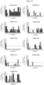
- Figure 1 shows IFN ⁇ production by a3a transduced T cells in response to native (wt) MAGE A3 peptide and each alanine-substituted peptide.
- Five of the alanine-substituted peptides resulted in a greater than 50 % decrease in IFN ⁇ production compared to native MAGE A3 peptide.
- the corresponding native residue at each of these five positions comprises the binding motif.
- the binding motif is defined as EXDPIXXXY, where X is any amino acid.
- ELISpot assays were carried out as described in section (1.1a), except effector cells were prepared from peripheral blood mononuclear cells (PBMCs) blood using standard procedures utilising Lymphoprep (Axis-Shields, cat# NYC-1114547) and Leucosep tubes (Greiner, cat# 227290), and plated at 25,000 cells per well. IMCmage1 was added to a final concentration of 0.1 nM per well. Controls were carried out in the absence of IMCmage1 (effectors + targets + peptide); and in the absence of peptide-pulsed target cells (effectors + IMCmage1).
- PBMCs peripheral blood mononuclear cells
- Figure 2 shows IFN ⁇ production by IMCmage1 redirected T cells in response to native (wt) MAGE A3 peptide and each alanine-substituted peptide.
- Five of the alanine-substituted peptides resulted in a greater than 50 % decrease in IFN ⁇ production compared to native MAGE A3.
- the corresponding native residue at each of these five positions comprises the binding motif.
- the binding motif is defined as EXDPIXXXY, where X is any amino acid.
- MAGE A3 peptide contains one native glycine residue; in this case the native glycine was considered nonessential for the purposes of defining the motif.
- Figure 3 shows IFN ⁇ production by IMCmage1 redirected T cells in response to native (wt) MAGE A3 peptide and each glycine-substituted peptide.
- Five of the glycine-substituted peptides resulted in a greater than 50 % decrease in IFN ⁇ production compared to native MAGE A3.
- the corresponding native residue at each of these five positions comprises the binding motif.
- the binding motif is defined as EXDPIXXXY, where X is any amino acid.
- the ScanProsite tool http://prosite.expasy.org/scanprosite was used to search all UniProtKB/Swiss-Prot (release 2012_10 of 31-Oct-12: 538259 entries) database sequences, for proteins which contain the motif identified above (entered as E-X-D-P-I-X-X-X-Y). No filters were used. Pattern options were set to allow at most 1 X sequence characters to match a conserved position in the pattern and the match mode was set to 'greedy, overlaps, no includes'.
- the search results also identified a number of motif-containing peptides from common human pathogens. Two peptides were selected from the list as examples for further testing; a nuclease protein present in three strains of Epstein Barr Virus (EBV), and a ribosomal maturation factor from Clostridium difficile, the amino acid sequences of these peptides are shown below.
- Protein accesion number
- Sequence EBV protein Q1HVE7) (P03217) (Q3KSR5)
- E F D P I Y P S Y C. difficile protein Q18BH3
- Activation of a3a T cells was determined by IFN ⁇ ELISpot assay using the same procedure as described in section 1.1a.
- HLA-A*01+ hepatocyte cells were used as targets and pulsed with 10 ⁇ M of each peptide.
- T cells were prepared from peripheral blood lymphocytes (PBLs) obtained from a healthy donor, and transduced with the a3a TCR. Non-transduced donor T cells were used as a negative control. T cell activation in response to cells pulsed with each of the motif-containing peptides was compared to activation by native MAGE A3 peptide pulsed cells.
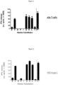
- FIG. 4 shows that cells pulsed with MAGE A3, MAGE A6, MAGE B18 and Titin peptides led to activation of a3a T cells.
- Peptide pulsed cells were prepared as described in 1.3a. Activation of IMCmage1 redirected T cells was determined by IFN ⁇ ELISpot assay using the same procedure as described in section 1.1b.
- FIG. 5 shows that cells pulsed with MAGE A3, MAGE A6, MAGE B18 and Titin peptides led to activation of IMCmage1 redirected T cells.
- Affinity was determined by surface plasmon resonance using a BIAcore 3000 instrument and reported in terms of an equilibrium dissociation constant (K D ).
- Soluble versions of the a3a and IMCmage1 TCRs were prepared using the method described in Boulter, et al., Protein Eng, 2003. 16: 707-711 .
- Biotinylated specific and control pMHC monomers were prepared as described in Garboczi, et al. Proc Natl Acad Sci U S A 1992. 89: 3429-3433 and O'Callaghan, et al., Anal Biochem 1999. 266: 9-15 , and immobilized on to a streptavidincoupled CM-5 sensor chips.
- Killing assays were carried out using the IncuCyte FLR-Platform (Essen Biosciences). Flat bottomed 96 well plates were used for assay.
- the assay medium was RPMI (w/o phenol red + 10% FCS +1% Pen/strep + 1% Glu).
- HLA-A*01+ hepatocyte cells were plated at 10,000 cells per well and incubated overnight to allow them to adhere.
- Peptides were made up at 10 ⁇ M final concentration and added at 25 ⁇ l (60 ⁇ M) per well.
- Effector T cells (prepared as described in 1.1b) were thawed from liquid N2, counted and plated at 100,000/well. IMCmage1 was added to a final concentration of 1 nM.
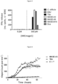
- Control measurements were carried out in the absence of IMCmage1 (effectors + targets). Images were taken every 2 h, over a three day period, and the number of apoptotic cells per mm 2 was quantified using the CellPlayer 96-well Kinetic Caspase 3/7 reagent and the IncuCyte FLR-Platform. The reagent is cleaved by activated Caspase 3/7 upon target cell apoptosis resulting in the release of the dye and green fluorescent staining of nuclear DNA.
- Figure 6 shows IMCmage1 redirected T cells kill cells pulsed with Titin peptide to a similar degree as native MAGE A3.
- iCell cardiomyocytes were obtained from Cellular Dynamics International. iCell cardiomyocytes are highly purified human cardiomyocytes derived from induced pluripotent stem (iPS) cells and are electrically active with typical mechanical characteristics of cardiac tissue. These cells are positive for Titin and negative for MAGE A3 as determined by standard RT-PCR methods.
- iPS induced pluripotent stem
- iCell cardiomyocytes were revived from liquid nitrogen and plated at 50,000 cells per well four days prior to the assay and treated as per the manufacturer's instructions.
- the iCell cardiomyocytes were virally transduced with HLA-A*01. EJM and colo205 (positive and negative controls respectively) were plated at 50,000 cells per well on the day of assay.
- iCell cardiomyocytes were washed once with R10 (RPMI + 10% FCS +1% Pen/strep + 1% Glu) and then incubated in R10 for the assay.
- a3a transduced T cells and corresponding non-transduced cells were taken from culture and plated at 50,000 cells per well.
- the cells were cultured for 24 h and then centrifuged at 800 xg for 4 min, 100 ul of supernatant was then removed and placed in a clean plate.
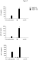
- the plates were stored at -70 °C until thawing and developing to measure multiple cytokine release (IFNy, GM-CSF and MIP-1 ⁇ ) by Luminex (25-plex kit), in accordance with manufacturer's instructions.
- Figure 7 shows a3a T cells release IFNy, GM-CSF and MIP-1 ⁇ in response to iCell cardiomyocytes.
- the BLAST search returned over 100 hits, including MAGE A3, MAGE A6, and MAGE B18, but not Titin.
- 15 sequences were selected for testing using peptide pulsed targets (HLA-A ⁇ 01+ hepatocyte cells) and IFN ⁇ ELISpot assays as described in Example 1 (section 1.1a and 1.1b).
- the peptide sequences are detailed in Figure 9 . None of these peptides was able to activate a3a T cells or IMCmage1 redirected T cells.
- HLA-A*01+ human cells from various tissues were screened for their ability to induce activation of a3a T cells and IMCmage1 redirected T cells. T cell activation was assessed by IFN ⁇ release using an ELISpot assay as described in Example 1 (section 1.1a and 1.1b). Multiple lots were used for most primary cell types. Cells were grown in 2D culture.
- Results showed no obvious reactivity for IMCmage1 and a3a transduced T cells. Since expression of MAGE family proteins is restricted to male germ line cells cross reactivity with MAGE A6 and MAGE B18 is not detected. However, despite Titin being expressed in cardiac and skeletal muscle, this experiment failed to detect cross reactivity with skeletal and muscle cells. Since titin expression is known to be upregulated in differentiated tissue ( Van der Loop, et al. (1996). J Muscle Res Cell Motil. 17:23-36 ) detecting cross reactivity required more specialised cell cultures (such as the iCells shown above).
- T cell activation was assessed using HLA-A*01+ hepatocyte cells pulsed with MAGE A3, human Titin or mouse Titin peptides. T cell activation was determined by IFN ⁇ ELISpot assay as described above. Synthetic peptides were obtained from Peptide Protein Research Limited, UK.
- Figure 10 shows IFN ⁇ release in response to HLA-A*01+ hepatocyte cells pulsed with MAGE A3 and human Titin; no IFN ⁇ was detected when cells were pulsed with the equivalent Titin peptide from mouse. Therefore, a mouse model would not have identified Titin as an off target peptide leading to unwanted side effects when administered to a patient.
- the target MHC-presented peptide used in this example is derived from human cancer testis antigen NY-ESO-1 and has the following amino acid sequence; SLLMWITQC.
- NY-ESO-1 peptide is presented on antigen presenting cells in the context of HLA-A*02.
- the binding peptide used in this example comprises a modified T cell receptor (TCR) fusion protein which has been engineered to possess enhanced affinity for NY-ESO-1 peptide, and is termed ImmTAC-NYESO in this example.
- TCR modified T cell receptor
- Variants of the native NY-ESO-1 peptide were obtained in which each amino acid position was sequentially replaced with alanine, as shown below (in each case the alanine substitution is underlined).
- Peptides were obtained from Peptide Protein Research Limited, UK.
- the native and alanine-substituted peptides were pulsed on to antigen presenting cells, and interferon ⁇ (IFN ⁇ ) production, as measured using the ELISpot assay, used as a read-out for T cell activation.
- Essential positions were defined by a greater than 50% reduction in T cell activity relative to the native peptide.
- ELISpot assays were carried as described in Example 1 section 1.1b except that HLA-A2+ T2 cells were used as target cells and effector PBMCs were plated at 40,000 cell per well. ImmTAC-NYESO was added to a final concentration of 0.1 nM per well.
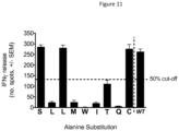
- Figure 11 shows IFN ⁇ production by ImmTAC-NYESO redirected T cells in response to native (wt) NY-ESO-1 peptide and each alanine-substituted peptide.
- Five of the alanine-substituted peptides resulted in a greater than 50 % decrease in IFN ⁇ production compared to native NY-ESO-1 peptide.
- the corresponding native residue at each of these five positions comprises the binding motif.
- the binding motif is defined as XLXMWITQX, where X is any amino acid.
- the ScanProsite tool http://prosite.expasy.org/scanprosite was used to search the UniProtKB/Swiss-Prot database (release date 13 th Nov 2013) for proteins which contain the motif identified above (entered as X L X M W I T Q X). The search was limited to human sequences.
- NY-ESO-1 accession no: P78358
- LAGE-1A accession no: O75638-2
- TCRs that bind to the NY-ESO-1 peptide are known to recognise cancer cells expressing LAGE-A1 ( McCormack et al, 2013, Cancer Immunol Immunother, 62 (4):773-85 ).
- Variants of the native NY-ESO peptide were obtained in which the amino acid residue at each position was sequentially replaced with all 19 alternative naturally-occurring amino acid, such that 171 peptides were prepared in total.
- the native and amino-acid substituted peptides were pulsed on to antigen presenting cells, and interferon ⁇ (IFN ⁇ ) production, as measured using the ELISpot assay, used as a read-out for T cell activation.
- Essential positions were defined by a greater than 50% reduction in T cell activity relative to the native peptide.
- ELISpot assays were carried as described in section 2.1a.
- Figure 12 shows IFN ⁇ production by ImmTAC-NYESO redirected T cells in response to native (wt) NY-ESO-1 peptide and each amino-acid-substituted peptide.
- the tolerated residues at each position of the peptide are shown below. For each position the first tolerated residue shown corresponds to the native (WT) residue at that position.
- the ScanProsite tool was used as described in section 2.2, to search for proteins containing the tolerated residues at the indicated positions (entered as [SVTYMFAGHNQKPRWLI]-[LI]-[LVAMICQNH]-[MQVTN]-W-[ITLMS]-[TSD]-[QG]-[CLVMSTGAI])
- Peptides from Plexin-D1 and protein 5B1 are tested in vitro as described in Example 1.3, to confirm recognition by ImmTAC-NYESO.
- the potential of these off-targets peptides to cause unwanted side effects in vivo is determined by, for example, reference to literature sources detailing expression of Plexin-D1 and protein 5B1 in normal tissue. Where expression in normal tissue is widespread, or in critical tissues, binding of ImmTAC-NYESO may be additionally be confirmed in vitro using cells which express the off target peptide.
- mouse model may not identify potential cross reactivity.
Description
- The present invention relates to a method for predicting whether an immunotherapeutic TCR, which binds to a target peptide that is presented in the context of major histocompatibility complex (MHC), and is for administration to a subject, will cross react with another peptide in the subject in vivo.
- MHC class I and class II are immunoglobulin superfamily proteins specialised for antigen presentation, with a polymorphic peptide binding site which enables them to present a diverse array of short peptide fragments at the surface of the antigen presenting cell. Peptides presented by MHC are derived from proteins which have been proteosomally processed within the cell. In humans, MHC molecules are known as human leukocyte antigens (HLA).
- A number of emerging immunotherapies rely on the administration to a subject to be treated of a binding peptide that binds a target peptide-MHC complex. The binding peptide may be an immune binding peptide such as, for example, an antibody or antigen binding fragment thereof or a T cell receptor or antigen binding fragment thereof. Such binding peptides bind to a binding sequence, comprising the amino acid sequence of the target MHC presented peptide. Often the binding sequence of the target peptide is known. There is a risk with such therapies that the binding peptide binds to peptides other than the target peptide (referred to herein as "off target peptides"), causing unwanted side effects. It is therefore desirable to identify whether such off target peptides exist. This allows binding peptides to be chosen and designed that do not bind to off target peptides and consequently have a far greater chance of not causing unwanted side effects.
- Reasons for unwanted side effects derived from off target specificities in adoptive T cell therapy are; mispairing of transduced TCR chains with endogenous chains, insertion mutagenesis associated with TCR transduction, or alloreactivity (Amos et al., Blood 2011, 118(3):499-509; Daniel-Meshulam et al., Front Immunol 2012, 3:186). To date, the way to prevent off target toxicity is to include apoptosis genes to destroy T cells if toxicity arises after administration to the patient (Restifo, et al., Not Rev Immunol 2012, 12(4): 269-81.)
- Thus a person skilled in the art would be motivated to provide mechanisms that deal with binding to off target peptides after administration of a binding peptide. It is therefore unlikely that a person skilled in the art would consider trying to identify off target peptides before the binding peptide is administered. In the highly unlikely event that the skilled person took this latter approach, one option would be to search protein sequence databases for peptides with similarity to the target MHC presented peptide. But, this often returns a large number of peptides, all of which would need to be tested and even then off-target peptides may not be identified. Lowering the stringency of the search parameters would further increase the number of potential epitopes that would have to be tested and again may still not reveal off target peptides. Alternatively, the skilled person may measure any immune response generated by the binding peptide in the presence of cells derived from normal tissue(s) (which preferably do not express the target peptide). However, this can be a difficult process, depending on the number and type of cells tested. Furthermore, primary cells cultured in vitro, may have a different protein expression profile compared to the same cell type in vivo. This may result in a false assessment of potential cross reactivity in vivo. Finally, the skilled person may use animal models to measure any immune response generated by the binding peptide. Because of the differences between human protein sequences and those of the animal, the absence of unwanted side effects in the animal may not translate to humans.
- Thus, these approaches do not accurately indicate, especially when administered to a subject, whether the binding peptide will indeed give rise to unwanted side effects derived from off target specificities, especially in an individualised or personalised setting. In short, any attempt heretofore to identify off target peptides before the binding peptide is administered had no reasonable expectation of success. Indeed, as is explained in more detail below, the inventors have found that such approaches will not necessarily identify off target peptides that cause an unwanted side effect. Although these approaches were performed on the a3a T cells described in the examples below, off-target activation of a3a T cells only become apparent when the binding peptide was were administered to patients (manuscript in preparation).
- In the prior art, Chinnasamy et al, 2011, J. Immunol, 186(2), 685-696 describes the isolation and development of a TCR that recognises a MAGE A3 peptide for the purpose of cancer immunotherapy. Maier et al., Eur. J. Immunol. 2000.30: 448-457 discloses an analysis of whether similarity between microbial and self-antigens can activate autoreactive lymphocytes.
- It is therefore desirable to provide an alternative method for predicting whether an immunotherapeutic TCR will or at least is likely to bind to an off target peptide, which addresses drawbacks of the prior art.
- The invention provides a method for predicting whether an immunotherapeutic TCR has the potential to cross react with another peptide in the subject in vivo to give rise to unwanted side effects derived from off target specificities in said subject, wherein the TCR binds to a target peptide presented by a Major Histocompatibility Complex (MHC) and is for administration to a subject;
- the method comprising:
- (a) identifying at least one binding motif in the target peptide to which the TCR binds;
- (b) searching for peptides that are present in the subject that comprise the at least one binding motif and that are not the target peptide; and
- (c) testing binding of any such peptide to the TCR in vitro,
- (d) wherein the presence of one or more such peptides that bind the TCR indicates that the TCR has the potential to cross react in vivo and cause unwanted side effects;
- the at least one binding motif being identified by:
- (i) creating a series of mutants of the target peptide, each mutant having the amino acid residue at one position in the binding sequence thereof that is involved in binding to the TCR substituted for alanine or glycine, such that over the series of mutants the amino acid residue in each position in the binding sequence is substituted for alanine or glycine; and
- testing each mutant in the series for its activity relative to the wild type target peptide,
- wherein an amino acid residue at a position within the binding sequence is identified as being part of the binding motif if the mutant in which the amino acid at that position is mutated to alanine or glycine has a 50% or greater loss of activity relative to the wild type target peptide, or
- (ii) creating a series of mutants, each mutant having the amino acid residue at one position in the binding sequence substituted for an alternative amino acid, such that over the series of mutants the amino acid residue in each position in the binding sequence is substituted for all alternative amino acids; and
- testing each mutant in the series for its activity relative to the wild type peptide,
- wherein amino acid substitutions which result in a 50% or greater loss of activity relative to the wild type target peptide are considered to be non-tolerated amino acids and not part of the binding motif and amino acids substitutions which do not result in a 50% or greater loss of activity relative to the wild type target peptide are considered as part of the binding motif.
- Preferred features of the invention are set out in the dependent claims herein.
- Methods of the invention may further comprise, where an amino acid residue at a position in the binding sequence is not identified as being part of the binding motif, substituting this position with at least one additional amino acid and testing for activity relative to the wild type peptide,
wherein amino acid substitutions which result in a 50% or greater loss of activity relative to the wild type target peptide are considered to be non-tolerated amino acids and not part of the binding motif and/or amino acid substitutions which do not result in a 50% or greater loss of activity relative to the wild type target peptide are considered as part of the binding motif. - The activity that is tested may be the ability of the mutant to bind to the immunotherapeutic TCR.
- The search may be carried out for peptides that are expressed in selected tissue(s) and/or accessible to the immunotherapeutic TCR.
- Methods of treatment are not included within the scope of the invention. When there is the absence of one or more of such peptides and hence the indication that the immunotherapeutic TCR has the potential not to cross react in vivo, a treatment effective amount of the immunotherapeutic TCR may be administered to the subject, and/or
when there is the presence of one or more such peptides and hence an indication that the immunotherapeutic TCR has the potential to cross react in vivo, the potential for each peptide to cause off target side effects in vivo may be identified, and where necessary, preparing an alternative immunotherapeutic TCR having the absence of one or more of such peptides and hence the indication that the alternative immunotherapeutic TCR has the potential not to cross react in vivo, and a treatment effective amount of the alternative immunotherapeutic TCR may be administered to the subject. - The treatment effective amount of the immunotherapeutic TCR is that amount typically given to the suitable mammalian patient. Thus, the invention comprehends testing known immunotherapeutic TCR treatments for whether such treatment immunotherapeutic TCRs will bind to peptides other than the target peptide. The treatment effective amount of the alternative immunotherapeutic TCR is within the ambit of the skilled person from this disclosure and the knowledge in the art. For example, the treatment effective amount can be determined by comparing the binding properties and/or ability to elicit the desired treatment biological response of the immunotherapeutic TCR with the binding properties and/or ability to elicit the desired treatment biological response of the alternative immunotherapeutic TCR, and adjusting the dosage of the immunotherapeutic TCR based on the difference in binding and/or ability to elicit the biological response of the alternative immunotherapeutic TCR in comparison with the binding and/or ability to elicit the biological response of the immunotherapeutic TCR peptide. Advantageously, the alternative immunotherapeutic TCR has binding properties and/or biological response eliciting properties akin to that of the immunotherapeutic TCR , and hence its dosage or amount to be administered is analogous to that of the immunotherapeutic TCR .
- Further disclosed but not within the scope of the claimed invention is an improvement in a method of treating a human or animal mammalian subject in need thereof comprising administering a binding peptide. This improvement comprises:
predicting whether the binding peptide, which binds to a target peptide presented by a Major Histocompatibility Complex (MHC) in the subject to thereby provide said treatment and is for administration to the subject, has the potential to cross react with another peptide in the subject in vivo, comprising: - identifying at least one binding motif in the target peptide to which the binding peptide binds; and
- searching for peptides that are present in the subject that comprise the at least one binding motif and that are not the target peptide,
- wherein the presence of one or more such peptides indicates that the binding peptide has the potential to cross react in vivo, and the absence of one or more of such peptides indicates that the binding peptide has the potential to not to react in vivo; and,
- when there is the absence of one or more of such peptides and hence the indication that the binding peptide has the potential will not to react in vivo, and/or when binding to the off target peptide is not expected to cause unwanted side effects in vivo, administering a treatment effective amount of the binding peptide to the subject.
- This method can include, when there is the presence of one or more such peptides and hence an indication that the binding peptide has the potential to cross react in vivo, preparing an alternative binding peptide having the absence of one or more of such peptides and hence the indication that the alternative binding peptide has the potential not to cross react in vivo, and administering a treatment effective amount of the alternative binding peptide to the subject.
- Moreover, also disclosed herein is the preparation of pharmaceutical compositions comprising a binding peptide. In this respect, the invention provides a method for preparing a pharmaceutical composition comprising a binding peptide, or an improvement to methods for preparing a pharmaceutical composition comprising a binding peptide, comprising:
predicting whether the binding peptide, which binds to a target peptide presented by a Major Histocompatibility Complex (MHC) in the subject to thereby provide said treatment and is for administration to the subject, has the potential to cross react with another peptide in the subject in vivo, comprising: - identifying at least one binding motif in the target peptide to which the binding peptide binds; and
- searching for peptides that are present in the subject that comprise the at least one binding motif and that are not the target peptide,
- wherein the presence of one or more such peptides indicates that the binding peptide has the potential to cross react in vivo, and the absence of one or more of such peptides indicates that the binding peptide has the potential not to cross react in vivo.
- These and other embodiments are disclosed or are obvious from and encompassed by, the following Detailed Description.
- The following Detailed Description, given by way of example, but not intended to limit the invention solely to the specific embodiments described, may best be understood in conjunction with the accompanying drawings, wherein:
-
Figure 1 shows IFNγ production by a3a T cells in response to peptide-pulsed cells presenting either MAGE A3 peptide (denoted WT), or alanine-substituted peptides. -
Figure 2 shows IFNγ production by IMCmage1 redirected T cells in response to peptide-pulsed cells presenting either MAGE A3 peptide (denoted WT), or alanine-substituted peptides. -
Figure 3 shows IFNγ production by IMCmage1 redirected T cells in response to peptide-pulsed cells presenting either MAGE A3 peptide (denoted WT), or glycine-substituted peptides. -
Figure 4 shows IFNγ production by a3a T cells in response to cells pulsed with peptides identified in the motif search. Non-transduced T cells (ntd) were used as a negative control. -
Figure 5 shows IFNγ production by IMCmage1 redirected T cells in response to cells pulsed with peptides identified in the motif search. Experiments carried out in the absence of IMCmage1 were used as a negative control. -
Figure 6 shows IMCmage1 redirected T cell killing of cells pulsed with either MAGE A3 or Titin peptides. Cell killing was determined using the IncuCyte platform. A negative control was carried out using in the absence of IMCmage1 (E + T). -
Figure 7 shows the cytokine profile a3a T cells in the presence of iCells, as determined by the Luminex assay. EJM and colo205 cells were used as a positive and negative control respectively. -
Figure 8 shows phase contrast images, obtained using the IncuCyte platform, of Titin positive iCells being killed by a3a T cells. The images were taken after 24 hours incubation. Non-transduced T cells (ntd) were used as a negative control. -
Figure 9 shows details of 15 peptides identified in a BLAST search carried out to find human peptides with a similar sequence to MAGE A3 peptide. Activation of a3a T cells or IMCmage1 redirected T cells, as determined by IFNγ ELISpot, is indicated. -
Figure 10 shows activation of a3a T cells, as determined by IFNγ ELISpot, in response to peptide-pulsed cells presenting either MAGE A3, human Titin, or mouse Titin. -
Figure 11 shows IFNγ production by ImmTAC-NYESO redirected T cells in response to peptide-pulsed cells presenting either NY-ESO-1 peptide (denoted WT), or alanine-substituted peptides. -
Figure 12 shows IFNγ production by ImmTAC-NYESO redirected T cells in response to native (WT) NY-ESO-1 peptide and each amino-acid-substituted peptide. - The inventors have found that, by identifying the binding motif in the target peptide to which the immunotherapeutic TCR binds, rather than the known binding sequence, off target peptides - that have the potential to, or will, cross react in vivo - can be identified far more accurately. If off target peptides are found, this allows immunotherapeutic TCRs to be altered so that they do not bind to these off target peptides.
- The binding motif(s) may be identified by creating a series of mutants of the target peptide, each mutant having the amino acid residue at one position in the sequence thereof that is involved in binding to the immunotherapeutic TCR (the "binding sequence") substituted for an alanine or glycine, such that over the series of mutants the amino acid residue in each position in the binding sequence is substituted for alanine or glycine. Each mutant in the series is then tested for its activity relative to the wild type target peptide. An amino acid residue at a position within the binding sequence may be identified as being part of the binding motif if the mutant in which the amino acid at that position is mutated to alanine or glycine has a 50, 55, 60, 65, 70, 75, 80, 85, 90% or greater loss of activity relative to the wild type target peptide. This may result in a binding motif comprising amino acid(s) at one or a plurality of positions (such as 2, 3, 4, 5, 5, 6, 7, 8, 9, 10 or more) within the binding sequence being identified.
- Where an amino acid residue at a position in the binding sequence is not identified as being part of the binding motif (using the technique described above for example), this position may be further substituted with at least one additional amino acid and tested for activity relative to the wild type peptide. Amino acid substitutions which result in a 50, 55, 60, 65, 70, 75, 80, 85, 90% or greater loss of activity relative to the wild type target peptide, are considered to be non-tolerated amino acids and/or not part of the binding motif. Conversely, amino acid substitutions which do not result in a 50% or greater loss of activity relative to the wild type target peptide may be considered as part of the binding motif. This may result in a binding motif as defined above and additionally in which one or a plurality of positions are represented by more than one but not all amino acids.
- Alternatively, a series of mutants is made in which the amino acid residue at each position of the binding sequence is mutated to all alternative amino acids (relative to the wild type amino acid). For a binding sequence of nine amino acids, this would mean 171 peptides. Each mutant in the series is tested for activity relative to the wild type peptide. Amino acid substitutions which result in a 50, 55, 60, 65, 70, 75, 80, 85, 90% or greater loss of activity, are considered to be non-tolerated amino acids and/or not part of the binding motif. Conversely, amino acid substitutions which do not result in a 50% or greater loss of activity relative to the wild type target peptide may be considered as part of the binding motif. This may result in a binding motif in which each position is represented by at least one but not all amino acids.
- The activity that is tested is the ability of the mutant to bind to the immunotherapeutic TCR (this can be measured using Surface Plasmon Resonance for example). Also disclosed herein is testing the ability of the mutant to elicit the biological response caused by binding to the binding peptide. The biological response may be, for example, activation of immune system cells such as T cells, measured by cytokine production or destruction of the target cell; activation of an enzyme, measured by accumulation of product or disappearance of substrate; or activation of a signalling cascade (measured by monitoring protein phosphorylation, or changes in gene expression and protein production).
- In the method of the invention according to claim 1(i), the alternative amino acid substituted into the mutants is alanine or glycine. Such techniques, known as "amino acid scanning", are known in the art and have been used to determine a binding motif. See Wells, Methods Enzymol 1991; 202: 390-411 for example. A similar approach is also known for detecting other specificities of a T cell receptor (for example Udyavar et al. J Immunol 2009; 182(7):4439-47). However, such amino acid scanning has not been used to identify off-target peptides which may cause undesirable side effects.
- Once the binding motif has been identified, protein databases may be searched for proteins which contain the binding motif. Suitable protein databases include but are not limited to UniProtKB/Swiss-Prot (http://www.uniprot.org/), Protein Information Resource (PIR) (http://pir.georgetown.edu/pirwww/index.shtml), and/or Reference Sequence (RefSeq) (www.ncbi.nlm.nih.gov/RefSeq).
- Searching for a peptide motif may be carried out using any one of a number of tools, which may be found on bioinformatics resource sites such as ExPASY (http://www.expasy.org/). For example, the search tool ScanProsite identifies user-defined motifs in all protein sequences in the UniProtKB/Swiss-Prot Protein Knowledgebase (De Castro et al. Nucleic Acids Res. 2006 ).
- It is preferred that peptides containing the exact binding motif, i.e. with 100% identity to the binding motif, are identified. However, proteins containing motifs that have less than 100% identity to the binding motif may be identified. For the purposes of searching, the binding motif may be modified to include ambiguity at certain positions, for example with amino acids which have similar properties (e.g. leucine/ isoleucine, etc), or where it is already known in the literature that a particular amino acid is tolerated at a certain position (such as HLA anchor residues).
- The search may be carried out for peptides that are of human origin or of organisms which are commonly present in humans, such as viral or bacterial pathogens, or commensal bacteria. However, where the method of the present invention is applied to non-human animals, such as non-human mammals, the search may be carried out for peptides that are of the relevant non-human animal origin or of organisms which are commonly present in such non-human animals. Additionally or alternatively, the search may be carried out for peptides that are expressed in selected tissue(s) and/or accessible to the immunotherapeutic TCR. Such information may be obtained from the literature.
- Peptides identified in the search as comprising the at least one binding motif may be confirmed as an off target peptide and cross reacting with the immunotherapeutic TCR by determining the ability of the identified peptide to bind to the immunotherapeutic TCR (for example using Surface Plasmon Resonance), or assessing the biological response generated by binding of the immunotherapeutic TCR to the identified peptide. The biological response may be for example, activation of immune system cells such as T cells, measured by cytokine production or destruction of the target cell; activation of an enzyme, measured by accumulation of product or disappearance of substrate; or, activation of a signalling cascade (measured by monitoring protein phosphorylation, or changes in gene expression and protein production).
- Once one or more off target peptides have been identified, the potential of the (or each) off target peptide to cause unwanted side effects when bound by the immunotherapeutic TCR e may be determined. This may include searching literature sources to determine expression of the off target peptide in normal tissue. For example, where expression of the off target peptide in normal tissue is non-existent or limited (for example with cancer testis antigens), the immunotherapeutic TCR may be considered suitable for administration in vivo. In cases where expression of the off target peptide in normal tissue is widespread or is in critical tissues, such as heart cells, binding may optionally be additionally confirmed in vitro using cells which express the off target peptide. In some situations the immunotherapeutic TCR may not be administered in vivo because of the cross reaction that this can cause. The immunotherapeutic TCR may be redesigned so that there is no longer any cross reactivity to the off target peptide(s), while maintaining binding, preferably with high affinity, to the target peptide. For example, T cell receptors can be redesigned by mutagenesis using the methods described in
WO 03/020763 - If no off target peptides are found, or if the binding to the off target peptides is not expected to cause unwanted side effects (for example by virtue of the off target peptide being expressed in limited or non-critical tissues), the immunotherapeutic TCR may be used in a method of preventing or treating a disease or condition which is ameliorated by administration of the immunotherapeutic TCR. Methods of treatment (which do not fall within the scope of the invention) include but are not limited to immunotherapies; for example, administration to a patient of modified T cells (adoptive therapy), such as those transduced with affinity enhanced T cell receptors or chimeric antibody receptors; administration of monoclonal antibodies or monoclonal antibody fragments, especially TCR-like antibodies; administration of novel bi-specific immunotherapeutic agents such as ImmTACs (Immune mobilising TCRs against cancer) (Liddy, et al. (2012) Not Med 18: 980-987) or BiTEs (Bispecific T cell engaging antibodies) (Baeuerle, et al. (2009). Curr Opin Mol Ther 11(1): 22-30).
- Such treatments may be provided in a pharmaceutical composition (which does not fall within the scope of the invention) together with one or more pharmaceutically acceptable carriers or excipients. Therapeutic TCRs, or cells, will usually be supplied as part of a sterile, pharmaceutical composition which will normally include a pharmaceutically acceptable carrier. This pharmaceutical composition may be in any suitable form, (depending upon the desired method of administering it to a patient). It may be provided in unit dosage form, will generally be provided in a sealed container and may be provided as part of a kit. Such a kit would normally (although not necessarily) include instructions for use. It may include a plurality of said unit dosage forms.
- The pharmaceutical composition may be adapted for administration by any appropriate route, such as a parenteral (including subcutaneous, intramuscular, or intravenous) route. Such compositions may be prepared by any method known in the art of pharmacy, for example by mixing the active ingredient with the carrier(s) or excipient(s) under sterile conditions.
- Dosages of the substances disclosed herein can vary between wide limits, depending upon the disease or disorder to be treated, the age and condition of the individual to be treated, etc; for example, a suitable dose range for an ImmTAC reagent may be between 25 ng/kg and 50 µg/kg. A physician will ultimately determine appropriate dosages to be used.
- The term T cell receptors ("TCRs") includes antigen binding fragments of T cell receptors. As is described in
WO 99/60120 - The TCR may be in soluble form (e.g. having no transmembrane or cytoplasmic domains), for example a monoclonal TCR as described in
WO03/020763 WO2004/033685 . For stability, soluble TCRs preferably have an introduced disulphide bond between residues of the respective constant domains, as described, for example, inWO 03/020763 - Also disclosed herein but not encompassed by the present invention include antibodies, such as TCR like antibodies, which have been engineered to bind to MHC presented peptides (for example see, Sergeeva, A., G. et al. (2011). Blood 117(16): 4262-72 and/or Dahan, R., and Y. Reiter. 2012. Expert Rev Mol Med. 14:e6. The term "antibody" as used herein refers to immunoglobulin molecules and immunologically active portions of immunoglobulin molecules, i.e., molecules that contain an antigen binding site that specifically binds an antigen, whether natural or partly or wholly synthetically produced. The term also covers any polypeptide or protein having a binding domain which is, or is homologous to, an antibody binding domain. Examples of antibodies are the immunoglobulin isotypes (e.g., IgG, IgE, IgM, IgD and IgA) and their isotypic subclasses; fragments which comprise an antigen binding domain such as Fab, scFv, Fv, dAb, Fd; and diabodies. Antibodies may be polyclonal or monoclonal. A monoclonal antibody may be referred to herein as "mab".
- It is possible to take monoclonal and other antibodies and use techniques of recombinant DNA technology to produce other antibodies or chimeric molecules which retain the specificity of the original antibody. Such techniques may involve introducing DNA encoding the immunoglobulin variable region, or the complementary determining regions (CDRs), of an antibody to the constant regions, or constant regions plus framework regions, of a different immunoglobulin. See, for instance,
EP-A-184187 GB 2188638A EP-A-239400 - As antibodies can be modified in a number of ways, the term "antibody" should be construed as covering antibody fragments, derivatives, functional equivalents and homologues of antibodies, humanised antibodies, including any polypeptide comprising an immunoglobulin binding domain, whether natural or wholly or partially synthetic. Chimeric molecules comprising an immunoglobulin binding domain, or equivalent, fused to another polypeptide are therefore included. Cloning and expression of chimeric antibodies are described in
EP-A-0120694 andEP-A-0125023 . A humanised antibody may be a modified antibody having the variable regions of a non-human, e.g. murine, antibody and the constant region of a human antibody. Methods for making humanised antibodies are described in, for example,US Patent No. 5225539 - It has been shown that fragments of a whole antibody can perform the function of binding antigens. Examples of binding fragments are (i) the Fab fragment consisting of VL, VH, CL and CH1 domains; (ii) the Fd fragment consisting of the VH and CH1 domains; (iii) the Fv fragment consisting of the VL and VH domains of a single antibody; (iv) the dAb fragment (Ward, E.S. et al., Nature 341:544-546 (1989)) which consists of a VH domain; (v) isolated CDR regions; (vi) F(ab')2 fragments, a bivalent fragment comprising two linked Fab fragments (vii) single chain Fv molecules (scFv), wherein a VH domain and a VL domain are linked by a peptide linker which allows the two domains to associate to form an antigen binding site (Bird et al., Science 242:423-426 (1988); Huston et al., PNAS USA 85:5879-5883 (1988)); (viii) bispecific single chain Fv dimers (
PCT/US92/09965 WO94/13804 WO94/13804 WO94/13804 - Also disclosed herein but not encompassed within the present invention are binding peptides that bind to MHC presented peptides and are based on engineered protein scaffolds. Protein scaffolds are derived from stable, soluble, natural protein structures which have been modified to provide a binding site for a target molecule of interest. Examples of engineered protein scaffolds include, but are not limited to, affibodies, which are based on the Z-domain of staphylococcal protein A that provides a binding interface on two of its a-helices (Nygren, P. A. (2008). FEBS J 275(11): 2668-76); anticalins, derived from lipocalins, that incorporate binding sites for small ligands at the open end of a beta-barrel fold (Skerra, A. (2008) FEBS J 275(11): 2677-83), nanobodies, and DARPins. Engineered protein scaffolds are typically targeted to bind the same antigenic proteins as antibodies, and are potential therapeutic agents. They may act as inhibitors or antagonists, or as delivery vehicles to target molecules, such as toxins, to a specific tissue in vivo (Gebauer, M. and A. Skerra (2009). Curr Opin Chem Biol 13(3): 245-55). Short peptides may also be used to bind a target protein. Phylomers are natural structured peptides derived from bacterial genomes. Such peptides represent a diverse array of protein structural folds and can be used to inhibit/disrupt protein-protein interactions in vivo (Watt, P. M. (2006). Nat Biotechnol 24(2): 177-83)].
- Further disclosed but not within the scope of the claimed invention are methods that can be practiced with such binding peptides as these monoclonal and bispecific antibody treatments, and hence the skilled person readily knows the formulation and dose and means to administer if there is no cross reactivity detected using the instant invention, and from the dosages of these known binding peptides, can readily determine, using the herein disclosure and the knowledge in the art, the formulation, and dose and means to administer alternative binding peptides.
- The present invention will now be described with reference to the following non-limiting examples.
- The target MHC presented peptide used in this example is derived from human cancer testis antigen MAGE A3 and has the following amino acid sequence; EVDPIGHLY. MAGE A3 peptide is presented on antigen presenting cells in the context of HLA-A*01.
- The binding peptide used in this example comprises a modified T cell receptor (TCR) which has been engineered to possess enhanced affinity for MAGE A3 peptide. Methods to produce affinity enhanced TCRs are known in the art (for example, phage display
WO 03/020763 WO2012/013913 . Two versions of the modified MAGE A3 TCR are used. A moderately affinity enhanced version, expressed by transduced T cells (termed a3a T cells) as described inWO2012/013913 , and a high affinity version, produced as a soluble protein fused to a T cell activating anti-CD3 fragment (termed IMCmage1), according to the method ofWO2010/133828 . - Variants of the native MAGE A3 peptide were obtained in which each amino acid position was sequentially replaced with alanine, as shown below (in each case the alanine substitution is underlined). Peptides were obtained from Peptide Protein Research Limited, U K.
- A V D P I G H L Y
- E A D P I G H L Y
- E V A P I G H L Y
- E V D A I G H L Y
- E V D P A G H L Y
- E V D P I A H L Y
- E V D P I G A L Y
- E V D P I G H A Y
- E V D P I G H L A
- The native and alanine-substituted peptides were pulsed on to antigen presenting cells, and interferon γ (IFNγ) production, as measured using the ELISpot assay, used as a read-out for T cell activation. Essential positions were defined by a greater than 50% reduction in T cell activity relative to the native peptide.
- ELISpot assays were carried out according to the manufacturer's instructions (BD BioSciences). HLA-A1+ hepatocyte cells were used as target cells and pulsed with 10 µM of each peptide. Target cells were counted and plated at 50,000 cells per well in 50 µl assay buffer (10% FCS, 88
% RPMI 1640, 1% glutamine and 1% penicillin/streptomycin). Effector T cells used in this method were a 1:1 mix of CD4+ and CD8+ T cells (obtained by negative selection (using the CD4 and CD8 Negative Isolation Kits, Dynal) from peripheral blood lymphocytes (PBL) obtained from a healthy donor). Cells were stimulated with anti CD3/CD28 coated beads (T cell expander, Invitrogen), transduced with lentivirus carrying the gene encoding a3a T cell receptor, and expanded in assay media containing 50 U/ml IL-2 until between 10 and 13 days post transduction. Effector T cells were plated at 15,000 cells per well. Plates were incubated overnight at 37 °C/5% CO2 and quantified, after development, using an automated ELISpot reader (Immunospot Series 5 Analyzer, Cellular Technology Ltd. -
Figure 1 shows IFNγ production by a3a transduced T cells in response to native (wt) MAGE A3 peptide and each alanine-substituted peptide. Five of the alanine-substituted peptides resulted in a greater than 50 % decrease in IFNγ production compared to native MAGE A3 peptide. The corresponding native residue at each of these five positions comprises the binding motif. In this case the binding motif is defined as EXDPIXXXY, where X is any amino acid. - ELISpot assays were carried out as described in section (1.1a), except effector cells were prepared from peripheral blood mononuclear cells (PBMCs) blood using standard procedures utilising Lymphoprep (Axis-Shields, cat# NYC-1114547) and Leucosep tubes (Greiner, cat# 227290), and plated at 25,000 cells per well. IMCmage1 was added to a final concentration of 0.1 nM per well. Controls were carried out in the absence of IMCmage1 (effectors + targets + peptide); and in the absence of peptide-pulsed target cells (effectors + IMCmage1).
-
Figure 2 shows IFNγ production by IMCmage1 redirected T cells in response to native (wt) MAGE A3 peptide and each alanine-substituted peptide. Five of the alanine-substituted peptides resulted in a greater than 50 % decrease in IFNγ production compared to native MAGE A3. The corresponding native residue at each of these five positions comprises the binding motif. In this case the binding motif is defined as EXDPIXXXY, where X is any amino acid. - The same procedure was followed as detailed in section (1.1b) except the HLA-A*01+ hepatocyte target cells were pulsed with glycine-substituted peptides. MAGE A3 peptide contains one native glycine residue; in this case the native glycine was considered nonessential for the purposes of defining the motif.
-
Figure 3 shows IFNγ production by IMCmage1 redirected T cells in response to native (wt) MAGE A3 peptide and each glycine-substituted peptide. Five of the glycine-substituted peptides resulted in a greater than 50 % decrease in IFNγ production compared to native MAGE A3. The corresponding native residue at each of these five positions comprises the binding motif. In this case the binding motif is defined as EXDPIXXXY, where X is any amino acid. - The ScanProsite tool (http://prosite.expasy.org/scanprosite) was used to search all UniProtKB/Swiss-Prot (release 2012_10 of 31-Oct-12: 538259 entries) database sequences, for proteins which contain the motif identified above (entered as E-X-D-P-I-X-X-X-Y). No filters were used. Pattern options were set to allow at most 1 X sequence characters to match a conserved position in the pattern and the match mode was set to 'greedy, overlaps, no includes'.
- Five unique human proteins were identified: native MAGE A3, MAGE family members A6 and B18, the muscle protein Titin, and a protein known as Dos. The amino acid sequences of the motif-containing peptides are shown in the table below (residues which comprise the motif are underlined).
Protein (Accession number) Sequence MAGE A3 (P43357) E V D P I G H L Y MAGE A6 (P43360) E V D P I G H V Y MAGE B18 (Q96M61) E V D P I R H Y Y Titin (Q8WZ42) E S D P I V A Q Y Dos (Q8N350) E P D P I L D N Y - The search results also identified a number of motif-containing peptides from common human pathogens. Two peptides were selected from the list as examples for further testing; a nuclease protein present in three strains of Epstein Barr Virus (EBV), and a ribosomal maturation factor from Clostridium difficile, the amino acid sequences of these peptides are shown below.
Protein (Accession number) Sequence EBV protein (Q1HVE7) (P03217) (Q3KSR5) E F D P I Y P S Y C. difficile protein (Q18BH3) E K D P I K E N Y - Potential off target peptides, identified above, were tested for their ability to cross react with a3a T cells and IMCmage1 redirected T cells using an IFNγ ELISpot assay.
- All peptides were produced synthetically by Peptide Protein Research Limited, UK. Activation of a3a T cells was determined by IFNγ ELISpot assay using the same procedure as described in section 1.1a. HLA-A*01+ hepatocyte cells were used as targets and pulsed with 10 µM of each peptide. T cells were prepared from peripheral blood lymphocytes (PBLs) obtained from a healthy donor, and transduced with the a3a TCR. Non-transduced donor T cells were used as a negative control. T cell activation in response to cells pulsed with each of the motif-containing peptides was compared to activation by native MAGE A3 peptide pulsed cells.
-
Figure 4 shows that cells pulsed with MAGE A3, MAGE A6, MAGE B18 and Titin peptides led to activation of a3a T cells. Peptide Dos as well as peptides from EBV and C. difficile did not induce T cell activation. - Peptide pulsed cells were prepared as described in 1.3a. Activation of IMCmage1 redirected T cells was determined by IFNγ ELISpot assay using the same procedure as described in section 1.1b.
-
Figure 5 shows that cells pulsed with MAGE A3, MAGE A6, MAGE B18 and Titin peptides led to activation of IMCmage1 redirected T cells. Peptide Dos as well as peptides from EBV and C. difficile did not induce T cell activation. - The experiments described in 1.3a and 1.3b confirm that there are three off target peptides which are bound by a3a T cells and IMCmage1 redirected T cells; MAGE A6, MAGE B18, and Titin. Of the three only Titin is relevant in a clinical context. Normal tissue expression of MAGE family proteins is restricted to male germ-line cells, whereas Titin is expressed in cardiac and skeletal muscle (Uniprot Protein Knowledgebase (http://www.uniprot.org/uniprot).
- Affinity was determined by surface plasmon resonance using a
BIAcore 3000 instrument and reported in terms of an equilibrium dissociation constant (KD). Soluble versions of the a3a and IMCmage1 TCRs were prepared using the method described in Boulter, et al., Protein Eng, 2003. 16: 707-711. Biotinylated specific and control pMHC monomers were prepared as described in Garboczi, et al. Proc Natl Acad Sci U S A 1992. 89: 3429-3433 and O'Callaghan, et al., Anal Biochem 1999. 266: 9-15, and immobilized on to a streptavidincoupled CM-5 sensor chips. All measurements were performed at 25°C in PBS buffer (Sigma) supplemented with 0.005% Tween (Sigma) at a constant flow rate. To measure affinity, serial dilutions of the soluble TCRs were flowed over the immobilized pMHCs and the response values at equilibrium were determined for each concentration. Equilibrium dissociation constants (KD) were determined by plotting the specific equilibrium binding against protein concentration followed by a least squares fit to the Langmuir binding equation, assuming a 1:1 interaction. - The results summarised in the table below confirm Titin peptide binds to soluble versions of the TCRs used in a3a T cells and IMCmage1. The affinity (KD) is shown in the table below:-
MAGE-A3 (EVDPIGHLY) MAGE-A6 (EVDPIGHVY) MAGE-B18 (EVDPIRHYY) Titin (ESDPIVAQY) IMCmage1 4.5 nM 4.2 nM 182 nM 86 nM MAGE a3a ~2 µM ~2 µM ~100 µM ~100 µM - Killing assays were carried out using the IncuCyte FLR-Platform (Essen Biosciences). Flat bottomed 96 well plates were used for assay. The assay medium was RPMI (w/o phenol red + 10% FCS +1% Pen/strep + 1% Glu). HLA-A*01+ hepatocyte cells were plated at 10,000 cells per well and incubated overnight to allow them to adhere. Peptides were made up at 10 µM final concentration and added at 25 µl (60 µM) per well. Effector T cells (prepared as described in 1.1b) were thawed from liquid N2, counted and plated at 100,000/well. IMCmage1 was added to a final concentration of 1 nM. Control measurements were carried out in the absence of IMCmage1 (effectors + targets). Images were taken every 2 h, over a three day period, and the number of apoptotic cells per mm2 was quantified using the CellPlayer 96-well
Kinetic Caspase 3/7 reagent and the IncuCyte FLR-Platform. The reagent is cleaved by activatedCaspase 3/7 upon target cell apoptosis resulting in the release of the dye and green fluorescent staining of nuclear DNA. -
Figure 6 shows IMCmage1 redirected T cells kill cells pulsed with Titin peptide to a similar degree as native MAGE A3. - iCell cardiomyocytes were obtained from Cellular Dynamics International. iCell cardiomyocytes are highly purified human cardiomyocytes derived from induced pluripotent stem (iPS) cells and are electrically active with typical mechanical characteristics of cardiac tissue. These cells are positive for Titin and negative for MAGE A3 as determined by standard RT-PCR methods.
- iCell cardiomyocytes were revived from liquid nitrogen and plated at 50,000 cells per well four days prior to the assay and treated as per the manufacturer's instructions. The iCell cardiomyocytes were virally transduced with HLA-A*01. EJM and colo205 (positive and negative controls respectively) were plated at 50,000 cells per well on the day of assay. iCell cardiomyocytes were washed once with R10 (RPMI + 10% FCS +1% Pen/strep + 1% Glu) and then incubated in R10 for the assay. a3a transduced T cells and corresponding non-transduced cells were taken from culture and plated at 50,000 cells per well. The cells were cultured for 24 h and then centrifuged at 800 xg for 4 min, 100 ul of supernatant was then removed and placed in a clean plate. The plates were stored at -70 °C until thawing and developing to measure multiple cytokine release (IFNy, GM-CSF and MIP-1β) by Luminex (25-plex kit), in accordance with manufacturer's instructions.
-
Figure 7 shows a3a T cells release IFNy, GM-CSF and MIP-1β in response to iCell cardiomyocytes. - Phase contrast images were obtained after 24 h on the the IncuCyte FLR-Platform (Essen Biosciences), using the same experimental set-up as that described above (1.3e).
Figure 8 shows the resulting images obtained. In the presence of a3a T cells, iCells are destroyed. - The following experiments show how the methods currently available in art do not, or would not, identify Titin as an off target peptide. These methods were not able to predict unwanted side effects when a3a T cells were administered to patients (manuscript in preparation).
- The RefSeq database was searched for human peptides with a similar sequence to MAGE A3, using protein BLAST (Basic Local Alignment Search Tool) version 2.2.27 (Altschul, et al, Nucleic Acids Res. 1997 25:3389-3402, http://blast.ncbi.nlm.nih.gov/Blast.cgi?CMD=Web&PAGE_TYPE=BlastHome). Algorithm parameters were adjusted automatically for a short protein. (General parameters; expect threshold = 200000, word size = 2: Scoring parameters; matrix = PAM30, gap costs =
existence 9extension 1; no compositional adjustments.) - The BLAST search returned over 100 hits, including MAGE A3, MAGE A6, and MAGE B18, but not Titin. 15 sequences were selected for testing using peptide pulsed targets (HLA-A∗01+ hepatocyte cells) and IFNγ ELISpot assays as described in Example 1 (section 1.1a and 1.1b). The peptide sequences are detailed in
Figure 9 . None of these peptides was able to activate a3a T cells or IMCmage1 redirected T cells. - HLA-A*01+ human cells from various tissues were screened for their ability to induce activation of a3a T cells and IMCmage1 redirected T cells. T cell activation was assessed by IFNγ release using an ELISpot assay as described in Example 1 (section 1.1a and 1.1b). Multiple lots were used for most primary cell types. Cells were grown in 2D culture.
- Astrocytes
- Cardiac Myocytes
- Non-pigmented ciliary epithelial cells
- Bronchial smooth muscle cells
- Dermal fibroblasts
- Dermal microvascular endothelial cells
- Hepatocytes
- Pulmonary Fibroblasts
- Renal Epithelial Cells
- Melanocytes
- Skeletal Muscle
- Pulmonary microvascular endothelial cells
- Results showed no obvious reactivity for IMCmage1 and a3a transduced T cells. Since expression of MAGE family proteins is restricted to male germ line cells cross reactivity with MAGE A6 and MAGE B18 is not detected. However, despite Titin being expressed in cardiac and skeletal muscle, this experiment failed to detect cross reactivity with skeletal and muscle cells. Since titin expression is known to be upregulated in differentiated tissue (Van der Loop, et al. (1996). J Muscle Res Cell Motil. 17:23-36) detecting cross reactivity required more specialised cell cultures (such as the iCells shown above).
- Sequence alignment of the full length human Titin protein with the mouse Titin protein was carried out using the align function on the Uniprot knowledgebase (http://www.uniprot.org/align). The equivalent mouse Titin peptide has the following sequence:-
Peptide Peptide Sequence Titin - human E S D P I V A Q Y Titin - mouse E S E P V V A Q Y - Activation of a3a T cells was assessed using HLA-A*01+ hepatocyte cells pulsed with MAGE A3, human Titin or mouse Titin peptides. T cell activation was determined by IFNγ ELISpot assay as described above. Synthetic peptides were obtained from Peptide Protein Research Limited, UK.
-
Figure 10 shows IFNγ release in response to HLA-A*01+ hepatocyte cells pulsed with MAGE A3 and human Titin; no IFNγ was detected when cells were pulsed with the equivalent Titin peptide from mouse. Therefore, a mouse model would not have identified Titin as an off target peptide leading to unwanted side effects when administered to a patient. - The target MHC-presented peptide used in this example is derived from human cancer testis antigen NY-ESO-1 and has the following amino acid sequence; SLLMWITQC. NY-ESO-1 peptide is presented on antigen presenting cells in the context of HLA-
A* 02. - The binding peptide used in this example comprises a modified T cell receptor (TCR) fusion protein which has been engineered to possess enhanced affinity for NY-ESO-1 peptide, and is termed ImmTAC-NYESO in this example. Production of such fusion proteins is described in McCormack et al, 2013, Cancer Immunol Immunother, 62 (4):773-85 and Liddy et al, 2012, Nat Med, 8:980-987.
- Variants of the native NY-ESO-1 peptide were obtained in which each amino acid position was sequentially replaced with alanine, as shown below (in each case the alanine substitution is underlined). Peptides were obtained from Peptide Protein Research Limited, UK.
- A L L M W I T Q C
- S A L M W I T Q C
- S L A M W I T Q C
- S L L A W I T Q C
- S L L M A I T Q C
- S L L M W A T Q C
- S L L M W I A Q C
- S L L M W I T A C
- S L L M WIT Q A
- The native and alanine-substituted peptides were pulsed on to antigen presenting cells, and interferon γ (IFNγ) production, as measured using the ELISpot assay, used as a read-out for T cell activation. Essential positions were defined by a greater than 50% reduction in T cell activity relative to the native peptide.
- ELISpot assays were carried as described in Example 1 section 1.1b except that HLA-A2+ T2 cells were used as target cells and effector PBMCs were plated at 40,000 cell per well. ImmTAC-NYESO was added to a final concentration of 0.1 nM per well.
-
Figure 11 shows IFNγ production by ImmTAC-NYESO redirected T cells in response to native (wt) NY-ESO-1 peptide and each alanine-substituted peptide. Five of the alanine-substituted peptides resulted in a greater than 50 % decrease in IFNγ production compared to native NY-ESO-1 peptide. The corresponding native residue at each of these five positions comprises the binding motif. In this case the binding motif is defined as XLXMWITQX, where X is any amino acid. - The ScanProsite tool (http://prosite.expasy.org/scanprosite) was used to search the UniProtKB/Swiss-Prot database (release date 13th Nov 2013) for proteins which contain the motif identified above (entered as X L X M W I T Q X). The search was limited to human sequences.
- Two unique human proteins were identified, NY-ESO-1 (accession no: P78358) and LAGE-1A (accession no: O75638-2) an alternative cancer testis antigen possessing the same 9 amino acid sequence as the NY-ESO-1 peptide. TCRs that bind to the NY-ESO-1 peptide are known to recognise cancer cells expressing LAGE-A1 (McCormack et al, 2013, Cancer Immunol Immunother, 62 (4):773-85).
- Variants of the native NY-ESO peptide were obtained in which the amino acid residue at each position was sequentially replaced with all 19 alternative naturally-occurring amino acid, such that 171 peptides were prepared in total. The native and amino-acid substituted peptides were pulsed on to antigen presenting cells, and interferon γ (IFNγ) production, as measured using the ELISpot assay, used as a read-out for T cell activation. Essential positions were defined by a greater than 50% reduction in T cell activity relative to the native peptide.
- ELISpot assays were carried as described in section 2.1a.
-
Figure 12 shows IFNγ production by ImmTAC-NYESO redirected T cells in response to native (wt) NY-ESO-1 peptide and each amino-acid-substituted peptide. - The tolerated residues at each position of the peptide are shown below. For each position the first tolerated residue shown corresponds to the native (WT) residue at that position.
Position Tolerated residues 1 S V T Y M F A G H N Q K P R W L I 2 LI 3 LVAMICQNH 4 MQVTN 5 W 6 ITLMS 7 TS D 8 QG 9 CLVMSTGAI - The ScanProsite tool was used as described in section 2.2, to search for proteins containing the tolerated residues at the indicated positions (entered as [SVTYMFAGHNQKPRWLI]-[LI]-[LVAMICQNH]-[MQVTN]-W-[ITLMS]-[TSD]-[QG]-[CLVMSTGAI])
- Two unique proteins were found (exincluding NY-ESO and LAGE A1).
Protein (accession number) Peptide sequence Position Plexin-D1 (Q9Y4D7) HLCMWSDGC 867 - 875: von Willebrand factor A domain-containing protein 5B1 (Q5TIE3) GLLNWITGA 3-11 - Peptides from Plexin-D1 and protein 5B1 are tested in vitro as described in Example 1.3, to confirm recognition by ImmTAC-NYESO. The potential of these off-targets peptides to cause unwanted side effects in vivo is determined by, for example, reference to literature sources detailing expression of Plexin-D1 and protein 5B1 in normal tissue. Where expression in normal tissue is widespread, or in critical tissues, binding of ImmTAC-NYESO may be additionally be confirmed in vitro using cells which express the off target peptide.
- A similarity search was performed on the WT sequence of the NY-ESO-1 peptide using the BLAST tool as described in Comparative Example 1a. The top 100 hits did not return either of the two motif containing peptides identified above.
- Sequence alignment of the full length human Plexin D1 and 5B1 proteins with the homologous protein from mouse (accession no: NP_080652.2 and NP_083677.1, respectively) was carried out using the align function on the Uniprot knowledgebase (http://www.uniprot.org/align). Comparison of the respective peptide sequences is shown below:-
Peptide Peptide Sequence Plexin D1- human H L C M W S D G C Plexin D1 - mouse H L C M W N D G C Peptide Peptide Sequence Protein 5B1-human G L L N W I T G A Protein 5B1-mouse G L L N C L T G A - Neither of the mouse peptides contains the motif identified in section 2.3, therefore a mouse model may not identify potential cross reactivity.
Claims (5)
- A method for predicting whether an immunotherapeutic TCR has the potential to cross react with another peptide in the subject in vivo to give rise to unwanted side effects derived from off target specificities in said subject, wherein the TCR binds to a target peptide presented by a Major Histocompatibility Complex (MHC) and is for administration to a subject;the method comprising:(a) identifying at least one binding motif in the target peptide to which the TCR binds;(b) searching for peptides that are present in the subject that comprise the at least one binding motif and that are not the target peptide; and(c) testing binding of any such peptide to the TCR in vitro,(d) wherein the presence of one or more such peptides that bind the TCR indicates that the TCR has the potential to cross react in vivo and cause unwanted side effects;the at least one binding motif being identified by:(i) creating a series of mutants of the target peptide, each mutant having the amino acid residue at one position in the binding sequence thereof that is involved in binding to the TCR substituted for alanine or glycine, such that over the series of mutants the amino acid residue in each position in the binding sequence is substituted for alanine or glycine; andtesting each mutant in the series for its activity relative to the wild type target peptide,wherein an amino acid residue at a position within the binding sequence is identified as being part of the binding motif if the mutant in which the amino acid at that position is mutated to alanine or glycine has a 50% or greater loss of activity relative to the wild type target peptide, or(ii) creating a series of mutants, each mutant having the amino acid residue at one position in the binding sequence substituted for an alternative amino acid, such that over the series of mutants the amino acid residue in each position in the binding sequence is substituted for all alternative amino acids; andtesting each mutant in the series for its activity relative to the wild type peptide,wherein amino acid substitutions which result in a 50% or greater loss of activity relative to the wild type target peptide are considered to be non-tolerated amino acids and not part of the binding motif and amino acids substitutions which do not result in a 50% or greater loss of activity relative to the wild type target peptide are considered as part of the binding motif.
- The method of claim 1(i), further comprising, where an amino acid residue at a position in the binding sequence is not identified as being part of the binding motif, substituting this position with at least one additional amino acid and testing for activity relative to the wild type peptide,
wherein amino acid substitutions which result in a 50% or greater loss of activity relative to the wild type target peptide are considered to be non-tolerated amino acids and not part of the binding motif and amino acid substitutions which do not result in a 50% or greater loss of activity relative to the wild type target peptide are considered as part of the binding motif. - The method of any preceding claim, wherein the activity that is tested is the ability of the mutant to bind to the TCR.
- The method of any preceding claim, wherein the subject is a human and the search is carried out for peptides that are of human origin or of organisms which are commonly present in humans.
- The method of any preceding claim, wherein the search is carried out for peptides that are expressed in selected tissue(s) and/or accessible to the TCR.
Priority Applications (1)
| Application Number | Priority Date | Filing Date | Title |
|---|---|---|---|
| EP23179362.1A EP4297036A3 (en) | 2012-12-21 | 2013-12-17 | Method for predicting the off-target binding of a peptide which binds to a target peptide presented by a major histocompatibility complex |
Applications Claiming Priority (3)
| Application Number | Priority Date | Filing Date | Title |
|---|---|---|---|
| GBGB1223172.6A GB201223172D0 (en) | 2012-12-21 | 2012-12-21 | Method |
| EP13811256.0A EP2936158B1 (en) | 2012-12-21 | 2013-12-17 | Method for predicting the off-target biding of a peptide which binds to a target peptide presented by a major histocompatibility complex |
| PCT/GB2013/053320 WO2014096803A1 (en) | 2012-12-21 | 2013-12-17 | Method for predicting the off-target biding of a peptide which binds to a target peptide presented by a major histocompatibility complex |
Related Parent Applications (2)
| Application Number | Title | Priority Date | Filing Date |
|---|---|---|---|
| EP13811256.0A Division-Into EP2936158B1 (en) | 2012-12-21 | 2013-12-17 | Method for predicting the off-target biding of a peptide which binds to a target peptide presented by a major histocompatibility complex |
| EP13811256.0A Division EP2936158B1 (en) | 2012-12-21 | 2013-12-17 | Method for predicting the off-target biding of a peptide which binds to a target peptide presented by a major histocompatibility complex |
Related Child Applications (2)
| Application Number | Title | Priority Date | Filing Date |
|---|---|---|---|
| EP23179362.1A Division-Into EP4297036A3 (en) | 2012-12-21 | 2013-12-17 | Method for predicting the off-target binding of a peptide which binds to a target peptide presented by a major histocompatibility complex |
| EP23179362.1A Division EP4297036A3 (en) | 2012-12-21 | 2013-12-17 | Method for predicting the off-target binding of a peptide which binds to a target peptide presented by a major histocompatibility complex |
Publications (2)
| Publication Number | Publication Date |
|---|---|
| EP3373013A1 EP3373013A1 (en) | 2018-09-12 |
| EP3373013B1 true EP3373013B1 (en) | 2023-07-19 |
Family
ID=47682402
Family Applications (3)
| Application Number | Title | Priority Date | Filing Date |
|---|---|---|---|
| EP23179362.1A Pending EP4297036A3 (en) | 2012-12-21 | 2013-12-17 | Method for predicting the off-target binding of a peptide which binds to a target peptide presented by a major histocompatibility complex |
| EP18162980.9A Active EP3373013B1 (en) | 2012-12-21 | 2013-12-17 | Method for predicting the off-target binding of a peptide which binds to a target peptide presented by a major histocompatibility complex |
| EP13811256.0A Revoked EP2936158B1 (en) | 2012-12-21 | 2013-12-17 | Method for predicting the off-target biding of a peptide which binds to a target peptide presented by a major histocompatibility complex |
Family Applications Before (1)
| Application Number | Title | Priority Date | Filing Date |
|---|---|---|---|
| EP23179362.1A Pending EP4297036A3 (en) | 2012-12-21 | 2013-12-17 | Method for predicting the off-target binding of a peptide which binds to a target peptide presented by a major histocompatibility complex |
Family Applications After (1)
| Application Number | Title | Priority Date | Filing Date |
|---|---|---|---|
| EP13811256.0A Revoked EP2936158B1 (en) | 2012-12-21 | 2013-12-17 | Method for predicting the off-target biding of a peptide which binds to a target peptide presented by a major histocompatibility complex |
Country Status (5)
| Country | Link |
|---|---|
| US (2) | US11017882B2 (en) |
| EP (3) | EP4297036A3 (en) |
| ES (2) | ES2718302T3 (en) |
| GB (1) | GB201223172D0 (en) |
| WO (1) | WO2014096803A1 (en) |
Families Citing this family (18)
| Publication number | Priority date | Publication date | Assignee | Title |
|---|---|---|---|---|
| GB201223172D0 (en) * | 2012-12-21 | 2013-02-06 | Immunocore Ltd | Method |
| NL2014935B1 (en) | 2015-06-08 | 2017-02-03 | Applied Immune Tech Ltd | T cell receptor like antibodies having fine specificity. |
| GB201604953D0 (en) | 2016-03-23 | 2016-05-04 | Immunocore Ltd | T cell receptors |
| AU2017246693B2 (en) | 2016-04-08 | 2022-07-21 | Immunocore Limited | T cell receptors |
| EP3519433A1 (en) | 2016-10-03 | 2019-08-07 | Juno Therapeutics, Inc. | Hpv-specific binding molecules |
| US11485785B2 (en) | 2017-06-14 | 2022-11-01 | Adicet Bio, Inc. | Antibodies capable of binding HLA-A2/TyrD in an HLA restricted manner and uses thereof |
| GB201709866D0 (en) | 2017-06-20 | 2017-08-02 | Immunocore Ltd | T cell receptors |
| SG11202002728VA (en) | 2017-10-03 | 2020-04-29 | Juno Therapeutics Inc | Hpv-specific binding molecules |
| MA52193A (en) | 2018-04-05 | 2021-02-17 | Juno Therapeutics Inc | T-LYMPHOCYTE RECEPTORS AND MODIFIED CELLS EXPRESSING THEM |
| GB201904328D0 (en) | 2019-03-28 | 2019-05-15 | Immunocore Ltd | Specific binding molecules |
| MX2021015033A (en) * | 2019-06-06 | 2022-01-18 | Immatics Biotechnologies Gmbh | Sorting with counter selection using sequence similar peptides. |
| GB201915282D0 (en) | 2019-10-22 | 2019-12-04 | Immunocore Ltd | Specific binding molecules |
| GB202006629D0 (en) | 2020-05-05 | 2020-06-17 | Immunocore Ltd | Specific binding molecules |
| GB202010329D0 (en) | 2020-07-06 | 2020-08-19 | Immunocore Ltd | Specific binding molecules |
| TW202328212A (en) | 2021-12-01 | 2023-07-16 | 英商英美偌科有限公司 | Treatment of mage-a4 positive cancer |
| WO2023099622A1 (en) | 2021-12-01 | 2023-06-08 | Immunocore Limited | Treatment |
| WO2023156663A1 (en) | 2022-02-20 | 2023-08-24 | Immunocore Limited | Hiv-specific binding molecules and tcr |
| WO2024033332A1 (en) | 2022-08-08 | 2024-02-15 | Immatics Biotechnologies Gmbh | Guided positional scanning method |
Citations (2)
| Publication number | Priority date | Publication date | Assignee | Title |
|---|---|---|---|---|
| WO2003020763A2 (en) * | 2001-08-31 | 2003-03-13 | Avidex Limited | Soluble t cell receptor |
| WO2004044004A2 (en) * | 2002-11-09 | 2004-05-27 | Avidex Limited | T cell receptor display |
Family Cites Families (16)
| Publication number | Priority date | Publication date | Assignee | Title |
|---|---|---|---|---|
| GB8308235D0 (en) | 1983-03-25 | 1983-05-05 | Celltech Ltd | Polypeptides |
| US4816567A (en) | 1983-04-08 | 1989-03-28 | Genentech, Inc. | Recombinant immunoglobin preparations |
| JPS61134325A (en) | 1984-12-04 | 1986-06-21 | Teijin Ltd | Expression of hybrid antibody gene |
| GB8607679D0 (en) | 1986-03-27 | 1986-04-30 | Winter G P | Recombinant dna product |
| US5225539A (en) | 1986-03-27 | 1993-07-06 | Medical Research Council | Recombinant altered antibodies and methods of making altered antibodies |
| ATE207080T1 (en) | 1991-11-25 | 2001-11-15 | Enzon Inc | MULTIVALENT ANTIGEN-BINDING PROTEINS |
| JP3720353B2 (en) | 1992-12-04 | 2005-11-24 | メディカル リサーチ カウンシル | Multivalent and multispecific binding proteins, their production and use |
| EA200001216A1 (en) | 1998-05-19 | 2001-06-25 | Авидекс Лимитед | SOLUBLE T-LYMPHOCYTIC RECEPTOR |
| BR0108210A (en) * | 2000-02-04 | 2002-10-22 | Univ Duke | Vaccine against human immunodeficiency virus |
| JP4436319B2 (en) | 2002-10-09 | 2010-03-24 | メディジーン リミテッド | Single-chain recombinant T cell receptor |
| GB0908613D0 (en) | 2009-05-20 | 2009-06-24 | Immunocore Ltd | T Cell Reseptors |
| WO2011161127A1 (en) * | 2010-06-21 | 2011-12-29 | Medimmune, Llc | Protease variants of human neprilysin |
| EA201390011A1 (en) | 2010-07-28 | 2013-07-30 | Иммьюнокор Лтд. | T-CELL RECEPTORS |
| JP6433894B2 (en) * | 2012-07-27 | 2018-12-05 | ザ ボード オブ トラスティーズ オブ ザ ユニバーシティ オブ イリノイ | Manipulation of T cell receptors |
| GB201223172D0 (en) * | 2012-12-21 | 2013-02-06 | Immunocore Ltd | Method |
| US9209965B2 (en) | 2014-01-14 | 2015-12-08 | Microsemi Semiconductor Ulc | Network interface with clock recovery module on line card |
-
2012
- 2012-12-21 GB GBGB1223172.6A patent/GB201223172D0/en not_active Ceased
-
2013
- 2013-12-17 ES ES13811256T patent/ES2718302T3/en active Active
- 2013-12-17 EP EP23179362.1A patent/EP4297036A3/en active Pending
- 2013-12-17 EP EP18162980.9A patent/EP3373013B1/en active Active
- 2013-12-17 WO PCT/GB2013/053320 patent/WO2014096803A1/en active Application Filing
- 2013-12-17 ES ES18162980T patent/ES2959752T3/en active Active
- 2013-12-17 EP EP13811256.0A patent/EP2936158B1/en not_active Revoked
-
2015
- 2015-06-17 US US14/741,828 patent/US11017882B2/en active Active
-
2021
- 2021-01-26 US US17/158,940 patent/US11929151B2/en active Active
Patent Citations (2)
| Publication number | Priority date | Publication date | Assignee | Title |
|---|---|---|---|---|
| WO2003020763A2 (en) * | 2001-08-31 | 2003-03-13 | Avidex Limited | Soluble t cell receptor |
| WO2004044004A2 (en) * | 2002-11-09 | 2004-05-27 | Avidex Limited | T cell receptor display |
Non-Patent Citations (9)
| Title |
|---|
| DAHAN RONY ET AL: "T-cell-receptor-like antibodies - generation, function and applications.", EXPERT REVIEWS IN MOLECULAR MEDICINE 24 FEB 2012, vol. 14, 24 February 2012 (2012-02-24), pages e6, ISSN: 1462-3994 * |
| HØYDAHL LENE STØKKEN ET AL: "Targeting the MHC Ligandome by Use of TCR-Like Antibodies.", ANTIBODIES (BASEL, SWITZERLAND) 09 MAY 2019, vol. 8, no. 2, 9 May 2019 (2019-05-09), ISSN: 2073-4468 * |
| J. K. LEE: "T Cell Cross-Reactivity and Conformational Changes during TCR Engagement", JOURNAL OF EXPERIMENTAL MEDICINE, vol. 200, no. 11, 6 December 2004 (2004-12-06), pages 1455 - 1466, XP055108653, ISSN: 0022-1007, DOI: 10.1084/jem.20041251 * |
| JANE L GROGAN ET AL: "Cross-Reactivity of Myelin Basic Protein-Specific T Cells with Multiple Microbial Peptides: Experimental Autoimmune Encephalomyelitis Induction in TCR Transgenic Mice", THE JOURNAL OF IMMUNOLOGY, 1 October 1999 (1999-10-01), United States, pages 3764 - 3770, XP055654579, Retrieved from the Internet <URL:https://www.jimmunol.org/content/jimmunol/163/7/3764.full.pdf> [retrieved on 20191220] * |
| MAIER B ET AL: "MULTIPLE CROSS-REACTIVE SELF-LIGANDS FOR BORRELIA BURGDORFERI-SPECIFIC HLA-DR4-RESTRICTED T CELLS", EUROPEAN JOURNAL OF IMMUNOLOGY, WILEY VCH, WEINHEIM, vol. 30, no. 2, 1 January 2000 (2000-01-01), pages 448 - 457, XP001029420, ISSN: 0014-2980, DOI: 10.1002/1521-4141(200002)30:2<448::AID-IMMU448>3.3.CO;2-0 * |
| RYAN L. HARD ET AL: "HDAC6 and Ubp-M BUZ Domains Recognize Specific C-Terminal Sequences of Proteins", NIH PUBLIC ACCESS AUTHOR MANUSCRIPT, vol. 49, no. 50, 21 December 2010 (2010-12-21), pages 1 - 19, XP055654582, DOI: 10.1021/bi101014s * |
| THOMAS LINNEMANN ET AL: "Mimotopes for tumor-specific T lymphocytes in human cancer determined with combinatorial peptide libraries", EUROPEAN JOURNAL OF IMMUNOLOGY, WILEY-VCH, HOBOKEN, USA, vol. 31, no. 1, 14 December 2000 (2000-12-14), pages 156 - 165, XP071221366, ISSN: 0014-2980, DOI: 10.1002/1521-4141(200101)31:1<156::AID-IMMU156>3.0.CO;2-P * |
| TUURE KINNUNEN ET AL: "The immunodominant epitope of lipocalin allergen Bos?d?2 is suboptimal for human T cells", EUROPEAN JOURNAL OF IMMUNOLOGY, 1 June 2003 (2003-06-01), Weinheim, pages 1717 - 1726, XP055654595, Retrieved from the Internet <URL:https://onlinelibrary.wiley.com/doi/full/10.1002/eji.200322952> [retrieved on 20211103], DOI: https://doi.org/10.1002/eji.200322952 * |
| Y. UEMURA ET AL: "Systematic Analysis of the Combinatorial Nature of Epitopes Recognized by TCR Leads to Identification of Mimicry Epitopes for Glutamic Acid Decarboxylase 65-Specific TCRs", THE JOURNAL OF IMMUNOLOGY, vol. 170, no. 2, 15 January 2003 (2003-01-15), US, pages 947 - 960, XP055578553, ISSN: 0022-1767, DOI: 10.4049/jimmunol.170.2.947 * |
Also Published As
| Publication number | Publication date |
|---|---|
| US11929151B2 (en) | 2024-03-12 |
| EP2936158A1 (en) | 2015-10-28 |
| US20150284877A1 (en) | 2015-10-08 |
| US11017882B2 (en) | 2021-05-25 |
| WO2014096803A1 (en) | 2014-06-26 |
| GB201223172D0 (en) | 2013-02-06 |
| EP2936158B1 (en) | 2019-02-13 |
| ES2959752T3 (en) | 2024-02-28 |
| EP3373013A1 (en) | 2018-09-12 |
| EP4297036A2 (en) | 2023-12-27 |
| EP4297036A3 (en) | 2024-04-10 |
| US20210241854A1 (en) | 2021-08-05 |
| ES2718302T3 (en) | 2019-07-01 |
Similar Documents
| Publication | Publication Date | Title |
|---|---|---|
| EP3373013B1 (en) | Method for predicting the off-target binding of a peptide which binds to a target peptide presented by a major histocompatibility complex | |
| AU2020294337B2 (en) | Inhibition of cytokine-induced SH2 protein in NK cells | |
| Wang et al. | CD47/SIRPα blocking peptide identification and synergistic effect with irradiation for cancer immunotherapy | |
| Inderberg-Suso et al. | Widespread CD4+ T-cell reactivity to novel hTERT epitopes following vaccination of cancer patients with a single hTERT peptide GV1001 | |
| Engelhard et al. | MHC-restricted phosphopeptide antigens: preclinical validation and first-in-humans clinical trial in participants with high-risk melanoma | |
| Stone et al. | Role of T cell receptor affinity in the efficacy and specificity of adoptive T cell therapies | |
| CA2558371C (en) | Methods of modulating immune responses by modulating tim-1 and tim-4 function | |
| JP2021521776A (en) | T cell receptor with MAGE-B2 specificity and its use | |
| AU2013308409A1 (en) | Target peptides for immunotherapy and diagnostics | |
| Clancy-Thompson et al. | Altered binding of tumor antigenic peptides to MHC class I affects CD8+ T cell–effector responses | |
| Repáraz et al. | Neoantigens as potential vaccines in hepatocellular carcinoma | |
| Dao et al. | Depleting T regulatory cells by targeting intracellular Foxp3 with a TCR mimic antibody | |
| AU2021286403A1 (en) | Target peptides for cancer therapy and diagnostics | |
| Ascione et al. | Development of a novel human phage display-derived anti-LAG3 scFv antibody targeting CD8+ T lymphocyte exhaustion | |
| Liu et al. | Discovery of anti-PD-L1 human domain antibodies for cancer immunotherapy | |
| Tsai et al. | TCR signaling promotes formation of an STS1-Cbl-b complex with pH-sensitive phosphatase activity that suppresses T cell function in acidic environments | |
| US20230173047A1 (en) | Citrullinated nucleophosmin peptides as cancer vaccines | |
| KR20230112684A (en) | Anti-tumor response to cytokeratins |
Legal Events
| Date | Code | Title | Description |
|---|---|---|---|
| PUAI | Public reference made under article 153(3) epc to a published international application that has entered the european phase |
Free format text: ORIGINAL CODE: 0009012 |
|
| STAA | Information on the status of an ep patent application or granted ep patent |
Free format text: STATUS: THE APPLICATION HAS BEEN PUBLISHED |
|
| AC | Divisional application: reference to earlier application |
Ref document number: 2936158 Country of ref document: EP Kind code of ref document: P |
|
| AK | Designated contracting states |
Kind code of ref document: A1 Designated state(s): AL AT BE BG CH CY CZ DE DK EE ES FI FR GB GR HR HU IE IS IT LI LT LU LV MC MK MT NL NO PL PT RO RS SE SI SK SM TR |
|
| STAA | Information on the status of an ep patent application or granted ep patent |
Free format text: STATUS: REQUEST FOR EXAMINATION WAS MADE |
|
| 17P | Request for examination filed |
Effective date: 20190307 |
|
| RBV | Designated contracting states (corrected) |
Designated state(s): AL AT BE BG CH CY CZ DE DK EE ES FI FR GB GR HR HU IE IS IT LI LT LU LV MC MK MT NL NO PL PT RO RS SE SI SK SM TR |
|
| STAA | Information on the status of an ep patent application or granted ep patent |
Free format text: STATUS: EXAMINATION IS IN PROGRESS |
|
| 17Q | First examination report despatched |
Effective date: 20200204 |
|
| TPAC | Observations filed by third parties |
Free format text: ORIGINAL CODE: EPIDOSNTIPA |
|
| STAA | Information on the status of an ep patent application or granted ep patent |
Free format text: STATUS: EXAMINATION IS IN PROGRESS |
|
| TPAC | Observations filed by third parties |
Free format text: ORIGINAL CODE: EPIDOSNTIPA |
|
| STAA | Information on the status of an ep patent application or granted ep patent |
Free format text: STATUS: EXAMINATION IS IN PROGRESS |
|
| RAP3 | Party data changed (applicant data changed or rights of an application transferred) |
Owner name: ADAPTIMMUNE LIMITED Owner name: IMMUNOCORE LIMITED |
|
| GRAP | Despatch of communication of intention to grant a patent |
Free format text: ORIGINAL CODE: EPIDOSNIGR1 |
|
| STAA | Information on the status of an ep patent application or granted ep patent |
Free format text: STATUS: GRANT OF PATENT IS INTENDED |
|
| RIC1 | Information provided on ipc code assigned before grant |
Ipc: G16B 35/20 20190101ALI20230109BHEP Ipc: G16B 35/00 20190101ALI20230109BHEP Ipc: C07K 14/725 19950101ALI20230109BHEP Ipc: G01N 33/50 19800101ALI20230109BHEP Ipc: G01N 33/68 19800101AFI20230109BHEP |
|
| INTG | Intention to grant announced |
Effective date: 20230131 |
|
| TPAC | Observations filed by third parties |
Free format text: ORIGINAL CODE: EPIDOSNTIPA |
|
| GRAS | Grant fee paid |
Free format text: ORIGINAL CODE: EPIDOSNIGR3 |
|
| P01 | Opt-out of the competence of the unified patent court (upc) registered |
Effective date: 20230427 |
|
| GRAA | (expected) grant |
Free format text: ORIGINAL CODE: 0009210 |
|
| STAA | Information on the status of an ep patent application or granted ep patent |
Free format text: STATUS: THE PATENT HAS BEEN GRANTED |
|
| AC | Divisional application: reference to earlier application |
Ref document number: 2936158 Country of ref document: EP Kind code of ref document: P |
|
| AK | Designated contracting states |
Kind code of ref document: B1 Designated state(s): AL AT BE BG CH CY CZ DE DK EE ES FI FR GB GR HR HU IE IS IT LI LT LU LV MC MK MT NL NO PL PT RO RS SE SI SK SM TR |
|
| REG | Reference to a national code |
Ref country code: GB Ref legal event code: FG4D |
|
| REG | Reference to a national code |
Ref country code: CH Ref legal event code: EP |
|
| REG | Reference to a national code |
Ref country code: DE Ref legal event code: R096 Ref document number: 602013084282 Country of ref document: DE |
|
| REG | Reference to a national code |
Ref country code: IE Ref legal event code: FG4D |
|
| REG | Reference to a national code |
Ref country code: LT Ref legal event code: MG9D |
|
| REG | Reference to a national code |
Ref country code: NL Ref legal event code: MP Effective date: 20230719 |
|
| REG | Reference to a national code |
Ref country code: AT Ref legal event code: MK05 Ref document number: 1589963 Country of ref document: AT Kind code of ref document: T Effective date: 20230719 |
|
| PG25 | Lapsed in a contracting state [announced via postgrant information from national office to epo] |
Ref country code: NL Free format text: LAPSE BECAUSE OF FAILURE TO SUBMIT A TRANSLATION OF THE DESCRIPTION OR TO PAY THE FEE WITHIN THE PRESCRIBED TIME-LIMIT Effective date: 20230719 |
|
| PG25 | Lapsed in a contracting state [announced via postgrant information from national office to epo] |
Ref country code: GR Free format text: LAPSE BECAUSE OF FAILURE TO SUBMIT A TRANSLATION OF THE DESCRIPTION OR TO PAY THE FEE WITHIN THE PRESCRIBED TIME-LIMIT Effective date: 20231020 |
|
| PGFP | Annual fee paid to national office [announced via postgrant information from national office to epo] |
Ref country code: GB Payment date: 20231214 Year of fee payment: 11 |
|
| PG25 | Lapsed in a contracting state [announced via postgrant information from national office to epo] |
Ref country code: IS Free format text: LAPSE BECAUSE OF FAILURE TO SUBMIT A TRANSLATION OF THE DESCRIPTION OR TO PAY THE FEE WITHIN THE PRESCRIBED TIME-LIMIT Effective date: 20231119 |
|
| PG25 | Lapsed in a contracting state [announced via postgrant information from national office to epo] |
Ref country code: SE Free format text: LAPSE BECAUSE OF FAILURE TO SUBMIT A TRANSLATION OF THE DESCRIPTION OR TO PAY THE FEE WITHIN THE PRESCRIBED TIME-LIMIT Effective date: 20230719 Ref country code: RS Free format text: LAPSE BECAUSE OF FAILURE TO SUBMIT A TRANSLATION OF THE DESCRIPTION OR TO PAY THE FEE WITHIN THE PRESCRIBED TIME-LIMIT Effective date: 20230719 Ref country code: PT Free format text: LAPSE BECAUSE OF FAILURE TO SUBMIT A TRANSLATION OF THE DESCRIPTION OR TO PAY THE FEE WITHIN THE PRESCRIBED TIME-LIMIT Effective date: 20231120 Ref country code: NO Free format text: LAPSE BECAUSE OF FAILURE TO SUBMIT A TRANSLATION OF THE DESCRIPTION OR TO PAY THE FEE WITHIN THE PRESCRIBED TIME-LIMIT Effective date: 20231019 Ref country code: LV Free format text: LAPSE BECAUSE OF FAILURE TO SUBMIT A TRANSLATION OF THE DESCRIPTION OR TO PAY THE FEE WITHIN THE PRESCRIBED TIME-LIMIT Effective date: 20230719 Ref country code: LT Free format text: LAPSE BECAUSE OF FAILURE TO SUBMIT A TRANSLATION OF THE DESCRIPTION OR TO PAY THE FEE WITHIN THE PRESCRIBED TIME-LIMIT Effective date: 20230719 Ref country code: IS Free format text: LAPSE BECAUSE OF FAILURE TO SUBMIT A TRANSLATION OF THE DESCRIPTION OR TO PAY THE FEE WITHIN THE PRESCRIBED TIME-LIMIT Effective date: 20231119 Ref country code: HR Free format text: LAPSE BECAUSE OF FAILURE TO SUBMIT A TRANSLATION OF THE DESCRIPTION OR TO PAY THE FEE WITHIN THE PRESCRIBED TIME-LIMIT Effective date: 20230719 Ref country code: GR Free format text: LAPSE BECAUSE OF FAILURE TO SUBMIT A TRANSLATION OF THE DESCRIPTION OR TO PAY THE FEE WITHIN THE PRESCRIBED TIME-LIMIT Effective date: 20231020 Ref country code: FI Free format text: LAPSE BECAUSE OF FAILURE TO SUBMIT A TRANSLATION OF THE DESCRIPTION OR TO PAY THE FEE WITHIN THE PRESCRIBED TIME-LIMIT Effective date: 20230719 Ref country code: AT Free format text: LAPSE BECAUSE OF FAILURE TO SUBMIT A TRANSLATION OF THE DESCRIPTION OR TO PAY THE FEE WITHIN THE PRESCRIBED TIME-LIMIT Effective date: 20230719 |
|
| PGFP | Annual fee paid to national office [announced via postgrant information from national office to epo] |
Ref country code: IE Payment date: 20231218 Year of fee payment: 11 Ref country code: FR Payment date: 20231220 Year of fee payment: 11 Ref country code: DE Payment date: 20231214 Year of fee payment: 11 |
|
| REG | Reference to a national code |
Ref country code: ES Ref legal event code: FG2A Ref document number: 2959752 Country of ref document: ES Kind code of ref document: T3 Effective date: 20240228 |
|
| PG25 | Lapsed in a contracting state [announced via postgrant information from national office to epo] |
Ref country code: PL Free format text: LAPSE BECAUSE OF FAILURE TO SUBMIT A TRANSLATION OF THE DESCRIPTION OR TO PAY THE FEE WITHIN THE PRESCRIBED TIME-LIMIT Effective date: 20230719 |
|
| PGFP | Annual fee paid to national office [announced via postgrant information from national office to epo] |
Ref country code: ES Payment date: 20240118 Year of fee payment: 11 |
|
| REG | Reference to a national code |
Ref country code: DE Ref legal event code: R026 Ref document number: 602013084282 Country of ref document: DE |
|
| PLBI | Opposition filed |
Free format text: ORIGINAL CODE: 0009260 |
|
| PLAB | Opposition data, opponent's data or that of the opponent's representative modified |
Free format text: ORIGINAL CODE: 0009299OPPO |
|
| PLBI | Opposition filed |
Free format text: ORIGINAL CODE: 0009260 |
|
| PG25 | Lapsed in a contracting state [announced via postgrant information from national office to epo] |
Ref country code: SM Free format text: LAPSE BECAUSE OF FAILURE TO SUBMIT A TRANSLATION OF THE DESCRIPTION OR TO PAY THE FEE WITHIN THE PRESCRIBED TIME-LIMIT Effective date: 20230719 Ref country code: RO Free format text: LAPSE BECAUSE OF FAILURE TO SUBMIT A TRANSLATION OF THE DESCRIPTION OR TO PAY THE FEE WITHIN THE PRESCRIBED TIME-LIMIT Effective date: 20230719 Ref country code: EE Free format text: LAPSE BECAUSE OF FAILURE TO SUBMIT A TRANSLATION OF THE DESCRIPTION OR TO PAY THE FEE WITHIN THE PRESCRIBED TIME-LIMIT Effective date: 20230719 Ref country code: DK Free format text: LAPSE BECAUSE OF FAILURE TO SUBMIT A TRANSLATION OF THE DESCRIPTION OR TO PAY THE FEE WITHIN THE PRESCRIBED TIME-LIMIT Effective date: 20230719 Ref country code: CZ Free format text: LAPSE BECAUSE OF FAILURE TO SUBMIT A TRANSLATION OF THE DESCRIPTION OR TO PAY THE FEE WITHIN THE PRESCRIBED TIME-LIMIT Effective date: 20230719 Ref country code: SK Free format text: LAPSE BECAUSE OF FAILURE TO SUBMIT A TRANSLATION OF THE DESCRIPTION OR TO PAY THE FEE WITHIN THE PRESCRIBED TIME-LIMIT Effective date: 20230719 |
|
| PGFP | Annual fee paid to national office [announced via postgrant information from national office to epo] |
Ref country code: CH Payment date: 20240110 Year of fee payment: 11 |
|
| PLAX | Notice of opposition and request to file observation + time limit sent |
Free format text: ORIGINAL CODE: EPIDOSNOBS2 |