EP3312290A1 - Cellular vamp cleavage assay - Google Patents
Cellular vamp cleavage assay Download PDFInfo
- Publication number
- EP3312290A1 EP3312290A1 EP16194390.7A EP16194390A EP3312290A1 EP 3312290 A1 EP3312290 A1 EP 3312290A1 EP 16194390 A EP16194390 A EP 16194390A EP 3312290 A1 EP3312290 A1 EP 3312290A1
- Authority
- EP
- European Patent Office
- Prior art keywords
- vamp
- bont
- antibody
- terminal
- detection antibody
- Prior art date
- Legal status (The legal status is an assumption and is not a legal conclusion. Google has not performed a legal analysis and makes no representation as to the accuracy of the status listed.)
- Ceased
Links
Images


Classifications
-
- G—PHYSICS
- G01—MEASURING; TESTING
- G01N—INVESTIGATING OR ANALYSING MATERIALS BY DETERMINING THEIR CHEMICAL OR PHYSICAL PROPERTIES
- G01N33/00—Investigating or analysing materials by specific methods not covered by groups G01N1/00 - G01N31/00
- G01N33/48—Biological material, e.g. blood, urine; Haemocytometers
- G01N33/50—Chemical analysis of biological material, e.g. blood, urine; Testing involving biospecific ligand binding methods; Immunological testing
- G01N33/53—Immunoassay; Biospecific binding assay; Materials therefor
- G01N33/569—Immunoassay; Biospecific binding assay; Materials therefor for microorganisms, e.g. protozoa, bacteria, viruses
- G01N33/56911—Bacteria
-
- C—CHEMISTRY; METALLURGY
- C07—ORGANIC CHEMISTRY
- C07K—PEPTIDES
- C07K14/00—Peptides having more than 20 amino acids; Gastrins; Somatostatins; Melanotropins; Derivatives thereof
- C07K14/195—Peptides having more than 20 amino acids; Gastrins; Somatostatins; Melanotropins; Derivatives thereof from bacteria
- C07K14/33—Peptides having more than 20 amino acids; Gastrins; Somatostatins; Melanotropins; Derivatives thereof from bacteria from Clostridium (G)
-
- C—CHEMISTRY; METALLURGY
- C07—ORGANIC CHEMISTRY
- C07K—PEPTIDES
- C07K14/00—Peptides having more than 20 amino acids; Gastrins; Somatostatins; Melanotropins; Derivatives thereof
- C07K14/435—Peptides having more than 20 amino acids; Gastrins; Somatostatins; Melanotropins; Derivatives thereof from animals; from humans
- C07K14/46—Peptides having more than 20 amino acids; Gastrins; Somatostatins; Melanotropins; Derivatives thereof from animals; from humans from vertebrates
- C07K14/47—Peptides having more than 20 amino acids; Gastrins; Somatostatins; Melanotropins; Derivatives thereof from animals; from humans from vertebrates from mammals
-
- C—CHEMISTRY; METALLURGY
- C07—ORGANIC CHEMISTRY
- C07K—PEPTIDES
- C07K16/00—Immunoglobulins [IGs], e.g. monoclonal or polyclonal antibodies
- C07K16/12—Immunoglobulins [IGs], e.g. monoclonal or polyclonal antibodies against material from bacteria
- C07K16/1267—Immunoglobulins [IGs], e.g. monoclonal or polyclonal antibodies against material from bacteria from Gram-positive bacteria
- C07K16/1282—Immunoglobulins [IGs], e.g. monoclonal or polyclonal antibodies against material from bacteria from Gram-positive bacteria from Clostridium (G)
-
- C—CHEMISTRY; METALLURGY
- C07—ORGANIC CHEMISTRY
- C07K—PEPTIDES
- C07K16/00—Immunoglobulins [IGs], e.g. monoclonal or polyclonal antibodies
- C07K16/18—Immunoglobulins [IGs], e.g. monoclonal or polyclonal antibodies against material from animals or humans
-
- C—CHEMISTRY; METALLURGY
- C12—BIOCHEMISTRY; BEER; SPIRITS; WINE; VINEGAR; MICROBIOLOGY; ENZYMOLOGY; MUTATION OR GENETIC ENGINEERING
- C12Q—MEASURING OR TESTING PROCESSES INVOLVING ENZYMES, NUCLEIC ACIDS OR MICROORGANISMS; COMPOSITIONS OR TEST PAPERS THEREFOR; PROCESSES OF PREPARING SUCH COMPOSITIONS; CONDITION-RESPONSIVE CONTROL IN MICROBIOLOGICAL OR ENZYMOLOGICAL PROCESSES
- C12Q1/00—Measuring or testing processes involving enzymes, nucleic acids or microorganisms; Compositions therefor; Processes of preparing such compositions
- C12Q1/34—Measuring or testing processes involving enzymes, nucleic acids or microorganisms; Compositions therefor; Processes of preparing such compositions involving hydrolase
- C12Q1/37—Measuring or testing processes involving enzymes, nucleic acids or microorganisms; Compositions therefor; Processes of preparing such compositions involving hydrolase involving peptidase or proteinase
-
- C—CHEMISTRY; METALLURGY
- C07—ORGANIC CHEMISTRY
- C07K—PEPTIDES
- C07K2317/00—Immunoglobulins specific features
- C07K2317/30—Immunoglobulins specific features characterized by aspects of specificity or valency
-
- C—CHEMISTRY; METALLURGY
- C07—ORGANIC CHEMISTRY
- C07K—PEPTIDES
- C07K2317/00—Immunoglobulins specific features
- C07K2317/90—Immunoglobulins specific features characterized by (pharmaco)kinetic aspects or by stability of the immunoglobulin
- C07K2317/92—Affinity (KD), association rate (Ka), dissociation rate (Kd) or EC50 value
-
- C—CHEMISTRY; METALLURGY
- C07—ORGANIC CHEMISTRY
- C07K—PEPTIDES
- C07K2319/00—Fusion polypeptide
-
- G—PHYSICS
- G01—MEASURING; TESTING
- G01N—INVESTIGATING OR ANALYSING MATERIALS BY DETERMINING THEIR CHEMICAL OR PHYSICAL PROPERTIES
- G01N2333/00—Assays involving biological materials from specific organisms or of a specific nature
- G01N2333/195—Assays involving biological materials from specific organisms or of a specific nature from bacteria
- G01N2333/33—Assays involving biological materials from specific organisms or of a specific nature from bacteria from Clostridium (G)
Definitions
- the present invention relates to cell based assays for VAMP cleaving clostridial neurotoxins.
- Clostridia Bacteria in the genus Clostridia produce highly potent and specific protein toxins, which can poison neurons and other cells to which they are delivered. Examples of such clostridial toxins include the neurotoxins produced by C . tetani (Tetanus neurotoxin) and by C . botulinum (Botulinum neurotoxins serotypes A to G), as well as those produced by C. baratii and C . butyricum .
- Clostridial neurotoxins act by inhibiting cholinergic transmission in the peripheral nervous system, in particular at the neuromuscular junction.
- clostridial neurotoxins are synthesised as a single-chain polypeptide that is modified post-translationally by a proteolytic cleavage event to form two polypeptide chains joined together by a disulphide bond. Cleavage occurs at a specific cleavage site, often referred to as the activation site, which is located between the cysteine residues that provide the inter-chain disulphide bond. It is this di-chain form that is the active form of the toxin.
- the two chains are termed the heavy chain (H-chain), which has a molecular mass of approximately 100 kDa, and the light chain (L-chain), which has a molecular mass of approximately 50 kDa.
- the H-chain comprises an N-terminal translocation component (H N domain) and a C-terminal targeting component (H C domain).
- the cleavage site is located between the L-chain and the H N domain.
- Non-cytotoxic proteases act by proteolytically cleaving intracellular transport proteins known as SNARE proteins (e.g. SNAP25, VAMP, or syntaxin) - see Gerald K (2002) "Cell and Molecular Biology” (4th edition) John Wiley & Sons, Inc .
- SNARE derives from the term Soluble NSF Attachment Receptor, where NSF means N-ethylmaleimide-Sensitive Factor.
- SNAP25 derives from the term Synaptosome Associated Protein of 25 kilo daltons.
- VAMP derives from the term Vesicle Associated Membrane Protein.
- SNARE proteins are integral to intracellular vesicle fusion, and thus to secretion of molecules via vesicle transport from a cell.
- the protease function is a zinc-dependent endopeptidase activity and exhibits high substrate specificity for SNARE proteins. Accordingly, once delivered to a desired target cell, the non-cytotoxic protease is capable of inhibiting cellular secretion from the target cell.
- the L-chain proteases of clostridial neurotoxins are non-cytotoxic proteases that cleave SNARE proteins.
- the L-chain proteases of BoNT/B, BoNT/D, BoNT/F, BoNT/G and TeNT cleave VAMPs also referred to as synaptobrevins
- the L-chain protease of BoNT/C cleaves both SNAP25 and syntaxin, which result in the inhibition of neurotransmitter release and consequent neuroparalysis ( Rossetto, O. et al., "Botulinum neurotoxins: genetic, structural and mechanistic insights.” Nature Reviews Microbiology 12.8 (2014): 535-549 ).
- BoNT/B, D-C, and G share two homologous synaptic vesicle proteins synaptotagmin I and II (Syt I/II) as their receptors, while BoNT/A, E, D, F and TeNT use another synaptic vesicle protein SV2.
- Synaptic vesicle protein SV2 In addition to protein receptors, all BoNTs require lipid co-receptor gangliosides, which are abundant on neuronal surfaces.
- Clostridial neurotoxins are used in therapy to treat motor and autonomic disorders.
- BoNT/A products including Botox®, Dysport® and Xeomin®
- BoNT/B product Nerobloc®/Myobloc®
- MLD50 mouse lethal dose 50
- the MLD50 unit for botulinum toxins is not a standardised unit.
- assays used by different manufacturers of marketed toxins differ in particular in the choice of dilution buffer ( Straughan, D. W., 2006, ATLA 34(3), 305-313 ; Hambleton and Pickett, Hambleton, P., and A. M. Pickett., 1994, Journal of the Royal Society of Medicine 87.11: 719 ).
- SNARE cleavage can be detected and quantified using a sandwich ELISA method. Such methods work well for SNAP25 and syntaxin cleavage (see eg. Pellett, Sabine, et al.
- Vesicle-associated Membrane Protein 2 Is Essential for cAMP-regulated Exocytosis in Rat Parotid Acinar Cells The Inhibition of cAMP-dependent Amylase Release by Botulinum Neurotoxin B.” Journal of Biological Chemistry 271.22 (1996): 13130-13134 ), these antibodies do not detect cleaved VAMP in cellular studies.
- VAMP cleavage products are present in small synaptic vesicle fractions prepared from rat cerebral cortex when treated with BoNT/B and TeNT using Coomassie blue staining ( Schiavo G., et al (1992), Tetanus and Botulinum-B neurotoxins block neurotransmitter release by proteolytic cleavage of synaptobrevin. Nature 359 p832-835 ). This suggests VAMP products from a cellular source can be present, although the synaptosome preparation may well not contain all the proteases present in a total cell lysate.
- the invention provides an antigenic polypeptide comprising a VAMP epitope, wherein said antigenic polypeptide consists of 10 to 65 amino acid residues, wherein said VAMP epitope comprises an amino acid sequence which is at least 90% identical to a VAMP sequence comprising at least 8 amino acid residues which are immediately C-terminal to a clostridial neurotoxin cleavage site in said VAMP.
- the invention provides an antigenic protein comprising a polypeptide according to the invention covalently linked to a carrier.
- the invention provides the use of an antigenic polypeptide or protein according to the invention to generate antibodies against a C-terminal VAMP cleavage product.
- the epitope of the invention is used to generate a polyclonal antibody against a C-terminal VAMP cleavage product.
- the epitope of the invention is used to generate a monoclonal antibody against a C-terminal VAMP cleavage product.
- the invention provides an antibody that binds to an antigenic polypeptide or protein of the invention.
- the invention provides the use of an antibody according to the invention in a gain of signal cellular assay for VAMP cleavage by a VAMP cleaving clostridial neurotoxin.
- the invention provides a method for determining cleavage of VAMP by a VAMP cleaving clostridial neurotoxin in a cell, comprising:
- the invention provides a method for determining immunoresistance to a VAMP cleaving clostridial neurotoxin in a subject, comprising:
- the invention provides a kit comprising a cell which is susceptible to intoxication by a VAMP cleaving neurotoxin; and a first detection antibody against cleaved VAMP, wherein said first detection antibody is an antibody according to the invention.
- the present invention is based on a finding by the inventors that it was possible to generate antibodies suitable for use in a cellular VAMP cleavage assay based on a gain of signal readout.
- the inventors have shown the successful detection of a neuronal VAMP2 cleavage product by Western Blot (WB) using antibody epitopes on the C-terminal side of the BoNT/F and BoNT/D cleavage sites, which are adjacent to the BoNT/D and BoNT/F cleavage sites.
- WB Western Blot
- This tool enables the quantitative assessment of the potency of BoNT/F and BoNT/D in a gain of signal cellular assay by monitoring the appearance of the cleaved VAMP product.
- the invention provides an antigenic polypeptide comprising a VAMP epitope, wherein said antigenic polypeptide consists of 10 to 65 amino acid residues, wherein said VAMP epitope comprises an amino acid sequence which is at least 90% identical to a VAMP sequence comprising at least 8 amino acid residues which are immediately C-terminal to a clostridial neurotoxin cleavage site in said VAMP.
- clostridial neurotoxin as used herein means any polypeptide that enters a neuron and inhibits neurotransmitter release. This process encompasses the binding of the neurotoxin to a low or high affinity receptor, the internalisation of the neurotoxin, the translocation of the endopeptidase portion of the neurotoxin into the cytoplasm and the enzymatic modification of the neurotoxin substrate. More specifically, the term “clostridial neurotoxin” encompasses any polypeptide produced by Clostridium bacteria that enters a neuron and inhibits neurotransmitter release, and such polypeptides produced by recombinant technologies or chemical techniques. It is the di-chain form that is the active form of the neurotoxin.
- Clostridial neurotoxins include botulinum neurotoxins (BoNTs) and Tetanus neurotoxin (TeNT).
- BoNT serotypes A to G can be distinguished based on inactivation by specific neutralising antisera, with such classification by serotype correlating with percentage sequence identity at the amino acid level.
- BoNT proteins of a given serotype are further divided into different subtypes on the basis of amino acid percentage sequence identity.
- BoNT/A neurotoxin amino acid sequence is provided as SEQ ID NO: 1 (UniProt accession number A5HZZ9).
- An example of a BoNT/B neurotoxin amino acid sequence is provided as SEQ ID NO: 2 (UniProt accession number B1INP5).
- An example of a BoNT/C neurotoxin amino acid sequence is provided as SEQ ID NO: 3 (UniProt accession number P18640).
- SEQ ID NO: 4 (UniProt accession number P19321).
- BoNT/E neurotoxin amino acid sequence is provided as SEQ ID NO: 5 (NCBI RefSeq accession number WP_003372387).
- An example of a BoNT/F neurotoxin amino acid sequence is provided as SEQ ID NO: 6 (UniProt accession number Q57236).
- An example of a BoNT/G neurotoxin amino acid sequence is provided as SEQ ID NO: 7 (NCBI RefSeq accession number WP_039635782).
- An example of a TeNT amino acid sequence is provided as SEQ ID NO: 8 (UniProt accession number P04958).
- H C domain means a functionally distinct region of the neurotoxin heavy chain with a molecular weight of approximately 50 kDa that enables the binding of the neurotoxin to a receptor located on the surface of the target cell.
- the H C domain consists of two structurally distinct subdomains, the "H CN subdomain” (N-terminal part of the H C domain) and the “H CC subdomain” (C-terminal part of the H C domain), each of which has a molecular weight of approximately 25 kDa.
- LH N domain means a neurotoxin that is devoid of the H C domain and consists of an endopeptidase domain ("L” or "light chain”) and the domain responsible for translocation of the endopeptidase into the cytoplasm (H N domain of the heavy chain).
- Exemplary L, H N , H CN and H CC domains are shown in table 1.
- Table 1 - Exemplary L, H N , H CN and H CC domains Clostridial neurotoxin Accession Number SEQ ID NO L H N H CN Hcc BoNT/A1 A5HZZ9 1 1-448 449-872 873-1094 1095-1296 BoNT/B1 B1INP5 2 1-441 442-859 860-1081 1082-1291 BoNT/C1 P18640 3 1-449 450-867 868-1095 1096-1291 BoNT/D P19321 4 1-442 443-863 864-1082 1083-1276 BoNT/E1 WP_003372387 5 1-423 424-846 847-1069 1070-1252 BoNT/F1 Q57236 6 1-439 440-865 866-1087 1088-1278 BoNT/G WP_039635782 7 1-446 447-864 865-1089 1090-1297 TeNT P
- VAMPs Vesicle-associated membrane proteins
- Synaptobrevin family includes members such as VAMP1, VAMP2 (both also known as synaptobrevins), VAMP3 (also known as cellubrevin), VAMP4, VAMP5, VAMP7 (SYBL1), VAMP8 (known as endobrevin), YKT6, SEC22A and others.
- VAMP1, VAMP2 and VAMP3 are cleaved by the light chains of BoNT/B, BoNT/D, BoNT/F, BoNT/G and TeNT.
- VAMP epitope means a portion of a VAMP protein to which an antibody binds.
- the antigenic polypeptide of the invention consists of 10 to 65, 10 to 60, 10 to 55, 10 to 50, 10 to 45, 10 to 40, 10 to 35, 10 to 30, 10 to 25, 10 to 20 or 10 to 15 amino acid residues.
- the antigenic polypeptide of the invention may consist of 10, 11, 12, 13, 14, 15, 16,17, 18, 19, 20, 21, 22, 23, 24, 25, 26, 27, 28, 29, 30, 31, 32, 33, 34, 35, 36, 37, 38, 39, 40, 41, 42, 43, 44, 45, 46, 47, 48, 49, 50, 51, 52, 53, 54, 55, 56, 57, 58, 59, 60, 61, 62, 63, 64 or 65 amino acid residues.
- the antigenic polypeptide of the invention comprises a VAMP epitope which comprises an amino acid sequence which is at least 90% identical to a VAMP sequence comprising at least 8, 9, 10, 11, 12, 13, 14 or 15 amino acid residues which are immediately C-terminal to a clostridial neurotoxin cleavage site in said VAMP.
- Clostridial neurotoxin VAMP cleavage sites for rat and human VAMP1, VAMP2 and VAMP3 are shown in table 2 and figure 1 .
- Table 2 - Clostridial neurotoxin VAMP cleavage sites VAMP SEQ ID NO BoNT/F5 & BoNT/FA Other BoNT/F BoNT/D & BoNT/DC BoNT/B & TeNT BoNT/G VAMP1_Rat (Q63666) 9 Leu56-Glu57 Gln60-Lys61 Lys61-Leu62 Not cleaved Ala83-Ala84 VAMP1_human (P23763) 10 Leu56-Glu57 Gln60-Lys61 Lys61-Leu62 Gln78-Phe79 Ala83-Ala84 VAMP2_Rat (P63045) 11 Leu54-Glu55 Gln58-Lys59 Lys59-Leu60 Gln76-Phe77 Ala81-A1a82 VAMP2_
- the VAMP is selected from VAMP1, VAMP2 and VAMP3.
- the VAMP is a human VAMP, more preferably, a human VAMP1, VAMP2 or VAMP3.
- the VAMP epitope is a BoNT/F cleaved VAMP epitope wherein the at least 8 amino acid residues are immediately C-terminal to a BoNT/F cleavage site in the VAMP.
- BoNT/F VAMP epitopes examples include:
- a BoNT/F VAMP epitope comprises or consists of an amino acid sequence which is at least 90% identical to KLSELDDRADALQ (SEQ ID NO: 15). In a preferred embodiment, a BoNT/F VAMP epitope comprises or consists of KLSELDDRADALQ (SEQ ID NO: 15).
- the VAMP epitope is a BoNT/D VAMP epitope wherein the at least 8 amino acid residues are immediately C-terminal to a BoNT/D cleavage site in the VAMP.
- BoNT/D VAMP epitopes examples include:
- a BoNT/D VAMP epitope comprises or consists of an amino acid sequence which is at least 90% identical to KLSELDDRADALQ (SEQ ID NO:15). In a preferred embodiment, a BoNT/D VAMP epitope comprises or consists of KLSELDDRADALQ (SEQ ID NO: 15).
- the VAMP epitope is a BoNT/F5 or BoNT/FA cleaved VAMP epitope wherein the at least 8 amino acid residues are immediately C-terminal to a BoNT/F5 or BoNT/FA cleavage site in the VAMP.
- BoNT/F5 or BoNT/FA VAMP epitopes examples include:
- a BoNT/F5 or BoNT/FA VAMP epitope comprises or consists of an amino acid sequence which is at least 90% identical to ERDQKLSELDDRA (SEQ ID NO: 32). In a preferred embodiment, a BoNT/F5 or BoNT/FA VAMP epitope comprises or consists of ERDQKLSELDDRA (SEQ ID NO: 32).
- the VAMP epitope is a BoNT/B or TeNT VAMP epitope wherein the at least 8 amino acid residues are immediately C-terminal to a BoNT/B or TeNT cleavage site in the VAMP.
- BoNT/B or TeNT VAMP epitopes examples include:
- a BoNT/B or TeNT VAMP epitope comprises or consists of an amino acid sequence which is at least 90% identical to FETSAAKLKRYW (SEQ ID NO: 22). In a preferred embodiment, a BoNT/B or TeNT VAMP epitope comprises or consists of FETSAAKLKRYW (SEQ ID NO: 22).
- the VAMP epitope is a BoNT/G VAMP epitope wherein the at least 8 amino acid residues are immediately C-terminal to a BoNT/G cleavage site in the VAMP.
- BoNT/G VAMP epitopes examples include:
- a BoNT/G VAMP epitope comprises or consists of an amino acid sequence which is at least 90% identical to AKLKRKYWWKN (SEQ ID NO: 27). In a preferred embodiment, a BoNT/G VAMP epitope comprises or consists of AKLKRKYWWKN (SEQ ID NO: 27).
- the "percent sequence identity" between two or more nucleic acid or amino acid sequences is a function of the number of identical nucleotides or amino acids at identical positions shared by the aligned sequences.
- % identity may be calculated as the number of identical nucleotides or amino acids at each position in an alignment divided by the total number of nucleotides or amino acids in the aligned sequence, multiplied by 100. Calculations of % sequence identity may also take into account the number of gaps, and the length of each gap that needs to be introduced to optimize alignment of two or more sequences. Sequence comparisons and the determination of percent identity between two or more sequences can be carried out using specific mathematical algorithms, such as BLAST, which will be familiar to a skilled person.
- the invention provides an antigenic protein comprising a polypeptide according to the invention covalently linked to a carrier.
- the carrier is a non immunogenic or weakly immunogenic protein.
- suitable carriers include keyhole limpet hemacyanin (KLH), ovalbumin (OVA), thyroglobulin (THY), bovine serum albumin (BSA), soybean trypsin inhibitor (STI) or a multiple attachment peptide (MAP).
- the antigenic protein comprises a linker between the polypeptide of the invention and the carrier.
- the linker is a peptide comprising non-charged amino acids with small side-chain R groups, such as, e.g., glycine, alanine, valine, leucine or serine.
- the linker consists of about 1 to about 30amino acid residues, preferably about 2 to about 25 amino acid residues, more preferably about 3 to about 20 amino acid residues, for example, 4, 5, 6, 7, 8, 9, 10, 11, 12, 13, 14 or 15 amino acid residues.
- suitable linkers include G-spacers such as GGG, GGGG and GGGGS or A-spacers such as AAA, AAAA and AAAAV.
- the invention provides the use of an antigenic polypeptide or protein according to the invention to generate antibodies against a C-terminal VAMP cleavage product.
- the epitope of the invention is used to generate a polyclonal antibody against a C-terminal VAMP cleavage product.
- the epitope of the invention is used to generate a monoclonal antibody against a C-terminal VAMP cleavage product.
- Polyclonal antibodies that bind to a VAMP epitope as described herein can be produced by injecting an animal, e.g. a mammal such as a rabbit, a goat, a mouse, a hamster or a monkey, or an egg, such as a chicken egg, with an antigenic polypeptide or protein of the invention.
- Polyclonal antibodies for a VAMP epitope as disclosed herein can be isolated from the animal (e.g. from the blood) or egg and further purified by well known techniques, such as protein affinity chromatography to obtain the IgG fraction, or by affinity purification against the VAMP epitope used for producing the antibodies.
- Monoclonal antibodies that bind to a VAMP epitope as described herein can be produced using a hybridoma method. See e.g., Chapter 7, Greenfield, Edward A., ed. Antibodies: a laboratory manual. Cold Spring Harbor Laboratory Press, 2014 . Briefly, a host animal, e.g. a mammal such as a rabbit, a goat, a mouse, a hamster or a monkey, is exposed to one or more injections of an antigenic polypeptide or protein of the invention to elicit lymphocytes that produce or are capable of producing antibodies that will specifically bind to a cleaved VAMP.
- a host animal e.g. a mammal such as a rabbit, a goat, a mouse, a hamster or a monkey, is exposed to one or more injections of an antigenic polypeptide or protein of the invention to elicit lymphocytes that produce or are capable of producing antibodies that will specifically bind to a cleaved VAMP.
- the antibody titer in the immunized animal can be monitored over time by standard techniques, such as with an ELISA (enzyme linked immunosorbent assay).
- the lymphocytes can be immunized in vitro using a suitable cell culture line.
- antibody-producing cells are isolated from the animal.
- peripheral blood lymphocytes are used, if cells of human origin are desired, or spleen cells or lymph node cells are used, if non-human mammalian sources are desired.
- the isolated antibody-producing cells are fused with an immortal cell line using a suitable fusing agent, such as polyethylene glycol, to form a hybridoma cell.
- Immortalized cell lines are usually transformed mammalian cells, particularly myeloma cells of rodent, bovine and human origin. Typically, a murine myeloma cell line is fused with splenocytes harvested from an appropriately immunized mouse to produce the hybridoma.
- Preferred immortal cell lines are mouse myeloma cell lines that are sensitive to culture medium containing hypoxanthine, aminopterin and thymidine (HAT).
- any of a number of myeloma cell lines can be used as a fusion partner according to standard techniques, e.g., the P3-NS1/1-Ag4-1, P3-x63-Ag8.653 or Sp2/O-Ag14 myeloma lines.
- Hybridoma cells resulting from the fusion are then selected using HAT medium, which kills unfused and unproductively fused myeloma cells (unfused splenocytes die after several days in culture because they are not transformed).
- the culture medium in which the hybridoma cells are grown can then be assayed for the presence of monoclonal antibodies that bind a VAMP epitope as described herein.
- hybridoma supernatants can be screened using a cleaved VAMP positive media in an immunoprecipitation assay, in vitro binding assay, such as, e.g., a radioimmunoassay (RIA) or an enzyme-linked immunoabsorbent assay (ELISA), or in a cell-based activity assay.
- a radioimmunoassay RIA
- ELISA enzyme-linked immunoabsorbent assay
- the binding affinity of a monoclonal antibody can also be determined, e.g., by Scatchard analysis. See, e.g., Peter J. Munson and David Rodbard, Ligand: A Versatile Computerized Approach For Characterization of Ligand-Binding Systems, 107(1) Anal. Biochem. 220-239 (1980 ).
- monoclonal antibodies that bind a VAMP epitope as described herein can be produced by screening a recombinant combinatorial immunoglobulin library, such as, e.g., an antibody phage display library, with an antigenic polypeptide or peptide of the invention.
- Kits for generating and screening phage display libraries are commercially available, such as, e.g., the Recombinant Phage Antibody System (Amersham GE Healthcare, Piscataway, NJ); and the SurfZAPTM Phage Display Kit (Stratagene, La JoIIa, CA). Additionally, examples of methods and reagents useful in generating and screening antibody display library can be found in, for example, Ladner et al. U.S. Patent 5,223,409 ; Borrebaeck et al. U.S. Patent 5,712,089 ; Griffiths et al. U.S. Patent 5,885,793 ; Griffiths et al. U.S. Patent 5,962,255 ; McCafferty et al.
- the invention provides an antibody that binds to an antigenic polypeptide or protein of the invention.
- the antibody is a polyclonal antibody.
- the antibody is a monoclonal antibody.
- Binding affinity between the antibody and the antigenic polypeptide or protein can be assessed by determining the equilibrium dissociation constant (K D ) which measures the rate at which new antibody-antigen complexes formed equals the rate at which antibody-antigen complexes dissociate at equilibrium.
- K D equilibrium dissociation constant
- the equilibrium dissociation constant is expressed in M, and is defined by the Kd/Ka ratio at equilibrium, where Ka is the antibody's association rate constant and Kd is the antibody's dissociation rate constant.
- K D [Ab] x [Ag]/[Ab + Ag], where [Ab] is the molar concentration of the antibody, [Ag] is the molar concentration of the antigen, and [Ab + Ag] is the of molar concentration of the antibody-antigen complex, where all concentrations are of such components when the system is at equilibrium.
- [Ab] is the molar concentration of the antibody
- [Ag] is the molar concentration of the antigen
- [Ab + Ag] is the of molar concentration of the antibody-antigen complex, where all concentrations are of such components when the system is at equilibrium.
- the K D between the antibody of the invention and the antigenic polypeptide or protein epitope is lower than 10 -6 M. In a preferred embodiment, the K D between the antibody of the invention and the antigenic polypeptide or protein is lower than 10 -7 M. In a more preferred embodiment, the K D between the antibody of the invention and the antigenic polypeptide or protein is lower than 10 -8 M. In a more preferred embodiment, the K D between the antibody of the invention and the antigenic polypeptide or protein is lower than 10 -9 M. In a more preferred embodiment, the K D between the antibody of the invention and the antigenic polypeptide or protein is lower than 10 -10 M.
- the K D between the antibody of the invention and the antigenic polypeptide or protein is lower than 10 -11 M. In a more preferred embodiment, the K D between the antibody of the invention and the antigenic polypeptide or protein is lower than 10 -12 M.
- the invention provides the use of an antibody according to the invention in a gain of signal cellular assay for VAMP cleavage by a VAMP cleaving clostridial neurotoxin.
- the invention provides a method for determining cleavage of VAMP by a VAMP cleaving clostridial neurotoxin in a cell, comprising:
- the method according to the invention further comprises d) quantifying by a suitable means the amount of the C-terminal VAMP cleavage product bound to said first detection antibody.
- step b) comprises contacting the cytoplasmic content of said cell with a second detection antibody against full length VAMP under conditions suitable for the binding of said second detection antibody to full length VAMP;
- step c) comprises detecting by a suitable means the binding of the second detection antibody to full length VAMP, and step d) comprises quantifying by a suitable means the amount of full length VAMP bound to said second detection antibody.
- the second detection antibody is the same as the first detection antibody and binds to the C-terminal VAMP cleavage product and to full length VAMP.
- the second detection antibody is different from said first detection antibody, and binds to full length VAMP but not to the C-terminal VAMP cleavage product.
- the second detection antibody binds to a VAMP epitope which is N-terminal to a clostridial neurotoxin cleavage site.
- suitable antibodies include commercially available antibodies such as ab3347 (Abcam) or ab181869 (Abcam).
- the method for determining cleavage of VAMP by a VAMP cleaving clostridial neurotoxin in a cell comprises:
- the method for determining cleavage of VAMP by a VAMP cleaving clostridial neurotoxin in a cell comprises:
- the invention provides a method for determining immunoresistance to a VAMP cleaving clostridial neurotoxin in a subject, comprising:
- step f) further comprises repeating steps a) to e) with a positive control sample.
- neutralizing antibodies against a VAMP cleaving clostridial neurotoxin means any antibody that will, under physiological conditions, bind to a region of a VAMP cleaving clostridial neurotoxin in such a manner as to reduce or prevent the VAMP cleaving clostridial neurotoxin from exerting its therapeutic effect in a subject.
- the subject is a mammal. In a preferred embodiment, the subject is a human being.
- the sample is selected from blood, plasma, serum and lymph fluid obtained from the subject.
- a test sample can be obtained from a subject prior to exposure to a VAMP cleaving clostridial neurotoxin, after a single treatment with a VAMP cleaving clostridial neurotoxin or after multiple treatments with a VAMP cleaving clostridial neurotoxin.
- the test sample is from a subject which is resistant to treatment with a VAMP cleaving clostridial neurotoxin.
- control sample means any sample in which the presence or absence of the test sample is known and includes both negative and positive control samples.
- a negative control sample can be obtained from an individual who had never been exposed to the VAMP cleaving clostridial neurotoxin and may include, without limitation, a sample from the same individual supplying the test sample, but taken before undergoing a treatment with a VAMP cleaving clostridial neurotoxin; a sample taken from a different individual never been exposed to a VAMP cleaving clostridial neurotoxin; a pooled sample taken from a plurality of different individuals never been exposed to a VAMP cleaving clostridial neurotoxin.
- a positive control sample can be obtained from an individual manifesting immunoresistance to the VAMP cleaving clostridial neurotoxin and includes, without limitation, individual testing positive in a patient-based testing assays; individual testing positive in an in vivo bioassay; and individual showing hyperimmunity, e.g., a subject vaccinated against a VAMP cleaving clostridial neurotoxin.
- step c) comprises contacting the cytoplasmic content of said cell with a second detection antibody against full length VAMP under conditions suitable for the binding of said second detection antibody to full length VAMP;
- step d) comprises detecting by a suitable means the binding of the second detection antibody to full length VAMP, and step e) comprises quantifying the amount of full length VAMP bound to said second detection antibody.
- the second detection antibody is the same as the first detection antibody and binds to the C-terminal VAMP cleavage product and to full length VAMP.
- the second detection antibody is different from said first detection antibody, and binds to full length VAMP but not to the C-terminal VAMP cleavage product.
- the second detection antibody binds to a VAMP epitope which is N-terminal to a clostridial neurotoxin cleavage site.
- suitable antibodies include commercially available antibodies such as ab3347 (Abcam) or ab181869 (Abcam).
- the method for determining immunoresistance to a VAMP cleaving clostridial neurotoxin in a subject comprises:
- the method for determining immunoresistance to a VAMP cleaving clostridial neurotoxin in a subject comprises:
- VAMP cleaving clostridial neurotoxin means a clostridial neurotoxin which binds to a receptor on a target cell, translocates a clostridial light chain (L) into the cytosol, which in turn proteolytically cleaves a VAMP thereby disrupting the secretion of molecules via vesicle transport by the cell.
- the VAMP cleaving clostridial neurotoxin comprises a BoNT/B, BoNT/D, BoNT/F, BoNT/G or TeNT light chain.
- the BoNT/B, BoNT/D, BoNT/F, BoNT/G or TeNT light chain comprises a sequence selected from:
- BoNT/B, BoNT/D, BoNT/F, BoNT/G or TeNT light chain as described herein has the ability to cleave a VAMP.
- the VAMP cleaving clostridial neurotoxin is selected from a BoNT/B, BoNT/D, BoNT/F, BoNT/G and a TeNT.
- the BoNT/B, BoNT/D, BoNT/F, BoNT/G or TeNT comprises a sequence selected from:
- BoNT/B, BoNT/D, BoNT/F, BoNT/G or TeNT clostridial neurotoxin as described herein has the ability to bind to a receptor on a target cell, translocate the clostridial light chain into the cytosol and cleave a VAMP.
- the VAMP cleaving clostridial neurotoxin is a mosaic neurotoxin.
- the term "mosaic neurotoxin" as used in this context refers to a naturally occurring clostridial neurotoxin that comprises at least one functional domain from another type of clostridial neurotoxins (e.g. a clostridial neurotoxin of a different serotype), the clostridial neurotoxin not usually comprising the at least one functional domain.
- Examples of naturally occurring VAMP cleaving mosaic neurotoxins are BoNT/DC and BoNT/FA.
- BoNT/DC comprises the L chain and H N domain of serotype D and the H C domain of serotype C Nakamura K, et al.
- BoNT/FA consists of a BoNT/F5 light chain, a H N domain closely related to subtype F1 and a BoNT/A1 H C domain ( Pellett, Sabine, et al. "Purification and Characterization of Botulinum Neurotoxin FA from a Genetically Modified Clostridium botulinum Strain.” mSphere 1.1 (2016): e00100-15 ).
- the VAMP cleaving clostridial neurotoxin is a mosaic neurotoxin selected from BoNT/DC and BoNT/FA.
- the VAMP cleaving clostridial neurotoxin is a chimeric neurotoxin.
- chimeric neurotoxin as used herein means a neurotoxin comprising one or more domains originating from a first neurotoxin and one or more domains originating from a second neurotoxin.
- a chimeric neurotoxin may comprise an LH N domain originating from a first neurotoxin and a H C domain originating from a second neurotoxin.
- Another example of a chimeric neurotoxin is a neurotoxin comprising an LH N H CN domain originating from a first neurotoxin and a H CC domain originating from a second neurotoxin. Examples of chimeric neurotoxins are provided in GB1607901.4 (not yet published), herein incorporated by reference.
- the VAMP cleaving clostridial neurotoxin is a chimeric neurotoxin which comprises:
- the VAMP cleaving clostridial neurotoxin is a chimeric neurotoxin which comprises:
- the VAMP cleaving clostridial neurotoxin is a chimeric neurotoxin which comprises:
- the VAMP cleaving clostridial neurotoxin can be a modified neurotoxin or a derivative thereof, including but not limited to those described below.
- a modified neurotoxin or derivative may contain one or more amino acids that has been modified as compared to the native (unmodified) form of the neurotoxin, or may contain one or more inserted amino acids that are not present in the native (unmodified) form of the toxin.
- a modified clostridial neurotoxin may have modified amino acid sequences in one or more domains relative to the native (unmodified) clostridial neurotoxin sequence. Such modifications may modify functional aspects of the neurotoxin, for example biological activity or persistence.
- a modified VAMP cleaving clostridial neurotoxin as described herein retains the ability to bind to a receptor on a target cell, to translocate the light chain into the cell cytoplasm and cleave a VAMP.
- a modified VAMP cleaving clostridial neurotoxin may have one or more modifications in the amino acid sequence of the heavy chain (such as a modified H C domain), wherein said modified heavy chain binds to target nerve cells with a higher or lower affinity than the native (unmodified) neurotoxin.
- modifications in the H C domain can include modifying residues in the ganglioside binding site or in the protein receptor binding site of the H CC domain that alter binding to the ganglioside receptor and/or the protein receptor of the target nerve cell. Examples of such modified neurotoxins are described in WO 2006/027207 and WO 2006/114308 , both of which are hereby incorporated by reference in their entirety.
- the H CC domain from a BoNT/B neurotoxin comprises at least one amino acid residue substitution, addition or deletion which has the effect of increasing the binding affinity of the BoNT/B H CC domain for human Syt II as compared to the natural BoNT/B H CC sequence.
- Suitable amino acid residue substitution, addition or deletion in the BoNT/B H CC subdomain have been disclosed in WO2013/180799 and in PCT/US2016/024211 which is not yet published (both herein incorporated by reference).
- Suitable amino acid residue substitution, addition or deletion in the H CC subdomain include substitution mutations selected from the group consisting of: V1118M; Y1183M; E1191M; E1191I; E1191Q; E1191T; S1199Y; S1199F; S1199L; S1201V; E1191C, E1191V, E1191L, E1191Y, S1199W, S1199E, S1199H, W1178Y, W1178Q, W1178A, W1178S, Y1183C, Y1183P and combinations thereof.
- the VAMP cleaving clostridial neurotoxin is a retargeted neurotoxin.
- retargeted neurotoxin also referred to as “targeted secretion inhibitors", “TSIs”, “TVEMPs” or “TEMs”
- TTIs targeted secretion inhibitors
- TVEMPs TVEMPs
- TEMs Targeting Moiety
- the TM can replace part or all of the H C or H CC domain of the clostridial neurotoxin heavy chain.
- retargeted neurotoxins are disclosed in WO96/33273 , WO98/07864 , WO00/10598 , WO01/21213 , WO01/53336 ; WO02/07759 WO2005/023309 , WO2006/026780 , WO2006/099590 , WO2006/056093 , WO2006/059105 , WO2006/059113 , WO2007/138339 , WO2007/106115 , WO2007/106799 , WO2009/150469 , WO2009/150470 , WO2010/055358 , WO2010/020811 , WO2010/138379 , WO2010/138395 , WO2010/138382 , WO2011/020052 , WO2011/020056 , WO2011/020114 , WO2011/020117 , WO2011/20119 , WO2012/156743 , WO2012/
- Examples of cells suitable for use in the methods or use according to the invention include a prokaryotic cell, eg.an E. coli cell, a yeast cell, an insect cell, an animal cell, a mammalian cell, a human cell, a mouse cell, a primate cell, and/or a neuronal cell.
- a prokaryotic cell eg.an E. coli cell, a yeast cell, an insect cell, an animal cell, a mammalian cell, a human cell, a mouse cell, a primate cell, and/or a neuronal cell.
- the cell is a neuronal cell, in particular cells with a high sensitivity to BoNT,
- a cell with a high sensitivity to BoNT is a cell which is susceptible to BoNT intoxication.
- a cell with a high sensitivity to BoNT is a cell which is susceptible to BoNT intoxication by, e.g., about 500 pM or less, about 400 pM or less, about 300 pM or less, about 200 pM or less, about 100 pM or less, about 90 pM or less, about 80 pM or less, about 70 pM or less, about 60 pM or less, about 50 pM or less, about 40 pM or less, about 30 pM or less, about 20 pM or less, about 10 pM or less, about 9 pM or less, about 8 pM or less, about 7 pM or less, about 6 pM or less, about 5 pM or less, about 4 pM or less, about 3 pM or less, about 2 pM or less, about 1 pM or less, about 0.9 pM or less
- the cell has a high sensitivity (as defined above) to a VAMP cleaving BoNT.
- the cell is a primary neuronal cell with a high sensitivity to BoNT, e.g., cortical neurons, hippocampal neurons, and/or spinal cord neurons.
- the cell is a rat cortical neuron.
- the cell is from a neuronal cell line with a high sensitivity to BoNT, e.g. BE(2)-M17, Kelly, LA1-55n, N1 E-115, N4TG3, N18, Neuro-2a, NG108-15, PC12, SH-SY5Y, SiMa, and/or SK-N-BE(2)-C.
- BoNT e.g. BE(2)-M17, Kelly, LA1-55n, N1 E-115, N4TG3, N18, Neuro-2a, NG108-15, PC12, SH-SY5Y, SiMa, and/or SK-N-BE(2)-C.
- the cell is a neuronal cell derived from a stem cell, in particular from an induced pluripotent stem cell (iPS cell), eg. i-Cell® Neurons, i-Cell® DopaNeurons iCell Glutamatergic Neurons, iCell MotoNeurons (Cellular dynamics Inc) Cerebral Cortical Neurons, Neural Stem Cells (Axol Biosciences), Peri.4U neurons, CNS.4U neurons, Dopa.4UNeurons (Axiogenesis), MNP cells (Lonza),Cortical Neurons, Motor Neurons (iStem), and/or iPSC-Derived Neural Cells (MTI-GlobalStem).
- iPS cell induced pluripotent stem cell
- the cell can be modified by recombinant technology to express high levels of VAMP, preferably VAMP1, VAMP2 and/or VAMP3.
- the cell expresses high levels of synaptotagmin I and/or synaptotagmin II (Syt I/ Syt II), In one embodiment in which the VAMP cleaving neurotoxin is a BoNT/B, a BoNT/D-C or a BoNT/G, the cell is modified by recombinant technology to express high levels of synaptotagmin I and/or synaptotagmin II (Syt I/ Syt II),
- the cell expresses high levels of synaptic vesicle protein (SV2).
- the VAMP cleaving neurotoxin is a BoNT/FA, a BoNT/F, a BoNT/D or a TeNT
- the cell is modified by recombinant technology to express high levels of synaptic vesicle protein (SV2).
- condition suitable for clostridial neurotoxin activity refers to conditions (e.g. temperature, pH, cofactors, etc) under which the clostridial neurotoxin can bind to a clostridial neurotoxin receptor present on the cell membrane, translocate the clostridial neurotoxin light chain into the cell cytoplasm and cleave a VAMP.
- the conditions suitable for clostridial neurotoxin activity can comprise incubation at about 37°C for a period of from about 1 hour to about 48 hours. In one embodiment of the method of the invention, the conditions suitable for clostridial neurotoxin activity can comprise incubation at about 37°C for a period of from about 2 hour to about 36 hours. In one embodiment of the method of the invention, the conditions suitable for clostridial neurotoxin activity can comprise incubation at about 37°C for a period of from about 4 hours to about 24 hours.
- conditions suitable for clostridial neurotoxin activity can comprise incubation at 37°C for 24 hours.
- condition suitable for the binding of a first detection antibody to cleaved VAMP and “conditions suitable for the binding of a second detection antibody to full length VAMP” refer to conditions (e.g. temperature, pH, cofactors, etc) under which the first and/or second detection antibody can bind to cleaved VAMP and/or full length VAMP.
- the conditions suitable for antibody binding can comprise incubation at about 4°C for a period of from about 8 hour to about 48 hours. In one embodiment of the method of the invention, the conditions suitable for antibody binding can comprise incubation at about 4°C for a period of from about 10 hour to about 24 hours. In one embodiment of the method of the invention, the conditions suitable for antibody binding can comprise incubation at about 4°C for a period of from about 12 hour to about 16 hours.
- the conditions suitable for antibody binding can comprise incubation at about 25°C for a period of from about 30 minutes hour to about 8 hours. In one embodiment of the method of the invention, the conditions suitable for antibody binding can comprise incubation at about 25°C for a period of from about 1 hour to about 4 hours. In one embodiment of the method of the invention, the conditions suitable for antibody binding can comprise incubation at about 25°C for a period of from about 1,5 hours to about 3 hours.
- Means suitable for detecting and quantifying the binding of a detection antibody to cleaved or full length VAMP are well known in the art.
- binding of a detection antibody to cleaved or full length VAMP can be detected and quantified by Western blotting. As each protein runs at a specific molecular weight via SDS-PAGE, the cleaved VAMP will be detected at lower molecular weights than the full length VAMP. Analysis of the bands by densitometry allows a percentage cleavage readout using both the full length band and the cleavage band within the same lane on the gel.
- VAMP cleavage can be detected and quantified using an enzyme-linked immunosorbent assay (ELISA), for example a sandwich ELISA.
- ELISA enzyme-linked immunosorbent assay
- the first detection antibody is a polyclonal antibody and the binding of the first detection antibody to the C-terminal VAMP cleavage product is detected and quantified in an enzyme-linked immunosorbent assay.
- the first detection antibody is a polyclonal antibody and the binding of the first detection antibody to the C-terminal VAMP cleavage product is detected and quantified in a western blot assay.
- the first detection antibody is a monoclonal antibody and the binding of the first detection antibody to the C-terminal VAMP cleavage product is detected and quantified in an enzyme-linked immunosorbent assay.
- the first detection antibody is a monoclonal antibody and the binding of the first detection antibody to the C-terminal VAMP cleavage product is detected and quantified in a western blot assay.
- the cell is lysed prior to contacting of its cytoplasmic content with the detection antibody(ies).
- the cell is permeabilized prior to contacting of its cytoplasmic content with the detection antibody(ies).
- the invention provides a kit comprising a cell which is susceptible to intoxication by a VAMP cleaving neurotoxin; and a first detection antibody against cleaved VAMP, wherein said first detection antibody is an antibody according to the invention.
- the kit further comprises a second detection antibody which binds to full length VAMP but not to the C-terminal VAMP cleavage product.
- the second detection antibody binds to a VAMP epitope which is N-terminal to a clostridial neurotoxin cleavage site.
- suitable antibodies include commercially available antibodies such as ab3347 (Abcam) or ab181869 (Abcam).
- clostridial neurotoxin includes a plurality of such candidate agents and reference to “the clostridial neurotoxin” includes reference to one or more clostridial neurotoxins and equivalents thereof known to those skilled in the art, and so forth.
- Example 1 detection of VAMP proteolytic cleavage by BoNT/D and BoNT/F
- LH N D (SEQ ID NO: 35) or a fusion protein called MBP-LF (SEQ ID NO: 36) which is a fusion of maltose binding protein (MBP), which is a commonly used affinity tag and well known to those skilled in the art, the light chain of BoNT/F1 and a C-terminal 6-histidine motif which is also a commonly used affinity tag and well known to those skilled in the art, were diluted to 0.01/ ⁇ g/ ⁇ l in assay buffer (50mM HEPES pH7.2, 200 ⁇ M ZnCl2, 1 ⁇ g/ ⁇ l BSA, 10mM DTT).
- assay buffer 50mM HEPES pH7.2, 200 ⁇ M ZnCl2, 1 ⁇ g/ ⁇ l BSA, 10mM DTT.
- VAMP2-GFP (SEQ ID NO: 37) which is a fusion protein of amino acids 2-94 of human VAMP2 and green fluorescent protein (GFP) which is a commonly used detectable marker and well known to those skilled in the art was diluted to 8 ⁇ M in assay buffer (50mM HEPES pH7.2, 200 ⁇ M ZnCl2, 1 ⁇ g/ ⁇ l BSA, 10mM DTT). Equal volumes of LH N D or MBP-LF and VAMP2-GFP (SEQ ID NO: 37) (8 ⁇ M) were combined and incubated at 37°C for 1 hour. Reactions were stopped by adding 2x reducing sample buffer (NuPage LDS sample buffer, 100mM DTT).
- Rat cortical neurons were prepared from E17-E18 CD rat embryos. Dissected cortical tissue was collected into ice-cold Hank's Balanced Salt Solution (HBSS) w/o Ca2+ or Mg2+, and then dissociated in papain solution for 40 minutes at 37°C following the manufacturer's instructions (Worthington Biochemical, NJ, US). Cortical cells were plated on poly-L-ornithine (PLO) coated 96-well plates at a density of 20,000 cells/well in 125 ⁇ l Neurobasal media containing 2% B27 supplement, 0.5 mM GlutaMAX, 1% foetal bovine serum (FBS) and 100 U/ml penicillin/streptomycin.
- PLO poly-L-ornithine
- Rat cortical neurons at DIV 18-21 were treated with a concentration range of native BoNT/F1 (Metabiologics, US) (1 nM - 1 fM), or BoNT/D (Metabiologics, US) (10 nM - 1 fM) in triplicate wells for 24 hours at 37°C. Media were removed and cells washed once with PBS. Cells were lysed in 40 ⁇ l LDS sample buffer (NuPage LDS buffer, 1mM DTT, 1:500 Benzonase) for 10 minutes at room temperature.
- LDS sample buffer NuPage LDS buffer, 1mM DTT, 1:500 Benzonase
- Neuronal lysates were boiled at 90°C for 5 minutes. 15 ⁇ l lysates were loaded per lane to 12% Bis-Tris gels and run in MES buffer at 200V for 50 min. Proteins were transferred to nitrocellulose membranes via a Transblot Turbo (Biorad) using the low MW programme. Membranes were blocked for 1 hour at room temperature with 5% low fat milk/PBS-Tween and then incubated with custom made Pep1, Pep2 or Pep3 anti-VAMP2 primary antibodies or commercial anti-VAMP2 antibodies (Abcam ab3347 and ab181869) overnight at 4°C. Membranes were washed 3 times in PBS-Tween and incubated with anti-rabbit-HRP secondary antibody for 1 hour at room temperature. Membranes were washed for 3 ⁇ 5mins in PBS-Tween, then developed with SuperSignal West Femto chemiluminescent substrate and visualised using a Syngene PXi system.
- Pep2 and Pep3 detect both the full length protein and the C-terminal cleaved products of VAMP2-GFP.
- ab3347 only detected full length VAMP2 and not the N-terminal cleavage fragment whereas ab181869 detected both.
- the Pep3 antibody showed a weak signal very poor affinity for monomer VAMP within a cell lysate and detected higher molecular weight species which were most likely to be dimers and other proteins (data not shown).
- the full length monomer signal was very low but there was a band for the BoNT/F and BoNT/D cleaved C-terminal product. This was in contrast to the earlier cell-free results which showed a strong signal from the full length and cleaved recombinant VAMP.
- Ab3347 was not tested due to the poor performance in the cell-free assay.
- ab181869 only detected full length VAMP2 in the cortical lysates.
- the Pep 2 data was used to quantify the dose dependent cleavage of VAMP2 by BoNT/F ( figure 4C ) and BoNT/D ( figure 4D ).
- the inventors have initially shown that in a cell-free system, both recombinant VAMP cleavage products can be detected. However, when transferred to a cellular lysate, the inventors have also shown that the N-terminal product is not detectable, but there may be other mechanisms involved, apart from degradation, that are yet unknown. In contrast, the inventors have shown that the C-terminal VAMP fragment which is still bound to the vesicle membrane is not degraded or altered in a manner that would prevent antibody binding and detection by Western Blot.
- the Pep2 epitope is adjacent to the BoNT/D and BoNT/F cleavage site and detects both full length VAMP and the cleaved product. In contrast, the Pep3 antibody, which was generated against a shorter epitope, further away from the F/D cleavage site, also detects, albeit weakly, the cleaved product.
- Example 2 detection of VAMP proteolytic cleavage by BoNT/FA and BoNT/F1 in rat cortical neurons
- Rat cortical neurons were prepared as detailed in example 1.
- Rat cortical neurons at DIV 18-21 were treated with a concentration range (1 pM - 1 fM) of recombinant BoNT/FA (SEQ ID NO: 38), or a concentration range (1 nM - 1 pM) of native BoNT/F1 (Metabiologics, US), or a concentration range (1 nM - 1 fM) of native BoNT/A1 (List Biological Laboratories Inc., US), in triplicate wells, for 24 hours, at 37°C. Media were removed and cells washed once with PBS. Cells were lysed in 40 ⁇ l LDS sample buffer (NuPage LDS buffer, 1mM DTT, 1:500 Benzonase) for 10 minutes at room temperature.
- LDS sample buffer NuPage LDS buffer, 1mM DTT, 1:500 Benzonase
- Rat cortical neurons were lysed in 40 ⁇ l lysis buffer (NuPage LDS sample buffer, 1mM DTT and 1:500 Benzonase) for 10 minutes at room temperature. Samples were boiled at 90°C for 5 minutes and 15 ⁇ l lysates loaded per lane to 12% Bis-Tris gels and run in either MOPS buffer at 200 V for 80 min (SNAP-25) or MES buffer at 200 V for 50 min (VAMP2). Proteins were transferred to nitrocellulose membranes via a Transblot Turbo (Biorad) using the mixed MW (SNAP25) or low MW (VAMP2) programmes.
- lysis buffer NuPage LDS sample buffer, 1mM DTT and 1:500 Benzonase
- Membranes were blocked for 1 hour at room temperature with 5% low fat milk/PBS-Tween and then incubated with either anti-SNAP25 (Sigma S9684 1:4000) or Pep2 (1:500), an anti-VAMP2 custom made (Eurogentec) antibody as described in example 1; each primary antibody was incubated overnight at 4°C. Membranes were washed 3 times in PBS-Tween and incubated with anti-rabbit-HRP secondary antibody for 1 hour at room temperature. Membranes were washed for 3 ⁇ 5mins in PBS-Tween, then developed with SuperSignal West Dura or West Femto chemiluminescent substrate and visualised using a Syngene PXi system. Band densitometry was analysed using Genetools software and % protein cleavage was determined using the ratio of the full length protein to the cleaved product for both SNAP-25 and VAMP2.
- BoNT/F1 and BoNT/FA rat cortical neurons were lysed, run on SDS-PAGE and Western blotted for VAMP-2 (BoNT/F1 and BoNT/FA) or SNAP-25 (BoNT/A1). Percent SNARE cleavage was determined from the ratio of full length to cleaved protein by densitometric analysis.
Landscapes
- Health & Medical Sciences (AREA)
- Chemical & Material Sciences (AREA)
- Life Sciences & Earth Sciences (AREA)
- Organic Chemistry (AREA)
- Molecular Biology (AREA)
- Immunology (AREA)
- Engineering & Computer Science (AREA)
- General Health & Medical Sciences (AREA)
- Biochemistry (AREA)
- Proteomics, Peptides & Aminoacids (AREA)
- Medicinal Chemistry (AREA)
- Biophysics (AREA)
- Genetics & Genomics (AREA)
- Zoology (AREA)
- Biomedical Technology (AREA)
- Urology & Nephrology (AREA)
- Hematology (AREA)
- Physics & Mathematics (AREA)
- Analytical Chemistry (AREA)
- Microbiology (AREA)
- Biotechnology (AREA)
- Wood Science & Technology (AREA)
- Gastroenterology & Hepatology (AREA)
- Tropical Medicine & Parasitology (AREA)
- General Physics & Mathematics (AREA)
- Cell Biology (AREA)
- Pathology (AREA)
- Virology (AREA)
- Food Science & Technology (AREA)
- Bioinformatics & Cheminformatics (AREA)
- General Engineering & Computer Science (AREA)
- Toxicology (AREA)
- Peptides Or Proteins (AREA)
- Apparatus Associated With Microorganisms And Enzymes (AREA)
- Enzymes And Modification Thereof (AREA)
- Preparation Of Compounds By Using Micro-Organisms (AREA)
- Measuring Or Testing Involving Enzymes Or Micro-Organisms (AREA)
Abstract
The present invention relates to VAMP epitopes suitable for generating antibodies against a VAMP C-terminal neurotoxin cleavage product, their use to generate antibodies against cleaved VAMP and the use of such antibodies in cellular VAMP cleavage assays based on a gain of signal.
Description
- The present invention relates to cell based assays for VAMP cleaving clostridial neurotoxins.
- Bacteria in the genus Clostridia produce highly potent and specific protein toxins, which can poison neurons and other cells to which they are delivered. Examples of such clostridial toxins include the neurotoxins produced by C. tetani (Tetanus neurotoxin) and by C. botulinum (Botulinum neurotoxins serotypes A to G), as well as those produced by C. baratii and C. butyricum.
- Clostridial neurotoxins act by inhibiting cholinergic transmission in the peripheral nervous system, in particular at the neuromuscular junction. In nature, clostridial neurotoxins are synthesised as a single-chain polypeptide that is modified post-translationally by a proteolytic cleavage event to form two polypeptide chains joined together by a disulphide bond. Cleavage occurs at a specific cleavage site, often referred to as the activation site, which is located between the cysteine residues that provide the inter-chain disulphide bond. It is this di-chain form that is the active form of the toxin. The two chains are termed the heavy chain (H-chain), which has a molecular mass of approximately 100 kDa, and the light chain (L-chain), which has a molecular mass of approximately 50 kDa. The H-chain comprises an N-terminal translocation component (HN domain) and a C-terminal targeting component (HC domain). The cleavage site is located between the L-chain and the HN domain. Following binding of the HC domain to its target neuron and internalisation of the bound toxin into the cell via an endosome, the HN domain translocates the L-chain across the endosomal membrane and into the cytosol, and the L-chain provides a protease function (also known as a non-cytotoxic protease).
- Non-cytotoxic proteases act by proteolytically cleaving intracellular transport proteins known as SNARE proteins (e.g. SNAP25, VAMP, or syntaxin) - see Gerald K (2002) "Cell and Molecular Biology" (4th edition) John Wiley & Sons, Inc. The acronym SNARE derives from the term Soluble NSF Attachment Receptor, where NSF means N-ethylmaleimide-Sensitive Factor. The acronym SNAP25 derives from the term Synaptosome Associated Protein of 25 kilo daltons. The acronym VAMP derives from the term Vesicle Associated Membrane Protein. SNARE proteins are integral to intracellular vesicle fusion, and thus to secretion of molecules via vesicle transport from a cell. The protease function is a zinc-dependent endopeptidase activity and exhibits high substrate specificity for SNARE proteins. Accordingly, once delivered to a desired target cell, the non-cytotoxic protease is capable of inhibiting cellular secretion from the target cell. The L-chain proteases of clostridial neurotoxins are non-cytotoxic proteases that cleave SNARE proteins. The L-chain proteases of BoNT/B, BoNT/D, BoNT/F, BoNT/G and TeNT cleave VAMPs (also referred to as synaptobrevins), the L-chain proteases of BoNT/A and BoNT/E cleave SNAP25 and the L-chain protease of BoNT/C cleaves both SNAP25 and syntaxin, which result in the inhibition of neurotransmitter release and consequent neuroparalysis (Rossetto, O. et al., "Botulinum neurotoxins: genetic, structural and mechanistic insights." Nature Reviews Microbiology 12.8 (2014): 535-549).
- Clostridial neurotoxins target and enter neurons by binding to their specific receptors through their receptor binding domains (HC), which are well-defined in the literature (Schiavo, G., Matteoli, M. & Montecucco, C. Neurotoxins affecting neuroexocytosis, Physiol Rev, 2000, 80, 717-766). Receptor binding dictates the efficacy and specificity of BoNTs to recognize neurons. BoNT/B, D-C, and G share two homologous synaptic vesicle proteins synaptotagmin I and II (Syt I/II) as their receptors, while BoNT/A, E, D, F and TeNT use another synaptic vesicle protein SV2. In addition to protein receptors, all BoNTs require lipid co-receptor gangliosides, which are abundant on neuronal surfaces.
- Clostridial neurotoxins are used in therapy to treat motor and autonomic disorders. Several BoNT/A products (including Botox®, Dysport® and Xeomin®) and one BoNT/B product (Neurobloc®/Myobloc®) are approved by regulatory agencies for use in humans.
- Traditionally, the potency of BoNT pharmaceutical products has been quantified in MLD50 (mouse lethal dose 50) units, one unit corresponding to the median lethal intraperitoneal dose in mice. However, the MLD50 unit for botulinum toxins is not a standardised unit. Indeed, assays used by different manufacturers of marketed toxins differ in particular in the choice of dilution buffer (Straughan, D. W., 2006, ATLA 34(3), 305-313; Hambleton and Pickett, Hambleton, P., and A. M. Pickett., 1994, Journal of the Royal Society of Medicine 87.11: 719). In addition, because of ethical concerns and recent regulations, it is now preferable to avoid the use of animal based potency assays. Cell-based potency assays avoid the requirement for animal testing and related ethical concerns. Following cellular intoxication, the potency of a clostridial neurotoxin can be measured by assessing the degree of SNARE cleavage within the target cell, for example by Western blotting. Alternatively SNARE cleavage can be detected and quantified using a sandwich ELISA method. Such methods work well for SNAP25 and syntaxin cleavage (see eg. Pellett, Sabine, et al. "Comparison of the primary rat spinal cord cell (RSC) assay and the mouse bioassay for botulinum neurotoxin type A potency determination." Journal of pharmacological and toxicological methods 61.3 (2010):304-310; Fernández-Salas, Ester, et al. "Botulinum neurotoxin serotype A specific cell-based potency assay to replace the mouse bioassay." PLoS One 7.11 (2012) : e49516; Kalandakanond S et al. "Cleavage of intracellular substrates of botulinum toxins A, C and D in mammalian target tissue" The Journal of Pharmacology and Experimental Therapeutics (2001):749-755; Peng L et al. "Cytotoxicity of botulinum neurotoxins reveals a direct role of syntaxin 1 and SNAP25 in neuron survival." Nature Communications (2013)4:1472). However, to date the cleavage product for VAMPs from cellular lysates has proved extremely difficult to detect. Indeed, although VAMP cleavage-specific antibodies that recognise cleaved VAMP are available and suitable for detection of VAMP cleavage in extracellular or cell fraction assays (Hallis, Bassam, B. A. James, and Clifford C. Shone. "Development of novel assays for botulinum type A and B neurotoxins based on their endopeptidase activities." Journal of clinical microbiology 34.8 (1996): 1934-1938; Kegel, B., et al. "An in vitro assay for detection of tetanus neurotoxin activity: Using antibodies for recognizing the proteolytically generated cleavage product." Toxicology in Vitro 21.8 (2007): 1641-1649; Fujita-Yoshigaki, Junko, et al. "Vesicle-associated Membrane Protein 2 Is Essential for cAMP-regulated Exocytosis in Rat Parotid Acinar Cells The Inhibition of cAMP-dependent Amylase Release by Botulinum Neurotoxin B." Journal of Biological Chemistry 271.22 (1996): 13130-13134), these antibodies do not detect cleaved VAMP in cellular studies.
- The general consensus in the field was so far that the cleaved VAMP product must be degraded very quickly in the cell and therefore does not contribute to the longevity of BoNT action (Foran, Patrick G., et al. "Evaluation of the Therapeutic Usefulness of Botulinum Neurotoxin B, C1, E, and F Compared with the Long Lasting Type A Basis for Distinct Durations of Inhibition of Exocytosis in Central Neurons." Journal of biological chemistry 278.2 (2003): 1363-1371). However, Schiavo et al. have shown both VAMP cleavage products are present in small synaptic vesicle fractions prepared from rat cerebral cortex when treated with BoNT/B and TeNT using Coomassie blue staining (Schiavo G., et al (1992), Tetanus and Botulinum-B neurotoxins block neurotransmitter release by proteolytic cleavage of synaptobrevin. Nature 359 p832-835). This suggests VAMP products from a cellular source can be present, although the synaptosome preparation may well not contain all the proteases present in a total cell lysate. Dong et al (2004) describe that in PC12 cells expressing YFP-Syb(FL)-CFP, signals from both VAMP products are detectable after cleavage by BoNT/B, and that the YFP-N-terminal cleaved VAMP product disperses into the cytosol and redistributes itself to the nucleus, whereas the CFP-C-terminal product remains localised to the vesicle (Dong M., et al (2004) Using fluorescent sensors to detect botulinum neurotoxin activity in vitro and in living cells. PNAS 101 (41) p14701-14706). This evidence suggests both VAMP products could be present, but as yet unknown cellular processes are hampering the recognition of an antibody to the N-terminal product. It is therefore standard practice to measure VAMP cleavage by disappearance of the full length band only (see eg. Pellett, Sabine, et al. "A neuronal cell-based botulinum neurotoxin assay for highly sensitive and specific detection of neutralizing serum antibodies." FEBS letters 581.25 (2007): 4803-4808.; Whitemarsh, Regina CM, et al. "Novel application of human neurons derived from induced pluripotent stem cells for highly sensitive botulinum neurotoxin detection Biological Sciences: Applied Biological Sciences." Toxicological Sciences, 2012, 126(2):426-435). However, assays based on the loss of a signal convey a risk of error as there may be discrepancies in total protein loading which would then cause either an over- or under-estimation of VAMP disappearance. A house keeping protein that is unchanged during BoNT treatment can be used to normalise VAMP disappearance to the density of the control protein. The disadvantage here is that the signal between the antibodies needs to be matched and in the linear scale in order to detect any differences for normalisation purposes. Although qualitatively this may be a reasonable indication of BoNT activity, it is not suitable for more detailed quantification and in particular for determining the potency of pharmaceutical BoNT formulations.
- There is thus a need for cellular VAMP cleavage assays based on a gain of signal readout.
- In a first aspect, the invention provides an antigenic polypeptide comprising a VAMP epitope, wherein said antigenic polypeptide consists of 10 to 65 amino acid residues, wherein said VAMP epitope comprises an amino acid sequence which is at least 90% identical to a VAMP sequence comprising at least 8 amino acid residues which are immediately C-terminal to a clostridial neurotoxin cleavage site in said VAMP.
- In another aspect, the invention provides an antigenic protein comprising a polypeptide according to the invention covalently linked to a carrier.
- In another aspect, the invention provides the use of an antigenic polypeptide or protein according to the invention to generate antibodies against a C-terminal VAMP cleavage product. In one embodiment, the epitope of the invention is used to generate a polyclonal antibody against a C-terminal VAMP cleavage product. In another embodiment, the epitope of the invention is used to generate a monoclonal antibody against a C-terminal VAMP cleavage product.
- In another aspect, the invention provides an antibody that binds to an antigenic polypeptide or protein of the invention.
- In another aspect, the invention provides the use of an antibody according to the invention in a gain of signal cellular assay for VAMP cleavage by a VAMP cleaving clostridial neurotoxin.
- In another aspect, the invention provides a method for determining cleavage of VAMP by a VAMP cleaving clostridial neurotoxin in a cell, comprising:
- a) contacting the cell with the clostridial neurotoxin under conditions suitable for clostridial neurotoxin activity;
- b) contacting the cytoplasmic content of said cell with a first detection antibody against the C-terminal VAMP cleavage product following cleavage of a VAMP by the VAMP cleaving clostridial neurotoxin under conditions suitable for the binding of the first detection antibody to the C-terminal VAMP cleavage product, wherein said first detection antibody is an antibody according to the invention; and
- c) detecting by a suitable means the binding of said first detection antibody to the C-terminal VAMP cleavage product.
- In another aspect, the invention provides a method for determining immunoresistance to a VAMP cleaving clostridial neurotoxin in a subject, comprising:
- a) adding a VAMP cleaving clostridial neurotoxin to a test sample obtained from the subject;
- b) contacting a cell with the test sample of step a) under conditions suitable for clostridial neurotoxin activity;
- c) contacting the cytoplasmic content of said cell with a first detection antibody against the C-terminal VAMP cleavage product following cleavage of a VAMP by the VAMP cleaving clostridial neurotoxin under conditions suitable for the binding of the first detection antibody to the C-terminal VAMP cleavage product, wherein said first detection antibody is an antibody according to the invention;
- d) detecting by a suitable means the binding of the first detection antibody to the C-terminal VAMP cleavage product;
- e) quantifying the amount of the C-terminal VAMP cleavage product bound to the first detection antibody;
- f) repeating steps a) to e) with a negative control sample instead of a test sample; and
- g) comparing the amount of the C-terminal VAMP cleavage product bound to said first detection antibody in steps (e) and (f), wherein detection of a lower amount of the C-terminal VAMP cleavage product bound to said first detection antibody in step (e) relative to the amount of the C-terminal VAMP cleavage product bound to said first detection antibody in step (f) is indicative of the presence of neutralizing antibodies against the VAMP cleaving clostridial neurotoxin.
- In another aspect, the invention provides a kit comprising a cell which is susceptible to intoxication by a VAMP cleaving neurotoxin; and a first detection antibody against cleaved VAMP, wherein said first detection antibody is an antibody according to the invention.
- The present invention is based on a finding by the inventors that it was possible to generate antibodies suitable for use in a cellular VAMP cleavage assay based on a gain of signal readout.
- In particular, the inventors have shown the successful detection of a neuronal VAMP2 cleavage product by Western Blot (WB) using antibody epitopes on the C-terminal side of the BoNT/F and BoNT/D cleavage sites, which are adjacent to the BoNT/D and BoNT/F cleavage sites. This tool enables the quantitative assessment of the potency of BoNT/F and BoNT/D in a gain of signal cellular assay by monitoring the appearance of the cleaved VAMP product.
- In a first aspect, the invention provides an antigenic polypeptide comprising a VAMP epitope, wherein said antigenic polypeptide consists of 10 to 65 amino acid residues, wherein said VAMP epitope comprises an amino acid sequence which is at least 90% identical to a VAMP sequence comprising at least 8 amino acid residues which are immediately C-terminal to a clostridial neurotoxin cleavage site in said VAMP.
- The term "clostridial neurotoxin" as used herein means any polypeptide that enters a neuron and inhibits neurotransmitter release. This process encompasses the binding of the neurotoxin to a low or high affinity receptor, the internalisation of the neurotoxin, the translocation of the endopeptidase portion of the neurotoxin into the cytoplasm and the enzymatic modification of the neurotoxin substrate. More specifically, the term "clostridial neurotoxin" encompasses any polypeptide produced by Clostridium bacteria that enters a neuron and inhibits neurotransmitter release, and such polypeptides produced by recombinant technologies or chemical techniques. It is the di-chain form that is the active form of the neurotoxin. The two chains are termed the heavy chain (H-chain), which has a molecular mass of approximately 100 kDa, and the light chain (L-chain), which has a molecular mass of approximately 50 kDa. Clostridial neurotoxins include botulinum neurotoxins (BoNTs) and Tetanus neurotoxin (TeNT). BoNT serotypes A to G can be distinguished based on inactivation by specific neutralising antisera, with such classification by serotype correlating with percentage sequence identity at the amino acid level. BoNT proteins of a given serotype are further divided into different subtypes on the basis of amino acid percentage sequence identity.
- An example of a BoNT/A neurotoxin amino acid sequence is provided as SEQ ID NO: 1 (UniProt accession number A5HZZ9). An example of a BoNT/B neurotoxin amino acid sequence is provided as SEQ ID NO: 2 (UniProt accession number B1INP5). An example of a BoNT/C neurotoxin amino acid sequence is provided as SEQ ID NO: 3 (UniProt accession number P18640). An example of a BoNT/D neurotoxin amino acid sequence is provided as SEQ ID NO: 4 (UniProt accession number P19321). An example of a BoNT/E neurotoxin amino acid sequence is provided as SEQ ID NO: 5 (NCBI RefSeq accession number WP_003372387). An example of a BoNT/F neurotoxin amino acid sequence is provided as SEQ ID NO: 6 (UniProt accession number Q57236). An example of a BoNT/G neurotoxin amino acid sequence is provided as SEQ ID NO: 7 (NCBI RefSeq accession number WP_039635782). An example of a TeNT amino acid sequence is provided as SEQ ID NO: 8 (UniProt accession number P04958).
- The term "HC domain" as used herein means a functionally distinct region of the neurotoxin heavy chain with a molecular weight of approximately 50 kDa that enables the binding of the neurotoxin to a receptor located on the surface of the target cell. The HC domain consists of two structurally distinct subdomains, the "HCN subdomain" (N-terminal part of the HC domain) and the "HCC subdomain" (C-terminal part of the HC domain), each of which has a molecular weight of approximately 25 kDa.
- The term "LHN domain" as used herein means a neurotoxin that is devoid of the HC domain and consists of an endopeptidase domain ("L" or "light chain") and the domain responsible for translocation of the endopeptidase into the cytoplasm (HN domain of the heavy chain).
- Exemplary L, HN, HCN and HCC domains are shown in table 1.
Table 1 - Exemplary L, HN, HCN and HCC domains Clostridial neurotoxin Accession Number SEQ ID NO L HN HCN Hcc BoNT/A1 A5HZZ9 1 1-448 449-872 873-1094 1095-1296 BoNT/B1 B1INP5 2 1-441 442-859 860-1081 1082-1291 BoNT/ C1 P18640 3 1-449 450-867 868-1095 1096-1291 BoNT/ D P19321 4 1-442 443-863 864-1082 1083-1276 BoNT/E1 WP_003372387 5 1-423 424-846 847-1069 1070-1252 BoNT/F1 Q57236 6 1-439 440-865 866-1087 1088-1278 BoNT/G WP_039635782 7 1-446 447-864 865-1089 1090-1297 TeNT P04958 8 1-456 457-880 881-1111 1112-1315 - The above-identified reference sequences should be considered a guide, as slight variations may occur according to sub-serotypes. By way of example,
US 2007/0166332 (hereby incorporated by reference in its entirety) cites slightly different clostridial sequences". - Vesicle-associated membrane proteins (VAMPs) are a family of SNARE proteins which have a similar structure and are involved in vesicle fusion and exocytosis, in particular neurotransmitter release. VAMPs are members of a family of SNARE proteins, which is called the Synaptobrevin family and includes members such as VAMP1, VAMP2 (both also known as synaptobrevins), VAMP3 (also known as cellubrevin), VAMP4, VAMP5, VAMP7 (SYBL1), VAMP8 (known as endobrevin), YKT6, SEC22A and others. VAMP1, VAMP2 and VAMP3 are cleaved by the light chains of BoNT/B, BoNT/D, BoNT/F, BoNT/G and TeNT.
- The term "VAMP epitope" as used herein means a portion of a VAMP protein to which an antibody binds.
- In a preferred embodiment, the antigenic polypeptide of the invention consists of 10 to 65, 10 to 60, 10 to 55, 10 to 50, 10 to 45, 10 to 40, 10 to 35, 10 to 30, 10 to 25, 10 to 20 or 10 to 15 amino acid residues. For example, the antigenic polypeptide of the invention may consist of 10, 11, 12, 13, 14, 15, 16,17, 18, 19, 20, 21, 22, 23, 24, 25, 26, 27, 28, 29, 30, 31, 32, 33, 34, 35, 36, 37, 38, 39, 40, 41, 42, 43, 44, 45, 46, 47, 48, 49, 50, 51, 52, 53, 54, 55, 56, 57, 58, 59, 60, 61, 62, 63, 64 or 65 amino acid residues.
- In a preferred embodiment, the antigenic polypeptide of the invention comprises a VAMP epitope which comprises an amino acid sequence which is at least 90% identical to a VAMP sequence comprising at least 8, 9, 10, 11, 12, 13, 14 or 15 amino acid residues which are immediately C-terminal to a clostridial neurotoxin cleavage site in said VAMP.
- Clostridial neurotoxin VAMP cleavage sites for rat and human VAMP1, VAMP2 and VAMP3 are shown in table 2 and
figure 1 .Table 2 - Clostridial neurotoxin VAMP cleavage sites VAMP SEQ ID NO BoNT/F5 & BoNT/FA Other BoNT/F BoNT/D & BoNT/DC BoNT/B & TeNT BoNT/G VAMP1_Rat (Q63666) 9 Leu56-Glu57 Gln60-Lys61 Lys61-Leu62 Not cleaved Ala83-Ala84 VAMP1_human (P23763) 10 Leu56-Glu57 Gln60-Lys61 Lys61-Leu62 Gln78-Phe79 Ala83-Ala84 VAMP2_Rat (P63045) 11 Leu54-Glu55 Gln58-Lys59 Lys59-Leu60 Gln76-Phe77 Ala81-A1a82 VAMP2_human (P63027) 12 Leu54-Glu55 Gln58-Lys59 Lys59-Leu60 Gln76-Phe77 Ala81-Ala82 VAMP3_Rat (P63025) 13 Leu41-Glu42 Gln45-Lys46 Lys46-Leu47 Gln63-Phe64 Ala68-Ala69 VAMP3_human (Q15836) 14 Leu37-Glu38 Gln41-Lys42 Lys42-Leu43 Gln59-Phe60 Ala64-Ala65 - In one embodiment, the VAMP is selected from VAMP1, VAMP2 and VAMP3.
- In a preferred embodiment, the VAMP is a human VAMP, more preferably, a human VAMP1, VAMP2 or VAMP3.
- In one embodiment of the antigenic polypeptide of the invention, the VAMP epitope is a BoNT/F cleaved VAMP epitope wherein the at least 8 amino acid residues are immediately C-terminal to a BoNT/F cleavage site in the VAMP.
- Examples of BoNT/F VAMP epitopes include:
- KLSELDDRADALQ (SEQ ID NO: 15)
- QKLSELDDRADALQ (SEQ ID NO: 16)
- KLSELDDRAD (SEQ ID NO: 17)
- KLSELDDRADALQAGAS (SEQ ID NO: 18)
- DQKLSELDDRADALQ (SEQ ID NO: 31)
- In one embodiment, a BoNT/F VAMP epitope comprises or consists of an amino acid sequence which is at least 90% identical to KLSELDDRADALQ (SEQ ID NO: 15). In a preferred embodiment, a BoNT/F VAMP epitope comprises or consists of KLSELDDRADALQ (SEQ ID NO: 15).
- In one embodiment of the antigenic polypeptide of the invention, the VAMP epitope is a BoNT/D VAMP epitope wherein the at least 8 amino acid residues are immediately C-terminal to a BoNT/D cleavage site in the VAMP.
- Examples of BoNT/D VAMP epitopes include:
- KLSELDDRADALQ (SEQ ID NO: 15)
- LSELDDRADALQ (SEQ ID NO: 19)
- LSELDDRADA (SEQ ID NO: 20)
- LSELDDRADALQAGAS (SEQ ID NO: 21)
- In one embodiment, a BoNT/D VAMP epitope comprises or consists of an amino acid sequence which is at least 90% identical to KLSELDDRADALQ (SEQ ID NO:15). In a preferred embodiment, a BoNT/D VAMP epitope comprises or consists of KLSELDDRADALQ (SEQ ID NO: 15).
- In one embodiment of the antigenic polypeptide of the invention, the VAMP epitope is a BoNT/F5 or BoNT/FA cleaved VAMP epitope wherein the at least 8 amino acid residues are immediately C-terminal to a BoNT/F5 or BoNT/FA cleavage site in the VAMP.
- Examples of BoNT/F5 or BoNT/FA VAMP epitopes include:
- ERDQKLSELDDRA (SEQ ID NO: 32)
- LERDQKLSELDDRA (SEQ ID NO: 33)
- VLERDQKLSELDDRA (SEQ ID NO: 34)
- In one embodiment, a BoNT/F5 or BoNT/FA VAMP epitope comprises or consists of an amino acid sequence which is at least 90% identical to ERDQKLSELDDRA (SEQ ID NO: 32). In a preferred embodiment, a BoNT/F5 or BoNT/FA VAMP epitope comprises or consists of ERDQKLSELDDRA (SEQ ID NO: 32).
- In one embodiment of the antigenic polypeptide of the invention, the VAMP epitope is a BoNT/B or TeNT VAMP epitope wherein the at least 8 amino acid residues are immediately C-terminal to a BoNT/B or TeNT cleavage site in the VAMP.
- Examples of BoNT/B or TeNT VAMP epitopes include:
- FETSAAKLKRYW (SEQ ID NO: 22)
- FESSAAKLKRYW (SEQ ID NO: 23)
- QFETSAAKLKRYW (SEQ ID NO: 24)
- FETSAAKLKR (SEQ ID NO: 25)
- FETSAAKLKRYWWKN (SEQ ID NO: 26)
- In one embodiment, a BoNT/B or TeNT VAMP epitope comprises or consists of an amino acid sequence which is at least 90% identical to FETSAAKLKRYW (SEQ ID NO: 22). In a preferred embodiment, a BoNT/B or TeNT VAMP epitope comprises or consists of FETSAAKLKRYW (SEQ ID NO: 22).
- In one embodiment of the antigenic polypeptide of the invention, the VAMP epitope is a BoNT/G VAMP epitope wherein the at least 8 amino acid residues are immediately C-terminal to a BoNT/G cleavage site in the VAMP.
- Examples of BoNT/G VAMP epitopes include:
- AKLKRKYWWKN (SEQ ID NO: 27)
- AAKLKRKYWWKN (SEQ ID NO: 28)
- AKLKRKYWWKNCKM (SEQ ID NO: 29)
- AKLKRKYWWKNLKM (SEQ ID NO: 30)
- In one embodiment, a BoNT/G VAMP epitope comprises or consists of an amino acid sequence which is at least 90% identical to AKLKRKYWWKN (SEQ ID NO: 27). In a preferred embodiment, a BoNT/G VAMP epitope comprises or consists of AKLKRKYWWKN (SEQ ID NO: 27).
- Herein, the "percent sequence identity" between two or more nucleic acid or amino acid sequences is a function of the number of identical nucleotides or amino acids at identical positions shared by the aligned sequences. Thus, % identity may be calculated as the number of identical nucleotides or amino acids at each position in an alignment divided by the total number of nucleotides or amino acids in the aligned sequence, multiplied by 100. Calculations of % sequence identity may also take into account the number of gaps, and the length of each gap that needs to be introduced to optimize alignment of two or more sequences. Sequence comparisons and the determination of percent identity between two or more sequences can be carried out using specific mathematical algorithms, such as BLAST, which will be familiar to a skilled person.
- In another aspect, the invention provides an antigenic protein comprising a polypeptide according to the invention covalently linked to a carrier.
- Preferably, the carrier is a non immunogenic or weakly immunogenic protein. Examples of suitable carriers include keyhole limpet hemacyanin (KLH), ovalbumin (OVA), thyroglobulin (THY), bovine serum albumin (BSA), soybean trypsin inhibitor (STI) or a multiple attachment peptide (MAP).
- In one embodiment, the antigenic protein comprises a linker between the polypeptide of the invention and the carrier. Suitably, the linker is a peptide comprising non-charged amino acids with small side-chain R groups, such as, e.g., glycine, alanine, valine, leucine or serine. Thus, in one embodiment the linker consists of about 1 to about 30amino acid residues, preferably about 2 to about 25 amino acid residues, more preferably about 3 to about 20 amino acid residues, for example, 4, 5, 6, 7, 8, 9, 10, 11, 12, 13, 14 or 15 amino acid residues. Examples of suitable linkers include G-spacers such as GGG, GGGG and GGGGS or A-spacers such as AAA, AAAA and AAAAV.
- In another aspect, the invention provides the use of an antigenic polypeptide or protein according to the invention to generate antibodies against a C-terminal VAMP cleavage product. In one embodiment, the epitope of the invention is used to generate a polyclonal antibody against a C-terminal VAMP cleavage product. In another embodiment, the epitope of the invention is used to generate a monoclonal antibody against a C-terminal VAMP cleavage product.
- Methods for generating antibodies are well known in the art, see eg. Greenfield, Edward A., ed. Antibodies: a laboratory manual. Cold Spring Harbor Laboratory Press, 2014; Leenaars, Marlies, and Coenraad FM Hendriksen. "Critical steps in the production of polyclonal and monoclonal antibodies: evaluation and recommendations." Ilar Journal 46.3 (2005): 269-279.
- Polyclonal antibodies that bind to a VAMP epitope as described herein can be produced by injecting an animal, e.g. a mammal such as a rabbit, a goat, a mouse, a hamster or a monkey, or an egg, such as a chicken egg, with an antigenic polypeptide or protein of the invention. Polyclonal antibodies for a VAMP epitope as disclosed herein can be isolated from the animal (e.g. from the blood) or egg and further purified by well known techniques, such as protein affinity chromatography to obtain the IgG fraction, or by affinity purification against the VAMP epitope used for producing the antibodies. Several contract research organisations provide custom antibody generation services, for example the company Eurogentec provides a "Speedy 28 day programme" in which they immunise day 0, and then have 3
booster injections day 7, 10 and 18. Medium bleed day 21 and final bleed day 28. This is one example of the general technique of polyclonal antibody production which is well known in the art. - Monoclonal antibodies that bind to a VAMP epitope as described herein can be produced using a hybridoma method. See e.g., Chapter 7, Greenfield, Edward A., ed. Antibodies: a laboratory manual. Cold Spring Harbor Laboratory Press, 2014. Briefly, a host animal, e.g. a mammal such as a rabbit, a goat, a mouse, a hamster or a monkey, is exposed to one or more injections of an antigenic polypeptide or protein of the invention to elicit lymphocytes that produce or are capable of producing antibodies that will specifically bind to a cleaved VAMP. The antibody titer in the immunized animal can be monitored over time by standard techniques, such as with an ELISA (enzyme linked immunosorbent assay). Alternatively, the lymphocytes can be immunized in vitro using a suitable cell culture line. At an appropriate time after immunization, e.g., when the antibody titers are highest, antibody-producing cells are isolated from the animal. Generally, either peripheral blood lymphocytes are used, if cells of human origin are desired, or spleen cells or lymph node cells are used, if non-human mammalian sources are desired. The isolated antibody-producing cells are fused with an immortal cell line using a suitable fusing agent, such as polyethylene glycol, to form a hybridoma cell. Immortalized cell lines are usually transformed mammalian cells, particularly myeloma cells of rodent, bovine and human origin. Typically, a murine myeloma cell line is fused with splenocytes harvested from an appropriately immunized mouse to produce the hybridoma. Preferred immortal cell lines are mouse myeloma cell lines that are sensitive to culture medium containing hypoxanthine, aminopterin and thymidine (HAT). Any of a number of myeloma cell lines can be used as a fusion partner according to standard techniques, e.g., the P3-NS1/1-Ag4-1, P3-x63-Ag8.653 or Sp2/O-Ag14 myeloma lines. Hybridoma cells resulting from the fusion are then selected using HAT medium, which kills unfused and unproductively fused myeloma cells (unfused splenocytes die after several days in culture because they are not transformed). The culture medium in which the hybridoma cells are grown can then be assayed for the presence of monoclonal antibodies that bind a VAMP epitope as described herein. For example, hybridoma supernatants can be screened using a cleaved VAMP positive media in an immunoprecipitation assay, in vitro binding assay, such as, e.g., a radioimmunoassay (RIA) or an enzyme-linked immunoabsorbent assay (ELISA), or in a cell-based activity assay. The binding affinity of a monoclonal antibody can also be determined, e.g., by Scatchard analysis. See, e.g., Peter J. Munson and David Rodbard, Ligand: A Versatile Computerized Approach For Characterization of Ligand-Binding Systems, 107(1) Anal. Biochem. 220-239 (1980). After the desired hybridoma cells are identified, limiting dilution procedures are used to isolate clones originating from a single cell until a clonal cell line expressing the desired monoclonal antibody is obtained. Alternatively, monoclonal antibodies that bind a VAMP epitope as described herein can be produced by screening a recombinant combinatorial immunoglobulin library, such as, e.g., an antibody phage display library, with an antigenic polypeptide or peptide of the invention. Kits for generating and screening phage display libraries are commercially available, such as, e.g., the Recombinant Phage Antibody System (Amersham GE Healthcare, Piscataway, NJ); and the SurfZAP™ Phage Display Kit (Stratagene, La JoIIa, CA). Additionally, examples of methods and reagents useful in generating and screening antibody display library can be found in, for example,
Ladner et al. U.S. Patent 5,223,409 ;Borrebaeck et al. U.S. Patent 5,712,089 ;Griffiths et al. U.S. Patent 5,885,793 ;Griffiths et al. U.S. Patent 5,962,255 ;McCafferty et al. U.S. Patent 5,969,108 ;Griffiths et al. U.S. Patent 6,010,884 ;Jespers et al. U.S. Patent 6,017,732 ;Borrebaeck et al. U.S. Patent 6,027,930 ;Johnson et al. U.S. Patent 6,140,471 ;McCafferty et al. U.S. Patent 6,172,197 , each of which is hereby incorporated by reference in its entirety. - In another aspect, the invention provides an antibody that binds to an antigenic polypeptide or protein of the invention.
- In one embodiment, the antibody is a polyclonal antibody.
- In one embodiment, the antibody is a monoclonal antibody.
- Binding affinity between the antibody and the antigenic polypeptide or protein can be assessed by determining the equilibrium dissociation constant (KD) which measures the rate at which new antibody-antigen complexes formed equals the rate at which antibody-antigen complexes dissociate at equilibrium. The equilibrium dissociation constant is expressed in M, and is defined by the Kd/Ka ratio at equilibrium, where Ka is the antibody's association rate constant and Kd is the antibody's dissociation rate constant. KD = [Ab] x [Ag]/[Ab + Ag], where [Ab] is the molar concentration of the antibody, [Ag] is the molar concentration of the antigen, and [Ab + Ag] is the of molar concentration of the antibody-antigen complex, where all concentrations are of such components when the system is at equilibrium. The smaller the equilibrium dissociation constant, the more tightly bound the antibody is to its antigen, or the higher the binding affinity between antibody and antigen.
- In a one embodiment, the KD between the antibody of the invention and the antigenic polypeptide or protein epitope is lower than 10-6 M. In a preferred embodiment, the KD between the antibody of the invention and the antigenic polypeptide or protein is lower than 10-7 M. In a more preferred embodiment, the KD between the antibody of the invention and the antigenic polypeptide or protein is lower than 10-8 M. In a more preferred embodiment, the KD between the antibody of the invention and the antigenic polypeptide or protein is lower than 10-9 M. In a more preferred embodiment, the KD between the antibody of the invention and the antigenic polypeptide or protein is lower than 10-10 M. In a more preferred embodiment, the KD between the antibody of the invention and the antigenic polypeptide or protein is lower than 10-11 M. In a more preferred embodiment, the KD between the antibody of the invention and the antigenic polypeptide or protein is lower than 10-12 M.
- In another aspect, the invention provides the use of an antibody according to the invention in a gain of signal cellular assay for VAMP cleavage by a VAMP cleaving clostridial neurotoxin.
- In another aspect, the invention provides a method for determining cleavage of VAMP by a VAMP cleaving clostridial neurotoxin in a cell, comprising:
- a) contacting the cell with the clostridial neurotoxin under conditions suitable for clostridial neurotoxin activity;
- b) contacting the cytoplasmic content of said cell with a first detection antibody against the C-terminal VAMP cleavage product following cleavage of a VAMP by the VAMP cleaving clostridial neurotoxin under conditions suitable for the binding of the first detection antibody to the C-terminal VAMP cleavage product, wherein said first detection antibody is an antibody according to the invention; and
- c) detecting by a suitable means the binding of said first detection antibody to the C-terminal VAMP cleavage product.
- In one embodiment, the method according to the invention, further comprises d) quantifying by a suitable means the amount of the C-terminal VAMP cleavage product bound to said first detection antibody.
- In one embodiment of the method of the invention, step b) comprises contacting the cytoplasmic content of said cell with a second detection antibody against full length VAMP under conditions suitable for the binding of said second detection antibody to full length VAMP; step c) comprises detecting by a suitable means the binding of the second detection antibody to full length VAMP, and step d) comprises quantifying by a suitable means the amount of full length VAMP bound to said second detection antibody.
- It will be clear to the person skilled in the art that an increase in the amount of C-terminal VAMP cleavage product bound to the first antibody and/or a decrease in the amount of full length VAMP bound to the second detection antibody are indicative of an increase in VAMP cleavage by the VAMP cleaving clostridial neurotoxin.
- In one embodiment, the second detection antibody is the same as the first detection antibody and binds to the C-terminal VAMP cleavage product and to full length VAMP.
- In an alternative embodiment, the second detection antibody is different from said first detection antibody, and binds to full length VAMP but not to the C-terminal VAMP cleavage product. Suitably, the second detection antibody binds to a VAMP epitope which is N-terminal to a clostridial neurotoxin cleavage site. Examples of suitable antibodies include commercially available antibodies such as ab3347 (Abcam) or ab181869 (Abcam).
- In a particular embodiment, the method for determining cleavage of VAMP by a VAMP cleaving clostridial neurotoxin in a cell, comprises:
- a) contacting the cell with the clostridial neurotoxin under conditions suitable for clostridial neurotoxin activity;
- b) contacting the cytoplasmic content of said cell with
- a first detection antibody against the C-terminal VAMP cleavage product following cleavage of a VAMP by the VAMP cleaving clostridial neurotoxin under conditions suitable for the binding of the first detection antibody to the C-terminal VAMP cleavage product, wherein said first detection antibody is an antibody according to the invention which binds to the C-terminal VAMP cleavage product and to full length VAMP; and with
- a second detection antibody which binds to full length VAMP but not to the C-terminal VAMP cleavage product;
- c) detecting by a suitable means
- the binding of the first antibody to the C-terminal VAMP cleavage product and to full length VAMP; and
- the binding of the second detection antibody to the full length VAMP; and
- d) quantifying by a suitable means:
- the combined amount of C-terminal VAMP cleavage product and full length VAMP bound to the first detection antibody; and
- the amount of full length VAMP bound to the second detection antibody.
- It will be clear to the person skilled in the art that a decrease in the amount of full length VAMP bound to the second detection antibody and no change in the combined amount of full length and C-terminal VAMP cleavage product bound to the first detection antibody is indicative of VAMP cleavage by the VAMP cleaving clostridial neurotoxin.
- In another particular embodiment, the method for determining cleavage of VAMP by a VAMP cleaving clostridial neurotoxin in a cell, comprises:
- a) contacting the cell with the clostridial neurotoxin under conditions suitable for clostridial neurotoxin activity;
- b) contacting the cytoplasmic content of said cell with a first detection antibody against the C-terminal VAMP cleavage product following cleavage of a VAMP by the VAMP cleaving clostridial neurotoxin under conditions suitable for the binding of the first detection antibody to the C-terminal VAMP cleavage product, wherein said first detection antibody is an antibody according to the invention which binds to the C-terminal VAMP cleavage product and to full length VAMP;
- c) detecting by a suitable means
- the binding of the first antibody to the C-terminal VAMP cleavage product; and
- the binding of the first detection antibody to the full length VAMP; wherein the signal generated from binding of the first detection antibody to the C-terminal VAMP cleavage product can be distinguished from the signal generated from binding of the first detection antibody to full length VAMP; and
- d) quantifying by a suitable means:
- the amount of C-terminal VAMP cleavage product bound to the first detection antibody; and
- the amount of full length VAMP bound to the first detection antibody.
- It will be clear to the person skilled in the art that an increase in the amount of C-terminal VAMP cleavage product bound to the first antibody and a decrease in the amount of full length VAMP bound to the first detection antibody are indicative of an increase in VAMP cleavage by the VAMP cleaving clostridial neurotoxin.
- In another aspect, the invention provides a method for determining immunoresistance to a VAMP cleaving clostridial neurotoxin in a subject, comprising:
- a) adding a VAMP cleaving clostridial neurotoxin to a test sample obtained from the subject;
- b) contacting a cell with the test sample of step a) under conditions suitable for clostridial neurotoxin activity;
- c) contacting the cytoplasmic content of said cell with a first detection antibody against the C-terminal VAMP cleavage product following cleavage of a VAMP by the VAMP cleaving clostridial neurotoxin under conditions suitable for the binding of the first detection antibody to the C-terminal VAMP cleavage product, wherein said first detection antibody is an antibody according to the invention;
- d) detecting by a suitable means the binding of the first detection antibody to the C-terminal VAMP cleavage product;
- e) quantifying the amount of the C-terminal VAMP cleavage product bound to the first detection antibody;
- f) repeating steps a) to e) with a negative control sample instead of a test sample; and
- g) comparing the amount of the C-terminal VAMP cleavage product bound to said first detection antibody in steps (e) and (f), wherein detection of a lower amount of the C-terminal VAMP cleavage product bound to said first detection antibody in step (e) relative to the amount of the C-terminal VAMP cleavage product bound to said first detection antibody in step (f) is indicative of the presence of neutralizing antibodies against the VAMP cleaving clostridial neurotoxin.
- In one embodiment, step f) further comprises repeating steps a) to e) with a positive control sample.
- As used herein, the term "neutralizing antibodies against a VAMP cleaving clostridial neurotoxin" means any antibody that will, under physiological conditions, bind to a region of a VAMP cleaving clostridial neurotoxin in such a manner as to reduce or prevent the VAMP cleaving clostridial neurotoxin from exerting its therapeutic effect in a subject.
- In one embodiment, the subject is a mammal. In a preferred embodiment, the subject is a human being.
- In one embodiment, the sample is selected from blood, plasma, serum and lymph fluid obtained from the subject.
- A test sample can be obtained from a subject prior to exposure to a VAMP cleaving clostridial neurotoxin, after a single treatment with a VAMP cleaving clostridial neurotoxin or after multiple treatments with a VAMP cleaving clostridial neurotoxin. In a particular embodiment, the test sample is from a subject which is resistant to treatment with a VAMP cleaving clostridial neurotoxin.
- As used herein, the term "control sample" means any sample in which the presence or absence of the test sample is known and includes both negative and positive control samples. With respect to neutralizing antibodies against VAMP cleaving clostridial neurotoxin, a negative control sample can be obtained from an individual who had never been exposed to the VAMP cleaving clostridial neurotoxin and may include, without limitation, a sample from the same individual supplying the test sample, but taken before undergoing a treatment with a VAMP cleaving clostridial neurotoxin; a sample taken from a different individual never been exposed to a VAMP cleaving clostridial neurotoxin; a pooled sample taken from a plurality of different individuals never been exposed to a VAMP cleaving clostridial neurotoxin.
- With respect to neutralizing antibodies against a VAMP cleaving clostridial neurotoxin, a positive control sample can be obtained from an individual manifesting immunoresistance to the VAMP cleaving clostridial neurotoxin and includes, without limitation, individual testing positive in a patient-based testing assays; individual testing positive in an in vivo bioassay; and individual showing hyperimmunity, e.g., a subject vaccinated against a VAMP cleaving clostridial neurotoxin.
- In one embodiment of the method for determining immunoresistance, step c) comprises contacting the cytoplasmic content of said cell with a second detection antibody against full length VAMP under conditions suitable for the binding of said second detection antibody to full length VAMP; step d) comprises detecting by a suitable means the binding of the second detection antibody to full length VAMP, and step e) comprises quantifying the amount of full length VAMP bound to said second detection antibody.
- In one embodiment, the second detection antibody is the same as the first detection antibody and binds to the C-terminal VAMP cleavage product and to full length VAMP.
- In an alternative embodiment, the second detection antibody is different from said first detection antibody, and binds to full length VAMP but not to the C-terminal VAMP cleavage product. Suitably, the second detection antibody binds to a VAMP epitope which is N-terminal to a clostridial neurotoxin cleavage site. Examples of suitable antibodies include commercially available antibodies such as ab3347 (Abcam) or ab181869 (Abcam).
- In a particular embodiment, the method for determining immunoresistance to a VAMP cleaving clostridial neurotoxin in a subject, comprises:
- a) adding a VAMP cleaving clostridial neurotoxin to a test sample obtained from the subject;
- b) contacting a cell with the test sample of step a) under conditions suitable for clostridial neurotoxin activity;
- c) contacting the cytoplasmic content of said cell with
- a first detection antibody against the C-terminal VAMP cleavage product following cleavage of a VAMP by the VAMP cleaving clostridial neurotoxin under conditions suitable for the binding of the first detection antibody to the C-terminal VAMP cleavage product, wherein said first detection antibody is an antibody according to the invention which binds to the C-terminal VAMP cleavage product and to full length VAMP; and with
- a second detection antibody which binds to full length VAMP but not to the C-terminal VAMP cleavage product;
- d) detecting by a suitable means
- the binding of the first antibody to the C-terminal VAMP cleavage product and to full length VAMP; and
- the binding of the second detection antibody to the full length VAMP;
- e) quantifying
- the combined amount of C-terminal VAMP cleavage product and full length VAMP bound to the first detection antibody; and
- the amount of full length VAMP bound to the second detection antibody;
- f) repeating steps a) to e) with a negative control sample instead of a test sample; and
- g) comparing the amount of the C-terminal VAMP cleavage product bound to said first detection antibody in steps (e) and (f), wherein detection of a lower combined amount of the C-terminal VAMP cleavage product and full length VAMP bound to the first detection antibody and/or a higher amount of the full length VAMP bound to the second detection antibody in step (e) relative to the corresponding amounts in step (f) is indicative of the presence of neutralizing antibodies against the VAMP cleaving clostridial neurotoxin.
- In another particular embodiment, the method for determining immunoresistance to a VAMP cleaving clostridial neurotoxin in a subject, comprises:
- a) adding a VAMP cleaving clostridial neurotoxin to a test sample obtained from the subject;
- b) contacting a cell with the test sample of step a) under conditions suitable for clostridial neurotoxin activity;
- c) contacting the cytoplasmic content of said cell with a first detection antibody against the C-terminal VAMP cleavage product following cleavage of a VAMP by the VAMP cleaving clostridial neurotoxin under conditions suitable for the binding of the first detection antibody to the C-terminal VAMP cleavage product, wherein said first detection antibody is an antibody according to the invention which binds to the C-terminal VAMP cleavage product and to full length VAMP;
- d) detecting by a suitable means
- the binding of the first antibody to the C-terminal VAMP cleavage product and to full length VAMP; and
- the binding of the first detection antibody to the full length VAMP;
- e) quantifying
- the amount of C-terminal VAMP cleavage product bound to the first detection antibody; and
- the amount of full length VAMP bound to the first detection antibody;
- f) repeating steps a) to e) with a negative control sample instead of a test sample; and
- g) comparing the amount of the C-terminal VAMP cleavage product bound to said first detection antibody in steps (e) and (f), wherein detection of a lower amount of the C-terminal VAMP cleavage product bound to said first detection antibody and/or a higher amount of the full length VAMP bound to the first detection antibody in step (e) relative to the corresponding amounts in step (f) is indicative of the presence of neutralizing antibodies against the VAMP cleaving clostridial neurotoxin.
- Herein, a "VAMP cleaving clostridial neurotoxin" means a clostridial neurotoxin which binds to a receptor on a target cell, translocates a clostridial light chain (L) into the cytosol, which in turn proteolytically cleaves a VAMP thereby disrupting the secretion of molecules via vesicle transport by the cell.
- Preferably, in the methods or use of the invention, the VAMP cleaving clostridial neurotoxin comprises a BoNT/B, BoNT/D, BoNT/F, BoNT/G or TeNT light chain. Suitably, the BoNT/B, BoNT/D, BoNT/F, BoNT/G or TeNT light chain comprises a sequence selected from:
- amino acid residues 1 to 441 of SEQ ID NO: 2, or a polypeptide sequence having at least 70 %, preferably at least 75%, 80%, 85%, 90%, 95% or 99% sequence identity thereto,
- amino acid residues 1 to 442 of SEQ ID NO: 4, or a polypeptide sequence having at least 70 %, preferably at least 75%, 80%, 85%, 90%, 95% or 99% sequence identity thereto,
- amino acid residues 1 to 439 of SEQ ID NO: 6, or a polypeptide sequence having at least 70 %, preferably at least 75%, 80%, 85%, 90%, 95% or 99% sequence identity thereto,
- amino acid residues 1 to 446 of SEQ ID NO: 7, or a polypeptide sequence having at least 70 %, preferably at least 75%, 80%, 85%, 90%, 95% or 99% sequence identity thereto, and
- amino acid residues 1 to 456 of SEQ ID NO: 8, or a polypeptide sequence having at least 70 %, preferably at least 75%, 80%, 85%, 90%, 95% or 99% sequence identity thereto.
- It is understood that a BoNT/B, BoNT/D, BoNT/F, BoNT/G or TeNT light chain as described herein has the ability to cleave a VAMP.
- In one embodiment of methods or use of the invention, the VAMP cleaving clostridial neurotoxin is selected from a BoNT/B, BoNT/D, BoNT/F, BoNT/G and a TeNT. Suitably, the BoNT/B, BoNT/D, BoNT/F, BoNT/G or TeNT comprises a sequence selected from:
- SEQ ID NO: 2 or a polypeptide sequence having at least 70 %, preferably at least 75%, 80%, 85%, 90%, 95% or 99% sequence identity thereto,
- SEQ ID NO: 4, or a polypeptide sequence having at least 70 %, preferably at least 75%, 80%, 85%, 90%, 95% or 99% sequence identity thereto,
- SEQ ID NO: 6, or a polypeptide sequence having at least 70 %, preferably at least 75%, 80%, 85%, 90%, 95% or 99% sequence identity thereto,
- SEQ ID NO: 7, or a polypeptide sequence having at least 70 %, preferably at least 75%, 80%, 85%, 90%, 95% or 99% sequence identity thereto, and
- SEQ ID NO: 8, or a polypeptide sequence having at least 70 %, preferably at least 75%, 80%, 85%, 90%, 95% or 99% sequence identity thereto.
- It is understood that a BoNT/B, BoNT/D, BoNT/F, BoNT/G or TeNT clostridial neurotoxin as described herein has the ability to bind to a receptor on a target cell, translocate the clostridial light chain into the cytosol and cleave a VAMP.
- In one embodiment, the VAMP cleaving clostridial neurotoxin is a mosaic neurotoxin. The term "mosaic neurotoxin" as used in this context refers to a naturally occurring clostridial neurotoxin that comprises at least one functional domain from another type of clostridial neurotoxins (e.g. a clostridial neurotoxin of a different serotype), the clostridial neurotoxin not usually comprising the at least one functional domain. Examples of naturally occurring VAMP cleaving mosaic neurotoxins are BoNT/DC and BoNT/FA. BoNT/DC comprises the L chain and HN domain of serotype D and the HC domain of serotype C Nakamura K, et al. "Characterization of the D/C mosaic neurotoxin produced by Clostridium botulinum associated with bovine botulism in Japan." Vet. Microbiol. (2010): 140:147-154., whereas BoNT/FA consists of a BoNT/F5 light chain, a HN domain closely related to subtype F1 and a BoNT/A1 HC domain (Pellett, Sabine, et al. "Purification and Characterization of Botulinum Neurotoxin FA from a Genetically Modified Clostridium botulinum Strain." mSphere 1.1 (2016): e00100-15).
- In one embodiment, the VAMP cleaving clostridial neurotoxin is a mosaic neurotoxin selected from BoNT/DC and BoNT/FA.
- In one embodiment, the VAMP cleaving clostridial neurotoxin is a chimeric neurotoxin. The term "chimeric neurotoxin" as used herein means a neurotoxin comprising one or more domains originating from a first neurotoxin and one or more domains originating from a second neurotoxin. For example, a chimeric neurotoxin may comprise an LHN domain originating from a first neurotoxin and a HC domain originating from a second neurotoxin. Another example of a chimeric neurotoxin is a neurotoxin comprising an LHNHCN domain originating from a first neurotoxin and a HCC domain originating from a second neurotoxin. Examples of chimeric neurotoxins are provided in
GB1607901.4 - In one embodiment, the VAMP cleaving clostridial neurotoxin is a chimeric neurotoxin which comprises:
- a light chain (L) from a BoNT/B, BoNT/D, BoNT/F, BoNT/G or TeNT,
- a HN domain from a BoNT/A, BoNT/B, BoNT/C, BoNT/D, BoNT/E, BoNT/F, BoNT/G or TeNT,
- a HCN domain from a BoNT/A, BoNT/B, BoNT/C, BoNT/D, BoNT/E, BoNT/F, BoNT/G or TeNT,
- a HCC domain from a BoNT/A, BoNT/B, BoNT/C, BoNT/D, BoNT/E, BoNT/F, BoNT/G or TeNT, and
- In one embodiment, the VAMP cleaving clostridial neurotoxin is a chimeric neurotoxin which comprises:
- a LHN domain from a first clostridial neurotoxin selected from BoNT/B, BoNT/D, BoNT/F, BoNT/G or TeNT,
- a HCNHCC domain from a second clostridial neurotoxin selected from BoNT/A, BoNT/B, BoNT/C, BoNT/D, BoNT/E, BoNT/F, BoNT/G or TeNT
- In one embodiment, the VAMP cleaving clostridial neurotoxin is a chimeric neurotoxin which comprises:
- a LHNHCN domain from a first clostridial neurotoxin selected from BoNT/B, BoNT/D, BoNT/F, BoNT/G or TeNT,
- a HCC domain from a second clostridial neurotoxin selected from BoNT/A, BoNT/B, BoNT/C, BoNT/D, BoNT/E, BoNT/F, BoNT/G or TeNT
- The VAMP cleaving clostridial neurotoxin can be a modified neurotoxin or a derivative thereof, including but not limited to those described below. A modified neurotoxin or derivative may contain one or more amino acids that has been modified as compared to the native (unmodified) form of the neurotoxin, or may contain one or more inserted amino acids that are not present in the native (unmodified) form of the toxin. By way of example, a modified clostridial neurotoxin may have modified amino acid sequences in one or more domains relative to the native (unmodified) clostridial neurotoxin sequence. Such modifications may modify functional aspects of the neurotoxin, for example biological activity or persistence.
- A modified VAMP cleaving clostridial neurotoxin as described herein retains the ability to bind to a receptor on a target cell, to translocate the light chain into the cell cytoplasm and cleave a VAMP.
- A modified VAMP cleaving clostridial neurotoxin may have one or more modifications in the amino acid sequence of the heavy chain (such as a modified HC domain), wherein said modified heavy chain binds to target nerve cells with a higher or lower affinity than the native (unmodified) neurotoxin. Such modifications in the HC domain can include modifying residues in the ganglioside binding site or in the protein receptor binding site of the HCC domain that alter binding to the ganglioside receptor and/or the protein receptor of the target nerve cell. Examples of such modified neurotoxins are described in
WO 2006/027207 andWO 2006/114308 , both of which are hereby incorporated by reference in their entirety. For example, the HCC domain from a BoNT/B neurotoxin comprises at least one amino acid residue substitution, addition or deletion which has the effect of increasing the binding affinity of the BoNT/B HCC domain for human Syt II as compared to the natural BoNT/B HCC sequence. Suitable amino acid residue substitution, addition or deletion in the BoNT/B HCC subdomain have been disclosed inWO2013/180799 and inPCT/US2016/024211 which is not yet published (both herein incorporated by reference). Suitable amino acid residue substitution, addition or deletion in the HCC subdomain include substitution mutations selected from the group consisting of: V1118M; Y1183M; E1191M; E1191I; E1191Q; E1191T; S1199Y; S1199F; S1199L; S1201V; E1191C, E1191V, E1191L, E1191Y, S1199W, S1199E, S1199H, W1178Y, W1178Q, W1178A, W1178S, Y1183C, Y1183P and combinations thereof. - In one embodiment, the VAMP cleaving clostridial neurotoxin is a retargeted neurotoxin. The term "retargeted neurotoxin" (also referred to as "targeted secretion inhibitors", "TSIs", "TVEMPs" or "TEMs") as used herein means a clostridial neurotoxin comprising a Targeting Moiety (TM) which binds to a non clostridial receptor. The TM can replace part or all of the HC or HCC domain of the clostridial neurotoxin heavy chain. Examples of retargeted neurotoxins are disclosed in
WO96/33273 WO98/07864 WO00/10598 WO01/21213 WO01/53336 WO02/07759 WO2005/023309 ,WO2006/026780 ,WO2006/099590 ,WO2006/056093 ,WO2006/059105 ,WO2006/059113 ,WO2007/138339 ,WO2007/106115 ,WO2007/106799 ,WO2009/150469 ,WO2009/150470 ,WO2010/055358 ,WO2010/020811 ,WO2010/138379 ,WO2010/138395 ,WO2010/138382 ,WO2011/020052 ,WO2011/020056 ,WO2011/020114 ,WO2011/020117 ,WO2011/20119 WO2012/156743 ,WO2012/134900 ,WO2012/134897 ,WO2012/134904 ,WO2012/134902 ,WO2012/135343 ,WO2012/135448 ,WO2012/135304 ,WO2012/134902 ,WO2014/033441 ,WO2014/128497 ,WO2014/053651 ,WO2015/004464 , all of which are herein incorporated by reference. - Examples of cells suitable for use in the methods or use according to the invention include a prokaryotic cell, eg.an E. coli cell, a yeast cell, an insect cell, an animal cell, a mammalian cell, a human cell, a mouse cell, a primate cell, and/or a neuronal cell. Preferably, the cell is a neuronal cell, in particular cells with a high sensitivity to BoNT,
- A cell with a high sensitivity to BoNT is a cell which is susceptible to BoNT intoxication. In some embodiments, a cell with a high sensitivity to BoNT is a cell which is susceptible to BoNT intoxication by, e.g., about 500 pM or less, about 400 pM or less, about 300 pM or less, about 200 pM or less, about 100 pM or less, about 90 pM or less, about 80 pM or less, about 70 pM or less, about 60 pM or less, about 50 pM or less, about 40 pM or less, about 30 pM or less, about 20 pM or less, about 10 pM or less, about 9 pM or less, about 8 pM or less, about 7 pM or less, about 6 pM or less, about 5 pM or less, about 4 pM or less, about 3 pM or less, about 2 pM or less, about 1 pM or less, about 0.9 pM or less, about 0.8 pM or less, about 0.7 pM or less, about 0.6 pM or less, about 0.5 pM or less, about 0.4 pM or less, about 0.3 pM or less, about 0.2 pM, about 0.1 pM or less, about 90fM or less, about 80fM or less, about 70fM or less, about 60fM or less, about 50fM or less, about 40fM or less, about 30fM or less, about 20fM or less, or about 10fM or less.
- Preferably, the cell has a high sensitivity (as defined above) to a VAMP cleaving BoNT.
- In one embodiment, the cell is a primary neuronal cell with a high sensitivity to BoNT, e.g., cortical neurons, hippocampal neurons, and/or spinal cord neurons. For example, the cell is a rat cortical neuron.
- In one embodiment, the cell is from a neuronal cell line with a high sensitivity to BoNT, e.g. BE(2)-M17, Kelly, LA1-55n, N1 E-115, N4TG3, N18, Neuro-2a, NG108-15, PC12, SH-SY5Y, SiMa, and/or SK-N-BE(2)-C.
- In one embodiment, the cell is a neuronal cell derived from a stem cell, in particular from an induced pluripotent stem cell (iPS cell), eg. i-Cell® Neurons, i-Cell® DopaNeurons iCell Glutamatergic Neurons, iCell MotoNeurons (Cellular dynamics Inc) Cerebral Cortical Neurons, Neural Stem Cells (Axol Biosciences), Peri.4U neurons, CNS.4U neurons, Dopa.4UNeurons (Axiogenesis), MNP cells (Lonza),Cortical Neurons, Motor Neurons (iStem), and/or iPSC-Derived Neural Cells (MTI-GlobalStem).
- In one embodiment, the cell can be modified by recombinant technology to express high levels of VAMP, preferably VAMP1, VAMP2 and/or VAMP3.
- In one embodiment in which the VAMP cleaving neurotoxin is a BoNT/B, a BoNT/DC or a BoNT/G, the cell expresses high levels of synaptotagmin I and/or synaptotagmin II (Syt I/ Syt II), In one embodiment in which the VAMP cleaving neurotoxin is a BoNT/B, a BoNT/D-C or a BoNT/G, the cell is modified by recombinant technology to express high levels of synaptotagmin I and/or synaptotagmin II (Syt I/ Syt II),
- In one embodiment in which the VAMP cleaving neurotoxin is a BoNT/FA, a BoNT/F, a BoNT/D or a TeNT, the cell expresses high levels of synaptic vesicle protein (SV2). In one embodiment in which the VAMP cleaving neurotoxin is a BoNT/FA, a BoNT/F, a BoNT/D or a TeNT, the cell is modified by recombinant technology to express high levels of synaptic vesicle protein (SV2).
- As used herein, "conditions suitable for clostridial neurotoxin activity" refers to conditions (e.g. temperature, pH, cofactors, etc) under which the clostridial neurotoxin can bind to a clostridial neurotoxin receptor present on the cell membrane, translocate the clostridial neurotoxin light chain into the cell cytoplasm and cleave a VAMP.
- In one embodiment of the methods of the invention, the conditions suitable for clostridial neurotoxin activity can comprise incubation at about 37°C for a period of from about 1 hour to about 48 hours. In one embodiment of the method of the invention, the conditions suitable for clostridial neurotoxin activity can comprise incubation at about 37°C for a period of from about 2 hour to about 36 hours. In one embodiment of the method of the invention, the conditions suitable for clostridial neurotoxin activity can comprise incubation at about 37°C for a period of from about 4 hours to about 24 hours.
- For example, conditions suitable for clostridial neurotoxin activity can comprise incubation at 37°C for 24 hours.
- As used herein, "conditions suitable for the binding of a first detection antibody to cleaved VAMP" and "conditions suitable for the binding of a second detection antibody to full length VAMP" refer to conditions (e.g. temperature, pH, cofactors, etc) under which the first and/or second detection antibody can bind to cleaved VAMP and/or full length VAMP.
- In one embodiment of the method of the invention, the conditions suitable for antibody binding can comprise incubation at about 4°C for a period of from about 8 hour to about 48 hours. In one embodiment of the method of the invention, the conditions suitable for antibody binding can comprise incubation at about 4°C for a period of from about 10 hour to about 24 hours. In one embodiment of the method of the invention, the conditions suitable for antibody binding can comprise incubation at about 4°C for a period of from about 12 hour to about 16 hours.
- In one embodiment of the method of the invention, the conditions suitable for antibody binding can comprise incubation at about 25°C for a period of from about 30 minutes hour to about 8 hours. In one embodiment of the method of the invention, the conditions suitable for antibody binding can comprise incubation at about 25°C for a period of from about 1 hour to about 4 hours. In one embodiment of the method of the invention, the conditions suitable for antibody binding can comprise incubation at about 25°C for a period of from about 1,5 hours to about 3 hours.
- Means suitable for detecting and quantifying the binding of a detection antibody to cleaved or full length VAMP are well known in the art. For example, binding of a detection antibody to cleaved or full length VAMP can be detected and quantified by Western blotting. As each protein runs at a specific molecular weight via SDS-PAGE, the cleaved VAMP will be detected at lower molecular weights than the full length VAMP. Analysis of the bands by densitometry allows a percentage cleavage readout using both the full length band and the cleavage band within the same lane on the gel. Alternatively VAMP cleavage can be detected and quantified using an enzyme-linked immunosorbent assay (ELISA), for example a sandwich ELISA.
- In one embodiment of the methods of the invention, the first detection antibody is a polyclonal antibody and the binding of the first detection antibody to the C-terminal VAMP cleavage product is detected and quantified in an enzyme-linked immunosorbent assay.
- In one embodiment of the methods of the invention, the first detection antibody is a polyclonal antibody and the binding of the first detection antibody to the C-terminal VAMP cleavage product is detected and quantified in a western blot assay.
- In one embodiment of the methods of the invention, the first detection antibody is a monoclonal antibody and the binding of the first detection antibody to the C-terminal VAMP cleavage product is detected and quantified in an enzyme-linked immunosorbent assay.
- In one embodiment of the methods of the invention, the first detection antibody is a monoclonal antibody and the binding of the first detection antibody to the C-terminal VAMP cleavage product is detected and quantified in a western blot assay.
- In one embodiment of the methods of the invention, the cell is lysed prior to contacting of its cytoplasmic content with the detection antibody(ies).
- In an alternative embodiment of the methods of the invention, the cell is permeabilized prior to contacting of its cytoplasmic content with the detection antibody(ies).
- In another aspect, the invention provides a kit comprising a cell which is susceptible to intoxication by a VAMP cleaving neurotoxin; and a first detection antibody against cleaved VAMP, wherein said first detection antibody is an antibody according to the invention.
- In one embodiment, the kit further comprises a second detection antibody which binds to full length VAMP but not to the C-terminal VAMP cleavage product. Suitably, the second detection antibody binds to a VAMP epitope which is N-terminal to a clostridial neurotoxin cleavage site. Examples of suitable antibodies include commercially available antibodies such as ab3347 (Abcam) or ab181869 (Abcam).
- This disclosure is not limited by the exemplary methods and materials disclosed herein, and any methods and materials similar or equivalent to those described herein can be used in the practice or testing of embodiments of this disclosure. Numeric ranges are inclusive of the numbers defining the range. Unless otherwise indicated, any nucleic acid sequences are written left to right in 5' to 3' orientation; amino acid sequences are written left to right in amino to carboxy orientation, respectively.
- Where a range of values is provided, it is understood that each intervening value, to the tenth of the unit of the lower limit unless the context clearly dictates otherwise, between the upper and lower limits of that range is also specifically disclosed. Each smaller range between any stated value or intervening value in a stated range and any other stated or intervening value in that stated range is encompassed within this disclosure. The upper and lower limits of these smaller ranges may independently be included or excluded in the range, and each range where either, neither or both limits are included in the smaller ranges is also encompassed within this disclosure, subject to any specifically excluded limit in the stated range. Where the stated range includes one or both of the limits, ranges excluding either or both of those included limits are also included in this disclosure.
- It must be noted that as used herein and in the appended claims, the singular forms "a", "an", and "the" include plural referents unless the context clearly dictates otherwise. Thus, for example, reference to "a clostridial neurotoxin" includes a plurality of such candidate agents and reference to "the clostridial neurotoxin" includes reference to one or more clostridial neurotoxins and equivalents thereof known to those skilled in the art, and so forth.
- The invention will now be described, by way of example only, with reference to the following Figures and Examples.
-
-
Figure 1 - VAMP sequences with clostridial neurotoxin cleavages sites -
Figure 2 - VAMP sequences with Ab epitopes and BoNT/F and BoNT/D cleavages sites. The sequences for human and rat VAMP1, VAMP2 and VAMP3 are shown for comparison. Rat and human VAMP2 sequences are identical in the epitope regions selected. The cleavage points for BoNT/F and BoNT/D are located on adjacent amino acids, Q58-K59 and K59-L60 respectively, and are indicated by the arrows. -
Figure 3 - Cell-free cleavage of recombinant VAMP2-GFP with MBP-LF and LHND. Recombinant VAMP2-GFP was incubated with 0.01µg/µl LHND or MBP-LF for 1hr at 37°C. Equal volumes of sample buffer were added and 0.5µg (Coomassie) and 0.3µg (blots) protein run via SDS-PAGE and either stained with Coomassie or blotted with various VAMP2 antibodies. 1 - BSA, 2 - VAMP2-GFP, 3 - Cleaved VAMP2-GFP (aa59/60-end), 4 - Cleaved VAMP2-GFP (aa1-58/59). -
Figure 4 - BoNT/F and BoNT/D treated rat cortical neurons. Rat cortical neurons grown in 96 well plates until DIV18-21 were treated for 24 hours with either BoNT/F (A) or BoNT/D (B). Lysates were run via SDS-PAGE and blotted with Pep1, Pep2 or Pep3 custom-made VAMP2 antibodies or ab181869. 1 - full length VAMP2, 2 - cleaved VAMP2. The pep 2 data shows the dose dependent disappearance of full length VAMP2 and the appearance of the lower molecular weight cleaved fragment. Both band signals were used to quantify the dose dependent percentage of VAMP2 cleavage by BoNT/F (C) and BoNT/D (D). -
Figure 5 : (A) Rat cortical neurons were treated with natural BoNT/F1 (□), natural BoNT/A1 (●) or recombinant BoNT/FA (Δ) for 24 hours. Cell were lysed, run on SDS-PAGE and blotted for VAMP-2 or SNAP-25 cleavage. Percent SNARE cleavage was determined from the ratio of full length to cleaved protein by densitometric analysis. Data were fitted using a four-parameter logistic equation and the concentration of BoNT required for 50% maximal SNARE cleavage (pEC50) determined (B). Data are mean ± s.e.m. (n=3 (BoNT/F1 and BoNT/A1) or 4 (BoNT/FA) independent experiments in triplicate). - Antibodies were generated by Eurogentec using their Speedy 28 day programme (https://secure.eurogentec.com/product/research-anti-protein-28-day-speedy-polyclonalpackages.html?country=gbr).Two rabbits per peptide were immunised with the following peptides:
- VAMP PEP1: H2N- SNR RLQ QTQ AQV DEC -CONH2
- VAMP PEP2: AcNH - KLS ELD DRA DAL Q - CONH2; or
- VAMP PEP3: H2N - CLQ AGA SQ - CONH2.
- Animals underwent a first immunisation and three subsequent boosters. A pre-immune bleed, medium bleed and a final bleed were taken.
- LHND (SEQ ID NO: 35) or a fusion protein called MBP-LF (SEQ ID NO: 36) which is a fusion of maltose binding protein (MBP), which is a commonly used affinity tag and well known to those skilled in the art, the light chain of BoNT/F1 and a C-terminal 6-histidine motif which is also a commonly used affinity tag and well known to those skilled in the art, were diluted to 0.01/µg/µl in assay buffer (50mM HEPES pH7.2, 200µM ZnCl2, 1µg/µl BSA, 10mM DTT). VAMP2-GFP (SEQ ID NO: 37) which is a fusion protein of amino acids 2-94 of human VAMP2 and green fluorescent protein (GFP) which is a commonly used detectable marker and well known to those skilled in the art was diluted to 8µM in assay buffer (50mM HEPES pH7.2, 200µM ZnCl2, 1µg/µl BSA, 10mM DTT). Equal volumes of LHND or MBP-LF and VAMP2-GFP (SEQ ID NO: 37) (8µM) were combined and incubated at 37°C for 1 hour. Reactions were stopped by adding 2x reducing sample buffer (NuPage LDS sample buffer, 100mM DTT).
- Rat cortical neurons were prepared from E17-E18 CD rat embryos. Dissected cortical tissue was collected into ice-cold Hank's Balanced Salt Solution (HBSS) w/o Ca2+ or Mg2+, and then dissociated in papain solution for 40 minutes at 37°C following the manufacturer's instructions (Worthington Biochemical, NJ, US). Cortical cells were plated on poly-L-ornithine (PLO) coated 96-well plates at a density of 20,000 cells/well in 125 µl Neurobasal media containing 2% B27 supplement, 0.5 mM GlutaMAX, 1% foetal bovine serum (FBS) and 100 U/ml penicillin/streptomycin. Cells were maintained at 37°C in a humidified atmosphere containing 5% CO2. A further 125 µl Neurobasal media containing 2% B27, 0.5 mM GlutaMAX was added on DIV (days in vitro) 4. Cells were maintained by replacement of half media twice per week. On
DIV 11, 1.5 µM cytosine β-D-arabinofuranoside (AraC) was added to the media to prevent proliferation of non-neuronal cells. - Rat cortical neurons at DIV 18-21 were treated with a concentration range of native BoNT/F1 (Metabiologics, US) (1 nM - 1 fM), or BoNT/D (Metabiologics, US) (10 nM - 1 fM) in triplicate wells for 24 hours at 37°C. Media were removed and cells washed once with PBS. Cells were lysed in 40 µl LDS sample buffer (NuPage LDS buffer, 1mM DTT, 1:500 Benzonase) for 10 minutes at room temperature.
- Neuronal lysates were boiled at 90°C for 5 minutes. 15 µl lysates were loaded per lane to 12% Bis-Tris gels and run in MES buffer at 200V for 50 min. Proteins were transferred to nitrocellulose membranes via a Transblot Turbo (Biorad) using the low MW programme. Membranes were blocked for 1 hour at room temperature with 5% low fat milk/PBS-Tween and then incubated with custom made Pep1, Pep2 or Pep3 anti-VAMP2 primary antibodies or commercial anti-VAMP2 antibodies (Abcam ab3347 and ab181869) overnight at 4°C. Membranes were washed 3 times in PBS-Tween and incubated with anti-rabbit-HRP secondary antibody for 1 hour at room temperature. Membranes were washed for 3×5mins in PBS-Tween, then developed with SuperSignal West Femto chemiluminescent substrate and visualised using a Syngene PXi system.
- The regions of the chosen three peptide epitopes from VAMP2 are shown in
figure 2 . - Initially, the antibodies were tested in a cell free assay using recombinant VAMP2-GFP. BoNT/F and BoNT/D substitutes (MBP-LF and LHND) containing the enzymatic light chain domains of the toxin were used to cleave the VAMP protein (
Figure 3 ). In addition, two other commercially available VAMP2 antibodies were used as a comparison; ab3347 (epitope aa1-18) and ab181869 (epitope within aa1-100).Figure 3 shows that Pep1 detects full length VAMP2 and the N-terminal cleaved portion (aa1-58/59) with much reduced signal. As expected there is no C-terminal cleavage product as the antibody epitope is not located on this portion. Pep2 and Pep3 detect both the full length protein and the C-terminal cleaved products of VAMP2-GFP. ab3347 only detected full length VAMP2 and not the N-terminal cleavage fragment whereas ab181869 detected both. - These first results show that the antibodies were able to detect full length and the expected cleaved products of recombinant VAMP2. The exception was ab3347 which only detected full length VAMP2 and not the N-terminal cleavage fragment. The next question was whether these antibodies could detect any cleavage products in a neuronal cell assay in which endogenous proteases would be present. Rat primary cortical neurons were treated with either BoNT/F or BoNT/D and lysed for WB analysis (
Figure 4 ). The Pep1 antibody only recognised the full length protein and there was no detectable cleavage product. The Pep 2 antibody detected both the full length and the C-terminal cleaved product. The Pep3 antibody showed a weak signal very poor affinity for monomer VAMP within a cell lysate and detected higher molecular weight species which were most likely to be dimers and other proteins (data not shown). The full length monomer signal was very low but there was a band for the BoNT/F and BoNT/D cleaved C-terminal product. This was in contrast to the earlier cell-free results which showed a strong signal from the full length and cleaved recombinant VAMP. Ab3347 was not tested due to the poor performance in the cell-free assay. Despite the positive binding to the N-terminal cleaved recombinant fragment in the cell-free assay, ab181869 only detected full length VAMP2 in the cortical lysates. The Pep 2 data was used to quantify the dose dependent cleavage of VAMP2 by BoNT/F (figure 4C ) and BoNT/D (figure 4D ). - The inventors have initially shown that in a cell-free system, both recombinant VAMP cleavage products can be detected. However, when transferred to a cellular lysate, the inventors have also shown that the N-terminal product is not detectable, but there may be other mechanisms involved, apart from degradation, that are yet unknown. In contrast, the inventors have shown that the C-terminal VAMP fragment which is still bound to the vesicle membrane is not degraded or altered in a manner that would prevent antibody binding and detection by Western Blot. The Pep2 epitope is adjacent to the BoNT/D and BoNT/F cleavage site and detects both full length VAMP and the cleaved product. In contrast, the Pep3 antibody, which was generated against a shorter epitope, further away from the F/D cleavage site, also detects, albeit weakly, the cleaved product.
- Rat cortical neurons were prepared as detailed in example 1.
- Rat cortical neurons at DIV 18-21 were treated with a concentration range (1 pM - 1 fM) of recombinant BoNT/FA (SEQ ID NO: 38), or a concentration range (1 nM - 1 pM) of native BoNT/F1 (Metabiologics, US), or a concentration range (1 nM - 1 fM) of native BoNT/A1 (List Biological Laboratories Inc., US), in triplicate wells, for 24 hours, at 37°C. Media were removed and cells washed once with PBS. Cells were lysed in 40 µl LDS sample buffer (NuPage LDS buffer, 1mM DTT, 1:500 Benzonase) for 10 minutes at room temperature.
- Rat cortical neurons were lysed in 40 µl lysis buffer (NuPage LDS sample buffer, 1mM DTT and 1:500 Benzonase) for 10 minutes at room temperature. Samples were boiled at 90°C for 5 minutes and 15 µl lysates loaded per lane to 12% Bis-Tris gels and run in either MOPS buffer at 200 V for 80 min (SNAP-25) or MES buffer at 200 V for 50 min (VAMP2). Proteins were transferred to nitrocellulose membranes via a Transblot Turbo (Biorad) using the mixed MW (SNAP25) or low MW (VAMP2) programmes. Membranes were blocked for 1 hour at room temperature with 5% low fat milk/PBS-Tween and then incubated with either anti-SNAP25 (Sigma S9684 1:4000) or Pep2 (1:500), an anti-VAMP2 custom made (Eurogentec) antibody as described in example 1; each primary antibody was incubated overnight at 4°C. Membranes were washed 3 times in PBS-Tween and incubated with anti-rabbit-HRP secondary antibody for 1 hour at room temperature. Membranes were washed for 3×5mins in PBS-Tween, then developed with SuperSignal West Dura or West Femto chemiluminescent substrate and visualised using a Syngene PXi system. Band densitometry was analysed using Genetools software and % protein cleavage was determined using the ratio of the full length protein to the cleaved product for both SNAP-25 and VAMP2.
- Following treatment with BoNT/F1, BoNT/A1 or BoNT/FA for 24 hours, rat cortical neurons were lysed, run on SDS-PAGE and Western blotted for VAMP-2 (BoNT/F1 and BoNT/FA) or SNAP-25 (BoNT/A1). Percent SNARE cleavage was determined from the ratio of full length to cleaved protein by densitometric analysis.
- The results are presented in
figure 5 . Recombinant BoNT/FA cleaved VAMP-2 with potency pEC50 = 12.75 ± 0.14, n=4. Natural BoNT/F1 cleaved VAMP-2 with potency pEC50 = 10.77 ± 0.12, n=3. Natural BoNT/A1 cleaved SNAP-25 with potency pEC50 = 12.38 ± 0.14, n=3. -
- SEQ ID NO: 1 - BoNT/A1 - UniProtKB Accession Number P10845 (Clostridium botulinum)

- SEQ ID NO: 2 - BoNT/B1 - UniProtKB Accession Number P10844 (Clostridium botulinum)

- SEQ ID NO: 3 - BoNT/C1 - UniProtKB Accession Number P18640 (Clostridium botulinum)


- SEQ ID NO: 4 - BoNT/D - UniProtKB Accession Number P19321 (Clostridium botulinum)


- SEQ ID NO: 5 - BoNT/E - Accession number WP_003372387 (Clostridium botulinum)


- SEQ ID NO: 6 - BoNT/F - UniProtKB Accession Number YP_001390123 (Clostridium botulinum)


- SEQ ID NO: 7 - BoNT/G - UniProtKB Accession Number WP_039635782 (Clostridium botulinum)


- SEQ ID NO: 8 - TeNT - UniProtKB Accession Number P04958 (Clostridium tetani)

- SEQ ID NO 9 - V AMP1_Rat (Q63666)

- SEQ ID NO 10 - VAMP1_human (P23763)

- SEQ ID NO 11 - VAMP2_Rat (P63045)

- SEQ ID NO 12 - VAMP2_human (P63027)

- SEQ ID NO 13 - VAMP3_Rat (P63025)

- SEQ ID NO 14 - VAMP3_human (Q15836)

- SEQ ID NO 15 - VAMP epitope
KLSELDDRADALQ - SEQ ID NO: 16 - VAMP epitope
QKLSELDDRADALQ - SEQ ID NO: 17 - VAMP epitope
KLSELDDRAD - SEQ ID NO: 18 - VAMP epitope
KLSELDDRADALQAGAS - SEQ ID NO: 19 - VAMP epitope
LSELDDRADALQ - SEQ ID NO: 20 - VAMP epitope
LSELDDRADA - SEQ ID NO: 21 - VAMP epitope
LSELDDRADALQAGAS - SEQ ID NO: 22 - VAMP epitope
FETSAAKLKRYW - SEQ ID NO: 23 - VAMP epitope
FESSAAKLKRYW - SEQ ID NO: 24 - VAMP epitope
QFETSAAKLKRYW - SEQ ID NO: 25 - VAMP epitope
FETSAAKLKR - SEQ ID NO: 26 - VAMP epitope
FETSAAKLKRYWWKN - SEQ ID NO: 27 - VAMP epitope
AKLKRKYWWKN - SEQ ID NO: 28 - VAMP epitope
AAKLKRKYWWKN - SEQ ID NO: 29 - VAMP epitope
AKLKRKYWWKNCKM - SEQ ID NO: 30 - VAMP epitope
AKLKRKYWWKNLKM - SEQ ID NO: 31 - VAMP epitope
DQKLSELDDRADALQ - SEQ ID NO: 32 - VAMP epitope
ERDQKLSELDDRA - SEQ ID NO: 33 - VAMP epitope
LERDQKLSELDDRA - SEQ ID NO: 34 - VAMP epitope
VLERDQKLSELDDRA - SEQ ID NO: 35- LHND


- SEQ ID NO: 36 - MBP-LF

- SEQ ID NO :37 (recombinant VAMP2-GFP)


- SEQ ID NO: 38 - recombinant BoNT/FA


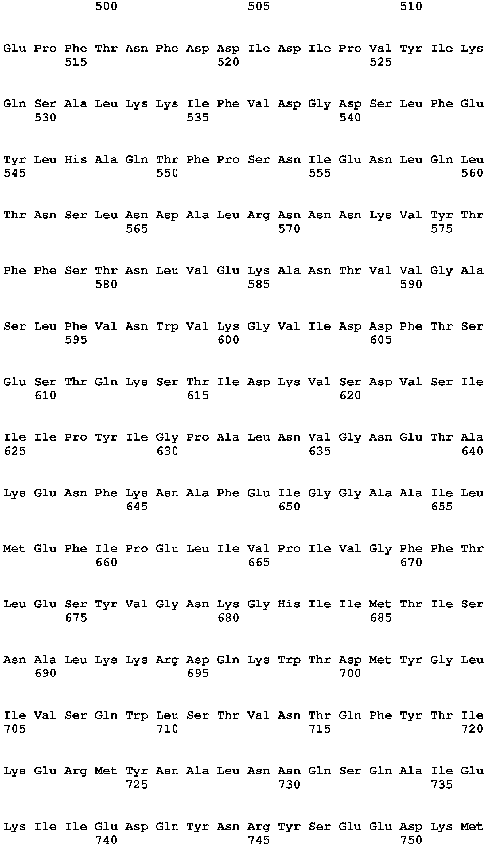


Claims (16)
- An antigenic polypeptide comprising a VAMP epitope, wherein said antigenic polypeptide consists of 10 to 65 amino acid residues, wherein said VAMP epitope comprises an amino acid sequence which is at least 90% identical to a VAMP sequence comprising at least 8 amino acid residues which are immediately C-terminal to a clostridial neurotoxin cleavage site in said VAMP.
- The antigenic polypeptide of claim 1, wherein said VAMP epitope consists of 10 to 30 amino acid residues.
- The antigenic polypeptide of claim 1 or 2, wherein said VAMP is selected from VAMP1, VAMP2 and VAMP3.
- The antigenic polypeptide of claim 1, 2 or 3, wherein said VAMP is a human VAMP.
- The antigenic polypeptide of any one of claims 1 to 4, wherein said VAMP epitope is selected from:- a BoNT/F or BoNT/D VAMP epitope comprising or consisting of KLSELDDRADALQ (SEQ ID NO: 15);- a BoNT/F5 or BoNT/FA VAMP epitope comprising or consisting of ERDQKLSELDDRA (SEQ ID NO: 32);- a BoNT/B or TeNT VAMP epitope comprising or consisting of FETSAAKLKRYW (SEQ ID NO: 22); and- a BoNT/G VAMP epitope comprising or consisting of AKLKRKYWWKN (SEQ ID NO: 27).
- An antigenic protein comprising a polypeptide according to any one of claims 1 to 5 covalently linked to a carrier.
- Use of an antigenic polypeptide according to any one of claims 1 to 5 or an antigenic protein according to claim 6 to generate antibodies against a C-terminal VAMP cleavage product.
- An antibody that binds to an antigenic polypeptide according to any one of claim 1 to 5 or to an antigenic protein according to claim 6.
- The antibody of claim 8, wherein said antibody is a polyclonal antibody.
- The antibody of claim 8, wherein said antibody is a monoclonal antibody.
- The antibody of claim 8, 9 or 10, wherein the KD between said antibody and said epitope is lower than 10-7 M.
- Use of an antibody according to any one of claims 8 to 11 in a gain of signal cellular assay for VAMP cleavage by a VAMP cleaving clostridial neurotoxin.
- A method for determining cleavage of VAMP by a VAMP cleaving clostridial neurotoxin in a cell, comprising:a. contacting the cell with the clostridial neurotoxin under conditions suitable for clostridial neurotoxin activity;b. contacting the cytoplasmic content of said cell with a first detection antibody against the C-terminal VAMP cleavage product following cleavage of a VAMP by the VAMP cleaving clostridial neurotoxin under conditions suitable for the binding of the first detection antibody to the C-terminal VAMP cleavage product, wherein said first detection antibody is an antibody according to any one of claims 8 to 11; andc. detecting by a suitable means the binding of said first detection antibody to the C-terminal VAMP cleavage product.
- The method of claim 13, further comprising d) quantifying by a suitable means the amount of the C-terminal VAMP cleavage product bound to said first detection antibody.
- A method for determining immunoresistance to a VAMP cleaving clostridial neurotoxin in a subject, comprising:a. adding a VAMP cleaving clostridial neurotoxin to a test sample obtained from the subject;b. contacting a cell with the test sample of step a) under conditions suitable for clostridial neurotoxin activity;c. contacting the cytoplasmic content of said cell with a first detection antibody against the C-terminal VAMP cleavage product following cleavage of a VAMP by the VAMP cleaving clostridial neurotoxin under conditions suitable for the binding of the first detection antibody to the C-terminal VAMP cleavage product, wherein said first detection antibody is an antibody according to any one of claims 8 to 11;d. detecting by a suitable means the binding of the first detection antibody to the C-terminal VAMP cleavage product;e. quantifying the amount of the C-terminal VAMP cleavage product bound to the first detection antibody;f. repeating steps a) to e) with a negative control sample instead of a test sample; andg. comparing the amount of the C-terminal VAMP cleavage product bound to said first detection antibody in steps (e) and (f), wherein detection of a lower amount of the C-terminal VAMP cleavage product bound to said first detection antibody in step (e) relative to the amount of the C-terminal VAMP cleavage product bound to said first detection antibody in step (f) is indicative of the presence of neutralizing antibodies against the VAMP cleaving clostridial neurotoxin.
- A kit comprising a cell which is susceptible to intoxication by a VAMP cleaving neurotoxin; and a first detection antibody against cleaved VAMP, wherein said first detection antibody is an antibody according to any one of claims 8 to 11.
Priority Applications (9)
| Application Number | Priority Date | Filing Date | Title |
|---|---|---|---|
| EP16194390.7A EP3312290A1 (en) | 2016-10-18 | 2016-10-18 | Cellular vamp cleavage assay |
| EP17793604.4A EP3529370A1 (en) | 2016-10-18 | 2017-10-18 | Cellular vamp cleavage assay |
| JP2019519721A JP7127019B2 (en) | 2016-10-18 | 2017-10-18 | Cellular VAMP cleavage assay |
| US16/335,358 US11193931B2 (en) | 2016-10-18 | 2017-10-18 | Cellular vamp cleavage assay |
| RU2019113794A RU2807994C2 (en) | 2016-10-18 | 2017-10-18 | Cellular vamp cleavage assay |
| PCT/EP2017/076569 WO2018073288A1 (en) | 2016-10-18 | 2017-10-18 | Cellular vamp cleavage assay |
| CN201780063220.0A CN109844130A (en) | 2016-10-18 | 2017-10-18 | Cell VAMP cutting measurement |
| US17/451,900 US20220113310A1 (en) | 2016-10-18 | 2021-10-22 | Cellular vamp cleavage assay |
| JP2022087418A JP2022130378A (en) | 2016-10-18 | 2022-05-30 | Cellular VAMP cleavage assay |
Applications Claiming Priority (1)
| Application Number | Priority Date | Filing Date | Title |
|---|---|---|---|
| EP16194390.7A EP3312290A1 (en) | 2016-10-18 | 2016-10-18 | Cellular vamp cleavage assay |
Publications (1)
| Publication Number | Publication Date |
|---|---|
| EP3312290A1 true EP3312290A1 (en) | 2018-04-25 |
Family
ID=57208094
Family Applications (2)
| Application Number | Title | Priority Date | Filing Date |
|---|---|---|---|
| EP16194390.7A Ceased EP3312290A1 (en) | 2016-10-18 | 2016-10-18 | Cellular vamp cleavage assay |
| EP17793604.4A Pending EP3529370A1 (en) | 2016-10-18 | 2017-10-18 | Cellular vamp cleavage assay |
Family Applications After (1)
| Application Number | Title | Priority Date | Filing Date |
|---|---|---|---|
| EP17793604.4A Pending EP3529370A1 (en) | 2016-10-18 | 2017-10-18 | Cellular vamp cleavage assay |
Country Status (5)
| Country | Link |
|---|---|
| US (2) | US11193931B2 (en) |
| EP (2) | EP3312290A1 (en) |
| JP (2) | JP7127019B2 (en) |
| CN (1) | CN109844130A (en) |
| WO (1) | WO2018073288A1 (en) |
Cited By (1)
| Publication number | Priority date | Publication date | Assignee | Title |
|---|---|---|---|---|
| US10751394B2 (en) * | 2016-10-25 | 2020-08-25 | Cellsnap Llc | Neurotoxins and uses thereof |
Families Citing this family (14)
| Publication number | Priority date | Publication date | Assignee | Title |
|---|---|---|---|---|
| EP2655614B1 (en) | 2010-12-22 | 2017-03-15 | President and Fellows of Harvard College | Continuous directed evolution |
| US10920208B2 (en) | 2014-10-22 | 2021-02-16 | President And Fellows Of Harvard College | Evolution of proteases |
| WO2016168631A1 (en) | 2015-04-17 | 2016-10-20 | President And Fellows Of Harvard College | Vector-based mutagenesis system |
| US10392674B2 (en) | 2015-07-22 | 2019-08-27 | President And Fellows Of Harvard College | Evolution of site-specific recombinases |
| US11524983B2 (en) | 2015-07-23 | 2022-12-13 | President And Fellows Of Harvard College | Evolution of Bt toxins |
| WO2017019895A1 (en) | 2015-07-30 | 2017-02-02 | President And Fellows Of Harvard College | Evolution of talens |
| DK3481852T3 (en) * | 2016-07-08 | 2023-02-27 | Childrens Medical Center | UNKNOWN BOTULINUM NEUROTOXIN AND ITS DERIVATIVES |
| EP3312290A1 (en) * | 2016-10-18 | 2018-04-25 | Ipsen Biopharm Limited | Cellular vamp cleavage assay |
| WO2019010164A1 (en) | 2017-07-06 | 2019-01-10 | President And Fellows Of Harvard College | Evolution of trna synthetases |
| US11624130B2 (en) | 2017-09-18 | 2023-04-11 | President And Fellows Of Harvard College | Continuous evolution for stabilized proteins |
| EP3752128A1 (en) * | 2018-02-16 | 2020-12-23 | Bontana Therapies Gmbh | Nucleic acid-based botulinum neurotoxin for therapeutic use |
| WO2019241649A1 (en) | 2018-06-14 | 2019-12-19 | President And Fellows Of Harvard College | Evolution of cytidine deaminases |
| KR102017240B1 (en) | 2018-08-23 | 2019-09-02 | 주식회사 하울바이오 | Anti-VAMP2 antibodies inhibiting SNARE complex and use thereof |
| WO2021011579A1 (en) * | 2019-07-15 | 2021-01-21 | President And Fellows Of Harvard College | Evolved botulinum neurotoxins and uses thereof |
Citations (3)
| Publication number | Priority date | Publication date | Assignee | Title |
|---|---|---|---|---|
| EP0763131A1 (en) * | 1994-06-03 | 1997-03-19 | Microbiological Research Authority | Toxin assay |
| US20120164657A1 (en) * | 2007-11-08 | 2012-06-28 | Johnson Eric A | Method of detecting botulinum neurotoxin and antibodies that neutralize botulinum neurotoxin action |
| WO2013049508A1 (en) * | 2011-09-29 | 2013-04-04 | WHITEMARSH, Regina Clare Meyer | Compositions and methods for toxigenicity testing |
Family Cites Families (62)
| Publication number | Priority date | Publication date | Assignee | Title |
|---|---|---|---|---|
| US5223409A (en) | 1988-09-02 | 1993-06-29 | Protein Engineering Corp. | Directed evolution of novel binding proteins |
| US6172197B1 (en) | 1991-07-10 | 2001-01-09 | Medical Research Council | Methods for producing members of specific binding pairs |
| GB9015198D0 (en) | 1990-07-10 | 1990-08-29 | Brien Caroline J O | Binding substance |
| GB9206318D0 (en) | 1992-03-24 | 1992-05-06 | Cambridge Antibody Tech | Binding substances |
| US5962255A (en) | 1992-03-24 | 1999-10-05 | Cambridge Antibody Technology Limited | Methods for producing recombinant vectors |
| DE69233782D1 (en) | 1991-12-02 | 2010-05-20 | Medical Res Council | Preparation of Autoantibodies on Phage Surfaces Starting from Antibody Segment Libraries |
| ATE199392T1 (en) | 1992-12-04 | 2001-03-15 | Medical Res Council | MULTIVALENT AND MULTI-SPECIFIC BINDING PROTEINS, THEIR PRODUCTION AND USE |
| GB9313509D0 (en) | 1993-06-30 | 1993-08-11 | Medical Res Council | Chemisynthetic libraries |
| US7227010B2 (en) * | 1993-09-21 | 2007-06-05 | United States Of America As Represented By The Secretary Of The Army | Recombinant light chains of botulinum neurotoxins and light chain fusion proteins for use in research and clinical therapy |
| SE9304060D0 (en) | 1993-12-06 | 1993-12-06 | Bioinvent Int Ab | Methods to select specific bacteriophages |
| GB9500851D0 (en) | 1995-01-17 | 1995-03-08 | Bionvent International Ab | Method of selecting specific bacteriophages |
| GB9508204D0 (en) | 1995-04-21 | 1995-06-07 | Speywood Lab Ltd | A novel agent able to modify peripheral afferent function |
| WO1997034620A1 (en) | 1996-03-18 | 1997-09-25 | The Regents Of The University Of California | Peptide inhibitors of neurotransmitter secretion by neuronal cells |
| GB9617671D0 (en) | 1996-08-23 | 1996-10-02 | Microbiological Res Authority | Recombinant toxin fragments |
| GB9818548D0 (en) | 1998-08-25 | 1998-10-21 | Microbiological Res Authority | Treatment of mucas hypersecretion |
| US7138127B1 (en) | 2000-01-19 | 2006-11-21 | Allergan, Inc. | Clostridial toxin derivatives and methods for treating pain |
| US6641820B1 (en) | 2000-01-19 | 2003-11-04 | Allergan, Inc. | Clostridial toxin derivatives and methods to treat pain |
| FR2809733B1 (en) * | 2000-06-02 | 2004-04-30 | Inst Nat Sante Rech Med | PEPTIDE SUBSTRATE RECOGNIZED BY BONT / B TYPE BOTULINUM TOXIN AND ITS USE FOR DETERMINING AND / OR DETECTING SAID TOXIN OR CORRESPONDING INHIBITORS |
| AU2002228887B2 (en) * | 2000-11-06 | 2006-08-24 | United States Army Medical Research And Material Command | Recombinant light chains of botulinum neurotoxins and light chain fusion proteins for use in research and clinical therapy |
| GB0321344D0 (en) | 2003-09-11 | 2003-10-15 | Health Prot Agency | Re-targeted toxin conjugates |
| US7611856B2 (en) | 2003-11-05 | 2009-11-03 | Los Alamos National Security, Llc | Mass spectrometry-based methods for detection and differentiation of botulinum neurotoxins |
| US7514088B2 (en) | 2005-03-15 | 2009-04-07 | Allergan, Inc. | Multivalent Clostridial toxin derivatives and methods of their use |
| EP1784420B1 (en) | 2004-09-01 | 2008-12-03 | Allergan, Inc. | Degradable clostridial toxins |
| DE102004043009A1 (en) | 2004-09-06 | 2006-03-23 | Toxogen Gmbh | Transport protein for introducing chemical compounds into nerve cells |
| GB0425795D0 (en) | 2004-11-24 | 2004-12-22 | Givaudan Sa | Composition |
| GB0426397D0 (en) | 2004-12-01 | 2005-01-05 | Health Prot Agency | Fusion proteins |
| WO2006059105A2 (en) | 2004-12-01 | 2006-06-08 | Health Protection Agency | Non-cytotoxic Protein Conjugates |
| WO2006099590A2 (en) | 2005-03-15 | 2006-09-21 | Allergan, Inc. | Modified clostridial toxins with altered targeting capabilities for clostridial toxin target cells |
| DE102005019302A1 (en) | 2005-04-26 | 2006-11-16 | Toxogen Gmbh | Carrier for targeting nerve cells |
| DK2154151T3 (en) | 2005-09-19 | 2011-09-05 | Allergan Inc | Clostridium toxin inactivated clostridium toxins |
| EP2001902B1 (en) | 2006-03-14 | 2013-03-27 | Allergan, Inc. | Modified clostridial toxins with altered targeting capabilities for clostridial toxin target cells |
| GB0610867D0 (en) | 2006-06-01 | 2006-07-12 | Syntaxin Ltd | Treatment of pain |
| US8753831B2 (en) * | 2007-06-05 | 2014-06-17 | City Of Hope | Methods for detection of botulinum neurotoxin |
| JP5799397B2 (en) | 2008-06-12 | 2015-10-28 | イプセン・バイオイノベーション・リミテッドIpsen Bioinnovation Limited | Cancer suppression |
| EP2719392B1 (en) | 2008-06-12 | 2019-07-24 | Ipsen Bioinnovation Limited | Fusion proteins for use in the treatment of acromegaly |
| GB0815264D0 (en) | 2008-08-21 | 2008-09-24 | Syntaxin Ltd | Non-cytotoxic proteins |
| GB0820970D0 (en) | 2008-11-17 | 2008-12-24 | Syntaxin Ltd | Suppression of cancer |
| US20100303788A1 (en) | 2009-05-29 | 2010-12-02 | Allergan, Inc. | Methods of Treating Chronic Neurogenic Inflammation Using Galanin Retargeted Endopepidases |
| US20100303783A1 (en) | 2009-05-29 | 2010-12-02 | Allergan, Inc. | Methods of Treating Urogenital-Neurological Disorders Using Tachykinin Retargeted Endopepidases |
| US20100303791A1 (en) | 2009-05-29 | 2010-12-02 | Allergan, Inc. | Methods of Treating Chronic Neurogenic Inflammation Using Glucagon Like Hormone Retargeted Endopepidases |
| KR20120061878A (en) | 2009-08-14 | 2012-06-13 | 알러간, 인코포레이티드 | Methods of treating cancer using tachykinin retargeted endopeptidases |
| AU2010282278A1 (en) | 2009-08-14 | 2012-03-15 | Allergan, Inc. | Methods of treating cancer using glucagon-like hormone retargeted endopeptidases |
| CA2771297A1 (en) | 2009-08-14 | 2011-02-17 | Allergan, Inc. | Methods of treating cancer using neurotrophin retargeted endopeptidases |
| CA2771289A1 (en) | 2009-08-14 | 2011-02-17 | Allergan, Inc. | Methods of treating cancer using galanin retargeted endpeptidases |
| CN102574900A (en) | 2009-08-14 | 2012-07-11 | 阿勒根公司 | Methods of treating cancer using opioid retargeted endpeptidases |
| US20120244188A1 (en) | 2011-03-25 | 2012-09-27 | Allergan, Inc. | Treatment of Sensory Disturbance Disorders |
| US20120251574A1 (en) | 2011-03-28 | 2012-10-04 | Allergan, Inc. | Endopeptidase and Neurotoxin Combination Treatment of Multiple Medical Conditions |
| US20120251573A1 (en) | 2011-03-28 | 2012-10-04 | Allergan, Inc. | Endopeptidase Treatment of Neuroendocrine Disorders |
| US20120251575A1 (en) | 2011-03-28 | 2012-10-04 | Allergan, Inc. | Endopeptidase Treatment of Involuntary Movement Disorders |
| WO2012135304A1 (en) | 2011-03-29 | 2012-10-04 | Allergan, Inc. | Vagal nerve-based disorders |
| US20120251519A1 (en) | 2011-03-29 | 2012-10-04 | Allergan, Inc. | Endopeptidase Treatment of Smooth Muscle Disorders |
| US20120251518A1 (en) | 2011-03-29 | 2012-10-04 | Allergan, Inc. | Endopeptidase Treatment of Sexual Dysfunction Disorders |
| GB201108108D0 (en) | 2011-05-16 | 2011-06-29 | Syntaxin Ltd | Therapeutic fusion proteins |
| EP2734635A1 (en) * | 2011-07-19 | 2014-05-28 | ETH Zürich | Means and methods for determining clostridial neurotoxins |
| US20140287433A1 (en) * | 2011-07-19 | 2014-09-25 | ETH Zürich | Means and methods for determining clostridial neurotoxins |
| DK3372239T3 (en) | 2012-05-30 | 2021-02-01 | Harvard College | MANIPULATED BOTULINUM NEUROTOXIN |
| US20140056870A1 (en) | 2012-08-27 | 2014-02-27 | Allergan, Inc. | Fusion proteins |
| US9005628B2 (en) | 2012-10-04 | 2015-04-14 | Dublin City University | Biotherapy for pain |
| US20160289731A1 (en) * | 2012-11-21 | 2016-10-06 | Merz Pharma Gmbh & Co, Kgaa | Means and methods for determination of botulinum neurotoxin biological activity |
| GB201303108D0 (en) | 2013-02-21 | 2013-04-10 | Syntaxin Ltd | Therapeutics for suppressing osteoporosis |
| GB201312295D0 (en) | 2013-07-09 | 2013-08-21 | Syntaxin Ltd | Suppression of itch |
| EP3312290A1 (en) * | 2016-10-18 | 2018-04-25 | Ipsen Biopharm Limited | Cellular vamp cleavage assay |
-
2016
- 2016-10-18 EP EP16194390.7A patent/EP3312290A1/en not_active Ceased
-
2017
- 2017-10-18 EP EP17793604.4A patent/EP3529370A1/en active Pending
- 2017-10-18 WO PCT/EP2017/076569 patent/WO2018073288A1/en unknown
- 2017-10-18 CN CN201780063220.0A patent/CN109844130A/en active Pending
- 2017-10-18 JP JP2019519721A patent/JP7127019B2/en active Active
- 2017-10-18 US US16/335,358 patent/US11193931B2/en active Active
-
2021
- 2021-10-22 US US17/451,900 patent/US20220113310A1/en active Pending
-
2022
- 2022-05-30 JP JP2022087418A patent/JP2022130378A/en active Pending
Patent Citations (3)
| Publication number | Priority date | Publication date | Assignee | Title |
|---|---|---|---|---|
| EP0763131A1 (en) * | 1994-06-03 | 1997-03-19 | Microbiological Research Authority | Toxin assay |
| US20120164657A1 (en) * | 2007-11-08 | 2012-06-28 | Johnson Eric A | Method of detecting botulinum neurotoxin and antibodies that neutralize botulinum neurotoxin action |
| WO2013049508A1 (en) * | 2011-09-29 | 2013-04-04 | WHITEMARSH, Regina Clare Meyer | Compositions and methods for toxigenicity testing |
Non-Patent Citations (29)
| Title |
|---|
| "Antibodies: a laboratory manual", 2014, COLD SPRING HARBOR LABORATORY PRESS |
| "Antibodies: a laboratory manual.", 2014, COLD SPRING HARBOR LABORATORY PRESS |
| C NAKAMURA K ET AL.: "Characterization of the D/C mosaic neurotoxin produced by Clostridium botulinum associated with bovine botulism in Japan", VET. MICROBIOL., vol. 140, 2010, pages 147 - 154 |
| DONG M. ET AL.: "Using fluorescent sensors to detect botulinum neurotoxin activity in vitro and in living cells", PNAS, vol. 101, no. 41, 2004, pages L4701 - 14706, XP008156528, DOI: doi:10.1073/pnas.0404107101 |
| FERNANDEZ-SALAS, ESTER ET AL.: "Botulinum neurotoxin serotype A specific cell-based potency assay to replace the mouse bioassay", PLOS ONE, vol. 7.11, 2012, pages E49516 |
| FORAN, PATRICK G. ET AL.: "Evaluation of the Therapeutic Usefulness of Botulinum Neurotoxin B, C1, E, and F Compared with the Long Lasting Type A Basis for Distinct Durations of Inhibition of Exocytosis in Central Neurons", JOURNAL OF BIOLOGICAL CHEMISTRY, vol. 278.2, 2003, pages 1363 - 1371 |
| FUJITA-YOSHIGAKI, JUNKO ET AL.: "Vesicle-associated Membrane Protein 2 Is Essential for cAMP-regulated Exocytosis in Rat Parotid Acinar Cells The Inhibition of cAMP-dependent Amylase Release by Botulinum Neurotoxin B", JOURNAL OF BIOLOGICAL CHEMISTRY, vol. 271.22, 1996, pages 13130 - 13134, XP002154378, DOI: doi:10.1074/jbc.271.22.13130 |
| GERALD K: "Cell and Molecular Biology", 2002, JOHN WILEY & SONS, INC |
| HALLIS B ET AL: "Development of novel assays for botulinum type A and B neurotoxins based on their endopeptidase activities", JOURNAL OF CLINICAL MICROBIOLOGY, AMERICAN SOCIETY FOR MICROBIOLOGY, US, vol. 34, no. 8, 1 August 1996 (1996-08-01), pages 1934 - 1938, XP002976172, ISSN: 0095-1137 * |
| HALLIS, BASSAM; B. A. JAMES; CLIFFORD C. SHONE: "Development of novel assays for botulinum type A and B neurotoxins based on their endopeptidase activities", JOURNAL OF CLINICAL MICROBIOLOGY, vol. 34.8, 1996, pages 1934 - 1938, XP002976172 |
| HAMBLETON; PICKETT; HAMBLETON, P.; A. M. PICKETT., JOURNAL OF THE ROYAL SOCIETY OF MEDICINE, vol. 87, no. 11, 1994, pages 719 |
| KALANDAKANOND S ET AL.: "Cleavage of intracellular substrates of botulinum toxins A, C and D in mammalian target tissue", THE JOURNAL OF PHARMACOLOGY AND EXPERIMENTAL THERAPEUTICS, 2001, pages 749 - 755 |
| KEGEL ET AL: "An in vitro assay for detection of tetanus neurotoxin activity: Using antibodies for recognizing the proteolytically generated cleavage product", TOXICOLOGY IN VITRO, ELSEVIER SCIENCE, GB, vol. 21, no. 8, 12 November 2007 (2007-11-12), pages 1641 - 1649, XP022340114, ISSN: 0887-2333, DOI: 10.1016/J.TIV.2007.06.015 * |
| KEGEL, B. ET AL.: "An in vitro assay for detection of tetanus neurotoxin activity: Using antibodies for recognizing the proteolytically generated cleavage product", TOXICOLOGY IN VITRO, vol. 21.8, 2007, pages 1641 - 1649, XP022340114, DOI: doi:10.1016/j.tiv.2007.06.015 |
| LEENAARS, MARLIES; COENRAAD FM HENDRIKSEN: "Critical steps in the production of polyclonal and monoclonal antibodies: evaluation and recommendations.", LIAR JOURNAL, vol. 46.3, 2005, pages 269 - 279 |
| MOGHADDAM M M ET AL: "Cloning and expression of a region of vesicle associated membrane protein2 (VAMP2) gene and its use as a recombinant peptide substrate for assaying clostridial neurotoxins in contaminated biologicals", BIOLOGICALS, ACADEMIC PRESS LTD., LONDON, GB, vol. 38, no. 1, 1 January 2010 (2010-01-01), pages 113 - 119, XP026978062, ISSN: 1045-1056, [retrieved on 20100101] * |
| PELLETT ET AL: "A neuronal cell-based botulinum neurotoxin assay for highly sensitive and specific detection of neutralizing serum antibodies", FEBS LETTERS, ELSEVIER, AMSTERDAM, NL, vol. 581, no. 25, 8 October 2007 (2007-10-08), pages 4803 - 4808, XP022289797, ISSN: 0014-5793, DOI: 10.1016/J.FEBSLET.2007.08.078 * |
| PELLETT, SABINE ET AL.: "A neuronal cell-based botulinum neurotoxin assay for highly sensitive and specific detection of neutralizing serum antibodies.", FEBS LETTERS, vol. 581.25, 2007, pages 4803 - 4808 |
| PELLETT, SABINE ET AL.: "Comparison of the primary rat spinal cord cell (RSC) assay and the mouse bioassay for botulinum neurotoxin type A potency determination", JOURNAL OF PHARMACOLOGICAL AND TOXICOLOGICAL METHODS, vol. 61.3, 2010, pages 304 - 310, XP055049479, DOI: doi:10.1016/j.vascn.2010.01.003 |
| PELLETT, SABINE ET AL.: "Purification and Characterization of Botulinum Neurotoxin FA from a Genetically Modified Clostridium botulinum Strain", MSPHERE, vol. 1.1, 2016, pages E00100 - E00115 |
| PENG L ET AL.: "Cytotoxicity of botulinum neurotoxins reveals a direct role of syntaxin 1 and SNAP25 in neuron survival", NATURE COMMUNICATIONS, vol. 4, 2013, pages 1472 |
| PETER J. MUNSON; DAVID RODBARD: "Ligand: A Versatile Computerized Approach For Characterization of Ligand-Binding Systems", ANAL. BIOCHEM., vol. 107, no. 1, 1980, pages 220 - 239, XP024830028, DOI: doi:10.1016/0003-2697(80)90515-1 |
| ROSSETTO, O. ET AL.: "Botulinum neurotoxins: genetic, structural and mechanistic insights", NATURE REVIEWS MICROBIOLOGY, vol. 12.8, 2014, pages 535 - 549, XP002768081, DOI: doi:10.1038/nrmicro3295 |
| SCHIAVO G. ET AL.: "Tetanus and Botulinum-B neurotoxins block neurotransmitter release by proteolytic cleavage of synaptobrevin", NATURE, vol. 359, 1992, pages 832 - 835, XP001080116, DOI: doi:10.1038/359832a0 |
| SCHIAVO, G.; MATTEOLI, M.; MONTECUCCO, C.: "Neurotoxins affecting neuroexocytosis", PHYSIOL REV, vol. 80, 2000, pages 717 - 766 |
| STRAUGHAN, D. W., ATLA, vol. 34, no. 3, 2006, pages 305 - 313 |
| SUZANNE R KALB ET AL: "Discovery of a novel enzymatic cleavage site for botulinum neurotoxin F5", FEBS LETTERS, ELSEVIER, AMSTERDAM, NL, vol. 586, no. 2, 28 November 2011 (2011-11-28), pages 109 - 115, XP028439969, ISSN: 0014-5793, [retrieved on 20111209], DOI: 10.1016/J.FEBSLET.2011.11.033 * |
| WHITEMARSH, REGINA CM ET AL.: "Novel application of human neurons derived from induced pluripotent stem cells for highly sensitive botulinum neurotoxin detection Biological Sciences: Applied Biological Sciences", TOXICOLOGICAL SCIENCES, vol. 126, no. 2, 2012, pages 426 - 435 |
| WICTOME M ET AL: "Development of an in vitro bioassay for Clostridium botulinum type B neurotoxin in foods that is more sensitive than the mouse bioassay", APPLIED AND ENVIRONMENTAL MICROBIOLOGY, AMERICAN SOCIETY FOR MICROBIOLOGY, US, vol. 65, no. 9, 1 September 1999 (1999-09-01), pages 3787 - 3792, XP002314355, ISSN: 0099-2240 * |
Cited By (1)
| Publication number | Priority date | Publication date | Assignee | Title |
|---|---|---|---|---|
| US10751394B2 (en) * | 2016-10-25 | 2020-08-25 | Cellsnap Llc | Neurotoxins and uses thereof |
Also Published As
| Publication number | Publication date |
|---|---|
| US20220113310A1 (en) | 2022-04-14 |
| US11193931B2 (en) | 2021-12-07 |
| CN109844130A (en) | 2019-06-04 |
| EP3529370A1 (en) | 2019-08-28 |
| WO2018073288A1 (en) | 2018-04-26 |
| JP7127019B2 (en) | 2022-08-29 |
| RU2019113794A (en) | 2020-11-20 |
| US20190219575A1 (en) | 2019-07-18 |
| RU2019113794A3 (en) | 2021-09-10 |
| JP2022130378A (en) | 2022-09-06 |
| JP2019537567A (en) | 2019-12-26 |
Similar Documents
| Publication | Publication Date | Title |
|---|---|---|
| US20220113310A1 (en) | Cellular vamp cleavage assay | |
| US11261240B2 (en) | Immuno-based botulinum toxin serotype A activity assays | |
| US10900967B2 (en) | Immuno-based retargeted endopeptidase activity assays | |
| US8618261B2 (en) | Immuno-based botulinum toxin serotype A activity assays | |
| RU2807994C2 (en) | Cellular vamp cleavage assay | |
| AU2017210625B2 (en) | Immuno-based botulinum toxin serotype A activity assays |
Legal Events
| Date | Code | Title | Description |
|---|---|---|---|
| PUAI | Public reference made under article 153(3) epc to a published international application that has entered the european phase |
Free format text: ORIGINAL CODE: 0009012 |
|
| AK | Designated contracting states |
Kind code of ref document: A1 Designated state(s): AL AT BE BG CH CY CZ DE DK EE ES FI FR GB GR HR HU IE IS IT LI LT LU LV MC MK MT NL NO PL PT RO RS SE SI SK SM TR |
|
| AX | Request for extension of the european patent |
Extension state: BA ME |
|
| STAA | Information on the status of an ep patent application or granted ep patent |
Free format text: STATUS: THE APPLICATION HAS BEEN REFUSED |
|
| 18R | Application refused |
Effective date: 20180519 |