EP3209684B1 - Vh-vl-interdomain angle based antibody humanization - Google Patents
Vh-vl-interdomain angle based antibody humanization Download PDFInfo
- Publication number
- EP3209684B1 EP3209684B1 EP15784344.2A EP15784344A EP3209684B1 EP 3209684 B1 EP3209684 B1 EP 3209684B1 EP 15784344 A EP15784344 A EP 15784344A EP 3209684 B1 EP3209684 B1 EP 3209684B1
- Authority
- EP
- European Patent Office
- Prior art keywords
- antibody
- angle
- orientation
- cdr
- residues
- Prior art date
- Legal status (The legal status is an assumption and is not a legal conclusion. Google has not performed a legal analysis and makes no representation as to the accuracy of the status listed.)
- Active
Links
- 238000000034 method Methods 0.000 claims description 103
- 108010067060 Immunoglobulin Variable Region Proteins 0.000 claims description 61
- 102000017727 Immunoglobulin Variable Region Human genes 0.000 claims description 61
- 230000027455 binding Effects 0.000 claims description 58
- 239000000427 antigen Substances 0.000 claims description 30
- 102000036639 antigens Human genes 0.000 claims description 30
- 108091007433 antigens Proteins 0.000 claims description 30
- 125000000539 amino acid group Chemical group 0.000 claims description 23
- 238000007637 random forest analysis Methods 0.000 claims description 23
- 239000013598 vector Substances 0.000 claims description 22
- 125000003275 alpha amino acid group Chemical group 0.000 claims description 20
- 239000012634 fragment Substances 0.000 claims description 16
- 210000004602 germ cell Anatomy 0.000 claims description 11
- 238000000926 separation method Methods 0.000 claims description 4
- 102000056549 human Fv Human genes 0.000 claims description 2
- 108700005872 human Fv Proteins 0.000 claims description 2
- 238000000547 structure data Methods 0.000 claims description 2
- 101710117290 Aldo-keto reductase family 1 member C4 Proteins 0.000 description 37
- 102100035360 Cerebellar degeneration-related antigen 1 Human genes 0.000 description 37
- 125000002915 carbonyl group Chemical group [*:2]C([*:1])=O 0.000 description 26
- 238000012360 testing method Methods 0.000 description 19
- 210000004027 cell Anatomy 0.000 description 18
- 239000011159 matrix material Substances 0.000 description 18
- 238000005259 measurement Methods 0.000 description 16
- 238000012549 training Methods 0.000 description 15
- 125000004429 atom Chemical group 0.000 description 13
- 238000002965 ELISA Methods 0.000 description 12
- 230000000875 corresponding effect Effects 0.000 description 12
- 230000008859 change Effects 0.000 description 11
- 230000006872 improvement Effects 0.000 description 11
- 238000010187 selection method Methods 0.000 description 10
- 108060003951 Immunoglobulin Proteins 0.000 description 9
- 108091007491 NSP3 Papain-like protease domains Proteins 0.000 description 9
- 102000018358 immunoglobulin Human genes 0.000 description 9
- 241001529936 Murinae Species 0.000 description 8
- 241000283973 Oryctolagus cuniculus Species 0.000 description 8
- 150000001413 amino acids Chemical class 0.000 description 8
- 102000004169 proteins and genes Human genes 0.000 description 8
- 108090000623 proteins and genes Proteins 0.000 description 8
- NFGXHKASABOEEW-UHFFFAOYSA-N 1-methylethyl 11-methoxy-3,7,11-trimethyl-2,4-dodecadienoate Chemical compound COC(C)(C)CCCC(C)CC=CC(C)=CC(=O)OC(C)C NFGXHKASABOEEW-UHFFFAOYSA-N 0.000 description 7
- 238000013459 approach Methods 0.000 description 7
- 239000013078 crystal Substances 0.000 description 7
- 239000000243 solution Substances 0.000 description 7
- 102000008394 Immunoglobulin Fragments Human genes 0.000 description 6
- 108010021625 Immunoglobulin Fragments Proteins 0.000 description 6
- 239000000872 buffer Substances 0.000 description 6
- 238000004422 calculation algorithm Methods 0.000 description 6
- 238000010494 dissociation reaction Methods 0.000 description 6
- 230000005593 dissociations Effects 0.000 description 6
- 238000012856 packing Methods 0.000 description 6
- 230000006870 function Effects 0.000 description 5
- 238000004519 manufacturing process Methods 0.000 description 5
- 238000012216 screening Methods 0.000 description 5
- 239000012491 analyte Substances 0.000 description 4
- 239000013604 expression vector Substances 0.000 description 4
- 241000894007 species Species 0.000 description 4
- 101000891579 Homo sapiens Microtubule-associated protein tau Proteins 0.000 description 3
- 102220470282 Tryptase delta_H43Q_mutation Human genes 0.000 description 3
- 230000001174 ascending effect Effects 0.000 description 3
- 210000004899 c-terminal region Anatomy 0.000 description 3
- 238000012512 characterization method Methods 0.000 description 3
- 238000010367 cloning Methods 0.000 description 3
- 238000003066 decision tree Methods 0.000 description 3
- 230000002650 habitual effect Effects 0.000 description 3
- 102000057063 human MAPT Human genes 0.000 description 3
- 230000001900 immune effect Effects 0.000 description 3
- 230000003993 interaction Effects 0.000 description 3
- 230000004807 localization Effects 0.000 description 3
- 238000001558 permutation test Methods 0.000 description 3
- 238000002360 preparation method Methods 0.000 description 3
- 230000009467 reduction Effects 0.000 description 3
- 238000000638 solvent extraction Methods 0.000 description 3
- 238000012800 visualization Methods 0.000 description 3
- XLYOFNOQVPJJNP-UHFFFAOYSA-N water Substances O XLYOFNOQVPJJNP-UHFFFAOYSA-N 0.000 description 3
- HZAXFHJVJLSVMW-UHFFFAOYSA-N 2-Aminoethan-1-ol Chemical compound NCCO HZAXFHJVJLSVMW-UHFFFAOYSA-N 0.000 description 2
- 102100027221 CD81 antigen Human genes 0.000 description 2
- 241000283707 Capra Species 0.000 description 2
- 239000006144 Dulbecco’s modified Eagle's medium Substances 0.000 description 2
- 102220543358 Glucagon-like peptide 1 receptor_L89A_mutation Human genes 0.000 description 2
- DHMQDGOQFOQNFH-UHFFFAOYSA-N Glycine Chemical compound NCC(O)=O DHMQDGOQFOQNFH-UHFFFAOYSA-N 0.000 description 2
- 101000914479 Homo sapiens CD81 antigen Proteins 0.000 description 2
- FAPWRFPIFSIZLT-UHFFFAOYSA-M Sodium chloride Chemical compound [Na+].[Cl-] FAPWRFPIFSIZLT-UHFFFAOYSA-M 0.000 description 2
- 238000001514 detection method Methods 0.000 description 2
- 238000009826 distribution Methods 0.000 description 2
- 239000003814 drug Substances 0.000 description 2
- 230000000694 effects Effects 0.000 description 2
- 238000012407 engineering method Methods 0.000 description 2
- 230000036541 health Effects 0.000 description 2
- 229940072221 immunoglobulins Drugs 0.000 description 2
- 238000011534 incubation Methods 0.000 description 2
- 238000002347 injection Methods 0.000 description 2
- 239000007924 injection Substances 0.000 description 2
- 238000001155 isoelectric focusing Methods 0.000 description 2
- 239000002609 medium Substances 0.000 description 2
- 239000013642 negative control Substances 0.000 description 2
- 108020004707 nucleic acids Proteins 0.000 description 2
- 102000039446 nucleic acids Human genes 0.000 description 2
- 150000007523 nucleic acids Chemical class 0.000 description 2
- 238000005457 optimization Methods 0.000 description 2
- 239000013612 plasmid Substances 0.000 description 2
- 229920000136 polysorbate Polymers 0.000 description 2
- 230000008569 process Effects 0.000 description 2
- 238000012545 processing Methods 0.000 description 2
- 230000005180 public health Effects 0.000 description 2
- 230000004044 response Effects 0.000 description 2
- 102200155795 rs28933377 Human genes 0.000 description 2
- 102200118229 rs34665886 Human genes 0.000 description 2
- 102220264878 rs756388446 Human genes 0.000 description 2
- 238000007619 statistical method Methods 0.000 description 2
- 238000010200 validation analysis Methods 0.000 description 2
- JKMHFZQWWAIEOD-UHFFFAOYSA-N 2-[4-(2-hydroxyethyl)piperazin-1-yl]ethanesulfonic acid Chemical compound OCC[NH+]1CCN(CCS([O-])(=O)=O)CC1 JKMHFZQWWAIEOD-UHFFFAOYSA-N 0.000 description 1
- 108010047041 Complementarity Determining Regions Proteins 0.000 description 1
- KDXKERNSBIXSRK-RXMQYKEDSA-N D-lysine Chemical compound NCCCC[C@@H](N)C(O)=O KDXKERNSBIXSRK-RXMQYKEDSA-N 0.000 description 1
- KCXVZYZYPLLWCC-UHFFFAOYSA-N EDTA Chemical compound OC(=O)CN(CC(O)=O)CCN(CC(O)=O)CC(O)=O KCXVZYZYPLLWCC-UHFFFAOYSA-N 0.000 description 1
- 238000012286 ELISA Assay Methods 0.000 description 1
- SXRSQZLOMIGNAQ-UHFFFAOYSA-N Glutaraldehyde Chemical compound O=CCCCC=O SXRSQZLOMIGNAQ-UHFFFAOYSA-N 0.000 description 1
- 239000004471 Glycine Substances 0.000 description 1
- 239000007995 HEPES buffer Substances 0.000 description 1
- 101000935587 Homo sapiens Flavin reductase (NADPH) Proteins 0.000 description 1
- 102000006496 Immunoglobulin Heavy Chains Human genes 0.000 description 1
- 108010019476 Immunoglobulin Heavy Chains Proteins 0.000 description 1
- 102220571882 Lipase member M_L91W_mutation Human genes 0.000 description 1
- 239000004472 Lysine Substances 0.000 description 1
- KDXKERNSBIXSRK-UHFFFAOYSA-N Lysine Natural products NCCCCC(N)C(O)=O KDXKERNSBIXSRK-UHFFFAOYSA-N 0.000 description 1
- 241001465754 Metazoa Species 0.000 description 1
- 102000008300 Mutant Proteins Human genes 0.000 description 1
- 108010021466 Mutant Proteins Proteins 0.000 description 1
- 241001494479 Pecora Species 0.000 description 1
- 102000003992 Peroxidases Human genes 0.000 description 1
- 102220480802 Platelet glycoprotein VI_L95H_mutation Human genes 0.000 description 1
- 239000004743 Polypropylene Substances 0.000 description 1
- 102220539555 Pterin-4-alpha-carbinolamine dehydratase_H61Q_mutation Human genes 0.000 description 1
- 108020004511 Recombinant DNA Proteins 0.000 description 1
- 101100269437 Saccharomyces cerevisiae (strain ATCC 204508 / S288c) AHC1 gene Proteins 0.000 description 1
- QAOWNCQODCNURD-UHFFFAOYSA-N Sulfuric acid Chemical compound OS(O)(=O)=O QAOWNCQODCNURD-UHFFFAOYSA-N 0.000 description 1
- 102220533938 T cell receptor beta variable 11-3_L55A_mutation Human genes 0.000 description 1
- 102220470284 Tryptase delta_H43N_mutation Human genes 0.000 description 1
- 238000002835 absorbance Methods 0.000 description 1
- 239000002253 acid Substances 0.000 description 1
- 150000007513 acids Chemical class 0.000 description 1
- 230000002776 aggregation Effects 0.000 description 1
- 238000004220 aggregation Methods 0.000 description 1
- 238000004458 analytical method Methods 0.000 description 1
- 230000003172 anti-dna Effects 0.000 description 1
- 230000009831 antigen interaction Effects 0.000 description 1
- 238000013528 artificial neural network Methods 0.000 description 1
- 238000003556 assay Methods 0.000 description 1
- 230000009286 beneficial effect Effects 0.000 description 1
- 230000008901 benefit Effects 0.000 description 1
- 239000011230 binding agent Substances 0.000 description 1
- 230000000903 blocking effect Effects 0.000 description 1
- 238000004364 calculation method Methods 0.000 description 1
- 238000005251 capillar electrophoresis Methods 0.000 description 1
- 229910052799 carbon Inorganic materials 0.000 description 1
- 125000004432 carbon atom Chemical group C* 0.000 description 1
- 239000006143 cell culture medium Substances 0.000 description 1
- 230000001413 cellular effect Effects 0.000 description 1
- 238000006243 chemical reaction Methods 0.000 description 1
- 230000000295 complement effect Effects 0.000 description 1
- 238000000205 computational method Methods 0.000 description 1
- 238000002939 conjugate gradient method Methods 0.000 description 1
- 230000002596 correlated effect Effects 0.000 description 1
- 238000010219 correlation analysis Methods 0.000 description 1
- 230000003247 decreasing effect Effects 0.000 description 1
- 230000001419 dependent effect Effects 0.000 description 1
- 238000013461 design Methods 0.000 description 1
- 238000006073 displacement reaction Methods 0.000 description 1
- 238000011156 evaluation Methods 0.000 description 1
- 238000002474 experimental method Methods 0.000 description 1
- 230000009931 harmful effect Effects 0.000 description 1
- 210000005260 human cell Anatomy 0.000 description 1
- 210000004408 hybridoma Anatomy 0.000 description 1
- 230000016784 immunoglobulin production Effects 0.000 description 1
- 238000000338 in vitro Methods 0.000 description 1
- 230000010354 integration Effects 0.000 description 1
- 238000004255 ion exchange chromatography Methods 0.000 description 1
- 239000003446 ligand Substances 0.000 description 1
- 125000003588 lysine group Chemical group [H]N([H])C([H])([H])C([H])([H])C([H])([H])C([H])([H])C([H])(N([H])[H])C(*)=O 0.000 description 1
- 238000013507 mapping Methods 0.000 description 1
- 239000000463 material Substances 0.000 description 1
- 239000000203 mixture Substances 0.000 description 1
- 239000003607 modifier Substances 0.000 description 1
- 230000035772 mutation Effects 0.000 description 1
- 230000001537 neural effect Effects 0.000 description 1
- 108040007629 peroxidase activity proteins Proteins 0.000 description 1
- 238000002823 phage display Methods 0.000 description 1
- 230000026731 phosphorylation Effects 0.000 description 1
- 238000006366 phosphorylation reaction Methods 0.000 description 1
- -1 polypropylene Polymers 0.000 description 1
- 229920001155 polypropylene Polymers 0.000 description 1
- 239000013641 positive control Substances 0.000 description 1
- 238000012805 post-processing Methods 0.000 description 1
- 108090000765 processed proteins & peptides Proteins 0.000 description 1
- 230000000644 propagated effect Effects 0.000 description 1
- 239000013014 purified material Substances 0.000 description 1
- 102000005962 receptors Human genes 0.000 description 1
- 238000004007 reversed phase HPLC Methods 0.000 description 1
- 238000012552 review Methods 0.000 description 1
- 102200062503 rs121909236 Human genes 0.000 description 1
- 102220063680 rs786205145 Human genes 0.000 description 1
- 102220122065 rs886042999 Human genes 0.000 description 1
- 238000002864 sequence alignment Methods 0.000 description 1
- 239000007974 sodium acetate buffer Substances 0.000 description 1
- 239000011780 sodium chloride Substances 0.000 description 1
- 238000002415 sodium dodecyl sulfate polyacrylamide gel electrophoresis Methods 0.000 description 1
- 238000010972 statistical evaluation Methods 0.000 description 1
- 239000006228 supernatant Substances 0.000 description 1
- 238000010998 test method Methods 0.000 description 1
- 230000001225 therapeutic effect Effects 0.000 description 1
- 238000012546 transfer Methods 0.000 description 1
- 230000009466 transformation Effects 0.000 description 1
- 230000009261 transgenic effect Effects 0.000 description 1
Images


Classifications
-
- C—CHEMISTRY; METALLURGY
- C07—ORGANIC CHEMISTRY
- C07K—PEPTIDES
- C07K16/00—Immunoglobulins [IGs], e.g. monoclonal or polyclonal antibodies
-
- C—CHEMISTRY; METALLURGY
- C07—ORGANIC CHEMISTRY
- C07K—PEPTIDES
- C07K16/00—Immunoglobulins [IGs], e.g. monoclonal or polyclonal antibodies
- C07K16/18—Immunoglobulins [IGs], e.g. monoclonal or polyclonal antibodies against material from animals or humans
- C07K16/28—Immunoglobulins [IGs], e.g. monoclonal or polyclonal antibodies against material from animals or humans against receptors, cell surface antigens or cell surface determinants
- C07K16/2803—Immunoglobulins [IGs], e.g. monoclonal or polyclonal antibodies against material from animals or humans against receptors, cell surface antigens or cell surface determinants against the immunoglobulin superfamily
-
- C—CHEMISTRY; METALLURGY
- C07—ORGANIC CHEMISTRY
- C07K—PEPTIDES
- C07K16/00—Immunoglobulins [IGs], e.g. monoclonal or polyclonal antibodies
- C07K16/18—Immunoglobulins [IGs], e.g. monoclonal or polyclonal antibodies against material from animals or humans
- C07K16/28—Immunoglobulins [IGs], e.g. monoclonal or polyclonal antibodies against material from animals or humans against receptors, cell surface antigens or cell surface determinants
- C07K16/2896—Immunoglobulins [IGs], e.g. monoclonal or polyclonal antibodies against material from animals or humans against receptors, cell surface antigens or cell surface determinants against molecules with a "CD"-designation, not provided for elsewhere
-
- C—CHEMISTRY; METALLURGY
- C07—ORGANIC CHEMISTRY
- C07K—PEPTIDES
- C07K16/00—Immunoglobulins [IGs], e.g. monoclonal or polyclonal antibodies
- C07K16/40—Immunoglobulins [IGs], e.g. monoclonal or polyclonal antibodies against enzymes
-
- C—CHEMISTRY; METALLURGY
- C12—BIOCHEMISTRY; BEER; SPIRITS; WINE; VINEGAR; MICROBIOLOGY; ENZYMOLOGY; MUTATION OR GENETIC ENGINEERING
- C12Y—ENZYMES
- C12Y304/00—Hydrolases acting on peptide bonds, i.e. peptidases (3.4)
- C12Y304/21—Serine endopeptidases (3.4.21)
- C12Y304/21106—Hepsin (3.4.21.106)
-
- C—CHEMISTRY; METALLURGY
- C07—ORGANIC CHEMISTRY
- C07K—PEPTIDES
- C07K2317/00—Immunoglobulins specific features
- C07K2317/20—Immunoglobulins specific features characterized by taxonomic origin
- C07K2317/24—Immunoglobulins specific features characterized by taxonomic origin containing regions, domains or residues from different species, e.g. chimeric, humanized or veneered
-
- C—CHEMISTRY; METALLURGY
- C07—ORGANIC CHEMISTRY
- C07K—PEPTIDES
- C07K2317/00—Immunoglobulins specific features
- C07K2317/50—Immunoglobulins specific features characterized by immunoglobulin fragments
- C07K2317/56—Immunoglobulins specific features characterized by immunoglobulin fragments variable (Fv) region, i.e. VH and/or VL
-
- C—CHEMISTRY; METALLURGY
- C07—ORGANIC CHEMISTRY
- C07K—PEPTIDES
- C07K2317/00—Immunoglobulins specific features
- C07K2317/90—Immunoglobulins specific features characterized by (pharmaco)kinetic aspects or by stability of the immunoglobulin
- C07K2317/92—Affinity (KD), association rate (Ka), dissociation rate (Kd) or EC50 value
Definitions
- the current invention is in the field of antibody humanization.
- a method for antibody humanization comprising the grafting of donor residues onto an acceptor framework wherein the selection of the acceptor framework is done depending on the VH-VL-interdomain angle of the humanized antibody and the donor antibody.
- the antigen binding site of antibodies is formed at the interface of the heavy and light chain variable domains, VH and VL, making the VH-VL domain orientation a factor that affects antibody specificity and affinity. Preserving the VH-VL domain orientation in the process of antibody engineering and humanization would be advantageous in order to retain the donor antibody properties. Predicting the correct VH-VL orientation has been recognized as a factor in antibody homology modeling.
- WO 2011/021009 variant immunoglobulins with improved manufacturability related to the finding that modifying the amino acid sequence of immunoglobulin molecules in certain key positions leads to improvements in manufacturability, and in particular to reductions in aggregation propensity and/or increases in production levels.
- variable region framework can be selected by taking into account the homology of a human acceptor framework with the donor sequence, but more importantly, selecting those variable region frameworks in which specific residues, being obligatory donor residues, are taken into account, i.e. given weighting.
- specific residues being obligatory donor residues
- weighting the more of these weighted (important) donor residues which are already present in a homologous human framework, the more appropriate the human framework is regardless of whether the overall homology is somewhat less than another framework with fewer weighted residues matching.
- WO 2001/027160 EP 1 224 224 a method of monoclonal antibody production and specifically to the simultaneous in vitro affinity optimization of multiple distinct domains of a variable region of a monoclonal antibody is reported.
- the grafting is accomplished by generating a diverse library of CDR grafted variable region fragments and then screening the library for binding activity similar or better than the binding activity of the donor.
- a diverse library is generated by selecting acceptor framework positions that differ at the corresponding position compared to the donor framework and making a library population containing of all possible amino acid residue changes at each of those positions together with all possible amino acid residue changes at each position within the CDRs of the variable region.
- the method as reported herein uses a fast sequence-based predictor that predicts VH-VL-interdomain orientation.
- the VH-VL-orientation is described in terms of the six absolute ABangle parameters to precisely separate the different degrees of freedom of VH-VL-orientation. It has been found that with the method as reported herein an improvement in the selection of humanized antibodies regarding the deviation of VH-VL-orientation of variant (humanized) antibodies with regard to the parent (non-human) antibody can be achieved. This shows an improvement regarding the similarity of the VH-VL-interdomain angle between parent (non-human) and variant (humanized) antibody.
- the method as reported herein (comprising a grafting procedure) is delivering better binding properties of the variant (humanized) antibodies compared to humanized antibodies obtained with different methods.
- Other engineering methods such as framework shuffling can be combined with the method as reported herein resulting in improved binding of the variant antibodies obtained when exchanging a human framework by another one in order to change the bio-physical properties of the antibody.
- One aspect of the invention is a method for selecting one or more variant antibody Fv fragments derived from a parent antibody Fv fragment comprising the following steps:
- the set of VH-VL-interface residues comprises residues H35, H37, H39, H45, H47, H50, H58, H60, H61, H91, H95, H96, H98, H100x-2, H100x-1, H100x, H101, H102, H103, H105, L32, L34, L36, L38, L43, L44, L46, L49, L50, L55, L87, L89, L91, L95x-1, L95x, L96 (numbering according to Chothia index).
- the set of VH-VL-interface residues comprises residues H33, H35, H43, H44, H46, H50, H55, H56, H58, H61, H62, H89, H99, L34, L36, L38, L41, L42, L43, L44, L45, L46, L49, L50, L53, L55, L56, L85, L87, L89, L91, L93, L94/L95x-1, L95x, L96, L97, L100 (numbering according to Chothia index).
- the set of VH-VL-interface residues comprises residues H33, H35, H37, H39, H43, H44, H45, H46, H47, H50, H55, H56, H58, H60, H61, H62, H89, H91, H95, H96, H98, H99, H100x-2, H100x-1, H100x, H101, H102, H103, H105, L32, L34, L36, L38, L41, L42, L43, L44, L45, L46, L49, L50, L53, L55, L56, L85, L87, L89, L91, L93, L94/L95x-1, L95x, L96, L97, L100 (numbering according to Chothia index).
- the set of VH-VL-interface residues comprises residues H35, H37, H39, H45, H47, H50, H58, H60, H61, H91, H95, H96, H98, H100x-2, H100x-1, H100x, H101, H102, H103, H105, L32, L34, L36, L38, L43, L44, L46, L49, L50, L55, L87, L89, L91, L95x-1, L95x, L96, L98 (numbering according to Chothia index).
- the set of VH-VL-interface residues comprises residues H33, H35, H37, H39, H43, H44, H45, H46, H47, H50, H58, H60, H61, H62, H89, H91, H95, H96, H98, H99, H100x-2, H100x-1, H100x, H101, H102, H103, H105, L32, L34, L36, L38, L41, L42, L43, L44, L45, L46, L49, L50, L53, L55, L56, L85, L87, L89, L91, L93, L94, L95x-1, L95x, L96, L97, L98, L100 (numbering according to Chothia index).
- the set of VH-VL-interface residues comprises residues 210, 296, 610, 612, 733 (numbering according to Wolfguy index).
- the set of VH-VL-interface residues comprises residues 199, 202, 204, 210, 212, 251, 292, 294, 295, 329, 351, 352, 354, 395, 396, 397, 398, 399, 401, 403, 597, 599, 602, 604, 609, 610, 612, 615, 651, 698, 733, 751, 753, 796, 797, 798 (numbering according to Wolfguy index).
- the set of VH-VL-interface residues comprises residues 197, 199, 208, 209, 211, 251, 289, 290, 292, 295, 296, 327, 355, 599, 602, 604, 607, 608, 609, 610, 611, 612, 615, 651, 696, 698, 699, 731, 733, 751, 753, 755, 796, 797, 798, 799, 803 (numbering according to Wolfguy index).
- the set of VH-VL-interface residues comprises residues 197, 199, 202, 204, 208, 209, 210, 211, 212, 251, 292, 294, 295, 296, 327, 329, 351, 352, 354, 355, 395, 396, 397, 398, 399, 401, 403, 597, 599, 602, 604, 607, 608, 609, 610, 611, 612, 615, 651, 696, 698, 699, 731, 733, 751, 753, 755, 796, 796, 797, 798, 799, 801, 803 (numbering according to Wolfguy index).
- the set of VH-VL-interface residues comprises residues 199, 202, 204, 210, 212, 251, 292, 294, 295, 329, 351, 352, 354, 395, 396, 397, 398, 399, 401, 403, 597, 599, 602, 604, 609, 610, 612, 615, 651, 698, 733, 751, 753, 796, 797, 798, 801 (numbering according to Wolfguy index).
- the set of VH-VL-interface residues comprises residues 197, 199, 202, 204, 208, 209, 210, 211, 212, 251, 292, 294, 295, 296, 327, 329, 351, 352, 354, 355, 395, 396, 397, 398, 399, 401, 403, 597, 599, 602, 604, 607, 608, 609, 610, 611, 612, 615, 651, 696, 698, 699, 731, 733, 751, 753, 755, 796, 797, 798, 799, 801, 803 (numbering according to Wolfguy index).
- the selecting/selection is based on the top 80 % variant antibody Fv fragments regarding VH-VL-orientation.
- the selecting/selection is of the top 20 % variant antibody Fv fragments regarding VH-VL-orientation.
- the selecting is a deselecting of the worst 20 % variant antibody Fv fragments regarding VH-VL-orientation.
- the VH-VL-orientation is determined by calculating the ABangle VH-VL-orientation parameters using a random forest method.
- the VH-VL-orientation is determined by calculating the ABangle VH-VL-orientation parameters using one random forest method for each ABangle.
- the VH-VL-orientation is determined by calculating the habitual torsion angle, the four bend angles (two per variable domain), and the length of the pivot axis of VH and VL (HL, HC1, LC1, HC2, LC2, dc) using a random forest model.
- the random forest model is trained only with complex antibody structure data.
- the smallest difference is the smallest difference between real and predicted angle parameter value relating to the highest Q 2 value.
- the smallest difference is the smallest difference between the parent antibody angle parameter and the humanized variant antibody angle parameter value relating to the highest Q 2 value.
- the highest structural similarity is the lowest average root-mean-square deviation (RMSD).
- RMSD is the RMSD determined for all Calpha atoms (or carbonyl atoms) of the amino acid residues of the non-human or parent antibody to the corresponding Calpha atoms of the variant antibody.
- dist ABangle was improved with regard to the reference of structures by using the VH-VL predictor.
- the reduction of dist ABangle by VH-VL reorientation translated generally into better RMSD values, especially with regard to the framework regions.
- notable improvements of dist ABangle and improvements of the carbonyl RMSD for the whole Fv was found.
- a model assembled from template structures aligned on either consensus VH or VL framework, followed by VH-VL reorientation on a consensus Fv framework is used for determining the VH-VL-orientation.
- a model aligned on the ⁇ -sheet core of the complete Fv (VH and VL simultaneously) is used for determining the VH-VL-orientation.
- a model in which the antibody Fv fragment is reoriented on a consensus Fv framework is used for determining the VH-VL-orientation.
- a model using template structures aligned onto a common consensus Fv framework and VH-VL orientation not being adjusted in any form is used for determining the VH-VL-orientation.
- a model assembled from template structures aligned on either consensus VH or VL framework, followed by VH-VL reorientation on a VH-VL orientation template structure chosen based on similarity is used to determine the VH-VL-orientation.
- a humanized antibody that comprises amino acid residues from a donor non-human antibody at amino acid positions L26-L32, L44, L46, L50-L52, L87, L91-L96, H26-H32, H45, H53-H55, H62 and H96-H101 (numbering according to Chothia index) and at the remaining positions in the light and heavy chain variable domain residues from an acceptor human or humanized antibody or an acceptor human germline amino acid sequence.
- a humanized antibody that comprises amino acid residues from a donor non-human antibody at amino acid positions H26-H32, H35, H37, H39, H45, H47, H50, H53-H55, H58, H60, H61, H91, H95, H96-H101, H102, H103, H105, L26-L32, L34, L36, L38, L43, L44, L46, L49, L50-L52, L55, L87, L89, L91-L96 (numbering according to Chothia index) and at the remaining positions in the light and heavy chain variable domain residues from an acceptor human or humanized antibody or an acceptor human germline amino acid sequence.
- a humanized antibody that comprises amino acid residues from a donor non-human antibody at amino acid positions H26-H32, H33, H35, H43, H44, H46, H50, H53-H55, H56, H58, H61, H62, H89, H96-H101, L26-L32, L34, L36, L38, L41, L42, L43, L44, L45, L46, L49, L50-L52, L53, L55, L56, L85, L87, L89, L91-L96, L97, L100 (numbering according to Chothia index) and at the remaining positions in the light and heavy chain variable domain residues from an acceptor human or humanized antibody or an acceptor human germline amino acid sequence.
- a humanized antibody that comprises amino acid residues from a donor non-human antibody at amino acid positions H26-H32, H33, H35, H37, H39, H43, H44, H45, H46, H47, H50, H53-H55, H56, H58, H60, H61, H62, H89, H91, H95, H96-H101, H102, H103 H105, L26-L32, L34, L36, L38, L41, L42, L43, L44, L45, L46, L49, L50-L52, L53, L55, L56, L85, L87, L89, L91-L96, L97, L100 (numbering according to Chothia index) and at the remaining positions in the light and heavy chain variable domain residues from an acceptor human or humanized antibody or an acceptor human germline amino acid sequence.
- a humanized antibody that comprises amino acid residues from a donor non-human antibody at amino acid positions H26-H32, H35, H37, H39, H45, H47, H50, H53-H55, H58, H60, H61, H91, H95, H96-H101, H102, H103, H105, L26-L32, L34, L36, L38, L43, L44, L46, L49, L50-L52, L55, L87, L89, L91-L96, L98 (numbering according to Chothia index) and at the remaining positions in the light and heavy chain variable domain residues from an acceptor human or humanized antibody or an acceptor human germline amino acid sequence.
- a humanized antibody that comprises amino acid residues from a donor non-human antibody at amino acid positions H26-H32, H33, H35, H37, H39, H43, H44, H45, H46, H47, H50, H53-H55, H58, H60, H61, H62, H89, H91, H95, H96-H101, H102, H103, H105, L26-L32, L34, L36, L38, L41, L42, L43, L44, L45, L46, L49, L50-L52, L53, L55, L56, L85, L87, L89, L91-L96, L97, L98, L100 (numbering according to Chothia index) and at the remaining positions in the light and heavy chain variable domain residues from an acceptor human or humanized antibody or an acceptor human germline amino acid sequence.
- the Wolfguy numbering defines CDR regions as the set union of the Kabat and Chothia definition. Furthermore, the numbering scheme annotates CDR loop tips based on CDR length (and partly based on sequence) so that the index of a CDR position indicates if a CDR residue is part of the ascending or the descending loop.
- Table 1 A comparison with established numbering schemes is shown in Table 1.
- Wolfguy is designed such that structurally equivalent residues (i.e. residues that are very similar in terms of conserved spatial localization in the Fv structure) are numbered with equivalent indices as far as possible. This is illustrated in Figure 1 .
- RMSD root-mean-square deviation
- the ABangle concept is a method which fully characterizes VH-VL orientation in a consistent and absolute sense using five angles (HL, HC1, LC1, HC2 and LC2) and a distance (dc).
- the pair of variable domains of an antibody, VH and VL, is denoted collectively as an antibody Fv fragment.
- antibody structures were extracted from a data bank (e.g. the protein data bank, PDB). Chothia antibody numbering (Chothia and Lesk, 1987) was applied to each of the antibody chains. Chains that were successfully numbered were paired to form Fv regions. This was done by applying the constraint that the H37 position C ⁇ coordinate of the heavy chain (alpha carbon atom of the amino acid residue at heavy chain variable domain position 37) must be within 20 ⁇ of the L87 position C ⁇ coordinate of the light chain. A non-redundant set of antibodies was created using CDHIT ( Li, W. and Godzik, A. Bioinformatics, 22 (2006) 1658-1659 ), applying a sequence identity cut-off over the framework of the Fv region of 99 %.
- CDHIT Li, W. and Godzik, A. Bioinformatics, 22 (2006) 1658-1659
- VH and VL coresets The most structurally conserved residue positions in the heavy and light domains were used to define domain location. These positions are denoted as the VH and VL coresets. These positions are predominantly located on the ⁇ -strands of the framework and form the core of each domain.
- the coreset positions are given in the following Table 3: light chain heavy chain L44 H35 L19 H12 L69 H38 L14 H36 L75 H83 L82 H19 L15 H94 L21 H37 L47 H11 L20 H47 L48 H39 L49 H93 L22 H46 L81 H45 L79 H68 L80 H69 L23 H71 L36 H70 L35 H17 L37 H72 L74 H92 L88 H84 L38 H91 L18 H90 L87 H20 L17 H21 L86 H85 L85 H25 L46 H24 L70 H86 L45 H89 L16 H88 L71 H87 L72 H22 L73 H23
- the coreset positions were used to register frames of reference onto the antibody Fv region domains.
- the VH domains in the non-redundant dataset were clustered using CDHIT, applying a sequence identity cut-off of 80 % over framework positions in the domain.
- One structure was randomly chosen from each of the 30 largest clusters.
- This set of domains was aligned over the VH coreset positions using Mammoth-mult ( Lupyan, D., et al., Bioinf. 21 (2005) 3255-3263 ). From this alignment the C ⁇ coordinates corresponding to the eight structurally conserved positions H36, H37, H38, H39, H89, H90, H91 and H92 in the ⁇ -sheet interface were extracted. Through the resulting 240 coordinates a plane was fitted. For the VL domain positions L35, L36, L37, L38, L85, L86, L87 and L88 were used to fit the plane.
- the procedure described above allows mapping the two reference frame planes onto any Fv structure. Therefore the measuring of the VH-VL orientation can be made equivalent to measuring the orientation between the two planes.
- To do this fully and in an absolute sense requires at least six parameters: a distance, a torsion angle and four bend angles. These parameters must be measured about a consistently defined vector that connects the planes.
- This vector is denoted C in the following.
- C the reference frame planes were registered onto each of the structures in the non-redundant set as described above and a mesh placed on each plane. Each structure therefore had equivalent mesh points and thus equivalent VH-VL mesh point pairs.
- the Euclidean distance was measured for each pair of mesh points in each structure.
- the pair of points with the minimum variance in their separation distance was identified.
- the vector which joins these points is defined as C.
- the coordinate system is fully defined using vectors, which lie in each plane and are centered on the points corresponding to C.
- H1 is the vector running parallel to the first principal component of the VH plane, while H2 runs parallel to the second principal component.
- L1 and L2 are similarly defined on the VL domain.
- the HL angle is a torsion angle between the two domains.
- the HC1 and LC1 bend angles are equivalent to tilting-like variations of one domain with respect to the other.
- the HC2 and LC2 bend angles describe twisting-like variations of one domain to the other.
- VH-VL orientation is used in accordance with its common meaning in the art as it would be understood by a person skilled in the art (see, e.g., Dunbar et al., Prot. Eng. Des. Sel. 26 (2013) 611-620 ; and Bujotzek, A., et al., Proteins, Struct. Funct. Bioinf., 83 (2015) 681-695 ). It denotes how the VH and VL domains orientate with respect to one another.
- VH-VL orientation is defined by
- the positions are determined according to the Chothia index.
- the vector C was chosen to have the most conserved length over the non-redundant set of structures.
- the distance, dc is this length. It has a mean value of 16.2 ⁇ and a standard deviation of only 0.3 ⁇ .
- Table 4 lists the top 10 positions and residues identified by the random forest algorithm as being important in determining each of the angular measures of VH-VL orientation.
- Table 4: X represents the variable L36Va/L38Eb/L42Ha/L43La/L44Fa,b/L45T/ L46Gb/L49G/L95H Angle top 10 important input variables HL L87Fb L42Ga/L43Ta L44Va,b H61D L89L H43Q H43N/H44K H62Kb/H89V L55H L53R HC1 Xa,b L56P L41Da,b L89A L97V L94N L34H L34N L96W L100A HC2 H62Sb H62Kb/H89V H43K H50W H46K/H62Db H35S H61Q H43Q H33W H58T LC1 L91W L89A Xa,b L97V L94N L50G H43Q
- acceptor human framework for the purposes herein is a framework comprising the amino acid sequence of a light chain variable domain (VL) framework or a heavy chain variable domain (VH) framework derived from a human immunoglobulin framework or a human consensus framework, as defined below.
- An acceptor human framework "derived from” a human immunoglobulin framework or a human consensus framework may comprise the same amino acid sequence thereof, or it may contain amino acid sequence changes. In some embodiments, the number of amino acid changes are 10 or less, 9 or less, 8 or less, 7 or less, 6 or less, 5 or less, 4 or less, 3 or less, or 2 or less.
- the VL acceptor human framework is identical in sequence to the VL human immunoglobulin framework sequence or human consensus framework sequence.
- Bind refers to the strength of the sum total of non-covalent interactions between a single binding site of a molecule (e.g., an antibody) and its binding partner (e.g., an antigen). Unless indicated otherwise, as used herein, "binding affinity” refers to intrinsic binding affinity which reflects a 1:1 interaction between members of a binding pair (e.g., antibody and antigen).
- the affinity of a molecule X for its partner Y can generally be represented by the dissociation constant (Kd). Affinity can be measured by common methods known in the art, including those described herein.
- antibody herein is used in the broadest sense and encompasses various antibody structures, including but not limited to monoclonal antibodies, polyclonal antibodies, multispecific antibodies (e.g., bispecific antibodies), and antibody fragments so long as they exhibit the desired antigen-binding activity.
- antibody fragment refers to a molecule other than an intact antibody that comprises a portion of an intact antibody that binds the antigen to which the intact antibody binds.
- antibody fragments include but are not limited to Fv, Fab, Fab', Fab'-SH, F(ab') 2 ; diabodies; linear antibodies; single-chain antibody molecules (e.g. scFv); and multispecific antibodies formed from antibody fragments.
- chimeric antibody refers to an antibody in which a portion of the heavy and/or light chain is derived from a particular source or species, while the remainder of the heavy and/or light chain is derived from a different source or species.
- the "class" of an antibody refers to the type of constant domain or constant region possessed by its heavy chain.
- the heavy chain constant domains that correspond to the different classes of immunoglobulins are called ⁇ , ⁇ , ⁇ , ⁇ , and ⁇ , respectively.
- Fc region herein is used to define a C-terminal region of an immunoglobulin heavy chain that contains at least a portion of the constant region.
- the term includes native sequence Fc regions and variant Fc regions.
- a human IgG heavy chain Fc region extends from Cys226, or from Pro230, to the carboxyl-terminus of the heavy chain.
- the C-terminal lysine (Lys447) of the Fc region may or may not be present.
- numbering of amino acid residues in the Fc region or constant region is according to the EU numbering system, also called the EU index, as described in Kabat, E.A. et al., Sequences of Proteins of Immunological Interest, 5th ed., Public Health Service, National Institutes of Health, Bethesda, MD (1991), NIH Publication 91-3242 .
- FR Framework or "FR” refers to variable domain residues other than hypervariable region (HVR) residues.
- the FR of a variable domain generally consists of four FR domains: FR1, FR2, FR3, and FR4. Accordingly, the HVR and FR sequences generally appear in the following sequence in VH (or VL): FR1-H1(L1)-FR2-H2(L2)-FR3-H3(L3)-FR4.
- full length antibody “intact antibody,” and “whole antibody” are used herein interchangeably to refer to an antibody having a structure substantially similar to a native antibody structure or having heavy chains that contain an Fc region as defined herein.
- a "human antibody” is one which possesses an amino acid sequence which corresponds to that of an antibody produced by a human or a human cell or derived from a non-human source that utilizes human antibody repertoires or other human antibody-encoding sequences. This definition of a human antibody specifically excludes a humanized antibody comprising non-human antigen-binding residues.
- a "human consensus framework” is a framework which represents the most commonly occurring amino acid residues in a selection of human immunoglobulin VL or VH framework sequences.
- the selection of human immunoglobulin VL or VH sequences is from a subgroup of variable domain sequences.
- the subgroup of sequences is a subgroup as in Kabat, E.A. et al., Sequences of Proteins of Immunological Interest, 5th ed., Bethesda MD (1991), NIH Publication 91-3242, Vols. 1-3 .
- the subgroup is subgroup kappa I as in Kabat et al., supra.
- the subgroup is subgroup III as in Kabat et al., supra.
- a “humanized” antibody refers to a chimeric antibody comprising amino acid residues from non-human HVRs and amino acid residues from human FRs.
- a humanized antibody will comprise substantially all of at least one, and typically two, variable domains, in which all or substantially all of the HVRs (e.g., CDRs) correspond to those of a non-human antibody, and all or substantially all of the FRs correspond to those of a human antibody.
- a humanized antibody optionally may comprise at least a portion of an antibody constant region derived from a human antibody.
- a "humanized form" of an antibody, e.g., a non-human antibody refers to an antibody that has undergone humanization.
- hypervariable region refers to each of the regions of an antibody variable domain which are hypervariable in sequence ("complementarity determining regions” or “CDRs") and/or form structurally defined loops ("hypervariable loops"), and/or contain the antigen-contacting residues ("antigen contacts").
- CDRs complementarity determining regions
- hypervariable loops form structurally defined loops
- antigen contacts Generally, antibodies comprise six HVRs; three in the VH (H1, H2, H3), and three in the VL (L1, L2, L3).
- HVRs herein include
- HVR residues and other residues in the variable domain are numbered herein according to Kabat et al., supra.
- an “isolated” antibody is one which has been separated from a component of its natural environment.
- an antibody is purified to greater than 95% or 99% purity as determined by, for example, electrophoretic (e.g., SDS-PAGE, isoelectric focusing (IEF), capillary electrophoresis) or chromatographic (e.g., ion exchange or reverse phase HPLC).
- electrophoretic e.g., SDS-PAGE, isoelectric focusing (IEF), capillary electrophoresis
- chromatographic e.g., ion exchange or reverse phase HPLC
- monoclonal antibody refers to an antibody obtained from a population of substantially homogeneous antibodies, i.e., the individual antibodies comprising the population are identical and/or bind the same epitope, except for possible variant antibodies, e.g., containing naturally occurring mutations or arising during production of a monoclonal antibody preparation, such variants generally being present in minor amounts.
- polyclonal antibody preparations typically include different antibodies directed against different determinants (epitopes)
- each monoclonal antibody of a monoclonal antibody preparation is directed against a single determinant on an antigen.
- the modifier "monoclonal” indicates the character of the antibody as being obtained from a substantially homogeneous population of antibodies, and is not to be construed as requiring production of the antibody by any particular method.
- the monoclonal antibodies to be used in accordance with the present invention may be made by a variety of techniques, including but not limited to the hybridoma method, recombinant DNA methods, phage-display methods, and methods utilizing transgenic animals containing all or part of the human immunoglobulin loci, such methods and other exemplary methods for making monoclonal antibodies being described herein.
- variable region refers to the domain of an antibody heavy or light chain that is involved in binding the antibody to antigen.
- the variable domains of the heavy chain and light chain (VH and VL, respectively) of a native antibody generally have similar structures, with each domain comprising four conserved framework regions (FRs) and three hypervariable regions (HVRs).
- FRs conserved framework regions
- HVRs hypervariable regions
- antibodies that bind a particular antigen may be isolated using a VH or VL domain from an antibody that binds the antigen to screen a library of complementary VL or VH domains, respectively. See, e.g., Portolano, S. et al., J. Immunol. 150 (1993) 880-887 ; Clackson, T. et al., Nature 352 (1991) 624-628 ).
- VH-VL-orientation is described in terms of the six absolute ABangle parameters to precisely separate the different degrees of freedom of VH-VL-orientation.
- the impact of VH-VL-orientation was evaluated on different antibody structures. It has been found that with the method as reported herein an improvement regarding the deviation of VH-VL-orientation of variant (humanized) antibodies with regard to the parent (non-human) antibody can be achieved. This is shown by the average root-mean-square deviation (RMSD) of the carbonyl atoms of the amino acid backbone.
- RMSD root-mean-square deviation
- the six antigen-binding loops of the two antibody variable fragments (Fvs) are hypervariable in sequence (hypervariable regions, HVRs). Five of them are prone to adapt canonical conformations that can be predicted from sequence based on existing template structures. This does not hold for the third loop on the variable region of the heavy chain, HVR-H3.
- the HVR-H3 is the most variable loop with regard to sequence and length, and typically the main antigen interaction specificity determining site.
- the antigen binding site of an antibody forms at the interface of the two Fvs (heavy chain variable domain (VH) and light chain variable domain (VL). Each variable domain comprises three HVRs. The relative orientation of VH and VL domain adds to the topology of the antigen binding site.
- Teplyakov et al. (2) used a single angular measure to describe VH-VL orientation.
- the difference in VH-VL tilt angle with respect to a reference structure is calculated as the ⁇ angle in spherical angular system ( ⁇ , ⁇ , ⁇ ) of the coordinate transformation achieved by sequential superposition of the VL and VH domains using a set of structurally conserved ⁇ -sheet core positions.
- Narayanan et al. (6) used an RMSD (root mean square deviation) based metric to train and evaluate an energy-based predictor of VH-VL orientation.
- VH-VL packing angle an absolute metric for comparing VH-VL orientation.
- the VH-VL packing angle is the torsion angle spanned by a vector fitted through the principal axes of a highly conserved set of C ⁇ positions in each of the two domains.
- the VH-VL packing angle allows to describe each individual Fv structure in terms of its VH-VL orientation in structural space.
- the authors identified a set of influential positions and provided a sequence-based predictor of VH-VL packing learned with a neural network.
- Table 5 Q 2 and RMSE values for the prediction of the six ABangle parameters averaged over 50 runs. The number of trees per random forest model was tuned manually so as to maximize Q 2 . The values in brackets specify the standard deviation.
- the random forest model was trained once on the complete dataset of apo and complex structures (Table 5, central column) and once on the complex structures only (Table 5, right column). Although the training set was reduced by almost 550 structures, the Q 2 and RMSE values improved when only complex structures were used. For HL, LC2 and dc, the Q 2 value is about 0.68, while HC1, LC1 and LC2 have Q 2 values of 0.75 and above (when considering complex structures).
- CD-HIT can be used to cluster the orientation fingerprints at 100 % identity, and, for each cluster, at least one representative can be added to the training set, until 2/3 of the available structures are assigned to the training set. The remaining 1 ⁇ 3 can be used for testing. Due to the fact that the test set then would consist of orientation fingerprints that are also included in the training set, the resulting Q 2 values, e.g. ranging from 0.71 to 0.88 for the current datd set depending on the respective ABangle parameter, would overstate the actual capabilities of the predictor when confronted with an unknown orientation fingerprint. In that case, Q 2 values to range 0.54 to 0.73, approximately, could be found for the current dataset.
- Figure 2 shows exemplary regression plots for predicted versus actual ABangle parameters on the complex structures only dataset.
- each ABangle parameter is influenced by a largely different set of interface positions on both VH and VL.
- a framework position was the most important descriptor. Nonetheless, in each case at least two HVR-H3 residues were among the most important descriptors.
- Positions that have been ranked among the top ten important input variables in the original ABangle publication (7) were tracked in the ranking presented herein, too. But, whereas Dunbar et al.
- HC1 and LC1 find HC1 to be exclusively determined by residues on the heavy chain, and LC1 exclusively determined by residues on the light chain, the top ten descriptors as determined with a method as reported herein for HC1 and LC1 involve fingerprint positions on both chains.
- fingerprint positions are ranked irrespective of amino acid specificity.
- the top 25 ranked fingerprint positions also contain a number of members of the sets of VH-VL-orientation determining positions identified by Chailyan et al. (5) (L41, L42, L43, L44) and by Abhinandan and Martin (4) (L41, L44, L46, L87, H33, H45, H60, H62, H91, H105). It has been found that L87 is the top descriptor for HL, L46 for HC1, H45 for LC1, H62 for HC2, and L44 for LC2.
- the mean carbonyl RMSD values per fragment calculated for the VH-VL-orientation-optimized Variant 2 models showed an improvement of approximately 0.05 ⁇ in comparison to the models using a generic VH-VL-orientation (see Table 7).
- VH-VL-orientation prediction based on sequence features can be extended by moving from a single VH-VL packing angle to a finer description of VH-VL orientation in terms of the six ABangle parameters defined by Dunbar et al. (7).
- a random forest model was trained on an up-to-date set of known Fv structures.
- the Q 2 values for the six predictors range from 0.67 to 0.80 when trained on a set consisting only of complex structures.
- VH-VL-orientation prediction and adjustment Two model building variants without VH-VL-orientation prediction and adjustment (Variants 1 and 3) were compared with a model building variant that predicts the most likely VH-VL-orientation in terms of the six ABangle parameters, automatically looks up the most similarly oriented Fv structure in an antibody template database, and reorients the raw model onto this VH-VL orientation template prior to further processing (Variant 2).
- the VH-VL orientation is herein predicted from a (meaningful) subset of Fv sequence positions (a "sequence fingerprint") rather than from complete Fv sequences.
- sequence fingerprint a subset of Fv sequence positions
- a set of interface residues has been identified wherein a residue is defined to be part of the VH-VL interface if its side chain atoms are neighboring atoms of the opposite chain with a distance of less or equal than 4 ⁇ in at least 90 % of all superimposed Fv structures in the database, e.g. in RAB3D.
- Table 29 VH-VL interface residues where a residue is part of the interface if its side chain atoms are neighboring atoms of the opposite chain with a distance of less or equal than 4 ⁇ in at least 90 % of all superpositioned Fv structures in RAB3D.
- Wolfguy Index Chothia (14) Index Wolfguy Region Dunbar et al. (7) Abhinandan, Martin (4) Chailyan et al.
- VH-VL-orientation is described in terms of the six "ABangle" orientation parameters, consisting of one torsion angle, four bend angles (two per variable domain), as well as the length of the pivot axis of VH and VL.
- VH-VL orientation the relative orientation between the VH- and VL-domains
- the invention is in the following exemplified with specific antibodies which are intended to serve as an example and should not be construed to limit the scope of the invention thereto.
- the method as reported herein is a generally applicable method.
- the sequence fingerprint consists of 54 amino acids, 29 in the VH region, and 25 in the VL region. See following Table 13. Table 13: Sequence fingerprint.
- Wolfguy Index Chothia (14) Index Wolfguy Region 199 H35 + CDR-H1 202 H37 + VH-FW2 204 H39 + VH-FW2 210 H45 + VH-FW2 212 H47 + VH-FW2 251 H50
- a rabbit antibody recognizing a peptide segment from the pTau protein (including the S422 phosphorylation) and its humanized variants are evaluated according to the methods as reported herein.
- an anti-Hepsin antibody and its humanized variants are evaluated according to the methods as reported herein.
- HVRs are marked with a gray background.
- the HVR definition used corresponds to the set union of the Kabat and Chothia CDR definition.
- Sequence positions that are part of the sequence fingerprint used for predicting VH-VL-orientation are marked with a black background.
- Fingerprint positions that are unpopulated in a given antibody are marked with an 'X'.
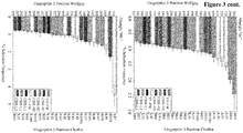
- CD81K04 VH humanization variants (the original murine sequence is shown on top):
- CD81K04 VL humanization variants (the original murine sequence is shown on top):
- CD81K13 VH humanization variants (the original murine sequence is shown on top):
- CD81K13 VL humanization variants (the original murine sequence is shown on top):
- the sequence variants have been designed using the general grafting principle. Grafting, in general, was developed to produce humanized antibodies. In addition grafting can also be used to obtain antibodies compatible to other species, or simply in order to exchange the framework of one antibody in order to get other biophysical properties for this antibody or antibody fragment.
- the antibodies are expressed in a "matrix" by combining all heavy chain plasmids with all light chain plasmids.
- the first row and the first column are then half-humanized antibodies, whereas the first cell is the original murine or rabbit antibody in its chimeric form, and the rest of the matrix are the fully humanized antibodies.
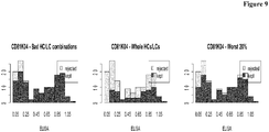
- the binding data are biochemical (cellular binding) ELISA data as summarized in Table 14 and Table 15 below, respectively.
- CD81K04 The chimeric form of CD81K04 is close to the value of 1.15, and the humanized variants are slightly less effective binders. For some of the variants, the affinity drops more drastically.
- the matrices can be correlated using the RV coefficient or other coefficients such as the correlation coefficient from the PROTEST method. These methods essentially evaluate the correlation between two data sets, where we have not one but several measurements for each sample and are therefore, to some degree, extensions of the standard univariate correlation coefficient.
- the RV coefficient is used in the following. In Table 23 the RV coefficient and its p-values for the three different data sets from the perspective of the HCs and the LCs is shown. Table 23: RV coefficients and corresponding p-values for all four data sets. The RV coefficient is calculated from the perspective of HCs as samples and hence the LCs as multivariate measurements and vice versa.
- RV HC p-value HC RV LC p-value LC CD81K04 0.2713155 0.05665427 0.3385992 0.03257287 CD81K13 0.4597497 0.03238687 0.1094843 0.627880
- Table 24 shows the correlation coefficient and the according p-value for the different data sets.
- Table 24 Pearson correlation coefficient for all three data sets and corresponding p-value. The correlation is calculated on vectorized versions of the angle-distance and binding matrices. The p-value indicates the probability to reach the calculated correlation under the null hypothesis of having no correlation.
- the first one is the ratio of median binding length between the kept and rejected subsets. For this ratio also a p-value was calculated using a permutation test. Furthermore the distribution of binding lengths or for one data set ICso values in the two subsets using stacked histograms was inspected visually.
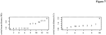
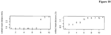
- Figure 7 depicts the average angle distance for the HCs (rows of the matrix, left) and the LCs (columns of the matrix, right). Antibodies comprising the HCs 6, 7, 8, 9, 12, 13 and 14, and LCs 7 and 9 were deselected.
- Table 25 shows the results of the comparison of the subsets of antibodies as to their binding length. The median binding length in both sets was calculated and the ratio of both was formed. In the table the results for all three methods together with the p-value, which indicates the probability of getting a ratio at least this low, are shown. Table 25: For the three different antibody rejection methods, the ratio of the median ELISA measurement between the rejected and kept antibodies was calculated. Additionally a p-value was calculated via a permutation method, which shows that probability of reaching a value as low or lower as the found median ratio. CD81K04 median ratio (deselected/selected) p-value reject worst 20% 0.2279 0.0373 reject bad HC/LC combinations 0.20661 0.0162 reject whole HCs and LCs 0.3152 ⁇ 0.001
- Antibodies comprising the HCs 3, 4 and 9, and LCs 2, 5, 6, 7, 10 and 11 were deselected.
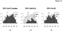
- Figure 12 shows that predominantly antibodies with lower binding length are rejected.
- Antibodies comprising the HCs 9, 10 and 11, and LCs 2 and 8 were deselected.
- the SPR data is used in the selection/deselection step. Two different measurements regarding the binding behavior of the different antibodies are available.
- the confidence matrix is incorporated as an additional step, e.g. first the deselected subset is chosen and then the high confidence subset is chosen.
- the distance information between all antibodies to compute clusters and identify clusters of antibodies that are most far away from the cluster incorporating the reference is used.
- the antibody structure database RAB3D contains mostly publicly available Fv structures.
- the Fv structures are processed and annotated with the in-house "Wolfguy" numbering scheme (see next section). All annotated Fv structures are superimposed on a consensus Fv framework, on a consensus VH framework, and on a consensus VL framework.
- the consensus structures are calculated using a subset of high-resolution structures from the PDB.
- the annotated and reoriented Fv structures serve as template repository for homology modeling.
- the Wolfguy numbering defines CDR regions as the set union of the Kabat and Chothia definition. Furthermore, the numbering scheme annotates CDR loop tips based on CDR length (and partly based on sequence) so that the index of a CDR position indicates if a CDR residue is part of the ascending or the descending loop.
- Table 1 Numbering of CDR-L3 and CDR-H3 using Chothia/Kabat (Ch-Kb), Honegger and Wolfguy numbering schemes. The latter has increasing numbers from the N-terminal basis to the CDR peak and decreasing ones starting from the C-terminal CDR end. Kabat schemes fix the two last CDR residues and introduce letters to accommodate for the CDR length.
- Wolfguy is designed such that structurally equivalent residues (i.e. residues that are very similar in terms of conserved spatial localization in the Fv structure) are numbered with equivalent indices as far as possible. This is illustrated in Figure 1 .
- the VH-VL orientation is herein predicted from a (meaningful) subset of Fv sequence positions (a "sequence fingerprint") rather than from complete Fv sequences.
- sequence fingerprint a subset of Fv sequence positions
- a set of interface residues has been identified wherein a residue is defined to be part of the VH-VL interface if its side chain atoms are neighboring atoms of the opposite chain with a distance of less or equal than 4 ⁇ in at least 90 % of all superimposed Fv structures in the database, e.g. in RAB3D.
- Table 29 VH-VL interface residues where a residue is part of the interface if its side chain atoms are neighboring atoms of the opposite chain with a distance of less or equal than 4 ⁇ in at least 90 % of all superpositioned Fv structures in RAB3D.
- Wolfguy Index Chothia (14) Index Wolfguy Region Dunbar et al. (7) Abhinandan, Martin (4) Chailyan et al.
- the “random forest” method turned out to be the statistically significant best predictor for each of the ABangle orientation parameters, followed by “neural net” and “decision tree”.
- the method "boosted tree” performed the least good on our dataset (data not shown).
- Tree Options Minimum Samples Per Node 10 Maximum Tree Depth 20 Split Method Gini Weighting Method By Class Forest Options Number of Trees Depending on ABangle parameter, see Table 2 Ensemble Method Bagging Voting Method Mean Score Equalize Class Sizes False Minimum Samples Per Class 5 Number of Descriptors All Advanced Tree Options Maximum Knots Per Property 20 Minimum Alpha 0.0 Maximum Pruned Trees 20 Disregard Uncorrelated Questions False Minimum Correlation Squared 0.00001 Maximum Lookahead Depth 0 Number of Lookahead Alternatives 3 Maximum Generic Depth 0 Generic Node Weighting 1.5 Learn Options Numeric Distance Function Euclidean Numeric Scaling Mean-Center and Scale, Scale by Number of Dimensions Fingerprint Distance Function Tanimoto Model Domain Fingerprint FCFP 2 Number Records Before Caching 100000 Node Pool Size 50000
- Table 32 shows the Q 2 and root-mean-square error (RMSE) values for the prediction of the six ABangle parameters averaged over 50 runs with randomly chosen training and test set.
- Table 32 Q 2 and RMSE values for the prediction of the six ABangle parameters averaged over 50 runs.
- the number of trees per random forest model was tuned manually so as to maximize Q 2 .
- the values in brackets specify the standard deviation.
- the random forest model has been trained once on the complete dataset of apo and complex structures (Table 32, central column) and once on the complex structures only (Table 32 above, right column). Despite the fact that the training set is reduced by almost 550 structures, the Q 2 and RMSE values improve when only complex structures are considered. For HL, LC2 and dc, Q 2 values are about 0.68, while HC1, LC1 and LC2 have Q 2 values of 0.75 and above (when considering complex structures).
- Figure 2 shows exemplary regression plots for predicted versus actual ABangle parameters on the complex structures only dataset.
- Table 33 Mean R 2 values for the prediction of the six ABangle parameters HL, HC1, LC1, HC2, LC2 and dc over three repetitions using the three sequence fingerprints and their variants with only the outmost CDR residues at the edge of the framework (a) and without any CDR residues (b).
- Fingerprint HL HC1 LC1 HC2 LC2 dc Mean Interface 1 0.616 0.755 0.687 0.750 0.602 0.569 0.663 1a 0.577 0.693 0.640 0.680 0.496 0.537 0.604 1b 0.290 0.564 0.410 0.450 0.359 0.354 0.405 ABangle 2 0.601 0.752 0.679 0.761 0.612 0.567 0.662 2a 0.566 0.705 0.669 0.703 0.549 0.520 0.619 2b 0.481 0.633 0.602 0.653 0.486 0.465 0.553 Interface + ABangle 3 0.598 0.758 0.684 0.751 0.616 0.554 0.660 3a 0.566 0.708 0.615 0.714 0.543 0.538 0.614 3b 0.478 0.632 0.638 0.630 0.485 0.502 0.561
- Fingerprint 1 based on the set of interface residues
- Fingerprint 2 based on the ABangle top input variable positions
- Fingerprint 3 has been chosen for further evaluation and a random forest model for learning.
- the random forest model was implemented and trained using Accelrys Pipeline Pilot 8.5 (19) with the component "Learn RP (random partitioning) Forest Model” in "Regression” mode.
- 50 runs were performed each while varying the number of decision trees in the random forest from 10 to 100 in order to determine the optimal number of trees with regard to the Q 2 value of the test set.
- the input dataset was randomly split into 70 % test and 30 % training set.
- the modeling software for modeling the Fv region of Antibodies uses the annotated and reoriented structures, e.g. from the RAB3D database, as template repository.
- a given pair of heavy and light chain input sequence was annotated with Wolfguy and reduced to VH and VL, respectively. Both VH and VL were then divided into seven functional segments, i.e. Framework 1, CDR 1, Framework 2, CDR 2, Framework 3, CDR 3, and Framework 4 (see Table 10).
- Framework 1, CDR 1, Framework 2, CDR 2, Framework 3, CDR 3, and Framework 4 see Table 10.
- no common framework template was picked per Fv or per chain, but every fragment was looked up/aligned/determined independently based on sequence homology.
- a single MoFvAb model might be assembled from fourteen different template structures, and possibly even more, as it is feasible to reconstruct the ascending and descending section of CDR loops from different template structures, too. Fragment template hits were ranked in the following order by
- the processed model was parameterized for the CHARMm force field and minimized using the Generalized Born with a simple Switching (GBSW) implicit water model, first by the Steepest Descent and then by the Conjugate Gradient method.
- GBSW Generalized Born with a simple Switching
- MoFvAb is available as a web service based on a protocol implemented in Accelrys Pipeline Pilot 8.5 (19) using the Accelrys Discovery Studio 3.5 (20) interface.
- the MoFvAb was used to build the 11 antibody Fv structures from AMAII.
- the AMAII structures were diverse with regard to species (rabbit, mouse, human) and consist mainly of protein-binding antibodies, with anti-DNA Fab A52 (PDB ID 4M61) being the exception. All AMAII reference structures were crystallized in the unbound form.
- access to more template structures than the original AMAII "contestants" was possible, including a number of rabbit antibody structures. Therefore, the modeling results in terms of RMSD presented herein cannot be directly compared to the results presented by the original blind modeling studies.
- the weighted distance function was used for finding orientation templates in the database that best match a predicted set of ABangle parameters.
- dist ABangle and dlst ABangle mingle angular (HL, HC1, LC1, HC2, LC2) with linear (dc) distance measures, they cannot be interpreted as factual distance in angular space but serve only as an abstract distance measure.
- the RV coefficient was introduced by Escoufier to measure the similarity between square symmetric matrices (26).
- the definition can be easily extended to rectangular matrices (27).
- a variant of the subsequent method is to just reject antibodies which have "bad" HC/LC combinations. This method might perform well if the correlation of angle-distance to antibody is not so strong for individual antibodies but is better preserved over whole HCs and LCs.
- the algorithm is:
- the carbonyl RMSD and the definition of ⁇ -sheet core and CDR loops according to Teplyakov et al. (2) were used
- the model was superimposed onto the crystal structure using the C ⁇ atoms of the ⁇ -sheet core with the superposition method provided in Accelrys Discovery Studio 3.5 (20).
- the carbonyl RMSD was then calculated as the deviation of the backbone carbonyl group atoms of the given segment with regard to the crystal structure.
- the carbonyl RMSD is more sensitive with regard to deviations in backbone conformation.
- HuH7-Rluc-H3 positive cell line expressing CD81
- HuH7-Rluc-L1 negative control cell line
- F-12 DMEM medium with 10 % FCS at 37 °C and 5 % CO 2 .
- the cells were trypsinized at approximately 90 % confluence and resuspended at 4 x 10 5 cells/mL.
- 2 x10 4 cells/well HuH7-Rluc-H3 and HuH7-Rluc-L1 (negative control cell line) were plated in 50 ⁇ L DMEM medium and allowed to adhere to the 96 well poly-D-Lysine plate (Greiner, Cat-Nr.
- the antibody samples to be tested were prepared in a separate polypropylene round bottom plate with a twofold desired concentration with a final volume of 120 ⁇ l. All of the assay samples were diluted in cell culture medium. 50 ⁇ L of each antibody sample (duplicate wells) were added to cells to give final volume of 100 ⁇ L/well and incubated for 2 hours at 4 °C. Following the primary incubation the samples were removed by aspiration and the cells were fixed with 0.05 % glutaraldehyde in PBS solution (Roche Diagnostics GmbH, Mannheim, Germany, Cat-Nr. 1666789) for 10 minutes at room temperature.
- the secondary incubation step for detection of bound anti-CD81 antibodies was performed for 2 h at room temperature on a reciprocal shaker.
- detection was performed using peroxidase conjugate sheep anti-human-IgG-gamma chain specific antibody (The Binding Site, Cat.-Nr. AP004) and a goat anti-mouse IgG, (H+L)-HRP conjugate (BIORAD, Cat-Nr. 170-6516) was used for the JS81 mouse positive control antibody (BD Biosciences, Cat. Nr. 555675) both diluted 1:1000 in PBS 10 % blocking buffer.
- the kinetic screening was performed according to Schraeml et al. ( Schraeml, M. and M. Biehl, Methods Mol. Biol. 901 (2012) 171-181 ) on a BIAcore 4000 instrument, mounted with a BIAcore CM5 sensor.
- the BIAcore 4000 instrument was under the control of the software version V1.1.
- a BIAcore CM5 series S chip was mounted into the instrument and was hydrodynamically addressed and preconditioned according to the manufacturer's instructions.
- the instrument buffer was HBS-EP buffer (10 mM HEPES (pH 7.4), 150 mM NaCl, 1 mM EDTA, 0.05 % (w/v) P20).
- An antibody capture system was prepared on the sensor surface.
- a polyclonal goat anti-human antibody with human IgG-Fc specificity (Jackson Lab.) was immobilized at 30 ⁇ g/ml in 10 mM sodium acetate buffer (pH 5) to spots 1, 2, 4 and 5 in the instrument's flow cells 1, 2, 3 and 4 at 10,000 RU using NHS/EDC chemistry. In each flow cell the antibodies were captured on spot 1 and spot 5. Spot 2 and spot 4 were used as reference spots. The sensor was deactivated with a 1 M ethanolamine solution. Humanized antibody derivatives were applied at concentrations between 44 nM and 70 nM in instrument buffer supplemented with 1mg/ml CMD (carboxymethyldextrane).
- CMD carboxymethyldextrane
- the antibodies were injected at a flow rate of 30 ⁇ l/min for 2 min.
- the capture level (CL) of the surface-presented antibodies was measured in rel. response units (RU).
- the analytes in solution, phosphorylated human tau protein, non-phosphorylated human tau protein and the phosphorylated human tau mutant protein T422S, were injected at 300 nM for 3 min. at a flow rate of 30 ⁇ l/min.
- the dissociation was monitored for 5 min.
- the capture system was regenerated by a 1 min. injection of 10 mM glycine buffer pH 1.7 at 30 ⁇ L/min. over all flow cells.
- binding late BL
- SL stability late
- kinetic rates were determined at 25 °C and 37 °C using the same experimental setup, but using multiple concentration series of each analyte in solution at 0 nM (buffer), 1.2 nM, 3.7 nM, 11.1 nM, 33.3 nM, 100 nM and 300 nM. From the concentration-dependent binding behavior the kinetic data was calculated using the BIAcore evaluation software according to the manufacturer's instructions and a Langmuir 1.1 model with RMAX global.
Landscapes
- Chemical & Material Sciences (AREA)
- Health & Medical Sciences (AREA)
- Organic Chemistry (AREA)
- Immunology (AREA)
- Life Sciences & Earth Sciences (AREA)
- Biochemistry (AREA)
- General Health & Medical Sciences (AREA)
- Genetics & Genomics (AREA)
- Proteomics, Peptides & Aminoacids (AREA)
- Molecular Biology (AREA)
- Medicinal Chemistry (AREA)
- Biophysics (AREA)
- Engineering & Computer Science (AREA)
- Bioinformatics & Cheminformatics (AREA)
- General Engineering & Computer Science (AREA)
- Wood Science & Technology (AREA)
- Zoology (AREA)
- Peptides Or Proteins (AREA)
- Medicines Containing Antibodies Or Antigens For Use As Internal Diagnostic Agents (AREA)
- Preparation Of Compounds By Using Micro-Organisms (AREA)
Description
- The current invention is in the field of antibody humanization. Herein is reported a method for antibody humanization comprising the grafting of donor residues onto an acceptor framework wherein the selection of the acceptor framework is done depending on the VH-VL-interdomain angle of the humanized antibody and the donor antibody.
- The antigen binding site of antibodies is formed at the interface of the heavy and light chain variable domains, VH and VL, making the VH-VL domain orientation a factor that affects antibody specificity and affinity. Preserving the VH-VL domain orientation in the process of antibody engineering and humanization would be advantageous in order to retain the donor antibody properties. Predicting the correct VH-VL orientation has been recognized as a factor in antibody homology modeling.
- In
WO 2011/021009 variant immunoglobulins with improved manufacturability related to the finding that modifying the amino acid sequence of immunoglobulin molecules in certain key positions leads to improvements in manufacturability, and in particular to reductions in aggregation propensity and/or increases in production levels. - In
WO 2008/003931 a method for framework selection for humanizing antibodies is reported, whereby the most appropriate variable region framework can be selected by taking into account the homology of a human acceptor framework with the donor sequence, but more importantly, selecting those variable region frameworks in which specific residues, being obligatory donor residues, are taken into account, i.e. given weighting. Thus, the more of these weighted (important) donor residues which are already present in a homologous human framework, the more appropriate the human framework is regardless of whether the overall homology is somewhat less than another framework with fewer weighted residues matching. - In
WO 2001/027160 (EP 1 224 224 ) a method of monoclonal antibody production and specifically to the simultaneous in vitro affinity optimization of multiple distinct domains of a variable region of a monoclonal antibody is reported. The grafting is accomplished by generating a diverse library of CDR grafted variable region fragments and then screening the library for binding activity similar or better than the binding activity of the donor. A diverse library is generated by selecting acceptor framework positions that differ at the corresponding position compared to the donor framework and making a library population containing of all possible amino acid residue changes at each of those positions together with all possible amino acid residue changes at each position within the CDRs of the variable region. - Dunbar, J., et al. (Prot. Eng. Des. Sel. 26 (2013) 611-620) report ABangle as characterizing the VH-VL orientation in antibodies. The prediction of VH-VL domain orientation for antibody variable domain modeling was reported by Bujotzek, A., et al. (Proteins: Structure, Function, and Bioinformatics 83 (2015) 681-695). In
Chapter 12 of the book "Therapeutic Antibody Engineering: Current and Future Advances Driving the Strongest Growth Area in the Pharmaceutical Industry" (2012, Elsevier, ISBN:9781908818096) on pages 265-297 - The invention is defined by the claims. Everything falling outside the scope of the claims is presented for reference only.
- Herein is reported a fast sequence-based method for humanizing an antibody based on the determination of the heavy and light chain variable domain orientation, VH-VL-interdomain orientation (angle). With the methods as reported herein an improved, i.e. faster, more economic, less resource demanding and more efficient, selection of the best suitable humanized variant of a non-human antibody is provided.
- In more detail, the method as reported herein uses a fast sequence-based predictor that predicts VH-VL-interdomain orientation. The VH-VL-orientation is described in terms of the six absolute ABangle parameters to precisely separate the different degrees of freedom of VH-VL-orientation. It has been found that with the method as reported herein an improvement in the selection of humanized antibodies regarding the deviation of VH-VL-orientation of variant (humanized) antibodies with regard to the parent (non-human) antibody can be achieved. This shows an improvement regarding the similarity of the VH-VL-interdomain angle between parent (non-human) and variant (humanized) antibody. The method as reported herein (comprising a grafting procedure) is delivering better binding properties of the variant (humanized) antibodies compared to humanized antibodies obtained with different methods. Other engineering methods such as framework shuffling can be combined with the method as reported herein resulting in improved binding of the variant antibodies obtained when exchanging a human framework by another one in order to change the bio-physical properties of the antibody.
- One aspect of the invention is a method for selecting one or more variant antibody Fv fragments derived from a parent antibody Fv fragment comprising the following steps:
- generating a multitude of variant antibody Fv fragments by grafting/transferring the antigen binding specificity determining residues from the non-human parent antibody Fv fragment on an human antibody Fv fragment germline amino acid sequence, whereby each variant antibody Fv fragment of the multitude of variant antibody Fv fragments differs from the other variant antibody Fv fragments by at least one amino acid residue,
- determining the VH-VL-orientation for the non-human Fv fragment and for each of the variant antibody Fv fragments of the multitude of variant antibody Fv fragments based on a set of VH-VL-interface residues comprising residues L44, L46, L87, H45, H62 (numbering according to Chothia index) of the antibody Fv fragment,
- selecting those variant antibody Fv fragments that have the smallest difference in the VH-VL-angle compared to the non-human antibody's VH-VL-angle and thereby selecting one or more variant antibody Fv fragments derived from a non-human antibody Fv fragment,
whereby the one or more variant antibody Fv fragments bind to the same antigen as the non-human antibody Fv fragment,
wherein the VH-VL-angle is determined by calculating the six parameters- the length of C, dc,
- the torsion angle, HL, from H1 to L1 measured about C,
- the bend angle, HC1, between H1 and C,
- the bend angle, HC2, between H2 and C,
- the bend angle, LC1 between L1 and C, and
- the bend angle, LC2, between L2 and C,
- In one embodiment of the invention the method comprising the following step:
- selecting those variant antibody Fv fragments that have the highest (structural) similarity in the VH-VL-interdomain angle compared to the parent antibody's VH-VL-interdomain angle and thereby selecting one or more variant antibody Fv fragments derived from a parent antibody Fv fragment.
- Disclosed herein is a method for humanizing a non-human antibody comprising the following steps:
- providing a non-human antibody specifically binding to an antigen,
- generating a multitude of variant antibodies by grafting/transferring one or more specificity determining residues from the non-human antibody on a human or humanized acceptor antibody or germline antibody sequence, whereby each variant antibody of the multitude of variant antibodies differs from the other variant antibodies by at least one amino acid residue,
- determining the VH-VL-orientation for the non-human antibody Fv fragment and for each of the variant antibody's Fv fragments of the multitude of variant antibodies based on a sequence fingerprint of the antibody Fv fragment,
- selecting those variant antibody Fv fragments that have the smallest difference in the VH-VL-orientation compared to the parent antibody's VH-VL-orientation and thereby selecting one or more humanized antibodies derived from a non-human,
- Disclosed herein is a method for humanizing a non-human antibody comprising the following steps:
- providing a non-human antibody specifically binding to an antigen,
- generating a multitude of variant antibodies by grafting/transferring one or more specificity determining residues from the non-human antibody on a human or humanized acceptor antibody or germline antibody sequence, whereby each variant antibody of the multitude of variant antibodies differs from the other variant antibodies by at least one amino acid residue,
- determining the VH-VL-orientation for the non-human antibody Fv fragment and for each of the variant antibody's Fv fragments of the multitude of variant antibodies based on a sequence fingerprint of the antibody Fv fragment,
- selecting those variant antibody Fv fragments that have the highest (structural) similarity in the VH-VL-interdomain angle compared to the parent antibody's VH-VL-interdomain angle and thereby selecting one or more humanized antibodies derived from a non-human antibody,
- In one embodiment of the invention the set of VH-VL-interface residues comprises residues H35, H37, H39, H45, H47, H50, H58, H60, H61, H91, H95, H96, H98, H100x-2, H100x-1, H100x, H101, H102, H103, H105, L32, L34, L36, L38, L43, L44, L46, L49, L50, L55, L87, L89, L91, L95x-1, L95x, L96 (numbering according to Chothia index).
- In one embodiment of the invention the set of VH-VL-interface residues comprises residues H33, H35, H43, H44, H46, H50, H55, H56, H58, H61, H62, H89, H99, L34, L36, L38, L41, L42, L43, L44, L45, L46, L49, L50, L53, L55, L56, L85, L87, L89, L91, L93, L94/L95x-1, L95x, L96, L97, L100 (numbering according to Chothia index).
- In one preferred embodiment of the invention the set of VH-VL-interface residues comprises residues H33, H35, H37, H39, H43, H44, H45, H46, H47, H50, H55, H56, H58, H60, H61, H62, H89, H91, H95, H96, H98, H99, H100x-2, H100x-1, H100x, H101, H102, H103, H105, L32, L34, L36, L38, L41, L42, L43, L44, L45, L46, L49, L50, L53, L55, L56, L85, L87, L89, L91, L93, L94/L95x-1, L95x, L96, L97, L100 (numbering according to Chothia index).
- In one embodiment of the invention the set of VH-VL-interface residues comprises residues H35, H37, H39, H45, H47, H50, H58, H60, H61, H91, H95, H96, H98, H100x-2, H100x-1, H100x, H101, H102, H103, H105, L32, L34, L36, L38, L43, L44, L46, L49, L50, L55, L87, L89, L91, L95x-1, L95x, L96, L98 (numbering according to Chothia index).
- In one embodiment of the invention the set of VH-VL-interface residues comprises residues H33, H35, H37, H39, H43, H44, H45, H46, H47, H50, H58, H60, H61, H62, H89, H91, H95, H96, H98, H99, H100x-2, H100x-1, H100x, H101, H102, H103, H105, L32, L34, L36, L38, L41, L42, L43, L44, L45, L46, L49, L50, L53, L55, L56, L85, L87, L89, L91, L93, L94, L95x-1, L95x, L96, L97, L98, L100 (numbering according to Chothia index).
- In one embodiment of the invention the set of VH-VL-interface residues comprises
residues - In one embodiment of the invention the set of VH-VL-interface residues comprises
residues - In one embodiment of the invention the set of VH-VL-interface residues comprises
residues - In one embodiment of the invention the set of VH-VL-interface residues comprises
residues - In one embodiment of the invention the set of VH-VL-interface residues comprises
residues - In one embodiment of the invention the set of VH-VL-interface residues comprises
residues - In one embodiment of the invention the selecting/selection is based on the top 80 % variant antibody Fv fragments regarding VH-VL-orientation.
- In one embodiment of the invention the selecting/selection is of the top 20 % variant antibody Fv fragments regarding VH-VL-orientation.
- In one embodiment of the invention the selecting is a deselecting of the worst 20 % variant antibody Fv fragments regarding VH-VL-orientation.
- In one embodiment of the invention the VH-VL-orientation is determined by calculating the ABangle VH-VL-orientation parameters using a random forest method.
- In one embodiment of the invention the VH-VL-orientation is determined by calculating the ABangle VH-VL-orientation parameters using one random forest method for each ABangle.
- In one embodiment of the invention the VH-VL-orientation is determined by calculating the habitual torsion angle, the four bend angles (two per variable domain), and the length of the pivot axis of VH and VL (HL, HC1, LC1, HC2, LC2, dc) using a random forest model.
- In one embodiment of the invention the random forest model is trained only with complex antibody structure data.
- In one embodiment of the invention the smallest difference is the smallest difference between real and predicted angle parameter value relating to the highest Q2 value.
- In one embodiment of the invention the smallest difference is the smallest difference between the parent antibody angle parameter and the humanized variant antibody angle parameter value relating to the highest Q2 value.
- In one embodiment of the invention the highest structural similarity is the lowest average root-mean-square deviation (RMSD). In one embodiment the RMSD is the RMSD determined for all Calpha atoms (or carbonyl atoms) of the amino acid residues of the non-human or parent antibody to the corresponding Calpha atoms of the variant antibody.
- In general, the
dist ABangle was improved with regard to the reference of structures by using the VH-VL predictor. The reduction ofdist ABangle by VH-VL reorientation translated generally into better RMSD values, especially with regard to the framework regions. On average, notable improvements ofdist ABangle and improvements of the carbonyl RMSD for the whole Fv was found. - In one embodiment of the invention a model assembled from template structures aligned on either consensus VH or VL framework, followed by VH-VL reorientation on a consensus Fv framework is used for determining the VH-VL-orientation.
- In one embodiment of the invention a model aligned on the β-sheet core of the complete Fv (VH and VL simultaneously) is used for determining the VH-VL-orientation.
- In one embodiment of the invention a model in which the antibody Fv fragment is reoriented on a consensus Fv framework is used for determining the VH-VL-orientation.
- In one embodiment of the invention a model using template structures aligned onto a common consensus Fv framework and VH-VL orientation not being adjusted in any form is used for determining the VH-VL-orientation.
- In one embodiment of the invention a model assembled from template structures aligned on either consensus VH or VL framework, followed by VH-VL reorientation on a VH-VL orientation template structure chosen based on similarity is used to determine the VH-VL-orientation.
- Disclosed herein is a method for producing an antibody comprising the following steps:
- selecting one or more antibodies or antibody Fv fragments according to a method as reported herein,
- selecting from the one or more antibodies or antibody Fv fragments a single antibody or antibody Fv fragment based on its binding properties,
- cloning the VH and VL encoding nucleic acids into one or more expression vectors,
- transfecting a cell with the expression vectors obtained in the previous step,
- cultivating the transfected cell and thereby producing the antibody.
- Disclosed herein is a method for producing an antibody comprising the following steps:
- selecting one or more antibodies or antibody Fv fragments comprising the following steps:
- generating a multitude of variant antibodies by grafting/transferring one or more specificity determining residues from a non-human antibody on a human or humanized acceptor antibody or germline antibody sequence, whereby each variant antibody of the multitude of variant antibodies differs from the other variant antibodies by at least one amino acid residue,
- determining the VH-VL-orientation for the non-human antibody Fv fragment and for each of the variant antibody's Fv fragments of the multitude of variant antibodies by calculating the habitual torsion angle, the four bend angles (two per variable domain), and the length of the pivot axis of VH and VL (HL, HC1, LC1, HC2, LC2, dc) using a random forest model based on a set of VH-VL-interface residues consisting of residues H33, H35, H37, H39, H43, H44, H45, H46, H47, H50, H55, H56, H58, H60, H61, H62, H89, H91, H95, H96, H98, H99, H100x-2, H100x-1, H100x, H101, H102, H103, H105, L32, L34, L36, L38, L41, L42, L43, L44, L45, L46, L49, L50, L53, L55, L56, L85, L87, L89, L91, L93, L94/L95x-1, L95x, L96, L97, L100 (numbering according to Chothia index) of the antibody Fv fragment,
- selecting those variant antibody Fv fragments that have the smallest average root-mean-square deviation (RMSD) determined for all pairs of corresponding Calpha atoms of the non-human antibody Fv fragment and variant antibody Fv fragment,
- selecting from the one or more antibodies a single antibody based on its binding properties,
- cloning the VH and VL encoding nucleic acids into one or more expression vectors,
- transfecting a cell with the expression vectors obtained in the previous step,
- cultivating the transfected cell and thereby producing the antibody.
- Disclosed herein is a humanized antibody that comprises amino acid residues from a donor non-human antibody at amino acid positions L26-L32, L44, L46, L50-L52, L87, L91-L96, H26-H32, H45, H53-H55, H62 and H96-H101 (numbering according to Chothia index) and at the remaining positions in the light and heavy chain variable domain residues from an acceptor human or humanized antibody or an acceptor human germline amino acid sequence.
- Disclosed herein is a humanized antibody that comprises amino acid residues from a donor non-human antibody at amino acid positions H26-H32, H35, H37, H39, H45, H47, H50, H53-H55, H58, H60, H61, H91, H95, H96-H101, H102, H103, H105, L26-L32, L34, L36, L38, L43, L44, L46, L49, L50-L52, L55, L87, L89, L91-L96 (numbering according to Chothia index) and at the remaining positions in the light and heavy chain variable domain residues from an acceptor human or humanized antibody or an acceptor human germline amino acid sequence.
- Disclosed herein is a humanized antibody that comprises amino acid residues from a donor non-human antibody at amino acid positions H26-H32, H33, H35, H43, H44, H46, H50, H53-H55, H56, H58, H61, H62, H89, H96-H101, L26-L32, L34, L36, L38, L41, L42, L43, L44, L45, L46, L49, L50-L52, L53, L55, L56, L85, L87, L89, L91-L96, L97, L100 (numbering according to Chothia index) and at the remaining positions in the light and heavy chain variable domain residues from an acceptor human or humanized antibody or an acceptor human germline amino acid sequence.
- Disclosed herein is a humanized antibody that comprises amino acid residues from a donor non-human antibody at amino acid positions H26-H32, H33, H35, H37, H39, H43, H44, H45, H46, H47, H50, H53-H55, H56, H58, H60, H61, H62, H89, H91, H95, H96-H101, H102, H103 H105, L26-L32, L34, L36, L38, L41, L42, L43, L44, L45, L46, L49, L50-L52, L53, L55, L56, L85, L87, L89, L91-L96, L97, L100 (numbering according to Chothia index) and at the remaining positions in the light and heavy chain variable domain residues from an acceptor human or humanized antibody or an acceptor human germline amino acid sequence.
- Disclosed herein is a humanized antibody that comprises amino acid residues from a donor non-human antibody at amino acid positions H26-H32, H35, H37, H39, H45, H47, H50, H53-H55, H58, H60, H61, H91, H95, H96-H101, H102, H103, H105, L26-L32, L34, L36, L38, L43, L44, L46, L49, L50-L52, L55, L87, L89, L91-L96, L98 (numbering according to Chothia index) and at the remaining positions in the light and heavy chain variable domain residues from an acceptor human or humanized antibody or an acceptor human germline amino acid sequence.
- Disclosed herein is a humanized antibody that comprises amino acid residues from a donor non-human antibody at amino acid positions H26-H32, H33, H35, H37, H39, H43, H44, H45, H46, H47, H50, H53-H55, H58, H60, H61, H62, H89, H91, H95, H96-H101, H102, H103, H105, L26-L32, L34, L36, L38, L41, L42, L43, L44, L45, L46, L49, L50-L52, L53, L55, L56, L85, L87, L89, L91-L96, L97, L98, L100 (numbering according to Chothia index) and at the remaining positions in the light and heavy chain variable domain residues from an acceptor human or humanized antibody or an acceptor human germline amino acid sequence.
-
- Figure 1
- Overlay of three exemplary CDR-H3 loops with 5, 10 and 15 amino acids length, taken from crystal structures with PDB ID 1N7M, 1DLF and 3HZM, respectively: a) Chothia/Kabat numbering shows the wide spatial distribution of residue 97 in the three representative CDR-H3 loops; b) Wolfguy numbering shows a compact spatial localization of residue 97, as it is always the third to last residue before the end of CDR-H3, denominated 397 according to Wolfguy index; c) several amino acids from the CDRs have inter-chain contacts, especially those located at the end of CDR-H3 and CDR-L3 (
residue 797 according to Wolfguy index clearly co-localizes and performs contacts with the VH). - Figure 2
- Predicted (vertical axis) versus actual ABangle orientation parameters (horizontal axis) for an exemplary run on the complex structures only test dataset (2/3 of the complex structures are used as training set whereas 1/3 is used as the test set). Perfect predictions would lie on the diagonal line.
- Figure 3
-
Top 25important Fingerprint 3 positions for the six ABangle parameters in terms of Percent Selection Frequency during predictor training. The values are averaged over ten runs with varying, randomly chosen training set (complex structures only). Error bars correspond to one standard deviation. Framework and CDR classification follows Wolfguy nomenclature. - Figure 4
- Average change in carbonyl RMSD for framework (FW), CDRs (CDR) and all Fv residues (All) and average change in distABangle when using unrestrained instead of restrained minimization (shown for the three
variants 1, II, III vs 1, 2, 3). - Figure 5
- Average change in carbonyl RMSD for framework (FW), CDRs (CDR) and all Fv residues (All) and average change in distABangle per AMAII antibody between original and reoriented models.
- Figure 6
- Average change in carbonyl RMSD for framework (FW), CDRs (CDR) and all Fv residues (All) and average change in distABangle per AMAII participant between original and reoriented models.
- Figure 7
- The HCs (rows of the matrix, left) and LCs (columns of the matrix, right) are sorted according to their mean angle-distance. These visualizations are used to pick "bad" HCs/LCs.
- Figure 8
- Matrix with ELISA measurements for the different HC/LC combinations. Antibodies which are deselected by the different methods are shaded; 1: bad HC/LC combinations; 2: whole HCs/LCs rejected; 3: worst 20 %.
- Figure 9
- Stacked histograms of the ELISA measurements for all three selection methods "bad HC/LC combinations" (left), "whole HCs and LCs" (middle) and "worst 20%" (right). The light-grey regions of the histogram bars indicate the antibodies that are rejected.
- Figure 10
- The HCs (rows of the matrix, left) and LCs (columns of the matrix, right) are sorted according to their mean angle-distance. These visualizations are used to pick "bad" HCs/LCs.
- Figure 11
- Matrix with ELISA measurements for the different HC/LC combinations. Antibodies which are deselected by the different methods are shaded; 1: bad HC/LC combinations; 2: whole HCs/LCs rejected; 3: worst 20 %.
- Figure 12
- Stacked histograms of the ELISA measurements for all three selection methods "bad HC/LC combinations" (left), "whole HCs and LCs" (middle) and "worst 20%" (right). The light-grey regions of the histogram bars indicate the antibodies that are rejected.
- Figure 13
- The HCs (rows of the matrix, left) and LCs (columns of the matrix, right) are sorted according to their mean angle-distance. These visualizations are used to pick "bad" HCs/LCs.
- Figure 14
- The three pictures each show the matrix with the BL measurements for the different HC/LC combinations; the antibodies which are deselected by the different methods are shaded; 1: bad HC/LC combinations; 2: whole HCs/LCs rejected; 3: worst 20 %.
- Figure 15
- Stacked histograms of the ELISA measurements for all three methods "bad HC/LC combinations" (left), "whole HCs and LCs" (middle) and "worst 20%" (right). The light-grey regions of the histogram bars indicate the antibodies that are deselected.
- Figure 16
- The three pictures each show the matrix with the t1/2 measurements for the different HC/LC combinations; the antibodies which are selected by the different methods are shaded; 1: bad HC/LC combinations; 2: whole HCs/LCs rejected; 3: worst 20 %.
- Figure 17
- Stacked histograms of the t1/2 measurements for all three methods "bad HC/LC combinations" (left), "whole HCs and LCs" (middle) and "worst 20%" (right). The light-grey regions of the histogram bars indicate the antibodies that are deselected.
- The Wolfguy numbering defines CDR regions as the set union of the Kabat and Chothia definition. Furthermore, the numbering scheme annotates CDR loop tips based on CDR length (and partly based on sequence) so that the index of a CDR position indicates if a CDR residue is part of the ascending or the descending loop. A comparison with established numbering schemes is shown in Table 1.


Figure 1 . - An example for a Wolfguy-numbered full-length VH and VL sequence can be found in Table 2.


- When making a comparison between any two amino acid based structures, generally distance-based metrics such as the root-mean-square deviation (RMSD) of equivalent atoms are used.
- To characterize the orientation between any two three-dimensional objects, it is necessary to define:
- a frame of reference on each object.
- axes to measure orientation parameters about.
- terminology to describe and quantify these parameters.
- The ABangle concept is a method which fully characterizes VH-VL orientation in a consistent and absolute sense using five angles (HL, HC1, LC1, HC2 and LC2) and a distance (dc). The pair of variable domains of an antibody, VH and VL, is denoted collectively as an antibody Fv fragment.
- In a first step antibody structures were extracted from a data bank (e.g. the protein data bank, PDB). Chothia antibody numbering (Chothia and Lesk, 1987) was applied to each of the antibody chains. Chains that were successfully numbered were paired to form Fv regions. This was done by applying the constraint that the H37 position Cα coordinate of the heavy chain (alpha carbon atom of the amino acid residue at heavy chain variable domain position 37) must be within 20 Å of the L87 position Cα coordinate of the light chain. A non-redundant set of antibodies was created using CDHIT (Li, W. and Godzik, A. Bioinformatics, 22 (2006) 1658-1659), applying a sequence identity cut-off over the framework of the Fv region of 99 %.
- The most structurally conserved residue positions in the heavy and light domains were used to define domain location. These positions are denoted as the VH and VL coresets. These positions are predominantly located on the β-strands of the framework and form the core of each domain. The coreset positions are given in the following Table 3:
light chain heavy chain L44 H35 L19 H12 L69 H38 L14 H36 L75 H83 L82 H19 L15 H94 L21 H37 L47 H11 L20 H47 L48 H39 L49 H93 L22 H46 L81 H45 L79 H68 L80 H69 L23 H71 L36 H70 L35 H17 L37 H72 L74 H92 L88 H84 L38 H91 L18 H90 L87 H20 L17 H21 L86 H85 L85 H25 L46 H24 L70 H86 L45 H89 L16 H88 L71 H87 L72 H22 L73 H23 - The coreset positions were used to register frames of reference onto the antibody Fv region domains.
- The VH domains in the non-redundant dataset were clustered using CDHIT, applying a sequence identity cut-off of 80 % over framework positions in the domain. One structure was randomly chosen from each of the 30 largest clusters. This set of domains was aligned over the VH coreset positions using Mammoth-mult (Lupyan, D., et al., Bioinf. 21 (2005) 3255-3263). From this alignment the Cα coordinates corresponding to the eight structurally conserved positions H36, H37, H38, H39, H89, H90, H91 and H92 in the β-sheet interface were extracted. Through the resulting 240 coordinates a plane was fitted. For the VL domain positions L35, L36, L37, L38, L85, L86, L87 and L88 were used to fit the plane.
- The procedure described above allows mapping the two reference frame planes onto any Fv structure. Therefore the measuring of the VH-VL orientation can be made equivalent to measuring the orientation between the two planes. To do this fully and in an absolute sense requires at least six parameters: a distance, a torsion angle and four bend angles. These parameters must be measured about a consistently defined vector that connects the planes. This vector is denoted C in the following. To identify C, the reference frame planes were registered onto each of the structures in the non-redundant set as described above and a mesh placed on each plane. Each structure therefore had equivalent mesh points and thus equivalent VH-VL mesh point pairs. The Euclidean distance was measured for each pair of mesh points in each structure. The pair of points with the minimum variance in their separation distance was identified. The vector which joins these points is defined as C.
- The coordinate system is fully defined using vectors, which lie in each plane and are centered on the points corresponding to C. H1 is the vector running parallel to the first principal component of the VH plane, while H2 runs parallel to the second principal component. L1 and L2 are similarly defined on the VL domain. The HL angle is a torsion angle between the two domains. The HC1 and LC1 bend angles are equivalent to tilting-like variations of one domain with respect to the other. The HC2 and LC2 bend angles describe twisting-like variations of one domain to the other.
- To describe the VH-VL orientation six measures are used, a distance and five angles. These are defined in the coordinate system as follows:
- the length of C, dc,
- the torsion angle, HL, from H1 to L1 measured about C,
- the bend angle, HC1, between H1 and C,
- the bend angle, HC2, between H2 and C,
- the bend angle, LC1 between L1 and C, and
- the bend angle, LC2, between L2 and C.
- The term "VH-VL orientation" is used in accordance with its common meaning in the art as it would be understood by a person skilled in the art (see, e.g., Dunbar et al., Prot. Eng. Des. Sel. 26 (2013) 611-620; and Bujotzek, A., et al., Proteins, Struct. Funct. Bioinf., 83 (2015) 681-695). It denotes how the VH and VL domains orientate with respect to one another.
- Thus the VH-VL orientation is defined by
- the length of C, dc,
- the torsion angle, HL, from H1 to L1 measured about C,
- the bend angle, HC1, between H1 and C,
- the bend angle, HC2, between H2 and C,
- the bend angle, LC1 between L1 and C, and
- the bend angle, LC2, between L2 and C,
- The positions are determined according to the Chothia index.
- The vector C was chosen to have the most conserved length over the non-redundant set of structures. The distance, dc, is this length. It has a mean value of 16.2 Å and a standard deviation of only 0.3 Å.
- Table 4 lists the top 10 positions and residues identified by the random forest algorithm as being important in determining each of the angular measures of VH-VL orientation.
Table 4: X represents the variable L36Va/L38Eb/L42Ha/L43La/L44Fa,b/L45T/ L46Gb/L49G/ L95H Angle top 10 important input variables HL L87Fb L42Ga/L43Ta L44Va,b H61D L89L H43Q H43N/H44K H62Kb/H89V L55H L53R HC1 Xa,b L56P L41Da,b L89A L97V L94N L34H L34N L96W L100A HC2 H62Sb H62Kb/H89V H43K H50W H46K/H62Db H35S H61Q H43Q H33W H58T LC1 L91W L89A Xa,b L97V L94N L50G H43Q L56P H62Sb L55A LC2 L50Y L42Ga/L43Ta L44Va,b L42Qa L55H H99Y L93T L94L L53R L85T a: denotes those positions also found to be influential by Chailyan et al. b: denotes positions also found to be influential by Abhinandan and Martin. reference 7 and Bujotzek, A., et al., Prot. Struct. Funct. Bioinf. 83 (2015) 681-695). - An "acceptor human framework" for the purposes herein is a framework comprising the amino acid sequence of a light chain variable domain (VL) framework or a heavy chain variable domain (VH) framework derived from a human immunoglobulin framework or a human consensus framework, as defined below. An acceptor human framework "derived from" a human immunoglobulin framework or a human consensus framework may comprise the same amino acid sequence thereof, or it may contain amino acid sequence changes. In some embodiments, the number of amino acid changes are 10 or less, 9 or less, 8 or less, 7 or less, 6 or less, 5 or less, 4 or less, 3 or less, or 2 or less. In some embodiments, the VL acceptor human framework is identical in sequence to the VL human immunoglobulin framework sequence or human consensus framework sequence.
- "Affinity" refers to the strength of the sum total of non-covalent interactions between a single binding site of a molecule (e.g., an antibody) and its binding partner (e.g., an antigen). Unless indicated otherwise, as used herein, "binding affinity" refers to intrinsic binding affinity which reflects a 1:1 interaction between members of a binding pair (e.g., antibody and antigen). The affinity of a molecule X for its partner Y can generally be represented by the dissociation constant (Kd). Affinity can be measured by common methods known in the art, including those described herein.
- The term "antibody" herein is used in the broadest sense and encompasses various antibody structures, including but not limited to monoclonal antibodies, polyclonal antibodies, multispecific antibodies (e.g., bispecific antibodies), and antibody fragments so long as they exhibit the desired antigen-binding activity.
- An "antibody fragment" refers to a molecule other than an intact antibody that comprises a portion of an intact antibody that binds the antigen to which the intact antibody binds. Examples of antibody fragments include but are not limited to Fv, Fab, Fab', Fab'-SH, F(ab')2; diabodies; linear antibodies; single-chain antibody molecules (e.g. scFv); and multispecific antibodies formed from antibody fragments.
- The term "chimeric" antibody refers to an antibody in which a portion of the heavy and/or light chain is derived from a particular source or species, while the remainder of the heavy and/or light chain is derived from a different source or species.
- The "class" of an antibody refers to the type of constant domain or constant region possessed by its heavy chain. There are five major classes of antibodies: IgA, IgD, IgE, IgG, and IgM, and several of these may be further divided into subclasses (isotypes), e.g., IgG1, IgG2, IgG3, IgG4, IgA1, and IgA2. The heavy chain constant domains that correspond to the different classes of immunoglobulins are called α, δ, ε, γ, and µ, respectively.
- The term "Fc region" herein is used to define a C-terminal region of an immunoglobulin heavy chain that contains at least a portion of the constant region. The term includes native sequence Fc regions and variant Fc regions. In one embodiment, a human IgG heavy chain Fc region extends from Cys226, or from Pro230, to the carboxyl-terminus of the heavy chain. However, the C-terminal lysine (Lys447) of the Fc region may or may not be present. Unless otherwise specified herein, numbering of amino acid residues in the Fc region or constant region is according to the EU numbering system, also called the EU index, as described in Kabat, E.A. et al., Sequences of Proteins of Immunological Interest, 5th ed., Public Health Service, National Institutes of Health, Bethesda, MD (1991), NIH Publication 91-3242.
- "Framework" or "FR" refers to variable domain residues other than hypervariable region (HVR) residues. The FR of a variable domain generally consists of four FR domains: FR1, FR2, FR3, and FR4. Accordingly, the HVR and FR sequences generally appear in the following sequence in VH (or VL): FR1-H1(L1)-FR2-H2(L2)-FR3-H3(L3)-FR4.
- The terms "full length antibody", "intact antibody," and "whole antibody" are used herein interchangeably to refer to an antibody having a structure substantially similar to a native antibody structure or having heavy chains that contain an Fc region as defined herein.
- A "human antibody" is one which possesses an amino acid sequence which corresponds to that of an antibody produced by a human or a human cell or derived from a non-human source that utilizes human antibody repertoires or other human antibody-encoding sequences. This definition of a human antibody specifically excludes a humanized antibody comprising non-human antigen-binding residues.
- A "human consensus framework" is a framework which represents the most commonly occurring amino acid residues in a selection of human immunoglobulin VL or VH framework sequences. Generally, the selection of human immunoglobulin VL or VH sequences is from a subgroup of variable domain sequences. Generally, the subgroup of sequences is a subgroup as in Kabat, E.A. et al., Sequences of Proteins of Immunological Interest, 5th ed., Bethesda MD (1991), NIH Publication 91-3242, Vols. 1-3. In one embodiment, for the VL, the subgroup is subgroup kappa I as in Kabat et al., supra. In one embodiment, for the VH, the subgroup is subgroup III as in Kabat et al., supra.
- A "humanized" antibody refers to a chimeric antibody comprising amino acid residues from non-human HVRs and amino acid residues from human FRs. In certain embodiments, a humanized antibody will comprise substantially all of at least one, and typically two, variable domains, in which all or substantially all of the HVRs (e.g., CDRs) correspond to those of a non-human antibody, and all or substantially all of the FRs correspond to those of a human antibody. A humanized antibody optionally may comprise at least a portion of an antibody constant region derived from a human antibody. A "humanized form" of an antibody, e.g., a non-human antibody, refers to an antibody that has undergone humanization.
- The term "hypervariable region" or "HVR", as used herein, refers to each of the regions of an antibody variable domain which are hypervariable in sequence ("complementarity determining regions" or "CDRs") and/or form structurally defined loops ("hypervariable loops"), and/or contain the antigen-contacting residues ("antigen contacts"). Generally, antibodies comprise six HVRs; three in the VH (H1, H2, H3), and three in the VL (L1, L2, L3).
- HVRs herein include
- (a) hypervariable loops occurring at amino acid residues 26-32 (L1), 50-52 (L2), 91-96 (L3), 26-32 (H1), 53-55 (H2), and 96-101 (H3) (Chothia, C. and Lesk, A.M., J. Mol. Biol. 196 (1987) 901-917);
- (b) CDRs occurring at amino acid residues 24-34 (L1), 50-56 (L2), 89-97 (L3), 31-35b (H1), 50-65 (H2), and 95-102 (H3) (Kabat, E.A. et al., Sequences of Proteins of Immunological Interest, 5th ed. Public Health Service, National Institutes of Health, Bethesda, MD (1991), NIH Publication 91-3242.);
- (c) antigen contacts occurring at amino acid residues 27c-36 (L1), 46-55 (L2), 89-96 (L3), 30-35b (H1), 47-58 (H2), and 93-101 (H3) (MacCallum et al. J. Mol. Biol. 262: 732-745 (1996)); and
- (d) combinations of (a), (b), and/or (c), including HVR amino acid residues 46-56 (L2), 47-56 (L2), 48-56 (L2), 49-56 (L2), 26-35 (H1), 26-35b (H1), 49-65 (H2), 93-102 (H3), and 94-102 (H3).
- Unless otherwise indicated, HVR residues and other residues in the variable domain (e.g., FR residues) are numbered herein according to Kabat et al., supra.
- The term "specificity determining residue" is used according to its meaning in the art. It defines the residues of an antibody that are directly involved in the interaction with antigen (see e.g. Padlan, E.A., et al., FASEB J. 9 (1995) 133-139).
- An "isolated" antibody is one which has been separated from a component of its natural environment. In some embodiments, an antibody is purified to greater than 95% or 99% purity as determined by, for example, electrophoretic (e.g., SDS-PAGE, isoelectric focusing (IEF), capillary electrophoresis) or chromatographic (e.g., ion exchange or reverse phase HPLC). For review of methods for assessment of antibody purity, see, e.g., Flatman, S. et al., J. Chromatogr. B 848 (2007) 79-87.
- The term "monoclonal antibody" as used herein refers to an antibody obtained from a population of substantially homogeneous antibodies, i.e., the individual antibodies comprising the population are identical and/or bind the same epitope, except for possible variant antibodies, e.g., containing naturally occurring mutations or arising during production of a monoclonal antibody preparation, such variants generally being present in minor amounts. In contrast to polyclonal antibody preparations, which typically include different antibodies directed against different determinants (epitopes), each monoclonal antibody of a monoclonal antibody preparation is directed against a single determinant on an antigen. Thus, the modifier "monoclonal" indicates the character of the antibody as being obtained from a substantially homogeneous population of antibodies, and is not to be construed as requiring production of the antibody by any particular method. For example, the monoclonal antibodies to be used in accordance with the present invention may be made by a variety of techniques, including but not limited to the hybridoma method, recombinant DNA methods, phage-display methods, and methods utilizing transgenic animals containing all or part of the human immunoglobulin loci, such methods and other exemplary methods for making monoclonal antibodies being described herein.
- The term "variable region" or "variable domain" refers to the domain of an antibody heavy or light chain that is involved in binding the antibody to antigen. The variable domains of the heavy chain and light chain (VH and VL, respectively) of a native antibody generally have similar structures, with each domain comprising four conserved framework regions (FRs) and three hypervariable regions (HVRs). (See, e.g., Kindt, T.J. et al. Kuby Immunology, 6th ed., W.H. Freeman and Co., N.Y. (2007), page 91) A single VH or VL domain may be sufficient to confer antigen-binding specificity. Furthermore, antibodies that bind a particular antigen may be isolated using a VH or VL domain from an antibody that binds the antigen to screen a library of complementary VL or VH domains, respectively. See, e.g., Portolano, S. et al., J. Immunol. 150 (1993) 880-887; Clackson, T. et al., Nature 352 (1991) 624-628).
- Herein is reported a fast sequence-based predictor that predicts VH-VL-interdomain orientation. Q2 values ranging from 0.67 to 0.80 are achieved. The VH-VL-orientation is described in terms of the six absolute ABangle parameters to precisely separate the different degrees of freedom of VH-VL-orientation. The impact of VH-VL-orientation was evaluated on different antibody structures. It has been found that with the method as reported herein an improvement regarding the deviation of VH-VL-orientation of variant (humanized) antibodies with regard to the parent (non-human) antibody can be achieved. This is shown by the average root-mean-square deviation (RMSD) of the carbonyl atoms of the amino acid backbone. This shows an improvement regarding the similarity of the VH-VL-interdomain angle between parent (non-human) and variant (humanized) antibody. The method as reported herein (comprising a grafting procedure) is delivering better binding properties of the variant (humanized) antibodies. Other engineering methods such as framework shuffling can be combined with the method as reported herein resulting in improved binding of the variant antibodies obtained when exchanging a human framework by another one in order to change the bio-physical properties of the antibody. This results in the provision of a method for selecting better humanized antibodies from a multitude of variant antibodies derived from a parent antibody.
- The use of antibodies in therapeutics and clinical diagnostics created a demand for precise homology models of antibody structures that enable rational antibody engineering whenever a crystal structure is not available. Therefore a multitude of computational methods for computer-aided antibody design (1), among them a number of homology modeling approaches that are regularly being assessed by blind modeling studies (8,2), has been developed.
- Due to the number of experimentally derived antibody structures (the structural antibody database SAbDab3 counts 1841 entries as of May 2014) the quality of antibody homology models is excellent in comparison to homology models of other biomolecules. The six antigen-binding loops of the two antibody variable fragments (Fvs) are hypervariable in sequence (hypervariable regions, HVRs). Five of them are prone to adapt canonical conformations that can be predicted from sequence based on existing template structures. This does not hold for the third loop on the variable region of the heavy chain, HVR-H3. The HVR-H3 is the most variable loop with regard to sequence and length, and typically the main antigen interaction specificity determining site.
- The antigen binding site of an antibody forms at the interface of the two Fvs (heavy chain variable domain (VH) and light chain variable domain (VL). Each variable domain comprises three HVRs. The relative orientation of VH and VL domain adds to the topology of the antigen binding site.
- In their recent Antibody Modeling Assessment study 2 (AMAII), Teplyakov et al. (2) used a single angular measure to describe VH-VL orientation. The difference in VH-VL tilt angle with respect to a reference structure is calculated as the κ angle in spherical angular system (ω, φ, κ) of the coordinate transformation achieved by sequential superposition of the VL and VH domains using a set of structurally conserved β-sheet core positions. Narayanan et al. (6) used an RMSD (root mean square deviation) based metric to train and evaluate an energy-based predictor of VH-VL orientation. Chailyan and coworkers (5) identified clusters of Fv structures of similar VH-VL orientation and determined influential sequence positions by measuring the Cα superposition RMSD of certain conserved residues. Other studies augment the RMSD values by providing the amount of rotation necessary to reorient one crystal structure's VH or VL onto another (10-12).
- Abhinandan and Martin (4) defined the VH-VL packing angle, an absolute metric for comparing VH-VL orientation. The VH-VL packing angle is the torsion angle spanned by a vector fitted through the principal axes of a highly conserved set of Cα positions in each of the two domains. In contrast to relative RMSD values, the VH-VL packing angle allows to describe each individual Fv structure in terms of its VH-VL orientation in structural space. Along with the definition of the VH-VL packing angle, the authors identified a set of influential positions and provided a sequence-based predictor of VH-VL packing learned with a neural network.
- Based on the past observations, which are at least in parts inconsistent with regard to Fv sequence positions deemed to have an impact VH-VL orientation (4, 5), Dunbar and coworkers (7) suggested that VH-VL orientation is subject to multiple degrees of freedom, and that each degree of freedom is determined by a different set of influential sequence positions. Consequently, the authors, in addition to the habitual torsion angle, defined four bend angles (two per variable domain), as well as the length of the pivot axis of VH and VL, and, using a random forest model, identified the most influential sequence positions for each of the five angle parameters (ABangle), as well as for the length of the pivot axis between VH and VL
- Herein is reported an ABangle-based method for the characterization and exploitation of the VH-VL-orientation during the humanization of an antibody. Herein is reported a sequence-based predictor of VH-VL-orientation for each of the six ABangle measures. Also a method of adjusting VH-VL orientation in actual antibody homology models is reported.
- Herein is reported an ABangle-based method for the characterization and exploitation of the VH-VL-orientation during the transfer of binding determining residues from a donor antibody to an acceptor antibody framework.
- Herein is reported an ABangle-based method for the characterization and exploitation of the VH-VL-orientation during the exchange of parts or entire framework regions of an antibody (framework shuffling).
-
Table 5: Q2 and RMSE values for the prediction of the six ABangle parameters averaged over 50 runs. The number of trees per random forest model was tuned manually so as to maximize Q2. The values in brackets specify the standard deviation. Apo and complex structures (n = 2249) Complex structures only (n_complex = 1468) Parameter N trees Q2 test set RMSE test set Q2 test set RMSE test set HL 33 0.68 (0.02) 2.28 (0.08) 0.67 (0.02) 2.26 (0.10) HC1 50 0.77 (0.02) 1.04 (0.05) 0.80 (0.02) 0.97 (0.04) LC1 50 0.73 (0.02) 1.26 (0.05) 0.75 (0.02) 1.25 (0.06) HC2 50 0.78 (0.01) 1.48 (0.04) 0.79 (0.02) 1.40 (0.07) LC2 75 0.65 (0.02) 1.40 (0.07) 0.69 (0.03) 1.30 (0.06) dc 100 0.56 (0.08) 0.21 (0.05) 0.67 (0.02) 0.18 (0.01) - The random forest model was trained once on the complete dataset of apo and complex structures (Table 5, central column) and once on the complex structures only (Table 5, right column). Although the training set was reduced by almost 550 structures, the Q2 and RMSE values improved when only complex structures were used. For HL, LC2 and dc, the Q2 value is about 0.68, while HC1, LC1 and LC2 have Q2 values of 0.75 and above (when considering complex structures).
- Alternatively to ensure to include the maximum diversity of different orientation fingerprints in the training set CD-HIT can be used to cluster the orientation fingerprints at 100 % identity, and, for each cluster, at least one representative can be added to the training set, until 2/3 of the available structures are assigned to the training set. The remaining ⅓ can be used for testing. Due to the fact that the test set then would consist of orientation fingerprints that are also included in the training set, the resulting Q2 values, e.g. ranging from 0.71 to 0.88 for the current datd set depending on the respective ABangle parameter, would overstate the actual capabilities of the predictor when confronted with an unknown orientation fingerprint. In that case, Q2 values to range 0.54 to 0.73, approximately, could be found for the current dataset.
-
Figure 2 shows exemplary regression plots for predicted versus actual ABangle parameters on the complex structures only dataset. - The correlation is improved compared e.g. to that reported by Abhinandan and Martin (4). Without being bound by this theory the improvement can be attributed to a finer description of the degrees of freedom of VH-VL-orientation in terms of the six ABangle parameters and the use of the Wolfguy numbering scheme reducing or even avoiding ambiguities in HVR residue numbering.
- The importance ranking of the fingerprint positions as descriptors for the different ABangle parameters is depicted in
Figure 3 . - Based on the finding of the fingerprint position importance ranking it has been found that each ABangle parameter is influenced by a largely different set of interface positions on both VH and VL. For all parameters except HC2, a framework position was the most important descriptor. Nonetheless, in each case at least two HVR-H3 residues were among the most important descriptors. Positions that have been ranked among the top ten important input variables in the original ABangle publication (7) were tracked in the ranking presented herein, too. But, whereas Dunbar et al. (7) find HC1 to be exclusively determined by residues on the heavy chain, and LC1 exclusively determined by residues on the light chain, the top ten descriptors as determined with a method as reported herein for HC1 and LC1 involve fingerprint positions on both chains. Herein the fingerprint positions are ranked irrespective of amino acid specificity.
- The top 25 ranked fingerprint positions also contain a number of members of the sets of VH-VL-orientation determining positions identified by Chailyan et al. (5) (L41, L42, L43, L44) and by Abhinandan and Martin (4) (L41, L44, L46, L87, H33, H45, H60, H62, H91, H105). It has been found that L87 is the top descriptor for HL, L46 for HC1, H45 for LC1, H62 for HC2, and L44 for LC2.
- A detailed description of the MoFvAb (Modeling of the Fv for Antibody) procedure has been published by Bujotzek, A., et al. (mAbs 7 (2015) 838-852). The results obtained for model building Variant 1 (models assembled from template structures aligned on either consensus VH or VL framework, followed by VH-VL reorientation on an consensus Fv framework), are shown in Table 6.
Table 6: AMAII models built with MoFvAb Variant 1. Values state the carbonyl RMSD for the fragments as defined by Teplyakov et al. (7) after chain-wise alignment on the β-sheet core.Model Reference VL VH L1 L2 L3 H1 H2 H3 H4 Ab01 4MA3_B_A 0.37 0.42 0.48 0.28 1.08 1.21 1.16 6.08 0.67 Ab01 4MA3_H_L 0.35 0.43 0.36 0.45 1.50 0.98 1.41 6.07 0.68 Ab02 4KUZ_ H_L 0.40 0.66 0.48 0.36 0.69 0.85 1.15 3.19 1.01 Ab03 4KQ3_H_L 0.34 0.44 0.38 0.35 0.51 2.41 0.35 2.09 0.88 Ab04 4KQ4_H_L 0.40 0.47 1.10 0.38 0.85 0.73 0.76 2.13 0.95 Ab05 4M6M_ H_L 0.53 0.40 1.28 0.38 1.96 0.28 0.29 2.93 0.37 Ab06 4M6O_H_L 0.37 0.59 0.42 0.40 0.86 0.88 0.62 3.57 0.89 Ab07 4MAU_H_L 0.38 0.40 0.54 0.52 0.81 0.57 0.44 2.10 0.59 Ab08 4M7K_H_L 0.35 0.46 0.75 0.21 0.77 0.61 0.79 2.69 0.43 Ab09 4KMT_H_L 0.32 0.37 0.29 0.39 0.29 0.47 0.97 2.89 0.38 Ab10 4M61_B_A 0.34 0.40 0.90 0.15 1.43 0.52 0.61 2.41 0.99 Ab10 4M61_D_C 0.31 0.41 1.16 0.22 1.47 0.78 0.58 2.42 0.42 Ab11 4M43_ H_L 0.34 0.61 0.37 0.25 1.01 1.07 0.54 2.95 0.35 0.37 0.47 0.65 0.33 1.02 0.87 0.74 3.20 0.66 - In order to factor in carbonyl displacement caused by deviations in VH-VL-orientation, the same models were aligned on the β-sheet core of the complete Fv (VH and VL simultaneously) and recalculated the values. The results are shown in Table 7.


- The comparison between Table 6 and Table 7 reveals how RMSD values deteriorate as soon as one considers the complete Fv structure. The mean carbonyl RMSD for the β-sheet core increased from 0.37 Å to 0.57 Å for VL, and from 0.47 Å to 0.69 Å for VH. This trend was not limited to the framework, but extended to the HVRs. The mean carbonyl RMSD for HVR-L3, for instance, increased from 1.02 Å to 1.45 Å, and from 3.20 Å to 3.32 Å for HVR-H3. The deviation in VH-VL-orientation by looking directly at the six ABangle parameters and the differences with regard to the reference structures is shown in Table 8.
Table 8: Deviation in VH-VL-orientation with regard to the reference structure in terms of the six ABangle parameters for the AMAII models built with MoFvAb Variant 1.Model Reference ΔHL AHC1 ΔLC1 ΔHC2 ΔLC2 Δdc distABangle Ab01 4ma3_B_A 1.85 0.04 0.33 2.88 0.39 0.27 3.47 Ab01 4ma3_ H_L 7.72 2.46 0.07 4.51 0.78 0.03 9.31 Ab02 4kuz_ H_L 0.71 1.57 1.97 1.61 1.10 0.40 3.29 Ab03 4kq3_H_L 1.71 0.19 0.95 2.45 0.27 0.10 3.15 Ab04 4kq4_H_L 0.59 1.94 1.42 4.95 0.45 0.00 5.55 Ab05 4m6m_H_L 8.84 3.90 3.30 4.17 0.32 0.35 11.04 Ab06 4m6o_ H_L 3.73 1.61 0.00 0.54 0.51 0.28 4.14 Ab07 4mau_ H_L 2.60 2.33 3.87 0.38 2.28 0.23 5.71 Ab08 4m7k_ H_L 2.32 1.61 3.08 0.28 0.09 0.34 4.20 Ab09 4kmt_ H_L 2.47 0.98 2.08 3.82 1.36 0.05 5.28 Ab10 4m61_B_A 0.63 0.25 1.15 4.56 1.60 0.20 5.02 Ab10 4m61_ D_ C 0.98 0.70 0.31 5.52 0.94 0.29 5.74 Ab11 4m43_H_L 2.39 2.15 4.38 2.25 1.33 0.35 6.04 2.81 1.52 1.76 2.92 0.88 0.22 5.53 - The models listed in Table 8 have been reoriented on the same consensus Fv framework and, thus, share essentially the same ABangle orientation of approximately θcons := (-59.45, 71.65, 120.49, 117.46, 82.77, 16.11). A large diversity in VH-VL-orientation that was inherent to the AMAII structures is shown. The largest deviations occurred for the parameters HL and HC2, not only between different structures, but also for sequence-identical structures from the same asymmetric unit: The parameter HL for 4MA3_B_A and 4MA3_H_L deviate by 5.87 degrees. This confirms that VH-VL-orientation, while being guided by certain sequence features (see
Figure 3 ), is also subject to an intrinsic, undirected variability. This is especially pronounced for protein-binding antibodies in the unbound form (7). - All models were rebuild with model building Variant 2 (models assembled from template structures aligned on either consensus VH or VL framework, followed by VH-VL-reorientation on a VH-VL-orientation template structure chosen based on similarity to predicted ABangle parameters) using the same choice of template structures. The results are shown in Table 9 (values refer to model-reference pairs aligned on the β-sheet core of the complete Fv).


- The mean carbonyl RMSD values per fragment calculated for the VH-VL-orientation-optimized
Variant 2 models showed an improvement of approximately 0.05 Å in comparison to the models using a generic VH-VL-orientation (see Table 7). The ABangle deviations revealed that theVariant 2 models have moved closer to the actual VH-VL orientation of the reference structures (see Table 10).

- The mean distABangle improved from 5.53 for the generic orientation models to 4.78 for the orientation-optimized versions. Shown in the rightmost column of Table are the VH-VL-orientation templates chosen based on the weighted distance
dist ABangle to the predicted ABangle parameters. The VH-VL-orientation templates were not picked based on fingerprint similarity but by similarity in ABangle orientation space. - All models were rebuild with
model building Variant 3, using template structures aligned onto a common consensus Fv framework instead of a per-chain consensus structure, and VH-VL-orientation not being adjusted in any form. Due to the fact that all template structures were aligned per Fv, the chain-wise carbonyl RMSD (see Table 6) increased from 0.37 Å to 0.43 Å for VL and from 0.47 Å to 0.55 Å for VH (data not shown). The carbonyl RMSD values for the model-reference pairs aligned on the complete Fv are listed in Table 11.

- The results for
Variant 3 were not as good as for the other two variants. Despite the fact that inVariant 3 template fragments from Fv structures with completely unrelated VH-VL-orientation were mixed, there seemed to be no particularly harmful effect on model quality. The corresponding ABangle deviations are shown in Table 12.Table 12: Deviation in VH-VL-orientation with regard to the reference structure in terms of the six ABangle parameters for the AMAII models built with MoFvAb Variant 3.Model Reference ΔHL ΔHC1 ΔLC1 ΔHC2 ΔLC2 Δdc distABangle Ab01 4MA3_B_A 2.99 0.21 0.59 0.89 0.20 0.52 3.23 Ab01 4MA3_H_L 2.88 2.29 0.99 0.74 0.59 0.28 3.94 Ab02 4KUZ_ H_L 3.53 1.10 3.38 2.46 2.69 0.33 6.20 Ab03 4KQ3_H_L 3.63 0.54 2.22 4.81 1.03 0.24 6.53 Ab04 4KQ4_H_L 0.91 2.09 0.71 1.25 0.73 0.10 2.79 Ab05 4M6M_ H_L 9.76 2.80 3.22 0.93 2.76 0.03 11.04 Ab06 4M6O_H_L 5.95 1.51 1.50 0.38 0.92 0.17 6.40 Ab07 4MAU_H_L 2.39 2.45 2.97 0.76 3.11 0.54 5.57 Ab08 4M7K_H_L 2.63 2.20 0.18 2.45 0.95 0.41 4.34 Ab09 4KMT_ H_L 3.25 0.11 2.32 0.55 0.18 0.11 4.04 Ab10 4M61_ B_A 0.92 0.39 3.26 0.41 0.08 0.29 3.45 Ab10 4M61_ D_ C 0.57 0.84 2.42 1.37 0.74 0.38 3.08 Ab11 4M43_ H_L 0.94 1.91 2.77 1.42 1.58 0.23 4.10 3.10 1.42 2.04 1.42 1.20 0.28 4.98 - The mean distABangle of the
Variant 3 models was not as good as for the VH-VL-orientation optimized models, but better than for the models with the consensus Fv orientation produced byVariant 1. Without being bound by this theory, the template fragments fetched from structures aligned onto a common consensus Fv framework do encode some VH-VL orientation information that would be otherwise lost. - All data shown above refer to models minimized in the presence of position restraints on all residues but those that were remodeled or were situated at fragment edges with adjacent residues originating from different template structures. Hence, a maximum of VH-VL-orientation information conferred by the template structures and/or VH-VL-reorientation was preserved.
- For comparison, the model building process was repeated and all models were minimized using the same force field and implicit water model combination (CHARMm and GBSW) while omitting the position restraints. For simplicity, only the average change in carbonyl RMSD and distABangle when switching from restrained to fully flexible minimization is summarized (see
Figure 4 ). For models built withMoFvAb Variant Variant 3 models, due to their template structure setup probably most affected by steric inaccuracies, benefit from unrestrained energy minimization by a small margin. All three model variants improve in terms of distABangle to the reference structures. Without being bound by this theory it appears that unrestrained energy minimization induces an equalization of model quality with regard to the different model building variants (force field/implicit water model combination). - The approach of improving a given Fv homology model by reorienting it onto a VH-VL-orientation template as reported herein was integrated into current state-of-the-art modeling software. The original AMAII models were obtained from http://www.3dabmod.com, the structures were annotated with Wolfguy numbering so as to facilitate integration, and the models were reoriented onto the VH-VL-orientation templates as listed in Table 8. The mean change in carbonyl RMSD and distABangle per antibody after reorientation, averaged over the respective models of all AMAII participants, is shown in
Figure 5 . - After chain-wise reorientation onto a different Fv structure (in particular without any post-processing) for eight of eleven sets of antibody models (constituted of all structures submitted by acc, ccg, jef, joa, mmt, pig and sch for the according Ab02), the carbonyl RMSD for the complete Fv backbone is improved by VH-VL-reorientation. Furthermore, the model sets Ab01, Ab10 and Ab11 improve in VH-VL orientation and framework RMSD.
- Finally, the model set was split in order to assess in how far the method as reported herein of VH-VL-reorientation agrees with antibody models built with different approaches. The mean change in carbonyl RMSD and distABangle after reorientation, averaged over all models of the respective AMAII participant, is shown in
Figure 6 . - The mean distABangle was improved with regard to the reference of structures for the models of all participants. The reduction of distABangle by VH-VL-reorientation translated into better RMSD values in five of the seven cases, especially with regard to the framework regions.
- On average, notable improvements of distABangle and small improvements of the carbonyl RMSD for the whole Fv was found.
- Thus, as reported herein the concept of VH-VL-orientation prediction based on sequence features can be extended by moving from a single VH-VL packing angle to a finer description of VH-VL orientation in terms of the six ABangle parameters defined by Dunbar et al. (7).
- For each ABangle parameter, a random forest model was trained on an up-to-date set of known Fv structures. The Q2 values for the six predictors range from 0.67 to 0.80 when trained on a set consisting only of complex structures.
- An analysis of the top descriptors of our random forest models revealed a number of HVR-H3 residues that had not been known as such before. The herein reported antibody numbering scheme "Wolfguy" contributed to identifying of these residues, as it is designed such that structurally equivalent residues are numbered with equivalent indices as far as possible, also (and in particular) in the hypervariable regions.
- Two model building variants without VH-VL-orientation prediction and adjustment (
Variants 1 and 3) were compared with a model building variant that predicts the most likely VH-VL-orientation in terms of the six ABangle parameters, automatically looks up the most similarly oriented Fv structure in an antibody template database, and reorients the raw model onto this VH-VL orientation template prior to further processing (Variant 2). - Synergy effects with regard to modeling HVR-H3 loops due to improved preorientation of VH and VL are to be expected. Furthermore, the computational cost of optimizing VH-VL-orientation based on a sequence-based predictor and subsequent reorientation on a template structure is negligible (e.g. when compared to synthetic work).
- It has been found that the total VH-VL-orientation difference between two (humanized) antibody variants binding to the same epitope of an antigen relative to its parent non-human antibody correlates with the respective difference in the antigen binding ability of the antibodies.
- The VH-VL orientation is herein predicted from a (meaningful) subset of Fv sequence positions (a "sequence fingerprint") rather than from complete Fv sequences. Based on the assumption that VH-VL orientation is governed by residues on or near the VH-VL interface, a set of interface residues has been identified wherein a residue is defined to be part of the VH-VL interface if its side chain atoms are neighboring atoms of the opposite chain with a distance of less or equal than 4 Å in at least 90 % of all superimposed Fv structures in the database, e.g. in RAB3D. The results are summarized in Table 29, which also states if a sequence position has previously been connected to being a determinant of VH-VL orientation based on statistical analyses (4, 5, 7).
Table 29: VH-VL interface residues where a residue is part of the interface if its side chain atoms are neighboring atoms of the opposite chain with a distance of less or equal than 4 Å in at least 90 % of all superpositioned Fv structures in RAB3D. Wolfguy Index Chothia (14) Index Wolfguy Region Dunbar et al. (7) Abhinandan, Martin (4) Chailyan et al. (5) 199 H35+ CDR-H1 X 202 H37+ VH-FW2 204 H39+ VH-FW2 210 H45+ VH-FW2 X 212 H47+ VH-FW2 251 H50 CDR-H2 X 292* H58 CDR-H2 X 294* H60 CDR-H2 X 295* H61 CDR-H2 X 329 H91+ VH-FW3 X 351 H95 CDR-H3 352* H96 CDR-H3 354* H98 CDR-H3 395* H100x-2* CDR-H3 396* H100x-1* CDR-H3 397* H100x* CDR-H3 398* H101 CDR-H3 399 H102 CDR-H3 401 H103+ VH-FW4 403 H105+ VH-FW4 X 597* L32 CDR-L1 599 L34+ CDR-L1 X 602 L36+ VL-FW2 X X 604 L38+ VL-FW2 X X 609 L43+ VL-FW2 X X 610 L44+ VL-FW2 X X X 612 L46+ VL-FW2 X X 615 L49 VL-FW2 X 651 L50 CDR-L2 X 698* L55+ CDR-L2 X 733 L87+ VL-FW3 X X 751 L89 CDR-L3 X 753* L91 CDR-L3 X 796* L95x-1* CDR-L3 X 797* L95x* CDR-L3 X 798* L96 CDR-L3 X 801 L98+ VL-FW4 *Numbering depending on loop length
+Part of the VH-VL interface as defined by Chothia et al. (13) - The above set of interface residues is missing some of the sequence positions that had been listed among the "top 10 important input variables" for VH-VL orientation by Dunbar et al. (7). Those sequence positions are listed in the following Table 30.
Table 30: Additional sequence positions listed among the "top 10 important input variables" for VH-VL orientation by Dunbar et al. (7). Wolfguy Index Chothia (14) Index Wolfguy Region Abhinandan, Martin (4) Chailyan et al. (5) 197* H33 CDR- H1 X 208 H43 VH- FW2 209 H44+ VH- FW2 211 H46 VH- FW2 296* H62 CDR- H2 X 327 H89 VH- FW3 355* H99 CDR-H3 607 L41 VL- FW2 X X 608 L42 VL-FW2 X 611 L45 VL- FW2 696* L53 CDR- L2 699 L56 CDR- L2 731 L85 VL- FW3 755* L93 CDR- L3 796* L94 CDR-L3 799 L97 CDR- L3 803 L100+ VL-FW4 *Numbering depending on loop length
+Part of the VH-VL interface as defined by Chothia et al. (13) - In order to select an appropriate (humanized) variant antibody of a parent antibody the VH-VL-orientation is described in terms of the six "ABangle" orientation parameters, consisting of one torsion angle, four bend angles (two per variable domain), as well as the length of the pivot axis of VH and VL.
- It has been found that the relative orientation between the VH- and VL-domains (VH-VL orientation) can be used to (pre)select the variant antibody(ies) with the best binding affinity. This is applicable within a group of humanized antibodies as well as to between a group of humanized antibodies and the parent non-human antibody.
- Furthermore, it has been found that each time a framework residue or an entire framework has to be (ex)changed, the binding of the new variant to its antigen can be evaluated based on the method as reported herein.
- The invention is in the following exemplified with specific antibodies which are intended to serve as an example and should not be construed to limit the scope of the invention thereto. The method as reported herein is a generally applicable method.
- The sequence fingerprint consists of 54 amino acids, 29 in the VH region, and 25 in the VL region. See following Table 13.
Table 13: Sequence fingerprint. Wolfguy Index Chothia (14) Index Wolfguy Region 199 H35+ CDR-H1 202 H37+ VH-FW2 204 H39+ VH-FW2 210 H45+ VH-FW2 212 H47+ VH-FW2 251 H50 CDR-H2 292* H58 CDR-H2 294* H60 CDR-H2 295* H61 CDR-H2 329 H91+ VH-FW3 351 H95 CDR-H3 352* H96 CDR-H3 354* H98 CDR-H3 395* H100x-2* CDR-H3 396* H100x-1* CDR-H3 397* H100x* CDR-H3 398* H101 CDR-H3 399 H102 CDR-H3 401 H103+ VH-FW4 403 H105+ VH-FW4 597* L32 CDR-L1 599 L34+ CDR-L1 602 L36+ VL-FW2 604 L38+ VL-FW2 609 L43+ VL-FW2 610 L44+ VL-FW2 612 L46+ VL-FW2 615 L49 VL-FW2 651 L50 CDR-L2 698* L55+ CDR-L2 733 L87+ VL-FW3 751 L89 CDR-L3 753* L91 CDR-L3 796* L95x-1* CDR-L3 797* L95x* CDR-L3 798* L96 CDR-L3 801 L98+ VL-FW 4 197* H33 CDR-H1 208 H43 VH-FW2 209 H44+ VH-FW2 211 H46 VH-FW2 296* H62 CDR-H2 327 H89 VH-FW3 355* H99 CDR-H3 607 L41 VL-FW2 608 L42 VL-FW2 611 L45 VL-FW2 696* L53 CDR-L2 699 L56 CDR-L2 731 L85 VL-FW3 755* L93 CDR-L3 796* L94 CDR-L3 799 L97 CDR-L3 803 L100+ VL-FW 4 *Numbering depending on loop length
+Part of the VH-VL interface as defined by Chothia et al. (13) - In the first example two murine antibodies, CD81K04 and CD81K13 binding to the large extra-cellular loop (LEL) of the CD81 receptor extracellular domain (ECD) and humanized variants thereof are evaluated according to the methods as reported herein.
- In the second example, a rabbit antibody recognizing a peptide segment from the pTau protein (including the S422 phosphorylation) and its humanized variants are evaluated according to the methods as reported herein.
- In the third example, an anti-Hepsin antibody and its humanized variants are evaluated according to the methods as reported herein.
- In the sequence alignments the HVRs are marked with a gray background. The HVR definition used corresponds to the set union of the Kabat and Chothia CDR definition. Sequence positions that are part of the sequence fingerprint used for predicting VH-VL-orientation are marked with a black background. Fingerprint positions that are unpopulated in a given antibody are marked with an 'X'.
-


-


-


-


-


-


-


-


- The sequence variants have been designed using the general grafting principle. Grafting, in general, was developed to produce humanized antibodies. In addition grafting can also be used to obtain antibodies compatible to other species, or simply in order to exchange the framework of one antibody in order to get other biophysical properties for this antibody or antibody fragment.
- After cloning of the humanized variable regions on the human constant region counterpart, the antibodies are expressed in a "matrix" by combining all heavy chain plasmids with all light chain plasmids. The first row and the first column are then half-humanized antibodies, whereas the first cell is the original murine or rabbit antibody in its chimeric form, and the rest of the matrix are the fully humanized antibodies.
- For the anti-CD81 antibodies CD81K04 and CD81K13, the binding data are biochemical (cellular binding) ELISA data as summarized in Table 14 and Table 15 below, respectively.


- The chimeric form of CD81K04 is close to the value of 1.15, and the humanized variants are slightly less effective binders. For some of the variants, the affinity drops more drastically.


- For the rabbit antibody Rb86 micro-purified material from the supernatants were analyzed in the first screening for their ability to have association and dissociation parameters that do not deviate too much from the ones of the original antibody. The binding late (BL) RU (response unit in SPR/BIAcore experiments at the end of the association phase) are compiled for each variant and for the reference rabbit antibody, as well as the dissociation constant kd [1/s] which can be translated in half-life of the antibody on its target (t1/2=ln2/kd), see Table 16 and Table 17, respectively. For some variants that associated very poorly (RU in association phase close to zero or negative), there is also no way to determine a kd value; the half-life value is set to 0.


- The predicted ABangle distances of the humanization variants with regard to the reference antibody are listed in Table 19 (CD81K04), Table 20 (CD81K13) and Table 21 (Rb86), consistent with the ordering of the ELISA and SPR/BIAcore data stated above (Table 14 to 17).


- The matrices can be correlated using the RV coefficient or other coefficients such as the correlation coefficient from the PROTEST method. These methods essentially evaluate the correlation between two data sets, where we have not one but several measurements for each sample and are therefore, to some degree, extensions of the standard univariate correlation coefficient. The RV coefficient is used in the following. In Table 23 the RV coefficient and its p-values for the three different data sets from the perspective of the HCs and the LCs is shown.
Table 23: RV coefficients and corresponding p-values for all four data sets. The RV coefficient is calculated from the perspective of HCs as samples and hence the LCs as multivariate measurements and vice versa. The p-values are calculated via a permutation test and indicate the probability of reaching a RV coefficient as high as or higher than the one calculated. RV HC p-value HC RV LC p-value LC CD81K04 0.2713155 0.05665427 0.3385992 0.03257287 CD81K13 0.4597497 0.03238687 0.1094843 0.627880 Rb86human BL 0.4305386 0.01185543 0.09112101 0.2462529 Rb81human t1/2 0.2072768 0.1809696 0.1127859 0.339187 - A less restricted view on the data set would be to view each mAb as an individual. In that case it makes sense to vectorize both matrices and calculate the Pearson correlation. Table 24 shows the correlation coefficient and the according p-value for the different data sets.
Table 24: Pearson correlation coefficient for all three data sets and corresponding p-value. The correlation is calculated on vectorized versions of the angle-distance and binding matrices. The p-value indicates the probability to reach the calculated correlation under the null hypothesis of having no correlation. Pearson correlation coefficient p-value CD81K04 -0.3827539 3.054e-06 CD81K13 -0.3351066 0.0003455 Rb86human BL -0.3670692 1.292e-10 Rb81human t1/2 -0.1512745 0.01014 - All data sets show a correlation. Thus, it has been found that methods which reject individual antibodies solely based on the angle-distance can be used to select humanized antibody variants.
- Three methods for the selection of antibody subsets are analyzed in the following as to their performance. These methods are the selection of the worst 20%, the selection of bad HC/LC combinations and the selection of whole HCs or LCs as described herein.
- In order to evaluate the performance of the different selection methods different statistics were calculated. The first one is the ratio of median binding length between the kept and rejected subsets. For this ratio also a p-value was calculated using a permutation test. Furthermore the distribution of binding lengths or for one data set ICso values in the two subsets using stacked histograms was inspected visually.
- For both methods that use HC/LC information, the HCs/LCs that are expected to perform worse than the rest have to be chosen.
Figure 7 depicts the average angle distance for the HCs (rows of the matrix, left) and the LCs (columns of the matrix, right). Antibodies comprising theHCs LCs - In
Figure 8 it is shown which antibodies are removed by the respective selection method (shaded) and which are kept. - Table 25 shows the results of the comparison of the subsets of antibodies as to their binding length. The median binding length in both sets was calculated and the ratio of both was formed. In the table the results for all three methods together with the p-value, which indicates the probability of getting a ratio at least this low, are shown.
Table 25: For the three different antibody rejection methods, the ratio of the median ELISA measurement between the rejected and kept antibodies was calculated. Additionally a p-value was calculated via a permutation method, which shows that probability of reaching a value as low or lower as the found median ratio. CD81K04 median ratio (deselected/selected) p-value reject worst 20% 0.2279 0.0373 reject bad HC/LC combinations 0.20661 0.0162 reject whole HCs and LCs 0.3152 <0.001 - Independent of the method the subset of antibodies kept has 3-5 times longer binding length than the deselected antibodies. Furthermore, these results are significant (p < 0.05).
- In
Figure 9 stacked histograms of the ELISA measurements for the three selection methods are shown. The histograms confirm the median ratio results. All three methods reject low-binding monoclonal antibodies. - The same approach as outlined above was performed for the humanized variants of antibody CD81K13. The results are shown in
Figures 10 to 12 and the following Table 26. - Antibodies comprising the
HCs LCs - All three methods lead to a subset binding 1.6 - 2 times longer than the antibodies in the deselected subset (see Table 26).
Table 26: For the three different antibody selection methods the ratio of the median ELISA measurement between the rejected and kept antibodies was calculated. Additionally a p-value was calculated via a permutation method, which shows that probability of reaching a value as low or lower as the found median ratio. CD81K13 median ratio (deselected/selected) p-value reject worst 20% 0.6255 0.0042 reject bad HC/LC combinations 0.51223 0.0062 reject whole HCs and LCs 0.6238 0.0313 -
Figure 12 shows that predominantly antibodies with lower binding length are rejected. - The same approach as outlined above was performed for the humanized variants of antibody Rb86. The results are shown in
Figures 13 to 15 and the following Table 27. - Antibodies comprising the
HCs LCs - For the variants of antibody Rb86 the SPR data is used in the selection/deselection step. Two different measurements regarding the binding behavior of the different antibodies are available.
- Based on the BL data different antibodies were deselected (see
Figure 14 ). - All three selection methods select antibodies that bind 3-5.5 times better on average (see Table 27) with regard to "BL". The p-value indicates that this result is not by chance, but due to the beneficial way to select antibodies in the different methods.
Table 27: For the three different antibody rejection methods the ratio of the median BL measurement between the rejected and kept antibodies is calculated. Additionally a p-value is calculated via a permutation method, which shows that probability of reaching a value as low or lower as the found median ratio. Rb86human BL median ratio (deselected/selected) p-value reject worst 20% 0.303 <0.001 reject bad HC/LC combinations 0.182 0.0149 reject whole HCs and LCs 0.223 0.007 - This is underlined in
Figure 15 wherein the stacked histograms show that predominantly antibodies with low binding length were deselected. - As alternative approach based on the t1/2 data antibodies were deselected (see
Figure 16 ). As can be seen the same antibodies are selected as based on the BL data. - The median ratio for the different selection methods shows that the deselected subset of antibodies is always worse than the kept subset (see Table 28). With the "Bad HC/LC combination"-method only a few antibodies were deselected, but there are a number of antibodies with no half-life in the set. This is the reason why for this method the p-value is not significant. For the other methods the p-values indicate that the deselected subset was chosen well.
Table 28: For the three different antibody selection methods the ratio of the median t1/2 measurement between the deselected and selected antibodies was calculated. Additionally a p-value was calculated via a permutation method, which shows that probability of reaching a value as low or lower as the found median ratio. Rb68human t1/2 median ratio (deselected/selected) p-value reject worst 20% 0.0698 <0.001 reject bad HC/ LC combinations 0 0.132 reject whole HCs and LCs 0.181 0.0232 - This is also shown in the stacked histograms in
Figure 17 . All methods deselect a good amount of the many antibodies with very low half-life. - It has been found that, when VH-VL-orientation (VH-VL-angle) prediction was employed on humanization variants that are all derived from a common original parent antibody, good humanized variants respect more closely the angle parameters of the parent antibody. The three methods used to reject antibodies with a suboptimal VH-VL-orientation, i.e. reject the "worst 20%", or a set of whole HCs/LCs, or the bad HC/LC combinations, resulted in similar antibody subsets. Stacked histograms and correlation analysis confirmed that angle-distance is a good indicator of the binding behavior. It has been found that by using a selection method as reported herein the quality of such filtered humanization matrix can be increased drastically.
- In one embodiment of the invention the confidence matrix is incorporated as an additional step, e.g. first the deselected subset is chosen and then the high confidence subset is chosen.
- In one embodiment of the invention the distance information between all antibodies to compute clusters and identify clusters of antibodies that are most far away from the cluster incorporating the reference is used.
- The following are examples of methods and compositions of the invention. It is understood that various other embodiments may be practiced, given the general description provided above. The examples are not to be understood to limit the invention. The true scope is set forth in the claims.
- The antibody structure database RAB3D contains mostly publicly available Fv structures. The Fv structures are processed and annotated with the in-house "Wolfguy" numbering scheme (see next section). All annotated Fv structures are superimposed on a consensus Fv framework, on a consensus VH framework, and on a consensus VL framework. The consensus structures are calculated using a subset of high-resolution structures from the PDB. The annotated and reoriented Fv structures serve as template repository for homology modeling.
- The Wolfguy numbering defines CDR regions as the set union of the Kabat and Chothia definition. Furthermore, the numbering scheme annotates CDR loop tips based on CDR length (and partly based on sequence) so that the index of a CDR position indicates if a CDR residue is part of the ascending or the descending loop. A comparison with established numbering schemes is shown in the following Table 1.
Table 1: Numbering of CDR-L3 and CDR-H3 using Chothia/Kabat (Ch-Kb), Honegger and Wolfguy numbering schemes. The latter has increasing numbers from the N-terminal basis to the CDR peak and decreasing ones starting from the C-terminal CDR end. Kabat schemes fix the two last CDR residues and introduce letters to accommodate for the CDR length. In contrast to Kabat nomenclature, the Honegger numbering does not use letters and is common for VH and VL. 326 88 102 84 730 327 89 103 85 731 328 90 104 86 732 329 91 105 87 733 330 92 C 88 734 331 93 107 89 751 332 94 108 90 752 351 95 109 91 753 352 96 110 92 754 353 97 111 91 755 354 98 112 94 756 355 99 113 95 757 356 100 114 95a 758 357 100a 115 95b 759 358 100b 116 95c 760 359 100c 117 95d 761 360 100d 118 95e 762 361 100e 119 95f 763 362 100f 120 764 363 100g 121 765 364 100h 122 766 384 100i 123 784 385 100i 124 785 386 100k 125 786 387 1001 126 787 388 127 788 389 128 789 390 129 790 391 130 791 392 131 792 393 132 793 394 133 794 395 134 795 396 135 796 397 136 797 398 101 137 96 798 399 102 138 97 799 401 103 F W 98 801 402 104 140 99 802 403 105 141 100 803 404 106 142 101 804 Wolfguv VH Ch-Kb Honegger Ch-Kb Wolfguy VL - Wolfguy is designed such that structurally equivalent residues (i.e. residues that are very similar in terms of conserved spatial localization in the Fv structure) are numbered with equivalent indices as far as possible. This is illustrated in
Figure 1 . - An example for a Wolfguy-numbered full-length VH and VL sequence can be found in the following Table 2.


- The VH-VL orientation is herein predicted from a (meaningful) subset of Fv sequence positions (a "sequence fingerprint") rather than from complete Fv sequences. Based on the assumption that VH-VL orientation is governed by residues on or near the VH-VL interface, a set of interface residues has been identified wherein a residue is defined to be part of the VH-VL interface if its side chain atoms are neighboring atoms of the opposite chain with a distance of less or equal than 4 Å in at least 90 % of all superimposed Fv structures in the database, e.g. in RAB3D. The results are summarized in Table 29, which also states if a sequence position has previously been connected to being a determinant of VH-VL orientation based on statistical analyses (4, 5, 7).
Table 29: VH-VL interface residues where a residue is part of the interface if its side chain atoms are neighboring atoms of the opposite chain with a distance of less or equal than 4 Å in at least 90 % of all superpositioned Fv structures in RAB3D. Wolfguy Index Chothia (14) Index Wolfguy Region Dunbar et al. (7) Abhinandan, Martin (4) Chailyan et al. (5) 199 H35+ CDR-H1 X 202 H37+ VH-FW2 204 H39+ VH-FW2 210 H45+ VH-FW2 X 212 H47+ VH-FW2 251 H50 CDR-H2 X 292* H58 CDR-H2 X 294* H60 CDR-H2 X 295* H61 CDR-H2 X 329 H91+ VH-FW3 X 351 H95 CDR-H3 352* H96 CDR-H3 354* H98 CDR-H3 395* H100x-2* CDR-H3 396* H100x-1* CDR-H3 397* H100x* CDR-H3 398* H101 CDR-H3 399 H102 CDR-H3 401 H103+ VH-FW4 403 H105+ VH-FW4 X 597* L32 CDR-L1 599 L34+ CDR-L1 X 602 L36+ VL-FW2 X X 604 L38+ VL-FW2 X X 609 L43+ VL-FW2 X X 610 L44+ VL-FW2 X X X 612 L46+ VL-FW2 X X 615 L49 VL-FW2 X 651 L50 CDR-L2 X 698* L55+ CDR-L2 X 733 L87+ VL-FW3 X X 751 L89 CDR-L3 X 753* L91 CDR-L3 X 796* L95x-1* CDR-L3 X 797* L95x* CDR-L3 X 798* L96 CDR-L3 X 801 L98+ VL-FW4 *Numbering depending on loop length
+Part of the VH-VL interface as defined by Chothia et al. (13) - The above set of interface residues is missing some of the sequence positions that had been listed among the "top 10 important input variables" for VH-VL orientation by Dunbar et al. (7). Those sequence positions are listed in the following Table 30.
Table 30: Additional sequence positions listed among the "top 10 important input variables" for VH-VL orientation by Dunbar et al. (7). Wolfguy Index Chothia (14) Index Wolfguy Region Abhinandan, Martin (4) Chailyan et al. (5) 197* H33 CDR- H1 X 208 H43 VH- FW2 209 H44+ VH- FW2 211 H46 VH- FW2 296* H62 CDR- H2 X 327 H89 VH- FW3 355* H99 CDR-H3 607 L41 VL- FW2 X X 608 L42 VL-FW2 X 611 L45 VL- FW2 696* L53 CDR- L2 699 L56 CDR- L2 731 L85 VL- FW3 755* L93 CDR- L3 796* L94 CDR-L3 799 L97 CDR- L3 803 L100+ VL- FW 4*Numbering depending on loop length
+Part of the VH-VL interface as defined by Chothia et al. (13) - From this collection of potentially VH-VL orientation determinant sequence positions, three sequence fingerprints were assembled for statistical evaluation:
-
Fingerprint 1 contains all sequence positions that have been found to be part of the VH-VL interface as stated in Table 29, with position 801 (L98) being discarded given their high degree of sequence conservation. -
Fingerprint 2 contains all sequence positions listed among the ABangle "top 10 important input variables" (7), i.e. 197, 199, 208, 209, 211, 251, 292, 295, 296, 327, 355, 599, 602, 604, 607, 608, 609, 610, 611, 612, 615, 651, 696, 698, 699, 731, 733, 751, 753, 755, 796, 797, 798, 799, 803 (H33, H35, H43, H44, H46, H50, H58, H61, H62, H89, H99, L34, L36, L38, L41, L42, L43, L44, L45, L46, L49, L50, L53, L55, L56, L85, L87, L89, L91, L93, L94/L95x-1, L95x, L96, L97, L100), and,positions 289 and 290 (H55 and H56). -
Fingerprint 3 is the set union ofFingerprint 1 andFingerprint 2. - In order to evaluate in how far it is possible to predict VH-VL orientation based only on framework sequence, we generated two reduced variants for each of the three fingerprints, namely
- a: with only the outmost CDR residues at the edge of the framework, and
- b: 5 without any CDR residues.
- Antibody Fv crystal structures publicly available as of October 2013 were accumulated from the RCSB PDB (www.rcsb.org) (24) and, for each structure, calculated the ABangle VH-VL-orientation parameters as described herein (see example 4). Furthermore, for each structure, the VH-VL-orientation sequence fingerprint was generated as illustrated above (black highlighting in the sequences). The sequence fingerprint consists of 54 amino acids, 29 in the VH region, and 25 in the VL region. The sequence fingerprint also contains residues belonging to the hypervariable regions, that, depending on loop length, may not be present in a given antibody sequence. In this case, the unoccupied sequence fingerprint position is denoted with an 'X', instead of the regular amino acid description in one-letter code. After both ABangle parameters as well as sequence fingerprint had been calculated, the dataset (n = 2249) was categorized into complex (n_complex = 1468) and apo (n_apo = 781) structures.
- The "random forest" method turned out to be the statistically significant best predictor for each of the ABangle orientation parameters, followed by "neural net" and "decision tree". The method "boosted tree" performed the least good on our dataset (data not shown).
- For each ABangle parameter, 50 runs each were performed (each run consisting of a training and a test phase) while varying the number of decision trees in the random forest from 10 to 100 in order to determine the optimal number of trees with regard to the Q2 value of the test set. For each individual run, the input dataset was randomly split into 70 % training and 30 % test set. The random forest model was implemented using Accelrys Pipeline Pilot 8.5 (19) with the component "Learn RP (random partitioning) Forest Model" in "Regression" mode. A list of the forest model parameter settings is listed in Table 31.
Table 31: Parameter settings for the regression using the "Learn RP (random partitioning) Forest Model" component in Accelrys Pipeline Pilot 8.5. Tree Options Minimum Samples Per Node 10 Maximum Tree Depth 20 Split Method Gini Weighting Method By Class Forest Options Number of Trees Depending on ABangle parameter, see Table 2 Ensemble Method Bagging Voting Method Mean Score Equalize Class Sizes False Minimum Samples Per Class 5 Number of Descriptors All Advanced Tree Options Maximum Knots Per Property 20 Minimum Alpha 0.0 Maximum Pruned Trees 20 Disregard Uncorrelated Questions False Minimum Correlation Squared 0.00001 Maximum Lookahead Depth 0 Number of Lookahead Alternatives 3 Maximum Generic Depth 0 Generic Node Weighting 1.5 Learn Options Numeric Distance Function Euclidean Numeric Scaling Mean-Center and Scale, Scale by Number of Dimensions Fingerprint Distance Function Tanimoto Model Domain Fingerprint FCFP 2 Number Records Before Caching 100000 Node Pool Size 50000 - Table 32 shows the Q2 and root-mean-square error (RMSE) values for the prediction of the six ABangle parameters averaged over 50 runs with randomly chosen training and test set.
Table 32: Q2 and RMSE values for the prediction of the six ABangle parameters averaged over 50 runs. The number of trees per random forest model was tuned manually so as to maximize Q2. The values in brackets specify the standard deviation. Apo and complex structures (n = 2249) Complex structures only (n complex = 1468) Parameter N trees Q2 test set RMSE test set Q2 test set RMSE test set HL 33 0.68 (0.02) 2.28 (0.08) 0.67 (0.02) 2.26 (0.10) HC1 50 0.77 (0.02) 1.04 (0.05) 0.80 (0.02) 0.97 (0.04) LC1 50 0.73 (0.02) 1.26 (0.05) 0.75 (0.02) 1.25 (0.06) HC2 50 0.78 (0.01) 1.48 (0.04) 0.79 (0.02) 1.40 (0.07) LC2 75 0.65 (0.02) 1.40 (0.07) 0.69 (0.03) 1.30 (0.06) dc 100 0.56 (0.08) 0.21 (0.05) 0.67 (0.02) 0.18 (0.01) - The random forest model has been trained once on the complete dataset of apo and complex structures (Table 32, central column) and once on the complex structures only (Table 32 above, right column). Despite the fact that the training set is reduced by almost 550 structures, the Q2 and RMSE values improve when only complex structures are considered. For HL, LC2 and dc, Q2 values are about 0.68, while HC1, LC1 and LC2 have Q2 values of 0.75 and above (when considering complex structures).
Figure 2 shows exemplary regression plots for predicted versus actual ABangle parameters on the complex structures only dataset. - Further it has been evaluated if the size of the validation set (either 1/3 or 1/2 of the dataset) has an impact on the random forest predictions. For all ABangle parameters, it has been found a difference in R2, as to be expected favoring the smaller validation and larger training set (data not shown).
- Finally, the prediction performance of the three fingerprints and their reduced variants for the six different ABangle parameters over three repetitions has been evaluated (see Table 33).
Table 33: Mean R2 values for the prediction of the six ABangle parameters HL, HC1, LC1, HC2, LC2 and dc over three repetitions using the three sequence fingerprints and their variants with only the outmost CDR residues at the edge of the framework (a) and without any CDR residues (b). Fingerprint HL HC1 LC1 HC2 LC2 dc Mean Interface 1 0.616 0.755 0.687 0.750 0.602 0.569 0.663 1a 0.577 0.693 0.640 0.680 0.496 0.537 0.604 1b 0.290 0.564 0.410 0.450 0.359 0.354 0.405 ABangle 2 0.601 0.752 0.679 0.761 0.612 0.567 0.662 2a 0.566 0.705 0.669 0.703 0.549 0.520 0.619 2b 0.481 0.633 0.602 0.653 0.486 0.465 0.553 Interface + ABangle 3 0.598 0.758 0.684 0.751 0.616 0.554 0.660 3a 0.566 0.708 0.615 0.714 0.543 0.538 0.614 3b 0.478 0.632 0.638 0.630 0.485 0.502 0.561 -
Fingerprint 1, based on the set of interface residues, andFingerprint 2, based on the ABangle top input variable positions, were equally well, while combining the two (Fingerprint 3) seems neither to confer additional predictive power nor to impair the results. In all three cases, the two reduced fingerprint variants do worse, which confirms that framework sequence information alone is insufficient for determining VH-VL domain orientation. -
Fingerprint 3 has been chosen for further evaluation and a random forest model for learning. In order to incorporate the predicting component into the homology modeling solution, the random forest model was implemented and trained using Accelrys Pipeline Pilot 8.5 (19) with the component "Learn RP (random partitioning) Forest Model" in "Regression" mode. For all Fv structures available in RAB3D (n = 2249), the ABangle VH-VL orientation parameters were calculated as well asFingerprint 3, and the members of the dataset were categorized into complex (n = 1468) and apo (n = 781) structures. For each ABangle parameter, 50 runs were performed each while varying the number of decision trees in the random forest from 10 to 100 in order to determine the optimal number of trees with regard to the Q2 value of the test set. For each individual run, the input dataset was randomly split into 70 % test and 30 % training set. - The modeling software for modeling the Fv region of Antibodies (MoFvAb) uses the annotated and reoriented structures, e.g. from the RAB3D database, as template repository. A given pair of heavy and light chain input sequence was annotated with Wolfguy and reduced to VH and VL, respectively. Both VH and VL were then divided into seven functional segments, i.e.
Framework 1,CDR 1,Framework 2,CDR 2,Framework 3,CDR 3, and Framework 4 (see Table 10). In contrast to other homology modeling approaches, no common framework template was picked per Fv or per chain, but every fragment was looked up/aligned/determined independently based on sequence homology. For example a single MoFvAb model might be assembled from fourteen different template structures, and possibly even more, as it is feasible to reconstruct the ascending and descending section of CDR loops from different template structures, too. Fragment template hits were ranked in the following order by - 1) sequence similarity (BLOSUM62 matrix score),
- 2) number of incomplete side chains,
- 3) resolution of the template structure, and
- 4) alignment RMSD of the template structure versus the RAB3D consensus framework.
- Optionally, it is possible to augment (or even replace in whole) the available template selection for a given fragment by a de novo segment.
- All template structures have been aligned onto a common consensus framework and therefore share the same coordinate system. Thus, the template coordinates were transferred to a raw model file without further adjustments. The raw model was then processed: Non-homologous side chains were exchanged, incomplete template side chains were remodeled, and steric clashes were removed by rotamer optimization. Due to the fact that each fragment was picked independently, the number of side chain exchanges necessary per model is manageable. The processed model was parameterized for the CHARMm force field and minimized using the Generalized Born with a simple Switching (GBSW) implicit water model, first by the Steepest Descent and then by the Conjugate Gradient method. In order to preserve a maximum of conformational information from the template structures, all residues that have not been remodeled and that were not situated at fragment edges (with adjacent residues originating from different template structures) were restrained during the minimization. MoFvAb is available as a web service based on a protocol implemented in Accelrys Pipeline Pilot 8.5 (19) using the Accelrys Discovery Studio 3.5 (20) interface.
- Three variants of MoFvAb model building were compared in order to assess the impact of VH-VL domain adjustment:
- Variant 1: models were built from template structures aligned per chain, i.e. on a consensus VH framework and on a consensus VL framework, respectively, and, prior to model processing and energy minimization, the VH-VL orientation of the model was adjusted by chain-wise alignment onto a consensus Fv structure. This variant produced models that have a generic, average VH-VL orientation unrelated with sequence.
- Variant 2: the VH-VL orientation of the model was predicted based on a sequence fingerprint as described above, and the most similar Fv template in the database in terms of its ABangle parameters was looked up. The VH-VL orientation of the model was then adjusted by chain-wise alignment onto the so-called orientation template.
Both inVariant - Variant 3: the models were built from template structures aligned onto a common consensus Fv framework instead of a per-chain consensus structure and VH-VL orientation was not adjusted.
- In order to create a representative test set, the MoFvAb was used to build the 11 antibody Fv structures from AMAII. The AMAII structures were diverse with regard to species (rabbit, mouse, human) and consist mainly of protein-binding antibodies, with anti-DNA Fab A52 (PDB ID 4M61) being the exception. All AMAII reference structures were crystallized in the unbound form. At the time of model building, access to more template structures than the original AMAII "contestants" was possible, including a number of rabbit antibody structures. Therefore, the modeling results in terms of RMSD presented herein cannot be directly compared to the results presented by the original blind modeling studies. In order to at least simulate a blind modeling scenario, no template fragments from structures with larger or equal to 95 % CDR sequence identity per chain were used, which obviously included the original crystal structures of AMAII, as well as sequence variants thereof. The identification of sequence-identical template structures to exclude from model building was performed using the software CD-HIT (15, 16).
- In order to compare similarity in ABangle space, a set of ABangle parameters as the tuple


- A predicted set of ABangle parameters θ̃ comes with set of associated standard deviations

- When calculating the distance between a predicted set of ABangle parameters θ̃ with standard deviations θ̃stddev and a measured set of ABangle parameters θ, the uncertainty of the prediction was factored by using a weighted distance function

- The weighted distance function was used for finding orientation templates in the database that best match a predicted set of ABangle parameters. As distABangle and dlstABangle mingle angular (HL, HC1, LC1, HC2, LC2) with linear (dc) distance measures, they cannot be interpreted as factual distance in angular space but serve only as an abstract distance measure.
- For calculating ABangle orientation parameters, the program code published by Dunbar et al. (7) available at http://www.stats.ox.ac.uk/∼dunbar/abangle/ was used in a slightly modified version that works on Wolfguy-numbered structures.
- The Pearson correlation coefficient measures the linear correlation between two variables X and Y. It is calculated as

- With cov(X,Y) being the covariance between X and Y and σ the standard deviation. The standard cor.test method in R(25) was used to calculate the correlation coefficient and the p-value to evaluate if it differs significantly from zero.
- The RV coefficient was introduced by Escoufier to measure the similarity between square symmetric matrices (26). The definition can be easily extended to rectangular matrices (27). For two matrices X and Y the RV coefficient can be calculated as

- In order to calculate an associated p-value for the RV coefficient, that is whether it is as high as it is just by chance, the coeffRV-method from the FactoMineR package was used (28) in R, which implements a permutation test as described in Josse et al.(29).
- Under the assumption that bigger angle distances hint to a worse binding behavior of antibodies compared to a reference, many different methods to reject antibodies are conceivable.
- Herein a certain percentage of antibodies is rejected directly based on their angle distance to the reference. As an example we here chose to reject 20%. So the steps in this algorithm are
- 1. sort the angle-distance matrix and remember the indices
- 2. use the indices of the 20% highest angle-distances to reject the worst antibodies
- Herein whole HCs or LCs is/are rejected and just produce the other HC/LC combinations. In order to do this we propose the following algorithm
- 1. calculate the average angle-distance for each HC/LC
- 2. visualize these distances and select a subset of HCs/LCs for rejection
- A variant of the subsequent method is to just reject antibodies which have "bad" HC/LC combinations. This method might perform well if the correlation of angle-distance to antibody is not so strong for individual antibodies but is better preserved over whole HCs and LCs. The algorithm is:
- 1. calculate the average angle-distance for each HC/LC
- 2. visualize these distances and select a subset of HCs/LCs in order to reject only all possible combinations between these.
- For the sake of consistency with AMAII, the carbonyl RMSD and the definition of β-sheet core and CDR loops according to Teplyakov et al. (2) were used In order to determine the carbonyl RMSD for a given fragment, first the model was superimposed onto the crystal structure using the Cα atoms of the β-sheet core with the superposition method provided in Accelrys Discovery Studio 3.5 (20). The carbonyl RMSD was then calculated as the deviation of the backbone carbonyl group atoms of the given segment with regard to the crystal structure. Compared to the commonly used Cα or whole backbone RMSD, the carbonyl RMSD is more sensitive with regard to deviations in backbone conformation. While in AMAII all carbonyl RMSD values were calculated based on a superposition of the β-sheet core of either VH or VL only, herein additionally the carbonyl RMSD based on a superposition of the β-sheet core of VH and VL simultaneously was calculated. Superpositioning on the whole Fv lead to worse RMSD values as it factors in flaws in VH-VL orientation.
- When recalculating RMSD carbonyl values of the original AMAII models downloaded from http://www.3dabmod.com, not all values from the original reference could be reproduced exactly, which was attributed to either minor differences in the superpositioning algorithm, or numerical inaccuracies.
- For the binding cell ELISA assay, HuH7-Rluc-H3 (positive cell line expressing CD81) and HuH7-Rluc-L1 (negative control cell line) were propagated in F-12 DMEM medium with 10 % FCS at 37 °C and 5 % CO2. On
day 1, the cells were trypsinized at approximately 90 % confluence and resuspended at 4 x 10⁵ cells/mL. 2 x10⁴ cells/well HuH7-Rluc-H3 and HuH7-Rluc-L1 (negative control cell line) were plated in 50 µL DMEM medium and allowed to adhere to the 96 well poly-D-Lysine plate (Greiner, Cat-Nr. 655940) for 24 hours at 37 °C and 5 % CO2. Onday 2, the antibody samples to be tested were prepared in a separate polypropylene round bottom plate with a twofold desired concentration with a final volume of 120 µl. All of the assay samples were diluted in cell culture medium. 50 µL of each antibody sample (duplicate wells) were added to cells to give final volume of 100 µL/well and incubated for 2 hours at 4 °C. Following the primary incubation the samples were removed by aspiration and the cells were fixed with 0.05 % glutaraldehyde in PBS solution (Roche Diagnostics GmbH, Mannheim, Germany, Cat-Nr. 1666789) for 10 minutes at room temperature. After fixation, each well was washed 3 times with 200 µL PBS/0.05 % Tween. The secondary incubation step for detection of bound anti-CD81 antibodies was performed for 2 h at room temperature on a reciprocal shaker. For humanized CD81K antibodies, detection was performed using peroxidase conjugate sheep anti-human-IgG-gamma chain specific antibody (The Binding Site, Cat.-Nr. AP004) and a goat anti-mouse IgG, (H+L)-HRP conjugate (BIORAD, Cat-Nr. 170-6516) was used for the JS81 mouse positive control antibody (BD Biosciences, Cat. Nr. 555675) both diluted 1:1000 inPBS 10 % blocking buffer. Each well was washed three times with 200 µL PBS/0.05 % Tween to remove unbound antibodies. The HRP activity was detected using 50 µL ready-to-use TMB solution (Roche Diagnostics GmbH, Mannheim, Germany, Cat-Nr. 1432559) and reaction was stopped after approximately 7-10 minutes with 50 µL perwell 1 M H2SO4. The absorbance was read using the ELISA Tecan reader at 450 nm with 620 nm reference wavelength. - The kinetic screening was performed according to Schraeml et al. (Schraeml, M. and M. Biehl, Methods Mol. Biol. 901 (2012) 171-181) on a BIAcore 4000 instrument, mounted with a BIAcore CM5 sensor. The BIAcore 4000 instrument was under the control of the software version V1.1. A BIAcore CM5 series S chip was mounted into the instrument and was hydrodynamically addressed and preconditioned according to the manufacturer's instructions. The instrument buffer was HBS-EP buffer (10 mM HEPES (pH 7.4), 150 mM NaCl, 1 mM EDTA, 0.05 % (w/v) P20). An antibody capture system was prepared on the sensor surface. A polyclonal goat anti-human antibody with human IgG-Fc specificity (Jackson Lab.) was immobilized at 30 µg/ml in 10 mM sodium acetate buffer (pH 5) to
spots flow cells spot 1 andspot 5.Spot 2 andspot 4 were used as reference spots. The sensor was deactivated with a 1 M ethanolamine solution. Humanized antibody derivatives were applied at concentrations between 44 nM and 70 nM in instrument buffer supplemented with 1mg/ml CMD (carboxymethyldextrane). The antibodies were injected at a flow rate of 30 µl/min for 2 min. The capture level (CL) of the surface-presented antibodies was measured in rel. response units (RU). The analytes in solution, phosphorylated human tau protein, non-phosphorylated human tau protein and the phosphorylated human tau mutant protein T422S, were injected at 300 nM for 3 min. at a flow rate of 30 µl/min. The dissociation was monitored for 5 min. The capture system was regenerated by a 1 min. injection of 10 mM glycine buffer pH 1.7 at 30 µL/min. over all flow cells. Two report points, the recorded signal shortly before the end of the analyte injection, denoted as binding late (BL) and the recorded signal shortly before the end of the dissociation time, stability late (SL), were used to characterize the kinetic screening performance. Furthermore, the dissociation rate constant kd (1/s) was calculated according to a Langmuir model and the antibody/antigen complex half-life was calculated in minutes according to the formula ln(2)/(60∗kd). The molar ratio (MR) was calculated according to the formula MR = (Binding Late (RU)) / (Capture level (RU)) ∗ (MW(antibody) /(MW(antigen)). In case the sensor was configured with a suitable amount of antibody ligand capture level, each antibody should be able to functionally bind at least to one analyte in solution, which is represented by a molar ratio of MR = 1.0. Then, the molar ratio is also an indicator for the valence mode of analyte binding. The maximum valence can be MR = 2 for an antibody binding two analytes, one with each Fab valence. - In another embodiment, kinetic rates were determined at 25 °C and 37 °C using the same experimental setup, but using multiple concentration series of each analyte in solution at 0 nM (buffer), 1.2 nM, 3.7 nM, 11.1 nM, 33.3 nM, 100 nM and 300 nM. From the concentration-dependent binding behavior the kinetic data was calculated using the BIAcore evaluation software according to the manufacturer's instructions and a Langmuir 1.1 model with RMAX global.
-
- 1. Kuroda, D., et al., Protein Eng. Des. Sel., 25 (2012) 507-521.
- 2. Teplyakov, A., et al. Proteins, 2014; DOI: 10.1002/prot.24554
- 3. Dunbar, J., et al., Nuc. Acids Res., 42 (2014) D1140-D1146.
- 4. Abhinandan, K.R. and Martin, A.C.R., Protein Eng. Des. Sel., 23 (2010) 689-697.
- 5. Chailyan, A., et al., FEBS J., 278 (2011) 2858-2866
- 6. Narayanan, A., et al., J. Mol. Biol. 388 (2009) 941-953.
- 7. Dunbar, J., et al., Protein Eng. Des. Sel., 26 (2013) 611-620.
- 8. Almagro, J.C., et al., Proteins 79 (2011) 3050-3066.
- 9. Jayaram, N., et al., Protein Eng. Des. Sel. 25 (2012) 523-529.
- 10. Banfield, M.J., et al., Proteins, 29 (1997) 161-171.
- 11. Li, Y., et al., Biochem., 39 (2000) 6296-6309.
- 12. Teplyakov, A., et al., Acta Cryst., F67 (2011) 1165-1167.
- 13. Chothia, C., et al., J. Mol. Biol., 278 (1998) 457-479.
- 14. Chothia, C. and Lesk, A.M., J. Mol. Biol. 196 (1987) 901-917.
- 15. Li, W. and Godzik, A., Bioinformatics, 22 (2006) 1658-1659.
- 16. Fu, L., et al., Bioinformatics, 28 (2012) 3150-3152.
- 17. Pan, R., et al., J. Virol., 87 (2013) 10221-10231.
- 18. Kaas, Q., et al., Brief Funct. Genom. Prot., 6 (2007) 253-264.
- 19. Accelrys Software Inc., Pipeline Pilot, Release 8.5.0.200, San Diego: Accelrys Software Inc., 2011
- 20. Accelrys Software Inc., Discovery Studio Modeling Environment, Release 3.5.0.12158, San Diego: Accelrys Software Inc., 2012.
- 21. Niederfellner, G., et al., Blood, 118 (2011) 358-367.
- 22. Kaas, Q., et al., Brief Funct. Genom. Prot., 6 (2007) 253-264.
- 23. Accelrys Software Inc., Pipeline Pilot, Release 8.5.0.200, San Diego: Accelrys Software Inc., 2011.
- 24. Berman, H.M., et al., Nucl. Acids Res., 28 (2000) 235-242.
- 25. R Core Team (2013). R: A language and environment for statistical computing. R Foundation for Statistical Computing, Vienna, Austria. URL http://www.R-project.org/.
- 26. Robert, P. and Escoufier, Y., Appl. Stat. 25 (1967) 257-265
- 27. Abdi, H., (2007). RV coefficient and congruence coefficient, In N.J. Salkind (Ed): Encyclopedia of measurement and statistics. Thousand Oaks: Sage.
- 28. Husson, F., et al., (2014). FactoMineR: Multivariate Exploratory Data Analysis and Data Mining with R. R package version 1.26. http://CRAN.R-project.org/package=FactoMineR
- 29. Josse, J., et al., Comp. Stat. Data Anal., 53 (2008) 82-91.
wherein H1 is the vector running parallel to the first principal component of the VH plane, H2 is the vector running parallel to the second principal component of the VH plane, L1 is the vector running parallel to the first principal component of the VL plane, L2 is the vector running parallel to the second principal component of the VL plane, the HL angle is the torsion angle between the two domains, the HC1 and LC1 are the bend angles equivalent to tilting-like variations of one domain with respect to the other, and the HC2 and LC2 bend angles are equivalent to the twisting-like variations of one domain to the other.
wherein H1 is the vector running parallel to the first principal component of the VH plane, H2 is the vector running parallel to the second principal component of the VH plane, L1 is the vector running parallel to the first principal component of the VL plane, L2 is the vector running parallel to the second principal component of the VL plane, the HL angle is the torsion angle between the two domains, the HC1 and LC1 are the bend angles equivalent to tilting-like variations of one domain with respect to the other, and the HC2 and LC2 bend angles are equivalent to the twisting-like variations of one domain to the other.
Claims (9)
- A method for selecting one or more variant antibody Fv fragments derived from a non-human antibody Fv fragment comprising the following steps:- generating a multitude of variant antibody Fv fragments by grafting/transferring the antigen binding specificity determining residues from the non-human antibody Fv fragment on a human antibody Fv fragment germline amino acid sequence , whereby each variant antibody Fv fragment of the multitude of variant antibody Fv fragments differs from the other variant antibody Fv fragments by at least one amino acid residue,- determining the VH-VL-angle for the non-human Fv fragment and for each of the variant antibody Fv fragments of the multitude of variant antibody Fv fragments based on a set of VH-VL-interface residues comprising residues L44, L46, L87, H45, H62 (numbering according to Chothia index) of the antibody Fv fragment,- selecting those variant antibody Fv fragments that have the smallest difference in the VH-VL-angle compared to the non-human antibody's VH-VL-angle and thereby selecting one or more variant antibody Fv fragments derived from a non-human antibody Fv fragment,
whereby the one or more variant antibody Fv fragments bind to the same antigen as the non-human antibody Fv fragment,
wherein the VH-VL-angle is determined by calculating the six parameters- the length of C, dc,- the torsion angle, HL, from H1 to L1 measured about C,- the bend angle, HC1, between H1 and C,- the bend angle, HC2, between H2 and C,- the bend angle, LC1 between L1 and C, and- the bend angle, LC2, between L2 and C,wherein reference frame planes are registered by i) aligning the Cα coordinates corresponding to the eight positions H36, H37, H38, H39, H89, H90, H91 and H92 of VH and fitting a plane through them and ii) aligning the Cα coordinates corresponding to the eight positions L35, L36, L37, L38, L85, L86, L87 and L88 of VL and fitting a plane through them, iii) placing a placed on each plane, whereby each structure has equivalent mesh points and equivalent VH-VL mesh point pairs, and iv) measuring the Euclidean distance for each pair of mesh points in each structure, whereby the vector C joins the pair of points with the minimum variance in their separation distance,
wherein H1 is the vector running parallel to the first principal component of the VH plane, H2 is the vector running parallel to the second principal component of the VH plane, L1 is the vector running parallel to the first principal component of the VL plane, L2 is the vector running parallel to the second principal component of the VL plane, the HL angle is the torsion angle between the two domains, the HC1 and LC1 are the bend angles equivalent to tilting-like variations of one domain with respect to the other, and the HC2 and LC2 bend angles are equivalent to the twisting-like variations of one domain to the other. - The method according to claim 1 comprising the following step:- selecting those variant antibody Fv fragments that have the highest similarity in the VH-VL-interdomain angle compared to the parent antibody's VH-VL-interdomain angle and thereby selecting one or more variant antibody Fv fragments derived from a parent antibody Fv fragment.
- The method according to any one of claims 1 to 2, wherein the set of VH-VL-interface residues comprises residues H33, H35, H37, H39, H43, H44, H45, H46, H47, H50, H55, H56, H58, H60, H61, H62, H89, H91, H95, H96, H98, H99, H100x-2, H100x-1, H100x, H101, H102, H103, H105, L32, L34, L36, L38, L41, L42, L43, L44, L45, L46, L49, L50, L53, L55, L56, L85, L87, L89, L91, L93, L94/L95x-1, L95x, L96, L97, LI00 (numbering according to Chothia index).
- The method according to any one of claims 1 to 3, wherein the VH-VL-angle is determined by calculating the VH-VL-angle parameters using one random forest method for each parameter.
- The method according to any one of claims 1 to 4, wherein the VH-VL-angle is determined by calculating the torsion angle, the four bend angles (two per variable domain), and the length of the pivot axis of VH and VL (HL, HC1, LC1, HC2, LC2, dc) using a random forest model.
- The method according to any one of claims 4 to 5, wherein the random forest model is trained only with complex antibody structure data.
- The method according to any one of claims 1 to 6, wherein the smallest difference is the highest Q2 value.
- The method according to any one of claims 1 to 7, wherein the highest similarity is the lowest average root-mean-square deviation (RMSD).
- The method according to any one of claims 1 to 8, wherein a model assembled from template structures aligned on either consensus VH or VL framework, followed by VH-VL reorientation on a VH-VL orientation template structure chosen based on similarity is used to determine the VH-VL-angle.
Applications Claiming Priority (2)
| Application Number | Priority Date | Filing Date | Title |
|---|---|---|---|
| EP14190307 | 2014-10-24 | ||
| PCT/EP2015/074294 WO2016062734A1 (en) | 2014-10-24 | 2015-10-21 | Vh-vl-interdomain angle based antibody humanization |
Publications (2)
| Publication Number | Publication Date |
|---|---|
| EP3209684A1 EP3209684A1 (en) | 2017-08-30 |
| EP3209684B1 true EP3209684B1 (en) | 2020-12-23 |
Family
ID=51799006
Family Applications (1)
| Application Number | Title | Priority Date | Filing Date |
|---|---|---|---|
| EP15784344.2A Active EP3209684B1 (en) | 2014-10-24 | 2015-10-21 | Vh-vl-interdomain angle based antibody humanization |
Country Status (12)
| Country | Link |
|---|---|
| US (2) | US20170204182A1 (en) |
| EP (1) | EP3209684B1 (en) |
| JP (1) | JP6829194B2 (en) |
| KR (1) | KR20170070070A (en) |
| CN (1) | CN107074933B (en) |
| BR (1) | BR112017005451A2 (en) |
| CA (1) | CA2960445A1 (en) |
| DK (1) | DK3209684T3 (en) |
| ES (1) | ES2857553T3 (en) |
| MX (1) | MX2017004001A (en) |
| RU (1) | RU2017115212A (en) |
| WO (1) | WO2016062734A1 (en) |
Families Citing this family (15)
| Publication number | Priority date | Publication date | Assignee | Title |
|---|---|---|---|---|
| TW201829463A (en) | 2016-11-18 | 2018-08-16 | 瑞士商赫孚孟拉羅股份公司 | Anti-hla-g antibodies and use thereof |
| PE20200006A1 (en) | 2017-04-04 | 2020-01-06 | Hoffmann La Roche | NEW BISPECIFIC ANTIGEN BINDING MOLECULES CAPABLE OF SPECIFICALLY BINDING CD40 AND FAP |
| CN117050176A (en) | 2017-07-31 | 2023-11-14 | 豪夫迈·罗氏有限公司 | Humanization method based on three-dimensional structure |
| TWI829667B (en) | 2018-02-09 | 2024-01-21 | 瑞士商赫孚孟拉羅股份公司 | Antibodies binding to gprc5d |
| AR115052A1 (en) | 2018-04-18 | 2020-11-25 | Hoffmann La Roche | MULTI-SPECIFIC ANTIBODIES AND THE USE OF THEM |
| AR114789A1 (en) | 2018-04-18 | 2020-10-14 | Hoffmann La Roche | ANTI-HLA-G ANTIBODIES AND THE USE OF THEM |
| MX2021003548A (en) | 2018-10-01 | 2021-05-27 | Hoffmann La Roche | Bispecific antigen binding molecules comprising anti-fap clone 212. |
| CN112654641A (en) | 2018-10-01 | 2021-04-13 | 豪夫迈·罗氏有限公司 | Bispecific antigen binding molecules with trivalent binding to CD40 |
| WO2020260326A1 (en) | 2019-06-27 | 2020-12-30 | F. Hoffmann-La Roche Ag | Novel icos antibodies and tumor-targeted antigen binding molecules comprising them |
| AR119382A1 (en) | 2019-07-12 | 2021-12-15 | Hoffmann La Roche | PRE-TARGETING ANTIBODIES AND METHODS OF USE |
| EP4004045A1 (en) | 2019-07-31 | 2022-06-01 | F. Hoffmann-La Roche AG | Antibodies binding to gprc5d |
| EP4003526A2 (en) | 2019-07-31 | 2022-06-01 | F. Hoffmann-La Roche AG | Antibodies binding to gprc5d |
| AR121706A1 (en) | 2020-04-01 | 2022-06-29 | Hoffmann La Roche | OX40 AND FAP-TARGETED BSPECIFIC ANTIGEN-BINDING MOLECULES |
| AR126009A1 (en) | 2021-06-02 | 2023-08-30 | Hoffmann La Roche | CD28 ANTIGEN-BINDING AGONIST MOLECULES THAT TARGET EPCAM |
| WO2023078420A1 (en) * | 2021-11-04 | 2023-05-11 | Vibrant Pharma Limited | Methods for antibody optimization |
Family Cites Families (9)
| Publication number | Priority date | Publication date | Assignee | Title |
|---|---|---|---|---|
| US5530101A (en) * | 1988-12-28 | 1996-06-25 | Protein Design Labs, Inc. | Humanized immunoglobulins |
| US6849425B1 (en) * | 1999-10-14 | 2005-02-01 | Ixsys, Inc. | Methods of optimizing antibody variable region binding affinity |
| EP1539233B1 (en) * | 2001-07-12 | 2011-04-27 | FOOTE, Jefferson | Super humanized antibodies |
| WO2004070011A2 (en) * | 2003-02-01 | 2004-08-19 | Tanox, Inc. | HIGH AFFINITY ANTI-HUMAN IgE ANTIBODIES |
| GB0613209D0 (en) * | 2006-07-03 | 2006-08-09 | Ucb Sa | Methods |
| WO2009093263A1 (en) * | 2008-01-23 | 2009-07-30 | Department Of Biotechnology | A humanized high affinity recombinant antibody against hepatitis b surface antigen |
| GB0914691D0 (en) * | 2009-08-21 | 2009-09-30 | Lonza Biologics Plc | Immunoglobulin variants |
| RU2596925C2 (en) * | 2009-09-06 | 2016-09-10 | Протаб Лтд. | Humanised antibodies specific for hsp65-derived peptide-6, methods and uses thereof |
| US20130108641A1 (en) * | 2011-09-14 | 2013-05-02 | Sanofi | Anti-gitr antibodies |
-
2015
- 2015-10-21 MX MX2017004001A patent/MX2017004001A/en unknown
- 2015-10-21 RU RU2017115212A patent/RU2017115212A/en not_active Application Discontinuation
- 2015-10-21 EP EP15784344.2A patent/EP3209684B1/en active Active
- 2015-10-21 WO PCT/EP2015/074294 patent/WO2016062734A1/en active Application Filing
- 2015-10-21 BR BR112017005451A patent/BR112017005451A2/en not_active Application Discontinuation
- 2015-10-21 ES ES15784344T patent/ES2857553T3/en active Active
- 2015-10-21 JP JP2017522101A patent/JP6829194B2/en active Active
- 2015-10-21 KR KR1020177010887A patent/KR20170070070A/en unknown
- 2015-10-21 DK DK15784344.2T patent/DK3209684T3/en active
- 2015-10-21 CA CA2960445A patent/CA2960445A1/en not_active Abandoned
- 2015-10-21 CN CN201580056696.2A patent/CN107074933B/en active Active
-
2017
- 2017-03-31 US US15/475,440 patent/US20170204182A1/en not_active Abandoned
-
2020
- 2020-03-16 US US16/820,094 patent/US20200377590A1/en active Pending
Non-Patent Citations (1)
| Title |
|---|
| None * |
Also Published As
| Publication number | Publication date |
|---|---|
| WO2016062734A1 (en) | 2016-04-28 |
| JP2017531443A (en) | 2017-10-26 |
| US20200377590A1 (en) | 2020-12-03 |
| BR112017005451A2 (en) | 2018-01-02 |
| CN107074933A (en) | 2017-08-18 |
| MX2017004001A (en) | 2017-06-30 |
| US20170204182A1 (en) | 2017-07-20 |
| DK3209684T3 (en) | 2021-02-22 |
| JP6829194B2 (en) | 2021-02-10 |
| EP3209684A1 (en) | 2017-08-30 |
| RU2017115212A3 (en) | 2019-04-24 |
| KR20170070070A (en) | 2017-06-21 |
| CN107074933B (en) | 2021-03-19 |
| RU2017115212A (en) | 2018-11-26 |
| CA2960445A1 (en) | 2016-04-28 |
| ES2857553T3 (en) | 2021-09-29 |
Similar Documents
| Publication | Publication Date | Title |
|---|---|---|
| EP3209684B1 (en) | Vh-vl-interdomain angle based antibody humanization | |
| EP2050765B1 (en) | Methods for use in human-adapting monoclonal antibodies | |
| Choi et al. | Antibody humanization by structure-based computational protein design | |
| US8343489B2 (en) | Methods for humanizing antibodies and humanized antibodies made thereby | |
| Bujotzek et al. | VH-VL orientation prediction for antibody humanization candidate selection: A case study | |
| JP7217772B2 (en) | Humanization method based on three-dimensional structure | |
| JP5323710B2 (en) | Methods for use with human-compatible monoclonal antibodies | |
| US9249232B2 (en) | Framework selection | |
| Licari et al. | Embedding dynamics in intrinsic physicochemical profiles of market-stage antibody-based biotherapeutics | |
| WO2023078420A1 (en) | Methods for antibody optimization | |
| US20220059184A1 (en) | Methods for identifying epitopes and paratopes | |
| Schmitz | Computational Methods to Engineer Antibodies for Vaccines and Therapeutics | |
| Almagro et al. | Humanization of antibodies |
Legal Events
| Date | Code | Title | Description |
|---|---|---|---|
| STAA | Information on the status of an ep patent application or granted ep patent |
Free format text: STATUS: THE INTERNATIONAL PUBLICATION HAS BEEN MADE |
|
| PUAI | Public reference made under article 153(3) epc to a published international application that has entered the european phase |
Free format text: ORIGINAL CODE: 0009012 |
|
| STAA | Information on the status of an ep patent application or granted ep patent |
Free format text: STATUS: REQUEST FOR EXAMINATION WAS MADE |
|
| 17P | Request for examination filed |
Effective date: 20170524 |
|
| AK | Designated contracting states |
Kind code of ref document: A1 Designated state(s): AL AT BE BG CH CY CZ DE DK EE ES FI FR GB GR HR HU IE IS IT LI LT LU LV MC MK MT NL NO PL PT RO RS SE SI SK SM TR |
|
| AX | Request for extension of the european patent |
Extension state: BA ME |
|
| DAV | Request for validation of the european patent (deleted) | ||
| DAX | Request for extension of the european patent (deleted) | ||
| STAA | Information on the status of an ep patent application or granted ep patent |
Free format text: STATUS: EXAMINATION IS IN PROGRESS |
|
| 17Q | First examination report despatched |
Effective date: 20180911 |
|
| GRAP | Despatch of communication of intention to grant a patent |
Free format text: ORIGINAL CODE: EPIDOSNIGR1 |
|
| STAA | Information on the status of an ep patent application or granted ep patent |
Free format text: STATUS: GRANT OF PATENT IS INTENDED |
|
| RIC1 | Information provided on ipc code assigned before grant |
Ipc: C07K 16/00 20060101AFI20200224BHEP |
|
| INTG | Intention to grant announced |
Effective date: 20200316 |
|
| GRAJ | Information related to disapproval of communication of intention to grant by the applicant or resumption of examination proceedings by the epo deleted |
Free format text: ORIGINAL CODE: EPIDOSDIGR1 |
|
| STAA | Information on the status of an ep patent application or granted ep patent |
Free format text: STATUS: EXAMINATION IS IN PROGRESS |
|
| GRAP | Despatch of communication of intention to grant a patent |
Free format text: ORIGINAL CODE: EPIDOSNIGR1 |
|
| STAA | Information on the status of an ep patent application or granted ep patent |
Free format text: STATUS: GRANT OF PATENT IS INTENDED |
|
| INTC | Intention to grant announced (deleted) | ||
| RIN1 | Information on inventor provided before grant (corrected) |
Inventor name: LIPSMEIER, FLORIAN Inventor name: BUJOTZEK, ALEXANDER Inventor name: GEORGES, GUY |
|
| INTG | Intention to grant announced |
Effective date: 20200715 |
|
| GRAS | Grant fee paid |
Free format text: ORIGINAL CODE: EPIDOSNIGR3 |
|
| GRAA | (expected) grant |
Free format text: ORIGINAL CODE: 0009210 |
|
| STAA | Information on the status of an ep patent application or granted ep patent |
Free format text: STATUS: THE PATENT HAS BEEN GRANTED |
|
| AK | Designated contracting states |
Kind code of ref document: B1 Designated state(s): AL AT BE BG CH CY CZ DE DK EE ES FI FR GB GR HR HU IE IS IT LI LT LU LV MC MK MT NL NO PL PT RO RS SE SI SK SM TR |
|
| REG | Reference to a national code |
Ref country code: GB Ref legal event code: FG4D |
|
| REG | Reference to a national code |
Ref country code: DE Ref legal event code: R096 Ref document number: 602015063873 Country of ref document: DE |
|
| REG | Reference to a national code |
Ref country code: AT Ref legal event code: REF Ref document number: 1347639 Country of ref document: AT Kind code of ref document: T Effective date: 20210115 |
|
| REG | Reference to a national code |
Ref country code: IE Ref legal event code: FG4D |
|
| REG | Reference to a national code |
Ref country code: DK Ref legal event code: T3 Effective date: 20210218 |
|
| REG | Reference to a national code |
Ref country code: NO Ref legal event code: T2 Effective date: 20201223 |
|
| REG | Reference to a national code |
Ref country code: SE Ref legal event code: TRGR |
|
| PG25 | Lapsed in a contracting state [announced via postgrant information from national office to epo] |
Ref country code: FI Free format text: LAPSE BECAUSE OF FAILURE TO SUBMIT A TRANSLATION OF THE DESCRIPTION OR TO PAY THE FEE WITHIN THE PRESCRIBED TIME-LIMIT Effective date: 20201223 Ref country code: RS Free format text: LAPSE BECAUSE OF FAILURE TO SUBMIT A TRANSLATION OF THE DESCRIPTION OR TO PAY THE FEE WITHIN THE PRESCRIBED TIME-LIMIT Effective date: 20201223 Ref country code: GR Free format text: LAPSE BECAUSE OF FAILURE TO SUBMIT A TRANSLATION OF THE DESCRIPTION OR TO PAY THE FEE WITHIN THE PRESCRIBED TIME-LIMIT Effective date: 20210324 |
|
| REG | Reference to a national code |
Ref country code: NL Ref legal event code: MP Effective date: 20201223 |
|
| PG25 | Lapsed in a contracting state [announced via postgrant information from national office to epo] |
Ref country code: BG Free format text: LAPSE BECAUSE OF FAILURE TO SUBMIT A TRANSLATION OF THE DESCRIPTION OR TO PAY THE FEE WITHIN THE PRESCRIBED TIME-LIMIT Effective date: 20210323 Ref country code: LV Free format text: LAPSE BECAUSE OF FAILURE TO SUBMIT A TRANSLATION OF THE DESCRIPTION OR TO PAY THE FEE WITHIN THE PRESCRIBED TIME-LIMIT Effective date: 20201223 |
|
| PG25 | Lapsed in a contracting state [announced via postgrant information from national office to epo] |
Ref country code: NL Free format text: LAPSE BECAUSE OF FAILURE TO SUBMIT A TRANSLATION OF THE DESCRIPTION OR TO PAY THE FEE WITHIN THE PRESCRIBED TIME-LIMIT Effective date: 20201223 Ref country code: HR Free format text: LAPSE BECAUSE OF FAILURE TO SUBMIT A TRANSLATION OF THE DESCRIPTION OR TO PAY THE FEE WITHIN THE PRESCRIBED TIME-LIMIT Effective date: 20201223 |
|
| REG | Reference to a national code |
Ref country code: LT Ref legal event code: MG9D |
|
| PG25 | Lapsed in a contracting state [announced via postgrant information from national office to epo] |
Ref country code: LT Free format text: LAPSE BECAUSE OF FAILURE TO SUBMIT A TRANSLATION OF THE DESCRIPTION OR TO PAY THE FEE WITHIN THE PRESCRIBED TIME-LIMIT Effective date: 20201223 Ref country code: PT Free format text: LAPSE BECAUSE OF FAILURE TO SUBMIT A TRANSLATION OF THE DESCRIPTION OR TO PAY THE FEE WITHIN THE PRESCRIBED TIME-LIMIT Effective date: 20210423 Ref country code: SK Free format text: LAPSE BECAUSE OF FAILURE TO SUBMIT A TRANSLATION OF THE DESCRIPTION OR TO PAY THE FEE WITHIN THE PRESCRIBED TIME-LIMIT Effective date: 20201223 Ref country code: RO Free format text: LAPSE BECAUSE OF FAILURE TO SUBMIT A TRANSLATION OF THE DESCRIPTION OR TO PAY THE FEE WITHIN THE PRESCRIBED TIME-LIMIT Effective date: 20201223 Ref country code: SM Free format text: LAPSE BECAUSE OF FAILURE TO SUBMIT A TRANSLATION OF THE DESCRIPTION OR TO PAY THE FEE WITHIN THE PRESCRIBED TIME-LIMIT Effective date: 20201223 Ref country code: CZ Free format text: LAPSE BECAUSE OF FAILURE TO SUBMIT A TRANSLATION OF THE DESCRIPTION OR TO PAY THE FEE WITHIN THE PRESCRIBED TIME-LIMIT Effective date: 20201223 Ref country code: EE Free format text: LAPSE BECAUSE OF FAILURE TO SUBMIT A TRANSLATION OF THE DESCRIPTION OR TO PAY THE FEE WITHIN THE PRESCRIBED TIME-LIMIT Effective date: 20201223 |
|
| PG25 | Lapsed in a contracting state [announced via postgrant information from national office to epo] |
Ref country code: PL Free format text: LAPSE BECAUSE OF FAILURE TO SUBMIT A TRANSLATION OF THE DESCRIPTION OR TO PAY THE FEE WITHIN THE PRESCRIBED TIME-LIMIT Effective date: 20201223 |
|
| REG | Reference to a national code |
Ref country code: DE Ref legal event code: R097 Ref document number: 602015063873 Country of ref document: DE |
|
| REG | Reference to a national code |
Ref country code: ES Ref legal event code: FG2A Ref document number: 2857553 Country of ref document: ES Kind code of ref document: T3 Effective date: 20210929 |
|
| PG25 | Lapsed in a contracting state [announced via postgrant information from national office to epo] |
Ref country code: IS Free format text: LAPSE BECAUSE OF FAILURE TO SUBMIT A TRANSLATION OF THE DESCRIPTION OR TO PAY THE FEE WITHIN THE PRESCRIBED TIME-LIMIT Effective date: 20210423 |
|
| PG25 | Lapsed in a contracting state [announced via postgrant information from national office to epo] |
Ref country code: AL Free format text: LAPSE BECAUSE OF FAILURE TO SUBMIT A TRANSLATION OF THE DESCRIPTION OR TO PAY THE FEE WITHIN THE PRESCRIBED TIME-LIMIT Effective date: 20201223 |
|
| PLBE | No opposition filed within time limit |
Free format text: ORIGINAL CODE: 0009261 |
|
| STAA | Information on the status of an ep patent application or granted ep patent |
Free format text: STATUS: NO OPPOSITION FILED WITHIN TIME LIMIT |
|
| 26N | No opposition filed |
Effective date: 20210924 |
|
| PG25 | Lapsed in a contracting state [announced via postgrant information from national office to epo] |
Ref country code: SI Free format text: LAPSE BECAUSE OF FAILURE TO SUBMIT A TRANSLATION OF THE DESCRIPTION OR TO PAY THE FEE WITHIN THE PRESCRIBED TIME-LIMIT Effective date: 20201223 |
|
| REG | Reference to a national code |
Ref country code: BE Ref legal event code: MM Effective date: 20211031 |
|
| PG25 | Lapsed in a contracting state [announced via postgrant information from national office to epo] |
Ref country code: MC Free format text: LAPSE BECAUSE OF FAILURE TO SUBMIT A TRANSLATION OF THE DESCRIPTION OR TO PAY THE FEE WITHIN THE PRESCRIBED TIME-LIMIT Effective date: 20201223 |
|
| PG25 | Lapsed in a contracting state [announced via postgrant information from national office to epo] |
Ref country code: LU Free format text: LAPSE BECAUSE OF NON-PAYMENT OF DUE FEES Effective date: 20211021 Ref country code: BE Free format text: LAPSE BECAUSE OF NON-PAYMENT OF DUE FEES Effective date: 20211031 |
|
| PG25 | Lapsed in a contracting state [announced via postgrant information from national office to epo] |
Ref country code: IE Free format text: LAPSE BECAUSE OF NON-PAYMENT OF DUE FEES Effective date: 20211021 |
|
| PGFP | Annual fee paid to national office [announced via postgrant information from national office to epo] |
Ref country code: NO Payment date: 20220926 Year of fee payment: 8 Ref country code: DK Payment date: 20220927 Year of fee payment: 8 |
|
| REG | Reference to a national code |
Ref country code: AT Ref legal event code: UEP Ref document number: 1347639 Country of ref document: AT Kind code of ref document: T Effective date: 20201223 |
|
| PGFP | Annual fee paid to national office [announced via postgrant information from national office to epo] |
Ref country code: SE Payment date: 20221011 Year of fee payment: 8 Ref country code: IT Payment date: 20221019 Year of fee payment: 8 Ref country code: ES Payment date: 20221101 Year of fee payment: 8 Ref country code: AT Payment date: 20220926 Year of fee payment: 8 |
|
| PG25 | Lapsed in a contracting state [announced via postgrant information from national office to epo] |
Ref country code: HU Free format text: LAPSE BECAUSE OF FAILURE TO SUBMIT A TRANSLATION OF THE DESCRIPTION OR TO PAY THE FEE WITHIN THE PRESCRIBED TIME-LIMIT; INVALID AB INITIO Effective date: 20151021 |
|
| PG25 | Lapsed in a contracting state [announced via postgrant information from national office to epo] |
Ref country code: CY Free format text: LAPSE BECAUSE OF FAILURE TO SUBMIT A TRANSLATION OF THE DESCRIPTION OR TO PAY THE FEE WITHIN THE PRESCRIBED TIME-LIMIT Effective date: 20201223 |
|
| PGFP | Annual fee paid to national office [announced via postgrant information from national office to epo] |
Ref country code: GB Payment date: 20230920 Year of fee payment: 9 |
|
| PGFP | Annual fee paid to national office [announced via postgrant information from national office to epo] |
Ref country code: FR Payment date: 20230920 Year of fee payment: 9 |
|
| REG | Reference to a national code |
Ref country code: DE Ref legal event code: R082 Ref document number: 602015063873 Country of ref document: DE Representative=s name: KRAUS & LEDERER PARTGMBB, DE |
|
| PGFP | Annual fee paid to national office [announced via postgrant information from national office to epo] |
Ref country code: DE Payment date: 20230920 Year of fee payment: 9 Ref country code: CH Payment date: 20231101 Year of fee payment: 9 |
|
| PG25 | Lapsed in a contracting state [announced via postgrant information from national office to epo] |
Ref country code: MK Free format text: LAPSE BECAUSE OF FAILURE TO SUBMIT A TRANSLATION OF THE DESCRIPTION OR TO PAY THE FEE WITHIN THE PRESCRIBED TIME-LIMIT Effective date: 20201223 |
|
| REG | Reference to a national code |
Ref country code: SE Ref legal event code: EUG |