EP3168297B1 - Cardiac cell culture material - Google Patents
Cardiac cell culture material Download PDFInfo
- Publication number
- EP3168297B1 EP3168297B1 EP15819754.1A EP15819754A EP3168297B1 EP 3168297 B1 EP3168297 B1 EP 3168297B1 EP 15819754 A EP15819754 A EP 15819754A EP 3168297 B1 EP3168297 B1 EP 3168297B1
- Authority
- EP
- European Patent Office
- Prior art keywords
- cardiac
- cell
- cells
- fibroblasts
- vcam
- Prior art date
- Legal status (The legal status is an assumption and is not a legal conclusion. Google has not performed a legal analysis and makes no representation as to the accuracy of the status listed.)
- Active
Links
- 210000002064 heart cell Anatomy 0.000 title claims description 46
- 238000004113 cell culture Methods 0.000 title description 25
- 239000000463 material Substances 0.000 title description 24
- 210000004027 cell Anatomy 0.000 claims description 135
- 210000004413 cardiac myocyte Anatomy 0.000 claims description 88
- 108010000134 Vascular Cell Adhesion Molecule-1 Proteins 0.000 claims description 83
- 210000002950 fibroblast Anatomy 0.000 claims description 82
- 102100023543 Vascular cell adhesion protein 1 Human genes 0.000 claims description 77
- 210000001054 cardiac fibroblast Anatomy 0.000 claims description 46
- 238000000034 method Methods 0.000 claims description 20
- 238000000338 in vitro Methods 0.000 claims description 8
- 230000002062 proliferating effect Effects 0.000 claims description 6
- 230000004069 differentiation Effects 0.000 claims description 5
- 230000035800 maturation Effects 0.000 claims description 4
- 230000005284 excitation Effects 0.000 description 27
- 238000003501 co-culture Methods 0.000 description 19
- INAAIJLSXJJHOZ-UHFFFAOYSA-N pibenzimol Chemical compound C1CN(C)CCN1C1=CC=C(N=C(N2)C=3C=C4NC(=NC4=CC=3)C=3C=CC(O)=CC=3)C2=C1 INAAIJLSXJJHOZ-UHFFFAOYSA-N 0.000 description 18
- 230000035755 proliferation Effects 0.000 description 18
- 210000005003 heart tissue Anatomy 0.000 description 16
- 210000001519 tissue Anatomy 0.000 description 16
- 108090000623 proteins and genes Proteins 0.000 description 14
- 241000699670 Mus sp. Species 0.000 description 13
- 210000000056 organ Anatomy 0.000 description 13
- 238000012258 culturing Methods 0.000 description 11
- 238000002474 experimental method Methods 0.000 description 11
- 230000014509 gene expression Effects 0.000 description 11
- 241000699666 Mus <mouse, genus> Species 0.000 description 10
- 230000006870 function Effects 0.000 description 10
- 241001465754 Metazoa Species 0.000 description 9
- 239000012620 biological material Substances 0.000 description 9
- 230000000694 effects Effects 0.000 description 9
- 238000004458 analytical method Methods 0.000 description 7
- 230000000747 cardiac effect Effects 0.000 description 7
- 238000005516 engineering process Methods 0.000 description 7
- 230000015572 biosynthetic process Effects 0.000 description 6
- 239000003153 chemical reaction reagent Substances 0.000 description 6
- 230000002500 effect on skin Effects 0.000 description 6
- 230000004217 heart function Effects 0.000 description 6
- 238000010166 immunofluorescence Methods 0.000 description 6
- 210000000651 myofibroblast Anatomy 0.000 description 6
- 102000004169 proteins and genes Human genes 0.000 description 6
- 239000000523 sample Substances 0.000 description 6
- 210000003556 vascular endothelial cell Anatomy 0.000 description 6
- 238000001262 western blot Methods 0.000 description 6
- WOVKYSAHUYNSMH-RRKCRQDMSA-N 5-bromodeoxyuridine Chemical compound C1[C@H](O)[C@@H](CO)O[C@H]1N1C(=O)NC(=O)C(Br)=C1 WOVKYSAHUYNSMH-RRKCRQDMSA-N 0.000 description 5
- 239000006144 Dulbecco’s modified Eagle's medium Substances 0.000 description 5
- 108010033040 Histones Proteins 0.000 description 5
- 108010041012 Integrin alpha4 Proteins 0.000 description 5
- 108010008212 Integrin alpha4beta1 Proteins 0.000 description 5
- 241000283973 Oryctolagus cuniculus Species 0.000 description 5
- OAICVXFJPJFONN-UHFFFAOYSA-N Phosphorus Chemical compound [P] OAICVXFJPJFONN-UHFFFAOYSA-N 0.000 description 5
- 102100024616 Platelet endothelial cell adhesion molecule Human genes 0.000 description 5
- 102100035071 Vimentin Human genes 0.000 description 5
- 108010065472 Vimentin Proteins 0.000 description 5
- 230000036982 action potential Effects 0.000 description 5
- 238000010009 beating Methods 0.000 description 5
- 239000002299 complementary DNA Substances 0.000 description 5
- 230000008602 contraction Effects 0.000 description 5
- 239000003814 drug Substances 0.000 description 5
- 239000000203 mixture Substances 0.000 description 5
- 230000003472 neutralizing effect Effects 0.000 description 5
- 230000001172 regenerating effect Effects 0.000 description 5
- 238000011160 research Methods 0.000 description 5
- 210000001626 skin fibroblast Anatomy 0.000 description 5
- 238000013334 tissue model Methods 0.000 description 5
- 210000005048 vimentin Anatomy 0.000 description 5
- 238000012413 Fluorescence activated cell sorting analysis Methods 0.000 description 4
- 238000003556 assay Methods 0.000 description 4
- 239000000872 buffer Substances 0.000 description 4
- 239000002771 cell marker Substances 0.000 description 4
- 230000009067 heart development Effects 0.000 description 4
- 230000001404 mediated effect Effects 0.000 description 4
- 238000002493 microarray Methods 0.000 description 4
- 230000002107 myocardial effect Effects 0.000 description 4
- 241000894007 species Species 0.000 description 4
- 238000002054 transplantation Methods 0.000 description 4
- 241000283707 Capra Species 0.000 description 3
- 206010019280 Heart failures Diseases 0.000 description 3
- 241000282412 Homo Species 0.000 description 3
- 241000700159 Rattus Species 0.000 description 3
- 102000007056 Recombinant Fusion Proteins Human genes 0.000 description 3
- 108010008281 Recombinant Fusion Proteins Proteins 0.000 description 3
- 102000004142 Trypsin Human genes 0.000 description 3
- 108090000631 Trypsin Proteins 0.000 description 3
- 238000004624 confocal microscopy Methods 0.000 description 3
- 238000010276 construction Methods 0.000 description 3
- 230000002950 deficient Effects 0.000 description 3
- 238000011161 development Methods 0.000 description 3
- 230000018109 developmental process Effects 0.000 description 3
- 210000001671 embryonic stem cell Anatomy 0.000 description 3
- 210000002744 extracellular matrix Anatomy 0.000 description 3
- MHMNJMPURVTYEJ-UHFFFAOYSA-N fluorescein-5-isothiocyanate Chemical compound O1C(=O)C2=CC(N=C=S)=CC=C2C21C1=CC=C(O)C=C1OC1=CC(O)=CC=C21 MHMNJMPURVTYEJ-UHFFFAOYSA-N 0.000 description 3
- 238000012252 genetic analysis Methods 0.000 description 3
- 239000001963 growth medium Substances 0.000 description 3
- 238000002372 labelling Methods 0.000 description 3
- 239000007788 liquid Substances 0.000 description 3
- 238000009343 monoculture Methods 0.000 description 3
- 238000010606 normalization Methods 0.000 description 3
- 230000002018 overexpression Effects 0.000 description 3
- 239000013641 positive control Substances 0.000 description 3
- 238000003753 real-time PCR Methods 0.000 description 3
- 239000000126 substance Substances 0.000 description 3
- 230000001052 transient effect Effects 0.000 description 3
- 239000012588 trypsin Substances 0.000 description 3
- 210000000596 ventricular septum Anatomy 0.000 description 3
- 102000007469 Actins Human genes 0.000 description 2
- 108010085238 Actins Proteins 0.000 description 2
- 238000012756 BrdU staining Methods 0.000 description 2
- 102000016911 Deoxyribonucleases Human genes 0.000 description 2
- 108010053770 Deoxyribonucleases Proteins 0.000 description 2
- KCXVZYZYPLLWCC-UHFFFAOYSA-N EDTA Chemical compound OC(=O)CN(CC(O)=O)CCN(CC(O)=O)CC(O)=O KCXVZYZYPLLWCC-UHFFFAOYSA-N 0.000 description 2
- 108010037362 Extracellular Matrix Proteins Proteins 0.000 description 2
- 102000010834 Extracellular Matrix Proteins Human genes 0.000 description 2
- 101000935043 Homo sapiens Integrin beta-1 Proteins 0.000 description 2
- 102100025304 Integrin beta-1 Human genes 0.000 description 2
- 102000011782 Keratins Human genes 0.000 description 2
- 108010076876 Keratins Proteins 0.000 description 2
- 238000011529 RT qPCR Methods 0.000 description 2
- 102000004987 Troponin T Human genes 0.000 description 2
- 108090001108 Troponin T Proteins 0.000 description 2
- 150000001413 amino acids Chemical class 0.000 description 2
- 230000003321 amplification Effects 0.000 description 2
- 102000006783 calponin Human genes 0.000 description 2
- 108010086826 calponin Proteins 0.000 description 2
- 239000013592 cell lysate Substances 0.000 description 2
- 230000008859 change Effects 0.000 description 2
- 239000006059 cover glass Substances 0.000 description 2
- 210000004748 cultured cell Anatomy 0.000 description 2
- 230000007547 defect Effects 0.000 description 2
- 238000013461 design Methods 0.000 description 2
- 230000007705 epithelial mesenchymal transition Effects 0.000 description 2
- 238000011156 evaluation Methods 0.000 description 2
- 210000003754 fetus Anatomy 0.000 description 2
- 238000000684 flow cytometry Methods 0.000 description 2
- 238000001943 fluorescence-activated cell sorting Methods 0.000 description 2
- 239000000499 gel Substances 0.000 description 2
- 208000019622 heart disease Diseases 0.000 description 2
- 238000010191 image analysis Methods 0.000 description 2
- 230000008611 intercellular interaction Effects 0.000 description 2
- 238000012423 maintenance Methods 0.000 description 2
- 210000001161 mammalian embryo Anatomy 0.000 description 2
- 238000004519 manufacturing process Methods 0.000 description 2
- 230000007246 mechanism Effects 0.000 description 2
- 239000012528 membrane Substances 0.000 description 2
- 210000000107 myocyte Anatomy 0.000 description 2
- 230000002182 neurohumoral effect Effects 0.000 description 2
- 238000010899 nucleation Methods 0.000 description 2
- 238000003199 nucleic acid amplification method Methods 0.000 description 2
- 230000008506 pathogenesis Effects 0.000 description 2
- 238000000746 purification Methods 0.000 description 2
- 102000005962 receptors Human genes 0.000 description 2
- 108020003175 receptors Proteins 0.000 description 2
- 238000007634 remodeling Methods 0.000 description 2
- 210000000329 smooth muscle myocyte Anatomy 0.000 description 2
- 239000000243 solution Substances 0.000 description 2
- 238000010186 staining Methods 0.000 description 2
- 238000012360 testing method Methods 0.000 description 2
- ANRHNWWPFJCPAZ-UHFFFAOYSA-M thionine Chemical compound [Cl-].C1=CC(N)=CC2=[S+]C3=CC(N)=CC=C3N=C21 ANRHNWWPFJCPAZ-UHFFFAOYSA-M 0.000 description 2
- 238000013518 transcription Methods 0.000 description 2
- 230000035897 transcription Effects 0.000 description 2
- 230000002861 ventricular Effects 0.000 description 2
- 239000012130 whole-cell lysate Substances 0.000 description 2
- 238000011816 wild-type C57Bl6 mouse Methods 0.000 description 2
- DGVVWUTYPXICAM-UHFFFAOYSA-N β‐Mercaptoethanol Chemical compound OCCS DGVVWUTYPXICAM-UHFFFAOYSA-N 0.000 description 2
- CUKWUWBLQQDQAC-VEQWQPCFSA-N (3s)-3-amino-4-[[(2s)-1-[[(2s)-1-[[(2s)-1-[[(2s,3s)-1-[[(2s)-1-[(2s)-2-[[(1s)-1-carboxyethyl]carbamoyl]pyrrolidin-1-yl]-3-(1h-imidazol-5-yl)-1-oxopropan-2-yl]amino]-3-methyl-1-oxopentan-2-yl]amino]-3-(4-hydroxyphenyl)-1-oxopropan-2-yl]amino]-3-methyl-1-ox Chemical compound C([C@@H](C(=O)N[C@@H]([C@@H](C)CC)C(=O)N[C@@H](CC=1NC=NC=1)C(=O)N1[C@@H](CCC1)C(=O)N[C@@H](C)C(O)=O)NC(=O)[C@@H](NC(=O)[C@H](CCCN=C(N)N)NC(=O)[C@@H](N)CC(O)=O)C(C)C)C1=CC=C(O)C=C1 CUKWUWBLQQDQAC-VEQWQPCFSA-N 0.000 description 1
- 239000012103 Alexa Fluor 488 Substances 0.000 description 1
- 102000005862 Angiotensin II Human genes 0.000 description 1
- 101800000733 Angiotensin-2 Proteins 0.000 description 1
- 241000271566 Aves Species 0.000 description 1
- 241000283690 Bos taurus Species 0.000 description 1
- 238000010599 BrdU assay Methods 0.000 description 1
- 241000282472 Canis lupus familiaris Species 0.000 description 1
- 208000031229 Cardiomyopathies Diseases 0.000 description 1
- 241000700198 Cavia Species 0.000 description 1
- 241000700199 Cavia porcellus Species 0.000 description 1
- 108010067225 Cell Adhesion Molecules Proteins 0.000 description 1
- 241000282693 Cercopithecidae Species 0.000 description 1
- 108010035532 Collagen Proteins 0.000 description 1
- 102000008186 Collagen Human genes 0.000 description 1
- 108020004635 Complementary DNA Proteins 0.000 description 1
- 241000699800 Cricetinae Species 0.000 description 1
- 238000000018 DNA microarray Methods 0.000 description 1
- 102400000686 Endothelin-1 Human genes 0.000 description 1
- 101800004490 Endothelin-1 Proteins 0.000 description 1
- 102000004190 Enzymes Human genes 0.000 description 1
- 108090000790 Enzymes Proteins 0.000 description 1
- 241000283086 Equidae Species 0.000 description 1
- 241000283074 Equus asinus Species 0.000 description 1
- 241000282326 Felis catus Species 0.000 description 1
- 102000006947 Histones Human genes 0.000 description 1
- 206010061218 Inflammation Diseases 0.000 description 1
- 108010022222 Integrin beta1 Proteins 0.000 description 1
- 102000012355 Integrin beta1 Human genes 0.000 description 1
- 108010025815 Kanamycin Kinase Proteins 0.000 description 1
- 239000012741 Laemmli sample buffer Substances 0.000 description 1
- 101000622305 Mus musculus Vascular cell adhesion protein 1 Proteins 0.000 description 1
- 229930193140 Neomycin Natural products 0.000 description 1
- 239000000020 Nitrocellulose Substances 0.000 description 1
- 229930040373 Paraformaldehyde Natural products 0.000 description 1
- 241001494479 Pecora Species 0.000 description 1
- 108091005804 Peptidases Proteins 0.000 description 1
- 102000035195 Peptidases Human genes 0.000 description 1
- WDVSHHCDHLJJJR-UHFFFAOYSA-N Proflavine Chemical compound C1=CC(N)=CC2=NC3=CC(N)=CC=C3C=C21 WDVSHHCDHLJJJR-UHFFFAOYSA-N 0.000 description 1
- 239000004365 Protease Substances 0.000 description 1
- 229940124158 Protease/peptidase inhibitor Drugs 0.000 description 1
- 108010029485 Protein Isoforms Proteins 0.000 description 1
- 102000001708 Protein Isoforms Human genes 0.000 description 1
- 238000002123 RNA extraction Methods 0.000 description 1
- 238000011530 RNeasy Mini Kit Methods 0.000 description 1
- 239000012980 RPMI-1640 medium Substances 0.000 description 1
- 238000010240 RT-PCR analysis Methods 0.000 description 1
- 102000006382 Ribonucleases Human genes 0.000 description 1
- 108010083644 Ribonucleases Proteins 0.000 description 1
- 238000000692 Student's t-test Methods 0.000 description 1
- 241000282887 Suidae Species 0.000 description 1
- 101710137500 T7 RNA polymerase Proteins 0.000 description 1
- 108010051583 Ventricular Myosins Proteins 0.000 description 1
- 241000251539 Vertebrata <Metazoa> Species 0.000 description 1
- 210000004504 adult stem cell Anatomy 0.000 description 1
- 230000002491 angiogenic effect Effects 0.000 description 1
- 229950006323 angiotensin ii Drugs 0.000 description 1
- 238000010171 animal model Methods 0.000 description 1
- 239000003242 anti bacterial agent Substances 0.000 description 1
- 229940088710 antibiotic agent Drugs 0.000 description 1
- 239000000427 antigen Substances 0.000 description 1
- 108091007433 antigens Proteins 0.000 description 1
- 102000036639 antigens Human genes 0.000 description 1
- 230000006907 apoptotic process Effects 0.000 description 1
- 108010045569 atelocollagen Proteins 0.000 description 1
- 239000011324 bead Substances 0.000 description 1
- OWMVSZAMULFTJU-UHFFFAOYSA-N bis-tris Chemical compound OCCN(CCO)C(CO)(CO)CO OWMVSZAMULFTJU-UHFFFAOYSA-N 0.000 description 1
- 239000008280 blood Substances 0.000 description 1
- 210000004369 blood Anatomy 0.000 description 1
- 230000036772 blood pressure Effects 0.000 description 1
- 238000010804 cDNA synthesis Methods 0.000 description 1
- 238000004364 calculation method Methods 0.000 description 1
- 230000021164 cell adhesion Effects 0.000 description 1
- 102000008395 cell adhesion mediator activity proteins Human genes 0.000 description 1
- 239000006143 cell culture medium Substances 0.000 description 1
- 230000027902 cell growth involved in cardiac muscle cell development Effects 0.000 description 1
- 230000033077 cellular process Effects 0.000 description 1
- 238000012512 characterization method Methods 0.000 description 1
- 238000007621 cluster analysis Methods 0.000 description 1
- 239000011248 coating agent Substances 0.000 description 1
- 238000000576 coating method Methods 0.000 description 1
- 229920001436 collagen Polymers 0.000 description 1
- 238000004891 communication Methods 0.000 description 1
- 238000012733 comparative method Methods 0.000 description 1
- 238000012136 culture method Methods 0.000 description 1
- 230000000093 cytochemical effect Effects 0.000 description 1
- 238000007405 data analysis Methods 0.000 description 1
- 230000003247 decreasing effect Effects 0.000 description 1
- 230000001419 dependent effect Effects 0.000 description 1
- 230000008021 deposition Effects 0.000 description 1
- 238000009795 derivation Methods 0.000 description 1
- 238000001514 detection method Methods 0.000 description 1
- 208000037265 diseases, disorders, signs and symptoms Diseases 0.000 description 1
- 208000035475 disorder Diseases 0.000 description 1
- 108010007093 dispase Proteins 0.000 description 1
- 238000009826 distribution Methods 0.000 description 1
- 229940079593 drug Drugs 0.000 description 1
- 230000002526 effect on cardiovascular system Effects 0.000 description 1
- 230000008011 embryonic death Effects 0.000 description 1
- 210000002889 endothelial cell Anatomy 0.000 description 1
- 230000003511 endothelial effect Effects 0.000 description 1
- 238000012407 engineering method Methods 0.000 description 1
- 210000002919 epithelial cell Anatomy 0.000 description 1
- 230000003203 everyday effect Effects 0.000 description 1
- HJUFTIJOISQSKQ-UHFFFAOYSA-N fenoxycarb Chemical compound C1=CC(OCCNC(=O)OCC)=CC=C1OC1=CC=CC=C1 HJUFTIJOISQSKQ-UHFFFAOYSA-N 0.000 description 1
- 108010021843 fluorescent protein 583 Proteins 0.000 description 1
- 108091008053 gene clusters Proteins 0.000 description 1
- 238000003633 gene expression assay Methods 0.000 description 1
- 239000003102 growth factor Substances 0.000 description 1
- 230000037183 heart physiology Effects 0.000 description 1
- 230000000004 hemodynamic effect Effects 0.000 description 1
- 238000010842 high-capacity cDNA reverse transcription kit Methods 0.000 description 1
- 229940088597 hormone Drugs 0.000 description 1
- 239000005556 hormone Substances 0.000 description 1
- 238000009396 hybridization Methods 0.000 description 1
- 229910052588 hydroxylapatite Inorganic materials 0.000 description 1
- 238000002952 image-based readout Methods 0.000 description 1
- 238000003365 immunocytochemistry Methods 0.000 description 1
- 238000012744 immunostaining Methods 0.000 description 1
- 238000010348 incorporation Methods 0.000 description 1
- 230000006698 induction Effects 0.000 description 1
- 230000004054 inflammatory process Effects 0.000 description 1
- 230000005764 inhibitory process Effects 0.000 description 1
- 208000014674 injury Diseases 0.000 description 1
- 108010044426 integrins Proteins 0.000 description 1
- 102000006495 integrins Human genes 0.000 description 1
- 230000003993 interaction Effects 0.000 description 1
- 208000028867 ischemia Diseases 0.000 description 1
- 238000002955 isolation Methods 0.000 description 1
- 210000003734 kidney Anatomy 0.000 description 1
- 238000011813 knockout mouse model Methods 0.000 description 1
- 210000005240 left ventricle Anatomy 0.000 description 1
- 231100000518 lethal Toxicity 0.000 description 1
- 230000001665 lethal effect Effects 0.000 description 1
- 208000032839 leukemia Diseases 0.000 description 1
- 244000144972 livestock Species 0.000 description 1
- 239000006166 lysate Substances 0.000 description 1
- 239000003550 marker Substances 0.000 description 1
- 239000011159 matrix material Substances 0.000 description 1
- 210000002901 mesenchymal stem cell Anatomy 0.000 description 1
- 238000010208 microarray analysis Methods 0.000 description 1
- 238000001000 micrograph Methods 0.000 description 1
- 238000007431 microscopic evaluation Methods 0.000 description 1
- 230000000394 mitotic effect Effects 0.000 description 1
- 238000002156 mixing Methods 0.000 description 1
- 230000009456 molecular mechanism Effects 0.000 description 1
- 230000000877 morphologic effect Effects 0.000 description 1
- 210000000663 muscle cell Anatomy 0.000 description 1
- 229940028444 muse Drugs 0.000 description 1
- 208000010125 myocardial infarction Diseases 0.000 description 1
- 210000004165 myocardium Anatomy 0.000 description 1
- 210000004457 myocytus nodalis Anatomy 0.000 description 1
- 239000013642 negative control Substances 0.000 description 1
- 229960004927 neomycin Drugs 0.000 description 1
- 229920001220 nitrocellulos Polymers 0.000 description 1
- 235000016709 nutrition Nutrition 0.000 description 1
- 238000001543 one-way ANOVA Methods 0.000 description 1
- 238000004091 panning Methods 0.000 description 1
- 229920002866 paraformaldehyde Polymers 0.000 description 1
- XYJRXVWERLGGKC-UHFFFAOYSA-D pentacalcium;hydroxide;triphosphate Chemical compound [OH-].[Ca+2].[Ca+2].[Ca+2].[Ca+2].[Ca+2].[O-]P([O-])([O-])=O.[O-]P([O-])([O-])=O.[O-]P([O-])([O-])=O XYJRXVWERLGGKC-UHFFFAOYSA-D 0.000 description 1
- 239000000137 peptide hydrolase inhibitor Substances 0.000 description 1
- 210000003668 pericyte Anatomy 0.000 description 1
- -1 phospho Chemical class 0.000 description 1
- 239000002504 physiological saline solution Substances 0.000 description 1
- 210000002826 placenta Anatomy 0.000 description 1
- 230000028742 placenta development Effects 0.000 description 1
- 210000001778 pluripotent stem cell Anatomy 0.000 description 1
- 239000011148 porous material Substances 0.000 description 1
- 238000002360 preparation method Methods 0.000 description 1
- 238000004321 preservation Methods 0.000 description 1
- 239000003761 preservation solution Substances 0.000 description 1
- 230000008569 process Effects 0.000 description 1
- 230000001737 promoting effect Effects 0.000 description 1
- 230000000541 pulsatile effect Effects 0.000 description 1
- 238000003908 quality control method Methods 0.000 description 1
- 238000011002 quantification Methods 0.000 description 1
- 102000027426 receptor tyrosine kinases Human genes 0.000 description 1
- 108091008598 receptor tyrosine kinases Proteins 0.000 description 1
- 238000003757 reverse transcription PCR Methods 0.000 description 1
- 229920006395 saturated elastomer Polymers 0.000 description 1
- 238000012216 screening Methods 0.000 description 1
- 230000028327 secretion Effects 0.000 description 1
- 230000011664 signaling Effects 0.000 description 1
- 210000002460 smooth muscle Anatomy 0.000 description 1
- 238000007619 statistical method Methods 0.000 description 1
- 230000000638 stimulation Effects 0.000 description 1
- 239000000758 substrate Substances 0.000 description 1
- 238000012546 transfer Methods 0.000 description 1
- 230000007704 transition Effects 0.000 description 1
- 230000008733 trauma Effects 0.000 description 1
- 230000002792 vascular Effects 0.000 description 1
- 239000011534 wash buffer Substances 0.000 description 1
- 238000005406 washing Methods 0.000 description 1
- XLYOFNOQVPJJNP-UHFFFAOYSA-N water Substances O XLYOFNOQVPJJNP-UHFFFAOYSA-N 0.000 description 1
Images
Classifications
-
- C—CHEMISTRY; METALLURGY
- C12—BIOCHEMISTRY; BEER; SPIRITS; WINE; VINEGAR; MICROBIOLOGY; ENZYMOLOGY; MUTATION OR GENETIC ENGINEERING
- C12N—MICROORGANISMS OR ENZYMES; COMPOSITIONS THEREOF; PROPAGATING, PRESERVING, OR MAINTAINING MICROORGANISMS; MUTATION OR GENETIC ENGINEERING; CULTURE MEDIA
- C12N5/00—Undifferentiated human, animal or plant cells, e.g. cell lines; Tissues; Cultivation or maintenance thereof; Culture media therefor
- C12N5/06—Animal cells or tissues; Human cells or tissues
- C12N5/0602—Vertebrate cells
- C12N5/0652—Cells of skeletal and connective tissues; Mesenchyme
- C12N5/0657—Cardiomyocytes; Heart cells
-
- A—HUMAN NECESSITIES
- A61—MEDICAL OR VETERINARY SCIENCE; HYGIENE
- A61K—PREPARATIONS FOR MEDICAL, DENTAL OR TOILETRY PURPOSES
- A61K38/00—Medicinal preparations containing peptides
- A61K38/16—Peptides having more than 20 amino acids; Gastrins; Somatostatins; Melanotropins; Derivatives thereof
- A61K38/17—Peptides having more than 20 amino acids; Gastrins; Somatostatins; Melanotropins; Derivatives thereof from animals; from humans
- A61K38/177—Receptors; Cell surface antigens; Cell surface determinants
- A61K38/1774—Immunoglobulin superfamily (e.g. CD2, CD4, CD8, ICAM molecules, B7 molecules, Fc-receptors, MHC-molecules)
-
- A—HUMAN NECESSITIES
- A61—MEDICAL OR VETERINARY SCIENCE; HYGIENE
- A61K—PREPARATIONS FOR MEDICAL, DENTAL OR TOILETRY PURPOSES
- A61K35/00—Medicinal preparations containing materials or reaction products thereof with undetermined constitution
- A61K35/12—Materials from mammals; Compositions comprising non-specified tissues or cells; Compositions comprising non-embryonic stem cells; Genetically modified cells
- A61K35/33—Fibroblasts
-
- A—HUMAN NECESSITIES
- A61—MEDICAL OR VETERINARY SCIENCE; HYGIENE
- A61L—METHODS OR APPARATUS FOR STERILISING MATERIALS OR OBJECTS IN GENERAL; DISINFECTION, STERILISATION OR DEODORISATION OF AIR; CHEMICAL ASPECTS OF BANDAGES, DRESSINGS, ABSORBENT PADS OR SURGICAL ARTICLES; MATERIALS FOR BANDAGES, DRESSINGS, ABSORBENT PADS OR SURGICAL ARTICLES
- A61L27/00—Materials for grafts or prostheses or for coating grafts or prostheses
- A61L27/36—Materials for grafts or prostheses or for coating grafts or prostheses containing ingredients of undetermined constitution or reaction products thereof, e.g. transplant tissue, natural bone, extracellular matrix
- A61L27/38—Materials for grafts or prostheses or for coating grafts or prostheses containing ingredients of undetermined constitution or reaction products thereof, e.g. transplant tissue, natural bone, extracellular matrix containing added animal cells
- A61L27/3804—Materials for grafts or prostheses or for coating grafts or prostheses containing ingredients of undetermined constitution or reaction products thereof, e.g. transplant tissue, natural bone, extracellular matrix containing added animal cells characterised by specific cells or progenitors thereof, e.g. fibroblasts, connective tissue cells, kidney cells
- A61L27/3826—Muscle cells, e.g. smooth muscle cells
-
- A—HUMAN NECESSITIES
- A61—MEDICAL OR VETERINARY SCIENCE; HYGIENE
- A61P—SPECIFIC THERAPEUTIC ACTIVITY OF CHEMICAL COMPOUNDS OR MEDICINAL PREPARATIONS
- A61P9/00—Drugs for disorders of the cardiovascular system
-
- C—CHEMISTRY; METALLURGY
- C12—BIOCHEMISTRY; BEER; SPIRITS; WINE; VINEGAR; MICROBIOLOGY; ENZYMOLOGY; MUTATION OR GENETIC ENGINEERING
- C12N—MICROORGANISMS OR ENZYMES; COMPOSITIONS THEREOF; PROPAGATING, PRESERVING, OR MAINTAINING MICROORGANISMS; MUTATION OR GENETIC ENGINEERING; CULTURE MEDIA
- C12N1/00—Microorganisms, e.g. protozoa; Compositions thereof; Processes of propagating, maintaining or preserving microorganisms or compositions thereof; Processes of preparing or isolating a composition containing a microorganism; Culture media therefor
-
- C—CHEMISTRY; METALLURGY
- C12—BIOCHEMISTRY; BEER; SPIRITS; WINE; VINEGAR; MICROBIOLOGY; ENZYMOLOGY; MUTATION OR GENETIC ENGINEERING
- C12N—MICROORGANISMS OR ENZYMES; COMPOSITIONS THEREOF; PROPAGATING, PRESERVING, OR MAINTAINING MICROORGANISMS; MUTATION OR GENETIC ENGINEERING; CULTURE MEDIA
- C12N5/00—Undifferentiated human, animal or plant cells, e.g. cell lines; Tissues; Cultivation or maintenance thereof; Culture media therefor
- C12N5/06—Animal cells or tissues; Human cells or tissues
- C12N5/0602—Vertebrate cells
- C12N5/0652—Cells of skeletal and connective tissues; Mesenchyme
- C12N5/0656—Adult fibroblasts
-
- C—CHEMISTRY; METALLURGY
- C12—BIOCHEMISTRY; BEER; SPIRITS; WINE; VINEGAR; MICROBIOLOGY; ENZYMOLOGY; MUTATION OR GENETIC ENGINEERING
- C12N—MICROORGANISMS OR ENZYMES; COMPOSITIONS THEREOF; PROPAGATING, PRESERVING, OR MAINTAINING MICROORGANISMS; MUTATION OR GENETIC ENGINEERING; CULTURE MEDIA
- C12N5/00—Undifferentiated human, animal or plant cells, e.g. cell lines; Tissues; Cultivation or maintenance thereof; Culture media therefor
- C12N5/10—Cells modified by introduction of foreign genetic material
-
- A—HUMAN NECESSITIES
- A61—MEDICAL OR VETERINARY SCIENCE; HYGIENE
- A61L—METHODS OR APPARATUS FOR STERILISING MATERIALS OR OBJECTS IN GENERAL; DISINFECTION, STERILISATION OR DEODORISATION OF AIR; CHEMICAL ASPECTS OF BANDAGES, DRESSINGS, ABSORBENT PADS OR SURGICAL ARTICLES; MATERIALS FOR BANDAGES, DRESSINGS, ABSORBENT PADS OR SURGICAL ARTICLES
- A61L2430/00—Materials or treatment for tissue regeneration
- A61L2430/20—Materials or treatment for tissue regeneration for reconstruction of the heart, e.g. heart valves
-
- C—CHEMISTRY; METALLURGY
- C12—BIOCHEMISTRY; BEER; SPIRITS; WINE; VINEGAR; MICROBIOLOGY; ENZYMOLOGY; MUTATION OR GENETIC ENGINEERING
- C12N—MICROORGANISMS OR ENZYMES; COMPOSITIONS THEREOF; PROPAGATING, PRESERVING, OR MAINTAINING MICROORGANISMS; MUTATION OR GENETIC ENGINEERING; CULTURE MEDIA
- C12N2501/00—Active agents used in cell culture processes, e.g. differentation
- C12N2501/50—Cell markers; Cell surface determinants
- C12N2501/58—Adhesion molecules, e.g. ICAM, VCAM, CD18 (ligand), CD11 (ligand), CD49 (ligand)
-
- C—CHEMISTRY; METALLURGY
- C12—BIOCHEMISTRY; BEER; SPIRITS; WINE; VINEGAR; MICROBIOLOGY; ENZYMOLOGY; MUTATION OR GENETIC ENGINEERING
- C12N—MICROORGANISMS OR ENZYMES; COMPOSITIONS THEREOF; PROPAGATING, PRESERVING, OR MAINTAINING MICROORGANISMS; MUTATION OR GENETIC ENGINEERING; CULTURE MEDIA
- C12N2501/00—Active agents used in cell culture processes, e.g. differentation
- C12N2501/998—Proteins not provided for elsewhere
-
- C—CHEMISTRY; METALLURGY
- C12—BIOCHEMISTRY; BEER; SPIRITS; WINE; VINEGAR; MICROBIOLOGY; ENZYMOLOGY; MUTATION OR GENETIC ENGINEERING
- C12N—MICROORGANISMS OR ENZYMES; COMPOSITIONS THEREOF; PROPAGATING, PRESERVING, OR MAINTAINING MICROORGANISMS; MUTATION OR GENETIC ENGINEERING; CULTURE MEDIA
- C12N2502/00—Coculture with; Conditioned medium produced by
- C12N2502/13—Coculture with; Conditioned medium produced by connective tissue cells; generic mesenchyme cells, e.g. so-called "embryonic fibroblasts"
- C12N2502/1323—Adult fibroblasts
-
- C—CHEMISTRY; METALLURGY
- C12—BIOCHEMISTRY; BEER; SPIRITS; WINE; VINEGAR; MICROBIOLOGY; ENZYMOLOGY; MUTATION OR GENETIC ENGINEERING
- C12N—MICROORGANISMS OR ENZYMES; COMPOSITIONS THEREOF; PROPAGATING, PRESERVING, OR MAINTAINING MICROORGANISMS; MUTATION OR GENETIC ENGINEERING; CULTURE MEDIA
- C12N2502/00—Coculture with; Conditioned medium produced by
- C12N2502/13—Coculture with; Conditioned medium produced by connective tissue cells; generic mesenchyme cells, e.g. so-called "embryonic fibroblasts"
- C12N2502/1329—Cardiomyocytes
-
- C—CHEMISTRY; METALLURGY
- C12—BIOCHEMISTRY; BEER; SPIRITS; WINE; VINEGAR; MICROBIOLOGY; ENZYMOLOGY; MUTATION OR GENETIC ENGINEERING
- C12N—MICROORGANISMS OR ENZYMES; COMPOSITIONS THEREOF; PROPAGATING, PRESERVING, OR MAINTAINING MICROORGANISMS; MUTATION OR GENETIC ENGINEERING; CULTURE MEDIA
- C12N2533/00—Supports or coatings for cell culture, characterised by material
- C12N2533/50—Proteins
Definitions
- the present invention relates to use of a cardiac fibroblast expressing VCAM-1 protein for proliferating of cardiac cell in vitro.
- the present invention relates to a fibroblast cell population expressing VCAM-1.
- a lot of cardiomyocytes were lost and also fibrous tissue replaces that area which leads to cardiac remodeling and heart failure accompanied with excess hemodynamics stress and neurohumoral stimulation.
- neurohumoral factors such as angiotensin II and endothelin-1 are well known to contribute to promote the cardiac remodeling via blood pressure elevation, cardiomyocyte apoptosis and local inflammation, cardiac fibroblasts have been reported to secrete those factors.
- Cardiac fibroblasts are also known to play a critical role in heart developments. Interconnected cellular processes in a cardiac fibroblast form a network of collagen, fibroblasts and myocytes. Although cardiomyocyte proliferation is indispensable process of formation of thick ventricular wall and embryonic cardiac fibroblasts have also been reported to promote myocardial mitotic activity through ⁇ -1 integrin signaling. The cardiac fibroblasts dominant causative substance has been unclear. Herein cardiac fibroblasts multifariously act on heart development and pathogenesis and the importance of understanding of mutual interaction and underlying mechanisms between cardiomyocytes and cardiac fibroblasts have been widely recognized. However the uncertain properties of cardiac fibroblasts were the bottle-neck for it and it is required to reveal functional and molecular biological characteristics of cardiac fibroblasts.
- Heart tissue engineering is promising methods for not only regenerative medicine, but also tissue models.
- cell sheet-based cardiac tissue using temperature responsive culture dishes have been developed.
- layering of cardiac cell sheets containing neonatal rats-derived cardiomyocytes, fibroblasts and endothelial cells on the various types of vascular bed enabled to fabricate three-dimensional vascularized viable cardiac tissue (Non patent documents 1 to 3). Since cell sheet-based tissue engineering does not need any scaffold, it requires some amounts of extracellular matrices to construct cell sheets.
- fibroblasts are indispensable to fabricate cardiac cell sheets when using purified embryonic stem cell-derived cardiomyocytes (Non patent document 4). Since recent reports have suggested that cell-cell interaction between cardiomyocytes and non-myocytes is important for heart physiology and pathogenesis (Non patent document 5), fibroblasts function might also affect the function of the engineered cardiac tissue and it might be prerequisite to select the suitable fibroblasts to fabricate the cardiac tissue in vitro for tissue models. However it remains unclear whether cardiac fibroblasts have the specific function for cardiomyocytes compared with other types of fibroblasts and the related molecular mechanisms.
- fibroblasts As mentioned above, since the cardiac fibroblasts play an important role in heart developments, and the onset or cure of heart diseases, it is required to separate cardiac fibroblasts that specifically act on cardiac cells such as cardiomyocytes from other fibroblasts, and to sample the cardiac fibroblasts. According to the recent studies, it has been revealed that fibroblasts, which were previously considered as a uniform cell type, have a great variety of phenotypes, and that the phenotypes differ depending on a load state of existing organs, tissues or cells.
- fibroblasts are only cells morphologically classified. Therefore, among fibroblasts, it is difficult to select only one type thereof having a specific function.
- VCAM-1 and ⁇ 4 integrin Kwee, et al. reported that VCAM-1 was expressed on embryonic day 11.5 at epicardium, cardiomyocytes, ventricular septum, and the like. It was also reported that, although the expression of ⁇ 4 integrin was recognized at similar areas as those of VCAM-1, ⁇ 4 integrin was not expressed in ventricular septum (Non patent document 6). Moreover, it was reported that, on embryonic day 11.5, there are embryonic death resulting from inhibition of formation of the placenta, and deformity due to decrease in dense layers of ventricular myocardium and ventricular septum in an embryo that is defective in VCAM-1. Yang, et al.
- Non patent document 7 also reported an epicardium defect in ⁇ 4 integrin null embryo of embryonic day 11.5 (Non patent document 7). Accordingly, it is considered that VCAM-1 and ⁇ 4 integrin mainly contribute to formation of cardiac cells and epicardium in the embryonic stage.
- the purpose of the present invention is to provide cardiac fibroblasts for the in vitro proliferation of cardiac cells in order to construct functional cardiac tissue.
- a functional cardiac cell which can be used in a regenerative medicine and an organizational model can be constructed by use of the cardiac fibrolasts of the present invention for proliferating cardiac cells in vitro.
- the present invention relates to use of a cardiac fibroblast expressing VCAM-1 for proliferating cardiac cells in vitro.
- the "cardiac cell culture material” may be any material that is used when culturing a cardiac cell.
- the material includes but is not limited to a reagent to be added to a culture medium, and a material, etc. for coating a bottom surface or a wall surface of a culture substrate of a culture vessel, etc. such as a petri dish and a flask, and the like.
- VCAM-1 vascular cell adhesion molecule-1
- vascular cell adhesion molecule-1 is a known protein as a cell adhesion molecule that expresses in a vascular endothelial cell, and the like.
- VCAM-1 includes but not limited to a protein encoded by a gene described in accession number NM_001078, etc. of NCBI (National Center for Biotechnology Information), and also includes an isoform obtained by alternative splicing.
- the VCAM-1 protein in the present disclosure includes VCAM-1 which is expressing on a cell surface, a soluble VCAM-1, various mutants one or a plurality of, for example, 1-20, 1-15, 1-10 or 1-5 of amino acids of which have been deleted from, substituted from, or added to an amino acid of VCAM-1 protein and having the same activity as VCAM-1 protein.
- VCAM-1 protein in an animal material which has been separated and purified by a well-known method and a recombinant protein may be used as the VCAM-1 in the present disclosure.
- a commercially available recombinant protein may be also used.
- a cell that is expressing VCAM-1 may be used.
- a publicly known cell sorting method may be used.
- the cell sorting method includes but not limited to flow cytometry using an anti-VCAM-1 antibody, magnetic bead method, affinity column method, and panning method.
- Anti-VCAM-1 antibodies are not particularly limited. Commercially available anti-VCAM-1 antibodies may be used, and a product produced by a known method by using VCAM-1 as an antigen may be also used. Moreover, as far as the cells that are expressing VCAM-1 may be screened, either monoclonal antibody or polyclonal antibody may be used; however, it is preferred to use monoclonal antibody from the viewpoint of specificity.
- the methods of screening the cardiac cell culture materials of the present disclosure include, a step of preparing cells, a step of performing cell sorting to the cells by using a VCAM-1 antibody, and a step of collecting only cells that have been judged to be expressing VCAM-1 as a result of the cell sorting.
- the type is a fibroblast.
- the fibroblasts include all the cells that will ultimately become fibroblasts or myofibroblasts. Namely, the scope of fibroblasts of the present invention includes the cells that are in the middle of differentiation or a maturation stage and cannot be identified as fibroblasts or myofibroblasts at that time as far as the cells will ultimately become fibroblasts or myofibroblasts.
- Cardiac-derived fibroblasts are preferably used, and among them, epicardium-derived fibroblasts are in particular preferred to be used.
- the animals from which fibroblasts are derived may be appropriately selected in accordance with the animals from which the cells to be co-cultured are derived.
- the animals include humans; experimental animals such as mice, rats, guinea pigs, hamsters, pigs, monkeys and rabbits; pet animals such as dogs, cats and birds; and livestock such as cattle, horses, sheep and goats.
- the fibroblasts may be of at any time of the animals such as fetus, neonate, infant, adult, and there is no limit.
- the cardiac cell culture material of the present disclosure may be a composition containing physiological saline, cell culture solution, or cell preservation solution, etc. for maintenance or preservation of VCAM-1 protein or cells that are expressing VCAM-1 protein.
- the contents contained in the composition there is no limit on the contents contained in the composition as far as the contents do not impair the function of VCAM-1.
- the state of the cardiac cell culture material may be liquid, gel-like, freezed, or freeze-dried, and the state thereof is not limited.
- the cardiac cell culture material may include fibroblasts regardless of presence or absence of VCAM-1 protein.
- the fibroblasts include all the cells that will ultimately become fibroblasts or myofibroblasts. Namely, even if the cells are in the middle of differentiation or a maturation stage and cannot be identified as fibroblasts or myofibroblasts at that time, if the cells are those that will ultimately become fibroblasts or myofibroblasts, the cells may be used without limit. Among them, fibroblasts that are expressing CD31 (vascular endothelial cell marker) are preferred.
- the ratio of VCAM-1 protein expressing cells (cell number) : CD31 expressing cells (cell number) are preferably 5:5-9:1, more preferably 5:5-8.2, and even more preferably 6:4-8.2.
- the cardiac cell culture material of the present disclosure can construct a functional cardiac tissue that can be used in a regenerative medicine and organizational model by culturing with a cardiac cell.
- the cardiac tissue can be used as an artificial organ material.
- the artificial organ material can be of any form. For example, it can be adhered to a damaged part of organs such as heart in the form of sheet.
- the artificial organ material can be also transplanted to a defect site of an organ after it is laminated or it is agglomerated by using scaffold, which has thickness in a certain extent.
- the material of the scaffold includes but not limited to hydroxyapatite, atelocollagen, and gel. Further, the artificial organ material can be used for cell transplantation, academic research, etc.
- an artificial organ can be produced from the artificial organ material by using a 3D printer.
- the produced artificial organ not only can be used for transplantation but also can be widely used for safety pharmacology test and preclinical research, etc.
- constructing a cardiac tissue means constructing a tissue having at least one of the cardiac functions such as promoting division of cardiac cells, and providing uniform beating throughout a whole tissue, which can be used for regenerative medicine and a tissue model.
- the cardiac functions include all the known cardiac functions such as autonomous pulsating ability, contraction and relaxation ability, impulse conduction ability, and hormone secretion ability, etc.
- the cardiac functions are not limited to functions which only the heart has. For example, a muscle cell also has the contraction and relaxation ability. However, even if other cells have an equivalent function, it does not affect the definition of the cardiac functions. Further, with respect to the cardiac functions, there is no limit on highness and lowness of the functions as long as they are suitable for use purpose of a cardiac tissue.
- contraction and relaxation ability For example, for the purpose of producing an artificial heart, it is required to have a contraction and relaxation ability to the extent that it can pump out blood thought out the body; however, for the purpose of academic research, etc. of contraction and relaxation ability in vitro, it is satisfied if contraction and relaxation ability is detected by some means.
- cardiac cells to be used include all the cells that constitute the heart such as cardiomyocytes, smooth muscle cells, pacemaker cells and vascular endothelial cells.
- the derivation of the cardiac cells can be appropriately set in accordance with the purpose of use as an artificial organ material.
- human-derived cardiac cells may be used, and for the purpose of constructing a tissue model in a mouse experiment, mouse-derived cardiac cells may be used.
- a cardiac cell of any period from fetus, newborn, pediatric and adult can be used, and there is no limit on the period.
- the cardiac cell of the present invention is preferred to be produced from pluripotent stem cells such as ES cells, iPS cells, and muse cells, and adult stem cells such as mesenchymal stem cells.
- the "culturing” can be carried out by a publicly known cell culturing method, and there is no limit on the condition of the culturing as long as a cardiac cell culture material and a cardiac cell are present in a culture vessel, or are immersed in the same culture medium.
- the cardiac cell culture material are cells which are expressing VCAM-1 protein
- the mixing percentage of the cells (cell number) that are expressing VCAM-1 to cardiac cells are preferably 3-20%, more preferably 6-18% and most preferably 9-16%.
- a culture liquid used for the culturing can be appropriately set in accordance with a kind of cell to be cultured.
- DMEM fetal calf serum
- ⁇ -MEM fetal calf serum
- RPMI-1640 fetal calf serum
- Nutritional substances such as FCS and FBS and antibiotics may be added to the culture liquid.
- the cultivation period the number of days until the desired cell number and/or function are obtained may be appropriately set.
- the periods include 1 day, 2 days, 3 days, 4 days, 5 days, 6 days, 7 days, 8 days, 9 days, 10 days, 11 days, 12 days, 13 days, 14 days, 1 month, 2 months, 3 months, 4 months, 5 months, and 6 months.
- the cultivation temperature may be appropriately set in accordance with the kinds of cells to be cultured.
- the temperature may be 10-60°C, preferably 20-50°C, and more preferably 30-40°C.
- the production method may further include the step of collecting cultured cells.
- the "cultured cells” may include both fibroblasts and cardiac cells, and may only include the cardiac cells.
- the cell may be separated and collected by using proteases such as trypsin.
- proteases such as trypsin.
- Wild-type C57BL/6 mice were purchased from Japan SLC (Shizuoka, Japan).
- B6 Cg-Tg (CAG-DsRed*MST) 1Nagy/J mice were purchased from The Jackson Laboratory (Bar Harbor, ME). All the experimental protocols were approved by the Institutional Animal Care and Use Committee of Tokyo Women's Medical University.
- the following antibodies were used for immune cytochemistry, western blot and flow cytometric analysis (FACS): rabbit polyclonal anti-discoidindomein receptor tyrosine kinase 2 (DDR2) (GeneTex, Irvine, CA); guinea pig monoclonal anti-vimentin (Progen, Heidelberg, Germany); mouse monoclonal anti-NG2 (Millipore, Temecula, CA); Rabbit polyclonal anti-alpha smooth muscle actin (Abcam, Cambridge, UK); mouse monoclonal anti-cardiac troponin T (cTnT) (Thermo Scientific, Rockford, IL); mouse monoclonal anti-cytokeratin11 (EXBIO, NadSafinou, CZ); rabbit polyclonal anti-Ki67 (Abcam, Cambridge, UK); rabbit polyclonal anti-Histon H3 (phosphor S10) (Abcam, Cambridge, UK); rat monoclonal anti-integrin ⁇ 4/ ⁇ 1 (Abcam
- mESC expressing the neomycin phosphotransferase gene under the control of the ⁇ -myosin heavy chain promoter and cardiomyocyte differentiation and purification were described previous report ( Matsuura K, et al.. Biomaterials. 2011;32:7355-7362 ). Briefly, for cardiac induction and cardiomyocyte purification, trypsinized ES cells were seeded at 5 ⁇ 10 4 cells/mL (total, 125 mL/flask) into spinner flasks (Integra Biosciences, Zizers, Switzerland) and cultured with DMEM supplemented with 10% FBS for 10 days, then these differentiated cells were treated with neomycin for further 8 days.
- Fibroblasts were obtained from Wild-type C57BL/6 mice (Neonatal, 1 day; Adult, 10-12 weeks).
- Neonatal cardiac fibroblasts were obtained from hearts of neonatal mice (1 day of age) as described previous report ( Matsuura K, et al,. Biomaterials. 2011; 32: 7355-7362 ). NCFs from passage 3 were used for the experiments.
- ACFs Adult cardiac fibroblasts
- mice 10-12 weeks
- First hearts were washed with PBS(-) and cut into circa 5 mm 2 species. These species were covered with sterilized cover glasses and cultured with DMEM supplemented with 10% FBS on 10 cm culture dishes. 2 weeks after starting culture, cells were dissociated with 0.25% Trypsin/EDTA and subcultured to other 10cm dishes. ACFs from passage 3 were used for the experiments.
- ADFs Adult dermal fibroblasts
- mice were obtained from dorsal dermal tissue of adult mice (10-12 weeks).
- First harvested dermal tissues were treated with Dispase I [1000 U/mL] (Eidea inc.) over night at 4 °C.
- the tissues were cut into circa 1mm 2 species. These species were covered with sterilized cover glasses and cultured with DMEM supplemented with 10% FBS on 10 cm culture dishes. 2 weeks after starting culture, cells were dissociated with 0.25% Trypsin/EDTA and subcultured to another 10cm dishes.
- ADFs from passage 3 were used for the experiments.
- NCFs and ADFs were isolated from B6.Cg-Tg (CAG-DsRed*MST) 1Nagy/J mice (Neonatal: 1 day, Adult: 10 weeks) with the same methods as described above.
- mESC-derived cardiomyocytes were co-cultured with each type of fibroblasts at the ratio of 8:2 with DMEM supplemented with 10% FBS (3.2 ⁇ 10 5 cells/cm 2 ). After 5 days of culture, the cells were incubated at 20 °C for detaching cell sheets. Bright field images of samples were obtained by a Nikon ECLIPSE Ti (Nikon, Tokyo, Japan).
- the electrical activities of the cardiomyocyte sheets were obtained from the extracellular potentials measured by a multi-electrode array (MED) system (Alpha MED Sciences, Osaka, Japan) as described previous report ( Matsuura K, et al,. Biomaterials. 2011; 32:7355-7362 ).
- MED multi-electrode array
- BrdU staining for a FACS analysis was performed as described in a BrdU Flow Kits Instruction Manual (BD Pharmingen, Franklin Lakes, NJ). Briefly, cells were fixed and permeabilized with BD Cytofix/Cytoperm Buffer, then exposed incorporated BrdU with DNase. BrdU staining was performed with APC-anti-BrdU antibody (BD Pharmingen, Franklin Lakes, NJ). Samples were analysed with a Gallios (Beckman Coulter, Brea, CA).
- BD Cytofix/Cytoperm Buffer (BD Pharmingen, Franklin Lakes, NJ); BD Perm/Wash Buffer (10X) (BD Pharmingen, Franklin Lakes, NJ); BD Cytoperm Plus Buffer (10X) (BD Pharmingen, Franklin Lakes, NJ); BrdU (10 mg/mL) (BD Pharmingen, Franklin Lakes, NJ); DNase (BD Pharmingen, Franklin Lakes, NJ).
- RNA quantity and quality were determined using a Nanodrop ND-1000 spectrophotometer (Thermo Fisher Scientific Inc., Waltham, MA) and an Agilent Bioanalyzer (Agilent Technologies, Palo Alto, CA), as recommended.
- cDNA products were then used as templates for in vitro transcription to generate fluorescent cRNA.
- cDNA products were mixed with a transcription master mix in the presence of T7 RNA polymerase and Cy3 labeled-CTP and incubated at 40 °C for 2 hours.
- Labeled cRNAs were purified using QIAGEN's RNeasy mini spin columns and eluted in 30 ⁇ l of nuclease-free water. After amplification and labeling, cRNA quantity and cyanine incorporation were determined using a Nanodrop ND-1000 spectrophotometer and an Agilent Bioanalyzer.
- the altered transcripts were quantified using the comparative method. We applied ⁇ 2-fold change in signal intensity to identify the significant differences of gene expression in this study.
- RT-PCR included 10 minutes at 25 °C, 120 minutes at 37°C, and 5 seconds at 85°C with iCycler (BIO-RAD).
- cDNA template (1 ⁇ g) was used from each sample.
- TaqMan probe real-time PCR studies were performed with TaqMan Gene Expression Assays (Applied biosystems). All experiments were conducted in triplicate. Samples were cycled 40 times with an 7300 Real Time PCR System (Applied Biosystems) as follows: 2 minutes at 50°C and 10 minutes at 95°C, followed by 40 cycles of 15 seconds at 95°C and 1 minute at 60°C. Relative quantification was calculated according to the ⁇ CT method for quantitative real-time PCR using a Gap DH gene as endogenous control.
- NCFs or ADFs were lysed in Laemmli sample buffer (BIO-RAD, CA, USA), protease inhibitor (Boehringer Mannheim, Indianapolis, IN) and 2-mercaptoethanol (Wako Pure Chemical Industries, Japan).
- the samples were separated on a 4% to 12% Bis-Tris Gels (Life Technologies, MD, United States), electrotransferred to a iBlot Transfer Stack, nitrocellulose, regular-size (Life technologies, MD, United States) with iBlot 7-Minute Blotting System(Life technologies, MD, United States), and processed for chemiluminescence analysis with Amersham ECL Prime Western Blotting Detection Reagent (GE Healthcare, PA, United States).
- anti-VCAM-1 (LifeSpan Biosciences, Seattle, WA); goat IgG isotype control (LifeSpan Biosciences, Seattle, WA).
- fibroblasts were seeded onto the upper layer of insert culture dishes (2.4 x 10 5 cells).
- mESC-derived cardiomyocytes were seeded onto the below layer (4.8 x 10 5 cells).
- the culture medium with the antibody at 10 ⁇ g/mL was changed every day until 5 days.
- the number of cardiomyocytes was examined at day 1 and day 5 in co-culture ( Figs.3A to C ). At day 1, the number of cardiomyocytes was identical among conditions, suggesting that each type of fibroblasts did not affect the initial adherence of cardiomyocytes after seeding. In co-culture with ACFs and NCFs, the number of YFP (+) and cTnT (+) cardiomyocytes at day 5 was significantly higher than that at day 1.
- mESC-derived cardiomyocytes and NCFs were cultured using cell culture inserts.
- NCFs were cultured on the upper layer and cardiomyocytes were cultured on the lower layer.
- the number of cardiomyocytes at day 5 was remarkably higher than that at day 1 in the presence of NCFs ( Fig. 4G ).
- the degree of the increase on cardiomyocyte number in the cell culture insert experiments between day 1 and day 5 was lower than that in co-culture condition ( ⁇ 2.5 times).
- integrin ⁇ 4 ⁇ 1 is known to be the principal co-receptor of VCAM-1
- VCAM-1 positive cells To confirm importance of VCAM-1 positive cells in constructing functional cardiac cell sheets, we measured the percentage of VCAM-1 positive cells in organism-derived cardiac fibroblasts.
- Cardiac fibroblasts were dissected and collected from neonatal mice (1 day) of C57/BL6 mice, and skin fibroblasts were dissected and collected from adult mice (10-12 weeks). Each of the fibroblasts were adhesion-cultured up to passage 3, and the cell volume of 1 ⁇ 10 7 cells per condition was obtained. Passage 3 is the same condition with the culture condition of the above-mentioned cardiomyocytes produced by cell sheets.
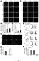
- Figs. 7A-C The results of cardiac fibroblasts (NCFs) were shown in Figs. 7A-C . It was found that the percentage of VCAM-1 positive cells in NCFs was approximately 60% ( Fig. 7A : 6.57%, Fig. 7B : 58.95%, Fig. 7C : 54.73%). Conversely, the percentage of VCAM-1 positive cells in skin fibroblasts (ADFs) was approximately 5%, and it turned out that the percentage of VCAM-1 positive cells in NCFs is significantly more than that of ADFs (P ⁇ 0.001).
- VCAM-1 positive cardiac fibroblasts originate from an outer membrane-derived cell from the view point of embryology, and we obtained the suggestion that it is effective to classify fibroblasts from the view point of embryology, and not to conduct morphological classification but to conduct functional classification as a cell source for constructing a functional tissue.
- NCFs vascular endothelial cell marker
- EMT epithelial mesenchymal transition
- EndMT endothelial mesenchymal transition
- kidney fibroblasts that differentiate from vascular endothelial cells through EndMT are expressing CD31 ( J Am Soc Nephrol 19:2282-2287, 2008 ). This may also become one of the bases for supporting that NCFs are expressing CD31.
- cardiac fibroblasts enhance proliferation of mouse embryonic stem cell (mESC) derived cardiomyocytes, and contribute to construction of more functional cardiac cell sheets. Moreover, it was indicated that cardiac fibroblasts are more abundantly expressing VCAM-1 compared with skin fibroblasts, and that the VCAM-1 of cardiac fibroblasts play an important role in proliferation of cardiac cells and construction of cardiac tissues that are functionally biologically-designed.
- mESC mouse embryonic stem cell
- cardiac cells of the present invention By culturing using the cardiac cells of the present invention, functional cardiac tissues are preferably constructed.
- the cardiac cells obtained by the culture can be used as regenerative medicines such as transplantation, or as artificial organ materials such as cardiac tissue models.
Landscapes
- Health & Medical Sciences (AREA)
- Life Sciences & Earth Sciences (AREA)
- Engineering & Computer Science (AREA)
- Chemical & Material Sciences (AREA)
- Biomedical Technology (AREA)
- Zoology (AREA)
- Bioinformatics & Cheminformatics (AREA)
- Genetics & Genomics (AREA)
- Biotechnology (AREA)
- Organic Chemistry (AREA)
- Wood Science & Technology (AREA)
- General Health & Medical Sciences (AREA)
- Cell Biology (AREA)
- General Engineering & Computer Science (AREA)
- Biochemistry (AREA)
- Microbiology (AREA)
- Medicinal Chemistry (AREA)
- Public Health (AREA)
- Veterinary Medicine (AREA)
- Animal Behavior & Ethology (AREA)
- Epidemiology (AREA)
- Pharmacology & Pharmacy (AREA)
- Rheumatology (AREA)
- Cardiology (AREA)
- Virology (AREA)
- Immunology (AREA)
- Tropical Medicine & Parasitology (AREA)
- Proteomics, Peptides & Aminoacids (AREA)
- Chemical Kinetics & Catalysis (AREA)
- Gastroenterology & Hepatology (AREA)
- Developmental Biology & Embryology (AREA)
- Transplantation (AREA)
- Botany (AREA)
- Dermatology (AREA)
- Oral & Maxillofacial Surgery (AREA)
- Urology & Nephrology (AREA)
- Heart & Thoracic Surgery (AREA)
- General Chemical & Material Sciences (AREA)
- Nuclear Medicine, Radiotherapy & Molecular Imaging (AREA)
- Micro-Organisms Or Cultivation Processes Thereof (AREA)
Description
- The present invention relates to use of a cardiac fibroblast expressing VCAM-1 protein for proliferating of cardiac cell in vitro. In addition, the present invention relates to a fibroblast cell population expressing VCAM-1.
- Fibroblasts exist in almost all of vertebrate, and when tissue is injured by trauma and ischemia, the injured area is replaced with fibrous tissue in accordance with fibroblasts proliferation and the abundant extracellular matrix deposition. Likewise, in a variety of heart disease such as myocardial infarction and cardiomyopathies, a lot of cardiomyocytes were lost and also fibrous tissue replaces that area, which leads to cardiac remodeling and heart failure accompanied with excess hemodynamics stress and neurohumoral stimulation. Although neurohumoral factors such as angiotensin II and endothelin-1 are well known to contribute to promote the cardiac remodeling via blood pressure elevation, cardiomyocyte apoptosis and local inflammation, cardiac fibroblasts have been reported to secrete those factors. Cardiac fibroblasts are also known to play a critical role in heart developments. Interconnected cellular processes in a cardiac fibroblast form a network of collagen, fibroblasts and myocytes. Although cardiomyocyte proliferation is indispensable process of formation of thick ventricular wall and embryonic cardiac fibroblasts have also been reported to promote myocardial mitotic activity through β-1 integrin signaling. The cardiac fibroblasts dominant causative substance has been unclear. Herein cardiac fibroblasts multifariously act on heart development and pathogenesis and the importance of understanding of mutual interaction and underlying mechanisms between cardiomyocytes and cardiac fibroblasts have been widely recognized. However the uncertain properties of cardiac fibroblasts were the bottle-neck for it and it is required to reveal functional and molecular biological characteristics of cardiac fibroblasts.
- Heart tissue engineering is promising methods for not only regenerative medicine, but also tissue models. Among cardiac tissue engineering methods, cell sheet-based cardiac tissue using temperature responsive culture dishes have been developed. Previously, it was reported that layering of cardiac cell sheets containing neonatal rats-derived cardiomyocytes, fibroblasts and endothelial cells on the various types of vascular bed enabled to fabricate three-dimensional vascularized viable cardiac tissue (Non
patent documents 1 to 3). Since cell sheet-based tissue engineering does not need any scaffold, it requires some amounts of extracellular matrices to construct cell sheets. Consistent with the evidences that left ventricle is mainly composed of fibroblasts and cardiomyocytes, some amounts of fibroblast are indispensable to fabricate cardiac cell sheets when using purified embryonic stem cell-derived cardiomyocytes (Non patent document 4). Since recent reports have suggested that cell-cell interaction between cardiomyocytes and non-myocytes is important for heart physiology and pathogenesis (Non patent document 5), fibroblasts function might also affect the function of the engineered cardiac tissue and it might be prerequisite to select the suitable fibroblasts to fabricate the cardiac tissue in vitro for tissue models. However it remains unclear whether cardiac fibroblasts have the specific function for cardiomyocytes compared with other types of fibroblasts and the related molecular mechanisms. - As mentioned above, since the cardiac fibroblasts play an important role in heart developments, and the onset or cure of heart diseases, it is required to separate cardiac fibroblasts that specifically act on cardiac cells such as cardiomyocytes from other fibroblasts, and to sample the cardiac fibroblasts. According to the recent studies, it has been revealed that fibroblasts, which were previously considered as a uniform cell type, have a great variety of phenotypes, and that the phenotypes differ depending on a load state of existing organs, tissues or cells.
- However, the function of fibroblasts is not clearly known, and fibroblasts are only cells morphologically classified. Therefore, among fibroblasts, it is difficult to select only one type thereof having a specific function.
- Meanwhile, with respect to VCAM-1 and α4 integrin, Kwee, et al. reported that VCAM-1 was expressed on embryonic day 11.5 at epicardium, cardiomyocytes, ventricular septum, and the like. It was also reported that, although the expression of α4 integrin was recognized at similar areas as those of VCAM-1, α4 integrin was not expressed in ventricular septum (Non patent document 6). Moreover, it was reported that, on embryonic day 11.5, there are embryonic death resulting from inhibition of formation of the placenta, and deformity due to decrease in dense layers of ventricular myocardium and ventricular septum in an embryo that is defective in VCAM-1. Yang, et al. also reported an epicardium defect in α4 integrin null embryo of embryonic day 11.5 (Non patent document 7). Accordingly, it is considered that VCAM-1 and α4 integrin mainly contribute to formation of cardiac cells and epicardium in the embryonic stage.
-
- Non-Patent Document 1: Shimizu T, et al., Fabrication of pulsatile cardiac tissue grafts using a novel 3-dimensional cell sheet manipulation technique and temperature-responsive cell culture surfaces. Circulation research. 2002;90:e40
- Non-Patent Document 2: Sekiya S, et al., Bioengineered cardiac cell sheet grafts have intrinsic angiogenic potential. Biochemical and biophysical research communications. 2006;341:573-582
- Non-Patent Document 3: Shimizu T, et al., Cell sheet engineering for myocardial tissue reconstruction. Biomaterials. 2003;24:2309-2316
- Non-Patent Document 4: Matsuura K, et al., Hagiwara N, Zandstra PW, Okano T. Creation of mouse embryonic stem cell-derived cardiac cell sheets. Biomaterials. 2011;32:7355-7362
- Non-Patent Document 5: Deschamps AM, et al., Disruptions and detours in the myocardial matrix highway and heart failure. Current heart failure reports. 2005;2:10-17
- Non-Patent Document 6: Kwee L, et al., Defective development of the embryonic and extraembryonic circulatory systems in vascular cell adhesion molecule (vcam-1) deficient mice. Development (Cambridge, England). 1995;121:489-503
- Non-Patent Document 7: Yang JT, et al., Cell adhesion events mediated by
- The purpose of the present invention is to provide cardiac fibroblasts for the in vitro proliferation of cardiac cells in order to construct functional cardiac tissue.
- It has been made clear that, in cardiac cell culturing, a functional cardiac tissue is well constructed by using a cardiac cell culture material containing VCAM-1 protein.
- Namely, the present invention includes the features of the appended claims.
- A functional cardiac cell which can be used in a regenerative medicine and an organizational model can be constructed by use of the cardiac fibrolasts of the present invention for proliferating cardiac cells in vitro.
-
- [
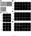
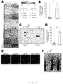
Fig. 1 ] A microscopic observation of NCF, ACF and ADF (photographs). (A) Bright field microscope images of each fibroblast. (B-E) Representative Figures of DDR2, vimentin and αSMA expression (Most of the fibroblasts were not expressing calponin, cytokeratin 11 or NG 2). - [
Fig. 2 ] Differences in characteristics of mESC derived cardiac cell sheets that were co-cultured with fibroblasts (photographs). (A) Before separated, many cell masses that were autonomously beating were observed on NCF and ACF co-culture sheet. After decrease in temperature, cell sheet formation was not observed in mESC derived cardiomyocytes and fibroblasts (-). (B) Extracellular action potentials on each of the cell sheets. Action potentials in ACF or NCF co-culture sheet were observed in each channel. However, the action potentials occurred on a one-off basis on the ADF co-culture sheet (encircling lines indicate the shapes of the cell sheets). (C) Immunofluorescent stain in each of the cell culture dishes which were observed by a confocal microscope. YFP emitted green (yellow) fluorescence (YFP: excitation wavelength 514 nm, fluorescence wavelength 527 nm), and vimentin emitted red fluorescence (cy3: excitation wavelength 512 nm, fluorescence wavelength 552 nm), and the nucleus was stained in hoechst 33258 (blue) (hoechst 33258: excitation wavelength 352 nm, fluorescence wavelength 461 nm). The confocal microscopy observation suggested that, the cells co-cultured with NCF or ACF have a large number of YFP (+) cells, compared with the cells co-cultured with fibroblasts (-) or ADF. (D) Immunofluorescent stain in each of the cell culture dishes observed by a confocal microscopy. cTnT emitted green fluorescence (FITC: excitation wavelength 488 nm, fluorescence wavelength 530 nm), vimentin emitted red fluorescence (cy3: excitation wavelength 512 nm, fluorescence wavelength 552 nm), and the nucleus was stained in hoechst 33258 (blue) (hoechst 33258: excitation wavelength 352 nm, fluorescence wavelength 461 nm). The confocal microscopy observation suggested that, the cells co-cultured with NCF or ACF have a large number of cTnT (+) cells, compared with the cells co-cultured with fibroblasts (-) or ADF. (E) The bar graphs show increase in the numbers of YFP (+) cells or of cTnT (+) cells in each of the cell culture dishes. The numbers of YFP (+) cells or of cTnT (+) cells in fibroblasts (-) were set to 1. More numbers of YFP (+) cells and cTnT (+) cells were observed in NCF or ACF culture dish compared with those in the culture dish of ADF co-culture or fibroblasts (-). In addition, there is no significant relationship in the number of cardiomyocytes between NCF and ACF. (N = 3, ** P < 0.01) - [
Fig. 3 ] The number of cardiomyocytes atday 1 andday 5 from the cell culture start in each of the cell culture dishes (Photographs). (A) Immunofluorescent stain atday 1 from culture start in each of the cell culture dishes which were used in a confocal microscope. YFP emitted green (yellow) fluorescence (YFP: excitation wavelength 514 nm, fluorescence wavelength 527 nm), and cTnT emitted red fluorescence (cy3: excitation wavelength 512 nm, fluorescence wavelength 552 nm), and the nucleus was stained in hoechst 33258 (blue) (hoechst 33258: excitation wavelength 352 nm, fluorescence wavelength 461 nm). (B) Immunofluorescent stain atday 5 from culture start in each of the cell culture dishes which were observed by a confocal microscope. YFP emitted green (yellow) fluorescence (YFP: excitation wavelength 514 nm, fluorescence wavelength 527 nm), and cTnT emitted red fluorescence (cy3: excitation wavelength 512 nm, fluorescence wavelength 552 nm), and the nucleus was stained in hoechst 33258 (blue) (hoechst 33258: excitation wavelength 352 nm, fluorescence wavelength 461 nm). (c) The number of cardiomyocytes in each of the cell culture dishes. The bar graphs show increase in the numbers of YFP (+) cells and of cTnT (+) cells (The values atday 1 in fibroblasts (-) were set to 1). In the ACF and NCF culture dishes, more numbers of cardiomyocytes were observed atday 5 from culture start compared with those atday 1. However, in the other culture dishes, there was no difference in the number of cardiomyocytes betweenday 1 andday 5. No significant difference was observed between ACF and NCF. (N =3, ** P < 0.01) - [
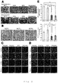
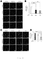
Fig. 4 ] Evaluation of proliferation in cardiomyocytes by immunofluorescent stain (photographs). (A) Immunofluorescent stain observation of Ki67 positive cardiomyocytes in each of co-culture dishes by using the confocal microscope. cTnT emitted green fluorescence (FITC: excitation wavelength 488 nm, fluorescence wavelength 530 nm), and Ki67 emitted red fluorescence (cy3: excitation wavelength 512 nm, fluorescence wavelength 552 nm), and the nucleus was stained in hoechst 33258 (blue) (hoechst 33258: excitation wavelength 352 nm, fluorescence wavelength 461 nm). (B) Percentage of Ki67 (+) or phosphorylated histone 3 (phosphor S10; Phh3) (+) cardiomyocytes in each of the culture dishes (N = 4, ** P < 0.01). (C) Immunofluorescence stain observation of phosphorylated histone 3 (phosphor S10; Phh3) positive cardiomyocytes in each of the culture dishes by using the confocal microscope. cTnT emitted green fluorescence (FITC: excitation wavelength 488 nm, fluorescence wavelength 530 nm), and phosphorylated histone 3 (phosphor S10; Phh3) emitted red fluorescence (cy3: excitation wavelength 512 nm, fluorescence wavelength 552 nm), and the nucleus was stained in hoechst 33258 (blue) (hoechst 33258: excitation wavelength 352 nm, fluorescence wavelength 461 nm). (D) Percentage of phosphorylated histone 3 (phosphor S10; Phh3) (+) cardiomyocytes in each of the culture dishes (N = 4, ** P < 0.01). (E) (F) BrdU FACS assay of cardiomyocytes in each of the culture dishes (N = 3, ** P < 0.01). (G) Immunofluorescence stain observation of YFP (+) and of cTnT (+) atday 1 andday 5 from culture start in the insert culture dishes by using the confocal microscope. YFP emitted green (yellow) fluorescence (YFP: excitation wavelength 514 nm, fluorescence wavelength 527 nm), and cTnT emitted red fluorescence (cy3: excitation wavelength 512 nm, fluorescence wavelength 552 nm), and the nucleus was stained in hoechst 33258 (blue) (hoechst 33258: excitation wavelength 352 nm, fluorescence wavelength 461 nm). (H) The bar graphs show increase in the numbers of YFP (+) cells and of cTnT (+) cells atday 1 and atday 5. The numbers of YFP (+) cells and of cTnT (+) cells atday 1 were set to 1. The proliferation of cardiomyocytes was observed at day 5 (N = 4, ** P < 0.01). - [
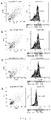
Fig. 5 ] (A) Comprehensive gene cluster analysis of ADF and NCF (photograph). This gene heat map shows a remarkable difference between ADF and NCF. This map was divided into two groups. The first group consisted of only ADF, and the second group consisted of only NCF. (B) The VCAM-1 gene expression level was examined by real time PCR. The VCAM-1 expression level was significantly high in NCF. The number of VCAM-1 genes in NCF was 16 times higher than that in ADF (N = 3, * P < 0.05). (C-D) The expression level of VCAM-1 protein in NCF and ADF in western blot analysis. The following transient overexpression cell lysate was used as a positive control: Sol8 (SantaCruz, CA, USA). The label peak of β-actin of each cell was set to 1 (N = 3, ** P < 0.01). (E) Immunofluorescence stain of the VCAM-1 receptor (α4β1) on mESC derived cardiomyocytes. (F) Western blot analysis of the VCAM-1 receptor on mESC derived cardiomyocytes. The following transient overexpression cell lysate was used as a positive control: Jurkat whole cell lysate. - [
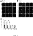
Fig. 6 ] Identification of cardiac growth factor by immunofluorescence stain analysis (photographs). (A-B) Immunofluorescence stain observation of the effect of neutralizing antibodies on cardiomyocytes atday 5. YFP emitted green (yellow) fluorescence (YFP: excitation wavelength 514 nm, fluorescence wavelength 527 nm), and cTnT emitted red fluorescence (cy3: excitation wavelength 512 nm, fluorescence wavelength 552 nm), and the nucleus was stained in hoechst 33258 (blue) (hoechst 33258: excitation wavelength 352 nm, fluorescence wavelength 461 nm). When NCF and cardiomyocytes were co-cultured by using a VCAM-1 neutralizing antibody, the number of cardiomyocytes was decreased atday 5. Meanwhile, when an isotype control was used, there was no effect on the number of cardiomyocytes atday 5. (N = 3, ** P < 0.01). (C-D) Immunofluorescence stain observation of the effect of VCAM-1 soluble protein on cardiomyocytes atday 5. YFP emitted green (yellow) fluorescence (YFP: excitation wavelength 514 nm, fluorescence wavelength 527 nm), and cTnT emitted red fluorescence (cy3: excitation wavelength 512 nm, fluorescence wavelength 552 nm), and the nucleus was stained in hoechst 33258 (blue) (hoechst 33258: excitation wavelength 352 nm, fluorescence wavelength 461 nm). Cardiomyocyte growth effect was obtained by culturing with VCAM-1 soluble protein (10 µg/mL). There was no significant difference in the number of cardiomyocytes between the conditions of co-culturing with NCF and co-culturing with VCAM-1 soluble protein in NCF (-) (N = 3, * P < 0.05, ** P < 0.01). - [
Fig. 7 ] The results of FACS analysis of cardiac fibroblasts derived from neonatal mice. (A-C) The results of staining with an anti-VCAM-1 antibody are shown. (D) The result of a negative control by staining skin fibroblasts only with a secondary antibody is shown. - The present invention relates to use of a cardiac fibroblast expressing VCAM-1 for proliferating cardiac cells in vitro. In the present disclosure, the "cardiac cell culture material" may be any material that is used when culturing a cardiac cell. For example, the material includes but is not limited to a reagent to be added to a culture medium, and a material, etc. for coating a bottom surface or a wall surface of a culture substrate of a culture vessel, etc. such as a petri dish and a flask, and the like.
- VCAM-1 (vascular cell adhesion molecule-1) is a known protein as a cell adhesion molecule that expresses in a vascular endothelial cell, and the like. For example, in the case of humans, VCAM-1 includes but not limited to a protein encoded by a gene described in accession number NM_001078, etc. of NCBI (National Center for Biotechnology Information), and also includes an isoform obtained by alternative splicing. The VCAM-1 protein in the present disclosure includes VCAM-1 which is expressing on a cell surface, a soluble VCAM-1, various mutants one or a plurality of, for example, 1-20, 1-15, 1-10 or 1-5 of amino acids of which have been deleted from, substituted from, or added to an amino acid of VCAM-1 protein and having the same activity as VCAM-1 protein. A VCAM-1 protein in an animal material which has been separated and purified by a well-known method and a recombinant protein may be used as the VCAM-1 in the present disclosure. A commercially available recombinant protein may be also used.
- Moreover, a cell that is expressing VCAM-1 may be used. In order to screen a cell that is expressing VCAM-1, a publicly known cell sorting method may be used. For example, the cell sorting method includes but not limited to flow cytometry using an anti-VCAM-1 antibody, magnetic bead method, affinity column method, and panning method.
- Anti-VCAM-1 antibodies are not particularly limited. Commercially available anti-VCAM-1 antibodies may be used, and a product produced by a known method by using VCAM-1 as an antigen may be also used. Moreover, as far as the cells that are expressing VCAM-1 may be screened, either monoclonal antibody or polyclonal antibody may be used; however, it is preferred to use monoclonal antibody from the viewpoint of specificity.
- Namely, the methods of screening the cardiac cell culture materials of the present disclosure include, a step of preparing cells, a step of performing cell sorting to the cells by using a VCAM-1 antibody, and a step of collecting only cells that have been judged to be expressing VCAM-1 as a result of the cell sorting.
- As the cell that is expressing VCAM-1, the type is a fibroblast. The fibroblasts include all the cells that will ultimately become fibroblasts or myofibroblasts. Namely, the scope of fibroblasts of the present invention includes the cells that are in the middle of differentiation or a maturation stage and cannot be identified as fibroblasts or myofibroblasts at that time as far as the cells will ultimately become fibroblasts or myofibroblasts.
- Cardiac-derived fibroblasts are preferably used, and among them, epicardium-derived fibroblasts are in particular preferred to be used. The animals from which fibroblasts are derived may be appropriately selected in accordance with the animals from which the cells to be co-cultured are derived. The animals, for example, include humans; experimental animals such as mice, rats, guinea pigs, hamsters, pigs, monkeys and rabbits; pet animals such as dogs, cats and birds; and livestock such as cattle, horses, sheep and goats. In a case where fibroblasts are taken from animals, the fibroblasts may be of at any time of the animals such as fetus, neonate, infant, adult, and there is no limit.
- The cardiac cell culture material of the present disclosure may be a composition containing physiological saline, cell culture solution, or cell preservation solution, etc. for maintenance or preservation of VCAM-1 protein or cells that are expressing VCAM-1 protein. There is no limit on the contents contained in the composition as far as the contents do not impair the function of VCAM-1. Moreover, the state of the cardiac cell culture material may be liquid, gel-like, freezed, or freeze-dried, and the state thereof is not limited.
- Further, the cardiac cell culture material may include fibroblasts regardless of presence or absence of VCAM-1 protein. The fibroblasts include all the cells that will ultimately become fibroblasts or myofibroblasts. Namely, even if the cells are in the middle of differentiation or a maturation stage and cannot be identified as fibroblasts or myofibroblasts at that time, if the cells are those that will ultimately become fibroblasts or myofibroblasts, the cells may be used without limit. Among them, fibroblasts that are expressing CD31 (vascular endothelial cell marker) are preferred. When fibroblasts that are expressing VCAM-1 protein are used as VCAM-1 protein, the ratio of VCAM-1 protein expressing cells (cell number) : CD31 expressing cells (cell number) are preferably 5:5-9:1, more preferably 5:5-8.2, and even more preferably 6:4-8.2.
- The cardiac cell culture material of the present disclosure can construct a functional cardiac tissue that can be used in a regenerative medicine and organizational model by culturing with a cardiac cell. The cardiac tissue can be used as an artificial organ material. The artificial organ material can be of any form. For example, it can be adhered to a damaged part of organs such as heart in the form of sheet. The artificial organ material can be also transplanted to a defect site of an organ after it is laminated or it is agglomerated by using scaffold, which has thickness in a certain extent. The material of the scaffold includes but not limited to hydroxyapatite, atelocollagen, and gel. Further, the artificial organ material can be used for cell transplantation, academic research, etc. as it is in the state of the culture cell, without making it to be a particular form. Furthermore, an artificial organ can be produced from the artificial organ material by using a 3D printer. The produced artificial organ not only can be used for transplantation but also can be widely used for safety pharmacology test and preclinical research, etc.
- In the present disclosure, "constructing a cardiac tissue" means constructing a tissue having at least one of the cardiac functions such as promoting division of cardiac cells, and providing uniform beating throughout a whole tissue, which can be used for regenerative medicine and a tissue model. The cardiac functions include all the known cardiac functions such as autonomous pulsating ability, contraction and relaxation ability, impulse conduction ability, and hormone secretion ability, etc. The cardiac functions are not limited to functions which only the heart has. For example, a muscle cell also has the contraction and relaxation ability. However, even if other cells have an equivalent function, it does not affect the definition of the cardiac functions. Further, with respect to the cardiac functions, there is no limit on highness and lowness of the functions as long as they are suitable for use purpose of a cardiac tissue. For example, for the purpose of producing an artificial heart, it is required to have a contraction and relaxation ability to the extent that it can pump out blood thought out the body; however, for the purpose of academic research, etc. of contraction and relaxation ability in vitro, it is satisfied if contraction and relaxation ability is detected by some means.
- In the artificial organ material, or the method to produce the same, cardiac cells to be used include all the cells that constitute the heart such as cardiomyocytes, smooth muscle cells, pacemaker cells and vascular endothelial cells. The derivation of the cardiac cells can be appropriately set in accordance with the purpose of use as an artificial organ material. For example, for the purpose of transplantation to humans, human-derived cardiac cells may be used, and for the purpose of constructing a tissue model in a mouse experiment, mouse-derived cardiac cells may be used. Furthermore, a cardiac cell of any period from fetus, newborn, pediatric and adult can be used, and there is no limit on the period. The cardiac cell of the present invention is preferred to be produced from pluripotent stem cells such as ES cells, iPS cells, and muse cells, and adult stem cells such as mesenchymal stem cells.
- The "culturing" can be carried out by a publicly known cell culturing method, and there is no limit on the condition of the culturing as long as a cardiac cell culture material and a cardiac cell are present in a culture vessel, or are immersed in the same culture medium. In a case where the cardiac cell culture material are cells which are expressing VCAM-1 protein, the mixing percentage of the cells (cell number) that are expressing VCAM-1 to cardiac cells are preferably 3-20%, more preferably 6-18% and most preferably 9-16%.
- A culture liquid used for the culturing can be appropriately set in accordance with a kind of cell to be cultured. For example, DMEM, α-MEM, RPMI-1640, and the like may be used. Nutritional substances such as FCS and FBS and antibiotics may be added to the culture liquid. With respect to the cultivation period, the number of days until the desired cell number and/or function are obtained may be appropriately set. For example, the periods include 1 day, 2 days, 3 days, 4 days, 5 days, 6 days, 7 days, 8 days, 9 days, 10 days, 11 days, 12 days, 13 days, 14 days, 1 month, 2 months, 3 months, 4 months, 5 months, and 6 months. The cultivation temperature may be appropriately set in accordance with the kinds of cells to be cultured. For example, the temperature may be 10-60°C, preferably 20-50°C, and more preferably 30-40°C.
- The production method may further include the step of collecting cultured cells. The "cultured cells" may include both fibroblasts and cardiac cells, and may only include the cardiac cells. With respect to the step to collect a cell, the cell may be separated and collected by using proteases such as trypsin. However, it is preferred that cell is separated and collected by the change in temperature by using a temperature responsive culture dish capable of separating a cell while retaining an extracellular matrix, etc.
- The present invention is further described below in detail with reference to the following examples; however, it should be construed that the invention is no way limited to those examples.
- Wild-type C57BL/6 mice were purchased from Japan SLC (Shizuoka, Japan). B6 Cg-Tg (CAG-DsRed*MST) 1Nagy/J mice were purchased from The Jackson Laboratory (Bar Harbor, ME). All the experimental protocols were approved by the Institutional Animal Care and Use Committee of Tokyo Women's Medical University. The following antibodies were used for immune cytochemistry, western blot and flow cytometric analysis (FACS): rabbit polyclonal anti-discoidindomein receptor tyrosine kinase 2 (DDR2) (GeneTex, Irvine, CA); guinea pig monoclonal anti-vimentin (Progen, Heidelberg, Germany); mouse monoclonal anti-NG2 (Millipore, Temecula, CA); Rabbit polyclonal anti-alpha smooth muscle actin (Abcam, Cambridge, UK); mouse monoclonal anti-cardiac troponin T (cTnT) (Thermo Scientific, Rockford, IL); mouse monoclonal anti-cytokeratin11 (EXBIO, NadSafinou, CZ); rabbit polyclonal anti-Ki67 (Abcam, Cambridge, UK); rabbit polyclonal anti-Histon H3 (phosphor S10) (Abcam, Cambridge, UK); rat monoclonal anti-integrin α4/β1 (Abcam, Cambridge, UK); recombinant mouse VCAM-1/CD106 Fc chimera (R&D systems, Minneapolis, MN). Unless otherwise specified, all reagents were purchased from Sigma-Aldrich. Secondary antibodies were purchased from Jackson ImmunoResearch Laboratories (West Grove, PA).
- The maintenance of mESC expressing the neomycin phosphotransferase gene under the control of the α-myosin heavy chain promoter and cardiomyocyte differentiation and purification were described previous report (Matsuura K, et al.. Biomaterials. 2011;32:7355-7362). Briefly, for cardiac induction and cardiomyocyte purification, trypsinized ES cells were seeded at 5 × 10⁴ cells/mL (total, 125 mL/flask) into spinner flasks (Integra Biosciences, Zizers, Switzerland) and cultured with DMEM supplemented with 10% FBS for 10 days, then these differentiated cells were treated with neomycin for further 8 days.
- Fibroblasts were obtained from Wild-type C57BL/6 mice (Neonatal, 1 day; Adult, 10-12 weeks).
- Neonatal cardiac fibroblasts (NCFs) were obtained from hearts of neonatal mice (1 day of age) as described previous report (Matsuura K, et al,. Biomaterials. 2011; 32: 7355-7362). NCFs from
passage 3 were used for the experiments. - Adult cardiac fibroblasts (ACFs) were obtained from hearts of adult mice (10-12 weeks) using the explant culture method as follow. First hearts were washed with PBS(-) and cut into circa 5 mm2 species. These species were covered with sterilized cover glasses and cultured with DMEM supplemented with 10% FBS on 10 cm culture dishes. 2 weeks after starting culture, cells were dissociated with 0.25% Trypsin/EDTA and subcultured to other 10cm dishes. ACFs from
passage 3 were used for the experiments. - Adult dermal fibroblasts (ADFs) were obtained from dorsal dermal tissue of adult mice (10-12 weeks). First harvested dermal tissues were treated with Dispase I [1000 U/mL] (Eidea inc.) over night at 4 °C. Next, the tissues were cut into circa 1mm2 species. These species were covered with sterilized cover glasses and cultured with DMEM supplemented with 10% FBS on 10 cm culture dishes. 2 weeks after starting culture, cells were dissociated with 0.25% Trypsin/EDTA and subcultured to another 10cm dishes. ADFs from
passage 3 were used for the experiments. - In some experiments, NCFs and ADFs were isolated from B6.Cg-Tg (CAG-DsRed*MST) 1Nagy/J mice (Neonatal: 1 day, Adult: 10 weeks) with the same methods as described above.
- Before seeding cells, the surface of temperature-responsive culture dishes (UpCell; CellSeed inc.) was coated with FBS for 2h. mESC-derived cardiomyocytes were co-cultured with each type of fibroblasts at the ratio of 8:2 with DMEM supplemented with 10% FBS (3.2 ×10⁵ cells/cm2). After 5 days of culture, the cells were incubated at 20 °C for detaching cell sheets. Bright field images of samples were obtained by a Nikon ECLIPSE Ti (Nikon, Tokyo, Japan).
- The electrical activities of the cardiomyocyte sheets were obtained from the extracellular potentials measured by a multi-electrode array (MED) system (Alpha MED Sciences, Osaka, Japan) as described previous report (Matsuura K, et al,. Biomaterials. 2011; 32:7355-7362).
- Cells were fixed with 4% paraformaldehyde and subjected to immunostaining as described previous report (Matsuura K, et al,. Biomaterials. 2011;32:7355-7362) . Images of the stained samples were obtained by an ImageXpress Ultra Confocal High Content Screening System (Molecular Devices, CA, USA). Image analysis data was obtained by a MetaExpress software (Molecular Devices, CA, USA) .
- Incubating cells (5×10⁵ cells) were stained with BrdU at a final concentration of 10 µM in cell culture medium. BrdU staining for a FACS analysis was performed as described in a BrdU Flow Kits Instruction Manual (BD Pharmingen, Franklin Lakes, NJ). Briefly, cells were fixed and permeabilized with BD Cytofix/Cytoperm Buffer, then exposed incorporated BrdU with DNase. BrdU staining was performed with APC-anti-BrdU antibody (BD Pharmingen, Franklin Lakes, NJ). Samples were analysed with a Gallios (Beckman Coulter, Brea, CA). The following reagents were used for the analysis: BD Cytofix/Cytoperm Buffer (BD Pharmingen, Franklin Lakes, NJ); BD Perm/Wash Buffer (10X) (BD Pharmingen, Franklin Lakes, NJ); BD Cytoperm Plus Buffer (10X) (BD Pharmingen, Franklin Lakes, NJ); BrdU (10 mg/mL) (BD Pharmingen, Franklin Lakes, NJ); DNase (BD Pharmingen, Franklin Lakes, NJ).
- Samples were observed five days in 5% CO2 at 37 °C with a BZ-9000 Fluorescence Microscope (Keyence, Osaka, Japan).
- Total RNA was extracted using TRIzol reagent (Invitrogen, Carlsbad, CA) according to the manufacturer's instructions. Total RNA was further purified using the Qiagen RNeasy Mini Kit (QIAGEN, Valencia, CA) according to the manufacturer's instructions.
- RNA quantity and quality were determined using a Nanodrop ND-1000 spectrophotometer (Thermo Fisher Scientific Inc., Waltham, MA) and an Agilent Bioanalyzer (Agilent Technologies, Palo Alto, CA), as recommended.
- For cRNA amplification and labeling, total RNA was amplified and labeled with Cyanine 3 (Cy3) using Agilent Low Input Quick Amp Labeling Kit, one-color (Agilent Technologies, Palo Alto, CA) following the manufacturer's instructions. Briefly, 100 ng of total RNA was reversed transcribed to double-strand cDNA using a poly dT-T7 promoter primer. Primer, template RNA and quality-control transcripts of known concentration and quality were first denatured at 65 °C for 10 min and incubated for 2 hours at 40 °C with 5X first strand Buffer, 0.1 M DTT, 10 mM dNTP mix, and Affinity Script RNase Block Mix. The AffinityScript enzyme was inactivated at 70 °C for 15 min.
- cDNA products were then used as templates for in vitro transcription to generate fluorescent cRNA. cDNA products were mixed with a transcription master mix in the presence of T7 RNA polymerase and Cy3 labeled-CTP and incubated at 40 °C for 2 hours. Labeled cRNAs were purified using QIAGEN's RNeasy mini spin columns and eluted in 30 µl of nuclease-free water. After amplification and labeling, cRNA quantity and cyanine incorporation were determined using a Nanodrop ND-1000 spectrophotometer and an Agilent Bioanalyzer.
- For Sample hybridization, 1.65 µg of Cy3 labeled cRNA were fragmented, and hybridized at 65 °C for 17 hours to an Agilent Mouse GE 4x44Kv2 Microarray (Design ID: 026655). After washing, microarrays were scanned using an Agilent DNA microarray scanner.
- For data analysis of microarray, intensity values of each scanned feature were quantified using Agilent feature extraction software version 10.7.3.1, which performs background subtractions.
- We only used features that were flagged as no errors (present flags) and excluded features that were not positive, not significant, not uniform, not above background, saturated, and population outliers (marginal and absent flags). Normalization was performed using Agilent GeneSpring GX version 11.0.2. (per chip: normalization to 75 percentile shift; per gene: normalization to median of all samples). There are total of 39,429 probes on Agilent Mouse GE 4x44Kv2 Microarray (Design ID: 026655) without control probes.
- The altered transcripts were quantified using the comparative method. We applied ≥ 2-fold change in signal intensity to identify the significant differences of gene expression in this study.
- Complementary DNA was generated from total RNA with High Capacity cDNA Reverse Transcription Kit (Applied biosystems). As the PCR-related primers, VCAM-1 Gene Express Assays (life Technology) was used.
- Each RT-PCR included 10 minutes at 25 °C, 120 minutes at 37°C, and 5 seconds at 85°C with iCycler (BIO-RAD). cDNA template (1 µg) was used from each sample. TaqMan probe real-time PCR studies were performed with TaqMan Gene Expression Assays (Applied biosystems). All experiments were conducted in triplicate. Samples were cycled 40 times with an 7300 Real Time PCR System (Applied Biosystems) as follows: 2 minutes at 50°C and 10 minutes at 95°C, followed by 40 cycles of 15 seconds at 95°C and 1 minute at 60°C. Relative quantification was calculated according to the ΔΔCT method for quantitative real-time PCR using a Gap DH gene as endogenous control.
- NCFs or ADFs were lysed in Laemmli sample buffer (BIO-RAD, CA, USA), protease inhibitor (Boehringer Mannheim, Indianapolis, IN) and 2-mercaptoethanol (Wako Pure Chemical Industries, Japan). The samples were separated on a 4% to 12% Bis-Tris Gels (Life Technologies, MD, United States), electrotransferred to a iBlot Transfer Stack, nitrocellulose, regular-size (Life technologies, MD, United States) with iBlot 7-Minute Blotting System(Life technologies, MD, United States), and processed for chemiluminescence analysis with Amersham ECL Prime Western Blotting Detection Reagent (GE Healthcare, PA, United States). Band intensity was analyzed using LAS4000 (Fujifilm, Tokyo, Japan) and NIH image software (version 1.46r). The following cell transient overexpression lysates were used for positive controls: K562 (Human erythromyeloblastoid leukemia cell line) for Co111a1 (Abcam, CB, UK); Sol8 (SantaCruz, CA, USA) for Vcam-1; ITGB1 293T for β1/CD29 (Abnova, Taipei, Taiwan); Jurkat Whole Cell Lysate for integrin α4β1 (SantaCruz, CA, USA).
- The following antibodies and culture dishes were used for neutralizing antibody assay: anti-VCAM-1 (LifeSpan Biosciences, Seattle, WA); goat IgG isotype control (LifeSpan Biosciences, Seattle, WA). Cell Culture Inserts for 24-well plates. 0.4 µm pores, Translucent, High Density PET Membrane (BD Pharmingen,Franklin Lakes, NJ).
- After the pretreatment with the antibodies at 10 µg/mL for 30 min, fibroblasts were seeded onto the upper layer of insert culture dishes (2.4 x 10⁵ cells). mESC-derived cardiomyocytes were seeded onto the below layer (4.8 x 10⁵ cells). The culture medium with the antibody at 10 µg/mL was changed every day until 5 days.
- All data were presented as the mean ± SD. The significance of the variation among different groups was determined by One-Way ANOVA Analysis. And then, the difference between two groups was determined by Tukey-Kramer Multiple Comparison Test using Statcel Software. p value < 0.05 was considered to be significantly different.
- At first we evaluated the characterization of cells that we were going to use for the co-culture experiments. The phase contrast images revealed that cells isolated from neonatal hearts, adult hearts and adult dermal tissue showed the fibroblast-like morphology (See
Fig. 1A ). Since there are not specific antibodies for fibroblasts, we tried to examine the expression of the proteins that are known to be expressed in fibroblasts such as DDR2 (CD167b), vimentin and αSMA. As shown inFigs. 1B to 1E , almost all of each type of cells expressed DDR2, vimentin and αSMA, but not Calponin (smooth muscle cell marker), Cytokeratin (epithelial cell marker) and NG2 (pericyte marker). On the basis of these findings, we used these cells as fibroblasts following experiments. - According to our previous findings that certain extent amounts of fibroblasts were necessary for fabricating cell sheet using mESC-derived cardiomyocytes and the optimal ratio of cardiomyocytes/fibroblasts was 8:2 (Biomaterials. 2011:32:7355-7362), we tried to create cardiac cell sheets using mESC-derived cardiomyocytes and 3 types of fibroblasts (ACFs, ADFs and NCFs) on UpCell temperature-responsive culture dishes. When cardiomyocytes were co-cultured with ACFs or NCFs, beating cardiomyocytes were equally distributed all over the area. Conversely, when cardiomyocytes were co-cultured with ADFs, nearby beating cells were aggregated. After 5 days cultivation, when the cultivation condition were changed from 37 °C to 20 °C, monolayered cell sheets were created in every condition with fibroblasts, but not in the condition without fibroblasts (
Fig. 2A ). - Next we examined the electrophysiological evaluation of the cell sheets using a MED system (Biomaterials. 2011; 32:7355-7362, Biomaterials. 2006;27:4765-4774). Consistent with the microscopical observation (
Fig.2A ), the extracellular action potential was observed at each channel in cell sheets with ACFs and NCFs (Fig. 2B ). Since it was recognized that, in these cell sheets, the entire sheets were uniformly beating, it was suggested that an electronic network was fabricated in the sheets and these cell sheets can carry out electric propagation. Meanwhile, in the cell sheets co-cultured with ADFs, the extracellular action potential was only observed in limited areas. - To confirm the difference of cardiomyocytes distribution among cell sheets, cofocal microscopic analysis was performed. As shown in
Figs. 2C to 2E , the number of YFP(+) cells and cardiac troponin T (cTnT) (+) cells, indicatives of mESC-derived cells, in cell sheets with ACFs and NCFs were more than those in cell sheets with ADFs. The number of cardiomyocytes in cell sheets with ADFs was comparable to that in condition without fibroblasts. In addition, there is no significant correlation on the number of cardiomyocytes between cell sheets co-cultured with ACFs and NCFs. These findings suggest that every kind of fibroblasts was useful for fabricating cell sheet, but fibroblasts derived from hearts might be better for fabricating more functional cardiac cell sheets. - To investigate the cause of the different number of cardiomyocytes between cell sheets co-cultured with heart-derived fibroblasts and dermal tissue-derived fibroblasts, the number of cardiomyocytes was examined at
day 1 andday 5 in co-culture (Figs.3A to C ). Atday 1, the number of cardiomyocytes was identical among conditions, suggesting that each type of fibroblasts did not affect the initial adherence of cardiomyocytes after seeding. In co-culture with ACFs and NCFs, the number of YFP (+) and cTnT (+) cardiomyocytes atday 5 was significantly higher than that atday 1. On the other hand, in co-culture with ADFs or in cardiomyocytes monoculture condition, the number of cardiomyocytes atday 5 was similar to that atday 1. The time-lapse image analysis using YFP(+) cardiomyocytes and fibroblasts isolated from DsRed mice showed that cardiomyocytes migrated and proliferated and constructed the mutual network formation in co-culture with NCFs. Conversely in co-culture with ADFs, cardiomyocytes showed less proliferation and did not construct the network formation. These findings suggest that fibroblasts from hearts, but not fibroblasts from dermal tissue might induce proliferation of mESC-derived cardiomyocytes in co-culture condition. - The proliferation of cardiomyocytes among conditions was confirmed by the immune cytochemical analysis. As shown in
figure 4A to D , the percentage of Ki67(+) cells and phospho histone H3 (PHH3) (+) cardiomyocytes in co-culture with NCFs were significant higher than those in co-culture with ADFs and in cardiomyocytes monoculture condition. Furthermore BrdU incorporation assay also showed the significant increase of the percentage of proliferative cardiomyocytes in co-culture with NCFs compared with that in co-culture with ADFs and in cardiomyocytes monoculture condition (Figs. 4E and F ). These findings strongly suggest that heart-derived fibroblasts induce proliferation of cardiomyocytes. - To investigate the underlying mechanisms on the proliferation of cardiomyocytes in co-culture with NCFs, mESC-derived cardiomyocytes and NCFs were cultured using cell culture inserts. In this experiment, NCFs were cultured on the upper layer and cardiomyocytes were cultured on the lower layer. The number of cardiomyocytes at
day 5 was remarkably higher than that atday 1 in the presence of NCFs (Fig. 4G ). However, the degree of the increase on cardiomyocyte number in the cell culture insert experiments betweenday 1 and day 5 (∼1.8 times) (Fig. 4H ) was lower than that in co-culture condition (∼2.5 times). These findings indicate it might promote the cardiomyocyte proliferation that the soluble factors secreted from NCFs and the cell-cell interaction between cardiomyocytes with cardiac fibroblasts. - To identify the factors that are responsible for involved in these effects, we performed comprehensive genetic analysis between NCFs and ADFs using a microarray analysis. As shown in
figure 5A , many differences in gene expression were observed between NCFs and ADFs. Over 500 genes showed more than 10 times enhanced expression in NCFs compared with ADFs. After choosing the cardiovascular-related genes from the lists, 20 genes were remained. Furthermore when we selected genes that were reported the embryonic lethal phenotype causing a disorder to generate heart in knock out mouse model and also act as a soluble factor and an sdhesive factor, Vcam-1 was remained. The enhanced expression of Vcam-1 in NCFs compared with ADFs was confirmed by quantitative RT-PCR and western blot analysis (Figs. 5B to D ). - Since integrin α4β1 is known to be the principal co-receptor of VCAM-1, we examined the integrin α4β1 expression in mESC-derived cardiomyocytes. As shown in
figures 5E and F , almost all of mESC-derived cardiomyocytes showed generation of integrin α4β1. - Next we elucidated whether VCAM-1 contributed to cardiac fibroblasts-mediated cardiomyocyte proliferation using neutralizing antibodies. After the pretreatment of NCFs with anti-VCAM-1 antibodies, NCFs and mESC-derived cardiomyocytes were cultured using cell culture inserts. Anti-VCAM-1 antibody treatment significantly inhibited cardiac fibroblast-mediated increase of cardiomyocyte number (
Figs. 6A and B ). - Finally we evaluated the direct effects of VCAM-1 on the proliferation of cardiomyocytes. One day after starting culture, cardiomyocytes were treated with VCAM-1 recombinant protein until
day 5. As shown infigure 6C and D , VCAM-1 treatment increased the number of cardiomyocytes compared with control. These findings suggest heart-derived fibroblasts might induce cardiomyocyte proliferation through fibroblasts-mediated VCAM-1 and integrin α4β1 in cardiomyocytes. - To confirm importance of VCAM-1 positive cells in constructing functional cardiac cell sheets, we measured the percentage of VCAM-1 positive cells in organism-derived cardiac fibroblasts.
- Cardiac fibroblasts were dissected and collected from neonatal mice (1 day) of C57/BL6 mice, and skin fibroblasts were dissected and collected from adult mice (10-12 weeks). Each of the fibroblasts were adhesion-cultured up to
passage 3, and the cell volume of 1×10⁷ cells per condition was obtained.Passage 3 is the same condition with the culture condition of the above-mentioned cardiomyocytes produced by cell sheets. - Both fibroblasts were subjected to primary immunofluorescent stain with Goat polyclonal anti-VCAM-1 antibodies (R&D systems, Minneapolis, MN), and were subjected to secondary immunofluorescent stain with Alexa Fluor 488 Donkey anti-goat IgG (Life Technologies, MD, United States). Subsequently, FACS analysis was conducted at Gallios (Beckman Coulter, Brea, CA), and VCAM-1 positive cell rate was measured (N=3). Calculation of significant difference was carried out by Student's t-test.
- The results of cardiac fibroblasts (NCFs) were shown in
Figs. 7A-C . It was found that the percentage of VCAM-1 positive cells in NCFs was approximately 60% (Fig. 7A : 6.57%,Fig. 7B : 58.95%,Fig. 7C : 54.73%). Conversely, the percentage of VCAM-1 positive cells in skin fibroblasts (ADFs) was approximately 5%, and it turned out that the percentage of VCAM-1 positive cells in NCFs is significantly more than that of ADFs (P < 0.001). - It was suggested that cardiac fibroblasts containing many VCAM-1 positive cells contribute to construction of functional myocardial tissues by proliferating cardiomyocytes derived from mice ES through the expressing VCAM-1. Further, it was considered that VCAM-1 positive cardiac fibroblasts originate from an outer membrane-derived cell from the view point of embryology, and we obtained the suggestion that it is effective to classify fibroblasts from the view point of embryology, and not to conduct morphological classification but to conduct functional classification as a cell source for constructing a functional tissue.
- It is considered that, in NCFs, the majority of the cells that are not expressing VCAM-1 express CD31 (vascular endothelial cell marker). The reason for this is as follows: it is known that tissue-resident cardiac fibroblasts are produced from epicardium-derived cells through epithelial mesenchymal transition (EMT), and also are differentiated from vascular endothelial cells through endothelial mesenchymal transition (EndMT). Furthermore, as is the case with cardiac fibroblasts, kidney fibroblasts that differentiate from vascular endothelial cells through EndMT are expressing CD31 (J Am Soc Nephrol 19:2282-2287, 2008). This may also become one of the bases for supporting that NCFs are expressing CD31.
- From the above, it was clarified that, not skin fibroblasts but cardiac fibroblasts enhance proliferation of mouse embryonic stem cell (mESC) derived cardiomyocytes, and contribute to construction of more functional cardiac cell sheets. Moreover, it was indicated that cardiac fibroblasts are more abundantly expressing VCAM-1 compared with skin fibroblasts, and that the VCAM-1 of cardiac fibroblasts play an important role in proliferation of cardiac cells and construction of cardiac tissues that are functionally biologically-designed.
- By culturing using the cardiac cells of the present invention, functional cardiac tissues are preferably constructed. The cardiac cells obtained by the culture can be used as regenerative medicines such as transplantation, or as artificial organ materials such as cardiac tissue models.
Claims (6)
- Use of a cardiac fibroblast expressing VCAM-1 protein for proliferating of cardiac cells in vitro, wherein the cardiac fibroblast expressing VCAM-1 protein comprises a cardiac fibroblast sorted as a cardiac fibroblast expressing VCAM-1 protein by using a known cell sorting method, and the fibroblast excludes a fibroblast that is in the middle of differentiation or a maturation stage and cannot be identified as a fibroblast.
- Use of the cardiac fibroblast expressing VCAM-1 protein according to claim 1, wherein the cardiac fibroblast expressing VCAM-1 protein comprises a cardiac fibroblast cell line expressing VCAM-1 protein.
- Use of the cardiac fibroblast expressing VCAM-1 protein according to claim 1 or 2, wherein the known cell sorting method is a cell sorting method using an anti-VCAM-1 antibody.
- Use of the cardiac fibroblast expressing VCAM-1 protein according to any one of claims 1 to 3, wherein the cardiac fibroblast expressing VCAM-1 protein is cultured with a cardiac cell.
- Use of the cardiac fibroblast expressing VCAM-1 protein according to claim 4, wherein the cardiac cell is a cardiomyocyte.
- A fibroblast cell population comprising a fibroblast expressing VCAM-1, wherein the fibroblast expressing VCAM-1 comprises a cardiac fibroblast sorted as a cardiac fibroblast expressing VCAM-1 protein by using a known cell sorting method, and the fibroblast excludes a fibroblast that is in the middle of differentiation or a maturation stage and cannot be identified as a fibroblast.
Applications Claiming Priority (2)
| Application Number | Priority Date | Filing Date | Title |
|---|---|---|---|
| JP2014142804 | 2014-07-11 | ||
| PCT/JP2015/050028 WO2016006262A1 (en) | 2014-07-11 | 2015-01-05 | Cardiac cell culture material |
Publications (3)
| Publication Number | Publication Date |
|---|---|
| EP3168297A1 EP3168297A1 (en) | 2017-05-17 |
| EP3168297A4 EP3168297A4 (en) | 2018-03-07 |
| EP3168297B1 true EP3168297B1 (en) | 2020-09-16 |
Family
ID=55063910
Family Applications (1)
| Application Number | Title | Priority Date | Filing Date |
|---|---|---|---|
| EP15819754.1A Active EP3168297B1 (en) | 2014-07-11 | 2015-01-05 | Cardiac cell culture material |
Country Status (8)
| Country | Link |
|---|---|
| US (2) | US20170112880A1 (en) |
| EP (1) | EP3168297B1 (en) |
| JP (2) | JP6241893B2 (en) |
| KR (1) | KR102263824B1 (en) |
| CN (2) | CN106661552B (en) |
| AU (1) | AU2015286462B2 (en) |
| CA (1) | CA2954743C (en) |
| WO (1) | WO2016006262A1 (en) |
Families Citing this family (7)
| Publication number | Priority date | Publication date | Assignee | Title |
|---|---|---|---|---|
| CN109890398B (en) * | 2017-02-24 | 2024-01-16 | 玛土撤拉有限公司 | Injectable composition useful for treating heart diseases and comprising fibroblasts and method for producing fibroblasts for therapeutic use |
| JP6883904B2 (en) * | 2018-08-29 | 2021-06-09 | 株式会社メトセラ | Fibroblast production method and G-CSF positive fibroblast population |
| KR102200341B1 (en) * | 2018-12-21 | 2021-01-08 | 고려대학교 산학협력단 | Cardiac Transplantation Sheet Mimicked Cardiac Tissue Containing Adipose Stem Cell Sheed and Method for Preparing Therof |
| US20220135942A1 (en) | 2019-03-29 | 2022-05-05 | Nagasaki University | Cultured tissue and method for producing same |
| WO2021146994A1 (en) * | 2020-01-22 | 2021-07-29 | 京东方科技集团股份有限公司 | Mesenchymal stem cell sheet and use thereof |
| CN115210361A (en) | 2020-03-04 | 2022-10-18 | 玛土撤拉有限公司 | Fibroblast cells with enhanced erythropoietin producing capacity |
| KR102283340B1 (en) * | 2020-03-27 | 2021-07-30 | 서울대학교산학협력단 | Cardiac-mimetic cell culture platform and a method for direct cardiac reprogramming |
Family Cites Families (11)
| Publication number | Priority date | Publication date | Assignee | Title |
|---|---|---|---|---|
| AU2003901668A0 (en) * | 2003-03-28 | 2003-05-01 | Medvet Science Pty. Ltd. | Non-haemopoietic precursor cells |
| JP2004535199A (en) * | 2001-07-12 | 2004-11-25 | ジェロン コーポレイション | Cardiomyocyte lineage cells produced from human pluripotent stem cells |
| PL1641914T3 (en) * | 2003-06-27 | 2017-01-31 | DePuy Synthes Products, Inc. | Postpartum cells derived from placental tissue, and methods of making and using the same |
| JP4749331B2 (en) * | 2003-10-07 | 2011-08-17 | 株式会社バイオマスター | Cell differentiation of adipose-derived progenitor cells |
| EP1876233A1 (en) * | 2005-01-24 | 2008-01-09 | Japan Health Sciences Foundation | Cells capable of differentiating into cardiac muscle cells |
| US8933197B2 (en) * | 2007-08-15 | 2015-01-13 | Amunix Operating Inc. | Compositions comprising modified biologically active polypeptides |
| JP2012523238A (en) * | 2009-04-09 | 2012-10-04 | ジ・アリゾナ・ボード・オブ・リージェンツ・オン・ビハーフ・オブ・ザ・ユニバーシティ・オブ・アリゾナ | Cell seeding and co-culture of 3D fibroblast constructs |
| US20120045487A1 (en) * | 2009-04-29 | 2012-02-23 | The Regents Of The University Of Michigan | Multiphasic microfibers for spatially guided cell growth |
| US8323972B2 (en) * | 2009-09-30 | 2012-12-04 | Advanced Technologies And Regenerative Medicine, Llc | Mammary artery derived cells and methods of use in tissue repair and regeneration |
| WO2012162741A1 (en) * | 2011-06-01 | 2012-12-06 | Monash University | Enrichment of cardiomyocytes |
| US10017739B2 (en) * | 2012-09-06 | 2018-07-10 | Duke University | Methods of expanding and assessing B cells and using expanded B cells to treat disease |
-
2015
- 2015-01-05 CN CN201580037730.1A patent/CN106661552B/en active Active
- 2015-01-05 EP EP15819754.1A patent/EP3168297B1/en active Active
- 2015-01-05 CA CA2954743A patent/CA2954743C/en active Active
- 2015-01-05 KR KR1020177003798A patent/KR102263824B1/en active IP Right Grant
- 2015-01-05 CN CN202010584861.4A patent/CN111876371A/en active Pending
- 2015-01-05 WO PCT/JP2015/050028 patent/WO2016006262A1/en active Application Filing
- 2015-01-05 AU AU2015286462A patent/AU2015286462B2/en active Active
- 2015-07-10 JP JP2015138613A patent/JP6241893B2/en active Active
-
2017
- 2017-01-09 US US15/401,832 patent/US20170112880A1/en not_active Abandoned
- 2017-10-30 JP JP2017208721A patent/JP2018029611A/en active Pending
-
2018
- 2018-08-21 US US16/106,723 patent/US11801267B2/en active Active
Non-Patent Citations (1)
| Title |
|---|
| None * |
Also Published As
| Publication number | Publication date |
|---|---|
| US20180369288A1 (en) | 2018-12-27 |
| CN111876371A (en) | 2020-11-03 |
| AU2015286462A1 (en) | 2017-03-02 |
| CN106661552A (en) | 2017-05-10 |
| WO2016006262A1 (en) | 2016-01-14 |
| KR20170023188A (en) | 2017-03-02 |
| EP3168297A1 (en) | 2017-05-17 |
| CN106661552B (en) | 2020-07-07 |
| US20170112880A1 (en) | 2017-04-27 |
| EP3168297A4 (en) | 2018-03-07 |
| JP6241893B2 (en) | 2017-12-06 |
| CA2954743C (en) | 2022-03-15 |
| CA2954743A1 (en) | 2016-01-14 |
| JP2018029611A (en) | 2018-03-01 |
| JP2016027797A (en) | 2016-02-25 |
| US11801267B2 (en) | 2023-10-31 |
| AU2015286462B2 (en) | 2021-04-15 |
| KR102263824B1 (en) | 2021-06-10 |
Similar Documents
| Publication | Publication Date | Title |
|---|---|---|
| US11801267B2 (en) | Cardiac cell culture material | |
| Raynaud et al. | Comprehensive characterization of mesenchymal stem cells from human placenta and fetal membrane and their response to osteoactivin stimulation | |
| JP2010500878A (en) | Co-culture method of stem cells and feeder cells using polymer membrane | |
| US8962323B2 (en) | Method of isolating dermal stem cells | |
| IL178232A (en) | Methods for augmenting proliferation potency of pluripotent stem cells | |
| EP3346002B1 (en) | Method for reducing pluripotent stem cells, method for producing cell population having reduced pluripotent stem cells | |
| Koyanagi et al. | Differentiation of circulating endothelial progenitor cells to a cardiomyogenic phenotype depends on E-cadherin | |
| Bai et al. | Establishment of human induced trophoblast stem-like cells from term villous cytotrophoblasts | |
| US20140370515A1 (en) | Method of co-culturing human endometrial stem cells and rat embryonic tooth bud cells to obtain ameloblast cells | |
| Iwamiya et al. | Cardiac fibroblast-derived VCAM-1 enhances cardiomyocyte proliferation for fabrication of bioengineered cardiac tissue | |
| Stingl et al. | Enzymatic dissociation and culture of normal human mammary tissue to detect progenitor activity | |
| Ibarra-Ibarra et al. | Improved efficiency of cardiomyocyte-like cell differentiation from rat adipose tissue-derived mesenchymal stem cells with a directed differentiation protocol | |
| KR101364965B1 (en) | Method for expansion and purification of human embryonic stem cell-derived cardiomyocyte | |
| KR20190112090A (en) | Control of Differentiation of Pluripotent Stem Cells | |
| Shimada et al. | Efficient expansion of mouse primary tenocytes using a novel collagen gel culture method | |
| JP2023179751A (en) | Method for producing proliferated hair follicle mesenchymal cells, and use thereof | |
| KR101760239B1 (en) | Method for isolating primary mesenchymal stem cells derived from human embryonic stem cells using cell insert culture system | |
| JP7198524B2 (en) | Method for producing spheroids and method for expressing pluripotent stem cell markers | |
| JP2021507714A (en) | Method of isolating human dermal fibroblasts | |
| TW201504435A (en) | Quality control method for hair-follicle forming composition | |
| Zhang et al. | Sertoli cells promote proliferation of bone marrow-derived mesenchymal stem cells in co-culture | |
| Guenther et al. | The treasury of Wharton's Jelly | |
| EP3569691A1 (en) | Method for producing corneal epithelial cell mass | |
| JP6587877B2 (en) | Marker for separating epidermal stem cells and method for producing three-dimensional cultured epidermis | |
| Elhendawi et al. | Kidney organoids |
Legal Events
| Date | Code | Title | Description |
|---|---|---|---|
| STAA | Information on the status of an ep patent application or granted ep patent |
Free format text: STATUS: THE INTERNATIONAL PUBLICATION HAS BEEN MADE |
|
| PUAI | Public reference made under article 153(3) epc to a published international application that has entered the european phase |
Free format text: ORIGINAL CODE: 0009012 |
|
| STAA | Information on the status of an ep patent application or granted ep patent |
Free format text: STATUS: REQUEST FOR EXAMINATION WAS MADE |
|
| 17P | Request for examination filed |
Effective date: 20170201 |
|
| AK | Designated contracting states |
Kind code of ref document: A1 Designated state(s): AL AT BE BG CH CY CZ DE DK EE ES FI FR GB GR HR HU IE IS IT LI LT LU LV MC MK MT NL NO PL PT RO RS SE SI SK SM TR |
|
| AX | Request for extension of the european patent |
Extension state: BA ME |
|
| DAX | Request for extension of the european patent (deleted) | ||
| A4 | Supplementary search report drawn up and despatched |
Effective date: 20180207 |
|
| RIC1 | Information provided on ipc code assigned before grant |
Ipc: C12N 1/00 20060101ALI20180201BHEP Ipc: C12N 5/077 20100101AFI20180201BHEP Ipc: C12N 5/10 20060101ALI20180201BHEP Ipc: A61K 38/17 20060101ALI20180201BHEP Ipc: A61K 35/33 20150101ALI20180201BHEP |
|
| STAA | Information on the status of an ep patent application or granted ep patent |
Free format text: STATUS: EXAMINATION IS IN PROGRESS |
|
| 17Q | First examination report despatched |
Effective date: 20190423 |
|
| GRAP | Despatch of communication of intention to grant a patent |
Free format text: ORIGINAL CODE: EPIDOSNIGR1 |
|
| STAA | Information on the status of an ep patent application or granted ep patent |
Free format text: STATUS: GRANT OF PATENT IS INTENDED |
|
| RIC1 | Information provided on ipc code assigned before grant |
Ipc: C12N 5/077 20100101AFI20200306BHEP Ipc: A61P 9/00 20060101ALI20200306BHEP Ipc: C12N 5/10 20060101ALI20200306BHEP Ipc: A61K 35/33 20150101ALI20200306BHEP Ipc: A61K 38/17 20060101ALI20200306BHEP Ipc: C12N 1/00 20060101ALI20200306BHEP |
|
| INTG | Intention to grant announced |
Effective date: 20200409 |
|
| GRAS | Grant fee paid |
Free format text: ORIGINAL CODE: EPIDOSNIGR3 |
|
| GRAA | (expected) grant |
Free format text: ORIGINAL CODE: 0009210 |
|
| STAA | Information on the status of an ep patent application or granted ep patent |
Free format text: STATUS: THE PATENT HAS BEEN GRANTED |
|
| AK | Designated contracting states |
Kind code of ref document: B1 Designated state(s): AL AT BE BG CH CY CZ DE DK EE ES FI FR GB GR HR HU IE IS IT LI LT LU LV MC MK MT NL NO PL PT RO RS SE SI SK SM TR |
|
| REG | Reference to a national code |
Ref country code: GB Ref legal event code: FG4D |
|
| REG | Reference to a national code |
Ref country code: CH Ref legal event code: EP |
|
| REG | Reference to a national code |
Ref country code: DE Ref legal event code: R096 Ref document number: 602015059270 Country of ref document: DE |
|
| REG | Reference to a national code |
Ref country code: IE Ref legal event code: FG4D |
|
| REG | Reference to a national code |
Ref country code: AT Ref legal event code: REF Ref document number: 1314192 Country of ref document: AT Kind code of ref document: T Effective date: 20201015 |
|
| PG25 | Lapsed in a contracting state [announced via postgrant information from national office to epo] |
Ref country code: NO Free format text: LAPSE BECAUSE OF FAILURE TO SUBMIT A TRANSLATION OF THE DESCRIPTION OR TO PAY THE FEE WITHIN THE PRESCRIBED TIME-LIMIT Effective date: 20201216 Ref country code: GR Free format text: LAPSE BECAUSE OF FAILURE TO SUBMIT A TRANSLATION OF THE DESCRIPTION OR TO PAY THE FEE WITHIN THE PRESCRIBED TIME-LIMIT Effective date: 20201217 Ref country code: BG Free format text: LAPSE BECAUSE OF FAILURE TO SUBMIT A TRANSLATION OF THE DESCRIPTION OR TO PAY THE FEE WITHIN THE PRESCRIBED TIME-LIMIT Effective date: 20201216 Ref country code: SE Free format text: LAPSE BECAUSE OF FAILURE TO SUBMIT A TRANSLATION OF THE DESCRIPTION OR TO PAY THE FEE WITHIN THE PRESCRIBED TIME-LIMIT Effective date: 20200916 Ref country code: HR Free format text: LAPSE BECAUSE OF FAILURE TO SUBMIT A TRANSLATION OF THE DESCRIPTION OR TO PAY THE FEE WITHIN THE PRESCRIBED TIME-LIMIT Effective date: 20200916 Ref country code: FI Free format text: LAPSE BECAUSE OF FAILURE TO SUBMIT A TRANSLATION OF THE DESCRIPTION OR TO PAY THE FEE WITHIN THE PRESCRIBED TIME-LIMIT Effective date: 20200916 |
|
| REG | Reference to a national code |
Ref country code: AT Ref legal event code: MK05 Ref document number: 1314192 Country of ref document: AT Kind code of ref document: T Effective date: 20200916 |
|
| REG | Reference to a national code |
Ref country code: NL Ref legal event code: MP Effective date: 20200916 |
|
| PG25 | Lapsed in a contracting state [announced via postgrant information from national office to epo] |
Ref country code: LV Free format text: LAPSE BECAUSE OF FAILURE TO SUBMIT A TRANSLATION OF THE DESCRIPTION OR TO PAY THE FEE WITHIN THE PRESCRIBED TIME-LIMIT Effective date: 20200916 Ref country code: RS Free format text: LAPSE BECAUSE OF FAILURE TO SUBMIT A TRANSLATION OF THE DESCRIPTION OR TO PAY THE FEE WITHIN THE PRESCRIBED TIME-LIMIT Effective date: 20200916 |
|
| REG | Reference to a national code |
Ref country code: LT Ref legal event code: MG4D |
|
| PG25 | Lapsed in a contracting state [announced via postgrant information from national office to epo] |
Ref country code: LT Free format text: LAPSE BECAUSE OF FAILURE TO SUBMIT A TRANSLATION OF THE DESCRIPTION OR TO PAY THE FEE WITHIN THE PRESCRIBED TIME-LIMIT Effective date: 20200916 Ref country code: EE Free format text: LAPSE BECAUSE OF FAILURE TO SUBMIT A TRANSLATION OF THE DESCRIPTION OR TO PAY THE FEE WITHIN THE PRESCRIBED TIME-LIMIT Effective date: 20200916 Ref country code: SM Free format text: LAPSE BECAUSE OF FAILURE TO SUBMIT A TRANSLATION OF THE DESCRIPTION OR TO PAY THE FEE WITHIN THE PRESCRIBED TIME-LIMIT Effective date: 20200916 Ref country code: RO Free format text: LAPSE BECAUSE OF FAILURE TO SUBMIT A TRANSLATION OF THE DESCRIPTION OR TO PAY THE FEE WITHIN THE PRESCRIBED TIME-LIMIT Effective date: 20200916 Ref country code: PT Free format text: LAPSE BECAUSE OF FAILURE TO SUBMIT A TRANSLATION OF THE DESCRIPTION OR TO PAY THE FEE WITHIN THE PRESCRIBED TIME-LIMIT Effective date: 20210118 Ref country code: CZ Free format text: LAPSE BECAUSE OF FAILURE TO SUBMIT A TRANSLATION OF THE DESCRIPTION OR TO PAY THE FEE WITHIN THE PRESCRIBED TIME-LIMIT Effective date: 20200916 |
|
| PG25 | Lapsed in a contracting state [announced via postgrant information from national office to epo] |
Ref country code: PL Free format text: LAPSE BECAUSE OF FAILURE TO SUBMIT A TRANSLATION OF THE DESCRIPTION OR TO PAY THE FEE WITHIN THE PRESCRIBED TIME-LIMIT Effective date: 20200916 Ref country code: IS Free format text: LAPSE BECAUSE OF FAILURE TO SUBMIT A TRANSLATION OF THE DESCRIPTION OR TO PAY THE FEE WITHIN THE PRESCRIBED TIME-LIMIT Effective date: 20210116 Ref country code: AT Free format text: LAPSE BECAUSE OF FAILURE TO SUBMIT A TRANSLATION OF THE DESCRIPTION OR TO PAY THE FEE WITHIN THE PRESCRIBED TIME-LIMIT Effective date: 20200916 Ref country code: AL Free format text: LAPSE BECAUSE OF FAILURE TO SUBMIT A TRANSLATION OF THE DESCRIPTION OR TO PAY THE FEE WITHIN THE PRESCRIBED TIME-LIMIT Effective date: 20200916 Ref country code: ES Free format text: LAPSE BECAUSE OF FAILURE TO SUBMIT A TRANSLATION OF THE DESCRIPTION OR TO PAY THE FEE WITHIN THE PRESCRIBED TIME-LIMIT Effective date: 20200916 |
|
| REG | Reference to a national code |
Ref country code: DE Ref legal event code: R097 Ref document number: 602015059270 Country of ref document: DE |
|
| PG25 | Lapsed in a contracting state [announced via postgrant information from national office to epo] |
Ref country code: SK Free format text: LAPSE BECAUSE OF FAILURE TO SUBMIT A TRANSLATION OF THE DESCRIPTION OR TO PAY THE FEE WITHIN THE PRESCRIBED TIME-LIMIT Effective date: 20200916 |
|
| PLBE | No opposition filed within time limit |
Free format text: ORIGINAL CODE: 0009261 |
|
| STAA | Information on the status of an ep patent application or granted ep patent |
Free format text: STATUS: NO OPPOSITION FILED WITHIN TIME LIMIT |
|
| 26N | No opposition filed |
Effective date: 20210617 |
|
| PG25 | Lapsed in a contracting state [announced via postgrant information from national office to epo] |
Ref country code: SI Free format text: LAPSE BECAUSE OF FAILURE TO SUBMIT A TRANSLATION OF THE DESCRIPTION OR TO PAY THE FEE WITHIN THE PRESCRIBED TIME-LIMIT Effective date: 20200916 Ref country code: MC Free format text: LAPSE BECAUSE OF FAILURE TO SUBMIT A TRANSLATION OF THE DESCRIPTION OR TO PAY THE FEE WITHIN THE PRESCRIBED TIME-LIMIT Effective date: 20200916 Ref country code: DK Free format text: LAPSE BECAUSE OF FAILURE TO SUBMIT A TRANSLATION OF THE DESCRIPTION OR TO PAY THE FEE WITHIN THE PRESCRIBED TIME-LIMIT Effective date: 20200916 |
|
| REG | Reference to a national code |
Ref country code: CH Ref legal event code: PL |
|
| PG25 | Lapsed in a contracting state [announced via postgrant information from national office to epo] |
Ref country code: LU Free format text: LAPSE BECAUSE OF NON-PAYMENT OF DUE FEES Effective date: 20210105 |
|
| REG | Reference to a national code |
Ref country code: BE Ref legal event code: MM Effective date: 20210131 |
|
| PG25 | Lapsed in a contracting state [announced via postgrant information from national office to epo] |
Ref country code: IT Free format text: LAPSE BECAUSE OF FAILURE TO SUBMIT A TRANSLATION OF THE DESCRIPTION OR TO PAY THE FEE WITHIN THE PRESCRIBED TIME-LIMIT Effective date: 20200916 |
|
| PG25 | Lapsed in a contracting state [announced via postgrant information from national office to epo] |
Ref country code: CH Free format text: LAPSE BECAUSE OF NON-PAYMENT OF DUE FEES Effective date: 20210131 Ref country code: LI Free format text: LAPSE BECAUSE OF NON-PAYMENT OF DUE FEES Effective date: 20210131 |
|
| PG25 | Lapsed in a contracting state [announced via postgrant information from national office to epo] |
Ref country code: IE Free format text: LAPSE BECAUSE OF NON-PAYMENT OF DUE FEES Effective date: 20210105 |
|
| PG25 | Lapsed in a contracting state [announced via postgrant information from national office to epo] |
Ref country code: BE Free format text: LAPSE BECAUSE OF NON-PAYMENT OF DUE FEES Effective date: 20210131 |
|
| PGFP | Annual fee paid to national office [announced via postgrant information from national office to epo] |
Ref country code: FR Payment date: 20230124 Year of fee payment: 9 |
|
| PG25 | Lapsed in a contracting state [announced via postgrant information from national office to epo] |
Ref country code: HU Free format text: LAPSE BECAUSE OF FAILURE TO SUBMIT A TRANSLATION OF THE DESCRIPTION OR TO PAY THE FEE WITHIN THE PRESCRIBED TIME-LIMIT; INVALID AB INITIO Effective date: 20150105 |
|
| PG25 | Lapsed in a contracting state [announced via postgrant information from national office to epo] |
Ref country code: NL Free format text: LAPSE BECAUSE OF NON-PAYMENT OF DUE FEES Effective date: 20200923 Ref country code: CY Free format text: LAPSE BECAUSE OF FAILURE TO SUBMIT A TRANSLATION OF THE DESCRIPTION OR TO PAY THE FEE WITHIN THE PRESCRIBED TIME-LIMIT Effective date: 20200916 |
|
| PG25 | Lapsed in a contracting state [announced via postgrant information from national office to epo] |
Ref country code: MK Free format text: LAPSE BECAUSE OF FAILURE TO SUBMIT A TRANSLATION OF THE DESCRIPTION OR TO PAY THE FEE WITHIN THE PRESCRIBED TIME-LIMIT Effective date: 20200916 |
|
| PGFP | Annual fee paid to national office [announced via postgrant information from national office to epo] |
Ref country code: DE Payment date: 20240119 Year of fee payment: 10 Ref country code: GB Payment date: 20240116 Year of fee payment: 10 |