EP3143134B1 - Modified natural killer cells and uses thereof - Google Patents
Modified natural killer cells and uses thereof Download PDFInfo
- Publication number
- EP3143134B1 EP3143134B1 EP15792972.0A EP15792972A EP3143134B1 EP 3143134 B1 EP3143134 B1 EP 3143134B1 EP 15792972 A EP15792972 A EP 15792972A EP 3143134 B1 EP3143134 B1 EP 3143134B1
- Authority
- EP
- European Patent Office
- Prior art keywords
- cell
- cells
- functional portion
- mbil15
- transduced
- Prior art date
- Legal status (The legal status is an assumption and is not a legal conclusion. Google has not performed a legal analysis and makes no representation as to the accuracy of the status listed.)
- Active
Links
- 210000000822 natural killer cell Anatomy 0.000 title claims description 291
- 210000004027 cell Anatomy 0.000 claims description 180
- 102000003812 Interleukin-15 Human genes 0.000 claims description 162
- 108090000172 Interleukin-15 Proteins 0.000 claims description 162
- 108010002350 Interleukin-2 Proteins 0.000 claims description 97
- 238000000034 method Methods 0.000 claims description 56
- 239000012528 membrane Substances 0.000 claims description 51
- 239000000203 mixture Substances 0.000 claims description 43
- 230000004083 survival effect Effects 0.000 claims description 32
- 206010028980 Neoplasm Diseases 0.000 claims description 30
- 108090000765 processed proteins & peptides Proteins 0.000 claims description 30
- 102000004196 processed proteins & peptides Human genes 0.000 claims description 28
- 108091005703 transmembrane proteins Proteins 0.000 claims description 27
- 102000035160 transmembrane proteins Human genes 0.000 claims description 27
- 229920001184 polypeptide Polymers 0.000 claims description 26
- 108020004707 nucleic acids Proteins 0.000 claims description 22
- 102000039446 nucleic acids Human genes 0.000 claims description 22
- 150000007523 nucleic acids Chemical class 0.000 claims description 22
- 201000011510 cancer Diseases 0.000 claims description 20
- 239000013598 vector Substances 0.000 claims description 19
- 239000008194 pharmaceutical composition Substances 0.000 claims description 13
- 108010076504 Protein Sorting Signals Proteins 0.000 claims description 12
- 238000004519 manufacturing process Methods 0.000 claims description 11
- 230000035755 proliferation Effects 0.000 claims description 9
- 230000001177 retroviral effect Effects 0.000 claims description 8
- 101000946860 Homo sapiens T-cell surface glycoprotein CD3 epsilon chain Proteins 0.000 claims description 7
- 101000914514 Homo sapiens T-cell-specific surface glycoprotein CD28 Proteins 0.000 claims description 7
- 101000851370 Homo sapiens Tumor necrosis factor receptor superfamily member 9 Proteins 0.000 claims description 7
- 102100035794 T-cell surface glycoprotein CD3 epsilon chain Human genes 0.000 claims description 7
- 102100027213 T-cell-specific surface glycoprotein CD28 Human genes 0.000 claims description 7
- 102100036856 Tumor necrosis factor receptor superfamily member 9 Human genes 0.000 claims description 7
- 230000006044 T cell activation Effects 0.000 claims description 5
- 239000000427 antigen Substances 0.000 claims description 5
- 108091007433 antigens Proteins 0.000 claims description 5
- 102000036639 antigens Human genes 0.000 claims description 5
- 230000006051 NK cell activation Effects 0.000 claims description 4
- 230000011712 cell development Effects 0.000 claims description 4
- 210000003958 hematopoietic stem cell Anatomy 0.000 claims description 4
- 239000013603 viral vector Substances 0.000 claims description 3
- 230000002463 transducing effect Effects 0.000 claims description 2
- 102000000588 Interleukin-2 Human genes 0.000 description 93
- 241000699670 Mus sp. Species 0.000 description 44
- 230000014509 gene expression Effects 0.000 description 33
- 241000282414 Homo sapiens Species 0.000 description 30
- 239000005090 green fluorescent protein Substances 0.000 description 28
- 238000002474 experimental method Methods 0.000 description 20
- 108090000623 proteins and genes Proteins 0.000 description 20
- 102000004169 proteins and genes Human genes 0.000 description 16
- 230000003013 cytotoxicity Effects 0.000 description 15
- 231100000135 cytotoxicity Toxicity 0.000 description 15
- 230000000694 effects Effects 0.000 description 15
- 235000018102 proteins Nutrition 0.000 description 15
- 208000006168 Ewing Sarcoma Diseases 0.000 description 14
- 238000011084 recovery Methods 0.000 description 14
- 238000001727 in vivo Methods 0.000 description 13
- 101000738771 Homo sapiens Receptor-type tyrosine-protein phosphatase C Proteins 0.000 description 12
- 102100037422 Receptor-type tyrosine-protein phosphatase C Human genes 0.000 description 12
- 238000000338 in vitro Methods 0.000 description 12
- 230000001965 increasing effect Effects 0.000 description 12
- 238000002347 injection Methods 0.000 description 11
- 239000007924 injection Substances 0.000 description 11
- 238000007912 intraperitoneal administration Methods 0.000 description 11
- 239000004698 Polyethylene Substances 0.000 description 10
- 210000001519 tissue Anatomy 0.000 description 10
- 238000011282 treatment Methods 0.000 description 10
- 101000581981 Homo sapiens Neural cell adhesion molecule 1 Proteins 0.000 description 9
- 108010052285 Membrane Proteins Proteins 0.000 description 9
- 241000699666 Mus <mouse, genus> Species 0.000 description 9
- 102100027347 Neural cell adhesion molecule 1 Human genes 0.000 description 9
- 230000000259 anti-tumor effect Effects 0.000 description 9
- 230000010261 cell growth Effects 0.000 description 9
- 238000010361 transduction Methods 0.000 description 9
- 108091003079 Bovine Serum Albumin Proteins 0.000 description 8
- 102000004127 Cytokines Human genes 0.000 description 8
- 108090000695 Cytokines Proteins 0.000 description 8
- 150000001875 compounds Chemical class 0.000 description 8
- 239000012091 fetal bovine serum Substances 0.000 description 8
- 238000001802 infusion Methods 0.000 description 8
- 150000003904 phospholipids Chemical class 0.000 description 8
- 230000000638 stimulation Effects 0.000 description 8
- 238000012360 testing method Methods 0.000 description 8
- 230000026683 transduction Effects 0.000 description 8
- 102000005962 receptors Human genes 0.000 description 7
- 108020003175 receptors Proteins 0.000 description 7
- 102000018697 Membrane Proteins Human genes 0.000 description 6
- 241001465754 Metazoa Species 0.000 description 6
- 210000001744 T-lymphocyte Anatomy 0.000 description 6
- 230000004913 activation Effects 0.000 description 6
- 230000010056 antibody-dependent cellular cytotoxicity Effects 0.000 description 6
- 230000001086 cytosolic effect Effects 0.000 description 6
- 238000002784 cytotoxicity assay Methods 0.000 description 6
- 231100000263 cytotoxicity test Toxicity 0.000 description 6
- 238000000684 flow cytometry Methods 0.000 description 6
- 238000009472 formulation Methods 0.000 description 6
- 230000012010 growth Effects 0.000 description 6
- 230000003993 interaction Effects 0.000 description 6
- 230000007246 mechanism Effects 0.000 description 6
- -1 transfection Chemical class 0.000 description 6
- 210000004881 tumor cell Anatomy 0.000 description 6
- XLYOFNOQVPJJNP-UHFFFAOYSA-N water Substances O XLYOFNOQVPJJNP-UHFFFAOYSA-N 0.000 description 6
- 102000028180 Glycophorins Human genes 0.000 description 5
- 108091005250 Glycophorins Proteins 0.000 description 5
- 101001023379 Homo sapiens Lysosome-associated membrane glycoprotein 1 Proteins 0.000 description 5
- 101000589305 Homo sapiens Natural cytotoxicity triggering receptor 2 Proteins 0.000 description 5
- 108060001084 Luciferase Proteins 0.000 description 5
- 239000005089 Luciferase Substances 0.000 description 5
- 102100035133 Lysosome-associated membrane glycoprotein 1 Human genes 0.000 description 5
- 241000124008 Mammalia Species 0.000 description 5
- 102100032851 Natural cytotoxicity triggering receptor 2 Human genes 0.000 description 5
- 102100032852 Natural cytotoxicity triggering receptor 3 Human genes 0.000 description 5
- 239000012980 RPMI-1640 medium Substances 0.000 description 5
- FAPWRFPIFSIZLT-UHFFFAOYSA-M Sodium chloride Chemical compound [Na+].[Cl-] FAPWRFPIFSIZLT-UHFFFAOYSA-M 0.000 description 5
- 239000003242 anti bacterial agent Substances 0.000 description 5
- 229940088710 antibiotic agent Drugs 0.000 description 5
- 208000032839 leukemia Diseases 0.000 description 5
- 210000005259 peripheral blood Anatomy 0.000 description 5
- 239000011886 peripheral blood Substances 0.000 description 5
- 238000010186 staining Methods 0.000 description 5
- 239000000126 substance Substances 0.000 description 5
- 239000006228 supernatant Substances 0.000 description 5
- 208000031261 Acute myeloid leukaemia Diseases 0.000 description 4
- 241000282472 Canis lupus familiaris Species 0.000 description 4
- 241000283707 Capra Species 0.000 description 4
- CURLTUGMZLYLDI-UHFFFAOYSA-N Carbon dioxide Chemical compound O=C=O CURLTUGMZLYLDI-UHFFFAOYSA-N 0.000 description 4
- 101001055157 Homo sapiens Interleukin-15 Proteins 0.000 description 4
- 101001002657 Homo sapiens Interleukin-2 Proteins 0.000 description 4
- 208000006664 Precursor Cell Lymphoblastic Leukemia-Lymphoma Diseases 0.000 description 4
- 241000700159 Rattus Species 0.000 description 4
- 210000004369 blood Anatomy 0.000 description 4
- 239000008280 blood Substances 0.000 description 4
- 210000000170 cell membrane Anatomy 0.000 description 4
- 238000002659 cell therapy Methods 0.000 description 4
- 230000003247 decreasing effect Effects 0.000 description 4
- 230000006870 function Effects 0.000 description 4
- 239000008187 granular material Substances 0.000 description 4
- 239000001963 growth medium Substances 0.000 description 4
- 230000002101 lytic effect Effects 0.000 description 4
- HQKMJHAJHXVSDF-UHFFFAOYSA-L magnesium stearate Chemical compound [Mg+2].CCCCCCCCCCCCCCCCCC([O-])=O.CCCCCCCCCCCCCCCCCC([O-])=O HQKMJHAJHXVSDF-UHFFFAOYSA-L 0.000 description 4
- 239000003550 marker Substances 0.000 description 4
- 150000003839 salts Chemical class 0.000 description 4
- 210000002966 serum Anatomy 0.000 description 4
- 230000011664 signaling Effects 0.000 description 4
- 239000011780 sodium chloride Substances 0.000 description 4
- 230000001225 therapeutic effect Effects 0.000 description 4
- 230000004614 tumor growth Effects 0.000 description 4
- 208000024893 Acute lymphoblastic leukemia Diseases 0.000 description 3
- 208000014697 Acute lymphocytic leukaemia Diseases 0.000 description 3
- 241000283690 Bos taurus Species 0.000 description 3
- 206010006187 Breast cancer Diseases 0.000 description 3
- 208000026310 Breast neoplasm Diseases 0.000 description 3
- 241000282465 Canis Species 0.000 description 3
- 241000700198 Cavia Species 0.000 description 3
- 241000283086 Equidae Species 0.000 description 3
- LFQSCWFLJHTTHZ-UHFFFAOYSA-N Ethanol Chemical compound CCO LFQSCWFLJHTTHZ-UHFFFAOYSA-N 0.000 description 3
- 241000282324 Felis Species 0.000 description 3
- 241000282326 Felis catus Species 0.000 description 3
- WQZGKKKJIJFFOK-GASJEMHNSA-N Glucose Natural products OC[C@H]1OC(O)[C@H](O)[C@@H](O)[C@@H]1O WQZGKKKJIJFFOK-GASJEMHNSA-N 0.000 description 3
- PEDCQBHIVMGVHV-UHFFFAOYSA-N Glycerine Chemical compound OCC(O)CO PEDCQBHIVMGVHV-UHFFFAOYSA-N 0.000 description 3
- 241000282412 Homo Species 0.000 description 3
- 101000917858 Homo sapiens Low affinity immunoglobulin gamma Fc region receptor III-A Proteins 0.000 description 3
- 101000917839 Homo sapiens Low affinity immunoglobulin gamma Fc region receptor III-B Proteins 0.000 description 3
- 101001109501 Homo sapiens NKG2-D type II integral membrane protein Proteins 0.000 description 3
- 108060003951 Immunoglobulin Proteins 0.000 description 3
- 108010043610 KIR Receptors Proteins 0.000 description 3
- 102100029185 Low affinity immunoglobulin gamma Fc region receptor III-B Human genes 0.000 description 3
- 206010025323 Lymphomas Diseases 0.000 description 3
- 108700018351 Major Histocompatibility Complex Proteins 0.000 description 3
- 208000033776 Myeloid Acute Leukemia Diseases 0.000 description 3
- 102100022680 NKG2-D type II integral membrane protein Human genes 0.000 description 3
- 241000283973 Oryctolagus cuniculus Species 0.000 description 3
- 241001494479 Pecora Species 0.000 description 3
- 241000288906 Primates Species 0.000 description 3
- 241000283984 Rodentia Species 0.000 description 3
- 206010039491 Sarcoma Diseases 0.000 description 3
- ZMANZCXQSJIPKH-UHFFFAOYSA-N Triethylamine Chemical compound CCN(CC)CC ZMANZCXQSJIPKH-UHFFFAOYSA-N 0.000 description 3
- 150000001413 amino acids Chemical group 0.000 description 3
- 238000004458 analytical method Methods 0.000 description 3
- 230000001093 anti-cancer Effects 0.000 description 3
- 238000013459 approach Methods 0.000 description 3
- 238000003556 assay Methods 0.000 description 3
- 230000003305 autocrine Effects 0.000 description 3
- WQZGKKKJIJFFOK-VFUOTHLCSA-N beta-D-glucose Chemical compound OC[C@H]1O[C@@H](O)[C@H](O)[C@@H](O)[C@@H]1O WQZGKKKJIJFFOK-VFUOTHLCSA-N 0.000 description 3
- 238000005415 bioluminescence Methods 0.000 description 3
- 230000029918 bioluminescence Effects 0.000 description 3
- 210000001185 bone marrow Anatomy 0.000 description 3
- 210000004556 brain Anatomy 0.000 description 3
- 239000003795 chemical substances by application Substances 0.000 description 3
- 239000002299 complementary DNA Substances 0.000 description 3
- 238000011161 development Methods 0.000 description 3
- 230000018109 developmental process Effects 0.000 description 3
- 235000014113 dietary fatty acids Nutrition 0.000 description 3
- 239000000194 fatty acid Substances 0.000 description 3
- 229930195729 fatty acid Natural products 0.000 description 3
- 239000012634 fragment Substances 0.000 description 3
- 230000004927 fusion Effects 0.000 description 3
- 102000056003 human IL15 Human genes 0.000 description 3
- 230000002209 hydrophobic effect Effects 0.000 description 3
- 102000018358 immunoglobulin Human genes 0.000 description 3
- 238000011503 in vivo imaging Methods 0.000 description 3
- 238000001325 log-rank test Methods 0.000 description 3
- 210000004698 lymphocyte Anatomy 0.000 description 3
- 239000000463 material Substances 0.000 description 3
- 238000005259 measurement Methods 0.000 description 3
- 108020004999 messenger RNA Proteins 0.000 description 3
- 230000003472 neutralizing effect Effects 0.000 description 3
- 238000007427 paired t-test Methods 0.000 description 3
- 239000002245 particle Substances 0.000 description 3
- 244000052769 pathogen Species 0.000 description 3
- 230000002093 peripheral effect Effects 0.000 description 3
- 230000026731 phosphorylation Effects 0.000 description 3
- 238000006366 phosphorylation reaction Methods 0.000 description 3
- 238000003752 polymerase chain reaction Methods 0.000 description 3
- 239000003755 preservative agent Substances 0.000 description 3
- 230000008569 process Effects 0.000 description 3
- 239000000047 product Substances 0.000 description 3
- 210000003289 regulatory T cell Anatomy 0.000 description 3
- 229960004641 rituximab Drugs 0.000 description 3
- 230000028327 secretion Effects 0.000 description 3
- 239000000243 solution Substances 0.000 description 3
- 210000000952 spleen Anatomy 0.000 description 3
- 230000004936 stimulating effect Effects 0.000 description 3
- 230000020382 suppression by virus of host antigen processing and presentation of peptide antigen via MHC class I Effects 0.000 description 3
- 239000000725 suspension Substances 0.000 description 3
- 208000024891 symptom Diseases 0.000 description 3
- 238000002560 therapeutic procedure Methods 0.000 description 3
- 230000000699 topical effect Effects 0.000 description 3
- 238000001890 transfection Methods 0.000 description 3
- 239000000080 wetting agent Substances 0.000 description 3
- 208000003950 B-cell lymphoma Diseases 0.000 description 2
- 102100038077 CD226 antigen Human genes 0.000 description 2
- 102000017420 CD3 protein, epsilon/gamma/delta subunit Human genes 0.000 description 2
- 108050005493 CD3 protein, epsilon/gamma/delta subunit Proteins 0.000 description 2
- 108020004414 DNA Proteins 0.000 description 2
- 102100025137 Early activation antigen CD69 Human genes 0.000 description 2
- 206010015548 Euthanasia Diseases 0.000 description 2
- DHCLVCXQIBBOPH-UHFFFAOYSA-N Glycerol 2-phosphate Chemical compound OCC(CO)OP(O)(O)=O DHCLVCXQIBBOPH-UHFFFAOYSA-N 0.000 description 2
- 101000884298 Homo sapiens CD226 antigen Proteins 0.000 description 2
- 101000934374 Homo sapiens Early activation antigen CD69 Proteins 0.000 description 2
- 101100179540 Homo sapiens IL15 gene Proteins 0.000 description 2
- 101001057504 Homo sapiens Interferon-stimulated gene 20 kDa protein Proteins 0.000 description 2
- 101001055144 Homo sapiens Interleukin-2 receptor subunit alpha Proteins 0.000 description 2
- 101001109508 Homo sapiens NKG2-A/NKG2-B type II integral membrane protein Proteins 0.000 description 2
- 102100026878 Interleukin-2 receptor subunit alpha Human genes 0.000 description 2
- 102100033627 Killer cell immunoglobulin-like receptor 3DL1 Human genes 0.000 description 2
- GUBGYTABKSRVRQ-QKKXKWKRSA-N Lactose Natural products OC[C@H]1O[C@@H](O[C@H]2[C@H](O)[C@@H](O)C(O)O[C@@H]2CO)[C@H](O)[C@@H](O)[C@H]1O GUBGYTABKSRVRQ-QKKXKWKRSA-N 0.000 description 2
- 241001529936 Murinae Species 0.000 description 2
- 102100022682 NKG2-A/NKG2-B type II integral membrane protein Human genes 0.000 description 2
- 108010004217 Natural Cytotoxicity Triggering Receptor 1 Proteins 0.000 description 2
- 108010004222 Natural Cytotoxicity Triggering Receptor 3 Proteins 0.000 description 2
- 102100032870 Natural cytotoxicity triggering receptor 1 Human genes 0.000 description 2
- 108010004729 Phycoerythrin Proteins 0.000 description 2
- 241000282849 Ruminantia Species 0.000 description 2
- 108010017324 STAT3 Transcription Factor Proteins 0.000 description 2
- 102000001712 STAT5 Transcription Factor Human genes 0.000 description 2
- 108010029477 STAT5 Transcription Factor Proteins 0.000 description 2
- 102000013968 STAT6 Transcription Factor Human genes 0.000 description 2
- 108010011005 STAT6 Transcription Factor Proteins 0.000 description 2
- 108091058545 Secretory proteins Proteins 0.000 description 2
- 102000040739 Secretory proteins Human genes 0.000 description 2
- 102100024040 Signal transducer and activator of transcription 3 Human genes 0.000 description 2
- 229920002472 Starch Polymers 0.000 description 2
- 230000002411 adverse Effects 0.000 description 2
- 239000003708 ampul Substances 0.000 description 2
- 238000004873 anchoring Methods 0.000 description 2
- 230000002424 anti-apoptotic effect Effects 0.000 description 2
- 230000000890 antigenic effect Effects 0.000 description 2
- 230000006907 apoptotic process Effects 0.000 description 2
- 239000012752 auxiliary agent Substances 0.000 description 2
- 201000008275 breast carcinoma Diseases 0.000 description 2
- 239000000872 buffer Substances 0.000 description 2
- 239000000969 carrier Substances 0.000 description 2
- 239000013592 cell lysate Substances 0.000 description 2
- 239000002771 cell marker Substances 0.000 description 2
- 230000003833 cell viability Effects 0.000 description 2
- 238000005119 centrifugation Methods 0.000 description 2
- 239000003153 chemical reaction reagent Substances 0.000 description 2
- 238000003501 co-culture Methods 0.000 description 2
- 238000012258 culturing Methods 0.000 description 2
- 230000009089 cytolysis Effects 0.000 description 2
- 231100000433 cytotoxic Toxicity 0.000 description 2
- 230000001472 cytotoxic effect Effects 0.000 description 2
- 238000013461 design Methods 0.000 description 2
- 239000008121 dextrose Substances 0.000 description 2
- 201000010099 disease Diseases 0.000 description 2
- 208000037265 diseases, disorders, signs and symptoms Diseases 0.000 description 2
- 239000003814 drug Substances 0.000 description 2
- 238000004520 electroporation Methods 0.000 description 2
- 239000003995 emulsifying agent Substances 0.000 description 2
- 239000000839 emulsion Substances 0.000 description 2
- 230000002708 enhancing effect Effects 0.000 description 2
- 235000019441 ethanol Nutrition 0.000 description 2
- 150000004665 fatty acids Chemical class 0.000 description 2
- 125000001924 fatty-acyl group Chemical group 0.000 description 2
- 239000003102 growth factor Substances 0.000 description 2
- 229940072221 immunoglobulins Drugs 0.000 description 2
- 230000001024 immunotherapeutic effect Effects 0.000 description 2
- 230000006872 improvement Effects 0.000 description 2
- 238000000099 in vitro assay Methods 0.000 description 2
- 239000004615 ingredient Substances 0.000 description 2
- 230000000977 initiatory effect Effects 0.000 description 2
- 238000001990 intravenous administration Methods 0.000 description 2
- 210000003734 kidney Anatomy 0.000 description 2
- 239000008101 lactose Substances 0.000 description 2
- 150000002632 lipids Chemical class 0.000 description 2
- 210000004185 liver Anatomy 0.000 description 2
- 230000007774 longterm Effects 0.000 description 2
- 210000004072 lung Anatomy 0.000 description 2
- 239000006166 lysate Substances 0.000 description 2
- 235000019359 magnesium stearate Nutrition 0.000 description 2
- 239000002773 nucleotide Substances 0.000 description 2
- 125000003729 nucleotide group Chemical group 0.000 description 2
- 239000002674 ointment Substances 0.000 description 2
- 230000003204 osmotic effect Effects 0.000 description 2
- 210000003819 peripheral blood mononuclear cell Anatomy 0.000 description 2
- 239000000843 powder Substances 0.000 description 2
- 238000011160 research Methods 0.000 description 2
- 230000004044 response Effects 0.000 description 2
- 201000009410 rhabdomyosarcoma Diseases 0.000 description 2
- 230000003248 secreting effect Effects 0.000 description 2
- 230000019491 signal transduction Effects 0.000 description 2
- PUZPDOWCWNUUKD-UHFFFAOYSA-M sodium fluoride Chemical compound [F-].[Na+] PUZPDOWCWNUUKD-UHFFFAOYSA-M 0.000 description 2
- 239000007787 solid Substances 0.000 description 2
- 239000003381 stabilizer Substances 0.000 description 2
- 239000008107 starch Substances 0.000 description 2
- 235000019698 starch Nutrition 0.000 description 2
- VLYWMPOKSSWJAL-UHFFFAOYSA-N sulfamethoxypyridazine Chemical compound N1=NC(OC)=CC=C1NS(=O)(=O)C1=CC=C(N)C=C1 VLYWMPOKSSWJAL-UHFFFAOYSA-N 0.000 description 2
- 230000008093 supporting effect Effects 0.000 description 2
- 230000008685 targeting Effects 0.000 description 2
- 239000003104 tissue culture media Substances 0.000 description 2
- 238000013518 transcription Methods 0.000 description 2
- 230000035897 transcription Effects 0.000 description 2
- 229960000575 trastuzumab Drugs 0.000 description 2
- 241001430294 unidentified retrovirus Species 0.000 description 2
- 229920002554 vinyl polymer Polymers 0.000 description 2
- 108091032973 (ribonucleotides)n+m Proteins 0.000 description 1
- RPROHCOBMVQVIV-UHFFFAOYSA-N 2,3,4,5-tetrahydro-1h-pyrido[4,3-b]indole Chemical compound N1C2=CC=CC=C2C2=C1CCNC2 RPROHCOBMVQVIV-UHFFFAOYSA-N 0.000 description 1
- MIJDSYMOBYNHOT-UHFFFAOYSA-N 2-(ethylamino)ethanol Chemical compound CCNCCO MIJDSYMOBYNHOT-UHFFFAOYSA-N 0.000 description 1
- BFSVOASYOCHEOV-UHFFFAOYSA-N 2-diethylaminoethanol Chemical compound CCN(CC)CCO BFSVOASYOCHEOV-UHFFFAOYSA-N 0.000 description 1
- DVLFYONBTKHTER-UHFFFAOYSA-N 3-(N-morpholino)propanesulfonic acid Chemical compound OS(=O)(=O)CCCN1CCOCC1 DVLFYONBTKHTER-UHFFFAOYSA-N 0.000 description 1
- FWMNVWWHGCHHJJ-SKKKGAJSSA-N 4-amino-1-[(2r)-6-amino-2-[[(2r)-2-[[(2r)-2-[[(2r)-2-amino-3-phenylpropanoyl]amino]-3-phenylpropanoyl]amino]-4-methylpentanoyl]amino]hexanoyl]piperidine-4-carboxylic acid Chemical compound C([C@H](C(=O)N[C@H](CC(C)C)C(=O)N[C@H](CCCCN)C(=O)N1CCC(N)(CC1)C(O)=O)NC(=O)[C@H](N)CC=1C=CC=CC=1)C1=CC=CC=C1 FWMNVWWHGCHHJJ-SKKKGAJSSA-N 0.000 description 1
- 244000215068 Acacia senegal Species 0.000 description 1
- 102000007469 Actins Human genes 0.000 description 1
- 108010085238 Actins Proteins 0.000 description 1
- 102000002260 Alkaline Phosphatase Human genes 0.000 description 1
- 108020004774 Alkaline Phosphatase Proteins 0.000 description 1
- GUBGYTABKSRVRQ-XLOQQCSPSA-N Alpha-Lactose Chemical compound O[C@@H]1[C@@H](O)[C@@H](O)[C@@H](CO)O[C@H]1O[C@@H]1[C@@H](CO)O[C@H](O)[C@H](O)[C@H]1O GUBGYTABKSRVRQ-XLOQQCSPSA-N 0.000 description 1
- QGZKDVFQNNGYKY-UHFFFAOYSA-O Ammonium Chemical compound [NH4+] QGZKDVFQNNGYKY-UHFFFAOYSA-O 0.000 description 1
- 229920000856 Amylose Polymers 0.000 description 1
- 206010073478 Anaplastic large-cell lymphoma Diseases 0.000 description 1
- 229940088872 Apoptosis inhibitor Drugs 0.000 description 1
- 208000023275 Autoimmune disease Diseases 0.000 description 1
- 208000010839 B-cell chronic lymphocytic leukemia Diseases 0.000 description 1
- 208000028564 B-cell non-Hodgkin lymphoma Diseases 0.000 description 1
- 208000032791 BCR-ABL1 positive chronic myelogenous leukemia Diseases 0.000 description 1
- 102100026596 Bcl-2-like protein 1 Human genes 0.000 description 1
- 101150008012 Bcl2l1 gene Proteins 0.000 description 1
- 108010046080 CD27 Ligand Proteins 0.000 description 1
- 102100025221 CD70 antigen Human genes 0.000 description 1
- OYPRJOBELJOOCE-UHFFFAOYSA-N Calcium Chemical compound [Ca] OYPRJOBELJOOCE-UHFFFAOYSA-N 0.000 description 1
- 201000005488 Capillary Leak Syndrome Diseases 0.000 description 1
- 102000004041 Caspase 7 Human genes 0.000 description 1
- 108090000567 Caspase 7 Proteins 0.000 description 1
- 102100026550 Caspase-9 Human genes 0.000 description 1
- 108090000566 Caspase-9 Proteins 0.000 description 1
- 108091006146 Channels Proteins 0.000 description 1
- 208000010833 Chronic myeloid leukaemia Diseases 0.000 description 1
- 206010009944 Colon cancer Diseases 0.000 description 1
- 208000035473 Communicable disease Diseases 0.000 description 1
- 102000013698 Cyclin-Dependent Kinase 6 Human genes 0.000 description 1
- 108010025468 Cyclin-Dependent Kinase 6 Proteins 0.000 description 1
- 102000010831 Cytoskeletal Proteins Human genes 0.000 description 1
- 108010037414 Cytoskeletal Proteins Proteins 0.000 description 1
- FBPFZTCFMRRESA-KVTDHHQDSA-N D-Mannitol Chemical compound OC[C@@H](O)[C@@H](O)[C@H](O)[C@H](O)CO FBPFZTCFMRRESA-KVTDHHQDSA-N 0.000 description 1
- 229920002307 Dextran Polymers 0.000 description 1
- 206010012735 Diarrhoea Diseases 0.000 description 1
- KCXVZYZYPLLWCC-UHFFFAOYSA-N EDTA Chemical compound OC(=O)CN(CC(O)=O)CCN(CC(O)=O)CC(O)=O KCXVZYZYPLLWCC-UHFFFAOYSA-N 0.000 description 1
- 238000002965 ELISA Methods 0.000 description 1
- 241000792859 Enema Species 0.000 description 1
- 102000004190 Enzymes Human genes 0.000 description 1
- 108090000790 Enzymes Proteins 0.000 description 1
- 241000283073 Equus caballus Species 0.000 description 1
- 108010037362 Extracellular Matrix Proteins Proteins 0.000 description 1
- 102000010834 Extracellular Matrix Proteins Human genes 0.000 description 1
- 101001055158 Felis catus Interleukin-15 Proteins 0.000 description 1
- 108090000331 Firefly luciferases Proteins 0.000 description 1
- 102000005915 GABA Receptors Human genes 0.000 description 1
- 108010005551 GABA Receptors Proteins 0.000 description 1
- 108010010803 Gelatin Proteins 0.000 description 1
- 108010026389 Gramicidin Proteins 0.000 description 1
- 229920000084 Gum arabic Polymers 0.000 description 1
- 102100022132 High affinity immunoglobulin epsilon receptor subunit gamma Human genes 0.000 description 1
- 108091010847 High affinity immunoglobulin epsilon receptor subunit gamma Proteins 0.000 description 1
- 208000017604 Hodgkin disease Diseases 0.000 description 1
- 208000021519 Hodgkin lymphoma Diseases 0.000 description 1
- 208000010747 Hodgkins lymphoma Diseases 0.000 description 1
- 101001027081 Homo sapiens Killer cell immunoglobulin-like receptor 2DL1 Proteins 0.000 description 1
- 101000945340 Homo sapiens Killer cell immunoglobulin-like receptor 2DS1 Proteins 0.000 description 1
- 206010021036 Hyponatraemia Diseases 0.000 description 1
- 101150039708 IL15 gene Proteins 0.000 description 1
- DGAQECJNVWCQMB-PUAWFVPOSA-M Ilexoside XXIX Chemical compound C[C@@H]1CC[C@@]2(CC[C@@]3(C(=CC[C@H]4[C@]3(CC[C@@H]5[C@@]4(CC[C@@H](C5(C)C)OS(=O)(=O)[O-])C)C)[C@@H]2[C@]1(C)O)C)C(=O)O[C@H]6[C@@H]([C@H]([C@@H]([C@H](O6)CO)O)O)O.[Na+] DGAQECJNVWCQMB-PUAWFVPOSA-M 0.000 description 1
- 108010017535 Interleukin-15 Receptors Proteins 0.000 description 1
- 102000004556 Interleukin-15 Receptors Human genes 0.000 description 1
- 102100030703 Interleukin-22 Human genes 0.000 description 1
- 102000004388 Interleukin-4 Human genes 0.000 description 1
- 108090000978 Interleukin-4 Proteins 0.000 description 1
- 108010002586 Interleukin-7 Proteins 0.000 description 1
- 102000000704 Interleukin-7 Human genes 0.000 description 1
- 108010002335 Interleukin-9 Proteins 0.000 description 1
- 102000000585 Interleukin-9 Human genes 0.000 description 1
- 102000015617 Janus Kinases Human genes 0.000 description 1
- 108010024121 Janus Kinases Proteins 0.000 description 1
- 102000002698 KIR Receptors Human genes 0.000 description 1
- 102100037363 Killer cell immunoglobulin-like receptor 2DL1 Human genes 0.000 description 1
- 102100033631 Killer cell immunoglobulin-like receptor 2DS1 Human genes 0.000 description 1
- HNDVDQJCIGZPNO-YFKPBYRVSA-N L-histidine Chemical compound OC(=O)[C@@H](N)CC1=CN=CN1 HNDVDQJCIGZPNO-YFKPBYRVSA-N 0.000 description 1
- 208000032004 Large-Cell Anaplastic Lymphoma Diseases 0.000 description 1
- 102000001845 Lipid-Linked Proteins Human genes 0.000 description 1
- 208000031422 Lymphocytic Chronic B-Cell Leukemia Diseases 0.000 description 1
- 101150018665 MAPK3 gene Proteins 0.000 description 1
- 241000282560 Macaca mulatta Species 0.000 description 1
- 229930195725 Mannitol Natural products 0.000 description 1
- 101150100676 Map2k1 gene Proteins 0.000 description 1
- 206010027476 Metastases Diseases 0.000 description 1
- 201000003793 Myelodysplastic syndrome Diseases 0.000 description 1
- 208000033761 Myelogenous Chronic BCR-ABL Positive Leukemia Diseases 0.000 description 1
- 208000025696 NK cell deficiency Diseases 0.000 description 1
- 208000002454 Nasopharyngeal Carcinoma Diseases 0.000 description 1
- 206010061306 Nasopharyngeal cancer Diseases 0.000 description 1
- 206010029260 Neuroblastoma Diseases 0.000 description 1
- 102000019315 Nicotinic acetylcholine receptors Human genes 0.000 description 1
- 108050006807 Nicotinic acetylcholine receptors Proteins 0.000 description 1
- 208000015914 Non-Hodgkin lymphomas Diseases 0.000 description 1
- 240000007019 Oxalis corniculata Species 0.000 description 1
- 108010089430 Phosphoproteins Proteins 0.000 description 1
- 102000007982 Phosphoproteins Human genes 0.000 description 1
- 108091000080 Phosphotransferase Proteins 0.000 description 1
- 229920002873 Polyethylenimine Polymers 0.000 description 1
- 239000004743 Polypropylene Substances 0.000 description 1
- 108010013381 Porins Proteins 0.000 description 1
- ZLMJMSJWJFRBEC-UHFFFAOYSA-N Potassium Chemical compound [K] ZLMJMSJWJFRBEC-UHFFFAOYSA-N 0.000 description 1
- 206010060862 Prostate cancer Diseases 0.000 description 1
- 208000000236 Prostatic Neoplasms Diseases 0.000 description 1
- 229940124158 Protease/peptidase inhibitor Drugs 0.000 description 1
- 102000003923 Protein Kinase C Human genes 0.000 description 1
- 108090000315 Protein Kinase C Proteins 0.000 description 1
- 206010037660 Pyrexia Diseases 0.000 description 1
- 108700008625 Reporter Genes Proteins 0.000 description 1
- 108010044012 STAT1 Transcription Factor Proteins 0.000 description 1
- 102000006381 STAT1 Transcription Factor Human genes 0.000 description 1
- 108010003723 Single-Domain Antibodies Proteins 0.000 description 1
- 208000021712 Soft tissue sarcoma Diseases 0.000 description 1
- 102000005890 Spectrin Human genes 0.000 description 1
- 108010019965 Spectrin Proteins 0.000 description 1
- 208000005718 Stomach Neoplasms Diseases 0.000 description 1
- 208000031932 Systemic capillary leak syndrome Diseases 0.000 description 1
- 108091008874 T cell receptors Proteins 0.000 description 1
- 230000005867 T cell response Effects 0.000 description 1
- 102000016266 T-Cell Antigen Receptors Human genes 0.000 description 1
- FEWJPZIEWOKRBE-UHFFFAOYSA-N Tartaric Acid Chemical class [H+].[H+].[O-]C(=O)C(O)C(O)C([O-])=O FEWJPZIEWOKRBE-UHFFFAOYSA-N 0.000 description 1
- 102000003929 Transaminases Human genes 0.000 description 1
- 108090000340 Transaminases Proteins 0.000 description 1
- 229920004890 Triton X-100 Polymers 0.000 description 1
- 239000013504 Triton X-100 Substances 0.000 description 1
- 241000700605 Viruses Species 0.000 description 1
- 206010047700 Vomiting Diseases 0.000 description 1
- 241000021375 Xenogenes Species 0.000 description 1
- 239000000205 acacia gum Substances 0.000 description 1
- 235000010489 acacia gum Nutrition 0.000 description 1
- 230000001133 acceleration Effects 0.000 description 1
- 230000003213 activating effect Effects 0.000 description 1
- 239000012190 activator Substances 0.000 description 1
- 230000033289 adaptive immune response Effects 0.000 description 1
- 230000004721 adaptive immunity Effects 0.000 description 1
- 238000004115 adherent culture Methods 0.000 description 1
- 230000001464 adherent effect Effects 0.000 description 1
- 239000000443 aerosol Substances 0.000 description 1
- 238000000246 agarose gel electrophoresis Methods 0.000 description 1
- 150000001298 alcohols Chemical class 0.000 description 1
- 108010004469 allophycocyanin Proteins 0.000 description 1
- 235000001014 amino acid Nutrition 0.000 description 1
- 125000003277 amino group Chemical group 0.000 description 1
- 238000010171 animal model Methods 0.000 description 1
- 230000000719 anti-leukaemic effect Effects 0.000 description 1
- 239000000158 apoptosis inhibitor Substances 0.000 description 1
- 239000012062 aqueous buffer Substances 0.000 description 1
- 239000012736 aqueous medium Substances 0.000 description 1
- 239000007864 aqueous solution Substances 0.000 description 1
- 125000003118 aryl group Chemical group 0.000 description 1
- 210000003719 b-lymphocyte Anatomy 0.000 description 1
- 108700000711 bcl-X Proteins 0.000 description 1
- 239000011324 bead Substances 0.000 description 1
- 230000009286 beneficial effect Effects 0.000 description 1
- 230000008901 benefit Effects 0.000 description 1
- 235000019445 benzyl alcohol Nutrition 0.000 description 1
- 150000003938 benzyl alcohols Chemical class 0.000 description 1
- 239000011230 binding agent Substances 0.000 description 1
- 230000004071 biological effect Effects 0.000 description 1
- 210000000601 blood cell Anatomy 0.000 description 1
- 239000007853 buffer solution Substances 0.000 description 1
- 239000007975 buffered saline Substances 0.000 description 1
- 239000006227 byproduct Substances 0.000 description 1
- 239000011575 calcium Substances 0.000 description 1
- 229910052791 calcium Inorganic materials 0.000 description 1
- 239000001506 calcium phosphate Substances 0.000 description 1
- 229910000389 calcium phosphate Inorganic materials 0.000 description 1
- 235000011010 calcium phosphates Nutrition 0.000 description 1
- 244000309466 calf Species 0.000 description 1
- 239000002775 capsule Substances 0.000 description 1
- 150000001720 carbohydrates Chemical class 0.000 description 1
- 235000014633 carbohydrates Nutrition 0.000 description 1
- 235000011089 carbon dioxide Nutrition 0.000 description 1
- 125000003178 carboxy group Chemical group [H]OC(*)=O 0.000 description 1
- 229920006317 cationic polymer Polymers 0.000 description 1
- 230000032823 cell division Effects 0.000 description 1
- 230000003915 cell function Effects 0.000 description 1
- 230000022534 cell killing Effects 0.000 description 1
- 230000023549 cell-cell signaling Effects 0.000 description 1
- 230000001413 cellular effect Effects 0.000 description 1
- 239000001913 cellulose Substances 0.000 description 1
- 229920002678 cellulose Polymers 0.000 description 1
- 230000008859 change Effects 0.000 description 1
- 239000012707 chemical precursor Substances 0.000 description 1
- 208000032852 chronic lymphocytic leukemia Diseases 0.000 description 1
- 208000029742 colonic neoplasm Diseases 0.000 description 1
- 238000004040 coloring Methods 0.000 description 1
- 238000004440 column chromatography Methods 0.000 description 1
- 230000000295 complement effect Effects 0.000 description 1
- 239000012141 concentrate Substances 0.000 description 1
- 239000003636 conditioned culture medium Substances 0.000 description 1
- 239000006071 cream Substances 0.000 description 1
- 239000000287 crude extract Substances 0.000 description 1
- 239000012228 culture supernatant Substances 0.000 description 1
- XUJNEKJLAYXESH-UHFFFAOYSA-N cysteine Natural products SCC(N)C(O)=O XUJNEKJLAYXESH-UHFFFAOYSA-N 0.000 description 1
- 235000018417 cysteine Nutrition 0.000 description 1
- 108010057085 cytokine receptors Proteins 0.000 description 1
- 102000003675 cytokine receptors Human genes 0.000 description 1
- 230000002435 cytoreductive effect Effects 0.000 description 1
- 210000000172 cytosol Anatomy 0.000 description 1
- 239000000412 dendrimer Substances 0.000 description 1
- 210000004443 dendritic cell Anatomy 0.000 description 1
- 229920000736 dendritic polymer Polymers 0.000 description 1
- 230000004069 differentiation Effects 0.000 description 1
- VHJLVAABSRFDPM-QWWZWVQMSA-N dithiothreitol Chemical compound SC[C@@H](O)[C@H](O)CS VHJLVAABSRFDPM-QWWZWVQMSA-N 0.000 description 1
- 239000002552 dosage form Substances 0.000 description 1
- 231100000673 dose–response relationship Toxicity 0.000 description 1
- 239000003937 drug carrier Substances 0.000 description 1
- 230000005014 ectopic expression Effects 0.000 description 1
- 239000012636 effector Substances 0.000 description 1
- 239000007920 enema Substances 0.000 description 1
- 229940079360 enema for constipation Drugs 0.000 description 1
- 210000003743 erythrocyte Anatomy 0.000 description 1
- DEFVIWRASFVYLL-UHFFFAOYSA-N ethylene glycol bis(2-aminoethyl)tetraacetic acid Chemical compound OC(=O)CN(CC(O)=O)CCOCCOCCN(CC(O)=O)CC(O)=O DEFVIWRASFVYLL-UHFFFAOYSA-N 0.000 description 1
- 210000002744 extracellular matrix Anatomy 0.000 description 1
- 125000004030 farnesyl group Chemical group [H]C([*])([H])C([H])=C(C([H])([H])[H])C([H])([H])C([H])([H])C([H])=C(C([H])([H])[H])C([H])([H])C([H])([H])C([H])=C(C([H])([H])[H])C([H])([H])[H] 0.000 description 1
- 238000010230 functional analysis Methods 0.000 description 1
- 108020001507 fusion proteins Proteins 0.000 description 1
- 102000037865 fusion proteins Human genes 0.000 description 1
- 206010017758 gastric cancer Diseases 0.000 description 1
- 229920000159 gelatin Polymers 0.000 description 1
- 239000008273 gelatin Substances 0.000 description 1
- 235000019322 gelatine Nutrition 0.000 description 1
- 235000011852 gelatine desserts Nutrition 0.000 description 1
- 238000012239 gene modification Methods 0.000 description 1
- 238000001415 gene therapy Methods 0.000 description 1
- 230000005017 genetic modification Effects 0.000 description 1
- 235000013617 genetically modified food Nutrition 0.000 description 1
- 125000002686 geranylgeranyl group Chemical group [H]C([*])([H])/C([H])=C(C([H])([H])[H])/C([H])([H])C([H])([H])/C([H])=C(C([H])([H])[H])/C([H])([H])C([H])([H])/C([H])=C(C([H])([H])[H])/C([H])([H])C([H])([H])C([H])=C(C([H])([H])[H])C([H])([H])[H] 0.000 description 1
- 239000008103 glucose Substances 0.000 description 1
- 235000011187 glycerol Nutrition 0.000 description 1
- 229930004094 glycosylphosphatidylinositol Natural products 0.000 description 1
- 125000003630 glycyl group Chemical group [H]N([H])C([H])([H])C(*)=O 0.000 description 1
- 206010073071 hepatocellular carcinoma Diseases 0.000 description 1
- 231100000844 hepatocellular carcinoma Toxicity 0.000 description 1
- 229940022353 herceptin Drugs 0.000 description 1
- IPCSVZSSVZVIGE-UHFFFAOYSA-M hexadecanoate Chemical compound CCCCCCCCCCCCCCCC([O-])=O IPCSVZSSVZVIGE-UHFFFAOYSA-M 0.000 description 1
- 208000029824 high grade glioma Diseases 0.000 description 1
- 238000004128 high performance liquid chromatography Methods 0.000 description 1
- HNDVDQJCIGZPNO-UHFFFAOYSA-N histidine Natural products OC(=O)C(N)CC1=CN=CN1 HNDVDQJCIGZPNO-UHFFFAOYSA-N 0.000 description 1
- 102000055277 human IL2 Human genes 0.000 description 1
- 210000005260 human cell Anatomy 0.000 description 1
- 125000001183 hydrocarbyl group Chemical group 0.000 description 1
- 150000004679 hydroxides Chemical class 0.000 description 1
- 229920003063 hydroxymethyl cellulose Polymers 0.000 description 1
- 229940031574 hydroxymethyl cellulose Drugs 0.000 description 1
- 238000003384 imaging method Methods 0.000 description 1
- 210000002865 immune cell Anatomy 0.000 description 1
- 210000000987 immune system Anatomy 0.000 description 1
- 238000003018 immunoassay Methods 0.000 description 1
- 230000006054 immunological memory Effects 0.000 description 1
- 238000013394 immunophenotyping Methods 0.000 description 1
- 238000000530 impalefection Methods 0.000 description 1
- 230000001976 improved effect Effects 0.000 description 1
- 238000010874 in vitro model Methods 0.000 description 1
- 238000005462 in vivo assay Methods 0.000 description 1
- 230000008611 intercellular interaction Effects 0.000 description 1
- 108010074108 interleukin-21 Proteins 0.000 description 1
- 230000031146 intracellular signal transduction Effects 0.000 description 1
- 238000007918 intramuscular administration Methods 0.000 description 1
- JJWLVOIRVHMVIS-UHFFFAOYSA-N isopropylamine Chemical compound CC(C)N JJWLVOIRVHMVIS-UHFFFAOYSA-N 0.000 description 1
- 239000003446 ligand Substances 0.000 description 1
- 239000000865 liniment Substances 0.000 description 1
- 108091005630 lipid-anchored proteins Proteins 0.000 description 1
- 238000001638 lipofection Methods 0.000 description 1
- 239000002502 liposome Substances 0.000 description 1
- 239000006193 liquid solution Substances 0.000 description 1
- 239000003589 local anesthetic agent Substances 0.000 description 1
- 239000006210 lotion Substances 0.000 description 1
- 239000000314 lubricant Substances 0.000 description 1
- 238000004020 luminiscence type Methods 0.000 description 1
- 201000011649 lymphoblastic lymphoma Diseases 0.000 description 1
- 239000008176 lyophilized powder Substances 0.000 description 1
- 239000012139 lysis buffer Substances 0.000 description 1
- ZLNQQNXFFQJAID-UHFFFAOYSA-L magnesium carbonate Chemical compound [Mg+2].[O-]C([O-])=O ZLNQQNXFFQJAID-UHFFFAOYSA-L 0.000 description 1
- 239000001095 magnesium carbonate Substances 0.000 description 1
- 229910000021 magnesium carbonate Inorganic materials 0.000 description 1
- 201000011614 malignant glioma Diseases 0.000 description 1
- 239000000594 mannitol Substances 0.000 description 1
- 235000010355 mannitol Nutrition 0.000 description 1
- 230000001404 mediated effect Effects 0.000 description 1
- 239000002609 medium Substances 0.000 description 1
- 230000015654 memory Effects 0.000 description 1
- 210000003071 memory t lymphocyte Anatomy 0.000 description 1
- 230000009401 metastasis Effects 0.000 description 1
- 238000002493 microarray Methods 0.000 description 1
- 238000010208 microarray analysis Methods 0.000 description 1
- 210000005087 mononuclear cell Anatomy 0.000 description 1
- 229940105132 myristate Drugs 0.000 description 1
- 201000011216 nasopharynx carcinoma Diseases 0.000 description 1
- 230000007935 neutral effect Effects 0.000 description 1
- 208000004235 neutropenia Diseases 0.000 description 1
- 230000001254 nonsecretory effect Effects 0.000 description 1
- 235000015097 nutrients Nutrition 0.000 description 1
- 239000003921 oil Substances 0.000 description 1
- 235000019198 oils Nutrition 0.000 description 1
- 230000003287 optical effect Effects 0.000 description 1
- 150000002894 organic compounds Chemical class 0.000 description 1
- 201000008968 osteosarcoma Diseases 0.000 description 1
- 239000006179 pH buffering agent Substances 0.000 description 1
- 239000012188 paraffin wax Substances 0.000 description 1
- 239000000137 peptide hydrolase inhibitor Substances 0.000 description 1
- 239000002304 perfume Substances 0.000 description 1
- 108091005706 peripheral membrane proteins Proteins 0.000 description 1
- 230000002688 persistence Effects 0.000 description 1
- 230000002085 persistent effect Effects 0.000 description 1
- 239000000546 pharmaceutical excipient Substances 0.000 description 1
- 239000000825 pharmaceutical preparation Substances 0.000 description 1
- 102000020233 phosphotransferase Human genes 0.000 description 1
- 239000006187 pill Substances 0.000 description 1
- 239000013612 plasmid Substances 0.000 description 1
- 238000007747 plating Methods 0.000 description 1
- 229920001223 polyethylene glycol Polymers 0.000 description 1
- 229920001155 polypropylene Polymers 0.000 description 1
- 102000007739 porin activity proteins Human genes 0.000 description 1
- 239000011148 porous material Substances 0.000 description 1
- 239000011591 potassium Substances 0.000 description 1
- 229910052700 potassium Inorganic materials 0.000 description 1
- 125000001844 prenyl group Chemical group [H]C([*])([H])C([H])=C(C([H])([H])[H])C([H])([H])[H] 0.000 description 1
- 230000002335 preservative effect Effects 0.000 description 1
- MFDFERRIHVXMIY-UHFFFAOYSA-N procaine Chemical compound CCN(CC)CCOC(=O)C1=CC=C(N)C=C1 MFDFERRIHVXMIY-UHFFFAOYSA-N 0.000 description 1
- 229960004919 procaine Drugs 0.000 description 1
- 230000002062 proliferating effect Effects 0.000 description 1
- 230000002035 prolonged effect Effects 0.000 description 1
- 238000011321 prophylaxis Methods 0.000 description 1
- XJMOSONTPMZWPB-UHFFFAOYSA-M propidium iodide Chemical compound [I-].[I-].C12=CC(N)=CC=C2C2=CC=C(N)C=C2[N+](CCC[N+](C)(CC)CC)=C1C1=CC=CC=C1 XJMOSONTPMZWPB-UHFFFAOYSA-M 0.000 description 1
- 108020001580 protein domains Proteins 0.000 description 1
- HNJBEVLQSNELDL-UHFFFAOYSA-N pyrrolidin-2-one Chemical compound O=C1CCCN1 HNJBEVLQSNELDL-UHFFFAOYSA-N 0.000 description 1
- 238000010188 recombinant method Methods 0.000 description 1
- 230000015696 regulation of natural killer cell activation Effects 0.000 description 1
- 230000001105 regulatory effect Effects 0.000 description 1
- 230000000717 retained effect Effects 0.000 description 1
- 108010056030 retronectin Proteins 0.000 description 1
- CVHZOJJKTDOEJC-UHFFFAOYSA-N saccharin Chemical compound C1=CC=C2C(=O)NS(=O)(=O)C2=C1 CVHZOJJKTDOEJC-UHFFFAOYSA-N 0.000 description 1
- 239000012266 salt solution Substances 0.000 description 1
- RMAQACBXLXPBSY-UHFFFAOYSA-N silicic acid Chemical compound O[Si](O)(O)O RMAQACBXLXPBSY-UHFFFAOYSA-N 0.000 description 1
- 235000012239 silicon dioxide Nutrition 0.000 description 1
- 239000011734 sodium Substances 0.000 description 1
- 229910052708 sodium Inorganic materials 0.000 description 1
- FQENQNTWSFEDLI-UHFFFAOYSA-J sodium diphosphate Chemical compound [Na+].[Na+].[Na+].[Na+].[O-]P([O-])(=O)OP([O-])([O-])=O FQENQNTWSFEDLI-UHFFFAOYSA-J 0.000 description 1
- 239000011775 sodium fluoride Substances 0.000 description 1
- 235000013024 sodium fluoride Nutrition 0.000 description 1
- 229940048086 sodium pyrophosphate Drugs 0.000 description 1
- 239000002904 solvent Substances 0.000 description 1
- 238000000527 sonication Methods 0.000 description 1
- 241000894007 species Species 0.000 description 1
- 210000000130 stem cell Anatomy 0.000 description 1
- 239000008227 sterile water for injection Substances 0.000 description 1
- 201000011549 stomach cancer Diseases 0.000 description 1
- 238000007920 subcutaneous administration Methods 0.000 description 1
- 239000013589 supplement Substances 0.000 description 1
- 230000003319 supportive effect Effects 0.000 description 1
- 239000000829 suppository Substances 0.000 description 1
- 238000004114 suspension culture Methods 0.000 description 1
- 238000013268 sustained release Methods 0.000 description 1
- 239000012730 sustained-release form Substances 0.000 description 1
- 238000003786 synthesis reaction Methods 0.000 description 1
- 239000003826 tablet Substances 0.000 description 1
- 239000000454 talc Substances 0.000 description 1
- 229910052623 talc Inorganic materials 0.000 description 1
- 235000012222 talc Nutrition 0.000 description 1
- 235000002906 tartaric acid Nutrition 0.000 description 1
- TUNFSRHWOTWDNC-UHFFFAOYSA-N tetradecanoic acid Chemical compound CCCCCCCCCCCCCC(O)=O TUNFSRHWOTWDNC-UHFFFAOYSA-N 0.000 description 1
- 235000019818 tetrasodium diphosphate Nutrition 0.000 description 1
- 239000001577 tetrasodium phosphonato phosphate Substances 0.000 description 1
- 230000009261 transgenic effect Effects 0.000 description 1
- 230000001052 transient effect Effects 0.000 description 1
- 238000013519 translation Methods 0.000 description 1
- 150000003626 triacylglycerols Chemical class 0.000 description 1
- QORWJWZARLRLPR-UHFFFAOYSA-H tricalcium bis(phosphate) Chemical compound [Ca+2].[Ca+2].[Ca+2].[O-]P([O-])([O-])=O.[O-]P([O-])([O-])=O QORWJWZARLRLPR-UHFFFAOYSA-H 0.000 description 1
- IHIXIJGXTJIKRB-UHFFFAOYSA-N trisodium vanadate Chemical compound [Na+].[Na+].[Na+].[O-][V]([O-])([O-])=O IHIXIJGXTJIKRB-UHFFFAOYSA-N 0.000 description 1
- 230000005909 tumor killing Effects 0.000 description 1
- ZWCXYZRRTRDGQE-LUPIJMBPSA-N valyl gramicidin a Chemical compound C1=CC=C2C(C[C@H](NC(=O)[C@@H](CC(C)C)NC(=O)[C@H](CC=3C4=CC=CC=C4NC=3)NC(=O)[C@@H](CC(C)C)NC(=O)[C@H](CC=3C4=CC=CC=C4NC=3)NC(=O)[C@@H](CC(C)C)NC(=O)[C@H](CC=3C4=CC=CC=C4NC=3)NC(=O)[C@@H](C(C)C)NC(=O)[C@H](C(C)C)NC(=O)[C@@H](C(C)C)NC(=O)[C@H](C)NC(=O)[C@H](NC(=O)[C@H](C)NC(=O)CNC(=O)[C@@H](NC=O)C(C)C)CC(C)C)C(=O)NCCO)=CNC2=C1 ZWCXYZRRTRDGQE-LUPIJMBPSA-N 0.000 description 1
- 235000015112 vegetable and seed oil Nutrition 0.000 description 1
- 239000008158 vegetable oil Substances 0.000 description 1
- 210000003462 vein Anatomy 0.000 description 1
- 230000004580 weight loss Effects 0.000 description 1
- 208000016261 weight loss Diseases 0.000 description 1
- 238000009736 wetting Methods 0.000 description 1
Images


Classifications
-
- C—CHEMISTRY; METALLURGY
- C12—BIOCHEMISTRY; BEER; SPIRITS; WINE; VINEGAR; MICROBIOLOGY; ENZYMOLOGY; MUTATION OR GENETIC ENGINEERING
- C12N—MICROORGANISMS OR ENZYMES; COMPOSITIONS THEREOF; PROPAGATING, PRESERVING, OR MAINTAINING MICROORGANISMS; MUTATION OR GENETIC ENGINEERING; CULTURE MEDIA
- C12N5/00—Undifferentiated human, animal or plant cells, e.g. cell lines; Tissues; Cultivation or maintenance thereof; Culture media therefor
- C12N5/06—Animal cells or tissues; Human cells or tissues
- C12N5/0602—Vertebrate cells
- C12N5/0634—Cells from the blood or the immune system
- C12N5/0646—Natural killers cells [NK], NKT cells
-
- A—HUMAN NECESSITIES
- A61—MEDICAL OR VETERINARY SCIENCE; HYGIENE
- A61K—PREPARATIONS FOR MEDICAL, DENTAL OR TOILETRY PURPOSES
- A61K35/00—Medicinal preparations containing materials or reaction products thereof with undetermined constitution
- A61K35/12—Materials from mammals; Compositions comprising non-specified tissues or cells; Compositions comprising non-embryonic stem cells; Genetically modified cells
- A61K35/14—Blood; Artificial blood
- A61K35/17—Lymphocytes; B-cells; T-cells; Natural killer cells; Interferon-activated or cytokine-activated lymphocytes
-
- A—HUMAN NECESSITIES
- A61—MEDICAL OR VETERINARY SCIENCE; HYGIENE
- A61K—PREPARATIONS FOR MEDICAL, DENTAL OR TOILETRY PURPOSES
- A61K38/00—Medicinal preparations containing peptides
- A61K38/16—Peptides having more than 20 amino acids; Gastrins; Somatostatins; Melanotropins; Derivatives thereof
- A61K38/17—Peptides having more than 20 amino acids; Gastrins; Somatostatins; Melanotropins; Derivatives thereof from animals; from humans
- A61K38/19—Cytokines; Lymphokines; Interferons
- A61K38/20—Interleukins [IL]
- A61K38/2013—IL-2
-
- A—HUMAN NECESSITIES
- A61—MEDICAL OR VETERINARY SCIENCE; HYGIENE
- A61K—PREPARATIONS FOR MEDICAL, DENTAL OR TOILETRY PURPOSES
- A61K38/00—Medicinal preparations containing peptides
- A61K38/16—Peptides having more than 20 amino acids; Gastrins; Somatostatins; Melanotropins; Derivatives thereof
- A61K38/17—Peptides having more than 20 amino acids; Gastrins; Somatostatins; Melanotropins; Derivatives thereof from animals; from humans
- A61K38/19—Cytokines; Lymphokines; Interferons
- A61K38/20—Interleukins [IL]
- A61K38/2086—IL-13 to IL-16
-
- A—HUMAN NECESSITIES
- A61—MEDICAL OR VETERINARY SCIENCE; HYGIENE
- A61K—PREPARATIONS FOR MEDICAL, DENTAL OR TOILETRY PURPOSES
- A61K39/00—Medicinal preparations containing antigens or antibodies
- A61K39/46—Cellular immunotherapy
- A61K39/461—Cellular immunotherapy characterised by the cell type used
- A61K39/4613—Natural-killer cells [NK or NK-T]
-
- A—HUMAN NECESSITIES
- A61—MEDICAL OR VETERINARY SCIENCE; HYGIENE
- A61K—PREPARATIONS FOR MEDICAL, DENTAL OR TOILETRY PURPOSES
- A61K39/00—Medicinal preparations containing antigens or antibodies
- A61K39/46—Cellular immunotherapy
- A61K39/464—Cellular immunotherapy characterised by the antigen targeted or presented
- A61K39/4643—Vertebrate antigens
- A61K39/4644—Cancer antigens
-
- A—HUMAN NECESSITIES
- A61—MEDICAL OR VETERINARY SCIENCE; HYGIENE
- A61P—SPECIFIC THERAPEUTIC ACTIVITY OF CHEMICAL COMPOUNDS OR MEDICINAL PREPARATIONS
- A61P35/00—Antineoplastic agents
-
- A—HUMAN NECESSITIES
- A61—MEDICAL OR VETERINARY SCIENCE; HYGIENE
- A61P—SPECIFIC THERAPEUTIC ACTIVITY OF CHEMICAL COMPOUNDS OR MEDICINAL PREPARATIONS
- A61P35/00—Antineoplastic agents
- A61P35/02—Antineoplastic agents specific for leukemia
-
- A—HUMAN NECESSITIES
- A61—MEDICAL OR VETERINARY SCIENCE; HYGIENE
- A61P—SPECIFIC THERAPEUTIC ACTIVITY OF CHEMICAL COMPOUNDS OR MEDICINAL PREPARATIONS
- A61P43/00—Drugs for specific purposes, not provided for in groups A61P1/00-A61P41/00
-
- C—CHEMISTRY; METALLURGY
- C07—ORGANIC CHEMISTRY
- C07K—PEPTIDES
- C07K14/00—Peptides having more than 20 amino acids; Gastrins; Somatostatins; Melanotropins; Derivatives thereof
- C07K14/435—Peptides having more than 20 amino acids; Gastrins; Somatostatins; Melanotropins; Derivatives thereof from animals; from humans
- C07K14/52—Cytokines; Lymphokines; Interferons
- C07K14/54—Interleukins [IL]
- C07K14/5443—IL-15
-
- C—CHEMISTRY; METALLURGY
- C07—ORGANIC CHEMISTRY
- C07K—PEPTIDES
- C07K14/00—Peptides having more than 20 amino acids; Gastrins; Somatostatins; Melanotropins; Derivatives thereof
- C07K14/435—Peptides having more than 20 amino acids; Gastrins; Somatostatins; Melanotropins; Derivatives thereof from animals; from humans
- C07K14/705—Receptors; Cell surface antigens; Cell surface determinants
- C07K14/70503—Immunoglobulin superfamily
- C07K14/70517—CD8
-
- C—CHEMISTRY; METALLURGY
- C12—BIOCHEMISTRY; BEER; SPIRITS; WINE; VINEGAR; MICROBIOLOGY; ENZYMOLOGY; MUTATION OR GENETIC ENGINEERING
- C12N—MICROORGANISMS OR ENZYMES; COMPOSITIONS THEREOF; PROPAGATING, PRESERVING, OR MAINTAINING MICROORGANISMS; MUTATION OR GENETIC ENGINEERING; CULTURE MEDIA
- C12N15/00—Mutation or genetic engineering; DNA or RNA concerning genetic engineering, vectors, e.g. plasmids, or their isolation, preparation or purification; Use of hosts therefor
- C12N15/09—Recombinant DNA-technology
- C12N15/11—DNA or RNA fragments; Modified forms thereof; Non-coding nucleic acids having a biological activity
- C12N15/62—DNA sequences coding for fusion proteins
-
- C—CHEMISTRY; METALLURGY
- C12—BIOCHEMISTRY; BEER; SPIRITS; WINE; VINEGAR; MICROBIOLOGY; ENZYMOLOGY; MUTATION OR GENETIC ENGINEERING
- C12N—MICROORGANISMS OR ENZYMES; COMPOSITIONS THEREOF; PROPAGATING, PRESERVING, OR MAINTAINING MICROORGANISMS; MUTATION OR GENETIC ENGINEERING; CULTURE MEDIA
- C12N15/00—Mutation or genetic engineering; DNA or RNA concerning genetic engineering, vectors, e.g. plasmids, or their isolation, preparation or purification; Use of hosts therefor
- C12N15/09—Recombinant DNA-technology
- C12N15/63—Introduction of foreign genetic material using vectors; Vectors; Use of hosts therefor; Regulation of expression
- C12N15/79—Vectors or expression systems specially adapted for eukaryotic hosts
- C12N15/85—Vectors or expression systems specially adapted for eukaryotic hosts for animal cells
- C12N15/86—Viral vectors
- C12N15/867—Retroviral vectors
-
- C—CHEMISTRY; METALLURGY
- C12—BIOCHEMISTRY; BEER; SPIRITS; WINE; VINEGAR; MICROBIOLOGY; ENZYMOLOGY; MUTATION OR GENETIC ENGINEERING
- C12N—MICROORGANISMS OR ENZYMES; COMPOSITIONS THEREOF; PROPAGATING, PRESERVING, OR MAINTAINING MICROORGANISMS; MUTATION OR GENETIC ENGINEERING; CULTURE MEDIA
- C12N5/00—Undifferentiated human, animal or plant cells, e.g. cell lines; Tissues; Cultivation or maintenance thereof; Culture media therefor
- C12N5/0006—Modification of the membrane of cells, e.g. cell decoration
-
- A—HUMAN NECESSITIES
- A61—MEDICAL OR VETERINARY SCIENCE; HYGIENE
- A61K—PREPARATIONS FOR MEDICAL, DENTAL OR TOILETRY PURPOSES
- A61K2239/00—Indexing codes associated with cellular immunotherapy of group A61K39/46
- A61K2239/31—Indexing codes associated with cellular immunotherapy of group A61K39/46 characterized by the route of administration
-
- A—HUMAN NECESSITIES
- A61—MEDICAL OR VETERINARY SCIENCE; HYGIENE
- A61K—PREPARATIONS FOR MEDICAL, DENTAL OR TOILETRY PURPOSES
- A61K2239/00—Indexing codes associated with cellular immunotherapy of group A61K39/46
- A61K2239/38—Indexing codes associated with cellular immunotherapy of group A61K39/46 characterised by the dose, timing or administration schedule
-
- A—HUMAN NECESSITIES
- A61—MEDICAL OR VETERINARY SCIENCE; HYGIENE
- A61K—PREPARATIONS FOR MEDICAL, DENTAL OR TOILETRY PURPOSES
- A61K2239/00—Indexing codes associated with cellular immunotherapy of group A61K39/46
- A61K2239/46—Indexing codes associated with cellular immunotherapy of group A61K39/46 characterised by the cancer treated
- A61K2239/48—Blood cells, e.g. leukemia or lymphoma
-
- C—CHEMISTRY; METALLURGY
- C07—ORGANIC CHEMISTRY
- C07K—PEPTIDES
- C07K2319/00—Fusion polypeptide
- C07K2319/01—Fusion polypeptide containing a localisation/targetting motif
- C07K2319/03—Fusion polypeptide containing a localisation/targetting motif containing a transmembrane segment
-
- C—CHEMISTRY; METALLURGY
- C12—BIOCHEMISTRY; BEER; SPIRITS; WINE; VINEGAR; MICROBIOLOGY; ENZYMOLOGY; MUTATION OR GENETIC ENGINEERING
- C12N—MICROORGANISMS OR ENZYMES; COMPOSITIONS THEREOF; PROPAGATING, PRESERVING, OR MAINTAINING MICROORGANISMS; MUTATION OR GENETIC ENGINEERING; CULTURE MEDIA
- C12N2501/00—Active agents used in cell culture processes, e.g. differentation
- C12N2501/20—Cytokines; Chemokines
- C12N2501/23—Interleukins [IL]
- C12N2501/2315—Interleukin-15 (IL-15)
-
- C—CHEMISTRY; METALLURGY
- C12—BIOCHEMISTRY; BEER; SPIRITS; WINE; VINEGAR; MICROBIOLOGY; ENZYMOLOGY; MUTATION OR GENETIC ENGINEERING
- C12N—MICROORGANISMS OR ENZYMES; COMPOSITIONS THEREOF; PROPAGATING, PRESERVING, OR MAINTAINING MICROORGANISMS; MUTATION OR GENETIC ENGINEERING; CULTURE MEDIA
- C12N2510/00—Genetically modified cells
Definitions
- NK cells survival and proliferation of NK cells in vivo requires stimulation by cytokines, such as IL-2 and IL-15.
- cytokines such as IL-2 and IL-15.
- IL-2 can have considerable side effects.
- IL-2 administration can lead to more serious and potentially fatal consequences, such as capillary leak syndrome. Decreasing the dose of IL-2 should reduce the risk of side effects but can result in stimulation of regulatory T cells which can inhibit NK cell function and possibly nullify its anti-cancer effect.
- US 2013/266551 describes a construct consisting of human IL-15 mature peptide fused to the signal peptide and transmembrane domain of human CD8 ⁇ to express membrane bound IL-15.
- US 2006/0093605 relates to methods for preferentially activating and expanding NK cells which methods utilize the stimulatory effects of IL-15 and CD27 ligand to preferentially expand and activate NK cells in a mixed culture comprising NK cells.
- US 2012/040323 describes multimeric IL-15 soluble fusion molecules and methods of making and using the same.
- NK cell survival and, hence, cytotoxicity requires cytokine support.
- IL-15 interleukin-15
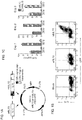
- the human IL15 gene was linked to that encoding CD8 ⁇ transmembrane domain ("mbIL15"). After retroviral transduction, human NK cells expressed mbIL-15 on the cell surface but IL-15 secretion was negligible.
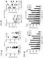
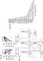
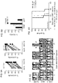
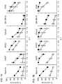
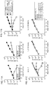
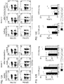
- mbIL15-NK cells expanded without IL-2, and were detectable in all tissues examined (except brain) in much higher numbers than mock-transduced NK cells (P ⁇ 0.001). Expansion in vitro and in vivo further increased with IL-2.
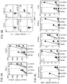
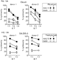
- mbIL15 The primary mechanism of mbIL15 stimulation was autocrine; it activated IL-15 signaling and anti-apoptotic signaling. Cytotoxicity against leukemia, lymphoma and solid tumor cell lines was consistently higher with mbIL15-NK cells. Median 24-hour cytotoxicity at 1:4 E:T was 71% versus 22% with mock-transduced cells; at 1:1 E:T, it was 99% versus 54% (P ⁇ 0.0001). Increased anti-tumor capacity was also evident in immunodeficient mice engrafted with leukemia (U937) or sarcoma (ES8) cells. Thus, mbIL15 conferred independent growth to NK cells and enhanced their anti-tumor capacity. Infusion of mbIL15-NK cells allows NK cell therapy without the adverse effects of IL-2.
- a cell that responds to IL-15 includes a cell in which one or more of its activities are regulated by IL-15. Examples of such cells include natural killer (NK) cells, T-cells, dendritic cells and moncytes.
- NK natural killer
- T-cells T-cells
- dendritic cells dendritic cells and moncytes.
- the one or more (e.g., isolated) cells can express all or a functional portion of IL-15 as a membrane-bound polypeptide, as a secretory protein or as a combination thereof.
- the disclosure is directed to a natural killer (NK) cell(s) that expresses all or a functional portion of interleukin-15 (IL-15).
- NK natural killer
- the one or more ( e.g. , isolated) NK cells can express all or a functional portion of IL-15 as a membrane-bound polypeptide, as a secretory protein or as a combination thereof.
- a first aspect of the present invention provides a natural killer (NK) cell that expresses all or a functional portion of interleukin-15 (IL-15), wherein:
- NK cells Natural Killer Cells
- MHC Major Histocompatibility Complex
- NK cells are unique, however, as they have the ability to recognize stressed cells regardless of whether peptides from pathogens are present on MHC molecules. They were named "natural killers” because of the initial notion that they do not require prior activation in order to kill target.
- NK cells are large granular lymphocytes (LGL) and are known to differentiate and mature in the bone marrow from where they then enter into the circulation.
- the NK cell is a mammalian NK cell.
- mammalian or “mammals” include primates ( e.g. , human), canines, felines, rodents, porcine, ruminants, and the like. Specific examples include humans, dogs, cats, horses, cows, sheep, goats, rabbits, guinea pigs, rats and mice.
- the mammalian NK cell is a human NK cell.
- Interleukin-15 refers to a cytokine that regulates T and NK cell activation and proliferation. This cytokine and interleukin 2 share many biological activities. They are found to bind common receptor subunits, and may compete for the same receptor, and thus negatively regulate each other's activity. The number of CD8+ memory cells is shown to be controlled by a balance between IL-15 and IL-2.
- This cytokine induces the activation of JAK kinases, as well as the phosphorylation and activation of transcription activators STAT3, STAT5, and STAT6 and may increase the expression of apoptosis inhibitor BCL2L1/BCL-x(L), possibly through the transcription activation activity of STAT6, and thus prevent apoptosis.
- a “functional portion” (“biologically active portion”) of IL-15 refers to a portion of IL-15 that retains one or more functions of full length or mature IL-15. Such functions include the promotion of NK cell survival, regulation of NK cell and T cell activation and proliferation as well as the support of NK cell development from hematopoietic stem cells.
- the sequence of a variety of IL-15 molecules are known in the art.
- the IL-15 is a wild type IL-15.
- the IL-15 is a mammalian IL-15 (e.g., Homo sapiens interleukin 15 (IL15), transcript variant 3, mRNA, NCBI Reference Sequence: NM_000585.4; Canis lupus familiaris interleukin 15 (IL15), mRNA, NCBI Reference Sequence: NM_001197188.1; Felis catus interleukin 15 (IL15), mRNA, NCBI Reference Sequence: NM_001009207.1).
- IL-15 Homo sapiens interleukin 15
- transcript variant 3 mRNA
- NCBI Reference Sequence NM_000585.4
- mammalian or “mammals” include primates ( e.g., human), canines, felines, rodents, porcine, ruminants, and the like. Specific examples include humans, dogs, cats, horses, cows, sheep, goats, rabbits, guinea pigs, rats and mice.
- the mammalian IL-15 is a human IL-15.
- All or a functional portion of IL-15 can be expressed by one or more NK cells (as a membrane-bound and/or secreted polypeptide) in a variety of ways.
- all or a functional portion of the IL-15 can be expressed within the NK cell and secreted from the NK cell and/or can be linked (conjugated; fused) directly or indirectly (e.g., ionic, non-ionic, covalent linkage) to the surface (e.g., at the surface, or within the membrane, of an NK cell) of the NK cell using any of a variety of linkers known in the art ( Hermanson, G., Bioconjugate Techniques, Academic Press 1996 ).
- all or a functional portion of the IL-15 is linked to all or a portion of a transmembrane protein.
- the NK cell expresses a fusion protein comprising all or a portion of IL-15 fused to all or a portion of a transmembrane protein.
- the portion of the transmembrane protein comprises all or a portion of a transmembrane domain of the transmembrane protein.
- a "transmembrane protein” or “membrane protein” is a protein located at and/or within a membrane such as the phospholipid bilayer of a biological membrane (e.g., biomembranes such as the membrane of a cell).
- Membrane proteins enable the membrane to carry out its distinctive activities.
- the complement of proteins attached to a membrane varies depending on cell type and subcellular location. Some proteins are bound only to the membrane surface, whereas others have one or more regions buried within the membrane and/or domains on one or both sides of the membrane. Protein domains on the extracellular membrane surface are generally involved in cell-cell signaling or interactions.
- transmembrane domains are a three-dimensional protein structure which is thermodynamically stable in a membrane (e.g., a membrane of a vesicle such as a cell).
- transmembrane domains include a single alpha helix, a stable complex of several transmembrane alpha helices, a transmembrane beta barrel, a beta-helix of gramicidin A, or any other structure.
- Transmembrane helices are usually about 20 amino acids in length.
- membrane proteins are classified into two broad categories-integral (intrinsic) and peripheral (extrinsic)-based on the nature of the membrane-protein interactions. Most biomembranes contain both types of membrane proteins.
- Integral membrane proteins also called intrinsic proteins, have one or more segments that are embedded in the phospholipid bilayer. Integral membrane proteins include transmembrane proteins and lipid-anchored proteins. Most integral proteins contain residues with hydrophobic side chains that interact with fatty acyl groups of the membrane phospholipids, thus anchoring the protein to the membrane. Most integral proteins span the entire phospholipid bilayer. These transmembrane proteins contain one or more membrane-spanning domains as well as domains, from four to several hundred residues long, extending into the aqueous medium on each side of the bilayer. Typically, the membrane-spanning domains are one or more (e.g., about 1, 2, 3, 4, 5, 6, 7, 8, 9, 10 or more) ⁇ helices and/or ⁇ strands.
- Membrane-spanning ⁇ -helical domains are typically embedded in membranes by hydrophobic interactions with the lipid interior of the bilayer and probably also by ionic interactions with the polar head groups of the phospholipids (e.g., glycophorin).
- the structure of ⁇ strands are typically in the form of membrane spanning barrels (e.g., porin).
- Some integral proteins are anchored to one of the membrane leaflets by covalently bound fatty acids. In these proteins, the bound fatty acid is embedded in the membrane, but the polypeptide chain does not enter the phospholipid bilayer.
- Some cell-surface proteins are anchored to the exoplasmic face of the plasma membrane by a complex glycosylated phospholipid that is linked to the C-terminus (e.g., glycosylphosphatidylinositol, alkaline phosphatase).
- Some cytosolic proteins are anchored to the cytosolic face of membranes by a hydrocarbon moiety covalently attached to a cysteine near the C-terminus (e.g., prenyl, farnesyl, and geranylgeranyl groups).
- a fatty acyl group e.g., myristate or palmitate
- an amide bond to the N-terminal glycine residue.
- Peripheral membrane proteins do not interact with the hydrophobic core of the phospholipid bilayer. Instead they are usually bound to the membrane indirectly by interactions with integral membrane proteins or directly by interactions with lipid polar head groups.
- Peripheral proteins localized to the cytosolic face of the plasma membrane include the cytoskeletal proteins spectrin and actin in erythrocytes and the enzyme protein kinase C. This enzyme shuttles between the cytosol and the cytosolic face of the plasma membrane and plays a role in signal transduction.
- Other peripheral proteins including certain proteins of the extracellular matrix, are localized to the outer (exoplasmic) surface of the plasma membrane.
- transmembrane proteins examples include a receptor, a ligand, an immunoglobulin, a glycophorin or a combination thereof.
- specific examples of transmembrane proteins include CD8 ⁇ , CD4, CD3 ⁇ , CD3 ⁇ , CD3 ⁇ , CD28, CD137, Fc ⁇ RI ⁇ , a T-cell receptor (TCR such as TCR ⁇ and/or TCR ⁇ ), a nicotinic acetylcholine receptor, a GABA receptor, or a combination thereof.
- TCR T-cell receptor
- immunoglobulins include IgG, IgA, IgM, IgE, IgD or a combination thereof.
- Specific examples of glycophorin include glycophorin A, glycophorin D or a combination thereof.
- all or a functional portion of the IL-15 can be linked to other components such as a signal peptide (e.g., a CD8 ⁇ signal sequence), a leader sequence, a secretory signal, a label (e.g., a reporter gene), etc.
- a signal peptide e.g., a CD8 ⁇ signal sequence
- a leader sequence e.g., a leader sequence
- a secretory signal e.g., a reporter gene
- the disclosure is directed to a method of producing a natural killer (NK) cell that expresses all or a functional portion of interleukin-15 (IL-15). All or a portion of the IL-15 can be expressed as a membrane-bound polypeptide, a secreted polypeptide or as a combination thereof.
- the method comprises introducing nucleic acid encoding all or a functional portion of IL-15 into the one or more NK cells.
- the nucleic acid encoding all or a functional portion of IL-15 is linked (e.g., fused) to all or a portion of a transmembrane protein.
- nucleic acid encoding all or a functional portion of IL-15 is introduced into the NK cell (e.g., wild type IL-15).
- NK cell e.g., wild type IL-15
- aspects in which nucleic acid encoding all or a functional portion if IL-15 and all or a functional portion of IL-15 fused to all or a portion of a transmembrane protein is introduced in to NK cell can be done so using a single nucleic acid or multiple ( e.g., separate; two) nucleic acids.
- the NK cell is maintained under conditions in which all or a functional portion of the IL-15 is expressed as a membrane-bound polypeptide and/or as a secreted polypeptide thereby producing a NK cell that expresses all or a functional portion of IL-15 as a membrane-bound polypeptide and/or as a secreted polypeptide.
- nucleic acid encoding all or a functional portion of IL-15 is fused to a signal peptide of CD8 ⁇ and all or a portion of a transmembrane domain of CD8 ⁇ is introduced into the NK cell.
- a method for producing an NK cell of the first aspect of the invention comprising:
- the disclosure is directed to a method of enhancing expansion and/or survival of NK cells (e.g., in vitro, ex vivo, and/or in vivo ).
- the method comprises introducing nucleic acid encoding all or a functional portion of IL-15.
- Nucleic acid encoding all or a portion of the IL-15 e.g., wild type IL-15
- encoding all or a functional portion of IL-15 fused to all or a portion of a transmembrane protein can be introduced into the NK cell.
- the NK cell can express all or a functional portion of IL-15 as a membrane-bound polypeptide, a secreted polypeptide or as a combination thereof.
- the NK cells are maintained under conditions in which all or a portion of the IL-15 is expressed as a membrane-bound polypeptide, a secreted polypeptide or as a combination thereof and in which the NK cells proliferate.
- nucleic acid encoding all or a functional portion of IL-15 is fused to a signal peptide of CD8 ⁇ and all or a portion of a transmembrane domain of CD8 ⁇ is introduced into the NK cell.
- the method can further comprise contacting the NK cells comprising membrane-bound IL-15 and/or secreted IL-15 with IL-2.
- the concentration of IL-2 is from about 10 IU/ml to about 1000IU/ml.
- the concentration of IL-2 is about 20, 40, 60, 80, 100, 120, 140, 160, 180, 200, 220, 240, 260, 280, 300, 320, 340, 360, 380, 400, 420, 440, 460, 480, 500, 520, 540, 560, 580, 600, 620, 640, 660, 680, 700, 720 740, 760, 780, 800, 820, 840, 860, 880, 900, 920, 940, 960, 980 IU/ml.
- nucleic acid e.g., transfection, transduction, and/or transposon system
- methods for introducing nucleic acid e.g., transfection, transduction, and/or transposon system
- a variety of methods for introducing nucleic acid e.g., transfection, transduction, and/or transposon system
- a functional portion of IL-15 as a transmembrane polypeptide and/or as a secreted polypeptide into a NK cell
- methods include chemical-based methods (e.g., involving the use of calcium phosphate; highly branched organic compounds (e.g., dendrimers); liposomes (lipofection); and/or cationic polymers (e.g., DEAE dextran; polyethylenimine)), non-chemical-based methods (e.g.
- electroporation electroporation; cell squeezing; sonoporation; optical transfection; impalefection; hydrodynamic delivery
- particle-based methods e.g., gene gun; magnetofection; particle bombardment
- vector-based methods e.g., vectors including viral vectors such as retroviral vector, lentiviral vectors, adenoviral vectors, etc.
- nucleotransfection e.g., Sleeping Beauty, PiggyBAC, etc.
- RNA transfection e.g., RNA transfection.
- NK cells can be grown and/or maintained at an appropriate temperature and gas mixture (e.g., about 25°C to about 37°C, about 5% CO 2 in a cell incubator). Culture conditions can vary widely, and variation of conditions for a particular cell type can result in different phenotypes.
- a commonly varied factor in culture systems is the cell growth medium.
- Recipes for growth media can vary in pH, glucose concentration, growth factors, and the presence of other nutrients.
- the growth factors used to supplement media are often derived from the serum of animal blood, such as fetal bovine serum (FBS), bovine calf serum, equine serum, porcine serum and/or human platelet lysate (hPL).
- FBS fetal bovine serum
- bovine calf serum bovine calf serum
- equine serum porcine serum
- hPL human platelet lysate
- Other factors considered for maintaining cells include plating density (number of cells per volume of culture medium) and growth of the cells in suspension or adherent cultures.
- the methods can further comprise isolating or separating the one or more NK cells produced by the methods provided herein, In addition, the methods can further comprises culturing the one or more NK cells. In some aspects, an NK cell line is produced.
- the disclosure also encompasses a (one or more) natural killer (NK) cell or cell line produced by the methods described herein, and compositions comprising the NK cells provided herein.
- the composition is a pharmaceutical composition comprising one or more of the NK cells or cell lines provided herein.
- the pharmaceutical composition can further comprise all or a functional portion of IL-2 (e.g., all or a functional portion of an (one or more) IL-2 protein; nucleic acid encoding all or a functional portion of IL-2).
- the present invention provides, in a fourth aspect of the invention, a composition comprising an NK cell of the first or third aspect of the invention.
- the composition is a pharmaceutical composition further comprising all or a functional portion of IL-2.
- IL-2 refers to a member of a cytokine family that also includes IL-4, IL-7, IL-9, IL-15 and IL-21. IL-2 signals through a receptor complex consisting of three chains, termed alpha, beta and gamma. The gamma chain is shared by all members of this family of cytokine receptors. IL-2, which similar to IL-15, facilitates production of immunoglobulins made by B cells and induces the differentiation and proliferation of NK cells. Primary differences between IL-2 and IL-15 are found in adaptive immune responses. For example, IL-2 is necessary for adaptive immunity to foreign pathogens, as it is the basis for the development of immunological memory. On the other hand, IL-15 is necessary for maintaining highly specific T cell responses by supporting the survival of CD8 memory T cells.
- the disclosure is directed to a method of treating a disease and/or condition involving NK cell therapy in an individual in need thereof comprising administering to the individual a natural killer (NK) cell that expresses all or a functional portion of interleukin-15 (IL-15).
- NK natural killer
- the NK cells express all or a functional portion of IL-15 as a membrane-bound polypeptide and/or as a secreted polypeptide.
- diseases and/or conditions that involve NK cell therapy include NK cell deficiencies, cancer, autoimmune diseases, infectious diseases and the like.
- the disclosure is directed to a method of treating cancer (e.g., a tumor) in an individual in need thereof comprising administering to the individual a natural killer (NK) cell that expresses all or a functional portion of interleukin-15 (IL-15). All or a functional portion of IL-15 can be expressed as a membrane-bound polypeptide and/or as a secreted polypeptide.
- NK natural killer
- a fifth aspect of the present invention provides an NK cell according to the first aspect of the invention, or a composition according to the fourth aspect of the invention, for use in a method of treating cancer in an individual in need thereof by administering to the individual the NK cell or the composition.
- the method can further comprise administering one or more antibodies, antigenic fragments and/or fusions thereof specific to the cancer (e.g., tumor).
- the method can further comprise administering one or more antibodies directed against one or more tumor antigens.
- the one or more antibodies can be a polyclonal antibody, a monoclonal antibody, a multivalent (e.g., bivalent, trivalent) antibody, a chimeric antibody, a humanized antibody, etc. and combinations thereof.
- antigenic fragments and/or fusions include Fab', F(ab') 2 , single chain variable fragment (scFv), multivalent scFv (e.g., di-scFv, tri-scFv), single domain antibody (nanobody) and etc.
- the cancer is a leukemia (e.g., acute lymphoblastic leukemia; acute myeloid leukemia; chronic myelogenous leukemia, chronic lymphocytic leukemia), a myelodysplastic syndrome, a lymphoma (e.g., B cell non-Hodgkin lymphoma, Hodgkin lymphoma, T-cell lymphoblastic lymphoma, anaplastic large cell lymphoma), a solid tumor (e.g., a breast cancer, prostate cancer, gastric cancer, colon cancer, hepatocellular carcinoma, nasopharyngeal carcinoma, neuroblastoma, high grade glioma), a sarcoma (e.g., Ewing sarcoma, rhabdomyosarcoma, non-rhabdomyosarcoma soft-tissue sarcoma, osteosarcoma).
- a leukemia e.g., acute lymphoblastic leukemia
- the method of treating cancer can further comprise administering IL-2 (all or a functional portion of IL-2 protein; nucleic acid encoding all or a functional portion of IL-2) to the individual.
- IL-2 is mammalian IL-2, such as human IL-2.
- a low dose of the IL-2 is administered to the individual.
- a "low dose" of IL-12 refers to a dose of IL-2 of about 1 million IU/m 2 or less ( e.g., about 800,000 IU/m 2 ; 600,000 IU/m 2 ; 400,000 IU/m 2 ; 200,000 IU/m 2 ; 100,000 IU/m 2 ; 80,000 IU/m 2 ; 60,000 IU/m 2 ; 40,000 IU/m 2 ; 20,000 IU/m 2 ; 10,000 IU/m 2 ; 8,000 IU/m 2 ; 6,000 IU/m 2 ; 4,000 IU/m 2 ; 2,000 IU/m 2 ; 1,000 IU/m 2 ; 800 IU/m 2 ; 600 IU/m 2 ; 400 IU/m 2 ; 200 IU/m 2 ; 100 IU/m 2 ).
- a normal dose of IL-2 is about 1 million IU/m 2 to about 5 million IU/m 2 .
- the one or more natural killer (NK) cell(s) that express all or a functional portion of interleukin-15 (IL-15) are administered in a therapeutically effective amount (i.e., an amount that is sufficient to treat the cancer, such as by ameliorating symptoms associated with the cancer, preventing or delaying the onset of the cancer, also lessening the severity or frequency of symptoms of the cancer and/or preventing, delaying or overcoming metastasis of the cancer).
- a therapeutically effective amount i.e., an amount that is sufficient to treat the cancer, such as by ameliorating symptoms associated with the cancer, preventing or delaying the onset of the cancer, also lessening the severity or frequency of symptoms of the cancer and/or preventing, delaying or overcoming metastasis of the cancer.
- the amount that will be therapeutically effective in the treatment of a particular individual will depend on the symptoms and severity of the condition (e.g., cancer), and can be determined by standard clinical techniques.
- in vitro or in vivo assays may optionally be employed to help identify optimal dosage ranges.
- the precise dose to be employed in the formulation will also depend on the route of administration, and the seriousness of the cancer, and should be decided according to the judgment of a practitioner and each patient's circumstances. Effective doses may be extrapolated from dose-response curves derived from in vitro or animal model test systems.
- the therapeutic compound can be delivered in a composition (e.g., a pharmaceutical composition), as described above, or by themselves. They can be administered systemically, or can be targeted to a particular tissue.
- the therapeutic compounds can be produced by a variety of means, including chemical synthesis; recombinant production; in vivo production (e.g., a transgenic animal, such as U.S. Patent No. 4,873,316 to Meade et al. ), for example, and can be isolated using standard means such as those described herein. A combination of any of the above methods of treatment can also be used.
- the compounds for use in the methods described herein can be formulated with a physiologically acceptable carrier or excipient to prepare a pharmaceutical composition.
- the carrier and composition can be sterile.
- the formulation should suit the mode of administration.
- Suitable pharmaceutically acceptable carriers include but are not limited to water, salt solutions (e.g., NaCl), saline, buffered saline, alcohols, glycerol, ethanol, gum arabic, vegetable oils, benzyl alcohols, polyethylene glycols, gelatin, carbohydrates such as lactose, amylose or starch, dextrose, magnesium stearate, talc, silicic acid, viscous paraffin, perfume oil, fatty acid esters, hydroxymethylcellulose, polyvinyl pyrolidone, etc., as well as combinations thereof.
- the pharmaceutical preparations can, if desired, be mixed with auxiliary agents, e.g., lubricants, preservatives, stabilizers, wetting agents, emulsifiers, salts for influencing osmotic pressure, buffers, coloring, flavoring and/or aromatic substances and the like that do not deleteriously react with the active compounds.
- auxiliary agents e.g., lubricants, preservatives, stabilizers, wetting agents, emulsifiers, salts for influencing osmotic pressure, buffers, coloring, flavoring and/or aromatic substances and the like that do not deleteriously react with the active compounds.
- the composition can also contain minor amounts of wetting or emulsifying agents, or pH buffering agents.
- the composition can be a liquid solution, suspension, emulsion, tablet, pill, capsule, sustained release formulation, or powder.
- the composition can be formulated as a suppository, with traditional binders and carriers such as triglycerides.
- Oral formulation can include standard carriers such as pharmaceutical grades of mannitol, lactose, starch, magnesium stearate, polyvinyl pyrollidone, sodium saccharine, cellulose, magnesium carbonate, etc.
- compositions of this invention include, but are not limited to, intradermal, intramuscular, intraperitoneal, intravenous, subcutaneous, topical, oral and intranasal.
- Other suitable methods of introduction can also include gene therapy (as described below), rechargeable or biodegradable devices, particle acceleration devises ("gene guns") and slow release polymeric devices.
- the pharmaceutical compositions of this invention can also be administered as part of a combinatorial therapy with other compounds.
- compositions for intravenous administration typically are solutions in sterile isotonic aqueous buffer.
- the composition may also include a solubilizing agent and a local anesthetic to ease pain at the site of the injection.
- the ingredients are supplied either separately or mixed together in unit dosage form, for example, as a dry lyophilized powder or water free concentrate in a hermetically sealed container such as an ampule or sachette indicating the quantity of active compound.
- the composition is to be administered by infusion, it can be dispensed with an infusion bottle containing sterile pharmaceutical grade water, saline or dextrose/water.
- an ampule of sterile water for injection or saline can be provided so that the ingredients may be mixed prior to administration.
- nonsprayable forms viscous to semi-solid or solid forms comprising a carrier compatible with topical application and having a dynamic viscosity preferably greater than water
- Suitable formulations include but are not limited to solutions, suspensions, emulsions, creams, ointments, powders, enemas, lotions, sols, liniments, salves, aerosols, etc., that are, if desired, sterilized or mixed with auxiliary agents, e.g., preservatives, stabilizers, wetting agents, buffers or salts for influencing osmotic pressure, etc.
- auxiliary agents e.g., preservatives, stabilizers, wetting agents, buffers or salts for influencing osmotic pressure, etc.
- compositions described herein can be formulated as neutral or salt forms.
- Pharmaceutically acceptable salts include those formed with free amino groups such as those derived from hydrochloric, phosphoric, acetic, oxalic, tartaric acids, etc., and those formed with free carboxyl groups such as those derived from sodium, potassium, ammonium, calcium, ferric hydroxides, isopropylamine, triethylamine, 2-ethylamino ethanol, histidine, procaine, etc.
- the disclosure is directed to pharmaceutical compositions comprising one or more NK cells that expresses all or a functional portion of interleukin-15 (IL-15) as a membrane-bound polypeptide.
- the pharmaceutical composition comprises one or more NK cells of the first or third aspect of the invention.
- compositions e.g., pharmaceutical compositions
- the agents identified herein can be used in the treatment of cancer.
- the agents identified herein can be used in the manufacture of a medicament for the treatment of cancer.
- an "individual” refers to an animal, and in a particular aspect, a mammal.
- mammals include primates, a canine, a feline, a rodent, and the like. Specific examples include humans, dogs, cats, horses, cows, sheep, goats, rabbits, guinea pigs, rats and mice.
- the term "individual in need thereof” refers to an individual who is in need of treatment or prophylaxis as determined by a researcher, veterinarian, medical doctor or other clinician. In one embodiment, an individual in need thereof is a mammal, such as a human.
- an NK cell is one that is separated from (substantially isolated with respect to) the complex cellular milieu in which it naturally occurs, or culture medium when produced by recombinant techniques, or chemical precursors or other chemicals when chemically synthesized.
- the isolated material will form part of a composition (for example, a crude extract containing other substances), buffer system, or reagent mix.
- the material may be purified to essential homogeneity, for example, as determined by agarose gel electrophoresis or column chromatography such as HPLC.
- an NK cell comprises at least about 50%, 80%, 90%, 95%, 98% or 99% (on a molar basis) of all macromolecular species present.
- Embodiments are provided in which more than one, or all of the group members are present, employed in, or otherwise relevant to a given product or process. Any one or more claims may be amended to explicitly exclude any embodiment, aspect, feature, element, or characteristic, or any combination thereof. Any one or more claims may be amended to exclude any agent, composition, amount, dose, administration route, cell type, target, cellular marker, antigen, targeting moiety, or combination thereof.
- the human cell lines Nalm-6 (B-lineage acute lymphoblastic leukemia), Daudi (B-cell lymphoma), K562 and U937(acute myeloid leukemia), and SK-BR-3 (breast carcinoma) were obtained from the American Type Culture Collection, the Ewing sarcoma cell line ES8 was from the St. Jude Children's Research Hospital tissue repository. All of the cell lines were transduced with a MSCV-internal ribosome entry site (IRES)-GFP retroviral vector (from the St. Jude Vector Development and Production Shared Resource) containing the firefly luciferase gene.
- IRS MSCV-internal ribosome entry site
- GFP retroviral vector from the St. Jude Vector Development and Production Shared Resource
- Transduced cells were selected for their expression of GFP with a MoFlo (Beckman Coulter, Miami, FL) or a FACSAria (BD Biosciences, San Jose, CA).
- MoFlo Bacillus Coulter, Miami, FL
- FACSAria BD Biosciences, San Jose, CA
- RPMI-1640 Invitrogen, Carlsbad, CA
- FBS fetal bovine serum
- FBS fetal bovine serum
- ES8 was validated by DNA fingerprinting at DSMZ (Braunschweig, Germany).
- Peripheral blood samples were obtained from discarded byproducts of platelet collections from healthy adult donors. Mononuclear cells were purified by centrifugation on an Accu-Prep density step (Accurate, Westbury, NY) and washed twice in RPMI-1640. To expand CD56+ CD3- NK cells, peripheral blood mononuclear cells and the genetically modified K562-mb15-41BBL cell line were co-cultured, as previously described in Fujisaki et al., Cancer Res, 69(9):4010-4017 (2009 ); Imai et al., Blood, 106:376-383 (2005 )).
- peripheral blood mononuclear cells were cultured with 100 Gy-irradiated K562-mb15-41BBL cell at 1.5 : 1 ratio in SCGM (CellGenix, Freiburg, Germany) containing 10% FBS, antibiotics and 10 IU/mL of recombinant human interleukin-2 (IL-2; Roche, Mannheim, Germany) in 6-well tissue culture plates. Tissue culture medium was partly exchanged every 2 days. After 7 days of co-culture, residual T cells were removed with Dynabeads CD3 (Invitrogen), resulting in cell population containing >95% CD56+ CD3- NK cells.
- the pMSCV-IRES-GFP, pEQ-PAM3(-E), and pRDF were obtained from the St. Jude Vector Development and Production Shared Resource.
- Interleukin-15 (IL-15) with a long signal peptide was sub-cloned by polymerase chain reaction (PCR) from a human spleen cDNA library (from Dr G. Neale, St Jude Children's Research Hospital) used as a template.
- the cDNA encoding the signal peptide of CD8 ⁇ , the mature peptide of IL-15 and the transmembrane domain of CD8 ⁇ were assembled by the splicing by overlapping extension by PCR (SOE-PCR) to encode a membrane-bound form of IL-15 ("mbIL15"); a wild-type form of IL-15 (not linked to CD8 ⁇ transmembrane domain; "wtIL15”) was also tested prepared.
- the resulting expression cassettes were sub-cloned into EcoRI and Xhol sites of murine stem-cell virus-internal ribosome entry site-green fluorescent protein (MSCV-IRES-GFP).
- RD144-pseudotyped retrovirus 3.0 x10 6 293T cells were transfected using X-tremeGENE 9 DNA (Roche, Mannheim, Germany), maintained in 10-cm tissue culture dishes for 18h, with 3.5 ⁇ g of cDNA encoding mbIL15 constructs, 3.5 ⁇ g of pEQ-PAM3(-E), and 3 ⁇ g of pRDF.
- the conditioned medium containing retrovirus was harvested at 36-96 hours and added to polypropylene tubes coated with RetroNectin (Takara, Otsu, Japan), which were centrifugated at 1400 g for 10 min and incubated at 37°C and 5% CO 2 for 4 hours. After additional centrifugation, and removal of the supernatant, expanded NK cells (0.5-1 x 10 6 ) were added to the tubes and left in at 37°C for 12 hours; these steps were repeated up to 6 times over 2-3 days. Cells were then maintained in RPMI-1640 with FBS, antibiotics and 100 IU/ml of IL-2. Transduced cells were assayed 3-29 days after transduction.
- mbIL-15 Surface expression of mbIL-15 was analyzed by flow cytometry using an anti-human IL-15 antibody (R&D, Minneapolis, MN) and phycoerythrin conjugated goat anti-mouse IgG1(Southern Biotech, Birmingham, AL). Antibody staining was detected with a Fortessa flow cytometer (Becton Dickinson). Levels of IL-15 in culture supernatants were measured with the Quantikine Immunoassay (R&D).
- transduced NK cells (1 x 10 6 cells/mL) were resuspended in RPMI-1640 with 10% FBS and antibiotics, placed into the wells of either a 24- or a 96-well plate (Costar, Corning, NY) and cultured without or with IL-2 (10-100 IU/ml). Numbers of viable GFP+ cells were determined with an Accuri C6 flow cytometer (Becton Dickinson), after staining with propidium iodide. In some experiments, cells were incubated for 10 minutes with a neutralizing anti-IL-15 antibody (R&D) or an isotype-matched non-reactive antibody before culture.
- R&D neutralizing anti-IL-15 antibody
- NK cell immunophenotyping was performed using the antibodies listed in the Table, visualized with a Fortessa flow cytometer and analyzed by Diva (Becton Dickinson) and FlowJo (TreeStar, Ashland, OR) software.
- Diva Becton Dickinson
- FlowJo TeStar, Ashland, OR
- Cell lysates were prepared using a lysis buffer containing 20 mM 3-(N-morpholino) propanesulfonic acid , 2 mM EGTA, 5 mM EDTA, 30 mM sodium fluoride, 60 mM ⁇ -glycerophosphate, 20 mM sodium pyrophosphate, 1 mM sodium orthovanadate, 1% Triton X-100, Complete Mini protease inhibitor cocktail (Roche, Mannheim, Germany) and 1 mM dithiothreitol. After sonication, lysates were frozen at -80°C and shipped in dry ice to Kinexus (Vancouver, CA) for Kinex Antibody Microarray analysis.
- a lysis buffer containing 20 mM 3-(N-morpholino) propanesulfonic acid , 2 mM EGTA, 5 mM EDTA, 30 mM sodium fluoride, 60 mM ⁇ -glycerophosphate, 20 mM
- luciferase-labeled target cells and NK cells were plated in 96-well, flat-bottomed black Viewplates (Corning) at various effector : target (E: T) ratios and cultured for 4 or 24 hours.
- Adherent cell lines were incubated at 37°C and 5% CO 2 for 4 hours before adding NK cells to allow for cell attachment.
- NK cells (cultured for 48 hours without IL-2) were cocultured with K562,U937 cells, or 721.221 cells and their Cw6-expressing variant for 4 hours.
- PE- or PE-Cy7-conjugated anti-CD107a antibody (BD Biosciences) at the beginning of the cultures and GolgiStop (0.15 ⁇ L; BD Biosciences) 1 hour later. Percentage of CD107a+ NK cells was determined by flow cytometry.
- human NK cells transduced with mbIL15 or mock-transduced (6-9 x 10⁶ cells per mouse) were injected in the tail vein of NOD.Cg-Prkdc scid IL2rg tmlWj1 /SzJ (NOD/scid IL2RGnull) mice (Jackson Laboratories, Bar Harbor, ME).
- NOD/scid IL2RGnull mice Jackson Laboratories, Bar Harbor, ME.
- mice To test tumor cell killing in mice, we prepared two xenograft models. In the first, U937 cells expressing luciferase were injected i.p. in NOD.Cg-Prkdc scid IL2rg tmlWjl /SzJ (NOD/scid IL2RGnull) mice (1 x 10 4 cells per mouse). Three days later, NK cells transduced with the MSCV vector containing either GFP alone or mbIL15 were injected i.p. (1 x 10 7 cells per mouse); NK cell injection was repeated on day 7. As a control, a group of mice received tissue culture medium instead of NK cells.
- mice were engrafted with ES8 cells (i.p.; 1 x 10 5 cells per mouse), followed by 1 NK cell injection on day 3 as above.
- Tumor engraftment and progression was evaluated using a Xenogen IVIS-200 system (Caliper Life Sciences, Hopkinton, MA), with imaging beginning 5 minutes after i.p. injection of an aqueous solution of D-luciferin potassium salt (3 mg/mouse). Photons emitted from luciferase-expression cells were quantified using the Living Image 4.3.1 software program.
- IL15 transmembrane domain of CD8 ⁇
- mbIL15 transmembrane domain of CD8 ⁇
- wtIL15 wild-type unmodified form
- Production of soluble IL-15 by the transduced NK cells was determined by testing supernatants collected after 24 and 48 hours of culture. A shown in Fig. 1C , cells expressing wtIL15 secreted substantial amounts of IL-15 whereas this was minimal in mbIL15-NK cells and undetectable in mock-transduced NK cells.
- NK cells expressing IL-15 have autonomous survival and expansion capacity 1
- NK cells from 15 donors were transduced with the mbIL15 construct and cultured in the absence of IL-2; cell numbers after culture were then compared to those in parallel cultures with mock-transduced NK cells.
- Fig. 2A expression of mbIL-15 dramatically increased NK cell survival: after 7 days of culture, median cell recovery was 85% while virtually no viable mock-transduced NK cell was detectable ( ⁇ 1%; P ⁇ 0.0001 by paired t test).
- the effect of mbIL15 significantly decreased if an anti-IL-15 neutralizing antibody was added to the cultures ( Figs. 6A-6B ).
- mbIL15 In experiments with expanded NK cells from 6 of the 9 donors, the capacity of mbIL15 to support NK cell survival beyond 7 days with low dose IL-2 (10 IU/mL) was determined. On day 14, mbIL15 NK cell numbers were maintained or increased in 4 of the 6 cultures; in 2 of these cells had further expanded by day 21. Only 2 of the 6 cultures with mock-transduced NK cells from the same donors had maintained cell numbers on day 14 and 21, and no cell growth was observed; median cell recovery on day 21 was 205% for mbIL15 NK cells and 80% for mock-transduced NK cells. Thus, even in the presence of low dose IL-2, expression mbIL15 conferred a considerable survival and growth advantage.
- NK cells In cultures of NK cells from one donor, a particularly high cell recovery was observed on day 7 when IL15 was expressed (261% with mbIL15 and 161% with wtIL15 in the absence of IL-2; 266% and 188% with 10 IU/mL IL-2). These cultures were monitored for 2 months and remarkable improvements in cell expansion and survival brought about by the expression of mbIL15 were observed ( Fig. 2C ). Even in the absence of IL-2, mbIL-15 NK cells continued to survive until day 21 and they were still detectable 75 days after initiation of the culture, while mock-transduced cells had become undetectable on day 7 and wtIL15-transduced NK cells on day 42.
- NK cells transduced with either mbIL155 or wtIL15 had a similar persistence profile, both cell types surviving longer than mock-transduced NK cells even under these conditions.
- IL15 expression improved survival and expansion of NK cells and that mbIL15 produced overall better stimulation. Whether mbIL15 expression would sustain expansion of human NK cells in NOD/scid IL2RGnull mice was next determined.
- Activated NK cells from 4 donors were transduced with mbIL15 (52%-74% GFP-positive) and injected into 4 mice (one mouse per donor); 4 control mice were injected with mock-transduced NK cells from the same donors.
- mbIL15 NK cells comprised 168.5 cells/ ⁇ l (range, 94-355) of peripheral blood mononucleated cells in the absence of IL-2 and 382 cells/ ⁇ l (151-710) when IL-2 was also administered ( Fig. 3A, B ).
- Human CD45+ cells also expressed CD56 and lack CD3 (not shown).
- the proportion of GFP+ had increased from 66.5% ⁇ 9.9% before injection to 93.8% ⁇ 4.4% on day 7 and 94.8% ⁇ 3.4% on day 11 (P ⁇ 0.01 for both comparisons).
- mbIL15 predominantly stimulated cells in trans (IL-15 presented on one NK cell stimulating a neighboring cell (a mechanism reported to occur physiologically)) or cis (by direct binding of mbIL15 to receptors expressed in the same cell)
- trans IL-15 presented on one NK cell stimulating a neighboring cell (a mechanism reported to occur physiologically)
- cis by direct binding of mbIL15 to receptors expressed in the same cell
- the proportions of GFP+ and GFP- NK cells in the cultures were evaluated after 7 days of culture. If the trans mechanism was predominant, the ratio between GFP+ and GFP- NK cells should remain unaltered during culture; if cis was predominant, the proportion of GFP+ cells should increase.
- mbIL15 NK cells expressed moderately higher levels of the activation receptors NKG2D, NKp44 (CD336) and NKp30 (CD337) as well as of CD16 and CD56, while expression of NKp46 (CD335) decreased and that of other molecules, such as DNAM-1 (CD226), remained unchanged ( Fig. 4B ; the Table).
- the signal transduction pathways activated by the expression of mbIL15 were also determined. As shown in Fig.
- mbIL-15 NK cells had several highly phosphorylated molecules. These included molecules known to be phosphorylated in response to IL-15 signaling, such as the transcription factors STAT1, STAT3 and STAT5, the kinases src, Erk1/2 and Mek1. Notably, a marked phosphorylation of Bad, as well as phosphorylation of Caspase 7 and 9, collectively indicative of an anti-apoptotic effect, were observed.
- Other highly phosphorylated molecules in mbIL15 NK cells whose role in IL-15 signaling is unclear included CDK6 and RafA.
- NK-mediated killing of tumor cells would likely also increase.
- This notion was first tested by comparing tumor cell cytotoxicity exerted by mbIL15-NK cells to that of mock transduced NK cells from the same donors.
- mice were injected with the human acute myeloid leukemia (AML) cell line U937 and then treated with either mbIL15- or mock-transduced NK cells.
- AML human acute myeloid leukemia
- mice were also tested in a second xenograft model in which NOD/scid IL2RGnull mice were injected with the Ewing sarcoma cell line ES8, which has a much slower growth rate, and the mice were treated with one injection of NK cells.
- ES8 Ewing sarcoma cell line
- NK cells persist in sufficient numbers to achieve an E:T ratio likely to produce tumor cytoreduction.32 Demonstrated herein is that expression of a membrane-bound form of IL-15 in human NK cells supported their autonomous expansion and extended survival in the absence of IL-2. NK cells expressing mbIL15 could be maintained in vitro for up to 2 months without exogenous IL-2. NK cells expressing mbIL15 could expand in immunodeficient mice and infiltrated multiple tissues where they could be found in much larger numbers than mock-transduced cells.
- mbIL-15 NK cells Expansion of mbIL-15 NK cells was further increased by a low concentration of IL-2 both in vitro and in vivo. Expression of mbIL15 did not impair the cytotoxic capacity of NK cells. In fact, in xenograft models, mbIL15 NK cells exerted anticancer activity which was more powerful than that of mock-transduced cells, indicating that this approach might improve the antitumor capacity of NK cell infusions while averting the side effects of IL-2 administration.
- IL-15 ectopic expression of IL-15 in human NK cells caused a stronger survival-promoting effect when IL-15 was presented in a membrane-bound form than in a secreted form.
- mbIL15 expressed in NK cells preferentially stimulates in cis rather than in the trans when IL-15 is presented by other cells. That is, mbIL15 appears to preferentially engage IL-15 receptors on the same cells, resulting in autocrine stimulation.
- This mechanism explains the IL-15 expression pattern that was consistently observed when mbIL15-transduced NK cells were labeled with an anti-IL-15 antibody, showing a substantial proportion of cells with strong GFP expression but ostensibly lacking IL-15 ( Fig. 1B ).
- IL-15 is expressed but not accessible to antibody because it is bound to its receptor and/or internalized.
- the capacity of mbIL15 to promote NK cell viability likely explains the increased cytotoxicity exerted by these cells, particularly in 24-hour in vitro assays and in vivo.
- the superiority of mbIL15-NK cells was also clear in short-term (4-hour) assays and these cells also released more lytic granules according to the CD107a test. Therefore, expression of mbIL15 is likely to increase NK cell cytotoxicity by other means, possibly by enhancing their activation status.
- NK cells typically relies on IL-2 to support their survival and expansion in vivo.
- the multiple side effects related to IL-2 administration are potentially serious and often render administration of this cytokine poorly tolerated. Stopping IL-2 administration or reducing its dose may results in decreased NK cell expansion and inefficient anti-tumor effect, which may be further inhibited by the stimulation of regulatory T cells.
- replacing IL-2 with IL-15 is potentially attractive but the clinical formulation of IL-15 is still being tested.
- adverse effects were observed in some animals, including diarrhea, emesis, weight loss, transient neutropenia, increase in transaminases and hyponatremia.
- NK cells In addition to T and NK cell expansion, expansion of regulatory T cells has been observed. Contrary to NK cells transduced with wtIL15, those transduced with mbIL15 released exceedingly small amount of IL-15 in the supernatant. Thus, any potential side effect that may be caused by the interaction of IL-15 with cells other than NK cells should be minimized by this approach. Of note, prolonged exposure of murine large granular lymphocytes to IL-15 leads to their leukemic growth. This poses a potential safety concern for IL-15 administration in patients and also for the use of NK cells expressing IL-15, particularly if such cells were administered to patients at a low risk of relapse.
- NK cells expressing mbIL15 generally survived for much shorter periods than the one year or more reported for T cell clones expressing soluble IL-15. Moreover, persistent NK expansion was not observed in immunodeficient mice, with a follow-up exceeding 9 months.
- NK cells also play a critical role in mediating antibody-dependent cell cytotoxicity in patients treated with monoclonal antibodies. Thus, infusion of NK cells is likely beneficial in multiple settings. Expansion of human NK cells in large numbers ex vivo is feasible; robust large-scale methods for this purpose have been established and are being used in clinical trials. Genetic modification of NK cells by retroviral transduction or electroporation is also possible. Therefore, the translation of the approach described herein into clinical-grade conditions is realistic and it is warranted by the superior expansion and cytotoxicity of mbIL15-NK cells.
- Antibodies were from BD Biosciences (CD56 PE, CD16 PE-Cy7, CD69 PE, CD25 PE-Cy7, CD122 BV421, CD158b PE), Beckman Coulter (CD335 PE, CD336 PE, CD337 PE, CD158ah PE, CD159a PE), Miltenyi Biotech (CD226 PE, CD158e APC), R&D Systems (NKG2D PE), Biolegend (CD132 APC). 2 Percentages refer to GFP+ cells expressing the marker. 3 Overexpressed markers are highlighted in bold font. MFI, mean fluorescence intensity
Description
- Survival and proliferation of NK cells in vivo requires stimulation by cytokines, such as IL-2 and IL-15. For example, after injection in immunodeficient mice, activated NK cells became undetectable after 1 week but persisted for up to one month if human IL-2 was also administered. Hence, clinical protocols using NK cell infusions typically rely on IL-2 administration to prolong NK cells survival in patients. However, IL-2 can have considerable side effects. In addition to fever and chills, IL-2 administration can lead to more serious and potentially fatal consequences, such as capillary leak syndrome. Decreasing the dose of IL-2 should reduce the risk of side effects but can result in stimulation of regulatory T cells which can inhibit NK cell function and possibly nullify its anti-cancer effect.
- Hence, it would be important to develop alternative ways to promote NK cell expansion and activity in vitro and/or in vivo.
- Shook et al. (2011), Tissue Antigens 78(6):409-415 discusses natural killer cell engineering for cellular therapy of cancer.
US 2013/266551 describes a construct consisting of human IL-15 mature peptide fused to the signal peptide and transmembrane domain of human CD8α to express membrane bound IL-15.US 2006/0093605 relates to methods for preferentially activating and expanding NK cells which methods utilize the stimulatory effects of IL-15 and CD27 ligand to preferentially expand and activate NK cells in a mixed culture comprising NK cells.US 2012/040323 describes multimeric IL-15 soluble fusion molecules and methods of making and using the same. - The present invention is as set forth in the claims. Accordingly, the present invention provides the following:
- [1] A natural killer (NK) cell that expresses all or a functional portion of interleukin-15 (IL-15), wherein:
- all or a functional portion of the IL-15 is fused to all or a portion of a transmembrane protein, wherein the IL-15, or functional portion thereof, is expressed as a membrane-bound polypeptide (mbIL-15) by the NK cell,
- wherein the all or a portion of the transmembrane protein is selected from CD8α, CD4, CD3ε, CD3γ, CD3δ, CD3ζ, CD28, and CD137;
- wherein the NK cell expressing all or a functional portion of IL-15 exhibits enhanced survival and expansion in the absence of IL-2, is able to regulate NK cell and T cell activation and proliferation, and/or support NK cell development from hematopoietic stem cells as compared to an NK cell not expressing all or a functional portion of IL-15.
- [2] The NK cell of [1] wherein the transmembrane protein comprises a CD8α transmembrane domain.
- [3] An NK cell of [1] or [2], wherein the all or a functional portion of IL-15 is fused to a signal peptide of CD8α and a transmembrane domain of CD8α.
- [4] An NK cell of any one of [1], [2] or [3], wherein the IL-15, or functional portion thereof, is mammalian IL-15
transcript variant 3, or a functional portion thereof. - [5] An NK cell of any one of [2]-[4], wherein the CD8α transmembrane domain is encoded by a nucleic acid comprising SEQ ID NO: 1 or wherein the secreted IL-15 is encoded by a nucleic acid comprising a CD8α signal peptide.
- [6] A method for producing an NK cell of any one of [1] to [5], the method comprising:
- a) introducing nucleic acid encoding all or a functional portion of IL-15 fused to all or a portion of a transmembrane protein into the NK cell, wherein the all or a portion of the transmembrane protein is selected from CD8α, CD4, CD3ε, CD3γ, CD3δ, CD3ζ, CD28, and CD137; and
- b) maintaining the NK cell under conditions in which all or a functional portion of the IL-15 is expressed,
- [7] The method of [6] wherein the nucleic acid is introduced into the NK cell by transducing the NK cell with a vector.
- [8] The method of [7] wherein the vector is a viral vector, preferably a retroviral vector.
- [9] The method of any one of [6] to [8], further comprising contacting the NK cells with IL-2.
- [10] A natural killer (NK) cell obtainable by the method of any one of [6]-[8].
- [11] A composition comprising the NK cell of any one of [1]-[5] or [10].
- [12] A composition according to [11], wherein the composition is a pharmaceutical composition further comprising all or a functional portion of IL-2.
- [13] An NK cell according to any one of [1] to [5] or a composition according to [11] or [12] for use in a method of treating cancer in an individual in need thereof by administering to the individual the NK cell or the composition.
- [14] An NK cell or composition for use according to [13] wherein the method of treating also comprises administering IL-2 to the individual, preferably at a low dose of about 1 million IU/m2 or less.
- [15] An NK cell or composition for use according to [13] or [14] wherein the method of treating also further comprises administering one or more antibodies directed against one or more tumor antigens.
-
-
Figs. 1A-1C : Design and expression of IL-15 constructs. 1A. Schematic representation of the wild-type and membrane-bound IL-15 constructs ("wtIL15" and "mbIL15") used in this study. 1B. Expression of IL-15 of the surface of NK cells transduced with mbIL15. Expanded NK cells were transduced with wtIL15, mbIL15 or with a vector containing GFP alone ("Mock'). Flow cytometry dot plots illustrate expression of GFP and IL-15, as detected by an anti-IL15 antibody (R&D Systems) and a goat-anti-mouse secondary antibody conjugated to phycoerythrin (Southern Biotechnology Associates). Percentage of cells (>98% CD56+ CD3- NK cells) in each quadrant is shown. 1C. Secretion of IL-15 by NK cells transduced with wtIL15. NK cells from 3 different donors were tested in triplicate. Bars indicate mean ± SD of ELISA measurements performed in supernatants collected after 24 and 48 hours of culture without IL-2. No IL-15 was detected in the supernatants of mock-transduced cells. -
Figs. 2A-2C : Survival and expansion of NK cells expressing IL-15 in vitro. 2A. Percentage of NK cell recovery as compared to input cells after 7-day parallel cultures without IL-2 for mock- and mbIL15 transduced cells from 15 donors (left panel) and mbIL15- or wtIL15-transduced cells from 9 donors (right panel). Horizontal bars indicate median value. Results of paired t tests are shown. Results of cultures with IL-2 (10 and 100 IU/mL) are shown in Supplementary Fig. S1. 2B. Survival and expansion of mock- and mbIL15-transduced NK cells from 6 donors with low dose IL-2 (10 IU/mL). 2C. Expansion and long-term survival of NK cells from one donor transduced with mbIL15, wtIL15 or mock-transduced cultured with no IL-2 or low dose IL-2 (results with 100 IU/mL IL2 are shown inFig. 6 ). Percentage of NK cell recovery at the indicated days of culture is shown. -
Fig. 3A-3C : Survival and expansion of NK cells expressing mb-IL15 in vivo. 3A. Absolute number of human CD45+ cells in peripheral blood of mice injected with mock- or mbIL15 transduced NK cells with or without IL-2 (16 mice total) 7 and 11 days after infusion (P = 0.004 with no IL-2, P = 0.021 with IL-2 onday 7; P = 0.044 and 0.026 on day 11). 3B. Flow cytometric dot plots illustrate the presence of human CD45+, GFP+ NK cells in mouse peripheral blood without (top) and with IL-2 treatment (bottom). Percentages of human CD45+ cells with or without GFP expression is shown. 3C. Percentage of human CD45+ cells in various tissues of mice injected with mock- or mbIL15 transduced NK cells with or without IL-2 collected 11 days after injection. Collectively, percentages of human CD45+ cells were significantly higher with mbIL15 (P <0.001 with no IL-2, P = 0.002 with IL-2). -
Figs. 4A-4C : Properties of NK cells expressing mbIL15. 4A. Relative proportion of GFP+ cells before and after 7 days of culture among NK cell populations transduced with mbIL15 or mock-transduced. Results with NK cells from 13 donors are shown; P <0.001 for mbIL15, not significant for mock. 4B. Immunophenotypic features of mbIL15-transduced NK cells. Cells marker analysis by flow cytometry was performed on NK cells cultured for 48 hours without IL-2. All results are summarized in the Table. 4C. Mock- and mbIL15-transduced NK cells were cultured for 48 hours without IL-2 and cell lysates were analyzed by Kinex Antibody Microarray (Kinexus). Of 809 anti-phosphoprotein antibodies tested, shown are those whose signals had a Z-ratio >0.5 and a % Error Range <100. Bars indicate percent signal change in NK cells expressing mbIL15 as compared to the normalized intensity in mock-transduced NK cells. -
Figs. 5A-5D : Anti-tumor capacity of NK cells expressing mbIL15. 5A. Results of 24-hour cytotoxicity assays with mbIL15- and mock-transduced NK cells from 9 donors against the Nalm-6, U937, K562, Daudi, SK-BR-3, and ES8 cell lines at 1:4 and 1:1 E:T ratio (15 experiments at each ratio; P <0.001 for both). Results obtained with individual cell lines in 4-hour and 24-hour cytotoxicity assays are shown inFig. 7 .5B . NK cells expressing mbIL15 have an increased release of lytic granules in the presence of target cells. Percentage of CD107a+ NK cells after 4-hour cytotoxicity assays at 1:1 E:T. Results with NK cells from 3 donors against 2 cell lines are shown (P = 0.007). 5C. NK cells expressing mbIL15 exert anti-tumor activity in vivo. NOD-SCID-IL2RGnull mice were injected i.p. with 1 x 10⁴ U937 cells labeled with luciferase. In 3 mice, no treatment was given ("No NK"), while 4 mice received mock-transduced NK cells (1 x 10⁷ i.p.) ondays -
Figs. 6A-6C : Survival and expansion of NK cells expressing IL-15 in vitro. 6A. Expansion of NK cells expressing mbIL15 in the absence of IL-2 is suppressed by an anti-IL-15 neutralizing antibody. Symbols show mean (± SD; n = 3) NK cell recovery during culture as compared to input cells in experiments with NK cells transduced with mbIL15. 6B. Percentage of NK cell recovery as compared to input cells after 7-day parallel cultures with low- (10 IU/mL) and high-dose (100 IU/mL) IL-2 for mock-, mbIL15- and wtIL15-transduced cells from 6 donors . Horizontal bars indicate median value. Results of paired t tests are shown. 6C. Expansion and long-term survival of NK cells from one donor transduced with mbIL15 or wtIL15 and cultured with 100 IU/mL IL2. Percentage of NK cell recovery at the indicated days of culture is shown. -
Figs. 7A-7B : Anti-tumor capacity of NK cells expressing mbIL15. Results of 4-hour (7A) and 24-hour cytotoxicity assays (7B) with mbIL15- and mock-transduced NK cells against the Nalm-6, U937, K562, Daudi, SK-BR-3, and ES8 cell lines at 1:4, 1:2 and 1:1 E:T ratio are shown. Each symbol indicate mean ± SD cytotoxicity in experiments with NK cells from 3 different donors for U937, K562, ES8, and 2 donors for Nalm-6, Daudi and SK-BR-3, all performed in triplicate (P <0.001 for all experiments). -
Figs. 8A-8C : Anti-tumor capacity of NK cells expressing mbIL15. NOD-SCID-IL2RGnull mice were injected i.p. with 1 x 10⁵ ES8 cells labeled with luciferase. In 7 mice, no treatment was given ("No NK"), while 11 mice received mock-transduced NK cells (1 x 10⁷ i.p.) onday relative day 3 measurements in each mouse). 8C. Overall survival comparisons of mice in the different treatment groups. Mice were euthanized when bioluminescence reached 1 x 10¹⁰ photons/second. P values for log rank test of the 3 curves, and for comparisons between each of 2 curves are shown. -
Fig. 9 shows the nucleotide sequence (SEQ ID NO: 1) and the amino acid sequence (SEQ ID NO: 2) of membrane bound IL-15. -
Fig. 10 shows the nucleotide sequence (SEQ ID NO: 3) and amino acid sequence (SEQ ID NO: 4) of human IL-15 (NCBI Reference Sequence: NM_000585.4). -
Figs. 11A-11C : mbIL15 stimulates NK cells by cis presentation. 11A. NK92 cells were transduced with mbIL15 (left) or wtIL15 (right) in a vector containing GFP, sorted to obtain 100% GFP+ cells and co-cultured with untransduced NK92 cells at a 1:1 ratio. Shown is percentage of cell recovery (± SD; n = 3) after culture for GFP+ and GFP- cells, relative to the number of cells at the beginning of the culture. 11B. NK92 cells expressing mbIL15 or untransduced were co-cultured with K562 cells ("K") either transduced with mbIL15 or untransduced at 1:2 ratio in the combinations shown. K562 cells were labeled with PKH26 (Sigma) and treated with Streck cell preservative (Streck, Omaha, NE) to prevent cell division before culture. Shown is percentage of NK92 cell recovery (± SD; n = 3) after culture, relative to cell numbers at the beginning of the culture. 11C. Proliferation of NK92 cells expressing mbIL15 compared to that of untransduced NK92 cells in the presence of increasing concentrations of exogenous IL-15. Cultures were performed in the absence of IL-2 (left), or with IL-2 at 10 IU/mL (center) or 100 IU/mL (right). Shown is percentage of cell recovery (± SD; n = 3) after culture relative to the number of cells at the beginning of the culture. -
Figs. 12A-12C . Expression and function of KIRs in mb15-NK cells. 12A. NK cell subsets defined by their KIR expression before transduction, and after mock- or mb15-transduction. Flow cytometric dot plots show results of staining with anti-KIR antibodies in CD56+ CD3- cells from 2 donors. Percentages of KIR+ cells are shown. 12B. Results of CD107a expression in CD158a-positive and CD158a-negative subsets after 4-hour culture with 721.221 cells or the same cells expressing the CD158a-binding Cw6 HLA. Shown are mean (± SD) of 4 independent experiments with NK cells from 3 donors (** P <0.0001; *P = 0.0002). 12C. Results of IFNγ secretion in the same experiments shown in 12B (** P <0.0001). -
Figs. 13A and 13B : Antibody-dependent cell cytotoxicity (ADCC) of NK cells expressing mbIL15. Results of 4-hour ADCC assays with mbIL15- and mock-transduced NK cells against (13A) Daudi and (13B) SK-BR-3 in the presence of Rituximab or Trastuzumab, respectively; IgG at the same concentration of the immunotherapeutic antibodies (1 µg/mL) was used as a control. Each symbol indicates mean ± SD cytotoxicity in experiments with NK cells from each donor in triplicate. In the presence of immunotherapeutic antibodies, mbIL15-NK cells exerted significantly higher ADCC than mock-transduced cells (P <0.001 for either donor in tests with Daudi or SK-BR-3). Cytotoxicity by mbIL15-NK cells without antibody was also significantly higher (P <0.001 for either donor in tests with Daudi or SK-BR-3). - The well-established anti-leukemic activity of natural killer (NK) cells indicates therapeutic potential for NK cell infusions. NK cell survival and, hence, cytotoxicity requires cytokine support. Described herein are experiments investigating whether expression of interleukin-15 (IL-15) in a non-secretory, membrane-bound form could sustain NK cell growth. The human IL15 gene was linked to that encoding CD8α transmembrane domain ("mbIL15"). After retroviral transduction, human NK cells expressed mbIL-15 on the cell surface but IL-15 secretion was negligible. Survival and expansion of mbIL15-NK cells without IL-2 was vastly superior to that of mock-transduced cells (after 7-day culture, P <0.0001, n=15), and to that of NK cells secreting non-membrane bound IL-15 (P = 0.025, n=9); viable mbIL15-NK cells were detectable for up to 2 months. In immunodeficient mice, mbIL15-NK cells expanded without IL-2, and were detectable in all tissues examined (except brain) in much higher numbers than mock-transduced NK cells (P<0.001). Expansion in vitro and in vivo further increased with IL-2. The primary mechanism of mbIL15 stimulation was autocrine; it activated IL-15 signaling and anti-apoptotic signaling. Cytotoxicity against leukemia, lymphoma and solid tumor cell lines was consistently higher with mbIL15-NK cells. Median 24-hour cytotoxicity at 1:4 E:T was 71% versus 22% with mock-transduced cells; at 1:1 E:T, it was 99% versus 54% (P <0.0001). Increased anti-tumor capacity was also evident in immunodeficient mice engrafted with leukemia (U937) or sarcoma (ES8) cells. Thus, mbIL15 conferred independent growth to NK cells and enhanced their anti-tumor capacity. Infusion of mbIL15-NK cells allows NK cell therapy without the adverse effects of IL-2.
- Accordingly, provided herein is a (one or more; a plurality) cell that expresses all or a functional portion of interleukin-15 (IL-15), wherein the cell is a cell that responds to IL-15. A cell that responds to IL-15 includes a cell in which one or more of its activities are regulated by IL-15. Examples of such cells include natural killer (NK) cells, T-cells, dendritic cells and moncytes. The one or more (e.g., isolated) cells can express all or a functional portion of IL-15 as a membrane-bound polypeptide, as a secretory protein or as a combination thereof.
- In one aspect, the disclosure is directed to a natural killer (NK) cell(s) that expresses all or a functional portion of interleukin-15 (IL-15). The one or more (e.g., isolated) NK cells can express all or a functional portion of IL-15 as a membrane-bound polypeptide, as a secretory protein or as a combination thereof. Specifically, a first aspect of the present invention provides a natural killer (NK) cell that expresses all or a functional portion of interleukin-15 (IL-15), wherein:
- all or a functional portion of the IL-15 is fused to all or a portion of a transmembrane protein, wherein the IL-15, or functional portion thereof, is expressed as a membrane-bound polypeptide (mbIL-15) by the NK cell,
- wherein the all or a portion of the transmembrane protein is selected from CD8α, CD4, CD3ε, CD3γ, CD3δ, CD3ζ, CD28, and CD137;
- wherein the NK cell expressing all or a functional portion of IL-15 exhibits enhanced survival and expansion in the absence of IL-2, is able to regulate NK cell and T cell activation and proliferation, and/or support NK cell development from hematopoietic stem cells as compared to an NK cell not expressing all or a functional portion of IL-15.
- As used herein, "Natural Killer Cells" ("NK cells") refer to a type of cytotoxic lymphocyte of the immune system. NK cells provide rapid responses to virally infected cells and respond to transformed cells. Typically immune cells detect peptides from pathogens presented by Major Histocompatibility Complex (MHC) molecules on the surface of infected cells, triggering cytokine release, causing lysis or apoptosis. NK cells are unique, however, as they have the ability to recognize stressed cells regardless of whether peptides from pathogens are present on MHC molecules. They were named "natural killers" because of the initial notion that they do not require prior activation in order to kill target. NK cells are large granular lymphocytes (LGL) and are known to differentiate and mature in the bone marrow from where they then enter into the circulation.
- In some aspects, the NK cell is a mammalian NK cell. Examples of "mammalian" or "mammals" include primates (e.g., human), canines, felines, rodents, porcine, ruminants, and the like. Specific examples include humans, dogs, cats, horses, cows, sheep, goats, rabbits, guinea pigs, rats and mice. In a particular aspect, the mammalian NK cell is a human NK cell.
- As used herein "Interleukin-15" ("IL-15") refers to a cytokine that regulates T and NK cell activation and proliferation. This cytokine and
interleukin 2 share many biological activities. They are found to bind common receptor subunits, and may compete for the same receptor, and thus negatively regulate each other's activity. The number of CD8+ memory cells is shown to be controlled by a balance between IL-15 and IL-2. This cytokine induces the activation of JAK kinases, as well as the phosphorylation and activation of transcription activators STAT3, STAT5, and STAT6 and may increase the expression of apoptosis inhibitor BCL2L1/BCL-x(L), possibly through the transcription activation activity of STAT6, and thus prevent apoptosis. - A "functional portion" ("biologically active portion") of IL-15 refers to a portion of IL-15 that retains one or more functions of full length or mature IL-15. Such functions include the promotion of NK cell survival, regulation of NK cell and T cell activation and proliferation as well as the support of NK cell development from hematopoietic stem cells.
- As will be appreciated by those of skill in the art, the sequence of a variety of IL-15 molecules are known in the art. In one aspect, the IL-15 is a wild type IL-15. In some aspects, the IL-15 is a mammalian IL-15 (e.g., Homo sapiens interleukin 15 (IL15),
transcript variant 3, mRNA, NCBI Reference Sequence: NM_000585.4; Canis lupus familiaris interleukin 15 (IL15), mRNA, NCBI Reference Sequence: NM_001197188.1; Felis catus interleukin 15 (IL15), mRNA, NCBI Reference Sequence: NM_001009207.1). Examples of "mammalian" or "mammals" include primates (e.g., human), canines, felines, rodents, porcine, ruminants, and the like. Specific examples include humans, dogs, cats, horses, cows, sheep, goats, rabbits, guinea pigs, rats and mice. In a particular aspect, the mammalian IL-15 is a human IL-15. - All or a functional portion of IL-15 can be expressed by one or more NK cells (as a membrane-bound and/or secreted polypeptide) in a variety of ways. For example, all or a functional portion of the IL-15 can be expressed within the NK cell and secreted from the NK cell and/or can be linked (conjugated; fused) directly or indirectly (e.g., ionic, non-ionic, covalent linkage) to the surface (e.g., at the surface, or within the membrane, of an NK cell) of the NK cell using any of a variety of linkers known in the art (Hermanson, G., Bioconjugate Techniques, Academic Press 1996). In particular aspects, all or a functional portion of the IL-15 is linked to all or a portion of a transmembrane protein. In one aspect, the NK cell expresses a fusion protein comprising all or a portion of IL-15 fused to all or a portion of a transmembrane protein. In a particular aspect, the portion of the transmembrane protein comprises all or a portion of a transmembrane domain of the transmembrane protein.
- As used herein, a "transmembrane protein" or "membrane protein" is a protein located at and/or within a membrane such as the phospholipid bilayer of a biological membrane (e.g., biomembranes such as the membrane of a cell). Membrane proteins enable the membrane to carry out its distinctive activities. The complement of proteins attached to a membrane varies depending on cell type and subcellular location. Some proteins are bound only to the membrane surface, whereas others have one or more regions buried within the membrane and/or domains on one or both sides of the membrane. Protein domains on the extracellular membrane surface are generally involved in cell-cell signaling or interactions. Domains lying along the cytosolic face of the membrane have a wide range of functions, from anchoring cytoskeletal proteins to the membrane to triggering intracellular signaling pathways. Domains within the membrane, referred to herein as "transmembrane domains", particularly those that form channels and pores, move molecules across the membrane. A "transmembrane domain", is a three-dimensional protein structure which is thermodynamically stable in a membrane (e.g., a membrane of a vesicle such as a cell). Examples of transmembrane domains include a single alpha helix, a stable complex of several transmembrane alpha helices, a transmembrane beta barrel, a beta-helix of gramicidin A, or any other structure. Transmembrane helices are usually about 20 amino acids in length.
- Typically, membrane proteins are classified into two broad categories-integral (intrinsic) and peripheral (extrinsic)-based on the nature of the membrane-protein interactions. Most biomembranes contain both types of membrane proteins.
- Integral membrane proteins, also called intrinsic proteins, have one or more segments that are embedded in the phospholipid bilayer. Integral membrane proteins include transmembrane proteins and lipid-anchored proteins. Most integral proteins contain residues with hydrophobic side chains that interact with fatty acyl groups of the membrane phospholipids, thus anchoring the protein to the membrane. Most integral proteins span the entire phospholipid bilayer. These transmembrane proteins contain one or more membrane-spanning domains as well as domains, from four to several hundred residues long, extending into the aqueous medium on each side of the bilayer. Typically, the membrane-spanning domains are one or more (e.g., about 1, 2, 3, 4, 5, 6, 7, 8, 9, 10 or more) α helices and/or β strands. Membrane-spanning α-helical domains are typically embedded in membranes by hydrophobic interactions with the lipid interior of the bilayer and probably also by ionic interactions with the polar head groups of the phospholipids (e.g., glycophorin). The structure of β strands are typically in the form of membrane spanning barrels (e.g., porin). Some integral proteins are anchored to one of the membrane leaflets by covalently bound fatty acids. In these proteins, the bound fatty acid is embedded in the membrane, but the polypeptide chain does not enter the phospholipid bilayer. Some cell-surface proteins are anchored to the exoplasmic face of the plasma membrane by a complex glycosylated phospholipid that is linked to the C-terminus (e.g., glycosylphosphatidylinositol, alkaline phosphatase). Some cytosolic proteins are anchored to the cytosolic face of membranes by a hydrocarbon moiety covalently attached to a cysteine near the C-terminus (e.g., prenyl, farnesyl, and geranylgeranyl groups). In another group of lipid-anchored cytosolic proteins, a fatty acyl group (e.g., myristate or palmitate) is linked by an amide bond to the N-terminal glycine residue.
- Peripheral membrane proteins, or extrinsic proteins, do not interact with the hydrophobic core of the phospholipid bilayer. Instead they are usually bound to the membrane indirectly by interactions with integral membrane proteins or directly by interactions with lipid polar head groups. Peripheral proteins localized to the cytosolic face of the plasma membrane include the cytoskeletal proteins spectrin and actin in erythrocytes and the enzyme protein kinase C. This enzyme shuttles between the cytosol and the cytosolic face of the plasma membrane and plays a role in signal transduction. Other peripheral proteins, including certain proteins of the extracellular matrix, are localized to the outer (exoplasmic) surface of the plasma membrane.
- Examples of transmembrane proteins include a receptor, a ligand, an immunoglobulin, a glycophorin or a combination thereof. Specific examples of transmembrane proteins include CD8α, CD4, CD3ε, CD3γ, CD3δ, CD3ζ, CD28, CD137, FcεRIγ, a T-cell receptor (TCR such as TCRα and/or TCRβ), a nicotinic acetylcholine receptor, a GABA receptor, or a combination thereof. Specific examples of immunoglobulins include IgG, IgA, IgM, IgE, IgD or a combination thereof. Specific examples of glycophorin include glycophorin A, glycophorin D or a combination thereof.
- In addition to being linked to all or a portion of a transmembrane protein, all or a functional portion of the IL-15 can be linked to other components such as a signal peptide (e.g., a CD8α signal sequence), a leader sequence, a secretory signal, a label (e.g., a reporter gene), etc. In a particular aspect, the all or a functional portion of IL-15 is fused to a signal peptide of CD8α and all or a portion of a transmembrane domain of CD8α.
- In another aspect, the disclosure is directed to a method of producing a natural killer (NK) cell that expresses all or a functional portion of interleukin-15 (IL-15). All or a portion of the IL-15 can be expressed as a membrane-bound polypeptide, a secreted polypeptide or as a combination thereof. The method comprises introducing nucleic acid encoding all or a functional portion of IL-15 into the one or more NK cells. In one aspect, the nucleic acid encoding all or a functional portion of IL-15 is linked (e.g., fused) to all or a portion of a transmembrane protein. Alternatively, or in addition, nucleic acid encoding all or a functional portion of IL-15 is introduced into the NK cell (e.g., wild type IL-15). As will be apparent to those of skill in the art, aspects in which nucleic acid encoding all or a functional portion if IL-15 and all or a functional portion of IL-15 fused to all or a portion of a transmembrane protein is introduced in to NK cell, can be done so using a single nucleic acid or multiple (e.g., separate; two) nucleic acids. The NK cell is maintained under conditions in which all or a functional portion of the IL-15 is expressed as a membrane-bound polypeptide and/or as a secreted polypeptide thereby producing a NK cell that expresses all or a functional portion of IL-15 as a membrane-bound polypeptide and/or as a secreted polypeptide. In a particular aspect, nucleic acid encoding all or a functional portion of IL-15 is fused to a signal peptide of CD8α and all or a portion of a transmembrane domain of CD8α is introduced into the NK cell.
- Specifically, in a second aspect of the present invention there is provided a method for producing an NK cell of the first aspect of the invention, the method comprising:
- a) introducing nucleic acid encoding all or a functional portion of IL-15 fused to all or a portion of a transmembrane protein into the NK cell, wherein the all or a portion of the transmembrane protein is selected from CD8α, CD4, CD3ε, CD3γ, CD3δ, CD3ζ, CD28, and CD137; and
- b) maintaining the NK cell under conditions in which all or a functional portion of the IL-15 is expressed,
- In yet another aspect, the disclosure is directed to a method of enhancing expansion and/or survival of NK cells (e.g., in vitro, ex vivo, and/or in vivo). The method comprises introducing nucleic acid encoding all or a functional portion of IL-15. Nucleic acid encoding all or a portion of the IL-15 (e.g., wild type IL-15) and/or encoding all or a functional portion of IL-15 fused to all or a portion of a transmembrane protein can be introduced into the NK cell. Thus, the NK cell can express all or a functional portion of IL-15 as a membrane-bound polypeptide, a secreted polypeptide or as a combination thereof. The NK cells are maintained under conditions in which all or a portion of the IL-15 is expressed as a membrane-bound polypeptide, a secreted polypeptide or as a combination thereof and in which the NK cells proliferate. In a particular aspect, nucleic acid encoding all or a functional portion of IL-15 is fused to a signal peptide of CD8α and all or a portion of a transmembrane domain of CD8α is introduced into the NK cell. In some aspects, the method can further comprise contacting the NK cells comprising membrane-bound IL-15 and/or secreted IL-15 with IL-2. In some aspects, the concentration of IL-2 is from about 10 IU/ml to about 1000IU/ml. In other aspects, the concentration of IL-2 is about 20, 40, 60, 80, 100, 120, 140, 160, 180, 200, 220, 240, 260, 280, 300, 320, 340, 360, 380, 400, 420, 440, 460, 480, 500, 520, 540, 560, 580, 600, 620, 640, 660, 680, 700, 720 740, 760, 780, 800, 820, 840, 860, 880, 900, 920, 940, 960, 980 IU/ml.
- As will be apparent to those of skill in the art, a variety of methods for introducing nucleic acid (e.g., transfection, transduction, and/or transposon system) encoding all or a functional portion of IL-15 as a transmembrane polypeptide and/or as a secreted polypeptide into a NK cell can be used. Examples of such methods include chemical-based methods (e.g., involving the use of calcium phosphate; highly branched organic compounds (e.g., dendrimers); liposomes (lipofection); and/or cationic polymers (e.g., DEAE dextran; polyethylenimine)), non-chemical-based methods (e.g., electroporation; cell squeezing; sonoporation; optical transfection; impalefection; hydrodynamic delivery), particle-based methods (e.g., gene gun; magnetofection; particle bombardment), vector-based methods (e.g., vectors including viral vectors such as retroviral vector, lentiviral vectors, adenoviral vectors, etc.), nucleotransfection, transposon-based methods (e.g., Sleeping Beauty, PiggyBAC, etc.) and/or RNA transfection.
- Also apparent to those of skill in the art is that a variety of methods of maintaining NK cells under conditions in which (i) all or a functional portion of the IL-15 is expressed as a membrane-bound polypeptide and/or as a secreted polypeptide and/or (ii) the NK cells comprising membrane-bound IL-15 and/or secreted IL-15 proliferate can be used. For example, NK cells can be grown and/or maintained at an appropriate temperature and gas mixture (e.g., about 25°C to about 37°C, about 5% CO2 in a cell incubator). Culture conditions can vary widely, and variation of conditions for a particular cell type can result in different phenotypes. In addition to temperature and gas mixture, a commonly varied factor in culture systems is the cell growth medium. Recipes for growth media can vary in pH, glucose concentration, growth factors, and the presence of other nutrients. The growth factors used to supplement media are often derived from the serum of animal blood, such as fetal bovine serum (FBS), bovine calf serum, equine serum, porcine serum and/or human platelet lysate (hPL). Other factors considered for maintaining cells include plating density (number of cells per volume of culture medium) and growth of the cells in suspension or adherent cultures.
- The methods can further comprise isolating or separating the one or more NK cells produced by the methods provided herein, In addition, the methods can further comprises culturing the one or more NK cells. In some aspects, an NK cell line is produced.
- The disclosure also encompasses a (one or more) natural killer (NK) cell or cell line produced by the methods described herein, and compositions comprising the NK cells provided herein. In a particular aspect, the composition is a pharmaceutical composition comprising one or more of the NK cells or cell lines provided herein. The pharmaceutical composition can further comprise all or a functional portion of IL-2 (e.g., all or a functional portion of an (one or more) IL-2 protein; nucleic acid encoding all or a functional portion of IL-2). Specifically, the present invention provides, in a fourth aspect of the invention, a composition comprising an NK cell of the first or third aspect of the invention. In one embodiment of the fourth aspect of the invention, the composition is a pharmaceutical composition further comprising all or a functional portion of IL-2.
- As used herein, "IL-2" refers to a member of a cytokine family that also includes IL-4, IL-7, IL-9, IL-15 and IL-21. IL-2 signals through a receptor complex consisting of three chains, termed alpha, beta and gamma. The gamma chain is shared by all members of this family of cytokine receptors. IL-2, which similar to IL-15, facilitates production of immunoglobulins made by B cells and induces the differentiation and proliferation of NK cells. Primary differences between IL-2 and IL-15 are found in adaptive immune responses. For example, IL-2 is necessary for adaptive immunity to foreign pathogens, as it is the basis for the development of immunological memory. On the other hand, IL-15 is necessary for maintaining highly specific T cell responses by supporting the survival of CD8 memory T cells.
- In another aspect, the disclosure is directed to a method of treating a disease and/or condition involving NK cell therapy in an individual in need thereof comprising administering to the individual a natural killer (NK) cell that expresses all or a functional portion of interleukin-15 (IL-15). In particular aspects, the NK cells express all or a functional portion of IL-15 as a membrane-bound polypeptide and/or as a secreted polypeptide. As is known in the art, diseases and/or conditions that involve NK cell therapy include NK cell deficiencies, cancer, autoimmune diseases, infectious diseases and the like.
- In a particular aspect, the disclosure is directed to a method of treating cancer (e.g., a tumor) in an individual in need thereof comprising administering to the individual a natural killer (NK) cell that expresses all or a functional portion of interleukin-15 (IL-15). All or a functional portion of IL-15 can be expressed as a membrane-bound polypeptide and/or as a secreted polypeptide. A fifth aspect of the present invention provides an NK cell according to the first aspect of the invention, or a composition according to the fourth aspect of the invention, for use in a method of treating cancer in an individual in need thereof by administering to the individual the NK cell or the composition.
- The method can further comprise administering one or more antibodies, antigenic fragments and/or fusions thereof specific to the cancer (e.g., tumor). For example, the method can further comprise administering one or more antibodies directed against one or more tumor antigens. As will be appreciated by those of skill in the art, the one or more antibodies can be a polyclonal antibody, a monoclonal antibody, a multivalent (e.g., bivalent, trivalent) antibody, a chimeric antibody, a humanized antibody, etc. and combinations thereof. A variety of antigenic fragments and/or fusions are also known in the art and include Fab', F(ab')2, single chain variable fragment (scFv), multivalent scFv (e.g., di-scFv, tri-scFv), single domain antibody (nanobody) and etc.
- In some aspects, the cancer is a leukemia (e.g., acute lymphoblastic leukemia; acute myeloid leukemia; chronic myelogenous leukemia, chronic lymphocytic leukemia), a myelodysplastic syndrome, a lymphoma (e.g., B cell non-Hodgkin lymphoma, Hodgkin lymphoma, T-cell lymphoblastic lymphoma, anaplastic large cell lymphoma), a solid tumor (e.g., a breast cancer, prostate cancer, gastric cancer, colon cancer, hepatocellular carcinoma, nasopharyngeal carcinoma, neuroblastoma, high grade glioma), a sarcoma (e.g., Ewing sarcoma, rhabdomyosarcoma, non-rhabdomyosarcoma soft-tissue sarcoma, osteosarcoma).
- The method of treating cancer can further comprise administering IL-2 (all or a functional portion of IL-2 protein; nucleic acid encoding all or a functional portion of IL-2) to the individual. In one aspect, the IL-2 is mammalian IL-2, such as human IL-2. In a particular aspect, a low dose of the IL-2 is administered to the individual. As used herein, a "low dose" of IL-12 refers to a dose of IL-2 of about 1 million IU/m2 or less (e.g., about 800,000 IU/m2; 600,000 IU/m2; 400,000 IU/m2; 200,000 IU/m2; 100,000 IU/m2; 80,000 IU/m2; 60,000 IU/m2; 40,000 IU/m2; 20,000 IU/m2; 10,000 IU/m2; 8,000 IU/m2; 6,000 IU/m2; 4,000 IU/m2; 2,000 IU/m2; 1,000 IU/m2; 800 IU/m2; 600 IU/m2; 400 IU/m2; 200 IU/m2; 100 IU/m2). In contrast, a normal dose of IL-2 is about 1 million IU/m2 to about 5 million IU/m2.
- The one or more natural killer (NK) cell(s) that express all or a functional portion of interleukin-15 (IL-15) (e.g., therapeutic compound; pharmaceutical composition) are administered in a therapeutically effective amount (i.e., an amount that is sufficient to treat the cancer, such as by ameliorating symptoms associated with the cancer, preventing or delaying the onset of the cancer, also lessening the severity or frequency of symptoms of the cancer and/or preventing, delaying or overcoming metastasis of the cancer). The amount that will be therapeutically effective in the treatment of a particular individual will depend on the symptoms and severity of the condition (e.g., cancer), and can be determined by standard clinical techniques. In addition, in vitro or in vivo assays may optionally be employed to help identify optimal dosage ranges. The precise dose to be employed in the formulation will also depend on the route of administration, and the seriousness of the cancer, and should be decided according to the judgment of a practitioner and each patient's circumstances. Effective doses may be extrapolated from dose-response curves derived from in vitro or animal model test systems.
- The therapeutic compound can be delivered in a composition (e.g., a pharmaceutical composition), as described above, or by themselves. They can be administered systemically, or can be targeted to a particular tissue. The therapeutic compounds can be produced by a variety of means, including chemical synthesis; recombinant production; in vivo production (e.g., a transgenic animal, such as
U.S. Patent No. 4,873,316 to Meade et al. ), for example, and can be isolated using standard means such as those described herein. A combination of any of the above methods of treatment can also be used. - The compounds for use in the methods described herein can be formulated with a physiologically acceptable carrier or excipient to prepare a pharmaceutical composition. The carrier and composition can be sterile. The formulation should suit the mode of administration.
- Suitable pharmaceutically acceptable carriers include but are not limited to water, salt solutions (e.g., NaCl), saline, buffered saline, alcohols, glycerol, ethanol, gum arabic, vegetable oils, benzyl alcohols, polyethylene glycols, gelatin, carbohydrates such as lactose, amylose or starch, dextrose, magnesium stearate, talc, silicic acid, viscous paraffin, perfume oil, fatty acid esters, hydroxymethylcellulose, polyvinyl pyrolidone, etc., as well as combinations thereof. The pharmaceutical preparations can, if desired, be mixed with auxiliary agents, e.g., lubricants, preservatives, stabilizers, wetting agents, emulsifiers, salts for influencing osmotic pressure, buffers, coloring, flavoring and/or aromatic substances and the like that do not deleteriously react with the active compounds.
- The composition, if desired, can also contain minor amounts of wetting or emulsifying agents, or pH buffering agents. The composition can be a liquid solution, suspension, emulsion, tablet, pill, capsule, sustained release formulation, or powder. The composition can be formulated as a suppository, with traditional binders and carriers such as triglycerides. Oral formulation can include standard carriers such as pharmaceutical grades of mannitol, lactose, starch, magnesium stearate, polyvinyl pyrollidone, sodium saccharine, cellulose, magnesium carbonate, etc.
- Methods of introduction of these compositions include, but are not limited to, intradermal, intramuscular, intraperitoneal, intravenous, subcutaneous, topical, oral and intranasal. Other suitable methods of introduction can also include gene therapy (as described below), rechargeable or biodegradable devices, particle acceleration devises ("gene guns") and slow release polymeric devices. The pharmaceutical compositions of this invention can also be administered as part of a combinatorial therapy with other compounds.
- The composition can be formulated in accordance with the routine procedures as a pharmaceutical composition adapted for administration to human beings. For example, compositions for intravenous administration typically are solutions in sterile isotonic aqueous buffer. Where necessary, the composition may also include a solubilizing agent and a local anesthetic to ease pain at the site of the injection. Generally, the ingredients are supplied either separately or mixed together in unit dosage form, for example, as a dry lyophilized powder or water free concentrate in a hermetically sealed container such as an ampule or sachette indicating the quantity of active compound. Where the composition is to be administered by infusion, it can be dispensed with an infusion bottle containing sterile pharmaceutical grade water, saline or dextrose/water. Where the composition is administered by injection, an ampule of sterile water for injection or saline can be provided so that the ingredients may be mixed prior to administration.
- For topical application, nonsprayable forms, viscous to semi-solid or solid forms comprising a carrier compatible with topical application and having a dynamic viscosity preferably greater than water, can be employed. Suitable formulations include but are not limited to solutions, suspensions, emulsions, creams, ointments, powders, enemas, lotions, sols, liniments, salves, aerosols, etc., that are, if desired, sterilized or mixed with auxiliary agents, e.g., preservatives, stabilizers, wetting agents, buffers or salts for influencing osmotic pressure, etc.
- Compounds described herein can be formulated as neutral or salt forms. Pharmaceutically acceptable salts include those formed with free amino groups such as those derived from hydrochloric, phosphoric, acetic, oxalic, tartaric acids, etc., and those formed with free carboxyl groups such as those derived from sodium, potassium, ammonium, calcium, ferric hydroxides, isopropylamine, triethylamine, 2-ethylamino ethanol, histidine, procaine, etc.
- In yet other aspects, the disclosure is directed to pharmaceutical compositions comprising one or more NK cells that expresses all or a functional portion of interleukin-15 (IL-15) as a membrane-bound polypeptide. In the present invention, the pharmaceutical composition comprises one or more NK cells of the first or third aspect of the invention. The disclosure is also directed to compositions (e.g., pharmaceutical compositions) for use as a medicament in therapy. For example, the agents identified herein can be used in the treatment of cancer. In addition, the agents identified herein can be used in the manufacture of a medicament for the treatment of cancer.
- As used herein an "individual" refers to an animal, and in a particular aspect, a mammal. Examples of mammals include primates, a canine, a feline, a rodent, and the like. Specific examples include humans, dogs, cats, horses, cows, sheep, goats, rabbits, guinea pigs, rats and mice. The term "individual in need thereof" refers to an individual who is in need of treatment or prophylaxis as determined by a researcher, veterinarian, medical doctor or other clinician. In one embodiment, an individual in need thereof is a mammal, such as a human.
- An (one or more) "isolated," "substantially pure," or "substantially pure and isolated" NK cell, as used herein, is one that is separated from (substantially isolated with respect to) the complex cellular milieu in which it naturally occurs, or culture medium when produced by recombinant techniques, or chemical precursors or other chemicals when chemically synthesized. In some instances, the isolated material will form part of a composition (for example, a crude extract containing other substances), buffer system, or reagent mix. In other circumstances, the material may be purified to essential homogeneity, for example, as determined by agarose gel electrophoresis or column chromatography such as HPLC. Preferably, an NK cell comprises at least about 50%, 80%, 90%, 95%, 98% or 99% (on a molar basis) of all macromolecular species present.
- Articles such as "a", "an", "the" and the like, may mean one or more than one unless indicated to the contrary or otherwise evident from the context.
- The phrase "and/or" as used herein in the specification and in the claims, should be understood to mean "either or both" of the elements so conjoined. Multiple elements listed with "and/or" should be construed in the same fashion, i.e., "one or more" of the elements so conjoined. Other elements may optionally be present other than the elements specifically identified by the "and/or" clause. As used herein in the specification and in the claims, "or" should be understood to have the same meaning as "and/or" as defined above. For example, when used in a list of elements, "or" or "and/or" shall be interpreted as being inclusive, i.e., the inclusion of at least one, but optionally more than one, of list of elements, and, optionally, additional unlisted elements. Only terms clearly indicative to the contrary, such as "only one of" or "exactly one of" will refer to the inclusion of exactly one element of a number or list of elements. Thus claims that include "or" between one or more members of a group are considered satisfied if one, more than one, or all of the group members are present, employed in, or otherwise relevant to a given product or process unless indicated to the contrary. Embodiments are provided in which exactly one member of the group is present, employed in, or otherwise relevant to a given product or process. Embodiments are provided in which more than one, or all of the group members are present, employed in, or otherwise relevant to a given product or process. Any one or more claims may be amended to explicitly exclude any embodiment, aspect, feature, element, or characteristic, or any combination thereof. Any one or more claims may be amended to exclude any agent, composition, amount, dose, administration route, cell type, target, cellular marker, antigen, targeting moiety, or combination thereof.
- The human cell lines Nalm-6 (B-lineage acute lymphoblastic leukemia), Daudi (B-cell lymphoma), K562 and U937(acute myeloid leukemia), and SK-BR-3 (breast carcinoma) were obtained from the American Type Culture Collection, the Ewing sarcoma cell line ES8 was from the St. Jude Children's Research Hospital tissue repository. All of the cell lines were transduced with a MSCV-internal ribosome entry site (IRES)-GFP retroviral vector (from the St. Jude Vector Development and Production Shared Resource) containing the firefly luciferase gene. Transduced cells were selected for their expression of GFP with a MoFlo (Beckman Coulter, Miami, FL) or a FACSAria (BD Biosciences, San Jose, CA). RPMI-1640 (Invitrogen, Carlsbad, CA) supplemented with 10% fetal bovine serum (FBS; Thermo Fisher Scientific, Waltham, MA) and antibiotics were used to maintain all cell lines. Cell lines were characterized by the providers for molecular and/or gene expression features; the cell marker profile of leukemia and lymphoma cell lines was periodically tested by flow cytometry to ensure that no changes had occurred and ES8 was validated by DNA fingerprinting at DSMZ (Braunschweig, Germany).
- Peripheral blood samples were obtained from discarded byproducts of platelet collections from healthy adult donors. Mononuclear cells were purified by centrifugation on an Accu-Prep density step (Accurate, Westbury, NY) and washed twice in RPMI-1640. To expand CD56+ CD3- NK cells, peripheral blood mononuclear cells and the genetically modified K562-mb15-41BBL cell line were co-cultured, as previously described in Fujisaki et al., Cancer Res, 69(9):4010-4017 (2009); Imai et al., Blood, 106:376-383 (2005)). Briefly, peripheral blood mononuclear cells were cultured with 100 Gy-irradiated K562-mb15-41BBL cell at 1.5 : 1 ratio in SCGM (CellGenix, Freiburg, Germany) containing 10% FBS, antibiotics and 10 IU/mL of recombinant human interleukin-2 (IL-2; Roche, Mannheim, Germany) in 6-well tissue culture plates. Tissue culture medium was partly exchanged every 2 days. After 7 days of co-culture, residual T cells were removed with Dynabeads CD3 (Invitrogen), resulting in cell population containing >95% CD56+ CD3- NK cells.
- The pMSCV-IRES-GFP, pEQ-PAM3(-E), and pRDF were obtained from the St. Jude Vector Development and Production Shared Resource. Interleukin-15 (IL-15) with a long signal peptide was sub-cloned by polymerase chain reaction (PCR) from a human spleen cDNA library (from Dr G. Neale, St Jude Children's Research Hospital) used as a template. The cDNA encoding the signal peptide of CD8α, the mature peptide of IL-15 and the transmembrane domain of CD8α were assembled by the splicing by overlapping extension by PCR (SOE-PCR) to encode a membrane-bound form of IL-15 ("mbIL15"); a wild-type form of IL-15 (not linked to CD8α transmembrane domain; "wtIL15") was also tested prepared. The resulting expression cassettes were sub-cloned into EcoRI and Xhol sites of murine stem-cell virus-internal ribosome entry site-green fluorescent protein (MSCV-IRES-GFP).
- To generate RD144-pseudotyped retrovirus, 3.0 x10⁶ 293T cells were transfected using X-tremeGENE 9 DNA (Roche, Mannheim, Germany), maintained in 10-cm tissue culture dishes for 18h, with 3.5 µg of cDNA encoding mbIL15 constructs, 3.5 µg of pEQ-PAM3(-E), and 3 µg of pRDF. After replacing the medium with RPMI-1640 with 10% FBS and antibiotics at 24 hours, the conditioned medium containing retrovirus was harvested at 36-96 hours and added to polypropylene tubes coated with RetroNectin (Takara, Otsu, Japan), which were centrifugated at 1400 g for 10 min and incubated at 37°C and 5% CO2 for 4 hours. After additional centrifugation, and removal of the supernatant, expanded NK cells (0.5-1 x 10⁶) were added to the tubes and left in at 37°C for 12 hours; these steps were repeated up to 6 times over 2-3 days. Cells were then maintained in RPMI-1640 with FBS, antibiotics and 100 IU/ml of IL-2. Transduced cells were assayed 3-29 days after transduction.
- Surface expression of mbIL-15 was analyzed by flow cytometry using an anti-human IL-15 antibody (R&D, Minneapolis, MN) and phycoerythrin conjugated goat anti-mouse IgG1(Southern Biotech, Birmingham, AL). Antibody staining was detected with a Fortessa flow cytometer (Becton Dickinson). Levels of IL-15 in culture supernatants were measured with the Quantikine Immunoassay (R&D).
- To estimate NK cell survival and growth in vitro, transduced NK cells (1 x 10⁶ cells/mL) were resuspended in RPMI-1640 with 10% FBS and antibiotics, placed into the wells of either a 24- or a 96-well plate (Costar, Corning, NY) and cultured without or with IL-2 (10-100 IU/ml). Numbers of viable GFP+ cells were determined with an Accuri C6 flow cytometer (Becton Dickinson), after staining with propidium iodide. In some experiments, cells were incubated for 10 minutes with a neutralizing anti-IL-15 antibody (R&D) or an isotype-matched non-reactive antibody before culture.
- NK cell immunophenotyping was performed using the antibodies listed in the Table, visualized with a Fortessa flow cytometer and analyzed by Diva (Becton Dickinson) and FlowJo (TreeStar, Ashland, OR) software. For phosphoprotein analysis, mock- and mbIL15-transduced NK cells (1 x 10⁷) were cultured without IL-2 for 48 hours. Cell lysates were prepared using a lysis buffer containing 20 mM 3-(N-morpholino) propanesulfonic acid , 2 mM EGTA, 5 mM EDTA, 30 mM sodium fluoride, 60 mM β-glycerophosphate, 20 mM sodium pyrophosphate, 1 mM sodium orthovanadate, 1% Triton X-100, Complete Mini protease inhibitor cocktail (Roche, Mannheim, Germany) and 1 mM dithiothreitol. After sonication, lysates were frozen at -80°C and shipped in dry ice to Kinexus (Vancouver, CA) for Kinex Antibody Microarray analysis.
- For cytotoxicity assays, luciferase-labeled target cells and NK cells (cultured without IL-2 for 48 hours) were plated in 96-well, flat-bottomed black Viewplates (Corning) at various effector : target (E: T) ratios and cultured for 4 or 24 hours. Adherent cell lines were incubated at 37°C and 5% CO2 for 4 hours before adding NK cells to allow for cell attachment. For antibody-dependent cell cytotoxicity assays, Rituximab (Rituxan, Roche; Mannheim, Germany), Trastuzumab (Herceptin, Roche) or purified human IgG (R&D Systems, Minneapolis, MN) were added (all at 1 µg/mL) before NK cells. At the end of the cultures, an equal volume of Bright-Glo luciferase reagent (Promega, Madison, WI) was then added to each test well, and after 5 minutes, luminescence was measured using a plate reader and analyzed with Gen5 2.00 software (both from BioTek, Tucson, AZ). In each plate, target cell viability was calculated using the luminescent signal from wells containing target cells only. All experiments were done in triplicate.
- To measure release of lytic granules, NK cells (cultured for 48 hours without IL-2) were cocultured with K562,U937 cells, or 721.221 cells and their Cw6-expressing variant for 4 hours. We added PE- or PE-Cy7-conjugated anti-CD107a antibody (BD Biosciences) at the beginning of the cultures and GolgiStop (0.15 µL; BD Biosciences) 1 hour later. Percentage of CD107a+ NK cells was determined by flow cytometry.
- To test NK cell expansion in vivo, human NK cells transduced with mbIL15 or mock-transduced (6-9 x 10⁶ cells per mouse) were injected in the tail vein of NOD.Cg-Prkdcscid IL2rgtmlWj1/SzJ (NOD/scid IL2RGnull) mice (Jackson Laboratories, Bar Harbor, ME). In some mice, we injected 20000 IU of IL-2 intraperitoneally (i.p.) 3 times per week. On
day - To test tumor cell killing in mice, we prepared two xenograft models. In the first, U937 cells expressing luciferase were injected i.p. in NOD.Cg-Prkdcscid IL2rgtmlWjl/SzJ (NOD/scid IL2RGnull) mice (1 x 10⁴ cells per mouse). Three days later, NK cells transduced with the MSCV vector containing either GFP alone or mbIL15 were injected i.p. (1 x 10⁷ cells per mouse); NK cell injection was repeated on
day 7. As a control, a group of mice received tissue culture medium instead of NK cells. In the second model, mice were engrafted with ES8 cells (i.p.; 1 x 10⁵ cells per mouse), followed by 1 NK cell injection onday 3 as above. Tumor engraftment and progression was evaluated using a Xenogen IVIS-200 system (Caliper Life Sciences, Hopkinton, MA), with imaging beginning 5 minutes after i.p. injection of an aqueous solution of D-luciferin potassium salt (3 mg/mouse). Photons emitted from luciferase-expression cells were quantified using the Living Image 4.3.1 software program. - As described herein, two forms of the IL15 gene were expressed in human NK cells: a membrane-bound form, resulting from a construct in which the human IL15 gene was linked to the gene encoding the transmembrane domain of CD8α ("mbIL15"), and a wild-type unmodified form ("wtIL15"). Both constructs were inserted in an MCSV retroviral vector containing GFP (
Fig. 1A ), which was used to transduce proliferating NK cells obtained after culturing peripheral blood mononucleated cells with the stimulatory cell line K562-mb15-41BBL.28 At the end of the cultures, before retroviral transduction, residual T-cells were depleted with anti-CD3 immunomagnetic beads resulting in >95% pure CD56+ CD3- cells. Median GFP expression was 71% (23%-97%, n = 60) with the construct containing mbIL15, and 69% (range, 20%-91%, n = 25) with that containing wtIL15. NK cells from the same donors also transduced with a vector containing only GFP had a median GFP expression of 84% (53%-98%, n = 60) (Fig. 1B ). - After transduction with mbIL15, IL-15 was expressed on the NK cell membrane: 40%-63% (median, 52; n = 7) of GFP+ NK cells had IL-15 as detected by an anti-IL15 antibody (
Fig. 1B ). By contrast, no IL-15 was detectable in cells transduced with wtIL15 (n = 4) or mock transduced NK cells (n = 7). Production of soluble IL-15 by the transduced NK cells was determined by testing supernatants collected after 24 and 48 hours of culture. A shown inFig. 1C , cells expressing wtIL15 secreted substantial amounts of IL-15 whereas this was minimal in mbIL15-NK cells and undetectable in mock-transduced NK cells. - To determine whether expression of IL-15 could replace exogenous IL-2 in maintaining NK cell survival, NK cells from 15 donors were transduced with the mbIL15 construct and cultured in the absence of IL-2; cell numbers after culture were then compared to those in parallel cultures with mock-transduced NK cells. As shown in
Fig. 2A , expression of mbIL-15 dramatically increased NK cell survival: after 7 days of culture, median cell recovery was 85% while virtually no viable mock-transduced NK cell was detectable (<1%; P<0.0001 by paired t test). The effect of mbIL15 significantly decreased if an anti-IL-15 neutralizing antibody was added to the cultures (Figs. 6A-6B ). In 9 of the 15 donors, recovery of mbIL15 NK cells was also compared to that of NK cells expressing wtIL15: it was significantly higher with the former (median, 85% versus 56%, P = 0.026;Fig. 2A ). - In parallel experiments, the supportive effects of IL15 expression in the presence of exogenous IL-2 were determined. When cultures contained 10 IU/mL of IL-2, 7-day recovery of NK cells expressing either mbIL15 or wtIL15 remained significantly higher than that of mock-transduced cells; under these conditions, no significant differences were noted between the 2 forms of IL15 (
Figs. 6A-6B ). Only when exogenous IL-2 was present at a high concentration (100 IU/mL), 7-day recovery of mock-transduced NK cells matched that of NK cells transduced with IL15 (Fig. 6A ). - In experiments with expanded NK cells from 6 of the 9 donors, the capacity of mbIL15 to support NK cell survival beyond 7 days with low dose IL-2 (10 IU/mL) was determined. On
day 14, mbIL15 NK cell numbers were maintained or increased in 4 of the 6 cultures; in 2 of these cells had further expanded byday 21. Only 2 of the 6 cultures with mock-transduced NK cells from the same donors had maintained cell numbers onday day 21 was 205% for mbIL15 NK cells and 80% for mock-transduced NK cells. Thus, even in the presence of low dose IL-2, expression mbIL15 conferred a considerable survival and growth advantage. - In cultures of NK cells from one donor, a particularly high cell recovery was observed on
day 7 when IL15 was expressed (261% with mbIL15 and 161% with wtIL15 in the absence of IL-2; 266% and 188% with 10 IU/mL IL-2). These cultures were monitored for 2 months and remarkable improvements in cell expansion and survival brought about by the expression of mbIL15 were observed (Fig. 2C ). Even in the absence of IL-2, mbIL-15 NK cells continued to survive untilday 21 and they were still detectable 75 days after initiation of the culture, while mock-transduced cells had become undetectable onday 7 and wtIL15-transduced NK cells onday 42. In the presence of IL-2 at low concentration (10 IU/mL), the number of mbIL15-expressing NK cells was identical to that originally seeded 2 months after initiation of the cultures, while viable mock-transduced and wtIL15-transduced NK cells had declined much earlier. As shown in SupplementaryFig. 6B , when IL-2 was added to the culture at a high dose (100 IU/mL), NK cells transduced with either mbIL155 or wtIL15 had a similar persistence profile, both cell types surviving longer than mock-transduced NK cells even under these conditions. - The experiments performed in vitro indicated that IL15 expression improved survival and expansion of NK cells and that mbIL15 produced overall better stimulation. Whether mbIL15 expression would sustain expansion of human NK cells in NOD/scid IL2RGnull mice was next determined. Activated NK cells from 4 donors were transduced with mbIL15 (52%-74% GFP-positive) and injected into 4 mice (one mouse per donor); 4 control mice were injected with mock-transduced NK cells from the same donors. NK cells expressing mbIL15 expanded much more than mock-transduced NK cells: 7 days after injection, median number of mbIL15 NK cells/µl of blood was 44.5 (range, 42-60) versus 6.5 (0-12) with mock-transduced NK cells (P = 0.004) (
Fig. 3A ). Parallel experiments were performed with the same cells, this time also administering 20,000 IU human IL-2 i.p. every 2 days (Fig. 3A ). Under these conditions, mbIL15 NK cells expanded even more (median NK cells/µl, 101; range, 60-167), while mock-transduced cells remained low (median, 18; range, 6-20; P = 0.021). - On
day 11 after injection, mbIL15 NK cells comprised 168.5 cells/µl (range, 94-355) of peripheral blood mononucleated cells in the absence of IL-2 and 382 cells/µl (151-710) when IL-2 was also administered (Fig. 3A, B ). By contrast, in mice injected with mock-transduced NK cells, human CD45 cells were virtually undetectable without IL-2, and present at low levels when IL-2 was also injected (median, 27; range 9-207; P = 0.026). Human CD45+ cells also expressed CD56 and lack CD3 (not shown). Of note, the proportion of GFP+ had increased from 66.5% ± 9.9% before injection to 93.8% ± 4.4% onday 7 and 94.8% ± 3.4% on day 11 (P <0.01 for both comparisons). - After euthanasia on
day Fig. 3C ): mean (± SD) percentage of CD45+ cells expressing mbIL15 was 1.2% ± 1.5% with no IL-2 and 3.0% ± 4.3% with IL-2, as compared to 0.04% ± 0.09% and 0.4% ± 0.6% with mock-transduced cells (P<0.001 and P = 0.002, respectively). The only exception was brain where neither mbIL15- nor mock-transduced NK cells could be detected. - To determine whether mbIL15 predominantly stimulated cells in trans (IL-15 presented on one NK cell stimulating a neighboring cell (a mechanism reported to occur physiologically)) or cis (by direct binding of mbIL15 to receptors expressed in the same cell), the proportions of GFP+ and GFP- NK cells in the cultures were evaluated after 7 days of culture. If the trans mechanism was predominant, the ratio between GFP+ and GFP- NK cells should remain unaltered during culture; if cis was predominant, the proportion of GFP+ cells should increase.
Fig. 4A shows the results of such analysis: the percentage of GFP+ cells among NK cells examined after 7 days of culture without IL-2 consistently increased if mbIL15 was expressed whereas it did not in cultures with mock-transduced cells: GFP+ cells constituted 95.9% ± 3.3% of the total cell population versus 57.5% ± 18.6% on day 7 (P < 0.0001), as compared to 71.2% ± 19.0% versus 80.5% ± 17.1% onday 0. Thus, the predominant mechanism of stimulation by mbIL-15 expressed in NK cells is autocrine. - Cells expressing mbIL15 essentially retained the immunophenotype of activated NK cells. However, when examined 2 days after IL-2 withdrawal compared to mock-transduced NK cells, mbIL15 NK cells expressed moderately higher levels of the activation receptors NKG2D, NKp44 (CD336) and NKp30 (CD337) as well as of CD16 and CD56, while expression of NKp46 (CD335) decreased and that of other molecules, such as DNAM-1 (CD226), remained unchanged (
Fig. 4B ; the Table). The signal transduction pathways activated by the expression of mbIL15 were also determined. As shown inFig. 4C , in comparison to mock-transduced NK cells, mbIL-15 NK cells had several highly phosphorylated molecules. These included molecules known to be phosphorylated in response to IL-15 signaling, such as the transcription factors STAT1, STAT3 and STAT5, the kinases src, Erk1/2 and Mek1. Notably, a marked phosphorylation of Bad, as well as phosphorylation ofCaspase - The improvements in NK cell survival and proliferation brought about by expression of mbIL15 indicated that NK-mediated killing of tumor cells would likely also increase. This notion was first tested by comparing tumor cell cytotoxicity exerted by mbIL15-NK cells to that of mock transduced NK cells from the same donors. Experiments with NK cells from 9 donors targeting the leukemia cell lines Nalm-6 (B-lineage acute lymphoblastic leukemia), U937 and K562 (acute myeloid leukemia), as well as Daudi (B-cell lymphoma), SKBR3 (breast carcinoma) and ES8 (Ewing sarcoma) at different E: T ratios and co-culture durations, for a total of 90 experiments, were performed.
Fig. 5A shows results of 24-hour assays: median cytotoxicity was 22% with mock-transduced NK cells at 1:4 E:T and 54% at 1:1 E:T; with mbIL15 NK cells, it was 71% and 99%, respectively (P <0.0001). Results with individual cell lines are shown inFigs. 7A-7B . Although the increased cytotoxicity might be related to the increase survival of NL cells in culture, an increased release of lytic granules by mbIL15-NK cells, as revealed by CD107a staining after culture with either K562 or U937 cells, was also observed (P = 0.0067;Fig. 5B ). - The gains in vitro cytotoxicity associated with expression of mbIL15 were reflected in experiments with NOD/scid IL2RGnull mice engrafted with human tumor cells. In one set of experiments, mice were injected with the human acute myeloid leukemia (AML) cell line U937 and then treated with either mbIL15- or mock-transduced NK cells. As shown in
Fig. 5C and 5D , mice receiving mbIL15-transduced NK cells had a slower tumor growth and a significantly longer survival than untreated mice and those treated with mock-transduced NK cells (P = 0.014, log rank test for trend). The cells were also tested in a second xenograft model in which NOD/scid IL2RGnull mice were injected with the Ewing sarcoma cell line ES8, which has a much slower growth rate, and the mice were treated with one injection of NK cells. As shown in SupplementaryFigs. 8A-8C , the outcome of mice treated with mbIL15 NK cells (n = 12) was superior to that of mock-transduced NK cells (n = 11) and of untreated mice (n = 7): median survival was 162, 49 and 21 days, respectively (P = 0.005). - Among the factors that determine the success of NK cell-based therapy of cancer, perhaps the most fundamental one is that NK cells persist in sufficient numbers to achieve an E:T ratio likely to produce tumor cytoreduction.32 Demonstrated herein is that expression of a membrane-bound form of IL-15 in human NK cells supported their autonomous expansion and extended survival in the absence of IL-2. NK cells expressing mbIL15 could be maintained in vitro for up to 2 months without exogenous IL-2. NK cells expressing mbIL15 could expand in immunodeficient mice and infiltrated multiple tissues where they could be found in much larger numbers than mock-transduced cells. Expansion of mbIL-15 NK cells was further increased by a low concentration of IL-2 both in vitro and in vivo. Expression of mbIL15 did not impair the cytotoxic capacity of NK cells. In fact, in xenograft models, mbIL15 NK cells exerted anticancer activity which was more powerful than that of mock-transduced cells, indicating that this approach might improve the antitumor capacity of NK cell infusions while averting the side effects of IL-2 administration.
- The findings herein show that ectopic expression of IL-15 in human NK cells caused a stronger survival-promoting effect when IL-15 was presented in a membrane-bound form than in a secreted form. Notably, however, mbIL15 expressed in NK cells preferentially stimulates in cis rather than in the trans when IL-15 is presented by other cells. That is, mbIL15 appears to preferentially engage IL-15 receptors on the same cells, resulting in autocrine stimulation. This mechanism explains the IL-15 expression pattern that was consistently observed when mbIL15-transduced NK cells were labeled with an anti-IL-15 antibody, showing a substantial proportion of cells with strong GFP expression but ostensibly lacking IL-15 (
Fig. 1B ). It is hypothesized that in these cells IL-15 is expressed but not accessible to antibody because it is bound to its receptor and/or internalized. The capacity of mbIL15 to promote NK cell viability likely explains the increased cytotoxicity exerted by these cells, particularly in 24-hour in vitro assays and in vivo. However, the superiority of mbIL15-NK cells was also clear in short-term (4-hour) assays and these cells also released more lytic granules according to the CD107a test. Therefore, expression of mbIL15 is likely to increase NK cell cytotoxicity by other means, possibly by enhancing their activation status. - Clinical administration of NK cells typically relies on IL-2 to support their survival and expansion in vivo. The multiple side effects related to IL-2 administration, however, are potentially serious and often render administration of this cytokine poorly tolerated. Stopping IL-2 administration or reducing its dose may results in decreased NK cell expansion and inefficient anti-tumor effect, which may be further inhibited by the stimulation of regulatory T cells. To this end, replacing IL-2 with IL-15 is potentially attractive but the clinical formulation of IL-15 is still being tested. Although it was overall well tolerated when administered to rhesus macaques, adverse effects were observed in some animals, including diarrhea, emesis, weight loss, transient neutropenia, increase in transaminases and hyponatremia. In addition to T and NK cell expansion, expansion of regulatory T cells has been observed. Contrary to NK cells transduced with wtIL15, those transduced with mbIL15 released exceedingly small amount of IL-15 in the supernatant. Thus, any potential side effect that may be caused by the interaction of IL-15 with cells other than NK cells should be minimized by this approach. Of note, prolonged exposure of murine large granular lymphocytes to IL-15 leads to their leukemic growth. This poses a potential safety concern for IL-15 administration in patients and also for the use of NK cells expressing IL-15, particularly if such cells were administered to patients at a low risk of relapse. In the experiments described herein, however, NK cells expressing mbIL15 generally survived for much shorter periods than the one year or more reported for T cell clones expressing soluble IL-15. Moreover, persistent NK expansion was not observed in immunodeficient mice, with a follow-up exceeding 9 months.
- There is considerable clinical evidence supporting the anti-cancer potential of NK cells. NK cells also play a critical role in mediating antibody-dependent cell cytotoxicity in patients treated with monoclonal antibodies. Thus, infusion of NK cells is likely beneficial in multiple settings. Expansion of human NK cells in large numbers ex vivo is feasible; robust large-scale methods for this purpose have been established and are being used in clinical trials. Genetic modification of NK cells by retroviral transduction or electroporation is also possible. Therefore, the translation of the approach described herein into clinical-grade conditions is realistic and it is warranted by the superior expansion and cytotoxicity of mbIL15-NK cells.
Table Surface marker expression in mock- and mbIL15 transduced NK cells1 Marker Mock mb15 %2 MFI % MFI CD56 100 44190 100 567213 CD16 88 7789 92 10784 CD69 90 3057 91 4481 CD25 (IL2Rα) 56 631 75 795 CD122 (IL2Rβ/IL15Rβ) 100 4833 99 3216 CD132 (IL2Rγ) 89 943 97 1263 NKG2D 99 2846 100 4953 CD335 (NKp46) 89 2613 89 2236 CD336 (NKp44) 84 9455 83 11530 CD337 (NKp30) 91 1286 95 2678 CD226 (DNAM-1) 99 16440 99 16905 CD158ah (KIR2DL1, KIR2DS1 23 6747 24 11793 CD158b 49 42515 47 51247 CD158e 22 4225 22 4946 CD159a 68 18133 73 21106 1Cell markers were analyzed after 48 hours of culture in the absence of IL-2. Antibodies were from BD Biosciences (CD56 PE, CD16 PE-Cy7, CD69 PE, CD25 PE-Cy7, CD122 BV421, CD158b PE), Beckman Coulter (CD335 PE, CD336 PE, CD337 PE, CD158ah PE, CD159a PE), Miltenyi Biotech (CD226 PE, CD158e APC), R&D Systems (NKG2D PE), Biolegend (CD132 APC).
2Percentages refer to GFP+ cells expressing the marker.
3Overexpressed markers are highlighted in bold font.
MFI, mean fluorescence intensity
In a third aspect of the invention there is provided an NK cell obtainable by a method of the second aspect of the invention.
Claims (15)
- A natural killer (NK) cell that expresses all or a functional portion of interleukin-15 (IL-15), wherein:all or a functional portion of the IL-15 is fused to all or a portion of a transmembrane protein, wherein the IL-15, or functional portion thereof, is expressed as a membrane-bound polypeptide (mbIL-15) by the NK cell,wherein the all or a portion of the transmembrane protein is selected from CD8α, CD4, CD3ε, CD3γ, CD3δ, CD3ζ, CD28, and CD137;wherein the NK cell expressing all or a functional portion of IL-15 exhibits enhanced survival and expansion in the absence of IL-2, is able to regulate NK cell and T cell activation and proliferation, and/or support NK cell development from hematopoietic stem cells as compared to an NK cell not expressing all or a functional portion of IL-15.
- The NK cell of claim 1 wherein the transmembrane protein comprises a CD8α transmembrane domain.
- An NK cell of claim 1 or claim 2, wherein the all or a functional portion of IL-15 is fused to a signal peptide of CD8α and a transmembrane domain of CD8α.
- An NK cell of any one of the preceding claims, wherein the IL-15, or functional portion thereof, is mammalian IL-15 transcript variant 3, or a functional portion thereof.
- An NK cell of any one of claims 2-4, wherein the CD8α transmembrane domain is encoded by a nucleic acid comprising SEQ ID NO: 1 or wherein the secreted IL-15 is encoded by a nucleic acid comprising a CD8α signal peptide.
- A method for producing an NK cell of any one of the preceding claims, the method comprising:a) introducing nucleic acid encoding all or a functional portion of IL-15 fused to all or a portion of a transmembrane protein into the NK cell, wherein the all or a portion of the transmembrane protein is selected from CD8α, CD4, CD3ε, CD3γ, CD3δ, CD3ζ, CD28, and CD137; andb) maintaining the NK cell under conditions in which all or a functional portion of the IL-15 is expressed,thereby producing a NK cell that expresses all or a functional portion of IL-15.
- The method of claim 6 wherein the nucleic acid is introduced into the NK cell by transducing the NK cell with a vector.
- The method of claim 7 wherein the vector is a viral vector, preferably a retroviral vector.
- The method of any one of claims 6 to 8, further comprising contacting the NK cells with IL-2.
- A natural killer (NK) cell obtainable by the method of any one of claims 6-8.
- A composition comprising the NK cell of any one of claims 1-5 or claim 10.
- A composition according to claim 11, wherein the composition is a pharmaceutical composition further comprising all or a functional portion of IL-2.
- An NK cell according to any one of claims 1 to 5 or a composition according to claim 11 or 12 for use in a method of treating cancer in an individual in need thereof by administering to the individual the NK cell or the composition.
- An NK cell or composition for use according to claim 13 wherein the method of treating also comprises administering IL-2 to the individual, preferably at a low dose of about 1 million IU/m2 or less.
- An NK cell or composition for use according to claim 13 or 14 wherein the method of treating also further comprises administering one or more antibodies directed against one or more tumor antigens.
Priority Applications (1)
| Application Number | Priority Date | Filing Date | Title |
|---|---|---|---|
| EP20195681.0A EP3805371A1 (en) | 2014-05-15 | 2015-05-14 | Modified natural killer cells and uses thereof |
Applications Claiming Priority (2)
| Application Number | Priority Date | Filing Date | Title |
|---|---|---|---|
| US201461993494P | 2014-05-15 | 2014-05-15 | |
| PCT/SG2015/050111 WO2015174928A1 (en) | 2014-05-15 | 2015-05-14 | Modified natural killer cells and uses thereof |
Related Child Applications (2)
| Application Number | Title | Priority Date | Filing Date |
|---|---|---|---|
| EP20195681.0A Division EP3805371A1 (en) | 2014-05-15 | 2015-05-14 | Modified natural killer cells and uses thereof |
| EP20195681.0A Division-Into EP3805371A1 (en) | 2014-05-15 | 2015-05-14 | Modified natural killer cells and uses thereof |
Publications (3)
| Publication Number | Publication Date |
|---|---|
| EP3143134A1 EP3143134A1 (en) | 2017-03-22 |
| EP3143134A4 EP3143134A4 (en) | 2017-10-18 |
| EP3143134B1 true EP3143134B1 (en) | 2020-10-28 |
Family
ID=54480322
Family Applications (2)
| Application Number | Title | Priority Date | Filing Date |
|---|---|---|---|
| EP20195681.0A Pending EP3805371A1 (en) | 2014-05-15 | 2015-05-14 | Modified natural killer cells and uses thereof |
| EP15792972.0A Active EP3143134B1 (en) | 2014-05-15 | 2015-05-14 | Modified natural killer cells and uses thereof |
Family Applications Before (1)
| Application Number | Title | Priority Date | Filing Date |
|---|---|---|---|
| EP20195681.0A Pending EP3805371A1 (en) | 2014-05-15 | 2015-05-14 | Modified natural killer cells and uses thereof |
Country Status (12)
| Country | Link |
|---|---|
| US (4) | US10428305B2 (en) |
| EP (2) | EP3805371A1 (en) |
| JP (4) | JP6694875B2 (en) |
| KR (2) | KR102211120B1 (en) |
| CN (2) | CN106459914B (en) |
| AU (3) | AU2015259877B2 (en) |
| CA (1) | CA2948462A1 (en) |
| DK (1) | DK3143134T3 (en) |
| ES (1) | ES2839089T3 (en) |
| PT (1) | PT3143134T (en) |
| SG (1) | SG11201609118RA (en) |
| WO (1) | WO2015174928A1 (en) |
Families Citing this family (56)
| Publication number | Priority date | Publication date | Assignee | Title |
|---|---|---|---|---|
| US9745548B2 (en) | 2012-03-15 | 2017-08-29 | Flodesign Sonics, Inc. | Acoustic perfusion devices |
| US9950282B2 (en) | 2012-03-15 | 2018-04-24 | Flodesign Sonics, Inc. | Electronic configuration and control for acoustic standing wave generation |
| US10967298B2 (en) | 2012-03-15 | 2021-04-06 | Flodesign Sonics, Inc. | Driver and control for variable impedence load |
| US10689609B2 (en) | 2012-03-15 | 2020-06-23 | Flodesign Sonics, Inc. | Acoustic bioreactor processes |
| US10322949B2 (en) | 2012-03-15 | 2019-06-18 | Flodesign Sonics, Inc. | Transducer and reflector configurations for an acoustophoretic device |
| US9752113B2 (en) | 2012-03-15 | 2017-09-05 | Flodesign Sonics, Inc. | Acoustic perfusion devices |
| US10704021B2 (en) | 2012-03-15 | 2020-07-07 | Flodesign Sonics, Inc. | Acoustic perfusion devices |
| US9458450B2 (en) | 2012-03-15 | 2016-10-04 | Flodesign Sonics, Inc. | Acoustophoretic separation technology using multi-dimensional standing waves |
| US10737953B2 (en) | 2012-04-20 | 2020-08-11 | Flodesign Sonics, Inc. | Acoustophoretic method for use in bioreactors |
| CN105408473B9 (en) | 2013-05-14 | 2021-09-17 | 得克萨斯州大学系统董事会 | Human applications of engineered Chimeric Antigen Receptor (CAR) T cells |
| US9745569B2 (en) | 2013-09-13 | 2017-08-29 | Flodesign Sonics, Inc. | System for generating high concentration factors for low cell density suspensions |
| EP3092049A1 (en) | 2014-01-08 | 2016-11-16 | Flodesign Sonics Inc. | Acoustophoresis device with dual acoustophoretic chamber |
| US10428305B2 (en) * | 2014-05-15 | 2019-10-01 | National University Of Singapore | Modified natural killer cells that express IL15 and uses thereof |
| US9744483B2 (en) | 2014-07-02 | 2017-08-29 | Flodesign Sonics, Inc. | Large scale acoustic separation device |
| US11377651B2 (en) | 2016-10-19 | 2022-07-05 | Flodesign Sonics, Inc. | Cell therapy processes utilizing acoustophoresis |
| US11420136B2 (en) | 2016-10-19 | 2022-08-23 | Flodesign Sonics, Inc. | Affinity cell extraction by acoustics |
| US11021699B2 (en) | 2015-04-29 | 2021-06-01 | FioDesign Sonics, Inc. | Separation using angled acoustic waves |
| US11708572B2 (en) | 2015-04-29 | 2023-07-25 | Flodesign Sonics, Inc. | Acoustic cell separation techniques and processes |
| US11474085B2 (en) | 2015-07-28 | 2022-10-18 | Flodesign Sonics, Inc. | Expanded bed affinity selection |
| US11459540B2 (en) | 2015-07-28 | 2022-10-04 | Flodesign Sonics, Inc. | Expanded bed affinity selection |
| EP3443001A4 (en) | 2016-04-11 | 2020-04-29 | Obsidian Therapeutics, Inc. | Regulated biocircuit systems |
| US10875919B2 (en) | 2016-04-26 | 2020-12-29 | Alector Llc | Chimeric receptors and methods of use thereof |
| US11214789B2 (en) | 2016-05-03 | 2022-01-04 | Flodesign Sonics, Inc. | Concentration and washing of particles with acoustics |
| US11085035B2 (en) | 2016-05-03 | 2021-08-10 | Flodesign Sonics, Inc. | Therapeutic cell washing, concentration, and separation utilizing acoustophoresis |
| CA3023239A1 (en) | 2016-05-07 | 2017-11-16 | Celularity Inc. | Methods of treating acute myeloid leukemia and multiple myeloma using natural killer cells |
| EP3568474A1 (en) | 2017-01-10 | 2019-11-20 | Intrexon Corporation | Modulating expression of polypeptides via new gene switch expression systems |
| US11629340B2 (en) | 2017-03-03 | 2023-04-18 | Obsidian Therapeutics, Inc. | DHFR tunable protein regulation |
| WO2018161026A1 (en) * | 2017-03-03 | 2018-09-07 | Obsidian Therapeutics, Inc. | Il15 compositions and methods for immunotherapy |
| CA3056439A1 (en) | 2017-03-27 | 2018-10-04 | National University Of Singapore | Truncated nkg2d chimeric receptors and uses thereof in natural killer cell immunotherapy |
| CA3056591A1 (en) | 2017-03-27 | 2018-10-04 | National University Of Singapore | Stimulatory cell lines for ex vivo expansion and activation of natural killer cells |
| EP3621635A4 (en) | 2017-05-10 | 2021-02-24 | Wellstat Immuno Therapeutics, LLC | Enveloped virus resistant to complement inactivation for the treatment of cancer |
| WO2018213828A1 (en) * | 2017-05-19 | 2018-11-22 | Case Western Reserve University | Compositions and methods for expanding ex vivo natural killer cells and therapeutic uses thereof |
| BR112020009889A2 (en) | 2017-12-14 | 2020-11-03 | Flodesign Sonics, Inc. | acoustic transducer driver and controller |
| NZ766453A (en) | 2018-02-01 | 2022-02-25 | Nkmax Co Ltd | Method of producing natural killer cells and composition for treating cancer |
| EP3749685A4 (en) * | 2018-02-09 | 2021-12-22 | National University of Singapore | Activating chimeric receptors and uses thereof in natural killer cell immunotherapy |
| CN108715834B (en) * | 2018-06-01 | 2021-09-14 | 天晴干细胞股份有限公司 | Preparation method of platelet lysate rich in CD41+ and CD81+ microcapsules |
| WO2020056045A1 (en) * | 2018-09-13 | 2020-03-19 | Nkarta, Inc. | Natural killer cell compositions and immunotherapy methods for treating tumors |
| WO2020180882A1 (en) * | 2019-03-05 | 2020-09-10 | Nkarta, Inc. | Cd19-directed chimeric antigen receptors and uses thereof in immunotherapy |
| AU2020234780A1 (en) * | 2019-03-08 | 2021-11-04 | Research Institute At Nationwide Children's Hospital | STAT3 transcriptome for designing more potent NK cells |
| KR20200110576A (en) * | 2019-03-15 | 2020-09-24 | 재단법인대구경북과학기술원 | Immune cells based on cytokines and immunotherapy uses thereof |
| US11364265B1 (en) * | 2019-03-15 | 2022-06-21 | Nantcell, Inc. | Recombinant erIL-15 NK cells |
| EP4003379A4 (en) * | 2019-07-31 | 2023-08-30 | Nkarta, Inc. | Methods and compositions for enhanced expansion and cytotoxicity of natural killer cells |
| AU2020345943A1 (en) | 2019-09-10 | 2022-03-31 | Obsidian Therapeutics, Inc. | CA2-IL15 fusion proteins for tunable regulation |
| CN113603787B (en) * | 2020-05-04 | 2023-05-23 | 英诺康生物医药科技(广州)有限公司 | Non-secretory human interleukin 15 and application thereof |
| AU2021286655A1 (en) * | 2020-06-12 | 2023-01-05 | Nkarta, Inc. | Genetically modified natural killer cells for CD70-directed cancer immunotherapy |
| WO2022104109A1 (en) | 2020-11-13 | 2022-05-19 | Catamaran Bio, Inc. | Genetically modified natural killer cells and methods of use thereof |
| US20220162288A1 (en) | 2020-11-25 | 2022-05-26 | Catamaran Bio, Inc. | Cellular therapeutics engineered with signal modulators and methods of use thereof |
| US11459372B2 (en) | 2020-11-30 | 2022-10-04 | Crispr Therapeutics Ag | Gene-edited natural killer cells |
| US11661459B2 (en) | 2020-12-03 | 2023-05-30 | Century Therapeutics, Inc. | Artificial cell death polypeptide for chimeric antigen receptor and uses thereof |
| US11883432B2 (en) | 2020-12-18 | 2024-01-30 | Century Therapeutics, Inc. | Chimeric antigen receptor system with adaptable receptor specificity |
| EP4271798A1 (en) | 2020-12-30 | 2023-11-08 | CRISPR Therapeutics AG | Compositions and methods for differentiating stem cells into nk cells |
| MX2023008162A (en) * | 2021-01-19 | 2023-07-24 | Obsidian Therapeutics Inc | Compositions and methods for expansion of t cells and tumor infiltrating lymphocytes. |
| CA3225985A1 (en) | 2021-07-01 | 2023-01-05 | Indapta Therapeutics, Inc. | Engineered natural killer (nk) cells and related methods |
| TW202405165A (en) * | 2022-04-14 | 2024-02-01 | 日商第一三共股份有限公司 | Polynucleotide encoding membrane-type cytokine and intracellular domain of TNF receptor superfamily molecule |
| WO2023217796A1 (en) | 2022-05-10 | 2023-11-16 | Miltenyi Biotec B.V. & Co. KG | Compositions comprising il-15, il-15 receptor alpha and the intracellular signaling domain of cd2 for immune cell therapy |
| WO2024007020A1 (en) | 2022-06-30 | 2024-01-04 | Indapta Therapeutics, Inc. | Combination of engineered natural killer (nk) cells and antibody therapy and related methods |
Family Cites Families (209)
| Publication number | Priority date | Publication date | Assignee | Title |
|---|---|---|---|---|
| US4650764A (en) | 1983-04-12 | 1987-03-17 | Wisconsin Alumni Research Foundation | Helper cell |
| US4690915A (en) | 1985-08-08 | 1987-09-01 | The United States Of America As Represented By The Department Of Health And Human Services | Adoptive immunotherapy as a treatment modality in humans |
| US4844893A (en) | 1986-10-07 | 1989-07-04 | Scripps Clinic And Research Foundation | EX vivo effector cell activation for target cell killing |
| US4980289A (en) | 1987-04-27 | 1990-12-25 | Wisconsin Alumni Research Foundation | Promoter deficient retroviral vector |
| US4873316A (en) | 1987-06-23 | 1989-10-10 | Biogen, Inc. | Isolation of exogenous recombinant proteins from the milk of transgenic mammals |
| US6355476B1 (en) | 1988-11-07 | 2002-03-12 | Advanced Research And Technologyinc | Nucleic acid encoding MIP-1α Lymphokine |
| US6303121B1 (en) | 1992-07-30 | 2001-10-16 | Advanced Research And Technology | Method of using human receptor protein 4-1BB |
| US5124263A (en) | 1989-01-12 | 1992-06-23 | Wisconsin Alumni Research Foundation | Recombination resistant retroviral helper cell and products produced thereby |
| US5399346A (en) | 1989-06-14 | 1995-03-21 | The United States Of America As Represented By The Department Of Health And Human Services | Gene therapy |
| US6319494B1 (en) | 1990-12-14 | 2001-11-20 | Cell Genesys, Inc. | Chimeric chains for receptor-associated signal transduction pathways |
| DE69123241T2 (en) | 1990-12-14 | 1997-04-17 | Cell Genesys Inc | CHIMERIC CHAINS FOR TRANSDUCTING RECEPTOR-RELATED SIGNAL PATHS |
| WO1992017198A1 (en) | 1991-03-28 | 1992-10-15 | The Regents Of The University Of Minnesota | Dna and amino acid sequence specific for natural killer cells |
| WO1994026290A1 (en) | 1993-05-07 | 1994-11-24 | Immunex Corporation | Cytokine designated 4-1bb ligand and human receptor that binds thereto |
| EP0711354A1 (en) | 1993-07-30 | 1996-05-15 | University Of Medicine & Dentistry Of New Jersey | Efficient gene transfer into primary lymphocytes |
| US5653977A (en) | 1993-09-09 | 1997-08-05 | Uab Research Foundation | Anti-idiotypic antibody that mimics the GD2 antigen |
| US5712149A (en) | 1995-02-03 | 1998-01-27 | Cell Genesys, Inc. | Chimeric receptor molecules for delivery of co-stimulatory signals |
| US6103521A (en) | 1995-02-06 | 2000-08-15 | Cell Genesys, Inc. | Multispecific chimeric receptors |
| DE19520729A1 (en) | 1995-06-07 | 1996-12-12 | Orpegen Pharma Gmbh | Test system for recording the activity of NK cells |
| GB9526131D0 (en) | 1995-12-21 | 1996-02-21 | Celltech Therapeutics Ltd | Recombinant chimeric receptors |
| US7070771B1 (en) | 1996-12-09 | 2006-07-04 | Regents Of The University Of California | Methods of expressing chimeric mouse and human CD40 ligand in human CD40+ cells |
| US20020018783A1 (en) | 1997-03-20 | 2002-02-14 | Michel Sadelain | Fusion proteins of a single chain antibody and cd28 and uses thereof |
| GB9713473D0 (en) | 1997-06-25 | 1997-09-03 | Celltech Therapeutics Ltd | Biological products |
| AU8661498A (en) | 1997-08-01 | 1999-02-22 | Schering Corporation | Mammalian cell membrane proteins; related reagents |
| GB9725764D0 (en) | 1997-12-04 | 1998-02-04 | Isis Innovation | HLA-E binding |
| US6464973B1 (en) | 1998-02-02 | 2002-10-15 | Johns Hopkins University, School Of Medicine | Universal GM-CSF expressing bystander human K562 cell line |
| DE19813759C1 (en) | 1998-03-27 | 1999-07-15 | Gsf Forschungszentrum Umwelt | Inducing a natural killer cell immune response ex vivo |
| GB9809658D0 (en) | 1998-05-06 | 1998-07-01 | Celltech Therapeutics Ltd | Biological products |
| WO2000014257A1 (en) | 1998-09-04 | 2000-03-16 | Sloan-Kettering Institute For Cancer Research | Fusion receptors specific for prostate-specific membrane antigen and uses thereof |
| AU2472400A (en) | 1998-10-20 | 2000-05-08 | City Of Hope | CD20-specific redirected T cells and their use in cellular immunotherapy of CD20+ malignancies |
| JP2002541845A (en) | 1999-04-16 | 2002-12-10 | セルテック セラピューティックス リミテッド | Synthetic transmembrane component |
| WO2001029191A1 (en) | 1999-10-21 | 2001-04-26 | Keisuke Teshigawara | Method of in vitro culture of lymphocytes and gene therapy compositions |
| JP3619853B2 (en) | 1999-11-26 | 2005-02-16 | 独立行政法人理化学研究所 | Method of growing natural killer cells |
| US6797514B2 (en) | 2000-02-24 | 2004-09-28 | Xcyte Therapies, Inc. | Simultaneous stimulation and concentration of cells |
| JP2002045174A (en) | 2000-07-31 | 2002-02-12 | Inst Of Physical & Chemical Res | Method for proliferating natural killer cell |
| GB0025307D0 (en) | 2000-10-16 | 2000-11-29 | Celltech Chiroscience Ltd | Biological products |
| WO2002077029A2 (en) | 2000-11-07 | 2002-10-03 | City Of Hope | Cd19-specific redirected immune cells |
| US7070995B2 (en) | 2001-04-11 | 2006-07-04 | City Of Hope | CE7-specific redirected immune cells |
| KR100888915B1 (en) | 2001-08-15 | 2009-03-16 | 다카라 바이오 가부시키가이샤 | Method of extended culture for antigen-specific cytotoxic t lymphocytes |
| CA2457143A1 (en) * | 2001-08-17 | 2003-02-27 | Roger Williams Hospital | In situ immunization |
| WO2003057171A2 (en) | 2002-01-03 | 2003-07-17 | The Trustees Of The University Of Pennsylvania | Activation and expansion of t-cells using an engineered multivalent signaling platform |
| WO2003089616A2 (en) | 2002-04-22 | 2003-10-30 | Fred Hutchinson Cancer Research Center | Soluble mic polypeptides as markers for diagnosis, prognosis and treatment of cancer and autoimmune diseases or conditions |
| US7446190B2 (en) | 2002-05-28 | 2008-11-04 | Sloan-Kettering Institute For Cancer Research | Nucleic acids encoding chimeric T cell receptors |
| WO2004027036A2 (en) | 2002-09-19 | 2004-04-01 | Johns Hopkins University School Of Medicine | Cancer immunotherapy with a viral antigen-defined, immunomodulator-secreting cell vaccine |
| GB0225279D0 (en) | 2002-10-30 | 2002-12-11 | Celltech R&D Ltd | Biological products |
| US9068234B2 (en) | 2003-01-21 | 2015-06-30 | Ptc Therapeutics, Inc. | Methods and agents for screening for compounds capable of modulating gene expression |
| US20050129671A1 (en) | 2003-03-11 | 2005-06-16 | City Of Hope | Mammalian antigen-presenting T cells and bi-specific T cells |
| CN1856506A (en) | 2003-06-27 | 2006-11-01 | 株式会社载体研究所 | Method for transplanting lymphohematopoietic cells into mammal |
| US7435596B2 (en) | 2004-11-04 | 2008-10-14 | St. Jude Children's Research Hospital, Inc. | Modified cell line and method for expansion of NK cell |
| US20050113564A1 (en) * | 2003-11-05 | 2005-05-26 | St. Jude Children's Research Hospital | Chimeric receptors with 4-1BB stimulatory signaling domain |
| US20130266551A1 (en) * | 2003-11-05 | 2013-10-10 | St. Jude Children's Research Hospital, Inc. | Chimeric receptors with 4-1bb stimulatory signaling domain |
| EP3363907A1 (en) | 2004-05-27 | 2018-08-22 | The Trustees of the University of Pennsylvania | Novel artificial antigen presenting cells and uses therefor |
| PT2921500T (en) | 2004-07-10 | 2023-09-25 | The Institute For Cancer Res | Genetically modified human natural killer cell lines |
| US7994298B2 (en) | 2004-09-24 | 2011-08-09 | Trustees Of Dartmouth College | Chimeric NK receptor and methods for treating cancer |
| GB0426903D0 (en) | 2004-12-08 | 2005-01-12 | Alexis Biotech Ltd | Complexes and methods |
| EP1777294A1 (en) | 2005-10-20 | 2007-04-25 | Institut National De La Sante Et De La Recherche Medicale (Inserm) | IL-15Ralpha sushi domain as a selective and potent enhancer of IL-15 action through IL-15Rbeta/gamma, and hyperagonist (IL15Ralpha sushi -IL15) fusion proteins |
| US20070160578A1 (en) | 2005-12-14 | 2007-07-12 | The Gov. Of The Usa As Represented By The Secretary Of The Dep. Of Health And Human Services | Expansion of natural killer and CD8 T-cells with IL-15R/ligand activator complexes |
| ES2529166T3 (en) | 2007-03-30 | 2015-02-17 | Memorial Sloan-Kettering Cancer Center | Constitutive expression of costimulatory ligands in T lymphocytes transferred adoptively |
| WO2009117566A1 (en) | 2008-03-21 | 2009-09-24 | H. Lee Moffitt Cancer Center And Research Institute, Inc. | Chemokine gene-modified cells for cancer immunotherapy |
| PL3006459T3 (en) | 2008-08-26 | 2022-01-17 | City Of Hope | Method and compositions for enhanced anti-tumor effector functioning of t cells |
| CN101684456A (en) | 2008-09-28 | 2010-03-31 | 江门罗森生物制药有限公司 | Method for amplifying NK cells of human beings under condition of in vitro culture |
| WO2010071836A1 (en) | 2008-12-19 | 2010-06-24 | Inserm | Il-15 mediated nk and t cell maturation |
| EP2410502A4 (en) | 2009-03-20 | 2015-01-14 | Ebm Corp | Blood vessel model for medical training and method for manufacturing same |
| EP2411507B1 (en) | 2009-03-26 | 2019-09-25 | CellProtect Nordic Pharmaceuticals AB | Expansion of nk cells |
| CA2768965C (en) | 2009-08-14 | 2019-06-04 | George N. Pavlakis | Use of il-15 to increase thymic output and to treat lymphopenia |
| WO2011053321A1 (en) | 2009-10-30 | 2011-05-05 | University Of Arkansas For Medical Science | Use of autologous effector cells for treatment of multiple myeloma |
| EP2493486A1 (en) | 2009-10-30 | 2012-09-05 | University Of Arkansas For Medical Science | Use of autologous effector cells and antibodies for treatment of multiple myeloma |
| EP3511023A1 (en) | 2009-12-02 | 2019-07-17 | Imaginab, Inc. | J591 minibodies and cys-diabodies for targeting human prostate specific membrane antigen (psma) and methods for their use |
| EP3785712A1 (en) | 2009-12-29 | 2021-03-03 | Gamida-Cell Ltd. | Methods for enhancing natural killer cell proliferation and activity |
| US9422598B2 (en) | 2010-06-04 | 2016-08-23 | Biomerieux | Method and kit for the prognosis of colorectal cancer |
| JP5996533B2 (en) | 2010-07-13 | 2016-09-21 | アントフロゲネシス コーポレーション | How to generate natural killer cells |
| US9487800B2 (en) * | 2010-09-08 | 2016-11-08 | Chemotherapeutisches Forschunginstitut Georg-Speyer-Haus | Interleukin 15 as selectable marker for gene transfer in lymphocytes |
| ES2602743T3 (en) | 2010-09-08 | 2017-02-22 | Chemotherapeutisches Forschungsinstitut Georg-Speyer-Haus | Chimeric antigen receptors with an optimized hinge region |
| KR20140020228A (en) * | 2010-09-21 | 2014-02-18 | 알토 바이오사이언스 코포레이션 | Multimeric il-15 soluble fusion molecules and methods of making and using same |
| EA035033B1 (en) | 2010-11-22 | 2020-04-20 | Иннейт Фарма Са | Method for treatment of hematological pre-malignancy or hematological malignancy |
| CN106220739A (en) | 2010-12-09 | 2016-12-14 | 宾夕法尼亚大学董事会 | The purposes of the T cell treatment cancer that Chimeric antigen receptor is modified |
| US20120321666A1 (en) | 2011-05-23 | 2012-12-20 | Cooper Laurence J N | T cell therapy for b cell lymphoma |
| WO2013040371A2 (en) | 2011-09-16 | 2013-03-21 | Baylor College Of Medicine | Targeting the tumor microenvironment using manipulated nkt cells |
| MX2014003176A (en) | 2011-09-16 | 2015-08-05 | Univ Pennsylvania | Rna engineered t cells for the treatment of cancer. |
| US9783822B2 (en) | 2011-09-23 | 2017-10-10 | Bluebird Bio, Inc. | Gene therapy methods |
| ITMO20110270A1 (en) | 2011-10-25 | 2013-04-26 | Sara Caldrer | A MODELED EFFECTIVE CELL FOR THE TREATMENT OF NEOPLASIES EXPRESSING THE DISIALONGANGLIOSIDE GD2 |
| CN103294374A (en) | 2012-02-23 | 2013-09-11 | 中兴通讯股份有限公司 | Method and device for unlocking touch screen |
| CN102637127B (en) | 2012-02-24 | 2015-04-08 | 青岛海信电器股份有限公司 | Method for controlling mouse modules and electronic device |
| HUE061931T2 (en) | 2012-06-28 | 2023-09-28 | Univ Of Central Florida | Methods and compositions for natural killer cells |
| US20160235787A1 (en) | 2012-07-13 | 2016-08-18 | The Trustees Of The University Of Pennsylvania | Epitope Spreading Associated with CAR T-Cells |
| US9175266B2 (en) | 2012-07-23 | 2015-11-03 | Gamida Cell Ltd. | Enhancement of natural killer (NK) cell proliferation and activity |
| AU2013296233B2 (en) | 2012-08-03 | 2019-05-02 | Icahn School Of Medicine At Mount Sinai | Biomarker associated with risk of melanoma reoccurrence |
| WO2014055442A2 (en) | 2012-10-01 | 2014-04-10 | The Trustees Of The University Of Pennsylvania | Compositions and methods for targeting stromal cells for the treatment of cancer |
| WO2014055413A2 (en) | 2012-10-02 | 2014-04-10 | Bloodcenter Research Foundation | A method of providing cellular therapy using modified natural killer cells or t lymphocytes |
| KR102198058B1 (en) | 2012-10-02 | 2021-01-06 | 메모리얼 슬로안 케터링 캔서 센터 | Compositions and methods for immunotherapy |
| US10117896B2 (en) | 2012-10-05 | 2018-11-06 | The Trustees Of The University Of Pennsylvania | Use of a trans-signaling approach in chimeric antigen receptors |
| WO2014099671A1 (en) | 2012-12-20 | 2014-06-26 | Bluebird Bio, Inc. | Chimeric antigen receptors and immune cells targeting b cell malignancies |
| EP2948544A4 (en) | 2013-01-28 | 2016-08-03 | St Jude Childrens Res Hospital | A chimeric receptor with nkg2d specificity for use in cell therapy against cancer and infectious disease |
| SI3613439T1 (en) | 2013-02-15 | 2021-11-30 | The Regents Of The University Of California | Chimeric antigen receptor and methods of use thereof |
| IL298125A (en) | 2013-02-26 | 2023-01-01 | Memorial Sloan Kettering Cancer Center | Compositions and methods for immunotherapy |
| CN103113470B (en) | 2013-02-27 | 2015-04-22 | 四川大学 | Genetically engineered lymphocyte targeting Human EGFR (Epidermal Growth Factor Receptor), preparation method and application of genetically engineered lymphocyte |
| ES2671004T3 (en) | 2013-03-07 | 2018-06-04 | Baylor College Of Medicine | Address to CD138 in cancer |
| EP2968601A1 (en) | 2013-03-10 | 2016-01-20 | Baylor College Of Medicine | Chemotherapy-resistant immune cells |
| US9745368B2 (en) | 2013-03-15 | 2017-08-29 | The Trustees Of The University Of Pennsylvania | Targeting cytotoxic cells with chimeric receptors for adoptive immunotherapy |
| US9446105B2 (en) | 2013-03-15 | 2016-09-20 | The Trustees Of The University Of Pennsylvania | Chimeric antigen receptor specific for folate receptor β |
| AU2014386824B2 (en) | 2013-03-15 | 2019-05-09 | Memorial Sloan-Kettering Cancer Center | Compositions and methods for immunotherapy |
| UY35468A (en) | 2013-03-16 | 2014-10-31 | Novartis Ag | CANCER TREATMENT USING AN ANTI-CD19 CHEMERIC ANTIGEN RECEIVER |
| CN105452287A (en) | 2013-04-17 | 2016-03-30 | 贝勒医学院 | Immunosuppressive tgf-[beta] signal converter |
| CN105408473B9 (en) | 2013-05-14 | 2021-09-17 | 得克萨斯州大学系统董事会 | Human applications of engineered Chimeric Antigen Receptor (CAR) T cells |
| KR102301464B1 (en) | 2013-06-10 | 2021-09-14 | 다나-파버 캔서 인스티튜트 인크. | Methods and compositions for reducing immunosupression by tumor cells |
| SG11201602979RA (en) | 2013-10-17 | 2016-05-30 | Univ Singapore | Chimeric receptor that triggers antibody-dependent cell cytotoxicity against multiple tumors |
| US10144770B2 (en) | 2013-10-17 | 2018-12-04 | National University Of Singapore | Chimeric receptors and uses thereof in immune therapy |
| RU2733652C2 (en) | 2013-10-31 | 2020-10-06 | Фред Хатчинсон Кэнсер Рисерч Сентер | Modified hematopoietic stem cells/progenitor cells and non-t effector cells, and use thereof |
| EP3858378A1 (en) | 2013-11-21 | 2021-08-04 | Autolus Limited | Cell |
| EP3083671B1 (en) | 2013-12-20 | 2020-09-30 | Fred Hutchinson Cancer Research Center | Tagged chimeric effector molecules and receptors thereof |
| AU2014368383B2 (en) | 2013-12-20 | 2020-01-16 | Cellectis | Method of engineering multi-input signal sensitive T cell for immunotherapy |
| ES2767423T3 (en) | 2014-01-13 | 2020-06-17 | Hope City | Chimeric antigen receptors (CARS) with mutations in the Fc spacer region and methods for their use |
| EP3105318A1 (en) | 2014-02-10 | 2016-12-21 | Nvigen, Inc. | Cell modulation nanocomposition, and methods of use |
| CA2938887C (en) | 2014-02-14 | 2023-04-11 | Laurence J. N. Cooper | Chimeric antigen receptors and methods of making |
| WO2015142661A1 (en) | 2014-03-15 | 2015-09-24 | Novartis Ag | Regulatable chimeric antigen receptor |
| GB201405845D0 (en) | 2014-04-01 | 2014-05-14 | Ucl Business Plc | Signalling system |
| WO2015154012A1 (en) | 2014-04-03 | 2015-10-08 | Memorial Sloan-Kettering Cancer Center | Clonogenic natural killer (nk) cell populations and methods of producing and using such populations |
| HUE048898T2 (en) | 2014-04-25 | 2020-09-28 | Bluebird Bio Inc | Mnd promoter chimeric antigen receptors |
| US10428305B2 (en) * | 2014-05-15 | 2019-10-01 | National University Of Singapore | Modified natural killer cells that express IL15 and uses thereof |
| US10550197B2 (en) | 2014-06-18 | 2020-02-04 | Chemotherapeutisches Forschungsinstitut Georg-Speyer-Haus | CAR-expressing NK-92 cells as cell therapeutic agents |
| WO2016011210A2 (en) | 2014-07-15 | 2016-01-21 | Juno Therapeutics, Inc. | Engineered cells for adoptive cell therapy |
| AU2015301460B2 (en) | 2014-08-14 | 2021-04-08 | Novartis Ag | Treatment of cancer using GFR alpha-4 chimeric antigen receptor |
| TWI751102B (en) | 2014-08-28 | 2022-01-01 | 美商奇諾治療有限公司 | Antibodies and chimeric antigen receptors specific for cd19 |
| RU2764074C2 (en) | 2014-08-28 | 2022-01-13 | Байоатла, Ллк | Conditionally active chimeric antigen receptors for modified t-cells |
| GB201415347D0 (en) | 2014-08-29 | 2014-10-15 | Ucl Business Plc | Signalling system |
| WO2016033690A1 (en) | 2014-09-04 | 2016-03-10 | Stemcell Technologies Inc. | Soluble antibody complexes for t cell or nk cell activation and expansion |
| MA40595A (en) | 2014-09-09 | 2021-05-26 | Unum Therapeutics | CHEMERICAL RECEPTORS AND USES OF THEM IN IMMUNE THERAPY |
| LT3194434T (en) | 2014-09-15 | 2019-09-25 | Molmed Spa | Chimeric antigen receptors |
| EP3193894A1 (en) | 2014-09-16 | 2017-07-26 | Fundación Pública Andaluza Progreso Y Salud | Use of cord blood plasma to treat nk cell-mediated diseases and ifn- mediated diseases |
| MA41538A (en) | 2014-10-17 | 2017-12-26 | Baylor College Medicine | BIPARTITE AND TRIPARTITE IMMUNE CELLS OF SIGNALING |
| BR112017008696A2 (en) | 2014-10-27 | 2017-12-26 | Univ Central Florida Res Found Inc | methods and compositions for natural killer cells |
| TWI787903B (en) | 2014-11-05 | 2022-12-21 | 美商奇諾治療有限公司 | Methods for transduction and cell processing |
| JP2017535284A (en) | 2014-11-05 | 2017-11-30 | ボード・オブ・リージエンツ,ザ・ユニバーシテイ・オブ・テキサス・システム | Genetically modified immune effector cells and genetically engineered cells for expansion of immune effector cells |
| WO2016073629A1 (en) | 2014-11-05 | 2016-05-12 | Board Of Regents, The University Of Texas System | Chimeric antigen receptors (car) to selectively target protein complexes |
| EP3018200A1 (en) | 2014-11-07 | 2016-05-11 | Fondazione Matilde Tettamanti e Menotti de Machi Onlus | Improved method for the generation of genetically modified cells |
| AU2015344769B2 (en) | 2014-11-12 | 2020-07-09 | Allogene Therapeutics, Inc. | Inhibitory chimeric antigen receptors |
| KR20170090506A (en) | 2014-12-19 | 2017-08-07 | 다나-파버 캔서 인스티튜트 인크. | Chimeric antigen receptors and methods for their use |
| KR20170100653A (en) | 2014-12-31 | 2017-09-04 | 안트로제네시스 코포레이션 | Methods of treating hematologic disorders, solid tumors, or infectious diseases using natural killer cells |
| EA201791443A1 (en) | 2014-12-31 | 2018-01-31 | Антродженезис Корпорейшн | NATURAL KILLER CELLS AND THEIR APPLICATIONS |
| WO2016115482A1 (en) | 2015-01-16 | 2016-07-21 | Novartis Pharma Ag | Phosphoglycerate kinase 1 (pgk) promoters and methods of use for expressing chimeric antigen receptor |
| WO2016118857A1 (en) | 2015-01-23 | 2016-07-28 | Musc Foundation For Research Development | Cytokine receptor genes and the use thereof to enhance therapy |
| MA41433A (en) | 2015-01-26 | 2017-12-05 | Baylor College Medicine | UNIVERSAL IMMUNE CELLS FOR ANTI-CANCER IMMUNOTHERAPY |
| JP6849600B6 (en) | 2015-01-29 | 2021-06-30 | リージェンツ オブ ザ ユニバーシティ オブ ミネソタ | Chimeric antigen receptor, composition and method |
| WO2016126608A1 (en) | 2015-02-02 | 2016-08-11 | Novartis Ag | Car-expressing cells against multiple tumor antigens and uses thereof |
| GB201501936D0 (en) | 2015-02-05 | 2015-03-25 | Ucl Business Plc | Signalling system |
| US20190024065A1 (en) | 2015-02-06 | 2019-01-24 | Cellectis | Primary hematopoietic cells genetically engineered by slow release of nucleic acids using nanoparticles |
| RS63574B1 (en) | 2015-02-06 | 2022-10-31 | Nat Univ Singapore | Methods for enhancing efficacy of therapeutic immune cells |
| MY196588A (en) | 2015-03-05 | 2023-04-19 | Hutchinson Fred Cancer Res | Immunomodulatory Fusion Proteins and uses Thereof |
| GB201503742D0 (en) | 2015-03-05 | 2015-04-22 | Ucl Business Plc | Chimeric antigen receptor |
| EP3064507A1 (en) | 2015-03-06 | 2016-09-07 | Deutsches Krebsforschungszentrum Stiftung des öffentlichen Rechts | Fusion proteins comprising a binding protein and an interleukin-15 polypeptide having a reduced affinity for IL15ra and therapeutic uses thereof |
| WO2016149254A1 (en) | 2015-03-17 | 2016-09-22 | Chimera Bioengineering, Inc. | Smart car devices, de car polypeptides, side cars and uses thereof |
| JP2018510160A (en) | 2015-03-20 | 2018-04-12 | ブルーバード バイオ, インコーポレイテッド | Vector preparation |
| GB201504840D0 (en) | 2015-03-23 | 2015-05-06 | Ucl Business Plc | Chimeric antigen receptor |
| US10744157B2 (en) | 2015-03-26 | 2020-08-18 | The Trustees Of Dartmouth College | Anti-MICA antigen binding fragments, fusion molecules, cells which express and methods of using |
| CN108367057B (en) | 2015-04-06 | 2022-11-22 | 赛通免疫股份有限公司 | EGFR-directed CAR therapy for glioblastoma |
| WO2016172583A1 (en) | 2015-04-23 | 2016-10-27 | Novartis Ag | Treatment of cancer using chimeric antigen receptor and protein kinase a blocker |
| US20180092968A1 (en) | 2015-04-23 | 2018-04-05 | The Trustees Of The University Of Pennsylvania | Compositions to disrupt protein kinase a anchoring and uses thereof |
| GB201507108D0 (en) | 2015-04-27 | 2015-06-10 | Ucl Business Plc | Nucleic acid construct |
| GB201507104D0 (en) | 2015-04-27 | 2015-06-10 | Ucl Business Plc | Nucleic acid construct |
| GB201507119D0 (en) | 2015-04-27 | 2015-06-10 | Ucl Business Plc | Nucleic Acid Construct |
| GB201507111D0 (en) | 2015-04-27 | 2015-06-10 | Ucl Business Plc | Nucleic acid construct |
| GB201507115D0 (en) | 2015-04-27 | 2015-06-10 | Ucl Business Plc | Nucleic Acid Construct |
| WO2016174652A1 (en) | 2015-04-30 | 2016-11-03 | Technion Research & Development Foundation Limited | Chimeric antigen receptors and methods of their use |
| GB201507368D0 (en) | 2015-04-30 | 2015-06-17 | Ucl Business Plc | Cell |
| CA3021226A1 (en) | 2015-05-11 | 2016-11-17 | University Health Network | Method for expansion of double negative regulatory t cells |
| BR122023022076A2 (en) | 2015-05-28 | 2024-01-16 | Kite Pharma, Inc. | PRECONDITIONING AGENTS, THEIR USES AND METHODS FOR IDENTIFYING A DOSE OF ONE OR MORE PRECONDITIONING AGENTS AND FOR VERIFYING THEIR EFFECTIVENESS |
| EP3302547A1 (en) | 2015-05-28 | 2018-04-11 | Armo Biosciences, Inc. | Pegylated interleukin-10 for use in treating cancer |
| CA2986060A1 (en) | 2015-05-29 | 2016-12-08 | Valerie Odegard | Composition and methods for regulating inhibitory interactions in genetically engineered cells |
| US11154572B2 (en) | 2015-06-05 | 2021-10-26 | Board Of Regents, The University Of Texas System | Methods of treatment with natural killer cells matched for killer immunoglobulin receptor type |
| AU2016275030B2 (en) | 2015-06-10 | 2021-12-09 | Nantkwest, Inc. | Modified NK-92 cells for treating cancer |
| CN115058395A (en) | 2015-06-25 | 2022-09-16 | 美商生物细胞基因治疗有限公司 | Chimeric Antigen Receptors (CAR), compositions and methods of use thereof |
| AU2016285859A1 (en) | 2015-06-29 | 2018-02-15 | The Johns Hopkins University | Immune checkpoint chimeric antigen receptors therapy |
| MA42895A (en) | 2015-07-15 | 2018-05-23 | Juno Therapeutics Inc | MODIFIED CELLS FOR ADOPTIVE CELL THERAPY |
| WO2017023859A1 (en) | 2015-07-31 | 2017-02-09 | Memorial Sloan-Kettering Cancer Center | Antigen-binding proteins targeting cd56 and uses thereof |
| GB201513540D0 (en) | 2015-07-31 | 2015-09-16 | King S College London | Therapeutic agents |
| KR20180037236A (en) | 2015-08-04 | 2018-04-11 | 아비드바이오틱스 코포레이션 | An insertable variable segment of the antibody and a modified α1-α2 domain of the NKG2D ligand and a non-native NKG2D ligand binding to the non-native NKG2D receptor |
| CN115960230A (en) | 2015-08-07 | 2023-04-14 | 伊麦吉纳博公司 | Antigen binding constructs for targeting molecules |
| GB201514874D0 (en) | 2015-08-20 | 2015-10-07 | Autolus Ltd | Cell |
| CA2995632C (en) | 2015-08-24 | 2023-02-07 | Cellectis | Chimeric antigen receptors with integrated controllable functions |
| EP3138905A1 (en) | 2015-09-04 | 2017-03-08 | Miltenyi Biotec GmbH | Method for natural killer cell expansion |
| WO2017058753A1 (en) | 2015-09-28 | 2017-04-06 | Trustees Of Dartmouth College | Chimeric antigen receptor, regulatory cells and methods of use |
| WO2017058752A1 (en) | 2015-09-28 | 2017-04-06 | Trustees Of Dartmouth College | Chimeric antigen receptor anti-inflammatory cells and methods of use |
| EP3371227B8 (en) | 2015-11-04 | 2022-04-06 | City of Hope | Chimeric antigen receptors targeting her2 |
| MA44314A (en) | 2015-11-05 | 2018-09-12 | Juno Therapeutics Inc | CHEMERICAL RECEPTORS CONTAINING TRAF-INDUCING DOMAINS, AND ASSOCIATED COMPOSITIONS AND METHODS |
| CN107109363A (en) | 2015-11-09 | 2017-08-29 | 张明杰 | Strengthen the method and pharmaceutical composition to abnormal cell lethality |
| MX2018006789A (en) | 2015-12-03 | 2019-02-13 | Juno Therapeutics Inc | Modified chimeric receptors and related compositions and methods. |
| JP6853514B2 (en) | 2015-12-27 | 2021-03-31 | 国立大学法人東海国立大学機構 | Chimeric antigen receptor genetically modified lymphocytes that suppress the production of inflammatory cytokines |
| CN108473959B (en) | 2016-01-20 | 2023-04-21 | 菲特治疗公司 | Compositions and methods for immune cell modulation in adoptive immunotherapy |
| KR20230148844A (en) | 2016-03-29 | 2023-10-25 | 유니버시티 오브 써던 캘리포니아 | Chimeric Antigen Receptors Targeting Cancer |
| CN110358736B (en) | 2016-05-20 | 2023-07-07 | 杭州朔溪生物医药有限公司 | Modified K562 cells, preparation method thereof and NK cell culture composition |
| AU2017279548A1 (en) | 2016-06-08 | 2018-12-13 | Precigen, Inc. | Cd33 specific chimeric antigen receptors |
| CN105985931A (en) | 2016-06-21 | 2016-10-05 | 黑龙江天晴干细胞股份有限公司 | NK cell in-vitro amplification composition and NK cell amplification method |
| CA2937157A1 (en) | 2016-07-25 | 2018-01-25 | Ucl Business Plc | Protein-based t-cell receptor knockdown |
| EP3487991B1 (en) | 2016-07-25 | 2022-09-07 | The United States of America, as represented by The Secretary, Department of Health and Human Services | Methods of producing modified natural killer cells and methods of use |
| CN108148862B (en) | 2016-12-05 | 2019-03-08 | 上海优卡迪生物医药科技有限公司 | The CAR-T transgene carrier for inhibiting immune escape of closing PDL1 a kind of and its construction method and application |
| BR112019011207A2 (en) | 2016-12-05 | 2019-10-08 | Juno Therapeutics Inc | modified cell production for adoptive cell therapy |
| CA3061898A1 (en) | 2016-12-28 | 2019-10-29 | Green Cross Lab Cell Corporation | Chimeric antigen receptor and natural killer cells expressing same |
| CA3056439A1 (en) | 2017-03-27 | 2018-10-04 | National University Of Singapore | Truncated nkg2d chimeric receptors and uses thereof in natural killer cell immunotherapy |
| CA3056591A1 (en) | 2017-03-27 | 2018-10-04 | National University Of Singapore | Stimulatory cell lines for ex vivo expansion and activation of natural killer cells |
| AU2018313939A1 (en) | 2017-08-10 | 2020-02-06 | National University Of Singapore | T cell receptor-deficient chimeric antigen receptor T-cells and methods of use thereof |
| CN109554348A (en) | 2017-09-27 | 2019-04-02 | 亘喜生物科技(上海)有限公司 | It can induce the engineering immunocyte of secretion anti-cd 47 antibody |
| EP3697425A1 (en) | 2017-10-18 | 2020-08-26 | Chemotherapeutisches Forschungsinstitut Georg-Speyer-Haus | Methods and compounds for improved immune cell therapy |
| CN107827990B (en) | 2017-10-30 | 2020-07-10 | 河北森朗生物科技有限公司 | Polypeptide, nucleic acid for encoding polypeptide, T lymphocyte modified by polypeptide and application of T lymphocyte |
| CN109971712B (en) | 2017-12-28 | 2023-06-20 | 上海细胞治疗研究院 | CAR-T cell specifically targeting CD19 antigen and stably expressing PD-1 antibody at high level and application thereof |
| JP2021512614A (en) | 2018-02-09 | 2021-05-20 | ナショナル ユニヴァーシティー オブ シンガポール | Adhesive receptor constructs and their use in natural killer cell immunotherapy |
| EP3749685A4 (en) | 2018-02-09 | 2021-12-22 | National University of Singapore | Activating chimeric receptors and uses thereof in natural killer cell immunotherapy |
| JP7334985B2 (en) | 2018-04-02 | 2023-08-29 | ナショナル ユニヴァーシティー オブ シンガポール | Neutralization of human cytokines by membrane-bound anti-cytokine non-signaling binders expressed in immune cells |
| SG11202101242VA (en) | 2018-08-29 | 2021-03-30 | Nat Univ Singapore | A method to specifically stimulate survival and expansion of genetically-modified immune cells |
| CN111088231A (en) | 2018-10-24 | 2020-05-01 | 艾生命序公司 | Anti-mesothelin CAR-T cell tumor immunotherapy secreted by PD-L1 antibody |
| WO2021009694A1 (en) | 2019-07-17 | 2021-01-21 | National University Of Singapore | Functional binders synthesized and secreted by immune cells |
-
2015
- 2015-05-14 US US15/309,362 patent/US10428305B2/en active Active
- 2015-05-14 ES ES15792972T patent/ES2839089T3/en active Active
- 2015-05-14 KR KR1020167033317A patent/KR102211120B1/en active IP Right Grant
- 2015-05-14 CA CA2948462A patent/CA2948462A1/en active Pending
- 2015-05-14 JP JP2017512628A patent/JP6694875B2/en active Active
- 2015-05-14 DK DK15792972.0T patent/DK3143134T3/en active
- 2015-05-14 KR KR1020217002832A patent/KR20210014210A/en not_active Application Discontinuation
- 2015-05-14 WO PCT/SG2015/050111 patent/WO2015174928A1/en active Application Filing
- 2015-05-14 CN CN201580024756.2A patent/CN106459914B/en active Active
- 2015-05-14 AU AU2015259877A patent/AU2015259877B2/en active Active
- 2015-05-14 CN CN202011087292.9A patent/CN112175911B/en active Active
- 2015-05-14 SG SG11201609118RA patent/SG11201609118RA/en unknown
- 2015-05-14 PT PT157929720T patent/PT3143134T/en unknown
- 2015-05-14 EP EP20195681.0A patent/EP3805371A1/en active Pending
- 2015-05-14 EP EP15792972.0A patent/EP3143134B1/en active Active
-
2019
- 2019-08-26 US US16/550,548 patent/US10774311B2/en active Active
-
2020
- 2020-04-20 JP JP2020074650A patent/JP7141644B2/en active Active
- 2020-08-06 US US16/986,742 patent/US11560548B2/en active Active
-
2021
- 2021-05-21 AU AU2021203276A patent/AU2021203276B2/en active Active
-
2022
- 2022-08-31 JP JP2022137956A patent/JP7421194B2/en active Active
- 2022-12-16 US US18/067,199 patent/US20230220343A1/en active Pending
-
2023
- 2023-08-09 AU AU2023214279A patent/AU2023214279A1/en active Pending
- 2023-12-28 JP JP2023223254A patent/JP2024045158A/en active Pending
Non-Patent Citations (1)
| Title |
|---|
| None * |
Also Published As
Similar Documents
| Publication | Publication Date | Title |
|---|---|---|
| US11560548B2 (en) | Immune cells expressing membrane-bound interleukin 15 (mbIL15) and uses thereof | |
| JP7062720B2 (en) | Methods and compositions for cell immunotherapy | |
| JP7086928B2 (en) | How to generate and use modified natural killer cells | |
| CN109689694B (en) | IL2 binding to its receptor IL-2R beta as a platform to enhance natural killer and regulatory T cell activity | |
| US11648269B2 (en) | T cell receptor-deficient chimeric antigen receptor T-cells and methods of use thereof | |
| TW201809271A (en) | Methods and compositions for transducing lymphocytes and regulating the activity thereof | |
| KR20220070449A (en) | Methods and compositions for transformation and delivery of lymphocytes | |
| KR102338957B1 (en) | Method for culturing natural killer cells derived from cord bloods with genetically engineered t cells | |
| CN113347991A (en) | Combination of compositions for eliminating and enhancing hematopoietic stem cell engraftment in bone marrow of a subject | |
| WO2022232796A1 (en) | Il2 tethered to its receptor il2rbeta and pore-forming proteins as a platform to enhance immune cell activity |
Legal Events
| Date | Code | Title | Description |
|---|---|---|---|
| STAA | Information on the status of an ep patent application or granted ep patent |
Free format text: STATUS: THE INTERNATIONAL PUBLICATION HAS BEEN MADE |
|
| PUAI | Public reference made under article 153(3) epc to a published international application that has entered the european phase |
Free format text: ORIGINAL CODE: 0009012 |
|
| STAA | Information on the status of an ep patent application or granted ep patent |
Free format text: STATUS: REQUEST FOR EXAMINATION WAS MADE |
|
| 17P | Request for examination filed |
Effective date: 20161214 |
|
| AK | Designated contracting states |
Kind code of ref document: A1 Designated state(s): AL AT BE BG CH CY CZ DE DK EE ES FI FR GB GR HR HU IE IS IT LI LT LU LV MC MK MT NL NO PL PT RO RS SE SI SK SM TR |
|
| AX | Request for extension of the european patent |
Extension state: BA ME |
|
| RAP1 | Party data changed (applicant data changed or rights of an application transferred) |
Owner name: ST. JUDE CHILDREN'S RESEARCH HOSPITAL, INC. Owner name: NATIONAL UNIVERSITY OF SINGAPORE |
|
| DAV | Request for validation of the european patent (deleted) | ||
| DAX | Request for extension of the european patent (deleted) | ||
| A4 | Supplementary search report drawn up and despatched |
Effective date: 20170919 |
|
| RIC1 | Information provided on ipc code assigned before grant |
Ipc: C07K 14/705 20060101ALI20170913BHEP Ipc: C12N 5/0783 20100101AFI20170913BHEP Ipc: A61K 38/20 20060101ALI20170913BHEP Ipc: C07K 14/54 20060101ALI20170913BHEP Ipc: C12N 15/24 20060101ALI20170913BHEP Ipc: C12N 15/62 20060101ALI20170913BHEP Ipc: A61K 35/17 20150101ALI20170913BHEP |
|
| STAA | Information on the status of an ep patent application or granted ep patent |
Free format text: STATUS: EXAMINATION IS IN PROGRESS |
|
| 17Q | First examination report despatched |
Effective date: 20180917 |
|
| GRAP | Despatch of communication of intention to grant a patent |
Free format text: ORIGINAL CODE: EPIDOSNIGR1 |
|
| STAA | Information on the status of an ep patent application or granted ep patent |
Free format text: STATUS: GRANT OF PATENT IS INTENDED |
|
| INTG | Intention to grant announced |
Effective date: 20200511 |
|
| GRAS | Grant fee paid |
Free format text: ORIGINAL CODE: EPIDOSNIGR3 |
|
| GRAA | (expected) grant |
Free format text: ORIGINAL CODE: 0009210 |
|
| STAA | Information on the status of an ep patent application or granted ep patent |
Free format text: STATUS: THE PATENT HAS BEEN GRANTED |
|
| AK | Designated contracting states |
Kind code of ref document: B1 Designated state(s): AL AT BE BG CH CY CZ DE DK EE ES FI FR GB GR HR HU IE IS IT LI LT LU LV MC MK MT NL NO PL PT RO RS SE SI SK SM TR |
|
| REG | Reference to a national code |
Ref country code: GB Ref legal event code: FG4D |
|
| REG | Reference to a national code |
Ref country code: CH Ref legal event code: EP |
|
| REG | Reference to a national code |
Ref country code: AT Ref legal event code: REF Ref document number: 1328242 Country of ref document: AT Kind code of ref document: T Effective date: 20201115 |
|
| REG | Reference to a national code |
Ref country code: DE Ref legal event code: R096 Ref document number: 602015061199 Country of ref document: DE |
|
| REG | Reference to a national code |
Ref country code: IE Ref legal event code: FG4D |
|
| REG | Reference to a national code |
Ref country code: CH Ref legal event code: NV Representative=s name: VENI GMBH, CH |
|
| REG | Reference to a national code |
Ref country code: PT Ref legal event code: SC4A Ref document number: 3143134 Country of ref document: PT Date of ref document: 20201204 Kind code of ref document: T Free format text: AVAILABILITY OF NATIONAL TRANSLATION Effective date: 20201126 |
|
| REG | Reference to a national code |
Ref country code: FI Ref legal event code: FGE |
|
| REG | Reference to a national code |
Ref country code: DK Ref legal event code: T3 Effective date: 20201222 |
|
| REG | Reference to a national code |
Ref country code: SE Ref legal event code: TRGR |
|
| REG | Reference to a national code |
Ref country code: NL Ref legal event code: FP |
|
| REG | Reference to a national code |
Ref country code: NO Ref legal event code: T2 Effective date: 20201028 |
|
| REG | Reference to a national code |
Ref country code: AT Ref legal event code: MK05 Ref document number: 1328242 Country of ref document: AT Kind code of ref document: T Effective date: 20201028 |
|
| PG25 | Lapsed in a contracting state [announced via postgrant information from national office to epo] |
Ref country code: GR Free format text: LAPSE BECAUSE OF FAILURE TO SUBMIT A TRANSLATION OF THE DESCRIPTION OR TO PAY THE FEE WITHIN THE PRESCRIBED TIME-LIMIT Effective date: 20210129 Ref country code: RS Free format text: LAPSE BECAUSE OF FAILURE TO SUBMIT A TRANSLATION OF THE DESCRIPTION OR TO PAY THE FEE WITHIN THE PRESCRIBED TIME-LIMIT Effective date: 20201028 |
|
| REG | Reference to a national code |
Ref country code: LT Ref legal event code: MG4D |
|
| PG25 | Lapsed in a contracting state [announced via postgrant information from national office to epo] |
Ref country code: PL Free format text: LAPSE BECAUSE OF FAILURE TO SUBMIT A TRANSLATION OF THE DESCRIPTION OR TO PAY THE FEE WITHIN THE PRESCRIBED TIME-LIMIT Effective date: 20201028 Ref country code: LV Free format text: LAPSE BECAUSE OF FAILURE TO SUBMIT A TRANSLATION OF THE DESCRIPTION OR TO PAY THE FEE WITHIN THE PRESCRIBED TIME-LIMIT Effective date: 20201028 Ref country code: IS Free format text: LAPSE BECAUSE OF FAILURE TO SUBMIT A TRANSLATION OF THE DESCRIPTION OR TO PAY THE FEE WITHIN THE PRESCRIBED TIME-LIMIT Effective date: 20210228 Ref country code: BG Free format text: LAPSE BECAUSE OF FAILURE TO SUBMIT A TRANSLATION OF THE DESCRIPTION OR TO PAY THE FEE WITHIN THE PRESCRIBED TIME-LIMIT Effective date: 20210128 Ref country code: AT Free format text: LAPSE BECAUSE OF FAILURE TO SUBMIT A TRANSLATION OF THE DESCRIPTION OR TO PAY THE FEE WITHIN THE PRESCRIBED TIME-LIMIT Effective date: 20201028 |
|
| PG25 | Lapsed in a contracting state [announced via postgrant information from national office to epo] |
Ref country code: HR Free format text: LAPSE BECAUSE OF FAILURE TO SUBMIT A TRANSLATION OF THE DESCRIPTION OR TO PAY THE FEE WITHIN THE PRESCRIBED TIME-LIMIT Effective date: 20201028 |
|
| REG | Reference to a national code |
Ref country code: ES Ref legal event code: FG2A Ref document number: 2839089 Country of ref document: ES Kind code of ref document: T3 Effective date: 20210705 |
|
| REG | Reference to a national code |
Ref country code: DE Ref legal event code: R097 Ref document number: 602015061199 Country of ref document: DE |
|
| PG25 | Lapsed in a contracting state [announced via postgrant information from national office to epo] |
Ref country code: LT Free format text: LAPSE BECAUSE OF FAILURE TO SUBMIT A TRANSLATION OF THE DESCRIPTION OR TO PAY THE FEE WITHIN THE PRESCRIBED TIME-LIMIT Effective date: 20201028 Ref country code: SK Free format text: LAPSE BECAUSE OF FAILURE TO SUBMIT A TRANSLATION OF THE DESCRIPTION OR TO PAY THE FEE WITHIN THE PRESCRIBED TIME-LIMIT Effective date: 20201028 Ref country code: RO Free format text: LAPSE BECAUSE OF FAILURE TO SUBMIT A TRANSLATION OF THE DESCRIPTION OR TO PAY THE FEE WITHIN THE PRESCRIBED TIME-LIMIT Effective date: 20201028 Ref country code: SM Free format text: LAPSE BECAUSE OF FAILURE TO SUBMIT A TRANSLATION OF THE DESCRIPTION OR TO PAY THE FEE WITHIN THE PRESCRIBED TIME-LIMIT Effective date: 20201028 Ref country code: CZ Free format text: LAPSE BECAUSE OF FAILURE TO SUBMIT A TRANSLATION OF THE DESCRIPTION OR TO PAY THE FEE WITHIN THE PRESCRIBED TIME-LIMIT Effective date: 20201028 Ref country code: EE Free format text: LAPSE BECAUSE OF FAILURE TO SUBMIT A TRANSLATION OF THE DESCRIPTION OR TO PAY THE FEE WITHIN THE PRESCRIBED TIME-LIMIT Effective date: 20201028 |
|
| PLBE | No opposition filed within time limit |
Free format text: ORIGINAL CODE: 0009261 |
|
| STAA | Information on the status of an ep patent application or granted ep patent |
Free format text: STATUS: NO OPPOSITION FILED WITHIN TIME LIMIT |
|
| 26N | No opposition filed |
Effective date: 20210729 |
|
| PG25 | Lapsed in a contracting state [announced via postgrant information from national office to epo] |
Ref country code: AL Free format text: LAPSE BECAUSE OF FAILURE TO SUBMIT A TRANSLATION OF THE DESCRIPTION OR TO PAY THE FEE WITHIN THE PRESCRIBED TIME-LIMIT Effective date: 20201028 |
|
| PG25 | Lapsed in a contracting state [announced via postgrant information from national office to epo] |
Ref country code: SI Free format text: LAPSE BECAUSE OF FAILURE TO SUBMIT A TRANSLATION OF THE DESCRIPTION OR TO PAY THE FEE WITHIN THE PRESCRIBED TIME-LIMIT Effective date: 20201028 |
|
| PG25 | Lapsed in a contracting state [announced via postgrant information from national office to epo] |
Ref country code: MC Free format text: LAPSE BECAUSE OF FAILURE TO SUBMIT A TRANSLATION OF THE DESCRIPTION OR TO PAY THE FEE WITHIN THE PRESCRIBED TIME-LIMIT Effective date: 20201028 Ref country code: LU Free format text: LAPSE BECAUSE OF NON-PAYMENT OF DUE FEES Effective date: 20210514 |
|
| PG25 | Lapsed in a contracting state [announced via postgrant information from national office to epo] |
Ref country code: IS Free format text: LAPSE BECAUSE OF FAILURE TO SUBMIT A TRANSLATION OF THE DESCRIPTION OR TO PAY THE FEE WITHIN THE PRESCRIBED TIME-LIMIT Effective date: 20210228 |
|
| PGFP | Annual fee paid to national office [announced via postgrant information from national office to epo] |
Ref country code: IE Payment date: 20230310 Year of fee payment: 9 Ref country code: FR Payment date: 20230309 Year of fee payment: 9 |
|
| PG25 | Lapsed in a contracting state [announced via postgrant information from national office to epo] |
Ref country code: HU Free format text: LAPSE BECAUSE OF FAILURE TO SUBMIT A TRANSLATION OF THE DESCRIPTION OR TO PAY THE FEE WITHIN THE PRESCRIBED TIME-LIMIT; INVALID AB INITIO Effective date: 20150514 |
|
| PGFP | Annual fee paid to national office [announced via postgrant information from national office to epo] |
Ref country code: SE Payment date: 20230310 Year of fee payment: 9 |
|
| P01 | Opt-out of the competence of the unified patent court (upc) registered |
Effective date: 20230515 |
|
| PG25 | Lapsed in a contracting state [announced via postgrant information from national office to epo] |
Ref country code: CY Free format text: LAPSE BECAUSE OF FAILURE TO SUBMIT A TRANSLATION OF THE DESCRIPTION OR TO PAY THE FEE WITHIN THE PRESCRIBED TIME-LIMIT Effective date: 20201028 |
|
| PGFP | Annual fee paid to national office [announced via postgrant information from national office to epo] |
Ref country code: PT Payment date: 20230516 Year of fee payment: 9 Ref country code: NO Payment date: 20230510 Year of fee payment: 9 Ref country code: IT Payment date: 20230412 Year of fee payment: 9 Ref country code: ES Payment date: 20230605 Year of fee payment: 9 Ref country code: DK Payment date: 20230511 Year of fee payment: 9 Ref country code: DE Payment date: 20230321 Year of fee payment: 9 Ref country code: CH Payment date: 20230602 Year of fee payment: 9 |
|
| PGFP | Annual fee paid to national office [announced via postgrant information from national office to epo] |
Ref country code: FI Payment date: 20230513 Year of fee payment: 9 |
|
| PGFP | Annual fee paid to national office [announced via postgrant information from national office to epo] |
Ref country code: BE Payment date: 20230418 Year of fee payment: 9 |
|
| PGFP | Annual fee paid to national office [announced via postgrant information from national office to epo] |
Ref country code: IE Payment date: 20240312 Year of fee payment: 10 Ref country code: NL Payment date: 20240315 Year of fee payment: 10 |
|
| PG25 | Lapsed in a contracting state [announced via postgrant information from national office to epo] |
Ref country code: MK Free format text: LAPSE BECAUSE OF FAILURE TO SUBMIT A TRANSLATION OF THE DESCRIPTION OR TO PAY THE FEE WITHIN THE PRESCRIBED TIME-LIMIT Effective date: 20201028 |
|
| PGFP | Annual fee paid to national office [announced via postgrant information from national office to epo] |
Ref country code: GB Payment date: 20240321 Year of fee payment: 10 |