EP3141599B1 - Allogeneic microvascular tissue for soft tissue treatments - Google Patents
Allogeneic microvascular tissue for soft tissue treatments Download PDFInfo
- Publication number
- EP3141599B1 EP3141599B1 EP16195005.0A EP16195005A EP3141599B1 EP 3141599 B1 EP3141599 B1 EP 3141599B1 EP 16195005 A EP16195005 A EP 16195005A EP 3141599 B1 EP3141599 B1 EP 3141599B1
- Authority
- EP
- European Patent Office
- Prior art keywords
- tissue
- microvascular
- composition
- microvascular tissue
- cells
- Prior art date
- Legal status (The legal status is an assumption and is not a legal conclusion. Google has not performed a legal analysis and makes no representation as to the accuracy of the status listed.)
- Active
Links
- 210000001519 tissue Anatomy 0.000 title claims description 210
- 230000000735 allogeneic effect Effects 0.000 title claims description 29
- 210000004872 soft tissue Anatomy 0.000 title description 11
- 238000011282 treatment Methods 0.000 title description 10
- 210000004027 cell Anatomy 0.000 claims description 78
- 210000000130 stem cell Anatomy 0.000 claims description 71
- 239000000203 mixture Substances 0.000 claims description 54
- 210000002435 tendon Anatomy 0.000 claims description 33
- 230000035876 healing Effects 0.000 claims description 29
- 230000008439 repair process Effects 0.000 claims description 24
- 210000003491 skin Anatomy 0.000 claims description 17
- 230000008929 regeneration Effects 0.000 claims description 16
- 238000011069 regeneration method Methods 0.000 claims description 16
- 230000035899 viability Effects 0.000 claims description 16
- 210000003041 ligament Anatomy 0.000 claims description 14
- 238000004108 freeze drying Methods 0.000 claims description 11
- 239000000546 pharmaceutical excipient Substances 0.000 claims description 10
- 210000000577 adipose tissue Anatomy 0.000 claims description 7
- 238000010494 dissociation reaction Methods 0.000 claims description 7
- 230000005593 dissociations Effects 0.000 claims description 7
- 238000001694 spray drying Methods 0.000 claims description 7
- 230000006862 enzymatic digestion Effects 0.000 claims description 6
- 230000000813 microbial effect Effects 0.000 claims description 6
- 238000011109 contamination Methods 0.000 claims description 5
- 210000003205 muscle Anatomy 0.000 claims description 4
- 238000000034 method Methods 0.000 description 26
- IAZDPXIOMUYVGZ-UHFFFAOYSA-N Dimethylsulphoxide Chemical compound CS(C)=O IAZDPXIOMUYVGZ-UHFFFAOYSA-N 0.000 description 18
- 239000000463 material Substances 0.000 description 16
- 210000001361 achilles tendon Anatomy 0.000 description 15
- 241001465754 Metazoa Species 0.000 description 12
- 210000000988 bone and bone Anatomy 0.000 description 11
- 238000001035 drying Methods 0.000 description 11
- 241000700159 Rattus Species 0.000 description 10
- 230000008569 process Effects 0.000 description 9
- WSFSSNUMVMOOMR-UHFFFAOYSA-N Formaldehyde Chemical compound O=C WSFSSNUMVMOOMR-UHFFFAOYSA-N 0.000 description 8
- 239000007943 implant Substances 0.000 description 8
- 238000002513 implantation Methods 0.000 description 8
- 239000000047 product Substances 0.000 description 8
- -1 BMP-12 Proteins 0.000 description 7
- 108090000790 Enzymes Proteins 0.000 description 7
- 102000004190 Enzymes Human genes 0.000 description 7
- 229940088598 enzyme Drugs 0.000 description 7
- 239000000523 sample Substances 0.000 description 7
- IJGRMHOSHXDMSA-UHFFFAOYSA-N Atomic nitrogen Chemical compound N#N IJGRMHOSHXDMSA-UHFFFAOYSA-N 0.000 description 6
- 102000008186 Collagen Human genes 0.000 description 6
- 108010035532 Collagen Proteins 0.000 description 6
- 102000007000 Tenascin Human genes 0.000 description 6
- 108010008125 Tenascin Proteins 0.000 description 6
- 229920001436 collagen Polymers 0.000 description 6
- 230000006378 damage Effects 0.000 description 6
- LOKCTEFSRHRXRJ-UHFFFAOYSA-I dipotassium trisodium dihydrogen phosphate hydrogen phosphate dichloride Chemical compound P(=O)(O)(O)[O-].[K+].P(=O)(O)([O-])[O-].[Na+].[Na+].[Cl-].[K+].[Cl-].[Na+] LOKCTEFSRHRXRJ-UHFFFAOYSA-I 0.000 description 6
- 239000002953 phosphate buffered saline Substances 0.000 description 6
- 238000012545 processing Methods 0.000 description 6
- XLYOFNOQVPJJNP-UHFFFAOYSA-N water Substances O XLYOFNOQVPJJNP-UHFFFAOYSA-N 0.000 description 6
- FAPWRFPIFSIZLT-UHFFFAOYSA-M Sodium chloride Chemical compound [Na+].[Cl-] FAPWRFPIFSIZLT-UHFFFAOYSA-M 0.000 description 5
- GLNADSQYFUSGOU-GPTZEZBUSA-J Trypan blue Chemical compound [Na+].[Na+].[Na+].[Na+].C1=C(S([O-])(=O)=O)C=C2C=C(S([O-])(=O)=O)C(/N=N/C3=CC=C(C=C3C)C=3C=C(C(=CC=3)\N=N\C=3C(=CC4=CC(=CC(N)=C4C=3O)S([O-])(=O)=O)S([O-])(=O)=O)C)=C(O)C2=C1N GLNADSQYFUSGOU-GPTZEZBUSA-J 0.000 description 5
- 210000000459 calcaneus Anatomy 0.000 description 5
- 201000010063 epididymitis Diseases 0.000 description 5
- 230000014509 gene expression Effects 0.000 description 5
- 230000007935 neutral effect Effects 0.000 description 5
- 238000002360 preparation method Methods 0.000 description 5
- 239000011780 sodium chloride Substances 0.000 description 5
- 239000000243 solution Substances 0.000 description 5
- 102000029816 Collagenase Human genes 0.000 description 4
- 108060005980 Collagenase Proteins 0.000 description 4
- WZUVPPKBWHMQCE-UHFFFAOYSA-N Haematoxylin Chemical compound C12=CC(O)=C(O)C=C2CC2(O)C1C1=CC=C(O)C(O)=C1OC2 WZUVPPKBWHMQCE-UHFFFAOYSA-N 0.000 description 4
- SECXISVLQFMRJM-UHFFFAOYSA-N N-Methylpyrrolidone Chemical compound CN1CCCC1=O SECXISVLQFMRJM-UHFFFAOYSA-N 0.000 description 4
- 210000002805 bone matrix Anatomy 0.000 description 4
- 230000003833 cell viability Effects 0.000 description 4
- 239000011248 coating agent Substances 0.000 description 4
- 238000000576 coating method Methods 0.000 description 4
- 239000013068 control sample Substances 0.000 description 4
- 238000001816 cooling Methods 0.000 description 4
- 230000009545 invasion Effects 0.000 description 4
- 239000007921 spray Substances 0.000 description 4
- HDTRYLNUVZCQOY-UHFFFAOYSA-N α-D-glucopyranosyl-α-D-glucopyranoside Natural products OC1C(O)C(O)C(CO)OC1OC1C(O)C(O)C(O)C(CO)O1 HDTRYLNUVZCQOY-UHFFFAOYSA-N 0.000 description 3
- 108010088751 Albumins Proteins 0.000 description 3
- 102000009027 Albumins Human genes 0.000 description 3
- HDTRYLNUVZCQOY-WSWWMNSNSA-N Trehalose Natural products O[C@@H]1[C@@H](O)[C@@H](O)[C@@H](CO)O[C@@H]1O[C@@H]1[C@H](O)[C@@H](O)[C@@H](O)[C@@H](CO)O1 HDTRYLNUVZCQOY-WSWWMNSNSA-N 0.000 description 3
- 208000027418 Wounds and injury Diseases 0.000 description 3
- 238000007792 addition Methods 0.000 description 3
- HDTRYLNUVZCQOY-LIZSDCNHSA-N alpha,alpha-trehalose Chemical compound O[C@@H]1[C@@H](O)[C@H](O)[C@@H](CO)O[C@@H]1O[C@@H]1[C@H](O)[C@@H](O)[C@H](O)[C@@H](CO)O1 HDTRYLNUVZCQOY-LIZSDCNHSA-N 0.000 description 3
- 239000011176 biofiber Substances 0.000 description 3
- 230000001413 cellular effect Effects 0.000 description 3
- 229960002424 collagenase Drugs 0.000 description 3
- 238000005138 cryopreservation Methods 0.000 description 3
- 230000000694 effects Effects 0.000 description 3
- 230000007717 exclusion Effects 0.000 description 3
- 239000003102 growth factor Substances 0.000 description 3
- 238000003364 immunohistochemistry Methods 0.000 description 3
- 208000014674 injury Diseases 0.000 description 3
- 239000012528 membrane Substances 0.000 description 3
- 210000002901 mesenchymal stem cell Anatomy 0.000 description 3
- 229910052757 nitrogen Inorganic materials 0.000 description 3
- 230000000399 orthopedic effect Effects 0.000 description 3
- 238000004321 preservation Methods 0.000 description 3
- 238000003860 storage Methods 0.000 description 3
- 238000001356 surgical procedure Methods 0.000 description 3
- 238000012360 testing method Methods 0.000 description 3
- KIUKXJAPPMFGSW-DNGZLQJQSA-N (2S,3S,4S,5R,6R)-6-[(2S,3R,4R,5S,6R)-3-Acetamido-2-[(2S,3S,4R,5R,6R)-6-[(2R,3R,4R,5S,6R)-3-acetamido-2,5-dihydroxy-6-(hydroxymethyl)oxan-4-yl]oxy-2-carboxy-4,5-dihydroxyoxan-3-yl]oxy-5-hydroxy-6-(hydroxymethyl)oxan-4-yl]oxy-3,4,5-trihydroxyoxane-2-carboxylic acid Chemical compound CC(=O)N[C@H]1[C@H](O)O[C@H](CO)[C@@H](O)[C@@H]1O[C@H]1[C@H](O)[C@@H](O)[C@H](O[C@H]2[C@@H]([C@@H](O[C@H]3[C@@H]([C@@H](O)[C@H](O)[C@H](O3)C(O)=O)O)[C@H](O)[C@@H](CO)O2)NC(C)=O)[C@@H](C(O)=O)O1 KIUKXJAPPMFGSW-DNGZLQJQSA-N 0.000 description 2
- 108091003079 Bovine Serum Albumin Proteins 0.000 description 2
- CURLTUGMZLYLDI-UHFFFAOYSA-N Carbon dioxide Chemical compound O=C=O CURLTUGMZLYLDI-UHFFFAOYSA-N 0.000 description 2
- FBPFZTCFMRRESA-KVTDHHQDSA-N D-Mannitol Chemical compound OC[C@@H](O)[C@@H](O)[C@H](O)[C@H](O)CO FBPFZTCFMRRESA-KVTDHHQDSA-N 0.000 description 2
- 102000009123 Fibrin Human genes 0.000 description 2
- 108010073385 Fibrin Proteins 0.000 description 2
- BWGVNKXGVNDBDI-UHFFFAOYSA-N Fibrin monomer Chemical compound CNC(=O)CNC(=O)CN BWGVNKXGVNDBDI-UHFFFAOYSA-N 0.000 description 2
- DHMQDGOQFOQNFH-UHFFFAOYSA-N Glycine Chemical compound NCC(O)=O DHMQDGOQFOQNFH-UHFFFAOYSA-N 0.000 description 2
- 229920002683 Glycosaminoglycan Polymers 0.000 description 2
- 206010061218 Inflammation Diseases 0.000 description 2
- 229930195725 Mannitol Natural products 0.000 description 2
- 239000004743 Polypropylene Substances 0.000 description 2
- 102000016611 Proteoglycans Human genes 0.000 description 2
- 108010067787 Proteoglycans Proteins 0.000 description 2
- PLXBWHJQWKZRKG-UHFFFAOYSA-N Resazurin Chemical compound C1=CC(=O)C=C2OC3=CC(O)=CC=C3[N+]([O-])=C21 PLXBWHJQWKZRKG-UHFFFAOYSA-N 0.000 description 2
- CZMRCDWAGMRECN-UGDNZRGBSA-N Sucrose Chemical compound O[C@H]1[C@H](O)[C@@H](CO)O[C@@]1(CO)O[C@@H]1[C@H](O)[C@@H](O)[C@H](O)[C@@H](CO)O1 CZMRCDWAGMRECN-UGDNZRGBSA-N 0.000 description 2
- 229930006000 Sucrose Natural products 0.000 description 2
- 208000021945 Tendon injury Diseases 0.000 description 2
- 108090000190 Thrombin Proteins 0.000 description 2
- 208000007536 Thrombosis Diseases 0.000 description 2
- 210000001789 adipocyte Anatomy 0.000 description 2
- 150000001298 alcohols Chemical class 0.000 description 2
- 150000001299 aldehydes Chemical class 0.000 description 2
- 210000002798 bone marrow cell Anatomy 0.000 description 2
- 210000000845 cartilage Anatomy 0.000 description 2
- 210000000170 cell membrane Anatomy 0.000 description 2
- YQGOJNYOYNNSMM-UHFFFAOYSA-N eosin Chemical compound [Na+].OC(=O)C1=CC=CC=C1C1=C2C=C(Br)C(=O)C(Br)=C2OC2=C(Br)C(O)=C(Br)C=C21 YQGOJNYOYNNSMM-UHFFFAOYSA-N 0.000 description 2
- 238000011156 evaluation Methods 0.000 description 2
- 238000010195 expression analysis Methods 0.000 description 2
- 210000003414 extremity Anatomy 0.000 description 2
- 229950003499 fibrin Drugs 0.000 description 2
- 238000007710 freezing Methods 0.000 description 2
- 230000008014 freezing Effects 0.000 description 2
- 229920002674 hyaluronan Polymers 0.000 description 2
- 229960003160 hyaluronic acid Drugs 0.000 description 2
- 239000000017 hydrogel Substances 0.000 description 2
- 238000011532 immunohistochemical staining Methods 0.000 description 2
- 230000004054 inflammatory process Effects 0.000 description 2
- 238000002955 isolation Methods 0.000 description 2
- 150000002632 lipids Chemical class 0.000 description 2
- 239000007788 liquid Substances 0.000 description 2
- 239000000594 mannitol Substances 0.000 description 2
- 235000010355 mannitol Nutrition 0.000 description 2
- 238000004519 manufacturing process Methods 0.000 description 2
- 239000011159 matrix material Substances 0.000 description 2
- 239000011325 microbead Substances 0.000 description 2
- 210000000663 muscle cell Anatomy 0.000 description 2
- 239000002245 particle Substances 0.000 description 2
- 210000004623 platelet-rich plasma Anatomy 0.000 description 2
- 229920000642 polymer Polymers 0.000 description 2
- 229920001155 polypropylene Polymers 0.000 description 2
- 239000002904 solvent Substances 0.000 description 2
- 238000010186 staining Methods 0.000 description 2
- 239000005720 sucrose Substances 0.000 description 2
- 235000000346 sugar Nutrition 0.000 description 2
- 150000008163 sugars Chemical class 0.000 description 2
- 229960004072 thrombin Drugs 0.000 description 2
- 210000002303 tibia Anatomy 0.000 description 2
- 230000017423 tissue regeneration Effects 0.000 description 2
- 230000003612 virological effect Effects 0.000 description 2
- CPKVUHPKYQGHMW-UHFFFAOYSA-N 1-ethenylpyrrolidin-2-one;molecular iodine Chemical compound II.C=CN1CCCC1=O CPKVUHPKYQGHMW-UHFFFAOYSA-N 0.000 description 1
- UPXRTVAIJMUAQR-UHFFFAOYSA-N 4-(9h-fluoren-9-ylmethoxycarbonylamino)-1-[(2-methylpropan-2-yl)oxycarbonyl]pyrrolidine-2-carboxylic acid Chemical compound C1C(C(O)=O)N(C(=O)OC(C)(C)C)CC1NC(=O)OCC1C2=CC=CC=C2C2=CC=CC=C21 UPXRTVAIJMUAQR-UHFFFAOYSA-N 0.000 description 1
- 108010081589 Becaplermin Proteins 0.000 description 1
- 108010049931 Bone Morphogenetic Protein 2 Proteins 0.000 description 1
- 102100024506 Bone morphogenetic protein 2 Human genes 0.000 description 1
- 102000004127 Cytokines Human genes 0.000 description 1
- 108090000695 Cytokines Proteins 0.000 description 1
- 238000002965 ELISA Methods 0.000 description 1
- LFQSCWFLJHTTHZ-UHFFFAOYSA-N Ethanol Chemical compound CCO LFQSCWFLJHTTHZ-UHFFFAOYSA-N 0.000 description 1
- 206010015548 Euthanasia Diseases 0.000 description 1
- 102000010834 Extracellular Matrix Proteins Human genes 0.000 description 1
- 108010037362 Extracellular Matrix Proteins Proteins 0.000 description 1
- 108090000379 Fibroblast growth factor 2 Proteins 0.000 description 1
- 102100024785 Fibroblast growth factor 2 Human genes 0.000 description 1
- 239000004471 Glycine Substances 0.000 description 1
- HTTJABKRGRZYRN-UHFFFAOYSA-N Heparin Chemical compound OC1C(NC(=O)C)C(O)OC(COS(O)(=O)=O)C1OC1C(OS(O)(=O)=O)C(O)C(OC2C(C(OS(O)(=O)=O)C(OC3C(C(O)C(O)C(O3)C(O)=O)OS(O)(=O)=O)C(CO)O2)NS(O)(=O)=O)C(C(O)=O)O1 HTTJABKRGRZYRN-UHFFFAOYSA-N 0.000 description 1
- 229920001612 Hydroxyethyl starch Polymers 0.000 description 1
- PIWKPBJCKXDKJR-UHFFFAOYSA-N Isoflurane Chemical compound FC(F)OC(Cl)C(F)(F)F PIWKPBJCKXDKJR-UHFFFAOYSA-N 0.000 description 1
- 206010061223 Ligament injury Diseases 0.000 description 1
- 206010065433 Ligament rupture Diseases 0.000 description 1
- 102000035092 Neutral proteases Human genes 0.000 description 1
- 108091005507 Neutral proteases Proteins 0.000 description 1
- 239000002202 Polyethylene glycol Substances 0.000 description 1
- 229920001213 Polysorbate 20 Polymers 0.000 description 1
- 238000011529 RT qPCR Methods 0.000 description 1
- 208000026137 Soft tissue injury Diseases 0.000 description 1
- 208000010040 Sprains and Strains Diseases 0.000 description 1
- 206010048031 Wound dehiscence Diseases 0.000 description 1
- 230000009471 action Effects 0.000 description 1
- 238000013019 agitation Methods 0.000 description 1
- 150000001413 amino acids Chemical class 0.000 description 1
- 238000004458 analytical method Methods 0.000 description 1
- 230000006907 apoptotic process Effects 0.000 description 1
- 230000001580 bacterial effect Effects 0.000 description 1
- 230000008901 benefit Effects 0.000 description 1
- 229940064804 betadine Drugs 0.000 description 1
- 239000000560 biocompatible material Substances 0.000 description 1
- 230000015572 biosynthetic process Effects 0.000 description 1
- 210000000601 blood cell Anatomy 0.000 description 1
- 210000004204 blood vessel Anatomy 0.000 description 1
- 229940098773 bovine serum albumin Drugs 0.000 description 1
- 238000004364 calculation method Methods 0.000 description 1
- 235000011089 carbon dioxide Nutrition 0.000 description 1
- 230000006037 cell lysis Effects 0.000 description 1
- 239000002738 chelating agent Substances 0.000 description 1
- 230000003399 chemotactic effect Effects 0.000 description 1
- 230000001086 cytosolic effect Effects 0.000 description 1
- 238000000432 density-gradient centrifugation Methods 0.000 description 1
- 230000029087 digestion Effects 0.000 description 1
- 201000010099 disease Diseases 0.000 description 1
- 208000037265 diseases, disorders, signs and symptoms Diseases 0.000 description 1
- 238000005516 engineering process Methods 0.000 description 1
- 210000003743 erythrocyte Anatomy 0.000 description 1
- 210000002744 extracellular matrix Anatomy 0.000 description 1
- 239000004744 fabric Substances 0.000 description 1
- 210000003195 fascia Anatomy 0.000 description 1
- 239000012091 fetal bovine serum Substances 0.000 description 1
- 210000002683 foot Anatomy 0.000 description 1
- 238000009472 formulation Methods 0.000 description 1
- 230000006870 function Effects 0.000 description 1
- 230000002538 fungal effect Effects 0.000 description 1
- 239000011521 glass Substances 0.000 description 1
- 150000002334 glycols Chemical class 0.000 description 1
- 210000003958 hematopoietic stem cell Anatomy 0.000 description 1
- 229920000669 heparin Polymers 0.000 description 1
- 229960002897 heparin Drugs 0.000 description 1
- 229940027278 hetastarch Drugs 0.000 description 1
- 230000002055 immunohistochemical effect Effects 0.000 description 1
- 230000006698 induction Effects 0.000 description 1
- 208000015181 infectious disease Diseases 0.000 description 1
- 238000001802 infusion Methods 0.000 description 1
- 238000010255 intramuscular injection Methods 0.000 description 1
- 239000007927 intramuscular injection Substances 0.000 description 1
- 230000000302 ischemic effect Effects 0.000 description 1
- 229960002725 isoflurane Drugs 0.000 description 1
- 229960004184 ketamine hydrochloride Drugs 0.000 description 1
- 230000003902 lesion Effects 0.000 description 1
- 210000004185 liver Anatomy 0.000 description 1
- 239000012931 lyophilized formulation Substances 0.000 description 1
- 239000003550 marker Substances 0.000 description 1
- 230000002503 metabolic effect Effects 0.000 description 1
- 230000007102 metabolic function Effects 0.000 description 1
- 230000004060 metabolic process Effects 0.000 description 1
- 238000002156 mixing Methods 0.000 description 1
- 238000012986 modification Methods 0.000 description 1
- 230000004048 modification Effects 0.000 description 1
- 210000001074 muscle attachment cell Anatomy 0.000 description 1
- 210000005036 nerve Anatomy 0.000 description 1
- 239000004745 nonwoven fabric Substances 0.000 description 1
- 102000039446 nucleic acids Human genes 0.000 description 1
- 108020004707 nucleic acids Proteins 0.000 description 1
- 150000007523 nucleic acids Chemical class 0.000 description 1
- 230000008447 perception Effects 0.000 description 1
- 229920001223 polyethylene glycol Polymers 0.000 description 1
- 238000003752 polymerase chain reaction Methods 0.000 description 1
- 239000000256 polyoxyethylene sorbitan monolaurate Substances 0.000 description 1
- 235000010486 polyoxyethylene sorbitan monolaurate Nutrition 0.000 description 1
- 210000000229 preadipocyte Anatomy 0.000 description 1
- 230000035755 proliferation Effects 0.000 description 1
- 108090000623 proteins and genes Proteins 0.000 description 1
- 102000004169 proteins and genes Human genes 0.000 description 1
- 238000003753 real-time PCR Methods 0.000 description 1
- 238000005057 refrigeration Methods 0.000 description 1
- 230000001172 regenerating effect Effects 0.000 description 1
- 238000011160 research Methods 0.000 description 1
- 230000000284 resting effect Effects 0.000 description 1
- 210000000513 rotator cuff Anatomy 0.000 description 1
- 238000012216 screening Methods 0.000 description 1
- 230000011664 signaling Effects 0.000 description 1
- 230000036560 skin regeneration Effects 0.000 description 1
- 238000000859 sublimation Methods 0.000 description 1
- 230000008022 sublimation Effects 0.000 description 1
- 239000000126 substance Substances 0.000 description 1
- 150000005846 sugar alcohols Polymers 0.000 description 1
- 239000013589 supplement Substances 0.000 description 1
- 239000004094 surface-active agent Substances 0.000 description 1
- 239000003356 suture material Substances 0.000 description 1
- 210000001258 synovial membrane Anatomy 0.000 description 1
- 239000002759 woven fabric Substances 0.000 description 1
- 229940107379 xylazine 100 mg/ml Drugs 0.000 description 1
Images


Classifications
-
- A—HUMAN NECESSITIES
- A61—MEDICAL OR VETERINARY SCIENCE; HYGIENE
- A61L—METHODS OR APPARATUS FOR STERILISING MATERIALS OR OBJECTS IN GENERAL; DISINFECTION, STERILISATION OR DEODORISATION OF AIR; CHEMICAL ASPECTS OF BANDAGES, DRESSINGS, ABSORBENT PADS OR SURGICAL ARTICLES; MATERIALS FOR BANDAGES, DRESSINGS, ABSORBENT PADS OR SURGICAL ARTICLES
- A61L27/00—Materials for grafts or prostheses or for coating grafts or prostheses
- A61L27/36—Materials for grafts or prostheses or for coating grafts or prostheses containing ingredients of undetermined constitution or reaction products thereof, e.g. transplant tissue, natural bone, extracellular matrix
- A61L27/3641—Materials for grafts or prostheses or for coating grafts or prostheses containing ingredients of undetermined constitution or reaction products thereof, e.g. transplant tissue, natural bone, extracellular matrix characterised by the site of application in the body
- A61L27/3645—Connective tissue
- A61L27/3662—Ligaments, tendons
-
- A—HUMAN NECESSITIES
- A61—MEDICAL OR VETERINARY SCIENCE; HYGIENE
- A61K—PREPARATIONS FOR MEDICAL, DENTAL OR TOILETRY PURPOSES
- A61K35/00—Medicinal preparations containing materials or reaction products thereof with undetermined constitution
- A61K35/12—Materials from mammals; Compositions comprising non-specified tissues or cells; Compositions comprising non-embryonic stem cells; Genetically modified cells
- A61K35/28—Bone marrow; Haematopoietic stem cells; Mesenchymal stem cells of any origin, e.g. adipose-derived stem cells
-
- A—HUMAN NECESSITIES
- A61—MEDICAL OR VETERINARY SCIENCE; HYGIENE
- A61K—PREPARATIONS FOR MEDICAL, DENTAL OR TOILETRY PURPOSES
- A61K35/00—Medicinal preparations containing materials or reaction products thereof with undetermined constitution
- A61K35/12—Materials from mammals; Compositions comprising non-specified tissues or cells; Compositions comprising non-embryonic stem cells; Genetically modified cells
- A61K35/35—Fat tissue; Adipocytes; Stromal cells; Connective tissues
-
- A—HUMAN NECESSITIES
- A61—MEDICAL OR VETERINARY SCIENCE; HYGIENE
- A61L—METHODS OR APPARATUS FOR STERILISING MATERIALS OR OBJECTS IN GENERAL; DISINFECTION, STERILISATION OR DEODORISATION OF AIR; CHEMICAL ASPECTS OF BANDAGES, DRESSINGS, ABSORBENT PADS OR SURGICAL ARTICLES; MATERIALS FOR BANDAGES, DRESSINGS, ABSORBENT PADS OR SURGICAL ARTICLES
- A61L27/00—Materials for grafts or prostheses or for coating grafts or prostheses
- A61L27/36—Materials for grafts or prostheses or for coating grafts or prostheses containing ingredients of undetermined constitution or reaction products thereof, e.g. transplant tissue, natural bone, extracellular matrix
- A61L27/3604—Materials for grafts or prostheses or for coating grafts or prostheses containing ingredients of undetermined constitution or reaction products thereof, e.g. transplant tissue, natural bone, extracellular matrix characterised by the human or animal origin of the biological material, e.g. hair, fascia, fish scales, silk, shellac, pericardium, pleura, renal tissue, amniotic membrane, parenchymal tissue, fetal tissue, muscle tissue, fat tissue, enamel
- A61L27/3625—Vascular tissue, e.g. heart valves
-
- A—HUMAN NECESSITIES
- A61—MEDICAL OR VETERINARY SCIENCE; HYGIENE
- A61L—METHODS OR APPARATUS FOR STERILISING MATERIALS OR OBJECTS IN GENERAL; DISINFECTION, STERILISATION OR DEODORISATION OF AIR; CHEMICAL ASPECTS OF BANDAGES, DRESSINGS, ABSORBENT PADS OR SURGICAL ARTICLES; MATERIALS FOR BANDAGES, DRESSINGS, ABSORBENT PADS OR SURGICAL ARTICLES
- A61L27/00—Materials for grafts or prostheses or for coating grafts or prostheses
- A61L27/36—Materials for grafts or prostheses or for coating grafts or prostheses containing ingredients of undetermined constitution or reaction products thereof, e.g. transplant tissue, natural bone, extracellular matrix
- A61L27/3683—Materials for grafts or prostheses or for coating grafts or prostheses containing ingredients of undetermined constitution or reaction products thereof, e.g. transplant tissue, natural bone, extracellular matrix subjected to a specific treatment prior to implantation, e.g. decellularising, demineralising, grinding, cellular disruption/non-collagenous protein removal, anti-calcification, crosslinking, supercritical fluid extraction, enzyme treatment
- A61L27/3687—Materials for grafts or prostheses or for coating grafts or prostheses containing ingredients of undetermined constitution or reaction products thereof, e.g. transplant tissue, natural bone, extracellular matrix subjected to a specific treatment prior to implantation, e.g. decellularising, demineralising, grinding, cellular disruption/non-collagenous protein removal, anti-calcification, crosslinking, supercritical fluid extraction, enzyme treatment characterised by the use of chemical agents in the treatment, e.g. specific enzymes, detergents, capping agents, crosslinkers, anticalcification agents
-
- A—HUMAN NECESSITIES
- A61—MEDICAL OR VETERINARY SCIENCE; HYGIENE
- A61L—METHODS OR APPARATUS FOR STERILISING MATERIALS OR OBJECTS IN GENERAL; DISINFECTION, STERILISATION OR DEODORISATION OF AIR; CHEMICAL ASPECTS OF BANDAGES, DRESSINGS, ABSORBENT PADS OR SURGICAL ARTICLES; MATERIALS FOR BANDAGES, DRESSINGS, ABSORBENT PADS OR SURGICAL ARTICLES
- A61L27/00—Materials for grafts or prostheses or for coating grafts or prostheses
- A61L27/36—Materials for grafts or prostheses or for coating grafts or prostheses containing ingredients of undetermined constitution or reaction products thereof, e.g. transplant tissue, natural bone, extracellular matrix
- A61L27/3683—Materials for grafts or prostheses or for coating grafts or prostheses containing ingredients of undetermined constitution or reaction products thereof, e.g. transplant tissue, natural bone, extracellular matrix subjected to a specific treatment prior to implantation, e.g. decellularising, demineralising, grinding, cellular disruption/non-collagenous protein removal, anti-calcification, crosslinking, supercritical fluid extraction, enzyme treatment
- A61L27/3691—Materials for grafts or prostheses or for coating grafts or prostheses containing ingredients of undetermined constitution or reaction products thereof, e.g. transplant tissue, natural bone, extracellular matrix subjected to a specific treatment prior to implantation, e.g. decellularising, demineralising, grinding, cellular disruption/non-collagenous protein removal, anti-calcification, crosslinking, supercritical fluid extraction, enzyme treatment characterised by physical conditions of the treatment, e.g. applying a compressive force to the composition, pressure cycles, ultrasonic/sonication or microwave treatment, lyophilisation
-
- A—HUMAN NECESSITIES
- A61—MEDICAL OR VETERINARY SCIENCE; HYGIENE
- A61L—METHODS OR APPARATUS FOR STERILISING MATERIALS OR OBJECTS IN GENERAL; DISINFECTION, STERILISATION OR DEODORISATION OF AIR; CHEMICAL ASPECTS OF BANDAGES, DRESSINGS, ABSORBENT PADS OR SURGICAL ARTICLES; MATERIALS FOR BANDAGES, DRESSINGS, ABSORBENT PADS OR SURGICAL ARTICLES
- A61L27/00—Materials for grafts or prostheses or for coating grafts or prostheses
- A61L27/36—Materials for grafts or prostheses or for coating grafts or prostheses containing ingredients of undetermined constitution or reaction products thereof, e.g. transplant tissue, natural bone, extracellular matrix
- A61L27/38—Materials for grafts or prostheses or for coating grafts or prostheses containing ingredients of undetermined constitution or reaction products thereof, e.g. transplant tissue, natural bone, extracellular matrix containing added animal cells
- A61L27/3804—Materials for grafts or prostheses or for coating grafts or prostheses containing ingredients of undetermined constitution or reaction products thereof, e.g. transplant tissue, natural bone, extracellular matrix containing added animal cells characterised by specific cells or progenitors thereof, e.g. fibroblasts, connective tissue cells, kidney cells
- A61L27/3834—Cells able to produce different cell types, e.g. hematopoietic stem cells, mesenchymal stem cells, marrow stromal cells, embryonic stem cells
-
- A—HUMAN NECESSITIES
- A61—MEDICAL OR VETERINARY SCIENCE; HYGIENE
- A61L—METHODS OR APPARATUS FOR STERILISING MATERIALS OR OBJECTS IN GENERAL; DISINFECTION, STERILISATION OR DEODORISATION OF AIR; CHEMICAL ASPECTS OF BANDAGES, DRESSINGS, ABSORBENT PADS OR SURGICAL ARTICLES; MATERIALS FOR BANDAGES, DRESSINGS, ABSORBENT PADS OR SURGICAL ARTICLES
- A61L27/00—Materials for grafts or prostheses or for coating grafts or prostheses
- A61L27/50—Materials characterised by their function or physical properties, e.g. injectable or lubricating compositions, shape-memory materials, surface modified materials
- A61L27/54—Biologically active materials, e.g. therapeutic substances
-
- A—HUMAN NECESSITIES
- A61—MEDICAL OR VETERINARY SCIENCE; HYGIENE
- A61L—METHODS OR APPARATUS FOR STERILISING MATERIALS OR OBJECTS IN GENERAL; DISINFECTION, STERILISATION OR DEODORISATION OF AIR; CHEMICAL ASPECTS OF BANDAGES, DRESSINGS, ABSORBENT PADS OR SURGICAL ARTICLES; MATERIALS FOR BANDAGES, DRESSINGS, ABSORBENT PADS OR SURGICAL ARTICLES
- A61L27/00—Materials for grafts or prostheses or for coating grafts or prostheses
- A61L27/50—Materials characterised by their function or physical properties, e.g. injectable or lubricating compositions, shape-memory materials, surface modified materials
- A61L27/56—Porous materials, e.g. foams or sponges
-
- C—CHEMISTRY; METALLURGY
- C12—BIOCHEMISTRY; BEER; SPIRITS; WINE; VINEGAR; MICROBIOLOGY; ENZYMOLOGY; MUTATION OR GENETIC ENGINEERING
- C12N—MICROORGANISMS OR ENZYMES; COMPOSITIONS THEREOF; PROPAGATING, PRESERVING, OR MAINTAINING MICROORGANISMS; MUTATION OR GENETIC ENGINEERING; CULTURE MEDIA
- C12N5/00—Undifferentiated human, animal or plant cells, e.g. cell lines; Tissues; Cultivation or maintenance thereof; Culture media therefor
- C12N5/06—Animal cells or tissues; Human cells or tissues
- C12N5/0602—Vertebrate cells
- C12N5/0652—Cells of skeletal and connective tissues; Mesenchyme
- C12N5/0653—Adipocytes; Adipose tissue
-
- A—HUMAN NECESSITIES
- A61—MEDICAL OR VETERINARY SCIENCE; HYGIENE
- A61L—METHODS OR APPARATUS FOR STERILISING MATERIALS OR OBJECTS IN GENERAL; DISINFECTION, STERILISATION OR DEODORISATION OF AIR; CHEMICAL ASPECTS OF BANDAGES, DRESSINGS, ABSORBENT PADS OR SURGICAL ARTICLES; MATERIALS FOR BANDAGES, DRESSINGS, ABSORBENT PADS OR SURGICAL ARTICLES
- A61L2300/00—Biologically active materials used in bandages, wound dressings, absorbent pads or medical devices
- A61L2300/60—Biologically active materials used in bandages, wound dressings, absorbent pads or medical devices characterised by a special physical form
- A61L2300/64—Animal cells
-
- A—HUMAN NECESSITIES
- A61—MEDICAL OR VETERINARY SCIENCE; HYGIENE
- A61L—METHODS OR APPARATUS FOR STERILISING MATERIALS OR OBJECTS IN GENERAL; DISINFECTION, STERILISATION OR DEODORISATION OF AIR; CHEMICAL ASPECTS OF BANDAGES, DRESSINGS, ABSORBENT PADS OR SURGICAL ARTICLES; MATERIALS FOR BANDAGES, DRESSINGS, ABSORBENT PADS OR SURGICAL ARTICLES
- A61L2430/00—Materials or treatment for tissue regeneration
- A61L2430/06—Materials or treatment for tissue regeneration for cartilage reconstruction, e.g. meniscus
-
- A—HUMAN NECESSITIES
- A61—MEDICAL OR VETERINARY SCIENCE; HYGIENE
- A61L—METHODS OR APPARATUS FOR STERILISING MATERIALS OR OBJECTS IN GENERAL; DISINFECTION, STERILISATION OR DEODORISATION OF AIR; CHEMICAL ASPECTS OF BANDAGES, DRESSINGS, ABSORBENT PADS OR SURGICAL ARTICLES; MATERIALS FOR BANDAGES, DRESSINGS, ABSORBENT PADS OR SURGICAL ARTICLES
- A61L2430/00—Materials or treatment for tissue regeneration
- A61L2430/10—Materials or treatment for tissue regeneration for reconstruction of tendons or ligaments
Definitions
- This application is directed to compositions relating to tissue repair using allogeneic microvascular tissue or cells.
- Tendon injuries are very common. Sprains will heal spontaneously, but complete tears will often lead to disability if not surgically treated. Despite surgical repairs, about 15% of Achilles tendons and 40% of two tendon rotator cuff repairs subsequently fail. Furthermore, the repaired tendons seldom return to pre-injury strength and function levels. Attempts to improve the success rate have involved better suture and bone anchors, new surgical methods, and patches to reinforce the repair and provide scaffolding for tissue ingrowth to thicken the tendon. There are also reports of improved tendon healing using growth factors such as BMP-2, BMP-12, PDGF-BB, and bFGF in preclinical models.
- growth factors such as BMP-2, BMP-12, PDGF-BB, and bFGF
- MSC mesenchymal stem cells
- the invention is as defined in claims 1-13 and relates to a composition comprising minimally processed, allogeneic, uncultured microvascular tissue wherein the viability of the included stem or progenitor cells is less than 50% and wherein said composition has tissue healing activity. It also relates to a composition comprising dissociated, and dried microvascular tissue, wherein said microvascular tissue comprises , uncultured stem or progenitor cells with less than 50% viability, and wherein the microvascular tissue has tissue healing activity, said composition being suitable for administration to a subject that is allogeneic or xenogeneic with respect to the source of the microvascular tissue.
- a processed microvascular tissue product which does not contain bone or bone matrix suitable for implantation into an allogeneic or xenogeneic recipient.
- a product suitable for implantation into a patient containing processed microvascular tissue which is stable at room temperature for more than a month.
- a dried or lyophilized formulation of processed microvascular tissue is provided.
- a formulation comprising processed microvascular tissue dried at hyperbaric pressures.
- nonviable stem or progenitor cells for the repair or regeneration of cartilage, ligaments or skin,.
- allogeneic or xenogeneic nonviable stem or progenitor cells for the repair or regeneration of cartilage, ligaments or skin.
- stem or progenitor cell products with less than 50% viability for the repair or regeneration cartilageligaments or skin.
- cryoperserved allogeneic microvascular tissue for the repair or regeneration of tendon or ligament.
- a product suitable for implantation into a human patient comprising processed microvascular tissue with less than 50% viability.
- a product suitable for implantation into a human patient comprising stem cells with stabilized membranes.
- Allogeneic or xenogeneic processed microvascular tissue for repair or regeneration of microvascular tissues other than bone in patients.
- a method for repair or regeneration of a tissue comprising applying a plurality of uncultured allogeneic stem or progenitor cells to the tissue and thereby effecting repair or regeneration of the tissue as compared to a control tissue to which uncultured allogeneic stem or progenitor cells are not applied.
- the plurality of uncultured allogeneic stem or progenitor cells can be included in a processed or cryopreserved microvascular tissue.
- the plurality of uncultured allogeneic stem or progenitor cells can include xenogeneic cells.
- a plurality of uncultured allogeneic stem or progenitor cells for use in a described method can be less than 50% viable or can include substantially no viable cells.
- a method for repair or regeneration of a tissue comprising applying a composition comprising substantially intact cell membranes of non-viable stem or progenitor cells to the tissue and thereby effecting repair or regeneration of the tissue as compared to a control tissue to which the composition is not applied.
- the non-viable stem or progenitor cells can be included in a processed or cryopreserved microvascular tissue.
- a composition for use in a described method comprises less than 50% viable cells or can include substantially no viable cells.
- a composition for use in a described method can be stable at room temperature and retains tissue healing activity for at least one month.
- a composition for use in a described method can include non-viable stem or progenitor cells that have been dried or lyophilized.
- a composition for use in a described method can include non-viable stem or progenitor cells that have been treated to prevent microbial contamination.
- composition for use in a described method can further comprise an excipient or implantable scaffold.
- tissue healing activity in a described method comprises improved healing of a soft or hard tissue exposed to the composition as compared to an analogous tissue similarly treated but without exposure to the composition.
- a composition comprising a plurality of uncultured stem or progenitor cells formulated for implantation into an allogeneic or xenogeneic recipient, where the composition has tissue healing activity and includes no bone or bone-derived matrix.
- a composition comprising substantially intact cell membranes of non-viable stem or progenitor cells formulated for implantation into an allogeneic or xenogeneic recipient, where the composition has tissue healing activity.
- an internal component of the non-viable stem or progenitor cells can be included in the composition.
- composition provided herein can allogeneic stem or progenitor cells that are included in a processed or cryopreserved microvascular tissue.
- composition provided herein includes less than 50% viable cells. In some embodiments, a composition provided herein can include substantially no viable cells.
- a composition provided herein can be stable at room temperature and retain tissue healing activity for at least one month.
- the tissue healing activity of a composition provided herein comprises improved healing of a soft or hard tissue exposed to the composition as compared to an analogous tissue similarly treated but without exposure to the composition.
- composition provided herein can include stem or progenitor cells that have been dried or lyophilized.
- composition provided herein can further include an excipient or an implantable scaffold.
- composition provided herein can include stem or progenitor cells that has been treated to prevent microbial contamination.
- processed or cryopreserved microvascular tissue for soft tissue (e.g., tendon, ligament, skin) and hard tissue healing.
- the processed or cryopreserved microvascular tissue provided herein is minimally processed, uncultured microvascular tissue that includes a mixture of stem and/or progenitor cells produced from the dissociation (e.g., by enzymatic digestion) of a microvascular tissue (e.g., adipose, tendon, or muscle tissue).
- Processed or cryopreserved microvascular tissue can include additional molecules (e.g., whole or fragmented extracellular matrix molecules, growth factors, or cell surface molecules).
- processed microvascular tissue refers to microvascular tissue that is dissociated as described herein, and then dried using, for example, a freeze-drying or spray drying technique.
- cryopreserved microvascular tissue refers to microvascular tissue that is dissociated as described herein, and then cryopreserved using known techniques. Processed microvascular tissue and cryopreserved microvascular tissue have soft and hard tissue healing (e.g., repair or regeneration) activity.
- tissue healing activity of a processed microvascular tissue is the ability of the provided processed microvascular tissue to facilitate improved healing (e.g., repair or regeneration) of a tissue (e.g., hard or soft tissue) exposed to the provided processed microvascular tissue as compared to an analogous tissue similarly treated but without exposure to a processed microvascular tissue. Improved healing is measured using any appropriate means, such as time to complete healing, amount of new tissue generated, strength of the resulting healed tissue, or functionality of the resulting healed tissue.
- Soft tissue includes tendons, ligaments, fascia, skin, fibrous tissues, fat, synovial membranes, muscle, nerves, and blood vessels.
- Soft tissue injuries that can benefit from the soft tissue healing activity of the provided processed microvascular tissues include, without limitation, injuries such as tendon and/or ligament tears and injuries resulting from ischemic events.
- Allogeneic and xenogeneic stem cells have not previously been used to facilitate repair of soft tissues such as ligaments and tendons because of the difficulty of producing new soft tissue with autologous stem cells and the perception that allogeneic and xenogeneic stem cells will be rejected.
- the research on stem cell preservation by freeze drying has been done on purified hematopoietic stem cells in order to increase viability.
- the process and composition described herein does not rely on purified stem cells or cell viability.
- the provided process is used to produce a processed or cryopreserved microvascular tissue containing a mixture of cells, including nonviable cells, mesenchymal stem and progenitor cells, and other molecules secreted by such cells (e.g., cytokines, growth factors, chemotactic molecules, and the like).
- the processed or cryopreserved microvascular tissue contains a mixture of viable and nonviable cells.
- the allogeneic stem and/or progenitor cells in the provided processed or cryopreserved microvascular tissue do not persist long in a patient, but they trigger a cascade of responses in the patient that lead to improved healing.
- the processed or cryopreserved microvascular tissue described herein need not include viable or whole stem cells to induce improved soft tissue healing.
- Processed or cryopreserved microvascular tissue described herein can be produced by dissociating a microvascular tissue.
- the microvascular tissue is enzymatically digested using one or more enzymes.
- Suitable enzymes include those that contribute to cell dissociation, such as collagenases and neutral proteases.
- the enzymatic digestion process can be adjusted to increase or decrease cell dissociation. For example, if more complete cell dissociation is desired, more than one enzyme can be included or digestion time can be increased. While cell viability need not be maintained, in some embodiments it is generally desired that cellular membranes remain generally intact to preserve membranes containing attachment and signaling molecules even if some cell lysis occurs during enzymatic digestion. Thus, the use of enzymes such as lipidases may not be useful in such a process, according to one embodiment of the present invention.
- microvascular tissue can be dissociated without the use of enzymes. Rather, microvascular tissue can be dissociated using physical or chemical means, including the use of chelators, ultrasonic agitation, or mechanical cell dissociation.
- the procurement of donor microvascular tissue and subsequent treatment can include steps for preventing microbial (e.g., bacterial, fungal, or viral) contamination.
- microbial e.g., bacterial, fungal, or viral
- donors can be screened for a predetermined list of microbial organisms (e.g., HIV, HPV, EBV, TB, etc.) prior to processing. Screening can be done using known techniques, such as detecting the presence of a microbial nucleic acid using polymerase chain reaction, or by detecting the presence of a molecule associated with a particular microbe by ELISA.
- Microbially contaminated microvascular tissue can be excluded from use, according to some embodiments of the present invention.
- processed or cryopreserved microvascular tissue can be produced using aseptic or sterile techniques.
- microvascular tissue can be further treated to remove undesired cells or molecules, such as red blood cells, lipids, or adipocytes. Additional treatment will depend upon the source of microvascular tissue. For example, if the microvascular tissue source is adipose tissue, the dissociated microvascular tissue can be centrifuged at relatively low force to separate lipids, adipocytes, and some pre-adipocytes from the rest of the microvascular tissue. In other embodiments, known muscle cell isolation protocols, such as the use of density gradient centrifugation, may be used to further treat muscle tissue following enzymatic digestion to remove muscle cells and enrich for desired cells.
- known muscle cell isolation protocols such as the use of density gradient centrifugation
- microvascular tissue is prepared, it is preserved dried using a freeze-drying or spray-drying technique to produce processed microvascular tissue, or is cryopreserved.
- Any appropriate excipient can be used when preserving microvascular tissue, including sugars (e.g., trehalose, mannitol, sucrose), polyalcohols (e.g., polyethylene glycol), aldehydes, proteins (e.g., albumin), amino acids (e.g., glycine), surfactants (e.g., Tween 20), DMSO, and/or permanganates.
- Freeze drying typically involves four steps: pretreatment, freezing, primary drying, and secondary drying.
- Pretreatment can include concentration adjustment or the addition of one or more excipients.
- the microvascular tissue is frozen.
- the freezing step is typically done in a carefully controlled manner (e.g., at a rate of cooling of between about -0.5° C per minute to about -50° C per minute) to preserve cell structure, however cell viability need not be preserved.
- microvascular tissue is frozen at a rate of cooling of about -10° C per minute. The rate of cooling can be adjusted based on the particular microvascular tissue and excipients used.
- the microvascular tissue can be frozen using any appropriate means, including using mechanical refrigeration and/or exposing a container containing the microvascular tissue to dry ice or liquid nitrogen until it reaches a temperature suitable for freeze drying.
- the temperature and pressure are adjusted to provide conditions suitable to cause sublimation of water from the microvascular tissue.
- the specific temperature and pressure can be adjusted to accommodate the excipient used and/or the concentration of the microvascular tissue.
- the temperature and pressure can be further adjusted to facilitate the removal of unfrozen water from the microvascular tissue.
- the final water content following the secondary drying step is preferably between 1% and 4% by weight, but can be adjusted in order to maximize shelf life or soft tissue healing activity.
- the microvascular tissue is spray dried.
- the microvascular tissue Prior to spray drying, the microvascular tissue can be pretreated similarly to microvascular tissue that is to be freeze dried, with the excipients being chosen as appropriate for spray drying rather than freeze drying.
- the microvascular tissue is atomized into droplets and exposed to heated air in a drying chamber.
- microvascular tissue is not processed by drying, but cryopreserved.
- Methods for cryopreserving tissue are known.
- microvascular tissue is mixed with one or more excipients (e.g., DMSO) and cooled in a carefully controlled manner.
- cooling is done in two or more stages in which the first stage is done in a controlled manner (e.g., reducing the temperature by 1° C per minute) to an intermediate temperature (e.g., -30° C), with the second stage transferring cells at the intermediate temperature to a colder storage temperature (e.g., - 196° C).
- Cryopreserved microvascular tissue is stored at a temperature suitable for maintaining the cryopreserved state (e.g., from about -30° C to -196° C).
- Freeze dried or spray dried processed microvascular tissue can be stored in a wider variety of conditions than cryopreserved cells, live cells, or fresh tissue. Suitable temperatures for the storage for processed microvascular tissue include temperatures from about -100° C to about 45° C.
- freeze dried or spray dried processed microvascular tissue can be stored at room temperature.
- the shelf life of the provided processed microvascular tissue is at least one week, and preferably, at least one month, while maintaining tissue healing activity.
- the percentage of viable cells in the provided microvascular tissue before processing or cryopreservation can be up to 100%. After processing or cryopreservation, it is less than 50%, e.g., less than 40%, less than 30%, less than 20%, or less than 10%. In some embodiments, the provided processed microvascular tissue contains no viable cells after processing or cryopreservation.
- Processed microvascular tissue can be applied directly to a tissue in need of repair, or can be applied to tissue surrounding such a tissue in need of repair.
- a dried processed microvascular tissue is reconstituted in a suitable carrier (e.g., water or saline) and directly applied to a tissue in need of repair.
- reconstituted processed microvascular tissue is applied to a scaffold, such as a collagen matrix or biocompatible fabric, prior to being applied to a tissue.
- a processed microvascular tissue is used to coat a material, such as a flexible biocompatible scaffold (e.g., woven or nonwoven fabric sheets or thread), biocompatible microbeads or particles, or an implantable medical device.
- Spray dried processed microvascular tissue is particularly suited to coating a material comprising microbeads or particles without requiring reconstitution prior to coating, as coating can be done during the spray drying process.
- Processed or cryopreserved microvascular tissue can be combined with any suitable device or material prior to implant into a patient.
- Processed or cryopreserved microvascular tissue can be combined with an orthopedic implant; a porous, flexible implantable scaffold; a surgical implant; pure water; saline; a porous coated implant; polymer solution; solvents such as DMSO, N-methylpyrrolidone (NMP), and alcohols; hydrogel; hyaluronic acid or other glycosaminoglycans or proteoglycans; collagen; fibrin; thrombin; blood clot; platelets; platelet rich plasma; demineralized bone matrix; autologous cells; and/or cancellous bone.
- solvents such as DMSO, N-methylpyrrolidone (NMP), and alcohols
- hydrogel hyaluronic acid or other glycosaminoglycans or proteoglycans
- collagen fibrin
- thrombin N-methylpyrroli
- Processed or cryopreserved microvascular tissue can be packaged alone for example, in a vial, or in combination with other products, such as those listed as being suitable for combination with processed or cryopreserved microvascular tissue.
- the processed or cryopreserved microvascular tissue can be separately packaged, or premixed or associated with the other material.
- processed microvascular tissue is packaged as a coating on a biocompatible material.
- Example 1 Preparation of lyophilized microvascular tissue.
- Rat microvascular tissue was prepared from epididymal fat pads or inguinal fat pads. Briefly, Worthington collagenase (lot #4145) was put into solution in phosphate buffered saline at 1 mg/ml, resulting in a solution with a specific activity of 40 U/ml. This collagenase solution was applied to the fat (inguinal and epididymal pads were processed separately) in an equal volume to weight ratio and incubated at 4° C for 45 minutes while mixing. Collagenase activity was stopped with 25% bovine serum albumin and the cells were spun down. As assessed by trypan blue exclusion, cells collected from epididymal fat pads had a viability of about 75%, while cells collected from inguinal fat pads had a viability of about 80%. The cells were resuspended and counted. Epididymal fat yielded about 720,000 cells per ml, and inguinal fat yielded about 215,000 cells per ml.
- Human microvascular tissue can be prepared using lipoaspirate that is enzymatically treated in a similar manner as the rat microvascular tissue.
- the collected cells were prepared for lyophilization in preservation medium (6.8% trehalose, 2% Hetastarch, 5% albumin, and 1 unit/ml heparin), and placed into glass vials with stopper closures and crimp seals suitable for lyophilization.
- preservation medium (6.8% trehalose, 2% Hetastarch, 5% albumin, and 1 unit/ml heparin)
- the lyophilizer temperature was dropped from 23° C to -45° C at a rate of 2.5° C per minute.
- Temperature was held at -45° C for 3 hours, followed by a first drying step.
- temperature was increased from -45° C to -35° C at a rate of 2.5° C per minute and the pressure was reduced to 80 torr, where the temperature and pressure were held for 36 hours.
- a secondary drying step was then performed by increasing the temperature from -35° C to -5° C at a rate of 0.2 C per minute, then held at 5° C for 6 hours. Following the second drying step, the vials were stoppered and crimp sealed under nitrogen.
- the general structure of the cells appeared to be maintained in 5-25% of cells, with a more exact calculation being difficult due to the appearance of many broken cells.
- the cells had an initial viability of less than 1-2%, with few cells retaining the ability to exclude trypan blue.
- the morphology of those cells maintaining the ability to exclude trypan blue suggested they were undergoing apoptosis, displaying enlarged nuclei and nuclear and cytoplasmic blebbing. No significant ability to metabolize alamarBlue ® was observed, indicating that metabolic integrity of the lyophilized cells was not maintained. None of the cells were able to establish a culture in M3:10 TM medium, confirming low/no viability of the lyophilized cells.
- Example 2 Treatment of tendon damage using cryopreserved microvascular tissue.
- Microvascular tissue cells were prepared from rat epididymal fat pads as described in Example 1.
- the cells were cryopreserved by resuspending the cells in M3DEF defined medium without supplements (Incell), adding an equal volume of EZ-CPZ (Incell), with a final DMSO concentration of 5%.
- the cells were then frozen slowly in vials in a slow cool box overnight at -80° C and then transferred to -130°C. Viability of the cryopreserved cells was determined to be less than 50% as assessed using trypan blue exclusion. Viability was typically around 90% before preparation.
- BioFiber ® Collagen-coated Scaffold material (Tornier) was placed on top of thick gauze material resting in PBS.
- the scaffold material was pre-wet with PBS prior to addition of microvascular tissue cells.
- the cells were thawed quickly at 37° C, centrifuged at 400 x g for 5 minutes, and resuspended at 1 ⁇ 10 6 cells per ml in phosphate buffered saline (PBS).
- PBS phosphate buffered saline
- the suspended cells were applied to the pre-wet scaffold material at a rate of 100 ⁇ l of the suspended cells per cm 2 of scaffold material, and allowed to absorb into the scaffold material by wicking action of the gauze beneath the scaffold material.
- Scaffold material was kept moist using PBS, and allowed to incubate for 15-20 minutes at 37° C and 5% CO 2 in a humid chamber.
- Microvascular tissue cell-impregnated scaffold material was kept moist and under aseptic conditions until use in Achilles tendon model.
- mice Prior to treatment, 32 animals were weighed and randomly assigned to 4 treatment groups. The right rear limb of each animal was shaved one day prior to the start of the test. Prior to surgery on Day 1, animals were weighed and anesthetized with an intramuscular injection of ketamine hydrochloride 100 mg/mL (40-90 mg/kg) and xylazine 100 mg/mL (5-10 mg/kg). Mask or chamber induction with Isoflurane may also be used. The skin was surgically prepared with betadine and alcohol scrubs, and draped using aseptic surgical techniques.
- Microvascular tissue cell-impregnated scaffold material was prepared immediately prior to implantation.
- the graft was trimmed to 10 mm x 11 mm and two of the corners were notched.
- Two 5-0 polypropylene sutures were placed in the graft for fixation.
- the graft was then rolled to form a cylindrical structure for wrapping around the Achilles tendon.
- the graft was set aside in the Petri dish with saline and covered until used.
- a straight, lateral skin incision was made from the caudal (distal) tibia of the right rear limb to the level of the mid tibia.
- the skin was dissected and retracted to allow a lateral exposure of the Achilles tendon from calcaneus to its musculo-tendinous junction.
- the exposed Achilles tendon was slightly abraded with mouse-tooth forceps prior to graft test article placement.
- a single 0.5 mm drill hole was made in the lateral to medial direction through the calcaneus to allow suture passage for graft fixation.
- the implant area was irrigated with saline to remove any debris and blotted dry.
- the graft was removed from the saline and then wrapped around the Achilles tendon with the notched ends adjacent to the calcaneus.
- the graft edge was closed with interrupted 5-0 polypropylene sutures.
- the cranial graft fixation suture was placed in the gastrocnemius cranial to the musculo-tendinous junction using a modified Mason-Allen suture pattern.
- the caudal graft fixation suture was then passed through the drill hole in the calcaneus and tensioned with the foot in a neutral position and tied. Six suture knots were tied for all fixation sutures.
- the incision was closed in a layered fashion using appropriate suture material. The incision site was observed evaluated daily until day 10 post operation.
- the rats were grouped into the treatment groups outlined in Table 1. Table 1.
- Treatment Groups Group No. Treatment Surgical control 8 Achilles tendon will be slightly abraded with mouse-tooth forceps Scaffold control 8 Achilles tendon will be slightly abraded with mouse-tooth forceps +Tornier's BioFiber Scaffold Coated with Collagen Scaffold + microvascular tissue cells (mVasc) 8 Achilles tendon will be slightly abraded with mouse-tooth forceps +Tornier's BioFiber Scaffold Coated with Collagen + Rat microvascular tissue cells (thawed)
- Rats were euthanized at day 42 ⁇ 1. Immediately following euthanasia, the implanted test or control article sites and surrounding tendinous tissue were collected by excision from each animal. All collected samples were split in half. One-half of the collected tissue was stored in 10% neutral buffered formalin for routine histopathological and immunohistochemistry evaluation. The remaining half was snap frozen at ⁇ -70°C in liquid nitrogen for gene expression analysis. A section of tendon and liver of each animal was also collected as staining controls and stored in 10% neutral buffered formalin for immunohistochemistry evaluation.
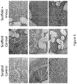
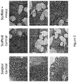
- Surgical controls showed normal histology in all but one animal, which demonstrated cellular invasion and loss of fascicular structure. This is commonly seen in tendons that have been treated surgically. Examples of H & E stains of tendon and associated tissue in surgical control sample sections are shown in the left column of Figure 1 . Examples of trichrome stains of tendon and associated tissue in surgical control sample sections are shown in the left column of Figure 2 .
- Rats receiving scaffolds with no microvascular tissue cells showed tissue invasion into the scaffolds.
- the associated tendons in the scaffold controls showed normal structure in 6 of 8 animals.
- Two of the scaffold control animals showed inflammation, which may have resulted from infections and/or wound dehiscence.
- Examples of H & E stains of tendon and associated tissue in scaffold control sample sections are shown in the center column of Figure 1 .
- Examples of trichrome stains of tendon and associated tissue in scaffold control sample sections are shown in the center column of Figure 2 .
- Rats receiving scaffold plus microvascular tissue cells also displayed tissue invasion into the scaffolds.
- the associated tendons showed normal structure in 6 of 7 animals and cellular invasion in 1 of 7.
- tendon approximated the cell-loaded scaffolds, and the appearance of new tendon formation was evident in 4 of 7 animals.
- Examples of H & E stains of tendon and associated tissue in scaffold plus microvascular tissue cell sample sections are shown in the right column of Figure 1 .
- Examples of Masson's trichrome stains of tendon and associated tissue in microvascular tissue cell sample sections are shown in the right column of Figure 2 .
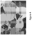
- One of the rats exhibited inflammation and one animal showed bone ingrowth into the scaffold ( Figure 4 ).
- skin appeared to have grown in the vicinity of the scaffold ( Figure 3 ), suggesting enhanced skin regeneration.
- Quantitative PCR analysis for tenascin determined tenascin expression levels to be 3.9-fold and 7.4-fold higher than surgical controls in the scaffold control and scaffold plus microvascular tissue cells, respectively (absolute values, 2.3, 8.9, and 17.0 for surgical controls, scaffold controls, and scaffold plus microvascular tissue cells, respectively). Immunohistochemical staining also showed an increase of tenascin expression in scaffold plus microvascular tissue cells ( Figure 5 , right column).
Landscapes
- Health & Medical Sciences (AREA)
- Life Sciences & Earth Sciences (AREA)
- Engineering & Computer Science (AREA)
- Biomedical Technology (AREA)
- Chemical & Material Sciences (AREA)
- General Health & Medical Sciences (AREA)
- Medicinal Chemistry (AREA)
- Epidemiology (AREA)
- Veterinary Medicine (AREA)
- Public Health (AREA)
- Animal Behavior & Ethology (AREA)
- Cell Biology (AREA)
- Zoology (AREA)
- Dermatology (AREA)
- Oral & Maxillofacial Surgery (AREA)
- Transplantation (AREA)
- Chemical Kinetics & Catalysis (AREA)
- Botany (AREA)
- Biotechnology (AREA)
- Developmental Biology & Embryology (AREA)
- Molecular Biology (AREA)
- Immunology (AREA)
- Pharmacology & Pharmacy (AREA)
- Virology (AREA)
- Urology & Nephrology (AREA)
- Bioinformatics & Cheminformatics (AREA)
- Organic Chemistry (AREA)
- Wood Science & Technology (AREA)
- Genetics & Genomics (AREA)
- Rheumatology (AREA)
- Vascular Medicine (AREA)
- Hematology (AREA)
- Orthopedic Medicine & Surgery (AREA)
- Rehabilitation Therapy (AREA)
- Microbiology (AREA)
- Biochemistry (AREA)
- General Engineering & Computer Science (AREA)
- Heart & Thoracic Surgery (AREA)
- Cardiology (AREA)
- General Chemical & Material Sciences (AREA)
Description
- This application is directed to compositions relating to tissue repair using allogeneic microvascular tissue or cells.
- Tendon injuries are very common. Sprains will heal spontaneously, but complete tears will often lead to disability if not surgically treated. Despite surgical repairs, about 15% of Achilles tendons and 40% of two tendon rotator cuff repairs subsequently fail. Furthermore, the repaired tendons seldom return to pre-injury strength and function levels. Attempts to improve the success rate have involved better suture and bone anchors, new surgical methods, and patches to reinforce the repair and provide scaffolding for tissue ingrowth to thicken the tendon. There are also reports of improved tendon healing using growth factors such as BMP-2, BMP-12, PDGF-BB, and bFGF in preclinical models.
- Although some researchers have indicated tendon as a possible tissue to grow using mesenchymal stem cells (MSC), very little work has been done along that line. There is even less work reported with adipose- or other microvascular tissue-derived stem or progenitor cells. It has been shown that fresh cells from adipose and other microvascular tissues could be used for regenerating orthopedic tissues. Others have since shown that these cells help treat tendon lesions in horse models of tendon injury, as well as other conditions. These cells were always autologous or syngeneic.
- However, the use of autologous sourced stem cells is inconvenient. It requires two surgical procedures with associated pain, cost, and morbidity. There are also risks in shipping the tissue to the lab for processing and a delay in treatment of the injury or disease.
- Recently bone graft products have been launched that use uncultured bone marrow cells from allogeneic donors adsorbed to bone chips or demineralized bone matrix (DBM). However, this type of product is unsuitable for soft-tissue repair and requires special handling to preserve the bone marrow cells.
- The invention is as defined in claims 1-13 and relates to a composition comprising minimally processed, allogeneic, uncultured microvascular tissue wherein the viability of the included stem or progenitor cells is less than 50% and wherein said composition has tissue healing activity. It also relates to a composition comprising dissociated, and dried microvascular tissue, wherein said microvascular tissue comprises , uncultured stem or progenitor cells with less than 50% viability, and wherein the microvascular tissue has tissue healing activity, said composition being suitable for administration to a subject that is allogeneic or xenogeneic with respect to the source of the microvascular tissue.
- Embodiments of the described invention may include embodiments with one or more of the following features:
- Use of allogeneic stem or progenitor cells for repair or regeneration of tendon, ligament, or skin.
- Use of processed microvascular tissue for repair or regeneration of tendons, ligaments, or skin.
- Use of allogeneic, uncultured, minimally processed microvascular tissue for regeneration of tendons, ligaments, or skin.
- Use of uncultured, dried allogeneic stem or progenitor cells to repair or regenerate tendon, ligaments or skin.
- A processed microvascular tissue product which does not contain bone or bone matrix suitable for implantation into an allogeneic or xenogeneic recipient.
- Manufacture of processed microvascular tissue from donors in a process validated to prevent viral contamination between lots.
- Manufacture of processed microvascular tissue from donors in a process that uses a single enzyme, removes blood cells, or uses no enzyme.
- A product suitable for implantation into a patient containing processed microvascular tissue which is stable at room temperature for more than a month.
- A dried or lyophilized formulation of processed microvascular tissue.
- A formulation comprising processed microvascular tissue dried at hyperbaric pressures.
- The use of nonviable stem or progenitor cells for the repair or regeneration of cartilage, ligaments or skin,.
- The use of allogeneic or xenogeneic nonviable stem or progenitor cells for the repair or regeneration of cartilage, ligaments or skin.
- The use of stem or progenitor cell products with less than 50% viability for the repair or regeneration cartilageligaments or skin.
- The use of cryoperserved allogeneic microvascular tissue for the repair or regeneration of tendon or ligament.
- A product suitable for implantation into a human patient comprising processed microvascular tissue with less than 50% viability.
- A product suitable for implantation into a human patient comprising stem cells with stabilized membranes.
- Combining allogeneic processed microvascular tissue with an orthopedic implant; a porous, flexible implantable scaffold; a surgical implant; pure water; porous coated implant; polymer solution; solvents such as DMSO, N-methylpyrrolidone (NMP), and alcohols; hydrogel; hyaluronic acid or other glycosaminoglycans or proteoglycans; collagen; fibrin; thrombin; blood clot; platelets; platelet rich plasma; demineralized bone matrix; or cancellous bone for implantation into a patient.
- Combining allogeneic processed microvascular tissue with any of the following excipients: trehalose, sucrose, mannitol, or other sugars; glycols; DMSO; aldehydes; albumin.
- Allogeneic or xenogeneic processed microvascular tissue for repair or regeneration of microvascular tissues other than bone in patients.
- A method is described for repair or regeneration of a tissue (e.g., tendon, ligament, or skin) comprising applying a plurality of uncultured allogeneic stem or progenitor cells to the tissue and thereby effecting repair or regeneration of the tissue as compared to a control tissue to which uncultured allogeneic stem or progenitor cells are not applied. The plurality of uncultured allogeneic stem or progenitor cells can be included in a processed or cryopreserved microvascular tissue. The plurality of uncultured allogeneic stem or progenitor cells can include xenogeneic cells.
- In some embodiments, a plurality of uncultured allogeneic stem or progenitor cells for use in a described method can be less than 50% viable or can include substantially no viable cells.
- A method is described for repair or regeneration of a tissue (e.g., tendon, ligament, bone, or skin) comprising applying a composition comprising substantially intact cell membranes of non-viable stem or progenitor cells to the tissue and thereby effecting repair or regeneration of the tissue as compared to a control tissue to which the composition is not applied. The non-viable stem or progenitor cells can be included in a processed or cryopreserved microvascular tissue.
- In some embodiments, a composition for use in a described method comprises less than 50% viable cells or can include substantially no viable cells.
- A composition for use in a described method can be stable at room temperature and retains tissue healing activity for at least one month.
- In some embodiments, a composition for use in a described method can include non-viable stem or progenitor cells that have been dried or lyophilized.
- In some embodiments, a composition for use in a described method can include non-viable stem or progenitor cells that have been treated to prevent microbial contamination.
- In some embodiments, a composition for use in a described method can further comprise an excipient or implantable scaffold.
- In some embodiments, tissue healing activity in a described method comprises improved healing of a soft or hard tissue exposed to the composition as compared to an analogous tissue similarly treated but without exposure to the composition.
- In an embodiment, a composition is provided comprising a plurality of uncultured stem or progenitor cells formulated for implantation into an allogeneic or xenogeneic recipient, where the composition has tissue healing activity and includes no bone or bone-derived matrix.
- In another embodiment, a composition is provided comprising substantially intact cell membranes of non-viable stem or progenitor cells formulated for implantation into an allogeneic or xenogeneic recipient, where the composition has tissue healing activity. In some embodiments, an internal component of the non-viable stem or progenitor cells can be included in the composition.
- In some embodiments, a composition provided herein can allogeneic stem or progenitor cells that are included in a processed or cryopreserved microvascular tissue.
- The composition provided herein includes less than 50% viable cells. In some embodiments, a composition provided herein can include substantially no viable cells.
- In some embodiments, a composition provided herein can be stable at room temperature and retain tissue healing activity for at least one month.
- In some embodiments, the tissue healing activity of a composition provided herein comprises improved healing of a soft or hard tissue exposed to the composition as compared to an analogous tissue similarly treated but without exposure to the composition.
- In some embodiments, a composition provided herein can include stem or progenitor cells that have been dried or lyophilized.
- In some embodiments, a composition provided herein can further include an excipient or an implantable scaffold.
- In some embodiments, a composition provided herein can include stem or progenitor cells that has been treated to prevent microbial contamination.
- While multiple embodiments are disclosed, still other embodiments of the present invention will become apparent to those skilled in the art from the following detailed description, which shows and describes illustrative embodiments of the invention. Accordingly, the drawings and detailed description are to be regarded as illustrative in nature and not restrictive.
-
-
Figure 1 shows hematoxylin and eosin (H & E) stains of Achilles tendon and associated tissue in surgical control, scaffold control, and scaffold plus microvascular tissue cell (mVasc) sample sections. -
Figure 2 shows trichrome stains of Achilles tendon and associated tissue in surgical control, scaffold control, and scaffold plus microvascular tissue cell (mVasc) sample sections. -
Figure 3 shows H & E stains of Achilles tendon and associated tissue in scaffold plus microvascular tissue cell sample sections, showing skin-like structures grown into the scaffold. -
Figure 4 shows a trichrome stain of Achilles tendon and associated tissue in a scaffold plus microvascular tissue cell sample section, showing bone-like structure grown into the scaffold where it was attached to the calcaneus. -
Figure 5 shows immunohistochemical staining of tenascin expression in Achilles tendon and associated tissue in surgical control, scaffold control, and scaffold plus microvascular tissue cell (mVasc) sample sections. - Described herein is a composition as defined in claims 1-13.
- Is described a process that generates allogeneic (i.e., allogeneic or xenogeneic) processed or cryopreserved microvascular tissue which is ready to use off-the-shelf when needed. Also described is the use of processed or cryopreserved allogeneic microvascular tissue for soft tissue (e.g., tendon, ligament, skin) and hard tissue healing. The processed or cryopreserved microvascular tissue provided herein is minimally processed, uncultured microvascular tissue that includes a mixture of stem and/or progenitor cells produced from the dissociation (e.g., by enzymatic digestion) of a microvascular tissue (e.g., adipose, tendon, or muscle tissue). Processed or cryopreserved microvascular tissue can include additional molecules (e.g., whole or fragmented extracellular matrix molecules, growth factors, or cell surface molecules).
- The term "processed microvascular tissue" as used herein refers to microvascular tissue that is dissociated as described herein, and then dried using, for example, a freeze-drying or spray drying technique. The term "cryopreserved microvascular tissue" as used herein refers to microvascular tissue that is dissociated as described herein, and then cryopreserved using known techniques. Processed microvascular tissue and cryopreserved microvascular tissue have soft and hard tissue healing (e.g., repair or regeneration) activity.
- As used herein, "tissue healing activity" of a processed microvascular tissue is the ability of the provided processed microvascular tissue to facilitate improved healing (e.g., repair or regeneration) of a tissue (e.g., hard or soft tissue) exposed to the provided processed microvascular tissue as compared to an analogous tissue similarly treated but without exposure to a processed microvascular tissue. Improved healing is measured using any appropriate means, such as time to complete healing, amount of new tissue generated, strength of the resulting healed tissue, or functionality of the resulting healed tissue. Soft tissue includes tendons, ligaments, fascia, skin, fibrous tissues, fat, synovial membranes, muscle, nerves, and blood vessels. Soft tissue injuries that can benefit from the soft tissue healing activity of the provided processed microvascular tissues include, without limitation, injuries such as tendon and/or ligament tears and injuries resulting from ischemic events.
- Allogeneic and xenogeneic stem cells have not previously been used to facilitate repair of soft tissues such as ligaments and tendons because of the difficulty of producing new soft tissue with autologous stem cells and the perception that allogeneic and xenogeneic stem cells will be rejected. In addition, the research on stem cell preservation by freeze drying has been done on purified hematopoietic stem cells in order to increase viability. In contrast, the process and composition described herein does not rely on purified stem cells or cell viability. Rather, the provided process is used to produce a processed or cryopreserved microvascular tissue containing a mixture of cells, including nonviable cells, mesenchymal stem and progenitor cells, and other molecules secreted by such cells (e.g., cytokines, growth factors, chemotactic molecules, and the like). In some embodiments, the processed or cryopreserved microvascular tissue contains a mixture of viable and nonviable cells.
- Like autologous stem cell infusions, the allogeneic stem and/or progenitor cells in the provided processed or cryopreserved microvascular tissue do not persist long in a patient, but they trigger a cascade of responses in the patient that lead to improved healing. The processed or cryopreserved microvascular tissue described herein need not include viable or whole stem cells to induce improved soft tissue healing.
- Processed or cryopreserved microvascular tissue described herein can be produced by dissociating a microvascular tissue. In some embodiments, the microvascular tissue is enzymatically digested using one or more enzymes. Suitable enzymes include those that contribute to cell dissociation, such as collagenases and neutral proteases. The enzymatic digestion process can be adjusted to increase or decrease cell dissociation. For example, if more complete cell dissociation is desired, more than one enzyme can be included or digestion time can be increased. While cell viability need not be maintained, in some embodiments it is generally desired that cellular membranes remain generally intact to preserve membranes containing attachment and signaling molecules even if some cell lysis occurs during enzymatic digestion. Thus, the use of enzymes such as lipidases may not be useful in such a process, according to one embodiment of the present invention.
- Alternatively, the microvascular tissue can be dissociated without the use of enzymes. Rather, microvascular tissue can be dissociated using physical or chemical means, including the use of chelators, ultrasonic agitation, or mechanical cell dissociation.
- The procurement of donor microvascular tissue and subsequent treatment can include steps for preventing microbial (e.g., bacterial, fungal, or viral) contamination. For example, donors can be screened for a predetermined list of microbial organisms (e.g., HIV, HPV, EBV, TB, etc.) prior to processing. Screening can be done using known techniques, such as detecting the presence of a microbial nucleic acid using polymerase chain reaction, or by detecting the presence of a molecule associated with a particular microbe by ELISA. Microbially contaminated microvascular tissue can be excluded from use, according to some embodiments of the present invention. In addition, processed or cryopreserved microvascular tissue can be produced using aseptic or sterile techniques.
- Following tissue dissociation, microvascular tissue can be further treated to remove undesired cells or molecules, such as red blood cells, lipids, or adipocytes. Additional treatment will depend upon the source of microvascular tissue. For example, if the microvascular tissue source is adipose tissue, the dissociated microvascular tissue can be centrifuged at relatively low force to separate lipids, adipocytes, and some pre-adipocytes from the rest of the microvascular tissue. In other embodiments, known muscle cell isolation protocols, such as the use of density gradient centrifugation, may be used to further treat muscle tissue following enzymatic digestion to remove muscle cells and enrich for desired cells.
- Once the microvascular tissue is prepared, it is preserved dried using a freeze-drying or spray-drying technique to produce processed microvascular tissue, or is cryopreserved. Any appropriate excipient can be used when preserving microvascular tissue, including sugars (e.g., trehalose, mannitol, sucrose), polyalcohols (e.g., polyethylene glycol), aldehydes, proteins (e.g., albumin), amino acids (e.g., glycine), surfactants (e.g., Tween 20), DMSO, and/or permanganates.
- Freeze drying typically involves four steps: pretreatment, freezing, primary drying, and secondary drying. Pretreatment can include concentration adjustment or the addition of one or more excipients. Following pretreatment, the microvascular tissue is frozen. The freezing step is typically done in a carefully controlled manner (e.g., at a rate of cooling of between about -0.5° C per minute to about -50° C per minute) to preserve cell structure, however cell viability need not be preserved. In some embodiments, microvascular tissue is frozen at a rate of cooling of about -10° C per minute. The rate of cooling can be adjusted based on the particular microvascular tissue and excipients used. The microvascular tissue can be frozen using any appropriate means, including using mechanical refrigeration and/or exposing a container containing the microvascular tissue to dry ice or liquid nitrogen until it reaches a temperature suitable for freeze drying.
- During the primary drying step, the temperature and pressure are adjusted to provide conditions suitable to cause sublimation of water from the microvascular tissue. The specific temperature and pressure can be adjusted to accommodate the excipient used and/or the concentration of the microvascular tissue.
- During the secondary drying step, the temperature and pressure can be further adjusted to facilitate the removal of unfrozen water from the microvascular tissue. The final water content following the secondary drying step is preferably between 1% and 4% by weight, but can be adjusted in order to maximize shelf life or soft tissue healing activity.
- In some embodiments, the microvascular tissue is spray dried. Prior to spray drying, the microvascular tissue can be pretreated similarly to microvascular tissue that is to be freeze dried, with the excipients being chosen as appropriate for spray drying rather than freeze drying. During spray drying, the microvascular tissue is atomized into droplets and exposed to heated air in a drying chamber.
- In some embodiments, microvascular tissue is not processed by drying, but cryopreserved. Methods for cryopreserving tissue are known. For example, microvascular tissue is mixed with one or more excipients (e.g., DMSO) and cooled in a carefully controlled manner. In some embodiments, cooling is done in two or more stages in which the first stage is done in a controlled manner (e.g., reducing the temperature by 1° C per minute) to an intermediate temperature (e.g., -30° C), with the second stage transferring cells at the intermediate temperature to a colder storage temperature (e.g., - 196° C).
- Cryopreserved microvascular tissue is stored at a temperature suitable for maintaining the cryopreserved state (e.g., from about -30° C to -196° C). Freeze dried or spray dried processed microvascular tissue can be stored in a wider variety of conditions than cryopreserved cells, live cells, or fresh tissue. Suitable temperatures for the storage for processed microvascular tissue include temperatures from about -100° C to about 45° C. In some embodiments, freeze dried or spray dried processed microvascular tissue can be stored at room temperature. The shelf life of the provided processed microvascular tissue is at least one week, and preferably, at least one month, while maintaining tissue healing activity.
- Additionally, because viability is not required for suitability of the processed or cryopreserved microvascular tissue for use in soft tissue repair, the preservation process and storage need not be adjusted to maintain viability. The percentage of viable cells in the provided microvascular tissue before processing or cryopreservation can be up to 100%. After processing or cryopreservation, it is less than 50%, e.g., less than 40%, less than 30%, less than 20%, or less than 10%. In some embodiments, the provided processed microvascular tissue contains no viable cells after processing or cryopreservation.
- Further described are methods of using processed microvascular tissue. Processed microvascular tissue can be applied directly to a tissue in need of repair, or can be applied to tissue surrounding such a tissue in need of repair. In some embodiments, a dried processed microvascular tissue is reconstituted in a suitable carrier (e.g., water or saline) and directly applied to a tissue in need of repair. In some embodiments, reconstituted processed microvascular tissue is applied to a scaffold, such as a collagen matrix or biocompatible fabric, prior to being applied to a tissue. In other embodiments, a processed microvascular tissue is used to coat a material, such as a flexible biocompatible scaffold (e.g., woven or nonwoven fabric sheets or thread), biocompatible microbeads or particles, or an implantable medical device. Spray dried processed microvascular tissue is particularly suited to coating a material comprising microbeads or particles without requiring reconstitution prior to coating, as coating can be done during the spray drying process.
- Processed or cryopreserved microvascular tissue can be combined with any suitable device or material prior to implant into a patient. Processed or cryopreserved microvascular tissue can be combined with an orthopedic implant; a porous, flexible implantable scaffold; a surgical implant; pure water; saline; a porous coated implant; polymer solution; solvents such as DMSO, N-methylpyrrolidone (NMP), and alcohols; hydrogel; hyaluronic acid or other glycosaminoglycans or proteoglycans; collagen; fibrin; thrombin; blood clot; platelets; platelet rich plasma; demineralized bone matrix; autologous cells; and/or cancellous bone.
- Processed or cryopreserved microvascular tissue can be packaged alone for example, in a vial, or in combination with other products, such as those listed as being suitable for combination with processed or cryopreserved microvascular tissue. When packaged with another material, the processed or cryopreserved microvascular tissue can be separately packaged, or premixed or associated with the other material. In some embodiments, processed microvascular tissue is packaged as a coating on a biocompatible material.
- The following examples are meant to illustrate specific embodiments, and are not intended to limit the scope of the invention.
- Rat microvascular tissue was prepared from epididymal fat pads or inguinal fat pads. Briefly, Worthington collagenase (lot #4145) was put into solution in phosphate buffered saline at 1 mg/ml, resulting in a solution with a specific activity of 40 U/ml. This collagenase solution was applied to the fat (inguinal and epididymal pads were processed separately) in an equal volume to weight ratio and incubated at 4° C for 45 minutes while mixing. Collagenase activity was stopped with 25% bovine serum albumin and the cells were spun down. As assessed by trypan blue exclusion, cells collected from epididymal fat pads had a viability of about 75%, while cells collected from inguinal fat pads had a viability of about 80%. The cells were resuspended and counted. Epididymal fat yielded about 720,000 cells per ml, and inguinal fat yielded about 215,000 cells per ml.
- Human microvascular tissue can be prepared using lipoaspirate that is enzymatically treated in a similar manner as the rat microvascular tissue.
- The collected cells were prepared for lyophilization in preservation medium (6.8% trehalose, 2% Hetastarch, 5% albumin, and 1 unit/ml heparin), and placed into glass vials with stopper closures and crimp seals suitable for lyophilization. To lyophilize the cells, the lyophilizer temperature was dropped from 23° C to -45° C at a rate of 2.5° C per minute. Temperature was held at -45° C for 3 hours, followed by a first drying step. During the first drying step, temperature was increased from -45° C to -35° C at a rate of 2.5° C per minute and the pressure was reduced to 80 torr, where the temperature and pressure were held for 36 hours. A secondary drying step was then performed by increasing the temperature from -35° C to -5° C at a rate of 0.2 C per minute, then held at 5° C for 6 hours. Following the second drying step, the vials were stoppered and crimp sealed under nitrogen.
- Two days after lyophilization, the cells were rehydrated and initial cell viability was assessed by trypan blue exclusion. Cell metabolic function was assessed by measuring alamarBlue® (Life Technologies, Carlsbad, CA) metabolism according to the manufacturer's instructions. Rehydrated cells were incubated in M3:10™ medium (Incell, San Antonio, TX) supplemented with 10% fetal bovine serum in a T-25 flask at 37° C for 7 days to assess proliferation ability.
- Following rehydration, the general structure of the cells appeared to be maintained in 5-25% of cells, with a more exact calculation being difficult due to the appearance of many broken cells. The cells had an initial viability of less than 1-2%, with few cells retaining the ability to exclude trypan blue. The morphology of those cells maintaining the ability to exclude trypan blue suggested they were undergoing apoptosis, displaying enlarged nuclei and nuclear and cytoplasmic blebbing. No significant ability to metabolize alamarBlue® was observed, indicating that metabolic integrity of the lyophilized cells was not maintained. None of the cells were able to establish a culture in M3:10™ medium, confirming low/no viability of the lyophilized cells.
- Microvascular tissue cells were prepared from rat epididymal fat pads as described in Example 1. The cells were cryopreserved by resuspending the cells in M3DEF defined medium without supplements (Incell), adding an equal volume of EZ-CPZ (Incell), with a final DMSO concentration of 5%. The cells were then frozen slowly in vials in a slow cool box overnight at -80° C and then transferred to -130°C. Viability of the cryopreserved cells was determined to be less than 50% as assessed using trypan blue exclusion. Viability was typically around 90% before preparation.
- BioFiber® Collagen-coated Scaffold material (Tornier) was placed on top of thick gauze material resting in PBS. The scaffold material was pre-wet with PBS prior to addition of microvascular tissue cells.
- The cells were thawed quickly at 37° C, centrifuged at 400 x g for 5 minutes, and resuspended at 1 × 10⁶ cells per ml in phosphate buffered saline (PBS). The suspended cells were applied to the pre-wet scaffold material at a rate of 100 µl of the suspended cells per cm2 of scaffold material, and allowed to absorb into the scaffold material by wicking action of the gauze beneath the scaffold material.
- Scaffold material was kept moist using PBS, and allowed to incubate for 15-20 minutes at 37° C and 5% CO2 in a humid chamber. Microvascular tissue cell-impregnated scaffold material was kept moist and under aseptic conditions until use in Achilles tendon model.
- Prior to treatment, 32 animals were weighed and randomly assigned to 4 treatment groups. The right rear limb of each animal was shaved one day prior to the start of the test. Prior to surgery on Day 1, animals were weighed and anesthetized with an intramuscular injection of ketamine hydrochloride 100 mg/mL (40-90 mg/kg) and xylazine 100 mg/mL (5-10 mg/kg). Mask or chamber induction with Isoflurane may also be used. The skin was surgically prepared with betadine and alcohol scrubs, and draped using aseptic surgical techniques.
- Microvascular tissue cell-impregnated scaffold material was prepared immediately prior to implantation. The graft was trimmed to 10 mm x 11 mm and two of the corners were notched. Two 5-0 polypropylene sutures were placed in the graft for fixation. The graft was then rolled to form a cylindrical structure for wrapping around the Achilles tendon. The graft was set aside in the Petri dish with saline and covered until used.
- A straight, lateral skin incision was made from the caudal (distal) tibia of the right rear limb to the level of the mid tibia. The skin was dissected and retracted to allow a lateral exposure of the Achilles tendon from calcaneus to its musculo-tendinous junction. The exposed Achilles tendon was slightly abraded with mouse-tooth forceps prior to graft test article placement. A single 0.5 mm drill hole was made in the lateral to medial direction through the calcaneus to allow suture passage for graft fixation. The implant area was irrigated with saline to remove any debris and blotted dry.
- The graft was removed from the saline and then wrapped around the Achilles tendon with the notched ends adjacent to the calcaneus. The graft edge was closed with interrupted 5-0 polypropylene sutures. The cranial graft fixation suture was placed in the gastrocnemius cranial to the musculo-tendinous junction using a modified Mason-Allen suture pattern. The caudal graft fixation suture was then passed through the drill hole in the calcaneus and tensioned with the foot in a neutral position and tied. Six suture knots were tied for all fixation sutures. The incision was closed in a layered fashion using appropriate suture material. The incision site was observed evaluated daily until day 10 post operation.
- The rats were grouped into the treatment groups outlined in Table 1.
Table 1. Treatment Groups Group No. Treatment Surgical control 8 Achilles tendon will be slightly abraded with mouse-tooth forceps Scaffold control 8 Achilles tendon will be slightly abraded with mouse-tooth forceps +Tornier's BioFiber Scaffold Coated with Collagen Scaffold + microvascular tissue cells (mVasc) 8 Achilles tendon will be slightly abraded with mouse-tooth forceps +Tornier's BioFiber Scaffold Coated with Collagen + Rat microvascular tissue cells (thawed) - Rats were euthanized at day 42 ± 1. Immediately following euthanasia, the implanted test or control article sites and surrounding tendinous tissue were collected by excision from each animal. All collected samples were split in half. One-half of the collected tissue was stored in 10% neutral buffered formalin for routine histopathological and immunohistochemistry evaluation. The remaining half was snap frozen at ≤ -70°C in liquid nitrogen for gene expression analysis. A section of tendon and liver of each animal was also collected as staining controls and stored in 10% neutral buffered formalin for immunohistochemistry evaluation.
- Histopathological, immunohistochemical, and gene expression analysis was done by BioModels (Watertown, MA). Briefly, hematoxylin and eosin and trichrome stains were performed on the tissues stored in 10% neutral buffered formalin. Immunohistochemistry utilizing anti-tenascin antibodies was also performed on the tissues stored in 10% neutral buffered formalin and on the tissues collected as staining controls. The tissues snap frozen at ≤ -70°C were utilized for isolation of RNA and qPCR of tenascin. Tenascin is a marker for tenocyte progenitor cells.
- Surgical controls showed normal histology in all but one animal, which demonstrated cellular invasion and loss of fascicular structure. This is commonly seen in tendons that have been treated surgically. Examples of H & E stains of tendon and associated tissue in surgical control sample sections are shown in the left column of
Figure 1 . Examples of trichrome stains of tendon and associated tissue in surgical control sample sections are shown in the left column ofFigure 2 . - Rats receiving scaffolds with no microvascular tissue cells showed tissue invasion into the scaffolds. The associated tendons in the scaffold controls showed normal structure in 6 of 8 animals. Two of the scaffold control animals showed inflammation, which may have resulted from infections and/or wound dehiscence. Examples of H & E stains of tendon and associated tissue in scaffold control sample sections are shown in the center column of
Figure 1 . Examples of trichrome stains of tendon and associated tissue in scaffold control sample sections are shown in the center column ofFigure 2 . - Rats receiving scaffold plus microvascular tissue cells also displayed tissue invasion into the scaffolds. The associated tendons showed normal structure in 6 of 7 animals and cellular invasion in 1 of 7. In 5 of 7 of the animals, tendon approximated the cell-loaded scaffolds, and the appearance of new tendon formation was evident in 4 of 7 animals. Examples of H & E stains of tendon and associated tissue in scaffold plus microvascular tissue cell sample sections are shown in the right column of
Figure 1 . Examples of Masson's trichrome stains of tendon and associated tissue in microvascular tissue cell sample sections are shown in the right column ofFigure 2 . One of the rats exhibited inflammation and one animal showed bone ingrowth into the scaffold (Figure 4 ). In two of the rats with scaffold plus microvascular tissue cells, skin appeared to have grown in the vicinity of the scaffold (Figure 3 ), suggesting enhanced skin regeneration. - Quantitative PCR analysis for tenascin determined tenascin expression levels to be 3.9-fold and 7.4-fold higher than surgical controls in the scaffold control and scaffold plus microvascular tissue cells, respectively (absolute values, 2.3, 8.9, and 17.0 for surgical controls, scaffold controls, and scaffold plus microvascular tissue cells, respectively). Immunohistochemical staining also showed an increase of tenascin expression in scaffold plus microvascular tissue cells (
Figure 5 , right column). - Various modifications and additions can be made to the exemplary embodiments discussed without departing from the scope of the present invention. For example, while the embodiments described above refer to particular features, the scope of this invention also includes embodiments having different combinations of features and embodiments that do not include all of the above described features.
Claims (13)
- A composition comprising minimally processed, uncultured microvascular tissue, wherein the tissue has been dissociated and then dried or cryopreserved, wherein the tissue has been dissociated using enzymatic digestion, wherein the tissue includes a mixture of stem and/or progenitor cells produced from the dissociation, wherein the viability of the included stem or progenitor cells is less than 50% and wherein said composition has tissue healing activity, wherein the tissue healing activity comprises improved repair or regeneration of a tissue.
- A composition according to claim 1, wherein said stem or progenitor cells are substantially all nonviable.
- A composition according to claim 1, wherein said composition is stable at room temperature and retains said tissue healing activity for at least a month.
- A composition comprising dissociated, and dried microvascular tissue, wherein said microvascular tissue comprises uncultured stem or progenitor cells with less than 50% viability, and wherein the microvascular tissue has tissue healing activity, said composition being suitable for administration to a subject that is allogeneic or xenogeneic with respect to the source of the microvascular tissue.
- The composition of claim 4, wherein said microvascular tissue is dried by lyophilization or spray-drying.
- The composition of claim 4, wherein the uncultured stem or progenitor cells contain less than 10% viable cells.
- The composition according to any one of the preceding claims, wherein said microvascular tissue is dissociated from adipose tissue or muscle tissue.
- The composition of claim 4, wherein said microvascular tissue is dissociated using enzymatic digestion.
- The composition according to any one of claims 4-8, wherein the dried microvascular tissue is stable at room temperature and retains tissue healing activity for at least one month.
- The composition according to any one of claims 4- 9, wherein the processed microvascular tissue further comprises an excipient.
- The composition according to any one of the preceding claims, wherein said microvascular tissue has been treated to prevent microbial contamination.
- The composition according to any one of the preceding claims wherein said tissue healing activity comprises improved healing of tendon, ligament, or skin exposed to the composition as compared to an analogous tissue similarly treated but without exposure to the composition.
- The composition according to any one of the preceding claims for use in repair or regeneration of tendon, ligament, or skin.
Applications Claiming Priority (3)
| Application Number | Priority Date | Filing Date | Title |
|---|---|---|---|
| US201161454367P | 2011-03-18 | 2011-03-18 | |
| EP12782247.6A EP2686419B1 (en) | 2011-03-18 | 2012-03-17 | Allogeneic microvascular tissue for soft tissue treatments |
| PCT/US2012/029568 WO2012154301A1 (en) | 2011-03-18 | 2012-03-17 | Allogeneic microvascular tissue for soft tissue treatments |
Related Parent Applications (1)
| Application Number | Title | Priority Date | Filing Date |
|---|---|---|---|
| EP12782247.6A Division EP2686419B1 (en) | 2011-03-18 | 2012-03-17 | Allogeneic microvascular tissue for soft tissue treatments |
Publications (2)
| Publication Number | Publication Date |
|---|---|
| EP3141599A1 EP3141599A1 (en) | 2017-03-15 |
| EP3141599B1 true EP3141599B1 (en) | 2022-12-21 |
Family
ID=47139494
Family Applications (2)
| Application Number | Title | Priority Date | Filing Date |
|---|---|---|---|
| EP16195005.0A Active EP3141599B1 (en) | 2011-03-18 | 2012-03-17 | Allogeneic microvascular tissue for soft tissue treatments |
| EP12782247.6A Active EP2686419B1 (en) | 2011-03-18 | 2012-03-17 | Allogeneic microvascular tissue for soft tissue treatments |
Family Applications After (1)
| Application Number | Title | Priority Date | Filing Date |
|---|---|---|---|
| EP12782247.6A Active EP2686419B1 (en) | 2011-03-18 | 2012-03-17 | Allogeneic microvascular tissue for soft tissue treatments |
Country Status (4)
| Country | Link |
|---|---|
| US (4) | US9044430B2 (en) |
| EP (2) | EP3141599B1 (en) |
| ES (2) | ES2613547T3 (en) |
| WO (1) | WO2012154301A1 (en) |
Families Citing this family (15)
| Publication number | Priority date | Publication date | Assignee | Title |
|---|---|---|---|---|
| EP3141599B1 (en) | 2011-03-18 | 2022-12-21 | MicroVascular Tissues, Inc. | Allogeneic microvascular tissue for soft tissue treatments |
| JP6456829B2 (en) | 2012-09-19 | 2019-01-23 | マイクロバスキュラー ティシューズ, インコーポレイテッド | Compositions and methods for treating and preventing tissue injury and disease |
| US11819522B2 (en) | 2012-09-19 | 2023-11-21 | Microvascular Tissues, Inc. | Compositions and methods for treating and preventing tissue injury and disease |
| US9872937B2 (en) | 2012-09-19 | 2018-01-23 | Microvascular Tissues, Inc. | Compositions and methods for treating and preventing tissue injury and disease |
| US10596202B2 (en) | 2012-09-19 | 2020-03-24 | Microvascular Tissues, Inc. | Compositions and methods for treating and preventing tissue injury and disease |
| US9681960B2 (en) | 2014-05-16 | 2017-06-20 | Howmedica Osteonics Corp. | Guides for fracture system |
| US10575968B2 (en) | 2014-05-16 | 2020-03-03 | Howmedica Osteonics Corp. | Guides for fracture system |
| WO2016111899A1 (en) | 2015-01-05 | 2016-07-14 | Petrucci Gary M | Methods and materials for treating lung disorders |
| US9687511B2 (en) | 2015-03-06 | 2017-06-27 | Vivex Biomedical, Inc. | Acellular biologic composition and method of manufacture |
| US11160904B2 (en) | 2017-05-09 | 2021-11-02 | Vivex Biologies Group, Inc. | Biological composition in a protectant shroud and methods |
| US10993969B2 (en) | 2016-02-05 | 2021-05-04 | Gary M. Petrucci | Methods and materials for treating nerve injuries and neurological disorders |
| US20190091261A1 (en) * | 2016-03-02 | 2019-03-28 | Microvascular Tissues, Inc | Enhanced multipotent cells and microvascular tissue and methods of use thereof |
| US10478531B2 (en) | 2017-06-22 | 2019-11-19 | Gary M. Petrucci | Methods and materials for treating blood vessels |
| US10251917B1 (en) | 2017-09-19 | 2019-04-09 | Gary M. Petrucci | Methods and materials for treating tumors |
| WO2019195773A1 (en) * | 2018-04-05 | 2019-10-10 | The George Washington University | METHOD OF TREATING A MAMMAL WITH DIABETES ASSOCIATED KIDNEY DISEASE USING LOCAL ADMINISTRATION OF STEM CELLS WITH TRANSIENTLY REDUCED p53 |
Family Cites Families (38)
| Publication number | Priority date | Publication date | Assignee | Title |
|---|---|---|---|---|
| US5358931A (en) | 1990-01-17 | 1994-10-25 | The Regents Of The University Of California | Interaction of thermal hysteresis proteins with cells and cell membranes and associated applications |
| US5656498A (en) * | 1994-02-22 | 1997-08-12 | Nippon Telegraph And Telephone Corporation | Freeze-dried blood cells, stem cells and platelets, and manufacturing method for the same |
| US6200606B1 (en) | 1996-01-16 | 2001-03-13 | Depuy Orthopaedics, Inc. | Isolation of precursor cells from hematopoietic and nonhematopoietic tissues and their use in vivo bone and cartilage regeneration |
| JP2000503542A (en) | 1996-01-16 | 2000-03-28 | デピュイ オーソピーディック,インコーポレイテッド | Separation of progenitor cells from hematopoietic and non-hematopoietic tissues and their use in bone and cartilage regeneration |
| US5730933A (en) | 1996-04-16 | 1998-03-24 | Depuy Orthopaedics, Inc. | Radiation sterilization of biologically active compounds |
| US5786207A (en) | 1997-05-28 | 1998-07-28 | University Of Pittsburgh | Tissue dissociating system and method |
| US7125856B1 (en) | 1999-04-15 | 2006-10-24 | St. Elizabeth's Medical Center Of Boston, Inc. | Angiogenic growth factors for treatment of peripheral neuropathy |
| US7659111B2 (en) | 2002-06-27 | 2010-02-09 | Core Dynamics Limited | Method for freezing viable cells |
| US20060127873A1 (en) * | 2002-07-16 | 2006-06-15 | Caroline Hoemann | Composition for cytocompatible, injectable, self-gelling chitosan solutions for encapsulating and delivering live cells or biologically active factors |
| AR047712A1 (en) | 2002-09-07 | 2006-02-15 | Royal Veterinary College | TREATMENT METHOD OF A NATURAL SOFT SKELETTIC TISSUE INJURY MANAGING A COMPOSITION OF MESENQUIMATOSE MOTHER CELLS |
| US7270946B2 (en) | 2002-10-04 | 2007-09-18 | Organ Recovery Systems, Inc. | Method for treatment of cellular materials with sugars prior to preservation |
| US7553827B2 (en) | 2003-08-13 | 2009-06-30 | Depuy Spine, Inc. | Transdiscal administration of cycline compounds |
| CA2530533C (en) | 2003-06-27 | 2015-02-10 | Ethicon, Incorporated | Postpartum cells derived from umbilical cord tissue, and methods of making and using the same |
| EP2450433A3 (en) | 2003-10-08 | 2012-08-08 | Vet-Stem Inc | Methods of preparing and using stem cell compositions and kits comprising the same |
| WO2005068498A2 (en) | 2004-01-19 | 2005-07-28 | Nsgene A/S | Human therapeutic cells secreting nerve growth factor |
| EP1576957A1 (en) | 2004-03-18 | 2005-09-21 | Universiteit Twente | Tissue repair using pluripotent cells |
| TR200402809A1 (en) | 2004-07-07 | 2006-02-21 | General Mills, Inc. | Vapor-free process for the production of gum-based fruit snacks |
| AU2005322873B2 (en) * | 2004-12-30 | 2011-09-01 | Primegen Biotech, Llc | Adipose-derived stem cells for tissue regeneration and wound healing |
| WO2007015252A2 (en) * | 2005-08-03 | 2007-02-08 | I.M.T. Interface Multigrad Technology Ltd. | Somatic cells for use in cell therapy |
| US20100068245A1 (en) | 2006-03-15 | 2010-03-18 | Manders Christian D | Preparation and storage of stable, biologically active materials |
| US20070292401A1 (en) | 2006-06-20 | 2007-12-20 | Harmon Alexander M | Soft tissue repair and regeneration using stem cell products |
| US20100015104A1 (en) | 2006-07-26 | 2010-01-21 | Cytori Therapeutics, Inc | Generation of adipose tissue and adipocytes |
| US20080033572A1 (en) | 2006-08-03 | 2008-02-07 | Ebi L.P. | Bone graft composites and methods of treating bone defects |
| KR100771058B1 (en) | 2007-05-18 | 2007-10-30 | 이희영 | Scaffolds removed lipid for human volume replacement or cell culture and use thereof |
| WO2009115581A2 (en) * | 2008-03-19 | 2009-09-24 | Cryo-Save Ag | Improved cryopreservation of adipose tissue for the isolation of mesenchymal stem cells |
| US8865199B2 (en) * | 2008-11-17 | 2014-10-21 | Ingeneron, Inc. | Biomatrix composition and methods of biomatrix seeding |
| KR101072732B1 (en) | 2009-04-01 | 2011-10-11 | 국립암센터 | Bone graft shaping system and method using the same |
| CN101757691B (en) | 2010-02-05 | 2013-04-24 | 陕西瑞盛生物科技有限公司 | Preparation method of tissue engineering cornea |
| WO2012024573A2 (en) | 2010-08-19 | 2012-02-23 | The Regents Of The University Of California | Compositions comrpising perivascular stem cells and nell-1 protein |
| US20140017787A1 (en) | 2010-10-11 | 2014-01-16 | Aline M. Betancourt | Mesenchymal stem cells and related therapies |
| AU2011352928B2 (en) | 2010-12-27 | 2017-02-02 | Stroma Cell Therapeutics, Llc | Ultrasonic cavitation derived stromal or mesenchymal vascular extracts and cells derived therefrom obtained from adipose tissue and use thereof |
| EP3141599B1 (en) | 2011-03-18 | 2022-12-21 | MicroVascular Tissues, Inc. | Allogeneic microvascular tissue for soft tissue treatments |
| WO2013142192A1 (en) | 2012-03-19 | 2013-09-26 | Richard Burt | Methods and compositions for regenerating and repairing damaged tissue using nonviable irradiated or lyophilized pluripotent stem cells |
| US9872937B2 (en) | 2012-09-19 | 2018-01-23 | Microvascular Tissues, Inc. | Compositions and methods for treating and preventing tissue injury and disease |
| US10596202B2 (en) | 2012-09-19 | 2020-03-24 | Microvascular Tissues, Inc. | Compositions and methods for treating and preventing tissue injury and disease |
| JP6456829B2 (en) | 2012-09-19 | 2019-01-23 | マイクロバスキュラー ティシューズ, インコーポレイテッド | Compositions and methods for treating and preventing tissue injury and disease |
| CA2938268A1 (en) | 2014-01-31 | 2015-08-06 | Dsm Ip Assets B.V. | Adipose tissue processing centrifuge and methods of use |
| US20190091261A1 (en) | 2016-03-02 | 2019-03-28 | Microvascular Tissues, Inc | Enhanced multipotent cells and microvascular tissue and methods of use thereof |
-
2012
- 2012-03-17 EP EP16195005.0A patent/EP3141599B1/en active Active
- 2012-03-17 EP EP12782247.6A patent/EP2686419B1/en active Active
- 2012-03-17 ES ES12782247.6T patent/ES2613547T3/en active Active
- 2012-03-17 WO PCT/US2012/029568 patent/WO2012154301A1/en active Application Filing
- 2012-03-17 US US13/423,171 patent/US9044430B2/en active Active
- 2012-03-17 ES ES16195005T patent/ES2939147T3/en active Active
-
2015
- 2015-04-28 US US14/698,763 patent/US10137224B2/en active Active
-
2018
- 2018-10-05 US US16/153,090 patent/US11376348B2/en active Active
-
2022
- 2022-06-01 US US17/830,146 patent/US20220296778A1/en active Pending
Also Published As
| Publication number | Publication date |
|---|---|
| ES2613547T3 (en) | 2017-05-24 |
| US20220296778A1 (en) | 2022-09-22 |
| US10137224B2 (en) | 2018-11-27 |
| US11376348B2 (en) | 2022-07-05 |
| US9044430B2 (en) | 2015-06-02 |
| US20160015859A1 (en) | 2016-01-21 |
| US20130071358A1 (en) | 2013-03-21 |
| EP2686419A4 (en) | 2014-10-15 |
| EP2686419A1 (en) | 2014-01-22 |
| WO2012154301A1 (en) | 2012-11-15 |
| ES2939147T3 (en) | 2023-04-19 |
| EP2686419B1 (en) | 2016-11-16 |
| EP3141599A1 (en) | 2017-03-15 |
| US20190038805A1 (en) | 2019-02-07 |
Similar Documents
| Publication | Publication Date | Title |
|---|---|---|
| US20220296778A1 (en) | Allogeneic microvascular tissue for soft tissue treatments | |
| JP6755893B2 (en) | Compositions and Methods for Treating and Preventing Tissue Injuries and Diseases | |
| JP5675595B2 (en) | Elastase treatment of tissue matrix | |
| CA2948126A1 (en) | Therapeutic placental compositions, methods of making and methods of use | |
| EP3203835B1 (en) | Method for hypothermic storage of placental tissue | |
| US20160271187A1 (en) | Compositions and methods for treating and preventing tissue injury and disease | |
| US11819522B2 (en) | Compositions and methods for treating and preventing tissue injury and disease | |
| US20230047934A1 (en) | Human tissue derived compositions and uses thereof | |
| Jafari et al. | Comparison of the effects of preservation methods on structural, biological, and mechanical properties of the human amniotic membrane for medical applications |
Legal Events
| Date | Code | Title | Description |
|---|---|---|---|
| PUAI | Public reference made under article 153(3) epc to a published international application that has entered the european phase |
Free format text: ORIGINAL CODE: 0009012 |
|
| STAA | Information on the status of an ep patent application or granted ep patent |
Free format text: STATUS: REQUEST FOR EXAMINATION WAS MADE |
|
| 17P | Request for examination filed |
Effective date: 20161021 |
|
| AC | Divisional application: reference to earlier application |
Ref document number: 2686419 Country of ref document: EP Kind code of ref document: P |
|
| AK | Designated contracting states |
Kind code of ref document: A1 Designated state(s): AL AT BE BG CH CY CZ DE DK EE ES FI FR GB GR HR HU IE IS IT LI LT LU LV MC MK MT NL NO PL PT RO RS SE SI SK SM TR |
|
| STAA | Information on the status of an ep patent application or granted ep patent |
Free format text: STATUS: EXAMINATION IS IN PROGRESS |
|
| 17Q | First examination report despatched |
Effective date: 20180926 |
|
| STAA | Information on the status of an ep patent application or granted ep patent |
Free format text: STATUS: EXAMINATION IS IN PROGRESS |
|
| STAA | Information on the status of an ep patent application or granted ep patent |
Free format text: STATUS: EXAMINATION IS IN PROGRESS |
|
| GRAP | Despatch of communication of intention to grant a patent |
Free format text: ORIGINAL CODE: EPIDOSNIGR1 |
|
| STAA | Information on the status of an ep patent application or granted ep patent |
Free format text: STATUS: GRANT OF PATENT IS INTENDED |
|
| INTG | Intention to grant announced |
Effective date: 20220603 |
|
| GRAS | Grant fee paid |
Free format text: ORIGINAL CODE: EPIDOSNIGR3 |
|
| RAP3 | Party data changed (applicant data changed or rights of an application transferred) |
Owner name: MICROVASCULAR TISSUES, INC. |
|
| RIN1 | Information on inventor provided before grant (corrected) |
Inventor name: EMMITT, RICHARD B. Inventor name: PETERSON, DALE R. |
|
| GRAA | (expected) grant |
Free format text: ORIGINAL CODE: 0009210 |
|
| STAA | Information on the status of an ep patent application or granted ep patent |
Free format text: STATUS: THE PATENT HAS BEEN GRANTED |
|
| AC | Divisional application: reference to earlier application |
Ref document number: 2686419 Country of ref document: EP Kind code of ref document: P |
|
| AK | Designated contracting states |
Kind code of ref document: B1 Designated state(s): AL AT BE BG CH CY CZ DE DK EE ES FI FR GB GR HR HU IE IS IT LI LT LU LV MC MK MT NL NO PL PT RO RS SE SI SK SM TR |
|
| REG | Reference to a national code |
Ref country code: GB Ref legal event code: FG4D |
|
| REG | Reference to a national code |
Ref country code: DE Ref legal event code: R096 Ref document number: 602012079148 Country of ref document: DE |
|
| REG | Reference to a national code |
Ref country code: CH Ref legal event code: EP |
|
| REG | Reference to a national code |
Ref country code: AT Ref legal event code: REF Ref document number: 1539077 Country of ref document: AT Kind code of ref document: T Effective date: 20230115 |
|
| REG | Reference to a national code |
Ref country code: IE Ref legal event code: FG4D |
|
| REG | Reference to a national code |
Ref country code: SE Ref legal event code: TRGR |
|
| REG | Reference to a national code |
Ref country code: LT Ref legal event code: MG9D |
|
| REG | Reference to a national code |
Ref country code: ES Ref legal event code: FG2A Ref document number: 2939147 Country of ref document: ES Kind code of ref document: T3 Effective date: 20230419 |
|
| REG | Reference to a national code |
Ref country code: NL Ref legal event code: MP Effective date: 20221221 |
|
| PG25 | Lapsed in a contracting state [announced via postgrant information from national office to epo] |
Ref country code: NO Free format text: LAPSE BECAUSE OF FAILURE TO SUBMIT A TRANSLATION OF THE DESCRIPTION OR TO PAY THE FEE WITHIN THE PRESCRIBED TIME-LIMIT Effective date: 20230321 Ref country code: LT Free format text: LAPSE BECAUSE OF FAILURE TO SUBMIT A TRANSLATION OF THE DESCRIPTION OR TO PAY THE FEE WITHIN THE PRESCRIBED TIME-LIMIT Effective date: 20221221 Ref country code: FI Free format text: LAPSE BECAUSE OF FAILURE TO SUBMIT A TRANSLATION OF THE DESCRIPTION OR TO PAY THE FEE WITHIN THE PRESCRIBED TIME-LIMIT Effective date: 20221221 |
|
| PGFP | Annual fee paid to national office [announced via postgrant information from national office to epo] |
Ref country code: FR Payment date: 20230327 Year of fee payment: 12 |
|
| REG | Reference to a national code |
Ref country code: AT Ref legal event code: MK05 Ref document number: 1539077 Country of ref document: AT Kind code of ref document: T Effective date: 20221221 |
|
| PG25 | Lapsed in a contracting state [announced via postgrant information from national office to epo] |
Ref country code: RS Free format text: LAPSE BECAUSE OF FAILURE TO SUBMIT A TRANSLATION OF THE DESCRIPTION OR TO PAY THE FEE WITHIN THE PRESCRIBED TIME-LIMIT Effective date: 20221221 Ref country code: LV Free format text: LAPSE BECAUSE OF FAILURE TO SUBMIT A TRANSLATION OF THE DESCRIPTION OR TO PAY THE FEE WITHIN THE PRESCRIBED TIME-LIMIT Effective date: 20221221 Ref country code: HR Free format text: LAPSE BECAUSE OF FAILURE TO SUBMIT A TRANSLATION OF THE DESCRIPTION OR TO PAY THE FEE WITHIN THE PRESCRIBED TIME-LIMIT Effective date: 20221221 Ref country code: GR Free format text: LAPSE BECAUSE OF FAILURE TO SUBMIT A TRANSLATION OF THE DESCRIPTION OR TO PAY THE FEE WITHIN THE PRESCRIBED TIME-LIMIT Effective date: 20230322 |
|
| PGFP | Annual fee paid to national office [announced via postgrant information from national office to epo] |
Ref country code: SE Payment date: 20230315 Year of fee payment: 12 Ref country code: BE Payment date: 20230327 Year of fee payment: 12 |
|
| PG25 | Lapsed in a contracting state [announced via postgrant information from national office to epo] |
Ref country code: NL Free format text: LAPSE BECAUSE OF FAILURE TO SUBMIT A TRANSLATION OF THE DESCRIPTION OR TO PAY THE FEE WITHIN THE PRESCRIBED TIME-LIMIT Effective date: 20221221 |
|
| PG25 | Lapsed in a contracting state [announced via postgrant information from national office to epo] |
Ref country code: SM Free format text: LAPSE BECAUSE OF FAILURE TO SUBMIT A TRANSLATION OF THE DESCRIPTION OR TO PAY THE FEE WITHIN THE PRESCRIBED TIME-LIMIT Effective date: 20221221 Ref country code: RO Free format text: LAPSE BECAUSE OF FAILURE TO SUBMIT A TRANSLATION OF THE DESCRIPTION OR TO PAY THE FEE WITHIN THE PRESCRIBED TIME-LIMIT Effective date: 20221221 Ref country code: PT Free format text: LAPSE BECAUSE OF FAILURE TO SUBMIT A TRANSLATION OF THE DESCRIPTION OR TO PAY THE FEE WITHIN THE PRESCRIBED TIME-LIMIT Effective date: 20230421 Ref country code: EE Free format text: LAPSE BECAUSE OF FAILURE TO SUBMIT A TRANSLATION OF THE DESCRIPTION OR TO PAY THE FEE WITHIN THE PRESCRIBED TIME-LIMIT Effective date: 20221221 Ref country code: CZ Free format text: LAPSE BECAUSE OF FAILURE TO SUBMIT A TRANSLATION OF THE DESCRIPTION OR TO PAY THE FEE WITHIN THE PRESCRIBED TIME-LIMIT Effective date: 20221221 Ref country code: AT Free format text: LAPSE BECAUSE OF FAILURE TO SUBMIT A TRANSLATION OF THE DESCRIPTION OR TO PAY THE FEE WITHIN THE PRESCRIBED TIME-LIMIT Effective date: 20221221 |
|
| PGFP | Annual fee paid to national office [announced via postgrant information from national office to epo] |
Ref country code: ES Payment date: 20230403 Year of fee payment: 12 |
|
| PG25 | Lapsed in a contracting state [announced via postgrant information from national office to epo] |
Ref country code: SK Free format text: LAPSE BECAUSE OF FAILURE TO SUBMIT A TRANSLATION OF THE DESCRIPTION OR TO PAY THE FEE WITHIN THE PRESCRIBED TIME-LIMIT Effective date: 20221221 Ref country code: PL Free format text: LAPSE BECAUSE OF FAILURE TO SUBMIT A TRANSLATION OF THE DESCRIPTION OR TO PAY THE FEE WITHIN THE PRESCRIBED TIME-LIMIT Effective date: 20221221 Ref country code: IS Free format text: LAPSE BECAUSE OF FAILURE TO SUBMIT A TRANSLATION OF THE DESCRIPTION OR TO PAY THE FEE WITHIN THE PRESCRIBED TIME-LIMIT Effective date: 20230421 Ref country code: AL Free format text: LAPSE BECAUSE OF FAILURE TO SUBMIT A TRANSLATION OF THE DESCRIPTION OR TO PAY THE FEE WITHIN THE PRESCRIBED TIME-LIMIT Effective date: 20221221 |
|
| REG | Reference to a national code |
Ref country code: DE Ref legal event code: R097 Ref document number: 602012079148 Country of ref document: DE |
|
| PLBE | No opposition filed within time limit |
Free format text: ORIGINAL CODE: 0009261 |
|
| STAA | Information on the status of an ep patent application or granted ep patent |
Free format text: STATUS: NO OPPOSITION FILED WITHIN TIME LIMIT |
|
| PG25 | Lapsed in a contracting state [announced via postgrant information from national office to epo] |
Ref country code: MC Free format text: LAPSE BECAUSE OF FAILURE TO SUBMIT A TRANSLATION OF THE DESCRIPTION OR TO PAY THE FEE WITHIN THE PRESCRIBED TIME-LIMIT Effective date: 20221221 Ref country code: DK Free format text: LAPSE BECAUSE OF FAILURE TO SUBMIT A TRANSLATION OF THE DESCRIPTION OR TO PAY THE FEE WITHIN THE PRESCRIBED TIME-LIMIT Effective date: 20221221 |
|
| REG | Reference to a national code |
Ref country code: CH Ref legal event code: PL |
|
| 26N | No opposition filed |
Effective date: 20230922 |
|
| PG25 | Lapsed in a contracting state [announced via postgrant information from national office to epo] |
Ref country code: LU Free format text: LAPSE BECAUSE OF NON-PAYMENT OF DUE FEES Effective date: 20230317 |
|
| REG | Reference to a national code |
Ref country code: IE Ref legal event code: MM4A |
|
| PG25 | Lapsed in a contracting state [announced via postgrant information from national office to epo] |
Ref country code: SI Free format text: LAPSE BECAUSE OF FAILURE TO SUBMIT A TRANSLATION OF THE DESCRIPTION OR TO PAY THE FEE WITHIN THE PRESCRIBED TIME-LIMIT Effective date: 20221221 Ref country code: LI Free format text: LAPSE BECAUSE OF NON-PAYMENT OF DUE FEES Effective date: 20230331 Ref country code: IE Free format text: LAPSE BECAUSE OF NON-PAYMENT OF DUE FEES Effective date: 20230317 Ref country code: CH Free format text: LAPSE BECAUSE OF NON-PAYMENT OF DUE FEES Effective date: 20230331 |
|
| PGFP | Annual fee paid to national office [announced via postgrant information from national office to epo] |
Ref country code: DE Payment date: 20240327 Year of fee payment: 13 Ref country code: GB Payment date: 20240327 Year of fee payment: 13 |