EP2911111A2 - Apparatus and method for lesion detection - Google Patents
Apparatus and method for lesion detection Download PDFInfo
- Publication number
- EP2911111A2 EP2911111A2 EP14189610.0A EP14189610A EP2911111A2 EP 2911111 A2 EP2911111 A2 EP 2911111A2 EP 14189610 A EP14189610 A EP 14189610A EP 2911111 A2 EP2911111 A2 EP 2911111A2
- Authority
- EP
- European Patent Office
- Prior art keywords
- anatomical
- lesion
- information
- medical image
- image
- Prior art date
- Legal status (The legal status is an assumption and is not a legal conclusion. Google has not performed a legal analysis and makes no representation as to the accuracy of the status listed.)
- Ceased
Links
- 230000003902 lesion Effects 0.000 title claims abstract description 375
- 238000000034 method Methods 0.000 title claims abstract description 80
- 238000001514 detection method Methods 0.000 title claims abstract description 21
- 238000012795 verification Methods 0.000 claims abstract description 15
- 210000000481 breast Anatomy 0.000 claims description 40
- 238000002604 ultrasonography Methods 0.000 claims description 35
- 210000001519 tissue Anatomy 0.000 claims description 34
- 230000000762 glandular Effects 0.000 claims description 32
- 238000013135 deep learning Methods 0.000 claims description 17
- 210000003205 muscle Anatomy 0.000 claims description 17
- 210000000988 bone and bone Anatomy 0.000 claims description 15
- 238000013528 artificial neural network Methods 0.000 claims description 10
- 238000012706 support-vector machine Methods 0.000 claims description 10
- 238000002059 diagnostic imaging Methods 0.000 claims description 5
- 230000004044 response Effects 0.000 claims description 4
- 238000002372 labelling Methods 0.000 claims description 2
- 238000010586 diagram Methods 0.000 description 18
- 210000002976 pectoralis muscle Anatomy 0.000 description 15
- 210000004003 subcutaneous fat Anatomy 0.000 description 15
- 238000004195 computer-aided diagnosis Methods 0.000 description 12
- 206010011732 Cyst Diseases 0.000 description 10
- 208000031513 cyst Diseases 0.000 description 10
- 206010006187 Breast cancer Diseases 0.000 description 8
- 208000026310 Breast neoplasm Diseases 0.000 description 8
- 239000000284 extract Substances 0.000 description 5
- 210000003484 anatomy Anatomy 0.000 description 4
- 230000006870 function Effects 0.000 description 4
- 238000004891 communication Methods 0.000 description 3
- 238000005516 engineering process Methods 0.000 description 3
- 230000003211 malignant effect Effects 0.000 description 3
- 238000013527 convolutional neural network Methods 0.000 description 2
- 238000012986 modification Methods 0.000 description 2
- 230000004048 modification Effects 0.000 description 2
- 230000003287 optical effect Effects 0.000 description 2
- 239000000523 sample Substances 0.000 description 2
- 230000000007 visual effect Effects 0.000 description 2
- 206010028980 Neoplasm Diseases 0.000 description 1
- 238000010276 construction Methods 0.000 description 1
- 238000003745 diagnosis Methods 0.000 description 1
- 238000003384 imaging method Methods 0.000 description 1
- 239000004973 liquid crystal related substance Substances 0.000 description 1
- 238000010801 machine learning Methods 0.000 description 1
- 238000005259 measurement Methods 0.000 description 1
- 230000002093 peripheral effect Effects 0.000 description 1
- 238000012545 processing Methods 0.000 description 1
Images


Classifications
-
- G—PHYSICS
- G06—COMPUTING; CALCULATING OR COUNTING
- G06T—IMAGE DATA PROCESSING OR GENERATION, IN GENERAL
- G06T7/00—Image analysis
- G06T7/0002—Inspection of images, e.g. flaw detection
- G06T7/0012—Biomedical image inspection
-
- A—HUMAN NECESSITIES
- A61—MEDICAL OR VETERINARY SCIENCE; HYGIENE
- A61B—DIAGNOSIS; SURGERY; IDENTIFICATION
- A61B6/00—Apparatus or devices for radiation diagnosis; Apparatus or devices for radiation diagnosis combined with radiation therapy equipment
- A61B6/50—Apparatus or devices for radiation diagnosis; Apparatus or devices for radiation diagnosis combined with radiation therapy equipment specially adapted for specific body parts; specially adapted for specific clinical applications
- A61B6/502—Apparatus or devices for radiation diagnosis; Apparatus or devices for radiation diagnosis combined with radiation therapy equipment specially adapted for specific body parts; specially adapted for specific clinical applications for diagnosis of breast, i.e. mammography
-
- A—HUMAN NECESSITIES
- A61—MEDICAL OR VETERINARY SCIENCE; HYGIENE
- A61B—DIAGNOSIS; SURGERY; IDENTIFICATION
- A61B6/00—Apparatus or devices for radiation diagnosis; Apparatus or devices for radiation diagnosis combined with radiation therapy equipment
- A61B6/52—Devices using data or image processing specially adapted for radiation diagnosis
- A61B6/5211—Devices using data or image processing specially adapted for radiation diagnosis involving processing of medical diagnostic data
- A61B6/5217—Devices using data or image processing specially adapted for radiation diagnosis involving processing of medical diagnostic data extracting a diagnostic or physiological parameter from medical diagnostic data
-
- A—HUMAN NECESSITIES
- A61—MEDICAL OR VETERINARY SCIENCE; HYGIENE
- A61B—DIAGNOSIS; SURGERY; IDENTIFICATION
- A61B8/00—Diagnosis using ultrasonic, sonic or infrasonic waves
- A61B8/08—Detecting organic movements or changes, e.g. tumours, cysts, swellings
- A61B8/0825—Detecting organic movements or changes, e.g. tumours, cysts, swellings for diagnosis of the breast, e.g. mammography
-
- A—HUMAN NECESSITIES
- A61—MEDICAL OR VETERINARY SCIENCE; HYGIENE
- A61B—DIAGNOSIS; SURGERY; IDENTIFICATION
- A61B8/00—Diagnosis using ultrasonic, sonic or infrasonic waves
- A61B8/52—Devices using data or image processing specially adapted for diagnosis using ultrasonic, sonic or infrasonic waves
- A61B8/5215—Devices using data or image processing specially adapted for diagnosis using ultrasonic, sonic or infrasonic waves involving processing of medical diagnostic data
- A61B8/5223—Devices using data or image processing specially adapted for diagnosis using ultrasonic, sonic or infrasonic waves involving processing of medical diagnostic data for extracting a diagnostic or physiological parameter from medical diagnostic data
-
- G—PHYSICS
- G06—COMPUTING; CALCULATING OR COUNTING
- G06F—ELECTRIC DIGITAL DATA PROCESSING
- G06F18/00—Pattern recognition
- G06F18/20—Analysing
- G06F18/21—Design or setup of recognition systems or techniques; Extraction of features in feature space; Blind source separation
-
- G—PHYSICS
- G06—COMPUTING; CALCULATING OR COUNTING
- G06T—IMAGE DATA PROCESSING OR GENERATION, IN GENERAL
- G06T2207/00—Indexing scheme for image analysis or image enhancement
- G06T2207/10—Image acquisition modality
- G06T2207/10132—Ultrasound image
-
- G—PHYSICS
- G06—COMPUTING; CALCULATING OR COUNTING
- G06T—IMAGE DATA PROCESSING OR GENERATION, IN GENERAL
- G06T2207/00—Indexing scheme for image analysis or image enhancement
- G06T2207/30—Subject of image; Context of image processing
- G06T2207/30004—Biomedical image processing
- G06T2207/30068—Mammography; Breast
Definitions
- the following description relates to a technique of detecting lesion from a medical image, and to a method and an apparatus for eliminating one or more lesion candidate detected from a medical image as a false positive lesion candidate.
- CADe Computer-Aided Detection
- CADx Computer-Aided Diagnosis
- ultrasound images are obtained from a probe whose location and angle are arbitrarily determined by a doctor.
- the obtained ultrasound images may contain an atypical image of a lesion due to the different locations and angles of a probe used to obtain the image.
- an algorithm of automatically detecting lesions is applied to such an atypical image, the computer is likely to detect not just lesions but also anatomical objects found in the image as lesions.
- an apparatus for lesion detection includes a lesion candidate detector configured to detect lesion candidates from a medical image, an adjacent object detector configured to detect anatomical objects from the medical image, a lesion candidate verifier configured to verify each of the lesion candidates based on anatomical context information comprising information regarding a location relationship between each of the lesion candidates and the anatomical objects, and a candidate remover configured to remove one or more false positive lesion candidate from among the detected lesion candidates based on a verification result of the lesion candidate verifier.
- the apparatus may further include an image receiver configured to obtain the medical image, in which the medical image is an image of a human breast captured using ultrasound waves or X-rays; the adjacent object detector is configured to detect at least one of skin, fat, glandular tissue, muscle, and bone from the breast image as an anatomical object; and the lesion candidate verifier is configured to verify each of the lesion candidates based on anatomical context information comprising information regarding a location relationship between each of the lesion candidates and the at least one of skin, fat, glandular tissue, muscle, and bone.
- an image receiver configured to obtain the medical image, in which the medical image is an image of a human breast captured using ultrasound waves or X-rays
- the adjacent object detector is configured to detect at least one of skin, fat, glandular tissue, muscle, and bone from the breast image as an anatomical object
- the lesion candidate verifier is configured to verify each of the lesion candidates based on anatomical context information comprising information regarding a location relationship between each of the lesion candidates and the at least one of skin, fat
- the lesion candidate detector may be configured to detect the lesion candidates from the medical image using at least one of Haar feature detection technique, deformable part model (DPM), and deep learning technique.
- Haar feature detection technique deformable part model (DPM)
- DPM deformable part model
- the adjacent object detector may include a plurality of individual object detectors, each of which may detect the anatomical objects individually from the medical image.
- Each of the plurality of individual object detectors may be configured to use deep learning technique, sliding window technique, or superpixel technique.
- the adjacent object detector may include a single object detector configured to simultaneously detect the anatomical objects from the medical image.
- the single object detector may be configured to use deep learning technique, sliding window technique, or superpixel technique.
- the single obj ect detector may be configured to extract a plurality of feature maps from the whole medical image or a part of the medical image, allocate the extracted feature maps with corresponding anatomical objects, and label a location of a feature map allocated with a specific anatomical object in the medical image as the specific anatomical object.
- the lesion candidate verifier may be configured to use at least one of Support Vector Machine (SVM), Artificial Neural Network (ANN), deep learning technique, and Bayesian Network.
- SVM Support Vector Machine
- ANN Artificial Neural Network
- Bayesian Network Bayesian Network
- the anatomical context information may include domain knowledge of the anatomical objects and domain knowledge of the lesion candidates, the domain knowledge of the anatomical objects comprising at least one of: "a specific part of a human body has predefined anatomical objects", "different anatomical objects have a predefined location relationship in a specific part of a human body” and “the identical anatomical objects are gathered”, and the domain knowledge of the lesion candidate comprising "a specific lesion candidate exists only in specific anatomical objects of a specific part of a human body.”
- the anatomical context information may include self information that is extracted from the detected lesion candidate and the detected anatomical object, and the self information may include at least one of confidence level information, brightness information, texture information, and shape information.
- the anatomical context information may include relationship information that is extracted from a correlation between each of the lesion candidates and each of the anatomical object.
- the relationship information may include at least one of a distance between each of the lesion candidates and each of the anatomical objects, a location of each of the lesion candidates relative to each of the anatomical objects, and information on similarity between each of the lesion candidates and each of the anatomical objects in terms of brightness, texture, or shape.
- the anatomical context information may include a probability distribution of existence of each of the lesion candidates in the medical image, the probability distribution being obtained from a pre-established learning data.
- the anatomical context information further may include adjacent image information obtained from an adjacent frame or cross-section, which comprises location information of each of the lesion candidates and each of the anatomical objects.
- a method of lesion detection involves detecting lesion candidates from a medical image, detecting anatomical objects from the medical image, verifying each of the lesion candidate based on anatomical context information comprising information regarding a location relationship between the lesion candidates and the anatomical objects, and removing one or more false positive lesion candidate from the detected lesion candidates based on a verification result.
- the general aspect of the method may further involve obtaining the medical image from a medical imaging apparatus, in which the medical image is an image of a breast of a human body captured using ultrasound waves or X-rays, the detecting of anatomical objects comprises detecting at least one of skin, fat, glandular tissue, muscle, and bone from the breast image as an anatomical object, and the verifying of each of the lesion candidates comprises verifying each of the lesion candidates based on anatomical context information comprising information regarding a location relationship between the lesion candidates and the at least one of skin, fat, glandular tissue, muscle, and bone.
- the detecting of lesion candidates may involve detecting the lesion candidates from the medical image using at least one of Haar feature detection technique, deformable part model (DPM), and deep learning technique.
- Haar feature detection technique e.g., Haar feature detection technique
- DPM deformable part model
- the detecting of the anatomical objects may involve detecting a plurality of individual objects to detect the anatomical objects from the medical image individually.
- the detecting of each of the plurality of individual objects may utilize deep learning technique, sliding window technique, or superpixel technique.
- the detecting of the anatomical objects may involve detecting all of the anatomical objects from the medical image simultaneously.
- the simultaneous detecting of all of the anatomical objects from the medical image may utilize deep learning technique, sliding window technique, or superpixel technique.
- the simultaneous detecting of all of the anatomical objects may involve: extracting a plurality of feature maps from the whole medical image or a part of the medical image, allocating each of the plurality of extracted feature maps with corresponding anatomical objects, and labeling a location of a feature map allocated with a specific anatomical object in the medical image with the specific anatomical object.
- the verifying of each of the lesion candidates may utilize at least one of Support Vector Machine (SVM), Artificial Neural Network (ANN) and Bayesian Network.
- SVM Support Vector Machine
- ANN Artificial Neural Network
- Bayesian Network a grouping of Neural Networks
- the anatomical context information may include domain knowledge of the anatomical objects and domain knowledge of the lesion candidates, and the domain knowledge of the anatomical objects may include at least one of: "a specific part of a human body has predefined anatomical objects", "different anatomical objects have a predefined location relationship in a specific part of a human body” and "the identical anatomical objects are gathered”, and the domain knowledge of the lesion candidate may include "a specific lesion candidate exists only in specific anatomical objects of a specific part of a human body.”
- the anatomical context information may include self information that is respectively extracted from the lesion candidates and the anatomical objects, and the self information may include at least one of confidence level information, brightness information, texture information, and shape information.
- the anatomical context information may include relationship information that is extracted from a correlation between each of the lesion candidates and each of the anatomical objects.
- the relationship information may include at least one of a distance between each of the lesion candidates and each of the anatomical objects, a location of each of the lesion candidates relative to each of the anatomical objects, and information on similarity between each of the lesion candidates and each of the anatomical objects in terms of brightness, texture, or shape.
- the anatomical context information may include a probability distribution of existence of each of the lesion candidates in the medical image, the probability distribution being obtained from pre-established learning data.
- the anatomical context information may further include adjacent image information from an adjacent frame or an adjacent cross-section, which includes location information of each of the lesion candidates or each of the anatomical objects.
- an apparatus for lesion detection includes a memory configured to store a medical image, and a processor configured to detect lesion candidates and anatomical objects from the medical image, configured to verify each of the detected lesion candidates based on anatomical context information comprising information regarding a location relationship between each of the lesion candidates and the detected anatomical objects, and configured to remove one or more false positive lesion candidates from among the detected lesion candidates based on a verification result.
- the general aspect of the apparatus may further include an image receiver configured to obtain the medical image from a medical imaging apparatus and to store the medical image in the memory, the medical image being an image of a human breast captured using ultrasound waves or X-rays, and the anatomical objects comprising at least one of skin, fat, glandular tissue, muscle, and bone from the breast image.
- the general aspect of the apparatus may further include a display screen configured to output a view of the medical image in which the lesion candidates remaining after the removing of the one or more false positive lesion candidates are displayed.
- a technology of detecting one or more lesion candidates in a Computer-Aided Detection (CADe) or Computer-Aided Diagnosis (CADx) is provided.
- the technology may be used when detecting a lesion candidate from an ultrasound image or X-ray breast medical image.
- the technology may be used with breast ultrasound image.
- anatomical object may refer to an element that is distinguishable by a specific anatomical characteristic or function.
- a breast may include a variety of tissues and components, including skin, subcutaneous fat, glandular tissue, retromammary fat, pectoralis muscle and ribs, as anatomical objects.
- lesion candidate region/portion refers to a specific region detected by CAD that is likely to be diagnosed as a lesion. A doctor determines whether or not a lesion candidate region detected by CAD is a true lesion in order to diagnose a patient.
- the apparatus and method for removing one or more false positive lesion candidate regions in a medical image entails verifying lesion candidates, detected from a medical image, using anatomical context information, and then removing any false positive lesion candidate. That is, firstly, lesion candidates and anatomical objects are detected from an image. Then, a location of each detected lesion candidate, a location of each detected anatomical object, and a location relationship between each detected lesion candidate and each detected anatomical object are verified based on anatomical context information. Then, any false positive lesion candidate is removed from the lesion candidates based on the verification result. In this manner, it may become more efficient to detect lesion candidates.
- the "false positive lesion candidate” refers to a lesion candidate that is mistakenly detected instead of a lesion that needs to be medically diagnosed. For example, if a specific anatomical object, such as fat, is determined to be a lesion candidate, the lesion candidate is highly likely to be false positive. In another example, if a breast image is captured and a lesion in the breast image is determined to be located in an area where a tumor or a cyst related to breast cancer is usually not found, the lesion candidate is highly likely to be false positive.
- a cyst of breast cancer may be identified as a lesion candidate, while skin, subcutaneous fat, glandular tissue, retromammary fat, pectoralis muscle and ribs may be detected as anatomical objects.
- a lesion candidate may be detected using various algorithms to automatically detect a lesion candidate from an image. For example, Haar feature detection technique, deformable part model, deep learning technique, or the like may be utilized.
- Detecting an anatomical object may be performed by detecting each of a plurality of anatomical objects from an image individually.
- a single deep learning technique or various deep learning techniques such as Convolutional Deep Belief Network (CDBN), Convolutional Neural Network (CNN), or the like, may be utilized.
- CDBN Convolutional Deep Belief Network
- CNN Convolutional Neural Network
- different detectors may be used for each distinct anatomical object: for example, a skin detector for skin, a fat detector for fat, a glandular tissue detector for a glandular tissue, and a muscle detector for muscle.
- a plurality of anatomical objects may be detected at once from an ultrasound breast image.
- a deep learning technique a sliding window technique or a superpixel technique may be utilized.
- various feature maps may be extracted from an ultrasound breast image, the extracted feature maps may be allocated with corresponding anatomical objects, and a location of a feature map allocated with a specific anatomical object in an image may be labeled with the specific anatomical object.
- skin, fat, muscle, and bone, and any other anatomical objects may be verified based on anatomical context information about a location relationship between anatomical objects.
- Anatomical context information may include domain knowledge associated with a lesion candidate and relevant anatomical objects.
- the anatomical context information may include information on a lesion candidate and information on each anatomical object.
- the anatomical context information may include information about a location relationship between a lesion candidate and an anatomical object.
- the anatomical context information may include a probability distribution of existence of a lesion candidate and/or anatomical object in a medical image of a specific part of a human body. The probability distribution may be acquired based on pre-established learning data.
- the anatomical context information may include adjacent image information obtained from an adjacent frame or an adjacent cross-section, such as location information of an anatomical object.
- an apparatus and/or method for removing a false positive lesion candidate from a medical image that is a 2D ultrasound breast image is provided.
- the medical image is not limited to an ultrasound breast image.
- the medical image may be an ultrasound or X-ray image of a different part of a human body, and the medical image may be a 2D or 3D image.
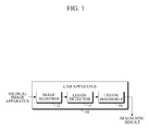
- FIG. 1 is a block diagram illustrating a configuration of a Computer-Aided Diagnosis (CAD) apparatus.
- CAD Computer-Aided Diagnosis
- the illustrated CAD apparatus 10 includes an image acquirer 12, a lesion detector 14, and a lesion diagnoser 16.
- a medical image apparatus may capture an ultrasound image of a patient's breast.
- the image acquirer 12 may then receive an image from the medical image apparatus that stores such a captured image.
- the lesion detector 14 may determine whether the received image from the image acquirer 12 includes one or more lesion candidate by applying a lesion detection algorithm to the received image, and may detect a location of each detected lesion candidate in the received image.
- the lesion diagnoser 16 may determine whether the detected lesion candidates are malignant.
- the CAD apparatus 10 may display the detected lesion candidate within an image along with a diagnostic result.
- the lesion detector 14 may detect a plurality of false positive lesion candidates as well as a lesion candidate that corresponds to an actual lesion due to poor-definition and atypical ultrasound image.
- a false positive lesion candidate corresponds to a detection error. Therefore, in order to enhance diagnostic performance and efficiency, the lesion detector 14 has to reduce the number of detection errors, that is, the number of false positive lesion candidates.
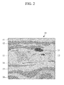
- FIG. 2 illustrates an example of a medical image.
- the medical image is an ultrasound breast image including examples of lesion candidates disposed among anatomical structures of a human breast.
- An image 10 illustrated in FIG. 2 includes anatomical objects, such as skin 11, subcutaneous fat 12, glandular tissue 13, retromammary fat 14, pectoralis muscle 15 and rib 16.
- the image 10 includes lesion candidates 21 and 22, such as a cyst and a malignant lesion.
- lesion candidates 21 and 22 may be found in various locations in the image 10.
- FIG. 3 is a block diagram illustrating an example of an apparatus for detecting lesion by removing a false positive lesion candidate from a medical image.
- an apparatus 30 for detecting lesion may include components, such as an image receiver 31, a lesion candidate detector 33, an adjacent object detector 35, a lesion candidate verifier 37, and a candidate remover 39, a display unit 36, or the like.
- the image receiver 31 is a component that receives a medical image.
- a medical image may be, for example, an ultrasound image of a human breast that is captured using ultrasonic waves, as shown in FIG. 2 .
- the medical image may be received from a medical image diagnostic/capturing device, from an imaging apparatus that captures a specific part of a human body using ultrasonic waves or X-rays, or from a storage device that stores medical images captured by the medical image diagnostic/capturing device.
- the medical image diagnostic/capturing device and storage devices may be collectively referred herein as a medical imagining apparatus.
- the image receiver 31 may be implemented as an integral part of such a medical imaging apparatus or may communicate with such a medical imaging apparatus via a wired or a wireless communication.
- the received image may be stored in a memory of the apparatus 30 for analysis.
- the lesion candidate detector 33, the adjacent object detector 35, the lesion candidate verifier 37, and the candidate remover 39 may be implemented on one or more computer processors.
- the lesion candidate detector 33 is a component that analyzes a medical image to determine whether the medical image includes one or more lesion candidates and to detect a location of each detected lesion candidate in the medical image.
- the lesion candidate detector 33 may use a Haar feature detection technique, a deformable part model technique, a deep learning technique, or the like.
- the lesion candidate detector 33 may make each detected lesion candidate correspond to a score indicating a confidence level thereof.
- a confidence level score may be a measurement that represents a possibility that each lesion candidate would be determined to be a true lesion.
- the adjacent object detector 35 may detect a plurality of anatomical objects from a medical image. If a medical image is an ultrasound breast image 10, the adjacent object detector 35 may detect skin, fat, glandular tissue, muscle, and bone within the image as anatomical objects.
- the adjacent object detector 35 may include a single object detector that detects all anatomical objects at once, or may include a plurality of individual object detectors that detect anatomical objects individually. Hereinafter, the adjacent object detector 35 is described in detail with reference to FIGS. 4 to 6 .
- the lesion candidate verifier 37 may verify a lesion candidate detected by the lesion candidate detector 33 and an anatomical object detected by the adjacent object detector 35.
- the lesion candidate verifier 37 may use Support Vector Machine (SVM), Artificial Neural Network (ANN), Bayesian Network, or any other machine learning and statistical techniques.
- SVM Support Vector Machine
- ANN Artificial Neural Network
- Bayesian Network or any other machine learning and statistical techniques.
- the lesion candidate verifier 37 is described in detail with reference to FIG. 8 .
- the context information 370 may include context information that indicates anatomical context of each anatomical object included in a medical image.
- the context information may include domain knowledge that generally indicates a location of a lesion candidate, a location of each anatomical object, and a location relationship between each lesion candidate and each anatomical object.
- the lesion candidate verifier 37 may use domain knowledge that indicates locations of a detected lesion candidate, skin, fat, glandular tissue, muscle, and bones, and a correlation between the locations of the above.
- the context information 370 may be stored in a computer readable storage medium. The context information 370 is hereinafter described in detail with reference to FIG. 9 .
- the lesion candidate detector 33 detects a lesion candidate by analyzing an image using a DPM technique and determining a specific region in the image as a "lesion candidate with a confidence level of 85%”
- the adjacent object detector 35 detects an anatomical object by analyzing the image using a sliding window technique and determines another specific region in the image as "subcutaneous fat with a confidence level of 95%”
- the adjacent object detector 35 detects any other anatomical object, such as "skin”, “glandular tissue”, “retromammary fat” and “pectoralis muscle” in the same way.
- the lesion candidate verifier 37 may verify whether the detected "lesion candidate" is a true lesion based on anatomical context information. For instance, the lesion candidate verifier 37 may verify a location relationship between a specific region (hereinafter referred to as Region A) detected to be a lesion candidate and each anatomical object. In other words, the lesion candidate verifier 37 may refer to anatomical context information indicating that a location of Region A is located at the same point of, close to, or far from a location of each anatomical object. Accordingly, the lesion candidate verifier 37 may determine whether Region A is a lesion highly likely to be determined to be a true lesion or whether Region A is a lesion less likely to be determined to be a true lesion.
- Region A a specific region
- the candidate remover 39 may receive the verification result from the lesion candidate verifier 37. Accordingly, the candidate remover may remove lesion candidates determined to be a false positive candidate by the candidate remover 39 among the lesion candidates detected by the lesion candidate detector 33, and include the remaining lesion candidates in the original image 10 to be displayed.
- the resultant image 10 is described in detail with reference to FIG. 7 .
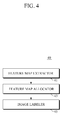
- FIG. 4 is a block diagram illustrating an example of an adjacent object detector shown in the apparatus illustrated in FIG.3 .
- a single detector 40 for simultaneously detecting all anatomical objects in an image is provided.
- the single detector 40 uses the sliding window technique in detecting a plurality of anatomical objects from a medical image.
- the single detector 40 may detect anatomical objects using histogram of gradient (HOG) features, that is, feature maps.
- the single detector 40 may include a feature map extractor 41, a feature map allocator 43, and an image labeler 45.
- the feature map extractor 41 extracts at least one feature map from an image.
- the feature map allocator 43 allocates the extracted feature map with a corresponding anatomical object.
- the extracted feature map may be compared with a plurality of pre-stored feature maps. If the extracted feature map coincides with any one of a plurality of pre-stored feature maps, the feature map allocator 43 may allocate the extracted feature map with an anatomical object corresponding to the coincided feature map. That is, the image labeler 45 may label a location of a feature map allocated with a specific anatomical object in an image with the specific anatomical object. Accordingly, a location of the feature map in the image may be detected as the specific anatomical object.
- HOG histogram of gradient
- the single detector 40 may use the superpixel technique.
- the single detector may be configured to be a classifier that segments pixels in an image into superpixels and then use image information of features of each superpixel, such as intensity, texture, sparse coding, and a deep belief network of a superpixel.
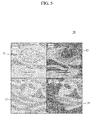
- FIG. 5 is a diagram illustrating examples of feature maps extracted by an example of the adjacent object detector illustrated in FIG. 4 .
- FIG. 5 demonstrates an example of a feature map group 50 that includes a cyst's feature map 51, a different cyst's feature map 54, a malignant lesions' feature map 55, and a glandular tissue's feature map 50.
- the feature map group 50 is merely an example, and it is obvious that other various feature maps may be further included to identify types and/or anatomical structures of more lesions. Such feature maps may be used similarly by the lesion candidate detector 33 illustrated in FIG. 3 .
- FIG. 6 is a diagram illustrating another example of an adjacent object detector shown in the apparatus illustrated in FIG. 3 .
- a multi detector 60 including a plurality of individual object detectors 61, 63, 65, and 67 is provided as an example of the adjacent object detector 35 illustrated in FIG. 3 .
- a skin object detector may detect only a skin region 11 from an original image 10.
- a subcutaneous fat detector 63 may detect only a subcutaneous fat region 12 from the original image 10.
- the glandular tissue detector 65 may detect only a glandular tissue region 13 from the original image 10.
- a pectoralis muscle detector 67 may detect a pectoralis muscle region from the original image 10.
- the various detected anatomical objects or tissues are color-coded.
- the portion of the image showing the skin, the subcutaneous fat, the glandular tissue and the pectoralis muscle, respectively, may be color-coded with blue, brown, green and tan highlights.
- the apparatus illustrated in FIG. 3 may allow such color-coded images to be displayed to a user on a display screen.
- each of a plurality of individual object detector 61, 63, 65 and 67 may use one of various deep learning techniques.
- DDN Deep Hierarchical Network
- CDBN Convolutional Deep Belief Network
- DDN Deconvolutional Deep Network
- FIG. 7 is a diagram illustrating an example of a combined image that indicates lesion candidates and anatomical objects detected from an original image illustrated in FIG. 3 .
- FIG. 7 is an example of a combined image that is output after the candidate remover 39 removes false positive lesion candidates from detected lesion candidates based on a verification result of the lesion candidate verifier 37.
- the image includes the remaining lesion candidates in an original image.
- a rib region 71, a subcutaneous fat region 72, a glandular tissue region 73, and two lesion candidates are displayed in the original image.
- the image may be output to a display screen or a touch screen of the display unit of the apparatus for lesion detection.
- a lesion candidate 74 is displayed with a confidence level of 69%, and the other lesion candidate is displayed with a confidence level of 0.12.
- the lesion candidate 74 is displayed with a visual indication for easy identification by the user. While, in this example, a rectangular box is used to indicate the lesion candidate 74, various other visual markings such as a contour line around the lesion or a color highlight may be used in another example.
- FIG. 8 is a block diagram illustrating in detail an embodiment of a lesion candidate verifier shown in the embodiment of FIG. 3 .
- the lesion candidate verifier 37 in FIG. 3 may include components, such as a self information extractor 371, a relation information extractor 373, an existence probability calculator 373, a verification processor 377, and the like.
- the self information extractor 371 may extract information on a lesion candidate itself detected by the lesion candidate detector 33 from the detected lesion candidate.
- the self information may include, for example, a confidence level score, values indicating brightness (or intensity of a pixel included in the lesion candidate), values indicating texture of the lesion candidate, and/or values indicating shape of the lesion candidate.
- the self information extractor 371 may extract self information from an anatomical object detected by adjacent object detector 35.
- the self information of the anatomical object may include a confidence level score, values indicating brightness (or intensity) of pixels included in the anatomical object, values indicating texture of the anatomical object, and/or values indicating shape of the anatomical object.
- Self information extracted by the self information extractor 371 may be included in the context information 370.
- the relationship information extractor 373 may include relationship information extracted from a correlation between each detected lesion candidate and each detected anatomical object.
- the relationship information may include a distance from each detected lesion candidate and each detected anatomical object, a location of each detected lesion candidate relative to each detected anatomical object, and/or information on similarities between each detected lesion candidate and each detected anatomical object in terms of brightness, texture, or shape.
- the relationship information extracted by the relationship information extractor 373 may be included in the context information 370.
- the existence probability calculator 375 may calculate a probability distribution of the existence of a specific lesion candidate in a specific anatomical structure based on pre-established learning data. A probability distribution calculated by the existence probability calculator 375 may be included in the context information 370 as probability information.
- the verification processor 377 is a component that performs a verifying function of the lesion candidate verifier 37 using information included in the context information 370. That is, the context information 370 may include self information obtained by the self information extractor 371, relationship information obtained by the relationship extractor 373, and/or probability information obtained by the existence probability calculator 375. As further described in the following with reference with FIG. 9 , the context information 370 may include domain knowledge, self information, relationship information, probability information, and/or adjacent image information. The verification processor 377 may verify whether a specific lesion candidate is false positive based on the specific lesion candidate's domain knowledge, self information, relationship information, probability information, and/or adjacent image information, all of which are included in the context information 370.
- the context information 370 may be a component included in the lesion candidate verifier 37 as a computer-readable storage medium to store context information including domain knowledge, self information, relationship information, probability information, and/or adjacent image information, or may be an external component placed outside of the lesion candidate verifier 37 to transmit/receive data through wired/wireless communication connection to the lesion candidate verifier 37.
- context information included in the context information 370 is described in detail with reference to FIG. 9 .
- FIG. 9 is a block diagram illustrating an example of context information used by a lesion candidate verifier shown in FIG. 3 .
- anatomical context information stored in the context information 370 may include domain knowledge 3701, self information 3703, relationship information 3705, probability information 3707, and/or adjacent image information 3709.
- the domain knowledge 3701 is information that indicates a location relationship in each region.
- the domain knowledge 3701 may include at least general information about an anatomical object such as, for example, "a specific part of a human body includes predetermined anatomical objects", "different anatomical objects have a predefined location relationship in a specific part of a human body” and “identical anatomical objects are gathered.”
- domain knowledge may include anatomical location information.
- domain knowledge may include anatomical location information like "a breast includes skin, subcutaneous fat, glandular tissue, retromammary fat, pectoralis muscle, and a rib as anatomical objects" or "a breast includes skin placed there atop, and then, subcutaneous fat, glandular tissue, retromammary fat, pectoralis muscle, and a rib placed below skin in the order named.
- the domain knowledge 3701 may include domain knowledge associated with a lesion candidate, for example, "a specific lesion candidate exists only between specific anatomical objects in a specific part of a human body.”
- the domain knowledge 3701 may include general medical location information, for example, "a cyst of breast cancer exists in a glandular tissue region at a time when the breast cancer is palpable.”
- the context information 370 may include self information 3703, which is extracted from each detected lesion candidate and each detected anatomical object.
- the self information 3703 may include confidence level information, brightness information, texture information and/or shape information of each lesion candidate/anatomical object.
- the self information 3703 may include a value of brightness of each pixel, a value of texture, and information on a shape of a specific lesion candidate/anatomical object.
- the context information 370 may include the relationship information 3705 that is extracted from a correlation between each detected lesion candidate and each detected anatomical object.
- the relationship information 3705 may include at least one of a distance between each lesion and each anatomical object, a location of a lesion candidate relative to each anatomical object, and information on similarity between each lesion candidate and each anatomical object in terms of brightness, texture or shape.
- a location of a lesion candidate relative to each anatomical object may be relationship information about whether the lesion candidate is located below skin and subcutaneous fat but above pectoralis muscle in a breast image.
- the context information 370 may include the probability information 3707.
- the probability information 3707 may be acquired from pre-established learning data.
- the probability information 3707 may be a probability distribution of the existence of a lesion candidate or anatomical object in a specific medical image.
- the probability information 3705 may include a probability distribution, for example, "skin exists on the top of the image at 100%", "subcutaneous fat may be found 10cm away from the top of the image at 50%”, and "a lesion such as a cyst of breast cancer may be found between subcutaneous fat and retromammary fat at 10 to 31%.”
- the context information 370 may include adjacent image information 3709.
- the adjacent image information 3709 may include location information of a lesion candidate or an anatomical object that is obtained from an adjacent frame/cross-section.
- the current medical image is one of slices of a 3D ultrasound volume image
- information on a lesion detected from previous adjacent slices and/or location information of an anatomical object detected from previous adjacent slices is highly associated with a lesion and/or anatomical object detected from the current slice.
- a previously detected anatomical object's location information included in adjacent image information may be used as useful reference in verifying and adjusting a lesion candidate and/or an anatomical object detected from the current image.
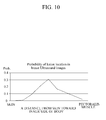
- FIG. 10 is a graph illustrating an example of probability information as an example of context information described in FIG. 9 .
- FIG. 10 illustrates a graph showing a probability of a lesion to be discovered in a specific region of an ultrasound breast image.
- a horizontal axis represents a distance from skin to pectoralis muscle
- a vertical axis represents a probability of a lesion to be discovered.
- the graph shows that a lesion is less likely to exist in a region close to skin and pectoralis muscle, and that a lesion may be discovered at an intermediate location between skin and pectoralis muscle at 31%.
- the probability distribution may be acquired from learning data pre-established based on data that has been accumulated in the process of lesion detection.
- the location information regarding a lesion candidate may be used to determine whether the lesion candidate is more or less likely to be a false positive lesion candidate.
- FIG. 11 is a flow chart illustrating an example of a method for detecting lesion by removing a false positive lesion candidate from a medical image.
- a method 110 for detecting lesion by removing a false positive lesion candidate from a medical image may involve the acquisition of a medical image.
- a medical image may be received by an image receiver of the apparatus shown in FIG. 3 in operation 111.
- the medical image may be received from a medical image diagnostic/capturing device that captures a specific part of a human body using ultrasound waves or X-rays, or from a storage device that stores medical images captured by the medical image diagnostic/capturing device.
- the lesion candidate detector 33 and the adjacent object detector 35 in the apparatus shown in FIG. 3 detect a lesion candidate and an anatomical object located in specific regions of the received image as Regions of Interest (ROIs) in operation 113.
- ROIs Regions of Interest
- the received image is an ultrasound breast image
- a region suspected of being a lesion such as a cyst of breast cancer
- skin, fat, glandular tissue, muscle and bones are detected as anatomical objects.
- context information is obtained from a computer-readable storage medium, for example, by the lesion candidate verifier 37 shown in FIG. 3 , to be used as information when determining whether a lesion candidate is false positive.
- the context information may be the context information 370 shown in FIG. 3 .
- the context information 370 may include domain knowledge, and may be pre-stored in a computer readable storage medium.
- the domain knowledge may include domain knowledge about the lesion candidate and/or domain knowledge about an anatomical object.
- the domain knowledge may be the domain knowledge 3701 shown in FIG. 9 .
- the lesion candidate verifier 37 may verify based on the obtained context information whether a lesion candidate amongst all the ROIs is false positive in operation 117. For example, if a lesion candidate is determined to exist in a skin region, the lesion candidate is less likely to be determined as being a true positive lesion, and thus, may be determined to be false positive. Alternatively, if a lesion candidate is determined to exist in a glandular tissue region, the lesion candidate is highly likely to be determined to be a true lesion, and thus, may be determined to be not false positive.
- the verification result obtained during operation 117 is used, for example, by the candidate remover 39 shown in FIG. 3 , to thereby output a result of lesion candidates from which any false positive lesion candidate is removed.
- FIG. 12 is a flow chart illustrating another example of a method for removing a false positive lesion candidate from a medical image.
- a method 120 for detecting lesion by removing a false positive lesion candidate from a medical image may involve, for example, obtaining a medical image by the image receiver illustrated in FIG. 3 , in operation 121.
- the medical image may be received from a medical image diagnostic/capturing device that captures a specific part of a human body using ultrasound waves or X-rays, or from a storage device that stores medical images captured by the medical image diagnostic/capturing device.
- the lesion candidate detector 33 and the adjacent object detector 35 of the apparatus shown in FIG. 3 analyze the received image and detect a lesion candidate and an anatomical object in specific regions of the received image as Regions of Interest (ROIs), in operation 123.
- the analysis may be performed by a computer processor.
- a region suspected of being a lesion such as a cyst of breast cancer, may be detected, and skin, fat, glandular tissue, muscle, and bones may be detected as anatomical objects.
- context information is acquired, for example, by the lesion candidate verifier 37 from a computer-readable storage medium, to be used as resources when determining whether a specific lesion candidate amongst the detected regions is false positive.
- the context information may be the context information 370 shown in FIG. 3 .
- the context information 370 may include domain knowledge, and may be stored in a computer readable medium.
- the domain knowledge may include domain knowledge of a lesion candidate and/or domain knowledge of an anatomical object.
- the domain knowledge may be the domain knowledge 3701 illustrated in FIG. 9 .
- the lesion candidate verifier 37 may extract self information and relationship information from each ROI detected in operation 123.
- the extracted self information may include information of a confidence level, brightness, texture, and/or shape of a corresponding ROI which may be a lesion candidate or an anatomical object.
- extracted self information may include a value of brightness of a pixel composing a specific lesion candidate/anatomical object, a value of texture of the specific lesion candidate/anatomical object, and information on a shape of the specific lesion candidate/anatomical object.
- extracted relationship information may include a distance between a lesion candidate and an anatomical object, a location of the lesion candidate relative to the anatomical object, or information on similarity between the lesion candidate and the anatomical object in terms of brightness, texture, or shape.
- the extracted self information and relationship information may be added to the context information 370 as the self information 3703 and the relationship information 3705, respectively, which are shown in FIG. 9 .
- context information of a specific lesion candidate is acquired, whether the specific lesion candidate among ROIs is false positive is verified, for example, by the lesion candidate verifier 37 shown in FIG. 3 , based on the acquired context information, that is, self information, relationship information and domain knowledge of the specific lesion candidate in operation 127. For example, if a lesion candidate is determined to exist in a skin region, the lesion candidate is determined less likely to be a true lesion, and thus, the lesion candidate may be determined to be false positive. In another example, if a lesion candidate is determined to exist in a glandular tissue region, the lesion candidate is determined highly likely to be a true lesion, and thus, may be determined to be not false positive.
- the verification result obtained during operation 127 may be used, for example, by the candidate remover 39 shown in FIG. 3 , to thereby output a result of lesion candidates from which any false positive lesion candidate is removed.
- FIG. 13 is a flow chart illustrating another example of a method for removing a false positive lesion candidate in a medical image.
- FIG. 13 is described with references to the apparatus illustrated in FIG. 3 .
- a method 130 for detecting a lesion by removing a false positive lesion candidate in a medical image may involve, for example, obtaining a medical image by an image receiver of the apparatus illustrated in FIG. 3 , in operation 131.
- the medical image may be received from a medical image diagnostic/capturing device that captures a specific part of a human body using ultrasound waves or X-rays, or from a storage device that stores medical images captured by the medical image diagnostic/capturing device.
- the storage device and the medical image diagnostic/capturing device may be collectively referred to as a medical imagining apparatus.
- the lesion candidate verifier 33 and the adjacent object detector 35 in the apparatus illustrated in FIG. 3 detects a lesion candidate and an anatomical object, respectively, from the analyzed medical image as an ROI in operation 133.
- the medical image is an ultrasound breast image
- a region suspected of being a lesion such as a breast cancer cyst
- skin, fat, glandular tissue, muscle, and bones may be detected as being anatomical objects.
- context information is obtained, for example, by the lesion candidate verifier 37 in Fig. 3 from a computer-readable storage medium, to be used as resources when determining whether a specific lesion candidate among the detected ROIs is false positive.
- the context information may include the context information 370 in FIG. 3 .
- the context information 370 may include domain knowledge, and may be stored in a computer-readable storage medium.
- the domain knowledge may include domain knowledge of a lesion candidate and domain knowledge of an anatomical object.
- the domain knowledge may be the domain knowledge 3701 in FIG. 9 .
- the lesion candidate verifier may extract self information and relationship information from each detected ROI.
- the extracted self information may include confidence level information, brightness information, texture information, and/or shape information of an ROI, that is, a lesion candidate or an anatomical object.
- self information may include a confidence level of a specific lesion candidate/anatomical object, a value indicating brightness of the specific lesion candidate/anatomical object, a value indicating texture of the specific lesion candidate/anatomical object, and information on shape of the specific lesion candidate/anatomical object.
- relationship information may include a distance between a lesion candidate between a lesion and an anatomical object, a location of the lesion relative to the anatomical object (e.g.
- a lesion candidate in a breast image may be positioned below skin and subcutaneous fat, but above pectoralis muscle), and information on similarity between the lesion candidate and the anatomical object in terms of brightness, texture or shape.
- the extracted self information and relationship information may be added to the context information 370 as the self information 3703 and the relation information 3705, respectively, which are shown in FIG. 9 .
- the context information may include probability information.
- the probability information may be acquired from pre-established learning data.
- the probability information may be a probability distribution of the existence of a lesion candidate/anatomical object in a specific medical image. Just like the domain knowledge, the probability information is also pre-stored.
- the probability information may be added to the context information 370 as the probability information 3703 shown in FIG. 9 .
- the context information may include adjacent image information. If the medical information is one of 2D continuous frames or one of 3D cross-sectional images, the adjacent image information may be acquired from a frame/cross-section which is adjacent to the current frame/cross-section.
- the adjacent image information may include location information of a lesion or an anatomical object. Just like the domain knowledge and probability information, the adjacent image information is pre-stored. For example, the adjacent image information may be added to the context information 370 as the adjacent image information 3709 shown in FIG. 9 .
- operation 137 may be performed, for example, by the lesion candidate verifier 37 shown in FIG. 3 , based on the context information, that is, the self information, relation information, and domain knowledge of the specific lesion candidate to verify whether the specific lesion candidate amongst ROIs is false positive. For example, if a lesion candidate is determined to exist in a glandular tissue region, the lesion candidate is determined highly likely to be a true lesion, and thus, the lesion candidate may be determined to be not false positive.
- the verification result obtained in operation 137 is used, for example, by the candidate remover shown in FIG. 3 , to output a result of lesion candidates from which any false positive lesion candidate is removed.
- the above-described apparatus for removing a false positive lesion candidate from a medical image may be executed by a computing device that includes at least some of a processor, a memory, a user input device and a presentation device.
- the memory is a medium that stores computer-readable software, an application, a program module, a routine, an instruction and/or data, which are encoded and set to perform a specific task once executed by a processor.
- the processor may read and implement computer-readable software, an application, a program module, a routine, an instruction, and/or data, which are stored in a memory.
- the user input device may be a means that enables a user to input an instruction requiring a processor to execute a specific task or input data necessary for the specific task.
- the user input device may include a physical/virtual keyboard or keypad, key buttons, a mouse, a joystick, a trackball, a touch-sensitive input device, and a microphone.
- the presentation device may include a display, a printer, a speaker, and a vibration device.
- the computing device may include a smartphone, a tablet, a laptop, a desktop, a server, a client, and other various devices.
- the computing device may be a single stand-alone device or a plurality of computing devices that operate in a distributive environment in which a plurality of computing devices cooperates via a communication network.
- the above-described method for removing a false positive lesion candidate from a medical image may be implemented by a computing device that includes a processor and a memory, wherein the memory stores computer readable software, an application, a program module, a routine, an instruction, and/or a data structure that is encoded to implement the above-described method.
- each component in an apparatus for removing a false positive lesion candidate from a medical image may be implemented by hardware that includes a circuit for a function of its own.
- each component in an apparatus for removing a false positive lesion candidate from a medical image may be implemented by a combination of computer-readable software, firmware, and hardware, such that each component executes a specific task once implemented by a processor of a computing device.
- the methods for removing a false positive lesion candidate which are described above with reference to FIGS. 11 to 13 , are merely exemplary. Modifications to the embodiments will be readily apparent for those skilled in the art that within the scope of the following claims.
- the examples of the methods for removing one or more false positive lesion candidates may be encoded as a computer-executable instruction to execute a specific task once implemented by a processor of a computer device.
- the computer-executable instruction may be encoded by a software developer into a program language and then into a machine language.
- non-transitory computer-readable storage media that includes program instructions to be implemented by a computer to cause a processor to execute or perform the program instructions.
- the media may also include, alone or in combination with the program instructions, data files, data structures, and the like.
- Examples of non-transitory computer-readable storage media include magnetic media, such as hard disks, floppy disks, and magnetic tape; optical media such as CD ROM disks and DVDs; magneto-optical media, such as optical disks; and hardware devices that are specially configured to store and perform program instructions, such as read-only memory (ROM), random access memory (RAM), flash memory, and the like.
- Examples of program instructions include machine code, such as produced by a compiler, and files containing higher level code that may be executed by the computer using an interpreter.
- the described hardware devices may be configured to act as one or more software modules in order to perform the operations and methods described above, or vice versa.
- a computer-readable storage medium may be distributed among computer systems connected through a network and computer-readable codes or program instructions may be stored and executed in a decentralized manner.
- the display may be implemented as a liquid crystal display (LCD), a light-emitting diode (LED) display, a plasma display panel (PDP), a screen, a terminal, and the like.
- a screen may be a physical structure that includes one or more hardware components that provide the ability to render a user interface and/or receive user input.
- the screen can encompass any combination of display region, gesture capture region, a touch sensitive display, and/or a configurable area.
- the screen can be embedded in the hardware or may be an external peripheral device that may be attached and detached from the apparatus.
- the display may be a single-screen or a multi-screen display.
- a single physical screen can include multiple displays that are managed as separate logical displays permitting different content to be displayed on separate displays although part of the same physical screen.
- a user interface may be responsible for inputting and outputting input information regarding an image or an input from a user.
- the user interface may further include an input/output device such as, for example, a mouse, a keyboard, a touch screen, a monitor, a speaker, a screen, and a software module for running the input/output device.
Landscapes
- Engineering & Computer Science (AREA)
- Health & Medical Sciences (AREA)
- Life Sciences & Earth Sciences (AREA)
- Medical Informatics (AREA)
- Physics & Mathematics (AREA)
- Nuclear Medicine, Radiotherapy & Molecular Imaging (AREA)
- General Health & Medical Sciences (AREA)
- Radiology & Medical Imaging (AREA)
- Computer Vision & Pattern Recognition (AREA)
- Public Health (AREA)
- Molecular Biology (AREA)
- Veterinary Medicine (AREA)
- Biophysics (AREA)
- Animal Behavior & Ethology (AREA)
- Surgery (AREA)
- Pathology (AREA)
- Biomedical Technology (AREA)
- Heart & Thoracic Surgery (AREA)
- Theoretical Computer Science (AREA)
- General Physics & Mathematics (AREA)
- Optics & Photonics (AREA)
- High Energy & Nuclear Physics (AREA)
- Physiology (AREA)
- Quality & Reliability (AREA)
- Data Mining & Analysis (AREA)
- Oral & Maxillofacial Surgery (AREA)
- Dentistry (AREA)
- Bioinformatics & Cheminformatics (AREA)
- Bioinformatics & Computational Biology (AREA)
- Evolutionary Biology (AREA)
- Evolutionary Computation (AREA)
- General Engineering & Computer Science (AREA)
- Artificial Intelligence (AREA)
- Ultra Sonic Daignosis Equipment (AREA)
- Apparatus For Radiation Diagnosis (AREA)
- Measuring And Recording Apparatus For Diagnosis (AREA)
- Image Analysis (AREA)
Abstract
Description
- The following description relates to a technique of detecting lesion from a medical image, and to a method and an apparatus for eliminating one or more lesion candidate detected from a medical image as a false positive lesion candidate.
- Computer-Aided Detection (CADe), also called Computer-Aided Diagnosis (CADx), is a technique of automatically detecting lesions from a medical image using a computer for the purpose of assisting a doctor in making a diagnosis. However, the low-resolution of many medical images and the wide-range atypical characteristics of lesions within medical images make it challenging to achieve high accuracy in CAD.
- For example, ultrasound images are obtained from a probe whose location and angle are arbitrarily determined by a doctor. Thus, the obtained ultrasound images may contain an atypical image of a lesion due to the different locations and angles of a probe used to obtain the image. Thus, if an algorithm of automatically detecting lesions is applied to such an atypical image, the computer is likely to detect not just lesions but also anatomical objects found in the image as lesions.
- This Summary is provided to introduce a selection of concepts in a simplified form that are further described below in the Detailed Description. This Summary is not intended to identify key features or essential features of the claimed subject matter, nor is it intended to be used as an aid in determining the scope of the claimed subject matter.
- The invention provides an apparatus for lesion detection which allows for an increased reliability when detecting lesions from a medical image. In one general aspect, an apparatus for lesion detection includes a lesion candidate detector configured to detect lesion candidates from a medical image, an adjacent object detector configured to detect anatomical objects from the medical image, a lesion candidate verifier configured to verify each of the lesion candidates based on anatomical context information comprising information regarding a location relationship between each of the lesion candidates and the anatomical objects, and a candidate remover configured to remove one or more false positive lesion candidate from among the detected lesion candidates based on a verification result of the lesion candidate verifier.
- The apparatus may further include an image receiver configured to obtain the medical image, in which the medical image is an image of a human breast captured using ultrasound waves or X-rays; the adjacent object detector is configured to detect at least one of skin, fat, glandular tissue, muscle, and bone from the breast image as an anatomical object; and the lesion candidate verifier is configured to verify each of the lesion candidates based on anatomical context information comprising information regarding a location relationship between each of the lesion candidates and the at least one of skin, fat, glandular tissue, muscle, and bone.
- The lesion candidate detector may be configured to detect the lesion candidates from the medical image using at least one of Haar feature detection technique, deformable part model (DPM), and deep learning technique.
- The adjacent object detector may include a plurality of individual object detectors, each of which may detect the anatomical objects individually from the medical image.
- Each of the plurality of individual object detectors may be configured to use deep learning technique, sliding window technique, or superpixel technique.
- The adjacent object detector may include a single object detector configured to simultaneously detect the anatomical objects from the medical image.
- The single object detector may be configured to use deep learning technique, sliding window technique, or superpixel technique.
- The single obj ect detector may be configured to extract a plurality of feature maps from the whole medical image or a part of the medical image, allocate the extracted feature maps with corresponding anatomical objects, and label a location of a feature map allocated with a specific anatomical object in the medical image as the specific anatomical object.
- The lesion candidate verifier may be configured to use at least one of Support Vector Machine (SVM), Artificial Neural Network (ANN), deep learning technique, and Bayesian Network.
- The anatomical context information may include domain knowledge of the anatomical objects and domain knowledge of the lesion candidates, the domain knowledge of the anatomical objects comprising at least one of: "a specific part of a human body has predefined anatomical objects", "different anatomical objects have a predefined location relationship in a specific part of a human body" and "the identical anatomical objects are gathered", and the domain knowledge of the lesion candidate comprising "a specific lesion candidate exists only in specific anatomical objects of a specific part of a human body."
- The anatomical context information may include self information that is extracted from the detected lesion candidate and the detected anatomical object, and the self information may include at least one of confidence level information, brightness information, texture information, and shape information.
- The anatomical context information may include relationship information that is extracted from a correlation between each of the lesion candidates and each of the anatomical object.
- The relationship information may include at least one of a distance between each of the lesion candidates and each of the anatomical objects, a location of each of the lesion candidates relative to each of the anatomical objects, and information on similarity between each of the lesion candidates and each of the anatomical objects in terms of brightness, texture, or shape.
- The anatomical context information may include a probability distribution of existence of each of the lesion candidates in the medical image, the probability distribution being obtained from a pre-established learning data.
- In response to the medical image being one of 2D continuous frames or one of 3D cross-sectional images, the anatomical context information further may include adjacent image information obtained from an adjacent frame or cross-section, which comprises location information of each of the lesion candidates and each of the anatomical objects.
- In another general aspect, a method of lesion detection involves detecting lesion candidates from a medical image, detecting anatomical objects from the medical image, verifying each of the lesion candidate based on anatomical context information comprising information regarding a location relationship between the lesion candidates and the anatomical objects, and removing one or more false positive lesion candidate from the detected lesion candidates based on a verification result.
- The general aspect of the method may further involve obtaining the medical image from a medical imaging apparatus, in which the medical image is an image of a breast of a human body captured using ultrasound waves or X-rays, the detecting of anatomical objects comprises detecting at least one of skin, fat, glandular tissue, muscle, and bone from the breast image as an anatomical object, and the verifying of each of the lesion candidates comprises verifying each of the lesion candidates based on anatomical context information comprising information regarding a location relationship between the lesion candidates and the at least one of skin, fat, glandular tissue, muscle, and bone.
- The detecting of lesion candidates may involve detecting the lesion candidates from the medical image using at least one of Haar feature detection technique, deformable part model (DPM), and deep learning technique.
- The detecting of the anatomical objects may involve detecting a plurality of individual objects to detect the anatomical objects from the medical image individually.
- The detecting of each of the plurality of individual objects may utilize deep learning technique, sliding window technique, or superpixel technique.
- The detecting of the anatomical objects may involve detecting all of the anatomical objects from the medical image simultaneously.
- The simultaneous detecting of all of the anatomical objects from the medical image may utilize deep learning technique, sliding window technique, or superpixel technique.
- The simultaneous detecting of all of the anatomical objects may involve: extracting a plurality of feature maps from the whole medical image or a part of the medical image, allocating each of the plurality of extracted feature maps with corresponding anatomical objects, and labeling a location of a feature map allocated with a specific anatomical object in the medical image with the specific anatomical object.
- The verifying of each of the lesion candidates may utilize at least one of Support Vector Machine (SVM), Artificial Neural Network (ANN) and Bayesian Network.
- The anatomical context information may include domain knowledge of the anatomical objects and domain knowledge of the lesion candidates, and the domain knowledge of the anatomical objects may include at least one of: "a specific part of a human body has predefined anatomical objects", "different anatomical objects have a predefined location relationship in a specific part of a human body" and "the identical anatomical objects are gathered", and the domain knowledge of the lesion candidate may include "a specific lesion candidate exists only in specific anatomical objects of a specific part of a human body."
- The anatomical context information may include self information that is respectively extracted from the lesion candidates and the anatomical objects, and the self information may include at least one of confidence level information, brightness information, texture information, and shape information.
- The anatomical context information may include relationship information that is extracted from a correlation between each of the lesion candidates and each of the anatomical objects. The relationship information may include at least one of a distance between each of the lesion candidates and each of the anatomical objects, a location of each of the lesion candidates relative to each of the anatomical objects, and information on similarity between each of the lesion candidates and each of the anatomical objects in terms of brightness, texture, or shape.
- The anatomical context information may include a probability distribution of existence of each of the lesion candidates in the medical image, the probability distribution being obtained from pre-established learning data.
- In response to the medical image being one of two-dimensional (2D) continuous frames or one of three-dimensional (3D) cross-sectional image, the anatomical context information may further include adjacent image information from an adjacent frame or an adjacent cross-section, which includes location information of each of the lesion candidates or each of the anatomical objects.
- In another general aspect, an apparatus for lesion detection includes a memory configured to store a medical image, and a processor configured to detect lesion candidates and anatomical objects from the medical image, configured to verify each of the detected lesion candidates based on anatomical context information comprising information regarding a location relationship between each of the lesion candidates and the detected anatomical objects, and configured to remove one or more false positive lesion candidates from among the detected lesion candidates based on a verification result.
- The general aspect of the apparatus may further include an image receiver configured to obtain the medical image from a medical imaging apparatus and to store the medical image in the memory, the medical image being an image of a human breast captured using ultrasound waves or X-rays, and the anatomical objects comprising at least one of skin, fat, glandular tissue, muscle, and bone from the breast image. The general aspect of the apparatus may further include a display screen configured to output a view of the medical image in which the lesion candidates remaining after the removing of the one or more false positive lesion candidates are displayed.
- Other features and aspects will be apparent from the following detailed description, the drawings, and the claims.
-
-
FIG. 1 is a block diagram illustrating a configuration of a Computer-Aided Diagnosis (CAD) apparatus. -
FIG. 2 is a diagram illustrating an example of lesion candidates in a breast ultrasound image and an anatomical structure of the breast ultrasound image. -
FIG. 3 is a block diagram illustrating an example of an apparatus for removing one or more false positive lesion candidates. -
FIG. 4 is a block diagram illustrating an example of an adjacent object detector shown in the example ofFIG. 3 . -
FIG. 5 is a diagram illustrating examples of a feature map extracted by the adjacent object detector shown in the example ofFIG. 4 . -
FIG. 6 is a diagram illustrating another example of operation of the adjacent object detector shown in the example ofFIG. 3 . -
FIG. 7 is a diagram illustrating an example of a combined image in which lesion candidates and anatomical objects, detected from an original image shown in the embodiment ofFIG. 3 , are displayed. -
FIG. 8 is a block diagram illustrating an example of a lesion candidate verifier shown in the example ofFIG. 3 . -
FIG. 9 is a block diagram illustrating an example of context information that is used by the lesion candidate verifier shown in the example ofFIG. 3 . -
FIG. 10 is a graph illustrating an example of probability information as an example of context information. -
FIG. 11 is a flow chart illustrating an example of a method for removing one or more false-positive lesion candidates from a medical image. -
FIG. 12 is a flow chart illustrating another example of a method for removing one or more false-positive lesion candidates from a medical image. -
FIG. 13 is a flow chart illustrating another example of a method for removing one or more false-positive lesion candidates from a medical image. - Throughout the drawings and the detailed description, unless otherwise described or provided, the same drawing reference numerals will be understood to refer to the same elements, features, and structures. The drawings may not be to scale, and the relative size, proportions, and depiction of elements in the drawings may be exaggerated for clarity, illustration, and convenience.
- The following detailed description is provided to assist the reader in gaining a comprehensive understanding of the methods, apparatuses, and/or systems described herein. However, various changes, modifications, and equivalents of the systems, apparatuses and/or methods described herein will be apparent to one of ordinary skill in the art. The progression of processing steps and/or operations described is an example; however, the sequence of and/or operations is not limited to that set forth herein and may be changed as is known in the art, with the exception of steps and/or operations necessarily occurring in a certain order. Also, descriptions of functions and constructions that are well known to one of ordinary skill in the art may be omitted for increased clarity and conciseness.
- The features described herein may be embodied in different forms, and are not to be construed as being limited to the examples described herein. Rather, the examples described herein have been provided so that this disclosure will be thorough and complete, and will convey the full scope of the disclosure to one of ordinary skill in the art.
- According to an example of an apparatus and/or a method for removing one or more false positive lesion candidates from a medical image, a technology of detecting one or more lesion candidates in a Computer-Aided Detection (CADe) or Computer-Aided Diagnosis (CADx) is provided. The technology may be used when detecting a lesion candidate from an ultrasound image or X-ray breast medical image. For example, the technology may be used with breast ultrasound image.
- In the present disclosure, the term "anatomical object" may refer to an element that is distinguishable by a specific anatomical characteristic or function. For example, a breast may include a variety of tissues and components, including skin, subcutaneous fat, glandular tissue, retromammary fat, pectoralis muscle and ribs, as anatomical objects.
- The term "lesion candidate region/portion" refers to a specific region detected by CAD that is likely to be diagnosed as a lesion. A doctor determines whether or not a lesion candidate region detected by CAD is a true lesion in order to diagnose a patient.
- The apparatus and method for removing one or more false positive lesion candidate regions in a medical image entails verifying lesion candidates, detected from a medical image, using anatomical context information, and then removing any false positive lesion candidate. That is, firstly, lesion candidates and anatomical objects are detected from an image. Then, a location of each detected lesion candidate, a location of each detected anatomical object, and a location relationship between each detected lesion candidate and each detected anatomical object are verified based on anatomical context information. Then, any false positive lesion candidate is removed from the lesion candidates based on the verification result. In this manner, it may become more efficient to detect lesion candidates.
- The "false positive lesion candidate" refers to a lesion candidate that is mistakenly detected instead of a lesion that needs to be medically diagnosed. For example, if a specific anatomical object, such as fat, is determined to be a lesion candidate, the lesion candidate is highly likely to be false positive. In another example, if a breast image is captured and a lesion in the breast image is determined to be located in an area where a tumor or a cyst related to breast cancer is usually not found, the lesion candidate is highly likely to be false positive.
- If a medical image is an ultrasound or X-ray breast image, a cyst of breast cancer may be identified as a lesion candidate, while skin, subcutaneous fat, glandular tissue, retromammary fat, pectoralis muscle and ribs may be detected as anatomical objects.
- A lesion candidate may be detected using various algorithms to automatically detect a lesion candidate from an image. For example, Haar feature detection technique, deformable part model, deep learning technique, or the like may be utilized.
- Detecting an anatomical object may be performed by detecting each of a plurality of anatomical objects from an image individually. In this case, a single deep learning technique or various deep learning techniques, such as Convolutional Deep Belief Network (CDBN), Convolutional Neural Network (CNN), or the like, may be utilized. In addition, different detectors may be used for each distinct anatomical object: for example, a skin detector for skin, a fat detector for fat, a glandular tissue detector for a glandular tissue, and a muscle detector for muscle.
- Meanwhile, a plurality of anatomical objects may be detected at once from an ultrasound breast image. In such an example, a deep learning technique, a sliding window technique or a superpixel technique may be utilized. For example, various feature maps may be extracted from an ultrasound breast image, the extracted feature maps may be allocated with corresponding anatomical objects, and a location of a feature map allocated with a specific anatomical object in an image may be labeled with the specific anatomical object.
- Detected in the above manner, skin, fat, muscle, and bone, and any other anatomical objects, may be verified based on anatomical context information about a location relationship between anatomical objects.
- Anatomical context information may include domain knowledge associated with a lesion candidate and relevant anatomical objects. In addition, the anatomical context information may include information on a lesion candidate and information on each anatomical object. Moreover, the anatomical context information may include information about a location relationship between a lesion candidate and an anatomical object. Furthermore, the anatomical context information may include a probability distribution of existence of a lesion candidate and/or anatomical object in a medical image of a specific part of a human body. The probability distribution may be acquired based on pre-established learning data. In an example in which a medical image is one of two-dimensional (2D) continuous video frames or one of three-dimensional (3D) cross-sectional images, the anatomical context information may include adjacent image information obtained from an adjacent frame or an adjacent cross-section, such as location information of an anatomical object.
- Hereinafter, examples of apparatuses and methods for detecting lesion by removing a false positive lesion candidate from a medical image are described with reference to drawings.
- For example, an apparatus and/or method for removing a false positive lesion candidate from a medical image that is a 2D ultrasound breast image is provided. However, the medical image is not limited to an ultrasound breast image. For example, the medical image may be an ultrasound or X-ray image of a different part of a human body, and the medical image may be a 2D or 3D image.
-
FIG. 1 is a block diagram illustrating a configuration of a Computer-Aided Diagnosis (CAD) apparatus. - Referring to
FIG. 1 , theillustrated CAD apparatus 10 includes animage acquirer 12, alesion detector 14, and alesion diagnoser 16. A medical image apparatus may capture an ultrasound image of a patient's breast. Theimage acquirer 12 may then receive an image from the medical image apparatus that stores such a captured image. Thelesion detector 14 may determine whether the received image from theimage acquirer 12 includes one or more lesion candidate by applying a lesion detection algorithm to the received image, and may detect a location of each detected lesion candidate in the received image. The lesion diagnoser 16 may determine whether the detected lesion candidates are malignant. TheCAD apparatus 10 may display the detected lesion candidate within an image along with a diagnostic result. - In such a CAD apparatus, the
lesion detector 14 may detect a plurality of false positive lesion candidates as well as a lesion candidate that corresponds to an actual lesion due to poor-definition and atypical ultrasound image. A false positive lesion candidate corresponds to a detection error. Therefore, in order to enhance diagnostic performance and efficiency, thelesion detector 14 has to reduce the number of detection errors, that is, the number of false positive lesion candidates. -
FIG. 2 illustrates an example of a medical image. The medical image is an ultrasound breast image including examples of lesion candidates disposed among anatomical structures of a human breast. - Referring to
FIG. 2 , an example of a 2D ultrasound image of a human breast is provided. Animage 10 illustrated inFIG. 2 includes anatomical objects, such asskin 11,subcutaneous fat 12,glandular tissue 13,retromammary fat 14,pectoralis muscle 15 andrib 16. In addition, theimage 10 includeslesion candidates skin 11, thesubcutaneous fat 12, theglandular tissue 13, theretromammary fat 14, thepectoralis muscle 15 and therib 16 are arranged from the surface to the inside in order. In addition, thelesion candidates image 10. -
FIG. 3 is a block diagram illustrating an example of an apparatus for detecting lesion by removing a false positive lesion candidate from a medical image. - Referring to
FIG. 3 , anapparatus 30 for detecting lesion may include components, such as animage receiver 31, alesion candidate detector 33, anadjacent object detector 35, alesion candidate verifier 37, and acandidate remover 39, adisplay unit 36, or the like. - The
image receiver 31 is a component that receives a medical image. A medical image may be, for example, an ultrasound image of a human breast that is captured using ultrasonic waves, as shown inFIG. 2 . The medical image may be received from a medical image diagnostic/capturing device, from an imaging apparatus that captures a specific part of a human body using ultrasonic waves or X-rays, or from a storage device that stores medical images captured by the medical image diagnostic/capturing device. The medical image diagnostic/capturing device and storage devices may be collectively referred herein as a medical imagining apparatus. According to one example, theimage receiver 31 may be implemented as an integral part of such a medical imaging apparatus or may communicate with such a medical imaging apparatus via a wired or a wireless communication. The received image may be stored in a memory of theapparatus 30 for analysis. - The
lesion candidate detector 33, theadjacent object detector 35, thelesion candidate verifier 37, and thecandidate remover 39 may be implemented on one or more computer processors. - The
lesion candidate detector 33 is a component that analyzes a medical image to determine whether the medical image includes one or more lesion candidates and to detect a location of each detected lesion candidate in the medical image. In order to detect a lesion candidate, thelesion candidate detector 33 may use a Haar feature detection technique, a deformable part model technique, a deep learning technique, or the like. Thelesion candidate detector 33 may make each detected lesion candidate correspond to a score indicating a confidence level thereof. A confidence level score may be a measurement that represents a possibility that each lesion candidate would be determined to be a true lesion. - The
adjacent object detector 35 may detect a plurality of anatomical objects from a medical image. If a medical image is anultrasound breast image 10, theadjacent object detector 35 may detect skin, fat, glandular tissue, muscle, and bone within the image as anatomical objects. Theadjacent object detector 35 may include a single object detector that detects all anatomical objects at once, or may include a plurality of individual object detectors that detect anatomical objects individually. Hereinafter, theadjacent object detector 35 is described in detail with reference toFIGS. 4 to 6 . - Based on
context information 370, including information associated anatomical context, thelesion candidate verifier 37 may verify a lesion candidate detected by thelesion candidate detector 33 and an anatomical object detected by theadjacent object detector 35. For example, thelesion candidate verifier 37 may use Support Vector Machine (SVM), Artificial Neural Network (ANN), Bayesian Network, or any other machine learning and statistical techniques. Thelesion candidate verifier 37 is described in detail with reference toFIG. 8 . - The
context information 370 may include context information that indicates anatomical context of each anatomical object included in a medical image. The context information may include domain knowledge that generally indicates a location of a lesion candidate, a location of each anatomical object, and a location relationship between each lesion candidate and each anatomical object. For example, if a medical image is theultrasound breast image 10, thelesion candidate verifier 37 may use domain knowledge that indicates locations of a detected lesion candidate, skin, fat, glandular tissue, muscle, and bones, and a correlation between the locations of the above. Thecontext information 370 may be stored in a computer readable storage medium. Thecontext information 370 is hereinafter described in detail with reference toFIG. 9 . - For example, suppose that the
lesion candidate detector 33 detects a lesion candidate by analyzing an image using a DPM technique and determining a specific region in the image as a "lesion candidate with a confidence level of 85%", that theadjacent object detector 35 detects an anatomical object by analyzing the image using a sliding window technique and determines another specific region in the image as "subcutaneous fat with a confidence level of 95%", and that that theadjacent object detector 35 detects any other anatomical object, such as "skin", "glandular tissue", "retromammary fat" and "pectoralis muscle" in the same way. - In this example, the
lesion candidate verifier 37 may verify whether the detected "lesion candidate" is a true lesion based on anatomical context information. For instance, thelesion candidate verifier 37 may verify a location relationship between a specific region (hereinafter referred to as Region A) detected to be a lesion candidate and each anatomical object. In other words, thelesion candidate verifier 37 may refer to anatomical context information indicating that a location of Region A is located at the same point of, close to, or far from a location of each anatomical object. Accordingly, thelesion candidate verifier 37 may determine whether Region A is a lesion highly likely to be determined to be a true lesion or whether Region A is a lesion less likely to be determined to be a true lesion. - The
candidate remover 39 may receive the verification result from thelesion candidate verifier 37. Accordingly, the candidate remover may remove lesion candidates determined to be a false positive candidate by thecandidate remover 39 among the lesion candidates detected by thelesion candidate detector 33, and include the remaining lesion candidates in theoriginal image 10 to be displayed. Theresultant image 10 is described in detail with reference toFIG. 7 . -
FIG. 4 is a block diagram illustrating an example of an adjacent object detector shown in the apparatus illustrated inFIG.3 . - Referring to
FIG. 4 , as an example of theadjacent object detector 31 inFIG. 3 , asingle detector 40 for simultaneously detecting all anatomical objects in an image is provided. In the example illustrated inFIG. 4 , thesingle detector 40 uses the sliding window technique in detecting a plurality of anatomical objects from a medical image. - As illustrated in
FIG. 4 , thesingle detector 40 may detect anatomical objects using histogram of gradient (HOG) features, that is, feature maps. To this end, thesingle detector 40 may include afeature map extractor 41, afeature map allocator 43, and animage labeler 45. Thefeature map extractor 41 extracts at least one feature map from an image. Thefeature map allocator 43 allocates the extracted feature map with a corresponding anatomical object. For this process, the extracted feature map may be compared with a plurality of pre-stored feature maps. If the extracted feature map coincides with any one of a plurality of pre-stored feature maps, thefeature map allocator 43 may allocate the extracted feature map with an anatomical object corresponding to the coincided feature map. That is, theimage labeler 45 may label a location of a feature map allocated with a specific anatomical object in an image with the specific anatomical object. Accordingly, a location of the feature map in the image may be detected as the specific anatomical object. - Alternatively, the
single detector 40 may use the superpixel technique. In this case, the single detector may be configured to be a classifier that segments pixels in an image into superpixels and then use image information of features of each superpixel, such as intensity, texture, sparse coding, and a deep belief network of a superpixel. -
FIG. 5 is a diagram illustrating examples of feature maps extracted by an example of the adjacent object detector illustrated inFIG. 4 . -
FIG. 5 demonstrates an example of afeature map group 50 that includes a cyst'sfeature map 51, a different cyst's feature map 54, a malignant lesions'feature map 55, and a glandular tissue'sfeature map 50. However, thefeature map group 50 is merely an example, and it is obvious that other various feature maps may be further included to identify types and/or anatomical structures of more lesions. Such feature maps may be used similarly by thelesion candidate detector 33 illustrated inFIG. 3 . -
FIG. 6 is a diagram illustrating another example of an adjacent object detector shown in the apparatus illustrated inFIG. 3 . - Referring to
FIG. 6 , amulti detector 60 including a plurality ofindividual object detectors adjacent object detector 35 illustrated inFIG. 3 . A skin object detector may detect only askin region 11 from anoriginal image 10. Asubcutaneous fat detector 63 may detect only a subcutaneousfat region 12 from theoriginal image 10. Theglandular tissue detector 65 may detect only aglandular tissue region 13 from theoriginal image 10. A pectoralis muscle detector 67 may detect a pectoralis muscle region from theoriginal image 10. In the diagram illustrated inFIG. 6 , the various detected anatomical objects or tissues are color-coded. For example, the portion of the image showing the skin, the subcutaneous fat, the glandular tissue and the pectoralis muscle, respectively, may be color-coded with blue, brown, green and tan highlights. The apparatus illustrated inFIG. 3 may allow such color-coded images to be displayed to a user on a display screen. - According to one example, each of a plurality of
individual object detector -
FIG. 7 is a diagram illustrating an example of a combined image that indicates lesion candidates and anatomical objects detected from an original image illustrated inFIG. 3 . -
FIG. 7 is an example of a combined image that is output after thecandidate remover 39 removes false positive lesion candidates from detected lesion candidates based on a verification result of thelesion candidate verifier 37. In this example, the image includes the remaining lesion candidates in an original image. As illustrated inFIG. 7 , arib region 71, a subcutaneousfat region 72, aglandular tissue region 73, and two lesion candidates are displayed in the original image. For example, the image may be output to a display screen or a touch screen of the display unit of the apparatus for lesion detection. In this example, alesion candidate 74 is displayed with a confidence level of 69%, and the other lesion candidate is displayed with a confidence level of 0.12. Thus, thelesion candidate 74 is displayed with a visual indication for easy identification by the user. While, in this example, a rectangular box is used to indicate thelesion candidate 74, various other visual markings such as a contour line around the lesion or a color highlight may be used in another example. -
FIG. 8 is a block diagram illustrating in detail an embodiment of a lesion candidate verifier shown in the embodiment ofFIG. 3 . - Referring to
FIG. 8 , thelesion candidate verifier 37 inFIG. 3 may include components, such as aself information extractor 371, arelation information extractor 373, anexistence probability calculator 373, averification processor 377, and the like. - The
self information extractor 371 may extract information on a lesion candidate itself detected by thelesion candidate detector 33 from the detected lesion candidate. The self information may include, for example, a confidence level score, values indicating brightness (or intensity of a pixel included in the lesion candidate), values indicating texture of the lesion candidate, and/or values indicating shape of the lesion candidate. Similarly, theself information extractor 371 may extract self information from an anatomical object detected byadjacent object detector 35. The self information of the anatomical object may include a confidence level score, values indicating brightness (or intensity) of pixels included in the anatomical object, values indicating texture of the anatomical object, and/or values indicating shape of the anatomical object. Self information extracted by theself information extractor 371 may be included in thecontext information 370. - The
relationship information extractor 373 may include relationship information extracted from a correlation between each detected lesion candidate and each detected anatomical object. The relationship information may include a distance from each detected lesion candidate and each detected anatomical object, a location of each detected lesion candidate relative to each detected anatomical object, and/or information on similarities between each detected lesion candidate and each detected anatomical object in terms of brightness, texture, or shape. The relationship information extracted by therelationship information extractor 373 may be included in thecontext information 370. - The
existence probability calculator 375 may calculate a probability distribution of the existence of a specific lesion candidate in a specific anatomical structure based on pre-established learning data. A probability distribution calculated by theexistence probability calculator 375 may be included in thecontext information 370 as probability information. - The
verification processor 377 is a component that performs a verifying function of thelesion candidate verifier 37 using information included in thecontext information 370. That is, thecontext information 370 may include self information obtained by theself information extractor 371, relationship information obtained by therelationship extractor 373, and/or probability information obtained by theexistence probability calculator 375. As further described in the following with reference withFIG. 9 , thecontext information 370 may include domain knowledge, self information, relationship information, probability information, and/or adjacent image information. Theverification processor 377 may verify whether a specific lesion candidate is false positive based on the specific lesion candidate's domain knowledge, self information, relationship information, probability information, and/or adjacent image information, all of which are included in thecontext information 370. - The
context information 370 may be a component included in thelesion candidate verifier 37 as a computer-readable storage medium to store context information including domain knowledge, self information, relationship information, probability information, and/or adjacent image information, or may be an external component placed outside of thelesion candidate verifier 37 to transmit/receive data through wired/wireless communication connection to thelesion candidate verifier 37. Hereinafter, context information included in thecontext information 370 is described in detail with reference toFIG. 9 . -
FIG. 9 is a block diagram illustrating an example of context information used by a lesion candidate verifier shown inFIG. 3 . - Referring to
FIG. 9 , anatomical context information stored in thecontext information 370 may includedomain knowledge 3701,self information 3703,relationship information 3705,probability information 3707, and/oradjacent image information 3709. - The
domain knowledge 3701 is information that indicates a location relationship in each region. Thedomain knowledge 3701 may include at least general information about an anatomical object such as, for example, "a specific part of a human body includes predetermined anatomical objects", "different anatomical objects have a predefined location relationship in a specific part of a human body" and "identical anatomical objects are gathered." - For example, in the case of an ultrasound breast image, domain knowledge may include anatomical location information. For instance, domain knowledge may include anatomical location information like "a breast includes skin, subcutaneous fat, glandular tissue, retromammary fat, pectoralis muscle, and a rib as anatomical objects" or "a breast includes skin placed there atop, and then, subcutaneous fat, glandular tissue, retromammary fat, pectoralis muscle, and a rib placed below skin in the order named.
- In addition, the
domain knowledge 3701 may include domain knowledge associated with a lesion candidate, for example, "a specific lesion candidate exists only between specific anatomical objects in a specific part of a human body." - For an ultrasound breast image, the
domain knowledge 3701 may include general medical location information, for example, "a cyst of breast cancer exists in a glandular tissue region at a time when the breast cancer is palpable." - In addition, the
context information 370 may includeself information 3703, which is extracted from each detected lesion candidate and each detected anatomical object. Theself information 3703 may include confidence level information, brightness information, texture information and/or shape information of each lesion candidate/anatomical object. For example, theself information 3703 may include a value of brightness of each pixel, a value of texture, and information on a shape of a specific lesion candidate/anatomical object. - In addition, the
context information 370 may include therelationship information 3705 that is extracted from a correlation between each detected lesion candidate and each detected anatomical object. Therelationship information 3705 may include at least one of a distance between each lesion and each anatomical object, a location of a lesion candidate relative to each anatomical object, and information on similarity between each lesion candidate and each anatomical object in terms of brightness, texture or shape. Herein, a location of a lesion candidate relative to each anatomical object may be relationship information about whether the lesion candidate is located below skin and subcutaneous fat but above pectoralis muscle in a breast image. - Moreover, the
context information 370 may include theprobability information 3707. Theprobability information 3707 may be acquired from pre-established learning data. Theprobability information 3707 may be a probability distribution of the existence of a lesion candidate or anatomical object in a specific medical image. - For example, in an example in which an ultrasound breast image is used as the medical image, the
probability information 3705 may include a probability distribution, for example, "skin exists on the top of the image at 100%", "subcutaneous fat may be found 10cm away from the top of the image at 50%", and "a lesion such as a cyst of breast cancer may be found between subcutaneous fat and retromammary fat at 10 to 31%." - Furthermore, the
context information 370 may includeadjacent image information 3709. In the case where a medical image is one of 2D continuous frames or one of 3D cross-sectional images, theadjacent image information 3709 may include location information of a lesion candidate or an anatomical object that is obtained from an adjacent frame/cross-section. - For example, in the case where the current medical image is one of slices of a 3D ultrasound volume image, information on a lesion detected from previous adjacent slices and/or location information of an anatomical object detected from previous adjacent slices is highly associated with a lesion and/or anatomical object detected from the current slice. Thus, a previously detected anatomical object's location information included in adjacent image information may be used as useful reference in verifying and adjusting a lesion candidate and/or an anatomical object detected from the current image.
-
FIG. 10 is a graph illustrating an example of probability information as an example of context information described inFIG. 9 . -
FIG. 10 illustrates a graph showing a probability of a lesion to be discovered in a specific region of an ultrasound breast image. In the graph, a horizontal axis represents a distance from skin to pectoralis muscle, and a vertical axis represents a probability of a lesion to be discovered. The graph shows that a lesion is less likely to exist in a region close to skin and pectoralis muscle, and that a lesion may be discovered at an intermediate location between skin and pectoralis muscle at 31%. The probability distribution may be acquired from learning data pre-established based on data that has been accumulated in the process of lesion detection. The location information regarding a lesion candidate may be used to determine whether the lesion candidate is more or less likely to be a false positive lesion candidate. -
FIG. 11 is a flow chart illustrating an example of a method for detecting lesion by removing a false positive lesion candidate from a medical image. - Referring to
FIG. 11 , amethod 110 for detecting lesion by removing a false positive lesion candidate from a medical image may involve the acquisition of a medical image. For example, a medical image may be received by an image receiver of the apparatus shown inFIG. 3 inoperation 111. The medical image may be received from a medical image diagnostic/capturing device that captures a specific part of a human body using ultrasound waves or X-rays, or from a storage device that stores medical images captured by the medical image diagnostic/capturing device. - Once the image is received, the received image is analyzed, and then, the
lesion candidate detector 33 and theadjacent object detector 35 in the apparatus shown inFIG. 3 detect a lesion candidate and an anatomical object located in specific regions of the received image as Regions of Interest (ROIs) inoperation 113. Duringoperation 113, if the received image is an ultrasound breast image, a region suspected of being a lesion, such as a cyst of breast cancer, is detected as a lesion candidate, and then skin, fat, glandular tissue, muscle and bones are detected as anatomical objects. - Then, in
operation 115, context information is obtained from a computer-readable storage medium, for example, by thelesion candidate verifier 37 shown inFIG. 3 , to be used as information when determining whether a lesion candidate is false positive. The context information may be thecontext information 370 shown inFIG. 3 . In this example, thecontext information 370 may include domain knowledge, and may be pre-stored in a computer readable storage medium. The domain knowledge may include domain knowledge about the lesion candidate and/or domain knowledge about an anatomical object. In addition, the domain knowledge may be thedomain knowledge 3701 shown inFIG. 9 . - Once the context information is obtained, the
lesion candidate verifier 37 may verify based on the obtained context information whether a lesion candidate amongst all the ROIs is false positive inoperation 117. For example, if a lesion candidate is determined to exist in a skin region, the lesion candidate is less likely to be determined as being a true positive lesion, and thus, may be determined to be false positive. Alternatively, if a lesion candidate is determined to exist in a glandular tissue region, the lesion candidate is highly likely to be determined to be a true lesion, and thus, may be determined to be not false positive. - The verification result obtained during
operation 117 is used, for example, by thecandidate remover 39 shown inFIG. 3 , to thereby output a result of lesion candidates from which any false positive lesion candidate is removed. -
FIG. 12 is a flow chart illustrating another example of a method for removing a false positive lesion candidate from a medical image. - Referring to
FIG. 12 , amethod 120 for detecting lesion by removing a false positive lesion candidate from a medical image may involve, for example, obtaining a medical image by the image receiver illustrated inFIG. 3 , inoperation 121. The medical image may be received from a medical image diagnostic/capturing device that captures a specific part of a human body using ultrasound waves or X-rays, or from a storage device that stores medical images captured by the medical image diagnostic/capturing device. - Once the image is received, the
lesion candidate detector 33 and theadjacent object detector 35 of the apparatus shown inFIG. 3 analyze the received image and detect a lesion candidate and an anatomical object in specific regions of the received image as Regions of Interest (ROIs), inoperation 123. The analysis may be performed by a computer processor. Duringoperation 123, if the image is an ultrasound breast image, a region suspected of being a lesion, such as a cyst of breast cancer, may be detected, and skin, fat, glandular tissue, muscle, and bones may be detected as anatomical objects. - Then, in
operation 125, context information is acquired, for example, by thelesion candidate verifier 37 from a computer-readable storage medium, to be used as resources when determining whether a specific lesion candidate amongst the detected regions is false positive. The context information may be thecontext information 370 shown inFIG. 3 . In this example, thecontext information 370 may include domain knowledge, and may be stored in a computer readable medium. The domain knowledge may include domain knowledge of a lesion candidate and/or domain knowledge of an anatomical object. The domain knowledge may be thedomain knowledge 3701 illustrated inFIG. 9 . - In addition, during
operation 125, thelesion candidate verifier 37 may extract self information and relationship information from each ROI detected inoperation 123. The extracted self information may include information of a confidence level, brightness, texture, and/or shape of a corresponding ROI which may be a lesion candidate or an anatomical object. For example, extracted self information may include a value of brightness of a pixel composing a specific lesion candidate/anatomical object, a value of texture of the specific lesion candidate/anatomical object, and information on a shape of the specific lesion candidate/anatomical object. In addition, extracted relationship information may include a distance between a lesion candidate and an anatomical object, a location of the lesion candidate relative to the anatomical object, or information on similarity between the lesion candidate and the anatomical object in terms of brightness, texture, or shape. The extracted self information and relationship information may be added to thecontext information 370 as theself information 3703 and therelationship information 3705, respectively, which are shown inFIG. 9 . - Once context information of a specific lesion candidate is acquired, whether the specific lesion candidate among ROIs is false positive is verified, for example, by the
lesion candidate verifier 37 shown inFIG. 3 , based on the acquired context information, that is, self information, relationship information and domain knowledge of the specific lesion candidate inoperation 127. For example, if a lesion candidate is determined to exist in a skin region, the lesion candidate is determined less likely to be a true lesion, and thus, the lesion candidate may be determined to be false positive. In another example, if a lesion candidate is determined to exist in a glandular tissue region, the lesion candidate is determined highly likely to be a true lesion, and thus, may be determined to be not false positive. - The verification result obtained during
operation 127 may be used, for example, by thecandidate remover 39 shown inFIG. 3 , to thereby output a result of lesion candidates from which any false positive lesion candidate is removed. -
FIG. 13 is a flow chart illustrating another example of a method for removing a false positive lesion candidate in a medical image.FIG. 13 is described with references to the apparatus illustrated inFIG. 3 . - Referring to
FIG. 13 , amethod 130 for detecting a lesion by removing a false positive lesion candidate in a medical image may involve, for example, obtaining a medical image by an image receiver of the apparatus illustrated inFIG. 3 , inoperation 131. The medical image may be received from a medical image diagnostic/capturing device that captures a specific part of a human body using ultrasound waves or X-rays, or from a storage device that stores medical images captured by the medical image diagnostic/capturing device. The storage device and the medical image diagnostic/capturing device may be collectively referred to as a medical imagining apparatus. - Once the medical image is received, the received medical image is analyzed, and then, the
lesion candidate verifier 33 and theadjacent object detector 35 in the apparatus illustrated inFIG. 3 detects a lesion candidate and an anatomical object, respectively, from the analyzed medical image as an ROI inoperation 133. Duringoperation 133, if the medical image is an ultrasound breast image, a region suspected of being a lesion, such as a breast cancer cyst, may be detected as a lesion candidate, and skin, fat, glandular tissue, muscle, and bones may be detected as being anatomical objects. - Then, in
operation 135, context information is obtained, for example, by thelesion candidate verifier 37 inFig. 3 from a computer-readable storage medium, to be used as resources when determining whether a specific lesion candidate among the detected ROIs is false positive. The context information may include thecontext information 370 inFIG. 3 . In this example, thecontext information 370 may include domain knowledge, and may be stored in a computer-readable storage medium. The domain knowledge may include domain knowledge of a lesion candidate and domain knowledge of an anatomical object. The domain knowledge may be thedomain knowledge 3701 inFIG. 9 . - In addition, during
operation 135, the lesion candidate verifier may extract self information and relationship information from each detected ROI. The extracted self information may include confidence level information, brightness information, texture information, and/or shape information of an ROI, that is, a lesion candidate or an anatomical object. For example, self information may include a confidence level of a specific lesion candidate/anatomical object, a value indicating brightness of the specific lesion candidate/anatomical object, a value indicating texture of the specific lesion candidate/anatomical object, and information on shape of the specific lesion candidate/anatomical object. In addition, relationship information may include a distance between a lesion candidate between a lesion and an anatomical object, a location of the lesion relative to the anatomical object (e.g. a lesion candidate in a breast image may be positioned below skin and subcutaneous fat, but above pectoralis muscle), and information on similarity between the lesion candidate and the anatomical object in terms of brightness, texture or shape. The extracted self information and relationship information may be added to thecontext information 370 as theself information 3703 and therelation information 3705, respectively, which are shown inFIG. 9 . - In addition, the context information may include probability information. The probability information may be acquired from pre-established learning data. The probability information may be a probability distribution of the existence of a lesion candidate/anatomical object in a specific medical image. Just like the domain knowledge, the probability information is also pre-stored. The probability information may be added to the
context information 370 as theprobability information 3703 shown inFIG. 9 . - Moreover, the context information may include adjacent image information. If the medical information is one of 2D continuous frames or one of 3D cross-sectional images, the adjacent image information may be acquired from a frame/cross-section which is adjacent to the current frame/cross-section. The adjacent image information may include location information of a lesion or an anatomical object. Just like the domain knowledge and probability information, the adjacent image information is pre-stored. For example, the adjacent image information may be added to the
context information 370 as theadjacent image information 3709 shown inFIG. 9 . - Once context information of a specific lesion candidate is acquired,
operation 137 may be performed, for example, by thelesion candidate verifier 37 shown inFIG. 3 , based on the context information, that is, the self information, relation information, and domain knowledge of the specific lesion candidate to verify whether the specific lesion candidate amongst ROIs is false positive. For example, if a lesion candidate is determined to exist in a glandular tissue region, the lesion candidate is determined highly likely to be a true lesion, and thus, the lesion candidate may be determined to be not false positive. - The verification result obtained in
operation 137 is used, for example, by the candidate remover shown inFIG. 3 , to output a result of lesion candidates from which any false positive lesion candidate is removed. - The above-described apparatus for removing a false positive lesion candidate from a medical image may be executed by a computing device that includes at least some of a processor, a memory, a user input device and a presentation device. The memory is a medium that stores computer-readable software, an application, a program module, a routine, an instruction and/or data, which are encoded and set to perform a specific task once executed by a processor. The processor may read and implement computer-readable software, an application, a program module, a routine, an instruction, and/or data, which are stored in a memory. The user input device may be a means that enables a user to input an instruction requiring a processor to execute a specific task or input data necessary for the specific task. The user input device may include a physical/virtual keyboard or keypad, key buttons, a mouse, a joystick, a trackball, a touch-sensitive input device, and a microphone. The presentation device may include a display, a printer, a speaker, and a vibration device.
- The computing device may include a smartphone, a tablet, a laptop, a desktop, a server, a client, and other various devices. The computing device may be a single stand-alone device or a plurality of computing devices that operate in a distributive environment in which a plurality of computing devices cooperates via a communication network.
- In addition, the above-described method for removing a false positive lesion candidate from a medical image may be implemented by a computing device that includes a processor and a memory, wherein the memory stores computer readable software, an application, a program module, a routine, an instruction, and/or a data structure that is encoded to implement the above-described method.
- The examples of apparatuses for removing a false positive lesion candidate from a medical image, which are described above with reference to
FIGS. 1 to 10 , are merely exemplary. It is readily apparent for those skilled in the art that different apparatuses having various configurations are possible within the scope of the following claims. Each component in an apparatus for removing a false positive lesion candidate from a medical image may be implemented by hardware that includes a circuit for a function of its own. In addition, each component in an apparatus for removing a false positive lesion candidate from a medical image may be implemented by a combination of computer-readable software, firmware, and hardware, such that each component executes a specific task once implemented by a processor of a computing device. - In addition, the methods for removing a false positive lesion candidate, which are described above with reference to
FIGS. 11 to 13 , are merely exemplary. Modifications to the embodiments will be readily apparent for those skilled in the art that within the scope of the following claims. The examples of the methods for removing one or more false positive lesion candidates may be encoded as a computer-executable instruction to execute a specific task once implemented by a processor of a computer device. The computer-executable instruction may be encoded by a software developer into a program language and then into a machine language. - The methods and/or operations described above may be recorded, stored, or fixed in one or more non-transitory computer-readable storage media that includes program instructions to be implemented by a computer to cause a processor to execute or perform the program instructions. The media may also include, alone or in combination with the program instructions, data files, data structures, and the like. Examples of non-transitory computer-readable storage media include magnetic media, such as hard disks, floppy disks, and magnetic tape; optical media such as CD ROM disks and DVDs; magneto-optical media, such as optical disks; and hardware devices that are specially configured to store and perform program instructions, such as read-only memory (ROM), random access memory (RAM), flash memory, and the like. Examples of program instructions include machine code, such as produced by a compiler, and files containing higher level code that may be executed by the computer using an interpreter. The described hardware devices may be configured to act as one or more software modules in order to perform the operations and methods described above, or vice versa. In addition, a computer-readable storage medium may be distributed among computer systems connected through a network and computer-readable codes or program instructions may be stored and executed in a decentralized manner.
- The display may be implemented as a liquid crystal display (LCD), a light-emitting diode (LED) display, a plasma display panel (PDP), a screen, a terminal, and the like. A screen may be a physical structure that includes one or more hardware components that provide the ability to render a user interface and/or receive user input. The screen can encompass any combination of display region, gesture capture region, a touch sensitive display, and/or a configurable area. The screen can be embedded in the hardware or may be an external peripheral device that may be attached and detached from the apparatus. The display may be a single-screen or a multi-screen display. A single physical screen can include multiple displays that are managed as separate logical displays permitting different content to be displayed on separate displays although part of the same physical screen.
- A user interface may be responsible for inputting and outputting input information regarding an image or an input from a user. The user interface may further include an input/output device such as, for example, a mouse, a keyboard, a touch screen, a monitor, a speaker, a screen, and a software module for running the input/output device.
- While this disclosure includes specific examples, it will be apparent to one of ordinary skill in the art that various changes in form and details may be made in these examples without departing from the scope of the claims. The examples described herein are to be considered in a descriptive sense only, and not for purposes of limitation. Descriptions of features or aspects in each example are to be considered as being applicable to similar features or aspects in other examples. Suitable results may be achieved if the described techniques are performed in a different order, and/or if components in a described system, architecture, device, or circuit are combined in a different manner and/or replaced or supplemented by other components or their equivalents. Therefore, the scope of the disclosure is defined not by the detailed description, but by the claims, and all variations within the scope of the claims are to be construed as being included in the disclosure.
Claims (15)
- An apparatus for lesion detection, the apparatus comprising:a lesion candidate detector configured to detect lesion candidates from a medical image;an adjacent object detector configured to detect anatomical objects from the medical image;a lesion candidate verifier configured to verify each of the lesion candidates based on anatomical context information comprising information regarding a location relationship between each of the lesion candidates and the anatomical objects; anda candidate remover configured to remove one or more false positive lesion candidate from among the detected lesion candidates based on a verification result of the lesion candidate verifier.
- The apparatus of claim 1, further comprising:an image receiver configured to obtain the medical image,wherein the medical image is an image of a human breast captured using ultrasound waves or X-rays;the adjacent object detector is configured to detect at least one of skin, fat, glandular tissue, muscle, and bone from the breast image as an anatomical object; andthe lesion candidate verifier is configured to verify each of the lesion candidates based on anatomical context information comprising information regarding a location relationship between each of the lesion candidates and the at least one of skin, fat, glandular tissue, muscle, and bone.
- The apparatus of claim 1 or 2, wherein the lesion candidate detector is configured to detect the lesion candidates from the medical image using at least one of Haar feature detection technique, deformable part model (DPM), and deep learning technique; and/or(a) wherein the adjacent object detector comprises a plurality of individual object detectors, each of which detects the anatomical objects individually from the medical image, preferably
wherein each of the plurality of individual object detectors is configured to use deep learning technique, sliding window technique, or superpixel technique; or(b) wherein the adjacent object detector comprises a single object detector configured to simultaneously detect the anatomical objects from the medical image, preferably wherein the single object detector is configured to use deep learning technique, sliding window technique, or superpixel technique; or
wherein the single object detector is configured to extract a plurality of feature maps from the whole medical image or a part of the medical image, allocate the extracted feature maps with corresponding anatomical objects, and label a location of a feature map allocated with a specific anatomical object in the medical image as the specific anatomical object. - The apparatus of one of claims 1 - 3, wherein the lesion candidate verifier is configured to use at least one of Support Vector Machine (SVM), Artificial Neural Network (ANN), deep learning technique, and Bayesian Network.
- The apparatus of one of claims 1 - 4, wherein the anatomical context information comprises domain knowledge of the anatomical objects and domain knowledge of the lesion candidates, the domain knowledge of the anatomical objects comprising at least one of: "a specific part of a human body has predefined anatomical objects", "different anatomical objects have a predefined location relationship in a specific part of a human body" and "the identical anatomical objects are gathered", and the domain knowledge of the lesion candidate comprising "a specific lesion candidate exists only in specific anatomical objects of a specific part of a human body", preferably(a) wherein the anatomical context information comprises self information that is extracted from the detected lesion candidate and the detected anatomical object, and the self information comprises at least one of confidence level information, brightness information, texture information, and shape information; or(b) wherein the anatomical context information comprises relationship information that is extracted from a correlation between each of the lesion candidates and each of the anatomical object; and
the relationship information comprises at least one of a distance between each of the lesion candidates and each of the anatomical objects, a location of each of the lesion candidates relative to each of the anatomical objects, and information on similarity between each of the lesion candidates and each of the anatomical objects in terms of brightness, texture, or shape; or(c) wherein the anatomical context information comprises a probability distribution of existence of each of the lesion candidates in the medical image, the probability distribution being obtained from a pre-established learning data; or(d) wherein, in response to the medical image being one of 2D continuous frames or one of 3D cross-sectional images, the anatomical context information further comprises adjacent image information obtained from an adjacent frame or cross-section, which comprises location information of each of the lesion candidates and each of the anatomical objects. - A method of lesion detection, the method comprising:detecting lesion candidates from a medical image;detecting anatomical objects from the medical image;verifying each of the lesion candidate based on anatomical context information comprising information regarding a location relationship between the lesion candidates and the anatomical objects; andremoving one or more false positive lesion candidate from the detected lesion candidates based on a verification result.
- The method of claim 6, further comprising:obtaining the medical image from a medical imaging apparatus,wherein the medical image is an image of a breast of a human body captured using ultrasound waves or X-rays;the detecting of anatomical objects comprises detecting at least one of skin, fat, glandular tissue, muscle, and bone from the breast image as an anatomical object; andthe verifying of each of the lesion candidates comprises verifying each of the lesion candidates based on anatomical context information comprising information regarding a location relationship between the lesion candidates and the at least one of skin, fat, glandular tissue, muscle, and bone.
- The method of claim 6 or 7, wherein the detecting of the anatomical objects comprises detecting a plurality of individual objects to detect the anatomical objects from the medical image individually.
- The method of claim 6 or 7, wherein the detecting of the anatomical objects comprises detecting all of the anatomical objects from the medical image simultaneously, preferably
wherein the simultaneous detecting of all of the anatomical objects comprises:extracting a plurality of feature maps from the whole medical image or a part of the medical image,allocating each of the plurality of extracted feature maps with corresponding anatomical objects, andlabeling a location of a feature map allocated with a specific anatomical object in the medical image with the specific anatomical object. - The method of one of claims 6 - 9, wherein the verifying of each of the lesion candidates utilizes at least one of Support Vector Machine (SVM), Artificial Neural Network (ANN) and Bayesian Network.
- The method of one of claims 6 - 10, wherein:the anatomical context information comprises domain knowledge of the anatomical objects and domain knowledge of the lesion candidates; andthe domain knowledge of the anatomical objects comprises at least one of: "a specific part of a human body has predefined anatomical objects", "different anatomical objects have a predefined location relationship in a specific part of a human body" and "the identical anatomical objects are gathered", and the domain knowledge of the lesion candidate comprises "a specific lesion candidate exists only in specific anatomical objects of a specific part of a human body."
- The method of claim 11, wherein the anatomical context information comprises self information that is respectively extracted from the lesion candidates and the anatomical objects; and the self information comprises at least one of confidence level information, brightness information, texture information, and shape information.
- The method of claim 11, wherein the anatomical context information comprises relationship information that is extracted from a correlation between each of the lesion candidates and each of the anatomical objects; and
the relationship information comprises at least one of a distance between each of the lesion candidates and each of the anatomical objects, a location of each of the lesion candidates relative to each of the anatomical objects, and information on similarity between each of the lesion candidates and each of the anatomical objects in terms of brightness, texture, or shape. - The method of claim 11, wherein the anatomical context information comprises a probability distribution of existence of each of the lesion candidates in the medical image, the probability distribution being obtained from pre-established learning data.
- The method of claim 11, wherein, in response to the medical image being one of two-dimensional (2D) continuous frames or one of three-dimensional (3D) cross-sectional image, the anatomical context information further comprises adjacent image information from an adjacent frame or an adjacent cross-section, which comprises location information of each of the lesion candidates or each of the anatomical objects.
Applications Claiming Priority (1)
| Application Number | Priority Date | Filing Date | Title |
|---|---|---|---|
| KR1020140019265A KR20150098119A (en) | 2014-02-19 | 2014-02-19 | System and method for removing false positive lesion candidate in medical image |
Publications (2)
| Publication Number | Publication Date |
|---|---|
| EP2911111A2 true EP2911111A2 (en) | 2015-08-26 |
| EP2911111A3 EP2911111A3 (en) | 2015-09-23 |
Family
ID=51751986
Family Applications (1)
| Application Number | Title | Priority Date | Filing Date |
|---|---|---|---|
| EP14189610.0A Ceased EP2911111A3 (en) | 2014-02-19 | 2014-10-21 | Apparatus and method for lesion detection |
Country Status (5)
| Country | Link |
|---|---|
| US (1) | US9532762B2 (en) |
| EP (1) | EP2911111A3 (en) |
| JP (1) | JP2015154918A (en) |
| KR (1) | KR20150098119A (en) |
| CN (1) | CN104840209A (en) |
Cited By (6)
| Publication number | Priority date | Publication date | Assignee | Title |
|---|---|---|---|---|
| WO2017142397A1 (en) | 2016-02-19 | 2017-08-24 | Scyfer B.V. | Device and method for generating a group equivariant convolutional neural network |
| KR20190085519A (en) * | 2016-11-15 | 2019-07-18 | 매직 립, 인코포레이티드 | Deep running system for cuboid detection |
| EP3852054A1 (en) * | 2020-01-16 | 2021-07-21 | Koninklijke Philips N.V. | Method and system for automatically detecting anatomical structures in a medical image |
| WO2021184466A1 (en) * | 2020-03-17 | 2021-09-23 | 北京大学 | Image classification method for equivariant convolutional network model based on partial differential operator |
| EP3926581A4 (en) * | 2019-02-14 | 2022-04-27 | Tencent Technology (Shenzhen) Company Limited | Method and apparatus for filtering medical image area, and storage medium |
| US11995821B2 (en) | 2019-02-14 | 2024-05-28 | Tencent Technology (Shenzhen) Company Limited | Medical image region screening method and apparatus and storage medium |
Families Citing this family (89)
| Publication number | Priority date | Publication date | Assignee | Title |
|---|---|---|---|---|
| US10251621B2 (en) | 2010-07-19 | 2019-04-09 | Qview Medical, Inc. | Automated breast ultrasound equipment and methods using enhanced navigator aids |
| US9826958B2 (en) | 2009-11-27 | 2017-11-28 | QView, INC | Automated detection of suspected abnormalities in ultrasound breast images |
| US10603007B2 (en) | 2009-11-27 | 2020-03-31 | Qview Medical, Inc. | Automated breast ultrasound equipment and methods using enhanced navigator aids |
| US9439621B2 (en) | 2009-11-27 | 2016-09-13 | Qview, Medical Inc | Reduced image reading time and improved patient flow in automated breast ultrasound using enchanced, whole breast navigator overview images |
| KR20150108701A (en) * | 2014-03-18 | 2015-09-30 | 삼성전자주식회사 | System and method for visualizing anatomic elements in a medical image |
| US10043270B2 (en) * | 2014-03-21 | 2018-08-07 | Koninklijke Philips N.V. | Image processing apparatus and method for segmenting a region of interest |
| JP6346576B2 (en) * | 2015-02-27 | 2018-06-20 | Hoya株式会社 | Image processing device |
| KR101657495B1 (en) * | 2015-09-04 | 2016-09-30 | (주)한국플랫폼서비스기술 | Image recognition method using deep learning analysis modular systems |
| DE102015217429A1 (en) * | 2015-09-11 | 2017-03-16 | Siemens Healthcare Gmbh | Diagnostic system and diagnostic procedure |
| CN105118068B (en) * | 2015-09-29 | 2017-12-05 | 常熟理工学院 | Medical image automatic marking method under a kind of condition of small sample |
| EP3380859A4 (en) * | 2015-11-29 | 2019-07-31 | Arterys Inc. | Automated cardiac volume segmentation |
| KR102592076B1 (en) * | 2015-12-14 | 2023-10-19 | 삼성전자주식회사 | Appartus and method for Object detection based on Deep leaning, apparatus for Learning thereof |
| US11331040B2 (en) | 2016-01-05 | 2022-05-17 | Logicink Corporation | Communication using programmable materials |
| US9959617B2 (en) | 2016-01-28 | 2018-05-01 | Taihao Medical Inc. | Medical image processing apparatus and breast image processing method thereof |
| WO2017211910A1 (en) * | 2016-06-07 | 2017-12-14 | Koninklijke Philips N.V. | Ultrasound system and method for breast tissue imaging and annotation of breast ultrasound images |
| TWI574671B (en) * | 2016-06-27 | 2017-03-21 | 太豪生醫股份有限公司 | Analysis method for breast image and electronic apparatus thereof |
| CN106295124B (en) * | 2016-07-27 | 2018-11-27 | 广州麦仑信息科技有限公司 | The method of a variety of image detecting technique comprehensive analysis gene subgraph likelihood probability amounts |
| US10346982B2 (en) * | 2016-08-22 | 2019-07-09 | Koios Medical, Inc. | Method and system of computer-aided detection using multiple images from different views of a region of interest to improve detection accuracy |
| US9905044B1 (en) * | 2016-08-25 | 2018-02-27 | General Electric Company | Systems and methods for functional imaging |
| KR101740464B1 (en) | 2016-10-20 | 2017-06-08 | (주)제이엘케이인스펙션 | Method and system for diagnosis and prognosis of stroke and systme therefor |
| KR101856867B1 (en) * | 2016-10-26 | 2018-05-10 | 고려대학교 산학협력단 | System and system for diagnosis of oral lesion |
| JP2020510463A (en) | 2017-01-27 | 2020-04-09 | アーテリーズ インコーポレイテッド | Automated segmentation using full-layer convolutional networks |
| WO2018144627A1 (en) * | 2017-01-31 | 2018-08-09 | Logicink Corporation | Cumulative biosensor system to detect alcohol |
| US10140709B2 (en) | 2017-02-27 | 2018-11-27 | International Business Machines Corporation | Automatic detection and semantic description of lesions using a convolutional neural network |
| WO2018157868A1 (en) * | 2017-03-02 | 2018-09-07 | 江清泉 | Ultrasonic imaging system and imaging method thereof |
| JP7024197B2 (en) * | 2017-03-24 | 2022-02-24 | カシオ計算機株式会社 | Image processing equipment, image processing methods and programs |
| JP6710373B2 (en) * | 2017-03-30 | 2020-06-17 | 国立研究開発法人産業技術総合研究所 | Ultrasound image diagnosis support method and system |
| CN110753517A (en) * | 2017-05-11 | 2020-02-04 | 韦拉索恩股份有限公司 | Ultrasound scanning based on probability mapping |
| US10152571B1 (en) | 2017-05-25 | 2018-12-11 | Enlitic, Inc. | Chest x-ray differential diagnosis system |
| KR102139856B1 (en) * | 2017-06-23 | 2020-07-30 | 울산대학교 산학협력단 | Method for ultrasound image processing |
| EP3646240A4 (en) | 2017-06-26 | 2021-03-17 | The Research Foundation for The State University of New York | System, method, and computer-accessible medium for virtual pancreatography |
| CN109223031A (en) * | 2017-07-11 | 2019-01-18 | 中慧医学成像有限公司 | A kind of imaging method obtaining human skeleton |
| JP6923384B2 (en) | 2017-07-27 | 2021-08-18 | 株式会社トプコン | Ophthalmic system and ophthalmic information processing device |
| CN111212596A (en) | 2017-08-17 | 2020-05-29 | 逻辑英克公司 | Tag for sensing airborne particulate contamination by wearable colorimetry |
| CN107680082A (en) * | 2017-09-11 | 2018-02-09 | 宁夏医科大学 | Lung tumor identification method based on depth convolutional neural networks and global characteristics |
| TWI683276B (en) | 2017-11-10 | 2020-01-21 | 太豪生醫股份有限公司 | Focus detection apparatus and method therof |
| US11551353B2 (en) | 2017-11-22 | 2023-01-10 | Arterys Inc. | Content based image retrieval for lesion analysis |
| CN107945167A (en) * | 2017-11-27 | 2018-04-20 | 北京医拍智能科技有限公司 | The detecting system of chronic obstructive pulmonary disease based on deep neural network |
| CN108399619B (en) * | 2017-12-22 | 2021-12-24 | 联想(北京)有限公司 | System and device for medical diagnosis |
| US11504095B2 (en) * | 2018-01-08 | 2022-11-22 | Rivanna Medical, Inc. | Three-dimensional imaging and modeling of ultrasound image data |
| JPWO2019138773A1 (en) | 2018-01-10 | 2020-12-10 | 富士フイルム株式会社 | Medical image processing equipment, endoscopic systems, medical image processing methods and programs |
| CN108280827B (en) * | 2018-01-24 | 2020-11-24 | 北京红云视界技术有限公司 | Coronary artery lesion automatic detection method, system and equipment based on deep learning |
| KR102178803B1 (en) * | 2018-01-26 | 2020-11-16 | 울산대학교 산학협력단 | System and method for assisting chest medical images reading |
| KR102153920B1 (en) * | 2018-02-26 | 2020-09-09 | (주)헬스허브 | System and method for interpreting medical images through the generation of refined artificial intelligence reinforcement learning data |
| US11382601B2 (en) * | 2018-03-01 | 2022-07-12 | Fujifilm Sonosite, Inc. | Method and apparatus for annotating ultrasound examinations |
| WO2019210292A1 (en) * | 2018-04-27 | 2019-10-31 | Delphinus Medical Technologies, Inc. | System and method for feature extraction and classification on ultrasound tomography images |
| CA3100495A1 (en) | 2018-05-16 | 2019-11-21 | Benevis Informatics, Llc | Systems and methods for review of computer-aided detection of pathology in images |
| KR102229939B1 (en) * | 2018-07-30 | 2021-03-19 | 연세대학교 산학협력단 | Apparatus for classifying pacemaker using artificial neural network, method thereof and computer recordable medium storing program to perform the method |
| EP3846696A1 (en) * | 2018-09-05 | 2021-07-14 | Koninklijke Philips N.V. | Fat layer identification with ultrasound imaging |
| US10943681B2 (en) | 2018-11-21 | 2021-03-09 | Enlitic, Inc. | Global multi-label generating system |
| US11145059B2 (en) | 2018-11-21 | 2021-10-12 | Enlitic, Inc. | Medical scan viewing system with enhanced training and methods for use therewith |
| US11457871B2 (en) | 2018-11-21 | 2022-10-04 | Enlitic, Inc. | Medical scan artifact detection system and methods for use therewith |
| US11282198B2 (en) | 2018-11-21 | 2022-03-22 | Enlitic, Inc. | Heat map generating system and methods for use therewith |
| KR102063492B1 (en) * | 2018-11-30 | 2020-01-08 | 아주대학교산학협력단 | The Method and System for Filtering the Obstacle Data in Machine Learning of Medical Images |
| CN109686444A (en) * | 2018-12-27 | 2019-04-26 | 上海联影智能医疗科技有限公司 | System and method for medical image classification |
| KR102216279B1 (en) * | 2019-01-22 | 2021-02-17 | 한국과학기술원 | Method for interpretating visual evidence according to breast mass characteristics and the system thereof |
| WO2020175356A1 (en) * | 2019-02-27 | 2020-09-03 | 学校法人慶應義塾 | Storage medium, image diagnosis assistance device, learning device, and learned model generation method |
| CN110033019B (en) * | 2019-03-06 | 2021-07-27 | 腾讯科技(深圳)有限公司 | Method and device for detecting abnormality of human body part and storage medium |
| KR102188649B1 (en) * | 2019-07-16 | 2020-12-09 | (주)제이엘케이 | Image processing apparatus and method |
| KR102400568B1 (en) * | 2019-07-19 | 2022-05-24 | 울산대학교 산학협력단 | Method and apparatus for identifying anomaly area of image using encoder |
| KR102279160B1 (en) * | 2019-07-19 | 2021-07-20 | 울산대학교 산학협력단 | Method and apparatus for identifying anomaly area of image |
| KR102338018B1 (en) * | 2019-07-30 | 2021-12-10 | 주식회사 힐세리온 | Ultrasound diagnosis apparatus for liver steatosis using the key points of ultrasound image and remote medical-diagnosis method using the same |
| US11308611B2 (en) | 2019-10-09 | 2022-04-19 | Siemens Healthcare Gmbh | Reducing false positive detections of malignant lesions using multi-parametric magnetic resonance imaging |
| CN110693526A (en) * | 2019-11-11 | 2020-01-17 | 深圳先进技术研究院 | Muscle disease assessment method and system and electronic equipment |
| US11462315B2 (en) | 2019-11-26 | 2022-10-04 | Enlitic, Inc. | Medical scan co-registration and methods for use therewith |
| KR102235681B1 (en) * | 2019-12-03 | 2021-04-05 | 주식회사 딥노이드 | Apparatus for navigating surgical location and method thereof |
| KR102393390B1 (en) * | 2019-12-30 | 2022-04-29 | 서울여자대학교 산학협력단 | Target data prediction method using correlation information based on multi medical image |
| JP7382240B2 (en) * | 2020-01-30 | 2023-11-16 | 富士フイルムヘルスケア株式会社 | Medical image processing device and medical image processing method |
| US11309072B2 (en) | 2020-04-21 | 2022-04-19 | GE Precision Healthcare LLC | Systems and methods for functional imaging |
| CN111603199B (en) * | 2020-04-24 | 2023-03-14 | 中国人民解放军总医院第二医学中心 | Three-dimensional reconstruction ultrasonic diagnosis system based on body surface positioning measuring instrument |
| JP7457571B2 (en) * | 2020-05-19 | 2024-03-28 | 富士フイルムヘルスケア株式会社 | Ultrasound diagnostic device and diagnostic support method |
| KR102396911B1 (en) * | 2020-05-19 | 2022-05-11 | 가천대학교 산학협력단 | The method and system for automatic detection of breast mass |
| JP7461221B2 (en) * | 2020-05-25 | 2024-04-03 | 富士フイルムヘルスケア株式会社 | Medical image processing device and medical imaging device |
| KR102460257B1 (en) * | 2020-07-03 | 2022-10-28 | 주식회사 뷰노 | Method or apparatus for providing diagnostic results |
| CN111932507B (en) * | 2020-07-31 | 2021-04-09 | 苏州慧维智能医疗科技有限公司 | Method for identifying lesion in real time based on digestive endoscopy |
| US20220067919A1 (en) * | 2020-08-26 | 2022-03-03 | GE Precision Healthcare LLC | System and method for identifying a tumor or lesion in a probabilty map |
| KR102375786B1 (en) * | 2020-09-14 | 2022-03-17 | 주식회사 뷰노 | Method for detecting abnormal findings and generating interpretation text of medical image |
| US11749401B2 (en) | 2020-10-30 | 2023-09-05 | Guerbet | Seed relabeling for seed-based segmentation of a medical image |
| US11688517B2 (en) | 2020-10-30 | 2023-06-27 | Guerbet | Multiple operating point false positive removal for lesion identification |
| US11694329B2 (en) | 2020-10-30 | 2023-07-04 | International Business Machines Corporation | Logistic model to determine 3D z-wise lesion connectivity |
| US11688063B2 (en) | 2020-10-30 | 2023-06-27 | Guerbet | Ensemble machine learning model architecture for lesion detection |
| US11436724B2 (en) | 2020-10-30 | 2022-09-06 | International Business Machines Corporation | Lesion detection artificial intelligence pipeline computing system |
| US11587236B2 (en) * | 2020-10-30 | 2023-02-21 | International Business Machines Corporation | Refining lesion contours with combined active contour and inpainting |
| KR20220097697A (en) | 2020-12-30 | 2022-07-08 | 경북대학교 산학협력단 | Method and system of medical image segmentation using fusion deep neural network model |
| US11669678B2 (en) | 2021-02-11 | 2023-06-06 | Enlitic, Inc. | System with report analysis and methods for use therewith |
| KR20220147957A (en) * | 2021-04-28 | 2022-11-04 | 자이메드 주식회사 | Apparatus and method for displaying tissue of biometric image |
| KR102517232B1 (en) * | 2021-08-02 | 2023-03-31 | 가천대학교 산학협력단 | Method for removing reflected light of medical image based on machine learning and device using the same |
| KR102370167B1 (en) * | 2021-10-25 | 2022-03-04 | 주식회사 파인트리시스템 | Assistance system for diagnosing ultrasound based on artificial intelligence. |
| KR20230168168A (en) * | 2022-06-03 | 2023-12-12 | 주식회사 온택트헬스 | Method for providing information of sonogram view and device usinng the same |
Family Cites Families (22)
| Publication number | Priority date | Publication date | Assignee | Title |
|---|---|---|---|---|
| WO2003070102A2 (en) | 2002-02-15 | 2003-08-28 | The Regents Of The University Of Michigan | Lung nodule detection and classification |
| JP2006502777A (en) | 2002-10-16 | 2006-01-26 | コーニンクレッカ フィリップス エレクトロニクス エヌ ヴィ | Hierarchical image segmentation |
| US7466848B2 (en) * | 2002-12-13 | 2008-12-16 | Rutgers, The State University Of New Jersey | Method and apparatus for automatically detecting breast lesions and tumors in images |
| US7092749B2 (en) | 2003-06-11 | 2006-08-15 | Siemens Medical Solutions Usa, Inc. | System and method for adapting the behavior of a diagnostic medical ultrasound system based on anatomic features present in ultrasound images |
| CA2529929A1 (en) | 2003-06-25 | 2005-01-06 | Siemens Medical Solutions Usa, Inc. | Systems and methods for automated diagnosis and decision support for breast imaging |
| WO2006126384A1 (en) * | 2005-05-23 | 2006-11-30 | Konica Minolta Medical & Graphic, Inc. | Abnormal shadow candidate display method, and medical image processing system |
| US7648460B2 (en) | 2005-08-31 | 2010-01-19 | Siemens Medical Solutions Usa, Inc. | Medical diagnostic imaging optimization based on anatomy recognition |
| US8050734B2 (en) | 2005-09-07 | 2011-11-01 | General Electric Company | Method and system for performing patient specific analysis of disease relevant changes of a disease in an anatomical structure |
| WO2007057816A2 (en) | 2005-11-21 | 2007-05-24 | Koninklijke Philips Electronics N.V. | Method for creating a model of a structure |
| US7865002B2 (en) * | 2006-06-02 | 2011-01-04 | Carolina Imaging Specialists, Inc. | Methods and apparatus for computer automated diagnosis of mammogram images |
| CA2668297C (en) | 2006-09-19 | 2017-10-24 | Cedara Software Corp. | System and method for shutter detection |
| US20080107321A1 (en) | 2006-11-02 | 2008-05-08 | Fujifilm Corporation | Spiculation detection method and apparatus for CAD |
| US8051386B2 (en) | 2006-12-21 | 2011-11-01 | Sectra Ab | CAD-based navigation of views of medical image data stacks or volumes |
| US8224057B2 (en) | 2007-10-18 | 2012-07-17 | Siemens Aktiengesellschaft | Method and system for nodule feature extraction using background contextual information in chest x-ray images |
| US8160341B2 (en) | 2007-12-21 | 2012-04-17 | Siemens Medical Solutions Usa, Inc. | Systems and methods for automatic robust anatomy detection through local voting and prediction |
| US20100158332A1 (en) * | 2008-12-22 | 2010-06-24 | Dan Rico | Method and system of automated detection of lesions in medical images |
| US20110026791A1 (en) * | 2009-07-29 | 2011-02-03 | Icad, Inc. | Systems, computer-readable media, and methods for classifying and displaying breast density |
| US8064677B2 (en) | 2009-11-25 | 2011-11-22 | Fujifilm Corporation | Systems and methods for measurement of objects of interest in medical images |
| JP5971682B2 (en) | 2011-03-02 | 2016-08-17 | 東芝メディカルシステムズ株式会社 | Magnetic resonance imaging system |
| JP5777053B2 (en) | 2011-05-12 | 2015-09-09 | 株式会社東芝 | Image processing device |
| KR20130012297A (en) | 2011-07-25 | 2013-02-04 | 삼성전자주식회사 | Apparatus for detecting lesion, method for detecting lesion and lesion diagnosis apparatus |
| KR20140018748A (en) * | 2012-08-03 | 2014-02-13 | 삼성전자주식회사 | Apparatus and method for lesion analysis in medical images |
-
2014
- 2014-02-19 KR KR1020140019265A patent/KR20150098119A/en not_active Application Discontinuation
- 2014-09-24 US US14/495,545 patent/US9532762B2/en not_active Expired - Fee Related
- 2014-10-21 EP EP14189610.0A patent/EP2911111A3/en not_active Ceased
- 2014-11-13 CN CN201410641338.5A patent/CN104840209A/en active Pending
- 2014-12-01 JP JP2014243217A patent/JP2015154918A/en active Pending
Non-Patent Citations (1)
| Title |
|---|
| None |
Cited By (9)
| Publication number | Priority date | Publication date | Assignee | Title |
|---|---|---|---|---|
| WO2017142397A1 (en) | 2016-02-19 | 2017-08-24 | Scyfer B.V. | Device and method for generating a group equivariant convolutional neural network |
| KR20190085519A (en) * | 2016-11-15 | 2019-07-18 | 매직 립, 인코포레이티드 | Deep running system for cuboid detection |
| US11328443B2 (en) | 2016-11-15 | 2022-05-10 | Magic Leap, Inc. | Deep learning system for cuboid detection |
| US11797860B2 (en) | 2016-11-15 | 2023-10-24 | Magic Leap, Inc. | Deep learning system for cuboid detection |
| EP3926581A4 (en) * | 2019-02-14 | 2022-04-27 | Tencent Technology (Shenzhen) Company Limited | Method and apparatus for filtering medical image area, and storage medium |
| US11995821B2 (en) | 2019-02-14 | 2024-05-28 | Tencent Technology (Shenzhen) Company Limited | Medical image region screening method and apparatus and storage medium |
| EP3852054A1 (en) * | 2020-01-16 | 2021-07-21 | Koninklijke Philips N.V. | Method and system for automatically detecting anatomical structures in a medical image |
| WO2021144230A1 (en) | 2020-01-16 | 2021-07-22 | Koninklijke Philips N.V. | Method and system for automatically detecting anatomical structures in a medical image |
| WO2021184466A1 (en) * | 2020-03-17 | 2021-09-23 | 北京大学 | Image classification method for equivariant convolutional network model based on partial differential operator |
Also Published As
| Publication number | Publication date |
|---|---|
| US9532762B2 (en) | 2017-01-03 |
| CN104840209A (en) | 2015-08-19 |
| JP2015154918A (en) | 2015-08-27 |
| KR20150098119A (en) | 2015-08-27 |
| US20150230773A1 (en) | 2015-08-20 |
| EP2911111A3 (en) | 2015-09-23 |
Similar Documents
| Publication | Publication Date | Title |
|---|---|---|
| US9532762B2 (en) | Apparatus and method for lesion detection | |
| US20180296193A1 (en) | Apparatus and method for visualizing anatomical elements in a medical image | |
| US11100645B2 (en) | Computer-aided diagnosis apparatus and computer-aided diagnosis method | |
| US10846853B2 (en) | Medical image processing apparatus, medical image processing method, and medical image processing program | |
| US10362941B2 (en) | Method and apparatus for performing registration of medical images | |
| US9773305B2 (en) | Lesion diagnosis apparatus and method | |
| US9282929B2 (en) | Apparatus and method for estimating malignant tumor | |
| US9129362B2 (en) | Semantic navigation and lesion mapping from digital breast tomosynthesis | |
| US8983156B2 (en) | System and method for improving workflow efficiences in reading tomosynthesis medical image data | |
| US10039501B2 (en) | Computer-aided diagnosis (CAD) apparatus and method using consecutive medical images | |
| US10169641B2 (en) | Apparatus and method for visualization of region of interest | |
| US20150003702A1 (en) | Processing and displaying a breast image | |
| US9256799B2 (en) | Marking system for computer-aided detection of breast abnormalities | |
| CN106796621B (en) | Image report annotation recognition | |
| US10698981B2 (en) | Automatic detection of medical image acquisition protocol | |
| US10290099B2 (en) | Image processing device and image processing method | |
| JP5961512B2 (en) | Image processing apparatus, operation method thereof, and image processing program | |
| US10249050B2 (en) | Image processing apparatus and image processing method | |
| JP7265805B2 (en) | Image analysis method, image analysis device, image analysis system, control program, recording medium | |
| US20170079604A1 (en) | System and method for digital breast tomosynthesis | |
| US20200349703A1 (en) | Interlobar membrane display apparatus, method, and program | |
| JP2021105960A (en) | Medical information processing device |
Legal Events
| Date | Code | Title | Description |
|---|---|---|---|
| PUAL | Search report despatched |
Free format text: ORIGINAL CODE: 0009013 |
|
| PUAI | Public reference made under article 153(3) epc to a published international application that has entered the european phase |
Free format text: ORIGINAL CODE: 0009012 |
|
| AK | Designated contracting states |
Kind code of ref document: A2 Designated state(s): AL AT BE BG CH CY CZ DE DK EE ES FI FR GB GR HR HU IE IS IT LI LT LU LV MC MK MT NL NO PL PT RO RS SE SI SK SM TR |
|
| AX | Request for extension of the european patent |
Extension state: BA ME |
|
| AK | Designated contracting states |
Kind code of ref document: A3 Designated state(s): AL AT BE BG CH CY CZ DE DK EE ES FI FR GB GR HR HU IE IS IT LI LT LU LV MC MK MT NL NO PL PT RO RS SE SI SK SM TR |
|
| AX | Request for extension of the european patent |
Extension state: BA ME |
|
| RIC1 | Information provided on ipc code assigned before grant |
Ipc: G06T 7/00 20060101AFI20150818BHEP |
|
| 17P | Request for examination filed |
Effective date: 20160323 |
|
| RBV | Designated contracting states (corrected) |
Designated state(s): AL AT BE BG CH CY CZ DE DK EE ES FI FR GB GR HR HU IE IS IT LI LT LU LV MC MK MT NL NO PL PT RO RS SE SI SK SM TR |
|
| R17P | Request for examination filed (corrected) |
Effective date: 20160323 |
|
| 17Q | First examination report despatched |
Effective date: 20170213 |
|
| STAA | Information on the status of an ep patent application or granted ep patent |
Free format text: STATUS: THE APPLICATION HAS BEEN REFUSED |
|
| 18R | Application refused |
Effective date: 20190205 |