EP2906104B1 - System and method for breathing rate measurements - Google Patents
System and method for breathing rate measurements Download PDFInfo
- Publication number
- EP2906104B1 EP2906104B1 EP13785655.5A EP13785655A EP2906104B1 EP 2906104 B1 EP2906104 B1 EP 2906104B1 EP 13785655 A EP13785655 A EP 13785655A EP 2906104 B1 EP2906104 B1 EP 2906104B1
- Authority
- EP
- European Patent Office
- Prior art keywords
- transducer
- subject
- analyzer
- energy
- cuff
- Prior art date
- Legal status (The legal status is an assumption and is not a legal conclusion. Google has not performed a legal analysis and makes no representation as to the accuracy of the status listed.)
- Active
Links
- 230000029058 respiratory gaseous exchange Effects 0.000 title claims description 53
- 238000000034 method Methods 0.000 title claims description 26
- 238000005259 measurement Methods 0.000 title description 10
- 238000012544 monitoring process Methods 0.000 claims description 8
- 238000002604 ultrasonography Methods 0.000 claims description 8
- 230000036772 blood pressure Effects 0.000 claims description 5
- 238000012545 processing Methods 0.000 claims description 3
- 238000004891 communication Methods 0.000 claims description 2
- 230000036387 respiratory rate Effects 0.000 claims description 2
- 238000012935 Averaging Methods 0.000 claims 1
- 230000008878 coupling Effects 0.000 claims 1
- 238000010168 coupling process Methods 0.000 claims 1
- 238000005859 coupling reaction Methods 0.000 claims 1
- 210000000038 chest Anatomy 0.000 description 16
- 238000004458 analytical method Methods 0.000 description 2
- 238000010009 beating Methods 0.000 description 2
- 238000009530 blood pressure measurement Methods 0.000 description 2
- 230000006866 deterioration Effects 0.000 description 2
- 238000010586 diagram Methods 0.000 description 2
- 230000000694 effects Effects 0.000 description 2
- 230000005855 radiation Effects 0.000 description 2
- QVGXLLKOCUKJST-UHFFFAOYSA-N atomic oxygen Chemical compound [O] QVGXLLKOCUKJST-UHFFFAOYSA-N 0.000 description 1
- 239000008280 blood Substances 0.000 description 1
- 210000004369 blood Anatomy 0.000 description 1
- 230000036760 body temperature Effects 0.000 description 1
- 238000004364 calculation method Methods 0.000 description 1
- 238000006073 displacement reaction Methods 0.000 description 1
- 230000009429 distress Effects 0.000 description 1
- 230000005670 electromagnetic radiation Effects 0.000 description 1
- 238000005516 engineering process Methods 0.000 description 1
- 230000001939 inductive effect Effects 0.000 description 1
- 230000001788 irregular Effects 0.000 description 1
- 230000000116 mitigating effect Effects 0.000 description 1
- 238000012806 monitoring device Methods 0.000 description 1
- 229910052760 oxygen Inorganic materials 0.000 description 1
- 239000001301 oxygen Substances 0.000 description 1
- 230000002123 temporal effect Effects 0.000 description 1
- 210000000115 thoracic cavity Anatomy 0.000 description 1
- 230000000007 visual effect Effects 0.000 description 1
Images


Classifications
-
- A—HUMAN NECESSITIES
- A61—MEDICAL OR VETERINARY SCIENCE; HYGIENE
- A61B—DIAGNOSIS; SURGERY; IDENTIFICATION
- A61B5/00—Measuring for diagnostic purposes; Identification of persons
- A61B5/02—Detecting, measuring or recording pulse, heart rate, blood pressure or blood flow; Combined pulse/heart-rate/blood pressure determination; Evaluating a cardiovascular condition not otherwise provided for, e.g. using combinations of techniques provided for in this group with electrocardiography or electroauscultation; Heart catheters for measuring blood pressure
- A61B5/0205—Simultaneously evaluating both cardiovascular conditions and different types of body conditions, e.g. heart and respiratory condition
-
- A—HUMAN NECESSITIES
- A61—MEDICAL OR VETERINARY SCIENCE; HYGIENE
- A61B—DIAGNOSIS; SURGERY; IDENTIFICATION
- A61B5/00—Measuring for diagnostic purposes; Identification of persons
- A61B5/02—Detecting, measuring or recording pulse, heart rate, blood pressure or blood flow; Combined pulse/heart-rate/blood pressure determination; Evaluating a cardiovascular condition not otherwise provided for, e.g. using combinations of techniques provided for in this group with electrocardiography or electroauscultation; Heart catheters for measuring blood pressure
- A61B5/021—Measuring pressure in heart or blood vessels
- A61B5/022—Measuring pressure in heart or blood vessels by applying pressure to close blood vessels, e.g. against the skin; Ophthalmodynamometers
-
- A—HUMAN NECESSITIES
- A61—MEDICAL OR VETERINARY SCIENCE; HYGIENE
- A61B—DIAGNOSIS; SURGERY; IDENTIFICATION
- A61B5/00—Measuring for diagnostic purposes; Identification of persons
- A61B5/08—Detecting, measuring or recording devices for evaluating the respiratory organs
- A61B5/0816—Measuring devices for examining respiratory frequency
-
- A—HUMAN NECESSITIES
- A61—MEDICAL OR VETERINARY SCIENCE; HYGIENE
- A61B—DIAGNOSIS; SURGERY; IDENTIFICATION
- A61B5/00—Measuring for diagnostic purposes; Identification of persons
- A61B5/68—Arrangements of detecting, measuring or recording means, e.g. sensors, in relation to patient
- A61B5/6801—Arrangements of detecting, measuring or recording means, e.g. sensors, in relation to patient specially adapted to be attached to or worn on the body surface
- A61B5/6813—Specially adapted to be attached to a specific body part
- A61B5/6824—Arm or wrist
-
- A—HUMAN NECESSITIES
- A61—MEDICAL OR VETERINARY SCIENCE; HYGIENE
- A61B—DIAGNOSIS; SURGERY; IDENTIFICATION
- A61B5/00—Measuring for diagnostic purposes; Identification of persons
- A61B5/72—Signal processing specially adapted for physiological signals or for diagnostic purposes
- A61B5/7271—Specific aspects of physiological measurement analysis
- A61B5/7278—Artificial waveform generation or derivation, e.g. synthesising signals from measured signals
-
- A—HUMAN NECESSITIES
- A61—MEDICAL OR VETERINARY SCIENCE; HYGIENE
- A61B—DIAGNOSIS; SURGERY; IDENTIFICATION
- A61B8/00—Diagnosis using ultrasonic, sonic or infrasonic waves
- A61B8/08—Detecting organic movements or changes, e.g. tumours, cysts, swellings
Definitions
- This invention belongs to the field of measurement and monitoring of breathing rate of a human subject.
- a number of physiological parameters are measured to estimate the state of health of a person.
- physiological parameters are measured on a continuous basis to monitor variations in the state of health of the patient. This is done to ensure timely medical intervention in case of deterioration in the state of health, especially when the deterioration is likely to endanger life.
- breathing rate also called the respiratory rate.
- the most commonly used method in low acuity settings is to observe the rise and fall of the patient's chest during breathing and counting the number of inhalation and exhalation cycles, timed with a clock. This is problematic when the patient has irregular or shallow breaths due to breathing distress.
- Devices and methods to automatically measure the breathing rate are known in the field, using different types of transducers. Some of those methods are impedance plethysmography, capnography (mainly used in ICUs) and inductive thoracic plethysmography (Respiband TM) in sleep studies (polysomnography). All these methods require the positioning of the sensor on the patient's body or elsewhere by a trained person.
- the published patent application US20100222687 assigned to the same assignee as that of the present application discloses a method of using a plurality of Doppler radars disposed on the seat belt or integrated into the seat belt for monitoring vital body signs of a person seated in a seat of a motor vehicle.
- the disclosed method unobtrusively monitors vital body signs like heart rate and respiration rate of the person seated in the motor vehicle
- US 2006/0100530 A1 discloses a system according the preamble of claim 1.
- Such a device may have the advantage that it may not need a skilled person to measure the breathing rate manually. Further, it may not need a skilled person to position a sensor proximate to the patient. Since the device measures the breathing rate, the data may be stored, or transmitted to a system for further processing or used along with the other measured parameters for making decisions or for issuing alarms.
- patient monitors used in ICUs use a sphygmomanometer cuff to measure the subject's BP, among other things.
- the disclosed method may offer the advantage that it makes it convenient to dispose the transducer in the cuff and once the cuff is worn by the subject, the measurement of the breathing rate of the subject is enabled without any additional positioning of a separate transducer.
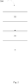
- Fig. 1 is a schematic diagram of the disclosed device 100.
- a transducer 107 radiates energy towards the chest of a subject.
- the transducer 107 also receives the energy reflected by the chest of the subject. Since the chest of the subject is moving due to the subject's breathing the frequency of the reflected energy undergoes Doppler shift.
- the analyzer 105 receives a signal 106 (to be called transducer signal here after) corresponding to the reflected energy received by the transducer 107.
- the analyzer 105 analyzes the transducer signal 106 and determines the breathing rate of the subject.
- an oscillator 103 produces energy at a predetermined frequency and drives the transducer 107.
- the transducer is preferably an antenna that converts high frequency energy into corresponding electromagnetic radiation and vice versa or an ultrasound transducer that converts electrical energy into ultrasound energy of a predetermined frequency and vice versa .
- electromagnetic waves In the description hereafter only one of them, viz, electromagnetic waves, will be referred to for ease of description. It is to be understood that mutatis mutandis , the description applies to ultrasound waves also. However, if there are exceptions, the differences with reference to the two forms of energy will be stated at appropriate places.
- the analyzer 105 may demodulate the transducer signal 106.
- the transducer signal 106 or demodulated signal 106a exhibits a complex dependency on the motion velocity, the motion amplitude and the distance between the chest and sensor. By using appropriate state-of-the-art signal processing, the subject's breathing rate can be obtained from the transducer signal 106 or demodulated signal 106a.
- the analyzer 105 analyzes the signal to determine a pattern corresponding to a complete breathing cycle, in the demodulated signal 106a. It identifies at least the beginning and end of each complete cycle comprising one inhalation and one exhalation.
- the analyzer 105 determines the time period for each cycle or a time period and amplitude of a pattern within each cycle or counts the number of complete cycles in a given period of time.
- the pattern within the cycle may correspond to the inhalation phase, exhalation phase and pauses, if any, between them.
- the reciprocal of the period when multiplied by sixty yields the breathing rate of the subject in number of breaths per minute which is the most common unit for breathing rate.
- the breathing rate and other values determined by the analyzer 105 are outputted appropriately 108.
- analyzer 105 calculates the breathing rate by Fourier analysis of the transducer signal 106 or the demodulated transducer signal 106a. This may be advantageous for accurate measurements as the effect of noise could be eliminated or suppressed, eliminating the need for determining the start or end points of a cycle and thereby reducing or even eliminating the ambiguity therein.
- the device may output the demodulated signal 106a in a manner suitable for being displayed on a monitor 109.
- limiting values for the various measured values such as breathing rate, or time for one breath (a complete cycle), time for each inhalation and each exhalation and so on may be input to the device through a suitable user interface 111 and whenever the individual values cross the limiting values, an indication or alarm 110 may be issued by the device. It is to be understood that the indication may be an audio or visual or audio-visual alarm.
- the one or more outputs such as the breathing rate, the duration and amplitude of the phases of breathing, etc., may be outputted in a wired or wireless manner, to a central monitoring unit or a bedside unit or a central repository for storing a record of the monitored values or incidents when an alarm was issued.
- the output 108 may be a running average of a predetermined number of breathing cycles for the display unit to display. Alternatively, both the cycle by cycle breathing rate and the running average may be displayed. Once in a while, subjects take a longer breath than normal voluntarily or involuntarily. In one embodiment, when such events occur, such very long cycles may be dropped from the calculation of the average.
- the transducer 107 is disposed on a sphygmomanometer cuff 101 as shown in Fig. 1 .
- the transducer may be releasably coupleable to the cuff, for example using hook-and-loop fasteners.
- the cuff is shown at a distance from the upper arm of the diagrammatic representation of the subject, it is to be understood that the cuff 101 is wrapped or suitably placed on the upper arm of the subject, as prescribed.
- the transducer 107 is disposed on the cuff so that in use, when the cuff is worn by the subject as prescribed, the direction of radiation of the energy is substantially towards the chest of the subject. When the radiated energy encounters the chest of the subject, at least part of the energy is reflected by the chest and at least a part of the reflected energy is sensed by the transducer 107.
- Fig. 3 shows the cuff 101, the transducer 107 disposed on the cuff 101 worn by a subject, as prescribed.
- the breathing rate measurement device and a sphygmomanometer cuff based blood pressure measurement device may be two independent instruments.
- the device according to claim 1 and the blood pressure measurement device are housed in the same enclosure. It may have a display that displays both the blood pressure and the breathing rate.
- the antenna maybe advantageously disposed in the cuff, to be worn by the subject to be monitored so that no special care is needed in positioning the transducer in relation to the chest of the subject.
- the method comprises the steps as follows.
- a transducer is disposed on the arm of the subject.
- an analyzing step 215 the signal received from the transducer is analyzed to determine the breathing rate of the subject.
- the chest moves away from the transducer or towards it during exhalation and inhalation respectively.
- the movement produces shifts in the frequency of the radiated energy.
- the frequency shift is negative - frequency lower than the radiated frequency - during exhalation and the shift is positive during inhalation.
- the magnitude of the shift is proportional to the rate of movement of the chest in relation to the transducer.
- At least one temporal pattern in the signal is determined. This could mean that the part of the signal that represents the inhalation phase or exhalation phase and the pauses, if any, between them may be determined. At least complete cycles of breathing maybe determined. This means that the start and end points of a complete breathing cycle may be determined. This could also mean that the starting points of two consecutive breathing cycles may be determined.
- the breathing rate could also be calculated by Fourier Analysis of the received signal. This may be advantageous for accurate measurements as the effect of noise could be eliminated more easily, reducing the ambiguity in determining the start or end of a cycle.
- the time duration of the complete cycle determined is measured.
- the reciprocal of this measured time when multiplied by sixty, yields the breathing rate in breaths per minute.
- higher frequencies are measured by counting the number of cycles in a certain period of time and the frequency in Hz is calculated. But for slow or long duration phenomenon, it is preferable to measure the time duration of each cycle and calculate the frequency in a suitable unit - Hz or cycles per minute, for instance.
- the values measured or further values calculated based on those measurements are output suitably.
- the output could be displayed in the form of alphanumeric display, or with lights or audio-visual display and so on and combinations thereof and is not treated as a part of this description of the method.
- the breathing rate displayed is the running average of a predetermined number of previous cycles. In one variant both the present breathing rate based on the latest full cycle and the running average as mentioned above are displayed together. In one variant the output includes an alarm when any of the measured or calculated parameters exceed or cross predetermined thresholds.
- the transducer is disposed in a sphygmomanometer cuff.
- the transducer may be integrated into the cuff as an integral part. Since the cuff has a predefined position and orientation of application on the arm of the subject, the transducer may be so coupled or integrated into it, such that when the cuff is applied to the subject's arm properly, the transducer is in the appropriate position relative to the subject's chest for the measurement of the breathing rate.
- the method has hitherto been described as a combination of the cuff based sphygmomanometer and the breathing rate. However it is to be understood that the method may be employed or practiced as an adjunct to or within a monitoring device or system which monitors various other parameters of a subject, for instance in an ICU or any other care facility.
- the parameters normally measured and monitored in that way are, but not limited to, blood pressure, Blood Oxygen Saturation (SpO2), heart rate and body temperature and so on.
- electromagnetic waves are used. Though other frequencies may be used, it is found that reliable results are obtained with a frequency of at least 1 GHz. Preferably, the frequency is between 20 GHz and 30 GHz and most preferably the frequency has a nominal value of 24 GHz.
- ultrasound energy is employed with a frequency of at least 40 kHz. In other words the device and method may use the principle of Doppler RADAR or ultrasound based Doppler Sonar, also called Acoustic Doppler Sonar, using Ultrasound.
Landscapes
- Health & Medical Sciences (AREA)
- Life Sciences & Earth Sciences (AREA)
- Engineering & Computer Science (AREA)
- Molecular Biology (AREA)
- Animal Behavior & Ethology (AREA)
- Veterinary Medicine (AREA)
- Public Health (AREA)
- Physics & Mathematics (AREA)
- General Health & Medical Sciences (AREA)
- Biophysics (AREA)
- Pathology (AREA)
- Biomedical Technology (AREA)
- Heart & Thoracic Surgery (AREA)
- Medical Informatics (AREA)
- Surgery (AREA)
- Physiology (AREA)
- Cardiology (AREA)
- Pulmonology (AREA)
- Vascular Medicine (AREA)
- Ophthalmology & Optometry (AREA)
- Signal Processing (AREA)
- Radiology & Medical Imaging (AREA)
- Nuclear Medicine, Radiotherapy & Molecular Imaging (AREA)
- Psychiatry (AREA)
- Artificial Intelligence (AREA)
- Computer Vision & Pattern Recognition (AREA)
- Measurement Of The Respiration, Hearing Ability, Form, And Blood Characteristics Of Living Organisms (AREA)
- Measuring Pulse, Heart Rate, Blood Pressure Or Blood Flow (AREA)
- Ultra Sonic Daignosis Equipment (AREA)
Description
- This invention belongs to the field of measurement and monitoring of breathing rate of a human subject.
- A number of physiological parameters are measured to estimate the state of health of a person. In the case of a patient in a hospital and especially in an intensive care unit (ICU) physiological parameters are measured on a continuous basis to monitor variations in the state of health of the patient. This is done to ensure timely medical intervention in case of deterioration in the state of health, especially when the deterioration is likely to endanger life.
- One of the important parameters measured is the breathing rate, also called the respiratory rate. The most commonly used method in low acuity settings is to observe the rise and fall of the patient's chest during breathing and counting the number of inhalation and exhalation cycles, timed with a clock. This is problematic when the patient has irregular or shallow breaths due to breathing distress.
- Devices and methods to automatically measure the breathing rate are known in the field, using different types of transducers. Some of those methods are impedance plethysmography, capnography (mainly used in ICUs) and inductive thoracic plethysmography (Respiband ™) in sleep studies (polysomnography). All these methods require the positioning of the sensor on the patient's body or elsewhere by a trained person.
- The published patent application
US20100222687 , assigned to the same assignee as that of the present application discloses a method of using a plurality of Doppler radars disposed on the seat belt or integrated into the seat belt for monitoring vital body signs of a person seated in a seat of a motor vehicle. The disclosed method unobtrusively monitors vital body signs like heart rate and respiration rate of the person seated in the motor vehicleUS 2006/0100530 A1 discloses a system according the preamble of claim 1. - There exists a need for a device for and a method of measuring the breathing rate of a patient for overcoming or mitigating one or more of the problems in the state of the art. The need is addressed with a device for measuring the breathing rate of a subject defined in claim 1.
- Such a device may have the advantage that it may not need a skilled person to measure the breathing rate manually. Further, it may not need a skilled person to position a sensor proximate to the patient. Since the device measures the breathing rate, the data may be stored, or transmitted to a system for further processing or used along with the other measured parameters for making decisions or for issuing alarms.
- Further, the need for such a method of measuring the breathing rate of a subject is addressed by a method as defined in claim 14.
- Normally, patient monitors used in ICUs use a sphygmomanometer cuff to measure the subject's BP, among other things. Thus the disclosed method may offer the advantage that it makes it convenient to dispose the transducer in the cuff and once the cuff is worn by the subject, the measurement of the breathing rate of the subject is enabled without any additional positioning of a separate transducer.
- These and other aspects of the disclosed device and the disclosed method are described in detail with reference to the following figures, wherein:
-
Fig. 1 is a schematic diagram of the disclosed device; -
Fig. 2 is a diagrammatic representation of the disclosed method; and -
Fig. 3 is a representation of the disclosed device in use. -
Fig. 1 is a schematic diagram of the discloseddevice 100. Atransducer 107 radiates energy towards the chest of a subject. Thetransducer 107 also receives the energy reflected by the chest of the subject. Since the chest of the subject is moving due to the subject's breathing the frequency of the reflected energy undergoes Doppler shift. Theanalyzer 105 receives a signal 106 (to be called transducer signal here after) corresponding to the reflected energy received by thetransducer 107. Theanalyzer 105 analyzes thetransducer signal 106 and determines the breathing rate of the subject. - To give a better picture of the device and its process, an
oscillator 103 produces energy at a predetermined frequency and drives thetransducer 107. The transducer is preferably an antenna that converts high frequency energy into corresponding electromagnetic radiation and vice versa or an ultrasound transducer that converts electrical energy into ultrasound energy of a predetermined frequency and vice versa. In the description hereafter only one of them, viz, electromagnetic waves, will be referred to for ease of description. It is to be understood that mutatis mutandis, the description applies to ultrasound waves also. However, if there are exceptions, the differences with reference to the two forms of energy will be stated at appropriate places. - With a stationary transducer, if the energy is reflected by a stationary object, there is no information in it to be demodulated. However in the present case the reflecting surface is the surface of the chest of the subject that moves in synchronism with the breathing of the subject, at least a part of the movement will be in a direction parallel to the direction of radiation. Thus the frequency of the reflected energy experiences Doppler shifts. The
analyzer 105 may demodulate thetransducer signal 106. Thetransducer signal 106 or demodulatedsignal 106a exhibits a complex dependency on the motion velocity, the motion amplitude and the distance between the chest and sensor. By using appropriate state-of-the-art signal processing, the subject's breathing rate can be obtained from thetransducer signal 106 or demodulatedsignal 106a. - The
analyzer 105 analyzes the signal to determine a pattern corresponding to a complete breathing cycle, in the demodulatedsignal 106a. It identifies at least the beginning and end of each complete cycle comprising one inhalation and one exhalation. - The
analyzer 105 determines the time period for each cycle or a time period and amplitude of a pattern within each cycle or counts the number of complete cycles in a given period of time. The pattern within the cycle may correspond to the inhalation phase, exhalation phase and pauses, if any, between them. As is well known, to measure low frequencies the method of calculating the time period of each cycle is more accurate. The reciprocal of the period when multiplied by sixty yields the breathing rate of the subject in number of breaths per minute which is the most common unit for breathing rate. The breathing rate and other values determined by theanalyzer 105 are outputted appropriately 108. - In one embodiment,
analyzer 105 calculates the breathing rate by Fourier analysis of thetransducer signal 106 or the demodulatedtransducer signal 106a. This may be advantageous for accurate measurements as the effect of noise could be eliminated or suppressed, eliminating the need for determining the start or end points of a cycle and thereby reducing or even eliminating the ambiguity therein. - In one embodiment, the device may output the demodulated
signal 106a in a manner suitable for being displayed on amonitor 109. In another preferred embodiment, limiting values for the various measured values, such as breathing rate, or time for one breath (a complete cycle), time for each inhalation and each exhalation and so on may be input to the device through asuitable user interface 111 and whenever the individual values cross the limiting values, an indication oralarm 110 may be issued by the device. It is to be understood that the indication may be an audio or visual or audio-visual alarm. In one embodiment, the one or more outputs such as the breathing rate, the duration and amplitude of the phases of breathing, etc., may be outputted in a wired or wireless manner, to a central monitoring unit or a bedside unit or a central repository for storing a record of the monitored values or incidents when an alarm was issued. - Since breathing is a natural biological phenomenon, there may be large variations in the time per cycle between cycles. Thus, if the breathing rate calculated as described above is displayed as is, the display on the
monitor 109 may change fairly rapidly and hence may be confusing or difficult to read. Thus, theoutput 108 may be a running average of a predetermined number of breathing cycles for the display unit to display. Alternatively, both the cycle by cycle breathing rate and the running average may be displayed. Once in a while, subjects take a longer breath than normal voluntarily or involuntarily. In one embodiment, when such events occur, such very long cycles may be dropped from the calculation of the average. - In one embodiment, the
transducer 107 is disposed on asphygmomanometer cuff 101 as shown inFig. 1 . The transducer may be releasably coupleable to the cuff, for example using hook-and-loop fasteners. Even though the cuff is shown at a distance from the upper arm of the diagrammatic representation of the subject, it is to be understood that thecuff 101 is wrapped or suitably placed on the upper arm of the subject, as prescribed. Thetransducer 107 is disposed on the cuff so that in use, when the cuff is worn by the subject as prescribed, the direction of radiation of the energy is substantially towards the chest of the subject. When the radiated energy encounters the chest of the subject, at least part of the energy is reflected by the chest and at least a part of the reflected energy is sensed by thetransducer 107. -
Fig. 3 shows thecuff 101, thetransducer 107 disposed on thecuff 101 worn by a subject, as prescribed. In this embodiment, the breathing rate measurement device and a sphygmomanometer cuff based blood pressure measurement device may be two independent instruments. In one embodiment, the device according to claim 1 and the blood pressure measurement device are housed in the same enclosure. It may have a display that displays both the blood pressure and the breathing rate. In such a case, the antenna maybe advantageously disposed in the cuff, to be worn by the subject to be monitored so that no special care is needed in positioning the transducer in relation to the chest of the subject. - Now the disclosed
method 200 is described in detail with reference toFig. 2 . The method comprises the steps as follows. In a disposingstep 213, a transducer is disposed on the arm of the subject. In an analyzingstep 215 the signal received from the transducer is analyzed to determine the breathing rate of the subject. - Since, when the subject breathes, the chest expands and contracts in synchronism with the breathing, the chest moves away from the transducer or towards it during exhalation and inhalation respectively. The movement produces shifts in the frequency of the radiated energy. The frequency shift, the Doppler shift, is negative - frequency lower than the radiated frequency - during exhalation and the shift is positive during inhalation. The magnitude of the shift is proportional to the rate of movement of the chest in relation to the transducer.
- In an analyzing
step 215, at least one temporal pattern in the signal is determined. This could mean that the part of the signal that represents the inhalation phase or exhalation phase and the pauses, if any, between them may be determined. At least complete cycles of breathing maybe determined. This means that the start and end points of a complete breathing cycle may be determined. This could also mean that the starting points of two consecutive breathing cycles may be determined. - Alternatively the breathing rate could also be calculated by Fourier Analysis of the received signal. This may be advantageous for accurate measurements as the effect of noise could be eliminated more easily, reducing the ambiguity in determining the start or end of a cycle.
- The time duration of the complete cycle determined is measured. The reciprocal of this measured time, when multiplied by sixty, yields the breathing rate in breaths per minute. Normally, higher frequencies are measured by counting the number of cycles in a certain period of time and the frequency in Hz is calculated. But for slow or long duration phenomenon, it is preferable to measure the time duration of each cycle and calculate the frequency in a suitable unit - Hz or cycles per minute, for instance.
- The values measured or further values calculated based on those measurements are output suitably. The output could be displayed in the form of alphanumeric display, or with lights or audio-visual display and so on and combinations thereof and is not treated as a part of this description of the method.
- The basic equations necessary to carry out the measurement using Doppler are explained below:
- The Doppler signal for a single target, which is a good approximation of a subject's chest, is given by Equation 1 below as:


- For an ideal measurement situation with breathing motion only and a perfect estimation of the phase term, equation (2) reduces to:

- In one variant of the method the breathing rate displayed is the running average of a predetermined number of previous cycles. In one variant both the present breathing rate based on the latest full cycle and the running average as mentioned above are displayed together. In one variant the output includes an alarm when any of the measured or calculated parameters exceed or cross predetermined thresholds.
- In one embodiment of the method, the transducer is disposed in a sphygmomanometer cuff. In one embodiment the transducer may be integrated into the cuff as an integral part. Since the cuff has a predefined position and orientation of application on the arm of the subject, the transducer may be so coupled or integrated into it, such that when the cuff is applied to the subject's arm properly, the transducer is in the appropriate position relative to the subject's chest for the measurement of the breathing rate.
- The method has hitherto been described as a combination of the cuff based sphygmomanometer and the breathing rate. However it is to be understood that the method may be employed or practiced as an adjunct to or within a monitoring device or system which monitors various other parameters of a subject, for instance in an ICU or any other care facility. The parameters normally measured and monitored in that way are, but not limited to, blood pressure, Blood Oxygen Saturation (SpO2), heart rate and body temperature and so on.
- In one embodiment, electromagnetic waves are used. Though other frequencies may be used, it is found that reliable results are obtained with a frequency of at least 1 GHz. Preferably, the frequency is between 20 GHz and 30 GHz and most preferably the frequency has a nominal value of 24 GHz. In another embodiment, ultrasound energy is employed with a frequency of at least 40 kHz. In other words the device and method may use the principle of Doppler RADAR or ultrasound based Doppler Sonar, also called Acoustic Doppler Sonar, using Ultrasound.
- While the embodiments have been described in detail in the drawings and description, such drawings and description are to be considered exemplary and not restrictive; the invention is not limited to the disclosed embodiments. Wherever electrical connections or communication is referred to, it is to be understood that it could be effected in a wired or wireless manner. For instance, instead of displaying measured values of the duration of each phase of the breathing cycle, one or more of them may be displayed as a ratio of the duration of the phase and the duration of the whole breathing cycle and so on. Units described as distinct may in practice be realized in the same physical unit. The units may be built using any one or more devices of various technologies such as microcontrollers, microprocessors, digital signal processors (DSP's), programmable logic devices and so on. The distinction made here is for the ease of understanding and the implementation of the units and the practice of the steps may be varied by skilled persons to advantage.
Claims (15)
- A device (100) for monitoring respiration of a subject, the device comprising:a transducer (107) arranged to be worn on an arm of the subject,an analyzer (105) operatively coupled to the transducer (107), the transducer (107) being arranged for radiating energy under control of the analyzer, the analyzer being arranged for processing a signal received from the transducer, for determining a respiration rate of the subject in dependence on the Doppler frequency shifts in the reflected energy received by the transducer.characterized by the transducer (107) being arranged for radiating energy towards the chest of the subject and receiving the energy reflected by the chest of the subject.
- The device (100) of claim 1 wherein the transducer (107) comprises an ultrasound transducer or an antenna.
- The device (100) of claim 1 further comprising a sphygmomanometer cuff (101) for monitoring a blood pressure of the subject, the cuff (101) being mechanically coupleable to the transducer (107).
- The device (100) of claim 3 wherein the transducer (107) is integrated into the sphygmomanometer cuff (101).
- The device of claim 3 or 4 wherein the sphygmomanometer cuff (101) is further electrically coupleable to the analyzer (105), the sphygmomanometer cuff (101) being arranged for monitoring a blood pressure of the subject in response to a further signal received from the analyzer (105).
- The device (100) of claim 5 wherein the analyzer (105) is further arranged to receive data on the monitored blood pressure from the sphygmomanometer cuff (101).
- The device (100) according to any one of claims 3 to 6 wherein the analyzer (105) is arranged for wireless communication with the transducer (107) or the cuff (101) or both to allow remote monitoring of the subject.
- The device (100) of claim 1, wherein the analyzer (105) is further configured for identifying at least one breathing phase from the transducer signal and for calculating at least one of a duration and an amplitude of the identified phase.
- The device (100) of claim 1, wherein the analyzer (105) is further configured for averaging at least one of the periodicity of respiration, the number of cycles of respiration per unit time, the duration of the phase, and the amplitude of the phase over at least one of a predetermined time period and a predetermined previous number of cycles.
- The device (100) of claim 9, wherein the analyzer (105) is further configured for using a current value within predetermined upper and lower limiting values or values that are within a predetermined multiple of a current average value, for calculating a subsequent average value.
- The device (100) of claim 10 wherein the analyzer (105) is configured for comparing at least one of the periodicity, the number of cycles per unit time, the duration of one or more phases and the amplitude of one or more phases with at least one threshold value for each of those values for generating an alarm signal.
- The device (100) of claim 2, wherein the radiated energy has a frequency of at least 1 GHz, preferably between 20 GHz and 30 GHz and most preferably in the order of 24 GHz.
- The device (100) of claims 2, wherein the radiated energy is ultrasound energy with a frequency of at least 40 kHz.
- A method (200) of monitoring the respiratory rate of a subject, the method comprising the steps of:a disposing step (213) of disposing a transducer on an arm of a subject for radiating energy towards the chest of the subject and receiving the energy reflected by the chest of the subject;an analyzing step of (215) of analyzing a signal received from the transducer, for determining a respiration rate of the subject in dependence on the Doppler frequency shifts in the transducer signal.
- The method (200) of claim 14, wherein the disposing is by mechanically coupling to or integrating the transducer into a sphygmomanometer cuff.
Applications Claiming Priority (2)
| Application Number | Priority Date | Filing Date | Title |
|---|---|---|---|
| US201261711290P | 2012-10-09 | 2012-10-09 | |
| PCT/IB2013/059091 WO2014057399A1 (en) | 2012-10-09 | 2013-10-03 | System and method for breathing rate measurements |
Publications (2)
| Publication Number | Publication Date |
|---|---|
| EP2906104A1 EP2906104A1 (en) | 2015-08-19 |
| EP2906104B1 true EP2906104B1 (en) | 2016-06-08 |
Family
ID=49515437
Family Applications (1)
| Application Number | Title | Priority Date | Filing Date |
|---|---|---|---|
| EP13785655.5A Active EP2906104B1 (en) | 2012-10-09 | 2013-10-03 | System and method for breathing rate measurements |
Country Status (6)
| Country | Link |
|---|---|
| US (1) | US10213114B2 (en) |
| EP (1) | EP2906104B1 (en) |
| JP (1) | JP6005869B2 (en) |
| CN (1) | CN104755016B (en) |
| BR (1) | BR112015007636A2 (en) |
| WO (1) | WO2014057399A1 (en) |
Families Citing this family (1)
| Publication number | Priority date | Publication date | Assignee | Title |
|---|---|---|---|---|
| CN104840218B (en) * | 2015-06-09 | 2018-08-10 | 联想(北京)有限公司 | A kind of measurement method and electronic equipment of respiratory rate |
Family Cites Families (21)
| Publication number | Priority date | Publication date | Assignee | Title |
|---|---|---|---|---|
| US3527197A (en) * | 1966-04-19 | 1970-09-08 | Southwest Res Inst | Indirect blood pressure measurement |
| US5010890A (en) | 1983-08-11 | 1991-04-30 | Vitacomm, Ltd. | Vital signs monitoring system |
| DE3783263T2 (en) | 1986-02-04 | 1993-07-22 | Colin Electronics | REGISTRATION DEVICE FOR LIVING BEINGS. |
| US5919141A (en) | 1994-11-15 | 1999-07-06 | Life Sensing Instrument Company, Inc. | Vital sign remote monitoring device |
| US20060100530A1 (en) * | 2000-11-28 | 2006-05-11 | Allez Physionix Limited | Systems and methods for non-invasive detection and monitoring of cardiac and blood parameters |
| JP2003275207A (en) | 2002-03-25 | 2003-09-30 | Junichi Ninomiya | Remote diagnostic treatment method due ultrasonic wave and remote diagnostic treatment system |
| US7341560B2 (en) * | 2004-10-05 | 2008-03-11 | Rader, Fishman & Grauer Pllc | Apparatuses and methods for non-invasively monitoring blood parameters |
| WO2008026157A2 (en) | 2006-08-30 | 2008-03-06 | Koninklijke Philips Electronics N.V. | Apparatus to monitorpulsating objects within the body |
| EP2194852A2 (en) * | 2007-09-25 | 2010-06-16 | Koninklijke Philips Electronics N.V. | Method and system for monitoring vital body signs of a seated person |
| US20100152600A1 (en) * | 2008-04-03 | 2010-06-17 | Kai Sensors, Inc. | Non-contact physiologic motion sensors and methods for use |
| EP2285276B1 (en) * | 2008-05-09 | 2013-12-25 | Philips Intellectual Property & Standards GmbH | Contactless respiration monitoring of a patient |
| ES2386111T3 (en) | 2008-05-09 | 2012-08-09 | Koninklijke Philips Electronics N.V. | Contactless breathing monitoring of a patient and optical sensor for a photoplethysmography measurement |
| BRPI0910520A2 (en) | 2008-07-11 | 2017-12-19 | Koninl Philips Electronics Nv | Heart Doppler Radar Sensor, Method for Adjusting the Transmit Power of a Heart Doppler Radar Sensor and Computer Program Product |
| BR112012019853A8 (en) | 2010-02-12 | 2018-06-26 | Koninl Philips Electronics Nv | method of processing a cyclic physiological signal and apparatus for monitoring a cyclic physiological signal |
| EP2417908A1 (en) | 2010-08-12 | 2012-02-15 | Philips Intellectual Property & Standards GmbH | Device, system and method for measuring vital signs |
| JP5682504B2 (en) * | 2010-09-09 | 2015-03-11 | コニカミノルタ株式会社 | Safety monitoring device |
| JP5855004B2 (en) * | 2010-09-09 | 2016-02-09 | シチズンホールディングス株式会社 | Arm-mounted blood pressure monitor |
| CN103108588B (en) | 2010-09-22 | 2015-08-12 | 皇家飞利浦电子股份有限公司 | For the method and apparatus of monitored object respiratory activity |
| WO2012038867A1 (en) | 2010-09-22 | 2012-03-29 | Koninklijke Philips Electronics N.V. | Method and device for identifying a subject in a sensor based monitoring system |
| JP2012157435A (en) * | 2011-01-31 | 2012-08-23 | Citizen Holdings Co Ltd | Sphygmomanometer |
| CN103607956B (en) | 2011-02-03 | 2016-08-10 | 尤伦·帕提 | Cardiorespiratory monitor through breast |
-
2013
- 2013-10-03 WO PCT/IB2013/059091 patent/WO2014057399A1/en active Application Filing
- 2013-10-03 JP JP2015535154A patent/JP6005869B2/en active Active
- 2013-10-03 BR BR112015007636A patent/BR112015007636A2/en not_active Application Discontinuation
- 2013-10-03 CN CN201380052751.1A patent/CN104755016B/en active Active
- 2013-10-03 US US14/433,879 patent/US10213114B2/en active Active
- 2013-10-03 EP EP13785655.5A patent/EP2906104B1/en active Active
Also Published As
| Publication number | Publication date |
|---|---|
| JP2015535185A (en) | 2015-12-10 |
| WO2014057399A1 (en) | 2014-04-17 |
| CN104755016A (en) | 2015-07-01 |
| BR112015007636A2 (en) | 2017-07-04 |
| CN104755016B (en) | 2016-11-09 |
| US20150250392A1 (en) | 2015-09-10 |
| US10213114B2 (en) | 2019-02-26 |
| JP6005869B2 (en) | 2016-10-12 |
| EP2906104A1 (en) | 2015-08-19 |
Similar Documents
| Publication | Publication Date | Title |
|---|---|---|
| US11690519B2 (en) | Apparatus, system, and method for monitoring physiological signs | |
| US20210338086A1 (en) | System and method for monitoring cardiorespiratory parameters | |
| US11224385B2 (en) | Method for determining a person's sleeping phase which is favourable for waking up | |
| EP3570737B1 (en) | Non-invasive blood pressure measurement using pulse wave velocity | |
| JP2000102515A (en) | Physical condition detector | |
| EP2906104B1 (en) | System and method for breathing rate measurements |
Legal Events
| Date | Code | Title | Description |
|---|---|---|---|
| PUAI | Public reference made under article 153(3) epc to a published international application that has entered the european phase |
Free format text: ORIGINAL CODE: 0009012 |
|
| 17P | Request for examination filed |
Effective date: 20150511 |
|
| AK | Designated contracting states |
Kind code of ref document: A1 Designated state(s): AL AT BE BG CH CY CZ DE DK EE ES FI FR GB GR HR HU IE IS IT LI LT LU LV MC MK MT NL NO PL PT RO RS SE SI SK SM TR |
|
| AX | Request for extension of the european patent |
Extension state: BA ME |
|
| GRAP | Despatch of communication of intention to grant a patent |
Free format text: ORIGINAL CODE: EPIDOSNIGR1 |
|
| INTG | Intention to grant announced |
Effective date: 20151029 |
|
| DAX | Request for extension of the european patent (deleted) | ||
| GRAS | Grant fee paid |
Free format text: ORIGINAL CODE: EPIDOSNIGR3 |
|
| GRAA | (expected) grant |
Free format text: ORIGINAL CODE: 0009210 |
|
| AK | Designated contracting states |
Kind code of ref document: B1 Designated state(s): AL AT BE BG CH CY CZ DE DK EE ES FI FR GB GR HR HU IE IS IT LI LT LU LV MC MK MT NL NO PL PT RO RS SE SI SK SM TR |
|
| REG | Reference to a national code |
Ref country code: GB Ref legal event code: FG4D |
|
| REG | Reference to a national code |
Ref country code: CH Ref legal event code: EP |
|
| REG | Reference to a national code |
Ref country code: IE Ref legal event code: FG4D |
|
| REG | Reference to a national code |
Ref country code: AT Ref legal event code: REF Ref document number: 804629 Country of ref document: AT Kind code of ref document: T Effective date: 20160715 |
|
| REG | Reference to a national code |
Ref country code: DE Ref legal event code: R096 Ref document number: 602013008496 Country of ref document: DE |
|
| REG | Reference to a national code |
Ref country code: LT Ref legal event code: MG4D |
|
| REG | Reference to a national code |
Ref country code: NL Ref legal event code: MP Effective date: 20160608 |
|
| REG | Reference to a national code |
Ref country code: FR Ref legal event code: PLFP Year of fee payment: 4 |
|
| PG25 | Lapsed in a contracting state [announced via postgrant information from national office to epo] |
Ref country code: FI Free format text: LAPSE BECAUSE OF FAILURE TO SUBMIT A TRANSLATION OF THE DESCRIPTION OR TO PAY THE FEE WITHIN THE PRESCRIBED TIME-LIMIT Effective date: 20160608 Ref country code: NO Free format text: LAPSE BECAUSE OF FAILURE TO SUBMIT A TRANSLATION OF THE DESCRIPTION OR TO PAY THE FEE WITHIN THE PRESCRIBED TIME-LIMIT Effective date: 20160908 Ref country code: LT Free format text: LAPSE BECAUSE OF FAILURE TO SUBMIT A TRANSLATION OF THE DESCRIPTION OR TO PAY THE FEE WITHIN THE PRESCRIBED TIME-LIMIT Effective date: 20160608 |
|
| REG | Reference to a national code |
Ref country code: AT Ref legal event code: MK05 Ref document number: 804629 Country of ref document: AT Kind code of ref document: T Effective date: 20160608 |
|
| PG25 | Lapsed in a contracting state [announced via postgrant information from national office to epo] |
Ref country code: SE Free format text: LAPSE BECAUSE OF FAILURE TO SUBMIT A TRANSLATION OF THE DESCRIPTION OR TO PAY THE FEE WITHIN THE PRESCRIBED TIME-LIMIT Effective date: 20160608 Ref country code: RS Free format text: LAPSE BECAUSE OF FAILURE TO SUBMIT A TRANSLATION OF THE DESCRIPTION OR TO PAY THE FEE WITHIN THE PRESCRIBED TIME-LIMIT Effective date: 20160608 Ref country code: LV Free format text: LAPSE BECAUSE OF FAILURE TO SUBMIT A TRANSLATION OF THE DESCRIPTION OR TO PAY THE FEE WITHIN THE PRESCRIBED TIME-LIMIT Effective date: 20160608 Ref country code: ES Free format text: LAPSE BECAUSE OF FAILURE TO SUBMIT A TRANSLATION OF THE DESCRIPTION OR TO PAY THE FEE WITHIN THE PRESCRIBED TIME-LIMIT Effective date: 20160608 Ref country code: GR Free format text: LAPSE BECAUSE OF FAILURE TO SUBMIT A TRANSLATION OF THE DESCRIPTION OR TO PAY THE FEE WITHIN THE PRESCRIBED TIME-LIMIT Effective date: 20160909 Ref country code: NL Free format text: LAPSE BECAUSE OF FAILURE TO SUBMIT A TRANSLATION OF THE DESCRIPTION OR TO PAY THE FEE WITHIN THE PRESCRIBED TIME-LIMIT Effective date: 20160608 Ref country code: HR Free format text: LAPSE BECAUSE OF FAILURE TO SUBMIT A TRANSLATION OF THE DESCRIPTION OR TO PAY THE FEE WITHIN THE PRESCRIBED TIME-LIMIT Effective date: 20160608 |
|
| PG25 | Lapsed in a contracting state [announced via postgrant information from national office to epo] |
Ref country code: RO Free format text: LAPSE BECAUSE OF FAILURE TO SUBMIT A TRANSLATION OF THE DESCRIPTION OR TO PAY THE FEE WITHIN THE PRESCRIBED TIME-LIMIT Effective date: 20160608 Ref country code: IS Free format text: LAPSE BECAUSE OF FAILURE TO SUBMIT A TRANSLATION OF THE DESCRIPTION OR TO PAY THE FEE WITHIN THE PRESCRIBED TIME-LIMIT Effective date: 20161008 Ref country code: IT Free format text: LAPSE BECAUSE OF FAILURE TO SUBMIT A TRANSLATION OF THE DESCRIPTION OR TO PAY THE FEE WITHIN THE PRESCRIBED TIME-LIMIT Effective date: 20160608 Ref country code: CZ Free format text: LAPSE BECAUSE OF FAILURE TO SUBMIT A TRANSLATION OF THE DESCRIPTION OR TO PAY THE FEE WITHIN THE PRESCRIBED TIME-LIMIT Effective date: 20160608 Ref country code: SK Free format text: LAPSE BECAUSE OF FAILURE TO SUBMIT A TRANSLATION OF THE DESCRIPTION OR TO PAY THE FEE WITHIN THE PRESCRIBED TIME-LIMIT Effective date: 20160608 Ref country code: EE Free format text: LAPSE BECAUSE OF FAILURE TO SUBMIT A TRANSLATION OF THE DESCRIPTION OR TO PAY THE FEE WITHIN THE PRESCRIBED TIME-LIMIT Effective date: 20160608 |
|
| PG25 | Lapsed in a contracting state [announced via postgrant information from national office to epo] |
Ref country code: BE Free format text: LAPSE BECAUSE OF NON-PAYMENT OF DUE FEES Effective date: 20160608 Ref country code: PL Free format text: LAPSE BECAUSE OF FAILURE TO SUBMIT A TRANSLATION OF THE DESCRIPTION OR TO PAY THE FEE WITHIN THE PRESCRIBED TIME-LIMIT Effective date: 20160608 Ref country code: SM Free format text: LAPSE BECAUSE OF FAILURE TO SUBMIT A TRANSLATION OF THE DESCRIPTION OR TO PAY THE FEE WITHIN THE PRESCRIBED TIME-LIMIT Effective date: 20160608 Ref country code: PT Free format text: LAPSE BECAUSE OF FAILURE TO SUBMIT A TRANSLATION OF THE DESCRIPTION OR TO PAY THE FEE WITHIN THE PRESCRIBED TIME-LIMIT Effective date: 20161010 Ref country code: AT Free format text: LAPSE BECAUSE OF FAILURE TO SUBMIT A TRANSLATION OF THE DESCRIPTION OR TO PAY THE FEE WITHIN THE PRESCRIBED TIME-LIMIT Effective date: 20160608 |
|
| REG | Reference to a national code |
Ref country code: DE Ref legal event code: R097 Ref document number: 602013008496 Country of ref document: DE |
|
| PLBE | No opposition filed within time limit |
Free format text: ORIGINAL CODE: 0009261 |
|
| STAA | Information on the status of an ep patent application or granted ep patent |
Free format text: STATUS: NO OPPOSITION FILED WITHIN TIME LIMIT |
|
| 26N | No opposition filed |
Effective date: 20170309 |
|
| PG25 | Lapsed in a contracting state [announced via postgrant information from national office to epo] |
Ref country code: SI Free format text: LAPSE BECAUSE OF FAILURE TO SUBMIT A TRANSLATION OF THE DESCRIPTION OR TO PAY THE FEE WITHIN THE PRESCRIBED TIME-LIMIT Effective date: 20160608 Ref country code: DK Free format text: LAPSE BECAUSE OF FAILURE TO SUBMIT A TRANSLATION OF THE DESCRIPTION OR TO PAY THE FEE WITHIN THE PRESCRIBED TIME-LIMIT Effective date: 20160608 |
|
| REG | Reference to a national code |
Ref country code: CH Ref legal event code: PL |
|
| REG | Reference to a national code |
Ref country code: IE Ref legal event code: MM4A |
|
| PG25 | Lapsed in a contracting state [announced via postgrant information from national office to epo] |
Ref country code: CH Free format text: LAPSE BECAUSE OF NON-PAYMENT OF DUE FEES Effective date: 20161031 Ref country code: LI Free format text: LAPSE BECAUSE OF NON-PAYMENT OF DUE FEES Effective date: 20161031 |
|
| PG25 | Lapsed in a contracting state [announced via postgrant information from national office to epo] |
Ref country code: LU Free format text: LAPSE BECAUSE OF NON-PAYMENT OF DUE FEES Effective date: 20161003 |
|
| REG | Reference to a national code |
Ref country code: FR Ref legal event code: PLFP Year of fee payment: 5 |
|
| PG25 | Lapsed in a contracting state [announced via postgrant information from national office to epo] |
Ref country code: IE Free format text: LAPSE BECAUSE OF NON-PAYMENT OF DUE FEES Effective date: 20161003 |
|
| PG25 | Lapsed in a contracting state [announced via postgrant information from national office to epo] |
Ref country code: HU Free format text: LAPSE BECAUSE OF FAILURE TO SUBMIT A TRANSLATION OF THE DESCRIPTION OR TO PAY THE FEE WITHIN THE PRESCRIBED TIME-LIMIT; INVALID AB INITIO Effective date: 20131003 |
|
| PG25 | Lapsed in a contracting state [announced via postgrant information from national office to epo] |
Ref country code: MC Free format text: LAPSE BECAUSE OF FAILURE TO SUBMIT A TRANSLATION OF THE DESCRIPTION OR TO PAY THE FEE WITHIN THE PRESCRIBED TIME-LIMIT Effective date: 20160608 Ref country code: MK Free format text: LAPSE BECAUSE OF FAILURE TO SUBMIT A TRANSLATION OF THE DESCRIPTION OR TO PAY THE FEE WITHIN THE PRESCRIBED TIME-LIMIT Effective date: 20160608 Ref country code: CY Free format text: LAPSE BECAUSE OF FAILURE TO SUBMIT A TRANSLATION OF THE DESCRIPTION OR TO PAY THE FEE WITHIN THE PRESCRIBED TIME-LIMIT Effective date: 20160608 Ref country code: MT Free format text: LAPSE BECAUSE OF NON-PAYMENT OF DUE FEES Effective date: 20161031 |
|
| PG25 | Lapsed in a contracting state [announced via postgrant information from national office to epo] |
Ref country code: BG Free format text: LAPSE BECAUSE OF FAILURE TO SUBMIT A TRANSLATION OF THE DESCRIPTION OR TO PAY THE FEE WITHIN THE PRESCRIBED TIME-LIMIT Effective date: 20160608 |
|
| REG | Reference to a national code |
Ref country code: FR Ref legal event code: PLFP Year of fee payment: 6 |
|
| PG25 | Lapsed in a contracting state [announced via postgrant information from national office to epo] |
Ref country code: AL Free format text: LAPSE BECAUSE OF FAILURE TO SUBMIT A TRANSLATION OF THE DESCRIPTION OR TO PAY THE FEE WITHIN THE PRESCRIBED TIME-LIMIT Effective date: 20160608 |
|
| PGFP | Annual fee paid to national office [announced via postgrant information from national office to epo] |
Ref country code: TR Payment date: 20180925 Year of fee payment: 6 |
|
| PGFP | Annual fee paid to national office [announced via postgrant information from national office to epo] |
Ref country code: FR Payment date: 20181025 Year of fee payment: 6 |
|
| PG25 | Lapsed in a contracting state [announced via postgrant information from national office to epo] |
Ref country code: FR Free format text: LAPSE BECAUSE OF NON-PAYMENT OF DUE FEES Effective date: 20191031 |
|
| PG25 | Lapsed in a contracting state [announced via postgrant information from national office to epo] |
Ref country code: TR Free format text: LAPSE BECAUSE OF NON-PAYMENT OF DUE FEES Effective date: 20191003 |
|
| PGFP | Annual fee paid to national office [announced via postgrant information from national office to epo] |
Ref country code: GB Payment date: 20231024 Year of fee payment: 11 |
|
| PGFP | Annual fee paid to national office [announced via postgrant information from national office to epo] |
Ref country code: DE Payment date: 20231027 Year of fee payment: 11 |