EP2895513B1 - Fc containing polypeptides with altered glycosylation and reduced effector function - Google Patents
Fc containing polypeptides with altered glycosylation and reduced effector function Download PDFInfo
- Publication number
- EP2895513B1 EP2895513B1 EP13766833.1A EP13766833A EP2895513B1 EP 2895513 B1 EP2895513 B1 EP 2895513B1 EP 13766833 A EP13766833 A EP 13766833A EP 2895513 B1 EP2895513 B1 EP 2895513B1
- Authority
- EP
- European Patent Office
- Prior art keywords
- antibody
- binding
- domain
- glycan
- residue
- Prior art date
- Legal status (The legal status is an assumption and is not a legal conclusion. Google has not performed a legal analysis and makes no representation as to the accuracy of the status listed.)
- Active
Links
- 239000012636 effector Substances 0.000 title claims description 130
- 108090000765 processed proteins & peptides Proteins 0.000 title description 235
- 102000004196 processed proteins & peptides Human genes 0.000 title description 206
- 229920001184 polypeptide Polymers 0.000 title description 205
- 230000013595 glycosylation Effects 0.000 title description 77
- 238000006206 glycosylation reaction Methods 0.000 title description 77
- 230000002829 reductive effect Effects 0.000 title description 24
- 239000003814 drug Substances 0.000 claims description 86
- 150000004676 glycans Chemical class 0.000 claims description 64
- 125000003275 alpha amino acid group Chemical group 0.000 claims description 62
- 229940079593 drug Drugs 0.000 claims description 61
- 235000001014 amino acid Nutrition 0.000 claims description 56
- 241000282414 Homo sapiens Species 0.000 claims description 48
- SQVRNKJHWKZAKO-UHFFFAOYSA-N beta-N-Acetyl-D-neuraminic acid Natural products CC(=O)NC1C(O)CC(O)(C(O)=O)OC1C(O)C(O)CO SQVRNKJHWKZAKO-UHFFFAOYSA-N 0.000 claims description 41
- 125000005647 linker group Chemical group 0.000 claims description 40
- WQZGKKKJIJFFOK-PHYPRBDBSA-N alpha-D-galactose Chemical group OC[C@H]1O[C@H](O)[C@H](O)[C@@H](O)[C@H]1O WQZGKKKJIJFFOK-PHYPRBDBSA-N 0.000 claims description 27
- 229940124597 therapeutic agent Drugs 0.000 claims description 23
- 239000013598 vector Substances 0.000 claims description 20
- 229930182830 galactose Chemical group 0.000 claims description 18
- 125000000613 asparagine group Chemical group N[C@@H](CC(N)=O)C(=O)* 0.000 claims description 17
- 239000000203 mixture Substances 0.000 claims description 17
- 125000005629 sialic acid group Chemical group 0.000 claims description 16
- 150000001720 carbohydrates Chemical group 0.000 claims description 15
- 229940002612 prodrug Drugs 0.000 claims description 15
- 239000000651 prodrug Substances 0.000 claims description 15
- 231100000599 cytotoxic agent Toxicity 0.000 claims description 13
- 108091033319 polynucleotide Proteins 0.000 claims description 13
- 239000002157 polynucleotide Substances 0.000 claims description 13
- 102000040430 polynucleotide Human genes 0.000 claims description 13
- 239000000032 diagnostic agent Substances 0.000 claims description 11
- 229940039227 diagnostic agent Drugs 0.000 claims description 11
- MTCFGRXMJLQNBG-REOHCLBHSA-N (2S)-2-Amino-3-hydroxypropansäure Chemical compound OC[C@H](N)C(O)=O MTCFGRXMJLQNBG-REOHCLBHSA-N 0.000 claims description 10
- 150000001875 compounds Chemical class 0.000 claims description 10
- MTCFGRXMJLQNBG-UHFFFAOYSA-N Serine Natural products OCC(N)C(O)=O MTCFGRXMJLQNBG-UHFFFAOYSA-N 0.000 claims description 9
- 239000002619 cytotoxin Substances 0.000 claims description 9
- 150000002923 oximes Chemical class 0.000 claims description 9
- 125000003172 aldehyde group Chemical group 0.000 claims description 8
- 125000000341 threoninyl group Chemical group [H]OC([H])(C([H])([H])[H])C([H])(N([H])[H])C(*)=O 0.000 claims description 8
- 101710112752 Cytotoxin Proteins 0.000 claims description 7
- 125000000217 alkyl group Chemical group 0.000 claims description 7
- 238000004519 manufacturing process Methods 0.000 claims description 7
- 125000003118 aryl group Chemical group 0.000 claims description 6
- 102000004190 Enzymes Human genes 0.000 claims description 5
- 108090000790 Enzymes Proteins 0.000 claims description 5
- 239000003937 drug carrier Substances 0.000 claims description 5
- 150000007857 hydrazones Chemical class 0.000 claims description 5
- 125000003295 alanine group Chemical group N[C@@H](C)C(=O)* 0.000 claims description 3
- 125000003545 alkoxy group Chemical group 0.000 claims description 2
- 125000004104 aryloxy group Chemical group 0.000 claims description 2
- 125000000404 glutamine group Chemical group N[C@@H](CCC(N)=O)C(=O)* 0.000 claims description 2
- 239000000546 pharmaceutical excipient Substances 0.000 claims description 2
- 125000003607 serino group Chemical group [H]N([H])[C@]([H])(C(=O)[*])C(O[H])([H])[H] 0.000 claims 1
- 230000027455 binding Effects 0.000 description 299
- 239000000427 antigen Substances 0.000 description 87
- 102000036639 antigens Human genes 0.000 description 87
- 108091007433 antigens Proteins 0.000 description 87
- 210000004027 cell Anatomy 0.000 description 74
- 108090000623 proteins and genes Proteins 0.000 description 53
- 238000000034 method Methods 0.000 description 50
- 230000021615 conjugation Effects 0.000 description 46
- 239000000562 conjugate Substances 0.000 description 42
- 230000035772 mutation Effects 0.000 description 42
- 102000004169 proteins and genes Human genes 0.000 description 40
- 235000018102 proteins Nutrition 0.000 description 39
- 230000006870 function Effects 0.000 description 37
- 238000011282 treatment Methods 0.000 description 36
- 150000003573 thiols Chemical class 0.000 description 34
- 239000012634 fragment Substances 0.000 description 29
- SQVRNKJHWKZAKO-OQPLDHBCSA-N sialic acid Chemical compound CC(=O)N[C@@H]1[C@@H](O)C[C@@](O)(C(O)=O)OC1[C@H](O)[C@H](O)CO SQVRNKJHWKZAKO-OQPLDHBCSA-N 0.000 description 29
- 238000004458 analytical method Methods 0.000 description 27
- 108060003951 Immunoglobulin Proteins 0.000 description 26
- 230000014509 gene expression Effects 0.000 description 26
- 102000018358 immunoglobulin Human genes 0.000 description 26
- 229940049595 antibody-drug conjugate Drugs 0.000 description 25
- 102100026120 IgG receptor FcRn large subunit p51 Human genes 0.000 description 24
- 238000002474 experimental method Methods 0.000 description 24
- 101710177940 IgG receptor FcRn large subunit p51 Proteins 0.000 description 23
- 239000002246 antineoplastic agent Substances 0.000 description 23
- 230000000694 effects Effects 0.000 description 21
- 206010028980 Neoplasm Diseases 0.000 description 20
- -1 antibodies Proteins 0.000 description 20
- 238000006467 substitution reaction Methods 0.000 description 19
- 230000001225 therapeutic effect Effects 0.000 description 19
- NFGXHKASABOEEW-UHFFFAOYSA-N 1-methylethyl 11-methoxy-3,7,11-trimethyl-2,4-dodecadienoate Chemical compound COC(C)(C)CCCC(C)CC=CC(C)=CC(=O)OC(C)C NFGXHKASABOEEW-UHFFFAOYSA-N 0.000 description 18
- AOJJSUZBOXZQNB-TZSSRYMLSA-N Doxorubicin Chemical compound O([C@H]1C[C@@](O)(CC=2C(O)=C3C(=O)C=4C=CC=C(C=4C(=O)C3=C(O)C=21)OC)C(=O)CO)[C@H]1C[C@H](N)[C@H](O)[C@H](C)O1 AOJJSUZBOXZQNB-TZSSRYMLSA-N 0.000 description 18
- 229940127089 cytotoxic agent Drugs 0.000 description 18
- 208000037265 diseases, disorders, signs and symptoms Diseases 0.000 description 18
- 125000002344 aminooxy group Chemical group [H]N([H])O[*] 0.000 description 17
- 239000000611 antibody drug conjugate Substances 0.000 description 17
- 102000005962 receptors Human genes 0.000 description 16
- 108020003175 receptors Proteins 0.000 description 16
- 125000002496 methyl group Chemical group [H]C([H])([H])* 0.000 description 15
- 239000000872 buffer Substances 0.000 description 14
- 239000013604 expression vector Substances 0.000 description 14
- 230000003647 oxidation Effects 0.000 description 14
- 238000007254 oxidation reaction Methods 0.000 description 14
- 238000002360 preparation method Methods 0.000 description 14
- 238000006243 chemical reaction Methods 0.000 description 13
- 230000000295 complement effect Effects 0.000 description 13
- 238000002415 sodium dodecyl sulfate polyacrylamide gel electrophoresis Methods 0.000 description 13
- 150000001413 amino acids Chemical class 0.000 description 12
- 239000003636 conditioned culture medium Substances 0.000 description 12
- 238000000338 in vitro Methods 0.000 description 12
- KHIWWQKSHDUIBK-UHFFFAOYSA-N periodic acid Chemical compound OI(=O)(=O)=O KHIWWQKSHDUIBK-UHFFFAOYSA-N 0.000 description 12
- 210000002966 serum Anatomy 0.000 description 12
- 108010073807 IgG Receptors Proteins 0.000 description 11
- 102000009490 IgG Receptors Human genes 0.000 description 11
- 102100023832 Prolyl endopeptidase FAP Human genes 0.000 description 11
- 125000000837 carbohydrate group Chemical group 0.000 description 11
- 229960004618 prednisone Drugs 0.000 description 11
- XOFYZVNMUHMLCC-ZPOLXVRWSA-N prednisone Chemical compound O=C1C=C[C@]2(C)[C@H]3C(=O)C[C@](C)([C@@](CC4)(O)C(=O)CO)[C@@H]4[C@@H]3CCC2=C1 XOFYZVNMUHMLCC-ZPOLXVRWSA-N 0.000 description 11
- 239000000047 product Substances 0.000 description 11
- 239000000243 solution Substances 0.000 description 11
- 241000894007 species Species 0.000 description 11
- 210000004881 tumor cell Anatomy 0.000 description 11
- 108010006654 Bleomycin Proteins 0.000 description 10
- 102000009109 Fc receptors Human genes 0.000 description 10
- 108010087819 Fc receptors Proteins 0.000 description 10
- 229940024606 amino acid Drugs 0.000 description 10
- 229960001561 bleomycin Drugs 0.000 description 10
- OYVAGSVQBOHSSS-UAPAGMARSA-O bleomycin A2 Chemical compound N([C@H](C(=O)N[C@H](C)[C@@H](O)[C@H](C)C(=O)N[C@@H]([C@H](O)C)C(=O)NCCC=1SC=C(N=1)C=1SC=C(N=1)C(=O)NCCC[S+](C)C)[C@@H](O[C@H]1[C@H]([C@@H](O)[C@H](O)[C@H](CO)O1)O[C@@H]1[C@H]([C@@H](OC(N)=O)[C@H](O)[C@@H](CO)O1)O)C=1N=CNC=1)C(=O)C1=NC([C@H](CC(N)=O)NC[C@H](N)C(N)=O)=NC(N)=C1C OYVAGSVQBOHSSS-UAPAGMARSA-O 0.000 description 10
- 239000003795 chemical substances by application Substances 0.000 description 10
- 229960005420 etoposide Drugs 0.000 description 10
- VJJPUSNTGOMMGY-MRVIYFEKSA-N etoposide Chemical compound COC1=C(O)C(OC)=CC([C@@H]2C3=CC=4OCOC=4C=C3[C@@H](O[C@H]3[C@@H]([C@@H](O)[C@@H]4O[C@H](C)OC[C@H]4O3)O)[C@@H]3[C@@H]2C(OC3)=O)=C1 VJJPUSNTGOMMGY-MRVIYFEKSA-N 0.000 description 10
- 238000001727 in vivo Methods 0.000 description 10
- 230000003993 interaction Effects 0.000 description 10
- UHDGCWIWMRVCDJ-CCXZUQQUSA-N Cytarabine Chemical compound O=C1N=C(N)C=CN1[C@H]1[C@@H](O)[C@H](O)[C@@H](CO)O1 UHDGCWIWMRVCDJ-CCXZUQQUSA-N 0.000 description 9
- 102000004127 Cytokines Human genes 0.000 description 9
- 108090000695 Cytokines Proteins 0.000 description 9
- IAZDPXIOMUYVGZ-UHFFFAOYSA-N Dimethylsulphoxide Chemical compound CS(C)=O IAZDPXIOMUYVGZ-UHFFFAOYSA-N 0.000 description 9
- 101150029707 ERBB2 gene Proteins 0.000 description 9
- 108010015133 Galactose oxidase Proteins 0.000 description 9
- FBOZXECLQNJBKD-ZDUSSCGKSA-N L-methotrexate Chemical compound C=1N=C2N=C(N)N=C(N)C2=NC=1CN(C)C1=CC=C(C(=O)N[C@@H](CCC(O)=O)C(O)=O)C=C1 FBOZXECLQNJBKD-ZDUSSCGKSA-N 0.000 description 9
- 101710099301 Low affinity immunoglobulin gamma Fc region receptor III-A Proteins 0.000 description 9
- 102100029193 Low affinity immunoglobulin gamma Fc region receptor III-A Human genes 0.000 description 9
- 238000012512 characterization method Methods 0.000 description 9
- 201000010099 disease Diseases 0.000 description 9
- 208000035475 disorder Diseases 0.000 description 9
- 238000004191 hydrophobic interaction chromatography Methods 0.000 description 9
- 238000004895 liquid chromatography mass spectrometry Methods 0.000 description 9
- 229960000485 methotrexate Drugs 0.000 description 9
- 108010068617 neonatal Fc receptor Proteins 0.000 description 9
- 210000003819 peripheral blood mononuclear cell Anatomy 0.000 description 9
- CMSMOCZEIVJLDB-UHFFFAOYSA-N Cyclophosphamide Chemical compound ClCCN(CCCl)P1(=O)NCCCO1 CMSMOCZEIVJLDB-UHFFFAOYSA-N 0.000 description 8
- 241001465754 Metazoa Species 0.000 description 8
- 125000000539 amino acid group Chemical group 0.000 description 8
- 230000001093 anti-cancer Effects 0.000 description 8
- 239000002738 chelating agent Substances 0.000 description 8
- 238000010367 cloning Methods 0.000 description 8
- 229960004397 cyclophosphamide Drugs 0.000 description 8
- 238000002347 injection Methods 0.000 description 8
- 239000007924 injection Substances 0.000 description 8
- CPTBDICYNRMXFX-UHFFFAOYSA-N procarbazine Chemical compound CNNCC1=CC=C(C(=O)NC(C)C)C=C1 CPTBDICYNRMXFX-UHFFFAOYSA-N 0.000 description 8
- 229960000624 procarbazine Drugs 0.000 description 8
- 238000001890 transfection Methods 0.000 description 8
- 108020004414 DNA Proteins 0.000 description 7
- JXLYSJRDGCGARV-WWYNWVTFSA-N Vinblastine Natural products O=C(O[C@H]1[C@](O)(C(=O)OC)[C@@H]2N(C)c3c(cc(c(OC)c3)[C@]3(C(=O)OC)c4[nH]c5c(c4CCN4C[C@](O)(CC)C[C@H](C3)C4)cccc5)[C@@]32[C@H]2[C@@]1(CC)C=CCN2CC3)C JXLYSJRDGCGARV-WWYNWVTFSA-N 0.000 description 7
- 238000003556 assay Methods 0.000 description 7
- 238000001516 cell proliferation assay Methods 0.000 description 7
- 231100000433 cytotoxic Toxicity 0.000 description 7
- 230000001472 cytotoxic effect Effects 0.000 description 7
- 238000010494 dissociation reaction Methods 0.000 description 7
- 230000005593 dissociations Effects 0.000 description 7
- 229960004679 doxorubicin Drugs 0.000 description 7
- 125000003630 glycyl group Chemical group [H]N([H])C([H])([H])C(*)=O 0.000 description 7
- 229920001223 polyethylene glycol Polymers 0.000 description 7
- 230000000069 prophylactic effect Effects 0.000 description 7
- 239000000523 sample Substances 0.000 description 7
- 238000002198 surface plasmon resonance spectroscopy Methods 0.000 description 7
- 230000004083 survival effect Effects 0.000 description 7
- 229960004528 vincristine Drugs 0.000 description 7
- OGWKCGZFUXNPDA-XQKSVPLYSA-N vincristine Chemical compound C([N@]1C[C@@H](C[C@]2(C(=O)OC)C=3C(=CC4=C([C@]56[C@H]([C@@]([C@H](OC(C)=O)[C@]7(CC)C=CCN([C@H]67)CC5)(O)C(=O)OC)N4C=O)C=3)OC)C[C@@](C1)(O)CC)CC1=C2NC2=CC=CC=C12 OGWKCGZFUXNPDA-XQKSVPLYSA-N 0.000 description 7
- OGWKCGZFUXNPDA-UHFFFAOYSA-N vincristine Natural products C1C(CC)(O)CC(CC2(C(=O)OC)C=3C(=CC4=C(C56C(C(C(OC(C)=O)C7(CC)C=CCN(C67)CC5)(O)C(=O)OC)N4C=O)C=3)OC)CN1CCC1=C2NC2=CC=CC=C12 OGWKCGZFUXNPDA-UHFFFAOYSA-N 0.000 description 7
- 241000894006 Bacteria Species 0.000 description 6
- 102100024217 CAMPATH-1 antigen Human genes 0.000 description 6
- 0 CC(C)NCC(*)(*)SSCCC(*)=O Chemical compound CC(C)NCC(*)(*)SSCCC(*)=O 0.000 description 6
- 108010065524 CD52 Antigen Proteins 0.000 description 6
- PEDCQBHIVMGVHV-UHFFFAOYSA-N Glycerine Chemical compound OCC(O)CO PEDCQBHIVMGVHV-UHFFFAOYSA-N 0.000 description 6
- 102000006496 Immunoglobulin Heavy Chains Human genes 0.000 description 6
- 108010019476 Immunoglobulin Heavy Chains Proteins 0.000 description 6
- 230000004988 N-glycosylation Effects 0.000 description 6
- DNIAPMSPPWPWGF-UHFFFAOYSA-N Propylene glycol Chemical compound CC(O)CO DNIAPMSPPWPWGF-UHFFFAOYSA-N 0.000 description 6
- FAPWRFPIFSIZLT-UHFFFAOYSA-M Sodium chloride Chemical compound [Na+].[Cl-] FAPWRFPIFSIZLT-UHFFFAOYSA-M 0.000 description 6
- ZMANZCXQSJIPKH-UHFFFAOYSA-N Triethylamine Chemical compound CCN(CC)CC ZMANZCXQSJIPKH-UHFFFAOYSA-N 0.000 description 6
- 230000008901 benefit Effects 0.000 description 6
- 230000015572 biosynthetic process Effects 0.000 description 6
- 238000002983 circular dichroism Methods 0.000 description 6
- 238000005859 coupling reaction Methods 0.000 description 6
- 229960000684 cytarabine Drugs 0.000 description 6
- 229940072221 immunoglobulins Drugs 0.000 description 6
- 238000001155 isoelectric focusing Methods 0.000 description 6
- 230000004807 localization Effects 0.000 description 6
- 230000004048 modification Effects 0.000 description 6
- 238000012986 modification Methods 0.000 description 6
- 230000009450 sialylation Effects 0.000 description 6
- JQWHASGSAFIOCM-UHFFFAOYSA-M sodium periodate Chemical compound [Na+].[O-]I(=O)(=O)=O JQWHASGSAFIOCM-UHFFFAOYSA-M 0.000 description 6
- 238000002560 therapeutic procedure Methods 0.000 description 6
- 229960003048 vinblastine Drugs 0.000 description 6
- JXLYSJRDGCGARV-XQKSVPLYSA-N vincaleukoblastine Chemical compound C([C@@H](C[C@]1(C(=O)OC)C=2C(=CC3=C([C@]45[C@H]([C@@]([C@H](OC(C)=O)[C@]6(CC)C=CCN([C@H]56)CC4)(O)C(=O)OC)N3C)C=2)OC)C[C@@](C2)(O)CC)N2CCC2=C1NC1=CC=CC=C21 JXLYSJRDGCGARV-XQKSVPLYSA-N 0.000 description 6
- 238000001262 western blot Methods 0.000 description 6
- YEDNBEGNKOANMB-REOHCLBHSA-N (2r)-2-amino-3-sulfanylpropanamide Chemical compound SC[C@H](N)C(N)=O YEDNBEGNKOANMB-REOHCLBHSA-N 0.000 description 5
- 238000001712 DNA sequencing Methods 0.000 description 5
- 101000917858 Homo sapiens Low affinity immunoglobulin gamma Fc region receptor III-A Proteins 0.000 description 5
- 241000699666 Mus <mouse, genus> Species 0.000 description 5
- 102000000447 Peptide-N4-(N-acetyl-beta-glucosaminyl) Asparagine Amidase Human genes 0.000 description 5
- 108010055817 Peptide-N4-(N-acetyl-beta-glucosaminyl) Asparagine Amidase Proteins 0.000 description 5
- 240000004808 Saccharomyces cerevisiae Species 0.000 description 5
- 235000014680 Saccharomyces cerevisiae Nutrition 0.000 description 5
- VMHLLURERBWHNL-UHFFFAOYSA-M Sodium acetate Chemical compound [Na+].CC([O-])=O VMHLLURERBWHNL-UHFFFAOYSA-M 0.000 description 5
- 230000010056 antibody-dependent cellular cytotoxicity Effects 0.000 description 5
- 235000014633 carbohydrates Nutrition 0.000 description 5
- 125000002915 carbonyl group Chemical group [*:2]C([*:1])=O 0.000 description 5
- 238000004113 cell culture Methods 0.000 description 5
- 230000008878 coupling Effects 0.000 description 5
- 238000010168 coupling process Methods 0.000 description 5
- 239000000824 cytostatic agent Substances 0.000 description 5
- 239000002254 cytotoxic agent Substances 0.000 description 5
- 238000002866 fluorescence resonance energy transfer Methods 0.000 description 5
- 229940022353 herceptin Drugs 0.000 description 5
- OAKJQQAXSVQMHS-UHFFFAOYSA-N hydrazine group Chemical group NN OAKJQQAXSVQMHS-UHFFFAOYSA-N 0.000 description 5
- 238000003384 imaging method Methods 0.000 description 5
- 210000003734 kidney Anatomy 0.000 description 5
- 239000003550 marker Substances 0.000 description 5
- 230000001613 neoplastic effect Effects 0.000 description 5
- 239000013612 plasmid Substances 0.000 description 5
- 238000000746 purification Methods 0.000 description 5
- 239000001632 sodium acetate Substances 0.000 description 5
- 235000017281 sodium acetate Nutrition 0.000 description 5
- 239000003053 toxin Substances 0.000 description 5
- 231100000765 toxin Toxicity 0.000 description 5
- 108700012359 toxins Proteins 0.000 description 5
- BQQGAGGSEMLWRS-UHFFFAOYSA-N (4-aminophenyl)methyl carbamate Chemical compound NC(=O)OCC1=CC=C(N)C=C1 BQQGAGGSEMLWRS-UHFFFAOYSA-N 0.000 description 4
- VVIAGPKUTFNRDU-UHFFFAOYSA-N 6S-folinic acid Natural products C1NC=2NC(N)=NC(=O)C=2N(C=O)C1CNC1=CC=C(C(=O)NC(CCC(O)=O)C(O)=O)C=C1 VVIAGPKUTFNRDU-UHFFFAOYSA-N 0.000 description 4
- KCXVZYZYPLLWCC-UHFFFAOYSA-N EDTA Chemical class OC(=O)CN(CC(O)=O)CCN(CC(O)=O)CC(O)=O KCXVZYZYPLLWCC-UHFFFAOYSA-N 0.000 description 4
- DHMQDGOQFOQNFH-UHFFFAOYSA-N Glycine Chemical compound NCC(O)=O DHMQDGOQFOQNFH-UHFFFAOYSA-N 0.000 description 4
- KZSNJWFQEVHDMF-BYPYZUCNSA-N L-valine Chemical compound CC(C)[C@H](N)C(O)=O KZSNJWFQEVHDMF-BYPYZUCNSA-N 0.000 description 4
- 108090001090 Lectins Proteins 0.000 description 4
- 102000004856 Lectins Human genes 0.000 description 4
- SQVRNKJHWKZAKO-LUWBGTNYSA-N N-acetylneuraminic acid Chemical compound CC(=O)N[C@@H]1[C@@H](O)CC(O)(C(O)=O)O[C@H]1[C@H](O)[C@H](O)CO SQVRNKJHWKZAKO-LUWBGTNYSA-N 0.000 description 4
- 102000003838 Sialyltransferases Human genes 0.000 description 4
- 108090000141 Sialyltransferases Proteins 0.000 description 4
- NKANXQFJJICGDU-QPLCGJKRSA-N Tamoxifen Chemical compound C=1C=CC=CC=1C(/CC)=C(C=1C=CC(OCCN(C)C)=CC=1)/C1=CC=CC=C1 NKANXQFJJICGDU-QPLCGJKRSA-N 0.000 description 4
- KZSNJWFQEVHDMF-UHFFFAOYSA-N Valine Natural products CC(C)C(N)C(O)=O KZSNJWFQEVHDMF-UHFFFAOYSA-N 0.000 description 4
- RJURFGZVJUQBHK-UHFFFAOYSA-N actinomycin D Natural products CC1OC(=O)C(C(C)C)N(C)C(=O)CN(C)C(=O)C2CCCN2C(=O)C(C(C)C)NC(=O)C1NC(=O)C1=C(N)C(=O)C(C)=C2OC(C(C)=CC=C3C(=O)NC4C(=O)NC(C(N5CCCC5C(=O)N(C)CC(=O)N(C)C(C(C)C)C(=O)OC4C)=O)C(C)C)=C3N=C21 RJURFGZVJUQBHK-UHFFFAOYSA-N 0.000 description 4
- 229940100198 alkylating agent Drugs 0.000 description 4
- 239000002168 alkylating agent Substances 0.000 description 4
- 150000001412 amines Chemical class 0.000 description 4
- 239000011230 binding agent Substances 0.000 description 4
- 210000004899 c-terminal region Anatomy 0.000 description 4
- 229940112129 campath Drugs 0.000 description 4
- 210000004978 chinese hamster ovary cell Anatomy 0.000 description 4
- DQLATGHUWYMOKM-UHFFFAOYSA-L cisplatin Chemical compound N[Pt](N)(Cl)Cl DQLATGHUWYMOKM-UHFFFAOYSA-L 0.000 description 4
- 229960004316 cisplatin Drugs 0.000 description 4
- 239000011248 coating agent Substances 0.000 description 4
- 238000000576 coating method Methods 0.000 description 4
- 230000003247 decreasing effect Effects 0.000 description 4
- 238000001514 detection method Methods 0.000 description 4
- 239000006185 dispersion Substances 0.000 description 4
- 229940088598 enzyme Drugs 0.000 description 4
- VVIAGPKUTFNRDU-ABLWVSNPSA-N folinic acid Chemical compound C1NC=2NC(N)=NC(=O)C=2N(C=O)C1CNC1=CC=C(C(=O)N[C@@H](CCC(O)=O)C(O)=O)C=C1 VVIAGPKUTFNRDU-ABLWVSNPSA-N 0.000 description 4
- 235000008191 folinic acid Nutrition 0.000 description 4
- 239000011672 folinic acid Substances 0.000 description 4
- 239000000499 gel Substances 0.000 description 4
- 230000012010 growth Effects 0.000 description 4
- 238000004128 high performance liquid chromatography Methods 0.000 description 4
- HOMGKSMUEGBAAB-UHFFFAOYSA-N ifosfamide Chemical compound ClCCNP1(=O)OCCCN1CCCl HOMGKSMUEGBAAB-UHFFFAOYSA-N 0.000 description 4
- 229960001101 ifosfamide Drugs 0.000 description 4
- 238000001990 intravenous administration Methods 0.000 description 4
- 238000002372 labelling Methods 0.000 description 4
- 239000002523 lectin Substances 0.000 description 4
- 229960001691 leucovorin Drugs 0.000 description 4
- 239000003446 ligand Substances 0.000 description 4
- 210000004962 mammalian cell Anatomy 0.000 description 4
- 244000005700 microbiome Species 0.000 description 4
- 229940060155 neuac Drugs 0.000 description 4
- CERZMXAJYMMUDR-UHFFFAOYSA-N neuraminic acid Natural products NC1C(O)CC(O)(C(O)=O)OC1C(O)C(O)CO CERZMXAJYMMUDR-UHFFFAOYSA-N 0.000 description 4
- 231100000252 nontoxic Toxicity 0.000 description 4
- 230000003000 nontoxic effect Effects 0.000 description 4
- 102000039446 nucleic acids Human genes 0.000 description 4
- 108020004707 nucleic acids Proteins 0.000 description 4
- 150000007523 nucleic acids Chemical class 0.000 description 4
- 239000008194 pharmaceutical composition Substances 0.000 description 4
- 239000008363 phosphate buffer Substances 0.000 description 4
- 125000002924 primary amino group Chemical group [H]N([H])* 0.000 description 4
- 238000000159 protein binding assay Methods 0.000 description 4
- 230000009467 reduction Effects 0.000 description 4
- 239000011780 sodium chloride Substances 0.000 description 4
- PVYJZLYGTZKPJE-UHFFFAOYSA-N streptonigrin Chemical class C=1C=C2C(=O)C(OC)=C(N)C(=O)C2=NC=1C(C=1N)=NC(C(O)=O)=C(C)C=1C1=CC=C(OC)C(OC)=C1O PVYJZLYGTZKPJE-UHFFFAOYSA-N 0.000 description 4
- 125000001424 substituent group Chemical group 0.000 description 4
- 238000003786 synthesis reaction Methods 0.000 description 4
- 210000001519 tissue Anatomy 0.000 description 4
- 230000004614 tumor growth Effects 0.000 description 4
- 239000004474 valine Substances 0.000 description 4
- 239000003981 vehicle Substances 0.000 description 4
- XLYOFNOQVPJJNP-UHFFFAOYSA-N water Substances O XLYOFNOQVPJJNP-UHFFFAOYSA-N 0.000 description 4
- IAKHMKGGTNLKSZ-INIZCTEOSA-N (S)-colchicine Chemical compound C1([C@@H](NC(C)=O)CC2)=CC(=O)C(OC)=CC=C1C1=C2C=C(OC)C(OC)=C1OC IAKHMKGGTNLKSZ-INIZCTEOSA-N 0.000 description 3
- AMHZIUVRYRVYBA-UHFFFAOYSA-N 2-(2-amino-4,5-dihydroimidazol-1-yl)acetic acid Chemical compound NC1=NCCN1CC(O)=O AMHZIUVRYRVYBA-UHFFFAOYSA-N 0.000 description 3
- 108010075254 C-Peptide Proteins 0.000 description 3
- 101100314454 Caenorhabditis elegans tra-1 gene Proteins 0.000 description 3
- DLGOEMSEDOSKAD-UHFFFAOYSA-N Carmustine Chemical compound ClCCNC(=O)N(N=O)CCCl DLGOEMSEDOSKAD-UHFFFAOYSA-N 0.000 description 3
- 241000699802 Cricetulus griseus Species 0.000 description 3
- YMWUJEATGCHHMB-UHFFFAOYSA-N Dichloromethane Chemical compound ClCCl YMWUJEATGCHHMB-UHFFFAOYSA-N 0.000 description 3
- BWGNESOTFCXPMA-UHFFFAOYSA-N Dihydrogen disulfide Chemical compound SS BWGNESOTFCXPMA-UHFFFAOYSA-N 0.000 description 3
- 238000002965 ELISA Methods 0.000 description 3
- LFQSCWFLJHTTHZ-UHFFFAOYSA-N Ethanol Chemical compound CCO LFQSCWFLJHTTHZ-UHFFFAOYSA-N 0.000 description 3
- 102000030902 Galactosyltransferase Human genes 0.000 description 3
- 108060003306 Galactosyltransferase Proteins 0.000 description 3
- WQZGKKKJIJFFOK-GASJEMHNSA-N Glucose Natural products OC[C@H]1OC(O)[C@H](O)[C@@H](O)[C@@H]1O WQZGKKKJIJFFOK-GASJEMHNSA-N 0.000 description 3
- 102000051366 Glycosyltransferases Human genes 0.000 description 3
- 108700023372 Glycosyltransferases Proteins 0.000 description 3
- 108010017213 Granulocyte-Macrophage Colony-Stimulating Factor Proteins 0.000 description 3
- 102100039620 Granulocyte-macrophage colony-stimulating factor Human genes 0.000 description 3
- 101001012157 Homo sapiens Receptor tyrosine-protein kinase erbB-2 Proteins 0.000 description 3
- 108090000174 Interleukin-10 Proteins 0.000 description 3
- 102000003814 Interleukin-10 Human genes 0.000 description 3
- 108020004684 Internal Ribosome Entry Sites Proteins 0.000 description 3
- 239000012097 Lipofectamine 2000 Substances 0.000 description 3
- GQYIWUVLTXOXAJ-UHFFFAOYSA-N Lomustine Chemical compound ClCCN(N=O)C(=O)NC1CCCCC1 GQYIWUVLTXOXAJ-UHFFFAOYSA-N 0.000 description 3
- PEEHTFAAVSWFBL-UHFFFAOYSA-N Maleimide Chemical compound O=C1NC(=O)C=C1 PEEHTFAAVSWFBL-UHFFFAOYSA-N 0.000 description 3
- 241000124008 Mammalia Species 0.000 description 3
- 241001529936 Murinae Species 0.000 description 3
- OVRNDRQMDRJTHS-KEWYIRBNSA-N N-acetyl-D-galactosamine Chemical compound CC(=O)N[C@H]1C(O)O[C@H](CO)[C@H](O)[C@@H]1O OVRNDRQMDRJTHS-KEWYIRBNSA-N 0.000 description 3
- MBLBDJOUHNCFQT-UHFFFAOYSA-N N-acetyl-D-galactosamine Natural products CC(=O)NC(C=O)C(O)C(O)C(O)CO MBLBDJOUHNCFQT-UHFFFAOYSA-N 0.000 description 3
- OVRNDRQMDRJTHS-RTRLPJTCSA-N N-acetyl-D-glucosamine Chemical compound CC(=O)N[C@H]1C(O)O[C@H](CO)[C@@H](O)[C@@H]1O OVRNDRQMDRJTHS-RTRLPJTCSA-N 0.000 description 3
- 108091028043 Nucleic acid sequence Proteins 0.000 description 3
- 239000002202 Polyethylene glycol Substances 0.000 description 3
- 102100030086 Receptor tyrosine-protein kinase erbB-2 Human genes 0.000 description 3
- 108700012920 TNF Proteins 0.000 description 3
- 102000004243 Tubulin Human genes 0.000 description 3
- 108090000704 Tubulin Proteins 0.000 description 3
- 241000700605 Viruses Species 0.000 description 3
- 239000013543 active substance Substances 0.000 description 3
- FPIPGXGPPPQFEQ-OVSJKPMPSA-N all-trans-retinol Chemical compound OC\C=C(/C)\C=C\C=C(/C)\C=C\C1=C(C)CCCC1(C)C FPIPGXGPPPQFEQ-OVSJKPMPSA-N 0.000 description 3
- 229940045799 anthracyclines and related substance Drugs 0.000 description 3
- 239000003242 anti bacterial agent Substances 0.000 description 3
- 230000000844 anti-bacterial effect Effects 0.000 description 3
- 230000003110 anti-inflammatory effect Effects 0.000 description 3
- 239000007864 aqueous solution Substances 0.000 description 3
- WQZGKKKJIJFFOK-VFUOTHLCSA-N beta-D-glucose Chemical compound OC[C@H]1O[C@@H](O)[C@H](O)[C@@H](O)[C@@H]1O WQZGKKKJIJFFOK-VFUOTHLCSA-N 0.000 description 3
- 239000012148 binding buffer Substances 0.000 description 3
- 230000037396 body weight Effects 0.000 description 3
- 229960005243 carmustine Drugs 0.000 description 3
- 230000008859 change Effects 0.000 description 3
- 235000018417 cysteine Nutrition 0.000 description 3
- 125000000151 cysteine group Chemical class N[C@@H](CS)C(=O)* 0.000 description 3
- 230000001085 cytostatic effect Effects 0.000 description 3
- 239000008121 dextrose Substances 0.000 description 3
- 210000002950 fibroblast Anatomy 0.000 description 3
- 125000000524 functional group Chemical group 0.000 description 3
- 230000004927 fusion Effects 0.000 description 3
- 229940088597 hormone Drugs 0.000 description 3
- 239000005556 hormone Substances 0.000 description 3
- 239000003667 hormone antagonist Substances 0.000 description 3
- 230000001900 immune effect Effects 0.000 description 3
- 239000004615 ingredient Substances 0.000 description 3
- 238000001361 intraarterial administration Methods 0.000 description 3
- 238000011068 loading method Methods 0.000 description 3
- 229960002247 lomustine Drugs 0.000 description 3
- WKPWGQKGSOKKOO-RSFHAFMBSA-N maytansine Chemical class CO[C@@H]([C@@]1(O)C[C@](OC(=O)N1)([C@H]([C@@H]1O[C@@]1(C)[C@@H](OC(=O)[C@H](C)N(C)C(C)=O)CC(=O)N1C)C)[H])\C=C\C=C(C)\CC2=CC(OC)=C(Cl)C1=C2 WKPWGQKGSOKKOO-RSFHAFMBSA-N 0.000 description 3
- 229960004961 mechlorethamine Drugs 0.000 description 3
- HAWPXGHAZFHHAD-UHFFFAOYSA-N mechlorethamine Chemical compound ClCCN(C)CCCl HAWPXGHAZFHHAD-UHFFFAOYSA-N 0.000 description 3
- 230000001404 mediated effect Effects 0.000 description 3
- 229960001156 mitoxantrone Drugs 0.000 description 3
- KKZJGLLVHKMTCM-UHFFFAOYSA-N mitoxantrone Chemical compound O=C1C2=C(O)C=CC(O)=C2C(=O)C2=C1C(NCCNCCO)=CC=C2NCCNCCO KKZJGLLVHKMTCM-UHFFFAOYSA-N 0.000 description 3
- 210000005170 neoplastic cell Anatomy 0.000 description 3
- 239000002777 nucleoside Substances 0.000 description 3
- 210000004940 nucleus Anatomy 0.000 description 3
- 239000002245 particle Substances 0.000 description 3
- 239000000843 powder Substances 0.000 description 3
- 239000002534 radiation-sensitizing agent Substances 0.000 description 3
- 230000000637 radiosensitizating effect Effects 0.000 description 3
- 239000011541 reaction mixture Substances 0.000 description 3
- 230000004044 response Effects 0.000 description 3
- 230000035945 sensitivity Effects 0.000 description 3
- 239000007787 solid Substances 0.000 description 3
- 239000002904 solvent Substances 0.000 description 3
- 239000000758 substrate Substances 0.000 description 3
- 235000000346 sugar Nutrition 0.000 description 3
- 238000011285 therapeutic regimen Methods 0.000 description 3
- 230000001988 toxicity Effects 0.000 description 3
- 231100000419 toxicity Toxicity 0.000 description 3
- 230000009466 transformation Effects 0.000 description 3
- 238000012795 verification Methods 0.000 description 3
- 230000003442 weekly effect Effects 0.000 description 3
- YBJHBAHKTGYVGT-ZKWXMUAHSA-N (+)-Biotin Chemical compound N1C(=O)N[C@@H]2[C@H](CCCCC(=O)O)SC[C@@H]21 YBJHBAHKTGYVGT-ZKWXMUAHSA-N 0.000 description 2
- WOWDZACBATWTAU-FEFUEGSOSA-N (2s)-2-[[(2s)-2-(dimethylamino)-3-methylbutanoyl]amino]-n-[(3r,4s,5s)-1-[(2s)-2-[(1r,2r)-3-[[(1s,2r)-1-hydroxy-1-phenylpropan-2-yl]amino]-1-methoxy-2-methyl-3-oxopropyl]pyrrolidin-1-yl]-3-methoxy-5-methyl-1-oxoheptan-4-yl]-n,3-dimethylbutanamide Chemical compound CC(C)[C@H](N(C)C)C(=O)N[C@@H](C(C)C)C(=O)N(C)[C@@H]([C@@H](C)CC)[C@H](OC)CC(=O)N1CCC[C@H]1[C@H](OC)[C@@H](C)C(=O)N[C@H](C)[C@@H](O)C1=CC=CC=C1 WOWDZACBATWTAU-FEFUEGSOSA-N 0.000 description 2
- UQVNRKBFAXNOGA-LWTNMJDUSA-N (E)-tomaymycin Chemical class CO[C@H]1NC2=CC(O)=C(OC)C=C2C(=O)N2C\C(=C\C)C[C@@H]12 UQVNRKBFAXNOGA-LWTNMJDUSA-N 0.000 description 2
- FPIPGXGPPPQFEQ-UHFFFAOYSA-N 13-cis retinol Natural products OCC=C(C)C=CC=C(C)C=CC1=C(C)CCCC1(C)C FPIPGXGPPPQFEQ-UHFFFAOYSA-N 0.000 description 2
- LSBDFXRDZJMBSC-UHFFFAOYSA-N 2-phenylacetamide Chemical class NC(=O)CC1=CC=CC=C1 LSBDFXRDZJMBSC-UHFFFAOYSA-N 0.000 description 2
- NDMPLJNOPCLANR-UHFFFAOYSA-N 3,4-dihydroxy-15-(4-hydroxy-18-methoxycarbonyl-5,18-seco-ibogamin-18-yl)-16-methoxy-1-methyl-6,7-didehydro-aspidospermidine-3-carboxylic acid methyl ester Natural products C1C(CC)(O)CC(CC2(C(=O)OC)C=3C(=CC4=C(C56C(C(C(O)C7(CC)C=CCN(C67)CC5)(O)C(=O)OC)N4C)C=3)OC)CN1CCC1=C2NC2=CC=CC=C12 NDMPLJNOPCLANR-UHFFFAOYSA-N 0.000 description 2
- CIWBSHSKHKDKBQ-JLAZNSOCSA-N Ascorbic acid Chemical compound OC[C@H](O)[C@H]1OC(=O)C(O)=C1O CIWBSHSKHKDKBQ-JLAZNSOCSA-N 0.000 description 2
- DJIMLSXHXKWADV-CIUDSAMLSA-N Asn-Leu-Ser Chemical compound OC[C@@H](C(O)=O)NC(=O)[C@H](CC(C)C)NC(=O)[C@@H](N)CC(N)=O DJIMLSXHXKWADV-CIUDSAMLSA-N 0.000 description 2
- IJGRMHOSHXDMSA-UHFFFAOYSA-N Atomic nitrogen Chemical compound N#N IJGRMHOSHXDMSA-UHFFFAOYSA-N 0.000 description 2
- 229940122361 Bisphosphonate Drugs 0.000 description 2
- 208000017667 Chronic Disease Diseases 0.000 description 2
- PTOAARAWEBMLNO-KVQBGUIXSA-N Cladribine Chemical compound C1=NC=2C(N)=NC(Cl)=NC=2N1[C@H]1C[C@H](O)[C@@H](CO)O1 PTOAARAWEBMLNO-KVQBGUIXSA-N 0.000 description 2
- SHZGCJCMOBCMKK-UHFFFAOYSA-N D-mannomethylose Natural products CC1OC(O)C(O)C(O)C1O SHZGCJCMOBCMKK-UHFFFAOYSA-N 0.000 description 2
- 108010092160 Dactinomycin Proteins 0.000 description 2
- OFDNQWIFNXBECV-UHFFFAOYSA-N Dolastatin 10 Natural products CC(C)C(N(C)C)C(=O)NC(C(C)C)C(=O)N(C)C(C(C)CC)C(OC)CC(=O)N1CCCC1C(OC)C(C)C(=O)NC(C=1SC=CN=1)CC1=CC=CC=C1 OFDNQWIFNXBECV-UHFFFAOYSA-N 0.000 description 2
- GHASVSINZRGABV-UHFFFAOYSA-N Fluorouracil Chemical compound FC1=CNC(=O)NC1=O GHASVSINZRGABV-UHFFFAOYSA-N 0.000 description 2
- 239000004471 Glycine Substances 0.000 description 2
- 101000913074 Homo sapiens High affinity immunoglobulin gamma Fc receptor I Proteins 0.000 description 2
- 101001057504 Homo sapiens Interferon-stimulated gene 20 kDa protein Proteins 0.000 description 2
- 101001055144 Homo sapiens Interleukin-2 receptor subunit alpha Proteins 0.000 description 2
- 108010021625 Immunoglobulin Fragments Proteins 0.000 description 2
- 102000008394 Immunoglobulin Fragments Human genes 0.000 description 2
- 102000013463 Immunoglobulin Light Chains Human genes 0.000 description 2
- 108010065825 Immunoglobulin Light Chains Proteins 0.000 description 2
- 102000016844 Immunoglobulin-like domains Human genes 0.000 description 2
- 108050006430 Immunoglobulin-like domains Proteins 0.000 description 2
- 102100027268 Interferon-stimulated gene 20 kDa protein Human genes 0.000 description 2
- SHZGCJCMOBCMKK-DHVFOXMCSA-N L-fucopyranose Chemical compound C[C@@H]1OC(O)[C@@H](O)[C@H](O)[C@@H]1O SHZGCJCMOBCMKK-DHVFOXMCSA-N 0.000 description 2
- FFEARJCKVFRZRR-BYPYZUCNSA-N L-methionine Chemical compound CSCC[C@H](N)C(O)=O FFEARJCKVFRZRR-BYPYZUCNSA-N 0.000 description 2
- QIVBCDIJIAJPQS-VIFPVBQESA-N L-tryptophane Chemical compound C1=CC=C2C(C[C@H](N)C(O)=O)=CNC2=C1 QIVBCDIJIAJPQS-VIFPVBQESA-N 0.000 description 2
- 108010000817 Leuprolide Proteins 0.000 description 2
- 206010025323 Lymphomas Diseases 0.000 description 2
- HRHKSTOGXBBQCB-UHFFFAOYSA-N Mitomycin E Natural products O=C1C(N)=C(C)C(=O)C2=C1C(COC(N)=O)C1(OC)C3N(C)C3CN12 HRHKSTOGXBBQCB-UHFFFAOYSA-N 0.000 description 2
- 241000699670 Mus sp. Species 0.000 description 2
- NWIBSHFKIJFRCO-WUDYKRTCSA-N Mytomycin Chemical compound C1N2C(C(C(C)=C(N)C3=O)=O)=C3[C@@H](COC(N)=O)[C@@]2(OC)[C@@H]2[C@H]1N2 NWIBSHFKIJFRCO-WUDYKRTCSA-N 0.000 description 2
- OVRNDRQMDRJTHS-UHFFFAOYSA-N N-acelyl-D-glucosamine Natural products CC(=O)NC1C(O)OC(CO)C(O)C1O OVRNDRQMDRJTHS-UHFFFAOYSA-N 0.000 description 2
- MBLBDJOUHNCFQT-LXGUWJNJSA-N N-acetylglucosamine Natural products CC(=O)N[C@@H](C=O)[C@@H](O)[C@H](O)[C@H](O)CO MBLBDJOUHNCFQT-LXGUWJNJSA-N 0.000 description 2
- 102000005348 Neuraminidase Human genes 0.000 description 2
- 108010006232 Neuraminidase Proteins 0.000 description 2
- 229910019142 PO4 Inorganic materials 0.000 description 2
- 108090000526 Papain Proteins 0.000 description 2
- ISWSIDIOOBJBQZ-UHFFFAOYSA-N Phenol Chemical compound OC1=CC=CC=C1 ISWSIDIOOBJBQZ-UHFFFAOYSA-N 0.000 description 2
- 239000004365 Protease Substances 0.000 description 2
- 108020004511 Recombinant DNA Proteins 0.000 description 2
- 239000006146 Roswell Park Memorial Institute medium Substances 0.000 description 2
- 238000011579 SCID mouse model Methods 0.000 description 2
- UIIMBOGNXHQVGW-UHFFFAOYSA-M Sodium bicarbonate Chemical compound [Na+].OC([O-])=O UIIMBOGNXHQVGW-UHFFFAOYSA-M 0.000 description 2
- 229940123237 Taxane Drugs 0.000 description 2
- MUMGGOZAMZWBJJ-DYKIIFRCSA-N Testostosterone Chemical compound O=C1CC[C@]2(C)[C@H]3CC[C@](C)([C@H](CC4)O)[C@@H]4[C@@H]3CCC2=C1 MUMGGOZAMZWBJJ-DYKIIFRCSA-N 0.000 description 2
- QIVBCDIJIAJPQS-UHFFFAOYSA-N Tryptophan Natural products C1=CC=C2C(CC(N)C(O)=O)=CNC2=C1 QIVBCDIJIAJPQS-UHFFFAOYSA-N 0.000 description 2
- 229940122803 Vinca alkaloid Drugs 0.000 description 2
- QYSXJUFSXHHAJI-XFEUOLMDSA-N Vitamin D3 Natural products C1(/[C@@H]2CC[C@@H]([C@]2(CCC1)C)[C@H](C)CCCC(C)C)=C/C=C1\C[C@@H](O)CCC1=C QYSXJUFSXHHAJI-XFEUOLMDSA-N 0.000 description 2
- 238000010521 absorption reaction Methods 0.000 description 2
- 239000000370 acceptor Substances 0.000 description 2
- 239000008351 acetate buffer Substances 0.000 description 2
- RJURFGZVJUQBHK-IIXSONLDSA-N actinomycin D Chemical compound C[C@H]1OC(=O)[C@H](C(C)C)N(C)C(=O)CN(C)C(=O)[C@@H]2CCCN2C(=O)[C@@H](C(C)C)NC(=O)[C@H]1NC(=O)C1=C(N)C(=O)C(C)=C2OC(C(C)=CC=C3C(=O)N[C@@H]4C(=O)N[C@@H](C(N5CCC[C@H]5C(=O)N(C)CC(=O)N(C)[C@@H](C(C)C)C(=O)O[C@@H]4C)=O)C(C)C)=C3N=C21 RJURFGZVJUQBHK-IIXSONLDSA-N 0.000 description 2
- 230000009471 action Effects 0.000 description 2
- 230000004913 activation Effects 0.000 description 2
- 239000004480 active ingredient Substances 0.000 description 2
- 229940009456 adriamycin Drugs 0.000 description 2
- 230000002411 adverse Effects 0.000 description 2
- 150000001299 aldehydes Chemical group 0.000 description 2
- SHGAZHPCJJPHSC-YCNIQYBTSA-N all-trans-retinoic acid Chemical compound OC(=O)\C=C(/C)\C=C\C=C(/C)\C=C\C1=C(C)CCCC1(C)C SHGAZHPCJJPHSC-YCNIQYBTSA-N 0.000 description 2
- 230000004075 alteration Effects 0.000 description 2
- 230000003444 anaesthetic effect Effects 0.000 description 2
- 230000000843 anti-fungal effect Effects 0.000 description 2
- 230000002924 anti-infective effect Effects 0.000 description 2
- 230000002141 anti-parasite Effects 0.000 description 2
- 230000000840 anti-viral effect Effects 0.000 description 2
- 229940088710 antibiotic agent Drugs 0.000 description 2
- 229940125644 antibody drug Drugs 0.000 description 2
- 239000003096 antiparasitic agent Substances 0.000 description 2
- 238000013459 approach Methods 0.000 description 2
- 230000001363 autoimmune Effects 0.000 description 2
- VSRXQHXAPYXROS-UHFFFAOYSA-N azanide;cyclobutane-1,1-dicarboxylic acid;platinum(2+) Chemical compound [NH2-].[NH2-].[Pt+2].OC(=O)C1(C(O)=O)CCC1 VSRXQHXAPYXROS-UHFFFAOYSA-N 0.000 description 2
- 150000004663 bisphosphonates Chemical class 0.000 description 2
- 210000004369 blood Anatomy 0.000 description 2
- 239000008280 blood Substances 0.000 description 2
- 230000008499 blood brain barrier function Effects 0.000 description 2
- 210000001218 blood-brain barrier Anatomy 0.000 description 2
- 210000004556 brain Anatomy 0.000 description 2
- 238000011088 calibration curve Methods 0.000 description 2
- 201000011510 cancer Diseases 0.000 description 2
- 229960004562 carboplatin Drugs 0.000 description 2
- 230000003833 cell viability Effects 0.000 description 2
- 230000001413 cellular effect Effects 0.000 description 2
- 238000007385 chemical modification Methods 0.000 description 2
- 239000003153 chemical reaction reagent Substances 0.000 description 2
- 238000002512 chemotherapy Methods 0.000 description 2
- 229960004630 chlorambucil Drugs 0.000 description 2
- JCKYGMPEJWAADB-UHFFFAOYSA-N chlorambucil Chemical compound OC(=O)CCCC1=CC=C(N(CCCl)CCCl)C=C1 JCKYGMPEJWAADB-UHFFFAOYSA-N 0.000 description 2
- OSASVXMJTNOKOY-UHFFFAOYSA-N chlorobutanol Chemical compound CC(C)(O)C(Cl)(Cl)Cl OSASVXMJTNOKOY-UHFFFAOYSA-N 0.000 description 2
- 238000004587 chromatography analysis Methods 0.000 description 2
- 238000003776 cleavage reaction Methods 0.000 description 2
- 230000001268 conjugating effect Effects 0.000 description 2
- VFLDPWHFBUODDF-FCXRPNKRSA-N curcumin Chemical compound C1=C(O)C(OC)=CC(\C=C\C(=O)CC(=O)\C=C\C=2C=C(OC)C(O)=CC=2)=C1 VFLDPWHFBUODDF-FCXRPNKRSA-N 0.000 description 2
- 230000009089 cytolysis Effects 0.000 description 2
- 229960000640 dactinomycin Drugs 0.000 description 2
- 238000011033 desalting Methods 0.000 description 2
- 230000029087 digestion Effects 0.000 description 2
- 239000003085 diluting agent Substances 0.000 description 2
- 239000002612 dispersion medium Substances 0.000 description 2
- OFDNQWIFNXBECV-VFSYNPLYSA-N dolastatin 10 Chemical compound CC(C)[C@H](N(C)C)C(=O)N[C@@H](C(C)C)C(=O)N(C)[C@@H]([C@@H](C)CC)[C@H](OC)CC(=O)N1CCC[C@H]1[C@H](OC)[C@@H](C)C(=O)N[C@H](C=1SC=CN=1)CC1=CC=CC=C1 OFDNQWIFNXBECV-VFSYNPLYSA-N 0.000 description 2
- 108010045524 dolastatin 10 Proteins 0.000 description 2
- VQNATVDKACXKTF-XELLLNAOSA-N duocarmycin Chemical class COC1=C(OC)C(OC)=C2NC(C(=O)N3C4=CC(=O)C5=C([C@@]64C[C@@H]6C3)C=C(N5)C(=O)OC)=CC2=C1 VQNATVDKACXKTF-XELLLNAOSA-N 0.000 description 2
- 238000004520 electroporation Methods 0.000 description 2
- 239000000839 emulsion Substances 0.000 description 2
- 238000005516 engineering process Methods 0.000 description 2
- 230000002255 enzymatic effect Effects 0.000 description 2
- 239000002532 enzyme inhibitor Substances 0.000 description 2
- 229960000390 fludarabine Drugs 0.000 description 2
- GIUYCYHIANZCFB-FJFJXFQQSA-N fludarabine phosphate Chemical compound C1=NC=2C(N)=NC(F)=NC=2N1[C@@H]1O[C@H](COP(O)(O)=O)[C@@H](O)[C@@H]1O GIUYCYHIANZCFB-FJFJXFQQSA-N 0.000 description 2
- 239000012530 fluid Substances 0.000 description 2
- 229960002949 fluorouracil Drugs 0.000 description 2
- 125000002485 formyl group Chemical group [H]C(*)=O 0.000 description 2
- 229940045109 genistein Drugs 0.000 description 2
- 235000006539 genistein Nutrition 0.000 description 2
- TZBJGXHYKVUXJN-UHFFFAOYSA-N genistein Natural products C1=CC(O)=CC=C1C1=COC2=CC(O)=CC(O)=C2C1=O TZBJGXHYKVUXJN-UHFFFAOYSA-N 0.000 description 2
- ZCOLJUOHXJRHDI-CMWLGVBASA-N genistein 7-O-beta-D-glucoside Chemical compound O[C@@H]1[C@@H](O)[C@H](O)[C@@H](CO)O[C@H]1OC1=CC(O)=C2C(=O)C(C=3C=CC(O)=CC=3)=COC2=C1 ZCOLJUOHXJRHDI-CMWLGVBASA-N 0.000 description 2
- RWSXRVCMGQZWBV-WDSKDSINSA-N glutathione Chemical compound OC(=O)[C@@H](N)CCC(=O)N[C@@H](CS)C(=O)NCC(O)=O RWSXRVCMGQZWBV-WDSKDSINSA-N 0.000 description 2
- 230000036541 health Effects 0.000 description 2
- 230000002209 hydrophobic effect Effects 0.000 description 2
- 238000011534 incubation Methods 0.000 description 2
- 230000002757 inflammatory effect Effects 0.000 description 2
- 230000005764 inhibitory process Effects 0.000 description 2
- 238000007918 intramuscular administration Methods 0.000 description 2
- 230000005865 ionizing radiation Effects 0.000 description 2
- YACHGFWEQXFSBS-RJXCBBHPSA-N leptomycin Chemical class OC(=O)/C=C(C)/C[C@H](C)[C@@H](O)[C@H](C)C(=O)[C@H](C)/C=C(\C)/C=C/C[C@@H](C)\C=C(/CC)\C=C\[C@@H]1OC(=O)C=C[C@@H]1C YACHGFWEQXFSBS-RJXCBBHPSA-N 0.000 description 2
- GFIJNRVAKGFPGQ-LIJARHBVSA-N leuprolide Chemical compound CCNC(=O)[C@@H]1CCCN1C(=O)[C@H](CCCNC(N)=N)NC(=O)[C@H](CC(C)C)NC(=O)[C@@H](CC(C)C)NC(=O)[C@@H](NC(=O)[C@H](CO)NC(=O)[C@H](CC=1C2=CC=CC=C2NC=1)NC(=O)[C@H](CC=1N=CNC=1)NC(=O)[C@H]1NC(=O)CC1)CC1=CC=C(O)C=C1 GFIJNRVAKGFPGQ-LIJARHBVSA-N 0.000 description 2
- 229960004338 leuprorelin Drugs 0.000 description 2
- 230000036210 malignancy Effects 0.000 description 2
- 238000001840 matrix-assisted laser desorption--ionisation time-of-flight mass spectrometry Methods 0.000 description 2
- 238000005259 measurement Methods 0.000 description 2
- 239000002609 medium Substances 0.000 description 2
- GLVAUDGFNGKCSF-UHFFFAOYSA-N mercaptopurine Chemical compound S=C1NC=NC2=C1NC=N2 GLVAUDGFNGKCSF-UHFFFAOYSA-N 0.000 description 2
- 229930182817 methionine Natural products 0.000 description 2
- 125000001360 methionine group Chemical group N[C@@H](CCSC)C(=O)* 0.000 description 2
- FBOZXECLQNJBKD-UHFFFAOYSA-N methotrexate Chemical compound C=1N=C2N=C(N)N=C(N)C2=NC=1CN(C)C1=CC=C(C(=O)NC(CCC(O)=O)C(O)=O)C=C1 FBOZXECLQNJBKD-UHFFFAOYSA-N 0.000 description 2
- HRHKSTOGXBBQCB-VFWICMBZSA-N methylmitomycin Chemical compound O=C1C(N)=C(C)C(=O)C2=C1[C@@H](COC(N)=O)[C@@]1(OC)[C@H]3N(C)[C@H]3CN12 HRHKSTOGXBBQCB-VFWICMBZSA-N 0.000 description 2
- 231100000219 mutagenic Toxicity 0.000 description 2
- 230000003505 mutagenic effect Effects 0.000 description 2
- 239000013642 negative control Substances 0.000 description 2
- 125000003835 nucleoside group Chemical group 0.000 description 2
- 150000002482 oligosaccharides Chemical class 0.000 description 2
- 229940127084 other anti-cancer agent Drugs 0.000 description 2
- 229940055729 papain Drugs 0.000 description 2
- 235000019834 papain Nutrition 0.000 description 2
- 238000007911 parenteral administration Methods 0.000 description 2
- NBIIXXVUZAFLBC-UHFFFAOYSA-K phosphate Chemical compound [O-]P([O-])([O-])=O NBIIXXVUZAFLBC-UHFFFAOYSA-K 0.000 description 2
- YJGVMLPVUAXIQN-XVVDYKMHSA-N podophyllotoxin Chemical compound COC1=C(OC)C(OC)=CC([C@@H]2C3=CC=4OCOC=4C=C3[C@H](O)[C@@H]3[C@@H]2C(OC3)=O)=C1 YJGVMLPVUAXIQN-XVVDYKMHSA-N 0.000 description 2
- 229920000765 poly(2-oxazolines) Polymers 0.000 description 2
- 229920000232 polyglycine polymer Polymers 0.000 description 2
- 229920000136 polysorbate Polymers 0.000 description 2
- 229950004406 porfiromycin Drugs 0.000 description 2
- 239000013641 positive control Substances 0.000 description 2
- 238000002953 preparative HPLC Methods 0.000 description 2
- 239000003755 preservative agent Substances 0.000 description 2
- 230000008569 process Effects 0.000 description 2
- 230000002035 prolonged effect Effects 0.000 description 2
- AQHHHDLHHXJYJD-UHFFFAOYSA-N propranolol Chemical compound C1=CC=C2C(OCC(O)CNC(C)C)=CC=CC2=C1 AQHHHDLHHXJYJD-UHFFFAOYSA-N 0.000 description 2
- RXWNCPJZOCPEPQ-NVWDDTSBSA-N puromycin Chemical compound C1=CC(OC)=CC=C1C[C@H](N)C(=O)N[C@H]1[C@@H](O)[C@H](N2C3=NC=NC(=C3N=C2)N(C)C)O[C@@H]1CO RXWNCPJZOCPEPQ-NVWDDTSBSA-N 0.000 description 2
- 230000005855 radiation Effects 0.000 description 2
- 238000003127 radioimmunoassay Methods 0.000 description 2
- 229940051022 radioimmunoconjugate Drugs 0.000 description 2
- 238000001959 radiotherapy Methods 0.000 description 2
- 238000003259 recombinant expression Methods 0.000 description 2
- 238000011084 recovery Methods 0.000 description 2
- 238000007634 remodeling Methods 0.000 description 2
- 229930002330 retinoic acid Natural products 0.000 description 2
- 238000004007 reversed phase HPLC Methods 0.000 description 2
- 102200072304 rs1057519530 Human genes 0.000 description 2
- 238000009118 salvage therapy Methods 0.000 description 2
- 230000007017 scission Effects 0.000 description 2
- 238000002741 site-directed mutagenesis Methods 0.000 description 2
- 150000003384 small molecules Chemical class 0.000 description 2
- 238000007920 subcutaneous administration Methods 0.000 description 2
- 239000000126 substance Substances 0.000 description 2
- 239000006228 supernatant Substances 0.000 description 2
- 239000004094 surface-active agent Substances 0.000 description 2
- 239000000725 suspension Substances 0.000 description 2
- 208000024891 symptom Diseases 0.000 description 2
- 229960001603 tamoxifen Drugs 0.000 description 2
- 238000012360 testing method Methods 0.000 description 2
- 230000032258 transport Effects 0.000 description 2
- AQRLNPVMDITEJU-UHFFFAOYSA-N triethylsilane Chemical compound CC[SiH](CC)CC AQRLNPVMDITEJU-UHFFFAOYSA-N 0.000 description 2
- 241001430294 unidentified retrovirus Species 0.000 description 2
- 229960004355 vindesine Drugs 0.000 description 2
- UGGWPQSBPIFKDZ-KOTLKJBCSA-N vindesine Chemical compound C([C@@H](C[C@]1(C(=O)OC)C=2C(=CC3=C([C@]45[C@H]([C@@]([C@H](O)[C@]6(CC)C=CCN([C@H]56)CC4)(O)C(N)=O)N3C)C=2)OC)C[C@@](C2)(O)CC)N2CCC2=C1N=C1[C]2C=CC=C1 UGGWPQSBPIFKDZ-KOTLKJBCSA-N 0.000 description 2
- QYSXJUFSXHHAJI-YRZJJWOYSA-N vitamin D3 Chemical compound C1(/[C@@H]2CC[C@@H]([C@]2(CCC1)C)[C@H](C)CCCC(C)C)=C\C=C1\C[C@@H](O)CCC1=C QYSXJUFSXHHAJI-YRZJJWOYSA-N 0.000 description 2
- 235000005282 vitamin D3 Nutrition 0.000 description 2
- 239000011647 vitamin D3 Substances 0.000 description 2
- 229940021056 vitamin d3 Drugs 0.000 description 2
- NGGMYCMLYOUNGM-UHFFFAOYSA-N (-)-fumagillin Natural products O1C(CC=C(C)C)C1(C)C1C(OC)C(OC(=O)C=CC=CC=CC=CC(O)=O)CCC21CO2 NGGMYCMLYOUNGM-UHFFFAOYSA-N 0.000 description 1
- ZZBOTLREHORFCK-UHFFFAOYSA-N (2,5-dioxopyrrolidin-1-yl) 2-[(2-methylpropan-2-yl)oxycarbonylamino]oxyacetate Chemical compound CC(C)(C)OC(=O)NOCC(=O)ON1C(=O)CCC1=O ZZBOTLREHORFCK-UHFFFAOYSA-N 0.000 description 1
- OHWBGKONMFYEKL-FQEVSTJZSA-N (2r)-2-amino-3-tritylsulfanylpropanamide Chemical compound C=1C=CC=CC=1C(C=1C=CC=CC=1)(SC[C@H](N)C(N)=O)C1=CC=CC=C1 OHWBGKONMFYEKL-FQEVSTJZSA-N 0.000 description 1
- MFRNYXJJRJQHNW-DEMKXPNLSA-N (2s)-2-[[(2r,3r)-3-methoxy-3-[(2s)-1-[(3r,4s,5s)-3-methoxy-5-methyl-4-[methyl-[(2s)-3-methyl-2-[[(2s)-3-methyl-2-(methylamino)butanoyl]amino]butanoyl]amino]heptanoyl]pyrrolidin-2-yl]-2-methylpropanoyl]amino]-3-phenylpropanoic acid Chemical compound CN[C@@H](C(C)C)C(=O)N[C@@H](C(C)C)C(=O)N(C)[C@@H]([C@@H](C)CC)[C@H](OC)CC(=O)N1CCC[C@H]1[C@H](OC)[C@@H](C)C(=O)N[C@H](C(O)=O)CC1=CC=CC=C1 MFRNYXJJRJQHNW-DEMKXPNLSA-N 0.000 description 1
- HKZAAJSTFUZYTO-LURJTMIESA-N (2s)-2-[[2-[[2-[[2-[(2-aminoacetyl)amino]acetyl]amino]acetyl]amino]acetyl]amino]-3-hydroxypropanoic acid Chemical compound NCC(=O)NCC(=O)NCC(=O)NCC(=O)N[C@@H](CO)C(O)=O HKZAAJSTFUZYTO-LURJTMIESA-N 0.000 description 1
- MCEHFIXEKNKSRW-LBPRGKRZSA-N (2s)-2-[[3,5-dichloro-4-[(2,4-diaminopteridin-6-yl)methyl-methylamino]benzoyl]amino]pentanedioic acid Chemical compound C=1N=C2N=C(N)N=C(N)C2=NC=1CN(C)C1=C(Cl)C=C(C(=O)N[C@@H](CCC(O)=O)C(O)=O)C=C1Cl MCEHFIXEKNKSRW-LBPRGKRZSA-N 0.000 description 1
- XOSLDLHSPLPVME-FKLVWXFWSA-N (2s,8s,14s,17s,20s,25s,28r,29s)-2-[(2s)-butan-2-yl]-28-ethyl-17-[(4-methoxyphenyl)methyl]-7,13,16,20,22,22,25,29-octamethyl-8-(2-methylpropyl)-14-propan-2-yl-1-oxa-4,7,10,13,16,19,24,27-octazacyclotriacontane-3,6,9,12,15,18,21,23,26,30-decone Chemical compound CN1C(=O)[C@H](C(C)C)N(C)C(=O)CNC(=O)[C@H](CC(C)C)N(C)C(=O)CNC(=O)[C@H]([C@@H](C)CC)OC(=O)[C@@H](C)[C@@H](CC)NC(=O)[C@H](C)NC(=O)C(C)(C)C(=O)[C@H](C)NC(=O)[C@@H]1CC1=CC=C(OC)C=C1 XOSLDLHSPLPVME-FKLVWXFWSA-N 0.000 description 1
- FPVKHBSQESCIEP-UHFFFAOYSA-N (8S)-3-(2-deoxy-beta-D-erythro-pentofuranosyl)-3,6,7,8-tetrahydroimidazo[4,5-d][1,3]diazepin-8-ol Natural products C1C(O)C(CO)OC1N1C(NC=NCC2O)=C2N=C1 FPVKHBSQESCIEP-UHFFFAOYSA-N 0.000 description 1
- FDKXTQMXEQVLRF-ZHACJKMWSA-N (E)-dacarbazine Chemical compound CN(C)\N=N\c1[nH]cnc1C(N)=O FDKXTQMXEQVLRF-ZHACJKMWSA-N 0.000 description 1
- WHTVZRBIWZFKQO-AWEZNQCLSA-N (S)-chloroquine Chemical compound ClC1=CC=C2C(N[C@@H](C)CCCN(CC)CC)=CC=NC2=C1 WHTVZRBIWZFKQO-AWEZNQCLSA-N 0.000 description 1
- HNSDLXPSAYFUHK-UHFFFAOYSA-N 1,4-bis(2-ethylhexyl) sulfosuccinate Chemical compound CCCCC(CC)COC(=O)CC(S(O)(=O)=O)C(=O)OCC(CC)CCCC HNSDLXPSAYFUHK-UHFFFAOYSA-N 0.000 description 1
- VSNHCAURESNICA-NJFSPNSNSA-N 1-oxidanylurea Chemical compound N[14C](=O)NO VSNHCAURESNICA-NJFSPNSNSA-N 0.000 description 1
- IIZPXYDJLKNOIY-JXPKJXOSSA-N 1-palmitoyl-2-arachidonoyl-sn-glycero-3-phosphocholine Chemical compound CCCCCCCCCCCCCCCC(=O)OC[C@H](COP([O-])(=O)OCC[N+](C)(C)C)OC(=O)CCC\C=C/C\C=C/C\C=C/C\C=C/CCCCC IIZPXYDJLKNOIY-JXPKJXOSSA-N 0.000 description 1
- PNDPGZBMCMUPRI-HVTJNCQCSA-N 10043-66-0 Chemical compound [131I][131I] PNDPGZBMCMUPRI-HVTJNCQCSA-N 0.000 description 1
- DBPWSSGDRRHUNT-CEGNMAFCSA-N 17α-hydroxyprogesterone Chemical compound C1CC2=CC(=O)CC[C@]2(C)[C@@H]2[C@@H]1[C@@H]1CC[C@@](C(=O)C)(O)[C@@]1(C)CC2 DBPWSSGDRRHUNT-CEGNMAFCSA-N 0.000 description 1
- RCWJMKCTHJPXJV-UHFFFAOYSA-N 2,5-bis(aziridin-1-yl)-1,4-benzoquinone Chemical compound O=C1C=C(N2CC2)C(=O)C=C1N1CC1 RCWJMKCTHJPXJV-UHFFFAOYSA-N 0.000 description 1
- VEAIXXJTUWDCBJ-UHFFFAOYSA-N 2-(diaziridin-1-yl)cyclohexa-2,5-diene-1,4-dione Chemical compound O=C1C=CC(=O)C(N2NC2)=C1 VEAIXXJTUWDCBJ-UHFFFAOYSA-N 0.000 description 1
- DWZFIFZYMHNRRV-UHFFFAOYSA-N 2-[(3,4-dihydroxy-5-methoxyphenyl)methylidene]propanedinitrile Chemical compound COC1=CC(C=C(C#N)C#N)=CC(O)=C1O DWZFIFZYMHNRRV-UHFFFAOYSA-N 0.000 description 1
- QMOQBVOBWVNSNO-UHFFFAOYSA-N 2-[[2-[[2-[(2-azaniumylacetyl)amino]acetyl]amino]acetyl]amino]acetate Chemical compound NCC(=O)NCC(=O)NCC(=O)NCC(O)=O QMOQBVOBWVNSNO-UHFFFAOYSA-N 0.000 description 1
- XJFPXLWGZWAWRQ-UHFFFAOYSA-N 2-[[2-[[2-[[2-[[2-[(2-azaniumylacetyl)amino]acetyl]amino]acetyl]amino]acetyl]amino]acetyl]amino]acetate Chemical compound NCC(=O)NCC(=O)NCC(=O)NCC(=O)NCC(=O)NCC(O)=O XJFPXLWGZWAWRQ-UHFFFAOYSA-N 0.000 description 1
- FZDFGHZZPBUTGP-UHFFFAOYSA-N 2-[[2-[bis(carboxymethyl)amino]-3-(4-isothiocyanatophenyl)propyl]-[2-[bis(carboxymethyl)amino]propyl]amino]acetic acid Chemical compound OC(=O)CN(CC(O)=O)C(C)CN(CC(O)=O)CC(N(CC(O)=O)CC(O)=O)CC1=CC=C(N=C=S)C=C1 FZDFGHZZPBUTGP-UHFFFAOYSA-N 0.000 description 1
- QKNYBSVHEMOAJP-UHFFFAOYSA-N 2-amino-2-(hydroxymethyl)propane-1,3-diol;hydron;chloride Chemical compound Cl.OCC(N)(CO)CO QKNYBSVHEMOAJP-UHFFFAOYSA-N 0.000 description 1
- AXAVXPMQTGXXJZ-UHFFFAOYSA-N 2-aminoacetic acid;2-amino-2-(hydroxymethyl)propane-1,3-diol Chemical compound NCC(O)=O.OCC(N)(CO)CO AXAVXPMQTGXXJZ-UHFFFAOYSA-N 0.000 description 1
- GHCZTIFQWKKGSB-UHFFFAOYSA-N 2-hydroxypropane-1,2,3-tricarboxylic acid;phosphoric acid Chemical compound OP(O)(O)=O.OC(=O)CC(O)(C(O)=O)CC(O)=O GHCZTIFQWKKGSB-UHFFFAOYSA-N 0.000 description 1
- AOPRXJXHLWYPQR-UHFFFAOYSA-N 2-phenoxyacetamide Chemical class NC(=O)COC1=CC=CC=C1 AOPRXJXHLWYPQR-UHFFFAOYSA-N 0.000 description 1
- XNPKNHHFCKSMRV-UHFFFAOYSA-N 4-(cyclohexylamino)butane-1-sulfonic acid Chemical compound OS(=O)(=O)CCCCNC1CCCCC1 XNPKNHHFCKSMRV-UHFFFAOYSA-N 0.000 description 1
- QFVHZQCOUORWEI-UHFFFAOYSA-N 4-[(4-anilino-5-sulfonaphthalen-1-yl)diazenyl]-5-hydroxynaphthalene-2,7-disulfonic acid Chemical compound C=12C(O)=CC(S(O)(=O)=O)=CC2=CC(S(O)(=O)=O)=CC=1N=NC(C1=CC=CC(=C11)S(O)(=O)=O)=CC=C1NC1=CC=CC=C1 QFVHZQCOUORWEI-UHFFFAOYSA-N 0.000 description 1
- DODQJNMQWMSYGS-QPLCGJKRSA-N 4-[(z)-1-[4-[2-(dimethylamino)ethoxy]phenyl]-1-phenylbut-1-en-2-yl]phenol Chemical compound C=1C=C(O)C=CC=1C(/CC)=C(C=1C=CC(OCCN(C)C)=CC=1)/C1=CC=CC=C1 DODQJNMQWMSYGS-QPLCGJKRSA-N 0.000 description 1
- SSMIFVHARFVINF-UHFFFAOYSA-N 4-amino-1,8-naphthalimide Chemical compound O=C1NC(=O)C2=CC=CC3=C2C1=CC=C3N SSMIFVHARFVINF-UHFFFAOYSA-N 0.000 description 1
- TVZGACDUOSZQKY-LBPRGKRZSA-N 4-aminofolic acid Chemical compound C1=NC2=NC(N)=NC(N)=C2N=C1CNC1=CC=C(C(=O)N[C@@H](CCC(O)=O)C(O)=O)C=C1 TVZGACDUOSZQKY-LBPRGKRZSA-N 0.000 description 1
- XHSQDZXAVJRBMX-DDHJBXDOSA-N 5,6-dichloro-1-β-d-ribofuranosylbenzimidazole Chemical compound O[C@@H]1[C@H](O)[C@@H](CO)O[C@H]1N1C2=CC(Cl)=C(Cl)C=C2N=C1 XHSQDZXAVJRBMX-DDHJBXDOSA-N 0.000 description 1
- NMUSYJAQQFHJEW-UHFFFAOYSA-N 5-Azacytidine Natural products O=C1N=C(N)N=CN1C1C(O)C(O)C(CO)O1 NMUSYJAQQFHJEW-UHFFFAOYSA-N 0.000 description 1
- XAUDJQYHKZQPEU-KVQBGUIXSA-N 5-aza-2'-deoxycytidine Chemical compound O=C1N=C(N)N=CN1[C@@H]1O[C@H](CO)[C@@H](O)C1 XAUDJQYHKZQPEU-KVQBGUIXSA-N 0.000 description 1
- NMUSYJAQQFHJEW-KVTDHHQDSA-N 5-azacytidine Chemical compound O=C1N=C(N)N=CN1[C@H]1[C@H](O)[C@H](O)[C@@H](CO)O1 NMUSYJAQQFHJEW-KVTDHHQDSA-N 0.000 description 1
- FHIDNBAQOFJWCA-UAKXSSHOSA-N 5-fluorouridine Chemical compound O[C@@H]1[C@H](O)[C@@H](CO)O[C@H]1N1C(=O)NC(=O)C(F)=C1 FHIDNBAQOFJWCA-UAKXSSHOSA-N 0.000 description 1
- STQGQHZAVUOBTE-UHFFFAOYSA-N 7-Cyan-hept-2t-en-4,6-diinsaeure Natural products C1=2C(O)=C3C(=O)C=4C(OC)=CC=CC=4C(=O)C3=C(O)C=2CC(O)(C(C)=O)CC1OC1CC(N)C(O)C(C)O1 STQGQHZAVUOBTE-UHFFFAOYSA-N 0.000 description 1
- SHGAZHPCJJPHSC-ZVCIMWCZSA-N 9-cis-retinoic acid Chemical compound OC(=O)/C=C(\C)/C=C/C=C(/C)\C=C\C1=C(C)CCCC1(C)C SHGAZHPCJJPHSC-ZVCIMWCZSA-N 0.000 description 1
- QTBSBXVTEAMEQO-UHFFFAOYSA-M Acetate Chemical compound CC([O-])=O QTBSBXVTEAMEQO-UHFFFAOYSA-M 0.000 description 1
- HRPVXLWXLXDGHG-UHFFFAOYSA-N Acrylamide Chemical compound NC(=O)C=C HRPVXLWXLXDGHG-UHFFFAOYSA-N 0.000 description 1
- OIRDTQYFTABQOQ-KQYNXXCUSA-N Adenosine Natural products C1=NC=2C(N)=NC=NC=2N1[C@@H]1O[C@H](CO)[C@@H](O)[C@H]1O OIRDTQYFTABQOQ-KQYNXXCUSA-N 0.000 description 1
- 108010000239 Aequorin Proteins 0.000 description 1
- 229920000936 Agarose Polymers 0.000 description 1
- 101710186708 Agglutinin Proteins 0.000 description 1
- 102400000068 Angiostatin Human genes 0.000 description 1
- 108010079709 Angiostatins Proteins 0.000 description 1
- 235000002198 Annona diversifolia Nutrition 0.000 description 1
- 239000004475 Arginine Substances 0.000 description 1
- HNXWVVHIGTZTBO-LKXGYXEUSA-N Asn-Ser-Thr Chemical compound C[C@@H](O)[C@@H](C(O)=O)NC(=O)[C@H](CO)NC(=O)[C@@H](N)CC(N)=O HNXWVVHIGTZTBO-LKXGYXEUSA-N 0.000 description 1
- DCXYFEDJOCDNAF-UHFFFAOYSA-N Asparagine Natural products OC(=O)C(N)CC(N)=O DCXYFEDJOCDNAF-UHFFFAOYSA-N 0.000 description 1
- 208000023275 Autoimmune disease Diseases 0.000 description 1
- 241001112741 Bacillaceae Species 0.000 description 1
- 244000063299 Bacillus subtilis Species 0.000 description 1
- 235000014469 Bacillus subtilis Nutrition 0.000 description 1
- ZOXJGFHDIHLPTG-UHFFFAOYSA-N Boron Chemical compound [B] ZOXJGFHDIHLPTG-UHFFFAOYSA-N 0.000 description 1
- 241000283690 Bos taurus Species 0.000 description 1
- 241000701822 Bovine papillomavirus Species 0.000 description 1
- 206010006187 Breast cancer Diseases 0.000 description 1
- 208000026310 Breast neoplasm Diseases 0.000 description 1
- COVZYZSDYWQREU-UHFFFAOYSA-N Busulfan Chemical compound CS(=O)(=O)OCCCCOS(C)(=O)=O COVZYZSDYWQREU-UHFFFAOYSA-N 0.000 description 1
- 239000002126 C01EB10 - Adenosine Substances 0.000 description 1
- XNUIYMRIRYQYBC-PEKVNIGGSA-N CCC(C)[C@@H]([C@@H](CC(N(CCC1)[C@@H]1[C@@H]([C@@H](C)C(NCCc1ccccc1C(OC)=O)=O)OC)=O)OC)N(C)C([C@H](C(C)C)NC([C@H](C(C)C)NC)=O)=O Chemical compound CCC(C)[C@@H]([C@@H](CC(N(CCC1)[C@@H]1[C@@H]([C@@H](C)C(NCCc1ccccc1C(OC)=O)=O)OC)=O)OC)N(C)C([C@H](C(C)C)NC([C@H](C(C)C)NC)=O)=O XNUIYMRIRYQYBC-PEKVNIGGSA-N 0.000 description 1
- TXCIAUNLDRJGJZ-UHFFFAOYSA-N CMP-N-acetyl neuraminic acid Natural products O1C(C(O)C(O)CO)C(NC(=O)C)C(O)CC1(C(O)=O)OP(O)(=O)OCC1C(O)C(O)C(N2C(N=C(N)C=C2)=O)O1 TXCIAUNLDRJGJZ-UHFFFAOYSA-N 0.000 description 1
- TXCIAUNLDRJGJZ-BILDWYJOSA-N CMP-N-acetyl-beta-neuraminic acid Chemical compound O1[C@@H]([C@H](O)[C@H](O)CO)[C@H](NC(=O)C)[C@@H](O)C[C@]1(C(O)=O)OP(O)(=O)OC[C@@H]1[C@@H](O)[C@@H](O)[C@H](N2C(N=C(N)C=C2)=O)O1 TXCIAUNLDRJGJZ-BILDWYJOSA-N 0.000 description 1
- FVLVBPDQNARYJU-XAHDHGMMSA-N C[C@H]1CCC(CC1)NC(=O)N(CCCl)N=O Chemical compound C[C@H]1CCC(CC1)NC(=O)N(CCCl)N=O FVLVBPDQNARYJU-XAHDHGMMSA-N 0.000 description 1
- UXVMQQNJUSDDNG-UHFFFAOYSA-L Calcium chloride Chemical compound [Cl-].[Cl-].[Ca+2] UXVMQQNJUSDDNG-UHFFFAOYSA-L 0.000 description 1
- 241000282832 Camelidae Species 0.000 description 1
- 241000283707 Capra Species 0.000 description 1
- BQENDLAVTKRQMS-SBBGFIFASA-L Carbenoxolone sodium Chemical compound [Na+].[Na+].C([C@H]1C2=CC(=O)[C@H]34)[C@@](C)(C([O-])=O)CC[C@]1(C)CC[C@@]2(C)[C@]4(C)CC[C@@H]1[C@]3(C)CC[C@H](OC(=O)CCC([O-])=O)C1(C)C BQENDLAVTKRQMS-SBBGFIFASA-L 0.000 description 1
- SHHKQEUPHAENFK-UHFFFAOYSA-N Carboquone Chemical compound O=C1C(C)=C(N2CC2)C(=O)C(C(COC(N)=O)OC)=C1N1CC1 SHHKQEUPHAENFK-UHFFFAOYSA-N 0.000 description 1
- 201000009030 Carcinoma Diseases 0.000 description 1
- 102000014914 Carrier Proteins Human genes 0.000 description 1
- 102000016289 Cell Adhesion Molecules Human genes 0.000 description 1
- 108010067225 Cell Adhesion Molecules Proteins 0.000 description 1
- 241000282693 Cercopithecidae Species 0.000 description 1
- 102100031162 Collagen alpha-1(XVIII) chain Human genes 0.000 description 1
- 206010009944 Colon cancer Diseases 0.000 description 1
- HVXBOLULGPECHP-WAYWQWQTSA-N Combretastatin A4 Chemical compound C1=C(O)C(OC)=CC=C1\C=C/C1=CC(OC)=C(OC)C(OC)=C1 HVXBOLULGPECHP-WAYWQWQTSA-N 0.000 description 1
- 108010047041 Complementarity Determining Regions Proteins 0.000 description 1
- RYGMFSIKBFXOCR-UHFFFAOYSA-N Copper Chemical compound [Cu] RYGMFSIKBFXOCR-UHFFFAOYSA-N 0.000 description 1
- 241000699800 Cricetinae Species 0.000 description 1
- 239000004971 Cross linker Substances 0.000 description 1
- PMATZTZNYRCHOR-CGLBZJNRSA-N Cyclosporin A Chemical compound CC[C@@H]1NC(=O)[C@H]([C@H](O)[C@H](C)C\C=C\C)N(C)C(=O)[C@H](C(C)C)N(C)C(=O)[C@H](CC(C)C)N(C)C(=O)[C@H](CC(C)C)N(C)C(=O)[C@@H](C)NC(=O)[C@H](C)NC(=O)[C@H](CC(C)C)N(C)C(=O)[C@H](C(C)C)NC(=O)[C@H](CC(C)C)N(C)C(=O)CN(C)C1=O PMATZTZNYRCHOR-CGLBZJNRSA-N 0.000 description 1
- 229930105110 Cyclosporin A Natural products 0.000 description 1
- 108010036949 Cyclosporine Proteins 0.000 description 1
- FBPFZTCFMRRESA-FSIIMWSLSA-N D-Glucitol Natural products OC[C@H](O)[C@H](O)[C@@H](O)[C@H](O)CO FBPFZTCFMRRESA-FSIIMWSLSA-N 0.000 description 1
- FBPFZTCFMRRESA-KVTDHHQDSA-N D-Mannitol Chemical compound OC[C@@H](O)[C@@H](O)[C@H](O)[C@H](O)CO FBPFZTCFMRRESA-KVTDHHQDSA-N 0.000 description 1
- FBPFZTCFMRRESA-JGWLITMVSA-N D-glucitol Chemical compound OC[C@H](O)[C@@H](O)[C@H](O)[C@H](O)CO FBPFZTCFMRRESA-JGWLITMVSA-N 0.000 description 1
- GUBGYTABKSRVRQ-WFVLMXAXSA-N DEAE-cellulose Chemical compound OC1C(O)C(O)C(CO)O[C@H]1O[C@@H]1C(CO)OC(O)C(O)C1O GUBGYTABKSRVRQ-WFVLMXAXSA-N 0.000 description 1
- 239000012625 DNA intercalator Substances 0.000 description 1
- 108700022150 Designed Ankyrin Repeat Proteins Proteins 0.000 description 1
- 102100024746 Dihydrofolate reductase Human genes 0.000 description 1
- LQKSHSFQQRCAFW-UHFFFAOYSA-N Dolastatin 15 Natural products COC1=CC(=O)N(C(=O)C(OC(=O)C2N(CCC2)C(=O)C2N(CCC2)C(=O)C(C(C)C)N(C)C(=O)C(NC(=O)C(C(C)C)N(C)C)C(C)C)C(C)C)C1CC1=CC=CC=C1 LQKSHSFQQRCAFW-UHFFFAOYSA-N 0.000 description 1
- LVGKNOAMLMIIKO-UHFFFAOYSA-N Elaidinsaeure-aethylester Natural products CCCCCCCCC=CCCCCCCCC(=O)OCC LVGKNOAMLMIIKO-UHFFFAOYSA-N 0.000 description 1
- 241000196324 Embryophyta Species 0.000 description 1
- MBYXEBXZARTUSS-QLWBXOBMSA-N Emetamine Natural products O(C)c1c(OC)cc2c(c(C[C@@H]3[C@H](CC)CN4[C@H](c5c(cc(OC)c(OC)c5)CC4)C3)ncc2)c1 MBYXEBXZARTUSS-QLWBXOBMSA-N 0.000 description 1
- 102100038083 Endosialin Human genes 0.000 description 1
- 108010079505 Endostatins Proteins 0.000 description 1
- 241000588921 Enterobacteriaceae Species 0.000 description 1
- 241000588724 Escherichia coli Species 0.000 description 1
- 102000016359 Fibronectins Human genes 0.000 description 1
- 108010067306 Fibronectins Proteins 0.000 description 1
- PNNNRSAQSRJVSB-SLPGGIOYSA-N Fucose Natural products C[C@H](O)[C@@H](O)[C@H](O)[C@H](O)C=O PNNNRSAQSRJVSB-SLPGGIOYSA-N 0.000 description 1
- 241000233866 Fungi Species 0.000 description 1
- 108700012941 GNRH1 Proteins 0.000 description 1
- 108010010803 Gelatin Proteins 0.000 description 1
- JRZJKWGQFNTSRN-UHFFFAOYSA-N Geldanamycin Natural products C1C(C)CC(OC)C(O)C(C)C=C(C)C(OC(N)=O)C(OC)CCC=C(C)C(=O)NC2=CC(=O)C(OC)=C1C2=O JRZJKWGQFNTSRN-UHFFFAOYSA-N 0.000 description 1
- 108010024636 Glutathione Proteins 0.000 description 1
- 102000005744 Glycoside Hydrolases Human genes 0.000 description 1
- 108010031186 Glycoside Hydrolases Proteins 0.000 description 1
- 108010008488 Glycylglycine Proteins 0.000 description 1
- 239000000579 Gonadotropin-Releasing Hormone Substances 0.000 description 1
- 108010026389 Gramicidin Proteins 0.000 description 1
- 241000606768 Haemophilus influenzae Species 0.000 description 1
- 101000884275 Homo sapiens Endosialin Proteins 0.000 description 1
- 101001008255 Homo sapiens Immunoglobulin kappa variable 1D-8 Proteins 0.000 description 1
- 101001047628 Homo sapiens Immunoglobulin kappa variable 2-29 Proteins 0.000 description 1
- 101001008321 Homo sapiens Immunoglobulin kappa variable 2D-26 Proteins 0.000 description 1
- 101001047619 Homo sapiens Immunoglobulin kappa variable 3-20 Proteins 0.000 description 1
- 101001008263 Homo sapiens Immunoglobulin kappa variable 3D-15 Proteins 0.000 description 1
- 101000577881 Homo sapiens Macrophage metalloelastase Proteins 0.000 description 1
- 101710146024 Horcolin Proteins 0.000 description 1
- 102000008100 Human Serum Albumin Human genes 0.000 description 1
- 108091006905 Human Serum Albumin Proteins 0.000 description 1
- 206010020751 Hypersensitivity Diseases 0.000 description 1
- 101150106931 IFNG gene Proteins 0.000 description 1
- 108091058560 IL8 Proteins 0.000 description 1
- XDXDZDZNSLXDNA-TZNDIEGXSA-N Idarubicin Chemical compound C1[C@H](N)[C@H](O)[C@H](C)O[C@H]1O[C@@H]1C2=C(O)C(C(=O)C3=CC=CC=C3C3=O)=C3C(O)=C2C[C@@](O)(C(C)=O)C1 XDXDZDZNSLXDNA-TZNDIEGXSA-N 0.000 description 1
- XDXDZDZNSLXDNA-UHFFFAOYSA-N Idarubicin Natural products C1C(N)C(O)C(C)OC1OC1C2=C(O)C(C(=O)C3=CC=CC=C3C3=O)=C3C(O)=C2CC(O)(C(C)=O)C1 XDXDZDZNSLXDNA-UHFFFAOYSA-N 0.000 description 1
- 108010058683 Immobilized Proteins Proteins 0.000 description 1
- 102000018071 Immunoglobulin Fc Fragments Human genes 0.000 description 1
- 108010091135 Immunoglobulin Fc Fragments Proteins 0.000 description 1
- 102100022964 Immunoglobulin kappa variable 3-20 Human genes 0.000 description 1
- 108010002350 Interleukin-2 Proteins 0.000 description 1
- 108090000978 Interleukin-4 Proteins 0.000 description 1
- 108090001005 Interleukin-6 Proteins 0.000 description 1
- 108090001007 Interleukin-8 Proteins 0.000 description 1
- SHGAZHPCJJPHSC-NUEINMDLSA-N Isotretinoin Chemical compound OC(=O)C=C(C)/C=C/C=C(C)C=CC1=C(C)CCCC1(C)C SHGAZHPCJJPHSC-NUEINMDLSA-N 0.000 description 1
- DCXYFEDJOCDNAF-REOHCLBHSA-N L-asparagine Chemical group OC(=O)[C@@H](N)CC(N)=O DCXYFEDJOCDNAF-REOHCLBHSA-N 0.000 description 1
- AYFVYJQAPQTCCC-GBXIJSLDSA-N L-threonine Chemical compound C[C@@H](O)[C@H](N)C(O)=O AYFVYJQAPQTCCC-GBXIJSLDSA-N 0.000 description 1
- 241000282838 Lama Species 0.000 description 1
- 101710189395 Lectin Proteins 0.000 description 1
- 108010028275 Leukocyte Elastase Proteins 0.000 description 1
- 102000016799 Leukocyte elastase Human genes 0.000 description 1
- JXLYSJRDGCGARV-PJXZDTQASA-N Leurosidine Natural products O=C(O[C@H]1[C@](O)(C(=O)OC)[C@@H]2N(C)c3c(cc(c(OC)c3)[C@]3(C(=O)OC)c4[nH]c5c(c4CCN4C[C@@](O)(CC)C[C@H](C3)C4)cccc5)[C@@]32[C@H]2[C@@]1(CC)C=CCN2CC3)C JXLYSJRDGCGARV-PJXZDTQASA-N 0.000 description 1
- LPGWZGMPDKDHEP-HLTPFJCJSA-N Leurosine Chemical compound C([C@]1([C@@H]2O1)CC)N(CCC=1C3=CC=CC=C3NC=11)C[C@H]2C[C@]1(C(=O)OC)C1=CC([C@]23[C@H]([C@@]([C@H](OC(C)=O)[C@]4(CC)C=CCN([C@H]34)CC2)(O)C(=O)OC)N2C)=C2C=C1OC LPGWZGMPDKDHEP-HLTPFJCJSA-N 0.000 description 1
- LPGWZGMPDKDHEP-GKWAKPNHSA-N Leurosine Natural products O=C(O[C@H]1[C@](O)(C(=O)OC)[C@@H]2N(C)c3c(cc(c(OC)c3)[C@]3(C(=O)OC)c4[nH]c5c(c4CCN4C[C@]6(CC)O[C@@H]6[C@H](C3)C4)cccc5)[C@@]32[C@H]2[C@@]1(CC)C=CCN2CC3)C LPGWZGMPDKDHEP-GKWAKPNHSA-N 0.000 description 1
- NNJVILVZKWQKPM-UHFFFAOYSA-N Lidocaine Chemical compound CCN(CC)CC(=O)NC1=C(C)C=CC=C1C NNJVILVZKWQKPM-UHFFFAOYSA-N 0.000 description 1
- 102000019298 Lipocalin Human genes 0.000 description 1
- 108050006654 Lipocalin Proteins 0.000 description 1
- 108060001084 Luciferase Proteins 0.000 description 1
- 239000005089 Luciferase Substances 0.000 description 1
- 241000219745 Lupinus Species 0.000 description 1
- 239000007987 MES buffer Substances 0.000 description 1
- 241000282553 Macaca Species 0.000 description 1
- 241000282567 Macaca fascicularis Species 0.000 description 1
- 102100027998 Macrophage metalloelastase Human genes 0.000 description 1
- 229910021380 Manganese Chloride Inorganic materials 0.000 description 1
- GLFNIEUTAYBVOC-UHFFFAOYSA-L Manganese chloride Chemical compound Cl[Mn]Cl GLFNIEUTAYBVOC-UHFFFAOYSA-L 0.000 description 1
- 229930195725 Mannitol Natural products 0.000 description 1
- 101710179758 Mannose-specific lectin Proteins 0.000 description 1
- 101710150763 Mannose-specific lectin 1 Proteins 0.000 description 1
- 101710150745 Mannose-specific lectin 2 Proteins 0.000 description 1
- 229930126263 Maytansine Natural products 0.000 description 1
- YJPIGAIKUZMOQA-UHFFFAOYSA-N Melatonin Natural products COC1=CC=C2N(C(C)=O)C=C(CCN)C2=C1 YJPIGAIKUZMOQA-UHFFFAOYSA-N 0.000 description 1
- BZLVMXJERCGZMT-UHFFFAOYSA-N Methyl tert-butyl ether Chemical compound COC(C)(C)C BZLVMXJERCGZMT-UHFFFAOYSA-N 0.000 description 1
- PCZOHLXUXFIOCF-UHFFFAOYSA-N Monacolin X Natural products C12C(OC(=O)C(C)CC)CC(C)C=C2C=CC(C)C1CCC1CC(O)CC(=O)O1 PCZOHLXUXFIOCF-UHFFFAOYSA-N 0.000 description 1
- 241000713333 Mouse mammary tumor virus Species 0.000 description 1
- SQVRNKJHWKZAKO-PFQGKNLYSA-N N-acetyl-beta-neuraminic acid Chemical compound CC(=O)N[C@@H]1[C@@H](O)C[C@@](O)(C(O)=O)O[C@H]1[C@H](O)[C@H](O)CO SQVRNKJHWKZAKO-PFQGKNLYSA-N 0.000 description 1
- ZDZOTLJHXYCWBA-VCVYQWHSSA-N N-debenzoyl-N-(tert-butoxycarbonyl)-10-deacetyltaxol Chemical compound O([C@H]1[C@H]2[C@@](C([C@H](O)C3=C(C)[C@@H](OC(=O)[C@H](O)[C@@H](NC(=O)OC(C)(C)C)C=4C=CC=CC=4)C[C@]1(O)C3(C)C)=O)(C)[C@@H](O)C[C@H]1OC[C@]12OC(=O)C)C(=O)C1=CC=CC=C1 ZDZOTLJHXYCWBA-VCVYQWHSSA-N 0.000 description 1
- SUHQNCLNRUAGOO-UHFFFAOYSA-N N-glycoloyl-neuraminic acid Natural products OCC(O)C(O)C(O)C(NC(=O)CO)C(O)CC(=O)C(O)=O SUHQNCLNRUAGOO-UHFFFAOYSA-N 0.000 description 1
- FDJKUWYYUZCUJX-UHFFFAOYSA-N N-glycolyl-beta-neuraminic acid Natural products OCC(O)C(O)C1OC(O)(C(O)=O)CC(O)C1NC(=O)CO FDJKUWYYUZCUJX-UHFFFAOYSA-N 0.000 description 1
- FDJKUWYYUZCUJX-KVNVFURPSA-N N-glycolylneuraminic acid Chemical compound OC[C@H](O)[C@H](O)[C@@H]1O[C@](O)(C(O)=O)C[C@H](O)[C@H]1NC(=O)CO FDJKUWYYUZCUJX-KVNVFURPSA-N 0.000 description 1
- WTBIAPVQQBCLFP-UHFFFAOYSA-N N.N.N.CC(O)=O.CC(O)=O.CC(O)=O.CC(O)=O.CC(O)=O Chemical compound N.N.N.CC(O)=O.CC(O)=O.CC(O)=O.CC(O)=O.CC(O)=O WTBIAPVQQBCLFP-UHFFFAOYSA-N 0.000 description 1
- 239000005104 Neeliglow 4-amino-1,8-naphthalimide Substances 0.000 description 1
- 101710204212 Neocarzinostatin Proteins 0.000 description 1
- KYRVNWMVYQXFEU-UHFFFAOYSA-N Nocodazole Chemical compound C1=C2NC(NC(=O)OC)=NC2=CC=C1C(=O)C1=CC=CS1 KYRVNWMVYQXFEU-UHFFFAOYSA-N 0.000 description 1
- 208000015914 Non-Hodgkin lymphomas Diseases 0.000 description 1
- 108091093105 Nuclear DNA Proteins 0.000 description 1
- 230000004989 O-glycosylation Effects 0.000 description 1
- 108700034306 PROMACE-CytaBOM protocol Proteins 0.000 description 1
- 229930012538 Paclitaxel Natural products 0.000 description 1
- 206010061902 Pancreatic neoplasm Diseases 0.000 description 1
- 241001494479 Pecora Species 0.000 description 1
- 206010035226 Plasma cell myeloma Diseases 0.000 description 1
- 206010035664 Pneumonia Diseases 0.000 description 1
- 241001505332 Polyomavirus sp. Species 0.000 description 1
- ONIBWKKTOPOVIA-UHFFFAOYSA-N Proline Natural products OC(=O)C1CCCN1 ONIBWKKTOPOVIA-UHFFFAOYSA-N 0.000 description 1
- 229940079156 Proteasome inhibitor Drugs 0.000 description 1
- 108010076504 Protein Sorting Signals Proteins 0.000 description 1
- 241000700159 Rattus Species 0.000 description 1
- NCYCYZXNIZJOKI-OVSJKPMPSA-N Retinaldehyde Chemical compound O=C\C=C(/C)\C=C\C=C(/C)\C=C\C1=C(C)CCCC1(C)C NCYCYZXNIZJOKI-OVSJKPMPSA-N 0.000 description 1
- OWPCHSCAPHNHAV-UHFFFAOYSA-N Rhizoxin Natural products C1C(O)C2(C)OC2C=CC(C)C(OC(=O)C2)CC2CC2OC2C(=O)OC1C(C)C(OC)C(C)=CC=CC(C)=CC1=COC(C)=N1 OWPCHSCAPHNHAV-UHFFFAOYSA-N 0.000 description 1
- AUVVAXYIELKVAI-UHFFFAOYSA-N SJ000285215 Natural products N1CCC2=CC(OC)=C(OC)C=C2C1CC1CC2C3=CC(OC)=C(OC)C=C3CCN2CC1CC AUVVAXYIELKVAI-UHFFFAOYSA-N 0.000 description 1
- 241000235070 Saccharomyces Species 0.000 description 1
- 241000607142 Salmonella Species 0.000 description 1
- 240000006028 Sambucus nigra Species 0.000 description 1
- 235000003142 Sambucus nigra Nutrition 0.000 description 1
- 229920002684 Sepharose Polymers 0.000 description 1
- 102000034755 Sex Hormone-Binding Globulin Human genes 0.000 description 1
- 108010089417 Sex Hormone-Binding Globulin Proteins 0.000 description 1
- PMZURENOXWZQFD-UHFFFAOYSA-L Sodium Sulfate Chemical compound [Na+].[Na+].[O-]S([O-])(=O)=O PMZURENOXWZQFD-UHFFFAOYSA-L 0.000 description 1
- 241000194017 Streptococcus Species 0.000 description 1
- ZSJLQEPLLKMAKR-UHFFFAOYSA-N Streptozotocin Natural products O=NN(C)C(=O)NC1C(O)OC(CO)C(O)C1O ZSJLQEPLLKMAKR-UHFFFAOYSA-N 0.000 description 1
- QAOWNCQODCNURD-UHFFFAOYSA-L Sulfate Chemical compound [O-]S([O-])(=O)=O QAOWNCQODCNURD-UHFFFAOYSA-L 0.000 description 1
- 230000006044 T cell activation Effects 0.000 description 1
- 108091008874 T cell receptors Proteins 0.000 description 1
- 102000016266 T-Cell Antigen Receptors Human genes 0.000 description 1
- 210000001744 T-lymphocyte Anatomy 0.000 description 1
- 101150006914 TRP1 gene Proteins 0.000 description 1
- HATRDXDCPOXQJX-UHFFFAOYSA-N Thapsigargin Natural products CCCCCCCC(=O)OC1C(OC(O)C(=C/C)C)C(=C2C3OC(=O)C(C)(O)C3(O)C(CC(C)(OC(=O)C)C12)OC(=O)CCC)C HATRDXDCPOXQJX-UHFFFAOYSA-N 0.000 description 1
- AYFVYJQAPQTCCC-UHFFFAOYSA-N Threonine Natural products CC(O)C(N)C(O)=O AYFVYJQAPQTCCC-UHFFFAOYSA-N 0.000 description 1
- 239000004473 Threonine Substances 0.000 description 1
- IQFYYKKMVGJFEH-XLPZGREQSA-N Thymidine Chemical compound O=C1NC(=O)C(C)=CN1[C@@H]1O[C@H](CO)[C@@H](O)C1 IQFYYKKMVGJFEH-XLPZGREQSA-N 0.000 description 1
- RTKIYFITIVXBLE-UHFFFAOYSA-N Trichostatin A Natural products ONC(=O)C=CC(C)=CC(C)C(=O)C1=CC=C(N(C)C)C=C1 RTKIYFITIVXBLE-UHFFFAOYSA-N 0.000 description 1
- FYAMXEPQQLNQDM-UHFFFAOYSA-N Tris(1-aziridinyl)phosphine oxide Chemical compound C1CN1P(N1CC1)(=O)N1CC1 FYAMXEPQQLNQDM-UHFFFAOYSA-N 0.000 description 1
- 102000004142 Trypsin Human genes 0.000 description 1
- 108090000631 Trypsin Proteins 0.000 description 1
- HSCJRCZFDFQWRP-ABVWGUQPSA-N UDP-alpha-D-galactose Chemical compound O[C@@H]1[C@@H](O)[C@@H](O)[C@@H](CO)O[C@@H]1OP(O)(=O)OP(O)(=O)OC[C@@H]1[C@@H](O)[C@@H](O)[C@H](N2C(NC(=O)C=C2)=O)O1 HSCJRCZFDFQWRP-ABVWGUQPSA-N 0.000 description 1
- GBOGMAARMMDZGR-UHFFFAOYSA-N UNPD149280 Natural products N1C(=O)C23OC(=O)C=CC(O)CCCC(C)CC=CC3C(O)C(=C)C(C)C2C1CC1=CC=CC=C1 GBOGMAARMMDZGR-UHFFFAOYSA-N 0.000 description 1
- HSCJRCZFDFQWRP-UHFFFAOYSA-N Uridindiphosphoglukose Natural products OC1C(O)C(O)C(CO)OC1OP(O)(=O)OP(O)(=O)OCC1C(O)C(O)C(N2C(NC(=O)C=C2)=O)O1 HSCJRCZFDFQWRP-UHFFFAOYSA-N 0.000 description 1
- 241000700618 Vaccinia virus Species 0.000 description 1
- 241000251539 Vertebrata <Metazoa> Species 0.000 description 1
- FPIPGXGPPPQFEQ-BOOMUCAASA-N Vitamin A Natural products OC/C=C(/C)\C=C\C=C(\C)/C=C/C1=C(C)CCCC1(C)C FPIPGXGPPPQFEQ-BOOMUCAASA-N 0.000 description 1
- ZMQRJWIYMXZORG-GZIFKOAOSA-N [(1e,3r,4r,6r,7z,9z,11e)-3,6,13-trihydroxy-3-methyl-1-[(2s)-6-oxo-2,3-dihydropyran-2-yl]trideca-1,7,9,11-tetraen-4-yl] dihydrogen phosphate Chemical compound OC/C=C/C=C\C=C/[C@H](O)C[C@@H](OP(O)(O)=O)[C@@](O)(C)\C=C\[C@@H]1CC=CC(=O)O1 ZMQRJWIYMXZORG-GZIFKOAOSA-N 0.000 description 1
- LQKSHSFQQRCAFW-CCVNJFHASA-N [(2s)-1-[(2s)-2-benzyl-3-methoxy-5-oxo-2h-pyrrol-1-yl]-3-methyl-1-oxobutan-2-yl] (2s)-1-[(2s)-1-[(2s)-2-[[(2s)-2-[[(2s)-2-(dimethylamino)-3-methylbutanoyl]amino]-3-methylbutanoyl]-methylamino]-3-methylbutanoyl]pyrrolidine-2-carbonyl]pyrrolidine-2-carboxyl Chemical compound C([C@@H]1N(C(=O)C=C1OC)C(=O)[C@@H](OC(=O)[C@H]1N(CCC1)C(=O)[C@H]1N(CCC1)C(=O)[C@H](C(C)C)N(C)C(=O)[C@@H](NC(=O)[C@H](C(C)C)N(C)C)C(C)C)C(C)C)C1=CC=CC=C1 LQKSHSFQQRCAFW-CCVNJFHASA-N 0.000 description 1
- JXLYSJRDGCGARV-KSNABSRWSA-N ac1l29ym Chemical compound C([C@H](C[C@]1(C(=O)OC)C=2C(=CC3=C([C@]45[C@H]([C@@]([C@H](OC(C)=O)[C@]6(CC)C=CCN([C@H]56)CC4)(O)C(=O)OC)N3C)C=2)OC)C[C@](C2)(O)CC)N2CCC2=C1NC1=CC=CC=C21 JXLYSJRDGCGARV-KSNABSRWSA-N 0.000 description 1
- ZMPZNCDIFIQVJZ-UHFFFAOYSA-N acetic acid;n'-[2-(cyclohexylamino)ethyl]ethane-1,2-diamine Chemical compound CC(O)=O.CC(O)=O.CC(O)=O.CC(O)=O.CC(O)=O.NCCNCCNC1CCCCC1 ZMPZNCDIFIQVJZ-UHFFFAOYSA-N 0.000 description 1
- 239000000654 additive Substances 0.000 description 1
- 229960005305 adenosine Drugs 0.000 description 1
- 238000001042 affinity chromatography Methods 0.000 description 1
- 239000000910 agglutinin Substances 0.000 description 1
- 230000002776 aggregation Effects 0.000 description 1
- 238000004220 aggregation Methods 0.000 description 1
- 125000003158 alcohol group Chemical group 0.000 description 1
- 230000001476 alcoholic effect Effects 0.000 description 1
- 229960001445 alitretinoin Drugs 0.000 description 1
- 125000005466 alkylenyl group Chemical group 0.000 description 1
- 208000026935 allergic disease Diseases 0.000 description 1
- ZTOKCBJDEGPICW-GWPISINRSA-N alpha-D-Manp-(1->3)-[alpha-D-Manp-(1->6)]-beta-D-Manp-(1->4)-beta-D-GlcpNAc-(1->4)-beta-D-GlcpNAc Chemical compound O[C@@H]1[C@@H](NC(=O)C)[C@H](O)O[C@H](CO)[C@H]1O[C@H]1[C@H](NC(C)=O)[C@@H](O)[C@H](O[C@H]2[C@H]([C@@H](O[C@@H]3[C@H]([C@@H](O)[C@H](O)[C@@H](CO)O3)O)[C@H](O)[C@@H](CO[C@@H]3[C@H]([C@@H](O)[C@H](O)[C@@H](CO)O3)O)O2)O)[C@@H](CO)O1 ZTOKCBJDEGPICW-GWPISINRSA-N 0.000 description 1
- ORDAZKGHSNRHTD-UHFFFAOYSA-N alpha-Toxicarol Natural products O1C(C)(C)C=CC2=C1C=CC1=C2OC2COC(C=C(C(=C3)OC)OC)=C3C2C1=O ORDAZKGHSNRHTD-UHFFFAOYSA-N 0.000 description 1
- 150000001408 amides Chemical class 0.000 description 1
- 229960003896 aminopterin Drugs 0.000 description 1
- 239000003098 androgen Substances 0.000 description 1
- 229940030486 androgens Drugs 0.000 description 1
- 239000004037 angiogenesis inhibitor Substances 0.000 description 1
- RGHILYZRVFRRNK-UHFFFAOYSA-N anthracene-1,2-dione Chemical class C1=CC=C2C=C(C(C(=O)C=C3)=O)C3=CC2=C1 RGHILYZRVFRRNK-UHFFFAOYSA-N 0.000 description 1
- 230000003527 anti-angiogenesis Effects 0.000 description 1
- 230000003466 anti-cipated effect Effects 0.000 description 1
- 229940046836 anti-estrogen Drugs 0.000 description 1
- 230000001833 anti-estrogenic effect Effects 0.000 description 1
- 229940121375 antifungal agent Drugs 0.000 description 1
- 239000003429 antifungal agent Substances 0.000 description 1
- 230000000890 antigenic effect Effects 0.000 description 1
- 239000004599 antimicrobial Substances 0.000 description 1
- 229940034982 antineoplastic agent Drugs 0.000 description 1
- 229940045696 antineoplastic drug podophyllotoxin derivative Drugs 0.000 description 1
- 239000003963 antioxidant agent Substances 0.000 description 1
- 235000006708 antioxidants Nutrition 0.000 description 1
- KZNIFHPLKGYRTM-UHFFFAOYSA-N apigenin Chemical compound C1=CC(O)=CC=C1C1=CC(=O)C2=C(O)C=C(O)C=C2O1 KZNIFHPLKGYRTM-UHFFFAOYSA-N 0.000 description 1
- 229940117893 apigenin Drugs 0.000 description 1
- XADJWCRESPGUTB-UHFFFAOYSA-N apigenin Natural products C1=CC(O)=CC=C1C1=CC(=O)C2=CC(O)=C(O)C=C2O1 XADJWCRESPGUTB-UHFFFAOYSA-N 0.000 description 1
- 235000008714 apigenin Nutrition 0.000 description 1
- 239000008365 aqueous carrier Substances 0.000 description 1
- 239000003125 aqueous solvent Substances 0.000 description 1
- ODKSFYDXXFIFQN-UHFFFAOYSA-N arginine Natural products OC(=O)C(N)CCCNC(N)=N ODKSFYDXXFIFQN-UHFFFAOYSA-N 0.000 description 1
- 125000000637 arginyl group Chemical group N[C@@H](CCCNC(N)=N)C(=O)* 0.000 description 1
- 239000003886 aromatase inhibitor Substances 0.000 description 1
- 229940046844 aromatase inhibitors Drugs 0.000 description 1
- 229960005070 ascorbic acid Drugs 0.000 description 1
- 235000010323 ascorbic acid Nutrition 0.000 description 1
- 239000011668 ascorbic acid Substances 0.000 description 1
- 235000009582 asparagine Nutrition 0.000 description 1
- 229960001230 asparagine Drugs 0.000 description 1
- FZCSTZYAHCUGEM-UHFFFAOYSA-N aspergillomarasmine B Natural products OC(=O)CNC(C(O)=O)CNC(C(O)=O)CC(O)=O FZCSTZYAHCUGEM-UHFFFAOYSA-N 0.000 description 1
- 238000000429 assembly Methods 0.000 description 1
- 230000000712 assembly Effects 0.000 description 1
- 125000004429 atom Chemical group 0.000 description 1
- 229960002756 azacitidine Drugs 0.000 description 1
- 150000001541 aziridines Chemical class 0.000 description 1
- 210000003719 b-lymphocyte Anatomy 0.000 description 1
- 230000006399 behavior Effects 0.000 description 1
- KFEUJDWYNGMDBV-RPHKZZMBSA-N beta-D-Galp-(1->4)-D-GlcpNAc Chemical compound O[C@@H]1[C@@H](NC(=O)C)C(O)O[C@H](CO)[C@H]1O[C@H]1[C@H](O)[C@@H](O)[C@@H](O)[C@@H](CO)O1 KFEUJDWYNGMDBV-RPHKZZMBSA-N 0.000 description 1
- 230000001588 bifunctional effect Effects 0.000 description 1
- 108091008324 binding proteins Proteins 0.000 description 1
- 238000002306 biochemical method Methods 0.000 description 1
- 230000003115 biocidal effect Effects 0.000 description 1
- 239000003139 biocide Substances 0.000 description 1
- 230000004071 biological effect Effects 0.000 description 1
- 230000008512 biological response Effects 0.000 description 1
- 229960002685 biotin Drugs 0.000 description 1
- 235000020958 biotin Nutrition 0.000 description 1
- 239000011616 biotin Substances 0.000 description 1
- 230000036765 blood level Effects 0.000 description 1
- 238000006664 bond formation reaction Methods 0.000 description 1
- 210000001185 bone marrow Anatomy 0.000 description 1
- 238000010322 bone marrow transplantation Methods 0.000 description 1
- 229910052796 boron Inorganic materials 0.000 description 1
- KQNZDYYTLMIZCT-KQPMLPITSA-N brefeldin A Chemical compound O[C@@H]1\C=C\C(=O)O[C@@H](C)CCC\C=C\[C@@H]2C[C@H](O)C[C@H]21 KQNZDYYTLMIZCT-KQPMLPITSA-N 0.000 description 1
- JUMGSHROWPPKFX-UHFFFAOYSA-N brefeldin-A Natural products CC1CCCC=CC2(C)CC(O)CC2(C)C(O)C=CC(=O)O1 JUMGSHROWPPKFX-UHFFFAOYSA-N 0.000 description 1
- 239000012267 brine Substances 0.000 description 1
- 229960002092 busulfan Drugs 0.000 description 1
- 239000001110 calcium chloride Substances 0.000 description 1
- 229910001628 calcium chloride Inorganic materials 0.000 description 1
- 239000001506 calcium phosphate Substances 0.000 description 1
- 229910000389 calcium phosphate Inorganic materials 0.000 description 1
- 235000011010 calcium phosphates Nutrition 0.000 description 1
- BPKIGYQJPYCAOW-FFJTTWKXSA-I calcium;potassium;disodium;(2s)-2-hydroxypropanoate;dichloride;dihydroxide;hydrate Chemical compound O.[OH-].[OH-].[Na+].[Na+].[Cl-].[Cl-].[K+].[Ca+2].C[C@H](O)C([O-])=O BPKIGYQJPYCAOW-FFJTTWKXSA-I 0.000 description 1
- HXCHCVDVKSCDHU-LULTVBGHSA-N calicheamicin Chemical compound C1[C@H](OC)[C@@H](NCC)CO[C@H]1O[C@H]1[C@H](O[C@@H]2C\3=C(NC(=O)OC)C(=O)C[C@](C/3=C/CSSSC)(O)C#C\C=C/C#C2)O[C@H](C)[C@@H](NO[C@@H]2O[C@H](C)[C@@H](SC(=O)C=3C(=C(OC)C(O[C@H]4[C@@H]([C@H](OC)[C@@H](O)[C@H](C)O4)O)=C(I)C=3C)OC)[C@@H](O)C2)[C@@H]1O HXCHCVDVKSCDHU-LULTVBGHSA-N 0.000 description 1
- 229930195731 calicheamicin Natural products 0.000 description 1
- VSJKWCGYPAHWDS-FQEVSTJZSA-N camptothecin Chemical compound C1=CC=C2C=C(CN3C4=CC5=C(C3=O)COC(=O)[C@]5(O)CC)C4=NC2=C1 VSJKWCGYPAHWDS-FQEVSTJZSA-N 0.000 description 1
- 239000003560 cancer drug Substances 0.000 description 1
- 208000035269 cancer or benign tumor Diseases 0.000 description 1
- 238000004850 capillary HPLC Methods 0.000 description 1
- 229960002115 carboquone Drugs 0.000 description 1
- XREUEWVEMYWFFA-CSKJXFQVSA-N carminomycin Chemical compound C1[C@H](N)[C@H](O)[C@H](C)O[C@H]1O[C@@H]1C2=C(O)C(C(=O)C3=C(O)C=CC=C3C3=O)=C3C(O)=C2C[C@@](O)(C(C)=O)C1 XREUEWVEMYWFFA-CSKJXFQVSA-N 0.000 description 1
- 229930188550 carminomycin Natural products 0.000 description 1
- XREUEWVEMYWFFA-UHFFFAOYSA-N carminomycin I Natural products C1C(N)C(O)C(C)OC1OC1C2=C(O)C(C(=O)C3=C(O)C=CC=C3C3=O)=C3C(O)=C2CC(O)(C(C)=O)C1 XREUEWVEMYWFFA-UHFFFAOYSA-N 0.000 description 1
- 229950001725 carubicin Drugs 0.000 description 1
- 230000015556 catabolic process Effects 0.000 description 1
- 238000000423 cell based assay Methods 0.000 description 1
- 230000030833 cell death Effects 0.000 description 1
- 230000007910 cell fusion Effects 0.000 description 1
- 230000004663 cell proliferation Effects 0.000 description 1
- 230000007541 cellular toxicity Effects 0.000 description 1
- 239000000919 ceramic Substances 0.000 description 1
- 208000019065 cervical carcinoma Diseases 0.000 description 1
- XRZYELWZLNAXGE-KPKJPENVSA-N chembl539947 Chemical compound CC(C)(C)C1=CC(\C=C(/C#N)C(N)=S)=CC(C(C)(C)C)=C1O XRZYELWZLNAXGE-KPKJPENVSA-N 0.000 description 1
- NDAYQJDHGXTBJL-MWWSRJDJSA-N chembl557217 Chemical compound C1=CC=C2C(C[C@H](NC(=O)[C@@H](CC(C)C)NC(=O)[C@H](CC=3C4=CC=CC=C4NC=3)NC(=O)[C@@H](CC(C)C)NC(=O)[C@H](CC=3C4=CC=CC=C4NC=3)NC(=O)[C@@H](CC(C)C)NC(=O)[C@H](CC=3C4=CC=CC=C4NC=3)NC(=O)[C@@H](C(C)C)NC(=O)[C@H](C(C)C)NC(=O)[C@@H](C(C)C)NC(=O)[C@H](C)NC(=O)[C@H](NC(=O)CNC(=O)[C@@H](NC=O)C(C)C)CC(C)C)C(=O)NCCO)=CNC2=C1 NDAYQJDHGXTBJL-MWWSRJDJSA-N 0.000 description 1
- 230000000973 chemotherapeutic effect Effects 0.000 description 1
- 229960004926 chlorobutanol Drugs 0.000 description 1
- WHTVZRBIWZFKQO-UHFFFAOYSA-N chloroquine Natural products ClC1=CC=C2C(NC(C)CCCN(CC)CC)=CC=NC2=C1 WHTVZRBIWZFKQO-UHFFFAOYSA-N 0.000 description 1
- 229960003677 chloroquine Drugs 0.000 description 1
- 210000000349 chromosome Anatomy 0.000 description 1
- 229960001265 ciclosporin Drugs 0.000 description 1
- 229960001380 cimetidine Drugs 0.000 description 1
- CCGSUNCLSOWKJO-UHFFFAOYSA-N cimetidine Chemical compound N#CNC(=N/C)\NCCSCC1=NC=N[C]1C CCGSUNCLSOWKJO-UHFFFAOYSA-N 0.000 description 1
- 238000001142 circular dichroism spectrum Methods 0.000 description 1
- 239000007979 citrate buffer Substances 0.000 description 1
- ACSIXWWBWUQEHA-UHFFFAOYSA-N clodronic acid Chemical compound OP(O)(=O)C(Cl)(Cl)P(O)(O)=O ACSIXWWBWUQEHA-UHFFFAOYSA-N 0.000 description 1
- 229960002286 clodronic acid Drugs 0.000 description 1
- 229960001338 colchicine Drugs 0.000 description 1
- 210000001072 colon Anatomy 0.000 description 1
- 208000029742 colonic neoplasm Diseases 0.000 description 1
- 229960005537 combretastatin A-4 Drugs 0.000 description 1
- HVXBOLULGPECHP-UHFFFAOYSA-N combretastatin A4 Natural products C1=C(O)C(OC)=CC=C1C=CC1=CC(OC)=C(OC)C(OC)=C1 HVXBOLULGPECHP-UHFFFAOYSA-N 0.000 description 1
- 150000004814 combretastatins Chemical class 0.000 description 1
- 230000004154 complement system Effects 0.000 description 1
- 238000005094 computer simulation Methods 0.000 description 1
- 230000001276 controlling effect Effects 0.000 description 1
- 229910052802 copper Inorganic materials 0.000 description 1
- 239000010949 copper Substances 0.000 description 1
- 238000012937 correction Methods 0.000 description 1
- 230000002596 correlated effect Effects 0.000 description 1
- 230000000875 corresponding effect Effects 0.000 description 1
- 239000003246 corticosteroid Substances 0.000 description 1
- 229960001334 corticosteroids Drugs 0.000 description 1
- 238000004132 cross linking Methods 0.000 description 1
- 229940109262 curcumin Drugs 0.000 description 1
- 235000012754 curcumin Nutrition 0.000 description 1
- 239000004148 curcumin Substances 0.000 description 1
- IMBXRZKCLVBLBH-OGYJWPHRSA-N cvp protocol Chemical compound ClCCN(CCCl)P1(=O)NCCCO1.O=C1C=C[C@]2(C)[C@H]3C(=O)C[C@](C)([C@@](CC4)(O)C(=O)CO)[C@@H]4[C@@H]3CCC2=C1.C([C@H](C[C@]1(C(=O)OC)C=2C(=C3C([C@]45[C@H]([C@@]([C@H](OC(C)=O)[C@]6(CC)C=CCN([C@H]56)CC4)(O)C(=O)OC)N3C=O)=CC=2)OC)C[C@@](C2)(O)CC)N2CCC2=C1NC1=CC=CC=C21 IMBXRZKCLVBLBH-OGYJWPHRSA-N 0.000 description 1
- GBOGMAARMMDZGR-TYHYBEHESA-N cytochalasin B Chemical compound C([C@H]1[C@@H]2[C@@H](C([C@@H](O)[C@@H]3/C=C/C[C@H](C)CCC[C@@H](O)/C=C/C(=O)O[C@@]23C(=O)N1)=C)C)C1=CC=CC=C1 GBOGMAARMMDZGR-TYHYBEHESA-N 0.000 description 1
- GBOGMAARMMDZGR-JREHFAHYSA-N cytochalasin B Natural products C[C@H]1CCC[C@@H](O)C=CC(=O)O[C@@]23[C@H](C=CC1)[C@H](O)C(=C)[C@@H](C)[C@@H]2[C@H](Cc4ccccc4)NC3=O GBOGMAARMMDZGR-JREHFAHYSA-N 0.000 description 1
- 230000003013 cytotoxicity Effects 0.000 description 1
- 231100000135 cytotoxicity Toxicity 0.000 description 1
- 229960003901 dacarbazine Drugs 0.000 description 1
- 230000006378 damage Effects 0.000 description 1
- 125000001295 dansyl group Chemical group [H]C1=C([H])C(N(C([H])([H])[H])C([H])([H])[H])=C2C([H])=C([H])C([H])=C(C2=C1[H])S(*)(=O)=O 0.000 description 1
- 238000007405 data analysis Methods 0.000 description 1
- 229960000975 daunorubicin Drugs 0.000 description 1
- STQGQHZAVUOBTE-VGBVRHCVSA-N daunorubicin Chemical compound O([C@H]1C[C@@](O)(CC=2C(O)=C3C(=O)C=4C=CC=C(C=4C(=O)C3=C(O)C=21)OC)C(C)=O)[C@H]1C[C@H](N)[C@H](O)[C@H](C)O1 STQGQHZAVUOBTE-VGBVRHCVSA-N 0.000 description 1
- 230000034994 death Effects 0.000 description 1
- 238000006731 degradation reaction Methods 0.000 description 1
- ORDAZKGHSNRHTD-UXHICEINSA-N deguelin Chemical compound O1C(C)(C)C=CC2=C1C=CC1=C2O[C@@H]2COC(C=C(C(=C3)OC)OC)=C3[C@@H]2C1=O ORDAZKGHSNRHTD-UXHICEINSA-N 0.000 description 1
- 230000001934 delay Effects 0.000 description 1
- 230000002939 deleterious effect Effects 0.000 description 1
- 238000012217 deletion Methods 0.000 description 1
- 230000037430 deletion Effects 0.000 description 1
- 238000004925 denaturation Methods 0.000 description 1
- 230000036425 denaturation Effects 0.000 description 1
- CFCUWKMKBJTWLW-UHFFFAOYSA-N deoliosyl-3C-alpha-L-digitoxosyl-MTM Natural products CC=1C(O)=C2C(O)=C3C(=O)C(OC4OC(C)C(O)C(OC5OC(C)C(O)C(OC6OC(C)C(O)C(C)(O)C6)C5)C4)C(C(OC)C(=O)C(O)C(C)O)CC3=CC2=CC=1OC(OC(C)C1O)CC1OC1CC(O)C(O)C(C)O1 CFCUWKMKBJTWLW-UHFFFAOYSA-N 0.000 description 1
- 238000013461 design Methods 0.000 description 1
- 230000001627 detrimental effect Effects 0.000 description 1
- 229960003957 dexamethasone Drugs 0.000 description 1
- UREBDLICKHMUKA-CXSFZGCWSA-N dexamethasone Chemical compound C1CC2=CC(=O)C=C[C@]2(C)[C@]2(F)[C@@H]1[C@@H]1C[C@@H](C)[C@@](C(=O)CO)(O)[C@@]1(C)C[C@@H]2O UREBDLICKHMUKA-CXSFZGCWSA-N 0.000 description 1
- 238000003745 diagnosis Methods 0.000 description 1
- 238000002059 diagnostic imaging Methods 0.000 description 1
- WVYXNIXAMZOZFK-UHFFFAOYSA-N diaziquone Chemical compound O=C1C(NC(=O)OCC)=C(N2CC2)C(=O)C(NC(=O)OCC)=C1N1CC1 WVYXNIXAMZOZFK-UHFFFAOYSA-N 0.000 description 1
- 229950002389 diaziquone Drugs 0.000 description 1
- RGLYKWWBQGJZGM-ISLYRVAYSA-N diethylstilbestrol Chemical compound C=1C=C(O)C=CC=1C(/CC)=C(\CC)C1=CC=C(O)C=C1 RGLYKWWBQGJZGM-ISLYRVAYSA-N 0.000 description 1
- 229960000452 diethylstilbestrol Drugs 0.000 description 1
- VFLDPWHFBUODDF-UHFFFAOYSA-N diferuloylmethane Natural products C1=C(O)C(OC)=CC(C=CC(=O)CC(=O)C=CC=2C=C(OC)C(O)=CC=2)=C1 VFLDPWHFBUODDF-UHFFFAOYSA-N 0.000 description 1
- 108020001096 dihydrofolate reductase Proteins 0.000 description 1
- UGMCXQCYOVCMTB-UHFFFAOYSA-K dihydroxy(stearato)aluminium Chemical compound CCCCCCCCCCCCCCCCCC(=O)O[Al](O)O UGMCXQCYOVCMTB-UHFFFAOYSA-K 0.000 description 1
- 239000000539 dimer Substances 0.000 description 1
- VSJKWCGYPAHWDS-UHFFFAOYSA-N dl-camptothecin Natural products C1=CC=C2C=C(CN3C4=CC5=C(C3=O)COC(=O)C5(O)CC)C4=NC2=C1 VSJKWCGYPAHWDS-UHFFFAOYSA-N 0.000 description 1
- 229960003668 docetaxel Drugs 0.000 description 1
- 229930188854 dolastatin Natural products 0.000 description 1
- 108010045526 dolastatin 11 Proteins 0.000 description 1
- 108010045552 dolastatin 15 Proteins 0.000 description 1
- 239000002552 dosage form Substances 0.000 description 1
- ZWAOHEXOSAUJHY-ZIYNGMLESA-N doxifluridine Chemical compound O[C@@H]1[C@H](O)[C@@H](C)O[C@H]1N1C(=O)NC(=O)C(F)=C1 ZWAOHEXOSAUJHY-ZIYNGMLESA-N 0.000 description 1
- 239000000890 drug combination Substances 0.000 description 1
- 230000009977 dual effect Effects 0.000 description 1
- 229960005501 duocarmycin Drugs 0.000 description 1
- 229930184221 duocarmycin Natural products 0.000 description 1
- 230000009982 effect on human Effects 0.000 description 1
- 239000003792 electrolyte Substances 0.000 description 1
- 238000001962 electrophoresis Methods 0.000 description 1
- AUVVAXYIELKVAI-CKBKHPSWSA-N emetine Chemical compound N1CCC2=CC(OC)=C(OC)C=C2[C@H]1C[C@H]1C[C@H]2C3=CC(OC)=C(OC)C=C3CCN2C[C@@H]1CC AUVVAXYIELKVAI-CKBKHPSWSA-N 0.000 description 1
- 229960002694 emetine Drugs 0.000 description 1
- AUVVAXYIELKVAI-UWBTVBNJSA-N emetine Natural products N1CCC2=CC(OC)=C(OC)C=C2[C@H]1C[C@H]1C[C@H]2C3=CC(OC)=C(OC)C=C3CCN2C[C@H]1CC AUVVAXYIELKVAI-UWBTVBNJSA-N 0.000 description 1
- 210000002889 endothelial cell Anatomy 0.000 description 1
- 239000002158 endotoxin Substances 0.000 description 1
- 239000003623 enhancer Substances 0.000 description 1
- HKSZLNNOFSGOKW-UHFFFAOYSA-N ent-staurosporine Natural products C12=C3N4C5=CC=CC=C5C3=C3CNC(=O)C3=C2C2=CC=CC=C2N1C1CC(NC)C(OC)C4(C)O1 HKSZLNNOFSGOKW-UHFFFAOYSA-N 0.000 description 1
- YJGVMLPVUAXIQN-UHFFFAOYSA-N epipodophyllotoxin Natural products COC1=C(OC)C(OC)=CC(C2C3=CC=4OCOC=4C=C3C(O)C3C2C(OC3)=O)=C1 YJGVMLPVUAXIQN-UHFFFAOYSA-N 0.000 description 1
- 210000000981 epithelium Anatomy 0.000 description 1
- 108010002591 epsilon receptor Proteins 0.000 description 1
- 150000002148 esters Chemical class 0.000 description 1
- 229940011871 estrogen Drugs 0.000 description 1
- 239000000262 estrogen Substances 0.000 description 1
- 239000000328 estrogen antagonist Substances 0.000 description 1
- 150000002170 ethers Chemical class 0.000 description 1
- 229960005542 ethidium bromide Drugs 0.000 description 1
- ZMMJGEGLRURXTF-UHFFFAOYSA-N ethidium bromide Chemical compound [Br-].C12=CC(N)=CC=C2C2=CC=C(N)C=C2[N+](CC)=C1C1=CC=CC=C1 ZMMJGEGLRURXTF-UHFFFAOYSA-N 0.000 description 1
- LVGKNOAMLMIIKO-QXMHVHEDSA-N ethyl oleate Chemical compound CCCCCCCC\C=C/CCCCCCCC(=O)OCC LVGKNOAMLMIIKO-QXMHVHEDSA-N 0.000 description 1
- 229940093471 ethyl oleate Drugs 0.000 description 1
- LIQODXNTTZAGID-OCBXBXKTSA-N etoposide phosphate Chemical compound COC1=C(OP(O)(O)=O)C(OC)=CC([C@@H]2C3=CC=4OCOC=4C=C3[C@@H](O[C@H]3[C@@H]([C@@H](O)[C@@H]4O[C@H](C)OC[C@H]4O3)O)[C@@H]3[C@@H]2C(OC3)=O)=C1 LIQODXNTTZAGID-OCBXBXKTSA-N 0.000 description 1
- 229960000752 etoposide phosphate Drugs 0.000 description 1
- 210000003527 eukaryotic cell Anatomy 0.000 description 1
- 235000008995 european elder Nutrition 0.000 description 1
- 238000000855 fermentation Methods 0.000 description 1
- 230000004151 fermentation Effects 0.000 description 1
- 108010072257 fibroblast activation protein alpha Proteins 0.000 description 1
- 238000001914 filtration Methods 0.000 description 1
- 238000009093 first-line therapy Methods 0.000 description 1
- ODKNJVUHOIMIIZ-RRKCRQDMSA-N floxuridine Chemical compound C1[C@H](O)[C@@H](CO)O[C@H]1N1C(=O)NC(=O)C(F)=C1 ODKNJVUHOIMIIZ-RRKCRQDMSA-N 0.000 description 1
- 229960000961 floxuridine Drugs 0.000 description 1
- XRECTZIEBJDKEO-UHFFFAOYSA-N flucytosine Chemical compound NC1=NC(=O)NC=C1F XRECTZIEBJDKEO-UHFFFAOYSA-N 0.000 description 1
- 229960004413 flucytosine Drugs 0.000 description 1
- GNBHRKFJIUUOQI-UHFFFAOYSA-N fluorescein Chemical compound O1C(=O)C2=CC=CC=C2C21C1=CC=C(O)C=C1OC1=CC(O)=CC=C21 GNBHRKFJIUUOQI-UHFFFAOYSA-N 0.000 description 1
- 239000007850 fluorescent dye Substances 0.000 description 1
- 229960004421 formestane Drugs 0.000 description 1
- OSVMTWJCGUFAOD-KZQROQTASA-N formestane Chemical compound O=C1CC[C@]2(C)[C@H]3CC[C@](C)(C(CC4)=O)[C@@H]4[C@@H]3CCC2=C1O OSVMTWJCGUFAOD-KZQROQTASA-N 0.000 description 1
- 238000009472 formulation Methods 0.000 description 1
- 229950010404 fostriecin Drugs 0.000 description 1
- 229960000936 fumagillin Drugs 0.000 description 1
- NGGMYCMLYOUNGM-CSDLUJIJSA-N fumagillin Chemical compound C([C@H]([C@H]([C@@H]1[C@]2(C)[C@H](O2)CC=C(C)C)OC)OC(=O)\C=C\C=C\C=C\C=C\C(O)=O)C[C@@]21CO2 NGGMYCMLYOUNGM-CSDLUJIJSA-N 0.000 description 1
- 230000005714 functional activity Effects 0.000 description 1
- 238000010230 functional analysis Methods 0.000 description 1
- 108020001507 fusion proteins Proteins 0.000 description 1
- 102000037865 fusion proteins Human genes 0.000 description 1
- 125000002519 galactosyl group Chemical group C1([C@H](O)[C@@H](O)[C@@H](O)[C@H](O1)CO)* 0.000 description 1
- 150000002256 galaktoses Chemical class 0.000 description 1
- 229960002963 ganciclovir Drugs 0.000 description 1
- IRSCQMHQWWYFCW-UHFFFAOYSA-N ganciclovir Chemical compound O=C1NC(N)=NC2=C1N=CN2COC(CO)CO IRSCQMHQWWYFCW-UHFFFAOYSA-N 0.000 description 1
- 239000008273 gelatin Substances 0.000 description 1
- 229920000159 gelatin Polymers 0.000 description 1
- 235000019322 gelatine Nutrition 0.000 description 1
- 235000011852 gelatine desserts Nutrition 0.000 description 1
- QTQAWLPCGQOSGP-GBTDJJJQSA-N geldanamycin Chemical compound N1C(=O)\C(C)=C/C=C\[C@@H](OC)[C@H](OC(N)=O)\C(C)=C/[C@@H](C)[C@@H](O)[C@H](OC)C[C@@H](C)CC2=C(OC)C(=O)C=C1C2=O QTQAWLPCGQOSGP-GBTDJJJQSA-N 0.000 description 1
- 238000002523 gelfiltration Methods 0.000 description 1
- 229960003180 glutathione Drugs 0.000 description 1
- 108700014210 glycosyltransferase activity proteins Proteins 0.000 description 1
- 108010001064 glycyl-glycyl-glycyl-glycine Proteins 0.000 description 1
- 229940047650 haemophilus influenzae Drugs 0.000 description 1
- 229910001385 heavy metal Inorganic materials 0.000 description 1
- 231100000171 higher toxicity Toxicity 0.000 description 1
- SGJNQVTUYXCBKH-HNQUOIGGSA-N hispidin Chemical compound O1C(=O)C=C(O)C=C1\C=C\C1=CC=C(O)C(O)=C1 SGJNQVTUYXCBKH-HNQUOIGGSA-N 0.000 description 1
- SGJNQVTUYXCBKH-UHFFFAOYSA-N hispidin Natural products O1C(=O)C=C(O)C=C1C=CC1=CC=C(O)C(O)=C1 SGJNQVTUYXCBKH-UHFFFAOYSA-N 0.000 description 1
- 239000012510 hollow fiber Substances 0.000 description 1
- HYFHYPWGAURHIV-UHFFFAOYSA-N homoharringtonine Natural products C1=C2CCN3CCCC43C=C(OC)C(OC(=O)C(O)(CCCC(C)(C)O)CC(=O)OC)C4C2=CC2=C1OCO2 HYFHYPWGAURHIV-UHFFFAOYSA-N 0.000 description 1
- 230000008348 humoral response Effects 0.000 description 1
- 210000004408 hybridoma Anatomy 0.000 description 1
- 229960002899 hydroxyprogesterone Drugs 0.000 description 1
- 230000009610 hypersensitivity Effects 0.000 description 1
- XOSLDLHSPLPVME-UHFFFAOYSA-N ibu-epidolastatin 11 Natural products CN1C(=O)C(C(C)C)N(C)C(=O)CNC(=O)C(CC(C)C)N(C)C(=O)CNC(=O)C(C(C)CC)OC(=O)C(C)C(CC)NC(=O)C(C)NC(=O)C(C)(C)C(=O)C(C)NC(=O)C1CC1=CC=C(OC)C=C1 XOSLDLHSPLPVME-UHFFFAOYSA-N 0.000 description 1
- 229960000908 idarubicin Drugs 0.000 description 1
- 230000036039 immunity Effects 0.000 description 1
- 230000003053 immunization Effects 0.000 description 1
- 238000002649 immunization Methods 0.000 description 1
- 230000002163 immunogen Effects 0.000 description 1
- 230000005847 immunogenicity Effects 0.000 description 1
- 229940027941 immunoglobulin g Drugs 0.000 description 1
- 230000016784 immunoglobulin production Effects 0.000 description 1
- 238000003364 immunohistochemistry Methods 0.000 description 1
- 230000006872 improvement Effects 0.000 description 1
- 230000002779 inactivation Effects 0.000 description 1
- 210000003000 inclusion body Anatomy 0.000 description 1
- 238000010348 incorporation Methods 0.000 description 1
- 239000011261 inert gas Substances 0.000 description 1
- 208000015181 infectious disease Diseases 0.000 description 1
- 208000027866 inflammatory disease Diseases 0.000 description 1
- 230000028709 inflammatory response Effects 0.000 description 1
- 230000000977 initiatory effect Effects 0.000 description 1
- 239000007972 injectable composition Substances 0.000 description 1
- 230000003834 intracellular effect Effects 0.000 description 1
- 238000007912 intraperitoneal administration Methods 0.000 description 1
- 230000026045 iodination Effects 0.000 description 1
- 238000006192 iodination reaction Methods 0.000 description 1
- 238000004255 ion exchange chromatography Methods 0.000 description 1
- 230000001788 irregular Effects 0.000 description 1
- 238000002955 isolation Methods 0.000 description 1
- 239000007951 isotonicity adjuster Substances 0.000 description 1
- 229960005280 isotretinoin Drugs 0.000 description 1
- 239000000787 lecithin Substances 0.000 description 1
- 229940067606 lecithin Drugs 0.000 description 1
- 235000010445 lecithin Nutrition 0.000 description 1
- 230000003902 lesion Effects 0.000 description 1
- 229960004194 lidocaine Drugs 0.000 description 1
- 230000000670 limiting effect Effects 0.000 description 1
- 150000002632 lipids Chemical class 0.000 description 1
- 239000007788 liquid Substances 0.000 description 1
- 210000004185 liver Anatomy 0.000 description 1
- PCZOHLXUXFIOCF-BXMDZJJMSA-N lovastatin Chemical compound C([C@H]1[C@@H](C)C=CC2=C[C@H](C)C[C@@H]([C@H]12)OC(=O)[C@@H](C)CC)C[C@@H]1C[C@@H](O)CC(=O)O1 PCZOHLXUXFIOCF-BXMDZJJMSA-N 0.000 description 1
- QLJODMDSTUBWDW-UHFFFAOYSA-N lovastatin hydroxy acid Natural products C1=CC(C)C(CCC(O)CC(O)CC(O)=O)C2C(OC(=O)C(C)CC)CC(C)C=C21 QLJODMDSTUBWDW-UHFFFAOYSA-N 0.000 description 1
- 210000004698 lymphocyte Anatomy 0.000 description 1
- 238000012423 maintenance Methods 0.000 description 1
- 230000014759 maintenance of location Effects 0.000 description 1
- 230000003211 malignant effect Effects 0.000 description 1
- 208000015486 malignant pancreatic neoplasm Diseases 0.000 description 1
- 239000011565 manganese chloride Substances 0.000 description 1
- 239000000594 mannitol Substances 0.000 description 1
- 235000010355 mannitol Nutrition 0.000 description 1
- 125000000311 mannosyl group Chemical group C1([C@@H](O)[C@@H](O)[C@H](O)[C@H](O1)CO)* 0.000 description 1
- 238000004949 mass spectrometry Methods 0.000 description 1
- 238000002483 medication Methods 0.000 description 1
- 229960003987 melatonin Drugs 0.000 description 1
- DRLFMBDRBRZALE-UHFFFAOYSA-N melatonin Chemical compound COC1=CC=C2NC=C(CCNC(C)=O)C2=C1 DRLFMBDRBRZALE-UHFFFAOYSA-N 0.000 description 1
- 229960001924 melphalan Drugs 0.000 description 1
- SGDBTWWWUNNDEQ-LBPRGKRZSA-N melphalan Chemical compound OC(=O)[C@@H](N)CC1=CC=C(N(CCCl)CCCl)C=C1 SGDBTWWWUNNDEQ-LBPRGKRZSA-N 0.000 description 1
- 238000002844 melting Methods 0.000 description 1
- 230000008018 melting Effects 0.000 description 1
- 239000012528 membrane Substances 0.000 description 1
- 229960001428 mercaptopurine Drugs 0.000 description 1
- 108020004999 messenger RNA Proteins 0.000 description 1
- 239000011325 microbead Substances 0.000 description 1
- 239000003094 microcapsule Substances 0.000 description 1
- 238000000520 microinjection Methods 0.000 description 1
- VKHAHZOOUSRJNA-GCNJZUOMSA-N mifepristone Chemical compound C1([C@@H]2C3=C4CCC(=O)C=C4CC[C@H]3[C@@H]3CC[C@@]([C@]3(C2)C)(O)C#CC)=CC=C(N(C)C)C=C1 VKHAHZOOUSRJNA-GCNJZUOMSA-N 0.000 description 1
- 229960003248 mifepristone Drugs 0.000 description 1
- 229960004023 minocycline Drugs 0.000 description 1
- DYKFCLLONBREIL-KVUCHLLUSA-N minocycline Chemical compound C([C@H]1C2)C3=C(N(C)C)C=CC(O)=C3C(=O)C1=C(O)[C@@]1(O)[C@@H]2[C@H](N(C)C)C(O)=C(C(N)=O)C1=O DYKFCLLONBREIL-KVUCHLLUSA-N 0.000 description 1
- CFCUWKMKBJTWLW-BKHRDMLASA-N mithramycin Chemical compound O([C@@H]1C[C@@H](O[C@H](C)[C@H]1O)OC=1C=C2C=C3C[C@H]([C@@H](C(=O)C3=C(O)C2=C(O)C=1C)O[C@@H]1O[C@H](C)[C@@H](O)[C@H](O[C@@H]2O[C@H](C)[C@H](O)[C@H](O[C@@H]3O[C@H](C)[C@@H](O)[C@@](C)(O)C3)C2)C1)[C@H](OC)C(=O)[C@@H](O)[C@@H](C)O)[C@H]1C[C@@H](O)[C@H](O)[C@@H](C)O1 CFCUWKMKBJTWLW-BKHRDMLASA-N 0.000 description 1
- MXWHMTNPTTVWDM-NXOFHUPFSA-N mitoguazone Chemical compound NC(N)=N\N=C(/C)\C=N\N=C(N)N MXWHMTNPTTVWDM-NXOFHUPFSA-N 0.000 description 1
- 229960004857 mitomycin Drugs 0.000 description 1
- 230000009149 molecular binding Effects 0.000 description 1
- 238000012544 monitoring process Methods 0.000 description 1
- 239000000178 monomer Substances 0.000 description 1
- 102000051367 mu Opioid Receptors Human genes 0.000 description 1
- 238000002703 mutagenesis Methods 0.000 description 1
- 231100000350 mutagenesis Toxicity 0.000 description 1
- 201000000050 myeloid neoplasm Diseases 0.000 description 1
- 229940086322 navelbine Drugs 0.000 description 1
- QZGIWPZCWHMVQL-UIYAJPBUSA-N neocarzinostatin chromophore Chemical compound O1[C@H](C)[C@H](O)[C@H](O)[C@@H](NC)[C@H]1O[C@@H]1C/2=C/C#C[C@H]3O[C@@]3([C@@H]3OC(=O)OC3)C#CC\2=C[C@H]1OC(=O)C1=C(O)C=CC2=C(C)C=C(OC)C=C12 QZGIWPZCWHMVQL-UIYAJPBUSA-N 0.000 description 1
- 229910052757 nitrogen Inorganic materials 0.000 description 1
- QJGQUHMNIGDVPM-UHFFFAOYSA-N nitrogen group Chemical group [N] QJGQUHMNIGDVPM-UHFFFAOYSA-N 0.000 description 1
- OSTGTTZJOCZWJG-UHFFFAOYSA-N nitrosourea Chemical class NC(=O)N=NO OSTGTTZJOCZWJG-UHFFFAOYSA-N 0.000 description 1
- 229950006344 nocodazole Drugs 0.000 description 1
- 239000012457 nonaqueous media Substances 0.000 description 1
- 231100000956 nontoxicity Toxicity 0.000 description 1
- 239000000346 nonvolatile oil Substances 0.000 description 1
- 239000003865 nucleic acid synthesis inhibitor Substances 0.000 description 1
- 239000012038 nucleophile Substances 0.000 description 1
- 150000003833 nucleoside derivatives Chemical class 0.000 description 1
- 239000002773 nucleotide Substances 0.000 description 1
- 125000003729 nucleotide group Chemical group 0.000 description 1
- 235000015097 nutrients Nutrition 0.000 description 1
- 229920001542 oligosaccharide Polymers 0.000 description 1
- 239000004006 olive oil Substances 0.000 description 1
- 235000008390 olive oil Nutrition 0.000 description 1
- HYFHYPWGAURHIV-JFIAXGOJSA-N omacetaxine mepesuccinate Chemical compound C1=C2CCN3CCC[C@]43C=C(OC)[C@@H](OC(=O)[C@@](O)(CCCC(C)(C)O)CC(=O)OC)[C@H]4C2=CC2=C1OCO2 HYFHYPWGAURHIV-JFIAXGOJSA-N 0.000 description 1
- 229960002230 omacetaxine mepesuccinate Drugs 0.000 description 1
- 238000005580 one pot reaction Methods 0.000 description 1
- 150000002895 organic esters Chemical class 0.000 description 1
- 210000001672 ovary Anatomy 0.000 description 1
- 229960001756 oxaliplatin Drugs 0.000 description 1
- DWAFYCQODLXJNR-BNTLRKBRSA-L oxaliplatin Chemical compound O1C(=O)C(=O)O[Pt]11N[C@@H]2CCCC[C@H]2N1 DWAFYCQODLXJNR-BNTLRKBRSA-L 0.000 description 1
- 229960001592 paclitaxel Drugs 0.000 description 1
- 201000002528 pancreatic cancer Diseases 0.000 description 1
- 208000008443 pancreatic carcinoma Diseases 0.000 description 1
- 230000036961 partial effect Effects 0.000 description 1
- 244000052769 pathogen Species 0.000 description 1
- MXHCPCSDRGLRER-UHFFFAOYSA-N pentaglycine Chemical compound NCC(=O)NCC(=O)NCC(=O)NCC(=O)NCC(O)=O MXHCPCSDRGLRER-UHFFFAOYSA-N 0.000 description 1
- FPVKHBSQESCIEP-JQCXWYLXSA-N pentostatin Chemical compound C1[C@H](O)[C@@H](CO)O[C@H]1N1C(N=CNC[C@H]2O)=C2N=C1 FPVKHBSQESCIEP-JQCXWYLXSA-N 0.000 description 1
- 238000010647 peptide synthesis reaction Methods 0.000 description 1
- 230000002572 peristaltic effect Effects 0.000 description 1
- 230000000144 pharmacologic effect Effects 0.000 description 1
- 229960003742 phenol Drugs 0.000 description 1
- 239000010452 phosphate Substances 0.000 description 1
- 150000003014 phosphoric acid esters Chemical class 0.000 description 1
- 230000035790 physiological processes and functions Effects 0.000 description 1
- 239000002504 physiological saline solution Substances 0.000 description 1
- HAGVCKULCLQGRF-UHFFFAOYSA-N pifithrin Chemical compound [Br-].C1=CC(C)=CC=C1C(=O)CN1[C+](N)SC2=C1CCCC2 HAGVCKULCLQGRF-UHFFFAOYSA-N 0.000 description 1
- 210000002826 placenta Anatomy 0.000 description 1
- 230000003169 placental effect Effects 0.000 description 1
- 229960003171 plicamycin Drugs 0.000 description 1
- 229960001237 podophyllotoxin Drugs 0.000 description 1
- YVCVYCSAAZQOJI-UHFFFAOYSA-N podophyllotoxin Natural products COC1=C(O)C(OC)=CC(C2C3=CC=4OCOC=4C=C3C(O)C3C2C(OC3)=O)=C1 YVCVYCSAAZQOJI-UHFFFAOYSA-N 0.000 description 1
- 239000003600 podophyllotoxin derivative Substances 0.000 description 1
- 238000002264 polyacrylamide gel electrophoresis Methods 0.000 description 1
- 229920005862 polyol Polymers 0.000 description 1
- 150000003077 polyols Chemical class 0.000 description 1
- 229950008882 polysorbate Drugs 0.000 description 1
- 238000001556 precipitation Methods 0.000 description 1
- 239000002243 precursor Substances 0.000 description 1
- 230000002265 prevention Effects 0.000 description 1
- 229960004919 procaine Drugs 0.000 description 1
- MFDFERRIHVXMIY-UHFFFAOYSA-N procaine Chemical compound CCN(CC)CCOC(=O)C1=CC=C(N)C=C1 MFDFERRIHVXMIY-UHFFFAOYSA-N 0.000 description 1
- 239000000583 progesterone congener Substances 0.000 description 1
- 210000001236 prokaryotic cell Anatomy 0.000 description 1
- 230000035755 proliferation Effects 0.000 description 1
- 125000001500 prolyl group Chemical group [H]N1C([H])(C(=O)[*])C([H])([H])C([H])([H])C1([H])[H] 0.000 description 1
- 229960003712 propranolol Drugs 0.000 description 1
- 239000003207 proteasome inhibitor Substances 0.000 description 1
- 238000001243 protein synthesis Methods 0.000 description 1
- 210000001938 protoplast Anatomy 0.000 description 1
- CPNGPNLZQNNVQM-UHFFFAOYSA-N pteridine Chemical compound N1=CN=CC2=NC=CN=C21 CPNGPNLZQNNVQM-UHFFFAOYSA-N 0.000 description 1
- 229950010131 puromycin Drugs 0.000 description 1
- 239000010453 quartz Substances 0.000 description 1
- 150000007660 quinolones Chemical class 0.000 description 1
- 230000002285 radioactive effect Effects 0.000 description 1
- 238000011363 radioimmunotherapy Methods 0.000 description 1
- GZUITABIAKMVPG-UHFFFAOYSA-N raloxifene Chemical compound C1=CC(O)=CC=C1C1=C(C(=O)C=2C=CC(OCCN3CCCCC3)=CC=2)C2=CC=C(O)C=C2S1 GZUITABIAKMVPG-UHFFFAOYSA-N 0.000 description 1
- 229960004622 raloxifene Drugs 0.000 description 1
- ZAHRKKWIAAJSAO-UHFFFAOYSA-N rapamycin Natural products COCC(O)C(=C/C(C)C(=O)CC(OC(=O)C1CCCCN1C(=O)C(=O)C2(O)OC(CC(OC)C(=CC=CC=CC(C)CC(C)C(=O)C)C)CCC2C)C(C)CC3CCC(O)C(C3)OC)C ZAHRKKWIAAJSAO-UHFFFAOYSA-N 0.000 description 1
- 238000010188 recombinant method Methods 0.000 description 1
- 239000013643 reference control Substances 0.000 description 1
- 231100000916 relative toxicity Toxicity 0.000 description 1
- 235000020945 retinal Nutrition 0.000 description 1
- 150000003726 retinal derivatives Chemical class 0.000 description 1
- 229960003471 retinol Drugs 0.000 description 1
- 235000020944 retinol Nutrition 0.000 description 1
- 239000011607 retinol Substances 0.000 description 1
- 238000012552 review Methods 0.000 description 1
- OWPCHSCAPHNHAV-LMONGJCWSA-N rhizoxin Chemical compound C/C([C@H](OC)[C@@H](C)[C@@H]1C[C@H](O)[C@]2(C)O[C@@H]2/C=C/[C@@H](C)[C@]2([H])OC(=O)C[C@@](C2)(C[C@@H]2O[C@H]2C(=O)O1)[H])=C\C=C\C(\C)=C\C1=COC(C)=N1 OWPCHSCAPHNHAV-LMONGJCWSA-N 0.000 description 1
- PYWVYCXTNDRMGF-UHFFFAOYSA-N rhodamine B Chemical compound [Cl-].C=12C=CC(=[N+](CC)CC)C=C2OC2=CC(N(CC)CC)=CC=C2C=1C1=CC=CC=C1C(O)=O PYWVYCXTNDRMGF-UHFFFAOYSA-N 0.000 description 1
- 229960000885 rifabutin Drugs 0.000 description 1
- 150000003839 salts Chemical class 0.000 description 1
- 238000013341 scale-up Methods 0.000 description 1
- 238000012216 screening Methods 0.000 description 1
- 230000028327 secretion Effects 0.000 description 1
- 229960003440 semustine Drugs 0.000 description 1
- 238000000926 separation method Methods 0.000 description 1
- VYPSYNLAJGMNEJ-UHFFFAOYSA-N silicon dioxide Inorganic materials O=[Si]=O VYPSYNLAJGMNEJ-UHFFFAOYSA-N 0.000 description 1
- 229960002930 sirolimus Drugs 0.000 description 1
- QFJCIRLUMZQUOT-HPLJOQBZSA-N sirolimus Chemical compound C1C[C@@H](O)[C@H](OC)C[C@@H]1C[C@@H](C)[C@H]1OC(=O)[C@@H]2CCCCN2C(=O)C(=O)[C@](O)(O2)[C@H](C)CC[C@H]2C[C@H](OC)/C(C)=C/C=C/C=C/[C@@H](C)C[C@@H](C)C(=O)[C@H](OC)[C@H](O)/C(C)=C/[C@@H](C)C(=O)C1 QFJCIRLUMZQUOT-HPLJOQBZSA-N 0.000 description 1
- 238000003998 size exclusion chromatography high performance liquid chromatography Methods 0.000 description 1
- 239000007974 sodium acetate buffer Substances 0.000 description 1
- 235000017557 sodium bicarbonate Nutrition 0.000 description 1
- 229910000030 sodium bicarbonate Inorganic materials 0.000 description 1
- 239000001488 sodium phosphate Substances 0.000 description 1
- 229910000162 sodium phosphate Inorganic materials 0.000 description 1
- HPALAKNZSZLMCH-UHFFFAOYSA-M sodium;chloride;hydrate Chemical compound O.[Na+].[Cl-] HPALAKNZSZLMCH-UHFFFAOYSA-M 0.000 description 1
- 239000000600 sorbitol Substances 0.000 description 1
- 125000006850 spacer group Chemical group 0.000 description 1
- 230000009870 specific binding Effects 0.000 description 1
- 239000003381 stabilizer Substances 0.000 description 1
- 239000007858 starting material Substances 0.000 description 1
- HKSZLNNOFSGOKW-FYTWVXJKSA-N staurosporine Chemical compound C12=C3N4C5=CC=CC=C5C3=C3CNC(=O)C3=C2C2=CC=CC=C2N1[C@H]1C[C@@H](NC)[C@@H](OC)[C@]4(C)O1 HKSZLNNOFSGOKW-FYTWVXJKSA-N 0.000 description 1
- CGPUWJWCVCFERF-UHFFFAOYSA-N staurosporine Natural products C12=C3N4C5=CC=CC=C5C3=C3CNC(=O)C3=C2C2=CC=CC=C2N1C1CC(NC)C(OC)C4(OC)O1 CGPUWJWCVCFERF-UHFFFAOYSA-N 0.000 description 1
- 230000001954 sterilising effect Effects 0.000 description 1
- 238000004659 sterilization and disinfection Methods 0.000 description 1
- 238000003860 storage Methods 0.000 description 1
- 229960001052 streptozocin Drugs 0.000 description 1
- ZSJLQEPLLKMAKR-GKHCUFPYSA-N streptozocin Chemical compound O=NN(C)C(=O)N[C@H]1[C@@H](O)O[C@H](CO)[C@@H](O)[C@@H]1O ZSJLQEPLLKMAKR-GKHCUFPYSA-N 0.000 description 1
- 150000005846 sugar alcohols Polymers 0.000 description 1
- 150000008163 sugars Chemical class 0.000 description 1
- 229910052717 sulfur Inorganic materials 0.000 description 1
- 125000004434 sulfur atom Chemical group 0.000 description 1
- 150000003467 sulfuric acid derivatives Chemical class 0.000 description 1
- 238000004114 suspension culture Methods 0.000 description 1
- 238000013268 sustained release Methods 0.000 description 1
- 239000012730 sustained-release form Substances 0.000 description 1
- 230000009885 systemic effect Effects 0.000 description 1
- AYUNIORJHRXIBJ-TXHRRWQRSA-N tanespimycin Chemical compound N1C(=O)\C(C)=C\C=C/[C@H](OC)[C@@H](OC(N)=O)\C(C)=C\[C@H](C)[C@@H](O)[C@@H](OC)C[C@H](C)CC2=C(NCC=C)C(=O)C=C1C2=O AYUNIORJHRXIBJ-TXHRRWQRSA-N 0.000 description 1
- 230000008685 targeting Effects 0.000 description 1
- DKPFODGZWDEEBT-QFIAKTPHSA-N taxane Chemical class C([C@]1(C)CCC[C@@H](C)[C@H]1C1)C[C@H]2[C@H](C)CC[C@@H]1C2(C)C DKPFODGZWDEEBT-QFIAKTPHSA-N 0.000 description 1
- RCINICONZNJXQF-MZXODVADSA-N taxol Chemical compound O([C@@H]1[C@@]2(C[C@@H](C(C)=C(C2(C)C)[C@H](C([C@]2(C)[C@@H](O)C[C@H]3OC[C@]3([C@H]21)OC(C)=O)=O)OC(=O)C)OC(=O)[C@H](O)[C@@H](NC(=O)C=1C=CC=CC=1)C=1C=CC=CC=1)O)C(=O)C1=CC=CC=C1 RCINICONZNJXQF-MZXODVADSA-N 0.000 description 1
- 229960001674 tegafur Drugs 0.000 description 1
- WFWLQNSHRPWKFK-ZCFIWIBFSA-N tegafur Chemical compound O=C1NC(=O)C(F)=CN1[C@@H]1OCCC1 WFWLQNSHRPWKFK-ZCFIWIBFSA-N 0.000 description 1
- NRUKOCRGYNPUPR-QBPJDGROSA-N teniposide Chemical compound COC1=C(O)C(OC)=CC([C@@H]2C3=CC=4OCOC=4C=C3[C@@H](O[C@H]3[C@@H]([C@@H](O)[C@@H]4O[C@@H](OC[C@H]4O3)C=3SC=CC=3)O)[C@@H]3[C@@H]2C(OC3)=O)=C1 NRUKOCRGYNPUPR-QBPJDGROSA-N 0.000 description 1
- 229960001278 teniposide Drugs 0.000 description 1
- 229960003604 testosterone Drugs 0.000 description 1
- 229960002372 tetracaine Drugs 0.000 description 1
- GKCBAIGFKIBETG-UHFFFAOYSA-N tetracaine Chemical compound CCCCNC1=CC=C(C(=O)OCCN(C)C)C=C1 GKCBAIGFKIBETG-UHFFFAOYSA-N 0.000 description 1
- 229960003433 thalidomide Drugs 0.000 description 1
- IXFPJGBNCFXKPI-FSIHEZPISA-N thapsigargin Chemical compound CCCC(=O)O[C@H]1C[C@](C)(OC(C)=O)[C@H]2[C@H](OC(=O)CCCCCCC)[C@@H](OC(=O)C(\C)=C/C)C(C)=C2[C@@H]2OC(=O)[C@@](C)(O)[C@]21O IXFPJGBNCFXKPI-FSIHEZPISA-N 0.000 description 1
- 230000004797 therapeutic response Effects 0.000 description 1
- RTKIYNMVFMVABJ-UHFFFAOYSA-L thimerosal Chemical compound [Na+].CC[Hg]SC1=CC=CC=C1C([O-])=O RTKIYNMVFMVABJ-UHFFFAOYSA-L 0.000 description 1
- 229940033663 thimerosal Drugs 0.000 description 1
- 125000003396 thiol group Chemical group [H]S* 0.000 description 1
- RYYWUUFWQRZTIU-UHFFFAOYSA-K thiophosphate Chemical compound [O-]P([O-])([O-])=S RYYWUUFWQRZTIU-UHFFFAOYSA-K 0.000 description 1
- ORYDPOVDJJZGHQ-UHFFFAOYSA-N tirapazamine Chemical compound C1=CC=CC2=[N+]([O-])C(N)=N[N+]([O-])=C21 ORYDPOVDJJZGHQ-UHFFFAOYSA-N 0.000 description 1
- 230000000699 topical effect Effects 0.000 description 1
- UCFGDBYHRUNTLO-QHCPKHFHSA-N topotecan Chemical compound C1=C(O)C(CN(C)C)=C2C=C(CN3C4=CC5=C(C3=O)COC(=O)[C@]5(O)CC)C4=NC2=C1 UCFGDBYHRUNTLO-QHCPKHFHSA-N 0.000 description 1
- 229960000303 topotecan Drugs 0.000 description 1
- 231100000331 toxic Toxicity 0.000 description 1
- 230000002588 toxic effect Effects 0.000 description 1
- 238000013518 transcription Methods 0.000 description 1
- 230000035897 transcription Effects 0.000 description 1
- 230000002103 transcriptional effect Effects 0.000 description 1
- 238000012546 transfer Methods 0.000 description 1
- 230000009261 transgenic effect Effects 0.000 description 1
- 238000011830 transgenic mouse model Methods 0.000 description 1
- 230000014616 translation Effects 0.000 description 1
- 230000001296 transplacental effect Effects 0.000 description 1
- 229960000575 trastuzumab Drugs 0.000 description 1
- 238000011269 treatment regimen Methods 0.000 description 1
- 229960001727 tretinoin Drugs 0.000 description 1
- PXSOHRWMIRDKMP-UHFFFAOYSA-N triaziquone Chemical compound O=C1C(N2CC2)=C(N2CC2)C(=O)C=C1N1CC1 PXSOHRWMIRDKMP-UHFFFAOYSA-N 0.000 description 1
- 229960004560 triaziquone Drugs 0.000 description 1
- QORWJWZARLRLPR-UHFFFAOYSA-H tricalcium bis(phosphate) Chemical compound [Ca+2].[Ca+2].[Ca+2].[O-]P([O-])([O-])=O.[O-]P([O-])([O-])=O QORWJWZARLRLPR-UHFFFAOYSA-H 0.000 description 1
- RTKIYFITIVXBLE-QEQCGCAPSA-N trichostatin A Chemical compound ONC(=O)/C=C/C(/C)=C/[C@@H](C)C(=O)C1=CC=C(N(C)C)C=C1 RTKIYFITIVXBLE-QEQCGCAPSA-N 0.000 description 1
- RYFMWSXOAZQYPI-UHFFFAOYSA-K trisodium phosphate Chemical compound [Na+].[Na+].[Na+].[O-]P([O-])([O-])=O RYFMWSXOAZQYPI-UHFFFAOYSA-K 0.000 description 1
- GXPHKUHSUJUWKP-UHFFFAOYSA-N troglitazone Chemical compound C1CC=2C(C)=C(O)C(C)=C(C)C=2OC1(C)COC(C=C1)=CC=C1CC1SC(=O)NC1=O GXPHKUHSUJUWKP-UHFFFAOYSA-N 0.000 description 1
- 229960001641 troglitazone Drugs 0.000 description 1
- GXPHKUHSUJUWKP-NTKDMRAZSA-N troglitazone Natural products C([C@@]1(OC=2C(C)=C(C(=C(C)C=2CC1)O)C)C)OC(C=C1)=CC=C1C[C@H]1SC(=O)NC1=O GXPHKUHSUJUWKP-NTKDMRAZSA-N 0.000 description 1
- 239000012588 trypsin Substances 0.000 description 1
- 239000003744 tubulin modulator Substances 0.000 description 1
- 230000005760 tumorsuppression Effects 0.000 description 1
- 238000000108 ultra-filtration Methods 0.000 description 1
- 241000701161 unidentified adenovirus Species 0.000 description 1
- 241000701447 unidentified baculovirus Species 0.000 description 1
- 238000011144 upstream manufacturing Methods 0.000 description 1
- 238000001291 vacuum drying Methods 0.000 description 1
- 238000009777 vacuum freeze-drying Methods 0.000 description 1
- 230000002792 vascular Effects 0.000 description 1
- 210000005166 vasculature Anatomy 0.000 description 1
- 235000015112 vegetable and seed oil Nutrition 0.000 description 1
- 239000008158 vegetable oil Substances 0.000 description 1
- 210000003462 vein Anatomy 0.000 description 1
- 230000035899 viability Effects 0.000 description 1
- GBABOYUKABKIAF-IELIFDKJSA-N vinorelbine Chemical compound C1N(CC=2C3=CC=CC=C3NC=22)CC(CC)=C[C@H]1C[C@]2(C(=O)OC)C1=CC([C@]23[C@H]([C@@]([C@H](OC(C)=O)[C@]4(CC)C=CCN([C@H]34)CC2)(O)C(=O)OC)N2C)=C2C=C1OC GBABOYUKABKIAF-IELIFDKJSA-N 0.000 description 1
- 229960002066 vinorelbine Drugs 0.000 description 1
- CILBMBUYJCWATM-PYGJLNRPSA-N vinorelbine ditartrate Chemical compound OC(=O)[C@H](O)[C@@H](O)C(O)=O.OC(=O)[C@H](O)[C@@H](O)C(O)=O.C1N(CC=2C3=CC=CC=C3NC=22)CC(CC)=C[C@H]1C[C@]2(C(=O)OC)C1=CC([C@]23[C@H]([C@@]([C@H](OC(C)=O)[C@]4(CC)C=CCN([C@H]34)CC2)(O)C(=O)OC)N2C)=C2C=C1OC CILBMBUYJCWATM-PYGJLNRPSA-N 0.000 description 1
- 235000019155 vitamin A Nutrition 0.000 description 1
- 239000011719 vitamin A Substances 0.000 description 1
- NCYCYZXNIZJOKI-UHFFFAOYSA-N vitamin A aldehyde Natural products O=CC=C(C)C=CC=C(C)C=CC1=C(C)CCCC1(C)C NCYCYZXNIZJOKI-UHFFFAOYSA-N 0.000 description 1
- 229940045997 vitamin a Drugs 0.000 description 1
- 229950009268 zinostatin Drugs 0.000 description 1
- 150000003952 β-lactams Chemical class 0.000 description 1
- 108020001612 μ-opioid receptors Proteins 0.000 description 1
Images

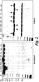
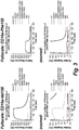
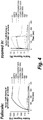
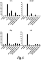


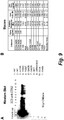
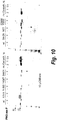
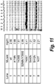
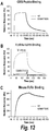


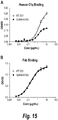
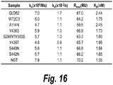

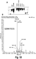

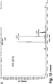
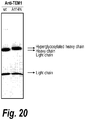
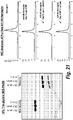
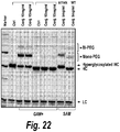
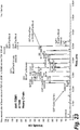


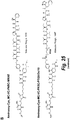


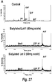
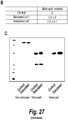

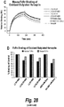

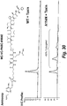


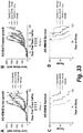
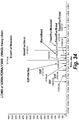
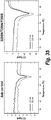

Classifications
-
- C—CHEMISTRY; METALLURGY
- C07—ORGANIC CHEMISTRY
- C07K—PEPTIDES
- C07K16/00—Immunoglobulins [IGs], e.g. monoclonal or polyclonal antibodies
-
- C—CHEMISTRY; METALLURGY
- C07—ORGANIC CHEMISTRY
- C07K—PEPTIDES
- C07K16/00—Immunoglobulins [IGs], e.g. monoclonal or polyclonal antibodies
- C07K16/18—Immunoglobulins [IGs], e.g. monoclonal or polyclonal antibodies against material from animals or humans
- C07K16/28—Immunoglobulins [IGs], e.g. monoclonal or polyclonal antibodies against material from animals or humans against receptors, cell surface antigens or cell surface determinants
- C07K16/2803—Immunoglobulins [IGs], e.g. monoclonal or polyclonal antibodies against material from animals or humans against receptors, cell surface antigens or cell surface determinants against the immunoglobulin superfamily
- C07K16/2809—Immunoglobulins [IGs], e.g. monoclonal or polyclonal antibodies against material from animals or humans against receptors, cell surface antigens or cell surface determinants against the immunoglobulin superfamily against the T-cell receptor (TcR)-CD3 complex
-
- A—HUMAN NECESSITIES
- A61—MEDICAL OR VETERINARY SCIENCE; HYGIENE
- A61K—PREPARATIONS FOR MEDICAL, DENTAL OR TOILETRY PURPOSES
- A61K39/00—Medicinal preparations containing antigens or antibodies
-
- A—HUMAN NECESSITIES
- A61—MEDICAL OR VETERINARY SCIENCE; HYGIENE
- A61K—PREPARATIONS FOR MEDICAL, DENTAL OR TOILETRY PURPOSES
- A61K47/00—Medicinal preparations characterised by the non-active ingredients used, e.g. carriers or inert additives; Targeting or modifying agents chemically bound to the active ingredient
- A61K47/50—Medicinal preparations characterised by the non-active ingredients used, e.g. carriers or inert additives; Targeting or modifying agents chemically bound to the active ingredient the non-active ingredient being chemically bound to the active ingredient, e.g. polymer-drug conjugates
- A61K47/51—Medicinal preparations characterised by the non-active ingredients used, e.g. carriers or inert additives; Targeting or modifying agents chemically bound to the active ingredient the non-active ingredient being chemically bound to the active ingredient, e.g. polymer-drug conjugates the non-active ingredient being a modifying agent
- A61K47/68—Medicinal preparations characterised by the non-active ingredients used, e.g. carriers or inert additives; Targeting or modifying agents chemically bound to the active ingredient the non-active ingredient being chemically bound to the active ingredient, e.g. polymer-drug conjugates the non-active ingredient being a modifying agent the modifying agent being an antibody, an immunoglobulin or a fragment thereof, e.g. an Fc-fragment
- A61K47/6801—Drug-antibody or immunoglobulin conjugates defined by the pharmacologically or therapeutically active agent
- A61K47/6803—Drugs conjugated to an antibody or immunoglobulin, e.g. cisplatin-antibody conjugates
-
- A—HUMAN NECESSITIES
- A61—MEDICAL OR VETERINARY SCIENCE; HYGIENE
- A61P—SPECIFIC THERAPEUTIC ACTIVITY OF CHEMICAL COMPOUNDS OR MEDICINAL PREPARATIONS
- A61P29/00—Non-central analgesic, antipyretic or antiinflammatory agents, e.g. antirheumatic agents; Non-steroidal antiinflammatory drugs [NSAID]
-
- A—HUMAN NECESSITIES
- A61—MEDICAL OR VETERINARY SCIENCE; HYGIENE
- A61P—SPECIFIC THERAPEUTIC ACTIVITY OF CHEMICAL COMPOUNDS OR MEDICINAL PREPARATIONS
- A61P35/00—Antineoplastic agents
-
- A—HUMAN NECESSITIES
- A61—MEDICAL OR VETERINARY SCIENCE; HYGIENE
- A61P—SPECIFIC THERAPEUTIC ACTIVITY OF CHEMICAL COMPOUNDS OR MEDICINAL PREPARATIONS
- A61P37/00—Drugs for immunological or allergic disorders
-
- A—HUMAN NECESSITIES
- A61—MEDICAL OR VETERINARY SCIENCE; HYGIENE
- A61P—SPECIFIC THERAPEUTIC ACTIVITY OF CHEMICAL COMPOUNDS OR MEDICINAL PREPARATIONS
- A61P37/00—Drugs for immunological or allergic disorders
- A61P37/02—Immunomodulators
-
- A—HUMAN NECESSITIES
- A61—MEDICAL OR VETERINARY SCIENCE; HYGIENE
- A61P—SPECIFIC THERAPEUTIC ACTIVITY OF CHEMICAL COMPOUNDS OR MEDICINAL PREPARATIONS
- A61P37/00—Drugs for immunological or allergic disorders
- A61P37/02—Immunomodulators
- A61P37/06—Immunosuppressants, e.g. drugs for graft rejection
-
- C—CHEMISTRY; METALLURGY
- C07—ORGANIC CHEMISTRY
- C07K—PEPTIDES
- C07K16/00—Immunoglobulins [IGs], e.g. monoclonal or polyclonal antibodies
- C07K16/18—Immunoglobulins [IGs], e.g. monoclonal or polyclonal antibodies against material from animals or humans
- C07K16/28—Immunoglobulins [IGs], e.g. monoclonal or polyclonal antibodies against material from animals or humans against receptors, cell surface antigens or cell surface determinants
- C07K16/2851—Immunoglobulins [IGs], e.g. monoclonal or polyclonal antibodies against material from animals or humans against receptors, cell surface antigens or cell surface determinants against the lectin superfamily, e.g. CD23, CD72
-
- C—CHEMISTRY; METALLURGY
- C07—ORGANIC CHEMISTRY
- C07K—PEPTIDES
- C07K16/00—Immunoglobulins [IGs], e.g. monoclonal or polyclonal antibodies
- C07K16/18—Immunoglobulins [IGs], e.g. monoclonal or polyclonal antibodies against material from animals or humans
- C07K16/28—Immunoglobulins [IGs], e.g. monoclonal or polyclonal antibodies against material from animals or humans against receptors, cell surface antigens or cell surface determinants
- C07K16/2893—Immunoglobulins [IGs], e.g. monoclonal or polyclonal antibodies against material from animals or humans against receptors, cell surface antigens or cell surface determinants against CD52
-
- C—CHEMISTRY; METALLURGY
- C07—ORGANIC CHEMISTRY
- C07K—PEPTIDES
- C07K16/00—Immunoglobulins [IGs], e.g. monoclonal or polyclonal antibodies
- C07K16/18—Immunoglobulins [IGs], e.g. monoclonal or polyclonal antibodies against material from animals or humans
- C07K16/32—Immunoglobulins [IGs], e.g. monoclonal or polyclonal antibodies against material from animals or humans against translation products of oncogenes
-
- C—CHEMISTRY; METALLURGY
- C07—ORGANIC CHEMISTRY
- C07K—PEPTIDES
- C07K16/00—Immunoglobulins [IGs], e.g. monoclonal or polyclonal antibodies
- C07K16/40—Immunoglobulins [IGs], e.g. monoclonal or polyclonal antibodies against enzymes
-
- A—HUMAN NECESSITIES
- A61—MEDICAL OR VETERINARY SCIENCE; HYGIENE
- A61K—PREPARATIONS FOR MEDICAL, DENTAL OR TOILETRY PURPOSES
- A61K39/00—Medicinal preparations containing antigens or antibodies
- A61K2039/505—Medicinal preparations containing antigens or antibodies comprising antibodies
-
- C—CHEMISTRY; METALLURGY
- C07—ORGANIC CHEMISTRY
- C07K—PEPTIDES
- C07K2317/00—Immunoglobulins specific features
- C07K2317/40—Immunoglobulins specific features characterized by post-translational modification
- C07K2317/41—Glycosylation, sialylation, or fucosylation
-
- C—CHEMISTRY; METALLURGY
- C07—ORGANIC CHEMISTRY
- C07K—PEPTIDES
- C07K2317/00—Immunoglobulins specific features
- C07K2317/50—Immunoglobulins specific features characterized by immunoglobulin fragments
- C07K2317/52—Constant or Fc region; Isotype
-
- C—CHEMISTRY; METALLURGY
- C07—ORGANIC CHEMISTRY
- C07K—PEPTIDES
- C07K2317/00—Immunoglobulins specific features
- C07K2317/70—Immunoglobulins specific features characterized by effect upon binding to a cell or to an antigen
- C07K2317/71—Decreased effector function due to an Fc-modification
-
- C—CHEMISTRY; METALLURGY
- C07—ORGANIC CHEMISTRY
- C07K—PEPTIDES
- C07K2317/00—Immunoglobulins specific features
- C07K2317/90—Immunoglobulins specific features characterized by (pharmaco)kinetic aspects or by stability of the immunoglobulin
- C07K2317/92—Affinity (KD), association rate (Ka), dissociation rate (Kd) or EC50 value
Definitions
- Antibodies with reduced or abolished Fc glycosylation have been employed for the treatment of inflammatory and autoimmune diseases or disorders in order to reduce side effects or toxicity associated with unwanted effector function (see e.g., Chan and Carter, Nat. Reviews Immunology, 2010 ).
- antibody Fc domain glycosylation is important for antibody structure, stability, and function and aglycosylation can result in antibodies with poor biophysical properties. Accordingly, there is a need in the art for engineered binding proteins with reduced effector function but which also retain the desirable properties of a glycosylated Fc domain.
- binding polypeptides e.g., antibodies or fusion
- optionally drug conjugates thereof comprising an Fc domain with altered glycosylation and reduced effector function.
- the Fc domain comprises: an asparagine residue at amino acid position 298, according to EU numbering; and a serine or threonine residue at amino acid position 300, according to EU numbering.
- the current disclosure also provides nucleic acids encoding the antigen-binding polypeptides, recombinant expression vectors and host cells for making such antigen-binding polypeptides. Methods of using the antigen-binding polypeptides disclosed herein to treat disease are also provided.
- the binding polypeptides e.g., antibodies
- the engineered N-linked glycosylation site at amino acid position 298 can also be used as a site for conjugation of effector moieties, such as cytotoxic drugs.
- the application describes an isolated binding polypeptide comprising an Fc domain with altered glycosylation, wherein the Fc domain comprises: an asparagine residue at amino acid position 298, according to EU numbering; and a serine or threonine residue at amino acid position 300, according to EU numbering, and wherein the binding polypeptide exhibits reduced effector function due to said altered glycosylation.
- the binding polypeptide further comprises an alanine residue at amino acid position 299, according to EU numbering.
- the binding polypeptide further comprises a glutamine residue at amino acid position 297, according to EU numbering.
- the Fc domain is an IgG1 Fc domain.
- the Fc domain is human.
- the side chain of the asparagine residue is linked to a glycan though a ⁇ -glycosylamide linkage.
- the glycan is a biantennary glycan.
- the glycan is a naturally occurring mammalian glycoform.
- the binding polypeptide has a lower affinity for an Fc ⁇ receptor than a binding polypeptide having a native Fc domain.
- the Fc ⁇ receptor is Fc ⁇ RI and/or Fc ⁇ RIIIa.
- the binding polypeptide has a similar affinity for an FcRn receptor as a binding polypeptide having a native Fc domain.
- the glycan comprises a reactive aldehyde group. In another embodiment, the glycan comprises an oxidized saccharide residue comprising a reactive aldehyde group. In another embodiment, the oxidized saccharide residue is a terminal sialic acid or galactose.
- the glycan is linked to an effector moiety.
- the effector moiety is a cytotoxin.
- the cytotoxin is selected from the group of cytotoxins listed in Table 1.
- the effector moiety is a detection agent.
- the effector moiety is linked through an oxime or hydrazone linkage to a saccharide residue of the glycan.
- the saccharide residue is a terminal sialic acid or galactose residue of the glycan.
- the effector moiety comprises a pH-sensitive linker, disulfide linker, enzyme-senstive linker or other cleavable linker moiety.
- the effector moiety comprises a linker moiety selected from the group of linker moieties depicted in Table 2 or 14. Further disclosed is a binding polypeptide which is an antibody or immunoadhesin.
- the application describes an isolated binding polypeptide comprising an Fc domain, wherein the Fc domain comprises: a free asparagine residue at amino acid position 298, according to EU numbering; and a free serine or threonine residue at amino acid position 300, according to EU numbering.
- the application describes an isolated binding polypeptide comprising an Fc domain, wherein the Fc domain comprises: a modified asparagine residue at amino acid position 298, according to EU numbering; and a free serine or threonine residue at amino acid position 300, according to EU numbering.
- the effector moiety is linked through a side chain of the modified asparagine residue to a saccharide residue of a glycan.
- the saccharide is a terminal sialic acid or galactose residue of the glycan.
- the effector moiety is linked through an oxime or hydrazone linkage to saccharide residue of the glycan.
- the saccharide is a terminal sialic acid or galactose residue of the glycan.
- the modified asparagine residue is linked to a drug effector moiety to form an antibody drug conjugate (ADC).
- ADC antibody drug conjugate
- composition comprises a binding polypeptide of any one of the preceding claims and a pharmaceutically acceptable carrier or excipient.
- the application describes treating a patient in thereof comprising administering an effective amount of the composition of the invention.
- the application describes an isolated polynucleotide encoding the binding polypeptide of the invention.
- the application describes a vector comprising the polynucleotide or a host cell comprising the polynucleotide or vector.
- the application describes a method of making a binding polypeptide comprising expressing the polynucleotide or vector in a cell.
- the current disclosure provides binding polypeptides (e.g., antibodies), and drug conjugates thereof, comprising an Fc domain, wherein the Fc domain comprises: an asparagine residue at amino acid position 298, according to EU numbering; and a serine or threonine residue at amino acid position 300, according to EU numbering.
- the current disclosure also provides nucleic acids encoding the antigen-binding polypeptides, recombinant expression vectors and host cells for making such antigen-binding polypeptides. Methods of using the antigen-binding polypeptides disclosed herein to treat disease are also provided.
- binding polypeptide or “binding polypeptide” shall refer to a polypeptide (e.g., an antibody) that contains at least one binding site which is responsible for selectively binding to a target antigen of interest (e.g. a human antigen).
- exemplary binding sites include an antibody variable domain, a ligand binding site of a receptor, or a receptor binding site of a ligand.
- the binding polypeptides of the invention comprise multiple (e.g., two, three, four, or more) binding sites.
- the term “native residue” shall refer to an amino acid residue that occurs naturally at a particular amino acid position of a binding polypeptide (e.g., an antibody or fragment thereof) and which has not been modified, introduced, or altered by the hand of man.
- a binding polypeptide e.g., an antibody or fragment thereof
- altered binding polypeptide or “altered binding polypeptide” includes binding polypeptides (e.g., an antibody or fragment thereof) comprising at least one non-native mutated amino acid residue.
- the term "specifically binds" as used herein, refers to the ability of an antibody or an antigen-binding fragment thereof to bind to an antigen with a dissociation constant (Kd) of at most about 1 x 10 -6 M, 1 x 10 -7 M, 1 x 10 -8 M, 1 x 10 -9 M, 1 x 10 -10 M, 1 x 10 -11 M, 1 x 10 -12 M, or less, and/or to bind to an antigen with an affinity that is at least two-fold greater than its affinity for a nonspecific antigen.
- Kd dissociation constant
- antibody refers to such assemblies (e.g., intact antibody molecules, antibody fragments, or variants thereof) which have significant known specific immunoreactive activity to an antigen of interest (e.g. a tumor associated antigen).
- Antibodies and immunoglobulins comprise light and heavy chains, with or without an interchain covalent linkage between them. Basic immunoglobulin structures in vertebrate systems are relatively well understood.
- immunoglobulins comprise two identical light chains of molecular weight approximately 23,000 Daltons, and two identical heavy chains of molecular weight 53,000-70,000. The four chains are joined by disulfide bonds in a "Y" configuration wherein the light chains bracket the heavy chains starting at the mouth of the "Y” and continuing through the variable region.
- Light chains of immunoglobulin are classified as either kappa or lambda ( ⁇ , ⁇ ). Each heavy chain class may be bound with either a kappa or lambda light chain.
- the light and heavy chains are covalently bonded to each other, and the "tail" portions of the two heavy chains are bonded to each other by covalent disulfide linkages or non-covalent linkages when the immunoglobulins are generated either by hybridomas, B cells, or genetically engineered host cells.
- the amino acid sequences run from an N-terminus at the forked ends of the Y configuration to the C-terminus at the bottom of each chain.
- heavy chains are classified as gamma, mu, alpha, delta, or epsilon, ( ⁇ , ⁇ , ⁇ , ⁇ , ⁇ ) with some subclasses among them (e.g., ⁇ 1- ⁇ 4). It is the nature of this chain that determines the "class" of the antibody as IgG, IgM, IgA IgG, or IgE, respectively.
- the immunoglobulin isotype subclasses e.g., IgG1, IgG2, IgG3, IgG4, IgA1, etc.
- Modified versions of each of these classes and isotypes are readily discernable to the skilled artisan in view of the instant disclosure and, accordingly, are within the scope of the current disclosure.
- region refers to a part or portion of an immunoglobulin or antibody chain and includes constant region or variable regions, as well as more discrete parts or portions of said regions.
- light chain variable regions include "complementarity determining regions” or "CDRs" interspersed among "framework regions” or "FRs", as defined herein.
- the regions of an immunoglobulin heavy or light chain may be defined as “constant” (C) region or “variable” (V) regions, based on the relative lack of sequence variation within the regions of various class members in the case of a "constant region”, or the significant variation within the regions of various class members in the case of a “variable regions”.
- the terms "constant region” and “variable region” may also be used functionally.
- the variable regions of an immunoglobulin or antibody determine antigen recognition and specificity.
- the constant regions of an immunoglobulin or antibody confer important effector functions such as secretion, transplacental mobility, Fc receptor binding, complement binding, and the like.
- the subunit structures and three dimensional configurations of the constant regions of the various immunoglobulin classes are well known.
- domains refers to a globular region of a heavy or light chain comprising peptide loops (e.g., comprising 3 to 4 peptide loops) stabilized, for example, by ⁇ -pleated sheet and/or intrachain disulfide bond.
- Constant region domains on the light chain of an immunoglobulin are referred to interchangeably as "light chain constant region domains", “CL regions” or “CL domains”.
- Constant domains on the heavy chain e.g. hinge, CH1, CH2 or CH3 domains
- H heavy chain constant region domains
- Variable domains on the light chain are referred to interchangeably as “light chain variable region domains", “VL region domains or “VL domains”.
- Variable domains on the heavy chain are referred to interchangeably as “heavy chain variable region domains", “VH region domains” or “VH domains”.
- variable constant region domains By convention the numbering of the variable constant region domains increases as they become more distal from the antigen binding site or amino-terminus of the immunoglobulin or antibody.
- the N-terminus of each heavy and light immunoglobulin chain is a variable region and at the C-terminus is a constant region; the CH3 and CL domains actually comprise the carboxy-terminus of the heavy and light chain, respectively. Accordingly, the domains of a light chain immunoglobulin are arranged in a VL-CL orientation, while the domains of the heavy chain are arranged in the VH-CH1-hinge-CH2-CH3 orientation.
- Amino acid positions in a heavy chain constant region including amino acid positions in the CH1, hinge, CH2, CH3, and CL domains, may be numbered according to the Kabat index numbering system (see Kabat et al, in "Sequences of Proteins of Immunological Interest", U.S. Dept. Health and Human Services, 5th edition, 1991 ).
- antibody amino acid positions may be numbered according to the EU index numbering system (see Kabat et al, ibid).
- VH domain includes the amino terminal variable domain of an immunoglobulin heavy chain
- VL domain includes the amino terminal variable domain of an immunoglobulin light chain
- CH1 domain includes the first (most amino terminal) constant region domain of an immunoglobulin heavy chain that extends, e.g., from about positions 114-223 in the Kabat numbering system (EU positions 118-215).
- the CH1 domain is adjacent to the VH domain and amino terminal to the hinge region of an immunoglobulin heavy chain molecule, and does not form a part of the Fc region of an immunoglobulin heavy chain.
- Hinge region includes the portion of a heavy chain molecule that joins the CH1 domain to the CH2 domain. This hinge region comprises approximately 25 residues and is flexible, thus allowing the two N-terminal antigen binding regions to move independently. Hinge regions can be subdivided into three distinct domains: upper, middle, and lower hinge domains ( Roux et al. J. Immunol. 1998, 161: 4083 ).
- CH2 domain includes the portion of a heavy chain immunoglobulin molecule that extends, e.g., from about positions 244-360 in the Kabat numbering system (EU positions 231-340).
- the CH2 domain is unique in that it is not closely paired with another domain. Rather, two N-linked branched carbohydrate chains are interposed between the two CH2 domains of an intact native IgG molecule.
- a binding polypeptide of the current disclosure comprises a CH2 domain derived from an IgG1 molecule (e.g. a human IgG1 molecule).
- CH3 domain includes the portion of a heavy chain immunoglobulin molecule that extends approximately 110 residues from N-terminus of the CH2 domain, e.g., from about positions 361-476 of the Kabat numbering system (EU positions 341 -445).
- the CH3 domain typically forms the C-terminal portion of the antibody.
- additional domains may extend from CH3 domain to form the C-terminal portion of the molecule (e.g. the CH4 domain in the ⁇ chain of IgM and the e chain of IgE).
- a binding polypeptide of the current disclosure comprises a CH3 domain derived from an IgG1 molecule (e.g. a human IgG1 molecule).
- CL domain includes the constant region domain of an immunoglobulin light chain that extends, e.g. from about Kabat position 107A-216.
- the CL domain is adjacent to the VL domain.
- a binding polypeptide of the current disclosure comprises a CL domain derived from a kappa light chain (e.g., a human kappa light chain).
- Fc region is defined as the portion of a heavy chain constant region beginning in the hinge region just upstream of the papain cleavage site (i.e. residue 216 in IgG, taking the first residue of heavy chain constant region to be 114) and ending at the C-terminus of the antibody. Accordingly, a complete Fc region comprises at least a hinge domain, a CH2 domain, and a CH3 domain.
- native Fc refers to a molecule comprising the sequence of a non-antigen-binding fragment resulting from digestion of an antibody or produced by other means, whether in monomeric or multimeric form, and can contain the hinge region.
- the original immunoglobulin source of the native Fc is preferably of human origin and can be any of the immunoglobulins, although IgG1 and IgG2 are preferred.
- Native Fc molecules are made up of monomeric polypeptides that can be linked into dimeric or multimeric forms by covalent (i.e., disulfide bonds) and non-covalent association.
- the number of intermolecular disulfide bonds between monomeric subunits of native Fc molecules ranges from 1 to 4 depending on class (e.g., IgG, IgA, and IgE) or subclass (e.g., IgG1, IgG2, IgG3, IgA1, and IgGA2).
- class e.g., IgG, IgA, and IgE
- subclass e.g., IgG1, IgG2, IgG3, IgA1, and IgGA2
- One example of a native Fc is a disulfide-bonded dimer resulting from papain digestion of an IgG.
- native Fc as used herein is generic to the monomeric, dimeric, and multimeric forms.
- Fc variant refers to a molecule or sequence that is modified from a native Fc but still comprises a binding site for the salvage receptor, FcRn (neonatal Fc receptor). Exemplary Fc variants, and their interaction with the salvage receptor, are known in the art. Thus, the term “Fc variant” can comprise a molecule or sequence that is humanized from a non-human native Fc. Furthermore, a native Fc comprises regions that can be removed because they provide structural features or biological activity that are not required for the antibody-like binding polypeptides of the invention.
- Fc variant comprises a molecule or sequence that lacks one or more native Fc sites or residues, or in which one or more Fc sites or residues has be modified, that affect or are involved in: (1) disulfide bond formation, (2) incompatibility with a selected host cell, (3) N-terminal heterogeneity upon expression in a selected host cell, (4) glycosylation, (5) interaction with complement, (6) binding to an Fc receptor other than a salvage receptor, or (7) antibody-dependent cellular cytotoxicity (ADCC).
- ADCC antibody-dependent cellular cytotoxicity
- Fc domain encompasses native Fc and Fc variants and sequences as defined above. As with Fc variants and native Fc molecules, the term “Fc domain” includes molecules in monomeric or multimeric form, whether digested from whole antibody or produced by other means.
- variable regions of an antibody allow it to selectively recognize and specifically bind epitopes on antigens. That is, the VL domain and VH domain of an antibody combine to form the variable region (Fv) that defines a three dimensional antigen binding site.
- This quaternary antibody structure forms the antigen binding site present at the end of each arm of the Y. More specifically, the antigen binding site is defined by three complementary determining regions (CDRs) on each of the heavy and light chain variable regions.
- CDRs complementary determining regions
- the term "antigen binding site” includes a site that specifically binds (immunoreacts with) an antigen (e.g., a cell surface or soluble antigen).
- the antigen binding site includes an immunoglobulin heavy chain and light chain variable region and the binding site formed by these variable regions determines the specificity of the antibody.
- An antigen binding site is formed by variable regions that vary from one antibody to another.
- the altered antibodies of the current disclosure comprise at least one antigen binding site.
- binding polypeptides of the current disclosure comprise at least two antigen binding domains that provide for the association of the binding polypeptide with the selected antigen.
- the antigen binding domains need not be derived from the same immunoglobulin molecule.
- the variable region may or be derived from any type of animal that can be induced to mount a humoral response and generate immunoglobulins against the desired antigen.
- variable region of the a binding polypeptide may be, for example, of mammalian origin e.g., may be human, murine, rat, goat, sheep, non-human primate (such as cynomolgus monkeys, macaques, etc.), lupine, or camelid (e.g., from camels, llamas and related species).
- mammalian origin e.g., may be human, murine, rat, goat, sheep, non-human primate (such as cynomolgus monkeys, macaques, etc.), lupine, or camelid (e.g., from camels, llamas and related species).
- the six CDRs present on each monomeric antibody are short, non-contiguous sequences of amino acids that are specifically positioned to form the antigen binding site as the antibody assumes its three dimensional configuration in an aqueous environment.
- the remainder of the heavy and light variable domains show less intermolecular variability in amino acid sequence and are termed the framework regions.
- the framework regions largely adopt a ⁇ -sheet conformation and the CDRs form loops which connect, and in some cases form part of, the ⁇ -sheet structure. Thus, these framework regions act to form a scaffold that provides for positioning the six CDRs in correct orientation by inter-chain, non-covalent interactions.
- the antigen binding domain formed by the positioned CDRs defines a surface complementary to the epitope on the immunoreactive antigen. This complementary surface promotes the non-covalent binding of the antibody to the immunoreactive antigen epitope.
- Exemplary binding polypeptides of the invention include antibody variants.
- antibody variant includes synthetic and engineered forms of antibodies which are altered such that they are not naturally occurring, e.g., antibodies that comprise at least two heavy chain portions but not two complete heavy chains (such as, domain deleted antibodies or minibodies); multispecific forms of antibodies (e.g., bispecific, trispecific, etc.) altered to bind to two or more different antigens or to different epitopes on a single antigen); heavy chain molecules joined to scFv molecules and the like.
- antibody variant includes multivalent forms of antibodies (e.g., trivalent, tetravalent, etc., antibodies that bind to three, four or more copies of the same antigen.
- valency refers to the number of potential target binding sites in a polypeptide.
- Each target binding site specifically binds one target molecule or specific site on a target molecule.
- each target binding site may specifically bind the same or different molecules (e.g., may bind to different ligands or different antigens, or different epitopes on the same antigen).
- the subject binding polypeptides preferably have at least one binding site specific for a human antigen molecule.
- binding polypeptide refers to the ability to specifically bind (e.g., immunoreact with) a given target antigen (e.g., a human target antigen).
- a binding polypeptide may be monospecific and contain one or more binding sites which specifically bind a target or a polypeptide may be multispecific and contain two or more binding sites which specifically bind the same or different targets.
- a binding polypeptide of the invention is specific for two different (e.g., non-overlapping) portions of the same target.
- a binding polypeptide of the invention is specific for more than one target.
- Exemplary binding polypeptides e.g., antibodies which comprise antigen binding sites that bind to antigens expressed on tumor cells are known in the art and one or more CDRs from such antibodies can be included in an antibody of the invention.
- linking moiety includes moieties which are capable of linking the effector moiety to the binding polypeptides disclosed herein.
- the linking moiety may be selected such that it is cleavable (e.g., enzymatically cleavable or pH-sensitive) or non-cleavable.
- Exemplary linking moieties are set forth in Table 2 herein.
- effector moiety comprises diagnostic and therapeutic agents (e.g. proteins, nucleic acids, lipids, drug moieties, and fragments thereof) with biological or other functional activity.
- a modified binding polypeptide comprising an effector moiety conjugated to a binding polypeptide has at least one additional function or property as compared to the unconjugated antibody.
- the conjugation of a cytotoxic drug (e.g., an effector moiety) to binding polypeptide results in the formation of a binding polypeptide with drug cytotoxicity as second function (i.e. in addition to antigen binding).
- the conjugation of a second binding polypeptide to the binding polypeptide may confer additional binding properties.
- the effector moiety may be synthesized or expressed by either peptide synthesis or recombinant DNA methods that are well known in the art.
- the effector may be synthesized artificially or purified from a natural source.
- drug moiety includes anti-inflammatory, anticancer, anti-infective (e.g., anti-fungal, antibacterial, anti-parasitic, anti-viral, etc.), and anesthetic therapeutic agents.
- the drug moiety is an anticancer or cytotoxic agent.
- Compatible drug moieties may also comprise prodrugs. Exemplary effector moieties are set forth in Table 1 herein.
- prodrug refers to a precursor or derivative form of a pharmaceutically active agent that is less active, reactive or prone to side effects as compared to the parent drug and is capable of being enzymatically activated or otherwise converted into a more active form in vivo.
- Prodrugs compatible with the compositions of the current disclosure include, but are not limited to, phosphate-containing prodrugs, amino acid-containing prodrugs, thiophosphate-containing prodrugs, sulfate-containing prodrugs, peptide-containing prodrugs, ⁇ -lactam-containing prodrugs, optionally substituted phenoxyacetamide-containing prodrugs or optionally substituted phenylacetamide-containing prodrugs, 5-fluorocytosine and other 5-fluorouridine prodrugs that can be converted to the more active cytotoxic free drug.
- One skilled in the art may make chemical modifications to the desired drug moiety or its prodrug in order to make reactions of that compound more convenient for purposes of preparing modified binding polypeptides of the current disclosure.
- the drug moieties also include derivatives, pharmaceutically acceptable salts, esters, amides, and ethers of the drug moieties described herein.
- Derivatives include modifications to drugs identified herein which may improve or not significantly reduce a particular drug's desired therapeutic activity.
- anticancer agent includes agents which are detrimental to the growth and/or proliferation of neoplastic or tumor cells and may act to reduce, inhibit or destroy malignancy.
- agents include, but are not limited to, cytostatic agents, alkylating agents, antibiotics, cytotoxic nucleosides, tubulin binding agents, hormones, hormone antagonists, cytotoxic agents, and the like.
- Cytotoxic agents include tomaymycin derivatives, maytansine derivatives, cryptophycine derivatives, anthracycline derivatives, bisphosphonate derivatives, leptomycin derivatives, streptonigrin derivatives, auristatine derivatives, and duocarmycin derivatives. Any agent that acts to retard or slow the growth of immunoreactive cells or malignant cells is within the scope of the current disclosure.
- antigen or "target antigen” as used herein refers to a molecule or a portion of a molecule that is capable of being bound by the binding site of a binding polypeptide.
- a target antigen may have one or more epitopes.
- binding polypeptides e.g., antibodies, antibody fragments, antibody variants, and fusion proteins
- Fc domain comprises: an asparagine residue at amino acid position 298, according to EU numbering; and a serine or threonine residue at amino acid position 300, according to EU numbering.
- Fc domains from any immunoglobulin class e.g., IgM, IgG, IgD, IgA and IgE
- species can be used in the binding polypeptides disclosed herein.
- Chimeric Fc domains comprising portions of Fc domains from different species or Ig classes can also be employed.
- the Fc domain is a human IgG1 Fc domain.
- mutation of the wild type amino acid at Kabat position 298 to an asparagine and Kabat position 300 to a serine or threonine results in the formation of an N-linked glycosylation consensus site (i.e, the N-X-T/S sequon, where X is any amino acid except proline).
- binding polypeptides disclosed herein encompass any binding polypeptide that comprises an Fc domain having an N-linked glycosylation site at position 298, according to Kabat numbering.
- the binding polypeptide is an antibody, or fragment or derivative thereof. Any antibody from any source or species can be employed in the binding polypeptides disclosed herein. Suitable antibodies include without limitation, human antibodies, humanized antibodies or chimeric antibodies.
- the binding polypeptide of the current disclosure may comprise an antigen binding fragment of an antibody.
- antigen-binding fragment refers to a polypeptide fragment of an immunoglobulin or antibody which binds antigen or competes with intact antibody ⁇ i.e., with the intact antibody from which they were derived) for antigen binding ⁇ i.e., specific binding).
- Antigen binding fragments can be produced by recombinant or biochemical methods that are well known in the art. Exemplary antigen-binding fragments include Fv, Fab, Fab', and (Fab')2.
- the antigen-binding fragment of the current disclosure is an altered antigen-binding fragment comprising at least one engineered glycosylation site.
- an altered antigen binding fragment of the current disclosure comprises an altered VH domain described supra.
- an altered antigen binding fragment of the current disclosure comprises an altered CH1 domain described supra.
- the binding polypeptide comprises a single chain variable region sequence (ScFv).
- Single chain variable region sequences comprise a single polypeptide having one or more antigen binding sites, e.g., a VL domain linked by a flexible linker to a VH domain.
- ScFv molecules can be constructed in a VH-linker-VL orientation or VL-linker-VH orientation.
- the flexible hinge that links the VL and VH domains that make up the antigen binding site preferably comprises from about 10 to about 50 amino acid residues. Connecting peptides are known in the art.
- Binding polypeptide of the invention may comprise at least one scFv and/or at least one constant region.
- a binding polypeptide of the current disclosure may comprise at least one scFv linked or fused to an antibody or fragment comprising an CH1 domain (e.g. a CH1 domain comprising an asparagine residue at Kabat position 114) and/or CH2 domain (e.g. a CH2 domain comprising an asparagine residue at EU position 298, and a serine or threonine residue at EU position 300).
- an CH1 domain e.g. a CH1 domain comprising an asparagine residue at Kabat position 11
- CH2 domain e.g. a CH2 domain comprising an asparagine residue at EU position 298, and a serine or threonine residue at EU position 300.
- a binding polypeptide of the current disclosure is a multivalent (e.g., tetravalent) antibody which is produced by fusing a DNA sequence encoding an antibody with a ScFv molecule (e.g., an altered ScFv molecule).
- a DNA sequence encoding an antibody with a ScFv molecule e.g., an altered ScFv molecule
- these sequences are combined such that the ScFv molecule (e.g., an altered ScFv molecule) is linked at its N-terminus or C-terminus to an Fc fragment of an antibody via a flexible linker (e.g., a gly/ser linker).
- a tetravalent antibody of the current disclosure can be made by fusing an ScFv molecule to a connecting peptide, which is fused to a CH1 domain (e.g. a CH1 domain comprising an asparagine residue at Kabat position 114) to construct an ScFv-Fab tetravalent molecule.
- a CH1 domain e.g. a CH1 domain comprising an asparagine residue at Kabat position 114
- a binding polypeptide of the current disclosure is an altered minibody.
- Altered minibodies of the current disclosure are dimeric molecules made up of two polypeptide chains each comprising an ScFv molecule (e.g., an altered ScFv molecule comprising an altered VH domain described supra) which is fused to a CH3 domain or portion thereof via a connecting peptide.
- Minibodies can be made by constructing an ScFv component and connecting peptide-CH3 component using methods described in the art (see, e.g., US patent 5,837,821 or WO 94/09817A1 ).
- a tetravalent minibody can be constructed. Tetravalent minibodies can be constructed in the same manner as minibodies, except that two ScFv molecules are linked using a flexible linker. The linked scFv-scFv construct is then joined to a CH3 domain.
- a binding polypeptide of the current disclosure comprises a diabody.
- Diabodies are dimeric, tetravalent molecules each having a polypeptide similar to scFv molecules, but usually having a short (less than 10 and preferably 1-5) amino acid residue linkers connecting both variable domains, such that the VL and VH domains on the same polypeptide chain cannot interact. Instead, the VL and VH domain of one polypeptide chain interact with the VH and VL domain (respectively) on a second polypeptide chain (see, for example, WO 02/02781 ). Diabodies of the current disclosure comprise an scFv molecule fused to a CH3 domain.
- the binding polypeptide s of the invention comprise multispecific or multivalent antibodies comprising one or more variable domain in series on the same polypeptide chain, e.g., tandem variable domain (TVD) polypeptides.
- TVD polypeptides include the "double head” or “Dual-Fv” configuration described in U.S. Patent No. 5,989,830 .
- variable domains of two different antibodies are expressed in a tandem orientation on two separate chains (one heavy chain and one light chain), wherein one polypeptide chain has two times a VH in series separated by a peptide linker (VH1-linker-VH2) and the other polypeptide chain consists of complementary VL domains connected in series by a peptide linker (VL1-linker-VL2).
- variable domains of two different antibodies are expressed in a tandem orientation on two separate polypeptide chains (one heavy chain and one light chain), wherein one polypeptide chain has two VH in series separated by a peptide linker (VH1-linker-VH2) and the other polypeptide chain consists of complementary VL domains connected in series by a peptide linker in the opposite orientation (VL2-linker-VL1).
- Additional antibody variants based on the "Dual-Fv" format include the Dual-Variable-Domain IgG (DVD-IgG) bispecific antibody (see U.S. Patent No. 7,612,181 and the TBTI format (see US 2010/0226923 A1 ).
- the binding polypeptide comprises a cross-over dual variable domain IgG (CODV-IgG) bispecific antibody based on a "double head" configuration (see US20120251541 A1 ).
- CODV-IgG antibody variants have one polypeptide chain with VL domains connected in series to a CL domain (VL1-L1-VL2-L2-CL) and a second polypeptide chain with complementary VH domains connected in series in the opposite orientation to a CH1 domain (VH2-L3-VH1-L4-CH1), where the polypeptide chains form a cross-over light chain-heavy chain pair.
- the second polypeptide may be further connected to an Fc domain (VH2-L3-VH1-L4-CH1-Fc).
- linker L3 is at least twice the length of linker L1 and/or linker L4 is at least twice the length of linker L2.
- L1 and L2 may be 1-3 amino acid residues in length
- L3 may be 2 to 6 amino acid residues in length
- L4 may be 4 to 7 amino acid residues in length.
- linkers include a single glycine (Gly) residue; a diglycine peptide (Gly-Gly); a tripeptide (Gly-Gly-Gly); a peptide with four glycine residues (Gly-Gly-Gly-Gly); a peptide with five glycine residues (Gly-Gly-Gly-Gly-Gly); a peptide with six glycine residues (Gly-Gly-Gly-Gly-Gly-Gly-Gly); a peptide with seven glycine residues (Gly-Gly-Gly-Gly-Gly-Gly-Gly-Gly); a peptide with eight glycine residues (Gly-Gly-Gly-Gly-Gly-Gly-Gly-Gly-Gly).
- amino acid residues may be used such as the peptide Gly-Gly-Gly-Gly-Ser and the peptide Gly-Gly-Gly-Gly-Ser-Gly-Gly-Gly-Gly-Gly-Ser.
- the binding polypeptide comprises an immunoadhesin molecule comprising a non-antibody binding region (e.g., a receptor, ligand, or cell-adhesion molecule) fused to an antibody constant region (see e.g., Ashkenazi et al., Methods, 1995 8(2), 104-115 )
- a non-antibody binding region e.g., a receptor, ligand, or cell-adhesion molecule
- the binding polypeptide comprises immunoglobulin-like domains.
- Suitable immunoglobulin-like domains include, without limitation, fibronectin domains (see, for example, Koide et al. (2007), Methods Mol. Biol. 352: 95-109 ), DARPin (see, for example, Stumpp et al. (2008) Drug Discov. Today 13 (15-16): 695-701 ), Z domains of protein A (see, Nygren et al. (2008) FEBS J. 275 (11): 2668-76 ), Lipocalins (see, for example, Skerra et al. (2008) FEBS J.
- the Fc domain of the binding polypeptides disclosed herein is glycosylated at the engineered arginine at position 298 (N298), according to EU numbering.
- the N-linked glycan is generally linked though a ⁇ -glycosylamide linkage to the nitrogen group of the N298 side chain.
- other suitable art recognized linkages can also be employed.
- the glycan may be a native glycan or an engineered glycan containing non-native linkages.
- the glycan comprises a saccharide that can be oxidized (e.g., by periodate treatment) to produce a group suitable for conjugation to an effector moiety (e.g., a reactive aldehyde group).
- an effector moiety e.g., a reactive aldehyde group
- Suitable oxidizable saccharides included, without limitation, galactose and sialic acid (e.g., N-Acetylneuraminic acid).
- the glycan is a biantennary glycan.
- the glycan is a naturally occurring mammalian glycoform.
- Glycosylation can be achieved through any means known in the art.
- the glycosylation is achieved by expression of the binding polypeptides in cells capable of N-linked glycosylation.
- Any natural or engineered cell e.g., prokaryotic or eukaryotic
- mammalian cells are employed to effect glycosylation.
- the N-glycans that are produced in mammalian cells are commonly referred to as complex N-glycans (see e.g., Drickamer K, Taylor ME (2006). Introduction to Glycobiology, 2nd ed .).
- a complex N-glycan has a structure with typically two to six outer branches with a sialyllactosamine sequence linked to an inner core structure Man 3 GlcNAc 2 .
- a complex N-glycan has at least one branch, and preferably at least two, of alternating GlcNAc and galactose (Gal) residues that terminate in oligosaccharides such as, for example: NeuNAc-; NeuAc ⁇ 2,6 GalNAc ⁇ 1-; NeuAc ⁇ 2,3 Gal ⁇ 1,3 GalNAc ⁇ 1-; and NeuAc ⁇ 2,3/6 Gal ⁇ 1,4 GlcNAc ⁇ 1.;
- sulfate esters can occur on galactose, GalNAc, and GlcNAc residues
- phosphate esters can occur on mannose residues.
- NeuAc can be O-acetylated or replaced by NeuGl (N-glycolylneuraminic acid).
- Complex N-glycans may also have intrachain substitutions of bisecting GlcNAc and core fucose (Fuc).
- glycosylation can be achieved or modified through enzymatic means, in vitro.
- one or more glycosyltransferases may be employed to add specific saccharide residues to N298, and one or more glycosidases may be employed to remove unwanted saccharides from the N-linked glycan.
- Such enzymatic means are well known in the art (see. e.g., WO/2007/005786 ).
- binding polypeptides may comprise an antibody constant region (e.g. an IgG constant region e.g., a human IgG constant region, e.g., a human IgG1 or IgG4 constant region) which mediates one or more effector functions.
- an antibody constant region e.g. an IgG constant region e.g., a human IgG constant region, e.g., a human IgG1 or IgG4 constant region
- binding of the C1 component of complement to an antibody constant region may activate the complement system. Activation of complement is important in the opsonisation and lysis of cell pathogens. The activation of complement also stimulates the inflammatory response and may also be involved in autoimmune hypersensitivity.
- antibodies bind to receptors on various cells via the Fc region, with a Fc receptor binding site on the antibody Fc region binding to a Fc receptor (FcR) on a cell.
- FcR Fc receptor
- Fc receptors which are specific for different classes of antibody, including IgG (gamma receptors), IgE (epsilon receptors), IgA (alpha receptors) and IgM (mu receptors). Binding of antibody to Fc receptors on cell surfaces triggers a number of important and diverse biological responses including engulfment and destruction of antibody-coated particles, clearance of immune complexes, lysis of antibody-coated target cells by killer cells (called antibody-dependent cell-mediated cytotoxicity, or ADCC), release of inflammatory mediators, placental transfer and control of immunoglobulin production.
- ADCC antibody-dependent cell-mediated cytotoxicity
- binding polypeptides e.g., antibodies or antigen binding fragments thereof
- binding polypeptides of the invention bind to an Fc-gamma receptor.
- binding polypeptides of the invention may comprise a constant region which is devoid of one or more effector functions (e.g., ADCC activity) and/or is unable to bind Fc ⁇ receptor.
- Certain embodiments of the invention include antibodies in which at least one amino acid in one or more of the constant region domains has been deleted or otherwise altered so as to provide desired biochemical characteristics such as reduced or enhanced effector functions, the ability to non-covalently dimerize, increased ability to localize at the site of a tumor, reduced serum half-life, or increased serum half-life when compared with a whole, unaltered antibody of approximately the same immunogenicity.
- certain antibodies for use in the diagnostic and treatment methods described herein are domain deleted antibodies which comprise a polypeptide chain similar to an immunoglobulin heavy chain, but which lack at least a portion of one or more heavy chain domains.
- one entire domain of the constant region of the modified antibody will be deleted, for example, all or part of the CH2 domain will be deleted.
- binding polypeptides comprise constant regions derived from different antibody isotypes (e.g., constant regions from two or more of a human IgG1).
- binding polypeptides comprises a chimeric hinge (i.e., a hinge comprising hinge portions derived from hinge domains of different antibody isotypes, e.g., an upper hinge domain from an IgG4 molecule and an IgG1 middle hinge domain).
- binding polypeptides comprise an Fc region or portion thereof from a human IgG4 molecule and a Ser228Pro mutation (EU numbering) in the core hinge region of the molecule.
- the Fc portion may be mutated to increase or decrease effector function using techniques known in the art.
- the deletion or inactivation (through point mutations or other means) of a constant region domain may reduce Fc receptor binding of the circulating modified antibody thereby increasing tumor localization.
- constant region modifications consistent with the instant invention moderate complement binding and thus reduce the serum half-life and nonspecific association of a conjugated cytotoxin.
- modifications of the constant region may be used to modify disulfide linkages or oligosaccharide moieties that allow for enhanced localization due to increased antigen specificity or flexibility.
- the resulting physiological profile, bioavailability and other biochemical effects of the modifications, such as tumor localization, biodistribution and serum half-life may easily be measured and quantified using well know immunological techniques without undue experimentation.
- an Fc domain employed in an antibody of the invention is an Fc variant.
- the term "Fc variant” refers to an Fc domain having at least one amino acid substitution relative to the wild-type Fc domain from which said Fc domain is derived.
- the Fc variant of said human IgG1 Fc domain comprises at least one amino acid substitution relative to said Fc domain.
- the amino acid substitution(s) of an Fc variant may be located at any position (i.e., any EU convention amino acid position) within the Fc domain.
- the Fc variant comprises a substitution at an amino acid position located in a hinge domain or portion thereof.
- the Fc variant comprises a substitution at an amino acid position located in a CH2 domain or portion thereof.
- the Fc variant comprises a substitution at an amino acid position located in a CH3 domain or portion thereof.
- the Fc variant comprises a substitution at an amino acid position located in a CH4 domain or portion thereof.
- the binding polypeptides of the invention may employ any art-recognized Fc variant which is known to impart an improvement (e.g., reduction or enhancement) in effector function and/or FcR binding.
- Said Fc variants may include, for example, any one of the amino acid substitutions disclosed in International PCT Publications WO88/07089A1 , WO96/14339A1 , WO98/05787A1 , WO98/23289A1 , WO99/51642A1 , WO99/58572A1 , WO00/09560A2 , WO00/32767A1 , WO00/42072A2 , WO02/44215A2 , WO02/060919A2 , WO03/074569A2 , WO04/016750A2 , WO04/029207A2 , WO04/035752A2 , WO04/063351A2 , WO04/07
- a binding polypeptide of the invention may comprise an Fc variant comprising an amino acid substitution at EU position 268 (e.g., H268D or H268E).
- a binding polypeptide of the invention may comprise an amino acid substitution at EU position 239 (e.g., S239D or S239E) and/or EU position 332 (e.g., I332D or I332Q).
- a binding polypeptide of the invention may comprise an Fc variant comprising an amino acid substitution which alters the antigen-independent effector functions of the antibody, in particular the circulating half-life of the binding polypeptide.
- Such binding polypeptides exhibit either increased or decreased binding to FcRn when compared to binding polypeptides lacking these substitutions, therefore, have an increased or decreased half-life in serum, respectively.
- Fc variants with improved affinity for FcRn are anticipated to have longer serum half-lives, and such molecules have useful applications in methods of treating mammals where long half-life of the administered antibody is desired, e.g., to treat a chronic disease or disorder.
- Fc variants with decreased FcRn binding affinity are expected to have shorter half-lives, and such molecules are also useful, for example, for administration to a mammal where a shortened circulation time may be advantageous, e.g. for in vivo diagnostic imaging or in situations where the starting antibody has toxic side effects when present in the circulation for prolonged periods.
- Fc variants with decreased FcRn binding affinity are also less likely to cross the placenta and, thus, are also useful in the treatment of diseases or disorders in pregnant women.
- other applications in which reduced FcRn binding affinity may be desired include those applications in which localization the brain, kidney, and/or liver is desired.
- the altered binding polypeptides (e.g., antibodies or antigen binding fragments thereof) of the invention exhibit reduced transport across the epithelium of kidney glomeruli from the vasculature.
- the altered binding polypeptides (e.g., antibodies or antigen binding fragments thereof) of the invention exhibit reduced transport across the blood brain barrier (BBB) from the brain, into the vascular space.
- BBB blood brain barrier
- an antibody with altered FcRn binding comprises an Fc domain having one or more amino acid substitutions within the "FcRn binding loop" of an Fc domain.
- the FcRn binding loop is comprised of amino acid residues 280-299 (according to EU numbering).
- the binding polypeptides (e.g., antibodies or antigen binding fragments thereof) of the invention comprise an Fc domain having one or more of the following substitutions: V284E, H285E, N286D, K290E and S304D (EU numbering).
- the biding molecules of the invention comprise a human Fc domain with the double mutation H433K/N434F (see, e.g., US Patent No. 8,163,881 ).
- binding polypeptides for use in the diagnostic and treatment methods described herein have a constant region, e.g., an IgG1 or IgG4 heavy chain constant region, which is altered to reduce or eliminate glycosylation.
- binding polypeptides (e.g., antibodies or antigen binding fragments thereof) of the invention may also comprise an Fc variant comprising an amino acid substitution which alters the glycosylation of the antibody Fc.
- said Fc variant may have reduced glycosylation (e.g., N- or O-linked glycosylation).
- the Fc variant comprises reduced glycosylation of the N-linked glycan normally found at amino acid position 297 (EU numbering).
- the antibody has an amino acid substitution near or within a glycosylation motif, for example, an N-linked glycosylation motif that contains the amino acid sequence NXT or NXS.
- the antibody comprises an Fc variant with an amino acid substitution at amino acid position 228 or 299 (EU numbering).
- the antibody comprises an IgG1 or IgG4 constant region comprising an S228P and a T299A mutation (EU numbering).
- the binding polypeptides of the current disclosure comprise effector moieties.
- these effector moieties are conjugated (either directly or through a linker moiety) to an N-linked glycan on the binding polypeptide, (e.g., an N-linked glycan linked to N298 (EU numbering) of the CH2 domain and/or N114 (Kabat numbering) of a CH1 domain).
- the binding polypeptide is full length antibody comprising two CH1 domains with a glycan at Kabat position 114, wherein both of the glycans are conjugated to one or more effector moieties.
- effector moiety can be added to the binding polypeptides disclosed herein.
- the effector moieties preferably add a non-natural function to an altered antibody or fragments thereof, without significantly altering the intrinsic activity of the binding polypeptide.
- the effector moiety may be, for example but not limited to, a therapeutic or diagnostic agent.
- a modified binding polypeptide (e.g., an antibody) of the current disclosure may comprise one or more effector moieties, which may by the same of different.
- the effector moiety can be of Formula (I): H 2 N-Q-CON-X Formula (I), wherein:
- the connector moiety connects the therapeutic agent to H 2 N-Q-.
- the connector moiety can include at least one of any suitable components known those skilled in the art, including, for example, an alkylenyl component, a polyethylene glycol component, a poly(glycine) component, a poly(oxazoline) component, a carbonyl component, a component derived from cysteinamide, a component derived from valine coupled with citruline, and a component derived from 4-aminobenzyl carbamate, or any combination thereof.
- the effector moiety of Formula (I) can be of Formula (Ia): H 2 N-Q-CH 2 -C(O)-Z-X Formula (Ia), wherein:
- the “component derived from cysteinamide” is the point of attachment to H 2 N-Q-CH 2 -C(O)-.
- the “component derived from cysteinamide” can refer to one or more portions of the effector moiety having the structure:
- the "Cys" component of an effector moiety may include one such portion.
- the following structure shows an effector moiety with one such portion (wherein the "Cys" component is indicated with the dotted line box):
- the "Cys" component of an effector moiety may include two or more such portions.
- the following moiety contains two such portions:
- each "Cys” component bears an -(MC) a -(VC) b -(PABC) c -(C 16 H 32 O 8 C 2 H 4 ) f -X group.
- the phrase "component derived from maleimide” can refer to any portion of the effector moiety having the structure: wherein d is an integer from 2 to 5.
- the number of MC components included in any Cys-(MC) a -(VC) b -(PABC) c -(C 16 H 32 O 8 C 2 H 4 ) f -X group in the effector moiety is indicated by subscript "a,” and can be 0 or 1 In one embodiment, a is 1. In another embodiment, b is 0.
- the "Cys” component can be connected to the "MC” component via the sulfur atom in the "Cys” component, as indicated with the dotted line box in the structure below:
- the phrase "component derived from valine coupled with citruline” can refer to any portion of the effector moiety with the following structure:
- b The number of VC components included in any Cys-(MC) a -(VC) b -(PABC) c -(C 16 H 32 O 8 C 2 H 4 ) f -X group in the effector moiety is indicated by subscript "b," and can be 0 or 1. In one embodiment, b is 1. In another embodiment, b is 0.
- the phrase "component derived from 4-aminobenzyl carbamate” can refer to any portion of the effector moiety with the following structure:
- c The number of PABC components included in any Cys-(MC) a -(VC) b -(PABC) c -(C 16 H 32 O 8 C 2 H 4 ) f -X group in the effector moiety is indicated by subscript "c," and can be 0 or 1. In one embodiment, c is 1. In another embodiment, c is 0.
- C 16 H 32 O 8 C 2 H 4 refers to the following structure:
- a is 1, b is 1, c is 1, and f is 0.
- the binding polypeptides of the current disclosure are conjugated to an effector moiety comprising a therapeutic agent, e.g. a drug moiety (or prodrug thereof) or radiolabeled compound.
- a therapeutic agent e.g. a drug moiety (or prodrug thereof) or radiolabeled compound.
- the therapeutic agent is a cytotoxin.
- exemplary drug moieties include anti-inflammatory, anticancer, anti-infective (e.g., anti-fungal, antibacterial, anti-parasitic, anti-viral, etc.), and anesthetic therapeutic agents.
- the drug moiety is an anticancer agent.
- anti-cancer agents include, but are not limited to, cytostatics, enzyme inhibitors, gene regulators, cytotoxic nucleosides, tubulin binding agents or tubulin inhibitors, proteasome inhibitors, hormones and hormone antagonists, anti-angiogenesis agents, and the like.
- Exemplary cytostatic anti-cancer agents include alkylating agents such as the anthracycline family of drugs (e.g.
- adriamycin carminomycin, cyclosporin-A, chloroquine, methopterin, mithramycin, porfiromycin, streptonigrin, porfiromycin, anthracenediones, and aziridines).
- cytostatic anti-cancer agents include DNA synthesis inhibitors (e.g., methotrexate and dichloromethotrexate, 3-amino-1,2,4-benzotriazine 1,4-dioxide, aminopterin, cytosine ⁇ -D-arabinofuranoside, 5-fluoro-5'-deoxyuridine, 5-fluorouracil, ganciclovir, hydroxyurea, actinomycin-D, and mitomycin C), DNA-intercalators or cross-linkers (e.g., bleomycin, carboplatin, carmustine, chlorambucil, cyclophosphamide, cis-diammineplatinum(II) dichloride (cisplatin), melphalan, mitoxantrone, and oxaliplatin), and DNA-RNA transcription regulators (e.g., actinomycin D, daunorubicin, doxorubicin, homoharringtonine, and idarubi
- quinonoid derivatives e.g. quinolones, genistein, bactacyclin
- busulfan ifosfamide, mechlorethamine
- triaziquone diaziquone
- carbazilquinone indoloquinone EO9
- diaziridinyl-benzoquinone methyl DZQ triethylenephospho
- Exemplary cytotoxic nucleoside anti-cancer agents include, for example, adenosine arabinoside, cytarabine, cytosine arabinoside, 5-fluorouracil, fludarabine, floxuridine, ftorafur, and 6-mercaptopurine.
- Exemplary anti-cancer tubulin binding agents include taxoids (e.g. paclitaxel, docetaxel, taxane), nocodazole, rhizoxin, dolastatins (e.g. Dolastatin-10, -11, or - 15), colchicine and colchicinoids (e.g. ZD6126), combretastatins (e.g.
- Combretastatin A-4, AVE-6032 Combretastatin A-4, AVE-6032
- vinca alkaloids e.g. vinblastine, vincristine, vindesine, and vinorelbine (navelbine)
- exemplary anti-cancer hormones and hormone antagonists include corticosteroids (e.g. prednisone), progestins (e.g. hydroxyprogesterone or medroprogesterone), estrogens, (e.g. diethylstilbestrol), antiestrogens (e.g. tamoxifen), androgens (e.g. testosterone), aromatase inhibitors (e.g.
- anti-cancer, anti-angiogenesis compounds included Angiostatin K1 -3, DL-a-difluoromethyl-ornithine, endostatin, fumagillin, genistein, minocycline, staurosporine, and ( ⁇ )-thalidomide.
- anti-cancer enzyme inhibitors include but are not limited to, S(+)-camptothecin, curcumin, (-)-deguelin, 5,6-diCHlorobenz-imidazole 1- ⁇ -D-ribofuranoside, etoposide, formestane, fostriecin, hispidin, 2-imino-1-imidazolidineacetic acid (cyclocreatine), mevinolin, trichostatin A, tyrphostin AG 34, and tyrphostin AG 879.
- Examplary anti-cancer gene regulators include 5-aza-2'-deoxycytidine, 5-azacytidine, cholecalciferol (vitamin D3), 4-hydroxytamoxifen, melatonin, mifepristone, raloxifene, trans-retinal (vitamin A aldehydes), retinoic acid, vitamin A acid, 9-cis-retinoic acid, 13-cis-retinoic acid, retinol (vitamin A), tamoxifen, and troglitazone.
- anti-cancer agents include, for example, the pteridine family of drugs, diynenes, and the podophyllotoxins.
- Particularly useful members of those classes include, for example, methopterin, podophyllotoxin, or podophyllotoxin derivatives such as etoposide or etoposide phosphate, leurosidine, vindesine, leurosine and the like.
- Still other anti-cancer agents that are compatible with the teachings herein include auristatins (e.g. auristatin E and monomethylauristan E), geldanamycin, calicheamicin, gramicidin D, maytansanoids (e.g. maytansine), neocarzinostatin, topotecan, taxanes, cytochalasin B, ethidium bromide, emetine, tenoposide, colchicin, dihydroxy anthracindione, mitoxantrone, procaine, tetracaine, lidocaine, propranolol, puromycin, and analogs or homologs thereof.
- auristatins e.g. auristatin E and monomethylauristan E
- geldanamycin e.g. auristatin E and monomethylauristan E
- calicheamicin e.g. g. g. auristatin E and monomethyl
- anti-cancer agents that are compatible with the teachings herein include tomaymycin derivatives, maytansine derivatives, cryptophycine derivatives, anthracycline derivatives, bisphosphonate derivatives, leptomycin derivatives, streptonigrin derivatives, auristatine derivatives, and duocarmycin derivatives
- radiosensitizing drugs that may be effectively directed to tumor or immunoreactive cells.
- Such drug moeities enhance the sensitivity to ionizing radiation, thereby increasing the efficacy of radiotherapy.
- an antibody modified with a radiosensitizing drug moiety and internalized by the tumor cell would deliver the radiosensitizer nearer the nucleus where radiosensitization would be maximal.
- Antibodies which lose the radiosensitizer moiety would be cleared quickly from the blood, localizing the remaining radiosensitization agent in the target tumor and providing minimal uptake in normal tissues.
- adjunct radiotherapy could be administered by external beam radiation directed specifically to the tumor, radioactivity directly implanted in the tumor, or systemic radioimmunotherapy with the same modified antibody.
- the therapeutic agent comprises radionuclides or radiolabels with high-energy ionizing radiation are capable of causing multiple strand breaks in nuclear DNA, leading to cell death.
- high-energy radionuclides include: 90Y, 125I, 131I, 123I, 111In, 105Rh, 153Sm, 67Cu, 67Ga, 166Ho, 177Lu, 186Re and 188Re. These isotopes typically produce high energy ⁇ -or ⁇ -particles which have a short path length.
- Such radionuclides kill cells to which they are in close proximity, for example neoplastic cells to which the conjugate has attached or has entered.
- high-energy isotopes may be generated by thermal irradiation of an otherwise stable isotope, for example as in boron neutron -capture therapy ( Guan et al., PNAS, 95: 13206-10, 1998 ).
- the therapeutic agent is selected from MMAE, MMAF, and PEGB-Do110.
- Exemplary therapeutic effector moieties include the structures: and
- the effector moiety is selected from:
- the effector moiety contains more than one therapeutic agent. These multiple therapeutic agents can be the same or different.
- the binding polypeptides of the current disclosure are conjugated to an effector moiety comprising a diagnostic agent.
- the diagnostic agent is a detectable small molecule labels e.g. biotin, fluorophores, chromophores, spin resonance probes, or radiolabels.
- fluorophores include fluorescent dyes (e.g. fluorescein, rhodamine, and the like) and other luminescent molecules (e.g. luminal).
- a fluorophore may be environmentally-sensitive such that its fluorescence changes if it is located close to one or more residues in the modified binding polypeptide that undergo structural changes upon binding a substrate (e.g. dansyl probes).
- radiolabels include small molecules containing atoms with one or more low sensitivity nuclei (13C, 15N, 2H, 125I, 124I, 123I, 99Tc, 43K, 52Fe, 64Cu, 68Ga, 111In and the like).
- the radionuclide is a gamma, photon, or positron-emitting radionuclide with a half-life suitable to permit activity or detection after the elapsed time between administration and localization to the imaging site.
- the diagnostic agent is a polypeptide.
- Exemplary diagnostic polypeptides include enzymes with fluorogenic or chromogenic activity, e.g. the ability to cleave a substrate which forms a fluorophore or chromophore as a product (i.e. reporter proteins such as luciferase).
- diagnostic proteins may have intrinsic fluorogenic or chromogenic activity (e.g., green, red, and yellow fluorescent bioluminescent aequorin proteins from bioluminescent marine organisms) or they may comprise a protein containing one or more low-energy radioactive nuclei (13C, 15N, 2H, 125I, 124I, 123I, 99Tc, 43K, 52Fe, 64Cu, 68Ga, 111In and the like).
- intrinsic fluorogenic or chromogenic activity e.g., green, red, and yellow fluorescent bioluminescent aequorin proteins from bioluminescent marine organisms
- low-energy radioactive nuclei 13C, 15N, 2H, 125I, 124I, 123I, 99Tc, 43K, 52Fe, 64Cu, 68Ga, 111In and the like.
- binding polypeptides of the current disclosure may be directly labeled (such as through iodination) or may be labeled indirectly through the use of a chelating agent.
- a chelating agent is covalently attached to a binding polypeptide and at least one radionuclide is associated with the chelating agent.
- Such chelating agents are typically referred to as bifunctional chelating agents as they bind both the polypeptide and the radioisotope.
- Exemplary chelating agents comprise 1-isothiocycmatobenzyl-3-methyldiothelene triaminepentaacetic acid (“MX-DTPA”) and cyclohexyl diethylenetriamine pentaacetic acid (“CHX-DTPA”) derivatives.
- Other chelating agents comprise P-DOTA and EDTA derivatives.
- Particularly preferred radionuclides for indirect labeling include 111In and 90Y. Most imaging studies utilize 5 mCi 111In-labeled antibody, because this dose is both safe and has increased imaging efficiency compared with lower doses, with optimal imaging occurring at three to six days after antibody administration. See, for example, Murray, (1985), J. Nuc. Med.
- radionuclide for direct labeling is 131I.
- nonradioactive conjugates may also be assembled depending on the selected agent to be conjugated.
- the diagnostic effector moiety is a FRET (Fluorescence Resonance Energy Transfer) probe.
- FRET Fluorescence Resonance Energy Transfer
- a FRET probe may include a cleavable linker (enzyme sensitive or pH linker) connecting the donor and acceptor moieties of the FRET probe, wherein cleavage results in enhanced fluorescence (including near Infrared) (see, e.g., A. Cobos-Correa et. al. Membrane-bound FRET probe visualizes MMP12 activity in pulmonary inflammation, Nature Chemical Biology (2009), 5(9), 628-63 ; S. Gehrig et.al. Spatially Resolved Monitoring of Neutrophil Elastase Activity with Ratiometric Fluorescent Reporters (2012) Angew. Chem. Int. Ed., 51, 6258 -6261 ).
- the effector moiety is selected from:
- the effector moieties of the invention may be functionalized to contain additional groups in addition to the effector moiety itself.
- the effector moiety may contain cleavable linkers which release the effector moiety from the binding polypeptide under particular conditions.
- the effector moiety may include a linker that is cleavable by cellular enzymes and/or is pH sensitive.
- the effector moiety may contain a disulfide bond that cleaved by intracellular glutathione upon uptake into the cell. Exemplary disulfide and pH senstitive linkers are provided below:
- the effector moiety may include hydrophilic and biocompatible moieties such as poly(glycine), poly(oxazoline), or PEG moieties.
- hydrophilic and biocompatible moieties such as poly(glycine), poly(oxazoline), or PEG moieties.
- Exemplary structures (“Y") are provided below:
- the effector moiety contains an aminooxy group which facilitates conjugation to a binding polypeptide via a stable oxime linkage.
- exemplary effector moieties containing aminooxy groups are set forth in Table 2 herein.
- Drug can be any drug of the table 1 in the text.
- Drug1 and Drug 2 can be same or different drugs.
- the effector moiety contains a hydrazide and/or N-alkylated hydrazine group to facilitate conjugation to a binding polypeptide via a stable hydrazone linkage.
- exemplary effector moieties containing aminooxy groups are set forth in Table 14 herein.
- effector moieties are conjugated (either directly or through a linker moiety) to an oxidized glycan (e.g., an oxidized N-linked glycan) of an altered binding polypeptide, (e.g., an engineered glycan at N298 of an antibody Fc domain).
- oxidized glycan means that an alcohol substituent on the glycan has been oxidized, providing a carbonyl substituent.
- the carbonyl substituent can react with suitable nitrogen nucleophile to form a carbon-nitrogen double bond. For example, reaction of the carbonyl group with an aminooxy group or hydrazine group would form an oxime or hydrazine, respectively.
- the carbonyl substituent is an aldehyde.
- Suitable oxidized glycans include oxidized galactose and oxidized sialic acid.
- the modified polypeptide of Formula (II) may be of Formula (II): Ab(Gal-C(O)H) x (Gal-Sia-C(O)H) y Formula (II), wherein
- any art recognized chemistry can be employed to conjugate an effector moiety (e.g., an effector moiety comprising a linker moiety) to a glycan (see e.g., Hermanson, G.T., Bioconjugate Techniques. Academic Press (1996 )).
- a saccharide residue e.g., a sialic acid or galactose residue
- oxidized e.g., using sodium periodate or galactose oxidase treatment
- This aldehyde group is reacted with effector moiety an aminooxy group or hydrazine group to form an oxime or hydrazone linker, respectively.
- Exemplary methods employing this general reaction scheme are set forth in Examples 10 to 15.
- the native or engineered glycans of a binding polypeptide are first pre-treated with a glycosyltransferase enzyme in vitro to provide a terminal saccharide residue that is suitably reactive.
- a glycosyltransferase enzyme for example, sialylation may be achieved first using a combination of galactosyltransferase (Gal T) and sialyltransferase (Sial T).
- biantennary glycans that lack galatose (G0F or G0) or that contain only one galactose (G1F or G1) can be converted to higher-order galactosylated or sialylated structures suitable for conjugation (G1F, G1, G2F, G2, G1S1F, G1S1, G2S1F, G2S1, G2S2F, or G2S2).
- sialic acid residues are introduced enzymatically and site specifically into the glycan of an antibody (e.g., an engineered glycan at N298 of the Fc domain) using a combination of galactosyltransferase (Gal T) and sialyltransferase (Sial T). Introduced sialic acid residues are subsequently oxidized with a low concentration of sodium periodate to yield reactive sialic acid aldehydes suitably reactive with drug-linkers (e.g., aminooxy drug linkers) to generate antibody drug conjugates (ADC) (e.g., oxime-linked ADCs).
- ADC antibody drug conjugates
- DAR drug-antibody ratio
- the invention provides modified polypeptides which are the product of the conjugating effector moieties are conjugated (either directly or through a linker moiety) to an oxidized glycan (e.g., an oxidized N-linked glycan) of an altered binding polypeptide (e.g., an engineered glycan at N298 of an antibody Fc domain).
- an oxidized glycan e.g., an oxidized N-linked glycan
- an altered binding polypeptide e.g., an engineered glycan at N298 of an antibody Fc domain
- Formula (III) is not intended to imply that the antibody, the Gal substituent, and the Gal-Sia subsituent are connected in a chain-like manner. Rather, when such substituents are present, the antibody is connected directly connected to each substituent.
- a binding polypeptide of Formula (III) in which x is 1 and y is 2 could have the arrangement shown below:
- Q is NH. In another embodiment, Q is O.
- x is 0.
- the antibody Ab of Formula (III) may be any suitable antibody as described herein.
- a method for preparing the binding polypeptide of Formula (III), the method comprising reacting an effector moiety of Formula (I): NH 2 -Q-CON-X Formula (I), wherein:
- a method for preparing the binding polypeptide of Formula (III), the method comprising reacting an effector moiety of Formula (I): NH 2 -Q-CON-X Formula (I), wherein:
- the disclosure provides methods of treating or diagnosing a patient in thereof comprising administering an effective amount a binding polypeptide disclosed herein.
- Preferred aspects of the present disclosure provide kits and methods for the diagnosis and/or treatment of disorders, e.g., neoplastic disorders in a mammalian subject in need of such treatment.
- the subject is a human.
- the binding polypeptides of the current disclosure are useful in a number of different applications.
- the subject binding polypeptide s are useful for reducing or eliminating cells bearing an epitope recognized by the binding domain of the binding polypeptide.
- the subject binding polypeptides are effective in reducing the concentration of or eliminating soluble antigen in the circulation.
- the binding polypeptides may reduce tumor size, inhibit tumor growth and/or prolong the survival time of tumor-bearing animals. Accordingly, this disclosure also relates to a method of treating tumors in a human or other animal by administering to such human or animal an effective, non-toxic amount of modified antibody.
- a therapeutically active amount of a modified antibody or fragments thereof may vary according to factors such as the disease stage (e.g., stage I versus stage IV), age, sex, medical complications (e.g., immunosuppressed conditions or diseases) and weight of the subject, and the ability of the modified antibody to elicit a desired response in the subject.
- the dosage regimen may be adjusted to provide the optimum therapeutic response. For example, several divided doses may be administered daily, or the dose may be proportionally reduced as indicated by the exigencies of the therapeutic situation.
- compositions provided in the current disclosure may be used to prophylactically or therapeutically treat any neoplasm comprising an antigenic marker that allows for the targeting of the cancerous cells by the modified antibody.
- the route of administration of the binding polypeptides of the current disclosure may be oral, parenteral, by inhalation or topical.
- parenteral as used herein includes intravenous, intraarterial, intraperitoneal, intramuscular, subcutaneous, rectal or vaginal administration.
- the intravenous, intraarterial, subcutaneous and intramuscular forms of parenteral administration are generally preferred. While all these forms of administration are clearly contemplated as being within the scope of the current disclosure, a form for administration would be a solution for injection, in particular for intravenous or intraarterial injection or drip.
- a suitable pharmaceutical composition for injection may comprise a buffer (e.g. acetate, phosphate or citrate buffer), a surfactant (e.g. polysorbate), optionally a stabilizer agent (e.g. human albumin), etc.
- a buffer e.g. acetate, phosphate or citrate buffer
- a surfactant e.g. polysorbate
- a stabilizer agent e.g. human albumin
- Preparations for parenteral administration include sterile aqueous or non-aqueous solutions, suspensions, and emulsions.
- non-aqueous solvents are propylene glycol, polyethylene glycol, vegetable oils such as olive oil, and injectable organic esters such as ethyl oleate.
- Aqueous carriers include water, alcoholic/aqueous solutions, emulsions or suspensions, including saline and buffered media.
- pharmaceutically acceptable carriers include, but are not limited to, 0.01-0.1 M and preferably 0.05M phosphate buffer or 0.8% saline.
- compositions suitable for injectable use include sterile aqueous solutions (where water soluble) or dispersions and sterile powders for the extemporaneous preparation of sterile injectable solutions or dispersions.
- the composition must be sterile and should be fluid to the extent that easy syringability exists. It should be stable under the conditions of manufacture and storage and will preferably be preserved against the contaminating action of microorganisms, such as bacteria and fungi.
- the carrier can be a solvent or dispersion medium containing, for example, water, ethanol, polyol (e.g., glycerol, propylene glycol, and liquid polyethylene glycol, and the like), and suitable mixtures thereof.
- the proper fluidity can be maintained, for example, by the use of a coating such as lecithin, by the maintenance of the required particle size in the case of dispersion and by the use of surfactants.
- Prevention of the action of microorganisms can be achieved by various antibacterial and antifungal agents, for example, parabens, chlorobutanol, phenol, ascorbic acid, thimerosal and the like.
- isotonic agents for example, sugars, polyalcohols, such as mannitol, sorbitol, or sodium chloride in the composition.
- Prolonged absorption of the injectable compositions can be brought about by including in the composition an agent which delays absorption, for example, aluminum monostearate and gelatin.
- sterile injectable solutions can be prepared by incorporating an active compound (e.g., a modified binding polypeptide by itself or in combination with other active agents) in the required amount in an appropriate solvent with one or a combination of ingredients enumerated herein, as required, followed by filtered sterilization.
- an active compound e.g., a modified binding polypeptide by itself or in combination with other active agents
- dispersions are prepared by incorporating the active compound into a sterile vehicle, which contains a basic dispersion medium and the required other ingredients from those enumerated above.
- the preferred methods of preparation are vacuum drying and freeze-drying, which yields a powder of an active ingredient plus any additional desired ingredient from a previously sterile-filtered solution thereof.
- the preparations for injections are processed, filled into containers such as ampoules, bags, bottles, syringes or vials, and sealed under aseptic conditions according to methods known in the art. Further, the preparations may be packaged and sold in the form of a kit such as those described in co-pending U.S.S.N. 09/259,337 and U.S.S.N. 09/259,338 . Such articles of manufacture will preferably have labels or package inserts indicating that the associated compositions are useful for treating a subject suffering from, or predisposed to autoimmune or neoplastic disorders.
- Effective doses of the compositions of the present disclosure, for the treatment of the above described conditions vary depending upon many different factors, including means of administration, target site, physiological state of the patient, whether the patient is human or an animal, other medications administered, and whether treatment is prophylactic or therapeutic.
- the patient is a human but non-human mammals including transgenic mammals can also be treated.
- Treatment dosages may be titrated using routine methods known to those of skill in the art to optimize safety and efficacy.
- the dosage can range, e.g., from about 0.0001 to 100 mg/kg, and more usually 0.01 to 5 mg/kg (e.g., 0.02 mg/kg, 0.25 mg/kg, 0.5 mg/kg, 0.75 mg/kg, 1mg/kg, 2 mg/kg, etc.), of the host body weight.
- dosages can be 1 mg/kg body weight or 10 mg/kg body weight or within the range of 1-10 mg/kg, preferably at least 1 mg/kg.
- Doses intermediate in the above ranges are also intended to be within the scope of the current disclosure. Subjects can be administered such doses daily, on alternative days, weekly or according to any other schedule determined by empirical analysis.
- An exemplary treatment entails administration in multiple dosages over a prolonged period, for example, of at least six months. Additional exemplary treatment regimens entail administration once per every two weeks or once a month or once every 3 to 6 months. Exemplary dosage schedules include 1-10 mg/kg or 15 mg/kg on consecutive days, 30 mg/kg on alternate days or 60 mg/kg weekly. In some methods, two or more monoclonal antibodies with different binding specificities are administered simultaneously, in which case the dosage of each antibody administered falls within the ranges indicated.
- Binding polypeptides of the current disclosure can be administered on multiple occasions. Intervals between single dosages can be weekly, monthly or yearly. Intervals can also be irregular as indicated by measuring blood levels of modified binding polypeptide or antigen in the patient. In some methods, dosage is adjusted to achieve a plasma modified binding polypeptide concentration of 1-1000 ⁇ g/ml and in some methods 25-300 ⁇ g/ml. Alternatively, binding polypeptides can be administered as a sustained release formulation, in which case less frequent administration is required. For antibodies, dosage and frequency vary depending on the half-life of the antibody in the patient. In general, humanized antibodies show the longest half-life, followed by chimeric antibodies and nonhuman antibodies.
- compositions containing the present antibodies or a cocktail thereof are administered to a patient not already in the disease state to enhance the patient's resistance. Such an amount is defined to be a "prophylactic effective dose.”
- prophylactic effective dose the precise amounts again depend upon the patient's state of health and general immunity, but generally range from 0.1 to 25 mg per dose, especially 0.5 to 2.5 mg per dose.
- a relatively low dosage is administered at relatively infrequent intervals over a long period of time.
- a relatively high dosage e.g., from about 1 to 400 mg/kg of antibody per dose, with dosages of from 5 to 25 mg being more commonly used for radioimmunoconjugates and higher doses for cytotoxin-drug modified antibodies
- a relatively high dosage e.g., from about 1 to 400 mg/kg of antibody per dose, with dosages of from 5 to 25 mg being more commonly used for radioimmunoconjugates and higher doses for cytotoxin-drug modified antibodies
- the patient can be administered a prophylactic regime.
- Binding polypeptides of the current disclosure can optionally be administered in combination with other agents that are effective in treating the disorder or condition in need of treatment (e.g., prophylactic or therapeutic).
- Effective single treatment dosages (i.e., therapeutically effective amounts) of 90Y-labeled modified antibodies of the current disclosure range from between about 5 and about 75 mCi, more preferably between about 10 and about 40 mCi.
- Effective single treatment non-marrow ablative dosages of 131I-modified antibodies range from between about 5 and about 70 mCi, more preferably between about 5 and about 40 mCi.
- Effective single treatment ablative dosages (i.e., may require autologous bone marrow transplantation) of 131I-labeled antibodies range from between about 30 and about 600 mCi, more preferably between about 50 and less than about 500 mCi.
- an effective single treatment non-marrow ablative dosages of iodine-131 labeled chimeric antibodies range from between about 5 and about 40 mCi, more preferably less than about 30 mCi. Imaging criteria for, e.g., the 111In label, are typically less than about 5 mCi.
- binding polypeptides may be administered as described immediately above, it must be emphasized that in other embodiments binding may be administered to otherwise healthy patients as a first line therapy. In such embodiments the binding polypeptides may be administered to patients having normal or average red marrow reserves and/or to patients that have not undergone, and are not undergoing, other therapies.
- the administration of modified antibodies or fragments thereof in conjunction or combination with an adjunct therapy means the sequential, simultaneous, coextensive, concurrent, concomitant or contemporaneous administration or application of the therapy and the disclosed antibodies.
- the administration or application of the various components of the combined therapeutic regimen may be timed to enhance the overall effectiveness of the treatment.
- chemotherapeutic agents could be administered in standard, well known courses of treatment followed within a few weeks by radioimmunoconjugates of the present disclosure.
- cytotoxin associated binding polypeptides could be administered intravenously followed by tumor localized external beam radiation.
- the modified binding polypeptide may be administered concurrently with one or more selected chemotherapeutic agents in a single office visit.
- a skilled artisan e.g. an experienced oncologist
- the combination of the binding polypeptides and the chemotherapeutic agent may be administered in any order and within any time frame that provides a therapeutic benefit to the patient. That is, the chemotherapeutic agent and binding polypeptides may be administered in any order or concurrently.
- the binding polypeptides of the present disclosure will be administered to patients that have previously undergone chemotherapy.
- the binding polypeptides and the chemotherapeutic treatment will be administered substantially simultaneously or concurrently.
- the patient may be given the binding polypeptides while undergoing a course of chemotherapy.
- the modified antibody will be administered within one year of any chemotherapeutic agent or treatment.
- the binding polypeptides will be administered within 10, 8, 6, 4, or 2 months of any chemotherapeutic agent or treatment. In still other preferred embodiments the binding polypeptide will be administered within 4, 3, 2 or 1 week of any chemotherapeutic agent or treatment. In yet other embodiments the binding polypeptides will be administered within 5, 4, 3, 2 or 1 days of the selected chemotherapeutic agent or treatment. It will further be appreciated that the two agents or treatments may be administered to the patient within a matter of hours or minutes (i.e. substantially simultaneously).
- binding polypeptides of the current disclosure may be used in conjunction or combination with any chemotherapeutic agent or agents (e.g. to provide a combined therapeutic regimen) that eliminates, reduces, inhibits or controls the growth of neoplastic cells in vivo.
- chemotherapeutic agents that are compatible with the current disclosure include alkylating agents, vinca alkaloids (e.g., vincristine and vinblastine), procarbazine, methotrexate and prednisone.
- MOPP mechlethamine (nitrogen mustard), vincristine (Oncovin), procarbazine and prednisone
- MOPP-resistant patients ABVD (e.g., adriamycin, bleomycin, vinblastine and dacarbazine), ChIVPP (CH1orambucil, vinblastine, procarbazine and prednisone), CABS (lomustine, doxorubicin, bleomycin and streptozotocin), MOPP plus ABVD, MOPP plus ABV (doxorubicin, bleomycin and vinblastine) or BCVPP (carmustine, cyclophosphamide, vinblastine, procarbazine and prednisone) combinations can be used.
- ABVD e.g., adriamycin, bleomycin, vinblastine and dacarbazine
- ChIVPP CH1orambucil, vinblastine, procarbazine and predn
- Additional regimens include use of single alkylating agents such as cyclophosphamide or chlorambucil, or combinations such as CVP (cyclophosphamide, vincristine and prednisone), CHOP (CVP and doxorubicin), C-MOPP (cyclophosphamide, vincristine, prednisone and procarbazine), CAP-BOP (CHOP plus procarbazine and bleomycin), m-BACOD (CHOP plus methotrexate, bleomycin and leucovorin), ProMACE-MOPP (prednisone, methotrexate, doxorubicin, cyclophosphamide, etoposide and leucovorin plus standard MOPP), ProMACE-CytaBOM (prednisone, doxorubicin, cyclophosphamide, etoposide, cytarabine, bleomycin
- CVP cyclophos
- CHOP has also been combined with bleomycin, methotrexate, procarbazine, nitrogen mustard, cytosine arabinoside and etoposide.
- Other compatible chemotherapeutic agents include, but are not limited to, 2-CHlorodeoxyadenosine (2-CDA), 2'-deoxycoformycin and fludarabine.
- Salvage therapies employ drugs such as cytosine arabinoside, carboplatin, cisplatin, etoposide and ifosfamide given alone or in combination.
- IMVP-16 ifosfamide, methotrexate and etoposide
- MIME methyl-gag, ifosfamide, methotrexate and etoposide
- DHAP dexamethasone, high dose cytarabine and cisplatin
- ESHAP etoposide, methylpredisolone, HD cytarabine, cisplatin
- CEPP(B) cyclophosphamide, etoposide, procarbazine, prednisone and bleomycin
- CAMP lomustine, mitoxantrone, cytarabine and prednisone
- chemotherapeutic agent used in combination with the modified antibodies of the current disclosure may vary by subject or may be administered according to what is known in the art. See for example, Bruce A. Chabner et al, Antineoplastic Agents, in GOODMAN & GILMAN'S THE PHARMACOLOGICAL BASIS OF THERAPEUTICS 1233-1287 (Joel G. Hardman et al., eds., 9th ed. 1996 ).
- binding polypeptides of the present disclosure may be administered in a pharmaceutically effective amount for the in vivo treatment of mammalian disorders.
- the disclosed binding polypeptides will be formulated to facilitate administration and promote stability of the active agent.
- compositions in accordance with the present disclosure comprise a pharmaceutically acceptable, non-toxic, sterile carrier such as physiological saline, nontoxic buffers, preservatives and the like.
- a pharmaceutically effective amount of the modified binding polypeptide, immunoreactive fragment or recombinant thereof, conjugated or unconjugated to a therapeutic agent shall be held to mean an amount sufficient to achieve effective binding to an antigen and to achieve a benefit, e.g., to ameliorate symptoms of a disease or disorder or to detect a substance or a cell.
- the modified binding polypeptide will be preferably be capable of interacting with selected immunoreactive antigens on neoplastic or immunoreactive cells and provide for an increase in the death of those cells.
- the pharmaceutical compositions of the present disclosure may be administered in single or multiple doses to provide for a pharmaceutically effective amount of the modified binding polypeptide.
- the binding polypeptides of the disclosure may be administered to a human or other animal in accordance with the aforementioned methods of treatment in an amount sufficient to produce a therapeutic or prophylactic effect.
- the binding polypeptides of the disclosure can be administered to such human or other animal in a conventional dosage form prepared by combining the antibody of the disclosure with a conventional pharmaceutically acceptable carrier or diluent according to known techniques. It will be recognized by one of skill in the art that the form and character of the pharmaceutically acceptable carrier or diluent is dictated by the amount of active ingredient with which it is to be combined, the route of administration and other well-known variables. Those skilled in the art will further appreciate that a cocktail comprising one or more species of binding polypeptides described in the current disclosure may prove to be particularly effective.
- the invention provides polynucleotides encoding the binding polypeptides disclosed herein.
- a method of making a binding polypeptide comprising expressing these polynucleotides is also provided.
- polynucleotides encoding the binding polypeptides disclosed herein are typically inserted in an expression vector for introduction into host cells that may be used to produce the desired quantity of the claimed antibodies, or fragments thereof. Accordingly, in certain aspects, the invention provides expression vectors comprising polynucleotides disclosed herein and host cells comprising these vectors and polynucleotides.
- vector or "expression vector” is used herein for the purposes of the specification and claims, to mean vectors used in accordance with the present invention as a vehicle for introducing into and expressing a desired gene in a cell.
- vectors may easily be selected from the group consisting of plasmids, phages, viruses and retroviruses.
- vectors compatible with the instant invention will comprise a selection marker, appropriate restriction sites to facilitate cloning of the desired gene and the ability to enter and/or replicate in eukaryotic or prokaryotic cells.
- one class of vector utilizes DNA elements which are derived from animal viruses such as bovine papilloma virus, polyoma virus, adenovirus, vaccinia virus, baculovirus, retroviruses (RSV, MMTV or MOMLV) or SV40 virus.
- Others involve the use of polycistronic systems with internal ribosome binding sites.
- cells which have integrated the DNA into their chromosomes may be selected by introducing one or more markers which allow selection of transfected host cells. The marker may provide for prototrophy to an auxotrophic host, biocide resistance (e.g., antibiotics) or resistance to heavy metals such as copper.
- the selectable marker gene can either be directly linked to the DNA sequences to be expressed, or introduced into the same cell by cotransformation. Additional elements may also be needed for optimal synthesis of mRNA. These elements may include signal sequences, splice signals, as well as transcriptional promoters, enhancers, and termination signals. In particularly preferred embodiments the cloned variable region genes are inserted into an expression vector along with the heavy and light chain constant region genes (preferably human) synthetic as discussed above.
- the binding polypeptides of the invention may be expressed using polycistronic constructs.
- multiple gene products of interest such as heavy and light chains of antibodies may be produced from a single polycistronic construct.
- IRES internal ribosome entry site
- Compatible IRES sequences are disclosed in U.S. Pat. No. 6,193,980 . Those skilled in the art will appreciate that such expression systems may be used to effectively produce the full range of polypeptides disclosed in the instant application.
- the expression vector may be introduced into an appropriate host cell. That is, the host cells may be transformed.
- Introduction of the plasmid into the host cell can be accomplished by various techniques well known to those of skill in the art. These include, but are not limited to, transfection (including electrophoresis and electroporation), protoplast fusion, calcium phosphate precipitation, cell fusion with enveloped DNA, microinjection, and infection with intact virus. See, Ridgway, A. A. G. "Mammalian Expression Vectors" Chapter 24.2, pp. 470-472 Vectors, Rodriguez and Denhardt, Eds. (Butterworths, Boston, Mass. 1988 ).
- plasmid introduction into the host is via electroporation.
- the transformed cells are grown under conditions appropriate to the production of the light chains and heavy chains, and assayed for heavy and/or light chain protein synthesis.
- exemplary assay techniques include enzyme-linked immunosorbent assay (ELISA), radioimmunoassay (RIA), or flourescence-activated cell sorter analysis (FACS), immunohistochemistry and the like.
- transformation shall be used in a broad sense to refer to the introduction of DNA into a recipient host cell that changes the genotype and consequently results in a change in the recipient cell.
- host cells refers to cells that have been transformed with vectors constructed using recombinant DNA techniques and encoding at least one heterologous gene.
- the terms “cell” and “cell culture” are used interchangeably to denote the source of antibody unless it is clearly specified otherwise.
- recovery of polypeptide from the “cells” may mean either from spun down whole cells, or from the cell culture containing both the medium and the suspended cells.
- the host cell line used for antibody expression is of mammalian origin; those skilled in the art can determine particular host cell lines which are best suited for the desired gene product to be expressed therein.
- Exemplary host cell lines include, but are not limited to, DG44 and DUXB11 (Chinese Hamster Ovary lines, DHFR minus), HELA (human cervical carcinoma), CVI (monkey kidney line), COS (a derivative of CVI with SV40 T antigen), R1610 (Chinese hamster fibroblast) BALBC/3T3 (mouse fibroblast), HAK (hamster kidney line), SP2/O (mouse myeloma), BFA-1c1BPT (bovine endothelial cells), RAJI (human lymphocyte), 293 (human kidney).
- the cell line provides for altered glycosylation, e.g., afucosylation, of the antibodyexpressed therefrom (e.g., PER.C6.RTM. (Crucell) or FUT8-knock-out CHO cell lines (Potelligent.RTM. Cells) (Biowa, Princeton, N.J.)).
- PER.C6.RTM. Crucell
- FUT8-knock-out CHO cell lines Pieris, CHO cells
- CHO cells are particularly preferred. Host cell lines are typically available from commercial services, the American Tissue Culture Collection or from published literature.
- Genes encoding the binding polypeptides of the invention can also be expressed non-mammalian cells such as bacteria or yeast or plant cells.
- non-mammalian microorganisms such as bacteria can also be transformed; i.e. those capable of being grown in cultures or fermentation.
- Bacteria which are susceptible to transformation, include members of the enterobacteriaceae, such as strains of Escherichia coli or Salmonella; Bacillaceae, such as Bacillus subtilis; Pneumococcus; Streptococcus, and Haemophilus influenzae.
- the polypeptides when expressed in bacteria, the polypeptides can become part of inclusion bodies. The polypeptides must be isolated, purified and then assembled into functional molecules.
- eukaryotic microbes may also be used. Saccharomyces cerevisiae, or common baker's yeast, is the most commonly used among eukaryotic microorganisms although a number of other strains are commonly available.
- Saccharomyces cerevisiae or common baker's yeast
- the plasmid YRp7 for example, ( Stinchcomb et al., Nature, 282:39 (1979 ); Kingsman et al., Gene, 7:141 (1979 ); Tschemper et al., Gene, 10:157 (1980 )) is commonly used.
- This plasmid already contains the TRP1 gene which provides a selection marker for a mutant strain of yeast lacking the ability to grow in tryptophan, for example ATCC No. 44076 or PEP4-1 ( Jones, Genetics, 85:12 (1977 )).
- the presence of the trpl lesion as a characteristic of the yeast host cell genome then provides an effective environment for detecting transformation by growth in the absence of tryptophan.
- hyperglycosylation mutations were designed in the heavy chain of the anti-CD-52 antibody, 2C3, for the purpose of adding a bulky group to an interaction interface (e.g., the FcRn binding site to modulate antibody pharmacokinetics), for modulating antibody effector function by changing its interaction with FcyRs, or to introduce a novel cross-linking site subsequence chemical modification for effector moiety conjugation, including but not limited to, drugs, toxins, cytotoxic agents, and radionucleotides.
- the hyperglycosylated 2C3 mutants are set forth in Table 3. Table 3.
- the A114N mutation designated based upon the Kabat numbering system, was introduced into the CH1 domain of 2C3 by mutagenic PCR. To create the full-length antibody, the VH domain plus the mutated A114N residue was inserted by ligation independent cloning (LIC) into the pENTR-LIC-IgG1 vector encoding antibody CH domains 1-3. All other mutations were introduced on pENTR-LIC-IgG1 by site-directed mutagenesis with a QuikChange site-directed mutagenesis kit (Agilent Technologies, Inc., Santa Clara, CA, USA). The WT 2C3 VH was cloned into mutated vectors by LIC.
- LIC ligation independent cloning
- Amino acid sequences of 2C3 anti-CD-52 antibodies SEQ ID NO Name Amino Acid Sequence 1 Anti-CD-52 WT light chain 2 Anti-CD-52 WT heavy chain 3 Anti-CD-52 A114N heavy chain 4 Anti-CD-52 Y436S heavy chain 5 Anti-CD-52 S440N heavy chain 6 Anti-CD-52 S442N heavy chain 7 Anti-CD-52 NGT heavy chain 8 Anti-CD-52 S298N / Y300S heavy chain
- the mutants and WT control were transfected into HEK293-EBNA cells in a 6-well plate format.
- the expression level was found to be ⁇ 0.1 ⁇ g/ml, as analyzed by SDS-PAGE and Western blot. Expression of mutants in conditioned media was also measured by protein A capture on Biacore. Concentration was determined using the dissociation response 6 minutes after injection into immobilized Protein A. CHO-produced WT 2C3 serially diluted in media from 90 ⁇ g/mL to 1.5ng/mL was used as a standard curve. Concentrations were calculated down to ⁇ 0.2 ⁇ g/mL by a calibration curve using a 4-parameter fit. As shown in Figure 9 , relative expressions levels were low and generally corresponded with the Western blot results.
- Biacore was used to compare the binding properties of the purified proteins.
- Mouse and SEC-purified human FcRn-HPC4 were immobilized on a CM5 chip via amine coupling. Each antibody was diluted to 200, 50, and 10nM and injected over the immobilized Fc receptors.
- Campath, CHO-produced WT 2C3, and DEPC-treated Campath were included as positive and negative controls.
- the Y436S mutant displayed about a 2-fold decrease in binding to human FcRn.
- binding of this mutant to mouse FcRn was not affected. None of the other 2C3 mutations had any considerable effect on human or mouse FcRn binding.
- Biacore was used to compare the antigen binding properties of the purified proteins using the CD-52 peptide 741 Biacore binding assay.
- CD-52 peptide 741 and control peptide 777 were immobilized to a CM5 chip.
- Antibodies were serially diluted 2-fold from 60 to 0.2nM in HBS-EP and injected in duplicate for 3 min followed by a 5 min dissociation in buffer at a50 ⁇ L/min flow-rate.
- GLD52 lot 17200-084 was included as a control.
- the surface was regenerated with 1 pulse of 40mM HCl.
- a 1:1 binding model was used to fit the 7.5 to 0.2nM curves.
- the A114N mutant had a slightly lower CD-52 binding affinity while the NGT mutant had a slightly higher affinity than the rest of the mutants in this assay.
- the CD-52 peptide 741 Biacore binding assay was repeated with protein purified from larger scale prep.
- the A114N mutant exhibited CD-52 peptide binding that was comparable to WT 2C3.
- Isoelectric focusing was performed to characterize the charge of the 2C3 mutants.
- Purified protein was run on immobilized pH gradient (pH3-10) acrylamide (IPG) gels.
- IPG immobilized pH gradient
- A114N was found to have more negative charges, likely due to sialic acid residues.
- Intact MS data confirmed the complex structure with sialic acids on A114N mutant.
- the WT 2C3 was shown to have G0F and G1F as the dominant glycosylation species ( Figures 18C and 18D , respectively).
- the A114N mutation was engineered in several other antibody backbones to confirm that the unique hyperglycosylation site could be introduced into unrelated heavy chain variable domain sequences.
- the hyperglycosylated anti-TEM1, anti-FAP, and anti-Her2 mutants are set forth in Table 5. Table 5.
- A114N and/or S298N mutants designed in several unrelated antibody backbones Mutation Antibody Desired benefits Applications A114N anti-TEM1 Additional glycosylation site at the elbow hinge of heavy chain for site-specific carbohydrate-mediated conjugation 1) Control anti-FAP 2) Aminooxy toxin anti-Her2 conjugation via exposed sialic acid or galactose group (SAM or GAM) S298N / T299A / Y300S (NNAS) anti-Her2 Switch the glycosylation from Asn297 to an engineered Asn298.
- the A114N mutation designated based upon the Kabat numbering system, was introduced into the CH1 domain of anti-TEM1 and anti-FAP by mutagenic PCR.
- the mutated VH plus residue 114 was inserted by ligation independent cloning (LIC) into the pENTR-LIC-IgG1 vector encoding antibody CH domains 1-3.
- Full-length mutants were then cloned into the pCEP4(-E+I)Dest expression vector by Gateway cloning. Mutations were confirmed by DNA sequencing. Amino acid sequences of the anti-TEM1 wild type and mutated heavy and light chains are set forth in Table 6.
- the mutants and wild type control were transfected into HEK293-EBNA cells in a triple flask format and purified on HiTrap protein A columns (GE Healthcare Biosciences, Pittsburgh, PA, USA).
- HiTrap protein A columns GE Healthcare Biosciences, Pittsburgh, PA, USA.
- the expression of anti-FAP A114N and anti-FAP A114C was about 3 ⁇ g/ml and about 1 ⁇ g/ml, respectively.
- the expression of anti-TEM1 A114N was about 0.04 ⁇ g/ml.
- Her-2 A114N, Her-2 A114N/NNAS, and WT Her-2 antibodies were created by ligation independent cloning.
- the VH domain of Herceptin was synthesized and PCR-amplified with two LIC-compatible sets of primers, either WT or bearing the A114N mutation.
- amplified VH inserts WT or A114N
- WT or A114N amplified VH inserts
- pENTR-LIC-IgG1 WT and pENTR-LIC-IgG1 NNAS resulting in three full-length mutants (A114N, NNAS, A114N/NNAS) and WT control as entry clones on pENTR.
- the A114N anti-Her2 and wild type constructs were transfected with Lipofectamine-2000 (2.5:1 ratio of reagent to DNA) and XtremeGene HP (3:1 ratior of reagent to DNA) into HEK293-EBNA cells in 12 triple flasks.
- Octet measurement of aliquots from day 3 conditioned media (CM) showed that protein expression was consistent across 6 flasks for both Lipofectamine-2000 and XtremeGene HP.
- the overall transfection efficiency was about 30% higher with XtremeGene HP.
- Conditioned media collected on day 3 was pooled together for both transfection conditions and purified by protein A column.
- Conditioned media from Day 6 was collected and purified separately for each transfection condition. Both eluates were buffer-exchanged separately into PBS, pH 7.2, and concentrated ⁇ 15-fold using Amicon-4 (50 kD cut-off) columns. Day 6 CM showed higher expression level compared to Day 3 CM. As shown in Table 8, a total of 3 mg of Herceptin A114N 15.59 mg/ml (from Lipofectamine transfection) and 6 mg of Herceptin A114N 16.86 mg/ml (from XtremeGene HP transfection) was produced from day 6 conditioned media for additional downstream applications, such as antibody-drug conjugation.
- purified A114N Herceptin Prior to conjugation, purified A114N Herceptin was characterized by SDS-PAGE and HIC (hydrophobic interaction chromatography). As shown in Figure 21 , the quality of purified A114N Herceptin was determined to be suitable for further downstream applications.
- glycosylation site at N114 was introduced at Kabat position 114 site on anti-TEM1; b) the A114N mutant had hyperglycosylation on the heavy chain by reducing SDS-PAGE; and c) the A114N hyperglycosylated mutant had complex carbohydrate structure by intact LC/MS, including terminal sialic acids and galactose, which are ideal for SAM and GAM conjugation.
- anti-TEM1 A114N was conjugated with a 5kDa PEG via aminooxy chemistry. As shown in Figure 22 , PEG was successfully conjugated to anti-TEM1 A114N through an aminooxy linkage. This mutant was also successfully prepared on the anti-FAP and anti-CD-52 2C3 backbones (not shown).
- the mutant, wild-type, and two aglycosylated control (HEBE1 Agly IgG4 and HEBE1 ⁇ ab IgG1 in pCEP4) constructs were transfected into HEK293-EBNA cells in triple-flasks for expression. Proteins were purified from 160 ml of conditioned media (CM) with 1 ml HiTrap protein A columns (GE) using a multi-channel peristaltic pump. Five micrograms of each resulting supernatant were analyzed on 4-20% Tris-Glycine reducing and nonreducing SDS-PAGE gels (see Figure 2 ).
- the heavy chains of the aglycosylated mutants (N297Q, T299A, and Agly controls), have migrated further (arrowhead), consistent with the loss of the glycans in these antibodies.
- the heavy chains of the engineered glycosylated antibodies (NSY, STY, SY, ⁇ ab, and wt control, arrows), however, migrate similarly to the wild-type control. This result is consistent with the existence of an engineered glycosylation site at EU position 298. SEC-HPLC analysis indicated that all mutants are expressed as monomers.
- the engineered H66 IgG1 Fc variants were partially reduced with 20mM DTT at 37°C for 30 min.
- the samples were then analyzed by capillary LC/MS on an Agilent 1100 capillary HPLC system coupled with a QSTAR qq TOF hybrid system (Applied Biosystems).
- a Bayesian protein reconstruction with baseline correction and computer modeling in Analyst QS 1.1 (Applied Bisoystem) was used for data analysis.
- S298N/T299A/Y300S H66 antibody mutant one glycosylation site was observed at amino acid 298 with biantennary and tri- antennary complex-type glycans detected as the major species alongside G0F, G1F and G2F (see Figure 34 ).
- This altered glycosylation profile is consistent which shifted glycosylation at N298 instead of the wild-type glycosylation site at N297.
- Biacore was used to assess binding to recombinant human Fc ⁇ RIIIa (V158 & F158) and Fc ⁇ RI. All four flowcells of a CM5 chip were immobilized with anti-HPC4 antibody via the standard amine coupling procedure provided by Biacore. The anti-HPC4 antibody was diluted to 50 ⁇ g/mL in 10mM sodium acetate pH 5.0 for the coupling reaction and injected for 25 min at 5 ⁇ L/min. Approximately 12,000 RU of antibody was immobilized to the chip surface.
- Recombinant human Fc ⁇ RIIIa-V158 and Fc ⁇ RIIIa-F158 were diluted to 0.6 ⁇ g/mL in binding buffer (HBS-P with 1mM CaCl 2 ) and injected to flowcells 2 and 4, respectively, for 3 min at 5 ⁇ L/min to capture 300 - 400 RU receptor on the anti-HPC4 chip.
- binding buffer HBS-P with 1mM CaCl 2
- flowcells 2 and 4 were injected to flowcells 2 and 4, respectively, for 3 min at 5 ⁇ L/min to capture 300 - 400 RU receptor on the anti-HPC4 chip.
- Flowcells 1 and 3 were used as reference controls. Each antibody was diluted to 200nM in binding buffer and injected over all four flowcells for 4 min, followed by 5 min dissociation in buffer. The surfaces were regenerated with 10mM EDTA in HBS-EP buffer for 3 min at 20 ⁇ L/min. The results of these experiments are shown in Figure 3 .
- Biacore was also used to compare the Fc ⁇ RI binding.
- Anti-tetra His antibody was buffer exchanged into 10mM sodium acetate pH 4.0 using a Zeba Desalting column and diluted to 25 ⁇ g/mL in the acetate buffer for amine coupling.
- Two flowcells of a CM5 chip were immobilized with ⁇ 9000 RU of the anti-Tetra-His antibody after 20 min injection at 5 ⁇ L/min. As in the previous experiment, ten times more Fc ⁇ RI was captured to the anti-tetra-His surface in order to compare samples with weak binding.
- Recombinant human Fc ⁇ RI was diluted 10 ⁇ g/mL in HBS-EP binding buffer and injected to flowcell 2 for 1 min at 5 ⁇ L/min to capture ⁇ 1000 RU receptor to the anti-tetra-His chip.
- a single concentration of antibody, 100nM was injected for 3 min at 30 ⁇ L/min over the captured receptor and control surface. Subsequently, dissociation was monitored for three minutes. The surface was then regenerated with two 30 second injections of 10mM glycine pH 2.5 at 20 ⁇ L/min. The results of these experiments are shown in Figure 4 .
- the stability of the S298N/T299A/Y300S antibody mutant was monitored by a Far-UV CD thermo melting experiment in which the CD signal at 216nm and 222nm was monitored as increasing temperature lead to the unfolding of the antibody (denaturation).
- Temperature was controlled by a thermoelectric peltier (Jasco model AWC100) and was increased at a rate of 1°C/min from 25-89 °C.
- the CD spectra were collected on a Jasco 815 spectrophotometer at a protein concentration of approximately 0.5 mg/mL in PBS buffer in a quartz cuvette (Hellma, Inc) with a path length of 10 mm.
- the scanning speed was 50 nm/min and a data pitch of 0.5 nm.
- a bandwidth of 2.5 nm was used with a sensitivity setting of medium.
- the CD signal and HT voltage were collected from 210-260 nm with data intervals of 0.5 nm and at temperature intervals of 1 °C and four replicate scans were performed for each sample.
- Fc-engineered mutants were assessed through a PBMC proliferation assay and a cytokine release assay.
- PBMC proliferation assay human PBMC were cultured with increasing concentrations of therapeutic antibody for 72 hours, 3 H-thymidine was added and cells were harvested 18 hours later.
- T cell depletion/Cytokine Release assay human PBMC were cultured with increasing concentrations of therapeutic antibody and were analyzed daily for cell counts and viability (Vi-Cell, Beckman Coulter) out to day 7. Cell supernatants were also harvested, stored at -20°C and analyzed on an 8-plex cytokine panel (Bio-Rad).
- Normal donor PBMC were thawed and treated under the following conditions (all in media containing complement): Untreated; BMA031, moIgG2b 10ug/ml; OKT3, moIgG2a 10ug/ml; H66, huIgG1 deltaAB 10ug/ml, 1ug/ml and 0.1ug/ml; H66, huIgG1 S298N/T299A/Y300S 10ug/ml, 1ug/ml and 0.1ug/ml.
- Cytokines were harvested at day 2 (D2) and day 4 (D4) for Bioplex Analysis (IL2, IL4, IL6, IL8, IL10, GM-CSF, IFNg, TNFa). Cells were stained at D4 for CD4, CD8, CD25 and abTCR expression.
- the S298N/Y300S mutation was also engineered in an anti-CD52 antibody backbone (clone 2C3). This mutant was then examined in order to determine whether the observed effector function modulation seen in the S298N/Y300S H66 anti- ⁇ TCR antibody was consistent in another antibody backbone.
- S298N/Y300S 2C3 variant DNA was prepared by quick change mutagenesis using pENTR_LIC_IgG1, and WT 2C3 VH was cloned into the mutated vector by LIC.
- Full-length mutants were cloned into the pCEP4 (-E+I)Dest expression vector using Gateway technology. Mutations were subsequently confirmed by DNA sequencing and the sequences are set forth in Table 11. The mutants were then transfected into HEK293-EBNA cells in a 6-well plate format and the protein was purified from conditioned media.
- Anti-CD52 2C3 wild-type antibody was produced in parallel as a control.
- Anti-CD52 clone 2C3 antibody sequences SEQ ID NO Name Amino Acid Sequence 28 Anti-CD-52 2C3 WT light chain 29 Anti-CD-52 2C3 WT heavy chain 30 Anti-CD-52 2C3 S298N/Y300S heavy chain
- Biacore was also used to characterize the antigen-binding, Fc ⁇ RIII, and binding properties of the purified antibodies (see Figures 12 , 13 , and 14 ).
- the S298N/Y300S 2C3 variant bound to the CD52 peptide tightly and the binding sensorgram was undistinguishable from the wild-type control, demonstrating that this mutation does not affect its antigen binding ( Figure 12A ).
- Fc ⁇ RIII receptor (Val158) was used in binding studies.
- the mutant and wild-type control antibody were diluted to 200nM and injected to HPC4-tag captured Fc ⁇ RIIIa.
- Fc ⁇ RIII binding was almost undetectable for the S298N/Y300S mutant, which indicated a loss of effector function by this variant ( Figure 12B and Figure 14A ).
- the Fc ⁇ RIII receptor (Phe158) was also used in binding studies.
- the mutant and wild-type control antibodies were diluted to 200nM and injected to HPC4-tag captured Fc ⁇ RIIIa.
- the mutant binds to both human and murine FcRn receptor with the same affinity as the wild-type antibody control and that it likely has no alterations in its circulation half-life or other pharmacokinetic properties (see Figure 12C , Figure 13A and B ). Accordingly, the S298N/Y300S mutation is applicable to antibodies in general, to reduce or eliminate undesired Fc effector function, for example through engagement of human Fc ⁇ receptors.
- Circulating immune complex detection was also investigated using a C1q binding assay for the S298N/Y300S mutant and WT control.
- High binding Costar 96-well plates were coated overnight at 4°C with 100 ⁇ l of 2-fold serially diluted 2C3 Abs at concentrations ranging from 10 - 0.001 ⁇ g/ml in coating buffer (0.1M NaCHO 3 pH 9.2).
- ELISA analysis showed that C1q binding is reduced for the S298N/Y300S mutant compared to WT ( Figure 15A ).
- the binding of anti-Fab Ab to the coated 2C3 Abs confirmed equivalent coating of the wells ( Figure 15B ).
- Example 7 Separation and analysis of S298N/Y300S mutant using Isoelectric Focusing.
- a pH 3-10 Isoelectric Focusing (IEF) gel was run to characterize the S298N/Y300S mutants.
- S298/Y300S was found to have more negative charges, and therefore, likely more sialic acid molecules ( Figure 18A ).
- Both the S298N/Y300S mutant and WT 2C3 were shown by intact MS to have G0F and G1F as the dominant glycosylation species ( Figure 18B and D , respectively).
- Example 8 Antigen binding affinity of S298N/Y300S.
- Biacore was used to compare the antigen binding affinity of WT anti-CD52 2C3 Ab and the S298N/Y300S mutant that had been prepared and purified from both smaller ( Figure 16 ) and larger ( Figure 17 ) scale expressions.
- CM5 chips immobilized with CD52 peptide 741 and control peptide 777 were obtained.
- Antibodies were serially diluted 2-fold from 60 to 0.2nM in HBS-EP and were then injected over the chip surface for 3 min followed by a 5 min dissociation in buffer at a flow rate of 50 ⁇ l/min. The surface was then regenerated with a pulse of 40mM HCl.
- a media screening platform was designed to test functional binding properties prior to purification in order to screen antibodies created during small scale transfections. These tests were performed using Octet ( Figure 19A ) to determine concentration and used Protein A biosensors and a GLD52 standard curve. Samples were diluted to 7.5 and 2nM in HBS-Ep for a CD52 binding comparison using Biacore ( Figure 19B ). The results of the peptide binding assay showed that both the S298N/Y300S mutant and the WT 2C3 antibodies have comparable CD52 peptide binding. Furthermore, these analyses demonstrate that Octet and Biacore work well to predict antigen binding by antibodies from small scale transfections.
- Example 9 Preparation of S298N/Y300S, S298N/T299A/Y300S, and N297Q/S298N/Y300S altered glycosylation mutants in additional antibody backbones.
- the S298/Y300S, S298N/T299A/Y300S, and N297Q/S298N/Y300S mutations were engineered in other antibody backbones to confirm that the additional tandem glycosylation site could be introduced into unrelated heavy chain variable domain sequences.
- the alternatively glycosylated anti-CD-52 12G6 and anti-Her2 mutants are set forth in Tables 12 and 13.
- Anti-CD52 clone 12G6 antibody sequences SEQ ID NO Name Amino Acid Sequence 31 Anti-CD-52 12G6 WT light chain 32 Anti-CD-52 12G6 WT heavy chain 33 Anti-CD-52 12G6 S298N/Y300S heavy chain 34 Anti-CD-52 12G6 S298N/ T299A/ Y300S heavy chain 35 Anti-CD-52 12G6 N297Q/ S298N/Y300S heavy chain Table 13: Anti-Her2 antibody sequences SEQ ID NO Name Amino Acid Sequence 36 Anti-Her2 WT light chain 37 Anti-Her2 WT heavy chain 38 Anti-Her2 S298N/T299A/ Y300S heavy chain
- an anti-HER antibody was first glycosylated in vitro using glycosyltransferase and relevant UDP sugar donors.
- donor antibodies were first galactosylated with ⁇ -galactosyltransferase, followed with sialylation with ⁇ 2,6-sialyltransferase according to the methods of Kaneko et al. ( Kaneko, Y., Nimmerjahn, F., and Ravetch, J. V. (2006) Anti-inflammatory activity of immunoglobulin G resulting from Fc sialylation. Science 313, 670-3 ).
- the reaction was performed in a one-pot synthesis step using ⁇ -galactosyltransferase (50mU/mg, Sigma) and ⁇ 2,6-sialyltranafrease (5ug/mg, R&D system) with donor sugar nucleotide substrates, UDP-galactose (10mM) and CMP-sialic acid (10mM) in 50mM MES buffer (pH 6.5) containing 5mM MnCl 2 .
- the reaction mixture containing 5mg/ml anti-HER2 antibody was incubated for 48 hours at 37 °C.
- sialylation was verified using MALDI-TOF MS analysis of permethylated glycans released from the antibody with PNGase F, sialic acid content analysis using Dionex HPLC and lectin blotting with SNA, a lectin specific for ⁇ 2,6-sialic acid.
- sialylated anti-HER2 antibody with various concentrations of periodate (0.25 to 2mM) was investigated.
- the sialylated antibody was first buffer-exchanged into 25mM Tris-HCl (pH 7.5) containing 5mM EDTA followed by buffer exchange with PBS buffer.
- the buffered antibody mixture was then applied to protein A Sepharose column pre-equilibrated with PBS buffer. After the column was washed with 15 column volumes of PBS, 15 column volumes of PBS containing 5mM EDTA, and 30 column volumes of PBS, it was then eluted with 25mM citrate phosphate buffer (pH 2.9).
- Oxidation can have adverse effects on the integrity of an antibody.
- methionine residues including Met-252 and Met-428, located in Fc CH3 region, close to FcRn binding site are known to affect FcRn binding which is critical for prolonging antibody serum half-life ( Wang, W., et al. (2011) Impact of methionine oxidation in human IgG1 Fc on serum half-life of monoclonal antibodies. Mol Immunol 48, 860-6 ).
- the oxidation state of the sialylated antibody was determined by LC/MS analysis of a trypsin peptide digest. This analysis revealed ⁇ 30% oxidation of Met-252 and ⁇ 10% oxidation of Met-428 after treatment of the sialylated trastuzumab with 1mM periodate.
- the FcRn binding kinetics for each antibody was evaluated using surface plasmon resonance (BIACORE).
- the galactose in a hyperglycosylated antibody mutant can also be oxidized specifically using galactose oxidase to generate an aldehyde group for conjugation.
- an A114N anti-TEM1 antibody was concentrated to 13-20 mg/ml and then treated with 20mU/mg sialidase in PBS for 6 hours at 37°C.
- the desialated product was then oxidized with galactose oxidase ("GAO"), first with 5 ug GAO/mg protein overnight at 37°C followed by addition of 2 ug GAO/mg protein and incubation for an additional 5 hours.
- GAO galactose oxidase
- the hyperglycosylation mutant A114N anti-HER antibody (15mg/ml) was similarly desialylated with sialidase (20mU/mg) and oxidized with 5ug GAO per mg protein in a single reaction overnight at 37°C.
- candidate drug effector moieties e.g., Momomethyl Auristatin E (MMAE) and Dolastatin 10 (Dol10)
- MMAE Momomethyl Auristatin E
- Dolastatin 10 Dol10
- aminooxy-derivatized MMAE (Aminooxy-Cys-MC-VC-PABC-MMAE)
- 30.1 mg of aminooxy-cystamide (0.098 mmol, 2 eq.) was combined with 64.6 mg of MC-VC-PABC-MMAE (0.049 mmol), and 100 ⁇ L of triethylamine in 3 mL of DMF.
- the resulting reaction mixture was stirred at room temperature for 15 minutes, by which time reaction was complete according to HPLC analysis.
- the compound was purified by preparative HPLC yielding 45 mg (62%) of the desired product as an off-white solid. Reversed-phase HPLC analysis suggested the purity of the compound to be >96%.
- ESI calcd for C73H116N14O18S (MH) + 1509.8501; found, m/z 1509.8469.
- drug-linkers of Example 11 were combined with the oxidized, sialylated antibodies of Example 10 in 75% DMSO at a concentration of 25mM to achieve a 24:1 molar ratio of drug-linker to antibody and a final antibody concentration at 5 mg/ml. The mixture was incubated overnight at room temperature. The unincorporated drug-linkers and any free drugs were scavenged using BioBeads. The product was buffer-exchanged into Histidine-Tween buffer using PD-10 columns and sterile filtered. The endotoxin levels were determined and less than 0.1EU/mg ADC was achieved for in vivo study.
- Figure 29A-C shows a hydrophobic interaction chromatograph (HIC) of different sialylated antibodies (anti FAP B11 and G11 and the anti-HER2 antibody of Example 11) glycoconjugated to AO-MMAE.
- Sialylated HER2 antibody was also conjugated with the drug-linker, AO-Cys-MC-VC-PABC-PEG8-Dol10 ( Figure 29D ).
- This analysis reveals that there are mainly one or two drug conjugates per antibody with a drug-to-antibody ratio (DAR) ranging from 1.3-1.9.
- DAR drug-to-antibody ratio
- the increased retention time of the Dol10 glycoconjugate ( Figure 29D ) as compared to the MMAE glycoconjugate ( Figure 29C ) is likely due to the greater hydrophobicity of Dol10.
- the galactose aldehyde generated with galactose oxidase on the A114N antiTEM1 hyperglycosylation mutant antibody as described in Example 11 was conjugated with 24 molar excess of aminooxy-MC-VC-PABC-MMAE drug-linker over antibody by overnight incubation at 25°C, yielding a ADC conjugate with a DAR of 1.72.
- the in vitro activity of the anti-HER and anti-FAP glycoconjugate molecules of the invention were also compared with corresponding thiol conjugates containing the same drug moiety linked via thiol linkages to hinge region cysteines of the same donor antibody.
- the thiol conjugates contained approximately twice the number of drugs per antibody (DAR) than the glycoconjugates.
- Thiol-based conjugation was performed as described by Stefano et al (Methods in Molecular Biology 2013 , in press).
- Her2+ SK-BR-3 and Her2- MDA-MB-231 cell lines were then employed to evaluate the relative efficacy of each ADC. The results of this analysis are presented in Table 15 below Table 15.
- Figure 31 shows a comparison of in vitro potency of anti-HER glycoconjugate and its counterpart thiol conjugate.
- Cell viability was determined following 72 hr exposure of the conjugates to Her2 antigen expressing (SK-BR-3) cells ( Figure 31A and C ) or non-expressing (MDA-MB-231) cells ( Figure 31B and D ).
- the ADCs contained either MMAE or PEG8-Dol10 linked to the glycans ("glyco") or by conventional chemistry to hinge region cysteines ("thiol").
- glyco Her2 antigen expressing
- MDA-MB-2311 non-expressing cells
- ADC prepared with antibodies against a tumor antigen (FAP) which is highly expressed by reactive stromal fibroblasts in epithelial cancers including colon, pancreatic and breast cancer ( Teicher, B. A. (2009) Antibody-drug conjugate targets. Curr Cancer Drug Targets 9, 982-1004 ).
- FAP tumor antigen
- conjugates were again prepared by conjugating either aminooxy MMAE drug-linker or maleimido MMAE drug-linker to glycans or a thiol group.
- a cell proliferation assay was also performed for ADC prepared with the anti-TEM1 antibody bearing the A114N hyperglycosylation mutation and AO-MMAE prepared as described in Example 14. Higher toxicity was observed with the TEM1-expressing cells lines SJSA-1 and A673 compared to the non-expressing MDA-MB-231 line. The level of toxicity compared with a conventional thiol conjugate with the same antibody was in keeping with the drug loading (DAR) of this preparation.
- DAR drug loading
- the site-specific conjugation of the drugs through the glycans with cleavable linkers produces ADCs with toxicities and in vitro efficacy that are equivalent to conventional thiol-based conjugates, as demonstrated using different antibodies and different drug-linkers.
- the level of drug conjugation correlates with the reduction of sialic acid. Increasing periodate concentration above 2mM produces little benefit, as expected from the complete conversion of sialic acid to the oxidized form.
- the number of drugs per antibody was slightly lower than the sialic acid content, indicating that some of the oxidized sialic acids may similarly not be available for coupling, either because of being buried or otherwise due to steric hindrance arising from the bulk of the drug-linker.
- Efficacy of anti-HER glycoconjugates were also evaluated in a Her2+ tumor cell xenograft mode and compared with thiol conjugate comparators having ⁇ 2-fold higher DAR.
- Beige/SCID mice were implanted with SK-OV-3 Her2+ tumor cells which were allowed to establish tumors of ⁇ 150 mm 3 prior to initiation of treatment.
- ADCs at 3 or 10mg/kg doses were injected through tail vein on days 38, 45, 52 and 59. There were ⁇ 10 mice per group. The tumor volume of mice in different group was measured and their survival was recorded. The survival curve was plotted based on Kaplan-Meier method.
- Figure 33 shows a comparison of in vivo efficacy of the anti-HER glycoconjugates and thiol conjugates in a Her2+ tumor cell xenograft model.
- Beige/SCID mice implanted with SK-OV-3 Her2+ tumor cells were dosed with MMAE ( Figure 33 A and B ) and PEG8-Dol10 ( Figure 33 C and D ) containing glycoconjugates or a thiol conjugate comparators with ⁇ 2-fold higher DAR.
- the tumor growth kinetics of the MMAE conjugates is shown in Figure 33A .
- the glycoconjugate showed a significantly higher efficacy than the naked antibody alone (black) but less than a thiol conjugate comparator having a ⁇ 2-fold higher DAR (green).
- the MMAE glycoconjugate showed significant tumor regression and a ⁇ 20 day delay in tumor growth ( Figure 33A ) and ⁇ 2-fold increase in survival time from first dose ( Figure 33B ).
- the thiol MMAE conjugate showed near-complete tumor suppression at the same dose of ADC (10 mg/kg).
- the thiol conjugate showed a lesser efficacy than the glycoconjugate at 10 mg/kg.
- This dose corresponds to 80 umol PEG8-Dol10 drug per kg dose, compared to 110 umol PEG8-Dol10 drug per kg dose for the glycoconjugate.
Landscapes
- Health & Medical Sciences (AREA)
- Chemical & Material Sciences (AREA)
- Immunology (AREA)
- Organic Chemistry (AREA)
- Life Sciences & Earth Sciences (AREA)
- General Health & Medical Sciences (AREA)
- Medicinal Chemistry (AREA)
- Biophysics (AREA)
- Biochemistry (AREA)
- Molecular Biology (AREA)
- Genetics & Genomics (AREA)
- Proteomics, Peptides & Aminoacids (AREA)
- Public Health (AREA)
- Animal Behavior & Ethology (AREA)
- Veterinary Medicine (AREA)
- Pharmacology & Pharmacy (AREA)
- General Chemical & Material Sciences (AREA)
- Chemical Kinetics & Catalysis (AREA)
- Nuclear Medicine, Radiotherapy & Molecular Imaging (AREA)
- Engineering & Computer Science (AREA)
- Bioinformatics & Cheminformatics (AREA)
- Oncology (AREA)
- Rheumatology (AREA)
- Pain & Pain Management (AREA)
- Epidemiology (AREA)
- Transplantation (AREA)
- Microbiology (AREA)
- Mycology (AREA)
- Peptides Or Proteins (AREA)
- Preparation Of Compounds By Using Micro-Organisms (AREA)
- Micro-Organisms Or Cultivation Processes Thereof (AREA)
- Medicines Containing Antibodies Or Antigens For Use As Internal Diagnostic Agents (AREA)
- Biomedical Technology (AREA)
- Diabetes (AREA)
- Endocrinology (AREA)
- Gastroenterology & Hepatology (AREA)
- Pathology (AREA)
- Toxicology (AREA)
- Urology & Nephrology (AREA)
- Medicinal Preparation (AREA)
Description
- This application claims priority to International Application No.
PCT/EP2012/003819 US Provisional Application 61/776,715 - This application is also related to
US Provisional Application 61/776,724 US Provisional Application 61/776,710 - Antibodies with reduced or abolished Fc glycosylation have been employed for the treatment of inflammatory and autoimmune diseases or disorders in order to reduce side effects or toxicity associated with unwanted effector function (see e.g., Chan and Carter, Nat. Reviews Immunology, 2010). However, antibody Fc domain glycosylation is important for antibody structure, stability, and function and aglycosylation can result in antibodies with poor biophysical properties. Accordingly, there is a need in the art for engineered binding proteins with reduced effector function but which also retain the desirable properties of a glycosylated Fc domain.
- International patent application
WO 2004/099249 discloses Fc variants, methods for their generation, and antibodies and Fc fusions comprising said Fc variants. However, Fc variants having altered glycosylation and reduced effector function are not disclosed. - The current disclosure improves upon the prior art by providing binding polypeptides (e.g., antibodies or fusion), and optionally drug conjugates thereof, comprising an Fc domain with altered glycosylation and reduced effector function. In exemplary embodiments, the Fc domain comprises: an asparagine residue at amino acid position 298, according to EU numbering; and a serine or threonine residue at
amino acid position 300, according to EU numbering. The current disclosure also provides nucleic acids encoding the antigen-binding polypeptides, recombinant expression vectors and host cells for making such antigen-binding polypeptides. Methods of using the antigen-binding polypeptides disclosed herein to treat disease are also provided. - The inventors have surprisingly found that the binding polypeptides (e.g., antibodies) of the current disclosure exhibit altered glycosylation profiles which are advantageous in that they abolish binding of the binding polypeptide to Fcγ receptors, thereby altering effector function of the binding polypeptide while retaining the desirable biophysical properties that are afforded by glycosylation. Moreover, the engineered N-linked glycosylation site at amino acid position 298 can also be used as a site for conjugation of effector moieties, such as cytotoxic drugs.
- The invention is defined by the claims.
- Accordingly, the application describes an isolated binding polypeptide comprising an Fc domain with altered glycosylation, wherein the Fc domain comprises: an asparagine residue at amino acid position 298, according to EU numbering; and a serine or threonine residue at
amino acid position 300, according to EU numbering, and wherein the binding polypeptide exhibits reduced effector function due to said altered glycosylation. In one embodiment, the binding polypeptide further comprises an alanine residue at amino acid position 299, according to EU numbering. In another embodiment, the binding polypeptide further comprises a glutamine residue at amino acid position 297, according to EU numbering. According to the invention, the Fc domain is an IgG1 Fc domain. According to the invention, the Fc domain is human. - In one embodiment, the side chain of the asparagine residue is linked to a glycan though a β-glycosylamide linkage. In another embodiment, the glycan is a biantennary glycan. In another embodiment, the glycan is a naturally occurring mammalian glycoform.
- In another embodiment, the binding polypeptide has a lower affinity for an Fcγ receptor than a binding polypeptide having a native Fc domain. In one embodiment, the Fcγ receptor is FcγRI and/or FcγRIIIa. In another embodiment, the binding polypeptide has a similar affinity for an FcRn receptor as a binding polypeptide having a native Fc domain.
- In another embodiment, the glycan comprises a reactive aldehyde group. In another embodiment, the glycan comprises an oxidized saccharide residue comprising a reactive aldehyde group. In another embodiment, the oxidized saccharide residue is a terminal sialic acid or galactose.
- In another embodiment, the glycan is linked to an effector moiety. In another embodiment, the effector moiety is a cytotoxin. In another embodiment, the cytotoxin is selected from the group of cytotoxins listed in Table 1. In another embodiment, the effector moiety is a detection agent. In another embodiment, the effector moiety is linked through an oxime or hydrazone linkage to a saccharide residue of the glycan. In another embodiment, the saccharide residue is a terminal sialic acid or galactose residue of the glycan. In another embodiment, the effector moiety comprises a pH-sensitive linker, disulfide linker, enzyme-senstive linker or other cleavable linker moiety. In another embodiment, the effector moiety comprises a linker moiety selected from the group of linker moieties depicted in Table 2 or 14. Further disclosed is a binding polypeptide which is an antibody or immunoadhesin.
- In another aspect, the application describes an isolated binding polypeptide comprising an Fc domain, wherein the Fc domain comprises: a free asparagine residue at amino acid position 298, according to EU numbering; and a free serine or threonine residue at
amino acid position 300, according to EU numbering. - In another aspect, it the application describesan isolated binding polypeptide comprising an Fc domain, wherein the Fc domain comprises: a modified asparagine residue at amino acid position 298, according to EU numbering; and a free serine or threonine residue at
amino acid position 300, according to EU numbering. - In another embodiment, the effector moiety is linked through a side chain of the modified asparagine residue to a saccharide residue of a glycan. In one embodiment, the saccharide is a terminal sialic acid or galactose residue of the glycan. In one embodiment, the effector moiety is linked through an oxime or hydrazone linkage to saccharide residue of the glycan. In one embodiment, the saccharide is a terminal sialic acid or galactose residue of the glycan. In another embodiment, the modified asparagine residue is linked to a drug effector moiety to form an antibody drug conjugate (ADC).
- In another aspect, a composition comprises a binding polypeptide of any one of the preceding claims and a pharmaceutically acceptable carrier or excipient.
- In another aspect, the application describes treating a patient in thereof comprising administering an effective amount of the composition of the invention.
- In another aspect, the application describes an isolated polynucleotide encoding the binding polypeptide of the invention. In another aspect, the application describes a vector comprising the polynucleotide or a host cell comprising the polynucleotide or vector.
- In yet another aspect, the application describes a method of making a binding polypeptide comprising expressing the polynucleotide or vector in a cell.
-
-
Figure 1 is a schematic illustration of the synthesis of an antibody drug conjugate where a toxin moiety is linked to an oxidized sialic acid residue of the antibody glycan using an oxime linkage. -
Figure 2 is a Coomassie-blue stained gel showing the expression and purification of glycosylation mutants. -
Figure 3 depicts the results of surface plasmon resonance experiments used to assess the binding of αβTCR HEBE1 IgG antibody mutants to recombinant human FcγRIIIa (V158 & F158). -
Figure 4 depicts the results of surface plasmon resonance experiments used to assess the binding of αβTCR HEBE1 IgG antibody mutants to recombinant human FcγRI. -
Figure 5 depicts the cytokine release profile from PBMCs for TNFa, GM-CSF, IFNy and IL10 in the presence of mutant anti-αβTCR antibodies (day 2). -
Figure 6 depicts the cytokine release profile from PBMCs for IL6, IL4 and IL2 in the presence of mutant anti-αβTCR antibodies (day 2). -
Figure 7 depicts the cytokine release profile from PBMCs for TNFa, GM-CSF, IFNy and IL10 in the presence of mutant anti-αβTCR antibodies (day 4). -
Figure 8 depicts the cytokine release profile from PBMCs for IL6, IL4 and IL2 in the presence of mutant anti-αβTCR antibodies (day 4). -
Figure 9 depicts the results of experiments investigating the expression level of 2C3 mutants by Western blotting and surface plasmon resonance. -
Figure 10 depicts the results of experiments investigating glycosylation of 2C3 mutants pre- and post- PNGase F treatment. -
Figure 11 depicts the results of SDS-PAGE experiments investigating glycosylation sites on 2C3 mutants isolated from cell culture. -
Figure 12 depicts the results of surface plasmon resonance experiments used to assess the binding of modified anti-CD52 to recombinant human FcγRIIIa (V158). Anti-CD52 comprising S298N/Y300S mutations in the Fc domain were used to assess the effector function of the modified molecule. binding to CD52 peptide (A), binding to FcγRIIIa (V158, B), and control binding to mouse FcRn (C). -
Figure 13 depicts the results of surface plasmon resonance experiments investigating the Fc binding properties of 2C3 mutants. -
Figure 14 depicts the results of surface plasmon resonance experiments investigating the binding of modified anti-CD52 to both FcγRIIIa (Va1158) (as above) and FcγRIIIa (Phe158). Anti-CD52 antibodies comprising S298N/Y300S mutations in the Fc domain were used to assess the effector function of the modified molecule binding to FcγRIIIa (Va1158,Fig. 14A ) and FcγRIIIa (Phe58,Fig. 14B ). -
Figure 15 depicts the analysis of C1q binding in the S298N/Y300S mutant and the WT 2C3 control (A) and the results of an Eliza analysis confirming equivalent coating of the wells. -
Figure 16 depicts the results of plasmon resonance experiments experiments measuring the binding kinetics of 2C3 mutants to CD-52 peptide 741. -
Figure 17 depicts the results of plasmon resonance experiments experiments comparing the antigen binding affinity of WT anti-CD-52 2C3 and the A114N hyperglycosylation mutant. -
Figure 18 depicts the results of isoelectric focusing and mass spectrometry charge characterization experiments to determine the glycan content of 2C3 mutants. -
Figure 19 depicts the results of concentration (Octet) and plamon resonance experiments comparing the antigen binding affinity of WT anti-CD-52 2C3 and mutants. -
Figure 20 depicts the results of SDS-PAGE experiments to determine the glycan content of the anti-TME1 A114N mutant. -
Figure 21 depicts the results of SDS-PAGE and hydrophobic interaction chromatography analysis of the A114N anti-Her2 mutant. -
Figure 22 depicts the results of SDS-PAGE experiments to demonstrate the conjugation of PEG to the 2C3 A114N mutant through an aminooxy linkage. -
Figure 23 depicts the results of LC-MS experiments to determine the glycan contents of anti-TEM1 A114N hyperglycosylation mutant. -
Figure 24 depicts the results of LC-MS experiments to determine the glycan contents of a wild-type HER2 antibody and an A114N anti-Her2 hyperglycosylation mutant. -
Figure 25 depicts an exemplary method for performing site-specific conjugation of an antibody according to the methods of the invention. -
Figure 26 depicts a synthesis of exemplary effector moieties of the invention: aminooxy-Cys-MC-VC-PABC-MMAE and aminooxy-Cys-MC-VC-PABC-PEGB-Do110. -
Figure 27 depicts characterization information for a sialylated HER2 antibody. -
Figure 28 depicts characterization information for oxidized sialylated anti-HER 2 antibody. -
Figure 29 depicts hydrophobic interaction chromatographs of glycoconjugates prepared with three different sialylated antibodies with two different aminooxy groups. -
Figure 30 shows a HIC chromatograph of antiHer2 A114 glycosylation mutant conjugate with AO-MMAE prepared using GAM(+) chemistry. -
Figure 31 depicts a comparison of the in vitro potency of an anti-HER2 glycoconjugate and thiol conjugate. -
Figure 32 depicts a comparison of the in vitro potency of an anti FAP B11 glycoconjugate and thiol conjugate. -
Figure 33 depicts a comparison of in vivo efficacy of anti-HER2 glycoconjugates and thiol conjugates in a Her2+ tumor cell xenograft model. -
Figure 34 depicts the results of LC-MS experiments to determine the glycan content of a mutant anti-αβTCR antibody containing the S298N/Y300S mutation. -
Figure 35 depicts the results of circular dichroism experiments to determine the relative thermal stability of a wild-type anti-αβTCR antibody and mutant anti-αβTCR antibody containing the S298N/Y300S mutation. -
Figure 36 depicts the results of a cell proliferation assay for ADC prepared with the anti-HER antibody bearing the A114N hyperglycosylation mutation and AO-MMAE. - The current disclosure provides binding polypeptides (e.g., antibodies), and drug conjugates thereof, comprising an Fc domain, wherein the Fc domain comprises: an asparagine residue at amino acid position 298, according to EU numbering; and a serine or threonine residue at
amino acid position 300, according to EU numbering. The current disclosure also provides nucleic acids encoding the antigen-binding polypeptides, recombinant expression vectors and host cells for making such antigen-binding polypeptides. Methods of using the antigen-binding polypeptides disclosed herein to treat disease are also provided. - As used herein, the term "binding polypeptide" or "binding polypeptide" shall refer to a polypeptide (e.g., an antibody) that contains at least one binding site which is responsible for selectively binding to a target antigen of interest (e.g. a human antigen). Exemplary binding sites include an antibody variable domain, a ligand binding site of a receptor, or a receptor binding site of a ligand. In certain aspects, the binding polypeptides of the invention comprise multiple (e.g., two, three, four, or more) binding sites.
- As used herein, the term "native residue" shall refer to an amino acid residue that occurs naturally at a particular amino acid position of a binding polypeptide (e.g., an antibody or fragment thereof) and which has not been modified, introduced, or altered by the hand of man. As used herein, the term "altered binding polypeptide" or "altered binding polypeptide" includes binding polypeptides (e.g., an antibody or fragment thereof) comprising at least one non-native mutated amino acid residue.
- The term "specifically binds" as used herein, refers to the ability of an antibody or an antigen-binding fragment thereof to bind to an antigen with a dissociation constant (Kd) of at most about 1 x 10⁻⁶ M, 1 x 10⁻⁷ M, 1 x 10⁻⁸ M, 1 x 10⁻⁹ M, 1 x 10⁻¹⁰ M, 1 x 10-11 M, 1 x 10-12 M, or less, and/or to bind to an antigen with an affinity that is at least two-fold greater than its affinity for a nonspecific antigen.
- As used herein, the term "antibody" refers to such assemblies (e.g., intact antibody molecules, antibody fragments, or variants thereof) which have significant known specific immunoreactive activity to an antigen of interest (e.g. a tumor associated antigen). Antibodies and immunoglobulins comprise light and heavy chains, with or without an interchain covalent linkage between them. Basic immunoglobulin structures in vertebrate systems are relatively well understood.
- As will be discussed in more detail below, the generic term "antibody" comprises five distinct classes of antibody that can be distinguished biochemically. All five classes of antibodies are clearly within the scope of the current disclosure, the following discussion will generally be directed to the IgG class of immunoglobulin molecules. With regard to IgG, immunoglobulins comprise two identical light chains of molecular weight approximately 23,000 Daltons, and two identical heavy chains of molecular weight 53,000-70,000. The four chains are joined by disulfide bonds in a "Y" configuration wherein the light chains bracket the heavy chains starting at the mouth of the "Y" and continuing through the variable region.
- Light chains of immunoglobulin are classified as either kappa or lambda (κ, λ). Each heavy chain class may be bound with either a kappa or lambda light chain. In general, the light and heavy chains are covalently bonded to each other, and the "tail" portions of the two heavy chains are bonded to each other by covalent disulfide linkages or non-covalent linkages when the immunoglobulins are generated either by hybridomas, B cells, or genetically engineered host cells. In the heavy chain, the amino acid sequences run from an N-terminus at the forked ends of the Y configuration to the C-terminus at the bottom of each chain. Those skilled in the art will appreciate that heavy chains are classified as gamma, mu, alpha, delta, or epsilon, (γ, µ, α, δ, ε) with some subclasses among them (e.g., γ1-γ4). It is the nature of this chain that determines the "class" of the antibody as IgG, IgM, IgA IgG, or IgE, respectively. The immunoglobulin isotype subclasses (e.g., IgG1, IgG2, IgG3, IgG4, IgA1, etc.) are well characterized and are known to confer functional specialization. Modified versions of each of these classes and isotypes are readily discernable to the skilled artisan in view of the instant disclosure and, accordingly, are within the scope of the current disclosure.
- Both the light and heavy chains are divided into regions of structural and functional homology. The term "region" refers to a part or portion of an immunoglobulin or antibody chain and includes constant region or variable regions, as well as more discrete parts or portions of said regions. For example, light chain variable regions include "complementarity determining regions" or "CDRs" interspersed among "framework regions" or "FRs", as defined herein.
- The regions of an immunoglobulin heavy or light chain may be defined as "constant" (C) region or "variable" (V) regions, based on the relative lack of sequence variation within the regions of various class members in the case of a "constant region", or the significant variation within the regions of various class members in the case of a "variable regions". The terms "constant region" and "variable region" may also be used functionally. In this regard, it will be appreciated that the variable regions of an immunoglobulin or antibody determine antigen recognition and specificity. Conversely, the constant regions of an immunoglobulin or antibody confer important effector functions such as secretion, transplacental mobility, Fc receptor binding, complement binding, and the like. The subunit structures and three dimensional configurations of the constant regions of the various immunoglobulin classes are well known.
- The constant and variable regions of immunoglobulin heavy and light chains are folded into domains. The term "domain" refers to a globular region of a heavy or light chain comprising peptide loops (e.g., comprising 3 to 4 peptide loops) stabilized, for example, by β-pleated sheet and/or intrachain disulfide bond. Constant region domains on the light chain of an immunoglobulin are referred to interchangeably as "light chain constant region domains", "CL regions" or "CL domains". Constant domains on the heavy chain (e.g. hinge, CH1, CH2 or CH3 domains) are referred to interchangeably as "heavy chain constant region domains", "CH" region domains or "CH domains". Variable domains on the light chain are referred to interchangeably as "light chain variable region domains", "VL region domains or "VL domains". Variable domains on the heavy chain are referred to interchangeably as "heavy chain variable region domains", "VH region domains" or "VH domains".
- By convention the numbering of the variable constant region domains increases as they become more distal from the antigen binding site or amino-terminus of the immunoglobulin or antibody. The N-terminus of each heavy and light immunoglobulin chain is a variable region and at the C-terminus is a constant region; the CH3 and CL domains actually comprise the carboxy-terminus of the heavy and light chain, respectively. Accordingly, the domains of a light chain immunoglobulin are arranged in a VL-CL orientation, while the domains of the heavy chain are arranged in the VH-CH1-hinge-CH2-CH3 orientation.
- Amino acid positions in a heavy chain constant region, including amino acid positions in the CH1, hinge, CH2, CH3, and CL domains, may be numbered according to the Kabat index numbering system (see Kabat et al, in "Sequences of Proteins of Immunological Interest", U.S. Dept. Health and Human Services, 5th edition, 1991). Alternatively, antibody amino acid positions may be numbered according to the EU index numbering system (see Kabat et al, ibid).
- As used herein, the term "VH domain" includes the amino terminal variable domain of an immunoglobulin heavy chain, and the term "VL domain" includes the amino terminal variable domain of an immunoglobulin light chain.
- As used herein, the term "CH1 domain" includes the first (most amino terminal) constant region domain of an immunoglobulin heavy chain that extends, e.g., from about positions 114-223 in the Kabat numbering system (EU positions 118-215). The CH1 domain is adjacent to the VH domain and amino terminal to the hinge region of an immunoglobulin heavy chain molecule, and does not form a part of the Fc region of an immunoglobulin heavy chain.
- As used herein, the term "hinge region" includes the portion of a heavy chain molecule that joins the CH1 domain to the CH2 domain. This hinge region comprises approximately 25 residues and is flexible, thus allowing the two N-terminal antigen binding regions to move independently. Hinge regions can be subdivided into three distinct domains: upper, middle, and lower hinge domains (Roux et al. J. Immunol. 1998, 161: 4083).
- As used herein, the term "CH2 domain" includes the portion of a heavy chain immunoglobulin molecule that extends, e.g., from about positions 244-360 in the Kabat numbering system (EU positions 231-340). The CH2 domain is unique in that it is not closely paired with another domain. Rather, two N-linked branched carbohydrate chains are interposed between the two CH2 domains of an intact native IgG molecule. A binding polypeptide of the current disclosure comprises a CH2 domain derived from an IgG1 molecule (e.g. a human IgG1 molecule).
- As used herein, the term "CH3 domain" includes the portion of a heavy chain immunoglobulin molecule that extends approximately 110 residues from N-terminus of the CH2 domain, e.g., from about positions 361-476 of the Kabat numbering system (EU positions 341 -445). The CH3 domain typically forms the C-terminal portion of the antibody. In some immunoglobulins, however, additional domains may extend from CH3 domain to form the C-terminal portion of the molecule (e.g. the CH4 domain in the µ chain of IgM and the e chain of IgE). A binding polypeptide of the current disclosure comprises a CH3 domain derived from an IgG1 molecule (e.g. a human IgG1 molecule).
- As used herein, the term "CL domain" includes the constant region domain of an immunoglobulin light chain that extends, e.g. from about Kabat position 107A-216. The CL domain is adjacent to the VL domain. In one embodiment, a binding polypeptide of the current disclosure comprises a CL domain derived from a kappa light chain (e.g., a human kappa light chain).
- As used herein, the term "Fc region" is defined as the portion of a heavy chain constant region beginning in the hinge region just upstream of the papain cleavage site (i.e.
residue 216 in IgG, taking the first residue of heavy chain constant region to be 114) and ending at the C-terminus of the antibody. Accordingly, a complete Fc region comprises at least a hinge domain, a CH2 domain, and a CH3 domain. - The term "native Fc" as used herein refers to a molecule comprising the sequence of a non-antigen-binding fragment resulting from digestion of an antibody or produced by other means, whether in monomeric or multimeric form, and can contain the hinge region. The original immunoglobulin source of the native Fc is preferably of human origin and can be any of the immunoglobulins, although IgG1 and IgG2 are preferred. Native Fc molecules are made up of monomeric polypeptides that can be linked into dimeric or multimeric forms by covalent (i.e., disulfide bonds) and non-covalent association. The number of intermolecular disulfide bonds between monomeric subunits of native Fc molecules ranges from 1 to 4 depending on class (e.g., IgG, IgA, and IgE) or subclass (e.g., IgG1, IgG2, IgG3, IgA1, and IgGA2). One example of a native Fc is a disulfide-bonded dimer resulting from papain digestion of an IgG. The term "native Fc" as used herein is generic to the monomeric, dimeric, and multimeric forms.
- The term "Fc variant" as used herein refers to a molecule or sequence that is modified from a native Fc but still comprises a binding site for the salvage receptor, FcRn (neonatal Fc receptor). Exemplary Fc variants, and their interaction with the salvage receptor, are known in the art. Thus, the term "Fc variant" can comprise a molecule or sequence that is humanized from a non-human native Fc. Furthermore, a native Fc comprises regions that can be removed because they provide structural features or biological activity that are not required for the antibody-like binding polypeptides of the invention. Thus, the term "Fc variant" comprises a molecule or sequence that lacks one or more native Fc sites or residues, or in which one or more Fc sites or residues has be modified, that affect or are involved in: (1) disulfide bond formation, (2) incompatibility with a selected host cell, (3) N-terminal heterogeneity upon expression in a selected host cell, (4) glycosylation, (5) interaction with complement, (6) binding to an Fc receptor other than a salvage receptor, or (7) antibody-dependent cellular cytotoxicity (ADCC).
- The term "Fc domain" as used herein encompasses native Fc and Fc variants and sequences as defined above. As with Fc variants and native Fc molecules, the term "Fc domain" includes molecules in monomeric or multimeric form, whether digested from whole antibody or produced by other means.
- As indicated above, the variable regions of an antibody allow it to selectively recognize and specifically bind epitopes on antigens. That is, the VL domain and VH domain of an antibody combine to form the variable region (Fv) that defines a three dimensional antigen binding site. This quaternary antibody structure forms the antigen binding site present at the end of each arm of the Y. More specifically, the antigen binding site is defined by three complementary determining regions (CDRs) on each of the heavy and light chain variable regions. As used herein, the term "antigen binding site" includes a site that specifically binds (immunoreacts with) an antigen (e.g., a cell surface or soluble antigen). The antigen binding site includes an immunoglobulin heavy chain and light chain variable region and the binding site formed by these variable regions determines the specificity of the antibody. An antigen binding site is formed by variable regions that vary from one antibody to another. The altered antibodies of the current disclosure comprise at least one antigen binding site.
- In certain embodiments, binding polypeptides of the current disclosure comprise at least two antigen binding domains that provide for the association of the binding polypeptide with the selected antigen. The antigen binding domains need not be derived from the same immunoglobulin molecule. In this regard, the variable region may or be derived from any type of animal that can be induced to mount a humoral response and generate immunoglobulins against the desired antigen. As such, the variable region of the a binding polypeptide may be, for example, of mammalian origin e.g., may be human, murine, rat, goat, sheep, non-human primate (such as cynomolgus monkeys, macaques, etc.), lupine, or camelid (e.g., from camels, llamas and related species).
- In naturally occurring antibodies, the six CDRs present on each monomeric antibody are short, non-contiguous sequences of amino acids that are specifically positioned to form the antigen binding site as the antibody assumes its three dimensional configuration in an aqueous environment. The remainder of the heavy and light variable domains show less intermolecular variability in amino acid sequence and are termed the framework regions. The framework regions largely adopt a β-sheet conformation and the CDRs form loops which connect, and in some cases form part of, the β-sheet structure. Thus, these framework regions act to form a scaffold that provides for positioning the six CDRs in correct orientation by inter-chain, non-covalent interactions. The antigen binding domain formed by the positioned CDRs defines a surface complementary to the epitope on the immunoreactive antigen. This complementary surface promotes the non-covalent binding of the antibody to the immunoreactive antigen epitope.
- Exemplary binding polypeptides of the invention include antibody variants. As used herein, the term "antibody variant" includes synthetic and engineered forms of antibodies which are altered such that they are not naturally occurring, e.g., antibodies that comprise at least two heavy chain portions but not two complete heavy chains (such as, domain deleted antibodies or minibodies); multispecific forms of antibodies (e.g., bispecific, trispecific, etc.) altered to bind to two or more different antigens or to different epitopes on a single antigen); heavy chain molecules joined to scFv molecules and the like. In addition, the term "antibody variant" includes multivalent forms of antibodies (e.g., trivalent, tetravalent, etc., antibodies that bind to three, four or more copies of the same antigen.
- As used herein the term "valency" refers to the number of potential target binding sites in a polypeptide. Each target binding site specifically binds one target molecule or specific site on a target molecule. When a polypeptide comprises more than one target binding site, each target binding site may specifically bind the same or different molecules (e.g., may bind to different ligands or different antigens, or different epitopes on the same antigen). The subject binding polypeptides preferably have at least one binding site specific for a human antigen molecule.
- The term "specificity" refers to the ability to specifically bind (e.g., immunoreact with) a given target antigen (e.g., a human target antigen). A binding polypeptide may be monospecific and contain one or more binding sites which specifically bind a target or a polypeptide may be multispecific and contain two or more binding sites which specifically bind the same or different targets. In certain embodiments, a binding polypeptide of the invention is specific for two different (e.g., non-overlapping) portions of the same target. In certain embodiments, a binding polypeptide of the invention is specific for more than one target. Exemplary binding polypeptides (e.g., antibodies) which comprise antigen binding sites that bind to antigens expressed on tumor cells are known in the art and one or more CDRs from such antibodies can be included in an antibody of the invention.
- The term "linking moiety" includes moieties which are capable of linking the effector moiety to the binding polypeptides disclosed herein. The linking moiety may be selected such that it is cleavable (e.g., enzymatically cleavable or pH-sensitive) or non-cleavable. Exemplary linking moieties are set forth in Table 2 herein.
- As used herein, the term "effector moiety" comprises diagnostic and therapeutic agents (e.g. proteins, nucleic acids, lipids, drug moieties, and fragments thereof) with biological or other functional activity. For example, a modified binding polypeptide comprising an effector moiety conjugated to a binding polypeptide has at least one additional function or property as compared to the unconjugated antibody. For example, the conjugation of a cytotoxic drug (e.g., an effector moiety) to binding polypeptide results in the formation of a binding polypeptide with drug cytotoxicity as second function (i.e. in addition to antigen binding). In another example, the conjugation of a second binding polypeptide to the binding polypeptide may confer additional binding properties. In certain embodiments, where the effector moiety is a genetically encoded therapeutic or diagnostic protein or nucleic acid, the effector moiety may be synthesized or expressed by either peptide synthesis or recombinant DNA methods that are well known in the art. In another aspect, where the effector is a non-genetically encoded peptide, or a drug moiety, the effector moiety may be synthesized artificially or purified from a natural source. As used herein, the term "drug moiety" includes anti-inflammatory, anticancer, anti-infective (e.g., anti-fungal, antibacterial, anti-parasitic, anti-viral, etc.), and anesthetic therapeutic agents. In a further embodiment, the drug moiety is an anticancer or cytotoxic agent. Compatible drug moieties may also comprise prodrugs. Exemplary effector moieties are set forth in Table 1 herein.
- As used herein, the term "prodrug" refers to a precursor or derivative form of a pharmaceutically active agent that is less active, reactive or prone to side effects as compared to the parent drug and is capable of being enzymatically activated or otherwise converted into a more active form in vivo. Prodrugs compatible with the compositions of the current disclosure include, but are not limited to, phosphate-containing prodrugs, amino acid-containing prodrugs, thiophosphate-containing prodrugs, sulfate-containing prodrugs, peptide-containing prodrugs, β-lactam-containing prodrugs, optionally substituted phenoxyacetamide-containing prodrugs or optionally substituted phenylacetamide-containing prodrugs, 5-fluorocytosine and other 5-fluorouridine prodrugs that can be converted to the more active cytotoxic free drug. One skilled in the art may make chemical modifications to the desired drug moiety or its prodrug in order to make reactions of that compound more convenient for purposes of preparing modified binding polypeptides of the current disclosure. The drug moieties also include derivatives, pharmaceutically acceptable salts, esters, amides, and ethers of the drug moieties described herein. Derivatives include modifications to drugs identified herein which may improve or not significantly reduce a particular drug's desired therapeutic activity.
- As used herein, the term "anticancer agent" includes agents which are detrimental to the growth and/or proliferation of neoplastic or tumor cells and may act to reduce, inhibit or destroy malignancy. Examples of such agents include, but are not limited to, cytostatic agents, alkylating agents, antibiotics, cytotoxic nucleosides, tubulin binding agents, hormones, hormone antagonists, cytotoxic agents, and the like. Cytotoxic agents include tomaymycin derivatives, maytansine derivatives, cryptophycine derivatives, anthracycline derivatives, bisphosphonate derivatives, leptomycin derivatives, streptonigrin derivatives, auristatine derivatives, and duocarmycin derivatives. Any agent that acts to retard or slow the growth of immunoreactive cells or malignant cells is within the scope of the current disclosure.
- The term "antigen" or "target antigen" as used herein refers to a molecule or a portion of a molecule that is capable of being bound by the binding site of a binding polypeptide. A target antigen may have one or more epitopes.
- In one aspect, the current disclosure provides binding polypeptides (e.g., antibodies, antibody fragments, antibody variants, and fusion proteins) comprising an Fc domain, wherein the Fc domain comprises: an asparagine residue at amino acid position 298, according to EU numbering; and a serine or threonine residue at
amino acid position 300, according to EU numbering. - Fc domains from any immunoglobulin class (e.g., IgM, IgG, IgD, IgA and IgE) and species can be used in the binding polypeptides disclosed herein. Chimeric Fc domains comprising portions of Fc domains from different species or Ig classes can also be employed. The Fc domain is a human IgG1 Fc domain. In the case of a human IgG1 Fc domain, mutation of the wild type amino acid at Kabat position 298 to an asparagine and
Kabat position 300 to a serine or threonine results in the formation of an N-linked glycosylation consensus site (i.e, the N-X-T/S sequon, where X is any amino acid except proline). However, in the case of Fc domains of other species and/or Ig classes or isotypes, the skilled artisan will appreciate that it may be necessary to mutate Kabat position 299 of the Fc domain if a proline residue is present to recreate an N-X-T/S sequon. - The binding polypeptides disclosed herein encompass any binding polypeptide that comprises an Fc domain having an N-linked glycosylation site at position 298, according to Kabat numbering. In certain aspects, the binding polypeptide is an antibody, or fragment or derivative thereof. Any antibody from any source or species can be employed in the binding polypeptides disclosed herein. Suitable antibodies include without limitation, human antibodies, humanized antibodies or chimeric antibodies.
- In certain aspects, the binding polypeptide of the current disclosure may comprise an antigen binding fragment of an antibody. The term "antigen-binding fragment" refers to a polypeptide fragment of an immunoglobulin or antibody which binds antigen or competes with intact antibody {i.e., with the intact antibody from which they were derived) for antigen binding {i.e., specific binding). Antigen binding fragments can be produced by recombinant or biochemical methods that are well known in the art. Exemplary antigen-binding fragments include Fv, Fab, Fab', and (Fab')2. In preferred aspects, the antigen-binding fragment of the current disclosure is an altered antigen-binding fragment comprising at least one engineered glycosylation site. In one exemplary aspect, an altered antigen binding fragment of the current disclosure comprises an altered VH domain described supra. In another exemplary aspect, an altered antigen binding fragment of the current disclosure comprises an altered CH1 domain described supra.
- In exemplary embodiments, the binding polypeptide comprises a single chain variable region sequence (ScFv). Single chain variable region sequences comprise a single polypeptide having one or more antigen binding sites, e.g., a VL domain linked by a flexible linker to a VH domain. ScFv molecules can be constructed in a VH-linker-VL orientation or VL-linker-VH orientation. The flexible hinge that links the VL and VH domains that make up the antigen binding site preferably comprises from about 10 to about 50 amino acid residues. Connecting peptides are known in the art. Binding polypeptide of the invention may comprise at least one scFv and/or at least one constant region. In one embodiment, a binding polypeptide of the current disclosure may comprise at least one scFv linked or fused to an antibody or fragment comprising an CH1 domain (e.g. a CH1 domain comprising an asparagine residue at Kabat position 114) and/or CH2 domain (e.g. a CH2 domain comprising an asparagine residue at EU position 298, and a serine or threonine residue at EU position 300).
- In certain exemplary embodiments, a binding polypeptide of the current disclosure is a multivalent (e.g., tetravalent) antibody which is produced by fusing a DNA sequence encoding an antibody with a ScFv molecule (e.g., an altered ScFv molecule). For example, in one embodiment, these sequences are combined such that the ScFv molecule (e.g., an altered ScFv molecule) is linked at its N-terminus or C-terminus to an Fc fragment of an antibody via a flexible linker (e.g., a gly/ser linker). In another embodiment a tetravalent antibody of the current disclosure can be made by fusing an ScFv molecule to a connecting peptide, which is fused to a CH1 domain (e.g. a CH1 domain comprising an asparagine residue at Kabat position 114) to construct an ScFv-Fab tetravalent molecule.
- In another embodiment, a binding polypeptide of the current disclosure is an altered minibody. Altered minibodies of the current disclosure are dimeric molecules made up of two polypeptide chains each comprising an ScFv molecule (e.g., an altered ScFv molecule comprising an altered VH domain described supra) which is fused to a CH3 domain or portion thereof via a connecting peptide. Minibodies can be made by constructing an ScFv component and connecting peptide-CH3 component using methods described in the art (see, e.g.,
US patent 5,837,821 orWO 94/09817A1 - In another embodiment, a binding polypeptide of the current disclosure comprises a diabody. Diabodies are dimeric, tetravalent molecules each having a polypeptide similar to scFv molecules, but usually having a short (less than 10 and preferably 1-5) amino acid residue linkers connecting both variable domains, such that the VL and VH domains on the same polypeptide chain cannot interact. Instead, the VL and VH domain of one polypeptide chain interact with the VH and VL domain (respectively) on a second polypeptide chain (see, for example,
WO 02/02781 - In other embodiments, the binding polypeptide s of the invention comprise multispecific or multivalent antibodies comprising one or more variable domain in series on the same polypeptide chain, e.g., tandem variable domain (TVD) polypeptides. Exemplary TVD polypeptides include the "double head" or "Dual-Fv" configuration described in
U.S. Patent No. 5,989,830 . In the Dual-Fv configuration, the variable domains of two different antibodies are expressed in a tandem orientation on two separate chains (one heavy chain and one light chain), wherein one polypeptide chain has two times a VH in series separated by a peptide linker (VH1-linker-VH2) and the other polypeptide chain consists of complementary VL domains connected in series by a peptide linker (VL1-linker-VL2). In the cross-over double head configuration, the variable domains of two different antibodies are expressed in a tandem orientation on two separate polypeptide chains (one heavy chain and one light chain), wherein one polypeptide chain has two VH in series separated by a peptide linker (VH1-linker-VH2) and the other polypeptide chain consists of complementary VL domains connected in series by a peptide linker in the opposite orientation (VL2-linker-VL1). Additional antibody variants based on the "Dual-Fv" format include the Dual-Variable-Domain IgG (DVD-IgG) bispecific antibody (seeU.S. Patent No. 7,612,181 and the TBTI format (seeUS 2010/0226923 A1 ). The addition of constant domains to respective chains of the Dual-Fv (CH1-Fc to the heavy chain and kappa or lambda constant domain to the light chain) leads to functional bispecific antibodies without any need for additional modifications (i.e., obvious addition of constant domains to enhance stability). - In another exemplary embodiment, the binding polypeptide comprises a cross-over dual variable domain IgG (CODV-IgG) bispecific antibody based on a "double head" configuration (see
US20120251541 A1 ). CODV-IgG antibody variants have one polypeptide chain with VL domains connected in series to a CL domain (VL1-L1-VL2-L2-CL) and a second polypeptide chain with complementary VH domains connected in series in the opposite orientation to a CH1 domain (VH2-L3-VH1-L4-CH1), where the polypeptide chains form a cross-over light chain-heavy chain pair. In certain embodiment, the second polypeptide may be further connected to an Fc domain (VH2-L3-VH1-L4-CH1-Fc). In certain embodiments, linker L3 is at least twice the length of linker L1 and/or linker L4 is at least twice the length of linker L2. For example, L1 and L2 may be 1-3 amino acid residues in length, L3 may be 2 to 6 amino acid residues in length, and L4 may be 4 to 7 amino acid residues in length. Examples of suitable linkers include a single glycine (Gly) residue; a diglycine peptide (Gly-Gly); a tripeptide (Gly-Gly-Gly); a peptide with four glycine residues (Gly-Gly-Gly-Gly); a peptide with five glycine residues (Gly-Gly-Gly-Gly-Gly); a peptide with six glycine residues (Gly-Gly-Gly-Gly-Gly-Gly); a peptide with seven glycine residues (Gly-Gly-Gly-Gly-Gly-Gly-Gly); a peptide with eight glycine residues (Gly-Gly-Gly-Gly-Gly-Gly-Gly-Gly). Other combinations of amino acid residues may be used such as the peptide Gly-Gly-Gly-Gly-Ser and the peptide Gly-Gly-Gly-Gly-Ser-Gly-Gly-Gly-Gly-Ser. - In certain aspects, the binding polypeptide comprises an immunoadhesin molecule comprising a non-antibody binding region (e.g., a receptor, ligand, or cell-adhesion molecule) fused to an antibody constant region (see e.g., Ashkenazi et al., Methods, 1995 8(2), 104-115)
- In certain aspects, the binding polypeptide comprises immunoglobulin-like domains. Suitable immunoglobulin-like domains include, without limitation, fibronectin domains (see, for example, Koide et al. (2007), Methods Mol. Biol. 352: 95-109), DARPin (see, for example, Stumpp et al. (2008) Drug Discov. Today 13 (15-16): 695-701), Z domains of protein A (see, Nygren et al. (2008) FEBS J. 275 (11): 2668-76), Lipocalins (see, for example, Skerra et al. (2008) FEBS J. 275 (11): 2677-83), Affilins (see, for example, Ebersbach et al. (2007) J. Mol. Biol. 372 (1): 172-85), Affitins (see, for example, Krehenbrink et al. (2008). J. Mol. Biol. 383 (5): 1058-), Avimers (see, for example, Silverman et al. (2005) Nat. Biotechnol. 23 (12): 1556-61), Fynomers, (see, for example, Grabulovski et al. (2007) J Biol Chem 282 (5): 3196-3204), and Kunitz domain peptides (see, for example, Nixon et al. (2006) Curr Opin Drug Discov Devel 9 (2): 261-8).
- The Fc domain of the binding polypeptides disclosed herein is glycosylated at the engineered arginine at position 298 (N298), according to EU numbering. The N-linked glycan is generally linked though a β-glycosylamide linkage to the nitrogen group of the N298 side chain. However, other suitable art recognized linkages can also be employed.
- Any type of naturally occurring or synthetic (i.e., non-natural) N-linked glycan can be linked to N114. For example, the glycan may be a native glycan or an engineered glycan containing non-native linkages. In certain embodiments, the glycan comprises a saccharide that can be oxidized (e.g., by periodate treatment) to produce a group suitable for conjugation to an effector moiety (e.g., a reactive aldehyde group). Suitable oxidizable saccharides included, without limitation, galactose and sialic acid (e.g., N-Acetylneuraminic acid). In certain embodiments, the glycan is a biantennary glycan. In certain embodiments, the glycan is a naturally occurring mammalian glycoform.
- Glycosylation can be achieved through any means known in the art. In certain embodiments, the glycosylation is achieved by expression of the binding polypeptides in cells capable of N-linked glycosylation. Any natural or engineered cell (e.g., prokaryotic or eukaryotic) can be employed. In general, mammalian cells are employed to effect glycosylation. The N-glycans that are produced in mammalian cells are commonly referred to as complex N-glycans (see e.g., Drickamer K, Taylor ME (2006). Introduction to Glycobiology, 2nd ed.). These complex N-glycans have a structure with typically two to six outer branches with a sialyllactosamine sequence linked to an inner core structure Man3GlcNAc2. A complex N-glycan has at least one branch, and preferably at least two, of alternating GlcNAc and galactose (Gal) residues that terminate in oligosaccharides such as, for example: NeuNAc-; NeuAc α2,6 GalNAc α1-; NeuAc α2,3 Gal β1,3 GalNAc α1-; and NeuAc α2,3/6 Gal β1,4 GlcNAc β 1.; In addition, sulfate esters can occur on galactose, GalNAc, and GlcNAc residues, and phosphate esters can occur on mannose residues. NeuAc can be O-acetylated or replaced by NeuGl (N-glycolylneuraminic acid). Complex N-glycans may also have intrachain substitutions of bisecting GlcNAc and core fucose (Fuc).
- Additionally or alternatively, glycosylation can be achieved or modified through enzymatic means, in vitro. For example, one or more glycosyltransferases may be employed to add specific saccharide residues to N298, and one or more glycosidases may be employed to remove unwanted saccharides from the N-linked glycan. Such enzymatic means are well known in the art (see. e.g.,
WO/2007/005786 ). - In certain aspects, binding polypeptides may comprise an antibody constant region (e.g. an IgG constant region e.g., a human IgG constant region, e.g., a human IgG1 or IgG4 constant region) which mediates one or more effector functions. For example, binding of the C1 component of complement to an antibody constant region may activate the complement system. Activation of complement is important in the opsonisation and lysis of cell pathogens. The activation of complement also stimulates the inflammatory response and may also be involved in autoimmune hypersensitivity. Further, antibodies bind to receptors on various cells via the Fc region, with a Fc receptor binding site on the antibody Fc region binding to a Fc receptor (FcR) on a cell. There are a number of Fc receptors which are specific for different classes of antibody, including IgG (gamma receptors), IgE (epsilon receptors), IgA (alpha receptors) and IgM (mu receptors). Binding of antibody to Fc receptors on cell surfaces triggers a number of important and diverse biological responses including engulfment and destruction of antibody-coated particles, clearance of immune complexes, lysis of antibody-coated target cells by killer cells (called antibody-dependent cell-mediated cytotoxicity, or ADCC), release of inflammatory mediators, placental transfer and control of immunoglobulin production. In preferred embodiments, the binding polypeptides (e.g., antibodies or antigen binding fragments thereof) of the invention bind to an Fc-gamma receptor. In alternative embodiments, binding polypeptides of the invention may comprise a constant region which is devoid of one or more effector functions (e.g., ADCC activity) and/or is unable to bind Fcγ receptor.
- Certain embodiments of the invention include antibodies in which at least one amino acid in one or more of the constant region domains has been deleted or otherwise altered so as to provide desired biochemical characteristics such as reduced or enhanced effector functions, the ability to non-covalently dimerize, increased ability to localize at the site of a tumor, reduced serum half-life, or increased serum half-life when compared with a whole, unaltered antibody of approximately the same immunogenicity. For example, certain antibodies for use in the diagnostic and treatment methods described herein are domain deleted antibodies which comprise a polypeptide chain similar to an immunoglobulin heavy chain, but which lack at least a portion of one or more heavy chain domains. For instance, in certain antibodies, one entire domain of the constant region of the modified antibody will be deleted, for example, all or part of the CH2 domain will be deleted.
- In certain other embodiments, binding polypeptides comprise constant regions derived from different antibody isotypes (e.g., constant regions from two or more of a human IgG1). In other aspects, binding polypeptides comprises a chimeric hinge (i.e., a hinge comprising hinge portions derived from hinge domains of different antibody isotypes, e.g., an upper hinge domain from an IgG4 molecule and an IgG1 middle hinge domain). In one aspect, binding polypeptides comprise an Fc region or portion thereof from a human IgG4 molecule and a Ser228Pro mutation (EU numbering) in the core hinge region of the molecule.
- In certain embodiments, the Fc portion may be mutated to increase or decrease effector function using techniques known in the art. For example, the deletion or inactivation (through point mutations or other means) of a constant region domain may reduce Fc receptor binding of the circulating modified antibody thereby increasing tumor localization. In other cases it may be that constant region modifications consistent with the instant invention moderate complement binding and thus reduce the serum half-life and nonspecific association of a conjugated cytotoxin. Yet other modifications of the constant region may be used to modify disulfide linkages or oligosaccharide moieties that allow for enhanced localization due to increased antigen specificity or flexibility. The resulting physiological profile, bioavailability and other biochemical effects of the modifications, such as tumor localization, biodistribution and serum half-life, may easily be measured and quantified using well know immunological techniques without undue experimentation.
- In certain embodiments, an Fc domain employed in an antibody of the invention is an Fc variant. As used herein, the term "Fc variant" refers to an Fc domain having at least one amino acid substitution relative to the wild-type Fc domain from which said Fc domain is derived. For example, wherein the Fc domain is derived from a human IgG1 antibody, the Fc variant of said human IgG1 Fc domain comprises at least one amino acid substitution relative to said Fc domain.
- The amino acid substitution(s) of an Fc variant may be located at any position (i.e., any EU convention amino acid position) within the Fc domain. In one embodiment, the Fc variant comprises a substitution at an amino acid position located in a hinge domain or portion thereof. In another embodiment, the Fc variant comprises a substitution at an amino acid position located in a CH2 domain or portion thereof. In another embodiment, the Fc variant comprises a substitution at an amino acid position located in a CH3 domain or portion thereof. In another embodiment, the Fc variant comprises a substitution at an amino acid position located in a CH4 domain or portion thereof.
- The binding polypeptides of the invention may employ any art-recognized Fc variant which is known to impart an improvement (e.g., reduction or enhancement) in effector function and/or FcR binding. Said Fc variants may include, for example, any one of the amino acid substitutions disclosed in International
PCT Publications WO88/07089A1 WO96/14339A1 WO98/05787A1 WO98/23289A1 WO99/51642A1 WO99/58572A1 WO00/09560A2 WO00/32767A1 WO00/42072A2 WO02/44215A2 WO02/060919A2 WO03/074569A2 WO04/016750A2 WO04/029207A2 WO04/035752A2 WO04/063351A2 WO04/074455A2 WO04/099249A2 WO05/040217A2 WO05/070963A1 WO05/077981A2 WO05/092925A2 WO05/123780A2 WO06/019447A1 WO06/047350A2 WO06/085967A2 U.S. Pat. Nos. 5,648,260 ;5,739,277 ;5,834,250 ;5,869,046 ;6,096,871 ;6,121,022 ;6,194,551 ;6,242,195 ;6,277,375 ;6,528,624 ;6,538,124 ;6,737,056 ;6,821,505 ;6,998,253 ; and7,083,784 . In one exemplary embodiment, a binding polypeptide of the invention may comprise an Fc variant comprising an amino acid substitution at EU position 268 (e.g., H268D or H268E). In another exemplary embodiment, a binding polypeptide of the invention may comprise an amino acid substitution at EU position 239 (e.g., S239D or S239E) and/or EU position 332 (e.g., I332D or I332Q). - In certain embodiments, a binding polypeptide of the invention may comprise an Fc variant comprising an amino acid substitution which alters the antigen-independent effector functions of the antibody, in particular the circulating half-life of the binding polypeptide. Such binding polypeptides exhibit either increased or decreased binding to FcRn when compared to binding polypeptides lacking these substitutions, therefore, have an increased or decreased half-life in serum, respectively. Fc variants with improved affinity for FcRn are anticipated to have longer serum half-lives, and such molecules have useful applications in methods of treating mammals where long half-life of the administered antibody is desired, e.g., to treat a chronic disease or disorder. In contrast, Fc variants with decreased FcRn binding affinity are expected to have shorter half-lives, and such molecules are also useful, for example, for administration to a mammal where a shortened circulation time may be advantageous, e.g. for in vivo diagnostic imaging or in situations where the starting antibody has toxic side effects when present in the circulation for prolonged periods. Fc variants with decreased FcRn binding affinity are also less likely to cross the placenta and, thus, are also useful in the treatment of diseases or disorders in pregnant women. In addition, other applications in which reduced FcRn binding affinity may be desired include those applications in which localization the brain, kidney, and/or liver is desired. In one exemplary embodiment, the altered binding polypeptides (e.g., antibodies or antigen binding fragments thereof) of the invention exhibit reduced transport across the epithelium of kidney glomeruli from the vasculature. In another embodiment, the altered binding polypeptides (e.g., antibodies or antigen binding fragments thereof) of the invention exhibit reduced transport across the blood brain barrier (BBB) from the brain, into the vascular space. In one embodiment, an antibody with altered FcRn binding comprises an Fc domain having one or more amino acid substitutions within the "FcRn binding loop" of an Fc domain. The FcRn binding loop is comprised of amino acid residues 280-299 (according to EU numbering). Exemplary amino acid substitutions which altered FcRn binding activity are disclosed in International
PCT Publication No. WO05/047327 US Patent No. 8,163,881 ). - In other aspects, binding polypeptides, for use in the diagnostic and treatment methods described herein have a constant region, e.g., an IgG1 or IgG4 heavy chain constant region, which is altered to reduce or eliminate glycosylation. For example, binding polypeptides (e.g., antibodies or antigen binding fragments thereof) of the invention may also comprise an Fc variant comprising an amino acid substitution which alters the glycosylation of the antibody Fc. For example, said Fc variant may have reduced glycosylation (e.g., N- or O-linked glycosylation). In exemplary embodiments, the Fc variant comprises reduced glycosylation of the N-linked glycan normally found at amino acid position 297 (EU numbering). In another embodiment, the antibody has an amino acid substitution near or within a glycosylation motif, for example, an N-linked glycosylation motif that contains the amino acid sequence NXT or NXS. In a particular embodiment, the antibody comprises an Fc variant with an amino acid substitution at amino acid position 228 or 299 (EU numbering). In more particular embodiments, the antibody comprises an IgG1 or IgG4 constant region comprising an S228P and a T299A mutation (EU numbering).
- In certain embodiments, the binding polypeptides of the current disclosure comprise effector moieties. In general these effector moieties are conjugated (either directly or through a linker moiety) to an N-linked glycan on the binding polypeptide, (e.g., an N-linked glycan linked to N298 (EU numbering) of the CH2 domain and/or N114 (Kabat numbering) of a CH1 domain). In certain embodiments, the binding polypeptide is full length antibody comprising two CH1 domains with a glycan at Kabat position 114, wherein both of the glycans are conjugated to one or more effector moieties.
- Any effector moiety can be added to the binding polypeptides disclosed herein. The effector moieties preferably add a non-natural function to an altered antibody or fragments thereof, without significantly altering the intrinsic activity of the binding polypeptide. The effector moiety may be, for example but not limited to, a therapeutic or diagnostic agent. A modified binding polypeptide (e.g., an antibody) of the current disclosure may comprise one or more effector moieties, which may by the same of different.
- In one embodiment, the effector moiety can be of Formula (I):
H2N-Q-CON-X Formula (I),
wherein: - A) Q is NH or O; and
- B) CON is a connector moiety; and
- C) X is a therapeutic agent as defined herein.
- The connector moiety connects the therapeutic agent to H2N-Q-. The connector moiety can include at least one of any suitable components known those skilled in the art, including, for example, an alkylenyl component, a polyethylene glycol component, a poly(glycine) component, a poly(oxazoline) component, a carbonyl component, a component derived from cysteinamide, a component derived from valine coupled with citruline, and a component derived from 4-aminobenzyl carbamate, or any combination thereof.
- In another embodiment, the effector moiety of Formula (I) can be of Formula (Ia):
H2N-Q-CH2-C(O)-Z-X Formula (Ia),
wherein: - A) Q is NH or O; and
- B) Z is -Cys-(MC)a-(VC)b-(PABC)c-(C16H32O8C2H4)f,
wherein- i. Cys is a component derived cysteinamide;
- ii. MC is a component derived from maleimide;
- iii. VC is a component derived from valine coupled with citruline;
- iv. PABC is a component derived from 4-aminobenzyl carbamate;
- v. X is a therapeutic agent as defined herein;
- vi. a is 0 or 1;
- vii. b is 0 or 1;
- viii.c is 0 or 1; and
- ix. f is 0 or 1
- The "component derived from cysteinamide" is the point of attachment to H2N-Q-CH2-C(O)-. In one embodiment, the "component derived from cysteinamide" can refer to one or more portions of the effector moiety having the structure:

- In one embodiment, the "Cys" component of an effector moiety may include one such portion. For example, the following structure shows an effector moiety with one such portion (wherein the "Cys" component is indicated with the dotted line box):

- In another embodiment, the "Cys" component of an effector moiety may include two or more such portions. For example, the following moiety contains two such portions:

- As can be seen from the structure, each "Cys" component bears an -(MC)a-(VC)b-(PABC)c-(C16H32O8C2H4)f-X group.
- In one embodiment, the phrase "component derived from maleimide" can refer to any portion of the effector moiety having the structure:

- In one embodiment, the "Cys" component can be connected to the "MC" component via the sulfur atom in the "Cys" component, as indicated with the dotted line box in the structure below:

- In one embodiment, the phrase "component derived from valine coupled with citruline" can refer to any portion of the effector moiety with the following structure:

- The number of VC components included in any Cys-(MC)a-(VC)b-(PABC)c-(C16H32O8C2H4)f -X group in the effector moiety is indicated by subscript "b," and can be 0 or 1. In one embodiment, b is 1. In another embodiment, b is 0.
- In one embodiment, the phrase "component derived from 4-aminobenzyl carbamate" can refer to any portion of the effector moiety with the following structure:

- The number of PABC components included in any Cys-(MC)a-(VC)b-(PABC)c-(C16H32O8C2H4)f -X group in the effector moiety is indicated by subscript "c," and can be 0 or 1. In one embodiment, c is 1. In another embodiment, c is 0.
- In one embodiment, "C16H32O8C2H4" refers to the following structure:

- The number of C16H32O8 units included in any Cys-(MC)a-(VC)b-(PABC)c-(C16H32O8C2H4)f -X group in the effector moiety is indicated by subscript "f," In one embodiment, f is 1. In another embodiment, f is 0.
- In one embodiment, a is 1, b is 1, c is 1, and f is 0.
- In certain embodiments, the binding polypeptides of the current disclosure are conjugated to an effector moiety comprising a therapeutic agent, e.g. a drug moiety (or prodrug thereof) or radiolabeled compound. In one embodiment the therapeutic agent is a cytotoxin. Exemplary cytotoxic effector moieties are set forth in Table 1 herein.
Table 1. Exemplary cytotoxic effector moieties 


R1= alkyl, aryl, alkoxy, aryloxy, R2, R3= alkyl, aryl - Further exemplary drug moieties include anti-inflammatory, anticancer, anti-infective (e.g., anti-fungal, antibacterial, anti-parasitic, anti-viral, etc.), and anesthetic therapeutic agents. In a further embodiment, the drug moiety is an anticancer agent. Exemplary anti-cancer agents include, but are not limited to, cytostatics, enzyme inhibitors, gene regulators, cytotoxic nucleosides, tubulin binding agents or tubulin inhibitors, proteasome inhibitors, hormones and hormone antagonists, anti-angiogenesis agents, and the like. Exemplary cytostatic anti-cancer agents include alkylating agents such as the anthracycline family of drugs (e.g. adriamycin, carminomycin, cyclosporin-A, chloroquine, methopterin, mithramycin, porfiromycin, streptonigrin, porfiromycin, anthracenediones, and aziridines). Other cytostatic anti-cancer agents include DNA synthesis inhibitors (e.g., methotrexate and dichloromethotrexate, 3-amino-1,2,4-
benzotriazine 1,4-dioxide, aminopterin, cytosine β-D-arabinofuranoside, 5-fluoro-5'-deoxyuridine, 5-fluorouracil, ganciclovir, hydroxyurea, actinomycin-D, and mitomycin C), DNA-intercalators or cross-linkers (e.g., bleomycin, carboplatin, carmustine, chlorambucil, cyclophosphamide, cis-diammineplatinum(II) dichloride (cisplatin), melphalan, mitoxantrone, and oxaliplatin), and DNA-RNA transcription regulators (e.g., actinomycin D, daunorubicin, doxorubicin, homoharringtonine, and idarubicin). Other exemplary cytostatic agents that are compatible with the present disclosure include ansamycin benzoquinones, quinonoid derivatives (e.g. quinolones, genistein, bactacyclin), busulfan, ifosfamide, mechlorethamine, triaziquone, diaziquone, carbazilquinone, indoloquinone EO9, diaziridinyl-benzoquinone methyl DZQ, triethylenephosphoramide, and nitrosourea compounds (e.g. carmustine, lomustine, semustine). - Exemplary cytotoxic nucleoside anti-cancer agents include, for example, adenosine arabinoside, cytarabine, cytosine arabinoside, 5-fluorouracil, fludarabine, floxuridine, ftorafur, and 6-mercaptopurine. Exemplary anti-cancer tubulin binding agents include taxoids (e.g. paclitaxel, docetaxel, taxane), nocodazole, rhizoxin, dolastatins (e.g. Dolastatin-10, -11, or - 15), colchicine and colchicinoids (e.g. ZD6126), combretastatins (e.g. Combretastatin A-4, AVE-6032), and vinca alkaloids (e.g. vinblastine, vincristine, vindesine, and vinorelbine (navelbine)). Exemplary anti-cancer hormones and hormone antagonists, include corticosteroids (e.g. prednisone), progestins (e.g. hydroxyprogesterone or medroprogesterone), estrogens, (e.g. diethylstilbestrol), antiestrogens (e.g. tamoxifen), androgens (e.g. testosterone), aromatase inhibitors (e.g. aminogluthetimide), 17-(allylamino)-17-demethoxygeldanamycin, 4-amino-1 ,8-naphthalimide, apigenin, brefeldin A, cimetidine, dichloromethylene-diphosphonic acid, leuprolide (leuprorelin), luteinizing hormone-releasing hormone, pifithrin-a, rapamycin, sex hormone-binding globulin, and thapsigargin. Exemplary anti-cancer, anti-angiogenesis compounds included Angiostatin K1 -3, DL-a-difluoromethyl-ornithine, endostatin, fumagillin, genistein, minocycline, staurosporine, and (±)-thalidomide.
- Exemplary anti-cancer enzyme inhibitors include but are not limited to, S(+)-camptothecin, curcumin, (-)-deguelin, 5,6-diCHlorobenz-imidazole 1-β-D-ribofuranoside, etoposide, formestane, fostriecin, hispidin, 2-imino-1-imidazolidineacetic acid (cyclocreatine), mevinolin, trichostatin A, tyrphostin AG 34, and tyrphostin AG 879.
- Examplary anti-cancer gene regulators include 5-aza-2'-deoxycytidine, 5-azacytidine, cholecalciferol (vitamin D3), 4-hydroxytamoxifen, melatonin, mifepristone, raloxifene, trans-retinal (vitamin A aldehydes), retinoic acid, vitamin A acid, 9-cis-retinoic acid, 13-cis-retinoic acid, retinol (vitamin A), tamoxifen, and troglitazone.
- Other preferred classes of anti-cancer agents include, for example, the pteridine family of drugs, diynenes, and the podophyllotoxins. Particularly useful members of those classes include, for example, methopterin, podophyllotoxin, or podophyllotoxin derivatives such as etoposide or etoposide phosphate, leurosidine, vindesine, leurosine and the like.
- Still other anti-cancer agents that are compatible with the teachings herein include auristatins (e.g. auristatin E and monomethylauristan E), geldanamycin, calicheamicin, gramicidin D, maytansanoids (e.g. maytansine), neocarzinostatin, topotecan, taxanes, cytochalasin B, ethidium bromide, emetine, tenoposide, colchicin, dihydroxy anthracindione, mitoxantrone, procaine, tetracaine, lidocaine, propranolol, puromycin, and analogs or homologs thereof.
- Still other anti-cancer agents that are compatible with the teachings herein include tomaymycin derivatives, maytansine derivatives, cryptophycine derivatives, anthracycline derivatives, bisphosphonate derivatives, leptomycin derivatives, streptonigrin derivatives, auristatine derivatives, and duocarmycin derivatives
- Another class of compatible anti-cancer agents that may be used as drug moieties are radiosensitizing drugs that may be effectively directed to tumor or immunoreactive cells. Such drug moeities enhance the sensitivity to ionizing radiation, thereby increasing the efficacy of radiotherapy. Not to be limited by theory, but an antibody modified with a radiosensitizing drug moiety and internalized by the tumor cell would deliver the radiosensitizer nearer the nucleus where radiosensitization would be maximal. Antibodies which lose the radiosensitizer moiety would be cleared quickly from the blood, localizing the remaining radiosensitization agent in the target tumor and providing minimal uptake in normal tissues. After clearance from the blood, adjunct radiotherapy could be administered by external beam radiation directed specifically to the tumor, radioactivity directly implanted in the tumor, or systemic radioimmunotherapy with the same modified antibody.
- In one embodiment, the therapeutic agent comprises radionuclides or radiolabels with high-energy ionizing radiation are capable of causing multiple strand breaks in nuclear DNA, leading to cell death. Exemplary high-energy radionuclides include: 90Y, 125I, 131I, 123I, 111In, 105Rh, 153Sm, 67Cu, 67Ga, 166Ho, 177Lu, 186Re and 188Re. These isotopes typically produce high energy α-or β-particles which have a short path length. Such radionuclides kill cells to which they are in close proximity, for example neoplastic cells to which the conjugate has attached or has entered. They have little or no effect on non-localized cells and are essentially non-immunogenic. Alternatively, high-energy isotopes may be generated by thermal irradiation of an otherwise stable isotope, for example as in boron neutron -capture therapy (Guan et al., PNAS, 95: 13206-10, 1998).
- In one embodiment, the therapeutic agent is selected from MMAE, MMAF, and PEGB-Do110.
- Exemplary therapeutic effector moieties include the structures:


- In one embodiment, the effector moiety is selected from:


- In certain embodiments, the effector moiety contains more than one therapeutic agent. These multiple therapeutic agents can be the same or different.
- In certain embodiments, the binding polypeptides of the current disclosure are conjugated to an effector moiety comprising a diagnostic agent. In one embodiment, the diagnostic agent is a detectable small molecule labels e.g. biotin, fluorophores, chromophores, spin resonance probes, or radiolabels. Exemplary fluorophores include fluorescent dyes (e.g. fluorescein, rhodamine, and the like) and other luminescent molecules (e.g. luminal). A fluorophore may be environmentally-sensitive such that its fluorescence changes if it is located close to one or more residues in the modified binding polypeptide that undergo structural changes upon binding a substrate (e.g. dansyl probes). Exemplary radiolabels include small molecules containing atoms with one or more low sensitivity nuclei (13C, 15N, 2H, 125I, 124I, 123I, 99Tc, 43K, 52Fe, 64Cu, 68Ga, 111In and the like). Preferably, the radionuclide is a gamma, photon, or positron-emitting radionuclide with a half-life suitable to permit activity or detection after the elapsed time between administration and localization to the imaging site.
- In one embodiment, the diagnostic agent is a polypeptide. Exemplary diagnostic polypeptides include enzymes with fluorogenic or chromogenic activity, e.g. the ability to cleave a substrate which forms a fluorophore or chromophore as a product (i.e. reporter proteins such as luciferase). Other diagnostic proteins may have intrinsic fluorogenic or chromogenic activity (e.g., green, red, and yellow fluorescent bioluminescent aequorin proteins from bioluminescent marine organisms) or they may comprise a protein containing one or more low-energy radioactive nuclei (13C, 15N, 2H, 125I, 124I, 123I, 99Tc, 43K, 52Fe, 64Cu, 68Ga, 111In and the like).
- With respect to the use of radiolabeled conjugates in conjunction with the present disclosure, binding polypeptides of the current disclosure may be directly labeled (such as through iodination) or may be labeled indirectly through the use of a chelating agent. As used herein, the phrases "indirect labeling" and "indirect labeling approach" both mean that a chelating agent is covalently attached to a binding polypeptide and at least one radionuclide is associated with the chelating agent. Such chelating agents are typically referred to as bifunctional chelating agents as they bind both the polypeptide and the radioisotope. Exemplary chelating agents comprise 1-isothiocycmatobenzyl-3-methyldiothelene triaminepentaacetic acid ("MX-DTPA") and cyclohexyl diethylenetriamine pentaacetic acid ("CHX-DTPA") derivatives. Other chelating agents comprise P-DOTA and EDTA derivatives. Particularly preferred radionuclides for indirect labeling include 111In and 90Y. Most imaging studies utilize 5 mCi 111In-labeled antibody, because this dose is both safe and has increased imaging efficiency compared with lower doses, with optimal imaging occurring at three to six days after antibody administration. See, for example, Murray, (1985), J. Nuc. Med. 26: 3328 and Carraguillo et al, (1985), J. Nuc. Med. 26: 67. A particularly preferred radionuclide for direct labeling is 131I. Those skilled in the art will appreciate that nonradioactive conjugates may also be assembled depending on the selected agent to be conjugated.
- In certain embodiments, the diagnostic effector moiety is a FRET (Fluorescence Resonance Energy Transfer) probe. FRET has been used for a variety of diagnostic applications including cancer diagnostics. A FRET probe may include a cleavable linker (enzyme sensitive or pH linker) connecting the donor and acceptor moieties of the FRET probe, wherein cleavage results in enhanced fluorescence (including near Infrared) (see, e.g., A. Cobos-Correa et. al. Membrane-bound FRET probe visualizes MMP12 activity in pulmonary inflammation, Nature Chemical Biology (2009), 5(9), 628-63; S. Gehrig et.al. Spatially Resolved Monitoring of Neutrophil Elastase Activity with Ratiometric Fluorescent Reporters (2012) Angew. Chem. Int. Ed., 51, 6258 -6261).
- In one embodiment, the effector moiety is selected from:


- In certain embodiments, the effector moieties of the invention may be functionalized to contain additional groups in addition to the effector moiety itself. For example, the effector moiety may contain cleavable linkers which release the effector moiety from the binding polypeptide under particular conditions. In exemplary embodiments, the effector moiety may include a linker that is cleavable by cellular enzymes and/or is pH sensitive. Additionally or alternatively, the effector moiety may contain a disulfide bond that cleaved by intracellular glutathione upon uptake into the cell. Exemplary disulfide and pH senstitive linkers are provided below:


- In yet other embodiments, the effector moiety may include hydrophilic and biocompatible moieties such as poly(glycine), poly(oxazoline), or PEG moieties. Exemplary structures ("Y") are provided below:


- R = H, unsubstituted or functional group containing alkyl groups
- P and Q = same or different functional groups for linking drugs, reporter molecules and protein
- In certain embodiments, the effector moiety contains an aminooxy group which facilitates conjugation to a binding polypeptide via a stable oxime linkage. Exemplary effector moieties containing aminooxy groups are set forth in Table 2 herein.
-
Table 2. Exemplary aminoxy effector moieties (wherein X can be any linker, Y is any spacer, and wherein X and/or Y are optional) Drug here can be any drug of the table 1 in the text. Drug1 and Drug 2 can be same or different drugs.Z-Y-X-Drug 


W, W1 and W2 = 


Y = 


X = Z = 


R1-5 = H, Alkyl or Aryl - In other embodiments, the effector moiety contains a hydrazide and/or N-alkylated hydrazine group to facilitate conjugation to a binding polypeptide via a stable hydrazone linkage. Exemplary effector moieties containing aminooxy groups are set forth in Table 14 herein.

- In certain embodiments, effector moieties are conjugated (either directly or through a linker moiety) to an oxidized glycan (e.g., an oxidized N-linked glycan) of an altered binding polypeptide, (e.g., an engineered glycan at N298 of an antibody Fc domain). The term "oxidized glycan" means that an alcohol substituent on the glycan has been oxidized, providing a carbonyl substituent. The carbonyl substituent can react with suitable nitrogen nucleophile to form a carbon-nitrogen double bond. For example, reaction of the carbonyl group with an aminooxy group or hydrazine group would form an oxime or hydrazine, respectively. In one embodiment, the carbonyl substituent is an aldehyde. Suitable oxidized glycans include oxidized galactose and oxidized sialic acid.
- In one embodiment, the modified polypeptide of Formula (II) may be of Formula (II):
Ab(Gal-C(O)H)x(Gal-Sia-C(O)H)y Formula (II),
wherein - A) Ab is an antibody or other binding polypeptide as defined herein;
- B) Gal is a component derived from galactose;
- C) Sia is a component derived from sialic acid;
- D) x is 0 to 5; and
- E) y is 0 to 5,
- wherein at least one of x and y is not 0.
- Any art recognized chemistry can be employed to conjugate an effector moiety (e.g., an effector moiety comprising a linker moiety) to a glycan (see e.g., Hermanson, G.T., Bioconjugate Techniques. Academic Press (1996)). In certain embodiments, a saccharide residue (e.g., a sialic acid or galactose residue) of the glycan is first oxidized (e.g., using sodium periodate or galactose oxidase treatment) to generate a reactive aldehyde group. This aldehyde group is reacted with effector moiety an aminooxy group or hydrazine group to form an oxime or hydrazone linker, respectively. Exemplary methods employing this general reaction scheme are set forth in Examples 10 to 15.
- In certain embodiments, the native or engineered glycans of a binding polypeptide are first pre-treated with a glycosyltransferase enzyme in vitro to provide a terminal saccharide residue that is suitably reactive. For example, sialylation may be achieved first using a combination of galactosyltransferase (Gal T) and sialyltransferase (Sial T). In certain embodiments, biantennary glycans that lack galatose (G0F or G0) or that contain only one galactose (G1F or G1) can be converted to higher-order galactosylated or sialylated structures suitable for conjugation (G1F, G1, G2F, G2, G1S1F, G1S1, G2S1F, G2S1, G2S2F, or G2S2).
- An exemplary conjugation scheme for producing sialylated glycoconjugates is shown in
Figure 25C . Sialic acid residues are introduced enzymatically and site specifically into the glycan of an antibody (e.g., an engineered glycan at N298 of the Fc domain) using a combination of galactosyltransferase (Gal T) and sialyltransferase (Sial T). Introduced sialic acid residues are subsequently oxidized with a low concentration of sodium periodate to yield reactive sialic acid aldehydes suitably reactive with drug-linkers (e.g., aminooxy drug linkers) to generate antibody drug conjugates (ADC) (e.g., oxime-linked ADCs). By controlling the number of glycan and the number of sialic residues with in vitro remodeling, the skilled artisan may have precise control over the drug-antibody ratio (DAR) of the ADCs. For example, if ∼1 sialic acid is added onto a single biantennary glycan (A1F) in each of heavy chain, an antibody or binding polypeptide with a DAR of 2 can be homogeneously obtained. - In certain embodiments, the invention provides modified polypeptides which are the product of the conjugating effector moieties are conjugated (either directly or through a linker moiety) to an oxidized glycan (e.g., an oxidized N-linked glycan) of an altered binding polypeptide (e.g., an engineered glycan at N298 of an antibody Fc domain).
- In one embodiment, the binding polypeptide can be of Formula (III):
Ab--(Gal-C(H)=N-Q-CON-X)x(Gal-Sia-C(H)=N-Q-CON-X)y Formula (III),
wherein: - A) Ab is an antibody as defined herein;
- B) QisNHorO;
- C) CON is a connector moiety as defined herein; and
- D) X is a therapeutic or diagnostic agent as defined herein;
- E) Gal is a component derived from galactose;
- F) Sia is a component derived from sialic acid;
- G) x is 0 to 5; and
- H) y is 0 to 5,
- wherein at least one of x and y is not 0.
- In one embodiment, the binding polypeptide can be of Formula (III) can be of Formula (IIIa):
Ab(Gal-C(H)=N-Q-CH2-C(O)-Z-X)x(Gal-Sia-C(H)=N-Q-CH2-C(O)-Z-X)y Formula (IIIa),
wherein: - A) Ab is an antibody;
- B) QisNHorO;
- C) Z is Cys-(MC)a-(VC)b-(PABC)c-(C16H32O8C2H4)f-, wherein
- i. Cys is a component derived cysteinamide;
- ii. MC is a component derived from maleimide;
- iii. VC is a component derived from valine coupled with citruline;
- iv. PABC is a component derived from 4-aminobenzyl carbamate;
- v. X is a therapeutic or diagnostic agent as defined herein;
- vi. a is 0 or 1;
- vii. b is 0 or 1;
- viii.c is 0 or 1; and
- ix. f is 0 or 1;
- D) X is a therapeutic agent as defined herein;
- E) Gal is a component derived from galactose;
- F) Sia is a component derived from sialic acid;
- G) x is 0 to 5; and
- H) y is 0 to 5,
- wherein at least one of x and y is not 0.
- It is to be understood that the Formula (III) is not intended to imply that the antibody, the Gal substituent, and the Gal-Sia subsituent are connected in a chain-like manner. Rather, when such substituents are present, the antibody is connected directly connected to each substituent. For example, a binding polypeptide of Formula (III) in which x is 1 and y is 2 could have the arrangement shown below:

- The CON substituent in Formula (III) and components therein are as described with regard to Formula (I) for effector moieties.
- In one embodiment, Q is NH. In another embodiment, Q is O.
- In one embodiment, x is 0.
- The antibody Ab of Formula (III) may be any suitable antibody as described herein.
- In one aspect, there is provided a method for preparing the binding polypeptide of Formula (III), the method comprising reacting an effector moiety of Formula (I):
NH2-Q-CON-X Formula (I),
wherein: - A) Q is NH or O;
- B) CON is a connector moiety; and
- C) X is a therapeutic or diagnostic agent as defined herein,
- A) OXG is an oxidized glycan; and
- B) r is selected from 0 to 4;
- In one aspect, there is provided a method for preparing the binding polypeptide of Formula (III), the method comprising reacting an effector moiety of Formula (I):
NH2-Q-CON-X Formula (I),
wherein: - A) Q is NH or O;
- B) CON is a connector moiety; and
- C) X is a therapeutic or diagnostic agent as defined herein,
- A) Ab is an antibody as described herein;
- B) Gal is a component derived from galactose;
- C) Sia is a component derived from sialic acid;
- D) x is 0 to 5; and
- E) y is 0 to 5,
- wherein at least one of x and y is not 0.
- In one aspect, the disclosure provides methods of treating or diagnosing a patient in thereof comprising administering an effective amount a binding polypeptide disclosed herein. Preferred aspects of the present disclosure provide kits and methods for the diagnosis and/or treatment of disorders, e.g., neoplastic disorders in a mammalian subject in need of such treatment. Preferably, the subject is a human.
- The binding polypeptides of the current disclosure are useful in a number of different applications. For example, in one embodiment, the subject binding polypeptide s are useful for reducing or eliminating cells bearing an epitope recognized by the binding domain of the binding polypeptide. In another embodiment, the subject binding polypeptides are effective in reducing the concentration of or eliminating soluble antigen in the circulation. In one embodiment, the binding polypeptides may reduce tumor size, inhibit tumor growth and/or prolong the survival time of tumor-bearing animals. Accordingly, this disclosure also relates to a method of treating tumors in a human or other animal by administering to such human or animal an effective, non-toxic amount of modified antibody. One skilled in the art would be able, by routine experimentation, to determine what an effective, non-toxic amount of modified binding polypeptide would be for the purpose of treating malignancies. For example, a therapeutically active amount of a modified antibody or fragments thereof may vary according to factors such as the disease stage (e.g., stage I versus stage IV), age, sex, medical complications (e.g., immunosuppressed conditions or diseases) and weight of the subject, and the ability of the modified antibody to elicit a desired response in the subject. The dosage regimen may be adjusted to provide the optimum therapeutic response. For example, several divided doses may be administered daily, or the dose may be proportionally reduced as indicated by the exigencies of the therapeutic situation.
- In general, the compositions provided in the current disclosure may be used to prophylactically or therapeutically treat any neoplasm comprising an antigenic marker that allows for the targeting of the cancerous cells by the modified antibody.
- Methods of preparing and administering binding polypeptides of the current disclosure to a subject are well known to or are readily determined by those skilled in the art. The route of administration of the binding polypeptides of the current disclosure may be oral, parenteral, by inhalation or topical. The term parenteral as used herein includes intravenous, intraarterial, intraperitoneal, intramuscular, subcutaneous, rectal or vaginal administration. The intravenous, intraarterial, subcutaneous and intramuscular forms of parenteral administration are generally preferred. While all these forms of administration are clearly contemplated as being within the scope of the current disclosure, a form for administration would be a solution for injection, in particular for intravenous or intraarterial injection or drip. Usually, a suitable pharmaceutical composition for injection may comprise a buffer (e.g. acetate, phosphate or citrate buffer), a surfactant (e.g. polysorbate), optionally a stabilizer agent (e.g. human albumin), etc. However, in other methods compatible with the teachings herein, the modified antibodies can be delivered directly to the site of the adverse cellular population thereby increasing the exposure of the diseased tissue to the therapeutic agent.
- In one embodiment, the binding polypeptide that is administered is a binding polypeptide of Formula (III):
Ab(Gal-C(H)=N-Q-CON-X)x(Gal-Sia-C(H)=N-Q-CON-X)y Formula (III),
wherein: - A) Ab is an antibody as defined herein;
- B) QisNHorO;
- C) CON is a connector moiety as defined herein; and
- D) X is a therapeutic or diagnostic agent as defined herein;
- E) Gal is a component derived from galactose;
- F) Sia is a component derived from sialic acid;
- G) x is 0 to 5; and
- H) y is 0 to 5,
- wherein at least one of x and y is not 0.
- Preparations for parenteral administration include sterile aqueous or non-aqueous solutions, suspensions, and emulsions. Examples of non-aqueous solvents are propylene glycol, polyethylene glycol, vegetable oils such as olive oil, and injectable organic esters such as ethyl oleate. Aqueous carriers include water, alcoholic/aqueous solutions, emulsions or suspensions, including saline and buffered media. In the compositions and methods of the current disclosure, pharmaceutically acceptable carriers include, but are not limited to, 0.01-0.1 M and preferably 0.05M phosphate buffer or 0.8% saline. Other common parenteral vehicles include sodium phosphate solutions, Ringer's dextrose, dextrose and sodium chloride, lactated Ringer's, or fixed oils. Intravenous vehicles include fluid and nutrient replenishers, electrolyte replenishers, such as those based on Ringer's dextrose, and the like. Preservatives and other additives may also be present such as for example, antimicrobials, antioxidants, chelating agents, and inert gases and the like. More particularly, pharmaceutical compositions suitable for injectable use include sterile aqueous solutions (where water soluble) or dispersions and sterile powders for the extemporaneous preparation of sterile injectable solutions or dispersions. In such cases, the composition must be sterile and should be fluid to the extent that easy syringability exists. It should be stable under the conditions of manufacture and storage and will preferably be preserved against the contaminating action of microorganisms, such as bacteria and fungi. The carrier can be a solvent or dispersion medium containing, for example, water, ethanol, polyol (e.g., glycerol, propylene glycol, and liquid polyethylene glycol, and the like), and suitable mixtures thereof. The proper fluidity can be maintained, for example, by the use of a coating such as lecithin, by the maintenance of the required particle size in the case of dispersion and by the use of surfactants.
- Prevention of the action of microorganisms can be achieved by various antibacterial and antifungal agents, for example, parabens, chlorobutanol, phenol, ascorbic acid, thimerosal and the like. In many cases, it will be preferable to include isotonic agents, for example, sugars, polyalcohols, such as mannitol, sorbitol, or sodium chloride in the composition. Prolonged absorption of the injectable compositions can be brought about by including in the composition an agent which delays absorption, for example, aluminum monostearate and gelatin.
- In any case, sterile injectable solutions can be prepared by incorporating an active compound (e.g., a modified binding polypeptide by itself or in combination with other active agents) in the required amount in an appropriate solvent with one or a combination of ingredients enumerated herein, as required, followed by filtered sterilization. Generally, dispersions are prepared by incorporating the active compound into a sterile vehicle, which contains a basic dispersion medium and the required other ingredients from those enumerated above. In the case of sterile powders for the preparation of sterile injectable solutions, the preferred methods of preparation are vacuum drying and freeze-drying, which yields a powder of an active ingredient plus any additional desired ingredient from a previously sterile-filtered solution thereof. The preparations for injections are processed, filled into containers such as ampoules, bags, bottles, syringes or vials, and sealed under aseptic conditions according to methods known in the art. Further, the preparations may be packaged and sold in the form of a kit such as those described in co-pending U.S.S.N.
09/259,337 09/259,338 - Effective doses of the compositions of the present disclosure, for the treatment of the above described conditions vary depending upon many different factors, including means of administration, target site, physiological state of the patient, whether the patient is human or an animal, other medications administered, and whether treatment is prophylactic or therapeutic. Usually, the patient is a human but non-human mammals including transgenic mammals can also be treated. Treatment dosages may be titrated using routine methods known to those of skill in the art to optimize safety and efficacy.
- For passive immunization with a binding polypeptide, the dosage can range, e.g., from about 0.0001 to 100 mg/kg, and more usually 0.01 to 5 mg/kg (e.g., 0.02 mg/kg, 0.25 mg/kg, 0.5 mg/kg, 0.75 mg/kg, 1mg/kg, 2 mg/kg, etc.), of the host body weight. For example dosages can be 1 mg/kg body weight or 10 mg/kg body weight or within the range of 1-10 mg/kg, preferably at least 1 mg/kg. Doses intermediate in the above ranges are also intended to be within the scope of the current disclosure. Subjects can be administered such doses daily, on alternative days, weekly or according to any other schedule determined by empirical analysis. An exemplary treatment entails administration in multiple dosages over a prolonged period, for example, of at least six months. Additional exemplary treatment regimens entail administration once per every two weeks or once a month or once every 3 to 6 months. Exemplary dosage schedules include 1-10 mg/kg or 15 mg/kg on consecutive days, 30 mg/kg on alternate days or 60 mg/kg weekly. In some methods, two or more monoclonal antibodies with different binding specificities are administered simultaneously, in which case the dosage of each antibody administered falls within the ranges indicated.
- Binding polypeptides of the current disclosure can be administered on multiple occasions. Intervals between single dosages can be weekly, monthly or yearly. Intervals can also be irregular as indicated by measuring blood levels of modified binding polypeptide or antigen in the patient. In some methods, dosage is adjusted to achieve a plasma modified binding polypeptide concentration of 1-1000 µg/ml and in some methods 25-300 µg/ml. Alternatively, binding polypeptides can be administered as a sustained release formulation, in which case less frequent administration is required. For antibodies, dosage and frequency vary depending on the half-life of the antibody in the patient. In general, humanized antibodies show the longest half-life, followed by chimeric antibodies and nonhuman antibodies.
- The dosage and frequency of administration can vary depending on whether the treatment is prophylactic or therapeutic. In prophylactic applications, compositions containing the present antibodies or a cocktail thereof are administered to a patient not already in the disease state to enhance the patient's resistance. Such an amount is defined to be a "prophylactic effective dose." In this use, the precise amounts again depend upon the patient's state of health and general immunity, but generally range from 0.1 to 25 mg per dose, especially 0.5 to 2.5 mg per dose. A relatively low dosage is administered at relatively infrequent intervals over a long period of time. Some patients continue to receive treatment for the rest of their lives. In therapeutic applications, a relatively high dosage (e.g., from about 1 to 400 mg/kg of antibody per dose, with dosages of from 5 to 25 mg being more commonly used for radioimmunoconjugates and higher doses for cytotoxin-drug modified antibodies) at relatively short intervals is sometimes required until progression of the disease is reduced or terminated, and preferably until the patient shows partial or complete amelioration of symptoms of disease. Thereafter, the patient can be administered a prophylactic regime.
- Binding polypeptides of the current disclosure can optionally be administered in combination with other agents that are effective in treating the disorder or condition in need of treatment (e.g., prophylactic or therapeutic). Effective single treatment dosages (i.e., therapeutically effective amounts) of 90Y-labeled modified antibodies of the current disclosure range from between about 5 and about 75 mCi, more preferably between about 10 and about 40 mCi. Effective single treatment non-marrow ablative dosages of 131I-modified antibodies range from between about 5 and about 70 mCi, more preferably between about 5 and about 40 mCi. Effective single treatment ablative dosages (i.e., may require autologous bone marrow transplantation) of 131I-labeled antibodies range from between about 30 and about 600 mCi, more preferably between about 50 and less than about 500 mCi. In conjunction with a chimeric antibody, owing to the longer circulating half-life vis-a-vis murine antibodies, an effective single treatment non-marrow ablative dosages of iodine-131 labeled chimeric antibodies range from between about 5 and about 40 mCi, more preferably less than about 30 mCi. Imaging criteria for, e.g., the 111In label, are typically less than about 5 mCi.
- While the binding polypeptides may be administered as described immediately above, it must be emphasized that in other embodiments binding may be administered to otherwise healthy patients as a first line therapy. In such embodiments the binding polypeptides may be administered to patients having normal or average red marrow reserves and/or to patients that have not undergone, and are not undergoing, other therapies. As used herein, the administration of modified antibodies or fragments thereof in conjunction or combination with an adjunct therapy means the sequential, simultaneous, coextensive, concurrent, concomitant or contemporaneous administration or application of the therapy and the disclosed antibodies. Those skilled in the art will appreciate that the administration or application of the various components of the combined therapeutic regimen may be timed to enhance the overall effectiveness of the treatment. For example, chemotherapeutic agents could be administered in standard, well known courses of treatment followed within a few weeks by radioimmunoconjugates of the present disclosure. Conversely, cytotoxin associated binding polypeptides could be administered intravenously followed by tumor localized external beam radiation. In yet other embodiments, the modified binding polypeptide may be administered concurrently with one or more selected chemotherapeutic agents in a single office visit. A skilled artisan (e.g. an experienced oncologist) would be readily be able to discern effective combined therapeutic regimens without undue experimentation based on the selected adjunct therapy and the teachings of the instant specification.
- In this regard it will be appreciated that the combination of the binding polypeptides and the chemotherapeutic agent may be administered in any order and within any time frame that provides a therapeutic benefit to the patient. That is, the chemotherapeutic agent and binding polypeptides may be administered in any order or concurrently. In selected embodiments the binding polypeptides of the present disclosure will be administered to patients that have previously undergone chemotherapy. In yet other embodiments, the binding polypeptides and the chemotherapeutic treatment will be administered substantially simultaneously or concurrently. For example, the patient may be given the binding polypeptides while undergoing a course of chemotherapy. In preferred embodiments the modified antibody will be administered within one year of any chemotherapeutic agent or treatment. In other preferred embodiments the binding polypeptides will be administered within 10, 8, 6, 4, or 2 months of any chemotherapeutic agent or treatment. In still other preferred embodiments the binding polypeptide will be administered within 4, 3, 2 or 1 week of any chemotherapeutic agent or treatment. In yet other embodiments the binding polypeptides will be administered within 5, 4, 3, 2 or 1 days of the selected chemotherapeutic agent or treatment. It will further be appreciated that the two agents or treatments may be administered to the patient within a matter of hours or minutes (i.e. substantially simultaneously).
- It will further be appreciated that the binding polypeptides of the current disclosure may be used in conjunction or combination with any chemotherapeutic agent or agents (e.g. to provide a combined therapeutic regimen) that eliminates, reduces, inhibits or controls the growth of neoplastic cells in vivo. Exemplary chemotherapeutic agents that are compatible with the current disclosure include alkylating agents, vinca alkaloids (e.g., vincristine and vinblastine), procarbazine, methotrexate and prednisone. The four-drug combination MOPP (mechlethamine (nitrogen mustard), vincristine (Oncovin), procarbazine and prednisone) is very effective in treating various types of lymphoma and comprises a preferred embodiment of the present disclosure. In MOPP-resistant patients, ABVD (e.g., adriamycin, bleomycin, vinblastine and dacarbazine), ChIVPP (CH1orambucil, vinblastine, procarbazine and prednisone), CABS (lomustine, doxorubicin, bleomycin and streptozotocin), MOPP plus ABVD, MOPP plus ABV (doxorubicin, bleomycin and vinblastine) or BCVPP (carmustine, cyclophosphamide, vinblastine, procarbazine and prednisone) combinations can be used. Arnold S. Freedman and Lee M. Nadler, Malignant Lymphomas, in HARRISON'S PRINCIPLES OF INTERNAL MEDICINE 1774-1788 (Kurt J. Isselbacher et al, eds., 13th ed. 1994) and V. T. DeVita et al, (1997) and the references cited therein for standard dosing and scheduling. These therapies can be used unchanged, or altered as needed for a particular patient, in combination with one or more binding polypeptides of the current disclosure as described herein.
- Additional regimens that are useful in the context of the present disclosure include use of single alkylating agents such as cyclophosphamide or chlorambucil, or combinations such as CVP (cyclophosphamide, vincristine and prednisone), CHOP (CVP and doxorubicin), C-MOPP (cyclophosphamide, vincristine, prednisone and procarbazine), CAP-BOP (CHOP plus procarbazine and bleomycin), m-BACOD (CHOP plus methotrexate, bleomycin and leucovorin), ProMACE-MOPP (prednisone, methotrexate, doxorubicin, cyclophosphamide, etoposide and leucovorin plus standard MOPP), ProMACE-CytaBOM (prednisone, doxorubicin, cyclophosphamide, etoposide, cytarabine, bleomycin, vincristine, methotrexate and leucovorin) and MACOP-B (methotrexate, doxorubicin, cyclophosphamide, vincristine, fixed dose prednisone, bleomycin and leucovorin). Those skilled in the art will readily be able to determine standard dosages and scheduling for each of these regimens. CHOP has also been combined with bleomycin, methotrexate, procarbazine, nitrogen mustard, cytosine arabinoside and etoposide. Other compatible chemotherapeutic agents include, but are not limited to, 2-CHlorodeoxyadenosine (2-CDA), 2'-deoxycoformycin and fludarabine.
- For patients with intermediate-and high-grade NHL, who fail to achieve remission or relapse, salvage therapy is used. Salvage therapies employ drugs such as cytosine arabinoside, carboplatin, cisplatin, etoposide and ifosfamide given alone or in combination. In relapsed or aggressive forms of certain neoplastic disorders the following protocols are often used: IMVP-16 (ifosfamide, methotrexate and etoposide), MIME (methyl-gag, ifosfamide, methotrexate and etoposide), DHAP (dexamethasone, high dose cytarabine and cisplatin), ESHAP (etoposide, methylpredisolone, HD cytarabine, cisplatin), CEPP(B) (cyclophosphamide, etoposide, procarbazine, prednisone and bleomycin) and CAMP (lomustine, mitoxantrone, cytarabine and prednisone) each with well-known dosing rates and schedules.
- The amount of chemotherapeutic agent to be used in combination with the modified antibodies of the current disclosure may vary by subject or may be administered according to what is known in the art. See for example, Bruce A. Chabner et al, Antineoplastic Agents, in GOODMAN & GILMAN'S THE PHARMACOLOGICAL BASIS OF THERAPEUTICS 1233-1287 (Joel G. Hardman et al., eds., 9th ed. 1996).
- As previously discussed, the binding polypeptides of the present disclosure, immunoreactive fragments or recombinants thereof may be administered in a pharmaceutically effective amount for the in vivo treatment of mammalian disorders. In this regard, it will be appreciated that the disclosed binding polypeptides will be formulated to facilitate administration and promote stability of the active agent.
- Preferably, pharmaceutical compositions in accordance with the present disclosure comprise a pharmaceutically acceptable, non-toxic, sterile carrier such as physiological saline, nontoxic buffers, preservatives and the like. For the purposes of the instant application, a pharmaceutically effective amount of the modified binding polypeptide, immunoreactive fragment or recombinant thereof, conjugated or unconjugated to a therapeutic agent, shall be held to mean an amount sufficient to achieve effective binding to an antigen and to achieve a benefit, e.g., to ameliorate symptoms of a disease or disorder or to detect a substance or a cell. In the case of tumor cells, the modified binding polypeptide will be preferably be capable of interacting with selected immunoreactive antigens on neoplastic or immunoreactive cells and provide for an increase in the death of those cells. Of course, the pharmaceutical compositions of the present disclosure may be administered in single or multiple doses to provide for a pharmaceutically effective amount of the modified binding polypeptide.
- In keeping with the scope of the present disclosure, the binding polypeptides of the disclosure may be administered to a human or other animal in accordance with the aforementioned methods of treatment in an amount sufficient to produce a therapeutic or prophylactic effect. The binding polypeptides of the disclosure can be administered to such human or other animal in a conventional dosage form prepared by combining the antibody of the disclosure with a conventional pharmaceutically acceptable carrier or diluent according to known techniques. It will be recognized by one of skill in the art that the form and character of the pharmaceutically acceptable carrier or diluent is dictated by the amount of active ingredient with which it is to be combined, the route of administration and other well-known variables. Those skilled in the art will further appreciate that a cocktail comprising one or more species of binding polypeptides described in the current disclosure may prove to be particularly effective.
- In one aspect, the invention provides polynucleotides encoding the binding polypeptides disclosed herein. A method of making a binding polypeptide comprising expressing these polynucleotides is also provided.
- Polynucleotides encoding the binding polypeptides disclosed herein are typically inserted in an expression vector for introduction into host cells that may be used to produce the desired quantity of the claimed antibodies, or fragments thereof. Accordingly, in certain aspects, the invention provides expression vectors comprising polynucleotides disclosed herein and host cells comprising these vectors and polynucleotides.
- The term "vector" or "expression vector" is used herein for the purposes of the specification and claims, to mean vectors used in accordance with the present invention as a vehicle for introducing into and expressing a desired gene in a cell. As known to those skilled in the art, such vectors may easily be selected from the group consisting of plasmids, phages, viruses and retroviruses. In general, vectors compatible with the instant invention will comprise a selection marker, appropriate restriction sites to facilitate cloning of the desired gene and the ability to enter and/or replicate in eukaryotic or prokaryotic cells.
- Numerous expression vector systems may be employed for the purposes of this invention. For example, one class of vector utilizes DNA elements which are derived from animal viruses such as bovine papilloma virus, polyoma virus, adenovirus, vaccinia virus, baculovirus, retroviruses (RSV, MMTV or MOMLV) or SV40 virus. Others involve the use of polycistronic systems with internal ribosome binding sites. Additionally, cells which have integrated the DNA into their chromosomes may be selected by introducing one or more markers which allow selection of transfected host cells. The marker may provide for prototrophy to an auxotrophic host, biocide resistance (e.g., antibiotics) or resistance to heavy metals such as copper. The selectable marker gene can either be directly linked to the DNA sequences to be expressed, or introduced into the same cell by cotransformation. Additional elements may also be needed for optimal synthesis of mRNA. These elements may include signal sequences, splice signals, as well as transcriptional promoters, enhancers, and termination signals. In particularly preferred embodiments the cloned variable region genes are inserted into an expression vector along with the heavy and light chain constant region genes (preferably human) synthetic as discussed above.
- In other preferred embodiments the binding polypeptides of the invention may be expressed using polycistronic constructs. In such expression systems, multiple gene products of interest such as heavy and light chains of antibodies may be produced from a single polycistronic construct. These systems advantageously use an internal ribosome entry site (IRES) to provide relatively high levels of polypeptides of the invention in eukaryotic host cells. Compatible IRES sequences are disclosed in
U.S. Pat. No. 6,193,980 . Those skilled in the art will appreciate that such expression systems may be used to effectively produce the full range of polypeptides disclosed in the instant application. - More generally, once a vector or DNA sequence encoding an antibody, or fragment thereof, has been prepared, the expression vector may be introduced into an appropriate host cell. That is, the host cells may be transformed. Introduction of the plasmid into the host cell can be accomplished by various techniques well known to those of skill in the art. These include, but are not limited to, transfection (including electrophoresis and electroporation), protoplast fusion, calcium phosphate precipitation, cell fusion with enveloped DNA, microinjection, and infection with intact virus. See, Ridgway, A. A. G. "Mammalian Expression Vectors" Chapter 24.2, pp. 470-472 Vectors, Rodriguez and Denhardt, Eds. (Butterworths, Boston, Mass. 1988). Most preferably, plasmid introduction into the host is via electroporation. The transformed cells are grown under conditions appropriate to the production of the light chains and heavy chains, and assayed for heavy and/or light chain protein synthesis. Exemplary assay techniques include enzyme-linked immunosorbent assay (ELISA), radioimmunoassay (RIA), or flourescence-activated cell sorter analysis (FACS), immunohistochemistry and the like.
- As used herein, the term "transformation" shall be used in a broad sense to refer to the introduction of DNA into a recipient host cell that changes the genotype and consequently results in a change in the recipient cell.
- Along those same lines, "host cells" refers to cells that have been transformed with vectors constructed using recombinant DNA techniques and encoding at least one heterologous gene. In descriptions of processes for isolation of polypeptides from recombinant hosts, the terms "cell" and "cell culture" are used interchangeably to denote the source of antibody unless it is clearly specified otherwise. In other words, recovery of polypeptide from the "cells" may mean either from spun down whole cells, or from the cell culture containing both the medium and the suspended cells.
- In one embodiment, the host cell line used for antibody expression is of mammalian origin; those skilled in the art can determine particular host cell lines which are best suited for the desired gene product to be expressed therein. Exemplary host cell lines include, but are not limited to, DG44 and DUXB11 (Chinese Hamster Ovary lines, DHFR minus), HELA (human cervical carcinoma), CVI (monkey kidney line), COS (a derivative of CVI with SV40 T antigen), R1610 (Chinese hamster fibroblast) BALBC/3T3 (mouse fibroblast), HAK (hamster kidney line), SP2/O (mouse myeloma), BFA-1c1BPT (bovine endothelial cells), RAJI (human lymphocyte), 293 (human kidney). In one embodiment, the cell line provides for altered glycosylation, e.g., afucosylation, of the antibodyexpressed therefrom (e.g., PER.C6.RTM. (Crucell) or FUT8-knock-out CHO cell lines (Potelligent.RTM. Cells) (Biowa, Princeton, N.J.)). In one embodiment NS0 cells may be used. CHO cells are particularly preferred. Host cell lines are typically available from commercial services, the American Tissue Culture Collection or from published literature.
- In vitro production allows scale-up to give large amounts of the desired polypeptides. Techniques for mammalian cell cultivation under tissue culture conditions are known in the art and include homogeneous suspension culture, e.g. in an airlift reactor or in a continuous stirrer reactor, or immobilized or entrapped cell culture, e.g. in hollow fibers, microcapsules, on agarose microbeads or ceramic cartridges. If necessary and/or desired, the solutions of polypeptides can be purified by the customary chromatography methods, for example gel filtration, ion-exchange chromatography, chromatography over DEAE-cellulose and/or (immuno-) affinity chromatography.
- Genes encoding the binding polypeptides of the invention can also be expressed non-mammalian cells such as bacteria or yeast or plant cells. In this regard it will be appreciated that various unicellular non-mammalian microorganisms such as bacteria can also be transformed; i.e. those capable of being grown in cultures or fermentation. Bacteria, which are susceptible to transformation, include members of the enterobacteriaceae, such as strains of Escherichia coli or Salmonella; Bacillaceae, such as Bacillus subtilis; Pneumococcus; Streptococcus, and Haemophilus influenzae. It will further be appreciated that, when expressed in bacteria, the polypeptides can become part of inclusion bodies. The polypeptides must be isolated, purified and then assembled into functional molecules.
- In addition to prokaryotes, eukaryotic microbes may also be used. Saccharomyces cerevisiae, or common baker's yeast, is the most commonly used among eukaryotic microorganisms although a number of other strains are commonly available. For expression in Saccharomyces, the plasmid YRp7, for example, (Stinchcomb et al., Nature, 282:39 (1979); Kingsman et al., Gene, 7:141 (1979); Tschemper et al., Gene, 10:157 (1980)) is commonly used. This plasmid already contains the TRP1 gene which provides a selection marker for a mutant strain of yeast lacking the ability to grow in tryptophan, for example ATCC No. 44076 or PEP4-1 (Jones, Genetics, 85:12 (1977)). The presence of the trpl lesion as a characteristic of the yeast host cell genome then provides an effective environment for detecting transformation by growth in the absence of tryptophan.
- The present invention is further illustrated by the following examples which should not be construed as further limiting.
- Multiple hyperglycosylation mutations were designed in the heavy chain of the anti-CD-52 antibody, 2C3, for the purpose of adding a bulky group to an interaction interface (e.g., the FcRn binding site to modulate antibody pharmacokinetics), for modulating antibody effector function by changing its interaction with FcyRs, or to introduce a novel cross-linking site subsequence chemical modification for effector moiety conjugation, including but not limited to, drugs, toxins, cytotoxic agents, and radionucleotides. The hyperglycosylated 2C3 mutants are set forth in Table 3.
Table 3. Hyperglycosylated 2C3 anti-CD-52 mutants Mutation Desired Benefit Applications A114N Glycosylation at Asn-Ser-Thr 1) Control 2) Effector moiety conjugation Y436T Glycosylation at Asn434 Inhibition of FcRn binding 1) Transplant and other indications which need short half-life Y436S Glycosylation at Asn434 Inhibition of FcRn binding 1) Transplant and other indications which need short half-life S440N Glycosylation at Asn-Leu-Ser 1) Control 2) Effector moiety conjugation S442N Glycosylation at Asn-Leu-Ser 1) Control 2) Effector moiety conjugation Add NGT to C-terminal Glycosylation 1) Control 2) Effector moiety conjugation S298N/Y300S Glycosylation at Asn298 Reduced effector function 1) Reduce effector function 2) Effector moiety conjugation - The A114N mutation, designated based upon the Kabat numbering system, was introduced into the CH1 domain of 2C3 by mutagenic PCR. To create the full-length antibody, the VH domain plus the mutated A114N residue was inserted by ligation independent cloning (LIC) into the pENTR-LIC-IgG1 vector encoding antibody CH domains 1-3. All other mutations were introduced on pENTR-LIC-IgG1 by site-directed mutagenesis with a QuikChange site-directed mutagenesis kit (Agilent Technologies, Inc., Santa Clara, CA, USA). The WT 2C3 VH was cloned into mutated vectors by LIC. Full-length mutants were cloned into the pCEP4(-E+I)Dest expression vector by Gateway cloning. Fc mutations were designated based on the EU numbering system. Mutations were confirmed by DNA sequencing. Amino acid sequences of the WT 2C3 heavy and light chains and the mutated 2C3 heavy chains are set forth in Table 4. Mutated amino acids are highlighted in gray and the consensus glycosylation target sites created by the mutation are underlined.
Table 4. Amino acid sequences of 2C3 anti-CD-52 antibodies SEQ ID NO Name Amino Acid Sequence 1 Anti-CD-52 WT light chain 
2 Anti-CD-52 WT heavy chain 
3 Anti-CD-52 A114N heavy chain 

4 Anti-CD-52 Y436S heavy chain 
5 Anti-CD-52 S440N heavy chain 
6 Anti-CD-52 S442N heavy chain 
7 Anti-CD-52 
NGT heavy chain 
8 Anti-CD-52 S298N / Y300S heavy chain 
- The mutants and WT control were transfected into HEK293-EBNA cells in a 6-well plate format. As shown in
Figure 9 , the expression level was found to be ∼0.1 µg/ml, as analyzed by SDS-PAGE and Western blot. Expression of mutants in conditioned media was also measured by protein A capture on Biacore. Concentration was determined using thedissociation response 6 minutes after injection into immobilized Protein A. CHO-produced WT 2C3 serially diluted in media from 90µg/mL to 1.5ng/mL was used as a standard curve. Concentrations were calculated down to ∼0.2µg/mL by a calibration curve using a 4-parameter fit. As shown inFigure 9 , relative expressions levels were low and generally corresponded with the Western blot results. - To determine whether additional glycosylation sites were introduced by mutation, 2C3 mutant and wild type proteins were treated with the universal deglycosylating enzyme PNGase F and protein samples were analyzed by SDS-PAGE and Western blot. As shown in
Figure 10 , only the A114N mutant had an increased apparent molecular weight, indicating the presence of an additional N-linked carbohydrate. - Small scale antibody preparations were produced to purify the 2C3 mutants for further verification of glycosylation site introduction. As shown in
Figure 11 , it was confirmed by SDS-PAGE that only the A114N mutant had additional glycosylation sites introduced. - Biacore was used to compare the binding properties of the purified proteins. Mouse and SEC-purified human FcRn-HPC4 were immobilized on a CM5 chip via amine coupling. Each antibody was diluted to 200, 50, and 10nM and injected over the immobilized Fc receptors. Campath, CHO-produced WT 2C3, and DEPC-treated Campath were included as positive and negative controls. As shown in
Figure 13 , the Y436S mutant displayed about a 2-fold decrease in binding to human FcRn. Interestingly, binding of this mutant to mouse FcRn was not affected. None of the other 2C3 mutations had any considerable effect on human or mouse FcRn binding. - Biacore was used to compare the antigen binding properties of the purified proteins using the CD-52 peptide 741 Biacore binding assay. CD-52 peptide 741 and control peptide 777 were immobilized to a CM5 chip. Antibodies were serially diluted 2-fold from 60 to 0.2nM in HBS-EP and injected in duplicate for 3 min followed by a 5 min dissociation in buffer at a50µL/min flow-rate. GLD52 lot 17200-084 was included as a control. The surface was regenerated with 1 pulse of 40mM HCl. A 1:1 binding model was used to fit the 7.5 to 0.2nM curves. As shown in
Figure 16 , the A114N mutant had a slightly lower CD-52 binding affinity while the NGT mutant had a slightly higher affinity than the rest of the mutants in this assay. The CD-52 peptide 741 Biacore binding assay was repeated with protein purified from larger scale prep. As shown inFigure 17 , the A114N mutant exhibited CD-52 peptide binding that was comparable to WT 2C3. - Isoelectric focusing (IEF) was performed to characterize the charge of the 2C3 mutants. Purified protein was run on immobilized pH gradient (pH3-10) acrylamide (IPG) gels. As shown in
Figure 18A , A114N was found to have more negative charges, likely due to sialic acid residues. Intact MS data confirmed the complex structure with sialic acids on A114N mutant. In contrast, the WT 2C3 was shown to have G0F and G1F as the dominant glycosylation species (Figures 18C and18D , respectively). - In addition to the 2C3 anti-CD-52 antibody, the A114N mutation was engineered in several other antibody backbones to confirm that the unique hyperglycosylation site could be introduced into unrelated heavy chain variable domain sequences. The hyperglycosylated anti-TEM1, anti-FAP, and anti-Her2 mutants are set forth in Table 5.
Table 5. A114N and/or S298N mutants designed in several unrelated antibody backbones Mutation Antibody Desired benefits Applications A114N anti-TEM1 Additional glycosylation site at the elbow hinge of heavy chain for site-specific carbohydrate-mediated conjugation 1) Control anti-FAP 2) Aminooxy toxin anti-Her2 conjugation via exposed sialic acid or galactose group (SAM or GAM) S298N / T299A / Y300S (NNAS) anti-Her2 Switch the glycosylation from Asn297 to an engineered Asn298. Expect solvent exposed and complex carbohydrates at S298N, offering conjugation site and means to remove effector function 1) Aminooxy toxin conjugation via exposed sialic acid or galactose group (SAM or GAM) 2) Reduced effector function A114N / NNAS anti-Her2 Potential for increased conjugation yield with two conjugation sites 1) Control 2) Aminooxy toxin conjugation via exposed sialic acid or galactose group (SAM or GAM) - The A114N mutation, designated based upon the Kabat numbering system, was introduced into the CH1 domain of anti-TEM1 and anti-FAP by mutagenic PCR. To create the full-length antibody, the mutated VH plus residue 114 was inserted by ligation independent cloning (LIC) into the pENTR-LIC-IgG1 vector encoding antibody CH domains 1-3. Full-length mutants were then cloned into the pCEP4(-E+I)Dest expression vector by Gateway cloning. Mutations were confirmed by DNA sequencing. Amino acid sequences of the anti-TEM1 wild type and mutated heavy and light chains are set forth in Table 6. Mutated amino acids are highlighted in gray and the consensus glycosylation target sites created by the mutation are underlined.
Table 6. Amino acid sequences of anti-TEM1 and anti-FAP antibodies SEQ ID NO Name Amino Acid Sequence 9 Anti-TEM1 WT light chain (clone #187) 
10 Anti-TEM1 WT heavy chain (clone #187) 
11 Anti-TEM1 A114N 
- The mutants and wild type control were transfected into HEK293-EBNA cells in a triple flask format and purified on HiTrap protein A columns (GE Healthcare Biosciences, Pittsburgh, PA, USA). As analyzed by A280 on a NanoDrop spectrophotometer, the expression of anti-FAP A114N and anti-FAP A114C was about 3µg/ml and about 1µg/ml, respectively. The expression of anti-TEM1 A114N was about 0.04µg/ml.
- To confirm that the additional glycosylation site was introduced into the A114N mutants, purified protein from the A114N mutants was analyzed on reducing SDS-PAGE along with wild-type control protein. One additional glycosylation site would add 2000-3000 Daltons to the molecular weight of the heavy chain. As shown in
Figure 20 , SDS-PAGE indicated that the anti-FAP and anti-TEM1 A114N mutant's heavy chain bands had increased apparent molecular weight, consistent with successful introduction of an additional glycosylation site to both antibodies. - The Her-2 A114N, Her-2 A114N/NNAS, and WT Her-2 antibodies were created by ligation independent cloning. The VH domain of Herceptin was synthesized and PCR-amplified with two LIC-compatible sets of primers, either WT or bearing the A114N mutation. To obtain a full-length antibody, amplified VH inserts (WT or A114N) were cloned into two pENTR vectors encoding CH 1-3 domains, pENTR-LIC-IgG1 WT and pENTR-LIC-IgG1 NNAS, resulting in three full-length mutants (A114N, NNAS, A114N/NNAS) and WT control as entry clones on pENTR. These mutants were cloned into the pCEP4(-E+I)Dest expression vector, by Gateway cloning. Mutations were confirmed by DNA sequencing. Amino acid sequences of the anti-Her-2 wild type and mutated heavy and light chains are set forth in Table 7. Mutated amino acids are highlighted in gray and the consensus glycosylation target sites created by the mutation are underlined.
Table 7. Amino acid sequences of anti-Her-2 antibodies SEQ ID NO Name Amino Acid Sequence 12 Anti-Her-2 WT light chain 
13 Anti-Her-2 WT heavy chain 

14 Anti-Her-2 A114N heavy chain 
15 Anti-Her2 NNAS heavy chain 
16 Anti-Her2 A114N / NNAS heavy chain 
- The A114N anti-Her2 and wild type constructs were transfected with Lipofectamine-2000 (2.5:1 ratio of reagent to DNA) and XtremeGene HP (3:1 ratior of reagent to DNA) into HEK293-EBNA cells in 12 triple flasks. Octet measurement of aliquots from
day 3 conditioned media (CM) showed that protein expression was consistent across 6 flasks for both Lipofectamine-2000 and XtremeGene HP. As shown in Table 8, the overall transfection efficiency was about 30% higher with XtremeGene HP. Conditioned media collected onday 3 was pooled together for both transfection conditions and purified by protein A column. Octet measurement showed 1.8 ug/ml antibody in the serum-containing mock media versus 0 ug/ml in no serum mock media.Table 8. A114N anti-Her2 hyperglycosylation mutant expression Lipofectamine-2000 XtremeGene HP Purified protein from protein A column Concentration (mg/ml) 1.72 3.18 Volume (ml) 3.5 3.5 Total protein (mg) 6.02 11.13 Buffer-exchanged protein Concentration (mg/ml) 15.59 16.86 Volume (ml) 0.2 0.36 Total protein (mg) 3.1 6.07 %Recovery 51.8 54.5 - Conditioned media from
Day 6 was collected and purified separately for each transfection condition. Both eluates were buffer-exchanged separately into PBS, pH 7.2, and concentrated ∼15-fold using Amicon-4 (50 kD cut-off) columns.Day 6 CM showed higher expression level compared toDay 3 CM. As shown in Table 8, a total of 3 mg of Herceptin A114N 15.59 mg/ml (from Lipofectamine transfection) and 6 mg of Herceptin A114N 16.86 mg/ml (from XtremeGene HP transfection) was produced fromday 6 conditioned media for additional downstream applications, such as antibody-drug conjugation. - Prior to conjugation, purified A114N Herceptin was characterized by SDS-PAGE and HIC (hydrophobic interaction chromatography). As shown in
Figure 21 , the quality of purified A114N Herceptin was determined to be suitable for further downstream applications. - It was demonstrated that: a) a glycosylation site was introduced at Kabat position 114 site on anti-TEM1; b) the A114N mutant had hyperglycosylation on the heavy chain by reducing SDS-PAGE; and c) the A114N hyperglycosylated mutant had complex carbohydrate structure by intact LC/MS, including terminal sialic acids and galactose, which are ideal for SAM and GAM conjugation. To confirm that the engineered glycosylation site was suitable for conjugation, anti-TEM1 A114N was conjugated with a 5kDa PEG via aminooxy chemistry. As shown in
Figure 22 , PEG was successfully conjugated to anti-TEM1 A114N through an aminooxy linkage. This mutant was also successfully prepared on the anti-FAP and anti-CD-52 2C3 backbones (not shown). These data demonstrate that the glycosylation site at N114 is useful for conjugation of effector moieties. - Engineered Fc variants were designed and generated in which a new glycosylation site was introduced at EU position Ser 298, next to the naturally-occurring Asn297 site. The glycosylation at Asn297 was either maintained or ablated by mutation. Mutations and desired glycosylation results are set forth in Table 9.
Table 9: Glycosylation states of various antibody variants # Mutation Desired Glycosylation State Applications 17 N297Q No glycosylation (agly) Agly Control 18 T299A No glycosylation (agly) Agly Control, unknown effector function 19 N297Q/S298N/Y300S No glycosylation at 297 but engineered glycosylation site at Reduced effector function; Conjugation (NSY) 298 via exposed sialic acid or galactose groups. 20 S298N/T299A/Y300S (STY) No glycosylation at 297 but engineered glycosylation site at 298 Reduced effector function; Conjugation via exposed sialic acid or galactose groups. 21 S298N/Y300S (SY) Two potential glycosylation sites at 297 & 298; Alterations in glycosylation pattern. Reduced effector function; Conjugation via exposed sialic acid or galactose groups. 22 Wild-type 297 control - Mutations were made on the heavy chain of αβ T-cell receptor antibody clone #66 by Quikchange using a pENTR_LIC_IgG1 template. The VH domain of HEBE1 Δab IgG1 #66 was amplified with LIC primers before being cloned into mutated or wild type pENTR_LIC_IgG1 by LIC to create full-length mutant or wild-type antibodies. The subcloning was verified with DraIII/XhoI double digest, producing an approximately 1250 bp-sized insert in the successful clones. Those full-length mutants were then cloned into an expression vector, pCEP4(-E+I)Dest, via Gateway cloning. The mutations were confirmed by DNA sequencing.Amino acid sequences of the WT H66 anti-αβTCR heavy and light chains and the mutated H66 heavy chains are set forth in Table 10. Mutated amino acids are highlighted in gray and the consensus glycosylation target sites created by the mutation are underlined.
Table 10: Amino acid sequences of H66 anti-αβTCR antibodies SEQ ID NO Name Amino Acid Sequence 23 Anti-αβTCR clone H66 light chain 
24 Anti-αβTCR clone H66 heavy chain 
25 Anti-αβTCR clone H66 S298N/Y300S heavy chain 
26 Anti-αβTCR clone H66 S298N/ T299A/ Y300S heavy chain 
27 Anti-αβTCR clone 
H66 N297Q/ S298N/Y300S heavy chain 
- The mutant, wild-type, and two aglycosylated control (HEBE1 Agly IgG4 and HEBE1 Δab IgG1 in pCEP4) constructs were transfected into HEK293-EBNA cells in triple-flasks for expression. Proteins were purified from 160 ml of conditioned media (CM) with 1 ml HiTrap protein A columns (GE) using a multi-channel peristaltic pump. Five micrograms of each resulting supernatant were analyzed on 4-20% Tris-Glycine reducing and nonreducing SDS-PAGE gels (see
Figure 2 ). The heavy chains of the aglycosylated mutants (N297Q, T299A, and Agly controls), have migrated further (arrowhead), consistent with the loss of the glycans in these antibodies. The heavy chains of the engineered glycosylated antibodies (NSY, STY, SY, Δab, and wt control, arrows), however, migrate similarly to the wild-type control. This result is consistent with the existence of an engineered glycosylation site at EU position 298. SEC-HPLC analysis indicated that all mutants are expressed as monomers. - The engineered H66 IgG1 Fc variants were partially reduced with 20mM DTT at 37°C for 30 min. The samples were then analyzed by capillary LC/MS on an
Agilent 1100 capillary HPLC system coupled with a QSTAR qq TOF hybrid system (Applied Biosystems). A Bayesian protein reconstruction with baseline correction and computer modeling in Analyst QS 1.1 (Applied Bisoystem) was used for data analysis. In the S298N/T299A/Y300S H66 antibody mutant, one glycosylation site was observed at amino acid 298 with biantennary and tri- antennary complex-type glycans detected as the major species alongside G0F, G1F and G2F (seeFigure 34 ). This altered glycosylation profile is consistent which shifted glycosylation at N298 instead of the wild-type glycosylation site at N297. - Biacore was used to assess binding to recombinant human FcγRIIIa (V158 & F158) and FcγRI. All four flowcells of a CM5 chip were immobilized with anti-HPC4 antibody via the standard amine coupling procedure provided by Biacore. The anti-HPC4 antibody was diluted to 50µg/mL in 10mM sodium acetate pH 5.0 for the coupling reaction and injected for 25 min at 5µL/min. Approximately 12,000 RU of antibody was immobilized to the chip surface. Recombinant human FcγRIIIa-V158 and FcγRIIIa-F158 were diluted to 0.6µg/mL in binding buffer (HBS-P with 1mM CaCl2) and injected to
flowcells Figure 3 . - Biacore was also used to compare the FcγRI binding. Anti-tetra His antibody was buffer exchanged into 10mM sodium acetate pH 4.0 using a Zeba Desalting column and diluted to 25µg/mL in the acetate buffer for amine coupling. Two flowcells of a CM5 chip were immobilized with ∼9000 RU of the anti-Tetra-His antibody after 20 min injection at 5µL/min. As in the previous experiment, ten times more FcγRI was captured to the anti-tetra-His surface in order to compare samples with weak binding. Recombinant human FcγRI was diluted 10µg/mL in HBS-EP binding buffer and injected to
flowcell 2 for 1 min at 5µL/min to capture ∼1000 RU receptor to the anti-tetra-His chip. A single concentration of antibody, 100nM, was injected for 3 min at 30µL/min over the captured receptor and control surface. Subsequently, dissociation was monitored for three minutes. The surface was then regenerated with two 30 second injections of 10mM glycine pH 2.5 at 20µL/min. The results of these experiments are shown inFigure 4 . - These results demonstrate a striking decrease in binding of the glycoengineered mutants to FcγRIIIa or FcγRI. H66 S298N/T299A/Y300S in particular has almost completely abolished binding to both receptors. This mutant was chosen for more detailed analysis.
- The stability of the S298N/T299A/Y300S antibody mutant was monitored by a Far-UV CD thermo melting experiment in which the CD signal at 216nm and 222nm was monitored as increasing temperature lead to the unfolding of the antibody (denaturation).
- Temperature was controlled by a thermoelectric peltier (Jasco model AWC100) and was increased at a rate of 1°C/min from 25-89 °C. The CD spectra were collected on a Jasco 815 spectrophotometer at a protein concentration of approximately 0.5 mg/mL in PBS buffer in a quartz cuvette (Hellma, Inc) with a path length of 10 mm. The scanning speed was 50 nm/min and a data pitch of 0.5 nm. A bandwidth of 2.5 nm was used with a sensitivity setting of medium. The CD signal and HT voltage were collected from 210-260 nm with data intervals of 0.5 nm and at temperature intervals of 1 °C and four replicate scans were performed for each sample. The results demonstrate that both delta AB H66 and the S298N/T299A/Y300S H66 mutant exhbit similar thermal behaviors and have approximately the same onset temperature for degradation (around 63°C) (
Figure 35 ), further suggesting that they have comparable stability. - Fc-engineered mutants were assessed through a PBMC proliferation assay and a cytokine release assay. In the PBMC proliferation assay, human PBMC were cultured with increasing concentrations of therapeutic antibody for 72 hours, 3H-thymidine was added and cells were harvested 18 hours later. For the T cell depletion/Cytokine Release assay, human PBMC were cultured with increasing concentrations of therapeutic antibody and were analyzed daily for cell counts and viability (Vi-Cell, Beckman Coulter) out to
day 7. Cell supernatants were also harvested, stored at -20°C and analyzed on an 8-plex cytokine panel (Bio-Rad). - Normal donor PBMC were thawed and treated under the following conditions (all in media containing complement): Untreated; BMA031, moIgG2b 10ug/ml; OKT3, moIgG2a 10ug/ml; H66, huIgG1 deltaAB 10ug/ml, 1ug/ml and 0.1ug/ml; H66, huIgG1 S298N/T299A/Y300S 10ug/ml, 1ug/ml and 0.1ug/ml.
- Cytokines were harvested at day 2 (D2) and day 4 (D4) for Bioplex Analysis (IL2, IL4, IL6, IL8, IL10, GM-CSF, IFNg, TNFa). Cells were stained at D4 for CD4, CD8, CD25 and abTCR expression.
- The results, shown in
Figures 5-8 , demonstrate that H66 S298N/T299A/Y300S behaved similarly to the H66 deltaAB in all the cell based assays performed, showing minimal T-cell activation by CD25 expression, binding to abTCR (with slightly different kinetics to deltaAB), and minimal cytokine release at both D2 and D4 time points. The S298N/T299A/Y300S mutant thus eliminated effector function as effectively as the deltaAB mutation. - In addition to the H66 anti-αβTCR antibody, the S298N/Y300S mutation was also engineered in an anti-CD52 antibody backbone (clone 2C3). This mutant was then examined in order to determine whether the observed effector function modulation seen in the S298N/Y300S H66 anti-αTCR antibody was consistent in another antibody backbone.
- First, S298N/Y300S 2C3 variant DNA was prepared by quick change mutagenesis using pENTR_LIC_IgG1, and WT 2C3 VH was cloned into the mutated vector by LIC. Full-length mutants were cloned into the pCEP4 (-E+I)Dest expression vector using Gateway technology. Mutations were subsequently confirmed by DNA sequencing and the sequences are set forth in Table 11. The mutants were then transfected into HEK293-EBNA cells in a 6-well plate format and the protein was purified from conditioned media. Anti-CD52 2C3 wild-type antibody was produced in parallel as a control. The expression level was found to be 0.1µg/mL using SD-PAGE and Western blot analyses (
Figure 9A ). Expression of mutants in neat conditioned media was also measured by protein A capture on Biacore. Concentration was determined using the dissociation response after a six-minute injection to immobilized protein A. CHO-produced WT 2C3 serially diluted in media from 90 µg/mL to 1.5ng/mL was used as a standard curve. Concentrations were calculated within approximately 0.2 µg/mL by a calibration curve using a 4-parameter fit. Relative expression levels were low and generally agree with the Western blot data (Figure 9B ).Table 11: Anti-CD52 clone 2C3 antibody sequences SEQ ID NO Name Amino Acid Sequence 28 Anti-CD-52 2C3 WT light chain 
29 Anti-CD-52 2C3 WT heavy chain 
30 Anti-CD-52 2C3 S298N/Y300S heavy chain 
- To evaluate the additional glycosylation sites introduced by the mutation, the enriched S298N/Y300S mutant was de-glycosylated with PNGase F. It did not demonstrate any apparent change in molecular weight, which indicates that no additional carbohydrate was present (
Figure 10 ). Small scale preparations were performed in order to purify these mutants for further characterization and the results reconfirmed that there was not an additional carbohydrate present on the S298N/Y300S mutant (Figure 11 ). - Biacore was also used to characterize the antigen-binding, FcγRIII, and binding properties of the purified antibodies (see
Figures 12 ,13 , and14 ). The S298N/Y300S 2C3 variant bound to the CD52 peptide tightly and the binding sensorgram was undistinguishable from the wild-type control, demonstrating that this mutation does not affect its antigen binding (Figure 12A ). - To assay for Fc effector function, FcγRIII receptor (Val158) was used in binding studies. The mutant and wild-type control antibody were diluted to 200nM and injected to HPC4-tag captured FcγRIIIa. FcγRIII binding was almost undetectable for the S298N/Y300S mutant, which indicated a loss of effector function by this variant (
Figure 12B andFigure 14A ). To further assay for Fc effector function, the FcγRIII receptor (Phe158) was also used in binding studies. The mutant and wild-type control antibodies were diluted to 200nM and injected to HPC4-tag captured FcγRIIIa. FcγRIII binding was almost undetectable for the S298N/Y300S mutant, which indicates a loss of effector function with the Phe158 variant (Figure 14B ). Finally, Biacore was used to compare the FcRn binding properties of the purified proteins. Mouse and SEC-purified human FcRn-HPC4 were immobilized to a CM5 chip via amine coupling. Each antibody was diluted to 200, 50, and 10 nM and injected over the receptors. Campath, CHO-produced WT 2C3, and DEPC-treated Campath were included as positive and negative controls. These data show that the mutant binds to both human and murine FcRn receptor with the same affinity as the wild-type antibody control and that it likely has no alterations in its circulation half-life or other pharmacokinetic properties (seeFigure 12C ,Figure 13A and B ). Accordingly, the S298N/Y300S mutation is applicable to antibodies in general, to reduce or eliminate undesired Fc effector function, for example through engagement of human Fcγ receptors. - Circulating immune complex detection was also investigated using a C1q binding assay for the S298N/Y300S mutant and WT control. High binding Costar 96-well plates were coated overnight at 4°C with 100µl of 2-fold serially diluted 2C3 Abs at concentrations ranging from 10 - 0.001 µg/ml in coating buffer (0.1M NaCHO3 pH 9.2). ELISA analysis showed that C1q binding is reduced for the S298N/Y300S mutant compared to WT (
Figure 15A ). The binding of anti-Fab Ab to the coated 2C3 Abs confirmed equivalent coating of the wells (Figure 15B ). - A pH 3-10 Isoelectric Focusing (IEF) gel was run to characterize the S298N/Y300S mutants. S298/Y300S was found to have more negative charges, and therefore, likely more sialic acid molecules (
Figure 18A ). Both the S298N/Y300S mutant and WT 2C3 were shown by intact MS to have G0F and G1F as the dominant glycosylation species (Figure 18B and D , respectively). - Biacore was used to compare the antigen binding affinity of WT anti-CD52 2C3 Ab and the S298N/Y300S mutant that had been prepared and purified from both smaller (
Figure 16 ) and larger (Figure 17 ) scale expressions. CM5 chips immobilized with CD52 peptide 741 and control peptide 777 were obtained. Antibodies were serially diluted 2-fold from 60 to 0.2nM in HBS-EP and were then injected over the chip surface for 3 min followed by a 5 min dissociation in buffer at a flow rate of 50µl/min. The surface was then regenerated with a pulse of 40mM HCl. These analyses were performed in duplicate and demonstrate that the S298N/Y300S mutant and WT 2C3 antibodies show comparable CD52 peptide binding. - A media screening platform was designed to test functional binding properties prior to purification in order to screen antibodies created during small scale transfections. These tests were performed using Octet (
Figure 19A ) to determine concentration and used Protein A biosensors and a GLD52 standard curve. Samples were diluted to 7.5 and 2nM in HBS-Ep for a CD52 binding comparison using Biacore (Figure 19B ). The results of the peptide binding assay showed that both the S298N/Y300S mutant and the WT 2C3 antibodies have comparable CD52 peptide binding. Furthermore, these analyses demonstrate that Octet and Biacore work well to predict antigen binding by antibodies from small scale transfections. - In addition to the anti-αβ-TCR antibody and 2C3 anti-CD-52 antibody, the S298/Y300S, S298N/T299A/Y300S, and N297Q/S298N/Y300S mutations were engineered in other antibody backbones to confirm that the additional tandem glycosylation site could be introduced into unrelated heavy chain variable domain sequences. The alternatively glycosylated anti-CD-52 12G6 and anti-Her2 mutants are set forth in Tables 12 and 13.
Table 12: Anti-CD52 clone 12G6 antibody sequences SEQ ID NO Name Amino Acid Sequence 31 Anti-CD-52 12G6 WT light chain 
32 Anti-CD-52 12G6 WT heavy chain 
33 Anti-CD-52 12G6 S298N/Y300S heavy chain 
34 Anti-CD-52 12G6 S298N/ T299A/ Y300S heavy chain 

35 Anti-CD-52 12G6 N297Q/ S298N/Y300S heavy chain 
Table 13: Anti-Her2 antibody sequences SEQ ID NO Name Amino Acid Sequence 36 Anti-Her2 WT light chain 
37 Anti-Her2 WT heavy chain 
38 Anti-Her2 S298N/T299A/ Y300S heavy chain 

- In order to generate antibodies containing glycan moieties capable of reacting with derivatized effector moieties, an anti-HER antibody was first glycosylated in vitro using glycosyltransferase and relevant UDP sugar donors. For example, to introduce the sialic acid residues, donor antibodies were first galactosylated with β-galactosyltransferase, followed with sialylation with α2,6-sialyltransferase according to the methods of Kaneko et al. (Kaneko, Y., Nimmerjahn, F., and Ravetch, J. V. (2006) Anti-inflammatory activity of immunoglobulin G resulting from Fc sialylation. Science 313, 670-3). The reaction was performed in a one-pot synthesis step using β-galactosyltransferase (50mU/mg, Sigma) and α2,6-sialyltranafrease (5ug/mg, R&D system) with donor sugar nucleotide substrates, UDP-galactose (10mM) and CMP-sialic acid (10mM) in 50mM MES buffer (pH 6.5) containing 5mM MnCl2. The reaction mixture containing 5mg/ml anti-HER2 antibody was incubated for 48 hours at 37 °C. The sialylation was verified using MALDI-TOF MS analysis of permethylated glycans released from the antibody with PNGase F, sialic acid content analysis using Dionex HPLC and lectin blotting with SNA, a lectin specific for α2,6-sialic acid.
- MALDI-TOF analysis of glycans released by PNGase F treatment of the sialylated anti-HER2 antibody indicated that native glycans had been completely remodeled with a mainly monosialylated biantennary structure, A1F (
Figure 27A ) together with small amount of disialylated species. Treatment of the antibody with higher amounts of α2,6-sialyltransferase produced more homogenous populations of the A1F glycoform, suggesting that either the enzyme activity or glycan localization may prevent full sialylation. Sialic acid content was determined to be ∼2 mol per mol of antibody, which is consistent with A1F glycan as the major glycoform species (Figure 27B ). Lectin blotting with a SAN lectin, Sambucus nigra agglutinin specific for α2,6-linked sialic acid, confirmed that the sialic acid was present in an α2,6-linkage configuration (Figure 27C ). - In conclusion, although the native protein glycans are somewhat heterogeneous, remodeling through galactosyl and sialyltransferases yields a nearly homogeneous antibody with monosialylated but fully galactosylated biantennary glycans (A1F). The introduction of only ∼1 sialic acid on the two galactose acceptors on each branched glycan may be due to limited accessibility of one of the galactoses from glycans which are often buried in the antibody or non-covalent interactions of the glycans with the protein surface.
- Once the sialylation was verified, the in-process oxidation of sialylated anti-HER2 antibody with various concentrations of periodate (0.25 to 2mM) was investigated. The sialylated antibody was first buffer-exchanged into 25mM Tris-HCl (pH 7.5) containing 5mM EDTA followed by buffer exchange with PBS buffer. The buffered antibody mixture was then applied to protein A Sepharose column pre-equilibrated with PBS buffer. After the column was washed with 15 column volumes of PBS, 15 column volumes of PBS containing 5mM EDTA, and 30 column volumes of PBS, it was then eluted with 25mM citrate phosphate buffer (pH 2.9). The eluates were immediately neutralized with dibasic phosphate buffer and the antibody concentrated using Amicon ultra from Millipore. Following purification, the sialylated anti-HER2 antibody then was oxidized with sodium periodate (Sigma) in 100mM sodium acetate buffer (pH 5.6) on ice in the dark for 30 minutes, and the reaction quenched with 3% glycerol on ice for 15minutes. The product was desalted and exchanged into 100mM sodium acetate (pH 5.6) by 5 rounds of ultrafiltration over 50kDa Amicons.
Figure 28A shows sialic acid content analysis of sialylated antibody titrated with various amounts of periodate. Complete oxidation of the sialic acid residues was achieved at a periodate concentration above 0.5mM. Indeed, a periodate concentration as low as 0.5mM was enough to fully oxidize the introduced sialic acid. Accordingly, a 1mM concentration of periodate was chosen for oxidation of sialylated antibody for drug conjugation. - Oxidation can have adverse effects on the integrity of an antibody. For, the oxidation of methionine residues, including Met-252 and Met-428, located in Fc CH3 region, close to FcRn binding site are known to affect FcRn binding which is critical for prolonging antibody serum half-life (Wang, W., et al. (2011) Impact of methionine oxidation in human IgG1 Fc on serum half-life of monoclonal antibodies. Mol Immunol 48, 860-6). Accordingly, to examine the potential side effects of periodate oxidation on methionine residues (e.g., Met-252) critical for FcRn interaction, the oxidation state of the sialylated antibody was determined by LC/MS analysis of a trypsin peptide digest. This analysis revealed ∼30% oxidation of Met-252 and < 10% oxidation of Met-428 after treatment of the sialylated trastuzumab with 1mM periodate. To determine the impact of this degree of methionine oxidation on FcRn binding, the FcRn binding kinetics for each antibody was evaluated using surface plasmon resonance (BIACORE). This analysis revealed that oxidation state correlated with a minor loss in FcRn binding (12% and 26% reduction in Ka for mouse and human FcRn, see
Figures 28B and28C respectively). Notably, a ∼25% reduction in the Ka for human FcRn has been reported to have no effect on the serum half-life in a human FcRn transgenic mouse, since a single intact FcRn site on each antibody is sufficient to provide functionality and the PK advantage (Wang et al., Id). - In summary, these data indicate that the introduction of periodate-sensitive sialic acid residues by sialyltransferase treatment permits the use of much lower concentrations of periodate, resulting in minimal side effects on antibody-FcRn interactions and antibody integrity as assessed by aggregation (≤1%). Thus, the use of sialylated antibodies according to the methods of the invention provides a wider window of oxidation conditions to be employed, allowing the reproducible generation of active glycoconjugates without an effect on serum half-life.
- The galactose in a hyperglycosylated antibody mutant can also be oxidized specifically using galactose oxidase to generate an aldehyde group for conjugation. To confirm this approach, an A114N anti-TEM1 antibody was concentrated to 13-20 mg/ml and then treated with 20mU/mg sialidase in PBS for 6 hours at 37°C. The desialated product was then oxidized with galactose oxidase ("GAO"), first with 5 ug GAO/mg protein overnight at 37°C followed by addition of 2 ug GAO/mg protein and incubation for an additional 5 hours. Sodium acetate was added to adjust the pH to 5.6 (0.1 v/v, pH5.6), and DMSO was added to achieve a final reaction concentration of 16%, were added prior to conjugation. The hyperglycosylation mutant A114N anti-HER antibody (15mg/ml) was similarly desialylated with sialidase (20mU/mg) and oxidized with 5ug GAO per mg protein in a single reaction overnight at 37°C.
- In order to facilitate conjugation with the aldehyde-derivatized antibody glycoforms of the invention, candidate drug effector moieties (e.g., Momomethyl Auristatin E (MMAE) and Dolastatin 10 (Dol10)) were derivatized with aminooxy-cystamide to contain functional groups (e.g., aminooxy-cys) specifically reactive with the aldehyde.
- Briefly, to generate aminooxy-cystamide as a starting material, S-Trityl-L-cysteinamide (362 mg, 1 mmol) was added to a 3 mL of a DMF solution of t-BOC-aminooxyacetic acid N-hydroxysuccinimide ester (289 mg, 1 mmol). The reaction was complete after 3 h as evident from HPLC analysis. The reaction mixture was subsequently diluted with 30 ml of dichloromethane and was washed with 0.1 M sodium bicarbonate solution (2 x 20 mL), water (2 x 20 mL), and brine (2 x 20 mL). The solution was dried over anhydrous sodium sulfate, filtered and concentrated to dryness. To this dried residue was added 3 mL of TFA followed by150 µL of triethylsilane. The resulting solution was precipitated from t-butyl methyl ether and the process repeated three times. After filtration, the residue was dried under reduced pressure yielding 205 mg of an off white solid (67% yield). The compound was used for next step without further purification.
- To generate aminooxy-derivatized MMAE (Aminooxy-Cys-MC-VC-PABC-MMAE), 30.1 mg of aminooxy-cystamide (0.098 mmol, 2 eq.) was combined with 64.6 mg of MC-VC-PABC-MMAE (0.049 mmol), and 100 µL of triethylamine in 3 mL of DMF. The resulting reaction mixture was stirred at room temperature for 15 minutes, by which time reaction was complete according to HPLC analysis. The compound was purified by preparative HPLC yielding 45 mg (62%) of the desired product as an off-white solid. Reversed-phase HPLC analysis suggested the purity of the compound to be >96%. ESI calcd for C73H116N14O18S (MH)+ 1509.8501; found, m/z 1509.8469.
- To generate aminooxy-derivatized Dol10 (Aminooxy-Cys-MC-VC-PABC-PEG8-Dol10), 7.4 mg (0.024 mmol, 3 eq.) of aminooxy-cystamide, 12 mg (0.008 mmol) of MC-VC-PABC-PEGB-Dol10 and 30 µL triethylamine were combined in in 3 mL of DMF. The reaction was complete within 15 minutes according to HPLC analysis. Preparative HPLC purification resulted in 6.2 mg (46%) of the desired product as an offwhite solid. Reversed-phase HPLC analysis suggests the purity of the compound to be >96%. ESI calcd for C80H124N16O19S2 (MH)+ 1678.0664; found, m/z 1678.0613.
- Following desalting, drug-linkers of Example 11 were combined with the oxidized, sialylated antibodies of Example 10 in 75% DMSO at a concentration of 25mM to achieve a 24:1 molar ratio of drug-linker to antibody and a final antibody concentration at 5 mg/ml. The mixture was incubated overnight at room temperature. The unincorporated drug-linkers and any free drugs were scavenged using BioBeads. The product was buffer-exchanged into Histidine-Tween buffer using PD-10 columns and sterile filtered. The endotoxin levels were determined and less than 0.1EU/mg ADC was achieved for in vivo study.
-
Figure 29A-C shows a hydrophobic interaction chromatograph (HIC) of different sialylated antibodies (anti FAP B11 and G11 and the anti-HER2 antibody of Example 11) glycoconjugated to AO-MMAE. Sialylated HER2 antibody was also conjugated with the drug-linker, AO-Cys-MC-VC-PABC-PEG8-Dol10 (Figure 29D ). This analysis reveals that there are mainly one or two drug conjugates per antibody with a drug-to-antibody ratio (DAR) ranging from 1.3-1.9. The increased retention time of the Dol10 glycoconjugate (Figure 29D ) as compared to the MMAE glycoconjugate (Figure 29C ) is likely due to the greater hydrophobicity of Dol10. - LC-MS analysis was also conducted with an anti-HER antibody conjugated with two different drug-linkers (AO-MMAE or AO-PEG8-Dol10) at 30mg scale. This analysis showed similar DAR values of 1.7 and 1.5 following conjugation, which is comparable to HIC analysis. Size-exclusion chromatograpy (SEC) showed very low levels (1%) of aggregates in these conjugates.
- The galactose aldehyde generated with galactose oxidase on the A114N antiTEM1 hyperglycosylation mutant antibody as described in Example 11 was conjugated with 24 molar excess of aminooxy-MC-VC-PABC-MMAE drug-linker over antibody by overnight incubation at 25°C, yielding a ADC conjugate with a DAR of 1.72.
To the galactose oxidase-treated antiHER antibody prepared as described in Example 11, one tenth reaction volume of 1M sodium acetate, pH5.6, was added to adjust the pH to 5.6 and DMSO was added to make the final concentration of 14% before adding 24eq. aminooxy MC-VC-PABC-MMAE drug linker. The reactions were incubated for overnight at room temperature. Free drug and drug-linker were scavenged with Biobeads and the product buffer exchanged by SEC (65% yield). The product conjugate was analyzed by HIC. As shown inFigure 30 , AO-MMAE had been conjugated to ∼60 % of the molecules. - The in vitro activity of the anti-HER and anti-FAP glycoconjugate molecules of the invention were also compared with corresponding thiol conjugates containing the same drug moiety linked via thiol linkages to hinge region cysteines of the same donor antibody. The thiol conjugates contained approximately twice the number of drugs per antibody (DAR) than the glycoconjugates. Thiol-based conjugation was performed as described by Stefano et al (Methods in Molecular Biology 2013, in press). Her2+ SK-BR-3 and Her2- MDA-MB-231 cell lines were then employed to evaluate the relative efficacy of each ADC. The results of this analysis are presented in Table 15 below
Table 15. EC50 comparison of glycoconjugates and thiol conjugates DAR EC50 (ng/ml) Anti-HER-MC-VC-PABC-MMAE (Thiol MMAE) 3.8* 2.3 AntiHER-AO-Cys-MC-VC-PABC-MMAE (Glyco MMAE) 1.7* 4.7 Anti-HER-MC-VC-PABC-PEG8-Dol10 (Thiol Dol10) 3.9* 0.45 Anti-HER-AO-Cys-MC-VC-PABC-PEG8-Dol10 (Glyco Dol10) 1.5* 0.97 Anti FAP B11-MC-VC-PABC-MMAE (Thiol MMAE), CHO+FAP 3.3** 382.4 Anti FAP B11-AO-Cys-MC-VC-PABC-MMAE (Glyco MMAE), CHO+FAP 1.5** 682.4 Note: * DAR determined by LC-MS; ** DAR determined by HIC -
Figure 31 shows a comparison of in vitro potency of anti-HER glycoconjugate and its counterpart thiol conjugate. Cell viability was determined following 72 hr exposure of the conjugates to Her2 antigen expressing (SK-BR-3) cells (Figure 31A and C ) or non-expressing (MDA-MB-231) cells (Figure 31B and D ). The ADCs contained either MMAE or PEG8-Dol10 linked to the glycans ("glyco") or by conventional chemistry to hinge region cysteines ("thiol"). As shown inFigure 30A and C , ∼2-fold lower EC50 was observed for the thiol conjugates compared to the glycoconjugates, which is consistent with 2-fold higher DAR in the former than the latter. No toxicity was observed with the Her2- cell line with any antibody up to 100ug/ml. - Similar trends were also observed in the cell proliferation for ADC prepared with antibodies against a tumor antigen (FAP) which is highly expressed by reactive stromal fibroblasts in epithelial cancers including colon, pancreatic and breast cancer (Teicher, B. A. (2009) Antibody-drug conjugate targets. Curr Cancer Drug Targets 9, 982-1004). These conjugates were again prepared by conjugating either aminooxy MMAE drug-linker or maleimido MMAE drug-linker to glycans or a thiol group. Cell proliferation assays of these conjugates showed that EC50 of the thiol conjugate had ∼100-fold higher potency on the CHO cells transfected with human FAP than the same cells lacking FAP expression as depicted in
Figure 32 , which shows a comparison of in vitro potency of anti FAP B11 glycoconjugate and thiol conjugate. Cell viability was determined following exposure of the conjugates to CHO cells transfected with or without FAP antigen. The ADCs contained MMAE linked to the glycans ("glyco") or by conventional chemistry to hinge region cysteines ("thiol"). Note that the ∼2-fold lower EC50 for the thiol compared to the glycoconjugates is consistent with the relative amounts of drug delivered per antibody assuming similar efficiencies for target binding and internalization in antigen expressing CHO cells. In parallel, a glycoconjugate of anti FAP (B11) ADC with a DAR of 1.5 as described previously was assayed and showed an ∼2-fold higher EC50 than comparator thiol conjugate (DAR 3.3). - As shown in
Figure 36 , similar trends were observed in the cell proliferation assay for ADC prepared with the anti-HER antibody bearing the A114N hyperglycosylation mutation and AO-MMAE as described in Example 14, when assayed on SK-BR-3 expressing cells or MDA-MB-231 cells. The A114N glycoconjugate clearly shows enhanced cell toxicity against the Her2 expressing cell line over the non-expressing line. The relative toxicity compared to the SialT glycoconjugate prepared with the same antibody is consistent with the lower drug loading of this preparation. - A cell proliferation assay was also performed for ADC prepared with the anti-TEM1 antibody bearing the A114N hyperglycosylation mutation and AO-MMAE prepared as described in Example 14. Higher toxicity was observed with the TEM1-expressing cells lines SJSA-1 and A673 compared to the non-expressing MDA-MB-231 line. The level of toxicity compared with a conventional thiol conjugate with the same antibody was in keeping with the drug loading (DAR) of this preparation.
SJSA-1 A673-RPMI A673-DMEM-RPMI MDA-MB-231 IC50 IC50 IC50 IC50 antiTEM1 A114N-AO-MC-VC-PABC- MMAE 3 µg/ml 3.2 µg/ml 2.2 µg/ ml 40 µg/ml antiTEM1-MC-VC-PABC- MMAE 4 µg/ ml 1 µg/ml 0.9 µg/ ml 20 µg/ml - In summary, the site-specific conjugation of the drugs through the glycans with cleavable linkers produces ADCs with toxicities and in vitro efficacy that are equivalent to conventional thiol-based conjugates, as demonstrated using different antibodies and different drug-linkers. Moreover, below 2mM periodate, the level of drug conjugation correlates with the reduction of sialic acid. Increasing periodate concentration above 2mM produces little benefit, as expected from the complete conversion of sialic acid to the oxidized form. However, under all conditions, the number of drugs per antibody was slightly lower than the sialic acid content, indicating that some of the oxidized sialic acids may similarly not be available for coupling, either because of being buried or otherwise due to steric hindrance arising from the bulk of the drug-linker.
- Efficacy of anti-HER glycoconjugates were also evaluated in a Her2+ tumor cell xenograft mode and compared with thiol conjugate comparators having ∼2-fold higher DAR. Beige/SCID mice were implanted with SK-OV-3 Her2+ tumor cells which were allowed to establish tumors of ∼150 mm3 prior to initiation of treatment. ADCs at 3 or 10mg/kg doses were injected through tail vein on days 38, 45, 52 and 59. There were ∼10 mice per group. The tumor volume of mice in different group was measured and their survival was recorded. The survival curve was plotted based on Kaplan-Meier method.
-
Figure 33 shows a comparison of in vivo efficacy of the anti-HER glycoconjugates and thiol conjugates in a Her2+ tumor cell xenograft model. Beige/SCID mice implanted with SK-OV-3 Her2+ tumor cells were dosed with MMAE (Figure 33 A and B) and PEG8-Dol10 (Figure 33 C and D) containing glycoconjugates or a thiol conjugate comparators with ∼2-fold higher DAR. The tumor growth kinetics of the MMAE conjugates is shown inFigure 33A . In this case, the glycoconjugate showed a significantly higher efficacy than the naked antibody alone (black) but less than a thiol conjugate comparator having a ∼2-fold higher DAR (green). The MMAE glycoconjugate showed significant tumor regression and a ∼20 day delay in tumor growth (Figure 33A ) and ∼2-fold increase in survival time from first dose (Figure 33B ). The thiol MMAE conjugate showed near-complete tumor suppression at the same dose of ADC (10 mg/kg). - The in vivo efficacy of a PEG8-Dol10 glycoconjugate ("Glyco Dol10') and a thiol conjugate comparator with ∼2-fold higher DAR ("Thiol Dol10") was also determined in the same Her2+ tumor cell xenograft model. Both conjugates showed lower efficacy than MMAE conjugates as described previously. However, the aminooxy-PEG8-Dol10 glycoconjugate ("Glyco Dol10") at 10 mg/kg showed a 15-day delay in tumor growth (
Figure 33C ) and ∼20 day (1.7-fold) increase in survival time following first administration (Figure 33D ). The thiol conjugate was more efficacious at the same dose, showing a 2-fold increase in survival. At a lower dose (3 mg/kg), the thiol conjugate showed a lesser efficacy than the glycoconjugate at 10 mg/kg. This dose corresponds to 80 umol PEG8-Dol10 drug per kg dose, compared to 110 umol PEG8-Dol10 drug per kg dose for the glycoconjugate. - These data demonstrate that site-specific conjugation of drugs onto sialic acid of antibody glycans yields molecules with comparable potency as ADCs generated via thiol-based chemistry. The somewhat lower in vivo efficacy likely stems from the fewer number of drugs which are carried by each antibody into the tumor cells by the internalization of each antibody-bound antigen. Although we have not compared these glycoconjugates with thiol conjugates of the same DAR, the efficacy observed at different doses of the two ADCs representing comparable levels of administered drug shows that the glycoconjugates have comparable intrinsic efficacy as their thiol counterparts, indicating no deleterious effect of conjugation at this site. Moreover, a 10mg/kg dose of the Dol10 glycoconjugate which introduced only 28% more drug provided a 2-fold increase in survival over the thiol conjugate (at 3mg/kg), suggesting these conjugates may even provide superior efficacies at the same DAR. Given the apparent limitation in sialic acid incorporation at native glycans, higher drug loading could be achieved by a number of different strategies including the use of branched drug linkers or the introduction of additional glycosylation sites and using the same method.
Ab(OXG)r Formula (II)
wherein
Ab(Gal-C(O)H)x(Gal-Sia-C(O)H)y Formula (IIa),
wherein
Claims (12)
- An isolated antibody comprising a human IgG1 Fc domain, wherein the human IgG1 Fc domain comprises a glycosylated asparagine residue at amino acid position 298, according to EU numbering; and a serine or threonine residue at amino acid position 300, according to EU numbering.
- The antibody of claim 1, wherein the human IgG1 Fc domain further comprises an alanine residue at amino acid position 299, according to EU numbering; or the human IgG1 Fc domain further comprises a glutamine residue at amino acid position 297, according to EU numbering.
- The antibody of claim 1 or 2, wherein one or more of the following conditions are met:a) the side chain of the asparagine residue at amino acid position 298, according to EU numbering, is linked to a glycan though a β-glycosylamide linkage;b) the side chain of the asparagine residue at amino acid position 298, according to EU numbering, is linked to a glycan though a β-glycosylamide linkage, and:i) the glycan is a biantennary glycan;ii) the glycan is a naturally occurring mammalian glycoform;iii) the glycan comprises a reactive aldehyde group;iv) the glycan comprises an oxidized saccharide residue comprising a reactive aldehyde group; orv) the glycan is linked to an effector moiety.
- The antibody of claim 3, wherein one or more of the following conditions are met:a) the oxidized saccharide residue is a terminal sialic acid or galactose;b) the effector moiety is a cytotoxin;c) the effector moiety is a diagnostic agent;d) the effector moiety is a therapeutic agent, wherein optionally the therapeutic agent is selected from the group consisting of a drug moiety, a prodrug moiety, and a radiolabeled compounde) the effector moiety comprises a pH-sensitive linker, disulfide linker, enzyme-sensitive linker or other cleavable linker moiety that releases the effector moiety from the antibody when cleaved; andf) the effector moiety comprises a linker moiety selected from the group of linker moieties depicted below:(i)
 (ii)
(ii) (iii)
(iii) (iv)
(iv) (v)
(v) (vi)
(vi) (vii)
(vii) (viii)
 and (ix)
- The antibody of any one of claim 3-4, wherein one or more of the following conditions are met:a) the cytotoxin is selected from the group of cytotoxins depicted below:


 b) the effector moiety is linked through an oxime or hydrazone linkage to a saccharide residue of the glycan; andc) the effector moiety is linked through an oxime or hydrazone linkage to a saccharide residue of the glycan, wherein the saccharide residue is a terminal sialic acid or galactose residue of the glycan.
 - The antibody of any one of claims 1-5, which comprises a human IgG1 Fc domain comprising an asparagine residue at position 297, according to EU numbering; a glycosylated asparagine residue at position 298, according to EU numbering; an alanine residue at amino acid position 299, according to EU numbering; and a serine residue at amino acid position 300, according to EU numbering.
- A composition comprising an antibody of any one of the preceding claims and a pharmaceutically acceptable carrier or excipient.
- An effective amount of the composition of claim 7 for use as a medicament.
- An isolated polynucleotide encoding the antibody of any one of claims 1-6.
- A vector comprising the polynucleotide of claim 9.
- A host cell comprising the polynucleotide of claim 9 or the vector of claim 10.
- A method of making an antibody comprising expressing the polynucleotide of claim 9 or the vector of claim 10 in a cell.
Priority Applications (9)
| Application Number | Priority Date | Filing Date | Title |
|---|---|---|---|
| DK18166377.4T DK3366705T3 (en) | 2012-09-12 | 2013-09-12 | FC-CONTAINING POLYPEPTIDES WITH ALTERED GLYCOSYRATION AND REDUCED EFFECTOR FUNCTION |
| EP18166377.4A EP3366705B1 (en) | 2012-09-12 | 2013-09-12 | Fc containing polypeptides with altered glycosylation and reduced effector function |
| RS20180981A RS57748B1 (en) | 2012-09-12 | 2013-09-12 | Fc containing polypeptides with altered glycosylation and reduced effector function |
| EP13766833.1A EP2895513B1 (en) | 2012-09-12 | 2013-09-12 | Fc containing polypeptides with altered glycosylation and reduced effector function |
| EP23164492.3A EP4223783A3 (en) | 2012-09-12 | 2013-09-12 | Fc containing polypeptides with altered glycosylation and reduced effector function |
| SI201331144T SI2895513T1 (en) | 2012-09-12 | 2013-09-12 | Fc containing polypeptides with altered glycosylation and reduced effector function |
| PL13766833T PL2895513T3 (en) | 2012-09-12 | 2013-09-12 | Fc containing polypeptides with altered glycosylation and reduced effector function |
| HRP20181354TT HRP20181354T1 (en) | 2012-09-12 | 2018-08-23 | Fc containing polypeptides with altered glycosylation and reduced effector function |
| CY181100880T CY1120909T1 (en) | 2012-09-12 | 2018-08-23 | FC-CONTAINING POLYPEPTIDES WITH MODIFIED GLYCOSYLYSIS AND REDUCED FACTORY FUNCTION |
Applications Claiming Priority (4)
| Application Number | Priority Date | Filing Date | Title |
|---|---|---|---|
| PCT/EP2012/003819 WO2013037484A2 (en) | 2011-09-12 | 2012-09-12 | Anti-aplhabetatcr antibody |
| US201361776715P | 2013-03-11 | 2013-03-11 | |
| EP13766833.1A EP2895513B1 (en) | 2012-09-12 | 2013-09-12 | Fc containing polypeptides with altered glycosylation and reduced effector function |
| PCT/US2013/059481 WO2014043361A1 (en) | 2012-09-12 | 2013-09-12 | Fc containing polypeptides with altered glycosylation and reduced effector function |
Related Child Applications (3)
| Application Number | Title | Priority Date | Filing Date |
|---|---|---|---|
| EP23164492.3A Division EP4223783A3 (en) | 2012-09-12 | 2013-09-12 | Fc containing polypeptides with altered glycosylation and reduced effector function |
| EP18166377.4A Division-Into EP3366705B1 (en) | 2012-09-12 | 2013-09-12 | Fc containing polypeptides with altered glycosylation and reduced effector function |
| EP18166377.4A Division EP3366705B1 (en) | 2012-09-12 | 2013-09-12 | Fc containing polypeptides with altered glycosylation and reduced effector function |
Publications (2)
| Publication Number | Publication Date |
|---|---|
| EP2895513A1 EP2895513A1 (en) | 2015-07-22 |
| EP2895513B1 true EP2895513B1 (en) | 2018-05-23 |
Family
ID=50278674
Family Applications (3)
| Application Number | Title | Priority Date | Filing Date |
|---|---|---|---|
| EP18166377.4A Active EP3366705B1 (en) | 2012-09-12 | 2013-09-12 | Fc containing polypeptides with altered glycosylation and reduced effector function |
| EP13766833.1A Active EP2895513B1 (en) | 2012-09-12 | 2013-09-12 | Fc containing polypeptides with altered glycosylation and reduced effector function |
| EP23164492.3A Pending EP4223783A3 (en) | 2012-09-12 | 2013-09-12 | Fc containing polypeptides with altered glycosylation and reduced effector function |
Family Applications Before (1)
| Application Number | Title | Priority Date | Filing Date |
|---|---|---|---|
| EP18166377.4A Active EP3366705B1 (en) | 2012-09-12 | 2013-09-12 | Fc containing polypeptides with altered glycosylation and reduced effector function |
Family Applications After (1)
| Application Number | Title | Priority Date | Filing Date |
|---|---|---|---|
| EP23164492.3A Pending EP4223783A3 (en) | 2012-09-12 | 2013-09-12 | Fc containing polypeptides with altered glycosylation and reduced effector function |
Country Status (35)
| Country | Link |
|---|---|
| EP (3) | EP3366705B1 (en) |
| JP (1) | JP6438397B2 (en) |
| KR (3) | KR102426488B1 (en) |
| CN (2) | CN104797601B (en) |
| AU (1) | AU2013315499B2 (en) |
| CA (1) | CA2884762C (en) |
| CL (1) | CL2015000606A1 (en) |
| CR (1) | CR20150147A (en) |
| CY (1) | CY1120909T1 (en) |
| DK (2) | DK2895513T3 (en) |
| DO (1) | DOP2015000050A (en) |
| EA (2) | EA035987B1 (en) |
| EC (1) | ECSP15014133A (en) |
| ES (2) | ES2950509T3 (en) |
| FI (1) | FI3366705T3 (en) |
| HK (1) | HK1207389A1 (en) |
| HR (2) | HRP20230623T1 (en) |
| HU (2) | HUE062876T2 (en) |
| IL (4) | IL237640B (en) |
| LT (2) | LT2895513T (en) |
| MA (1) | MA37949B1 (en) |
| MX (2) | MX366103B (en) |
| MY (1) | MY170126A (en) |
| NZ (1) | NZ705983A (en) |
| PE (1) | PE20150650A1 (en) |
| PH (1) | PH12015500528A1 (en) |
| PL (2) | PL3366705T3 (en) |
| PT (2) | PT3366705T (en) |
| RS (2) | RS57748B1 (en) |
| SG (2) | SG10201701831VA (en) |
| SI (2) | SI3366705T1 (en) |
| TN (1) | TN2015000085A1 (en) |
| UA (1) | UA116995C2 (en) |
| WO (1) | WO2014043361A1 (en) |
| ZA (1) | ZA201501417B (en) |
Families Citing this family (28)
| Publication number | Priority date | Publication date | Assignee | Title |
|---|---|---|---|---|
| UY34317A (en) | 2011-09-12 | 2013-02-28 | Genzyme Corp | T cell antireceptor antibody (alpha) / ß |
| US9790268B2 (en) | 2012-09-12 | 2017-10-17 | Genzyme Corporation | Fc containing polypeptides with altered glycosylation and reduced effector function |
| IL275376B2 (en) | 2013-03-11 | 2024-01-01 | Genzyme Corp | Hyperglycosylated binding polypeptides |
| CA2942769A1 (en) | 2014-03-19 | 2015-09-24 | Genzyme Corporation | Site-specific glycoengineering of targeting moieties |
| US10905678B2 (en) | 2014-04-08 | 2021-02-02 | University Of Georgia Research Foundation, Inc. | Site-specific antibody-drug glycoconjugates and methods |
| RU2728235C2 (en) * | 2014-06-12 | 2020-07-28 | СиЭсПиСи ДОФЕН КОРПОРЕЙШН | Conjugate of antibody and drug, compositions based on said conjugate and methods of producing said conjugate |
| AU2015330869B2 (en) | 2014-10-09 | 2021-07-08 | Genzyme Corporation | Glycoengineered antibody drug conjugates |
| TWI752920B (en) | 2015-10-12 | 2022-01-21 | 美商再生元醫藥公司 | Antigen-binding proteins that activate the leptin receptor |
| EP3222292A1 (en) | 2016-03-03 | 2017-09-27 | Heidelberg Pharma GmbH | Amanitin conjugates |
| TW201815821A (en) | 2016-07-18 | 2018-05-01 | 美商再生元醫藥公司 | Anti-zika virus antibodies and methods of use |
| AU2017359439A1 (en) | 2016-11-08 | 2019-05-23 | Regeneron Pharmaceuticals, Inc. | Antigen-binding proteins that antagonize leptin receptor |
| WO2018107116A1 (en) * | 2016-12-09 | 2018-06-14 | Abbvie Stemcentrx Llc | Methods of reducing toxicity of antibody drug conjugates, and compositions produced thereby |
| GB201717446D0 (en) | 2017-10-24 | 2017-12-06 | Evox Therapeutics Ltd | Affinity purification of engineering extracellular vesicles |
| BR112020012011A2 (en) | 2017-12-18 | 2020-11-24 | Regeneron Pharmaceuticals, Inc. | bispecific antigen-binding molecules and bispecific antigen-binding molecules or immunoglobulin chain, pharmaceutical composition, container or injection device, isolated nucleic acid, isolated vector, isolated host cell, methods for treating a disease or condition, for treating a condition of lipodystrophy in a subject, to produce a bispecific antigen binding molecule, and, to administer an isolated bispecific antigen binding molecule or pharmaceutical composition. |
| AU2019249273A1 (en) | 2018-04-06 | 2020-10-15 | Regeneron Pharmaceuticals, Inc. | Methods of treatment using a leptin receptor agonist antibody |
| JP1640846S (en) * | 2018-10-16 | 2019-09-09 | ||
| SG11202102060PA (en) | 2018-11-21 | 2021-03-30 | Regeneron Pharma | Anti-staphylococcus antibodies and uses thereof |
| US11666658B2 (en) | 2018-12-21 | 2023-06-06 | Regeneran Pharmaceuticals, Inc. | Rifamycin analogs and antibody-drug conjugates thereof |
| WO2020160242A1 (en) | 2019-02-01 | 2020-08-06 | Regeneron Pharmaceuticals, Inc. | Anti-il2 receptor gamma antigen-binding proteins |
| AU2021268026A1 (en) | 2020-05-08 | 2023-01-19 | Regeneron Pharmaceuticals, Inc. | VEGF traps and mini-traps and methods for treating ocular disorders and cancer |
| PE20231657A1 (en) | 2020-09-15 | 2023-10-17 | Regeneron Pharma | USE OF LEPR AGONISTS FOR PAIN |
| KR20220052098A (en) | 2020-10-20 | 2022-04-27 | 주식회사 엘지에너지솔루션 | Physical property measurement system and method for electrode specimen using masking tape |
| KR102576252B1 (en) * | 2021-04-07 | 2023-09-11 | 아주대학교산학협력단 | Pharmaceutical composition for the prevention or treatment of diseases associated with immune dysregulation comprising oxidized immunoglobulin |
| TW202325273A (en) * | 2021-10-22 | 2023-07-01 | 美商杜夫特學院信託管理公司 | Fap-targeted neutron capture agents, and uses and formulations related thereto |
| US20240052051A1 (en) | 2022-07-29 | 2024-02-15 | Regeneron Pharmaceuticals, Inc. | Anti-tfr:payload fusions and methods of use thereof |
| WO2024026474A1 (en) | 2022-07-29 | 2024-02-01 | Regeneron Pharmaceuticals, Inc. | Compositions and methods for transferrin receptor (tfr)-mediated delivery to the brain and muscle |
| WO2024026494A1 (en) | 2022-07-29 | 2024-02-01 | Regeneron Pharmaceuticals, Inc. | Viral particles retargeted to transferrin receptor 1 |
| WO2024098002A1 (en) | 2022-11-04 | 2024-05-10 | Regeneron Pharmaceuticals, Inc. | Calcium voltage-gated channel auxiliary subunit gamma 1 (cacng1) binding proteins and cacng1-mediated delivery to skeletal muscle |
Family Cites Families (57)
| Publication number | Priority date | Publication date | Assignee | Title |
|---|---|---|---|---|
| JP3101690B2 (en) | 1987-03-18 | 2000-10-23 | エス・ビィ・2・インコーポレイテッド | Modifications of or for denatured antibodies |
| US6780613B1 (en) | 1988-10-28 | 2004-08-24 | Genentech, Inc. | Growth hormone variants |
| WO1994009817A1 (en) | 1992-11-04 | 1994-05-11 | City Of Hope | Novel antibody construct |
| GB9422383D0 (en) | 1994-11-05 | 1995-01-04 | Wellcome Found | Antibodies |
| US5869046A (en) | 1995-04-14 | 1999-02-09 | Genentech, Inc. | Altered polypeptides with increased half-life |
| US6096871A (en) | 1995-04-14 | 2000-08-01 | Genentech, Inc. | Polypeptides altered to contain an epitope from the Fc region of an IgG molecule for increased half-life |
| US5739277A (en) | 1995-04-14 | 1998-04-14 | Genentech Inc. | Altered polypeptides with increased half-life |
| US6121022A (en) | 1995-04-14 | 2000-09-19 | Genentech, Inc. | Altered polypeptides with increased half-life |
| US5989830A (en) | 1995-10-16 | 1999-11-23 | Unilever Patent Holdings Bv | Bifunctional or bivalent antibody fragment analogue |
| GB9524973D0 (en) | 1995-12-06 | 1996-02-07 | Lynxvale Ltd | Viral vectors |
| JP2001506967A (en) | 1996-08-02 | 2001-05-29 | ブリストル―マイヤーズ・スクイブ・カンパニー | Methods for suppressing immunoglobulin-induced toxicity as a result of the use of immunoglobulins in therapy and in vivo diagnosis |
| WO1998023289A1 (en) | 1996-11-27 | 1998-06-04 | The General Hospital Corporation | MODULATION OF IgG BINDING TO FcRn |
| US6277375B1 (en) | 1997-03-03 | 2001-08-21 | Board Of Regents, The University Of Texas System | Immunoglobulin-like domains with increased half-lives |
| US6528624B1 (en) | 1998-04-02 | 2003-03-04 | Genentech, Inc. | Polypeptide variants |
| EP1068241B1 (en) | 1998-04-02 | 2007-10-10 | Genentech, Inc. | Antibody variants and fragments thereof |
| US6242195B1 (en) | 1998-04-02 | 2001-06-05 | Genentech, Inc. | Methods for determining binding of an analyte to a receptor |
| US6194551B1 (en) | 1998-04-02 | 2001-02-27 | Genentech, Inc. | Polypeptide variants |
| GB9809951D0 (en) | 1998-05-08 | 1998-07-08 | Univ Cambridge Tech | Binding molecules |
| EP1105427A2 (en) | 1998-08-17 | 2001-06-13 | Abgenix, Inc. | Generation of modified molecules with increased serum half-lives |
| EP1006183A1 (en) | 1998-12-03 | 2000-06-07 | Max-Planck-Gesellschaft zur Förderung der Wissenschaften e.V. | Recombinant soluble Fc receptors |
| US6737056B1 (en) | 1999-01-15 | 2004-05-18 | Genentech, Inc. | Polypeptide variants with altered effector function |
| PL209786B1 (en) | 1999-01-15 | 2011-10-31 | Genentech Inc | Variant of mother polypeptide containing Fc region, polypeptide containing variant of Fc region with altered affinity of Fc gamma receptor binding (Fc R), polypeptide containing variant of Fc region with altered affinity of Fc gamma neonatal receptor binding (Fc Rn), composition, isolated nucleic acid, vector, host cell, method for obtaining polypeptide variant, the use thereof and method for obtaining region Fc variant |
| EP1294904A1 (en) | 2000-06-30 | 2003-03-26 | Vlaams Interuniversitair Instituut voor Biotechnologie vzw. | Heterodimeric fusion proteins |
| GB0029407D0 (en) | 2000-12-01 | 2001-01-17 | Affitech As | Product |
| EP2357187A1 (en) | 2000-12-12 | 2011-08-17 | MedImmune, LLC | Molecules with extended half-lives, compositions and uses thereof |
| US6900292B2 (en) * | 2001-08-17 | 2005-05-31 | Lee-Hwei K. Sun | Fc fusion proteins of human erythropoietin with increased biological activities |
| US20040002587A1 (en) * | 2002-02-20 | 2004-01-01 | Watkins Jeffry D. | Fc region variants |
| US20040018557A1 (en) | 2002-03-01 | 2004-01-29 | Immunomedics, Inc. | Bispecific antibody point mutations for enhancing rate of clearance |
| US20040132101A1 (en) * | 2002-09-27 | 2004-07-08 | Xencor | Optimized Fc variants and methods for their generation |
| EP1534335B9 (en) | 2002-08-14 | 2016-01-13 | Macrogenics, Inc. | Fcgammariib-specific antibodies and methods of use thereof |
| BRPI0314814C1 (en) | 2002-09-27 | 2021-07-27 | Xencor Inc | antibody comprising an fc variant |
| PT1562972E (en) | 2002-10-15 | 2010-11-10 | Facet Biotech Corp | Alteration of fcrn binding affinities or serum half-lives of antibodies by mutagenesis |
| US7355008B2 (en) | 2003-01-09 | 2008-04-08 | Macrogenics, Inc. | Identification and engineering of antibodies with variant Fc regions and methods of using same |
| NZ543292A (en) * | 2003-06-12 | 2008-04-30 | Lilly Co Eli | GLP-1 analog fusion proteins |
| US8399618B2 (en) * | 2004-10-21 | 2013-03-19 | Xencor, Inc. | Immunoglobulin insertions, deletions, and substitutions |
| GB0324368D0 (en) | 2003-10-17 | 2003-11-19 | Univ Cambridge Tech | Polypeptides including modified constant regions |
| WO2005047327A2 (en) | 2003-11-12 | 2005-05-26 | Biogen Idec Ma Inc. | NEONATAL Fc RECEPTOR (FcRn)-BINDING POLYPEPTIDE VARIANTS, DIMERIC Fc BINDING PROTEINS AND METHODS RELATED THERETO |
| WO2005077981A2 (en) | 2003-12-22 | 2005-08-25 | Xencor, Inc. | Fc POLYPEPTIDES WITH NOVEL Fc LIGAND BINDING SITES |
| EP2154157A3 (en) | 2004-01-12 | 2010-04-28 | Applied Molecular Evolution Inc. | FC region variants |
| EP2053062A1 (en) | 2004-03-24 | 2009-04-29 | Xencor, Inc. | Immunoglobin variants outside the Fc region |
| WO2005123780A2 (en) | 2004-04-09 | 2005-12-29 | Protein Design Labs, Inc. | Alteration of fcrn binding affinities or serum half-lives of antibodies by mutagenesis |
| WO2006085967A2 (en) | 2004-07-09 | 2006-08-17 | Xencor, Inc. | OPTIMIZED ANTI-CD20 MONOCONAL ANTIBODIES HAVING Fc VARIANTS |
| KR100863776B1 (en) | 2004-07-15 | 2008-10-16 | 젠코어 인코포레이티드 | OPTIMIZED Fc VARIANTS |
| CA2581505A1 (en) * | 2004-09-27 | 2006-04-06 | Centocor, Inc. | Srage mimetibody, compositions, methods and uses |
| WO2006047350A2 (en) | 2004-10-21 | 2006-05-04 | Xencor, Inc. | IgG IMMUNOGLOBULIN VARIANTS WITH OPTIMIZED EFFECTOR FUNCTION |
| JP2008537941A (en) * | 2005-03-31 | 2008-10-02 | ゼンコー・インコーポレイテッド | Fc variants with optimized properties |
| EP1888649A2 (en) * | 2005-05-09 | 2008-02-20 | GlycArt Biotechnology AG | Antigen binding molecules having modified fc regions and altered binding to fc receptors |
| US8163881B2 (en) | 2005-05-31 | 2012-04-24 | The Board Of Regents Of The University Of Texas System | Immunoglobulin molecules with improved characteristics |
| EP1896071B1 (en) | 2005-06-30 | 2015-04-29 | Janssen Biotech, Inc. | Methods and compositions with enhanced therapeutic activity |
| US7612181B2 (en) | 2005-08-19 | 2009-11-03 | Abbott Laboratories | Dual variable domain immunoglobulin and uses thereof |
| US8460362B2 (en) | 2006-07-20 | 2013-06-11 | Orbusneich Medical, Inc. | Bioabsorbable polymeric medical device |
| WO2008091954A2 (en) * | 2007-01-23 | 2008-07-31 | Xencor, Inc. | Optimized cd40 antibodies and methods of using the same |
| EP2050764A1 (en) | 2007-10-15 | 2009-04-22 | sanofi-aventis | Novel polyvalent bispecific antibody format and uses thereof |
| CN102105582A (en) * | 2008-04-21 | 2011-06-22 | 诺沃-诺迪斯克有限公司 | Hyperglycosylated human coagulation factor IX |
| SG10201609665PA (en) * | 2011-02-25 | 2017-01-27 | Chugai Pharmaceutical Co Ltd | FcɣRIIb-SPECIFIC Fc ANTIBODY |
| TWI588156B (en) | 2011-03-28 | 2017-06-21 | 賽諾菲公司 | Dual variable region antibody-like binding proteins having cross-over binding region orientation |
| UY34317A (en) * | 2011-09-12 | 2013-02-28 | Genzyme Corp | T cell antireceptor antibody (alpha) / ß |
-
2013
- 2013-09-12 JP JP2015532046A patent/JP6438397B2/en active Active
- 2013-09-12 MA MA37949A patent/MA37949B1/en unknown
- 2013-09-12 PL PL18166377.4T patent/PL3366705T3/en unknown
- 2013-09-12 RS RS20180981A patent/RS57748B1/en unknown
- 2013-09-12 MX MX2015003234A patent/MX366103B/en active IP Right Grant
- 2013-09-12 KR KR1020207033737A patent/KR102426488B1/en active IP Right Grant
- 2013-09-12 LT LTEP13766833.1T patent/LT2895513T/en unknown
- 2013-09-12 CA CA2884762A patent/CA2884762C/en active Active
- 2013-09-12 CN CN201380058863.8A patent/CN104797601B/en active Active
- 2013-09-12 DK DK13766833.1T patent/DK2895513T3/en active
- 2013-09-12 ES ES18166377T patent/ES2950509T3/en active Active
- 2013-09-12 PT PT181663774T patent/PT3366705T/en unknown
- 2013-09-12 DK DK18166377.4T patent/DK3366705T3/en active
- 2013-09-12 WO PCT/US2013/059481 patent/WO2014043361A1/en active Application Filing
- 2013-09-12 PT PT13766833T patent/PT2895513T/en unknown
- 2013-09-12 RS RS20230536A patent/RS64317B1/en unknown
- 2013-09-12 EP EP18166377.4A patent/EP3366705B1/en active Active
- 2013-09-12 EP EP13766833.1A patent/EP2895513B1/en active Active
- 2013-09-12 EP EP23164492.3A patent/EP4223783A3/en active Pending
- 2013-09-12 SG SG10201701831VA patent/SG10201701831VA/en unknown
- 2013-09-12 PL PL13766833T patent/PL2895513T3/en unknown
- 2013-09-12 CN CN201910721123.7A patent/CN110590953A/en active Pending
- 2013-09-12 PE PE2015000327A patent/PE20150650A1/en active IP Right Grant
- 2013-09-12 EA EA201590545A patent/EA035987B1/en unknown
- 2013-09-12 ES ES13766833.1T patent/ES2684549T3/en active Active
- 2013-09-12 HU HUE18166377A patent/HUE062876T2/en unknown
- 2013-09-12 SG SG11201501521TA patent/SG11201501521TA/en unknown
- 2013-09-12 SI SI201332053T patent/SI3366705T1/en unknown
- 2013-09-12 FI FIEP18166377.4T patent/FI3366705T3/en active
- 2013-09-12 KR KR1020227025572A patent/KR20220110855A/en not_active Application Discontinuation
- 2013-09-12 AU AU2013315499A patent/AU2013315499B2/en active Active
- 2013-09-12 KR KR1020157009038A patent/KR102185151B1/en active IP Right Grant
- 2013-09-12 LT LTEP18166377.4T patent/LT3366705T/en unknown
- 2013-09-12 NZ NZ705983A patent/NZ705983A/en unknown
- 2013-09-12 HR HRP20230623TT patent/HRP20230623T1/en unknown
- 2013-09-12 EA EA202091166A patent/EA202091166A1/en unknown
- 2013-09-12 MY MYPI2015000513A patent/MY170126A/en unknown
- 2013-09-12 SI SI201331144T patent/SI2895513T1/en unknown
- 2013-09-12 HU HUE13766833A patent/HUE039337T2/en unknown
- 2013-12-09 UA UAA201503410A patent/UA116995C2/en unknown
-
2015
- 2015-03-02 ZA ZA2015/01417A patent/ZA201501417B/en unknown
- 2015-03-05 DO DO2015000050A patent/DOP2015000050A/en unknown
- 2015-03-06 TN TNP2015000085A patent/TN2015000085A1/en unknown
- 2015-03-09 IL IL237640A patent/IL237640B/en active IP Right Grant
- 2015-03-11 CL CL2015000606A patent/CL2015000606A1/en unknown
- 2015-03-11 PH PH12015500528A patent/PH12015500528A1/en unknown
- 2015-03-12 MX MX2019007753A patent/MX2019007753A/en unknown
- 2015-03-19 CR CR20150147A patent/CR20150147A/en unknown
- 2015-04-10 EC ECIEPI201514133A patent/ECSP15014133A/en unknown
- 2015-08-19 HK HK15108040.6A patent/HK1207389A1/en unknown
-
2018
- 2018-08-23 HR HRP20181354TT patent/HRP20181354T1/en unknown
- 2018-08-23 CY CY181100880T patent/CY1120909T1/en unknown
-
2019
- 2019-11-28 IL IL271027A patent/IL271027B/en active IP Right Grant
-
2020
- 2020-08-23 IL IL276866A patent/IL276866B/en unknown
-
2021
- 2021-09-29 IL IL286766A patent/IL286766A/en unknown
Non-Patent Citations (1)
| Title |
|---|
| WENDY R. GION ET AL: "Expression of antibodies using single open reading frame (sORF) vector design", MABS, vol. 5, no. 4, 31 May 2013 (2013-05-31), US, pages 595 - 607, XP055258379, ISSN: 1942-0862, DOI: 10.4161/mabs.25161 * |
Also Published As
Similar Documents
| Publication | Publication Date | Title |
|---|---|---|
| US11807690B2 (en) | Hyperglycosylated binding polypeptides | |
| US20210147524A1 (en) | Fc containing polypeptides with altered glycosylation and reduced effector function | |
| EP2895513B1 (en) | Fc containing polypeptides with altered glycosylation and reduced effector function |
Legal Events
| Date | Code | Title | Description |
|---|---|---|---|
| PUAI | Public reference made under article 153(3) epc to a published international application that has entered the european phase |
Free format text: ORIGINAL CODE: 0009012 |
|
| 17P | Request for examination filed |
Effective date: 20150312 |
|
| AK | Designated contracting states |
Kind code of ref document: A1 Designated state(s): AL AT BE BG CH CY CZ DE DK EE ES FI FR GB GR HR HU IE IS IT LI LT LU LV MC MK MT NL NO PL PT RO RS SE SI SK SM TR |
|
| AX | Request for extension of the european patent |
Extension state: BA ME |
|
| DAX | Request for extension of the european patent (deleted) | ||
| REG | Reference to a national code |
Ref country code: HK Ref legal event code: DE Ref document number: 1207389 Country of ref document: HK |
|
| 17Q | First examination report despatched |
Effective date: 20160524 |
|
| GRAP | Despatch of communication of intention to grant a patent |
Free format text: ORIGINAL CODE: EPIDOSNIGR1 |
|
| STAA | Information on the status of an ep patent application or granted ep patent |
Free format text: STATUS: GRANT OF PATENT IS INTENDED |
|
| INTG | Intention to grant announced |
Effective date: 20171211 |
|
| GRAS | Grant fee paid |
Free format text: ORIGINAL CODE: EPIDOSNIGR3 |
|
| GRAA | (expected) grant |
Free format text: ORIGINAL CODE: 0009210 |
|
| STAA | Information on the status of an ep patent application or granted ep patent |
Free format text: STATUS: THE PATENT HAS BEEN GRANTED |
|
| AK | Designated contracting states |
Kind code of ref document: B1 Designated state(s): AL AT BE BG CH CY CZ DE DK EE ES FI FR GB GR HR HU IE IS IT LI LT LU LV MC MK MT NL NO PL PT RO RS SE SI SK SM TR |
|
| REG | Reference to a national code |
Ref country code: GB Ref legal event code: FG4D |
|
| REG | Reference to a national code |
Ref country code: CH Ref legal event code: EP |
|
| REG | Reference to a national code |
Ref country code: IE Ref legal event code: FG4D |
|
| REG | Reference to a national code |
Ref country code: AT Ref legal event code: REF Ref document number: 1001429 Country of ref document: AT Kind code of ref document: T Effective date: 20180615 |
|
| REG | Reference to a national code |
Ref country code: DE Ref legal event code: R096 Ref document number: 602013037846 Country of ref document: DE |
|
| RAP2 | Party data changed (patent owner data changed or rights of a patent transferred) |
Owner name: GENZYME CORPORATION |
|
| REG | Reference to a national code |
Ref country code: FR Ref legal event code: PLFP Year of fee payment: 6 |
|
| REG | Reference to a national code |
Ref country code: RO Ref legal event code: EPE |
|
| REG | Reference to a national code |
Ref country code: HR Ref legal event code: TUEP Ref document number: P20181354 Country of ref document: HR |
|
| REG | Reference to a national code |
Ref country code: DK Ref legal event code: T3 Effective date: 20180827 |
|
| REG | Reference to a national code |
Ref country code: NL Ref legal event code: FP |
|
| REG | Reference to a national code |
Ref country code: SE Ref legal event code: TRGR |
|
| REG | Reference to a national code |
Ref country code: ES Ref legal event code: FG2A Ref document number: 2684549 Country of ref document: ES Kind code of ref document: T3 Effective date: 20181003 |
|
| REG | Reference to a national code |
Ref country code: PT Ref legal event code: SC4A Ref document number: 2895513 Country of ref document: PT Date of ref document: 20181008 Kind code of ref document: T Free format text: AVAILABILITY OF NATIONAL TRANSLATION Effective date: 20180821 |
|
| REG | Reference to a national code |
Ref country code: HR Ref legal event code: T1PR Ref document number: P20181354 Country of ref document: HR |
|
| REG | Reference to a national code |
Ref country code: NO Ref legal event code: T2 Effective date: 20180523 |
|
| REG | Reference to a national code |
Ref country code: EE Ref legal event code: FG4A Ref document number: E016091 Country of ref document: EE Effective date: 20180823 |
|
| REG | Reference to a national code |
Ref country code: HU Ref legal event code: AG4A Ref document number: E039337 Country of ref document: HU |
|
| REG | Reference to a national code |
Ref country code: SK Ref legal event code: T3 Ref document number: E 28730 Country of ref document: SK |
|
| REG | Reference to a national code |
Ref country code: DE Ref legal event code: R097 Ref document number: 602013037846 Country of ref document: DE |
|
| PG25 | Lapsed in a contracting state [announced via postgrant information from national office to epo] |
Ref country code: SM Free format text: LAPSE BECAUSE OF FAILURE TO SUBMIT A TRANSLATION OF THE DESCRIPTION OR TO PAY THE FEE WITHIN THE PRESCRIBED TIME-LIMIT Effective date: 20180523 |
|
| REG | Reference to a national code |
Ref country code: GR Ref legal event code: EP Ref document number: 20180402359 Country of ref document: GR Effective date: 20190225 |
|
| PLBE | No opposition filed within time limit |
Free format text: ORIGINAL CODE: 0009261 |
|
| STAA | Information on the status of an ep patent application or granted ep patent |
Free format text: STATUS: NO OPPOSITION FILED WITHIN TIME LIMIT |
|
| 26N | No opposition filed |
Effective date: 20190226 |
|
| REG | Reference to a national code |
Ref country code: HR Ref legal event code: ODRP Ref document number: P20181354 Country of ref document: HR Payment date: 20190725 Year of fee payment: 7 |
|
| PGFP | Annual fee paid to national office [announced via postgrant information from national office to epo] |
Ref country code: MC Payment date: 20190828 Year of fee payment: 7 Ref country code: CY Payment date: 20190910 Year of fee payment: 7 |
|
| PG25 | Lapsed in a contracting state [announced via postgrant information from national office to epo] |
Ref country code: AL Free format text: LAPSE BECAUSE OF FAILURE TO SUBMIT A TRANSLATION OF THE DESCRIPTION OR TO PAY THE FEE WITHIN THE PRESCRIBED TIME-LIMIT Effective date: 20180523 |
|
| PGFP | Annual fee paid to national office [announced via postgrant information from national office to epo] |
Ref country code: LV Payment date: 20190919 Year of fee payment: 7 |
|
| PGFP | Annual fee paid to national office [announced via postgrant information from national office to epo] |
Ref country code: MT Payment date: 20190919 Year of fee payment: 7 |
|
| REG | Reference to a national code |
Ref country code: AT Ref legal event code: UEP Ref document number: 1001429 Country of ref document: AT Kind code of ref document: T Effective date: 20180523 |
|
| PG25 | Lapsed in a contracting state [announced via postgrant information from national office to epo] |
Ref country code: MK Free format text: LAPSE BECAUSE OF NON-PAYMENT OF DUE FEES Effective date: 20180523 |
|
| PG25 | Lapsed in a contracting state [announced via postgrant information from national office to epo] |
Ref country code: IS Free format text: LAPSE BECAUSE OF FAILURE TO SUBMIT A TRANSLATION OF THE DESCRIPTION OR TO PAY THE FEE WITHIN THE PRESCRIBED TIME-LIMIT Effective date: 20180923 |
|
| REG | Reference to a national code |
Ref country code: HR Ref legal event code: ODRP Ref document number: P20181354 Country of ref document: HR Payment date: 20200819 Year of fee payment: 8 |
|
| PG25 | Lapsed in a contracting state [announced via postgrant information from national office to epo] |
Ref country code: CY Free format text: LAPSE BECAUSE OF NON-PAYMENT OF DUE FEES Effective date: 20200912 Ref country code: MC Free format text: LAPSE BECAUSE OF NON-PAYMENT OF DUE FEES Effective date: 20200930 |
|
| PG25 | Lapsed in a contracting state [announced via postgrant information from national office to epo] |
Ref country code: LV Free format text: LAPSE BECAUSE OF NON-PAYMENT OF DUE FEES Effective date: 20200912 |
|
| REG | Reference to a national code |
Ref country code: HR Ref legal event code: ODRP Ref document number: P20181354 Country of ref document: HR Payment date: 20210910 Year of fee payment: 9 |
|
| PG25 | Lapsed in a contracting state [announced via postgrant information from national office to epo] |
Ref country code: MT Free format text: LAPSE BECAUSE OF NON-PAYMENT OF DUE FEES Effective date: 20200912 |
|
| REG | Reference to a national code |
Ref country code: HR Ref legal event code: ODRP Ref document number: P20181354 Country of ref document: HR Payment date: 20220912 Year of fee payment: 10 |
|
| P01 | Opt-out of the competence of the unified patent court (upc) registered |
Effective date: 20230424 |
|
| REG | Reference to a national code |
Ref country code: EE Ref legal event code: HC1A Ref document number: E016091 Country of ref document: EE |
|
| PGFP | Annual fee paid to national office [announced via postgrant information from national office to epo] |
Ref country code: PT Payment date: 20230630 Year of fee payment: 11 Ref country code: DK Payment date: 20230627 Year of fee payment: 11 |
|
| PGFP | Annual fee paid to national office [announced via postgrant information from national office to epo] |
Ref country code: PL Payment date: 20230627 Year of fee payment: 11 Ref country code: NL Payment date: 20230719 Year of fee payment: 11 |
|
| REG | Reference to a national code |
Ref country code: HR Ref legal event code: ODRP Ref document number: P20181354 Country of ref document: HR Payment date: 20230731 Year of fee payment: 11 |
|
| PGFP | Annual fee paid to national office [announced via postgrant information from national office to epo] |
Ref country code: LU Payment date: 20230825 Year of fee payment: 11 |
|
| PGFP | Annual fee paid to national office [announced via postgrant information from national office to epo] |
Ref country code: TR Payment date: 20230911 Year of fee payment: 11 Ref country code: RO Payment date: 20230816 Year of fee payment: 11 Ref country code: NO Payment date: 20230911 Year of fee payment: 11 Ref country code: IT Payment date: 20230810 Year of fee payment: 11 Ref country code: IE Payment date: 20230710 Year of fee payment: 11 Ref country code: GB Payment date: 20230720 Year of fee payment: 11 Ref country code: FI Payment date: 20230912 Year of fee payment: 11 Ref country code: EE Payment date: 20230803 Year of fee payment: 11 Ref country code: CZ Payment date: 20230818 Year of fee payment: 11 Ref country code: BG Payment date: 20230714 Year of fee payment: 11 Ref country code: AT Payment date: 20230825 Year of fee payment: 11 |
|
| PGFP | Annual fee paid to national office [announced via postgrant information from national office to epo] |
Ref country code: SK Payment date: 20230814 Year of fee payment: 11 Ref country code: SI Payment date: 20230725 Year of fee payment: 11 Ref country code: SE Payment date: 20230710 Year of fee payment: 11 Ref country code: RS Payment date: 20230810 Year of fee payment: 11 Ref country code: HU Payment date: 20230829 Year of fee payment: 11 Ref country code: HR Payment date: 20230731 Year of fee payment: 11 Ref country code: GR Payment date: 20230811 Year of fee payment: 11 Ref country code: FR Payment date: 20230710 Year of fee payment: 11 Ref country code: DE Payment date: 20230718 Year of fee payment: 11 Ref country code: BE Payment date: 20230818 Year of fee payment: 11 |
|
| PGFP | Annual fee paid to national office [announced via postgrant information from national office to epo] |
Ref country code: LT Payment date: 20230825 Year of fee payment: 11 |
|
| PGFP | Annual fee paid to national office [announced via postgrant information from national office to epo] |
Ref country code: ES Payment date: 20231006 Year of fee payment: 11 |
|
| PGFP | Annual fee paid to national office [announced via postgrant information from national office to epo] |
Ref country code: CH Payment date: 20231001 Year of fee payment: 11 |