EP2809767B1 - Nucleic acid amplification and detection apparatus - Google Patents
Nucleic acid amplification and detection apparatus Download PDFInfo
- Publication number
- EP2809767B1 EP2809767B1 EP13743047.6A EP13743047A EP2809767B1 EP 2809767 B1 EP2809767 B1 EP 2809767B1 EP 13743047 A EP13743047 A EP 13743047A EP 2809767 B1 EP2809767 B1 EP 2809767B1
- Authority
- EP
- European Patent Office
- Prior art keywords
- derivative
- nucleic acid
- support
- detection apparatus
- acid amplification
- Prior art date
- Legal status (The legal status is an assumption and is not a legal conclusion. Google has not performed a legal analysis and makes no representation as to the accuracy of the status listed.)
- Active
Links
- 102000039446 nucleic acids Human genes 0.000 title claims description 67
- 108020004707 nucleic acids Proteins 0.000 title claims description 67
- 150000007523 nucleic acids Chemical class 0.000 title claims description 67
- 238000003199 nucleic acid amplification method Methods 0.000 title claims description 61
- 230000003321 amplification Effects 0.000 title claims description 58
- 238000001514 detection method Methods 0.000 title claims description 41
- 238000006243 chemical reaction Methods 0.000 claims description 89
- 238000005259 measurement Methods 0.000 claims description 60
- 238000000034 method Methods 0.000 claims description 58
- 238000003556 assay Methods 0.000 claims description 31
- 230000008569 process Effects 0.000 claims description 23
- 238000012545 processing Methods 0.000 claims description 21
- 230000003287 optical effect Effects 0.000 claims description 18
- 238000002156 mixing Methods 0.000 claims description 10
- 239000003153 chemical reaction reagent Substances 0.000 claims description 8
- 239000011324 bead Substances 0.000 claims description 7
- 230000008878 coupling Effects 0.000 claims description 6
- 238000010168 coupling process Methods 0.000 claims description 6
- 238000005859 coupling reaction Methods 0.000 claims description 6
- 239000012530 fluid Substances 0.000 claims description 6
- 238000003384 imaging method Methods 0.000 claims description 4
- 230000000717 retained effect Effects 0.000 claims description 4
- 238000010521 absorption reaction Methods 0.000 claims description 3
- 230000005540 biological transmission Effects 0.000 claims description 3
- 239000011553 magnetic fluid Substances 0.000 claims description 3
- 230000003534 oscillatory effect Effects 0.000 claims description 3
- 230000005298 paramagnetic effect Effects 0.000 claims description 3
- 238000003752 polymerase chain reaction Methods 0.000 description 57
- 108020004414 DNA Proteins 0.000 description 55
- 108010014303 DNA-directed DNA polymerase Proteins 0.000 description 13
- 102000016928 DNA-directed DNA polymerase Human genes 0.000 description 13
- 239000000047 product Substances 0.000 description 11
- 238000012360 testing method Methods 0.000 description 11
- 102000053602 DNA Human genes 0.000 description 10
- 238000010438 heat treatment Methods 0.000 description 10
- 239000013615 primer Substances 0.000 description 10
- 102000004190 Enzymes Human genes 0.000 description 8
- 108090000790 Enzymes Proteins 0.000 description 8
- 239000012634 fragment Substances 0.000 description 7
- 230000006820 DNA synthesis Effects 0.000 description 6
- 230000000295 complement effect Effects 0.000 description 6
- 238000004458 analytical method Methods 0.000 description 5
- 238000002844 melting Methods 0.000 description 5
- 230000008018 melting Effects 0.000 description 5
- 230000035945 sensitivity Effects 0.000 description 5
- 230000004544 DNA amplification Effects 0.000 description 4
- 108020004682 Single-Stranded DNA Proteins 0.000 description 4
- 241000700605 Viruses Species 0.000 description 4
- 238000000137 annealing Methods 0.000 description 4
- 238000005382 thermal cycling Methods 0.000 description 4
- 108010006785 Taq Polymerase Proteins 0.000 description 3
- 238000001816 cooling Methods 0.000 description 3
- 230000001351 cycling effect Effects 0.000 description 3
- 238000004925 denaturation Methods 0.000 description 3
- 230000036425 denaturation Effects 0.000 description 3
- 238000003745 diagnosis Methods 0.000 description 3
- 201000010099 disease Diseases 0.000 description 3
- 208000037265 diseases, disorders, signs and symptoms Diseases 0.000 description 3
- 230000000694 effects Effects 0.000 description 3
- 230000005284 excitation Effects 0.000 description 3
- 238000011901 isothermal amplification Methods 0.000 description 3
- 238000012546 transfer Methods 0.000 description 3
- ZHNUHDYFZUAESO-UHFFFAOYSA-N Formamide Chemical compound NC=O ZHNUHDYFZUAESO-UHFFFAOYSA-N 0.000 description 2
- 108091028043 Nucleic acid sequence Proteins 0.000 description 2
- 108010091086 Recombinases Proteins 0.000 description 2
- 102000018120 Recombinases Human genes 0.000 description 2
- 238000013459 approach Methods 0.000 description 2
- 239000000872 buffer Substances 0.000 description 2
- 150000001768 cations Chemical class 0.000 description 2
- 238000011109 contamination Methods 0.000 description 2
- 238000011217 control strategy Methods 0.000 description 2
- 238000013461 design Methods 0.000 description 2
- 238000002405 diagnostic procedure Methods 0.000 description 2
- 230000007613 environmental effect Effects 0.000 description 2
- 229910052739 hydrogen Inorganic materials 0.000 description 2
- 239000001257 hydrogen Substances 0.000 description 2
- 229910001425 magnesium ion Inorganic materials 0.000 description 2
- 230000003211 malignant effect Effects 0.000 description 2
- 239000000203 mixture Substances 0.000 description 2
- 239000002773 nucleotide Substances 0.000 description 2
- 125000003729 nucleotide group Chemical group 0.000 description 2
- 239000003921 oil Substances 0.000 description 2
- 230000001717 pathogenic effect Effects 0.000 description 2
- 108090000623 proteins and genes Proteins 0.000 description 2
- 238000003753 real-time PCR Methods 0.000 description 2
- 230000010076 replication Effects 0.000 description 2
- 238000011160 research Methods 0.000 description 2
- 238000000926 separation method Methods 0.000 description 2
- 239000000126 substance Substances 0.000 description 2
- 230000007704 transition Effects 0.000 description 2
- 125000002264 triphosphate group Chemical class [H]OP(=O)(O[H])OP(=O)(O[H])OP(=O)(O[H])O* 0.000 description 2
- 230000003612 virological effect Effects 0.000 description 2
- 108091093088 Amplicon Proteins 0.000 description 1
- 108020000992 Ancient DNA Proteins 0.000 description 1
- 208000035473 Communicable disease Diseases 0.000 description 1
- RYGMFSIKBFXOCR-UHFFFAOYSA-N Copper Chemical compound [Cu] RYGMFSIKBFXOCR-UHFFFAOYSA-N 0.000 description 1
- 108020001019 DNA Primers Proteins 0.000 description 1
- 102000003844 DNA helicases Human genes 0.000 description 1
- 108090000133 DNA helicases Proteins 0.000 description 1
- 239000003155 DNA primer Substances 0.000 description 1
- 238000013382 DNA quantification Methods 0.000 description 1
- 238000001712 DNA sequencing Methods 0.000 description 1
- 208000026350 Inborn Genetic disease Diseases 0.000 description 1
- 206010025323 Lymphomas Diseases 0.000 description 1
- FYYHWMGAXLPEAU-UHFFFAOYSA-N Magnesium Chemical compound [Mg] FYYHWMGAXLPEAU-UHFFFAOYSA-N 0.000 description 1
- 241001503485 Mammuthus Species 0.000 description 1
- 208000024556 Mendelian disease Diseases 0.000 description 1
- 241001465754 Metazoa Species 0.000 description 1
- 108060004795 Methyltransferase Proteins 0.000 description 1
- 108091034117 Oligonucleotide Proteins 0.000 description 1
- 238000012408 PCR amplification Methods 0.000 description 1
- 238000002944 PCR assay Methods 0.000 description 1
- 230000005679 Peltier effect Effects 0.000 description 1
- XUIMIQQOPSSXEZ-UHFFFAOYSA-N Silicon Chemical compound [Si] XUIMIQQOPSSXEZ-UHFFFAOYSA-N 0.000 description 1
- 241000589500 Thermus aquaticus Species 0.000 description 1
- 108020005202 Viral DNA Proteins 0.000 description 1
- JLCPHMBAVCMARE-UHFFFAOYSA-N [3-[[3-[[3-[[3-[[3-[[3-[[3-[[3-[[3-[[3-[[3-[[5-(2-amino-6-oxo-1H-purin-9-yl)-3-[[3-[[3-[[3-[[3-[[3-[[5-(2-amino-6-oxo-1H-purin-9-yl)-3-[[5-(2-amino-6-oxo-1H-purin-9-yl)-3-hydroxyoxolan-2-yl]methoxy-hydroxyphosphoryl]oxyoxolan-2-yl]methoxy-hydroxyphosphoryl]oxy-5-(5-methyl-2,4-dioxopyrimidin-1-yl)oxolan-2-yl]methoxy-hydroxyphosphoryl]oxy-5-(6-aminopurin-9-yl)oxolan-2-yl]methoxy-hydroxyphosphoryl]oxy-5-(6-aminopurin-9-yl)oxolan-2-yl]methoxy-hydroxyphosphoryl]oxy-5-(6-aminopurin-9-yl)oxolan-2-yl]methoxy-hydroxyphosphoryl]oxy-5-(6-aminopurin-9-yl)oxolan-2-yl]methoxy-hydroxyphosphoryl]oxyoxolan-2-yl]methoxy-hydroxyphosphoryl]oxy-5-(5-methyl-2,4-dioxopyrimidin-1-yl)oxolan-2-yl]methoxy-hydroxyphosphoryl]oxy-5-(4-amino-2-oxopyrimidin-1-yl)oxolan-2-yl]methoxy-hydroxyphosphoryl]oxy-5-(5-methyl-2,4-dioxopyrimidin-1-yl)oxolan-2-yl]methoxy-hydroxyphosphoryl]oxy-5-(5-methyl-2,4-dioxopyrimidin-1-yl)oxolan-2-yl]methoxy-hydroxyphosphoryl]oxy-5-(6-aminopurin-9-yl)oxolan-2-yl]methoxy-hydroxyphosphoryl]oxy-5-(6-aminopurin-9-yl)oxolan-2-yl]methoxy-hydroxyphosphoryl]oxy-5-(4-amino-2-oxopyrimidin-1-yl)oxolan-2-yl]methoxy-hydroxyphosphoryl]oxy-5-(4-amino-2-oxopyrimidin-1-yl)oxolan-2-yl]methoxy-hydroxyphosphoryl]oxy-5-(4-amino-2-oxopyrimidin-1-yl)oxolan-2-yl]methoxy-hydroxyphosphoryl]oxy-5-(6-aminopurin-9-yl)oxolan-2-yl]methoxy-hydroxyphosphoryl]oxy-5-(4-amino-2-oxopyrimidin-1-yl)oxolan-2-yl]methyl [5-(6-aminopurin-9-yl)-2-(hydroxymethyl)oxolan-3-yl] hydrogen phosphate Polymers Cc1cn(C2CC(OP(O)(=O)OCC3OC(CC3OP(O)(=O)OCC3OC(CC3O)n3cnc4c3nc(N)[nH]c4=O)n3cnc4c3nc(N)[nH]c4=O)C(COP(O)(=O)OC3CC(OC3COP(O)(=O)OC3CC(OC3COP(O)(=O)OC3CC(OC3COP(O)(=O)OC3CC(OC3COP(O)(=O)OC3CC(OC3COP(O)(=O)OC3CC(OC3COP(O)(=O)OC3CC(OC3COP(O)(=O)OC3CC(OC3COP(O)(=O)OC3CC(OC3COP(O)(=O)OC3CC(OC3COP(O)(=O)OC3CC(OC3COP(O)(=O)OC3CC(OC3COP(O)(=O)OC3CC(OC3COP(O)(=O)OC3CC(OC3COP(O)(=O)OC3CC(OC3COP(O)(=O)OC3CC(OC3COP(O)(=O)OC3CC(OC3CO)n3cnc4c(N)ncnc34)n3ccc(N)nc3=O)n3cnc4c(N)ncnc34)n3ccc(N)nc3=O)n3ccc(N)nc3=O)n3ccc(N)nc3=O)n3cnc4c(N)ncnc34)n3cnc4c(N)ncnc34)n3cc(C)c(=O)[nH]c3=O)n3cc(C)c(=O)[nH]c3=O)n3ccc(N)nc3=O)n3cc(C)c(=O)[nH]c3=O)n3cnc4c3nc(N)[nH]c4=O)n3cnc4c(N)ncnc34)n3cnc4c(N)ncnc34)n3cnc4c(N)ncnc34)n3cnc4c(N)ncnc34)O2)c(=O)[nH]c1=O JLCPHMBAVCMARE-UHFFFAOYSA-N 0.000 description 1
- 238000009825 accumulation Methods 0.000 description 1
- 230000004913 activation Effects 0.000 description 1
- 238000000246 agarose gel electrophoresis Methods 0.000 description 1
- 239000004411 aluminium Substances 0.000 description 1
- XAGFODPZIPBFFR-UHFFFAOYSA-N aluminium Chemical compound [Al] XAGFODPZIPBFFR-UHFFFAOYSA-N 0.000 description 1
- 229910052782 aluminium Inorganic materials 0.000 description 1
- 238000010171 animal model Methods 0.000 description 1
- 230000003466 anti-cipated effect Effects 0.000 description 1
- 230000000692 anti-sense effect Effects 0.000 description 1
- 238000003491 array Methods 0.000 description 1
- 238000013096 assay test Methods 0.000 description 1
- 230000008901 benefit Effects 0.000 description 1
- 230000015572 biosynthetic process Effects 0.000 description 1
- 239000007853 buffer solution Substances 0.000 description 1
- 238000004140 cleaning Methods 0.000 description 1
- 238000010367 cloning Methods 0.000 description 1
- 238000004891 communication Methods 0.000 description 1
- 238000009833 condensation Methods 0.000 description 1
- 230000005494 condensation Effects 0.000 description 1
- 239000000356 contaminant Substances 0.000 description 1
- 239000010949 copper Substances 0.000 description 1
- 229910052802 copper Inorganic materials 0.000 description 1
- 238000007821 culture assay Methods 0.000 description 1
- 230000001419 dependent effect Effects 0.000 description 1
- 238000010586 diagram Methods 0.000 description 1
- 238000013399 early diagnosis Methods 0.000 description 1
- 230000002255 enzymatic effect Effects 0.000 description 1
- 238000011067 equilibration Methods 0.000 description 1
- 230000002349 favourable effect Effects 0.000 description 1
- 239000012467 final product Substances 0.000 description 1
- 239000007850 fluorescent dye Substances 0.000 description 1
- 238000004374 forensic analysis Methods 0.000 description 1
- 238000009472 formulation Methods 0.000 description 1
- 238000010230 functional analysis Methods 0.000 description 1
- 239000000499 gel Substances 0.000 description 1
- 230000014509 gene expression Effects 0.000 description 1
- 230000002068 genetic effect Effects 0.000 description 1
- 238000010353 genetic engineering Methods 0.000 description 1
- 239000004519 grease Substances 0.000 description 1
- 238000007849 hot-start PCR Methods 0.000 description 1
- 238000010191 image analysis Methods 0.000 description 1
- 239000012678 infectious agent Substances 0.000 description 1
- 208000015181 infectious disease Diseases 0.000 description 1
- 230000000977 initiatory effect Effects 0.000 description 1
- 150000002500 ions Chemical class 0.000 description 1
- 208000032839 leukemia Diseases 0.000 description 1
- 238000007403 mPCR Methods 0.000 description 1
- 239000011777 magnesium Substances 0.000 description 1
- 230000005291 magnetic effect Effects 0.000 description 1
- 229910001437 manganese ion Inorganic materials 0.000 description 1
- 239000003550 marker Substances 0.000 description 1
- 239000000463 material Substances 0.000 description 1
- 230000007246 mechanism Effects 0.000 description 1
- 230000001404 mediated effect Effects 0.000 description 1
- 244000005700 microbiome Species 0.000 description 1
- 238000012986 modification Methods 0.000 description 1
- 230000004048 modification Effects 0.000 description 1
- 238000012544 monitoring process Methods 0.000 description 1
- 238000002703 mutagenesis Methods 0.000 description 1
- 231100000350 mutagenesis Toxicity 0.000 description 1
- 238000007826 nucleic acid assay Methods 0.000 description 1
- 238000005457 optimization Methods 0.000 description 1
- 230000010355 oscillation Effects 0.000 description 1
- 229910001414 potassium ion Inorganic materials 0.000 description 1
- 238000000746 purification Methods 0.000 description 1
- 238000011002 quantification Methods 0.000 description 1
- 239000011541 reaction mixture Substances 0.000 description 1
- 230000035484 reaction time Effects 0.000 description 1
- 238000005057 refrigeration Methods 0.000 description 1
- 230000003362 replicative effect Effects 0.000 description 1
- 238000009781 safety test method Methods 0.000 description 1
- 238000012163 sequencing technique Methods 0.000 description 1
- 229910052710 silicon Inorganic materials 0.000 description 1
- 239000010703 silicon Substances 0.000 description 1
- 238000010561 standard procedure Methods 0.000 description 1
- 239000000758 substrate Substances 0.000 description 1
- 231100000041 toxicology testing Toxicity 0.000 description 1
- 230000005945 translocation Effects 0.000 description 1
- 235000011178 triphosphate Nutrition 0.000 description 1
- 239000001226 triphosphate Substances 0.000 description 1
- 241001148471 unidentified anaerobic bacterium Species 0.000 description 1
Images


Classifications
-
- C—CHEMISTRY; METALLURGY
- C12—BIOCHEMISTRY; BEER; SPIRITS; WINE; VINEGAR; MICROBIOLOGY; ENZYMOLOGY; MUTATION OR GENETIC ENGINEERING
- C12Q—MEASURING OR TESTING PROCESSES INVOLVING ENZYMES, NUCLEIC ACIDS OR MICROORGANISMS; COMPOSITIONS OR TEST PAPERS THEREFOR; PROCESSES OF PREPARING SUCH COMPOSITIONS; CONDITION-RESPONSIVE CONTROL IN MICROBIOLOGICAL OR ENZYMOLOGICAL PROCESSES
- C12Q1/00—Measuring or testing processes involving enzymes, nucleic acids or microorganisms; Compositions therefor; Processes of preparing such compositions
- C12Q1/68—Measuring or testing processes involving enzymes, nucleic acids or microorganisms; Compositions therefor; Processes of preparing such compositions involving nucleic acids
- C12Q1/6844—Nucleic acid amplification reactions
- C12Q1/6851—Quantitative amplification
-
- G—PHYSICS
- G01—MEASURING; TESTING
- G01N—INVESTIGATING OR ANALYSING MATERIALS BY DETERMINING THEIR CHEMICAL OR PHYSICAL PROPERTIES
- G01N21/00—Investigating or analysing materials by the use of optical means, i.e. using sub-millimetre waves, infrared, visible or ultraviolet light
- G01N21/62—Systems in which the material investigated is excited whereby it emits light or causes a change in wavelength of the incident light
- G01N21/63—Systems in which the material investigated is excited whereby it emits light or causes a change in wavelength of the incident light optically excited
- G01N21/64—Fluorescence; Phosphorescence
- G01N21/645—Specially adapted constructive features of fluorimeters
- G01N21/6452—Individual samples arranged in a regular 2D-array, e.g. multiwell plates
-
- B—PERFORMING OPERATIONS; TRANSPORTING
- B01—PHYSICAL OR CHEMICAL PROCESSES OR APPARATUS IN GENERAL
- B01L—CHEMICAL OR PHYSICAL LABORATORY APPARATUS FOR GENERAL USE
- B01L7/00—Heating or cooling apparatus; Heat insulating devices
- B01L7/52—Heating or cooling apparatus; Heat insulating devices with provision for submitting samples to a predetermined sequence of different temperatures, e.g. for treating nucleic acid samples
Definitions
- the present invention relates to a nucleic acid amplification and detection apparatus and method.
- nucleic acids are amplified by one of two methods: polymerase chain reaction (PCR) or isothermal amplification, both of which are described below.
- PCR polymerase chain reaction
- PCR is used to amplify a specific region of a DNA strand (the DNA target). Most PCR methods typically amplify DNA fragments of up to -10 kilo base pairs (kb), although some techniques allow for amplification of fragments up to 40 kb in size.
- a basic PCR set up requires several components and reagents. These components include:
- the PCR is commonly carried out in a reaction volume of 10-200 pl in small , reaction tubes (0.2-0.5 ml volumes) in a thermal cycler.
- the thermal cycler heats and cools the reaction tubes to achieve the temperatures required at each step of the reaction (see below).
- Many modern thermal cyclers make use of the Peltier effect, which permits both heating and cooling of the block holding the PCR tubes simply by reversing the electric current.
- Thin-walled reaction tubes permit favorable thermal conductivity to allow for rapid thermal equilibration.
- Most thermal cyclers have heated lids to prevent condensation at the top of the reaction tube. Older thermocyclers lacking a heated lid require a layer of oil on top of the reaction mixture or a ball of wax inside the tube.
- PCR consists of a series of 20-40 repeated temperature changes, called cycles, with each cycle commonly consisting of 2-3 discrete temperature steps, usually three...
- the cycling is often preceded by a single temperature step (called hold) at a high temperature (>90°C), and followed by one hold at the end for final product extension or brief storage.
- the temperatures used and the length of, time they are applied in each cycle depend on a variety of parameters. These include the enzyme used for DNA synthesis, the concentration of divalent ions and dNTPs in the reaction, and the melting temperature (Tm) of the primers.
- agarose gel electrophoresis is employed for size separation of the PCR products.
- the size(s) of PCR products is determined by comparison with a DNA ladder (a molecular weight marker), which contains DNA fragments of known size, run on the gel alongside the PCR products...
- the PCR process can be divided into three stages:
- Exponential amplification At every cycle, the amount of product is doubled (assuming 100% reaction efficiency). The reaction is very sensitive: only minute quantities of DNA need to be present.
- Leveling off stage The reaction slows as the DNA polymerase loses activity and as consumption of reagents such as dNTPs and primers causes them to become limiting.
- Plateau No more product accumulates due to exhaustion of reagents and enzyme.
- PCR can fail for various reasons, in part due to its sensitivity to contamination causing amplification of spurious DNA products. Because of this, a number of techniques and procedures have been developed for optimizing PCR conditions. Contamination with extraneous DNA is addressed with lab protocols and procedures that separate pre-PCR mixtures from potential DNA contaminants. This usually involves spatial separation of PCR-setup areas from areas for analysis or purification of PCR products, use of disposable plasticware, and thoroughly cleaning the work surface between reaction setups. Primer-design techniques are important in improving PCR product yield and in avoiding the formation of spurious products, and the usage of alternate buffer components or polymerase enzymes can help with amplification of long or otherwise problematic regions of DNA. Addition of reagents, such as formamide, in buffer systems may increase the specificity and yield of PCR.
- PCR amplifies the regions of DNA that it targets, PCR can be used to analyze extremely small amounts of sample. This is often critical for forensic analysis, when only a trace amount of DNA is available as evidence. PCR may also be used in the analysis of ancient DNA that is tens of thousands of years old. These PCR-based techniques have been successfully used on animals, such as a forty-thousand-year-old mammoth, and also on human DNA, in applications ranging from the analysis of Egyptian mummies to the identification of a Russian tsar.
- Quantitative PCR methods allow the estimation of the amount of a given sequence present in a sample-a technique often applied to quantitatively determine levels of gene expression.
- Real-time PCR is an established tool for DNA quantification that measures the accumulation of DNA product after each round of PCR amplification.
- PCR permits early diagnosis of malignant diseases such as leukemia and lymphomas, which is currently the highest-developed in cancer research and is already being used routinely.
- malignant diseases such as leukemia and lymphomas
- PCR assays can be performed directly on genomic DNA samples to detect translocation-specific malignant cells at a sensitivity that is at least 10,000-fold higher than that of other methods.
- PCR also permits identification of non-cultivatable or slow-growing microorganisms such as mycobacteria, anaerobic bacteria, or viruses from tissue culture assays and animal models.
- non-cultivatable or slow-growing microorganisms such as mycobacteria, anaerobic bacteria, or viruses from tissue culture assays and animal models.
- the basis for PCR diagnostic applications in microbiology is the detection of infectious agents and the discrimination of non-pathogenic from pathogenic strains by virtue of specific genes.
- Viral DNA can likewise be detected by PCR.
- the primers used need to be specific to the targeted sequences in the DNA of a virus, and the PCR can be used for diagnostic analyses or DNA sequencing of the viral genome.
- the high sensitivity of PCR permits virus detection soon after infection and even before the onset of disease. Such early detection may give physicians a significant lead in treatment.
- the amount of virus ("viral load") in a patient can also be quantified by PCR-based DNA quantitation techniques (see below).
- WO-A-2007/005077 discloses an instrument for carrying out real-time multiplex PCR by detection of fluorescence. Different fluorescent dyes can be detected by different optical modules coupled to a a photomultiplier tube.
- the device contains a temperature control capable of modulating the temperature of the reaction vessels individually or as a group.
- the rotating platform which rotates the disk containing the vessels is under the control of an actuator, a slot sensor trigger provides a signal to the control unit which allows the control unit to synchronise the position of a well with the optical measurement during rotation.
- a barcode reader which identifies the type of disk and may identify the actual disk to assist a technician in tracking data to specific samples from multiple disks.
- the vessels are heated by light emitted by a heating element.
- the optical measurements are used in analysing the content of nucleic acid in the sample. It is desired to provide a nucleic acid amplification and detection apparatus and method that alleviate one or more difficulties of the prior art, or that at least provide a useful alternative.
- nucleic acid amplification and detection apparatus as defined in the claims, including:
- the measurement components include one or more optical measurement components configured to measure respective optical characteristics including optical absorption, reflection, luminance output, and/or fluorescence.
- the optical measurement components include optical measurement components configured to measure multiple channels of fluorescence, reflectance or transmission, or combinations thereof.
- the sample position controller is configured to selectively cause the actuator to rotate the support about the axis of rotation in an oscillatory manner to cause mixing of the contents of the reaction vessels.
- the nucleic acid amplification and detection apparatus includes an ultrasonic transducer component configured for selective coupling to a selected one of the reaction vessels to cause mixing of the contents of the selected reaction vessel.
- the ultrasonic transducer component is configured to support the selected reaction vessel in a spaced arrangement relative to the support to inhibit the coupling of ultrasonic energy to the support.
- the nucleic acid amplification and detection apparatus includes a sample identification component configured to identify a selected sample based on an identifier associated with the corresponding reaction vessel.
- the sample identification component includes at least one of a barcode reader, an RFID sensor, and an imaging device. In some embodiments, the sample identification component includes an imaging device, and the sample identification component is further configured to acquire an image of the contents of the reaction vessel and to process the acquired image to confirm the reagents therein.
- the temperature control component is thermally coupled to the support across a gap therebetween. In some embodiments, the gap includes a thermally conductive fluid therebetween. In some embodiments, the thermally conductive fluid is a magnetic fluid retained within the gap by one or more magnets.
- the nucleic acid amplification and detection apparatus includes one or more magnets arranged about the support at respective different heights, such that when the support is rotated, paramagnetic beads within a reaction vessel in the support move past the magnets, causing the beads to oscillate correspondingly within the reaction vessel.
- the nucleic acid amplification and detection apparatus includes a processing component configured to process sensor data representing the measured characteristics to determine an assay result for the nucleic acids within the reaction vessels.
- the processing component is configured to:
- the processing component is configured to:
- nucleic acid detection apparatus configured to execute any one of the above method is disclosed.
- the described embodiments of the present invention include a nucleic acid amplification and detection apparatus, as defined in the claims, that combines, in a single integrated instrument, nucleic acid amplification with measurement of the resulting amplification products during and/or following amplification.
- the apparatus provides temperature control suitable for a DNA amplification reaction within removable reaction vessels using isothermal and/or thermo-cycled protocols, together with diagnostic or measurement components that are used to measure characteristics of the samples while still contained within the same removable reaction vessels.
- the apparatus allows the optical and/or electromagnetic characteristics of amplified nucleic acid samples to be measured while the controlled temperature environment is maintained such that it can be used as biomedical, environmental, food safety, or veterinary applications.
- the apparatus includes a support configured to receive a plurality of reaction vessels containing respective samples of one or more nucleic acids to be amplified.
- the support is configured to receive a disposable component that supports or otherwise includes or incorporates the reaction vessels.
- the reaction vessels are in the form of standard sample tubes such as those used for PCR, but this need not be the case in other embodiments.
- the support is rotatable about an axis of rotation and the reaction vessels are supported at respective receiving locations distributed about the axis of rotation.
- a temperature control component thermally coupled to the support is configured to control the, temperature of the support and hence the samples contained in the reaction vessels, thereby allowing the nucleic acids contained in the reaction vessels to be amplified.
- the apparatus includes measurement components configured to measure one or more characteristics of the nucleic acids within the reaction vessels at respective measurement locations distributed about the axis of rotation.
- the measurement components arranged in this manner are thus also referred to herein as 'measurement stations'.
- This arrangement allows any one of the reactions vessels to be moved to any one of the measurement locations by simply rotating the support so that the selected reaction vessel is positioned at the selected measurement location.
- the apparatus including a sample position controller and associated actuator configured to rotate the support about the axis of rotation so as to move the selected reaction vessel to the selected measurement location. This allows the corresponding measurement component to measure one or more characteristics of the nucleic acids within the reaction vessel.
- a sample of interest can be divided into one or more reaction vessels so that the apparatus can be used to amplify the nucleic acids(s) in the divided sample and to measure multiple test and control reactions, displaying the results of these multiple tests to a user.
- fluorescence as a detection signal can provide good sensitivity, and where the measurement components include multiple fluorescence detectors configured to detect respective non-overlapping wavelength ranges so as not to interfere with one another, multiple channels of test and/or control reactions can be incorporated within a single reaction vessel.
- the measurement components can be configured to measure optical absorption, reflection, luminance output, and/or fluorescence.
- the rotatable support arrangement allows measurement components configured to perform different sample measurements to be arranged around the support so that any of the reaction vessels in the support can be moved into position for a selected measurement on demand. These measurements may include, for example and inter alia, multiple channels of fluorescence, reflectance or transmission, combinations of these. Other measurements can be additionally or alternatively included, if required. Thus multiple measurement stations can access each sample, allowing a combination of measurements to be provided for each reaction vessel.
- the supporting of the consumable or reaction vessels in the rotatable support also allows the contents of the reaction vessels to be mixed by rapid oscillatory rotation of the support under control of the samples position controller and associated actuator.
- the rotary oscillation induces mixing flows and disruption to the fluid within each reaction vessel, thereby improving the degree of reaction, reaction times and uniformity of measurements within the apparatus.
- the mixing can be enhanced by including magnetic beads and/or other forms of disruptive features within each reaction vessel.
- permanent magnets are fixed at locations just beyond the outer periphery of the support and arranged circumferentially about the rotation axis and at different alternating heights, so that when the support is rotated, paramagnetic beads within a reaction vessel move past the magnets at alternating heights, causing the beads to oscillate correspondingly up and down within the reaction vessel.
- Figures 1 to 3 show an assembly 100 of an embodiment in which the apparatus includes three optical measurement stations 102, 104, 106 arranged about a support 108 that includes two openings 110 configured to receive respective reaction vessels.
- the support 108 is cylindrically symmetric about its axis of rotation, and is composed of a material having a high thermal conductivity such as aluminium or copper, for example.
- the heating and temperature control of the support is achieved using electrical heating elements such as resistors, and at least one temperature sensor embedded in or otherwise attached to the support. Electrical connections to these heating elements and temperature sensor are by way of cable connections or slip rings. In embodiments with cable connections, the rotation of the support is limited in angular travel.
- the embodiment shown in Figures 1 to 4 uses non-contact means to control the temperature of the support 108.
- the assembly 100 includes a printed circuit board (PCB) 302 having resistive heater elements 402 mounted thereon in a circular arrangement to provide heating to a metallic annular ring 304 rigidly mounted to the PCB 302.
- PCB printed circuit board
- the ring 304 is mounted within a complementary and generally annular cavity in a generally circular lower portion or skirt of the support 108 with a small gap therebetween.
- the ring 304 and the lower portion of the support 108 are in close mutual proximity over a large common surface area of the gap to improve the thermal transfer from the ring 304 to the support 108.
- this thermal transfer can be achieved by a thermal gap similar to that described above but with different shapes (e.g., as a flat plate with a gap to a flat underside of the support), or alternatively by a sliding thermally conductive contact.
- the thermal coupling is such that a desired amplification temperature in the range of about 35-65°C can be reached within a period of about ten minutes or less from a starting temperature around room temperature or, in the case of PCR, from one of the PCR operating temperatures.
- this gap may be filled with air or a thermally conductive fluid such as a thermally conductive grease or silicon oil retained within the gap by a seal.
- a magnetic fluid is retained within this gap using magnets disposed on either or both sides of the gap.
- the cut away view of Figure 3 also shows a non-contact temperature sensor 306 mounted on the printed circuit board (PCB) 302 to provide feedback to a standard temperature control circuit, which can also be on the PCB 302 or alternatively external to the assembly 100.
- the temperature sensor 306 measures the temperature of the rotating support 108 but is mounted to the fixed printed circuit board (PCB) 302.
- the temperature sensor 306 is connected to an annular member or vane 308 that is disposed within an annular channel in the lower portion of the support 108 with a small gap therebetween. This arrangement provides good thermal coupling to the sensor 306 from the support 108 across the small gap into the metallic vane 308 attached to the sensor 306.
- both the heater elements 402 and the feedback temperature sensor 306 are connected to electronic circuits and additionally the heater elements 402 are controlled by a microprocessor or analogue control circuit, accurate temperature control strategies can be implemented.
- proportional, integral, differential, (PID) control can be used to accurately drive and stabilise the support 108 and the sensor 306 to a desired temperature set point.
- non-contact temperature sensors can be used.
- a non-contact optical or infra-red temperature sensor such as the Melexis MLX90615 Infra Red Thermometer sensor are used in some embodiments.
- multiple temperature sensors with different characteristics are used to optimise the temperature control strategy for rapid heat up and transitions combined with good steady state temperature accuracy.
- air flow and/or Peltier cell elements are used to actively cool the support 108 to provide a rapid temperature transition to lower temperatures.
- additional openings in the support are provided to receive calibration or reference samples with specific optical characteristics. This allows a stepper motor/actuator 112 coupled to the support 108 to be controlled so that a sample to be measured or a reference target is positioned at a measurement station location. This can be used to self-calibrate or self-test the apparatus during power up or measurement cycles by comparing the measured reference values against know values for the reference target.
- the support can also be coupled to a vibrating mechanism or actuator such as an electromagnetic coil and slug. Actuation of this component can induce vibration in the reaction vessels mounted in the support.
- the excitation frequency can be in the range of Hz up to kHz. Where the excitation is above 20 kHz, it can be referred to as ultrasonic mixing. For high frequency or ultrasonic mixing, a piezoelectric actuator can be used.
- the apparatus includes an ultrasonic transducer configured so that the support can rotate the reaction vessel so that the ultrasonic transducer can contact it through an opening in the support, with the ultrasonic transducer slightly lifting the reaction vessel so that it is not fully supported by the support. This allows efficient ultrasonic excitation of each reaction vessel only ( i.e., without exciting the support itself) while otherwise allowing the reaction vessel to be seated in good thermal contact at other rotation positions and associated measurement stations.
- the apparatus includes a barcode reader, RFID reader or an image sensor, and the support can be rotated to position a selected reaction vessel or associated disposable plastic assembly carrying or forming the vessel such that an attached label or feature is positioned in front of the barcode reader, RFID reader or image sensor.
- a barcode reader RFID reader
- an image sensor this can also be employed to confirm that the sample and reaction vessel or the disposable assembly that carries the reaction vessels have the correct reagents added, are assembled correctly, and are functional.
- the apparatus includes a processing component that uses the measured outputs from a sample over time to determine a diagnostic test result.
- Figure 5 is a schematic illustration of a typical assay measurement (such as fluorescence) over time during nucleic acid amplification within the apparatus.
- existing instruments use simple approaches such as a gradient threshold, a fixed or variable threshold for the assay at particular times in the amplification process.
- these prior art approaches are prone to error.
- the apparatus uses an improved detection method that is more reliable than the standard methods used today.
- the method involves receiving a signal (or, equivalently, signal data representing the signal) representing assay measurements of a sample during nucleic acid amplification, and generating second derivative data representing successive values of the second derivative of the signal (or signal data) with respect to time.
- the assay test is considered to be asserted only if:
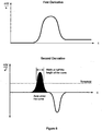
- an assay curve is asserted if a positive going peak in the second derivative is present and has a height above a given threshold and a width or integrated area under its curve that exceeds a corresponding predetermined threshold value, as shown schematically in the lower part of Figure 6 .
- Suitable characteristics include the positive peak height, the area under the second derivative curve for the positive and negative going curves or the width of a peak in the second derivative curve, as shown in the lower part of Figure 6 .
- An advantage of using a second derivative as an analysis method for determining a nucleic acid amplification diagnostic result is that it provides sensitivity to the essential exponential nature of some amplification methods and provides a reliable method to discriminate this within a test result.
- Figure 7 provides an illustration of this. This method has particular application to (but is not limited to) isothermal nucleic acid reactions.
- the assay methods described above can be implemented by the processing component in a number of different ways.
- the methods are implemented in the form of programming instructions of one or more software modules or components 802 stored on non-volatile (e.g., solid-state or hard disk) storage 804 associated with the processing component 800, as shown in Figure 8 .
- non-volatile e.g., solid-state or hard disk
- the processing component 800 includes standard computer components, including random access memory (RAM) 806, at least one processor 808, and interfaces 810, 812, 814, all interconnected by a bus 816.
- the interfaces typically include universal serial bus (USB) interfaces 810, at least one of which may be connected to a keypad or keyboard 818 and optionally also a pointing device such as a mouse, a network interface connector (NIC) 812 which can be used to connect the processing component 800 to a communications network 820, if desired.
- the processing component 800 also includes a display adapter 814, which is connected to a display device such as an LCD panel display 822, and an operating system 824 such as Linux or Microsoft Windows.
- the methods can be stored in a PROM, EEPROM, or the like, or alternatively may be implemented in the form of configuration data for a field programmable gate arrays (FPGAs).
- FPGAs field programmable gate arrays
- the methods may be implemented, either in part or in their entirety, in the form of one or more dedicated hardware components, such as application-specific integrated circuits (ASICs), for example.
- ASICs application-specific integrated circuits
- diagnostic testing particularly relating to a compact portable test instrument suitable for use in medical diagnostics at the Point-of-Care (POC) and in Physician's Office Laboratories (POL).
- POC Point-of-Care
- POL Physician's Office Laboratories
- the described embodiments of the present invention include nucleic acid amplification and detection apparatus that are configured to receive only two measurement tubes contained within a single consumable assembly and are therefore suitable for portable, point of care, or other field applications.
- the described heated support/rotor arrangements enable multiple measurements, self-calibration and mixing functions to be performed with respect to a small number of reaction vessels or test tubes that are contained within in or are part of a disposable cartridge or vessel assembly. These features allow reduced complexity and make possible a compact, portable, and relatively low cost apparatus. Notwithstanding the above, it will be apparent that in other embodiments a nucleic acid amplification and detection apparatus can in general be configured to receive any practical number of reaction vessels. Many modifications will be apparent to those skilled in the art without departing from the scope of the present invention.
Landscapes
- Chemical & Material Sciences (AREA)
- Life Sciences & Earth Sciences (AREA)
- Health & Medical Sciences (AREA)
- Organic Chemistry (AREA)
- Engineering & Computer Science (AREA)
- Zoology (AREA)
- Wood Science & Technology (AREA)
- Proteomics, Peptides & Aminoacids (AREA)
- Biochemistry (AREA)
- Immunology (AREA)
- General Health & Medical Sciences (AREA)
- Physics & Mathematics (AREA)
- Analytical Chemistry (AREA)
- Molecular Biology (AREA)
- Bioinformatics & Cheminformatics (AREA)
- Biotechnology (AREA)
- General Engineering & Computer Science (AREA)
- Microbiology (AREA)
- Genetics & Genomics (AREA)
- Chemical Kinetics & Catalysis (AREA)
- Biophysics (AREA)
- General Physics & Mathematics (AREA)
- Nuclear Medicine, Radiotherapy & Molecular Imaging (AREA)
- Pathology (AREA)
- Measuring Or Testing Involving Enzymes Or Micro-Organisms (AREA)
- Apparatus Associated With Microorganisms And Enzymes (AREA)
- Algebra (AREA)
- Bioinformatics & Computational Biology (AREA)
- Mathematical Analysis (AREA)
- Mathematical Optimization (AREA)
- Mathematical Physics (AREA)
- Pure & Applied Mathematics (AREA)
Description
- The present invention relates to a nucleic acid amplification and detection apparatus and method.
- The amplification of nucleic acids is important in many fields, including medical, biomedical, environmental, veterinary and food safety testing. In general, nucleic acids are amplified by one of two methods: polymerase chain reaction (PCR) or isothermal amplification, both of which are described below.
- As described in the Wikipedia1 at http://en.wikipedia.org/wiki/Polymerase chain reaction:
- "The polymerase chain reaction (PCR) is a scientific technique in molecular biology to amplify a single or a few copies of a piece of DNA across several orders of magnitude, generating thousands to millions of copies of a particular DNA sequence.
- Developed in 1983 by Kary Mullis, PCR is now a common and often indispensable technique used in medical and biological research labs for a variety of applications. These include DNA cloning for sequencing, DNA-based phylogeny, or functional analysis of genes; the diagnosis of hereditary diseases; the identification of genetic fingerprints (used in forensic sciences and paternity testing); and the detection and diagnosis of infectious diseases. In 1993, Mullis was awarded the Nobel Prize in Chemistry along with Michael Smith for his work on PCR.
- The method relies on thermal cycling, consisting of cycles of repeated heating and cooling of the reaction for DNA melting and enzymatic replication of the DNA. Primers (short DNA fragments) containing sequences-complementary to the target region along with a DNA polymerase (after which the method is named) are key components to enable selective and repeated amplification. As PCR progresses, the DNA generated is itself used as a template for replication, setting in motion a chain reaction in which the DNA template is exponentially amplified. PCR can be extensively modified to perform a wide array of genetic manipulations.
- Almost all PCR applications employ a heat-stable DNA polymerase, such as Taq polymerase, an enzyme originally isolated from the bacterium Thermus aquaticus. This DNA polymerase enzymatically assembles a new DNA strand from DNA building-blocks, the nucleotides, by using single-stranded DNA as a template and DNA oligonucleotides (also called DNA primers), which are required for initiation of DNA synthesis. The vast majority of PCR methods use thermal cycling, i.e., alternately heating and cooling the PCR sample to a defined series of temperature steps. These thermal cycling steps are necessary first to physically separate the two strands in a DNA double helix at a high temperature in a process called DNA melting. At a lower temperature, each strand is then used as the template in DNA synthesis by the DNA polymerase to selectively amplify the target DNA. The selectivity of PCR results from the use of primers that are complementary to the DNA region targeted for amplification under specific thermal cycling conditions.
- PCR is used to amplify a specific region of a DNA strand (the DNA target). Most PCR methods typically amplify DNA fragments of up to -10 kilo base pairs (kb), although some techniques allow for amplification of fragments up to 40 kb in size.
- A basic PCR set up requires several components and reagents. These components include:
- DNA template that contains the DNA region (target) to be amplified.
- Two primers that are complementary to the 3' (three prime) ends of each of the sense and anti-sense strand of the DNA target.
- Taq polymerase or another DNA polymerase with a temperature optimum at around 70 °C.
- Deoxynucleoside triphosphates (dNTPs; nucleotides containing triphosphate groups), the building-blocks from which the DNA polymerase synthesizes a new DNA strand.
- Buffer solution, providing a suitable chemical environment for optimum activity and stability of the DNA polymerase.
- Divalent cations, magnesium or manganese ions; generally Mg2+ is used, but Mn2+ can be utilized for PCR-mediated DNA mutagenesis, as higher Mn2+ concentration increases the error rate during DNA synthesis.
- Monovalent cation potassium ions.
- The PCR is commonly carried out in a reaction volume of 10-200 pl in small , reaction tubes (0.2-0.5 ml volumes) in a thermal cycler. The thermal cycler heats and cools the reaction tubes to achieve the temperatures required at each step of the reaction (see below). Many modern thermal cyclers make use of the Peltier effect, which permits both heating and cooling of the block holding the PCR tubes simply by reversing the electric current. Thin-walled reaction tubes permit favorable thermal conductivity to allow for rapid thermal equilibration. Most thermal cyclers have heated lids to prevent condensation at the top of the reaction tube. Older thermocyclers lacking a heated lid require a layer of oil on top of the reaction mixture or a ball of wax inside the tube.
- Typically, PCR consists of a series of 20-40 repeated temperature changes, called cycles, with each cycle commonly consisting of 2-3 discrete temperature steps, usually three... The cycling is often preceded by a single temperature step (called hold) at a high temperature (>90°C), and followed by one hold at the end for final product extension or brief storage. The temperatures used and the length of, time they are applied in each cycle depend on a variety of parameters. These include the enzyme used for DNA synthesis, the concentration of divalent ions and dNTPs in the reaction, and the melting temperature (Tm) of the primers.
- Initialization step: This step consists of heating the reaction to a temperature of 94-96 °C (or 98 °C if extremely thermostable polymerases are used), which is held for 1-9 minutes. It is only required for DNA polymerases that require heat activation by hot-start PCR.
- Denaturation step: This step is the first regular cycling event and consists of heating the reaction to 94-98 °C for 20-30 seconds. It causes DNA melting of the DNA template by disrupting the hydrogen bonds between complementary bases, yielding single-stranded DNA molecules.
- Annealing step: The reaction temperature is lowered to 50-65 °C for 20-40 seconds allowing annealing of the primers to the single-stranded DNA template. Typically the annealing temperature is about 3-5 degrees Celsius below the Tm of the primers used. Stable DNA-DNA hydrogen bonds are only formed when the primer sequence very closely matches the template sequence. The polymerase binds to the primer-template hybrid and begins DNA synthesis.
- Extension/elongation step: The temperature at this step depends on the DNA polymerase used; Taq polymerase has its optimum activity temperature at 75-80 °C, and commonly a temperature of 72 °C is used with this enzyme. At this step the DNA polymerase synthesizes a new DNA strand complementary to the DNA template strand by adding dNTPs that are complementary to the template in 5' to 3' direction, condensing the 5'-phosphate group of the dNTPs with the 3'-hydroxyl group at the end of the nascent (extending) DNA strand. The extension time depends both on the DNA polymerase used and on the length of the DNA fragment to be amplified. As a rule-of-thumb, at its optimum temperature, the DNA polymerase will polymerize a thousand bases per minute. Under optimum conditions, i.e., if there are no limitations due to limiting substrates or reagents, at each extension step, the amount of DNA target is doubled, leading to exponential (geometric) amplification-of the specific DNA fragment.
- Final elongation: This single step is occasionally performed at a temperature of 70-74 °C for 5-15 minutes after the last PCR cycle to ensure that any remaining single-stranded DNA is fully extended.
- Final hold: This step at 4-15 °C for an indefinite time may be employed for short-term storage of the reaction.
- To check whether the PCR generated the anticipated DNA fragment (also sometimes referred to as the amplimer or amplicon), agarose gel electrophoresis is employed for size separation of the PCR products. The size(s) of PCR products is determined by comparison with a DNA ladder (a molecular weight marker), which contains DNA fragments of known size, run on the gel alongside the PCR products...
- The PCR process can be divided into three stages:
- Exponential amplification: At every cycle, the amount of product is doubled (assuming 100% reaction efficiency). The reaction is very sensitive: only minute quantities of DNA need to be present.
- Leveling off stage: The reaction slows as the DNA polymerase loses activity and as consumption of reagents such as dNTPs and primers causes them to become limiting.
- Plateau: No more product accumulates due to exhaustion of reagents and enzyme.
- In practice, PCR can fail for various reasons, in part due to its sensitivity to contamination causing amplification of spurious DNA products. Because of this, a number of techniques and procedures have been developed for optimizing PCR conditions. Contamination with extraneous DNA is addressed with lab protocols and procedures that separate pre-PCR mixtures from potential DNA contaminants. This usually involves spatial separation of PCR-setup areas from areas for analysis or purification of PCR products, use of disposable plasticware, and thoroughly cleaning the work surface between reaction setups. Primer-design techniques are important in improving PCR product yield and in avoiding the formation of spurious products, and the usage of alternate buffer components or polymerase enzymes can help with amplification of long or otherwise problematic regions of DNA. Addition of reagents, such as formamide, in buffer systems may increase the specificity and yield of PCR.
- Because PCR amplifies the regions of DNA that it targets, PCR can be used to analyze extremely small amounts of sample. This is often critical for forensic analysis, when only a trace amount of DNA is available as evidence. PCR may also be used in the analysis of ancient DNA that is tens of thousands of years old. These PCR-based techniques have been successfully used on animals, such as a forty-thousand-year-old mammoth, and also on human DNA, in applications ranging from the analysis of Egyptian mummies to the identification of a Russian tsar.
- Quantitative PCR methods allow the estimation of the amount of a given sequence present in a sample-a technique often applied to quantitatively determine levels of gene expression. Real-time PCR is an established tool for DNA quantification that measures the accumulation of DNA product after each round of PCR amplification.
- PCR permits early diagnosis of malignant diseases such as leukemia and lymphomas, which is currently the highest-developed in cancer research and is already being used routinely. (See the studies cited in the EUTOS For CML study article at http://www.eutos.org/content/molecular_monitoring/information/pcr_testing/, especially notes 10-13.) PCR assays can be performed directly on genomic DNA samples to detect translocation-specific malignant cells at a sensitivity that is at least 10,000-fold higher than that of other methods.
- PCR also permits identification of non-cultivatable or slow-growing microorganisms such as mycobacteria, anaerobic bacteria, or viruses from tissue culture assays and animal models. The basis for PCR diagnostic applications in microbiology is the detection of infectious agents and the discrimination of non-pathogenic from pathogenic strains by virtue of specific genes.
- Viral DNA can likewise be detected by PCR. The primers used need to be specific to the targeted sequences in the DNA of a virus, and the PCR can be used for diagnostic analyses or DNA sequencing of the viral genome. The high sensitivity of PCR permits virus detection soon after infection and even before the onset of disease. Such early detection may give physicians a significant lead in treatment. The amount of virus ("viral load") in a patient can also be quantified by PCR-based DNA quantitation techniques (see below).
- As described in the Wikipedia1 at http://en.wikinedia.org/wiki/Variants of PCR#Isothermal amplification methods:
- "Some DNA amplification protocols have been developed that may be used alternatively to PCR:
- Helicase-dependent amplification is similar to traditional PCR, but uses a constant temperature rather than cycling through denaturation and annealing/extension steps. DNA Helicase, an enzyme that unwinds DNA, is used in place of thermal denaturation.
- PAN-AC also uses isothermal conditions for amplification, and may be used to analyze living cells.
- Nicking Enzyme Amplification Reaction referred to as NEAR, is isothermal, replicating DNA at a constant temperature using a polymerase and nicking enzyme.
- Recombinase Polymerase Amplification (RPA). The method uses a recombinase to specifically pair primers with double-stranded DNA on the basis of homology, thus directing DNA synthesis from defined DNA sequences present in the sample. Presence of the target sequence initiates DNA amplification, and no thermal or chemical melting of DNA is required. The reaction progresses rapidly and results in specific DNA amplification from just a few target copies to detectable levels typically within 5-10 minutes. The entire reaction system is stable as a dried formulation and does not need refrigeration. RPA can be used to replace PCR (Polymerase Chain Reaction) in a variety of laboratory applications and users can design their own assays.
- Despite the many advances in this general field, existing nucleic acid amplification and detection methods and apparatus nevertheless suffer from various difficulties. For example, existing detection methods that determine assay results are prone to error, and existing nucleic acid amplification and detection apparatus are typically large, complex and costly.
-
WO-A-2007/005077 discloses an instrument for carrying out real-time multiplex PCR by detection of fluorescence. Different fluorescent dyes can be detected by different optical modules coupled to a a photomultiplier tube. The device contains a temperature control capable of modulating the temperature of the reaction vessels individually or as a group. The rotating platform which rotates the disk containing the vessels is under the control of an actuator, a slot sensor trigger provides a signal to the control unit which allows the control unit to synchronise the position of a well with the optical measurement during rotation. Further disclosed is a barcode reader which identifies the type of disk and may identify the actual disk to assist a technician in tracking data to specific samples from multiple disks. The vessels are heated by light emitted by a heating element. Finally, the optical measurements are used in analysing the content of nucleic acid in the sample. It is desired to provide a nucleic acid amplification and detection apparatus and method that alleviate one or more difficulties of the prior art, or that at least provide a useful alternative. - In accordance with some embodiments of the present invention, there is provided a nucleic acid amplification and detection apparatus, as defined in the claims, including:
- a support configured to receive a plurality of reaction vessels containing respective samples of one or more nucleic acids to be amplified, the support being rotatable about an axis of rotation and the reaction vessels being received in the support at respective receiving locations distributed about the axis of rotation;
- a temperature control component thermally coupled to the support and configured to control the temperature of the support in order to amplify the nucleic acids contained in the reaction vessels while received in the support;
- one or more measurement components configured to measure one or more characteristics of the nucleic acids within the reaction vessels at respective measurement locations distributed about the axis of rotation;
- an actuator coupled to the support and configured to rotate the support about the axis of rotation; and
- a sample position controller coupled to the actuator and being configured to rotate the support about the axis of rotation so as to position a selected one of the plurality of reaction vessels to a selected one of the measurement locations to allow a corresponding one of the measurement components to perform a corresponding measurement on the corresponding sample.
- In some embodiments, the measurement components include one or more optical measurement components configured to measure respective optical characteristics including optical absorption, reflection, luminance output, and/or fluorescence.
- In some embodiments, the optical measurement components include optical measurement components configured to measure multiple channels of fluorescence, reflectance or transmission, or combinations thereof. The sample position controller is configured to selectively cause the actuator to rotate the support about the axis of rotation in an oscillatory manner to cause mixing of the contents of the reaction vessels.
In some embodiments, the nucleic acid amplification and detection apparatus includes an ultrasonic transducer component configured for selective coupling to a selected one of the reaction vessels to cause mixing of the contents of the selected reaction vessel.
In some embodiments, the ultrasonic transducer component is configured to support the selected reaction vessel in a spaced arrangement relative to the support to inhibit the coupling of ultrasonic energy to the support.
In some embodiments, the nucleic acid amplification and detection apparatus includes a sample identification component configured to identify a selected sample based on an identifier associated with the corresponding reaction vessel. - In some embodiments, the sample identification component includes at least one of a barcode reader, an RFID sensor, and an imaging device.
In some embodiments, the sample identification component includes an imaging device, and the sample identification component is further configured to acquire an image of the contents of the reaction vessel and to process the acquired image to confirm the reagents therein.
In some embodiments, the temperature control component is thermally coupled to the support across a gap therebetween. In some embodiments, the gap includes a thermally conductive fluid therebetween. In some embodiments, the thermally conductive fluid is a magnetic fluid retained within the gap by one or more magnets. - In some embodiments, the nucleic acid amplification and detection apparatus includes one or more magnets arranged about the support at respective different heights, such that when the support is rotated, paramagnetic beads within a reaction vessel in the support move past the magnets, causing the beads to oscillate correspondingly within the reaction vessel.
- In some embodiments, the nucleic acid amplification and detection apparatus includes a processing component configured to process sensor data representing the measured characteristics to determine an assay result for the nucleic acids within the reaction vessels.
- In some embodiments, the processing component is configured to:
- receive signal data representing assay measurements of a sample containing at least one nucleic acid at respective times during amplification of the at least one nucleic acid;
- process said signal data to generate second derivative data representing values of a second derivative of said signal data with respect to time;
- process said second derivative data to determine whether at least one of the second derivative values exceeds a predetermined second derivative threshold value;
- process said second derivative data to determine a crossover time at which the second derivative of said signal data crosses zero after having exceeded the second derivative threshold value;
- process said signal data to generate first derivative data representing a first derivative of said signal data with respect to time at the crossover time; and
- generate assay result data representing a result of the assay, the result being determined on the basis of: (i) whether the predetermined second derivative threshold value was exceeded, and (ii) the first derivative of said signal with respect to time at the crossover time.
- In some embodiments, the processing component is configured to:
- receive signal data representing assay measurements of a sample containing at least one nucleic acid at respective times during amplification of the at least one nucleic acid;
- process said signal data to generate second derivative data representing values of a second derivative of said signal data with respect to time;
- process said second derivative data to determine whether at least one of the second derivative values exceeds a predetermined second derivative threshold value;
- process said second derivative data to determine whether the second derivative of said signal data crosses zero after having exceeded the second derivative threshold value;
- process said second derivative data to determine which of the second derivative values are positive;
- determine a width or integrated area of the positive second derivative values;
- compare the width or integrated area of the positive second derivative values with a further predetermined threshold value; and
- generate assay result data representing a result of the assay, the result being determined on the basis of: (i) whether the predetermined second derivative threshold value was exceeded, (ii) whether the second derivative of said signal crosses zero after having exceeded the second derivative threshold value, and (iii) whether the width or integrated area of the positive second derivative values exceeds the further predetermined threshold value.
- receiving signal data representing assay measurements of a sample containing at least one nucleic acid at respective times during amplification of the at least one nucleic acid;
- processing said signal data to generate second derivative data representing values of a second derivative of said signal data with respect to time;
- processing said second derivative data to determine whether at least one of the second derivative values exceeds a predetermined second derivative threshold value;
- processing said second derivative data to determine a crossover time at which the second derivative of said signal data crosses zero after having exceeded the second derivative threshold value;
- processing said signal data to generate first derivative data representing a first derivative of said signal data with respect to time at the crossover time;
- generating assay result data representing a result of the assay, the result being determined on the basis of: (i) whether the predetermined second derivative threshold value was exceeded, and (ii) the first derivative of said signal with respect to time at the crossover time; and
- outputting the assay result data.
- receiving signal data representing assay measurements of a sample containing at least one nucleic acid at respective times during amplification of the at least one nucleic acid;
- processing said signal data to generate second derivative data representing values of a second derivative of said signal data with respect to time;
- processing said second derivative data to determine whether at least one of the second derivative values exceeds a predetermined second derivative threshold value;
- processing said second derivative data to determine whether the second derivative of said signal data crosses zero after having exceeded the second derivative threshold value;
- processing said second derivative data to determine which of the second derivative values are positive;
- determining a width or integrated area of the positive second derivative values;
- comparing the width or integrated area of the positive second derivative values with a further predetermined threshold value;
- generating assay result data representing a result of the assay, the result being determined on the basis of: (i) whether the predetermined second derivative threshold value was exceeded, (ii) whether the second derivative of said signal crosses zero after having exceed the second derivative threshold value, and (iii) whether the width or integrated area of the positive second derivative values exceeds the further predetermined threshold value; and
- outputting the assay result data.
- Even further disclosed is a computer-readable storage medium having stored thereon programming instructions or configuration data that, when executed by at least one processor, causes the processor to execute any one of the above methods.
- Finally a nucleic acid detection apparatus configured to execute any one of the above method is disclosed.
- Some embodiments of the present invention are hereinafter described, by way of example only, with reference to the accompanying
Figures 1-4 .Figures 5-8 illustrate methods of the disclosure: -
Figures 1 to 4 are isometric, plan, cut-away, and exploded views, respectively, of an assembly of a nucleic acid amplification and detection apparatus; -
Figure 5 is a schematic graph of a typical nucleic acid assay measurement as a function of time during amplification of the nucleic acid; -
Figure 6 includes schematic graphs of the first and second derivatives, respectively, with respect to time of the assay curve ofFigure 5 ; -
Figure 7 includes schematic graphs of example assay measurement values and the first and second derivatives of those measured values, respectively, with respect to time; and -
Figure 8 is a block diagram of a processing component of the nucleic acid amplification and detection apparatus. - The described embodiments of the present invention include a nucleic acid amplification and detection apparatus, as defined in the claims, that combines, in a single integrated instrument, nucleic acid amplification with measurement of the resulting amplification products during and/or following amplification. The apparatus provides temperature control suitable for a DNA amplification reaction within removable reaction vessels using isothermal and/or thermo-cycled protocols, together with diagnostic or measurement components that are used to measure characteristics of the samples while still contained within the same removable reaction vessels. In particular, the apparatus allows the optical and/or electromagnetic characteristics of amplified nucleic acid samples to be measured while the controlled temperature environment is maintained such that it can be used as biomedical, environmental, food safety, or veterinary applications.
The apparatus includes a support configured to receive a plurality of reaction vessels containing respective samples of one or more nucleic acids to be amplified. In some embodiments, the support is configured to receive a disposable component that supports or otherwise includes or incorporates the reaction vessels. In the described embodiments, the reaction vessels are in the form of standard sample tubes such as those used for PCR, but this need not be the case in other embodiments. The support is rotatable about an axis of rotation and the reaction vessels are supported at respective receiving locations distributed about the axis of rotation. A temperature control component thermally coupled to the support is configured to control the, temperature of the support and hence the samples contained in the reaction vessels, thereby allowing the nucleic acids contained in the reaction vessels to be amplified. - In order to perform measurements on the sample during and/or following nucleic acid amplification, the apparatus includes measurement components configured to measure one or more characteristics of the nucleic acids within the reaction vessels at respective measurement locations distributed about the axis of rotation. The measurement components arranged in this manner are thus also referred to herein as 'measurement stations'. This arrangement allows any one of the reactions vessels to be moved to any one of the measurement locations by simply rotating the support so that the selected reaction vessel is positioned at the selected measurement location. This is achieved by the apparatus including a sample position controller and associated actuator configured to rotate the support about the axis of rotation so as to move the selected reaction vessel to the selected measurement location. This allows the corresponding measurement component to measure one or more characteristics of the nucleic acids within the reaction vessel.
- For example, a sample of interest can be divided into one or more reaction vessels so that the apparatus can be used to amplify the nucleic acids(s) in the divided sample and to measure multiple test and control reactions, displaying the results of these multiple tests to a user.
- Use of fluorescence as a detection signal can provide good sensitivity, and where the measurement components include multiple fluorescence detectors configured to detect respective non-overlapping wavelength ranges so as not to interfere with one another, multiple channels of test and/or control reactions can be incorporated within a single reaction vessel. The measurement components can be configured to measure optical absorption, reflection, luminance output, and/or fluorescence.
- The rotatable support arrangement allows measurement components configured to perform different sample measurements to be arranged around the support so that any of the reaction vessels in the support can be moved into position for a selected measurement on demand. These measurements may include, for example and inter alia, multiple channels of fluorescence, reflectance or transmission, combinations of these. Other measurements can be additionally or alternatively included, if required. Thus multiple measurement stations can access each sample, allowing a combination of measurements to be provided for each reaction vessel.
- The supporting of the consumable or reaction vessels in the rotatable support also allows the contents of the reaction vessels to be mixed by rapid oscillatory rotation of the support under control of the samples position controller and associated actuator. The rotary oscillation induces mixing flows and disruption to the fluid within each reaction vessel, thereby improving the degree of reaction, reaction times and uniformity of measurements within the apparatus.
- The mixing can be enhanced by including magnetic beads and/or other forms of disruptive features within each reaction vessel. For example, in some embodiments permanent magnets are fixed at locations just beyond the outer periphery of the support and arranged circumferentially about the rotation axis and at different alternating heights, so that when the support is rotated, paramagnetic beads within a reaction vessel move past the magnets at alternating heights, causing the beads to oscillate correspondingly up and down within the reaction vessel.
-
Figures 1 to 3 show anassembly 100 of an embodiment in which the apparatus includes threeoptical measurement stations support 108 that includes twoopenings 110 configured to receive respective reaction vessels. Thesupport 108 is cylindrically symmetric about its axis of rotation, and is composed of a material having a high thermal conductivity such as aluminium or copper, for example. - In some embodiments (not shown), the heating and temperature control of the support is achieved using electrical heating elements such as resistors, and at least one temperature sensor embedded in or otherwise attached to the support. Electrical connections to these heating elements and temperature sensor are by way of cable connections or slip rings. In embodiments with cable connections, the rotation of the support is limited in angular travel.
- In contrast to these arrangements, the embodiment shown in
Figures 1 to 4 uses non-contact means to control the temperature of thesupport 108. As shown in the cut-away view ofFigure 3 and the exploded view ofFigure 4 , theassembly 100 includes a printed circuit board (PCB) 302 havingresistive heater elements 402 mounted thereon in a circular arrangement to provide heating to a metallicannular ring 304 rigidly mounted to thePCB 302. - The
ring 304 is mounted within a complementary and generally annular cavity in a generally circular lower portion or skirt of thesupport 108 with a small gap therebetween. Thering 304 and the lower portion of thesupport 108 are in close mutual proximity over a large common surface area of the gap to improve the thermal transfer from thering 304 to thesupport 108. In other embodiments, this thermal transfer can be achieved by a thermal gap similar to that described above but with different shapes (e.g., as a flat plate with a gap to a flat underside of the support), or alternatively by a sliding thermally conductive contact. The thermal coupling is such that a desired amplification temperature in the range of about 35-65°C can be reached within a period of about ten minutes or less from a starting temperature around room temperature or, in the case of PCR, from one of the PCR operating temperatures. - In embodiments where the thermal transfer is across a gap, this gap may be filled with air or a thermally conductive fluid such as a thermally conductive grease or silicon oil retained within the gap by a seal. In some embodiments, a magnetic fluid is retained within this gap using magnets disposed on either or both sides of the gap.
- The cut away view of
Figure 3 also shows anon-contact temperature sensor 306 mounted on the printed circuit board (PCB) 302 to provide feedback to a standard temperature control circuit, which can also be on thePCB 302 or alternatively external to theassembly 100. Thetemperature sensor 306 measures the temperature of therotating support 108 but is mounted to the fixed printed circuit board (PCB) 302. Thetemperature sensor 306 is connected to an annular member orvane 308 that is disposed within an annular channel in the lower portion of thesupport 108 with a small gap therebetween. This arrangement provides good thermal coupling to thesensor 306 from thesupport 108 across the small gap into themetallic vane 308 attached to thesensor 306. - Where both the
heater elements 402 and thefeedback temperature sensor 306 are connected to electronic circuits and additionally theheater elements 402 are controlled by a microprocessor or analogue control circuit, accurate temperature control strategies can be implemented. For example proportional, integral, differential, (PID) control can be used to accurately drive and stabilise thesupport 108 and thesensor 306 to a desired temperature set point. - In other embodiments, alternative types of non-contact temperature sensors can be used. For example, a non-contact optical or infra-red temperature sensor such as the Melexis MLX90615 Infra Red Thermometer sensor are used in some embodiments. In some embodiments, multiple temperature sensors with different characteristics are used to optimise the temperature control strategy for rapid heat up and transitions combined with good steady state temperature accuracy. In some embodiments, air flow and/or Peltier cell elements are used to actively cool the
support 108 to provide a rapid temperature transition to lower temperatures. - In some embodiments, additional openings in the support are provided to receive calibration or reference samples with specific optical characteristics. This allows a stepper motor/
actuator 112 coupled to thesupport 108 to be controlled so that a sample to be measured or a reference target is positioned at a measurement station location. This can be used to self-calibrate or self-test the apparatus during power up or measurement cycles by comparing the measured reference values against know values for the reference target. - In some embodiments, the support can also be coupled to a vibrating mechanism or actuator such as an electromagnetic coil and slug. Actuation of this component can induce vibration in the reaction vessels mounted in the support. The excitation frequency can be in the range of Hz up to kHz. Where the excitation is above 20 kHz, it can be referred to as ultrasonic mixing. For high frequency or ultrasonic mixing, a piezoelectric actuator can be used.
- In some embodiments, the apparatus includes an ultrasonic transducer configured so that the support can rotate the reaction vessel so that the ultrasonic transducer can contact it through an opening in the support, with the ultrasonic transducer slightly lifting the reaction vessel so that it is not fully supported by the support. This allows efficient ultrasonic excitation of each reaction vessel only (i.e., without exciting the support itself) while otherwise allowing the reaction vessel to be seated in good thermal contact at other rotation positions and associated measurement stations.
- In some embodiments, the apparatus includes a barcode reader, RFID reader or an image sensor, and the support can be rotated to position a selected reaction vessel or associated disposable plastic assembly carrying or forming the vessel such that an attached label or feature is positioned in front of the barcode reader, RFID reader or image sensor. Where an image sensor is used, this can also be employed to confirm that the sample and reaction vessel or the disposable assembly that carries the reaction vessels have the correct reagents added, are assembled correctly, and are functional.
- In some embodiments, the apparatus includes a processing component that uses the measured outputs from a sample over time to determine a diagnostic test result.
Figure 5 is a schematic illustration of a typical assay measurement (such as fluorescence) over time during nucleic acid amplification within the apparatus. Existing instruments use simple approaches such as a gradient threshold, a fixed or variable threshold for the assay at particular times in the amplification process. However, these prior art approaches are prone to error. In contrast, the apparatus uses an improved detection method that is more reliable than the standard methods used today. - The method involves receiving a signal (or, equivalently, signal data representing the signal) representing assay measurements of a sample during nucleic acid amplification, and generating second derivative data representing successive values of the second derivative of the signal (or signal data) with respect to time.
- In the described embodiment, the assay test is considered to be asserted only if:
- (i) at least one of the second derivative values exceeds a predetermined positive second derivative threshold value; and
- (ii) the second derivative values cross zero after having exceeded the second derivative threshold value of (i), the time at which this occurs being referred to as the zero crossing time; and
- (iii) a measure of the width (e.g., the full width at half-maximum (FWHM)) or integrated second derivative values (the latter being equivalent to the first derivative value at the zero crossing time of (ii)) exceeds a further corresponding predetermined threshold value.
- Thus an assay curve is asserted if a positive going peak in the second derivative is present and has a height above a given threshold and a width or integrated area under its curve that exceeds a corresponding predetermined threshold value, as shown schematically in the lower part of
Figure 6 . - In other embodiments, other characteristics of the second derivative values can be used as pass or fail criteria to determine whether the test result is asserted or not asserted or is possibly invalid. Suitable characteristics include the positive peak height, the area under the second derivative curve for the positive and negative going curves or the width of a peak in the second derivative curve, as shown in the lower part of
Figure 6 . - An advantage of using a second derivative as an analysis method for determining a nucleic acid amplification diagnostic result is that it provides sensitivity to the essential exponential nature of some amplification methods and provides a reliable method to discriminate this within a test result.
Figure 7 provides an illustration of this. This method has particular application to (but is not limited to) isothermal nucleic acid reactions. - As will be apparent to those skilled in the art, the assay methods described above can be implemented by the processing component in a number of different ways. In some embodiments, the methods are implemented in the form of programming instructions of one or more software modules or
components 802 stored on non-volatile (e.g., solid-state or hard disk)storage 804 associated with theprocessing component 800, as shown inFigure 8 . - The
processing component 800 includes standard computer components, including random access memory (RAM) 806, at least oneprocessor 808, and interfaces 810, 812, 814, all interconnected by a bus 816. The interfaces typically include universal serial bus (USB) interfaces 810, at least one of which may be connected to a keypad orkeyboard 818 and optionally also a pointing device such as a mouse, a network interface connector (NIC) 812 which can be used to connect theprocessing component 800 to acommunications network 820, if desired. Theprocessing component 800 also includes adisplay adapter 814, which is connected to a display device such as anLCD panel display 822, and anoperating system 824 such as Linux or Microsoft Windows. - In other embodiments, the methods can be stored in a PROM, EEPROM, or the like, or alternatively may be implemented in the form of configuration data for a field programmable gate arrays (FPGAs). In yet other embodiments, the methods may be implemented, either in part or in their entirety, in the form of one or more dedicated hardware components, such as application-specific integrated circuits (ASICs), for example.
- Applications of the apparatus and methods described herein include diagnostic testing, particularly relating to a compact portable test instrument suitable for use in medical diagnostics at the Point-of-Care (POC) and in Physician's Office Laboratories (POL).
- The described embodiments of the present invention include nucleic acid amplification and detection apparatus that are configured to receive only two measurement tubes contained within a single consumable assembly and are therefore suitable for portable, point of care, or other field applications. The described heated support/rotor arrangements enable multiple measurements, self-calibration and mixing functions to be performed with respect to a small number of reaction vessels or test tubes that are contained within in or are part of a disposable cartridge or vessel assembly. These features allow reduced complexity and make possible a compact, portable, and relatively low cost apparatus. Notwithstanding the above, it will be apparent that in other embodiments a nucleic acid amplification and detection apparatus can in general be configured to receive any practical number of reaction vessels.
Many modifications will be apparent to those skilled in the art without departing from the scope of the present invention.
Claims (15)
- A nucleic acid amplification and detection apparatus, including:a support configured to receive a plurality of reaction vessels containing respective samples of one or more nucleic acids to be amplified, the support being rotatable about an axis of rotation and the reaction vessels being received in the support at respective receiving locations distributed about the axis of rotation;a temperature control component thermally coupled to the support and configured to control the temperature of the support in order to amplify the nucleic acids contained in the reaction vessels while received in the support;a plurality of measurement components configured to measure respective characteristics of the nucleic acids within the reaction vessels at respective measurement locations distributed about the axis of rotation;an actuator coupled to the support and configured to rotate the support about the axis of rotation; anda sample position controller coupled to the actuator and being configured :(i) to selectively cause the actuator to rotate the support about the axis of rotation in an oscillatory manner to cause mixing of the contents of the reaction vessels; and(ii) to rotate the support about the axis of rotation so as to position a selected one of the plurality of reaction vessels to a selected one of the plurality of measurement locations to allow a corresponding one of the plurality of measurement components to perform a corresponding measurement on the corresponding sample.
- The nucleic acid amplification and detection apparatus of claim 1, wherein the measurement components include one or more optical measurement components configured to measure respective optical characteristics including optical absorption, reflection, luminance output, and/or fluorescence.
- The nucleic acid amplification and detection apparatus of claim 2, wherein the optical measurement components include optical measurement components configured to measure multiple channels of fluorescence, reflectance or transmission, or combinations thereof.
- The nucleic acid amplification and detection apparatus of any one of claims 1 to 3, including an ultrasonic transducer component configured for selective coupling to a selected one of the reaction vessels to cause mixing of the contents of the selected reaction vessel.
- The nucleic acid amplification and detection apparatus of claim 4, wherein the ultrasonic transducer component is configured to support the selected reaction vessel in a spaced arrangement relative to the support to inhibit the coupling of ultrasonic energy to the support.
- The nucleic acid amplification and detection apparatus of any one of claims 1 to 5, including a sample identification component configured to identify a selected sample based on an identifier associated with the corresponding reaction vessel.
- The nucleic acid amplification and detection apparatus of claim 6, wherein the sample identification component includes at least one of a barcode reader, an RFID sensor, and an imaging device.
- The nucleic acid amplification and detection apparatus of claim 6, wherein the sample identification component includes an imaging device, and the sample identification component is further configured to acquire an image of the contents of the reaction vessel and to process the acquired image to confirm the reagents therein.
- The nucleic acid amplification and detection apparatus of any one of claims 1 to 8, wherein the temperature control component is thermally coupled to the support across a gap therebetween.
- The nucleic acid amplification and detection apparatus of claim 9, wherein the gap includes a thermally conductive fluid therebetween.
- The nucleic acid amplification and detection apparatus of claim 10, wherein the thermally conductive fluid is a magnetic fluid retained within the gap by one or more magnets.
- The nucleic acid amplification and detection apparatus of any one of claims 1 to 11, including one or more magnets arranged about the support at respective different heights, such that when the support is rotated, paramagnetic beads within a reaction vessel in the support move past the magnets, causing the beads to oscillate correspondingly within the reaction vessel.
- The nucleic acid amplification and detection apparatus of any one of claims 1 to 11, including a processing component configured to process sensor data representing the measured characteristics to determine an assay result for the nucleic acids within the reaction vessels.
- The nucleic acid amplification and detection apparatus of claim 13, wherein the processing component is configured to:receive signal data representing assay measurements of a sample containing at least one nucleic acid at respective times during amplification of the at least one nucleic acid;process said signal data to generate second derivative data representing values of a second derivative of said signal data with respect to time;process said second derivative data to determine whether at least one of the second derivative values exceeds a predetermined second derivative threshold value;process said second derivative data to determine a crossover time at which the second derivative of said signal data crosses zero after having exceeded the second derivative threshold value;process said signal data to generate first derivative data representing a first derivative of said signal data with respect to time at the crossover time; andgenerate assay result data representing a result of the assay, the result being determined on the basis of: (i) whether the predetermined second derivative threshold value was exceeded, and (ii) the first derivative of said signal with respect to time at the crossover time.
- The nucleic acid amplification and detection apparatus of claim 13, wherein the processing component is configured to:receive signal data representing assay measurements of a sample containing at least one nucleic acid at respective times during amplification of the at least one nucleic acid;process said signal data to generate second derivative data representing values of a second derivative of said signal data with respect to time;process said second derivative data to determine whether at least one of the second derivative values exceeds a predetermined second derivative threshold value;process said second derivative data to determine whether the second derivative of said signal data crosses zero after having exceeded the second derivative threshold value;process said second derivative data to determine which of the second derivative values are positive;determine a width or integrated area of the positive second derivative values;compare the width or integrated area of the positive second derivative values with a further predetermined threshold value; andgenerate assay result data representing a result of the assay, the result being determined on the basis of: (i) whether the predetermined second derivative threshold value was exceeded, (ii) whether the second derivative of said signal crosses zero after having exceeded the second derivative threshold value, and (iii) whether the width or integrated area of the positive second derivative values exceeds the further predetermined threshold value.
Priority Applications (1)
| Application Number | Priority Date | Filing Date | Title |
|---|---|---|---|
| EP16183619.2A EP3231861B1 (en) | 2012-02-03 | 2013-02-01 | Nucleic acid amplification with mathematical calculation of the assay result |
Applications Claiming Priority (2)
| Application Number | Priority Date | Filing Date | Title |
|---|---|---|---|
| US201261594870P | 2012-02-03 | 2012-02-03 | |
| PCT/AU2013/000092 WO2013113072A1 (en) | 2012-02-03 | 2013-02-01 | Nucleic acid amplification and detection apparatus and method |
Related Child Applications (2)
| Application Number | Title | Priority Date | Filing Date |
|---|---|---|---|
| EP16183619.2A Division-Into EP3231861B1 (en) | 2012-02-03 | 2013-02-01 | Nucleic acid amplification with mathematical calculation of the assay result |
| EP16183619.2A Division EP3231861B1 (en) | 2012-02-03 | 2013-02-01 | Nucleic acid amplification with mathematical calculation of the assay result |
Publications (3)
| Publication Number | Publication Date |
|---|---|
| EP2809767A1 EP2809767A1 (en) | 2014-12-10 |
| EP2809767A4 EP2809767A4 (en) | 2016-02-17 |
| EP2809767B1 true EP2809767B1 (en) | 2017-11-08 |
Family
ID=48904312
Family Applications (2)
| Application Number | Title | Priority Date | Filing Date |
|---|---|---|---|
| EP16183619.2A Active EP3231861B1 (en) | 2012-02-03 | 2013-02-01 | Nucleic acid amplification with mathematical calculation of the assay result |
| EP13743047.6A Active EP2809767B1 (en) | 2012-02-03 | 2013-02-01 | Nucleic acid amplification and detection apparatus |
Family Applications Before (1)
| Application Number | Title | Priority Date | Filing Date |
|---|---|---|---|
| EP16183619.2A Active EP3231861B1 (en) | 2012-02-03 | 2013-02-01 | Nucleic acid amplification with mathematical calculation of the assay result |
Country Status (4)
| Country | Link |
|---|---|
| US (2) | US10428375B2 (en) |
| EP (2) | EP3231861B1 (en) |
| AU (1) | AU2013203281A1 (en) |
| WO (1) | WO2013113072A1 (en) |
Families Citing this family (14)
| Publication number | Priority date | Publication date | Assignee | Title |
|---|---|---|---|---|
| USD753311S1 (en) | 2011-10-12 | 2016-04-05 | Alere Switzerland Gmbh | Isothermal nucleic acid amplification meter |
| AU2013203281A1 (en) | 2012-02-03 | 2013-08-22 | Axxin Pty Ltd | Nucleic acid amplification and detection apparatus and method |
| US9932629B2 (en) | 2012-06-26 | 2018-04-03 | Axxin Pty Ltd | Nucleic acid amplification and detection kit |
| EP3218698B1 (en) | 2014-11-14 | 2020-05-13 | Axxin Pty Ltd | Biological sample collection and storage assembly |
| US11198122B2 (en) | 2015-07-17 | 2021-12-14 | Axxin Pty Ltd | Diagnostic test assembly, apparatus, method |
| CN111386465B (en) | 2017-09-27 | 2024-03-22 | 艾可辛私人有限公司 | Diagnostic test system and method |
| AU2018360737A1 (en) | 2017-10-31 | 2020-06-04 | Alberta Biophotonics Inc. | Optical measurement method and system |
| CN107739710B (en) * | 2017-11-08 | 2024-02-23 | 西安天隆科技有限公司 | Rapid nucleic acid amplification system |
| CN111098192A (en) * | 2020-01-14 | 2020-05-05 | 谭登富 | Drilling and tapping machine |
| CN112816446B (en) * | 2020-12-24 | 2022-02-01 | 四川长虹电器股份有限公司 | Method for detecting powder decay of fluorescent wheel based on fluorescence spectrum |
| TWI812941B (en) * | 2021-04-16 | 2023-08-21 | 財桂生物股份有限公司 | Fluorescence detection constant temperature oscillating mixer |
| NL2028003B1 (en) * | 2021-04-16 | 2022-10-31 | Micronit Holding B V | Method for providing a detection output which is representative for the presence of a molecule of interest |
| CN114166768B (en) * | 2022-02-14 | 2022-05-20 | 四川大学华西医院 | Conversion method and device for detecting homogeneity of same index by different equipment and electronic equipment |
| EP4310701A1 (en) * | 2022-02-14 | 2024-01-24 | West China Hospital, Sichuan University | Homogenization conversion method and apparatus for detection of same index by different devices, and electronic device |
Family Cites Families (100)
| Publication number | Priority date | Publication date | Assignee | Title |
|---|---|---|---|---|
| US3913562A (en) | 1974-01-16 | 1975-10-21 | Moore Perk Corp | Disposable package for collecting and transporting cultures for laboratory analysis |
| US4234540A (en) | 1979-08-24 | 1980-11-18 | Coulter Electronics, Inc. | Apparatus for monitoring chemical reactions and employing moving photometer means |
| US4250266A (en) * | 1979-12-19 | 1981-02-10 | Honeywell Inc. | Automated micro-organism culture growth and detection instrument |
| US4376634A (en) | 1980-05-30 | 1983-03-15 | Mallinckrodt, Inc. | Assay kit having syringe, dilution device and reagents within sealed container |
| US4580577A (en) | 1981-01-12 | 1986-04-08 | Brien Joseph O | Method and apparatus for collecting saliva |
| US4353868A (en) | 1981-05-29 | 1982-10-12 | Sherwood Medical Industries Inc. | Specimen collecting device |
| FR2590673B1 (en) | 1985-11-05 | 1988-09-16 | Bally Philippe | SELF-CONTAINED MINIATURIZED TEST DEVICE FOR SINGLE USE |
| US4770853A (en) | 1986-12-03 | 1988-09-13 | New Horizons Diagnostics Corporation | Device for self contained solid phase immunodiffusion assay |
| US5169789A (en) | 1986-12-03 | 1992-12-08 | New Horizons Diagnostics Corporation | Device and method for self contained solid phase immunodiffusion assay |
| US4912034A (en) | 1987-09-21 | 1990-03-27 | Biogenex Laboratories | Immunoassay test device and method |
| US4813432A (en) | 1987-10-13 | 1989-03-21 | Saint-Amand Manufacturing, Inc. | Swab transport system |
| US5266266A (en) | 1988-02-09 | 1993-11-30 | Nason Frederic L | Specimen test unit |
| US5152965A (en) | 1989-06-02 | 1992-10-06 | Abbott Laboratories | Two-piece reagent container assembly |
| US5435970A (en) | 1989-12-18 | 1995-07-25 | Environmental Diagnostics, Inc. | Device for analysis for constituents in biological fluids |
| FR2669428B1 (en) * | 1990-11-16 | 1993-04-23 | Alcyon Analyzer Sa | AUTOMATIC COLORIMETRY SAMPLE ANALYZER. |
| JP3285451B2 (en) | 1994-08-01 | 2002-05-27 | 富士写真フイルム株式会社 | Analysis method and analysis element for whole blood sample |
| CN1155827C (en) | 1995-05-09 | 2004-06-30 | 贝克曼考尔特公司 | Device and method for separating cellular components of blood from liquid portion of blood |
| US5827675A (en) | 1995-07-12 | 1998-10-27 | Charm Sciences, Inc. | Test apparatus, system and method for the detection of test samples |
| DE69629582T2 (en) | 1995-07-12 | 2004-06-24 | Charm Sciences, Inc., Lawrence | TEST DEVICE, SYSTEM AND METHOD FOR DETECTING TEST SAMPLES |
| JPH11509100A (en) | 1995-07-13 | 1999-08-17 | イムノロジカル アソシエーツ オブ デンバー | Self-contained device that integrates nucleic acid extraction, amplification and detection |
| US6153425A (en) | 1995-07-13 | 2000-11-28 | Xtrana, Inc. | Self-contained device integrating nucleic acid extraction, amplification and detection |
| GB9526204D0 (en) | 1995-12-21 | 1996-02-21 | Biotrace Ltd | Sampling and assay device |
| GB9626101D0 (en) | 1996-12-16 | 1997-02-05 | Celsis Int Plc | Assay device |
| US5917592A (en) | 1997-02-28 | 1999-06-29 | Charm Sciences, Inc. | Photometer, and test sample holder for use therein, method and system |
| JP2000146957A (en) | 1997-10-13 | 2000-05-26 | Kikkoman Corp | Specimen extracting tool and instrument for smear test |
| AU1569299A (en) | 1997-12-16 | 1999-07-05 | Celsis International Plc | Sample-collecting and assay device |
| EP1075328B1 (en) | 1998-05-01 | 2005-11-16 | Gen-Probe Incorporated | Automated diagnostic analyzing method |
| US6171870B1 (en) | 1998-08-06 | 2001-01-09 | Spectral Diagnostics, Inc. | Analytical test device and method for use in medical diagnoses |
| WO2000060348A1 (en) | 1999-03-30 | 2000-10-12 | Kikkoman Corporation | Sample examining instrument and wiping-out examining instrument |
| US6387621B1 (en) | 1999-04-27 | 2002-05-14 | University Of Utah Research Foundation | Automated analysis of real-time nucleic acid amplification |
| US6548018B2 (en) | 2000-03-31 | 2003-04-15 | Neogen Corporation | Apparatus for chemiluminescent assays |
| WO2001082811A1 (en) | 2000-04-27 | 2001-11-08 | Medtronic, Inc. | System and method for assessing transmurality of ablation lesions |
| US6783934B1 (en) * | 2000-05-01 | 2004-08-31 | Cepheid, Inc. | Methods for quantitative analysis of nucleic acid amplification reaction |
| US6641782B1 (en) | 2000-11-15 | 2003-11-04 | Polaroid Corporation | Apparatus for performing diagnostic testing |
| IL146776A (en) | 2001-11-27 | 2010-11-30 | Yoram Alroy | Device for blood sampling under vacuum conditions |
| EP1487969A4 (en) * | 2001-12-07 | 2008-07-09 | Dyax Corp | Method and apparatus for washing magnetically responsive particles |
| US7691326B2 (en) | 2002-01-04 | 2010-04-06 | Sorenson Genomics, L.L.C. | System for non-invasive extraction, secure handling and storage and facile processing and facile processing of a specimen |
| US7225689B2 (en) | 2002-01-14 | 2007-06-05 | Rapid Medical Diagnostic Corporation | Sample testing device with funnel collector |
| US7201881B2 (en) | 2002-07-26 | 2007-04-10 | Applera Corporation | Actuator for deformable valves in a microfluidic device, and method |
| US7186383B2 (en) | 2002-09-27 | 2007-03-06 | Ast Management Inc. | Miniaturized fluid delivery and analysis system |
| US7238520B2 (en) | 2002-12-04 | 2007-07-03 | Smiths Detection Inc. | PCR sample preparation holder and method |
| JP4632262B2 (en) | 2003-02-05 | 2011-02-16 | アイキューム,インク. | Sample processing |
| JP2004294388A (en) | 2003-03-10 | 2004-10-21 | Arkray Inc | Component-analyzing apparatus |
| JP4193566B2 (en) * | 2003-05-06 | 2008-12-10 | 東ソー株式会社 | Automatic analyzer |
| US7090803B1 (en) | 2003-10-28 | 2006-08-15 | American Bio Medica Corporation | Lateral flow immunoassay device |
| GB0401288D0 (en) | 2004-01-21 | 2004-02-25 | Orion Diagnostica Oy | Sampling and assay device |
| US7238322B2 (en) | 2004-01-28 | 2007-07-03 | Dnt Scientific Research, Llc | Delayed and diffused flow rapid confirmatory immunological testing apparatus and method |
| JP4401845B2 (en) | 2004-04-06 | 2010-01-20 | 株式会社札幌イムノ・ダイアグノスティック・ラボラトリー | Sample test container set and sample test kit |
| US20080268529A1 (en) | 2004-06-02 | 2008-10-30 | Arkray, Inc. | Container for Nucleic Acid Amplification, Nucleic Acid Preparation Kit and Nucleic Acid Analyzer |
| US20060030790A1 (en) | 2004-08-06 | 2006-02-09 | Braig James R | Sample element with barrier material and vacuum |
| WO2006047777A2 (en) | 2004-10-27 | 2006-05-04 | Cepheid | Closed-system multi-stage nucleic acid amplification reactions |
| US20060135953A1 (en) | 2004-12-22 | 2006-06-22 | Wlodzimierz Kania | Tissue ablation system including guidewire with sensing element |
| US20060166367A1 (en) | 2005-01-27 | 2006-07-27 | Neogen Corporation | Protein assay method and test device |
| US7709249B2 (en) * | 2005-04-01 | 2010-05-04 | 3M Innovative Properties Company | Multiplex fluorescence detection device having fiber bundle coupling multiple optical modules to a common detector |
| US20060270027A1 (en) * | 2005-05-27 | 2006-11-30 | Irm Llc | High throughput incubation devices and systems |
| EP1888235A1 (en) | 2005-06-06 | 2008-02-20 | Decision Biomarkers Incorporated | Assays based on liquid flow over arrays |
| US7507374B2 (en) | 2005-06-28 | 2009-03-24 | American Bio Medica Corp. | Saliva sample testing device |
| US20070009382A1 (en) | 2005-07-05 | 2007-01-11 | William Bedingham | Heating element for a rotating multiplex fluorescence detection device |
| WO2007098184A2 (en) | 2006-02-21 | 2007-08-30 | Nanogen, Inc. | Methods and compositions for analyte detection |
| JP5254949B2 (en) | 2006-03-15 | 2013-08-07 | マイクロニクス, インコーポレイテッド | Integrated nucleic acid assay |
| US8476064B2 (en) | 2006-05-09 | 2013-07-02 | Charm Sciences, Inc. | Inhibition assay method and device for detection of antibiotics |
| WO2008005248A2 (en) | 2006-06-30 | 2008-01-10 | Canon U.S. Life Sciences, Inc. | Real-time pcr in micro-channels |
| US7625746B2 (en) * | 2006-07-24 | 2009-12-01 | Nanosphere, Inc. | Method of denaturing and fragmenting DNA or RNA using ultrasound |
| US7695953B2 (en) | 2007-01-09 | 2010-04-13 | American Bio Medica Corporation | Apparatus for high-sensitivity body fluid testing device |
| US9376704B2 (en) | 2007-02-12 | 2016-06-28 | Charm Sciences, Inc. | Test kit and method for detecting bacteriophage |
| AU2013202899B2 (en) | 2007-05-17 | 2015-02-05 | Advance Dx, Inc. | Method of processing a fluid sample using a fluid separator collection card |
| US8200440B2 (en) * | 2007-05-18 | 2012-06-12 | Affymetrix, Inc. | System, method, and computer software product for genotype determination using probe array data |
| US7981054B2 (en) | 2007-07-16 | 2011-07-19 | Phuong Nguyen | All-in-one biological specimen collecting, transporting and analyzing device |
| US8200308B2 (en) | 2007-07-18 | 2012-06-12 | Siemens Medical Solutions Usa, Inc. | Continuous measurement and mapping of physiological data |
| US8415141B2 (en) | 2007-08-17 | 2013-04-09 | Qimin You | Enclosed unit for rapid detection of a target nucleic acid amplification product |
| DE102007062441A1 (en) | 2007-12-20 | 2009-06-25 | Aj Innuscreen Gmbh | Mobile rapid test system for nucleic acid analysis |
| KR20090076765A (en) | 2008-01-09 | 2009-07-13 | 삼성전자주식회사 | Esg discovery apparatus and method in cbms |
| DK2268200T3 (en) | 2008-03-18 | 2019-01-07 | Circa Scient Llc | TEMPERATURE MEASURING EQUIPMENT WITH LARGE SURFACE AREA |
| US20100077843A1 (en) | 2008-03-31 | 2010-04-01 | Doraisamy Loganathan | Substance identification apparatus and methods of using |
| AU2009240461A1 (en) * | 2008-04-24 | 2009-10-29 | 3M Innovative Properties Company | Analysis of nucleic acid amplification curves using wavelet transformation |
| EP2123360A1 (en) * | 2008-05-20 | 2009-11-25 | F.Hoffmann-La Roche Ag | Thermocycling device having a thermocycler module with a thermal switch, method of cooling a heating block in a thermocycler module of a thermocycling device and analytical apparatus |
| US8285489B2 (en) * | 2008-09-09 | 2012-10-09 | Bio-Rad Laboratories, Inc. | Multi-stage, regression-based PCR analysis system |
| US8219324B2 (en) * | 2008-09-12 | 2012-07-10 | Roche Molecular Systems, Inc. | Real-time PCR elbow calling by equation-less algorithm |
| CN102245755B (en) | 2008-09-30 | 2013-11-06 | 3M创新有限公司 | Biodetection articles |
| JP5279481B2 (en) * | 2008-12-25 | 2013-09-04 | 株式会社日立ハイテクノロジーズ | Nucleic acid analyzer |
| JP5782384B2 (en) | 2009-02-03 | 2015-09-24 | ネットバイオ・インコーポレーテッドNetBio, Inc. | Self-contained apparatus and purification method for isolating nucleic acids from untreated samples |
| US8453525B2 (en) | 2009-02-09 | 2013-06-04 | Healthpoint Diagnostix, Inc. | Plasma sample collection device |
| EP2406614A1 (en) | 2009-03-10 | 2012-01-18 | Agency For Science, Technology And Research | Apparatus for processing a biological and/or chemical sample |
| EP2425246A4 (en) | 2009-04-27 | 2012-10-17 | Axxin Pty Ltd | A capillary flow test assembly |
| WO2010129728A2 (en) | 2009-05-06 | 2010-11-11 | 3M Innovative Properties Company | Articles with shell structures including a cell extractant and biodetection methods thereof |
| US9284593B2 (en) | 2009-12-30 | 2016-03-15 | 3M Innovative Properties Company | Live bioload detection using microparticles |
| US10865440B2 (en) | 2011-10-21 | 2020-12-15 | IntegenX, Inc. | Sample preparation, processing and analysis systems |
| AU2013203281A1 (en) | 2012-02-03 | 2013-08-22 | Axxin Pty Ltd | Nucleic acid amplification and detection apparatus and method |
| CN104379762B (en) | 2012-02-03 | 2016-08-24 | 皮罗比特私人有限公司 | Rotatable platform for nucleic acid sequencing |
| US9822356B2 (en) | 2012-04-20 | 2017-11-21 | California Institute Of Technology | Fluidic devices and systems for sample preparation or autonomous analysis |
| US9932629B2 (en) | 2012-06-26 | 2018-04-03 | Axxin Pty Ltd | Nucleic acid amplification and detection kit |
| US9446406B2 (en) | 2012-06-29 | 2016-09-20 | Biocontrol Systems, Inc. | Sample collection and bioluminescent analysis system |
| EP2965817B1 (en) | 2012-10-24 | 2017-09-27 | Genmark Diagnostics Inc. | Integrated multiplex target analysis |
| KR102102123B1 (en) | 2012-12-21 | 2020-04-20 | 퍼킨엘머 헬스 사이언시즈, 아이엔씨. | Fluidic circuits and related manufacturing methods |
| US11096736B2 (en) | 2013-12-09 | 2021-08-24 | Biosense Webster (Israel) Ltd. | Pericardial catheter with temperature sensing array |
| US9145581B1 (en) | 2014-10-17 | 2015-09-29 | Daniel Lai | Rapid nucleic acid extraction method and apparatus |
| EP3218698B1 (en) | 2014-11-14 | 2020-05-13 | Axxin Pty Ltd | Biological sample collection and storage assembly |
| WO2017011540A1 (en) | 2015-07-15 | 2017-01-19 | Texas Heart Institute | Method and device for detection of arrhythmias and myocardial injury |
| US11198122B2 (en) | 2015-07-17 | 2021-12-14 | Axxin Pty Ltd | Diagnostic test assembly, apparatus, method |
| US20210077183A1 (en) | 2019-09-18 | 2021-03-18 | Biosense Webster (Israel) Ltd. | Catheter with thin-film electrodes on expandable mechanical structure |
-
2013
- 2013-02-01 AU AU2013203281A patent/AU2013203281A1/en not_active Abandoned
- 2013-02-01 EP EP16183619.2A patent/EP3231861B1/en active Active
- 2013-02-01 US US14/376,185 patent/US10428375B2/en active Active
- 2013-02-01 EP EP13743047.6A patent/EP2809767B1/en active Active
- 2013-02-01 WO PCT/AU2013/000092 patent/WO2013113072A1/en active Application Filing
-
2019
- 2019-08-22 US US16/548,643 patent/US11634758B2/en active Active
Non-Patent Citations (1)
| Title |
|---|
| None * |
Also Published As
| Publication number | Publication date |
|---|---|
| AU2013203281A1 (en) | 2013-08-22 |
| EP2809767A1 (en) | 2014-12-10 |
| US20190376129A1 (en) | 2019-12-12 |
| WO2013113072A1 (en) | 2013-08-08 |
| US10428375B2 (en) | 2019-10-01 |
| EP3231861A1 (en) | 2017-10-18 |
| EP3231861B1 (en) | 2020-01-08 |
| US20140377766A1 (en) | 2014-12-25 |
| US11634758B2 (en) | 2023-04-25 |
| EP2809767A4 (en) | 2016-02-17 |
Similar Documents
| Publication | Publication Date | Title |
|---|---|---|
| US11634758B2 (en) | Nucleic acid amplification and detection apparatus and method | |
| JP7250292B2 (en) | NUCLEIC ACID AMPLIFIER, NUCLEIC ACID AMPLIFICATION METHOD, AND NUCLEIC ACID AMPLIFICATION CHIP | |
| Dong et al. | Rapid PCR powered by microfluidics: A quick review under the background of COVID-19 pandemic | |
| US11915795B2 (en) | Method and device for digital high resolution melt | |
| Matsubara et al. | Microchamber array based DNA quantification and specific sequence detection from a single copy via PCR in nanoliter volumes | |
| Ho Kim et al. | Performance evaluation of thermal cyclers for PCR in a rapid cycling condition | |
| US20170128947A1 (en) | Devices and methods for monitoring and quantifying nucleic acid amplification | |
| Saunders | An introduction to real-time PCR | |
| AU2015275310B2 (en) | Nucleic acid amplification and detection apparatus and method | |
| Zhang et al. | Quantitative or digital PCR? A comparative analysis for choosing the optimal one for biosensing applications | |
| EP4150109A2 (en) | Apparatus and methods for rapid nucleic acid detection | |
| EP3940053A1 (en) | Nucleic acid amplification method | |
| Tsang et al. | Automated system for multiplexing detection of COVID-19 and other respiratory pathogens | |
| JP2017201954A (en) | Fluorescence labeled probe, reagent for nucleic acid amplification reaction, and nucleic acid amplification reaction method | |
| JP2018000049A (en) | Fluorescently labeled probe, reagent for nucleic acid amplification reactions, and nucleic acid amplification reaction method |
Legal Events
| Date | Code | Title | Description |
|---|---|---|---|
| PUAI | Public reference made under article 153(3) epc to a published international application that has entered the european phase |
Free format text: ORIGINAL CODE: 0009012 |
|
| 17P | Request for examination filed |
Effective date: 20140812 |
|
| AK | Designated contracting states |
Kind code of ref document: A1 Designated state(s): AL AT BE BG CH CY CZ DE DK EE ES FI FR GB GR HR HU IE IS IT LI LT LU LV MC MK MT NL NO PL PT RO RS SE SI SK SM TR |
|
| AX | Request for extension of the european patent |
Extension state: BA ME |
|
| DAX | Request for extension of the european patent (deleted) | ||
| RIC1 | Information provided on ipc code assigned before grant |
Ipc: B03C 3/017 20060101ALI20150907BHEP Ipc: C12M 1/34 20060101AFI20150907BHEP Ipc: C12Q 1/68 20060101ALI20150907BHEP Ipc: G01N 21/64 20060101ALI20150907BHEP |
|
| RA4 | Supplementary search report drawn up and despatched (corrected) |
Effective date: 20160114 |
|
| RIC1 | Information provided on ipc code assigned before grant |
Ipc: G01N 21/64 20060101ALI20160108BHEP Ipc: C12M 1/34 20060101AFI20160108BHEP Ipc: B03C 3/017 20060101ALI20160108BHEP Ipc: C12Q 1/68 20060101ALI20160108BHEP |
|
| GRAP | Despatch of communication of intention to grant a patent |
Free format text: ORIGINAL CODE: EPIDOSNIGR1 |
|
| INTG | Intention to grant announced |
Effective date: 20170111 |
|
| GRAJ | Information related to disapproval of communication of intention to grant by the applicant or resumption of examination proceedings by the epo deleted |
Free format text: ORIGINAL CODE: EPIDOSDIGR1 |
|
| GRAL | Information related to payment of fee for publishing/printing deleted |
Free format text: ORIGINAL CODE: EPIDOSDIGR3 |
|
| GRAS | Grant fee paid |
Free format text: ORIGINAL CODE: EPIDOSNIGR3 |
|
| INTC | Intention to grant announced (deleted) | ||
| GRAJ | Information related to disapproval of communication of intention to grant by the applicant or resumption of examination proceedings by the epo deleted |
Free format text: ORIGINAL CODE: EPIDOSDIGR1 |
|
| GRAP | Despatch of communication of intention to grant a patent |
Free format text: ORIGINAL CODE: EPIDOSNIGR1 |
|
| GRAR | Information related to intention to grant a patent recorded |
Free format text: ORIGINAL CODE: EPIDOSNIGR71 |
|
| GRAA | (expected) grant |
Free format text: ORIGINAL CODE: 0009210 |
|
| RAP1 | Party data changed (applicant data changed or rights of an application transferred) |
Owner name: AXXIN PTY LTD |
|
| INTG | Intention to grant announced |
Effective date: 20170928 |
|
| AK | Designated contracting states |
Kind code of ref document: B1 Designated state(s): AL AT BE BG CH CY CZ DE DK EE ES FI FR GB GR HR HU IE IS IT LI LT LU LV MC MK MT NL NO PL PT RO RS SE SI SK SM TR |
|
| REG | Reference to a national code |
Ref country code: GB Ref legal event code: FG4D |
|
| REG | Reference to a national code |
Ref country code: CH Ref legal event code: EP Ref country code: AT Ref legal event code: REF Ref document number: 944158 Country of ref document: AT Kind code of ref document: T Effective date: 20171115 |
|
| REG | Reference to a national code |
Ref country code: IE Ref legal event code: FG4D |
|
| REG | Reference to a national code |
Ref country code: DE Ref legal event code: R096 Ref document number: 602013029096 Country of ref document: DE |
|
| REG | Reference to a national code |
Ref country code: FR Ref legal event code: PLFP Year of fee payment: 6 |
|
| REG | Reference to a national code |
Ref country code: NL Ref legal event code: MP Effective date: 20171108 |
|
| REG | Reference to a national code |
Ref country code: LT Ref legal event code: MG4D |
|
| REG | Reference to a national code |
Ref country code: AT Ref legal event code: MK05 Ref document number: 944158 Country of ref document: AT Kind code of ref document: T Effective date: 20171108 |
|
| PG25 | Lapsed in a contracting state [announced via postgrant information from national office to epo] |
Ref country code: NO Free format text: LAPSE BECAUSE OF FAILURE TO SUBMIT A TRANSLATION OF THE DESCRIPTION OR TO PAY THE FEE WITHIN THE PRESCRIBED TIME-LIMIT Effective date: 20180208 Ref country code: FI Free format text: LAPSE BECAUSE OF FAILURE TO SUBMIT A TRANSLATION OF THE DESCRIPTION OR TO PAY THE FEE WITHIN THE PRESCRIBED TIME-LIMIT Effective date: 20171108 Ref country code: NL Free format text: LAPSE BECAUSE OF FAILURE TO SUBMIT A TRANSLATION OF THE DESCRIPTION OR TO PAY THE FEE WITHIN THE PRESCRIBED TIME-LIMIT Effective date: 20171108 Ref country code: ES Free format text: LAPSE BECAUSE OF FAILURE TO SUBMIT A TRANSLATION OF THE DESCRIPTION OR TO PAY THE FEE WITHIN THE PRESCRIBED TIME-LIMIT Effective date: 20171108 Ref country code: LT Free format text: LAPSE BECAUSE OF FAILURE TO SUBMIT A TRANSLATION OF THE DESCRIPTION OR TO PAY THE FEE WITHIN THE PRESCRIBED TIME-LIMIT Effective date: 20171108 Ref country code: SE Free format text: LAPSE BECAUSE OF FAILURE TO SUBMIT A TRANSLATION OF THE DESCRIPTION OR TO PAY THE FEE WITHIN THE PRESCRIBED TIME-LIMIT Effective date: 20171108 |
|
| PG25 | Lapsed in a contracting state [announced via postgrant information from national office to epo] |
Ref country code: IS Free format text: LAPSE BECAUSE OF FAILURE TO SUBMIT A TRANSLATION OF THE DESCRIPTION OR TO PAY THE FEE WITHIN THE PRESCRIBED TIME-LIMIT Effective date: 20180308 Ref country code: BG Free format text: LAPSE BECAUSE OF FAILURE TO SUBMIT A TRANSLATION OF THE DESCRIPTION OR TO PAY THE FEE WITHIN THE PRESCRIBED TIME-LIMIT Effective date: 20180208 Ref country code: GR Free format text: LAPSE BECAUSE OF FAILURE TO SUBMIT A TRANSLATION OF THE DESCRIPTION OR TO PAY THE FEE WITHIN THE PRESCRIBED TIME-LIMIT Effective date: 20180209 Ref country code: LV Free format text: LAPSE BECAUSE OF FAILURE TO SUBMIT A TRANSLATION OF THE DESCRIPTION OR TO PAY THE FEE WITHIN THE PRESCRIBED TIME-LIMIT Effective date: 20171108 Ref country code: RS Free format text: LAPSE BECAUSE OF FAILURE TO SUBMIT A TRANSLATION OF THE DESCRIPTION OR TO PAY THE FEE WITHIN THE PRESCRIBED TIME-LIMIT Effective date: 20171108 Ref country code: AT Free format text: LAPSE BECAUSE OF FAILURE TO SUBMIT A TRANSLATION OF THE DESCRIPTION OR TO PAY THE FEE WITHIN THE PRESCRIBED TIME-LIMIT Effective date: 20171108 Ref country code: HR Free format text: LAPSE BECAUSE OF FAILURE TO SUBMIT A TRANSLATION OF THE DESCRIPTION OR TO PAY THE FEE WITHIN THE PRESCRIBED TIME-LIMIT Effective date: 20171108 |
|
| PG25 | Lapsed in a contracting state [announced via postgrant information from national office to epo] |
Ref country code: EE Free format text: LAPSE BECAUSE OF FAILURE TO SUBMIT A TRANSLATION OF THE DESCRIPTION OR TO PAY THE FEE WITHIN THE PRESCRIBED TIME-LIMIT Effective date: 20171108 Ref country code: CY Free format text: LAPSE BECAUSE OF FAILURE TO SUBMIT A TRANSLATION OF THE DESCRIPTION OR TO PAY THE FEE WITHIN THE PRESCRIBED TIME-LIMIT Effective date: 20171108 Ref country code: CZ Free format text: LAPSE BECAUSE OF FAILURE TO SUBMIT A TRANSLATION OF THE DESCRIPTION OR TO PAY THE FEE WITHIN THE PRESCRIBED TIME-LIMIT Effective date: 20171108 Ref country code: SK Free format text: LAPSE BECAUSE OF FAILURE TO SUBMIT A TRANSLATION OF THE DESCRIPTION OR TO PAY THE FEE WITHIN THE PRESCRIBED TIME-LIMIT Effective date: 20171108 Ref country code: DK Free format text: LAPSE BECAUSE OF FAILURE TO SUBMIT A TRANSLATION OF THE DESCRIPTION OR TO PAY THE FEE WITHIN THE PRESCRIBED TIME-LIMIT Effective date: 20171108 |
|
| REG | Reference to a national code |
Ref country code: DE Ref legal event code: R097 Ref document number: 602013029096 Country of ref document: DE |
|
| PG25 | Lapsed in a contracting state [announced via postgrant information from national office to epo] |
Ref country code: PL Free format text: LAPSE BECAUSE OF FAILURE TO SUBMIT A TRANSLATION OF THE DESCRIPTION OR TO PAY THE FEE WITHIN THE PRESCRIBED TIME-LIMIT Effective date: 20171108 Ref country code: IT Free format text: LAPSE BECAUSE OF FAILURE TO SUBMIT A TRANSLATION OF THE DESCRIPTION OR TO PAY THE FEE WITHIN THE PRESCRIBED TIME-LIMIT Effective date: 20171108 Ref country code: RO Free format text: LAPSE BECAUSE OF FAILURE TO SUBMIT A TRANSLATION OF THE DESCRIPTION OR TO PAY THE FEE WITHIN THE PRESCRIBED TIME-LIMIT Effective date: 20171108 Ref country code: SM Free format text: LAPSE BECAUSE OF FAILURE TO SUBMIT A TRANSLATION OF THE DESCRIPTION OR TO PAY THE FEE WITHIN THE PRESCRIBED TIME-LIMIT Effective date: 20171108 |
|
| PLBE | No opposition filed within time limit |
Free format text: ORIGINAL CODE: 0009261 |
|
| STAA | Information on the status of an ep patent application or granted ep patent |
Free format text: STATUS: NO OPPOSITION FILED WITHIN TIME LIMIT |
|
| PG25 | Lapsed in a contracting state [announced via postgrant information from national office to epo] |
Ref country code: MC Free format text: LAPSE BECAUSE OF FAILURE TO SUBMIT A TRANSLATION OF THE DESCRIPTION OR TO PAY THE FEE WITHIN THE PRESCRIBED TIME-LIMIT Effective date: 20171108 |
|
| 26N | No opposition filed |
Effective date: 20180809 |
|
| REG | Reference to a national code |
Ref country code: BE Ref legal event code: MM Effective date: 20180228 |
|
| PG25 | Lapsed in a contracting state [announced via postgrant information from national office to epo] |
Ref country code: SI Free format text: LAPSE BECAUSE OF FAILURE TO SUBMIT A TRANSLATION OF THE DESCRIPTION OR TO PAY THE FEE WITHIN THE PRESCRIBED TIME-LIMIT Effective date: 20171108 Ref country code: LU Free format text: LAPSE BECAUSE OF NON-PAYMENT OF DUE FEES Effective date: 20180201 |
|
| PG25 | Lapsed in a contracting state [announced via postgrant information from national office to epo] |
Ref country code: BE Free format text: LAPSE BECAUSE OF NON-PAYMENT OF DUE FEES Effective date: 20180228 |
|
| PG25 | Lapsed in a contracting state [announced via postgrant information from national office to epo] |
Ref country code: MT Free format text: LAPSE BECAUSE OF NON-PAYMENT OF DUE FEES Effective date: 20180201 |
|
| PG25 | Lapsed in a contracting state [announced via postgrant information from national office to epo] |
Ref country code: TR Free format text: LAPSE BECAUSE OF FAILURE TO SUBMIT A TRANSLATION OF THE DESCRIPTION OR TO PAY THE FEE WITHIN THE PRESCRIBED TIME-LIMIT Effective date: 20171108 |
|
| PG25 | Lapsed in a contracting state [announced via postgrant information from national office to epo] |
Ref country code: HU Free format text: LAPSE BECAUSE OF FAILURE TO SUBMIT A TRANSLATION OF THE DESCRIPTION OR TO PAY THE FEE WITHIN THE PRESCRIBED TIME-LIMIT; INVALID AB INITIO Effective date: 20130201 Ref country code: PT Free format text: LAPSE BECAUSE OF FAILURE TO SUBMIT A TRANSLATION OF THE DESCRIPTION OR TO PAY THE FEE WITHIN THE PRESCRIBED TIME-LIMIT Effective date: 20171108 |
|
| PG25 | Lapsed in a contracting state [announced via postgrant information from national office to epo] |
Ref country code: MK Free format text: LAPSE BECAUSE OF NON-PAYMENT OF DUE FEES Effective date: 20171108 |
|
| PG25 | Lapsed in a contracting state [announced via postgrant information from national office to epo] |
Ref country code: AL Free format text: LAPSE BECAUSE OF FAILURE TO SUBMIT A TRANSLATION OF THE DESCRIPTION OR TO PAY THE FEE WITHIN THE PRESCRIBED TIME-LIMIT Effective date: 20171108 |
|
| PGFP | Annual fee paid to national office [announced via postgrant information from national office to epo] |
Ref country code: FR Payment date: 20230123 Year of fee payment: 11 |
|
| P01 | Opt-out of the competence of the unified patent court (upc) registered |
Effective date: 20230510 |
|
| PGFP | Annual fee paid to national office [announced via postgrant information from national office to epo] |
Ref country code: IE Payment date: 20240124 Year of fee payment: 12 |
|
| PGFP | Annual fee paid to national office [announced via postgrant information from national office to epo] |
Ref country code: DE Payment date: 20240116 Year of fee payment: 12 Ref country code: CH Payment date: 20240301 Year of fee payment: 12 Ref country code: GB Payment date: 20240118 Year of fee payment: 12 |