EP2799110A1 - Method for operating a bioresonance device - Google Patents
Method for operating a bioresonance device Download PDFInfo
- Publication number
- EP2799110A1 EP2799110A1 EP14001541.3A EP14001541A EP2799110A1 EP 2799110 A1 EP2799110 A1 EP 2799110A1 EP 14001541 A EP14001541 A EP 14001541A EP 2799110 A1 EP2799110 A1 EP 2799110A1
- Authority
- EP
- European Patent Office
- Prior art keywords
- frequencies
- patient
- frequency
- data points
- value
- Prior art date
- Legal status (The legal status is an assumption and is not a legal conclusion. Google has not performed a legal analysis and makes no representation as to the accuracy of the status listed.)
- Granted
Links
- 238000000034 method Methods 0.000 title claims abstract description 65
- 238000005259 measurement Methods 0.000 claims description 46
- 238000001228 spectrum Methods 0.000 claims description 33
- 230000002526 effect on cardiovascular system Effects 0.000 claims description 31
- 230000006870 function Effects 0.000 claims description 18
- 241000282414 Homo sapiens Species 0.000 claims description 17
- 238000002565 electrocardiography Methods 0.000 claims description 17
- 241001465754 Metazoa Species 0.000 claims description 15
- 238000000537 electroencephalography Methods 0.000 claims description 3
- 238000002567 electromyography Methods 0.000 claims description 2
- 238000002569 electronystagmography Methods 0.000 claims description 2
- 238000002570 electrooculography Methods 0.000 claims description 2
- 238000002571 electroretinography Methods 0.000 claims description 2
- 230000035790 physiological processes and functions Effects 0.000 abstract description 4
- 238000002560 therapeutic procedure Methods 0.000 description 24
- 208000037265 diseases, disorders, signs and symptoms Diseases 0.000 description 14
- 230000000694 effects Effects 0.000 description 14
- 238000011156 evaluation Methods 0.000 description 13
- 201000010099 disease Diseases 0.000 description 11
- 230000010355 oscillation Effects 0.000 description 8
- 238000004364 calculation method Methods 0.000 description 6
- 230000008859 change Effects 0.000 description 6
- 238000003745 diagnosis Methods 0.000 description 6
- 230000001225 therapeutic effect Effects 0.000 description 6
- 210000003403 autonomic nervous system Anatomy 0.000 description 5
- 230000008901 benefit Effects 0.000 description 4
- 230000001965 increasing effect Effects 0.000 description 4
- 239000012528 membrane Substances 0.000 description 4
- 210000000056 organ Anatomy 0.000 description 4
- 230000004962 physiological condition Effects 0.000 description 4
- 239000000523 sample Substances 0.000 description 4
- 238000004458 analytical method Methods 0.000 description 3
- 230000008878 coupling Effects 0.000 description 3
- 238000010168 coupling process Methods 0.000 description 3
- 238000005859 coupling reaction Methods 0.000 description 3
- 238000001514 detection method Methods 0.000 description 3
- 208000035475 disorder Diseases 0.000 description 3
- 238000000718 qrs complex Methods 0.000 description 3
- 230000029058 respiratory gaseous exchange Effects 0.000 description 3
- 230000033764 rhythmic process Effects 0.000 description 3
- 238000010183 spectrum analysis Methods 0.000 description 3
- 230000004936 stimulating effect Effects 0.000 description 3
- 210000001519 tissue Anatomy 0.000 description 3
- 208000006673 asthma Diseases 0.000 description 2
- 230000033228 biological regulation Effects 0.000 description 2
- 230000000747 cardiac effect Effects 0.000 description 2
- 238000006243 chemical reaction Methods 0.000 description 2
- 201000001352 cholecystitis Diseases 0.000 description 2
- 230000008602 contraction Effects 0.000 description 2
- 230000001276 controlling effect Effects 0.000 description 2
- 239000003814 drug Substances 0.000 description 2
- 238000005516 engineering process Methods 0.000 description 2
- 230000005284 excitation Effects 0.000 description 2
- 239000000835 fiber Substances 0.000 description 2
- 210000004165 myocardium Anatomy 0.000 description 2
- 230000009467 reduction Effects 0.000 description 2
- 238000011160 research Methods 0.000 description 2
- 230000004044 response Effects 0.000 description 2
- 238000012360 testing method Methods 0.000 description 2
- 206010003497 Asphyxia Diseases 0.000 description 1
- 208000032131 Diabetic Neuropathies Diseases 0.000 description 1
- 240000006829 Ficus sundaica Species 0.000 description 1
- 241000282412 Homo Species 0.000 description 1
- 206010040047 Sepsis Diseases 0.000 description 1
- 208000004301 Sinus Arrhythmia Diseases 0.000 description 1
- 206010049418 Sudden Cardiac Death Diseases 0.000 description 1
- 230000001133 acceleration Effects 0.000 description 1
- QVGXLLKOCUKJST-UHFFFAOYSA-N atomic oxygen Chemical compound [O] QVGXLLKOCUKJST-UHFFFAOYSA-N 0.000 description 1
- 230000035581 baroreflex Effects 0.000 description 1
- 230000017531 blood circulation Effects 0.000 description 1
- 230000036772 blood pressure Effects 0.000 description 1
- 210000004027 cell Anatomy 0.000 description 1
- 230000000052 comparative effect Effects 0.000 description 1
- 238000012790 confirmation Methods 0.000 description 1
- 238000010276 construction Methods 0.000 description 1
- 208000029078 coronary artery disease Diseases 0.000 description 1
- 230000003247 decreasing effect Effects 0.000 description 1
- 230000002708 enhancing effect Effects 0.000 description 1
- 208000030603 inherited susceptibility to asthma Diseases 0.000 description 1
- 230000002452 interceptive effect Effects 0.000 description 1
- 230000007257 malfunction Effects 0.000 description 1
- 238000000691 measurement method Methods 0.000 description 1
- 230000007246 mechanism Effects 0.000 description 1
- 230000006609 metabolic stress Effects 0.000 description 1
- 239000000203 mixture Substances 0.000 description 1
- 208000010125 myocardial infarction Diseases 0.000 description 1
- 210000000653 nervous system Anatomy 0.000 description 1
- 229910052760 oxygen Inorganic materials 0.000 description 1
- 239000001301 oxygen Substances 0.000 description 1
- 230000001575 pathological effect Effects 0.000 description 1
- 230000002265 prevention Effects 0.000 description 1
- 230000008569 process Effects 0.000 description 1
- 238000012545 processing Methods 0.000 description 1
- 208000020016 psychiatric disease Diseases 0.000 description 1
- 238000002106 pulse oximetry Methods 0.000 description 1
- 230000001105 regulatory effect Effects 0.000 description 1
- 230000000241 respiratory effect Effects 0.000 description 1
- 206010039083 rhinitis Diseases 0.000 description 1
- 230000000630 rising effect Effects 0.000 description 1
- 230000001624 sedative effect Effects 0.000 description 1
- 230000000638 stimulation Effects 0.000 description 1
- 230000035882 stress Effects 0.000 description 1
- 230000008700 sympathetic activation Effects 0.000 description 1
- 238000010863 targeted diagnosis Methods 0.000 description 1
- 230000028016 temperature homeostasis Effects 0.000 description 1
- 230000002792 vascular Effects 0.000 description 1
- 238000001845 vibrational spectrum Methods 0.000 description 1
- XLYOFNOQVPJJNP-UHFFFAOYSA-N water Substances O XLYOFNOQVPJJNP-UHFFFAOYSA-N 0.000 description 1
Images


Classifications
-
- A—HUMAN NECESSITIES
- A61—MEDICAL OR VETERINARY SCIENCE; HYGIENE
- A61N—ELECTROTHERAPY; MAGNETOTHERAPY; RADIATION THERAPY; ULTRASOUND THERAPY
- A61N1/00—Electrotherapy; Circuits therefor
- A61N1/18—Applying electric currents by contact electrodes
- A61N1/32—Applying electric currents by contact electrodes alternating or intermittent currents
-
- A—HUMAN NECESSITIES
- A61—MEDICAL OR VETERINARY SCIENCE; HYGIENE
- A61N—ELECTROTHERAPY; MAGNETOTHERAPY; RADIATION THERAPY; ULTRASOUND THERAPY
- A61N1/00—Electrotherapy; Circuits therefor
- A61N1/40—Applying electric fields by inductive or capacitive coupling ; Applying radio-frequency signals
Definitions
- the invention relates to a method for operating a medical device, in particular a bioresonance device.
- the application takes the priority of the German patent application 10 2013 007 448.7 to which reference is made to the content.
- the method and the device according to the invention are used to determine data concerning the physiological condition of an animal or human patient and their evaluation with regard to the physiological state and the possible therapy of the patient.
- bioresonance has become increasingly important as a diagnostic and / or treatment method of alternative medicine.
- the basis of bioresonance therapy is the search (analysis) and administration (therapy) of frequencies and frequency spectra with the aim of restarting self-regulation in humans and animals.
- the human or animal body radiates different electromagnetic oscillations.
- Cells, tissues and organs each have specific vibrations. These individual vibrations are connected and influence each other. Together they form the overall vibration spectrum of the patient, the individual vibration pattern.
- a detector is brought into contact with the patient who receives the electromagnetic oscillations of the patient, or individual body parts or organs and transmits it to the bioresonance device.
- the bioresonance device Depending on the vibrations received, the bioresonance device generates electromagnetic frequencies that are relayed to the patient (i.e., "impressed"). This achieves the therapeutic goal.
- the devices known from the prior art generally use the measurement of the skin resistance of the patient. Since the skin resistance varies greatly as a result of numerous influences such as skin condition, blood circulation, ambient temperature or water content in the body, this measurement method is only limited use, since it can easily lead to incorrect measurement results. Thus, this method can not sufficiently ensure that the individual vibration pattern of the patient and the patient's response to the therapeutic vibration (biofeedback) are actually recorded correctly. This uncertainty has a negative effect on the success of therapy and the validity of the diagnosis.
- the object of the invention is to provide a contrast improved system for the application of bioresonance diagnostics and therapy and a method for operating this system.
- the invention is based on the finding that a human or animal organism reacts physiologically on a stimulus in the form of an ("impressed") electromagnetic frequency acting on it and the sum of possible different reactions can be recognized as a change in an electrophysiological signal of the organism.
- the change in the electrophysiological signal is measurable.
- the change in the electrophysiological measured value is particularly noticeable in the case of different stimuli, in this case frequencies. Accordingly, the reaction pattern detected as a function of different frequencies can be used from electrophysiological measured values of the organism for the diagnosis of disease or stress states of the organism.
- the individual resonant frequency patterns created using the method according to the invention can also be used for therapeutic purposes.
- frequencies are impressed on the patient, which counteract the disturbance condition and thus bring the patient as far as possible back to its normal physiological state (i.e., healthy state).
- electrophysiological measured values are first of all collected by the patient, as stated. These measurements are preferably results of a method selected from the group of the following methods: Electroencephalography (EEG), electrocardiography (ECG), electrogastrography, electrocochleography, electronystagmography, electrooculography, electroretinography, electromyography, and electroneurography. Preference is given to measured values from electroencephalography and electrocardiography.
- the method according to the invention is particularly preferably carried out with measured values from electrocardiography.
- the electrical activity ie the conduction of an electrical pulse or an excitation
- an organ or tissue is detected.
- This activity is measured over time and recorded in curves that are typical for the respective procedure.
- the measured values relevant to the invention are therefore represented as measured value curves.
- the shape of the respective curve for example the position and steepness of certain peaks, gives the medical expert indications about the physiological state of the respective organ or tissue.
- a heartbeat is displayed in a typical curve known as the PQRSTU curve.
- the individual letters are names for characteristic peaks, valleys or other typical features of the curve (see also Fig. 1A ).
- the electrophysiological measured values ascertained by the patient are preferably not used directly (that is to say in the sense of raw data) for comparison with standard values or standard curves, but are first of all computationally processed ("calculated data point").
- the area under an area under the curve is detected and used as a starting point for comparison with a standard value.
- a meaningful curve section is first defined for the respective method. For example, with an ECG as a curve section, preferably a complete P-Q-R-S-T-U curve, i.
- the respective area is thus determined under a P-Q-R-S-T-U curve measured at a specific frequency as the preferred electrophysiological data point.
- a specific frequency as the preferred electrophysiological data point.
- at least two or more such data points are detected, i. it is determined a plurality of areas.
- the curves are recorded as a function of simultaneously impressed frequencies.
- one or more of the cardiovascular measured values can also be selected from the group comprising heart rate, heart rhythm, length, amplitude or shape of the P-wave, the T-wave, the U-wave or the QRS complex, length of the PQ interval, the QT interval, the RR interval or the ST segment.
- the heart rate in particular the heart rate variability (HRV), is particularly preferably used. This can be determined based on three to four heartbeats. A control based on 3-4 heart beats is advantageous because the HRV reacts only briefly to the frequencies impressed from the outside and adapts to the new situation again after a maximum of 3-4 heart beats.
- the electrophysiological measured values of the patient are detected by the method according to the invention as a function of electromagnetic vibrations (frequencies) impressed on the patient. This means that the readings are taken while the patient is reading the electrical pulses, i. the stimulation through which impressed frequencies receives.
- step a) of the method according to the invention is implemented.
- the patient is thus simultaneously connected to a device for recording the electrophysiological measured values and to a device for imparting (stimulating) the frequencies to the patient. Both devices can be separate or combined.
- At least two, in particular more than two, frequencies are impressed on the patient in a first step.
- the more than two frequencies are imposed as a sequence of single frequencies, i. the frequencies act on the patient in succession rather than as a mixture of different frequencies.
- the effect of a specific single frequency on the patient can be determined.
- between 10 and 500, preferably between 50 and 400, more preferably between 100 and 200 frequencies are embossed onto the patient. This then results in a so-called range value measurement. At each of these frequencies an electrophysiological measurement curve is recorded. At N impressed frequencies thus also N electrophysiological measured value curves are recorded.
- the impressed frequencies are preferably in the range of 0.1 Hz to 10.0 GHz. In a preferred embodiment, they are in the range of 0.1 Hz to 100 MHz and preferably in the range of 0.1 Hz to 500 MHz.
- the frequencies are sequential, i. in a sequence, they may have the same frequency difference to each other, i. the frequency sequence has a uniform step size. For example, if a sequence of frequencies with a frequency value of 0.5 kHz; 1.0 kHz; 1.5 kHz; 2.0 kHz; 2.5 kHz; 3.0 kHz; 3.5 kHz; 4.0 kHz, etc., the step size is 0.5 kHz.
- individual frequencies are impressed on the patient. It is preferred, however, to impose frequency spectra, i. to apply several single frequencies simultaneously to the patient.
- the impressed spectra are decadal frequency spectra, i. Spectra, each starting from a single frequency as the fundamental frequency, and then only have other frequencies, which are each increased by a factor of 10 against the respective low frequency.
- the lowest frequency value is referred to as the "frequency baseline".
- the decadic frequency spectrum would include frequencies of 5 kHz, 50 kHz, 500 kHz, and so on.
- a decadal frequency spectrum contains only those frequencies that differ by a factor of 10 from each other.
- the basic frequency value is not for a specific single frequency, but for the smallest value of a frequency spectrum, wherein the other frequencies each have a higher frequency by a factor of 10 n .
- the general term frequency is understood to mean both the single frequency and a frequency spectrum.
- the lowest frequency is impressed. Then the frequency range is progressively stepped through to the highest frequency. But the sequence can also be the other way round, i. E. It can also be started with the highest frequency and then a gradual reduction to the smallest frequency.
- the electrophysiological data points are compared with a standard value for these data points so as to determine at least one resonance frequency.
- the resonance frequency is the frequency whose electrophysiological data point deviates from the standard value.
- two or more resonance frequencies can be found. Often the number of identified Resonant frequencies between 10 and 100, preferably between 50 and 80 resonant frequencies. The number of resonant frequencies determined in a range value measurement can be influenced by the use of a filter.
- the default value can be a value from an external database, i. a value that does not go back to the individual patient.
- the typical curves of the electrophysiological measured values according to the invention are fundamentally known to the person skilled in the art.
- the curve obtained by the patient at a certain frequency can be compared to such a typical curve.
- the analysis of the area under the curve can preferably serve this purpose.
- a standard value is used, which is determined in the respective range value measurement of the patient.
- the use of the mean value from the patient's own range value measurement as desired value has the advantage that it reflects the individual electrophysiologically detectable state of the patient at the time of the measurement.
- the patient's own, i. individual, default value is preferably the mean of all data points determined for the patient in the course of a measurement. This default value can also be called a setpoint. Also for this calculation, the area under the measured value curve is preferably used. Those frequencies at which the area of the measured value curve deviates from the mean value of all detected areas then represent the resonance frequencies sought. The degree of deviation, which is considered to be diagnostically or therapeutically relevant, can be determined by a person skilled in the art. Determining the resonance frequencies creates an individual "energetic fingerprint" of the patient.
- the measured value is increased or decreased by the impressed frequency.
- This effect is expressed by the so-called polarity of the resonance frequency ("+" for increase; "-" for reduction).
- the resonant frequency patterns from the database preferably represent certain physiological conditions.
- the databases are based on a high number of different range value measurements of different patients.
- the databases to be used according to the invention thus represent standard resonance frequency patterns.
- resonance patterns are preferably both for a healthy normal state and for pathological conditions filed.
- the database comprises more than 500, preferably more than 1500 standard frequency patterns.
- the resonance frequencies determined by the method according to the invention are used to select therapeutically effective frequencies for this patient.
- the selection can be made manually by the therapist or automated by the device, in particular by a bioresonance device.
- the selection of frequencies advantageously follows the principle of "similia similibus currentur", i. the patient is given identical or similar frequencies for therapy ("harmonization") of his disorder.
- the inventive method for operating the medical device can be designed as a control loop by the default value as setpoint and the data points collected after step a) represent the actual value.
- the resonance frequencies generated and transmitted after steps e) and f) can be impressed on the patient and his electrophysiological data points according to step a) in dependence thereon Frequencies are determined. The method according to the invention is then repeated iteratively until there is no medically relevant deviation between the actual values and the desired value.
- the inventors have found that the targeted control of a bioresonance device can be carried out in a very advantageous manner by the detection and evaluation of cardiovascular measured values.
- this embodiment according to the invention has the advantage that cardiovascular data are less susceptible to external stimuli that are irrelevant to the therapy and thus lead to a more reliable therapy and diagnostic success.
- cardiovascular measurements in contrast to skin resistance measurements allow a wide range of possible evaluation parameters (heart rate, heart rate, size, duration and shape of the individual waves, etc.), which can be selected depending on the disease to be treated or diagnosed.
- the area under the measured value curve is used for the evaluation.
- cardiovascular measurement parameters that are possible according to the invention are very precisely known in their importance and their individual variability as a result of many years of intensive research and thus represent a good basis for a control system based thereon.
- This embodiment according to the invention can be advantageously constructed as a control loop.
- these biofeedback mechanism changes can be re-used to match the electromagnetic frequencies generated by the generator module.
- steps a), b) and c) are repeated at least once, more preferably several times.
- the operation of the bioresonance device is thus controlled in this embodiment via a control loop in which at least one cardiovascular signal is used as a controlled variable.
- this embodiment advantageously allows an interactive change of the frequency spectrums acting on the patient. If a frequency spectrum is therapeutically detected as being effective, it can be decided directly by the software during the measurement which frequency spectrum can advantageously be impressed on the patient. This allows a more effective and, above all, more detailed and faster analysis in conjunction with a shortened therapy duration.
- the particular advantage of this embodiment of the invention is therefore that the selection of the therapeutic frequency and / or the duration of its application is no longer based on empirical values, but on the basis of at least one cardiovascular parameter collected on the patient, which has been demonstrably influenced by a previously impressed frequency.
- the cardiovascular measurements reflect the electrical activities of the cardiac muscle fibers as detected, for example, by an electrocardiogram (ECG).
- ECG electrocardiogram
- the cardiovascular measurements are acquired during use of the bioresonance device. This direct coupling between the detection of the measured values and a coordinated control of the electromagnetic frequencies makes it possible to carry out the method in a control loop.
- the time interval between the arrival of the cardiovascular measured value in the bioresonance device and the generation of a controlled electromagnetic frequency is less than 60 seconds, preferably less than 20 seconds, more preferably less than 5 seconds and particularly preferably less than 2 seconds.
- the software can be designed to allow the exact peak determination of the cardiac waveform.
- the evaluation of the cardiovascular parameters can be carried out by customary methods known to the person skilled in the art.
- the preferred parameter heart rate variability (HRV) can be determined, for example, in the time domain, in the frequency domain or in the non-linear domain (see Definition of HRV).
- the cardiovascular measurements are selected from the group comprising heart rate, heart rhythm, length, amplitude or shape of the P-wave, the T-wave, the U-wave or the QRS complex, length of the PQ interval, QT interval, the RR interval or the ST segment.
- the area under the curve represents a particularly relevant feature. Accordingly, in a preferred embodiment of the invention, it flows into the evaluation as a data point.
- the heart rate is used for the control and in this case preferably the heart rate variability (HRV), which is determined to a particularly preferred extent on the basis of 3-4 heart beats.
- HRV heart rate variability
- a control based on 3-4 heart beats is advantageous because the HRV reacts only briefly to the frequencies impressed from the outside and adapts to the new situation again after a maximum of 3-4 heart beats.
- heart rate variability has its origin in the function of the autonomic nervous system, it is thus possible according to the invention to diagnose and / or treat, for example, diseases in which the autonomous nervous system plays a central role.
- An example of a disease directly affecting the autonomic nervous system is diabetic neuropathy, whereas, for example, coronary heart disease has an indirect effect on the autonomic nervous system.
- mental illnesses can have recognizable consequences on the heart activity by an increase of the Katecholaminapthams and the sympathetic activation;
- the heart rate variability can therefore be used for the bioresonance device according to the invention also in the field of neuropsychiatry for diagnostic and therapeutic purposes. These diseases are therefore treatable according to the invention.
- diseases which can be treated according to the invention via changes in heart rate variability are asphyxia in newborns, sudden cardiac death following myocardial infarction (as a predictive value), asthma or sepsis.
- the frequency generator module in this case applies the dipole technology. This has the advantage that even far higher frequencies than 100 kHz can be generated.
- the inventive method is carried out iteratively, i. the electromagnetic frequencies acting on the patient generated by the frequency generator module can directly or indirectly have an effect on the subsequently determined cardiovascular measured values.
- the system according to the invention additionally has an input unit which is preferably a keyboard and preferably a membrane keypad.
- the system according to the invention additionally has an input / output unit, which is preferably a screen with a membrane keyboard and particularly preferably a touch screen.
- the system according to the invention has a measuring device for detecting at least one cardiovascular measured value, which preferably comprises measuring probes.
- the system according to the invention has one or more electrodes which serve to transmit the generated electromagnetic frequencies to the patient.
- the system according to the invention can be used for the therapy or diagnosis of diseases.
- a bioresonance device is a device for treatment (diagnosis, therapy (including prevention)) of a disease that can absorb electromagnetic oscillations from a patient and radiate electromagnetic oscillations on the patient ("impressed frequency").
- the frequencies are in the Hz, kHz or MHz range, preferably frequency spectra consisting of different individual frequencies are used.
- a cardiovascular measurement is defined as a measurement relating to the heart or the vascular system. This includes, for example, the recording of the electrical activities of the cardiac muscle fibers, as reproduced in summary form by the ECG. In addition, it also includes parameters such as blood pressure or the (transcutaneously measured) oxygen partial pressure.
- a “frequency generator module” is defined as a device that generates electromagnetic frequencies.
- the frequency generator module can generate frequency spectra with different individual frequencies.
- the frequency generator module can be regulated.
- a measuring probe is understood as any device which is suitable for detecting cardiovascular measured values in a patient. This includes, for example, a measuring electrode, a clip that can be attached to the finger, toe or ear (in particular for pulse oximetry), a (compressible) cuff, a chest belt or a heart rate monitor.
- heart rate variability is understood to mean the quantified fluctuations of the heart rate.
- heart rate variability or “heartbeat variability” are used.
- the distance between two heart beats is usually defined as the time between the beginning of two contractions of the ventricles. This onset of chamber contraction appears in the electrocardiogram (ECG) as a so-called R-wave.
- the distance between two R-waves is therefore referred to as RR interval or as NN interval.
- the RR interval can be converted into the heart rate as a reciprocal (60 BPM ⁇ 1000 ms: 60 beats per minute ⁇ 1000 milliseconds RR distance). The variations in the duration of the RR intervals thus reflect the heart rate variability.
- a simple statistical quantity for determining the variance is the standard deviation of the RR intervals.
- a total of three areas (domains) are used, which are used to analyze heart rate variability: the time range (eg standard deviation of the heart rate) RR intervals), the frequency range (eg spectrum of heart rate variability) and the non-linear range (eg Poincaré plots).
- the fluctuations of the heart rate can be further characterized by spectral analysis. More recently, complex empirical parameters such.
- Spectral analysis is a very accurate method for determining the frequency components that make up heart rate variability. For example, it provides information about the coupling of respiration and heartbeat (ie their coherence) in the relaxed state.
- VLF very low frequency
- LF low frequency
- HF high frequency
- ULF ultra low frequency
- the VLF represents the peripheral-central thermoregulation
- the LF the oscillations of the baroreflexes
- the HF the respiratory sinus arrhythmia.
- electrodes are used which serve both to record the electrophysiological measured values and to impress the electromagnetic frequencies.
- the electrodes therefore have a double function.
- the cardiovascular measured value after step a) is detected as a function of at least two frequencies that are impressed on the patient, whose cardiovascular measured values. The patient is therefore simultaneously stimulated by the frequencies and cardiovascular examined.
- a method according to embodiment 1 or 2 characterized in that the time span between the arrival of the cardiovascular measured value in the bioresonance device and the generation of an electromagnetic frequency controlled thereby is less than 60 seconds, preferably less than 5 seconds and particularly preferably less than 2 seconds.
- cardiovascular measurements are selected from the group comprising heart rate, heart rhythm, length, amplitude or shape of the P-wave, the T-wave, the U-wave or the QRS Complex, length of the PQ interval, the QT interval, the RR interval or the ST segment.
- a method characterized in that the frequency generator module is controlled in dependence on the variability of the heart rate, which is preferably determined on the basis of 3-4 heartbeats.
- the frequency generator module generates a frequency, or else a frequency spectrum of at least two, preferably two to 8000 and particularly preferably two to 500 frequencies.
- a bioresonance device characterized in that the frequency generator module generates a decadal frequency spectrum.
- system according to one of claims 8 to 11, characterized in that the system comprises one or more electrodes for transmitting the generated electromagnetic frequencies to the patient.
- Example 1 Apparative implementation of the method according to the invention
- the polarity of the frequencies to be recorded can also be changed. Since the frequencies are generated in the bioresonance device with the so-called dipole technology, these are circularly polarized oscillations, which can be used as a left-handed or right-handed vibration or in a superposition of both directions of vibration.
- the measuring range and the step size of the frequency spectra to be recorded can be set.
- a starting frequency basic value of 0.0 and a final frequency basic value of 100.0 at a step size of 0.50, a total of 200 frequency spectra to be measured result, based on these basic frequency values.
- the step size can be set to 0.25, for example, and then leads to 400 traces.
- the second step four electrodes are attached to the upper body of the person to be tested (s. Fig. 2 ) and connected via cable to the bioresonance device.
- the electrodes fulfill two functions: on the one hand, they serve for coupling the frequencies coming from the bioresonance device, on the other hand, for measuring heart signals as electrocardiological measured values.
- the resonance device can also be connected to the patient via further electrodes. These should then be designed as area detectors or as four 30 cm long Velcro detectors (on arms and legs).
- the heart signal quality is checked. It is checked whether the connection of the electrodes to the body to the evaluation electronics in the bioresonance device is safe and undisturbed.
- Step 4 Check the best derivative
- the best derivative is automatically measured, calculated and evaluated.
- the bioresonance device determines between which of the four terminals the best heart curve is to be measured.
- the measurement can be tracked on a device-side monitor that displays the currently checked derivative.
- the derivative which turns out to be the best derivative in several measurement runs is proposed by the device as suitable for measurement.
- the bioresonance device starts to set the basic frequency values according to the selected settings.
- the frequencies are transmitted via the electrodes to the body of the person to be measured. Meanwhile, the bioresonance device measures the heart waveform and evaluates changes caused by the stimulus of the impressed frequencies.
- the heart curve is displayed on the monitor of the bioresonance device and can be tracked across the measurement.
- the derivative of the heart curve determined in the last step is displayed on the monitor as well as the current pulse.
- the patient has to behave calmly (not too big movements, normal breathing, etc.). If all frequencies of the range have been measured, the bioresonance device automatically goes into the calculation and thus into the next step.
- Step 6 Calculation and selection of resonance frequencies
- the electrophysiological measurements are compared to a corresponding default value, and those fundamental frequency values at which the electrophysiological reading deviates from the standard value are identified as the resonant frequency (also referred to as the "interference value", "resonance value” or “resonance point”).
- the comparison on the basis of computationally processed data points, for example, the area under the PQRSTU curve, the rise of the R-wave (quantifiable by the angle alpha) or the heart rate variability, these data points then with the corresponding default values of the respective Data points are compared.
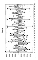
- FIG. 3 Data from the application of the method according to the invention to a human patient are reproduced.
- the upper part of the graph shows the heart curve determined by the measuring electronics.
- the lower part of the graph shows the difference between the standard value and the computationally processed data point. This results in the comparison of the standard value fluctuations upwards or downwards. If the patient is exposed to a stimulus which does not affect him, ie he does not have to process it with energy, the value remains within the normal fluctuation range.
- the conspicuous resonant frequencies are displayed by the device in a graphically processed form (see FIG. 4 ).
- Both baseline values which have a toning (ie, enhancing) effect and a sedating (ie, attenuating) effect of cardiac activity, are of interest for both diagnosis and therapy, and are automatically harmonized with the +/- bipolar function.
- the 20 strongest resonant frequency baseline values can also be selected by manual input.
- the respectively marked resonant frequencies are taken over after confirmation by the user and can thus be further processed and analyzed in the last step.
- Step 7 Result and calculation of the therapy programs
- the won resonance frequencies are further processed. These can be sorted by their polarity, by the increasing frequency base value, or by the magnitude of the change they produce. The latter sorting makes the most sense here.
- the individual frequency basic values can be further analyzed.
- the resonance values can also be stored in the sort order selected on a memory card.
- Example 2 Application Example - Patient A
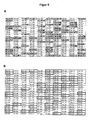
- FIG. 5B is the stored in the database frequency pattern for gallbladder inflammation with a total of 52 frequency baseline values.
- FIG. 6 the comparison of this stored pattern with the determined resonant frequencies of a match of 89%, thus indicating a corresponding disease.
- Example 3 Application Example - Patient B
- the resonance frequency basic values - as in Example 1 - were determined with the bioresonance device.
- the ascertained 56 resonance frequency basic values are in FIG. 7 listed.
- the system found the program number 43.11 rhinitis, then the program number 43.20, bronchial asthma.
- the bioresonance device therefore proposes these therapy programs as the two most important therapy programs for this patient.
- both the resonance frequency basic values and the therapy programs were then stored and provided on a memory card for the subsequent therapy of the patient.
- Example 4 Construction of a bioresonance device according to the invention
- the bioresonance device has the following components: By means of a connection for electrodes which detect the cardiovascular measured values, in particular the heart rate, these measured values are fed into the device. Within the device, the measured values are evaluated in an electronic data processing unit for determining the heart rate variability and forwarded to the main processor (CPU). In the main processor, the comparison of the collected heart rate variability with a setpoint, which is specified as a threshold. Above this threshold, the control of the frequency generator module is changed accordingly. The spectrum of electromagnetic frequencies generated by the frequency generator module is transmitted to the patient via frequency outputs and electrodes attached thereto. For external control of the bioresonance device, it has a touch panel.
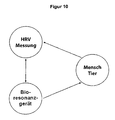
- FIG. 10 is shown schematically the control loop, as it results from the application of the method according to the invention.
- the bioresonance device acts on the human or the animal as a patient.
- the resulting variability of the heart rate is measured by means of a measuring probe and fed back into the bioresonance device, which retains the generated frequency spectrum depending on the measured heart rate variability or modifies according to a predetermined algorithm.
- the generated electromagnetic frequency spectrum is to be understood as a controlled system, the heart rate variability as a controlled variable and the CPU of the bioresonance device as a controller.
Landscapes
- Health & Medical Sciences (AREA)
- Engineering & Computer Science (AREA)
- Biomedical Technology (AREA)
- Nuclear Medicine, Radiotherapy & Molecular Imaging (AREA)
- Radiology & Medical Imaging (AREA)
- Life Sciences & Earth Sciences (AREA)
- Animal Behavior & Ethology (AREA)
- General Health & Medical Sciences (AREA)
- Public Health (AREA)
- Veterinary Medicine (AREA)
- Measurement And Recording Of Electrical Phenomena And Electrical Characteristics Of The Living Body (AREA)
Abstract
Die Erfindung betrifft ein Verfahren zum Betrieb eines medizinischen Geräts, insbesondere eines Bioresonanzgeräts, zur Ermittlung des physiologischen Zustands eines Patienten basierend auf frequenzinduzierten elektrophysiologischen Messwerten, und ein Bioresonanzgerät zur Durchführung dieses Verfahrens.The invention relates to a method for operating a medical device, in particular a bioresonance device, for determining the physiological state of a patient based on frequency-induced electrophysiological measured values, and a bioresonance device for carrying out this method.
Description
Die Erfindung betrifft ein Verfahren zum Betrieb eines medizinischen Geräts, insbesondere eines Bioresonanzgeräts. Die Anmeldung nimmt die Priorität der deutschen Patentanmeldung
Das Verfahren und das Gerät nach der Erfindung dienen der Ermittlung von Daten betreffend den physiologischen Zustand eines tierischen oder menschlichen Patienten sowie deren Auswertung im Hinblick auf den physiologischen Zustand und die mögliche Therapie des Patienten.The method and the device according to the invention are used to determine data concerning the physiological condition of an animal or human patient and their evaluation with regard to the physiological state and the possible therapy of the patient.
In den letzten Jahren hat die Bioresonanz als Diagnose- und/oder Behandlungsmethode der alternativen Medizin zunehmende Bedeutung gewonnen. Grundlage der Bioresonanztherapie ist die Suche (Analyse) und Gabe (Therapie) von Frequenzen und Frequenzspektren mit dem Ziel, Eigenregulationen bei Menschen und Tieren wieder in Gang zu setzen.In recent years bioresonance has become increasingly important as a diagnostic and / or treatment method of alternative medicine. The basis of bioresonance therapy is the search (analysis) and administration (therapy) of frequencies and frequency spectra with the aim of restarting self-regulation in humans and animals.
Nach der Lehre der Bioresonanz strahlt der menschliche oder tierische Körper unterschiedliche elektromagnetische Schwingungen ab. Zellen, Gewebe und Organe haben jeweils spezifische Schwingungen. Diese Einzelschwingungen stehen miteinander in Verbindung und beeinflussen sich gegenseitig. Gemeinsam bilden sie das Gesamtschwingungsspektrum des Patienten, das individuelle Schwingungsbild.According to the bioresonance theory, the human or animal body radiates different electromagnetic oscillations. Cells, tissues and organs each have specific vibrations. These individual vibrations are connected and influence each other. Together they form the overall vibration spectrum of the patient, the individual vibration pattern.
Nach der Theorie der biophysikalischen Medizin treten bei Fehlfunktionen im menschlichen oder tierischen Körper charakteristische Schwingungen auf, anhand derer sich die Natur der Störung identifizieren lässt. Diese charakteristischen Schwingungen (Frequenzen) lassen sich von außen beeinflussen, was zu einer Aufhebung des Störungszustandes kommt.According to the theory of biophysical medicine, malfunctions in the human or animal body lead to characteristic oscillations, by means of which the nature of the disorder can be identified. These characteristic vibrations (frequencies) can be influenced from the outside, which leads to a cancellation of the fault condition.
Zur Anwendung der Bioresonanztherapie sind verschiedene Vorrichtungen bekannt, so beispielsweise aus der deutschen Gebrauchsmusterschrift 32 10 955. Dabei wird ein Detektor mit dem Patienten in Kontakt gebracht, der die elektromagnetischen Schwingungen des Patienten, bzw. einzelner Körperpartien oder Organe aufnimmt und an die Bioresonanzvorrichtung überträgt. In Abhängigkeit von den aufgenommen Schwingungen werden von der Bioresonanzvorrichtung elektromagnetische Frequenzen generiert, die an den Patienten weitergeleitet werden (d.h. sie werden "aufgeprägt"). Damit wird das therapeutische Ziel erreicht.For the application of bioresonance therapy various devices are known, for example from
Somit ist bei der Bioresonanztherapie die möglichst exakte, fehlerfreie Ermittlung der individuellen Schwingungen wie auch die schnelle Auswertung eine wesentliche Grundlage für eine gezielte Diagnose einer Störung und für eine wirkungsvolle Therapie mit raschem Therapieerfolg.Thus, in bioresonance therapy the most accurate, error-free determination of the individual oscillations as well as the rapid evaluation is an essential basis for a targeted diagnosis of a disorder and for an effective therapy with rapid therapeutic success.
Die aus dem Stand der Technik bekannten Geräte nutzen dazu in der Regel die Messung des Hautwiderstandes des Patienten. Da der Hautwiderstand infolge zahlreicher Einflüsse wie Hautbeschaffenheit, Durchblutung, Umgebungstemperatur oder Wassergehalt im Körper sehr stark variiert, ist dieses Messverfahren nur eingeschränkt verwendbar, da es leicht zu falschen Messergebnissen führen kann. Mit dieser Methode kann also nicht ausreichend sichergestellt werden, dass das individuelle Schwingungsbild des Patienten und die Reaktion des Patienten auf die therapeutische Schwingung (Biofeedback) tatsächlich zutreffend erfasst werden. Diese Unsicherheit wirkt sich negativ auf den Therapieerfolg und die Aussagekraft der Diagnose aus.The devices known from the prior art generally use the measurement of the skin resistance of the patient. Since the skin resistance varies greatly as a result of numerous influences such as skin condition, blood circulation, ambient temperature or water content in the body, this measurement method is only limited use, since it can easily lead to incorrect measurement results. Thus, this method can not sufficiently ensure that the individual vibration pattern of the patient and the patient's response to the therapeutic vibration (biofeedback) are actually recorded correctly. This uncertainty has a negative effect on the success of therapy and the validity of the diagnosis.
Aufgabe der Erfindung ist es, ein demgegenüber verbessertes System für die Anwendung der Bioresonanzdiagnostik und -therapie sowie ein Verfahren zum Betrieb dieses Systems bereitzustellen.The object of the invention is to provide a contrast improved system for the application of bioresonance diagnostics and therapy and a method for operating this system.
Diese Aufgabe wird gemäß der Erfindung durch die Bereitstellung eines Verfahrens für den Betrieb/ die Steuerung eines medizinischen Geräts, insbesondere eines Bioresonanzgerätes, gelöst, das die folgenden Schritte umfasst:
- a) Ermitteln von elektrophysiologischen Datenpunkten eines menschlichen oder tierischen Patienten in Abhängigkeit von auf diesen Patienten (Mensch oder Tier) aufgeprägten elektromagnetischen Frequenzen,
- b) Vergleichen dieser elektrophysiologischen Datenpunkte mit einem Standardwert für diese elektrophysiologische Datenpunkte,
- c) Identifizieren mindestens einer der Frequenzen aus Schritt a), bei der der elektrophysiologische Datenpunkt von dem Standardwert abweicht (nachfolgend auch "Resonanzfrequenz" oder "Interferenzwert") und
- d) Abgleichen der mindestens einen Resonanzfrequenz des Patienten mit einer Datenbank umfassend Resonanzfrequenzen.
- a) determining electrophysiological data points of a human or animal patient as a function of electromagnetic frequencies impressed on this patient (human or animal),
- b) comparing these electrophysiological data points with a standard value for these electrophysiological data points,
- c) identifying at least one of the frequencies from step a) in which the electrophysiological data point deviates from the standard value (hereinafter also "resonance frequency" or "interference value") and
- d) matching the at least one resonance frequency of the patient with a database comprising resonance frequencies.
Der Erfindung liegt die Erkenntnis zugrunde, dass ein menschlicher oder tierischer Organismus auf einem Stimulus in Form einer auf ihn einwirkenden ("aufgeprägten") elektromagnetischen Frequenz physiologisch reagiert und die Summe möglicher verschiedener Reaktionen als Veränderung eines elektrophysiologischen Signals des Organismus erkennbar ist. Die Veränderung des elektrophysiologischen Signals ist messbar. Je nach physiologischem Ausgangszustand des Organismus ist die Veränderung des elektrophysiologischen Messwerts bei verschiedenen Stimuli, hier also Frequenzen, besonders erkennbar. Demnach kann das in Abhängigkeit von verschiedenen Frequenzen erfasste Reaktionsmuster aus elektrophysiologischen Messwerten des Organismus zur Diagnose von Krankheits- oder Belastungszuständen des Organismus genutzt werden.The invention is based on the finding that a human or animal organism reacts physiologically on a stimulus in the form of an ("impressed") electromagnetic frequency acting on it and the sum of possible different reactions can be recognized as a change in an electrophysiological signal of the organism. The change in the electrophysiological signal is measurable. Depending on the physiological initial state of the organism, the change in the electrophysiological measured value is particularly noticeable in the case of different stimuli, in this case frequencies. Accordingly, the reaction pattern detected as a function of different frequencies can be used from electrophysiological measured values of the organism for the diagnosis of disease or stress states of the organism.
Die Erfinder haben demnach festgestellt, dass aufgrund der Abweichung eines elektrophysiologischen Messwerten von einem Standardwert Resonanzfrequenzen ("Resonanzfrequenzmuster") eines menschlichen oder tierischen Patienten ermittelt werden können, die geeignet sind, Auskunft über den physiologischen Zustand dieses Patienten zu geben. Nach dem erfindungsgemäßen Verfahren können für den Vergleich entweder diese Messwerte als solche, oder aber aus diesen Messwerten berechnete Werte herangezogen. Sofern nicht ausdrücklich anders angegeben, wird demnach zusammenfassend von elektrophysiologischen "Daten" oder "Datenpunkten" gesprochen. Die aus den Messwerten berechneten Werte werden im Rahmen dieser Anmeldung als "rechnerisch verarbeitete Datenpunkte" bezeichnet. Somit werden von den Begriffen "Daten" oder "Datenpunkte" sowohl die elektrophysiologischen Messwerte umfasst, als auch die daraus berechneten sogenannten "rechnerisch verarbeiteten Datenpunkte".The inventors have therefore found that due to the deviation of an electrophysiological measurement values from a standard value, resonance frequencies ("resonance frequency patterns") of a human or animal patient can be determined which are suitable for providing information about the physiological condition of this patient. According to the method of the invention, either these measured values as such or calculated from these measured values can be used for the comparison. Unless expressly stated otherwise, it is therefore referred to collectively as electrophysiological "data" or "data points". The values calculated from the measured values are referred to in this application as "computationally processed data points". Thus, the terms "data" or "data points" encompass both the electrophysiological measured values and the so-called "computationally processed data points" calculated therefrom.
Die mit Hilfe des erfindungsgemäßen Verfahrens erstellten individuellen Resonanzfrequenzmuster können außerdem zu therapeutischen Zwecken genutzt werden. So können z.B. mit Hilfe eines Bioresonanzgeräts Frequenzen auf den Patienten aufgeprägt werden, die dem Störungszustand entgegenwirken und so den Patienten möglichst wieder in seinen physiologischen Normalzustand (d.h. gesunden Zustand) bringen.The individual resonant frequency patterns created using the method according to the invention can also be used for therapeutic purposes. Thus, e.g. With the help of a bioresonance device frequencies are impressed on the patient, which counteract the disturbance condition and thus bring the patient as far as possible back to its normal physiological state (i.e., healthy state).
Nach dem erfindungsgemäßen Verfahren werden, wie ausgeführt, von dem Patienten zunächst elektrophysiologische Messwerte erhoben. Diese Messwerte sind vorzugsweise Ergebnisse eines Verfahrens, das aus der Gruppe der folgenden Verfahren ausgewählt ist: Elektroenzephalographie (EEG), Elektrokardiographie (EKG), Elektrogastrographie, Elektrokochleographie, Elektronystagmographie, Elektrookulographie, Elektroretinographie, Elektromyographie, und Elektroneurographie. Bevorzugt sind Messwerte aus der Elektroenzephalographie und der Elektrokardiographie. Besonders bevorzugt wird das erfindungsgemäße Verfahren mit Messwerten aus der Elektrokardiographie durchgeführt.According to the method of the invention, electrophysiological measured values are first of all collected by the patient, as stated. These measurements are preferably results of a method selected from the group of the following methods: Electroencephalography (EEG), electrocardiography (ECG), electrogastrography, electrocochleography, electronystagmography, electrooculography, electroretinography, electromyography, and electroneurography. Preference is given to measured values from electroencephalography and electrocardiography. The method according to the invention is particularly preferably carried out with measured values from electrocardiography.
All diesen Verfahren ist gemein, dass die elektrische Aktivität, d.h. die Leitung eines elektrischen Impulses bzw. einer Erregung, eines Organs oder Gewebes erfasst wird. Diese Aktivität wird über die Zeit gemessen und in für das jeweilige Verfahren typischen Kurvenverlaufen erfasst. Die erfindungsgemäß relevanten Messwerte werden demnach als Messwertkurven dargestellt. Die Gestalt der jeweiligen Kurve, z.B. die Lage und Steilheit bestimmter Peaks, gibt dem medizinischen Fachmann Anhaltspunkte über den physiologischen Zustand des jeweiligen Organs bzw. Gewebes. Bei einem EKG wird zum Beispiel ein Herzschlag in einer typischen Kurve dargestellt, die als P-Q-R-S-T-U Kurve bekannt ist. Die einzelnen Buchstaben sind Bezeichnungen für charakteristische Peaks, Täler oder sonstige typische Merkmale der Kurve (siehe dazu auch
In dem erfindungsgemäßen Verfahren werden die vom Patienten ermittelten elektrophysiologischen Messwerte, also die entsprechenden Messwertkurven, vorzugsweise nicht unmittelbar (d.h. im Sinne von Rohdaten) für den Vergleich mit Standardwerten oder Standardkurven eingesetzt, sondern werden zunächst rechnerisch verarbeitet ("verrechneter Datenpunkt"). In einer besonders vorteilhaften Ausführungsform der Erfindung wird die Fläche unter einer Kurve (Area Under the Curve; AUC) erfasst und als Ausgangspunkt für den Vergleich mit einem Standardwert herangezogen. Dem Fachmann sind die Methoden zur Berechnung der Fläche unter einer Kurve bekannt. Für diese Berechnung wird zunächst ein für das jeweilige Verfahren sinnvoller Kurvenabschnitt definiert. Zum Beispiel wird bei einem EKG als Kurvenabschnitt vorzugsweise eine vollständige P-Q-R-S-T-U Kurve, d.h. also die Erregungsleitung eines Herzschlags definiert. In diesem Fall wird nach dem erfindungsgemäßen Verfahren demnach die jeweilige Fläche unter einer bei einer bestimmten Frequenz gemessenen P-Q-R-S-T-U Kurve als bevorzugter elektrophysiologischer Datenpunkt ermittelt. Vorzugsweise werden mindestens zwei oder mehr solcher Datenpunkte ermittelt, d.h. es wird eine Mehrzahl von Flächen ermittelt. Die Kurven werden in Abhängigkeit von zeitgleich aufgeprägten Frequenzen aufgenommen.In the method according to the invention, the electrophysiological measured values ascertained by the patient, ie the corresponding measured value curves, are preferably not used directly (that is to say in the sense of raw data) for comparison with standard values or standard curves, but are first of all computationally processed ("calculated data point"). In a particularly advantageous embodiment of the invention, the area under an area under the curve (AUC) is detected and used as a starting point for comparison with a standard value. The person skilled in the art knows the methods for calculating the area under a curve. For this calculation, a meaningful curve section is first defined for the respective method. For example, with an ECG as a curve section, preferably a complete P-Q-R-S-T-U curve, i. that defines the excitation conduction of a heartbeat. In this case, according to the method of the invention, the respective area is thus determined under a P-Q-R-S-T-U curve measured at a specific frequency as the preferred electrophysiological data point. Preferably, at least two or more such data points are detected, i. it is determined a plurality of areas. The curves are recorded as a function of simultaneously impressed frequencies.
In weiteren Ausführungsformen der Erfindung können alternativ oder ergänzend zu der Flächenberechnung andere rechnerisch zu ermittelnde Charakteristika der vom Patienten erfassten elektrophysiologischen Messwerte (Kurven) zur Darstellung und Analyse des Zustands des Patienten herangezogen werden. In der besonders bevorzugten Ausführungsform auf Basis eines EKG wird vorzugsweise die Steilheit des Q-R-S Peaks ("Gradient") ausgewertet. Dieser kann durch einen Winkel alpha beschrieben werden, der dem Winkel zwischen der Senkrechten zur X-Achse (= Zeitachse im EKG) und dem ansteigendem Schenkel der R-Zacke entspricht (siehe
In einer weiteren Ausführungsform der Erfindung können alternativ oder ergänzend zu den bereits genannten Messwerten Gradient und Fläche unter der Kurve (AUC) auch ein oder mehrere der kardiovaskulären Messwerte ausgewählt aus der Gruppe umfassend Herzfrequenz, Herzrhythmus, Länge, Amplitude oder Form der P-Welle, der T-Welle, der U-Welle oder des QRS-Komplexes, Länge des PQ-Intervalls, des QT-Intervalls, des RR-Intervalls oder der ST-Strecke herangezogen werden. Ganz besonders bevorzugt wird die Herzfrequenz, insbesondere bevorzugt die Herzfrequenzvariabilität (engl. Heart rate variability; HRV) herangezogen. Diese kann auf Basis von drei bis vier Herzschlägen bestimmt werden. Eine Steuerung auf Basis von 3-4 Herzschlägen ist vorteilhaft, da die HRV nur kurzzeitig auf die von außen aufgeprägten Frequenzen reagiert und sich nach spätestens 3-4 Herzschlägen wieder auf die neue Situation anpasst.In a further embodiment of the invention, as an alternative or in addition to the already mentioned measured values gradient and area under the curve (AUC), one or more of the cardiovascular measured values can also be selected from the group comprising heart rate, heart rhythm, length, amplitude or shape of the P-wave, the T-wave, the U-wave or the QRS complex, length of the PQ interval, the QT interval, the RR interval or the ST segment. The heart rate, in particular the heart rate variability (HRV), is particularly preferably used. This can be determined based on three to four heartbeats. A control based on 3-4 heart beats is advantageous because the HRV reacts only briefly to the frequencies impressed from the outside and adapts to the new situation again after a maximum of 3-4 heart beats.
Die elektrophysiologischen Messwerte des Patienten werden nach dem erfindungsgemäßen Verfahren in Abhängigkeit von auf den Patienten aufgeprägten elektromagnetischen Schwingungen (Frequenzen) erfasst. Das bedeutet, dass die Messwerte erfasst werden, während der Patient die elektrischen Impulse, d.h. die Stimulation, durch die aufgeprägten Frequenzen erhält. Damit wird Schritt a) des erfindungsgemäßen Verfahrens umgesetzt. Der Patient ist also zeitgleich mit einer Vorrichtung zur Erfassung der elektrophysiologischen Messwerte und mit einer Vorrichtung zur Aufprägung (Stimulation) der Frequenzen auf den Patienten verbunden. Beide Vorrichtungen können getrennt oder kombiniert sein.The electrophysiological measured values of the patient are detected by the method according to the invention as a function of electromagnetic vibrations (frequencies) impressed on the patient. This means that the readings are taken while the patient is reading the electrical pulses, i. the stimulation through which impressed frequencies receives. Thus, step a) of the method according to the invention is implemented. The patient is thus simultaneously connected to a device for recording the electrophysiological measured values and to a device for imparting (stimulating) the frequencies to the patient. Both devices can be separate or combined.
Auf den Patienten werden erfindungsgemäß in einem ersten Schritt mindestens zwei, insbesondere mehr als zwei Frequenzen aufgeprägt. In einer Ausführungsform werden die mehr als zwei Frequenzen als Abfolge von Einzelfrequenzen aufgeprägt, d.h. die Frequenzen wirken nacheinander und nicht als Gemisch verschiedener Frequenzen auf den Patienten ein. So kann die Wirkung einer bestimmten Einzelfrequenz auf den Patienten ermittelt werden.According to the invention, at least two, in particular more than two, frequencies are impressed on the patient in a first step. In one embodiment, the more than two frequencies are imposed as a sequence of single frequencies, i. the frequencies act on the patient in succession rather than as a mixture of different frequencies. Thus, the effect of a specific single frequency on the patient can be determined.
In einer Ausführungsform des erfindungsgemäßen Verfahrens werden zwischen 10 bis 500, bevorzugt zwischen 50 und 400, weiter bevorzugt zwischen 100 und 200 Frequenzen auf den Patienten geprägt. Damit erfolgt dann eine sog. Bereichswertmessung. Bei jeder dieser Frequenzen wird eine elektrophysiologische Messwertkurve aufgenommen. Bei N aufgeprägten Frequenzen werden somit auch N elektrophysiologische Messwertkurven aufgenommen.In one embodiment of the method according to the invention, between 10 and 500, preferably between 50 and 400, more preferably between 100 and 200 frequencies are embossed onto the patient. This then results in a so-called range value measurement. At each of these frequencies an electrophysiological measurement curve is recorded. At N impressed frequencies thus also N electrophysiological measured value curves are recorded.
Die aufgeprägten Frequenzen liegen vorzugsweise im Bereich von 0,1 Hz bis 10,0 GHz. In einer bevorzugten Ausführungsform liegen sie im Bereich von 0,1 Hz bis 100 MHz und bevorzugt im Bereich von 0,1 Hz bis 500 MHz.The impressed frequencies are preferably in the range of 0.1 Hz to 10.0 GHz. In a preferred embodiment, they are in the range of 0.1 Hz to 100 MHz and preferably in the range of 0.1 Hz to 500 MHz.
Sofern die Frequenzen sequenziell, d.h. in einer Abfolge, aufgeprägt werden, können sie zueinander denselben Frequenzunterschied aufweisen, d.h. die Frequenzfolge besitzt eine einheitliche Schrittweite. Wenn beispielsweise eine Abfolge von Frequenzen mit einem Frequenzwert von 0,5 kHz; 1,0 kHz; 1,5 kHz; 2,0 kHz; 2,5 kHz; 3,0 kHz; 3,5 kHz; 4,0 kHz etc. verwendet werden, beträgt die Schrittweite 0,5 kHz.If the frequencies are sequential, i. in a sequence, they may have the same frequency difference to each other, i. the frequency sequence has a uniform step size. For example, if a sequence of frequencies with a frequency value of 0.5 kHz; 1.0 kHz; 1.5 kHz; 2.0 kHz; 2.5 kHz; 3.0 kHz; 3.5 kHz; 4.0 kHz, etc., the step size is 0.5 kHz.
In einer Ausführungsform der Erfindung werden jeweils einzelne Frequenzen auf den Patienten aufgeprägt. Es ist aber bevorzugt, jeweils Frequenzspektren aufzuprägen, d.h. mehrere Einzelfrequenzen gleichzeitig dem Patienten zu applizieren.In one embodiment of the invention, individual frequencies are impressed on the patient. It is preferred, however, to impose frequency spectra, i. to apply several single frequencies simultaneously to the patient.
Vorzugsweise handelt es sich bei den aufgeprägten Spektren um dekadische Frequenzspektren, d.h. um Spektren, die jeweils von einer Einzelfrequenz als Grundfrequenz ausgehen, und dann nur weitere Frequenzen aufweisen, die jeweils um den Faktor 10 gegenüber der jeweils niedrigen Frequenz erhöht sind. Der niedrigste Frequenzwert wird als "Frequenzgrundwert" bezeichnet. So würde bei einem Frequenzgrundwert von 0,5 kHz das dekadische Frequenzspektrum Frequenzen von 5 kHz, 50 kHz, 500 kHz usw. enthalten. Ein dekadisches Frequenzspektrum enthält ausschließlich solche jeweils um den Faktor 10 voneinander abweichende Frequenzen.Preferably, the impressed spectra are decadal frequency spectra, i. Spectra, each starting from a single frequency as the fundamental frequency, and then only have other frequencies, which are each increased by a factor of 10 against the respective low frequency. The lowest frequency value is referred to as the "frequency baseline". Thus, with a frequency baseline of 0.5 kHz, the decadic frequency spectrum would include frequencies of 5 kHz, 50 kHz, 500 kHz, and so on. A decadal frequency spectrum contains only those frequencies that differ by a factor of 10 from each other.
In dieser bevorzugten Ausführungsform steht somit der Frequenzgrundwert nicht für eine konkrete Einzelfrequenz, sondern für den kleinsten Wert eines Frequenzspektrums, wobei die weiteren Frequenzen jeweils eine um den Faktor 10n höhere Frequenz aufweisen. Sofern nicht näher spezifiziert, wird im Kontext der vorliegenden Erfindung unter dem allgemeinen Begriff Frequenz sowohl die Einzelfrequenz als auch ein Frequenzspektrum verstanden.In this preferred embodiment, therefore, the basic frequency value is not for a specific single frequency, but for the smallest value of a frequency spectrum, wherein the other frequencies each have a higher frequency by a factor of 10 n . Unless specified, in the context of the present invention, the general term frequency is understood to mean both the single frequency and a frequency spectrum.
In einer bevorzugten Ausführungsform wird zunächst die niedrigste Frequenz aufgeprägt. Dann wird schrittweise der Frequenzbereich bis zur höchsten Frequenz durchlaufen. Die Abfolge kann aber auch genau anders herum sein, d.h. es kann auch mit der höchsten Frequenz begonnen werden und dann eine schrittweise Erniedrigung bis zur kleinsten Frequenz erfolgen.In a preferred embodiment, first the lowest frequency is impressed. Then the frequency range is progressively stepped through to the highest frequency. But the sequence can also be the other way round, i. E. It can also be started with the highest frequency and then a gradual reduction to the smallest frequency.
Gemäß Schritt b) des erfindungsgemäßen Verfahrens werden die elektrophysiologischen Datenpunkte mit einem Standardwert für diese Datenpunkte verglichen, um so mindestens eine Resonanzfrequenz zu ermitteln. Die Resonanzfrequenz ist diejenige Frequenz, dessen elektrophysiologischer Datenpunkt vom Standardwert abweicht. Bei der Aufprägung einer Mehrzahl von Frequenzen (Bereichswertmessung) können zwei oder mehr Resonanzfrequenzen gefunden werden. Häufig liegt die Anzahl von ermittelten Resonanzfrequenzen zwischen 10 und 100, vorzugsweise zwischen 50 und 80 Resonanzfrequenzen. Die Anzahl der bei einer Bereichswertmessung ermittelten Resonanzfrequenzen kann durch die Verwendung eines Filters beeinflussbar sein.According to step b) of the method according to the invention, the electrophysiological data points are compared with a standard value for these data points so as to determine at least one resonance frequency. The resonance frequency is the frequency whose electrophysiological data point deviates from the standard value. When imposing a plurality of frequencies (range value measurement), two or more resonance frequencies can be found. Often the number of identified Resonant frequencies between 10 and 100, preferably between 50 and 80 resonant frequencies. The number of resonant frequencies determined in a range value measurement can be influenced by the use of a filter.
Als Standardwert kann ein Wert aus einer externen Datenbank herangezogen werden, d.h. ein Wert, der nicht auf den individuellen Patienten zurückgeht. So sind, wie oben ausgeführt, die typischen Kurvenverläufe der erfindungsgemäßen elektrophysiologischen Messwerte dem Fachmann grundsätzlich bekannt. Die vom Patienten bei einer bestimmten Frequenz ermittelte Kurve kann mit einer solchen typischen Kurve verglichen werden. Als Vergleichsmaßstab kann dazu vorzugsweise die Analyse der Fläche unter der Kurve dienen.The default value can be a value from an external database, i. a value that does not go back to the individual patient. Thus, as stated above, the typical curves of the electrophysiological measured values according to the invention are fundamentally known to the person skilled in the art. The curve obtained by the patient at a certain frequency can be compared to such a typical curve. As a comparative scale, the analysis of the area under the curve can preferably serve this purpose.
In einer bevorzugten Ausführungsform der Erfindung wird ein Standardwert herangezogen, der in der jeweiligen Bereichswertmessung des Patienten ermittelt wird. Das Heranziehen des Mittelwertes aus der patienteneigenen Bereichswertmessung als Sollwert hat den Vorteil, dass sich darin der individuelle elektrophysiologisch erfassbare Zustand des Patienten zum Zeitpunkt der Messung widerspiegelt.In a preferred embodiment of the invention, a standard value is used, which is determined in the respective range value measurement of the patient. The use of the mean value from the patient's own range value measurement as desired value has the advantage that it reflects the individual electrophysiologically detectable state of the patient at the time of the measurement.
Der patienteneigene, d.h. individuelle, Standardwert ist vorzugsweise der Mittelwert aller für den Patienten ermittelten Datenpunkte im Verlaufe einer Messung. Dieser Standardwert kann auch als Sollwert bezeichnet werden. Auch für diese Berechnung wird vorzugsweise die Fläche unter der Messwertkurve herangezogen. Diejenigen Frequenzen, bei denen die Fläche der Messwertkurve vom Mittelwert aller erfassten Flächen abweicht, stellen dann die gesuchten Resonanzfrequenzen dar. Das Maß der Abweichung, das als diagnostisch oder therapeutisch relevant erachtet wird, kann vom Fachmann bestimmt werden. Durch die Ermittlung der Resonanzfrequenzen entsteht ein individueller "energetischer Fingerabdruck" des Patienten.The patient's own, i. individual, default value is preferably the mean of all data points determined for the patient in the course of a measurement. This default value can also be called a setpoint. Also for this calculation, the area under the measured value curve is preferably used. Those frequencies at which the area of the measured value curve deviates from the mean value of all detected areas then represent the resonance frequencies sought. The degree of deviation, which is considered to be diagnostically or therapeutically relevant, can be determined by a person skilled in the art. Determining the resonance frequencies creates an individual "energetic fingerprint" of the patient.
In einer bevorzugten Ausführungsform wird zusätzlich erfasst, ob sich der Messwert durch die aufgeprägte Frequenz erhöht oder erniedrigt. Diese Wirkung wird durch die sog. Polarität der Resonanzfrequenz zum Ausdruck gebracht ("+" für Erhöhung; "-". für Erniedrigung).In a preferred embodiment, it is additionally detected whether the measured value is increased or decreased by the impressed frequency. This effect is expressed by the so-called polarity of the resonance frequency ("+" for increase; "-" for reduction).
Für die Auswertung der ermittelten Resonanzfrequenzen durch den Abgleich mit einer Datenbank mit vorbekannten Resonanzfrequenzmustern (Schritt d) des erfindungsgemäßen Verfahrens) können diese verschiedenen Auswahlkriterien mit Hilfe entsprechender Filter genauer dargestellt und ausgewertet werden. Die Resonanzfrequenzmuster aus der Datenbank repräsentieren vorzugsweise bestimmte physiologische Zustände. Den Datenbanken liegen eine hohe Anzahl verschiedener Bereichswertmessungen unterschiedlicher Patienten zugrunde. Die erfindungsgemäß heranzuziehenden Datenbanken geben somit Standardresonanzfrequenzmuster wieder. In der Datenbank sind vorzugsweise Resonanzmuster sowohl für einen gesunden Normalzustand als auch für pathologische Zustände abgelegt. In einer Ausführungsform der Erfindung umfasst die Datenbank mehr als 500, bevorzugt mehr als 1500 Standardfrequenzmuster.For the evaluation of the determined resonant frequencies by comparison with a database with previously known resonance frequency patterns (step d) of the method according to the invention), these different selection criteria can be more accurately represented and evaluated with the aid of appropriate filters. The resonant frequency patterns from the database preferably represent certain physiological conditions. The databases are based on a high number of different range value measurements of different patients. The databases to be used according to the invention thus represent standard resonance frequency patterns. In the database, resonance patterns are preferably both for a healthy normal state and for pathological conditions filed. In one embodiment of the invention, the database comprises more than 500, preferably more than 1500 standard frequency patterns.
Der Abgleich der individuellen Resonanzfrequenzen mit den Standardresonanzfrequenzmustern aus einer Datenbank ermöglicht die Ermittlung des physiologisches Zustandes des Patienten.The comparison of the individual resonance frequencies with the standard resonance frequency patterns from a database makes it possible to determine the physiological condition of the patient.
In einer Ausführungsform der Erfindung werden die durch das erfindungsgemäße Verfahren ermittelten Resonanzfrequenzen dazu genutzt, für diesen Patienten therapeutisch wirksame Frequenzen auszuwählen. Die Auswahl kann manuell durch den Therapeuten erfolgen oder aber automatisiert durch das Gerät, insbesondere durch ein Bioresonanzgerät. Die Auswahl der Frequenzen folgt vorteilhafterweise nach dem Prinzip des "similia similibus curentur", d.h. es werden dem Patienten zur Therapie ("Harmonisierung") seiner Störung identische oder ähnliche Frequenzen aufgeprägt.In one embodiment of the invention, the resonance frequencies determined by the method according to the invention are used to select therapeutically effective frequencies for this patient. The selection can be made manually by the therapist or automated by the device, in particular by a bioresonance device. The selection of frequencies advantageously follows the principle of "similia similibus curentur", i. the patient is given identical or similar frequencies for therapy ("harmonization") of his disorder.
In diesem Aspekt betrifft die Erfindung demnach ein Verfahren für den Betrieb eines medizinischen Geräts, insbesondere eines Bioresonanzgerätes, mit folgenden Schritten:
- a) Ermitteln von elektrophysiologischen Datenpunkten eines menschlichen oder tierischen Patienten in Abhängigkeit von auf diesen Patienten (Mensch oder Tier) aufgeprägten elektromagnetischen Frequenzen,
- b) Vergleichen dieser elektrophysiologischen Datenpunkte mit einem Standardwert für diese elektrophysiologischen Datenpunkte,
- c) Identifizieren mindestens einer der Frequenzen aus Schritt a), bei der ein elektrophysiologischer Datenpunkt von dem Standardwert abweicht ("Resonanzfrequenz" oder "Interferenzwert") und
- d) Abgleichen der mindestens einen Resonanzfrequenz des Patienten mit einer Datenbank umfassend Resonanzfrequenzen und Identifizieren mindestens eines Resonanzfrequenzmusters,
- e) Erzeugen der mindestens einen nach c) identifizierten Resonanzfrequenz und/oder des mindestens einen nach d) identifizierten Resonanzfrequenzmusters und
- f) Übermitteln der Resonanzfrequenz und/oder des Resonanzfrequenzmusters nach e) auf mindestens eine mit dem Gerät verbundene Elektrode.
- a) determining electrophysiological data points of a human or animal patient as a function of electromagnetic frequencies impressed on this patient (human or animal),
- b) comparing these electrophysiological data points with a standard value for these electrophysiological data points,
- c) identifying at least one of the frequencies from step a) at which an electrophysiological data point deviates from the standard value ("resonance frequency" or "interference value") and
- d) matching the at least one resonance frequency of the patient with a database comprising resonance frequencies and identifying at least one resonance frequency pattern,
- e) generating the at least one resonant frequency identified according to c) and / or the at least one resonant frequency pattern identified according to d) and
- f) transmitting the resonance frequency and / or the resonance frequency pattern according to e) to at least one electrode connected to the device.
In einer besonders vorteilhaften Ausgestaltung kann das erfindungsgemäße Verfahren zum Betrieb des medizinischen Geräts als ein Regelkreislauf gestaltet sein, indem der Standardwert als Sollwert und die nach Schritt a) erhobenen Datenpunkte den Istwert darstellen. Nach dieser Ausführungsform können die nach den Schritten e) und f) erzeugten und übermittelten Resonanzfrequenzen auf den Patienten aufgeprägt und dessen elektrophysiologischen Datenpunkte gemäß Schritt a) in Abhängigkeit von diesen Frequenzen ermittelt werden. Das erfindungsgemäße Verfahren wird dann so oft iterativ durchlaufen, bis es keine medizinisch relevante Abweichung mehr gibt zwischen den Istwerten und dem Sollwert.In a particularly advantageous embodiment, the inventive method for operating the medical device can be designed as a control loop by the default value as setpoint and the data points collected after step a) represent the actual value. According to this embodiment, the resonance frequencies generated and transmitted after steps e) and f) can be impressed on the patient and his electrophysiological data points according to step a) in dependence thereon Frequencies are determined. The method according to the invention is then repeated iteratively until there is no medically relevant deviation between the actual values and the desired value.
In einem weiteren Aspekt stellt die Erfindung ein Verfahren für den Betrieb eines Bioresonanzgerätes bereit, das die folgenden Schritte umfasst:
- a) Erfassung mindestens eines kardiovaskulären Messwertes (Istwert) eines Patienten in Abhängigkeit von auf diesen Patienten aufgeprägten elektromagnetischen Frequenzen,
- b) Vergleich des kardiovaskulären Messwerts mit einem Sollwert
- c) Steuerung eines Moduls zur Erzeugung elektromagnetischer Frequenzen (Frequenzgeneratormodul) in Abhängigkeit von der Differenz zwischen Messwert und Sollwert.
- a) acquisition of at least one cardiovascular measured value (actual value) of a patient as a function of electromagnetic frequencies impressed on this patient,
- b) Comparison of the cardiovascular measured value with a desired value
- c) Control of a module for generating electromagnetic frequencies (frequency generator module) as a function of the difference between measured value and setpoint.
So haben die Erfinder gefunden, dass die zielgerichtete Steuerung eines Bioresonanzgerätes in sehr vorteilhafter Weise durch die Erfassung und Auswertung kardiovaskulärer Messwerte erfolgen kann.Thus, the inventors have found that the targeted control of a bioresonance device can be carried out in a very advantageous manner by the detection and evaluation of cardiovascular measured values.
Diese erfindungsgemäße Ausführungsform hat insbesondere den Vorteil, dass kardiovaskuläre Daten von äußeren, für die Therapie unmaßgeblichen Reizen weniger beeinflussbar sind und so zu einem zuverlässigeren Therapie- und Diagnoseerfolg führen. Außerdem erlauben kardiovaskuläre Messungen im Gegensatz zu Hautwiderstandsmessungen ein breites Spektrum an möglichen Auswerteparametern (Herzrate, Herzfrequenz, Größe, Zeitdauer und Form der einzelnen Wellen etc.), die in Abhängigkeit von der zu therapierenden oder diagnostizierenden Erkrankung gewählt werden können. In einer besonderen Ausführungsform wird die Fläche unter der Messwertkurve zur Auswertung herangezogen.In particular, this embodiment according to the invention has the advantage that cardiovascular data are less susceptible to external stimuli that are irrelevant to the therapy and thus lead to a more reliable therapy and diagnostic success. In addition, cardiovascular measurements in contrast to skin resistance measurements allow a wide range of possible evaluation parameters (heart rate, heart rate, size, duration and shape of the individual waves, etc.), which can be selected depending on the disease to be treated or diagnosed. In a particular embodiment, the area under the measured value curve is used for the evaluation.
Die erfindungsgemäß möglichen kardiovaskulären Messparameter sind durch die langjährige intensive Forschung in ihrer Bedeutung und ihrer individuellen Variabilität sehr genau bekannt und stellen somit eine gute Grundlage für ein darauf aufbauendes Steuerungssystem dar.The cardiovascular measurement parameters that are possible according to the invention are very precisely known in their importance and their individual variability as a result of many years of intensive research and thus represent a good basis for a control system based thereon.
Diese erfindungsgemäße Ausführungsform kann vorteilhafterweise als ein Regelkreis aufgebaut werden. Indem Frequenzspektren, die durch das Bioresonanzgerät auf den Patienten einwirken, ihrerseits kardiovaskuläre Veränderungen hervorrufen können, können diese Veränderungen im Sinne eines Biofeedback-Mechanismus wieder zur Anpassung der von dem Generatormodul erzeugten elektromagnetischen Frequenzen verwendet werden. Vorzugsweise werden demnach die Schritte a), b) und c) mindestens einmal, besonders bevorzugt mehrmalig wiederholt.This embodiment according to the invention can be advantageously constructed as a control loop. In turn, by allowing frequency spectra that act on the patient through the bioresonance device to cause cardiovascular changes, these biofeedback mechanism changes can be re-used to match the electromagnetic frequencies generated by the generator module. Preferably, therefore, steps a), b) and c) are repeated at least once, more preferably several times.
Der Betrieb des Bioresonanzgerätes wird in dieser Ausführungsform also über einen Regelkreis gesteuert, in dem mindestens ein kardiovaskuläres Signal als Regelgröße eingesetzt wird.The operation of the bioresonance device is thus controlled in this embodiment via a control loop in which at least one cardiovascular signal is used as a controlled variable.
Damit erlaubt diese Ausführungsform in vorteilhafter Weise eine interaktive Veränderung der auf den Patienten einwirkenden Frequenzspektren. Wird ein Frequenzspektrum therapeutisch als wirksam detektiert, kann während der Messung direkt durch die Software entschieden werden, welches Frequenzspektrum vorteilhafterweise auf den Patienten aufgeprägt werden kann. Dies erlaubt eine effektivere und vor allem detailliertere und schnellere Analyse in Verbindung mit einer zugleich verkürzten Therapiedauer.Thus, this embodiment advantageously allows an interactive change of the frequency spectrums acting on the patient. If a frequency spectrum is therapeutically detected as being effective, it can be decided directly by the software during the measurement which frequency spectrum can advantageously be impressed on the patient. This allows a more effective and, above all, more detailed and faster analysis in conjunction with a shortened therapy duration.
Der besondere Vorteil dieser Ausführungsform der Erfindung ist demnach, dass die Auswahl der therapeutischen Frequenz und/oder die Dauer ihrer Anwendung nicht mehr auf Erfahrungswerten basiert, sondern auf Grundlage mindestens eines am Patienten erhobenen kardiovaskulären Parameters, der nachweislich durch eine zuvor aufgeprägte Frequenz beeinflusst wurde.The particular advantage of this embodiment of the invention is therefore that the selection of the therapeutic frequency and / or the duration of its application is no longer based on empirical values, but on the basis of at least one cardiovascular parameter collected on the patient, which has been demonstrably influenced by a previously impressed frequency.
In einer bevorzugten Ausführungsform der Erfindung geben die kardiovaskulären Messwerte die elektrischen Aktivitäten der Herzmuskelfasern wieder, wie sie beispielsweise durch ein Elektrokardiogramm (EKG) erfasst werden.In a preferred embodiment of the invention, the cardiovascular measurements reflect the electrical activities of the cardiac muscle fibers as detected, for example, by an electrocardiogram (ECG).
In einer Ausführungsform der Erfindung werden die kardiovaskulären Messwerte während der Anwendung des Bioresonanzgerätes erfasst. Diese direkte Kopplung zwischen der Erfassung der Messwerte und einer darauf abgestimmten Steuerung der elektromagnetischen Frequenzen erlaubt die Durchführung des Verfahrens in einem Regelkreis.In one embodiment of the invention, the cardiovascular measurements are acquired during use of the bioresonance device. This direct coupling between the detection of the measured values and a coordinated control of the electromagnetic frequencies makes it possible to carry out the method in a control loop.
In einer weiteren Ausführungsform beträgt bei dem erfindungsgemäßen Verfahren die Zeitspanne zwischen dem Eintreffen des kardiovaskulären Messwertes in dem Bioresonanzgerät und der Erzeugung einer dadurch gesteuerten elektromagnetischen Frequenz weniger als 60 Sekunden, bevorzugt weniger als 20 Sekunden, weiter bevorzugt weniger als 5 Sekunden und besonders bevorzugt weniger als 2 Sekunden. Dies wird vorteilhafterweise durch die Verwendung einer algorithmenbasierten Software gewährleistet, die eine dynamische Reaktion auf das jeweilige Messergebnis mit kurzen Auswertezeiten ermöglicht. Die Software kann so ausgelegt sein, dass sie die exakte Peak-Bestimmung der kardialen Messkurve erlaubt. Die Auswertung der kardiovaskulären Parameter kann nach üblichen, dem Fachmann bekannten Methoden erfolgen. So kann der bevorzugte Parameter Herzfrequenzvariabilität (HRV) zum Beispiel im Zeitbereich, im Frequenzbereich oder im nichtlinearen Bereich bestimmt werden (s. Definition der HRV).In a further embodiment, in the method according to the invention, the time interval between the arrival of the cardiovascular measured value in the bioresonance device and the generation of a controlled electromagnetic frequency is less than 60 seconds, preferably less than 20 seconds, more preferably less than 5 seconds and particularly preferably less than 2 seconds. This is advantageously ensured by the use of algorithm-based software, which enables a dynamic response to the respective measurement result with short evaluation times. The software can be designed to allow the exact peak determination of the cardiac waveform. The evaluation of the cardiovascular parameters can be carried out by customary methods known to the person skilled in the art. Thus, the preferred parameter heart rate variability (HRV) can be determined, for example, in the time domain, in the frequency domain or in the non-linear domain (see Definition of HRV).
In einer Ausführungsform der Erfindung sind die kardiovaskulären Messwerte ausgewählt aus der Gruppe umfassend Herzfrequenz, Herzrhythmus, Länge, Amplitude oder Form der P-Welle, der T-Welle, der U-Welle oder des QRS-Komplexes, Länge des PQ-Intervalls, des QT-Intervalls, des RR-Intervalls oder der ST-Strecke. Wie oben bereits ausgeführt, stellt die Fläche unter der Kurve ein besonders relevantes Merkmal dar. Sie fließt demnach in einer bevorzugten Ausführungsform der Erfindung als Datenpunkt in die Auswertung ein.In one embodiment of the invention, the cardiovascular measurements are selected from the group comprising heart rate, heart rhythm, length, amplitude or shape of the P-wave, the T-wave, the U-wave or the QRS complex, length of the PQ interval, QT interval, the RR interval or the ST segment. As already explained above, the area under the curve represents a particularly relevant feature. Accordingly, in a preferred embodiment of the invention, it flows into the evaluation as a data point.
In einer bevorzugten Ausführungsform der Erfindung wird zur Steuerung die Herzfrequenz und hierbei bevorzugt die Herzfrequenzvariabilität (engl. Heart rate variability; HRV) herangezogen, die in besonders bevorzugtem Maße auf Basis von 3-4 Herzschlägen bestimmt wird. Eine Steuerung auf Basis von 3-4 Herzschlägen ist vorteilhaft, da die HRV nur kurzzeitig auf die von außen aufgeprägten Frequenzen reagiert und sich nach spätestens 3-4 Herzschlägen wieder auf die neue Situation anpasst.In a preferred embodiment of the invention, the heart rate is used for the control and in this case preferably the heart rate variability (HRV), which is determined to a particularly preferred extent on the basis of 3-4 heart beats. A control based on 3-4 heart beats is advantageous because the HRV reacts only briefly to the frequencies impressed from the outside and adapts to the new situation again after a maximum of 3-4 heart beats.
Da die Herzratenvariabilität ihren Ursprung in der Funktion des vegetativen Nervensystems hat, lassen sich damit erfindungsgemäß beispielsweise Erkrankungen diagnostizieren und/oder behandeln, bei denen das autonome Nervensystem eine zentrale Rolle spielt. Dabei sind Erkrankungen zu unterscheiden, die direkt das autonome Nervensystem schädigen, und Krankheiten, die sich etwa über dauerhaft erhöhte Stoffwechselbeanspruchungen indirekt auf das autonome Nervensystem auswirken.Since heart rate variability has its origin in the function of the autonomic nervous system, it is thus possible according to the invention to diagnose and / or treat, for example, diseases in which the autonomous nervous system plays a central role. Diseases that directly affect the autonomic nervous system and illnesses that have an effect on the autonomic nervous system, for example, through permanently increased metabolic stress, must be distinguished.
Ein Beispiel für eine Erkrankung mit direkter Schädigung des autonomen Nervensystems ist die diabetische Neuropathie, wohingegen beispielsweise die koronare Herzkrankheit sich indirekt auf das autonome Nervensystem auswirkt. Auch psychische Erkrankungen können über eine Erhöhung des Katecholaminspiegels und die Sympathikusaktivierung erkennbare Folgen auf die Herzaktivität haben; die Herzfrequenzvariabilität kann daher für die erfindungsgemäße Bioresonanzvorrichtung auch im Bereich der Neuropsychiatrie zu diagnostischen und therapeutischen Zwecken herangezogen werden. Auch diese Erkrankungen sind daher erfindungsgemäß behandelbar.An example of a disease directly affecting the autonomic nervous system is diabetic neuropathy, whereas, for example, coronary heart disease has an indirect effect on the autonomic nervous system. Also mental illnesses can have recognizable consequences on the heart activity by an increase of the Katecholaminspiegels and the sympathetic activation; The heart rate variability can therefore be used for the bioresonance device according to the invention also in the field of neuropsychiatry for diagnostic and therapeutic purposes. These diseases are therefore treatable according to the invention.
Weitere Erkrankungen, die erfindungsgemäß über Veränderungen der Herzratenvariabilität therapierbar sind, sind: Asphyxie bei Neugeborenen, plötzlicher Herztod nach Herzinfarkt (als prädiktiver Wert), Asthma oder Sepsis.Other diseases which can be treated according to the invention via changes in heart rate variability are asphyxia in newborns, sudden cardiac death following myocardial infarction (as a predictive value), asthma or sepsis.
In einer vorteilhaften Ausführungsform wird bei dem erfindungsgemäßen Verfahren durch das Frequenzgeneratormodul eine Frequenz oder aber ein Frequenzspektrum aus mindestens zwei, bevorzugt zwei bis 8000 und besonders bevorzugt zwei bis 500 Frequenzen erzeugt.In an advantageous embodiment, in the method according to the invention by the frequency generator module, a frequency or a frequency spectrum of at least two, preferably two to 8000 and particularly preferably two to 500 frequencies generated.
In einer bevorzugten Ausführungsform wendet das Frequenzgeneratormodul hierbei die Dipol-Technologie an. Diese hat den Vorteil, dass auch weit höhere Frequenzen als 100 kHz erzeugt werden können.In a preferred embodiment, the frequency generator module in this case applies the dipole technology. This has the advantage that even far higher frequencies than 100 kHz can be generated.
Bei dem erfindungsgemäßen Verfahren ist das von dem Frequenzgeneratormodul erzeugte Frequenzspektrum ein dekadisches Frequenzspektrum. Unter einem dekadischen Frequenzspektrum wird im Sinne der Erfindung ein Frequenzspektrum verstanden, bei denen die Einzelfrequenzen sich in ihrer Frequenz um den Faktor 10 oder einem Vielfachen hiervon unterscheiden. In einer besonders bevorzugten Ausführungsform unterscheiden sich bei dem dekadischen Frequenzspektrum alle Einzelfrequenzen um den Faktor 10, wie beispielsweise durch das folgende Frequenzspektrum dargestellt:
- 80 kHz, 800 kHz, 8 MHz, 80 MHz, 800 MHz.
- 80 kHz, 800 kHz, 8 MHz, 80 MHz, 800 MHz.
In einer besonders bevorzugten Ausführungsform wird das erfindungsgemäße Verfahren iterativ durchgeführt, d.h. dass die von dem Frequenzgeneratormodul erzeugten, auf den Patienten einwirkenden elektromagnetischen Frequenzen direkt oder indirekt Auswirkungen auf die nachfolgend ermittelten kardiovaskulären Messwerte haben können.In a particularly preferred embodiment, the inventive method is carried out iteratively, i. the electromagnetic frequencies acting on the patient generated by the frequency generator module can directly or indirectly have an effect on the subsequently determined cardiovascular measured values.
In einem weiteren Aspekt betrifft die Erfindung ein System zur Anwendung des erfindungsgemäßen Verfahrens, das die folgenden Module umfasst:
- Modul zur Aufnahme und Auswertung mindestens eines kardiovaskulären Messwerts,
- Modul zur Erzeugung elektromagnetischer Frequenzen (Frequenzgeneratormodul) sowie
- Modul zur Steuerung des Frequenzgeneratormoduls.
- Module for recording and evaluating at least one cardiovascular measured value,
- Module for generating electromagnetic frequencies (frequency generator module) as well
- Module for controlling the frequency generator module.
In einer Ausführungsform weist das erfindungsgemäße System zusätzlich eine Eingabeeinheit auf, die bevorzugt eine Tastatur und bevorzugt eine Folientastatur ist.In one embodiment, the system according to the invention additionally has an input unit which is preferably a keyboard and preferably a membrane keypad.
In einer weiteren Ausführungsform weist das erfindungsgemäße System zusätzlich eine Eingabe/Ausgabe-Einheit auf, die bevorzugt ein Bildschirm mit einer Folientastatur und besonders bevorzugt ein Berührungsbildschirm ist.In a further embodiment, the system according to the invention additionally has an input / output unit, which is preferably a screen with a membrane keyboard and particularly preferably a touch screen.
In einer weiteren Ausführungsform weist das erfindungsgemäße System eine Messvorrichtung zur Erfassung mindestens eines kardiovaskulären Messwerts auf, die bevorzugt Messsonden umfasst.In a further embodiment, the system according to the invention has a measuring device for detecting at least one cardiovascular measured value, which preferably comprises measuring probes.
In einer Ausführungsform weist das erfindungsgemäße System eine oder mehrere Elektroden auf, die zur Übermittlung der erzeugten elektromagnetischen Frequenzen auf den Patienten dienen.In one embodiment, the system according to the invention has one or more electrodes which serve to transmit the generated electromagnetic frequencies to the patient.
In einer besonderen Ausführungsform kann das erfindungsgemäße System zur Therapie oder Diagnostik von Erkrankungen verwendet werden.In a particular embodiment, the system according to the invention can be used for the therapy or diagnosis of diseases.
Ein Bioresonanzgerät im Sinne der Erfindung ist eine Vorrichtung zur Behandlung (Diagnose, Therapie (einschließlich Prävention)) einer Erkrankung, die elektromagnetische Schwingungen von einem Patienten aufnehmen und elektromagnetische Schwingungen auf den Patienten abstrahlen kann ("aufgeprägte Frequenz"). Bevorzugt liegen die Frequenzen im Hz, kHz oder MHz-Bereich, wobei bevorzugt Frequenzspektren bestehend aus unterschiedlichen Einzelfrequenzen verwendet werden.A bioresonance device according to the invention is a device for treatment (diagnosis, therapy (including prevention)) of a disease that can absorb electromagnetic oscillations from a patient and radiate electromagnetic oscillations on the patient ("impressed frequency"). Preferably, the frequencies are in the Hz, kHz or MHz range, preferably frequency spectra consisting of different individual frequencies are used.
Gemäß der Erfindung ist ein kardiovaskulärer Messwert, als ein Messwert definiert, der das Herz oder das Gefäßsystem betrifft. Dies umfasst beispielsweise die Aufzeichnung der elektrischen Aktivitäten der Herzmuskelfasern, wie sie in summarischer Form durch das EKG wiedergegeben wird. Darüber hinaus fallen darunter auch Parameter wie der Blutdruck oder der (transkutan gemessene) Sauerstoffpartialdruck.According to the invention, a cardiovascular measurement is defined as a measurement relating to the heart or the vascular system. This includes, for example, the recording of the electrical activities of the cardiac muscle fibers, as reproduced in summary form by the ECG. In addition, it also includes parameters such as blood pressure or the (transcutaneously measured) oxygen partial pressure.
Erfindungsgemäß ist ein "Frequenzgeneratormodul" als eine Vorrichtung definiert, die elektromagnetische Frequenzen erzeugt. In bevorzugter Form kann das Frequenzgeneratormodul Frequenzspektren mit unterschiedlichen Einzelfrequenzen erzeugen. Vorzugsweise lässt sich das Frequenzgeneratormodul regeln.According to the invention, a "frequency generator module" is defined as a device that generates electromagnetic frequencies. In a preferred form, the frequency generator module can generate frequency spectra with different individual frequencies. Preferably, the frequency generator module can be regulated.
Im Kontext der Erfindung wird unter einer Messsonde jede Vorrichtung verstanden, die geeignet ist, kardiovaskuläre Messwerte bei einem Patienten zu erfassen. Dies umfasst bspw. eine Messelektrode, einen Clip, der am Finger, Zeh oder Ohr angebracht werden kann (insbesondere für die Pulsoxymetrie), eine (komprimierbare) Manschette, einen Brustgürtel oder eine Pulsuhr.In the context of the invention, a measuring probe is understood as any device which is suitable for detecting cardiovascular measured values in a patient. This includes, for example, a measuring electrode, a clip that can be attached to the finger, toe or ear (in particular for pulse oximetry), a (compressible) cuff, a chest belt or a heart rate monitor.
Im Rahmen dieser Erfindung werden unter "Herzfrequenzvariabilität" die quantifizierten Schwankungen der Herzfrequenz verstanden. Synonym hierzu werden die Begriffe "Herzratenvariabilität" oder "Herzschlagvariabilität" verwendet. Der Abstand zwischen zwei Herzschlägen wird meistens definiert als die Zeit zwischen dem Beginn zweier Kontraktionen der Herzkammern. Dieser Beginn der Kammerkontraktion erscheint im Elektrokardiogramm (EKG) als so genannte R-Zacke. Der Abstand zwischen zwei R-Zacken wird daher als RR-Intervall oder auch als NN-Intervall bezeichnet. Das RR-Intervall lässt sich als Kehrwert in die Herzfrequenz umrechnen (60 BPM ∼ 1000 ms: 60 Beats per minute ∼ 1000 Millisekunden RR-Abstand). Die Schwankungen in der Dauer der RR-Intervalle geben somit die Herzfrequenzvariabilität wieder. Eine einfache statistische Größe zur Bestimmung der Streuung ist die Standardabweichung der RR-Intervalle. Man unterscheidet insgesamt drei Bereiche (Domänen), die zur Analyse der Herzfrequenzvariabilität genutzt werden: Den Zeitbereich (z. B. Standardabweichung der RR-Intervalle), den Frequenzbereich (z. B. Spektrum der Herzfrequenzvariabilität) und den nichtlinearer Bereich (z. B. Poincaré-Plots). Hinsichtlich ihrer Zeitskala lassen sich die Schwankungen der Herzfrequenz durch Verfahren der Spektralanalyse näher charakterisieren. In jüngerer Zeit werden auch komplexe empirische Parameter, wie z. B. die fraktale Dimension herangezogen. Die Spektralanalyse ist ein sehr genaues Verfahren zur Feststellung der Frequenzanteile, aus denen sich die Variabilität der Herzfrequenz zusammensetzt. Sie gibt beispielsweise Auskunft über die Kopplung von Atmung und Herzschlag (also deren Kohärenz) im entspannten Zustand. Sind Atmung und Herzschlag gut gekoppelt, ergibt die Spektralanalyse einen eindeutigen Peak (Spitzenwert). Das betreffende Mess-Spektrum wird in der HRV-Forschung in drei Frequenzbänder aufgeteilt, VLF (very low frequency), LF (low frequency, mitunter auch als MF (middle frequency) bezeichnet) und HF (high frequency), teilweise zuzüglich eines vierten Frequenzbandes: ULF (ultra low frequency). Hierbei repräsentiert die VLF die peripher-zentrale Thermoregulation, die LF die Oszillationen des Baroreflexes und die HF die respiratorische Sinusarrhythmie.In the context of this invention, "heart rate variability" is understood to mean the quantified fluctuations of the heart rate. Synonymously, the terms "heart rate variability" or "heartbeat variability" are used. The distance between two heart beats is usually defined as the time between the beginning of two contractions of the ventricles. This onset of chamber contraction appears in the electrocardiogram (ECG) as a so-called R-wave. The distance between two R-waves is therefore referred to as RR interval or as NN interval. The RR interval can be converted into the heart rate as a reciprocal (60 BPM ~ 1000 ms: 60 beats per minute ~ 1000 milliseconds RR distance). The variations in the duration of the RR intervals thus reflect the heart rate variability. A simple statistical quantity for determining the variance is the standard deviation of the RR intervals. A total of three areas (domains) are used, which are used to analyze heart rate variability: the time range (eg standard deviation of the heart rate) RR intervals), the frequency range (eg spectrum of heart rate variability) and the non-linear range (eg Poincaré plots). With regard to their time scale, the fluctuations of the heart rate can be further characterized by spectral analysis. More recently, complex empirical parameters such. B. the fractal dimension used. Spectral analysis is a very accurate method for determining the frequency components that make up heart rate variability. For example, it provides information about the coupling of respiration and heartbeat (ie their coherence) in the relaxed state. When respiration and heartbeat are well coupled, the spectral analysis gives a clear peak (peak). The relevant measurement spectrum is divided into three frequency bands in HRV research, VLF (very low frequency), LF (low frequency, also sometimes referred to as MF (middle frequency)) and HF (high frequency), sometimes plus a fourth frequency band : ULF (ultra low frequency). Here, the VLF represents the peripheral-central thermoregulation, the LF the oscillations of the baroreflexes and the HF the respiratory sinus arrhythmia.
In einem weiteren Aspekt betrifft die Erfindung ein medizinisches Gerät zum Erfassen und Erzeugen von Resonanzfrequenzen umfassend die folgenden Module:
- a) ein Modul zur Aufnahme elektrophysiologischer Messwerte am menschlichen oder tierischen Patienten,
- b) ein Speichermodul umfassend eine Datenbank von Resonanzfrequenzen,
- c) ein Modul zum Erzeugen elektromagnetischer Frequenzen sowie
- d) mindestens eine Elektrode, die sowohl der Erfassung der elektrophysiologischen Messwerte als auch der Aufprägung der elektromagnetischen Frequenzen dient.
- a) a module for recording electrophysiological measurements on a human or animal patient,
- b) a memory module comprising a database of resonant frequencies,
- c) a module for generating electromagnetic frequencies as well
- d) at least one electrode, which serves both the detection of the electrophysiological measured values and the impressing of the electromagnetic frequencies.
Bei dieser Ausführungsform der Erfindung werden demnach Elektroden verwendet, die sowohl zur Erfassung der elektrophysiologischen Messwerte als auch zur Aufprägung der elektromagnetischen Frequenzen dienen. Die Elektroden haben demnach eine Doppelfunktion.Accordingly, in this embodiment of the invention, electrodes are used which serve both to record the electrophysiological measured values and to impress the electromagnetic frequencies. The electrodes therefore have a double function.
Spezielle Ausführungsformen der Erfindung sind wie folgt:Specific embodiments of the invention are as follows:
Ein Verfahren zum Betrieb eines Bioresonanzgerätes umfassend die folgenden Schritte:
- a) Erfassung mindestens eines kardiovaskulären Messwerts (Istwert),
- b) Vergleich des kardiovaskulären Messwertes mit einem Sollwert
- c) Steuerung eines Moduls zur Erzeugung elektromagnetischer Frequenzen (Frequenzgeneratormodul) in Abhängigkeit von der Differenz zwischen Messwert und Sollwert.
- a) recording at least one cardiovascular measured value (actual value),
- b) Comparison of the cardiovascular measured value with a desired value
- c) Control of a module for generating electromagnetic frequencies (frequency generator module) as a function of the difference between measured value and setpoint.
Vorzugsweise wird der kardiovaskuläre Messwert nach Schritt a) in Abhängigkeit von mindestens zwei Frequenzen, die auf den Patienten aufgeprägt werden, dessen kardiovaskulären Messwerte erfaßt werden. Der Patient wird demnach zeitgleich von den Frequenzen stimuliert und kardivaskulär untersucht.Preferably, the cardiovascular measured value after step a) is detected as a function of at least two frequencies that are impressed on the patient, whose cardiovascular measured values. The patient is therefore simultaneously stimulated by the frequencies and cardiovascular examined.
Ein Verfahren gemäß Ausführungsform 1, dadurch gekennzeichnet, dass die Schritte a), b) und c) wiederholt werden, insbesondere in Abhängigkeit vom Ist-, Sollwerts Vergleich.A method according to
Ein Verfahren gemäß Ausführungsform 1 oder 2, dadurch gekennzeichnet, dass die Zeitspanne zwischen dem Eintreffen des kardiovaskulären Messwertes in der Bioresonanzvorrichtung und der Erzeugung einer dadurch gesteuerten elektromagnetischen Frequenz weniger als 60 Sekunden, bevorzugt weniger als 5 Sekunden und besonders bevorzugt weniger als 2 Sekunden beträgt.A method according to
Ein Verfahren nach einem der Ausführungsformen 1 bis 3, dadurch gekennzeichnet, dass die kardiovaskulären Messwerte ausgewählt sind aus der Gruppe umfassend Herzfrequenz, Herzrhythmus, Länge, Amplitude oder Form der P-Welle, der T-Welle, der U-Welle oder des QRS-Komplexes, Länge des PQ-Intervalls, des QT-Intervalls, des RR-Intervalls oder der ST-Strecke.A method according to any one of
Ein Verfahren nach einem der vorherigen Ausführungsformen, dadurch gekennzeichnet, dass das Frequenzgeneratormodul in Abhängigkeit von der Variabilität der Herzfrequenz gesteuert wird, die bevorzugt auf Basis von 3-4 Herzschlägen bestimmt wird.A method according to one of the previous embodiments, characterized in that the frequency generator module is controlled in dependence on the variability of the heart rate, which is preferably determined on the basis of 3-4 heartbeats.
Ein Verfahren nach einem der vorherigen Ausführungsformen, dadurch gekennzeichnet, dass das Frequenzgeneratormodul eine Frequenz, oder aber ein Frequenzspektrum aus mindestens zwei, bevorzugt zwei bis 8000 und besonders bevorzugt zwei bis 500 Frequenzen erzeugt.A method according to one of the previous embodiments, characterized in that the frequency generator module generates a frequency, or else a frequency spectrum of at least two, preferably two to 8000 and particularly preferably two to 500 frequencies.
Eine Bioresonanzvorrichtung gemäß Ausführungsform 6, dadurch gekennzeichnet, dass das Frequenzgeneratormodul ein dekadisches Frequenzspektrum erzeugt.A bioresonance device according to
Ein System zur Anwendung in einem Verfahren nach einem der vorherigen Ansprüche, umfassend:
- a) Modul zur Aufnahme und Auswertung mindestens eines kardiovaskulären Messwerts,
- b) Modul zur Erzeugung elektromagnetischer Frequenzen (Frequenzgeneratormodul)
- b) Modul zur Steuerung des Frequenzgeneratormoduls.
- a) module for receiving and evaluating at least one cardiovascular measured value,
- b) module for generating electromagnetic frequencies (frequency generator module)
- b) module for controlling the frequency generator module.
Ein System gemäß Ausführungsform 8, dadurch gekennzeichnet, dass das System zusätzlich eine Eingabeeinheit aufweist, die bevorzugt eine Tastatur und bevorzugt eine Folientastatur ist.A system according to
Ein System nach Anspruch 8, dadurch gekennzeichnet, dass das System zusätzlich eine Eingabe/Ausgabe-Einheit aufweist, die bevorzugt ein Bildschirm mit einer Folientastatur und besonders bevorzugt ein Berührungsbildschirm istA system according to
Ein System nach einem der Ausführungsformen 8 bis 10, dadurch gekennzeichnet, dass das System mindestens eine Messsonde umfasst.A system according to any of
System nach einem der Ansprüche 8 bis 11, dadurch gekennzeichnet, dass das System eine oder mehrere Elektroden zur Übermittlung der erzeugten elektromagnetischen Frequenzen auf den Patienten aufweist.System according to one of
Ein System nach einem der Ausführungsformen 8 bis 12 zur Verwendung in der Therapie oder Diagnostik von Erkrankungen.A system according to any one of
Im Folgenden wird die Erfindung anhand von Beispielen erläutert. Diese Erläuterungen sind lediglich beispielhaft und schränken die Breite der Erfindung nicht ein.In the following the invention will be explained by means of examples. These explanations are merely exemplary and do not limit the scope of the invention.
Im Folgenden wird die Durchführung des erfindungsgemäßen Verfahrens anhand des Bioresonanzgeräts "Rayocomp PS 1000 polar" der Firma Rayonex Biomedical GmbH (Lennestadt, Deutschland) beschrieben.In the following, the implementation of the method according to the invention is described using the bioresonance device "Rayocomp PS 1000 polar" from Rayonex Biomedical GmbH (Lennestadt, Germany).
Bei dem Verfahren kann auch die Polarität der aufzuprägenden Frequenzen verändert werden. Da die Frequenzen bei dem Bioresonanzgerät mit der sogenannten Dipol-Technologie erzeugt werden, handelt es sich hierbei um zirkular polarisierte Schwingungen, die als linksrotierende oder rechtsrotierende Schwingung oder in einer Überlagerung beider Schwingungsrichtungen zur Anwendung kommen können.In the method, the polarity of the frequencies to be recorded can also be changed. Since the frequencies are generated in the bioresonance device with the so-called dipole technology, these are circularly polarized oscillations, which can be used as a left-handed or right-handed vibration or in a superposition of both directions of vibration.
Im ersten Schritt kann der Messbereich und die Schrittweite der aufzuprägenden Frequenzspektren eingestellt werden. Durch Einstellung eines Startfrequenzgrundwertes von 0,0 und einem Endfrequenzgrundwert von 100,0 ergeben sich bei einer Schrittweite von 0,50 insgesamt 200 zu messende Frequenzspektren, basierend auf diesen Frequenzgrundwerten. Alternativ kann die Schrittweite beispielsweise auf 0,25 eingestellt werden und führt dann zu 400 Messkurven.In the first step, the measuring range and the step size of the frequency spectra to be recorded can be set. By setting a starting frequency basic value of 0.0 and a final frequency basic value of 100.0, at a step size of 0.50, a total of 200 frequency spectra to be measured result, based on these basic frequency values. Alternatively, the step size can be set to 0.25, for example, and then leads to 400 traces.
Im zweiten Schritt werden vier Elektroden am Oberkörper der zu testenden Person angebracht (s.
In diesem Schritt wird die Herzsignalqualität geprüft. Es wird geprüft, ob die Verbindung der Elektroden zum Körper zur Auswerteelektronik im Bioresonanzgerät sicher und ungestört ist.In this step, the heart signal quality is checked. It is checked whether the connection of the electrodes to the body to the evaluation electronics in the bioresonance device is safe and undisturbed.
Im vorherigen Schritt wurde geprüft, ob eine genügend hohe Signalqualität vorliegt. In diesem Schritt wird nun automatisch die beste Ableitung gemessen, berechnet und bewertet. Das Bioresonanzgerät ermittelt, zwischen welchen der vier Anschlüssen die beste Herzkurve zu messen ist. Die Messung kann auf einem geräteseitigen Monitor verfolgt werden, der die aktuell überprüfte Ableitung anzeigt. Diejenige Ableitung, die sich in mehreren Messdurchläufen als die beste Ableitung herausstellt, wird von dem Gerät als zur Messung geeignet vorgeschlagen.In the previous step, it was checked whether a sufficiently high signal quality is present. In this step, the best derivative is automatically measured, calculated and evaluated. The bioresonance device determines between which of the four terminals the best heart curve is to be measured. The measurement can be tracked on a device-side monitor that displays the currently checked derivative. The derivative which turns out to be the best derivative in several measurement runs is proposed by the device as suitable for measurement.
Für den Fall, dass die Steilheit einer Kurve oder die Fläche unter der Kurve zur Berechnung der Messwerte verwendet werden soll, ist darauf zu achten, dass in dem gemessenen Elektrokardiogramm die R-Zacke als Peak nach oben zeigt.If the steepness of a curve or the area under the curve is to be used to calculate the measured values, it must be ensured that the R peak in the measured electrocardiogram points upwards as a peak.
Nun erfolgt die eigentliche Messung der auf den Frequenzgrundwerten basierenden Spektren. Das Bioresonanzgerät beginnt gemäß den gewählten Einstellungen die Frequenzgrundwerte einzustellen. Die Frequenzen werden über die Elektroden auf den Körper der zu messenden Person übertragen. Währenddessen misst das Bioresonanzgerät die Herzkurve und bewertet Veränderungen, die durch den Stimulus der aufgeprägten Frequenzen entstehen.The actual measurement of the spectrums based on the frequency baseline values now takes place. The bioresonance device starts to set the basic frequency values according to the selected settings. The frequencies are transmitted via the electrodes to the body of the person to be measured. Meanwhile, the bioresonance device measures the heart waveform and evaluates changes caused by the stimulus of the impressed frequencies.
Die Herzkurve wird dabei auf dem Monitor des Bioresonanzgerätes dargestellt und kann über die Messung hinweg verfolgt werden. Zudem wird auf dem Monitor die im letzten Schritt bestimmte Ableitung der Herzkurve ebenso dargestellt wie der aktuelle Puls. Während der Messung hat sich der Patient ruhig zu verhalten (keine zu großen Bewegungen, normale Atmung, etc.). Wurden alle Frequenzen des Bereichs gemessen, geht das Bioresonanzgerät automatisch in die Berechnung und damit in den nächsten Schritt über.The heart curve is displayed on the monitor of the bioresonance device and can be tracked across the measurement. In addition, the derivative of the heart curve determined in the last step is displayed on the monitor as well as the current pulse. During the measurement the patient has to behave calmly (not too big movements, normal breathing, etc.). If all frequencies of the range have been measured, the bioresonance device automatically goes into the calculation and thus into the next step.
In diesem Schritt werden die elektrophysiologischen Messwerte mit einem entsprechendem Standardwert verglichen, und diejenigen Frequenzgrundwerte, bei denen der elektrophysiologische Messwert von dem Standardwert abweicht, als Resonanzfrequenz (auch als "Interferenzwert", "Resonanzwert" oder "Resonanzstelle" genannt) identifiziert. In bevorzugter Weise erfolgt hierbei der Vergleich auf Basis von rechnerisch bearbeiteten Datenpunkten, beispielsweise die Fläche unter der P-Q-R-S-T-U-Kurve, dem Anstieg der R-Zacke (quantifizierbar durch den Winkel alpha) oder der Herzratenvariabilität, wobei diese Datenpunkte dann mit den entsprechenden Standardwerten der jeweiligen Datenpunkte verglichen werden.In this step, the electrophysiological measurements are compared to a corresponding default value, and those fundamental frequency values at which the electrophysiological reading deviates from the standard value are identified as the resonant frequency (also referred to as the "interference value", "resonance value" or "resonance point"). In a preferred manner, the comparison on the basis of computationally processed data points, for example, the area under the PQRSTU curve, the rise of the R-wave (quantifiable by the angle alpha) or the heart rate variability, these data points then with the corresponding default values of the respective Data points are compared.
In der
Des Weiteren werden in diesem Schritt die auffälligen Resonanzfrequenzen von dem Gerät in einer grafisch aufbereiten Form angezeigt (siehe
In diesem Beispiel werden von dem Gerät von 200 Messwerten ca. 68 der am stärksten wirkenden Resonanzfrequenzen angezeigt. Die Anzahl dieser priorisierten Sequenzen kann durch eine Verstellung der Signifikanzschwelle verändert werden.In this example, out of 200 readings, approximately 68 of the most powerful resonant frequencies are displayed by the instrument. The number of these prioritized sequences can be changed by adjusting the significance threshold.
Weiterhin können durch manuelle Eingabe auch die 20 stärksten Resonanzfrequenzgrundwerte ausgewählt werden. Die jeweils markierten Resonanzfrequenzen werden nach Bestätigung des Anwenders übernommen und können so im letzten Schritt weiter bearbeitet und analysiert werden.Furthermore, the 20 strongest resonant frequency baseline values can also be selected by manual input. The respectively marked resonant frequencies are taken over after confirmation by the user and can thus be further processed and analyzed in the last step.
Im letzten Schritt werden die gewonnen Resonanzfrequenzen weiter bearbeitet. Diese können nach ihrer Polarität, nach ansteigendem Frequenzgrundwert oder der Stärke der durch sie erzeugten Veränderung sortiert werden. Die letztgenannte Sortierung ist hierbei am sinnvollsten.In the last step, the won resonance frequencies are further processed. These can be sorted by their polarity, by the increasing frequency base value, or by the magnitude of the change they produce. The latter sorting makes the most sense here.
In der Speicherverwaltung lassen sich die einzelnen Frequenzgrundwerte weiter analysieren.In memory management, the individual frequency basic values can be further analyzed.
Durch einen Vergleich der identifizierten Resonanzfrequenzen mit in einer Datenbank abgespeicherten Frequenzmustern, kann ermittelt werden, welche Frequenzmuster besonders wichtig für den Patienten sind. In dem beispielhaften Bioresonanzgerät sind mehr als 1700 Therapie-Programme abgespeichert. Die aufgefundenen Resonanzfrequenzen bilden somit eine Art "energetischer Fingerabdruck", der all jene Frequenzgrundwerte beschreibt, die der Patient aktuell für seine Regulation benötigt.By comparing the identified resonance frequencies with frequency patterns stored in a database, it can be determined which frequency patterns are particularly important for the patient. In the exemplary bioresonance device more than 1700 therapy programs are stored. The resonant frequencies found thus form a kind of "energetic fingerprint" that describes all those frequency baselines that the patient currently needs for his regulation.
Die Resonanzwerte können zudem in der jeweils gewählten Sortierreihenfolge auf eine Speicherkarte abspeichert werden.The resonance values can also be stored in the sort order selected on a memory card.
Ein Patient wurde mit dem Gerät "Rayocomp PS 1000 polar" (Fa. Rayonex, Lennestadt, Deutschland) anhand der im Beispiel 1 geschilderten Schritte untersucht.One patient was examined with the device "Rayocomp PS 1000 polar" (Rayonex, Lennestadt, Germany) using the steps described in Example 1.
Die Aufprägung von insgesamt 200 Frequenzen zwischen 0 kHz und 99.5 kHz ergab insgesamt 68 Resonanzfrequenzen, die in der
In der
Eine Patientin (56 Jahre, weiblich) wurde mit dem Gerät "Rayocomp PS 1000 polar" (Fa. Rayonex Biomedical GmbH, Lennestadt, Deutschland) anhand der im Beispiel 1 geschilderten Schritte untersucht.One patient (56 years, female) was examined with the device "Rayocomp PS 1000 polar" (Rayonex Biomedical GmbH, Lennestadt, Germany) on the basis of the steps described in Example 1.
Zuerst wurden die Resonanzfrequenzgrundwerte - wie im Beispiel 1 - mit dem Bioresonanzgerät ermittelt. Die ermittelten 56 Resonanzfrequenzgrundwerte sind in
Diese Resonanzfrequenzgrundwerte (Interferenzwerte) stellen all jene Frequenzen dar, die der Organismus der Patientin aktuell für seine Regulation benötigt. Im vorliegenden Fall wurden 56 Resonanzfrequenzgrundwerte ermittelt.These resonance frequency baselines (interference values) represent all those frequencies which the patient's organism currently needs for its regulation. In the present case, 56 fundamental resonance frequency values were determined.
Anschließend wurden über 1700 Therapieprogrammen mit dem Muster der Resonanzfrequenzgrundwerte der Patientin verglichen. Das Ergebnis dieses Vergleichs ist in
Hierbei ist zu beachten, dass geräteseitig als erstes, zweites Programm sowie als letztes Programm Standardprogramme aufgelistet werden, und somit also nur die übrigen Therapieprogramme spezifisch auf den Zustand des Patienten beziehen.It should be noted here that standard programs are listed on the device side as the first, second program and as the last program, and thus therefore only relate the other therapy programs specifically to the state of the patient.
Als erstes fand das System das Programm mit der Nummer 43.11 Rhinitis, dann das Programm mit der Nummer 43.20 Asthma bronchiale. Das Bioresonanzgerät schlägt also diese Therapieprogramme als die beiden wichtigsten Therapieprogramme für diese Patientin vor.First, the system found the program number 43.11 rhinitis, then the program number 43.20, bronchial asthma. The bioresonance device therefore proposes these therapy programs as the two most important therapy programs for this patient.
Auf der Basis der erfindungsgemäßen Messung wurden dann sowohl die Resonanzfrequenzgrundwerte als auch die Therapie-Programme auf eine Speicherkarte für die anschließende Therapie der Patientin gespeichert und bereitgestellt.On the basis of the measurement according to the invention, both the resonance frequency basic values and the therapy programs were then stored and provided on a memory card for the subsequent therapy of the patient.
Wie in der
In
- Fig. 1A:Fig. 1A:
- Schematische Darstellung einer EKG-Kurve mit Zuordnung der einzelnen Abschnitte als P-Welle (P), negativer Q-Ausschlag (Q), R-Zacke (R), negativer S-Ausschlag (S), T-Welle (T) und U-Welle.Schematic representation of an ECG curve with assignment of the individual sections as P-wave (P), negative Q-deflection (Q), R-wave (R), negative S-wave (S), T-wave (T) and U -Wave.
- Fig. 1B:1B:
- Schematische Darstellung zur Auswertung einer EKG-Kurve anhand der Herzratenvariabilität (HRV), die der Variabilität der RR-Strecke entspricht, des Winkels □, der die Steigung der R-Zacke wiedergibt, und der Fläche unter der EKG-Kurve (A im gestrichelten Bereich).Schematic representation for the evaluation of an ECG curve on the basis of the heart rate variability (HRV), which corresponds to the variability of the RR-route, the angle □, which represents the slope of the R-wave, and the area under the ECG-curve (A in the dashed area ).
- Fig. 2:Fig. 2:
- Schematische Darstellung zu Platzierung der vier Elektroden zur Aufprägung der Frequenzen und elektrokardiographischen Messung am Oberkörper des Patienten.Schematic representation of placement of the four electrodes for impressing the frequencies and electrocardiographic measurement on the upper body of the patient.
- Fig. 3:3:
- Darstellung einer EKG-Messreihe mit den erfassten EKG-Kurven in der oberen Reihe und den Abweichungen von Messwert und Standardwert in der unteren Reihe. Der umrahmte Bereich gibt eine Resonanzsituation wieder, bei der es durch die beiden aufgeprägten Frequenzen zu einer deutlichen positiven Differenz zwischen Messwert und Standardwert kommt.Display of an ECG measurement series with the recorded ECG curves in the upper row and the deviations from measured value and standard value in the lower row. The framed area reproduces a resonance situation in which the two impressed frequencies result in a significant positive difference between measured value and standard value.
- Fig. 4:4:
- Darstellung von Resonanzfrequenzen einer Messung mit 100 Messwerten (X-Achse). Die y-Achse gibt die Abweichung vom Standardwert an. Stimulierende Resonanzfrequenzen sind als geschlossene Punkte, abschwächende Resonanzfrequenzen als offene Punkte dargestellt.Representation of resonance frequencies of a measurement with 100 measured values (X-axis). The y-axis indicates the deviation from the standard value. Stimulating resonance frequencies are shown as closed points, attenuating resonance frequencies as open points.
- Fig. 5A:Fig. 5A:
- Darstellung der Resonanzfrequenzen einer Messung mit 200 Messwerten (von 00.00 bis 99.50) basierend auf der Testung von Patient A (siehe Beispiel 2). Die ermittelten 68 Resonanzfrequenzen sind grau unterlegt.Representation of the resonance frequencies of a measurement with 200 measurements (from 00.00 to 99.50) based on the testing of patient A (see example 2). The determined 68 resonance frequencies are highlighted in gray.
- Fig. 5B:Fig. 5B:
- Darstellung der Resonanzfrequenzen eines in der Datenbank abgespeicherten Frequenzmusters für Gallenblasenentzündung mit insgesamt 52 Frequenzgrundwerten, die als umrandete Kästchen hervorgehoben sind.Representation of the resonance frequencies of a frequency pattern stored in the database for gallbladder inflammation with a total of 52 frequency baseline values, which are highlighted as boxed boxes.
- Fig. 6:Fig. 6:
-
Ergebnis des Vergleichs der ermittelten Resonanzfrequenzen aus
Figur 5A mit dem Frequenzmuster ausFigur 5B . Die übereinstimmenden Werte sind grau unterlegt und als umrandete Kästchen hervorgehoben.Result of the comparison of the determined resonance frequenciesFIG. 5A with the frequency pattern offFIG. 5B , The matching values are grayed out and highlighted as boxed boxes. - Fig. 7:Fig. 7:
- Darstellung der Resonanzfrequenzen einer Messung mit 200 Messwerten (von 00.00 bis 99.50) basierend auf der Testung von Patient B (siehe Beispiel 3). Die ermittelten 56 Resonanzfrequenzen sind mit ihrer jeweiligen Polarität aufgelistet. Stimulierende Sequenzen sind mit "R.+" gekennzeichnet, abschwächende Frequenzen mit "R.-".Representation of the resonance frequencies of a measurement with 200 measurements (from 00.00 to 99.50) based on the testing of patient B (see example 3). The determined 56 resonance frequencies are listed with their respective polarity. Stimulating sequences are marked with "R. +", attenuating frequencies with "R.-".
- Fig. 8:Fig. 8:
-
Ergebnis des Vergleichs der ermittelten Resonanzfrequenzen aus
Figur 7 mit dem in der Datenbank abgespeicherten Frequenzmustern. Die relevanten Frequenzmuster sind unter Nr. 2 bis Nr. 5 aufgeführt. Frequenzmuster Nr. 1, Nr. 6 und Nr. 7 werden standardmäßig im Rahmen einer Therapie verwendet und sind deshalb mit aufgelistet.Result of the comparison of the determined resonance frequenciesFIG. 7 with the frequency patterns stored in the database. The relevant frequency patterns are listed underNo 2 toNo 5.Frequency patterns # 1, # 6, and # 7 are used by default as part of a therapy and are therefore listed. - Fig. 9:Fig. 9:
- Schematische Darstellung eines Bioresonanzgerätes (zur näheren Beschreibung siehe Beispiel 4).Schematic representation of a bioresonance device (for a more detailed description see Example 4).
- Fig. 10:Fig. 10:
- Schematische Darstellung eines Regelkreises, wie er mit dem erfindungsgemäßen Verfahren verwirklicht werden kann, konkretisiert auf die Herzratenvariabilität, die anhand der EKG-Daten berechnet wird.Schematic representation of a control loop, as it can be realized with the inventive method, concretized on the heart rate variability, which is calculated from the ECG data.
Claims (12)
Applications Claiming Priority (1)
| Application Number | Priority Date | Filing Date | Title |
|---|---|---|---|
| DE102013007448.7A DE102013007448A1 (en) | 2013-05-02 | 2013-05-02 | Method for operating a bioresonance device |
Publications (3)
| Publication Number | Publication Date |
|---|---|
| EP2799110A1 true EP2799110A1 (en) | 2014-11-05 |
| EP2799110B1 EP2799110B1 (en) | 2024-02-28 |
| EP2799110C0 EP2799110C0 (en) | 2024-02-28 |
Family
ID=50685724
Family Applications (1)
| Application Number | Title | Priority Date | Filing Date |
|---|---|---|---|
| EP14001541.3A Active EP2799110B1 (en) | 2013-05-02 | 2014-05-02 | Medical device for registering and generating resonant frequencies |
Country Status (2)
| Country | Link |
|---|---|
| EP (1) | EP2799110B1 (en) |
| DE (1) | DE102013007448A1 (en) |
Cited By (4)
| Publication number | Priority date | Publication date | Assignee | Title |
|---|---|---|---|---|
| DE102017005051A1 (en) | 2017-05-26 | 2018-11-29 | Apere GmbH & Co. KG | Refractory-time generator for controlling therapeutically effective electromagnetic signals |
| WO2020007441A1 (en) | 2018-07-02 | 2020-01-09 | Apere GmbH & Co. KG | Refractory period generator for controlling therapeutically effective electromagnetic signals |
| CN111568423A (en) * | 2019-02-15 | 2020-08-25 | 南京昇翠人工智能有限公司 | Method and device for measuring resonance frequency estimation value of user breath |
| EP3782546A1 (en) | 2019-08-20 | 2021-02-24 | Rayonex Biomedical GmbH | Method for identifying an acupuncture point and/or a meridian |
Citations (4)
| Publication number | Priority date | Publication date | Assignee | Title |
|---|---|---|---|---|
| EP0686406A1 (en) * | 1993-04-23 | 1995-12-13 | PETRENKO, Sergei Ivanovich | Device for changing the activity of a biological cell |
| DE19752934A1 (en) * | 1997-05-23 | 1998-12-03 | Siegfried Dr Kiontke | Irradiation in homoeopathic therapy |
| DE102009002134A1 (en) * | 2009-04-02 | 2010-10-28 | Plank, Elisabeth, Dr. | Use of heart rate variability change to correlate magnetic field changes with physiological condition and method therefor |
| EP2450075A1 (en) * | 2010-11-03 | 2012-05-09 | Beniamino Palmieri | Apparatus for delivering energy to a patient's body on the basis of the patient's biological parameters |
Family Cites Families (3)
| Publication number | Priority date | Publication date | Assignee | Title |
|---|---|---|---|---|
| US6450942B1 (en) * | 1999-08-20 | 2002-09-17 | Cardiorest International Ltd. | Method for reducing heart loads in mammals |
| AT504720B1 (en) * | 2007-01-09 | 2010-05-15 | Lingg Gerhard | DEVICE FOR APPLYING SAVED FREQUENCY INFORMATION |
| JP5148992B2 (en) * | 2007-12-27 | 2013-02-20 | 芳裕 山口 | ECG waveform processing system and program |
-
2013
- 2013-05-02 DE DE102013007448.7A patent/DE102013007448A1/en active Pending
-
2014
- 2014-05-02 EP EP14001541.3A patent/EP2799110B1/en active Active
Patent Citations (4)
| Publication number | Priority date | Publication date | Assignee | Title |
|---|---|---|---|---|
| EP0686406A1 (en) * | 1993-04-23 | 1995-12-13 | PETRENKO, Sergei Ivanovich | Device for changing the activity of a biological cell |
| DE19752934A1 (en) * | 1997-05-23 | 1998-12-03 | Siegfried Dr Kiontke | Irradiation in homoeopathic therapy |
| DE102009002134A1 (en) * | 2009-04-02 | 2010-10-28 | Plank, Elisabeth, Dr. | Use of heart rate variability change to correlate magnetic field changes with physiological condition and method therefor |
| EP2450075A1 (en) * | 2010-11-03 | 2012-05-09 | Beniamino Palmieri | Apparatus for delivering energy to a patient's body on the basis of the patient's biological parameters |
Non-Patent Citations (2)
| Title |
|---|
| RAYONEX: "4. User Manual of the Rayocomp PS 1000 Polar Contents", 10 August 2010 (2010-08-10), Internet, pages 1 - 99, XP055132394, Retrieved from the Internet <URL:http://www.extrao.fr/index.php/image/news/76/9000EN_FB069_4_Benutzerhandbuch_PS1000polar_Rev3.pdf> [retrieved on 20140730] * |
| RAYONEX: "Rayocomp PS 10 Portable High-Performance Bio-Resonator User Manual", 15 September 2011 (2011-09-15), Internet, pages 1 - 72, XP055132391, Retrieved from the Internet <URL:http://www.extrao.fr/index.php/image/news/75/9100EN_FB-146_Rayocomp_PS_10_Handbuch_Rev28.pdf> [retrieved on 20140730] * |
Cited By (6)
| Publication number | Priority date | Publication date | Assignee | Title |
|---|---|---|---|---|
| DE102017005051A1 (en) | 2017-05-26 | 2018-11-29 | Apere GmbH & Co. KG | Refractory-time generator for controlling therapeutically effective electromagnetic signals |
| WO2020007441A1 (en) | 2018-07-02 | 2020-01-09 | Apere GmbH & Co. KG | Refractory period generator for controlling therapeutically effective electromagnetic signals |
| CN111568423A (en) * | 2019-02-15 | 2020-08-25 | 南京昇翠人工智能有限公司 | Method and device for measuring resonance frequency estimation value of user breath |
| CN111568423B (en) * | 2019-02-15 | 2023-03-03 | 南京昇翠人工智能有限公司 | Method and device for measuring resonance frequency estimation value of user breath |
| EP3782546A1 (en) | 2019-08-20 | 2021-02-24 | Rayonex Biomedical GmbH | Method for identifying an acupuncture point and/or a meridian |
| WO2021032803A1 (en) | 2019-08-20 | 2021-02-25 | Rayonex Biomedical Gmbh | Method for identifying an acupuncture point and/or a meridian |
Also Published As
| Publication number | Publication date |
|---|---|
| EP2799110B1 (en) | 2024-02-28 |
| DE102013007448A1 (en) | 2014-11-20 |
| EP2799110C0 (en) | 2024-02-28 |
Similar Documents
| Publication | Publication Date | Title |
|---|---|---|
| EP2887861B1 (en) | Apparatus for examining a phase distribution to identify a pathological interaction between different areas of the brain | |
| DE60210086T2 (en) | REAL TIME HIGH FREQUENCY QRS ELECTROCARDIOGRAPH | |
| DE60319419T2 (en) | SLEEP DETECTION WITH ADJUSTABLE THRESHOLD | |
| DE60114658T2 (en) | MONITORING DEVICE FOR MONITORING DIASTOLIC RELAXATION BY IMPEDANCE MEASUREMENT | |
| DE102008002933A1 (en) | Data recording for patient status analysis | |
| DE69531728T2 (en) | DEVICE FOR MEASURING AND EVALUATING THE ELECTRICAL STABILITY OF THE HEART | |
| WO2011113428A2 (en) | Method for determining the activity of the parasympathetic nervous system and/or the sympathetic nervous system of the autonomic nervous system of a living being | |
| DE112009002463T5 (en) | Method and apparatus for measuring and treating chills during a therapeutic temperature control | |
| DE10304085A1 (en) | Arrangement and method for performing magnetic field therapy | |
| WO2018060162A1 (en) | Monitoring of biosignals, in particular electrocardiograms | |
| DE10211765B4 (en) | Device for localizing the target point of electrodes for brain stimulation, in particular for deep brain stimulation | |
| EP2799110B1 (en) | Medical device for registering and generating resonant frequencies | |
| WO2017050321A1 (en) | Method and device for quantifying a respiratory sinus arrhythmia and use of said type of method or said type of device | |
| EP1279416A2 (en) | Analysis system for the control of rehabilitation training | |
| DE102008021940A1 (en) | Stimulation arrangement for measuring the physiological signal reactivity | |
| WO2022069452A1 (en) | Method for classifying a polysomnography recording into defined sleep stages | |
| DE102005003678A1 (en) | Blood pressure and pulse rate variability measurement device for a mammal has a blood pressure and pulse measurement device for connection to a limb extremity with a recording and analysis device connected to the measurement device | |
| DE102009033829A1 (en) | Method for monitoring heart, circulation and respiration of child, involves calculating correlated ECG waveforms, heart rate curves and respiratory rhythm courses from evaluated Doppler information by module of hybrid system | |
| EP3850640B1 (en) | Method and apparatus for cardiac monitoring | |
| Hegde et al. | A review on ECG signal processing and HRV analysis | |
| DE102011117573A1 (en) | Method and device for the non-invasive monitoring of pain intensities and the determination of depths of anesthesia during medical interventions | |
| EP3946003A1 (en) | Method for classifying a polysomnography recording into defined sleep stages | |
| EP2413789A1 (en) | Use of the heart rate variability change to correlate magnetic field changes with physiological sensitivity and method therefor | |
| DE102004020515A1 (en) | Wireless recording, remote monitoring of animal muscle activities involves using measurement device miniaturized/ergonomically designed so investigated animal is almost unaffected in perception, movement processes and natural environment | |
| AT406634B (en) | Method and switching device for generating a signal representing the heart beat |
Legal Events
| Date | Code | Title | Description |
|---|---|---|---|
| PUAI | Public reference made under article 153(3) epc to a published international application that has entered the european phase |
Free format text: ORIGINAL CODE: 0009012 |
|
| 17P | Request for examination filed |
Effective date: 20140502 |
|
| AK | Designated contracting states |
Kind code of ref document: A1 Designated state(s): AL AT BE BG CH CY CZ DE DK EE ES FI FR GB GR HR HU IE IS IT LI LT LU LV MC MK MT NL NO PL PT RO RS SE SI SK SM TR |
|
| AX | Request for extension of the european patent |
Extension state: BA ME |
|
| R17P | Request for examination filed (corrected) |
Effective date: 20150504 |
|
| RBV | Designated contracting states (corrected) |
Designated state(s): AL AT BE BG CH CY CZ DE DK EE ES FI FR GB GR HR HU IE IS IT LI LT LU LV MC MK MT NL NO PL PT RO RS SE SI SK SM TR |
|
| STAA | Information on the status of an ep patent application or granted ep patent |
Free format text: STATUS: EXAMINATION IS IN PROGRESS |
|
| 17Q | First examination report despatched |
Effective date: 20171012 |
|
| STAA | Information on the status of an ep patent application or granted ep patent |
Free format text: STATUS: EXAMINATION IS IN PROGRESS |
|
| STAA | Information on the status of an ep patent application or granted ep patent |
Free format text: STATUS: EXAMINATION IS IN PROGRESS |
|
| GRAP | Despatch of communication of intention to grant a patent |
Free format text: ORIGINAL CODE: EPIDOSNIGR1 |
|
| STAA | Information on the status of an ep patent application or granted ep patent |
Free format text: STATUS: GRANT OF PATENT IS INTENDED |
|
| GRAJ | Information related to disapproval of communication of intention to grant by the applicant or resumption of examination proceedings by the epo deleted |
Free format text: ORIGINAL CODE: EPIDOSDIGR1 |
|
| STAA | Information on the status of an ep patent application or granted ep patent |
Free format text: STATUS: EXAMINATION IS IN PROGRESS |
|
| INTG | Intention to grant announced |
Effective date: 20231013 |
|
| GRAP | Despatch of communication of intention to grant a patent |
Free format text: ORIGINAL CODE: EPIDOSNIGR1 |
|
| STAA | Information on the status of an ep patent application or granted ep patent |
Free format text: STATUS: GRANT OF PATENT IS INTENDED |
|
| INTC | Intention to grant announced (deleted) | ||
| INTG | Intention to grant announced |
Effective date: 20231113 |
|
| GRAS | Grant fee paid |
Free format text: ORIGINAL CODE: EPIDOSNIGR3 |
|
| GRAA | (expected) grant |
Free format text: ORIGINAL CODE: 0009210 |
|
| STAA | Information on the status of an ep patent application or granted ep patent |
Free format text: STATUS: THE PATENT HAS BEEN GRANTED |
|
| AK | Designated contracting states |
Kind code of ref document: B1 Designated state(s): AL AT BE BG CH CY CZ DE DK EE ES FI FR GB GR HR HU IE IS IT LI LT LU LV MC MK MT NL NO PL PT RO RS SE SI SK SM TR |
|
| REG | Reference to a national code |
Ref country code: GB Ref legal event code: FG4D Free format text: NOT ENGLISH |
|
| REG | Reference to a national code |
Ref country code: CH Ref legal event code: EP |
|
| REG | Reference to a national code |
Ref country code: DE Ref legal event code: R096 Ref document number: 502014016775 Country of ref document: DE |
|
| REG | Reference to a national code |
Ref country code: IE Ref legal event code: FG4D Free format text: LANGUAGE OF EP DOCUMENT: GERMAN |
|
| U01 | Request for unitary effect filed |
Effective date: 20240319 |
|
| U07 | Unitary effect registered |
Designated state(s): AT BE BG DE DK EE FI FR IT LT LU LV MT NL PT SE SI Effective date: 20240326 |