EP2736600B1 - Microvesicles isolated from mesenchymal stem cells for use as immunosuppressive agents - Google Patents
Microvesicles isolated from mesenchymal stem cells for use as immunosuppressive agents Download PDFInfo
- Publication number
- EP2736600B1 EP2736600B1 EP12773379.8A EP12773379A EP2736600B1 EP 2736600 B1 EP2736600 B1 EP 2736600B1 EP 12773379 A EP12773379 A EP 12773379A EP 2736600 B1 EP2736600 B1 EP 2736600B1
- Authority
- EP
- European Patent Office
- Prior art keywords
- msc
- cells
- mvs
- mesenchymal stem
- stem cells
- Prior art date
- Legal status (The legal status is an assumption and is not a legal conclusion. Google has not performed a legal analysis and makes no representation as to the accuracy of the status listed.)
- Revoked
Links
- 210000002901 mesenchymal stem cell Anatomy 0.000 title claims description 23
- 239000003018 immunosuppressive agent Substances 0.000 title description 4
- 229940125721 immunosuppressive agent Drugs 0.000 title description 4
- 210000004027 cell Anatomy 0.000 claims description 62
- 238000011282 treatment Methods 0.000 claims description 17
- 210000001808 exosome Anatomy 0.000 claims description 6
- 239000008194 pharmaceutical composition Substances 0.000 claims description 6
- 230000002265 prevention Effects 0.000 claims description 6
- 208000023275 Autoimmune disease Diseases 0.000 claims description 5
- 208000037976 chronic inflammation Diseases 0.000 claims description 5
- 208000037893 chronic inflammatory disorder Diseases 0.000 claims description 5
- 208000009329 Graft vs Host Disease Diseases 0.000 claims description 4
- 206010052779 Transplant rejections Diseases 0.000 claims description 4
- 208000024908 graft versus host disease Diseases 0.000 claims description 4
- 239000002671 adjuvant Substances 0.000 claims description 3
- 230000000735 allogeneic effect Effects 0.000 claims description 3
- 238000001415 gene therapy Methods 0.000 claims description 3
- 230000008105 immune reaction Effects 0.000 claims description 3
- 238000002650 immunosuppressive therapy Methods 0.000 claims description 3
- 102000000412 Annexin Human genes 0.000 claims description 2
- 108050008874 Annexin Proteins 0.000 claims description 2
- 239000000546 pharmaceutical excipient Substances 0.000 claims description 2
- 208000030090 Acute Disease Diseases 0.000 claims 2
- 230000001154 acute effect Effects 0.000 claims 2
- 210000003719 b-lymphocyte Anatomy 0.000 description 37
- 210000004271 bone marrow stromal cell Anatomy 0.000 description 26
- 102100024222 B-lymphocyte antigen CD19 Human genes 0.000 description 24
- 101000980825 Homo sapiens B-lymphocyte antigen CD19 Proteins 0.000 description 24
- 230000000694 effects Effects 0.000 description 24
- 238000003501 co-culture Methods 0.000 description 21
- TVZRAEYQIKYCPH-UHFFFAOYSA-N 3-(trimethylsilyl)propane-1-sulfonic acid Chemical compound C[Si](C)(C)CCCS(O)(=O)=O TVZRAEYQIKYCPH-UHFFFAOYSA-N 0.000 description 20
- 238000004458 analytical method Methods 0.000 description 18
- 206010009887 colitis Diseases 0.000 description 16
- 241001465754 Metazoa Species 0.000 description 15
- 210000004698 lymphocyte Anatomy 0.000 description 14
- 238000002474 experimental method Methods 0.000 description 13
- 230000005764 inhibitory process Effects 0.000 description 13
- 238000000034 method Methods 0.000 description 13
- 108090000672 Annexin A5 Proteins 0.000 description 12
- 102000004121 Annexin A5 Human genes 0.000 description 12
- 230000002401 inhibitory effect Effects 0.000 description 12
- 230000035755 proliferation Effects 0.000 description 12
- 239000001963 growth medium Substances 0.000 description 11
- 238000004519 manufacturing process Methods 0.000 description 10
- 238000011534 incubation Methods 0.000 description 9
- IPJDHSYCSQAODE-UHFFFAOYSA-N 5-chloromethylfluorescein diacetate Chemical compound O1C(=O)C2=CC(CCl)=CC=C2C21C1=CC=C(OC(C)=O)C=C1OC1=CC(OC(=O)C)=CC=C21 IPJDHSYCSQAODE-UHFFFAOYSA-N 0.000 description 8
- 238000004624 confocal microscopy Methods 0.000 description 8
- 230000004069 differentiation Effects 0.000 description 8
- 230000002757 inflammatory effect Effects 0.000 description 8
- 230000003993 interaction Effects 0.000 description 7
- 239000002609 medium Substances 0.000 description 7
- 238000010171 animal model Methods 0.000 description 6
- 230000008901 benefit Effects 0.000 description 6
- 210000004443 dendritic cell Anatomy 0.000 description 6
- 231100000673 dose–response relationship Toxicity 0.000 description 6
- 230000028993 immune response Effects 0.000 description 6
- 238000002955 isolation Methods 0.000 description 6
- 230000009467 reduction Effects 0.000 description 6
- 210000001519 tissue Anatomy 0.000 description 6
- 102100027207 CD27 antigen Human genes 0.000 description 5
- 101000914511 Homo sapiens CD27 antigen Proteins 0.000 description 5
- 101000581981 Homo sapiens Neural cell adhesion molecule 1 Proteins 0.000 description 5
- 102100027347 Neural cell adhesion molecule 1 Human genes 0.000 description 5
- 210000001744 T-lymphocyte Anatomy 0.000 description 5
- 210000001072 colon Anatomy 0.000 description 5
- 230000000875 corresponding effect Effects 0.000 description 5
- 210000003608 fece Anatomy 0.000 description 5
- 210000004180 plasmocyte Anatomy 0.000 description 5
- 238000012360 testing method Methods 0.000 description 5
- YXHLJMWYDTXDHS-IRFLANFNSA-N 7-aminoactinomycin D Chemical compound C[C@H]1OC(=O)[C@H](C(C)C)N(C)C(=O)CN(C)C(=O)[C@@H]2CCCN2C(=O)[C@@H](C(C)C)NC(=O)[C@H]1NC(=O)C1=C(N)C(=O)C(C)=C2OC(C(C)=C(N)C=C3C(=O)N[C@@H]4C(=O)N[C@@H](C(N5CCC[C@H]5C(=O)N(C)CC(=O)N(C)[C@@H](C(C)C)C(=O)O[C@@H]4C)=O)C(C)C)=C3N=C21 YXHLJMWYDTXDHS-IRFLANFNSA-N 0.000 description 4
- IJGRMHOSHXDMSA-UHFFFAOYSA-N Atomic nitrogen Chemical compound N#N IJGRMHOSHXDMSA-UHFFFAOYSA-N 0.000 description 4
- 108090001005 Interleukin-6 Proteins 0.000 description 4
- 206010067584 Type 1 diabetes mellitus Diseases 0.000 description 4
- 239000011324 bead Substances 0.000 description 4
- 230000004663 cell proliferation Effects 0.000 description 4
- 230000001413 cellular effect Effects 0.000 description 4
- 230000001419 dependent effect Effects 0.000 description 4
- 230000009266 disease activity Effects 0.000 description 4
- 230000014509 gene expression Effects 0.000 description 4
- 230000001506 immunosuppresive effect Effects 0.000 description 4
- 238000010348 incorporation Methods 0.000 description 4
- 230000006698 induction Effects 0.000 description 4
- 239000007924 injection Substances 0.000 description 4
- 238000002347 injection Methods 0.000 description 4
- 239000007788 liquid Substances 0.000 description 4
- 230000003287 optical effect Effects 0.000 description 4
- 239000007787 solid Substances 0.000 description 4
- 239000006228 supernatant Substances 0.000 description 4
- 238000000108 ultra-filtration Methods 0.000 description 4
- 238000005199 ultracentrifugation Methods 0.000 description 4
- 230000035899 viability Effects 0.000 description 4
- XLYOFNOQVPJJNP-UHFFFAOYSA-N water Substances O XLYOFNOQVPJJNP-UHFFFAOYSA-N 0.000 description 4
- 108700012813 7-aminoactinomycin D Proteins 0.000 description 3
- IAZDPXIOMUYVGZ-UHFFFAOYSA-N Dimethylsulphoxide Chemical compound CS(C)=O IAZDPXIOMUYVGZ-UHFFFAOYSA-N 0.000 description 3
- 238000002965 ELISA Methods 0.000 description 3
- 108060003951 Immunoglobulin Proteins 0.000 description 3
- 241000699666 Mus <mouse, genus> Species 0.000 description 3
- 101150000187 PTGS2 gene Proteins 0.000 description 3
- 239000012980 RPMI-1640 medium Substances 0.000 description 3
- 239000000427 antigen Substances 0.000 description 3
- 210000000612 antigen-presenting cell Anatomy 0.000 description 3
- 238000003556 assay Methods 0.000 description 3
- 239000012298 atmosphere Substances 0.000 description 3
- 210000004369 blood Anatomy 0.000 description 3
- 239000008280 blood Substances 0.000 description 3
- 230000037396 body weight Effects 0.000 description 3
- 230000001684 chronic effect Effects 0.000 description 3
- 239000003636 conditioned culture medium Substances 0.000 description 3
- 238000010790 dilution Methods 0.000 description 3
- 239000012895 dilution Substances 0.000 description 3
- 238000011156 evaluation Methods 0.000 description 3
- MHMNJMPURVTYEJ-UHFFFAOYSA-N fluorescein-5-isothiocyanate Chemical compound O1C(=O)C2=CC(N=C=S)=CC=C2C21C1=CC=C(O)C=C1OC1=CC(O)=CC=C21 MHMNJMPURVTYEJ-UHFFFAOYSA-N 0.000 description 3
- 230000002519 immonomodulatory effect Effects 0.000 description 3
- 102000018358 immunoglobulin Human genes 0.000 description 3
- 238000000338 in vitro Methods 0.000 description 3
- 230000000968 intestinal effect Effects 0.000 description 3
- 230000007246 mechanism Effects 0.000 description 3
- 230000001404 mediated effect Effects 0.000 description 3
- 239000011859 microparticle Substances 0.000 description 3
- 230000000877 morphologic effect Effects 0.000 description 3
- 210000000822 natural killer cell Anatomy 0.000 description 3
- 230000007170 pathology Effects 0.000 description 3
- 210000005259 peripheral blood Anatomy 0.000 description 3
- 239000011886 peripheral blood Substances 0.000 description 3
- 210000003819 peripheral blood mononuclear cell Anatomy 0.000 description 3
- 239000000523 sample Substances 0.000 description 3
- 238000007619 statistical method Methods 0.000 description 3
- 230000000638 stimulation Effects 0.000 description 3
- 238000005406 washing Methods 0.000 description 3
- 238000012286 ELISA Assay Methods 0.000 description 2
- WSFSSNUMVMOOMR-UHFFFAOYSA-N Formaldehyde Chemical compound O=C WSFSSNUMVMOOMR-UHFFFAOYSA-N 0.000 description 2
- 206010061218 Inflammation Diseases 0.000 description 2
- 102000008070 Interferon-gamma Human genes 0.000 description 2
- 108010074328 Interferon-gamma Proteins 0.000 description 2
- 102000003777 Interleukin-1 beta Human genes 0.000 description 2
- 108090000193 Interleukin-1 beta Proteins 0.000 description 2
- 241000699670 Mus sp. Species 0.000 description 2
- 206010028980 Neoplasm Diseases 0.000 description 2
- FAPWRFPIFSIZLT-UHFFFAOYSA-M Sodium chloride Chemical compound [Na+].[Cl-] FAPWRFPIFSIZLT-UHFFFAOYSA-M 0.000 description 2
- DBMJMQXJHONAFJ-UHFFFAOYSA-M Sodium laurylsulphate Chemical compound [Na+].CCCCCCCCCCCCOS([O-])(=O)=O DBMJMQXJHONAFJ-UHFFFAOYSA-M 0.000 description 2
- 230000009471 action Effects 0.000 description 2
- 102000036639 antigens Human genes 0.000 description 2
- 108091007433 antigens Proteins 0.000 description 2
- 230000001640 apoptogenic effect Effects 0.000 description 2
- 230000006399 behavior Effects 0.000 description 2
- 239000012148 binding buffer Substances 0.000 description 2
- 239000000969 carrier Substances 0.000 description 2
- 238000004113 cell culture Methods 0.000 description 2
- 230000003833 cell viability Effects 0.000 description 2
- 230000030570 cellular localization Effects 0.000 description 2
- 238000005119 centrifugation Methods 0.000 description 2
- 238000012512 characterization method Methods 0.000 description 2
- 239000012141 concentrate Substances 0.000 description 2
- 239000012228 culture supernatant Substances 0.000 description 2
- 210000000805 cytoplasm Anatomy 0.000 description 2
- 206010012601 diabetes mellitus Diseases 0.000 description 2
- 201000010099 disease Diseases 0.000 description 2
- 208000037265 diseases, disorders, signs and symptoms Diseases 0.000 description 2
- 239000003651 drinking water Substances 0.000 description 2
- 235000020188 drinking water Nutrition 0.000 description 2
- 239000000975 dye Substances 0.000 description 2
- 230000003511 endothelial effect Effects 0.000 description 2
- 238000005516 engineering process Methods 0.000 description 2
- 230000007613 environmental effect Effects 0.000 description 2
- GNBHRKFJIUUOQI-UHFFFAOYSA-N fluorescein Chemical compound O1C(=O)C2=CC=CC=C2C21C1=CC=C(O)C=C1OC1=CC(O)=CC=C21 GNBHRKFJIUUOQI-UHFFFAOYSA-N 0.000 description 2
- 239000007850 fluorescent dye Substances 0.000 description 2
- 229940044627 gamma-interferon Drugs 0.000 description 2
- 210000002865 immune cell Anatomy 0.000 description 2
- 230000016784 immunoglobulin production Effects 0.000 description 2
- 208000027866 inflammatory disease Diseases 0.000 description 2
- 230000004054 inflammatory process Effects 0.000 description 2
- 230000035992 intercellular communication Effects 0.000 description 2
- 238000007912 intraperitoneal administration Methods 0.000 description 2
- 239000000463 material Substances 0.000 description 2
- 239000012528 membrane Substances 0.000 description 2
- 239000003068 molecular probe Substances 0.000 description 2
- 229910052757 nitrogen Inorganic materials 0.000 description 2
- 229940046166 oligodeoxynucleotide Drugs 0.000 description 2
- 210000000056 organ Anatomy 0.000 description 2
- 239000008363 phosphate buffer Substances 0.000 description 2
- 230000002062 proliferating effect Effects 0.000 description 2
- 208000002574 reactive arthritis Diseases 0.000 description 2
- 230000000306 recurrent effect Effects 0.000 description 2
- 230000004044 response Effects 0.000 description 2
- 238000012552 review Methods 0.000 description 2
- 238000005070 sampling Methods 0.000 description 2
- 239000011780 sodium chloride Substances 0.000 description 2
- 229940083575 sodium dodecyl sulfate Drugs 0.000 description 2
- 235000019333 sodium laurylsulphate Nutrition 0.000 description 2
- 239000000243 solution Substances 0.000 description 2
- 241000894007 species Species 0.000 description 2
- 238000010186 staining Methods 0.000 description 2
- 210000000130 stem cell Anatomy 0.000 description 2
- 230000004936 stimulating effect Effects 0.000 description 2
- UCSJYZPVAKXKNQ-HZYVHMACSA-N streptomycin Chemical compound CN[C@H]1[C@H](O)[C@@H](O)[C@H](CO)O[C@H]1O[C@@H]1[C@](C=O)(O)[C@H](C)O[C@H]1O[C@@H]1[C@@H](NC(N)=N)[C@H](O)[C@@H](NC(N)=N)[C@H](O)[C@H]1O UCSJYZPVAKXKNQ-HZYVHMACSA-N 0.000 description 2
- 210000002536 stromal cell Anatomy 0.000 description 2
- 239000000126 substance Substances 0.000 description 2
- 230000004580 weight loss Effects 0.000 description 2
- 108091032973 (ribonucleotides)n+m Proteins 0.000 description 1
- 102000040650 (ribonucleotides)n+m Human genes 0.000 description 1
- TZCPCKNHXULUIY-RGULYWFUSA-N 1,2-distearoyl-sn-glycero-3-phosphoserine Chemical compound CCCCCCCCCCCCCCCCCC(=O)OC[C@H](COP(O)(=O)OC[C@H](N)C(O)=O)OC(=O)CCCCCCCCCCCCCCCCC TZCPCKNHXULUIY-RGULYWFUSA-N 0.000 description 1
- GEYOCULIXLDCMW-UHFFFAOYSA-N 1,2-phenylenediamine Chemical compound NC1=CC=CC=C1N GEYOCULIXLDCMW-UHFFFAOYSA-N 0.000 description 1
- PRDFBSVERLRRMY-UHFFFAOYSA-N 2'-(4-ethoxyphenyl)-5-(4-methylpiperazin-1-yl)-2,5'-bibenzimidazole Chemical compound C1=CC(OCC)=CC=C1C1=NC2=CC=C(C=3NC4=CC(=CC=C4N=3)N3CCN(C)CC3)C=C2N1 PRDFBSVERLRRMY-UHFFFAOYSA-N 0.000 description 1
- FWBHETKCLVMNFS-UHFFFAOYSA-N 4',6-Diamino-2-phenylindol Chemical compound C1=CC(C(=N)N)=CC=C1C1=CC2=CC=C(C(N)=N)C=C2N1 FWBHETKCLVMNFS-UHFFFAOYSA-N 0.000 description 1
- 102100031585 ADP-ribosyl cyclase/cyclic ADP-ribose hydrolase 1 Human genes 0.000 description 1
- 208000026872 Addison Disease Diseases 0.000 description 1
- 206010003267 Arthritis reactive Diseases 0.000 description 1
- 206010003827 Autoimmune hepatitis Diseases 0.000 description 1
- 208000027496 Behcet disease Diseases 0.000 description 1
- 208000008439 Biliary Liver Cirrhosis Diseases 0.000 description 1
- 208000033222 Biliary cirrhosis primary Diseases 0.000 description 1
- 241000283690 Bos taurus Species 0.000 description 1
- 206010006458 Bronchitis chronic Diseases 0.000 description 1
- 102000017420 CD3 protein, epsilon/gamma/delta subunit Human genes 0.000 description 1
- 108050005493 CD3 protein, epsilon/gamma/delta subunit Proteins 0.000 description 1
- 101150071146 COX2 gene Proteins 0.000 description 1
- 241000283707 Capra Species 0.000 description 1
- 208000017667 Chronic Disease Diseases 0.000 description 1
- 206010009900 Colitis ulcerative Diseases 0.000 description 1
- 208000011231 Crohn disease Diseases 0.000 description 1
- 201000003883 Cystic fibrosis Diseases 0.000 description 1
- 108090000695 Cytokines Proteins 0.000 description 1
- 102000004127 Cytokines Human genes 0.000 description 1
- 108020004414 DNA Proteins 0.000 description 1
- 206010012435 Dermatitis and eczema Diseases 0.000 description 1
- 206010013554 Diverticulum Diseases 0.000 description 1
- KCXVZYZYPLLWCC-UHFFFAOYSA-N EDTA Chemical compound OC(=O)CN(CC(O)=O)CCN(CC(O)=O)CC(O)=O KCXVZYZYPLLWCC-UHFFFAOYSA-N 0.000 description 1
- 229920001917 Ficoll Polymers 0.000 description 1
- 208000007465 Giant cell arteritis Diseases 0.000 description 1
- 206010018367 Glomerulonephritis chronic Diseases 0.000 description 1
- ZWZWYGMENQVNFU-UHFFFAOYSA-N Glycerophosphorylserin Natural products OC(=O)C(N)COP(O)(=O)OCC(O)CO ZWZWYGMENQVNFU-UHFFFAOYSA-N 0.000 description 1
- 208000024869 Goodpasture syndrome Diseases 0.000 description 1
- 206010072579 Granulomatosis with polyangiitis Diseases 0.000 description 1
- 208000030836 Hashimoto thyroiditis Diseases 0.000 description 1
- 208000035186 Hemolytic Autoimmune Anemia Diseases 0.000 description 1
- 201000004331 Henoch-Schoenlein purpura Diseases 0.000 description 1
- 206010019617 Henoch-Schonlein purpura Diseases 0.000 description 1
- 101000777636 Homo sapiens ADP-ribosyl cyclase/cyclic ADP-ribose hydrolase 1 Proteins 0.000 description 1
- 241000257303 Hymenoptera Species 0.000 description 1
- 208000031814 IgA Vasculitis Diseases 0.000 description 1
- 238000012313 Kruskal-Wallis test Methods 0.000 description 1
- 206010058467 Lung neoplasm malignant Diseases 0.000 description 1
- 238000000585 Mann–Whitney U test Methods 0.000 description 1
- 108700011259 MicroRNAs Proteins 0.000 description 1
- 206010027910 Mononeuritis Diseases 0.000 description 1
- 101100060131 Mus musculus Cdk5rap2 gene Proteins 0.000 description 1
- 229930182555 Penicillin Natural products 0.000 description 1
- JGSARLDLIJGVTE-MBNYWOFBSA-N Penicillin G Chemical compound N([C@H]1[C@H]2SC([C@@H](N2C1=O)C(O)=O)(C)C)C(=O)CC1=CC=CC=C1 JGSARLDLIJGVTE-MBNYWOFBSA-N 0.000 description 1
- 102000003992 Peroxidases Human genes 0.000 description 1
- 239000004793 Polystyrene Substances 0.000 description 1
- 208000012654 Primary biliary cholangitis Diseases 0.000 description 1
- 201000004681 Psoriasis Diseases 0.000 description 1
- 238000002123 RNA extraction Methods 0.000 description 1
- 238000010240 RT-PCR analysis Methods 0.000 description 1
- 208000033464 Reiter syndrome Diseases 0.000 description 1
- 239000006146 Roswell Park Memorial Institute medium Substances 0.000 description 1
- 206010039710 Scleroderma Diseases 0.000 description 1
- 208000021386 Sjogren Syndrome Diseases 0.000 description 1
- 208000031339 Split cord malformation Diseases 0.000 description 1
- 238000000692 Student's t-test Methods 0.000 description 1
- 208000004732 Systemic Vasculitis Diseases 0.000 description 1
- 208000001106 Takayasu Arteritis Diseases 0.000 description 1
- 206010043540 Thromboangiitis obliterans Diseases 0.000 description 1
- 102000004142 Trypsin Human genes 0.000 description 1
- 108090000631 Trypsin Proteins 0.000 description 1
- 102100040247 Tumor necrosis factor Human genes 0.000 description 1
- 201000006704 Ulcerative Colitis Diseases 0.000 description 1
- 206010047115 Vasculitis Diseases 0.000 description 1
- 206010047642 Vitiligo Diseases 0.000 description 1
- 230000003213 activating effect Effects 0.000 description 1
- 230000004913 activation Effects 0.000 description 1
- 208000004631 alopecia areata Diseases 0.000 description 1
- 238000000540 analysis of variance Methods 0.000 description 1
- 230000002424 anti-apoptotic effect Effects 0.000 description 1
- 230000003110 anti-inflammatory effect Effects 0.000 description 1
- 230000006023 anti-tumor response Effects 0.000 description 1
- 208000037908 antibody-mediated disorder Diseases 0.000 description 1
- QVGXLLKOCUKJST-UHFFFAOYSA-N atomic oxygen Chemical compound [O] QVGXLLKOCUKJST-UHFFFAOYSA-N 0.000 description 1
- 230000001363 autoimmune Effects 0.000 description 1
- 201000000448 autoimmune hemolytic anemia Diseases 0.000 description 1
- 208000037979 autoimmune inflammatory disease Diseases 0.000 description 1
- 230000033228 biological regulation Effects 0.000 description 1
- 206010006451 bronchitis Diseases 0.000 description 1
- 239000006143 cell culture medium Substances 0.000 description 1
- 230000024245 cell differentiation Effects 0.000 description 1
- 210000000170 cell membrane Anatomy 0.000 description 1
- 239000006285 cell suspension Substances 0.000 description 1
- 208000010353 central nervous system vasculitis Diseases 0.000 description 1
- 239000003153 chemical reaction reagent Substances 0.000 description 1
- 208000007451 chronic bronchitis Diseases 0.000 description 1
- 208000025302 chronic primary adrenal insufficiency Diseases 0.000 description 1
- 230000008045 co-localization Effects 0.000 description 1
- 230000002596 correlated effect Effects 0.000 description 1
- 201000003278 cryoglobulinemia Diseases 0.000 description 1
- 239000002577 cryoprotective agent Substances 0.000 description 1
- 210000004748 cultured cell Anatomy 0.000 description 1
- 230000006378 damage Effects 0.000 description 1
- 201000001981 dermatomyositis Diseases 0.000 description 1
- 238000011161 development Methods 0.000 description 1
- 230000018109 developmental process Effects 0.000 description 1
- 230000003292 diminished effect Effects 0.000 description 1
- BFMYDTVEBKDAKJ-UHFFFAOYSA-L disodium;(2',7'-dibromo-3',6'-dioxido-3-oxospiro[2-benzofuran-1,9'-xanthene]-4'-yl)mercury;hydrate Chemical compound O.[Na+].[Na+].O1C(=O)C2=CC=CC=C2C21C1=CC(Br)=C([O-])C([Hg])=C1OC1=C2C=C(Br)C([O-])=C1 BFMYDTVEBKDAKJ-UHFFFAOYSA-L 0.000 description 1
- 238000009826 distribution Methods 0.000 description 1
- 208000007784 diverticulitis Diseases 0.000 description 1
- 238000012137 double-staining Methods 0.000 description 1
- 230000010102 embolization Effects 0.000 description 1
- 210000002889 endothelial cell Anatomy 0.000 description 1
- 230000002708 enhancing effect Effects 0.000 description 1
- 239000000284 extract Substances 0.000 description 1
- 239000012634 fragment Substances 0.000 description 1
- 230000009454 functional inhibition Effects 0.000 description 1
- 210000003701 histiocyte Anatomy 0.000 description 1
- 210000005260 human cell Anatomy 0.000 description 1
- 238000010191 image analysis Methods 0.000 description 1
- 208000026278 immune system disease Diseases 0.000 description 1
- 230000036039 immunity Effects 0.000 description 1
- 238000010166 immunofluorescence Methods 0.000 description 1
- 238000010185 immunofluorescence analysis Methods 0.000 description 1
- 208000015446 immunoglobulin a vasculitis Diseases 0.000 description 1
- 229940072221 immunoglobulins Drugs 0.000 description 1
- 230000004957 immunoregulator effect Effects 0.000 description 1
- 230000001024 immunotherapeutic effect Effects 0.000 description 1
- 230000006872 improvement Effects 0.000 description 1
- 238000001727 in vivo Methods 0.000 description 1
- 239000005550 inflammation mediator Substances 0.000 description 1
- 238000001802 infusion Methods 0.000 description 1
- 230000003834 intracellular effect Effects 0.000 description 1
- 239000007928 intraperitoneal injection Substances 0.000 description 1
- 230000004807 localization Effects 0.000 description 1
- 210000002540 macrophage Anatomy 0.000 description 1
- 239000003550 marker Substances 0.000 description 1
- 239000013028 medium composition Substances 0.000 description 1
- 201000001441 melanoma Diseases 0.000 description 1
- 108020004999 messenger RNA Proteins 0.000 description 1
- WSFSSNUMVMOOMR-NJFSPNSNSA-N methanone Chemical compound O=[14CH2] WSFSSNUMVMOOMR-NJFSPNSNSA-N 0.000 description 1
- 239000002679 microRNA Substances 0.000 description 1
- 238000002156 mixing Methods 0.000 description 1
- 239000000203 mixture Substances 0.000 description 1
- 208000013734 mononeuritis simplex Diseases 0.000 description 1
- 201000005518 mononeuropathy Diseases 0.000 description 1
- 210000004877 mucosa Anatomy 0.000 description 1
- 201000006417 multiple sclerosis Diseases 0.000 description 1
- 239000013642 negative control Substances 0.000 description 1
- 230000014089 negative regulation of B cell differentiation Effects 0.000 description 1
- 230000021766 negative regulation of B cell proliferation Effects 0.000 description 1
- 230000005401 negative regulation of immunoglobulin production Effects 0.000 description 1
- 230000006548 oncogenic transformation Effects 0.000 description 1
- 239000001301 oxygen Substances 0.000 description 1
- 229910052760 oxygen Inorganic materials 0.000 description 1
- 210000000496 pancreas Anatomy 0.000 description 1
- 239000002245 particle Substances 0.000 description 1
- 239000008188 pellet Substances 0.000 description 1
- 229940049954 penicillin Drugs 0.000 description 1
- 208000028169 periodontal disease Diseases 0.000 description 1
- 108040007629 peroxidase activity proteins Proteins 0.000 description 1
- 238000009520 phase I clinical trial Methods 0.000 description 1
- 238000007747 plating Methods 0.000 description 1
- 201000006292 polyarteritis nodosa Diseases 0.000 description 1
- 229920000136 polysorbate Polymers 0.000 description 1
- 229920002223 polystyrene Polymers 0.000 description 1
- 238000002360 preparation method Methods 0.000 description 1
- 230000008569 process Effects 0.000 description 1
- 239000000047 product Substances 0.000 description 1
- 108090000623 proteins and genes Proteins 0.000 description 1
- 102000004169 proteins and genes Human genes 0.000 description 1
- 238000011084 recovery Methods 0.000 description 1
- 230000001172 regenerating effect Effects 0.000 description 1
- 238000003757 reverse transcription PCR Methods 0.000 description 1
- 206010039073 rheumatoid arthritis Diseases 0.000 description 1
- 238000004645 scanning capacitance microscopy Methods 0.000 description 1
- 230000028327 secretion Effects 0.000 description 1
- 238000000926 separation method Methods 0.000 description 1
- 210000002966 serum Anatomy 0.000 description 1
- 230000015607 signal release Effects 0.000 description 1
- 201000009890 sinusitis Diseases 0.000 description 1
- 238000001228 spectrum Methods 0.000 description 1
- 230000001954 sterilising effect Effects 0.000 description 1
- 238000004659 sterilization and disinfection Methods 0.000 description 1
- 229960005322 streptomycin Drugs 0.000 description 1
- 239000000758 substrate Substances 0.000 description 1
- 238000013068 supply chain management Methods 0.000 description 1
- 230000004083 survival effect Effects 0.000 description 1
- 208000024891 symptom Diseases 0.000 description 1
- 208000011580 syndromic disease Diseases 0.000 description 1
- 238000007910 systemic administration Methods 0.000 description 1
- 230000009885 systemic effect Effects 0.000 description 1
- 201000000596 systemic lupus erythematosus Diseases 0.000 description 1
- 238000012353 t test Methods 0.000 description 1
- 210000004876 tela submucosa Anatomy 0.000 description 1
- 206010043207 temporal arteritis Diseases 0.000 description 1
- 238000010257 thawing Methods 0.000 description 1
- 230000001225 therapeutic effect Effects 0.000 description 1
- JOUDBUYBGJYFFP-FOCLMDBBSA-N thioindigo Chemical compound S\1C2=CC=CC=C2C(=O)C/1=C1/C(=O)C2=CC=CC=C2S1 JOUDBUYBGJYFFP-FOCLMDBBSA-N 0.000 description 1
- 238000004448 titration Methods 0.000 description 1
- 238000012546 transfer Methods 0.000 description 1
- 238000002054 transplantation Methods 0.000 description 1
- 239000012588 trypsin Substances 0.000 description 1
- 208000001072 type 2 diabetes mellitus Diseases 0.000 description 1
- 229960005486 vaccine Drugs 0.000 description 1
- 230000002792 vascular Effects 0.000 description 1
- 238000012795 verification Methods 0.000 description 1
- 230000003442 weekly effect Effects 0.000 description 1
Images


Classifications
-
- A—HUMAN NECESSITIES
- A61—MEDICAL OR VETERINARY SCIENCE; HYGIENE
- A61K—PREPARATIONS FOR MEDICAL, DENTAL OR TOILETRY PURPOSES
- A61K35/00—Medicinal preparations containing materials or reaction products thereof with undetermined constitution
- A61K35/12—Materials from mammals; Compositions comprising non-specified tissues or cells; Compositions comprising non-embryonic stem cells; Genetically modified cells
- A61K35/48—Reproductive organs
- A61K35/54—Ovaries; Ova; Ovules; Embryos; Foetal cells; Germ cells
- A61K35/545—Embryonic stem cells; Pluripotent stem cells; Induced pluripotent stem cells; Uncharacterised stem cells
-
- A—HUMAN NECESSITIES
- A61—MEDICAL OR VETERINARY SCIENCE; HYGIENE
- A61K—PREPARATIONS FOR MEDICAL, DENTAL OR TOILETRY PURPOSES
- A61K35/00—Medicinal preparations containing materials or reaction products thereof with undetermined constitution
- A61K35/12—Materials from mammals; Compositions comprising non-specified tissues or cells; Compositions comprising non-embryonic stem cells; Genetically modified cells
- A61K35/28—Bone marrow; Haematopoietic stem cells; Mesenchymal stem cells of any origin, e.g. adipose-derived stem cells
-
- A—HUMAN NECESSITIES
- A61—MEDICAL OR VETERINARY SCIENCE; HYGIENE
- A61P—SPECIFIC THERAPEUTIC ACTIVITY OF CHEMICAL COMPOUNDS OR MEDICINAL PREPARATIONS
- A61P29/00—Non-central analgesic, antipyretic or antiinflammatory agents, e.g. antirheumatic agents; Non-steroidal antiinflammatory drugs [NSAID]
-
- A—HUMAN NECESSITIES
- A61—MEDICAL OR VETERINARY SCIENCE; HYGIENE
- A61P—SPECIFIC THERAPEUTIC ACTIVITY OF CHEMICAL COMPOUNDS OR MEDICINAL PREPARATIONS
- A61P37/00—Drugs for immunological or allergic disorders
- A61P37/02—Immunomodulators
- A61P37/06—Immunosuppressants, e.g. drugs for graft rejection
-
- A—HUMAN NECESSITIES
- A61—MEDICAL OR VETERINARY SCIENCE; HYGIENE
- A61K—PREPARATIONS FOR MEDICAL, DENTAL OR TOILETRY PURPOSES
- A61K35/00—Medicinal preparations containing materials or reaction products thereof with undetermined constitution
- A61K35/12—Materials from mammals; Compositions comprising non-specified tissues or cells; Compositions comprising non-embryonic stem cells; Genetically modified cells
- A61K2035/122—Materials from mammals; Compositions comprising non-specified tissues or cells; Compositions comprising non-embryonic stem cells; Genetically modified cells for inducing tolerance or supression of immune responses
Definitions
- the present invention concerns microvesicles isolated from mesenchymal stem cells for use as immunosuppressive agents.
- the invention concerns microvesicles isolated from mesenchymal stem cells to be used as immunosuppressive agents for treatment of inflammatory and immune pathologies.
- MSC Mesenchymal stromal cells
- MSC effect on B lymphocytes does not seem to be univocal. In fact both inhibiting (Corcione 2006, Tabera 2008, Comoli 2008) and stimulating effects (Traggiai 2008, Rasmussen 2007) on antibody proliferation, differentiation and production by B lymphocytes have been described. Interestingly it is remarkable that in certain conditions, MSCs can play a role of antigen presenting cells (Chan, 2006), thus amplifying immune response. This phenomenon appears to occur under certain environmental conditions, for example in the presence of low gamma-interferon levels (Chan, 2006). According to a study on B lymphocyte interaction, MSCs displayed opposite effects (inhibiting or stimulating) depending on stimulus intensity used for B cells or donor origin (Rasmussen, 2007). These conflicting and not predictable results lead to impediments in MSC use for complex autoimmune disease treatment.
- MV microvesicles
- microparticles i.e. membrane coated bodies secreted by many cell phenotypes
- Théry 2009
- MV species have been described, sized from 50 to 1000 nm or more, and with different structural and biochemical properties depending on intracellular compartment deriving from.
- An increasing lot of experimental evidences suggest that said MVs play a fundamental role in intercellular communication, through protein, mRNA and microRNA transfer (Valadi, 2007; Camussi, 2010).
- MVs immune response carrier
- cellular phenotypes like dendritic cells or T lymphocytes.
- Microvesicles isolated from dendritic cell expressing tumour antigen have been used in two phase I clinical trials on melanoma or pulmonary carcinoma suffering patients (Escudier, 2005; Morse, 2005), in order to stimulate tumour immune response.
- These studies demonstrated MVs to be safe for human systemic use, encouraging clinical result occurring for individual patients.
- the use of exosomes with immunosuppressive activity has been studied by P Robbins et al. (US20060116321 ) which isolated exosomes from different cell types including antigen presenting cells (dendritic cells and macrophages).
- Exosomes from antigen presenting cells as vaccine carriers ( Delcayre A, US20060222654 ) or antitumor response generators (Delcayre A, US7704964 ) have been also studied.
- Microvesicles derived from endothelial cells preferably from endothelial progenitors, have been studied like possible adjuvants in transplant of islands endothelial pancreatic-cells for treatment of Type 1 or Type 2 diabetes. Adjuvanting effect would allow the survival and functionality of transplanted islands ( Cantaluppi V, US20100233216 ).
- MSC inhibiting effect on B lymphocytes occurs also when said two cell populations are separated by permeable membranes, an observation that has been considered as a support of soluble mediators occurrence (Corcione, 2006; Augello, 2005). However, the inhibiting effect did not (Corcione, 2006) or only partially occur (Augello, 2005) when MSC culture supernatant was used. Therefore it has been assumed that MSC inhibiting effect in order to be completely exerted would need intercellular contact or B lymphocyte signal release (Corcione, 2006).
- MSC-MVs mesenchymal stem cells
- microvesicles isolated from stem cells are suitable to effectively mediate immunosuppressive effect on B lymphocytes.
- the inventors have identified specific mediators of inhibiting effect of mesenchymal stem cells on B lymphocyte in microparticles produced from mesenchymal stem cells, also called microvesicles (MV) and recently recognized like mediators of intercellular communication. Therefore, microvesicles or exosomes from mesenchymal stem cells can be advantageously used like immunosuppressive agents, in particular in chronic inflammatory pathology and autoimmune diseases as for example Type 1 diabetes.
- MVs deriving from mesenchymal stem cell culture medium a strong dose-dependent effect on antibody proliferation, differentiation and production by CpG stimulated B lymphocyte is obtained.
- a fluorescent label it has been demonstrated both using FACS and confocal microscopy analyses that MVs are associated with CpG stimulated B lymphocyte but not with other immune cells like T lymphocytes, dendritic or NK cells, said result further supporting specificity of detected effect.
- MVs produced by mesenchymal stem cells are carriers of inhibiting effect exerted by said cells on B lymphocytes. Therefore, MVs, produced by MSCs under certain culture conditions, can be therapeutically used instead of origin cells, resulting in remarkable advantages in terms of standardization, ease of handling, safety and effectiveness. Both using MSCs and MSC-MVs also functional inhibition of B lymphocyte is obtained, as demonstrated by reduction of immunoglobulin secretion. Moreover, a linear correlation of MSC-MV concentration and inhibition of B cell proliferation and differentiation is observed.
- MSC-MVs In order to verify whether observed phenomena were mediated by interaction with other cell populations occurring in PBMCs, MSC-MVs have been labelled using a fluorescent dye under above described experimental conditions and association with different cell phenotypes labelled with fluorescent antibodies against specific surface markers using both FACS and confocal microscopy analyses has been verified. MSC-MVs displayed to be associated exclusively with B and not T lymphocytes, NK or dendritic cells. These data indicate that the inhibiting effect of MSCs on B cells is greatly, if not completely, mediated by MV release.
- MSC-MVs instead of whole living cells could therefore offer a series of meaningful advantages, both in terms of safety and ease of handling.
- the MSC-MV use moreover offers advantages both for the production and clinical use.
- microvesicles, or exosomes isolated from mesenchymal stem cells for use in immunosuppressive therapy.
- microvesicles refers to microparticle mixture having sizes generally variable about 0.1 to 1 micron.
- the microvesicles according to the invention can be used for treatment of inflammatory, inflammatory chronic and autoimmune diseases.
- Inflammatory and autoimmune pathologies in particular antibody-mediated diseases, can be severe and chronic.
- Treatment with microvesicles of the invention aims to reduce flogosis extent, symptoms and correlated complications.
- Chronic inflammatory diseases are for example autoimmune inflammatory diseases, rheumatology diseases like inflammation of connective and vascular tissues.
- diseases that can be treated with MVs according to the present invention are the following: systemic vasculitis, polyarteritis nodosa, giant cell arteritis, Wegener's granulomatosis, Henoch-Schonlein purpura, cryoglobulinaemia, central nervous system vasculitis, multiplex mononeuritis, Takayasu's arteritis, Behçet's disease, Buerger's disease, intestinal chronic inflammatory diseases, as for example Crohn's disease and ulcerative colitis, Hashimoto's thyroiditis, Addison's disease, diabetes mellitus Type I, adult late-onset autoimmune diabetes (LADA), recurrent autoimmune diabetes in longstanding diabetes patients after receiving pancreas or islet transplantation, rheumatoid arthritis, systemic lupus erythematosus, dermatomyositis, scleroderma, Sjogren's syndrome, multiple sclerosis
- microvesicles according to the present invention can be used for treatment and prevention of transplant rejection.
- said microvesicles can be used in transplants of cells, tissues and solid organs, aiming to favour donor/acceptor compatibility, both reducing immune response and favouring development of immunity tolerance: graft-versus-host-disease, transplant rejection (graft) both for cell and solid organ.
- the microvesicles of the invention can be further used in order to inhibit immune reaction developing as result of allogeneic/xenogeneic cell transplant or gene therapy.
- composition comprising or consisting of microvesicles isolated from mesenchymal stem cells, as active principle, optionally binding molecules enhancing immunosuppressive activity thereof as for example annexin, together with one or more pharmaceutically acceptable excipients and/or adjuvants for use as described in claims 6-10.
- Marrow derived commercial human mesenchymal stem cells have been plated in 75cm 2 polystyrene culture flasks with ventilated stopper (Becton Dickinson, USA) at density of 4X10 3 cells/cm 3 in 10ml of Mesencult basal culture medium (Stem Cell Technologies, BC, Vancouver, Canada) added with bovine foetal serum (20%, FBS, Hyclone Laboratories), penicillin at 100 U/ml and streptomycin at 100 ⁇ g/ml (Gibco, Grand Island, NY). Cultures were incubated at 37°C in humidified atmosphere at 5% of CO 2 . Afterwards cells were maintained within same culture medium, plate detached in trypsin/EDTA solution (Invitrogen, Life Technologies, Italy) at 80-90% confluence and re-plated at 1:2 dilution. Culture medium replacement occurring weekly.
- MSCs have been previously labelled with 5-chloromethylfluorescein diacetate (CMFDA, Cell Tracker, Molecular Probes) at 0.5 ⁇ M concentration and co-cultured with MSC in 10% FBS added RPMI 1640 culture medium (BioWhittaker, Lona, Belgium).
- B lymphocyte have been stimulated by incubation with 2.5 ⁇ g/ml of CpG human oligodeoxynucleotides (Hycult Biotechnology). After one week cultured cells were washed in Dulbecco saline phosphate buffer (D-PBS, Euro-Clone, Milan, Italy) and analyzed using FACS (Fluorescent Activating Cell Sorter Analysis, FACSCanto II, BD Biosciences).
- D-PBS Dulbecco saline phosphate buffer
- FACS Fluorescent Activating Cell Sorter Analysis, FACSCanto II, BD Biosciences.
- MSC-MVs conditioned medium
- RPMI 1640 medium BioWhittaker
- conditioned medium obtained according to above described procedure is diluted in 10ml of PBS in polyallomer tubes (Beckman Coulter, Milan, Italy), then ultracentrifuged at 100000 g at 4°C for 1 hour. At the end of procedure, 2 ml of ultracentrifuged medium were collected from bottom of the tubes, diluted in 10ml of PBS and concentrated by centrifugation for 20-30min at 2000 g in 30kDa MWCO Amicon Ultra Centrifugal sterile filter (Millipore) to 15-20 ⁇ l final volume.
- CFMDA labelled CLMs were co-cultured in 96 well plates both with MSCs (at 1:100 ratio) or MSC-MVs in FBS 10% (Hyclone) added RPMI 1640 (BioWhittaker) medium. After a week the plates were centrifuged at 300 g for 5 min, the supernatants recovered and tested using ELISA assay (enzyme-linked immunosorbent assay).
- goat purified immunoglobulin against PBS diluted human IgA, IgG or IgM (IgA 10 ⁇ g/ml, IgG 15 ⁇ g/ml, IgM 2,5 ⁇ g/ml, Jackson ImmunoResearch Laboratories, West Grove, PA), were adhered at surface of ELISA 96 well plates (Corning) by overnight incubation at 4°C. After 3 0,1% PBS/Tween washings cell culture supernatant was added to plates (50 ⁇ l/well) and incubated at 37°C for 1 hour under humidified atmosphere.
- CLMs were recovered from culture plates, centrifuged at 300 g for 5 min and resuspended in PBS/FBS (2%). Single cell suspensions were incubated under dark for 20 min at 4°C with directly conjugated monoclonal antibodies against following surface human molecules: CD19 (1:7, Cy5 conjugated), CD27 [1:7, phycoerithrin (PE) conjugated], CD38 (1:30, PE-Cy7 conjugated), IgM (1:100, Cy5 conjugated), CD86 (1:5, APC conjugated), CD3 [1:7, fluorescein (FITC) conjugated], CD56 (1:7, FITC conjugated), CD19 [1:20 allophycocyanin-Cy7 (APC) conjugated].
- CD19 1:7, Cy5 conjugated
- CD38 1:30, PE-Cy7 conjugated
- IgM 1:100, Cy5 conjugated
- CD86 (1:5, A
- MSC-MVs from supernatants of 2x10 6 MSCs were incubated with 5 ⁇ l of monoclonal antibody against human surface molecule of APC conjugated annexin V (BD) complexed with vital 7-amino-actinomycin dye [7-AAD, conjugated with peridinin-chlorophyll-protein (PerCP) BD complex] in 500 ⁇ l final volume of annexin V 1 x binding buffer (BB1 x) according to kit reported instructions.
- BD APC conjugated annexin V
- PerCP peridinin-chlorophyll-protein
- MSC-MV samples were transferred in Troucount (BD) tubes containing beads for dimensional calibration. Said beads were used in order to define MSC-MV gate based on morphologic parameters (forward scatter, FSC-H; side scatter, SSC-H).
- MVs are isolated from culture medium of 2X10 6 MSCs (see above) and labelled with PKH26 dye at 2.5x10 -6 (Sigma, Saint Louis, USA) concentration according to producer instructions. Labelled MVs were incubated with CLM (5X10 5 cells/well) in 10% FBS added RPMI. After 1 hour of incubation at 37°C under humidified atmosphere with 5% of CO 2 , the cell population was washed with PBS and divided in 2 parts, one assayed by cytofluorimetry, the other by confocal microscopy.
- CLMs incubated with PKH26 labelled MVs were washed with PBS, fixed in 4 % formaldehyde, incubated with 5% PBS/FBS for 30 min and individually stained with following monoclonal antibodies: anti-CD86 (1: 10), anti CD3 (1:10) and anti-CD19 (1:10) conjugated with APC (BD) and FITC conjugated (1:30) anti-CD56 (BD). All the antibodies were diluted in 1% PBS/BSA and incubated for 1 hour. Negative controls were prepared using CLM samples without MV incubation, or without primary antibody in staining procedure.
- Nuclei were counterstained with Hoechst 33342 1 ⁇ g/ml (Invitrogen, Molecular Probes, OR, USA). After washing with PBS, samples were mounted with Pro-long Anti-Fade (Invitrogen) reagent.
- MSC-MV population is distinguished from fragments of apoptotic cells by DNA 7-AAD marker negativity and displayed to be annexin V positive ( Figure 4 ).
- Cytofluorimetric analysis of CLMs pre-incubated with PKH26 labelled MSC-MVs demonstrated that incorporation thereof was selective in a sub-population of CD86 positive cells and among these in CD19 positive cells corresponding to B lymphocytes.
- Lymphocyte T corresponding CD3 positive cells displayed to be negative.
- NK cell corresponding CD56 positive cells displayed to be negative also ( Figure 6 ).
- Annexin V binds cell membrane exposed phosphatidylserine and modifies immune response against said cells (Munoz, 2007).
- MVs obtained according to above described procedure and 1:3 diluted are incubated for 15min with recombinant purified annexin V at room temp. (BD, concentration range from 10 ⁇ g to 0.1 ⁇ g) in 100 ⁇ l final volume of annexin V binding buffer (BBB1 x) according to producer instructions.
- BBB1 x annexin V binding buffer
- MVs have been washed with PBS by ultracentrifugation and co-cultured with CFMDA labelled and PBMC stimulated CpGs.
- CFMDA labelled and PBMC stimulated CpGs.
- After 7 days cultures are analyzed by cytofluorimetry in order to estimate B lymphocyte proliferation and differentiation.
- Addition of annexin V remarkably enhanced MV inhibiting effect on activated B lymphocytes, in dose-dependent mode. This strategy
- EXAMPLE 3 Treatment of experimental ulcerous colitis with microvesicles derived from mesenchymal stem stromal cells (MSC-MVS)
- This test is in order to estimate MSC-MV inhibiting effect for therapeutic purpose in experimental model of internal chronic inflammatory disease.
- MSC-MVs produced from 2x10 6 MSCs over 24 hour culture have been isolated and administered according to previously described procedure.
- Administration route MSC-MV suspended in 0.2 ml of PBS by intraperitoneal way daily administered from 0 to 5 day.
- Table 1 shows body weight values, disease activity index and water intake on sixth day of experimentation for control animals without colitis induction administered only with PBS (vehicle/PBS), animals with DSS induced colitis and only PBS administered (DSS/PBS) and animals with DSS induced colitis and administered with injections of microvesicles isolated from mesenchymal stem cells and PBS suspended (MV/DSS).
- FIG 10 shows RT-PCR analysis of TNFalfa, IL-1beta, IL-6 and Cox2 gene expression on colon extracted RNAs.
- DSS treatment results in remarkable increment of flogosis markers.
- Administration of MSC-MVs to DSS treated mice reduced meaningfully the activation of TNFalfa, IL-1 beta, while reduction of IL-6 and Cox2 displayed to be lower than significance limit.
- Histological analysis ( Figure 11A and B ) displayed inflammatory infiltrate both in group 2 and group 3. Although morphologic differences for two groups have not been detected, it is likely that such infiltrate is less active in group 3 than group 2, due to reduced expression of flogosis indices ( Figure 10 ) together with lower clinical activity (table 1).
- MSC-MVs are suitable to reduce inflammatory reaction associated to experimental colitis resulting in reduction of disease activity index and amelioration of general conditions.
- MSC-MVs improve clinical picture and the flogistic response in an animal model of intestinal inflammatory disease.
Landscapes
- Health & Medical Sciences (AREA)
- Life Sciences & Earth Sciences (AREA)
- Immunology (AREA)
- Developmental Biology & Embryology (AREA)
- Cell Biology (AREA)
- Veterinary Medicine (AREA)
- Animal Behavior & Ethology (AREA)
- Engineering & Computer Science (AREA)
- Medicinal Chemistry (AREA)
- Public Health (AREA)
- General Health & Medical Sciences (AREA)
- Pharmacology & Pharmacy (AREA)
- Chemical & Material Sciences (AREA)
- Virology (AREA)
- Epidemiology (AREA)
- Zoology (AREA)
- Biotechnology (AREA)
- Biomedical Technology (AREA)
- Reproductive Health (AREA)
- Hematology (AREA)
- Chemical Kinetics & Catalysis (AREA)
- General Chemical & Material Sciences (AREA)
- Nuclear Medicine, Radiotherapy & Molecular Imaging (AREA)
- Organic Chemistry (AREA)
- Bioinformatics & Cheminformatics (AREA)
- Gynecology & Obstetrics (AREA)
- Pain & Pain Management (AREA)
- Transplantation (AREA)
- Rheumatology (AREA)
- Micro-Organisms Or Cultivation Processes Thereof (AREA)
- Medicines Containing Material From Animals Or Micro-Organisms (AREA)
- Peptides Or Proteins (AREA)
Description
- The present invention concerns microvesicles isolated from mesenchymal stem cells for use as immunosuppressive agents. In particular, the invention concerns microvesicles isolated from mesenchymal stem cells to be used as immunosuppressive agents for treatment of inflammatory and immune pathologies.
- Immunomodulating properties of stromal mesenchymal stem cells are the object of growing interest and clinical application but the effect reproducibility thereof is controversial and involved mechanisms only partially have been elucidated. Mesenchymal stromal cells (MSC) displayed immunoregulating properties in vitro, in animal model and clinical applications as for graft-versus-host disease (see reviews by Nauta 2007, Zhao 2010, Caimi 2010). However, action mechanisms thereof are not completely elucidated. In particular, while MSC immunosuppressive effect on activated T lymphocytes more extensively has been studied (Krampera, 2003), and multiple involved factors described, both MSC effect on B lymphocytes and action mechanism are very controversial (Corcione 2006, Tabera 2008, Comoli 2008, Traggiai 2008, Rasmussen 2007). Being reported species differences (human vs. mouse) as to MSC and B cells interaction (Renseneb, 2009), below only results deriving from human cells will be discussed. Generally the occurrence of MSC-B lymphocytes interaction mediating soluble factors is assumed and indeed it has been also reported that these factors would have a more important effect on B than T lymphocytes (Augello, 2006).
- As above reported, MSC effect on B lymphocytes does not seem to be univocal. In fact both inhibiting (Corcione 2006, Tabera 2008, Comoli 2008) and stimulating effects (Traggiai 2008, Rasmussen 2007) on antibody proliferation, differentiation and production by B lymphocytes have been described. Interestingly it is remarkable that in certain conditions, MSCs can play a role of antigen presenting cells (Chan, 2006), thus amplifying immune response. This phenomenon appears to occur under certain environmental conditions, for example in the presence of low gamma-interferon levels (Chan, 2006). According to a study on B lymphocyte interaction, MSCs displayed opposite effects (inhibiting or stimulating) depending on stimulus intensity used for B cells or donor origin (Rasmussen, 2007). These conflicting and not predictable results lead to impediments in MSC use for complex autoimmune disease treatment.
- In the light of above it is therefore apparent the need to provide new treatments of inflammatory and immune diseases suitable to overcome the disadvantages of prior art.
- Recently, it has been demonstrated that multiple interactions of immune cells are mediated by microvesicles (MV) or microparticles, i.e. membrane coated bodies secreted by many cell phenotypes (Théry, 2009). Various MV species have been described, sized from 50 to 1000 nm or more, and with different structural and biochemical properties depending on intracellular compartment deriving from. An increasing lot of experimental evidences suggest that said MVs play a fundamental role in intercellular communication, through protein, mRNA and microRNA transfer (Valadi, 2007; Camussi, 2010).
- The role of MVs as immune response carrier has been demonstrated for cellular phenotypes like dendritic cells or T lymphocytes. Microvesicles isolated from dendritic cell expressing tumour antigen have been used in two phase I clinical trials on melanoma or pulmonary carcinoma suffering patients (Escudier, 2005; Morse, 2005), in order to stimulate tumour immune response. These studies demonstrated MVs to be safe for human systemic use, encouraging clinical result occurring for individual patients. The use of exosomes with immunosuppressive activity has been studied by
P Robbins et al. (US20060116321 ) which isolated exosomes from different cell types including antigen presenting cells (dendritic cells and macrophages). Analogously the use of microvesicles from dendritic cells for immunotherapeutic purposes (Lamparsky, US 6812023 ) as prevention of not desired auto- or graft-antigen immune responses, or reduction of exaggerated antigen response (Mattews Albert L, US 20020004041 ) has been proposed. - Exosomes from antigen presenting cells as vaccine carriers (
Delcayre A, US20060222654 ) or antitumor response generators (Delcayre A,US7704964 ) have been also studied. - Microvesicles derived from endothelial cells, preferably from endothelial progenitors, have been studied like possible adjuvants in transplant of islands endothelial pancreatic-cells for treatment of
Type 1 or Type 2 diabetes. Adjuvanting effect would allow the survival and functionality of transplanted islands (Cantaluppi V, US20100233216 ). - Moreover it has been described that MSC inhibiting effect on B lymphocytes occurs also when said two cell populations are separated by permeable membranes, an observation that has been considered as a support of soluble mediators occurrence (Corcione, 2006; Augello, 2005). However, the inhibiting effect did not (Corcione, 2006) or only partially occur (Augello, 2005) when MSC culture supernatant was used. Therefore it has been assumed that MSC inhibiting effect in order to be completely exerted would need intercellular contact or B lymphocyte signal release (Corcione, 2006).
- As to mesenchymal stem cells, microvesicles from mesenchymal stem cells (MSC-MVs) have been described as possible mediators of antiapoptotic and pro-regenerative effect associated to MSC administration in animal models (Bruno 2009; Herrera, 2009; Gatti, 2011).
- The inventors of the present invention now have found that microvesicles isolated from stem cells are suitable to effectively mediate immunosuppressive effect on B lymphocytes. In fact, the inventors have identified specific mediators of inhibiting effect of mesenchymal stem cells on B lymphocyte in microparticles produced from mesenchymal stem cells, also called microvesicles (MV) and recently recognized like mediators of intercellular communication. Therefore, microvesicles or exosomes from mesenchymal stem cells can be advantageously used like immunosuppressive agents, in particular in chronic inflammatory pathology and autoimmune diseases as for
example Type 1 diabetes. In particular, it has bees observed that using MVs deriving from mesenchymal stem cell culture medium a strong dose-dependent effect on antibody proliferation, differentiation and production by CpG stimulated B lymphocyte is obtained. Using a fluorescent label, it has been demonstrated both using FACS and confocal microscopy analyses that MVs are associated with CpG stimulated B lymphocyte but not with other immune cells like T lymphocytes, dendritic or NK cells, said result further supporting specificity of detected effect. - These results indicate that MVs produced by mesenchymal stem cells are carriers of inhibiting effect exerted by said cells on B lymphocytes. Therefore, MVs, produced by MSCs under certain culture conditions, can be therapeutically used instead of origin cells, resulting in remarkable advantages in terms of standardization, ease of handling, safety and effectiveness. Both using MSCs and MSC-MVs also functional inhibition of B lymphocyte is obtained, as demonstrated by reduction of immunoglobulin secretion. Moreover, a linear correlation of MSC-MV concentration and inhibition of B cell proliferation and differentiation is observed.
- In order to verify whether observed phenomena were mediated by interaction with other cell populations occurring in PBMCs, MSC-MVs have been labelled using a fluorescent dye under above described experimental conditions and association with different cell phenotypes labelled with fluorescent antibodies against specific surface markers using both FACS and confocal microscopy analyses has been verified. MSC-MVs displayed to be associated exclusively with B and not T lymphocytes, NK or dendritic cells. These data indicate that the inhibiting effect of MSCs on B cells is greatly, if not completely, mediated by MV release.
- Moreover, below reported experimental data show the in vivo anti-inflammatory effect of MSC-MVs. In particular, results obtained on animal model of ulcerous colitis show a clinical improvement and flogosis index reduction.
- The use of MSC-MVs instead of whole living cells could therefore offer a series of meaningful advantages, both in terms of safety and ease of handling. The MSC-MV use moreover offers advantages both for the production and clinical use.
- The advantages for the production are the following ones:
- MSC-MVs can be produced due to continuous release thereof by MSCs within the culture medium, wherefrom can be isolated by ultrafiltration/ultracentrifugation.
- The production thereof can be amplified using bioreactors allowing large cell amounts to adjusted within three-dimensional structures, and facilitate the isolation of continuous flowing MSC-MVs.
- The production of MSC-MVs by cells can be stimulated based on medium composition by addition of various substances (e.g. cytokine as gamma-interferon) or variable physical conditions (e.g. oxygen pressure).
- MSC-MVs can be isolated from medium by ultrafiltration, using filters with 30 to 100 kD cut-off.
- After the concentration by ultrafiltration, MSC-MVs can be purified by ultracentrifugation at 100000 g.
- Isolated and purified MSC-MVs can be optionally subjected to a sterilization process by irradiation, other than living cells.
- MSC-MV can be easy frozen in PBS, optionally with addition of cryoprotective agents approved for clinical use (DMSO), and after thawing washed by ultrafiltration/ultracentrifugation. Now the same are ready to be used, that is it is not necessary (other than cells) the verification of viability and functionality thereof.
- The obtained product is more easy standardizable than living cells; this results in higher compliance to pharmaceutical regulation and cost reduction.
- MSC-MVs can be used as carrier for effect amplifying molecules like annexin V (see
figure 9 ) - As to clinical level there are the following advantages:
- MSC-MVs are quickly metabolized (Gatti, 2011) and do not generate ectopic colonization problems as living cells.
- MSC-MVs do not involve oncogenic transformation risk as it occurs using living cells, especially when in vitro manipulated (Tolar, 2007).
- MSC-MVs display a more specific and reproducible activity on activated target B cells (B lymphocyte) than MSCs.
- MSC-MV systemic administration, differently than MSCs, involves the infusion of very lower volumes of particle material resulting in lower embolization risk.
- MSC-MV repeated administration, also close to each other, do not involve above reported safety problems deriving from use of living cells, thus facilitating the attainment of desired effect.
- MSC-MVs are simpler and more stable bodies than origin cells and display a reproducible and expectable behaviour, on the contrary MSC fate after transplant is uncertain. For example, MSC behaviour can vary depending on interaction environment, as also confirmed by our experimental data. On the contrary environmental condition did not effect MSC-MV activity on lymphocytes.
- All the above reported advantages assume still greater importance at paediatric level, due both to ethical reasons and smaller body sizes and longer life expectancy.
- It is therefore a specific object of the present invention microvesicles, or exosomes, isolated from mesenchymal stem cells for use in immunosuppressive therapy.
- The term microvesicles refers to microparticle mixture having sizes generally variable about 0.1 to 1 micron.
- In particular, the microvesicles according to the invention can be used for treatment of inflammatory, inflammatory chronic and autoimmune diseases. Inflammatory and autoimmune pathologies, in particular antibody-mediated diseases, can be severe and chronic. Treatment with microvesicles of the invention aims to reduce flogosis extent, symptoms and correlated complications. Chronic inflammatory diseases are for example autoimmune inflammatory diseases, rheumatology diseases like inflammation of connective and vascular tissues. For example, diseases that can be treated with MVs according to the present invention are the following: systemic vasculitis, polyarteritis nodosa, giant cell arteritis, Wegener's granulomatosis, Henoch-Schonlein purpura, cryoglobulinaemia, central nervous system vasculitis, multiplex mononeuritis, Takayasu's arteritis, Behçet's disease, Buerger's disease, intestinal chronic inflammatory diseases, as for example Crohn's disease and ulcerative colitis, Hashimoto's thyroiditis, autoimmune hemolytic anemia, Addison's disease, diabetes mellitus Type I, adult late-onset autoimmune diabetes (LADA), recurrent autoimmune diabetes in longstanding diabetes patients after receiving pancreas or islet transplantation, rheumatoid arthritis, systemic lupus erythematosus, dermatomyositis, scleroderma, Sjogren's syndrome, multiple sclerosis, chronic autoimmune hepatitis, primary biliary cirrhosis, psoriasis, alopecia areata, vitiligo, Goodpasture's syndrome, Guillame-Barrè's syndrome, chronic glomerulonephritis, dermatitis and eczema, Reiter's syndrome, reactive arthritis, cystic fibrosis, sinusitis, chronic bronchitis, periodontal disease, diverticulosis/diverticulitis.
- Moreover the microvesicles according to the present invention can be used for treatment and prevention of transplant rejection. For example said microvesicles can be used in transplants of cells, tissues and solid organs, aiming to favour donor/acceptor compatibility, both reducing immune response and favouring development of immunity tolerance: graft-versus-host-disease, transplant rejection (graft) both for cell and solid organ. The microvesicles of the invention can be further used in order to inhibit immune reaction developing as result of allogeneic/xenogeneic cell transplant or gene therapy.
- It is a further object of the present invention a pharmaceutical composition comprising or consisting of microvesicles isolated from mesenchymal stem cells, as active principle, optionally binding molecules enhancing immunosuppressive activity thereof as for example annexin, together with one or more pharmaceutically acceptable excipients and/or adjuvants for use as described in claims 6-10.
- The present invention now will be described by an illustrative but not limitative way according to preferred embodiments thereof with particular reference to enclosed drawings wherein:
-
Figure 1 shows a co-culture of CPG and MSC stimulated CLMs. 1A-1B) Density plots representative of cytofluorimetric analysis of CFMDA labelled CLMs (c) or CLMs after co-culture with SCMs (MSC/C) at 1:20, 1:50 and 1:100 MSC/C ratio. 1A) CD19 positive cells were selected at R1 gate from only CLM culture (C) or MSC co-culture. 1B) In the upper left quadrant Q1 the percentage of CD19/CMFDA positive cells is shown for only CLM culture (C) or MSC co-culture. 1C) Histogram (left) and plot (right) showing the mean and inhibition percentage, respectively, of CD19 positive lymphocyte viability (B cells) for 3 independent experiments from only CLM culture (c) or MSC co-culture. 1D) Histograms (left) and plot (right) showing the mean and percentage of proliferation inhibition, respectively, of CD19 positive and CMFDA labelled cells for 3 independent experiments from only CLM culture (c) or MSC co-culture. -
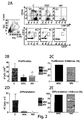
Figure 2 shows a parallel co-culture of CpG, MSC or MSC-MV stimulated CLMs. 2A) Density plots representative of cytofluorimetric analysis of CMFDA labelled CLMs of control group (C) and CLMs after co-culture with MSC or MSC-MV. Lymphocytes were selected at Q1 gate by morphologic parameters (FSC-H; SSC-H) like viable cells (left). Said lymphocytes are successively analyzed based on proliferation rate by selection of CD19/CD27 positive events (right, upper panel) or by differentiation phase in plasma cells (PC) by selection of CD19/CD27 positive events (right, lower panel). 2B-2C) Histograms showing the mean and proliferation inhibition percentage, respectively, of CMFDA/CD19 positive lymphocytes from only CLM culture (C) or MSC or MSC-MV co-culture. 2D-2E) Histograms showing the mean and percentage of proliferation inhibition, respectively, of CD19/CD27 positive plasma cells (PC) from only CLM culture (C) or MSC or MSC-MV co-culture. The results are mean and standard error of 3 independent experiments. p<0.05 -
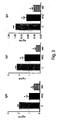
Figure 3 shows the inhibition of immunoglobulin production in CpG stimulated CLMs after MSC or MSC-MV co-culture. ELISA assay of supernatants from only CLM culture (C), incubated with MSC or MSC-MV. The results are mean and standard deviation of IgM, IgG or IgA concentration (ng/ml) for 3 independent experiments. *p< 0.05 -
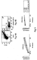
Figure 4 shows the characterization of MSC-MVs. Representative scatter plot obtained for MSC released MVs using FACSCanto cytofluorimeter. 4A) MV gate was selected using 0.5-1µm size calibration beads. 4B) MSC-MVs derived from gate shown in 4A were identified in Q4 quadrant by annexin V,7-AAD and Trucount (P1) beads. -
Figure 5 shows the dose-dependent effect of MSC-MVs on B lymphocytes in CLMs. Panel A shows the inhibition of proliferation (left) and differentiation (right) of CpG stimulated CLMs after MSC-MV culture not diluted (MV) or at 1:3 or 1:6 dilution. A strong inhibition is observed. Results are from 4 independent experiments. -
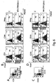
Figure 6 shows FAC analysis for incorporation of PKH26 labelled MVs by CLM selected populations after CpG stimulation. 6A) Density plots for CD3+, CD86+ lymphocytes and double negatives (Dneg) selected in 3 different gates; 6B) Histogram plots of fluorescence intensities for CD3-FITC, CD86-APC positive lymphocytes selected and analyzed before (upper panel) or after incubation with PKH26 labelled MSC-MVs (lower panel). Fluorescence intensity is higher in CD86 positive cells. 6C) Density plots for CD56+, CD19+ lymphocytes and double negatives (Dneg) selected in 3 different gates; 6B) Histogram plots of fluorescence intensities for CD56-FITC, CD19-APC-Cy7 positive lymphocytes selected and analyzed before (upper panel) or after incubation with PKH26 labelled MSC-MVs (lower panel). Fluorescence intensity is higher in CD86 positive cells. -
Figure 7 shows CLM confocal microscopy analysis. Cells have been stained with APC conjugated antibodies against CD3, CD19 and CD86 (green pseudo staining) and conjugated anti-CD 56 (green) with addition (upper panel) or without (left panel) incubation with PKH26 stained MSC-MV (red). MVs mostly occur within cytoplasm of CD19 (arrow tip) and CD86 (arrow) positive cells. Nuclei were counterstained with Hoechst. Magnification bar: 10µm. -
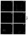
Figure 8 show Z reconstruction confocal microscopy for cell subpopulations. Z reconstruction microphotographies (a, d) carried out using laser scanning confocal microscopy of PKH26 positive MSC-MV incubated lymphocytes, stained with anti-CD19 (a-c) and anti-CD86 (d-f) antibodies. The projections on XZ and XY (b-c, e-f) axes obtained from consecutive multiple optical cross-sections showed intracellular localization of fluorescent red dye labelled MSC-MVs in CD19 positive (c, green) and CD86 positive (f, green) cells, respectively. Nuclei are stained using Hoechst. Magnification Bars: 5 µm (a, d) and 2 µm (b-c, e-f). -
Figure 9 shows the histograms showing mean of proliferating and differentiating cells obtained from a single PBMC culture (c), with MV diluted 1:3 or MV 1:3 incubated with annexin V (concentration from 10 µg to 0.1 µg). The inhibiting effect of MVs is enhanced by annexin V according to dose-dependent mode. -
Figure 10 shows the expression of TNF alpha, IL-1beta, IL-6 and Cox2 in colon tissue on sixth day of experimentation in control animals without colitis induction administered only with PBS (vehicle/PBS), animals with DSS induced colitis and only PBS administered (DSS/PBS) and animals with DSS induced colitis and administered with injections of microvesicles isolated from stem mesenchymal cells and suspended in PBS (DSS/MV). Values are mean ±SD. *p<0.05. -
Figure 11 shows histological analysis (hematoxylin-eosin) of colon tissue from mouse with DSS induced colitis and administered with only PBS (A) and mouse with DSS induced colitis and administered with injections of microvesicles isolated from stem mesenchymal cells (b). Within mucosa and submucosa of treated mouse an inflammatory infiltrate with marked limphomononuclear and histiocyte component is visible. - Marrow derived commercial human mesenchymal stem cells (MSC; Lonza, Basel, Switzerland) have been plated in 75cm2 polystyrene culture flasks with ventilated stopper (Becton Dickinson, USA) at density of 4X10³ cells/cm3 in 10ml of Mesencult basal culture medium (Stem Cell Technologies, BC, Vancouver, Canada) added with bovine foetal serum (20%, FBS, Hyclone Laboratories), penicillin at 100 U/ml and streptomycin at 100 µg/ml (Gibco, Grand Island, NY). Cultures were incubated at 37°C in humidified atmosphere at 5% of CO2. Afterwards cells were maintained within same culture medium, plate detached in trypsin/EDTA solution (Invitrogen, Life Technologies, Italy) at 80-90% confluence and re-plated at 1:2 dilution. Culture medium replacement occurring weekly.
- Blood samples from healthy donors are collected at Centro Trasfusionale Ospedale Pediatrico Bambino Gesù in Rome. After informed consent acquisition, cells peripheral blood limphomononuclear (CLM) have been separated by Ficoll gradient (Histopaque, Sigma Chemical C., St Louis, MO, USA) using 50 ml of heparinized venous blood, washed two times in saline phosphate buffer, then cryo-stored in liquid nitrogen.
- Experiments have been set up in order to test B lymphocyte viability in co-culture with adhered MSCs in 96 well culture plates (Corning-Costar, Celbio, Milan, Italy) at initial concentrations of 2.5x10⁴, 1X10⁴ or 5x10⁴ cells/well in 20% FBS added basal Mesencult culture medium. For immunomodulation tests MSCs are adhered to 96 well culture plates at lower concentration of 5x10³ cells/well and cultured in 20% FBS added basal Mesencult culture medium. On next day in MSC adhered wells, culture medium is drawn and replaced with CLMs (5x10⁵/well corresponding to 1:20, 1:50, 1:100 MSC/CLM ratio, respectively). MSCs have been previously labelled with 5-chloromethylfluorescein diacetate (CMFDA, Cell Tracker, Molecular Probes) at 0.5µM concentration and co-cultured with MSC in 10% FBS added RPMI 1640 culture medium (BioWhittaker, Lona, Belgium). B lymphocyte have been stimulated by incubation with 2.5µg/ml of CpG human oligodeoxynucleotides (Hycult Biotechnology). After one week cultured cells were washed in Dulbecco saline phosphate buffer (D-PBS, Euro-Clone, Milan, Italy) and analyzed using FACS (Fluorescent Activating Cell Sorter Analysis, FACSCanto II, BD Biosciences).
- 90 % confluent MSC cultures were used for preparation of conditioned medium (CM). After 5 day culture, medium is recovered from plates wherein cell plating has been initially carried out at 2x10⁶/well concentration and centrifuged at 1000 g for 20min in order to remove cell debris. MSC produced microvesicles (MSC-MVs) then have been isolated as described in successive paragraph, and added to 5x10⁵ CFMDA labelled lymphocytes after 1 hour and 24 hours of CpG stimulation (Hycult Biotechnology). For selected tests, MSC-MVs were diluted at 1:3, 1:6 in RPMI 1640 medium (BioWhittaker), then added to CLM labelled cultures after 1 hour and 24 hours from culture starting. After a week in the presence of 2.5 µg/ml of CpG human oligodeoxynucleotides added at culture starting, CLMs were recovered by centrifugation, washed and FACS analyzed.
- Isolation methodology of microvesicles is a Lamparski et al (2002) modified method
- 1 90% confluent MSCs: n=6 T75/cm2 (11 ml/plate)
- 2 recovery of culture medium
- 3 centrifuge at 1000g for 10 min in order to eliminate cellular debris
- 4 recover 10ml of medium from each sample
- 5 concentrate medium using 4 ml 30kDa filters (Millipore)
- 6 recover concentrated medium (approximately 50µl for each filter) and dilute in PBS 10ml in ultra-centrifuge tubes
- 7 ultracentrifuge at 100000 g for 1 h at 4°C
- 8 recover MSC-MV pellet and dilute in PBS 10ml
- 9 concentrate MSC-MV solution using 4 ml 30kDa filters (Millipore)
- 10 recover approximately 15-20µl of selected sample (MSC-MV)
- For isolation of microvesicles from MSCs, conditioned medium obtained according to above described procedure is diluted in 10ml of PBS in polyallomer tubes (Beckman Coulter, Milan, Italy), then ultracentrifuged at 100000 g at 4°C for 1 hour. At the end of procedure, 2 ml of ultracentrifuged medium were collected from bottom of the tubes, diluted in 10ml of PBS and concentrated by centrifugation for 20-30min at 2000 g in 30kDa MWCO Amicon Ultra Centrifugal sterile filter (Millipore) to 15-20µl final volume.
- CFMDA labelled CLMs were co-cultured in 96 well plates both with MSCs (at 1:100 ratio) or MSC-MVs in
FBS 10% (Hyclone) added RPMI 1640 (BioWhittaker) medium. After a week the plates were centrifuged at 300 g for 5 min, the supernatants recovered and tested using ELISA assay (enzyme-linked immunosorbent assay). In order to estimate the immunoglobulin production, goat purified immunoglobulin against PBS diluted human IgA, IgG or IgM (IgA 10µg/ml,IgG 15 µg/ml,IgM 2,5 µg/ml, Jackson ImmunoResearch Laboratories, West Grove, PA), were adhered at surface of ELISA 96 well plates (Corning) by overnight incubation at 4°C. After 3 0,1% PBS/Tween washings cell culture supernatant was added to plates (50µl/well) and incubated at 37°C for 1 hour under humidified atmosphere. After washing, the plates were incubated for 1 hour at 37°C with peroxidase conjugated antibodies against human IgA (1:1000), human IgG (1:2000) or human IgM (1:1000) diluted in PBS (Jackson ImmunoResearch Laboratories) (50 µl). PBS diluted o-phenylendiamine tablets (Sigma-Aldrich) were used as chromogen substrate in order the assay to be carried out. The reaction was interrupted after 30 min by addition of 10% sodium dodecylsulfate (50µl/well). At the end of the procedure plate readings were carried out using spectrophotometer (microplate spectrophotometer Benchmark Plus) at 460 OD. - At co-culture end CLMs were recovered from culture plates, centrifuged at 300 g for 5 min and resuspended in PBS/FBS (2%). Single cell suspensions were incubated under dark for 20 min at 4°C with directly conjugated monoclonal antibodies against following surface human molecules: CD19 (1:7, Cy5 conjugated), CD27 [1:7, phycoerithrin (PE) conjugated], CD38 (1:30, PE-Cy7 conjugated), IgM (1:100, Cy5 conjugated), CD86 (1:5, APC conjugated), CD3 [1:7, fluorescein (FITC) conjugated], CD56 (1:7, FITC conjugated), CD19 [1:20 allophycocyanin-Cy7 (APC) conjugated]. All the antibodies were from Becton Dickinson (BD, Franklin Lakes, NJ, USA). After antibody binding, cells were washed 2 times with 2% PBS/BSA and data acquired using FACSCanto II (BD). Cytofluorimetric profiles were analyzed by Cell Quest Software (BD). A minimum of 20,000 events was recorded for single assay.
- In order to characterize MSC-MVs, in individual assays, MSC-MVs from supernatants of 2x10⁶ MSCs according to above reported method were incubated with 5µl of monoclonal antibody against human surface molecule of APC conjugated annexin V (BD) complexed with vital 7-amino-actinomycin dye [7-AAD, conjugated with peridinin-chlorophyll-protein (PerCP) BD complex] in 500µl final volume of annexin V 1 x binding buffer (BB1 x) according to kit reported instructions. This complex allows specific identification of MSC derived not apoptotic MSC-MV. After 15min of incubation under dark at room temp, MSC-MV samples were transferred in Troucount (BD) tubes containing beads for dimensional calibration. Said beads were used in order to define MSC-MV gate based on morphologic parameters (forward scatter, FSC-H; side scatter, SSC-H).
- MVs are isolated from culture medium of 2X10⁶ MSCs (see above) and labelled with PKH26 dye at 2.5x10⁻⁶ (Sigma, Saint Louis, USA) concentration according to producer instructions. Labelled MVs were incubated with CLM (5X10⁵ cells/well) in 10% FBS added RPMI. After 1 hour of incubation at 37°C under humidified atmosphere with 5% of CO2, the cell population was washed with PBS and divided in 2 parts, one assayed by cytofluorimetry, the other by confocal microscopy.
- CLMs incubated with PKH26 labelled MVs were washed with PBS, fixed in 4 % formaldehyde, incubated with 5% PBS/FBS for 30 min and individually stained with following monoclonal antibodies: anti-CD86 (1: 10), anti CD3 (1:10) and anti-CD19 (1:10) conjugated with APC (BD) and FITC conjugated (1:30) anti-CD56 (BD). All the antibodies were diluted in 1% PBS/BSA and incubated for 1 hour. Negative controls were prepared using CLM samples without MV incubation, or without primary antibody in staining procedure.
- Nuclei were counterstained with Hoechst 33342 1µg/ml (Invitrogen, Molecular Probes, OR, USA). After washing with PBS, samples were mounted with Pro-long Anti-Fade (Invitrogen) reagent.
- Confocal analysis has been carried out using Olympus Fluoview FV1000 confocal microscope including FV10-ASW version 2.0, Multi Ar (458-488 and 515nm) software with diode laser 2X He/Ne (543 and 633 nm) and 405 using 60x (1,35 NA oil) lens. Individual optical sections are acquired by 1024X1024 pixel scanning mode, with 207 nm/pixel size, at 40 µsec/pixel sampling speed and 12 bit/pixel images. Fluorochrome mixing was avoided by automatic sequential acquisition of multichannel images, in order to reduce channel spectrum interference. 'Pinhole' opening was 1 Air unit.
- Z reconstruction of single serial optical sections was carried out according to 1024x1024 pixel scanning mode with 2 electronic zoom corresponding to 103 nm/pixel, with 20 µs/pixel sampling speed and Z separation of 0.40 µsec/section. Images were processed using
Photoshop 9,0 version software (Adobe System Inc, CA). - Data are analyzed in terms of mean, maximum and minimum quantitative value of data. Non parametric analysis of variance (Kruskal-Wallis test) is carried out in order to compare group quantitative data; Mann-Whitney U test has been used like test after variance analysis. P<0.05 was considered statistically significant. Statistical analysis was carried out using GraphPad software (GraphPad Prism, Release 3.03, Avenida de la Playa La Jolla, CA)..
- Firstly we have studied the effects of co-culture with MSC on viability of B lymphocytes after stimulation with CpG. We have co-cultured with MSCs CMFDA labelled CLMs (C) obtained from 7 healthy donors with MSC. After 7 day incubation we have analyzed by cytofluorimetric analysis CD19 positive cells in total sample of labelled CLMs. Maximum inhibition of B cell viability was observed at MSC/C 1:20 ratio (
Figure 1 A) ; this effect was still provable 1:50 and 1:100 dilutions (Figure 1A, 1B ). - We have further estimated the effect of MSC co-culture on B cell proliferation. After 7 days under above reported experimental conditions the gate of double CD19/CMFDA labelled was selected by cytofluorimetric analysis. As it is apparent in
Figure 1C we obtained maximum inhibition on B cell proliferation at MSC/C 1:20 ratio. In addition inhibition at MSC/C ratio of 1:50 and a lower effect at 1:100 ratio were provable. Q1 quadrant ofFigure 1D shows the percentage of proliferating B lymphocytes (CD19 positive and CFMDA labelled) with respect to lymphocyte total number; the proliferation was already highly inhibited (90,3%) at MSC/C 1:100 ratio. Therefore, this ratio is used in all the successive co-culture experiments. - In parallel experiments we have studied the effects of co-culture with MSCs or MSC-MVs on B lymphocyte proliferation and differentiation. To this end MSC-MVs are added to CLMs after 1 hour and 24 hours from the beginning of experiment. Parallel cultures wherein CLMs were co-cultured with MSCs at same 1:100 ratio were prepared. A meaningful inhibition on B cell proliferation is observed both in the presence of MSCs and MSC-MVs (
Figure 2A ). Similar inhibition percentages, i.e. 70% and 60%, respectively, were obtained both in the presence of MSCs and MSC-MVs (Figure 2B ). In these parallel experiments we have also studied plasma cellular differentiation. Plasma cellular line (PC) was identified by double staining with CD19 and CD27. We have demonstrated a meaningful inhibition of plasma cellular production, being 98% and 100%, respectively, in the presence of both MSCs and MSC-MVs. As MSCs, MSC-MVs also inhibited the production of IgM, IgG and IgA (Figure 3 ). - Moreover we have estimated the production of immunoglobulins from same cell cultures. After 7 days, supernatants were recovered and assayed by ELISA. Both MSCs and MSC-MVs inhibited the production of IgM, IgG and IgA as shown in
Figure 3 . - We have verified that our method allowed purified MSC-MVs to be isolated, estimating also amounts by cytofluorimetric analysis. MSC-MV population is distinguished from fragments of apoptotic cells by DNA 7-AAD marker negativity and displayed to be annexin V positive (
Figure 4 ). - A dose-dependent effect of MSC-MVs on both B lymphocyte proliferation and differentiation has been displayed (
Figure 5 ). The inhibition effect was greater on differentiation than proliferation. - Cytofluorimetric analysis of CLMs pre-incubated with PKH26 labelled MSC-MVs demonstrated that incorporation thereof was selective in a sub-population of CD86 positive cells and among these in CD19 positive cells corresponding to B lymphocytes. Lymphocyte T corresponding CD3 positive cells displayed to be negative. NK cell corresponding CD56 positive cells displayed to be negative also (
Figure 6 ). - Using laser confocal microscopy, immunofluorescence analysis of CLMs pre-incubated or not with PKH26 labelled MSC-MVs was carried out, in order to estimate the interaction of MSC-MVs with specific cell type (
Figure 7 ). Panel of fluorescein conjugated antibodies against CD3, CD19, CD56 and CD86 molecules was used and co-localization thereof demonstrated a recurrent association of MSC-MVs with CD19 and CD86 positive cells, while no association was detected for CD19 and CD56 positive cells (Figure 7 ). Localization analysis of PKH26 labelled red stained MSC-MVs was based on Z reconstruction of single serial optical sections of CD19 and CD86 positive cells (Figure 8 ). MSC-MV distribution in lateral and axial dimensions, detected using XZ- and YZ axis projections, suggested an intracellular localization in the cytoplasm of CD19 and CD86 positive cells (Figure 8 ). - Annexin V binds cell membrane exposed phosphatidylserine and modifies immune response against said cells (Munoz, 2007). In this experiment, MVs obtained according to above described procedure and 1:3 diluted are incubated for 15min with recombinant purified annexin V at room temp. (BD, concentration range from 10 µg to 0.1 µg) in 100 µl final volume of annexin V binding buffer (BBB1 x) according to producer instructions. Then MVs have been washed with PBS by ultracentrifugation and co-cultured with CFMDA labelled and PBMC stimulated CpGs. After 7 days cultures are analyzed by cytofluorimetry in order to estimate B lymphocyte proliferation and differentiation. Addition of annexin V remarkably enhanced MV inhibiting effect on activated B lymphocytes, in dose-dependent mode. This strategy can be used in order to enhance MV immunomodulating effect.
- This test is in order to estimate MSC-MV inhibiting effect for therapeutic purpose in experimental model of internal chronic inflammatory disease.
- Experiment has been carried out on female 8 week age B57BL/6J mice. Colitis has been induced by administration of 3% sodium dodecyl sulfate (DSS) in drinking water, ad libitum administered over five days. This is a well established and widely used animal model, being referenced hundreds times by Pub Med (for a review, see Kawada et al., 2007). All the animals have been treated according to the ethical code of L'Aquila University, wherein experiments have been carried out, having ad libitum availability of water and food and kept under 12 hour dark/light cycle. Animals are subdivided in three experimental groups.
- Group 1 (n=4, normal controls): intraperitoneal (ip) daily injections of PBS (only vehicle), 0,2 ml from 1 to 5 day;
- Group 2 (n=5, colitis induction): like
group 1, with addition of 3% DSS in drinking water. - Group 3 (n=4, colitis induction and treatment with microvesicles): like group 2, with addition of MSC-MVs suspended in 0.2 ml PBS by IP route.
- On sixth day, the animals were sacrificed using CO2. Colon has been excised, a part of tissue fixed in formalin for successive histological analysis, another part immediately frozen in liquid nitrogen and then stored at -80°C for successive RNA extraction.
- MSC-MVs produced from 2x10⁶ MSCs over 24 hour culture have been isolated and administered according to previously described procedure.
Administration route: MSC-MV suspended in 0.2 ml of PBS by intraperitoneal way daily administered from 0 to 5 day. - Disease activity index (faeces evaluation; see Tanaka F, 2008)
- Flogosis histopathological signs
- Titration of inflammation mediator expression in colon tissue extracts: TNFalfa, IL6, IL-1 beta, and Cox2 by RT-PCR.
- Data are expressed as mean ± SD. Group differences are analyzed by t-test p<0.05 being significant.
- All the animals survived the treatment. While control animals (group 1) progressively gained weight during the week, DSS treated animals displayed weight loss beginning on fourth day (table 1). Weight loss was associated to liquid blood mixed faeces, with increase of colitis activity index (table 1). Both body weight loss and colitis activity index displayed to be remarkably lower in animals daily administered with MSC-MV intraperitoneal injections in addition to DSS. Water intake was meaningfully diminished in DSS administered animals, but was not meaningfully different for two experimental groups with or without MSC-MVs (table 1).
Table 1 shows body weight values, disease activity index and water intake on sixth day of experimentation for control animals without colitis induction administered only with PBS (vehicle/PBS), animals with DSS induced colitis and only PBS administered (DSS/PBS) and animals with DSS induced colitis and administered with injections of microvesicles isolated from mesenchymal stem cells and PBS suspended (MV/DSS).
Disease activity index (score Stool) has been evaluated according to following scale:
normal/semi-solid faeces = 1; semi-solid haematic faeces = 2; liquid haematic faeces = 3Table 1 EXPERIMENTAL GROUP BODY WEIGHT (g) STOOL SCORE WATER INTAKE (mL) N=4 N=5 N=4 (DAY 6) Mean ± SD Mean ± SD Mean ± SD Vehicle/PBS 17.40 ± 0.57 1.0 ± 0 7.450 ± 3.977 DSS/PBS 15.60 ± 0.93 * 2.5 ± 0 * 3.160 ± 0.844* DSS/MV 17.15 ± 0.75 § 1.9 ± 0.23 * § 3.850 ± 0.904* * Different from group vs Vehicle/PBS, p<0.05
§ Different from group vs DSS/PBS, p<0.05 -
Figure 10 shows RT-PCR analysis of TNFalfa, IL-1beta, IL-6 and Cox2 gene expression on colon extracted RNAs. DSS treatment results in remarkable increment of flogosis markers. Administration of MSC-MVs to DSS treated mice reduced meaningfully the activation of TNFalfa, IL-1 beta, while reduction of IL-6 and Cox2 displayed to be lower than significance limit. Histological analysis (Figure 11A andB ) displayed inflammatory infiltrate both in group 2 andgroup 3. Although morphologic differences for two groups have not been detected, it is likely that such infiltrate is less active ingroup 3 than group 2, due to reduced expression of flogosis indices (Figure 10 ) together with lower clinical activity (table 1). In conclusion, these results show that MSC-MVs are suitable to reduce inflammatory reaction associated to experimental colitis resulting in reduction of disease activity index and amelioration of general conditions. MSC-MVs improve clinical picture and the flogistic response in an animal model of intestinal inflammatory disease. -
- Augello A, Tasso R, Negrini SM, Amateis A, Indiveri F, Cancedda R, Pennesi G. Bone marrow mesenchymal progenitor cells inhibit lymphocyte proliferation by activation of the programmed
death 1 pathway.Eur J Immunol 2005;35:1482-90 - Bruno S, Grange C, Deregibus MC, Saviozzi S, Collino F, Bussolati B, Tetta C, Camussi G (2009) "Mesenchymal stem cell-derived microvesicles protect against acute tubular injury" J Am Soc Nephrol 20(5): 1053-1067
- Caimi PF, Reese J, Lee Z, Lazarus HM. Emerging therapeutic approaches for multipotent mesenchymal stromal cells. Curr Opin Hematol. 2010;17:505-13.
- Camussi G, Deregibus MC, Bruno S, Cantaluppi V, Biancone L.
- Exosomes/microvesicles as a mechanism of cell-to-cell communication. Kidney lnt. 2010 78:838-48.
- Chan JL, Tang KC, Patel AP, Bonilla LM, Pierobon N, Ponzio NM, Rameshwar P. Antigen-presenting property of mesenchymal stem cells occurs during a narrow window at low levels of interferon-gamma. Blood. 2006 15;107:4817-24
- Comoli P, Ginevri F, Maccario R, Frassoni F, Locatelli F (2008) "Human mesenchymal stem cell inhibit antibody production induced in vitro by allostimulation" Nephrol Dial Transplant 23:1196-1202
- Corcione A, Benvenuto F, Ferretti E, Giunti D, Pistoia V, Uccelli A (2006) "Human mesenchymal stem cells modulate B-cells functions" Blood 107(1): 367-372
- Escudier B, Dorval T, Chaput N, André F, Caby MP, Novault S, Flament C, Leboulaire C, Borg C, Amigorena S, Boccaccio C, Bonnerot C, Dhellin O, Movassagh M, Piperno S, Robert C, Serra V, Valente N, Le Pecq JB, Spatz A, Lantz O, Tursz T, Angevin E, Zitvogel L. Vaccination of metastatic melanoma patients with autologous dendritic cell (DC) derivedexosomes: results of the first phase I clinical trial. J. Transl. Med. 3, 10 (2005).
- Gatti S, Bruno S, Deregibus MC, Sordi A, Cantaluppi V, Tetta C, Camussi G. Microvesicles derived from human adult mesenchymal stem cells protect against ischaemia-reperfusion-induced acute and chronic kidney injury. Nephrol Dial Transplant. 2011 May;26(5):1474-83. Epub 2011
Feb 15 - Herrera MB, Fonsato V, Gatti S, Deregibus MC, Sordi A, Cantarella D, Calogero R, Bussolati B, Tetta C, Camussi G. Human liver stem cell-derived microvesicles accelerate hepatic regeneration in hepatectomized rats. J Cell Mol Med. 2010;14:1605-18.
- Kawada M, Arihiro A, Mizoguchi E. Insights from advances in research of chemically induced experimental models of human inflammatory bowel disease. World J Gastroenterol. 2007(42):5581-93.
- Krampera M, Glennie S, Dyson J, Scott D, Simpson E, Dazzi F (2003) "Bone marrow mesenchymal stem cells inhibit the response of naïve and memory antigen-specific T cells to their cognate peptide" Blood 101: 3722-3729
- Lamparski HG, Metha-Damani A, Jenq-Yuan Y, Le Pecq JB (2002) "Production and characterization of clinical grade exosomes derived from dendritic cells" J Immunol Methods 270: 211-226
- Meirelles Lda S, Fontes AM, Covas DT, Caplan Al. Mechanisms involved in the therapeutic properties of mesenchymal stem cells. Cytokine Growth Factor Rev. 2009;20:419-27.
- Morse MA, Garst J, Osada T, Khan S, Hobeika A, Clay TM, Valente N, Shreeniwas R, Sutton MA, Delcayre A, Hsu DH, Le Pecq JB, Lyerly HK.. A phase I study of dexosome immunotherapy in patients with advanced non-small cell lung cancer. J. Transl. Med. 3, 9 (2005).
- Nauta AJ, Fibbe WE (2007) "Immunomodulating properties of mesenchymal stromal cells" Blood 110(10): 3499-3506
- Rasmussen I, Le Blank K, Sundberg B, Ringden O (2007) "Mesenchymal stem cells stimulate antibody secretion in human B cells" Scandinavian Journal of Immunology 65: 336-343
- Ren G, Su J, Zhang L, Zhao X, Ling W, L'huillie A, Zhang J, Lu Y, Roberts Al, Ji W, Zhang H, Rabson AB, Shi Y (2009) "Species variation in the mechanisms of mesenchymal stem cell-mediated immunosuppression" Stem Cells 2009 27(8):1954-62
- Tabera S, Perez-Simon J, Diez-Campelo M, Sanchez-Abarca LI, Bianco B, Lopez A, Benito A, Ocio E, Sanchez-Guijo FM, Canino C, San Miguel JF (2008) "The effect of mesenchymal stem cells on the viabilità, proliferation and differentiation of B-lymphocytes" Haematologica 93(9): 1301-1309
- Tanaka F, Tominaga K, Ochi M, Tanigawa T, Watanabe T, Fujiwara Y, Ohta K, Oshitani N, Higuchi K, Arakawa T. Exogenous administration of mesenchymal stem cells ameliorates dextran sulfate sodium-induced colitis via anti-inflammatory action in damaged tissue in rats. Life Sci. 2008 .
- Théry C, Ostrowski M, Segura E. Membrane vesicles as conveyors of immune responses. Nat Rev Immunol. 2009;9:581-93.
- Tolar J, Nauta AJ, Osborn MJ, et al. Sarcoma derived from cultured mesenchymal stem cells. Stem Cells 2007; 25:371-379
- Traggiai E, Volpi S, Schena F, Gattorno M, Ferlito F, Moretta L, Martini A (2008) "Bone marrow-derived mesenchymal stem cells induce both polyclonal expansion and differentiation of B cells isolated from healthy donors and systemic lupus erythematosus patients" Stem Cells 26: 562-569
- Valadi H, Ekström K, Bossios A, Sjöstrand M, Lee JJ, Lötvall JO. Exosome-mediated transfer of mRNAs and microRNAs is a novel mechanism of genetic exchange between cells. Nat Cell Biol. 2007;9:654-9.
Claims (10)
- Microvesicles, or exosomes, isolated from mesenchymal stem cells for use in immunosuppressive therapy.
- Microvesicles isolated from mesenchymal stem cells for use according to claim 1, in the treatment of acute and chronic inflammatory diseases and autoimmune diseases.
- Microvesicles isolated from mesenchymal stem cells for use according to claim 1, in the treatment and prevention of transplant rejection.
- Microvesicles isolated from mesenchymal stem cells for use according to claim 1, in the treatment and prevention of graft-versus-host-disease.
- Microvesicles isolated from mesenchymal stem cells for use according to claim 1, in order to inhibit immune reaction developing as result of allogeneic/xenogeneic cell transplant or gene therapy.
- Pharmaceutical composition comprising or consisting of microvesicles isolated from mesenchymal stem cells, as active principle, optionally annexin bound, together with one or more pharmaceutically acceptable excipients and/or adjuvants, for use in immunosuppressive therapy.
- Pharmaceutical composition for use according to claim 6, in the treatment of the acute and chronic inflammatory diseases and autoimmune diseases.
- Pharmaceutical composition for use according to claim 6, in the treatment and prevention of transplant rejection.
- Pharmaceutical composition for use according to claim 6, in the treatment and prevention of graft-versus-host-disease.
- Pharmaceutical composition for use according to claim 6, in order to inhibit immune reaction developing as result of allogeneic/xenogeneic cell transplant or gene therapy.
Priority Applications (1)
| Application Number | Priority Date | Filing Date | Title |
|---|---|---|---|
| PL12773379T PL2736600T3 (en) | 2011-07-28 | 2012-07-26 | Microvesicles isolated from mesenchymal stem cells for use as immunosuppressive agents |
Applications Claiming Priority (2)
| Application Number | Priority Date | Filing Date | Title |
|---|---|---|---|
| IT000403A ITRM20110403A1 (en) | 2011-07-28 | 2011-07-28 | MICROWELES ISOLATED BY MESENCHIMAL CELLS AS IMMUNOSOPPRESSORS. |
| PCT/IT2012/000232 WO2013014691A1 (en) | 2011-07-28 | 2012-07-26 | Microvesicles isolated from mesenchymal stem cells for use as immunosuppressive agents |
Publications (2)
| Publication Number | Publication Date |
|---|---|
| EP2736600A1 EP2736600A1 (en) | 2014-06-04 |
| EP2736600B1 true EP2736600B1 (en) | 2016-08-31 |
Family
ID=44675724
Family Applications (1)
| Application Number | Title | Priority Date | Filing Date |
|---|---|---|---|
| EP12773379.8A Revoked EP2736600B1 (en) | 2011-07-28 | 2012-07-26 | Microvesicles isolated from mesenchymal stem cells for use as immunosuppressive agents |
Country Status (12)
| Country | Link |
|---|---|
| US (2) | US20140220053A1 (en) |
| EP (1) | EP2736600B1 (en) |
| CN (1) | CN103917275B (en) |
| AU (2) | AU2012288431A1 (en) |
| CA (1) | CA2842296C (en) |
| DK (1) | DK2736600T3 (en) |
| ES (1) | ES2604946T3 (en) |
| HU (1) | HUE029784T2 (en) |
| IT (1) | ITRM20110403A1 (en) |
| PL (1) | PL2736600T3 (en) |
| PT (1) | PT2736600T (en) |
| WO (1) | WO2013014691A1 (en) |
Cited By (2)
| Publication number | Priority date | Publication date | Assignee | Title |
|---|---|---|---|---|
| WO2019111198A1 (en) | 2017-12-06 | 2019-06-13 | Fondazione Irccs "Ca' Granda - Ospedale Maggiore Policlinico" | Extracellular vesicles and uses thereof |
| WO2019111197A1 (en) | 2017-12-06 | 2019-06-13 | Fondazione Irccs "Ca' Granda - Ospedale Maggiore Policlinico" | Extracellular vesicles and uses thereof |
Families Citing this family (17)
| Publication number | Priority date | Publication date | Assignee | Title |
|---|---|---|---|---|
| HUE049003T2 (en) * | 2013-03-13 | 2020-09-28 | Univ Miami | Method for isolation and purification of microvesicles from cell culture supernatants and biological fluids |
| US10961531B2 (en) | 2013-06-05 | 2021-03-30 | Agex Therapeutics, Inc. | Compositions and methods for induced tissue regeneration in mammalian species |
| GB201317887D0 (en) | 2013-10-09 | 2013-11-20 | Reneuron Ltd | Product |
| US11078462B2 (en) | 2014-02-18 | 2021-08-03 | ReCyte Therapeutics, Inc. | Perivascular stromal cells from primate pluripotent stem cells |
| US10240127B2 (en) | 2014-07-03 | 2019-03-26 | ReCyte Therapeutics, Inc. | Exosomes from clonal progenitor cells |
| CN104490931B (en) * | 2014-12-17 | 2018-08-21 | 江苏大学 | The application of the exosome treatment skin injuries of people's umbilical cord mesenchymal stem cells secretion |
| WO2017173150A1 (en) | 2016-03-31 | 2017-10-05 | Commence Bio, Inc. | Medium, methods, cells and secreted factors for stem cell culture and therapy |
| BR112018074334A2 (en) * | 2016-05-25 | 2019-03-06 | Erasmus University Medical Center Rotterdam | use of mesenchymal stem cells and parts thereof |
| EP3672606A4 (en) * | 2017-08-24 | 2021-05-05 | The Texas A & M University System | Mesenchymal stem/stromal cell-derived extracellular vesicles and uses thereof in autoimmune diseases |
| US11766402B2 (en) | 2017-11-16 | 2023-09-26 | Board Of Regents, The University Of Texas System | Methods for production of MSC-derived exosomes |
| ES2724215B2 (en) * | 2018-03-01 | 2020-04-14 | Univ Alcala Henares | Endothelial microvesicles with a microbicidal effect |
| CN108542917A (en) * | 2018-07-20 | 2018-09-18 | 深圳市第二人民医院 | A kind for the treatment of rheumatic ostealgia disease treatment injection and preparation method thereof with person joint's liquid mescenchymal stem cell excretion body extract |
| CN109498541A (en) * | 2018-12-12 | 2019-03-22 | 福建省海西细胞生物工程有限公司 | A kind of intelligence improves the stem cell microcapsule composition of a variety of skin problems |
| CN110179826B (en) * | 2019-05-29 | 2021-08-31 | 武汉五州润达生物医药科技有限公司 | Human umbilical cord mesenchymal stem cell source stem cell factor microvesicle preparation and preparation method thereof |
| CN110755450B (en) * | 2019-11-12 | 2020-06-30 | 山东大学齐鲁医院 | Application of extracellular vesicles derived from mesenchymal stem cells in treatment of subarachnoid hemorrhage |
| EP3909594A1 (en) * | 2020-05-14 | 2021-11-17 | Bernat Soria Escoms | Mesenchymal stromal cells for the treatment of covid-19 and other inflammatory, autoimmune and degenerative diseases |
| CN112300984A (en) * | 2020-09-21 | 2021-02-02 | 四川大学华西医院 | Medical hUC-MSCs-MVs batch preparation process and quality control |
Citations (5)
| Publication number | Priority date | Publication date | Assignee | Title |
|---|---|---|---|---|
| WO2006007529A2 (en) | 2004-07-01 | 2006-01-19 | University Of Pittsburgh Of The Commonwealth System For Higher Education | Immunosuppressive exosomes |
| WO2009105044A1 (en) | 2008-02-22 | 2009-08-27 | Agency For Science, Technology And Research (A*Star) | Mesenchymal stem cell particles |
| WO2011010966A1 (en) | 2009-07-23 | 2011-01-27 | Agency For Science, Technology And Research (A*Star) | Pre-natal mesenchymal stem cells |
| WO2011053257A2 (en) | 2009-11-02 | 2011-05-05 | Agency For Science, Technology And Research (A*Star) | Methods of monitoring cellular states |
| WO2012125471A1 (en) | 2011-03-11 | 2012-09-20 | Children's Medical Center Corporation | Methods and compositions relating to mesenchymal stem cell exosomes |
Family Cites Families (11)
| Publication number | Priority date | Publication date | Assignee | Title |
|---|---|---|---|---|
| US4789734A (en) * | 1985-08-06 | 1988-12-06 | La Jolla Cancer Research Foundation | Vitronectin specific cell receptor derived from mammalian mesenchymal tissue |
| US20020004041A1 (en) | 1999-02-19 | 2002-01-10 | Albert Matthew L. | Methods for abrogating a cellular immune response |
| US6812023B1 (en) | 2000-04-27 | 2004-11-02 | Anosys, Inc. | Methods of producing membrane vesicles |
| US7704964B2 (en) | 2001-08-17 | 2010-04-27 | Exothera L.L.C. | Methods and compounds for the targeting of protein to exosomes |
| WO2004073319A2 (en) | 2003-02-14 | 2004-08-26 | Anosys, Inc. | Methods and compounds for raising antibodies and for screening antibody repertoires |
| US8021847B2 (en) * | 2004-06-02 | 2011-09-20 | Proxy Life Science Holdings, Inc. | Microvesicle-based compositions and methods |
| EP2209481B1 (en) | 2007-10-15 | 2013-03-13 | Fresenius Medical Care Deutschland GmbH | Use of microvesicles (mvs) for preparing a medicament having adjuvant activity on endothelial cell transplantation, particularly in the treatment of diabetes by pancreatic islet transplantation, and related method |
| US20110177054A1 (en) * | 2008-06-06 | 2011-07-21 | Derrick Gibbings | Use of endo-lysosomal system and secreted vesicles (exosome-like) in treatments and diagnostics based on small rna and experimental study of small rna |
| EP2363136A1 (en) * | 2010-03-02 | 2011-09-07 | Fresenius Medical Care Deutschland GmbH | Microvesicles (MVs) derived from adult stem cells for use in the therapeutic treatment of a tumor disease |
| SG189438A1 (en) * | 2010-10-18 | 2013-05-31 | Agency Science Tech & Res | Use of exosomes to promote or enhance hair growth |
| WO2012087241A1 (en) * | 2010-12-20 | 2012-06-28 | Agency For Science, Technology And Research | Method of purifying exosomes |
-
2011
- 2011-07-28 IT IT000403A patent/ITRM20110403A1/en unknown
-
2012
- 2012-07-26 US US14/235,436 patent/US20140220053A1/en not_active Abandoned
- 2012-07-26 DK DK12773379.8T patent/DK2736600T3/en active
- 2012-07-26 EP EP12773379.8A patent/EP2736600B1/en not_active Revoked
- 2012-07-26 PT PT127733798T patent/PT2736600T/en unknown
- 2012-07-26 AU AU2012288431A patent/AU2012288431A1/en not_active Abandoned
- 2012-07-26 WO PCT/IT2012/000232 patent/WO2013014691A1/en active Application Filing
- 2012-07-26 ES ES12773379.8T patent/ES2604946T3/en active Active
- 2012-07-26 CA CA2842296A patent/CA2842296C/en not_active Expired - Fee Related
- 2012-07-26 PL PL12773379T patent/PL2736600T3/en unknown
- 2012-07-26 HU HUE12773379A patent/HUE029784T2/en unknown
- 2012-07-26 CN CN201280037526.6A patent/CN103917275B/en not_active Expired - Fee Related
-
2017
- 2017-06-26 AU AU2017204319A patent/AU2017204319A1/en not_active Abandoned
- 2017-06-29 US US15/637,205 patent/US20180140642A1/en not_active Abandoned
Patent Citations (5)
| Publication number | Priority date | Publication date | Assignee | Title |
|---|---|---|---|---|
| WO2006007529A2 (en) | 2004-07-01 | 2006-01-19 | University Of Pittsburgh Of The Commonwealth System For Higher Education | Immunosuppressive exosomes |
| WO2009105044A1 (en) | 2008-02-22 | 2009-08-27 | Agency For Science, Technology And Research (A*Star) | Mesenchymal stem cell particles |
| WO2011010966A1 (en) | 2009-07-23 | 2011-01-27 | Agency For Science, Technology And Research (A*Star) | Pre-natal mesenchymal stem cells |
| WO2011053257A2 (en) | 2009-11-02 | 2011-05-05 | Agency For Science, Technology And Research (A*Star) | Methods of monitoring cellular states |
| WO2012125471A1 (en) | 2011-03-11 | 2012-09-20 | Children's Medical Center Corporation | Methods and compositions relating to mesenchymal stem cell exosomes |
Non-Patent Citations (18)
| Title |
|---|
| ANNA CORCIONE; ET AL.,: "Human mesenchymal stem cells modulate B-cell functions", BLOOD,, vol. 107, no. 1, 1 January 2006 (2006-01-01), pages 367 - 372, XP002669209 |
| BIEBACK K. ET AL.: "Translating Research into Clinical Scale Manufacturing of Mesenchymal Stromal Cells", STEM CELLS INT (2010, vol. 11, no. 4, pages 1 - 11, XP055206060 |
| BRUNO S; GRANGE C; ET AL.,: "Mesenchymal Stem Cell-Derived Microvesicles Protect Against Acute Tubular Injury", JOURNAL OF THE AMERICAN SOCIETY OF NEPHROLOGY,, vol. 20, no. 5, 1 May 2009 (2009-05-01), pages 1053 - 1067, XP055027861 |
| CAMUSSI GIOVANNI; DEREGIBUS MARIA C; BRUNO STEFANIA; CANTALUPPI VINCENZO; BIANCONE LUIGI: "Exosomes/microvesicles as a mechanism of cell-to-cell communication", KIDNEY INTERNATIONAL,, vol. 78, no. 9, November 2010 (2010-11-01), pages 838 - 848, XP002669206 |
| CLOTILDE THÉRY; MATIAS OSTROWSKI; ELODIE SEGURA: "Membrane vesicles as conveyors of immune responses", NATURE REVIEWS IMMUNOLOGY,, vol. 9, August 2009 (2009-08-01), pages 581 - 593, XP055018445 |
| DIMARINO A. M. ET AL.: "Mesenchymal stem cells in tissue repair", FRONTIERS IN IMMUNOLOGY, vol. 4, no. 201, 25 May 2013 (2013-05-25), pages 1 - 9, XP055285574 |
| ELISABETTA TRAGGIAI; ET AL.: "Bone Marrow-Derived Mesenchymal Stem Cells Induce Both Polyclonal Expansion and Differentiation of B Cells Isolated from Healthy Donors and Systemic Lupus Erythematosus Patients", STEM CELLS, vol. 26, no. 2, 1 February 2008 (2008-02-01), pages 562 - 569, XP055018794 |
| ESCUDIER BERNARD; ET AL.,: "Vaccination of metastatic melanoma patients with autologous dendritic cell (DC) derived-exosomes: results of thefirst phase I clinical tria", JOURNAL OF TRANSLATIONAL MEDICINE,, vol. 3, no. 1, 2 March 2005 (2005-03-02), pages 1 - 13, XP021009856 |
| HERRERA M B ET AL: "Human liver stem cell-derived microvesicles accelerate hepatic regeneration in hepatectomized rats", JOURNAL OF CELLULAR AND MOLECULAR MEDICINE, UNIVERSITY PRESS CAROL DAVILA, BUCHAREST, RO, vol. 14, no. 6B, 1 June 2010 (2010-06-01), pages 1605 - 1618, XP002730586, ISSN: 1582-1838, [retrieved on 20090724], DOI: 10.1111/J.1582-4934.2009.00860.X * |
| LAMPARSKI H G; METHA-DAMANI A; YAO J-Y; PATEL S; HSU D-H; RUEGG C; LE PECQ J-B: "Production and characterization of clinical grade exosomes derived from dendritic cells", JOURNAL OF IMMUNOLOGICAL METH,, vol. 270, no. 2, 15 December 2002 (2002-12-15), pages 211 - 226, XP004387999 |
| MORSE MICHAEL A; ET AL.,: "A phase I study of dexosome immunotherapy in patients with advanced non-small cell lung cancer", JOURNAL OF TRANSLATIONAL MEDICINE,, vol. 3, no. 1, 21 February 2005 (2005-02-21), pages 1 - 9, XP021009899 |
| PAOLO F CAIMI, REESE JANE, LEE ZHENGHONG, LAZARUS HILLARD M: "NIH Public Access Author Manuscript - Emerging Therapeutic Approaches For Multipotent Mesenchymal Stromal Cells (MSCs)", CURR OPIN HEMATOL, 21 August 2013 (2013-08-21), pages 1 - 16, XP055391005 |
| PATRIZIA COMOLI; ET AL.,: "Human mesenchymal stem cells inhibit antibody production induced in vitro by allostimulation", NEPHROLOGY DIALYSIS TRANSPLANTATION,, vol. 23, no. 4, 19 November 2007 (2007-11-19), pages 1196 - 1202, XP002669208 |
| RASMUSSON I; LE BLANC K; SUNDBERG B; RINGDÉN O: "Mesenchymal Stem Cells Stimulate Antibody Secretion in Human B Cells", SCANDINAVIAN JOURNAL OF IMMUNOLOGY,, vol. 65, no. 4, 1 April 2007 (2007-04-01), pages 336 - 343, XP055018762 |
| S. BRUNO ET AL: "Mesenchymal Stem Cell-Derived Microvesicles Protect Against Acute Tubular Injury", JOURNAL OF THE AMERICAN SOCIETY OF NEPHROLOGY, vol. 20, no. 5, 1 May 2009 (2009-05-01), pages 1053 - 1067, XP055027861, ISSN: 1046-6673, DOI: 10.1681/ASN.2008070798 * |
| SORAYA TABERA; ET AL.,: "The effect of mesenchymal stem cells on the viability, proliferation and differentiation of B-lymphocytes", HAEMATOLOGICA,, vol. 93, no. 9, 1 September 2008 (2008-09-01), pages 1301 - 1309, XP002669210 |
| VALADI H; ET AL: "Exosome-mediated transfer of mRNAs and microRNAs is a novel mechanism of genetic exchange between cells", NATURE CELL BIOLOGY,, vol. 9, no. 6, June 2007 (2007-06-01), pages 654 - 659, XP002546815 |
| YEN-SHUN C. ET AL.: "Mesenchymal Stem Cell: Considerations for Manufacturing and Clinical Trials on Cell Therapy Product", INT J STEM CELL RES THER, vol. 3, no. 1, 2016, pages 1 - 12, XP055512086 |
Cited By (2)
| Publication number | Priority date | Publication date | Assignee | Title |
|---|---|---|---|---|
| WO2019111198A1 (en) | 2017-12-06 | 2019-06-13 | Fondazione Irccs "Ca' Granda - Ospedale Maggiore Policlinico" | Extracellular vesicles and uses thereof |
| WO2019111197A1 (en) | 2017-12-06 | 2019-06-13 | Fondazione Irccs "Ca' Granda - Ospedale Maggiore Policlinico" | Extracellular vesicles and uses thereof |
Also Published As
| Publication number | Publication date |
|---|---|
| US20140220053A1 (en) | 2014-08-07 |
| PL2736600T3 (en) | 2017-04-28 |
| US20180140642A1 (en) | 2018-05-24 |
| DK2736600T3 (en) | 2016-12-12 |
| EP2736600A1 (en) | 2014-06-04 |
| CA2842296A1 (en) | 2013-01-31 |
| PT2736600T (en) | 2016-11-28 |
| CN103917275B (en) | 2017-09-08 |
| WO2013014691A1 (en) | 2013-01-31 |
| CA2842296C (en) | 2018-09-04 |
| HUE029784T2 (en) | 2017-04-28 |
| AU2017204319A1 (en) | 2017-07-20 |
| ES2604946T3 (en) | 2017-03-10 |
| CN103917275A (en) | 2014-07-09 |
| ITRM20110403A1 (en) | 2013-01-29 |
| AU2012288431A1 (en) | 2014-02-06 |
Similar Documents
| Publication | Publication Date | Title |
|---|---|---|
| EP2736600B1 (en) | Microvesicles isolated from mesenchymal stem cells for use as immunosuppressive agents | |
| Mao et al. | Mesenchymal stem cells and their therapeutic applications in inflammatory bowel disease | |
| Lai et al. | Exosomes for drug delivery—a novel application for the mesenchymal stem cell | |
| Liu et al. | Immune characterization of mesenchymal stem cells in human umbilical cord Wharton’s jelly and derived cartilage cells | |
| Follin et al. | Human adipose-derived stromal cells in a clinically applicable injectable alginate hydrogel: phenotypic and immunomodulatory evaluation | |
| Carrión et al. | Opposing effect of mesenchymal stem cells on Th1 and Th17 cell polarization according to the state of CD4+ T cell activation | |
| Reinders et al. | Bone marrow-derived mesenchymal stromal cells from patients with end-stage renal disease are suitable for autologous therapy | |
| TW200902718A (en) | Procurement, isolation, and cryopreservation of endometrial/menstrual cells | |
| EP2029149B1 (en) | Immune privileged and modulatory progenitor cells | |
| WO2014203267A2 (en) | Method for isolation, purification and industrial scale expansion of human adipose tissue derived mesenchymal stem cells | |
| JP2021052774A (en) | Novel immunoregulatory cells and production methods thereof | |
| Huang et al. | Combination of retinal pigment epithelium cell‐conditioned medium and photoreceptor outer segments stimulate mesenchymal stem cell differentiation toward a functional retinal pigment epithelium cell phenotype | |
| Motavaf et al. | Therapeutic application of mesenchymal stem cell-derived exosomes: A promising cell-free therapeutic strategy in regenerative medicine | |
| Budgude et al. | Cryopreservation of mesenchymal stromal cell-derived extracellular vesicles using trehalose maintains their ability to expand hematopoietic stem cells in vitro | |
| KR20130096661A (en) | A composition comprising stem cell for preventing or treating of immune or inflammatory disease | |
| Gao et al. | Adipose-derived mesenchymal stem cells promote liver regeneration and suppress rejection in small-for-size liver allograft | |
| AU2004217938B2 (en) | Methods of treating disease by transplantation of developing allogeneic or xenogeneic organs or tissues | |
| WO2021200299A1 (en) | Cellular aging inhibitor, biological tissue repair promoter, gene expression regulator, and manufacturing method | |
| WO2016001839A1 (en) | Management of liver disease using pooled mesenchymal stromal cells | |
| KR20080091776A (en) | Method of using hepatic progenitors in treating liver dysfunction | |
| US20180085405A1 (en) | Treatment of stroke by amniotic fluid derived stem cell conditioned media and products derived thereof | |
| US20210130789A1 (en) | Methods of stromal cell expansion, uses and materials related thereto | |
| Heale | A Biophysical and Molecular Characterization of Human Embryonic Stem Cell-Derived Exosomes | |
| Juntunen | Development of Adipose-Derived Stromal/stem Cell–Based Therapies: Effects of donor weight and culture conditions on cell characteristics and testing the efficacy in stroke models | |
| Davidson | Microvesicles and exosomes in local and distant communication with the heart |
Legal Events
| Date | Code | Title | Description |
|---|---|---|---|
| PUAI | Public reference made under article 153(3) epc to a published international application that has entered the european phase |
Free format text: ORIGINAL CODE: 0009012 |
|
| 17P | Request for examination filed |
Effective date: 20140128 |
|
| AK | Designated contracting states |
Kind code of ref document: A1 Designated state(s): AL AT BE BG CH CY CZ DE DK EE ES FI FR GB GR HR HU IE IS IT LI LT LU LV MC MK MT NL NO PL PT RO RS SE SI SK SM TR |
|
| AX | Request for extension of the european patent |
Extension state: BA |
|
| RAX | Requested extension states of the european patent have changed |
Extension state: BA Payment date: 20140128 |
|
| 17Q | First examination report despatched |
Effective date: 20150326 |
|
| GRAP | Despatch of communication of intention to grant a patent |
Free format text: ORIGINAL CODE: EPIDOSNIGR1 |
|
| INTG | Intention to grant announced |
Effective date: 20151008 |
|
| RAP1 | Party data changed (applicant data changed or rights of an application transferred) |
Owner name: CRYO-SAVE AG |
|
| GRAP | Despatch of communication of intention to grant a patent |
Free format text: ORIGINAL CODE: EPIDOSNIGR1 |
|
| INTG | Intention to grant announced |
Effective date: 20160330 |
|
| GRAS | Grant fee paid |
Free format text: ORIGINAL CODE: EPIDOSNIGR3 |
|
| GRAA | (expected) grant |
Free format text: ORIGINAL CODE: 0009210 |
|
| STAA | Information on the status of an ep patent application or granted ep patent |
Free format text: STATUS: THE PATENT HAS BEEN GRANTED |
|
| AK | Designated contracting states |
Kind code of ref document: B1 Designated state(s): AL AT BE BG CH CY CZ DE DK EE ES FI FR GB GR HR HU IE IS IT LI LT LU LV MC MK MT NL NO PL PT RO RS SE SI SK SM TR |
|
| AX | Request for extension of the european patent |
Extension state: BA |
|
| REG | Reference to a national code |
Ref country code: CH Ref legal event code: EP Ref country code: GB Ref legal event code: FG4D |
|
| REG | Reference to a national code |
Ref country code: IE Ref legal event code: FG4D |
|
| REG | Reference to a national code |
Ref country code: DE Ref legal event code: R096 Ref document number: 602012022452 Country of ref document: DE |
|
| REG | Reference to a national code |
Ref country code: AT Ref legal event code: REF Ref document number: 824476 Country of ref document: AT Kind code of ref document: T Effective date: 20161015 |
|
| REG | Reference to a national code |
Ref country code: RO Ref legal event code: EPE |
|
| REG | Reference to a national code |
Ref country code: PT Ref legal event code: SC4A Ref document number: 2736600 Country of ref document: PT Date of ref document: 20161128 Kind code of ref document: T Free format text: AVAILABILITY OF NATIONAL TRANSLATION Effective date: 20161122 |
|
| REG | Reference to a national code |
Ref country code: NL Ref legal event code: FP |
|
| REG | Reference to a national code |
Ref country code: DK Ref legal event code: T3 Effective date: 20161208 |
|
| REG | Reference to a national code |
Ref country code: LT Ref legal event code: MG4D |
|
| PG25 | Lapsed in a contracting state [announced via postgrant information from national office to epo] |
Ref country code: HR Free format text: LAPSE BECAUSE OF FAILURE TO SUBMIT A TRANSLATION OF THE DESCRIPTION OR TO PAY THE FEE WITHIN THE PRESCRIBED TIME-LIMIT Effective date: 20160831 Ref country code: LT Free format text: LAPSE BECAUSE OF FAILURE TO SUBMIT A TRANSLATION OF THE DESCRIPTION OR TO PAY THE FEE WITHIN THE PRESCRIBED TIME-LIMIT Effective date: 20160831 Ref country code: NO Free format text: LAPSE BECAUSE OF FAILURE TO SUBMIT A TRANSLATION OF THE DESCRIPTION OR TO PAY THE FEE WITHIN THE PRESCRIBED TIME-LIMIT Effective date: 20161130 Ref country code: RS Free format text: LAPSE BECAUSE OF FAILURE TO SUBMIT A TRANSLATION OF THE DESCRIPTION OR TO PAY THE FEE WITHIN THE PRESCRIBED TIME-LIMIT Effective date: 20160831 |
|
| PG25 | Lapsed in a contracting state [announced via postgrant information from national office to epo] |
Ref country code: SE Free format text: LAPSE BECAUSE OF FAILURE TO SUBMIT A TRANSLATION OF THE DESCRIPTION OR TO PAY THE FEE WITHIN THE PRESCRIBED TIME-LIMIT Effective date: 20160831 Ref country code: LV Free format text: LAPSE BECAUSE OF FAILURE TO SUBMIT A TRANSLATION OF THE DESCRIPTION OR TO PAY THE FEE WITHIN THE PRESCRIBED TIME-LIMIT Effective date: 20160831 |
|
| REG | Reference to a national code |
Ref country code: ES Ref legal event code: FG2A Ref document number: 2604946 Country of ref document: ES Kind code of ref document: T3 Effective date: 20170310 |
|
| REG | Reference to a national code |
Ref country code: GR Ref legal event code: EP Ref document number: 20160402899 Country of ref document: GR Effective date: 20170222 |
|
| PG25 | Lapsed in a contracting state [announced via postgrant information from national office to epo] |
Ref country code: EE Free format text: LAPSE BECAUSE OF FAILURE TO SUBMIT A TRANSLATION OF THE DESCRIPTION OR TO PAY THE FEE WITHIN THE PRESCRIBED TIME-LIMIT Effective date: 20160831 |
|
| REG | Reference to a national code |
Ref country code: HU Ref legal event code: AG4A Ref document number: E029784 Country of ref document: HU |
|
| PG25 | Lapsed in a contracting state [announced via postgrant information from national office to epo] |
Ref country code: SM Free format text: LAPSE BECAUSE OF FAILURE TO SUBMIT A TRANSLATION OF THE DESCRIPTION OR TO PAY THE FEE WITHIN THE PRESCRIBED TIME-LIMIT Effective date: 20160831 Ref country code: BG Free format text: LAPSE BECAUSE OF FAILURE TO SUBMIT A TRANSLATION OF THE DESCRIPTION OR TO PAY THE FEE WITHIN THE PRESCRIBED TIME-LIMIT Effective date: 20161130 |
|
| REG | Reference to a national code |
Ref country code: DE Ref legal event code: R026 Ref document number: 602012022452 Country of ref document: DE |
|
| REG | Reference to a national code |
Ref country code: SK Ref legal event code: T3 Ref document number: E 22781 Country of ref document: SK |
|
| PLBI | Opposition filed |
Free format text: ORIGINAL CODE: 0009260 |
|
| REG | Reference to a national code |
Ref country code: FR Ref legal event code: PLFP Year of fee payment: 6 |
|
| PLAX | Notice of opposition and request to file observation + time limit sent |
Free format text: ORIGINAL CODE: EPIDOSNOBS2 |
|
| 26 | Opposition filed |
Opponent name: DR. H. ULRICH DOERRIES Effective date: 20170531 |
|
| PG25 | Lapsed in a contracting state [announced via postgrant information from national office to epo] |
Ref country code: SI Free format text: LAPSE BECAUSE OF FAILURE TO SUBMIT A TRANSLATION OF THE DESCRIPTION OR TO PAY THE FEE WITHIN THE PRESCRIBED TIME-LIMIT Effective date: 20160831 |
|
| PGFP | Annual fee paid to national office [announced via postgrant information from national office to epo] |
Ref country code: LU Payment date: 20170710 Year of fee payment: 6 |
|
| PGFP | Annual fee paid to national office [announced via postgrant information from national office to epo] |
Ref country code: NL Payment date: 20170712 Year of fee payment: 6 |
|
| PGFP | Annual fee paid to national office [announced via postgrant information from national office to epo] |
Ref country code: DE Payment date: 20170724 Year of fee payment: 6 Ref country code: ES Payment date: 20170801 Year of fee payment: 6 Ref country code: GB Payment date: 20170726 Year of fee payment: 6 Ref country code: CH Payment date: 20170712 Year of fee payment: 6 Ref country code: FI Payment date: 20170710 Year of fee payment: 6 |
|
| PLBB | Reply of patent proprietor to notice(s) of opposition received |
Free format text: ORIGINAL CODE: EPIDOSNOBS3 |
|
| PGFP | Annual fee paid to national office [announced via postgrant information from national office to epo] |
Ref country code: DK Payment date: 20170711 Year of fee payment: 6 Ref country code: IE Payment date: 20170710 Year of fee payment: 6 |
|
| REG | Reference to a national code |
Ref country code: FR Ref legal event code: PLFP Year of fee payment: 7 |
|
| PGFP | Annual fee paid to national office [announced via postgrant information from national office to epo] |
Ref country code: SK Payment date: 20180612 Year of fee payment: 7 |
|
| PGFP | Annual fee paid to national office [announced via postgrant information from national office to epo] |
Ref country code: PL Payment date: 20180605 Year of fee payment: 7 Ref country code: BE Payment date: 20180614 Year of fee payment: 7 Ref country code: FR Payment date: 20180612 Year of fee payment: 7 Ref country code: GR Payment date: 20180613 Year of fee payment: 7 Ref country code: RO Payment date: 20180627 Year of fee payment: 7 |
|
| PG25 | Lapsed in a contracting state [announced via postgrant information from national office to epo] |
Ref country code: MT Free format text: LAPSE BECAUSE OF NON-PAYMENT OF DUE FEES Effective date: 20170726 |
|
| PG25 | Lapsed in a contracting state [announced via postgrant information from national office to epo] |
Ref country code: AL Free format text: LAPSE BECAUSE OF FAILURE TO SUBMIT A TRANSLATION OF THE DESCRIPTION OR TO PAY THE FEE WITHIN THE PRESCRIBED TIME-LIMIT Effective date: 20160831 |
|
| PGFP | Annual fee paid to national office [announced via postgrant information from national office to epo] |
Ref country code: IT Payment date: 20180731 Year of fee payment: 7 |
|
| PGFP | Annual fee paid to national office [announced via postgrant information from national office to epo] |
Ref country code: HU Payment date: 20180620 Year of fee payment: 7 Ref country code: AT Payment date: 20180625 Year of fee payment: 7 Ref country code: TR Payment date: 20180725 Year of fee payment: 7 Ref country code: CZ Payment date: 20180629 Year of fee payment: 7 |
|
| APBM | Appeal reference recorded |
Free format text: ORIGINAL CODE: EPIDOSNREFNO |
|
| APBP | Date of receipt of notice of appeal recorded |
Free format text: ORIGINAL CODE: EPIDOSNNOA2O |
|
| PGFP | Annual fee paid to national office [announced via postgrant information from national office to epo] |
Ref country code: PT Payment date: 20180725 Year of fee payment: 7 |
|
| APAH | Appeal reference modified |
Free format text: ORIGINAL CODE: EPIDOSCREFNO |
|
| APBM | Appeal reference recorded |
Free format text: ORIGINAL CODE: EPIDOSNREFNO |
|
| APBP | Date of receipt of notice of appeal recorded |
Free format text: ORIGINAL CODE: EPIDOSNNOA2O |
|
| REG | Reference to a national code |
Ref country code: DE Ref legal event code: R119 Ref document number: 602012022452 Country of ref document: DE |
|
| REG | Reference to a national code |
Ref country code: DK Ref legal event code: EBP Effective date: 20180731 |
|
| REG | Reference to a national code |
Ref country code: CH Ref legal event code: PL |
|
| APBQ | Date of receipt of statement of grounds of appeal recorded |
Free format text: ORIGINAL CODE: EPIDOSNNOA3O |
|
| REG | Reference to a national code |
Ref country code: NL Ref legal event code: MM Effective date: 20180801 |
|
| GBPC | Gb: european patent ceased through non-payment of renewal fee |
Effective date: 20180726 |
|
| PG25 | Lapsed in a contracting state [announced via postgrant information from national office to epo] |
Ref country code: LU Free format text: LAPSE BECAUSE OF NON-PAYMENT OF DUE FEES Effective date: 20180726 |
|
| APAH | Appeal reference modified |
Free format text: ORIGINAL CODE: EPIDOSCREFNO |
|
| REG | Reference to a national code |
Ref country code: IE Ref legal event code: MM4A |
|
| PG25 | Lapsed in a contracting state [announced via postgrant information from national office to epo] |
Ref country code: GB Free format text: LAPSE BECAUSE OF NON-PAYMENT OF DUE FEES Effective date: 20180726 Ref country code: DE Free format text: LAPSE BECAUSE OF NON-PAYMENT OF DUE FEES Effective date: 20190201 Ref country code: FI Free format text: LAPSE BECAUSE OF NON-PAYMENT OF DUE FEES Effective date: 20180726 Ref country code: LI Free format text: LAPSE BECAUSE OF NON-PAYMENT OF DUE FEES Effective date: 20180731 Ref country code: IE Free format text: LAPSE BECAUSE OF NON-PAYMENT OF DUE FEES Effective date: 20180726 Ref country code: CH Free format text: LAPSE BECAUSE OF NON-PAYMENT OF DUE FEES Effective date: 20180731 |
|
| PG25 | Lapsed in a contracting state [announced via postgrant information from national office to epo] |
Ref country code: NL Free format text: LAPSE BECAUSE OF NON-PAYMENT OF DUE FEES Effective date: 20180801 |
|
| PG25 | Lapsed in a contracting state [announced via postgrant information from national office to epo] |
Ref country code: MC Free format text: LAPSE BECAUSE OF FAILURE TO SUBMIT A TRANSLATION OF THE DESCRIPTION OR TO PAY THE FEE WITHIN THE PRESCRIBED TIME-LIMIT Effective date: 20160831 |
|
| PG25 | Lapsed in a contracting state [announced via postgrant information from national office to epo] |
Ref country code: DK Free format text: LAPSE BECAUSE OF NON-PAYMENT OF DUE FEES Effective date: 20180731 |
|
| REG | Reference to a national code |
Ref country code: AT Ref legal event code: UEP Ref document number: 824476 Country of ref document: AT Kind code of ref document: T Effective date: 20160831 |
|
| REG | Reference to a national code |
Ref country code: ES Ref legal event code: FD2A Effective date: 20190917 |
|
| PG25 | Lapsed in a contracting state [announced via postgrant information from national office to epo] |
Ref country code: ES Free format text: LAPSE BECAUSE OF NON-PAYMENT OF DUE FEES Effective date: 20180727 Ref country code: CY Free format text: LAPSE BECAUSE OF NON-PAYMENT OF DUE FEES Effective date: 20160831 |
|
| PG25 | Lapsed in a contracting state [announced via postgrant information from national office to epo] |
Ref country code: MK Free format text: LAPSE BECAUSE OF FAILURE TO SUBMIT A TRANSLATION OF THE DESCRIPTION OR TO PAY THE FEE WITHIN THE PRESCRIBED TIME-LIMIT Effective date: 20160831 |
|
| REG | Reference to a national code |
Ref country code: AT Ref legal event code: MM01 Ref document number: 824476 Country of ref document: AT Kind code of ref document: T Effective date: 20190726 |
|
| REG | Reference to a national code |
Ref country code: SK Ref legal event code: MM4A Ref document number: E 22781 Country of ref document: SK Effective date: 20190726 |
|
| REG | Reference to a national code |
Ref country code: BE Ref legal event code: MM Effective date: 20190731 |
|
| PG25 | Lapsed in a contracting state [announced via postgrant information from national office to epo] |
Ref country code: RO Free format text: LAPSE BECAUSE OF NON-PAYMENT OF DUE FEES Effective date: 20190726 Ref country code: GR Free format text: LAPSE BECAUSE OF NON-PAYMENT OF DUE FEES Effective date: 20200206 Ref country code: PT Free format text: LAPSE BECAUSE OF NON-PAYMENT OF DUE FEES Effective date: 20200127 Ref country code: HU Free format text: LAPSE BECAUSE OF NON-PAYMENT OF DUE FEES Effective date: 20190727 Ref country code: AT Free format text: LAPSE BECAUSE OF NON-PAYMENT OF DUE FEES Effective date: 20190726 |
|
| PG25 | Lapsed in a contracting state [announced via postgrant information from national office to epo] |
Ref country code: BE Free format text: LAPSE BECAUSE OF NON-PAYMENT OF DUE FEES Effective date: 20190731 Ref country code: SK Free format text: LAPSE BECAUSE OF NON-PAYMENT OF DUE FEES Effective date: 20190726 Ref country code: CZ Free format text: LAPSE BECAUSE OF NON-PAYMENT OF DUE FEES Effective date: 20190726 |
|
| PG25 | Lapsed in a contracting state [announced via postgrant information from national office to epo] |
Ref country code: FR Free format text: LAPSE BECAUSE OF NON-PAYMENT OF DUE FEES Effective date: 20190731 |
|
| PG25 | Lapsed in a contracting state [announced via postgrant information from national office to epo] |
Ref country code: IS Free format text: LAPSE BECAUSE OF FAILURE TO SUBMIT A TRANSLATION OF THE DESCRIPTION OR TO PAY THE FEE WITHIN THE PRESCRIBED TIME-LIMIT Effective date: 20161231 |
|
| PG25 | Lapsed in a contracting state [announced via postgrant information from national office to epo] |
Ref country code: IT Free format text: LAPSE BECAUSE OF NON-PAYMENT OF DUE FEES Effective date: 20190726 |
|
| PG25 | Lapsed in a contracting state [announced via postgrant information from national office to epo] |
Ref country code: PL Free format text: LAPSE BECAUSE OF NON-PAYMENT OF DUE FEES Effective date: 20190726 |
|
| PG25 | Lapsed in a contracting state [announced via postgrant information from national office to epo] |
Ref country code: TR Free format text: LAPSE BECAUSE OF NON-PAYMENT OF DUE FEES Effective date: 20190726 |
|
| APAH | Appeal reference modified |
Free format text: ORIGINAL CODE: EPIDOSCREFNO |
|
| APBY | Invitation to file observations in appeal sent |
Free format text: ORIGINAL CODE: EPIDOSNOBA2O |
|
| APBU | Appeal procedure closed |
Free format text: ORIGINAL CODE: EPIDOSNNOA9O |
|
| APCA | Receipt of observations in appeal recorded |
Free format text: ORIGINAL CODE: EPIDOSNOBA4O |
|
| RDAF | Communication despatched that patent is revoked |
Free format text: ORIGINAL CODE: EPIDOSNREV1 |
|
| STAA | Information on the status of an ep patent application or granted ep patent |
Free format text: STATUS: THE PATENT HAS BEEN GRANTED |
|
| REG | Reference to a national code |
Ref country code: DE Ref legal event code: R103 Ref document number: 602012022452 Country of ref document: DE Ref country code: DE Ref legal event code: R064 Ref document number: 602012022452 Country of ref document: DE |
|
| RDAG | Patent revoked |
Free format text: ORIGINAL CODE: 0009271 |
|
| STAA | Information on the status of an ep patent application or granted ep patent |
Free format text: STATUS: PATENT REVOKED |
|
| REG | Reference to a national code |
Ref country code: SK Ref legal event code: MC4A Ref document number: E 22781 Country of ref document: SK Effective date: 20230721 |
|
| REG | Reference to a national code |
Ref country code: CH Ref legal event code: PL |
|
| 27W | Patent revoked |
Effective date: 20230721 |
|
| REG | Reference to a national code |
Ref country code: AT Ref legal event code: MA03 Ref document number: 824476 Country of ref document: AT Kind code of ref document: T Effective date: 20230721 |