EP2697397B1 - Safe sequencing system - Google Patents
Safe sequencing system Download PDFInfo
- Publication number
- EP2697397B1 EP2697397B1 EP12772013.4A EP12772013A EP2697397B1 EP 2697397 B1 EP2697397 B1 EP 2697397B1 EP 12772013 A EP12772013 A EP 12772013A EP 2697397 B1 EP2697397 B1 EP 2697397B1
- Authority
- EP
- European Patent Office
- Prior art keywords
- analyte
- fragments
- uid
- sequence
- dna
- Prior art date
- Legal status (The legal status is an assumption and is not a legal conclusion. Google has not performed a legal analysis and makes no representation as to the accuracy of the status listed.)
- Revoked
Links
Images

Classifications
-
- C—CHEMISTRY; METALLURGY
- C12—BIOCHEMISTRY; BEER; SPIRITS; WINE; VINEGAR; MICROBIOLOGY; ENZYMOLOGY; MUTATION OR GENETIC ENGINEERING
- C12Q—MEASURING OR TESTING PROCESSES INVOLVING ENZYMES, NUCLEIC ACIDS OR MICROORGANISMS; COMPOSITIONS OR TEST PAPERS THEREFOR; PROCESSES OF PREPARING SUCH COMPOSITIONS; CONDITION-RESPONSIVE CONTROL IN MICROBIOLOGICAL OR ENZYMOLOGICAL PROCESSES
- C12Q1/00—Measuring or testing processes involving enzymes, nucleic acids or microorganisms; Compositions therefor; Processes of preparing such compositions
- C12Q1/68—Measuring or testing processes involving enzymes, nucleic acids or microorganisms; Compositions therefor; Processes of preparing such compositions involving nucleic acids
- C12Q1/6869—Methods for sequencing
- C12Q1/6874—Methods for sequencing involving nucleic acid arrays, e.g. sequencing by hybridisation
-
- C—CHEMISTRY; METALLURGY
- C12—BIOCHEMISTRY; BEER; SPIRITS; WINE; VINEGAR; MICROBIOLOGY; ENZYMOLOGY; MUTATION OR GENETIC ENGINEERING
- C12Q—MEASURING OR TESTING PROCESSES INVOLVING ENZYMES, NUCLEIC ACIDS OR MICROORGANISMS; COMPOSITIONS OR TEST PAPERS THEREFOR; PROCESSES OF PREPARING SUCH COMPOSITIONS; CONDITION-RESPONSIVE CONTROL IN MICROBIOLOGICAL OR ENZYMOLOGICAL PROCESSES
- C12Q1/00—Measuring or testing processes involving enzymes, nucleic acids or microorganisms; Compositions therefor; Processes of preparing such compositions
- C12Q1/68—Measuring or testing processes involving enzymes, nucleic acids or microorganisms; Compositions therefor; Processes of preparing such compositions involving nucleic acids
- C12Q1/6806—Preparing nucleic acids for analysis, e.g. for polymerase chain reaction [PCR] assay
-
- C—CHEMISTRY; METALLURGY
- C12—BIOCHEMISTRY; BEER; SPIRITS; WINE; VINEGAR; MICROBIOLOGY; ENZYMOLOGY; MUTATION OR GENETIC ENGINEERING
- C12Q—MEASURING OR TESTING PROCESSES INVOLVING ENZYMES, NUCLEIC ACIDS OR MICROORGANISMS; COMPOSITIONS OR TEST PAPERS THEREFOR; PROCESSES OF PREPARING SUCH COMPOSITIONS; CONDITION-RESPONSIVE CONTROL IN MICROBIOLOGICAL OR ENZYMOLOGICAL PROCESSES
- C12Q1/00—Measuring or testing processes involving enzymes, nucleic acids or microorganisms; Compositions therefor; Processes of preparing such compositions
- C12Q1/68—Measuring or testing processes involving enzymes, nucleic acids or microorganisms; Compositions therefor; Processes of preparing such compositions involving nucleic acids
- C12Q1/6869—Methods for sequencing
-
- C—CHEMISTRY; METALLURGY
- C12—BIOCHEMISTRY; BEER; SPIRITS; WINE; VINEGAR; MICROBIOLOGY; ENZYMOLOGY; MUTATION OR GENETIC ENGINEERING
- C12Q—MEASURING OR TESTING PROCESSES INVOLVING ENZYMES, NUCLEIC ACIDS OR MICROORGANISMS; COMPOSITIONS OR TEST PAPERS THEREFOR; PROCESSES OF PREPARING SUCH COMPOSITIONS; CONDITION-RESPONSIVE CONTROL IN MICROBIOLOGICAL OR ENZYMOLOGICAL PROCESSES
- C12Q1/00—Measuring or testing processes involving enzymes, nucleic acids or microorganisms; Compositions therefor; Processes of preparing such compositions
- C12Q1/68—Measuring or testing processes involving enzymes, nucleic acids or microorganisms; Compositions therefor; Processes of preparing such compositions involving nucleic acids
- C12Q1/6876—Nucleic acid products used in the analysis of nucleic acids, e.g. primers or probes
-
- C—CHEMISTRY; METALLURGY
- C12—BIOCHEMISTRY; BEER; SPIRITS; WINE; VINEGAR; MICROBIOLOGY; ENZYMOLOGY; MUTATION OR GENETIC ENGINEERING
- C12Q—MEASURING OR TESTING PROCESSES INVOLVING ENZYMES, NUCLEIC ACIDS OR MICROORGANISMS; COMPOSITIONS OR TEST PAPERS THEREFOR; PROCESSES OF PREPARING SUCH COMPOSITIONS; CONDITION-RESPONSIVE CONTROL IN MICROBIOLOGICAL OR ENZYMOLOGICAL PROCESSES
- C12Q2525/00—Reactions involving modified oligonucleotides, nucleic acids, or nucleotides
- C12Q2525/10—Modifications characterised by
- C12Q2525/179—Modifications characterised by incorporating arbitrary or random nucleotide sequences
-
- C—CHEMISTRY; METALLURGY
- C12—BIOCHEMISTRY; BEER; SPIRITS; WINE; VINEGAR; MICROBIOLOGY; ENZYMOLOGY; MUTATION OR GENETIC ENGINEERING
- C12Q—MEASURING OR TESTING PROCESSES INVOLVING ENZYMES, NUCLEIC ACIDS OR MICROORGANISMS; COMPOSITIONS OR TEST PAPERS THEREFOR; PROCESSES OF PREPARING SUCH COMPOSITIONS; CONDITION-RESPONSIVE CONTROL IN MICROBIOLOGICAL OR ENZYMOLOGICAL PROCESSES
- C12Q2525/00—Reactions involving modified oligonucleotides, nucleic acids, or nucleotides
- C12Q2525/10—Modifications characterised by
- C12Q2525/191—Modifications characterised by incorporating an adaptor
-
- C—CHEMISTRY; METALLURGY
- C12—BIOCHEMISTRY; BEER; SPIRITS; WINE; VINEGAR; MICROBIOLOGY; ENZYMOLOGY; MUTATION OR GENETIC ENGINEERING
- C12Q—MEASURING OR TESTING PROCESSES INVOLVING ENZYMES, NUCLEIC ACIDS OR MICROORGANISMS; COMPOSITIONS OR TEST PAPERS THEREFOR; PROCESSES OF PREPARING SUCH COMPOSITIONS; CONDITION-RESPONSIVE CONTROL IN MICROBIOLOGICAL OR ENZYMOLOGICAL PROCESSES
- C12Q2563/00—Nucleic acid detection characterized by the use of physical, structural and functional properties
- C12Q2563/179—Nucleic acid detection characterized by the use of physical, structural and functional properties the label being a nucleic acid
-
- C—CHEMISTRY; METALLURGY
- C12—BIOCHEMISTRY; BEER; SPIRITS; WINE; VINEGAR; MICROBIOLOGY; ENZYMOLOGY; MUTATION OR GENETIC ENGINEERING
- C12Q—MEASURING OR TESTING PROCESSES INVOLVING ENZYMES, NUCLEIC ACIDS OR MICROORGANISMS; COMPOSITIONS OR TEST PAPERS THEREFOR; PROCESSES OF PREPARING SUCH COMPOSITIONS; CONDITION-RESPONSIVE CONTROL IN MICROBIOLOGICAL OR ENZYMOLOGICAL PROCESSES
- C12Q2600/00—Oligonucleotides characterized by their use
- C12Q2600/158—Expression markers
Definitions
- This invention is related to the area of nucleic acid sequencing. In particular, it relates to manipulative and analytic steps for analyzing and verifying the products of low frequency events.
- Detection of such mutations will likely be essential to optimize therapy. Detection of donor DNA in the blood of organ transplant patients is an important indicator of graft rejection and detection of fetal DNA in maternal plasma can be used for prenatal diagnosis in a non-invasive fashion (7, 8). In neoplastic diseases, which are all driven by somatic mutations, the applications of rare mutant detection are manifold; they can be used to help identify residual disease at surgical margins or in lymph nodes, to follow the course of therapy when assessed in plasma, and perhaps to identify patients with early, surgically curable disease when evaluated in stool, sputum, plasma, and other bodily fluids (9-11).
- Massively parallel sequencing represents a particularly powerful form of Digital PCR in that hundreds of millions of template molecules can be analyzed one-by-one. It has the advantage over conventional Digital PCR methods in that multiple bases can be queried sequentially and easily in an automated fashion.
- massively parallel sequencing cannot generally be used to detect rare variants because of the high error rate associated with the sequencing process. For example, with the commonly used Illumina sequencing instruments, this error rate varies from ⁇ 1%(31, 32) to ⁇ 0.05% (33, 34), depending on factors such as the read length (35), use of improved base calling algorithms (36-38) and the type of variants detected (39). Some of these errors presumably result from mutations introduced during template preparation, during the pre-amplification steps required for library preparation and during further solid-phase amplification on the instrument itself.
- a method analyzes nucleic acid sequences.
- a u nique id entifier (UID) nucleic acid sequence is attached to a first end of each of a plurality of analyte nucleic acid fragments to form uniquely identified analyte nucleic acid fragments.
- Nucleotide sequence of a uniquely identified analyte nucleic acid fragment is redundantly determined, wherein determined nucleotide sequences which share a UID form a family of members.
- a nucleotide sequence is identified as accurately representing an analyte nucleic acid fragment when at least 50 % of members of the family contain the sequence and the sequence is found in at least two families.
- a method analyzes nucleic acid sequences.
- a unique identifier sequence (UID) is attached to a first end of each of a plurality of analyte DNA fragments using at least two cycles of amplification with first and second primers to form uniquely identified analyte DNA fragments.
- the UIDs are in excess of the analyte DNA fragments during amplification.
- the first primers comprise a first segment complementary to a desired amplicon; a second segment containing the UID; and a third segment containing a universal priming site for subsequent amplification.
- the second primers comprise a universal priming site for subsequent amplification.
- Each cycle of amplification attaches one universal priming site to a strand.
- the uniquely identified analyte DNA fragments are amplified to form a family of uniquely identified analyte DNA fragments from each uniquely identified analyte DNA fragment. Nucleotide sequences of a plurality of members of the family are determined.
- Another aspect of the invention is a method to analyze DNA using endogenous unique identifier sequences (UIDs).
- Fragmented analyte DNA is obtained comprising fragments of 30 to 2000 bases, inclusive. Each end of a fragment forms an endogenous UID for the fragment.
- Adapter oligonucleotides are attached to ends of the fragments to form adapted fragments. Fragments representing one or more selected genes are optionally enriched by means of capturing a subset of the fragments using capture oligonucleotides complementary to selected genes in the analyte DNA or by amplifying fragments complementary to selected genes.
- the adapted fragments are amplified using primers complementary to the adapter oligonucleotides to form families of adapted fragments.
- Nucleotide sequence is determined of a plurality of members of a family. Nucleotide sequences of the plurality of members of the family are compared. A nucleotide sequence is identified as accurately representing an analyte DNA fragment when at least a 1% of members of the family contain the sequence.
- Still another aspect of the invention is a composition
- composition comprising population of primer pairs, wherein each pair comprises a first and second primer for amplifying and identifying a gene or gene portion.
- the first primer comprises a first portion of 10-100 nucleotides complementary to the gene or gene portion and a second portion of 10 to 100 nucleotides comprising a site for hybridization to a third primer.
- the second primer comprises a first portion of 10-100 nucleotides complementary to the gene or gene portion and a second portion of 10 to 100 nucleotides comprising a site for hybridization to a fourth primer.
- Interposed between the first portion and the second portion of the second primer is a third portion consisting of 2 to 4000 nucleotides forming a unique identifier (UID).
- UID unique identifier
- the unique identifiers in the population have at least 4 different sequences.
- the first and second primers are complementary to opposite strands of the gene or gene portion.
- a kit may comprise the population of primers and the third and fourth primers complementary to the second portions of each of the first and second primers.
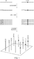
- the inventors have developed an approach, called "Safe-SeqS” (from Safe - Seq uencing S ystem). In one embodiment it involves two basic steps ( Fig. 1 ). The first is the assignment of a Unique Identifier (UID) to each nucleic acid template molecule to be analyzed. The second is the amplification of each uniquely tagged template, so that many daughter molecules with the identical sequence are generated (defined as a UID-family). If a mutation pre-existed in the template molecule used for amplification, that mutation should be present in a certain proportion, or even all, of daughter molecules containing that UID (barring any subsequent replication or sequencing errors).
- UID Unique Identifier
- a UID-family in which every family member (or a certain predetermined proportion) has an identical mutation is called a "super-mutant.” Mutations not occurring in the original templates, such as those occurring during the amplification steps or through errors in base-calling, should not give rise to super-mutants, i.e., will not be present at the pre-determined frequency in a UID family. In other embodiments, amplification is not necessary.
- the approach can be employed for any purpose where a very high level of accuracy and sensitivity is required from sequence data. As shown below, the approach can be used to assess the fidelity of a polymerase, the accuracy of in vitro synthesized nucleic acid synthesis, and the prevalence of mutations in nuclear or mitochondrial nucleic acids of normal cells. The approach may be used to detect and/or quantify mosaicsm and somatic mutations.
- Fragments of nucleic acids may be obtained using a random fragment forming technique such as mechanical shearing, sonicating, or subjecting nucleic acids to other physical or chemical stresses. Fragments may not be strictly random, as some sites may be more susceptible to stresses than others. Endonucleases that randomly or specifically fragment may also be used to generate fragments. Size of fragments may vary, but desirably will be in ranges between 30 and 5,000 basepairs, between 100 and 2,000, between 150 and 1,000, or within ranges with different combinations of these endpoints. Nucleic acids may be, for example, RNA or DNA. Modified forms of RNA or DNA may also be used.
- Attachment of an exogenous UID to an analyte nucleic acids fragment may be performed by any means known in the art, including enzymatic, chemical, or biologic.
- One means employs a polymerase chain reaction.
- Another means employs a ligase enzyme.
- the enzyme may be mammalian or bacterial, for example. Ends of fragments may be repaired prior to joining using other enzymes such as Klenow Fragment of T4 DNA Polymerase. Other enzymes which may be used for attaching are other polymerase enzymes.
- An UID may be added to one or both ends of the fragments.
- a UID may be contained within a nucleic acid molecule that contains other regions for other intended functionality. For example, a universal priming site may be added to permit later amplification. Another additional site may be a region of complementarity to a particular region or gene in the analyte nucleic acids.
- a UID may be from 2 to 4,000, from 100 to 1000, from 4 to 400, bases
- UIDs may be made using random addition of nucleotides to form a short sequence to be used as an identifier. At each position of addition, a selection from one of four deoxyribonucleotides may be used. Alternatively a selection from one of three, two, or one deoxyribonucleotides may be used. Thus the UID may be fully random, somewhat random, or non-random in certain positions.
- Another manner of making UIDs utilizes pre-determined nucleotides assembled on a chip. In this manner of making, complexity is attained in a planned manner. It may be advantageous to attach a UID to each end of a fragment, increasing the complexity of the UID population on fragments.
- a cycle of polymerase chain reaction for adding exogenous UID refers to the thermal denaturation of a double stranded molecule, the hybridization of a first primer to a resulting single strand, the extension of the primer to form a new second strand hybridized to the original single strand.
- a second cycle refers to the denaturation of the new second strand from the original single strand, the hybridization of a second primer to the new second strand, and the extension of the second primer to form a new third strand, hybridized to the new second strand. Multiple cycles may be required to increase efficiency, for example, when analyte is dilute or inhibitors are present.
- adapters can be added to the ends of fragments by ligation.
- Complexity of the analyte fragments can be decreased by a capture step, either on a solid phase or in liquid step.
- the capture step will employ hybridization to probes representing a gene or set of genes of interest. If on a solid phase, non-binding fragments are separated from binding fragments. Suitable solid phases known in the art include filters, membranes, beads, columns, etc.
- a capture reagent can be added which binds to the probes, for example through a biotin-avidin type interaction. After capture, desired fragments can be eluted for further processing.
- the order of adding adapters and capturing is not critical.
- Another means of reducing the complexity of the analyte fragments involves amplification of one or more specific genes or regions.
- One way to accomplish this is to use inverse PCR.
- Primers can be used which are gene-specific, thus enriching while forming libraries.
- the gene-specific primers can contain grafting sequences for subsequent attachment to a massively parallel sequencing platform.
- endogenous UIDs provide a limited number of unique possibilities, depending on the fragment size and sequencing read length
- combinations of both endogenous and exogenous UIDs can be used. Introducing additional sequences when amplifying would increase the available UIDs and thereby increase sensitivity.
- the template can be split into 96 wells, and 96 different primers could be used during the amplification. This would effectively increase the available UIDs 96-fold, because up to 96 templates with the same endogenous UID could be distinguished.
- This technique can also be used with exogenous UIDs, so that each well's primers adds a unique, well-specific sequence to the amplification products. This can improve the specificity of detection of rare templates.
- Amplification of fragments containing a UID can be performed according to known techniques to generate families of fragments. Polymerase chain reaction can be used. Other amplification methods can also be used, as is convenient. Inverse PCR may be used, as can rolling circle amplification. Amplification of fragments typically is done using primers that are complementary to priming sites that are attached to the fragments at the same time as the UIDs. The priming sites are distal to the UIDs, so that amplification includes the UIDs. Amplification forms a family of fragments, each member of the family sharing the same UID. Because the diversity of UIDs is greatly in excess of the diversity of the fragments, each family should derive from a single fragment molecule in the analyte.
- Primers used for the amplification may be chemically modified to render them more resistant to exonucleases.
- One such modification is the use of phosphorothioate linkages between one or more 3' nucleotides.
- Family members are sequenced and compared to identify any divergencies within a family. Sequencing is preferably performed on a massively parallel sequencing platform, many of which are commercially available. If the sequencing platform requires a sequence for "grafting," i.e., attachment to the sequencing device, such a sequence can be added during addition of UIDs or adapters or separately.
- a grafting sequence may be part of a UID primer, a universal primer, a gene target-specific primer, the amplification primers used for making a family, or separate. Redundant sequencing refers to the sequencing of a plurality of members of a single family.
- a threshold can be set for identifying a mutation in an analyte. If the "mutation" appears in all members of a family, then it derives from the analyte. If it appears in less than all members, then it may have been introduced during the analysis.
- Thresholds for calling a mutation may be set, for example, at 50%, 60%, 70%, 80%, 90%, 95%, 97%, 98 %, or 100%. Thresholds will be set based on the number of members of a family that are sequenced and the particular purpose and situation.
- the first primer comprises a first portion of 10-100 nucleotides complementary to the gene or gene portion and a second portion of 10 to 100 nucleotides comprising a site for hybridization to a third primer.
- the second primer comprises a first portion of 10-100 nucleotides complementary to the gene or gene portion and a second portion of 10 to 100 nucleotides comprising a site for hybridization to a fourth primer.
- Interposed between the first portion and the second portion of the second primer is a third portion consisting of 2 to 4,000 nucleotides forming a unique identifier (UID).
- UID unique identifier
- the unique identifiers in the population have at least 4, at least 16, at least 64, at least 256, at least 1,024, at least 4,096, at least 16,384, at least 65,536, at least 262,144, at least 1,048,576, at least 4,194,304, at least 16,777,216, or at least 67,108,864 different sequences.
- the first and second primers are complementary to opposite strands of the gene or gene portion.
- a kit can be made containing both the primers for attaching exogenous UIDs as well as amplification primers, i.e ., the third and fourth primers complementary to the second portions of each of the first and second primers.
- the third and fourth primers can optionally contain additional grafting or indexing sequences.
- the UID may comprise randomly selected sequences, pre-defined nucleotide sequences, or both randomly selected sequences and pre-defined nucleotides. If both, these can be joined together in blocks or interspersed.
- the methods of analysis can be used to quantitate as well as to determine a sequence. For example, the relative abundance of two analyte DNA fragments may be compared.
- Safe-SeqS has limitations. For example, we have demonstrated that the exogenous UIDs strategy can be used to analyze a single amplicon in depth. This technology may not be applicable to situations wherein multiple amplicons must be analyzed from a sample containing a limited number of templates. Multiplexing in the UID assignment cycles ( Fig. 3 ) may provide a solution to this challenge.
- a second limitation is that the efficiency of amplification in the UID assignment cycles is critical for the success of the method. Clinical samples may contain inhibitors that reduce the efficiency of this step. This problem can presumably be overcome by performing more than two cycles in the UID assignment PCR step ( Fig. 3 ), though this would complicate the determination of the number of templates analyzed.
- Safe-SeqS The specificity of Safe-SeqS is currently limited by the fidelity of the polymerase used in the UID assignment PCR step, i.e., 8.8 x 10 -7 mutations/bp in its current implementation with two cycles. Increasing the number of cycles in the UID assignment PCR step to five would decrease the overall specificity to ⁇ 2 x 10 -6 mutations/bp. However, this specificity can be increased by requiring more than one super-mutant for mutation identification -- the probability of introducing the same artifactual mutation twice or three times would be exceedingly low ([2 x 10 -6 ] 2 or [2 x 10 -6 ] 3 , respectively). In sum, there are several simple ways to perform Safe-SeqS variations and analysis variations to realize the needs of specific experiments.
- UIDs sometimes called barcodes or indexes
- Safe-SeqS analysis can also determine which strand of a template is mutated, thus an additional criteria for calling mutations could require that the mutation appears in only one or in both strands of the originally double stranded template.
- Massively parallel sequencers are able to obtain sequence information from both ends of a template in two sequential reads. (This type of sequencing experiment is called a "paired end" run on the Illumina platform, but similar experiments can be done on other sequencing platforms where they may be called by another name.)
- the two strands of a double stranded template can be differentiated by the observed orientation of the sequences and the order in which they appear when sequence information is obtained from both ends.
- a UID strand pair could consist of the following two groups of sequences when each end of a template is sequenced in sequential reads: 1) A sequence in the sense orientation that begins at position 100 of chromosome 2 in the first read followed by a sequence in the antisense orientation that begins at position 400 of chromosome 2 in the second read; and 2) A sequence in the antisense orientation that begins at position 400 of chromosome 2 in the first read followed by a sequence in the sense orientation that begins at position 100 of chromosome 2 in the second read.
- 42,222 of 69,505 UIDs (representing 21,111 original double stranded molecules) in the region of interest represented UID strand pairs.
- this strategy employs two sets of PCR primers. The first set is synthesized with standard phosphoramidite precursors and contained sequences complementary to the gene of interest on the 3' end and different tails at the 5' ends of both the forward and reverse primers. The different tails allowed universal amplification in the next step.
- two cycles of PCR are performed with the primers and a high-fidelity polymerase, producing a uniquely tagged, double-stranded DNA fragment from each of the two strands of each original template molecule ( Fig. 3 ).
- the residual, unused UID assignment primers are removed by digestion with a single-strand specific exonuclease, without further purification, and two new primers are added.
- SPRI solid phase reversible immobilization
- the new primers complementary to the tails introduced in the UID assignment cycles, contain grafting sequences at their 5' ends, permitting solid-phase amplification on the Illumina instrument, and phosphorothioate residues at their 3' ends to make them resistant to any remaining exonuclease. Following 25 additional cycles of PCR, the products are loaded on the Illumina instrument. As shown below, this strategy allowed us to evaluate the majority of input fragments and was used for several illustrative experiments.
- Phusion polymerase The error rate of Phusion polymerase, estimated through cloning of PCR products encoding ⁇ -galactosidase in plasmid vectors and transformation into bacteria, is reported by the manufacturer to be 4.4 x 10 -7 errors/bp/PCR cycle.
- conventional analysis of the Illumina sequencing data revealed an apparent error rate of 9.1 x 10 -6 errors/bp/PCR cycle, more than an order of magnitude higher than the reported Phusion polymerase error rate (Table 2A).
- Safe-SeqS of the same data revealed an error rate of 4.5 x 10 -7 errors/bp/PCR cycle, nearly identical to that measured for Phusion polymerase in biological assays (Table 2A).
- Mitochondrial mutations in DNA from normal human cells Conventional analysis of 7 individuals Total mutations identified 30,599 12,970 Fraction of mutations represented by single base substitutions 98% 1% Fraction of mutations represented by deletions 2% 1% Fraction of mutations represented by insertions 0% 0% Safe-SeqS analysis of 7 individuals Total super-mutants identified 135 61 Fraction of super-mutants represented by single base substitutions 99% 1% Fraction of super-mutants represented by deletions 1% 1% Fraction of super-mutants represented by insertions 0% 0%
- Safe-SeqS also allowed a determination of the total number of distinct mutational events and an estimation of PCR cycle in which the mutation occurred. There were 19 cycles of. PCR performed in wells containing a single template molecule in these experiments. If a polymerase error occurred in cycle 19, there would be only one super-mutant produced (from the strand containing the mutation). If the error occurred in cycle 18 there should be two super-mutants (derived from the mutant strands produced in cycle 19), etc. Accordingly, the cycle in which the error occurred is related to the number of super-mutants containing that error.
- the oligonucleotides contained a large number of insertion and deletion errors, representing 8.2 ⁇ 0.63% and 25 ⁇ 1.5% of the total super-mutants, respectively. Importantly, both the position and nature of the errors were highly reproducible among seven independent replicates of this experiment performed on the same batch of oligonucleotides ( Fig. 6A ). This nature and distribution of errors had little in common with that of the errors produced by Phusion polymerase ( Fig. 6B and Table 5), which were distributed in the expected stochastic pattern among replicate experiments. The number of errors in the oligonucleotides synthesized with phosphoramidites was -60 times higher than in the equivalent products synthesized by Phusion polymerase.
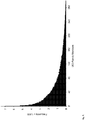
- the exogenous UID strategy ( Fig. 3 ) was then used to determine the prevalence of rare mutations in a small region of the CTNNBI gene from ⁇ 100,000 normal human cells from three unrelated individuals. Through comparison with the number of UID-families obtained in the Safe-SeqS experiments (Table 2B), we calculated that the majority (78 ⁇ 9.8 %) of the input fragments were converted into UID-families. There was an average of 68 members/UID-family, easily fulfilling the required redundancy for Safe-SeqS ( Fig. 7 ).
- Safe-SeqS One possible strategy to increase the specificity of Safe-SeqS is to perform the library amplification (and possibly the UID assignment cycles) in multiple wells. This can be accomplished in as few as 2 or as many as 384 wells using standard PCR plates, or scaled up to many more wells when using a microfluidic device (thousands to millions). When performed this way, indexing sequences can be introduced into the templates that are unique to the wells in which the template is amplified. Rare mutations, thus, should give rise to two super-mutants (i.e., one from each strand), both with the same well index sequence.
- Genomic DNA from human pancreas or cultured lymphoblastoid cells was prepared using Qiagen kits. The pancreas DNA was used for the capture experiment and the lymphoblastoid cells were used for the inverse PCR experiment. DNA was quantified by optical absorbance and with qPCR. DNA was fragmented to an average size of ⁇ 200 bp by acoustic shearing (Covaris), then end-repaired, A-tailed, and ligated to Y-shaped adapters according to standard Illumina protocols. The ends of each template molecule provide endogenous UIDs corresponding to their chromosomal positions.
- Amplifications were performed using Phusion HotStart I (NEB) in 50 uL reactions containing 1X Phusion HF buffer, 0.5 mM dNTPs, 0.5 uM each forward and reverse primer (both 5'-phosphorylated), and 1U of Phusion polymerase.
- the following cycling conditions were used: one cycle of 98°C for 30s; and 16 cycles of 98°C for 10s, 65°C for 30s, and 72°C for 30s. All 96 reactions were pooled and then purified using a Qiagen MinElute PCR Purification Kit (cat. no. 28004) and a QIAquick Gel Extraction kit (cat. no. 28704).
- the KRAS -specific primers both contained grafting sequences for hybridization to the Illumina GA IIx flow cell (Table 6). The following cycling conditions were used: one cycle of 98°C for 2 min; and 37 cycles of 98°C for 10s, 61°C for 15s, and 72°C for 10s. The final purification was performed with a NucleoSpin Extract II kit (Macherey-Nagel) and eluted in 20uL NE Buffer. The resulting DNA fragments contained UIDs composed of three sequences: two endogenous ones, represented by the two ends of the original sheared fragments plus the exogenous sequence introduced during the indexing amplification.
- Genomic DNA from normal human colonic mucosae or blood lymphocytes was prepared using Qiagen kits. The DNA from colonic mucosae was used for the experiments on CTNNB1 and mitochondrial DNA, while the lymphocyte DNA was used for the experiments on CTNNB1 and on polymerase fidelity.
- DNA was quantified with Digital PCR (2) using primers that amplified single-copy genes from human cells (Analysis of Polymerase Fidelity and CTNNB1 ), qPCR (mitochondrial DNA), or by optical absorbance (oligonucleotides). Each strand of each template molecule was encoded with a 12 or 14 base UID using two cycles of amplicon-specific PCR, as described in the text and Fig.
- the amplicon-specific primers both contained universal tag sequences at their 5' ends for a later amplification step.
- the UIDs constituted 12 or 14 random nucleotide sequences appended to the 5' end of the forward amplicon-specific primers (Table 6). These primers can generate 16.8 and 268 million distinct UIDs, respectively. It is important that the number of distinct UIDs greatly exceed the number of original template molecules to minimize the probability that two different original templates acquired the same UID.
- the UID assignment PCR cycles included Phusion Hot Start II (NEB) in a 45 uL reaction containing 1X Phusion HF buffer, 0.25mM dNTPs, 0.5 uM each forward (containing 12-14 Ns) and reverse primers, and 2U of Phusion polymerase. To keep the final template concentrations ⁇ 1.5 ng/uL, multiple wells were used to create some libraries. The following cycling conditions were employed: one incubation of 98°C for 30s (to activate the Phusion Hot Start II); and two cycles of 98°C for 10 s, 61°C for 120 s, and 72°C for 10 s.
- NEB Phusion Hot Start II
- each well was digested with 60 U of a single strand DNA specific nuclease (Exonuclease-I; Enzymatics) at 37°C for 1hr After a 5 min heat-inactivation at 98°C, primers complementary to the introduced universal tags (Table 6) were added to a final concentration of 0.5uM each.
- These primers contained two terminal phosphorothioates to make them resistant to any residual Exonuclease-I activity. They also contained 5' grafting sequences necessary for hybridization to the Illumina GA IIx flow cell. Finally, they contained an index sequence between the grafting sequence and the universal tag sequence.
- This index sequence enables the PCR products from multiple different individuals to be simultaneously analyzed in the same flow cell compartment of the sequencer.
- the following cycling conditions were used for the subsequent 25 cycles of PCR: 98°C for 10s and 72°C for 15s. No intermediate purification steps were performed in an effort to reduce the losses of template molecules.
- Phusion polymerase fidelity Analysis of Phusion polymerase fidelity. Amplification of a fragment of human genomic DNA within the BMX (RefSeq Accession NM_203281.2) gene was first performed using the PCR conditions described above. The template was diluted so that an average of one template molecule was present in every 10 wells of a 96-well PCR plate. Fifty uL PCR reactions were then performed in 1X Phusion HF buffer, 0.25mM dNTPs, 0.5uM each forward and reverse primers (Table 6), and 2U of Phusion polymerase. The cycling conditions were one cycle of 98°C for 30s; and 19 cycles of 98°C for 10 s, 61°C for 120 s, and 72°C for 10s.
- the primers were removed by digestion with 60 U of Exonuclease-I at 37°C for 1hr followed by a 5 min heat-inactivation at 98°C. No purification of the PCR product was performed, either before or after Exonuclease-I digestion. The entire contents of each well were then used as templates for the exogenous UIDs strategy described above.
- UID-families based on their endogenous or exogenous UIDs. Only UID-families with two or more members were considered. Such UID-families included the vast majority ( ⁇ 99%) of the sequencing reads. To ensure that the same data was used for both conventional and Safe-SeqS analysis, we also excluded UID-families containing only one member from conventional analysis. Furthermore, we only identified a base as "mutant" in conventional sequencing analysis if the same variant was identified in at least two members of at least one UID-family (i.e., two mutations) when comparing conventional analysis to that of Safe-SeqS with exogenous UIDs.
- Apparent mutations defined as any base call that varies from the expected base at a defined position, can result from a variety of processes:
- Safe-SeqS minimizes the number of false-positive mutation calls in several ways.
- Safe-SeqS with exogenous UIDs results in the fewest false-positive mutation calls because it requires the fewest enzymatic steps.
- error-generating processes #3 to #7 are completely eliminated because these steps aren't performed.
- Safe-SeqS with exogenous UIDs also drastically reduces errors resulting from error-generating processes #10 and #11 because of the way the data is analyzed.
Landscapes
- Chemical & Material Sciences (AREA)
- Life Sciences & Earth Sciences (AREA)
- Proteomics, Peptides & Aminoacids (AREA)
- Organic Chemistry (AREA)
- Health & Medical Sciences (AREA)
- Zoology (AREA)
- Engineering & Computer Science (AREA)
- Wood Science & Technology (AREA)
- Analytical Chemistry (AREA)
- Molecular Biology (AREA)
- Biochemistry (AREA)
- Microbiology (AREA)
- Immunology (AREA)
- Biotechnology (AREA)
- Bioinformatics & Cheminformatics (AREA)
- General Engineering & Computer Science (AREA)
- General Health & Medical Sciences (AREA)
- Genetics & Genomics (AREA)
- Biophysics (AREA)
- Physics & Mathematics (AREA)
- Chemical Kinetics & Catalysis (AREA)
- Measuring Or Testing Involving Enzymes Or Micro-Organisms (AREA)
- Investigating Or Analysing Biological Materials (AREA)
Description
- This invention is related to the area of nucleic acid sequencing. In particular, it relates to manipulative and analytic steps for analyzing and verifying the products of low frequency events.
- Genetic mutations underlie many aspects of life and death - through evolution and disease, respectively. Accordingly, their measurement is critical to several fields of research. Luria and Delbrück's classic fluctuation analysis is a prototypic example of the insights into biological processes that can be gained simply by counting the number of mutations in carefully controlled experiments (1). Counting de novo mutations in humans, not present in their parents, have similarly led to new insights into the rate at which our species can evolve (2, 3). Similarly, counting genetic or epigenetic changes in tumors can inform fundamental issues in cancer biology (4). Mutations lie at the core of current problems in managing patients with viral diseases such as AIDS and hepatitis by virtue of the drug-resistance they can cause (5, 6). Detection of such mutations, particularly at a stage prior to their becoming dominant in the population, will likely be essential to optimize therapy. Detection of donor DNA in the blood of organ transplant patients is an important indicator of graft rejection and detection of fetal DNA in maternal plasma can be used for prenatal diagnosis in a non-invasive fashion (7, 8). In neoplastic diseases, which are all driven by somatic mutations, the applications of rare mutant detection are manifold; they can be used to help identify residual disease at surgical margins or in lymph nodes, to follow the course of therapy when assessed in plasma, and perhaps to identify patients with early, surgically curable disease when evaluated in stool, sputum, plasma, and other bodily fluids (9-11).
- These examples highlight the importance of identifying rare mutations for both basic and clinical research. Accordingly, innovative ways to assess them have been devised over the years. The first methods involved biologic assays based on prototrophy, resistance to viral infection or drugs, or biochemical assays (1, 12-18). Molecular cloning and sequencing provided a new dimension to the field, as it allowed the type of mutation, rather than simply its presence, to be identified (19-24). Some of the most powerful of these newer methods are based on Digital PCR, in which individual molecules are assessed one-by-one (25). Digital PCR is conceptually identical to the analysis of individual clones of bacteria, cells, or virus, but is performed entirely in vitro with defined, inanimate reagents. Several implementations of Digital PCR have been described, including the analysis of molecules arrayed in multi-well plates, in polonies, in microfluidic devices, and in water-in-oil emulsions (25-30). In each of these technologies, mutant templates are identified through their binding to oligonucleotides specific for the potentially mutant base.
- Massively parallel sequencing represents a particularly powerful form of Digital PCR in that hundreds of millions of template molecules can be analyzed one-by-one. It has the advantage over conventional Digital PCR methods in that multiple bases can be queried sequentially and easily in an automated fashion. However, massively parallel sequencing cannot generally be used to detect rare variants because of the high error rate associated with the sequencing process. For example, with the commonly used Illumina sequencing instruments, this error rate varies from ∼1%(31, 32) to ∼0.05% (33, 34), depending on factors such as the read length (35), use of improved base calling algorithms (36-38) and the type of variants detected (39). Some of these errors presumably result from mutations introduced during template preparation, during the pre-amplification steps required for library preparation and during further solid-phase amplification on the instrument itself. Other errors are due to base mis-incorporation during sequencing and base-calling errors. Advances in base-calling can enhance confidence (e.g., (36-39)), but instrument-based errors are still limiting, particularly in clinical samples wherein the mutation prevalence can be 0.01% or less (11). In the work described below, we show how templates can be prepared and the sequencing data obtained from them can be more reliably interpreted, so that relatively rare mutations can be identified with commercially available instruments.
- There is a continuing need in the art to improve the sensitivity and accuracy of sequence determinations for investigative, clinical, forensic, and genealogical purposes.
- According to one aspect of the invention a method analyzes nucleic acid sequences. A unique identifier (UID) nucleic acid sequence is attached to a first end of each of a plurality of analyte nucleic acid fragments to form uniquely identified analyte nucleic acid fragments. Nucleotide sequence of a uniquely identified analyte nucleic acid fragment is redundantly determined, wherein determined nucleotide sequences which share a UID form a family of members. A nucleotide sequence is identified as accurately representing an analyte nucleic acid fragment when at least 50 % of members of the family contain the sequence and the sequence is found in at least two families.
- According to another aspect of the invention a method analyzes nucleic acid sequences. A unique identifier sequence (UID) is attached to a first end of each of a plurality of analyte DNA fragments using at least two cycles of amplification with first and second primers to form uniquely identified analyte DNA fragments. The UIDs are in excess of the analyte DNA fragments during amplification. The first primers comprise a first segment complementary to a desired amplicon; a second segment containing the UID; and a third segment containing a universal priming site for subsequent amplification. The second primers comprise a universal priming site for subsequent amplification. Each cycle of amplification attaches one universal priming site to a strand. The uniquely identified analyte DNA fragments are amplified to form a family of uniquely identified analyte DNA fragments from each uniquely identified analyte DNA fragment. Nucleotide sequences of a plurality of members of the family are determined.
- Another aspect of the invention is a method to analyze DNA using endogenous unique identifier sequences (UIDs). Fragmented analyte DNA is obtained comprising fragments of 30 to 2000 bases, inclusive. Each end of a fragment forms an endogenous UID for the fragment. Adapter oligonucleotides are attached to ends of the fragments to form adapted fragments. Fragments representing one or more selected genes are optionally enriched by means of capturing a subset of the fragments using capture oligonucleotides complementary to selected genes in the analyte DNA or by amplifying fragments complementary to selected genes. The adapted fragments are amplified using primers complementary to the adapter oligonucleotides to form families of adapted fragments. Nucleotide sequence is determined of a plurality of members of a family. Nucleotide sequences of the plurality of members of the family are compared. A nucleotide sequence is identified as accurately representing an analyte DNA fragment when at least a 1% of members of the family contain the sequence.
- Still another aspect of the invention is a composition comprising population of primer pairs, wherein each pair comprises a first and second primer for amplifying and identifying a gene or gene portion. The first primer comprises a first portion of 10-100 nucleotides complementary to the gene or gene portion and a second portion of 10 to 100 nucleotides comprising a site for hybridization to a third primer. The second primer comprises a first portion of 10-100 nucleotides complementary to the gene or gene portion and a second portion of 10 to 100 nucleotides comprising a site for hybridization to a fourth primer. Interposed between the first portion and the second portion of the second primer is a third portion consisting of 2 to 4000 nucleotides forming a unique identifier (UID). The unique identifiers in the population have at least 4 different sequences. The first and second primers are complementary to opposite strands of the gene or gene portion. A kit may comprise the population of primers and the third and fourth primers complementary to the second portions of each of the first and second primers.
- These and other embodiments which will be apparent to those of skill in the art upon reading the specification provide the art with tools and methods for sensitively and accurately determining nucleic acid features or sequences.
-
-
Fig. 1 . Essential Elements of Safe-SeqS. In the first step, each fragment to be analyzed is assigned a unique identification (UID) sequence (metal hatch or stippled bars). In the second step, the uniquely tagged fragments are amplified, producing UID-families, each member of which has the same UID. A super-mutant is defined as a UID-family in which ≥95% of family members have the same mutation. -
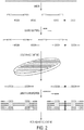
Fig. 2 . Safe-SeqS with Endogenous UIDs Plus Capture. The sequences of the ends of each fragment produced by random shearing (variously shaded bars) serve as the unique identifiers (UIDs). These fragments are ligated to adapters (earth hatched and cross hatched bars) so they can subsequently be amplified by PCR. One uniquely identifiable fragment is produced from each strand of the double-stranded template; only one strand is shown. Fragments of interest are captured on a solid phase containing oligonucleotides complementary to the sequences of interest. Following PCR amplification to produce UID-families with primers containing 5' "grafting" sequences (adhesive filled and light stippled bars), sequencing is performed and super-mutants are defined as inFig. 1 . -
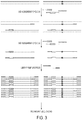
Fig. 3 . Safe-SeqS with Exogenous UIDs. DNA (sheared or unsheared) is amplified with a set of gene-specific primers. One of the primers has a random DNA sequence (e.g., a set of 14 N's) that forms the unique identifier (UID; variously shaded bars), located 5' to its gene-specific sequence, and both have sequences that permit universal amplification in the next step (earth hatched and cross hatched bars). Two UID assignment cycles produce two fragments - each with a different UID - from each double-stranded template molecule, as shown. Subsequent PCR with universal primers, which also contain "grafting" sequences (adhesive filled and light stippled bars), produces UID-families which are directly sequenced. Super-mutants are defined as in the legend toFig. 1 . -
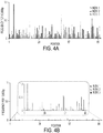
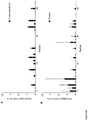
Figs. 4A-4B . Single Base Substitutions Identified by Conventional and Safe-SeqS Analysis. The exogenous UID strategy depicted inFig. 3 was used to produce PCR fragments from the CTNNBI gene of three normal, unrelated individuals. Each position represents one of 87 possible single base substitutions (3 possible substitutions/base x 29 bases analyzed). These fragments were sequenced on an Illumina GA IIx instrument and analyzed in the conventional manner (Fig. 4A ) or with Safe-SeqS (Fig. 4B ). Safe-SeqS results are displayed on the same scale as conventional analysis for direct comparison; the inset is a magnified view. Note that most of the variants identified by conventional analysis are likely to represent sequencing errors, as indicated by their high frequency relative to Safe-SeqS and their consistency among unrelated samples. -
Fig. 5 . Safe-SeqS with endogenous UIDs plus inverse PCR. The sequence of the ends of each fragment produced by random shearing serve as unique identifiers (UIDs; variously shaded bars). These fragments are ligated to adapters (earth hatched and cross hatched bars) as in a standard Illumina library preparation. One uniquely tagged fragment is produced from each strand of the double-stranded template; only one strand is shown. Following circularization with a ligase, inverse PCR is performed with gene-specific primers that also contain 5' "grafting" sequences (adhesive filled and lightly stippled bars). This PCR produces UID-families which are directly sequenced. Super-mutants are defined as inFig. 1 . -
Fig. 6A-6B . Single base substitutions position vs. error frequency in oligonucleotides synthesized with phosphoramidites and Phusion. A representative portion of the same 31-base DNA fragment synthesized with phosphoramidites (Fig. 6A ) or Phusion polymerase (Fig. 6B ) was analyzed by Safe-SeqS. The means and standard deviations for seven independent experiments of each type are plotted. There was an average of 1,721 ± 383 and 196 ± 143 SBS super-mutants identified in the phosphoramidite-synthesized and Phusion-generated fragments, respectively. The y-axis indicates the fraction of the total errors at the indicated position. Note that the errors in the phosphoramidite-synthesized DNA fragment were consistent among the seven replicates, as would be expected if the errors were systematically introduced during the synthesis itself. In contrast, the errors in the Phusion-generated fragments appeared to be heterogeneous among samples, as expected from a stochastic process (Luria and Delbruck, Genetics 28: 491-511, 1943). -
Fig. 7 . UID-family member distribution. The exogenous UID strategy depicted inFig. 3 was used to produce PCR fragments from a region of CTNNBI from three normal, unrelated individuals (Table 2B); a representative example of the UID-families with ≤ 300 members (99% of total UID-families) generated from one individual is shown. The y-axis indicates the number of different UID-families that contained the number of family members shown on the x-axis. - The inventors have developed an approach, called "Safe-SeqS" (from Safe-Sequencing System). In one embodiment it involves two basic steps (
Fig. 1 ). The first is the assignment of a Unique Identifier (UID) to each nucleic acid template molecule to be analyzed. The second is the amplification of each uniquely tagged template, so that many daughter molecules with the identical sequence are generated (defined as a UID-family). If a mutation pre-existed in the template molecule used for amplification, that mutation should be present in a certain proportion, or even all, of daughter molecules containing that UID (barring any subsequent replication or sequencing errors). A UID-family in which every family member (or a certain predetermined proportion) has an identical mutation is called a "super-mutant." Mutations not occurring in the original templates, such as those occurring during the amplification steps or through errors in base-calling, should not give rise to super-mutants, i.e., will not be present at the pre-determined frequency in a UID family. In other embodiments, amplification is not necessary. - The approach can be employed for any purpose where a very high level of accuracy and sensitivity is required from sequence data. As shown below, the approach can be used to assess the fidelity of a polymerase, the accuracy of in vitro synthesized nucleic acid synthesis, and the prevalence of mutations in nuclear or mitochondrial nucleic acids of normal cells. The approach may be used to detect and/or quantify mosaicsm and somatic mutations.
- Fragments of nucleic acids may be obtained using a random fragment forming technique such as mechanical shearing, sonicating, or subjecting nucleic acids to other physical or chemical stresses. Fragments may not be strictly random, as some sites may be more susceptible to stresses than others. Endonucleases that randomly or specifically fragment may also be used to generate fragments. Size of fragments may vary, but desirably will be in ranges between 30 and 5,000 basepairs, between 100 and 2,000, between 150 and 1,000, or within ranges with different combinations of these endpoints. Nucleic acids may be, for example, RNA or DNA. Modified forms of RNA or DNA may also be used.
- Attachment of an exogenous UID to an analyte nucleic acids fragment may be performed by any means known in the art, including enzymatic, chemical, or biologic. One means employs a polymerase chain reaction. Another means employs a ligase enzyme. The enzyme may be mammalian or bacterial, for example. Ends of fragments may be repaired prior to joining using other enzymes such as Klenow Fragment of T4 DNA Polymerase. Other enzymes which may be used for attaching are other polymerase enzymes. An UID may be added to one or both ends of the fragments. A UID may be contained within a nucleic acid molecule that contains other regions for other intended functionality. For example, a universal priming site may be added to permit later amplification. Another additional site may be a region of complementarity to a particular region or gene in the analyte nucleic acids. A UID may be from 2 to 4,000, from 100 to 1000, from 4 to 400, bases in length, for example.
- UIDs may be made using random addition of nucleotides to form a short sequence to be used as an identifier. At each position of addition, a selection from one of four deoxyribonucleotides may be used. Alternatively a selection from one of three, two, or one deoxyribonucleotides may be used. Thus the UID may be fully random, somewhat random, or non-random in certain positions. Another manner of making UIDs utilizes pre-determined nucleotides assembled on a chip. In this manner of making, complexity is attained in a planned manner. It may be advantageous to attach a UID to each end of a fragment, increasing the complexity of the UID population on fragments.
- A cycle of polymerase chain reaction for adding exogenous UID refers to the thermal denaturation of a double stranded molecule, the hybridization of a first primer to a resulting single strand, the extension of the primer to form a new second strand hybridized to the original single strand. A second cycle refers to the denaturation of the new second strand from the original single strand, the hybridization of a second primer to the new second strand, and the extension of the second primer to form a new third strand, hybridized to the new second strand. Multiple cycles may be required to increase efficiency, for example, when analyte is dilute or inhibitors are present.
- In the case of endogenous UIDs, adapters can be added to the ends of fragments by ligation. Complexity of the analyte fragments can be decreased by a capture step, either on a solid phase or in liquid step. Typically the capture step will employ hybridization to probes representing a gene or set of genes of interest. If on a solid phase, non-binding fragments are separated from binding fragments. Suitable solid phases known in the art include filters, membranes, beads, columns, etc. If in a liquid phase, a capture reagent can be added which binds to the probes, for example through a biotin-avidin type interaction. After capture, desired fragments can be eluted for further processing. The order of adding adapters and capturing is not critical. Another means of reducing the complexity of the analyte fragments involves amplification of one or more specific genes or regions. One way to accomplish this is to use inverse PCR. Primers can be used which are gene-specific, thus enriching while forming libraries. Optionally, the gene-specific primers can contain grafting sequences for subsequent attachment to a massively parallel sequencing platform.
- Because endogenous UIDs provide a limited number of unique possibilities, depending on the fragment size and sequencing read length, combinations of both endogenous and exogenous UIDs can be used. Introducing additional sequences when amplifying would increase the available UIDs and thereby increase sensitivity. For example, before amplification, the template can be split into 96 wells, and 96 different primers could be used during the amplification. This would effectively increase the available UIDs 96-fold, because up to 96 templates with the same endogenous UID could be distinguished. This technique can also be used with exogenous UIDs, so that each well's primers adds a unique, well-specific sequence to the amplification products. This can improve the specificity of detection of rare templates.
- Amplification of fragments containing a UID can be performed according to known techniques to generate families of fragments. Polymerase chain reaction can be used. Other amplification methods can also be used, as is convenient. Inverse PCR may be used, as can rolling circle amplification. Amplification of fragments typically is done using primers that are complementary to priming sites that are attached to the fragments at the same time as the UIDs. The priming sites are distal to the UIDs, so that amplification includes the UIDs. Amplification forms a family of fragments, each member of the family sharing the same UID. Because the diversity of UIDs is greatly in excess of the diversity of the fragments, each family should derive from a single fragment molecule in the analyte. Primers used for the amplification may be chemically modified to render them more resistant to exonucleases. One such modification is the use of phosphorothioate linkages between one or more 3' nucleotides. Another employs boranophosphates.
- Family members are sequenced and compared to identify any divergencies within a family. Sequencing is preferably performed on a massively parallel sequencing platform, many of which are commercially available. If the sequencing platform requires a sequence for "grafting," i.e., attachment to the sequencing device, such a sequence can be added during addition of UIDs or adapters or separately. A grafting sequence may be part of a UID primer, a universal primer, a gene target-specific primer, the amplification primers used for making a family, or separate. Redundant sequencing refers to the sequencing of a plurality of members of a single family.
- A threshold can be set for identifying a mutation in an analyte. If the "mutation" appears in all members of a family, then it derives from the analyte. If it appears in less than all members, then it may have been introduced during the analysis. Thresholds for calling a mutation may be set, for example, at 50%, 60%, 70%, 80%, 90%, 95%, 97%, 98 %, or 100%. Thresholds will be set based on the number of members of a family that are sequenced and the particular purpose and situation.
- Populations of primer pairs are used to attach exogenous UIDs. The first primer comprises a first portion of 10-100 nucleotides complementary to the gene or gene portion and a second portion of 10 to 100 nucleotides comprising a site for hybridization to a third primer. The second primer comprises a first portion of 10-100 nucleotides complementary to the gene or gene portion and a second portion of 10 to 100 nucleotides comprising a site for hybridization to a fourth primer. Interposed between the first portion and the second portion of the second primer is a third portion consisting of 2 to 4,000 nucleotides forming a unique identifier (UID). The unique identifiers in the population have at least 4, at least 16, at least 64, at least 256, at least 1,024, at least 4,096, at least 16,384, at least 65,536, at least 262,144, at least 1,048,576, at least 4,194,304, at least 16,777,216, or at least 67,108,864 different sequences. The first and second primers are complementary to opposite strands of the gene or gene portion. A kit can be made containing both the primers for attaching exogenous UIDs as well as amplification primers, i.e., the third and fourth primers complementary to the second portions of each of the first and second primers. The third and fourth primers can optionally contain additional grafting or indexing sequences. The UID may comprise randomly selected sequences, pre-defined nucleotide sequences, or both randomly selected sequences and pre-defined nucleotides. If both, these can be joined together in blocks or interspersed.
- The methods of analysis can be used to quantitate as well as to determine a sequence. For example, the relative abundance of two analyte DNA fragments may be compared.
- The results described below in the examples demonstrate that the Safe-SeqS approach can substantially improve the accuracy of massively parallel sequencing (Tables 1 and 2). It can be implemented through either endogenous or exogenously introduced UIDs and can be applied to virtually any sample preparation workflow or sequencing platform. As demonstrated here, the approach can easily be used to identify rare mutants in a population of DNA templates, to measure polymerase error rates, and to judge the reliability of oligonucleotide syntheses. One of the advantages of the strategy is that it yields the number of templates analyzed as well as the fraction of templates containing variant bases. Previously described in vitro methods for the detection of small numbers of template molecules (e.g., (29, 50)) allow the fraction of mutant templates to be determined but cannot determine the number of mutant and normal templates in the original sample.
- It is of interest to compare Safe-SeqS to other approaches for reducing errors in next-generation sequencing. As mentioned above, in the background of the invention, sophisticated algorithms to increase the accuracy of base-calling have been developed (e.g., (36-39)). These can certainly reduce false positive calls, but their sensitivity is still limited by artifactual mutations occurring during the PCR steps required for library preparation as well as by (a reduced number of) base-calling errors. For example, the algorithm employed in the current study used very stringent criteria for base-calling and was applied to short read-lengths, but was still unable to reduce the error rate to less than an average of 2.0 x 10⁻⁴ errors/bp. This error frequency is at least as low as those reported with other algorithms. To improve sensitivity further, these base-calling improvements can be used together with Safe-SeqS. Travers et al. have described another powerful strategy for reducing errors (51). With this technology, both strands of each template molecule are sequenced redundantly after a number of preparative enzymatic steps. However, this approach can only be performed on a specific instrument. Moreover, for many clinical applications, there are relatively few template molecules in the initial sample and evaluation of nearly all of them is required to obtain the requisite sensitivity. The approach described here with exogenously introduced UIDs (
Fig. 3 ) fulfills this requirement by coupling the UID assignment step with a subsequent amplification in which few molecules are lost. Our endogenous UID approaches (Fig. 2 andFig. 5 ) and the one described by Travers et al. are not ideally suited for this purpose because of the inevitable losses of template molecules during the ligation and other preparative steps. - How do we know that the mutations identified by conventional analyses in the current study represent artifacts rather than true mutations in the original templates? Strong evidence supporting this is provided by the observation that the mutation prevalence in all but one experiment was similar -- 2.0 x 10⁻⁴ to 2.4 x 10⁻⁴ mutations/bp (Tables 1 and 2). The exception was the experiment with oligonucleotides synthesized from phosphoramidites, in which the error of the synthetic process was apparently higher than the error rate of conventional Illumina analysis when used with stringent base-calling criteria. In contrast, the mutation prevalence of Safe-SeqS varied much more, from 0.0 to 1.4 x 10⁻⁵ mutations/bp, depending on the template and experiment. Moreover, the mutation prevalence measured by Safe-SeqS in the most controlled experiment, in which polymerase fidelity was measured (Table 2A), was almost identical to that predicted from previous experiments in which polymerase fidelity was measured by biological assays. Our measurements of mutation prevalence in the DNA from normal cells are consistent with some previous experimental data. However, estimates of these prevalences vary widely and may depend on cell type and sequence analyzed (see SI text). We therefore cannot be certain that the few mutations revealed by Safe-SeqS represented errors occurring during the sequencing process rather than true mutations present in the original DNA templates. Potential sources of error in the Safe-SeqS process are described in the SI text.
- Another potential application of Safe-SeqS is the minimization of PCR contamination, a serious problem for clinical laboratories. With endogenous or exogenous UID assignment, the UIDs of mutant templates can simply be compared to those identified in prior experiments; the probability that the same mutation from two independent samples would have the same UID in different experiments is negligible when mutations are infrequent. Additionally, with exogenous UIDs, a control experiment with the same template but without the UID assigning PCR cycles (
Fig. 3 ) can ensure that no DNA contamination is present in that template preparation; no template should be amplified in the absence of UID assignment cycles and thus no PCR product of the proper size should be observed. - Like all techniques, Safe-SeqS has limitations. For example, we have demonstrated that the exogenous UIDs strategy can be used to analyze a single amplicon in depth. This technology may not be applicable to situations wherein multiple amplicons must be analyzed from a sample containing a limited number of templates. Multiplexing in the UID assignment cycles (
Fig. 3 ) may provide a solution to this challenge. A second limitation is that the efficiency of amplification in the UID assignment cycles is critical for the success of the method. Clinical samples may contain inhibitors that reduce the efficiency of this step. This problem can presumably be overcome by performing more than two cycles in the UID assignment PCR step (Fig. 3 ), though this would complicate the determination of the number of templates analyzed. The specificity of Safe-SeqS is currently limited by the fidelity of the polymerase used in the UID assignment PCR step, i.e., 8.8 x 10⁻⁷ mutations/bp in its current implementation with two cycles. Increasing the number of cycles in the UID assignment PCR step to five would decrease the overall specificity to ∼2 x 10⁻⁶ mutations/bp. However, this specificity can be increased by requiring more than one super-mutant for mutation identification -- the probability of introducing the same artifactual mutation twice or three times would be exceedingly low ([2 x 10⁻⁶]2 or [2 x 10⁻⁶]3, respectively). In sum, there are several simple ways to perform Safe-SeqS variations and analysis variations to realize the needs of specific experiments. - Luria and Delbruck, in their classic paper in 1943, wrote that their "prediction cannot be verified directly, because what we observe, when we count the number of resistant bacteria in a culture, is not the number of mutations which have occurred but the number of resistant bacteria which have arisen by multiplication of those which mutated, the amount of multiplication depending on how far back the mutation occurred." The Safe-SeqS procedure described here can verify such predictions because the number as well as the time of occurrence of each mutation can be estimated from the data, as noted in the experiments on polymerase fidelity. In addition to templates generated by polymerases in vitro, the same approach can be applied to DNA from bacteria, viruses, and mammalian cells. We therefore expect that this strategy will provide definitive answers to a variety of important biomedical questions.
- The above disclosure generally describes the present invention. A more complete understanding can be obtained by reference to the following specific examples which are provided herein for purposes of illustration only, and are not intended to limit the scope of the invention defined in the present claims.
- UIDs, sometimes called barcodes or indexes, can be assigned to nucleic acid fragments in many ways. These include the introduction of exogenous sequences through PCR (40, 41) or ligation (42, 43). Even more simply, randomly sheared genomic DNA inherently contains UIDs consisting of the sequences of the two ends of each sheared fragment (
Fig. 2 andFig. 5 ). Paired-end sequencing of these fragments yields UID-families that can be analyzed as described above. To employ such endogenous UIDs in Safe-SeqS, we used two separate approaches: one designed to evaluate many genes simultaneously and the other designed to evaluate a single gene fragment in depth (Fig. 2 andFig. 5 , respectively). - For the evaluation of multiple genes, we ligated standard Illumina sequencing adapters to the ends of sheared DNA fragments to produce a standard sequencing library, then captured genes of interest on a solid phase (44). In this experiment, a library made from the DNA of ∼15,000 normal cells was used, and 2,594 bp from six genes were targeted for capture. After excluding known single nucleotide polymorphisms, 25,563 apparent mutations, corresponding to 2.4 x 10⁻⁴ ± mutations/bp, were also identified (Table 1). Based on previous analyses of mutation rates in human cells, at least 90% of these apparent mutations were likely to represent mutations introduced during template and library preparation or base-calling errors. Note that the error rate determined here (2.4 x 10⁻⁴ mutations/bp) is considerably lower than usually reported in experiments using the Illumina instrument because we used very stringent criteria for base calling.
Table 1. Safe-SeqS with Endogenous UIDs Conventional Analysis Capture Inverse PCR High quality bp 106,958,863 1,041,346,645 Mean high quality bp read depth 38,620× 2,085,600× Mutations identified 25,563 234,352 Mutations/bp 2.4E-04 2.3E-04 Safe-SeqS Analysis High quality bp 106,958,863 1,041,346,645 Mean high quality bp read depth 38,620× 2,085,600× UID-families 69,505 1,057 Average # of members/UID-family 40 21,688 Median # of members/UID- family 19 4 Super-mutants identified 8 0 Super-mutants/bp 3.5E-06 0.0 - With Safe-SeqS analysis of the same data, we determined that 69,505 original template molecules were assessed in this experiment (i.e., 69,505 UID-families, with an average of 40 members per family, were identified, Table 1). All of the polymorphic variants identified by conventional analysis were also identified by Safe-SeqS. However, only 8 super-mutants were observed among these families, corresponding to 3.5 x 10⁻⁶ mutations/bp. Thus Safe-SeqS decreased the presumptive sequencing errors by at least 70-fold.
- Safe-SeqS analysis can also determine which strand of a template is mutated, thus an additional criteria for calling mutations could require that the mutation appears in only one or in both strands of the originally double stranded template. Massively parallel sequencers are able to obtain sequence information from both ends of a template in two sequential reads. (This type of sequencing experiment is called a "paired end" run on the Illumina platform, but similar experiments can be done on other sequencing platforms where they may be called by another name.) The two strands of a double stranded template can be differentiated by the observed orientation of the sequences and the order in which they appear when sequence information is obtained from both ends. For example, a UID strand pair could consist of the following two groups of sequences when each end of a template is sequenced in sequential reads: 1) A sequence in the sense orientation that begins at position 100 of
chromosome 2 in the first read followed by a sequence in the antisense orientation that begins at position 400 ofchromosome 2 in the second read; and 2) A sequence in the antisense orientation that begins at position 400 ofchromosome 2 in the first read followed by a sequence in the sense orientation that begins at position 100 ofchromosome 2 in the second read. In the capture experiment described above, 42,222 of 69,505 UIDs (representing 21,111 original double stranded molecules) in the region of interest represented UID strand pairs. These 42,222 UIDs encompassed 1,417,838 bases in the region of interest. When allowing a mutation to only occur within UID strand pairs (whether in one or both strands), two super-mutants were observed, yielding a mutation rate of 1.4 x 10⁻⁶ super-mutants/bp. When requiring that a mutation occur in only one strand of a UID strand pair, only one super-mutant was observed, yielding a mutation rate of 7.1 x 10⁻⁷ super-mutants/bp. When requiring that a mutation occur in both strands of a UID strand pair, only one super-mutant was observed, yielding a mutation rate of 7.1 x 10⁻⁷ super-mutants/bp. Thus, requiring that mutations occur in only one or in both strands of templates can further increase the specificity of Safe-SeqS. - A strategy employing endogenous UIDs was also used to reduce false positive mutations upon deep sequencing of a single region of interest. In this case, a library prepared as described above from ∼1,750 normal cells was used as template for inverse PCR employing primers complementary to a gene of interest, so the PCR products could be directly used for sequencing (
Fig. 5 ). With conventional analysis, an average of 2.3 x 10⁻⁴ mutations/bp were observed, similar to that observed in the capture experiment (Table 1). Given that only 1,057 independent molecules from normal cells were assessed in this experiment, as determined through Safe-SeqS analysis, all mutations observed with conventional analysis likely represented false positives (Table 1). With Safe-SeqS analysis of the same data, no super-mutants were identified at any position. - Though the results described above show that Safe-SeqS can increase the reliability of massively parallel sequencing, the number of different molecules that can be examined using endogenous UIDs is limited. For fragments sheared to an average size of 150 bp (range 125-175), 36 base paired-end sequencing can evaluate a maximum of ∼7,200 different molecules containing a specific mutation (2 reads x 2 orientations x 36 bases/read x 50 base variation on either end of the fragment). In practice, the actual number of UIDs is smaller because the shearing process is not entirely random.
- To make more efficient use of the original templates, we developed a Safe-SeqS strategy that employed a minimum number of enzymatic steps. This strategy also permitted the use of degraded or damaged DNA, such as found in clinical specimens or after bisulfite-treatment for the examination of cytosine methylation (45). As depicted in
Fig. 3 , this strategy employs two sets of PCR primers. The first set is synthesized with standard phosphoramidite precursors and contained sequences complementary to the gene of interest on the 3' end and different tails at the 5' ends of both the forward and reverse primers. The different tails allowed universal amplification in the next step. Finally, there was a stretch of 12 to 14 random nucleotides between the tail and the sequence-specific nucleotides in the forward primer (40). The random nucleotides form the UIDs. An equivalent way to assign UIDs to fragments, not used in this study, would employ 10,000 forward primers and 10,000 reverse primers synthesized on a microarray. Each of these 20,000 primers would have gene-specific primers at their 3'-ends and one of 10,000 specific, predetermined, non-overlapping UID sequences at their 5'-ends, allowing for 108 (i.e., [104]2) possible UID combinations. In either case, two cycles of PCR are performed with the primers and a high-fidelity polymerase, producing a uniquely tagged, double-stranded DNA fragment from each of the two strands of each original template molecule (Fig. 3 ). The residual, unused UID assignment primers are removed by digestion with a single-strand specific exonuclease, without further purification, and two new primers are added. Alternatively or in addition to such digestion, one can use a silica column that selectively retains larger-sized fragments or one can use solid phase reversible immobilization (SPRI) beads under conditions that selectively retain larger fragments to eliminate smaller, non-specific, amplification artifacts. This purification may potentially help in reducing primer-dimer accumulation in later steps. The new primers, complementary to the tails introduced in the UID assignment cycles, contain grafting sequences at their 5' ends, permitting solid-phase amplification on the Illumina instrument, and phosphorothioate residues at their 3' ends to make them resistant to any remaining exonuclease. Following 25 additional cycles of PCR, the products are loaded on the Illumina instrument. As shown below, this strategy allowed us to evaluate the majority of input fragments and was used for several illustrative experiments. - Measurement of the error rates of DNA polymerases is essential for their characterization and dictates the situations in which these enzymes can be used. We chose to measure the error rate of Phusion polymerase, as this polymerase has one of the lowest reported error frequencies of any commercially available enzyme and therefore poses a particular challenge for an in vitro-based approach. We first amplified a single human DNA template molecule, comprising a segment of an arbitrarily chosen human gene, through 19 rounds of PCR. The PCR products from these amplifications, in their entirety, were used as templates for Safe-SeqS as described in
Fig. 3 . In seven independent experiments of this type, the number of UID-families identified by sequencing was 624,678 ± 421,274, which is consistent with an amplification efficiency of 92 ± 9.6% per round of PCR. - The error rate of Phusion polymerase, estimated through cloning of PCR products encoding β-galactosidase in plasmid vectors and transformation into bacteria, is reported by the manufacturer to be 4.4 x 10⁻⁷errors/bp/PCR cycle. Even with very high stringency base-calling, conventional analysis of the Illumina sequencing data revealed an apparent error rate of 9.1 x 10⁻⁶ errors/bp/PCR cycle, more than an order of magnitude higher than the reported Phusion polymerase error rate (Table 2A). In contrast, Safe-SeqS of the same data revealed an error rate of 4.5 x 10⁻⁷errors/bp/PCR cycle, nearly identical to that measured for Phusion polymerase in biological assays (Table 2A). The vast majority (>99%) of these errors were single base substitutions (Table 3A), consistent with previous data on the mutation spectra created by other prokaryotic DNA polymerases (15,46,47).
Table 2A-2C. Safe-SeqS with Exogenous UIDs 2A. Polymerase Fidelity Mean Standard Deviation Conventional analysis of 7 replicates High quality bp 996,855,791 64,030,757 Total mutations identified 198,638 22,515 Mutations/bp 2.0E-04 1.7.E-05 Calculated Phusion Error Rate (errors/bp/cycle) 9.1E-06 7.7E-07 Safe-SeqS analysis of 7 replicates High quality bp 996,855,791 64,030,757 UID-families 624,678 421,274 Members/UID-family 107 122 Total super-mutants identified 197 143 Super-mutants/bp 9.9E-06 2.3E-06 Calculated Phusion Error Rate (errors/bp/cycle) 4.5E-07 1.0E-07 2B. CTNNB1 mutations in DNA from normal human cells Conventional analysis of 3 individuals High quality bp 559,334,774 66,600,749 Total mutations identified 118,488 11,357 Mutations/bp 2.1E-04 1.6:E-05 Safe-SeqS analysis of 3 individuals High quality bp 559,334,774 66,600,749 UID-families 374,553 263,105 Members/UID-family 68 38 Total super-mutants identified 99 78 Super-mutants/bp 9.0E-06 3.1E-06 2C. Mitochondrial mutations in DNA from normal human cells Conventional analysis of 7 individuals High quality bp 147,673,456 54,308,546 Total mutations identified 30,599 12,970 Mutations/bp 2.1E-04 9.4E-05 Safe-SeqS analysis of 7 individuals High quality bp 147,673,456 54,308,546 UID-families 515,600 89,985 Members/UID-family 15 6 Total super-mutants identified 135 61 Super-mutants/bp 1.4E-05 6.8E-06 Table 3A-C. Fraction of Single Base Substitutions, Insertions, and Deletions with Exogenous UIDs 3A. Polymerase Fidelity Mean Standard Deviation Conventional analysis of 7 replicates Total mutations identified 198,638 22,515 Fraction of mutations represented by single base substitutions 99% 0% Fraction of mutations represented by deletions 1% 0% Fraction of mutations represented by insertions 0% 0% Safe-SeqS analysis of 7 replicates Total super-mutants identified 197 143 Fraction of super-mutants represented by single base substitutions 99% 2% Fraction of super-mutants represented by deletions 1% 2% Fraction of super-mutants represented by insertions 0% 0% 3B. CTNNB1 mutations in DNA from normal human cells Conventional analysis of 3 individuals Total mutations identified 118,488 11,357 Fraction of mutations represented by single base substitutions 97% 0% Fraction of mutations represented by deletions 3% 0% Fraction of mutations represented by insertions 0% 0% Safe-SeqS analysis of 3 individuals Total super-mutants identified 99 78 Fraction of super-mutants represented by single base substitutions 100% 1% Fraction of super-mutants represented by deletions 0% 1% Fraction of super-mutants represented by insertions 0% 0% 3C. Mitochondrial mutations in DNA from normal human cells Conventional analysis of 7 individuals Total mutations identified 30,599 12,970 Fraction of mutations represented by single base substitutions 98% 1% Fraction of mutations represented by deletions 2% 1% Fraction of mutations represented by insertions 0% 0% Safe-SeqS analysis of 7 individuals Total super-mutants identified 135 61 Fraction of super-mutants represented by single base substitutions 99% 1% Fraction of super-mutants represented by deletions 1% 1% Fraction of super-mutants represented by insertions 0% 0% - Safe-SeqS also allowed a determination of the total number of distinct mutational events and an estimation of PCR cycle in which the mutation occurred. There were 19 cycles of. PCR performed in wells containing a single template molecule in these experiments. If a polymerase error occurred in
cycle 19, there would be only one super-mutant produced (from the strand containing the mutation). If the error occurred in cycle 18 there should be two super-mutants (derived from the mutant strands produced in cycle 19), etc. Accordingly, the cycle in which the error occurred is related to the number of super-mutants containing that error. The data from seven independent experiments demonstrate a relatively consistent number of observed total polymerase errors (2.2 ± 1.1 x 10⁻⁶ distinct mutations/bp), in good agreement with the expected number of observations from simulations (1.5 ± 0.21 x 10⁻⁶ distinct mutations/bp). The data also show a highly variable timing of occurrence of polymerase errors among experiments (Table 4), as predicted from classic fluctuation analysis (1). This kind of information is difficult to derive using conventional analysis of the same next-generation sequencing data, in part because of the prohibitively high apparent mutation rate noted above.Table 4A-4G. Observed and Expected Number of Errors Generated by Phusion Polymerase 4A. Experiment 1Observed Expected (mean ± SD) * Mutations represented by 1 super-mutant 10 19 ± 3.7 Mutations represented by 2 super-mutants 8 5.8 ± 2.3 Mutations represented by 3 super-mutants 4 1.3 ± 1.1 Mutations represented by 4 super-mutants 4 1.8 ± 1.3 Mutations represented by 5 super-mutants 2 0.61 ± 0.75 Mutations represented by 6 super-mutants 2 0.22 ± 0.44 Mutations represented by 7 super-mutants 0 0.01 ± 0.10 Mutations represented by 8 super-mutants 0 0.87 ± 0.86 Mutations represented by 9 super-mutants 2 0.28 ± 0.51 Mutations represented by 10 super-mutants 0 0.14 ± 0.38 Mutations represented by > 10 super-mutants 3 1.5 ± 2.7 Distinct mutations 35 32 ± 4.2 4B. Experiment 2Mutations represented by 1 super-mutant 19 23 ± 4.1 Mutations represented by 2 super-mutants 5 9.5 ± 2.8 Mutations represented by 3 super-mutants 4 2.7 ± 1.6 Mutations represented by 4 super-mutants 7 2.7 ± 1.7 Mutations represented by 5 super-mutants 2 0.88 ± 0.94 Mutations represented by 6 super-mutants 1 0.40 ± 0.60 Mutations represented by 7 super-mutants 3 0.16 ± 0.42 Mutations represented by 8 super-mutants 1 0.99 ± 1.0 Mutations represented by 9 super-mutants 1 0.39 ± 0.68 Mutations represented by 10 super-mutants 0 0.17 ± 0.43 Mutations represented by > 10 super-mutants 9 1.8 ± 3.4 Distinct mutations 52 43 ± 5.1 4C. Experiment 3Mutations represented by 1 super-mutant 7 17 ± 3.4 Mutations represented by 2 super-mutants 9 5.4 ± 2.0 Mutations represented by 3 super-mutants 4 1.2 ± 1.1 Mutations represented by 4 super-mutants 4 1.7 ± 1.4 Mutations represented by 5 super-mutants 2 0.50 ± 0.70 Mutations represented by 6 super-mutants 0 0.17 ± 0.45 Mutations represented by 7 super-mutants 1 0.03 ± 0.17 Mutations represented by 8 super-mutants 0 0.59 ± 0.74 Mutations represented by 9 super-mutants 0 0.24 ± 0.50 Mutations represented by 10 super-mutants 1 0.07 ± 0.29 Mutations represented by >10 super-mutants 5 1.5 ± 2.6 Distinct mutations 33 28 ± 3.7 4D. Experiment 4Mutations represented by 1 super-mutant 7 15 ± 3.7 Mutations represented by 2 super-mutants 8 4.1 ± 1.7 Mutations represented by 3 super-mutants 2 0.70 ± 0.74 Mutations represented by 4 super-mutants 1 1.5 ± 1.3 Mutations represented by 5 super-mutants 3 0.21 ± 0.52 Mutations represented by 6 super-mutants 2 0.08 ± 0.27 Mutations represented by 7 super-mutants 1 0.0 ± 0.0 Mutations represented by 8 super-mutants 2 0.65 ± 0.77 Mutations represented by 9 super-mutants 2 0.17 ± 0.43 Mutations represented by 10 super-mutants 0 0.05 ± 0.22 Mutations represented by >10 super-mutants 1 0.92 ± 2.1 Distinct mutations 29 23 ± 3.2 4E. Experiment 5Mutations represented by 1 super-mutant 9 23 ± 4.1 Mutations represented by 2 super-mutants 6 9.5 ± 2.8 Mutations represented by 3 super-mutants 5 2.7 ± 1.6 Mutations represented by 4 super-mutants 3 2.7 ± 1.7 Mutations represented by 5 super-mutants 6 0.88 ± 0.94 Mutations represented by 6 super-mutants 2 0.40 ± 0.60 Mutations represented by 7 super-mutants 1 0.16 ± 0.42 Mutations represented by 8 super-mutants 2 0.99 ± 1.0 Mutations represented by 9 super-mutants 2 0.39 ± 0.68 Mutations represented by 10 super-mutants 3 0.17 ± 0.43 Mutations represented by >10 super-mutants 7 1.8 ± 3.4 Distinct mutations 46 43 ± 5.1 4F. Experiment 6Mutations represented by 1 super-mutant 4 6.7 ± 2.8 Mutations represented by 2 super-mutants 7 1.5 ± 1.2 Mutations represented by 3 super-mutants 1 0.10 ± 0.33 Mutations represented by 4 super-mutants 2 0.60 ± 0.82 Mutations represented by 5 super-mutants 0 0.07 ± 0.26 Mutations represented by 6 super-mutants 0 0.01 ± 0.10 Mutations represented by 7 super-mutants 1 0.0 ± 0.0 Mutations represented by 8 super-mutants 1 0.39 ± 0.60 Mutations represented by 9 super-mutants 0 0.01 ± 0.10 Mutations represented by 10 super-mutants 0 0.0 ± 0.0 Mutations represented by >10 super-mutants 2 0.50 ± 1.1 Distinct mutations 18 9.9 ± 1.4 4G. Experiment 7Mutations represented by 1 super-mutant 8 2.9 ± 1.6 Mutations represented by 2 super-mutants 2 0.61 ± 0.79 Mutations represented by 3 super-mutants 0 0.04 ± 0.24 Mutations represented by 4 super-mutants 0 0.41 ± 0.59 Mutations represented by 5 super-mutants 1 0.01 ± 0.10 Mutations represented by 6 super-mutants 0 0.0 ± 0.0 Mutations represented by 7 super-mutants 0 0.0 ± 0.0 Mutations represented by 8 super-mutants 0 0.14 ± 0.35 Mutations represented by 9 super-mutants 0 0.01 ± 0.10 Mutations represented by 10 super-mutants 0 0.0 ± 0.0 Mutations represented by > 10 super-mutants 0 0.32 ± 0.93 Distinct mutations 11 4.5 ± 0.62 *See SI Text for details of the simulations - A small number of mistakes during the synthesis of oligonucleotides from phoshoramidite precursors are tolerable for most applications, such as routine PCR or cloning. However, for synthetic biology, wherein many oligonucleotides must be joined together, such mistakes present a major obstacle to success. Clever strategies for making the gene construction process more efficient have been devised (48, 49), but all such strategies would benefit from more accurate synthesis of the oligonucleotides themselves. Determining the number of errors in synthesized oligonucleotides is difficult because the fraction of oligonucleotides containing errors can be lower than the sensitivity of conventional next-generation sequencing analyses.
- To determine whether Safe-SeqS could be used for this determination, we used standard phosphoramidite chemistry to synthesize an oligonucleotide containing 31 bases that were designed to be identical to that analyzed in the polymerase fidelity experiment described above. In the synthetic oligonucleotide, the 31 bases were surrounded by sequences complementary to primers that could be used for the UID assignment steps of Safe-SeqS (
Fig. 3 ). By performing Safe-SeqS on ∼300,000 oligonucleotides, we found that there were 8.9 ± 0.28 x 10⁻⁴ super-mutants/bp and that these errors occurred throughout the oligonucleotides (Fig. 6A ). The oligonucleotides contained a large number of insertion and deletion errors, representing 8.2 ± 0.63% and 25 ± 1.5% of the total super-mutants, respectively. Importantly, both the position and nature of the errors were highly reproducible among seven independent replicates of this experiment performed on the same batch of oligonucleotides (Fig. 6A ). This nature and distribution of errors had little in common with that of the errors produced by Phusion polymerase (Fig. 6B and Table 5), which were distributed in the expected stochastic pattern among replicate experiments. The number of errors in the oligonucleotides synthesized with phosphoramidites was -60 times higher than in the equivalent products synthesized by Phusion polymerase. These data, in toto, indicate that the vast majority of errors in the former were generated during their synthesis rather than during the Safe-SeqS procedure.Table 5. Phosphoramidite- vs Phusion-Synthesized DNA: Transitions vs Transversions Comparison Phosphoramidites Exp. 1 Exp.2 Exp.3 Exp.4 Exp.5 Exp.6 Exp. 7 Average Standard Deviation Transition super-mutants: 496 509 471 396 323 273 470 420 92 Transversion super-mutants: 1494 1499 1521 1154 944 907 1626 1306 298 p-value* 3.4E-05 Phusion Transition super-mutants: 63 275 127 5 87 182 103 120 87 Transversion super-mutants: 14 124 77 12 57 191 63 77 63 p-value* 0.08 *p-values were calculated using a two-tailed paired t-test - Does Safe-SeqS preserve the ratio of mutant:normal sequences in the original templates? To address this question, we synthesized two 31-base oligonucleotides of identical sequence with the exception of nt 15 (50:50 C/G instead of T) and mixed them at nominal mutant/normal fractions of 3.3% and 0.33%. Through Safe-SeqS analysis of the oligonucleotide mixtures, we found that the ratios were 2.8% and 0.27%, respectively. We conclude that the UID assignment and amplification procedures used in Safe-SeqS do not greatly alter the proportion of variant sequences and thereby provide a reliable estimate of that proportion when unknown. This conclusion is also supported by the reproducibility of variant fractions when analyzed in independent Safe-SeqS experiments (
Fig. 6A ). - The exogenous UID strategy (
Fig. 3 ) was then used to determine the prevalence of rare mutations in a small region of the CTNNBI gene from ∼100,000 normal human cells from three unrelated individuals. Through comparison with the number of UID-families obtained in the Safe-SeqS experiments (Table 2B), we calculated that the majority (78 ± 9.8 %) of the input fragments were converted into UID-families. There was an average of 68 members/UID-family, easily fulfilling the required redundancy for Safe-SeqS (Fig. 7 ). Conventional analysis of the Illumina sequencing data revealed an average of 118,488 ± 11,357 mutations among the ∼560 Mb of sequence analyzed per sample, corresponding to an apparent mutation prevalence of 2.1 ± 0.16 x 10⁻⁴ mutations/bp (Table 2B). Only an average of 99 ± 78 super-mutants were observed in the Safe-SeqS analysis. The vast majority (>99%) of super-mutants were single base substitutions and the calculated mutation rate was 9.0 ± 3.1 x 10⁻⁶ mutations/bp (Table 3B). Safe-SeqS thereby reduced the apparent frequency of mutations in genomic DNA by at least 24-fold (Fig. 4 ). - One possible strategy to increase the specificity of Safe-SeqS is to perform the library amplification (and possibly the UID assignment cycles) in multiple wells. This can be accomplished in as few as 2 or as many as 384 wells using standard PCR plates, or scaled up to many more wells when using a microfluidic device (thousands to millions). When performed this way, indexing sequences can be introduced into the templates that are unique to the wells in which the template is amplified. Rare mutations, thus, should give rise to two super-mutants (i.e., one from each strand), both with the same well index sequence. When performing Safe-SeqS with exogenous UIDs on the CTNNBI templates described above and diluted into 10 wells (each well yielding templates amplified with a different index sequence), the mutation rate was further reduced from 9.0 ± 3.1 x 10⁻⁶ to 3.7 ± 1.2 x 10⁻⁶ super-mutants/bp. Thus, analyzing templates in multiple compartments-in a manner that yields differentially encoded templates based on the compartment in which templates were amplified-may be an additional strategy to increase the specificity of Safe-SeqS.
- We applied the identical strategy to a short segment of mitochondrial DNA in ∼1,000 cells from each of seven unrelated individuals. Conventional analysis of the Illumina sequencing libraries produced with the Safe-SeqS procedure (
Fig. 3 ) revealed an average of 30,599 ± 12,970 mutations among the ∼150 Mb of sequence analyzed per sample, corresponding to an apparent mutation prevalence of 2.1 ± 0.94 x 10⁻⁴ mutations/bp (Table 2C). Only 135 ± 61 super-mutants were observed in the Safe-SeqS analysis. As with the CTNNB1 gene, the vast majority of mutations were single base substitutions, though occasional single base deletions were also observed (Table 3C). The calculated mutation rate in the analyzed segment of mtDNA was 1.4 ± 0.68 x 10⁻⁵ mutations/bp (Table 2C). Thus, Safe-SeqS thereby reduced the apparent frequency of mutations in genomic DNA by at least 15-fold. - Endogenous UIDs. Genomic DNA from human pancreas or cultured lymphoblastoid cells was prepared using Qiagen kits. The pancreas DNA was used for the capture experiment and the lymphoblastoid cells were used for the inverse PCR experiment. DNA was quantified by optical absorbance and with qPCR. DNA was fragmented to an average size of ∼200 bp by acoustic shearing (Covaris), then end-repaired, A-tailed, and ligated to Y-shaped adapters according to standard Illumina protocols. The ends of each template molecule provide endogenous UIDs corresponding to their chromosomal positions. After PCR-mediated amplification of the libraries with primer sequences within the adapters, DNA was captured (1) with a filter containing 2,594 nt corresponding to six cancer genes. After capture, 18 cycles of PCR were performed to ensure sufficient amounts of template for sequencing on an Illumina GA IIx instrument.
- For the inverse PCR experiments (
Fig. 5 ), we ligated custom adapters (IDT, Table 6) instead of standard Y-shaped Illumina adapters to sheared cellular DNA. These adapters retained the region complementary to the universal sequencing primer but lacked the grafting sequences required for hybridization to the Illumina GA IIx flow cell. The ligated DNA was diluted into 96 wells and the DNA in each column of 8 wells was amplified with a unique forward primer containing one of 12 index sequences at its 5' end plus a standard reverse primer (Table 6). Amplifications were performed using Phusion HotStart I (NEB) in 50 uL reactions containing 1X Phusion HF buffer, 0.5 mM dNTPs, 0.5 uM each forward and reverse primer (both 5'-phosphorylated), and 1U of Phusion polymerase. The following cycling conditions were used: one cycle of 98°C for 30s; and 16 cycles of 98°C for 10s, 65°C for 30s, and 72°C for 30s. All 96 reactions were pooled and then purified using a Qiagen MinElute PCR Purification Kit (cat. no. 28004) and a QIAquick Gel Extraction kit (cat. no. 28704). To prepare the circular templates necessary for inverse PCR, DNA was diluted to ∼1 ng/uL and ligated with T4 DNA Ligase (Enzymatics) for 30min at room temperature in a 600uL reaction containing 1X T4 DNA Ligation Buffer and 18,000U of T4 DNA Ligase. The ligation reaction was purified using a Qiagen MinElute kit. Inverse PCR was performed using Phusion Hot Start I on 90 ng of circular template distributed in twelve 50 uL reactions, each containing 1X Phusion HF Buffer, 0.25mM dNTPs, 0.5uM each of KRAS forward and reverse primers (Table 6) and 1U of Phusion polymerase. The KRAS-specific primers both contained grafting sequences for hybridization to the Illumina GA IIx flow cell (Table 6). The following cycling conditions were used: one cycle of 98°C for 2 min; and 37 cycles of 98°C for 10s, 61°C for 15s, and 72°C for 10s. The final purification was performed with a NucleoSpin Extract II kit (Macherey-Nagel) and eluted in 20uL NE Buffer. The resulting DNA fragments contained UIDs composed of three sequences: two endogenous ones, represented by the two ends of the original sheared fragments plus the exogenous sequence introduced during the indexing amplification. As 12 exogenous sequences were used, this increased the number of distinct UIDs by 12-fold over that obtained without exogenous UIDs. This number could easily be increased by using a greater number of distinct primers.Table 6. Oligonucleotides Used Font Legend: Symbol Legend: REGION COMPLEMENTARY TO TEMPLATES /5Phos/ = 5'Phosphate TEMPLATE-SPECIFIC UID SEQUENCE * = Phosphorothioate linkage UNIVERSAL SEQUENCE EXPERIMENT-SPECIFIC INDEX SEQUENCE ILLUMINA GRAFTING PRIMERS (FOR HYBRIDIZATION TO FLOW CELL) Endogenous UIDs Capture Seqence (SEQ ID NO:1-81, respectively) Adapter - strand 1 /5Phos/GATCGGAAGAGCGGTTCAGCAGGAATGCCGAG Adapter - strand 2 ACACTCTTTCCCTACACGACGCTCTTCCGAT*C*T Whole Genome Amplification - for 
Whole Genome Amplificatim - rev 
Post-Capture Amplification - for 
Post-Capture Amplification - rev 
Sequencing Primer, Read 1 (Illumina; San Diego, CA) ACACTCTTTCCCTACACGACGCTCTTCCGATCT Sequencing Primer, Read 2 (Illumina; San Diego, CA) CTCGGCATTCCTGCTGAACCGCTCTTCCGATCT Inverse PCR Adapter - strand 1 /5Phos/GATCGGAAGAGCGGITCAGCAGGAATGCCGAG Adapter - strand 2 ACACTCTTTCCCTACACGAOGCTCTTCCGAT*C*T Whole Genome Amplification - for-1 /5Phos/CGTGATACACTCTTTCCCTACACGACGCTCTTCCGAT*C*T Whole Genome Amplification - for-2 /5Phos/ACATCGACACTCTTTCCCTACACGACGCTCTTCCGAT*C*T Whole Genome Amplification - for-3 /5PhoB/GCCTAAACACTCriTCCCrACACGACGCTCrTCCGAT*C*T Whole Genome Amplification - for-4 /5Phos/TGGTCAACACTCTTTCCCTACACGACGCTCTTCCGAr*C*T Whole Genome Amplification - for-5 /5Phos/CACTGTACACTCTTTCCCTACACGACGCTCTTCCGAT*C*T Whole Genome Amplification - for-6 /5Phos/ATTGGCACACTCTTTCCCTACACGACGCTCTTCCGAT*C*T Whole Genome Amplification - for-7 /5Phos/GATCTGACACTCTTTCCCTACACGACGCTCTTCCGAT*C*T Whole Genome Amplification - for-8 /5Phos/TCAAGTACACTCTTTCCCTACACGACGCTCTTCCGAT*C*T Whole Genome Amplification - for-9 /5Phos/CTGATCACACTCTTTCCCTACACGACGCTCTTCCGAT*C*T Whole Genome Amplification - for-10 /5Phos/AAGCTAACACTCTTTCCCTACACGACGCTCTTCCGAT*C*T Whole Genome Amplification - for-11 /5Phos/GTAGCCACACTCTTTCCCTACACGACGCTCTTCCGAT*C*T Whole Genome Amplification - for-12 /5Phos/TACAAGACACTCTTTCCCTACACGACGCTCTTCCGAT*C*T Whole Genome Amplification - rev /5Phos/CTCGGCATTCCTGCTGAACCGCTCTTCCGAT*C*T Inverse PCR - antisense AATGATACGGCGACCACCCGAGATCTACAC CAGCAGGCCTTATAATAAAAATAATGA Inverse PCR - for CAAGCAGAAGACGGCATACGAGAT TGACTGAATATAAACTTGTGGTAGTTG Sequencing Primer 1 (to read internal sequences) ACACTCTTTCCCTACACGACGCTCTICCGATCT Sequencing Primer 2 (to read internal sequences) CTCGGCATTCCTGCTGAACCGCTCTTCCGATCT Index Primer 1 (to read experiment indexes) CGGAAGAGCGTCGTGTAGGGAAAGAGTGT Index Primer 2 (to read experiment indexes) CGGAAGAGCGGTTCAGCAGGAATGCCGAG Exogenous UIDs Polymerase Fidelity Digital PCR Amplification - for GGTTACAGGCTCATGATGTAACC Digital PCR Amplification - rev GATACCAGCTTGGTAATGGCA UID Assignment Amplification - for CGACGTAAAACGACGQCCAGT NNNNNNNNNNNNGGTTACAGGCTCATGATGTAACC UID Assignment Amplification - rev CACACAGGAAACAGCTATGACCATGGATACCAGCTTGGTAATGGCA Library Amplification - for-1 
Library Amplification - for-2 
Library Amplification - for-3 
Library Amplification - for-4 
Library Amplification - for-5 
Library Amplification - for-6 
Library Amplification - for-7 
Library Amplification - for-8 
Library Amplification - for-9 
Library Amplification - for-10 
Library Amplification - rev CAAGCAGAAGACGGCATACGAGATCACACAGGAAACAGCTATGACCA*T*G Sequencing Primer (to read UID and internal sequences) CGACGTAAAACGACGGCCAGT Index Primer (to read experiment indexes) ACTGGCCGTCGTTTTACGTCG CTNNBI mutations in DNA from normal human cells UID Assignment Amplificatim - for 
UID Assignment Amplification - rev CACACAGGAAACAGCTATGACCATGTCCACATCCTCTTCCTCAGGATT Library Amplification - for AATGATACGGCGACCACCGAGATCTACACCGACGTAAAACGACGGCCA*C*T Library Amplification - rev-1 
Library Amplification - rev-2 
Library Amplification - rev-3 
Library Amplification - rev-4 
Library Amplification - rev-5 
Library Amplification - rev-6 
Library Amplification - rev-7 
Library Amplification - rev-8 
Library Amplification - rev-9 
library Amplification - rev-10 
Sequencing Primer (to read UID and internal sequences) CGACGTAAAACGACGGCCAGT Index Primer (to read experiment indexes) CATGGTCATAGCTGTTTCCTGTGTG Mitochondrial mutations in DNA from normal human cells UID Assignment Amplification - for 
UID Assignment Amplification - rev CACACAGGAAACAGCTATGACCATGATGCTAAGGCGAGGATGAAA Library Amplification - for-1 
Library Amplification - for-2 
Library Amplification - for-3 
Library Amplification - for-4 
Library Amplification - for-5 
Library Amplification - for-6 
Library Amplification - for-7 
Library Amplification - rev CAAGCAGAAGACGGCATACGAGATCACACAGGAAACAGCTATGACCA*T*G Sequencing Primer 1 (to read UIDs) CGACGTAAAACGACGGCCAGT Sequencing Primer 2 (to read internal sequences) CCTAATTCCCCCCATOCTTAC Index Primer (to read experiment indexes) ACTGGCCGTCGTTTTACGTCG Analysis of Phosphoramidite Oligonucleotide Composition Synthesized template, wt 
Synthesized template, mut (S = 50/50 mix of C and G) 
UID Assignment Amplificatim - for ACACTCTTTCCCTACACGACGCTC NNNNNNNNNNNNGGTGAGTCTGTGCAGGCAT UID Assignment Amplification - rev CTCGAGCACTGTCCTGACTGAGACGATACCAGCTTGGTAATGGCA Library Amplification - for 
Library Amplification - rev CAAGCAGAAGACGGCATACGAGATCTCGAGCACTGTCCTGACTGAG*A*C Sequencing Primer (to read UID and internal sequences) ACACTCTTTCCCTACACGACGCTC - Exogenous UIDs. Genomic DNA from normal human colonic mucosae or blood lymphocytes was prepared using Qiagen kits. The DNA from colonic mucosae was used for the experiments on CTNNB1 and mitochondrial DNA, while the lymphocyte DNA was used for the experiments on CTNNB1 and on polymerase fidelity. DNA was quantified with Digital PCR (2) using primers that amplified single-copy genes from human cells (Analysis of Polymerase Fidelity and CTNNB1), qPCR (mitochondrial DNA), or by optical absorbance (oligonucleotides). Each strand of each template molecule was encoded with a 12 or 14 base UID using two cycles of amplicon-specific PCR, as described in the text and
Fig. 3 . The amplicon-specific primers both contained universal tag sequences at their 5' ends for a later amplification step. The UIDs constituted 12 or 14 random nucleotide sequences appended to the 5' end of the forward amplicon-specific primers (Table 6). These primers can generate 16.8 and 268 million distinct UIDs, respectively. It is important that the number of distinct UIDs greatly exceed the number of original template molecules to minimize the probability that two different original templates acquired the same UID. The UID assignment PCR cycles included Phusion Hot Start II (NEB) in a 45 uL reaction containing 1X Phusion HF buffer, 0.25mM dNTPs, 0.5 uM each forward (containing 12-14 Ns) and reverse primers, and 2U of Phusion polymerase. To keep the final template concentrations <1.5 ng/uL, multiple wells were used to create some libraries. The following cycling conditions were employed: one incubation of 98°C for 30s (to activate the Phusion Hot Start II); and two cycles of 98°C for 10 s, 61°C for 120 s, and 72°C for 10 s. To ensure complete removal of the first round primers, each well was digested with 60 U of a single strand DNA specific nuclease (Exonuclease-I; Enzymatics) at 37°C for 1hr After a 5 min heat-inactivation at 98°C, primers complementary to the introduced universal tags (Table 6) were added to a final concentration of 0.5uM each. These primers contained two terminal phosphorothioates to make them resistant to any residual Exonuclease-I activity. They also contained 5' grafting sequences necessary for hybridization to the Illumina GA IIx flow cell. Finally, they contained an index sequence between the grafting sequence and the universal tag sequence. This index sequence enables the PCR products from multiple different individuals to be simultaneously analyzed in the same flow cell compartment of the sequencer. The following cycling conditions were used for the subsequent 25 cycles of PCR: 98°C for 10s and 72°C for 15s. No intermediate purification steps were performed in an effort to reduce the losses of template molecules. - After the second round of amplification, wells were consolidated and purified using a Qiagen QIAquick PCR Purification Kit (cat. no. 28104) and eluted in 50 uL EB Buffer (Qiagen). Fragments of the expected size were purified after agarose (mtDNA libraries) or polyacrylamide (all other libraries) gel electrophoresis. For agarose gel purification, the eight 6-uL aliquots were loaded into wells of a 2% Size Select Gel (Invitrogen) and bands of the expected size were collected in EB Buffer as specified by the manufacturer. For polyacrylamide gel purification, ten 5-uL aliquots were loaded into wells of a 10% TBE Polyacrylamide Gel (Invitrogen). Gel slices containing the fragments of interest were excised, crushed, and eluted essentially as described (3).
- Analysis of Phusion polymerase fidelity. Amplification of a fragment of human genomic DNA within the BMX (RefSeq Accession NM_203281.2) gene was first performed using the PCR conditions described above. The template was diluted so that an average of one template molecule was present in every 10 wells of a 96-well PCR plate. Fifty uL PCR reactions were then performed in 1X Phusion HF buffer, 0.25mM dNTPs, 0.5uM each forward and reverse primers (Table 6), and 2U of Phusion polymerase. The cycling conditions were one cycle of 98°C for 30s; and 19 cycles of 98°C for 10 s, 61°C for 120 s, and 72°C for 10s. The primers were removed by digestion with 60 U of Exonuclease-I at 37°C for 1hr followed by a 5 min heat-inactivation at 98°C. No purification of the PCR product was performed, either before or after Exonuclease-I digestion. The entire contents of each well were then used as templates for the exogenous UIDs strategy described above.
- Sequencing. Sequencing of all the libraries described above was performed using an Illumina GA IIx instrument as specified by the manufacturer. The total length of the reads used for each experiment varied from 36 to 73 bases. Base-calling and sequence alignment was performed with the Eland pipeline (Illumina). Only high quality reads meeting the following criteria were used for subsequent analysis: (i) the first 25 bases passed the standard Illumina chastity filter; (ii) every base in the read had a quality score ≥20; and (iii) ≤ 3 mismatches to expected sequences. For the exogenous UID libraries, we additionally required the UIDs to have a quality score ≥30. We noticed a relatively high frequency of errors at the ends of the reads in the endogenous UID libraries prepared with the standard Illumina protocol, presumably introduced during shearing or end-repair, so the first and last three bases of these tags were excluded from analysis .
- Safe-SeqS analysis. High quality reads were grouped into UID-families based on their endogenous or exogenous UIDs. Only UID-families with two or more members were considered. Such UID-families included the vast majority (≥99%) of the sequencing reads. To ensure that the same data was used for both conventional and Safe-SeqS analysis, we also excluded UID-families containing only one member from conventional analysis. Furthermore, we only identified a base as "mutant" in conventional sequencing analysis if the same variant was identified in at least two members of at least one UID-family (i.e., two mutations) when comparing conventional analysis to that of Safe-SeqS with exogenous UIDs. For comparison with Safe-SeqS with endogenous UIDs, we required at least two members of each of two UID-families (i.e., four mutations) to identify a position as "mutant" in conventional analysis. With either endogenous or exogenous UIDs, a super-mutant was defined as a UID-family in which ≥95% of members shared the identical mutation. Thus, UID-families with <20 members had to be 100% identical at the mutant position, while a 5% combined replication and sequencing error rate was permitted in UID-families with more members. To determine polymerase fidelity using Safe-SeqS, and to compare the results with previous analyses of Phusion polymerase fidelity, it was necessary to realize that the previous analyses would only detect mutations present in both strands of the PCR products (4). This would be equivalent to analyzing PCR products generated with one less cycle with Safe-SeqS, and the appropriate correction was made in Table 2A. Unless otherwise specified, all values listed in the text and Tables represent means and standard deviations.
- Apparent mutations, defined as any base call that varies from the expected base at a defined position, can result from a variety of processes:
- 1. Mutations present in the template DNA. For templates derived from normal human cells, these include mutations that were present in the zygote, occurred later during embryonic and adult development, or were present in a contaminant inadvertently introduced into the sample. These mutations are expected to be present in both strands of the relevant templates. If the mutation occurred only in the last cell-cycle of a cell whose DNA was used as template, the mutation would be present in only one strand of the template.
- 2. Chemically-modified bases present in the templates. It has been estimated that there are many thousands of oxidized bases present in every human cell (5). When such DNA is amplified by Phusion polymerase, an apparent mutation in one strand may result.
- 3. Errors introduced during the shearing process required to generate small fragments for sequencing. Acoustic shearing generates short-lived, high temperatures that can damage DNA.
- 4. Errors introduced during end-repair of the sheared fragments. The source of these errors can be polymerase infidelity or through incorporation of chemically-modified bases in the dNTPs used for polymerization.
- 5. Errors introduced by other enzymatic steps, particularly if the enzymes are impure and contaminated with nucleases, polymerases, or ligases.
- 6. Errors introduced during PCR amplification to prepare the libraries for capturing or for inverse PCR.
- 7. Errors during PCR after capturing or during inverse PCR amplification.
- 8. Errors introduced into the UID assignment cycles of Safe-SeqS (
Fig. 3 ). - 9. Errors introduced into the library amplification cycles of Safe-SeqS performed with exogenous UIDs. Note that if UID assignment primers from
process # 8 are not completely removed, they could potentially amplify DNA fragments containing errors introduced during these cycles, creating a new super-mutant. - 10. Errors introduced into the first bridge-PCR cycle on the Illumina flow cell. If amplification is inefficient, an error introduced into the second bridge-PCR cycle could also result in a cluster containing a mutation in most of its component molecules.
- 11. Errors in base-calling.
- With conventional sequencing-by-synthesis approaches, all the error-producing processes described above are relevant, resulting in a relatively high number of false-positive mutation calls (Tables 1 and 2). Safe-SeqS minimizes the number of false-positive mutation calls in several ways. Safe-SeqS with exogenous UIDs results in the fewest false-positive mutation calls because it requires the fewest enzymatic steps. With exogenous UIDs, error-
generating processes # 3 to #7 are completely eliminated because these steps aren't performed. Safe-SeqS with exogenous UIDs also drastically reduces errors resulting from error-generating processes # 10 and #11 because of the way the data is analyzed. - After Safe-SeqS with exogenous UIDs, the only false-positive errors remaining should be those introduced during the UID assignment PCR cycles (error-generating process #8) or residual UID-containing primers during the library amplification cycles (error-generating process #9). The errors from error-generating
process # 8 can theoretically be eliminated by requiring at least two super-mutants to identify a position as "mutant." This requirement is reasonable because every pre-existing mutation in a double stranded DNA template should give rise to two super-mutants, one from each strand. Furthermore, this requirement would eliminate error-generating process #2 (damaged bases in the original templates) because such bases, when copied, should give rise to only one super-mutant. Finally, errors generated during the library amplification cycles (process #9) will not be amplified by residual UID-containing primers if those primers are completely removed, such as performed here with excess Exonuclease-I. - With endogenous UIDs, the mistakes introduced by
processes # 10 and #11 are drastically reduced because of the way in which the data is analyzed (as with exogenous UIDs). Errors introduced inprocesses # 2 to #7 can be minimized by requiring that a mutation be observed in at least two UID-families, for the reasons stated in the paragraph above. With this requirement, few false-positive mutations, in theory, should be identified. - In practice, the situation is complicated by the fact that the various amplifications are not perfect, so every strand of every original template molecule is not recovered as a UID-family. This efficiency can vary from sample to sample, depending in part on the concentration of inhibitors present in clinical samples. Moreover, with exogenous UIDs, a polymerase error during the library amplification step can create a new UID-family that wasn't represented in the UID assignment step. If this error occurred in a mutant template, an additional, artificial super-mutant would be created.
- These factors can be managed by incorporating various additional criteria into the analyses. For example, one might require UID-families to contain more than two, five or ten members. Another requirement could be that the exogenous UIDs of super-mutants not be related to any other UID in the library by a one-base difference. This would eliminate artificial super-mutants generated during the library amplification steps (noted in above paragraph). We routinely instituted this requirement in our Safe-SeqS analyses, but it made little difference (<1%) in the number of super-mutants identified. Specificity for mutations can be further increased by requiring more than one super-mutant to identify a position as "mutant," as described above for endogenous UIDs. When requiring multiple super-mutants, the specificity can be even further increased by requiring that each strand of the original double stranded template contain the mutation or, when libraries are amplified using multiple wells, that rare mutations share an introduced sequence that identifies the well in which the mutations (i.e., one from each strand) were amplified. Such decisions involve the usual trade-off between specificity and sensitivity. In our experiments with exogenous UIDs (Table 2), we required only one super-mutant to identify a position as "mutant" and included all UID-families with more than one member. As endogenous UIDs was associated with more error-generating processes than with exogenous UIDs, we required two super-mutants to identify a position as mutant in the experiments reported in Table 1 and also included all UID-families with more than one member.
- The experiments reported in Tables 1 and 2, in which > 10,000 templates were assessed, show that mutations are present in the nuclear DNA of normal human cells at a frequency of 3.5 x 10⁻⁶ to 9.0 x 10⁻⁶ mutants/bp depending on the region analyzed. It is impossible to determine whether this low level represents genuine mutations present in the original templates or the sum of genuine mutations plus artifactual mutations from the error-generating processes described above. Mutation prevalences in human cells have not been widely investigated, in part because they are so infrequent. However, several clever techniques to identify rare mutants have been devised and can in principle be used for comparison. Unfortunately, estimates of human mutation prevalences vary widely, ranging from as many as 10-5 mutants/bp to as many as 10-8 mutants/bp (6-12). In several of these studies, the estimates are complicated by the lack of data on the nature of the actual mutations - they could in some cases be caused by losses of whole chromosomes, in others by missense mutations, and in others mainly by nonsense mutations or small insertions or deletions. Additionally, these studies used various sources of normal cells and examined different genes, making direct comparisons difficult. Estimates of the prevalences and rates of mitochondrial DNA mutations similarly vary (13-19). It will be of interest in future work to analyze the same DNA templates and genes with various technologies to determine the basis for these different estimates.
- But let us assume that all of the mutations identified with Safe-SeqS represent genuine mutations present in the original DNA templates from normal cells. What does this tell us about the number of generations though which these cells have proceeded since the organism was conceived? There is a simple relationship between mutation rate and mutation prevalence: the mutation prevalence equals the product of the mutation rate and the number of generations that the cell has gone through since conception. The somatic mutation rate has been determined in previous studies to be ∼ 10-9 mutants/bp/generation, though this estimate also varies from study to study for reasons related to those mentioned above with respect to mutation prevalence. Combining this literature-derived estimate of mutation rate with our estimates of mutation prevalence suggests that the normal cells analyzed (lymphocytes, lymphoblastoid cell lines or colonic mucosae) had proceeded through 3,500 to 8,900 generations, representing cells dividing every 3 to 7 days for the individuals examined in this study (
average age 65 years). - The timing of mutations introduced by polymerases greatly alters the final number of mutations observed (20). For example, two mutations would differ in prevalence by ∼64-fold if introduced 6 cycles apart (26). Because polymerases introduce mutations in a stochastic manner, a simple Monte Carlo method was employed for the simulations. In these simulations, we used the manufacturer's estimate of the Phusion polymerase error rate with an appropriate adjustment for ability of Safe-SeqS to detect mutations in only one strand (4). Note that errors introduced in
cycle 19, as well as in the two UID assignment cycles, would result in changes in only one strand of the duplex - i.e., result in one super-mutant rather than two. In each experiment, we assumed that there was a constant efficiency of amplification given by the total number of templates obtained at the end of the experiment (i.e., if the number of UID-families was N, then we assumed that the number of templates increased by a factor of N/219 in each cycle). One-thousand simulations were performed for each of seven experiments, and the results reported in Table 4. -
- 1. Herman DS, et al. (2009) Filter-based hybridization capture of subgenomes enables resequencing and copy-number detection. Nat Methods 6:507-510.
- 2. Vogelstein B & Kinzler KW (1999) Digital PCR. Proc Natl Acad Sci U S A 96:9236-9241.
- 3. Chory J & Pollard JD, Jr. (2001) Separation of small DNA fragments by conventional gel electrophoresis. Curr Protoc Mol Biol Chapter 2:.
- 4. Barnes WM (1992) The fidelity of Taq polymerase catalyzing PCR is improved by an N-terminal deletion. Gene 112:29-35.
- 5. Collins AR (1999) Oxidative DNA damage, antioxidants, and cancer. Bioessays 21:238-246.
- 6. Morley AA, Cox S, & Holliday R (1982) Human lymphocytes resistant to 6-thioguanine increase with age. Mech Ageing Dev 19:21-26.
- 7. Trainor KJ, et al. (1984) Mutation frequency in human lymphocytes increases with age. Mech Ageing Dev 27:83-86.
- 8. Grist SA, McCarron M, Kutlaca A, Turner DR, & Morley AA (1992) In vivo human somatic mutation: frequency and spectrum with age. Mutat Res 266:189-196.
- 9. Williams GT, Geraghty JM, Campbell F, Appleton MA, & Williams ED (1995) Normal colonic mucosa in hereditary non-polyposis colorectal cancer shows no generalised increase in somatic mutation. Br J Cancer 71:1077-1080.
- 10. Campbell F, Appleton MA, Shields CJ, & Williams GT (1998) No difference in stem cell somatic mutation between the background mucosa of right- and left-sided sporadic colorectal carcinomas. J Pathol 186:31-35.
- 11. Araten DJ, Nafa K, Pakdeesuwan K, & Luzzatto L (1999) Clonal populations of hematopoietic cells with paroxysmal nocturnal hemoglobinuria genotype and phenotype are present in normal individuals. Proc Natl Acad Sci USA 96:5209-5214.
- 12. Araten DJ, et al. (2005) A quantitative measurement of the human somatic mutation rate. Cancer Res 65:8111-8117.
- 13. Monnat RJ, Jr. & Loeb LA (1985) Nucleotide sequence preservation of human mitochondrial DNA. Proc Natl Acad Sci USA 82:2895-2899.
- 14. Bodenteich A, Mitchell LG, & Merril CR (1991) A lifetime of retinal light exposure does not appear to increase mitochondrial mutations. Gene 108:305-309.
- 15. Howell N, Kubacka I, & Mackey DA (1996) How rapidly does the human mitochondrial genome evolve? Am J Hum Genet 59:501-509.
- 16. Khrapko K, et al. (1997) Mitochondrial mutational spectra in human cells and tissues. Proc Natl Acad Sci USA 94:13798-13803.
- 17. Heyer E, et al. (2001) Phylogenetic and familial estimates of mitochondrial substitution rates: study of control region mutations in deep-rooting pedigrees. Am J Hum Genet 69:1113-1126.
- 18. Howell N, et al. (2003) The pedigree rate of sequence divergence in the human mitochondrial genome: there is a difference between phylogenetic and pedigree rates. Am J Hum Genet 72:659-670.
- 19. Taylor RW, et al. (2003) Mitochondrial DNA mutations in human colonic crypt stem cells. J Clin Invest 112:1351-1360.
- 20. Luria SE & Delbruck M (1943) Mutations of Bacteria from Virus Sensitivity to Virus Resistance. Genetics 28:491-511.
-
- 1. Luria SE & Delbruck M (1943) Mutations of Bacteria from Virus Sensitivity to Virus Resistance. Genetics 28:491-511.
- 2. Roach JC, et al. (2010) Analysis of genetic inheritance in a family quartet by whole-genome sequencing. Science 328:636-639.
- 3. Durbin RM, et al. (2010) A map of human genome variation from population-scale sequencing. Nature 467:1061-1073.
- 4. Shibata D (2011) Mutation and epigenetic molecular clocks in cancer. Carcinogenesis 32:123-128.
- 5. McMahon MA, et al. (2007) The HBV drug entecavir - effects on HIV-1 replication and resistance. N Engl J Med 356:2614-2621.
- 6. Eastman PS, et al. (1998) Maternal viral genotypic zidovudine resistance and infrequent failure of zidovudine therapy to prevent perinatal transmission of human .
- 7. Chiu RW, et al. (2008) Noninvasive prenatal diagnosis of fetal chromosomal aneuploidy by massively parallel genomic sequencing of DNA in maternal plasma. Proc Natl Acad Sci U S A 105:20458-20463.
- 8. Fan HC, Blumenfeld YJ, Chitkara U, Hudgins L, & Quake SR (2008) Noninvasive diagnosis of fetal aneuploidy by shotgun sequencing DNA from maternal blood. Proc Natl Acad Sci U S A 105:16266-16271.
- 9. Hoque MO, et al. (2003) High-throughput molecular analysis of urine sediment for the detection of bladder cancer by high-density single-nucleotide polymorphism array. Cancer Res 63:5723-5726.
- 10. Thunnissen FB (2003) Sputum examination for early detection of lung cancer. J Clin Pathol 56:805-810.
- 11. Diehl F, et al. (2008) Analysis of mutations in DNA isolated from plasma and stool of colorectal cancer patients. Gastroenterology 135:489-498.
- 12. Barnes WM (1992) The fidelity of Taq polymerase catalyzing PCR is improved by an N-terminal deletion. Gene 112:29-35.
- 13. Araten DJ, et al. (2005) A quantitative measurement of the human somatic mutation rate. Cancer Res 65:8111-8117.
- 14. Campbell F, Appleton MA, Shields CJ, & Williams GT (1998) No difference in stem cell somatic mutation between the background mucosa of right- and left-sided sporadic colorectal carcinomas. J Pathol 186:31-35.
- 15. Tindall KR & Kunkel TA (1988) Fidelity of DNA synthesis by the Thermus aquaticus DNA polymerase. Biochemistry 27:6008-6013.
- 16. Kunkel TA (1985) The mutational specificity of DNA polymerase-beta during in vitro DNA synthesis. Production of frameshift, base substitution, and deletion mutations. J Biol Chem 260:5787-5796.
- 17. van Dongen JJ & Wolvers-Tettero IL (1991) Analysis of immunoglobulin and T cell receptor genes. Part II: Possibilities and limitations in the diagnosis and management of lymphoproliferative diseases and related disorders. Clin Chim Acta 198:93-174.
- 18. Grist SA, McCarron M, Kutlaca A, Turner DR, & Morley AA (1992) In vivo human somatic mutation: frequency and spectrum with age. Mutat Res 266:189-196.
- 19. Liu Q & Sommer SS (2004) Detection of extremely rare alleles by bidirectional pyrophosphorolysis-activated polymerization allele-specific amplification (Bi-PAP-A): measurement of mutation load in mammalian tissues. Biotechniques 36:156-166.
- 20. Monnat RJ, Jr. & Loeb LA (1985) Nucleotide sequence preservation of human mitochondrial DNA. Proc Natl Acad Sci USA 82:2895-2899.
- 21. Shi C, et al. (2004) LigAmp for sensitive detection of single-nucleotide differences. Nat Methods 1:141-147.
- 22. Keohavong P & Thilly WG (1989) Fidelity of DNA polymerases in DNA amplification. Proc Natl Acad Sci USA 86:9253-9257.
- 23. Sidransky D, et al. (1991) Identification of p53 gene mutations in bladder cancers and urine samples. Science 252:706-709.
- 24. Bielas JH & Loeb LA (2005) Quantification of random genomic mutations. Nat Methods 2:285-290.
- 25. Vogelstein B & Kinzler KW (1999) Digital PCR. Proc Natl Acad Sci U S A 96:9236-9241.
- 26. Mitra RD, et al. (2003) Digital genotyping and haplotyping with polymerase colonies. Proc Natl Acad Sci USA 100:5926-5931.
- 27. Chetverina HV, Samatov TR, Ugarov VI, & Chetverin AB (2002) Molecular colony diagnostics: detection and quantitation of viral nucleic acids by in-gel PCR. Biotechniques 33:150-152, 154, 156.
- 28. Zimmermann BG, et al. (2008) Digital PCR: a powerful new tool for noninvasive prenatal diagnosis? Prenat Diagn 28:1087-1093.
- 29. Dressman D, Yan H, Traverso G, Kinzler KW, & Vogelstein B (2003) Transforming single DNA molecules into fluorescent magnetic particles for detection and enumeration of genetic variations. Proc Natl Acad Sci USA 100:8817-8822.
- 30. Ottesen EA, Hong JW, Quake SR, & Leadbetter JR (2006) Microfluidic digital PCR enables multigene analysis of individual environmental bacteria. Science 314:1464-1467.
- 31. Quail MA, et al. (2008) A large genome center's improvements to the Illumina sequencing system. Nat Methods 5:1005-1010.
- 32. Nazarian R, et al. (2010) Melanomas acquire resistance to B-RAF(V600E) inhibition by RTKorN-RAS upregulation. Nature 468:973-977.
- 33. He Y, et al. (2010) Heteroplasmic mitochondrial DNA mutations in normal and tumour cells. Nature 464:610-614.
- 34. Gore A, et al. (2011) Somatic coding mutations in human induced pluripotent stem cells. Nature 471:63-67.
- 35. Dohm JC, Lottaz C, Borodina T, & Himmelbauer H (2008) Substantial biases in ultrashort read data sets from high-throughput DNA sequencing. Nucleic Acids Res 36:e105.
- 36. Erlich Y, Mitra PP, delaBastide M, McCombie WR, & Hannon GJ (2008) Alta-Cyclic: a self-optimizing base caller for next-generation sequencing. Nat Methods 5:679-682.
- 37. Rougemont J, et al. (2008) Probabilistic base calling of Solexa sequencing data. BMC Bioinformatics 9:431.
- 38. Druley TE, et al. (2009) Quantification of rare allelic variants from pooled genomic DNA. Nat Methods 6:263-265.
- 39. Vallania FL, et al. (2010) High-throughput discovery of rare insertions and deletions in large cohorts. Genome Res 20:1711-1718.
- 40. McCloskey ML, Stoger R, Hansen RS, & Laird CD (2007) Encoding PCR products with batch-stamps and barcodes. Biochem Genet 45:761-767.
- 41. Parameswaran P, et al. (2007) A pyrosequencing-tailored nucleotide barcode design unveils opportunities for large-scale sample multiplexing. Nucleic Acids Res 35:e130.
- 42. Craig DW, et al. (2008) Identification of genetic variants using bar-coded multiplexed sequencing. Nat Methods 5:887-893.
- 43. Miner BE, Stoger RJ, Burden AF, Laird CD, & Hansen RS (2004) Molecular barcodes detect redundancy and contamination in hairpin-bisulfite PCR. Nucleic Acids Res 32:e135.
- 44. Herman DS, et al. (2009) Filter-based hybridization capture of subgenomes enables resequencing and copy-number detection. Nat Methods 6:507-510.
- 45. Jones PA & Baylin SB (2007) The epigenomics of cancer. Cell 128:683-692.
- 46. de Boer JG & Ripley LS (1988) An in vitro assay for frameshift mutations: hotspots for deletions of 1 bp by Klenow-fragment polymerase share a consensus DNA sequence. Genetics 118:181-191.
- 47. Eckert KA & Kunkel TA (1990) High fidelity DNA synthesis by the Thermus aquaticus DNA polymerase. Nucleic Acids Res 18:3739-3744.
- 48. Kosuri S, et al. (2010) Scalable gene synthesis by selective amplification of DNA pools from high-fidelity microchips. Nat Biotechnol 28:1295-1299.
- 49. Matzas M, et al. (2010) High-fidelity gene synthesis by retrieval of sequence-verified DNA identified using high-throughput pyrosequencing. Nat Biotechnol 28:1291-1294.
- 50. Li J, et al. (2008) Replacing PCR with COLD-PCR enriches variant DNA sequences and redefines the sensitivity of genetic testing. Nat Med 14:579-584.
- 51. Eid J, et al. (2009) Real-time DNA sequencing from single polymerase molecules. Science 323:133-138.
-
- 1. A method to analyze nucleic acid sequences, comprising:
- attaching a unique identifier nucleic acid sequence (UID) to a first end of each of a plurality of analyte nucleic acid fragments to form uniquely identified analyte nucleic acid fragments;
- redundantly determining nucleotide sequence of a uniquely identified analyte nucleic acid fragment, wherein determined nucleotide sequences which share a UID form a family of members;
- identifying a nucleotide sequence as accurately representing an analyte nucleic acid fragment when at least 50% of members of the family contain the sequence and the sequence is found in at least two families.
- 2. The method of
clause 1 wherein prior to the step of redundantly determining, the uniquely identified analyte nucleic acid fragments are amplified. - 3. The method of
clause 1 wherein the nucleotide sequence is identified when at 50% of members of the family contain the sequence. - 4. The method of
clause 1 wherein the nucleotide sequence is identified when at least 70% off members of the family contain the sequence. - 5. The method of
clause 1 wherein the nucleotide sequence is identified when at least 90% of members of the family contain the sequence. - 6. The method of
clause 1 wherein the nucleotide sequence is identified when 100% of members of the family contain the sequence. - 7. The method of
clause 1 wherein the step of attaching is performed by polymerase chain reaction. - 8. The method of
clause 1 wherein a first universal priming site is attached to a second end of each of a plurality of analyte nucleic acid fragments. - 9. The method of
clause 7 wherein at least two cycles of polymerase chain reaction are performed such that a family is formed of uniquely identified analyte nucleic acid fragments that have a UID on the first end and a first universal priming site on a second end. - 10. The method of
clause 1 wherein the UID is covalently linked to a second universal priming site. - 11. The method of
clause 8 wherein the UID is covalently linked to a second universal priming site. - 12. The method of
clause 11 wherein prior to the step of redundantly determining, the uniquely identified analyte nucleic acid fragments are amplified using a pair of primers which are complementary to the first and the second universal priming sites, respectively. - 13. The method of
clause 10 wherein the UID is attached to the 5' end of an analyte nucleic acid fragment and the second universal priming site is 5' to the UID. - 14. The method of
clause 10 wherein the UID is attached to the 3' end of an analyte nucleic acid fragment and the second universal priming site is 3' to the UID. - 15. The method of
clause 1 wherein the analyte nucleic acid fragments are formed by applying a shear force to analyte nucleic acid. - 16. The method of
clause 7 wherein prior to the step of redundantly determining, the uniquely identified analyte nucleic acid fragments are subjected to amplification, and wherein prior to said amplification, a single strand-specific exonuclease is used to digest excess primers used to attach the UID the analyte nucleic acid fragments. - 17. The method of clause 16 wherein prior to the step of redundantly determining, the uniquely identified analyte nucleic acid fragments are subject to amplification, and wherein prior to said amplification, the single strand-specific exonuclease is inactivated, inhibited, or removed.
- 18. The method of clause 17 wherein the single strand-specific exonuclease is inactivated by heat treatment.
- 19. The method of clause 16 wherein primers used in said amplification comprise one or more chemical modifications rendering them resistant to exonucleases.
- 20. The method of clause 16 wherein primers used in said amplification comprise one or more phosphorothioate linkages.
- 21. A method to analyze nucleic acid sequences, comprising:
- attaching a unique identifier sequence (UID) to a first end of each of a plurality of analyte DNA fragments using at least two cycles of amplification with first and second primers to form uniquely identified analyte DNA fragments, wherein the UIDs are in excess of the analyte DNA fragments during amplification, wherein the first primers comprise:
- a first segment complementary to a desired amplicon;
- a second segment containing the UID;
- a third segment containing a universal priming site for subsequent amplification; and wherein the second primers comprise a universal priming site for subsequent amplification; wherein each cycle of amplification attaches one universal priming site to a strand;
- amplifying the uniquely identified analyte DNA fragments to form a family of uniquely identified analyte DNA fragments from each uniquely identified analyte DNA fragment; and
- determining nucleotide sequences of a plurality of members of the family.
- attaching a unique identifier sequence (UID) to a first end of each of a plurality of analyte DNA fragments using at least two cycles of amplification with first and second primers to form uniquely identified analyte DNA fragments, wherein the UIDs are in excess of the analyte DNA fragments during amplification, wherein the first primers comprise:
- 22. The method of clause 21 wherein the second primers each comprise a UID.
- 23. The method of clause 21 further comprising the steps of:
- comparing sequences of a family of uniquely identified analyte DNA fragments; and
- identifying a nucleotide sequence as accurately representing an analyte DNA fragment when at least 50 % of members of the family contain the sequence and are present in at least two families.
- 24. The method of
clause 2 wherein the nucleotide sequence is identified when at least 5% of members of the family contain the sequence. - 25. The method of clause 23 wherein the nucleotide sequence is identified when at least 25% of members of the family contain the sequence.
- 26. The method of clause 23 wherein the nucleotide sequence is identified when at least 50% of members of the family contain the sequence.
- 27. The method of
clause 2 wherein the nucleotide sequence is identified when at least 70% of members of the family contain the sequence. - 28. The method of clause 23 wherein the nucleotide sequence is identified when at least 90% of members of the family contain the sequence.
- 29. The method of clause 21 wherein the UIDs are from 2 to 4000 bases inclusive.
- 30. The method of clause 21 wherein prior to the step of amplifying the uniquely identified analyte DNA fragments, a single strand-specific exonuclease is used to digest excess primers used to attach the UID the analyte DNA fragments.
- 31. The method of clause 30 wherein prior to the step of amplifying the single strand-specific exonuclease is inactivated, inhibited, or removed.
- 32. The method of clause 31 wherein the single strand-specific exonuclease is inactivated by heat treatment.
- 33. The method of clause 30 wherein primers used in the step of amplifying comprise one or more phosphorothioate linkages.
- 34. A method to analyze DNA using endogenous unique identifier sequences (UIDs), comprising:
- attaching adapter oligonucleotides to ends of fragments of analyte DNA of between 30 to 2000 bases, inclusive, to form adapted fragments, wherein each end of a fragment before said attaching is an endogenous UID for the fragment;
- amplifying the adapted fragments using primers complementary to the adapter oligonucleotides to form families of adapted fragments;
- determining nucleotide sequence of a plurality of members of a family; comparing nucleotide sequences of the plurality of members of the family; and
- identifying a nucleotide sequence as accurately representing an analyte DNA fragment when at least 50 4% of members of the family contain the sequence, and the sequence is found in at least two families.
- 35. The method of clause 34 further comprising:
- enriching for fragments representing one or more selected genes by means of capturing a subset of the fragments using capture oligonucleotides complementary to selected genes in the analyte DNA.
- 36. The method of clause 34 further comprising:
- enriching for fragments representing one or more selected genes by means of amplifying fragments complementary to selected genes.
- 37. The method of clause 35 or 36 wherein the step of attaching is prior to the step of enriching.
- 38. The method of clause 34 wherein the fragments are formed by shearing.
- 39. A population of primer pairs, wherein each pair comprises a first and second primer for amplifying and identifying a gene or gene portion, wherein:
- the first primer comprises a first portion of 10-100 nucleotides complementary to the gene or gene portion and a second portion of 10 to 100 nucleotides comprising a site for hybridization to a third primer;
- the second primer comprises a first portion of 10-100 nucleotides complementary to the gene or gene portion and a second portion of 10 to 100 nucleotides comprising a site for hybridization to a fourth primer, wherein interposed between the first portion and the second portion of the second primer is a third portion consisting of 2 to 4000 nucleotides forming a unique identifier (UID);
- 40. The method of clause 39 wherein the first primer further comprises a unique identifier (UID).
- 41. The population of clause 39 wherein the unique identifiers in the population have at least at least 16, at least 64, at least 256, at least 1,024, at least 4,096, at least 16,384, at least 65,536, at least 262,144, at least 1,048,576, at least 4,194,304, at least 16,777,216, or at least 67,108,864 different sequences.
- 42. A kit comprising the population of primers of clause 39 and the third and fourth primers complementary to the second portions of each of the first and second primers.
- 43. The population of clause 39 wherein the UID comprises randomly selected sequences.
- 44. The population of clause 39 wherein the UID comprises pre-defined nucleotide sequences.
- 45. The population of clause 39 wherein the UID comprises both randomly selected sequences and pre-defined nucleotides.
- 46. The method of
clause 2, 21 or 34 6 wherein prior to the amplification, the analyte DNA is treated with bisulfite to convert unmethylated cytosine bases to uracil. - 47. The method of
clause 1, 21, or 34 further comprising the step of comparing number of families representing a first analyte DNA fragment to number of families representing a second analyte DNA fragment to determine a relative concentration of a first analyte DNA fragment to a second analyte DNA fragment in the plurality of analyte DNA fragments. -
- <110> Vogelstein, Bert
Kinzler, Kenneth W.
Papadopoulos, Nickolas
Kinde, Isaac - <120> Safe Sequencing System
- <130> 001107.00873
- <160> 81
- <170> FastSEQ for Windows Version 4.0
- <210> 1
<211> 32
<212> DNA
<213> Artificial Sequence - <220>
<223> Primers and adapters - <400> 1
gatcggaaga gcggttcagc aggaatgccg ag 32 - <210> 2
<211> 33
<212> DNA
<213> Artificial Sequence - <220>
<223> Primers and adapters - <400> 2
acactctttc cctacacgac gctcttccga tct 33 - <210> 3
<211> 62
<212> DNA
<213> Artificial Sequence - <220>
<223> Primers and adapters - <400> 3

- <210> 4
<211> 57
<212> DNA
<213> Artificial Sequence - <220>
<223> Primers and adapters - <400> 4
caagcagaag acggcatacg agatctcggc attcctgctg aaccgctctt ccgatct 57 - <210> 5
<211> 62
<212> DNA
<213> Artificial Sequence - <220>
<223> Primers and adapters - <400> 5

- <210> 6
<211> 57
<212> DNA
<213> Artificial Sequence - <220>
<223> Primers and adapters - <400> 6
caagcagaag acggcatacg agatctcggc attcctgctg aaccgctctt ccgatct 57 - <210> 7
<211> 33
<212> DNA
<213> Artificial Sequence - <220>
<223> Primers and adapters - <400> 7
acactctttc cctacacgac gctcttccga tct 33 - <210> 8
<211> 33
<212> DNA
<213> Artificial Sequence - <220>
<223> Primers and adapters - <400> 8
ctcggcattc ctgctgaacc gctcttccga tct 33 - <210> 9
<211> 32
<212> DNA
<213> Artificial Sequence - <220>
<223> Primers and adapters - <400> 9
gatcggaaga gcggttcagc aggaatgccg ag 32 - <210> 10
<211> 33
<212> DNA
<213> Artificial Sequence - <220>
<223> Primers and adapters - <400> 10
acactctttc cctacacgac gctcttccga tct 33 - <210> 11
<211> 39
<212> DNA
<213> Artificial Sequence - <220>
<223> Primers and adapters - <400> 11
cgtgatacac tctttcccta cacgacgctc ttccgatct 39 - <210> 12
<211> 39
<212> DNA
<213> Artificial Sequence - <220>
<223> Primers and adapters - <400> 12
acatcgacac tctttcccta cacgacgctc ttccgatct 39 - <210> 13
<211> 39
<212> DNA
<213> Artificial Sequence - <220>
<223> Primers and adapters - <400> 13
gcctaaacac tctttcccta cacgacgctc ttccgatct 39 - <210> 14
<211> 39
<212> DNA
<213> Artificial Sequence - <220>
<223> Primers and adapters - <400> 14
tggtcaacac tctttcccta cacgacgctc ttccgatct 39 - <210> 15
<211> 39
<212> DNA
<213> Artificial Sequence - <220>
<223> Primers and adapters - <400> 15
cactgtacac tctttcccta cacgacgctc ttccgatct 39 - <210> 16
<211> 39
<212> DNA
<213> Artificial Sequence - <220>
<223> Primers and adapters - <400> 16
attggcacac tctttcccta cacgacgctc ttccgatct 39 - <210> 17
<211> 39
<212> DNA
<213> Artificial Sequence - <220>
<223> Primers and adapters - <400> 17
gatctgacac tctttcccta cacgacgctc ttccgatct 39 - <210> 18
<211> 39
<212> DNA
<213> Artificial Sequence - <220>
<223> Primers and adapters - <400> 18
tcaagtacac tctttcccta cacgacgctc ttccgatct 39 - <210> 19
<211> 39
<212> DNA
<213> Artificial Sequence - <220>
<223> Primers and adapters - <400> 19
ctgatcacac tctttcccta cacgacgctc ttccgatct 39 - <210> 20
<211> 39
<212> DNA
<213> Artificial Sequence - <220>
<223> Primers and adapters - <400> 20
aagctaacac tctttcccta cacgacgctc ttccgatct 39 - <210> 21
<211> 39
<212> DNA
<213> Artificial Sequence - <220>
<223> Primers and adapters - <400> 21
gtagccacac tctttcccta cacgacgctc ttccgatct 39 - <210> 22
<211> 39
<212> DNA
<213> Artificial Sequence - <220>
<223> Primers and adapters - <400> 22
tacaagacac tctttcccta cacgacgctc ttccgatct 39 - <210> 23
<211> 33
<212> DNA
<213> Artificial Sequence - <220>
<223> Primers and adapters - <400> 23
ctcggcattc ctgctgaacc gctcttccga tct 33 - <210> 24
<211> 56
<212> DNA
<213> Artificial Sequence - <220>
<223> Primers and adapters - <400> 24
aatgatacgg cgaccaccga gatctacacc agcaggcctt ataataaaaa taatga 56 - <210> 25
<211> 51
<212> DNA
<213> Artificial Sequence - <220>
<223> Primers and adapters - <400> 25
caagcagaag acggcatacg agattgactg aatataaact tgtggtagtt g 51 - <210> 26
<211> 33
<212> DNA
<213> Artificial Sequence - <220>
<223> Primers and adapters - <400> 26
acactctttc cctacacgac gctcttccga tct 33 - <210> 27
<211> 33
<212> DNA
<213> Artificial Sequence - <220>
<223> Primers and adapters - <400> 27
ctcggcattc ctgctgaacc gctcttccga tct 33 - <210> 28
<211> 29
<212> DNA
<213> Artificial Sequence - <220>
<223> Primers and adapters - <400> 28
cggaagagcg tcgtgtaggg aaagagtgt 29 - <210> 29
<211> 29
<212> DNA
<213> Artificial Sequence - <220>
<223> Primers and adapters - <400> 29
cggaagagcg gttcagcagg aatgccgag 29 - <210> 30
<211> 23
<212> DNA
<213> Artificial Sequence - <220>
<223> Primers and adapters - <400> 30
ggttacaggc tcatgatgta acc 23 - <210> 31
<211> 21
<212> DNA
<213> Artificial Sequence - <220>
<223> Primers and adapters - <400> 31
gataccagct tggtaatggc a 21 - <210> 32
<211> 56
<212> DNA
<213> Artificial Sequence - <220>
<223> Primers and adapters - <221> misc_feature
<222> (1)...(56)
<223> n = A,T,C or G - <400> 32
cgacgtaaaa cgacggccag tnnnnnnnnn nnnggttaca ggctcatgat gtaacc 56 - <210> 33
<211> 46
<212> DNA
<213> Artificial Sequence - <220>
<223> Primers and adapters - <400> 33
cacacaggaa acagctatga ccatggatac cagcttggta atggca 46 - <210> 34
<211> 56
<212> DNA
<213> Artificial Sequence - <220>
<223> Primers and adapters - <400> 34
aatgatacgg cgaccaccga gatctacacc gtgatcgacg taaaacgacg gccagt 56 - <210> 35
<211> 56
<212> DNA
<213> Artificial Sequence - <220>
<223> Primers and adapters - <400> 35
aatgatacgg cgaccaccga gatctacaca catcgcgacg taaaacgacg gccagt 56 - <210> 36
<211> 56
<212> DNA
<213> Artificial Sequence - <220>
<223> Primers and adapters - <400> 36
aatgatacgg cgaccaccga gatctacacg cctaacgacg taaaacgacg gccagt 56 - <210> 37
<211> 56
<212> DNA
<213> Artificial Sequence - <220>
<223> Primers and adapters - <400> 37
aatgatacgg cgaccaccga gatctacact ggtcacgacg taaaacgacg gccagt 56 - <210> 38
<211> 56
<212> DNA
<213> Artificial Sequence - <220>
<223> Primers and adapters - <400> 38
aatgatacgg cgaccaccga gatctacacc actgtcgacg taaaacgacg gccagt 56 - <210> 39
<211> 56
<212> DNA
<213> Artificial Sequence - <220>
<223> Primers and adapters - <400> 39
aatgatacgg cgaccaccga gatctacaca ttggccgacg taaaacgacg gccagt 56 - <210> 40
<211> 56
<212> DNA
<213> Artificial Sequence - <220>
<223> Primers and adapters - <400> 40
aatgatacgg cgaccaccga gatctacacg atctgcgacg taaaacgacg gccagt 56 - <210> 41
<211> 56
<212> DNA
<213> Artificial Sequence - <220>
<223> Primers and adapters - <400> 41
aatgatacgg cgaccaccga gatctacact caagtcgacg taaaacgacg gccagt 56 - <210> 42
<211> 56
<212> DNA
<213> Artificial Sequence - <220>
<223> Primers and adapters - <400> 42
aatgatacgg cgaccaccga gatctacacc tgatccgacg taaaacgacg gccagt 56 - <210> 43
<211> 56
<212> DNA
<213> Artificial Sequence - <220>
<223> Primers and adapters - <400> 43
aatgatacgg cgaccaccga gatctacaca agctacgacg taaaacgacg gccagt 56 - <210> 44
<211> 49
<212> DNA
<213> Artificial Sequence - <220>
<223> Primers and adapters - <400> 44
caagcagaag acggcatacg agatcacaca ggaaacagct atgaccatg 49 - <210> 45
<211> 21
<212> DNA
<213> Artificial Sequence - <220>
<223> Primers and adapters - <400> 45
cgacgtaaaa cgacggccag t 21 - <210> 46
<211> 21
<212> DNA
<213> Artificial Sequence - <220>
<223> Primers and adapters - <400> 46
actggccgtc gttttacgtc g 21 - <210> 47
<211> 58
<212> DNA
<213> Artificial Sequence - <220>
<223> Primers and adapters - <221> misc_feature
<222> (1)...(58)
<223> n = A,T,C or G - <400> 47
cgacgtaaaa cgacggccag tnnnnnnnnn nnnnngcagc aacagtctta cctggact 58 - <210> 48
<211> 48
<212> DNA
<213> Artificial Sequence - <220>
<223> Primers and adapters - <400> 48
cacacaggaa acagctatga ccatgtccac atcctcttcc tcaggatt 48 - <210> 49
<211> 50
<212> DNA
<213> Artificial Sequence - <220>
<223> Primers and adapters - <400> 49
aatgatacgg cgaccaccga gatctacacc gacgtaaaac gacggccagt 50 - <210> 50
<211> 55
<212> DNA
<213> Artificial Sequence - <220>
<223> Primers and adapters - <400> 50
caagcagaag acggcatacg agatatcacg cacacaggaa acagctatga ccatg 55 - <210> 51
<211> 55
<212> DNA
<213> Artificial Sequence - <220>
<223> Primers and adapters - <400> 51
caagcagaag acggcatacg agatcgatgt cacacaggaa acagctatga ccatg 55 - <210> 52
<211> 55
<212> DNA
<213> Artificial Sequence - <220>
<223> Primers and adapters - <400> 52
caagcagaag acggcatacg agattgacca cacacaggaa acagctatga ccatg 55 - <210> 53
<211> 55
<212> DNA
<213> Artificial Sequence - <220>
<223> Primers and adapters - <400> 53
caagcagaag acggcatacg agatgccaat cacacaggaa acagctatga ccatg 55 - <210> 54
<211> 55
<212> DNA
<213> Artificial Sequence - <220>
<223> Primers and adapters - <400> 54
caagcagaag acggcatacg agatcagatc cacacaggaa acagctatga ccatg 55 - <210> 55
<211> 55
<212> DNA
<213> Artificial Sequence - <220>
<223> Primers and adapters - <400> 55
caagcagaag acggcatacg agatacttga cacacaggaa acagctatga ccatg 55 - <210> 56
<211> 55
<212> DNA
<213> Artificial Sequence - <220>
<223> Primers and adapters - <400> 56
caagcagaag acggcatacg agatgatcag cacacaggaa acagctatga ccatg 55 - <210> 57
<211> 55
<212> DNA
<213> Artificial Sequence - <220>
<223> Primers and adapters - <400> 57
caagcagaag acggcatacg agattagctt cacacaggaa acagctatga ccatg 55 - <210> 58
<211> 55
<212> DNA
<213> Artificial Sequence - <220>
<223> Primers and adapters - <400> 58
caagcagaag acggcatacg agatggctac cacacaggaa acagctatga ccatg 55 - <210> 59
<211> 55
<212> DNA
<213> Artificial Sequence - <220>
<223> Primers and adapters - <400> 59
caagcagaag acggcatacg agatcttgta cacacaggaa acagctatga ccatg 55 - <210> 60
<211> 21
<212> DNA
<213> Artificial Sequence - <220>
<223> Primers and adapters - <400> 60
cgacgtaaaa cgacggccag t 21 - <210> 61
<211> 25
<212> DNA
<213> Artificial Sequence - <220>
<223> Primers and adapters - <400> 61
catggtcata gctgtttcct gtgtg 25 - <210> 62
<211> 57
<212> DNA
<213> Artificial Sequence - <220>
<223> Primers and adapters - <221> misc_feature
<222> (1)...(57)
<223> n = A,T,C or G - <400> 62
cgacgtaaaa cgacggccag tnnnnnnnnn nnnnnttacc gagaaagctc acaagaa 57 - <210> 63
<211> 45
<212> DNA
<213> Artificial Sequence - <220>
<223> Primers and adapters - <400> 63
cacacaggaa acagctatga ccatgatgct aaggcgagga tgaaa 45 - <210> 64
<211> 56
<212> DNA
<213> Artificial Sequence - <220>
<223> Primers and adapters - <400> 64
aatgatacgg cgaccaccga gatctacaca catcgcgacg taaaacgacg gccagt 56 - <210> 65
<211> 56
<212> DNA
<213> Artificial Sequence - <220>
<223> Primers and adapters - <400> 65
aatgatacgg egaccaccga gatctacacg cctaacgacg taaaacgacg gccagt 56 - <210> 66
<211> 56
<212> DNA
<213> Artificial Sequence - <220>
<223> Primers and adapters - <400> 66
aatgatacgg cgaccaccga gatctacact ggtcacgacg taaaacgacg gccagt 56 - <210> 67
<211> 56
<212> DNA
<213> Artificial Sequence - <220>
<223> Primers and adapters - <400> 67
aatgatacgg cgaccaccga gatctacaca ttggccgacg taaaacgacg gccagt 56 - <210> 68
<211> 56
<212> DNA
<213> Artificial Sequence - <220>
<223> Primers and adapters - <400> 68
aatgatacgg cgaccaccga gatctacacg atctgcgacg taaaacgacg gccagt 56 - <210> 69
<211> 56
<212> DNA
<213> Artificial Sequence - <220>
<223> Primers and adapters - <400> 69
aatgatacgg cgaccaccga gatctacact caagtcgacg taaaacgacg gccagt 56 - <210> 70
<211> 56
<212> DNA
<213> Artificial Sequence - <220>
<223> Primers and adapters - <400> 70
aatgatacgg cgaccaccga gatctacacc tgatccgacg taaaacgacg gccagt 56 - <210> 71
<211> 49
<212> DNA
<213> Artificial Sequence - <220>
<223> Primers and adapters - <400> 71
caagcagaag acggcatacg agatcacaca ggaaacagct atgaccatg 49 - <210> 72
<211> 21
<212> DNA
<213> Artificial Sequence - <220>
<223> Primers and adapters - <400> 72
cgacgtaaaa cgacggccag t 21 - <210> 73
<211> 21
<212> DNA
<213> Artificial Sequence - <220>
<223> Primers and adapters - <400> 73
cctaattccc cccatcctta c 21 - <210> 74
<211> 21
<212> DNA
<213> Artificial Sequence - <220>
<223> Primers and adapters - <400> 74
actggccgtc gttttacgtc g 21 - <210> 75
<211> 77
<212> DNA
<213> Artificial Sequence - <220>
<223> Primers and adapters - <400> 75

- <210> 76
<211> 77
<212> DNA
<213> Artificial Sequence - <220>
<223> Primers and adapters - <400> 76

- <210> 77
<211> 55
<212> DNA
<213> Artificial Sequence - <220>
<223> Primers and adapters - <221> misc_feature
<222> (1)...(55)
<223> n = A,T,C or G - <400> 77
acactctttc cctacacgac gctcnnnnnn nnnnnnggtg agtctgtgca ggcat 55 - <210> 78
<211> 45
<212> DNA
<213> Artificial Sequence - <220>
<223> Primers and adapters - <400> 78
ctcgagcact gtcctgactg agacgatacc agcttggtaa tggca 45 - <210> 79
<211> 59
<212> DNA
<213> Artificial Sequence - <220>
<223> Primers and adapters - <400> 79
aatgatacgg cgaccaccga gatctacacc gtgatacact ctttccctac acgacgctc 59 - <210> 80
<211> 48
<212> DNA
<213> Artificial Sequence - <220>
<223> Primers and adapters - <400> 80
caagcagaag acggcatacg agatctcgag cactgtcctg actgagac 48 - <210> 81
<211> 24
<212> DNA
<213> Artificial Sequence - <220>
<223> Primers and adapters - <400> 81
acactctttc cctacacgac gctc 24
Claims (15)
- A method to identify single base substitution, insertion, and deletion mutations in an analyte nucleic acid fragment, comprising:attaching a unique identifier nucleic acid sequence (UID) to a first end of each of a plurality of analyte nucleic acid fragments to form uniquely identified analyte nucleic acid fragments;redundantly determining the nucleotide sequence of a uniquely identified analyte nucleic acid fragment, wherein determined nucleotide sequences which share a UID form a family of members;identifying a nucleotide sequence as accurately representing an analyte nucleic acid fragment when at least 50 % of members of the family contain the sequence, and the sequence is found in at least two families; andidentifying a single base substitution, insertion, or deletion mutation in the analyte nucleic acid fragment when the nucleotide sequence that accurately represents the analyte nucleic acid fragment is different from an expected sequence by a single base substitution, insertion, or deletion in the analyte nucleic acid fragment.
- The method of claim 1 wherein:(1) prior to the step of redundantly determining, the uniquely identified analyte nucleic acid fragments are amplified and wherein optionally prior to the amplification, the analyte DNA is treated with bisulfite to convert unmethylated cytosine bases to uracil;(2) the nucleotide sequence is identified when at least 70% of members of the family contain the sequence;(3) the nucleotide sequence is identified when at least 90% of members of the family contain the sequence;(4) the nucleotide sequence is identified when 100% of members of the family contain the sequence; or(5) the analyte nucleic acid fragments are formed by applying a shear force to analyte nucleic acid.
- The method of claim 1 wherein the step of attaching is performed by polymerase chain reaction.
- The method of claim 1 wherein a first universal priming site is attached to a second end of each of a plurality of analyte nucleic acid fragments, wherein optionally the UID is covalently linked to a second universal priming site, and wherein preferably prior to the step of redundantly determining, the uniquely identified analyte nucleic acid fragments are amplified using a pair of primers which are complementary to the first and the second universal priming sites, respectively.
- The method of claim 3 wherein at least two cycles of polymerase chain reaction are performed such that a family is formed of uniquely identified analyte nucleic acid fragments that have a UID on the first end and a first universal priming site on a second end.
- The method of claim 1 wherein the UID is covalently linked to a second universal priming site, wherein optionally:(1) the UID is attached to the 5' end of an analyte nucleic acid fragment and the second universal priming site is 5' to the UID; or(2) the UID is attached to the 3' end of an analyte nucleic acid fragment and the second universal priming site is 3' to the UID.
- The method of claim 3 wherein prior to the step of redundantly determining, the uniquely identified analyte nucleic acid fragments are subjected to amplification, and wherein prior to said amplification, a single strand-specific exonuclease is used to digest excess primers used to attach the UID to the analyte nucleic acid fragments, wherein optionally:(1) prior to the step of redundantly determining, the uniquely identified analyte nucleic acid fragments are subject to amplification, and wherein prior to said amplification, the single strand-specific exonuclease is inactivated, inhibited, or removed, wherein preferably the single strand-specific exonuclease is inactivated by heat treatment;(2) primers used in said amplification comprise one or more chemical modifications rendering them resistant to exonucleases; or(3) primers used in said amplification comprise one or more phosphorothioate linkages.
- A method to identify single base substitution, insertion, and deletion mutations in an analyte nucleic acid fragment, comprising:attaching a unique identifier sequence (UID) to a first end of each of a plurality of analyte DNA fragments using at least two cycles of amplification with first and second primers to form uniquely identified analyte DNA fragments, wherein the UIDs are in excess of the analyte DNA fragments during amplification, wherein the first primers comprise:• a first segment complementary to a desired amplicon;• a second segment containing the UID;• a third segment containing a universal priming site for subsequent amplification;and wherein the second primers comprise a universal priming site for subsequent amplification; wherein each cycle of amplification attaches one universal priming site to a strand;amplifying the uniquely identified analyte DNA fragments to form a family of uniquely identified analyte DNA fragments from each uniquely identified analyte DNA fragment; anddetermining nucleotide sequences of a plurality of members of the family, the method further comprising the steps of:comparing sequences of a family of uniquely identified analyte DNA fragments;identifying a nucleotide sequence as accurately representing an analyte DNA fragment when at least 50% of members of the family contain the sequence, and the sequence is found in at least two families; andidentifying a single base substitution, insertion, or deletion mutation when the nucleotide sequence that accurately represents the analyte nucleic acid fragment is different from the expected sequence by a single base substitution, insertion, or deletion in the analyte nucleic acid fragment, wherein optionally:(1) the nucleotide sequence is identified when at least 70% of members of the family contain the sequence; or(2) the nucleotide sequence is identified when at least 90% of members of the family contain the sequence.
- The method of claim 8 wherein:(1) the second primers each comprise a UID; or(2) the UIDs are from 2 to 4000 bases inclusive.
- The method of claim 8 wherein prior to the step of amplifying the uniquely identified analyte DNA fragments, a single strand-specific exonuclease is used to digest excess primers used to attach the UID the analyte DNA fragments, wherein optionally:(1) prior to the step of amplifying the single strand-specific exonuclease is inactivated, inhibited, or removed, wherein preferably the single strand-specific exonuclease is inactivated by heat treatment; or(2) primers used in the step of amplifying comprise one or more phosphorothioate linkages.
- A method to identify single base substitution, insertion, and deletion mutations in an analyte nucleic acid fragment using endogenous unique identifier sequences (UIDs), comprising:attaching adapter oligonucleotides to ends of fragments of analyte DNA of between 30 to 2000 bases, inclusive, to form adapted fragments, wherein each end of a fragment before said attaching is an endogenous UID for the fragment;amplifying the adapted fragments using primers complementary to the adapter oligonucleotides to form families of adapted fragments;determining nucleotide sequence of a plurality of members of a family;comparing nucleotide sequences of the plurality of members of the family;identifying a nucleotide sequence as accurately representing an analyte DNA fragment when at least 50% of members of the family contain the sequence, and the sequence is found in at least two families; andidentifying a single base substitution, insertion, or deletion mutation when the nucleotide sequence that accurately represents the analyte nucleic acid fragment is different from an expected sequence by a single base substitution, insertion, or deletion in the analyte nucleic acid fragment.
- The method of claim 11 wherein:(1) the method further comprises enriching for fragments representing one or more selected genes by means of capturing a subset of the fragments using capture oligonucleotides complementary to selected genes in the analyte DNA, and wherein optionally the step of attaching is prior to the step of enriching;(2) the method further comprises enriching for fragments representing one or more selected genes by means of amplifying fragments complementary to selected genes and wherein optionally the step of attaching is prior to the step of enriching;(3) the fragments are formed by shearing; or(4) a nucleotide sequence is identified as accurately representing an analyte DNA fragment when at least 70% of members of the family contain the sequence.
- The method of claim 8 or 11 wherein prior to the amplification, the analyte DNA is treated with bisulfite to convert unmethylated cytosine bases to uracil.
- The method of claim 1, 8, or 11 further comprising the step of comparing number of families representing a first analyte DNA fragment to number of families representing a second analyte DNA fragment to determine a relative concentration of a first analyte DNA fragment to a second analyte DNA fragment in the plurality of analyte DNA fragments.
- The method of claim 1, 8 or 11 wherein a nucleotide sequence is identified as accurately representing an analyte DNA fragment when(a) the sequence is present in only one of two strands of the analyte DNA fragment; or(b) the sequence is present in both of two strands of the analyte DNA fragment.
Priority Applications (3)
| Application Number | Priority Date | Filing Date | Title |
|---|---|---|---|
| EP17154750.8A EP3246416A1 (en) | 2011-04-15 | 2012-04-12 | Safe sequencing system |
| EP21173115.3A EP3907299A1 (en) | 2011-04-15 | 2012-04-12 | Safe sequencing system |
| EP21173117.9A EP3907297A1 (en) | 2011-04-15 | 2012-04-12 | Safe sequencing system |
Applications Claiming Priority (3)
| Application Number | Priority Date | Filing Date | Title |
|---|---|---|---|
| US201161476150P | 2011-04-15 | 2011-04-15 | |
| US201161484482P | 2011-05-10 | 2011-05-10 | |
| PCT/US2012/033207 WO2012142213A2 (en) | 2011-04-15 | 2012-04-12 | Safe sequencing system |
Related Child Applications (4)
| Application Number | Title | Priority Date | Filing Date |
|---|---|---|---|
| EP21173115.3A Division EP3907299A1 (en) | 2011-04-15 | 2012-04-12 | Safe sequencing system |
| EP17154750.8A Division EP3246416A1 (en) | 2011-04-15 | 2012-04-12 | Safe sequencing system |
| EP17154750.8A Division-Into EP3246416A1 (en) | 2011-04-15 | 2012-04-12 | Safe sequencing system |
| EP21173117.9A Division EP3907297A1 (en) | 2011-04-15 | 2012-04-12 | Safe sequencing system |
Publications (3)
| Publication Number | Publication Date |
|---|---|
| EP2697397A2 EP2697397A2 (en) | 2014-02-19 |
| EP2697397A4 EP2697397A4 (en) | 2014-10-15 |
| EP2697397B1 true EP2697397B1 (en) | 2017-04-05 |
Family
ID=47009955
Family Applications (4)
| Application Number | Title | Priority Date | Filing Date |
|---|---|---|---|
| EP17154750.8A Pending EP3246416A1 (en) | 2011-04-15 | 2012-04-12 | Safe sequencing system |
| EP21173115.3A Pending EP3907299A1 (en) | 2011-04-15 | 2012-04-12 | Safe sequencing system |
| EP21173117.9A Pending EP3907297A1 (en) | 2011-04-15 | 2012-04-12 | Safe sequencing system |
| EP12772013.4A Revoked EP2697397B1 (en) | 2011-04-15 | 2012-04-12 | Safe sequencing system |
Family Applications Before (3)
| Application Number | Title | Priority Date | Filing Date |
|---|---|---|---|
| EP17154750.8A Pending EP3246416A1 (en) | 2011-04-15 | 2012-04-12 | Safe sequencing system |
| EP21173115.3A Pending EP3907299A1 (en) | 2011-04-15 | 2012-04-12 | Safe sequencing system |
| EP21173117.9A Pending EP3907297A1 (en) | 2011-04-15 | 2012-04-12 | Safe sequencing system |
Country Status (7)
| Country | Link |
|---|---|
| US (11) | US9476095B2 (en) |
| EP (4) | EP3246416A1 (en) |
| CN (2) | CN110016499B (en) |
| AU (1) | AU2012242847B2 (en) |
| ES (1) | ES2625288T3 (en) |
| PL (1) | PL2697397T3 (en) |
| WO (1) | WO2012142213A2 (en) |
Families Citing this family (237)
| Publication number | Priority date | Publication date | Assignee | Title |
|---|---|---|---|---|
| US10083273B2 (en) | 2005-07-29 | 2018-09-25 | Natera, Inc. | System and method for cleaning noisy genetic data and determining chromosome copy number |
| US9424392B2 (en) | 2005-11-26 | 2016-08-23 | Natera, Inc. | System and method for cleaning noisy genetic data from target individuals using genetic data from genetically related individuals |
| US11111544B2 (en) | 2005-07-29 | 2021-09-07 | Natera, Inc. | System and method for cleaning noisy genetic data and determining chromosome copy number |
| US10081839B2 (en) | 2005-07-29 | 2018-09-25 | Natera, Inc | System and method for cleaning noisy genetic data and determining chromosome copy number |
| US11111543B2 (en) | 2005-07-29 | 2021-09-07 | Natera, Inc. | System and method for cleaning noisy genetic data and determining chromosome copy number |
| WO2007133710A2 (en) | 2006-05-11 | 2007-11-22 | Raindance Technologies, Inc. | Microfluidic devices and methods of use thereof |
| WO2008097559A2 (en) | 2007-02-06 | 2008-08-14 | Brandeis University | Manipulation of fluids and reactions in microfluidic systems |
| US8592221B2 (en) | 2007-04-19 | 2013-11-26 | Brandeis University | Manipulation of fluids, fluid components and reactions in microfluidic systems |
| WO2010009365A1 (en) | 2008-07-18 | 2010-01-21 | Raindance Technologies, Inc. | Droplet libraries |
| WO2010028288A2 (en) | 2008-09-05 | 2010-03-11 | Aueon, Inc. | Methods for stratifying and annotating cancer drug treatment options |
| US9394567B2 (en) | 2008-11-07 | 2016-07-19 | Adaptive Biotechnologies Corporation | Detection and quantification of sample contamination in immune repertoire analysis |
| US9506119B2 (en) | 2008-11-07 | 2016-11-29 | Adaptive Biotechnologies Corp. | Method of sequence determination using sequence tags |
| US8628927B2 (en) | 2008-11-07 | 2014-01-14 | Sequenta, Inc. | Monitoring health and disease status using clonotype profiles |
| US9528160B2 (en) | 2008-11-07 | 2016-12-27 | Adaptive Biotechnolgies Corp. | Rare clonotypes and uses thereof |
| US9365901B2 (en) | 2008-11-07 | 2016-06-14 | Adaptive Biotechnologies Corp. | Monitoring immunoglobulin heavy chain evolution in B-cell acute lymphoblastic leukemia |
| EP2719774B8 (en) | 2008-11-07 | 2020-04-22 | Adaptive Biotechnologies Corporation | Methods of monitoring conditions by sequence analysis |
| US8748103B2 (en) | 2008-11-07 | 2014-06-10 | Sequenta, Inc. | Monitoring health and disease status using clonotype profiles |
| DK3059337T3 (en) | 2009-01-15 | 2019-07-22 | Adaptive Biotechnologies Corp | Adaptive immunity profiling and methods for producing monoclonal antibodies |
| WO2010127186A1 (en) | 2009-04-30 | 2010-11-04 | Prognosys Biosciences, Inc. | Nucleic acid constructs and methods of use |
| SG10201403451QA (en) | 2009-06-25 | 2014-09-26 | Hutchinson Fred Cancer Res | Method of measuring adaptive immunity |
| US10017812B2 (en) | 2010-05-18 | 2018-07-10 | Natera, Inc. | Methods for non-invasive prenatal ploidy calling |
| ES2640776T3 (en) | 2009-09-30 | 2017-11-06 | Natera, Inc. | Methods for non-invasively calling prenatal ploidy |
| US9315857B2 (en) | 2009-12-15 | 2016-04-19 | Cellular Research, Inc. | Digital counting of individual molecules by stochastic attachment of diverse label-tags |
| US8835358B2 (en) | 2009-12-15 | 2014-09-16 | Cellular Research, Inc. | Digital counting of individual molecules by stochastic attachment of diverse labels |
| CA2789425C (en) | 2010-02-12 | 2020-04-28 | Raindance Technologies, Inc. | Digital analyte analysis with polymerase error correction |
| US9399797B2 (en) | 2010-02-12 | 2016-07-26 | Raindance Technologies, Inc. | Digital analyte analysis |
| US20190300945A1 (en) | 2010-04-05 | 2019-10-03 | Prognosys Biosciences, Inc. | Spatially Encoded Biological Assays |
| CA2794522C (en) | 2010-04-05 | 2019-11-26 | Prognosys Biosciences, Inc. | Spatially encoded biological assays |
| US10787701B2 (en) | 2010-04-05 | 2020-09-29 | Prognosys Biosciences, Inc. | Spatially encoded biological assays |
| US10316362B2 (en) | 2010-05-18 | 2019-06-11 | Natera, Inc. | Methods for simultaneous amplification of target loci |
| US20190010543A1 (en) | 2010-05-18 | 2019-01-10 | Natera, Inc. | Methods for simultaneous amplification of target loci |
| US11939634B2 (en) | 2010-05-18 | 2024-03-26 | Natera, Inc. | Methods for simultaneous amplification of target loci |
| US9677118B2 (en) | 2014-04-21 | 2017-06-13 | Natera, Inc. | Methods for simultaneous amplification of target loci |
| US11332785B2 (en) | 2010-05-18 | 2022-05-17 | Natera, Inc. | Methods for non-invasive prenatal ploidy calling |
| US11332793B2 (en) | 2010-05-18 | 2022-05-17 | Natera, Inc. | Methods for simultaneous amplification of target loci |
| US11326208B2 (en) | 2010-05-18 | 2022-05-10 | Natera, Inc. | Methods for nested PCR amplification of cell-free DNA |
| US11322224B2 (en) | 2010-05-18 | 2022-05-03 | Natera, Inc. | Methods for non-invasive prenatal ploidy calling |
| US11408031B2 (en) | 2010-05-18 | 2022-08-09 | Natera, Inc. | Methods for non-invasive prenatal paternity testing |
| US11339429B2 (en) | 2010-05-18 | 2022-05-24 | Natera, Inc. | Methods for non-invasive prenatal ploidy calling |
| WO2011146632A1 (en) | 2010-05-18 | 2011-11-24 | Gene Security Network Inc. | Methods for non-invasive prenatal ploidy calling |
| JP5992911B2 (en) | 2010-09-21 | 2016-09-14 | ポピュレーション ジェネティクス テクノロジーズ リミテッド | Increasing the reliability of allelic calls by molecular counting |
| NZ608313A (en) | 2010-09-24 | 2013-12-20 | Univ Leland Stanford Junior | Direct capture, amplification and sequencing of target dna using immobilized primers |
| EP4328321A3 (en) | 2010-10-22 | 2024-05-29 | Cold Spring Harbor Laboratory | Varietal counting of nucleic acids for obtaining genomic copy number information |
| CA2821906C (en) | 2010-12-22 | 2020-08-25 | Natera, Inc. | Methods for non-invasive prenatal paternity testing |
| US9150852B2 (en) | 2011-02-18 | 2015-10-06 | Raindance Technologies, Inc. | Compositions and methods for molecular labeling |
| US9260753B2 (en) | 2011-03-24 | 2016-02-16 | President And Fellows Of Harvard College | Single cell nucleic acid detection and analysis |
| GB201106254D0 (en) | 2011-04-13 | 2011-05-25 | Frisen Jonas | Method and product |
| ES2625288T3 (en) | 2011-04-15 | 2017-07-19 | The Johns Hopkins University | Secure Sequencing System |
| EP3216872B1 (en) | 2011-06-02 | 2020-04-01 | Bio-Rad Laboratories, Inc. | Enzyme quantification |
| US8658430B2 (en) | 2011-07-20 | 2014-02-25 | Raindance Technologies, Inc. | Manipulating droplet size |
| US10385475B2 (en) | 2011-09-12 | 2019-08-20 | Adaptive Biotechnologies Corp. | Random array sequencing of low-complexity libraries |
| EP2768982A4 (en) | 2011-10-21 | 2015-06-03 | Adaptive Biotechnologies Corp | Quantification of adaptive immune cell genomes in a complex mixture of cells |
| EP3904536A1 (en) | 2011-12-09 | 2021-11-03 | Adaptive Biotechnologies Corporation | Diagnosis of lymphoid malignancies and minimal residual disease detection |
| US9499865B2 (en) | 2011-12-13 | 2016-11-22 | Adaptive Biotechnologies Corp. | Detection and measurement of tissue-infiltrating lymphocytes |
| HUE053360T2 (en) * | 2012-02-17 | 2021-06-28 | Hutchinson Fred Cancer Res | Compositions and methods for accurately identifying mutations |
| WO2013130512A2 (en) | 2012-02-27 | 2013-09-06 | The University Of North Carolina At Chapel Hill | Methods and uses for molecular tags |
| CN104364392B (en) | 2012-02-27 | 2018-05-25 | 赛卢拉研究公司 | For the composition and kit of numerator counts |
| EP2820155B1 (en) | 2012-02-28 | 2017-07-26 | Population Genetics Technologies Ltd. | Method for attaching a counter sequence to a nucleic acid sample |
| EP3372694A1 (en) | 2012-03-05 | 2018-09-12 | Adaptive Biotechnologies Corporation | Determining paired immune receptor chains from frequency matched subunits |
| WO2013138510A1 (en) * | 2012-03-13 | 2013-09-19 | Patel Abhijit Ajit | Measurement of nucleic acid variants using highly-multiplexed error-suppressed deep sequencing |
| WO2013142389A1 (en) | 2012-03-20 | 2013-09-26 | University Of Washington Through Its Center For Commercialization | Methods of lowering the error rate of massively parallel dna sequencing using duplex consensus sequencing |
| DK2831276T3 (en) | 2012-05-08 | 2016-08-01 | Adaptive Biotechnologies Corp | Compositions and Methods for Measuring and Calibrating Amplification Bias in Multiplex PCR Reactions |
| WO2014026031A1 (en) * | 2012-08-10 | 2014-02-13 | Sequenta, Inc. | High sensitivity mutation detection using sequence tags |
| US10161007B2 (en) | 2012-08-13 | 2018-12-25 | The Regents Of The University Of California | Methods and systems for detecting biological components |
| US20160040229A1 (en) | 2013-08-16 | 2016-02-11 | Guardant Health, Inc. | Systems and methods to detect rare mutations and copy number variation |
| US11913065B2 (en) | 2012-09-04 | 2024-02-27 | Guardent Health, Inc. | Systems and methods to detect rare mutations and copy number variation |
| US10876152B2 (en) | 2012-09-04 | 2020-12-29 | Guardant Health, Inc. | Systems and methods to detect rare mutations and copy number variation |
| IL305303A (en) | 2012-09-04 | 2023-10-01 | Guardant Health Inc | Systems and methods to detect rare mutations and copy number variation |
| EP2904111B1 (en) | 2012-10-01 | 2017-12-06 | Adaptive Biotechnologies Corporation | Immunocompetence assessment by adaptive immune receptor diversity and clonality characterization |
| WO2015160439A2 (en) | 2014-04-17 | 2015-10-22 | Adaptive Biotechnologies Corporation | Quantification of adaptive immune cell genomes in a complex mixture of cells |
| ES2701742T3 (en) | 2012-10-29 | 2019-02-25 | Univ Johns Hopkins | Papanicolaou test for ovarian and endometrial cancers |
| CN104903466B (en) | 2012-11-05 | 2016-11-23 | 鲁比康基因组学公司 | Bar coding nucleic acid |
| US10870890B2 (en) | 2013-02-18 | 2020-12-22 | The Johns Hopkins University | TERT promoter mutations in urothelial neoplasia |
| EP3882362B1 (en) | 2013-03-15 | 2024-05-08 | Guardant Health, Inc. | Methods for sequencing of cell free polynucleotides |
| GB2584364A (en) | 2013-03-15 | 2020-12-02 | Abvitro Llc | Single cell bar-coding for antibody discovery |
| US11859171B2 (en) | 2013-04-17 | 2024-01-02 | Agency For Science, Technology And Research | Method for generating extended sequence reads |
| EP3013984B1 (en) | 2013-06-25 | 2023-03-22 | Prognosys Biosciences, Inc. | Methods for determining spatial patterns of biological targets in a sample |
| US9708657B2 (en) | 2013-07-01 | 2017-07-18 | Adaptive Biotechnologies Corp. | Method for generating clonotype profiles using sequence tags |
| KR20230074639A (en) | 2013-08-28 | 2023-05-30 | 벡톤 디킨슨 앤드 컴퍼니 | Massively parallel single cell analysis |
| US10262755B2 (en) | 2014-04-21 | 2019-04-16 | Natera, Inc. | Detecting cancer mutations and aneuploidy in chromosomal segments |
| US10577655B2 (en) | 2013-09-27 | 2020-03-03 | Natera, Inc. | Cell free DNA diagnostic testing standards |
| US11901041B2 (en) | 2013-10-04 | 2024-02-13 | Bio-Rad Laboratories, Inc. | Digital analysis of nucleic acid modification |
| CN105745528A (en) | 2013-10-07 | 2016-07-06 | 赛卢拉研究公司 | Methods and systems for digitally counting features on arrays |
| DK3077539T3 (en) | 2013-12-02 | 2018-11-19 | Personal Genome Diagnostics Inc | Procedure for evaluating minority variations in a sample |
| US11286519B2 (en) | 2013-12-11 | 2022-03-29 | Accuragen Holdings Limited | Methods and compositions for enrichment of amplification products |
| US11859246B2 (en) | 2013-12-11 | 2024-01-02 | Accuragen Holdings Limited | Methods and compositions for enrichment of amplification products |
| SG11201604737VA (en) | 2013-12-11 | 2016-07-28 | Accuragen Inc | Compositions and methods for detecting rare sequence variants |
| US9944977B2 (en) | 2013-12-12 | 2018-04-17 | Raindance Technologies, Inc. | Distinguishing rare variations in a nucleic acid sequence from a sample |
| CN106062214B (en) | 2013-12-28 | 2020-06-09 | 夸登特健康公司 | Methods and systems for detecting genetic variations |
| ES2741740T3 (en) | 2014-03-05 | 2020-02-12 | Adaptive Biotechnologies Corp | Methods that use synthetic molecules that contain random nucleotide segments |
| US10066265B2 (en) | 2014-04-01 | 2018-09-04 | Adaptive Biotechnologies Corp. | Determining antigen-specific t-cells |
| US20150284715A1 (en) * | 2014-04-07 | 2015-10-08 | Qiagen Gmbh | Enrichment Methods |
| EP3134541B1 (en) | 2014-04-21 | 2020-08-19 | Natera, Inc. | Detecting copy number variations (cnv) of chromosomal segments in cancer |
| WO2015163778A1 (en) * | 2014-04-22 | 2015-10-29 | Chudakov Dmitry Mikhajlovich | Method to identify hot spot pcr and sequencing errors in highthroughput sequencing data |
| CA3060708A1 (en) | 2014-04-29 | 2015-11-05 | Illumina, Inc | Multiplexed single cell gene expression analysis using template switch and tagmentation |
| EP2947156A1 (en) * | 2014-05-22 | 2015-11-25 | Qiagen GmbH | Optimization of sequencing reactions |
| GB201409282D0 (en) | 2014-05-23 | 2014-07-09 | Univ Sydney Tech | Sequencing process |
| EP3160654A4 (en) | 2014-06-27 | 2017-11-15 | The Regents of The University of California | Pcr-activated sorting (pas) |
| MX2017003382A (en) | 2014-09-15 | 2017-11-20 | Abvitro Llc | High-throughput nucleotide library sequencing. |
| US9845507B2 (en) | 2014-09-30 | 2017-12-19 | Sysmex Corporation | Methods for detecting oncogenic mutations |
| CA3001986C (en) | 2014-10-22 | 2023-02-21 | The Regents Of The University Of California | High definition microdroplet printer |
| EP3212790B1 (en) | 2014-10-29 | 2020-03-25 | Adaptive Biotechnologies Corp. | Highly-multiplexed simultaneous detection of nucleic acids encoding paired adaptive immune receptor heterodimers from many samples |
| US10246701B2 (en) | 2014-11-14 | 2019-04-02 | Adaptive Biotechnologies Corp. | Multiplexed digital quantitation of rearranged lymphoid receptors in a complex mixture |
| US10233490B2 (en) | 2014-11-21 | 2019-03-19 | Metabiotech Corporation | Methods for assembling and reading nucleic acid sequences from mixed populations |
| US11066705B2 (en) | 2014-11-25 | 2021-07-20 | Adaptive Biotechnologies Corporation | Characterization of adaptive immune response to vaccination or infection using immune repertoire sequencing |
| GB2552267B (en) | 2014-12-31 | 2020-06-10 | Guardant Health Inc | Detection and treatment of disease exhibiting disease cell heterogeneity and systems and methods for communicating test results |
| EP3253479B1 (en) | 2015-02-04 | 2022-09-21 | The Regents of The University of California | Sequencing of nucleic acids via barcoding in discrete entities |
| US10421993B2 (en) | 2015-02-11 | 2019-09-24 | Paragon Genomics, Inc. | Methods and compositions for reducing non-specific amplification products |
| EP3259371B1 (en) | 2015-02-19 | 2020-09-02 | Becton, Dickinson and Company | High-throughput single-cell analysis combining proteomic and genomic information |
| EP3262196B1 (en) | 2015-02-24 | 2020-12-09 | Adaptive Biotechnologies Corp. | Method for determining hla status using immune repertoire sequencing |
| ES2836802T3 (en) | 2015-02-27 | 2021-06-28 | Becton Dickinson Co | Spatially addressable molecular barcodes |
| EP4180535A1 (en) | 2015-03-30 | 2023-05-17 | Becton, Dickinson and Company | Methods and compositions for combinatorial barcoding |
| AU2016242967B2 (en) | 2015-04-01 | 2021-07-01 | Adaptive Biotechnologies Corp. | Method of identifying human compatible T cell receptors specific for an antigenic target |
| EP3530752B1 (en) | 2015-04-10 | 2021-03-24 | Spatial Transcriptomics AB | Spatially distinguished, multiplex nucleic acid analysis of biological specimens |
| WO2016172373A1 (en) | 2015-04-23 | 2016-10-27 | Cellular Research, Inc. | Methods and compositions for whole transcriptome amplification |
| US10844428B2 (en) * | 2015-04-28 | 2020-11-24 | Illumina, Inc. | Error suppression in sequenced DNA fragments using redundant reads with unique molecular indices (UMIS) |
| EP3294906A1 (en) | 2015-05-11 | 2018-03-21 | Natera, Inc. | Methods and compositions for determining ploidy |
| WO2016191272A1 (en) | 2015-05-22 | 2016-12-01 | Sigma-Aldrich Co. Llc | Methods for next generation genome walking and related compositions and kits |
| US11124823B2 (en) | 2015-06-01 | 2021-09-21 | Becton, Dickinson And Company | Methods for RNA quantification |
| EP3653728B1 (en) | 2015-06-09 | 2023-02-01 | Life Technologies Corporation | Methods, systems, compositions, kits, apparatus and computer-readable media for molecular tagging |
| GB2539675B (en) | 2015-06-23 | 2017-11-22 | Cs Genetics Ltd | Libraries of multimeric barcoding reagents and kits thereof for labelling nucleic acids for sequencing |
| US11286531B2 (en) | 2015-08-11 | 2022-03-29 | The Johns Hopkins University | Assaying ovarian cyst fluid |
| JP6743150B2 (en) | 2015-08-28 | 2020-08-19 | イルミナ インコーポレイテッド | Single cell nucleic acid sequence analysis |
| US10647981B1 (en) | 2015-09-08 | 2020-05-12 | Bio-Rad Laboratories, Inc. | Nucleic acid library generation methods and compositions |
| ES2745694T3 (en) | 2015-09-11 | 2020-03-03 | Cellular Res Inc | Methods and compositions for nucleic acid library normalization |
| JP6560088B2 (en) | 2015-09-30 | 2019-08-14 | 富士フイルム株式会社 | Primer design method, DNA amplification method and analysis method |
| KR20180055905A (en) | 2015-10-09 | 2018-05-25 | 아큐라젠 홀딩스 리미티드 | Methods and compositions for the enrichment of amplification products |
| MX2018005858A (en) | 2015-11-11 | 2019-02-20 | Resolution Bioscience Inc | High efficiency construction of dna libraries. |
| EP3387152B1 (en) | 2015-12-08 | 2022-01-26 | Twinstrand Biosciences, Inc. | Improved adapters, methods, and compositions for duplex sequencing |
| EP3390668A4 (en) | 2015-12-17 | 2020-04-01 | Guardant Health, Inc. | Methods to determine tumor gene copy number by analysis of cell-free dna |
| US20180371544A1 (en) * | 2015-12-31 | 2018-12-27 | Northeastern University | Sequencing Methods |
| WO2017176630A1 (en) | 2016-04-07 | 2017-10-12 | The Board Of Trustees Of The Leland Stanford Junior University | Noninvasive diagnostics by sequencing 5-hydroxymethylated cell-free dna |
| CN106555226B (en) * | 2016-04-14 | 2019-07-23 | 大连晶泰生物技术有限公司 | A kind of method and kit constructing high-throughput sequencing library |
| EP3443066A4 (en) | 2016-04-14 | 2019-12-11 | Guardant Health, Inc. | Methods for early detection of cancer |
| US11384382B2 (en) | 2016-04-14 | 2022-07-12 | Guardant Health, Inc. | Methods of attaching adapters to sample nucleic acids |
| WO2017189973A1 (en) | 2016-04-29 | 2017-11-02 | Bio-Rad Laboratories, Inc. | Digital polymerase fidelity assay |
| KR20230047506A (en) | 2016-05-02 | 2023-04-07 | 엔코디아, 인코포레이티드 | Macromolecule analysis employing nucleic acid encoding |
| EP4269616A3 (en) | 2016-05-02 | 2024-02-14 | Becton, Dickinson and Company | Accurate molecular barcoding |
| CA3024630A1 (en) | 2016-05-16 | 2017-11-23 | Accuragen Holdings Limited | Method of improved sequencing by strand identification |
| US10301677B2 (en) | 2016-05-25 | 2019-05-28 | Cellular Research, Inc. | Normalization of nucleic acid libraries |
| JP7046007B2 (en) | 2016-05-26 | 2022-04-01 | ベクトン・ディキンソン・アンド・カンパニー | How to adjust the molecular label count |
| US10202641B2 (en) | 2016-05-31 | 2019-02-12 | Cellular Research, Inc. | Error correction in amplification of samples |
| US10640763B2 (en) | 2016-05-31 | 2020-05-05 | Cellular Research, Inc. | Molecular indexing of internal sequences |
| WO2017214557A1 (en) | 2016-06-10 | 2017-12-14 | Counsyl, Inc. | Nucleic acid sequencing adapters and uses thereof |
| KR101915701B1 (en) | 2016-06-16 | 2018-11-07 | 한국한의학연구원 | Method for measuring mutation rate |
| WO2017217694A2 (en) * | 2016-06-16 | 2017-12-21 | 한국한의학연구원 | Method for measuring mutation rate |
| WO2018031691A1 (en) | 2016-08-10 | 2018-02-15 | The Regents Of The University Of California | Combined multiple-displacement amplification and pcr in an emulsion microdroplet |
| CN109844133A (en) | 2016-08-15 | 2019-06-04 | 安可济控股有限公司 | Detect the composition and method of rare sequence variants |
| CA3034649A1 (en) | 2016-08-25 | 2018-03-01 | Resolution Bioscience, Inc. | Methods for the detection of genomic copy changes in dna samples |
| US10428325B1 (en) | 2016-09-21 | 2019-10-01 | Adaptive Biotechnologies Corporation | Identification of antigen-specific B cell receptors |
| JP7091348B2 (en) | 2016-09-26 | 2022-06-27 | ベクトン・ディキンソン・アンド・カンパニー | Measurement of protein expression using reagents with barcoded oligonucleotide sequences |
| US9850523B1 (en) | 2016-09-30 | 2017-12-26 | Guardant Health, Inc. | Methods for multi-resolution analysis of cell-free nucleic acids |
| WO2018064629A1 (en) | 2016-09-30 | 2018-04-05 | Guardant Health, Inc. | Methods for multi-resolution analysis of cell-free nucleic acids |
| US11485996B2 (en) | 2016-10-04 | 2022-11-01 | Natera, Inc. | Methods for characterizing copy number variation using proximity-litigation sequencing |
| CN109952612B (en) | 2016-11-08 | 2023-12-01 | 贝克顿迪金森公司 | Method for classifying expression profiles |
| AU2017359047A1 (en) | 2016-11-08 | 2019-05-02 | Becton, Dickinson And Company | Methods for cell label classification |
| US10011870B2 (en) | 2016-12-07 | 2018-07-03 | Natera, Inc. | Compositions and methods for identifying nucleic acid molecules |
| US20220033893A1 (en) | 2016-12-21 | 2022-02-03 | The Regents Of The University Of California | Single Cell Genomic Sequencing Using Hydrogel Based Droplets |
| GB201622222D0 (en) | 2016-12-23 | 2017-02-08 | Cs Genetics Ltd | Reagents and methods for molecular barcoding of nucleic acids of single cells |
| ES2874143T3 (en) | 2017-01-10 | 2021-11-04 | Paragon Genomics Inc | Methods and compositions to reduce redundant, molecular barcodes created in primer extension reactions |
| CN106701956A (en) * | 2017-01-11 | 2017-05-24 | 上海思路迪生物医学科技有限公司 | Technology for digitized deep sequencing of ctDNA |
| EP3568234B1 (en) | 2017-01-13 | 2023-09-06 | Cellular Research, Inc. | Hydrophilic coating of fluidic channels |
| CN110313034B (en) | 2017-01-18 | 2023-06-06 | 伊鲁米那股份有限公司 | Method, machine-readable medium and computer system for sequencing nucleic acid molecules |
| WO2018144240A1 (en) | 2017-02-01 | 2018-08-09 | Cellular Research, Inc. | Selective amplification using blocking oligonucleotides |
| AU2018225348A1 (en) | 2017-02-21 | 2019-07-18 | Natera, Inc. | Compositions, methods, and kits for isolating nucleic acids |
| US20210130879A1 (en) * | 2017-03-24 | 2021-05-06 | The Johns Hopkins University | Strand-specific detection of bisulfite-converted duplexes |
| WO2018183942A1 (en) | 2017-03-31 | 2018-10-04 | Grail, Inc. | Improved library preparation and use thereof for sequencing-based error correction and/or variant identification |
| US11959142B2 (en) | 2017-05-04 | 2024-04-16 | The Johns Hopkins University | Detection of cancer |
| EP3635107A4 (en) * | 2017-05-14 | 2021-01-13 | Foresee Genomic Ltd | Dna construct for sequencing and method for preparing the same |
| WO2018226293A1 (en) | 2017-06-05 | 2018-12-13 | Becton, Dickinson And Company | Sample indexing for single cells |
| US11542540B2 (en) | 2017-06-16 | 2023-01-03 | Life Technologies Corporation | Control nucleic acids, and compositions, kits, and uses thereof |
| CN107164518A (en) * | 2017-06-23 | 2017-09-15 | 广东腾飞基因科技股份有限公司 | A kind of cancer related gene mutation high-sensitivity detecting method and kit |
| CN107217052A (en) * | 2017-07-07 | 2017-09-29 | 上海交通大学 | The preparation method and its matched reagent box of a kind of quantitative high-throughput sequencing library |
| US11505826B2 (en) | 2017-07-12 | 2022-11-22 | Agilent Technologies, Inc. | Sequencing method for genomic rearrangement detection |
| CA3072195A1 (en) | 2017-08-07 | 2019-04-04 | The Johns Hopkins University | Methods and materials for assessing and treating cancer |
| US11447818B2 (en) | 2017-09-15 | 2022-09-20 | Illumina, Inc. | Universal short adapters with variable length non-random unique molecular identifiers |
| WO2019071219A1 (en) | 2017-10-06 | 2019-04-11 | Grail, Inc. | Site-specific noise model for targeted sequencing |
| US10501739B2 (en) | 2017-10-18 | 2019-12-10 | Mission Bio, Inc. | Method, systems and apparatus for single cell analysis |
| KR102556494B1 (en) | 2017-10-31 | 2023-07-18 | 엔코디아, 인코포레이티드 | Kits for assays using nucleic acid encoding and/or labeling |
| EP3707273B1 (en) | 2017-11-08 | 2024-04-10 | Twinstrand Biosciences, Inc. | Reagents and adapters for nucleic acid sequencing and methods for making such reagents and adapters |
| CA3080170A1 (en) | 2017-11-28 | 2019-06-06 | Grail, Inc. | Models for targeted sequencing |
| US11254980B1 (en) | 2017-11-29 | 2022-02-22 | Adaptive Biotechnologies Corporation | Methods of profiling targeted polynucleotides while mitigating sequencing depth requirements |
| EP3728636A1 (en) | 2017-12-19 | 2020-10-28 | Becton, Dickinson and Company | Particles associated with oligonucleotides |
| US11905553B2 (en) | 2018-01-29 | 2024-02-20 | St. Jude Children's Research Hospital, Inc. | Method for nucleic acid amplification |
| WO2019161031A1 (en) * | 2018-02-15 | 2019-08-22 | Papgene, Inc. | Barcoded molecular standards |
| US11203782B2 (en) | 2018-03-29 | 2021-12-21 | Accuragen Holdings Limited | Compositions and methods comprising asymmetric barcoding |
| US11230729B2 (en) | 2018-04-20 | 2022-01-25 | Inanna Diagnostics, Inc | Methods and devices for obtaining cellular and DNA material from human female reproductive system |
| EP4353836A3 (en) | 2018-04-27 | 2024-05-01 | Kao Corporation | Highly accurate sequencing method |
| US11365409B2 (en) | 2018-05-03 | 2022-06-21 | Becton, Dickinson And Company | Molecular barcoding on opposite transcript ends |
| WO2019213294A1 (en) | 2018-05-03 | 2019-11-07 | Becton, Dickinson And Company | High throughput multiomics sample analysis |
| US20210198747A1 (en) | 2018-05-18 | 2021-07-01 | The Johns Hopkins University | Cell-free dna for assessing and/or treating cancer |
| EP3810806A1 (en) | 2018-06-22 | 2021-04-28 | Bluestar Genomics, Inc. | Hydroxymethylation analysis of cell-free nucleic acid samples for assigning tissue of origin, and related methods of use |
| US11525159B2 (en) | 2018-07-03 | 2022-12-13 | Natera, Inc. | Methods for detection of donor-derived cell-free DNA |
| JP2021530219A (en) | 2018-07-12 | 2021-11-11 | ツインストランド・バイオサイエンシズ・インコーポレイテッドTwinstrand Biosciences, Inc. | Methods and Reagents for Characterizing Genome Editing, Cloning, and Related Applications |
| SG11202102421PA (en) | 2018-09-19 | 2021-04-29 | Bluestar Genomics Inc | Cell-free dna hydroxymethylation profiles in the evaluation of pancreatic lesions |
| CN113286881A (en) | 2018-09-27 | 2021-08-20 | 格里尔公司 | Methylation signatures and target methylation probe plates |
| US10696994B2 (en) | 2018-09-28 | 2020-06-30 | Bioo Scientific Corporation | Size selection of RNA using poly(A) polymerase |
| US11104941B2 (en) | 2018-09-28 | 2021-08-31 | Bioo Scientific Corporation | 5′ adapter comprising an internal 5′-5′ linkage |
| WO2020072380A1 (en) | 2018-10-01 | 2020-04-09 | Cellular Research, Inc. | Determining 5' transcript sequences |
| WO2020097315A1 (en) | 2018-11-08 | 2020-05-14 | Cellular Research, Inc. | Whole transcriptome analysis of single cells using random priming |
| CN113195717A (en) | 2018-12-13 | 2021-07-30 | 贝克顿迪金森公司 | Selective extension in single cell whole transcriptome analysis |
| WO2020150356A1 (en) | 2019-01-16 | 2020-07-23 | Becton, Dickinson And Company | Polymerase chain reaction normalization through primer titration |
| WO2020154247A1 (en) | 2019-01-23 | 2020-07-30 | Cellular Research, Inc. | Oligonucleotides associated with antibodies |
| CN113661249A (en) | 2019-01-31 | 2021-11-16 | 夸登特健康公司 | Compositions and methods for isolating cell-free DNA |
| US11965208B2 (en) | 2019-04-19 | 2024-04-23 | Becton, Dickinson And Company | Methods of associating phenotypical data and single cell sequencing data |
| AU2020267119A1 (en) | 2019-04-30 | 2021-11-18 | Encodia, Inc. | Methods for preparing analytes and related kits |
| CA3138806A1 (en) | 2019-05-22 | 2020-11-26 | Dalia Dhingra | Method and apparatus for simultaneous targeted sequencing of dna, rna and protein |
| EP3748015A1 (en) * | 2019-06-04 | 2020-12-09 | Sysmex Corporation | Method for analyzing nucleic acid sequence, apparatus for analyzing nucleic acid sequence, and program for analyzing nucleic acid sequence |
| EP3748013A1 (en) * | 2019-06-04 | 2020-12-09 | Sysmex Corporation | Method for analyzing a nucleic acid sequence |
| EP3748014A1 (en) | 2019-06-04 | 2020-12-09 | Sysmex Corporation | Method for analyzing nucleic acid sequence, apparatus for analyzing nucleic acid sequence, and program for analyzing nucleic acid sequence |
| US11667954B2 (en) | 2019-07-01 | 2023-06-06 | Mission Bio, Inc. | Method and apparatus to normalize quantitative readouts in single-cell experiments |
| EP4004231A1 (en) | 2019-07-22 | 2022-06-01 | Becton, Dickinson and Company | Single cell chromatin immunoprecipitation sequencing assay |
| US20210032702A1 (en) * | 2019-07-31 | 2021-02-04 | The General Hospital Corporation | Lineage inference from single-cell transcriptomes |
| US20220301654A1 (en) | 2019-08-28 | 2022-09-22 | Grail, Llc | Systems and methods for predicting and monitoring treatment response from cell-free nucleic acids |
| GB2595193B (en) | 2019-11-06 | 2022-10-12 | Univ Leland Stanford Junior | Methods and systems for analyzing nucleic acid molecules |
| WO2021092386A1 (en) | 2019-11-08 | 2021-05-14 | Becton Dickinson And Company | Using random priming to obtain full-length v(d)j information for immune repertoire sequencing |
| CN113122616A (en) | 2019-12-30 | 2021-07-16 | 财团法人工业技术研究院 | Method for amplifying and determining target nucleotide sequence |
| EP4090763A1 (en) | 2020-01-13 | 2022-11-23 | Becton Dickinson and Company | Methods and compositions for quantitation of proteins and rna |
| US11211147B2 (en) | 2020-02-18 | 2021-12-28 | Tempus Labs, Inc. | Estimation of circulating tumor fraction using off-target reads of targeted-panel sequencing |
| US11475981B2 (en) | 2020-02-18 | 2022-10-18 | Tempus Labs, Inc. | Methods and systems for dynamic variant thresholding in a liquid biopsy assay |
| US11211144B2 (en) | 2020-02-18 | 2021-12-28 | Tempus Labs, Inc. | Methods and systems for refining copy number variation in a liquid biopsy assay |
| EP4118231A1 (en) | 2020-03-11 | 2023-01-18 | F. Hoffmann-La Roche AG | Novel nucleic acid template structure for sequencing |
| US20230183789A1 (en) | 2020-04-03 | 2023-06-15 | Roche Sequencing Solutions, Inc. | A method of detecting structural rearrangements in a genome |
| US11661625B2 (en) | 2020-05-14 | 2023-05-30 | Becton, Dickinson And Company | Primers for immune repertoire profiling |
| US10941453B1 (en) | 2020-05-20 | 2021-03-09 | Paragon Genomics, Inc. | High throughput detection of pathogen RNA in clinical specimens |
| CN116234926A (en) | 2020-06-03 | 2023-06-06 | 中国海洋大学 | Method for analyzing target nucleic acid from cells |
| JP2023531386A (en) | 2020-06-08 | 2023-07-24 | エフ. ホフマン-ラ ロシュ アーゲー | Methods and compositions for detecting structural rearrangements within the genome |
| US11932901B2 (en) | 2020-07-13 | 2024-03-19 | Becton, Dickinson And Company | Target enrichment using nucleic acid probes for scRNAseq |
| US11739443B2 (en) | 2020-11-20 | 2023-08-29 | Becton, Dickinson And Company | Profiling of highly expressed and lowly expressed proteins |
| EP4294941A1 (en) | 2021-02-18 | 2023-12-27 | F. Hoffmann-La Roche AG | Structure to prevent threading of nucleic acid templates through a nanopore during sequencing |
| US20220307077A1 (en) | 2021-03-24 | 2022-09-29 | Ambry Genetics Corporation | Conservative concurrent evaluation of dna modifications |
| US11783912B2 (en) | 2021-05-05 | 2023-10-10 | The Board Of Trustees Of The Leland Stanford Junior University | Methods and systems for analyzing nucleic acid molecules |
| CA3219822A1 (en) | 2021-05-21 | 2022-11-24 | 10K Genomics | Composition and method for analyzing target molecule from sample |
| WO2022257867A1 (en) | 2021-06-07 | 2022-12-15 | 中国海洋大学 | Product and method for analyzing omics information of sample |
| WO2022263846A1 (en) | 2021-06-18 | 2022-12-22 | Cs Genetics Limited | Reagents and methods for molecular barcoding |
| AU2022407332A1 (en) | 2021-12-07 | 2024-05-02 | Caribou Biosciences, Inc. | A method of capturing crispr endonuclease cleavage products |
| US11680293B1 (en) | 2022-04-21 | 2023-06-20 | Paragon Genomics, Inc. | Methods and compositions for amplifying DNA and generating DNA sequencing results from target-enriched DNA molecules |
Citations (12)
| Publication number | Priority date | Publication date | Assignee | Title |
|---|---|---|---|---|
| WO2002059355A2 (en) | 2001-01-25 | 2002-08-01 | Tm Bioscience Corporation | Polynucleotides for use as tags and tag complements, manufacture and use thereof |
| US20030148335A1 (en) | 2001-10-10 | 2003-08-07 | Li Shen | Detecting targets by unique identifier nucleotide tags |
| US6746845B2 (en) | 1995-09-12 | 2004-06-08 | The Johns Hopkins University | Method for serial analysis of gene expression |
| US20060263789A1 (en) | 2005-05-19 | 2006-11-23 | Robert Kincaid | Unique identifiers for indicating properties associated with entities to which they are attached, and methods for using |
| US20070020640A1 (en) | 2005-07-21 | 2007-01-25 | Mccloskey Megan L | Molecular encoding of nucleic acid templates for PCR and other forms of sequence analysis |
| US20070269805A1 (en) | 2003-09-02 | 2007-11-22 | Keygene N.V. | Ola-Based Methods for the Detection of Target Nucleic Avid Sequences |
| WO2009152928A2 (en) | 2008-05-28 | 2009-12-23 | Genxpro Gmbh | Method for the quantitative analysis of nucleic acids, markers therefor and their use |
| US20100129874A1 (en) | 2008-09-05 | 2010-05-27 | The Washington University | Method for multiplexed nucleic acid patch polymerase chain reaction |
| WO2010127186A1 (en) | 2009-04-30 | 2010-11-04 | Prognosys Biosciences, Inc. | Nucleic acid constructs and methods of use |
| WO2010126614A2 (en) | 2009-04-30 | 2010-11-04 | Good Start Genetics, Inc. | Methods and compositions for evaluating genetic markers |
| WO2012038839A2 (en) | 2010-09-21 | 2012-03-29 | Population Genetics Technologies Ltd. | Increasing confidence of allele calls with molecular counting |
| WO2012142213A2 (en) | 2011-04-15 | 2012-10-18 | The Johns Hopkins University | Safe sequencing system |
Family Cites Families (294)
| Publication number | Priority date | Publication date | Assignee | Title |
|---|---|---|---|---|
| CA43460A (en) | 1893-07-03 | Robert Wellington Bigger | Furnace | |
| CA57345A (en) | 1897-07-30 | 1897-09-04 | Francis Louis Becker | Upright piano-forte action |
| CA62924A (en) | 1898-12-27 | 1899-03-25 | Azarie Mireault | Medicinal compound |
| US6040166A (en) | 1985-03-28 | 2000-03-21 | Roche Molecular Systems, Inc. | Kits for amplifying and detecting nucleic acid sequences, including a probe |
| US5308751A (en) * | 1992-03-23 | 1994-05-03 | General Atomics | Method for sequencing double-stranded DNA |
| GB9323305D0 (en) | 1993-11-11 | 1994-01-05 | Medinnova Sf | Isoaltion of nucleic acid |
| US5750341A (en) | 1995-04-17 | 1998-05-12 | Lynx Therapeutics, Inc. | DNA sequencing by parallel oligonucleotide extensions |
| US6027890A (en) | 1996-01-23 | 2000-02-22 | Rapigene, Inc. | Methods and compositions for enhancing sensitivity in the analysis of biological-based assays |
| CA2248981C (en) | 1996-03-15 | 2009-11-24 | The Penn State Research Foundation | Detection of extracellular tumor-associated nucleic acid in blood plasma or serum using nucleic acid amplification assays |
| US6361940B1 (en) | 1996-09-24 | 2002-03-26 | Qiagen Genomics, Inc. | Compositions and methods for enhancing hybridization and priming specificity |
| ATE545710T1 (en) | 1997-04-01 | 2012-03-15 | Illumina Cambridge Ltd | METHOD FOR THE DUPLICATION OF NUCLEIC ACIDS |
| HUP0002356A2 (en) | 1997-07-22 | 2000-11-28 | Qiagen Genomics, Inc. | Amplification and other enzymatic reactions performed on nucleic acid arrays |
| DE19736691A1 (en) | 1997-08-22 | 1999-02-25 | Michael Prof Dr Med Giesing | Characterising and identifying disseminated metastatic cancer cells |
| DE19813317A1 (en) | 1998-03-26 | 1999-09-30 | Roche Diagnostics Gmbh | Nucleic acid amplification involving primer extension preamplification, especially for whole genome amplification |
| US6576420B1 (en) | 1998-06-23 | 2003-06-10 | Regents Of The University Of California | Method for early diagnosis of, and determination of prognosis in, cancer |
| DE69940430D1 (en) | 1998-06-24 | 2009-04-02 | Illumina Inc | Decoding of matrix-like sensors by microparticles |
| US6787308B2 (en) | 1998-07-30 | 2004-09-07 | Solexa Ltd. | Arrayed biomolecules and their use in sequencing |
| AR021833A1 (en) | 1998-09-30 | 2002-08-07 | Applied Research Systems | METHODS OF AMPLIFICATION AND SEQUENCING OF NUCLEIC ACID |
| US7510841B2 (en) | 1998-12-28 | 2009-03-31 | Illumina, Inc. | Methods of making and using composite arrays for the detection of a plurality of target analytes |
| CA2299119C (en) | 1999-02-23 | 2013-02-05 | Qiagen Gmbh | A method of stabilizing and/or isolating nucleic acids |
| US20060275782A1 (en) | 1999-04-20 | 2006-12-07 | Illumina, Inc. | Detection of nucleic acid reactions on bead arrays |
| AU7569600A (en) | 1999-05-20 | 2000-12-28 | Illumina, Inc. | Combinatorial decoding of random nucleic acid arrays |
| US6544732B1 (en) | 1999-05-20 | 2003-04-08 | Illumina, Inc. | Encoding and decoding of array sensors utilizing nanocrystals |
| US20020119448A1 (en) * | 1999-06-23 | 2002-08-29 | Joseph A. Sorge | Methods of enriching for and identifying polymorphisms |
| AU2246601A (en) | 1999-08-30 | 2001-04-10 | Illumina, Inc. | Methods for improving signal detection from an array |
| US7211390B2 (en) | 1999-09-16 | 2007-05-01 | 454 Life Sciences Corporation | Method of sequencing a nucleic acid |
| US7244559B2 (en) | 1999-09-16 | 2007-07-17 | 454 Life Sciences Corporation | Method of sequencing a nucleic acid |
| WO2001023618A2 (en) | 1999-09-30 | 2001-04-05 | Qiagen Genomics, Inc. | Compositions and methods for reducing oligonucleotide hybridization and priming specificity |
| US7582420B2 (en) | 2001-07-12 | 2009-09-01 | Illumina, Inc. | Multiplex nucleic acid reactions |
| US6770441B2 (en) | 2000-02-10 | 2004-08-03 | Illumina, Inc. | Array compositions and methods of making same |
| US7601497B2 (en) | 2000-06-15 | 2009-10-13 | Qiagen Gaithersburg, Inc. | Detection of nucleic acids by target-specific hybrid capture method |
| DE10031236A1 (en) | 2000-06-27 | 2002-01-10 | Qiagen Gmbh | Use of carboxylic acids and other additives in combination with cationic compounds to stabilize nucleic acids in biological materials |
| CA2411794A1 (en) | 2000-06-30 | 2002-01-10 | Molecular Staging, Inc. | Signal amplification with lollipop probes |
| AU2001284760A1 (en) | 2000-08-09 | 2002-02-18 | Illumina, Inc. | Automated information processing in randomly ordered arrays |
| AU2001288393A1 (en) | 2000-08-25 | 2002-03-04 | Illumina, Inc. | Probes and decoder oligonucleotides |
| DE10063179A1 (en) | 2000-12-18 | 2002-06-20 | Bayer Ag | Detecting tumor cells and their precursors in cervical smears, by simultaneous detection of two markers, suitable for automation and providing an objective diagnosis |
| WO2002099982A2 (en) | 2001-03-01 | 2002-12-12 | Illumina, Inc. | Methods for improving signal detection from an array |
| US7671349B2 (en) | 2003-04-08 | 2010-03-02 | Cymer, Inc. | Laser produced plasma EUV light source |
| US7499806B2 (en) | 2002-02-14 | 2009-03-03 | Illumina, Inc. | Image processing in microsphere arrays |
| US20040259105A1 (en) | 2002-10-03 | 2004-12-23 | Jian-Bing Fan | Multiplex nucleic acid analysis using archived or fixed samples |
| US7822555B2 (en) | 2002-11-11 | 2010-10-26 | Affymetrix, Inc. | Methods for identifying DNA copy number changes |
| US7704687B2 (en) | 2002-11-15 | 2010-04-27 | The Johns Hopkins University | Digital karyotyping |
| US9487823B2 (en) | 2002-12-20 | 2016-11-08 | Qiagen Gmbh | Nucleic acid amplification |
| US6977153B2 (en) | 2002-12-31 | 2005-12-20 | Qiagen Gmbh | Rolling circle amplification of RNA |
| EP2159285B1 (en) | 2003-01-29 | 2012-09-26 | 454 Life Sciences Corporation | Methods of amplifying and sequencing nucleic acids |
| WO2004081183A2 (en) | 2003-03-07 | 2004-09-23 | Rubicon Genomics, Inc. | In vitro dna immortalization and whole genome amplification using libraries generated from randomly fragmented dna |
| US8043834B2 (en) | 2003-03-31 | 2011-10-25 | Qiagen Gmbh | Universal reagents for rolling circle amplification and methods of use |
| US8150626B2 (en) | 2003-05-15 | 2012-04-03 | Illumina, Inc. | Methods and compositions for diagnosing lung cancer with specific DNA methylation patterns |
| US9045796B2 (en) | 2003-06-20 | 2015-06-02 | Illumina, Inc. | Methods and compositions for whole genome amplification and genotyping |
| CN102517381A (en) | 2003-10-07 | 2012-06-27 | 千年药品公司 | Nucleic acid molecules and proteins for the identification, assessment, prevention, and therapy of ovarian cancer |
| WO2005054435A2 (en) | 2003-11-26 | 2005-06-16 | Eppendorf Ag | Methods and compositions for in vitro amplification of extrachromosomal nucleic acid |
| ATE476498T1 (en) | 2003-12-02 | 2010-08-15 | Pasteur Institut | NEW CORONAVIRUS STRAIN ASSOCIATED WITH SARS AND ITS USES |
| US20050136405A1 (en) | 2003-12-17 | 2005-06-23 | James Linder | Novel method for the detection of cancer biomarkers in cervical specimens |
| US8026053B2 (en) | 2004-03-02 | 2011-09-27 | The Johns Hopkins University | Mutations of the PIK3CA gene in human cancers |
| US7776531B1 (en) | 2004-03-25 | 2010-08-17 | Illumina, Inc. | Compositions and methods for stabilizing surface bound probes |
| BRPI0509979A (en) | 2004-04-20 | 2007-10-16 | Genaco Biomedical Products Inc | method to detect ncrna |
| EP1766061B1 (en) | 2004-05-20 | 2013-07-17 | Quest Diagnostics Investments Incorporated | Single label comparative hybridization |
| US7745125B2 (en) | 2004-06-28 | 2010-06-29 | Roche Molecular Systems, Inc. | 2′-terminator related pyrophosphorolysis activated polymerization |
| WO2006026738A2 (en) | 2004-08-31 | 2006-03-09 | Qiagen North American Holdings, Inc. | Methods and compositions for rna amplification and detection using an rna-dependent rna-polymerase |
| US20090088328A1 (en) | 2004-11-23 | 2009-04-02 | Quest Diagnostics Investments Incorporated | Microarray Quality Control |
| US20060127918A1 (en) | 2004-12-14 | 2006-06-15 | Quest Diagnostics Investments Incorporated | Nucleic acid arrays |
| EP1846760B1 (en) | 2005-01-27 | 2010-12-15 | Quest Diagnostics Investments Incorporated | Rapid comparative genome hybridization |
| ATE406463T1 (en) | 2005-04-06 | 2008-09-15 | Maurice Stroun | METHOD FOR CANCER DIAGNOSIS USING DETECTION OF DNA AND RNA IN THE CIRCULATION |
| CN101248180B (en) | 2005-04-18 | 2013-09-25 | 米托米克斯公司 | Mitochondrial mutations and rearrangements as a diagnostic tool for the detection of sun exposure, prostate cancer and other cancers |
| EP1896617B1 (en) | 2005-05-31 | 2013-01-02 | Life Technologies Corporation | Multiplex amplification of short nucleic acids |
| US7601499B2 (en) * | 2005-06-06 | 2009-10-13 | 454 Life Sciences Corporation | Paired end sequencing |
| US20060292576A1 (en) | 2005-06-23 | 2006-12-28 | Quest Diagnostics Investments Incorporated | Non-in situ hybridization method for detecting chromosomal abnormalities |
| GB0514910D0 (en) | 2005-07-20 | 2005-08-24 | Solexa Ltd | Method for sequencing a polynucleotide template |
| US7977108B2 (en) | 2005-07-25 | 2011-07-12 | Roche Molecular Systems, Inc. | Method for detecting a mutation in a repetitive nucleic acid sequence |
| US10081839B2 (en) | 2005-07-29 | 2018-09-25 | Natera, Inc | System and method for cleaning noisy genetic data and determining chromosome copy number |
| US9424392B2 (en) | 2005-11-26 | 2016-08-23 | Natera, Inc. | System and method for cleaning noisy genetic data from target individuals using genetic data from genetically related individuals |
| JP2009506759A (en) | 2005-09-01 | 2009-02-19 | コーベット ライフ サイエンス ピーティーワイ リミテッド | Methods for nucleic acid amplification, quantification, and identification. |
| US8076074B2 (en) | 2005-11-29 | 2011-12-13 | Quest Diagnostics Investments Incorporated | Balanced translocation in comparative hybridization |
| EP3404114B1 (en) | 2005-12-22 | 2021-05-05 | Keygene N.V. | Method for high-throughput aflp-based polymorphism detection |
| US7702468B2 (en) | 2006-05-03 | 2010-04-20 | Population Diagnostics, Inc. | Evaluating genetic disorders |
| US8768629B2 (en) | 2009-02-11 | 2014-07-01 | Caris Mpi, Inc. | Molecular profiling of tumors |
| SG140505A1 (en) | 2006-09-05 | 2008-03-28 | Univ Singapore | Diagnostic biomolecule(s) |
| US7754429B2 (en) | 2006-10-06 | 2010-07-13 | Illumina Cambridge Limited | Method for pair-wise sequencing a plurity of target polynucleotides |
| EP2076609A1 (en) | 2006-10-10 | 2009-07-08 | Illumina Inc. | Compositions and methods for representational selection of nucleic acids fro complex mixtures using hybridization |
| DE102006050037A1 (en) | 2006-10-24 | 2008-04-30 | Robert Bosch Gmbh | Ultrasonic transducer for motor vehicle, has damping unit for damping vibrations of wall, arranged in diaphragm pot in edge section of diaphragm and connected with edge section and inner side of wall in force-fitting manner |
| EP2677308B1 (en) | 2006-12-14 | 2017-04-26 | Life Technologies Corporation | Method for fabricating large scale FET arrays |
| US7899626B2 (en) | 2007-01-10 | 2011-03-01 | Illumina, Inc. | System and method of measuring methylation of nucleic acids |
| EP2121983A2 (en) * | 2007-02-02 | 2009-11-25 | Illumina Cambridge Limited | Methods for indexing samples and sequencing multiple nucleotide templates |
| AU2008221468A1 (en) | 2007-02-26 | 2008-09-04 | John Wayne Cancer Institute | Utility of B-RAF DNA mutation in diagnosis and treatment of cancer |
| US20100111944A1 (en) | 2007-03-23 | 2010-05-06 | Washington University | Method of diagnosing, classifying and treating endometrial cancer and precancer |
| DE102007016707A1 (en) | 2007-04-04 | 2008-10-09 | Qiagen Gmbh | Process for the purification of biomolecules |
| US8999634B2 (en) | 2007-04-27 | 2015-04-07 | Quest Diagnostics Investments Incorporated | Nucleic acid detection combining amplification with fragmentation |
| DE102007025277A1 (en) | 2007-05-31 | 2008-12-04 | Qiagen Gmbh | Method for stabilizing a biological sample |
| CN101720359A (en) | 2007-06-01 | 2010-06-02 | 454生命科学公司 | System and meth0d for identification of individual samples from a multiplex mixture |
| US20100112590A1 (en) | 2007-07-23 | 2010-05-06 | The Chinese University Of Hong Kong | Diagnosing Fetal Chromosomal Aneuploidy Using Genomic Sequencing With Enrichment |
| US7507539B2 (en) | 2007-07-30 | 2009-03-24 | Quest Diagnostics Investments Incorporated | Substractive single label comparative hybridization |
| WO2009135212A2 (en) | 2008-05-02 | 2009-11-05 | Epicentre Technologies Corporation | Selective 5' ligation tagging of rna |
| US8871687B2 (en) | 2007-09-28 | 2014-10-28 | Quest Diagnostics Investments Incorporated | Nucleic acid sequencing by single-base primer extension |
| EP2053132A1 (en) | 2007-10-23 | 2009-04-29 | Roche Diagnostics GmbH | Enrichment and sequence analysis of geomic regions |
| WO2009058331A2 (en) | 2007-10-29 | 2009-05-07 | Vermilllion, Inc. | Biomarkers for the detection of early stage ovarian cancer |
| US8093063B2 (en) | 2007-11-29 | 2012-01-10 | Quest Diagnostics Investments Incorporated | Assay for detecting genetic abnormalities in genomic nucleic acids |
| WO2009100080A1 (en) * | 2008-02-04 | 2009-08-13 | Life Technologies Corporation | Composition and method for sequencing nucleic acid |
| US20090298187A1 (en) | 2008-04-17 | 2009-12-03 | Qiagen Gaithersburg, Inc. | Compositions, methods, and kits using synthetic probes for determining the presence of a target nucleic acid |
| US20100041048A1 (en) | 2008-07-31 | 2010-02-18 | The Johns Hopkins University | Circulating Mutant DNA to Assess Tumor Dynamics |
| US9410956B1 (en) | 2008-08-01 | 2016-08-09 | University Of South Florida | Micro-RNA profiling in ovarian cancer |
| AU2009284136A1 (en) | 2008-08-18 | 2010-02-25 | Max-Planck-Gesellschaft Zur Forderung Der Wissenschaften E.V. | Susceptibility to HSP90-inhibitors |
| PL2326735T3 (en) | 2008-09-03 | 2017-04-28 | The Johns Hopkins University | Genetic alterations in isocitrate dehydrogenase and other genes in malignant glioma |
| CA2736124A1 (en) | 2008-09-03 | 2010-03-11 | The Johns Hopkins University | Pathways underlying pancreatic tumorigenesis and an hereditary pancreatic cancer gene |
| US8383345B2 (en) | 2008-09-12 | 2013-02-26 | University Of Washington | Sequence tag directed subassembly of short sequencing reads into long sequencing reads |
| CA2741650C (en) | 2008-10-27 | 2017-09-05 | Qiagen Gaithersburg Inc. | Fast results hybrid capture assay and system |
| EP2350314B1 (en) | 2008-10-30 | 2015-06-03 | QIAGEN Gaithersburg, Inc. | Individually synthesized g-deficient primers to be used in whole genome amplification |
| US8399221B2 (en) | 2008-11-04 | 2013-03-19 | Sabiosciences Corporation | Methods for detection and quantitation of small RNAs |
| CN102439169B (en) | 2008-11-13 | 2014-11-19 | 复旦大学 | Compositions and methods for micro-rna expession profiling of colorectal cancer |
| EP2193831A1 (en) | 2008-12-05 | 2010-06-09 | Qiagen GmbH | Parallel extraction of different biomolecules made of Formalin-fixed tissue |
| EP2391733A1 (en) | 2009-01-27 | 2011-12-07 | QIAGEN Gaithersburg, Inc. | Thermophilic helicase dependent amplification technology with endpoint homogenous fluorescent detection |
| WO2010099230A2 (en) | 2009-02-25 | 2010-09-02 | Illumina, Inc. | Separation of pyrophosphate release and pyrophosphate detection |
| US9347092B2 (en) | 2009-02-25 | 2016-05-24 | Roche Molecular System, Inc. | Solid support for high-throughput nucleic acid analysis |
| JP5457222B2 (en) | 2009-02-25 | 2014-04-02 | エフ.ホフマン−ラ ロシュ アーゲー | Miniaturized high-throughput nucleic acid analysis |
| ES2903425T3 (en) | 2009-03-30 | 2022-04-01 | Illumina Inc | Gene expression analysis in single cells |
| US8691509B2 (en) | 2009-04-02 | 2014-04-08 | Fluidigm Corporation | Multi-primer amplification method for barcoding of target nucleic acids |
| CA2760542A1 (en) | 2009-05-01 | 2010-11-04 | Qiagen Gaithersburg, Inc. | A non-target amplification method for detection of rna splice-forms in a sample |
| EP2435584B1 (en) | 2009-05-26 | 2016-07-13 | Quest Diagnostics Investments Incorporated | Methods for detecting gene dysregulations |
| CA2764468C (en) | 2009-06-05 | 2021-11-16 | Myriad Genetics, Inc. | Methods of detecting cancer comprising screening for mutations in the apc, egfr, kras, pten and tp53 genes |
| US20130143747A1 (en) | 2011-12-05 | 2013-06-06 | Myriad Genetics, Incorporated | Methods of detecting cancer |
| US8409802B2 (en) | 2009-08-14 | 2013-04-02 | Roche Molecular Systems, Inc. | Format of probes to detect nucleic acid differences |
| EP2669387B1 (en) | 2009-08-25 | 2016-07-20 | Illumina, Inc. | Methods for selecting and amplifying polynucleotides |
| ES2640776T3 (en) | 2009-09-30 | 2017-11-06 | Natera, Inc. | Methods for non-invasively calling prenatal ploidy |
| EP2494069B1 (en) | 2009-10-30 | 2013-10-02 | Roche Diagniostics GmbH | Method for detecting balanced chromosomal aberrations in a genome |
| US20120202704A1 (en) | 2009-12-07 | 2012-08-09 | Illumina, Inc. | Multi-sample indexing for multiplex genotyping |
| US9238832B2 (en) | 2009-12-11 | 2016-01-19 | Roche Molecular Systems, Inc. | Allele-specific amplification of nucleic acids |
| AU2010366273A1 (en) | 2009-12-14 | 2012-10-11 | North Carolina State University | Mean DNA copy number of chromosomal regions is of prognostic significance in cancer |
| US10388403B2 (en) | 2010-01-19 | 2019-08-20 | Verinata Health, Inc. | Analyzing copy number variation in the detection of cancer |
| JP6103941B2 (en) | 2010-01-29 | 2017-03-29 | キアジェン ゲイサーズバーグ インコーポレイテッド | Methods and compositions for sequence-specific purification and multiplex analysis of nucleic acids |
| AU2011223655A1 (en) | 2010-03-03 | 2012-06-28 | OSI Pharmaceuticals, LLC | Biological markers predictive of anti-cancer response to insulin-like growth factor-1 receptor kinase inhibitors |
| JP5847097B2 (en) | 2010-03-05 | 2016-01-20 | ジンテス ゲゼルシャフト ミット ベシュレンクテル ハフツング | Bone cement system for bone formation |
| WO2011123246A2 (en) | 2010-04-01 | 2011-10-06 | Illumina, Inc. | Solid-phase clonal amplification and related methods |
| WO2011140827A1 (en) | 2010-05-12 | 2011-11-17 | 湖州树问生物技术有限公司 | Antibody and diagnostic kit |
| WO2011143583A1 (en) | 2010-05-13 | 2011-11-17 | Illumina, Inc. | Binding assays for markers |
| US20190010543A1 (en) | 2010-05-18 | 2019-01-10 | Natera, Inc. | Methods for simultaneous amplification of target loci |
| WO2011146629A2 (en) | 2010-05-19 | 2011-11-24 | Qiagen Gaithersburg Inc. | Methods and compositions for sequence-specific purification and multiplex analysis of nucleic acids |
| CA2841142C (en) | 2010-06-23 | 2020-12-15 | Ryan D. Morin | Biomarkers for non-hodgkin lymphomas and uses thereof |
| US9029103B2 (en) | 2010-08-27 | 2015-05-12 | Illumina Cambridge Limited | Methods for sequencing polynucleotides |
| WO2012034130A2 (en) | 2010-09-10 | 2012-03-15 | Qiagen | Methods and compositions for nucleic acid detection |
| US8483969B2 (en) | 2010-09-17 | 2013-07-09 | Illuminia, Inc. | Variation analysis for multiple templates on a solid support |
| WO2012047899A2 (en) | 2010-10-04 | 2012-04-12 | The Johns Hopkins University | Novel dna hypermethylation diagnostic biomarkers for colorectal cancer |
| EP4328321A3 (en) | 2010-10-22 | 2024-05-29 | Cold Spring Harbor Laboratory | Varietal counting of nucleic acids for obtaining genomic copy number information |
| EP4269623A3 (en) | 2010-11-30 | 2024-03-06 | The Chinese University Of Hong Kong | Analysis of a biological sample of an organism for chromosomal deletions or amplifications associated with cancer |
| LT2649086T (en) | 2010-12-09 | 2017-11-10 | The Trustees Of The University Of Pennsylvania | Use of chimeric antigen receptor-modified t cells to treat cancer |
| CA2821080C (en) | 2010-12-14 | 2021-02-02 | University Of Maryland, Baltimore | Universal anti-tag chimeric antigen receptor-expressing t cells and methods of treating cancer |
| CA2821906C (en) | 2010-12-22 | 2020-08-25 | Natera, Inc. | Methods for non-invasive prenatal paternity testing |
| EP2659005A4 (en) | 2010-12-28 | 2016-08-24 | Caris Mpi Inc | Molecular profiling for cancer |
| KR20190002733A (en) | 2010-12-30 | 2019-01-08 | 파운데이션 메디신 인코포레이티드 | Optimization of multigene analysis of tumor samples |
| WO2012095378A1 (en) | 2011-01-11 | 2012-07-19 | Roche Diagnostics Gmbh | High resolution melting analysis as a prescreening tool |
| US9365897B2 (en) | 2011-02-08 | 2016-06-14 | Illumina, Inc. | Selective enrichment of nucleic acids |
| US8728732B2 (en) | 2011-03-01 | 2014-05-20 | The Johns Hopkins University | Global DNA hypomethylation and biomarkers for clinical indications in cancer |
| ES2472965T3 (en) | 2011-03-04 | 2014-07-03 | F. Hoffmann-La Roche Ag | New type of universal probes for the detection of genomic variants |
| US8658572B2 (en) | 2011-03-18 | 2014-02-25 | Roche Nimblegen, Inc. | Whole proteome tiling microarrays |
| WO2012170715A1 (en) | 2011-06-07 | 2012-12-13 | Caris Mpi, Inc. | Molecular profiling for cancer |
| PT3623481T (en) | 2011-09-23 | 2021-10-15 | Illumina Inc | Methods and compositions for nucleic acid sequencing |
| US9410206B2 (en) | 2011-11-30 | 2016-08-09 | John Wayne Cancer Institute | Long noncoding RNA (lncRNA) as a biomarker and therapeutic marker in cancer |
| EP2607495A1 (en) | 2011-12-23 | 2013-06-26 | Genomica S.A.U. | Method for detection of KRAS mutations |
| CA2863144A1 (en) | 2012-01-31 | 2013-08-08 | Paul SPEISER | Non-invasive cancer diagnosis |
| CA2865163A1 (en) | 2012-02-23 | 2013-08-29 | Qiagen Mansfield, Inc. | Multimodal pcr target detection |
| WO2013130512A2 (en) | 2012-02-27 | 2013-09-06 | The University Of North Carolina At Chapel Hill | Methods and uses for molecular tags |
| WO2013138510A1 (en) | 2012-03-13 | 2013-09-19 | Patel Abhijit Ajit | Measurement of nucleic acid variants using highly-multiplexed error-suppressed deep sequencing |
| WO2013142389A1 (en) | 2012-03-20 | 2013-09-26 | University Of Washington Through Its Center For Commercialization | Methods of lowering the error rate of massively parallel dna sequencing using duplex consensus sequencing |
| ES2945311T3 (en) | 2012-03-26 | 2023-06-30 | Univ Johns Hopkins | Rapid detection of aneuploidy |
| EP2653558B1 (en) | 2012-04-18 | 2015-10-07 | Roche Diagniostics GmbH | A method of detecting nucleic acid targets using a statistical classifier |
| DK3428290T3 (en) | 2012-07-26 | 2022-07-04 | Illumina Inc | COMPOSITIONS AND METHODS FOR AMPLIFICATION OF NUCLEIC ACIDS |
| EP2880182B1 (en) | 2012-08-02 | 2017-07-26 | Qiagen GmbH | Recombinase mediated targeted dna enrichment for next generation sequencing |
| US8904664B2 (en) | 2012-08-15 | 2014-12-09 | Mimedx Group, Inc. | Dehydration device and methods for drying biological materials |
| IL305303A (en) | 2012-09-04 | 2023-10-01 | Guardant Health Inc | Systems and methods to detect rare mutations and copy number variation |
| US20140066317A1 (en) | 2012-09-04 | 2014-03-06 | Guardant Health, Inc. | Systems and methods to detect rare mutations and copy number variation |
| US10876152B2 (en) | 2012-09-04 | 2020-12-29 | Guardant Health, Inc. | Systems and methods to detect rare mutations and copy number variation |
| US20160040229A1 (en) | 2013-08-16 | 2016-02-11 | Guardant Health, Inc. | Systems and methods to detect rare mutations and copy number variation |
| US20150275267A1 (en) | 2012-09-18 | 2015-10-01 | Qiagen Gmbh | Method and kit for preparing a target rna depleted sample |
| WO2014062571A1 (en) | 2012-10-15 | 2014-04-24 | The Johns Hopkins University | Arid1b and neuroblastoma |
| US20140128270A1 (en) | 2012-11-08 | 2014-05-08 | Roche Molecular Systems, Inc. | Method of improving microarray performance by strand elimination |
| US9218450B2 (en) | 2012-11-29 | 2015-12-22 | Roche Molecular Systems, Inc. | Accurate and fast mapping of reads to genome |
| CA2893745A1 (en) | 2012-12-04 | 2014-06-12 | Caris Mpi, Inc. | Molecular profiling for cancer |
| US9382581B2 (en) | 2012-12-13 | 2016-07-05 | Roche Molecular Systems, Inc. | Primers with modified phosphate and base in allele-specific PCR |
| US9279146B2 (en) | 2012-12-21 | 2016-03-08 | Roche Molecular Systems, Inc. | Compounds and methods for the enrichment of mutated nucleic acid from a mixture |
| US20140287937A1 (en) | 2013-02-21 | 2014-09-25 | Toma Biosciences, Inc. | Methods for assessing cancer |
| WO2014149356A1 (en) | 2013-03-15 | 2014-09-25 | Abbott Molecular Inc. | Detection of bisulfite converted nucleotide sequences |
| TWI654206B (en) | 2013-03-16 | 2019-03-21 | 諾華公司 | Treatment of cancer with a humanized anti-CD19 chimeric antigen receptor |
| AU2014262481A1 (en) | 2013-05-10 | 2015-11-26 | Foundation Medicine, Inc. | Analysis of genetic variants |
| US9501449B2 (en) | 2013-09-10 | 2016-11-22 | Sviral, Inc. | Method, apparatus, and computer-readable medium for parallelization of a computer program on a plurality of computing cores |
| JP6525473B2 (en) | 2013-11-13 | 2019-06-05 | ニューゲン テクノロジーズ, インコーポレイテッド | Compositions and methods for identifying replicate sequencing leads |
| US9873908B2 (en) | 2013-11-27 | 2018-01-23 | Roche Molecular Systems, Inc. | Methods for the enrichment of mutated nucleic acid from a mixture |
| US10246746B2 (en) | 2013-12-20 | 2019-04-02 | Illumina, Inc. | Preserving genomic connectivity information in fragmented genomic DNA samples |
| CN106062214B (en) | 2013-12-28 | 2020-06-09 | 夸登特健康公司 | Methods and systems for detecting genetic variations |
| US9944924B2 (en) | 2014-01-16 | 2018-04-17 | Illumina, Inc. | Polynucleotide modification on solid support |
| WO2015116868A2 (en) | 2014-01-29 | 2015-08-06 | Caris Mpi, Inc. | Molecular profiling of immune modulators |
| US9670530B2 (en) | 2014-01-30 | 2017-06-06 | Illumina, Inc. | Haplotype resolved genome sequencing |
| WO2015120177A1 (en) | 2014-02-07 | 2015-08-13 | Qiagen Sciences Llc | Pcr primers |
| WO2015134552A1 (en) | 2014-03-03 | 2015-09-11 | Swift Biosciences, Inc. | Enhanced adaptor ligation |
| JP6664332B2 (en) | 2014-03-18 | 2020-03-13 | キアゲン ゲーエムベーハー | Stabilization and isolation of extracellular nucleic acids |
| CA2943636C (en) | 2014-03-25 | 2020-06-09 | Quest Diagnostics Investments Incorporated | Detection of gene fusions by intragenic differential expression (ide) using average cycle thresholds |
| CN106462670B (en) | 2014-05-12 | 2020-04-10 | 豪夫迈·罗氏有限公司 | Rare variant calling in ultra-deep sequencing |
| CA2949883A1 (en) | 2014-05-27 | 2015-12-03 | Inserm (Institut National De La Sante Et De La Recherche Medicale) | Methods for predicting the survival time of patients suffering from cancer |
| ES2846746T3 (en) | 2014-06-03 | 2021-07-29 | Illumina Inc | Compositions, systems and methods for detecting events using anchors anchored or adjacent to nanopores |
| US10400277B2 (en) | 2014-06-10 | 2019-09-03 | Diacarta Ltd | DNA mutation detection employing enrichment of mutant polynucleotide sequences and minimally invasive sampling |
| GB201410646D0 (en) | 2014-06-14 | 2014-07-30 | Illumina Cambridge Ltd | Methods of increasing sequencing accuracy |
| US10017759B2 (en) | 2014-06-26 | 2018-07-10 | Illumina, Inc. | Library preparation of tagged nucleic acid |
| WO2015198074A1 (en) | 2014-06-27 | 2015-12-30 | Illumina Cambridge Limited | Methods, applications and systems for processing and presenting gene sequencing information |
| US10658073B2 (en) | 2014-08-15 | 2020-05-19 | QIAGEN Redwood City, Inc. | Methods and systems for interpretation and reporting of sequence-based genetic tests using pooled allele statistics |
| US20160017320A1 (en) | 2014-07-15 | 2016-01-21 | Qiagen Sciences, Llc | Semi-random barcodes for nucleic acid analysis |
| CA2957633A1 (en) | 2014-08-06 | 2016-02-11 | Nugen Technologies, Inc. | Digital measurements from targeted sequencing |
| WO2016024346A1 (en) | 2014-08-13 | 2016-02-18 | 株式会社東京精密 | Prober and probe testing method |
| WO2016040607A1 (en) | 2014-09-12 | 2016-03-17 | Illumina, Inc. | Compositions, systems, and methods for detecting the presence of polymer subunits using chemiluminescence |
| CN104531854B (en) | 2014-11-10 | 2017-01-18 | 中国人民解放军第三〇七医院 | Kit for detecting drug resistance of cetuximab used for metastatic colorectal cancer treatment by |
| EP3218511B1 (en) | 2014-11-11 | 2020-04-22 | Illumina Cambridge Limited | Methods and arrays for producing and sequencing monoclonal clusters of nucleic acid |
| EP4112738A1 (en) | 2014-12-05 | 2023-01-04 | Foundation Medicine, Inc. | Multigene analysis of tumor samples |
| EP3882356A1 (en) | 2014-12-15 | 2021-09-22 | Illumina, Inc. | Compositions and methods for single molecular placement on a substrate |
| US10792299B2 (en) | 2014-12-26 | 2020-10-06 | Nitto Denko Corporation | Methods and compositions for treating malignant tumors associated with kras mutation |
| GB2552267B (en) | 2014-12-31 | 2020-06-10 | Guardant Health Inc | Detection and treatment of disease exhibiting disease cell heterogeneity and systems and methods for communicating test results |
| EP3763825B1 (en) | 2015-01-23 | 2023-10-04 | Qiagen Sciences, LLC | High multiplex pcr with molecular barcoding |
| RU2761432C2 (en) | 2015-02-10 | 2021-12-08 | Иллюмина, Инк. | Method and composition for analysis of cellular components |
| US10815522B2 (en) | 2015-02-20 | 2020-10-27 | The Johns Hopkins University | Genomic alterations in the tumor and circulation of pancreatic cancer patients |
| WO2016135300A1 (en) | 2015-02-26 | 2016-09-01 | Qiagen Gmbh | Efficiency improving methods for gene library generation |
| WO2016140974A1 (en) | 2015-03-01 | 2016-09-09 | Novena Therapeutics Inc. | Process for measuring tumor response to an initial oncology treatment |
| US20180045727A1 (en) | 2015-03-03 | 2018-02-15 | Caris Mpi, Inc. | Molecular profiling for cancer |
| CN107750279A (en) | 2015-03-16 | 2018-03-02 | 个人基因组诊断公司 | Foranalysis of nucleic acids system and method |
| WO2016154380A1 (en) | 2015-03-24 | 2016-09-29 | Eutropics Pharmaceuticals, Inc. | Surrogate functional biomarker for solid tumor cancer |
| MX2017012367A (en) | 2015-03-26 | 2017-12-14 | Quest Diagnostics Invest Inc | Alignment and variant sequencing analysis pipeline. |
| US9828672B2 (en) | 2015-03-26 | 2017-11-28 | Lam Research Corporation | Minimizing radical recombination using ALD silicon oxide surface coating with intermittent restoration plasma |
| EP3277837A1 (en) | 2015-03-31 | 2018-02-07 | Qiagen GmbH | Efficiency improving ligation methods |
| WO2016170147A1 (en) | 2015-04-22 | 2016-10-27 | Qiagen Gmbh | Efficiency improving ligation methods |
| US10844428B2 (en) | 2015-04-28 | 2020-11-24 | Illumina, Inc. | Error suppression in sequenced DNA fragments using redundant reads with unique molecular indices (UMIS) |
| JP6995625B2 (en) | 2015-05-01 | 2022-01-14 | ガーダント ヘルス, インコーポレイテッド | Diagnostic method |
| WO2016181128A1 (en) | 2015-05-11 | 2016-11-17 | Genefirst Ltd | Methods, compositions, and kits for preparing sequencing library |
| CN107949642B (en) | 2015-05-27 | 2022-04-26 | 奎斯特诊断投资股份有限公司 | Compositions and methods for screening for solid tumors |
| AU2016270887B2 (en) | 2015-06-03 | 2019-09-19 | Illumina, Inc. | Compositions, systems, and methods for sequencing polynucleotides using tethers anchored to polymerases adjacent to nanopores |
| EP3303630B1 (en) | 2015-06-05 | 2022-01-05 | Qiagen GmbH | Method for separating dna by size |
| CA2978858A1 (en) | 2015-06-10 | 2016-12-15 | Qiagen Gmbh | Method for isolating extracellular nucleic acids using anion exchange particles |
| US10590425B2 (en) | 2015-06-29 | 2020-03-17 | Caris Science, Inc. | Therapeutic oligonucleotides |
| ES2877205T3 (en) | 2015-07-06 | 2021-11-16 | Illumina Cambridge Ltd | Sample preparation for nucleic acid amplification |
| US10640823B2 (en) | 2015-07-08 | 2020-05-05 | Quest Diagnostics Investments Incorporated | Detecting genetic copy number variation |
| US20190071730A9 (en) | 2015-07-21 | 2019-03-07 | Guardant Health, Inc. | Locked nucleic acids for capturing fusion genes |
| US10913975B2 (en) | 2015-07-27 | 2021-02-09 | Illumina, Inc. | Spatial mapping of nucleic acid sequence information |
| US11286531B2 (en) | 2015-08-11 | 2022-03-29 | The Johns Hopkins University | Assaying ovarian cyst fluid |
| US11248262B2 (en) | 2015-08-24 | 2022-02-15 | Qiagen Gmbh | Method for generating a RNA-sequencing library |
| US11302416B2 (en) | 2015-09-02 | 2022-04-12 | Guardant Health | Machine learning for somatic single nucleotide variant detection in cell-free tumor nucleic acid sequencing applications |
| US20170058332A1 (en) | 2015-09-02 | 2017-03-02 | Guardant Health, Inc. | Identification of somatic mutations versus germline variants for cell-free dna variant calling applications |
| IL258309B2 (en) | 2015-09-24 | 2023-03-01 | Caris Science Inc | Method, apparatus, and computer program product for analyzing biological data |
| US10577643B2 (en) | 2015-10-07 | 2020-03-03 | Illumina, Inc. | Off-target capture reduction in sequencing techniques |
| CN108368546B (en) | 2015-10-10 | 2023-08-01 | 夸登特健康公司 | Method for detecting gene fusion in cell-free DNA analysis and application thereof |
| US20170141793A1 (en) | 2015-11-13 | 2017-05-18 | Microsoft Technology Licensing, Llc | Error correction for nucleotide data stores |
| CA3005694A1 (en) | 2015-11-20 | 2017-05-26 | Qiagen Gmbh | Method of preparing sterilized compositions for stabilization of extracellular nucleic acids |
| EP3390668A4 (en) | 2015-12-17 | 2020-04-01 | Guardant Health, Inc. | Methods to determine tumor gene copy number by analysis of cell-free dna |
| EP3402896B1 (en) | 2016-01-15 | 2021-03-31 | Ventana Medical Systems, Inc. | Deep sequencing profiling of tumors |
| WO2017127741A1 (en) | 2016-01-22 | 2017-07-27 | Grail, Inc. | Methods and systems for high fidelity sequencing |
| JP6876062B2 (en) | 2016-01-26 | 2021-05-26 | ヴェンタナ メディカル システムズ, インク. | Predictive diagnostic workflow for tumors with automated disection, next-generation sequencing, and automated slide staining equipment |
| US20190300946A1 (en) | 2016-01-29 | 2019-10-03 | The Johns Hopkins University | Bottleneck sequencing |
| SG11201806609TA (en) | 2016-02-02 | 2018-09-27 | Guardant Health Inc | Cancer evolution detection and diagnostic |
| EP3423828A4 (en) | 2016-02-29 | 2019-11-13 | Foundation Medicine, Inc. | Methods and systems for evaluating tumor mutational burden |
| EP3443066A4 (en) | 2016-04-14 | 2019-12-11 | Guardant Health, Inc. | Methods for early detection of cancer |
| US20190106751A1 (en) | 2016-04-15 | 2019-04-11 | Natera, Inc. | Methods for lung cancer detection |
| WO2017181134A2 (en) | 2016-04-15 | 2017-10-19 | F. Hoffman-La Roche Ag | Detecting cancer driver genes and pathways |
| US20170316149A1 (en) | 2016-04-28 | 2017-11-02 | Quest Diagnostics Investments Inc. | Classification of genetic variants |
| CN109477137B (en) | 2016-05-11 | 2023-05-30 | 伊鲁米那股份有限公司 | Polynucleotide enrichment and amplification Using the ARGONAUTE System |
| WO2017201315A1 (en) | 2016-05-18 | 2017-11-23 | Roche Sequencing Solutions, Inc. | Quantitative real time pcr amplification using an electrowetting-based device |
| US11293017B2 (en) | 2016-05-25 | 2022-04-05 | Caris Science, Inc. | Oligonucleotide probes and uses thereof |
| WO2017218512A1 (en) | 2016-06-13 | 2017-12-21 | Grail, Inc. | Enrichment of mutated cell free nucleic acids for cancer detection |
| CA3024984C (en) | 2016-06-30 | 2021-12-07 | Grail, Inc. | Differential tagging of rna for preparation of a cell-free dna/rna sequencing library |
| AU2017292854B2 (en) | 2016-07-06 | 2023-08-17 | Guardant Health, Inc. | Methods for fragmentome profiling of cell-free nucleic acids |
| US11821028B2 (en) | 2016-07-12 | 2023-11-21 | QIAGEN Sciences, LLP | Single end duplex DNA sequencing |
| WO2018018008A1 (en) | 2016-07-22 | 2018-01-25 | Oregon Health & Science University | Single cell whole genome libraries and combinatorial indexing methods of making thereof |
| NZ751798A (en) | 2016-09-22 | 2022-02-25 | Illumina Inc | Somatic copy number variation detection |
| WO2018057928A1 (en) | 2016-09-23 | 2018-03-29 | Grail, Inc. | Methods of preparing and analyzing cell-free nucleic acid sequencing libraries |
| EP3299471B1 (en) | 2016-09-23 | 2019-10-23 | Roche Diagniostics GmbH | Methods for determining the amount of a nucleic acid of interest in an unprocessed sample |
| AU2017336562A1 (en) | 2016-09-27 | 2019-04-18 | Caris Science, Inc. | Oligonucleotide probes and uses thereof |
| WO2018064629A1 (en) | 2016-09-30 | 2018-04-05 | Guardant Health, Inc. | Methods for multi-resolution analysis of cell-free nucleic acids |
| US10810213B2 (en) | 2016-10-03 | 2020-10-20 | Illumina, Inc. | Phenotype/disease specific gene ranking using curated, gene library and network based data structures |
| CN109416927B (en) | 2016-10-07 | 2023-05-02 | Illumina公司 | System and method for secondary analysis of nucleotide sequencing data |
| TW202340477A (en) | 2016-10-24 | 2023-10-16 | 美商格瑞爾有限責任公司 | Methods and systems for tumor detection |
| US10822651B2 (en) | 2016-10-28 | 2020-11-03 | Grail, Inc. | Methods for single-stranded nucleic acid library preparation |
| JP6860662B2 (en) | 2016-10-31 | 2021-04-21 | エフ.ホフマン−ラ ロシュ アーゲーF. Hoffmann−La Roche Aktiengesellschaft | Construction of a bar-coded circular library for identification of chimeric products |
| AU2017355732A1 (en) | 2016-11-07 | 2019-05-09 | Grail, Llc | Methods of identifying somatic mutational signatures for early cancer detection |
| CA3042434A1 (en) | 2016-11-15 | 2018-05-24 | Personal Genome Diagnostics Inc. | Non-unique barcodes in a genotyping assay |
| WO2018093780A1 (en) | 2016-11-16 | 2018-05-24 | Illumina, Inc. | Validation methods and systems for sequence variant calls |
| US10011870B2 (en) | 2016-12-07 | 2018-07-03 | Natera, Inc. | Compositions and methods for identifying nucleic acid molecules |
| EP4357455A2 (en) | 2016-12-12 | 2024-04-24 | Grail, LLC | Methods for tagging and amplifying rna template molecules for preparing sequencing libraries |
| WO2018119452A2 (en) | 2016-12-22 | 2018-06-28 | Guardant Health, Inc. | Methods and systems for analyzing nucleic acid molecules |
| US20180225413A1 (en) | 2016-12-22 | 2018-08-09 | Grail, Inc. | Base Coverage Normalization and Use Thereof in Detecting Copy Number Variation |
| EP3559255A1 (en) | 2016-12-23 | 2019-10-30 | Grail, Inc. | Methods for high efficiency library preparation using double-stranded adapters |
| MX2019007894A (en) | 2016-12-28 | 2019-12-11 | Quest Diagnostics Invest Llc | Compositions and methods for detecting circulating tumor dna. |
| BR112019014042A2 (en) | 2017-01-17 | 2020-02-04 | Illumina Inc | determination of oncogenic splice variant |
| CN110313034B (en) | 2017-01-18 | 2023-06-06 | 伊鲁米那股份有限公司 | Method, machine-readable medium and computer system for sequencing nucleic acid molecules |
| AU2018212272B2 (en) | 2017-01-25 | 2022-04-28 | Grail, Inc. | Diagnostic applications using nucleic acid fragments |
| CA3063364A1 (en) | 2017-01-26 | 2018-08-02 | Qiagen Gmbh | Method for enriching template nucleic acids |
| EP3382978A1 (en) | 2017-03-31 | 2018-10-03 | Nagravision SA | Distributed denial of service analysis |
| US11959142B2 (en) | 2017-05-04 | 2024-04-16 | The Johns Hopkins University | Detection of cancer |
| WO2018218113A1 (en) | 2017-05-26 | 2018-11-29 | Novelis Inc. | Fluid temperature control system and method for decoating kiln |
| CA3072195A1 (en) | 2017-08-07 | 2019-04-04 | The Johns Hopkins University | Methods and materials for assessing and treating cancer |
| EP3718113A1 (en) | 2017-11-30 | 2020-10-07 | Illumina, Inc. | Validation methods and systems for sequence variant calls |
| CN108181992A (en) | 2018-01-22 | 2018-06-19 | 北京百度网讯科技有限公司 | Voice awakening method, device, equipment and computer-readable medium based on gesture |
| WO2019178563A1 (en) | 2018-03-15 | 2019-09-19 | The Board Of Trustees Of Leland Stanford Junior University | Methods using nucleic acid signals for revealing biological attributes |
| WO2020021119A1 (en) | 2018-07-27 | 2020-01-30 | F. Hoffmann-La Roche Ag | Method of monitoring effectiveness of immunotherapy of cancer patients |
-
2012
- 2012-04-12 ES ES12772013.4T patent/ES2625288T3/en active Active
- 2012-04-12 WO PCT/US2012/033207 patent/WO2012142213A2/en active Application Filing
- 2012-04-12 EP EP17154750.8A patent/EP3246416A1/en active Pending
- 2012-04-12 CN CN201811425636.5A patent/CN110016499B/en active Active
- 2012-04-12 US US14/111,715 patent/US9476095B2/en active Active
- 2012-04-12 AU AU2012242847A patent/AU2012242847B2/en active Active
- 2012-04-12 EP EP21173115.3A patent/EP3907299A1/en active Pending
- 2012-04-12 CN CN201280029284.6A patent/CN103748236B/en active Active
- 2012-04-12 EP EP21173117.9A patent/EP3907297A1/en active Pending
- 2012-04-12 EP EP12772013.4A patent/EP2697397B1/en not_active Revoked
- 2012-04-12 PL PL12772013T patent/PL2697397T3/en unknown
-
2015
- 2015-07-30 US US14/814,030 patent/US9487829B2/en active Active
-
2016
- 2016-04-05 US US15/090,773 patent/US20160215333A1/en not_active Abandoned
- 2016-08-18 US US15/240,034 patent/US11180803B2/en active Active
-
2019
- 2019-05-21 US US16/417,817 patent/US11459611B2/en active Active
-
2021
- 2021-04-28 US US17/243,184 patent/US11773440B2/en active Active
- 2021-05-19 US US17/324,380 patent/US11453913B2/en active Active
- 2021-05-19 US US17/324,253 patent/US20210277467A1/en active Pending
-
2022
- 2022-06-10 US US17/837,591 patent/US20220316005A1/en active Pending
-
2023
- 2023-11-13 US US18/507,776 patent/US20240084381A1/en active Pending
- 2023-11-27 US US18/519,727 patent/US20240102092A1/en active Pending
Patent Citations (12)
| Publication number | Priority date | Publication date | Assignee | Title |
|---|---|---|---|---|
| US6746845B2 (en) | 1995-09-12 | 2004-06-08 | The Johns Hopkins University | Method for serial analysis of gene expression |
| WO2002059355A2 (en) | 2001-01-25 | 2002-08-01 | Tm Bioscience Corporation | Polynucleotides for use as tags and tag complements, manufacture and use thereof |
| US20030148335A1 (en) | 2001-10-10 | 2003-08-07 | Li Shen | Detecting targets by unique identifier nucleotide tags |
| US20070269805A1 (en) | 2003-09-02 | 2007-11-22 | Keygene N.V. | Ola-Based Methods for the Detection of Target Nucleic Avid Sequences |
| US20060263789A1 (en) | 2005-05-19 | 2006-11-23 | Robert Kincaid | Unique identifiers for indicating properties associated with entities to which they are attached, and methods for using |
| US20070020640A1 (en) | 2005-07-21 | 2007-01-25 | Mccloskey Megan L | Molecular encoding of nucleic acid templates for PCR and other forms of sequence analysis |
| WO2009152928A2 (en) | 2008-05-28 | 2009-12-23 | Genxpro Gmbh | Method for the quantitative analysis of nucleic acids, markers therefor and their use |
| US20100129874A1 (en) | 2008-09-05 | 2010-05-27 | The Washington University | Method for multiplexed nucleic acid patch polymerase chain reaction |
| WO2010127186A1 (en) | 2009-04-30 | 2010-11-04 | Prognosys Biosciences, Inc. | Nucleic acid constructs and methods of use |
| WO2010126614A2 (en) | 2009-04-30 | 2010-11-04 | Good Start Genetics, Inc. | Methods and compositions for evaluating genetic markers |
| WO2012038839A2 (en) | 2010-09-21 | 2012-03-29 | Population Genetics Technologies Ltd. | Increasing confidence of allele calls with molecular counting |
| WO2012142213A2 (en) | 2011-04-15 | 2012-10-18 | The Johns Hopkins University | Safe sequencing system |
Non-Patent Citations (21)
| Title |
|---|
| "consensus sequences", WIKIPEDIA, 10 April 2011 (2011-04-10), pages 1 - 2, XP055448731 |
| "exonuclease", THERMO SCIENTIFIC, - 29 November 2016 (2016-11-29), pages 1 - 4, XP055450389 |
| "Polymerase chain reaction", WIKIPEDIA, 29 January 2011 (2011-01-29), pages 1 - 13, XP055450412, Retrieved from the Internet <URL:https://en.wikipedia.org/w/index.php?title=Polymerase_chain_reaction&oldid=410799584> |
| BAINBRIDGE ET AL.: "Whole exome capture in solution with 3 Gbp ofdata", GENOME BIOLOGY, vol. 11, 2010, pages 1 - 8, XP055167629 |
| BELL: "A simple way to treat PCR products prior to sequencing using ExoSAP-IT", BIOTECHNIQUES, vol. 44, no. 6, 2008, pages 834, XP001538198 |
| BINLADEN ET AL: "The use of coded PCR primers enables high-throughput sequencing of multiple homolog amplification products by 454 parallel sequencing", PLOS ONE, vol. 2, no. 2, 2007, pages 1 - 9, XP002732340 |
| BINLADEN J. ET AL.: "The use of coded PCR primers enables high-throughput sequencing of multiple homolog amplification products by 454 parallel sequencing", PLOS ONE, vol. 2, no. 2, 14 February 2007 (2007-02-14), pages 1 - 9, XP002732340 |
| CASBON ET AL: "A method for counting PCR template molecules with application to next-generation sequencing", NUCLEIC ACIDS RESEARCH, vol. 39, no. 12, 2011, pages 1 - 8, XP002666160 |
| CASBON J.A. ET AL.: "A method for counting PCR template molecules with application to next-generation sequencing", NUCLEIC ACIDS RES., vol. 39, no. 12, 13 April 2011 (2011-04-13), pages 1 - 8, XP002666160 |
| FULLWOOD ET AL.: "Next ... generation DNA sequencing of paired ... end tags(PET) for transcripton1e and genome analyses", GENOME RES., vol. 19, 2009, pages 521 - 532, XP055015048 |
| HIATT ET AL: "Parallel, tag-directed assembly of locally derived short sequence reads (+ Online Methods)", NATURE METHODS, vol. 7, no. 2, 2010, pages 119 - 122, XP055005905 |
| INNIS, MICHAEL A. ET AL.: "PCR applications: protocols for functional genomics", 1999, ISBN: 0080919634, article "35 the ckeavase i enzyme for mutation and polymorphism scanning", pages: 537 - 550, XP055448749 |
| KINDE 1. ET AL.: "Detection and quantification of rare mutations with massively parallel sequencing", PROC. NATL. ACAD. SCI. USA., vol. 108, no. 23, 7 June 2011 (2011-06-07), pages 9530 - 9535, XP055164202 |
| KINDE ET AL: "Detection and quantification of rare mutations with massively parallel sequencing", PROCEEDINGS OF THE NATIONAL ACADEMY OF SCIENCES,, vol. 108, no. 23, 2011, pages 9530 - 9535, XP055164202 |
| KIVIOJA ET AL.: "Counting absolute number of molecules using unique molecular identifiers", NATURE PRECEDINGS, 14 April 2011 (2011-04-14), pages 1 - 18, XP055448736 |
| KIVIOJA: "Counting absolute number of molecules using unique molecular identifiers", NATURE PRECEDINGS, 2012, pages 1 - 18, XP055448736 |
| KRAYTSBERG ET AL: "Single molecule PCR in mtDNA mutational analysis: Genuine mutations vs. damage bypass-derived artifacts", METHODS, vol. 46, no. 4, 2008, pages 269 - 273, XP025681208 |
| KRAYTSBERG ET AL: "Single-molecule PCR: an artifact-free PCR approach for the analysis of somatic mutations", FUTURE DRUGS LTD, vol. 5, no. 5, 2005, pages 809 - 815, XP055450406 |
| LINNARSSON: "Recent advances in DNA sequencing methods - general principles of sample preparation", EXPERIMENTAL CELL RESEARCH, vol. 316, 2010, pages 1339 - 1343, XP055087319 |
| METZKER: "Sequencing technologies - the next generation", NAT REV. GENET., vol. 11, 2010, pages 31 - 46, XP055256196 |
| ZILBERMAN ET AL.: "Genome-wide analysis of DNA methylation patterns", DEVELOPMENT, vol. 134, 2007, pages 3959 - 3965, XP002614667 |
Also Published As
| Publication number | Publication date |
|---|---|
| US20190376137A1 (en) | 2019-12-12 |
| AU2012242847A1 (en) | 2013-11-07 |
| EP2697397A2 (en) | 2014-02-19 |
| CN103748236A (en) | 2014-04-23 |
| AU2012242847B2 (en) | 2017-01-19 |
| US9476095B2 (en) | 2016-10-25 |
| US20170051347A1 (en) | 2017-02-23 |
| WO2012142213A3 (en) | 2013-01-24 |
| US11773440B2 (en) | 2023-10-03 |
| US20210277467A1 (en) | 2021-09-09 |
| EP3246416A1 (en) | 2017-11-22 |
| US20150361492A1 (en) | 2015-12-17 |
| US20210254153A1 (en) | 2021-08-19 |
| US20220316005A1 (en) | 2022-10-06 |
| ES2625288T3 (en) | 2017-07-19 |
| EP3907299A1 (en) | 2021-11-10 |
| WO2012142213A2 (en) | 2012-10-18 |
| PL2697397T3 (en) | 2017-08-31 |
| US20140227705A1 (en) | 2014-08-14 |
| US11453913B2 (en) | 2022-09-27 |
| US20210277468A1 (en) | 2021-09-09 |
| US20240084381A1 (en) | 2024-03-14 |
| EP3907297A1 (en) | 2021-11-10 |
| US20240102092A1 (en) | 2024-03-28 |
| US11180803B2 (en) | 2021-11-23 |
| US20160215333A1 (en) | 2016-07-28 |
| EP2697397A4 (en) | 2014-10-15 |
| CN110016499B (en) | 2023-11-14 |
| US9487829B2 (en) | 2016-11-08 |
| CN103748236B (en) | 2018-12-25 |
| US11459611B2 (en) | 2022-10-04 |
| CN110016499A (en) | 2019-07-16 |
Similar Documents
| Publication | Publication Date | Title |
|---|---|---|
| US11773440B2 (en) | Safe sequencing system | |
| US20200385810A1 (en) | Methods for determining fraction of fetal nucleic acids in maternal samples | |
| AU2014248511B2 (en) | Systems and methods for prenatal genetic analysis | |
| DK2513339T3 (en) | PROCEDURE FOR DETERMINING FRACTION OF Fetal NUCLEIC ACID IN MATERNAL SAMPLES | |
| KR20180020137A (en) | Error suppression of sequenced DNA fragments using redundant reading with unique molecule index (UMI) | |
| JP2021176302A (en) | Deep sequencing profiling of tumors | |
| US20190338362A1 (en) | Methods for non-invasive prenatal determination of aneuploidy using targeted next generation sequencing of biallelic snps | |
| WO2023287876A1 (en) | Efficient duplex sequencing using high fidelity next generation sequencing reads |
Legal Events
| Date | Code | Title | Description |
|---|---|---|---|
| PUAI | Public reference made under article 153(3) epc to a published international application that has entered the european phase |
Free format text: ORIGINAL CODE: 0009012 |
|
| 17P | Request for examination filed |
Effective date: 20131022 |
|
| AK | Designated contracting states |
Kind code of ref document: A2 Designated state(s): AL AT BE BG CH CY CZ DE DK EE ES FI FR GB GR HR HU IE IS IT LI LT LU LV MC MK MT NL NO PL PT RO RS SE SI SK SM TR |
|
| DAX | Request for extension of the european patent (deleted) | ||
| A4 | Supplementary search report drawn up and despatched |
Effective date: 20140917 |
|
| RIC1 | Information provided on ipc code assigned before grant |
Ipc: C12Q 1/68 20060101AFI20140908BHEP Ipc: C12N 15/11 20060101ALI20140908BHEP |
|
| 17Q | First examination report despatched |
Effective date: 20150513 |
|
| GRAP | Despatch of communication of intention to grant a patent |
Free format text: ORIGINAL CODE: EPIDOSNIGR1 |
|
| INTG | Intention to grant announced |
Effective date: 20161012 |
|
| GRAS | Grant fee paid |
Free format text: ORIGINAL CODE: EPIDOSNIGR3 |
|
| STAA | Information on the status of an ep patent application or granted ep patent |
Free format text: STATUS: GRANT OF PATENT IS INTENDED |
|
| GRAA | (expected) grant |
Free format text: ORIGINAL CODE: 0009210 |
|
| STAA | Information on the status of an ep patent application or granted ep patent |
Free format text: STATUS: THE PATENT HAS BEEN GRANTED |
|
| AK | Designated contracting states |
Kind code of ref document: B1 Designated state(s): AL AT BE BG CH CY CZ DE DK EE ES FI FR GB GR HR HU IE IS IT LI LT LU LV MC MK MT NL NO PL PT RO RS SE SI SK SM TR |
|
| REG | Reference to a national code |
Ref country code: GB Ref legal event code: FG4D |
|
| REG | Reference to a national code |
Ref country code: CH Ref legal event code: EP |
|
| REG | Reference to a national code |
Ref country code: AT Ref legal event code: REF Ref document number: 881897 Country of ref document: AT Kind code of ref document: T Effective date: 20170415 |
|
| REG | Reference to a national code |
Ref country code: FR Ref legal event code: PLFP Year of fee payment: 6 |
|
| REG | Reference to a national code |
Ref country code: IE Ref legal event code: FG4D |
|
| REG | Reference to a national code |
Ref country code: NL Ref legal event code: FP |
|
| REG | Reference to a national code |
Ref country code: DE Ref legal event code: R096 Ref document number: 602012030804 Country of ref document: DE |
|
| REG | Reference to a national code |
Ref country code: SE Ref legal event code: TRGR |
|
| REG | Reference to a national code |
Ref country code: NO Ref legal event code: T2 Effective date: 20170405 |
|
| REG | Reference to a national code |
Ref country code: ES Ref legal event code: FG2A Ref document number: 2625288 Country of ref document: ES Kind code of ref document: T3 Effective date: 20170719 |
|
| REG | Reference to a national code |
Ref country code: LT Ref legal event code: MG4D |
|
| REG | Reference to a national code |
Ref country code: AT Ref legal event code: MK05 Ref document number: 881897 Country of ref document: AT Kind code of ref document: T Effective date: 20170405 |
|
| PG25 | Lapsed in a contracting state [announced via postgrant information from national office to epo] |
Ref country code: LT Free format text: LAPSE BECAUSE OF FAILURE TO SUBMIT A TRANSLATION OF THE DESCRIPTION OR TO PAY THE FEE WITHIN THE PRESCRIBED TIME-LIMIT Effective date: 20170405 Ref country code: GR Free format text: LAPSE BECAUSE OF FAILURE TO SUBMIT A TRANSLATION OF THE DESCRIPTION OR TO PAY THE FEE WITHIN THE PRESCRIBED TIME-LIMIT Effective date: 20170706 Ref country code: HR Free format text: LAPSE BECAUSE OF FAILURE TO SUBMIT A TRANSLATION OF THE DESCRIPTION OR TO PAY THE FEE WITHIN THE PRESCRIBED TIME-LIMIT Effective date: 20170405 Ref country code: AT Free format text: LAPSE BECAUSE OF FAILURE TO SUBMIT A TRANSLATION OF THE DESCRIPTION OR TO PAY THE FEE WITHIN THE PRESCRIBED TIME-LIMIT Effective date: 20170405 |
|
| PG25 | Lapsed in a contracting state [announced via postgrant information from national office to epo] |
Ref country code: LV Free format text: LAPSE BECAUSE OF FAILURE TO SUBMIT A TRANSLATION OF THE DESCRIPTION OR TO PAY THE FEE WITHIN THE PRESCRIBED TIME-LIMIT Effective date: 20170405 Ref country code: BG Free format text: LAPSE BECAUSE OF FAILURE TO SUBMIT A TRANSLATION OF THE DESCRIPTION OR TO PAY THE FEE WITHIN THE PRESCRIBED TIME-LIMIT Effective date: 20170705 Ref country code: IS Free format text: LAPSE BECAUSE OF FAILURE TO SUBMIT A TRANSLATION OF THE DESCRIPTION OR TO PAY THE FEE WITHIN THE PRESCRIBED TIME-LIMIT Effective date: 20170805 Ref country code: RS Free format text: LAPSE BECAUSE OF FAILURE TO SUBMIT A TRANSLATION OF THE DESCRIPTION OR TO PAY THE FEE WITHIN THE PRESCRIBED TIME-LIMIT Effective date: 20170405 |
|
| REG | Reference to a national code |
Ref country code: DE Ref legal event code: R026 Ref document number: 602012030804 Country of ref document: DE |
|
| PLBI | Opposition filed |
Free format text: ORIGINAL CODE: 0009260 |
|
| PLBI | Opposition filed |
Free format text: ORIGINAL CODE: 0009260 |
|
| PLBI | Opposition filed |
Free format text: ORIGINAL CODE: 0009260 |
|
| PLAX | Notice of opposition and request to file observation + time limit sent |
Free format text: ORIGINAL CODE: EPIDOSNOBS2 |
|
| 26 | Opposition filed |
Opponent name: ROCHE DIAGNOSTICS GMBH Effective date: 20171220 |
|
| PG25 | Lapsed in a contracting state [announced via postgrant information from national office to epo] |
Ref country code: EE Free format text: LAPSE BECAUSE OF FAILURE TO SUBMIT A TRANSLATION OF THE DESCRIPTION OR TO PAY THE FEE WITHIN THE PRESCRIBED TIME-LIMIT Effective date: 20170405 Ref country code: SK Free format text: LAPSE BECAUSE OF FAILURE TO SUBMIT A TRANSLATION OF THE DESCRIPTION OR TO PAY THE FEE WITHIN THE PRESCRIBED TIME-LIMIT Effective date: 20170405 Ref country code: MC Free format text: LAPSE BECAUSE OF FAILURE TO SUBMIT A TRANSLATION OF THE DESCRIPTION OR TO PAY THE FEE WITHIN THE PRESCRIBED TIME-LIMIT Effective date: 20170405 Ref country code: DK Free format text: LAPSE BECAUSE OF FAILURE TO SUBMIT A TRANSLATION OF THE DESCRIPTION OR TO PAY THE FEE WITHIN THE PRESCRIBED TIME-LIMIT Effective date: 20170405 Ref country code: CZ Free format text: LAPSE BECAUSE OF FAILURE TO SUBMIT A TRANSLATION OF THE DESCRIPTION OR TO PAY THE FEE WITHIN THE PRESCRIBED TIME-LIMIT Effective date: 20170405 Ref country code: RO Free format text: LAPSE BECAUSE OF FAILURE TO SUBMIT A TRANSLATION OF THE DESCRIPTION OR TO PAY THE FEE WITHIN THE PRESCRIBED TIME-LIMIT Effective date: 20170405 |
|
| 26 | Opposition filed |
Opponent name: STRAWMAN LIMITED Effective date: 20180103 |
|
| 26 | Opposition filed |
Opponent name: JAMES POOLE LIMITED Effective date: 20180105 |
|
| PG25 | Lapsed in a contracting state [announced via postgrant information from national office to epo] |
Ref country code: SM Free format text: LAPSE BECAUSE OF FAILURE TO SUBMIT A TRANSLATION OF THE DESCRIPTION OR TO PAY THE FEE WITHIN THE PRESCRIBED TIME-LIMIT Effective date: 20170405 |
|
| REG | Reference to a national code |
Ref country code: FR Ref legal event code: PLFP Year of fee payment: 7 |
|
| PG25 | Lapsed in a contracting state [announced via postgrant information from national office to epo] |
Ref country code: SI Free format text: LAPSE BECAUSE OF FAILURE TO SUBMIT A TRANSLATION OF THE DESCRIPTION OR TO PAY THE FEE WITHIN THE PRESCRIBED TIME-LIMIT Effective date: 20170405 |
|
| PLBB | Reply of patent proprietor to notice(s) of opposition received |
Free format text: ORIGINAL CODE: EPIDOSNOBS3 |
|
| PG25 | Lapsed in a contracting state [announced via postgrant information from national office to epo] |
Ref country code: MT Free format text: LAPSE BECAUSE OF NON-PAYMENT OF DUE FEES Effective date: 20170412 |
|
| PLAB | Opposition data, opponent's data or that of the opponent's representative modified |
Free format text: ORIGINAL CODE: 0009299OPPO |
|
| R26 | Opposition filed (corrected) |
Opponent name: STRAWMAN LIMITED Effective date: 20180103 |
|
| PGFP | Annual fee paid to national office [announced via postgrant information from national office to epo] |
Ref country code: PL Payment date: 20190325 Year of fee payment: 8 |
|
| REG | Reference to a national code |
Ref country code: DE Ref legal event code: R064 Ref document number: 602012030804 Country of ref document: DE Ref country code: DE Ref legal event code: R103 Ref document number: 602012030804 Country of ref document: DE |
|
| RDAF | Communication despatched that patent is revoked |
Free format text: ORIGINAL CODE: EPIDOSNREV1 |
|
| PG25 | Lapsed in a contracting state [announced via postgrant information from national office to epo] |
Ref country code: HU Free format text: LAPSE BECAUSE OF FAILURE TO SUBMIT A TRANSLATION OF THE DESCRIPTION OR TO PAY THE FEE WITHIN THE PRESCRIBED TIME-LIMIT; INVALID AB INITIO Effective date: 20120412 |
|
| PGFP | Annual fee paid to national office [announced via postgrant information from national office to epo] |
Ref country code: NL Payment date: 20190426 Year of fee payment: 8 Ref country code: LU Payment date: 20190429 Year of fee payment: 8 |
|
| PGFP | Annual fee paid to national office [announced via postgrant information from national office to epo] |
Ref country code: IT Payment date: 20190423 Year of fee payment: 8 Ref country code: FI Payment date: 20190429 Year of fee payment: 8 Ref country code: ES Payment date: 20190503 Year of fee payment: 8 Ref country code: IE Payment date: 20190429 Year of fee payment: 8 Ref country code: DE Payment date: 20190429 Year of fee payment: 8 Ref country code: NO Payment date: 20190429 Year of fee payment: 8 |
|
| PGFP | Annual fee paid to national office [announced via postgrant information from national office to epo] |
Ref country code: FR Payment date: 20190425 Year of fee payment: 8 Ref country code: BE Payment date: 20190429 Year of fee payment: 8 |
|
| PGFP | Annual fee paid to national office [announced via postgrant information from national office to epo] |
Ref country code: CH Payment date: 20190501 Year of fee payment: 8 |
|
| RDAG | Patent revoked |
Free format text: ORIGINAL CODE: 0009271 |
|
| STAA | Information on the status of an ep patent application or granted ep patent |
Free format text: STATUS: PATENT REVOKED |
|
| PG25 | Lapsed in a contracting state [announced via postgrant information from national office to epo] |
Ref country code: CY Free format text: LAPSE BECAUSE OF NON-PAYMENT OF DUE FEES Effective date: 20170405 |
|
| PGFP | Annual fee paid to national office [announced via postgrant information from national office to epo] |
Ref country code: GB Payment date: 20190429 Year of fee payment: 8 |
|
| REG | Reference to a national code |
Ref country code: CH Ref legal event code: PL |
|
| 27W | Patent revoked |
Effective date: 20190515 |
|
| GBPR | Gb: patent revoked under art. 102 of the ep convention designating the uk as contracting state |
Effective date: 20190515 |
|
| PG25 | Lapsed in a contracting state [announced via postgrant information from national office to epo] |
Ref country code: MK Free format text: LAPSE BECAUSE OF FAILURE TO SUBMIT A TRANSLATION OF THE DESCRIPTION OR TO PAY THE FEE WITHIN THE PRESCRIBED TIME-LIMIT Effective date: 20170405 |
|
| REG | Reference to a national code |
Ref country code: SE Ref legal event code: ECNC |
|
| PG25 | Lapsed in a contracting state [announced via postgrant information from national office to epo] |
Ref country code: PT Free format text: LAPSE BECAUSE OF FAILURE TO SUBMIT A TRANSLATION OF THE DESCRIPTION OR TO PAY THE FEE WITHIN THE PRESCRIBED TIME-LIMIT Effective date: 20170405 |
|
| PG25 | Lapsed in a contracting state [announced via postgrant information from national office to epo] |
Ref country code: AL Free format text: LAPSE BECAUSE OF FAILURE TO SUBMIT A TRANSLATION OF THE DESCRIPTION OR TO PAY THE FEE WITHIN THE PRESCRIBED TIME-LIMIT Effective date: 20170405 |
|
| PGFP | Annual fee paid to national office [announced via postgrant information from national office to epo] |
Ref country code: SE Payment date: 20210428 Year of fee payment: 10 |
|
| PG25 | Lapsed in a contracting state [announced via postgrant information from national office to epo] |
Ref country code: TR Free format text: LAPSE BECAUSE OF FAILURE TO SUBMIT A TRANSLATION OF THE DESCRIPTION OR TO PAY THE FEE WITHIN THE PRESCRIBED TIME-LIMIT Effective date: 20170405 |