EP2674439B1 - Anti-TrkA antibodies, derivatives and uses thereof - Google Patents
Anti-TrkA antibodies, derivatives and uses thereof Download PDFInfo
- Publication number
- EP2674439B1 EP2674439B1 EP12171752.4A EP12171752A EP2674439B1 EP 2674439 B1 EP2674439 B1 EP 2674439B1 EP 12171752 A EP12171752 A EP 12171752A EP 2674439 B1 EP2674439 B1 EP 2674439B1
- Authority
- EP
- European Patent Office
- Prior art keywords
- seq
- antibody
- trka
- amino acid
- acid sequence
- Prior art date
- Legal status (The legal status is an assumption and is not a legal conclusion. Google has not performed a legal analysis and makes no representation as to the accuracy of the status listed.)
- Not-in-force
Links
Images


Classifications
-
- C—CHEMISTRY; METALLURGY
- C07—ORGANIC CHEMISTRY
- C07K—PEPTIDES
- C07K16/00—Immunoglobulins [IGs], e.g. monoclonal or polyclonal antibodies
- C07K16/18—Immunoglobulins [IGs], e.g. monoclonal or polyclonal antibodies against material from animals or humans
- C07K16/28—Immunoglobulins [IGs], e.g. monoclonal or polyclonal antibodies against material from animals or humans against receptors, cell surface antigens or cell surface determinants
- C07K16/2878—Immunoglobulins [IGs], e.g. monoclonal or polyclonal antibodies against material from animals or humans against receptors, cell surface antigens or cell surface determinants against the NGF-receptor/TNF-receptor superfamily, e.g. CD27, CD30, CD40, CD95
-
- C—CHEMISTRY; METALLURGY
- C07—ORGANIC CHEMISTRY
- C07K—PEPTIDES
- C07K16/00—Immunoglobulins [IGs], e.g. monoclonal or polyclonal antibodies
- C07K16/18—Immunoglobulins [IGs], e.g. monoclonal or polyclonal antibodies against material from animals or humans
- C07K16/28—Immunoglobulins [IGs], e.g. monoclonal or polyclonal antibodies against material from animals or humans against receptors, cell surface antigens or cell surface determinants
- C07K16/2863—Immunoglobulins [IGs], e.g. monoclonal or polyclonal antibodies against material from animals or humans against receptors, cell surface antigens or cell surface determinants against receptors for growth factors, growth regulators
-
- A—HUMAN NECESSITIES
- A61—MEDICAL OR VETERINARY SCIENCE; HYGIENE
- A61K—PREPARATIONS FOR MEDICAL, DENTAL OR TOILETRY PURPOSES
- A61K39/00—Medicinal preparations containing antigens or antibodies
- A61K39/395—Antibodies; Immunoglobulins; Immune serum, e.g. antilymphocytic serum
- A61K39/39533—Antibodies; Immunoglobulins; Immune serum, e.g. antilymphocytic serum against materials from animals
- A61K39/39541—Antibodies; Immunoglobulins; Immune serum, e.g. antilymphocytic serum against materials from animals against normal tissues, cells
-
- A—HUMAN NECESSITIES
- A61—MEDICAL OR VETERINARY SCIENCE; HYGIENE
- A61K—PREPARATIONS FOR MEDICAL, DENTAL OR TOILETRY PURPOSES
- A61K39/00—Medicinal preparations containing antigens or antibodies
- A61K2039/505—Medicinal preparations containing antigens or antibodies comprising antibodies
-
- A—HUMAN NECESSITIES
- A61—MEDICAL OR VETERINARY SCIENCE; HYGIENE
- A61K—PREPARATIONS FOR MEDICAL, DENTAL OR TOILETRY PURPOSES
- A61K47/00—Medicinal preparations characterised by the non-active ingredients used, e.g. carriers or inert additives; Targeting or modifying agents chemically bound to the active ingredient
- A61K47/50—Medicinal preparations characterised by the non-active ingredients used, e.g. carriers or inert additives; Targeting or modifying agents chemically bound to the active ingredient the non-active ingredient being chemically bound to the active ingredient, e.g. polymer-drug conjugates
- A61K47/51—Medicinal preparations characterised by the non-active ingredients used, e.g. carriers or inert additives; Targeting or modifying agents chemically bound to the active ingredient the non-active ingredient being chemically bound to the active ingredient, e.g. polymer-drug conjugates the non-active ingredient being a modifying agent
- A61K47/68—Medicinal preparations characterised by the non-active ingredients used, e.g. carriers or inert additives; Targeting or modifying agents chemically bound to the active ingredient the non-active ingredient being chemically bound to the active ingredient, e.g. polymer-drug conjugates the non-active ingredient being a modifying agent the modifying agent being an antibody, an immunoglobulin or a fragment thereof, e.g. an Fc-fragment
- A61K47/6835—Medicinal preparations characterised by the non-active ingredients used, e.g. carriers or inert additives; Targeting or modifying agents chemically bound to the active ingredient the non-active ingredient being chemically bound to the active ingredient, e.g. polymer-drug conjugates the non-active ingredient being a modifying agent the modifying agent being an antibody, an immunoglobulin or a fragment thereof, e.g. an Fc-fragment the modifying agent being an antibody or an immunoglobulin bearing at least one antigen-binding site
- A61K47/6849—Medicinal preparations characterised by the non-active ingredients used, e.g. carriers or inert additives; Targeting or modifying agents chemically bound to the active ingredient the non-active ingredient being chemically bound to the active ingredient, e.g. polymer-drug conjugates the non-active ingredient being a modifying agent the modifying agent being an antibody, an immunoglobulin or a fragment thereof, e.g. an Fc-fragment the modifying agent being an antibody or an immunoglobulin bearing at least one antigen-binding site the antibody targeting a receptor, a cell surface antigen or a cell surface determinant
-
- A—HUMAN NECESSITIES
- A61—MEDICAL OR VETERINARY SCIENCE; HYGIENE
- A61K—PREPARATIONS FOR MEDICAL, DENTAL OR TOILETRY PURPOSES
- A61K47/00—Medicinal preparations characterised by the non-active ingredients used, e.g. carriers or inert additives; Targeting or modifying agents chemically bound to the active ingredient
- A61K47/50—Medicinal preparations characterised by the non-active ingredients used, e.g. carriers or inert additives; Targeting or modifying agents chemically bound to the active ingredient the non-active ingredient being chemically bound to the active ingredient, e.g. polymer-drug conjugates
- A61K47/51—Medicinal preparations characterised by the non-active ingredients used, e.g. carriers or inert additives; Targeting or modifying agents chemically bound to the active ingredient the non-active ingredient being chemically bound to the active ingredient, e.g. polymer-drug conjugates the non-active ingredient being a modifying agent
- A61K47/68—Medicinal preparations characterised by the non-active ingredients used, e.g. carriers or inert additives; Targeting or modifying agents chemically bound to the active ingredient the non-active ingredient being chemically bound to the active ingredient, e.g. polymer-drug conjugates the non-active ingredient being a modifying agent the modifying agent being an antibody, an immunoglobulin or a fragment thereof, e.g. an Fc-fragment
- A61K47/6835—Medicinal preparations characterised by the non-active ingredients used, e.g. carriers or inert additives; Targeting or modifying agents chemically bound to the active ingredient the non-active ingredient being chemically bound to the active ingredient, e.g. polymer-drug conjugates the non-active ingredient being a modifying agent the modifying agent being an antibody, an immunoglobulin or a fragment thereof, e.g. an Fc-fragment the modifying agent being an antibody or an immunoglobulin bearing at least one antigen-binding site
- A61K47/6851—Medicinal preparations characterised by the non-active ingredients used, e.g. carriers or inert additives; Targeting or modifying agents chemically bound to the active ingredient the non-active ingredient being chemically bound to the active ingredient, e.g. polymer-drug conjugates the non-active ingredient being a modifying agent the modifying agent being an antibody, an immunoglobulin or a fragment thereof, e.g. an Fc-fragment the modifying agent being an antibody or an immunoglobulin bearing at least one antigen-binding site the antibody targeting a determinant of a tumour cell
-
- C—CHEMISTRY; METALLURGY
- C07—ORGANIC CHEMISTRY
- C07K—PEPTIDES
- C07K2317/00—Immunoglobulins specific features
- C07K2317/20—Immunoglobulins specific features characterized by taxonomic origin
- C07K2317/21—Immunoglobulins specific features characterized by taxonomic origin from primates, e.g. man
-
- C—CHEMISTRY; METALLURGY
- C07—ORGANIC CHEMISTRY
- C07K—PEPTIDES
- C07K2317/00—Immunoglobulins specific features
- C07K2317/30—Immunoglobulins specific features characterized by aspects of specificity or valency
- C07K2317/33—Crossreactivity, e.g. for species or epitope, or lack of said crossreactivity
-
- C—CHEMISTRY; METALLURGY
- C07—ORGANIC CHEMISTRY
- C07K—PEPTIDES
- C07K2317/00—Immunoglobulins specific features
- C07K2317/30—Immunoglobulins specific features characterized by aspects of specificity or valency
- C07K2317/34—Identification of a linear epitope shorter than 20 amino acid residues or of a conformational epitope defined by amino acid residues
-
- C—CHEMISTRY; METALLURGY
- C07—ORGANIC CHEMISTRY
- C07K—PEPTIDES
- C07K2317/00—Immunoglobulins specific features
- C07K2317/50—Immunoglobulins specific features characterized by immunoglobulin fragments
- C07K2317/56—Immunoglobulins specific features characterized by immunoglobulin fragments variable (Fv) region, i.e. VH and/or VL
- C07K2317/565—Complementarity determining region [CDR]
-
- C—CHEMISTRY; METALLURGY
- C07—ORGANIC CHEMISTRY
- C07K—PEPTIDES
- C07K2317/00—Immunoglobulins specific features
- C07K2317/60—Immunoglobulins specific features characterized by non-natural combinations of immunoglobulin fragments
- C07K2317/62—Immunoglobulins specific features characterized by non-natural combinations of immunoglobulin fragments comprising only variable region components
- C07K2317/622—Single chain antibody (scFv)
-
- C—CHEMISTRY; METALLURGY
- C07—ORGANIC CHEMISTRY
- C07K—PEPTIDES
- C07K2317/00—Immunoglobulins specific features
- C07K2317/70—Immunoglobulins specific features characterized by effect upon binding to a cell or to an antigen
- C07K2317/73—Inducing cell death, e.g. apoptosis, necrosis or inhibition of cell proliferation
-
- C—CHEMISTRY; METALLURGY
- C07—ORGANIC CHEMISTRY
- C07K—PEPTIDES
- C07K2317/00—Immunoglobulins specific features
- C07K2317/70—Immunoglobulins specific features characterized by effect upon binding to a cell or to an antigen
- C07K2317/76—Antagonist effect on antigen, e.g. neutralization or inhibition of binding
-
- C—CHEMISTRY; METALLURGY
- C07—ORGANIC CHEMISTRY
- C07K—PEPTIDES
- C07K2317/00—Immunoglobulins specific features
- C07K2317/90—Immunoglobulins specific features characterized by (pharmaco)kinetic aspects or by stability of the immunoglobulin
- C07K2317/92—Affinity (KD), association rate (Ka), dissociation rate (Kd) or EC50 value
Definitions
- the present invention relates to anti-TrkA antibodies and to derivatives thereof which are able to recognise and bind an amino acid sequence comprised in the Nerve Growth Factor NGF-binding site of the high affinity tyrosine kinase receptor of NGF (TrkA), thus acting as NGF antagonists and preventing the functional activation of TrkA by NGF.
- the antibodies of the invention are useful in the treatment of conditions associated with the expression and/or activity of TrkA, including pain states.
- Monoclonal antibodies represent the fastest-growing market segment within the pharmaceutical industry. Despite a number of drawbacks, they are particularly appreciated among biotherapeutics, thanks to their unique features, including extremely high target specificity, favourable pharmacokinetics (long half-life) as well as faster development and higher success rate, as compared to small molecules. Soluble ligands and membrane-bound receptors involved in pain signalling represent ideal targets for the Mab, making it possible to obtain anti-pain biologics with higher specificity and/or different mechanism of action (MoA) as compared to currently available analgesics.
- MoA mechanism of action
- anti-NGF neutralizing antibodies represent the most efficacious tool to reduce NGF bioactivity, especially if both receptors are thought to be involved; moreover when the neutralizing agent is an antibody in the whole Immunoglobulin format (IgG), it is usually safer to block a ligand instead of a membrane-bound receptor, which would increase the risk of complement-dependent cytolysis (CDC) and antigen-dependent cell cytolysis (ADCC) for the receptor bearing cell.
- CDC complement-dependent cytolysis
- ADCC antigen-dependent cell cytolysis
- NPS Peripheral nervous System
- Various anti-TrkA antibodies have also been generated.
- One such antibody is the monoclonal called 5C3 as disclosed in WO97/21732 .
- the antibody interacts within the juxtamembrane/IgG2 domain of TrkA receptor.
- this antibody was found to be a TrkA agonist and therefore not useful for inhibiting TrkA-triggered activities and consequently having a therapeutic effect, as reducing pain.
- this antibody when binding to TrkA, this antibody does not prevent the functional activation of the receptor, since, on the contrary, it induces the receptor functional activation upon its binding.
- it was not raised against specific loops in the TrkAd5 domain that are known to be essential for NGF binding.
- MNAC13 An anti-TrkA monoclonal antibody referred to as MNAC13 is disclosed in WO00/73344 .
- This antibody and its derivatives are said to prevent the functional activation of TrkA in a range of biological systems.
- the MNAC13 antibody is able to reduce pain in relevant animal models ( Ugolini et al., 2007. Proc Natl Acad Sci U S A. 104: 2985-2990 ).
- structural evidence Covaceuszach et al., 2005. Proteins.
- TrkAd5 domain NGF binding site on the TrkA receptor
- TrkAd4 domain TrkA extracellular domain
- WO06/131952 discloses medical uses of the MNAC13 antibody in treating chronic pain.
- WO05/061540 discloses a method of antibody humanization in which structural data from crystallographic studies are employed to conduct the first design steps of the humanization process.
- Anti-TrkA antibodies such as MNAC13, as disclosed in WO00/73344 , are described as examples of mouse antibodies humanized using the method.
- Several different humanized variants of MNAC13 are disclosed in WO09/098238 .
- Persistent pain represents a major health problem. It can show different levels of severity and is associated to different pathologies, such as back injury, migraine headaches, arthritis, herpes zoster, diabetic neuropathy, temporomandibular joint syndrome, and cancer. Mild pain is presently treated with acetaminophen, aspirin, and NSAIDs. The NSAIDs inhibit COX and thereby reduce prostaglandin synthesis. However, they are associated with gastrointestinal toxicity, and although COX-2-selective inhibitors have significantly reduced adverse gastric effects, there is still a raised risk of cardiovascular disease ( Zeilhofer, 2007. Biochem Pharmacol. 73: 165-174 ).
- Moderate pain can be controlled using corticosteroidal drugs such as cortisol and prednisone, inhibiting phospholipase A2 ( Flower and Blackwell, 1979. Nature. 278: 456-459 ). Nonetheless, corticosteroids display remarkable adverse effects including weight gain, insomnia, and immune system weakening. Severe pain may be treated with strong opioids such as morphine and fentanyl. However, long-term use of opiates is limited by several serious drawbacks, including development of tolerance and physical dependence ( Przewlocki and Przewlocka, 2001. Eur J Pharmacol. 429: 79-91 ).
- Chronic pain may be of either nociceptive or neuropathic origin.
- complex pain syndromes are produced by a combination of both, as it is the case for many types of oncologic pain.
- Nociceptive pain is induced by noxious mechanical, chemical, or thermal stimuli acting through pain specific receptors, mainly expressed on the peripheral endings of sensory neurons.
- Activation of nociceptive Ad-fibers (small diameter, rapidly-conducting, and myelinated) and C-fibers (small diameter, slower-conducting, and unmeylinated) results in pain perception.
- Tonic or chronic nociceptive pain may arise from sustained inflammatory disorders, resulting in hyperalgesia (increased sensitivity to painful stimuli) and/or allodynia (lowering of the threshold beyond which a stimulus is perceived as painful).
- Neuropathic pain may be induced by neural lesion or dysfunction, sometimes implying central neuroplasticity.
- cytokines such as tumor necrosis factor-a (TNFa) and interleukin-1 (IL-1) are secreted by neutrophils and activated cells of the monocyte/macrophage lineage. These cytokines may stimulate the release of Nerve growth factor (NGF) both from structural sources (fibroblasts, keratinocytes, and Schwann cells) and from inflammatory cell types (lymphocytes, macrophages, and mast cells).
- NEF Nerve growth factor
- mast cell degranulation releases other proinflammatory substances, which make up the so-called inflammatory soup: histamine, cytokines, prostaglandins, bradykinin, serotonin (5-hydroxytryptamine [5-HT]), adenosine triphosphate (ATP) and H+.
- proinflammatory substances which make up the so-called inflammatory soup: histamine, cytokines, prostaglandins, bradykinin, serotonin (5-hydroxytryptamine [5-HT]), adenosine triphosphate (ATP) and H+.
- TrkA receptor signalling in persistent pain
- Nerve growth factor is a multi-functional molecule that exerts its biological functions in a variety of neural and non-neural cells ( Levi-Montalcini, 1987. Science. 237: 1154-1162 ), by means of two types of receptors: the TrkA tyrosine kinase receptor and the p75 neurotrophin receptor (p75NTR), belonging to the molecular family of Tumor Necrosis Factor receptors. TrkA mediates the survival and neurite outgrowth-promoting effects of NGF during development. Each NGF dimer binds two TrkA monomers, resulting in dimerization and trans-autophosphorylation of specific tyrosine residues.
- phosphotyrosine residues form docking sites for several adaptor proteins coupling the receptor to intracellular signalling pathways including the mitogen activated protein kinase (MAPK), phosphatidylinositol 3-kinase (PI3K) and PLC pathways ( Kaplan and Miller, 2000. Curr Opin Neurobiol. 10: 381-391 ); ( Huang and Reichardt, 2003. Annu Rev Biochem. 72: 609-642 ). It is known that the exogenous administration of NGF induces pain both in animals and in humans ( Mantyh et al., Anesthesiology. 115: 189-204 ) furthermore, the first line of evidence associating NGF signalling and pain comes from genetic studies.
- NGF1 nuclear factor kinase kinase kinase
- NGF is a peripherally produced mediator of several persistent pain states, notably those associated with inflammation, also thanks to its dual action on inflammatory mast cells, that are recruited by NGF to the injured or painful site, and are induced by NGF to release inflammatory mediators. NGF is released by mast cells, fibroblasts and other cell types present in peripheral sites where inflammation is taking place. In particular, mast cells seem to play a key role ( Woolf et al., 1996. J Neurosci. 16: 2716-2723 ). In fact, they produce NGF and display functional TrkA receptors on their surface in the same time, which makes them capable of responding to NGF itself, ( Horigome et al., 1993. J Biol Chem. 268: 14881-14887 ).
- NGF-TrkA system appears to mediate mast cell activation through an autocrine loop, allowing local amplification of the activation process.
- prior art still fails to disclose an anti-TrkA molecule that specifically recognises and binds an epitope in the TrkAd5 domain, comprising the sequence from aa 294 to to aa 299 of TrkA amino acid sequence of SEQ ID NO: 65, and more effectively acts as NGF antagonists specifically preventing the functional activation of TrkA by NGF.
- Object of the invention is an antibody, recombinant or synthetic antigen-binding fragments thereof which is able to recognise and bind an epitope comprising the sequence from aa. 294 to aa. 299 of the human TrkA amino acid sequence (UniProt P04629
- the antibody, recombinant or synthetic antigen-binding fragments thereof of the invention is able to specifically antagonize the activity of NGF triggered by TrkA, by preventing the functional activation of TrkA by NGF (also known as neutralizing antibodies).
- an antibody refers to:
- said epitope consists of the sequence from aa. 294 to aa. 299 of the human TrkA amino acid sequence of SEQ ID NO: 65 (i.e. the sequence VEMHHW, SEQ ID NO: 66).
- the antibody, recombinant or synthetic antigen-binding fragments thereof according to the invention preferably comprises at least one heavy chain complementary determining region (CDRH3) amino acid sequence having at least 80 % identity to an amino acid sequence selected from the group consisting of: SEQ ID NO: 64, 43, 46, 49, 52, 55, 58 and 61.
- CDRH3 heavy chain complementary determining region
- the antibody, recombinant or synthetic antigen-binding fragments thereof according to the present invention preferably comprises at least one heavy chain complementary determining region (CDRH1) amino acid sequence having at least 80 % identity to an amino acid sequence selected from the group consisting of: SEQ ID NO: 62, 41, 44, 47, 50, 53, 56 and 59.
- CDRH1 heavy chain complementary determining region
- the antibody, recombinant or synthetic antigen-binding fragments thereof according to the present invention preferably comprises at least one heavy chain complementary determining region (CDRH2) amino acid sequence having at least 80 % identity to an amino acid sequence selected from the group consisting of: SEQ ID NO: 63, 42, 45, 48, 51, 54, 57 and 60.
- CDRH2 heavy chain complementary determining region
- the antibody, recombinant or synthetic antigen-binding fragments thereof comprises a CDRH1 amino acid sequence having at least 80 % identity to SEQ ID NO: 62, a CDRH2 amino acid sequence having at least 80 % identity to SEQ ID NO: 63 and a CDRH3 amino acid sequence having at least 80 % identity to SEQ ID NO: 64.
- the antibody, recombinant or synthetic antigen-binding fragments thereof according to the present invention preferably comprises one light chain complementary determining region (CDRL3) amino acid sequence having at least 80 % identity to an amino acid sequence selected from the group consisting of: SEQ ID NO: 40, 19, 22, 25, 28, 31, 34 and 37.
- CDRL3 light chain complementary determining region
- the antibody, recombinant or synthetic antigen-binding fragments thereof according to the present invention preferably comprises at least one light chain complementary determining region (CDRL1) amino acid sequence having at least 80 % identity to an amino acid sequence selected from the group consisting of: SEQ ID NO: 38, 17, 20, 23, 26, 29, 32 and 35
- the antibody, recombinant or synthetic antigen-binding fragments thereof according to the present invention preferably comprises one light chain complementary determining region (CDRL2) amino acid sequence having at least 80 % identity to an amino acid sequence selected from the group consisting of: SEQ ID NO: 39, 18, 21, 24, 27, 30, 33 and 36.
- CDRL2 light chain complementary determining region
- the antibody, recombinant or synthetic antigen-binding fragments thereof comprises a CDRL1 amino acid sequence having at least 80 % identity to SEQ ID NO: 38, a CDRL2 amino acid sequence having at least 80 % identity to SEQ ID NO: 39 and a CDRL3 amino acid sequence having at least 80 % identity to SEQ ID NO: 40.
- the antibody, recombinant or synthetic antigen-binding fragments thereof according to the present invention preferably comprises a heavy chain variable region comprising SEQ ID NO: 15, 1, 3, 5, 7, 9, 11 or 13.
- the antibody, recombinant or synthetic antigen-binding fragments thereof according to the present invention preferably comprises a light chain variable region comprising SEQ ID NO: 16,2,4,6,8, 10, 12 or 14.
- the antibody, recombinant or synthetic antigen-binding fragments thereof according to the present invention comprises:
- the antibody comprises both a variable heavy chain as described in a) above and variable light chain as described in b), i.e. it comprises one of the following 64 combinations of light and heavy chains:
- an anti-TrkA antibody which comprises a heavy chain variable region comprising CDRH1, CDRH2 and CDRH3, and/or a light chain variable region comprising CDRL1, CDRL2, and CDRL3 wherein both CDRHs and CDRLs amino acid sequence are combined as described in the table below: CDRH1 CDRH2 CDRH3 CDRL1 CDRL2 CDRL3 SEQ.ID NO: 41 42 43 17 18 19 SEQ.ID NO: 44 45 46 20 21 22 SEQ.ID NO: 47 48 49 23 24 25 SEQ.ID NO: 50 51 52 26 27 28 SEQ.ID NO: 53 54 55 29 30 31 SEQ.ID NO: 56 57 58 32 33 34 SEQ.ID NO: 59 60 61 35 36 37 SEQ.ID NO: 62 63 64 38 39 40 SEQ.ID NO: 41 45 46 20 21 22 SEQ.ID NO: 41 48 49 23 24 25 SEQ.ID NO: 41 51
- the antibody, recombinant or synthetic antigen-binding fragments thereof according to the present invention preferably comprises a heavy chain variable region comprising SEQ ID NO: 15 and a light chain variable region comprising SEQ ID NO: 16.
- At least 80 % identity means that the identity may be at least 80 % or at least 85 % or 90% or 95% or 100% sequence identity to referred sequences.
- a derivative of said antibody is also object of the invention; wherein the derivative is capable of binding VEMHHW epitope on TrkA receptor.
- the anti-TrkA antibodies of the present invention may comprise any suitable framework variable domain sequence, provided that the binding activity to TrkA is substantially retained.
- the anti-TrkA antibodies of the invention comprise a human subgroup III heavy chain framework consensus sequence.
- the framework consensus sequence comprises the heavy chain variable domain sequence of claim 1 of US Patent No. 7,608,453 B2 which is incorporated herein by reference:
- the antibody, recombinant or synthetic antigen-binding fragments thereof according to the invention is preferably a monoclonal antibody or a chimeric or a humanized, or a deimmunized or an affinity matured antibody or a fully human antibody.
- Object of the invention is also a nucleic acid molecule encoding the antibody, recombinant or synthetic antigen-binding fragments thereof as above defined, preferably said nucleic acid molecule comprises the nucleic acid sequence of SEQ ID NO: 90 (CRB0089_IgG4 derived VH-CH1-H-CH2-CH3) and 91 (CRB0089_VLCL).
- Another objects of the invention are an expression vector encoding the antibody recombinant or synthetic antigen-binding fragments thereof of the invention, a host cell comprising said nucleic acid which preferably produces the antibody, recombinant or synthetic antigen-binding fragments thereof of the invention.
- a further object of the invention is a method of producing the antibody, recombinant or synthetic antigen-binding fragments thereof of the invention comprising culturing the above cell that produces the antibody as described above and recovering the antibody from the cell culture.
- the antibodies are useful for therapeutic applications in humans.
- the antibodies are fully human or chimeric or humanized to minimize the risk for immune responses against the antibodies when administered to a patient.
- other antigen-binding molecules such as, e.g., antigen-binding antibody fragments, antibody derivatives, and multi-specific molecules, can be designed or derived from such antibodies.
- Antibody-binding fragments of such antibodies as well as molecules comprising such antigen-binding fragments, including engineered antibody fragments, antibody derivatives, bispecific antibodies and other multispecific molecules, are also object of the invention.
- the antibody, recombinant or synthetic antigen-binding fragments thereof according to the invention for use as a medicament, preferably, for use in the treatment of pain.
- composition comprising at least one antibody, recombinant or synthetic antigen-binding fragments thereof as described above and pharmaceutically acceptable excipients.
- compositions comprising the antibody and/or a fragment and/or a recombinant derivative and/or a conjugate thereof in admixture with at least one pharmaceutically acceptable excipient and/or vehicle are included in the scope of the present invention.
- composition according to the invention is for use in parenteral administration.
- a further object of the invention is the use of the antibody, recombinant or synthetic antigen-binding fragments thereof according to the invention for inhibiting TrkA.
- It is another object of the invention a method of reducing and/or inhibiting TrkA comprising administering an effective amount of the antibody, recombinant or synthetic antigen-binding fragments thereof as described above.
- mutants of the disclosed CDRs may be generated by mutating one or more amino acids in the sequence of the CDRs.It is known that a single amino acid substitution appropriately positioned in a CDR can be sufficient to improve the affinity.
- researchers have used site directed mutagenesis to increase affinity of some immunoglobulin products by about 10 fold. This method of increasing or decreasing (i.e modulating) affinity of antibodies by mutating CDRs is common knowledge (see, e.g., Paul, W. E., 1993).
- substitution, deletion, or addition of amino acids to the CDRs of the invention to increase or decrease (i.e, modulate) binding affinity or specificity is also within the scope of this invention.
- the preferred antibody according to the present invention is identified with the name CRB0089_IgG4 (comprising SEQ ID NO: 15 and SEQ ID NO: 16). While the present invention focuses on such antibody, as an exemplification of the present invention, one of ordinary skill in the art will appreciate that, once given the present disclosure, other similar antibodies, and antibody fragments thereof, as well as antibody fragments of these similar antibodies may be produced and used within the scope of the present invention. Such similar antibodies may be produced by a reasonable amount of experimentation by those skilled in the art.
- the antibody is a scFv, Fv fragment, a Fab fragment, a F(ab)2 fragment, a multimeric antibody, a peptide or a proteolytic fragment containing the epitope binding region.
- the scFv fragment comprises a sequence selected from the group of SEQ. ID NO: 1, 2, 3, 4, 5, 6, 7, 8, 9, 10, 11, 12, 13, 14, 15 and 16.
- nucleic acid encoding the antibody or functional derivatives thereof of the invention, or hybridizing with the above nucleic acid, or consisting of a degenerated sequence thereof.
- the process for the preparation of the monoclonal antibody is within the skills of the man skilled in the art and comprises cultivating host cell and isolating the antibody according to standard procedures.
- the antibody herein disclosed shall be suitably formulated in pharmaceutical compositions as normally done in this technical field.
- the antibodies of the present invention may comprises at least one CDRH as defined above that contains one or more amino acid substitutions, deletions or insertions of no more than 4 amino acids, preferably of no more than 2 amino acids.
- the antibodies of the present invention may further comprises at least one CDRL as defined above that contains one or more amino acid substitutions, deletions or insertions of no more than 4 amino acids, preferably of no more than 2 amino acids.
- the invention comprises a method for treating or preventing pain the method comprising administering to a subject in need thereof an effective amount of at least one antibody, recombinant or synthetic antigen-binding fragments thereof of the invention simultaneously or sequentially with an agent that specifically blocks said disease.
- the antibody, recombinant or synthetic antigen-binding fragments thereof of the invention are neutralizing antibody (i.e. an antibody that reduces or abolishes the biological activity of the related antigen) that binds to TrkA and reduces the likelihood that TrkA binds to NGF.
- the antibody, recombinant or synthetic antigen-binding fragments thereof of the invention binds to TrkA at a location that overlaps with a location at which NGF binds to TrkA.
- the invention provides formulations comprising a therapeutically effective amount of an antibody as disclosed herein, a buffer maintaining the pH in the range from about 4.5 to about 7.5, and, optionally, a surfactant.
- the formulations are typically for an antibody as disclosed herein, recombinant or synthetic antigen-binding fragments thereof of the inventionas active principle concentration from about 0.1 mg/ml to about 100 mg/ml.
- the antibody, recombinant or synthetic antigen-binding fragments thereof concentration is from about 0.1 mg/ml to about 100 mg/ml.
- a "pharmaceutical composition” is one that is adapted and suitable for administration to a mammal, especially a human. Thus, the composition can be used to treat a disease or disorder in the mammal.
- the antibody in the composition has been subjected to one or more purification or isolation steps, such that contaminant(s) that might interfere with its therapeutic use have been separated therefrom.
- the pharmaceutical composition comprises the therapeutic protein and a pharmaceutically acceptable carrier or diluent.
- the composition is usually sterile and may be lyophilized. Pharmaceutical preparations are described in more detail below.
- Therapeutic formulations of the antibody/antibodies can be prepared by mixing the antibody having the desired degree of purity with optional physiologically acceptable carriers, excipients or stabilizers ( Remington's Pharmaceutical Sciences 16th edition, Osol, A. Ed., 1980 ), in the form of lyophilized formulations or aqueous solutions.
- Acceptable carriers, excipients, or stabilizers are nontoxic to recipients at the dosages and concentrations employed, and may include buffers, antioxidants, preservatives, peptides, proteins, hydrophilic polymers, chelating agents such as EDTA, sugars, salt-forming counter-ions such as sodium; metal complexes (e.g., Zn-protein complexes); and/or non-ionic surfactants such as TWEEN®, PLURONICS® or polyethylene glycol (PEG).
- buffers such as EDTA, sugars, salt-forming counter-ions such as sodium
- metal complexes e.g., Zn-protein complexes
- non-ionic surfactants such as TWEEN®, PLURONICS® or polyethylene glycol (PEG).
- the active ingredients may also be entrapped in microcapsule prepared, for example, by coacervation techniques or by interfacial polymerization, for example, hydroxymethylcellulose or gelatin-microcapsule and poly-(methylmethacylate) microcapsule, respectively, in colloidal drug delivery systems (for example, liposomes, albumin microspheres, microemulsions, nano-particles and nanocapsules) or in macroemulsions.
- colloidal drug delivery systems for example, liposomes, albumin microspheres, microemulsions, nano-particles and nanocapsules
- macroemulsions for example, liposomes, albumin microspheres, microemulsions, nano-particles and nanocapsules
- the formulations to be used for in vivo administration must be sterile. This is readily accomplished by filtration through sterile filtration membranes.
- the appropriate dosage of anti-TrkA antibody/antibodies of the present invention will depend on the type of disease to be treated, the severity and course of the disease, whether the antibody is administered for preventive or therapeutic purposes, previous therapy, the patient's clinical history and response to the antibody, and the discretion of the attending physician.
- the antibody is suitably administered to the patient at one time or over a series of treatments.
- about 1 ⁇ g/kg to 15 mg/kg of antibody or fragment thereof is an initial candidate dosage for administration to the patient, whether, for example, by one or more separate administrations, or by continuous infusion. The progress of this therapy is easily monitored by conventional techniques and assays.
- antibody herein is used in the broadest sense and encompasses various antibody structures, including but not limited to monoclonal antibodies, polyclonal antibodies, multispecific antibodies (e.g., bispecific antibodies), and antibody fragments so long as they exhibit the desired antigen-binding activity.
- antibody fragment refers to a molecule other than an intact antibody that comprises a portion of an intact antibody that binds the antigen to which the intact antibody binds.
- antibody fragments include but are not limited to Fv, Fab, Fab', Fab'-SH, F(ab')2; diabodies; linear antibodies; single-chain antibody molecules (e.g. scFv); and multispecific antibodies formed from antibody fragments.
- an "antibody that binds to the same epitope” as a reference antibody refers to an antibody that blocks binding of the reference antibody to its antigen in a competition assay by 50% or more, and conversely, the reference antibody blocks binding of the antibody to its antigen in a competition assay by 50% or more.
- An exemplary competition assay is provided herein.
- chimeric antibody refers to an antibody in which a portion of the heavy and/or light chain is derived from a particular source or species, while the remainder of the heavy and/or light chain is derived from a different source or species.
- the "class” of an antibody refers to the type of constant domain or constant region possessed by its heavy chain.
- the heavy chain constant domains that correspond to the different classes of immunoglobulins are called [alpha], [delta], [epsilon], [gamma], and [mu], respectively.
- Fc region herein is used to define a C-terminal region of an immunoglobulin heavy chain that contains at least a portion of the constant region.
- the term includes native sequence Fc regions and variant Fc regions. Unless otherwise specified herein, numbering of amino acid residues in the Fc region or constant region is according to the EU numbering system, also called the EU index, as described in Kabat et al., Sequences of Proteins of Immunological Interest, 5th Ed. Public Health Service, National Institutes of Health, Bethesda, MD, 1991 .
- FR Framework or "FR” refers to variable domain residues other than hypervariable region (HVR) residues.
- the FR of a variable domain generally consists of four FR domains: FR1, FR2, FR3, and FR4. Accordingly, the HVR and FR sequences generally appear in the following sequence in VH (or VL): FR1-H1(L1)-FR2-H2(L2)-FR3-H3(L3)-FR4.
- full length antibody “intact antibody,” and “whole antibody” are used herein interchangeably to refer to an antibody having a structure substantially similar to a native antibody structure or having heavy chains that contain an Fc region as defined herein.
- host cell refers to cells into which exogenous nucleic acid has been introduced, including the progeny of such cells.
- Host cells include “transformants” and “transformed cells,” which include the primary transformed cell and progeny derived therefrom without regard to the number of passages. Progeny may not be completely identical in nucleic acid content to a parent cell, but may contain mutations. Mutant progeny that have the same function or biological activity as screened or selected for in the originally transformed cell are included herein.
- a "human antibody” is one which possesses an amino acid sequence which corresponds to that of an antibody produced by a human or a human cell or derived from a non-human source that utilizes human antibody repertoires or other human antibody-encoding sequences. This definition of a human antibody specifically excludes a humanized antibody comprising non- human antigen-binding residues.
- a "human consensus framework” is a framework which represents the most commonly occurring amino acid residues in a selection of human immunoglobulin VL or VH framework sequences.
- the selection of human immunoglobulin VL or VH sequences is from a subgroup of variable domain sequences.
- the subgroup of sequences is a subgroup as in Kabat et al., Sequences of Proteins of Immunological Interest, Fifth Edition, NIH Publication 91- 3242, Bethesda MD (1991), vols. 1-3 .
- a “humanized” antibody refers to a chimeric antibody comprising amino acid residues from non-human HVRs and amino acid residues from human FRs.
- a humanized antibody will comprise substantially all of at least one, and typically two, variable domains, in which all or substantially all of the HVRs (e.g., CDRs) correspond to those of a non- human antibody, and all or substantially all of the FRs correspond to those of a human antibody.
- a humanized antibody optionally may comprise at least a portion of an antibody constant region derived from a human antibody.
- a "humanized form" of an antibody, e.g., a non-human antibody refers to an antibody that has undergone humanization.
- a “deimmunized” antibody is an antibody with reduced immunogenicity based on disruption of HLA binding, an underlying requirement for T cell stimulation.
- hypervariable region refers to each of the regions of an antibody variable domain which are hypervariable in sequence and/or form structurally defined loops ("hypervariable loops").
- native four-chain antibodies comprise six HVRs; three in the VH (HI, H2, H3), and three in the VL (LI , L2, L3).
- HVRs generally comprise amino acid residues from the hypervariable loops and/or from the "complementarity determining regions" (CDRs), the latter being of highest sequence variability and/or involved in antigen recognition.
- CDRs complementarity determining regions
- Exemplary hypervariable loops occur at amino acid residues 26-32 (LI), 50-52 (L2), 91-96 (L3), 26-32 (HI), 53-55 (H2), and 96-101 (H3) ( Chothia and Lesk, J. Mol. Biol. 196:901-917, 1987 ).
- Exemplary CDRs (CDR-L1 , CDR-L2, CDR-L3, CDR-H1 , CDR-H2, and CDR-H3) occur at amino acid residues 24-34 of LI , 50-56 of L2, 89-97 of L3, 31-35B of HI , 50-65 of H2, and 95-102 of H3 ( Kabat et al., Sequences of Proteins of Immunological Interest, 5th Ed. Public Health Service, National Institutes of Health, Bethesda, MD, 1991 ). With the exception of CDRl in VH, CDRs generally comprise the amino acid residues that form the hypervariable loops. CDRs also comprise "specificity determining residues," or "SDRs,” which are residues that contact antigen.
- SDRs are contained within regions of the CDRs called abbreviated-CDRs, or a-CDRs.
- exemplary a-CDRs (a-CDR-Ll , a-CDR-L2, a-CDR-L3, a-CDR-H1 , a-CDR-H2, and a-CDR-H3) occur at amino acid residues 31-34 of LI , 50-55 of L2, 89-96 of L3, 31-35B of HI , 50-58 of H2, and 95-102 of H3 (See Almagro and Fransson, Front. Biosci. 13: 1619-1633, 2008 ).
- HVR residues and other residues in the variable domain (e.g., FR residues) are numbered herein according to Kabat et al.
- monoclonal antibody refers to an antibody obtained from a population of substantially homogeneous antibodies, i.e., the individual antibodies comprising the population are identical and/or bind the same epitope, except for possible variant antibodies, e.g., containing naturally occurring mutations or arising during production of a monoclonal antibody preparation, such variants generally being present in minor amounts.
- polyclonal antibody preparations typically include different antibodies directed against different determinants (epitopes)
- each monoclonal antibody of a monoclonal antibody preparation is directed against a single determinant on an antigen.
- the modifier "monoclonal” indicates the character of the antibody as being obtained from a substantially homogeneous population of antibodies, and is not to be construed as requiring production of the antibody by any particular method.
- the monoclonal antibodies to be used in accordance with the present invention may be made by a variety of techniques, including but not limited to the hybridoma method, recombinant DNA methods, phage-display methods, and methods utilizing transgenic animals containing all or part of the human immunoglobulin loci, such methods and other exemplary methods for making monoclonal antibodies being described herein.
- Percent (%) amino acid sequence identity with respect to a reference polypeptide sequence is defined as the percentage of amino acid residues in a candidate sequence that are identical with the amino acid residues in the reference polypeptide sequence, after aligning the sequences and introducing gaps, if necessary, to achieve the maximum percent sequence identity, and not considering any conservative substitutions as part of the sequence identity. Alignment for purposes of determining percent amino acid sequence identity can be achieved in various ways that are within the skill in the art, for instance, using publicly available computer software such as BLAST, BLAST-2, ALIGN or Megalign (DNASTAR) software. Those skilled in the art can determine appropriate parameters for aligning sequences, including any algorithms needed to achieve maximal alignment over the full length of the sequences being compared.
- variable region refers to the domain of an antibody heavy or light chain that is involved in binding the antibody to antigen.
- the variable domains of the heavy chain and light chain (VH and VL, respectively) of a native antibody generally have similar structures, with each domain comprising four conserved framework regions (FRs) and three hypervariable regions (HVRs, See, e.g., Kindt et al. Kuby Immunology, 6th ed., W.H. Freeman and Co., page 91, 2007 ).
- FRs conserved framework regions
- HVRs hypervariable regions
- antibodies that bind a particular antigen may be isolated using a VH or VL domain from an antibody that binds the antigen to screen a library of complementary VL or VH domains, respectively (See, e.g., Portolano et al., J. Immunol. 150:880- 887, 1993 ; Clarkson et al., Nature 352:624-628, 1991 ).
- vector refers to a nucleic acid molecule capable of propagating another nucleic acid to which it is linked.
- the term includes the vector as a self- replicating nucleic acid structure as well as the vector incorporated into the genome of a host cell into which it has been introduced.
- Certain vectors are capable of directing the expression of nucleic acids to which they are operatively linked. Such vectors are referred to herein as "expression vectors”.
- the antibody or derivatives thereof comprises a heavy chain variable domain (VH) sequence having at least 80 %, 85 %, 90%, 91%, 92%, 93%, 94%, 95%, 96%, 97%, 98%, 99%, or 100% sequence identity to an amino acid sequence selected from the group of: SEQ ID NO: 15, 1, 3, 5, 7, 9, 11 and 13.
- VH heavy chain variable domain
- the antibody or derivatives thereof comprises a light chain variable domain (VK or VL) sequence having at least 80 %, 85 %, 90%, 91%, 92%, 93%, 94%, 95%, 96%, 97%, 98%, 99%, or 100% sequence identity to an amino acid sequence selected from the group of: SEQ ID NO: 16, 2, 4, 6, 8, 10, 12 and 14.
- VK or VL light chain variable domain
- the VH sequence or VK/VL sequence having at least 80 %, 85 %, 90%, 91%, 92%, 93%, 94%, 95%, 96%, 97%, 98%, or 99% identity to SEQ ID NO: 15, 1, 3, 5, 7, 9, 11 and 13 and SEQ ID NO: 16, 2, 4, 6, 8, 10, 12 and 14, respectively contains substitutions (e.g., conservative substitutions), insertions, or deletions relative to the reference sequence, but an anti-TrkA antibody comprising that sequence retains the ability to bind to the TrkAd5 domain.
- a total of 1 to 4 amino acids have been substituted, inserted and/or deleted in the sequence of the CDRH3 such as in SEQ ID No 49.
- substitutions, insertions, or deletions occur in regions outside the HVRs (i.e. , in the FRs).
- the antibody of the invention is an ScFv antibody CRB0022, CRB0036, CRB0069, CRB0072, CRB0082, CRB0084, CRB0088, CRB0089 (SEQ. ID NO: 94, 95, 96, 97, 98, 99, 101 and 100, respectively).
- the respective IgG4 are also part of the invention.
- the antibody, recombinant or synthetic antigen-binding fragments thereof of the invention has a dissociation constant (Kd) of ⁇ 100 nM, ⁇ 10 nM, ⁇ 1 nM, ⁇ 0.1 nM, ⁇ 0.01 nM, or ⁇ 0.001 nM or less, e.g. from 10-8 M to 10 -13 M, e.g. , from 10 -9 M to 10 -13 M.
- Kd dissociation constant
- Kd is measured by a radiolabeled antigen binding assay (RIA) performed with the Fab version of an antibody of interest and its antigen as described by the following assay.
- Solution binding affinity of Fabs for antigen is measured by equilibrating Fab with a minimal concentration of (I)-labeled antigen in the presence of a titration series of unlabeled antigen, then capturing bound antigen with an anti-Fab antibody-coated plate (see, e.g. , Chen et al., J. Mol. Biol. 293:865-881(1999 )).
- Recombinant and/or biotechnological derivatives as well as fragments of any of the above-disclosed anti-TrkA antibodies also fall within the scope of the invention, provided that the binding activity to TrkA is substantially retained.
- anti-TrkA antibodies that compete with any of the above-disclosed anti-TrkA antibodies for binding to TrkA.
- An anti-TrkA antibody falling within the scope of the present invention is preferably a monoclonal antibody, which is for example produced by recombinant techniques. As an alternative, it is a polyclonal antibody.
- a chimeric antibody is for example an antibody comprising antigen binding sequences from a non-human donor grafted to a heterologous non-human, human, or humanized sequence (e.g., framework and/or constant domain sequences).
- the non-human donor is a mouse.
- an antigen binding sequence is synthetic, e.g., obtained by mutagenesis (e.g., phage display or SPLINT screening, etc.).
- a chimeric antibody of the invention has murine V regions and a human C region.
- the murine light chain V region is fused to a human kappa light chain.
- the murine heavy chain V region is fused to a human IgG1 or a IgG4 C region.
- the antibodies of the invention are of the IgG class (e.g., IgG1 or IgG4) and comprise at least one mutation in E233, L234, G236, D265, D270, N297, E318, K320, K322, A327, A330, P331, and/or P329 (numbering according to the EU index).
- the antibodies comprise the mutations L234A/L235A or D265A/N297A.
- Antibodies of the IgG4 isotype are shown to be dynamic molecules, undergoing Fab arm exchange in vivo and in vitro.
- the ability to engage in Fab arm exchange appears to be an inherent feature of IgG4 that involves the third constant domain in addition to the hinge region and that only requires a reducing environment to be activated.
- the antibodies of the invention are characterized by S228P mutation in the IgG4 core-hinge that was demonstrated to be involved in the reduction in IgG4 half antibody formation.
- the antibodies of the invention bind (such as specifically bind) TrkA and in some embodiments, they modulate (e.g. inhibit) one or more aspects of TrkA signaling (such as TrkA phosphorylation) and/or neutralization of any biologically relevant TrkA and/or TrkA ligand biological pathway and/or treatment or prevention of a disorder associated with TrkA activation (such as increased TrkA expression and/or activity).
- TrkA signaling such as TrkA phosphorylation
- TrkA ligand biological pathway such as TrkA phosphorylation
- Treatment or prevention of a disorder associated with TrkA activation such as increased TrkA expression and/or activity.
- an isolated full length IgG anti-TrkA antibody of the present invention generally binds human TrkA with a Kd in a nM range or stronger.
- binding affinity of a ligand to its receptor can be determined using any of a variety of assays, and expressed in terms of a variety of quantitative values. Accordingly, the binding affinity is expressed as Kd values and reflects intrinsic binding affinity (e.g., with minimized avidity effects). Generally and preferably, binding affinity is measured in vitro, whether in a cell-free or cell-associated setting. Any of a number of assays known in the art, including those described herein, can be used to obtain binding affinity measurements, including, for example, Biacore and ELISA.
- the anti-TrkA antibody of the present invention specifically binds to a polypeptide consisting of or consisting essentially of a TrkA (e.g., a human or mouse TrkA), preferably with a Kd of 1 x 10 -8 M or stronger.
- TrkA e.g., a human or mouse TrkA
- the anti-TrkA antibody of the present invention as well as the derivatives and fragments thereof, which are capable of recognising and binding the NGF-binding site of TrkA, are advantageously effective in a number of applications, including those discussed herein below.
- a compound of the invention for the treatment of a disorder for which a TrkA inhibitor is indicated.
- a method of treating a disorder in an animal comprising administering to said animal a therapeutically effective amount of a compound of the invention.
- TrkA inhibitor for which a TrkA inhibitor is indicated include pain, particularly neuropathic, nociceptive and inflammatory pain.
- the antibody, recombinant or synthetic antigen-binding fragments thereof according to the present invention are effective in the treatment of both chronic pain and acute pain.
- the treatment of chronic pain is preferred.
- the pain may for example be associated with any of the following: pancreatitis, kidney stones, IBD, Crohn's disease, post surgical adhesions, gall bladder stones, headaches, dysmenorrhea, musculoskeletal pain, sprains, visceral pain, ovarian cysts, prostatitis (in particular the chronic abatteric variant), cystitis, interstitial cystitis, post-operative pain, pain due to vertebral fracture associated with osteoporosis, migraine, trigeminal neuralgia, pain from burns and/or wounds, pain associated with trauma, neuropathic pain, pain associated with musculoskeletal diseases, rheumatoid arthritis, ankylosing spondilitis, periarticular pathologies, HIV infection.
- Osteoarthritis, endometriosis, uterine leiomyomas, oncological pain and, in particular, pain from bone metastases are examples of pathological conditions in which associated pain is reduced by treatment with the anti-TrkA antibodies and derivatives of the present invention.
- NGF neurotrophic factor
- SPLINT libraries constructed from human lymphocytes.
- SPLINT technology express human scFv (single chain antibody fragment) libraries cloned in pMV1 vector, a vector derived from pLinker220 vector ( Visintin et al., 2004. J Immunol Methods. 290:135-153 .), as fusion to the VP16 activation domain.
- the variable regions are linked with a small peptide linker (SGGSTSGSGKPGSGEGSSGT, SEQ ID NO: 93).
- pMV1 contains LEU2 gene that permits maintenance of the plasmid and selection on media lacking leucine in yeast strain L40 and the bla gene that permits the selection of plasmid in E.coli.
- RNA was prepared from the B lymphocytes and pooled together before being used for the isolation of mRNA.
- mRNA was prepared using Oligotex mRNA mini kit (Qiagen) according to manufacturer's instruction.
- ThermoScriptTM RT-PCR System (Invitrogen) was used for cDNA synthesis reactions according to manufacturer's instruction.
- Oligo (dT)20 were used to synthesize cDNA of V-genes repertoire.
- the primer sequences which in theory encompass the entire repertoire of human antibody genes, were obtained from IMGT/GENE-DB ( Giudicelli et al., 2005. Stud Health Technol Inform. 116:3-8 .), and modified according to previously published protocols ( Sblattero and Bradbury, 1998. Immunotechnology. 3:271-278 ; Marks et al., 1991. Eur J Immunol. 21:985-991 ; Orlandi et al., 1992. Biotechnology. 24:527-531 ).
- the individual rearranged heavy- and light-chain variable regions are amplified separately and are linked through a series of overlap polymerase chain reaction (PCR) steps to give the final scFv products that are used for cloning ( Visintin et al., 2004. J Immunol Methods. 290:135-153 ).
- PCR overlap polymerase chain reaction
- the PCR reactions included seven VH forward primers paired with four VH reverse primers which generated a total of twenty-eight reactions; whereas four V ⁇ forward primers paired with four reverse primers generated a total of sixteen reactions; and nine V ⁇ forward primers paired with two V ⁇ reverse primers generated a total of eighteen reactions.
- the PCRs led to the representation in the repertoire of variable regions derived from all conceivable framework assemblies. All primers contained either BssHII or NheI restriction sites or linker sequence. The final pull-through PCR could be done with two primers (PTfw&PTrv).
- pMICBD1 Visintin et al., 2004. J Immunol Methods. 290:135-153
- the bait was constructed by assembly PCR.
- the oligodeoxynucleotides for the first step of assembly PCR (Table V upper panel; SEQ. ID NO: 80, 81, 82, 83, 84, 85, 86 and 87) were diluted to 0.125 ⁇ g/ ⁇ L with double distilled water, while the oligodeoxynucleotides for the second PCR step (Table V lower panel; SEQ.
- the PCR mixtures were analyzed by 1.5% agarose gel electrophoresis and then purified by PCR purification kit (Qiagen).
- the purified cDNA was then first digested with restriction enzymes EcoRI-BamHI.
- the digested fragment was subsequently run by gel electrophoresis and the isolated DNA was subjected to gel extraction kit (Qiagen).
- the purified EcoRI-BamHI fragment was subsequently cloned in pMICBD1 vector.
- the final construct was checked by sequence analysis and western blot analysis and X-gal assay as previously described ( Visintin and Cattaneo, 2001. Antibody Engineering. 1:790 )( Visintin et al., 2002. J Mol Biol. 317:73-83 .).
- L40 expressing bait TrkA dimer bait was transformed with 250 ⁇ g of human SPLINT library according to the protocol described below:
- cDNA encoding isolated scFvs were cloned into pETM-13 vector (http://www.embl.de/ExternalInfo/protein_unit/draft_frames_save/flowchart/clo_vector/ frame_our_Ec_vectors.html) for expression and induction of inclusion bodies into the cell cytoplasm of E.coli.
- Appropriate strain must me employed to maximize expression levels: standard choice is BL21(DE3) strain (Novagen). If the codon usage is very different Rosetta 2 (DE3) (Novagen) can be employed. Single colonies carrying the expression plasmid were cultured O/N at 37°C.
- solubilization buffer 6M Guanidinum, 100mM Tris pH 8, 1mM EDTA, 100mM DTT.
- solubilized samples were incubated shaking for 2 hours at room temperature.
- the supernatant were pH lowered to 3-4 by adding acetic acid.
- the samples were dialyzed against 250 mL of 6M Guanidinium pH 3.5 (4hous at RT or 12 hours at 4°C), three times changing buffer.
- TrkA, TrkB, TrkC and p75NTR receptors were engineered as immunoadhesins ( Chamow and Ashkenazi, 1996. Trends Biotechnol. 14:52-60 ) by linking the extracellular domain of the receptors to the Fc portion (immunoglobulin heavy chain constant region) of IgG2a camel antibody (Camelus dromedarius) (SEQ: ID NO: 70, 71, 72, 73 and 74).
- the DNA sequences coding for the immunoadhesins were cloned into pCDNA3 vector (Invitrogen) for expression in mammalian cell lines and the proteins were purified by Protein A-Sepharose chromatography from culture medium. The purified proteins were aliquoted and stored at -80°C after quantification and analysis by Bioanalyzer 2100 (Agilent).
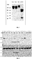
- the purified proteins were also subjected to western blot analysis using an anti-Camel antibody (Bethyl) as primary antibody (1:4000) and an anti-rabbit-HRP secondary antibody diluted 1:2000 (DAKO). ECL reagents (Amersham) was used for the detection of the protein according to the manufacturing instruction ( Figure 7 ).
- Microtitre plate wells were coated with 50 ⁇ l of 2 ⁇ g/mL human TrkA immunoadhesin, 10 ⁇ g/ml bovine serum albumin (BSA) or PBS (the uncoated well) .
- BSA bovine serum albumin
- PBS the uncoated well
- Microtitre plate wells were coated with 50 ⁇ l of either 2 ⁇ g/ml human TrkA immunoadhesin, 2 ⁇ g/ml mouse TrkA immunoadhesin, 10 ⁇ g/ml bovine serum albumin (BSA) or PBS (the uncoated well) .
- BSA bovine serum albumin
- PBS the uncoated well
- 50 ⁇ l of soluble scFv from each isolated clone was added to a well coated with either hTrkA, mTrkA, BSA or an uncoated well.
- HRP activity was visualized using TMB (Sigma). Clones were considered to be cross-reacting with human and mouse TrkA if the ELISA signal generated in the mTrkA coated well was at least 0.5-fold less than the signal on hTrkA (data not shown).
- the antibodies were also shown to be specific for hTrkA and mTrkA by relative binding to the BIACore sensor chips coated with the appropriate antigen.
- Antibodies were immobilized by ammine coupling to Biosensor CM5 sensorchips (Pharmacia) according to the manufacturers instructions.
- Anti-TrkA scFv proteins were diluted at 20-50 ⁇ g/ml in suitable pre-concentration buffer (at least 2 pH unit below the pI of the scFv in order to get a net positive charge), chosen among the Acetate buffers.
- the scFvs were immobilized at 100RU to get a low density immobilization.
- the recombinant protein consisting of D4-D5 domain of human and murine TrkA receptor (hTrkA Ig1,2 and mTrkA Ig1,2 respectively) were injected over the immobilized scFv with a contact time of 60 seconds and with a dissociation time of 400 seconds and the assay workflow placed 5 serial dilutions of the TrkA Ig1,2 (starting in the micromolar range and diluting 1:2 each time).
- the regeneration condition were mild (contact time 30 seconds, 10mM Glycine pH2).
- the concentrations of TrkA Ig1,2 were adjusted to optimize, contact time, dissociation time and rigeneration. Data were analysed by Bioevaluation Software(see results on Table II and Table III). The quality of the data fitting were checked by the value of Chi 2 and of the U-value.
- TF-1 cells a human hematopoietic cell line which expresses the native human TrkA receptor but not the p75 NGF-receptor, proliferate in response to exogenous human NGF ( Chevalier et al., Blood 1994, vol. 83:1479-85 ).
- the TF-1 proliferation assay as described by Chevalier et al. formed the basis for a potency assay to measure the effects of NGF/TrkA neutralising antibodies on the NGF-mediated proliferation of TF1 cells.
- TF1 cells are cultured for 1 week in RPMI-1640 containing 10% FBS with 2 ng/ml GM-CSF.
- Cells for testing are centrifuged (1000 rpm, 5 min), washed (RPMI-1640), centrifuged again and resuspended in RPMI-1640 + 10% FBS to a concentration of 300,000-400,000 cells/ml. They are then replated on 96-well microplates (15,000-20,000 cells per well in 50 ml) and TrkA neutralising antibodies are added soon after seeding.
- TF1 are exposed to 10 ng/mL NGF in RPMI-1640 containing 10% FBS (50 ml of 2X NGF is added per well, each well has a final volume of 100 ml). Control wells are included, either containing medium alone, or containing TF1 cells in the absence of NGF ("cellular blank"). Each treatment is performed in triplicate. After a 40 h incubation period, at 37°C, 5% CO 2 , 10 ⁇ l of MTT reagent (MTT cell proliferation assay kit) is added for 4 h incubation at 37°C.
- MTT reagent MTT cell proliferation assay kit
- MTT cell proliferation assay kit 100 ⁇ l per well; gently mixing, no pipetting
- O/N overnight incubation at room temperature in the dark.
- Absorbance is recorded at 570 nm. 100% inhibition is set as the value of inhibition corresponding to the average O.D. value observed for cells cultured without NGF, in the absence of antibody. 0% inhibition is set as the value of inhibition corresponding to the average O.D. value observed for cells exposed to 10 ng/ml NGF, in the absence of anti-TrkA antibodies.
- Fluorescence activated cell sorter is a powerful tool to measure and analyze cell surface molecules of single cells which flow in fluid stream through a beam of light to detect the fluorescences of the cells. FACS was applied to determine the binding profiling of the various scFv onto TF-1 receptor TrkA. FACS Tests are performed only on population with cell viability > 95%. Method is an adaptation of protocols reported on the Application Note " Detection of antibody-stained cell surface and intracellular protein targets with Agilent 2100 Bioanalyzer".
- 6x10 6 cells are first centrifuged at 350g for 5 min at room temperature (RT) and wash with 12 ml of Staining Buffer (SB) at RT. The pellet was resuspended in 3 ml of SB at 4°C and aliquote 0,5 ml of suspension in 1.5 ml conical tubes. Cells were centrifuged again at 350g for 5 min at 4°C and the pellet was resuspended in 1ml HBSS at 4°C. 125 ⁇ l 16% p-formaldehyde (final conc 1,7%) was added into each tube and then the sample was incubated for 10 min at 4°C on tube rotator.
- SB Staining Buffer
- FACS Tests are performed only on population with cell viability > 95%. Method is an adaptation of protocols reported on the Application Note "Detection of antibody-stained cell surface and intracellular protein targets with Agilent 2100 Bioanalyzer”.
- 6x10 6 cells are first centrifuged at 350g for 5 min at room temperature (RT) and wash with 12 ml of Dye Loading Buffer at RT. Cell pellets were resuspended in 2 ml of Dye Loading Buffer. 1 ⁇ m of CALCEIN_AM was added then 330 ⁇ l/sample are aliquoted in eppendorf and incubated for 30 min at 37°C in termoblock under dim light. After incubation the samples were washed with FACS Buffer (0,5 ml/sample) at 4°C and then centrifuged 5 min at 350g at RT.
- FACS Buffer (0,5 ml/sample
- Cells were resuspended in 100 ⁇ l/sample of Antibody Solution (con:0,2-20 ⁇ g/ml in FACS Buffer) and then incubated for 1 hour at 4°C on rotating wheel. Proper controls are included (FACS Buffer). After incubation the samples were washed with FACS Buffer (0,5 ml/sample) at 4°C and then centrifuged 5 min at 350g at RT. Cells were resuspended in 100 ⁇ l/sample of Antibody Solution in FACS buffer 4 ⁇ g/ml (1:500) and incubated for 30 minutes at 4°C on rotating wheel.
- 3T3-TrkA cells are cultured in DMEM (+10% FBS + 1X GlutaMAX + 100 units/ml penicillin and 0,1 mg/ml streptomycin) and can be used for the test from 3 days up to 2 months following seeding. The day before the test, cells are seeded in a 6 multi-well plate (2 ml of a suspension containing 5x10 5 cells per well).
- Anti-TrkA CRB0089 scFv was reformatted to entire IgG antibodies.
- the cDNA encoding the light and heavy chain (human IgG 4 ) were generated by GENEART (Germany) with suitable restriction sites for subcloning. Sequences were optimized for mammalian expression (CHO-S cell line) (SEQ ID. NO: 90 for heavy chain and 91 for light chain). After synthesis of both chains, the cDNAs were sub-cloned in expression plasmids (pcDNA3.1 derivates containing an extended CMV promoter for expression of the gene of interest) using HindIII and XhoI as cloning sites.
- each antibody chain two expression plasmids were generated: one plasmid containing the cDNA encoding the light chain, one containing the cDNA encoding the heavy chain.
- the expression plasmid containing the correct inserts were verified by restriction analysis and DNA sequence analysis of the insert.
- Anti-TrkA antibody was produced from transfected cells.
- CHO-S cells were transfected with plasmids encoding CRB0089 heavy and light chains.
- Conditioned media from transfected cells were recovered by removing cells and debris. Clarified conditioned media were loaded onto protein A-sepharose column. Non-specific bindings were removed by extensively binding buffer washes (20 mM sodium phosphate pH 7.0).
- Bound antibody proteins on the protein A column were recovered by acidic antibody elution from protein A (0.1 M glycine-HCl pH 3.0). Eluted proteins were immediately neutralized with 1M Tris-HCl pH 9.0 (100mL per mL eluted fractions). Pooled eluted fractions were dialyzed against PBS. Aggregated antibody proteins were removed by size exclusion chromatography.
- mice were weighed and allocated 4 per cage; 20 ⁇ l of formalin solution (1% in saline) were subcutaneously injected into the plantar surface of the right hind paw using an Hamilton micro-syringe equipped with a 26-gauge needle; four animals at a time were placed in a transparent plexiglass box (11x12x12 cm), allowed to move freely, and the observation period started. A mirror was placed behind the boxes to allow an unimpeded view of the animals hind paws. The licking activity, i.e. the total amount of time the animal spent licking the injected paw, was taken as index of pain.
- the licking activity was recorded continuously for 1 hour, calculated in blocks of consecutive 5-minutes periods and analyzed as the early (0-5 min) and the late (15-35 min) phases of the formalin test.
- Each mouse was subcutaneously injected into the dorsal surface of the right hind paw using an Hamilton micro-syringe (26-gauge needle) with 20 ⁇ l anti-TrkA (5-20mg/paw) mAb or PBS as control group, 18 hours before the test.
- mice were subcutaneously injected (systemically) with 300 ⁇ l CRB0089_IgG4 (5-20mg) or PBS as control group, 18 hours before the test.
- CRB0089_IgG4 was administered in the right hind paw 72 hours later the CFA injection. Eighteen hours after CRB0089_IgG4 administration, the paw volume was measured by means of a Plethysmometer (UgoBasile, Italy).
- CCI Chronic constriction injury
- a non-inflammatory model of chronic muscle pain in rats bilateral allodynia induced by unilateral injection of acidic saline in the gastrocnemius muscle.
- the acidic saline animal model of pain is thought to mimic human chronic pain syndromes such as fibromyalgia.
- Repeated intramuscular injections of acidic saline is a model of non-inflammatory pain characterized by bilateral long-lasting allodynia of the paw which is believed to be centrally mediated.
- Nociceptive thresholds expressed in grams (g), were measured with a Dynamic Plantar Aesthesiometer by applying increasing pressure to the right and left hind paw until the rat withdrew the paw. A maximal cut-off of 50g was used to prevent tissue damage. The threshold was tested three times for each paw and the mean value was calculated.
- CRB0089_IgG4 was administered subcutaneously at a dose of 20 ⁇ g/rat.
- a saline subcutaneous injection was used as vehicle control.
- Mechanical withdrawal thresholds of both hind paws were measured 18h (6d), 90h (9d), and 162h (12d) after CRB0089_IgG4 injection. Two injections of acidic saline into the gastrocnemius muscle produced bilateral decreases in the mechanical withdrawal threshold of the paw 24 h after the second injection.
- VEMHHW peptide SEQ ID NO: 66
- Ig-like domain I27 Titin protein
- VEMHHW peptide was inserted between A76-N77 and E27-D29 of I27 Ig-like domain of titin protein (SEQ. ID NO: 92).
- the recombinant 2xVEMHHW-I27 protein was subsequently cloned at the 3' of LexA and used to challenge a mouse SPLINT (mSPLINT) and a human SPLINT libraries (huSPLINT_09) ( Visintin et al., 2004. J Immunol Methods. 290:135-153 ).
- amino acid sequence of V regions of the isolated anti-VEMHHW scFvs are in the group of sequences consisting of SEQ.ID NO: 1, 2, 3, 4 from the selection of mSPLINT and SEQ. ID NO: 5, 6, 7, 8, 9, 10, 11, 12, 13, 14, 15 and 16 from the selection of hSPLINT_09.
- scFv cDNA expressing anti-VEMHHW scFv were cloned into E.coli pETM-13 expression vector. The proteins were well expressed in the cytoplasm and mostly retained in inclusion bodies (IB). scFv fragments can be refolded by dialysis after solubilization of IB ( Umetsu et al., 2003. J Biol Chem. 278:8979-8987 . Epub 2003 Jan 8977). We performed the technique of refolding by dilution ( Patil et al., 2008. J Biotechnol. 134:218-221 . Epub 2008 Jan 2018). The refolding condition of scFv was optimized for each sample. Refolded scFv were subsequently quantified by Bioanalyzer 2100 (Agilent) and tested by ELISA and Biacore analysis.
- TrkA binding analysis by flow cytometry on 3T3-TrkA and TF1 expressing TrkA receptor was performed with the panel of isolated anti-TrkA scFvs. All the anti-TrkA scFvs were able to recognize the TrkA receptor under physiological condition.
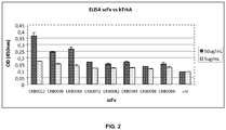
- TrkA expressing cells by anti-TrkA scFv, we analyzed the panel of scFv for specificity and crossreactivity with mouse TrkA by ELISA assay.
- ECD extracellular domain
- recombinant proteins were constructed to have the ECD domain of the Trk receptors linked to the Fc portion of a IgG2a camel antibody (Camelus dromedaries) (SEQ. ID NO: 70, 71, 72, 73 and 74).
- the recombinant protein were expressed in mammalian cell line (CHO-S cell line) and purified by protein A column. After purification the receptor chimera were analysed by SDS-PAGE western blot analysis under reducing condition ( Figure 7 ). The purified immunoadhesins were used as ligand in ELISA.
- TrkA Like other receptor tyrosine kinases, TrkA undergoes dimerization and activation upon ligand binding. In the absence of NGF, some domains of the receptor, perhaps the same ones responsible for ligand binding, impede its spontaneous dimerization at the cell surface ( Arevalo et al., 2000. Mol Cell Biol. 20:5908-5916 ). It was demonstrated that a recombinant deleted protein of TrkA receptor, Ig-1,2 which express both Ig-1 and Ig-2 domains of the extracellular domain (ECD) of the receptor TrkA was able to dimerize also in the absence of NGF ( Arevalo et al., 2000. Mol Cell Biol. 20:5908-5916 .). We have used two recombinant proteins engineered to express both Ig-likes domains and able to dimerize in the absence of NGF for Biacore analysis.
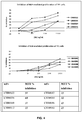
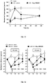
- the TF1 cell proliferation assay (MTT cell proliferation kit, ATCC) was used (concentration/response study). Final average OD values for triplicate measurements were calculated by subtraction of the average values for the cellular blank. Maximal inhibition was set corresponding to the average OD value observed for cells cultured without NGF, in the absence of test antibody. Zero inhibition was set corresponding to the average OD value observed for cells exposed to 10 ng/ml NGF in the absence of test scFv. As shown in Figure 5 , all the isolated anti-TrkA scFv were able to inhibit TF1 cell proliferation mediated by human NGF in a range between 20-60%.
- a complete IgG4 immunoglobulin was assembled by amplifying the individual V-regions of isolated anti-TrkA CRB0089 into a vector enabling the transfer of V-regions from scFv to full length immunoglobulin for mammalian expression.
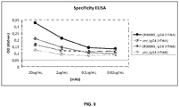
- Mab CRB0089_IgG4 was also tested to bind TF1 expressing TrkA receptor by FACS analysis.
- the antibody was able to bind to TrkA receptor in a dose dependent manner. At higher concentration (20mg/mL) the antibody display 99% gated events on more than 2800 cells tested.
- the antibody strongly bind to TrkA receptor even at lower concentrations ( Figure 10 ) as compared with an unrelated antibody (unr-IgG4) used as negative control.
- a TF1 cell proliferation assay was employed (concentration response study).
- the inhibitory potency of anti-TrkA CRB0089_IgG4 antibody was quantified as IC 50 values (i.e., the concentration of antibody required to reduce the NGF-mediated proliferative response by 50%) using Sigma Plot software. Inhibition curves were plotted individually in order to obtain discrete IC 50 values for each test antibody in each experiment. Measures of cell proliferation were normalized with respect to maximum OD values obtained within that assay, in the absence of added test antibody. Normalized responses were then plotted against test antibody concentration on a log scale, and IC 50 values were derived using the Sigma Plot nonlinear curve fitting function "log (inhibitor) vs response-variable slope".
- CRB0089_IgG4 was able to inhibit NGF-mediated proliferative response in a dose dependent manner with an IC 50 of approximately 43 nM.
- CRB0089_IgG4 Two “classical” screening models were initially used to evaluate the analgesic properties of CRB0089_IgG4: a) the formalin-induced licking behavior in the mouse, b) the complete Freund's adjuvant (CFA)-induced mechanical hyperalgesia in the rat.
- CFA complete Freund's adjuvant
- CRB0089_IgG4 injected s.c. in the range 5-20 ⁇ g, showed to inhibit the late phase of the formalin-induced behavior in mice at both the used doses ( Figure 13 ). No treatments induced significant changes related to the early phase of the formalin-induced behavior in mice.
- CCI chronic constriction injury
- neuropathic pain a non-inflammatory model of chronic muscle pain in rats- bilateral allodynia induced by unilateral injection of acidic saline in the gastrocnemius muscle
- Table I Results of anti-TrkA SPLINT screening: BAIT N. ⁇ clones (I screening) N. positive clones (II screening) TrkA_loopA 189 61
- Table II Biacore analysis of anti-TrkA scFvs vs human TrkA: Human Ig1,2 TrkA scFv ka (1/Ms) kd (1/s) KD (M) CRB0022 9192 2,94E-05 3.2 nM CRB0036 2731 3,75E-05 13.7 nM CRB0069 6266 3,57E-05 5.7 nM CRB0072 3675 7,48E-06 2.04 nM CRB0082 2090 8,79.E-05 42.1 nM CRB0084 9602 1.63E-02 1.70 ⁇ M CRB0088 2755 3.2E-06 1.16 nM CRB0089 4598 7,76E-05 16.9 nM Table III: Biacore analysis of anti-Trk
- PCR-assembly 80 5'-CATCATGAATTCCTAATAGAAGTGGAAAAGCCTCTGTACGGAGTAGAGGTG-3' 81 82 83 84 85 86 87 5'-TAATACGACTCACTATAGTCGACGGATCCTTACAATTCTTTCACTTTCAGATTGGCTG-3' II PCR-assembly 88 5'-CATCATGAATTCCTAATAGAAGTGGAAAAG-3' 89 5'-TAATACGACTCACTATAGTCGACGG-3'
Landscapes
- Health & Medical Sciences (AREA)
- Chemical & Material Sciences (AREA)
- Immunology (AREA)
- Organic Chemistry (AREA)
- Medicinal Chemistry (AREA)
- Life Sciences & Earth Sciences (AREA)
- General Health & Medical Sciences (AREA)
- Proteomics, Peptides & Aminoacids (AREA)
- Genetics & Genomics (AREA)
- Molecular Biology (AREA)
- Biophysics (AREA)
- Biochemistry (AREA)
- Engineering & Computer Science (AREA)
- Bioinformatics & Cheminformatics (AREA)
- Biomedical Technology (AREA)
- Microbiology (AREA)
- Mycology (AREA)
- Pharmacology & Pharmacy (AREA)
- Epidemiology (AREA)
- Animal Behavior & Ethology (AREA)
- Public Health (AREA)
- Veterinary Medicine (AREA)
- Peptides Or Proteins (AREA)
Description
- The present invention relates to anti-TrkA antibodies and to derivatives thereof which are able to recognise and bind an amino acid sequence comprised in the Nerve Growth Factor NGF-binding site of the high affinity tyrosine kinase receptor of NGF (TrkA), thus acting as NGF antagonists and preventing the functional activation of TrkA by NGF. The antibodies of the invention are useful in the treatment of conditions associated with the expression and/or activity of TrkA, including pain states.
- Monoclonal antibodies (Mabs) represent the fastest-growing market segment within the pharmaceutical industry. Despite a number of drawbacks, they are particularly appreciated among biotherapeutics, thanks to their unique features, including extremely high target specificity, favourable pharmacokinetics (long half-life) as well as faster development and higher success rate, as compared to small molecules. Soluble ligands and membrane-bound receptors involved in pain signalling represent ideal targets for the Mab, making it possible to obtain anti-pain biologics with higher specificity and/or different mechanism of action (MoA) as compared to currently available analgesics.
- The huge body of evidence suggesting that the NGF pathway provides new molecular targets for pain therapy has spurred the development of neutralizing antibodies against NGF and its receptors. In general, antibodies in the whole immunoglobulin format (IgG) are big molecules that do not go through the blood brain barrier when they are systemically administered. As for antibodies targeting the NGF pathway this means they are likely to be effective in periphery (as it is desirable for many pain conditions) but not in central nervous system (CNS). This is a clear advantage, since NGF is known to exert neurotrophic and neuroprotective effects on specific TrkA-expressing cell populations in the CNS, like basal forebrain cholinergic neurons). From a theoretical point of view anti-NGF neutralizing antibodies represent the most efficacious tool to reduce NGF bioactivity, especially if both receptors are thought to be involved; moreover when the neutralizing agent is an antibody in the whole Immunoglobulin format (IgG), it is usually safer to block a ligand instead of a membrane-bound receptor, which would increase the risk of complement-dependent cytolysis (CDC) and antigen-dependent cell cytolysis (ADCC) for the receptor bearing cell. On the other hand, targeting NGF in Peripheral nervous System (PNS) might indirectly affect the concentration of the same factor in CNS, altering the balance between PNS and CNS pools (peripheral sink effect), with possible deleterious effects on NGF-responsive neurons in the CNS; moreover, in certain cases (e.g the massive release of NGF in a short time, in inflammation) it is much easier to target and block the receptor than the NGF ligand. The neutralisation of either receptor would therefore avoid the drawbacks associated with NGF targeting as well as the side effects depending on signalling through the other receptor.
- Various anti-TrkA antibodies have also been generated. One such antibody is the monoclonal called 5C3 as disclosed in
WO97/21732 - An anti-TrkA monoclonal antibody referred to as MNAC13 is disclosed in
WO00/73344 -
WO06/131952 -
WO05/061540 WO00/73344 WO09/098238 - An human antibody and derivatives thereof that acts as a powerful NGF activity antagonist by recognizing and binding specific loops in the TrkAd5 domain essential for NGF binding has not been provided yet. Moreover there is the need to obtain antibodies with a defined binding specificity to fine regulating the activity thereof.
- Persistent pain represents a major health problem. It can show different levels of severity and is associated to different pathologies, such as back injury, migraine headaches, arthritis, herpes zoster, diabetic neuropathy, temporomandibular joint syndrome, and cancer. Mild pain is presently treated with acetaminophen, aspirin, and NSAIDs. The NSAIDs inhibit COX and thereby reduce prostaglandin synthesis. However, they are associated with gastrointestinal toxicity, and although COX-2-selective inhibitors have significantly reduced adverse gastric effects, there is still a raised risk of cardiovascular disease (Zeilhofer, 2007. Biochem Pharmacol. 73: 165-174). Moderate pain can be controlled using corticosteroidal drugs such as cortisol and prednisone, inhibiting phospholipase A2 (Flower and Blackwell, 1979. Nature. 278: 456-459). Nonetheless, corticosteroids display remarkable adverse effects including weight gain, insomnia, and immune system weakening. Severe pain may be treated with strong opioids such as morphine and fentanyl. However, long-term use of opiates is limited by several serious drawbacks, including development of tolerance and physical dependence (Przewlocki and Przewlocka, 2001. Eur J Pharmacol. 429: 79-91).
- As current pain therapies are often poorly effective or have undesirable side effects, an urgent need therefore exists to develop more specifically efficacious drugs directed against new molecular targets, with particular emphasis for the therapeutic area of chronic pain.
- Chronic pain may be of either nociceptive or neuropathic origin. In some cases, complex pain syndromes are produced by a combination of both, as it is the case for many types of oncologic pain. Nociceptive pain is induced by noxious mechanical, chemical, or thermal stimuli acting through pain specific receptors, mainly expressed on the peripheral endings of sensory neurons. Activation of nociceptive Ad-fibers (small diameter, rapidly-conducting, and myelinated) and C-fibers (small diameter, slower-conducting, and unmeylinated) results in pain perception. Tonic or chronic nociceptive pain may arise from sustained inflammatory disorders, resulting in hyperalgesia (increased sensitivity to painful stimuli) and/or allodynia (lowering of the threshold beyond which a stimulus is perceived as painful). Neuropathic pain may be induced by neural lesion or dysfunction, sometimes implying central neuroplasticity.
- Following injury, inflammatory mediators are released from both damaged tissues and activated immune cells. Proinflammatory cytokines such as tumor necrosis factor-a (TNFa) and interleukin-1 (IL-1) are secreted by neutrophils and activated cells of the monocyte/macrophage lineage. These cytokines may stimulate the release of Nerve growth factor (NGF) both from structural sources (fibroblasts, keratinocytes, and Schwann cells) and from inflammatory cell types (lymphocytes, macrophages, and mast cells). Moreover, mast cell degranulation releases other proinflammatory substances, which make up the so-called inflammatory soup: histamine, cytokines, prostaglandins, bradykinin, serotonin (5-hydroxytryptamine [5-HT]), adenosine triphosphate (ATP) and H+. Upon binding to their own specific receptors on nociceptive neurons, they all contribute to pain signalling (Pezet and McMahon, 2006. Annu Rev Neurosci. 29: 507-538).
- Nerve growth factor (NGF) is a multi-functional molecule that exerts its biological functions in a variety of neural and non-neural cells (Levi-Montalcini, 1987. Science. 237: 1154-1162), by means of two types of receptors: the TrkA tyrosine kinase receptor and the p75 neurotrophin receptor (p75NTR), belonging to the molecular family of Tumor Necrosis Factor receptors. TrkA mediates the survival and neurite outgrowth-promoting effects of NGF during development. Each NGF dimer binds two TrkA monomers, resulting in dimerization and trans-autophosphorylation of specific tyrosine residues. These phosphotyrosine residues form docking sites for several adaptor proteins coupling the receptor to intracellular signalling pathways including the mitogen activated protein kinase (MAPK), phosphatidylinositol 3-kinase (PI3K) and PLC pathways (Kaplan and Miller, 2000. Curr Opin Neurobiol. 10: 381-391); (Huang and Reichardt, 2003. Annu Rev Biochem. 72: 609-642). It is known that the exogenous administration of NGF induces pain both in animals and in humans (Mantyh et al., Anesthesiology. 115: 189-204) furthermore, the first line of evidence associating NGF signalling and pain comes from genetic studies. For example, congenital insensitivity to pain with anhidrosis is an autosomal recessive disorder characterized by the absence/abnormal development of several subsets of sensory and sympathetic neurons, which makes affected individuals unresponsive to pain and unable to sweat (anhydrosis). Null mutations in the gene encoding TrkA (NTRK1) have been recognized to be responsible for this disorder (Indo, 2001. Hum Mutat. 18: 462-471; Indo et al., 2001. Hum Mutat. 18: 308-318; Indo et al., 1996. Nat Genet. 13: 485-488). NGF plays a key role in pain transduction mechanisms in adult nervous system. Peripheral nociceptors strongly express the TrkA and p75NTR receptors and are developmentally and functionally dependent on NGF. NGF is a peripherally produced mediator of several persistent pain states, notably those associated with inflammation, also thanks to its dual action on inflammatory mast cells, that are recruited by NGF to the injured or painful site, and are induced by NGF to release inflammatory mediators. NGF is released by mast cells, fibroblasts and other cell types present in peripheral sites where inflammation is taking place. In particular, mast cells seem to play a key role (Woolf et al., 1996. J Neurosci. 16: 2716-2723). In fact, they produce NGF and display functional TrkA receptors on their surface in the same time, which makes them capable of responding to NGF itself, (Horigome et al., 1993. J Biol Chem. 268: 14881-14887).
- Thus the NGF-TrkA system appears to mediate mast cell activation through an autocrine loop, allowing local amplification of the activation process.
- Therefore, prior art still fails to disclose an anti-TrkA molecule that specifically recognises and binds an epitope in the TrkAd5 domain, comprising the sequence from aa 294 to to aa 299 of TrkA amino acid sequence of SEQ ID NO: 65, and more effectively acts as NGF antagonists specifically preventing the functional activation of TrkA by NGF.
- Object of the invention is an antibody, recombinant or synthetic antigen-binding fragments thereof which is able to recognise and bind an epitope comprising the sequence from aa. 294 to aa. 299 of the human TrkA amino acid sequence (UniProt P04629|33-796) of SEQ ID NO: 65 (i.e. the sequence VEMHHW, SEQ ID NO: 66).
- The antibody, recombinant or synthetic antigen-binding fragments thereof of the invention is able to specifically antagonize the activity of NGF triggered by TrkA, by preventing the functional activation of TrkA by NGF (also known as neutralizing antibodies).
- In the present invention an antibody refers to:
- a) a monoclonal, a polyclonal or a chimeric, or a humanized, or a deimmunized, or an affinity matured antibody, or a fully human antibody;
- b) a recombinant or synthetic antigen-binding fragments thereof, as well as molecules comprising such antigen-binding fragments, including engineered antibody fragments, antibody derivatives, bispecific antibodies and other multispecific molecules.
- In a preferred embodiment, said epitope consists of the sequence from aa. 294 to aa. 299 of the human TrkA amino acid sequence of SEQ ID NO: 65 (i.e. the sequence VEMHHW, SEQ ID NO: 66).
- The antibody, recombinant or synthetic antigen-binding fragments thereof according to the invention preferably comprises at least one heavy chain complementary determining region (CDRH3) amino acid sequence having at least 80 % identity to an amino acid sequence selected from the group consisting of: SEQ ID NO: 64, 43, 46, 49, 52, 55, 58 and 61.
- The antibody, recombinant or synthetic antigen-binding fragments thereof according to the present invention preferably comprises at least one heavy chain complementary determining region (CDRH1) amino acid sequence having at least 80 % identity to an amino acid sequence selected from the group consisting of: SEQ ID NO: 62, 41, 44, 47, 50, 53, 56 and 59.
- The antibody, recombinant or synthetic antigen-binding fragments thereof according to the present invention preferably comprises at least one heavy chain complementary determining region (CDRH2) amino acid sequence having at least 80 % identity to an amino acid sequence selected from the group consisting of: SEQ ID NO: 63, 42, 45, 48, 51, 54, 57 and 60.
- In a preferred embodiment, the antibody, recombinant or synthetic antigen-binding fragments thereof comprises a CDRH1 amino acid sequence having at least 80 % identity to SEQ ID NO: 62, a CDRH2 amino acid sequence having at least 80 % identity to SEQ ID NO: 63 and a CDRH3 amino acid sequence having at least 80 % identity to SEQ ID NO: 64.
- The antibody, recombinant or synthetic antigen-binding fragments thereof according to the present invention preferably comprises one light chain complementary determining region (CDRL3) amino acid sequence having at least 80 % identity to an amino acid sequence selected from the group consisting of: SEQ ID NO: 40, 19, 22, 25, 28, 31, 34 and 37.
- The antibody, recombinant or synthetic antigen-binding fragments thereof according to the present invention preferably comprises at least one light chain complementary determining region (CDRL1) amino acid sequence having at least 80 % identity to an amino acid sequence selected from the group consisting of: SEQ ID NO: 38, 17, 20, 23, 26, 29, 32 and 35
- The antibody, recombinant or synthetic antigen-binding fragments thereof according to the present invention preferably comprises one light chain complementary determining region (CDRL2) amino acid sequence having at least 80 % identity to an amino acid sequence selected from the group consisting of: SEQ ID NO: 39, 18, 21, 24, 27, 30, 33 and 36.
- In a preferred embodiment of the invention, the antibody, recombinant or synthetic antigen-binding fragments thereof comprises a CDRL1 amino acid sequence having at least 80 % identity to SEQ ID NO: 38, a CDRL2 amino acid sequence having at least 80 % identity to SEQ ID NO: 39 and a CDRL3 amino acid sequence having at least 80 % identity to SEQ ID NO: 40. PLEASE INSERT PAGE 8A
- The antibody, recombinant or synthetic antigen-binding fragments thereof according to the present invention preferably comprises a heavy chain variable region comprising SEQ ID NO: 15, 1, 3, 5, 7, 9, 11 or 13.
- The antibody, recombinant or synthetic antigen-binding fragments thereof according to the present invention preferably comprises a light chain variable region comprising SEQ ID NO: 16,2,4,6,8, 10, 12 or 14.
- According to one aspect of the invention, the antibody, recombinant or synthetic antigen-binding fragments thereof according to the present invention comprises:
- a) a variable heavy chain comprising a sequence selected from any of CRB0022VH (SEQ. ID NO: 1), CRB0036VH (SEQ. ID NO: 3), CRB0069VH (SEQ. ID NO: 5), CRB0072VH (SEQ. ID NO: 7), CRB0082VH (SEQ. ID NO Preferably the antibody, recombinant or synthetic antigen-binding fragments thereof comprises a CDRH1 amino acid sequence of SEQ ID NO: 62, a CDRH2 amino acid sequence of SEQ ID NO: 63, a CDRH3 amino acid sequence of SEQ ID NO: 64, a CDRL1 amino acid sequence of SEQ ID NO: 38, a CDRL2 amino acid sequence of SEQ ID NO: 39 and a CDRL3 of SEQ ID NO: 40. 9), CRB0084VH (SEQ. ID NO: 11), CRB0088VH (SEQ. ID NO: 13) or CRB0089VH (SEQ. ID NO: 15) or variants thereof or
- b) a variable light chain comprising a sequence selected from any of CRB0022VK (SEQ. ID NO: 2), CRB0036VK (SEQ. ID NO: 4), CRB0069VK (SEQ. ID NO: 6), CRB0072VK (SEQ. ID NO: 8), CRB0082VL (SEQ. ID NO: 10), CRB0084VL (SEQ. ID NO: 12), CRB0088VL (SEQ. ID NO: 14) or CRB0089VL (SEQ. ID NO: 16) or variants thereof.
- More preferably, the antibody comprises both a variable heavy chain as described in a) above and variable light chain as described in b), i.e. it comprises one of the following 64 combinations of light and heavy chains:
- CRB0022VH_CRB0022VK; CRB0022VH_CRB0036VK;
- CRB0022VH_CRB0069VK; CRB0022VH_CRB0072VK; CRB0022VH_CRB0082VL;
- CRB0022VH_CRB0084VL; CRB0022VH_CRB0088VL; CRB0022VH_CRB0089VL;
- CRB0036VH_CRB0022VK; CRB0036VH_CRB0036VK;
- CRB0036VH_CRB0069VK; CRB0036VH_CRB0072VK;
- CRB0036VH_CRB0082VL; CRB0036VH_CRB0084VL; CRB0036VH_CRB0088VL;
- CRB0036VH_CRB0089VL; CRB0069VH_CRB0022VK; CRB0069VH_CRB0036VK;
- CRB0069VH_CRB0069VK; CRB0069VH_CRB0072VK; CRB0069VH_CRB0082VL;
- CRB0069VH_CRB0084VL; CRB0069VH_CRB0088VL; CRB0069VH_CRB0089VL;
- CRB0072VH_CRB0022VK; CRB0072VH_CRB0036VK;
- CRB0072VH_CRB0069VK; CRB0072VH_CRB0072VK;
- CRB0072VH_CRB0082VL; CRB0072VH_CRB0084VL; CRB0072VH_CRB0088VL;
- CRB0072VH_CRB0089VL; CRB0082VH_CRB0022VK; CRB0082VH_CRB0036VK;
- CRB0082VH_CRB0069VK; CRB0082VH_CRB0072VK; CRB0082VH_CRB0082VL;
- CRB0082VH_CRB0084VL; CRB0082VH_CRB0088VL; CRB0082VH_CRB0089VL;
- CRB0084VH_CRB0022VK; CRB0084VH_CRB0036VK;
- CRB0084VH_CRB0069VK; CRB0084VH_CRB0072VK;
- CRB0084VH_CRB0082VL; CRB0084VH_CRB0084VL; CRB0084VH_CRB0088VL;
- CRB0084VH_CRB0089VL; CRB0088VH_CRB0022VK; CRB0088VH_CRB0036VK;
- CRB0088VH_CRB0069VK; CRB0088VH_CRB0072VK; CRB0088VH_CRB0082VL;
- CRB0088VH_CRB0084VL; CRB0088VH_CRB0088VL; CRB0088VH_CRB0089VL;
- CRB0089VH_CRB0022VK; CRB0089VH_CRB0036VK;
- CRB0089VH_CRB0069VK; CRB0089VH_CRB0072VK; CRB0089VH_CRB0082VL;
- CRB0089VH_CRB0084VL; CRB0089VH_CRB0088VL; CRB0089VH_CRB0089VL Particularly preferred anti-TrkA antibodies which fall within the scope of the present invention comprises a plurality of hypervariable regions (CDR), at least one of which, or at least two of which, or at least three of which, or at least four of which, or at least five of which, or six of which is/are selected from the group consisting of:
- CDRH1 regions, comprising sequences selected from the group consisting of SEQ. ID Nos: 41, 44, 47, 50, 53, 56, 59, 62
- CDRH2 regions, comprising sequences selected from the group consisting of SEQ. ID Nos: 42, 45, 48, 51, 54, 57, 60, 63
- CDRH3 regions, comprising sequences selected from the group consisting of SEQ. ID Nos:43,46,49,52,55,58,61,64
- CDRL1 regions, comprising sequences selected from the group consisting of SEQ. ID Nos: 17, 20, 23, 26, 29, 32, 35, 38
- CDRL2 regions, comprising sequences selected from the group consisting of SEQ. ID Nos: 18, 21, 24, 27, 30, 33, 36, 39
- CDRL3 regions, comprising sequences selected from the group consisting of SEQ. ID Nos: 19, 22, 25, 28, 31, 34, 37, 40.
- Other particularly preferred anti-TrkA antibodies which fall within the scope of the present invention are disclosed herein below:
an anti-TrkA antibody which comprises a heavy chain variable region comprising CDRH1, CDRH2 and CDRH3, and/or a light chain variable region comprising CDRL1, CDRL2, and CDRL3 wherein both CDRHs and CDRLs amino acid sequence are combined as described in the table below:CDRH1 CDRH2 CDRH3 CDRL1 CDRL2 CDRL3 SEQ.ID NO: 41 42 43 17 18 19 SEQ.ID NO: 44 45 46 20 21 22 SEQ.ID NO: 47 48 49 23 24 25 SEQ.ID NO: 50 51 52 26 27 28 SEQ.ID NO: 53 54 55 29 30 31 SEQ.ID NO: 56 57 58 32 33 34 SEQ.ID NO: 59 60 61 35 36 37 SEQ.ID NO: 62 63 64 38 39 40 SEQ.ID NO: 41 45 46 20 21 22 SEQ.ID NO: 41 48 49 23 24 25 SEQ.ID NO: 41 51 52 26 27 28 SEQ.ID NO: 41 54 55 29 30 31 SEQ.ID NO: 41 57 58 32 33 34 SEQ.ID NO: 41 60 61 35 36 37 SEQ.ID NO: 41 63 64 38 39 40 CDRH1 CDRH2 CDRH3 CDRL1 CDRL2 CDRL3 SEQ.ID NO: 44 42 43 17 18 19 SEQ.ID NO: 44 48 49 23 24 25 SEQ.ID NO: 44 51 52 26 27 28 SEQ.ID NO: 44 54 55 29 30 31 SEQ.ID NO: 44 57 58 32 33 34 SEQ.ID NO: 44 60 61 35 36 37 SEQ.ID NO: 44 63 64 38 39 40 CDRH1 CDRH2 CDRH3 CDRL1 CDRL2 CDRL3 SEQ.ID NO: 47 42 43 17 18 19 SEQ.ID NO: 47 45 46 20 21 22 SEQ.ID NO: 47 51 52 26 27 28 SEQ.ID NO: 47 54 55 29 30 31 SEQ.ID NO: 47 57 58 32 33 34 SEQ.ID NO: 47 60 61 35 36 37 SEQ.ID NO: 47 63 64 38 39 40 CDRH1 CDRH2 CDRH3 CDRL1 CDRL2 CDRL3 SEQ.ID NO: 50 42 43 17 18 19 SEQ.ID NO: 50 45 46 20 21 22 SEQ.ID NO: 50 48 49 23 24 25 SEQ.ID NO: 50 54 55 29 30 31 SEQ.ID NO: 50 57 58 32 33 34 SEQ.ID NO: 50 60 61 35 36 37 SEQ.ID NO: 50 63 64 38 39 40 CDRH1 CDRH2 CDRH3 CDRL1 CDRL2 CDRL3 SEQ.ID NO: 53 42 43 17 18 19 SEQ.ID NO: 53 45 46 20 21 22 SEQ.ID NO: 53 48 49 23 24 25 SEQ.ID NO: 53 51 52 26 27 28 SEQ.ID NO: 53 57 58 32 33 34 SEQ.ID NO: 53 60 61 35 36 37 SEQ.ID NO: 53 63 64 38 39 40 CDRH1 CDRH2 CDRH3 CDRL1 CDRL2 CDRL3 SEQ.ID NO: 56 42 43 17 18 19 SEQ.ID NO: 56 45 46 20 21 22 SEQ.ID NO: 56 48 49 23 24 25 SEQ.ID NO: 56 51 52 26 27 28 SEQ.ID NO: 56 54 55 29 30 31 SEQ.ID NO: 56 60 61 35 36 37 SEQ.ID NO: 56 63 64 38 39 40 CDRH1 CDRH2 CDRH3 CDRL1 CDRL2 CDRL3 SEQ.ID NO: 59 42 43 17 18 19 SEQ.ID NO: 59 45 46 20 21 22 SEQ.ID NO: 59 48 49 23 24 25 SEQ.ID NO: 59 51 52 26 27 28 SEQ.ID NO: 59 54 55 29 30 31 SEQ.ID NO: 59 57 58 32 33 34 SEQ.ID NO: 59 63 64 38 39 40 CDRH1 CDRH2 CDRH3 CDRL1 CDRL2 CDRL3 SEQ.ID NO: 62 42 43 17 18 19 SEQ.ID NO: 62 45 46 20 21 22 SEQ.ID NO: 62 48 49 23 24 25 SEQ.ID NO: 62 51 52 26 27 28 SEQ.ID NO: 62 54 55 29 30 31 SEQ.ID NO: 62 57 58 32 33 34 SEQ.ID NO: 62 60 61 35 36 37 CDRH1 CDRH2 CDRH3 CDRL1 CDRL2 CDRL3 SEQ.ID NO: 44 42 46 20 21 22 SEQ.ID NO: 47 42 49 23 24 25 SEQ.ID NO: 50 42 52 26 27 28 SEQ.ID NO: 53 42 55 29 30 31 SEQ.ID NO: 56 42 58 32 33 34 SEQ.ID NO: 59 42 61 35 36 37 SEQ.ID NO: 62 42 64 38 39 40 CDRH1 CDRH2 CDRH3 CDRL1 CDRL2 CDRL3 SEQ.ID NO: 41 45 43 17 18 19 SEQ.ID NO: 47 45 49 23 24 25 SEQ.ID NO: 50 45 52 26 27 28 SEQ.ID NO: 53 45 55 29 30 31 SEQ.ID NO: 56 45 58 32 33 34 SEQ.ID NO: 59 45 61 35 36 37 SEQ.ID NO: 62 45 64 38 39 40 CDRH1 CDRH2 CDRH3 CDRL1 CDRL2 CDRL3 SEQ.ID NO: 41 48 43 17 18 19 SEQ.ID NO: 44 48 46 20 21 22 SEQ.ID NO: 50 48 52 26 27 28 SEQ.ID NO: 53 48 55 29 30 31 SEQ.ID NO: 56 48 58 32 33 34 SEQ.ID NO: 59 48 61 35 36 37 SEQ.ID NO: 62 48 64 38 39 40 CDRH1 CDRH2 CDRH3 CDRL1 CDRL2 CDRL3 SEQ.ID NO: 41 51 43 17 18 19 SEQ.ID NO: 44 51 46 20 21 22 SEQ.ID NO: 47 51 49 23 24 25 SEQ.ID NO: 53 51 55 29 30 31 SEQ.ID NO: 56 51 58 32 33 34 SEQ.ID NO: 59 51 61 35 36 37 SEQ.ID NO: 62 51 64 38 39 40 CDRH1 CDRH2 CDRH3 CDRL1 CDRL2 CDRL3 SEQ.ID NO: 41 54 43 17 18 19 SEQ.ID NO: 44 54 46 20 21 22 SEQ.ID NO: 47 54 49 23 24 25 SEQ.ID NO: 50 54 52 26 27 28 SEQ.ID NO: 56 54 58 32 33 34 SEQ.ID NO: 59 54 61 35 36 37 SEQ.ID NO: 62 54 64 38 39 40 CDRH1 CDRH2 CDRH3 CDRL1 CDRL2 CDRL3 SEQ.ID NO: 41 57 43 17 18 19 SEQ.ID NO: 44 57 46 20 21 22 SEQ.ID NO: 47 57 49 23 24 25 SEQ.ID NO: 50 57 52 26 27 28 SEQ.ID NO: 53 57 55 29 30 31 SEQ.ID NO: 59 57 61 35 36 37 SEQ.ID NO: 62 57 64 38 39 40 CDRH1 CDRH2 CDRH3 CDRL1 CDRL2 CDRL3 SEQ.ID NO: 41 60 43 17 18 19 SEQ.ID NO: 44 60 46 20 21 22 SEQ.ID NO: 47 60 49 23 24 25 SEQ.ID NO: 50 60 52 26 27 28 SEQ.ID NO: 53 60 55 29 30 31 SEQ.ID NO: 56 60 58 32 33 34 SEQ.ID NO: 62 60 64 38 39 40 CDRH1 CDRH2 CDRH3 CDRL1 CDRL2 CDRL3 SEQ.ID NO: 41 63 43 17 18 19 SEQ.ID NO: 44 63 46 20 21 22 SEQ.ID NO: 47 63 49 23 24 25 SEQ.ID NO: 50 63 52 26 27 28 SEQ.ID NO: 53 63 55 29 30 31 SEQ.ID NO: 56 63 58 32 33 34 SEQ.ID NO: 59 63 61 35 36 37 CDRH1 CDRH2 CDRH3 CDRL1 CDRL2 CDRL3 SEQ.ID NO: 44 45 43 20 21 22 SEQ.ID NO: 47 48 43 23 24 25 SEQ.ID NO: 50 51 43 26 27 28 SEQ.ID NO: 53 54 43 29 30 31 SEQ.ID NO: 56 57 43 32 33 34 SEQ.ID NO: 59 60 43 35 36 37 SEQ.ID NO: 62 63 43 38 39 40 CDRH1 CDRH2 CDRH3 CDRL1 CDRL2 CDRL3 SEQ.ID NO: 41 42 46 17 18 19 SEQ.ID NO: 47 48 46 23 24 25 SEQ.ID NO: 50 51 46 26 27 28 SEQ.ID NO: 53 54 46 29 30 31 SEQ.ID NO: 56 57 46 32 33 34 SEQ.ID NO: 59 60 46 35 36 37 SEQ.ID NO: 62 63 46 38 39 40 CDRH1 CDRH2 CDRH3 CDRL1 CDRL2 CDRL3 SEQ.ID NO: 41 42 49 17 18 19 SEQ.ID NO: 44 45 49 20 21 22 SEQ.ID NO: 50 51 49 26 27 28 SEQ.ID NO: 53 54 49 29 30 31 SEQ.ID NO: 56 57 49 32 33 34 SEQ.ID NO: 59 60 49 35 36 37 SEQ.ID NO: 62 63 49 38 39 40 CDRH1 CDRH2 CDRH3 CDRL1 CDRL2 CDRL3 SEQ.ID NO: 41 42 52 17 18 19 SEQ.ID NO: 44 45 52 20 21 22 SEQ.ID NO: 47 48 52 23 24 25 SEQ.ID NO: 53 54 52 29 30 31 SEQ.ID NO: 56 57 52 32 33 34 SEQ.ID NO: 59 60 52 35 36 37 SEQ.ID NO: 62 63 52 38 39 40 CDRH1 CDRH2 CDRH3 CDRL1 CDRL2 CDRL3 SEQ.ID NO: 41 42 55 17 18 19 SEQ.ID NO: 44 45 55 20 21 22 SEQ.ID NO: 47 48 55 23 24 25 SEQ.ID NO: 50 51 55 26 27 28 SEQ.ID NO: 56 57 55 32 33 34 SEQ.ID NO: 59 60 55 35 36 37 SEQ.ID NO: 62 63 55 38 39 40 CDRH1 CDRH2 CDRH3 CDRL1 CDRL2 CDRL3 SEQ.ID NO: 41 42 58 17 18 19 SEQ.ID NO: 44 45 58 20 21 22 SEQ.ID NO: 47 48 58 23 24 25 SEQ.ID NO: 50 51 58 26 27 28 SEQ.ID NO: 53 54 58 29 30 31 SEQ.ID NO: 59 60 58 35 36 37 SEQ.ID NO: 62 63 58 38 39 40 CDRH1 CDRH2 CDRH3 CDRL1 CDRL2 CDRL3 SEQ.ID NO: 41 42 61 17 18 19 SEQ.ID NO: 44 45 61 20 21 22 SEQ.ID NO: 47 48 61 23 24 25 SEQ.ID NO: 50 51 61 26 27 28 SEQ.ID NO: 53 54 61 29 30 31 SEQ.ID NO: 56 57 61 32 33 34 SEQ.ID NO: 62 63 61 38 39 40 CDRH1 CDRH2 CDRH3 CDRL1 CDRL2 CDRL3 SEQ.ID NO: 41 42 64 17 18 19 SEQ.ID NO: 44 45 64 20 21 22 SEQ.ID NO: 47 48 64 23 24 25 SEQ.ID NO: 50 51 64 26 27 28 SEQ.ID NO: 53 54 64 29 30 31 SEQ.ID NO: 56 57 64 32 33 34 SEQ.ID NO: 59 60 64 35 36 37 CDRH1 CDRH2 CDRH3 CDRL1 CDRL2 CDRL3 SEQ.ID NO: 44 45 46 17 21 22 SEQ.ID NO: 47 48 49 17 24 25 SEQ.ID NO: 50 51 52 17 27 28 SEQ.ID NO: 53 54 55 17 30 31 SEQ.ID NO: 56 57 58 17 33 34 SEQ.ID NO: 59 60 61 17 36 37 SEQ.ID NO: 62 63 64 17 39 40 CDRH1 CDRH2 CDRH3 CDRL1 CDRL2 CDRL3 SEQ.ID NO: 41 42 43 20 18 19 SEQ.ID NO: 47 48 49 20 24 25 SEQ.ID NO: 50 51 52 20 27 28 SEQ.ID NO: 53 54 55 20 30 31 SEQ.ID NO: 56 57 58 20 33 34 SEQ.ID NO: 59 60 61 20 36 37 SEQ.ID NO: 62 63 64 20 39 40 CDRH1 CDRH2 CDRH3 CDRL1 CDRL2 CDRL3 SEQ.ID NO: 41 42 43 23 18 19 SEQ.ID NO: 44 45 46 23 21 22 SEQ.ID NO: 50 51 52 23 27 28 SEQ.ID NO: 53 54 55 23 30 31 SEQ.ID NO: 56 57 58 23 33 34 SEQ.ID NO: 59 60 61 23 36 37 SEQ.ID NO: 62 63 64 23 39 40 CDRH1 CDRH2 CDRH3 CDRL1 CDRL2 CDRL3 SEQ.ID NO: 41 42 43 26 18 19 SEQ.ID NO: 44 45 46 26 21 22 SEQ.ID NO: 47 48 49 26 24 25 SEQ.ID NO: 53 54 55 26 30 31 SEQ.ID NO: 56 57 58 26 33 34 SEQ.ID NO: 59 60 61 26 36 37 SEQ.ID NO: 62 63 64 26 39 40 CDRH1 CDRH2 CDRH3 CDRL1 CDRL2 CDRL3 SEQ.ID NO: 41 42 43 29 18 19 SEQ.ID NO: 44 45 46 29 21 22 SEQ.ID NO: 47 48 49 29 24 25 SEQ.ID NO: 50 51 52 29 27 28 SEQ.ID NO: 56 57 58 29 33 34 SEQ.ID NO: 59 60 61 29 36 37 SEQ.ID NO: 62 63 64 29 39 40 CDRH1 CDRH2 CDRH3 CDRL1 CDRL2 CDRL3 SEQ.ID NO: 41 42 43 32 18 19 SEQ.ID NO: 44 45 46 32 21 22 SEQ.ID NO: 47 48 49 32 24 25 SEQ.ID NO: 50 51 52 32 27 28 SEQ.ID NO: 53 54 55 32 30 31 SEQ.ID NO: 59 60 61 32 36 37 SEQ.ID NO: 62 63 64 32 39 40 CDRH1 CDRH2 CDRH3 CDRL1 CDRL2 CDRL3 SEQ.ID NO: 41 42 43 35 18 19 SEQ.ID NO: 44 45 46 35 21 22 SEQ.ID NO: 47 48 49 35 24 25 SEQ.ID NO: 50 51 52 35 27 28 SEQ.ID NO: 53 54 55 35 30 31 SEQ.ID NO: 56 57 58 35 33 34 SEQ.ID NO: 62 63 64 35 39 40 CDRH1 CDRH2 CDRH3 CDRL1 CDRL2 CDRL3 SEQ.ID NO: 41 42 43 38 18 19 SEQ.ID NO: 44 45 46 38 21 22 SEQ.ID NO: 47 48 49 38 24 25 SEQ.ID NO: 50 51 52 38 27 28 SEQ.ID NO: 53 54 55 38 30 31 SEQ.ID NO: 56 57 58 38 33 34 SEQ.ID NO: 59 60 61 38 36 37 CDRH1 CDRH2 CDRH3 CDRL1 CDRL2 CDRL3 SEQ.ID NO: 44 45 46 20 18 22 SEQ.ID NO: 47 48 49 23 18 25 SEQ.ID NO: 50 51 52 26 18 28 SEQ.ID NO: 53 54 55 29 18 31 SEQ.ID NO: 56 57 58 32 18 34 SEQ.ID NO: 59 60 61 35 18 37 SEQ.ID NO: 62 63 64 38 18 40 CDRH1 CDRH2 CDRH3 CDRL1 CDRL2 CDRL3 SEQ.ID NO: 41 42 43 17 21 19 SEQ.ID NO: 47 48 49 23 21 25 SEQ.ID NO: 50 51 52 26 21 28 SEQ.ID NO: 53 54 55 29 21 31 SEQ.ID NO: 56 57 58 32 21 34 SEQ.ID NO: 59 60 61 35 21 37 SEQ.ID NO: 62 63 64 38 21 40 CDRH1 CDRH2 CDRH3 CDRL1 CDRL2 CDRL3 SEQ.ID NO: 41 42 43 17 24 19 SEQ.ID NO: 44 45 46 20 24 22 SEQ.ID NO: 50 51 52 26 24 28 SEQ.ID NO: 53 54 55 29 24 31 SEQ.ID NO: 56 57 58 32 24 34 SEQ.ID NO: 59 60 61 35 24 37 SEQ.ID NO: 62 63 64 38 24 40 CDRH1 CDRH2 CDRH3 CDRL1 CDRL2 CDRL3 SEQ.ID NO: 41 42 43 17 27 19 SEQ.ID NO: 44 45 46 20 27 22 SEQ.ID NO: 47 48 49 23 27 25 SEQ.ID NO: 53 54 55 29 27 31 SEQ.ID NO: 56 57 58 32 27 34 SEQ.ID NO: 59 60 61 35 27 37 SEQ.ID NO: 62 63 64 38 27 40 CDRH1 CDRH2 CDRH3 CDRL1 CDRL2 CDRL3 SEQ.ID NO: 41 42 43 17 30 19 SEQ.ID NO: 44 45 46 20 30 22 SEQ.ID NO: 47 48 49 23 30 25 SEQ.ID NO: 50 51 52 26 30 28 SEQ.ID NO: 56 57 58 32 30 34 SEQ.ID NO: 59 60 61 35 30 37 SEQ.ID NO: 62 63 64 38 30 40 SEQ.ID NO: 41 42 43 17 33 19 SEQ.ID NO: 44 45 46 20 33 22 SEQ.ID NO: 47 48 49 23 33 25 SEQ.ID NO: 50 51 52 26 33 28 SEQ.ID NO: 53 54 55 29 33 31 SEQ.ID NO: 59 60 61 35 33 37 SEQ.ID NO: 62 63 64 38 33 40 CDRH1 CDRH2 CDRH3 CDRL1 CDRL2 CDRL3 SEQ.ID NO: 41 42 43 17 36 19 SEQ.ID NO: 44 45 46 20 36 22 SEQ.ID NO: 47 48 49 23 36 25 SEQ.ID NO: 50 51 52 26 36 28 SEQ.ID NO: 53 54 55 29 36 31 SEQ.ID NO: 56 57 58 32 36 34 SEQ.ID NO: 62 63 64 38 36 40 CDRH1 CDRH2 CDRH3 CDRL1 CDRL2 CDRL3 SEQ.ID NO: 41 42 43 17 39 19 SEQ.ID NO: 44 45 46 20 39 22 SEQ.ID NO: 47 48 49 23 39 25 SEQ.ID NO: 50 51 52 26 39 28 SEQ.ID NO: 53 54 55 29 39 31 SEQ.ID NO: 56 57 58 32 39 34 SEQ.ID NO: 59 60 61 35 39 37 CDRH1 CDRH2 CDRH3 CDRL1 CDRL2 CDRL3 SEQ.ID NO: 44 45 46 20 21 19 SEQ.ID NO: 47 48 49 23 24 19 SEQ.ID NO: 50 51 52 26 27 19 SEQ.ID NO: 53 54 55 29 30 19 SEQ.ID NO: 56 57 58 32 33 19 SEQ.ID NO: 59 60 61 35 36 19 SEQ.ID NO: 62 63 64 38 39 19 CDRH1 CDRH2 CDRH3 CDRL1 CDRL2 CDRL3 SEQ.ID NO: 41 42 43 17 18 22 SEQ.ID NO: 47 48 49 23 24 22 SEQ.ID NO: 50 51 52 26 27 22 SEQ.ID NO: 53 54 55 29 30 22 SEQ.ID NO: 56 57 58 32 33 22 SEQ.ID NO: 59 60 61 35 36 22 SEQ.ID NO: 62 63 64 38 39 22 CDRH1 CDRH2 CDRH3 CDRL1 CDRL2 CDRL3 SEQ.ID NO: 41 42 43 17 18 25 SEQ.ID NO: 44 45 46 20 21 25 SEQ.ID NO: 50 51 52 26 27 25 SEQ.ID NO: 53 54 55 29 30 25 SEQ.ID NO: 56 57 58 32 33 25 SEQ.ID NO: 59 60 61 35 36 25 SEQ.ID NO: 62 63 64 38 39 25 CDRH1 CDRH2 CDRH3 CDRL1 CDRL2 CDRL3 SEQ.ID NO: 41 42 43 17 18 28 SEQ.ID NO: 44 45 46 20 21 28 SEQ.ID NO: 47 48 49 23 24 28 SEQ.ID NO: 53 54 55 29 30 28 SEQ.ID NO: 56 57 58 32 33 28 SEQ.ID NO: 59 60 61 35 36 28 SEQ.ID NO: 62 63 64 38 39 28 CDRH1 CDRH2 CDRH3 CDRL1 CDRL2 CDRL3 SEQ.ID NO: 41 42 43 17 18 31 SEQ.ID NO: 44 45 46 20 21 31 SEQ.ID NO: 47 48 49 23 24 31 SEQ.ID NO: 50 51 52 26 27 31 SEQ.ID NO: 56 57 58 32 33 31 SEQ.ID NO: 59 60 61 35 36 31 SEQ.ID NO: 62 63 64 38 39 31 CDRH1 CDRH2 CDRH3 CDRL1 CDRL2 CDRL3 SEQ.ID NO: 41 42 43 17 18 34 SEQ.ID NO: 44 45 46 20 21 34 SEQ.ID NO: 47 48 49 23 24 34 SEQ.ID NO: 50 51 52 26 27 34 SEQ.ID NO: 53 54 55 29 30 34 SEQ.ID NO: 59 60 61 35 36 34 SEQ.ID NO: 62 63 64 38 39 34 CDRH1 CDRH2 CDRH3 CDRL1 CDRL2 CDRL3 SEQ.ID NO: 41 42 43 17 18 37 SEQ.ID NO: 44 45 46 20 21 37 SEQ.ID NO: 47 48 49 23 24 37 SEQ.ID NO: 50 51 52 26 27 37 SEQ.ID NO: 53 54 55 29 30 37 SEQ.ID NO: 56 57 58 32 33 37 SEQ.ID NO: 62 63 64 38 39 37 CDRH1 CDRH2 CDRH3 CDRL1 CDRL2 CDRL3 SEQ.ID NO: 41 42 43 17 18 40 SEQ.ID NO: 44 45 46 20 21 40 SEQ.ID NO: 47 48 49 23 24 40 SEQ.ID NO: 50 51 52 26 27 40 SEQ.ID NO: 53 54 55 29 30 40 SEQ.ID NO: 56 57 58 32 33 40 SEQ.ID NO: 59 60 61 35 36 40 - The antibody, recombinant or synthetic antigen-binding fragments thereof according to the present invention preferably comprises a heavy chain variable region comprising SEQ ID NO: 15 and a light chain variable region comprising SEQ ID NO: 16.
- In the present invention "at least 80 % identity" means that the identity may be at least 80 % or at least 85 % or 90% or 95% or 100% sequence identity to referred sequences. A derivative of said antibody is also object of the invention; wherein the derivative is capable of binding VEMHHW epitope on TrkA receptor.
- The anti-TrkA antibodies of the present invention may comprise any suitable framework variable domain sequence, provided that the binding activity to TrkA is substantially retained. For example, the anti-TrkA antibodies of the invention comprise a human subgroup III heavy chain framework consensus sequence. For example, the framework consensus sequence comprises the heavy chain variable domain sequence of
claim 1 ofUS Patent No. 7,608,453 B2 which is incorporated herein by reference:
- The antibody, recombinant or synthetic antigen-binding fragments thereof according to the invention is preferably a monoclonal antibody or a chimeric or a humanized, or a deimmunized or an affinity matured antibody or a fully human antibody.
- Object of the invention is also a nucleic acid molecule encoding the antibody, recombinant or synthetic antigen-binding fragments thereof as above defined, preferably said nucleic acid molecule comprises the nucleic acid sequence of SEQ ID NO: 90 (CRB0089_IgG4 derived VH-CH1-H-CH2-CH3) and 91 (CRB0089_VLCL).
- Other objects of the invention are an expression vector encoding the antibody recombinant or synthetic antigen-binding fragments thereof of the invention, a host cell comprising said nucleic acid which preferably produces the antibody, recombinant or synthetic antigen-binding fragments thereof of the invention.
- A further object of the invention is a method of producing the antibody, recombinant or synthetic antigen-binding fragments thereof of the invention comprising culturing the above cell that produces the antibody as described above and recovering the antibody from the cell culture.
- The antibodies are useful for therapeutic applications in humans. Typically, the antibodies are fully human or chimeric or humanized to minimize the risk for immune responses against the antibodies when administered to a patient. As described herein, other antigen-binding molecules such as, e.g., antigen-binding antibody fragments, antibody derivatives, and multi-specific molecules, can be designed or derived from such antibodies.
- Antibody-binding fragments of such antibodies, as well as molecules comprising such antigen-binding fragments, including engineered antibody fragments, antibody derivatives, bispecific antibodies and other multispecific molecules, are also object of the invention.
- It is a further object of the invention, the antibody, recombinant or synthetic antigen-binding fragments thereof according to the invention for use as a medicament, preferably, for use in the treatment of pain.
- It is another object of the invention a pharmaceutical composition comprising at least one antibody, recombinant or synthetic antigen-binding fragments thereof as described above and pharmaceutically acceptable excipients.
- Pharmaceutical compositions comprising the antibody and/or a fragment and/or a recombinant derivative and/or a conjugate thereof in admixture with at least one pharmaceutically acceptable excipient and/or vehicle are included in the scope of the present invention.
- In a preferred embodiment, the composition according to the invention is for use in parenteral administration.
- A further object of the invention is the use of the antibody, recombinant or synthetic antigen-binding fragments thereof according to the invention for inhibiting TrkA.
- It is another object of the invention a method of reducing and/or inhibiting TrkA comprising administering an effective amount of the antibody, recombinant or synthetic antigen-binding fragments thereof as described above.
- In the present invention mutants of the disclosed CDRs may be generated by mutating one or more amino acids in the sequence of the CDRs.It is known that a single amino acid substitution appropriately positioned in a CDR can be sufficient to improve the affinity. Researchers have used site directed mutagenesis to increase affinity of some immunoglobulin products by about 10 fold. This method of increasing or decreasing (i.e modulating) affinity of antibodies by mutating CDRs is common knowledge (see, e.g., Paul, W. E., 1993). Thus, the substitution, deletion, or addition of amino acids to the CDRs of the invention to increase or decrease (i.e, modulate) binding affinity or specificity is also within the scope of this invention.
- For sake of brevity, the preferred antibody according to the present invention is identified with the name CRB0089_IgG4 (comprising SEQ ID NO: 15 and SEQ ID NO: 16). While the present invention focuses on such antibody, as an exemplification of the present invention, one of ordinary skill in the art will appreciate that, once given the present disclosure, other similar antibodies, and antibody fragments thereof, as well as antibody fragments of these similar antibodies may be produced and used within the scope of the present invention. Such similar antibodies may be produced by a reasonable amount of experimentation by those skilled in the art.
- Still preferably, the antibody is a scFv, Fv fragment, a Fab fragment, a F(ab)2 fragment, a multimeric antibody, a peptide or a proteolytic fragment containing the epitope binding region. Preferably the scFv fragment comprises a sequence selected from the group of SEQ. ID NO: 1, 2, 3, 4, 5, 6, 7, 8, 9, 10, 11, 12, 13, 14, 15 and 16.
- It is a further object of the present invention a nucleic acid encoding the antibody or functional derivatives thereof of the invention, or hybridizing with the above nucleic acid, or consisting of a degenerated sequence thereof.
- The process for the preparation of the monoclonal antibody is within the skills of the man skilled in the art and comprises cultivating host cell and isolating the antibody according to standard procedures.
- As far as the industrial aspects of the present invention are concerned, the antibody herein disclosed shall be suitably formulated in pharmaceutical compositions as normally done in this technical field.
- The antibodies of the present invention may comprises at least one CDRH as defined above that contains one or more amino acid substitutions, deletions or insertions of no more than 4 amino acids, preferably of no more than 2 amino acids. The antibodies of the present invention may further comprises at least one CDRL as defined above that contains one or more amino acid substitutions, deletions or insertions of no more than 4 amino acids, preferably of no more than 2 amino acids.
- In some aspects, the invention comprises a method for treating or preventing pain the method comprising administering to a subject in need thereof an effective amount of at least one antibody, recombinant or synthetic antigen-binding fragments thereof of the invention simultaneously or sequentially with an agent that specifically blocks said disease.
- The antibody, recombinant or synthetic antigen-binding fragments thereof of the invention are neutralizing antibody (i.e. an antibody that reduces or abolishes the biological activity of the related antigen) that binds to TrkA and reduces the likelihood that TrkA binds to NGF.
- Preferably, the antibody, recombinant or synthetic antigen-binding fragments thereof of the invention binds to TrkA at a location that overlaps with a location at which NGF binds to TrkA.
- The invention provides formulations comprising a therapeutically effective amount of an antibody as disclosed herein, a buffer maintaining the pH in the range from about 4.5 to about 7.5, and, optionally, a surfactant.
- The formulations are typically for an antibody as disclosed herein, recombinant or synthetic antigen-binding fragments thereof of the inventionas active principle concentration from about 0.1 mg/ml to about 100 mg/ml. In certain embodiments, the antibody, recombinant or synthetic antigen-binding fragments thereof concentration is from about 0.1 mg/ml to about 100 mg/ml. For the purposes herein, a "pharmaceutical composition" is one that is adapted and suitable for administration to a mammal, especially a human. Thus, the composition can be used to treat a disease or disorder in the mammal. Moreover, the antibody in the composition has been subjected to one or more purification or isolation steps, such that contaminant(s) that might interfere with its therapeutic use have been separated therefrom. Generally, the pharmaceutical composition comprises the therapeutic protein and a pharmaceutically acceptable carrier or diluent. The composition is usually sterile and may be lyophilized. Pharmaceutical preparations are described in more detail below.
- Therapeutic formulations of the antibody/antibodies can be prepared by mixing the antibody having the desired degree of purity with optional physiologically acceptable carriers, excipients or stabilizers (Remington's Pharmaceutical Sciences 16th edition, Osol, A. Ed., 1980), in the form of lyophilized formulations or aqueous solutions. Acceptable carriers, excipients, or stabilizers are nontoxic to recipients at the dosages and concentrations employed, and may include buffers, antioxidants, preservatives, peptides, proteins, hydrophilic polymers, chelating agents such as EDTA, sugars, salt-forming counter-ions such as sodium; metal complexes (e.g., Zn-protein complexes); and/or non-ionic surfactants such as TWEEN®, PLURONICS® or polyethylene glycol (PEG).
- The active ingredients may also be entrapped in microcapsule prepared, for example, by coacervation techniques or by interfacial polymerization, for example, hydroxymethylcellulose or gelatin-microcapsule and poly-(methylmethacylate) microcapsule, respectively, in colloidal drug delivery systems (for example, liposomes, albumin microspheres, microemulsions, nano-particles and nanocapsules) or in macroemulsions. Such techniques are disclosed in Remington's Pharmaceutical Sciences 16th edition, Osol, A. Ed., 1980). The formulations to be used for in vivo administration must be sterile. This is readily accomplished by filtration through sterile filtration membranes.
- In another embodiment, for the prevention or treatment of disease, the appropriate dosage of anti-TrkA antibody/antibodies of the present invention, will depend on the type of disease to be treated, the severity and course of the disease, whether the antibody is administered for preventive or therapeutic purposes, previous therapy, the patient's clinical history and response to the antibody, and the discretion of the attending physician. The antibody is suitably administered to the patient at one time or over a series of treatments. Depending on the type and severity of the disease, about 1 µg/kg to 15 mg/kg of antibody or fragment thereof is an initial candidate dosage for administration to the patient, whether, for example, by one or more separate administrations, or by continuous infusion. The progress of this therapy is easily monitored by conventional techniques and assays.
- The term "antibody" herein is used in the broadest sense and encompasses various antibody structures, including but not limited to monoclonal antibodies, polyclonal antibodies, multispecific antibodies (e.g., bispecific antibodies), and antibody fragments so long as they exhibit the desired antigen-binding activity.
- An "antibody fragment" refers to a molecule other than an intact antibody that comprises a portion of an intact antibody that binds the antigen to which the intact antibody binds.
- Examples of antibody fragments include but are not limited to Fv, Fab, Fab', Fab'-SH, F(ab')2; diabodies; linear antibodies; single-chain antibody molecules (e.g. scFv); and multispecific antibodies formed from antibody fragments.
- An "antibody that binds to the same epitope" as a reference antibody refers to an antibody that blocks binding of the reference antibody to its antigen in a competition assay by 50% or more, and conversely, the reference antibody blocks binding of the antibody to its antigen in a competition assay by 50% or more. An exemplary competition assay is provided herein.
- The term "chimeric" antibody refers to an antibody in which a portion of the heavy and/or light chain is derived from a particular source or species, while the remainder of the heavy and/or light chain is derived from a different source or species.
- The "class" of an antibody refers to the type of constant domain or constant region possessed by its heavy chain. There are five major classes of antibodies: IgA, IgD, IgE, IgG, and IgM, and several of these may be further divided into subclasses (isotypes), e.g., IgG1, IgG2, IgG3, IgG4, IgA1, and IgA2. The heavy chain constant domains that correspond to the different classes of immunoglobulins are called [alpha], [delta], [epsilon], [gamma], and [mu], respectively.
- The term "Fc region" herein is used to define a C-terminal region of an immunoglobulin heavy chain that contains at least a portion of the constant region. The term includes native sequence Fc regions and variant Fc regions. Unless otherwise specified herein, numbering of amino acid residues in the Fc region or constant region is according to the EU numbering system, also called the EU index, as described in Kabat et al., Sequences of Proteins of Immunological Interest, 5th Ed. Public Health Service, National Institutes of Health, Bethesda, MD, 1991.
- "Framework" or "FR" refers to variable domain residues other than hypervariable region (HVR) residues. The FR of a variable domain generally consists of four FR domains: FR1, FR2, FR3, and FR4. Accordingly, the HVR and FR sequences generally appear in the following sequence in VH (or VL): FR1-H1(L1)-FR2-H2(L2)-FR3-H3(L3)-FR4.
- The terms "full length antibody," "intact antibody," and "whole antibody" are used herein interchangeably to refer to an antibody having a structure substantially similar to a native antibody structure or having heavy chains that contain an Fc region as defined herein.
- The terms "host cell," "host cell line," and "host cell culture" are used interchangeably and refer to cells into which exogenous nucleic acid has been introduced, including the progeny of such cells. Host cells include "transformants" and "transformed cells," which include the primary transformed cell and progeny derived therefrom without regard to the number of passages. Progeny may not be completely identical in nucleic acid content to a parent cell, but may contain mutations. Mutant progeny that have the same function or biological activity as screened or selected for in the originally transformed cell are included herein.
- A "human antibody" is one which possesses an amino acid sequence which corresponds to that of an antibody produced by a human or a human cell or derived from a non-human source that utilizes human antibody repertoires or other human antibody-encoding sequences. This definition of a human antibody specifically excludes a humanized antibody comprising non- human antigen-binding residues.
- A "human consensus framework" is a framework which represents the most commonly occurring amino acid residues in a selection of human immunoglobulin VL or VH framework sequences. Generally, the selection of human immunoglobulin VL or VH sequences is from a subgroup of variable domain sequences. Generally, the subgroup of sequences is a subgroup as in Kabat et al., Sequences of Proteins of Immunological Interest, Fifth Edition, NIH Publication 91- 3242, Bethesda MD (1991), vols. 1-3.
- A "humanized" antibody refers to a chimeric antibody comprising amino acid residues from non-human HVRs and amino acid residues from human FRs. In certain embodiments, a humanized antibody will comprise substantially all of at least one, and typically two, variable domains, in which all or substantially all of the HVRs (e.g., CDRs) correspond to those of a non- human antibody, and all or substantially all of the FRs correspond to those of a human antibody. A humanized antibody optionally may comprise at least a portion of an antibody constant region derived from a human antibody. A "humanized form" of an antibody, e.g., a non-human antibody, refers to an antibody that has undergone humanization.
- A "deimmunized" antibody is an antibody with reduced immunogenicity based on disruption of HLA binding, an underlying requirement for T cell stimulation.
- The term "hypervariable region" or "HVR," as used herein refers to each of the regions of an antibody variable domain which are hypervariable in sequence and/or form structurally defined loops ("hypervariable loops"). Generally, native four-chain antibodies comprise six HVRs; three in the VH (HI, H2, H3), and three in the VL (LI , L2, L3). HVRs generally comprise amino acid residues from the hypervariable loops and/or from the "complementarity determining regions" (CDRs), the latter being of highest sequence variability and/or involved in antigen recognition. Exemplary hypervariable loops occur at amino acid residues 26-32 (LI), 50-52 (L2), 91-96 (L3), 26-32 (HI), 53-55 (H2), and 96-101 (H3) (Chothia and Lesk, J. Mol. Biol. 196:901-917, 1987). Exemplary CDRs (CDR-L1 , CDR-L2, CDR-L3, CDR-H1 , CDR-H2, and CDR-H3) occur at amino acid residues 24-34 of LI , 50-56 of L2, 89-97 of L3, 31-35B of HI , 50-65 of H2, and 95-102 of H3 (Kabat et al., Sequences of Proteins of Immunological Interest, 5th Ed. Public Health Service, National Institutes of Health, Bethesda, MD, 1991). With the exception of CDRl in VH, CDRs generally comprise the amino acid residues that form the hypervariable loops. CDRs also comprise "specificity determining residues," or "SDRs," which are residues that contact antigen. SDRs are contained within regions of the CDRs called abbreviated-CDRs, or a-CDRs. Exemplary a-CDRs (a-CDR-Ll , a-CDR-L2, a-CDR-L3, a-CDR-H1 , a-CDR-H2, and a-CDR-H3) occur at amino acid residues 31-34 of LI , 50-55 of L2, 89-96 of L3, 31-35B of HI , 50-58 of H2, and 95-102 of H3 (See Almagro and Fransson, Front. Biosci. 13: 1619-1633, 2008). Unless otherwise indicated, HVR residues and other residues in the variable domain (e.g., FR residues) are numbered herein according to Kabat et al.
- The term "monoclonal antibody" as used herein refers to an antibody obtained from a population of substantially homogeneous antibodies, i.e., the individual antibodies comprising the population are identical and/or bind the same epitope, except for possible variant antibodies, e.g., containing naturally occurring mutations or arising during production of a monoclonal antibody preparation, such variants generally being present in minor amounts. In contrast to polyclonal antibody preparations, which typically include different antibodies directed against different determinants (epitopes), each monoclonal antibody of a monoclonal antibody preparation is directed against a single determinant on an antigen. Thus, the modifier "monoclonal" indicates the character of the antibody as being obtained from a substantially homogeneous population of antibodies, and is not to be construed as requiring production of the antibody by any particular method. For example, the monoclonal antibodies to be used in accordance with the present invention may be made by a variety of techniques, including but not limited to the hybridoma method, recombinant DNA methods, phage-display methods, and methods utilizing transgenic animals containing all or part of the human immunoglobulin loci, such methods and other exemplary methods for making monoclonal antibodies being described herein.
- "Percent (%) amino acid sequence identity" with respect to a reference polypeptide sequence is defined as the percentage of amino acid residues in a candidate sequence that are identical with the amino acid residues in the reference polypeptide sequence, after aligning the sequences and introducing gaps, if necessary, to achieve the maximum percent sequence identity, and not considering any conservative substitutions as part of the sequence identity. Alignment for purposes of determining percent amino acid sequence identity can be achieved in various ways that are within the skill in the art, for instance, using publicly available computer software such as BLAST, BLAST-2, ALIGN or Megalign (DNASTAR) software. Those skilled in the art can determine appropriate parameters for aligning sequences, including any algorithms needed to achieve maximal alignment over the full length of the sequences being compared.
- The term "variable region" or "variable domain" refers to the domain of an antibody heavy or light chain that is involved in binding the antibody to antigen. The variable domains of the heavy chain and light chain (VH and VL, respectively) of a native antibody generally have similar structures, with each domain comprising four conserved framework regions (FRs) and three hypervariable regions (HVRs, See, e.g., Kindt et al. Kuby Immunology, 6th ed., W.H. Freeman and Co., page 91, 2007). A single VH or VL domain may be sufficient to confer antigen-binding specificity. Furthermore, antibodies that bind a particular antigen may be isolated using a VH or VL domain from an antibody that binds the antigen to screen a library of complementary VL or VH domains, respectively (See, e.g., Portolano et al., J. Immunol. 150:880- 887, 1993; Clarkson et al., Nature 352:624-628, 1991).
- The term "vector," as used herein, refers to a nucleic acid molecule capable of propagating another nucleic acid to which it is linked. The term includes the vector as a self- replicating nucleic acid structure as well as the vector incorporated into the genome of a host cell into which it has been introduced. Certain vectors are capable of directing the expression of nucleic acids to which they are operatively linked. Such vectors are referred to herein as "expression vectors".
- In another aspect, the antibody or derivatives thereof comprises a heavy chain variable domain (VH) sequence having at least 80 %, 85 %, 90%, 91%, 92%, 93%, 94%, 95%, 96%, 97%, 98%, 99%, or 100% sequence identity to an amino acid sequence selected from the group of: SEQ ID NO: 15, 1, 3, 5, 7, 9, 11 and 13.
- In another aspect, the antibody or derivatives thereof comprises a light chain variable domain (VK or VL) sequence having at least 80 %, 85 %, 90%, 91%, 92%, 93%, 94%, 95%, 96%, 97%, 98%, 99%, or 100% sequence identity to an amino acid sequence selected from the group of: SEQ ID NO: 16, 2, 4, 6, 8, 10, 12 and 14.
- In certain embodiments, the VH sequence or VK/VL sequence having at least 80 %, 85 %, 90%, 91%, 92%, 93%, 94%, 95%, 96%, 97%, 98%, or 99% identity to SEQ ID NO: 15, 1, 3, 5, 7, 9, 11 and 13 and SEQ ID NO: 16, 2, 4, 6, 8, 10, 12 and 14, respectively, contains substitutions (e.g., conservative substitutions), insertions, or deletions relative to the reference sequence, but an anti-TrkA antibody comprising that sequence retains the ability to bind to the TrkAd5 domain. In certain embodiments, a total of 1 to 4 amino acids have been substituted, inserted and/or deleted in the sequence of the CDRH3 such as in SEQ ID No 49. In certain embodiments, substitutions, insertions, or deletions occur in regions outside the HVRs (i.e. , in the FRs).
- Preferably, the antibody of the invention is an ScFv antibody CRB0022, CRB0036, CRB0069, CRB0072, CRB0082, CRB0084, CRB0088, CRB0089 (SEQ. ID NO: 94, 95, 96, 97, 98, 99, 101 and 100, respectively). The respective IgG4 are also part of the invention.
- In certain embodiments, the antibody, recombinant or synthetic antigen-binding fragments thereof of the invention has a dissociation constant (Kd) of < 100 nM, < 10 nM, < 1 nM, < 0.1 nM, < 0.01 nM, or < 0.001 nM or less, e.g. from 10-8 M to 10-13 M, e.g. , from 10-9 M to 10-13 M.
- In one embodiment, Kd is measured by a radiolabeled antigen binding assay (RIA) performed with the Fab version of an antibody of interest and its antigen as described by the following assay. Solution binding affinity of Fabs for antigen is measured by equilibrating Fab with a minimal concentration of (I)-labeled antigen in the presence of a titration series of unlabeled antigen, then capturing bound antigen with an anti-Fab antibody-coated plate (see, e.g. , Chen et al., J. Mol. Biol. 293:865-881(1999)). Recombinant and/or biotechnological derivatives as well as fragments of any of the above-disclosed anti-TrkA antibodies also fall within the scope of the invention, provided that the binding activity to TrkA is substantially retained.
- Within the scope of the present invention are also anti-TrkA antibodies that compete with any of the above-disclosed anti-TrkA antibodies for binding to TrkA.
- An anti-TrkA antibody falling within the scope of the present invention is preferably a monoclonal antibody, which is for example produced by recombinant techniques. As an alternative, it is a polyclonal antibody.
- A chimeric antibody is for example an antibody comprising antigen binding sequences from a non-human donor grafted to a heterologous non-human, human, or humanized sequence (e.g., framework and/or constant domain sequences). In one embodiment, the non-human donor is a mouse. In a further embodiment, an antigen binding sequence is synthetic, e.g., obtained by mutagenesis (e.g., phage display or SPLINT screening, etc.). In a particular embodiment, a chimeric antibody of the invention has murine V regions and a human C region. In one embodiment, the murine light chain V region is fused to a human kappa light chain. In another embodiment, the murine heavy chain V region is fused to a human IgG1 or a IgG4 C region.
- In some embodiments, the antibodies of the invention are of the IgG class (e.g., IgG1 or IgG4) and comprise at least one mutation in E233, L234, G236, D265, D270, N297, E318, K320, K322, A327, A330, P331, and/or P329 (numbering according to the EU index). In some embodiments, the antibodies comprise the mutations L234A/L235A or D265A/N297A.
- Antibodies of the IgG4 isotype are shown to be dynamic molecules, undergoing Fab arm exchange in vivo and in vitro. The ability to engage in Fab arm exchange appears to be an inherent feature of IgG4 that involves the third constant domain in addition to the hinge region and that only requires a reducing environment to be activated. In some embodiments, the antibodies of the invention are characterized by S228P mutation in the IgG4 core-hinge that was demonstrated to be involved in the reduction in IgG4 half antibody formation.
- The antibodies of the invention bind (such as specifically bind) TrkA and in some embodiments, they modulate (e.g. inhibit) one or more aspects of TrkA signaling (such as TrkA phosphorylation) and/or neutralization of any biologically relevant TrkA and/or TrkA ligand biological pathway and/or treatment or prevention of a disorder associated with TrkA activation (such as increased TrkA expression and/or activity).
- The inventors have also found that an isolated full length IgG anti-TrkA antibody of the present invention generally binds human TrkA with a Kd in a nM range or stronger. As is well-established in the art, binding affinity of a ligand to its receptor can be determined using any of a variety of assays, and expressed in terms of a variety of quantitative values. Accordingly, the binding affinity is expressed as Kd values and reflects intrinsic binding affinity (e.g., with minimized avidity effects). Generally and preferably, binding affinity is measured in vitro, whether in a cell-free or cell-associated setting. Any of a number of assays known in the art, including those described herein, can be used to obtain binding affinity measurements, including, for example, Biacore and ELISA.
- In some embodiments, the anti-TrkA antibody of the present invention specifically binds to a polypeptide consisting of or consisting essentially of a TrkA (e.g., a human or mouse TrkA), preferably with a Kd of 1 x 10⁻⁸ M or stronger.
- The anti-TrkA antibody of the present invention, as well as the derivatives and fragments thereof, which are capable of recognising and binding the NGF-binding site of TrkA, are advantageously effective in a number of applications, including those discussed herein below.
- In a further aspect of the invention there is provided a compound of the invention for the treatment of a disorder for which a TrkA inhibitor is indicated.
- In a further aspect of the invention there is provided use of a compound of the invention for the preparation of a medicament for the treatment of a disorder for which a TrkA inhibitor is indicated.
- In a further aspect of the invention there is provided a method of treating a disorder in an animal (preferably a mammal, more preferably a human) for which a TrkA inhibitor is indicated, comprising administering to said animal a therapeutically effective amount of a compound of the invention.
- Disorder for which a TrkA inhibitor is indicated include pain, particularly neuropathic, nociceptive and inflammatory pain.
- The antibody, recombinant or synthetic antigen-binding fragments thereof according to the present invention are effective in the treatment of both chronic pain and acute pain.
- The treatment of chronic pain is preferred. The pain may for example be associated with any of the following: pancreatitis, kidney stones, IBD, Crohn's disease, post surgical adhesions, gall bladder stones, headaches, dysmenorrhea, musculoskeletal pain, sprains, visceral pain, ovarian cysts, prostatitis (in particular the chronic abatteric variant), cystitis, interstitial cystitis, post-operative pain, pain due to vertebral fracture associated with osteoporosis, migraine, trigeminal neuralgia, pain from burns and/or wounds, pain associated with trauma, neuropathic pain, pain associated with musculoskeletal diseases, rheumatoid arthritis, ankylosing spondilitis, periarticular pathologies, HIV infection. Osteoarthritis, endometriosis, uterine leiomyomas, oncological pain and, in particular, pain from bone metastases are examples of pathological conditions in which associated pain is reduced by treatment with the anti-TrkA antibodies and derivatives of the present invention.
- It is known that NGF can be used in the treatment of Alzheimer's disease (Cattaneo et al., 2008. J Alzheimers Dis. 15:255-283). Unfortunately, it also induces hyperalgesia, due to its action on peripheral TrkA receptors. The antibody, recombinant or synthetic antigen-binding fragments thereof according to the present invention may therefore be used in combination with NGF-based treatments to reduce undesired NGF-evoked hyperalgesia.
- The invention will be described now by non-limiting examples referring to the following figures.
-
-
Figure 1 . shows the NGF bind TrkA extracellular domain (ECD); the loop chosen for the selection of SPLINT libraries on human TrkA ECD comprises sequence VEMHHW. -
Figure 2 . ELISA reactivity of isolated anti-TrkA scFvs versus human TrkA immunoadhesin (hTrkA-Fc) and an unrelated scFv (unr) as negative control. hTrkA-Fc antigen was coated at 2mg/mL. Anti-TrkA scFvs were used at 50 and/or 5 mg/mL. The mean absorbance at 450nm of the experiments performed in duplicate wells is shown with SD indicated by the bars. -
Figure 3 . Specificity ELISA of isolated anti-TrkA scFvs versus human TrkA immunoadhesin (hTrkA-Fc), human TrkB immunoadhesins (hTrkB-Fc), human TrkC immunoadhesins (hTrkC-Fc) and human p75 immunoadhesins (hp75-Fc). Human antigens were coated at 2µg/mL. Anti-TrkA scFvs were used at 50 µg/mL (upper panel) and/or 5 µg/mL (lower panel). The mean absorbance at 450nm of the experiments performed in duplicate wells is shown with SD indicated by the bars. -
Figure 4 . MTT assay of isolated anti-TrkA scFvs. Cell proliferation in TF1 was measured following incubation with NGF and the indicated anti-TrkA scFvs. The MTT assay is a colorimetric assay for measuring the activity of enzymes that reduce MTT (3-(4,5-Dimethylthiazol-2-yl)-2,5-diphenyltetrazolium bromide, a yellow tetrazole) to formazan dyes, giving a purple color. It is apply in this experiment to assess the proliferation of TF1 cells.
Cells were challenged with various concentrations of scFv (20ng/mL; 200ng/mL; 2000ng/mL and 20.000ng/mL). % of inhibition of TF1 proliferation was calculated using 10 ng/ml as 100% standard. All anti-TrkA scFvs inhibit NGF-mediated proliferation in a dose-dependent manner. -
Figure 5 . FACS analysis performed with Bioanalyzer 2100. A cell fluid chip-based fluorescent cytometric assay that runs on Bioanalyzer 2100 (Agilent) for fast characterization of small population cell phenotypes was assessed. The assay determines the expression of specific cell surface markers. In this experiment, the specificity of scFvs on TF-1 cell line expressing human TrkA receptor was assessed. Six samples can be analyzed on each chip in one automated process. Results were in good agreement with conventional flow cytometry in quantisation. -
Figure 6 . Evaluation of analgesic activity in the formalin test in the mouse. The licking activity was recorded continuously for 1 hour, calculated in blocks of consecutive 5-minutes periods and analyzed as the early (0-5 min) and the late (15-35 min) phases of the formalin test. Each mouse was subcutaneus injected into the dorsal surface of the right hind paw one hour before the test with 20 µl (40 µg) anti-TrkA scFv or PBS as control groups. The two phases characterizing the formalin test were separately analyzed by unpaired Student's t test (for two groups). -
Figure 7 . Western blot analysis of human TrkA, TrkB, TrkC and p75NTR immunoadhesins. -
Figure 8 . NGF biological assay performed with 3T3TrkA cells. Western blot results showing that CRB0022, CRB0036, CRB0069, CRB0072, CRB0082, CRB0084, CRB0088 and CRB0089 lead a differential reduction of phospho-TrkA levels after NGF stimulation in mouse 3T3 cells overexpressing TrkA. -
Figure 9 . Specificity ELISA of anti-TrkA CRB0089_IgG4 mAb versus human TrkA immunoadhesin (hTrkA-Fc) and human TrkB immunoadhesins (hTrkB-Fc), Human antigens were coated at 2µg/mL. CRB0089_IgG4 was used at different concentration (20µg/mL - 0.02µg/mL). The mean absorbance at 450nm of the experiments performed in duplicate wells is shown with SD indicated by the bars. CRB0089_IgG4 binds hTrkA-Fc in a dose-dependent manner. A minimal binding activity was shown also for hTrkB-Fc but only at higher doses. -
Figure 10 . FACS analysis performed with Bioanalyzer 2100. A cell fluid chip-based fluorescent cytometric assay that runs on Bioanalyzer 2100 (Agilent) for fast characterization of small population cell phenotypes was assessed. In this experiment, the specificity of anti-TrkA CRB0089_IgG4 on TF-1 cell line expressing human TrkA receptor was assessed. Results were in good agreement with conventional flow cytometry in quantisation. -
Figure 11 . Dose response curve with IC50 determination. MTT potency assay was used for the determination of potency of CRB0089_IgG4. Cell proliferation of TF1 cells was measured following incubation with NGF and the indicated concentration of CRB0089_IgG4. CRB0089_IgG4 inhibited NGF-mediated proliferation in a dose-dependent manner. Half-maximal inhibition (IC50) by CRB0089_IgG4 was calculated around 40nM. -
Figure 12 . Formalin-induced licking behavior in mice: effect of increasing doses of CRB0089_IgG4 injected locally in the right dorsal paw. Each mouse was subcutaneus injected with CRB0089_IgG4 at the indicated doses (5-20 µg of mAb) into the dorsal surface of the right hind paw eighteen hours before formalin injection (1% formalin). The licking activity was recorded continuously for 1 hour, calculated in blocks of consecutive 5-minutes periods and analyzed as the early (0-5 min) and the late (15-35 min) phases of the formalin test. The two phases characterizing the formalin test were separately analyzed by unpaired Student's t test (for two groups). -
Figure 13 . Formalin-induced licking behavior in mice: effect of increasing doses of CRB0089_IgG4 subcutaneously injected. Each mouse was subcutaneously injected (systemically) with 300 µl CRB0089_IgG4 or PBS as control group, 18 hours before the test. As internal control of the experiment, a group of mice (n=8) was treated locally in the right dorsal paw with 20µg (1µg/µl) CRB0089_IgG4 as in the previous experiment shown inFigure 11 . -
Figure 14 . CFA-induced inflammatory pain in rats: effect of increasing subcutaneous doses of CRB0089_IgG4. Male Wistar rats were injected into the right hind footpad with 300 µg of Mycobacterium tuberculosis in 100 µL of liquid paraffin (Complete Freund's Adjuvant; CFA). Seventy-two hours later, CRB0089_IgG4 (5 or 20 µg) was administered subcutaneously and 18h after the administration, the response to noxious mechanical stimulation was assessed by measuring PWT with an analgesimeter of the Randall-Selitto type. -
Figure 15 . CFA-induced inflammatory pain in rats: effect of increasing subcutaneous doses of CRB0089_IgG4 in thecontrolateral paw 18h after injection. The response to noxious mechanical stimulation mechanical threshold was measured also in the controlateral non-injured hind paw. A slight but significant hypoalgesic-like effect was recognized after treatment with 20µg/rat dose of CRB0089_IgG4. -
Figure 16 . CFA-induced paw edema in rats: effect of increasing subcutaneous doses of CRB0089_IgG4 in theipsilateral paw 18h after injection. The paw edema induced by the CFA injury in the right hind paw was evaluated. As for the hyperalgesic test, CRB0089_IgG4 was administered 72h after the CFA injection. Eighteen hours after CRB0089_IgG4 administration the paw volume was measured by means of a Plethysmometer (UgoBasile, Italy). -
Figure 17 . CCI-induced neuropathic pain in rats: effect of a single injection of 20µg/rat CRB0089_IgG4 on mechanical hyperalgesia. Experiments were performed on male Wistar rats. The response to noxious mechanical stimulation was assessed by measuring PWT with an analgesimeter. -
Figure 18 . Acidic saline-induced pain in rats: effect of a single injection of 20µg/rat CRB0089_IgG4 on mechanical allodynia. Ipsilateral and contralateral paw withdrawal thresholds in response to mechanical stimuli were measured on Days 0 (baseline - 0d), 5 (5d), 6 (6d), 9 (9d), and 12 (12d). Nociceptive thresholds, expressed in grams (g), were measured with a Dynamic Plantar Aesthesiometer by applying increasing pressure to the right and left hind paw until the rat withdrew the paw. - The development of therapeutic antibodies for use in the treatment of human diseases has long been a goal for many researchers in the antibody field. One way to obtain these antibodies is through SPLINT libraries constructed from human lymphocytes. SPLINT technology express human scFv (single chain antibody fragment) libraries cloned in pMV1 vector, a vector derived from pLinker220 vector (Visintin et al., 2004. J Immunol Methods. 290:135-153.), as fusion to the VP16 activation domain. The variable regions are linked with a small peptide linker (SGGSTSGSGKPGSGEGSSGT, SEQ ID NO: 93). pMV1 contains LEU2 gene that permits maintenance of the plasmid and selection on media lacking leucine in yeast strain L40 and the bla gene that permits the selection of plasmid in E.coli.
- For construction of human SPLINT libraries the peripheral blood donations from one hundred, non-immunized donors were used. Approximately 2-20 ml of blood samples from each donor were collected. B-lymphocytes were isolated from peripheral blood by using Ficoll plaque reagent (Amersham, USA). Briefly, the diluted blood sample (1:1 of blood per PBS) was carefully layered on top of the Ficoll plaque reagent, and then the two phase solution was centrifuged at 400 × g for 30 minutes. B-lymphocytes were collected from the interface between the two phases. Total RNA was extracted from B-lymphocytes by RNeasy Mini Kit (Qiagen) according to manufacturer's instruction. Total RNA was prepared from the B lymphocytes and pooled together before being used for the isolation of mRNA. mRNA was prepared using Oligotex mRNA mini kit (Qiagen) according to manufacturer's instruction. ThermoScript™ RT-PCR System (Invitrogen) was used for cDNA synthesis reactions according to manufacturer's instruction.Oligo (dT)20 were used to synthesize cDNA of V-genes repertoire. In order to reduce amplification bias, we performed 62 independent PCR reactions to amplify V gene segments, using all possible combinations within a primer set (data not shown).
- The primer sequences, which in theory encompass the entire repertoire of human antibody genes, were obtained from IMGT/GENE-DB (Giudicelli et al., 2005. Stud Health Technol Inform. 116:3-8.), and modified according to previously published protocols (Sblattero and Bradbury, 1998. Immunotechnology. 3:271-278; Marks et al., 1991. Eur J Immunol. 21:985-991; Orlandi et al., 1992. Biotechnology. 24:527-531). In this method, the individual rearranged heavy- and light-chain variable regions are amplified separately and are linked through a series of overlap polymerase chain reaction (PCR) steps to give the final scFv products that are used for cloning (Visintin et al., 2004. J Immunol Methods. 290:135-153).
- The PCR reactions included seven VH forward primers paired with four VH reverse primers which generated a total of twenty-eight reactions; whereas four Vκ forward primers paired with four reverse primers generated a total of sixteen reactions; and nine Vλ forward primers paired with two Vλ reverse primers generated a total of eighteen reactions. The PCRs led to the representation in the repertoire of variable regions derived from all conceivable framework assemblies. All primers contained either BssHII or NheI restriction sites or linker sequence. The final pull-through PCR could be done with two primers (PTfw&PTrv). After the final scFv gene repertoires had been sequentially digested with BssHII and NheI, they were ligated directly into pre-digested and dephosphorylated pMV1 vector. From one ligation reaction and thirty electroporations for the library, we were able to obtain the final huSPLINT_09 library consisting of ∼108 different scFv molecules with 0.04% of clones from no-insert ligation.
- pMICBD1 (Visintin et al., 2004. J Immunol Methods. 290:135-153) vector was used to clone I27 titin modified to expose two loop of human TrkA (SEQ. ID NO 68; SEQ: ID NO: 69). The bait was constructed by assembly PCR. The oligodeoxynucleotides for the first step of assembly PCR (Table V upper panel; SEQ. ID NO: 80, 81, 82, 83, 84, 85, 86 and 87) were diluted to 0.125µg/µL with double distilled water, while the oligodeoxynucleotides for the second PCR step (Table V lower panel; SEQ. ID NO: 88 and 89) were diluted to 0.25 µg/µL. For the first PCR reaction, 4µL of each oligo, 4µL of 5 mM dNTPs, 10µL of 10x PFU buffer (Promega), 1.5 µL of PFU DNA polymerase (Promega, 3U/µL), and 68.5µL of double distilled water were combined. This mixture was then subjected to 8 cycles of amplification at 94 °C (1.5 min), 54 °C (2 min), and 72 °C (3 min). During the first cycle, the 94 °C step was performed for 7 min. After the last cycle completed, an additional 5
min 72 °C elongation step was performed. For the second PCR reaction, 1µL of the crude mixture from the first PCR reaction was mixed with 4 µL of each primer, 4µL of 5 mM dNTPs, 10µL of 10x PFU buffer (Promega), 1.5µL of PFU DNA polymerase (Promega, 3U/µL), and 75.5µL of double distilled water. This mixture was then subjected to 25 cycles of amplification. Each cycle consisted of a 30 second 94°C step, a 2 min 54 °C step, and a 1.5min 72 °C step. Prior to the first cycle, a 5 min 94 °C step was used. A 5min 72 °C elongation step was included following the final cycle. The PCR mixtures were analyzed by 1.5% agarose gel electrophoresis and then purified by PCR purification kit (Qiagen). The purified cDNA was then first digested with restriction enzymes EcoRI-BamHI. The digested fragment was subsequently run by gel electrophoresis and the isolated DNA was subjected to gel extraction kit (Qiagen). The purified EcoRI-BamHI fragment was subsequently cloned in pMICBD1 vector. The final construct was checked by sequence analysis and western blot analysis and X-gal assay as previously described (Visintin and Cattaneo, 2001. Antibody Engineering. 1:790)(Visintin et al., 2002. J Mol Biol. 317:73-83.). - L40 expressing bait TrkA dimer bait was transformed with 250 µg of human SPLINT library according to the protocol described below:
- Day 1: inoculation of L40 containing the bait in 5 ml of YC-UW overnight;
- Day 2: inoculation of 100 ml of YC-UW with an aliquot of the overnight culture in order to have a dilution that allows the algorithmic growth phase to be reached the next day;
- Day 3: the overnight culture is transferred in 1 1 of heated YPAD to obtain a culture with an OD 600= 0.3; the yeast is grown at 30° C for 3 h, the cells are centrifuged at 1500 rpm for 5 min at room temperature; the yeast pellet is washed with 500 ml of 1 x TE and then centrifuged at 1500 rpm for 5 min at room temperature; the pellet is resuspended in 20 ml of 1 x LiAC, 0.5 x TE and transferred to a fresh flask; 250 µg of human SPLINT library and 1 ml of denatured salmon sperm are added; 140 ml of 1 x LiAc, 40% PEG 3350, 1 x TE; the product is mixed and incubated in a low-speed agitator for 30 min at 30° C; 17.6 ml of DMSO are added and mixed. Thermal shock is performed for 10 min at 42° C while agitating occasionally. The product is quickly cooled by adding 400 ml of YPA. The yeast is pelleted by centrifugation and washed once with 500 ml of YPA. After centrifugation, the pellet is resuspended in 1 1 of YPAD preheated to 30° C. The product is incubated for 1 h at 30° C; 1 ml of the culture is isolated and the pellet obtained by centrifugation of this ml is resuspended in 1 ml of YC-UWL. Dilutions of 1:10, 1:100, and 1:1000 are seeded on YC-UWL plates to calculate the efficiency of the transformation. The pellet obtained from the remaining culture is washed twice with YC-WHULK. The final pellet is resuspended in 10 ml of YC-WHULK. The aliquots are plated on YC-WHULK plates and after 3-4 days the colonies that have grown are analyzed to determine the interaction.
- Thousand colonies grown on YC-WHULK and testing blue on beta-Gal assay were analyzed by PCR-fingerprinting analysis using the BstNI restriction enzyme. The analysis of digestion patterns and scFv sequences isolated from the HIS+lacZ+ colonies permit the isolation of 189 different scFv which 61 that recognize the bait in a secondary screening (specificity screening) (see Table I).
- cDNA encoding isolated scFvs were cloned into pETM-13 vector (http://www.embl.de/ExternalInfo/protein_unit/draft_frames_save/flowchart/clo_vector/ frame_our_Ec_vectors.html) for expression and induction of inclusion bodies into the cell cytoplasm of E.coli. Appropriate strain must me employed to maximize expression levels: standard choice is BL21(DE3) strain (Novagen). If the codon usage is very different Rosetta 2 (DE3) (Novagen) can be employed. Single colonies carrying the expression plasmid were cultured O/N at 37°C. Next day diluted overnight growths were grown to OD600>0.75 and IPTG to 1.5mM final concentration was added. After 4-5 hours incubation cell were collected and centrifuged at 6000 RPM for 15 minutes. Pellet were frozen at -80°C. Pellet were resuspended in 20mL of Lysis Buffer (
50mM Tris pH 8, 0.5mM EDTA, 10 mg/mL DNase, 20 mg/mL Lysozime). The lysates were incubated by shaking for 1 hour. Then the lysated were subsequently sonicated three times for 45 seconds and keep in ice for 1 minute after each sonication. The lysates were centrifuged at 6000 RPM. After centrifugation the supernatant were discarded. Pellets were then resuspended and vortex in 20 mL wash buffer (10mM Tris pH 8, 1mM EDTA, 1% Triton X100). After centrifugation at 10000 RPM for 10 minutes the pellets were subsequently resuspended in 20mL wash buffer 2 (10mM Tris pH 8, 1mM EDTA, 1M NaCl).After centrifugation at 10000 RPM for 10 minutes the pellets were subsequently resuspended in 20mL wash buffer 3 (10mM Tris pH 8, 1mM EDTA). After centrifugation at 10000 RPM for 10 minutes the pellets were subsequently resuspended and vortex in 5mL/g of solubilization buffer (6M Guanidinum,100mM Tris pH 8, 1mM EDTA, 100mM DTT). The solubilized samples were incubated shaking for 2 hours at room temperature. After centrifugation at 10000 RPM for 10 minutes the supernatant were pH lowered to 3-4 by adding acetic acid. The samples were dialyzed against 250 mL of 6M Guanidinium pH 3.5 (4hous at RT or 12 hours at 4°C), three times changing buffer. After the inclusion bodies have been solubilized by high concentrations of denaturing agents, refolding is then accomplished by the controlled removal of excess denaturant. This was allowed to occur in the presence of a suitable redox system and of other folding promotion agents according to the model of "pulse renaturation". Dilution of the solubilized protein (35mg/L) directly stirring into the renaturation buffer (0.5M Arginine, 100mM Tris pH 8.5, 5mM EDTA, 375mM L-glutathione oxidized (freshly added) was performed every 50 minutes (incubation at 4°C in the meantime) till all the solubilized sample has been added. After incubation at 4°C overnight, the samples were dialyzed against 5L of IEXA buffer (according to the pI of scFv and thus to ionic exchange protocol subsequently employed) 4 hours at RT or overnight at 4°C, with two buffer changes. Centrifuge at 10000 RPM for 10 min and filter were necessary before ion exchange chromatography. The purified proteins were aliquoted and stored at -80°C after quantification and analysis by Bioanalyzer 2100 (Agilent). - Soluble human and murine TrkA, TrkB, TrkC and p75NTR receptors were engineered as immunoadhesins (Chamow and Ashkenazi, 1996. Trends Biotechnol. 14:52-60) by linking the extracellular domain of the receptors to the Fc portion (immunoglobulin heavy chain constant region) of IgG2a camel antibody (Camelus dromedarius) (SEQ: ID NO: 70, 71, 72, 73 and 74). The DNA sequences coding for the immunoadhesins were cloned into pCDNA3 vector (Invitrogen) for expression in mammalian cell lines and the proteins were purified by Protein A-Sepharose chromatography from culture medium. The purified proteins were aliquoted and stored at -80°C after quantification and analysis by Bioanalyzer 2100 (Agilent).
- The purified proteins were also subjected to western blot analysis using an anti-Camel antibody (Bethyl) as primary antibody (1:4000) and an anti-rabbit-HRP secondary antibody diluted 1:2000 (DAKO). ECL reagents (Amersham) was used for the detection of the protein according to the manufacturing instruction (
Figure 7 ). - Microtitre plate wells were coated with 50µl of 2µg/mL human TrkA immunoadhesin, 10µg/ml bovine serum albumin (BSA) or PBS (the uncoated well) . After preblock of the microtitre plates, 50 µl of soluble scFv from each selected clones (at 50-5 µg/mL concentration) was added to a well coated with hTrkA, BSA or an uncoated well. HRP activity was visualized using TMB (Sigma) The results of eight isolated clones are shown in
Figure 7 . - Microtitre plate wells were coated with 50µl of either 2µg/ml human TrkA immunoadhesin, 2 µg/ml mouse TrkA immunoadhesin, 10 µg/ml bovine serum albumin (BSA) or PBS (the uncoated well) . After preblock of the microtitre plates, 50µl of soluble scFv from each isolated clone was added to a well coated with either hTrkA, mTrkA, BSA or an uncoated well. As above, HRP activity was visualized using TMB (Sigma). Clones were considered to be cross-reacting with human and mouse TrkA if the ELISA signal generated in the mTrkA coated well was at least 0.5-fold less than the signal on hTrkA (data not shown).
- The antibodies were also shown to be specific for hTrkA and mTrkA by relative binding to the BIACore sensor chips coated with the appropriate antigen. Antibodies were immobilized by ammine coupling to Biosensor CM5 sensorchips (Pharmacia) according to the manufacturers instructions. Anti-TrkA scFv proteins were diluted at 20-50µg/ml in suitable pre-concentration buffer (at least 2 pH unit below the pI of the scFv in order to get a net positive charge), chosen among the Acetate buffers. The scFvs were immobilized at 100RU to get a low density immobilization. As binder, the recombinant protein consisting of D4-D5 domain of human and murine TrkA receptor (hTrkA Ig1,2 and mTrkA Ig1,2 respectively) were injected over the immobilized scFv with a contact time of 60 seconds and with a dissociation time of 400 seconds and the assay workflow placed 5 serial dilutions of the TrkA Ig1,2 (starting in the micromolar range and diluting 1:2 each time). The regeneration condition were mild (
contact time 30 seconds, 10mM Glycine pH2). On the basis of the resulting sensorgrams, the concentrations of TrkA Ig1,2 were adjusted to optimize, contact time, dissociation time and rigeneration. Data were analysed by Bioevaluation Software(see results on Table II and Table III). The quality of the data fitting were checked by the value of Chi2 and of the U-value. - This is a quantitative assay to measure the functional effect of anti-NGF/TrkA antibodies on the interaction between human NGF and the human TrkA receptor in vitro. TF-1 cells, a human hematopoietic cell line which expresses the native human TrkA receptor but not the p75 NGF-receptor, proliferate in response to exogenous human NGF (Chevalier et al., Blood 1994, vol. 83:1479-85). The TF-1 proliferation assay as described by Chevalier et al., formed the basis for a potency assay to measure the effects of NGF/TrkA neutralising antibodies on the NGF-mediated proliferation of TF1 cells. Before testing with a MTT cell proliferation assay kit (ATCC), TF1 cells are cultured for 1 week in RPMI-1640 containing 10% FBS with 2 ng/ml GM-CSF. Cells for testing are centrifuged (1000 rpm, 5 min), washed (RPMI-1640), centrifuged again and resuspended in RPMI-1640 + 10% FBS to a concentration of 300,000-400,000 cells/ml. They are then replated on 96-well microplates (15,000-20,000 cells per well in 50 ml) and TrkA neutralising antibodies are added soon after seeding. After 60 min (pre-incubation with TrkA neutralising antibodies), TF1 are exposed to 10 ng/mL NGF in RPMI-1640 containing 10% FBS (50 ml of 2X NGF is added per well, each well has a final volume of 100 ml). Control wells are included, either containing medium alone, or containing TF1 cells in the absence of NGF ("cellular blank"). Each treatment is performed in triplicate. After a 40 h incubation period, at 37°C, 5% CO2, 10 µl of MTT reagent (MTT cell proliferation assay kit) is added for 4 h incubation at 37°C.
- Thereafter, wells are incubated with Detergent Reagent (MTT cell proliferation assay kit: 100 µl per well; gently mixing, no pipetting) for overnight (O/N) incubation at room temperature in the dark. Absorbance is recorded at 570 nm. 100% inhibition is set as the value of inhibition corresponding to the average O.D. value observed for cells cultured without NGF, in the absence of antibody. 0% inhibition is set as the value of inhibition corresponding to the average O.D. value observed for cells exposed to 10 ng/ml NGF, in the absence of anti-TrkA antibodies.
- Fluorescence activated cell sorter (FACS) is a powerful tool to measure and analyze cell surface molecules of single cells which flow in fluid stream through a beam of light to detect the fluorescences of the cells. FACS was applied to determine the binding profiling of the various scFv onto TF-1 receptor TrkA. FACS Tests are performed only on population with cell viability > 95%. Method is an adaptation of protocols reported on the Application Note " Detection of antibody-stained cell surface and intracellular protein targets with Agilent 2100 Bioanalyzer".
- 106 cells/sample= tot: 6x10 6 cells are first centrifuged at 350g for 5 min at room temperature (RT) and wash with 12 ml of Staining Buffer (SB) at RT. The pellet was resuspended in 3 ml of SB at 4°C and
aliquote final conc 1,7%) was added into each tube and then the sample was incubated for 10 min at 4°C on tube rotator. After 10 minutes, 75µl of 10 % detergent solution-Tween 20 (final conc - FACS Tests are performed only on population with cell viability > 95%. Method is an adaptation of protocols reported on the Application Note " Detection of antibody-stained cell surface and intracellular protein targets with Agilent 2100 Bioanalyzer".
- 106 cells/sample= tot: 6x10 6 cells are first centrifuged at 350g for 5 min at room temperature (RT) and wash with 12 ml of Dye Loading Buffer at RT. Cell pellets were resuspended in 2 ml of Dye Loading Buffer. 1 µm of CALCEIN_AM was added then 330µl/sample are aliquoted in eppendorf and incubated for 30 min at 37°C in termoblock under dim light. After incubation the samples were washed with FACS Buffer (0,5 ml/sample) at 4°C and then centrifuged 5 min at 350g at RT. Cells were resuspended in 100 µl/sample of Antibody Solution (con:0,2-20 µg/ml in FACS Buffer) and then incubated for 1 hour at 4°C on rotating wheel. Proper controls are included (FACS Buffer). After incubation the samples were washed with FACS Buffer (0,5 ml/sample) at 4°C and then centrifuged 5 min at 350g at RT. Cells were resuspended in 100 µl/sample of Antibody Solution in
FACS buffer 4 µg/ml (1:500) and incubated for 30 minutes at 4°C on rotating wheel. After incubation the samples were washed with FACS Buffer (0,5 ml/sample) at 4°C and then centrifuged 5 min at 350g at RT. Cells were resuspended in 100 µl/sample of Cell Buffer (Component of Agilent Cell Reagents) and proceeded with the loading of the chip as reported in the Agilent Cell Assay Kit Guide). - This assay is described in (Ugolini et al., 2007. Proc Natl Acad Sci U S A. 104:2985-2990). 3T3-TrkA cells are cultured in DMEM (+10% FBS + 1X GlutaMAX + 100 units/ml penicillin and 0,1 mg/ml streptomycin) and can be used for the test from 3 days up to 2 months following seeding. The day before the test, cells are seeded in a 6 multi-well plate (2 ml of a suspension containing 5x10⁵ cells per well). The day after, growth medium is removed and adherent cells are washed with PBS (+Ca/Mg) before being incubated with serum-free medium supplemented with 0.05% BSA for 1h at 37°C in CO2 incubator. At this step, antibodies or other compounds neutralizing NGF/TrkA are added to the corresponding wells at the diluition/s to be evaluated, so that they are present in the medium for 1 h before testing. At the end of such pre-incubation step, 100ng/ml of NGF are added to each well (except for negative control) for 10 min at 37 °C in CO2 incubator. After PBS (+Ca/Mg) wash, cells are scraped on ice in 250 ml of cold RIPA buffer supplemented with phosphatases and proteases inhibitors and insoluble material is removed by 5 min 10000g centrifugation (4°C). Extracts are separated on
SDS polyacrylamide 10% gels and transferred to nitrocellulose using standard protocols. After blocking 1 h at RT with PBS (+5% non fat dry milk) with gentle agitation, filter are incubated O/N at 4°C with either anti-phospho TrkA antibody (1:1000) or anti-TrkA antibody (1:1000), followed by the corresponding HRP-conjugated secondary antibody (anti-rabbit /anti-goat 1:1000) for 1h at RT. After 3 washes in PBS containing 0.1% Tween20 and 3 washes in PBS at RT (gentle agitation), the HRP conjugates are detected by ECL. - Anti-TrkA CRB0089 scFv was reformatted to entire IgG antibodies. The cDNA encoding the light and heavy chain (human IgG4) were generated by GENEART (Germany) with suitable restriction sites for subcloning. Sequences were optimized for mammalian expression (CHO-S cell line) (SEQ ID. NO: 90 for heavy chain and 91 for light chain). After synthesis of both chains, the cDNAs were sub-cloned in expression plasmids (pcDNA3.1 derivates containing an extended CMV promoter for expression of the gene of interest) using HindIII and XhoI as cloning sites. For each antibody chain, two expression plasmids were generated: one plasmid containing the cDNA encoding the light chain, one containing the cDNA encoding the heavy chain. The expression plasmid containing the correct inserts were verified by restriction analysis and DNA sequence analysis of the insert.
- Anti-TrkA antibody was produced from transfected cells. CHO-S cells were transfected with plasmids encoding CRB0089 heavy and light chains. Conditioned media from transfected cells were recovered by removing cells and debris. Clarified conditioned media were loaded onto protein A-sepharose column. Non-specific bindings were removed by extensively binding buffer washes (20 mM sodium phosphate pH 7.0). Bound antibody proteins on the protein A column were recovered by acidic antibody elution from protein A (0.1 M glycine-HCl pH 3.0). Eluted proteins were immediately neutralized with 1M Tris-HCl pH 9.0 (100mL per mL eluted fractions). Pooled eluted fractions were dialyzed against PBS. Aggregated antibody proteins were removed by size exclusion chromatography.
- The experiment was performed in two days sessions, between 9 and 16 hours, 8 animals/groups of treatment. On the day before the experiment, 16 mice were weighed and allocated 4 per cage; 20 µl of formalin solution (1% in saline) were subcutaneously injected into the plantar surface of the right hind paw using an Hamilton micro-syringe equipped with a 26-gauge needle; four animals at a time were placed in a transparent plexiglass box (11x12x12 cm), allowed to move freely, and the observation period started. A mirror was placed behind the boxes to allow an unimpeded view of the animals hind paws. The licking activity, i.e. the total amount of time the animal spent licking the injected paw, was taken as index of pain. The licking activity was recorded continuously for 1 hour, calculated in blocks of consecutive 5-minutes periods and analyzed as the early (0-5 min) and the late (15-35 min) phases of the formalin test. Each mouse was subcutaneously injected into the dorsal surface of the right hind paw using an Hamilton micro-syringe (26-gauge needle) with 20 µl anti-TrkA (5-20mg/paw) mAb or PBS as control group, 18 hours before the test.
- Alternatively, mice were subcutaneously injected (systemically) with 300µl CRB0089_IgG4 (5-20mg) or PBS as control group, 18 hours before the test.
- Male Wistar rats were injected into the right hind footpad with 300µg of Mycobacterium tuberculosis in 100 µL of liquid paraffin (Complete Freund's Adjuvant; CFA). Seventy-two hours later, CRB0089_IgG4 (5 or 20 µg) was administered subcutaneously and 18h after the administration, the response to noxious mechanical stimulation was assessed by measuring paw withdrawal threshold (PWT) with an analgesimeter of the Randall-Selitto type. Animals were gently restrained, and steadily increasing pressure was applied to the dorsal surface of both the ipsilateral (CFA-treated) and the controlateral paw via a dome-shaped plastic tip.
- To evaluate the paw edema induced by the CFA injury, CRB0089_IgG4 was administered in the
right hind paw 72 hours later the CFA injection. Eighteen hours after CRB0089_IgG4 administration, the paw volume was measured by means of a Plethysmometer (UgoBasile, Italy). - Experiments were performed on male Wistar rats (Charles River) weighing 225-250 g at the time of surgery. A minimum of 7 days was allowed for acclimatization before the beginning of the experiments. On each test day, the rats were brought into the
experimental room 2 hours prior to the session in order to habituate them to the environment. The experiments were performed by a single experimenter. The CCI was carried out as described previously by Bennet and Xie (Bennett and Xie, 1988. Pain. 33:87-107). Rats were anesthetized with sodium pentobarbital (50 mg/kg i.p.). The common right sciatic nerve was exposed at mid-thigh level, proximal to the sciatic trifurcation. Four chronic gut ligatures (4/0 silk) with about 1 mm spacing were loosely tied around the nerve, so that the vascular supply was not compromised. The overlying muscle was closed in layers with 4/0 synthetic absorbable surgical suture. The skin was closed by application of acrylic glue. In sham animals, an identical dissection was performed, except that the sciatic nerve was not ligated. The tests were conducted on animals at least 1 week after surgery. The response to noxious mechanical stimulation was assessed by measuring PWT with an analgesimeter. Animals were gently restrained, and steadily increasing pressure was applied to the dorsal surface of the ipsilateral (CFA-treated) paw via a dome-shaped plastic tip. The latency to paw withdrawal was determined before surgery, after surgery and at a selected time after test compound or vehicle injection. - The acidic saline animal model of pain is thought to mimic human chronic pain syndromes such as fibromyalgia. Repeated intramuscular injections of acidic saline is a model of non-inflammatory pain characterized by bilateral long-lasting allodynia of the paw which is believed to be centrally mediated.
- Male Wistar rats were brought to the behavioral testing room 1 h before the test. The right gastrocnemius muscle was injected with 150 µL of preservative-free sterile saline (pH= 4). Five days later (5d), the same gastrocnemius muscle was re-injected. As a control for the injection procedure, a separate group of animals were injected with sterile saline. Ipsilateral and contralateral paw withdrawal thresholds in response to mechanical stimuli were measured on Days 0 (baseline - 0d), 5 (5d), 6 (6d), 9 (9d), and 12 (12d). Nociceptive thresholds, expressed in grams (g), were measured with a Dynamic Plantar Aesthesiometer by applying increasing pressure to the right and left hind paw until the rat withdrew the paw. A maximal cut-off of 50g was used to prevent tissue damage. The threshold was tested three times for each paw and the mean value was calculated. On
Day 5, 6h after the second saline injection, CRB0089_IgG4 was administered subcutaneously at a dose of 20µg/rat. A saline subcutaneous injection was used as vehicle control. Mechanical withdrawal thresholds of both hind paws were measured 18h (6d), 90h (9d), and 162h (12d) after CRB0089_IgG4 injection. Two injections of acidic saline into the gastrocnemius muscle produced bilateral decreases in the mechanical withdrawal threshold of the paw 24 h after the second injection. - To select specific anti-VEMHHW epitope of human TrkA receptor by SPLINT technology, the VEMHHW peptide (SEQ ID NO: 66) was engineered to be part of the two loops of the immunoglobulin like (Ig-like) domain of I27 Titin protein (SEQ. ID NO: 68, 69). Ig-like domain is a common structural unit across many protein families that are functionally unrelated (Wright et al., 2004. Protein Eng Des Sel. 17:443-453.
- Epub 2004 Jun 2018). The beta-sandwich fold provides a very robust structural scaffold upon which it is possible to insert long peptides without altering either the structure or folding of the domain. VEMHHW peptide was inserted between A76-N77 and E27-D29 of I27 Ig-like domain of titin protein (SEQ. ID NO: 92). The recombinant 2xVEMHHW-I27 protein was subsequently cloned at the 3' of LexA and used to challenge a mouse SPLINT (mSPLINT) and a human SPLINT libraries (huSPLINT_09) (Visintin et al., 2004. J Immunol Methods. 290:135-153).
- From the selection procedure using hSPLINT_09 a total of 189 colonies able to grow in the absence of histidine and showing activation of β-Galactosidase were obtained. The scFv-VP16 plasmids were isolated and sorted by their restriction patterns and sequences. The specificity of scFvs with different DNA fingerprints were re-analyzed using yeast strains expressing LexA-2xVEMHHW-I27 and LexA-I27, as non-relevant antigen. 61 different anti-VEMHHW scFvs were thus identified. Analysis of the V region nucleotide sequences of the selected anti-VEMHHW scFvs revealed that they were derived from germline V region genes. The amino acid sequence of V regions of the isolated anti-VEMHHW scFvs are in the group of sequences consisting of SEQ.ID NO: 1, 2, 3, 4 from the selection of mSPLINT and SEQ. ID NO: 5, 6, 7, 8, 9, 10, 11, 12, 13, 14, 15 and 16 from the selection of hSPLINT_09.
- To identify potential anti-TrkA in vivo binders cDNA expressing anti-VEMHHW scFv were cloned into E.coli pETM-13 expression vector. The proteins were well expressed in the cytoplasm and mostly retained in inclusion bodies (IB). scFv fragments can be refolded by dialysis after solubilization of IB (Umetsu et al., 2003. J Biol Chem. 278:8979-8987. Epub 2003 Jan 8977). We performed the technique of refolding by dilution (Patil et al., 2008. J Biotechnol. 134:218-221. Epub 2008 Jan 2018). The refolding condition of scFv was optimized for each sample. Refolded scFv were subsequently quantified by Bioanalyzer 2100 (Agilent) and tested by ELISA and Biacore analysis.
- The purified anti-TrkA scFv were first analysed by FACS analysis for binding to TrkA receptor. TrkA binding analysis by flow cytometry on 3T3-TrkA and TF1 expressing TrkA receptor was performed with the panel of isolated anti-TrkA scFvs. All the anti-TrkA scFvs were able to recognize the TrkA receptor under physiological condition. Following identification of TrkA expressing cells by anti-TrkA scFv, we analyzed the panel of scFv for specificity and crossreactivity with mouse TrkA by ELISA assay. In order to use the extracellular domain (ECD) of Trk receptor we have engineered a panel of human Trk receptors (TrkA, TrkB, TrkC and p75NTR) as immunoadhesin proteins.
- These recombinant proteins were constructed to have the ECD domain of the Trk receptors linked to the Fc portion of a IgG2a camel antibody (Camelus dromedaries) (SEQ. ID NO: 70, 71, 72, 73 and 74). The recombinant protein were expressed in mammalian cell line (CHO-S cell line) and purified by protein A column. After purification the receptor chimera were analysed by SDS-PAGE western blot analysis under reducing condition (
Figure 7 ). The purified immunoadhesins were used as ligand in ELISA. As shown infigure 2 , all the scFv were able to recognize at the concentration of 50µg/mL and 5µg/mL the TrkA immunoadhesins. The same scFvs were tested against the Trk family immunoadhesins. The anti-TrkA seems to be specific for TrkA only, even if a slight crossreactivity was shown for some scFv (CRB0036 and CRB0069) on TrkB and TrkC immunoadhesins (Figure 3 ). - Like other receptor tyrosine kinases, TrkA undergoes dimerization and activation upon ligand binding. In the absence of NGF, some domains of the receptor, perhaps the same ones responsible for ligand binding, impede its spontaneous dimerization at the cell surface (Arevalo et al., 2000. Mol Cell Biol. 20:5908-5916). It was demonstrated that a recombinant deleted protein of TrkA receptor, Ig-1,2 which express both Ig-1 and Ig-2 domains of the extracellular domain (ECD) of the receptor TrkA was able to dimerize also in the absence of NGF (Arevalo et al., 2000. Mol Cell Biol. 20:5908-5916.). We have used two recombinant proteins engineered to express both Ig-likes domains and able to dimerize in the absence of NGF for Biacore analysis.
- We have carried out surface plasmon resonance (SPR) analyses to determine the binding kinetics of a panel of isolated anti-TrkA scFvs (SEQ. ID NO: 1, 2, 3, 4, 5, 6, 7, 8, 9, 10, 11, 12, 13, 14, 15 and 16). The scFvs were immobilized on a CM5 chip followed by injections at various concentration of human and murine Ig1,2 TrkA (SEQ. ID NO: 78) and mouse Ig1,2 TrkA (SEQ. ID NO: 79). As shown in Table II and III, all the scFvs were able to recognize both human and
mouse Ig - In order to evaluate the potency of anti-TrkA scFvs, the TF1 cell proliferation assay (MTT cell proliferation kit, ATCC) was used (concentration/response study). Final average OD values for triplicate measurements were calculated by subtraction of the average values for the cellular blank. Maximal inhibition was set corresponding to the average OD value observed for cells cultured without NGF, in the absence of test antibody. Zero inhibition was set corresponding to the average OD value observed for cells exposed to 10 ng/ml NGF in the absence of test scFv. As shown in
Figure 5 , all the isolated anti-TrkA scFv were able to inhibit TF1 cell proliferation mediated by human NGF in a range between 20-60%. - To test whether the anti-TrkA scFvs were also able to inhibit NGF triggered phosphorylation on Tyr residues under physiological condition in 3T3TrkA cell line, we treated the cell line with NGF and 100mg/mL anti-TrkA for 1 hour. As shown in
Figure 8 all the scFv tested were able to inhibit the phosphorylation of TrkA at Tyr 490. As a positive control, a strong antagonist of NGF that totally inhibit the phosphorylation of TrkA was used (anti-NGF). - To test whether the anti-TrkA scFvs were able to inhibit inflammatory pain induced by subcutaneous injection of formalin in mouse model, 20mg of each purified anti-TrkA scFv was injected subcutaneously in the mouse hind paw. The injection resulted in a biphasic licking response: the first phase represents a basic pain response to direct stimulation of the nerve endings, the second phase represents a tonic pain response to subsequent inflammation. The effect of different anti-TrkA scFv is summarized in
Figure 6 . All anti-TrkA scFv were able to inhibit the second phase of formalin induced pain with a percent of analgesic effect in a range between 27-55%. - A complete IgG4 immunoglobulin was assembled by amplifying the individual V-regions of isolated anti-TrkA CRB0089 into a vector enabling the transfer of V-regions from scFv to full length immunoglobulin for mammalian expression.
- Whole IgG4 was produced and purified from transfected CHO cell line. The anti-TrkA CRB0089_IgG4 was tested for binding in ELISA. As shown in
Figure 9 , the antibody was able to bind in a dose specific way TrkA receptor. A weak interaction with TrkB receptor was also detected even if the specificity of this binding must be confirmed by other specific binding assay. - Furthermore, the kinetic analysis of CRB0089_IgG4 to TrkA Ig1,2 on Biacore X-100 was performed. As shown in Table IV, the kinetics rates of association and dissociation and affinity constants were calculated both for human and murine TrkA receptor. mAb CRB0089_IgG4 cross reacts with similar affinity both human and murine receptor thus indicated that murine models of pain can be used for further preclinical development of the antibody.
- Mab CRB0089_IgG4 was also tested to bind TF1 expressing TrkA receptor by FACS analysis. The antibody was able to bind to TrkA receptor in a dose dependent manner. At higher concentration (20mg/mL) the antibody display 99% gated events on more than 2800 cells tested. The antibody strongly bind to TrkA receptor even at lower concentrations (
Figure 10 ) as compared with an unrelated antibody (unr-IgG4) used as negative control. - In order to evaluate the potency of anti-TrkA CRB0089_IgG4, a TF1 cell proliferation assay was employed (concentration response study).The inhibitory potency of anti-TrkA CRB0089_IgG4 antibody was quantified as IC50 values (i.e., the concentration of antibody required to reduce the NGF-mediated proliferative response by 50%) using Sigma Plot software. Inhibition curves were plotted individually in order to obtain discrete IC50 values for each test antibody in each experiment. Measures of cell proliferation were normalized with respect to maximum OD values obtained within that assay, in the absence of added test antibody. Normalized responses were then plotted against test antibody concentration on a log scale, and IC50 values were derived using the Sigma Plot nonlinear curve fitting function "log (inhibitor) vs response-variable slope".
- As shown in
Figure 11 , CRB0089_IgG4 was able to inhibit NGF-mediated proliferative response in a dose dependent manner with an IC50 of approximately 43 nM. - A series of in vivo experiments were conducted to assess the activity of CRB0089_IgG4 in rodent models of inflammatory/neurogenic and neuropathic pain.
- Two "classical" screening models were initially used to evaluate the analgesic properties of CRB0089_IgG4: a) the formalin-induced licking behavior in the mouse, b) the complete Freund's adjuvant (CFA)-induced mechanical hyperalgesia in the rat.
- a) The experiment was performed in two days sessions with 8 animals/groups of treatment. CRB0089_IgG4, injected into the dorsal surface of the paw, showed to be active at both used dose (5-20 µg/paw) to inhibit both the early and the late phase of the formalin-induced behavior in mice (
Figure 12 ). In another experiment, each mouse was subcutaneously injected with 300 µl CRB0089_IgG4 or PBS as control group, 18 hours before the test. As internal control of the experiment, a group of mice (n=8) was treated locally in the right dorsal paw with 20µg (1µg/µl) CRB0089_IgG4 as in the previous experiment. - CRB0089_IgG4, injected s.c. in the range 5-20µg, showed to inhibit the late phase of the formalin-induced behavior in mice at both the used doses (
Figure 13 ). No treatments induced significant changes related to the early phase of the formalin-induced behavior in mice. - b) Male Wistar rats were injected into the right hind footpad with Mycobacterium tuberculosis in liquid paraffin (Complete Freund's Adjuvant; CFA). Seventy-two hours later, CRB0089_IgG4 (5 or 20 µg) was administered subcutaneously and 18h after the administration, the response to noxious mechanical stimulation was assessed by measuring PWT with an analgesimeter of the Randall-Selitto type. Animals were gently restrained, and steadily increasing pressure was applied to the dorsal surface of both the ipsilateral (CFA-treated) and the controlateral paw via a dome-shaped plastic tip.
- In this experimental model of inflammatory pain, CRB0089_IgG4 dose-dependently reduced mechanical hyperalgesia in the CFA-injured
hind paw 18h after the antibody injection (ED50=∼5µg/rat, ED100=∼20µg/rat;Figure 14 ). When the mechanical threshold was measured in the controlateral non-injured hind paw, a slight but significant hypoalgesic-like effect was recognized after treatment with 20µg/rat dose of CRB0089_IgG4 (Figure 15 ). - In this experiment it was also evaluated the paw edema induced by the CFA injury in the right hind paw. As for the hyperalgesic test previous described, we administered CRB0089_IgG4 72h later the CFA injection. Eighteen hours after CRB0089_IgG4 administration the paw volume was measured by means of a Plethysmometer. As shown in
Figure 16 , both concentrations of CRB0089_IgG4 did not reduced paw edema in treated rats. - To test neuropatic pain, two different animal models were used: c) chronic constriction injury (CCI)-induced neuropathic pain in rats and d) a non-inflammatory model of chronic muscle pain in rats- bilateral allodynia induced by unilateral injection of acidic saline in the gastrocnemius muscle
- a) Experiments were performed on male Wistar rats (Charles River) weighing 225-250 g at the time of surgery. The CCI was carried out as described previously (Bennett and Xie, 1988. Pain. 33:87-107). The tests were conducted on animals at least 1 week after surgery. The response to noxious mechanical stimulation was assessed by measuring PWT with an analgesimeter. Animals were gently restrained, and steadily increasing pressure was applied to the dorsal surface of the ipsilateral (CFA-treated) paw via a dome-shaped plastic tip. The latency to paw withdrawal was determined before surgery, after surgery and at a selected time after test compound or vehicle injection. As shown in
Figure 17 , CRB0089_IgG4 at the concentration of 20mg/rat induced 100% reversion of hyperalgesia after 18h post injection. - d) The acidic saline animal model of pain is thought to mimic human chronic pain syndromes such as fibromyalgia. Repeated intramuscular injections of acidic saline is a model of non-inflammatory pain characterized by bilateral long-lasting allodynia of the paw which is believed to be centrally mediated. Ipsilateral and contralateral paw withdrawal thresholds in response to mechanical stimuli were measured on Days 0 (baseline - 0d), 5 (5d), 6 (6d), 9 (9d), and 12 (12d). Nociceptive thresholds, expressed in grams (g), were measured with a Dynamic Plantar Aesthesiometer by applying increasing pressure to the right and left hind paw until the rat withdrew the paw. CRB0089_IgG4 was administered subcutaneously at a dose of 20µg/rat 6h after the second acidic saline injection. Mechanical withdrawal thresholds of both hind paws were measured 18h (6d), 90h (9d), and 162h (12d) after CRB0089_IgG4 injection. Two injections of acidic saline into the gastrocnemius muscle produced bilateral decreases in the mechanical withdrawal threshold of the paw 24 h after the second injection. CRB0089_IgG4 (20µg/rat), injected subcutaneously 18h before the measure of mechanical allodynia (6d), increased the withdrawal threshold of both hind paws. The anti-nociceptive effects of a single injection of 20µg/rat CRB0089_IgG4 lasted almost 90h before returning to basal allodynic parameters. As shown in
Figure 18 CRB0089_IgG4 at the concentration of 20µg/rat induced 100% reversion of hyperalgesia after 90h of injection in the ipsilateral paw. Moreover, at the same concentration and at the same time, CRB0089_IgG4 induced 60% reversion hyperalgesia also in the controlateral paw. -
Table I: Results of anti-TrkA SPLINT screening: BAIT N. ≠clones (I screening) N. positive clones (II screening) TrkA_loopA 189 61 Table II: Biacore analysis of anti-TrkA scFvs vs human TrkA: Human Ig1,2 TrkA scFv ka (1/Ms) kd (1/s) KD (M) CRB0022 9192 2,94E-05 3.2 nM CRB0036 2731 3,75E-05 13.7 nM CRB0069 6266 3,57E-05 5.7 nM CRB0072 3675 7,48E-06 2.04 nM CRB0082 2090 8,79.E-05 42.1 nM CRB0084 9602 1.63E-02 1.70 µM CRB0088 2755 3.2E-06 1.16 nM CRB0089 4598 7,76E-05 16.9 nM Table III: Biacore analysis of anti-TrkA scFvs vs mouse TrkA: Mouse Ig1,2 TrkA scFv ka (1/Ms) kd (1/s) KD (M) CRB0022 9426 1.09E-05 1.16 nM CRB0036 6440 7.07E-06 1.10 nM CRB0069 6120 1.02E-05 1.67 nM CRB0072 2907 4.93E-05 17 nM CRB0082 2630 344.E-04 131 nM CRB0084 1.07E+04 2.33E-05 2.18 nM CRB0088 3413 1.31E-04 38.5 nM CRB0089 2247 3.19E-05 14.2 nM Table IV: Biacore analysis of CRB0089_IgG4: Human Ig1,2 TrkA mAb ka (1/Ms) kd (1/s) KD (M) CRB0089_lgG4 6424 1,03E-04 1,61E-08 Mouse Ig1,2 TrkA ka (1/Ms) kd (1/s) KD (M) CRB0089_IgG4 4950 1,36E-04 2,76E-08 Table V: TrkA_bait PCR assembly primers: SEQ. ID NO I PCR- assembly 80 5'-CATCATGAATTCCTAATAGAAGTGGAAAAGCCTCTGTACGGAGTAGAGGTG-3' 81 
82 
83 
84 
85 
86 
87 5'-TAATACGACTCACTATAGTCGACGGATCCTTACAATTCTTTCACTTTCAGATTGGCTG-3' II PCR- assembly 88 5'-CATCATGAATTCCTAATAGAAGTGGAAAAG-3' 89 5'-TAATACGACTCACTATAGTCGACGG-3' -
- <110> Rottapharm Biotech Srl
- <120> ANTI-TrkA ANTIBODIES, DERIVATIVES AND USES THEREOF
- <130>
BE 116142 - <160> 101
- <170> PatentIn version 3.5
- <210> 1
<211> 120
<212> PRT
<213> Artificial Sequence - <220>
<223> CRB0022VH - <400> 1

- <210> 2
<211> 106
<212> PRT
<213> Artificial Sequence - <220>
<223> CRB0022VK - <400> 2


- <210> 3
<211> 116
<212> PRT
<213> Artificial Sequence - <220>
<223> CRB0036VH - <400> 3


- <210> 4
<211> 112
<212> PRT
<213> Artificial Sequence - <220>
<223> CRB0036VK - <400> 4

- <210> 5
<211> 123
<212> PRT
<213> Artificial Sequence - <220>
<223> CRB0069VH - <400> 5


- <210> 6
<211> 110
<212> PRT
<213> Artificial Sequence - <220>
<223> CRB0069VK - <400> 6

- <210> 7
<211> 127
<212> PRT
<213> Artificial Sequence - <220>
<223> CRB0072VH - <400> 7

- <210> 8
<211> 107
<212> PRT
<213> Artificial Sequence - <220>
<223> CRB0072VK - <400> 8


- <210> 9
<211> 117
<212> PRT
<213> Artificial Sequence - <220>
<223> CRB0082VH - <400> 9

- <210> 10
<211> 108
<212> PRT
<213> Artificial Sequence - <220>
<223> CRB0082VL - <400> 10


- <210> 11
<211> 120
<212> PRT
<213> Artificial Sequence - <220>
<223> CRB0084VH - <400> 11

- <210> 12
<211> 108
<212> PRT
<213> Artificial Sequence - <220>
<223> CRB0084VL - <400> 12

- <210> 13
<211> 121
<212> PRT
<213> Artificial Sequence - <220>
<223> CRB0088VH - <400> 13


- <210> 14
<211> 111
<212> PRT
<213> Artificial Sequence - <220>
<223> CRB0088VL - <400> 14

- <210> 15
<211> 122
<212> PRT
<213> Artificial Sequence - <220>
<223> CRB0089VH - <400> 15


- <210> 16
<211> 105
<212> PRT
<213> Artificial Sequence - <220>
<223> CRB0089VL - <400> 16

- <210> 17
<211> 10
<212> PRT
<213> Artificial Sequence - <220>
<223> CDRL1_22 - <400> 17

- <210> 18
<211> 7
<212> PRT
<213> Artificial Sequence - <220>
<223> CDRL2_22 - <400> 18

- <210> 19
<211> 9
<212> PRT
<213> Artificial Sequence - <220>
<223> CDRL3_22 - <400> 19

- <210> 20
<211> 16
<212> PRT
<213> Artificial Sequence - <220>
<223> CDRL1_36 - <400> 20

- <210> 21
<211> 7
<212> PRT
<213> Artificial Sequence - <220>
<223> CDRL2_36 - <400> 21

- <210> 22
<211> 9
<212> PRT
<213> Artificial Sequence - <220>
<223> CDRL3_36 - <400> 22

- <210> 23
<211> 14
<212> PRT
<213> Artificial Sequence - <220>
<223> CDRL1_69 - <400> 23

- <210> 24
<211> 7
<212> PRT
<213> Artificial Sequence - <220>
<223> CDRL2_69 - <400> 24

- <210> 25
<211> 10
<212> PRT
<213> Artificial Sequence - <220>
<223> CDRL3_69 - <400> 25

- <210> 26
<211> 11
<212> PRT
<213> Artificial Sequence - <220>
<223> CDRL1_72 - <400> 26

- <210> 27
<211> 7
<212> PRT
<213> Artificial Sequence - <220>
<223> CDRL2_72 - <400> 27

- <210> 28
<211> 9
<212> PRT
<213> Artificial Sequence - <220>
<223> CDRL3_72 - <400> 28

- <210> 29
<211> 11
<212> PRT
<213> Artificial Sequence - <220>
<223> CDRL1_82 - <400> 29

- <210> 30
<211> 7
<212> PRT
<213> Artificial Sequence - <220>
<223> CDRL2_82 - <400> 30


- <210> 31
<211> 11
<212> PRT
<213> Artificial Sequence - <220>
<223> CDRL3_82 - <400> 31

- <210> 32
<211> 11
<212> PRT
<213> Artificial Sequence - <220>
<223> CDRL1_84 - <400> 32

- <210> 33
<211> 7
<212> PRT
<213> Artificial Sequence - <220>
<223> CDRL2_84 - <400> 33

- <210> 34
<211> 11
<212> PRT
<213> Artificial Sequence - <220>
<223> CDRL3_84 - <400> 34

- <210> 35
<211> 12
<212> PRT
<213> Artificial Sequence - <220>
<223> CDRL11_88 - <400> 35

- <210> 36
<211> 11
<212> PRT
<213> Artificial Sequence - <220>
<223> CDRL2_88 - <400> 36

- <210> 37
<211> 9
<212> PRT
<213> Artificial Sequence - <220>
<223> CDRL3_88 - <400> 37

- <210> 38
<211> 14
<212> PRT
<213> Artificial Sequence - <220>
<223> CDRL1_89 - <400> 38

- <210> 39
<211> 7
<212> PRT
<213> Artificial Sequence - <220>
<223> CDRL2_89 - <400> 39

- <210> 40
<211> 5
<212> PRT
<213> Artificial Sequence - <220>
<223> CDRL3_89 - <400> 40

- <210> 41
<211> 5
<212> PRT
<213> Artificial Sequence - <220>
<223> CDRH1_22 - <400> 41

- <210> 42
<211> 17
<212> PRT
<213> Artificial Sequence - <220>
<223> CDRH2_22 - <400> 42

- <210> 43
<211> 11
<212> PRT
<213> Artificial Sequence - <220>
<223> CDRH3_22 - <400> 43

- <210> 44
<211> 5
<212> PRT
<213> Artificial Sequence - <220>
<223> CDRH1_36 - <400> 44

- <210> 45
<211> 17
<212> PRT
<213> Artificial Sequence - <220>
<223> CDRH2_36 - <400> 45

- <210> 46
<211> 7
<212> PRT
<213> Artificial Sequence - <220>
<223> CDRH3_36 - <400> 46

- <210> 47
<211> 7
<212> PRT
<213> Artificial Sequence - <220>
<223> CDRH1_69 - <400> 47

- <210> 48
<211> 18
<212> PRT
<213> Artificial Sequence - <220>
<223> CDRH2_69 - <400> 48


- <210> 49
<211> 11
<212> PRT
<213> Artificial Sequence - <220>
<223> CDRH3_69 - <400> 49

- <210> 50
<211> 5
<212> PRT
<213> Artificial Sequence - <220>
<223> CDRH1_72 - <400> 50

- <210> 51
<211> 17
<212> PRT
<213> Artificial Sequence - <220>
<223> CDRH2_72 - <400> 51

- <210> 52
<211> 18
<212> PRT
<213> Artificial Sequence - <220>
<223> CDRH3_72 - <400> 52


- <210> 53
<211> 7
<212> PRT
<213> Artificial Sequence - <220>
<223> CDRH1_82 - <400> 53

- <210> 54
<211> 15
<212> PRT
<213> Artificial Sequence - <220>
<223> CDRH2_82 - <400> 54

- <210> 55
<211> 8
<212> PRT
<213> Artificial Sequence - <220>
<223> CDRH3_82 - <400> 55

- <210> 56
<211> 5
<212> PRT
<213> Artificial Sequence - <220>
<223> CDRH1_84 - <400> 56

- <210> 57
<211> 16
<212> PRT
<213> Artificial Sequence - <220>
<223> CDRH2_84 - <400> 57

- <210> 58
<211> 12
<212> PRT
<213> Artificial Sequence - <220>
<223> CDRH3_84 - <400> 58

- <210> 59
<211> 5
<212> PRT
<213> Artificial Sequence - <220>
<223> CDRH1_88 - <400> 59

- <210> 60
<211> 19
<212> PRT
<213> Artificial Sequence - <220>
<223> CDRH2_88 - <400> 60

- <210> 61
<211> 10
<212> PRT
<213> Artificial Sequence - <220>
<223> CDRH3_88 - <400> 61

- <210> 62
<211> 7
<212> PRT
<213> Artificial Sequence - <220>
<223> CDRH1_89 - <400> 62

- <210> 63
<211> 18
<212> PRT
<213> Artificial Sequence - <220>
<223> CDRH2_89 - <400> 63

- <210> 64
<211> 10
<212> PRT
<213> Artificial Sequence - <220>
<223> CDRH3_89 - <400> 64

- <210> 65
<211> 796
<212> PRT
<213> Homo sapiens - <400> 65


- <210> 66
<211> 6
<212> PRT
<213> Homo sapiens - <400> 66

- <210> 67
<211> 6
<212> PRT
<213> Mus musculus - <400> 67

- <210> 68
<211> 285
<212> DNA
<213> Homo sapiens - <400> 68

- <210> 69
<211> 94
<212> PRT
<213> Homo sapiens - <400> 69


- <210> 70
<211> 667
<212> PRT
<213> Artificial Sequence - <220>
<223> Human TrkA-CamelFc - <400> 70


- <210> 71
<211> 687
<212> PRT
<213> Artificial Sequence - <220>
<223> human TrkB-CamelFc - <400> 71


- <210> 72
<211> 686
<212> PRT
<213> Artificial Sequence - <220>
<223> human TrkC-CamelFc - <400> 72


- <210> 73
<211> 506
<212> PRT
<213> Artificial Sequence - <220>
<223> human p75NTR-CamelFc - <400> 73


- <210> 74
<211> 670
<212> PRT
<213> Artificial Sequence - <220>
<223> mouse TrkA-CamelFc - <400> 74


- <210> 75
<211> 799
<212> PRT
<213> Mus musculus - <400> 75


- <210> 76
<211> 799
<212> PRT
<213> Rattus rattus - <400> 76


- <210> 77
<211> 830
<212> PRT
<213> Macaca mulatta - <400> 77


- <210> 78
<211> 216
<212> PRT
<213> Artificial Sequence - <220>
<223> human Ig1,2 TrkA - <400> 78

- <210> 79
<211> 223
<212> PRT
<213> Artificial Sequence - <220>
<223> mouse Ig1,2 TrkA - <400> 79

- <210> 80
<211> 51
<212> DNA
<213> Artificial Sequence - <220>
<223> synthetic oligonucleotide - <400> 80
catcatgaat tcctaataga agtggaaaag cctctgtacg gagtagaggtg 51 - <210> 81
<211> 63
<212> DNA
<213> Artificial Sequence - <220>
<223> synthetic oligonucleotide - <400> 81

- <210> 82
<211> 61
<212> DNA
<213> Artificial Sequence - <220>
<223> synthetic oligonucleotide - <400> 82

- <210> 83
<211> 60
<212> DNA
<213> Artificial Sequence - <220>
<223> synthetic oligonucleotide - <400> 83
caatgatttc acagtcaggg gaagctgtca aaggctgtcc tttcagcttc cactggccgt 60 - <210> 84
<211> 66
<212> DNA
<213> Artificial Sequence - <220>
<223> synthetic oligonucleotide - <400> 84

- <210> 85
<211> 63
<212> DNA
<213> Artificial Sequence - <220>
<223> synthetic oligonucleotide - <400> 85

- <210> 86
<211> 63
<212> DNA
<213> Artificial Sequence - <220>
<223> synthetic oligonucleotide - <400> 86
ggtttccttc caggtggaga tgcaccactg gaaatctgca gccaatctga aagtgaaaga 60 - att 63
- <210> 87
<211> 58
<212> DNA
<213> Artificial Sequence - <220>
<223> synthetic oligonucleotide - <400> 87
taatacgact cactatagtc gacggatcct tacaattctt tcactttcag attggctg 58 - <210> 88
<211> 30
<212> DNA
<213> Artificial Sequence - <220>
<223> synthetic oligonucleotide - <400> 88
catcatgaat tcctaataga agtggaaaag 30 - <210> 89
<211> 25
<212> DNA
<213> Artificial Sequence - <220>
<223> synthetic oligonucleotide - <400> 89
taatacgact cactatagtc gacgg 25 - <210> 90
<211> 1410
<212> DNA
<213> Artificial Sequence - <220>
<223> CRB0089 IgG4 derived VH-CH1-H-CH2-CH3 - <400> 90

- <210> 91
<211> 696
<212> DNA
<213> Artificial Sequence - <220>
<223> CRB0089_VLCL - <400> 91

- <210> 92
<211> 89
<212> PRT
<213> Artificial Sequence - <220>
<223> 127 Ig-like domain of titin protein - <400> 92

- <210> 93
<211> 20
<212> PRT
<213> Artificial Sequence - <220>
<223> peptide linker - <400> 93

- <210> 94
<211> 247
<212> PRT
<213> Artificial Sequence - <220>
<223> CRB0022 scFv - <400> 94


- <210> 95
<211> 249
<212> PRT
<213> Artificial Sequence - <220>
<223> CRB0036 scFv - <400> 95


- <210> 96
<211> 253
<212> PRT
<213> Artificial Sequence - <220>
<223> CRB0069 scFv - <400> 96


- <210> 97
<211> 254
<212> PRT
<213> Artificial Sequence - <220>
<223> CRB0072 scFv - <400> 97


- <210> 98
<211> 245
<212> PRT
<213> Artificial Sequence - <220>
<223> CRB0082 scFv - <400> 98


- <210> 99
<211> 248
<212> PRT
<213> Artificial Sequence - <220>
<223> CRB0084 scFv - <400> 99

- <210> 100
<211> 247
<212> PRT
<213> Artificial Sequence - <220>
<223> CRB0089 scFv - <400> 100


- <210> 101
<211> 252
<212> PRT
<213> Artificial Sequence - <220>
<223> CRB0088 scFv - <400> 101


Claims (17)
- An antibody, recombinant or synthetic antigen-binding fragments thereof which is able to recognise and bind an epitope comprising the sequence from aa. 294 to aa. 299 of the human TrkA amino acid sequence of SEQ ID NO: 65.
- The antibody, recombinant or synthetic antigen-binding fragments thereof according to claim 1, wherein said epitope consists of the sequence from aa. 294 to aa. 299 of the human TrkA amino acid sequence of SEQ ID NO: 65.
- The antibody, recombinant or synthetic antigen-binding fragments thereof according to any of the previous claims comprising at least one heavy chain complementary determining region (CDRH3) amino acid sequence having at least 80 % identity to an amino acid sequence selected from the group consisting of: SEQ ID NO: 64, 43, 46, 49, 52, 55, 58 and 61.
- The antibody, recombinant or synthetic antigen-binding fragments thereof according to claim 3 comprising at least one heavy chain complementary determining region (CDRH1) amino acid sequence having at least 80 % identity to an amino acid sequence selected from the group consisting of: SEQ ID NO: 62, 41, 44, 47, 50, 53, 56 and 59.
- The antibody, recombinant or synthetic antigen-binding fragments thereof according to claim 3 or 4 comprising at least one heavy chain complementary determining region (CDRH2) amino acid sequence having at least 80 % identity to an amino acid sequence selected from the group consisting of: SEQ ID NO: 63, 42, 45, 48, 51, 54, 57 and 60.
- The antibody, recombinant or synthetic antigen-binding fragments thereof according to claim 5 comprising a CDRH1 amino acid sequence having at least 80 % identity to SEQ ID NO: 62, a CDRH2 amino acid sequence having at least 80 % identity to SEQ ID NO: 63 and a CDRH3 amino acid sequence having at least 80 % identity to SEQ ID NO: 64.
- The antibody, recombinant or synthetic antigen-binding fragments thereof according to any of claims 3-6 comprising at least one light chain complementary determining region (CDRL3) amino acid sequence having at least 80 % identity to an amino acid sequence selected from the group consisting of: SEQ ID NO: 40, 19, 22, 25, 28, 31, 34 and 37.
- The antibody, recombinant or synthetic antigen-binding fragments thereof according to any of claims 3-7 comprising at least one light chain complementary determining region (CDRL1) amino acid sequence having at least 80 % identity to an amino acid sequence selected from the group consisting of: SEQ ID NO: 38, 17, 20, 23, 26, 29, 32 and 35.
- The antibody, recombinant or synthetic antigen-binding fragments thereof according to any of claims 3-8 comprising at least one light chain complementary determining region (CDRL2) amino acid sequence having at least 80 % identity to an amino acid sequence selected from the group consisting of: SEQ ID NO: 39, 18, 21, 24, 27, 30, 33 and 36.
- The antibody, recombinant or synthetic antigen-binding fragments thereof according to claim 9 comprising a CDRL1 amino acid sequence having at least 80 % identity to SEQ ID NO: 38, a CDRL2 amino acid sequence having at least 80 % identity to SEQ ID NO: 39 and a CDRL3 amino acid sequence having at least 80 % identity to SEQ ID NO: 40.
- The antibody, recombinant or synthetic antigen-binding fragments thereof according to claim 10 comprising a CDRH1 amino acid sequence of SEQ ID NO: 62, a CDRH2 amino acid sequence of SEQ ID NO: 63, a CDRH3 amino acid sequence of SEQ ID NO: 64, a CDRL1 amino acid sequence of SEQ ID NO: 38, a CDRL2 amino acid sequence of SEQ ID NO: 39 and a CDRL3 of SEQ ID NO: 40.
- The antibody, recombinant or synthetic antigen-binding fragments thereof according to any one of claims 3-11 comprising a heavy chain variable region comprising SEQ ID NO: 15, 1, 3, 5, 7, 9, 11 or 13.
- The antibody, recombinant or synthetic antigen-binding fragments thereof according to any one of claims 3-12 comprising a light chain variable region comprising SEQ ID NO: 16,2,4,6,8, 10, 12 or 14.
- The antibody, recombinant or synthetic antigen-binding fragments thereof according to any one of claims 3-13 comprising a heavy chain variable region comprising SEQ ID NO: 15 and a light chain variable region comprising SEQ ID NO: 16.
- The antibody, recombinant or synthetic antigen-binding fragments thereof according to any one of previous claims for use as a medicament.
- The antibody, recombinant or synthetic antigen-binding fragments thereof according to any one of claims 1-14, for use in the treatment of pain.
- A pharmaceutical composition comprising at least one antibody, recombinant or synthetic antigen-binding fragments thereof according to any one of claims 1 to 14 and pharmaceutically acceptable excipients.
Priority Applications (4)
| Application Number | Priority Date | Filing Date | Title |
|---|---|---|---|
| EP12171752.4A EP2674439B1 (en) | 2012-06-13 | 2012-06-13 | Anti-TrkA antibodies, derivatives and uses thereof |
| US13/894,489 US9365654B2 (en) | 2012-06-13 | 2013-05-15 | Anti-TrkA antibodies, derivatives and uses thereof |
| CA2816009A CA2816009A1 (en) | 2012-06-13 | 2013-05-17 | Anti-trka antibodies, derivatives and uses thereof |
| US15/152,782 US20160244529A1 (en) | 2012-06-13 | 2016-05-12 | Anti-trka antibodies, derivatives and uses thereof |
Applications Claiming Priority (1)
| Application Number | Priority Date | Filing Date | Title |
|---|---|---|---|
| EP12171752.4A EP2674439B1 (en) | 2012-06-13 | 2012-06-13 | Anti-TrkA antibodies, derivatives and uses thereof |
Publications (2)
| Publication Number | Publication Date |
|---|---|
| EP2674439A1 EP2674439A1 (en) | 2013-12-18 |
| EP2674439B1 true EP2674439B1 (en) | 2017-02-01 |
Family
ID=46210156
Family Applications (1)
| Application Number | Title | Priority Date | Filing Date |
|---|---|---|---|
| EP12171752.4A Not-in-force EP2674439B1 (en) | 2012-06-13 | 2012-06-13 | Anti-TrkA antibodies, derivatives and uses thereof |
Country Status (3)
| Country | Link |
|---|---|
| US (2) | US9365654B2 (en) |
| EP (1) | EP2674439B1 (en) |
| CA (1) | CA2816009A1 (en) |
Families Citing this family (18)
| Publication number | Priority date | Publication date | Assignee | Title |
|---|---|---|---|---|
| US9067988B2 (en) | 2010-12-01 | 2015-06-30 | Alderbio Holdings Llc | Methods of preventing or treating pain using anti-NGF antibodies |
| US9078878B2 (en) | 2010-12-01 | 2015-07-14 | Alderbio Holdings Llc | Anti-NGF antibodies that selectively inhibit the association of NGF with TrkA, without affecting the association of NGF with p75 |
| US11214610B2 (en) | 2010-12-01 | 2022-01-04 | H. Lundbeck A/S | High-purity production of multi-subunit proteins such as antibodies in transformed microbes such as Pichia pastoris |
| NZ611076A (en) | 2010-12-01 | 2015-09-25 | Alderbio Holdings Llc | Anti-ngf compositions and use thereof |
| US9884909B2 (en) | 2010-12-01 | 2018-02-06 | Alderbio Holdings Llc | Anti-NGF compositions and use thereof |
| US9539324B2 (en) | 2010-12-01 | 2017-01-10 | Alderbio Holdings, Llc | Methods of preventing inflammation and treating pain using anti-NGF compositions |
| GB201316592D0 (en) * | 2013-09-18 | 2013-10-30 | Levicept Ltd | Fusion protein |
| GB201412748D0 (en) * | 2014-07-17 | 2014-09-03 | Levicept Ltd | Therapeutic use of P75NTR neurotrophin binding protein |
| CN113388036A (en) | 2014-12-05 | 2021-09-14 | 纪念斯隆-凯特琳癌症中心 | Antibodies targeting B-cell maturation antigens and uses thereof |
| EP3227432B1 (en) | 2014-12-05 | 2023-10-11 | Memorial Sloan Kettering Cancer Center | Chimeric antigen receptors targeting b-cell maturation antigen and uses thereof |
| CN117357638A (en) | 2017-02-17 | 2024-01-09 | 弗雷德哈钦森癌症中心 | Combination therapy for the treatment of BCMA-related cancers and autoimmune disorders |
| JP7447006B2 (en) | 2017-11-01 | 2024-03-11 | ジュノー セラピューティクス インコーポレイテッド | Chimeric antigen receptor specific for B cell maturation antigen (BCMA) |
| MX2020004243A (en) | 2017-11-01 | 2020-09-25 | Juno Therapeutics Inc | Antibodies and chimeric antigen receptors specific for b-cell maturation antigen. |
| AR114110A1 (en) * | 2018-02-28 | 2020-07-22 | Lilly Co Eli | ANTI-TRKA ANTIBODY |
| US20220204630A1 (en) * | 2019-05-30 | 2022-06-30 | Sunshine Lake Pharma Co., Ltd. | Anti-trka antibodies and uses thereof |
| AR122141A1 (en) | 2020-05-28 | 2022-08-17 | Lilly Co Eli | TRKA INHIBITOR |
| CN112961244B (en) * | 2021-02-28 | 2023-08-29 | 熙源安健医药(上海)有限公司 | anti-TrkA antibody or antigen binding fragment thereof, preparation method and application thereof |
| WO2023125477A1 (en) * | 2021-12-28 | 2023-07-06 | 4B Technologies (Beijing) Co., Limited | TrkA ANTIBODY AND APPLICATION THEREOF |
Family Cites Families (7)
| Publication number | Priority date | Publication date | Assignee | Title |
|---|---|---|---|---|
| GB9525180D0 (en) | 1995-12-08 | 1996-02-07 | Univ Mcgill | Design of hormone-like antibodies with agonistic and antagonistic fuctions |
| IT1306704B1 (en) * | 1999-05-26 | 2001-10-02 | Sirs Societa Italiana Per La R | MONOCLONAL ANTIBODIES AND ITS SYNTHETIC OR BIOTECHNOLOGICAL DERIVATIVES ENABLE TO ACT AS ANTAGONIST MOLECULES FOR NGF. |
| EP1425694A2 (en) | 2001-08-03 | 2004-06-09 | Medical Research Council | Method of identifying a consensus sequence for intracellular antibodies |
| ITRM20030601A1 (en) | 2003-12-24 | 2005-06-25 | Lay Line Genomics Spa | METHOD FOR THE HUMANIZATION OF HUMANIZED ANTIBODIES AND ANTIBODIES WITH IT OBTAINED. |
| ITRM20050290A1 (en) | 2005-06-07 | 2006-12-08 | Lay Line Genomics Spa | USE OF MOLECULES ABLE TO INHIBIT THE BOND BETWEEN NGF AND ITS TRKA RECEPTOR AS AN EXTENDED EFFECT ANALGESICS. |
| ITRM20050332A1 (en) * | 2005-06-24 | 2006-12-25 | Lay Line Genomics Spa | USE OF MOLECULES ABLE TO BLOCK TRKA'S ACTIVITY TO STRENGTHEN THE EFFECTS OF ANALGESIC OPPIACEI ON PAIN. |
| PT2252633E (en) | 2008-02-04 | 2013-11-19 | Lay Line Genomics Spa | Anti-trka antibodies and derivatives thereof |
-
2012
- 2012-06-13 EP EP12171752.4A patent/EP2674439B1/en not_active Not-in-force
-
2013
- 2013-05-15 US US13/894,489 patent/US9365654B2/en not_active Expired - Fee Related
- 2013-05-17 CA CA2816009A patent/CA2816009A1/en not_active Abandoned
-
2016
- 2016-05-12 US US15/152,782 patent/US20160244529A1/en not_active Abandoned
Non-Patent Citations (1)
| Title |
|---|
| None * |
Also Published As
| Publication number | Publication date |
|---|---|
| US20130336964A1 (en) | 2013-12-19 |
| US9365654B2 (en) | 2016-06-14 |
| CA2816009A1 (en) | 2013-12-13 |
| US20160244529A1 (en) | 2016-08-25 |
| EP2674439A1 (en) | 2013-12-18 |
Similar Documents
| Publication | Publication Date | Title |
|---|---|---|
| EP2674439B1 (en) | Anti-TrkA antibodies, derivatives and uses thereof | |
| JP7260586B2 (en) | Anti-LAG3 antibody | |
| US11274142B2 (en) | Fusion protein containing TGF-β receptor and medicinal uses thereof | |
| US11919964B2 (en) | Bi-specific anti-CGRP receptor/PAC1 receptor antigen binding proteins and uses thereof | |
| JP2020122013A (en) | Bispecific antibodies against cd3epsilon and bcma | |
| JP5612663B2 (en) | Bispecific anti-ErbB-1 / anti-c-Met antibody | |
| KR101572338B1 (en) | Monovalent antigen binding proteins | |
| AU2018370279B2 (en) | Antibodies to a-synuclein and uses thereof | |
| CN111057151A (en) | Monovalent blood brain barrier shuttle modules | |
| KR20210013708A (en) | Anti-OX40 Antibodies and Methods of Use | |
| US20220403037A1 (en) | Anti-ccr8 antibodies and uses thereof | |
| KR20230132544A (en) | Novel anti-gremlin 1 antibody | |
| CN114746446A (en) | Methods of treating cancer using anti-OX 40 antibodies in combination with anti-TIGIT antibodies | |
| EP4155318A1 (en) | Bispecific antibody and use thereof | |
| CN116209680A (en) | Novel human antibodies that bind to human CD3 epsilon | |
| CN114728063A (en) | Treatment of cancer with anti-OX 40 antibodies and multiple kinase inhibitors | |
| CN115279412A (en) | Antibodies conjugated to fatty acid molecules and uses thereof | |
| AU2020304671A1 (en) | Anti-CGRP receptor/anti-PAC1 receptor bispecific antigen binding proteins | |
| US9481729B2 (en) | Anti-HER2 and anti-IGF-IR BI-specific antibodies and uses thereof | |
| US20240010717A1 (en) | Anti-ngf antibodies and uses thereof | |
| WO2022242679A1 (en) | Anti-cd137 antibodies and methods of use | |
| JP2024521701A (en) | Anti-CD137 antibodies and methods of use thereof | |
| CN114729047A (en) | Methods of treating cancer using anti-OX 40 antibodies in combination with chemotherapeutic agents | |
| CN115151563A (en) | Methods of treating cancer using anti-OX 40 antibodies in combination with anti-PD 1 or anti-PDL 1 antibodies | |
| AU2022397400A1 (en) | Anti-ngf antibodies and uses thereof |
Legal Events
| Date | Code | Title | Description |
|---|---|---|---|
| PUAI | Public reference made under article 153(3) epc to a published international application that has entered the european phase |
Free format text: ORIGINAL CODE: 0009012 |
|
| AK | Designated contracting states |
Kind code of ref document: A1 Designated state(s): AL AT BE BG CH CY CZ DE DK EE ES FI FR GB GR HR HU IE IS IT LI LT LU LV MC MK MT NL NO PL PT RO RS SE SI SK SM TR |
|
| AX | Request for extension of the european patent |
Extension state: BA ME |
|
| 17P | Request for examination filed |
Effective date: 20140617 |
|
| RBV | Designated contracting states (corrected) |
Designated state(s): AL AT BE BG CH CY CZ DE DK EE ES FI FR GB GR HR HU IE IS IT LI LT LU LV MC MK MT NL NO PL PT RO RS SE SI SK SM TR |
|
| 17Q | First examination report despatched |
Effective date: 20150223 |
|
| GRAP | Despatch of communication of intention to grant a patent |
Free format text: ORIGINAL CODE: EPIDOSNIGR1 |
|
| INTG | Intention to grant announced |
Effective date: 20160812 |
|
| GRAS | Grant fee paid |
Free format text: ORIGINAL CODE: EPIDOSNIGR3 |
|
| GRAA | (expected) grant |
Free format text: ORIGINAL CODE: 0009210 |
|
| AK | Designated contracting states |
Kind code of ref document: B1 Designated state(s): AL AT BE BG CH CY CZ DE DK EE ES FI FR GB GR HR HU IE IS IT LI LT LU LV MC MK MT NL NO PL PT RO RS SE SI SK SM TR |
|
| REG | Reference to a national code |
Ref country code: GB Ref legal event code: FG4D |
|
| REG | Reference to a national code |
Ref country code: CH Ref legal event code: EP Ref country code: AT Ref legal event code: REF Ref document number: 865523 Country of ref document: AT Kind code of ref document: T Effective date: 20170215 |
|
| REG | Reference to a national code |
Ref country code: IE Ref legal event code: FG4D |
|
| REG | Reference to a national code |
Ref country code: DE Ref legal event code: R096 Ref document number: 602012028170 Country of ref document: DE |
|
| REG | Reference to a national code |
Ref country code: NL Ref legal event code: MP Effective date: 20170201 |
|
| REG | Reference to a national code |
Ref country code: LT Ref legal event code: MG4D |
|
| REG | Reference to a national code |
Ref country code: AT Ref legal event code: MK05 Ref document number: 865523 Country of ref document: AT Kind code of ref document: T Effective date: 20170201 |
|
| REG | Reference to a national code |
Ref country code: FR Ref legal event code: PLFP Year of fee payment: 6 |
|
| PG25 | Lapsed in a contracting state [announced via postgrant information from national office to epo] |
Ref country code: HR Free format text: LAPSE BECAUSE OF FAILURE TO SUBMIT A TRANSLATION OF THE DESCRIPTION OR TO PAY THE FEE WITHIN THE PRESCRIBED TIME-LIMIT Effective date: 20170201 Ref country code: FI Free format text: LAPSE BECAUSE OF FAILURE TO SUBMIT A TRANSLATION OF THE DESCRIPTION OR TO PAY THE FEE WITHIN THE PRESCRIBED TIME-LIMIT Effective date: 20170201 Ref country code: IS Free format text: LAPSE BECAUSE OF FAILURE TO SUBMIT A TRANSLATION OF THE DESCRIPTION OR TO PAY THE FEE WITHIN THE PRESCRIBED TIME-LIMIT Effective date: 20170601 Ref country code: GR Free format text: LAPSE BECAUSE OF FAILURE TO SUBMIT A TRANSLATION OF THE DESCRIPTION OR TO PAY THE FEE WITHIN THE PRESCRIBED TIME-LIMIT Effective date: 20170502 Ref country code: NO Free format text: LAPSE BECAUSE OF FAILURE TO SUBMIT A TRANSLATION OF THE DESCRIPTION OR TO PAY THE FEE WITHIN THE PRESCRIBED TIME-LIMIT Effective date: 20170501 Ref country code: LT Free format text: LAPSE BECAUSE OF FAILURE TO SUBMIT A TRANSLATION OF THE DESCRIPTION OR TO PAY THE FEE WITHIN THE PRESCRIBED TIME-LIMIT Effective date: 20170201 |
|
| PG25 | Lapsed in a contracting state [announced via postgrant information from national office to epo] |
Ref country code: LV Free format text: LAPSE BECAUSE OF FAILURE TO SUBMIT A TRANSLATION OF THE DESCRIPTION OR TO PAY THE FEE WITHIN THE PRESCRIBED TIME-LIMIT Effective date: 20170201 Ref country code: BG Free format text: LAPSE BECAUSE OF FAILURE TO SUBMIT A TRANSLATION OF THE DESCRIPTION OR TO PAY THE FEE WITHIN THE PRESCRIBED TIME-LIMIT Effective date: 20170501 Ref country code: AT Free format text: LAPSE BECAUSE OF FAILURE TO SUBMIT A TRANSLATION OF THE DESCRIPTION OR TO PAY THE FEE WITHIN THE PRESCRIBED TIME-LIMIT Effective date: 20170201 Ref country code: PT Free format text: LAPSE BECAUSE OF FAILURE TO SUBMIT A TRANSLATION OF THE DESCRIPTION OR TO PAY THE FEE WITHIN THE PRESCRIBED TIME-LIMIT Effective date: 20170601 Ref country code: SE Free format text: LAPSE BECAUSE OF FAILURE TO SUBMIT A TRANSLATION OF THE DESCRIPTION OR TO PAY THE FEE WITHIN THE PRESCRIBED TIME-LIMIT Effective date: 20170201 Ref country code: RS Free format text: LAPSE BECAUSE OF FAILURE TO SUBMIT A TRANSLATION OF THE DESCRIPTION OR TO PAY THE FEE WITHIN THE PRESCRIBED TIME-LIMIT Effective date: 20170201 Ref country code: ES Free format text: LAPSE BECAUSE OF FAILURE TO SUBMIT A TRANSLATION OF THE DESCRIPTION OR TO PAY THE FEE WITHIN THE PRESCRIBED TIME-LIMIT Effective date: 20170201 Ref country code: PL Free format text: LAPSE BECAUSE OF FAILURE TO SUBMIT A TRANSLATION OF THE DESCRIPTION OR TO PAY THE FEE WITHIN THE PRESCRIBED TIME-LIMIT Effective date: 20170201 Ref country code: NL Free format text: LAPSE BECAUSE OF FAILURE TO SUBMIT A TRANSLATION OF THE DESCRIPTION OR TO PAY THE FEE WITHIN THE PRESCRIBED TIME-LIMIT Effective date: 20170201 |
|
| PG25 | Lapsed in a contracting state [announced via postgrant information from national office to epo] |
Ref country code: SK Free format text: LAPSE BECAUSE OF FAILURE TO SUBMIT A TRANSLATION OF THE DESCRIPTION OR TO PAY THE FEE WITHIN THE PRESCRIBED TIME-LIMIT Effective date: 20170201 Ref country code: EE Free format text: LAPSE BECAUSE OF FAILURE TO SUBMIT A TRANSLATION OF THE DESCRIPTION OR TO PAY THE FEE WITHIN THE PRESCRIBED TIME-LIMIT Effective date: 20170201 Ref country code: RO Free format text: LAPSE BECAUSE OF FAILURE TO SUBMIT A TRANSLATION OF THE DESCRIPTION OR TO PAY THE FEE WITHIN THE PRESCRIBED TIME-LIMIT Effective date: 20170201 Ref country code: CZ Free format text: LAPSE BECAUSE OF FAILURE TO SUBMIT A TRANSLATION OF THE DESCRIPTION OR TO PAY THE FEE WITHIN THE PRESCRIBED TIME-LIMIT Effective date: 20170201 |
|
| REG | Reference to a national code |
Ref country code: DE Ref legal event code: R097 Ref document number: 602012028170 Country of ref document: DE |
|
| PG25 | Lapsed in a contracting state [announced via postgrant information from national office to epo] |
Ref country code: DK Free format text: LAPSE BECAUSE OF FAILURE TO SUBMIT A TRANSLATION OF THE DESCRIPTION OR TO PAY THE FEE WITHIN THE PRESCRIBED TIME-LIMIT Effective date: 20170201 Ref country code: SM Free format text: LAPSE BECAUSE OF FAILURE TO SUBMIT A TRANSLATION OF THE DESCRIPTION OR TO PAY THE FEE WITHIN THE PRESCRIBED TIME-LIMIT Effective date: 20170201 |
|
| PLBE | No opposition filed within time limit |
Free format text: ORIGINAL CODE: 0009261 |
|
| STAA | Information on the status of an ep patent application or granted ep patent |
Free format text: STATUS: NO OPPOSITION FILED WITHIN TIME LIMIT |
|
| 26N | No opposition filed |
Effective date: 20171103 |
|
| PG25 | Lapsed in a contracting state [announced via postgrant information from national office to epo] |
Ref country code: MC Free format text: LAPSE BECAUSE OF FAILURE TO SUBMIT A TRANSLATION OF THE DESCRIPTION OR TO PAY THE FEE WITHIN THE PRESCRIBED TIME-LIMIT Effective date: 20170201 |
|
| PG25 | Lapsed in a contracting state [announced via postgrant information from national office to epo] |
Ref country code: SI Free format text: LAPSE BECAUSE OF FAILURE TO SUBMIT A TRANSLATION OF THE DESCRIPTION OR TO PAY THE FEE WITHIN THE PRESCRIBED TIME-LIMIT Effective date: 20170201 |
|
| REG | Reference to a national code |
Ref country code: IE Ref legal event code: MM4A |
|
| PG25 | Lapsed in a contracting state [announced via postgrant information from national office to epo] |
Ref country code: IE Free format text: LAPSE BECAUSE OF NON-PAYMENT OF DUE FEES Effective date: 20170613 Ref country code: LU Free format text: LAPSE BECAUSE OF NON-PAYMENT OF DUE FEES Effective date: 20170613 |
|
| REG | Reference to a national code |
Ref country code: BE Ref legal event code: MM Effective date: 20170630 |
|
| REG | Reference to a national code |
Ref country code: FR Ref legal event code: PLFP Year of fee payment: 7 |
|
| PG25 | Lapsed in a contracting state [announced via postgrant information from national office to epo] |
Ref country code: BE Free format text: LAPSE BECAUSE OF NON-PAYMENT OF DUE FEES Effective date: 20170630 |
|
| PGFP | Annual fee paid to national office [announced via postgrant information from national office to epo] |
Ref country code: FR Payment date: 20180629 Year of fee payment: 7 |
|
| PG25 | Lapsed in a contracting state [announced via postgrant information from national office to epo] |
Ref country code: MT Free format text: LAPSE BECAUSE OF NON-PAYMENT OF DUE FEES Effective date: 20170613 |
|
| PGFP | Annual fee paid to national office [announced via postgrant information from national office to epo] |
Ref country code: GB Payment date: 20180629 Year of fee payment: 7 Ref country code: DE Payment date: 20180629 Year of fee payment: 7 Ref country code: IT Payment date: 20180626 Year of fee payment: 7 |
|
| PGFP | Annual fee paid to national office [announced via postgrant information from national office to epo] |
Ref country code: CH Payment date: 20180719 Year of fee payment: 7 |
|
| PG25 | Lapsed in a contracting state [announced via postgrant information from national office to epo] |
Ref country code: HU Free format text: LAPSE BECAUSE OF FAILURE TO SUBMIT A TRANSLATION OF THE DESCRIPTION OR TO PAY THE FEE WITHIN THE PRESCRIBED TIME-LIMIT; INVALID AB INITIO Effective date: 20120613 |
|
| PG25 | Lapsed in a contracting state [announced via postgrant information from national office to epo] |
Ref country code: CY Free format text: LAPSE BECAUSE OF NON-PAYMENT OF DUE FEES Effective date: 20170201 |
|
| PG25 | Lapsed in a contracting state [announced via postgrant information from national office to epo] |
Ref country code: MK Free format text: LAPSE BECAUSE OF FAILURE TO SUBMIT A TRANSLATION OF THE DESCRIPTION OR TO PAY THE FEE WITHIN THE PRESCRIBED TIME-LIMIT Effective date: 20170201 |
|
| REG | Reference to a national code |
Ref country code: DE Ref legal event code: R119 Ref document number: 602012028170 Country of ref document: DE |
|
| REG | Reference to a national code |
Ref country code: CH Ref legal event code: PL |
|
| GBPC | Gb: european patent ceased through non-payment of renewal fee |
Effective date: 20190613 |
|
| PG25 | Lapsed in a contracting state [announced via postgrant information from national office to epo] |
Ref country code: TR Free format text: LAPSE BECAUSE OF FAILURE TO SUBMIT A TRANSLATION OF THE DESCRIPTION OR TO PAY THE FEE WITHIN THE PRESCRIBED TIME-LIMIT Effective date: 20170201 |
|
| PG25 | Lapsed in a contracting state [announced via postgrant information from national office to epo] |
Ref country code: DE Free format text: LAPSE BECAUSE OF NON-PAYMENT OF DUE FEES Effective date: 20200101 Ref country code: GB Free format text: LAPSE BECAUSE OF NON-PAYMENT OF DUE FEES Effective date: 20190613 Ref country code: IT Free format text: LAPSE BECAUSE OF NON-PAYMENT OF DUE FEES Effective date: 20190613 |
|
| PG25 | Lapsed in a contracting state [announced via postgrant information from national office to epo] |
Ref country code: LI Free format text: LAPSE BECAUSE OF NON-PAYMENT OF DUE FEES Effective date: 20190630 Ref country code: CH Free format text: LAPSE BECAUSE OF NON-PAYMENT OF DUE FEES Effective date: 20190630 |
|
| PG25 | Lapsed in a contracting state [announced via postgrant information from national office to epo] |
Ref country code: FR Free format text: LAPSE BECAUSE OF NON-PAYMENT OF DUE FEES Effective date: 20190630 |
|
| PG25 | Lapsed in a contracting state [announced via postgrant information from national office to epo] |
Ref country code: AL Free format text: LAPSE BECAUSE OF FAILURE TO SUBMIT A TRANSLATION OF THE DESCRIPTION OR TO PAY THE FEE WITHIN THE PRESCRIBED TIME-LIMIT Effective date: 20170201 |