EP2656093B1 - Parallel mri method using calibration scan, coil sensitivity maps and navigators for rigid motion compensation - Google Patents
Parallel mri method using calibration scan, coil sensitivity maps and navigators for rigid motion compensation Download PDFInfo
- Publication number
- EP2656093B1 EP2656093B1 EP11811147.5A EP11811147A EP2656093B1 EP 2656093 B1 EP2656093 B1 EP 2656093B1 EP 11811147 A EP11811147 A EP 11811147A EP 2656093 B1 EP2656093 B1 EP 2656093B1
- Authority
- EP
- European Patent Office
- Prior art keywords
- magnetic resonance
- swpv
- imaging
- navigators
- data
- Prior art date
- Legal status (The legal status is an assumption and is not a legal conclusion. Google has not performed a legal analysis and makes no representation as to the accuracy of the status listed.)
- Not-in-force
Links
Images


Classifications
-
- G—PHYSICS
- G01—MEASURING; TESTING
- G01R—MEASURING ELECTRIC VARIABLES; MEASURING MAGNETIC VARIABLES
- G01R33/00—Arrangements or instruments for measuring magnetic variables
- G01R33/20—Arrangements or instruments for measuring magnetic variables involving magnetic resonance
- G01R33/44—Arrangements or instruments for measuring magnetic variables involving magnetic resonance using nuclear magnetic resonance [NMR]
- G01R33/48—NMR imaging systems
- G01R33/54—Signal processing systems, e.g. using pulse sequences ; Generation or control of pulse sequences; Operator console
- G01R33/56—Image enhancement or correction, e.g. subtraction or averaging techniques, e.g. improvement of signal-to-noise ratio and resolution
- G01R33/565—Correction of image distortions, e.g. due to magnetic field inhomogeneities
- G01R33/56509—Correction of image distortions, e.g. due to magnetic field inhomogeneities due to motion, displacement or flow, e.g. gradient moment nulling
-
- G—PHYSICS
- G01—MEASURING; TESTING
- G01R—MEASURING ELECTRIC VARIABLES; MEASURING MAGNETIC VARIABLES
- G01R33/00—Arrangements or instruments for measuring magnetic variables
- G01R33/20—Arrangements or instruments for measuring magnetic variables involving magnetic resonance
- G01R33/44—Arrangements or instruments for measuring magnetic variables involving magnetic resonance using nuclear magnetic resonance [NMR]
- G01R33/48—NMR imaging systems
- G01R33/54—Signal processing systems, e.g. using pulse sequences ; Generation or control of pulse sequences; Operator console
- G01R33/56—Image enhancement or correction, e.g. subtraction or averaging techniques, e.g. improvement of signal-to-noise ratio and resolution
- G01R33/561—Image enhancement or correction, e.g. subtraction or averaging techniques, e.g. improvement of signal-to-noise ratio and resolution by reduction of the scanning time, i.e. fast acquiring systems, e.g. using echo-planar pulse sequences
- G01R33/5611—Parallel magnetic resonance imaging, e.g. sensitivity encoding [SENSE], simultaneous acquisition of spatial harmonics [SMASH], unaliasing by Fourier encoding of the overlaps using the temporal dimension [UNFOLD], k-t-broad-use linear acquisition speed-up technique [k-t-BLAST], k-t-SENSE
-
- G—PHYSICS
- G01—MEASURING; TESTING
- G01R—MEASURING ELECTRIC VARIABLES; MEASURING MAGNETIC VARIABLES
- G01R33/00—Arrangements or instruments for measuring magnetic variables
- G01R33/20—Arrangements or instruments for measuring magnetic variables involving magnetic resonance
- G01R33/44—Arrangements or instruments for measuring magnetic variables involving magnetic resonance using nuclear magnetic resonance [NMR]
- G01R33/48—NMR imaging systems
- G01R33/54—Signal processing systems, e.g. using pulse sequences ; Generation or control of pulse sequences; Operator console
- G01R33/56—Image enhancement or correction, e.g. subtraction or averaging techniques, e.g. improvement of signal-to-noise ratio and resolution
- G01R33/567—Image enhancement or correction, e.g. subtraction or averaging techniques, e.g. improvement of signal-to-noise ratio and resolution gated by physiological signals, i.e. synchronization of acquired MR data with periodical motion of an object of interest, e.g. monitoring or triggering system for cardiac or respiratory gating
- G01R33/5676—Gating or triggering based on an MR signal, e.g. involving one or more navigator echoes for motion monitoring and correction
-
- G—PHYSICS
- G01—MEASURING; TESTING
- G01R—MEASURING ELECTRIC VARIABLES; MEASURING MAGNETIC VARIABLES
- G01R33/00—Arrangements or instruments for measuring magnetic variables
- G01R33/20—Arrangements or instruments for measuring magnetic variables involving magnetic resonance
- G01R33/44—Arrangements or instruments for measuring magnetic variables involving magnetic resonance using nuclear magnetic resonance [NMR]
- G01R33/48—NMR imaging systems
- G01R33/58—Calibration of imaging systems, e.g. using test probes, Phantoms; Calibration objects or fiducial markers such as active or passive RF coils surrounding an MR active material
-
- G—PHYSICS
- G01—MEASURING; TESTING
- G01R—MEASURING ELECTRIC VARIABLES; MEASURING MAGNETIC VARIABLES
- G01R33/00—Arrangements or instruments for measuring magnetic variables
- G01R33/20—Arrangements or instruments for measuring magnetic variables involving magnetic resonance
- G01R33/44—Arrangements or instruments for measuring magnetic variables involving magnetic resonance using nuclear magnetic resonance [NMR]
- G01R33/48—NMR imaging systems
- G01R33/54—Signal processing systems, e.g. using pulse sequences ; Generation or control of pulse sequences; Operator console
- G01R33/56—Image enhancement or correction, e.g. subtraction or averaging techniques, e.g. improvement of signal-to-noise ratio and resolution
- G01R33/563—Image enhancement or correction, e.g. subtraction or averaging techniques, e.g. improvement of signal-to-noise ratio and resolution of moving material, e.g. flow contrast angiography
- G01R33/56308—Characterization of motion or flow; Dynamic imaging
- G01R33/56325—Cine imaging
Definitions
- the following relates to the magnetic resonance imaging arts, medical imaging arts, and related arts.
- Magnetic resonance (MR) imaging is a known medical imaging technique, which is also employed in veterinary imaging and other imaging applications such as characterizing archaeological artifacts.
- MR imaging For high resolution MR imaging of the human brain or other medical applications, subject motion can cause problematic degradation of image quality.
- functional MR imaging fMRI
- the acquisition time is extended in order to capture time-varying functional aspects. This extended acquisition time increases the likelihood and likely severity of motion artifacts.
- Subject motion in MR imaging can be corrected prospectively or retrospectively.
- Real-time prospective correction methods update the coordinates of the imaging volume such that the position and orientation of the imaging volume tracks with the position and orientation of the subject over the course of the MR imaging data acquisition sequence repetitions.
- Retrospective correction methods entail adjustment (e.g., a rigid shift and/or regridding) of the MR imaging data after the acquisition.
- Prospective motion correction has certain advantages. It ensures that the desired imaging region remains in view (that is, within the imaging volume) throughout the imaging sequence. In contrast, in retrospective motion correction the subject motion could move the desired imaging region outside of the imaging volume. Also, in prospective motion correction the imaging volume can be rotated commensurate with rotation of the imaging subject such that the MR acquisition axes remain aligned with anatomically significant axes (e.g., with axial, coronal, and sagittal axes).
- the subject motion must be quantitatively assessed.
- this assessment must be provided in real-time (that is, during the imaging and fast enough to provide feedback for adjusting the imaging volume coordinates between acquisition sequence repetitions).
- hardware based motion assessment techniques for example using a camera, optionally in conjunction with artificial fiducial markers applied to the subject. These hardware based methods provide real-time motion information, and also do not entail modification of the MR acquisition pulse sequence.
- the camera, artificial fiducial markers, or other equipment employed in hardware-based subject motion assessment increases cost and complicates the workflow. For example, set-up and alignment of the camera, and/or deployment of the artificial fiducial markers, increases imaging preparation time.
- Navigator based methods are also known for real-time subject motion assessment.
- existing navigator based techniques are not well suited for use in conjunction with partially parallel imaging (PPI) employing multiple receive coils with different coil sensitivities.
- existing navigator based techniques entail substantial modification of the acquisition sequence in order to acquire sufficient data with which to assess subject motion.
- Manke et al. propose a technique which enables the calibration of a 3D affine respiratory motion model to the individual motion pattern of the patient.
- the concept involves multiple navigators to address non-linear properties and hysteresis effects of the model parameters with respect to the conventional diaphragmatic navigator. Based on a given navigator measurement, the current motion state of the object is predicted by means of a calibrated motion model.
- Lin et al. (Proceedings of the International Society for Magnetic Resonance in Medicine, vol. 17, 757, 2009 , and Magnetic Resonance in Medicine, vol. 63, 339-348, 2010 ) describe a motion detection and correction method for applications using multiple channel coil arrays.
- Image-space correlations of floating navigators are used for detection of translational, rotational and non-consistent (through-plane and non-rigid body) motion.
- GRAPPA operators are used to correct the k-space data for suppressing motion artifacts.
- an apparatus is provided as defined in independent claim 7.
- One advantage resides in providing real-time subject motion assessment without the use of extraneous hardware and with reduced modification of the MR acquisition sequence.
- Another advantage resides in providing sensitivity weights with real-time subject motion assessment that accommodates different coil sensitivities of the phased array coils used in the modern MR systems.
- Another advantage resides in providing PPI with real-time subject motion assessment without acquisition of additional reference data for the motion assessment.
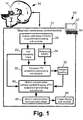
- an imaging system includes a magnetic resonance (MR) scanner 10, such as an illustrated AchievaTM MR scanner (available from Koninklijke Philips Electronics N.V., Eindhoven, The Netherlands), or an InteraTM or PanoramaTM MR scanner (both also available from Koninklijke Philips Electronics N.V.), or another commercially available MR scanner, or a non-commercial MR scanner, or so forth.
- MR magnetic resonance
- the MR scanner includes internal components (not illustrated) such as a superconducting or resistive main magnet generating a static (B 0 ) magnetic field, sets of magnetic field gradient coil windings for superimposing selected magnetic field gradients on the static magnetic field, a radio frequency excitation system for generating a radio frequency (B 1 ) field at a frequency selected to excite magnetic resonance (typically 1 H magnetic resonance, although excitation of another magnetic resonance nuclei or of multiple magnetic resonance nuclei is also contemplated), and a radio frequency receive system including a radio frequency receive coil array, or a plurality of receive coils, for detecting magnetic resonance signals emitted from the subject.
- internal components such as a superconducting or resistive main magnet generating a static (B 0 ) magnetic field, sets of magnetic field gradient coil windings for superimposing selected magnetic field gradients on the static magnetic field, a radio frequency excitation system for generating a radio frequency (B 1 ) field at a frequency selected to excite magnetic resonance (typically 1 H magnetic resonance,
- the MR scanner 10 operates under control of a magnetic resonance (MR) control module 12 to perform magnetic resonance (MR) imaging with motion correction.
- the MR imaging is partially parallel imaging (PPI).
- PPI imaging data are acquired by a plurality of magnetic resonance receive channels, for example including radio frequency receive coils of a diagrammatically illustrated receive coil array 14.
- SENSE sensitivity encoding
- SMASH simultaneous acquisition off spatial harmonics
- GRAPPA generalized autocalibrating partially parallel acquisition
- a plurality of receive coils having different coil sensitivities acquire an undersampled MR imaging dataset that is missing some data (for example, skipping acquisition of some phase encoding lines as is done in SENSE).
- the missing data are compensated by additional information provided by the use of a plurality of receive coils with different coil sensitivities, using a suitable reconstruction technique (e.g., a SENSE or SMASH reconstruction technique).
- the undersampling of the acquired PPI dataset corresponds to an imaging data acquisition acceleration factor (typically denoted R ) whose magnitude depends upon the extent of undersampling.
- PPI techniques entail a pre-scan for determining the coil sensitivities. Typically, the coil sensitivities vary relatively gradually across space. Accordingly, a coarse calibration (COCA) pre-scan is suitably performed by a COCA acquisition/processing sub-module 20. Coil sensitivity maps 22 for the coils of the plurality of coils 14 are generated by the COCA acquisition/processing sub-module 20. More generally, the sub-module 20 acquires MR calibration data and generates coil sensitivity maps therefrom. As disclosed herein, the COCA acquisition/processing sub-module 20 also generates reference projection vectors (PV) for use in subject motion assessment from the COCA prescan or other MR calibration data.
- PV reference projection vectors
- the generated reference PV may be weighted to account for the coil sensitivities using the information contained in the coil sensitivity maps 22 - this results in reference sensitivity weighted projection vectors (reference SWPV) 24.
- reference SWPV reference sensitivity weighted projection vectors
- this disclosed approach uses information conventionally acquired as part of the PPI process, namely the COCA pre-scan or other MR calibration data used to generate the coil sensitivity maps, to also generate reference data (e.g., reference SWPV 24 ) for use in subject motion detection.
- subject motion assessment is performed by a navigator projection vector (PV) acquisition/processing sub-module 30 to generate a subject position 32.
- the acquired PV can be either extra data or part of the imaging data, for example the echo train in a Turbo spin echo sequence, or several radial projections in a radial trajectory.
- the navigator PV acquisition/processing sub-module 30 acquires navigator PV, optionally performs sensitivity weighting of the navigator PV based on the coil sensitivity maps 22 to generate navigator sensitivity weighted PV (navigator SWPV) and compares the navigator SWPV with the reference SWPV 24 to determine the subject position 32 including three-dimensional rigid translation and rotation components.
- the comparisons between navigator and reference SWPV suitably employ cross-correlation.
- a motion compensated MR acquisition/processing sub-module 40 acquires MR data for an imaging volume that is adjusted to be consonant with the subject position 32 determined from the most recent iteration of navigator PV acquisitions, and performs a suitable image reconstruction process (e.g., SENSE reconstruction processing or so forth) to generate a motion compensated reconstructed image.
- the MR acquisition sequence is a partially parallel imaging (PPI) imaging sequence, although non-PPI acquisition sequences are also suitable.
- the motion compensation can be performed either prospectively or retrospectively.
- the reconstruction optionally also includes performing a rigid translational/rotational adjustment of each reconstructed image to register all images with a suitable spatial reference.
- each image can be translated and rotated to match with the spatial reference of the reference SWPV 24 based on the subject motion corresponding to the subject position 32.
- all images of the fMRI (or other) sequence of images are spatially aligned and functional (or other) variations over time can be readily assessed based on the sequence of images.
- the navigator PV acquisition/processing sub-module 30 is suitably applied before each image acquisition repetition in order to acquire a current value for the subject position 32 which is then used to define the imaging volume of the subsequent acquisition performed by the motion-compensated MR acquisition/processing sub-module 40.
- the subject position 32 is determined respective to the reference SWPV 24, and hence the subject position 32 is always referenced to the common spatial reference of the reference SWPV 24.
- brain fMRI is disclosed as an illustrative example, the disclosed approach is suitably employed in any imaging over a time interval sufficient for prospective motion compensation to be advantageously performed.
- the navigator PV acquisition/processing sub-module 30 may be invoked at intervals during the acquisition of imaging data for the single image and the imaging volume suitably adjusted during acquisition of the imaging data for the single image based on the subject position 32 provided by the most recent navigator PV acquisition.
- an image display sub-module 44 suitably displays the motion compensated reconstructed image 42.
- the image display sub-module 44 may display a CINE sequence of the images, or may display the images of the sequence in a suitable matrix, or so forth.
- the MR control module 12 is embodied by an illustrative computer 50 whose processor (which may, for example, comprise a multi-core processor or other parallel processor, a single-core processor, a graphical processing unit, i.e. GPU, or so forth) is programmed to implement the sub-modules 20, 30, 40 including control of the MR scanner 10 to cause the MR scanner 10 to perform the requisite data acquisitions. While the processor is generally a digital processor, it is contemplated for the processor to include or incorporate some analog circuitry, such as by way of illustrative example application-specific integrated circuitry (ASIC) configured to perform some or all of the image reconstruction processing of the sub-module 40.
- ASIC application-specific integrated circuitry
- the disclosed motion compensation approaches may also be embodied as a storage medium storing instructions that when executed by a processor perform the operations of the various sub-modules 20, 30, 40.
- the storage medium may comprise a hard drive, optical drive, random access memory (RAM), FLASH memory, or other electrostatic memory, various combinations thereof, or another suitable storage medium.
- the illustrative computer 50 includes a display 52 via which the image display sub-module 44 displays the motion compensated reconstructed image 42 (or a CINE sequence or array of such images).
- the computer 50 also include an illustrative keyboard 54 or other user input device (e.g., a mouse, trackball, or other pointing device) via which a user inputs commands, parameters, or so forth for initiating and controlling the imaging process.
- the COCA acquisition/processing sub-module 20 causes the MR scanner 10 and the plurality of radio frequency receive coils 14 to acquire coarse calibration (COCA) prescan data in an operation 60.
- the coil sensitivity maps 22 are computed based on the COCA prescan data.
- the reference SWPV 24 is computed using the same COCA prescan data (or a subset thereof) that are used to compute the coil sensitivity maps 22 in the operation 62. In this way no additional data are acquired in order to generate the reference SWPV 24.
- the size of the reference SWPV 24 for use in motion detection is suitably (1) the number of radio frequency receive coils in the plurality 14 times three navigators per coil (corresponding to three orthogonal directions, e.g. Cartesian x, y, and z directions) times the number of readouts.
- the reference SWPV 24 is suitably represented as a stack of the navigators from all receive coils after processing by a one-dimensional fast Fourier transform (1D-FFT).
- the reference SWPV 24 calculation operation 64 is performed after the COCA data acquisition operation 60, with the sensitivity weighting being performed after the coil sensitivity maps 22 are computed in the operation 62. After the calculation of extrapolated sensitivity maps, the three-dimensional low resolution image acquired in the operation 60 is rotated and translated in a given range.
- the projection vector is calculated, sensitivity-weighted, and saved.
- One embodiment for the calculation of SWPV at many locations in a fast way is to re-grid the Cartesian data onto 3D radial trajectory. This embodiment is reasonable if the sensitivity maps change smoothly in spatial domain.
- the navigator projection vector (PV) acquisition/processing sub-module 30 operates as follows.
- the sub-module 30 causes the MR scanner 10 and the plurality of radio frequency receive coils 14 to acquire magnetic resonance navigator projection vectors (PV).
- these PV are sensitivity weighted using the coil sensitivity maps 22 to generate magnetic resonance navigator sensitivity weighted projection vectors (magnetic resonance navigator SWPV).
- the navigator SWPV are compared with the reference SWPV 24 to quantitatively assess the subject position 32 at the time of the acquisition 70 referenced to the position indicated by the reference SWPV 24.
- a cross correlation between the acquired magnetic resonance SWPV and the reference SWPV 24 is computed. Since each calculated projection vector corresponds to a location of the reference image, the calculated projection vector providing the maximum cross correlation gives the current position of the subject.
- the extended Kalman filter is optionally applied to predict the range of the next subject position based on the current subject position. See Kalman, "A new approach to linear filtering and prediction problems", Trans. ASME J. Basic Eng. Vol. 82 (series D) pages 35-45 (1960 ).
- the motion compensated MR acquisition/processing sub-module 40 operates as follows.
- the sub-module 40 causes the MR scanner 10 and the plurality of radio frequency receive coils 14 to acquire imaging data in an imaging volume.
- Prospective motion compensation is incorporated into the operation 80 by adjusting the imaging volume to align with the subject position 32 indicated by the most recently iteration of the acquisition/processing operations 70, 72, 74.
- the resulting imaging data are reconstructed in an operation 82 using a suitable image reconstruction technique.
- the motion compensation can be done retrospectively and the image reconstruction operation 82 may include performing a rigid shift/rotation of the resulting image to align with the reference position indicated by the reference SWPV 24.
- the disclosed imaging process of FIGURE 2 is an illustrative example, and numerous variations are contemplated.
- the sensitivity weighting of the reference projection vectors and of the magnetic resonance navigator projection vectors is optionally omitted.
- reference projection vector may be acquired separately from the COCA prescan prior to initiating the imaging.
- the generated subject position 32 is input to the data reconstruction operation 82 (or to a post-reconstruction motion compensation operation that is not illustrated) instead of being input to the acquisition operation 80, and the subject position 32 is used retrospectively, that is, after the imaging data acquisition, to perform a rigid shift/rotation of the acquired imaging data or of the reconstructed image in order to compensate for motion respective to the reference position of the reference SWPV 24.
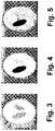
- FIGURES 3-5 some illustrative phantom imaging results are shown.
- a 32-channel cardiac coil and Shepp-Logan phantom was used to produce the simulated data set.
- FIGURE 3 shows the images for the reference scan operation 60 and the navigators. It is assumed here that the image contrast may change between the pre-scanned reference and the navigator acquisitions due to different steady-states of magnetization.
- FIGURES 4 and 5 show the navigator with 13% contrast change and with 42% contrast change, respectively.
- FIGURE 6 With reference to FIGURE 6 , the change of cross correlation between calculated reference SWPV 24 and projection vectors acquired in the operation 70 due to motion are plotted.
- the data of FIGURE 6 are calculated results for the same phantom example as in FIGURES 3-5 , and are calculated for the same 32-channel cardiac coil. It is seen that the cross correlation is maximized when there is no relative motion between the reference SWPV 24 and the navigator (sensitivity weighted) projection vectors. When the contrast change between the reference image and the navigator is not significant, both rotation and translation can be sensitively detected.
Landscapes
- Physics & Mathematics (AREA)
- Engineering & Computer Science (AREA)
- Health & Medical Sciences (AREA)
- Condensed Matter Physics & Semiconductors (AREA)
- General Physics & Mathematics (AREA)
- High Energy & Nuclear Physics (AREA)
- Radiology & Medical Imaging (AREA)
- Nuclear Medicine, Radiotherapy & Molecular Imaging (AREA)
- Signal Processing (AREA)
- General Health & Medical Sciences (AREA)
- Life Sciences & Earth Sciences (AREA)
- Biophysics (AREA)
- Cardiology (AREA)
- Physiology (AREA)
- Power Engineering (AREA)
- Pulmonology (AREA)
- Magnetic Resonance Imaging Apparatus (AREA)
Description
- The following relates to the magnetic resonance imaging arts, medical imaging arts, and related arts.
- Magnetic resonance (MR) imaging is a known medical imaging technique, which is also employed in veterinary imaging and other imaging applications such as characterizing archaeological artifacts. For high resolution MR imaging of the human brain or other medical applications, subject motion can cause problematic degradation of image quality. In functional MR imaging (fMRI) such as brain fMRI, the acquisition time is extended in order to capture time-varying functional aspects. This extended acquisition time increases the likelihood and likely severity of motion artifacts.
- Subject motion in MR imaging can be corrected prospectively or retrospectively. Real-time prospective correction methods update the coordinates of the imaging volume such that the position and orientation of the imaging volume tracks with the position and orientation of the subject over the course of the MR imaging data acquisition sequence repetitions. Retrospective correction methods entail adjustment (e.g., a rigid shift and/or regridding) of the MR imaging data after the acquisition.
- Prospective motion correction has certain advantages. It ensures that the desired imaging region remains in view (that is, within the imaging volume) throughout the imaging sequence. In contrast, in retrospective motion correction the subject motion could move the desired imaging region outside of the imaging volume. Also, in prospective motion correction the imaging volume can be rotated commensurate with rotation of the imaging subject such that the MR acquisition axes remain aligned with anatomically significant axes (e.g., with axial, coronal, and sagittal axes).
- To implement either prospective or retrospective motion correction, the subject motion must be quantitatively assessed. For prospective motion correction this assessment must be provided in real-time (that is, during the imaging and fast enough to provide feedback for adjusting the imaging volume coordinates between acquisition sequence repetitions). There are some hardware based motion assessment techniques, for example using a camera, optionally in conjunction with artificial fiducial markers applied to the subject. These hardware based methods provide real-time motion information, and also do not entail modification of the MR acquisition pulse sequence. However, the camera, artificial fiducial markers, or other equipment employed in hardware-based subject motion assessment increases cost and complicates the workflow. For example, set-up and alignment of the camera, and/or deployment of the artificial fiducial markers, increases imaging preparation time.
- Navigator based methods are also known for real-time subject motion assessment. However, existing navigator based techniques are not well suited for use in conjunction with partially parallel imaging (PPI) employing multiple receive coils with different coil sensitivities. Moreover, existing navigator based techniques entail substantial modification of the acquisition sequence in order to acquire sufficient data with which to assess subject motion.
- Nehrke et al. (Magnetic Resonance in Medicine, vol. 54, no. 5, 1130-1138, 2005) describe a concept of prospective 3D affine motion correction using arbitrary MR sequences. A patient-specific motion model steered by real-time navigators is employed to account for the degrees of freedom provided by the affine motion model.
- Manke et al. (Magnetic Resonance in Medicine, vol. 50, no. 1, 122-131, 2003) propose a technique which enables the calibration of a 3D affine respiratory motion model to the individual motion pattern of the patient. The concept involves multiple navigators to address non-linear properties and hysteresis effects of the model parameters with respect to the conventional diaphragmatic navigator. Based on a given navigator measurement, the current motion state of the object is predicted by means of a calibrated motion model.
- Lin et al. (Proceedings of the International Society for Magnetic Resonance in Medicine, vol. 17, 757, 2009, and Magnetic Resonance in Medicine, vol. 63, 339-348, 2010) describe a motion detection and correction method for applications using multiple channel coil arrays. Image-space correlations of floating navigators are used for detection of translational, rotational and non-consistent (through-plane and non-rigid body) motion. GRAPPA operators are used to correct the k-space data for suppressing motion artifacts.
- The following provides new and improved apparatuses and methods as disclosed herein.
- In accordance with one disclosed aspect, a method is provided as defined by
independent claim 1, with preferred embodiments defined in dependent claims 2-6. - In accordance with another disclosed aspect, an apparatus is provided as defined in independent claim 7.
- One advantage resides in providing real-time subject motion assessment without the use of extraneous hardware and with reduced modification of the MR acquisition sequence.
- Another advantage resides in providing sensitivity weights with real-time subject motion assessment that accommodates different coil sensitivities of the phased array coils used in the modern MR systems.
- Another advantage resides in providing PPI with real-time subject motion assessment without acquisition of additional reference data for the motion assessment.
- Further advantages will be apparent to those of ordinary skill in the art upon reading and understanding the following detailed description.
-
FIGURE 1 diagrammatically shows an imaging system configured to perform motion-corrected magnetic resonance imaging as disclosed herein. -
FIGURE 2 diagrammatically shows processing performed by selected processing sub-modules of the magnetic resonance control module ofFIGURE 1 . -
FIGURES 3-5 show phantom imaging results as described herein. -
FIGURE 6 plots calculated cross correlation between the acquired and calculated projection vectors for different subject motion conditions as described herein. - With reference to
FIGURE 1 , an imaging system includes a magnetic resonance (MR)scanner 10, such as an illustrated Achieva™ MR scanner (available from Koninklijke Philips Electronics N.V., Eindhoven, The Netherlands), or an Intera™ or Panorama™ MR scanner (both also available from Koninklijke Philips Electronics N.V.), or another commercially available MR scanner, or a non-commercial MR scanner, or so forth. In a typical embodiment, the MR scanner includes internal components (not illustrated) such as a superconducting or resistive main magnet generating a static (B0) magnetic field, sets of magnetic field gradient coil windings for superimposing selected magnetic field gradients on the static magnetic field, a radio frequency excitation system for generating a radio frequency (B1) field at a frequency selected to excite magnetic resonance (typically 1H magnetic resonance, although excitation of another magnetic resonance nuclei or of multiple magnetic resonance nuclei is also contemplated), and a radio frequency receive system including a radio frequency receive coil array, or a plurality of receive coils, for detecting magnetic resonance signals emitted from the subject. - The
MR scanner 10 operates under control of a magnetic resonance (MR)control module 12 to perform magnetic resonance (MR) imaging with motion correction. In some embodiments, the MR imaging is partially parallel imaging (PPI). In PPI, imaging data are acquired by a plurality of magnetic resonance receive channels, for example including radio frequency receive coils of a diagrammatically illustratedreceive coil array 14. Substantially any PPI technique can be employed, such as sensitivity encoding (SENSE), simultaneous acquisition off spatial harmonics (SMASH), generalized autocalibrating partially parallel acquisition (GRAPPA), or so forth. In PPI, a plurality of receive coils having different coil sensitivities acquire an undersampled MR imaging dataset that is missing some data (for example, skipping acquisition of some phase encoding lines as is done in SENSE). The missing data are compensated by additional information provided by the use of a plurality of receive coils with different coil sensitivities, using a suitable reconstruction technique (e.g., a SENSE or SMASH reconstruction technique). The undersampling of the acquired PPI dataset corresponds to an imaging data acquisition acceleration factor (typically denoted R) whose magnitude depends upon the extent of undersampling. - PPI techniques entail a pre-scan for determining the coil sensitivities. Typically, the coil sensitivities vary relatively gradually across space. Accordingly, a coarse calibration (COCA) pre-scan is suitably performed by a COCA acquisition/
processing sub-module 20.Coil sensitivity maps 22 for the coils of the plurality ofcoils 14 are generated by the COCA acquisition/processing sub-module 20. More generally, thesub-module 20 acquires MR calibration data and generates coil sensitivity maps therefrom. As disclosed herein, the COCA acquisition/processing sub-module 20 also generates reference projection vectors (PV) for use in subject motion assessment from the COCA prescan or other MR calibration data. Optionally, as further disclosed herein, the generated reference PV may be weighted to account for the coil sensitivities using the information contained in the coil sensitivity maps 22 - this results in reference sensitivity weighted projection vectors (reference SWPV) 24. Advantageously, this disclosed approach uses information conventionally acquired as part of the PPI process, namely the COCA pre-scan or other MR calibration data used to generate the coil sensitivity maps, to also generate reference data (e.g., reference SWPV 24) for use in subject motion detection. - With continuing reference to
FIGURE 1 , subject motion assessment is performed by a navigator projection vector (PV) acquisition/processing sub-module 30 to generate asubject position 32. The acquired PV can be either extra data or part of the imaging data, for example the echo train in a Turbo spin echo sequence, or several radial projections in a radial trajectory. The navigator PV acquisition/processing sub-module 30 acquires navigator PV, optionally performs sensitivity weighting of the navigator PV based on thecoil sensitivity maps 22 to generate navigator sensitivity weighted PV (navigator SWPV) and compares the navigator SWPV with thereference SWPV 24 to determine thesubject position 32 including three-dimensional rigid translation and rotation components. In illustrative embodiments, the comparisons between navigator and reference SWPV suitably employ cross-correlation. - With continuing reference to
FIGURE 1 , a motion compensated MR acquisition/processing sub-module 40 acquires MR data for an imaging volume that is adjusted to be consonant with thesubject position 32 determined from the most recent iteration of navigator PV acquisitions, and performs a suitable image reconstruction process (e.g., SENSE reconstruction processing or so forth) to generate a motion compensated reconstructed image. In some embodiments, the MR acquisition sequence is a partially parallel imaging (PPI) imaging sequence, although non-PPI acquisition sequences are also suitable. The motion compensation can be performed either prospectively or retrospectively. If a retrospective motion compensation scheme is adopted and the imaging includes acquisition of multiple images over a period of time, as (by way of illustrative example) in the case of brain fMRI, then the reconstruction optionally also includes performing a rigid translational/rotational adjustment of each reconstructed image to register all images with a suitable spatial reference. For example, each image can be translated and rotated to match with the spatial reference of thereference SWPV 24 based on the subject motion corresponding to thesubject position 32. In this way, all images of the fMRI (or other) sequence of images are spatially aligned and functional (or other) variations over time can be readily assessed based on the sequence of images. - In the case of brain fMRI or another image sequence acquisition, the navigator PV acquisition/
processing sub-module 30 is suitably applied before each image acquisition repetition in order to acquire a current value for thesubject position 32 which is then used to define the imaging volume of the subsequent acquisition performed by the motion-compensated MR acquisition/processing sub-module 40. For each repetition, thesubject position 32 is determined respective to thereference SWPV 24, and hence thesubject position 32 is always referenced to the common spatial reference of thereference SWPV 24. Although brain fMRI is disclosed as an illustrative example, the disclosed approach is suitably employed in any imaging over a time interval sufficient for prospective motion compensation to be advantageously performed. - As another example, if the acquisition of a single image is sufficiently long, then the navigator PV acquisition/
processing sub-module 30 may be invoked at intervals during the acquisition of imaging data for the single image and the imaging volume suitably adjusted during acquisition of the imaging data for the single image based on thesubject position 32 provided by the most recent navigator PV acquisition. - With continuing reference to
FIGURE 1 , animage display sub-module 44 suitably displays the motion compensated reconstructedimage 42. In embodiments in which a sequence of images are acquired, theimage display sub-module 44 may display a CINE sequence of the images, or may display the images of the sequence in a suitable matrix, or so forth. - In the illustrated embodiment, the
MR control module 12 is embodied by anillustrative computer 50 whose processor (which may, for example, comprise a multi-core processor or other parallel processor, a single-core processor, a graphical processing unit, i.e. GPU, or so forth) is programmed to implement the sub-modules 20, 30, 40 including control of theMR scanner 10 to cause theMR scanner 10 to perform the requisite data acquisitions. While the processor is generally a digital processor, it is contemplated for the processor to include or incorporate some analog circuitry, such as by way of illustrative example application-specific integrated circuitry (ASIC) configured to perform some or all of the image reconstruction processing of the sub-module 40. The disclosed motion compensation approaches may also be embodied as a storage medium storing instructions that when executed by a processor perform the operations of thevarious sub-modules illustrative computer 50 includes adisplay 52 via which theimage display sub-module 44 displays the motion compensated reconstructed image 42 (or a CINE sequence or array of such images). Thecomputer 50 also include anillustrative keyboard 54 or other user input device (e.g., a mouse, trackball, or other pointing device) via which a user inputs commands, parameters, or so forth for initiating and controlling the imaging process. - Having described the illustrative embodiment of
FIGURE 1 , additional disclosure relating to thevarious sub-modules - With reference to
FIGURE 2 , the COCA acquisition/processing sub-module 20 causes theMR scanner 10 and the plurality of radio frequency receivecoils 14 to acquire coarse calibration (COCA) prescan data in anoperation 60. In anoperation 62, the coil sensitivity maps 22 are computed based on the COCA prescan data. In anoperation 64, thereference SWPV 24 is computed using the same COCA prescan data (or a subset thereof) that are used to compute the coil sensitivity maps 22 in theoperation 62. In this way no additional data are acquired in order to generate thereference SWPV 24. The size of thereference SWPV 24 for use in motion detection is suitably (1) the number of radio frequency receive coils in theplurality 14 times three navigators per coil (corresponding to three orthogonal directions, e.g. Cartesian x, y, and z directions) times the number of readouts. Thereference SWPV 24 is suitably represented as a stack of the navigators from all receive coils after processing by a one-dimensional fast Fourier transform (1D-FFT). Thereference SWPV 24calculation operation 64 is performed after the COCAdata acquisition operation 60, with the sensitivity weighting being performed after the coil sensitivity maps 22 are computed in theoperation 62. After the calculation of extrapolated sensitivity maps, the three-dimensional low resolution image acquired in theoperation 60 is rotated and translated in a given range. For each location, the projection vector is calculated, sensitivity-weighted, and saved. One embodiment for the calculation of SWPV at many locations in a fast way is to re-grid the Cartesian data onto 3D radial trajectory. This embodiment is reasonable if the sensitivity maps change smoothly in spatial domain. - With continuing reference to
FIGURE 2 , the navigator projection vector (PV) acquisition/processing sub-module 30 operates as follows. In an acquisition operation 70 the sub-module 30 causes theMR scanner 10 and the plurality of radio frequency receivecoils 14 to acquire magnetic resonance navigator projection vectors (PV). In an operation 72, these PV are sensitivity weighted using the coil sensitivity maps 22 to generate magnetic resonance navigator sensitivity weighted projection vectors (magnetic resonance navigator SWPV). In anoperation 74, the navigator SWPV are compared with thereference SWPV 24 to quantitatively assess thesubject position 32 at the time of the acquisition 70 referenced to the position indicated by thereference SWPV 24. In a suitable comparison approach, a cross correlation between the acquired magnetic resonance SWPV and thereference SWPV 24 is computed. Since each calculated projection vector corresponds to a location of the reference image, the calculated projection vector providing the maximum cross correlation gives the current position of the subject. To avoid the computational cost of calculating all cross correlations, the extended Kalman filter is optionally applied to predict the range of the next subject position based on the current subject position. See Kalman, "A new approach to linear filtering and prediction problems", Trans. ASME J. Basic Eng. Vol. 82 (series D) pages 35-45 (1960). - With continuing reference to
FIGURE 2 , the motion compensated MR acquisition/processing sub-module 40 operates as follows. In anacquisition operation 80 the sub-module 40 causes theMR scanner 10 and the plurality of radio frequency receivecoils 14 to acquire imaging data in an imaging volume. Prospective motion compensation is incorporated into theoperation 80 by adjusting the imaging volume to align with thesubject position 32 indicated by the most recently iteration of the acquisition/processing operations 70, 72, 74. The resulting imaging data are reconstructed in anoperation 82 using a suitable image reconstruction technique. Optionally, the motion compensation can be done retrospectively and theimage reconstruction operation 82 may include performing a rigid shift/rotation of the resulting image to align with the reference position indicated by thereference SWPV 24. - The disclosed imaging process of
FIGURE 2 is an illustrative example, and numerous variations are contemplated. For example, the sensitivity weighting of the reference projection vectors and of the magnetic resonance navigator projection vectors is optionally omitted. - As another example, other data besides the COCA prescan that is acquired prior to performing the PPI may be used to generate the reference projection vector (or reference sensitivity weighted projection vector). Thus, for example, the reference projection vectors may be acquired separately from the COCA prescan prior to initiating the imaging.
- As yet another example, while prospective motion compensation is employed in the illustrative example of
FIGURE 2 , it is alternatively contemplated to employ retrospective motion compensation. In this variant embodiment, the generatedsubject position 32 is input to the data reconstruction operation 82 (or to a post-reconstruction motion compensation operation that is not illustrated) instead of being input to theacquisition operation 80, and thesubject position 32 is used retrospectively, that is, after the imaging data acquisition, to perform a rigid shift/rotation of the acquired imaging data or of the reconstructed image in order to compensate for motion respective to the reference position of thereference SWPV 24. - With reference to
FIGURES 3-5 , some illustrative phantom imaging results are shown. In this phantom example, a 32-channel cardiac coil and Shepp-Logan phantom was used to produce the simulated data set.FIGURE 3 shows the images for thereference scan operation 60 and the navigators. It is assumed here that the image contrast may change between the pre-scanned reference and the navigator acquisitions due to different steady-states of magnetization.FIGURES 4 and 5 show the navigator with 13% contrast change and with 42% contrast change, respectively. - With reference to
FIGURE 6 , the change of cross correlation betweencalculated reference SWPV 24 and projection vectors acquired in the operation 70 due to motion are plotted. The data ofFIGURE 6 are calculated results for the same phantom example as inFIGURES 3-5 , and are calculated for the same 32-channel cardiac coil. It is seen that the cross correlation is maximized when there is no relative motion between thereference SWPV 24 and the navigator (sensitivity weighted) projection vectors. When the contrast change between the reference image and the navigator is not significant, both rotation and translation can be sensitively detected. - This application has described one or more preferred embodiments. Modifications and alterations may occur to others upon reading and understanding the preceding detailed description. It is intended that the application be construed as including all such modifications and alterations insofar as they come within the scope of the appended claims or the equivalents thereof.
Claims (7)
- A method comprising:acquiring magnetic resonance = MR calibration data using a plurality of radio frequency receive coils (14);generating coil sensitivity maps (22) for the radio frequency receive coils based on the MR calibration data;performing magnetic resonance imaging including acquiring magnetic resonance imaging data and generating one or more reconstructed magnetic resonance images (42), wherein the magnetic resonance imaging comprises partially parallel imaging selected from a group consisting of sensitivity encoding, simultaneous acquisition of spatial harmonics, and generalized autocalibrating partially parallel acquisition, characterized in that the method further comprises:generating reference navigators based on the MR calibration data;generating magnetic resonance navigators based on a portion of the acquired magnetic resonance imaging data or extra data acquired with the magnetic resonance imaging data;sensitivity weighting (64, 72) the magnetic resonance navigators and the reference navigators using the coil sensitivity maps to generate magnetic resonance sensitivity weighted navigators = navigator SWPV, and reference sensitivity weighted navigators = reference SWPV (24) respectively;comparing the navigator SWPV with the reference SWPV to generate subject position information (32); andin the one or more reconstructed magnetic resonance images (42), compensating for subject motion prospectively or retrospectively using the generated subject position information.
- The method as set forth in claim 1, wherein the comparing comprises:computing cross correlations between the navigator SWPV and the reference SWPV (74) ; andgenerating the subject position information (32) based on the computed cross correlations.
- The method as set forth in any one of claims 1-2, further comprising:
displaying the one or more reconstructed magnetic resonance images (42) on a display device (52). - The method as set forth in any one of claims 1-3, wherein the one or more reconstructed magnetic resonance images (42) comprise a time sequence of reconstructed magnetic resonance images and the method further comprises:
displaying a CINE sequence of the time sequence of reconstructed magnetic resonance images on a display device (52). - The method as set forth in any one of claims 1-4, wherein the compensating comprises:
performing prospective motion compensation by adjusting an imaging volume based on the generated subject position information (32). - The method as set forth in any one of claims 1-4, wherein the compensating comprises:
performing retrospective motion compensation based on the subject position information (32). - An apparatus comprising:a magnetic resonance = MR scanner (10);a plurality of radio frequency receive coils (14); anda data processing device (50) configured to cooperate with the MR scanner and the radio frequency receive coils to perform a method as set forth in any one of claims 1-6.
Applications Claiming Priority (2)
| Application Number | Priority Date | Filing Date | Title |
|---|---|---|---|
| US201061425971P | 2010-12-22 | 2010-12-22 | |
| PCT/IB2011/055738 WO2012085796A1 (en) | 2010-12-22 | 2011-12-16 | Parallel mri method using calibration scan, coil sensitivity maps and navigators for rigid motion compensation |
Publications (2)
| Publication Number | Publication Date |
|---|---|
| EP2656093A1 EP2656093A1 (en) | 2013-10-30 |
| EP2656093B1 true EP2656093B1 (en) | 2019-10-16 |
Family
ID=45509573
Family Applications (1)
| Application Number | Title | Priority Date | Filing Date |
|---|---|---|---|
| EP11811147.5A Not-in-force EP2656093B1 (en) | 2010-12-22 | 2011-12-16 | Parallel mri method using calibration scan, coil sensitivity maps and navigators for rigid motion compensation |
Country Status (5)
| Country | Link |
|---|---|
| US (1) | US10067213B2 (en) |
| EP (1) | EP2656093B1 (en) |
| JP (1) | JP5960163B2 (en) |
| CN (1) | CN103403569B (en) |
| WO (1) | WO2012085796A1 (en) |
Families Citing this family (25)
| Publication number | Priority date | Publication date | Assignee | Title |
|---|---|---|---|---|
| WO2009129457A1 (en) * | 2008-04-17 | 2009-10-22 | The Government Of The United States Of America, As Represented By The Secretary, Department Of Health And Human Services Services, National Institutes Of Health | Movement correction in mri using a camera |
| US8823375B2 (en) * | 2010-12-31 | 2014-09-02 | General Electric Company | System and method for generating a magnetic resonance image using prospective motion correction and parallel imaging |
| JP5992624B2 (en) * | 2012-09-06 | 2016-09-14 | コーニンクレッカ フィリップス エヌ ヴェKoninklijke Philips N.V. | MRI system with navigator-based motion detection |
| JP5755285B2 (en) | 2013-05-31 | 2015-07-29 | ジーイー・メディカル・システムズ・グローバル・テクノロジー・カンパニー・エルエルシー | Magnetic resonance apparatus and program |
| CN103334520B (en) * | 2013-06-26 | 2016-09-14 | 东亚装饰股份有限公司 | A kind of three-dimensional adjustable connecting components device |
| US10302732B2 (en) * | 2013-12-02 | 2019-05-28 | Koninklijke Philips N.V. | Real-time adaptive physiology synchronization and gating for steady state MR sequences |
| DE102014210471B4 (en) * | 2014-06-03 | 2018-11-08 | Siemens Healthcare Gmbh | A method of performing a magnetic resonance examination with a prospective motion correction and magnetic resonance system therefor |
| DE102014212943B4 (en) * | 2014-07-03 | 2016-11-24 | Siemens Healthcare Gmbh | Magnetic resonance imaging taking into account different frequency coding patterns |
| DE102014214266A1 (en) * | 2014-07-22 | 2016-01-28 | Siemens Aktiengesellschaft | Method for magnetic resonance imaging, as well as local coil and magnetic resonance apparatus |
| KR20160029586A (en) * | 2014-09-05 | 2016-03-15 | 삼성전자주식회사 | Magnetic Resonance Imaging apparatus and method for operating the same |
| WO2016071054A1 (en) * | 2014-11-07 | 2016-05-12 | Koninklijke Philips N.V. | Method and system for generating mr images of a moving object in its environment |
| DE102015203385B4 (en) * | 2015-02-25 | 2017-11-30 | Siemens Healthcare Gmbh | Method for generating motion information to an at least partially moving examination area and magnetic resonance system and hybrid imaging modality |
| DE102015207590A1 (en) * | 2015-04-24 | 2016-10-27 | Siemens Healthcare Gmbh | A method of motion compensation during magnetic resonance imaging |
| DE102015222835B4 (en) * | 2015-11-19 | 2019-06-13 | Siemens Healthcare Gmbh | Magnetic resonance imaging method with simultaneous image acquisition of several partial volumes with a synchronous image acquisition by navigators |
| DE102016200293A1 (en) | 2016-01-13 | 2017-07-13 | Siemens Healthcare Gmbh | Determination of states of motion |
| DE102016204198B4 (en) | 2016-03-15 | 2018-06-07 | Siemens Healthcare Gmbh | Method for generating MR images with prospective motion correction and partial volume-specific weighting of the image information |
| US10420510B2 (en) | 2016-04-22 | 2019-09-24 | General Electric Company | System and method for imaging a moving subject |
| DE102016223809B4 (en) * | 2016-11-30 | 2022-03-03 | Siemens Healthcare Gmbh | Processing of motion signals during a magnetic resonance measurement |
| EP3413074A1 (en) * | 2017-06-09 | 2018-12-12 | Koninklijke Philips N.V. | Parallel multi-slice mr imaging |
| CN107576925B (en) * | 2017-08-07 | 2020-01-03 | 上海东软医疗科技有限公司 | Magnetic resonance multi-contrast image reconstruction method and device |
| DE102018202137A1 (en) | 2018-02-12 | 2019-08-14 | Siemens Healthcare Gmbh | Method for operating a magnetic resonance device, magnetic resonance device, computer program and electronically readable data carrier |
| US11835612B2 (en) | 2019-03-12 | 2023-12-05 | University Of Cincinnati | System and method for motion correction of magnetic resonance image |
| JP7451366B2 (en) | 2020-09-18 | 2024-03-18 | キヤノンメディカルシステムズ株式会社 | Image generation device, image generation method, and image generation program |
| DE102020215490A1 (en) | 2020-12-08 | 2022-06-09 | Siemens Healthcare Gmbh | Method for acquiring MR data |
| EP4163657A1 (en) | 2021-10-11 | 2023-04-12 | Koninklijke Philips N.V. | Motion correction using low resolution magnetic resonance images |
Family Cites Families (17)
| Publication number | Priority date | Publication date | Assignee | Title |
|---|---|---|---|---|
| US5427101A (en) * | 1994-08-04 | 1995-06-27 | Board Of Trustees Of The Leland Stanford Junior University | Diminishing variance process for real-time reduction of motion artifacts in MRI |
| JP4072879B2 (en) * | 1998-11-26 | 2008-04-09 | 株式会社日立メディコ | Nuclear magnetic resonance imaging system |
| US6516210B1 (en) | 2000-11-22 | 2003-02-04 | Koninklijke Philips Electronics N.V. | Signal analysis for navigated magnetic resonance imaging |
| JP2002301044A (en) * | 2001-04-06 | 2002-10-15 | Hitachi Medical Corp | Magnetic resonance image diagnosis apparatus |
| EP1419484A2 (en) * | 2001-08-10 | 2004-05-19 | Koninklijke Philips Electronics N.V. | X-ray examination apparatus for reconstructing a three-dimensional data set from projection images |
| JP2004514483A (en) * | 2001-11-07 | 2004-05-20 | コーニンクレッカ フィリップス エレクトロニクス エヌ ヴィ | Signal analysis for navigated magnetic resonance imaging |
| US7561909B1 (en) | 2002-09-16 | 2009-07-14 | The United States Of America As Represented By The Department Of Health And Human Services | MRI navigator methods and systems |
| US7587074B2 (en) | 2003-07-21 | 2009-09-08 | Paieon Inc. | Method and system for identifying optimal image within a series of images that depict a moving organ |
| JP4202855B2 (en) * | 2003-07-25 | 2008-12-24 | 株式会社日立メディコ | Magnetic resonance imaging system |
| DE10353342B4 (en) * | 2003-11-14 | 2008-07-17 | Siemens Ag | Improved MRI imaging based on conventional PPA reconstruction techniques |
| CN101027569B (en) * | 2004-09-24 | 2011-03-16 | 皇家飞利浦电子股份有限公司 | Magnetic resonance device and method |
| CN101171527A (en) * | 2005-05-02 | 2008-04-30 | 皇家飞利浦电子股份有限公司 | Independent motion correction in respective signal channels of a magnetic resonance imaging system |
| JP2007098026A (en) | 2005-10-07 | 2007-04-19 | Ge Medical Systems Global Technology Co Llc | Magnetic resonance imaging system |
| US7348776B1 (en) * | 2006-09-01 | 2008-03-25 | The Board Of Trustees Of The Leland Stanford Junior University | Motion corrected magnetic resonance imaging |
| US8624596B2 (en) * | 2009-10-02 | 2014-01-07 | Siemens Aktiengesellschaft | Method for determining motion parameters of an object in a magnetic field |
| DE102010032080B4 (en) * | 2010-07-23 | 2012-09-27 | Siemens Aktiengesellschaft | Triggered Magnetic Resonance Imaging Based on Partial Parallel Acquisition (PPA) |
| JP5992624B2 (en) * | 2012-09-06 | 2016-09-14 | コーニンクレッカ フィリップス エヌ ヴェKoninklijke Philips N.V. | MRI system with navigator-based motion detection |
-
2011
- 2011-12-16 CN CN201180068106.XA patent/CN103403569B/en not_active Expired - Fee Related
- 2011-12-16 JP JP2013545589A patent/JP5960163B2/en not_active Expired - Fee Related
- 2011-12-16 WO PCT/IB2011/055738 patent/WO2012085796A1/en active Application Filing
- 2011-12-16 US US13/995,378 patent/US10067213B2/en not_active Expired - Fee Related
- 2011-12-16 EP EP11811147.5A patent/EP2656093B1/en not_active Not-in-force
Non-Patent Citations (2)
| Title |
|---|
| LIN W ET AL: "Motion Compensation with Floating Navigator and GRAPPA Operators", INTERNATIONAL SOCIETY FOR MAGNETIC RESONANCE IN MEDICINE. SCIENTIFIC MEETING AND EXHIBITION. PROCEEDINGS, INTERNATIONAL SOCIETY FOR MAGNETIC RESONANCE IN MEDICINE, US, vol. 17, 1 April 2009 (2009-04-01), pages 757, XP002580952, ISSN: 1524-6965 * |
| LIN WEI ET AL: "Motion Correction Using an Enhanced Floating Navigator and GRAPPA Operations", MAGNETIC RESONANCE IN MEDICINE, ACADEMIC PRESS, DULUTH, MN, US, vol. 63, 13 November 2009 (2009-11-13), pages 339 - 348, XP002580951, ISSN: 0740-3194 * |
Also Published As
| Publication number | Publication date |
|---|---|
| US10067213B2 (en) | 2018-09-04 |
| US20130278263A1 (en) | 2013-10-24 |
| JP2014503290A (en) | 2014-02-13 |
| WO2012085796A1 (en) | 2012-06-28 |
| JP5960163B2 (en) | 2016-08-02 |
| EP2656093A1 (en) | 2013-10-30 |
| CN103403569B (en) | 2016-02-03 |
| CN103403569A (en) | 2013-11-20 |
Similar Documents
| Publication | Publication Date | Title |
|---|---|---|
| EP2656093B1 (en) | Parallel mri method using calibration scan, coil sensitivity maps and navigators for rigid motion compensation | |
| US10634753B2 (en) | MR imaging with motion detection | |
| US10401456B2 (en) | Parallel MR imaging with Nyquist ghost correction for EPI | |
| JP6018401B2 (en) | System and method for predicting and correcting distortions induced by higher order eddy currents in diffusion weighted echo planar imaging | |
| US9223001B2 (en) | MR imaging using navigators | |
| US11067653B2 (en) | Magnetic resonance imaging method including acquiring echo signals from each k-space slice with different relaxation time weightings after echo signals from different k-space splices are acquired | |
| US7696753B2 (en) | MRI apparatus and method with B0 corrections using reference and object average units | |
| JP2006280930A (en) | Method and system for magnetic field map during ssfp using phase-incremented or frequency-shifted magnitude image | |
| CN106796274B (en) | PROPELLER-MR imaging with artifact suppression | |
| EP3123192A1 (en) | Epi ghost correction involving sense | |
| WO2012011069A1 (en) | Mri method for retrospective motion correction with interleaved radial acquisition | |
| US7292034B2 (en) | Method for dynamic detection and change in the magnetic field distribution in magnetic resonance (NMR) measurements | |
| US11852705B2 (en) | MR imaging using a 3D radial or spiral acquisition with soft motion gating | |
| US20150168521A1 (en) | Magnetic resonance imaging apparatus and magnetic resonance imaging method | |
| JP7023954B2 (en) | Propeller MR imaging | |
| US20220413080A1 (en) | 3D MR Imaging with Intrinsic Motion Detection | |
| EP3185029A1 (en) | Mr imaging using propeller acquisition with t2 decay correction | |
| US11226385B2 (en) | Dixon type water/fat separation MR imaging with improved fat shift correction | |
| EP3686621A1 (en) | Imaging method with variable density in k-space for multi-spin echo magnetic resonance imaging with different delay times after a preparation pulse and/or different echo times | |
| Kholmovski et al. | GARSE: generalized autocalibrating reconstruction for sensitivity encoded MRI | |
| EP4291913A1 (en) | Dixon-type water/fat separation mr imaging |
Legal Events
| Date | Code | Title | Description |
|---|---|---|---|
| PUAI | Public reference made under article 153(3) epc to a published international application that has entered the european phase |
Free format text: ORIGINAL CODE: 0009012 |
|
| 17P | Request for examination filed |
Effective date: 20130722 |
|
| AK | Designated contracting states |
Kind code of ref document: A1 Designated state(s): AL AT BE BG CH CY CZ DE DK EE ES FI FR GB GR HR HU IE IS IT LI LT LU LV MC MK MT NL NO PL PT RO RS SE SI SK SM TR |
|
| DAX | Request for extension of the european patent (deleted) | ||
| 17Q | First examination report despatched |
Effective date: 20140402 |
|
| GRAP | Despatch of communication of intention to grant a patent |
Free format text: ORIGINAL CODE: EPIDOSNIGR1 |
|
| STAA | Information on the status of an ep patent application or granted ep patent |
Free format text: STATUS: GRANT OF PATENT IS INTENDED |
|
| INTG | Intention to grant announced |
Effective date: 20190503 |
|
| GRAS | Grant fee paid |
Free format text: ORIGINAL CODE: EPIDOSNIGR3 |
|
| GRAA | (expected) grant |
Free format text: ORIGINAL CODE: 0009210 |
|
| STAA | Information on the status of an ep patent application or granted ep patent |
Free format text: STATUS: THE PATENT HAS BEEN GRANTED |
|
| AK | Designated contracting states |
Kind code of ref document: B1 Designated state(s): AL AT BE BG CH CY CZ DE DK EE ES FI FR GB GR HR HU IE IS IT LI LT LU LV MC MK MT NL NO PL PT RO RS SE SI SK SM TR |
|
| REG | Reference to a national code |
Ref country code: GB Ref legal event code: FG4D |
|
| REG | Reference to a national code |
Ref country code: CH Ref legal event code: EP |
|
| REG | Reference to a national code |
Ref country code: DE Ref legal event code: R096 Ref document number: 602011062800 Country of ref document: DE |
|
| REG | Reference to a national code |
Ref country code: IE Ref legal event code: FG4D |
|
| REG | Reference to a national code |
Ref country code: AT Ref legal event code: REF Ref document number: 1191803 Country of ref document: AT Kind code of ref document: T Effective date: 20191115 |
|
| REG | Reference to a national code |
Ref country code: DE Ref legal event code: R084 Ref document number: 602011062800 Country of ref document: DE |
|
| REG | Reference to a national code |
Ref country code: NL Ref legal event code: MP Effective date: 20191016 |
|
| REG | Reference to a national code |
Ref country code: LT Ref legal event code: MG4D |
|
| RAP2 | Party data changed (patent owner data changed or rights of a patent transferred) |
Owner name: KONINKLIJKE PHILIPS N.V. |
|
| REG | Reference to a national code |
Ref country code: AT Ref legal event code: MK05 Ref document number: 1191803 Country of ref document: AT Kind code of ref document: T Effective date: 20191016 |
|
| PG25 | Lapsed in a contracting state [announced via postgrant information from national office to epo] |
Ref country code: NO Free format text: LAPSE BECAUSE OF FAILURE TO SUBMIT A TRANSLATION OF THE DESCRIPTION OR TO PAY THE FEE WITHIN THE PRESCRIBED TIME-LIMIT Effective date: 20200116 Ref country code: PL Free format text: LAPSE BECAUSE OF FAILURE TO SUBMIT A TRANSLATION OF THE DESCRIPTION OR TO PAY THE FEE WITHIN THE PRESCRIBED TIME-LIMIT Effective date: 20191016 Ref country code: GR Free format text: LAPSE BECAUSE OF FAILURE TO SUBMIT A TRANSLATION OF THE DESCRIPTION OR TO PAY THE FEE WITHIN THE PRESCRIBED TIME-LIMIT Effective date: 20200117 Ref country code: BG Free format text: LAPSE BECAUSE OF FAILURE TO SUBMIT A TRANSLATION OF THE DESCRIPTION OR TO PAY THE FEE WITHIN THE PRESCRIBED TIME-LIMIT Effective date: 20200116 Ref country code: FI Free format text: LAPSE BECAUSE OF FAILURE TO SUBMIT A TRANSLATION OF THE DESCRIPTION OR TO PAY THE FEE WITHIN THE PRESCRIBED TIME-LIMIT Effective date: 20191016 Ref country code: PT Free format text: LAPSE BECAUSE OF FAILURE TO SUBMIT A TRANSLATION OF THE DESCRIPTION OR TO PAY THE FEE WITHIN THE PRESCRIBED TIME-LIMIT Effective date: 20200217 Ref country code: LT Free format text: LAPSE BECAUSE OF FAILURE TO SUBMIT A TRANSLATION OF THE DESCRIPTION OR TO PAY THE FEE WITHIN THE PRESCRIBED TIME-LIMIT Effective date: 20191016 Ref country code: NL Free format text: LAPSE BECAUSE OF FAILURE TO SUBMIT A TRANSLATION OF THE DESCRIPTION OR TO PAY THE FEE WITHIN THE PRESCRIBED TIME-LIMIT Effective date: 20191016 Ref country code: AT Free format text: LAPSE BECAUSE OF FAILURE TO SUBMIT A TRANSLATION OF THE DESCRIPTION OR TO PAY THE FEE WITHIN THE PRESCRIBED TIME-LIMIT Effective date: 20191016 Ref country code: ES Free format text: LAPSE BECAUSE OF FAILURE TO SUBMIT A TRANSLATION OF THE DESCRIPTION OR TO PAY THE FEE WITHIN THE PRESCRIBED TIME-LIMIT Effective date: 20191016 Ref country code: SE Free format text: LAPSE BECAUSE OF FAILURE TO SUBMIT A TRANSLATION OF THE DESCRIPTION OR TO PAY THE FEE WITHIN THE PRESCRIBED TIME-LIMIT Effective date: 20191016 Ref country code: LV Free format text: LAPSE BECAUSE OF FAILURE TO SUBMIT A TRANSLATION OF THE DESCRIPTION OR TO PAY THE FEE WITHIN THE PRESCRIBED TIME-LIMIT Effective date: 20191016 |
|
| PGFP | Annual fee paid to national office [announced via postgrant information from national office to epo] |
Ref country code: DE Payment date: 20191230 Year of fee payment: 9 |
|
| PG25 | Lapsed in a contracting state [announced via postgrant information from national office to epo] |
Ref country code: IS Free format text: LAPSE BECAUSE OF FAILURE TO SUBMIT A TRANSLATION OF THE DESCRIPTION OR TO PAY THE FEE WITHIN THE PRESCRIBED TIME-LIMIT Effective date: 20200224 Ref country code: HR Free format text: LAPSE BECAUSE OF FAILURE TO SUBMIT A TRANSLATION OF THE DESCRIPTION OR TO PAY THE FEE WITHIN THE PRESCRIBED TIME-LIMIT Effective date: 20191016 Ref country code: RS Free format text: LAPSE BECAUSE OF FAILURE TO SUBMIT A TRANSLATION OF THE DESCRIPTION OR TO PAY THE FEE WITHIN THE PRESCRIBED TIME-LIMIT Effective date: 20191016 |
|
| PG25 | Lapsed in a contracting state [announced via postgrant information from national office to epo] |
Ref country code: AL Free format text: LAPSE BECAUSE OF FAILURE TO SUBMIT A TRANSLATION OF THE DESCRIPTION OR TO PAY THE FEE WITHIN THE PRESCRIBED TIME-LIMIT Effective date: 20191016 |
|
| REG | Reference to a national code |
Ref country code: DE Ref legal event code: R097 Ref document number: 602011062800 Country of ref document: DE |
|
| PG2D | Information on lapse in contracting state deleted |
Ref country code: IS |
|
| PG25 | Lapsed in a contracting state [announced via postgrant information from national office to epo] |
Ref country code: CZ Free format text: LAPSE BECAUSE OF FAILURE TO SUBMIT A TRANSLATION OF THE DESCRIPTION OR TO PAY THE FEE WITHIN THE PRESCRIBED TIME-LIMIT Effective date: 20191016 Ref country code: RO Free format text: LAPSE BECAUSE OF FAILURE TO SUBMIT A TRANSLATION OF THE DESCRIPTION OR TO PAY THE FEE WITHIN THE PRESCRIBED TIME-LIMIT Effective date: 20191016 Ref country code: DK Free format text: LAPSE BECAUSE OF FAILURE TO SUBMIT A TRANSLATION OF THE DESCRIPTION OR TO PAY THE FEE WITHIN THE PRESCRIBED TIME-LIMIT Effective date: 20191016 Ref country code: EE Free format text: LAPSE BECAUSE OF FAILURE TO SUBMIT A TRANSLATION OF THE DESCRIPTION OR TO PAY THE FEE WITHIN THE PRESCRIBED TIME-LIMIT Effective date: 20191016 Ref country code: IS Free format text: LAPSE BECAUSE OF FAILURE TO SUBMIT A TRANSLATION OF THE DESCRIPTION OR TO PAY THE FEE WITHIN THE PRESCRIBED TIME-LIMIT Effective date: 20200216 |
|
| REG | Reference to a national code |
Ref country code: CH Ref legal event code: PL |
|
| PLBE | No opposition filed within time limit |
Free format text: ORIGINAL CODE: 0009261 |
|
| STAA | Information on the status of an ep patent application or granted ep patent |
Free format text: STATUS: NO OPPOSITION FILED WITHIN TIME LIMIT |
|
| REG | Reference to a national code |
Ref country code: BE Ref legal event code: MM Effective date: 20191231 |
|
| PG25 | Lapsed in a contracting state [announced via postgrant information from national office to epo] |
Ref country code: MC Free format text: LAPSE BECAUSE OF FAILURE TO SUBMIT A TRANSLATION OF THE DESCRIPTION OR TO PAY THE FEE WITHIN THE PRESCRIBED TIME-LIMIT Effective date: 20191016 Ref country code: SK Free format text: LAPSE BECAUSE OF FAILURE TO SUBMIT A TRANSLATION OF THE DESCRIPTION OR TO PAY THE FEE WITHIN THE PRESCRIBED TIME-LIMIT Effective date: 20191016 Ref country code: SM Free format text: LAPSE BECAUSE OF FAILURE TO SUBMIT A TRANSLATION OF THE DESCRIPTION OR TO PAY THE FEE WITHIN THE PRESCRIBED TIME-LIMIT Effective date: 20191016 Ref country code: IT Free format text: LAPSE BECAUSE OF FAILURE TO SUBMIT A TRANSLATION OF THE DESCRIPTION OR TO PAY THE FEE WITHIN THE PRESCRIBED TIME-LIMIT Effective date: 20191016 |
|
| 26N | No opposition filed |
Effective date: 20200717 |
|
| GBPC | Gb: european patent ceased through non-payment of renewal fee |
Effective date: 20200116 |
|
| PG25 | Lapsed in a contracting state [announced via postgrant information from national office to epo] |
Ref country code: GB Free format text: LAPSE BECAUSE OF NON-PAYMENT OF DUE FEES Effective date: 20200116 Ref country code: IE Free format text: LAPSE BECAUSE OF NON-PAYMENT OF DUE FEES Effective date: 20191216 Ref country code: FR Free format text: LAPSE BECAUSE OF NON-PAYMENT OF DUE FEES Effective date: 20191216 Ref country code: LU Free format text: LAPSE BECAUSE OF NON-PAYMENT OF DUE FEES Effective date: 20191216 |
|
| PG25 | Lapsed in a contracting state [announced via postgrant information from national office to epo] |
Ref country code: SI Free format text: LAPSE BECAUSE OF FAILURE TO SUBMIT A TRANSLATION OF THE DESCRIPTION OR TO PAY THE FEE WITHIN THE PRESCRIBED TIME-LIMIT Effective date: 20191016 Ref country code: BE Free format text: LAPSE BECAUSE OF NON-PAYMENT OF DUE FEES Effective date: 20191231 Ref country code: CH Free format text: LAPSE BECAUSE OF NON-PAYMENT OF DUE FEES Effective date: 20191231 Ref country code: LI Free format text: LAPSE BECAUSE OF NON-PAYMENT OF DUE FEES Effective date: 20191231 |
|
| PG25 | Lapsed in a contracting state [announced via postgrant information from national office to epo] |
Ref country code: CY Free format text: LAPSE BECAUSE OF FAILURE TO SUBMIT A TRANSLATION OF THE DESCRIPTION OR TO PAY THE FEE WITHIN THE PRESCRIBED TIME-LIMIT Effective date: 20191016 |
|
| REG | Reference to a national code |
Ref country code: DE Ref legal event code: R119 Ref document number: 602011062800 Country of ref document: DE |
|
| PG25 | Lapsed in a contracting state [announced via postgrant information from national office to epo] |
Ref country code: HU Free format text: LAPSE BECAUSE OF FAILURE TO SUBMIT A TRANSLATION OF THE DESCRIPTION OR TO PAY THE FEE WITHIN THE PRESCRIBED TIME-LIMIT; INVALID AB INITIO Effective date: 20111216 Ref country code: MT Free format text: LAPSE BECAUSE OF FAILURE TO SUBMIT A TRANSLATION OF THE DESCRIPTION OR TO PAY THE FEE WITHIN THE PRESCRIBED TIME-LIMIT Effective date: 20191016 |
|
| PG25 | Lapsed in a contracting state [announced via postgrant information from national office to epo] |
Ref country code: DE Free format text: LAPSE BECAUSE OF NON-PAYMENT OF DUE FEES Effective date: 20210701 |
|
| PG25 | Lapsed in a contracting state [announced via postgrant information from national office to epo] |
Ref country code: TR Free format text: LAPSE BECAUSE OF FAILURE TO SUBMIT A TRANSLATION OF THE DESCRIPTION OR TO PAY THE FEE WITHIN THE PRESCRIBED TIME-LIMIT Effective date: 20191016 |
|
| PG25 | Lapsed in a contracting state [announced via postgrant information from national office to epo] |
Ref country code: MK Free format text: LAPSE BECAUSE OF FAILURE TO SUBMIT A TRANSLATION OF THE DESCRIPTION OR TO PAY THE FEE WITHIN THE PRESCRIBED TIME-LIMIT Effective date: 20191016 |