EP2581745B1 - Composition for diagnosis of lung cancer and diagnosis kit of lung cancer - Google Patents
Composition for diagnosis of lung cancer and diagnosis kit of lung cancer Download PDFInfo
- Publication number
- EP2581745B1 EP2581745B1 EP12150252.0A EP12150252A EP2581745B1 EP 2581745 B1 EP2581745 B1 EP 2581745B1 EP 12150252 A EP12150252 A EP 12150252A EP 2581745 B1 EP2581745 B1 EP 2581745B1
- Authority
- EP
- European Patent Office
- Prior art keywords
- lung cancer
- composition
- protein
- antibody
- diagnosis
- Prior art date
- Legal status (The legal status is an assumption and is not a legal conclusion. Google has not performed a legal analysis and makes no representation as to the accuracy of the status listed.)
- Active
Links
Images
Classifications
-
- C—CHEMISTRY; METALLURGY
- C12—BIOCHEMISTRY; BEER; SPIRITS; WINE; VINEGAR; MICROBIOLOGY; ENZYMOLOGY; MUTATION OR GENETIC ENGINEERING
- C12Q—MEASURING OR TESTING PROCESSES INVOLVING ENZYMES, NUCLEIC ACIDS OR MICROORGANISMS; COMPOSITIONS OR TEST PAPERS THEREFOR; PROCESSES OF PREPARING SUCH COMPOSITIONS; CONDITION-RESPONSIVE CONTROL IN MICROBIOLOGICAL OR ENZYMOLOGICAL PROCESSES
- C12Q1/00—Measuring or testing processes involving enzymes, nucleic acids or microorganisms; Compositions therefor; Processes of preparing such compositions
- C12Q1/68—Measuring or testing processes involving enzymes, nucleic acids or microorganisms; Compositions therefor; Processes of preparing such compositions involving nucleic acids
- C12Q1/6876—Nucleic acid products used in the analysis of nucleic acids, e.g. primers or probes
- C12Q1/6883—Nucleic acid products used in the analysis of nucleic acids, e.g. primers or probes for diseases caused by alterations of genetic material
- C12Q1/6886—Nucleic acid products used in the analysis of nucleic acids, e.g. primers or probes for diseases caused by alterations of genetic material for cancer
-
- G—PHYSICS
- G01—MEASURING; TESTING
- G01N—INVESTIGATING OR ANALYSING MATERIALS BY DETERMINING THEIR CHEMICAL OR PHYSICAL PROPERTIES
- G01N33/00—Investigating or analysing materials by specific methods not covered by groups G01N1/00 - G01N31/00
- G01N33/48—Biological material, e.g. blood, urine; Haemocytometers
- G01N33/50—Chemical analysis of biological material, e.g. blood, urine; Testing involving biospecific ligand binding methods; Immunological testing
- G01N33/53—Immunoassay; Biospecific binding assay; Materials therefor
- G01N33/5302—Apparatus specially adapted for immunological test procedures
-
- G—PHYSICS
- G01—MEASURING; TESTING
- G01N—INVESTIGATING OR ANALYSING MATERIALS BY DETERMINING THEIR CHEMICAL OR PHYSICAL PROPERTIES
- G01N33/00—Investigating or analysing materials by specific methods not covered by groups G01N1/00 - G01N31/00
- G01N33/48—Biological material, e.g. blood, urine; Haemocytometers
- G01N33/50—Chemical analysis of biological material, e.g. blood, urine; Testing involving biospecific ligand binding methods; Immunological testing
- G01N33/53—Immunoassay; Biospecific binding assay; Materials therefor
- G01N33/573—Immunoassay; Biospecific binding assay; Materials therefor for enzymes or isoenzymes
-
- G—PHYSICS
- G01—MEASURING; TESTING
- G01N—INVESTIGATING OR ANALYSING MATERIALS BY DETERMINING THEIR CHEMICAL OR PHYSICAL PROPERTIES
- G01N33/00—Investigating or analysing materials by specific methods not covered by groups G01N1/00 - G01N31/00
- G01N33/48—Biological material, e.g. blood, urine; Haemocytometers
- G01N33/50—Chemical analysis of biological material, e.g. blood, urine; Testing involving biospecific ligand binding methods; Immunological testing
- G01N33/53—Immunoassay; Biospecific binding assay; Materials therefor
- G01N33/574—Immunoassay; Biospecific binding assay; Materials therefor for cancer
- G01N33/57407—Specifically defined cancers
- G01N33/57423—Specifically defined cancers of lung
-
- G—PHYSICS
- G01—MEASURING; TESTING
- G01N—INVESTIGATING OR ANALYSING MATERIALS BY DETERMINING THEIR CHEMICAL OR PHYSICAL PROPERTIES
- G01N33/00—Investigating or analysing materials by specific methods not covered by groups G01N1/00 - G01N31/00
- G01N33/48—Biological material, e.g. blood, urine; Haemocytometers
- G01N33/50—Chemical analysis of biological material, e.g. blood, urine; Testing involving biospecific ligand binding methods; Immunological testing
- G01N33/68—Chemical analysis of biological material, e.g. blood, urine; Testing involving biospecific ligand binding methods; Immunological testing involving proteins, peptides or amino acids
- G01N33/6854—Immunoglobulins
- G01N33/6857—Antibody fragments
-
- G—PHYSICS
- G01—MEASURING; TESTING
- G01N—INVESTIGATING OR ANALYSING MATERIALS BY DETERMINING THEIR CHEMICAL OR PHYSICAL PROPERTIES
- G01N33/00—Investigating or analysing materials by specific methods not covered by groups G01N1/00 - G01N31/00
- G01N33/48—Biological material, e.g. blood, urine; Haemocytometers
- G01N33/50—Chemical analysis of biological material, e.g. blood, urine; Testing involving biospecific ligand binding methods; Immunological testing
- G01N33/68—Chemical analysis of biological material, e.g. blood, urine; Testing involving biospecific ligand binding methods; Immunological testing involving proteins, peptides or amino acids
- G01N33/6893—Chemical analysis of biological material, e.g. blood, urine; Testing involving biospecific ligand binding methods; Immunological testing involving proteins, peptides or amino acids related to diseases not provided for elsewhere
Definitions
- the present invention relates to the use of a composition for diagnosis of lung cancer, and a lung cancer diagnosis method using the same, in which the composition includes a lung cancer-specific protein marker whose expression is increased or decreased in a lung cancer tissue.
- Lung cancer is currently one of the cancers showing high lethality overseas as well as domestically. Such a tendency is caused by limited subjective symptoms, and the absence of an early diagnosis method having a high sensitivity.
- diagnosis of lung cancer relatively highly depends on an imaging method (X-ray, CT, MRI, etc.) while there are few examples of the discovery of a substance capable of being used as a biochemical indicator.
- a biomarker includes all physical and biochemical indicators capable of diagnosing the physiological and pathological states of a body.
- the paradigm of cancer treatment is changing from the development of an antitumor agent for directly treating a cancer to induction of early detection, and effective treatment and continuous monitoring based on the detection. Accordingly, the most required thing for this change is a biomarker, that is, a substance for early diagnosing and monitoring of a cancer.
- biomarkers There are some substances, called biomarkers, which are suggested for cancer diagnosis. However, they have not yet sufficiently shown specificity and sensitivity. Thus, in various diseases, cross-reactivity exists, while there is no method for complementing this. For this reason, even in the USA, there are few actually clinically applied biomarkers with FDA approval.
- a body fluid directly reflects various kinds of disease states, and thus is considered to store the records on a change of a body.
- the body fluid is an important factor in early diagnosis and prediction of a disease.
- tissue-specific cancer biomarker For this reason, research on finding a disease-specific protein by using a body fluid (such as serum and cerebrospinal fluid) capable of easily being obtained and handled has been continuously conducted.
- the representative body fluid most widely used for diagnosis, is blood.
- blood exists farthest away from a tissue origin. While being secreted, the blood moves away from the origin.
- tissue-specific origin protein it is difficult to find a tissue-specific origin protein, and also there exist many difficulties in detection due to a wide dynamic range of a blood protein.
- a currently widely used method that is, through a LC-MS/MS analysis using a blood protein, accompanies many difficulties. In consideration of such difficulties, it is determined that finding a biomarker directly from a tissue is advantageous in the discovery of a tissue/individual specific biomarker according to the kind of cancer.
- lung cancer can be quickly, simply, and accurately diagnosed.
- the present invention provides the use of a composition, including an antibody specifically binding to Quiescin-sulfhydryl oxidase 1 in the in vitro/ex vivo diagnosis of lung cancer.
- the present invention provides the use of a composition including a primer or a probe specific to a nucleic acid encoding at least Quiescin-sulfhydryl oxidase 1 in the in vitro/ex vivo diagnosis of lung cancer.
- the lung cancer is at least one selected from the group including lung adenocarcinoma, squamous cell carcinoma, large cell cancer, and small cell cancer.
- the present invention provides the use of a diagnostic kit including the composition as an active ingredient in the in vitro/ex vivo diagnosis of lung cancer.
- the present invention provides a lung cancer diagnostic microarray including the composition as an active ingredient.
- the present invention provides a method for detecting, from a biological sample, Quiescin-sulfhydryl oxidase 1 as a lung cancer marker.
- the biological sample is at least one selected from the group including tissues, cells, whole blood, serum, plasma, saliva, sputum, and urine, obtained from mammals.
- the inventive composition for diagnosis of lung cancer is excellent in sensitivity and specificity, and thus can be usefully used in early diagnosis of lung cancer.
- the present invention provides the use of a composition for diagnosis of lung cancer, which includes an antibody specifically binding to Quiescin-sulfhydryl oxidase 1
- the composition for diagnosis of lung cancer includes an antibody specifically binding to the marker, as an active ingredient.
- the term "diagnosis” indicates determination of existence or characteristics of pathologies. In view of the objective of the present invention, the diagnosis is on the onset of lung cancer.
- lung cancer means a malignant tumor occurring in a lung, and histologically includes lung adenocarcinoma, squamous cell carcinoma, large cell lung cancer, and small cell lung cancer.
- Lung cancer has 4 main histological types, and then is further subdivided into two types of small cell lung cancer (SCLC) and non-small cell lung cancer (NSCLC).
- the non-small cell lung cancer (NSCLC) is found in about 80% of all lung cancer patients, and includes lung adenocarcinoma, squamous cell carcinoma, and large cell lung cancer. From among some histological types of NSCLC, squamous cell carcinoma is found in 20-25% of lung cancer patients, and large cell lung cancer is found in 15-20%, and lung adenocarcinoma is found in 30-40%.
- an antibody may be a whole form of antibody (hereinafter, referred to as a "whole antibody") or its functional fragment.
- the whole antibody may be a polymer type of antibody including monomers or 2 or more bound whole antibodies.
- the functional fragment of the antibody is an antibody having a heavy chain variable region and a light chain variable region of the whole antibody, and in actuality, recognizes the same epitope as that recognized by the whole antibody.
- the functional fragment of the antibody includes single chain variable region fragments (scFv), (scFv)2, Fab, Fab', F(ab')2, and the like, but the present invention is not limited thereto.
- the single chain variable region (scFv) indicates a single chain polypeptide antibody fragment in which a heavy chain variable region and a light chain variable region are linked by a linker peptide.
- the antibody may be modified by attachment with various molecules such as an enzyme, a fluorescent material, a radioactive material and a protein.
- the modified antibody may be obtained by chemically modifying the antibody. This modification method is conventionally used in the art.
- the antibody may be obtained as a chimeric antibody having a variable region derived from a non-human antibody, and a constant region derived from a human antibody, or may be obtained as a humanized antibody including a complementarity-determining region derived from a non-human antibody, and a framework region (FR) and a constant region derived from a human antibody.
- Such an antibody may be prepared by using a method known in the art.
- the diagnostic composition may further include a reagent used for immunoassay, known in the art, besides the protein-specific antibody.
- the immunoassay may include any method as long as it can measure binding between an antigen and an antibody.
- the present invention provides the use of a diagnostic kit of lung cancer, which includes the composition as an active ingredient.
- the diagnostic kit may be, for example, an immunochromarography-based lateral flow assay kit for detecting a specific protein from a test sample.
- the lateral flow assay kit generally includes a sample pad to which a test sample is applied, a releasing pad coated with a detection antibody, a transfer membrane (e.g., nitrocellulose) or a strip in which the test sample is moved and separated, and an antigen-antibody reaction occurs, and an absorption pad.
- the present invention provides the use of a diagnostic microarray of lung cancer, which includes the composition as an active ingredient.
- an antibody is immobilized on a slide glass surface treated with a specific reagent, and then a protein specifically bound to the antibody through an antigen-antibody reaction is detected.
- the diagnostic composition of lung cancer includes, as an active ingredient, a primer or a probe specific to a nucleic acid encoding at least Quiescin-sulfhydryl oxidase 1.
- the detection of a specific nucleic acid by using the primer may be carried out by amplifying a sequence of a target gene through an amplification method such as PCR, and detecting the amplification of the gene according to a method known in the art. Also, the detection of a specific nucleic acid by using the probe may be carried out by contacting a test sample nucleic acid with the probe under a proper condition, and detecting the existence of a hybridized nucleic acid.
- primer indicates a short nucleic acid sequence having short free OH groups, which can form a base pair with a complementary template, and functions as a start point for template strand replication.
- the "probe” indicates a fragment of a nucleic acid including several to several hundreds of bases (such as RNA or DNA) capable of being specifically bound to mRNA.
- the probe is labeled and thus can be used for determining the existence of a specific mRNA.
- the probe may be produced as various types of probes such as an oligonucleotide probe, a single stranded DNA probe, double stranded DNA probe, a RNA probe, and may be labeled with biotin, FITC, rhodamine, DIG, radio isotope, or the like.
- the probe may be labeled with a detectable substance, for example, a radio label capable of providing an appropriate signal and having a sufficient half life.
- a detectable substance for example, a radio label capable of providing an appropriate signal and having a sufficient half life.
- the labeled probe may be hybridized with a nucleic acid on a solid substrate, as known in the art.
- Examples of the method for detecting a specific nucleic acid by using the probe or the primer may include, but are not limited to, polymerase chain reaction (PCR), DNA sequencing, RT-PCR, primer extension, oligonucleotide extension analysis, allele-specific PCR, RNase mismatch cleavage, single strand conformation polymorphism (SSCP) and heteroduplex simultaneous analysis, denaturing gradient gel electrophoresis (DGGE), denaturing high pressure liquid chromatography, hybridization reaction, DNA chip, and the like.
- Examples of the hybridization reaction include northern hybridization, in situ hybridization, and microarray.
- the composition for diagnosis of lung cancer may further include a conventionally used reagent in the above described nucleic acid detecting method.
- a conventionally used reagent in the above described nucleic acid detecting method.
- it may include a metal ion salt required for a PCR reaction, such as dNTP (deoxynulceotide triphosphate),
- thermostable polymerase and magnesium chloride, and include dNTP, sequenase, etc. required for sequencing.
- the composition for diagnosis of lung cancer may be provided in the form of a diagnostic kit or microarray.
- the inventive composition may be provided as a RT-PCR kit, a DNA chip, or the like, but the present invention is not limited thereto, in which the RT ⁇ PCR kit includes respective primer pairs specific to at Quiescin-sulfhydryl oxidase 1.
- the present invention provides a method for detecting, from a biological sample, Quiescin-sulfhydryl oxidase 1 as a lung cancer marker through an antigen-antibody binding reaction using an antibody specifically bound to Quiescin-sulfhydryl oxidase 1.
- the protein is detected from a biological sample so as to predict the diagnosis or prognosis of lung cancer early.
- the proteins are fractionated through electrophoresis (SDS-PAGE), transferred to a solid support, and immobilized.
- An antibody specifically bound to the proteins is added to perform an antigen-antibody binding reaction, and then expression levels of these proteins are measured.
- the expression levels of the proteins are measured, and compared to each other. Then, when the expression level of the protein in the lung cancer patient tissue is higher than that in the healthy person, he is diagnosed to have lung cancer or expected to have lung cancer.
- test samples obtained from mammals, such as tissues, cells, whole blood, serum, plasma, saliva, sputum, and urine, but the present invention is not limited thereto.
- the test sample may be subjected to a post-treatment such as filtration, distillation, extraction, concentration, inactivation of an interrupting substance, or addition of a reagent, before being used for the detection.
- a nitrocellulose membrane As a solid support for the antigen-antibody binding reaction, a nitrocellulose membrane, a PVDF membrane (polyvinylidene difluoride membrane), a 96 well plate synthesized by a polyvinyl resin or a polystyrene resin, or a slide glass made of a glass may be used.
- a PVDF membrane polyvinylidene difluoride membrane
- a 96 well plate synthesized by a polyvinyl resin or a polystyrene resin or a slide glass made of a glass
- the antigen-antibody binding reaction may be carried out by a conventional method such as enzyme-linked immunosorbent assay (ELISA), radioimmunoassay (RIA), sandwich assay, western blotting, immunoprecipitation, immunohistochemical staining, flow cytometry, fluorescence activated cell sorter (FACS), chromogenic enzyme substrate test, and antigen-antibody aggregation.
- ELISA enzyme-linked immunosorbent assay
- RIA radioimmunoassay
- sandwich assay western blotting
- immunoprecipitation immunohistochemical staining
- flow cytometry flow cytometry
- FACS fluorescence activated cell sorter
- chromogenic enzyme substrate test and antigen-antibody aggregation.
- a system for primary culture using a lung cancer tissue and a surrounding normal tissue obtained from a lung cancer patient was secured.
- the lung cancer tissue and the surrounding normal tissue were maintained in a fresh state, and were chopped by using scissors or a blade in accordance with a mechanical method. Then, they were treated with collagenase type , reacted for 30 minutes to 90 minutes at 37°C so as to separate single cells. Then, they were plated onto a medium containing 10% FBS, and cultured.
- Example 1 Cells from Example 1 were subcultured twice to three times so as to collect a protein secreted during a culturing process. In subculture 3, the same number of cells were counted, and they were transferred to a new culturing plate, and cultured for 48 hours to collect a medium including a secreted protein.
- each plate was sufficiently washed. Then, the medium was replaced by a serum free media, and culturing was performed for 24 to 48 hours. In this process, in order to obtain a protein secreted from each cell, a TCA precipitation method was used for protein concentration.
- Example 2 The protein from Example 2, which was secreted from the normal tissue cell and the lung cancer tissue cell and concentrated, was separated through SDS-PAGE, and the gel was washed with ddH2O three times (each for 5 minutes). Then, the protein was added with Coomassie G250 Stain (BioRad, Hercules, CA), and softly shaken at room temperature for 1 hour for staining. In the coomassie staining, in each pair (N: a protein secreted from a normal tissue, LC: a protein secreted from a lung cancer tissue), the proteins were used for the experiment in the same amount (see FIG. 2 ).
- Example 3 After the staining in Example 3, an interesting protein band was cut into pieces, and was subjected to in-gel trypsin digestion. 25 bands showing different respective strengths of the protein secreted from the lung cancer tissue, and the protein secreted from the normal tissue were analyzed.
- a spectral count that is, the number of peptides, obtained during analysis for protein identification after MS/MS analysis
- the secreted protein was analyzed by a conventional semi-quantitative method, and the identified protein was subjected to gene ontology analysis so as to determine the characteristics of proteins obtained from the experimental result.
- mRNA and protein of Quiescin-sulfhydryl oxidase 1 were obtained, and then through real-time PCR, and western blotting analysis, the expression was verified.
- a protein was separated. Simply, blood of the lung cancer tissue was washed with PBS, and the tissue was homogenized in a RIPA buffer containing a protease inhibitor.
- proteins of respective samples were collected in the same amount by a quantitative method through Bradford assay, and then in the normal tissue and the lung cancer tissue, the expression level of Quiescin-sulfhydryl oxidase 1 protein was measured. Since the expression levels of beta actin in the normal tissue and the lung cancer tissue are similar to each other, it can be determined that the proteins were used in the same amount in the experiment.
- the expression level of the protein was significantly increased in the lung cancer tissue compared to that in the normal tissue.
- the protein can be in actuality utilized in the diagnosis of lung cancer.
Landscapes
- Health & Medical Sciences (AREA)
- Life Sciences & Earth Sciences (AREA)
- Immunology (AREA)
- Engineering & Computer Science (AREA)
- Chemical & Material Sciences (AREA)
- Molecular Biology (AREA)
- Biomedical Technology (AREA)
- Hematology (AREA)
- Urology & Nephrology (AREA)
- Pathology (AREA)
- Analytical Chemistry (AREA)
- General Health & Medical Sciences (AREA)
- Biotechnology (AREA)
- Microbiology (AREA)
- Physics & Mathematics (AREA)
- Biochemistry (AREA)
- Cell Biology (AREA)
- Food Science & Technology (AREA)
- General Physics & Mathematics (AREA)
- Medicinal Chemistry (AREA)
- Proteomics, Peptides & Aminoacids (AREA)
- Oncology (AREA)
- Hospice & Palliative Care (AREA)
- Organic Chemistry (AREA)
- Genetics & Genomics (AREA)
- Wood Science & Technology (AREA)
- Zoology (AREA)
- Biophysics (AREA)
- Bioinformatics & Cheminformatics (AREA)
- General Engineering & Computer Science (AREA)
- Measuring Or Testing Involving Enzymes Or Micro-Organisms (AREA)
- Investigating Or Analysing Biological Materials (AREA)
- Peptides Or Proteins (AREA)
Description
- The present invention relates to the use of a composition for diagnosis of lung cancer, and a lung cancer diagnosis method using the same, in which the composition includes a lung cancer-specific protein marker whose expression is increased or decreased in a lung cancer tissue.
- Lung cancer is currently one of the cancers showing high lethality overseas as well as domestically. Such a tendency is caused by limited subjective symptoms, and the absence of an early diagnosis method having a high sensitivity. At present, the diagnosis of lung cancer relatively highly depends on an imaging method (X-ray, CT, MRI, etc.) while there are few examples of the discovery of a substance capable of being used as a biochemical indicator.
- A biomarker includes all physical and biochemical indicators capable of diagnosing the physiological and pathological states of a body. At present, the paradigm of cancer treatment is changing from the development of an antitumor agent for directly treating a cancer to induction of early detection, and effective treatment and continuous monitoring based on the detection. Accordingly, the most required thing for this change is a biomarker, that is, a substance for early diagnosing and monitoring of a cancer.
- There are some substances, called biomarkers, which are suggested for cancer diagnosis. However, they have not yet sufficiently shown specificity and sensitivity. Thus, in various diseases, cross-reactivity exists, while there is no method for complementing this. For this reason, even in the USA, there are few actually clinically applied biomarkers with FDA approval.
- In a case of lung cancer, many theses on a biomarker candidate substance have been published, and research on novel biomarker candidate proteins is continuously being conducted. However, up to now, there is no commercialized biomarker capable of specifically diagnosing lung cancer.
- A body fluid directly reflects various kinds of disease states, and thus is considered to store the records on a change of a body. In other words, the body fluid is an important factor in early diagnosis and prediction of a disease.
- For this reason, research on finding a disease-specific protein by using a body fluid (such as serum and cerebrospinal fluid) capable of easily being obtained and handled has been continuously conducted. The representative body fluid, most widely used for diagnosis, is blood. However, blood exists farthest away from a tissue origin. While being secreted, the blood moves away from the origin. Thus, it is difficult to find a tissue-specific origin protein, and also there exist many difficulties in detection due to a wide dynamic range of a blood protein. Accordingly, for discovery of a tissue-specific cancer biomarker, a currently widely used method, that is, through a LC-MS/MS analysis using a blood protein, accompanies many difficulties. In consideration of such difficulties, it is determined that finding a biomarker directly from a tissue is advantageous in the discovery of a tissue/individual specific biomarker according to the kind of cancer.
-
- In the present invention, through direct discovery of a lung cancer-specific biomarker from a tissue, lung cancer can be quickly, simply, and accurately diagnosed.
- The present invention provides the use of a composition, including an antibody specifically binding to Quiescin-
sulfhydryl oxidase 1 in the in vitro/ex vivo diagnosis of lung cancer. - The present invention provides the use of a composition including a primer or a probe specific to a nucleic acid encoding at least Quiescin-
sulfhydryl oxidase 1 in the in vitro/ex vivo diagnosis of lung cancer. - In the present invention, the lung cancer is at least one selected from the group including lung adenocarcinoma, squamous cell carcinoma, large cell cancer, and small cell cancer.
- The present invention provides the use of a diagnostic kit including the composition as an active ingredient in the in vitro/ex vivo diagnosis of lung cancer.
- The present invention provides a lung cancer diagnostic microarray including the composition as an active ingredient.
- The present invention provides a method for detecting, from a biological sample, Quiescin-
sulfhydryl oxidase 1 as a lung cancer marker. - In the present invention, the biological sample is at least one selected from the group including tissues, cells, whole blood, serum, plasma, saliva, sputum, and urine, obtained from mammals.
- The inventive composition for diagnosis of lung cancer is excellent in sensitivity and specificity, and thus can be usefully used in early diagnosis of lung cancer.
- The above and other objects, features and advantages of the present invention will be more apparent from the following detailed description taken in conjunction with the accompanying drawings, in which:
-
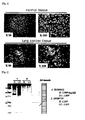
FIG. 1 shows primary culture of cells separated from a normal surrounding tissue and a lung cancer tissue of a lung cancer patient; -
FIG. 2 shows SDS-PAGE separation of proteins from a normal surrounding tissue and a lung cancer tissue of a lung cancer patient, and coomassie staining of them; -
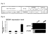
FIG. 3 shows LC-MS/MS analysis of Quiescin-sulfhydryl oxidase 1 (QSOX1); -
FIG. 4 shows the comparison between a normal cell line and a lung cancer cell line in mRNA expression and protein expression of Quiescin-sulfhydryl oxidase 1 (QSOX1); and -
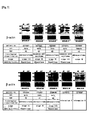
FIG. 5 shows the comparison between a normal surrounding tissue and a lung cancer tissue in protein expression of Quiescin-sulfhydryl oxidase 1 (QSOX1) of lung cancer patients. - The present invention provides the use of a composition for diagnosis of lung cancer, which includes an antibody specifically binding to Quiescin-
sulfhydryl oxidase 1 Preferably, the composition for diagnosis of lung cancer includes an antibody specifically binding to the marker, as an active ingredient. - In the present invention, the term "diagnosis" indicates determination of existence or characteristics of pathologies. In view of the objective of the present invention, the diagnosis is on the onset of lung cancer.
- In the present invention, the term "lung cancer" means a malignant tumor occurring in a lung, and histologically includes lung adenocarcinoma, squamous cell carcinoma, large cell lung cancer, and small cell lung cancer.
- Lung cancer has 4 main histological types, and then is further subdivided into two types of small cell lung cancer (SCLC) and non-small cell lung cancer (NSCLC). The non-small cell lung cancer (NSCLC) is found in about 80% of all lung cancer patients, and includes lung adenocarcinoma, squamous cell carcinoma, and large cell lung cancer. From among some histological types of NSCLC, squamous cell carcinoma is found in 20-25% of lung cancer patients, and large cell lung cancer is found in 15-20%, and lung adenocarcinoma is found in 30-40%.
- In the present invention, an antibody may be a whole form of antibody (hereinafter, referred to as a "whole antibody") or its functional fragment. The whole antibody may be a polymer type of antibody including monomers or 2 or more bound whole antibodies. The functional fragment of the antibody is an antibody having a heavy chain variable region and a light chain variable region of the whole antibody, and in actuality, recognizes the same epitope as that recognized by the whole antibody.
- The functional fragment of the antibody includes single chain variable region fragments (scFv), (scFv)2, Fab, Fab', F(ab')2, and the like, but the present invention is not limited thereto. The single chain variable region (scFv) indicates a single chain polypeptide antibody fragment in which a heavy chain variable region and a light chain variable region are linked by a linker peptide.
- The antibody may be modified by attachment with various molecules such as an enzyme, a fluorescent material, a radioactive material and a protein. The modified antibody may be obtained by chemically modifying the antibody. This modification method is conventionally used in the art. Also, the antibody may be obtained as a chimeric antibody having a variable region derived from a non-human antibody, and a constant region derived from a human antibody, or may be obtained as a humanized antibody including a complementarity-determining region derived from a non-human antibody, and a framework region (FR) and a constant region derived from a human antibody. Such an antibody may be prepared by using a method known in the art.
- The diagnostic composition may further include a reagent used for immunoassay, known in the art, besides the protein-specific antibody. The immunoassay may include any method as long as it can measure binding between an antigen and an antibody.
- Also, the present invention provides the use of a diagnostic kit of lung cancer, which includes the composition as an active ingredient.
- The diagnostic kit may be, for example, an immunochromarography-based lateral flow assay kit for detecting a specific protein from a test sample. The lateral flow assay kit generally includes a sample pad to which a test sample is applied, a releasing pad coated with a detection antibody, a transfer membrane (e.g., nitrocellulose) or a strip in which the test sample is moved and separated, and an antigen-antibody reaction occurs, and an absorption pad.
- Also, the present invention provides the use of a diagnostic microarray of lung cancer, which includes the composition as an active ingredient.
- In the microarray, in general, an antibody is immobilized on a slide glass surface treated with a specific reagent, and then a protein specifically bound to the antibody through an antigen-antibody reaction is detected.
- Also, the diagnostic composition of lung cancer includes, as an active ingredient, a primer or a probe specific to a nucleic acid encoding at least Quiescin-
sulfhydryl oxidase 1. - The detection of a specific nucleic acid by using the primer may be carried out by amplifying a sequence of a target gene through an amplification method such as PCR, and detecting the amplification of the gene according to a method known in the art. Also, the detection of a specific nucleic acid by using the probe may be carried out by contacting a test sample nucleic acid with the probe under a proper condition, and detecting the existence of a hybridized nucleic acid.
- The term "primer" indicates a short nucleic acid sequence having short free OH groups, which can form a base pair with a complementary template, and functions as a start point for template strand replication.
- The "probe" indicates a fragment of a nucleic acid including several to several hundreds of bases (such as RNA or DNA) capable of being specifically bound to mRNA. The probe is labeled and thus can be used for determining the existence of a specific mRNA. The probe may be produced as various types of probes such as an oligonucleotide probe, a single stranded DNA probe, double stranded DNA probe, a RNA probe, and may be labeled with biotin, FITC, rhodamine, DIG, radio isotope, or the like.
- Also, the probe may be labeled with a detectable substance, for example, a radio label capable of providing an appropriate signal and having a sufficient half life. The labeled probe may be hybridized with a nucleic acid on a solid substrate, as known in the art.
- Examples of the method for detecting a specific nucleic acid by using the probe or the primer may include, but are not limited to, polymerase chain reaction (PCR), DNA sequencing, RT-PCR, primer extension, oligonucleotide extension analysis, allele-specific PCR, RNase mismatch cleavage, single strand conformation polymorphism (SSCP) and heteroduplex simultaneous analysis, denaturing gradient gel electrophoresis (DGGE), denaturing high pressure liquid chromatography, hybridization reaction, DNA chip, and the like. Examples of the hybridization reaction include northern hybridization, in situ hybridization, and microarray.
- The composition for diagnosis of lung cancer may further include a conventionally used reagent in the above described nucleic acid detecting method. For example, it may include a metal ion salt required for a PCR reaction, such as dNTP (deoxynulceotide triphosphate),
- thermostable polymerase, and magnesium chloride, and include dNTP, sequenase, etc. required for sequencing.
- Preferably, the composition for diagnosis of lung cancer may be provided in the form of a diagnostic kit or microarray. For example, the inventive composition may be provided as a RT-PCR kit, a DNA chip, or the like, but the present invention is not limited thereto, in which the RT~PCR kit includes respective primer pairs specific to at Quiescin-
sulfhydryl oxidase 1. - Also, the present invention provides a method for detecting, from a biological sample, Quiescin-
sulfhydryl oxidase 1 as a lung cancer marker through an antigen-antibody binding reaction using an antibody specifically bound to Quiescin-sulfhydryl oxidase 1. - In the present invention, through an antigen-antibody binding reaction using an antibody specifically bound to Quiescin-
sulfhydryl oxidase 1 as a lung cancer marker, the protein is detected from a biological sample so as to predict the diagnosis or prognosis of lung cancer early. Specifically, the proteins are fractionated through electrophoresis (SDS-PAGE), transferred to a solid support, and immobilized. An antibody specifically bound to the proteins is added to perform an antigen-antibody binding reaction, and then expression levels of these proteins are measured. In other words, from a lung cancer patient tissue and a healthy person tissue, the expression levels of the proteins are measured, and compared to each other. Then, when the expression level of the protein in the lung cancer patient
tissue is higher than that in the healthy person, he is diagnosed to have lung cancer or expected to have lung cancer. - Examples of the biological sample may include test samples obtained from mammals, such as tissues, cells, whole blood, serum, plasma, saliva, sputum, and urine, but the present invention is not limited thereto. The test sample may be subjected to a post-treatment such as filtration, distillation, extraction, concentration, inactivation of an interrupting substance, or addition of a reagent, before being used for the detection.
- As a solid support for the antigen-antibody binding reaction, a nitrocellulose membrane, a PVDF membrane (polyvinylidene difluoride membrane), a 96 well plate synthesized by a polyvinyl resin or a polystyrene resin, or a slide glass made of a glass may be used.
- The antigen-antibody binding reaction may be carried out by a conventional method such as enzyme-linked immunosorbent assay (ELISA), radioimmunoassay (RIA), sandwich assay, western blotting, immunoprecipitation, immunohistochemical staining, flow cytometry, fluorescence activated cell sorter (FACS), chromogenic enzyme substrate test, and antigen-antibody aggregation.
- Hereinafter, a preferred embodiment will be provided for illustrating the present invention. However, the Examples below are intended to further illustrate the present invention without limiting its scope.
- A system for primary culture using a lung cancer tissue and a surrounding normal tissue obtained from a lung cancer patient was secured. The lung cancer tissue and the surrounding normal tissue were maintained in a fresh state, and were chopped by using scissors or a blade in accordance with a mechanical method. Then, they were treated with collagenase type, reacted for 30 minutes to 90 minutes at 37°C so as to separate single cells. Then, they were plated onto a medium containing 10% FBS, and cultured.

- In a case of a cell separated from the normal tissue, a typical epithelial cell type of pebble-shaped circular cells were grown, while in a case of a cell separated from the lung cancer tissue, fibrocyte-like shaped cells were grown in a long form (see
FIG. 1 ). - Cells from Example 1 were subcultured twice to three times so as to collect a protein secreted during a culturing process. In subculture 3, the same number of cells were counted, and they were transferred to a new culturing plate, and cultured for 48 hours to collect a medium including a secreted protein.
- Then, in order to remove the FBS content contained in a subculture medium of the normal cell plate and the lung cancer cell plate, each plate was sufficiently washed. Then, the medium was replaced by a serum free media, and culturing was performed for 24 to 48 hours. In this process, in order to obtain a protein secreted from each cell, a TCA precipitation method was used for protein concentration.
- The protein from Example 2, which was secreted from the normal tissue cell and the lung cancer tissue cell and concentrated, was separated through SDS-PAGE, and the gel was washed with ddH2O three times (each for 5 minutes). Then, the protein was added with Coomassie G250 Stain (BioRad, Hercules, CA), and softly shaken at room temperature for 1 hour for staining. In the coomassie staining, in each pair (N: a protein secreted from a normal tissue, LC: a protein secreted from a lung cancer tissue), the proteins were used for the experiment in the same amount (see
FIG. 2 ). - After the staining in Example 3, an interesting protein band was cut into pieces, and was subjected to in-gel trypsin digestion. 25 bands showing different
respective strengths of the protein secreted from the lung cancer tissue, and the protein secreted from the normal tissue were analyzed. - In semi-quantitative proteomics, without using a labeling material, a spectral count (that is, the number of peptides, obtained during analysis for protein identification after MS/MS analysis), is used. The secreted protein was analyzed by a conventional semi-quantitative method, and the identified protein was subjected to gene ontology analysis so as to determine the characteristics of proteins obtained from the experimental result.
- Through LC-ESI-MS/MS analysis, from two pairs (20100622, 20100719) of samples, a protein of Quiescin-sulfhydryl oxidase 1 (QSOX1) was found. In respective samples, the protein secreted from the lung cancer tissue (LC) showed 1.64 and 4.31 times higher quantitative values, respectively, compared to the protein secreted from the normal tissue (N) (see
FIG. 3 ).[Table 1] Identified protein IPI number clone: 20100622 clone: 20100719 Average value. lung cancer/ normal quantitative value quantitative value normal Lung cancer Lung cancer/normal normal Lung cancer Lung cancer/normal Fibrillin-1 IPI00328113 5.55 74.58 13.44 1 4.31 4.31 8.87 Isoform A of Lamin-A/C IPI00021405 (+2) 4.44 18.19 4.097 1 3.44 3.44 3.77 Latent-transforming growth factor beta-binding protein 2IPI00292150 3.33 13.64 4.096 1 2.58 2.58 3.34 Galectin-1 IPI00219219 4.44 12.73 2.867 1 3.44 3.44 3.15 highly similar to Dickkopf-related protein 3 IPI00002714 (+2) 7.77 20.92 2.692 1 5.17 5.17 3.93 Isoform A1-B of Heterogeneous nuclear ribonucleoprotein A1 IPI00215965 (+3) 2.22 5.46 2.459 1 1.72 1.72 2.09 14-3-3 protein epsilon IPI00000816 8.88 20.92 2.356 4.77 7.75 1.625 1.99 Stanniocalcin-2 IPI00008780 12.21 24.56 2.011 1 9.47 9.47 5.74 Cystatin-C IPI00032293 8.88 17.28 1.946 3.58 6.89 1.925 1.94 Isoform 1 of Connective tissue growth factor IPI00020977 6.66 11.82 1.775 1 5.17 5.17 3.47 Profilin-1 IPI00216691 8.88 15.46 1.741 2.38 6.89 2.895 2.32 Isoform 1 of Extracellular matrix protein 1IPI00003351 (+1) 23.32 39.11 1.677 11.92 18.08 1.517 1.60 Unknown ##IPI00008556 4.44 7.28 1.64 1 3.44 3.44 2.54 Histone H2B type 2-E IPI00003935 (+9) 3.33 5.46 1.64 1 2.58 2.58 2.11 - Also, through LC-ESI-MS/MS analysis, besides Quiescin-sulfhydryl oxidase 1 (QSOX1), other proteins whose secretions were increased in the lung cancer tissue compared to in the normal tissue were found. The result is noted in table 1. Meanwhile, through LC-ESI-MS/MS analysis, some proteins whose secretions were decreased in the lung cancer tissue compared to in the normal tissue were found. The result is noted in table 2.
[Table 2] Identified protein IPI number clone: 20100622 clone: 20100719 Average value. lung cancer/ normal quantitative value quantitative value normal lung cancer lung cancer /normal normal lung cancer lung cancer /normal Kinesin-like protein KIF26A IPI00788247 (+1) 2.22 1 0.45 2.38 1.72 0.723 0.59 Zinc finger protein 516 IPI00852669 (+1) 2.22 1 0.45 2.38 1.72 0.723 0.59 Interstitial collagenase IPI00008561 (+1) 11.1 3.64 0.328 17.88 8.61 0.482 0.40 Isoform 1 of A-kinase anchor protein 9 IPI00019223 (+4) 6.66 1 0.15 11.92 6.89 0.578 0.36 - From a L132 cell of a cell line of a lung epithelial cell, and a H358 cell of a cell line of non-small cell lung cancer (NSCLC), mRNA and protein of Quiescin-
sulfhydryl oxidase 1 were obtained, and then through real-time PCR, and western blotting analysis, the expression was verified. - As a result, it was found that expression levels of mRNA and protein of Quiescin-
sulfhydryl oxidase 1 were highly increased in the H358 cell line, compared in the normal cell line (L132) (seeFIG. 4 ). - In order to determine if Quiescin-
sulfhydryl oxidase 1 expression verified in the lung cancer cell line occurs in a lung cancer patient, in a lung cancer tissue and a surrounding normal tissue obtained from a total of 10 patients, protein expression levels were measured through western blotting. - First, from the lung cancer tissue and the surrounding normal tissue separated from 10 lung cancer patients, a protein was separated. Simply, blood of the lung cancer tissue was washed with PBS, and the tissue was homogenized in a RIPA buffer containing a protease inhibitor.
- Then, proteins of respective samples were collected in the same amount by a quantitative method through Bradford assay, and then in the normal tissue and the lung cancer tissue, the expression level of Quiescin-
sulfhydryl oxidase 1 protein was measured. Since the expression levels of beta actin in the normal tissue and the lung cancer tissue are similar to each other, it can be determined that the proteins were used in the same amount in the experiment. - At the lower position (68kDa) and at the higher position (82kDa) with respect to the marker of 72kDa, expression levels of the secreted form and the membrane bound form were significantly increased in all lung cancer tissues of the 10 lung cancer patients. Accordingly, it can be determined that the expression level in the clinical samples was observed in the same manner as in the lung cancer cell line (see
FIG. 5 ). - As described above, the expression level of the protein was significantly increased in the lung cancer tissue compared to that in the normal tissue. Thus, the protein can be in actuality utilized in the diagnosis of lung cancer.
- Although a preferred embodiment of the present invention has been described for illustrative purposes, those skilled in the art will appreciate that various modifications, additions and substitutions are possible, without departing from the scope of the invention as disclosed in the accompanying claims.
Claims (9)
- Use of a composition comprising an antibody specifically binding to Quiescin-sulfhydryl oxidase 1 in the in vitro/ex vivo diagnosis of lung cancer.
- Use of the composition as claimed in claim 1, wherein the lung cancer is at least one selected from the group including lung adenocarcinoma, squamous cell carcinoma, large cell cancer, and small cell cancer.
- Use of a diagnostic kit comprising the composition as claimed in claim 1 as an active ingredient in the in vitro/ex vivo diagnosis of lung cancer.
- Use of a diagnostic microarray comprising the composition as claimed in claim 1 as an active ingredient, in the in vitro/ex vivo diagnosis of lung cancer.
- Use of a composition comprising a primer or a probe specific to a nucleic acid encoding Quiescin-sulfhydryl oxidase 1 in the in vitro/ex vivo diagnosis of lung cancer.
- Use of a diagnostic kit comprising the composition as claimed in claim 5 as an active ingredient in the in vitro/ex vivo diagnosis of lung cancer.
- Use of a diagnostic microarray comprising the composition as claimed in claim 5 as an active ingredient, in the in vitro/ex vivo diagnosis of lung cancer.
- A method for detecting, from a biological sample, Quiescin-sulfhydryl oxidase 1 as a lung cancer marker through an antigen-antibody binding reaction using an antibody specifically bound to Quiescin-sulfhydryl oxidase 1.
- The method as claimed in claim 8, wherein the biological sample is at least one selected from the group including tissues, cells, whole blood, serum, plasma, saliva, sputum, and urine obtained from mammals.
Applications Claiming Priority (1)
| Application Number | Priority Date | Filing Date | Title |
|---|---|---|---|
| KR1020110104949A KR101297309B1 (en) | 2011-10-14 | 2011-10-14 | Composition for diagnosis of lung cancer and diagnosis kit of lung cancer |
Publications (3)
| Publication Number | Publication Date |
|---|---|
| EP2581745A2 EP2581745A2 (en) | 2013-04-17 |
| EP2581745A3 EP2581745A3 (en) | 2013-04-24 |
| EP2581745B1 true EP2581745B1 (en) | 2015-12-30 |
Family
ID=45463442
Family Applications (1)
| Application Number | Title | Priority Date | Filing Date |
|---|---|---|---|
| EP12150252.0A Active EP2581745B1 (en) | 2011-10-14 | 2012-01-05 | Composition for diagnosis of lung cancer and diagnosis kit of lung cancer |
Country Status (4)
| Country | Link |
|---|---|
| US (2) | US20130095490A1 (en) |
| EP (1) | EP2581745B1 (en) |
| KR (1) | KR101297309B1 (en) |
| WO (1) | WO2013054984A1 (en) |
Families Citing this family (3)
| Publication number | Priority date | Publication date | Assignee | Title |
|---|---|---|---|---|
| KR101981806B1 (en) * | 2013-05-14 | 2019-05-23 | 엘지전자 주식회사 | Quescin-sulfhydryl oxidase 1 specific antibody, Hybridoma cell producing the same, and Composition for diagnosis of lung cancer having the same |
| US10151001B2 (en) | 2014-12-29 | 2018-12-11 | National Guard Health Affairs | Quantification of Lamin C and Lamin A for tumor classification |
| CN105510411B (en) * | 2015-09-14 | 2018-05-08 | 陕西师范大学 | The detection method of single cancer cell is realized in interaction based on cell and microelectrode |
Family Cites Families (6)
| Publication number | Priority date | Publication date | Assignee | Title |
|---|---|---|---|---|
| WO2009002472A1 (en) * | 2007-06-22 | 2008-12-31 | University Of Georgia Research Foundation, Inc. | Novel secreted proteins of adipocytes for diagnostic purposes |
| WO2010071788A1 (en) * | 2008-12-17 | 2010-06-24 | Arizona Board of Regents, a body corporate acting for and on behalf of Arizona State University | Pancreatic cancer markers and uses thereof |
| WO2010086384A1 (en) * | 2009-01-30 | 2010-08-05 | Pronota N.V. | Target for treatment of acute heart failure |
| DK2391892T3 (en) * | 2009-01-30 | 2017-04-24 | Mycartis N V | BIOMARKET FOR DIAGNOSIS, PREDICTION AND / OR PROJECTS OF ACUTE HEART FAILURE AND USE thereof |
| WO2011035433A1 (en) * | 2009-09-23 | 2011-03-31 | University Health Network | Selected strains on serum-free growth media for proteomics analysis of lung cancer biomarkers |
| US20110275065A1 (en) * | 2010-05-07 | 2011-11-10 | Ranju Ralhan | Methods and compositions for the diagnosis and treatment of thyroid cancer |
-
2011
- 2011-10-14 KR KR1020110104949A patent/KR101297309B1/en active IP Right Grant
- 2011-12-28 WO PCT/KR2011/010257 patent/WO2013054984A1/en active Application Filing
-
2012
- 2012-01-05 EP EP12150252.0A patent/EP2581745B1/en active Active
- 2012-01-17 US US13/351,631 patent/US20130095490A1/en not_active Abandoned
-
2014
- 2014-05-14 US US14/277,327 patent/US20140248637A1/en not_active Abandoned
Also Published As
| Publication number | Publication date |
|---|---|
| EP2581745A2 (en) | 2013-04-17 |
| WO2013054984A1 (en) | 2013-04-18 |
| US20130095490A1 (en) | 2013-04-18 |
| US20140248637A1 (en) | 2014-09-04 |
| KR20130040293A (en) | 2013-04-24 |
| EP2581745A3 (en) | 2013-04-24 |
| KR101297309B1 (en) | 2013-08-16 |
Similar Documents
| Publication | Publication Date | Title |
|---|---|---|
| CA2671939C (en) | Biomarker for detecting prostate cancer | |
| EP1996940B1 (en) | Detection of cancer by elevated levels of bcl-2 | |
| TWI585411B (en) | Urine markers for detection of bladder cancer | |
| EP2762574A1 (en) | Non-invasive diagnostic method for diagnosing bladder cancer | |
| EP1601968A2 (en) | Serum macrophage migration inhibitory factor (mif) as marker for prostate cancer | |
| AU2015360694A1 (en) | Use of markers including filamin a in the diagnosis and treatment of prostate cancer | |
| CN113249491A (en) | Biomarker for diagnosing endometrial cancer and product and application thereof | |
| CN113234830B (en) | Product for lung cancer diagnosis and application | |
| KR20130046457A (en) | Newly identified colorectal cancer marker genes, proteins translated from the genes and a diagnostic kit using the same | |
| US20160187341A1 (en) | Keratins as biomarkers for cervical cancer and survival | |
| EP2581745B1 (en) | Composition for diagnosis of lung cancer and diagnosis kit of lung cancer | |
| KR101995189B1 (en) | Biomarker for non-invasive in vitro diagnosis of a Hepatocellular carcinoma and biokit for diagnosis thereof comprising the same | |
| JP5403534B2 (en) | Methods to provide information for predicting prognosis of esophageal cancer | |
| CN114164273B (en) | Squamous carcinoma prognosis marker, establishment method of prognosis risk assessment model and application of prognosis risk assessment model | |
| WO2019134994A1 (en) | Prognostic biomarkers for human papillomavirus positive cancers | |
| KR101334123B1 (en) | Composition for diagnosis of small cell lung cancer and diagnosis kit of small cell lung cancer | |
| US20230083393A1 (en) | Multiple biomarkers for diagnosing lung cancer and use thereof | |
| EP3494228B1 (en) | Method for prognosing and diagnosing tumors | |
| Xu et al. | A potential panel of five mRNAs in urinary extracellular vesicles for the detection of bladder cancer | |
| KR101815253B1 (en) | CXCL14 Biomarker for Diagnosing Liver Fibrosis | |
| EP4317458A1 (en) | Follicular thyroid cancer-specific marker | |
| KR20110068695A (en) | Biomarkers indicative of ovarian carcinoma and diagnosis using the same | |
| KR101403019B1 (en) | Novel biomarker for the diagnosis of lung cancer | |
| US20150044703A1 (en) | Methods and compositions for detecting endometrial or ovarian cancer | |
| JP2017529074A (en) | ERCC1 isoform 3 mRNA and / or protein for use in diagnosing resistance to a therapeutic agent and method for diagnosing resistance to a therapeutic agent using this mRNA and / or protein |
Legal Events
| Date | Code | Title | Description |
|---|---|---|---|
| PUAL | Search report despatched |
Free format text: ORIGINAL CODE: 0009013 |
|
| PUAI | Public reference made under article 153(3) epc to a published international application that has entered the european phase |
Free format text: ORIGINAL CODE: 0009012 |
|
| 17P | Request for examination filed |
Effective date: 20120105 |
|
| AK | Designated contracting states |
Kind code of ref document: A2 Designated state(s): AL AT BE BG CH CY CZ DE DK EE ES FI FR GB GR HR HU IE IS IT LI LT LU LV MC MK MT NL NO PL PT RO RS SE SI SK SM TR |
|
| AX | Request for extension of the european patent |
Extension state: BA ME |
|
| AK | Designated contracting states |
Kind code of ref document: A3 Designated state(s): AL AT BE BG CH CY CZ DE DK EE ES FI FR GB GR HR HU IE IS IT LI LT LU LV MC MK MT NL NO PL PT RO RS SE SI SK SM TR |
|
| AX | Request for extension of the european patent |
Extension state: BA ME |
|
| RIC1 | Information provided on ipc code assigned before grant |
Ipc: G01N 33/574 20060101AFI20130318BHEP |
|
| 17Q | First examination report despatched |
Effective date: 20150218 |
|
| GRAP | Despatch of communication of intention to grant a patent |
Free format text: ORIGINAL CODE: EPIDOSNIGR1 |
|
| INTG | Intention to grant announced |
Effective date: 20150727 |
|
| RIN1 | Information on inventor provided before grant (corrected) |
Inventor name: SUNG, HYE JIN Inventor name: CHO, JE YOEL |
|
| GRAS | Grant fee paid |
Free format text: ORIGINAL CODE: EPIDOSNIGR3 |
|
| GRAA | (expected) grant |
Free format text: ORIGINAL CODE: 0009210 |
|
| AK | Designated contracting states |
Kind code of ref document: B1 Designated state(s): AL AT BE BG CH CY CZ DE DK EE ES FI FR GB GR HR HU IE IS IT LI LT LU LV MC MK MT NL NO PL PT RO RS SE SI SK SM TR |
|
| REG | Reference to a national code |
Ref country code: GB Ref legal event code: FG4D |
|
| REG | Reference to a national code |
Ref country code: CH Ref legal event code: EP |
|
| REG | Reference to a national code |
Ref country code: AT Ref legal event code: REF Ref document number: 767711 Country of ref document: AT Kind code of ref document: T Effective date: 20160115 |
|
| REG | Reference to a national code |
Ref country code: IE Ref legal event code: FG4D |
|
| REG | Reference to a national code |
Ref country code: DE Ref legal event code: R096 Ref document number: 602012013290 Country of ref document: DE |
|
| REG | Reference to a national code |
Ref country code: LT Ref legal event code: MG4D |
|
| PG25 | Lapsed in a contracting state [announced via postgrant information from national office to epo] |
Ref country code: NO Free format text: LAPSE BECAUSE OF FAILURE TO SUBMIT A TRANSLATION OF THE DESCRIPTION OR TO PAY THE FEE WITHIN THE PRESCRIBED TIME-LIMIT Effective date: 20160330 Ref country code: HR Free format text: LAPSE BECAUSE OF FAILURE TO SUBMIT A TRANSLATION OF THE DESCRIPTION OR TO PAY THE FEE WITHIN THE PRESCRIBED TIME-LIMIT Effective date: 20151230 Ref country code: LT Free format text: LAPSE BECAUSE OF FAILURE TO SUBMIT A TRANSLATION OF THE DESCRIPTION OR TO PAY THE FEE WITHIN THE PRESCRIBED TIME-LIMIT Effective date: 20151230 |
|
| REG | Reference to a national code |
Ref country code: NL Ref legal event code: MP Effective date: 20151230 |
|
| REG | Reference to a national code |
Ref country code: AT Ref legal event code: MK05 Ref document number: 767711 Country of ref document: AT Kind code of ref document: T Effective date: 20151230 |
|
| PG25 | Lapsed in a contracting state [announced via postgrant information from national office to epo] |
Ref country code: GR Free format text: LAPSE BECAUSE OF FAILURE TO SUBMIT A TRANSLATION OF THE DESCRIPTION OR TO PAY THE FEE WITHIN THE PRESCRIBED TIME-LIMIT Effective date: 20160331 Ref country code: BE Free format text: LAPSE BECAUSE OF NON-PAYMENT OF DUE FEES Effective date: 20160131 Ref country code: RS Free format text: LAPSE BECAUSE OF FAILURE TO SUBMIT A TRANSLATION OF THE DESCRIPTION OR TO PAY THE FEE WITHIN THE PRESCRIBED TIME-LIMIT Effective date: 20151230 Ref country code: LV Free format text: LAPSE BECAUSE OF FAILURE TO SUBMIT A TRANSLATION OF THE DESCRIPTION OR TO PAY THE FEE WITHIN THE PRESCRIBED TIME-LIMIT Effective date: 20151230 Ref country code: FI Free format text: LAPSE BECAUSE OF FAILURE TO SUBMIT A TRANSLATION OF THE DESCRIPTION OR TO PAY THE FEE WITHIN THE PRESCRIBED TIME-LIMIT Effective date: 20151230 Ref country code: SE Free format text: LAPSE BECAUSE OF FAILURE TO SUBMIT A TRANSLATION OF THE DESCRIPTION OR TO PAY THE FEE WITHIN THE PRESCRIBED TIME-LIMIT Effective date: 20151230 |
|
| PG25 | Lapsed in a contracting state [announced via postgrant information from national office to epo] |
Ref country code: NL Free format text: LAPSE BECAUSE OF FAILURE TO SUBMIT A TRANSLATION OF THE DESCRIPTION OR TO PAY THE FEE WITHIN THE PRESCRIBED TIME-LIMIT Effective date: 20151230 |
|
| PG25 | Lapsed in a contracting state [announced via postgrant information from national office to epo] |
Ref country code: ES Free format text: LAPSE BECAUSE OF FAILURE TO SUBMIT A TRANSLATION OF THE DESCRIPTION OR TO PAY THE FEE WITHIN THE PRESCRIBED TIME-LIMIT Effective date: 20151230 Ref country code: IT Free format text: LAPSE BECAUSE OF FAILURE TO SUBMIT A TRANSLATION OF THE DESCRIPTION OR TO PAY THE FEE WITHIN THE PRESCRIBED TIME-LIMIT Effective date: 20151230 Ref country code: CZ Free format text: LAPSE BECAUSE OF FAILURE TO SUBMIT A TRANSLATION OF THE DESCRIPTION OR TO PAY THE FEE WITHIN THE PRESCRIBED TIME-LIMIT Effective date: 20151230 |
|
| PG25 | Lapsed in a contracting state [announced via postgrant information from national office to epo] |
Ref country code: PT Free format text: LAPSE BECAUSE OF FAILURE TO SUBMIT A TRANSLATION OF THE DESCRIPTION OR TO PAY THE FEE WITHIN THE PRESCRIBED TIME-LIMIT Effective date: 20160502 Ref country code: SM Free format text: LAPSE BECAUSE OF FAILURE TO SUBMIT A TRANSLATION OF THE DESCRIPTION OR TO PAY THE FEE WITHIN THE PRESCRIBED TIME-LIMIT Effective date: 20151230 Ref country code: AT Free format text: LAPSE BECAUSE OF FAILURE TO SUBMIT A TRANSLATION OF THE DESCRIPTION OR TO PAY THE FEE WITHIN THE PRESCRIBED TIME-LIMIT Effective date: 20151230 Ref country code: PL Free format text: LAPSE BECAUSE OF FAILURE TO SUBMIT A TRANSLATION OF THE DESCRIPTION OR TO PAY THE FEE WITHIN THE PRESCRIBED TIME-LIMIT Effective date: 20151230 Ref country code: LU Free format text: LAPSE BECAUSE OF FAILURE TO SUBMIT A TRANSLATION OF THE DESCRIPTION OR TO PAY THE FEE WITHIN THE PRESCRIBED TIME-LIMIT Effective date: 20160105 Ref country code: SK Free format text: LAPSE BECAUSE OF FAILURE TO SUBMIT A TRANSLATION OF THE DESCRIPTION OR TO PAY THE FEE WITHIN THE PRESCRIBED TIME-LIMIT Effective date: 20151230 Ref country code: RO Free format text: LAPSE BECAUSE OF FAILURE TO SUBMIT A TRANSLATION OF THE DESCRIPTION OR TO PAY THE FEE WITHIN THE PRESCRIBED TIME-LIMIT Effective date: 20151230 Ref country code: EE Free format text: LAPSE BECAUSE OF FAILURE TO SUBMIT A TRANSLATION OF THE DESCRIPTION OR TO PAY THE FEE WITHIN THE PRESCRIBED TIME-LIMIT Effective date: 20151230 Ref country code: IS Free format text: LAPSE BECAUSE OF FAILURE TO SUBMIT A TRANSLATION OF THE DESCRIPTION OR TO PAY THE FEE WITHIN THE PRESCRIBED TIME-LIMIT Effective date: 20160430 |
|
| REG | Reference to a national code |
Ref country code: CH Ref legal event code: PL |
|
| PG25 | Lapsed in a contracting state [announced via postgrant information from national office to epo] |
Ref country code: MC Free format text: LAPSE BECAUSE OF FAILURE TO SUBMIT A TRANSLATION OF THE DESCRIPTION OR TO PAY THE FEE WITHIN THE PRESCRIBED TIME-LIMIT Effective date: 20151230 |
|
| REG | Reference to a national code |
Ref country code: DE Ref legal event code: R097 Ref document number: 602012013290 Country of ref document: DE |
|
| PG25 | Lapsed in a contracting state [announced via postgrant information from national office to epo] |
Ref country code: CH Free format text: LAPSE BECAUSE OF NON-PAYMENT OF DUE FEES Effective date: 20160131 Ref country code: LI Free format text: LAPSE BECAUSE OF NON-PAYMENT OF DUE FEES Effective date: 20160131 Ref country code: DK Free format text: LAPSE BECAUSE OF FAILURE TO SUBMIT A TRANSLATION OF THE DESCRIPTION OR TO PAY THE FEE WITHIN THE PRESCRIBED TIME-LIMIT Effective date: 20151230 |
|
| REG | Reference to a national code |
Ref country code: IE Ref legal event code: MM4A |
|
| PLBE | No opposition filed within time limit |
Free format text: ORIGINAL CODE: 0009261 |
|
| STAA | Information on the status of an ep patent application or granted ep patent |
Free format text: STATUS: NO OPPOSITION FILED WITHIN TIME LIMIT |
|
| 26N | No opposition filed |
Effective date: 20161003 |
|
| PG25 | Lapsed in a contracting state [announced via postgrant information from national office to epo] |
Ref country code: BE Free format text: LAPSE BECAUSE OF FAILURE TO SUBMIT A TRANSLATION OF THE DESCRIPTION OR TO PAY THE FEE WITHIN THE PRESCRIBED TIME-LIMIT Effective date: 20151230 |
|
| REG | Reference to a national code |
Ref country code: FR Ref legal event code: ST Effective date: 20161125 |
|
| PG25 | Lapsed in a contracting state [announced via postgrant information from national office to epo] |
Ref country code: FR Free format text: LAPSE BECAUSE OF NON-PAYMENT OF DUE FEES Effective date: 20160229 Ref country code: IE Free format text: LAPSE BECAUSE OF NON-PAYMENT OF DUE FEES Effective date: 20160105 |
|
| PG25 | Lapsed in a contracting state [announced via postgrant information from national office to epo] |
Ref country code: SI Free format text: LAPSE BECAUSE OF FAILURE TO SUBMIT A TRANSLATION OF THE DESCRIPTION OR TO PAY THE FEE WITHIN THE PRESCRIBED TIME-LIMIT Effective date: 20151230 |
|
| PG25 | Lapsed in a contracting state [announced via postgrant information from national office to epo] |
Ref country code: MT Free format text: LAPSE BECAUSE OF FAILURE TO SUBMIT A TRANSLATION OF THE DESCRIPTION OR TO PAY THE FEE WITHIN THE PRESCRIBED TIME-LIMIT Effective date: 20151230 |
|
| PG25 | Lapsed in a contracting state [announced via postgrant information from national office to epo] |
Ref country code: CY Free format text: LAPSE BECAUSE OF FAILURE TO SUBMIT A TRANSLATION OF THE DESCRIPTION OR TO PAY THE FEE WITHIN THE PRESCRIBED TIME-LIMIT Effective date: 20151230 Ref country code: HU Free format text: LAPSE BECAUSE OF FAILURE TO SUBMIT A TRANSLATION OF THE DESCRIPTION OR TO PAY THE FEE WITHIN THE PRESCRIBED TIME-LIMIT; INVALID AB INITIO Effective date: 20120105 |
|
| PG25 | Lapsed in a contracting state [announced via postgrant information from national office to epo] |
Ref country code: TR Free format text: LAPSE BECAUSE OF FAILURE TO SUBMIT A TRANSLATION OF THE DESCRIPTION OR TO PAY THE FEE WITHIN THE PRESCRIBED TIME-LIMIT Effective date: 20151230 Ref country code: MK Free format text: LAPSE BECAUSE OF FAILURE TO SUBMIT A TRANSLATION OF THE DESCRIPTION OR TO PAY THE FEE WITHIN THE PRESCRIBED TIME-LIMIT Effective date: 20151230 Ref country code: MT Free format text: LAPSE BECAUSE OF FAILURE TO SUBMIT A TRANSLATION OF THE DESCRIPTION OR TO PAY THE FEE WITHIN THE PRESCRIBED TIME-LIMIT Effective date: 20160131 |
|
| PG25 | Lapsed in a contracting state [announced via postgrant information from national office to epo] |
Ref country code: BG Free format text: LAPSE BECAUSE OF FAILURE TO SUBMIT A TRANSLATION OF THE DESCRIPTION OR TO PAY THE FEE WITHIN THE PRESCRIBED TIME-LIMIT Effective date: 20151230 |
|
| PG25 | Lapsed in a contracting state [announced via postgrant information from national office to epo] |
Ref country code: AL Free format text: LAPSE BECAUSE OF FAILURE TO SUBMIT A TRANSLATION OF THE DESCRIPTION OR TO PAY THE FEE WITHIN THE PRESCRIBED TIME-LIMIT Effective date: 20151230 |
|
| REG | Reference to a national code |
Ref country code: DE Ref legal event code: R082 Ref document number: 602012013290 Country of ref document: DE Representative=s name: PATENTANWAELTE STAEGER & SPERLING PARTNERSCHAF, DE Ref country code: DE Ref legal event code: R081 Ref document number: 602012013290 Country of ref document: DE Owner name: PROTANBIO INC., KR Free format text: FORMER OWNER: SEOUL NATIONAL UNIVERSITY R&DB FOUNDATION, SEOUL, KR |
|
| REG | Reference to a national code |
Ref country code: GB Ref legal event code: 732E Free format text: REGISTERED BETWEEN 20190808 AND 20190814 |
|
| PGFP | Annual fee paid to national office [announced via postgrant information from national office to epo] |
Ref country code: GB Payment date: 20230119 Year of fee payment: 12 Ref country code: DE Payment date: 20230327 Year of fee payment: 12 |
|
| P01 | Opt-out of the competence of the unified patent court (upc) registered |
Effective date: 20230516 |
|
| P03 | Removal of the opt-out of the competence of the unified patent court (upc) registered |