EP2575666B1 - Methods of packing medical devices - Google Patents
Methods of packing medical devices Download PDFInfo
- Publication number
- EP2575666B1 EP2575666B1 EP11730052.5A EP11730052A EP2575666B1 EP 2575666 B1 EP2575666 B1 EP 2575666B1 EP 11730052 A EP11730052 A EP 11730052A EP 2575666 B1 EP2575666 B1 EP 2575666B1
- Authority
- EP
- European Patent Office
- Prior art keywords
- bag
- vacuum
- medical device
- placing
- endoscope
- Prior art date
- Legal status (The legal status is an assumption and is not a legal conclusion. Google has not performed a legal analysis and makes no representation as to the accuracy of the status listed.)
- Active
Links
- 238000000034 method Methods 0.000 title claims description 53
- 238000012856 packing Methods 0.000 title description 4
- 230000008569 process Effects 0.000 claims description 16
- 238000004806 packaging method and process Methods 0.000 claims description 15
- 238000004659 sterilization and disinfection Methods 0.000 claims description 12
- QVGXLLKOCUKJST-UHFFFAOYSA-N atomic oxygen Chemical compound [O] QVGXLLKOCUKJST-UHFFFAOYSA-N 0.000 claims description 11
- 229910052760 oxygen Inorganic materials 0.000 claims description 11
- 239000001301 oxygen Substances 0.000 claims description 11
- 238000012544 monitoring process Methods 0.000 claims description 10
- 238000007789 sealing Methods 0.000 claims description 10
- 238000013500 data storage Methods 0.000 claims description 2
- 238000012360 testing method Methods 0.000 description 21
- 238000003860 storage Methods 0.000 description 15
- 241000894006 Bacteria Species 0.000 description 12
- 239000000463 material Substances 0.000 description 9
- 239000003570 air Substances 0.000 description 8
- 230000000813 microbial effect Effects 0.000 description 7
- 244000005700 microbiome Species 0.000 description 7
- XLYOFNOQVPJJNP-UHFFFAOYSA-N water Substances O XLYOFNOQVPJJNP-UHFFFAOYSA-N 0.000 description 7
- 230000006378 damage Effects 0.000 description 6
- 238000005202 decontamination Methods 0.000 description 6
- 230000003588 decontaminative effect Effects 0.000 description 6
- 238000003780 insertion Methods 0.000 description 6
- 230000037431 insertion Effects 0.000 description 6
- 241000193163 Clostridioides difficile Species 0.000 description 5
- 241001148470 aerobic bacillus Species 0.000 description 5
- 238000001035 drying Methods 0.000 description 5
- 230000009467 reduction Effects 0.000 description 5
- 230000002496 gastric effect Effects 0.000 description 4
- 239000013642 negative control Substances 0.000 description 4
- 239000013641 positive control Substances 0.000 description 4
- 238000012958 reprocessing Methods 0.000 description 4
- 241001148471 unidentified anaerobic bacterium Species 0.000 description 4
- 206010011409 Cross infection Diseases 0.000 description 3
- 206010029803 Nosocomial infection Diseases 0.000 description 3
- 238000004140 cleaning Methods 0.000 description 3
- 208000015181 infectious disease Diseases 0.000 description 3
- 239000004417 polycarbonate Substances 0.000 description 3
- 238000010200 validation analysis Methods 0.000 description 3
- 230000003245 working effect Effects 0.000 description 3
- 241000228212 Aspergillus Species 0.000 description 2
- 241000588724 Escherichia coli Species 0.000 description 2
- 208000033809 Suppuration Diseases 0.000 description 2
- 230000002159 abnormal effect Effects 0.000 description 2
- 239000012080 ambient air Substances 0.000 description 2
- 210000004369 blood Anatomy 0.000 description 2
- 239000008280 blood Substances 0.000 description 2
- 230000001332 colony forming effect Effects 0.000 description 2
- 230000001010 compromised effect Effects 0.000 description 2
- 238000011109 contamination Methods 0.000 description 2
- 238000000113 differential scanning calorimetry Methods 0.000 description 2
- 230000000694 effects Effects 0.000 description 2
- 238000001839 endoscopy Methods 0.000 description 2
- 230000036541 health Effects 0.000 description 2
- 230000036512 infertility Effects 0.000 description 2
- 210000003097 mucus Anatomy 0.000 description 2
- 230000035699 permeability Effects 0.000 description 2
- 239000004810 polytetrafluoroethylene Substances 0.000 description 2
- 229920001343 polytetrafluoroethylene Polymers 0.000 description 2
- 239000000047 product Substances 0.000 description 2
- 230000000717 retained effect Effects 0.000 description 2
- 239000000523 sample Substances 0.000 description 2
- 239000000126 substance Substances 0.000 description 2
- 239000000725 suspension Substances 0.000 description 2
- 210000001519 tissue Anatomy 0.000 description 2
- 238000005406 washing Methods 0.000 description 2
- 108091003079 Bovine Serum Albumin Proteins 0.000 description 1
- 241000233866 Fungi Species 0.000 description 1
- 241000282414 Homo sapiens Species 0.000 description 1
- 208000034693 Laceration Diseases 0.000 description 1
- 239000004952 Polyamide Substances 0.000 description 1
- 239000004698 Polyethylene Substances 0.000 description 1
- 241000589517 Pseudomonas aeruginosa Species 0.000 description 1
- 241000191940 Staphylococcus Species 0.000 description 1
- 241000191967 Staphylococcus aureus Species 0.000 description 1
- 241000700605 Viruses Species 0.000 description 1
- 206010000269 abscess Diseases 0.000 description 1
- 210000004666 bacterial spore Anatomy 0.000 description 1
- 238000003339 best practice Methods 0.000 description 1
- 210000000941 bile Anatomy 0.000 description 1
- 230000033228 biological regulation Effects 0.000 description 1
- 230000005540 biological transmission Effects 0.000 description 1
- 238000001574 biopsy Methods 0.000 description 1
- 230000015572 biosynthetic process Effects 0.000 description 1
- 210000001124 body fluid Anatomy 0.000 description 1
- 239000010839 body fluid Substances 0.000 description 1
- 239000012888 bovine serum Substances 0.000 description 1
- 229940098773 bovine serum albumin Drugs 0.000 description 1
- 230000003749 cleanliness Effects 0.000 description 1
- 238000004040 coloring Methods 0.000 description 1
- 239000000356 contaminant Substances 0.000 description 1
- 230000003247 decreasing effect Effects 0.000 description 1
- 239000000645 desinfectant Substances 0.000 description 1
- 239000003599 detergent Substances 0.000 description 1
- 239000003814 drug Substances 0.000 description 1
- 229940079593 drug Drugs 0.000 description 1
- 239000003925 fat Substances 0.000 description 1
- 210000003608 fece Anatomy 0.000 description 1
- 239000012467 final product Substances 0.000 description 1
- 238000009459 flexible packaging Methods 0.000 description 1
- 238000005187 foaming Methods 0.000 description 1
- 235000013305 food Nutrition 0.000 description 1
- 239000007789 gas Substances 0.000 description 1
- 210000001035 gastrointestinal tract Anatomy 0.000 description 1
- 230000002070 germicidal effect Effects 0.000 description 1
- 210000000987 immune system Anatomy 0.000 description 1
- 238000011534 incubation Methods 0.000 description 1
- 230000000968 intestinal effect Effects 0.000 description 1
- 239000007788 liquid Substances 0.000 description 1
- 238000012423 maintenance Methods 0.000 description 1
- 238000004519 manufacturing process Methods 0.000 description 1
- 230000004060 metabolic process Effects 0.000 description 1
- 244000000010 microbial pathogen Species 0.000 description 1
- 238000012986 modification Methods 0.000 description 1
- 230000004048 modification Effects 0.000 description 1
- 210000000214 mouth Anatomy 0.000 description 1
- 210000004400 mucous membrane Anatomy 0.000 description 1
- 235000015097 nutrients Nutrition 0.000 description 1
- 230000001590 oxidative effect Effects 0.000 description 1
- 238000012858 packaging process Methods 0.000 description 1
- 239000004033 plastic Substances 0.000 description 1
- 229920003023 plastic Polymers 0.000 description 1
- 229920002647 polyamide Polymers 0.000 description 1
- 229920000515 polycarbonate Polymers 0.000 description 1
- 229920000573 polyethylene Polymers 0.000 description 1
- 238000011028 process validation Methods 0.000 description 1
- 230000000135 prohibitive effect Effects 0.000 description 1
- 230000035755 proliferation Effects 0.000 description 1
- 230000002035 prolonged effect Effects 0.000 description 1
- 238000003908 quality control method Methods 0.000 description 1
- 230000001105 regulatory effect Effects 0.000 description 1
- 210000003296 saliva Anatomy 0.000 description 1
- 230000028327 secretion Effects 0.000 description 1
- 210000004215 spore Anatomy 0.000 description 1
- 238000010561 standard procedure Methods 0.000 description 1
- 239000000758 substrate Substances 0.000 description 1
- 235000000346 sugar Nutrition 0.000 description 1
- 150000008163 sugars Chemical class 0.000 description 1
- 230000004083 survival effect Effects 0.000 description 1
- 238000010998 test method Methods 0.000 description 1
- 238000009461 vacuum packaging Methods 0.000 description 1
- 210000001215 vagina Anatomy 0.000 description 1
- 238000012795 verification Methods 0.000 description 1
- 210000001835 viscera Anatomy 0.000 description 1
Images


Classifications
-
- B—PERFORMING OPERATIONS; TRANSPORTING
- B65—CONVEYING; PACKING; STORING; HANDLING THIN OR FILAMENTARY MATERIAL
- B65D—CONTAINERS FOR STORAGE OR TRANSPORT OF ARTICLES OR MATERIALS, e.g. BAGS, BARRELS, BOTTLES, BOXES, CANS, CARTONS, CRATES, DRUMS, JARS, TANKS, HOPPERS, FORWARDING CONTAINERS; ACCESSORIES, CLOSURES, OR FITTINGS THEREFOR; PACKAGING ELEMENTS; PACKAGES
- B65D81/00—Containers, packaging elements, or packages, for contents presenting particular transport or storage problems, or adapted to be used for non-packaging purposes after removal of contents
- B65D81/18—Containers, packaging elements, or packages, for contents presenting particular transport or storage problems, or adapted to be used for non-packaging purposes after removal of contents providing specific environment for contents, e.g. temperature above or below ambient
- B65D81/20—Containers, packaging elements, or packages, for contents presenting particular transport or storage problems, or adapted to be used for non-packaging purposes after removal of contents providing specific environment for contents, e.g. temperature above or below ambient under vacuum or superatmospheric pressure, or in a special atmosphere, e.g. of inert gas
- B65D81/2007—Containers, packaging elements, or packages, for contents presenting particular transport or storage problems, or adapted to be used for non-packaging purposes after removal of contents providing specific environment for contents, e.g. temperature above or below ambient under vacuum or superatmospheric pressure, or in a special atmosphere, e.g. of inert gas under vacuum
- B65D81/2023—Containers, packaging elements, or packages, for contents presenting particular transport or storage problems, or adapted to be used for non-packaging purposes after removal of contents providing specific environment for contents, e.g. temperature above or below ambient under vacuum or superatmospheric pressure, or in a special atmosphere, e.g. of inert gas under vacuum in a flexible container
- B65D81/203—Containers, packaging elements, or packages, for contents presenting particular transport or storage problems, or adapted to be used for non-packaging purposes after removal of contents providing specific environment for contents, e.g. temperature above or below ambient under vacuum or superatmospheric pressure, or in a special atmosphere, e.g. of inert gas under vacuum in a flexible container with one or several rigid inserts
-
- A—HUMAN NECESSITIES
- A61—MEDICAL OR VETERINARY SCIENCE; HYGIENE
- A61B—DIAGNOSIS; SURGERY; IDENTIFICATION
- A61B1/00—Instruments for performing medical examinations of the interior of cavities or tubes of the body by visual or photographical inspection, e.g. endoscopes; Illuminating arrangements therefor
- A61B1/00142—Instruments for performing medical examinations of the interior of cavities or tubes of the body by visual or photographical inspection, e.g. endoscopes; Illuminating arrangements therefor with means for preventing contamination, e.g. by using a sanitary sheath
- A61B1/00144—Hygienic packaging
-
- A—HUMAN NECESSITIES
- A61—MEDICAL OR VETERINARY SCIENCE; HYGIENE
- A61B—DIAGNOSIS; SURGERY; IDENTIFICATION
- A61B50/00—Containers, covers, furniture or holders specially adapted for surgical or diagnostic appliances or instruments, e.g. sterile covers
- A61B50/30—Containers specially adapted for packaging, protecting, dispensing, collecting or disposing of surgical or diagnostic appliances or instruments
-
- B—PERFORMING OPERATIONS; TRANSPORTING
- B65—CONVEYING; PACKING; STORING; HANDLING THIN OR FILAMENTARY MATERIAL
- B65B—MACHINES, APPARATUS OR DEVICES FOR, OR METHODS OF, PACKAGING ARTICLES OR MATERIALS; UNPACKING
- B65B31/00—Packaging articles or materials under special atmospheric or gaseous conditions; Adding propellants to aerosol containers
- B65B31/04—Evacuating, pressurising or gasifying filled containers or wrappers by means of nozzles through which air or other gas, e.g. an inert gas, is withdrawn or supplied
- B65B31/046—Evacuating, pressurising or gasifying filled containers or wrappers by means of nozzles through which air or other gas, e.g. an inert gas, is withdrawn or supplied the nozzles co-operating, or being combined, with a device for opening or closing the container or wrapper
-
- A—HUMAN NECESSITIES
- A61—MEDICAL OR VETERINARY SCIENCE; HYGIENE
- A61B—DIAGNOSIS; SURGERY; IDENTIFICATION
- A61B50/00—Containers, covers, furniture or holders specially adapted for surgical or diagnostic appliances or instruments, e.g. sterile covers
- A61B2050/005—Containers, covers, furniture or holders specially adapted for surgical or diagnostic appliances or instruments, e.g. sterile covers with a lid or cover
- A61B2050/0067—Types of closures or fasteners
- A61B2050/0086—Types of closures or fasteners closed by or under vacuum
-
- A—HUMAN NECESSITIES
- A61—MEDICAL OR VETERINARY SCIENCE; HYGIENE
- A61B—DIAGNOSIS; SURGERY; IDENTIFICATION
- A61B50/00—Containers, covers, furniture or holders specially adapted for surgical or diagnostic appliances or instruments, e.g. sterile covers
- A61B50/30—Containers specially adapted for packaging, protecting, dispensing, collecting or disposing of surgical or diagnostic appliances or instruments
- A61B50/31—Carrying cases or bags, e.g. doctors' bags
- A61B2050/311—Cases
-
- A—HUMAN NECESSITIES
- A61—MEDICAL OR VETERINARY SCIENCE; HYGIENE
- A61B—DIAGNOSIS; SURGERY; IDENTIFICATION
- A61B50/00—Containers, covers, furniture or holders specially adapted for surgical or diagnostic appliances or instruments, e.g. sterile covers
- A61B50/30—Containers specially adapted for packaging, protecting, dispensing, collecting or disposing of surgical or diagnostic appliances or instruments
- A61B2050/314—Flexible bags or pouches
-
- A—HUMAN NECESSITIES
- A61—MEDICAL OR VETERINARY SCIENCE; HYGIENE
- A61B—DIAGNOSIS; SURGERY; IDENTIFICATION
- A61B50/00—Containers, covers, furniture or holders specially adapted for surgical or diagnostic appliances or instruments, e.g. sterile covers
- A61B50/30—Containers specially adapted for packaging, protecting, dispensing, collecting or disposing of surgical or diagnostic appliances or instruments
- A61B50/33—Trays
-
- A—HUMAN NECESSITIES
- A61—MEDICAL OR VETERINARY SCIENCE; HYGIENE
- A61L—METHODS OR APPARATUS FOR STERILISING MATERIALS OR OBJECTS IN GENERAL; DISINFECTION, STERILISATION OR DEODORISATION OF AIR; CHEMICAL ASPECTS OF BANDAGES, DRESSINGS, ABSORBENT PADS OR SURGICAL ARTICLES; MATERIALS FOR BANDAGES, DRESSINGS, ABSORBENT PADS OR SURGICAL ARTICLES
- A61L2202/00—Aspects relating to methods or apparatus for disinfecting or sterilising materials or objects
- A61L2202/10—Apparatus features
- A61L2202/18—Aseptic storing means
- A61L2202/181—Flexible packaging means, e.g. permeable membranes, paper
-
- A—HUMAN NECESSITIES
- A61—MEDICAL OR VETERINARY SCIENCE; HYGIENE
- A61L—METHODS OR APPARATUS FOR STERILISING MATERIALS OR OBJECTS IN GENERAL; DISINFECTION, STERILISATION OR DEODORISATION OF AIR; CHEMICAL ASPECTS OF BANDAGES, DRESSINGS, ABSORBENT PADS OR SURGICAL ARTICLES; MATERIALS FOR BANDAGES, DRESSINGS, ABSORBENT PADS OR SURGICAL ARTICLES
- A61L2202/00—Aspects relating to methods or apparatus for disinfecting or sterilising materials or objects
- A61L2202/20—Targets to be treated
- A61L2202/24—Medical instruments, e.g. endoscopes, catheters, sharps
Definitions
- the present invention relates to methods of packaging medical devices, in particular but not exclusively to methods of packaging endoscopes, particularly flexible endoscopes.
- Endoscopes are expensive, complex, reusable instruments that require unique consideration with respect to decontamination, storage and transportation between each patient procedure.
- the internal 'channels' for air, water, aspiration, accessories etc are exposed to body fluids and other contaminants.
- Endoscopes are routinely exposed to mucus and other gastrointestinal secretions, blood, saliva, faeces, bile, and sometimes pus all of which can aid to the cross infection of microorganisms from one patient to the next.
- flexible endoscopes In contrast to rigid endoscopes and most reusable accessories, flexible endoscopes are thermolabile (heat sensitive) and cannot be sterilized which is the standard procedure for most instruments, known as the 'terminal process'. Sterilisation is defined as the complete destruction of all micro-organisms including bacterial spores. Sterilisation is required for devices that are normally used in sterile areas of the body (e.g. laparoscopes, microsurgical instruments). Flexible endoscopes (which make contact with mucous membranes but do not ordinarily penetrate normally sterile areas of the body) are generally reprocessed by high level disinfection rather than sterilisation in order to kill bacteria, viruses, mycobacteria and some spores - see e.g. UK Medicines and Healthcare products Regulatory Agency Medical Device Alert 2004/028. Most flexible gastrointestinal endoscopes would not withstand the conditions normally used in a steam sterilisation process.
- Aerobic bacteria grow in the presence of O 2 , and are the most common causes of clinical infection. Aerobic bacteria such as Aspergillus (see DSC), Staphylococcus and Pseudomonas Aeruginosa cause blood infection and are harmful to human beings especially persons believed to have weak immune systems.
- An aerobic bacterium is an organism that comprises a metabolism based on oxygen. It is a type of bacteria that requires oxygen for its growth and survival. Aerobic bacteria use oxygen for oxidizing the substrates such as fats or sugars for obtaining energy.
- Anaerobic bacteria are a type of bacteria that grow in places which are starved of oxygen. Such bacteria infect deep lacerations, deep tissues, and internal organs. Infections are marked by bad-smelling pus, the formation of abscesses, and the destruction of tissue. The bacteria are most often located in the mouth, gastrointestinal tract, vagina, and on the skin. Examples are Staphylococcus aureus and C. diff (Clostridium difficile). Certain anaerobic bacteria, known as obligate anaerobes, die in the presence of oxygen. Facultative anaerobes, on the other hand, can adapt to both aerobic and anaerobic habitats. This versatility is what gives the facultative anaerobe E. coli (Escherichia coli) its ability to adapt to its intestinal (anaerobic) and its extra-intestinal (aerobic or anaerobic) habitats.
- E. coli Esscherichia coli
- HSC Health Service Circulars
- the scope may now be used again, or hung up ready for use within 3 hours or transported to another department and hung ready for use within 3 hours, alternatively it may be placed in a DSC (Drying and Storage Cabinet) for 3 to 7 days (which becomes the terminal process) ready for immediate use.
- DSC Drying and Storage Cabinet
- Many units are now using purpose built DSC's, which have been shown to prevent colonisation of endoscope channels over time periods ranging from 72hours to 7 days.
- These DSCs are designed to deliver high efficiency particulate filtered air to the internal channels of the endoscope at the appropriate temperature and flow rate. According to the manufacturers, their use avoids the need for endoscopes to undergo early morning repeat decontamination cycles.
- test method used on the DSC states that following storage the acceptable contamination of the internal channels of endoscopes shall be less than 10cfu (Colony Forming Units) and no pathogenic microorganisms, aspergillus (aerobic) or any other filamentous fungi shall be found.
- 10cfu Cold Forming Units
- aspergillus aspergillus (aerobic) or any other filamentous fungi shall be found.
- AER 10 cfu/ml is the maximum allowable level within the European Standard BS EN ISO 15883 for final rinse water during the terminal process..
- US6161695 discloses a method for protectively packaging sterilizable materials.
- a product to be packaged is placed within a semi-rigid, foldable packaging tray to form a package insert.
- the package insert is then placed within a heat-sealable container, such as a polymeric film envelope. Vacuum force is applied to evacuate the vacuum sealable container and to seal the packaging insert within the vacuum sealable container.
- the vacuum sealed container may then be placed within an outer, flexible packaging container and heat sealed therein. The entire package is generally sterilized after heat sealing within the outer, flexible package.
- a method of packaging a medical device comprising the steps of:
- Such a level of oxygen stops the growth of both obligate anaerobic bacteria and aerobic bacteria on the surface and within the inner channels of flexible endoscopes during transportation and/or storage, prolonging the aseptic storage and safe transportation of medical devices.
- the method is particularly appropriate for packaging non-sterile medical devices, especially endoscopes and in particular flexible endoscopes which, by their nature, cannot be sterilized but only subjected to high level disinfection.
- the method may be for packaging a medical device having a sealed chamber, in which case the method may comprise the step of applying a vacuum to the interior of the bag such that the pressure difference between the sealed chamber and the interior of the bag does not exceed 1 bar.
- Medical devices having chambers sealed from external conditions include flexible endoscopes where the internal workings, which may include a camera and a light, are sealed from the external environment of mucus, etc., thereby defining a sealed chamber. Subjecting the chamber seal to a pressure difference in excess of 1 bar can cause it to fail, potentially allowing the external environment to contaminate the internal workings or vice versa.
- the vacuum in the bag at the time the bag is hermetically sealed is preferably about 250mbar below the ambient pressure (typically 1 bar (10 5 Pa)), i.e. about 750mbar absolute. This is to be contrasted with vacuum packaging in the food industry where higher levels of vacuum in the range 700mbar to 1 bar below ambient are applied and obligate anaerobic bacteria are less prevalent.
- the step of hermetically sealing the bag may comprise the creation of at least two seals in series, in particular three seals in series ("triple sealed").
- the method may comprise the further step of subjecting the medical device to high level disinfection prior to placing the device in a bag.
- the method may comprise the step of placing a contaminated ("dirty") endoscope in a bag and sealing the bag, thereby removing any cross infection risks for both staff and patients when transporting dirty endoscopes from one area to another or from one hospital to another.
- the method may comprise the further step of ensuring that the exposed surfaces of the medical device are dry before placing the medical device in a bag. This enables prolonged aseptic storage of the device. Drying of the device may be achieved by placing the medical device in a DSC prior to placing the medical device in the bag.
- the method may comprise the further step of placing the medical device on a tray before placing the device and tray into the bag.
- the use of the tray helps to protect the scope from any abnormal contours under the effect of the vacuum.
- the method may comprise the step of providing a tray having a supporting surface with a recess formed therein and placing the device such that one part of the device is supported by the surface and another part of the device lies in the recess.
- the method may comprise the step of providing a bridge member on the tray to cover part of the device.
- the method may comprise the further step of placing the medical device in a further, sterile bag before placing it in a bag to which the vacuum is applied.
- a sterile bag inside the vacuum bag ensures the scope is not compromised.
- the method may comprise the further step of enclosing the hermetically sealed, device-containing bag in a rigid case, thereby providing further protection of the device and its sealed bag.
- the additional use of a tray ensures the package shape fits perfectly into the transportation case every time.
- the method may comprise the further step of monitoring the vacuum in the bag at the time the bag is hermetically sealed, comparing the monitored vacuum with a predetermined value and generating a signal depending on the result of said comparison.
- the monitoring of the vacuum may be carried out independently of the air removal step, which may require its own monitoring.
- Such an independent monitoring system may be independent of the packaging device's microprocessor controller and may be fitted with its own set of probes used to monitor all cycle/process parameters that are critical to the process.
- the resulting cycle validation data/signal may be passed to a PC or mass storage device and information can then be saved to the patient's file, as often required by law.
- the signal may cause a printer to generate one or more corresponding labels which may then be attached to the bag and/or the case.
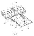
- Figure 1 shows a medical device in the form of an endoscope 10 placed on a tray 20 and then in a bag 30.
- the mouth 40 of the bag is placed in a combined vacuum pump and sealing unit 50 which removes air and any moisture from the interior 31 of the bag before hermetically sealing the bag with a triple seal (indicated at 41) whilst the vacuum is being applied.
- the material of the bag is substantially impermeable to gas, having an oxygen permeability of about 50 cm 3 /m 2 .d.bar or less, thereby ensuring that the vacuum in the bag is maintained.
- One suitable bag material is a polyamide/polyethylene laminate of the kind sold e.g. by Lava Vacuumverpackung under the name "EK-flex N90 embossed". This material has an oxygen permeability of 50 cm 3 /m 2 .d.bar (per DIN 53380). It also has a water vapour transmission rate of ⁇ 3.0 g/m 2 .d (per DIN 53122).
- the material of the bag typically lies in the range 80 ⁇ m to 120 ⁇ m, the particular material mentioned above having a total gauge of 90 ⁇ m (per DIN53370) and a weight per area of 88 g/m 2 (per DIN ISO 536).
- the interior of the bag is initially clean by virtue of the bag manufacturing process (in particular the temperatures involved), the interior of the bag need not be sterile before use.
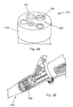
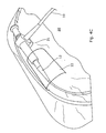
- Figure 2A shows the highly flexible distal tip 100 of the insertion tube having an external surface 104 as well as channels or lumens for supply of air or water (101), for the purposes of biopsy or the application of suction (102) and for a water jet (103).
- Figure 2B shows the internal workings 110 at the other end of the insertion tube, in particular one or more chambers 120 which are sealed to prevent leakage into or out of the endoscope etc. during operation.
- Flexible endoscopes are subjected to a leak test between patient uses and the maximum pressure used for this purpose is less than 1 bar, higher pressures could easily damage the highly flexible distal tip of the insertion tube for instance.
- the vacuum applied by the unit to the bag at the time the bag is hermetically sealed is about 250mbar below the ambient pressure of 1 bar (10 5 Pa), i.e. about 750mbar absolute.
- the level of vacuum is determined by an adjustable valve 61 which, as illustrated in figure 1B , is located on the underside 62 of a removable cover 60 on the outside of the vacuum pump and sealing unit 50 so as to be readily accessible for maintenance and quality control purposes.
- Such a vacuum level avoids a pressure difference between internal chambers 120 and the inside of the bag 30 exceeding I bar.
- This level of vacuum in the bag also results in the sealed bag containing oxygen at a level sufficiently high to suppress the growth of obligate anerobic bacteria but sufficiently low to deteriorate aerobic bacteria.
- typical tray dimensions are around 450mm x 350mm x 25mm, while typical bag dimensions are around 600mm length and 400mm to 550 mm width.
- Endoscopes typically range in length from around 3m to around 1.5m, the former corresponding to a gastrointestinal (GI) device having an insertion tube of up to 15mm diameter and around 1.7m length connected to a handle of around 0.2m length and attached thereto an umbilical or light source of around 1m length.
- the lower end of the range corresponds to an ear, nose and throat (ENT) device having an insertion tube of around 2mm diameter and 0.3 m length, a handle of 0.17m length and an umbilical of around 1m length.
- GI gastrointestinal
- ENT ear, nose and throat
- Such a level of vacuum need not kill microorganisms within the system, just reduce their rate of proliferation, thereby maintaining the endoscope below the prescribed level of contamination (e.g. 10CFU/ml) for longer.
- the process can be used with "wet" endoscopes that have undergone automated washing and high level disinfection but that have not been dried. It can also be used with endoscopes that have been processed through a validated AER and dried within an Endoscope Drying Cabinet, i.e. "dry" endoscopes.
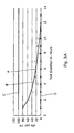
- Figure 3A illustrates the reduction in microbial populations (measured in colony forming units / millilitre) for surrogate/mock "dry" endoscopes containing small amounts of microbes packed in the manner set out above and evaluated at regular intervals over a 30 day period.
- Pieces of 2mm ID PTFE were cut into 1.5m and 2m lengths (to create Surrogate Device test pieces) and sealed in plastic packs and irradiated for sterility.
- a culture was prepared with a known concentration of C.difficile at approximately 10 3 suspended in Nutrient Broth. 0.03% Bovine Serum albumin was added to the culture.
- a Surrogate Device from a sterilized pack was aseptically removed and inoculated with the aforementioned suspension.
- the surrogate devices were then placed for 2 hours in a 22 incubator on a revolving platform to dry the suspension. Microorganisms were tested separately. Surrogate Devices were packed in a controlled ambient environment at 20°C ⁇ 2°C, in a "dirty" laboratory with 8 air room changes per hour. A positive control for each organism was tested in a Surrogate Device for both 1.5m and 2m lengths to establish the amount of bacteria contained in each device after the incubation drying period. The inoculated test tubes [Surrogate Device] were then packed in the manner set out above together with one un-inoculated tube (negative control) for each set of shelf life tests.
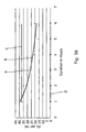
- Figures 3B , C and D show corresponding test results for "wet" scopes packed using the method of the invention.
- dry tests pieces of 2mm ID PTFE were cut into 1.5m and 2m lengths, sealed in a pack and irradiated to ensure sterility. A piece of tube was then removed from the sterilized pack and aseptically inoculated with a lenticule disc containing approximately 50 cfu of the organism to be used, viz. P.aeruginosa, C.difficile or S.aureus 0.3% Bovine Serum was added to the culture.
- Samples were then packed according to the present invention and in a controlled ambient environment at 20°C ⁇ 2°C; however, before packing, bags were inoculated with external rinse water.
- Inoculated test tubes with each organism type and an one un-inoculated tube(negative control) for each set of shelf life tests were also packed.
- a positive control set of each organism was also inoculated for each test day, placed in an unsealed bag and left in ambient air conditions.
- a positive control for each organismon a piece of the test tube [Surrogate Device] was tested for both 1.5m and 2m lengths to establish the amount of bacteria contained in each piece of test tube. All packs were then pressure tested for seal integrity using a pressure/vacuum testing system.
- test tube pieces [Surrogate Devices]
- the positive unpacked tubes and the negative packed tubes were then examined after designated storage period for the presence of viable organisms.
- Figures 3B-D illustrate that the microbial populations do not exceed that of packing and gradually reduce over the storage period (measured in cfu/ml) for mock endoscopes containing a low level of culture (representative of the maximum allowable AER TVC) of microorganisms packed in the manner set out above and evaluated at regular intervals over a 6 hour period.
- the test results show that after the designated storage period any residual bacteria (both aerobic and anaerobic microorganisms) steadily decreased and that the mock endoscopes retained their packed integrity for the duration of the test period.
- the above packaging method is one step of a total system covering an endoscope from one patient to the next. After removal from a patient, the dirty endoscope is put in a bag and sealed. This helps remove any cross infection risks for both staff and patients when transporting dirty endoscopes from one area to another or from one hospital to another. Bags and trays used for dirty endoscopes may be provided with features, e.g. a red colouring, to distinguish them from bags and trays for use with disinfected devices. Such a red bag gives a clear message to a trained decontamination specialist that the endoscope is not clean and requires reprocessing. Following reprocessing in an AER and possibly a DSC, the clean endoscope is then packaged as set out above.
- a red colouring to distinguish them from bags and trays for use with disinfected devices.
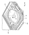
- Figure 4A shows the device 10 after packaging in the bag 30, the tray 20 helping to protect the scope from any abnormal contours under the effect of the vacuum.
- the bag may be made of heavy gauge rather than thinner, standard gauge material.
- the supporting surface 23 of the tray 20 may be provided with one or more recesses - indicated at 21 in the plan view of figure 4B - to accommodate protuberant parts 11 of the endoscope 10 and allow the remainder of device to lie substantially flat on the supporting surface of the tray.
- the tray 20 may also be provided with one or more bridge pieces 22 to extend over any particularly delicate regions 12 of the device and thereby protect those regions from the force exerted by the material of the bag when evacuated as described above.
- bridge piece 22 is semicircular in section, having a diameter in the range from about 40mm to about 70mm and a typical length of around 100mm. These bridge pieces may be separate or be formed integrally with a tray.
- a cap may also be provided that permits application of vacuum to the lumens but that prevents application of vacuum to the endoscope sheath.
- the tray 20 and bridges 22 are made of material, such as polycarbonate, that is able to withstand standard sterilisation procedures, e.g. autoclaving at 134°C for 2 minutes.
- the device may be placed in a further, sterile bag before this is placed in bag 30, thereby ensuring that the scope is not compromised by contact with the tray.
- this inner bag is not sealed in order that the vacuum might be applied to both the inner bag and the bag 30.
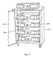
- the package is placed in a rigid case 200 having a lid 200', thereby providing further protection of the device and its sealed bag. It will be seen that the use of a tray ensures the package shape fits perfectly into the transportation case every time. As shown in figure 5 , yet further protection may be provided by placing the rigid case 200 in an enclosure 210 having a door 210' configured to accommodate a plurality of such cases. Such endoscopes are then ready for use on a patient.
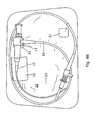
- an independent system 301 of the kind illustrated in figure 6 monitors the vacuum in the bag and the seal heater at the time the bag is hermetically sealed and compares the monitored vacuum with a predetermined critical process value of vacuum.
- the controller 300 of such an independent monitoring system is independent of the packaging device's microprocessor controller and has its own vacuum and seal heater sensors 310 as well as a reader 320 for identifying the packaged endoscope, e.g. by means of a barcode containing the endoscope serial number. The reader may also identify e.g. the operator and the validity period.
- a signal is generated by the controller that will either raise an alarm or cause one or more validation labels to be printed by printer 340.
- These labels can be attached to the bag as indicated at 220 in figure 4 and/or to a seal 230 on the case as shown in figure 5 .
- Controller 300 can also pass data to a data storage device 350 for tracking purposes.
- the present invention provides a quick and easy method of packaging medical devices such as endoscopes ready for transportation and storage. This in turn reduces the use of expensive disinfectants used to re-disinfect endoscopes, as well as reducing the number of drying cabinets required. It also reduces the number of times an endoscope is reprocessed in an AER which subjects the endoscope to harsh chemicals and over time takes its toll on the condition of the device.
Landscapes
- Health & Medical Sciences (AREA)
- Life Sciences & Earth Sciences (AREA)
- Surgery (AREA)
- Engineering & Computer Science (AREA)
- Public Health (AREA)
- Nuclear Medicine, Radiotherapy & Molecular Imaging (AREA)
- Biomedical Technology (AREA)
- Heart & Thoracic Surgery (AREA)
- Medical Informatics (AREA)
- Molecular Biology (AREA)
- Animal Behavior & Ethology (AREA)
- General Health & Medical Sciences (AREA)
- Mechanical Engineering (AREA)
- Veterinary Medicine (AREA)
- Biophysics (AREA)
- Physics & Mathematics (AREA)
- Optics & Photonics (AREA)
- Pathology (AREA)
- Radiology & Medical Imaging (AREA)
- Chemical & Material Sciences (AREA)
- Dispersion Chemistry (AREA)
- Apparatus For Disinfection Or Sterilisation (AREA)
- Vacuum Packaging (AREA)
- Endoscopes (AREA)
- Packages (AREA)
Description
- The present invention relates to methods of packaging medical devices, in particular but not exclusively to methods of packaging endoscopes, particularly flexible endoscopes.
- Flexible endoscopes are expensive, complex, reusable instruments that require unique consideration with respect to decontamination, storage and transportation between each patient procedure. In addition to the external surface of an endoscope, the internal 'channels' for air, water, aspiration, accessories etc are exposed to body fluids and other contaminants. Endoscopes are routinely exposed to mucus and other gastrointestinal secretions, blood, saliva, faeces, bile, and sometimes pus all of which can aid to the cross infection of microorganisms from one patient to the next.
- In contrast to rigid endoscopes and most reusable accessories, flexible endoscopes are thermolabile (heat sensitive) and cannot be sterilized which is the standard procedure for most instruments, known as the 'terminal process'. Sterilisation is defined as the complete destruction of all micro-organisms including bacterial spores. Sterilisation is required for devices that are normally used in sterile areas of the body (e.g. laparoscopes, microsurgical instruments). Flexible endoscopes (which make contact with mucous membranes but do not ordinarily penetrate normally sterile areas of the body) are generally reprocessed by high level disinfection rather than sterilisation in order to kill bacteria, viruses, mycobacteria and some spores - see e.g. UK Medicines and Healthcare products Regulatory Agency Medical Device Alert 2004/028. Most flexible gastrointestinal endoscopes would not withstand the conditions normally used in a steam sterilisation process.
- Medical apparatus such as endoscopes are typically exposed to two different types of bacteria. Aerobic bacteria grow in the presence of O2, and are the most common causes of clinical infection. Aerobic bacteria such as Aspergillus (see DSC), Staphylococcus and Pseudomonas Aeruginosa cause blood infection and are harmful to human beings especially persons believed to have weak immune systems. An aerobic bacterium is an organism that comprises a metabolism based on oxygen. It is a type of bacteria that requires oxygen for its growth and survival. Aerobic bacteria use oxygen for oxidizing the substrates such as fats or sugars for obtaining energy.
- Anaerobic bacteria are a type of bacteria that grow in places which are starved of oxygen. Such bacteria infect deep lacerations, deep tissues, and internal organs. Infections are marked by bad-smelling pus, the formation of abscesses, and the destruction of tissue. The bacteria are most often located in the mouth, gastrointestinal tract, vagina, and on the skin. Examples are Staphylococcus aureus and C. diff (Clostridium difficile). Certain anaerobic bacteria, known as obligate anaerobes, die in the presence of oxygen. Facultative anaerobes, on the other hand, can adapt to both aerobic and anaerobic habitats. This versatility is what gives the facultative anaerobe E. coli (Escherichia coli) its ability to adapt to its intestinal (anaerobic) and its extra-intestinal (aerobic or anaerobic) habitats.
- The United Kingdom Department of Health has issued a number of HSC's (Health Service Circulars), guidelines and legislations to be adhered to for the treatment of flexible endoscopes between patient uses, much of which is documented in the BSG (British Society of Gastroenterology) Endoscopy decontamination guidelines. Between each patient use the following steps should be taken:
- a. Decontamination should begin as soon as the endoscope has been removed from the patient.
- b. Before the endoscope is detached from the light source/videoprocessor a preliminary cleaning routine should be undertaken.
- a. The endoscope is then detached from the light source/videoprocessor, removed to the reprocessing room and attached to a leakage tester system (not exceeding 1 bar pressure as per the endoscope manufacturers recommendations) to check the integrity of all channels or outer casing for bite damage or any other damage before reprocessing.
- b. The next stage is manual cleaning and rinsing of all exposed internal and external surfaces. A low-foaming detergent that has been specifically designated for medical instrument cleaning should be used.
- c. The next stage is automated washing and high level disinfection with a liquid chemical germicide within an AER (Automated Endoscope Reprocessor) followed by rinsing with sterile grade water.
- The scope may now be used again, or hung up ready for use within 3 hours or transported to another department and hung ready for use within 3 hours, alternatively it may be placed in a DSC (Drying and Storage Cabinet) for 3 to 7 days (which becomes the terminal process) ready for immediate use. Many units are now using purpose built DSC's, which have been shown to prevent colonisation of endoscope channels over time periods ranging from 72hours to 7 days. These DSCs are designed to deliver high efficiency particulate filtered air to the internal channels of the endoscope at the appropriate temperature and flow rate. According to the manufacturers, their use avoids the need for endoscopes to undergo early morning repeat decontamination cycles.
- Specifically, the test method used on the DSC states that following storage the acceptable contamination of the internal channels of endoscopes shall be less than 10cfu (Colony Forming Units) and no pathogenic microorganisms, aspergillus (aerobic) or any other filamentous fungi shall be found. Within an
AER 10 cfu/ml is the maximum allowable level within the European Standard BS EN ISO 15883 for final rinse water during the terminal process.. - The use of such DSCs positioned around a hospital within different departments has allowed endoscope decontamination to become centralised within hospitals, which is deemed to be best practice. The down side to this new practice is that safe and aseptic transportation of endoscopes from one department to another is not readily available or very expensive and slow to prepare and added to this, the purchase of many DSC's is prohibitive. Moreover, guidelines require that when transporting endoscopes to and from areas outside the endoscopy unit, they must be transferred in a covered rigid receptacle, not only to avoid damage to the endoscope, but also to protect the cleanliness of the scope and to protect staff and the public when returning potentially contaminated scopes.
-
US6161695 discloses a method for protectively packaging sterilizable materials. A product to be packaged is placed within a semi-rigid, foldable packaging tray to form a package insert. The package insert is then placed within a heat-sealable container, such as a polymeric film envelope. Vacuum force is applied to evacuate the vacuum sealable container and to seal the packaging insert within the vacuum sealable container. The vacuum sealed container may then be placed within an outer, flexible packaging container and heat sealed therein. The entire package is generally sterilized after heat sealing within the outer, flexible package. - According to the present invention, there is provided a method of packaging a medical device , comprising the steps of:
- placing the medical device in a bag;
- removing air from the bag by applying a vacuum of less than 1 bar; and
- hermetically sealing the bag while the vacuum is being applied such that the concentration of oxygen in the hermetically sealed bag is 16% +/- 0.5% by volume.
- Such a level of oxygen stops the growth of both obligate anaerobic bacteria and aerobic bacteria on the surface and within the inner channels of flexible endoscopes during transportation and/or storage, prolonging the aseptic storage and safe transportation of medical devices. The method is particularly appropriate for packaging non-sterile medical devices, especially endoscopes and in particular flexible endoscopes which, by their nature, cannot be sterilized but only subjected to high level disinfection.
- The method may be for packaging a medical device having a sealed chamber, in which case the method may comprise the step of applying a vacuum to the interior of the bag such that the pressure difference between the sealed chamber and the interior of the bag does not exceed 1 bar.
- Medical devices having chambers sealed from external conditions include flexible endoscopes where the internal workings, which may include a camera and a light, are sealed from the external environment of mucus, etc., thereby defining a sealed chamber. Subjecting the chamber seal to a pressure difference in excess of 1 bar can cause it to fail, potentially allowing the external environment to contaminate the internal workings or vice versa.
- The vacuum in the bag at the time the bag is hermetically sealed is preferably about 250mbar below the ambient pressure (typically 1 bar (105 Pa)), i.e. about 750mbar absolute. This is to be contrasted with vacuum packaging in the food industry where higher levels of vacuum in the range 700mbar to 1 bar below ambient are applied and obligate anaerobic bacteria are less prevalent.
- The step of hermetically sealing the bag may comprise the creation of at least two seals in series, in particular three seals in series ("triple sealed").
- The method may comprise the further step of subjecting the medical device to high level disinfection prior to placing the device in a bag. Prior to such high level disinfection, the method may comprise the step of placing a contaminated ("dirty") endoscope in a bag and sealing the bag, thereby removing any cross infection risks for both staff and patients when transporting dirty endoscopes from one area to another or from one hospital to another.
- The method may comprise the further step of ensuring that the exposed surfaces of the medical device are dry before placing the medical device in a bag. This enables prolonged aseptic storage of the device. Drying of the device may be achieved by placing the medical device in a DSC prior to placing the medical device in the bag.
- The method may comprise the further step of placing the medical device on a tray before placing the device and tray into the bag. The use of the tray helps to protect the scope from any abnormal contours under the effect of the vacuum. The method may comprise the step of providing a tray having a supporting surface with a recess formed therein and placing the device such that one part of the device is supported by the surface and another part of the device lies in the recess. The method may comprise the step of providing a bridge member on the tray to cover part of the device.
- The method may comprise the further step of placing the medical device in a further, sterile bag before placing it in a bag to which the vacuum is applied. The use of a sterile bag inside the vacuum bag ensures the scope is not compromised.
- The method may comprise the further step of enclosing the hermetically sealed, device-containing bag in a rigid case, thereby providing further protection of the device and its sealed bag. The additional use of a tray ensures the package shape fits perfectly into the transportation case every time.
- Yet further protection may be provided by placing the rigid case in an enclosure configured to accommodate a plurality of such cases.
- The method may comprise the further step of monitoring the vacuum in the bag at the time the bag is hermetically sealed, comparing the monitored vacuum with a predetermined value and generating a signal depending on the result of said comparison.
- The monitoring of the vacuum may be carried out independently of the air removal step, which may require its own monitoring. Such an independent monitoring system (IMS) may be independent of the packaging device's microprocessor controller and may be fitted with its own set of probes used to monitor all cycle/process parameters that are critical to the process. The resulting cycle validation data/signal may be passed to a PC or mass storage device and information can then be saved to the patient's file, as often required by law.
- Alternatively, the signal may cause a printer to generate one or more corresponding labels which may then be attached to the bag and/or the case.
- An embodiment of the invention will now be described by way of example with reference to the accompanying drawings, in which:
-
Figure 1A illustrates a medical device when subject to the packaging process; -
Figure 1B is a detailed view of the underside of thecover 60 infigure 1A ; -
Figures 2A and 2B are detail views of the tip and opposite ends of the insertion tube of a typical endoscope; -
Figure 3A is a graph showing the reduction in microbial populations for a "dry" mock endoscope containing small amounts of C.difficile microbes and packaged in accordance with the invention; -
Figure 3B is a graph showing the reduction in microbial populations for a "wet" mock endoscope containing small amounts of P.aeruginosa microbes and packaged in accordance with the invention; -
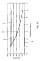
Figure 3C is a graph showing the reduction in microbial populations for a "wet" mock endoscope containing small amounts of C.difficile microbes and packaged in accordance with the invention; -
Figure 3D is a graph showing the reduction in microbial populations for a "wet" mock endoscope containing small amounts of S.aureus microbes and packaged in accordance with the invention; -
Figure 4A shows an endoscope packaged according to the invention and placed in a rigid case; -
Figure 4B is a plan view of an endoscope packaged in accordance with the invention; -
Figure 4C is a perspective view of a detail offigure 4B ; -
Figure 5 shows several rigid cases in an enclosure. -
Figure 6 is a schematic of a monitoring system. -
Figure 1 shows a medical device in the form of anendoscope 10 placed on atray 20 and then in abag 30. Themouth 40 of the bag is placed in a combined vacuum pump and sealingunit 50 which removes air and any moisture from theinterior 31 of the bag before hermetically sealing the bag with a triple seal (indicated at 41) whilst the vacuum is being applied. The material of the bag is substantially impermeable to gas, having an oxygen permeability of about 50 cm3/m2.d.bar or less, thereby ensuring that the vacuum in the bag is maintained. One suitable bag material is a polyamide/polyethylene laminate of the kind sold e.g. by Lava Vacuumverpackung under the name "EK-flex N90 embossed". This material has an oxygen permeability of 50 cm3/m2.d.bar (per DIN 53380). It also has a water vapour transmission rate of <3.0 g/m2.d (per DIN 53122). - The material of the bag typically lies in the range 80µm to 120µm, the particular material mentioned above having a total gauge of 90µm (per DIN53370) and a weight per area of 88 g/m2 (per DIN ISO 536). Although the interior of the bag is initially clean by virtue of the bag manufacturing process (in particular the temperatures involved), the interior of the bag need not be sterile before use.
-
Figure 2A shows the highly flexibledistal tip 100 of the insertion tube having anexternal surface 104 as well as channels or lumens for supply of air or water (101), for the purposes of biopsy or the application of suction (102) and for a water jet (103).Figure 2B shows theinternal workings 110 at the other end of the insertion tube, in particular one ormore chambers 120 which are sealed to prevent leakage into or out of the endoscope etc. during operation. Flexible endoscopes are subjected to a leak test between patient uses and the maximum pressure used for this purpose is less than 1 bar, higher pressures could easily damage the highly flexible distal tip of the insertion tube for instance. - The vacuum applied by the unit to the bag at the time the bag is hermetically sealed is about 250mbar below the ambient pressure of 1 bar (105 Pa), i.e. about 750mbar absolute. The level of vacuum is determined by an
adjustable valve 61 which, as illustrated infigure 1B , is located on theunderside 62 of aremovable cover 60 on the outside of the vacuum pump and sealingunit 50 so as to be readily accessible for maintenance and quality control purposes. Such a vacuum level avoids a pressure difference betweeninternal chambers 120 and the inside of thebag 30 exceeding I bar. This level of vacuum in the bag also results in the sealed bag containing oxygen at a level sufficiently high to suppress the growth of obligate anerobic bacteria but sufficiently low to deteriorate aerobic bacteria. Using the same sized tray in the same sized bag at the same level of vacuum ensures reproducibility: typical tray dimensions are around 450mm x 350mm x 25mm, while typical bag dimensions are around 600mm length and 400mm to 550 mm width. Endoscopes typically range in length from around 3m to around 1.5m, the former corresponding to a gastrointestinal (GI) device having an insertion tube of up to 15mm diameter and around 1.7m length connected to a handle of around 0.2m length and attached thereto an umbilical or light source of around 1m length. The lower end of the range corresponds to an ear, nose and throat (ENT) device having an insertion tube of around 2mm diameter and 0.3 m length, a handle of 0.17m length and an umbilical of around 1m length. - Such a level of vacuum need not kill microorganisms within the system, just reduce their rate of proliferation, thereby maintaining the endoscope below the prescribed level of contamination (e.g. 10CFU/ml) for longer.
- The process can be used with "wet" endoscopes that have undergone automated washing and high level disinfection but that have not been dried. It can also be used with endoscopes that have been processed through a validated AER and dried within an Endoscope Drying Cabinet, i.e. "dry" endoscopes.
-
Figure 3A illustrates the reduction in microbial populations (measured in colony forming units / millilitre) for surrogate/mock "dry" endoscopes containing small amounts of microbes packed in the manner set out above and evaluated at regular intervals over a 30 day period. Pieces of 2mm ID PTFE were cut into 1.5m and 2m lengths (to create Surrogate Device test pieces) and sealed in plastic packs and irradiated for sterility. A culture was prepared with a known concentration of C.difficile at approximately 103 suspended in Nutrient Broth. 0.03% Bovine Serum albumin was added to the culture. A Surrogate Device from a sterilized pack was aseptically removed and inoculated with the aforementioned suspension. The surrogate devices were then placed for 2 hours in a 22 incubator on a revolving platform to dry the suspension. Microorganisms were tested separately. Surrogate Devices were packed in a controlled ambient environment at 20°C ± 2°C, in a "dirty" laboratory with 8 air room changes per hour. A positive control for each organism was tested in a Surrogate Device for both 1.5m and 2m lengths to establish the amount of bacteria contained in each device after the incubation drying period. The inoculated test tubes [Surrogate Device] were then packed in the manner set out above together with one un-inoculated tube (negative control) for each set of shelf life tests. - To serve as a comparison between devices packed according to the invention and uncontrolled devices, a positive control set for each test day was inoculated. Devices were placed in an unsealed bag and left in ambient air conditions [20°C ± 2°C, in a "dirty" laboratory with 8 air room changes per hour].
- Those bags prepared in accordance with the invention were pressure tested for seal integrity using a pressure/vacuum testing system. After the set time intervals listed above, the seal integrity was tested before opening again using a pressure/ vacuum testing system. The test Surrogate Devices, the inoculated unpacked surrogate devices and the packed negative control tubes were then examined after designated storage period for the presence of viable organisms.
- Referring to
figure 3 , data points are indicated by crosses A, with the logarithmic trend being indicated by line B. The level of the start culture is indicated by line C, while the level of the clean scope (negative control) is indicated by line D. The results show that the microbial populations do not exceed that of packing and gradually reduce to "none detected" over the storage period, that after the designated storage period any residual bacteria (both aerobic and anaerobic microorganisms) decrease and that the mock endoscopes retained their packed integrity for the duration of the test period. -
Figures 3B ,C andD show corresponding test results for "wet" scopes packed using the method of the invention. As for the dry tests, pieces of 2mm ID PTFE were cut into 1.5m and 2m lengths, sealed in a pack and irradiated to ensure sterility. A piece of tube was then removed from the sterilized pack and aseptically inoculated with a lenticule disc containing approximately 50 cfu of the organism to be used, viz. P.aeruginosa, C.difficile or S.aureus 0.3% Bovine Serum was added to the culture. - Samples were then packed according to the present invention and in a controlled ambient environment at 20°C ± 2°C; however, before packing, bags were inoculated with external rinse water. Inoculated test tubes with each organism type and an one un-inoculated tube(negative control) for each set of shelf life tests were also packed. A positive control set of each organism was also inoculated for each test day, placed in an unsealed bag and left in ambient air conditions. A positive control for each organismon a piece of the test tube [Surrogate Device] was tested for both 1.5m and 2m lengths to establish the amount of bacteria contained in each piece of test tube. All packs were then pressure tested for seal integrity using a pressure/vacuum testing system.
- After the set time intervals, and before opening, the seal integrity was again tested using a pressure/vacuum testing system. The test tube pieces [Surrogate Devices], the positive unpacked tubes and the negative packed tubes were then examined after designated storage period for the presence of viable organisms.
-
Figures 3B-D illustrate that the microbial populations do not exceed that of packing and gradually reduce over the storage period (measured in cfu/ml) for mock endoscopes containing a low level of culture (representative of the maximum allowable AER TVC) of microorganisms packed in the manner set out above and evaluated at regular intervals over a 6 hour period. The test results show that after the designated storage period any residual bacteria (both aerobic and anaerobic microorganisms) steadily decreased and that the mock endoscopes retained their packed integrity for the duration of the test period. - The above packaging method is one step of a total system covering an endoscope from one patient to the next. After removal from a patient, the dirty endoscope is put in a bag and sealed. This helps remove any cross infection risks for both staff and patients when transporting dirty endoscopes from one area to another or from one hospital to another. Bags and trays used for dirty endoscopes may be provided with features, e.g. a red colouring, to distinguish them from bags and trays for use with disinfected devices. Such a red bag gives a clear message to a trained decontamination specialist that the endoscope is not clean and requires reprocessing. Following reprocessing in an AER and possibly a DSC, the clean endoscope is then packaged as set out above.
-
Figure 4A shows thedevice 10 after packaging in thebag 30, thetray 20 helping to protect the scope from any abnormal contours under the effect of the vacuum. To prevent the bag from tearing against the various protrusions of an endoscope when a vacuum is applied, the bag may be made of heavy gauge rather than thinner, standard gauge material. To avoid undue concentration of force on delicate external surfaces of medical devices such as endoscopes, the supportingsurface 23 of thetray 20 may be provided with one or more recesses - indicated at 21 in the plan view offigure 4B - to accommodateprotuberant parts 11 of theendoscope 10 and allow the remainder of device to lie substantially flat on the supporting surface of the tray. As also illustrated in the perspective view offigure 4C , thetray 20 may also be provided with one ormore bridge pieces 22 to extend over any particularlydelicate regions 12 of the device and thereby protect those regions from the force exerted by the material of the bag when evacuated as described above. In the example shown,bridge piece 22 is semicircular in section, having a diameter in the range from about 40mm to about 70mm and a typical length of around 100mm. These bridge pieces may be separate or be formed integrally with a tray. Where the device is an endoscope, a cap may also be provided that permits application of vacuum to the lumens but that prevents application of vacuum to the endoscope sheath. Thetray 20 and bridges 22 are made of material, such as polycarbonate, that is able to withstand standard sterilisation procedures, e.g. autoclaving at 134°C for 2 minutes. - In another embodiment, not illustrated, the device may be placed in a further, sterile bag before this is placed in
bag 30, thereby ensuring that the scope is not compromised by contact with the tray. However, this inner bag is not sealed in order that the vacuum might be applied to both the inner bag and thebag 30. - The package is placed in a
rigid case 200 having alid 200', thereby providing further protection of the device and its sealed bag. It will be seen that the use of a tray ensures the package shape fits perfectly into the transportation case every time. As shown infigure 5 , yet further protection may be provided by placing therigid case 200 in anenclosure 210 having adoor 210' configured to accommodate a plurality of such cases. Such endoscopes are then ready for use on a patient. - Regulations such as HTM2030 and ISO 15883 may require that any process that produces a final product before use on a patient - a so-called "terminal process" - be fitted with an Independent Monitoring System (IMS) for cycle/process validation and verification. This is a requirement for any AER, DSC or steam sterilizer. Such an IMS system must be independent of the device's (typically microprocessor) controller and to this end is fitted with its own set of probes used to monitor all cycle/process parameters that are critical to the process. Cycle validation data is passed to a PC or mass storage device and information can then be saved to the patient's file, as often required by law.
- To this end, an
independent system 301 of the kind illustrated infigure 6 monitors the vacuum in the bag and the seal heater at the time the bag is hermetically sealed and compares the monitored vacuum with a predetermined critical process value of vacuum. Thecontroller 300 of such an independent monitoring system (IMS) is independent of the packaging device's microprocessor controller and has its own vacuum andseal heater sensors 310 as well as areader 320 for identifying the packaged endoscope, e.g. by means of a barcode containing the endoscope serial number. The reader may also identify e.g. the operator and the validity period. Depending on the result of said comparison, a signal is generated by the controller that will either raise an alarm or cause one or more validation labels to be printed byprinter 340. These labels can be attached to the bag as indicated at 220 infigure 4 and/or to aseal 230 on the case as shown infigure 5 .Controller 300 can also pass data to adata storage device 350 for tracking purposes. - The present invention provides a quick and easy method of packaging medical devices such as endoscopes ready for transportation and storage.. This in turn reduces the use of expensive disinfectants used to re-disinfect endoscopes, as well as reducing the number of drying cabinets required. It also reduces the number of times an endoscope is reprocessed in an AER which subjects the endoscope to harsh chemicals and over time takes its toll on the condition of the device.
- It should be understood that this invention has been described by way of examples only and that a wide variety of modifications can be made without departing from the scope of the invention.
Claims (15)
- Method of packaging a medical device (10), comprising the steps of:placing the medical device in a bag (30); andremoving air from the bag by applying a vacuum of less than 1 bar;
characterized by hermetically sealing the bag (30) while the vacuum is being applied such that the
concentration of oxygen in the hermetically sealed bag is 16% +/- 0.5% by volume. - Method of packaging a medical device according to claim 1, the medical device (10) having a sealed chamber (120), comprising the step of applying a vacuum to the interior of the bag (30) such that the pressure difference between the sealed chamber and the interior of the bag does not exceed 1 bar.
- Method according to any preceding claim and comprising the step of hermetically sealing the bag (30) whilst the vacuum is being applied and such that the vacuum in the bag is about 250mbar below the ambient pressure.
- Method according to any preceding claim and wherein the step of hermetically sealing the bag (30) comprises the creation of at least two seals (41) in series.
- Method according to any preceding claim and comprising the step of subjecting the medical device (10) to high level disinfection prior to placing the device in a bag (30).
- Method according to claim 5 and comprising the step of ensuring that the exposed surfaces of the medical device (10) are dry before placing the medical device in the bag (30).
- Method according to any preceding claim and comprising the step of placing the medical device (10) on a tray (20) before placing the device and tray into the bag (30).
- Method according to claim 7 and comprising the step of providing a tray (20) having a supporting surface (23) with a recess (21) formed therein and placing the medical device (10) such that one part of the medical device is supported by the surface and another part of the device lies in the recess.
- Method according to any preceding claim and comprising the step of monitoring the vacuum in the bag (30) at the time the bag is hermetically sealed, comparing the monitored vacuum with a predetermined value and generating a signal depending on the result of said comparison.
- Method according to claim 9, wherein the process of monitoring of the vacuum is independent of the process of removing air from the bag (30).
- Method according to claim 10, wherein the process of removing air from the bag (30) is controlled by a first controller (50,61) and the process of monitoring the vacuum is controlled by a second controller (300), independent of the first controller.
- Method according to claim 11, wherein the first and second controllers (50,61; 300) are connected to respective independent sensors (61; 310).
- Method according to any one of claims 9 to 12 and comprising the step of passing data from the second controller (300) to a data storage device (350).
- Method according to any one of claims 9 to 13 and comprising the step of printing one or more labels corresponding to the signal.
- Method according to any preceding claim wherein the medical device (10) is an endoscope.
Priority Applications (1)
| Application Number | Priority Date | Filing Date | Title |
|---|---|---|---|
| PL11730052T PL2575666T3 (en) | 2010-06-02 | 2011-05-31 | Methods of packing medical devices |
Applications Claiming Priority (2)
| Application Number | Priority Date | Filing Date | Title |
|---|---|---|---|
| GBGB1009230.2A GB201009230D0 (en) | 2010-06-02 | 2010-06-02 | Methods of packing medical devices |
| PCT/GB2011/051024 WO2011151641A2 (en) | 2010-06-02 | 2011-05-31 | Methods of packing medical devices |
Publications (2)
| Publication Number | Publication Date |
|---|---|
| EP2575666A2 EP2575666A2 (en) | 2013-04-10 |
| EP2575666B1 true EP2575666B1 (en) | 2014-08-13 |
Family
ID=42471044
Family Applications (1)
| Application Number | Title | Priority Date | Filing Date |
|---|---|---|---|
| EP11730052.5A Active EP2575666B1 (en) | 2010-06-02 | 2011-05-31 | Methods of packing medical devices |
Country Status (6)
| Country | Link |
|---|---|
| EP (1) | EP2575666B1 (en) |
| DK (1) | DK2575666T3 (en) |
| ES (1) | ES2520042T3 (en) |
| GB (1) | GB201009230D0 (en) |
| PL (1) | PL2575666T3 (en) |
| WO (1) | WO2011151641A2 (en) |
Families Citing this family (7)
| Publication number | Priority date | Publication date | Assignee | Title |
|---|---|---|---|---|
| GB2497353A (en) * | 2011-12-09 | 2013-06-12 | Entpr Medical Ltd | Endoscope storage tray |
| US20170065394A1 (en) * | 2014-03-05 | 2017-03-09 | Medizinische Hochschule Hannover | Medical implant, medical device and method for making a medical implant |
| GB201407541D0 (en) | 2014-04-29 | 2014-06-11 | Meditech Endoscopy Ltd | Storage device |
| EP3941381A4 (en) * | 2019-03-19 | 2022-11-23 | Medivators Inc. | Vented endoscope tray covers, systems and methods |
| EP3942993A4 (en) * | 2019-03-19 | 2022-05-04 | FUJIFILM Corporation | Endoscope connector device |
| CN114126530A (en) | 2019-06-20 | 2022-03-01 | 美涤威公司 | Endoscope storage cart, system and method |
| US11974864B2 (en) | 2020-02-25 | 2024-05-07 | Medivators Inc. | Stackable endoscope storage tray and method of use |
Family Cites Families (3)
| Publication number | Priority date | Publication date | Assignee | Title |
|---|---|---|---|---|
| US5577368A (en) * | 1995-04-03 | 1996-11-26 | Johnson & Johnson Professional, Inc. | Method for improving wear resistance of polymeric bioimplantable components |
| US6161695A (en) * | 1998-08-21 | 2000-12-19 | Depuy Orthopaedics, Inc. | Protective packaging unit |
| CA2738972C (en) * | 2008-10-21 | 2016-06-28 | Medicart International Limited | Medical equipment storage and transportation kit |
-
2010
- 2010-06-02 GB GBGB1009230.2A patent/GB201009230D0/en not_active Ceased
-
2011
- 2011-05-31 PL PL11730052T patent/PL2575666T3/en unknown
- 2011-05-31 EP EP11730052.5A patent/EP2575666B1/en active Active
- 2011-05-31 DK DK11730052.5T patent/DK2575666T3/en active
- 2011-05-31 WO PCT/GB2011/051024 patent/WO2011151641A2/en active Application Filing
- 2011-05-31 ES ES11730052.5T patent/ES2520042T3/en active Active
Also Published As
| Publication number | Publication date |
|---|---|
| ES2520042T3 (en) | 2014-11-11 |
| EP2575666A2 (en) | 2013-04-10 |
| PL2575666T3 (en) | 2015-01-30 |
| WO2011151641A2 (en) | 2011-12-08 |
| WO2011151641A3 (en) | 2012-03-01 |
| GB201009230D0 (en) | 2010-07-21 |
| DK2575666T3 (en) | 2014-11-10 |
Similar Documents
| Publication | Publication Date | Title |
|---|---|---|
| EP2575666B1 (en) | Methods of packing medical devices | |
| US6193931B1 (en) | Container monitoring system | |
| Rutala et al. | Infection control: the role of disinfection and sterilization | |
| CA1337259C (en) | Disposable biological indicator test pack for monitoring steam and ethylene oxide sterilization cycles | |
| US6815206B2 (en) | Container monitoring system | |
| KR102435336B1 (en) | A sterilisation container, method of sterilisation and sterilisation apparatus | |
| US20010006610A1 (en) | Contained indicators for determining sterilizations | |
| US20220184258A1 (en) | Apparatus and method for sterilization of an article | |
| Heeg et al. | Decontaminated single-use devices: an oxymoron that may be placing patients at risk for cross-contamination | |
| EP3367910B1 (en) | Method of use of a probe cover | |
| US8486691B2 (en) | Apparatus for assessing the effectiveness of a sterilization process | |
| Penna et al. | The presterilization microbial load on used medical devices and the effectiveness of hydrogen peroxide gas plasma against Bacillus subtilis spores | |
| JP2019136005A (en) | Sterile sampling flow path kit and cell culture apparatus using the same | |
| EP3492113A1 (en) | Sterilization package | |
| Muscarella | Are all sterilization processes alike? | |
| RU2664949C2 (en) | Transportation of medical instruments | |
| Ackert-Burr | Low-temperature sterilization: are you in the know? | |
| Kiernan et al. | Fundamentals of Decontamination and Sterilisation | |
| Lund et al. | Adequacy of Sterilization Techniques for NOLA Dry Field Retractors | |
| Mukherjee et al. | 2 Sterilisation for Safe Minimal Access Surgery: Evidence and Recommendations | |
| JP4378828B2 (en) | Sterilization assurance method for ozone sterilization | |
| CN101189162A (en) | Item reprocessing and sterile packaging apparatus | |
| Ljungberg | Evaluation of an Ozone Cabinet for Disinfecting Medical Equipment | |
| Ahmed et al. | Quality Control and Surveillance | |
| Lowbury et al. | Sterilization and Physical Disinfection |
Legal Events
| Date | Code | Title | Description |
|---|---|---|---|
| PUAI | Public reference made under article 153(3) epc to a published international application that has entered the european phase |
Free format text: ORIGINAL CODE: 0009012 |
|
| 17P | Request for examination filed |
Effective date: 20130102 |
|
| AK | Designated contracting states |
Kind code of ref document: A2 Designated state(s): AL AT BE BG CH CY CZ DE DK EE ES FI FR GB GR HR HU IE IS IT LI LT LU LV MC MK MT NL NO PL PT RO RS SE SI SK SM TR |
|
| DAX | Request for extension of the european patent (deleted) | ||
| GRAP | Despatch of communication of intention to grant a patent |
Free format text: ORIGINAL CODE: EPIDOSNIGR1 |
|
| INTG | Intention to grant announced |
Effective date: 20140310 |
|
| GRAS | Grant fee paid |
Free format text: ORIGINAL CODE: EPIDOSNIGR3 |
|
| GRAA | (expected) grant |
Free format text: ORIGINAL CODE: 0009210 |
|
| RAP1 | Party data changed (applicant data changed or rights of an application transferred) |
Owner name: GETINGE UK LIMITED |
|
| AK | Designated contracting states |
Kind code of ref document: B1 Designated state(s): AL AT BE BG CH CY CZ DE DK EE ES FI FR GB GR HR HU IE IS IT LI LT LU LV MC MK MT NL NO PL PT RO RS SE SI SK SM TR |
|
| REG | Reference to a national code |
Ref country code: GB Ref legal event code: FG4D |
|
| REG | Reference to a national code |
Ref country code: CH Ref legal event code: EP Ref country code: AT Ref legal event code: REF Ref document number: 681678 Country of ref document: AT Kind code of ref document: T Effective date: 20140815 |
|
| REG | Reference to a national code |
Ref country code: IE Ref legal event code: FG4D |
|
| REG | Reference to a national code |
Ref country code: DE Ref legal event code: R096 Ref document number: 602011009106 Country of ref document: DE Effective date: 20141002 |
|
| REG | Reference to a national code |
Ref country code: DK Ref legal event code: T3 Effective date: 20141104 |
|
| REG | Reference to a national code |
Ref country code: ES Ref legal event code: FG2A Ref document number: 2520042 Country of ref document: ES Kind code of ref document: T3 Effective date: 20141111 |
|
| REG | Reference to a national code |
Ref country code: CH Ref legal event code: NV Representative=s name: MEYER AND KOLLEGEN, CH |
|
| REG | Reference to a national code |
Ref country code: SE Ref legal event code: TRGR |
|
| REG | Reference to a national code |
Ref country code: NL Ref legal event code: T3 |
|
| REG | Reference to a national code |
Ref country code: NO Ref legal event code: T2 Effective date: 20140813 |
|
| REG | Reference to a national code |
Ref country code: LT Ref legal event code: MG4D |
|
| PG25 | Lapsed in a contracting state [announced via postgrant information from national office to epo] |
Ref country code: BG Free format text: LAPSE BECAUSE OF FAILURE TO SUBMIT A TRANSLATION OF THE DESCRIPTION OR TO PAY THE FEE WITHIN THE PRESCRIBED TIME-LIMIT Effective date: 20141113 Ref country code: LT Free format text: LAPSE BECAUSE OF FAILURE TO SUBMIT A TRANSLATION OF THE DESCRIPTION OR TO PAY THE FEE WITHIN THE PRESCRIBED TIME-LIMIT Effective date: 20140813 Ref country code: PT Free format text: LAPSE BECAUSE OF FAILURE TO SUBMIT A TRANSLATION OF THE DESCRIPTION OR TO PAY THE FEE WITHIN THE PRESCRIBED TIME-LIMIT Effective date: 20141215 |
|
| REG | Reference to a national code |
Ref country code: PL Ref legal event code: T3 |
|
| REG | Reference to a national code |
Ref country code: GR Ref legal event code: EP Ref document number: 20140402258 Country of ref document: GR Effective date: 20141223 |
|
| PG25 | Lapsed in a contracting state [announced via postgrant information from national office to epo] |
Ref country code: HR Free format text: LAPSE BECAUSE OF FAILURE TO SUBMIT A TRANSLATION OF THE DESCRIPTION OR TO PAY THE FEE WITHIN THE PRESCRIBED TIME-LIMIT Effective date: 20140813 Ref country code: LV Free format text: LAPSE BECAUSE OF FAILURE TO SUBMIT A TRANSLATION OF THE DESCRIPTION OR TO PAY THE FEE WITHIN THE PRESCRIBED TIME-LIMIT Effective date: 20140813 Ref country code: IS Free format text: LAPSE BECAUSE OF FAILURE TO SUBMIT A TRANSLATION OF THE DESCRIPTION OR TO PAY THE FEE WITHIN THE PRESCRIBED TIME-LIMIT Effective date: 20141213 Ref country code: RS Free format text: LAPSE BECAUSE OF FAILURE TO SUBMIT A TRANSLATION OF THE DESCRIPTION OR TO PAY THE FEE WITHIN THE PRESCRIBED TIME-LIMIT Effective date: 20140813 Ref country code: CY Free format text: LAPSE BECAUSE OF FAILURE TO SUBMIT A TRANSLATION OF THE DESCRIPTION OR TO PAY THE FEE WITHIN THE PRESCRIBED TIME-LIMIT Effective date: 20140813 |
|
| PG25 | Lapsed in a contracting state [announced via postgrant information from national office to epo] |
Ref country code: SK Free format text: LAPSE BECAUSE OF FAILURE TO SUBMIT A TRANSLATION OF THE DESCRIPTION OR TO PAY THE FEE WITHIN THE PRESCRIBED TIME-LIMIT Effective date: 20140813 Ref country code: CZ Free format text: LAPSE BECAUSE OF FAILURE TO SUBMIT A TRANSLATION OF THE DESCRIPTION OR TO PAY THE FEE WITHIN THE PRESCRIBED TIME-LIMIT Effective date: 20140813 Ref country code: EE Free format text: LAPSE BECAUSE OF FAILURE TO SUBMIT A TRANSLATION OF THE DESCRIPTION OR TO PAY THE FEE WITHIN THE PRESCRIBED TIME-LIMIT Effective date: 20140813 Ref country code: RO Free format text: LAPSE BECAUSE OF FAILURE TO SUBMIT A TRANSLATION OF THE DESCRIPTION OR TO PAY THE FEE WITHIN THE PRESCRIBED TIME-LIMIT Effective date: 20140813 |
|
| REG | Reference to a national code |
Ref country code: DE Ref legal event code: R097 Ref document number: 602011009106 Country of ref document: DE |
|
| PLBE | No opposition filed within time limit |
Free format text: ORIGINAL CODE: 0009261 |
|
| STAA | Information on the status of an ep patent application or granted ep patent |
Free format text: STATUS: NO OPPOSITION FILED WITHIN TIME LIMIT |
|
| 26N | No opposition filed |
Effective date: 20150515 |
|
| PG25 | Lapsed in a contracting state [announced via postgrant information from national office to epo] |
Ref country code: SI Free format text: LAPSE BECAUSE OF FAILURE TO SUBMIT A TRANSLATION OF THE DESCRIPTION OR TO PAY THE FEE WITHIN THE PRESCRIBED TIME-LIMIT Effective date: 20140813 |
|
| PG25 | Lapsed in a contracting state [announced via postgrant information from national office to epo] |
Ref country code: MC Free format text: LAPSE BECAUSE OF FAILURE TO SUBMIT A TRANSLATION OF THE DESCRIPTION OR TO PAY THE FEE WITHIN THE PRESCRIBED TIME-LIMIT Effective date: 20140813 |
|
| REG | Reference to a national code |
Ref country code: IE Ref legal event code: MM4A |
|
| PG25 | Lapsed in a contracting state [announced via postgrant information from national office to epo] |
Ref country code: IE Free format text: LAPSE BECAUSE OF NON-PAYMENT OF DUE FEES Effective date: 20150531 |
|
| REG | Reference to a national code |
Ref country code: FR Ref legal event code: PLFP Year of fee payment: 6 |
|
| PG25 | Lapsed in a contracting state [announced via postgrant information from national office to epo] |
Ref country code: MT Free format text: LAPSE BECAUSE OF FAILURE TO SUBMIT A TRANSLATION OF THE DESCRIPTION OR TO PAY THE FEE WITHIN THE PRESCRIBED TIME-LIMIT Effective date: 20140813 |
|
| PG25 | Lapsed in a contracting state [announced via postgrant information from national office to epo] |
Ref country code: HU Free format text: LAPSE BECAUSE OF FAILURE TO SUBMIT A TRANSLATION OF THE DESCRIPTION OR TO PAY THE FEE WITHIN THE PRESCRIBED TIME-LIMIT; INVALID AB INITIO Effective date: 20110531 Ref country code: SM Free format text: LAPSE BECAUSE OF FAILURE TO SUBMIT A TRANSLATION OF THE DESCRIPTION OR TO PAY THE FEE WITHIN THE PRESCRIBED TIME-LIMIT Effective date: 20140813 |
|
| REG | Reference to a national code |
Ref country code: FR Ref legal event code: PLFP Year of fee payment: 7 |
|
| PG25 | Lapsed in a contracting state [announced via postgrant information from national office to epo] |
Ref country code: TR Free format text: LAPSE BECAUSE OF FAILURE TO SUBMIT A TRANSLATION OF THE DESCRIPTION OR TO PAY THE FEE WITHIN THE PRESCRIBED TIME-LIMIT Effective date: 20140813 |
|
| REG | Reference to a national code |
Ref country code: FR Ref legal event code: PLFP Year of fee payment: 8 |
|
| PG25 | Lapsed in a contracting state [announced via postgrant information from national office to epo] |
Ref country code: MK Free format text: LAPSE BECAUSE OF FAILURE TO SUBMIT A TRANSLATION OF THE DESCRIPTION OR TO PAY THE FEE WITHIN THE PRESCRIBED TIME-LIMIT Effective date: 20140813 |
|
| PG25 | Lapsed in a contracting state [announced via postgrant information from national office to epo] |
Ref country code: AL Free format text: LAPSE BECAUSE OF FAILURE TO SUBMIT A TRANSLATION OF THE DESCRIPTION OR TO PAY THE FEE WITHIN THE PRESCRIBED TIME-LIMIT Effective date: 20140813 |
|
| PGFP | Annual fee paid to national office [announced via postgrant information from national office to epo] |
Ref country code: LU Payment date: 20190508 Year of fee payment: 9 |
|
| PGFP | Annual fee paid to national office [announced via postgrant information from national office to epo] |
Ref country code: CH Payment date: 20190425 Year of fee payment: 9 |
|
| PGFP | Annual fee paid to national office [announced via postgrant information from national office to epo] |
Ref country code: AT Payment date: 20190425 Year of fee payment: 9 |
|
| PGFP | Annual fee paid to national office [announced via postgrant information from national office to epo] |
Ref country code: PL Payment date: 20191202 Year of fee payment: 9 |
|
| PGFP | Annual fee paid to national office [announced via postgrant information from national office to epo] |
Ref country code: ES Payment date: 20200602 Year of fee payment: 10 Ref country code: FI Payment date: 20200428 Year of fee payment: 10 Ref country code: DK Payment date: 20200428 Year of fee payment: 10 Ref country code: NO Payment date: 20200428 Year of fee payment: 10 |
|
| PGFP | Annual fee paid to national office [announced via postgrant information from national office to epo] |
Ref country code: SE Payment date: 20200508 Year of fee payment: 10 Ref country code: IT Payment date: 20200520 Year of fee payment: 10 Ref country code: BE Payment date: 20200421 Year of fee payment: 10 |
|
| REG | Reference to a national code |
Ref country code: AT Ref legal event code: MM01 Ref document number: 681678 Country of ref document: AT Kind code of ref document: T Effective date: 20200531 |
|
| PG25 | Lapsed in a contracting state [announced via postgrant information from national office to epo] |
Ref country code: CH Free format text: LAPSE BECAUSE OF NON-PAYMENT OF DUE FEES Effective date: 20200531 Ref country code: GR Free format text: LAPSE BECAUSE OF NON-PAYMENT OF DUE FEES Effective date: 20201209 Ref country code: AT Free format text: LAPSE BECAUSE OF NON-PAYMENT OF DUE FEES Effective date: 20200531 Ref country code: LI Free format text: LAPSE BECAUSE OF NON-PAYMENT OF DUE FEES Effective date: 20200531 |
|
| PG25 | Lapsed in a contracting state [announced via postgrant information from national office to epo] |
Ref country code: LU Free format text: LAPSE BECAUSE OF NON-PAYMENT OF DUE FEES Effective date: 20200531 |
|
| REG | Reference to a national code |
Ref country code: FI Ref legal event code: MAE |
|
| REG | Reference to a national code |
Ref country code: DK Ref legal event code: EBP Effective date: 20210531 |
|
| REG | Reference to a national code |
Ref country code: NO Ref legal event code: MMEP |
|
| REG | Reference to a national code |
Ref country code: SE Ref legal event code: EUG |
|
| PG25 | Lapsed in a contracting state [announced via postgrant information from national office to epo] |
Ref country code: NO Free format text: LAPSE BECAUSE OF NON-PAYMENT OF DUE FEES Effective date: 20210531 Ref country code: FI Free format text: LAPSE BECAUSE OF NON-PAYMENT OF DUE FEES Effective date: 20210531 |
|
| REG | Reference to a national code |
Ref country code: BE Ref legal event code: MM Effective date: 20210531 |
|
| PG25 | Lapsed in a contracting state [announced via postgrant information from national office to epo] |
Ref country code: DK Free format text: LAPSE BECAUSE OF NON-PAYMENT OF DUE FEES Effective date: 20210531 |
|
| PG25 | Lapsed in a contracting state [announced via postgrant information from national office to epo] |
Ref country code: SE Free format text: LAPSE BECAUSE OF NON-PAYMENT OF DUE FEES Effective date: 20210601 |
|
| PG25 | Lapsed in a contracting state [announced via postgrant information from national office to epo] |
Ref country code: BE Free format text: LAPSE BECAUSE OF NON-PAYMENT OF DUE FEES Effective date: 20210531 |
|
| REG | Reference to a national code |
Ref country code: ES Ref legal event code: FD2A Effective date: 20220826 |
|
| PG25 | Lapsed in a contracting state [announced via postgrant information from national office to epo] |
Ref country code: PL Free format text: LAPSE BECAUSE OF NON-PAYMENT OF DUE FEES Effective date: 20200531 |
|
| PG25 | Lapsed in a contracting state [announced via postgrant information from national office to epo] |
Ref country code: ES Free format text: LAPSE BECAUSE OF NON-PAYMENT OF DUE FEES Effective date: 20210601 |
|
| REG | Reference to a national code |
Ref country code: FR Ref legal event code: PLFP Year of fee payment: 13 |
|
| PG25 | Lapsed in a contracting state [announced via postgrant information from national office to epo] |
Ref country code: IT Free format text: LAPSE BECAUSE OF NON-PAYMENT OF DUE FEES Effective date: 20200531 |
|
| PGFP | Annual fee paid to national office [announced via postgrant information from national office to epo] |
Ref country code: FR Payment date: 20230412 Year of fee payment: 13 Ref country code: DE Payment date: 20230412 Year of fee payment: 13 |
|
| PGFP | Annual fee paid to national office [announced via postgrant information from national office to epo] |
Ref country code: GB Payment date: 20230412 Year of fee payment: 13 |
|
| PGFP | Annual fee paid to national office [announced via postgrant information from national office to epo] |
Ref country code: NL Payment date: 20240418 Year of fee payment: 14 |