EP2461844B1 - Device and method for detecting blood or blood constituents in the liquid system of a device for extracorporeal blood treatment - Google Patents
Device and method for detecting blood or blood constituents in the liquid system of a device for extracorporeal blood treatment Download PDFInfo
- Publication number
- EP2461844B1 EP2461844B1 EP10747579.0A EP10747579A EP2461844B1 EP 2461844 B1 EP2461844 B1 EP 2461844B1 EP 10747579 A EP10747579 A EP 10747579A EP 2461844 B1 EP2461844 B1 EP 2461844B1
- Authority
- EP
- European Patent Office
- Prior art keywords
- intensity
- component
- blood
- quotient
- entry
- Prior art date
- Legal status (The legal status is an assumption and is not a legal conclusion. Google has not performed a legal analysis and makes no representation as to the accuracy of the status listed.)
- Not-in-force
Links
Images


Classifications
-
- A—HUMAN NECESSITIES
- A61—MEDICAL OR VETERINARY SCIENCE; HYGIENE
- A61M—DEVICES FOR INTRODUCING MEDIA INTO, OR ONTO, THE BODY; DEVICES FOR TRANSDUCING BODY MEDIA OR FOR TAKING MEDIA FROM THE BODY; DEVICES FOR PRODUCING OR ENDING SLEEP OR STUPOR
- A61M1/00—Suction or pumping devices for medical purposes; Devices for carrying-off, for treatment of, or for carrying-over, body-liquids; Drainage systems
- A61M1/14—Dialysis systems; Artificial kidneys; Blood oxygenators ; Reciprocating systems for treatment of body fluids, e.g. single needle systems for hemofiltration or pheresis
- A61M1/16—Dialysis systems; Artificial kidneys; Blood oxygenators ; Reciprocating systems for treatment of body fluids, e.g. single needle systems for hemofiltration or pheresis with membranes
- A61M1/1692—Detection of blood traces in dialysate
-
- G—PHYSICS
- G01—MEASURING; TESTING
- G01N—INVESTIGATING OR ANALYSING MATERIALS BY DETERMINING THEIR CHEMICAL OR PHYSICAL PROPERTIES
- G01N21/00—Investigating or analysing materials by the use of optical means, i.e. using sub-millimetre waves, infrared, visible or ultraviolet light
- G01N21/17—Systems in which incident light is modified in accordance with the properties of the material investigated
- G01N21/25—Colour; Spectral properties, i.e. comparison of effect of material on the light at two or more different wavelengths or wavelength bands
- G01N21/251—Colorimeters; Construction thereof
-
- G—PHYSICS
- G01—MEASURING; TESTING
- G01N—INVESTIGATING OR ANALYSING MATERIALS BY DETERMINING THEIR CHEMICAL OR PHYSICAL PROPERTIES
- G01N33/00—Investigating or analysing materials by specific methods not covered by groups G01N1/00 - G01N31/00
- G01N33/48—Biological material, e.g. blood, urine; Haemocytometers
- G01N33/483—Physical analysis of biological material
- G01N33/487—Physical analysis of biological material of liquid biological material
- G01N33/49—Blood
-
- A—HUMAN NECESSITIES
- A61—MEDICAL OR VETERINARY SCIENCE; HYGIENE
- A61M—DEVICES FOR INTRODUCING MEDIA INTO, OR ONTO, THE BODY; DEVICES FOR TRANSDUCING BODY MEDIA OR FOR TAKING MEDIA FROM THE BODY; DEVICES FOR PRODUCING OR ENDING SLEEP OR STUPOR
- A61M2205/00—General characteristics of the apparatus
- A61M2205/18—General characteristics of the apparatus with alarm
-
- A—HUMAN NECESSITIES
- A61—MEDICAL OR VETERINARY SCIENCE; HYGIENE
- A61M—DEVICES FOR INTRODUCING MEDIA INTO, OR ONTO, THE BODY; DEVICES FOR TRANSDUCING BODY MEDIA OR FOR TAKING MEDIA FROM THE BODY; DEVICES FOR PRODUCING OR ENDING SLEEP OR STUPOR
- A61M2205/00—General characteristics of the apparatus
- A61M2205/33—Controlling, regulating or measuring
- A61M2205/3306—Optical measuring means
- A61M2205/3313—Optical measuring means used specific wavelengths
Definitions
- the invention relates to an apparatus and a method for detecting blood or blood constituents in the fluid system of an extracorporeal blood treatment apparatus comprising a dialyzer or filter divided into a first chamber and a second chamber by a semipermeable membrane, the first chamber being part of the extracorporeal blood circuit and the second chamber is part of the fluid system of the extracorporeal blood treatment device.
- hemodialysis the patient's blood is purified in an extracorporeal blood circuit that includes a dialyzer.
- the dialyzer has a blood chamber and a dialysis fluid chamber, which are separated by a semipermeable membrane.
- the dialysis fluid chamber is traversed by dialysis fluid, whereby substances are transported through the membrane due to the diffusion between the dialysis fluid and the blood, the dialysis fluid chamber of the dialyzer does not flow through dialysis fluid during hemofiltration (HF).
- Hemofiltration (HF) effectively removes certain substances due to convection through the membrane of the filter.
- a combination of both methods is hemodiafiltration (HDF).
- the known blood leak detector has a housing in which a transparent hose is inserted.
- a light emitter and a light receiver on both sides of the inserted hose.
- hemoglobin or its constituents can enter into the dialysis fluid due to hemolysis in an extracorporeal blood treatment.
- Hemolysis is the dissolution (destruction) of the erythrocytes (red blood cells) of the blood.
- the erythrocytes consist mainly of the oxygen binding protein hemoglobin, which gives the red blood to the erythrocytes and thus to the blood. When hemolysis occurs, the hemoglobin is released.
- Hemolysis may occur in extracorporeal blood treatment, for example due to mechanical stress on the blood due to shear flow. Such shear flows occur, inter alia, when a blood-carrying tubing of the tubing system of the blood treatment device is bent. Hemolysis may also be systemic due to the patient.
- Hemoglobin in extracorporeal blood treatment, may diffuse from the blood side of the dialyzer through the semipermeable membrane to the dialysate side. Therefore, the hemoglobin can be detected in the blood by the optical measuring methods known in the art.
- a disadvantage is that with the known optical measuring method, it can not be distinguished between an entry of blood as a result of a defect in the dialyzer or filter or the entry of hemoglobin as a blood component as a result of hemolysis.
- a defect of the dialyzer or filter or hemolysis but are to introduce different measures. For example, in the case of a membrane rupture, the dialyzer must be exchanged, while in the case of hemolysis the user must be advised to initiate suitable countermeasures, for example to change the tube system or at least to free it from the kink.
- the monitoring devices generally used in extracorporeal blood treatment for the detection of blood ingress or hemolysis are based on the spectroscopic evaluation of the red and green portion of the light. In this wavelength range, however, it is not possible to distinguish between blood and hemoglobin.
- the US 2007/0259436 A1 describes a method for detecting hemoglobin in blood in which light having a wavelength of 390 to 460 nm passes through a sample. The change in light intensity is determined for two or more wavelengths.
- a method for detecting a hemolysis in which the decrease in the intensity of the blue component of light is taken into account is also known from US Pat JP 62000838A known.
- the invention is based on the object to provide a device that allows entry of blood or blood components in the fluid system of the extracorporeal blood treatment device to make a decision on the initiation of targeted countermeasures.
- the device according to the invention for detecting blood or blood components in the fluid system of an extracorporeal blood treatment device is a device for distinguishing between the entry of blood into the fluid system due to a defect of the dialyzer or filter, for example a rupture of the semipermeable membrane of the dialyzer or filter, and entry hemoglobin is formed into the fluid system due to hemolysis, whereby a defect of the dialyzer or filter or hemolysis is detected based on the change in the intensity of light passing through the fluid in the fluid system.
- the quotient of the intensity of the red or green portion of the light emerging from the liquid and the intensity of the blue portion of the emergent light increases when hemolyzed blood does not enter the dialysis fluid. Also, the quotient of the intensity of the red portion and the green portion of the light exiting the liquid increases when non-hemolyzed blood enters the dialysis fluid.
- the quotient of the intensity of the red component and the blue component or the quotient of the intensity of the red component and the green component increase.
- the quotient of the green component and the blue component does not change or does not change significantly when hemolyzed blood enters.
- a distinction between the entry of hemolyzed and non-hemolyzed blood can be made by evaluating the ratio of red and blue in the light exiting the fluid. It has been shown that the quotient of the red portion and blue portion increases at the entry of non-hemolyzed blood strength than the entry of hemolyzed blood. Also, a distinction between the entry of hemolyzed and non-hemolyzed blood can be made by evaluating the ratio of the green portion and the blue portion of the light exiting the fluid, as there is a significant increase in the quotient only at the entry of non-hemolyzed blood shows.
- a preferred embodiment of the invention envisages between the entry of blood into the fluid system due to a defect of the dialyzer or filter and the entry of hemoglobin into the fluid system due to hemolysis based on the change in intensity of at least the blue portion of the light exiting the fluid to distinguish.
- the blue component it is also possible to evaluate further components of the light, for example the red component and / or the green component. It has been shown that the evaluation of the blue component allows a reliable differentiation between blood and the blood component hemoglobin in the dialysis fluid.
- Another particularly preferred embodiment provides for the evaluation of both the blue component and the red component.
- the intensity of the blue portion of the light emerging from the liquid on the one hand and the intensity of the red portion of the light emerging from the liquid on the other hand are determined.
- the intensity of the red component is compared with the intensity of the blue component. Since the changes in light intensity of both the blue and red components are evaluated and correlated, it is safe to distinguish between blood and hemoglobin.
- the intensity of the light entering the liquid plays no role for the device according to the invention and the method according to the invention, since two different color components are set in relation. Therefore, the device according to the invention and the method according to the invention are insensitive to variations in the intensity of the light source.
- the quotient of the intensity of the red component and the intensity of the blue component is compared with a predetermined limit value and / or the quotient of the intensity of the green component and the intensity of the blue component is compared with a predetermined limit value.
- An entry of hemoglobin as a result of hemolysis is then closed when the quotient is less than or equal to the predetermined limit.
- the quotient is greater than the predefined limit value, the entry of blood is concluded, for example due to a rupture of the membrane.
- a limit only a single value can be specified. It But it is also possible to set several individual values for different queries as the limit value.
- a particularly preferred embodiment initially provides for the calculation of the quotient of the red fraction and the green component of the light emerging from the liquid for the detection of the entry of hemolyzed blood or non-hemolyzed blood into the dialysis fluid, but not between the entry of hemolyzed blood or non-hemolyzed blood to be able to distinguish. Only when hemolyzed blood or non-hemolyzed blood is detected, the quotient of the red and blue portion and / or from the green and blue portion to distinguish between a defect of the dialyzer or filter or hemolysis is determined.
- not only the intensity of the light exiting the liquid, but also the intensity of the light entering the liquid is monitored.
- the monitoring of the intensity of the light entering the liquid allows the detection of an accident.
- the means for transmitting light for example an LED
- the device according to the invention has means for emitting light entering the liquid in the liquid system and means for receiving light emerging from the liquid in the liquid system.
- the device has an evaluation unit for evaluating the intensity of the in the Liquid entering and emerging from the liquid light.
- the means for emitting light is a light source emitting white light, for example a white light emitting LED or a white light emitting RGB LED, while the means for receiving light is a light sensor having different color components of the light receives.
- the measuring apparatus has a simpler structure than a measuring apparatus, in which a plurality of light sources are provided, for example, a blue, red and green light source, and a plurality of light sensors are provided which receive, for example, blue, red and green light.
- a light source that emits nonspecific light proves to be advantageous because the measurement result can be less affected by shifts in the spectrum of light emitted by individual light sources having a narrow wavelength band.
- Another particularly preferred embodiment provides for an adaptation of the predetermined limit value or the predetermined limit values to the patient. This makes it possible to distinguish between the entry of blood or hemoglobin into the fluid system even in patients suffering from systemic hemolysis.
- the predetermined limit can be adjusted accordingly, so that not alone due to the increased proportion of hemoglobin in the blood due to a systemic hemolysis on the entry of hemoglobin in the dialysis fluid is closed is due to damage to the blood, for example, by a bent blood leading tubing.
- a set of adjusted limit values can be determined and specified.
- the adaptation of the predetermined limit value or the limit values is preferably carried out on the basis of patient-specific data, preferably on a Input unit can be entered.
- the predetermined limit value is determined as a function of patient-specific data, which are obtained on the basis of a reference measurement. For example, it is possible to provide a reference measurement at a time before or at the beginning or during the blood treatment, in which a hemolysis, for example due to the kinking of a hose line, is not assumed. Deviations from this threshold may then indicate the onset of hemoglobin due to blood damage.
- an optical and / or acoustic and / or tactile signal may be given to initiate appropriate countermeasures.
- the blood treatment can be interrupted immediately when blood enters. It is also possible that a control signal for an intervention in the machine control is generated in order to initiate countermeasures automatically.
- the result of the monitoring is preferably displayed on a screen, which may be a so-called touch screen.
- a screen which may be a so-called touch screen.
- the recommended countermeasures are displayed, for example by an appropriate text and / or corresponding symbols. For example, it may be necessary to replace the hose line system or the dialyzer.
- Fig. 1 shows in a highly simplified schematic representation of an embodiment of the relevant for the invention components of an extracorporeal blood treatment device that can be operated as a hemodialysis and / or hemofiltration device. Therefore, the extracorporeal blood treatment device will hereinafter also be referred to as a hemodiafiltration device.
- the hemodiafiltration device has a dialyzer or filter 1 which is separated by a semipermeable membrane 2 into a blood chamber 3 and a dialysis fluid chamber 4.
- the inlet 3a of the blood chamber is connected to one end of an arterial blood supply line 5 into which a blood pump 6 is connected, while the outlet 3b of the blood chamber is connected to one end of a venous blood return line 7 into which a drip chamber 8 is connected.
- At the other ends of the arterial and venous blood lines 5, 7 are the arterial and venous cannulae (not shown) for connection to the patient.
- This part of the fluid system constitutes the extracorporeal blood circulation I of the Hemodiafiltration device.
- the blood lines 5, 7 are hose lines of a single use hose line system, which is inserted into the blood treatment device.
- the fluid system II of the hemodiafiltration device comprises a device 9 for providing fresh dialysis fluid, which is connected via a dialysis fluid supply line 10 to the inlet 4a of the dialysis fluid chamber 4 of the dialyzer 1 or filter. From the outlet 4b of the dialysis fluid chamber 4 of the dialyzer 1 or filter, a dialysis fluid return line 11 leaves, which leads to an outlet 12.
- the hemodiafiltration device has a substituate source 14, from which a substituate line 15 into which a substituate pump 16 is connected leads to the venous drip chamber 8.
- a predetermined amount of substitution fluid from the substituate source can be supplied to the extracorporeal blood circuit 1 if fluid is withdrawn from the extracorporeal blood circuit I via the dialyzer 1.
- the hemodiafiltration device further comprises a central control and computing unit 17, which is connected via control lines 6 ', 13', 16 'to the blood pump 6, the dialysis fluid pump 13 and the substituate pump 16.
- the control and computing unit 17 sends control commands to the individual components and receives data about their operating states from the components.
- the device according to the invention for detecting blood or blood constituents in the fluid system of the extracorporeal blood treatment device will now be described.
- the device according to the invention for detecting blood or blood components may form an independent unit or be part of the blood treatment device.
- the device according to the invention is part of the blood treatment device.
- the device according to the invention can make use of the parts which are already present in the known extracorporeal blood treatment devices.
- the device according to the invention can make use of the central control and computing unit of the blood treatment apparatus in order to evaluate the measured data obtained.
- the device of the invention can use the display unit and the input unit of the blood treatment device.
- a separate evaluation unit may also be provided for the inventive device, a separate display unit or input unit may be present.
- the device for detecting blood or hemoglobin has a light source 21 A, such as an LED, and a light sensor 21 B, which are arranged opposite to each other.
- Light source 21A and light sensor 21B constitute a measuring arrangement 21 for measuring the change in the intensity of light resulting from the liquid in liquid system II, i. Dialysis fluid emerges. Since the dialysis fluid lines 10, 11 are transparent tubing, the light can be coupled through the tubing into the dialysis fluid and decoupled from the dialysis fluid.
- the measuring arrangement 21 is arranged on the dialysis fluid return line 11, in particular in the section of the dialysis fluid return line 11, downstream of the dialysis fluid chamber 4 of the dialyzer 1 or filter and upstream of the dialysis fluid pump 13.
- the measurement can be carried out either on a measuring chamber intended for single use or on a permanently installed measuring chamber of the hose line.
- a measuring chamber a cuvette can be used.
- the measuring arrangement may also have an input-side light sensor 21 C, which measures the light emitted by the light source 21 A light.
- a shut-off device 18 Downstream of the drip chamber 8 is located on the venous blood line 7 a shut-off device 18 for shutting off the blood line 7.
- the obturator 18, in particular an electromagnetically operable hose clamp, is connected via a control line 18 'to the control and computing unit 17.
- the device for detecting blood or blood components has an evaluation unit 20, which is connected via a data line 20 'to the central control and computing unit 17 of the hemodiafiltration device.
- the evaluation unit 20 can also be part of the control and computing unit 17.
- the evaluation unit receives the signals of the output-side light sensor 21 B via a data line 21 'and the signals of the input-side light sensor 21C of the measuring arrangement 21 via a data line 21 ".
- the evaluation unit 20 may have a control unit which has a control unit which is dependent on the light sensor 2 1 connected to the output side B measured intensity of the light emitted from the light source 21 A, the light source such that the light intensity remains constant regardless of the ambient conditions.
- the result of the monitoring is signaled by a signal unit 22 which is connected to the evaluation unit 20 via a data line 22 '.
- the signal unit 22 has a screen 22A for displaying symbols or text and an alarm generator 22B for generating an audible or tactile alarm.
- the input unit 23 may be also act as a so-called touch screen, which serves as a screen of the signal unit.
- the light source 21A of the measuring arrangement 21 emits white light which contains the color components red, green and blue with a specific intensity I 0 .
- the light source 21 A is connected via a control line 21 A 'to the central control and computing unit 17, so that the intensity I 0 of the light from the control and computing unit 17 can be specified.
- the light sensor 21B receives the light passing through the dialyzing fluid (filtrate) from the light source 21 A.
- the light sensor is a sensor that can evaluate the intensity I 1 of three color components red, green and blue (RGB).
- the photosensitive surface of the sensor is formed by a plurality of identical photodiodes arranged in rows and columns. Each photodiode is associated with a color filter. For the evaluation of the red component, a red color filter is provided, for the evaluation of the green component a green color filter and for the evaluation of the blue component of the light a blue color filter.
- Fig. 2 shows the checkered arrangement of the photodiodes with the color filters.
- the color filters are marked R (red), G (green) and B (blue).
- the light sensor 21 B provides an electrical output signal that contains information about the intensity of the red, green and blue portion of the light.
- Fig. 3 shows a schematic representation of the relative sensitivity of the sensor for the individual color components blue, green and red. Since the light sensor not only a single measurement point, but a measuring field maps, the sensor 1 provides an integrated over the photosensitive surface output signal.
- the light sensor can be, for example, a "Programmable Color Light-To-Frequency Converter TCS230" manufactured by TAOS (Texas Adv. Optoelectronic Solutions Inc., Piano, Texas, USA) or a “Digital Color Sensor S9706 “of the company Hamamatsu (Hamamatsu Photonics KK, Japan) or the like. Construction and function of the two sensors are described in detail in the data sheets of the manufacturer.
- the evaluation unit 20 receives the three color components red, green and blue containing output signal S of the output side light sensor 21B of the measuring device 21.
- the output signal of the sensor in the three individual signals for the color components red, green and blue (RGB) is decomposed.
- the amount of the individual signals (signal strength) is proportional to the intensity I 1 of the light emerging from the dialysis fluid (filtrate).
- the intensity I 0 of the light entering the dialysis fluid (filtrate) is determined by the light source 21 A and measured with the input-side light sensor 21C.
- the evaluation unit 20 now determines the quotient R / B from the intensity I 1 of the red component R of the light emerging from the liquid, ie the amount of the individual signal for the red component, and the intensity I 1 of the blue component B of the light emerging from the liquid, ie the amount of the single signal for the blue component.
- the evaluation unit can calculate the quotient R / G from the intensity I 1 of the red component and the green component, ie the quotient of the individual signals of the red and green component.
- the evaluation unit calculates the quotient G / B of the intensity I 1 of the green component and the blue component. All quotients are stored in an internal memory of the evaluation unit.
- the measurement can take place at predetermined time intervals or continuously. It is also possible to perform reference measurements at certain times, for example before the treatment, in order to be able to compare the current values with the reference values.
- the reference measurements can also be carried out cyclically, for example using a bypass bypassing the dialyzer or filter.
- the bypass for bypassing the dialyzer for the reference measurements comprises a bypass line 24 which leaves the dialysis fluid supply line 10 upstream of the dialysis fluid chamber 4 and leads to the dialysis fluid discharge line 11 downstream of the dialysis fluid chamber.
- a shut-off device 25 for opening and closing the bypass line and in the portion of the dialysis fluid 10 between the bypass line 24 and the Dialysing fluid chamber 4 is a shut-off device 26 for separating the dialysis fluid.
- the two shut-off elements 25, 26 are connected via control lines 25 ', 26' to the central control and computing unit 17.
- the control unit opens the obturator 24 and closes the obturator 25. Otherwise, the obturator 24 are closed and the obturator 25 is opened.
- Fig. 4 shows schematically the characteristic curves of the extinction for intact blood in the dialysate on the one hand and for hemolyzed blood in the dialysate on the other hand and the relative sensitivity of the sensor as a function of the wavelength. It can be seen that in the range of blue to green light there are considerable differences for intact blood and hemolyzed blood because the hemolyzed blood introduces free hemoglobin into the dialysate. In the area of green to red light, on the other hand, there are hardly any differences. The count of extinction for intact blood in the dialysate differs among other things in the region of the blue spectrum from the count of extinction for the hemolyzed blood in the dialysate.
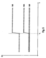
- Fig. 5 shows the magnitude of the quotient of the individual signals S of the color sensor for the individual color components of the light ROT / BLUE (R / B), RED / GREEN (R / G), GREEN / BLUE (G / B) as a function of the time before entry (t ⁇ t 1 ) and after entry (t> t 1 ) of hemolyzed blood into the dialysate. It can be seen that the addition of hemoglobin significantly increases the red / blue quotient (RED / BLUE) quotient and the red / green quotient (RED / GREEN) quotient, while the magnitude of the quotient green and blue (GREEN / BLUE) changed little or not significantly.
- Fig. 6 shows the case of non-hemolyzed blood entering the dialysate. It turns out that the amount of all quotients (RED / BLUE, RED / GREEN and GREEN / BLUE) increases significantly with the entry of non-hemolyzed blood.
- a comparison of Figures 5 and 6 shows that the entry of non-hemolyzed blood ( Fig. 6 ) leads to a greater increase in the amount of the red and blue ratio (RED / BLUE) than the entry of hemolysed blood ( Fig. 5 ). While with the entry of hemolyzed blood the amount of the quotient of the green and Blue component (GREEN / BLUE) remains virtually unchanged ( Fig.
- the difference between the increase in the red / blue ratio (RED / BLUE) ratio of haemolysed blood and non-hemolyzed blood and the difference between the green / blue ratio (GREEN / BLUE) of haemolysed and non-hemolyzed Blood is in FIG. 6 denoted by ⁇ .
- the evaluation unit 20 compares the quotient of the red and blue component (RED / BLUE) with a predetermined limit, which can be changed depending on the method. In this embodiment, only the quotient R / B of the red and blue components, which is compared with the predetermined limit, needs to be calculated. If the quotient of the red and blue component (RED / BLUE) is above the predefined limit value, the evaluation unit 20 concludes the entry of non-hemolyzed blood, for example due to a membrane rupture (FIG. Fig. 6 ), while the evaluation unit concludes the entry of hemolyzed blood, for example, as a result of blood damage, for example due to a kinked tubing or a systemic hemolysis ( Fig. 5 ), when the quotient of the red and blue component (RED / BLUE) is less than or equal to the specified limit value.
- a membrane rupture for example due to a membrane rupture
- hemolyzed blood for example, as a result of blood damage, for example due
- the evaluation unit 20 does not calculate the quotients of the red and blue components but the quotients of the green and blue components.
- the evaluation unit concludes that non-hemolyzed blood ( Fig. 6 ), when the quotient of the green and blue component (GREEN / BLUE) is greater than the predetermined limit, while the evaluation unit concludes that hemolysed blood has entered ( Fig. 5 ) when the quotient of the green and blue components is equal to or less than the predetermined limit.
- both quotients (RED / BLUE) and (GREEN / BLUE) are determined and statistically evaluated. For example, it is possible that the evaluation unit closes only on the entry of non-hemolyzed blood when both the quotient of the red and blue component and the quotient of the green and blue component are above the predetermined limits.
- the evaluation unit 20 determines not only the quotient of the red and blue portion and / or the green and blue portion to distinguish between the entry of hemolyzed and non-hemolyzed blood, but also the quotients of the red and Green component, which is stored in the memory.
- the quotient of the red and green component (RED / GREEN) is compared in the evaluation unit 20 with a first predetermined limit value. If the quotient of the red and green component (RED / GREEN) is greater than the predetermined first limit value, the evaluation unit concludes that hemolyzed blood or non-hemolyzed blood enters the dialysis fluid. Otherwise, an incident is not closed. Only when the evaluation unit has determined an accident, i.
- the evaluation unit initiates the steps described above in order to distinguish between the entry of hemolyzed blood or non-hemolyzed blood.
- the evaluation unit calculates the quotient of the red and blue component (RED / BLUE), which is compared with a predetermined second limit value, and / or the quotient of the green and blue component (green / blue), which has a predetermined third limit value is compared as described above.
- the predetermined limit values can be stored in a memory in the evaluation unit 20.
- the predetermined limits are fixed.
- the predetermined limits are adjusted to the patient.
- the evaluation unit 20 has means for adapting the limit value to patient-specific data entered on the input unit 23 become. For example, it is possible to set a higher limit for patients suffering from systemic hemolysis than patients in whom hemoglobin in the blood is not elevated. Thus, after entering the patient-specific data for a patient suffering from systemic hemolysis, the evaluation unit prescribes a correction of the otherwise prescribed limit value with a specific correction factor.
- An alternative embodiment provides for the determination of the limit value with a reference measurement which is carried out before or at the beginning or else during the blood treatment at a time when a complication is not assumed.
- This reference measurement will identify any hemoglobin levels that may be elevated due to systemic hemolysis, from which a corresponding correction factor will be calculated.
- the evaluation unit recognizes the entry of hemolyzed blood, for example due to the kinking of a tubing or the entry of non-hemolyzed blood, for example due to a membrane rupture
- the result of the check is signaled by the signal unit 22.
- the alarm generator 22B provides an audible and / or visual and / or tactile alarm.
- visual alarm symbols can be displayed on the screen 22B of the signal unit 22 .
- an indication of a targeted countermeasure For example, it may be indicated on the screen that the hose system or dialyzer is to be changed. It is also possible to point out on the screen that although hemoglobin is recognized in the dialysate, this is not due to hemolysis as a result of damage to the blood in the extracorporeal circulation, but rather to a systemic conditional hemolysis of the patient.
- the evaluation unit In addition to signaling the complications of the extracorporeal blood treatment, it is also possible that the evaluation unit generates a control signal for intervention in the machine control, which receives the central control and processing unit 17 of the extracorporeal blood treatment device. In the event that the evaluation unit detects a membrane rupture, the evaluation unit generates a Control signal for immediate interruption of blood treatment. The central control and processing unit 17 then interrupts immediately the blood promotion by closing the hose clamp 18 and stopping the blood pump 6. On the screen now appears a request to replace the dialyzer 1 or filter, while an audible alarm with the alarm generator 22B is given.
Landscapes
- Health & Medical Sciences (AREA)
- Life Sciences & Earth Sciences (AREA)
- Engineering & Computer Science (AREA)
- Biomedical Technology (AREA)
- Urology & Nephrology (AREA)
- Physics & Mathematics (AREA)
- Hematology (AREA)
- General Health & Medical Sciences (AREA)
- Heart & Thoracic Surgery (AREA)
- Chemical & Material Sciences (AREA)
- Biochemistry (AREA)
- Analytical Chemistry (AREA)
- General Physics & Mathematics (AREA)
- Immunology (AREA)
- Pathology (AREA)
- Vascular Medicine (AREA)
- Veterinary Medicine (AREA)
- Public Health (AREA)
- Animal Behavior & Ethology (AREA)
- Anesthesiology (AREA)
- Emergency Medicine (AREA)
- Biophysics (AREA)
- Ecology (AREA)
- Spectroscopy & Molecular Physics (AREA)
- Food Science & Technology (AREA)
- Molecular Biology (AREA)
- Medicinal Chemistry (AREA)
- External Artificial Organs (AREA)
Description
Die Erfindung betrifft eine Vorrichtung und ein Verfahren zur Erkennung von Blut oder Blutbestandteilen im Flüssigkeitssystem einer Vorrichtung zur extrakorporalen Blutbehandlung, die einen durch eine semipermeable Membran in eine erste Kammer und eine zweite Kammer unterteilten Dialysator oder Filter aufweist, wobei die erste Kammer Teil des extrakorporalen Blutkreislaufs und die zweite Kammer Teil des Flüssigkeitssystems der extrakorporalen Blutbehandlungsvorrichtung ist.The invention relates to an apparatus and a method for detecting blood or blood constituents in the fluid system of an extracorporeal blood treatment apparatus comprising a dialyzer or filter divided into a first chamber and a second chamber by a semipermeable membrane, the first chamber being part of the extracorporeal blood circuit and the second chamber is part of the fluid system of the extracorporeal blood treatment device.
Es sind verschiedene Verfahren zur extrakorporalen Blutbehandlung bekannt. Bei der Hämodialyse (HD) wird das Blut des Patienten in einem extrakorporalen Blutkreislauf gereinigt, der einen Dialysator umfasst. Der Dialysator weist eine Blutkammer und eine Dialysierflüssigkeitskammer auf, die von einer semipermeablen Membran getrennt sind. , Während bei der Hämodialyse (HD) die Dialysierflüssigkeitskammer von Dialysierflüssigkeit durchflossen wird, wobei Substanzen aufgrund der Diffusion zwischen der Dialysierflüssigkeit und dem Blut durch die Membran transportiert werden, wird bei der Hämofiltration (HF) die Dialysierflüssigkeitskammer des Dialysators nicht von Dialysierflüssigkeit durchströmt. Bei der Hämofiltration (HF) werden bestimmte Substanzen aufgrund von Konvektion durch die Membran des Filters effektiv entfernt. Eine Kombination aus beiden Verfahren ist die Hämodiafiltration (HDF).Various methods for extracorporeal blood treatment are known. In hemodialysis (HD), the patient's blood is purified in an extracorporeal blood circuit that includes a dialyzer. The dialyzer has a blood chamber and a dialysis fluid chamber, which are separated by a semipermeable membrane. During hemodialysis (HD), the dialysis fluid chamber is traversed by dialysis fluid, whereby substances are transported through the membrane due to the diffusion between the dialysis fluid and the blood, the dialysis fluid chamber of the dialyzer does not flow through dialysis fluid during hemofiltration (HF). Hemofiltration (HF) effectively removes certain substances due to convection through the membrane of the filter. A combination of both methods is hemodiafiltration (HDF).
Bei der Durchführung einer extrakorporalen Blutbehandlung besteht grundsätzlich das Risiko einer Ruptur der semipermeablen Membran des Dialysators oder Filters. Auch kann sich die Vergussmasse des Dialysators oder Filters ablösen. Im Falle eines Defekts des Dialysators oder Filters gelangt Blut aus dem extrakorporalen Blutkreislauf in das Flüssigkeitssystem der Blutbehandlungsvorrichtung. Daher wird bei den bekannten Blutbehandlungsvorrichtungen der Eintritt von Blut in das Flüssigkeitssystem infolge eines Defekts des Dialysators oder Filters überwacht. Die Erkennung von Blut im Flüssigkeitssystem erfolgt nach dem Stand der Technik mit einem optischen Messverfahren, wobei die Abnahme der Intensität von durch die Dialysierflüssigkeit hindurchtretenden Licht ausgewertet wird. Beim Eintritt von Blut in die Dialysierflüssigkeit ändert sich die Intensität des aus der Dialysierflüssigkeit austretenden Lichts, wobei die Intentsitätsänderung von der Wellenlänge des Lichts abhängig ist. Mit den bekannten Verfahren kann ein Bluteintritt in das Flüssigkeitssystem sicher erkannt werden.When carrying out an extracorporeal blood treatment, there is basically the risk of a rupture of the semipermeable membrane of the dialyzer or filter. Also, the potting compound of the dialyzer or filter can peel off. In the event of a defect in the dialyzer or filter, blood from the extracorporeal bloodstream enters the fluid system of the blood treatment device. Therefore, in the known blood treatment devices, the entry of blood into the fluid system due to a defect of the dialyzer or filter is monitored. The detection of blood in the Fluid system according to the prior art with an optical measuring method, wherein the decrease in the intensity of passing through the dialysis fluid light is evaluated. Upon entry of blood into the dialysis fluid, the intensity of the light exiting the dialysis fluid changes, the change in intensity being dependent on the wavelength of the light. With the known methods, blood entry into the fluid system can be reliably detected.
Aus der
Neben dem Bluteintritt in das Flüssigkeitssystem infolge eines Defekts des Dialysators oder Filters, beispielsweise aufgrund einer Membranruptur oder einer Vergussmassenablösung, kann bei einer extrakorporalen Blutbehandlung auch freies Hämoglobin oder seine Bestandteile in die Dialysierflüssigkeit aufgrund einer Hämolyse gelangen. Als Hämolyse bezeichnet man die Auflösung (Zerstörung) der Erythrozyten (roten Blutkörperchen) des Bluts. Die Erythrozyten bestehen maßgeblich aus dem Sauerstoff bindenden Protein Hämoglobin, das den Erythrozyten und somit auch dem Blut die rote Farbe verleiht. Beim Auftreten von Hämolyse wird das Hämoglobin freigesetzt.In addition to the blood entering the fluid system due to a defect of the dialyzer or filter, for example, due to a membrane rupture or Vergussmassenablösung, free hemoglobin or its constituents can enter into the dialysis fluid due to hemolysis in an extracorporeal blood treatment. Hemolysis is the dissolution (destruction) of the erythrocytes (red blood cells) of the blood. The erythrocytes consist mainly of the oxygen binding protein hemoglobin, which gives the red blood to the erythrocytes and thus to the blood. When hemolysis occurs, the hemoglobin is released.
Eine Hämolyse kann bei einer extrakorporalen Blutbehandlung beispielsweise aufgrund einer mechanischen Beanspruchung des Bluts durch Scherströmungen auftreten. Derartige Scherströmungen treten unter anderem dann auf, wenn eine Blut führende Schlauchleitung des Schlauchleitungssystems der Blutbehandlungsvorrichtung abgeknickt wird. Eine Hämolyse kann aber auch systemisch durch den Patienten bedingt sein.Hemolysis may occur in extracorporeal blood treatment, for example due to mechanical stress on the blood due to shear flow. Such shear flows occur, inter alia, when a blood-carrying tubing of the tubing system of the blood treatment device is bent. Hemolysis may also be systemic due to the patient.
Hämoglobin kann bei der extrakorporalen Blutbehandlung von der Blutseite des Dialysators durch die semipermeable Membran auf die Dialysatseite diffundieren. Daher kann das Hämoglobin mit den nach dem Stand der Technik bekannten optischen Messverfahren im Blut nachgewiesen werden.Hemoglobin, in extracorporeal blood treatment, may diffuse from the blood side of the dialyzer through the semipermeable membrane to the dialysate side. Therefore, the hemoglobin can be detected in the blood by the optical measuring methods known in the art.
Nachteilig ist, dass mit den bekannten optischen Messverfahren zwischen einem Eintritt von Blut infolge eines Defekts des Dialysators oder Filters oder dem Eintritt von Hämoglobin als Blutbestandteil infolge einer Hämolyse nicht unterschieden werden kann. Bei einem Defekt des Dialysators oder Filters oder einer Hämolyse sind aber unterschiedliche Maßnahmen einzuleiten. So muss beispielsweise bei einer Membranruptur der Dialysator ausgetauscht werden, während bei einer Hämolyse der Anwender darauf hingewiesen werden muss, geeignete Gegenmaßnahem einzuleiten, beispielsweise das Schlauchsystem zu wechseln oder zumindest von der Knickstelle zu befreien.A disadvantage is that with the known optical measuring method, it can not be distinguished between an entry of blood as a result of a defect in the dialyzer or filter or the entry of hemoglobin as a blood component as a result of hemolysis. In case of a defect of the dialyzer or filter or hemolysis but are to introduce different measures. For example, in the case of a membrane rupture, the dialyzer must be exchanged, while in the case of hemolysis the user must be advised to initiate suitable countermeasures, for example to change the tube system or at least to free it from the kink.
Die im Allgemeinen bei der extrakorporalen Blutbehandlung eingesetzten Überwachungsvorrichtungen zur Erkennung eines Bluteintritts oder einer Hämolyse beruhen auf der spektroskopischen Auswertung des roten und grünen Anteils des Lichts. In diesem Wellenlängenbereich kann aber nicht zwischen Blut und Hämoglobin unterschieden werden.The monitoring devices generally used in extracorporeal blood treatment for the detection of blood ingress or hemolysis are based on the spectroscopic evaluation of the red and green portion of the light. In this wavelength range, however, it is not possible to distinguish between blood and hemoglobin.
Die
Ein Verfahren zur Erkennung einer Hämolyse, bei dem die Abnahme der Intensität des Blauanteils von Licht Berücksichtigung findet, ist auch aus der
Der Erfindung liegt die Aufgabe zu Grunde, eine Vorrichtung bereitzustellen, die bei einem Eintritt von Blut oder Blutbestandteilen in das Flüssigkeitssystem der extrakorporalen Blutbehandlungsvorrichtung erlaubt, eine Entscheidung über die Einleitung gezielter Gegenmaßnahmen zu treffen. Darüber hinaus ist eine Aufgabe der Erfindung, ein Verfahren anzugeben, das ermöglicht, eine Entscheidung zur Einleitung gezielter Gegenmaßnahmen beim Eintritt von Blut oder Blutbestandteilen in das Flüssigkeitssystem der Blutbehandlungsvorrichtung zu treffen.The invention is based on the object to provide a device that allows entry of blood or blood components in the fluid system of the extracorporeal blood treatment device to make a decision on the initiation of targeted countermeasures. In addition, it is an object of the invention to provide a method that makes it possible to make a decision to initiate targeted countermeasures when blood or blood components enter the fluid system of the blood treatment device.
Die Lösung dieser Aufgaben erfolgt erfindungsgemäß mit den Merkmalen der Patentansprüche 1 und 13. Vorteilhafte Ausführungsformen der Erfindung sind Gegenstand der Unteransprüche.The solution of these objects is achieved according to the invention with the features of
Die erfindungsgemäße Vorrichtung zur Erkennung von Blut oder Blutbestandteilen im Flüssigkeitssystem einer extrakorporalen Blutbehandlungsvorrichtung ist als eine Einrichtung zur Unterscheidung zwischen dem Eintritt von Blut in das Flüssigkeitssystem aufgrund eines Defekts des Dialysators oder Filters, beispielsweise einer Ruptur der semipermeablen Membran des Dialysators oder Filters, und dem Eintritt von Hämoglobin in das Flüssigkeitssystem aufgrund einer Hämolyse ausgebildet, wobei ein Defekt des Dialysators oder Filters oder eine Hämolyse auf der Grundlage der Änderung der Intensität von Licht festgestellt wird, das durch die im Flüssigkeitssystem befindliche Flüssigkeit hindurchtritt.The device according to the invention for detecting blood or blood components in the fluid system of an extracorporeal blood treatment device is a device for distinguishing between the entry of blood into the fluid system due to a defect of the dialyzer or filter, for example a rupture of the semipermeable membrane of the dialyzer or filter, and entry hemoglobin is formed into the fluid system due to hemolysis, whereby a defect of the dialyzer or filter or hemolysis is detected based on the change in the intensity of light passing through the fluid in the fluid system.
Es hat sich gezeigt, dass der Quotient aus der Intensität des Rot- oder Grünanteils des aus der Flüssigkeit austretenden Lichts und der Intensität des Blauanteils des austretenden Lichts zunimmt, wenn nicht hämolysiertes Blut in die Dialysierflüssigkeit gelangt. Auch nimmt der Quotient aus der Intensität des Rotanteils und Grünanteils des aus der Flüssigkeit austretenden Lichts zu, wenn nicht hämolysiertes Blut in die Dialysierflüssigkeit gelangt. Wenn hämolysiertes Blut in die Dialysierflüssigkeit gelangt, nimmt der Quotient aus der Intensität des Rotanteils und Blauanteils oder der Quotient aus der Intensität des Rotanteils und Grünanteils zu. Der Quotient des Grünanteils und Blauanteils ändert sich hingegen beim Eintritt von hämolysierten Blut nicht oder nicht wesentlich.It has been found that the quotient of the intensity of the red or green portion of the light emerging from the liquid and the intensity of the blue portion of the emergent light increases when hemolyzed blood does not enter the dialysis fluid. Also, the quotient of the intensity of the red portion and the green portion of the light exiting the liquid increases when non-hemolyzed blood enters the dialysis fluid. When hemolyzed blood enters the dialysis fluid, the quotient of the intensity of the red component and the blue component or the quotient of the intensity of the red component and the green component increase. The quotient of the green component and the blue component, on the other hand, does not change or does not change significantly when hemolyzed blood enters.
Eine Unterscheidung zwischen dem Eintritt von hämolysierten und nicht hämolysierten Blut kann dadurch getroffen werden, dass das Verhältnis von dem Rotanteil und dem Blauanteil des aus der Flüssigkeit austretenden Lichts ausgewertet wird. Es hat sich gezeigt, dass der Quotient aus dem Rotanteil und Blauanteil beim Eintritt von nicht hämolysierten Blut stärke zunimmt als beim Eintritt von hämolysierten Blut. Auch kann eine Unterscheidung zwischen dem Eintritt von hämolysierten und nicht hämolysierten Blut dadurch getroffen werden, dass das Verhältnis von dem Grünanteil und dem Blauanteil des aus der Flüssigkeit austretenden Lichts ausgewertet wird, da sich ein signifikanter Anstieg des Quotienten nur bei dem Eintritt von nicht hämolysierten Blut zeigt.A distinction between the entry of hemolyzed and non-hemolyzed blood can be made by evaluating the ratio of red and blue in the light exiting the fluid. It has been shown that the quotient of the red portion and blue portion increases at the entry of non-hemolyzed blood strength than the entry of hemolyzed blood. Also, a distinction between the entry of hemolyzed and non-hemolyzed blood can be made by evaluating the ratio of the green portion and the blue portion of the light exiting the fluid, as there is a significant increase in the quotient only at the entry of non-hemolyzed blood shows.
Eine bevorzugte Ausführungsform der Erfindung sieht vor, zwischen dem Eintritt von Blut in das Flüssigkeitssystem aufgrund eines Defekts des Dialyators oder Filters und dem Eintritt von Hämoglobin in das Flüssigkeitssystem aufgrund einer Hämolyse auf der Grundlage der Änderung der Intensität zumindest des Blauanteils des aus der Flüssigkeit austretenden Lichts zu unterscheiden. Neben dem Blauanteil können aber noch weitere Anteile des Lichts, beispielsweise der Rotanteil und/oder der Grünanteil ausgewertet werden. Es hat sich gezeigt, dass die Auswertung des Blauanteils eine sichere Differenzierung zwischen Blut und dem Blutbestandteil Hämoglobin in der Dialysierflüssigkeit erlaubt.A preferred embodiment of the invention envisages between the entry of blood into the fluid system due to a defect of the dialyzer or filter and the entry of hemoglobin into the fluid system due to hemolysis based on the change in intensity of at least the blue portion of the light exiting the fluid to distinguish. In addition to the blue component, however, it is also possible to evaluate further components of the light, for example the red component and / or the green component. It has been shown that the evaluation of the blue component allows a reliable differentiation between blood and the blood component hemoglobin in the dialysis fluid.
Eine weitere besonders bevorzugte Ausführungsform sieht die Auswertung sowohl des Blauanteils als auch des Rotanteils vor. Dabei werden die Intensität des Blauanteils des aus der Flüssigkeit austretenden Lichts einerseits und die Intensität des Rotanteils des aus der Flüssigkeit austretenden Lichts andererseits bestimmt. Die Intensität des Rotanteils wird mit der Intensität des Blauanteils verglichen. Da die Änderung der Lichtintensität sowohl des Blau- als auch Rotanteils ausgewertet und miteinander in Beziehung gesetzt werden, kann sicher zwischen Blut und Hämoglobin unterschieden werden. Die Intensität des in die Flüssigkeit eintretenden Lichts spielt für die erfindungsgemäße Vorrichtung und das erfindungsgemäße Verfahren keine Rolle, da zwei unterschiedliche Farbanteile ins Verhältnis gesetzt werden. Daher sind die erfindungsgemäße Vorrichtung und das erfindungsgemäße Verfahren unempfindlich gegenüber Schwankungen der Intensität der Lichtquelle.Another particularly preferred embodiment provides for the evaluation of both the blue component and the red component. The intensity of the blue portion of the light emerging from the liquid on the one hand and the intensity of the red portion of the light emerging from the liquid on the other hand are determined. The intensity of the red component is compared with the intensity of the blue component. Since the changes in light intensity of both the blue and red components are evaluated and correlated, it is safe to distinguish between blood and hemoglobin. The intensity of the light entering the liquid plays no role for the device according to the invention and the method according to the invention, since two different color components are set in relation. Therefore, the device according to the invention and the method according to the invention are insensitive to variations in the intensity of the light source.
Bei einer bevorzugten Ausführungsform wird der Quotient aus der Intensität des Rotanteils und der Intensität des Blauanteils mit einem vorgegebenen Grenzwert verglichen und/oder der Quotient aus der Intensität des Grünanteils und der Intensität des Blauanteils mit einem vorgegebenen Grenzwert verglichen Auf einen Eintritt von Hämoglobin infolge einer Hämolyse wird dann geschlossen, wenn der Quotient kleiner oder gleich dem vorgegebenen Grenzwert ist. Ist der Quotient hingegen größer als der vorgegebene Grenzwert, wird auf den Eintritt von Blut beispielsweise aufgrund einer Ruptur der Membran geschlossen. Als Grenzwert kann nur ein einziger Wert vorgegeben werden. Es ist aber auch möglich, als Grenzwert mehrere individuelle Werte für unterschiedliche Abfragen vorzugeben.In a preferred embodiment, the quotient of the intensity of the red component and the intensity of the blue component is compared with a predetermined limit value and / or the quotient of the intensity of the green component and the intensity of the blue component is compared with a predetermined limit value. An entry of hemoglobin as a result of hemolysis is then closed when the quotient is less than or equal to the predetermined limit. On the other hand, if the quotient is greater than the predefined limit value, the entry of blood is concluded, for example due to a rupture of the membrane. As a limit, only a single value can be specified. It But it is also possible to set several individual values for different queries as the limit value.
Eine besonders bevorzugte Ausführungsform sieht zunächst die Berechnung des Quotienten aus dem Rotanteil und dem Grünanteil des aus der Flüssigkeit austretenden Lichts zur Erkennung des Eintritts von hämolysierten Blut oder nicht hämolysierten Bluts in die Dialysierflüssigkeit vor, ohne jedoch zwischen dem Eintritt von hämolysierten Blut oder nicht hämolysierten Blut unterscheiden zu können. Erst wenn hämolysiertes Blut oder nicht hämolysiertes Blut erkannt ist, wird der Quotient aus dem Rot- und Blauanteil und/oder aus dem Grün- und Blauanteil zur Unterscheidung zwischen einem Defekt des Dialysators oder Filters oder einer Hämolyse bestimmt.A particularly preferred embodiment initially provides for the calculation of the quotient of the red fraction and the green component of the light emerging from the liquid for the detection of the entry of hemolyzed blood or non-hemolyzed blood into the dialysis fluid, but not between the entry of hemolyzed blood or non-hemolyzed blood to be able to distinguish. Only when hemolyzed blood or non-hemolyzed blood is detected, the quotient of the red and blue portion and / or from the green and blue portion to distinguish between a defect of the dialyzer or filter or hemolysis is determined.
Bei einer bevorzugten Ausführungsform wird nicht nur die Intensität des aus der Flüssigkeit austretenden Lichts, sondern auch die Intensität des in die Flüssigkeit eintretenden Lichts überwacht. Die Überwachung der Intensität des in die Flüssigkeit eintretenden Lichts erlaubt die Erkennung eines Störfalls. Beispielesweise kann durch Vergleich der gemessenen Intensität des eintretenden Lichts mit einem vorgegebenen Grenzwert erkannt werden, ob die Mittel zum Senden von Licht, beispielsweise eine LED, defekt sind. Dies ist insbesondere dann von Interesse, wenn aus der Flüssigkeit austretendes Lichts nicht mehr empfangen wird. Denn der Grund hierfür kann sein, dass sich anstelle von Dialysierflüssigkeit Blut im Flüssigkeitssystem befindet, was einen massiven Störfall darstellt, oder nur die Lichtquelle defekt ist.In a preferred embodiment, not only the intensity of the light exiting the liquid, but also the intensity of the light entering the liquid is monitored. The monitoring of the intensity of the light entering the liquid allows the detection of an accident. By way of example, by comparing the measured intensity of the incoming light with a predetermined limit value, it can be detected whether the means for transmitting light, for example an LED, are defective. This is of particular interest when light emerging from the liquid is no longer received. Because the reason for this may be that instead of dialysis fluid blood is in the fluid system, which is a massive accident, or only the light source is defective.
Es ist aber auch möglich, mit der gemessenen Intensität des in die Flüssigkeit eintretenden Lichts eine Steuerung oder Regelung vorzusehen, um die Lichtintensität der Lichtquelle unabhängig von den Umgebungsbedingungen, beispielsweise der Temperatur, konstant zu halten.However, it is also possible to provide a control or regulation with the measured intensity of the light entering the liquid in order to keep the light intensity of the light source independent of the ambient conditions, for example the temperature, constant.
Die erfindungsgemäße Vorrichtung verfügt über Mittel zum Emittieren von Licht, das in die im Flüssigkeitssystem befindliche Flüssigkeit eintritt, und Mittel zum Empfangen von Licht, das aus der im Flüssigkeitssystem befindlichen Flüssigkeit austritt. Darüber hinaus weist die Vorrichtung eine Auswerteinheit zum Auswerten der Intensität des in die Flüssigkeit eintretenden und aus der Flüssigkeit austretenden Lichts auf.The device according to the invention has means for emitting light entering the liquid in the liquid system and means for receiving light emerging from the liquid in the liquid system. In addition, the device has an evaluation unit for evaluating the intensity of the in the Liquid entering and emerging from the liquid light.
Bei einer bevorzugten Ausführungsform sind die Mittel zum Emittieren von Licht eine Lichtquelle, die weißes Licht emittiert, beispielsweise eine weißes Licht emittierende LED oder eine weißes Licht emittierende RGB-LED, während die Mittel zum Empfangen von Licht ein Lichtsensor sind, der unterschiedliche Farbanteile des Lichts empfängt. Diese bevorzugte Ausführungsform hat den Vorteil, dass die Messapparatur einen einfacheren Aufbau als eine Messapparatur hat, bei der mehrere Lichtquellen vorgesehen sind, beispielsweise eine blaue, rote und grüne Lichtquelle, und mehrere Lichtsensoren vorgesehen sind, die beispielsweise blaues, rotes und grünes Licht empfangen. Darüber hinaus erweist sich die Verwendung einer Lichtquelle, die unspezifisches Licht (Weißlicht) emittiert, als vorteilhaft, da das Messergebnis weniger von Verschiebungen des Spektrums des Lichts beeinflusst werden kann, das von einzelnen Lichtquellen mit einem schmalen Wellenlängenband emittiert wird.In a preferred embodiment, the means for emitting light is a light source emitting white light, for example a white light emitting LED or a white light emitting RGB LED, while the means for receiving light is a light sensor having different color components of the light receives. This preferred embodiment has the advantage that the measuring apparatus has a simpler structure than a measuring apparatus, in which a plurality of light sources are provided, for example, a blue, red and green light source, and a plurality of light sensors are provided which receive, for example, blue, red and green light. In addition, the use of a light source that emits nonspecific light (white light) proves to be advantageous because the measurement result can be less affected by shifts in the spectrum of light emitted by individual light sources having a narrow wavelength band.
Eine weitere besonders bevorzugte Ausführungsform sieht eine Anpassung des vorgegebenen Grenzwerts oder der vorgegebenen Grenzwerte an den Patienten vor. Dadurch ist es möglich, auch bei Patienten, die an einer systemischen Hämolyse leiden, zwischen dem Eintritt von Blut oder Hämoglobin in das Flüssigkeitssystem zu unterscheiden. Bei einem krankheitsbedingten höheren Anteil von Hämoglobin im Blut (systemische Hämolyse) kann der vorgegebene Grenzwert entsprechend angepasst werden, so dass nicht allein schon wegen des erhöhten Anteils von Hämoglobin im Blut aufgrund einer systemischen Hämolyse auf den Eintritt von Hämoglobin in die Dialysierflüssigkeit geschlossen wird, der auf eine Schädigung des Blutes beispielsweise durch eine abgeknickte Blut führende Schlauchleitung zurückzuführen ist. Beispielsweise kann anhand der Patientendaten ein Satz von angepassten Grenzwerten ermittelt und vorgegeben werden.Another particularly preferred embodiment provides for an adaptation of the predetermined limit value or the predetermined limit values to the patient. This makes it possible to distinguish between the entry of blood or hemoglobin into the fluid system even in patients suffering from systemic hemolysis. With a disease-related higher proportion of hemoglobin in the blood (systemic hemolysis), the predetermined limit can be adjusted accordingly, so that not alone due to the increased proportion of hemoglobin in the blood due to a systemic hemolysis on the entry of hemoglobin in the dialysis fluid is closed is due to damage to the blood, for example, by a bent blood leading tubing. For example, based on the patient data, a set of adjusted limit values can be determined and specified.
Die Anpassung des vorgegebenen Grenzwertes oder der Grenzwerte erfolgt vorzugsweise auf der Grundlage von patientenspezifischen Daten, die vorzugsweise auf einer Eingabeeinheit eingegeben werden. Es ist aber auch möglich, dass der vorgegebene Grenzwert in Abhängigkeit von patientenspezifischen Daten ermittelt wird, die auf der Grundlage einer Referenzmessung gewonnen werden. Beispielsweise ist es möglich, vor Beginn oder zu Beginn oder während der Blutbehandlung eine Referenzmessung zu einem Zeitpunkt vorzusehen, bei dem von einer Hämolyse beispielsweise aufgrund des Abknickens einer Schlauchleitung nicht ausgegangen wird. Abweichungen von diesem Grenzwert lassen dann auf den Eintritt von Hämoglobin aufgrund einer Blutschädigung schließen.The adaptation of the predetermined limit value or the limit values is preferably carried out on the basis of patient-specific data, preferably on a Input unit can be entered. But it is also possible that the predetermined limit value is determined as a function of patient-specific data, which are obtained on the basis of a reference measurement. For example, it is possible to provide a reference measurement at a time before or at the beginning or during the blood treatment, in which a hemolysis, for example due to the kinking of a hose line, is not assumed. Deviations from this threshold may then indicate the onset of hemoglobin due to blood damage.
Wenn der Eintritt von Blut und / oder Hämoglobin festgestellt wird, kann ein optisches und / oder akustisches und / oder taktiles Signal gegeben werden, um geeignete Gegenmaßnahmen einleiten zu können. Beispielsweise kann die Blutbehandlung beim Eintritt von Blut sofort unterbrochen werden. Es ist auch möglich, dass ein Steuersignal für einen Eingriff in die Maschinensteuerung erzeugt wird, um Gegenmaßnahmen automatisch einleiten zu können.If the entry of blood and / or hemoglobin is detected, an optical and / or acoustic and / or tactile signal may be given to initiate appropriate countermeasures. For example, the blood treatment can be interrupted immediately when blood enters. It is also possible that a control signal for an intervention in the machine control is generated in order to initiate countermeasures automatically.
Das Ergebnis der Überwachung wird vorzugsweise auf einem Bildschirm angezeigt, bei dem es sich um einen sogenannten Touchscreen handeln kann. Auf dem Monitor können neben der Art der auftretenden Komplikationen, d.h. Membranruptur oder Hämolyse, die empfohlenen Gegenmaßnahmen, beispielsweise durch einen entprechenden Text und / oder entsprechende Symbole angezeigt werden. Beispielsweise kann zum Austausch des Schlauchleitungssystems oder des Dialysators aufgefordert werden.The result of the monitoring is preferably displayed on a screen, which may be a so-called touch screen. On the monitor, in addition to the type of complications that occur, i. Membrane rupture or hemolysis, the recommended countermeasures are displayed, for example by an appropriate text and / or corresponding symbols. For example, it may be necessary to replace the hose line system or the dialyzer.
Im Folgenden werden Ausführungsbeispiele der Erfindung unter Bezugnahme auf die Zeichnungen näher erläutert.In the following, embodiments of the invention will be explained in more detail with reference to the drawings.
Es zeigen:
- Fig. 1
- eine extrakorporale Blutbehandlungsvorrichtung zusammen mit einer Vorrichtung zur Erkennung von Blut oder Blutbestandteilen im Flüssigkeitssystem der Blutbehandlungsvorrichtung in stark vereinfachter schematischer Darstellung,
- Fig. 2
- eine schematische Darstellung des Messfeldes des Lichtsensors der Vorrichtung zur Erkennung von Blut oder Blutbestandteilen,
- Fig. 3
- eine schematische Darstellung der relativen Empfindlichkeit des Lichtsensors in Abhängigkeit von der Wellenlänge des Lichts,
- Fig. 4
- eine schematische Darstellung der Extinktion als Funktion der Wellenlänge für den Fall des Eintritts von Blut oder von Hämoglobin in die Dialysierflüssigkeit,
- Fig. 5
- die Veränderung der Signalstärke des Lichtsensors für die einzelnen Farbanteile des Lichts vor und nach dem Eintritt von Hämoglobin in die Dialysierflüssigkeit und
- Fig. 6
- die Veränderung der Signalstärke des Lichtsensors für die einzelnen Farbanteile vor und nach dem Eintritt von Blut in die Dialysierflüssigkeit.
- Fig. 1
- an extracorporeal blood treatment device together with a device for detecting blood or blood components in the fluid system of the blood treatment device in a highly simplified schematic representation,
- Fig. 2
- a schematic representation of the measuring field of the light sensor of the device for detecting blood or blood components,
- Fig. 3
- a schematic representation of the relative sensitivity of the light sensor as a function of the wavelength of the light,
- Fig. 4
- a schematic representation of the absorbance as a function of wavelength in the case of the entry of blood or hemoglobin in the dialysis fluid,
- Fig. 5
- the change in the signal strength of the light sensor for the individual color components of the light before and after the entry of hemoglobin into the dialysis fluid and
- Fig. 6
- the change in the signal strength of the light sensor for the individual color components before and after the entry of blood into the dialysis fluid.
Die Hämodiafiltrationsvorrichtung weist einen Dialysator oder Filter 1 auf, der durch eine semipermeable Membran 2 in eine Blutkammer 3 und eine Dialysierflüssigkeitskammer 4 getrennt ist. Der Einlass 3a der Blutkammer ist mit einem Ende einer arteriellen Blutzuführleitung 5 verbunden, in die eine Blutpumpe 6 geschaltet ist, während der Auslass 3b der Blutkammer mit einem Ende einer venösen Blutrückführleitung 7 verbunden ist, in die eine Tropfkammer 8 geschaltet ist. An den anderen Enden der arteriellen und venösen Blutleitung 5, 7 befinden sich die nicht dargestellten arteriellen und venösen Kanülen zum Anschluss an den Patienten. Dieser Teil des Flüssigkeitssystems stellt den extrakorporalen Blutkreislauf I der Hämodiafiltrationsvorrichtung dar. Bei den Blutleitungen 5, 7 handelt es sich um Schlauchleitungen eines zur einmaligen Verwendung bestimmten Schlauchleitungssystems, das in die Blutbehandlungsvorrichtung eingelegt wird.The hemodiafiltration device has a dialyzer or filter 1 which is separated by a
Das Flüssigkeitssystem II der Hämodiafiltrationsvorrichtung umfasst eine Einrichtung 9 zur Bereitstellung frischer Dialysierflüssigkeit, die über eine Dialysierflüssigkeitszuführleitung 10 mit dem Einlass 4a der Dialysierflüssigkeitskammer 4 des Dialysators 1 oder Filters verbunden ist. Von dem Auslass 4b der Dialysierflüssigkeitskammer 4 des Dialysators 1 oder Filters geht eine Dialysierflüssigkeitsrückführleitung 11 ab, die zu einem Auslass 12 führt. Zum Fördern der Dialysierflüssigkeit dient eine Dialysierflüssigkeitspumpe 13, die in die Dialysierflüssigkeitsrückführleitung geschaltet ist.The fluid system II of the hemodiafiltration device comprises a device 9 for providing fresh dialysis fluid, which is connected via a dialysis
Darüber hinaus verfügt die Hämodiafiltrationsvorrichtung über eine Substituatquelle 14, von der eine Substituatleitung 15, in die eine Substituatpumpe 16 geschaltet ist, zu der venösen Tropfkammer 8 führt. Mit der Substituatpumpe kann dem extrakorporalen Blutkreislauf 1 eine vorgegebene Menge an Substitutionsflüssigkeit aus der Substituatquelle zugeführt werden, wenn dem extrakorporalen Blutkreislauf I über den Dialysator 1 Flüssigkeit entzogen wird.In addition, the hemodiafiltration device has a
Die Hämodiafiltrationsvorrichtung umfasst weiterhin eine zentrale Steuer- und Recheneinheit 17, die über Steuerleitungen 6', 13', 16' mit der Blutpumpe 6, der Dialysierflüssigkeitspumpe 13 und der Substituatpumpe 16 verbunden ist. Die Steuer- und Recheneinheit 17 sendet an die einzelnen Komponenten Steuerbefehle und empfängt von den Komponenten Daten über deren Betriebszustände.The hemodiafiltration device further comprises a central control and
Nachfolgend wird die erfindungsgemäße Vorrichtung zur Erkennung von Blut oder Blutbestandteilen im Flüssigkeitssystem der extrakorporalen Blutbehandlungsvorrichtung beschrieben. Die erfindungsgemäße Vorrichtung zur Erkennung von Blut oder Blutbestandteilen kann eine selbständige Einheit bilden oder Bestandteil der Blutbehandlungsvorrichtung sein. Bei dem vorliegenden Ausführungsbeispiel ist die erfindungsgemäße Vorrichtung Bestandteil der Blutbehandlungsvorrichtung.The device according to the invention for detecting blood or blood constituents in the fluid system of the extracorporeal blood treatment device will now be described. The device according to the invention for detecting blood or blood components may form an independent unit or be part of the blood treatment device. In the present embodiment, the device according to the invention is part of the blood treatment device.
Die erfindungsgemäße Vorrichtung kann mit Ausnahme der Messanordnung von den Teilen Gebrauch machen, die bereits in den bekannten extrakorporalen Blutbehandlungsvorrichtungen vorhanden sind. Beispielsweise kann die erfindungsgemäße Vorrichtung von der zentralen Steuer- und Recheneinheit der Blutbehandlungsvorrichtung Gebrauch machen, um die gewonnenen Messdaten auszuwerten. Auch kann die erfindungsgemäße Vorrichtung die Anzeigeeinheit und die Eingabeeinheit der Blutbehandlungsvorrichtung verwenden. Es kann aber für die erfindungsgemäße Vorrichtung auch eine separate Auswerteinheit vorgesehen sein. Auch kann eine separate Anzeigeeinheit oder Eingabeeinheit vorhanden sein.The device according to the invention, with the exception of the measuring arrangement, can make use of the parts which are already present in the known extracorporeal blood treatment devices. For example, the device according to the invention can make use of the central control and computing unit of the blood treatment apparatus in order to evaluate the measured data obtained. Also, the device of the invention can use the display unit and the input unit of the blood treatment device. However, it may also be provided for the inventive device, a separate evaluation unit. Also, a separate display unit or input unit may be present.
Die Vorrichtung zur Erkennung von Blut oder Hämoglobin verfügt über eine Lichtquelle 21 A, beispielsweise eine LED, und einen Lichtsensor 21 B, die einander gegenüberliegend angeordnet sind. Lichtquelle 21A und Lichtsensor 21B bilden eine Messanordnung 21 zum Messen der Änderung der Intensität von Licht, das aus der im Flüssigkeitssystem II befindliche Flüssigkeit, d.h. Dialysierflüssigkeit austritt. Da die Dialysierflüssigkeitsleitungen 10, 11 transparente Schlauchleitungen sind, kann das Licht durch die Schlauchleitung in die Dialysierflüssigkeit eingekoppelt und aus der Dialysierflüssigkeit ausgekoppelt werden. Die Messanordnung 21 ist an der Dialysierflüssigkeitsrückführleitung 11, insbesondere in dem Abschnitt der Dialysierflüssigkeitsrückführleitung 11 stromab der Dialysierflüssigkeitskammer 4 des Dialysators 1 oder Filters und stromauf der Dialysierflüssigkeitspumpe 13 angeordnet. Die Messung kann sowohl an einer zur einmaligen Verwendung bestimmten Messkammer oder an einer fest installierten Messkammer der Schlauchleitung erfolgen. Als Messkammer kann auch eine Küvette Verwendung finden. Neben dem ausgangsseitigen Lichtsensor 21 B kann die Messanordnung noch einen eingangsseitigen Lichtsensor 21 C aufweise, der das mit der Lichtquelle 21A emittierte Licht misst.The device for detecting blood or hemoglobin has a
Für den Fall einer Ruptur der semipermeablen Membran 2 des Dialysators 1 oder Filters tritt sowohl beim Betrieb der Hämodiafiltrationsvorrichtung als Hämodialysevorrichtung als auch Hämofiltrationsvorrichtung Blut aus dem extrakorporalen Blutkreislauf I in die Dialysierflüssigkeitskammer 4 des Dialysators 1 oder Filters ein. Das nicht hämolysierte Blut kann dann mit der Messanordnung 21 in der Dialysierflüssigkeit nachgewiesen werden, die im Dialysierflüssigkeitssystem II strömt. Wenn die Hämodiafiltrationsvorrichtung nur als Hämofiltrationsvorrichtung betrieben wird, kann das Blut in dem Filtrat nachgewiesen werden, das der Dialysierflüssigkeitskammer 4 entzogen wird. Für den Fall einer Hämolyse infolge des Abknickens der arteriellen oder venösen Blutleitung 5, 7 oder einer systemisch bedingten Hämolyse gelangt Hämoglobin oder hämolysiertes Blut durch die intakte semipermeable Membran 2 des Dialysators 1 oder Filters in die Dialysierflüssigkeitskammer 4, welches ebenfalls mit der Messanordnung 21 nachgewiesen werden kann.In the event of a rupture of the
Stromab der Tropfkammer 8 befindet sich an der venösen Blutleitung 7 ein Absperrorgan 18 zum Absperren der Blutleitung 7. Das Absperrorgan 18, insbesondere eine elektromagnetisch betätigbare Schlauchklemme, ist über eine Steuerleitung 18' mit der Steuer- und Recheneinheit 17 verbunden.Downstream of the drip chamber 8 is located on the venous blood line 7 a shut-off
Die Vorrichtung zur Erkennung von Blut oder Blutbestandteilen weist eine Auswerteinheit 20 auf, die über eine Datenleitung 20' mit der zentralen Steuer- und Recheneinheit 17 der Hämodiafiltrationsvorrichtung verbunden ist. Die Auswerteinheit 20 kann aber auch Bestandteil der Steuer- und Recheneinheit 17 sein. Die Auswerteinheit empfängt über eine Datenleitung 21' die Signale des ausgangsseitigen Lichtsensors 21 B und über eine Datenleitung 21 " die Signale des eingangsseitigen Lichtsensors 21C der Messanordnung 21. Die Auswerteinheit 20 kann eine Regeleinheit aufweisen, die in Abhängigkeit von der mit dem ausgangsseitigen Lichtsensors 2 1 B gemessenen Intensität des von der Lichtquelle 21 A emittierten Lichts die Lichtquelle derart ansteuert, das die Lichtintensität unabhängig von den Umgebungsbedingungen konstant bleibt.The device for detecting blood or blood components has an
Das Ergebnis der Überwachung wird mit einer Signaleinheit 22 signalisiert, die über eine Datenleitung 22' mit der Auswerteinheit 20 verbunden ist. Die Signaleinheit 22 weist einen Bildschirm 22A zum Anzeigen von Symbolen oder Text und einem Alarmgeber 22B zur Erzeugung eines akustischen oder taktilen Alarms auf. Auf einer Eingabeeinheit 23, die über eine Datenleitung 23' mit der Auswerteinheit 20 verbunden ist, können patientenspezifische Daten eingegeben werden. Bei der Eingabeeinheit 23 kann es sich auch um einen sogenannten Touchscreen handeln, der gleichsam als Bildschirm der Signaleinheit dient.The result of the monitoring is signaled by a
Die Lichtquelle 21A der Messanordnung 21 emittiert weißes Licht, das die Farbanteile Rot, Grün und Blau mit einer bestimmten Intensität I0 enthält. Die Lichtquelle 21 A ist über eine Steuerleitung 21A' mit der zentralen Steuer- und Recheneinheit 17 verbunden, so dass die Intensität I0 des Lichts von der Steuer- und Recheneinheit 17 vorgegeben werden kann. Der Lichtsensor 21B empfängt das durch die Dialysierflüssigkeit (Filtrat) hindurchtretende Licht der Lichtquelle 21 A. Bei dem Lichtsensor handelt es sich um einen Sensor, der die Intensität I1 von drei Farbanteilen Rot, Grün und Blau (RGB) auswerten kann. Die lichtempfindliche Oberfläche des Sensors wird von einer Vielzahl identischer Fotodioden gebildet, die in Reihen und Spalten angeordnet sind. Jeder Fotodiode ist jeweils ein Farbfilter zugeordnet. Für die Auswertung des Rotanteils ist ein roter Farbfilter, für die Auswertung des Grünanteils ein grüner Farbfilter und für die Auswertung des Blauanteils des Lichts ein blauer Farbfilter vorgesehen.The
Bei dem Lichtsensor kann es sich beispielsweise um einen Sensor des Fabrikats "Programmable Color Light-To-Frequency Converter TCS230" der Firma TAOS (Texas Advanded Optoelectronic Solutions Inc., Piano, Texas, USA) oder um einen Sensor des Fabrikats "Digital Color Sensor S9706" der Firma Hamamatsu (Hamamatsu Photonics K.K., Japan) oder dergleichen handeln. Aufbau und Funktion der beiden Sensoren sind in den Datenblättern der Hersteller im Einzelnen beschrieben.The light sensor can be, for example, a "Programmable Color Light-To-Frequency Converter TCS230" manufactured by TAOS (Texas Adv. Optoelectronic Solutions Inc., Piano, Texas, USA) or a "Digital Color Sensor S9706 "of the company Hamamatsu (Hamamatsu Photonics KK, Japan) or the like. Construction and function of the two sensors are described in detail in the data sheets of the manufacturer.
Nachfolgend wird die Funktionsweise der erfindungsgemäßen Vorrichtung zur Unterscheidung zwischen Blut und Hämoglobin in der Dialysierflüssigkeit (Filtrat) beschrieben.The operation of the device according to the invention for distinguishing between blood and hemoglobin in the dialysis fluid (filtrate) will be described below.
Die Auswerteinheit 20 empfängt das die drei Farbanteile Rot, Grün und Blau enthaltende Ausgangssignal S des ausgangseitigen Lichtsensors 21B der Messanordnung 21. In der Auswerteinheit 20 wird das Ausgangssignal des Sensors in die drei Einzelsignale für die Farbbestandteile Rot, Grün und Blau (RGB) zerlegt. Der Betrag der Einzelsignale (Signalstärke) ist proportional der Intensität I1 des aus der Dialysierflüssigkeit (Filtrat) austretenden Lichts. Die Intensität I0 des in die Dialysierflüssigkeit (Filtrat) eintretenden Lichts wird von der Lichtquelle 21 A bestimmt und mit dem eingangsseitigen Lichtsensor 21C gemessen. Die Auswerteinheit 20 bestimmt nunmehr den Quotienten R/B aus der Intensität I1 des aus der Flüssigkeit austretenden Rotanteils R des Lichts, d.h. dem Betrag des Einzelsignals für den Rotanteil, und der Intensität I1 des aus der Flüssigkeit austretenden Blauanteils B des Lichts, d.h. dem Betrag des Einzelsignals für den Blauanteil. Darüber hinaus kann die Auswerteinheit den Quotienten R/G aus der Intensität I1 des Rotanteils und des Grünanteils, d.h. den Quotienten der Einzelsignale des Rot- und Grünanteils berechnen. Ferner berechnet die Auswerteinheit den Quotienten G/B der Intensität I1 des Grünanteils und des Blauanteils. Sämtliche Quotienten werden in einem internen Speicher der Auswerteinheit abgelegt. Die Messung kann in vorgegebenen Zeitabständen oder kontinuierlich erfolgen. Es ist auch möglich, zu bestimmten Zeitpunkten, beispielsweise vor der Behandlung, Referenzmessungen durchzuführen, um die aktuellen Werte mit den Referenzwerten vergleichen zu können. Die Referenzmessungen können auch zyklisch beispielsweise unter Verwendung eines den Dialysator oder Filter umgehenden Bypasses erfolgen.The
Der Bypass zur Umgehung des Dialysators für die Referenzmessungen umfasst eine Bypass-Leitung 24, die von der Dialysierflüssigkeitszuführleitung 10 stromauf der Dialysierflüssigkeitskammer 4 abgeht und stromab der Dialysierflüssigkeitskammer zu der Dialysierflüssigkeitsabführleitung 11 führt. In der Bypass-Leitung 24 befindet sich ein Absperrorgan 25 zum Öffnen und Schließen der Bypassleitung und in dem Abschnitt der Dialysierflüssigkeitszuführleitung 10 zwischen der Bypass-Leitung 24 und der Dialysierflüssigkeitskammer 4 befindet sich ein Absperrorgan 26 zum Abtrennen der Dialysierflüssigkeitskammer. Die beiden Absperrorgane 25, 26 sind über Steuerleitungen 25', 26' mit der zentralen Steuer- und Recheneinheit 17 verbunden. Zur Umgehung des Dialysators 1 öffnet die Steuereinheit das Absperrorgan 24 und schließt das Absperrorgan 25. Ansonsten sind das Absperrorgan 24 geschlossen und das Absperrorgan 25 geöffnet.The bypass for bypassing the dialyzer for the reference measurements comprises a
Der Unterschied der Zunahme des Betrags des Quotienten aus dem Rot- und Blauanteil (ROT / BLAU) bei hämolysiertem Blut und nicht hämolysiertem Blut und der Unterschied zwischen dem Betrag des Quotienten aus dem Grün- und Blauanteil (GRÜN / BLAU) bei hämolysiertem und nicht hämolysierten Blut ist in
Bei einer ersten Ausführungsform vergleicht die Auswerteinheit 20 den Quotienten aus dem Rot- und Blauanteil (ROT / BLAU) mit einem vorgegebenen Grenzwert, der verfahrensabhängig verändert werden kann. Bei dieser Ausführungsform braucht nur der Quotient R/B des Rot- und Blauanteils berechnet zu werden, der mit dem vorgegebenen Grenzwert verglichen wird. Wenn der Quotient von dem Rot- und Blauanteil (ROT / BLAU) oberhalb des vorgegebenen Grenzwerts liegt, schließt die Auswerteinheit 20 auf den Eintritt von nicht hämolysiertem Blut beispielsweise aufgrund einer Membranruptur (
Bei einer alternativen Ausführungsform berechnet die Auswerteinheit 20 nicht den Quotienten des Rot- und Blauanteils, sondern den Quotienten des Grün- und Blauanteils. Die Auswerteinheit schließt auf den Eintritt von nicht hämolysiertem Blut (
Es ist auch möglich, dass beide Quotienten (ROT / BLAU) und (GRÜN / BLAU) bestimmt werden und statistisch ausgewertet werden. Beispielsweise ist es möglich, dass die Auswerteinheit nur auf den Eintritt von nicht hämolysiertem Blut schließt, wenn sowohl der Quotient des Rot- und Blauanteils als auch der Quotient des Grün- und Blauanteils oberhalb der vorgegebenen Grenzwerte liegen.It is also possible that both quotients (RED / BLUE) and (GREEN / BLUE) are determined and statistically evaluated. For example, it is possible that the evaluation unit closes only on the entry of non-hemolyzed blood when both the quotient of the red and blue component and the quotient of the green and blue component are above the predetermined limits.
Grundsätzlich ist es aber auch möglich, nur den Blauanteil des Lichts auszuwerten, der weder mit dem Rotanteil noch den Grünanteil in Beziehung gesetzt wird.In principle, however, it is also possible to evaluate only the blue portion of the light which is not related to either the red component or the green component.
Bei einer bevorzugten Ausführungsform bestimmt die Auswerteinheit 20 nicht nur den Quotienten aus dem Rot- und Blauanteil und/oder dem Grün- und Blauanteil, um zwischen dem Eintritt von hämolysierten und nicht hämolysierten Blut unterscheiden zu können, sondern auch den Quotienten aus dem Rot- und Grünanteils, der in dem Speicher abgelegt wird. Der Quotient aus dem Rot- und Grünanteil (ROT/GRÜN) wird in der Auswerteinheit 20 mit einem ersten vorgegebenen Grenzwert verglichen. Wenn der Quotient aus dem Rot- und Grünanteil (ROT/GRÜN) größer als der vorgegebene erste Grenzwert ist, schließt die Auswerteinheit auf den Eintritt von hämolysierten Blut oder nicht hämolysierten Blut in die Dialysierflüssigkeit. Ansonsten wird auf einen Störfall nicht geschlossen. Erst wenn die Auswerteineheit einen Störfall festgestellt hat, d.h. der Quotient aus dem Rot- und Grünanteil (ROT/GRÜN) größer als der vorgegebene erste Grenzwert ist, leitet die Auswerteinheit die oben beschriebenen Schritte ein, um zwischen dem Eintritt von hämolysierten Blut oder nicht hämolysierten Blut unterscheiden zu können. Die Auswerteinheit berechnet dann den Quotienten aus dem Rot- und Blauanteil (ROT/BLAU), der mit einem vorgegebenen zweiten Grenzwert verglichen wird, und/oder den Quotienten aus dem Grün- und Blauanteil (Grün/BLAU), der mit einem vorgegebenen dritten Grenzwert verglichen wird, wie oben beschrieben ist.In a preferred embodiment, the
Die vorgegebenen Grenzwerte können in einem Speicher in der Auswerteinheit 20 abgelegt sein. Damit sind die vorgegebenen Grenzwerte fest vorgegeben. Bei einer alternativen Ausführungsform werden die vorgegebenen Grenzwerte an den Patienten angepasst. Hierzu verfügt die Auswerteinheit 20 über Mittel zum Anpassen des Grenzwertes an patientenspezifische Daten, die auf der Eingabeeinheit 23 eingegeben werden. Beispielsweise ist es möglich, für Patienten, die an einer systemischen Hämolyse leiden, einen höheren Grenzwert vorzugeben, als Patienten, bei denen das Hämoglobin im Blut nicht erhöht ist. Die Auswerteinheit nimmt also nach der Eingabe der patientenspezifischen Daten für einen Patienten, der an systemischer Hämolyse leidet, eine Korrektur des ansonsten vorgegebenen Grenzwertes mit einem bestimmten Korrekturfaktor vor.The predetermined limit values can be stored in a memory in the
Eine alternative Ausführungsform sieht die Ermittlung des Grenzwertes mit einer Referenzmessung vor, die vor oder zu Beginn oder auch während der Blutbehandlung zu einem Zeitpunkt vorgenommen wird, zu dem von einer Komplikation nicht ausgegangen wird. Mit dieser Referenzmessung werden die möglicherweise aufgrund einer systemischen Hämolyse erhöhten Hämoglobinwerte ermittelt, aus denen ein entsprechender Korrekturfaktor berechnet wird.An alternative embodiment provides for the determination of the limit value with a reference measurement which is carried out before or at the beginning or else during the blood treatment at a time when a complication is not assumed. This reference measurement will identify any hemoglobin levels that may be elevated due to systemic hemolysis, from which a corresponding correction factor will be calculated.
Wenn die Auswerteinheit den Eintritt von hämolysiertem Blut beispielsweise aufgrund des Abknickens einer Schlauchleitung oder den Eintritt von nicht hämolysiertem Blut beispielsweise aufgrund einer Membranruptur erkennt, wird das Ergebnis der Überprüfung mit der Signaleinheit 22 signalisiert. Beispielsweise wird mit dem Alarmgeber 22B ein akustischer und/oder optischer und/oder taktiler Alarm gegeben. Auf dem Bildschirm 22B der Signaleinheit 22 können optische Alarmsymbole angezeigt werden. Auch kann auf dem Bildschirm ein Hinweis auf eine gezielte Gegenmaßnahme gegeben werden. Beispielsweise kann auf dem Bildschirm angezeigt werden, dass das Schlauchsystem oder der Dialysator zu wechseln ist. Es ist auch möglich, auf dem Bildschirm darauf hinzuweisen, dass zwar Hämoglobin im Dialysat erkannt wird, das aber nicht auf eine Hämolyse in Folge einer Schädigung des Bluts im extrakorporalen Kreislauf, sondern auf eine systemische bedingte Hämolyse des Patienten zurückzuführen ist.If the evaluation unit recognizes the entry of hemolyzed blood, for example due to the kinking of a tubing or the entry of non-hemolyzed blood, for example due to a membrane rupture, the result of the check is signaled by the
Neben der Signalisierung der Komplikationen der extrakorporalen Blutbehandlung ist es auch möglich, dass die Auswerteinheit ein Steuersignal zum Eingriff in die Maschinensteuerung erzeugt, das die zentrale Steuer- und Recheneinheit 17 der extrakorporalen Blutbehandlungsvorrichtung empfängt. Für den Fall, dass die Auswerteinheit eine Membranruptur detektiert, erzeugt die Auswerteinheit ein Steuersignal für die sofortige Unterbrechung der Blutbehandlung. Die zentrale Steuer- und Recheneinheit 17 unterbricht dann sofort die Blutförderung durch Schließen der Schlauchklemme 18 und Stoppen der Blutpumpe 6. Auf dem Bildschirm erscheint nunmehr eine Aufforderung zum Austausch des Dialysators 1 oder Filters, während ein akustischer Alarm mit dem Alarmgeber 22B gegeben wird.In addition to signaling the complications of the extracorporeal blood treatment, it is also possible that the evaluation unit generates a control signal for intervention in the machine control, which receives the central control and
Claims (20)
- A device for the detection of blood or blood constituents in the fluid system of an apparatus for extracorporeal blood treatment, which device comprises a dialyser (1) or filter divided by a semipermeable membrane (2) into a first chamber (3) and a second chamber (4), wherein the first chamber is part of the extracorporeal blood circuit (I) and the second chamber is part of a fluid system (II), with means (21A) for emitting light which enters into the fluid present in the fluid system, and means (2 1 B) for receiving light which exits from the fluid present in the fluid system, and
an evaluation unit (20) which comprises means for evaluating intensity I1 of the light exiting from the fluid,
characterised in that
the device for the detection of blood or blood constituents in the fluid system is designed as a device for distinguishing between the entry of blood into the fluid system due to a defect in the dialyser or filter or the entry of haemoglobin into the fluid system due to a haemolysis on the basis of the change in intensity I1 of the light exiting from the fluid. - The device according to claim 1, characterised in that the device for the detection of blood or blood constituents in the fluid system is designed such that a distinction is made between the entry of blood into the fluid due to a defect in the dialyser or filter or the entry of haemoglobin into the fluid system due to a haemolysis at least on the basis of the change in intensity I1 of the blue component of the light exiting from the fluid.
- The device according to claim 1 or 2, characterised in that the means (21) for emitting light and the means (21) for receiving light are designed as a means for emitting and receiving light which comprises at least a blue component and a red component, and
that the evaluation unit (20) is designed such that intensity I1/B of the blue component of the light exiting from the fluid and intensity I1/R of the red component of the light exiting from the fluid is determined, and
that the evaluation unit (20) is designed such that intensity I1/R of the red component is compared with intensity I1/B of the blue component, wherein a distinction is made between the entry of blood into the fluid system and a haemolysis on the basis of the comparison between the intensity of the red component and the intensity of the blue component. - The device according to claim 3, characterised in that the evaluation unit (20) is designed such that the quotient (R/B) of the intensity of red component R and the intensity of blue component B is calculated and compared with a preset threshold value, wherein it is concluded that there is an entry of blood if the quotient is greater than the preset threshold value or in that the evaluation unit (20) is designed such that the quotient (R/B) of the intensity of red component R and the intensity of blue component B is calculated and compared with a preset threshold value, wherein it is concluded that there is a haemolysis if the quotient is less than or equal to the preset threshold value.
- The device according to claim 1 or 2, characterised in that the means (21 A) for emitting light and the means (2 1 B) for receiving light are designed as a means for emitting and receiving light which comprises a blue component and a green component, and
that the evaluation unit (20) is designed such that intensity I1/B of the blue component of the light exiting from the fluid and intensity I1/G of the green component of the light exiting from the fluid is determined, and
that the evaluation unit (20) is designed such that intensity I1/G of green component G is compared with intensity I1/B of blue component B, wherein a distinction is made between the entry of blood into the fluid system and a haemolysis on the basis of the comparison between the intensity of the green component and the intensity of the blue component. - The device according to claim 5, characterised in that the evaluation unit (20) is designed such that the quotient (G/B) of the intensity of the green component and intensity of the blue component is calculated and compared with a preset threshold value, wherein it is concluded that there is an entry of blood if the quotient is greater than the preset threshold value or the evaluation unit (20) is designed such that the quotient (G/B) of the intensity of the green component and the intensity of blue component B is calculated and compared with a preset threshold value, wherein it is concluded that there is a haemolysis if the quotient is less than or equal to the threshold value.
- The device according to claim 1 or 2, characterised in that the means (21A) for emitting light and the means (21B) for receiving light are designed as a means for emitting and receiving light which comprises a red component, a green component and a blue component, and
that the evaluation unit (20) is designed such that intensity I1/R of the red component of the light exiting from the fluid and intensity I1/G of the green component of the light exiting from the fluid is determined, and
that the evaluation unit (17) is designed such that intensity I1/R of the red component is compared with intensity I1/G of green component G in a first step, wherein it is concluded that there is an entry of blood into the fluid system or a haemolysis
on the basis of the comparison between the intensity of the green component and intensity of the blue component,
and that the evaluation unit (20) is designed such that, in the event that the evaluation unit concludes that there is an entry of blood or a haemolysis in the first step, intensity I1/R of the red component is compared with intensity I1/B of the blue component in a second step, wherein a distinction is made between the entry of blood into the fluid system and a haemolysis on the basis of the comparison between the intensity of the red component and the intensity of the blue component. - The device according to claim 7, characterised in that the evaluation unit (20) is designed such that the quotient of intensity I1/R of the red component and intensity I1/G of green component G is calculated in the first step, wherein it is concluded that there is an entry of blood or a haemolysis if the quotient is greater than a preset first threshold value, and
that the evaluation unit (20) is designed such that the quotient of intensity I1/R of red component R and intensity I1/B of blue component B is calculated in the second step, wherein it is concluded that there is an entry of blood if the quotient is greater than a preset second threshold value or it is concluded that there is a haemolysis if the quotient is less than or equal to the preset second threshold value. - The device according to claim 1 or 2, characterised in that the means (21 A) for emitting light and the means (21 B) for receiving light are designed as a means for emitting and receiving light which comprises a red component, a green component and a blue component, and
that the evaluation unit (20) is designed such that intensity I1/R of the red component, intensity I1/G of the green component and intensity I1/B of the blue component of the light exiting from the fluid is determined, and
that the evaluation unit (20) is designed such that
intensity I1/R of the red component is compared with intensity I1/G of green component G in a first step, wherein it is concluded that there is an entry of blood into the fluid system or a haemolysis on the basis of the comparison between the intensity of the green component and the intensity of the blue component, and that the evaluation unit (20) is designed such that, in the event that the evaluation unit concludes that there is an entry of blood or a haemolysis in the first step, intensity I1/G of the green component is compared with intensity I1/B of the blue component in a second step, wherein a distinction is made between the entry of blood into the fluid system and a haemolysis on the basis of the comparison between the intensity of the green component and the intensity of the blue component, wherein
the evaluation unit (20) is designed such that the quotient of intensity IR of red component R and intensity IG of green component G is calculated in the first step, wherein it is concluded that there is an entry of blood or a haemolysis if the quotient is greater than a preset first threshold value, and
that the evaluation unit (20) is designed such that the quotient of intensity I1/G of green component G and intensity I1/B of blue component B is calculated in the second step, wherein it is concluded that there is an entry of blood if the quotient is greater than a preset second threshold value or it is concluded that there is a haemolysis if the quotient is less than or equal to the preset second threshold value. - The device according to any one of claims 1 through 9, characterised in that means (21 C) are provided for receiving light which enters into the fluid present in the fluid system, wherein the evaluation unit (20) is designed such that intensity I0 of the light entering into the fluid is compared with a preset threshold value.
- The device according to any one of claims 1 through 10, characterised in that the means (21, 21A) for emitting light are a light source which emits white light, wherein the means (21, 21A, 21B) for receiving light are a light sensor which receives different colour components of light.
- The device for extracorporeal blood treatment which comprises a dialyser (1) or filter divided by a semipermeable membrane (2) into a first chamber (3) and a second chamber (4), wherein the first chamber (3) is part of an extracorporeal blood circuit (I) and the second chamber (4) of the dialyser (1) or filter is part of a fluid system (II), with a device for the detection of blood or blood constituents in the fluid system according to any one of claims 1 through 11.
- A method for the detection of blood or blood constituents in the fluid system of an apparatus for extracorporeal blood treatment which comprises a dialyser or filter divided by a semipermeable membrane into a first chamber and a second chamber, wherein the first chamber is part of an extracorporeal blood circuit and the second chamber is part of a fluid system, with the following process steps:emitting of light which enters into the fluid present in the fluid system andreceiving of light which exits from the fluid present in the fluid system, and evaluation of intensity I1 of the light exiting from the fluid,characterised in that,on the basis of the change in intensity I1 of the light exiting from the fluid, a distinction is made between the entry of blood into the fluid system due to a defect in the dialyser or filter or the entry of haemoglobin into the fluid system due to a haemolysis.
- The method according to claim 13, characterised in that a distinction is made between the entry of blood into the fluid system due to a defect in the dialyser or filter and the entry of haemoglobin into the fluid system due to a haemolysis at least on the basis of the change in intensity I1 of the blue component of the light exiting from the fluid.
- The method according to claim 13 or 14, characterised in that light is emitted and received, which light comprises at least a blue component and a red component, and
that intensity I1/B of the blue component of the light exiting from the fluid and intensity I1/R of the red component of the light exiting from the fluid is determined, and
that intensity I1/R of the red component is compared with intensity I1/B of the blue component, wherein a distinction is made between the entry of blood into the fluid system and a haemolysis on the basis of the comparison between the intensity of red component R and the intensity of blue component B. - The method according to claim 15, characterised in that the quotient (R/B) of the intensity of the red component and the intensity of the blue component is calculated and compared with a preset threshold value, wherein it is concluded that there is an entry of blood if the quotient is greater than the preset threshold value or the quotient (R/B) of the intensity of the red component and the intensity of the blue component is calculated and compared with a preset threshold value, wherein it is concluded that there is a haemolysis if the quotient is less than or equal to the preset threshold value.
- The method according to claim 13 or 14, characterised in that light is emitted and received which comprises at least a blue component and a green component, and that intensity I1/B of the blue component of the light exiting from the fluid and intensity I1/G of the green component of the light exiting from the fluid is determined, and
that the intensity of green component G is compared with the intensity of blue component B,
wherein a distinction is made between the entry of blood into the fluid system and a haemolysis on the basis of the comparison of the intensity of the green component and the intensity of blue component B, wherein
the quotient (G/B) of the intensity of green component G and the intensity of blue component B is calculated and compared with a preset threshold value, wherein it is concluded that there is an entry of blood if the quotient is greater than the preset threshold value, or the quotient (G/B) of the intensity of green component G and the intensity of blue component B is calculated and compared with a preset threshold value, wherein it is concluded that there is a haemolysis if the quotient is less than or equal to the preset threshold value. - The method according to claim 13 or 14, characterised in that light is emitted and received which comprises a red component, a green component and a blue component, and
that intensity I1/R of the red component, intensity I1/G of the green component and intensity I1/B of the blue component of the light exiting from the fluid is determined, and
that intensity I1/R of the red component is compared with intensity I1/G of green component G in a first step, wherein it is concluded that there is an entry of blood into the fluid system or a haemolysis on the basis of the comparison between the intensity of the red component and the intensity of the green component, and that, in the event that it is concluded that there is an entry of blood or a haemolysis in the first step, intensity I1/R of the red component is compared with intensity I1/B of the blue component in a second step, wherein a distinction is made between the entry of blood into the fluid system and a haemolysis on the basis of the comparison between the intensity of the red component and the intensity of the blue component, wherein
the quotient of intensity I1/R of the red component and
intensity I1/G of green component G is calculated in the first step, wherein it is concluded that there is an entry of blood or a haemolysis if the quotient is greater than a preset first threshold value, and
the quotient of intensity I1/R of the red component and intensity I1/B of blue component B is calculated in the second step, wherein it is concluded that there is an entry of blood if the quotient is greater than a preset second threshold value or it is concluded that there is a haemolysis if the quotient is less than or equal to the preset second threshold value. - The method according to claim 13 or 14, characterised in that light is emitted and received which comprises a red component, a green component and a blue component, and
that intensity I1/R of the red component, intensity I1/G of the green component and intensity I1/B of the blue component of the light exiting from the fluid is determined, and
that intensity I1/R of the red component is compared with intensity I1/G of green component G in a first step, wherein it is concluded that there is an entry of blood into the fluid system or a haemolysis on the basis of the comparison between the intensity of the red component and the intensity of the green component, and that, in the event that it is concluded that there is an entry of blood or a haemolysis in the first step, intensity I1/G of the green component is compared with intensity I1/B of the blue component in a second step, wherein a distinction is made between the entry of blood into the fluid system and a haemolysis on the basis of the comparison between the intensity of the green component and intensity of the blue component, wherein the quotient of intensity I1/R of the red component and intensity I1/G of green component G is calculated in the first step, wherein it is concluded that there is an entry of blood or a haemolysis if the quotient is greater than a preset first threshold value, and the quotient of intensity I1/G of the green component and
intensity I1/B of blue component B is calculated in the second step, wherein it is concluded that there is an entry of blood if the quotient is greater than a preset second threshold value or it is concluded that there is a haemolysis if the quotient is less than or equal to the preset second threshold value. - The method according to any one of claims 13 through 19, characterised in that light that enters into the fluid present in the fluid system is received, wherein intensity I0 of the light entering into the fluid is compared with a preset threshold value.
Applications Claiming Priority (2)
| Application Number | Priority Date | Filing Date | Title |
|---|---|---|---|
| DE102009036044A DE102009036044A1 (en) | 2009-08-04 | 2009-08-04 | Device and method for detecting blood or blood components in the fluid system of an apparatus for extracorporeal blood treatment |
| PCT/EP2010/004697 WO2011015321A1 (en) | 2009-08-04 | 2010-07-31 | Device and method for detecting blood or blood constituents in the liquid system of a device for extracorporeal blood treatment |
Publications (2)
| Publication Number | Publication Date |
|---|---|
| EP2461844A1 EP2461844A1 (en) | 2012-06-13 |
| EP2461844B1 true EP2461844B1 (en) | 2013-12-11 |
Family
ID=43014525
Family Applications (1)
| Application Number | Title | Priority Date | Filing Date |
|---|---|---|---|
| EP10747579.0A Not-in-force EP2461844B1 (en) | 2009-08-04 | 2010-07-31 | Device and method for detecting blood or blood constituents in the liquid system of a device for extracorporeal blood treatment |
Country Status (10)
| Country | Link |
|---|---|
| US (1) | US8743353B2 (en) |
| EP (1) | EP2461844B1 (en) |
| JP (1) | JP5678061B2 (en) |
| CN (1) | CN102548594B (en) |
| AU (1) | AU2010281016B2 (en) |
| BR (1) | BR112012002594B1 (en) |
| DE (1) | DE102009036044A1 (en) |
| EA (1) | EA027286B1 (en) |
| IN (1) | IN2012DN00732A (en) |
| WO (1) | WO2011015321A1 (en) |
Families Citing this family (19)
| Publication number | Priority date | Publication date | Assignee | Title |
|---|---|---|---|---|
| DE102010034626A1 (en) * | 2010-08-17 | 2012-02-23 | B. Braun Avitum Ag | Device for extracorporeal blood treatment |
| JP5816014B2 (en) * | 2011-07-14 | 2015-11-17 | 旭化成メディカル株式会社 | Plasma purification apparatus and method of operating plasma purification apparatus |
| JP5808653B2 (en) * | 2011-11-18 | 2015-11-10 | シスメックス株式会社 | Blood cell counter and blood cell counter method |
| JP6250053B2 (en) * | 2012-09-04 | 2017-12-20 | フェンウォール、インコーポレイテッド | Boundary detection device for blood treatment system |
| EP2931334B1 (en) | 2012-12-14 | 2017-08-09 | Gambro Lundia AB | Diaphragm repositioning for pressure pod using position sensing |
| DE102013103335A1 (en) * | 2013-04-03 | 2014-10-09 | B. Braun Avitum Ag | System for detecting a state of a dialyzer device, and sensor device usable therefor |
| DE102013104501A1 (en) * | 2013-05-02 | 2014-11-06 | B. Braun Avitum Ag | Device for extracorporeal blood treatment |
| DE102013112038A1 (en) * | 2013-10-31 | 2015-04-30 | Fresenius Medical Care AG & Co. KGaA | Blood treatment device with increased patient safety and procedures |
| JP6385095B2 (en) * | 2014-03-26 | 2018-09-05 | 旭化成メディカル株式会社 | Blood purification equipment |
| US10980929B2 (en) | 2014-09-12 | 2021-04-20 | Diality Inc. | Hemodialysis system with ultrafiltration controller |
| US10172989B2 (en) | 2014-09-12 | 2019-01-08 | Easydial Inc. | Portable hemodialysis machine and disposable cartridge with blood leak sensor |
| DE102016107024A1 (en) * | 2016-04-15 | 2017-10-19 | B. Braun Avitum Ag | Method and device for extracorporeal blood treatment |
| CN109187444A (en) * | 2016-08-25 | 2019-01-11 | 任文利 | A kind of haemoconcentration detection device |
| DE102017000226A1 (en) * | 2017-01-13 | 2018-07-19 | Fresenius Medical Care Deutschland Gmbh | Apparatus and method for detecting blood or a blood component or determining the concentration of blood or a blood component in a fluid |
| CN108007886A (en) * | 2017-12-27 | 2018-05-08 | 北京哈特凯尔医疗科技有限公司 | The detection device and detection method of blood content in a kind of liquid based on double UV check |
| DE102018205387A1 (en) * | 2018-04-10 | 2019-10-10 | Fresenius Medical Care Deutschland Gmbh | Apparatus and method for the determination of residual blood in a dialyzer and dialysis system |
| CN110694129A (en) * | 2019-09-06 | 2020-01-17 | 广东宝莱特医用科技股份有限公司 | Blood detection device and method |
| CN112051264B (en) * | 2020-06-13 | 2021-06-01 | 中南大学湘雅医院 | Dialysis waste liquid hemoglobin detection platform |
| CN116942941B (en) * | 2023-09-14 | 2023-12-29 | 苏州森斯缔夫传感科技有限公司 | Light intensity comparison sensor, method and blood purifier |
Family Cites Families (15)
| Publication number | Priority date | Publication date | Assignee | Title |
|---|---|---|---|---|
| JPS5911864B2 (en) * | 1975-07-14 | 1984-03-19 | 武田薬品工業株式会社 | Blood leak detection device |
| US4166961A (en) * | 1978-03-22 | 1979-09-04 | Hoechst Aktiengesellschaft | Method and apparatus for detecting a blood leak in a hemodialysis system |
| JPS62838A (en) | 1985-06-26 | 1987-01-06 | Kawasumi Lab Inc | Hemolysis measuring instrument |
| US5486286A (en) * | 1991-04-19 | 1996-01-23 | Althin Medical, Inc. | Apparatus for performing a self-test of kidney dialysis membrane |
| ATE191367T1 (en) * | 1992-11-12 | 2000-04-15 | Althin Medical Inc | DEVICE FOR KIDNEY DIALYSIS |
| US6348156B1 (en) * | 1999-09-03 | 2002-02-19 | Baxter International Inc. | Blood processing systems and methods with sensors to detect contamination due to presence of cellular components or dilution due to presence of plasma |
| US6947131B2 (en) * | 2002-05-07 | 2005-09-20 | Chf Solutions, Inc. | Blood leak detector for extracorporeal treatment system |
| CA2799526C (en) * | 2003-07-02 | 2014-01-21 | Terumo Bct, Inc. | Monitoring and control system for blood processing |
| US7671974B2 (en) * | 2003-10-29 | 2010-03-02 | Chf Solutions Inc. | Cuvette apparatus and system for measuring optical properties of a liquid such as blood |
| DE202004003335U1 (en) * | 2004-03-03 | 2004-06-09 | Christian Doppler Labor für spezifische Adsorptionstechnologien in der Medizin/Donau-Universität Krems | Apparatus for the optical measurement of a filtrate flow, for the separation of plasma from blood in an external blood cleaning system, has an eddy flow into the filter chamber and an optical detector at the outflow |
| US20090257061A1 (en) * | 2005-11-25 | 2009-10-15 | Nils Wihlborg | Optical Analyzer |
| US7790464B2 (en) | 2006-05-04 | 2010-09-07 | Blaze Medical Devices, LLC | Blood hemolysis analyzer |
| DE102006029899B4 (en) * | 2006-06-29 | 2009-06-04 | Fresenius Medical Care Deutschland Gmbh | Spectroscopic detector and method for the determination of blood and biological markers in liquids |
| US8130369B2 (en) * | 2008-11-05 | 2012-03-06 | Fresenius Medical Care Holdings, Inc. | Measuring hematocrit and estimating hemoglobin values with a non-invasive, optical blood monitoring system |
| JP5278681B2 (en) * | 2009-01-28 | 2013-09-04 | メディカテック株式会社 | Dialysis machine sensor device |
-
2009
- 2009-08-04 DE DE102009036044A patent/DE102009036044A1/en not_active Ceased
-
2010
- 2010-07-31 IN IN732DEN2012 patent/IN2012DN00732A/en unknown
- 2010-07-31 AU AU2010281016A patent/AU2010281016B2/en not_active Ceased
- 2010-07-31 EA EA201270235A patent/EA027286B1/en not_active IP Right Cessation
- 2010-07-31 CN CN201080034586.3A patent/CN102548594B/en not_active Expired - Fee Related
- 2010-07-31 EP EP10747579.0A patent/EP2461844B1/en not_active Not-in-force
- 2010-07-31 US US13/388,603 patent/US8743353B2/en not_active Expired - Fee Related
- 2010-07-31 BR BR112012002594A patent/BR112012002594B1/en not_active IP Right Cessation
- 2010-07-31 JP JP2012523234A patent/JP5678061B2/en not_active Expired - Fee Related
- 2010-07-31 WO PCT/EP2010/004697 patent/WO2011015321A1/en active Application Filing
Also Published As
| Publication number | Publication date |
|---|---|
| JP2013500800A (en) | 2013-01-10 |
| AU2010281016B2 (en) | 2015-04-30 |
| CN102548594A (en) | 2012-07-04 |
| CN102548594B (en) | 2014-10-22 |
| DE102009036044A1 (en) | 2011-02-10 |
| WO2011015321A1 (en) | 2011-02-10 |
| US8743353B2 (en) | 2014-06-03 |
| BR112012002594A2 (en) | 2016-03-22 |
| EA027286B1 (en) | 2017-07-31 |
| JP5678061B2 (en) | 2015-02-25 |
| EP2461844A1 (en) | 2012-06-13 |
| AU2010281016A1 (en) | 2012-02-23 |
| US20120170020A1 (en) | 2012-07-05 |
| IN2012DN00732A (en) | 2015-06-19 |
| BR112012002594B1 (en) | 2020-02-04 |
| EA201270235A1 (en) | 2012-07-30 |
Similar Documents
| Publication | Publication Date | Title |
|---|---|---|
| EP2461844B1 (en) | Device and method for detecting blood or blood constituents in the liquid system of a device for extracorporeal blood treatment | |
| DE19757523C1 (en) | Method of monitoring functioning of blood distributing machine for dialysis | |
| EP2714128B1 (en) | Device and method for recognizing an operating state of an extra-corporeal blood treatment | |
| EP0763367B1 (en) | Method for testing at least one filter mounted in a dialyse circuit | |
| EP2627368B1 (en) | Method and device for the measurement and the elimination of system changes in a device for the treatment of blood | |
| EP0692269B1 (en) | Haemo(dia)filtration device with filtrate flow control | |
| EP2783715B1 (en) | Method for detecting recirculation in an arteriovenous shunt during ongoing haemodialysis and dialysis system | |
| DE10328435B3 (en) | Device for extracorporeal blood treatment with a device for checking a sterile filter and method for checking a sterile filter of an extracorporeal blood treatment device | |
| EP2068973B1 (en) | Method and device for detecting air in a fluid system, in particular in an extracorporeal blood circuit of a blood treatment device | |
| EP2432518B1 (en) | Apparatus and method for identifying a tubing system for an extracorporeal blood treatment device | |
| DE102015016271A1 (en) | System and method for detecting an operating condition or a course of treatment in a blood treatment | |
| EP0765670B1 (en) | Means for controlling the integrity of the membrane of a dialyzer | |
| WO2018130590A1 (en) | Device and method for detecting blood or a blood constituent or determining the concentration of blood or of a blood constituent in a liquid | |
| EP3478335B1 (en) | Blood treatment device for blood detection in a dialysis fluid discharge line | |
| EP2696911B1 (en) | Method and device for adapting the mean molecular cleaning performance by setting the substitution flow | |
| EP2735323B1 (en) | Method and apparatus for detecting decreased dialysis performance due to clotting | |
| DE102013103222A1 (en) | Recirculation detection by bolus administration | |
| DE102021126681A1 (en) | Method and device for monitoring blood purification with an extracorporeal blood purification device |
Legal Events
| Date | Code | Title | Description |
|---|---|---|---|
| PUAI | Public reference made under article 153(3) epc to a published international application that has entered the european phase |
Free format text: ORIGINAL CODE: 0009012 |
|
| 17P | Request for examination filed |
Effective date: 20120201 |
|
| AK | Designated contracting states |
Kind code of ref document: A1 Designated state(s): AL AT BE BG CH CY CZ DE DK EE ES FI FR GB GR HR HU IE IS IT LI LT LU LV MC MK MT NL NO PL PT RO SE SI SK SM TR |
|
| DAX | Request for extension of the european patent (deleted) | ||
| GRAP | Despatch of communication of intention to grant a patent |
Free format text: ORIGINAL CODE: EPIDOSNIGR1 |
|
| GRAP | Despatch of communication of intention to grant a patent |
Free format text: ORIGINAL CODE: EPIDOSNIGR1 |
|
| INTG | Intention to grant announced |
Effective date: 20130801 |
|
| GRAS | Grant fee paid |
Free format text: ORIGINAL CODE: EPIDOSNIGR3 |
|
| GRAA | (expected) grant |
Free format text: ORIGINAL CODE: 0009210 |
|
| AK | Designated contracting states |
Kind code of ref document: B1 Designated state(s): AL AT BE BG CH CY CZ DE DK EE ES FI FR GB GR HR HU IE IS IT LI LT LU LV MC MK MT NL NO PL PT RO SE SI SK SM TR |
|
| REG | Reference to a national code |
Ref country code: GB Ref legal event code: FG4D Free format text: NOT ENGLISH |
|
| REG | Reference to a national code |
Ref country code: CH Ref legal event code: EP |
|
| REG | Reference to a national code |
Ref country code: AT Ref legal event code: REF Ref document number: 644137 Country of ref document: AT Kind code of ref document: T Effective date: 20140115 |
|
| REG | Reference to a national code |
Ref country code: IE Ref legal event code: FG4D Free format text: LANGUAGE OF EP DOCUMENT: GERMAN |
|
| REG | Reference to a national code |
Ref country code: SE Ref legal event code: TRGR |
|
| REG | Reference to a national code |
Ref country code: DE Ref legal event code: R096 Ref document number: 502010005639 Country of ref document: DE Effective date: 20140206 |
|
| REG | Reference to a national code |
Ref country code: NL Ref legal event code: VDEP Effective date: 20131211 |
|
| PG25 | Lapsed in a contracting state [announced via postgrant information from national office to epo] |
Ref country code: NO Free format text: LAPSE BECAUSE OF FAILURE TO SUBMIT A TRANSLATION OF THE DESCRIPTION OR TO PAY THE FEE WITHIN THE PRESCRIBED TIME-LIMIT Effective date: 20140311 Ref country code: LT Free format text: LAPSE BECAUSE OF FAILURE TO SUBMIT A TRANSLATION OF THE DESCRIPTION OR TO PAY THE FEE WITHIN THE PRESCRIBED TIME-LIMIT Effective date: 20131211 Ref country code: HR Free format text: LAPSE BECAUSE OF FAILURE TO SUBMIT A TRANSLATION OF THE DESCRIPTION OR TO PAY THE FEE WITHIN THE PRESCRIBED TIME-LIMIT Effective date: 20131211 Ref country code: FI Free format text: LAPSE BECAUSE OF FAILURE TO SUBMIT A TRANSLATION OF THE DESCRIPTION OR TO PAY THE FEE WITHIN THE PRESCRIBED TIME-LIMIT Effective date: 20131211 Ref country code: NL Free format text: LAPSE BECAUSE OF FAILURE TO SUBMIT A TRANSLATION OF THE DESCRIPTION OR TO PAY THE FEE WITHIN THE PRESCRIBED TIME-LIMIT Effective date: 20131211 |
|
| REG | Reference to a national code |
Ref country code: LT Ref legal event code: MG4D |
|
| PG25 | Lapsed in a contracting state [announced via postgrant information from national office to epo] |
Ref country code: LV Free format text: LAPSE BECAUSE OF FAILURE TO SUBMIT A TRANSLATION OF THE DESCRIPTION OR TO PAY THE FEE WITHIN THE PRESCRIBED TIME-LIMIT Effective date: 20131211 Ref country code: CY Free format text: LAPSE BECAUSE OF FAILURE TO SUBMIT A TRANSLATION OF THE DESCRIPTION OR TO PAY THE FEE WITHIN THE PRESCRIBED TIME-LIMIT Effective date: 20131211 |
|
| PG25 | Lapsed in a contracting state [announced via postgrant information from national office to epo] |
Ref country code: IS Free format text: LAPSE BECAUSE OF FAILURE TO SUBMIT A TRANSLATION OF THE DESCRIPTION OR TO PAY THE FEE WITHIN THE PRESCRIBED TIME-LIMIT Effective date: 20140411 Ref country code: EE Free format text: LAPSE BECAUSE OF FAILURE TO SUBMIT A TRANSLATION OF THE DESCRIPTION OR TO PAY THE FEE WITHIN THE PRESCRIBED TIME-LIMIT Effective date: 20131211 |
|
| PG25 | Lapsed in a contracting state [announced via postgrant information from national office to epo] |
Ref country code: PT Free format text: LAPSE BECAUSE OF FAILURE TO SUBMIT A TRANSLATION OF THE DESCRIPTION OR TO PAY THE FEE WITHIN THE PRESCRIBED TIME-LIMIT Effective date: 20140411 Ref country code: SK Free format text: LAPSE BECAUSE OF FAILURE TO SUBMIT A TRANSLATION OF THE DESCRIPTION OR TO PAY THE FEE WITHIN THE PRESCRIBED TIME-LIMIT Effective date: 20131211 Ref country code: CZ Free format text: LAPSE BECAUSE OF FAILURE TO SUBMIT A TRANSLATION OF THE DESCRIPTION OR TO PAY THE FEE WITHIN THE PRESCRIBED TIME-LIMIT Effective date: 20131211 Ref country code: ES Free format text: LAPSE BECAUSE OF FAILURE TO SUBMIT A TRANSLATION OF THE DESCRIPTION OR TO PAY THE FEE WITHIN THE PRESCRIBED TIME-LIMIT Effective date: 20131211 Ref country code: PL Free format text: LAPSE BECAUSE OF FAILURE TO SUBMIT A TRANSLATION OF THE DESCRIPTION OR TO PAY THE FEE WITHIN THE PRESCRIBED TIME-LIMIT Effective date: 20131211 Ref country code: RO Free format text: LAPSE BECAUSE OF FAILURE TO SUBMIT A TRANSLATION OF THE DESCRIPTION OR TO PAY THE FEE WITHIN THE PRESCRIBED TIME-LIMIT Effective date: 20131211 |
|
| REG | Reference to a national code |
Ref country code: DE Ref legal event code: R097 Ref document number: 502010005639 Country of ref document: DE |
|
| PLBE | No opposition filed within time limit |
Free format text: ORIGINAL CODE: 0009261 |
|
| STAA | Information on the status of an ep patent application or granted ep patent |
Free format text: STATUS: NO OPPOSITION FILED WITHIN TIME LIMIT |
|
| PG25 | Lapsed in a contracting state [announced via postgrant information from national office to epo] |
Ref country code: DK Free format text: LAPSE BECAUSE OF FAILURE TO SUBMIT A TRANSLATION OF THE DESCRIPTION OR TO PAY THE FEE WITHIN THE PRESCRIBED TIME-LIMIT Effective date: 20131211 |
|
| 26N | No opposition filed |
Effective date: 20140912 |
|
| REG | Reference to a national code |
Ref country code: DE Ref legal event code: R097 Ref document number: 502010005639 Country of ref document: DE Effective date: 20140912 |
|
| PG25 | Lapsed in a contracting state [announced via postgrant information from national office to epo] |
Ref country code: SI Free format text: LAPSE BECAUSE OF FAILURE TO SUBMIT A TRANSLATION OF THE DESCRIPTION OR TO PAY THE FEE WITHIN THE PRESCRIBED TIME-LIMIT Effective date: 20131211 Ref country code: LU Free format text: LAPSE BECAUSE OF FAILURE TO SUBMIT A TRANSLATION OF THE DESCRIPTION OR TO PAY THE FEE WITHIN THE PRESCRIBED TIME-LIMIT Effective date: 20140731 |
|
| GBPC | Gb: european patent ceased through non-payment of renewal fee |
Effective date: 20140731 |
|
| REG | Reference to a national code |
Ref country code: IE Ref legal event code: MM4A |
|
| REG | Reference to a national code |
Ref country code: FR Ref legal event code: ST Effective date: 20150331 |
|
| PG25 | Lapsed in a contracting state [announced via postgrant information from national office to epo] |
Ref country code: FR Free format text: LAPSE BECAUSE OF NON-PAYMENT OF DUE FEES Effective date: 20140731 Ref country code: GB Free format text: LAPSE BECAUSE OF NON-PAYMENT OF DUE FEES Effective date: 20140731 |
|
| PG25 | Lapsed in a contracting state [announced via postgrant information from national office to epo] |
Ref country code: IE Free format text: LAPSE BECAUSE OF NON-PAYMENT OF DUE FEES Effective date: 20140731 |
|
| PG25 | Lapsed in a contracting state [announced via postgrant information from national office to epo] |
Ref country code: MC Free format text: LAPSE BECAUSE OF FAILURE TO SUBMIT A TRANSLATION OF THE DESCRIPTION OR TO PAY THE FEE WITHIN THE PRESCRIBED TIME-LIMIT Effective date: 20131211 Ref country code: SM Free format text: LAPSE BECAUSE OF FAILURE TO SUBMIT A TRANSLATION OF THE DESCRIPTION OR TO PAY THE FEE WITHIN THE PRESCRIBED TIME-LIMIT Effective date: 20131211 |
|
| PG25 | Lapsed in a contracting state [announced via postgrant information from national office to epo] |
Ref country code: GR Free format text: LAPSE BECAUSE OF FAILURE TO SUBMIT A TRANSLATION OF THE DESCRIPTION OR TO PAY THE FEE WITHIN THE PRESCRIBED TIME-LIMIT Effective date: 20140312 Ref country code: BG Free format text: LAPSE BECAUSE OF FAILURE TO SUBMIT A TRANSLATION OF THE DESCRIPTION OR TO PAY THE FEE WITHIN THE PRESCRIBED TIME-LIMIT Effective date: 20131211 Ref country code: MT Free format text: LAPSE BECAUSE OF FAILURE TO SUBMIT A TRANSLATION OF THE DESCRIPTION OR TO PAY THE FEE WITHIN THE PRESCRIBED TIME-LIMIT Effective date: 20131211 |
|
| PG25 | Lapsed in a contracting state [announced via postgrant information from national office to epo] |
Ref country code: TR Free format text: LAPSE BECAUSE OF FAILURE TO SUBMIT A TRANSLATION OF THE DESCRIPTION OR TO PAY THE FEE WITHIN THE PRESCRIBED TIME-LIMIT Effective date: 20131211 Ref country code: BE Free format text: LAPSE BECAUSE OF FAILURE TO SUBMIT A TRANSLATION OF THE DESCRIPTION OR TO PAY THE FEE WITHIN THE PRESCRIBED TIME-LIMIT Effective date: 20140731 Ref country code: HU Free format text: LAPSE BECAUSE OF FAILURE TO SUBMIT A TRANSLATION OF THE DESCRIPTION OR TO PAY THE FEE WITHIN THE PRESCRIBED TIME-LIMIT; INVALID AB INITIO Effective date: 20100731 |
|
| REG | Reference to a national code |
Ref country code: AT Ref legal event code: MM01 Ref document number: 644137 Country of ref document: AT Kind code of ref document: T Effective date: 20150731 |
|
| PG25 | Lapsed in a contracting state [announced via postgrant information from national office to epo] |
Ref country code: AT Free format text: LAPSE BECAUSE OF NON-PAYMENT OF DUE FEES Effective date: 20150731 |
|
| REG | Reference to a national code |
Ref country code: CH Ref legal event code: NV Representative=s name: AMMANN PATENTANWAELTE AG BERN, CH |
|
| PG25 | Lapsed in a contracting state [announced via postgrant information from national office to epo] |
Ref country code: MK Free format text: LAPSE BECAUSE OF FAILURE TO SUBMIT A TRANSLATION OF THE DESCRIPTION OR TO PAY THE FEE WITHIN THE PRESCRIBED TIME-LIMIT Effective date: 20131211 |
|
| PG25 | Lapsed in a contracting state [announced via postgrant information from national office to epo] |
Ref country code: AL Free format text: LAPSE BECAUSE OF FAILURE TO SUBMIT A TRANSLATION OF THE DESCRIPTION OR TO PAY THE FEE WITHIN THE PRESCRIBED TIME-LIMIT Effective date: 20131211 |
|
| PGFP | Annual fee paid to national office [announced via postgrant information from national office to epo] |
Ref country code: CH Payment date: 20200623 Year of fee payment: 11 |
|
| PGFP | Annual fee paid to national office [announced via postgrant information from national office to epo] |
Ref country code: SE Payment date: 20200626 Year of fee payment: 11 |
|
| PGFP | Annual fee paid to national office [announced via postgrant information from national office to epo] |
Ref country code: DE Payment date: 20200622 Year of fee payment: 11 |
|
| PGFP | Annual fee paid to national office [announced via postgrant information from national office to epo] |
Ref country code: IT Payment date: 20200622 Year of fee payment: 11 |
|
| REG | Reference to a national code |
Ref country code: DE Ref legal event code: R119 Ref document number: 502010005639 Country of ref document: DE |
|
| REG | Reference to a national code |
Ref country code: CH Ref legal event code: PL |
|
| REG | Reference to a national code |
Ref country code: SE Ref legal event code: EUG |
|
| PG25 | Lapsed in a contracting state [announced via postgrant information from national office to epo] |
Ref country code: LI Free format text: LAPSE BECAUSE OF NON-PAYMENT OF DUE FEES Effective date: 20210731 Ref country code: DE Free format text: LAPSE BECAUSE OF NON-PAYMENT OF DUE FEES Effective date: 20220201 Ref country code: CH Free format text: LAPSE BECAUSE OF NON-PAYMENT OF DUE FEES Effective date: 20210731 |
|
| PG25 | Lapsed in a contracting state [announced via postgrant information from national office to epo] |
Ref country code: SE Free format text: LAPSE BECAUSE OF NON-PAYMENT OF DUE FEES Effective date: 20210801 |
|
| PG25 | Lapsed in a contracting state [announced via postgrant information from national office to epo] |
Ref country code: IT Free format text: LAPSE BECAUSE OF NON-PAYMENT OF DUE FEES Effective date: 20210731 |