EP2410046A2 - Isolating method for umbilical cord blood-derived pluripotent stem cells expressing znf281 - Google Patents
Isolating method for umbilical cord blood-derived pluripotent stem cells expressing znf281 Download PDFInfo
- Publication number
- EP2410046A2 EP2410046A2 EP10753642A EP10753642A EP2410046A2 EP 2410046 A2 EP2410046 A2 EP 2410046A2 EP 10753642 A EP10753642 A EP 10753642A EP 10753642 A EP10753642 A EP 10753642A EP 2410046 A2 EP2410046 A2 EP 2410046A2
- Authority
- EP
- European Patent Office
- Prior art keywords
- stem cells
- cells
- umbilical cord
- cord blood
- culture
- Prior art date
- Legal status (The legal status is an assumption and is not a legal conclusion. Google has not performed a legal analysis and makes no representation as to the accuracy of the status listed.)
- Granted
Links
- 210000004700 fetal blood Anatomy 0.000 title claims abstract description 88
- 210000001778 pluripotent stem cell Anatomy 0.000 title claims abstract description 71
- 238000000034 method Methods 0.000 title claims abstract description 37
- 210000004027 cell Anatomy 0.000 claims abstract description 134
- 210000000130 stem cell Anatomy 0.000 claims abstract description 80
- 210000002894 multi-fate stem cell Anatomy 0.000 claims abstract description 70
- 108010067306 Fibronectins Proteins 0.000 claims abstract description 25
- 210000001616 monocyte Anatomy 0.000 claims abstract description 24
- 238000012258 culturing Methods 0.000 claims abstract description 18
- 239000001963 growth medium Substances 0.000 claims abstract description 14
- 239000003814 drug Substances 0.000 claims abstract description 10
- 230000001965 increasing effect Effects 0.000 claims abstract description 9
- 229940124597 therapeutic agent Drugs 0.000 claims abstract description 8
- 238000003306 harvesting Methods 0.000 claims abstract description 6
- 239000002609 medium Substances 0.000 claims description 31
- 102100037362 Fibronectin Human genes 0.000 claims description 24
- 102100026316 Zinc finger protein 281 Human genes 0.000 claims description 16
- 101000785710 Homo sapiens Zinc finger protein 281 Proteins 0.000 claims description 15
- CIWBSHSKHKDKBQ-JLAZNSOCSA-N Ascorbic acid Chemical compound OC[C@H](O)[C@H]1OC(=O)C(O)=C1O CIWBSHSKHKDKBQ-JLAZNSOCSA-N 0.000 claims description 14
- -1 CD31 Proteins 0.000 claims description 14
- 210000004504 adult stem cell Anatomy 0.000 claims description 14
- 108091003079 Bovine Serum Albumin Proteins 0.000 claims description 13
- 239000012091 fetal bovine serum Substances 0.000 claims description 13
- JYGXADMDTFJGBT-VWUMJDOOSA-N hydrocortisone Chemical compound O=C1CC[C@]2(C)[C@H]3[C@@H](O)C[C@](C)([C@@](CC4)(O)C(=O)CO)[C@@H]4[C@@H]3CCC2=C1 JYGXADMDTFJGBT-VWUMJDOOSA-N 0.000 claims description 12
- 102000004127 Cytokines Human genes 0.000 claims description 11
- 108090000695 Cytokines Proteins 0.000 claims description 11
- 102000005789 Vascular Endothelial Growth Factors Human genes 0.000 claims description 9
- 108010019530 Vascular Endothelial Growth Factors Proteins 0.000 claims description 9
- 108091023040 Transcription factor Proteins 0.000 claims description 7
- 102000040945 Transcription factor Human genes 0.000 claims description 7
- 235000010323 ascorbic acid Nutrition 0.000 claims description 7
- 239000011668 ascorbic acid Substances 0.000 claims description 7
- 101710085500 C-X-C motif chemokine 9 Proteins 0.000 claims description 6
- 102100036170 C-X-C motif chemokine 9 Human genes 0.000 claims description 6
- 108090000379 Fibroblast growth factor 2 Proteins 0.000 claims description 6
- HTTJABKRGRZYRN-UHFFFAOYSA-N Heparin Chemical compound OC1C(NC(=O)C)C(O)OC(COS(O)(=O)=O)C1OC1C(OS(O)(=O)=O)C(O)C(OC2C(C(OS(O)(=O)=O)C(OC3C(C(O)C(O)C(O3)C(O)=O)OS(O)(=O)=O)C(CO)O2)NS(O)(=O)=O)C(C(O)=O)O1 HTTJABKRGRZYRN-UHFFFAOYSA-N 0.000 claims description 6
- 108010074328 Interferon-gamma Proteins 0.000 claims description 6
- 102000004887 Transforming Growth Factor beta Human genes 0.000 claims description 6
- 108090001012 Transforming Growth Factor beta Proteins 0.000 claims description 6
- 229960005070 ascorbic acid Drugs 0.000 claims description 6
- 210000002950 fibroblast Anatomy 0.000 claims description 6
- 229960002897 heparin Drugs 0.000 claims description 6
- 229920000669 heparin Polymers 0.000 claims description 6
- 229960000890 hydrocortisone Drugs 0.000 claims description 6
- 230000001900 immune effect Effects 0.000 claims description 6
- ZRKFYGHZFMAOKI-QMGMOQQFSA-N tgfbeta Chemical compound C([C@H](NC(=O)[C@H](C(C)C)NC(=O)CNC(=O)[C@H](CCC(O)=O)NC(=O)[C@H](CCCNC(N)=N)NC(=O)[C@H](CC(N)=O)NC(=O)[C@H](CC(C)C)NC(=O)[C@H]([C@@H](C)O)NC(=O)[C@H](CCC(O)=O)NC(=O)[C@H]([C@@H](C)O)NC(=O)[C@H](CC(C)C)NC(=O)CNC(=O)[C@H](C)NC(=O)[C@H](CO)NC(=O)[C@H](CCC(N)=O)NC(=O)[C@@H](NC(=O)[C@H](C)NC(=O)[C@H](C)NC(=O)[C@@H](NC(=O)[C@H](CC(C)C)NC(=O)[C@@H](N)CCSC)C(C)C)[C@@H](C)CC)C(=O)N[C@@H]([C@@H](C)O)C(=O)N[C@@H](C(C)C)C(=O)N[C@@H](CC=1C=CC=CC=1)C(=O)N[C@@H](C)C(=O)N1[C@@H](CCC1)C(=O)N[C@@H]([C@@H](C)O)C(=O)N[C@@H](CC(N)=O)C(=O)N[C@@H](CCC(O)=O)C(=O)N[C@@H](C)C(=O)N[C@@H](CC=1C=CC=CC=1)C(=O)N[C@@H](CCCNC(N)=N)C(=O)N[C@@H](C)C(=O)N[C@@H](CC(C)C)C(=O)N1[C@@H](CCC1)C(=O)N1[C@@H](CCC1)C(=O)N[C@@H](CCCNC(N)=N)C(=O)N[C@@H](CCC(O)=O)C(=O)N[C@@H](CCCNC(N)=N)C(=O)N[C@@H](CO)C(=O)N[C@@H](CCCNC(N)=N)C(=O)N[C@@H](CC(C)C)C(=O)N[C@@H](CC(C)C)C(O)=O)C1=CC=C(O)C=C1 ZRKFYGHZFMAOKI-QMGMOQQFSA-N 0.000 claims description 6
- 102100032367 C-C motif chemokine 5 Human genes 0.000 claims description 5
- 108010055166 Chemokine CCL5 Proteins 0.000 claims description 5
- 108010017213 Granulocyte-Macrophage Colony-Stimulating Factor Proteins 0.000 claims description 5
- 102000006354 HLA-DR Antigens Human genes 0.000 claims description 5
- 108010058597 HLA-DR Antigens Proteins 0.000 claims description 5
- 102100031573 Hematopoietic progenitor cell antigen CD34 Human genes 0.000 claims description 5
- 101000777663 Homo sapiens Hematopoietic progenitor cell antigen CD34 Proteins 0.000 claims description 5
- 101000946889 Homo sapiens Monocyte differentiation antigen CD14 Proteins 0.000 claims description 5
- 101000738771 Homo sapiens Receptor-type tyrosine-protein phosphatase C Proteins 0.000 claims description 5
- 102100037850 Interferon gamma Human genes 0.000 claims description 5
- 102100035877 Monocyte differentiation antigen CD14 Human genes 0.000 claims description 5
- 102100038895 Myc proto-oncogene protein Human genes 0.000 claims description 5
- 101710135898 Myc proto-oncogene protein Proteins 0.000 claims description 5
- 102100024616 Platelet endothelial cell adhesion molecule Human genes 0.000 claims description 5
- 102100037422 Receptor-type tyrosine-protein phosphatase C Human genes 0.000 claims description 5
- 101710150448 Transcriptional regulator Myc Proteins 0.000 claims description 5
- 230000001186 cumulative effect Effects 0.000 claims description 5
- 102100036848 C-C motif chemokine 20 Human genes 0.000 claims description 4
- 102100034871 C-C motif chemokine 8 Human genes 0.000 claims description 4
- 101710155833 C-C motif chemokine 8 Proteins 0.000 claims description 4
- 102100025248 C-X-C motif chemokine 10 Human genes 0.000 claims description 4
- 101710098275 C-X-C motif chemokine 10 Proteins 0.000 claims description 4
- 102000019034 Chemokines Human genes 0.000 claims description 4
- 108010012236 Chemokines Proteins 0.000 claims description 4
- 101710139422 Eotaxin Proteins 0.000 claims description 4
- 102100023688 Eotaxin Human genes 0.000 claims description 4
- 101000713099 Homo sapiens C-C motif chemokine 20 Proteins 0.000 claims description 4
- 108090000174 Interleukin-10 Proteins 0.000 claims description 4
- 108090000176 Interleukin-13 Proteins 0.000 claims description 4
- 102000003816 Interleukin-13 Human genes 0.000 claims description 4
- 102000049772 Interleukin-16 Human genes 0.000 claims description 4
- 101800003050 Interleukin-16 Proteins 0.000 claims description 4
- 108010002386 Interleukin-3 Proteins 0.000 claims description 4
- 108090001005 Interleukin-6 Proteins 0.000 claims description 4
- 108090001007 Interleukin-8 Proteins 0.000 claims description 4
- 241001465754 Metazoa Species 0.000 claims description 4
- 101000947192 Rattus norvegicus C-X-C motif chemokine 2 Proteins 0.000 claims description 4
- 108010031372 Tissue Inhibitor of Metalloproteinase-2 Proteins 0.000 claims description 4
- 230000035606 childbirth Effects 0.000 claims description 4
- AEUKDPKXTPNBNY-XEYRWQBLSA-N mcp 2 Chemical compound C([C@@H](C(=O)N[C@@H](CS)C(=O)N[C@@H](CCCNC(N)=N)C(=O)N[C@@H]([C@@H](C)CC)C(=O)N[C@@H](CCCNC(N)=N)C(=O)NCC(=O)N[C@@H](CCCNC(N)=N)C(=O)N[C@@H]([C@@H](C)CC)C(=O)N[C@@H](CC=1NC=NC=1)C(=O)N1[C@@H](CCC1)C(=O)N[C@@H](CC(C)C)C(=O)N[C@@H](CS)C(=O)N[C@@H](CS)C(=O)N[C@@H](CCCNC(N)=N)C(=O)N[C@@H](CCCNC(N)=N)C(O)=O)NC(=O)CNC(=O)[C@H](C)NC(=O)[C@H](CCCNC(N)=N)NC(=O)[C@H](CCCNC(N)=N)NC(=O)[C@H](CCC(O)=O)NC(=O)[C@H](CC(C)C)NC(=O)[C@H]1N(CCC1)C(=O)[C@H](CC(C)C)NC(=O)[C@H](CS)NC(=O)[C@H](CC(C)C)NC(=O)[C@H](C)NC(=O)[C@H](CCCNC(N)=N)NC(=O)[C@H](CCCNC(N)=N)NC(=O)[C@H](CS)NC(=O)[C@H](C)NC(=O)[C@H](CS)NC(=O)[C@@H](NC(=O)[C@@H](N)C(C)C)C(C)C)C1=CC=CC=C1 AEUKDPKXTPNBNY-XEYRWQBLSA-N 0.000 claims description 4
- 108090000765 processed proteins & peptides Proteins 0.000 claims description 4
- 210000003743 erythrocyte Anatomy 0.000 claims description 3
- 210000002744 extracellular matrix Anatomy 0.000 claims description 3
- 239000012634 fragment Substances 0.000 claims description 3
- 102000010834 Extracellular Matrix Proteins Human genes 0.000 claims description 2
- 108010037362 Extracellular Matrix Proteins Proteins 0.000 claims description 2
- 239000012997 ficoll-paque Substances 0.000 claims description 2
- 238000002156 mixing Methods 0.000 claims description 2
- 230000003248 secreting effect Effects 0.000 claims description 2
- 102100024785 Fibroblast growth factor 2 Human genes 0.000 claims 2
- 108090000723 Insulin-Like Growth Factor I Proteins 0.000 claims 2
- 102000004218 Insulin-Like Growth Factor I Human genes 0.000 claims 2
- 102100039620 Granulocyte-macrophage colony-stimulating factor Human genes 0.000 claims 1
- 102000005354 Tissue Inhibitor of Metalloproteinase-2 Human genes 0.000 claims 1
- 230000002062 proliferating effect Effects 0.000 abstract description 7
- 238000012136 culture method Methods 0.000 abstract description 2
- 102000016359 Fibronectins Human genes 0.000 abstract 1
- 230000004069 differentiation Effects 0.000 description 29
- 230000014509 gene expression Effects 0.000 description 28
- 102100035423 POU domain, class 5, transcription factor 1 Human genes 0.000 description 23
- 101710126211 POU domain, class 5, transcription factor 1 Proteins 0.000 description 23
- 108090000623 proteins and genes Proteins 0.000 description 19
- 210000001789 adipocyte Anatomy 0.000 description 18
- 210000002901 mesenchymal stem cell Anatomy 0.000 description 15
- 201000010099 disease Diseases 0.000 description 14
- 208000037265 diseases, disorders, signs and symptoms Diseases 0.000 description 14
- WSFSSNUMVMOOMR-UHFFFAOYSA-N Formaldehyde Chemical compound O=C WSFSSNUMVMOOMR-UHFFFAOYSA-N 0.000 description 12
- 210000000963 osteoblast Anatomy 0.000 description 12
- 238000010186 staining Methods 0.000 description 12
- 238000002955 isolation Methods 0.000 description 11
- 235000018102 proteins Nutrition 0.000 description 11
- 102000004169 proteins and genes Human genes 0.000 description 11
- 210000003954 umbilical cord Anatomy 0.000 description 11
- 239000002771 cell marker Substances 0.000 description 10
- 239000000427 antigen Substances 0.000 description 9
- 102000036639 antigens Human genes 0.000 description 9
- 108091007433 antigens Proteins 0.000 description 9
- 239000003550 marker Substances 0.000 description 9
- 230000001537 neural effect Effects 0.000 description 9
- 230000035755 proliferation Effects 0.000 description 9
- 210000001612 chondrocyte Anatomy 0.000 description 8
- 230000009818 osteogenic differentiation Effects 0.000 description 8
- XLYOFNOQVPJJNP-UHFFFAOYSA-N water Chemical compound O XLYOFNOQVPJJNP-UHFFFAOYSA-N 0.000 description 8
- 108010073929 Vascular Endothelial Growth Factor A Proteins 0.000 description 7
- 238000004458 analytical method Methods 0.000 description 7
- 210000001525 retina Anatomy 0.000 description 7
- 102400001368 Epidermal growth factor Human genes 0.000 description 6
- 101800003838 Epidermal growth factor Proteins 0.000 description 6
- 101000599951 Homo sapiens Insulin-like growth factor I Proteins 0.000 description 6
- 102100037852 Insulin-like growth factor I Human genes 0.000 description 6
- 101100247004 Rattus norvegicus Qsox1 gene Proteins 0.000 description 6
- 229940116977 epidermal growth factor Drugs 0.000 description 6
- 210000004940 nucleus Anatomy 0.000 description 6
- VBEQCZHXXJYVRD-GACYYNSASA-N uroanthelone Chemical compound C([C@@H](C(=O)N[C@H](C(=O)N[C@@H](CS)C(=O)N[C@@H](CC(N)=O)C(=O)N[C@@H](CS)C(=O)N[C@H](C(=O)N[C@@H]([C@@H](C)CC)C(=O)NCC(=O)N[C@@H](CC=1C=CC(O)=CC=1)C(=O)N[C@@H](CO)C(=O)NCC(=O)N[C@@H](CC(O)=O)C(=O)N[C@@H](CCCNC(N)=N)C(=O)N[C@@H](CS)C(=O)N[C@@H](CCC(N)=O)C(=O)N[C@@H]([C@@H](C)O)C(=O)N[C@@H](CCCNC(N)=N)C(=O)N[C@@H](CC(O)=O)C(=O)N[C@@H](CC(C)C)C(=O)N[C@@H](CCCNC(N)=N)C(=O)N[C@@H](CC=1C2=CC=CC=C2NC=1)C(=O)N[C@@H](CC=1C2=CC=CC=C2NC=1)C(=O)N[C@@H](CCC(O)=O)C(=O)N[C@@H](CC(C)C)C(=O)N[C@@H](CCCNC(N)=N)C(O)=O)C(C)C)[C@@H](C)O)NC(=O)[C@H](CO)NC(=O)[C@H](CC(O)=O)NC(=O)[C@H](CC(C)C)NC(=O)[C@H](CO)NC(=O)[C@H](CCC(O)=O)NC(=O)[C@@H](NC(=O)[C@H](CC=1NC=NC=1)NC(=O)[C@H](CCSC)NC(=O)[C@H](CS)NC(=O)[C@@H](NC(=O)CNC(=O)CNC(=O)[C@H](CC(N)=O)NC(=O)[C@H](CC(C)C)NC(=O)[C@H](CS)NC(=O)[C@H](CC=1C=CC(O)=CC=1)NC(=O)CNC(=O)[C@H](CC(O)=O)NC(=O)[C@H](CC=1C=CC(O)=CC=1)NC(=O)[C@H](CO)NC(=O)[C@H](CO)NC(=O)[C@H]1N(CCC1)C(=O)[C@H](CS)NC(=O)CNC(=O)[C@H]1N(CCC1)C(=O)[C@H](CC=1C=CC(O)=CC=1)NC(=O)[C@H](CO)NC(=O)[C@@H](N)CC(N)=O)C(C)C)[C@@H](C)CC)C1=CC=C(O)C=C1 VBEQCZHXXJYVRD-GACYYNSASA-N 0.000 description 6
- JKYKXTRKURYNGW-UHFFFAOYSA-N 3,4-dihydroxy-9,10-dioxo-9,10-dihydroanthracene-2-sulfonic acid Chemical compound O=C1C2=CC=CC=C2C(=O)C2=C1C(O)=C(O)C(S(O)(=O)=O)=C2 JKYKXTRKURYNGW-UHFFFAOYSA-N 0.000 description 5
- 102000013948 Fatty acid-binding protein 4 Human genes 0.000 description 5
- 108050003772 Fatty acid-binding protein 4 Proteins 0.000 description 5
- 102100026262 Metalloproteinase inhibitor 2 Human genes 0.000 description 5
- NPGIHFRTRXVWOY-UHFFFAOYSA-N Oil red O Chemical compound Cc1ccc(C)c(c1)N=Nc1cc(C)c(cc1C)N=Nc1c(O)ccc2ccccc12 NPGIHFRTRXVWOY-UHFFFAOYSA-N 0.000 description 5
- 230000015572 biosynthetic process Effects 0.000 description 5
- 210000004369 blood Anatomy 0.000 description 5
- 239000008280 blood Substances 0.000 description 5
- 230000009816 chondrogenic differentiation Effects 0.000 description 5
- LOKCTEFSRHRXRJ-UHFFFAOYSA-I dipotassium trisodium dihydrogen phosphate hydrogen phosphate dichloride Chemical compound P(=O)(O)(O)[O-].[K+].P(=O)(O)([O-])[O-].[Na+].[Na+].[Cl-].[K+].[Cl-].[Na+] LOKCTEFSRHRXRJ-UHFFFAOYSA-I 0.000 description 5
- 210000001671 embryonic stem cell Anatomy 0.000 description 5
- 210000002569 neuron Anatomy 0.000 description 5
- 239000002953 phosphate buffered saline Substances 0.000 description 5
- OYPRJOBELJOOCE-UHFFFAOYSA-N Calcium Chemical compound [Ca] OYPRJOBELJOOCE-UHFFFAOYSA-N 0.000 description 4
- 102000008186 Collagen Human genes 0.000 description 4
- 108010035532 Collagen Proteins 0.000 description 4
- 239000006144 Dulbecco’s modified Eagle's medium Substances 0.000 description 4
- 108050007372 Fibroblast Growth Factor Proteins 0.000 description 4
- 102000018233 Fibroblast Growth Factor Human genes 0.000 description 4
- 102000003974 Fibroblast growth factor 2 Human genes 0.000 description 4
- 102000004457 Granulocyte-Macrophage Colony-Stimulating Factor Human genes 0.000 description 4
- 108010004729 Phycoerythrin Proteins 0.000 description 4
- 230000001464 adherent effect Effects 0.000 description 4
- 230000009815 adipogenic differentiation Effects 0.000 description 4
- 239000011575 calcium Substances 0.000 description 4
- 229910052791 calcium Inorganic materials 0.000 description 4
- 229920001436 collagen Polymers 0.000 description 4
- 239000012153 distilled water Substances 0.000 description 4
- 229940126864 fibroblast growth factor Drugs 0.000 description 4
- 238000000684 flow cytometry Methods 0.000 description 4
- 238000003757 reverse transcription PCR Methods 0.000 description 4
- 210000001519 tissue Anatomy 0.000 description 4
- 229950003937 tolonium Drugs 0.000 description 4
- HNONEKILPDHFOL-UHFFFAOYSA-M tolonium chloride Chemical compound [Cl-].C1=C(C)C(N)=CC2=[S+]C3=CC(N(C)C)=CC=C3N=C21 HNONEKILPDHFOL-UHFFFAOYSA-M 0.000 description 4
- 238000002054 transplantation Methods 0.000 description 4
- APKFDSVGJQXUKY-KKGHZKTASA-N Amphotericin-B Natural products O[C@H]1[C@@H](N)[C@H](O)[C@@H](C)O[C@H]1O[C@H]1C=CC=CC=CC=CC=CC=CC=C[C@H](C)[C@@H](O)[C@@H](C)[C@H](C)OC(=O)C[C@H](O)C[C@H](O)CC[C@@H](O)[C@H](O)C[C@H](O)C[C@](O)(C[C@H](O)[C@H]2C(O)=O)O[C@H]2C1 APKFDSVGJQXUKY-KKGHZKTASA-N 0.000 description 3
- 208000024172 Cardiovascular disease Diseases 0.000 description 3
- IAZDPXIOMUYVGZ-UHFFFAOYSA-N Dimethylsulphoxide Chemical compound CS(C)=O IAZDPXIOMUYVGZ-UHFFFAOYSA-N 0.000 description 3
- LFQSCWFLJHTTHZ-UHFFFAOYSA-N Ethanol Chemical compound CCO LFQSCWFLJHTTHZ-UHFFFAOYSA-N 0.000 description 3
- 229930182566 Gentamicin Natural products 0.000 description 3
- CEAZRRDELHUEMR-URQXQFDESA-N Gentamicin Chemical compound O1[C@H](C(C)NC)CC[C@@H](N)[C@H]1O[C@H]1[C@H](O)[C@@H](O[C@@H]2[C@@H]([C@@H](NC)[C@@](C)(O)CO2)O)[C@H](N)C[C@@H]1N CEAZRRDELHUEMR-URQXQFDESA-N 0.000 description 3
- WQZGKKKJIJFFOK-GASJEMHNSA-N Glucose Natural products OC[C@H]1OC(O)[C@H](O)[C@@H](O)[C@@H]1O WQZGKKKJIJFFOK-GASJEMHNSA-N 0.000 description 3
- 102000010175 Opsin Human genes 0.000 description 3
- 108050001704 Opsin Proteins 0.000 description 3
- QAOWNCQODCNURD-UHFFFAOYSA-L Sulfate Chemical compound [O-]S([O-])(=O)=O QAOWNCQODCNURD-UHFFFAOYSA-L 0.000 description 3
- APKFDSVGJQXUKY-INPOYWNPSA-N amphotericin B Chemical compound O[C@H]1[C@@H](N)[C@H](O)[C@@H](C)O[C@H]1O[C@H]1/C=C/C=C/C=C/C=C/C=C/C=C/C=C/[C@H](C)[C@@H](O)[C@@H](C)[C@H](C)OC(=O)C[C@H](O)C[C@H](O)CC[C@@H](O)[C@H](O)C[C@H](O)C[C@](O)(C[C@H](O)[C@H]2C(O)=O)O[C@H]2C1 APKFDSVGJQXUKY-INPOYWNPSA-N 0.000 description 3
- 229960003942 amphotericin b Drugs 0.000 description 3
- 230000000692 anti-sense effect Effects 0.000 description 3
- 210000001185 bone marrow Anatomy 0.000 description 3
- 230000000694 effects Effects 0.000 description 3
- 238000005516 engineering process Methods 0.000 description 3
- 230000001747 exhibiting effect Effects 0.000 description 3
- 238000002474 experimental method Methods 0.000 description 3
- MHMNJMPURVTYEJ-UHFFFAOYSA-N fluorescein-5-isothiocyanate Chemical compound O1C(=O)C2=CC(N=C=S)=CC=C2C21C1=CC=C(O)C=C1OC1=CC(O)=CC=C21 MHMNJMPURVTYEJ-UHFFFAOYSA-N 0.000 description 3
- 238000001943 fluorescence-activated cell sorting Methods 0.000 description 3
- 108020004445 glyceraldehyde-3-phosphate dehydrogenase Proteins 0.000 description 3
- 210000003958 hematopoietic stem cell Anatomy 0.000 description 3
- 210000003494 hepatocyte Anatomy 0.000 description 3
- 238000012744 immunostaining Methods 0.000 description 3
- 230000001939 inductive effect Effects 0.000 description 3
- 239000010410 layer Substances 0.000 description 3
- 150000002632 lipids Chemical class 0.000 description 3
- 230000008569 process Effects 0.000 description 3
- 230000028327 secretion Effects 0.000 description 3
- 238000003786 synthesis reaction Methods 0.000 description 3
- MTCFGRXMJLQNBG-REOHCLBHSA-N (2S)-2-Amino-3-hydroxypropansäure Chemical compound OC[C@H](N)C(O)=O MTCFGRXMJLQNBG-REOHCLBHSA-N 0.000 description 2
- NCYCYZXNIZJOKI-IOUUIBBYSA-N 11-cis-retinal Chemical compound O=C/C=C(\C)/C=C\C=C(/C)\C=C\C1=C(C)CCCC1(C)C NCYCYZXNIZJOKI-IOUUIBBYSA-N 0.000 description 2
- 102100022464 5'-nucleotidase Human genes 0.000 description 2
- 108010081589 Becaplermin Proteins 0.000 description 2
- 102100032912 CD44 antigen Human genes 0.000 description 2
- 102100031181 Glyceraldehyde-3-phosphate dehydrogenase Human genes 0.000 description 2
- DHCLVCXQIBBOPH-UHFFFAOYSA-N Glycerol 2-phosphate Chemical compound OCC(CO)OP(O)(O)=O DHCLVCXQIBBOPH-UHFFFAOYSA-N 0.000 description 2
- DHMQDGOQFOQNFH-UHFFFAOYSA-N Glycine Chemical compound NCC(O)=O DHMQDGOQFOQNFH-UHFFFAOYSA-N 0.000 description 2
- 102100039619 Granulocyte colony-stimulating factor Human genes 0.000 description 2
- 239000012981 Hank's balanced salt solution Substances 0.000 description 2
- 241000282412 Homo Species 0.000 description 2
- 101000678236 Homo sapiens 5'-nucleotidase Proteins 0.000 description 2
- 101000868273 Homo sapiens CD44 antigen Proteins 0.000 description 2
- 101001046677 Homo sapiens Integrin alpha-V Proteins 0.000 description 2
- 101000935043 Homo sapiens Integrin beta-1 Proteins 0.000 description 2
- 101000884271 Homo sapiens Signal transducer CD24 Proteins 0.000 description 2
- 101000800116 Homo sapiens Thy-1 membrane glycoprotein Proteins 0.000 description 2
- 101000687905 Homo sapiens Transcription factor SOX-2 Proteins 0.000 description 2
- 101000976622 Homo sapiens Zinc finger protein 42 homolog Proteins 0.000 description 2
- 102100022337 Integrin alpha-V Human genes 0.000 description 2
- 102100025304 Integrin beta-1 Human genes 0.000 description 2
- KFZMGEQAYNKOFK-UHFFFAOYSA-N Isopropanol Chemical compound CC(C)O KFZMGEQAYNKOFK-UHFFFAOYSA-N 0.000 description 2
- XUJNEKJLAYXESH-REOHCLBHSA-N L-Cysteine Chemical compound SC[C@H](N)C(O)=O XUJNEKJLAYXESH-REOHCLBHSA-N 0.000 description 2
- DCXYFEDJOCDNAF-REOHCLBHSA-N L-asparagine Chemical compound OC(=O)[C@@H](N)CC(N)=O DCXYFEDJOCDNAF-REOHCLBHSA-N 0.000 description 2
- WHUUTDBJXJRKMK-VKHMYHEASA-N L-glutamic acid Chemical compound OC(=O)[C@@H](N)CCC(O)=O WHUUTDBJXJRKMK-VKHMYHEASA-N 0.000 description 2
- ROHFNLRQFUQHCH-YFKPBYRVSA-N L-leucine Chemical compound CC(C)C[C@H](N)C(O)=O ROHFNLRQFUQHCH-YFKPBYRVSA-N 0.000 description 2
- COLNVLDHVKWLRT-QMMMGPOBSA-N L-phenylalanine Chemical compound OC(=O)[C@@H](N)CC1=CC=CC=C1 COLNVLDHVKWLRT-QMMMGPOBSA-N 0.000 description 2
- AYFVYJQAPQTCCC-GBXIJSLDSA-N L-threonine Chemical compound C[C@@H](O)[C@H](N)C(O)=O AYFVYJQAPQTCCC-GBXIJSLDSA-N 0.000 description 2
- QIVBCDIJIAJPQS-VIFPVBQESA-N L-tryptophane Chemical compound C1=CC=C2C(C[C@H](N)C(O)=O)=CNC2=C1 QIVBCDIJIAJPQS-VIFPVBQESA-N 0.000 description 2
- OUYCCCASQSFEME-QMMMGPOBSA-N L-tyrosine Chemical compound OC(=O)[C@@H](N)CC1=CC=C(O)C=C1 OUYCCCASQSFEME-QMMMGPOBSA-N 0.000 description 2
- KZSNJWFQEVHDMF-BYPYZUCNSA-N L-valine Chemical compound CC(C)[C@H](N)C(O)=O KZSNJWFQEVHDMF-BYPYZUCNSA-N 0.000 description 2
- CSNNHWWHGAXBCP-UHFFFAOYSA-L Magnesium sulfate Chemical compound [Mg+2].[O-][S+2]([O-])([O-])[O-] CSNNHWWHGAXBCP-UHFFFAOYSA-L 0.000 description 2
- 102100023174 Methionine aminopeptidase 2 Human genes 0.000 description 2
- 101000976618 Mus musculus Zinc finger protein 42 Proteins 0.000 description 2
- NWIBSHFKIJFRCO-WUDYKRTCSA-N Mytomycin Chemical compound C1N2C(C(C(C)=C(N)C3=O)=O)=C3[C@@H](COC(N)=O)[C@@]2(OC)[C@@H]2[C@H]1N2 NWIBSHFKIJFRCO-WUDYKRTCSA-N 0.000 description 2
- 108090000028 Neprilysin Proteins 0.000 description 2
- 102000003729 Neprilysin Human genes 0.000 description 2
- DFPAKSUCGFBDDF-UHFFFAOYSA-N Nicotinamide Chemical compound NC(=O)C1=CC=CN=C1 DFPAKSUCGFBDDF-UHFFFAOYSA-N 0.000 description 2
- 108010032788 PAX6 Transcription Factor Proteins 0.000 description 2
- 102100037506 Paired box protein Pax-6 Human genes 0.000 description 2
- 101710150336 Protein Rex Proteins 0.000 description 2
- 102000018210 Recoverin Human genes 0.000 description 2
- 108010076570 Recoverin Proteins 0.000 description 2
- 102100040756 Rhodopsin Human genes 0.000 description 2
- 108090000820 Rhodopsin Proteins 0.000 description 2
- AUNGANRZJHBGPY-SCRDCRAPSA-N Riboflavin Chemical compound OC[C@@H](O)[C@@H](O)[C@@H](O)CN1C=2C=C(C)C(C)=CC=2N=C2C1=NC(=O)NC2=O AUNGANRZJHBGPY-SCRDCRAPSA-N 0.000 description 2
- 101150086694 SLC22A3 gene Proteins 0.000 description 2
- 102100038081 Signal transducer CD24 Human genes 0.000 description 2
- UIIMBOGNXHQVGW-UHFFFAOYSA-M Sodium bicarbonate Chemical compound [Na+].OC([O-])=O UIIMBOGNXHQVGW-UHFFFAOYSA-M 0.000 description 2
- FAPWRFPIFSIZLT-UHFFFAOYSA-M Sodium chloride Chemical compound [Na+].[Cl-] FAPWRFPIFSIZLT-UHFFFAOYSA-M 0.000 description 2
- 210000001744 T-lymphocyte Anatomy 0.000 description 2
- 102100033523 Thy-1 membrane glycoprotein Human genes 0.000 description 2
- 102100024270 Transcription factor SOX-2 Human genes 0.000 description 2
- 102000018594 Tumour necrosis factor Human genes 0.000 description 2
- 108050007852 Tumour necrosis factor Proteins 0.000 description 2
- 102100023550 Zinc finger protein 42 homolog Human genes 0.000 description 2
- 108010004469 allophycocyanin Proteins 0.000 description 2
- 150000001413 amino acids Chemical group 0.000 description 2
- 230000008859 change Effects 0.000 description 2
- 230000002759 chromosomal effect Effects 0.000 description 2
- 210000000349 chromosome Anatomy 0.000 description 2
- 239000002299 complementary DNA Substances 0.000 description 2
- 239000003085 diluting agent Substances 0.000 description 2
- OVBPIULPVIDEAO-LBPRGKRZSA-N folic acid Chemical compound C=1N=C2NC(N)=NC(=O)C2=NC=1CNC1=CC=C(C(=O)N[C@@H](CCC(O)=O)C(O)=O)C=C1 OVBPIULPVIDEAO-LBPRGKRZSA-N 0.000 description 2
- 239000008103 glucose Substances 0.000 description 2
- 230000012010 growth Effects 0.000 description 2
- 239000003102 growth factor Substances 0.000 description 2
- 230000003394 haemopoietic effect Effects 0.000 description 2
- 238000010166 immunofluorescence Methods 0.000 description 2
- CGIGDMFJXJATDK-UHFFFAOYSA-N indomethacin Chemical compound CC1=C(CC(O)=O)C2=CC(OC)=CC=C2N1C(=O)C1=CC=C(Cl)C=C1 CGIGDMFJXJATDK-UHFFFAOYSA-N 0.000 description 2
- NOESYZHRGYRDHS-UHFFFAOYSA-N insulin Chemical compound N1C(=O)C(NC(=O)C(CCC(N)=O)NC(=O)C(CCC(O)=O)NC(=O)C(C(C)C)NC(=O)C(NC(=O)CN)C(C)CC)CSSCC(C(NC(CO)C(=O)NC(CC(C)C)C(=O)NC(CC=2C=CC(O)=CC=2)C(=O)NC(CCC(N)=O)C(=O)NC(CC(C)C)C(=O)NC(CCC(O)=O)C(=O)NC(CC(N)=O)C(=O)NC(CC=2C=CC(O)=CC=2)C(=O)NC(CSSCC(NC(=O)C(C(C)C)NC(=O)C(CC(C)C)NC(=O)C(CC=2C=CC(O)=CC=2)NC(=O)C(CC(C)C)NC(=O)C(C)NC(=O)C(CCC(O)=O)NC(=O)C(C(C)C)NC(=O)C(CC(C)C)NC(=O)C(CC=2NC=NC=2)NC(=O)C(CO)NC(=O)CNC2=O)C(=O)NCC(=O)NC(CCC(O)=O)C(=O)NC(CCCNC(N)=N)C(=O)NCC(=O)NC(CC=3C=CC=CC=3)C(=O)NC(CC=3C=CC=CC=3)C(=O)NC(CC=3C=CC(O)=CC=3)C(=O)NC(C(C)O)C(=O)N3C(CCC3)C(=O)NC(CCCCN)C(=O)NC(C)C(O)=O)C(=O)NC(CC(N)=O)C(O)=O)=O)NC(=O)C(C(C)CC)NC(=O)C(CO)NC(=O)C(C(C)O)NC(=O)C1CSSCC2NC(=O)C(CC(C)C)NC(=O)C(NC(=O)C(CCC(N)=O)NC(=O)C(CC(N)=O)NC(=O)C(NC(=O)C(N)CC=1C=CC=CC=1)C(C)C)CC1=CN=CN1 NOESYZHRGYRDHS-UHFFFAOYSA-N 0.000 description 2
- 230000003834 intracellular effect Effects 0.000 description 2
- 238000005259 measurement Methods 0.000 description 2
- 210000003716 mesoderm Anatomy 0.000 description 2
- 201000008482 osteoarthritis Diseases 0.000 description 2
- 239000008188 pellet Substances 0.000 description 2
- 238000011084 recovery Methods 0.000 description 2
- 230000001105 regulatory effect Effects 0.000 description 2
- 239000002356 single layer Substances 0.000 description 2
- DAEPDZWVDSPTHF-UHFFFAOYSA-M sodium pyruvate Chemical compound [Na+].CC(=O)C([O-])=O DAEPDZWVDSPTHF-UHFFFAOYSA-M 0.000 description 2
- 239000000243 solution Substances 0.000 description 2
- 230000004936 stimulating effect Effects 0.000 description 2
- 230000001502 supplementing effect Effects 0.000 description 2
- 230000001225 therapeutic effect Effects 0.000 description 2
- 238000002560 therapeutic procedure Methods 0.000 description 2
- QZNNVYOVQUKYSC-JEDNCBNOSA-N (2s)-2-amino-3-(1h-imidazol-5-yl)propanoic acid;hydron;chloride Chemical compound Cl.OC(=O)[C@@H](N)CC1=CN=CN1 QZNNVYOVQUKYSC-JEDNCBNOSA-N 0.000 description 1
- 239000001763 2-hydroxyethyl(trimethyl)azanium Substances 0.000 description 1
- APIXJSLKIYYUKG-UHFFFAOYSA-N 3 Isobutyl 1 methylxanthine Chemical compound O=C1N(C)C(=O)N(CC(C)C)C2=C1N=CN2 APIXJSLKIYYUKG-UHFFFAOYSA-N 0.000 description 1
- FWBHETKCLVMNFS-UHFFFAOYSA-N 4',6-Diamino-2-phenylindol Chemical compound C1=CC(C(=N)N)=CC=C1C1=CC2=CC=C(C(N)=N)C=C2N1 FWBHETKCLVMNFS-UHFFFAOYSA-N 0.000 description 1
- 229920000936 Agarose Polymers 0.000 description 1
- KWTQSFXGGICVPE-WCCKRBBISA-N Arginine hydrochloride Chemical compound Cl.OC(=O)[C@@H](N)CCCN=C(N)N KWTQSFXGGICVPE-WCCKRBBISA-N 0.000 description 1
- 206010065687 Bone loss Diseases 0.000 description 1
- 101100257359 Caenorhabditis elegans sox-2 gene Proteins 0.000 description 1
- UXVMQQNJUSDDNG-UHFFFAOYSA-L Calcium chloride Chemical compound [Cl-].[Cl-].[Ca+2] UXVMQQNJUSDDNG-UHFFFAOYSA-L 0.000 description 1
- 241000283707 Capra Species 0.000 description 1
- 208000005623 Carcinogenesis Diseases 0.000 description 1
- 235000019743 Choline chloride Nutrition 0.000 description 1
- 208000035473 Communicable disease Diseases 0.000 description 1
- AUNGANRZJHBGPY-UHFFFAOYSA-N D-Lyxoflavin Natural products OCC(O)C(O)C(O)CN1C=2C=C(C)C(C)=CC=2N=C2C1=NC(=O)NC2=O AUNGANRZJHBGPY-UHFFFAOYSA-N 0.000 description 1
- CKLJMWTZIZZHCS-UHFFFAOYSA-N D-OH-Asp Natural products OC(=O)C(N)CC(O)=O CKLJMWTZIZZHCS-UHFFFAOYSA-N 0.000 description 1
- QNAYBMKLOCPYGJ-UHFFFAOYSA-N D-alpha-Ala Natural products CC([NH3+])C([O-])=O QNAYBMKLOCPYGJ-UHFFFAOYSA-N 0.000 description 1
- 229920001917 Ficoll Polymers 0.000 description 1
- 102400000921 Gastrin Human genes 0.000 description 1
- 108010052343 Gastrins Proteins 0.000 description 1
- 108010010803 Gelatin Proteins 0.000 description 1
- 102100039289 Glial fibrillary acidic protein Human genes 0.000 description 1
- 101710193519 Glial fibrillary acidic protein Proteins 0.000 description 1
- 239000004471 Glycine Substances 0.000 description 1
- 208000009329 Graft vs Host Disease Diseases 0.000 description 1
- 108010017080 Granulocyte Colony-Stimulating Factor Proteins 0.000 description 1
- 101000746367 Homo sapiens Granulocyte colony-stimulating factor Proteins 0.000 description 1
- 101000645296 Homo sapiens Metalloproteinase inhibitor 2 Proteins 0.000 description 1
- 101000979001 Homo sapiens Methionine aminopeptidase 2 Proteins 0.000 description 1
- 101000969087 Homo sapiens Microtubule-associated protein 2 Proteins 0.000 description 1
- 101000759255 Homo sapiens Zinc finger protein 148 Proteins 0.000 description 1
- 206010061216 Infarction Diseases 0.000 description 1
- 102000004877 Insulin Human genes 0.000 description 1
- 108090001061 Insulin Proteins 0.000 description 1
- 108010064593 Intercellular Adhesion Molecule-1 Proteins 0.000 description 1
- 102100037877 Intercellular adhesion molecule 1 Human genes 0.000 description 1
- 102000008070 Interferon-gamma Human genes 0.000 description 1
- 102000015696 Interleukins Human genes 0.000 description 1
- 108010063738 Interleukins Proteins 0.000 description 1
- QNAYBMKLOCPYGJ-UWTATZPHSA-N L-Alanine Natural products C[C@@H](N)C(O)=O QNAYBMKLOCPYGJ-UWTATZPHSA-N 0.000 description 1
- CKLJMWTZIZZHCS-UWTATZPHSA-N L-Aspartic acid Natural products OC(=O)[C@H](N)CC(O)=O CKLJMWTZIZZHCS-UWTATZPHSA-N 0.000 description 1
- FFEARJCKVFRZRR-UHFFFAOYSA-N L-Methionine Natural products CSCCC(N)C(O)=O FFEARJCKVFRZRR-UHFFFAOYSA-N 0.000 description 1
- ONIBWKKTOPOVIA-BYPYZUCNSA-N L-Proline Chemical compound OC(=O)[C@@H]1CCCN1 ONIBWKKTOPOVIA-BYPYZUCNSA-N 0.000 description 1
- QNAYBMKLOCPYGJ-REOHCLBHSA-N L-alanine Chemical compound C[C@H](N)C(O)=O QNAYBMKLOCPYGJ-REOHCLBHSA-N 0.000 description 1
- CKLJMWTZIZZHCS-REOHCLBHSA-N L-aspartic acid Chemical compound OC(=O)[C@@H](N)CC(O)=O CKLJMWTZIZZHCS-REOHCLBHSA-N 0.000 description 1
- 235000013878 L-cysteine Nutrition 0.000 description 1
- 239000004201 L-cysteine Substances 0.000 description 1
- ZDXPYRJPNDTMRX-VKHMYHEASA-N L-glutamine Chemical compound OC(=O)[C@@H](N)CCC(N)=O ZDXPYRJPNDTMRX-VKHMYHEASA-N 0.000 description 1
- 229930182816 L-glutamine Natural products 0.000 description 1
- AGPKZVBTJJNPAG-WHFBIAKZSA-N L-isoleucine Chemical compound CC[C@H](C)[C@H](N)C(O)=O AGPKZVBTJJNPAG-WHFBIAKZSA-N 0.000 description 1
- 229930182844 L-isoleucine Natural products 0.000 description 1
- 239000004395 L-leucine Substances 0.000 description 1
- 235000019454 L-leucine Nutrition 0.000 description 1
- BVHLGVCQOALMSV-JEDNCBNOSA-N L-lysine hydrochloride Chemical compound Cl.NCCCC[C@H](N)C(O)=O BVHLGVCQOALMSV-JEDNCBNOSA-N 0.000 description 1
- FFEARJCKVFRZRR-BYPYZUCNSA-N L-methionine Chemical compound CSCC[C@H](N)C(O)=O FFEARJCKVFRZRR-BYPYZUCNSA-N 0.000 description 1
- 229930195722 L-methionine Natural products 0.000 description 1
- 229930182821 L-proline Natural products 0.000 description 1
- 108010046938 Macrophage Colony-Stimulating Factor Proteins 0.000 description 1
- 108010009474 Macrophage Inflammatory Proteins Proteins 0.000 description 1
- 102000009571 Macrophage Inflammatory Proteins Human genes 0.000 description 1
- 102100028123 Macrophage colony-stimulating factor 1 Human genes 0.000 description 1
- 108050006602 Metalloproteinase inhibitor 2 Proteins 0.000 description 1
- 108090000192 Methionyl aminopeptidases Proteins 0.000 description 1
- 102000014962 Monocyte Chemoattractant Proteins Human genes 0.000 description 1
- 108010064136 Monocyte Chemoattractant Proteins Proteins 0.000 description 1
- 101100257363 Mus musculus Sox2 gene Proteins 0.000 description 1
- OVBPIULPVIDEAO-UHFFFAOYSA-N N-Pteroyl-L-glutaminsaeure Natural products C=1N=C2NC(N)=NC(=O)C2=NC=1CNC1=CC=C(C(=O)NC(CCC(O)=O)C(O)=O)C=C1 OVBPIULPVIDEAO-UHFFFAOYSA-N 0.000 description 1
- 108700026244 Open Reading Frames Proteins 0.000 description 1
- 108700005126 Ornithine decarboxylases Proteins 0.000 description 1
- 208000001132 Osteoporosis Diseases 0.000 description 1
- 108010016731 PPAR gamma Proteins 0.000 description 1
- 229930040373 Paraformaldehyde Natural products 0.000 description 1
- 102100038825 Peroxisome proliferator-activated receptor gamma Human genes 0.000 description 1
- BELBBZDIHDAJOR-UHFFFAOYSA-N Phenolsulfonephthalein Chemical compound C1=CC(O)=CC=C1C1(C=2C=CC(O)=CC=2)C2=CC=CC=C2S(=O)(=O)O1 BELBBZDIHDAJOR-UHFFFAOYSA-N 0.000 description 1
- 108020004511 Recombinant DNA Proteins 0.000 description 1
- 239000004473 Threonine Substances 0.000 description 1
- 206010043540 Thromboangiitis obliterans Diseases 0.000 description 1
- 229920004890 Triton X-100 Polymers 0.000 description 1
- 239000013504 Triton X-100 Substances 0.000 description 1
- 208000027418 Wounds and injury Diseases 0.000 description 1
- HCHKCACWOHOZIP-UHFFFAOYSA-N Zinc Chemical compound [Zn] HCHKCACWOHOZIP-UHFFFAOYSA-N 0.000 description 1
- 102100023442 Zinc finger protein 148 Human genes 0.000 description 1
- 101710143983 Zinc finger protein 281 Proteins 0.000 description 1
- 230000004913 activation Effects 0.000 description 1
- 210000000577 adipose tissue Anatomy 0.000 description 1
- 229960003767 alanine Drugs 0.000 description 1
- 230000004075 alteration Effects 0.000 description 1
- 210000000411 amacrine cell Anatomy 0.000 description 1
- 239000001988 antibody-antigen conjugate Substances 0.000 description 1
- 229940072107 ascorbate Drugs 0.000 description 1
- 229960001230 asparagine Drugs 0.000 description 1
- 229960005261 aspartic acid Drugs 0.000 description 1
- 239000011324 bead Substances 0.000 description 1
- 230000004993 binary fission Effects 0.000 description 1
- 230000033558 biomineral tissue development Effects 0.000 description 1
- 239000008366 buffered solution Substances 0.000 description 1
- FAPWYRCQGJNNSJ-UBKPKTQASA-L calcium D-pantothenic acid Chemical compound [Ca+2].OCC(C)(C)[C@@H](O)C(=O)NCCC([O-])=O.OCC(C)(C)[C@@H](O)C(=O)NCCC([O-])=O FAPWYRCQGJNNSJ-UBKPKTQASA-L 0.000 description 1
- 239000001110 calcium chloride Substances 0.000 description 1
- 229910001628 calcium chloride Inorganic materials 0.000 description 1
- 230000036952 cancer formation Effects 0.000 description 1
- 231100000504 carcinogenesis Toxicity 0.000 description 1
- 230000022159 cartilage development Effects 0.000 description 1
- 230000022131 cell cycle Effects 0.000 description 1
- 230000030833 cell death Effects 0.000 description 1
- 230000010261 cell growth Effects 0.000 description 1
- 238000002659 cell therapy Methods 0.000 description 1
- 230000001413 cellular effect Effects 0.000 description 1
- 238000005119 centrifugation Methods 0.000 description 1
- AOXOCDRNSPFDPE-UKEONUMOSA-N chembl413654 Chemical compound C([C@H](C(=O)NCC(=O)N[C@H](CC=1C2=CC=CC=C2NC=1)C(=O)N[C@H](CCSC)C(=O)N[C@H](CC(O)=O)C(=O)N[C@H](CC=1C=CC=CC=1)C(N)=O)NC(=O)[C@@H](C)NC(=O)[C@@H](CCC(O)=O)NC(=O)[C@@H](CCC(O)=O)NC(=O)[C@@H](CCC(O)=O)NC(=O)[C@H](CCC(O)=O)NC(=O)[C@H](CCC(O)=O)NC(=O)[C@H](CC(C)C)NC(=O)[C@H](CC=1C2=CC=CC=C2NC=1)NC(=O)[C@H]1N(CCC1)C(=O)CNC(=O)[C@@H](N)CCC(O)=O)C1=CC=C(O)C=C1 AOXOCDRNSPFDPE-UKEONUMOSA-N 0.000 description 1
- 229960003178 choline chloride Drugs 0.000 description 1
- SGMZJAMFUVOLNK-UHFFFAOYSA-M choline chloride Chemical compound [Cl-].C[N+](C)(C)CCO SGMZJAMFUVOLNK-UHFFFAOYSA-M 0.000 description 1
- 239000000470 constituent Substances 0.000 description 1
- 230000006378 damage Effects 0.000 description 1
- 230000003247 decreasing effect Effects 0.000 description 1
- 230000003412 degenerative effect Effects 0.000 description 1
- 238000000432 density-gradient centrifugation Methods 0.000 description 1
- 238000011161 development Methods 0.000 description 1
- 230000018109 developmental process Effects 0.000 description 1
- 229960003957 dexamethasone Drugs 0.000 description 1
- UREBDLICKHMUKA-CXSFZGCWSA-N dexamethasone Chemical compound C1CC2=CC(=O)C=C[C@]2(C)[C@]2(F)[C@@H]1[C@@H]1C[C@@H](C)[C@@](C(=O)CO)(O)[C@@]1(C)C[C@@H]2O UREBDLICKHMUKA-CXSFZGCWSA-N 0.000 description 1
- 206010012601 diabetes mellitus Diseases 0.000 description 1
- 229940079593 drug Drugs 0.000 description 1
- 210000003981 ectoderm Anatomy 0.000 description 1
- 210000002257 embryonic structure Anatomy 0.000 description 1
- 210000002889 endothelial cell Anatomy 0.000 description 1
- 230000003511 endothelial effect Effects 0.000 description 1
- 210000002919 epithelial cell Anatomy 0.000 description 1
- 239000002360 explosive Substances 0.000 description 1
- 230000004992 fission Effects 0.000 description 1
- 229960000304 folic acid Drugs 0.000 description 1
- 235000019152 folic acid Nutrition 0.000 description 1
- 239000011724 folic acid Substances 0.000 description 1
- 229920000159 gelatin Polymers 0.000 description 1
- 239000008273 gelatin Substances 0.000 description 1
- 235000019322 gelatine Nutrition 0.000 description 1
- 235000011852 gelatine desserts Nutrition 0.000 description 1
- 210000005046 glial fibrillary acidic protein Anatomy 0.000 description 1
- 229960002989 glutamic acid Drugs 0.000 description 1
- 102000006602 glyceraldehyde-3-phosphate dehydrogenase Human genes 0.000 description 1
- 210000003714 granulocyte Anatomy 0.000 description 1
- XLYOFNOQVPJJNP-ZSJDYOACSA-N heavy water Substances [2H]O[2H] XLYOFNOQVPJJNP-ZSJDYOACSA-N 0.000 description 1
- 230000001771 impaired effect Effects 0.000 description 1
- 238000000338 in vitro Methods 0.000 description 1
- 238000001727 in vivo Methods 0.000 description 1
- 229960000905 indomethacin Drugs 0.000 description 1
- 230000006698 induction Effects 0.000 description 1
- 230000007574 infarction Effects 0.000 description 1
- 208000014674 injury Diseases 0.000 description 1
- CDAISMWEOUEBRE-GPIVLXJGSA-N inositol Chemical compound O[C@H]1[C@H](O)[C@@H](O)[C@H](O)[C@H](O)[C@@H]1O CDAISMWEOUEBRE-GPIVLXJGSA-N 0.000 description 1
- 229940125396 insulin Drugs 0.000 description 1
- 229960003130 interferon gamma Drugs 0.000 description 1
- 229960000310 isoleucine Drugs 0.000 description 1
- 229960003136 leucine Drugs 0.000 description 1
- 210000004185 liver Anatomy 0.000 description 1
- 208000019423 liver disease Diseases 0.000 description 1
- 238000011068 loading method Methods 0.000 description 1
- 229910052943 magnesium sulfate Inorganic materials 0.000 description 1
- 238000012423 maintenance Methods 0.000 description 1
- 230000007246 mechanism Effects 0.000 description 1
- 229960004452 methionine Drugs 0.000 description 1
- 229960004857 mitomycin Drugs 0.000 description 1
- 239000000203 mixture Substances 0.000 description 1
- 239000013642 negative control Substances 0.000 description 1
- 210000003061 neural cell Anatomy 0.000 description 1
- 208000015122 neurodegenerative disease Diseases 0.000 description 1
- 229960003966 nicotinamide Drugs 0.000 description 1
- 235000005152 nicotinamide Nutrition 0.000 description 1
- 239000011570 nicotinamide Substances 0.000 description 1
- 210000004967 non-hematopoietic stem cell Anatomy 0.000 description 1
- 238000010899 nucleation Methods 0.000 description 1
- 210000002997 osteoclast Anatomy 0.000 description 1
- 210000004409 osteocyte Anatomy 0.000 description 1
- 238000004091 panning Methods 0.000 description 1
- 230000003076 paracrine Effects 0.000 description 1
- 229920002866 paraformaldehyde Polymers 0.000 description 1
- 210000002824 peroxisome Anatomy 0.000 description 1
- 229960003531 phenolsulfonphthalein Drugs 0.000 description 1
- 229960005190 phenylalanine Drugs 0.000 description 1
- 108091008695 photoreceptors Proteins 0.000 description 1
- 239000002504 physiological saline solution Substances 0.000 description 1
- OXNIZHLAWKMVMX-UHFFFAOYSA-N picric acid Chemical compound OC1=C([N+]([O-])=O)C=C([N+]([O-])=O)C=C1[N+]([O-])=O OXNIZHLAWKMVMX-UHFFFAOYSA-N 0.000 description 1
- 239000013641 positive control Substances 0.000 description 1
- 229960002429 proline Drugs 0.000 description 1
- LXNHXLLTXMVWPM-UHFFFAOYSA-N pyridoxine Chemical compound CC1=NC=C(CO)C(CO)=C1O LXNHXLLTXMVWPM-UHFFFAOYSA-N 0.000 description 1
- 235000019171 pyridoxine hydrochloride Nutrition 0.000 description 1
- 239000011764 pyridoxine hydrochloride Substances 0.000 description 1
- 102000005962 receptors Human genes 0.000 description 1
- 108020003175 receptors Proteins 0.000 description 1
- 230000001172 regenerating effect Effects 0.000 description 1
- 238000011160 research Methods 0.000 description 1
- 238000012552 review Methods 0.000 description 1
- 206010039073 rheumatoid arthritis Diseases 0.000 description 1
- 229960002477 riboflavin Drugs 0.000 description 1
- 235000019192 riboflavin Nutrition 0.000 description 1
- 239000002151 riboflavin Substances 0.000 description 1
- CDAISMWEOUEBRE-UHFFFAOYSA-N scyllo-inosotol Natural products OC1C(O)C(O)C(O)C(O)C1O CDAISMWEOUEBRE-UHFFFAOYSA-N 0.000 description 1
- 229960001153 serine Drugs 0.000 description 1
- 210000002966 serum Anatomy 0.000 description 1
- 229910000030 sodium bicarbonate Inorganic materials 0.000 description 1
- 239000011780 sodium chloride Substances 0.000 description 1
- AJPJDKMHJJGVTQ-UHFFFAOYSA-M sodium dihydrogen phosphate Chemical compound [Na+].OP(O)([O-])=O AJPJDKMHJJGVTQ-UHFFFAOYSA-M 0.000 description 1
- 229910000162 sodium phosphate Inorganic materials 0.000 description 1
- 229940054269 sodium pyruvate Drugs 0.000 description 1
- 230000003595 spectral effect Effects 0.000 description 1
- 208000020431 spinal cord injury Diseases 0.000 description 1
- 239000013589 supplement Substances 0.000 description 1
- 238000010381 tandem affinity purification Methods 0.000 description 1
- MPLHNVLQVRSVEE-UHFFFAOYSA-N texas red Chemical compound [O-]S(=O)(=O)C1=CC(S(Cl)(=O)=O)=CC=C1C(C1=CC=2CCCN3CCCC(C=23)=C1O1)=C2C1=C(CCC1)C3=[N+]1CCCC3=C2 MPLHNVLQVRSVEE-UHFFFAOYSA-N 0.000 description 1
- 229960003495 thiamine Drugs 0.000 description 1
- DPJRMOMPQZCRJU-UHFFFAOYSA-M thiamine hydrochloride Chemical compound Cl.[Cl-].CC1=C(CCO)SC=[N+]1CC1=CN=C(C)N=C1N DPJRMOMPQZCRJU-UHFFFAOYSA-M 0.000 description 1
- 229960002898 threonine Drugs 0.000 description 1
- 229960004799 tryptophan Drugs 0.000 description 1
- 229960004441 tyrosine Drugs 0.000 description 1
- 229960004295 valine Drugs 0.000 description 1
- 229940011671 vitamin b6 Drugs 0.000 description 1
- 238000005406 washing Methods 0.000 description 1
- 239000011701 zinc Substances 0.000 description 1
- 229910052725 zinc Inorganic materials 0.000 description 1
Images


Classifications
-
- C—CHEMISTRY; METALLURGY
- C12—BIOCHEMISTRY; BEER; SPIRITS; WINE; VINEGAR; MICROBIOLOGY; ENZYMOLOGY; MUTATION OR GENETIC ENGINEERING
- C12N—MICROORGANISMS OR ENZYMES; COMPOSITIONS THEREOF; PROPAGATING, PRESERVING, OR MAINTAINING MICROORGANISMS; MUTATION OR GENETIC ENGINEERING; CULTURE MEDIA
- C12N5/00—Undifferentiated human, animal or plant cells, e.g. cell lines; Tissues; Cultivation or maintenance thereof; Culture media therefor
- C12N5/06—Animal cells or tissues; Human cells or tissues
- C12N5/0602—Vertebrate cells
- C12N5/0603—Embryonic cells ; Embryoid bodies
- C12N5/0605—Cells from extra-embryonic tissues, e.g. placenta, amnion, yolk sac, Wharton's jelly
-
- A—HUMAN NECESSITIES
- A61—MEDICAL OR VETERINARY SCIENCE; HYGIENE
- A61P—SPECIFIC THERAPEUTIC ACTIVITY OF CHEMICAL COMPOUNDS OR MEDICINAL PREPARATIONS
- A61P19/00—Drugs for skeletal disorders
-
- A—HUMAN NECESSITIES
- A61—MEDICAL OR VETERINARY SCIENCE; HYGIENE
- A61P—SPECIFIC THERAPEUTIC ACTIVITY OF CHEMICAL COMPOUNDS OR MEDICINAL PREPARATIONS
- A61P25/00—Drugs for disorders of the nervous system
-
- A—HUMAN NECESSITIES
- A61—MEDICAL OR VETERINARY SCIENCE; HYGIENE
- A61P—SPECIFIC THERAPEUTIC ACTIVITY OF CHEMICAL COMPOUNDS OR MEDICINAL PREPARATIONS
- A61P9/00—Drugs for disorders of the cardiovascular system
-
- C—CHEMISTRY; METALLURGY
- C12—BIOCHEMISTRY; BEER; SPIRITS; WINE; VINEGAR; MICROBIOLOGY; ENZYMOLOGY; MUTATION OR GENETIC ENGINEERING
- C12N—MICROORGANISMS OR ENZYMES; COMPOSITIONS THEREOF; PROPAGATING, PRESERVING, OR MAINTAINING MICROORGANISMS; MUTATION OR GENETIC ENGINEERING; CULTURE MEDIA
- C12N5/00—Undifferentiated human, animal or plant cells, e.g. cell lines; Tissues; Cultivation or maintenance thereof; Culture media therefor
- C12N5/06—Animal cells or tissues; Human cells or tissues
- C12N5/0602—Vertebrate cells
- C12N5/0634—Cells from the blood or the immune system
- C12N5/0647—Haematopoietic stem cells; Uncommitted or multipotent progenitors
Definitions
- the present invention relates to a method for isolating pluripotent/multipotent stem cells derived from umbilical cord blood, characterized by culturing monocytes isolated from umbilical cord blood in a culture vessel containing fibronectin and then harvesting stem cells from the culture, the umbilical cord blood-derived pluripotent/multipotent stem cells isolated thereby; and a cell therapeutic agent containing the pluripotent/multipotent stem cells derived from umbilical cord blood or cells differentiated therefrom.
- the present invention also relates to a novel culture media for stem cells, a culture method for stem cells which is characterized by culturing and proliferating stem cells in the culture media, and a method for increasing stemness of stem cells which is characterized by a sphere culture or a three-dimensional culture of stem cells.
- stem cells Characterized by being self-renewing, undergoing differentiation and being immortal, stem cells have been proposed to be a solution for the problems of regenerative medicine and tissue replacement so that they can be used to treat various degenerative diseases as well as to provide deep insight into cellular biology.
- Adult stem cells obtainable from various tissues, are far more attractive than embryonic stem cells because of the ability to come from an unlimited number of sources and the ethical objections to using human embryos as a source of cells are rendered irrelevant.
- stem cells isolated from umbilical cord blood have advantages over other adult stem cells in that the donors of umbilical cord blood are not injured, unlike the donors of bone marrow or adipose tissue.
- Umbilical cord blood-derived mesenchymal stem cells have been successfully introduced into various kinds of cells including neural cells, hepatocytes, osteocytes, etc., in vitro ( Sun, W. et al., Stem cells, 23:931, 2005 ; Hong SH. et al., Biochem. Biophys. Res. Commun., 30:1153, 2005 ; Hutson EL. et al., Tissue Engineering, 11:1407, 2005 ).
- Successful in vivo transplantation of umbilical cord blood-derived mesenchymal stem cells for injuries, diabetes mellitus and heart infarction were also reported ( Nonome, K. et al., Am, J. Physiol. Gastrointest.
- mesenchymal stem cells from umbilical cord blood.
- the isolation rate remains at around 20%. As much as 50% of the mesenchymal stem cells may be isolated from flesh blood which has been taken 5 hours before the isolation. However, the isolation rate is decreased to 20% or less when the blood has been taken over 5 hours before doing the isolation and the cells, although isolated, do not proliferate well.
- the present invention pertains to a method for isolating umbilical cord blood-derived stem cells, characterized by culturing monocytes isolated from umbilical cord blood in a culture vessel containing fibronectin and harvesting the stem cells from the culture.
- the step of harvesting the stem cells from the culture comprises utilizing an immunological property of stem cells to separate the stem cells.
- the isolation of monocytes from umbilical cord blood may be achieved using a typical method. After mixing the umbilical cord blood with Hetasep to deplete erythrocytes, monocytes are isolated using Ficoll-paque.
- Hetasep is preferably used in an amount of 0.5 ⁇ 2 mL per 5 mL of Hetasep.
- the umbilical cord blood used in the present invention is preferably one that is recovered immediately after childbirth, one that is stored at room temperature for 12 ⁇ 48 hours after the recovery, or one that is stored at 3 ⁇ 5°C for 6 ⁇ 72 hours after the recovery.
- the isolation of stem cells from the umbilical cord blood-derived monocytes is characterized by the use of fibronectin.
- culture vessel containing fibronectin is intended to mean a condition in which monocytes can be brought into contact with fibronectin.
- fibronectin may be layered on a culture vessel or may be contained in the form of spheres or three-dimensional structures in a culture medium.
- fibronectin when the culture vessel is coated therewith, when the culture vessel is coated therewith, fibronectin may be contained at a density of from 0.1 to 1 mg/mL.
- the fibronectin useful in the present invention may be derived from animals without limitation and preferably from humans.
- the fibronectin may be prepared by artificial synthesis (e.g., chemical synthesis, synthesis using a peptide synthesizer, etc.) or biosynthesis (e.g., recombinant DNA technology, fibroblast culture, etc.) or may be separated from the plasma of animals including humans from the extracellular matrices.
- the fibronectin may be a fragment or peptide sequence of fibronectin or may contain the fragment or peptide.
- SNU-1 or EGM-2 is preferably used as a fundamental medium for culturing the monocytes.
- the SNU-1 medium contains the following composition (Table 1). TABLE 1 SNU-1(mg/l) SNU-1 (mg/l) SNU-1 (mg/l) CaCl 2 (anhyd.) 200 L-Isoleucine 78 i-Inositol 3 KCL 400 L-Leucine 78 Riboflavin 0.15 MgSO 4 (anhyd.) 97.67 L-Lysine HCl 108.75 Thiamine HCl 1.5 NaCl 7635 L-Methionine 22.5 L-Alanine 17.8 NaH 2 PO 4 H 2 O 140 L-Phenylalanine 48 L-Asparagine H 2 O 30 D-Glucose 1000 L-Serine 21 L-Aspartic Acid 26.6 Phenol Red 10 L-Threonine 72 L-Glutamic acid 29.4 Sodium Pyruvate 110 L-Tryptophan 15 L-Proline 23 L-Arginine HCl 189 L-Ty
- the fundamental medium is preferably supplemented with FGF-B (Fibroblast Growth Factor), ascorbic acid, EGF (Epidermal Growth Factor), hydrocortisone, IGF-1 (Insulin-like Growth Factor-1) or VEGF (Vascular Endothelial Growth Factor), and heparin and optionally with GA-1000 (Gentamycin Sulfate, Amphotericin-B)if needed.
- FGF-B Fibroblast Growth Factor
- EGF Epidermatitis
- hydrocortisone hydrocortisone
- IGF-1 Insulin-like Growth Factor-1
- VEGF Vascular Endothelial Growth Factor
- GA-1000 Genetamycin Sulfate, Amphotericin-B
- the fundamental medium is supplemented with fetal bovine serum (FBS) 20%, bFGF (Fibroblast Growth Factor) 1 ⁇ 40 ng/ml, ascorbic acid 0.1 ⁇ 5.0 ⁇ g/ml, EGF (Epidermal Growth Factor) 1 ⁇ 40 ng/ml, hydrocortisone 0.1 ⁇ 1 ⁇ g/ml, IGF-I (Insulin-like Growth Factor-1) 1 ⁇ 40 ng/ml or VEGF (Vascular Endothelial Growth Factor) 1 ⁇ 5 ng/ml and heparin 20 ⁇ 25 ⁇ g/ml and optionally with GA-1000 (Gentamycin Sulfate, Amphotericin-B) if needed.
- FBS fetal bovine serum
- bFGF Fibroblast Growth Factor 1 ⁇ 40 ng/ml
- ascorbic acid 0.1 ⁇ 5.0 ⁇ g/ml
- EGF Epidermatitis-1
- hydrocortisone 0.1 ⁇ 1 ⁇ g/
- the monocytes which remain suspended are removed while only adherent cells are cultured.
- the monocytes adherent to the vessel only stem cells are proliferated. From 12 to 20 days after the isolation, rapid proliferation of the stem cells can be observed.
- the medium is preferably replaced with a fresh one every two or three days.
- Flow cytometry sorting may be performed by electrostatic droplet charging or cell capture.
- an antibody specifically recognizing a surface antigen is fluorescence labeled after which the fluorescent intensity is measured from the labeled antibody-antigen conjugate and converted into electric signals to determine the expression level of the antigen of the cells.
- fluorophores may be used in combination to separate cells expressing different surface antigens.
- fluorophores are FITC (fluorescein isothiocyanate), PE (phycoerythrin), APC (allo-phycocyanin), TR (TexasRed), Cy3, CyChrome, Red613, Red670, TRI-Color, and QuantumRed.
- the stem cells harvested from the culture by, for example, centrifugation may be immunostained directly with an antibody or may be proliferated in a suitable medium before immunostaining with an antibody.
- a target cell sample is mixed with a primary antibody specific for a surface antigen and incubated for 0.5 to 1 hour on ice. If the primary antibody is labeled with a fluorophore, the cell sample is washed and separated with a flow cytometer. If the primary antibody is not labeled with a fluorophore, the cell sample treated with the primary antibody is washed and mixed with a fluorescence-labeled secondary antibody that can bind to the primary antibody. Then, the immunostained cells are incubated again for 0.5 to 1 hour on ice and washed before being separated by a flow cytometer.
- the umbilical cord blood-derived pluripotent/multipotent stem cells isolated according to the present invention have at least one of the following properties:
- umbilical cord blood-derived pluripotent/multipotent stem cells of the present invention remain undifferentiated is evident from the expression of Oct-4, Sox-2, Rex-1, c-myc, and ZNF281 in the stem cells.
- the umbilical cord blood-derived pluripotent/multipotent stem cells of the present invention is highly proliferative.
- a karyotype analysis demonstrated that the cells of the present invention rapidly proliferate, but have a normal chromosomal structure.
- the umbilical cord blood-derived pluripotent/multipotent stem cells of the present invention are immunologically negative to CD14, CD31, CD34, CD45 and HLA-DR, all known as hematopoietic stem cell markers or immunorejection-related markers. Due to the lack of such hematopoiesis- or immunorejection-related markers, the umbilical cord blood-derived stem cells of the present invention can be transplanted with minimal vascularization and immunorejection and thus can be effectively used in allogenic transplantation.
- the umbilical cord blood-derived pluripotent/multipotent stem cells of the present invention also may differentiate into hepatocytes from the mesoderm and neurons and retina-related cells from the ectoderm as well as osteoblasts, chondrocytes and adipose cells from the mesoderm. Therefore, the umbilical cord blood-derived pluripotent/multipotent stem cells of the present invention can be used to treat a variety of diseases.
- the umbilical cord blood-derived pluripotent /multipotent stem cells of the present invention may secrete various cytokines or chemokines including TIMP-2, TGF- ⁇ , RANTES CINC-3, EOTAXIN, GM-CSF, IFN- ⁇ , IL-1b, IL-3, IL-6, IL-8, IL-10, IL12p40, IL13, IL-16, IP-10, Leptin, MCP-2, MIG, MIP-3a, b-NGFm, sTNFRI, and PFGF-bb.
- the umbilical cord blood-derived pluripotent/multipotent stem cells of the present invention can be applied to the treatment of various diseases.
- the novel nature of the umbilical cord blood-derived pluripotent/multipotent stem cells of the present invention comes from their having these characteristics.
- the umbilical cord blood-derived pluripotent/multipotent stem cells of the present invention can be differentiated into various types of cells including osteoblasts, chondrocytes, adipose cells, hepatocytes and neurons, and thus find applications in therapies for various corresponding diseases. Therefore, the present invention provides a cell therapeutic agent comprising the umbilical cord blood-derived pluripotent/multipotent stem cells of the present invention or the cells differentiated therefrom.
- the cell therapeutic agent of the present invention can be applied to the treatment of various diseases including, for instance, neural diseases (e.g., degenerative neural diseases), osteoarthritis (e.g., degenerative arthritis, rheumatoid arthritis), bone loss (e.g., osteoporosis), hepatic diseases (e.g., hepatocirrhosis), and cardiovascular diseases.
- neural diseases e.g., degenerative neural diseases
- osteoarthritis e.g., degenerative arthritis, rheumatoid arthritis
- bone loss e.g., osteoporosis
- hepatic diseases e.g., hepatocirrhosis
- cardiovascular diseases e.g., cardiovascular diseases.
- the cell therapeutic agent of the present invention contains at least one diluent that can protect and maintain the cells.
- Buffered solutions such as physiological saline, PBS (Phosphate Buffered Saline), HBSS (Hank's balanced salt solution), and plasma or serum components may be used as the diluents.
- the present invention addresses a medium for culturing the novel stem cells.
- the medium is based on the EGM-2 or SNU-1 medium and supplemented with fetal bovine serum (FBS) 20%, bFGF (Fibroblast Growth Factor) 1 ⁇ 40ng/ml, ascorbic acid 0.1 ⁇ 5.0 ⁇ g/ml, EGF (Epidermal Growth Factor) 1 ⁇ 40ng/ml, hydrocortisone 0.1 ⁇ 1 ⁇ g/ml, IGF-1 (Insulin-like Growth Factor-1) 1 ⁇ 40ng/ml or VEGF (Vascular Endothelial Growth Factor) 1 ⁇ 5ng/ml and heparin 20 ⁇ 25 ⁇ g/ml, and optionally with GA-1000 (Gentamycin Sulfate, Amphotericin-B) if needed.
- FBS fetal bovine serum
- bFGF Fibroblast Growth Factor 1 ⁇ 40ng/ml
- ascorbic acid 0.1 ⁇ 5.0 ⁇ g/ml
- the culturing medium for stem cells is novel and is used in the method for isolating umbilical cord blood-derived pluripotent/multipotent stem cells according to the present invention. Further, the culture medium of the present invention is useful for proliferating all adult stem cells including umbilical cord blood-derived stem cells and is applicable to the culturing of adult stem cells.
- the present invention addresses a method for culturing stem cells comprising culturing and proliferating stem cells in the medium of the present invention.
- the stem cells may be adult stem cells.
- the medium for culturing stem cells in accordance with the present invention may be used to culture the umbilical cord blood-derived pluripotent/multipotent stem cells of the present invention.
- the umbilical cord blood-derived pluripotent/multipotent stem cells of the present invention may be preferably passaged 3 ⁇ 5 days after the formation of cell colonies in a spindle form. Cultivation is preferably performed in a 5% CO 2 condition and may be carried out for 5 ⁇ 30 days, but the present invention is not limited to these.
- the present invention addresses a method for increasing stemness of stem cells, characterized by a sphere culture or a three-dimensional culture of stem cells whereby stem cells are cultured.
- MEF Mae embryonic fibroblast cell
- the stem cells may preferably be adult stem cells.
- increasing stemness means forming an embryonic stem cell-like colony or expressing transcription factors such as Oct4, Sox2, etc. at a higher level.
- the human umbilical cord blood-derived pluripotent/multipotent stem cells of the present invention When cultured in the presence of fibronectin, as explained in detail above, the human umbilical cord blood-derived pluripotent/multipotent stem cells of the present invention actively proliferate for a longer period of time without differentiating, compared to conventional adult stem cells.
- the pluripotent/multipotent stem cells of the present invention can be effectively applied to the treatment of conventionally incurable diseases, as well as neural diseases, cardiovascular diseases, and diseases of the skeletal system.
- EXAMPLE 1 Culturing, Proliferation and Karyotype Analysis of the Umbilical Cord Blood-Derived Pluripotent/Multipotent Stem Cells
- the full term UCB was mixed with HetaSep (Stem cells Technologies INC, Vancouver, BC) to deplete erythrocyte counts. Then, monocytes were obtained by a typical Ficoll density gradient centrifugation. The monocytes were seeded at a density of 1 x 10 5 ⁇ 8 cells/well on 6-well plates coated with 0.1mg/ml - 1 mg/ml fibronectin and maintained in SNU-1 or EGM-2 (Lonza) supplemented with EGM-2 SingleQuots containing 20% FBS.
- the EGM-2 SingQouts contained heparin, Ascorbic acid, rhEGF, hydrocortisone, VEGF, rhFGF-B, R 3 -IGF-1, and GA-1000.
- the monocytes which still remained non-adherent three days after culturing were removed while the medium was replaced with a fresh one every two or three days.
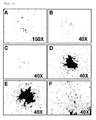
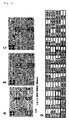
- FIG. 1A The adherent cells were observed to form colonies, exhibiting spindle-shaped morphology 5 ⁇ 30 days after being cultured under the condition of 5% CO 2 ( FIG. 1A ). Once formed, the colonies rapidly proliferated. The cells were suspended with 0.125% Trypsin-EDTA 3 ⁇ 7 days after the formation of colonies, and transferred to fresh dishes where the cells continued to be maintained ( FIGS. 1B, 1C, 1D, 1E and 1F).
- FIG. 1 is of photographs showing the culture progress of the cells. As seen in FIG. 1 , the cell colony grew larger with the lapse of time. In addition even after passage, the morphology of the cells stayed uniform ( FIG.1 . isolation and proliferation of the umbilical cord blood-derived pluripotent/multipotent stem cells. Colonies formed after the monocytes were cultured for 14 days (A), 15 days (B), 16 days (C), 17 days (D) and 18 days (E), and cells after passage 3 (F)).
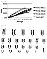
- umbilical cord blood-derived pluripotent/multipotent stem cells of three kinds were isolated from different samples of umbilical cord blood and were seeded at a density of 2x10 5 cells on 100 dishes and passaged at regular intervals of three to four days while being counted using a cytometer. Cell counting was performed until the cells stopped growing.
- CPDL values were calculated while the cells continued to be maintained.
- hUCB-EPC human umbilical cord blood derived endothelial progenitor cells
- FIG. 2 The results are shown in FIG. 2 .
- hUCB-EPC had a CPDL of about 20 over two months whereas the CPDL of all the umbilical cord blood-derived pluripotent/multipotent stem cells from three different samples were observed to range between 40 and 45.
- chromosomes of the umbilical cord blood-derived pluripotent/multipotent stem cells isolated according to the present invention are normal.
- a karyotype analysis was performed to examine the chromosomes of the cells. As can be seen in FIG. 3 , the cells were found to have a normal chromosomal structure even after passage 10.
- Flow cytometry was carried out to analyze the characteristics of the cells suspended in media.
- FITC fluorescein isothiocyanate
- PE phycoerythrin
- the surface antigens used to analyze the characteristics of the umbilical cord blood-derived pluripotent/multipotent stem cells (partially pluripotent/multipotent stem cell) isolated in the present invention included CD10 (T cell marker), CD14 (monocyte marker), CD24 (epithelial cell marker), CD29 (monocyte marker), CD31 (endothelial cell marker), CD34 (hematopoietic stem cell marker), CD44 (mesenchymal stem cell marker), CD45 (non-hematopoietic stem cell marker), CD51/61 (osteoclast marker), CD73 (mesenchymal stem cell marker), CD90 (mesenchymal stem cell marker), CD105 (mesenchymal stem cell marker), CD133 (hematopoietic stem cell marker), and HLA-DR(immunorejection-related marker), and were analyzed using a flow cytometer.
- EXAMPLE 3 Expression Pattern of ZNF281 and Core transcription factor in the Umbilical Cord Blood-Derived Pluripotent/Multipotent Stem Cells
- ZNF281 (Zinc finger protein 281) is one of the core transcription factors of ESC ( Wang J et al. (2006) Nature 444, 364-368 ). ZNF281, also called ZBP-99, contains four Kruppel-type zinc fingers that collectively share 91% amino acid sequence similarity and 79% sequence identity with those found in ZBP-89. In addition, there are highly conserved amino acid sequences in the carboxy-terminal segments of the two genes. The predicted open reading frame of ZNF281 cDNA encodes a 99-kDa protein.
- EMSA Electrophoretic mobility shift as say
- ZNF281 protein specifically binds to the GC-rich promoter elements of GASTRIN and ORNITHINE DECARBOXYLASE genes ( Law DJ et al. (1999) Biochem Biophys Res Commun 262, 113-120 ; Lisowsky T et a 1. (1999) FEBS Lett 453, 369-374 ).
- ZNF281 was identified as a c-MYC-associated protein by mass spectral multidimensional protein identification technology and tandem affinity purification ( Koch HB et al. (2007) Cell Cycle 6, 205-217 .).
- Oct3/4 genes such as POU family transcription factors, are known to not exist in differentiated tissues, but to be expressed particularly in undifferentiated stem cells which have a high proliferative capacity ( Tai M-H. et al., Carcinogenesis 26:4 95, 2005 ; Tondreau T. et al., Stem cells, 23:1105, 2005 ).
- Oct3/4 are therefore used as markers for embryonic stem cells and also as markers indicative of undifferentiation.
- the cell colonies were stained using Oct4 as a marker for stemness. As a result, many cells were found to have Oct4 stained around the nucleus. Flow cytometry also demonstrated the Oct4 expression in the cells.
- the cells were fixed at 4°C overnight with 4% formaldehyde and permeabilized for 10 min with 0.1% Triton X-100 (Sigma-Aldrich).
- the slides and the dishes were incubated for one hour with an anti-human Oct4 mouse primary antibody (1:200), washed with PBS (phosphate buffered saline; Gibco), immunostained for one hour with an Alexa594-conjugated, goat anti-mouse IgG secondary antibody (Invitrogen) while the nucleus was countstained with DAPI.
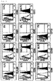
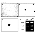
- FIGS. 5 and 6 many of the umbilical cord blood-derived pluripotent/multipotent stem cells were found to express Oct4 ( FIG. 5A : flow cytogram of Oct4-expressing cells, B: image of immunostained Oct4 C: image of the nucleus stained in Oct4 expressed cells, D: overlapped image of Oct4 expression and nucleus staining).
- the cells were induced to differentiate into osteoblasts.
- the cells were allowed to adhere to culture dishes and incubated to about 70-80% confluency.
- the culture medium was replaced with an osteogenic differentiation-inducing medium.
- the osteogenic differentiation-inducing medium was prepared by supplementing DMEM low glucose with 10% FBS plus 10 mM beta-glycerophosphate (Sigma-Aldrich), 0.1 ⁇ M D examethasone (Sigma-Aldrich), and 50 ⁇ M ascorbate (Sigma-Aldrich).
- the medium was replaced with a fresh one every third day while differentiation was induced for about two weeks.
- the calcium mineralization attributed to the osteogenic differentiation was examined by Alizarin red S staining.
- the staining was conducted as follows. After the medium was removed, the cells were washed twice with distilled water and fixed at 4°C for one hour in cold 70% EtOH. Then, the cells were washed again twice with distilled water and stained at room temperature for 10 min with 40mM Alizarin red S, followed by washing five times with distilled water.
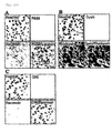
- FIGS. 8A and 8B in FIG. 8A no calcium was detected by Alizarin red S in the absence of differentiation whereas in FIG. 8B calcium appeared red when the cells were induced to differentiate into osteoblasts, which indicates that the umbilical cord blood-derived pluripotent/multipotent stem cells differentiated into osteoblasts releasing calcium.
- the cells were induced to differentiate into adipose cells.
- the cells were allowed to adhere to culture dishes and cultured to about 70-80% confluency.
- the medium was replaced with an adipocyte differentiation medium.
- the adipocyte differentiation medium was prepared by supplementing DMEM low glucose with 10% FBS plus 1 ⁇ M Dexamethasone, 10 ⁇ g/ml insulin (Sigma-Aldrich), 0.5 mM 3-isobutyl-1-methylxanthine (Sigma-Aldrich), and 0.2 mM indomethacin (Sigma-Aldrich).
- the differentiation medium was replaced with a fresh one every third day while differentiation was induced for about 2-3 weeks.
- adipocyte differentiation was examined by Oil red O staining.
- the medium was removed and the cells were washed in PBS and incubated at room temperature for 5 min in 10% formalin.
- the formalin was replaced with the same volume of a fresh one, followed by fixing the cells at room temperature for at least one hour.
- the cells were washed with 60% isopropanol.
- the alcohol was completely evaporated before the cells were stained at room temperature for 10 min with Oil Red O. Immediately after this dye was removed, the cells were washed in distilled water.
- Adipocytes appear red upon treatment with Oil red O because it stains lipid droplets red.
- Oil red O because it stains lipid droplets red.
- FIGS. 8C and 8D in FIG. 8C neither lipid droplets nor the red appearance was detected in the absence of adipocyte differentiation whereas in FIG. 8D when induced to differentiate into adipocytes, the cells were observed to have many lipid droplets and thus turn red).
- the cells were treated for three weeks with the rTGF-beta 3-containing chondrogenic differentiation medium (PT-3003) from Lonza, while the medium was replaced twice a week with a fresh one. Chondrogenesis was measured once a week.
- PT-3003 the rTGF-beta 3-containing chondrogenic differentiation medium
- toluidine blue staining was conducted. The cells were fixed for 10 hours with 4% formaldehyde and then for an additional 10 hours with picric acid. After cryosection, the cells were stained for 3 min with toluidine blue and counterstained for 3 sec staining with hematoxilin.
- a pellet of chondrocytes does not collapse, but maintains a constant morphology and appears blue when stained with toluidine blue.
- FIGS. 8E and 8F the cells subjected to chondrogenic differentiation were observed to be stained blue and maintain their morphology.
- EXAMPLE 7 Change in Gene Expression Level after Differentiation of the Umbilical Cord Blood-Derived Pluripotent/Multipotent Stem Cells into Osteoblasts and Adipocytes
- the cells were induced to differentiate into neurons.
- the cells were pre-incubated for 24 hours in DMEM supplemented with 5% FBS and 10ng/ml bFGF (basic Fibroblast Growth Factor). Thereafter, the cells were treated for 24 hours in DMEM containing 1% DMSO, 100 ⁇ M BHA, 0.5mM VPA, 10mM KCl, and 10 ng/ml NGF, and B27 to induce neural differentiation.
- the cells were fixed with 4% paraformaldehyde and immunostained for the neural markers Tuj-1, MAP-2, GFAP, and Neurofilament-160. As a result, the four markers were observed to be expressed ( FIG. 8G ).
- the retina-related characteristics of the umbilical cord blood-derived pluripotent/multipotent stem cells were examined using immunofluorescence.
- FIG. 10 is of photographs showing expression patterns of retina-specific proteins as measured by immunofluorescence.
- the expression patterns of PAX6 and Hu protein are shown in FIG. 10A .
- PAX6 is known as a retina progenitor marker and Hu protein is expressed specifically in gaglion cells and amacrine cells, which are constituents of the retina. They were not detected in a normal culture.
- opsin and rhodopsin are detected. Opsin is expressed specifically in cone cells while rhodposin is specific for rod cells. In a normal culture, opsin was not detected, but expression of rhodopsin was observed.
- CRX is known as a pan-photoreceptor marker
- the therapeutic capacity of stem cells is largely attributable to two effects: first, direct differentiation of stem cells into impaired cells; and secondly, the ability to secrete various cytokines or growth factors that induce positive alterations that lead to a therapeutic effect on pre-existing cells.
- stem cells are known to secrete various cytokines or growth factors, showing so-called paracrine effects ( Kim et al. Cytokine. 2005 ).
- a human Cytokine antibody array (RaybioTech. Norc ross, USA) was used.
- FIG. 11 shows array analysis results of hUCB-MSC1 (A), hUCB-MSC2 (B) and hUCB-MSC3 (C) and an arrangement of antibodies (D)).
- EXAMPLE 11 3-Dimensional Culture of the Umbilical Cord Blood-Derived Pluripotent/Multipotent Stem Cells
- the culture dishes were coated with 0.7% agarose to a thickness of 5 mm or more so that the cells could not adhere to the bottom, but formed spheres.
- the cells were seeded at a density of less than 2000 cells/cm 2 to prevent adhesion between single cells.
- the spheres thus formed were separated from single cells using a 40 ⁇ m strainer. As can been seen in FIG.
- the stem cells did not undergo cell death, but formed spheres, maintaining the characteristics of stem cells when they were cultured in a sphere culture system.
- the expression levels of embryonic markers such as OCT4, SOX2 and the like in cells that were maintained in a sphere culture were observed to be higher compared to those which were maintained as a monolayer.
- Embryonic stem cells are typically cultured on a layer of mouse embryonic fibroblast cells because the kemokines, such as LIF, from the mouse embryonic fibroblast cells play an important role in maintaining the morphology of ES cells and preventing the differentiation of ES cells.
- the umbilical cord blood-derived pluripotent/multipotent stem cells did not grow in a flat shape like typical adult stem cells, but formed 3-dimensional colonies.
- STO cells were seeded at a density of 2 x 10 5 cells/ml on 0.1% gelatin-coated dishes and incubated for 24 hours, followed by seeding the umbilical cord blood-derived pluripotent/multipotent stem cells on the STO cell layer. As seen in FIG. 13 , the cells were observed to proliferate on the STO cells in a pattern which was similar to that of embryonic stem cell with the lapse of time.
- the human umbilical cord blood-derived pluripotent/multipotent stem cells of the present invention When cultured in the presence of fibronectin, the human umbilical cord blood-derived pluripotent/multipotent stem cells of the present invention actively proliferate for a longer period of time without differentiating, compared to conventional adult stem cells.
- the pluripotent/multipotent stem cells of the present invention can be effectively applied to the treatment of conventionally incurable diseases, as well as neural diseases, cardiovascular diseases, and diseases of the skeletal system.
Landscapes
- Health & Medical Sciences (AREA)
- Life Sciences & Earth Sciences (AREA)
- Engineering & Computer Science (AREA)
- Biomedical Technology (AREA)
- Organic Chemistry (AREA)
- Chemical & Material Sciences (AREA)
- Bioinformatics & Cheminformatics (AREA)
- Wood Science & Technology (AREA)
- Zoology (AREA)
- Genetics & Genomics (AREA)
- Biotechnology (AREA)
- General Health & Medical Sciences (AREA)
- Biochemistry (AREA)
- Microbiology (AREA)
- Cell Biology (AREA)
- General Engineering & Computer Science (AREA)
- Developmental Biology & Embryology (AREA)
- Gynecology & Obstetrics (AREA)
- Reproductive Health (AREA)
- Hematology (AREA)
- Nuclear Medicine, Radiotherapy & Molecular Imaging (AREA)
- Medicinal Chemistry (AREA)
- Chemical Kinetics & Catalysis (AREA)
- General Chemical & Material Sciences (AREA)
- Pharmacology & Pharmacy (AREA)
- Veterinary Medicine (AREA)
- Public Health (AREA)
- Animal Behavior & Ethology (AREA)
- Immunology (AREA)
- Pregnancy & Childbirth (AREA)
- Neurology (AREA)
- Physical Education & Sports Medicine (AREA)
- Neurosurgery (AREA)
- Heart & Thoracic Surgery (AREA)
- Cardiology (AREA)
- Micro-Organisms Or Cultivation Processes Thereof (AREA)
- Medicines Containing Material From Animals Or Micro-Organisms (AREA)
Abstract
Description
- The present invention relates to a method for isolating pluripotent/multipotent stem cells derived from umbilical cord blood, characterized by culturing monocytes isolated from umbilical cord blood in a culture vessel containing fibronectin and then harvesting stem cells from the culture, the umbilical cord blood-derived pluripotent/multipotent stem cells isolated thereby; and a cell therapeutic agent containing the pluripotent/multipotent stem cells derived from umbilical cord blood or cells differentiated therefrom. The present invention also relates to a novel culture media for stem cells, a culture method for stem cells which is characterized by culturing and proliferating stem cells in the culture media, and a method for increasing stemness of stem cells which is characterized by a sphere culture or a three-dimensional culture of stem cells.
- Characterized by being self-renewing, undergoing differentiation and being immortal, stem cells have been proposed to be a solution for the problems of regenerative medicine and tissue replacement so that they can be used to treat various degenerative diseases as well as to provide deep insight into cellular biology. Adult stem cells, obtainable from various tissues, are far more attractive than embryonic stem cells because of the ability to come from an unlimited number of sources and the ethical objections to using human embryos as a source of cells are rendered irrelevant. Moreover, stem cells isolated from umbilical cord blood have advantages over other adult stem cells in that the donors of umbilical cord blood are not injured, unlike the donors of bone marrow or adipose tissue.
- Umbilical cord blood-derived mesenchymal stem cells have been successfully introduced into various kinds of cells including neural cells, hepatocytes, osteocytes, etc., in vitro (Sun, W. et al., Stem cells, 23:931, 2005; Hong SH. et al., Biochem. Biophys. Res. Commun., 30:1153, 2005; Hutson EL. et al., Tissue Engineering, 11:1407, 2005). Successful in vivo transplantation of umbilical cord blood-derived mesenchymal stem cells for injuries, diabetes mellitus and heart infarction were also reported (Nonome, K. et al., Am, J. Physiol. Gastrointest. Liver Physiol., 289:1091, 2005; Yoshida, S. et al., Stem cells, 23:1409, 2005; Kim Bo. et al., Circulation, 112:96, 2005). Due to the low possibility of infectious diseases being transmitted and the low possibility of causing graft-versus-host diseases, the transplantation of umbilical cord blood-derived mesenchymal stem cells has been increasingly applied to both child and adult patients (Claudio G. B. et al., Annual Review of Medicine, 57:403, 2006). Although umbilical cord blood transplantation has been accepted as a therapy for some diseases, particularly hematopoietic defection-related diseases (Grewal, SS. et al., Blood, 103:1147, 2004; Knutsen, AP. et al., Journal pediatrics, 142:519, 2003; Ooi, J. et al., Blood, 103:489, 2004; Sanz GF. et al., Blood, 103:489, 2004), studies on the clinical application of umbilical cord blood-derived mesenchymal stem cells are still limited. For example, there are reports on stem cells being used to successfully cure, not completely but partially, women having spinal cord injuries and patients with Buerger's disease (Kim, SW. et al., Stem Cells, 2006; Kang, KS. et al., Cytotherapy, 7:368, 2005). However, the mechanism of development of the cells or how to grow and proliferate them still remains unknown.
- It is difficult to isolate mesenchymal stem cells from umbilical cord blood. For example, when the method used to isolate bone marrow-derived mesenchymal stem cells is applied to umbilical cord blood, the isolation rate remains at around 20%. As much as 50% of the mesenchymal stem cells may be isolated from flesh blood which has been taken 5 hours before the isolation. However, the isolation rate is decreased to 20% or less when the blood has been taken over 5 hours before doing the isolation and the cells, although isolated, do not proliferate well.
- Leading to the present invention, intensive and thorough research into the isolation and mass proliferation of stem cells from umbilical cord blood, conducted by the present inventors, resulted in the finding that when cultured in the presence of fibronectin, monocytes isolated from human umbilical cord blood highly proliferate and form colonies, exhibiting a spindle shaped morphology, and maintain stem cell properties even after many passages. Therefore, based on the finding, a method is provided for effectively isolating umbilical cord blood-derived non-hematopoietic pluripotent/multipotent stem cells and mesenchymal stem cells and for proliferating them on a mass scale.
- It is therefore an object of the present invention to provide a method for isolating umbilical cord blood-derived pluripotent/multipotent stem cells, comprising culturing monocytes isolated from umbilical cord blood in the presence of fibronectin and recovering stem cells from the culture.
- It is another object of the present invention to provide umbilical cord blood-derived pluripotent/multipotent stem cells isolated by the isolation method.
- It is a further object of the present invention to provide a cell therapeutic agent comprising the umbilical cord blood-derived pluripotent/multipotent stem cells or cells differentiated therefrom.
- It is still a further object of the present invention to provide a novel culture medium for stem cells.
- It is still another object of the present invention to provide a method for culturing stem cells, using the culture medium.
- It is a method for increasing stemness of stem cells, using a sphere culture or three-dimensional culture of stem cells.
- In accordance with an aspect thereof, the present invention pertains to a method for isolating umbilical cord blood-derived stem cells, characterized by culturing monocytes isolated from umbilical cord blood in a culture vessel containing fibronectin and harvesting the stem cells from the culture.
- In one embodiment, the step of harvesting the stem cells from the culture comprises utilizing an immunological property of stem cells to separate the stem cells.
- The isolation of monocytes from umbilical cord blood may be achieved using a typical method. After mixing the umbilical cord blood with Hetasep to deplete erythrocytes, monocytes are isolated using Ficoll-paque. Herein, Hetasep is preferably used in an amount of 0.5 ∼ 2 mL per 5 mL of Hetasep.
- To increase the isolation yield of monocytes therefrom, the umbilical cord blood used in the present invention is preferably one that is recovered immediately after childbirth, one that is stored at room temperature for 12∼48 hours after the recovery, or one that is stored at 3∼5°C for 6∼72 hours after the recovery.
- The isolation of stem cells from the umbilical cord blood-derived monocytes is characterized by the use of fibronectin. The term "culture vessel containing fibronectin," as used herein, is intended to mean a condition in which monocytes can be brought into contact with fibronectin. For example, fibronectin may be layered on a culture vessel or may be contained in the form of spheres or three-dimensional structures in a culture medium. In one embodiment of the present invention, when the culture vessel is coated therewith, fibronectin may be contained at a density of from 0.1 to 1 mg/mL.
- The fibronectin useful in the present invention may be derived from animals without limitation and preferably from humans. Also, the fibronectin may be prepared by artificial synthesis (e.g., chemical synthesis, synthesis using a peptide synthesizer, etc.) or biosynthesis (e.g., recombinant DNA technology, fibroblast culture, etc.) or may be separated from the plasma of animals including humans from the extracellular matrices. The fibronectin may be a fragment or peptide sequence of fibronectin or may contain the fragment or peptide.
- No particular limitations are imparted to the medium available when the monocytes are cultured in the culture vessel containing fibronectin. SNU-1 or EGM-2 is preferably used as a fundamental medium for culturing the monocytes.
- The SNU-1 medium contains the following composition (Table 1).
TABLE 1 SNU-1(mg/l) SNU-1 (mg/l) SNU-1 (mg/l) CaCl2(anhyd.) 200 L-Isoleucine 78 i- Inositol 3 KCL 400 L-Leucine 78 Riboflavin 0.15 MgSO4(anhyd.) 97.67 L-Lysine HCl 108.75 Thiamine HCl 1.5 NaCl 7635 L-Methionine 22.5 L-Alanine 17.8 NaH2PO4 H2O 140 L-Phenylalanine 48 L-Asparagine H2O 30 D-Glucose 1000 L-Serine 21 L-Aspartic Acid 26.6 Phenol Red 10 L-Threonine 72 L-Glutamic acid 29.4 Sodium Pyruvate 110 L-Tryptophan 15 L-Proline 23 L-Arginine HCl 189 L-Tyrosine 2Na 2H2O 54 Nicotinamide 1.5 L-Cysteine 2HCl 36 L-Valine 69 Pyridoxine Hcl 1.5 L-Glutamine 292 D-Ca pantothenate 1.5 NaHCO3 1000 Glycine 15 Choline Chloride 1.5 L-Histidine HCl H2O 63 Folic Acid 1.5 - In the present invention, the fundamental medium is preferably supplemented with FGF-B (Fibroblast Growth Factor), ascorbic acid, EGF (Epidermal Growth Factor), hydrocortisone, IGF-1 (Insulin-like Growth Factor-1) or VEGF (Vascular Endothelial Growth Factor), and heparin and optionally with GA-1000 (Gentamycin Sulfate, Amphotericin-B)if needed.
- More preferably, the fundamental medium is supplemented with fetal bovine serum (FBS) 20%, bFGF (Fibroblast Growth Factor) 1∼40 ng/ml, ascorbic acid 0.1∼5.0 µg/ml, EGF (Epidermal Growth Factor) 1∼40 ng/ml, hydrocortisone 0.1∼1 µg/ml, IGF-I (Insulin-like Growth Factor-1) 1∼40 ng/ml or VEGF (Vascular Endothelial Growth Factor) 1∼5 ng/ml and
heparin 20∼25µg/ml and optionally with GA-1000 (Gentamycin Sulfate, Amphotericin-B) if needed. - After being cultured for three days, the monocytes which remain suspended are removed while only adherent cells are cultured. Among the monocytes adherent to the vessel, only stem cells are proliferated. From 12 to 20 days after the isolation, rapid proliferation of the stem cells can be observed. In this context, the medium is preferably replaced with a fresh one every two or three days.
- To obtain umbilical cord blood-derived pluripotent/multipotent stem cells from the culture, FACS using a flow cytometer with a sorting function (Int. Immunol., 10(3):275, 1998), magnetic beads, or a panning method based on an antibody specific for mesenchymal stem cells (J. Immunol., 141(8):2797, 1998) may be used. To obtain pluripotent/multipotent stem cells from a mass volume of culture, a column to which antibodies specific for molecules expressed on cell surfaces are immobilized (hereinafter referred to as "surface antigen") alone or in combination may be used.
- Flow cytometry sorting may be performed by electrostatic droplet charging or cell capture. In either of the processes, an antibody specifically recognizing a surface antigen is fluorescence labeled after which the fluorescent intensity is measured from the labeled antibody-antigen conjugate and converted into electric signals to determine the expression level of the antigen of the cells. Further, different kinds of fluorophores may be used in combination to separate cells expressing different surface antigens. Among the fluorophores are FITC (fluorescein isothiocyanate), PE (phycoerythrin), APC (allo-phycocyanin), TR (TexasRed), Cy3, CyChrome, Red613, Red670, TRI-Color, and QuantumRed.
- In FACS using a flow cytometer, the stem cells harvested from the culture by, for example, centrifugation may be immunostained directly with an antibody or may be proliferated in a suitable medium before immunostaining with an antibody. For the immunostaining, a target cell sample is mixed with a primary antibody specific for a surface antigen and incubated for 0.5 to 1 hour on ice. If the primary antibody is labeled with a fluorophore, the cell sample is washed and separated with a flow cytometer. If the primary antibody is not labeled with a fluorophore, the cell sample treated with the primary antibody is washed and mixed with a fluorescence-labeled secondary antibody that can bind to the primary antibody. Then, the immunostained cells are incubated again for 0.5 to 1 hour on ice and washed before being separated by a flow cytometer.
- The umbilical cord blood-derived pluripotent/multipotent stem cells isolated according to the present invention have at least one of the following properties:
- (a) showing a positive immunological characteristic to the transcription factors c-myc and ZNF281.
- (b) adhering to a extracellular matrix-coated surface and forming cell colonies in a spindle or
spherical shape 5 to 30 days after adhesion. - (c) showing a CPDL (cumulative population doubling level) of from 30 to 45.
- (d) showing a negative immunological characteristic to CD14, CD31, CD34, CD45 and HLA-DR.
- (e) ability to differentiate into mesodermal, endodermal and ectodermal cells.
- (f) secreting at least one cytokine or chemokine selected from the group consisting of TIMP-2, TGF-β, RANTES CINC-3, EOTAXIN, GM-CSF, IFN-γ, IL-1b IL-3, IL-6, IL-8, IL-10, IL12p40, IL13, IL-16, IP-10, Leptin, MCP-2, MIG, MIP-3a, b-NGFm, sTNFRI, and PFGF-bb.
- The fact that the umbilical cord blood-derived pluripotent/multipotent stem cells of the present invention remain undifferentiated is evident from the expression of Oct-4, Sox-2, Rex-1, c-myc, and ZNF281 in the stem cells.
- In addition, showing a CPDL (cumulative population doubling level) of from 30 to 45, the umbilical cord blood-derived pluripotent/multipotent stem cells of the present invention is highly proliferative. A karyotype analysis demonstrated that the cells of the present invention rapidly proliferate, but have a normal chromosomal structure.
- The umbilical cord blood-derived pluripotent/multipotent stem cells of the present invention are immunologically negative to CD14, CD31, CD34, CD45 and HLA-DR, all known as hematopoietic stem cell markers or immunorejection-related markers. Due to the lack of such hematopoiesis- or immunorejection-related markers, the umbilical cord blood-derived stem cells of the present invention can be transplanted with minimal vascularization and immunorejection and thus can be effectively used in allogenic transplantation.
- The umbilical cord blood-derived pluripotent/multipotent stem cells of the present invention also may differentiate into hepatocytes from the mesoderm and neurons and retina-related cells from the ectoderm as well as osteoblasts, chondrocytes and adipose cells from the mesoderm. Therefore, the umbilical cord blood-derived pluripotent/multipotent stem cells of the present invention can be used to treat a variety of diseases.
- Further, the umbilical cord blood-derived pluripotent /multipotent stem cells of the present invention may secrete various cytokines or chemokines including TIMP-2, TGF-β, RANTES CINC-3, EOTAXIN, GM-CSF, IFN-γ, IL-1b, IL-3, IL-6, IL-8, IL-10, IL12p40, IL13, IL-16, IP-10, Leptin, MCP-2, MIG, MIP-3a, b-NGFm, sTNFRI, and PFGF-bb. Having the ability to secrete these cytokines or chemokines, the umbilical cord blood-derived pluripotent/multipotent stem cells of the present invention can be applied to the treatment of various diseases.
- The novel nature of the umbilical cord blood-derived pluripotent/multipotent stem cells of the present invention comes from their having these characteristics.
- As described above, the umbilical cord blood-derived pluripotent/multipotent stem cells of the present invention can be differentiated into various types of cells including osteoblasts, chondrocytes, adipose cells, hepatocytes and neurons, and thus find applications in therapies for various corresponding diseases. Therefore, the present invention provides a cell therapeutic agent comprising the umbilical cord blood-derived pluripotent/multipotent stem cells of the present invention or the cells differentiated therefrom. The cell therapeutic agent of the present invention can be applied to the treatment of various diseases including, for instance, neural diseases (e.g., degenerative neural diseases), osteoarthritis (e.g., degenerative arthritis, rheumatoid arthritis), bone loss (e.g., osteoporosis), hepatic diseases (e.g., hepatocirrhosis), and cardiovascular diseases.
- Preferably, the cell therapeutic agent of the present invention contains at least one diluent that can protect and maintain the cells. Buffered solutions such as physiological saline, PBS (Phosphate Buffered Saline), HBSS (Hank's balanced salt solution), and plasma or serum components may be used as the diluents.
- In accordance with another aspect thereof, the present invention addresses a medium for culturing the novel stem cells. The medium is based on the EGM-2 or SNU-1 medium and supplemented with fetal bovine serum (FBS) 20%, bFGF (Fibroblast Growth Factor) 1∼40ng/ml, ascorbic acid 0.1∼5.0µg/ml, EGF (Epidermal Growth Factor) 1∼40ng/ml, hydrocortisone 0.1∼1µg/ml, IGF-1 (Insulin-like Growth Factor-1) 1∼40ng/ml or VEGF (Vascular Endothelial Growth Factor) 1∼5ng/ml and
heparin 20∼25µg/ml, and optionally with GA-1000 (Gentamycin Sulfate, Amphotericin-B) if needed. - The culturing medium for stem cells is novel and is used in the method for isolating umbilical cord blood-derived pluripotent/multipotent stem cells according to the present invention. Further, the culture medium of the present invention is useful for proliferating all adult stem cells including umbilical cord blood-derived stem cells and is applicable to the culturing of adult stem cells.
- In accordance with a further aspect thereof, the present invention addresses a method for culturing stem cells comprising culturing and proliferating stem cells in the medium of the present invention. Preferably, the stem cells may be adult stem cells.
- In one embodiment, the medium for culturing stem cells in accordance with the present invention may be used to culture the umbilical cord blood-derived pluripotent/multipotent stem cells of the present invention. While being cultured in the medium of the present invention, the umbilical cord blood-derived pluripotent/multipotent stem cells of the present invention may be preferably passaged 3∼5 days after the formation of cell colonies in a spindle form. Cultivation is preferably performed in a 5% CO2 condition and may be carried out for 5∼30 days, but the present invention is not limited to these.
- In accordance with still a further aspect thereof, the present invention addresses a method for increasing stemness of stem cells, characterized by a sphere culture or a three-dimensional culture of stem cells whereby stem cells are cultured. For the three-dimensional culture, MEF (Mouse embryonic fibroblast cell) is preferably used. Further, the stem cells may preferably be adult stem cells.
- As used herein, the term "increasing stemness" means forming an embryonic stem cell-like colony or expressing transcription factors such as Oct4, Sox2, etc. at a higher level.
- When cultured in the presence of fibronectin, as explained in detail above, the human umbilical cord blood-derived pluripotent/multipotent stem cells of the present invention actively proliferate for a longer period of time without differentiating, compared to conventional adult stem cells. In addition, having the ability to differentiate to various kinds of cells such as chondrocytes, osteoblasts, and adipose cells, the pluripotent/multipotent stem cells of the present invention can be effectively applied to the treatment of conventionally incurable diseases, as well as neural diseases, cardiovascular diseases, and diseases of the skeletal system.
-
-
FIG. 1 is of photographs showing colonies formed after the monocytes isolated from human umbilical cord blood were cultured for 14, 15, 16, 17 and 18 days (A, B, C, D and E, respectively) and cells after passage 3 (F); -
FIG. 2 is a graph in which cumulative cell growth is plotted against time. -
FIG. 3 shows karyotype analysis results after the umbilical cord blood-derived pluripotent/multipotent stem cells are cultured for a long period of time. -
FIG. 4 is of flow cytograms showing the expression patterns of various markers on the umbilical cord blood-derived pluripotent/multipotent stem cells of the present invention. -
FIG. 5 shows the expression patterns of various undifferentiation markers on the umbilical cord blood-derived pluripotent/multipotent stem cells of the present invention as measured by flow cytometry and immunostaining (A: flow cytogram of Oct4-expressing cells, B: image of immunostained Oct4 C: image of the nucleus stained in Oct4 expressed cells, D: overlapped image of Oct4 expression and nucleus staining). -
FIG. 6 shows the expression of ZNF281 in terra-1, hUCB-MSC, AD-MSC and AM (upper panel) and in hUCB-MSC after 3∼9 passages (lower panel) as analyzed by FACS. -
FIG. 7 shows the expression of ZNF281, Oct4, Sox2, c-myc and Rex-1, all essential for the maintenance of undifferentiation, in the human umbilical cord blood-derived pluripotent/multipotent stem cells of the present invention, as measured by RT-PCR. -
FIG. 8 is of photographs showing the differentiation of human umbilical cord blood-derived pluripotent/multipotent stem cells into osteoblasts, adipocytes, chondrocytes and neurons (A: non-osteogenic differentiation-induced control after staining with Alizarin Red S, B: osteogenic differentiation-induced cells after staining with Alizarin Red S, C: non-adipogenic differentiation-induced control after staining with Oil Red O, D: adipogenic differentiation-induced cells after staining with Oil Red O, E: chondrogenic differentiation-induced cells after staining with Toluidine Blue, F: pellet of chondrogenic differentiation-induced cells, G: cells immunostained with neural markers Tuj-1 and MAP2 after neural differentiation). -
FIG. 9 shows RNA levels of the umbilical cord blood-derived pluripotent/multipotent stem cells after the induction of osteogenic differentiation and adipogenic differentiation, as measured by RT-PCR (PPAR-γ and FABP-4 are adipogenic differentiation markers andCollagen Type 1 is an osteogenic differentiation marker. PPAR-γ: Peroxisome Proliferator-Activated Receptor gamma, FABP-4: Fatty Acid Binding Protein-4, GAPDH: Glyceraldehyde-3-phosphate dehydrogenase ). -
FIG. 10 shows expression patterns of retina-related proteins in the umbilical cord blood-derived pluripotent/multipotent stem cells of the present invention. -
FIG. 11 shows the secretion of various cytokines to culture media of the human umbilical cord blood-derived pluripotent/multipotent stem cells, as analyzed by an antibody array (A: hUCB-MSC1, B: hUCB-MSC2, C: hUCB-MSC3, D: an arrangement of antibodies, POS: Positive control, NEG: Negative control, GCSF: Granulocyte-Colony Stimulating Factor, GM-CSF: Granulocyte Monocyte-Colony Stimulating Factor, ICAM-1: Intra-Cellular Adhesion Molecule, IFN-γ: Interferon-γ, IL: Interleukin, MCP: Monocyte Chemoattractant Protein, M-CSF: Monocyte-Golony Stimulating Factor, MIG: Monokine induced by Interferon Gamma, MIP: Macrophage Inflammatory Protein, RANTES: Regulated upon Activation, Normal T-cell Expressed and Secreted, TGF-β: Transforming Growth Factor-β, TNF: Tumour Necrosis Factor, sTNFR: soluble Tumour Necrosis Factor Receptor, PDGF-BB: Platelet-Derived Growth Factor-BB, TIMP2: Tissue Inhibitor of Metalloproteinases-2). -
FIGS. 12 and13 are photographs showing three-dimensional cultures of the umbilical cord blood-derived pluripotent/multipotent stem cells using a sphere culture process and STO cells and the expression patterns of the transcription factors Oct4 and Sox2 as measured by RT-PCR (FIG. 12 : a three-dimensional culture of umbilical cord blood-derived pluripotent/multipotent stem cells by a sphere culture process,FIG. 13 : a three-dimensional culture of umbilical cord blood-derived pluripotent/multipotent stem cells on STO cells). - A better understanding of the present invention may be obtained through the following examples which are set forth to illustrate, but are not to be construed as limiting the present invention.
- Full term UCB samples (n=20) were collected immediately after delivery with the informed consent of the mothers. The full term UCB was mixed with HetaSep (Stem cells Technologies INC, Vancouver, BC) to deplete erythrocyte counts. Then, monocytes were obtained by a typical Ficoll density gradient centrifugation. The monocytes were seeded at a density of 1 x 10⁵∼8 cells/well on 6-well plates coated with 0.1mg/ml - 1 mg/ml fibronectin and maintained in SNU-1 or EGM-2 (Lonza) supplemented with EGM-2 SingleQuots containing 20% FBS. The EGM-2 SingQouts contained heparin, Ascorbic acid, rhEGF, hydrocortisone, VEGF, rhFGF-B, R3-IGF-1, and GA-1000. The monocytes which still remained non-adherent three days after culturing were removed while the medium was replaced with a fresh one every two or three days.
- The adherent cells were observed to form colonies, exhibiting spindle-shaped
morphology 5∼30 days after being cultured under the condition of 5% CO2 (FIG. 1A ). Once formed, the colonies rapidly proliferated. The cells were suspended with 0.125% Trypsin-EDTA 3∼7 days after the formation of colonies, and transferred to fresh dishes where the cells continued to be maintained (FIGS. 1B, 1C, 1D, 1E and 1F). FIG. 1 is of photographs showing the culture progress of the cells. As seen inFIG. 1 , the cell colony grew larger with the lapse of time. In addition even after passage, the morphology of the cells stayed uniform (FIG.1 . isolation and proliferation of the umbilical cord blood-derived pluripotent/multipotent stem cells. Colonies formed after the monocytes were cultured for 14 days (A), 15 days (B), 16 days (C), 17 days (D) and 18 days (E), and cells after passage 3 (F)). - While they continued to be cultured, the capacity of the isolated umbilical cord blood-derived pluripotent/multipotent stem cells to proliferate was analyzed by measuring CPDL (cumulative population doubling level) (Cristofalo et al., Proc. Natl. Acad. Sci., USA 95, 1998). Cells divide by binary fission. Hence, the growth rate of cells can be determined by the time which it takes for one cell to split into two, called the doubling time. A CDPL of 10 means 10 fissions of one cell, which results in the proliferation of one cell to approximately 1000 cells. One of the greatest problems of conventional umbilical cord blood-derived stem cells is that they have a significantly low proliferation capacity, compared to adipose tissue- or bone marrow-derived mesenchymal stem cells. Because clinical applications require a large number of stem cells, the proliferation capacity of stem cells is important. The measurement was performed as follows. First, umbilical cord blood-derived pluripotent/multipotent stem cells of three kinds were isolated from different samples of umbilical cord blood and were seeded at a density of 2x10⁵ cells on 100 dishes and passaged at regular intervals of three to four days while being counted using a cytometer. Cell counting was performed until the cells stopped growing. CPDL values are obtained on the basis of the measurements of cell counts according to the following Math Formula 1

wherein, NI means the number of cells in the initial culture and NH means the number of cells in a saturation condition upon passage. - CPDL values were calculated while the cells continued to be maintained. For comparison, hUCB-EPC (human umbilical cord blood derived endothelial progenitor cells) was used as a control. The results are shown in
FIG. 2 . As can be seen in the graph ofFIG. 2 , hUCB-EPC had a CPDL of about 20 over two months whereas the CPDL of all the umbilical cord blood-derived pluripotent/multipotent stem cells from three different samples were observed to range between 40 and 45. These numerical values indicate that the cells can grow theoretically to the number of 1012 in one colony. - On the whole, cells with a capacity for great proliferation may be made to be cancerous, exhibiting explosive growth. Once cancerized, cells are likely to proliferate irrespective of various regulatory signals in the body, and thus cannot be used as a cell therapeutic agent and are inconsistent with the purpose of the study. Therefore, it must be determined whether the chromosomes of the umbilical cord blood-derived pluripotent/multipotent stem cells isolated according to the present invention are normal. In this context, a karyotype analysis was performed to examine the chromosomes of the cells. As can be seen in
FIG. 3 , the cells were found to have a normal chromosomal structure even afterpassage 10. - Flow cytometry was carried out to analyze the characteristics of the cells suspended in media. For phenotyping cell surface antigens, cells harvested after 3∼4 passages were stained with a fluorescein isothiocyanate (FITC)- or phycoerythrin (PE)-conjugated antibody, and analyzed with FACSAria (Becton Dickinson, NY).
- The surface antigens used to analyze the characteristics of the umbilical cord blood-derived pluripotent/multipotent stem cells (partially pluripotent/multipotent stem cell) isolated in the present invention included CD10 (T cell marker), CD14 (monocyte marker), CD24 (epithelial cell marker), CD29 (monocyte marker), CD31 (endothelial cell marker), CD34 (hematopoietic stem cell marker), CD44 (mesenchymal stem cell marker), CD45 (non-hematopoietic stem cell marker), CD51/61 (osteoclast marker), CD73 (mesenchymal stem cell marker), CD90 (mesenchymal stem cell marker), CD105 (mesenchymal stem cell marker), CD133 (hematopoietic stem cell marker), and HLA-DR(immunorejection-related marker), and were analyzed using a flow cytometer. The results are summarized in Table 2 and graphically depicted in
FIG. 4 .TABLE 2 CD Antigen on Umbilical Cord Blood-derived Pluripotent/multipotent Stem Cell Positively stained cells/total cells (%) CD10 0.2 ± 0.1 CD14 2.0 ± 1.0 CD24 68.7 ± 2.9 CD29 100 ± 0.0 CD31 0.4 ± 0.7 CD34 3.5 ± 3.4 CD44 100 ± 0.1 CD45 0.0 ± 0.1 CD51/61 6.4 ± 10.8 CD73 99.6 ± 0.5 CD90 99.7 ± 0.3 CD105 99.5 ± 0.8 CD133 0.1 ± 0.1 HLA-DR 2.0 ± 3.2 - ZNF281 (Zinc finger protein 281) is one of the core transcription factors of ESC (Wang J et al. (2006) Nature 444, 364-368). ZNF281, also called ZBP-99, contains four Kruppel-type zinc fingers that collectively share 91% amino acid sequence similarity and 79% sequence identity with those found in ZBP-89. In addition, there are highly conserved amino acid sequences in the carboxy-terminal segments of the two genes. The predicted open reading frame of ZNF281 cDNA encodes a 99-kDa protein. EMSA (Electrophoretic mobility shift as say) showed that ZNF281 protein specifically binds to the GC-rich promoter elements of GASTRIN and ORNITHINE DECARBOXYLASE genes (Law DJ et al. (1999) Biochem Biophys Res Commun 262, 113-120; Lisowsky T et a 1. (1999) FEBS Lett 453, 369-374). ZNF281 was identified as a c-MYC-associated protein by mass spectral multidimensional protein identification technology and tandem affinity purification (Koch HB et al. (2007) .).
- Oct3/4 genes, such as POU family transcription factors, are known to not exist in differentiated tissues, but to be expressed particularly in undifferentiated stem cells which have a high proliferative capacity (Tai M-H. et al., Carcinogenesis 26:4 95, 2005; Tondreau T. et al., Stem cells, 23:1105, 2005). On the whole, Oct3/4 are therefore used as markers for embryonic stem cells and also as markers indicative of undifferentiation. The cell colonies were stained using Oct4 as a marker for stemness. As a result, many cells were found to have Oct4 stained around the nucleus. Flow cytometry also demonstrated the Oct4 expression in the cells.
- To stain intracellular proteins, the cells were fixed at 4°C overnight with 4% formaldehyde and permeabilized for 10 min with 0.1% Triton X-100 (Sigma-Aldrich). The slides and the dishes were incubated for one hour with an anti-human Oct4 mouse primary antibody (1:200), washed with PBS (phosphate buffered saline; Gibco), immunostained for one hour with an Alexa594-conjugated, goat anti-mouse IgG secondary antibody (Invitrogen) while the nucleus was countstained with DAPI.
- As seen in
FIGS. 5 and6 , many of the umbilical cord blood-derived pluripotent/multipotent stem cells were found to express Oct4 (FIG. 5A : flow cytogram of Oct4-expressing cells, B: image of immunostained Oct4 C: image of the nucleus stained in Oct4 expressed cells, D: overlapped image of Oct4 expression and nucleus staining). - Genes such as Oct4 are closely associated with the stemness of stem cells. In fact, attempts have recently been made to induce adult stem cells to have pluripotency similar to that of embryonic stem cells by getting them to overexpress genes such as Oct4, Sox2 and the like (Takahashi et al, Cell, 131(5), 861-872, 2007). Accordingly, the expression of these genes in stem cells is very important in keeping the stem cells at an undifferentiated state without losing stemness. Thus, to examine the expression patterns of ZNF281, Oct-4 and Sox2 in the umbilical cord blood-derived pluripotent/multipotent stem cells isolated according to the present invention, RT-PCR was performed.
- For this, primers were constructed as in Table 3.
TABLE 3 Target Gene Direction Sequence SEQ ID NO: OCT-4 sense primer 5'-CGAAAGAGAAAGCGAACCAG-3' 1 antisenseprimer 5'-GCCGGTTACAGAACCACACT-3' 2 SOX2 sense primer 5'-CCTCCGGGACATGATCAG-3' 3 antisenseprimer 5'-TTCTCCCCCCTCCAGTTC-3' 4 C-MYC sense primer 5'-TACCCTCTCAACGACAGCAG-3' 5 antisenseprimer 5'-GGGCTGTGAGGAGGTTTG-3' 6 ZNF281 sense primer 5'-ACGTAACAGCGCAGACAGAA-3' 7 antisenseprimer 5'-GTGTTGAAGCCCAAGTGGTT-3' 8 REX-1 sense primer 5'-TGAAAGCCCACATCCTAACG-3' 9 antisenseprimer 5'-CAAGCTATCCTCCTGCTTTGG-3' 10 - As seen in
FIG. 7 , Oct-4, Sox2, c-myc, ZNF281, and REX-1 were expressed, indicating that the umbilical cord blood-derived stem cells of the present invention have pluripotency. - The cells were induced to differentiate into osteoblasts. In this regard, the cells were allowed to adhere to culture dishes and incubated to about 70-80% confluency. Then, the culture medium was replaced with an osteogenic differentiation-inducing medium. The osteogenic differentiation-inducing medium was prepared by supplementing DMEM low glucose with 10% FBS plus 10 mM beta-glycerophosphate (Sigma-Aldrich), 0.1 µM D examethasone (Sigma-Aldrich), and 50 µM ascorbate (Sigma-Aldrich). The medium was replaced with a fresh one every third day while differentiation was induced for about two weeks.
- After two weeks, the calcium mineralization attributed to the osteogenic differentiation was examined by Alizarin red S staining. The staining was conducted as follows. After the medium was removed, the cells were washed twice with distilled water and fixed at 4°C for one hour in cold 70% EtOH. Then, the cells were washed again twice with distilled water and stained at room temperature for 10 min with 40mM Alizarin red S, followed by washing five times with distilled water.
- As seen in
FIGS. 8A and 8B , inFIG. 8A no calcium was detected by Alizarin red S in the absence of differentiation whereas inFIG. 8B calcium appeared red when the cells were induced to differentiate into osteoblasts, which indicates that the umbilical cord blood-derived pluripotent/multipotent stem cells differentiated into osteoblasts releasing calcium. - The cells were induced to differentiate into adipose cells. In this regard, the cells were allowed to adhere to culture dishes and cultured to about 70-80% confluency. Then, the medium was replaced with an adipocyte differentiation medium. The adipocyte differentiation medium was prepared by supplementing DMEM low glucose with 10% FBS plus 1µM Dexamethasone, 10µg/ml insulin (Sigma-Aldrich), 0.5 mM 3-isobutyl-1-methylxanthine (Sigma-Aldrich), and 0.2 mM indomethacin (Sigma-Aldrich). The differentiation medium was replaced with a fresh one every third day while differentiation was induced for about 2-3 weeks.
- After 2∼3 weeks, adipocyte differentiation was examined by Oil red O staining. For this, the medium was removed and the cells were washed in PBS and incubated at room temperature for 5 min in 10% formalin. The formalin was replaced with the same volume of a fresh one, followed by fixing the cells at room temperature for at least one hour. After removal of the formalin, the cells were washed with 60% isopropanol. The alcohol was completely evaporated before the cells were stained at room temperature for 10 min with Oil Red O. Immediately after this dye was removed, the cells were washed in distilled water.
- Adipocytes appear red upon treatment with Oil red O because it stains lipid droplets red. As can be seen in
FIGS. 8C and 8D , inFIG. 8C neither lipid droplets nor the red appearance was detected in the absence of adipocyte differentiation whereas inFIG. 8D when induced to differentiate into adipocytes, the cells were observed to have many lipid droplets and thus turn red). - To induce the cells to differentiation chondrocytes, the cells were treated for three weeks with the rTGF-beta 3-containing chondrogenic differentiation medium (PT-3003) from Lonza, while the medium was replaced twice a week with a fresh one. Chondrogenesis was measured once a week.
- To examine chondrogenic differentiation, toluidine blue staining was conducted. The cells were fixed for 10 hours with 4% formaldehyde and then for an additional 10 hours with picric acid. After cryosection, the cells were stained for 3 min with toluidine blue and counterstained for 3 sec staining with hematoxilin.
- A pellet of chondrocytes does not collapse, but maintains a constant morphology and appears blue when stained with toluidine blue. As can be seen in
FIGS. 8E and 8F , the cells subjected to chondrogenic differentiation were observed to be stained blue and maintain their morphology. - Stem cells were observed to significantly change in gene expression patterns while undergoing the differentiation process. As a rule, the expression level of PPAR-γ (Peroxisome Proloferator-activated Receptor-γ) or FABP4 (Fatty Acid Binding Protein 4) increases with the differentiation of stem cells into adipocytes while the expression level of
Collagen Type 1 increases with the differentiation of stem cells into osteoblasts (Mat hews et al., J Am Acad Dermatol, 56(3), 472-492, 2007; Cho et al., J. Cell. Biochem., 96, 533-542, 2 005). Accordingly, after inducing differentiation, the expression level of a gene which is expressed in cells of a specific type indirectly indicates whether the stem cells have differentiated into the cells of the specific type. Experiments were conducted as follows. - Two to three weeks after differentiation into osteoblasts and adipocytes, total RNA was isolated from the cells using Trizol (Invitrogen). cDNA was synthesized using AccuPower RT Premix (Bioneer), followed by PCR using Maxime PCRPreMix Kit (Intronbio).
- For this, primers were designed as shown in Table 4, below.
TABLE 4 Target Gene Direction Sequence SEQ ID NO: PPAR-γ Sense primer 5'-TGCTTTTGTAGGTACCTGGA-3' 11 Antisense primer 5'-CATAAACTCTCGTGGAAGTG-3' 12 FABP4 sense primer 5'-GAGTCAACGGATTTGGTCGT-3' 13 Antisense primer 5'-GACAAGCTTCCCGTTCTCAG-3' 14 Collagen Type 1Sense primer 5'-GAGAGAGAGGCTTCCCTGGT-3' 15 Antisense primer 5'-CACCACGATCACCACTCTTG-3' 16 - As is apparent from the data of
FIG. 9 , the expression levels of PPAR-γ and FABP4, which are markers for adipocyte differentiation, were greatly increased when the cells were induced to differentiate into adipocytes. Also, the expression level ofcollagen type 1, a maker for osteogenic differentiation, was greatly increased in cells going through differentiation. Meanwhile, the same level of the loading control GAPDH was detected in both cells which had and had not gone through differentiation, supporting the reasonability of the experiment. - The cells were induced to differentiate into neurons. First, the cells were pre-incubated for 24 hours in DMEM supplemented with 5% FBS and 10ng/ml bFGF (basic Fibroblast Growth Factor). Thereafter, the cells were treated for 24 hours in DMEM containing 1% DMSO, 100 µM BHA, 0.5mM VPA, 10mM KCl, and 10 ng/ml NGF, and B27 to induce neural differentiation. To examine the neural differentiation, the cells were fixed with 4% paraformaldehyde and immunostained for the neural markers Tuj-1, MAP-2, GFAP, and Neurofilament-160. As a result, the four markers were observed to be expressed (
FIG. 8G ). - The retina-related characteristics of the umbilical cord blood-derived pluripotent/multipotent stem cells were examined using immunofluorescence.
- Expression patterns of retina-specific proteins in the cells were examined.
-
FIG. 10 is of photographs showing expression patterns of retina-specific proteins as measured by immunofluorescence. The expression patterns of PAX6 and Hu protein are shown inFIG. 10A . PAX6 is known as a retina progenitor marker and Hu protein is expressed specifically in gaglion cells and amacrine cells, which are constituents of the retina. They were not detected in a normal culture. - In
FIG. 10B , opsin and rhodopsin are detected. Opsin is expressed specifically in cone cells while rhodposin is specific for rod cells. In a normal culture, opsin was not detected, but expression of rhodopsin was observed. - Shown in
FIG. 10C are the expression patterns of CRX and Recoverin. CRX is known as a pan-photoreceptor marker, and Recoverin as a photoreceptor marker. These markers were not detected in a normal culture (magnification x 400, scale bar = 50 µm). - The therapeutic capacity of stem cells is largely attributable to two effects: first, direct differentiation of stem cells into impaired cells; and secondly, the ability to secrete various cytokines or growth factors that induce positive alterations that lead to a therapeutic effect on pre-existing cells. Generally, stem cells are known to secrete various cytokines or growth factors, showing so-called paracrine effects (Kim et al. Cytokine. 2005). To examine the secretion patterns of the umbilical cord blood-derived pluripotent/multipotent stem cells isolated according to the present invention, a human Cytokine antibody array (RaybioTech. Norc ross, USA) was used.
- First, cells were stabilized for 24 hours in a medium free of both FBS and supplements, after which the medium was taken in an amount of 1 mL at regular 2 hour intervals. Each of the medium samples was collected in an amount of 100 µL to form a pool which was quantitatively analyzed for protein level, followed by assaying on an array
- As shown in
FIG. 11 , the umbilical cord blood-derived pluripotent/multipotent stem cells isolated from three different samples were all found to secrete IL-8 and TIMP-2. In addition, the secretion of various cytokines including TGF-β, RANTES, CINC-3, EOTAXIN, GM-CSF, IFN-γ, IL-1b, IL-3, IL-6, IL-10, IL12p40, IL13, IL-16, IP-10, Leptin, MCP-2, MIG, MIP-3a, b-NGFm, sTNFRI, and PFGF-bb was also observed (FIG. 11 shows array analysis results of hUCB-MSC1 (A), hUCB-MSC2 (B) and hUCB-MSC3 (C) and an arrangement of antibodies (D)). - Generally, adult stem cells grow, forming a monolayer in a culture dish. However, experiments demonstrated that the stemness of adults stem cells was increased when the adult stem cells were allowed to grow not in a 2-dimensional state, but as a sphere in a 3-dimensional state. In this regard, first, the culture dishes were coated with 0.7% agarose to a thickness of 5 mm or more so that the cells could not adhere to the bottom, but formed spheres. The cells were seeded at a density of less than 2000 cells/cm2 to prevent adhesion between single cells. The spheres thus formed were separated from single cells using a 40 µm strainer. As can been seen in
FIG. 12 , the stem cells did not undergo cell death, but formed spheres, maintaining the characteristics of stem cells when they were cultured in a sphere culture system. As seen inFIGS. 12A-D , the expression levels of embryonic markers such as OCT4, SOX2 and the like in cells that were maintained in a sphere culture were observed to be higher compared to those which were maintained as a monolayer. - Embryonic stem cells are typically cultured on a layer of mouse embryonic fibroblast cells because the kemokines, such as LIF, from the mouse embryonic fibroblast cells play an important role in maintaining the morphology of ES cells and preventing the differentiation of ES cells. When cultured on a layer of mouse embryonic fibroblast cells, the umbilical cord blood-derived pluripotent/multipotent stem cells did not grow in a flat shape like typical adult stem cells, but formed 3-dimensional colonies. After their proliferation was suppressed by treatment with 0.1 mg/ml mitomycin C, STO cells were seeded at a density of 2 x 10⁵ cells/ml on 0.1% gelatin-coated dishes and incubated for 24 hours, followed by seeding the umbilical cord blood-derived pluripotent/multipotent stem cells on the STO cell layer. As seen in
FIG. 13 , the cells were observed to proliferate on the STO cells in a pattern which was similar to that of embryonic stem cell with the lapse of time. - When cultured in the presence of fibronectin, the human umbilical cord blood-derived pluripotent/multipotent stem cells of the present invention actively proliferate for a longer period of time without differentiating, compared to conventional adult stem cells. In addition, having the ability to differentiate to various kinds of cells such as chondrocytes, osteoblasts, and adipose cells, the pluripotent/multipotent stem cells of the present invention can be effectively applied to the treatment of conventionally incurable diseases, as well as neural diseases, cardiovascular diseases, and diseases of the skeletal system.


Claims (21)
- A method for isolating umbilical cord blood-derived pluripotent or multipotent stem cells, comprising culturing monocytes isolated from umbilical cord blood in a culture vessel containing fibronectin and harvesting stem cells from the culture.
- The method of claim 1, wherein the monocytes are isolated by mixing umbilical cord blood with Hetasep to deplete erythrocytes and thereafter using Ficoll-paque.
- The method of claim 2, wherein the Hetasep is mixed at a ratio of 0.5∼2 ml with 5 ml of umbilical cord blood.
- The method of claim 1, wherein the umbilical cord blood is one that is recovered immediately after childbirth, one that is stored at room temperature for 12∼48 hours after childbirth, or one that is stored at 3∼5°C for 6∼72 hours after childbirth.
- The method of claim 1, wherein the fibronectin is layered on the culture vessel or contained in a form of spheres or three-dimensional structures in a culture medium.
- The method of claim 5, wherein when the culture vessel is coated therewith, fibronectin is contained at a density of from 0.1 to 1 mg/mL.
- The method of claim 1, wherein the fibronectin is derived from an animal.
- The method of claim 7, wherein the animal is human.
- The method of claim 1, wherein the fibronectin is artificially synthesized or biosynthesized.
- The method of claim 1, wherein the fibronectin is a fragment or peptide sequence of fibronectin.
- The method of claim 1, wherein the monocytes are cultured in an EGM-2 or SNU-1 medium supplemented with 20% fetal bovine serum (FBS), 1∼40ng/ml bFGF, 0.1∼5.0 µg/ml ascorbic acid, 1∼40ng/ml EGF, 0.1∼1 µg/ml hydrocortisone, 1∼40 ng/ml IGF-I or 1∼5ng/ml VEGF and 20∼25 µg/ml heparin.
- The method of claim 1, wherein the step of harvesting the stem cells from the culture comprises utilizing an immunological property of stem cells to separate the stem cells.
- Umbilical cord blood-derived pluripotent or multipotent stem cells, isolated using the method of claim 1, having at least one of the following properties:(a) showing a positive immunological characteristic to transcription factors c-myc and ZNF281;(b) adhering to a extracellular matrix-coated surface and forming cell colonies in a spindle or spherical shape 5 to 30 days after adhesion;(c) showing a CPDL (cumulative population doubling level) of from 30 to 45;(d) showing a negative immunological characteristic to CD14, CD31, CD34, CD45 and HLA-DR;(e) ability to differentiate into mesodermal, endodermal and ectodermal cells; and(f) secreting at least one cytokine or chemokine selected from the group consisting of TIMP-2, TGF-β, RANTES CINC-3, EOTAXIN, GM-CSF, IFN-γ, IL-1b IL-3, IL-6, IL-8, IL-10, IL12p40, IL13, IL-16, IP-10, Leptin, MCP-2, MIG, MIP-3a, b-NGFm, sTNFRI, and PFGF-bb.
- A cell therapeutic agent, comprising the umbilical cord blood-derived pluripotent or multipotent stem cells of claim 13 or cells differentiated therefrom.
- A culture medium for stem cells, comprising an EGM-2 or SNU-1 medium supplemented with 20% fetal bovine serum (FBS), 1∼40 ng/ml bFGF, 0.1∼5.0 µg/ml ascorbic acid, 1∼40ng/ml EGF, 0.1∼1 µg/ml hydrocortisone, 1∼40 ng/ml IGF-I or 1∼5 ng/ml VEGF and 20∼25 µg/ml heparin.
- The culture medium of claim 15, wherein the stem cells are adult stem cells.
- A method for culturing stem cells, comprising using the culture medium of claim 15 or claim 16.
- The method of claim 17, wherein the stem cells are adult stem cells.
- A method for increasing stemness of stem cells, using a sphere culture or three-dimensional culture of stem cells.
- The method of claim 19, wherein a MEF (Mouse embryonic fibroblast cell) is used for the three-dimensional culture.
- The method of claim 20, wherein the stem cells are adults stem cells.
Priority Applications (1)
| Application Number | Priority Date | Filing Date | Title |
|---|---|---|---|
| PL10753642T PL2410046T3 (en) | 2009-03-20 | 2010-03-03 | Isolating method for umbilical cord blood-derived pluripotent stem cells expressing znf281 |
Applications Claiming Priority (2)
| Application Number | Priority Date | Filing Date | Title |
|---|---|---|---|
| KR1020090023821A KR100950195B1 (en) | 2009-03-20 | 2009-03-20 | Method for isolation of umbilical cord blood derived-pluripotent stem cell expressing znf281 |
| PCT/KR2010/001338 WO2010107192A2 (en) | 2009-03-20 | 2010-03-03 | Isolating method for umbilical cord blood-derived pluripotent stem cells expressing znf281 |
Publications (3)
| Publication Number | Publication Date |
|---|---|
| EP2410046A2 true EP2410046A2 (en) | 2012-01-25 |
| EP2410046A4 EP2410046A4 (en) | 2013-02-27 |
| EP2410046B1 EP2410046B1 (en) | 2019-01-23 |
Family
ID=42183944
Family Applications (1)
| Application Number | Title | Priority Date | Filing Date |
|---|---|---|---|
| EP10753642.7A Active EP2410046B1 (en) | 2009-03-20 | 2010-03-03 | Isolating method for umbilical cord blood-derived pluripotent stem cells expressing znf281 |
Country Status (11)
| Country | Link |
|---|---|
| US (1) | US10584312B2 (en) |
| EP (1) | EP2410046B1 (en) |
| JP (2) | JP5995720B2 (en) |
| KR (1) | KR100950195B1 (en) |
| CN (1) | CN102395673B (en) |
| AU (1) | AU2010225612B2 (en) |
| ES (1) | ES2722930T3 (en) |
| MY (1) | MY160164A (en) |
| PL (1) | PL2410046T3 (en) |
| RU (1) | RU2511417C2 (en) |
| WO (2) | WO2010107159A1 (en) |
Families Citing this family (20)
| Publication number | Priority date | Publication date | Assignee | Title |
|---|---|---|---|---|
| KR100950195B1 (en) | 2009-03-20 | 2010-03-29 | 서울대학교산학협력단 | Method for isolation of umbilical cord blood derived-pluripotent stem cell expressing znf281 |
| JP5833126B2 (en) | 2010-09-08 | 2015-12-16 | カンステム バイオテック カンパニー リミテッド | Amniotic fluid-derived multipotent stem cells and method for producing the same |
| US8940294B2 (en) | 2012-03-02 | 2015-01-27 | Tissuetech, Inc. | Methods of isolating and culturing stem cells |
| CN103396995B (en) * | 2013-07-16 | 2015-09-23 | 广州赛哲生物科技有限公司 | A kind of three-dimensional culture method screening breast carcinoma stem cell |
| LT3064572T (en) * | 2013-11-01 | 2020-07-27 | Bbhc Co. Ltd. | Method for producing induced pluripotent stem cell from mesenchymal stem cell and induced pluripotent stem cell produced by the method |
| JP6895946B2 (en) * | 2015-07-14 | 2021-06-30 | メディミューン,エルエルシー | Compositions and Methods for the Treatment of Cancer |
| US10384207B2 (en) | 2015-07-21 | 2019-08-20 | Neuro Probe Incorporated | Assay apparatus and methods |
| WO2017123312A2 (en) | 2015-09-02 | 2017-07-20 | Regeneration Worldwide Company, Inc. | Composition and methods of using umbilical cord lining stem cells |
| CN105087494A (en) * | 2015-09-11 | 2015-11-25 | 中国人民解放军第四军医大学 | Culture method of breast cancer stem cells |
| EP3385368B1 (en) * | 2015-12-03 | 2023-03-01 | Takara Bio Inc. | Method for producing mesenchymal stem cells |
| JP6912789B2 (en) * | 2016-03-16 | 2021-08-04 | 株式会社日本触媒 | Neural stem cell culture method and neurospheroid formation method |
| EP3490572A4 (en) * | 2016-08-01 | 2020-05-13 | Embryogenesis Pty Ltd | Skin care formulation |
| KR101798217B1 (en) | 2016-10-21 | 2017-11-15 | 차의과학대학교 산학협력단 | Sorting method of mesenchymal stem cell derived from pluripotent stem cell |
| RU2645255C1 (en) * | 2016-12-16 | 2018-02-19 | Федеральное государственное бюджетное учреждение "Национальный медицинский исследовательский центр акушерства, гинекологии и перинатологии имени академика В.И.Кулакова" Министерства здравоохранения Российской Федерации | Method for obtaining of biosafe culture of mesenchimal stem cells from human chorionic villae |
| CN109666630A (en) * | 2017-10-17 | 2019-04-23 | 澳门大学 | Pluripotent stem cell differentiation is the method and its culture medium of mescenchymal stem cell and application |
| CN109112099A (en) * | 2018-08-30 | 2019-01-01 | 丰泽康生物医药(深圳)有限公司 | A kind of serum free medium for improving monocyte and being converted to multipotential cell |
| CN111424014B (en) * | 2019-11-22 | 2022-03-29 | 上海交通大学医学院附属第九人民医院 | Culture medium of immortalized cell strain of human jugular auxiliary nerve ganglionic tumor |
| TW202208613A (en) * | 2020-05-05 | 2022-03-01 | 美商加速生物科學有限公司 | Mortal pluripotent stem cells |
| CN112430626A (en) * | 2020-11-27 | 2021-03-02 | 成都康景生物科技有限公司 | Genetically modified umbilical mesenchymal stem cell, preparation method and application |
| US11674090B1 (en) | 2021-11-30 | 2023-06-13 | Honeywell International Inc. | Energy optimization in fluid catalytic cracking and dehydrogenation units |
Citations (1)
| Publication number | Priority date | Publication date | Assignee | Title |
|---|---|---|---|---|
| WO2008149129A1 (en) * | 2007-06-08 | 2008-12-11 | Nova Thera Limited | Cell expansion |
Family Cites Families (15)
| Publication number | Priority date | Publication date | Assignee | Title |
|---|---|---|---|---|
| NZ511685A (en) * | 1998-12-04 | 2003-12-19 | Naval Medical Res Ct | Human brain endothelial cells and growth medium and method for expansion of primitive CD34+CD38- bone marrow stem cells |
| US6610535B1 (en) | 2000-02-10 | 2003-08-26 | Es Cell International Pte Ltd. | Progenitor cells and methods and uses related thereto |
| US20040005559A1 (en) * | 2000-07-24 | 2004-01-08 | Loring Jeanne F. | Markers of neuronal differentiation and morphogenesis |
| CA2438501C (en) * | 2001-02-14 | 2014-09-16 | Leo T. Furcht | Multipotent adult stem cells, sources thereof, methods of obtaining and maintaining same, methods of differentiation thereof, methods of use thereof and cells derived thereof |
| US7736892B2 (en) * | 2002-02-25 | 2010-06-15 | Kansas State University Research Foundation | Cultures, products and methods using umbilical cord matrix cells |
| TWI288779B (en) * | 2002-03-28 | 2007-10-21 | Blasticon Biotech Forschung | Dedifferentiated, programmable stem cells of monocytic origin, and their production and use |
| US7393628B2 (en) * | 2002-10-08 | 2008-07-01 | American National Red Cross | Method for enriching adherent monocyte populations |
| US20030224411A1 (en) * | 2003-03-13 | 2003-12-04 | Stanton Lawrence W. | Genes that are up- or down-regulated during differentiation of human embryonic stem cells |
| JPWO2005056778A1 (en) | 2003-12-11 | 2007-07-12 | 独立行政法人理化学研究所 | Method of inhibiting or proliferating hematopoietic stem cells |
| ATE494360T1 (en) * | 2004-07-20 | 2011-01-15 | Univ Case Western Reserve | NEW CELL POPULATIONS AND THEIR APPLICATIONS |
| ES2621847T3 (en) * | 2004-12-23 | 2017-07-05 | DePuy Synthes Products, Inc. | Postpartum cells derived from umbilical cord tissue, and methods of making and using them |
| KR100697326B1 (en) * | 2005-12-02 | 2007-03-20 | 재단법인서울대학교산학협력재단 | Multipotent Adult Stem Cells Having an Ability of Oct4 Expression Derived from Umbilical Cord Blood and Method for Preparing the Same |
| WO2008110570A1 (en) * | 2007-03-13 | 2008-09-18 | Medizinische Universität Graz | Method to study proliferation of endothelial progenitor cells and the potential influence of compounds on their proliferation behaviour |
| RU2343928C1 (en) * | 2007-04-03 | 2009-01-20 | Открытое акционерное общество "Институт стволовых клеток человека" | Method of obtaining of blasts from funic blood |
| KR100950195B1 (en) | 2009-03-20 | 2010-03-29 | 서울대학교산학협력단 | Method for isolation of umbilical cord blood derived-pluripotent stem cell expressing znf281 |
-
2009
- 2009-03-20 KR KR1020090023821A patent/KR100950195B1/en active IP Right Grant
- 2009-05-01 WO PCT/KR2009/002333 patent/WO2010107159A1/en active Application Filing
-
2010
- 2010-03-03 JP JP2012500708A patent/JP5995720B2/en active Active
- 2010-03-03 EP EP10753642.7A patent/EP2410046B1/en active Active
- 2010-03-03 MY MYPI2011004459A patent/MY160164A/en unknown
- 2010-03-03 PL PL10753642T patent/PL2410046T3/en unknown
- 2010-03-03 RU RU2011142170/10A patent/RU2511417C2/en active
- 2010-03-03 US US13/257,961 patent/US10584312B2/en active Active
- 2010-03-03 WO PCT/KR2010/001338 patent/WO2010107192A2/en active Application Filing
- 2010-03-03 CN CN201080016872.7A patent/CN102395673B/en active Active
- 2010-03-03 AU AU2010225612A patent/AU2010225612B2/en active Active
- 2010-03-03 ES ES10753642T patent/ES2722930T3/en active Active
-
2015
- 2015-01-05 JP JP2015000016A patent/JP6038962B2/en active Active
Patent Citations (1)
| Publication number | Priority date | Publication date | Assignee | Title |
|---|---|---|---|---|
| WO2008149129A1 (en) * | 2007-06-08 | 2008-12-11 | Nova Thera Limited | Cell expansion |
Non-Patent Citations (6)
| Title |
|---|
| ELIZABETH L HUTSON ET AL: "Rapid Isolation, Expansion, and Differentiation of Osteoprogenitors from Full-Term Umbilical Cord Blood", TISSUE ENGINEERING, vol. 11, no. 9/10, 1 January 2005 (2005-01-01), pages 1407-1420, XP055050122, * |
| KOGLER GESINE ET AL: "A new human somatic stem cell from placental cord blood with intrinsic pluripotent differentiation potential", THE JOURNAL OF EXPERIMENTAL MEDICINE, ROCKEFELLER UNIVERSITY PRESS, US, vol. 200, no. 2, 19 July 2004 (2004-07-19) , pages 123-135, XP002559665, ISSN: 0022-1007, DOI: 10.1084/JEM.20040440 * |
| Kwang-Won Seo ET AL: "ZNF281 Knockdown Induced Osteogenic Differentiation of Human Multipotent Stem Cells In Vivo and In Vitro", Cell Transplantation, vol. 22, no. 1, 1 January 2013 (2013-01-01), pages 29-40, XP055099615, ISSN: 0963-6897, DOI: 10.3727/096368912X654948 * |
| See also references of WO2010107192A2 * |
| ZHAO ET AL: "Identification of stem cells from human umbilical cord blood with embryonic and hematopoietic characteristics", EXPERIMENTAL CELL RESEARCH, ACADEMIC PRESS, US, vol. 312, no. 13, 1 August 2006 (2006-08-01), pages 2454-2464, XP005541900, ISSN: 0014-4827, DOI: 10.1016/J.YEXCR.2006.04.008 * |
| ZHENG-XU WANG ET AL: "The Transcription Factor Zfp281 Controls Embryonic Stem Cell Pluripotency by Direct Activation and Repression of Target Genes", STEM CELLS, vol. 26, no. 11, 1 November 2008 (2008-11-01), pages 2791-2799, XP055050152, ISSN: 1066-5099, DOI: 10.1634/stemcells.2008-0443 * |
Also Published As
| Publication number | Publication date |
|---|---|
| US20120021509A1 (en) | 2012-01-26 |
| MY160164A (en) | 2017-02-28 |
| EP2410046B1 (en) | 2019-01-23 |
| WO2010107192A2 (en) | 2010-09-23 |
| WO2010107159A1 (en) | 2010-09-23 |
| WO2010107192A3 (en) | 2011-02-24 |
| PL2410046T3 (en) | 2019-10-31 |
| RU2511417C2 (en) | 2014-04-10 |
| JP5995720B2 (en) | 2016-09-21 |
| JP6038962B2 (en) | 2016-12-07 |
| JP2012520671A (en) | 2012-09-10 |
| CN102395673B (en) | 2015-07-22 |
| ES2722930T3 (en) | 2019-08-20 |
| JP2015091254A (en) | 2015-05-14 |
| AU2010225612A1 (en) | 2011-11-10 |
| CN102395673A (en) | 2012-03-28 |
| US10584312B2 (en) | 2020-03-10 |
| AU2010225612B2 (en) | 2014-06-26 |
| KR100950195B1 (en) | 2010-03-29 |
| RU2011142170A (en) | 2013-04-27 |
| EP2410046A4 (en) | 2013-02-27 |
Similar Documents
| Publication | Publication Date | Title |
|---|---|---|
| EP2410046B1 (en) | Isolating method for umbilical cord blood-derived pluripotent stem cells expressing znf281 | |
| Trivedi et al. | Derivation and immunological characterization of mesenchymal stromal cells from human embryonic stem cells | |
| Montesinos et al. | Human mesenchymal stromal cells from adult and neonatal sources: comparative analysis of their morphology, immunophenotype, differentiation patterns and neural protein expression | |
| EP2374871B1 (en) | Pluripotent stem cells, method for preparation thereof and uses thereof | |
| AU2007285057B2 (en) | Mesenchymal stem cell conditioned medium | |
| US7534606B2 (en) | Placental stem cell and methods thereof | |
| US20100105132A1 (en) | Human Mesenchymal stem cells and preparation thereof | |
| Hsu et al. | Isolation of the multipotent MSC subpopulation from human gingival fibroblasts by culturing on chitosan membranes | |
| Tarle et al. | Development of a serum‐free system to expand dental‐derived stem cells: PDLSCs and SHEDs | |
| WO1997039104A1 (en) | Cryopreservation and extensive subculturing of human mesenchymal stem cells | |
| Sreejit et al. | Generation of mesenchymal stem cell lines from murine bone marrow | |
| Varga et al. | Mesenchymal stem cell like (MSCl) cells generated from human embryonic stem cells support pluripotent cell growth | |
| KR20070018738A (en) | The method for isolation of mesenchymal stem cells from bone marrow using subfractionation culture method | |
| US20170226472A1 (en) | Culture medium and method for enriching and maintaining cancer stem cells (cscs) using said medium | |
| Sato et al. | Serum-free isolation and culture system to enhance the proliferation and bone regeneration of adipose tissue-derived mesenchymal stem cells | |
| KR20210044749A (en) | Serum-free medium composition | |
| KR101627907B1 (en) | Method for Robust Expansion of Umbilical Cord Blood derived-Pluripotent Stem Cell Expressing ZNF281 | |
| Kannaiyan et al. | Villous chorion: a potential source for pluripotent-like stromal cells | |
| US20200131480A1 (en) | Method For Enhancing Proliferation Capability Of Stem Cells Using Ethionamide | |
| WO2020040293A1 (en) | Production method for mesenchymal stem cells | |
| Ma et al. | Identification and multilineage potential research of a novel type of adipose-derived mesenchymal stem cells from goose inguinal groove | |
| KR101158664B1 (en) | Method for Robust Expansion of Umbilical Cord Blood derived-Pluripotent Stem Cell Expressing ZNF281 | |
| Funatsu et al. | Characterization of mesenchymal stem cells derived from periodontal ligament | |
| US20230146610A1 (en) | Method for enhancing proliferation capability of stem cells using ethionamide | |
| JP2019076008A (en) | Precursor cells capable of differentiating into osteocytes, chondrocytes, adipocytes or neurons, method for producing the same, and method for using the same |
Legal Events
| Date | Code | Title | Description |
|---|---|---|---|
| PUAI | Public reference made under article 153(3) epc to a published international application that has entered the european phase |
Free format text: ORIGINAL CODE: 0009012 |
|
| 17P | Request for examination filed |
Effective date: 20111019 |
|
| AK | Designated contracting states |
Kind code of ref document: A2 Designated state(s): AT BE BG CH CY CZ DE DK EE ES FI FR GB GR HR HU IE IS IT LI LT LU LV MC MK MT NL NO PL PT RO SE SI SK SM TR |
|
| DAX | Request for extension of the european patent (deleted) | ||
| A4 | Supplementary search report drawn up and despatched |
Effective date: 20130128 |
|
| RIC1 | Information provided on ipc code assigned before grant |
Ipc: C12N 5/07 20100101ALI20130122BHEP Ipc: C12N 5/071 20100101AFI20130122BHEP Ipc: C12N 5/02 20060101ALI20130122BHEP |
|
| 17Q | First examination report despatched |
Effective date: 20140206 |
|
| STAA | Information on the status of an ep patent application or granted ep patent |
Free format text: STATUS: EXAMINATION IS IN PROGRESS |
|
| GRAP | Despatch of communication of intention to grant a patent |
Free format text: ORIGINAL CODE: EPIDOSNIGR1 |
|
| STAA | Information on the status of an ep patent application or granted ep patent |
Free format text: STATUS: GRANT OF PATENT IS INTENDED |
|
| GRAS | Grant fee paid |
Free format text: ORIGINAL CODE: EPIDOSNIGR3 |
|
| INTG | Intention to grant announced |
Effective date: 20181121 |
|
| GRAA | (expected) grant |
Free format text: ORIGINAL CODE: 0009210 |
|
| STAA | Information on the status of an ep patent application or granted ep patent |
Free format text: STATUS: THE PATENT HAS BEEN GRANTED |
|
| AK | Designated contracting states |
Kind code of ref document: B1 Designated state(s): AT BE BG CH CY CZ DE DK EE ES FI FR GB GR HR HU IE IS IT LI LT LU LV MC MK MT NL NO PL PT RO SE SI SK SM TR |
|
| REG | Reference to a national code |
Ref country code: GB Ref legal event code: FG4D |
|
| REG | Reference to a national code |
Ref country code: CH Ref legal event code: EP |
|
| REG | Reference to a national code |
Ref country code: DE Ref legal event code: R096 Ref document number: 602010056731 Country of ref document: DE |
|
| REG | Reference to a national code |
Ref country code: AT Ref legal event code: REF Ref document number: 1091477 Country of ref document: AT Kind code of ref document: T Effective date: 20190215 |
|
| REG | Reference to a national code |
Ref country code: IE Ref legal event code: FG4D |
|
| REG | Reference to a national code |
Ref country code: DE Ref legal event code: R088 Ref document number: 602010056731 Country of ref document: DE |
|
| REG | Reference to a national code |
Ref country code: CH Ref legal event code: NV Representative=s name: FREI PATENTANWALTSBUERO AG, CH |
|
| REG | Reference to a national code |
Ref country code: NL Ref legal event code: FP |
|
| PG25 | Lapsed in a contracting state [announced via postgrant information from national office to epo] |
Ref country code: PT Free format text: LAPSE BECAUSE OF FAILURE TO SUBMIT A TRANSLATION OF THE DESCRIPTION OR TO PAY THE FEE WITHIN THE PRESCRIBED TIME-LIMIT Effective date: 20190523 Ref country code: NO Free format text: LAPSE BECAUSE OF FAILURE TO SUBMIT A TRANSLATION OF THE DESCRIPTION OR TO PAY THE FEE WITHIN THE PRESCRIBED TIME-LIMIT Effective date: 20190423 Ref country code: LT Free format text: LAPSE BECAUSE OF FAILURE TO SUBMIT A TRANSLATION OF THE DESCRIPTION OR TO PAY THE FEE WITHIN THE PRESCRIBED TIME-LIMIT Effective date: 20190123 Ref country code: FI Free format text: LAPSE BECAUSE OF FAILURE TO SUBMIT A TRANSLATION OF THE DESCRIPTION OR TO PAY THE FEE WITHIN THE PRESCRIBED TIME-LIMIT Effective date: 20190123 Ref country code: SE Free format text: LAPSE BECAUSE OF FAILURE TO SUBMIT A TRANSLATION OF THE DESCRIPTION OR TO PAY THE FEE WITHIN THE PRESCRIBED TIME-LIMIT Effective date: 20190123 |
|
| REG | Reference to a national code |
Ref country code: AT Ref legal event code: MK05 Ref document number: 1091477 Country of ref document: AT Kind code of ref document: T Effective date: 20190123 |
|
| REG | Reference to a national code |
Ref country code: ES Ref legal event code: FG2A Ref document number: 2722930 Country of ref document: ES Kind code of ref document: T3 Effective date: 20190820 |
|
| PG25 | Lapsed in a contracting state [announced via postgrant information from national office to epo] |
Ref country code: BG Free format text: LAPSE BECAUSE OF FAILURE TO SUBMIT A TRANSLATION OF THE DESCRIPTION OR TO PAY THE FEE WITHIN THE PRESCRIBED TIME-LIMIT Effective date: 20190423 Ref country code: HR Free format text: LAPSE BECAUSE OF FAILURE TO SUBMIT A TRANSLATION OF THE DESCRIPTION OR TO PAY THE FEE WITHIN THE PRESCRIBED TIME-LIMIT Effective date: 20190123 Ref country code: IS Free format text: LAPSE BECAUSE OF FAILURE TO SUBMIT A TRANSLATION OF THE DESCRIPTION OR TO PAY THE FEE WITHIN THE PRESCRIBED TIME-LIMIT Effective date: 20190523 Ref country code: LV Free format text: LAPSE BECAUSE OF FAILURE TO SUBMIT A TRANSLATION OF THE DESCRIPTION OR TO PAY THE FEE WITHIN THE PRESCRIBED TIME-LIMIT Effective date: 20190123 Ref country code: GR Free format text: LAPSE BECAUSE OF FAILURE TO SUBMIT A TRANSLATION OF THE DESCRIPTION OR TO PAY THE FEE WITHIN THE PRESCRIBED TIME-LIMIT Effective date: 20190424 |
|
| REG | Reference to a national code |
Ref country code: DE Ref legal event code: R097 Ref document number: 602010056731 Country of ref document: DE |
|
| PG25 | Lapsed in a contracting state [announced via postgrant information from national office to epo] |
Ref country code: RO Free format text: LAPSE BECAUSE OF FAILURE TO SUBMIT A TRANSLATION OF THE DESCRIPTION OR TO PAY THE FEE WITHIN THE PRESCRIBED TIME-LIMIT Effective date: 20190123 Ref country code: SK Free format text: LAPSE BECAUSE OF FAILURE TO SUBMIT A TRANSLATION OF THE DESCRIPTION OR TO PAY THE FEE WITHIN THE PRESCRIBED TIME-LIMIT Effective date: 20190123 Ref country code: CZ Free format text: LAPSE BECAUSE OF FAILURE TO SUBMIT A TRANSLATION OF THE DESCRIPTION OR TO PAY THE FEE WITHIN THE PRESCRIBED TIME-LIMIT Effective date: 20190123 Ref country code: DK Free format text: LAPSE BECAUSE OF FAILURE TO SUBMIT A TRANSLATION OF THE DESCRIPTION OR TO PAY THE FEE WITHIN THE PRESCRIBED TIME-LIMIT Effective date: 20190123 Ref country code: MC Free format text: LAPSE BECAUSE OF FAILURE TO SUBMIT A TRANSLATION OF THE DESCRIPTION OR TO PAY THE FEE WITHIN THE PRESCRIBED TIME-LIMIT Effective date: 20190123 Ref country code: EE Free format text: LAPSE BECAUSE OF FAILURE TO SUBMIT A TRANSLATION OF THE DESCRIPTION OR TO PAY THE FEE WITHIN THE PRESCRIBED TIME-LIMIT Effective date: 20190123 |
|
| PG25 | Lapsed in a contracting state [announced via postgrant information from national office to epo] |
Ref country code: SM Free format text: LAPSE BECAUSE OF FAILURE TO SUBMIT A TRANSLATION OF THE DESCRIPTION OR TO PAY THE FEE WITHIN THE PRESCRIBED TIME-LIMIT Effective date: 20190123 Ref country code: LU Free format text: LAPSE BECAUSE OF NON-PAYMENT OF DUE FEES Effective date: 20190303 |
|
| PLBE | No opposition filed within time limit |
Free format text: ORIGINAL CODE: 0009261 |
|
| STAA | Information on the status of an ep patent application or granted ep patent |
Free format text: STATUS: NO OPPOSITION FILED WITHIN TIME LIMIT |
|
| PG25 | Lapsed in a contracting state [announced via postgrant information from national office to epo] |
Ref country code: AT Free format text: LAPSE BECAUSE OF FAILURE TO SUBMIT A TRANSLATION OF THE DESCRIPTION OR TO PAY THE FEE WITHIN THE PRESCRIBED TIME-LIMIT Effective date: 20190123 |
|
| 26N | No opposition filed |
Effective date: 20191024 |
|
| PG25 | Lapsed in a contracting state [announced via postgrant information from national office to epo] |
Ref country code: IE Free format text: LAPSE BECAUSE OF NON-PAYMENT OF DUE FEES Effective date: 20190303 |
|
| PG25 | Lapsed in a contracting state [announced via postgrant information from national office to epo] |
Ref country code: SI Free format text: LAPSE BECAUSE OF FAILURE TO SUBMIT A TRANSLATION OF THE DESCRIPTION OR TO PAY THE FEE WITHIN THE PRESCRIBED TIME-LIMIT Effective date: 20190123 |
|
| PG25 | Lapsed in a contracting state [announced via postgrant information from national office to epo] |
Ref country code: TR Free format text: LAPSE BECAUSE OF FAILURE TO SUBMIT A TRANSLATION OF THE DESCRIPTION OR TO PAY THE FEE WITHIN THE PRESCRIBED TIME-LIMIT Effective date: 20190123 |
|
| PG25 | Lapsed in a contracting state [announced via postgrant information from national office to epo] |
Ref country code: MT Free format text: LAPSE BECAUSE OF NON-PAYMENT OF DUE FEES Effective date: 20190303 |
|
| PG25 | Lapsed in a contracting state [announced via postgrant information from national office to epo] |
Ref country code: CY Free format text: LAPSE BECAUSE OF FAILURE TO SUBMIT A TRANSLATION OF THE DESCRIPTION OR TO PAY THE FEE WITHIN THE PRESCRIBED TIME-LIMIT Effective date: 20190123 |
|
| PG25 | Lapsed in a contracting state [announced via postgrant information from national office to epo] |
Ref country code: HU Free format text: LAPSE BECAUSE OF FAILURE TO SUBMIT A TRANSLATION OF THE DESCRIPTION OR TO PAY THE FEE WITHIN THE PRESCRIBED TIME-LIMIT; INVALID AB INITIO Effective date: 20100303 |
|
| PG25 | Lapsed in a contracting state [announced via postgrant information from national office to epo] |
Ref country code: MK Free format text: LAPSE BECAUSE OF FAILURE TO SUBMIT A TRANSLATION OF THE DESCRIPTION OR TO PAY THE FEE WITHIN THE PRESCRIBED TIME-LIMIT Effective date: 20190123 |
|
| PGFP | Annual fee paid to national office [announced via postgrant information from national office to epo] |
Ref country code: FR Payment date: 20230309 Year of fee payment: 14 |
|
| PGFP | Annual fee paid to national office [announced via postgrant information from national office to epo] |
Ref country code: PL Payment date: 20230228 Year of fee payment: 14 Ref country code: IT Payment date: 20230228 Year of fee payment: 14 Ref country code: BE Payment date: 20230315 Year of fee payment: 14 |
|
| P01 | Opt-out of the competence of the unified patent court (upc) registered |
Effective date: 20230522 |
|
| PGFP | Annual fee paid to national office [announced via postgrant information from national office to epo] |
Ref country code: ES Payment date: 20230407 Year of fee payment: 14 Ref country code: CH Payment date: 20230401 Year of fee payment: 14 |
|
| PGFP | Annual fee paid to national office [announced via postgrant information from national office to epo] |
Ref country code: NL Payment date: 20240315 Year of fee payment: 15 |
|
| PGFP | Annual fee paid to national office [announced via postgrant information from national office to epo] |
Ref country code: DE Payment date: 20240306 Year of fee payment: 15 Ref country code: GB Payment date: 20240307 Year of fee payment: 15 |