EP2366714A1 - Naturally occuring autoantibodies against alpha-synuclein that inhibit the aggregation and cytotoxicity of alpha-synuclein - Google Patents
Naturally occuring autoantibodies against alpha-synuclein that inhibit the aggregation and cytotoxicity of alpha-synuclein Download PDFInfo
- Publication number
- EP2366714A1 EP2366714A1 EP10155373A EP10155373A EP2366714A1 EP 2366714 A1 EP2366714 A1 EP 2366714A1 EP 10155373 A EP10155373 A EP 10155373A EP 10155373 A EP10155373 A EP 10155373A EP 2366714 A1 EP2366714 A1 EP 2366714A1
- Authority
- EP
- European Patent Office
- Prior art keywords
- syn
- antibody
- synuclein
- human
- abs
- Prior art date
- Legal status (The legal status is an assumption and is not a legal conclusion. Google has not performed a legal analysis and makes no representation as to the accuracy of the status listed.)
- Withdrawn
Links
- 108090000185 alpha-Synuclein Proteins 0.000 title claims abstract description 19
- 102000003802 alpha-Synuclein Human genes 0.000 title claims abstract 4
- 230000002776 aggregation Effects 0.000 title description 8
- 238000004220 aggregation Methods 0.000 title description 8
- 231100000135 cytotoxicity Toxicity 0.000 title description 7
- 230000003013 cytotoxicity Effects 0.000 title description 7
- 239000003814 drug Substances 0.000 claims abstract description 8
- 208000018737 Parkinson disease Diseases 0.000 claims description 34
- 210000004027 cell Anatomy 0.000 claims description 32
- 150000007523 nucleic acids Chemical class 0.000 claims description 17
- 108020004707 nucleic acids Proteins 0.000 claims description 16
- 102000039446 nucleic acids Human genes 0.000 claims description 16
- 239000012634 fragment Substances 0.000 claims description 12
- 208000015122 neurodegenerative disease Diseases 0.000 claims description 12
- 150000001413 amino acids Chemical class 0.000 claims description 10
- 230000014509 gene expression Effects 0.000 claims description 8
- 238000000034 method Methods 0.000 claims description 7
- 206010067889 Dementia with Lewy bodies Diseases 0.000 claims description 6
- 201000002832 Lewy body dementia Diseases 0.000 claims description 6
- 239000000203 mixture Substances 0.000 claims description 6
- 239000008194 pharmaceutical composition Substances 0.000 claims description 5
- 239000000032 diagnostic agent Substances 0.000 claims description 4
- 229940039227 diagnostic agent Drugs 0.000 claims description 4
- 239000003937 drug carrier Substances 0.000 claims description 4
- 238000009169 immunotherapy Methods 0.000 claims description 3
- 239000012228 culture supernatant Substances 0.000 claims description 2
- 238000012258 culturing Methods 0.000 claims 1
- 238000003745 diagnosis Methods 0.000 abstract description 5
- 108090000765 processed proteins & peptides Proteins 0.000 description 27
- 230000027455 binding Effects 0.000 description 19
- 210000002966 serum Anatomy 0.000 description 16
- WEVYAHXRMPXWCK-UHFFFAOYSA-N Acetonitrile Chemical compound CC#N WEVYAHXRMPXWCK-UHFFFAOYSA-N 0.000 description 15
- 102100026882 Alpha-synuclein Human genes 0.000 description 14
- 102000004196 processed proteins & peptides Human genes 0.000 description 13
- 210000004556 brain Anatomy 0.000 description 12
- JADVWWSKYZXRGX-UHFFFAOYSA-M thioflavine T Chemical compound [Cl-].C1=CC(N(C)C)=CC=C1C1=[N+](C)C2=CC=C(C)C=C2S1 JADVWWSKYZXRGX-UHFFFAOYSA-M 0.000 description 12
- 238000001262 western blot Methods 0.000 description 11
- 238000002965 ELISA Methods 0.000 description 10
- 230000015572 biosynthetic process Effects 0.000 description 10
- 210000004558 lewy body Anatomy 0.000 description 10
- 238000002198 surface plasmon resonance spectroscopy Methods 0.000 description 10
- 238000002360 preparation method Methods 0.000 description 9
- 230000000694 effects Effects 0.000 description 8
- 238000011534 incubation Methods 0.000 description 8
- BDAGIHXWWSANSR-UHFFFAOYSA-N methanoic acid Natural products OC=O BDAGIHXWWSANSR-UHFFFAOYSA-N 0.000 description 8
- 108090000623 proteins and genes Proteins 0.000 description 8
- 238000011830 transgenic mouse model Methods 0.000 description 8
- 238000003556 assay Methods 0.000 description 7
- LOKCTEFSRHRXRJ-UHFFFAOYSA-I dipotassium trisodium dihydrogen phosphate hydrogen phosphate dichloride Chemical compound P(=O)(O)(O)[O-].[K+].P(=O)(O)([O-])[O-].[Na+].[Na+].[Cl-].[K+].[Cl-].[Na+] LOKCTEFSRHRXRJ-UHFFFAOYSA-I 0.000 description 7
- 238000002474 experimental method Methods 0.000 description 7
- 239000000499 gel Substances 0.000 description 7
- 238000004949 mass spectrometry Methods 0.000 description 7
- 239000002953 phosphate buffered saline Substances 0.000 description 7
- 238000009825 accumulation Methods 0.000 description 6
- 238000004458 analytical method Methods 0.000 description 6
- 239000000872 buffer Substances 0.000 description 6
- 102000004169 proteins and genes Human genes 0.000 description 6
- 239000000523 sample Substances 0.000 description 6
- 238000012163 sequencing technique Methods 0.000 description 6
- 239000000243 solution Substances 0.000 description 6
- 241000894007 species Species 0.000 description 6
- 241000283707 Capra Species 0.000 description 5
- 241000699660 Mus musculus Species 0.000 description 5
- 239000013543 active substance Substances 0.000 description 5
- 238000013459 approach Methods 0.000 description 5
- 238000001514 detection method Methods 0.000 description 5
- 238000004128 high performance liquid chromatography Methods 0.000 description 5
- 239000013642 negative control Substances 0.000 description 5
- 239000013641 positive control Substances 0.000 description 5
- OSWFIVFLDKOXQC-UHFFFAOYSA-N 4-(3-methoxyphenyl)aniline Chemical compound COC1=CC=CC(C=2C=CC(N)=CC=2)=C1 OSWFIVFLDKOXQC-UHFFFAOYSA-N 0.000 description 4
- 108020004414 DNA Proteins 0.000 description 4
- DHMQDGOQFOQNFH-UHFFFAOYSA-N Glycine Chemical compound NCC(O)=O DHMQDGOQFOQNFH-UHFFFAOYSA-N 0.000 description 4
- 108010021625 Immunoglobulin Fragments Proteins 0.000 description 4
- 102000008394 Immunoglobulin Fragments Human genes 0.000 description 4
- 238000001042 affinity chromatography Methods 0.000 description 4
- 238000002869 basic local alignment search tool Methods 0.000 description 4
- 108090000182 beta-Synuclein Proteins 0.000 description 4
- 102000003799 beta-Synuclein Human genes 0.000 description 4
- 238000006243 chemical reaction Methods 0.000 description 4
- 238000003776 cleavage reaction Methods 0.000 description 4
- 238000012217 deletion Methods 0.000 description 4
- 230000037430 deletion Effects 0.000 description 4
- 208000037265 diseases, disorders, signs and symptoms Diseases 0.000 description 4
- 230000002255 enzymatic effect Effects 0.000 description 4
- 235000019253 formic acid Nutrition 0.000 description 4
- 108090001121 gamma-Synuclein Proteins 0.000 description 4
- 102000004963 gamma-Synuclein Human genes 0.000 description 4
- 238000002649 immunization Methods 0.000 description 4
- 230000003053 immunization Effects 0.000 description 4
- 239000003550 marker Substances 0.000 description 4
- 230000009467 reduction Effects 0.000 description 4
- 230000007017 scission Effects 0.000 description 4
- 230000001225 therapeutic effect Effects 0.000 description 4
- UXVMQQNJUSDDNG-UHFFFAOYSA-L Calcium chloride Chemical compound [Cl-].[Cl-].[Ca+2] UXVMQQNJUSDDNG-UHFFFAOYSA-L 0.000 description 3
- 102100035360 Cerebellar degeneration-related antigen 1 Human genes 0.000 description 3
- IAZDPXIOMUYVGZ-UHFFFAOYSA-N Dimethylsulphoxide Chemical compound CS(C)=O IAZDPXIOMUYVGZ-UHFFFAOYSA-N 0.000 description 3
- 241001465754 Metazoa Species 0.000 description 3
- OKKJLVBELUTLKV-UHFFFAOYSA-N Methanol Chemical compound OC OKKJLVBELUTLKV-UHFFFAOYSA-N 0.000 description 3
- 206010029260 Neuroblastoma Diseases 0.000 description 3
- 108091005804 Peptidases Proteins 0.000 description 3
- 102000003992 Peroxidases Human genes 0.000 description 3
- 239000004365 Protease Substances 0.000 description 3
- 102100037486 Reverse transcriptase/ribonuclease H Human genes 0.000 description 3
- VYPSYNLAJGMNEJ-UHFFFAOYSA-N Silicium dioxide Chemical compound O=[Si]=O VYPSYNLAJGMNEJ-UHFFFAOYSA-N 0.000 description 3
- HEMHJVSKTPXQMS-UHFFFAOYSA-M Sodium hydroxide Chemical compound [OH-].[Na+] HEMHJVSKTPXQMS-UHFFFAOYSA-M 0.000 description 3
- 230000002159 abnormal effect Effects 0.000 description 3
- 238000007792 addition Methods 0.000 description 3
- 239000000427 antigen Substances 0.000 description 3
- 108091007433 antigens Proteins 0.000 description 3
- 102000036639 antigens Human genes 0.000 description 3
- 239000001110 calcium chloride Substances 0.000 description 3
- 229910001628 calcium chloride Inorganic materials 0.000 description 3
- 206010061592 cardiac fibrillation Diseases 0.000 description 3
- 239000003795 chemical substances by application Substances 0.000 description 3
- 230000000875 corresponding effect Effects 0.000 description 3
- 230000002600 fibrillogenic effect Effects 0.000 description 3
- 210000005260 human cell Anatomy 0.000 description 3
- 238000001114 immunoprecipitation Methods 0.000 description 3
- 238000000338 in vitro Methods 0.000 description 3
- 238000002347 injection Methods 0.000 description 3
- 239000007924 injection Substances 0.000 description 3
- 238000012482 interaction analysis Methods 0.000 description 3
- 238000002955 isolation Methods 0.000 description 3
- 238000002372 labelling Methods 0.000 description 3
- 239000012528 membrane Substances 0.000 description 3
- 230000001537 neural effect Effects 0.000 description 3
- 210000002241 neurite Anatomy 0.000 description 3
- 230000002093 peripheral effect Effects 0.000 description 3
- 108040007629 peroxidase activity proteins Proteins 0.000 description 3
- 235000019419 proteases Nutrition 0.000 description 3
- 238000011160 research Methods 0.000 description 3
- 238000000926 separation method Methods 0.000 description 3
- 238000002415 sodium dodecyl sulfate polyacrylamide gel electrophoresis Methods 0.000 description 3
- 230000001988 toxicity Effects 0.000 description 3
- 231100000419 toxicity Toxicity 0.000 description 3
- 108090000317 Chymotrypsin Proteins 0.000 description 2
- 102000053602 DNA Human genes 0.000 description 2
- 241001269524 Dura Species 0.000 description 2
- 108010067770 Endopeptidase K Proteins 0.000 description 2
- 239000004471 Glycine Substances 0.000 description 2
- PXIPVTKHYLBLMZ-UHFFFAOYSA-N Sodium azide Chemical compound [Na+].[N-]=[N+]=[N-] PXIPVTKHYLBLMZ-UHFFFAOYSA-N 0.000 description 2
- 238000000692 Student's t-test Methods 0.000 description 2
- 102000019355 Synuclein Human genes 0.000 description 2
- 108050006783 Synuclein Proteins 0.000 description 2
- PZBFGYYEXUXCOF-UHFFFAOYSA-N TCEP Chemical compound OC(=O)CCP(CCC(O)=O)CCC(O)=O PZBFGYYEXUXCOF-UHFFFAOYSA-N 0.000 description 2
- 101150052863 THY1 gene Proteins 0.000 description 2
- 101710120037 Toxin CcdB Proteins 0.000 description 2
- 239000007983 Tris buffer Substances 0.000 description 2
- 108090000631 Trypsin Proteins 0.000 description 2
- 102000004142 Trypsin Human genes 0.000 description 2
- 238000001261 affinity purification Methods 0.000 description 2
- 238000000540 analysis of variance Methods 0.000 description 2
- 239000012491 analyte Substances 0.000 description 2
- 230000000903 blocking effect Effects 0.000 description 2
- 230000003833 cell viability Effects 0.000 description 2
- 230000030570 cellular localization Effects 0.000 description 2
- 210000003169 central nervous system Anatomy 0.000 description 2
- 239000003153 chemical reaction reagent Substances 0.000 description 2
- 229960002376 chymotrypsin Drugs 0.000 description 2
- 108090001092 clostripain Proteins 0.000 description 2
- 230000008878 coupling Effects 0.000 description 2
- 238000010168 coupling process Methods 0.000 description 2
- 238000005859 coupling reaction Methods 0.000 description 2
- 230000001627 detrimental effect Effects 0.000 description 2
- 201000010099 disease Diseases 0.000 description 2
- 208000035475 disorder Diseases 0.000 description 2
- 210000005064 dopaminergic neuron Anatomy 0.000 description 2
- 239000012636 effector Substances 0.000 description 2
- 238000011156 evaluation Methods 0.000 description 2
- 230000005284 excitation Effects 0.000 description 2
- 238000009472 formulation Methods 0.000 description 2
- 238000013467 fragmentation Methods 0.000 description 2
- 238000006062 fragmentation reaction Methods 0.000 description 2
- AFQIYTIJXGTIEY-UHFFFAOYSA-N hydrogen carbonate;triethylazanium Chemical compound OC(O)=O.CCN(CC)CC AFQIYTIJXGTIEY-UHFFFAOYSA-N 0.000 description 2
- 238000003317 immunochromatography Methods 0.000 description 2
- 229940127121 immunoconjugate Drugs 0.000 description 2
- 238000003364 immunohistochemistry Methods 0.000 description 2
- 210000003000 inclusion body Anatomy 0.000 description 2
- VBCVPMMZEGZULK-NRFANRHFSA-N indoxacarb Chemical compound C([C@@]1(OC2)C(=O)OC)C3=CC(Cl)=CC=C3C1=NN2C(=O)N(C(=O)OC)C1=CC=C(OC(F)(F)F)C=C1 VBCVPMMZEGZULK-NRFANRHFSA-N 0.000 description 2
- 238000001802 infusion Methods 0.000 description 2
- 230000002401 inhibitory effect Effects 0.000 description 2
- 238000001990 intravenous administration Methods 0.000 description 2
- 150000002500 ions Chemical class 0.000 description 2
- 239000003446 ligand Substances 0.000 description 2
- 230000004807 localization Effects 0.000 description 2
- 238000013507 mapping Methods 0.000 description 2
- 239000011159 matrix material Substances 0.000 description 2
- 230000004770 neurodegeneration Effects 0.000 description 2
- 210000002569 neuron Anatomy 0.000 description 2
- 230000002981 neuropathic effect Effects 0.000 description 2
- 230000002018 overexpression Effects 0.000 description 2
- 230000036961 partial effect Effects 0.000 description 2
- 230000008506 pathogenesis Effects 0.000 description 2
- 239000013610 patient sample Substances 0.000 description 2
- 238000002823 phage display Methods 0.000 description 2
- 210000000063 presynaptic terminal Anatomy 0.000 description 2
- 125000002924 primary amino group Chemical group [H]N([H])* 0.000 description 2
- 239000011347 resin Substances 0.000 description 2
- 229920005989 resin Polymers 0.000 description 2
- 102200036620 rs104893878 Human genes 0.000 description 2
- 239000012146 running buffer Substances 0.000 description 2
- 239000002904 solvent Substances 0.000 description 2
- 238000001228 spectrum Methods 0.000 description 2
- 239000007858 starting material Substances 0.000 description 2
- UCSJYZPVAKXKNQ-HZYVHMACSA-N streptomycin Chemical compound CN[C@H]1[C@H](O)[C@@H](O)[C@H](CO)O[C@H]1O[C@@H]1[C@](C=O)(O)[C@H](C)O[C@H]1O[C@@H]1[C@@H](NC(N)=N)[C@H](O)[C@@H](NC(N)=N)[C@H](O)[C@H]1O UCSJYZPVAKXKNQ-HZYVHMACSA-N 0.000 description 2
- 230000004960 subcellular localization Effects 0.000 description 2
- 238000006467 substitution reaction Methods 0.000 description 2
- 238000012353 t test Methods 0.000 description 2
- LENZDBCJOHFCAS-UHFFFAOYSA-N tris Chemical compound OCC(N)(CO)CO LENZDBCJOHFCAS-UHFFFAOYSA-N 0.000 description 2
- 239000012588 trypsin Substances 0.000 description 2
- 239000013598 vector Substances 0.000 description 2
- 108091032973 (ribonucleotides)n+m Proteins 0.000 description 1
- AZKSAVLVSZKNRD-UHFFFAOYSA-M 3-(4,5-dimethylthiazol-2-yl)-2,5-diphenyltetrazolium bromide Chemical compound [Br-].S1C(C)=C(C)N=C1[N+]1=NC(C=2C=CC=CC=2)=NN1C1=CC=CC=C1 AZKSAVLVSZKNRD-UHFFFAOYSA-M 0.000 description 1
- 208000024827 Alzheimer disease Diseases 0.000 description 1
- ATRRKUHOCOJYRX-UHFFFAOYSA-N Ammonium bicarbonate Chemical compound [NH4+].OC([O-])=O ATRRKUHOCOJYRX-UHFFFAOYSA-N 0.000 description 1
- 229910000013 Ammonium bicarbonate Inorganic materials 0.000 description 1
- 208000037259 Amyloid Plaque Diseases 0.000 description 1
- 208000002109 Argyria Diseases 0.000 description 1
- 108091003079 Bovine Serum Albumin Proteins 0.000 description 1
- 108091035707 Consensus sequence Proteins 0.000 description 1
- 241000699800 Cricetinae Species 0.000 description 1
- 206010012289 Dementia Diseases 0.000 description 1
- 241000255581 Drosophila <fruit fly, genus> Species 0.000 description 1
- 102000004190 Enzymes Human genes 0.000 description 1
- 108090000790 Enzymes Proteins 0.000 description 1
- KOSRFJWDECSPRO-WDSKDSINSA-N Glu-Glu Chemical compound OC(=O)CC[C@H](N)C(=O)N[C@@H](CCC(O)=O)C(O)=O KOSRFJWDECSPRO-WDSKDSINSA-N 0.000 description 1
- 102000009465 Growth Factor Receptors Human genes 0.000 description 1
- 108010009202 Growth Factor Receptors Proteins 0.000 description 1
- 101000834898 Homo sapiens Alpha-synuclein Proteins 0.000 description 1
- 108060003951 Immunoglobulin Proteins 0.000 description 1
- 102000001706 Immunoglobulin Fab Fragments Human genes 0.000 description 1
- 108010054477 Immunoglobulin Fab Fragments Proteins 0.000 description 1
- 102000006496 Immunoglobulin Heavy Chains Human genes 0.000 description 1
- 108010019476 Immunoglobulin Heavy Chains Proteins 0.000 description 1
- 102000013463 Immunoglobulin Light Chains Human genes 0.000 description 1
- 108010065825 Immunoglobulin Light Chains Proteins 0.000 description 1
- 102000017727 Immunoglobulin Variable Region Human genes 0.000 description 1
- 108010067060 Immunoglobulin Variable Region Proteins 0.000 description 1
- 102000000589 Interleukin-1 Human genes 0.000 description 1
- 108010002352 Interleukin-1 Proteins 0.000 description 1
- KDXKERNSBIXSRK-YFKPBYRVSA-N L-lysine Chemical compound NCCCC[C@H](N)C(O)=O KDXKERNSBIXSRK-YFKPBYRVSA-N 0.000 description 1
- 231100000002 MTT assay Toxicity 0.000 description 1
- 238000000134 MTT assay Methods 0.000 description 1
- 241000124008 Mammalia Species 0.000 description 1
- 108010006519 Molecular Chaperones Proteins 0.000 description 1
- 208000001089 Multiple system atrophy Diseases 0.000 description 1
- 208000002740 Muscle Rigidity Diseases 0.000 description 1
- 206010029350 Neurotoxicity Diseases 0.000 description 1
- 241000283973 Oryctolagus cuniculus Species 0.000 description 1
- 229910019142 PO4 Inorganic materials 0.000 description 1
- 239000002033 PVDF binder Substances 0.000 description 1
- 229930182555 Penicillin Natural products 0.000 description 1
- JGSARLDLIJGVTE-MBNYWOFBSA-N Penicillin G Chemical compound N([C@H]1[C@H]2SC([C@@H](N2C1=O)C(O)=O)(C)C)C(=O)CC1=CC=CC=C1 JGSARLDLIJGVTE-MBNYWOFBSA-N 0.000 description 1
- 229920001213 Polysorbate 20 Polymers 0.000 description 1
- 108010026552 Proteome Proteins 0.000 description 1
- 239000012980 RPMI-1640 medium Substances 0.000 description 1
- 108020005091 Replication Origin Proteins 0.000 description 1
- 206010071390 Resting tremor Diseases 0.000 description 1
- 108020004682 Single-Stranded DNA Proteins 0.000 description 1
- QAOWNCQODCNURD-UHFFFAOYSA-N Sulfuric acid Chemical compound OS(O)(=O)=O QAOWNCQODCNURD-UHFFFAOYSA-N 0.000 description 1
- 108090001109 Thermolysin Proteins 0.000 description 1
- 206010044221 Toxic encephalopathy Diseases 0.000 description 1
- 206010044565 Tremor Diseases 0.000 description 1
- DTQVDTLACAAQTR-UHFFFAOYSA-N Trifluoroacetic acid Chemical compound OC(=O)C(F)(F)F DTQVDTLACAAQTR-UHFFFAOYSA-N 0.000 description 1
- 238000010162 Tukey test Methods 0.000 description 1
- 241000700605 Viruses Species 0.000 description 1
- 230000002378 acidificating effect Effects 0.000 description 1
- 239000004480 active ingredient Substances 0.000 description 1
- 239000002671 adjuvant Substances 0.000 description 1
- 230000029936 alkylation Effects 0.000 description 1
- 238000005804 alkylation reaction Methods 0.000 description 1
- KOSRFJWDECSPRO-UHFFFAOYSA-N alpha-L-glutamyl-L-glutamic acid Natural products OC(=O)CCC(N)C(=O)NC(CCC(O)=O)C(O)=O KOSRFJWDECSPRO-UHFFFAOYSA-N 0.000 description 1
- 125000000539 amino acid group Chemical group 0.000 description 1
- 235000012538 ammonium bicarbonate Nutrition 0.000 description 1
- 239000001099 ammonium carbonate Substances 0.000 description 1
- 239000007864 aqueous solution Substances 0.000 description 1
- 239000012298 atmosphere Substances 0.000 description 1
- 210000003050 axon Anatomy 0.000 description 1
- 230000009286 beneficial effect Effects 0.000 description 1
- 230000003115 biocidal effect Effects 0.000 description 1
- 125000004057 biotinyl group Chemical group [H]N1C(=O)N([H])[C@]2([H])[C@@]([H])(SC([H])([H])[C@]12[H])C([H])([H])C([H])([H])C([H])([H])C([H])([H])C(*)=O 0.000 description 1
- OWMVSZAMULFTJU-UHFFFAOYSA-N bis-tris Chemical compound OCCN(CCO)C(CO)(CO)CO OWMVSZAMULFTJU-UHFFFAOYSA-N 0.000 description 1
- 239000007853 buffer solution Substances 0.000 description 1
- 239000008366 buffered solution Substances 0.000 description 1
- 210000004899 c-terminal region Anatomy 0.000 description 1
- 238000007623 carbamidomethylation reaction Methods 0.000 description 1
- 239000000969 carrier Substances 0.000 description 1
- 230000021164 cell adhesion Effects 0.000 description 1
- 238000004113 cell culture Methods 0.000 description 1
- 239000006143 cell culture medium Substances 0.000 description 1
- 230000003915 cell function Effects 0.000 description 1
- 238000003570 cell viability assay Methods 0.000 description 1
- 238000012512 characterization method Methods 0.000 description 1
- 238000004587 chromatography analysis Methods 0.000 description 1
- 239000011248 coating agent Substances 0.000 description 1
- 238000000576 coating method Methods 0.000 description 1
- 239000002299 complementary DNA Substances 0.000 description 1
- 230000001276 controlling effect Effects 0.000 description 1
- 230000002596 correlated effect Effects 0.000 description 1
- 210000004748 cultured cell Anatomy 0.000 description 1
- 231100000433 cytotoxic Toxicity 0.000 description 1
- 230000001472 cytotoxic effect Effects 0.000 description 1
- 230000003247 decreasing effect Effects 0.000 description 1
- 230000006735 deficit Effects 0.000 description 1
- 230000003412 degenerative effect Effects 0.000 description 1
- 239000008367 deionised water Substances 0.000 description 1
- 238000011033 desalting Methods 0.000 description 1
- 238000011161 development Methods 0.000 description 1
- 230000018109 developmental process Effects 0.000 description 1
- 238000002405 diagnostic procedure Methods 0.000 description 1
- 230000029087 digestion Effects 0.000 description 1
- 239000003085 diluting agent Substances 0.000 description 1
- 238000010790 dilution Methods 0.000 description 1
- 239000012895 dilution Substances 0.000 description 1
- BNIILDVGGAEEIG-UHFFFAOYSA-L disodium hydrogen phosphate Chemical compound [Na+].[Na+].OP([O-])([O-])=O BNIILDVGGAEEIG-UHFFFAOYSA-L 0.000 description 1
- 229910000397 disodium phosphate Inorganic materials 0.000 description 1
- 239000006185 dispersion Substances 0.000 description 1
- 231100000673 dose–response relationship Toxicity 0.000 description 1
- 238000012377 drug delivery Methods 0.000 description 1
- 238000002330 electrospray ionisation mass spectrometry Methods 0.000 description 1
- 239000003623 enhancer Substances 0.000 description 1
- 229940088598 enzyme Drugs 0.000 description 1
- 210000003527 eukaryotic cell Anatomy 0.000 description 1
- 239000012894 fetal calf serum Substances 0.000 description 1
- 230000006870 function Effects 0.000 description 1
- 230000002068 genetic effect Effects 0.000 description 1
- 239000003365 glass fiber Substances 0.000 description 1
- 230000002518 glial effect Effects 0.000 description 1
- 108010055341 glutamyl-glutamic acid Proteins 0.000 description 1
- 125000001475 halogen functional group Chemical group 0.000 description 1
- 210000000987 immune system Anatomy 0.000 description 1
- 102000018358 immunoglobulin Human genes 0.000 description 1
- 229940027941 immunoglobulin g Drugs 0.000 description 1
- 238000013115 immunohistochemical detection Methods 0.000 description 1
- 230000002055 immunohistochemical effect Effects 0.000 description 1
- 238000011532 immunohistochemical staining Methods 0.000 description 1
- 230000003993 interaction Effects 0.000 description 1
- 238000001361 intraarterial administration Methods 0.000 description 1
- 238000007913 intrathecal administration Methods 0.000 description 1
- PGLTVOMIXTUURA-UHFFFAOYSA-N iodoacetamide Chemical compound NC(=O)CI PGLTVOMIXTUURA-UHFFFAOYSA-N 0.000 description 1
- 238000005040 ion trap Methods 0.000 description 1
- 238000011068 loading method Methods 0.000 description 1
- 230000033001 locomotion Effects 0.000 description 1
- 210000004962 mammalian cell Anatomy 0.000 description 1
- 238000004519 manufacturing process Methods 0.000 description 1
- 239000000463 material Substances 0.000 description 1
- 238000005259 measurement Methods 0.000 description 1
- 230000007246 mechanism Effects 0.000 description 1
- 230000001404 mediated effect Effects 0.000 description 1
- 239000002609 medium Substances 0.000 description 1
- 230000004060 metabolic process Effects 0.000 description 1
- 238000002156 mixing Methods 0.000 description 1
- 230000004048 modification Effects 0.000 description 1
- 238000012986 modification Methods 0.000 description 1
- 238000009126 molecular therapy Methods 0.000 description 1
- 238000010172 mouse model Methods 0.000 description 1
- 230000035772 mutation Effects 0.000 description 1
- 210000004897 n-terminal region Anatomy 0.000 description 1
- 238000002418 nanoflow liquid chromatography-electrospray ionisation mass spectrometry Methods 0.000 description 1
- 210000000653 nervous system Anatomy 0.000 description 1
- 230000000626 neurodegenerative effect Effects 0.000 description 1
- 230000014511 neuron projection development Effects 0.000 description 1
- 231100000189 neurotoxic Toxicity 0.000 description 1
- 230000002887 neurotoxic effect Effects 0.000 description 1
- 230000007135 neurotoxicity Effects 0.000 description 1
- 231100000228 neurotoxicity Toxicity 0.000 description 1
- 231100000252 nontoxic Toxicity 0.000 description 1
- 230000003000 nontoxic effect Effects 0.000 description 1
- 229940013982 octagam Drugs 0.000 description 1
- 238000001543 one-way ANOVA Methods 0.000 description 1
- 238000012540 peak fractionation analysis Methods 0.000 description 1
- 229940049954 penicillin Drugs 0.000 description 1
- 210000005259 peripheral blood Anatomy 0.000 description 1
- 239000011886 peripheral blood Substances 0.000 description 1
- 239000000546 pharmaceutical excipient Substances 0.000 description 1
- 239000013612 plasmid Substances 0.000 description 1
- 238000002264 polyacrylamide gel electrophoresis Methods 0.000 description 1
- 235000010486 polyoxyethylene sorbitan monolaurate Nutrition 0.000 description 1
- 239000000256 polyoxyethylene sorbitan monolaurate Substances 0.000 description 1
- 229920002981 polyvinylidene fluoride Polymers 0.000 description 1
- 230000001144 postural effect Effects 0.000 description 1
- 239000002244 precipitate Substances 0.000 description 1
- 230000000750 progressive effect Effects 0.000 description 1
- 210000001236 prokaryotic cell Anatomy 0.000 description 1
- 230000004850 protein–protein interaction Effects 0.000 description 1
- 230000006337 proteolytic cleavage Effects 0.000 description 1
- 238000000746 purification Methods 0.000 description 1
- 238000010791 quenching Methods 0.000 description 1
- 230000000171 quenching effect Effects 0.000 description 1
- 239000011541 reaction mixture Substances 0.000 description 1
- 230000009257 reactivity Effects 0.000 description 1
- 230000002829 reductive effect Effects 0.000 description 1
- 230000011514 reflex Effects 0.000 description 1
- 230000008929 regeneration Effects 0.000 description 1
- 238000011069 regeneration method Methods 0.000 description 1
- BOLDJAUMGUJJKM-LSDHHAIUSA-N renifolin D Natural products CC(=C)[C@@H]1Cc2c(O)c(O)ccc2[C@H]1CC(=O)c3ccc(O)cc3O BOLDJAUMGUJJKM-LSDHHAIUSA-N 0.000 description 1
- 210000003705 ribosome Anatomy 0.000 description 1
- 239000012723 sample buffer Substances 0.000 description 1
- 239000007787 solid Substances 0.000 description 1
- 230000009870 specific binding Effects 0.000 description 1
- 239000003381 stabilizer Substances 0.000 description 1
- 238000010186 staining Methods 0.000 description 1
- 229960005322 streptomycin Drugs 0.000 description 1
- 238000007920 subcutaneous administration Methods 0.000 description 1
- 210000003523 substantia nigra Anatomy 0.000 description 1
- 230000001629 suppression Effects 0.000 description 1
- 230000002459 sustained effect Effects 0.000 description 1
- 208000024891 symptom Diseases 0.000 description 1
- 210000000225 synapse Anatomy 0.000 description 1
- 125000003831 tetrazolyl group Chemical group 0.000 description 1
- 230000036962 time dependent Effects 0.000 description 1
- 210000001519 tissue Anatomy 0.000 description 1
- 238000002723 toxicity assay Methods 0.000 description 1
- 239000003053 toxin Substances 0.000 description 1
- 231100000765 toxin Toxicity 0.000 description 1
- 230000005030 transcription termination Effects 0.000 description 1
- 238000010361 transduction Methods 0.000 description 1
- 230000026683 transduction Effects 0.000 description 1
- 238000001890 transfection Methods 0.000 description 1
- 230000009466 transformation Effects 0.000 description 1
- 238000011820 transgenic animal model Methods 0.000 description 1
- 230000009261 transgenic effect Effects 0.000 description 1
Images


Classifications
-
- C—CHEMISTRY; METALLURGY
- C07—ORGANIC CHEMISTRY
- C07K—PEPTIDES
- C07K16/00—Immunoglobulins [IGs], e.g. monoclonal or polyclonal antibodies
- C07K16/18—Immunoglobulins [IGs], e.g. monoclonal or polyclonal antibodies against material from animals or humans
- C07K16/28—Immunoglobulins [IGs], e.g. monoclonal or polyclonal antibodies against material from animals or humans against receptors, cell surface antigens or cell surface determinants
-
- C—CHEMISTRY; METALLURGY
- C07—ORGANIC CHEMISTRY
- C07K—PEPTIDES
- C07K16/00—Immunoglobulins [IGs], e.g. monoclonal or polyclonal antibodies
- C07K16/18—Immunoglobulins [IGs], e.g. monoclonal or polyclonal antibodies against material from animals or humans
-
- A—HUMAN NECESSITIES
- A61—MEDICAL OR VETERINARY SCIENCE; HYGIENE
- A61P—SPECIFIC THERAPEUTIC ACTIVITY OF CHEMICAL COMPOUNDS OR MEDICINAL PREPARATIONS
- A61P25/00—Drugs for disorders of the nervous system
- A61P25/14—Drugs for disorders of the nervous system for treating abnormal movements, e.g. chorea, dyskinesia
- A61P25/16—Anti-Parkinson drugs
-
- A—HUMAN NECESSITIES
- A61—MEDICAL OR VETERINARY SCIENCE; HYGIENE
- A61P—SPECIFIC THERAPEUTIC ACTIVITY OF CHEMICAL COMPOUNDS OR MEDICINAL PREPARATIONS
- A61P25/00—Drugs for disorders of the nervous system
- A61P25/28—Drugs for disorders of the nervous system for treating neurodegenerative disorders of the central nervous system, e.g. nootropic agents, cognition enhancers, drugs for treating Alzheimer's disease or other forms of dementia
-
- A—HUMAN NECESSITIES
- A61—MEDICAL OR VETERINARY SCIENCE; HYGIENE
- A61P—SPECIFIC THERAPEUTIC ACTIVITY OF CHEMICAL COMPOUNDS OR MEDICINAL PREPARATIONS
- A61P37/00—Drugs for immunological or allergic disorders
-
- C—CHEMISTRY; METALLURGY
- C07—ORGANIC CHEMISTRY
- C07K—PEPTIDES
- C07K2317/00—Immunoglobulins specific features
- C07K2317/20—Immunoglobulins specific features characterized by taxonomic origin
- C07K2317/21—Immunoglobulins specific features characterized by taxonomic origin from primates, e.g. man
-
- C—CHEMISTRY; METALLURGY
- C07—ORGANIC CHEMISTRY
- C07K—PEPTIDES
- C07K2317/00—Immunoglobulins specific features
- C07K2317/70—Immunoglobulins specific features characterized by effect upon binding to a cell or to an antigen
- C07K2317/76—Antagonist effect on antigen, e.g. neutralization or inhibition of binding
Definitions
- the present invention refers to human antibodies which are directed against ⁇ -Synuclein ( ⁇ -Syn) and their use in medicine and diagnosis.
- Parkinson's disease is the second most common neurodegenerative disorder globally as it affects about 1% of the population over 65 years old worldwide. It is clinically characterized by resting tremor, slowness of movement, muscular rigidity and impairment of postural reflexes. The progressive loss of dopaminergic neurons in the substantia nigra and formation of fibrillar cytoplasmic inclusions termed Lewy bodies (LBs) and Lewy neurites are the neuropathological hallmarks of PD.
- LBs Lewy bodies
- Lewy neurites are the neuropathological hallmarks of PD.
- ⁇ -Synuclein has been identified as the major component of such inclusions and it is found in the brains of PD patients and patients with other degenerative disorders such as the LB variant of Alzheimer's disease, dementia with LBs and both glial and neuronal cytoplasmic inclusions of multiple system atrophy.
- ⁇ -Syn has become a primary target of interest both because point mutations in the ⁇ -Synuclein gene and dosage effects caused by gene triplication have been linked to familial PD and because over-expression of ⁇ -Syn in neuronal cell lines and transgenic mice has been shown to lead to the formation of similar inclusions.
- ⁇ -Syn is a small peptide of 140 amino acids, primarily expressed at presynaptic terminals in the central nervous system. It is divided into three distinct regions.
- the N-terminal region contains six imperfect repeats of the consensus sequence KGKEGV which may facilitate protein-protein interactions.
- the central region is known as the non-amyloid component ("NAC region") and may be essential for the aggregation of the peptide.
- NAC region non-amyloid component
- the acidic C-terminal region is most likely responsible for the chaperone function of ⁇ -Syn. Though the specific role of ⁇ -Syn is still unknown, ample evidence suggests that over-expression disturbs normal cell function, resulting in decreased neurite outgrowth and cell adhesion.
- ⁇ -Syn-Abs ⁇ -Syn-Abs
- IVIG IgG preparations
- a first aspect of the invention is a human antibody which is directed against ⁇ -Synuclein ( ⁇ -Syn) or a fragment of such an antibody.
- the antibody is suitable for use in medicine, particularly human medicine, more particularly for the treatment of neurodegenerative disorder such as Parkinson's disease.
- the antibody is suitable for use as a diagnostic agent, particularly as an agent for the diagnosis of a neurodegenerative disorder, such as Parkinson's disease.
- a further aspect of the invention is a nucleic acid molecule encoding the antibody optionally in operative linkage to an expression control sequence.
- a further aspect of the present invention is a recombinant cell which comprises the nucleic acid molecule.
- the cell may be used for the preparation of the antibody.
- Still a further aspect of the present invention is a pharmaceutical composition
- a pharmaceutical composition comprising the antibody, the nucleic acid molecule or the recombinant cell together with a pharmaceutically acceptable carrier.
- Still a further aspect of the present invention is a method for the treatment of a neurodegenerative disorder, comprising administering an antibody as described above to a subject, particularly a human subject in need thereof.
- This subject is suffering from a neurodegenerative disorder, such as Parkinson's disease or in risk of developing a neurodegenerative disorder, such as Parkinson's disease.
- the present invention refers to a human antibody directed against ⁇ -Syn or a fragment thereof.
- the term "human antibody” encompasses fully human or humanized antibodies.
- Human antibodies may be prepared from genetically engineered animals, e.g. animals comprising a xenogenic immune system or from antibody display libraries according to known techniques.
- Humanized antibodies may be prepared by humanization of monoclonal antibodies according to known techniques.
- the human antibody of the invention is a naturally occurring human auto-antibody.
- Such an antibody may be isolated from sera of human donors or from commercial immunoglobulin preparations such as IVIG by immunochromatography with immobilized ⁇ -Syn.
- a human autoantibody preparation may be heterogeneous or homogenous.
- a heterogeneous preparation of autoantibodies may comprise a plurality of different autoantibody species. Such a preparation is obtainable by isolation from the sera of human donors, e.g. by immunochromatography as described above.
- a homogeneous autoantibody preparation may be obtained by recombinant manufacture of a single autoantibody species as herein described in detail below.
- the antibodies of the invention may be IgA-, IgD-, IgE-, IgG- or IgM- type, preferably of the IgG- or IgM- type including, but not limited to the IgG1, IgG2, IgG3, IgG4, IgM1 and IgM2 type.
- antibody particularly refers to molecules comprising at least one immunoglobulin heavy chain and at least one immunoglobulin light chain.
- Each heavy and light chain may comprise a variable and a constant domain.
- the antigen-binding site may be formed from the variable domains of a heavy and a light chain.
- the invention also encompasses fragments of human antibodies, e.g. portions of the above-mentioned antibodies which comprise at least one antigen-binding site.
- antibody fragments include Fab fragments, Fab' fragments, F(ab') 2 fragments, Fv fragments, diabodies or single chain antibody molecules and other fragments as long as they exhibit the desired capability of binding to ⁇ -Syn.
- the antibody or the fragment of the invention binds to an epitope on ⁇ -Syn, which is located between amino acid residues 60 and 95 of human ⁇ -Syn (SWISS Prot: P37840/SEQ ID NO:1).
- the antibodies of the present invention may bind to monomeric ⁇ -Syn, to aggregated ⁇ -Syn or preferably to both of monomeric and aggregated, e.g. di-, tri- or tetrameric ⁇ -Syn.
- the antibody may also react with oligomeric, particularly tetrameric ⁇ -Syn and/or ⁇ -Syn aggregates.
- the antibody of the present invention is characterized by a light chain sequence (SEQ ID NO:2) or a variant thereof:
- the light chain sequence above comprises a constant domain (aa111-220) and a variable domain (aa1-110).
- the variable domain comprises Framework (FR) and CDR sequences.
- the CDR sequences are located from aa23-35 (LCDR1), aa51-57 (LCDR2) and aa90-102 (LCDR3).
- variant as used hereinabove, particularly includes amino acid sequences which differ from the indicated sequence by partial or complete deletion of the constant domain and/or by partial or complete exchange of FR sequences. Further, the term “variant” also includes amino acid sequences which differ from the indicated CDR sequences by substitution, deletion or addition of one or two amino acids, preferably by substitution, deletion or addition of one amino acid.
- the antibody of the present invention may be coupled to a heterologous group, e.g. an effector group.
- a heterologous group e.g. an effector group.
- an antibody conjugate is especially suitable for therapeutic applications.
- the term "eflector group” may refer to a cytotoxic group, such as a radioisotope or radionucleide, a toxin, a therapeutic group or another effector group known in the art.
- the antibody of the invention may be coupled to a labelling group.
- the term “labelling group” refers to a detectable marker, e.g. a radiolabelled amino acid or biotinyl moiety, a fluorescent marker, an enzyme or any other type of marker which is known in the art.
- the antibody of the present invention is suitable for use in medicine, particularly for use in human medicine.
- the antibody may be used in the treatment of a neurodegenerative disorder, for example Parkinson's disease or Dementia with Lewy bodies (DLB). More preferably, the disorder is Parkinson's disease.
- the treatment may comprise a passive immune therapy thereby reducing and/or inhibiting detrimental effects of ⁇ -Syn aggregate formation in the nervous system, particularly in the central nervous system of the subject to be treated. These detrimental effects may include cytotoxicity, particularly neurotoxicity.
- the antibody of the invention may be used as a diagnostic agent, for example for the diagnosis of neurodegenerative disorders, such as PD or DLB. More preferably, the antibody of the invention may be used as a diagnostic agent for PD.
- nucleic acid molecule encompasses DNA, e.g. single- or double-stranded DNA, or RNA.
- the DNA may be of genomic, cDNA or synthetic origin, or a combination thereof.
- the nucleic acid molecule of the invention may be in operative linkage to an expression control sequence, i.e. to a sequence which is necessary to effect the expression of coding nucleic acid sequences.
- expression control sequences may include promoters, enhancers, ribosomal binding sites and/or transcription termination sequences. Specific examples of suitable expression control sequences are known in the art.
- the nucleic acid molecule of the invention may be located on a vector which may additionally contain a replication origin and/or a selection marker gene.
- vectors are e.g. plasmids, cosmids, phages, viruses etc.
- the invention refers to a recombinant cell, which comprises the nucleic acid molecule as described above.
- the nucleic acid molecule may be introduced into the recombinant cell by transformation, transfection or transduction according to any method known in the art.
- the recombinant cell may e.g. be a prokaryotic or eukaryotic cell.
- the cell is a mammalian cell, e.g. a hamster, rabbit, or human cell.
- the cell is a human cell.
- the antibody of the invention may be prepared by a method, wherein the cell as described above is cultured under conditions which allow expression of the antibody encoding nucleic acid molecule.
- the antibody may be collected from the cultured cell or the culture supernatant.
- the antibody is prepared from a mammalian, particularly from a human cell.
- compositions comprising the antibody, the nucleic acid molecule or the recombinant cell as described above together with a pharmaceutically acceptable carrier.
- carrier includes agents, e.g. diluents, stabilizers, adjuvants or other types of excipients that are non-toxic to the cell or mammal to be exposed thereto at the dosages and concentrations employed.
- the pharmaceutically acceptable carrier is an aqueous pH buffered solution, which is useful for drug delivery, particularly for the delivery of antibody molecules.
- the pharmaceutical composition may be formulated by mixing the active agent with carriers and optionally other agents that are usually incorporated into the formulation.
- the composition may be formulated in the form of lyophilized formulations, aqueous solutions, dispersions or solid preparations.
- the present invention also encompasses the administration of the pharmaceutical composition to a subject in need thereof, particularly a human patient suffering from a neurodegenerative disorder, such as Parkinson's disease or DLB.
- a neurodegenerative disorder such as Parkinson's disease or DLB.
- about 1 ⁇ g/kg to 15 mg/kg of the active ingredient may be administered to a patient in need thereof, e.g. by one or more separate administrations or by continuous infusion.
- a typical daily dosage might range from about 1 ⁇ g/kg to about 100 mg/kg, depending on the factors mentioned above.
- the treatment is sustained until a desired suppression of the disease or the symptoms occurs.
- the composition may be administered by any suitable route, for example, by parenteral, subcutaneous, intravascular, intravenous, intraarterial, or intrathecal injection or infusion.
- the active agent according to the present invention may be administered together with other active agents, particularly active agents useful for the treatment of neurodegenerative disorders, such as PD or DLB.
- the present invention relates to a diagnostic method comprising determining the amount and/or localization of ⁇ -Syn in the patient tissue or a patient sample.
- the antibody of the present invention preferably carries a labelling group as described above.
- kits for diagnosis or treatment of neurodegenerative disorders comprising at least one antibody and/or nucleic acid molecule and/or cell as described above.
- the kit further comprises at least one other active agent or further components.
- ⁇ -Syn-Abs Naturally occurring ⁇ -Synuclein antibodies ( ⁇ -Syn-Abs) were isolated using affinity chromatography. A column was packed with NH 2 -activated resin (PIERCE Biotechnology, Rockford, IL), labelled with recombinant ⁇ -Syn (rPeptide, Bogart, GA; 1 mg / 2 ml drained resin) and equilibrated and washed with phosphate buffered saline (pH 7.4). After passing either purified human intravenous immunoglobulin G (IVIG) or IgG fraction from the plasma of a healthy donor through the column, sixteen fractions were eluted with glycine buffer at pH 2.8 and collected.
- IVIG human intravenous immunoglobulin G
- IgG fraction IgG fraction from the plasma of a healthy donor
- a 96-well ELISA plate was coated with recombinant ⁇ -Syn (rPeptide)dissolved in coating buffer (1.7 mM H 2 PO 4 x H 2 O 98 mM Na 2 HPO 4 x H 2 O 0.05% sodium azide, pH 7.4). After blocking the plate with SuperBlock blocking buffer (PIERCE Biotechnology), ⁇ -Syn-Abs samples were loaded overnight at 4°C. An appropriate secondary antibody, goat anti-human IgG H+L peroxidase conjugate (Calbiochem; Merck KGaA, Darmstadt, Germany), was incubated for one hour. Tetramethylbenzimide (TMB, Calbiochem) was added, and the reaction was stopped with 2N H 2 SO 4 . Finally, measurement was carried out in an ELISA plate reader (Multiskan Ex, Thermo, Waltham, MA) at 450 nm.
- coating buffer 1.7 mM H 2 PO 4 x H 2 O 98 mM Na 2 HPO 4 x H
- Samples were mixed with 4 x LDS sample buffer (Invitrogen, Düsseldorf, Germany) with DTT, boiled for 5 min and subjected to polyacrylamide gel electrophoresis. Samples were separated on NUPAGE Bis-Tris 4-12%, 1 mm gels (Invitrogen) in MES running buffer at 160 V according to the manufacturer's instructions. Once separated, the proteins were either visualized using silver staining or subjected to a Western blot analysis.
- the membranes were then washed three times in 1x phosphate buffered saline with 0.05% Tween 20 (PBST) and incubated with the appropriate secondary antibody, goat anti-human or goat anti-mouse (Pierce Biotechnology), at a concentration of 1:100,000 in PBST for 1 h at room temperature. Proteins were visualized using SuperSignal West Dura (PIERCE Biotechnology).
- the reaction mixture of ⁇ -Synuclein was incubated with affinity-purified ⁇ -Syn-Abs, monoclonal ⁇ -Syn-Abs (as positive control, clone Syn 211, Invitrogen), phosphate buffered saline (PBS), and flow through from the affinity chromatography (as negative control) at 4°C overnight.
- Protein G was added and incubated at 4°C overnight to precipitate the IgG/ ⁇ -Syn complex. The precipitates were centrifuged and washed five times with PBS before loading a 12% SDS gel.
- SPR Surface Plasmon Resonance
- Interaction analysis was performed by injection of analyte samples (IVIG, 75-150 ⁇ g/ml) or ⁇ -Syn monoclonal antibody clone Syn 211 (Invitrogen, 10 ⁇ g/ml as positive control; 20 ⁇ l/min) diluted in running buffer (1x PBS / 0.005% P20).
- the sensor chip was cleaned from immune-complexes by the injection of 5-20 ⁇ l of regeneration solution (25 mM NaOH). Sensogram evaluation was performed using the BIA evaluation 3.2 RCI.
- the primary antibodies were incubated in a 1:100 dilution at 37°C in a humid chamber for 1 hr.
- the Vectastain Elite ABC kit or the Vectastain M.O.M. kit (Biozol, Eching, Germany) was used according to the manufacturer's instructions.
- ⁇ -Syn fibrillated ⁇ -Syn was performed as described previously (Herrera et al., 2008). Briefly, recombinant ⁇ -Syn (rPeptide) was diluted in 10 mM Tris buffer of pH 7.4 and shaken at 37°C and 600 rpm. At different time points (0, 2, 4 and 8 days), aliquots were taken and measured using the Thioflavin T fibrillation assay. In order to determine the ideal ⁇ -Syn concentration for the assay, several concentrations (4, 2, 1mg/ml) were tested after four days of incubation.
- Human neuroblastoma cells (SH-SY5Y) were maintained in RPMI1640 (Lonza, Cologne, Germany) supplemented with 10% (v/v) fetal calf serum and 1% (v/v) penicillin/streptomycin antibiotic mix and grown in a 5% CO 2 atmosphere at 37°C. Cells were harvested and plated in 96-well plates coated with poly-L-Lysin at 20,000 cells per well per 100 ⁇ l of medium. Cells were then treated with ⁇ -Syn alone or ⁇ -Syn pre-incubated with ⁇ -Syn-Abs for four days at 37°C and 600 rpm.
- the MTT (3-(4,5-dimethylthiazol-2-yl)-2,5-diphenyltetrazolium bromide; Sigma Aldrich) reagent was resuspended in 5 mg/ml with deionised water and diluted in cell culture media to 0.5 mg/ml. Treatment was removed from the cells and 200 ⁇ l MTT solution was added and incubated for 2-4 hours at 37°C. After removal of the MTT solution, cells were treated with 200 ⁇ l DMSO to reduce the tetrazolium salt into the insoluble, purple coloured formazane. Readings were taken at 570 nm after another 30 min incubation at room temperature and again in the dark using a Tecan reader. Samples were run in triplicates and plotted as means +/- SD. Each experiment was performed at least three times.
- Protein samples were resolubilized in 50 mM triethylammonium bicarbonate (TEAB) buffer prior to reduction by addition of tris(2-carboxyethyl)phosphine (TCEP) to a final concentration of 5 mM and incubation at 37 °C for 20 min. Subsequently iodoacetamide to a 10 mM final concentration was added and the sample was incubated at room temperature for another 20 mins in the dark.
- TEAB triethylammonium bicarbonate
- TCEP tris(2-carboxyethyl)phosphine
- Alkylated peptides were used for enzymatic cleavage with trypsin, chymotrypsin, glutamic-C protease, clostripain, LysC or proteinase K. Therefore a small aliquot was diluted with 10 volumes of the suitable buffer for the enzymatic cleavage. Incubation time was varied in order to produce overlapping peptides by each protease.
- Buffer solutions Trypsin, Thermolysin, LysC: 50 mM ammonium bicarbonate, 10 % acetonitrile (v/v)
- Chymotrypsin 100 mM Tris-HCI, 10 mM CaCl 2 , 5% ACN (v/v), pH 8.0
- Glutamic-C protease 50 mM Tris-HCI, 0.5 mM Glu-Glu, pH 8.0
- Clostripain 50 mM Tris-HCI, 10 mM CaCl 2 , 10% ACN (v/v), 20 mM DTT , pH 8.5
- the HPLC system was coupled to an Advion NanoMate 100 chip-electrospray system (Advion, Ithaca, NY), and detection was performed on a Finnigan LTQ-FT mass spectrometer (ThermoFisher, Bremen, Germany) equipped with a 6T magnet. Peptides from enzymatic cleavage were acified with formic acid and applied to nanoLC-ESIMS/MS.
- MS overview spectra were automatically taken in FT-mode (+/- 3 ppm) according to manufacturer's instrument settings for nanoLC-ESI-MSMS analyses, peptide fragmentation and detection was accomplished in the instrument's LTQ ion trap with an accuracy of +/- 0.3 Da.
- the peptide masses and fragmentation data was searched against a human antibody sequenced derived from the NCBlnr (National Center for Biotechnology Information, Bethesda, USA) database utilizing the MASCOT search engine (Matrix Science, London). Positive identification of peptides were annotated for the generation of sequence candidates.
- Unassigned data was extracted and used for subsequent de novo peptide sequencing and searching of amino acid permutated human antibody peptide amino acid sequences.
- N-terminal Edman sequencing of HPLC separated peptides was performed by an ABI Procise Model 49x protein sequencer using peptide fractions spotted on to Biobrene treated glass fiber discs.
- the purified ⁇ -Syn-Abs from IVIG and that from single donor serum were able to detect the monomeric form of recombinant ⁇ -Syn peptide at 19 kDa on Western blots in a dose dependent manner. It also detected aggregated ⁇ -Syn species ( Figure 3a and 3b ).
- the signals at 38, 57 and 76 kDa correspond to the approximate sizes of dimeric, trimeric and tetrameric forms of ⁇ -Syn, respectively.
- a tetrameric form of ⁇ - and ⁇ -Syn corresponding to the 76 kDa band was clearly recognized by ⁇ -Syn-Abs from IVIG and from serum.
- ⁇ -Syn-Abs In order to display the anatomical and histopathological localization of ⁇ -Syn in Lewy bodies using affinity-purified ⁇ -Syn-Abs, brain samples from PD patients and from an ⁇ -Syn transgenic (Thy1)-h[A30P] mouse model were analyzed using immunohistochemistry. In human PD patient samples, the affinity-purified ⁇ -Syn-Abs recognized the same structures as the monoclonal anti-human ⁇ -Syn-Ab ( Figure 6 ); these included a halo around a weaker core in Lewy bodies and drilled roots in Lewy neurites as well as in somatodendritic deposits.
- the antibody isolated from serum did not show the same capacity for inhibiting fibril formation as compared to the antibody isolated from IVIG.
- ⁇ -Syn was incubated with the column flow-through, a decrease in fibril formation was observed.
- the resulting data was analysed by a t-test using the GraphPad Software (GraphPad Software Inc., San Diego, CA).
- ⁇ -Synuclein ⁇ -Synuclein
- PD Parkinson's disease
- NAC non amyloidal component
Landscapes
- Health & Medical Sciences (AREA)
- Chemical & Material Sciences (AREA)
- Organic Chemistry (AREA)
- Immunology (AREA)
- General Health & Medical Sciences (AREA)
- Medicinal Chemistry (AREA)
- Life Sciences & Earth Sciences (AREA)
- Engineering & Computer Science (AREA)
- Bioinformatics & Cheminformatics (AREA)
- Biomedical Technology (AREA)
- Neurosurgery (AREA)
- Neurology (AREA)
- Biophysics (AREA)
- Proteomics, Peptides & Aminoacids (AREA)
- Molecular Biology (AREA)
- Genetics & Genomics (AREA)
- Biochemistry (AREA)
- Public Health (AREA)
- Veterinary Medicine (AREA)
- Pharmacology & Pharmacy (AREA)
- General Chemical & Material Sciences (AREA)
- Chemical Kinetics & Catalysis (AREA)
- Animal Behavior & Ethology (AREA)
- Nuclear Medicine, Radiotherapy & Molecular Imaging (AREA)
- Hospice & Palliative Care (AREA)
- Psychiatry (AREA)
- Psychology (AREA)
- Peptides Or Proteins (AREA)
Abstract
The present invention refers to human antibodies which are directed against α-Synuclein (α-Syn) and their use in medicine and diagnosis.
Description
- The present invention refers to human antibodies which are directed against α-Synuclein (α-Syn) and their use in medicine and diagnosis.
- Parkinson's disease (PD) is the second most common neurodegenerative disorder globally as it affects about 1% of the population over 65 years old worldwide. It is clinically characterized by resting tremor, slowness of movement, muscular rigidity and impairment of postural reflexes. The progressive loss of dopaminergic neurons in the substantia nigra and formation of fibrillar cytoplasmic inclusions termed Lewy bodies (LBs) and Lewy neurites are the neuropathological hallmarks of PD.
- α-Synuclein (α-Syn) has been identified as the major component of such inclusions and it is found in the brains of PD patients and patients with other degenerative disorders such as the LB variant of Alzheimer's disease, dementia with LBs and both glial and neuronal cytoplasmic inclusions of multiple system atrophy. α-Syn has become a primary target of interest both because point mutations in the α-Synuclein gene and dosage effects caused by gene triplication have been linked to familial PD and because over-expression of α-Syn in neuronal cell lines and transgenic mice has been shown to lead to the formation of similar inclusions.
- α-Syn is a small peptide of 140 amino acids, primarily expressed at presynaptic terminals in the central nervous system. It is divided into three distinct regions. The N-terminal region contains six imperfect repeats of the consensus sequence KGKEGV which may facilitate protein-protein interactions. The central region is known as the non-amyloid component ("NAC region") and may be essential for the aggregation of the peptide. The acidic C-terminal region is most likely responsible for the chaperone function of α-Syn. Though the specific role of α-Syn is still unknown, ample evidence suggests that over-expression disturbs normal cell function, resulting in decreased neurite outgrowth and cell adhesion. The mechanism that leads to the accumulation of α-Syn and subsequent neurodegeneration is still subject to ongoing research. Abnormal accumulation of α-Syn oligomers in the synaptic terminals and axons is now believed to be a key event in the pathogenesis of PD. Current research is focused on finding new approaches aiming for the reduction of abnormal accumulation of α-Syn.
- In recent years, one effective approach to reducing neuronal accumulation of α-Syn aggregates has been immunization. It was hypothesized to have a potential role in the treatment of PD. One group was able to show that active immunization against human α-Syn resulted in a significant reduction of α-Syn aggregates in neuronal cell bodies and synapses of immunoresponsive transgenic mice as compared to untreated animals (Masliah et al., 2005). More recently, a human single-chain antibody fragment against oligomeric α-Syn was isolated from a phage display antibody library. This antibody fragment was able to bind oligomeric forms of α-Syn and inhibited both aggregation and toxicity of α-Syn in vitro (Emadi et al., 2007).
- We were able to identify and isolate naturally occurring autoantibodies that bind to α-Syn (α-Syn-Abs) from human sera and from commercial IgG preparations (IVIG). These autoantibodies may be involved in the metabolism and clearance of α-Syn oligomers. Thus, a treatment with α-Syn-autoantibodies may be a beneficial therapeutic approach for PD patients.
- Thus, a first aspect of the invention is a human antibody which is directed against α-Synuclein (α-Syn) or a fragment of such an antibody. The antibody is suitable for use in medicine, particularly human medicine, more particularly for the treatment of neurodegenerative disorder such as Parkinson's disease. Further, the antibody is suitable for use as a diagnostic agent, particularly as an agent for the diagnosis of a neurodegenerative disorder, such as Parkinson's disease.
- A further aspect of the invention is a nucleic acid molecule encoding the antibody optionally in operative linkage to an expression control sequence.
- A further aspect of the present invention is a recombinant cell which comprises the nucleic acid molecule. The cell may be used for the preparation of the antibody.
- Still a further aspect of the present invention is a pharmaceutical composition comprising the antibody, the nucleic acid molecule or the recombinant cell together with a pharmaceutically acceptable carrier.
- Still a further aspect of the present invention is a method for the treatment of a neurodegenerative disorder, comprising administering an antibody as described above to a subject, particularly a human subject in need thereof. This subject is suffering from a neurodegenerative disorder, such as Parkinson's disease or in risk of developing a neurodegenerative disorder, such as Parkinson's disease.
- The present invention refers to a human antibody directed against α-Syn or a fragment thereof. The term "human antibody" encompasses fully human or humanized antibodies. Human antibodies may be prepared from genetically engineered animals, e.g. animals comprising a xenogenic immune system or from antibody display libraries according to known techniques. Humanized antibodies may be prepared by humanization of monoclonal antibodies according to known techniques.
- Preferably, the human antibody of the invention is a naturally occurring human auto-antibody. Such an antibody may be isolated from sera of human donors or from commercial immunoglobulin preparations such as IVIG by immunochromatography with immobilized α-Syn. A human autoantibody preparation may be heterogeneous or homogenous. A heterogeneous preparation of autoantibodies may comprise a plurality of different autoantibody species. Such a preparation is obtainable by isolation from the sera of human donors, e.g. by immunochromatography as described above.
- A homogeneous autoantibody preparation may be obtained by recombinant manufacture of a single autoantibody species as herein described in detail below.
- The antibodies of the invention may be IgA-, IgD-, IgE-, IgG- or IgM- type, preferably of the IgG- or IgM- type including, but not limited to the IgG1, IgG2, IgG3, IgG4, IgM1 and IgM2 type.
- The term "antibody" particularly refers to molecules comprising at least one immunoglobulin heavy chain and at least one immunoglobulin light chain. Each heavy and light chain may comprise a variable and a constant domain. The antigen-binding site may be formed from the variable domains of a heavy and a light chain.
- The invention also encompasses fragments of human antibodies, e.g. portions of the above-mentioned antibodies which comprise at least one antigen-binding site. Examples of antibody fragments include Fab fragments, Fab' fragments, F(ab')2 fragments, Fv fragments, diabodies or single chain antibody molecules and other fragments as long as they exhibit the desired capability of binding to α-Syn.
- Preferably, the antibody or the fragment of the invention binds to an epitope on α-Syn, which is located between
amino acid residues - The antibodies of the present invention may bind to monomeric α-Syn, to aggregated α-Syn or preferably to both of monomeric and aggregated, e.g. di-, tri- or tetrameric α-Syn. The antibody may also react with oligomeric, particularly tetrameric β-Syn and/or γ-Syn aggregates.
- In an especially preferred embodiment, the antibody of the present invention is characterized by a light chain sequence (SEQ ID NO:2) or a variant thereof:

- The light chain sequence above comprises a constant domain (aa111-220) and a variable domain (aa1-110). The variable domain comprises Framework (FR) and CDR sequences. The CDR sequences are located from aa23-35 (LCDR1), aa51-57 (LCDR2) and aa90-102 (LCDR3).
- The term "variant" as used hereinabove, particularly includes amino acid sequences which differ from the indicated sequence by partial or complete deletion of the constant domain and/or by partial or complete exchange of FR sequences. Further, the term "variant" also includes amino acid sequences which differ from the indicated CDR sequences by substitution, deletion or addition of one or two amino acids, preferably by substitution, deletion or addition of one amino acid.
- The antibody of the present invention may be coupled to a heterologous group, e.g. an effector group. Such an antibody conjugate is especially suitable for therapeutic applications. The term "eflector group" may refer to a cytotoxic group, such as a radioisotope or radionucleide, a toxin, a therapeutic group or another effector group known in the art. Alternatively, the antibody of the invention may be coupled to a labelling group. Such an antibody conjugate is particularly suitable for diagnostic applications. As used herein, the term "labelling group" refers to a detectable marker, e.g. a radiolabelled amino acid or biotinyl moiety, a fluorescent marker, an enzyme or any other type of marker which is known in the art.
- The antibody of the present invention is suitable for use in medicine, particularly for use in human medicine. The antibody may be used in the treatment of a neurodegenerative disorder, for example Parkinson's disease or Dementia with Lewy bodies (DLB). More preferably, the disorder is Parkinson's disease. The treatment may comprise a passive immune therapy thereby reducing and/or inhibiting detrimental effects of α-Syn aggregate formation in the nervous system, particularly in the central nervous system of the subject to be treated. These detrimental effects may include cytotoxicity, particularly neurotoxicity.
- Furthermore, the antibody of the invention may be used as a diagnostic agent, for example for the diagnosis of neurodegenerative disorders, such as PD or DLB. More preferably, the antibody of the invention may be used as a diagnostic agent for PD.
- The invention also refers to a nucleic acid molecule encoding the antibody as described above. The term "nucleic acid molecule" encompasses DNA, e.g. single- or double-stranded DNA, or RNA. The DNA may be of genomic, cDNA or synthetic origin, or a combination thereof. The nucleic acid molecule of the invention may be in operative linkage to an expression control sequence, i.e. to a sequence which is necessary to effect the expression of coding nucleic acid sequences. Such expression control sequences may include promoters, enhancers, ribosomal binding sites and/or transcription termination sequences. Specific examples of suitable expression control sequences are known in the art.
- The nucleic acid molecule of the invention may be located on a vector which may additionally contain a replication origin and/or a selection marker gene. Examples of vectors are e.g. plasmids, cosmids, phages, viruses etc.
- Further, the invention refers to a recombinant cell, which comprises the nucleic acid molecule as described above. The nucleic acid molecule may be introduced into the recombinant cell by transformation, transfection or transduction according to any method known in the art. The recombinant cell may e.g. be a prokaryotic or eukaryotic cell. Preferably, the cell is a mammalian cell, e.g. a hamster, rabbit, or human cell. Preferably, the cell is a human cell.
- The antibody of the invention may be prepared by a method, wherein the cell as described above is cultured under conditions which allow expression of the antibody encoding nucleic acid molecule. The antibody may be collected from the cultured cell or the culture supernatant. Preferably, the antibody is prepared from a mammalian, particularly from a human cell.
- Still a further aspect of the present invention relates to a pharmaceutical composition comprising the antibody, the nucleic acid molecule or the recombinant cell as described above together with a pharmaceutically acceptable carrier. The term "carrier" includes agents, e.g. diluents, stabilizers, adjuvants or other types of excipients that are non-toxic to the cell or mammal to be exposed thereto at the dosages and concentrations employed. Often, the pharmaceutically acceptable carrier is an aqueous pH buffered solution, which is useful for drug delivery, particularly for the delivery of antibody molecules. The pharmaceutical composition may be formulated by mixing the active agent with carriers and optionally other agents that are usually incorporated into the formulation. For example, the composition may be formulated in the form of lyophilized formulations, aqueous solutions, dispersions or solid preparations.
- The present invention also encompasses the administration of the pharmaceutical composition to a subject in need thereof, particularly a human patient suffering from a neurodegenerative disorder, such as Parkinson's disease or DLB. Depending on the type and the severity of the condition to be treated about 1 µg/kg to 15 mg/kg of the active ingredient may be administered to a patient in need thereof, e.g. by one or more separate administrations or by continuous infusion. A typical daily dosage might range from about 1 µg/kg to about 100 mg/kg, depending on the factors mentioned above. For repeated administrations over several days or longer, depending on the condition to be treated, the treatment is sustained until a desired suppression of the disease or the symptoms occurs. The composition may be administered by any suitable route, for example, by parenteral, subcutaneous, intravascular, intravenous, intraarterial, or intrathecal injection or infusion.
- The active agent according to the present invention may be administered together with other active agents, particularly active agents useful for the treatment of neurodegenerative disorders, such as PD or DLB.
- Furthermore, the present invention relates to a diagnostic method comprising determining the amount and/or localization of α-Syn in the patient tissue or a patient sample. In this embodiment, the antibody of the present invention preferably carries a labelling group as described above.
- Finally, the present invention relates to kits for diagnosis or treatment of neurodegenerative disorders comprising at least one antibody and/or nucleic acid molecule and/or cell as described above. In addition, the kit further comprises at least one other active agent or further components.
- The present invention shall be explained in more detail by the following figures and examples.
-
-
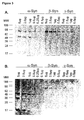
Figure 1 : Naturally occurring α-Syn-Abs were isolated from the serum of a single donor (30.6 mg/ml starting material) and from a commercially available IVIG preparation (Octagam; 10 mg/ml starting material) using affinity chromatography. The fractions that resulted from a representative experiment are depicted. -
Figure 2 : The fractions that resulted from the chromatography column were analyzed in an α-Syn ELISA. The main fractions (MF) with high IgG content were compared to the peripheral fractions (PF) that contained less IgG. As a negative control, the flow through (FT) from the affinity purification was analyzed. Samples were added to α-Syn-coated wells of an ELISA plate. Bound antibodies were detected with a HRP-conjugated goat anti-human IgG antibody followed by Tetramethylbenzimide (TMB) / peroxidase colour reaction that was detected at 450 nm (Pierce Biotechnologies). A. Fractions containing α-Syn-Abs compared to fractions without α-Syn-Abs and to flow through of the affinity chromatography. B. α-Syn-Abs isolated from IVIG versus α-Syn-Abs isolated from the serum of a single donor. -
Figure 3 : SDS-Page and Western blot analysis of naturally occurring α-Syn-Abs.
Different amounts of recombinant α-Syn (5µg, 2.5µg, 1µg, 0.5µg, 0.25µg, and 0.1µg), β-Syn (5µg, 2.5µg, 1µg, and 0.5µg) and β-Syn (5pg, 2.5µg, 1µg, and 0.5µg) were separated on a 4-12% gradient mini gel (Invitrogen) and detected by: A. Naturally occurring α-Syn-Abs isolated from IVIG, and B. Naturally occurring α-Syn-Abs isolated from the serum of a single donor. -
Figure 4 : lmmunoprecipitation of α-Syn by α-Syn-Abs affinity-purified using IVIG. Naturally occurring α-Syn-Abs isolated from IVIG, positive control with a commercially available monoclonal a-Synuclein antibody (clone Syn 211, Biosource) and a negative control (flow through from the affinity purification) were immunoprecipitated with α-Syn and subjected to Western blot analysis. Data shown are from a representative experiment. -
Figure 5 : Surface plasmon resonance analysis of affinity-purified polyclonal α-Syn-Abs (analyte) to α-Syn was done on BIACORE 2000 (Biacore AB) at 25°C. A. Interaction analysis of immobilized α-, β- and γ-Syn with affinity-purified α-Syn-Abs (pAB). Plot of sensograms of antibody binding to α- (red), to β- (blue) and γ-Syn (green). B. Interaction analysis of immobilized α-, β-and y-Syn with a monoclonal antibody against human α-Syn (mAB; Syn 211 Biosource). Plot of sensograms of antibody binding to α- (red), to β- (blue) and y-Syn (green). -
Figure 6 : Immunohistochemical detection of α-Syn in a brain sample of a patient with Parkinson's disease (PD). A. lmmunostain of a brain sample of a human PD case (left panel) using the naturally occurring α-Syn-Ab. B. Immunostain (positive control) of a brain sample of a human. PD case (right panel) incubated with a commercially available α-Syn monoclonal antibody (mAb) (MBL clone 211). C. Immunostain of a brain sample of a transgenic mouse model using the affinity-purified α-Syn-Ab. D. Immunostain (positive control) of a brain sample of a transgenic mouse model incubated with a commercially available α-Syn-mAb (MBL clone 211). LB stands for Lewy body, LN for Lewy neurites and LB li for Lewy body like inclusions. -
Figure 7 : Recombinant α-Syn truncations were analyzed for reactivity with affinity-purified α-Syn-Abs. A. α-Syn sequence. B. Different amounts of recombinant α-Syn peptide (5µg and 1µg for each peptide), α-Syn 1-60, α-Syn 1-95, α-Syn 61-140, and α-Syn 96-140 were separated on a 4-12% gradient mini gel (Invitrogen) and Western blot analysis was performed with α-Syn-Abs. -
Figure 8 : Effect of α-Syn-Abs on aggregation of α-Syn. The kinetics of α-Syn fibril formation was monitored by Thioflavin T (ThT) fluorescence. a-Syn (rPeptide; 4mg/ml) was shaken at 37°C and 600 rpm. A. At the time points indicate, aliquots were taken and a ThT assay was performed. B. α-Syn was incubated with or without the α-Syn-Abs affinity-purified from IVIG or from the serum of a single donor (1µM; Serum Abs). The samples were incubated four days at 37°C and 600 rpm and aliquots were added to the ThT solution. ThT fluorescence intensity was measured at an excitation wavelength of 450 nm and an emission wavelength of 485 nm. Samples were run in triplicates and plotted as means +/-SD. A comparison of the cells treated with α-Syn aloneto cells treated with either α-Syn Abs from IVIG or α-Syn Abs from serum was performed using a t-test (* P<0.05). -
Figure 9 : Effect of the affinity-purified α-Syn-Abs on α-Syn-induced cytotoxicity. SH-SY5Y cells were treated with 10µM aliquots of α-Syn aggregated with and without 1µM affinity-purified IVIG α-Syn-Abs or a synthetic antibody against a growth factor receptor protein as a non-specific control. Samples were run in triplicates and plotted as means +/- SD. A comparison between every group in the assay was performed using the ANOVA Tukey test (** P<0.001), n.s. is not specific. - Naturally occurring α-Synuclein antibodies (α-Syn-Abs) were isolated using affinity chromatography. A column was packed with NH2-activated resin (PIERCE Biotechnology, Rockford, IL), labelled with recombinant α-Syn (rPeptide, Bogart, GA; 1 mg / 2 ml drained resin) and equilibrated and washed with phosphate buffered saline (pH 7.4). After passing either purified human intravenous immunoglobulin G (IVIG) or IgG fraction from the plasma of a healthy donor through the column, sixteen fractions were eluted with glycine buffer at pH 2.8 and collected. The main fractions that contain the greatest amount of α-Syn-Abs as well as the peripherical fractions that contain low amounts of α-Syn-Abs were pooled and their binding capacity was tested using an α-Syn-ELISA.
- A 96-well ELISA plate was coated with recombinant α-Syn (rPeptide)dissolved in coating buffer (1.7 mM H2PO4 x H2O 98 mM Na2HPO4 x H2O 0.05% sodium azide, pH 7.4). After blocking the plate with SuperBlock blocking buffer (PIERCE Biotechnology), α-Syn-Abs samples were loaded overnight at 4°C. An appropriate secondary antibody, goat anti-human IgG H+L peroxidase conjugate (Calbiochem; Merck KGaA, Darmstadt, Germany), was incubated for one hour. Tetramethylbenzimide (TMB, Calbiochem) was added, and the reaction was stopped with 2N H2SO4. Finally, measurement was carried out in an ELISA plate reader (Multiskan Ex, Thermo, Waltham, MA) at 450 nm.
- Samples were mixed with 4 x LDS sample buffer (Invitrogen, Karlsruhe, Germany) with DTT, boiled for 5 min and subjected to polyacrylamide gel electrophoresis. Samples were separated on NUPAGE Bis-Tris 4-12%, 1 mm gels (Invitrogen) in MES running buffer at 160 V according to the manufacturer's instructions. Once separated, the proteins were either visualized using silver staining or subjected to a Western blot analysis.
- After being separated, the proteins were transferred to a PVDF membrane at 160 mA for 45 min. Membranes were blocked using RotiBlock (Roth, Karlsruhe, Germany) for 2h at room temperature and were then probed overnight with either affinity-purified α-Syn-Abs 1:20,000 in RotiBlock or monoclonal α-Syn-Abs (clone Syn211, Invitrogen) 1:20,000 in RotiBlock, as indicated. The membranes were then washed three times in 1x phosphate buffered saline with 0.05% Tween 20 (PBST) and incubated with the appropriate secondary antibody, goat anti-human or goat anti-mouse (Pierce Biotechnology), at a concentration of 1:100,000 in PBST for 1 h at room temperature. Proteins were visualized using SuperSignal West Dura (PIERCE Biotechnology).
- The reaction mixture of α-Synuclein was incubated with affinity-purified α-Syn-Abs, monoclonal α-Syn-Abs (as positive control, clone Syn 211, Invitrogen), phosphate buffered saline (PBS), and flow through from the affinity chromatography (as negative control) at 4°C overnight. Protein G was added and incubated at 4°C overnight to precipitate the IgG/α-Syn complex. The precipitates were centrifuged and washed five times with PBS before loading a 12% SDS gel. In the Western blot, α-Syn monoclonal antibody clone 211 (Invitrogen) and HRP-conjugated goat anti-human IgG were used and this was then followed by detection with West Dura Super Signal (PIERCE Biotechnology).
- Surface Plasmon Resonance (SPR) analysis of the affinity-purified polyclonal α-Syn-Abs binding to α-Synuclein was done on BIACORE 2000 (GE Healthcare, Freiburg, Germany). Ligand immobilization of α-, β- and γ-Synuclein (Sigma Aldrich, Munich, Germany) resuspended in 1x PBS; 10-20µg/ml) was performed by amino coupling to 10,000 units (RU) on different flow cells (Fc2-4) of the CM5 sensor chip. β- and γ-Synuclein were used as negative controls. The SPR-signal of the reference flow cell (Fc1) was automatically subtracted from the sensograms of any other flow cell. Interaction analysis was performed by injection of analyte samples (IVIG, 75-150µg/ml) or α-Syn monoclonal antibody clone Syn 211 (Invitrogen, 10µg/ml as positive control; 20 µl/min) diluted in running buffer (1x PBS / 0.005% P20). The sensor chip was cleaned from immune-complexes by the injection of 5-20µl of regeneration solution (25 mM NaOH). Sensogram evaluation was performed using the BIA evaluation 3.2 RCI.
- In order to map detailed epitopes of the affinity-purified α-Syn-Abs, we used four peptides that spanned the whole sequence of α-Syn. The α-Syn-Abs were tested for binding capability to peptides Syn 1-60, Syn 61-140, Syn 1-95, Syn 96-140 (Sigma Aldrich) using Western blot and surface plasmon resonance as described above, with α-Syn-Abs as the ligand and the Synuclein truncations as analytes.
- In order to determine the cellular and subcellular localization of the binding partners of the α-Syn-Abs in the brain of transgenic mice (Thy1)-h[A30P] (Kahle et al., 2004), immunohistochemical experiments were conducted using brain samples of patients with Parkinson's disease and a neuroblastoma cell line (SH-SY5Y). The histopathological staining properties of α-Syn-Abs were compared to those of commercially availableparaffin permeable antibodies against α-Synuclein (clone 211, MBL, Woburn, MA, USA). Briefly, antigen retrieval was carried out by incubation in 70% formic acid for 20min. After quenching of the endogenous peroxidase by 3% H2O2 in Methanol for 20min the primary antibodies were incubated in a 1:100 dilution at 37°C in a humid chamber for 1 hr. As a detection system, the Vectastain Elite ABC kit or the Vectastain M.O.M. kit (Biozol, Eching, Germany) was used according to the manufacturer's instructions.
- The generation of fibrillated α-Syn was performed as described previously (Herrera et al., 2008). Briefly, recombinant α-Syn (rPeptide) was diluted in 10 mM Tris buffer of pH 7.4 and shaken at 37°C and 600 rpm. At different time points (0, 2, 4 and 8 days), aliquots were taken and measured using the Thioflavin T fibrillation assay. In order to determine the ideal α-Syn concentration for the assay, several concentrations (4, 2, 1mg/ml) were tested after four days of incubation.
- In order to determine the amount of fibrillated α-Syn, a fluorometric experiment was performed as described previously (Herrera et al., 2008). Recombinant α-Syn (rPeptide) was incubated as described above. In this case,10µl aliquots were added to 80 µl 50mM glycine puffer of pH 8.5 and 10µl 100µM Thioflavin T solution (Sigma Aldrich) and fluorescence was measured spectrofluorometrically using a Tecan reader Infinite M200 (Crailsheim, Germany) at an excitation wavelength of 450 nm and an emission wavelength of 485 nm. Samples were run in triplicates and plotted as means +/- SD. Each experiment was performed at least three times.
- Human neuroblastoma cells (SH-SY5Y) were maintained in RPMI1640 (Lonza, Cologne, Germany) supplemented with 10% (v/v) fetal calf serum and 1% (v/v) penicillin/streptomycin antibiotic mix and grown in a 5% CO2 atmosphere at 37°C. Cells were harvested and plated in 96-well plates coated with poly-L-Lysin at 20,000 cells per well per 100µl of medium. Cells were then treated with α-Syn alone or α-Syn pre-incubated with α-Syn-Abs for four days at 37°C and 600 rpm. The MTT (3-(4,5-dimethylthiazol-2-yl)-2,5-diphenyltetrazolium bromide; Sigma Aldrich) reagent was resuspended in 5 mg/ml with deionised water and diluted in cell culture media to 0.5 mg/ml. Treatment was removed from the cells and 200µl MTT solution was added and incubated for 2-4 hours at 37°C. After removal of the MTT solution, cells were treated with 200µl DMSO to reduce the tetrazolium salt into the insoluble, purple coloured formazane. Readings were taken at 570 nm after another 30 min incubation at room temperature and again in the dark using a Tecan reader. Samples were run in triplicates and plotted as means +/- SD. Each experiment was performed at least three times.
- Protein samples were resolubilized in 50 mM triethylammonium bicarbonate (TEAB) buffer prior to reduction by addition of tris(2-carboxyethyl)phosphine (TCEP) to a final concentration of 5 mM and incubation at 37 °C for 20 min. Subsequently iodoacetamide to a 10 mM final concentration was added and the sample was incubated at room temperature for another 20 mins in the dark.
- Separation of antibody light chain molecules and heavy chains molecules was performed by SDS-PAGE according to Laemmli et al.. For subsequent proteolytic cleavage of the light chain IgG molecules small gel spots were cut and washed according to Proteome Factory's in-gel digestion protocol.
- Alkylated peptides were used for enzymatic cleavage with trypsin, chymotrypsin, glutamic-C protease, clostripain, LysC or proteinase K. Therefore a small aliquot was diluted with 10 volumes of the suitable buffer for the enzymatic cleavage. Incubation time was varied in order to produce overlapping peptides by each protease.
Buffer solutions: Trypsin, Thermolysin, LysC: 50 mM ammonium bicarbonate, 10 % acetonitrile (v/v) Chymotrypsin: 100 mM Tris-HCI, 10 mM CaCl2, 5% ACN (v/v), pH 8.0
Proteinase K: 100 mM Tris=HCI, 10 mM CaCl2, 5% ACN (v/v), pH 8.0 Glutamic-C protease: 50 mM Tris-HCI, 0.5 mM Glu-Glu, pH 8.0 Clostripain: 50 mM Tris-HCI, 10 mM CaCl2, 10% ACN (v/v), 20 mM DTT , pH 8.5 - For carrying out nanoLC-ESI-MS/MS high resolution MS, the HPLC system was coupled to an
Advion NanoMate 100 chip-electrospray system (Advion, Ithaca, NY), and detection was performed on a Finnigan LTQ-FT mass spectrometer (ThermoFisher, Bremen, Germany) equipped with a 6T magnet. Peptides from enzymatic cleavage were acified with formic acid and applied to nanoLC-ESIMS/MS. After trapping and desalting the peptides on enrichment column (Zorbax SB C18, 0.3 x 5 mm, Agilent) using 1% acetonitrile/0.5% formic acid solution for five minutes peptides were separated on Zorbax 300 SB C18, 75 µm x 150 mm column (Agilent, Waldbronn) using an acetonitrile/0.1 % formic acid gradient from 5% to 40% acetonitrile within 40 to 115 minutes. MS overview spectra were automatically taken in FT-mode (+/- 3 ppm) according to manufacturer's instrument settings for nanoLC-ESI-MSMS analyses, peptide fragmentation and detection was accomplished in the instrument's LTQ ion trap with an accuracy of +/- 0.3 Da. - The peptide masses and fragmentation data was searched against a human antibody sequenced derived from the NCBlnr (National Center for Biotechnology Information, Bethesda, USA) database utilizing the MASCOT search engine (Matrix Science, London). Positive identification of peptides were annotated for the generation of sequence candidates.
- Unassigned data was extracted and used for subsequent de novo peptide sequencing and searching of amino acid permutated human antibody peptide amino acid sequences.
- MS parent ion accuracy +/- 3 ppm
MSMS fragment ion accuracy +/- 0.3 Da
Fixed modification: Carbamidomethylation (Cys)
Denovo sequencing
Binning of duplicate spectra and peaks: - Binning MS 0.00015%
- Binning MSMS 0.15 Da
- Tolerance MS 0.0003%
- Tolerance MSMS 0.3 Da
- BLAST matrix: PAM30
- Expect value: 10
- Database: Human antibody sequences derived from NCBlnr
- N-terminal Edman sequencing of HPLC separated peptides was performed by an ABI Procise Model 49x protein sequencer using peptide fractions spotted on to Biobrene treated glass fiber discs.
- We purified human α-Syn-Abs from IVIG and from the serum of a single donor by using an affinity column coated with recombinant α-Syn (
Figure 1 ). The resulting fractions with the highest IgG content were pooled and their binding properties to α-Syn were tested using an ELISA. We found that the resulting main fractions had a strong anti-α-Syn signal as compared to both the flow through IgG and peripheral fractions without α-Syn-Abs (Figure 2a ). There was a 2.5-fold increase in the fraction containing affinity-purified α-Syn-Abs as compared to the peripheral fractions without the antibody and a 4-fold increase as compared to the flow through of the affinity column. InFigure 2b we show that α-Syn-Abs isolated from IVIG as well as from the serum of a single donor are dose-dependently binding α-Syn in an ELISA. The detection limit lies between 125 and 62.5 ng/ml for both affinity purified α-Syn-Abs. - The purified α-Syn-Abs from IVIG and that from single donor serum were able to detect the monomeric form of recombinant α-Syn peptide at 19 kDa on Western blots in a dose dependent manner. It also detected aggregated α-Syn species (
Figure 3a and 3b ). The signals at 38, 57 and 76 kDa correspond to the approximate sizes of dimeric, trimeric and tetrameric forms of α-Syn, respectively. A tetrameric form of β- and γ-Syn corresponding to the 76 kDa band was clearly recognized by α-Syn-Abs from IVIG and from serum. - In order to further confirm the ability of the naturally occurring α-Syn-Abs to bind α-Syn, we performed immunoprecipitation of recombinant α-Syn peptide (
Figure 4 ). We were able to confirm that the affinity-purified α-Syn-Abs do bind α-Syn peptide. The negative control, consisting of the same immunoprecipitation reaction with the column flow-through alone, did not bind to the recombinant α-Syn peptide. The reactions with protein G alone or with non-specific antibodies (Interleukin-1 antibody) were also negative (data not shown). - Furthermore, we obtained SPR data on the interaction of affinity-purified α-Syn-Abs from IVIG with α-, β- and γ-Synuclein immobilized via amino coupling (
Figure 5 ). Data demonstrated that the affinity-purified α-Syn-Abs from IVIG bind to α-Syn, to some extent as well as to β-Syn, but not to y-Syn. After 20 seconds, the antibody dissociates to 50% from α-Syn and 100% from the β-isoform. In comparison, a monoclonal antibody (clone Syn 211, Biosource) against human α-Syn bound particularly strong to α-Syn but bound very little or not at all to β-Syn and γ-Syn. - In order to display the anatomical and histopathological localization of α-Syn in Lewy bodies using affinity-purified α-Syn-Abs, brain samples from PD patients and from an α-Syn transgenic (Thy1)-h[A30P] mouse model were analyzed using immunohistochemistry. In human PD patient samples, the affinity-purified α-Syn-Abs recognized the same structures as the monoclonal anti-human α-Syn-Ab (
Figure 6 ); these included a halo around a weaker core in Lewy bodies and drilled roots in Lewy neurites as well as in somatodendritic deposits. In the α-Syn transgenic mouse model there are no Lewy bodies present but rather only so called "Lewy body like inclusions" have been reported (Kahle et al., 2001). The immunohistochemical staining demonstrated that both the affinity purified α-Syn-Abs and the monoclonal α-Syn-Ab recognised these inclusions. Taken together, our results could conclusively demonstrate the specific binding of the affinity-purified antibodies to either recombinant human α-Syn or native human α-Syn. - In order to further characterize the naturally occurring antibodies, we identified which epitopes of α-Syn are bound by α-Syn-Abs. Therefore, four truncated synthetic peptides spanning different regions of α-Syn were subjected to Western blot analysis (
Figure 7a ). The α-Syn-Abs bound to the peptides corresponding to residues 1-60, 1-95 and 61-140 but not to the peptide corresponding to residues 96-140 (Figure 7b ). The non-amyloid component (NAC) is located between ∼61 and 95 and seemed to be relevant in the binding of α-Syn-Abs to α-Syn. This data was confirmed by SPR data that also showed that the α-Syn fragments containing the NAC region had a higher binding specificity to α-Syn as compared to fragments that lacked this region (data not shown). - We investigated whether purified α-Syn-Abs had an effect on α-Syn fibril formation by using Thioflavin T (ThT), a fluorescent reagent that specifically binds to fibrillar structures. Incubation of highly concentrated α-Syn (4 mg/ml) alone resulted in a time-dependent increase in fluorescence as the α-Syn began to aggregate (
Figure 8a ). Incubation of α-Syn with 2µM affinity-purified α-Syn-Abs incubated for four days at 37°C and 600 rpm caused a significant decrease in ThT fluorescence, suggesting that the affinity-purified autoantibody was able to reduce fibril formation of α-Syn (Figure 8b ). The antibody isolated from serum did not show the same capacity for inhibiting fibril formation as compared to the antibody isolated from IVIG. When α-Syn was incubated with the column flow-through, a decrease in fibril formation was observed. The resulting data was analysed by a t-test using the GraphPad Software (GraphPad Software Inc., San Diego, CA). - We examined the cytotoxicity of pre-aggregated α-Syn samples on the human neuroblastoma cell line SH-SY5Y using an MTT assay. We tested whether the affinity-purified α-Syn-Abs had an effect on α-Syn-mediated cytotoxicity. Cells were treated with α-Syn and pre-incubated for four days with or without α-Syn-Abs. As depicted in
Figure 9b , there was a significant increase in cell viability when cells were treated with α-Syn and α-Syn-Abs. Incubation with an unspecific synthetic antibody did not produce the same effect. The resulting data was analyzed with one way analysis of variance (ANOVA) using the SigmaStat Software (Systat Software GmbH, Erkrath, Germany). - Neuropathologic and genetic studies as well as the development of transgenic animal models have provided evidence for the involvement of α-Synuclein (α-Syn) in the progression of Parkinson's disease (PD). It has become increasingly evident that the misfolded and aggregated species of α-Syn are known to be neurotoxic and subsequently lead to neurodegeneration. Thus, research has focused on finding new approaches to reducing abnormal accumulation of α-Syn.
- In recent years, immunization has been shown to be effective in reducing neuronal accumulation of α-Syn aggregates. Masliah and colleagues also demonstrated that active immunization against human α-Syn reduced α-Syn aggregates in the brains of transgenic mice (Masliah et al., 2005). Papachroni and colleagues demonstrated for the first time the existence of autoantibodies (AAbs) against α-Syn that are positively correlated with the familial but not sporadic form of PD. In the study, they examined the presence of AAbs against α-Syn in the peripheral blood serum of PD patients and controls. They could detect such AAbs in 65% of all tested patients and they demonstrated that 90% of patients with familial PD tested positive for AAbs against α-Syn. Therefore, they hypothesized that these AAbs could be involved in the pathogenesis of α-Syn. (Papachroni et al., 2007).
- Emadi and colleagues have isolated a human single-chain antibody fragment (scFv) against oligomeric α-Syn from a phage display antibody library (Emadi et al., 2007). They described binding only to oligomeric forms of α-Syn and inhibited both aggregation and toxicity of α-Syn in vitro. This approach is considered a significant advance toward a molecular therapy targeted against PD and other neurodegenerative conditions in which α-Syn aggregations represent a primary hallmark of disease.
- In this study, we were able to isolate naturally occurring α-Syn antibodies from IVIG as well as from the serum of a healthy donor. Subsequent characterization by different assays including ELISA, Western blot analysis and surface plasmon resonance (SPR) analysis demonstrated that these α-Syn antibodies recognize several species of α-Syn. ELISA showed that antibodies purified from IVIG and from the serum of a single donor could both bind α-Syn. Western blot analysis demonstrated that the autoantibody was able to bind different species of α-Syn but not monomeric β- and γ-Synuclein. These results were confirmed using SPR analysis: the binding of the autoantibody was shown to be specific for monomeric α-Syn but not for monomeric β- or γ-Syn. Interestingly, oligomeric forms of all three synucleins were also recognized.
- Our attempt to characterize the linear binding epitope of our α-Syn-Abs indicated that the non amyloidal component (NAC) region might be of importance. The NAC Region was originally identified as a component of Alzheimer's amyloid plaques. Deletions of this regions have been shown to be crucial for the aggregation of α-Syn in vitro (Ueda et al., 1993, Conway et al., 1998, Takeda et al., 1998) and through their toxicity to dopaminergic neurons in a drosophila model (Periquet et al., 2007).
- We were able to determine the cellular and subcellular localization of the binding partners of the α-Syn-Abs in the brains of PD patients as well as in the brains of transgenic mice by immunohistochemistry. The functionality of the affinity-purified α-Syn antibody was tested using a thioflavin T assay and we were able to show that it can inhibit fibrillation of α-Syn. Also, a cell viability assay demonstrated that the affinity-purified α-Syn-Abs were able to increase cell viability and reduce α-Syn-induced cytotoxicity. These results are consistent with the effects of the antibodies against oligomeric forms of α-Syn as published in Emadi et al., 2007.
- Taken together, these data suggest that the isolated α-Syn antibodies could have a therapeutic application in controlling the aggregation of α-Synuclein and consequently in the progression of PD.
-
- 1. Conway KA, Harper JD, Lansbury PT (Accelerated in vitro fibril formation by a mutant alpha-synuclein linked to early-onset Parkinson disease. Nature medicine 4:1318-1320.1998).
- 2. Dodel R, Hampel H, Depboylu C, Lin S, Gao F, Schock S, Jackel S, Wei X, Buerger K, Hoft C, Hemmer B, Moller HJ, Farlow M, Oertel WH, Sommer N, Du Y (Human antibodies against amyloid beta peptide: a potential treatment for Alzheimer's disease. Annals of neurology 52:253-256.2002).
- 3. Du Y, Wei X, Dodel R, Sommer N, Hampel H, Gao F, Ma Z, Zhao L, Oertel WH, Farlow M (Human anti-beta-amyloid antibodies block beta-amyloid fibril formation and prevent beta-amyloid-induced neurotoxicity. Brain 126:1935-1939.2003).
- 4. Emadi S, Barkhordarian H, Wang MS, Schulz P, Sierks MR (Isolation of a human single chain antibody fragment against oligomeric alpha-synuclein that inhibits aggregation and prevents alpha-synuclein-induced toxicity. Journal of molecular biology 368:1132-1144.2007).
- 5. Eriksen JL, Dawson TM, Dickson DW, Petrucelli L (Caught in the act: alpha-synuclein is the culprit in Parkinson's disease. Neuron 40:453-456.2003).
- 6. Giasson BI, Murray IV, Trojanowski JQ, Lee VM (A hydrophobic stretch of 12 amino acid residues in the middle of alpha-synuclein is essential for filament assembly. The Journal of biological chemistry 276:2380-2386.2001).
- 7. Hashimoto M, Masliah E (Alpha-synuclein in Lewy body disease and Alzheimer's disease. Brain pathology (Zurich, Switzerland) 9:707-720.1999).
- 8. Herrera FE, Chesi A, Paleologou KE, Schmid A, Munoz A, Vendruscolo M, Gustincich S, Lashuel HA, Carloni P (Inhibition of alpha-synuclein fibrillization by dopamine is mediated by interactions with five C-terminal residues and with E83 in the NAC region. PLoS ONE 3:e3394.2008).
- 9. Iwai A (Properties of NACP/alpha-synuclein and its role in Alzheimer's disease. Biochimica et biophysica acta 1502:95-109.2000).
- 10. Kahle PJ, Neumann M, Ozmen L, Muller V, Odoy S, Okamoto N, Jacobsen H, Iwatsubo T, Trojanowski JQ, Takahashi H, Wakabayashi K, Bogdanovic N, Riederer P, Kretzschmar HA, Haass C (Selective insolubility of alpha-synuclein in human Lewy body diseases is recapitulated in a transgenic mouse model. The American journal of pathology 159:2215-2225.2001).
- 11. Kayed R, Head E, Thompson JL, McIntire TM, Milton SC, Cotman CW, Glabe CG (Common structure of soluble amyloid oligomers implies common mechanism of pathogenesis. Science (New York, NY 300:486-489.2003).
- 12. Kim YS, Lee D, Lee EK, Sung JY, Chung KC, Kim J, Paik SR (Multiple ligand interaction of alpha-synuclein produced various forms of protein aggregates in the presence of Abeta25-35, copper, and eosin. Brain research 908:93-98.2001).
- 13. Masliah E, Rockenstein E, Adame A, Alford M, Crews L, Hashimoto M, Seubert P, Lee M, Goldstein J, Chilcote T, Games D, Schenk D (Effects of alpha-synuclein immunization in a mouse model of Parkinson's disease. Neuron 46:857-868.2005).
- 14. Masliah E, Rockenstein E, Veinbergs I, Mallory M, Hashimoto M, Takeda A, Sagara Y, Sisk A, Mucke L (Dopaminergic loss and inclusion body formation in alpha-synuclein mice: implications for neurodegenerative disorders. Science (New York, NY 287:1265-1269.2000).
- 15. Murphy RC, Messer A (A single-chain Fv intrabody provides functional protection against the effects of mutant protein in an organotypic slice culture model of Huntington's disease. Molecular Brain Research 121:141-145.2004).
- 16. Papachroni KK, Ninkina N, Papapanagiotou A, Hadjigeorgiou GM, Xiromerisiou G, Papadimitriou A, Kalofoutis A, Buchman VL (Autoantibodies to alpha-synuclein in inherited Parkinson's disease. Journal of neurochemistry 101:749-756.2007).
- 17. Park SM, Jung HY, Kim TD, Park JH, Yang CH, Kim J (Distinct roles of the N-terminal-binding domain and the C-terminal-solubilizing domain of alpha-synuclein, a molecular chaperone. The Journal of biological chemistry 277:28512-28520.2002).
- 18. Periquet M, Fulga T, Myllykangas L, Schlossmacher MG, Feany MB (Aggregated alpha-synuclein mediates dopaminergic neurotoxicity in vivo. J Neurosci 27:3338-3346.2007).
- 19. Polymeropoulos MH, Lavedan C, Leroy E, Ide SE, Dehejia A, Dutra A, Pike B, Root H, Rubenstein J, Boyer R, Stenroos ES, Chandrasekharappa S, Athanassiadou A, Papapetropoulos T, Johnson WG, Lazzarini AM, Duvoisin RC, Di lorio G, Golbe LI, Nussbaum RL (Mutation in the alpha-synuclein gene identified in families with Parkinson's disease. Science (New York, NY 276:2045-2047.1997).
- 20. Singleton AB, Farrer M, Johnson J, Singleton A, Hague S, Kachergus J, Hulihan M, Peuralinna T, Dutra A, Nussbaum R, Lincoln S, Crawley A, Hanson M, Maraganore D, Adler C, Cookson MR, Muenter M, Baptista M, Miller D, Blancato J, Hardy J, Gwinn-Hardy K (alpha-Synuclein locus triplication causes Parkinson's disease. Science (New York, NY 302:841.2003).
- 21. Spillantini MG, Schmidt ML, Lee VM, Trojanowski JQ, Jakes R, Goedert M (Alpha-synuclein in Lewy bodies. Nature 388:839-840.1997).
- 22. Takeda A, Hashimoto M, Mallory M, Sundsumo M, Hansen L, Sisk A, Masliah E (Abnormal distribution of the non-Abeta component of Alzheimer's disease amyloid precursor/alpha-synuclein in Lewy body disease as revealed by proteinase K and formic acid pretreatment. Laboratory investigation; a journal of technical methods and pathology 78:1169-1177.1998).
- 23. Takenouchi T, Hashimoto M, Hsu LJ, Mackowski B, Rockenstein E, Mallory M, Masliah E (Reduced neuritic outgrowth and cell adhesion in neuronal cells transfected with human alpha-synuclein. Molecular and cellular neurosciences 17:141-150.2001).
- 24. Ueda K, Fukushima H, Masliah E, Xia Y, Iwai A, Yoshimoto M, Otero DA, Kondo J, Ihara Y, Saitoh T (Molecular cloning of cDNA encoding an unrecognized component of amyloid in Alzheimer disease. Proceedings of the National Academy of Sciences of the United States of America 90:11282-11286.1993).
- 25. Wakabayashi K, Hayashi S, Kakita A, Yamada M, Toyoshima Y, Yoshimoto M, Takahashi H (Accumulation of alpha-synuclein/NACP is a cytopathological feature common to Lewy body disease and multiple system atrophy. Acta neuropathologica 96:445-452.1998).
- 26. Zarranz JJ, Alegre J, Gomez-Esteban JC, Lezcano E, Ros R, Ampuero I, Vidal L, Hoenicka J, Rodriguez O, Atares B, Llorens V, Gomez Tortosa E, del Ser T, Munoz DG, de Yebenes JG (The new mutation, E46K, of alpha-synuclein causes Parkinson and Lewy body dementia. Annals of neurology 55:164-173.2004).
- 27. Zhou W, Hurlbert MS, Schaack J, Prasad KN, Freed CR (Overexpression of human alpha-synuclein causes dopamine neuron death in rat primary culture and immortalized mesencephalon-derived cells. Brain research 866:33-43.2000).
BLAST search parameters:
HPLC separation of LysC digested peptides was performed by using an


Claims (14)
- A human antibody which is directed against α-Synuclein (α-Syn) or a fragment or derivative thereof.
- The antibody of claim 1 which is directed against an epitope between amino acids 60-95 of human α-Syn.
- The antibody of claim 1 or 2 which is a naturally occurring human autoantibody or a fragment or derivative thereof.
- The antibody of any one of claims 1-3 which binds monomeric α-Syn, aggregated α-Syn, or both monomeric and aggregated α-Syn.
- The antibody of any one of the preceding claims characterized by a light chain sequence SEQ ID NO:2 or a variant thereof.
- The antibody of any one of the preceding claims for use in medicine, particularly in human medicine.
- The antibody of any one of the preceding claims for use in the treatment of a neurodegenerative disorder, particularly Parkinson's disease or Dementia with Lewy bodies.
- The antibody of any one of the preceding claims for use in passive immune therapy.
- The antibody of any one of claims 1-5 for use as a diagnostic agent.
- A nucleic acid molecule encoding the antibody of any one of claims 1-5 optionally in operative linkage to an expression control sequence.
- A recombinant cell which comprises the nucleic acid molecule of claim 10.
- A method of preparing an antibody of any one of claims 1-5 comprising the steps:- culturing a cell of claim 11 under conditions which allow expression of the antibody encoding nucleic acid molecule, and- collecting the antibody from the cell or the culture supernatant.
- A pharmaceutical composition comprising the antibody of any one of claims 1-5, the nucleic acid molecule of claim 10 or the recombinant cell of claim 11 together with a pharmaceutically acceptable carrier.
- The composition of claim 13 for use in passive immune therapy.
Priority Applications (7)
| Application Number | Priority Date | Filing Date | Title |
|---|---|---|---|
| EP10155373A EP2366714A1 (en) | 2010-03-03 | 2010-03-03 | Naturally occuring autoantibodies against alpha-synuclein that inhibit the aggregation and cytotoxicity of alpha-synuclein |
| EP11706256A EP2542586A1 (en) | 2010-03-03 | 2011-03-03 | Naturally occurring autoantibodies against alpha-synuclein that inhibit the aggregation and cytotoxicity of alpha-synuclein |
| CA2791409A CA2791409C (en) | 2010-03-03 | 2011-03-03 | Naturally occurring autoantibodies against alpha-synuclein that inhibit the aggregation and cytotoxicity of alpha-synuclein |
| PCT/EP2011/053188 WO2011107544A1 (en) | 2010-03-03 | 2011-03-03 | Naturally occurring autoantibodies against alpha-synuclein that inhibit the aggregation and cytotoxicity of alpha-synuclein |
| AU2011222920A AU2011222920C1 (en) | 2010-03-03 | 2011-03-03 | Naturally occurring autoantibodies against alpha-Synuclein that inhibit the aggregation and cytotoxicity of alpha-Synuclein |
| US13/582,489 US8741293B2 (en) | 2010-03-03 | 2011-03-03 | Naturally occurring autoantibodies against alpha-synuclein that inhibit the aggregation and cytotoxicity of alpha-synuclein |
| US14/256,140 US20140377271A1 (en) | 2010-03-03 | 2014-04-18 | Naturally occurring autoantibodies against alpha-synuclein that inhibit the aggregation and cytotoxicity of alpha-synuclein |
Applications Claiming Priority (1)
| Application Number | Priority Date | Filing Date | Title |
|---|---|---|---|
| EP10155373A EP2366714A1 (en) | 2010-03-03 | 2010-03-03 | Naturally occuring autoantibodies against alpha-synuclein that inhibit the aggregation and cytotoxicity of alpha-synuclein |
Publications (1)
| Publication Number | Publication Date |
|---|---|
| EP2366714A1 true EP2366714A1 (en) | 2011-09-21 |
Family
ID=42340377
Family Applications (2)
| Application Number | Title | Priority Date | Filing Date |
|---|---|---|---|
| EP10155373A Withdrawn EP2366714A1 (en) | 2010-03-03 | 2010-03-03 | Naturally occuring autoantibodies against alpha-synuclein that inhibit the aggregation and cytotoxicity of alpha-synuclein |
| EP11706256A Withdrawn EP2542586A1 (en) | 2010-03-03 | 2011-03-03 | Naturally occurring autoantibodies against alpha-synuclein that inhibit the aggregation and cytotoxicity of alpha-synuclein |
Family Applications After (1)
| Application Number | Title | Priority Date | Filing Date |
|---|---|---|---|
| EP11706256A Withdrawn EP2542586A1 (en) | 2010-03-03 | 2011-03-03 | Naturally occurring autoantibodies against alpha-synuclein that inhibit the aggregation and cytotoxicity of alpha-synuclein |
Country Status (5)
| Country | Link |
|---|---|
| US (2) | US8741293B2 (en) |
| EP (2) | EP2366714A1 (en) |
| AU (1) | AU2011222920C1 (en) |
| CA (1) | CA2791409C (en) |
| WO (1) | WO2011107544A1 (en) |
Cited By (2)
| Publication number | Priority date | Publication date | Assignee | Title |
|---|---|---|---|---|
| EP2787348A1 (en) * | 2013-04-03 | 2014-10-08 | Affiris AG | Method for detecting aSyn-specific antibodies in a biological sample |
| WO2014161890A1 (en) | 2013-04-03 | 2014-10-09 | Affiris Ag | Method for detecting proteinopathy-specific antibodies in a biological sample |
Families Citing this family (18)
| Publication number | Priority date | Publication date | Assignee | Title |
|---|---|---|---|---|
| PT2771031T (en) | 2011-10-28 | 2018-05-24 | Prothena Biosciences Ltd | Humanized antibodies that recognize alpha-synuclein |
| WO2013112945A1 (en) | 2012-01-27 | 2013-08-01 | Neotope Biosciences Limited | Humanized antibodies that recognize alpha-synuclein |
| UA118441C2 (en) | 2012-10-08 | 2019-01-25 | Протена Біосаєнсиз Лімітед | Antibodies recognizing alpha-synuclein |
| AU2013203043B2 (en) * | 2013-03-15 | 2016-10-06 | Takeda Pharmaceutical Company Limited | Methods to produce a human plasma-derived igg preparation enriched in brain disease-related natural iggs |
| US10513555B2 (en) | 2013-07-04 | 2019-12-24 | Prothena Biosciences Limited | Antibody formulations and methods |
| WO2016054114A1 (en) | 2014-09-29 | 2016-04-07 | The Regents Of The University Of California | Compositions for expanding regulatory t cells (treg), and treating autoimmune and inflammatory diseases and conditions |
| JP2017536102A (en) | 2014-10-16 | 2017-12-07 | ジェネンテック, インコーポレイテッド | Anti-alpha-synuclein antibodies and methods of use |
| GB201419084D0 (en) | 2014-10-27 | 2014-12-10 | Agency Science Tech & Res | Anti-PD-1 antibodies |
| GB201512203D0 (en) | 2015-07-13 | 2015-08-19 | Lundbeck & Co As H | Agents,uses and methods |
| MX2019005594A (en) | 2016-11-15 | 2019-07-04 | H Lundbeck As | Agents, uses and methods for the treatment of synucleinopathy. |
| WO2018109058A1 (en) | 2016-12-16 | 2018-06-21 | H. Lundbeck A/S | Agents, uses and methods |
| US10364286B2 (en) | 2016-12-22 | 2019-07-30 | H. Lundbeck A/S | Monoclonal anti-alpha-synuclein antibodies for preventing tau aggregation |
| JP7136790B2 (en) | 2017-02-17 | 2022-09-13 | ブリストル-マイヤーズ スクイブ カンパニー | Antibodies against alpha-synuclein and uses thereof |
| MX2020004674A (en) * | 2017-11-17 | 2020-08-13 | Abl Bio Inc | Antibodies to ¿-synuclein and uses thereof. |
| GB201720975D0 (en) | 2017-12-15 | 2018-01-31 | Ucb Biopharma Sprl | Anti-alpha synuclein antibodies |
| GB201720970D0 (en) | 2017-12-15 | 2018-01-31 | Ucb Biopharma Sprl | Antibodies |
| WO2023161528A1 (en) | 2022-02-28 | 2023-08-31 | Tridem Bioscience Gmbh & Co Kg | A CONJUGATE COMPRISING AT LEAST A ß-GLUCAN |
| WO2023161527A1 (en) | 2022-02-28 | 2023-08-31 | Tridem Bioscience Gmbh & Co Kg | A CONJUGATE CONSISTING OF OR COMPRISING AT LEAST A ß-GLUCAN OR A MANNAN |
Citations (1)
| Publication number | Priority date | Publication date | Assignee | Title |
|---|---|---|---|---|
| WO2009133521A2 (en) * | 2008-04-29 | 2009-11-05 | Bioartic Neuroscience Ab | Antibodies and vaccines for use in therapeutic and diagnostic methods for alpha-synuclein-related disorders |
Family Cites Families (5)
| Publication number | Priority date | Publication date | Assignee | Title |
|---|---|---|---|---|
| TW200509968A (en) * | 2002-11-01 | 2005-03-16 | Elan Pharm Inc | Prevention and treatment of synucleinopathic disease |
| US9034337B2 (en) * | 2003-10-31 | 2015-05-19 | Prothena Biosciences Limited | Treatment and delay of outset of synucleinopathic and amyloidogenic disease |
| EP1638514A4 (en) * | 2003-06-06 | 2009-11-18 | Medimmune Inc | Use of epha4 and modulator or epha4 for diagnosis, treatment and prevention of cancer |
| CA2657953A1 (en) * | 2005-07-19 | 2007-01-25 | University Of Rochester | Alpha-synuclein antibodies and methods related thereto |
| WO2007143711A2 (en) * | 2006-06-07 | 2007-12-13 | Wisconsin Alumni Research Foundation | Blood-brain barrier targeting antibodies |
-
2010
- 2010-03-03 EP EP10155373A patent/EP2366714A1/en not_active Withdrawn
-
2011
- 2011-03-03 WO PCT/EP2011/053188 patent/WO2011107544A1/en active Application Filing
- 2011-03-03 CA CA2791409A patent/CA2791409C/en active Active
- 2011-03-03 AU AU2011222920A patent/AU2011222920C1/en active Active
- 2011-03-03 EP EP11706256A patent/EP2542586A1/en not_active Withdrawn
- 2011-03-03 US US13/582,489 patent/US8741293B2/en active Active
-
2014
- 2014-04-18 US US14/256,140 patent/US20140377271A1/en not_active Abandoned
Patent Citations (1)
| Publication number | Priority date | Publication date | Assignee | Title |
|---|---|---|---|---|
| WO2009133521A2 (en) * | 2008-04-29 | 2009-11-05 | Bioartic Neuroscience Ab | Antibodies and vaccines for use in therapeutic and diagnostic methods for alpha-synuclein-related disorders |
Non-Patent Citations (31)
| Title |
|---|
| CONWAY KA; HARPER JD; LANSBURY PT: "Accelerated in vitro fibril formation by a mutant alpha-synuclein linked to early-onset Parkinson disease", NATURE MEDICINE, vol. 4, 1998, pages 1318 - 1320 |
| DODEL R; HAMPEL H; DEPBOYLU C; LIN S; GAO F; SCHOCK S; JACKEL S; WEI X; BUERGER K; HOFT C: "Human antibodies against amyloid beta peptide: a potential treatment for Alzheimer's disease", ANNALS OF NEUROLOGY, vol. 52, 2002, pages 253 - 256 |
| DU Y; WEI X; DODEL R; SOMMER N; HAMPEL H; GAO F; MA Z; ZHAO L; OERTEL WH; FARLOW M: "Human anti-beta-amyloid antibodies block beta-amyloid fibril formation and prevent beta-amyloid-induced neurotoxicity", BRAIN, vol. 126, 2003, pages 1935 - 1939 |
| EMADI ET AL: "Isolation of a Human Single Chain Antibody Fragment Against Oligomeric alpha-Synuclein that Inhibits Aggregation and Prevents alpha-Synuclein-induced Toxicity", JOURNAL OF MOLECULAR BIOLOGY, LONDON, GB LNKD- DOI:10.1016/J.JMB.2007.02.089, vol. 368, no. 4, 17 April 2007 (2007-04-17), pages 1132 - 1144, XP022030022, ISSN: 0022-2836 * |
| EMADI S; BARKHORDARIAN H; WANG MS; SCHULZ P; SIERKS MR: "isolation of a human single chain antibody fragment against oligomeric alpha-synuclein that inhibits aggregation and prevents alpha-synuclein- induced toxicity", JOURNAL OF MOLECULAR BIOLOGY, vol. 368, 2007, pages 1132 - 1144 |
| ERIKSEN JL; DAWSON TM; DICKSON DW; PETRUCELLI L: "Caught in the act: alpha-synuclein is the culprit in Parkinson's disease", NEURON, vol. 40, 2003, pages 453 - 456 |
| GIASSON BI; MURRAY IV; TROJANOWSKI JQ; LEE VM: "A hydrophobic stretch of 12 amino acid residues in the middle of alpha-synuclein is essential for filament assembly", THE JOURNAL OF BIOLOGICAL CHEMISTRY, vol. 276, 2001, pages 2380 - 2386 |
| HASHIMOTO M; MASLIAH E: "Alpha-synuclein in Lewy body disease and Alzheimer's disease", BRAIN PATHOLOGY, vol. 9, 1999, pages 707 - 720 |
| HERRERA FE; CHESI A; PALEOLOGOU KE; SCHMID A; MUNOZ A; VENDRUSCOLO M; GUSTINCICH S; LASHUEL HA; CARLONI P: "Inhibition of alpha-synuclein fibrillization by dopamine is mediated by interactions with five C-terminal residues and with E83 in the NAC region", PLOS ONE, vol. 3, 2008, pages E3394 |
| IWAI A: "Properties of NACP/alpha-synuclein and its role in Alzheimer's disease", BIOCHIMICA ET BIOPHYSICA ACTA, vol. 1502, 2000, pages 95 - 109 |
| KAHLE PJ; NEUMANN M; OZMEN L; MULLER V; ODOY S; OKAMOTO N; JACOBSEN H; IWATSUBO T; TROJANOWSKI JQ; TAKAHASHI H: "Selective insolubility of alpha-synuclein in human Lewy body diseases is recapitulated in a transgenic mouse model", THE AMERICAN JOURNAL OF PATHOLOGY, vol. 159, 2001, pages 2215 - 2225 |
| KAYED R; HEAD E; THOMPSON JL; MCINTIRE TM; MILTON SC; COTMAN CW; GLABE CG: "Common structure of soluble amyloid oligomers implies common mechanism of pathogenesis", SCIENCE, vol. 300, 2003, pages 486 - 489 |
| KIM YS; LEE D; LEE EK; SUNG JY; CHUNG KC; KIM J; PAIK SR: "Multiple ligand interaction of alpha-synuclein produced various forms of protein aggregates in the presence of Abeta25-35, copper, and eosin", BRAIN RESEARCH, vol. 908, 2001, pages 93 - 98 |
| MASLIAH E ET AL: "Effects of alpha-synuclein immunization in a mouse model of Parkinsons disease", NEURON, CELL PRESS, US LNKD- DOI:10.1016/J.NEURON.2005.05.010, vol. 46, no. 6, 16 June 2005 (2005-06-16), pages 857 - 868, XP008091682, ISSN: 0896-6273 * |
| MASLIAH E; ROCKENSTEIN E; ADAME A; ALFORD M; CREWS L; HASHIMOTO M; SEUBERT P; LEE M; GOLDSTEIN J; CHILCOTE T: "Effects of alpha-synuclein immunization in a mouse model of Parkinson's disease", NEURON, vol. 46, 2005, pages 857 - 868 |
| MASLIAH E; ROCKENSTEIN E; VEINBERGS I; MALLORY M; HASHIMOTO M; TAKEDA A; SAGARA Y; SISK A; MUCKE L: "Dopaminergic loss and inclusion body formation in alpha-synuclein mice: implications for neurodegenerative disorders", SCIENCE, vol. 287, 2000, pages 1265 - 1269 |
| MURPHY RC; MESSER A: "A single-chain Fv intrabody provides functional protection against the effects of mutant protein in an organotypic slice culture model of Huntington's disease", MOLECULAR BRAIN RESEARCH, vol. 121, 2004, pages 141 - 145 |
| NEFF F ET AL: "Immunotherapy and naturally occurring autoantibodies in neurodegenerative disorders", AUTOIMMUNITY REVIEWS, ELSEVIER, AMSTERDAM, NL LNKD- DOI:10.1016/J.AUTREV.2008.04.010, vol. 7, no. 6, 1 June 2008 (2008-06-01), pages 501 - 507, XP022732412, ISSN: 1568-9972, [retrieved on 20080501] * |
| PAPACHRONI KATERINA K ET AL: "Autoantibodies to alpha-synuclein in inherited Parkinson's disease", JOURNAL OF NEUROCHEMISTRY, WILEY INTERSCIENCE, NEW YORK, NY, US LNKD- DOI:10.1111/J.1471-4159.2006.04365.X, vol. 101, no. 3, 1 May 2007 (2007-05-01), pages 749 - 756, XP002530228, ISSN: 0022-3042 * |
| PAPACHRONI KK; NINKINA N; PAPAPANAGIOTOU A; HADJIGEORGIOU GM; XIROMERISIOU G; PAPADIMITRIOU A; KALOFOUTIS A; BUCHMAN VL: "Autoantibodies to alpha-synuclein in inherited Parkinson's disease", JOURNAL OF NEUROCHEMISTRY, vol. 101, 2007, pages 749 - 756 |
| PARK SM; JUNG HY; KIM TD; PARK JH; YANG CH; KIM J: "Distinct roles of the N-terminal-binding domain and the C-terminal-solubilizing domain of alpha-synuclein, a molecular chaperone", THE JOURNAL OF BIOLOGICAL CHEMISTRY, vol. 277, 2002, pages 28512 - 28520 |
| PERIQUET M; FULGA T; MYLLYKANGAS L; SCHLOSSMACHER MG; FEANY MB: "Aggregated alpha-synuclein mediates dopaminergic neurotoxicity in vivo", J NEUROSCI, vol. 27, 2007, pages 3338 - 3346 |
| POLYMEROPOULOS MH; LAVEDAN C; LEROY E; IDE SE; DEHEJIA A; DUTRA A; PIKE B; ROOT H; RUBENSTEIN J; BOYER R: "Mutation in the alpha-synuclein gene identified in families with Parkinson's disease", SCIENCE, vol. 276, 1997, pages 2045 - 2047 |
| SINGLETON AB; FARRER M; JOHNSON J; SINGLETON A; HAGUE S; KACHERGUS J; HULIHAN M; PEURALINNA T; DUTRA A; NUSSBAUM R: "alpha-Synuclein locus triplication causes Parkinson's disease", SCIENCE, vol. 302, 2003, pages 841 |
| SPILLANTINI MG; SCHMIDT ML; LEE VM; TROJANOWSKI JQ; JAKES R; GOEDERT M: "Alpha-synuclein in Lewy bodies", NATURE, vol. 388, 1997, pages 839 - 840 |
| TAKEDA A; HASHIMOTO M; MALLORY M; SUNDSUMO M; HANSEN L; SISK A; MASLIAH E: "Abnormal distribution of the non-Abeta component of Alzheimer's disease amyloid precursor/alpha-synuclein in Lewy body disease as revealed by proteinase K and formic acid pretreatment. Laboratory investigation", A JOURNAL OF TECHNICAL METHODS AND PATHOLOGY, vol. 78, 1998, pages 1169 - 1177 |
| TAKENOUCHI T; HASHIMOTO M; HSU LJ; MACKOWSKI B; ROCKENSTEIN E; MALLORY M; MASLIAH E: "Reduced neuritic outgrowth and cell adhesion in neuronal cells transfected with human alpha-synuclein", MOLECULAR AND CELLULAR NEUROSCIENCES, vol. 17, 2001, pages 141 - 150 |
| UEDA K; FUKUSHIMA H; MASLIAH E; XIA Y; IWAI A; YOSHIMOTO M; OTERO DA; KONDO J; IHARA Y; SAITOH T: "Molecular cloning of cDNA encoding an unrecognized component of amyloid in Alzheimer disease", PROCEEDINGS OF THE NATIONAL ACADEMY OF SCIENCES OF THE UNITED STATES OF AMERICA, vol. 90, 1993, pages 11282 - 11286 |
| WAKABAYASHI K; HAYASHI S; KAKITA A; YAMADA M; TOYOSHIMA Y; YOSHIMOTO M; TAKAHASHI H: "Accumulation of alpha-synuclein/NACP is a cytopathological feature common to Lewy body disease and multiple system atrophy", ACTA NEUROPATHOLOGICA, vol. 96, 1998, pages 445 - 452 |
| ZARRANZ JJ; ALEGRE J; GOMEZ-ESTEBAN JC; LEZCANO E; ROS R; AMPUERO I; VIDAL L; HOENICKA J; RODRIGUEZ O; ATARES B: "The new mutation, E46K, of alpha-synuclein causes Parkinson and Lewy body dementia", ANNALS OF NEUROLOGY, vol. 55, 2004, pages 164 - 173 |
| ZHOU W; HURLBERT MS; SCHAACK J; PRASAD KN; FREED CR: "Overexpression of human alpha-synuclein causes dopamine neuron death in rat primary culture and immortalized mesencephalon-derived cells", BRAIN RESEARCH, vol. 866, 2000, pages 33 - 43 |
Cited By (6)
| Publication number | Priority date | Publication date | Assignee | Title |
|---|---|---|---|---|
| EP2787348A1 (en) * | 2013-04-03 | 2014-10-08 | Affiris AG | Method for detecting aSyn-specific antibodies in a biological sample |
| WO2014161879A1 (en) | 2013-04-03 | 2014-10-09 | Affiris Ag | Method for detecting asyn-specific antibodies in a biological sample |
| WO2014161890A1 (en) | 2013-04-03 | 2014-10-09 | Affiris Ag | Method for detecting proteinopathy-specific antibodies in a biological sample |
| CN105247368A (en) * | 2013-04-03 | 2016-01-13 | 阿费里斯股份公司 | Method for detecting [alpha]Syn-specific antibodies in biological sample |
| CN105247368B (en) * | 2013-04-03 | 2018-04-27 | 阿费里斯股份公司 | Method for detecting α Syn specific antibodies in biological sample |
| US9964547B2 (en) | 2013-04-03 | 2018-05-08 | Affiris Ag | Method for detecting aSyn-specific antibodies in a biological sample |
Also Published As
| Publication number | Publication date |
|---|---|
| EP2542586A1 (en) | 2013-01-09 |
| CA2791409A1 (en) | 2011-09-09 |
| CA2791409C (en) | 2019-06-04 |
| AU2011222920A1 (en) | 2012-09-20 |
| US20140377271A1 (en) | 2014-12-25 |
| US8741293B2 (en) | 2014-06-03 |
| WO2011107544A1 (en) | 2011-09-09 |
| AU2011222920C1 (en) | 2014-12-04 |
| US20130052200A1 (en) | 2013-02-28 |
| AU2011222920B2 (en) | 2014-07-17 |
Similar Documents
| Publication | Publication Date | Title |
|---|---|---|
| EP2366714A1 (en) | Naturally occuring autoantibodies against alpha-synuclein that inhibit the aggregation and cytotoxicity of alpha-synuclein | |
| US20180022797A1 (en) | Transthyretin antibodies and uses thereof | |
| Zatloukal et al. | p62 Is a common component of cytoplasmic inclusions in protein aggregation diseases | |
| EP1194164B1 (en) | Prion protein peptides and uses thereof | |
| US20210355202A1 (en) | Antibody binding active alpha-synuclein | |
| CN106574258B (en) | Anti-transthyretin humanized antibodies | |
| CN106459956B (en) | anti-transthyretin human antibodies | |
| Ariesandi et al. | Temperature-dependent structural changes of Parkinson's alpha-synuclein reveal the role of pre-existing oligomers in alpha-synuclein fibrillization | |
| US20090246191A1 (en) | Preparation of Purified Covalently Cross-linked Abeta Oligomers and Uses Thereof | |
| Sahin et al. | Antibodies against the C-terminus of α-synuclein modulate its fibrillation | |
| US7041807B1 (en) | Antibodies to a YYX epitope of a mammalian prion protein | |
| WO2020032027A1 (en) | Diagnostic drug and diagnostic method for alzheimer's disease | |
| AU6454200A (en) | Fragments of cellular prion protein and methods useful in the diagnosis and treatment of prion diseases | |
| JP4937143B2 (en) | Method for detecting autoantibodies having specificity for non-diffusible globular Aβ (X-38..43) oligomeric structural epitopes | |
| WO2019133799A1 (en) | Monoclonal antibodies targeting microtubule-binding domain of tau protein | |
| US11397188B2 (en) | Method of detecting an APP Alzheimer's disease marker peptide in patients with Alzheimer's disease | |
| US12000844B2 (en) | Diagnostic drug and diagnostic method for Alzheimer's disease | |
| Salim | Engineering Novel Single-chain Variable Fragments for Therapeutic and Diagnostic Approaches in Parkinson's Disease | |
| Dhillon | Investigating the Strain Properties of Alpha-Synuclein: Implications for Pathogenesis | |
| Levites et al. | LVHN Scholarly Work s | |
| NZ717188B2 (en) | Means and methods for diagnosing and treating multiple sclerosis |
Legal Events
| Date | Code | Title | Description |
|---|---|---|---|
| PUAI | Public reference made under article 153(3) epc to a published international application that has entered the european phase |
Free format text: ORIGINAL CODE: 0009012 |
|
| AK | Designated contracting states |
Kind code of ref document: A1 Designated state(s): AT BE BG CH CY CZ DE DK EE ES FI FR GB GR HR HU IE IS IT LI LT LU LV MC MK MT NL NO PL PT RO SE SI SK SM TR |
|
| AX | Request for extension of the european patent |
Extension state: AL BA ME RS |
|
| STAA | Information on the status of an ep patent application or granted ep patent |
Free format text: STATUS: THE APPLICATION IS DEEMED TO BE WITHDRAWN |
|
| 18D | Application deemed to be withdrawn |
Effective date: 20120322 |