EP2342228B1 - Pd-1 specific antibodies and uses thereof - Google Patents
Pd-1 specific antibodies and uses thereof Download PDFInfo
- Publication number
- EP2342228B1 EP2342228B1 EP09786276.7A EP09786276A EP2342228B1 EP 2342228 B1 EP2342228 B1 EP 2342228B1 EP 09786276 A EP09786276 A EP 09786276A EP 2342228 B1 EP2342228 B1 EP 2342228B1
- Authority
- EP
- European Patent Office
- Prior art keywords
- seq
- antibody
- clone
- antibodies
- heavy chain
- Prior art date
- Legal status (The legal status is an assumption and is not a legal conclusion. Google has not performed a legal analysis and makes no representation as to the accuracy of the status listed.)
- Active
Links
- 101710089372 Programmed cell death protein 1 Proteins 0.000 claims description 97
- 210000004027 cell Anatomy 0.000 claims description 84
- 239000000427 antigen Substances 0.000 claims description 24
- 239000012634 fragment Substances 0.000 claims description 24
- 102100035360 Cerebellar degeneration-related antigen 1 Human genes 0.000 claims description 23
- 108091007433 antigens Proteins 0.000 claims description 22
- 102000036639 antigens Human genes 0.000 claims description 22
- 210000004408 hybridoma Anatomy 0.000 claims description 19
- 238000004113 cell culture Methods 0.000 claims description 15
- 210000004698 lymphocyte Anatomy 0.000 claims description 11
- 150000007523 nucleic acids Chemical class 0.000 claims description 11
- 230000028993 immune response Effects 0.000 claims description 10
- 108020004707 nucleic acids Proteins 0.000 claims description 10
- 102000039446 nucleic acids Human genes 0.000 claims description 10
- 239000013598 vector Substances 0.000 claims description 10
- 150000001413 amino acids Chemical class 0.000 claims description 8
- 208000009329 Graft vs Host Disease Diseases 0.000 claims description 6
- 208000024908 graft versus host disease Diseases 0.000 claims description 6
- 208000011231 Crohn disease Diseases 0.000 claims description 4
- 208000022559 Inflammatory bowel disease Diseases 0.000 claims description 4
- 206010052779 Transplant rejections Diseases 0.000 claims description 4
- 206010067584 Type 1 diabetes mellitus Diseases 0.000 claims description 4
- 230000001404 mediated effect Effects 0.000 claims description 4
- 201000006417 multiple sclerosis Diseases 0.000 claims description 4
- 206010039073 rheumatoid arthritis Diseases 0.000 claims description 4
- 201000000596 systemic lupus erythematosus Diseases 0.000 claims description 4
- 230000001939 inductive effect Effects 0.000 claims description 3
- 102000004190 Enzymes Human genes 0.000 claims description 2
- 108090000790 Enzymes Proteins 0.000 claims description 2
- 206010020751 Hypersensitivity Diseases 0.000 claims description 2
- 230000007815 allergy Effects 0.000 claims description 2
- 210000000056 organ Anatomy 0.000 claims description 2
- 239000008194 pharmaceutical composition Substances 0.000 claims description 2
- 102100023990 60S ribosomal protein L17 Human genes 0.000 claims 8
- 102100040678 Programmed cell death protein 1 Human genes 0.000 description 106
- 238000000034 method Methods 0.000 description 38
- 101000611936 Homo sapiens Programmed cell death protein 1 Proteins 0.000 description 34
- 230000011664 signaling Effects 0.000 description 34
- 239000011324 bead Substances 0.000 description 32
- 230000035755 proliferation Effects 0.000 description 24
- 108020004414 DNA Proteins 0.000 description 23
- 108091034117 Oligonucleotide Proteins 0.000 description 22
- 210000001744 T-lymphocyte Anatomy 0.000 description 21
- 101000914514 Homo sapiens T-cell-specific surface glycoprotein CD28 Proteins 0.000 description 18
- 102100027213 T-cell-specific surface glycoprotein CD28 Human genes 0.000 description 18
- 108090000765 processed proteins & peptides Proteins 0.000 description 18
- 102000048362 human PDCD1 Human genes 0.000 description 17
- 229920001184 polypeptide Polymers 0.000 description 17
- 102000004196 processed proteins & peptides Human genes 0.000 description 17
- 108090000623 proteins and genes Proteins 0.000 description 16
- 230000000694 effects Effects 0.000 description 15
- 230000002401 inhibitory effect Effects 0.000 description 15
- 235000018102 proteins Nutrition 0.000 description 15
- 102000004169 proteins and genes Human genes 0.000 description 15
- 108010002350 Interleukin-2 Proteins 0.000 description 13
- 102000000588 Interleukin-2 Human genes 0.000 description 13
- 239000000203 mixture Substances 0.000 description 13
- 108060003951 Immunoglobulin Proteins 0.000 description 12
- 241001529936 Murinae Species 0.000 description 12
- 241000699660 Mus musculus Species 0.000 description 12
- 102000018358 immunoglobulin Human genes 0.000 description 12
- 241000699666 Mus <mouse, genus> Species 0.000 description 11
- 230000003213 activating effect Effects 0.000 description 11
- 238000004458 analytical method Methods 0.000 description 11
- 239000012228 culture supernatant Substances 0.000 description 11
- 238000002474 experimental method Methods 0.000 description 11
- 239000000047 product Substances 0.000 description 10
- VDABVNMGKGUPEY-UHFFFAOYSA-N 6-carboxyfluorescein succinimidyl ester Chemical compound C=1C(O)=CC=C2C=1OC1=CC(O)=CC=C1C2(C1=C2)OC(=O)C1=CC=C2C(=O)ON1C(=O)CCC1=O VDABVNMGKGUPEY-UHFFFAOYSA-N 0.000 description 9
- 102000008394 Immunoglobulin Fragments Human genes 0.000 description 9
- 108010021625 Immunoglobulin Fragments Proteins 0.000 description 9
- 230000001086 cytosolic effect Effects 0.000 description 8
- 125000002088 tosyl group Chemical group [H]C1=C([H])C(=C([H])C([H])=C1C([H])([H])[H])S(*)(=O)=O 0.000 description 8
- 238000002965 ELISA Methods 0.000 description 7
- 239000006146 Roswell Park Memorial Institute medium Substances 0.000 description 7
- 239000000872 buffer Substances 0.000 description 7
- 239000002299 complementary DNA Substances 0.000 description 7
- 238000010494 dissociation reaction Methods 0.000 description 7
- 230000005593 dissociations Effects 0.000 description 7
- 238000009472 formulation Methods 0.000 description 7
- 210000002865 immune cell Anatomy 0.000 description 7
- 210000000265 leukocyte Anatomy 0.000 description 7
- 108091033319 polynucleotide Proteins 0.000 description 7
- 102000040430 polynucleotide Human genes 0.000 description 7
- 239000002157 polynucleotide Substances 0.000 description 7
- 108020003175 receptors Proteins 0.000 description 7
- 102000005962 receptors Human genes 0.000 description 7
- 210000004989 spleen cell Anatomy 0.000 description 7
- 206010035226 Plasma cell myeloma Diseases 0.000 description 6
- FAPWRFPIFSIZLT-UHFFFAOYSA-M Sodium chloride Chemical compound [Na+].[Cl-] FAPWRFPIFSIZLT-UHFFFAOYSA-M 0.000 description 6
- 230000001270 agonistic effect Effects 0.000 description 6
- 235000001014 amino acid Nutrition 0.000 description 6
- 210000003719 b-lymphocyte Anatomy 0.000 description 6
- 238000006243 chemical reaction Methods 0.000 description 6
- 238000010367 cloning Methods 0.000 description 6
- 208000026278 immune system disease Diseases 0.000 description 6
- 210000001616 monocyte Anatomy 0.000 description 6
- 201000000050 myeloid neoplasm Diseases 0.000 description 6
- 238000003752 polymerase chain reaction Methods 0.000 description 6
- 230000028327 secretion Effects 0.000 description 6
- CURLTUGMZLYLDI-UHFFFAOYSA-N Carbon dioxide Chemical compound O=C=O CURLTUGMZLYLDI-UHFFFAOYSA-N 0.000 description 5
- 241000283074 Equus asinus Species 0.000 description 5
- 241000283973 Oryctolagus cuniculus Species 0.000 description 5
- 230000004913 activation Effects 0.000 description 5
- 230000000295 complement effect Effects 0.000 description 5
- 238000010790 dilution Methods 0.000 description 5
- 239000012895 dilution Substances 0.000 description 5
- 239000013604 expression vector Substances 0.000 description 5
- 230000004927 fusion Effects 0.000 description 5
- 238000001727 in vivo Methods 0.000 description 5
- 238000002360 preparation method Methods 0.000 description 5
- NFGXHKASABOEEW-UHFFFAOYSA-N 1-methylethyl 11-methoxy-3,7,11-trimethyl-2,4-dodecadienoate Chemical compound COC(C)(C)CCCC(C)CC=CC(C)=CC(=O)OC(C)C NFGXHKASABOEEW-UHFFFAOYSA-N 0.000 description 4
- 108020004705 Codon Proteins 0.000 description 4
- 241000699670 Mus sp. Species 0.000 description 4
- IQFYYKKMVGJFEH-XLPZGREQSA-N Thymidine Chemical compound O=C1NC(=O)C(C)=CN1[C@@H]1O[C@H](CO)[C@@H](O)C1 IQFYYKKMVGJFEH-XLPZGREQSA-N 0.000 description 4
- 230000009824 affinity maturation Effects 0.000 description 4
- 239000000556 agonist Substances 0.000 description 4
- 238000003556 assay Methods 0.000 description 4
- 238000012512 characterization method Methods 0.000 description 4
- 230000002596 correlated effect Effects 0.000 description 4
- 230000000875 corresponding effect Effects 0.000 description 4
- 238000004132 cross linking Methods 0.000 description 4
- 238000001514 detection method Methods 0.000 description 4
- 238000002649 immunization Methods 0.000 description 4
- 230000003053 immunization Effects 0.000 description 4
- 230000005764 inhibitory process Effects 0.000 description 4
- 238000011068 loading method Methods 0.000 description 4
- 239000002609 medium Substances 0.000 description 4
- 230000003389 potentiating effect Effects 0.000 description 4
- 230000004044 response Effects 0.000 description 4
- 230000000638 stimulation Effects 0.000 description 4
- 210000001519 tissue Anatomy 0.000 description 4
- 238000004448 titration Methods 0.000 description 4
- 241000283707 Capra Species 0.000 description 3
- 108091026890 Coding region Proteins 0.000 description 3
- IAZDPXIOMUYVGZ-UHFFFAOYSA-N Dimethylsulphoxide Chemical compound CS(C)=O IAZDPXIOMUYVGZ-UHFFFAOYSA-N 0.000 description 3
- 101100005713 Homo sapiens CD4 gene Proteins 0.000 description 3
- 101100519207 Mus musculus Pdcd1 gene Proteins 0.000 description 3
- 230000006052 T cell proliferation Effects 0.000 description 3
- 238000012867 alanine scanning Methods 0.000 description 3
- 125000000539 amino acid group Chemical group 0.000 description 3
- 239000012472 biological sample Substances 0.000 description 3
- 210000004369 blood Anatomy 0.000 description 3
- 239000008280 blood Substances 0.000 description 3
- 230000037396 body weight Effects 0.000 description 3
- 229910002092 carbon dioxide Inorganic materials 0.000 description 3
- 239000003795 chemical substances by application Substances 0.000 description 3
- 239000002254 cytotoxic agent Substances 0.000 description 3
- 229940127089 cytotoxic agent Drugs 0.000 description 3
- 231100000599 cytotoxic agent Toxicity 0.000 description 3
- 230000003247 decreasing effect Effects 0.000 description 3
- 230000002222 downregulating effect Effects 0.000 description 3
- 239000000710 homodimer Substances 0.000 description 3
- 229940072221 immunoglobulins Drugs 0.000 description 3
- 238000000338 in vitro Methods 0.000 description 3
- 238000011534 incubation Methods 0.000 description 3
- 239000007924 injection Substances 0.000 description 3
- 238000002347 injection Methods 0.000 description 3
- 239000003446 ligand Substances 0.000 description 3
- 238000004519 manufacturing process Methods 0.000 description 3
- 238000002156 mixing Methods 0.000 description 3
- 239000000178 monomer Substances 0.000 description 3
- 230000026731 phosphorylation Effects 0.000 description 3
- 238000006366 phosphorylation reaction Methods 0.000 description 3
- 238000011002 quantification Methods 0.000 description 3
- 238000007423 screening assay Methods 0.000 description 3
- 239000011780 sodium chloride Substances 0.000 description 3
- 239000000243 solution Substances 0.000 description 3
- 238000012360 testing method Methods 0.000 description 3
- 230000001225 therapeutic effect Effects 0.000 description 3
- 238000001890 transfection Methods 0.000 description 3
- 239000003656 tris buffered saline Substances 0.000 description 3
- DGVVWUTYPXICAM-UHFFFAOYSA-N β‐Mercaptoethanol Chemical compound OCCS DGVVWUTYPXICAM-UHFFFAOYSA-N 0.000 description 3
- YBJHBAHKTGYVGT-ZKWXMUAHSA-N (+)-Biotin Chemical group N1C(=O)N[C@@H]2[C@H](CCCCC(=O)O)SC[C@@H]21 YBJHBAHKTGYVGT-ZKWXMUAHSA-N 0.000 description 2
- JKMHFZQWWAIEOD-UHFFFAOYSA-N 2-[4-(2-hydroxyethyl)piperazin-1-yl]ethanesulfonic acid Chemical compound OCC[NH+]1CCN(CCS([O-])(=O)=O)CC1 JKMHFZQWWAIEOD-UHFFFAOYSA-N 0.000 description 2
- 238000010600 3H thymidine incorporation assay Methods 0.000 description 2
- TVZGACDUOSZQKY-LBPRGKRZSA-N 4-aminofolic acid Chemical compound C1=NC2=NC(N)=NC(N)=C2N=C1CNC1=CC=C(C(=O)N[C@@H](CCC(O)=O)C(O)=O)C=C1 TVZGACDUOSZQKY-LBPRGKRZSA-N 0.000 description 2
- 102000002260 Alkaline Phosphatase Human genes 0.000 description 2
- 108020004774 Alkaline Phosphatase Proteins 0.000 description 2
- 108700031361 Brachyury Proteins 0.000 description 2
- 208000035473 Communicable disease Diseases 0.000 description 2
- 239000006144 Dulbecco’s modified Eagle's medium Substances 0.000 description 2
- 102000009109 Fc receptors Human genes 0.000 description 2
- 108010087819 Fc receptors Proteins 0.000 description 2
- 229920001917 Ficoll Polymers 0.000 description 2
- 241000282412 Homo Species 0.000 description 2
- 102000012745 Immunoglobulin Subunits Human genes 0.000 description 2
- 108010079585 Immunoglobulin Subunits Proteins 0.000 description 2
- OUYCCCASQSFEME-QMMMGPOBSA-N L-tyrosine Chemical compound OC(=O)[C@@H](N)CC1=CC=C(O)C=C1 OUYCCCASQSFEME-QMMMGPOBSA-N 0.000 description 2
- TWRXJAOTZQYOKJ-UHFFFAOYSA-L Magnesium chloride Chemical compound [Mg+2].[Cl-].[Cl-] TWRXJAOTZQYOKJ-UHFFFAOYSA-L 0.000 description 2
- 241001465754 Metazoa Species 0.000 description 2
- 206010028980 Neoplasm Diseases 0.000 description 2
- CDBYLPFSWZWCQE-UHFFFAOYSA-L Sodium Carbonate Chemical compound [Na+].[Na+].[O-]C([O-])=O CDBYLPFSWZWCQE-UHFFFAOYSA-L 0.000 description 2
- UIIMBOGNXHQVGW-UHFFFAOYSA-M Sodium bicarbonate Chemical compound [Na+].OC([O-])=O UIIMBOGNXHQVGW-UHFFFAOYSA-M 0.000 description 2
- 230000005867 T cell response Effects 0.000 description 2
- 239000007983 Tris buffer Substances 0.000 description 2
- JLCPHMBAVCMARE-UHFFFAOYSA-N [3-[[3-[[3-[[3-[[3-[[3-[[3-[[3-[[3-[[3-[[3-[[5-(2-amino-6-oxo-1H-purin-9-yl)-3-[[3-[[3-[[3-[[3-[[3-[[5-(2-amino-6-oxo-1H-purin-9-yl)-3-[[5-(2-amino-6-oxo-1H-purin-9-yl)-3-hydroxyoxolan-2-yl]methoxy-hydroxyphosphoryl]oxyoxolan-2-yl]methoxy-hydroxyphosphoryl]oxy-5-(5-methyl-2,4-dioxopyrimidin-1-yl)oxolan-2-yl]methoxy-hydroxyphosphoryl]oxy-5-(6-aminopurin-9-yl)oxolan-2-yl]methoxy-hydroxyphosphoryl]oxy-5-(6-aminopurin-9-yl)oxolan-2-yl]methoxy-hydroxyphosphoryl]oxy-5-(6-aminopurin-9-yl)oxolan-2-yl]methoxy-hydroxyphosphoryl]oxy-5-(6-aminopurin-9-yl)oxolan-2-yl]methoxy-hydroxyphosphoryl]oxyoxolan-2-yl]methoxy-hydroxyphosphoryl]oxy-5-(5-methyl-2,4-dioxopyrimidin-1-yl)oxolan-2-yl]methoxy-hydroxyphosphoryl]oxy-5-(4-amino-2-oxopyrimidin-1-yl)oxolan-2-yl]methoxy-hydroxyphosphoryl]oxy-5-(5-methyl-2,4-dioxopyrimidin-1-yl)oxolan-2-yl]methoxy-hydroxyphosphoryl]oxy-5-(5-methyl-2,4-dioxopyrimidin-1-yl)oxolan-2-yl]methoxy-hydroxyphosphoryl]oxy-5-(6-aminopurin-9-yl)oxolan-2-yl]methoxy-hydroxyphosphoryl]oxy-5-(6-aminopurin-9-yl)oxolan-2-yl]methoxy-hydroxyphosphoryl]oxy-5-(4-amino-2-oxopyrimidin-1-yl)oxolan-2-yl]methoxy-hydroxyphosphoryl]oxy-5-(4-amino-2-oxopyrimidin-1-yl)oxolan-2-yl]methoxy-hydroxyphosphoryl]oxy-5-(4-amino-2-oxopyrimidin-1-yl)oxolan-2-yl]methoxy-hydroxyphosphoryl]oxy-5-(6-aminopurin-9-yl)oxolan-2-yl]methoxy-hydroxyphosphoryl]oxy-5-(4-amino-2-oxopyrimidin-1-yl)oxolan-2-yl]methyl [5-(6-aminopurin-9-yl)-2-(hydroxymethyl)oxolan-3-yl] hydrogen phosphate Polymers Cc1cn(C2CC(OP(O)(=O)OCC3OC(CC3OP(O)(=O)OCC3OC(CC3O)n3cnc4c3nc(N)[nH]c4=O)n3cnc4c3nc(N)[nH]c4=O)C(COP(O)(=O)OC3CC(OC3COP(O)(=O)OC3CC(OC3COP(O)(=O)OC3CC(OC3COP(O)(=O)OC3CC(OC3COP(O)(=O)OC3CC(OC3COP(O)(=O)OC3CC(OC3COP(O)(=O)OC3CC(OC3COP(O)(=O)OC3CC(OC3COP(O)(=O)OC3CC(OC3COP(O)(=O)OC3CC(OC3COP(O)(=O)OC3CC(OC3COP(O)(=O)OC3CC(OC3COP(O)(=O)OC3CC(OC3COP(O)(=O)OC3CC(OC3COP(O)(=O)OC3CC(OC3COP(O)(=O)OC3CC(OC3COP(O)(=O)OC3CC(OC3CO)n3cnc4c(N)ncnc34)n3ccc(N)nc3=O)n3cnc4c(N)ncnc34)n3ccc(N)nc3=O)n3ccc(N)nc3=O)n3ccc(N)nc3=O)n3cnc4c(N)ncnc34)n3cnc4c(N)ncnc34)n3cc(C)c(=O)[nH]c3=O)n3cc(C)c(=O)[nH]c3=O)n3ccc(N)nc3=O)n3cc(C)c(=O)[nH]c3=O)n3cnc4c3nc(N)[nH]c4=O)n3cnc4c(N)ncnc34)n3cnc4c(N)ncnc34)n3cnc4c(N)ncnc34)n3cnc4c(N)ncnc34)O2)c(=O)[nH]c1=O JLCPHMBAVCMARE-UHFFFAOYSA-N 0.000 description 2
- 229940037003 alum Drugs 0.000 description 2
- 229960003896 aminopterin Drugs 0.000 description 2
- 238000000137 annealing Methods 0.000 description 2
- 230000015572 biosynthetic process Effects 0.000 description 2
- 230000000903 blocking effect Effects 0.000 description 2
- 201000011510 cancer Diseases 0.000 description 2
- 230000007910 cell fusion Effects 0.000 description 2
- 239000006285 cell suspension Substances 0.000 description 2
- 239000011248 coating agent Substances 0.000 description 2
- 238000000576 coating method Methods 0.000 description 2
- 150000001875 compounds Chemical class 0.000 description 2
- 238000010276 construction Methods 0.000 description 2
- 238000007796 conventional method Methods 0.000 description 2
- 230000008878 coupling Effects 0.000 description 2
- 238000010168 coupling process Methods 0.000 description 2
- 238000005859 coupling reaction Methods 0.000 description 2
- 239000013078 crystal Substances 0.000 description 2
- 238000000684 flow cytometry Methods 0.000 description 2
- 238000001943 fluorescence-activated cell sorting Methods 0.000 description 2
- 108020001507 fusion proteins Proteins 0.000 description 2
- 102000037865 fusion proteins Human genes 0.000 description 2
- 239000001963 growth medium Substances 0.000 description 2
- 230000036541 health Effects 0.000 description 2
- 230000003463 hyperproliferative effect Effects 0.000 description 2
- FDGQSTZJBFJUBT-UHFFFAOYSA-N hypoxanthine Chemical compound O=C1NC=NC2=C1NC=N2 FDGQSTZJBFJUBT-UHFFFAOYSA-N 0.000 description 2
- 230000036039 immunity Effects 0.000 description 2
- 230000008676 import Effects 0.000 description 2
- 230000001965 increasing effect Effects 0.000 description 2
- 230000000977 initiatory effect Effects 0.000 description 2
- 230000004073 interleukin-2 production Effects 0.000 description 2
- 230000000670 limiting effect Effects 0.000 description 2
- 239000000463 material Substances 0.000 description 2
- MIKKOBKEXMRYFQ-WZTVWXICSA-N meglumine amidotrizoate Chemical compound C[NH2+]C[C@H](O)[C@@H](O)[C@H](O)[C@H](O)CO.CC(=O)NC1=C(I)C(NC(C)=O)=C(I)C(C([O-])=O)=C1I MIKKOBKEXMRYFQ-WZTVWXICSA-N 0.000 description 2
- 239000003068 molecular probe Substances 0.000 description 2
- 230000035772 mutation Effects 0.000 description 2
- 239000013642 negative control Substances 0.000 description 2
- 239000008363 phosphate buffer Substances 0.000 description 2
- 239000013612 plasmid Substances 0.000 description 2
- 239000004033 plastic Substances 0.000 description 2
- 229920003023 plastic Polymers 0.000 description 2
- 229920001223 polyethylene glycol Polymers 0.000 description 2
- 239000013641 positive control Substances 0.000 description 2
- 239000002244 precipitate Substances 0.000 description 2
- XJMOSONTPMZWPB-UHFFFAOYSA-M propidium iodide Chemical compound [I-].[I-].C12=CC(N)=CC=C2C2=CC=C(N)C=C2[N+](CCC[N+](C)(CC)CC)=C1C1=CC=CC=C1 XJMOSONTPMZWPB-UHFFFAOYSA-M 0.000 description 2
- 238000003127 radioimmunoassay Methods 0.000 description 2
- -1 radioisotopes Substances 0.000 description 2
- 238000002708 random mutagenesis Methods 0.000 description 2
- 238000010188 recombinant method Methods 0.000 description 2
- 230000001105 regulatory effect Effects 0.000 description 2
- 102200116220 rs121917743 Human genes 0.000 description 2
- 238000009738 saturating Methods 0.000 description 2
- 238000012216 screening Methods 0.000 description 2
- 238000000926 separation method Methods 0.000 description 2
- DAEPDZWVDSPTHF-UHFFFAOYSA-M sodium pyruvate Chemical compound [Na+].CC(=O)C([O-])=O DAEPDZWVDSPTHF-UHFFFAOYSA-M 0.000 description 2
- 210000000952 spleen Anatomy 0.000 description 2
- 238000010186 staining Methods 0.000 description 2
- 238000010561 standard procedure Methods 0.000 description 2
- 239000006228 supernatant Substances 0.000 description 2
- 238000002198 surface plasmon resonance spectroscopy Methods 0.000 description 2
- 239000000725 suspension Substances 0.000 description 2
- 239000003053 toxin Substances 0.000 description 2
- 231100000765 toxin Toxicity 0.000 description 2
- 108700012359 toxins Proteins 0.000 description 2
- LENZDBCJOHFCAS-UHFFFAOYSA-N tris Chemical compound OCC(N)(CO)CO LENZDBCJOHFCAS-UHFFFAOYSA-N 0.000 description 2
- OUYCCCASQSFEME-UHFFFAOYSA-N tyrosine Natural products OC(=O)C(N)CC1=CC=C(O)C=C1 OUYCCCASQSFEME-UHFFFAOYSA-N 0.000 description 2
- 239000011534 wash buffer Substances 0.000 description 2
- NHBKXEKEPDILRR-UHFFFAOYSA-N 2,3-bis(butanoylsulfanyl)propyl butanoate Chemical compound CCCC(=O)OCC(SC(=O)CCC)CSC(=O)CCC NHBKXEKEPDILRR-UHFFFAOYSA-N 0.000 description 1
- XMTQQYYKAHVGBJ-UHFFFAOYSA-N 3-(3,4-DICHLOROPHENYL)-1,1-DIMETHYLUREA Chemical compound CN(C)C(=O)NC1=CC=C(Cl)C(Cl)=C1 XMTQQYYKAHVGBJ-UHFFFAOYSA-N 0.000 description 1
- XZKIHKMTEMTJQX-UHFFFAOYSA-N 4-Nitrophenyl Phosphate Chemical compound OP(O)(=O)OC1=CC=C([N+]([O-])=O)C=C1 XZKIHKMTEMTJQX-UHFFFAOYSA-N 0.000 description 1
- FWMNVWWHGCHHJJ-SKKKGAJSSA-N 4-amino-1-[(2r)-6-amino-2-[[(2r)-2-[[(2r)-2-[[(2r)-2-amino-3-phenylpropanoyl]amino]-3-phenylpropanoyl]amino]-4-methylpentanoyl]amino]hexanoyl]piperidine-4-carboxylic acid Chemical compound C([C@H](C(=O)N[C@H](CC(C)C)C(=O)N[C@H](CCCCN)C(=O)N1CCC(N)(CC1)C(O)=O)NC(=O)[C@H](N)CC=1C=CC=CC=1)C1=CC=CC=C1 FWMNVWWHGCHHJJ-SKKKGAJSSA-N 0.000 description 1
- LPXQRXLUHJKZIE-UHFFFAOYSA-N 8-azaguanine Chemical compound NC1=NC(O)=C2NN=NC2=N1 LPXQRXLUHJKZIE-UHFFFAOYSA-N 0.000 description 1
- 229960005508 8-azaguanine Drugs 0.000 description 1
- HJCMDXDYPOUFDY-WHFBIAKZSA-N Ala-Gln Chemical compound C[C@H](N)C(=O)N[C@H](C(O)=O)CCC(N)=O HJCMDXDYPOUFDY-WHFBIAKZSA-N 0.000 description 1
- 108010088751 Albumins Proteins 0.000 description 1
- 102000009027 Albumins Human genes 0.000 description 1
- 206010003497 Asphyxia Diseases 0.000 description 1
- 108090001008 Avidin Proteins 0.000 description 1
- DWRXFEITVBNRMK-UHFFFAOYSA-N Beta-D-1-Arabinofuranosylthymine Natural products O=C1NC(=O)C(C)=CN1C1C(O)C(O)C(CO)O1 DWRXFEITVBNRMK-UHFFFAOYSA-N 0.000 description 1
- 108091035707 Consensus sequence Proteins 0.000 description 1
- KCXVZYZYPLLWCC-UHFFFAOYSA-N EDTA Chemical compound OC(=O)CN(CC(O)=O)CCN(CC(O)=O)CC(O)=O KCXVZYZYPLLWCC-UHFFFAOYSA-N 0.000 description 1
- 238000008157 ELISA kit Methods 0.000 description 1
- 239000007995 HEPES buffer Substances 0.000 description 1
- 101001002657 Homo sapiens Interleukin-2 Proteins 0.000 description 1
- 101000946889 Homo sapiens Monocyte differentiation antigen CD14 Proteins 0.000 description 1
- 108010001336 Horseradish Peroxidase Proteins 0.000 description 1
- 108010091358 Hypoxanthine Phosphoribosyltransferase Proteins 0.000 description 1
- UGQMRVRMYYASKQ-UHFFFAOYSA-N Hypoxanthine nucleoside Natural products OC1C(O)C(CO)OC1N1C(NC=NC2=O)=C2N=C1 UGQMRVRMYYASKQ-UHFFFAOYSA-N 0.000 description 1
- 102100029098 Hypoxanthine-guanine phosphoribosyltransferase Human genes 0.000 description 1
- 102000009786 Immunoglobulin Constant Regions Human genes 0.000 description 1
- 108010009817 Immunoglobulin Constant Regions Proteins 0.000 description 1
- 102000001706 Immunoglobulin Fab Fragments Human genes 0.000 description 1
- 108010054477 Immunoglobulin Fab Fragments Proteins 0.000 description 1
- 102000017727 Immunoglobulin Variable Region Human genes 0.000 description 1
- 108010067060 Immunoglobulin Variable Region Proteins 0.000 description 1
- 241000764238 Isis Species 0.000 description 1
- QNAYBMKLOCPYGJ-REOHCLBHSA-N L-alanine Chemical compound C[C@H](N)C(O)=O QNAYBMKLOCPYGJ-REOHCLBHSA-N 0.000 description 1
- ZDXPYRJPNDTMRX-VKHMYHEASA-N L-glutamine Chemical compound OC(=O)[C@@H](N)CCC(N)=O ZDXPYRJPNDTMRX-VKHMYHEASA-N 0.000 description 1
- 229930182816 L-glutamine Natural products 0.000 description 1
- 102100035877 Monocyte differentiation antigen CD14 Human genes 0.000 description 1
- 101001043827 Mus musculus Interleukin-2 Proteins 0.000 description 1
- 241000204031 Mycoplasma Species 0.000 description 1
- 208000009525 Myocarditis Diseases 0.000 description 1
- 108091028043 Nucleic acid sequence Proteins 0.000 description 1
- 208000012868 Overgrowth Diseases 0.000 description 1
- 108091005804 Peptidases Proteins 0.000 description 1
- 239000004698 Polyethylene Substances 0.000 description 1
- 239000002202 Polyethylene glycol Substances 0.000 description 1
- 239000004793 Polystyrene Substances 0.000 description 1
- 239000004365 Protease Substances 0.000 description 1
- 102000007056 Recombinant Fusion Proteins Human genes 0.000 description 1
- 108010008281 Recombinant Fusion Proteins Proteins 0.000 description 1
- 102100037486 Reverse transcriptase/ribonuclease H Human genes 0.000 description 1
- 229920004890 Triton X-100 Polymers 0.000 description 1
- 239000013504 Triton X-100 Substances 0.000 description 1
- 201000005180 acute myocarditis Diseases 0.000 description 1
- 235000004279 alanine Nutrition 0.000 description 1
- 239000005557 antagonist Substances 0.000 description 1
- 239000003242 anti bacterial agent Substances 0.000 description 1
- 230000001857 anti-mycotic effect Effects 0.000 description 1
- 229940088710 antibiotic agent Drugs 0.000 description 1
- 239000002543 antimycotic Substances 0.000 description 1
- 239000003963 antioxidant agent Substances 0.000 description 1
- 238000013459 approach Methods 0.000 description 1
- 238000003491 array Methods 0.000 description 1
- 239000012298 atmosphere Substances 0.000 description 1
- IQFYYKKMVGJFEH-UHFFFAOYSA-N beta-L-thymidine Natural products O=C1NC(=O)C(C)=CN1C1OC(CO)C(O)C1 IQFYYKKMVGJFEH-UHFFFAOYSA-N 0.000 description 1
- 239000011230 binding agent Substances 0.000 description 1
- 230000004071 biological effect Effects 0.000 description 1
- 230000008512 biological response Effects 0.000 description 1
- 229960002685 biotin Drugs 0.000 description 1
- 235000020958 biotin Nutrition 0.000 description 1
- 239000011616 biotin Substances 0.000 description 1
- 229910052799 carbon Inorganic materials 0.000 description 1
- 239000001569 carbon dioxide Substances 0.000 description 1
- 239000011545 carbonate/bicarbonate buffer Substances 0.000 description 1
- 238000001516 cell proliferation assay Methods 0.000 description 1
- 230000001413 cellular effect Effects 0.000 description 1
- 238000010382 chemical cross-linking Methods 0.000 description 1
- 239000007795 chemical reaction product Substances 0.000 description 1
- 239000003153 chemical reaction reagent Substances 0.000 description 1
- 210000004978 chinese hamster ovary cell Anatomy 0.000 description 1
- 238000011260 co-administration Methods 0.000 description 1
- 230000000139 costimulatory effect Effects 0.000 description 1
- 230000009260 cross reactivity Effects 0.000 description 1
- 238000005138 cryopreservation Methods 0.000 description 1
- 239000000824 cytostatic agent Substances 0.000 description 1
- 230000001419 dependent effect Effects 0.000 description 1
- 238000011161 development Methods 0.000 description 1
- ZBCBWPMODOFKDW-UHFFFAOYSA-N diethanolamine Chemical compound OCCNCCO ZBCBWPMODOFKDW-UHFFFAOYSA-N 0.000 description 1
- 230000029087 digestion Effects 0.000 description 1
- 239000000539 dimer Substances 0.000 description 1
- 230000002526 effect on cardiovascular system Effects 0.000 description 1
- 239000012636 effector Substances 0.000 description 1
- 108010048367 enhanced green fluorescent protein Proteins 0.000 description 1
- 239000003797 essential amino acid Substances 0.000 description 1
- 235000020776 essential amino acid Nutrition 0.000 description 1
- 210000000416 exudates and transudate Anatomy 0.000 description 1
- MHMNJMPURVTYEJ-UHFFFAOYSA-N fluorescein-5-isothiocyanate Chemical compound O1C(=O)C2=CC(N=C=S)=CC=C2C21C1=CC=C(O)C=C1OC1=CC(O)=CC=C21 MHMNJMPURVTYEJ-UHFFFAOYSA-N 0.000 description 1
- 239000012595 freezing medium Substances 0.000 description 1
- 238000003197 gene knockdown Methods 0.000 description 1
- 239000003365 glass fiber Substances 0.000 description 1
- 230000013595 glycosylation Effects 0.000 description 1
- 238000006206 glycosylation reaction Methods 0.000 description 1
- 239000008187 granular material Substances 0.000 description 1
- 230000001900 immune effect Effects 0.000 description 1
- 230000002163 immunogen Effects 0.000 description 1
- 208000015181 infectious disease Diseases 0.000 description 1
- 230000002757 inflammatory effect Effects 0.000 description 1
- 239000004615 ingredient Substances 0.000 description 1
- 238000003780 insertion Methods 0.000 description 1
- 230000037431 insertion Effects 0.000 description 1
- 230000003834 intracellular effect Effects 0.000 description 1
- 230000004068 intracellular signaling Effects 0.000 description 1
- 238000007912 intraperitoneal administration Methods 0.000 description 1
- 238000002955 isolation Methods 0.000 description 1
- 238000005304 joining Methods 0.000 description 1
- 238000002372 labelling Methods 0.000 description 1
- 230000003902 lesion Effects 0.000 description 1
- 239000007788 liquid Substances 0.000 description 1
- 229910001629 magnesium chloride Inorganic materials 0.000 description 1
- 238000002826 magnetic-activated cell sorting Methods 0.000 description 1
- 210000004962 mammalian cell Anatomy 0.000 description 1
- 238000013507 mapping Methods 0.000 description 1
- 239000012528 membrane Substances 0.000 description 1
- 244000005700 microbiome Species 0.000 description 1
- 239000003226 mitogen Substances 0.000 description 1
- 230000004048 modification Effects 0.000 description 1
- 238000012986 modification Methods 0.000 description 1
- 238000010369 molecular cloning Methods 0.000 description 1
- 210000005087 mononuclear cell Anatomy 0.000 description 1
- 229940126619 mouse monoclonal antibody Drugs 0.000 description 1
- 238000002703 mutagenesis Methods 0.000 description 1
- 231100000350 mutagenesis Toxicity 0.000 description 1
- 230000003287 optical effect Effects 0.000 description 1
- 238000005457 optimization Methods 0.000 description 1
- 230000037361 pathway Effects 0.000 description 1
- 230000002093 peripheral effect Effects 0.000 description 1
- 238000002823 phage display Methods 0.000 description 1
- 229920000642 polymer Polymers 0.000 description 1
- 229920001451 polypropylene glycol Polymers 0.000 description 1
- 229920002223 polystyrene Polymers 0.000 description 1
- 239000000843 powder Substances 0.000 description 1
- 230000008569 process Effects 0.000 description 1
- 230000002062 proliferating effect Effects 0.000 description 1
- 230000009696 proliferative response Effects 0.000 description 1
- 230000000644 propagated effect Effects 0.000 description 1
- 238000003133 propidium iodide exclusion Methods 0.000 description 1
- 230000009467 reduction Effects 0.000 description 1
- 230000002829 reductive effect Effects 0.000 description 1
- 238000011160 research Methods 0.000 description 1
- 102220083034 rs750032094 Human genes 0.000 description 1
- 239000000523 sample Substances 0.000 description 1
- 238000003345 scintillation counting Methods 0.000 description 1
- 230000003248 secreting effect Effects 0.000 description 1
- 108091006024 signal transducing proteins Proteins 0.000 description 1
- 102000034285 signal transducing proteins Human genes 0.000 description 1
- 102000035025 signaling receptors Human genes 0.000 description 1
- 108091005475 signaling receptors Proteins 0.000 description 1
- 210000003491 skin Anatomy 0.000 description 1
- 235000017557 sodium bicarbonate Nutrition 0.000 description 1
- 229910000030 sodium bicarbonate Inorganic materials 0.000 description 1
- 235000017550 sodium carbonate Nutrition 0.000 description 1
- 229910000029 sodium carbonate Inorganic materials 0.000 description 1
- 238000002415 sodium dodecyl sulfate polyacrylamide gel electrophoresis Methods 0.000 description 1
- 229940054269 sodium pyruvate Drugs 0.000 description 1
- 241000894007 species Species 0.000 description 1
- 239000008174 sterile solution Substances 0.000 description 1
- 239000000126 substance Substances 0.000 description 1
- 238000006467 substitution reaction Methods 0.000 description 1
- 239000000758 substrate Substances 0.000 description 1
- 230000002483 superagonistic effect Effects 0.000 description 1
- 239000013589 supplement Substances 0.000 description 1
- 239000000375 suspending agent Substances 0.000 description 1
- 208000024891 symptom Diseases 0.000 description 1
- 230000002194 synthesizing effect Effects 0.000 description 1
- 239000003826 tablet Substances 0.000 description 1
- 230000008685 targeting Effects 0.000 description 1
- 239000002562 thickening agent Substances 0.000 description 1
- 229940104230 thymidine Drugs 0.000 description 1
- 239000012096 transfection reagent Substances 0.000 description 1
- 238000011179 visual inspection Methods 0.000 description 1
- 238000005406 washing Methods 0.000 description 1
- XLYOFNOQVPJJNP-UHFFFAOYSA-N water Chemical compound O XLYOFNOQVPJJNP-UHFFFAOYSA-N 0.000 description 1
- 239000008215 water for injection Substances 0.000 description 1
Images


Classifications
-
- C—CHEMISTRY; METALLURGY
- C07—ORGANIC CHEMISTRY
- C07K—PEPTIDES
- C07K16/00—Immunoglobulins [IGs], e.g. monoclonal or polyclonal antibodies
- C07K16/18—Immunoglobulins [IGs], e.g. monoclonal or polyclonal antibodies against material from animals or humans
- C07K16/28—Immunoglobulins [IGs], e.g. monoclonal or polyclonal antibodies against material from animals or humans against receptors, cell surface antigens or cell surface determinants
- C07K16/2803—Immunoglobulins [IGs], e.g. monoclonal or polyclonal antibodies against material from animals or humans against receptors, cell surface antigens or cell surface determinants against the immunoglobulin superfamily
- C07K16/2818—Immunoglobulins [IGs], e.g. monoclonal or polyclonal antibodies against material from animals or humans against receptors, cell surface antigens or cell surface determinants against the immunoglobulin superfamily against CD28 or CD152
-
- A—HUMAN NECESSITIES
- A61—MEDICAL OR VETERINARY SCIENCE; HYGIENE
- A61P—SPECIFIC THERAPEUTIC ACTIVITY OF CHEMICAL COMPOUNDS OR MEDICINAL PREPARATIONS
- A61P1/00—Drugs for disorders of the alimentary tract or the digestive system
-
- A—HUMAN NECESSITIES
- A61—MEDICAL OR VETERINARY SCIENCE; HYGIENE
- A61P—SPECIFIC THERAPEUTIC ACTIVITY OF CHEMICAL COMPOUNDS OR MEDICINAL PREPARATIONS
- A61P1/00—Drugs for disorders of the alimentary tract or the digestive system
- A61P1/04—Drugs for disorders of the alimentary tract or the digestive system for ulcers, gastritis or reflux esophagitis, e.g. antacids, inhibitors of acid secretion, mucosal protectants
-
- A—HUMAN NECESSITIES
- A61—MEDICAL OR VETERINARY SCIENCE; HYGIENE
- A61P—SPECIFIC THERAPEUTIC ACTIVITY OF CHEMICAL COMPOUNDS OR MEDICINAL PREPARATIONS
- A61P19/00—Drugs for skeletal disorders
- A61P19/02—Drugs for skeletal disorders for joint disorders, e.g. arthritis, arthrosis
-
- A—HUMAN NECESSITIES
- A61—MEDICAL OR VETERINARY SCIENCE; HYGIENE
- A61P—SPECIFIC THERAPEUTIC ACTIVITY OF CHEMICAL COMPOUNDS OR MEDICINAL PREPARATIONS
- A61P25/00—Drugs for disorders of the nervous system
-
- A—HUMAN NECESSITIES
- A61—MEDICAL OR VETERINARY SCIENCE; HYGIENE
- A61P—SPECIFIC THERAPEUTIC ACTIVITY OF CHEMICAL COMPOUNDS OR MEDICINAL PREPARATIONS
- A61P29/00—Non-central analgesic, antipyretic or antiinflammatory agents, e.g. antirheumatic agents; Non-steroidal antiinflammatory drugs [NSAID]
-
- A—HUMAN NECESSITIES
- A61—MEDICAL OR VETERINARY SCIENCE; HYGIENE
- A61P—SPECIFIC THERAPEUTIC ACTIVITY OF CHEMICAL COMPOUNDS OR MEDICINAL PREPARATIONS
- A61P3/00—Drugs for disorders of the metabolism
- A61P3/08—Drugs for disorders of the metabolism for glucose homeostasis
- A61P3/10—Drugs for disorders of the metabolism for glucose homeostasis for hyperglycaemia, e.g. antidiabetics
-
- A—HUMAN NECESSITIES
- A61—MEDICAL OR VETERINARY SCIENCE; HYGIENE
- A61P—SPECIFIC THERAPEUTIC ACTIVITY OF CHEMICAL COMPOUNDS OR MEDICINAL PREPARATIONS
- A61P37/00—Drugs for immunological or allergic disorders
-
- A—HUMAN NECESSITIES
- A61—MEDICAL OR VETERINARY SCIENCE; HYGIENE
- A61P—SPECIFIC THERAPEUTIC ACTIVITY OF CHEMICAL COMPOUNDS OR MEDICINAL PREPARATIONS
- A61P37/00—Drugs for immunological or allergic disorders
- A61P37/02—Immunomodulators
- A61P37/06—Immunosuppressants, e.g. drugs for graft rejection
-
- A—HUMAN NECESSITIES
- A61—MEDICAL OR VETERINARY SCIENCE; HYGIENE
- A61P—SPECIFIC THERAPEUTIC ACTIVITY OF CHEMICAL COMPOUNDS OR MEDICINAL PREPARATIONS
- A61P37/00—Drugs for immunological or allergic disorders
- A61P37/08—Antiallergic agents
-
- A—HUMAN NECESSITIES
- A61—MEDICAL OR VETERINARY SCIENCE; HYGIENE
- A61K—PREPARATIONS FOR MEDICAL, DENTAL OR TOILETRY PURPOSES
- A61K39/00—Medicinal preparations containing antigens or antibodies
- A61K2039/505—Medicinal preparations containing antigens or antibodies comprising antibodies
-
- C—CHEMISTRY; METALLURGY
- C07—ORGANIC CHEMISTRY
- C07K—PEPTIDES
- C07K2317/00—Immunoglobulins specific features
- C07K2317/30—Immunoglobulins specific features characterized by aspects of specificity or valency
- C07K2317/34—Identification of a linear epitope shorter than 20 amino acid residues or of a conformational epitope defined by amino acid residues
-
- C—CHEMISTRY; METALLURGY
- C07—ORGANIC CHEMISTRY
- C07K—PEPTIDES
- C07K2317/00—Immunoglobulins specific features
- C07K2317/50—Immunoglobulins specific features characterized by immunoglobulin fragments
- C07K2317/56—Immunoglobulins specific features characterized by immunoglobulin fragments variable (Fv) region, i.e. VH and/or VL
-
- C—CHEMISTRY; METALLURGY
- C07—ORGANIC CHEMISTRY
- C07K—PEPTIDES
- C07K2317/00—Immunoglobulins specific features
- C07K2317/70—Immunoglobulins specific features characterized by effect upon binding to a cell or to an antigen
- C07K2317/73—Inducing cell death, e.g. apoptosis, necrosis or inhibition of cell proliferation
-
- C—CHEMISTRY; METALLURGY
- C07—ORGANIC CHEMISTRY
- C07K—PEPTIDES
- C07K2319/00—Fusion polypeptide
- C07K2319/30—Non-immunoglobulin-derived peptide or protein having an immunoglobulin constant or Fc region, or a fragment thereof, attached thereto
Definitions
- WO 2004/056875 discloses antibodies that can act as agonists and/or antagonists of PD-1 and their uses for modulating immune responses.
- US 2007/202100 relates to the use of PD-1 modulating agents in order to modulate a costimulatory or inhibitory signal in an immune cell, thereby modulating immune response.
- One aspect of the present disclosure provides antibodies that can act as agonists of PD-1, thereby modulating immune responses regulated by PD-1.
- the anti-PD-1 antibodies can be novel antigen-binding fragments.
- Anti-PD-1 antibodies disclosed herein are able to bind to human PD-1 and agonize the activity of PD-1, thereby inhibiting the function of immune cells expressing PD-1.
- Exemplary antibodies for use in the context of this disclosure include, but are not limited to monoclonal antibody produced by clone 19.
- compositions comprising PD-1 specific antibodies and their use in methods of down regulating the immune response. These methods can be practiced on any subject, including humans or animals.
- anti-PD-1 antibodies are used to treat or prevent immune disorders by reducing the T cell response.
- immune disorders that can be treated via the administration of PD-1 specific antibodies to a subject include, but are not limited to, rheumatoid arthritis, multiple sclerosis, inflammatory bowel disease, Crohn's disease, systemic lupus erythematosus, type I diabetes, transplant rejection, graft-versus-host disease, hyperproliferative immune disorders, cancer, and infectious diseases.
- Some embodiments of this aspect of the disclosure may use two PD-1 specific antibodies that bind to distinct, non-overlapping epitopes.
- Anti-PD-1 antibodies disclosed herein may be used, in another aspect of the disclosure to detect PD-1 or its fragments in a biological sample.
- the amount of PD-1 detected may be correlated with the expression level of PD-1, and associated with the activation status of immune cells (e.g., activated T cells, B cells, and/or monocytes) in the subject.
- immune cells e.g., activated T cells, B cells, and/or monocytes
- an antibody refers to an immunoglobulin or a fragment or a derivative thereof, and encompasses any polypeptide comprising an antigen-binding site, regardless of whether it is produced in vitro or in vivo.
- an antibody includes, but is not limited to, polyclonal, monoclonal, monospecific, polyspecific, bispecific, humanized, single-chain, chimeric, synthetic, recombinant, hybrid, mutated, and grafted antibodies.
- antibody fragment or "an antigen binding fragment” includes antibody fragments such as Fab, F(ab') 2 , Fv, scFv, Ed, dab, and other antibody fragments that retain antigen-binding function, i.e., the ability to bind PD-1 specifically and/or that are produced from a monoclonal antibody disclosed herein. These fragments comprise an antigen-binding domain and can also, in some aspects, agonize the function of PD-1.
- Antibodies disclosed herein, and fragments thereof include those antibodies having altered glycosylation patterns when compared to the parent antibody ( e.g ., the antibody produced by clone 10 and/or clone 19).
- the PD-1 antibodies disclosed herein are able to antagonize the activity and/or proliferation of lymphocytes by agonizing PD-1.
- the term “antagonize the activity” relates to a decrease (or reduction) in lymphocyte proliferation or activity that is at least about 10%, 20%, 30%, 40%, 50%, 60%, 70%, 80%, 90%, or more.
- the term “antagonize” may be used interchangeably with the terms “inhibitory” and "inhibit”.
- PD-1-mediated activity can be determined quantitatively using T cell proliferation assays as described herein.
- terapéuticaally effective refers to a dosage or amount of the disclosed antibodies that is sufficient to agonize the activity of PD-1 and provide for the amelioration of symptoms in a subject or to achieve a desired biological response, e.g., decreased T cell activity, etc.
- isolated refers to a molecule that is substantially free of its natural environment.
- an isolated antibody is substantially free of cellular material or other proteins from the cell (e.g., hybridoma) or other source from which it is derived.
- isolated also refers to preparations where the isolated protein is sufficiently pure to be administered as a pharmaceutical composition, or at least 70-80% (w/w) pure, at least 80-90% (w/w) pure, 90-95% pure; or at least 95%, 96%,97%, 98%, 99%, or 100% (w/w) pure.
- the anti-PD-1 antibodies can be novel antigen-binding fragments.
- Anti-PD-1 antibodies disclosed herein are able to bind to including human PD-1 and agonize PD-1, thereby inhibiting the function of immune cells expressing PD-1.
- the immune cells are activated lymphocytes, such as T-cells, B-cells and/or monocytes expressing PD-1.
- Exemplary antibodies for use in the context of this disclosure include, but are not limited to monoclonal antibodies produced by clone 19. Some aspects of this aspect of the disclosure may use two PD-1 specific antibodies that bind to distinct, non-overlapping epitopes.
- Another aspect of the disclosure provides anti-PD-1 specific monoclonal antibodies having modified binding affinity.
- One aspect provides for modifying the binding affinity such that the antibody has a low affinity for PD-1 (e.g., the antibody has a dissociation rate of between 0.1 sec -1 and 0.5 sec -1 or less than 0.90 sec -1 ).
- binding affinity of the antibodies can be increased or decreased via various methods known in the art.
- binding characteristics can be modified by direct mutation, methods of affinity maturation, phage display, or chain shuffling within the nucleic acids encoding the antibody molecules. Individual residues or combinations of residues can be randomized so that in a population of otherwise identical antigen binding sites, all twenty amino acids are found at particular positions and binding characteristics/affinities can also be modified by methods of affinity maturation. (See, e.g., Yang et al. (1995) J. Mol. Biol. 254, 392-403 ; Hawkins et al. (1992) J. Mol. Bio.
- WO 9523813 teaches in vitro methods of altering antibody affinities utilizing alanine-scanning mutagenesis.
- Alanine-scanning mutagenesis can also be used, for example, to map the antigen binding residues of an antibody ( Kelley et al. Biochemistry 32, 6828-6835 (1993 ); Vajdos et al. J. Mol. Biol. 320, 415-428 (2002 )). Sequence-based methods of affinity maturation (see, U.S.
- Pat. Application No. 2003/022240 A1 and U.S. Pat. No. 2002/177170A1 may also be used to increase or decrease the binding affinities of antibodies.
- the binding affinities of antibodies in which the binding affinity has been altered can be determined using methods as disclosed herein (for example, dissociation rates for modified antibodies can be determined by surface plasmon resonance-based analysis as described for Figure 12 ).
- Anti-PD1 antibodies described herein can be linked to another molecule/moiety.
- Non-limiting examples include another peptide or protein (albumin, another antibody, etc.), toxins, radioisotopes, cytotoxic agents or cytostatic agents.
- the term "link” or “linked” relates to the chemical cross-linking or covalent attachment of another molecule/moiety by recombinant methods.
- Antibodies disclosed herein may also be linked to one or more nonproteinaceous polymers, e.g ., polyethylene glycol, polypropylene glycol, or polyoxyalkylenes (see, for example, U. S. Patent Nos. 4,791,192 ; 4,766,106 ; 4,670,417 ; 4,640,835 ; 4,609,546 ; 4,496,689 ; 4,495,285 ; 4,301,144 ; and 4,179,337 ).
- the antibodies may also be tagged with a detectable, or functional, label.
- Detectable labels include radiolabels such as 99 Tc, which may also be attached to antibodies using conventional chemistry. Detectable labels also include enzyme labels such as horseradish peroxidase or alkaline phosphatase. Other types of detectable labels include chemical moieties such as biotin, which may be detected via binding to a specific cognate detectable moiety, e.g ., labeled avidin.
- Another aspect of the disclosure provides for the use of antibodies disclosed herein for isolating PD-1 or PD-1-expressing cells. Yet another aspect of the disclosure provides methods of inducing tolerance to a specific antigen. For example, tolerance can be induced by co-administration of antigen and an anti-PD-1 antibody disclosed herein. Still other aspects of the disclosure relate to reducing immune responses mediated by activated lymphocytes in a subject comprising the administration of anti-PD-1 antibodies disclosed herein. Another aspect of the disclosure provides for the use of the disclosed anti-PD-1 antibodies for agonizing PD-1 and down regulating immune responses (or in some cases inhibiting or reducing the proliferation of activated lymphocytes). In particular aspects, the immune response is TcR/CD28-mediated.
- graft-versus-host disease can be treated via the administration of anti-PD-1 antibodies.
- Some embodiments of this aspect of the disclosure may use two PD-1 specific antibodies that bind to distinct, non-overlapping epitopes.
- compositions comprising PD-1 specific antibodies and their use in methods of down regulating the immune response (or reducing the proliferation of activated T-cells, B-cells or mononuclear cells). These methods can be practiced on any subject, including humans or animals.
- anti-PD-1 antibodies are used to treat or prevent immune disorders by reducing the T cell response.
- Non-limiting examples of immune disorders that can be treated via the administration of PD-1 specific antibodies to a subject include, but are not limited to, rheumatoid arthritis, multiple sclerosis, inflammatory bowel disease, Crohn's disease, systemic lupus erythematosus, type I diabetes, transplant rejection, graft-versus-host disease, hyperproliferative immune disorders, cancer, and infectious diseases.
- Yet other aspects of the disclosure provide for inhibiting or reducing lymphocyte (T-cell, B-cell and/or monocyte) activity in inflammatory lesions.
- Some embodiments of this aspect of the disclosure may use two PD-1 specific antibodies that bind to distinct, non-overlapping epitopes (such antibodies can be affinity matched to provide a desired activity in vivo (e.g., Clone 19 and Clone 2)).
- Anti-PD-1 antibodies disclosed herein may be used, in another aspect of the disclosure to detect PD-1 or its fragments in a biological sample.
- the amount of PD-1 detected may be correlated with the expression level of PD-1, and associated with the activation status of immune cells (e.g ., activated T cells, B cells, and/or monocytes) in the subject.
- immune cells e.g ., activated T cells, B cells, and/or monocytes
- T-cells can be activated by any T-cell activating compound.
- one such T-cell-activating compound is an anti-CD3 antibody, which binds TcR.
- Activating anti-CD3 antibodies are known in the art (see, for example, U. S. Patent Nos. 6,405,696 and 5,316,763 ).
- the ratio between the activating TcR signal and negative PD-1 signal is determined experimentally using conventional procedures known in the art or as described in the Examples.
- Some embodiments of this aspect of the disclosure may use two PD-1 specific antibodies that bind to distinct, non-overlapping epitopes.
- the antibodies or antibody compositions of the present disclosure are administered in therapeutically effective amounts.
- a therapeutically effective amount may vary with the subject's age, condition, and sex, as well as the severity of the medical condition of the subject.
- a therapeutically effective amount of antibody ranges from about 0.001 to about 25 mg/kg body weight, preferably from about 0.01 to about 25 mg/kg body weight, from about 0.1 to about 20 mg/kg body weight, or from about 1 to about 10 mg/kg.
- the dosage may be adjusted, as necessary, to suit observed effects of the treatment. The appropriate dose is chosen based on clinical indications by a treating physician.
- the antibodies of the disclosure can be used as a targeting agent for delivery of another therapeutic or a cytotoxic agent (e.g., a toxin) to a cell expressing PD-1.
- a cytotoxic agent e.g., a toxin
- the method includes administering an anti-PD-1 antibody coupled to a therapeutic or a cytotoxic agent or under conditions that allow binding of the antibody to PD-1 expressed on the cell surface.
- Still other aspects of the disclosure provide for the use of the disclosed antibodies for detecting the presence of PD-1 in biological samples.
- the amount of PD-1 detected may be correlated with the expression level of PD-1, which, in turn, is correlated with the activation status of immune cells (e.g ., activated T cells, B cells, and monocytes) in the subject.
- immune cells e.g ., activated T cells, B cells, and monocytes
- the subject disclosure also provides methods of binding an antibody to a PD-1 polypeptide comprising contacting a sample that may contain PD-1 or cells expressing PD-1 with an antibody under conditions that allow for the formation of an antibody-antigen complex. These methods can further comprise the step of detecting the formation of said antibody-antigen complex.
- the complex can be detected using any means known in the art (e.g ., fluorescence activated cell sorting, radioimmunoassays, or chromogenic assays).
- compositions comprising anti-PD-1 antibodies.
- These compositions can be formulated according to known methods for preparing pharmaceutically useful compositions.
- Formulations are described in a number of sources which are well known and readily available to those skilled in the art. For example, Remington's Pharmaceutical Science (Martin E.W., Easton Pennsylvania, Mack Publishing Company, 19th ed., 1995 ) describes formulations which can be used in connection with the subject disclosure.
- Formulations suitable for administration include, for example, aqueous sterile injection solutions, which may contain antioxidants, buffers, bacteriostats, and solutes which render the formulation isotonic with the blood of the intended recipient; and aqueous and nonaqueous sterile suspensions which may include suspending agents and thickening agents.
- the formulations may be presented in unit-dose or multi-dose containers, for example sealed ampoules and vials, and may be stored in a freeze dried (lyophilized) condition requiring only the condition of the sterile liquid carrier, for example, water for injections, prior to use.
- sterile liquid carrier for example, water for injections, prior to use.
- Extemporaneous injection solutions and suspensions may be prepared from sterile powder, granules, tablets, etc. It should be understood that in addition to the ingredients particularly mentioned above, the formulations of the subject disclosure can include other agents conventional in the art having regard to the type of formulation in question.
- nucleic acids encoding PD-1 specific antibodies disclosed herein are provided.
- the nucleic acids encoding the antibody secreted by clone 19 can be isolated according to methods known to those skilled in the art.
- Yet another aspect of the disclosure provides vectors and transformed host cells comprising a nucleic acid encoding a PD-1 specific antibody as secreted by clone 19.
- constant regions of the murine antibodies disclosed herein can be substituted with human constant regions to form chimeric antibodies comprising murine variable regions and human constant regions.
- Some aspects provide for the substitution of heavy chain constant regions on the disclosed antibodies that provide for higher Fc receptor binding by the antibodies (e.g ., human IgG1, IgG3, and murine IgG2a isotypes, all of which bind Fc receptors strongly, can be grafted onto variable regions of the disclosed antibodies without affecting binding specificity).
- CDRs from the murine antibodies disclosed herein can be isolated and grafted into human framework regions to form humanized antibodies.
- methods of producing the disclosed PD-1 specific antibodies are also provided by the subject disclosure.
- hybridomas disclosed herein were deposited on September 9, 2008 with European Collection of Cell Cultures (ECACC), Centre For Emergency Preparedness and Response, The Health Protection Agency, Porton Down, Salisbury, Wiltshire, SP4 0JG United Kingdom.
- ECACC European Collection of Cell Cultures
- accession numbers for the hybridomas are as follows:
- antibodies disclosed herein can be a full-length murine, human, humanized, or chimeric antibody; or a fragment or derivative thereof.
- the antibody binds the same, or substantially the same, epitope as clone 19 or by a monoclonal antibody comprising a VH sequence of SEQ ID NO: 14 and a Vk sequence of SEQ ID NO: 12.
- the antibody, including a fragment or derivative thereof comprises the same or substantially identical VH and/or Vk regions as clone 19 (SEQ ID NOs: 14 and 12).
- the antibody including a fragment or derivative thereof, comprises the same or substantially identical CDR1, CDR2 and CDR3 regions as those found in the Vk and VH sequences of clone 19 (SEQ ID NOs: 27-32).
- the antibody comprises a VH sequence of SEQ ID NO: 14, a Vk sequence of SEQ ID NO: 12, as well as the sequence for murine IgG1 constant heavy chain region (GenBank accession No. D78344) and the sequence for murine IgG1 constant light chain region (GenBank accession No. V00807).
- Other aspects of the disclosure provide nucleotide sequences encoding the disclosed antibodies, expression vectors comprising such sequences, host cells comprising such vectors, and methods of producing such antibodies from such host cells.
- Immunoreactive fragments comprise a portion of the intact antibody, generally the antigen binding site or variable region.
- antibody fragments include Fab, Fab', Fab'-SH, F(ab') 2 , and Fv fragments; diabodies; any antibody fragment that is a polypeptide having a primary structure consisting of one uninterrupted sequence of contiguous amino acid residues (referred to herein as a "single-chain antibody fragment” or “single chain polypeptide”), including without limitation (1) single-chain Fv (scFv) molecules (2) single chain polypeptides containing only one light chain variable domain, or a fragment thereof that contains the three CDRs of the light chain variable domain, without an associated heavy chain moiety and (3) single chain polypeptides containing only one heavy chain variable region, or a fragment thereof containing the three CDRs of the heavy chain variable region, without an associated light chain moiety; and multispecific antibodies formed from
- Fab or F(ab') 2 fragments may be produced by protease digestion of the isolated antibodies, according to conventional techniques.
- the DNA of a hybridoma producing an antibody of this disclosure may be modified so as to encode for a fragment of this disclosure. The modified DNA is then inserted into an expression vector and used to transform or transfect an appropriate cell, which then expresses the desired fragment.
- the DNA of a hybridoma producing an antibody of this disclosure can be modified prior to insertion into an expression vector, for example, by substituting the coding sequence for human heavy- and light-chain constant domains in place of the homologous non-human sequences (e.g ., Morrison et al., Proc. Natl. Acad. Sci. U.S.A., 81, pp. 6851 (1984 )), or by covalently joining to the immunoglobulin coding sequence all or part of the coding sequence for a non-immunoglobulin polypeptide. In that manner, "chimeric" or "hybrid" antibodies are prepared that have the binding specificity of the original antibody.
- the antibodies of the present disclosure may also be made into "chimeric" antibodies (immunoglobulins) in which a portion of the heavy and/or light chain is identical with or homologous to corresponding sequences in the original antibody, while the remainder of the chain(s) is identical with or homologous to corresponding sequences in antibodies derived from another species or belonging to another antibody class or subclass, as well as fragments of such antibodies, so long as they exhibit the desired biological activity (Cabilly et al., supra ; Morrison et al., Proc. Natl. Acad. Sci. U.S.A., 81, pp. 6851 (1984 )).
- a chimeric recombinant mAb from clone 19 VH and Vk sequences, or a derivative or variant thereof is produced.
- Nucleic acid sequences encoding the clone 19 VH and Vk sequences are cloned into a commercially available or otherwise known eukaryotic expression vector containing the light and heavy chain constant regions for a human or non-human antibody, using standard techniques.
- a commercially available vector is pASK84, available from the ATCC (American Type Culture Collection, catalog number 87094).
- CHO cells, or other mammalian cell lines are then transfected with the vectors by standard methods, as described for example in " Molecular Cloning", Sambrook et al.
- the result is transfected cell lines that stably express and secrete the antibody molecule of interest, such as a chimeric version of clone 19 comprising its original VH and Vk regions and the constant regions from a human mAb.
- Human IgG1 constant heavy chain region GenBank accession #: J00228
- Human IgG2 constant heavy chain region GenBank accession #: J00230
- Human IgG3 constant heavy chain region GenBank accession #: X04646
- Human IgG4 constant heavy chain region GenBank accession #: K01316
- Human kappa light chain constant region GenBank accession #: J00241.
- VH and Vk regions of clone 19, or mutants or derivatives thereof can be cloned into vectors encoding truncated constant regions in order to express antibody fragments (e.g ., Fab fragments).
- Isotype-switching of antibody can be made according to similar principles. For example, an antibody with the exact same specificity as clone 19 but of a different isotype can be obtained by sub-cloning the cDNA encoding Vk and VH sequences into plasmids containing cDNA encoding human kappa light chain constant regions and a human heavy constant chain region selected from IgG1 or IgG2 or IgG3 or IgG4 constant heavy chain regions.
- an antibody as generated can possess any isotype and the antibody can then be isotype switched using conventional techniques in the art.
- Such techniques include the use of direct recombinant techniques (see, e.g., US Patent 4,816,397 ), cell-cell fusion techniques (see e.g., US Patent 5,916,771 ), and other suitable techniques known in the art.
- the effector function of antibodies provided by the disclosure may be "changed" with respect to the isotype of a parent antibody by isotype switching to, e.g., an IgG1, IgG2, IgG3, IgG4, IgD, IgA, IgE, or IgM antibody for various therapeutic or other uses.
- the antibody of this disclosure is humanized.
- “Humanized” forms of antibodies according to this disclosure are specific chimeric immunoglobulins, immunoglobulin chains or fragments thereof (such as Fv, Fab, Fab', F(ab') 2, or other antigen-binding subsequences of antibodies) which contain minimal sequence derived from the murine immunoglobulin.
- humanized antibodies are human immunoglobulins (recipient antibody) in which residues from a complementary-determining region (CDR) of the recipient are replaced by residues from a CDR of the original antibody (donor antibody) while maintaining the desired specificity, affinity, and capacity of the original antibody.
- CDR complementary-determining region
- humanized antibodies can comprise residues that are not found in either the recipient antibody or in the imported CDR or framework sequences. These modifications are made to further refine and optimize antibody performance.
- the humanized antibody will comprise substantially all of at least one, and typically two, variable domains, in which all or substantially all of the CDR regions correspond to those of the original antibody and all or substantially all of the FR regions are those of a human immunoglobulin consensus sequence.
- the humanized antibody optimally also will comprise at least a portion of an immunoglobulin constant region (Fc), typically that of a human immunoglobulin.
- Fc immunoglobulin constant region
- humanized versions of clone 19 comprising the VH and Vk CDR regions of clone 19 and constant and framework regions from a human mAb can be made, using known constant and framework human mAb sequences and established techniques in the art, as described herein.
- the domain can contain SEQ ID NO: 30 or amino acids 6-10 of SEQ ID NO: 30.
- a humanized antibody according to the present disclosure has one or more amino acid residues introduced into it from the original antibody. These murine or other non-human amino acid residues are often referred to as "import" residues, which are typically taken from an "import” variable domain. Humanization can be essentially performed following the method of Winter and co-workers ( Jones et al., Nature, 321, pp. 522 (1986 ); Riechmann et al., Nature, 332, pp. 323 (1988 ); Verhoeyen et al., Science, 239, pp. 1534 (1988 )).
- humanized antibodies are chimeric antibodies (Cabilly et al., U.S. Pat. No. 4,816,567 ), wherein substantially less than an intact human variable domain has been substituted by the corresponding sequence from the original antibody.
- humanized antibodies according to this disclosure are typically human antibodies in which some CDR residues and possibly some FR residues are substituted by residues from analogous sites in the original antibody.
- the myeloma cell line SP2/0-Ag14 from the German Collection of Microorganisms and Cell Cultures (DSMZ GmbH, Braunschweig) was used.
- This cell line is a hybrid between BALB/c spleen cells and the myeloma cell line P3x63Ag8.
- the cells have been described as not synthesizing or secreting immunoglobulin chains, being resistant to 8-azaguanine at 20 ⁇ g/ml, and not growing in HAT (Hypoxanthine, Aminopterin, Thymidine) medium.
- HAT Hypoxanthine, Aminopterin, Thymidine
- the SP2/0 cells are routinely maintained in tissue culture flasks in standard growth medium (with 10% FCS). A new aliquot of frozen SP2/0 cells was used after a period of 2 weeks in order to avoid the implementation of HGPRT-positive revertants.
- the myeloma cells were shown to be negative in all myco
- the recombinant protein PD-1Fc was prepared using the methods described for the production of CD28Fc ( Evans et al. Nat Immunol. 6, 271-9 (2005 )) and concentrated to 5.1 mg/ml in 0.01 M HEPES, 150 mM NaCl, pH 7.4. SDS-PAGE analysis of the antigen run under reducing and non-reducing conditions established the purity of the protein to be >95%.
- mice Five mice (about 8 weeks old) were immunized via the intraperitoneal cavity using an immunization protocol over 60 days. For immunization an alum precipitate of the immunogen was prepared. The alum precipitate was freshly prepared for each boost. The mice were immunized with 50 ⁇ g protein and boosted with 25 ⁇ g protein. Three mice were used for fusion.
- CGM complete growth medium
- the cell suspension (140 Cl/well) of each fusion was plated out into eight 96-well tissue culture flat-bottom plates (Corning-Costar) containing 140 Cl/well peritoneal exudate cells as feeder cells in CGM with 20% FCS. The plates were incubated for 10 days. During this period cells were fed two times with HAT medium. An aliquot of the spleen cell preparation (about 8x10 6 spleen cells) was cultivated 10 days in a well of a 24-well plate and the cell culture supernatant served as positive control in ELISA.
- ELISA was used for screening of IgG in cell culture supernatants.
- 96 well flat-bottom polystyrene microtiter plates (Greiner, Cat. No 655061) were coated with 50 ⁇ l/well PD-1Fc antigen (5 ⁇ g/ml) in 0.5 M carbonate/bicarbonate buffer, pH 9.6.
- the plates were incubated on a shaker for 1 h at RT, followed by several washes.
- For detection of bound antibodies plates were incubated with 50 ⁇ l/well goat anti-mouse IgG (Fab specific) conjugated to alkaline phosphatase (1:5000) in blocking buffer for 1 h at RT on a shaker, followed by several washes and addition of 150 ⁇ l/well substrate buffer (2 mM 4-nitrophenyl phosphate in 5% diethanolamine + 0.5 mM MgCl 2 , pH 9.8).
- the optical density (OD) was estimated in a 12-channel Dynex Opsys MR microplate reader at 405 nm. Wells with OD405nm 2-fold higher than the OD405nm of the average plate value were selected as positive.
- Cells from positive IgG producing cultures were transferred into wells of a 48-well plate and cultivated for several days (depending on the growth characteristics of the cells).
- An ELISA on PD-1Fc and without precoated antigen in order to select the specific binders was carried out.
- the cells from ELISA-positive wells were frozen in freezing medium (90 % FCS, 10 % DMSO). An aliquot of the cells was further cultivated for production of cell culture supernatants for further characterization.
- hybridoma cells were cloned to reduce the risk of overgrowth by non-producing cells (first cloning). To ensure that the antibodies are truly monoclonal the hybridomas were cloned again (second cloning). The method of limiting dilution was used for both cloning procedures. IgG producing cells were distributed into one 96 well plate containing feeder cells at a density of 1-3 cells per well. After 8-10 days (depending on growth characteristics) all plates were visually inspected under the microscope for detection of monoclonal growth. Culture supernatants from such wells were screened for specific immunoglobulin content using the above-described screening assay.
- the following directly labelled antibodies were used: donkey anti-mouse IgG Alexa647 conjugate (Molecular Probes), anti-human CD4 Alexa647 conjugate (Serotec Ltd) and anti-human CD4 FITC conjugate (Serotec Ltd).
- OX7 migG 1 culture supernatant; in-house
- MOPC21 mIgG 1 ; Sigma-Aldrich Ltd
- Isotype-specific PE-labelled goat anti-mouse IgG 1 and IgG 2a antibodies (STAR81PE and STAR82PE respectively) were obtained from Serotec Ltd and exhibited ⁇ 1% cross reactivity with other murine Ig subclasses.
- Propidium iodide and rabbit IgG were from Sigma Ltd.
- Clone19 anti-PD1 antibody produced as described above was conjugated to Alexa647 using a kit following the manufacturer's instructions (Molecular Probes).
- IL-2 levels in cell culture supernatants were quantified using the DuoSet Human IL-2 ELISA Kit (R&D Systems Ltd).
- Hybridoma supernatant was prepared and diluted into sterile, azide-free PBS.
- Purified stocks of monoclonal antibodies were isotyped at 1 ⁇ g/ml in PBS using the IsoStrip Mouse Monoclonal Antibody Isotyping Kit (Santa Cruz; sc-24958).
- the isotypes of Clone 19, Clone 10 and Clone 2 were IgG 1 ⁇ .
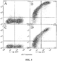
- Cells were harvested at 18-24 hours and stained with anti-PD1 antibodies or isotype controls at 10 ⁇ g/ml in PBS-azide for 1h at 4°C. Cells were washed with PBS-Azide, pelleted at 1500rpm/5min and primary antibodies were labelled with Alexa647-conjugated donkey anti-mouse IgG (5 ⁇ g/ml) in PBS-Azide for 30 min at 4°C. Cells were washed as above and resuspended in 200 ⁇ l PBS-Azide before being analysed at the flow cytometer. Propidium iodide (5 ⁇ g/ml) was added immediately prior to analysis to identify dead cells.
- GFP-positive (transfected) viable cells were gated and analysed for binding of anti-PD1 antibodies. Mutants were defined as 'knock-out' (reducing the percentage of cells bound by the anti-PD1 antibody) or 'knock-down' (reducing the intensity of antibody staining relative to other PD-1 antibodies).
- FIG. 1 An example of the binding analysis is shown in Figure 1 .
- Transfection efficiencies ranged from 15-50% (GFP + ). Isotype controls were negative on all transfectants.
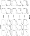
- Analysis of the percentage of GFP + cells that are also positive for Alexa647 (anti-PD-1 antibody binding) shows that the L16R and R118D mutations completely eliminate Clone 19 binding ( Fig. 2 ). All R118D expressing cells bind Clone 10, indicating functional expression of PD-1, but have the lowest intensity of all mutants ( Fig. 2 ), suggesting a low level of expression.
- V18R partially eliminates Clone 19 binding.
- Clone 10 binds all the mutants but for mutants N41K and L103E the binding intensity for this antibody versus the other PD-1 antibodies is significantly decreased ( Fig. 2 ).
- the binding analyses thus define two distinct epitopes each defined in turn by at least two residues: anti-PD-1 antibody Clone 10 binds to a membrane-distal epitope that overlaps with the ligand-binding region ( Zhang et al. Immunity 20, 337-47 (2004 )); Clone 19 binds to a membrane-proximal epitope.
- the binding-disrupting residues are mapped onto the murine PD-1 crystal structure in Figure 3 .
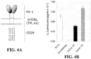
- a dimeric form of PD-1 was generated that consisted of the extracellular (antibody-binding) region of human PD-1 spliced to the transmembrane region of CD3 ⁇ (to produce dimers) and the cytoplasmic region of CD28 (in order to have an "active" readout consisting of IL-2 secretion; Fig. 4A ).
- oligonucleotide 1 (left arrow; sequence 5'-TAGTAGAGATCTCTCAAGCAGGCCACCATGCAAATCCCACAGGCGCCGTGG-3', SEQ ID NO: 33), which encodes a Bg1II restriction site and the rat ribosome binding site followed by the initiating codon and the first 21 bases of the signal peptide-encoding sequence of human PD-1, was used in a polymerase chain reaction (PCR1) with the complement of oligonucleotide 2 (5'-TCAGCCGGATCCTTCCAAACCCTGGTGCTCT GCTACTTGCTAGATGG-3', SEQ ID NO: 34).
- PCR1 polymerase chain reaction
- Oligonucleotide 2 encodes the last nine residues of the human PD-1 extracellular domain (up to residue 170 of the mature polypeptide) inserting a Bam H1 site, followed by 20 bases encoding the NH 2 -terminal end of the mouse CD3 ⁇ transmembrane domain. PCR reactions were carried out under standard conditions.
- oligonucleotide 2 was used in a PCR reaction (PCR2) with the complement of oligonucleotide 3 (5'-ATCACAGCCCTGTACCTGAATAGTAGAAG GAATAGACTC-3', SEQ ID NO: 35) which encodes the COOH-terminal end of the transmembrane region of mouse CD3 ⁇ , followed by the first 21 bases encoding the NH 2 -terminal end of the mouse CD28 cytoplasmic domain.
- step 3 the PCR1 and PCR2 reaction products were purified, annealed, extended and then amplified in the presence of oligonucleotide 1 and the complement of oligonucleotide 3, to generate a cDNA encoding the extracellular region of PD-1 fused with the transmembrane region of CD3 ⁇ .
- oligonucleotide 3 was used in a PCR reaction (PCR3) with oligonucleotide 4 (5'-CTCGAGCTACTAGGGGCGGTACGCTGCAAA- 3', SEQ ID NO: 36), which encodes the COOH-terminal end of the cytoplasmic domain of mouse CD28 followed by a stop codon and a Xho I restriction site.
- step 5 the purified PCR3 product was fused with the purified PCR product from step 3 by annealing the two products, extending the annealed hybrid, and then amplifying it with oligonucleotides 1 and 4.
- Human PD-1 and mouse CD28 cDNA was amplified using pENTRhPD-1/mCD28 as template, which was originally constructed from IMAGE clones obtained from Geneservices Ltd (Cambridge UK).
- Mouse CD3 ⁇ was amplified from DO11.10 mouse T cell hybridoma cDNA.
- the fusion PCR products were cloned into pCR4®-TOPO® (Invitrogen) and the final products sequenced by the dideoxy method.
- the constructs were cut with Bg1II and XhoI and inserted into the lentiviral vector pHR-SIN-BX-IRES-Em.
- HEK 293T cells were transfected with pHR-SIN-BX-IRES-Em encoding hPD-1/mCD3 ⁇ WT/mCD28, and the supernatant used to infect DO11.10 T-cell hybridomas.
- Infected DO11.10 cells were propagated and FACS sorted for mouse PD-1 and EGFP expression, and then tested for agonistic signaling by the anti-PD-1 antibodies using IL-2 release as a stimulation assay readout.
- IL-2 secretion results indicate that both antibodies are capable of inducing signaling via the hPD-1/mCD3 ⁇ WT/mCD28 chimera; however Clone 19, which binds PD-1 closest to the membrane induces the largest amount of IL-2 release (representative data is shown in Fig. 4b ). This supports the notion that the topology of the complex formed by the antibodies is what determines the relative levels of signaling induced by agonists. The data also suggest that the degree of agonistic signaling can be varied with choice of antibody.
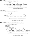
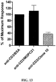
- the antibodies were tested for their ability to inhibit TCR-derived activating signals by covalently coupling the antibodies, along with anti-CD3 antibodies, to tosyl-activated DYNALBEADS.
- the beads were then added to cultures of PBL labelled with carboxyfluorescein succinimidyl ester (CFSE). Proliferation levels were indicated by the fraction of cells with diluted CFSE determined by flow cytometric analysis.
- CFSE carboxyfluorescein succinimidyl ester
- Tosyl-activated 4.5 ⁇ m DYNALBEADS (M450; Invitrogen) were washed in 0.1M sterile phosphate buffer (pH 8) and loaded with 2.5 ⁇ g total antibody per 3x10 7 beads at 37°C for 18-24 h with continuous inversion mixing.
- Rabbit IgG (Sigma) was used to equalise the amount of total antibody per bead-loading reaction. Beads were blocked for at least 30 min in RPMI with 10% FCS at room temperature and washed three times in serum-free RPMI.
- bead-bound antibody was quantified in duplicate with saturating amounts of isotype-specific PE-labelled goat antibodies and compared with QuantibriteTM prelabelled quantification kit (BD Biosciences Ltd.).
- the geometric mean fluorescence PE intensities of bead singlets (minus background of unloaded beads as a control) were used to calculate the absolute amount of antibody loaded per bead from the standard curve.
- An example of such a titration is given in Fig. 5 .
- Loaded beads were stored at 4°C.
- the amounts of anti-CD3 antibody added were varied so that, at the time of the experiments, the effects of matched sets of beads with near-equivalent levels of anti-CD3 antibody could be compared.
- the level of stimulation provided by anti-CD3 loaded beads was defined as low (resulting in 15% proliferation of bulk lymphocytes at day 5), medium-low (30% proliferation), medium-high (60% proliferation) and high (80% proliferation).
- Fresh heparinized blood was diluted 1:1 with PBS and the lymphocytes isolated by density gradient separation (Ficoll Hypaque).
- accessory cells were depleted by plastic adherence for 2 h at 37°C or with specific antibody-labelled DYNALBEADS (against CD14/19/8/56).
- Cells were washed in PBS and RPMI and resuspended at 10 7 cells/ml in serum-free RPMI. Cells were labeled with 25 ⁇ M CFSE in PBS for 10 min in the dark at RT. CFSE was quenched with an equal volume of FCS at RT for 5 min.
- Cells were washed 3-5 times with RPMI and resuspended at 10 6 cells/ml in RPMI +10% FCS + PSG + 2-ME (final concentration 5x10 -5 M).
- Antibodies (beads), mitogen or media was added to relevant wells in 96-well round-bottomed plates and 10 5 cells/well were distributed and incubated at 37°C for 3-5 days.
- cells were stained with directly-labelled cell-surface antibodies for 1 h at 4°C.
- Cells were washed with PBS-Azide, pelleted at 1500rpm/5min and resuspended in 200 ⁇ l PBS-Azide. Cells were analysed for CFSE and antibody labelling at the flow cytometer using FlowJo Flow Cytometry Analysis Software.
- Clone 10 antibodies were further tested for their ability to inhibit TCR-derived activating signals by covalently coupling the antibodies, along with anti-CD3 antibodies, to tosyl-activated DYNALBEADS.
- the beads were then added to cultures of human CD4 + T cells and proliferation measured by 3 H-thymidine incorporation.
- Tosyl-activated 4.5 ⁇ m DYNALBEADS (M450; Invitrogen) were washed in 0.1M sterile phosphate buffer (pH 7.5) and loaded with 2 ⁇ g of anti-human CD3 (clone OKT3) per 1x10 7 beads at 37°C for 8h with continuous inversion mixing. Beads were then washed to remove un-conjugated anti-CD3. Aliquots of the anti-CD3 conjugated beads were then secondarily coated with 3 ⁇ g of anti-PD-1 antibody or control per 1x10 7 beads at 37°C for 19h with continuous inversion mixing.
- Beads were washed and then incubated in 0.2M Tris / 0.1% BSA (pH 8.5) for 3 hours to inactivate free tosyl groups, followed by washing and resuspension of beads in PBS / 0.1% BSA / 2mM EDTA (pH 7.4). Equal anti-CD3 and antibody coating of the bead sets was confirmed by staining the beads with fluorochrome-labelled isotype-specific antibodies and analysing by flow cytometry.
- CD4 + T cells were purified from the whole PBLs by negative selection using MACS (CD4 + T cell isolation Kit II; Miltenyi Biotec). 1x10 5 human CD4 + T cells / well were cultured at a 1:1 ratio with the coated beads in 96-well round-bottomed plates and incubated at 37°C for 6 days. Proliferation was measured at day 6 by addition of 0.5 ⁇ Ci/well 3 H-thymidine for the last 6 hours of culture. Cells were harvested onto glass-fibre filters and incorporated 3 H-thymidine was measured by ⁇ -scintillation counting.
- the results in Figure 13 show the day 6 proliferative response by human CD4 + T cells measured in the presence of anti-CD3 plus Clone 10 antibody or control coated beads.
- the data are expressed as percentage of the maximal response (anti-CD3 plus BSA control) and are the mean of 4 different donor responses.
- CD4 + T cell proliferation was inhibited in the presence of Clone 10, so that the average proliferation observed was only 37.7 % of the maximum.
- Clone 19 generally induces stronger signaling by the hPD-1/mCD3 ⁇ WT/mCD28 chimera than Clone 10 ( Fig. 4B ) but in some experiments it gives weaker inhibitory signaling by native PD-1 (see, e.g. Fig. 8 ). It is possible that this is because, in some experiments, Clone 19 but not Clone 10 ligation results in the phosphorylation of both the ITIM (inhibitory, blue) and the ITSM (activating, red) tyrosine-based signaling motifs of PD-1 (see Fig. 9 ).
- a monomeric, monovalent form of PD-1, hPD-1/mCD28 that consisted of the extracellular (antibody-binding) and transmembrane regions of human PD-1 spliced to the cytoplasmic region of CD28 (in order to have an "active" readout consisting of IL-2 secretion; Fig. 10a ), was generated.
- oligonucleotide 1 (left arrow; sequence 5'-TAGTAGAGATCTCTCAAGCAGGCCACCA TGCAAATCCCACAGGCGCCGTGG-3', SEQ ID NO: 33), which encodes a BglII restriction site and the rat ribosome binding site followed by the initiating codon and the first 24 bases of the signal peptide-encoding sequence of human PD-1, was used in a polymerase chain reaction (PCR1) with the complement of oligonucleotide 2 (5'-GCCCAGCCGGCCAG TTCCAAACCTTTTGGGTGCTGGTGGTGGTTGGT-3', SEQ ID NO: 37).
- PCR1 polymerase chain reaction
- Oligonucleotide 2 encodes the last 23 bases of the human PD-1 extracellular domain (up to residue 149 of the mature polypeptide), followed by 24 bases encoding the NH 2 -terminal sequence of the mouse CD28 transmembrane region. PCR reactions were carried out under standard conditions.
- oligonucleotide 2 was used in a PCR reaction (PCR2) with the complement of oligonucleotide 3 (5'-TTTGCAGCGTACCGCCCCACGCGTTAGTAGCTCGAG-3', SEQ ID NO: 38) which encodes the COOH-terminal end of the cytoplasmic domain of mouse CD28, a Mlu I restriction site followed by a stop codon and a Xho I restriction site.
- the purified PCR2 product was fused with the purified PCR1 product from step 1 by annealing the two products, extending the annealed hybrid, and then amplifying it with oligonucleotides 1 and 3.
- Mouse CD28 sequence was amplified using pCR4®-TOPO®rCD28/mCD28 as template, which was originally amplified from DO11.10 mouse T cell hybridoma cDNA.
- the human extracellular PD-1 was amplified from pE14hPD-1Long, a gift from Dr Chao Yu of the MRC Human Immunology Unit, Oxford.
- the fusion PCR products were cloned into pCR4®-TOPO®(Invitrogen) and the final products sequenced by the dideoxy method.
- the constructs were cut with BglII and XhoI and inserted into the lentiviral vector pHR-SIN-BX-IRES-Em for infection of DO11.10 cells.
- Activation of the DO11.10 cells expressing the hPD-1/mCD28 chimera by anti-PD-1 antibodies was examined using IL-2 secretion as a read-out.
- hPD-1/mCD28 Lack of signaling by hPD-1/mCD28 suggests that agonistic signaling may be enhanced by cross-linking a monomeric receptor with two antibodies that bind to non-overlapping epitopes
- Clone 10 and Clone 19 were not agonistic for a chimeric form of human PD-1, i.e. hPD-1/mCD28, consisting of the monomeric extracellular region of PD-1 attached to the transmembrane and intracellular signaling domains of CD28 ( Fig. 10 ), in contrast to the equivalent CD28 construct (containing the homodimeric extracellular domain of rat CD28).
- hPD-1/mCD28 a chimeric form of human PD-1
- CD28 containing the homodimeric extracellular domain of rat CD28.
- the likeliest explanation for this is that, because PD-1 is monomeric and CD28 is a homodimer, the attachment of bivalent antibody leads to the assembly of a multimeric array of "cross-linked" CD28 molecules and a very high density of signaling domains ( Fig.
- Agonistic signalling is enhanced by cross-linking a monomeric receptor with two antibodies that bind to non-overlapping epitopes
- DO11.10 cells expressing the hPD-1/mCD28 chimeric protein were used in a Clone 10/Clone 19 antibody stimulation assay as follows.
- 96-well flat-bottomed plates (Costar EIA/RIA plates) were coated overnight at 4°C with 500 ⁇ g/ml donkey anti-mouse IgG (Jackson Immunoresearch) in coating buffer (15mM Na2CO3, 35mM NaHCO3, pH 9.6. Prior to the addition of cells, the plates were washed three times with 200 ⁇ l chilled PBS.
- the heavy chain CDR1s for clones 2, 10 and 19 have been identified according to both the combined Kabat/Chothia numbering system and the Kabat numbering system. All other CDRs have been identified according to the Kabat numbering system ( Kabat et al., 1987, "In sequences of proteins of immunological interest ", U.S. Dept. Health and Human Services, NIH USA. Heavy chain CDR1s for clones 2, 10 and 19, as identified by the Kabat numbering system, are identified (underlined amino acids) in Table 1.
Landscapes
- Health & Medical Sciences (AREA)
- Chemical & Material Sciences (AREA)
- Organic Chemistry (AREA)
- Immunology (AREA)
- Medicinal Chemistry (AREA)
- General Health & Medical Sciences (AREA)
- Life Sciences & Earth Sciences (AREA)
- Pharmacology & Pharmacy (AREA)
- Veterinary Medicine (AREA)
- Nuclear Medicine, Radiotherapy & Molecular Imaging (AREA)
- Engineering & Computer Science (AREA)
- Chemical Kinetics & Catalysis (AREA)
- Bioinformatics & Cheminformatics (AREA)
- Animal Behavior & Ethology (AREA)
- General Chemical & Material Sciences (AREA)
- Public Health (AREA)
- Diabetes (AREA)
- Proteomics, Peptides & Aminoacids (AREA)
- Molecular Biology (AREA)
- Genetics & Genomics (AREA)
- Biophysics (AREA)
- Biochemistry (AREA)
- Rheumatology (AREA)
- Biomedical Technology (AREA)
- Neurology (AREA)
- Neurosurgery (AREA)
- Pain & Pain Management (AREA)
- Pulmonology (AREA)
- Orthopedic Medicine & Surgery (AREA)
- Physical Education & Sports Medicine (AREA)
- Obesity (AREA)
- Hematology (AREA)
- Endocrinology (AREA)
- Emergency Medicine (AREA)
- Transplantation (AREA)
- Peptides Or Proteins (AREA)
- Medicines Containing Antibodies Or Antigens For Use As Internal Diagnostic Agents (AREA)
- Preparation Of Compounds By Using Micro-Organisms (AREA)
- Micro-Organisms Or Cultivation Processes Thereof (AREA)
Description
-
WO 2004/056875 - Wang et al (2007, Eur J Immunol, 37, 2983-2990) studies PD-1/PD-1 ligand and its role in allograft tolerance.
- Seka et al (2007, Cardiovascular Research, 75, 158-167) studies the roles of PD-1/PD-1 ligand pathway in the development of acute myocarditis.
-
US 2007/202100 relates to the use of PD-1 modulating agents in order to modulate a costimulatory or inhibitory signal in an immune cell, thereby modulating immune response. - The scope of the present invention is defined by the claims and any information that does not fall within the claims is provided for information only.
- One aspect of the present disclosure provides antibodies that can act as agonists of PD-1, thereby modulating immune responses regulated by PD-1. In one aspect, the anti-PD-1 antibodies can be novel antigen-binding fragments. Anti-PD-1 antibodies disclosed herein are able to bind to human PD-1 and agonize the activity of PD-1, thereby inhibiting the function of immune cells expressing PD-1. Exemplary antibodies for use in the context of this disclosure include, but are not limited to monoclonal antibody produced by
clone 19. - Another aspect of the disclosure provides compositions comprising PD-1 specific antibodies and their use in methods of down regulating the immune response. These methods can be practiced on any subject, including humans or animals. In particular aspects, anti-PD-1 antibodies are used to treat or prevent immune disorders by reducing the T cell response. Non-limiting examples of immune disorders that can be treated via the administration of PD-1 specific antibodies to a subject include, but are not limited to, rheumatoid arthritis, multiple sclerosis, inflammatory bowel disease, Crohn's disease, systemic lupus erythematosus, type I diabetes, transplant rejection, graft-versus-host disease, hyperproliferative immune disorders, cancer, and infectious diseases. Some embodiments of this aspect of the disclosure may use two PD-1 specific antibodies that bind to distinct, non-overlapping epitopes.
- Anti-PD-1 antibodies disclosed herein may be used, in another aspect of the disclosure to detect PD-1 or its fragments in a biological sample. The amount of PD-1 detected may be correlated with the expression level of PD-1, and associated with the activation status of immune cells (e.g., activated T cells, B cells, and/or monocytes) in the subject.
-
-
Fig. 1 : Detection of antibody binding to human PD-1 transfectants. Cells were transfected with constructs expressing the extracellular region of human PD-1 (panels A and B) or mutants that affected the binding of Clone 19 (L16R; panel C) or Clone 10 (L103E; panel D) anti-PD-1 antibodies. The cells were labelled with isotype control antibody (panel A) or with Clone 19 (panels B and C) or Clone 10 (panel D) antibody, followed by Alexa647 labelled secondary antibody. Transfected 293T cells are eGFP-positive (x-axis). Antibody binding is shown on the y-axis. -
Fig. 2 : PD-1 epitope screen. The geometric means of the Alex647 fluorescence levels for the GFP+ cells is given for each of the mutants of PD-1, expressed as full length proteins in HEK 293T cells. -
Fig. 3 : Anti-PD-1 antibody epitopes. The epitopes were mapped by antibody binding analysis following expression of single-residue mutated forms of PD-1 in HEK 293T cells. Mouse PD-1 residues equivalent to human PD-1 residues that when mutated partially or fully block the binding ofClones Clone 2 andclone 10 antibodies appear to compete with one another for binding to PD-1 based upon the results of this analysis. -
Figs. 4A -B: IL-2 secretion induced by anti-PD-1 antibodies binding to a hPD-1/mCD3ζWT/mCD28 chimera. (Figure 4A ) A chimera consisting of the extracellular region of human PD-1 and the transmembrane and cytoplasmic regions of mouse (m) TCRζ and CD28 was expressed in DO11.10 cells. (Figure 4B ) The cells were treated with immobilized anti-CD3 (KT3) or anti-PD-1 antibodies and the amount of IL-2 released was measured. -
Fig. 5 : Quantification of monoclonal antibodies loaded onto tosyl-activated DYNALBEADS. Amount of anti-human CD3 OKT3 antibody used per loading (107 beads with 2.5 µg total antibody) is shown on the x-axis. Remaining amount of antibody was made up with Rabbit IgG or anti-PD-1 antibodies (Clone 19, Clone 10, Clone 2) to a total of 2.5 µg. The number of IgG1 (Rabbit IgG or anti-PD-1 antibodies; red bars) or IgG2a (OKT3; blue bars) molecules detected per bead is shown on the y-axis. Green arrows indicate beads selected for use in the experiments shown inFig. 6 . Values are averages of duplicates. -
Fig. 6 : Titration of anti-PD-1 antibodies coupled to tosyl-activated DYNALBEADS. Bulk preparations of PBL were incubated with beads containing anti-CD3 and increasing amounts of anti-PD-1 antibodies (Clone 19 or Clone 10). Amount of anti-PD-1 antibody loaded per 107 beads (in a total of 2.5 µg mAb per 107 beads) is shown on the x-axis. Proliferation (y-axis) was measured by CFSE dilution atday 5. Bars represent means of triplicates ± SD. -
Fig. 7 : Stimulation of a PD-1/mCD28 chimera-expressing DO11.10 cell line with titrations of two anti-PD-1 antibodies. DO11.10 cells expressing a PD-1/mCD28 chimera were incubated with titrations of anti-PD-1Clone 19 antibody (from 100 µg/ml to 0 µg/ml) and anti-PD-1Clone 10 antibody (from 100 µg/ml to 0 µg/ml). Cells were then incubated in donkey anti-mouse IgG antibody coated (500 µg /ml) 96 well plates for 48 hours before tissue culture supernatant was assayed for IL-2 by ELISA. -
Fig. 8 : Activation of T cells with beads quantified for Ig content. PBL were depleted of monocytes by plastic adherence (bulk PBL). The amount of anti-CD3 (OKT3) and anti-PD-1 antibodies (Clone 19 or Clone 10) was quantified and is shown in the table (left) expressed as number of molecules per bead. Proliferation (y-axis) was measured by CFSE dilution atday 5. Bars are duplicates ± SD. -
Fig. 9 : Explanation for differential signaling by the two antibodies.Clone 19 induces stronger signaling by a hPD-1/mCD3ζWT/mCD28 chimera than Clone 10. PD-1 has ITIM (inhibitory, blue) and ITSM (activating, red) tyrosine-based signaling motifs. It is suggested that, in vitro, Clone 19 triggers the phosphorylation of both motifs whereas Clone 10 ligation results in phosphorylation of the inhibitory motif only, leading to more potent inhibitory signaling. -
Figs. 10A-10B : IL-2 secretion induced by anti-PD-1 antibodies binding to a hPD-1/mCD28 chimera. (Figure 10A ) A chimera consisting of the extracellular region of human PD-1 and the transmembrane and cytoplasmic regions of mouse CD28 was expressed in DO11.10 cells. (Figure 10B ) The cells were treated with immobilized anti-CD3 (KT3) or anti-PD-1 antibodies and the amount of IL-2 released was measured. -
Figs. 11A-11B : Strong signaling induced by a pair of antibodies binding to a monomeric signaling protein. (Figure 11A ) Antibodies, which are bivalent, cause strong signaling by homodimeric receptors because they are able to generate a high local density of signaling domains. (Figure 11B ) In contrast, antibodies are only able to recruit pairs of monomeric receptors, such as PD-1, leading to much less intense signaling. (Figure 11C ) By using antibodies that bind to two non-overlapping epitopes, higher densities of monomeric signaling receptors can be generated, giving much more potent signaling. -
Fig. 12 : Dissociation rates forClone buffer 10 mM Hepes, 150 mM NaCl pH 7.4. Following injection of the soluble PD-1, the buffer only was injected, allowing dissociation of the bound soluble PD-1 from each of the antibodies simultaneously. Dissociation rates were fitted using Origin v.5.0 software (MicroCal Software Inc, Northampton, MA) after subtraction of the dissociation rate for OX-7 dissociating from the anti-mouse Fc antibody. -
Fig. 13 : Inhibition of CD4+ T cell proliferation by anti-PD-1 antibodies. CD4+ T cells were purified from human PBL by negative selection and cultured with Dynalbeads coated with anti-CD3 plus control (BSA or MOPC21) orClone 10 antibody. Proliferation (y-axis) was measured by 3H-thymidine incorporation atday 6. Bars represent the % of maximal response (anti-CD3/BSA) and are the mean +/- S.E.M. of 4 different donor cultures. - The term "antibody", as used in this disclosure, refers to an immunoglobulin or a fragment or a derivative thereof, and encompasses any polypeptide comprising an antigen-binding site, regardless of whether it is produced in vitro or in vivo. Thus, an antibody includes, but is not limited to, polyclonal, monoclonal, monospecific, polyspecific, bispecific, humanized, single-chain, chimeric, synthetic, recombinant, hybrid, mutated, and grafted antibodies.
- The term "antibody fragment" or "an antigen binding fragment" includes antibody fragments such as Fab, F(ab')2, Fv, scFv, Ed, dab, and other antibody fragments that retain antigen-binding function, i.e., the ability to bind PD-1 specifically and/or that are produced from a monoclonal antibody disclosed herein. These fragments comprise an antigen-binding domain and can also, in some aspects, agonize the function of PD-1. Antibodies disclosed herein, and fragments thereof, include those antibodies having altered glycosylation patterns when compared to the parent antibody (e.g., the antibody produced by
clone 10 and/or clone 19). - As discussed above, the PD-1 antibodies disclosed herein are able to antagonize the activity and/or proliferation of lymphocytes by agonizing PD-1. The term "antagonize the activity" relates to a decrease (or reduction) in lymphocyte proliferation or activity that is at least about 10%, 20%, 30%, 40%, 50%, 60%, 70%, 80%, 90%, or more. The term "antagonize" may be used interchangeably with the terms "inhibitory" and "inhibit". PD-1-mediated activity can be determined quantitatively using T cell proliferation assays as described herein.
- The terms "therapeutically effective", "therapeutically effective amount", "effective amount" or "in an amount effective" refers to a dosage or amount of the disclosed antibodies that is sufficient to agonize the activity of PD-1 and provide for the amelioration of symptoms in a subject or to achieve a desired biological response, e.g., decreased T cell activity, etc.
- The term "isolated" refers to a molecule that is substantially free of its natural environment. For instance, an isolated antibody is substantially free of cellular material or other proteins from the cell (e.g., hybridoma) or other source from which it is derived. The term isolated also refers to preparations where the isolated protein is sufficiently pure to be administered as a pharmaceutical composition, or at least 70-80% (w/w) pure, at least 80-90% (w/w) pure, 90-95% pure; or at least 95%, 96%,97%, 98%, 99%, or 100% (w/w) pure.
- One aspect of the present disclosure provides antibodies that can act as agonists of PD-1, thereby modulating immune responses regulated by PD-1. In one aspect, the anti-PD-1 antibodies can be novel antigen-binding fragments. Anti-PD-1 antibodies disclosed herein are able to bind to including human PD-1 and agonize PD-1, thereby inhibiting the function of immune cells expressing PD-1. In some aspects, the immune cells are activated lymphocytes, such as T-cells, B-cells and/or monocytes expressing PD-1. Exemplary antibodies for use in the context of this disclosure include, but are not limited to monoclonal antibodies produced by
clone 19. Some aspects of this aspect of the disclosure may use two PD-1 specific antibodies that bind to distinct, non-overlapping epitopes. - Another aspect of the disclosure provides anti-PD-1 specific monoclonal antibodies having modified binding affinity. One aspect provides for modifying the binding affinity such that the antibody has a low affinity for PD-1 (e.g., the antibody has a dissociation rate of between 0.1 sec-1 and 0.5 sec-1 or less than 0.90 sec-1). Particular aspects provided antibodies having off rates of 0.10 sec-1, 0.15 sec-1, 0.20 sec-1, 0.25 sec-1, 0.30 sec-1, 0.35 sec-1, 0.40 sec-1, 0.45 sec-1 or 0.50 sec-1 or for antibodies having dissociation rates ranging from 0.04 sec-1 to 2.0 sec-1 (e.g., 0.04 sec-1, 0.05 sec-1, 0.06 sec-1, 0.07 sec-1, 0.08 sec-1, 0.09 sec-1,0.10 sec-1, 0.15 sec-1, 0.20 sec-1, 0.25 sec-1, 0.30 sec-1, 0.35 sec-1, 0.40 sec-1, 0.45 sec-1, 0.50 sec-1, 0.55 sec-1, 0.60 sec-1, 0.65 sec-1, 0.70 sec-1, 0.75 sec-1, 0.80 sec-1, 0.85 sec-1, 0.90 sec-1, 0.95 sec-1, 1.0 sec-1, 1.10 sec-1, 1.20 sec-1, 1.30 sec-1, 1.40 sec-1, 1.50 sec-1, 1.60 sec-1, 1.70 sec-1, 1.80 sec-1, 1.90 sec-1, or 2.00 sec-1). Antibodies having such binding affinities can be modified in any suitable process.
- Thus, the binding affinity of the antibodies (such as those produced by
clone 2, clone 10 or clone 19) can be increased or decreased via various methods known in the art. For example, binding characteristics can be modified by direct mutation, methods of affinity maturation, phage display, or chain shuffling within the nucleic acids encoding the antibody molecules. Individual residues or combinations of residues can be randomized so that in a population of otherwise identical antigen binding sites, all twenty amino acids are found at particular positions and binding characteristics/affinities can also be modified by methods of affinity maturation. (See, e.g., Yang et al. (1995) J. Mol. Biol. 254, 392-403; Hawkins et al. (1992) J. Mol. Bio. 226,889-896; or Low et al. (1996) J. Mol. Biol. 250, 359-368). Methods known in the art include, for example, Marks et al. BioTechnology, 10, 779-783 (1992), which describes affinity maturation by VH and VL domain shuffling; random mutagenesis of CDR and/or framework residues is described by: Barbas et al. Proc Nat. Acad. Sci, USA 91, 3809-3813 (1994); Schier et al. Gene, 169, 147-155 (1995); Yelton et al. J. Immunol., 155, 1994-2004 (1995); Jackson et al. J. Immunol., 154, 3310-9 (1995); and Hawkins et al. J. Mol. Biol., 226, 889-896 (1992). - Strategies for antibody optimization are sometimes carried out using random mutagenesis. In these cases positions are chosen randomly, or amino acid changes are made using simplistic rules. For example all residues may be mutated to alanine, referred to as alanine-scanning.
WO 9523813 2003/022240 A1 and U.S. Pat. No.2002/177170A1 ) may also be used to increase or decrease the binding affinities of antibodies. Finally, the binding affinities of antibodies in which the binding affinity has been altered can be determined using methods as disclosed herein (for example, dissociation rates for modified antibodies can be determined by surface plasmon resonance-based analysis as described forFigure 12 ). - Anti-PD1 antibodies described herein can be linked to another molecule/moiety. Non-limiting examples include another peptide or protein (albumin, another antibody, etc.), toxins, radioisotopes, cytotoxic agents or cytostatic agents. The term "link" or "linked" relates to the chemical cross-linking or covalent attachment of another molecule/moiety by recombinant methods. Antibodies disclosed herein may also be linked to one or more nonproteinaceous polymers, e.g., polyethylene glycol, polypropylene glycol, or polyoxyalkylenes (see, for example, U. S. Patent Nos.
4,791,192 ;4,766,106 ;4,670,417 ;4,640,835 ;4,609,546 ;4,496,689 ;4,495,285 ;4,301,144 ; and4,179,337 ). - The antibodies may also be tagged with a detectable, or functional, label. Detectable labels include radiolabels such as 99Tc, which may also be attached to antibodies using conventional chemistry. Detectable labels also include enzyme labels such as horseradish peroxidase or alkaline phosphatase. Other types of detectable labels include chemical moieties such as biotin, which may be detected via binding to a specific cognate detectable moiety, e.g., labeled avidin.
- Another aspect of the disclosure provides for the use of antibodies disclosed herein for isolating PD-1 or PD-1-expressing cells. Yet another aspect of the disclosure provides methods of inducing tolerance to a specific antigen. For example, tolerance can be induced by co-administration of antigen and an anti-PD-1 antibody disclosed herein. Still other aspects of the disclosure relate to reducing immune responses mediated by activated lymphocytes in a subject comprising the administration of anti-PD-1 antibodies disclosed herein. Another aspect of the disclosure provides for the use of the disclosed anti-PD-1 antibodies for agonizing PD-1 and down regulating immune responses (or in some cases inhibiting or reducing the proliferation of activated lymphocytes). In particular aspects, the immune response is TcR/CD28-mediated. As discussed herein, allergies, rheumatoid arthritis, type I diabetes mellitus, multiple sclerosis, inflammatory bowel disease, Crohn's disease, systemic lupus erythematosus, tissue, skin and organ transplant rejection or graft-versus-host disease (GVHD) can be treated via the administration of anti-PD-1 antibodies. Some embodiments of this aspect of the disclosure may use two PD-1 specific antibodies that bind to distinct, non-overlapping epitopes.
- Another aspect of the disclosure provides compositions comprising PD-1 specific antibodies and their use in methods of down regulating the immune response (or reducing the proliferation of activated T-cells, B-cells or mononuclear cells). These methods can be practiced on any subject, including humans or animals. In particular aspects, anti-PD-1 antibodies are used to treat or prevent immune disorders by reducing the T cell response. Non-limiting examples of immune disorders that can be treated via the administration of PD-1 specific antibodies to a subject include, but are not limited to, rheumatoid arthritis, multiple sclerosis, inflammatory bowel disease, Crohn's disease, systemic lupus erythematosus, type I diabetes, transplant rejection, graft-versus-host disease, hyperproliferative immune disorders, cancer, and infectious diseases. Yet other aspects of the disclosure provide for inhibiting or reducing lymphocyte (T-cell, B-cell and/or monocyte) activity in inflammatory lesions. Some embodiments of this aspect of the disclosure may use two PD-1 specific antibodies that bind to distinct, non-overlapping epitopes (such antibodies can be affinity matched to provide a desired activity in vivo (e.g.,
Clone 19 and Clone 2)). - Anti-PD-1 antibodies disclosed herein may be used, in another aspect of the disclosure to detect PD-1 or its fragments in a biological sample. The amount of PD-1 detected may be correlated with the expression level of PD-1, and associated with the activation status of immune cells (e.g., activated T cells, B cells, and/or monocytes) in the subject.
- T-cells can be activated by any T-cell activating compound. As discussed in the examples, one such T-cell-activating compound is an anti-CD3 antibody, which binds TcR. Activating anti-CD3 antibodies are known in the art (see, for example, U. S. Patent Nos.
6,405,696 and5,316,763 ). The ratio between the activating TcR signal and negative PD-1 signal is determined experimentally using conventional procedures known in the art or as described in the Examples. Some embodiments of this aspect of the disclosure may use two PD-1 specific antibodies that bind to distinct, non-overlapping epitopes. - The antibodies or antibody compositions of the present disclosure are administered in therapeutically effective amounts. Generally, a therapeutically effective amount may vary with the subject's age, condition, and sex, as well as the severity of the medical condition of the subject. A therapeutically effective amount of antibody ranges from about 0.001 to about 25 mg/kg body weight, preferably from about 0.01 to about 25 mg/kg body weight, from about 0.1 to about 20 mg/kg body weight, or from about 1 to about 10 mg/kg. The dosage may be adjusted, as necessary, to suit observed effects of the treatment. The appropriate dose is chosen based on clinical indications by a treating physician.
- In another aspect, the antibodies of the disclosure can be used as a targeting agent for delivery of another therapeutic or a cytotoxic agent (e.g., a toxin) to a cell expressing PD-1. The method includes administering an anti-PD-1 antibody coupled to a therapeutic or a cytotoxic agent or under conditions that allow binding of the antibody to PD-1 expressed on the cell surface.
- Still other aspects of the disclosure provide for the use of the disclosed antibodies for detecting the presence of PD-1 in biological samples. The amount of PD-1 detected may be correlated with the expression level of PD-1, which, in turn, is correlated with the activation status of immune cells (e.g., activated T cells, B cells, and monocytes) in the subject.
- The subject disclosure also provides methods of binding an antibody to a PD-1 polypeptide comprising contacting a sample that may contain PD-1 or cells expressing PD-1 with an antibody under conditions that allow for the formation of an antibody-antigen complex. These methods can further comprise the step of detecting the formation of said antibody-antigen complex. The complex can be detected using any means known in the art (e.g., fluorescence activated cell sorting, radioimmunoassays, or chromogenic assays).
- Another aspect of the disclosure provides compositions comprising anti-PD-1 antibodies. These compositions can be formulated according to known methods for preparing pharmaceutically useful compositions. Formulations are described in a number of sources which are well known and readily available to those skilled in the art. For example, Remington's Pharmaceutical Science (Martin E.W., Easton Pennsylvania, Mack Publishing Company, 19th ed., 1995) describes formulations which can be used in connection with the subject disclosure. Formulations suitable for administration include, for example, aqueous sterile injection solutions, which may contain antioxidants, buffers, bacteriostats, and solutes which render the formulation isotonic with the blood of the intended recipient; and aqueous and nonaqueous sterile suspensions which may include suspending agents and thickening agents. The formulations may be presented in unit-dose or multi-dose containers, for example sealed ampoules and vials, and may be stored in a freeze dried (lyophilized) condition requiring only the condition of the sterile liquid carrier, for example, water for injections, prior to use. Extemporaneous injection solutions and suspensions may be prepared from sterile powder, granules, tablets, etc. It should be understood that in addition to the ingredients particularly mentioned above, the formulations of the subject disclosure can include other agents conventional in the art having regard to the type of formulation in question.
- Another aspect of the disclosure provides nucleic acids encoding PD-1 specific antibodies disclosed herein. For example, the nucleic acids encoding the antibody secreted by
clone 19 can be isolated according to methods known to those skilled in the art. Yet another aspect of the disclosure provides vectors and transformed host cells comprising a nucleic acid encoding a PD-1 specific antibody as secreted byclone 19. As would be apparent to those skilled in the art, constant regions of the murine antibodies disclosed herein can be substituted with human constant regions to form chimeric antibodies comprising murine variable regions and human constant regions. Some aspects provide for the substitution of heavy chain constant regions on the disclosed antibodies that provide for higher Fc receptor binding by the antibodies (e.g., human IgG1, IgG3, and murine IgG2a isotypes, all of which bind Fc receptors strongly, can be grafted onto variable regions of the disclosed antibodies without affecting binding specificity). Alternatively, CDRs from the murine antibodies disclosed herein can be isolated and grafted into human framework regions to form humanized antibodies. Finally, methods of producing the disclosed PD-1 specific antibodies (including methods of producing the aforementioned humanized and chimeric antibodies) are also provided by the subject disclosure. - The hybridomas disclosed herein were deposited on September 9, 2008 with European Collection of Cell Cultures (ECACC), Centre For Emergency Preparedness and Response, The Health Protection Agency, Porton Down, Salisbury, Wiltshire, SP4 0JG United Kingdom. The accession numbers for the hybridomas are as follows:
- Clone 2: 08090903;
- Clone 10: 08090902; and
- Clone 19: 08090901.
- As discussed above, antibodies disclosed herein can be a full-length murine, human, humanized, or chimeric antibody; or a fragment or derivative thereof. In one aspect, the antibody binds the same, or substantially the same, epitope as
clone 19 or by a monoclonal antibody comprising a VH sequence of SEQ ID NO: 14 and a Vk sequence of SEQ ID NO: 12. In another aspect, the antibody, including a fragment or derivative thereof, comprises the same or substantially identical VH and/or Vk regions as clone 19 (SEQ ID NOs: 14 and 12). - In another aspect, the antibody, including a fragment or derivative thereof, comprises the same or substantially identical CDR1, CDR2 and CDR3 regions as those found in the Vk and VH sequences of clone 19 (SEQ ID NOs: 27-32). In one aspect, the antibody comprises a VH sequence of SEQ ID NO: 14, a Vk sequence of SEQ ID NO: 12, as well as the sequence for murine IgG1 constant heavy chain region (GenBank accession No. D78344) and the sequence for murine IgG1 constant light chain region (GenBank accession No. V00807). Other aspects of the disclosure provide nucleotide sequences encoding the disclosed antibodies, expression vectors comprising such sequences, host cells comprising such vectors, and methods of producing such antibodies from such host cells.
- Fragments and derivatives of antibodies of this disclosure can be produced by techniques that are known in the art. "Immunoreactive fragments" comprise a portion of the intact antibody, generally the antigen binding site or variable region. Examples of antibody fragments include Fab, Fab', Fab'-SH, F(ab')2, and Fv fragments; diabodies; any antibody fragment that is a polypeptide having a primary structure consisting of one uninterrupted sequence of contiguous amino acid residues (referred to herein as a "single-chain antibody fragment" or "single chain polypeptide"), including without limitation (1) single-chain Fv (scFv) molecules (2) single chain polypeptides containing only one light chain variable domain, or a fragment thereof that contains the three CDRs of the light chain variable domain, without an associated heavy chain moiety and (3) single chain polypeptides containing only one heavy chain variable region, or a fragment thereof containing the three CDRs of the heavy chain variable region, without an associated light chain moiety; and multispecific antibodies formed from antibody fragments. For instance, Fab or F(ab')2 fragments may be produced by protease digestion of the isolated antibodies, according to conventional techniques. Alternatively, the DNA of a hybridoma producing an antibody of this disclosure may be modified so as to encode for a fragment of this disclosure. The modified DNA is then inserted into an expression vector and used to transform or transfect an appropriate cell, which then expresses the desired fragment.
- In an alternate aspect, the DNA of a hybridoma producing an antibody of this disclosure can be modified prior to insertion into an expression vector, for example, by substituting the coding sequence for human heavy- and light-chain constant domains in place of the homologous non-human sequences (e.g., Morrison et al., Proc. Natl. Acad. Sci. U.S.A., 81, pp. 6851 (1984)), or by covalently joining to the immunoglobulin coding sequence all or part of the coding sequence for a non-immunoglobulin polypeptide. In that manner, "chimeric" or "hybrid" antibodies are prepared that have the binding specificity of the original antibody. Typically, such non-immunoglobulin polypeptides are substituted for the constant domains of an antibody of the disclosure. Thus, the antibodies of the present disclosure may also be made into "chimeric" antibodies (immunoglobulins) in which a portion of the heavy and/or light chain is identical with or homologous to corresponding sequences in the original antibody, while the remainder of the chain(s) is identical with or homologous to corresponding sequences in antibodies derived from another species or belonging to another antibody class or subclass, as well as fragments of such antibodies, so long as they exhibit the desired biological activity (Cabilly et al., supra; Morrison et al., Proc. Natl. Acad. Sci. U.S.A., 81, pp. 6851 (1984)).
- In an exemplary aspect, a chimeric recombinant mAb from
clone 19 VH and Vk sequences, or a derivative or variant thereof, is produced. Nucleic acid sequences encoding theclone 19 VH and Vk sequences (SEQ ID NOs: SEQ ID NOs: 14 and 12, respectively) are cloned into a commercially available or otherwise known eukaryotic expression vector containing the light and heavy chain constant regions for a human or non-human antibody, using standard techniques. One example of a commercially available vector is pASK84, available from the ATCC (American Type Culture Collection, catalog number 87094). CHO cells, or other mammalian cell lines are then transfected with the vectors by standard methods, as described for example in "Molecular Cloning", Sambrook et al. The result is transfected cell lines that stably express and secrete the antibody molecule of interest, such as a chimeric version ofclone 19 comprising its original VH and Vk regions and the constant regions from a human mAb. The entire cDNA sequences encoding the constant regions of human IgG can be found in the following GenBank entries: Human IgG1 constant heavy chain region: GenBank accession #: J00228; Human IgG2 constant heavy chain region: GenBank accession #: J00230; Human IgG3 constant heavy chain region: GenBank accession #: X04646; Human IgG4 constant heavy chain region: GenBank accession #: K01316; and Human kappa light chain constant region: GenBank accession #: J00241. - Alternatively, VH and Vk regions of
clone 19, or mutants or derivatives thereof, can be cloned into vectors encoding truncated constant regions in order to express antibody fragments (e.g., Fab fragments). Isotype-switching of antibody can be made according to similar principles. For example, an antibody with the exact same specificity asclone 19 but of a different isotype can be obtained by sub-cloning the cDNA encoding Vk and VH sequences into plasmids containing cDNA encoding human kappa light chain constant regions and a human heavy constant chain region selected from IgG1 or IgG2 or IgG3 or IgG4 constant heavy chain regions. Thus, an antibody as generated can possess any isotype and the antibody can then be isotype switched using conventional techniques in the art. Such techniques include the use of direct recombinant techniques (see, e.g.,US Patent 4,816,397 ), cell-cell fusion techniques (see e.g.,US Patent 5,916,771 ), and other suitable techniques known in the art. Accordingly, the effector function of antibodies provided by the disclosure may be "changed" with respect to the isotype of a parent antibody by isotype switching to, e.g., an IgG1, IgG2, IgG3, IgG4, IgD, IgA, IgE, or IgM antibody for various therapeutic or other uses. - According to another aspect, the antibody of this disclosure is humanized. "Humanized" forms of antibodies according to this disclosure are specific chimeric immunoglobulins, immunoglobulin chains or fragments thereof (such as Fv, Fab, Fab', F(ab') 2, or other antigen-binding subsequences of antibodies) which contain minimal sequence derived from the murine immunoglobulin. For the most part, humanized antibodies are human immunoglobulins (recipient antibody) in which residues from a complementary-determining region (CDR) of the recipient are replaced by residues from a CDR of the original antibody (donor antibody) while maintaining the desired specificity, affinity, and capacity of the original antibody. In some instances, Fv framework residues of the human immunoglobulin may be replaced by corresponding non-human residues. Furthermore, humanized antibodies can comprise residues that are not found in either the recipient antibody or in the imported CDR or framework sequences. These modifications are made to further refine and optimize antibody performance. In general, the humanized antibody will comprise substantially all of at least one, and typically two, variable domains, in which all or substantially all of the CDR regions correspond to those of the original antibody and all or substantially all of the FR regions are those of a human immunoglobulin consensus sequence. The humanized antibody optimally also will comprise at least a portion of an immunoglobulin constant region (Fc), typically that of a human immunoglobulin. For further details see Jones et al., Nature, 321, pp. 522 (1986); Reichmann et al., Nature, 332, pp. 323 (1988); and Presta, Curr. Op. Struct. Biol., 2, pp. 593 (1992). Accordingly, humanized versions of
clone 19 comprising the VH and Vk CDR regions ofclone 19 and constant and framework regions from a human mAb can be made, using known constant and framework human mAb sequences and established techniques in the art, as described herein. For any humanized antibody incorporating theclone 19 VH CDR1 domain, the domain can contain SEQ ID NO: 30 or amino acids 6-10 of SEQ ID NO: 30. - Methods for humanizing the antibodies of this disclosure are well known in the art. Generally, a humanized antibody according to the present disclosure has one or more amino acid residues introduced into it from the original antibody. These murine or other non-human amino acid residues are often referred to as "import" residues, which are typically taken from an "import" variable domain. Humanization can be essentially performed following the method of Winter and co-workers (Jones et al., Nature, 321, pp. 522 (1986); Riechmann et al., Nature, 332, pp. 323 (1988); Verhoeyen et al., Science, 239, pp. 1534 (1988)). Accordingly, such "humanized" antibodies are chimeric antibodies (Cabilly et al., U.S. Pat. No.
4,816,567 ), wherein substantially less than an intact human variable domain has been substituted by the corresponding sequence from the original antibody. In practice, humanized antibodies according to this disclosure are typically human antibodies in which some CDR residues and possibly some FR residues are substituted by residues from analogous sites in the original antibody. - For fusion the myeloma cell line SP2/0-Ag14 from the German Collection of Microorganisms and Cell Cultures (DSMZ GmbH, Braunschweig) was used. This cell line is a hybrid between BALB/c spleen cells and the myeloma cell line P3x63Ag8. The cells have been described as not synthesizing or secreting immunoglobulin chains, being resistant to 8-azaguanine at 20 µg/ml, and not growing in HAT (Hypoxanthine, Aminopterin, Thymidine) medium. The SP2/0 cells are routinely maintained in tissue culture flasks in standard growth medium (with 10% FCS). A new aliquot of frozen SP2/0 cells was used after a period of 2 weeks in order to avoid the implementation of HGPRT-positive revertants. The myeloma cells were shown to be negative in all mycoplasma tests.
- The recombinant protein PD-1Fc was prepared using the methods described for the production of CD28Fc (Evans et al. Nat Immunol. 6, 271-9 (2005)) and concentrated to 5.1 mg/ml in 0.01 M HEPES, 150 mM NaCl, pH 7.4. SDS-PAGE analysis of the antigen run under reducing and non-reducing conditions established the purity of the protein to be >95%.
- Five mice (about 8 weeks old) were immunized via the intraperitoneal cavity using an immunization protocol over 60 days. For immunization an alum precipitate of the immunogen was prepared. The alum precipitate was freshly prepared for each boost. The mice were immunized with 50 µg protein and boosted with 25 µg protein. Three mice were used for fusion.
- Cells were handled under sterile conditions using a laminar air-flow system, sterile materials and sterile solutions. Cells were incubated at 37°C in a humid atmosphere containing 5% carbon dioxide. For cultivation of the hybridoma cells a complete growth medium (CGM) containing DMEM with supplements 2-mercaptoethanol, L-Glutamine, GlutaMax, HT, non essential amino acids, sodium pyruvate, antibiotics/antimycotic solution (in concentrations recommended by the supplier) and FCS at different concentrations (10%, 15% or 20%) was used.
- After asphyxiation of the three immunized mice in CO2 spleens were aseptically removed. A single cell suspension of pooled spleens was prepared. The spleen cells and the myeloma cells were washed several times with DMEM and fused twice in the presence of 1
ml 50% (w/v) PEG 3550 (ratio spleen cells to SP2/0 2.5:1 and 2.4:1). The hybridomas produced were resuspended in CGM containing 20% FCS and aminopterin (HAT medium). The cell suspension (140 Cl/well) of each fusion was plated out into eight 96-well tissue culture flat-bottom plates (Corning-Costar) containing 140 Cl/well peritoneal exudate cells as feeder cells in CGM with 20% FCS. The plates were incubated for 10 days. During this period cells were fed two times with HAT medium. An aliquot of the spleen cell preparation (about 8x10⁶ spleen cells) was cultivated 10 days in a well of a 24-well plate and the cell culture supernatant served as positive control in ELISA. - An ELISA was used for screening of IgG in cell culture supernatants. 96 well flat-bottom polystyrene microtiter plates (Greiner, Cat. No 655061) were coated with 50 µl/well PD-1Fc antigen (5 µg/ml) in 0.5 M carbonate/bicarbonate buffer, pH 9.6. After incubation overnight in a moist chamber at 4°C the plates were washed with tris-buffered saline (TBS, 50 mM Tris, pH 7.8, 500 mM sodium chloride) containing 0.01% Triton X-100 (washing buffer) and blocked with 200 µl/well 2% FCS in TBS (blocking buffer) for 1 hour at room temperature (RT) on a shaker. The wells were washed with washing buffer and 100µl cell culture supernatant was added in the appropriate well. Cell culture supernatant from
SP 2/0 myeloma cells was used as a negative control. As positive control cell culture supernatant from spleen cell culture was used. The plates were incubated on a shaker for 1 h at RT, followed by several washes. For detection of bound antibodies plates were incubated with 50 µl/well goat anti-mouse IgG (Fab specific) conjugated to alkaline phosphatase (1:5000) in blocking buffer for 1 h at RT on a shaker, followed by several washes and addition of 150 µl/well substrate buffer (2 mM 4-nitrophenyl phosphate in 5% diethanolamine + 0.5 mM MgCl2, pH 9.8). The optical density (OD) was estimated in a 12-channel Dynex Opsys MR microplate reader at 405 nm. Wells with OD405nm 2-fold higher than the OD405nm of the average plate value were selected as positive. - Cells from positive IgG producing cultures were transferred into wells of a 48-well plate and cultivated for several days (depending on the growth characteristics of the cells). An ELISA on PD-1Fc and without precoated antigen in order to select the specific binders was carried out. The cells from ELISA-positive wells were frozen in freezing medium (90 % FCS, 10 % DMSO). An aliquot of the cells was further cultivated for production of cell culture supernatants for further characterization.
- As soon as positive wells were identified, hybridoma cells were cloned to reduce the risk of overgrowth by non-producing cells (first cloning). To ensure that the antibodies are truly monoclonal the hybridomas were cloned again (second cloning). The method of limiting dilution was used for both cloning procedures. IgG producing cells were distributed into one 96 well plate containing feeder cells at a density of 1-3 cells per well. After 8-10 days (depending on growth characteristics) all plates were visually inspected under the microscope for detection of monoclonal growth. Culture supernatants from such wells were screened for specific immunoglobulin content using the above-described screening assay. The appropriate clones concerning growth characteristic and ELISA signal were selected, transferred into wells of a 24- well plate and cultivated for some days. A screening assay was performed. This procedure was repeated two to three times. The appropriate subclone was selected respectively for the second cloning procedure or cultivation for cryopreservation. This procedure resulted in the production of three anti-PD-1 antibodies:
Clone 2,Clone 10 andClone 19.Clone 2 is characterized only with respect to its epitope and binding off-rate. - The following directly labelled antibodies were used: donkey anti-mouse IgG Alexa647 conjugate (Molecular Probes), anti-human CD4 Alexa647 conjugate (Serotec Ltd) and anti-human CD4 FITC conjugate (Serotec Ltd). OX7 (mIgG1 culture supernatant; in-house) and MOPC21 (mIgG1; Sigma-Aldrich Ltd) were used as isotype controls. Isotype-specific PE-labelled goat anti-mouse IgG1 and IgG2a antibodies (STAR81PE and STAR82PE respectively) were obtained from Serotec Ltd and exhibited <1% cross reactivity with other murine Ig subclasses. Propidium iodide and rabbit IgG were from Sigma Ltd. Clone19 anti-PD1 antibody produced as described above was conjugated to Alexa647 using a kit following the manufacturer's instructions (Molecular Probes). IL-2 levels in cell culture supernatants were quantified using the DuoSet Human IL-2 ELISA Kit (R&D Systems Ltd).
- Hybridoma supernatant was prepared and diluted into sterile, azide-free PBS. Purified stocks of monoclonal antibodies were isotyped at 1 µg/ml in PBS using the IsoStrip Mouse Monoclonal Antibody Isotyping Kit (Santa Cruz; sc-24958). The isotypes of
Clone 19,Clone 10 andClone 2 were IgG1κ. - Constructs encoding the human extracellular region of PD-1 with the transmembrane and intracellular regions of murine CD28 were cloned into the bi-cistronic mammalian expression vector pGFP2-n2 from BioSignal Packard Ltd, which also encodes GFP. Mutant constructs varying by one amino acid were prepared using the "drastic" mutagenesis approach (Davis et al. Proc Natl Acad Sci USA. 95, 5490-4 (1998)). Plasmids (2 µg/well) were transfected into HEK-293T cells in 6 well plates using Genejuice transfection reagent (Novagen; 6 µl/well). Mock and no-transfection controls were included with each experiment. Cells were harvested at 18-24 hours and stained with anti-PD1 antibodies or isotype controls at 10 µg/ml in PBS-azide for 1h at 4°C. Cells were washed with PBS-Azide, pelleted at 1500rpm/5min and primary antibodies were labelled with Alexa647-conjugated donkey anti-mouse IgG (5 µg/ml) in PBS-Azide for 30 min at 4°C. Cells were washed as above and resuspended in 200 µl PBS-Azide before being analysed at the flow cytometer. Propidium iodide (5 µg/ml) was added immediately prior to analysis to identify dead cells. GFP-positive (transfected) viable cells were gated and analysed for binding of anti-PD1 antibodies. Mutants were defined as 'knock-out' (reducing the percentage of cells bound by the anti-PD1 antibody) or 'knock-down' (reducing the intensity of antibody staining relative to other PD-1 antibodies).
- Following transfection, cells analysed at the flow cytometer were 85-90% viable by propidium iodide exclusion. An example of the binding analysis is shown in
Figure 1 . Transfection efficiencies ranged from 15-50% (GFP+). Isotype controls were negative on all transfectants. Analysis of the percentage of GFP+ cells that are also positive for Alexa647 (anti-PD-1 antibody binding) shows that the L16R and R118D mutations completely eliminateClone 19 binding (Fig. 2 ). All R118D expressing cells bindClone 10, indicating functional expression of PD-1, but have the lowest intensity of all mutants (Fig. 2 ), suggesting a low level of expression. V18R partially eliminatesClone 19 binding.Clone 10 binds all the mutants but for mutants N41K and L103E the binding intensity for this antibody versus the other PD-1 antibodies is significantly decreased (Fig. 2 ). The binding analyses thus define two distinct epitopes each defined in turn by at least two residues: anti-PD-1antibody Clone 10 binds to a membrane-distal epitope that overlaps with the ligand-binding region (Zhang et al. ));Clone 19 binds to a membrane-proximal epitope. The binding-disrupting residues are mapped onto the murine PD-1 crystal structure inFigure 3 . - To directly compare the signal-generating activities of the antibodies, a dimeric form of PD-1 was generated that consisted of the extracellular (antibody-binding) region of human PD-1 spliced to the transmembrane region of CD3ζ (to produce dimers) and the cytoplasmic region of CD28 (in order to have an "active" readout consisting of IL-2 secretion;
Fig. 4A ). -

- The hPD-1/mCD3ζWT/mCD28 construct was created in a series of five steps. In
step 1, oligonucleotide 1 (left arrow; sequence 5'-TAGTAGAGATCTCTCAAGCAGGCCACCATGCAAATCCCACAGGCGCCGTGG-3', SEQ ID NO: 33), which encodes a Bg1II restriction site and the rat ribosome binding site followed by the initiating codon and the first 21 bases of the signal peptide-encoding sequence of human PD-1, was used in a polymerase chain reaction (PCR1) with the complement of oligonucleotide 2 (5'-TCAGCCGGATCCTTCCAAACCCTGGTGCTCT GCTACTTGCTAGATGG-3', SEQ ID NO: 34).Oligonucleotide 2 encodes the last nine residues of the human PD-1 extracellular domain (up to residue 170 of the mature polypeptide) inserting a Bam H1 site, followed by 20 bases encoding the NH2-terminal end of the mouse CD3ζ transmembrane domain. PCR reactions were carried out under standard conditions. Instep 2,oligonucleotide 2 was used in a PCR reaction (PCR2) with the complement of oligonucleotide 3 (5'-ATCACAGCCCTGTACCTGAATAGTAGAAG GAATAGACTC-3', SEQ ID NO: 35) which encodes the COOH-terminal end of the transmembrane region of mouse CD3ζ, followed by the first 21 bases encoding the NH2-terminal end of the mouse CD28 cytoplasmic domain. Instep 3, the PCR1 and PCR2 reaction products were purified, annealed, extended and then amplified in the presence ofoligonucleotide 1 and the complement ofoligonucleotide 3, to generate a cDNA encoding the extracellular region of PD-1 fused with the transmembrane region of CD3ζ. Instep 4,oligonucleotide 3 was used in a PCR reaction (PCR3) with oligonucleotide 4 (5'-CTCGAGCTACTAGGGGCGGTACGCTGCAAA- 3', SEQ ID NO: 36), which encodes the COOH-terminal end of the cytoplasmic domain of mouse CD28 followed by a stop codon and a Xho I restriction site. Instep 5, the purified PCR3 product was fused with the purified PCR product fromstep 3 by annealing the two products, extending the annealed hybrid, and then amplifying it witholigonucleotides - Human PD-1 and mouse CD28 cDNA was amplified using pENTRhPD-1/mCD28 as template, which was originally constructed from IMAGE clones obtained from Geneservices Ltd (Cambridge UK). Mouse CD3ζ was amplified from DO11.10 mouse T cell hybridoma cDNA. The fusion PCR products were cloned into pCR4®-TOPO® (Invitrogen) and the final products sequenced by the dideoxy method. The constructs were cut with Bg1II and XhoI and inserted into the lentiviral vector pHR-SIN-BX-IRES-Em.
- HEK 293T cells were transfected with pHR-SIN-BX-IRES-Em encoding hPD-1/mCD3ζWT/mCD28, and the supernatant used to infect DO11.10 T-cell hybridomas. Infected DO11.10 cells were propagated and FACS sorted for mouse PD-1 and EGFP expression, and then tested for agonistic signaling by the anti-PD-1 antibodies using IL-2 release as a stimulation assay readout. The IL-2 secretion results indicate that both antibodies are capable of inducing signaling via the hPD-1/mCD3ζWT/mCD28 chimera; however
Clone 19, which binds PD-1 closest to the membrane induces the largest amount of IL-2 release (representative data is shown inFig. 4b ). This supports the notion that the topology of the complex formed by the antibodies is what determines the relative levels of signaling induced by agonists. The data also suggest that the degree of agonistic signaling can be varied with choice of antibody. - The antibodies were tested for their ability to inhibit TCR-derived activating signals by covalently coupling the antibodies, along with anti-CD3 antibodies, to tosyl-activated DYNALBEADS. The beads were then added to cultures of PBL labelled with carboxyfluorescein succinimidyl ester (CFSE). Proliferation levels were indicated by the fraction of cells with diluted CFSE determined by flow cytometric analysis.
- Tosyl-activated 4.5 µm DYNALBEADS (M450; Invitrogen) were washed in 0.1M sterile phosphate buffer (pH 8) and loaded with 2.5 µg total antibody per 3x10⁷ beads at 37°C for 18-24 h with continuous inversion mixing. Rabbit IgG (Sigma) was used to equalise the amount of total antibody per bead-loading reaction. Beads were blocked for at least 30 min in RPMI with 10% FCS at room temperature and washed three times in serum-free RPMI. For some experiments, bead-bound antibody was quantified in duplicate with saturating amounts of isotype-specific PE-labelled goat antibodies and compared with Quantibrite™ prelabelled quantification kit (BD Biosciences Ltd.). The geometric mean fluorescence PE intensities of bead singlets (minus background of unloaded beads as a control) were used to calculate the absolute amount of antibody loaded per bead from the standard curve. An example of such a titration is given in
Fig. 5 . Loaded beads were stored at 4°C. During bead loading the amounts of anti-CD3 antibody added were varied so that, at the time of the experiments, the effects of matched sets of beads with near-equivalent levels of anti-CD3 antibody could be compared. The level of stimulation provided by anti-CD3 loaded beads was defined as low (resulting in 15% proliferation of bulk lymphocytes at day 5), medium-low (30% proliferation), medium-high (60% proliferation) and high (80% proliferation). - Fresh heparinized blood was diluted 1:1 with PBS and the lymphocytes isolated by density gradient separation (Ficoll Hypaque). In some experiments, accessory cells were depleted by plastic adherence for 2 h at 37°C or with specific antibody-labelled DYNALBEADS (against CD14/19/8/56). Cells were washed in PBS and RPMI and resuspended at 107 cells/ml in serum-free RPMI. Cells were labeled with 25 µM CFSE in PBS for 10 min in the dark at RT. CFSE was quenched with an equal volume of FCS at RT for 5 min. Cells were washed 3-5 times with RPMI and resuspended at 106 cells/ml in RPMI +10% FCS + PSG + 2-ME (final concentration 5x10⁻⁵ M). Antibodies (beads), mitogen or media was added to relevant wells in 96-well round-bottomed plates and 105 cells/well were distributed and incubated at 37°C for 3-5 days. For proliferation studies, cells were stained with directly-labelled cell-surface antibodies for 1 h at 4°C. Cells were washed with PBS-Azide, pelleted at 1500rpm/5min and resuspended in 200 µl PBS-Azide. Cells were analysed for CFSE and antibody labelling at the flow cytometer using FlowJo Flow Cytometry Analysis Software.
- In the experimental results described in
Figure 6 , the tosyl-activated beads used had been incubated with 2.5µg of total antibody containing up to 2375 ng of anti-PD-1Clone 10 orClone 19 antibody, and enough anti-CD3 antibody to induce ∼25% proliferation in the absence of anti-PD-1 antibody. Proliferation was measured by CFSE dilution atday 5. Inexperiment 1, visual inspection indicates that inhibition of proliferation is seen with all three antibodies, withClones Clone 19 is of the order of 80%. Inexperiment 2, although the degree of proliferation in the presence of OKT3 only is somewhat reduced (to ∼15%), it is clear thatClones Clone 10 is without any inhibitory effect inexperiment 2. -
Clone 10 antibodies were further tested for their ability to inhibit TCR-derived activating signals by covalently coupling the antibodies, along with anti-CD3 antibodies, to tosyl-activated DYNALBEADS. The beads were then added to cultures of human CD4+ T cells and proliferation measured by 3H-thymidine incorporation. - Tosyl-activated 4.5 µm DYNALBEADS (M450; Invitrogen) were washed in 0.1M sterile phosphate buffer (pH 7.5) and loaded with 2 µg of anti-human CD3 (clone OKT3) per 1x10⁷ beads at 37°C for 8h with continuous inversion mixing. Beads were then washed to remove un-conjugated anti-CD3. Aliquots of the anti-CD3 conjugated beads were then secondarily coated with 3 µg of anti-PD-1 antibody or control per 1x10⁷ beads at 37°C for 19h with continuous inversion mixing. Beads were washed and then incubated in 0.2M Tris / 0.1% BSA (pH 8.5) for 3 hours to inactivate free tosyl groups, followed by washing and resuspension of beads in PBS / 0.1% BSA / 2mM EDTA (pH 7.4). Equal anti-CD3 and antibody coating of the bead sets was confirmed by staining the beads with fluorochrome-labelled isotype-specific antibodies and analysing by flow cytometry.
- Fresh heparinized blood was diluted 1:1 with RPMI and the lymphocytes isolated by density gradient separation (Ficoll Hypaque). CD4+ T cells were purified from the whole PBLs by negative selection using MACS (CD4+ T cell isolation Kit II; Miltenyi Biotec). 1x10⁵ human CD4+ T cells / well were cultured at a 1:1 ratio with the coated beads in 96-well round-bottomed plates and incubated at 37°C for 6 days. Proliferation was measured at
day 6 by addition of 0.5µCi/well 3H-thymidine for the last 6 hours of culture. Cells were harvested onto glass-fibre filters and incorporated 3H-thymidine was measured by β-scintillation counting. - The results in
Figure 13 show theday 6 proliferative response by human CD4+ T cells measured in the presence of anti-CD3 plusClone 10 antibody or control coated beads. The data are expressed as percentage of the maximal response (anti-CD3 plus BSA control) and are the mean of 4 different donor responses. CD4+ T cell proliferation was inhibited in the presence ofClone 10, so that the average proliferation observed was only 37.7 % of the maximum. -
Clone 19 generally induces stronger signaling by the hPD-1/mCD3ζWT/mCD28 chimera than Clone 10 (Fig. 4B ) but in some experiments it gives weaker inhibitory signaling by native PD-1 (see, e.g.Fig. 8 ). It is possible that this is because, in some experiments,Clone 19 but not Clone 10 ligation results in the phosphorylation of both the ITIM (inhibitory, blue) and the ITSM (activating, red) tyrosine-based signaling motifs of PD-1 (seeFig. 9 ). - Individual anti-PD-1 antibodies working alone, e.g.
Clone 10, are already inhibitory but it should be possible to significantly enhance these effects by using pairs of anti-PD-1 antibodies. Initial characterization of the signaling properties of the antibodies relied on an assay in which PD-1 was expressed in the form of the hPD-1/mCD3ζWT/mCD28 chimera, which forms a homodimer. This was done in order to facilitate comparisons with anti-CD28 superagonistic antibodies, since CD28 is also a homodimer. A question that arises is: To what extent is signaling by the hPD-1/mCD3ζWT/mCD28 chimera dependent on its bivalency, and resulting cross-linking? To test this, a monomeric, monovalent form of PD-1, hPD-1/mCD28, that consisted of the extracellular (antibody-binding) and transmembrane regions of human PD-1 spliced to the cytoplasmic region of CD28 (in order to have an "active" readout consisting of IL-2 secretion;Fig. 10a ), was generated. -

- The hPD-1/mCD28 construct was created in a series of three steps. In
step 1, oligonucleotide 1 (left arrow; sequence 5'-TAGTAGAGATCTCTCAAGCAGGCCACCA TGCAAATCCCACAGGCGCCGTGG-3', SEQ ID NO: 33), which encodes a BglII restriction site and the rat ribosome binding site followed by the initiating codon and the first 24 bases of the signal peptide-encoding sequence of human PD-1, was used in a polymerase chain reaction (PCR1) with the complement of oligonucleotide 2 (5'-GCCCAGCCGGCCAG TTCCAAACCTTTTGGGTGCTGGTGGTGGTTGGT-3', SEQ ID NO: 37).Oligonucleotide 2 encodes the last 23 bases of the human PD-1 extracellular domain (up to residue 149 of the mature polypeptide), followed by 24 bases encoding the NH2-terminal sequence of the mouse CD28 transmembrane region. PCR reactions were carried out under standard conditions. Instep 2,oligonucleotide 2 was used in a PCR reaction (PCR2) with the complement of oligonucleotide 3 (5'-TTTGCAGCGTACCGCCCCACGCGTTAGTAGCTCGAG-3', SEQ ID NO: 38) which encodes the COOH-terminal end of the cytoplasmic domain of mouse CD28, a Mlu I restriction site followed by a stop codon and a Xho I restriction site. Instep 3, the purified PCR2 product was fused with the purified PCR1 product fromstep 1 by annealing the two products, extending the annealed hybrid, and then amplifying it witholigonucleotides - Mouse CD28 sequence was amplified using pCR4®-TOPO®rCD28/mCD28 as template, which was originally amplified from DO11.10 mouse T cell hybridoma cDNA. The human extracellular PD-1 was amplified from pE14hPD-1Long, a gift from Dr Chao Yu of the MRC Human Immunology Unit, Oxford. The fusion PCR products were cloned into pCR4®-TOPO®(Invitrogen) and the final products sequenced by the dideoxy method. The constructs were cut with BglII and XhoI and inserted into the lentiviral vector pHR-SIN-BX-IRES-Em for infection of DO11.10 cells. Activation of the DO11.10 cells expressing the hPD-1/mCD28 chimera by anti-PD-1 antibodies was examined using IL-2 secretion as a read-out.
-
Clone 10 andClone 19 were not agonistic for a chimeric form of human PD-1, i.e. hPD-1/mCD28, consisting of the monomeric extracellular region of PD-1 attached to the transmembrane and intracellular signaling domains of CD28 (Fig. 10 ), in contrast to the equivalent CD28 construct (containing the homodimeric extracellular domain of rat CD28). The likeliest explanation for this is that, because PD-1 is monomeric and CD28 is a homodimer, the attachment of bivalent antibody leads to the assembly of a multimeric array of "cross-linked" CD28 molecules and a very high density of signaling domains (Fig. 11a ), whereas the binding ofClone 10 orClone 19 brings together only pairs of PD-1 molecules (Fig. 11b ) In vivo, therefore, antibodies binding to homodimeric receptors will generally produce stronger signaling than an antibody that binds to a monomeric receptor. In the case of PD-1, if a multimeric assembly of PD-1 molecules could be generated this would be predicted to lead to much more potent signaling (Fig. 11c ). The positions of the epitopes of Clone 10 (or Clone 2) and ofClone 19 on opposite "sides" of PD-1 (Fig. 3 ) imply that the two antibodies are likely to bind to non-overlapping surfaces, i.e. that each native PD-1 monomer would be capable of binding both antibodies. This suggests that pairs of the antibodies could be used in vivo to "cross-link" native PD-1 monomers as shown inFigure 11c . The high-density arrays of sequestered PD-1 molecules thus generated are expected to produce more potent signaling than would be possible using single antibodies. - To test the idea that pairs of antibodies could be used to "cross-link" native PD-1 monomers and induce enhanced agonistic signaling, DO11.10 cells expressing the hPD-1/mCD28 chimeric protein were used in a
Clone 10/Clone 19 antibody stimulation assay as follows. 96-well flat-bottomed plates (Costar EIA/RIA plates) were coated overnight at 4°C with 500 µ g/ml donkey anti-mouse IgG (Jackson Immunoresearch) in coating buffer (15mM Na2CO3, 35mM NaHCO3, pH 9.6. Prior to the addition of cells, the plates were washed three times with 200 µl chilled PBS. 5 x 10⁵ cells were centrifuged at 1200 rpm for 3 minutes and resuspended in 100 µl complete medium containing theClone 19 antibody at various concentrations for 30 minutes. The cells then washed and subjected to an additional 30 minute incubation withClone 10 at various concentrations, before the cells were plated out in triplicate onto the donkey anti-mouse IgG pre-coated 96 well plates. Cells were incubated at 37°C, in 5% CO2 for 48 hours before the cell culture supernatant was removed and assayed for mouse interleukin-2 (IL-2) by ELISA. - The results of this experiment (
Fig. 7 ) show that at the highest concentrations, i.e. 100 µg/ml, neitherClone 10 norClone 19 initiate signalling (IL-2 production) in DO11.10 cells expressing the hPD-1/mCD28 chimeric protein. However, successive incubations of the antibodies at 10-100 µg/ml induced significant levels of IL-2 production. This suggests that cross-linking pairs of antibodies could be used to induce enhanced signalling in vivo. -
- VK DNA (SEQ ID NO: 1)

- VK protein (SEQ ID NO: 2)

- VH DNA (original cloned) (SEQ ID NO: 3)

- VH DNA (mutated to remove splice site) (SEQ ID NO: 5)

- VH protein (SEQ ID NO: 4 or SEQ ID NO: 6)

-
- VK DNA (SEQ ID NO: 7)

- VK protein (SEQ ID NO: 8)

- VH DNA (SEQ ID NO: 9)

- VH protein (SEQ ID NO: 10)

-
- VK DNA (SEQ ID NO: 11)

- VK protein (SEQ ID NO: 12)

- VH DNA (SEQ ID NO: 13)

- VH protein (SEQ ID NO: 14)

- In Table 1 and the sequences provided above, the heavy chain CDR1s for
clones clones -
- <110> Isis Innovation Limited
- <120> PD-1 Specific Antibodies and Uses Thereof
- <130> ISI.107XC1PCT
- <160> 38
- <170> PatentIn version 3.3
- <210> 1
<211> 333
<212> DNA
<213> Mus musculus - <220>
<221> CDS
<222> (1)..(333) - <220>
<221> misc_feature
<222> (1)..(333)
<223>Clone 2 Vk (polynucleotide and polypeptide sequences) - <400> 1

- <210> 2
<211> 111
<212> PRT
<213> Mus musculus - <400> 2

- <210> 3
<211> 354
<212> DNA
<213> Mus musculus - <220>
<221> CDS
<222> (1)..(354)
<223>Clone 2 VH (original) - <220>
<221> misc_feature
<222> (1)..(354)
<223> Clone VH protein (polynucleotide and polypeptide sequences) - <400> 3


- <210> 4
<211> 118
<212> PRT
<213> Mus musculus - <400> 4

- <210> 5
<211> 354
<212> DNA
<213> Artificial Sequence - <220>
<223>Clone 2 VH mutated splice site (polynucleotide and polypeptide sequences) - <220>
<221> CDS
<222> (1)..(354) - <400> 5

- <210> 6
<211> 118
<212> PRT
<213> Artificial Sequence - <220>
<223> Synthetic Construct - <400> 6


- <210> 7
<211> 336
<212> DNA
<213> Mus musculus - <220>
<221> CDS
<222> (1)..(336)
<223>Clone 10 Vk - <220>
<221> misc_feature
<222> (1)..(336)
<223>Clone 10 Vk (polynucleotide and polypeptide sequences) - <400> 7

- <210> 8
<211> 112
<212> PRT
<213> Mus musculus - <400> 8

- <210> 9
<211> 354
<212> DNA
<213> Mus musculus - <220>
<221> CDS
<222> (1)..(354) - <220>
<221> misc_feature
<222> (1)..(354)
<223>Clone 10 VH (polynucleotide and polypeptide sequences) - <400> 9


- <210> 10
<211> 118
<212> PRT
<213> Mus musculus - <400> 10

- <210> 11
<211> 324
<212> DNA
<213> Mus musculus - <220>
<221> CDS
<222> (1)..(324) - <220>
<221> misc_feature
<222> (1)..(324)
<223>Clone 19 Vk (polynucleotide and polypeptide sequences) - <400> 11

- <210> 12
<211> 108
<212> PRT
<213> Mus musculus - <400> 12


- <210> 13
<211> 354
<212> DNA
<213> Mus musculus - <220>
<221> CDS
<222> (1)..(354) - <220>
<221> misc_feature
<222> (1)..(354)
<223>Clone 19 VH (polynucleotide and polypeptide sequences) - <400> 13

- <210> 14
<211> 118
<212> PRT
<213> Mus musculus - <400> 14

- <210> 15
<211> 15
<212> PRT
<213> Artificial Sequence - <220>
<223>Clone 2 Vk CDR1 - <400> 15

- <210> 16
<211> 7
<212> PRT
<213> Artificial Sequence - <220>
<223>Clone 2 Vk CDR2 - <400> 16

- <210> 17
<211> 9
<212> PRT
<213> Artificial Sequence - <220>
<223>Clone 2 Vk CDR3 - <400> 17

- <210> 18
<211> 10
<212> PRT
<213> Artificial Sequence - <220>
<223>Clone 2 VH CDR1 - <400> 18

- <210> 19
<211> 17
<212> PRT
<213> Artificial Sequence - <220>
<223>Clone 2 VH CDR2 - <400> 19

- <210> 20
<211> 11
<212> PRT
<213> Artificial Sequence - <220>
<223>Clone 2 VH CDR3 - <400> 20


- <210> 21
<211> 16
<212> PRT
<213> Artificial Sequence - <220>
<223>Clone 10 VK CDR1 - <400> 21

- <210> 22
<211> 7
<212> PRT
<213> Artificial Sequence - <220>
<223>Clone 10 Vk CDR2 - <400> 22

- <210> 23
<211> 9
<212> PRT
<213> Artificial Sequence - <220>
<223>Clone 10 Vk CDR3 - <400> 23

- <210> 24
<211> 11
<212> PRT
<213> Artificial Sequence - <220>
<223>Clone 10 VH - <400> 24

- <210> 25
<211> 16
<212> PRT
<213> Artificial Sequence - <220>
<223>Clone 10 VH CDR2 - <400> 25

- <210> 26
<211> 11
<212> PRT
<213> Artificial Sequence - <220>
<223>Clone 10 VH - <400> 26

- <210> 27
<211> 12
<212> PRT
<213> Artificial Sequence - <220>
<223>Clone 19 Vk CDR1 - <400> 27

- <210> 28
<211> 7
<212> PRT
<213> Artificial Sequence - <220>
<223>Clone 19 Vk CDR2 - <400> 28

- <210> 29
<211> 9
<212> PRT
<213> Artificial Sequence - <220>
<223>Clone 19 Vk CDR3 - <400> 29


- <210> 30
<211> 10
<212> PRT
<213> Artificial Sequence - <220>
<223>Clone 19 VH CDR1 - <400> 30

- <210> 31
<211> 17
<212> PRT
<213> Artificial Sequence - <220>
<223>Clone 19 VH CDR2 - <400> 31

- <210> 32
<211> 11
<212> PRT
<213> Artificial Sequence - <220>
<223>Clone 19 VH CDR3 - <400> 32

- <210> 33
<211> 51
<212> DNA
<213> Artificial Sequence - <220>
<223>oligonucleotide 1 primer - <400> 33
tagtagagat ctctcaagca ggccaccatg caaatcccac aggcgccgtg g 51 - <210> 34
<211> 47
<212> DNA
<213> Artificial Sequence - <220>
<223>oligonucleotide 2 primer - <400> 34
tcagccggat ccttccaaac cctggtgctc tgctacttgc tagatgg 47 - <210> 35
<211> 39
<212> DNA
<213> Artificial Sequence - <220>
<223>oligonucleotide 3 primer - <400> 35
atcacagccc tgtacctgaa tagtagaagg aatagactc 39 - <210> 36
<211> 30
<212> DNA
<213> Artificial Sequence - <220>
<223>oligonucleotide 4 primer - <400> 36
ctcgagctac taggggcggt acgctgcaaa 30 - <210> 37
<211> 47
<212> DNA
<213> Artificial Sequence - <220>
<223>oligonucleotide 2 primer - <400> 37
gcccagccgg ccagttccaa accttttggg tgctggtggt ggttggt 47 - <210> 38
<211> 36
<212> DNA
<213> Artificial Sequence - <220>
<223>oligonucleotide 3 primer - <400> 38
tttgcagcgt accgccccac gcgttagtag ctcgag 36
| TABLE 1. CDR SEQUENCES FOR |
|||
| CDR1 | | CDR3 | |
| Clone | |||
| 2 Vκ | RASKSVSTSGFNYIH (SEQ ID NO: 15) | LASNLES (SEQ ID NO: 16) | QHSRELPLT (SEQ ID NO: 17) |
| |
GYTFTTYYLY (SEQ ID NO: 18) | GINPSNGGTNFNEKFKS (SEQ ID NO: 19) | RDYRYDRGFDY (SEQ ID NO: 20) |
| |
RSGQNIVHSNGNTYLE (SEQ ID NO: 21) | KVSNRFF (SEQ ID NO: 22) | FQGSHVPFT (SEQ ID NO: 23) |
| |
GYSITSDYAWN (SEQ ID NO: 24) | YINYSGSTSYNPSLKS (SEQ ID NO: 25) | WIGSSAWYFDV (SEQ ID NO: 26) |
| |
RASSSVISSYLH (SEQ ID NO: 27) | STSNLAS (SEQ ID NO: 28) | QQYNGYPLT (SEQ ID NO: 29) |
| |
GYTFTTYPIE (SEQ ID NO: 30) | NFHPYNDDTKYNEKFKG (SEQ ID NO: 31) | ENYGSHGGFVY (SEQ ID NO: 32) |
Claims (13)
- An isolated antibody selected from the group consisting of:a) a monoclonal antibody that binds to PD-1 and is produced by the hybridoma clone 19 or antigen binding fragment thereof, said hybridoma clone being deposited with the European Collection of Cell Cultures (ECACC) under accession number 08090901;b) an isolated antibody or antigen binding fragment that binds to PD-1 and comprises i) a light chain comprising SEQ ID NO: 27 (light chain CDR1), SEQ ID NO:28 (light chain CDR2), SEQ ID NO:29 (light chain CDR3), and a heavy chain comprising SEQ ID NO:30 (heavy chain CDR1), SEQ ID NO:31 (heavy chain CDR2) and SEQ ID NO:32 (heavy chain CDR3); or ii) a light chain comprising SEQ ID NO: 27 (light chain CDR1), SEQ ID NO:28 (light chain CDR2), SEQ ID NO:29 (light chain CDR3), and a heavy chain comprising amino acids 6-10 of SEQ ID NO:30 (heavy chain CDR1), SEQ ID NO:31 (heavy chain CDR2) and SEQ ID NO:32 (heavy chain CDR3); andc) an isolated antibody or antigen binding fragment that binds to PD-1 and comprises SEQ ID NO: 12 and SEQ ID NO: 14.
- The antibody according to claim 1, wherein said antibody is the monoclonal antibody that specifically binds to PD-1 and is produced by the hybridoma clone 19 or antigen binding fragment thereof, said hybridoma clone being deposited with the European Collection of Cell Cultures (ECACC) under accession number 08090901.
- The antibody according to claim 1, wherein said antibody binds PD-1 and is a humanized, chimeric or single chain antibody comprising:a) a light chain comprising SEQ ID NO: 27 (light chain CDR1), SEQ ID NO: 28 (light chain CDR2), SEQ ID NO: 29 (light chain CDR3), and a heavy chain comprising SEQ ID NO: 30 (heavy chain CDR1), SEQ ID NO: 31 (heavy chain CDR2) and SEQ ID NO: 32 (heavy chain CDR3); orb) a light chain comprising SEQ ID NO: 27 (light chain CDR1), SEQ ID NO: 28 (light chain CDR2), SEQ ID NO: 29 (light chain CDR3), and a heavy chain comprising amino acids 6-10 of SEQ ID NO: 30 (heavy chain CDR1), SEQ ID NO: 31 (heavy chain CDR2) and SEQ ID NO: 32 (heavy chain CDR3).
- The antibody according to claim 1, wherein said antibody binds PD-1 and comprises SEQ ID NO: 12 and SEQ ID NO: 14.
- The isolated antibody according to claim 4, wherein SEQ ID NO: 12 and SEQ ID NO: 14 are fused to human constant regions.
- The isolated antibody according to any one of claims 1-5, wherein said antibody is tagged with a detectable label.
- The isolated antibody according to claim 6, wherein said detectable label is a radiolabel or an enzyme.
- A pharmaceutical composition comprising an antibody according to any one of claims 1-7.
- An isolated nucleic acid, vector or host cell selected from:a) an isolated nucleic acid encoding an antibody according to any one of claims 1-5;b) a nucleic acid encoding an antibody, said nucleic acid comprising SEQ ID NO: 11 and SEQ ID NO: 13;c) a vector comprising one or more nucleic acids according to a) or b); ord) a host cell comprising a vector according to c) or a nucleic acid according to a) or b).
- An antibody according to any one of claims 1-5 and a specific antigen for use for inducing tolerance to said specific antigen in a subject.
- An antibody according to any one of claims 1-5 for use for reducing immune responses mediated by activated lymphocytes in a subject or for treating allergies, rheumatoid arthritis, type I diabetes mellitus, multiple sclerosis, inflammatory bowel disease, Crohn's disease, systemic lupus erythematosus, tissue, skin and organ transplant rejection or graft-versus-host disease (GVHD) in a subject.
- The antibody for use according to any one of claims 10 to 11, wherein an additional PD-1 specific monoclonal antibody that binds to an epitope that is distinct and non-overlapping to the epitope bound by an antibody produced by clone 19 is used.
- The antibody for use according to claim 12, wherein said additional PD-1 specific monoclonal antibody is the antibody produced by clone 10 deposited with the European Collection of Cell Cultures (ECACC) under accession number 08090902 or comprising the CDR sequences of SEQ ID Nos 21-26, or the antibody produced by clone 2 deposited with the European Collection of Cell Cultures (ECACC) under accession number 08090903 or comprising the CDR sequences of SEQ ID Nos 15-20.
Applications Claiming Priority (2)
| Application Number | Priority Date | Filing Date | Title |
|---|---|---|---|
| US9644708P | 2008-09-12 | 2008-09-12 | |
| PCT/IB2009/006940 WO2010029434A1 (en) | 2008-09-12 | 2009-09-14 | Pd-1 specific antibodies and uses thereof |
Publications (2)
| Publication Number | Publication Date |
|---|---|
| EP2342228A1 EP2342228A1 (en) | 2011-07-13 |
| EP2342228B1 true EP2342228B1 (en) | 2017-09-06 |
Family
ID=41335564
Family Applications (1)
| Application Number | Title | Priority Date | Filing Date |
|---|---|---|---|
| EP09786276.7A Active EP2342228B1 (en) | 2008-09-12 | 2009-09-14 | Pd-1 specific antibodies and uses thereof |
Country Status (6)
| Country | Link |
|---|---|
| US (1) | US9181342B2 (en) |
| EP (1) | EP2342228B1 (en) |
| JP (1) | JP5794917B2 (en) |
| AU (1) | AU2009290543B2 (en) |
| CA (1) | CA2736816C (en) |
| WO (1) | WO2010029434A1 (en) |
Cited By (1)
| Publication number | Priority date | Publication date | Assignee | Title |
|---|---|---|---|---|
| WO2020128447A1 (en) | 2018-12-17 | 2020-06-25 | Oxford University Innovation Limited | Modified antibodies |
Families Citing this family (116)
| Publication number | Priority date | Publication date | Assignee | Title |
|---|---|---|---|---|
| WO2011110604A1 (en) * | 2010-03-11 | 2011-09-15 | Ucb Pharma, S.A. | Pd-1 antibody |
| TW201134488A (en) * | 2010-03-11 | 2011-10-16 | Ucb Pharma Sa | PD-1 antibodies |
| PL2699264T3 (en) * | 2011-04-20 | 2018-08-31 | Medimmune, Llc | Antibodies and other molecules that bind b7-h1 and pd-1 |
| CN114835823A (en) * | 2011-07-29 | 2022-08-02 | 宾夕法尼亚大学董事会 | Transducible co-stimulatory receptors |
| SI2992017T1 (en) | 2013-05-02 | 2021-04-30 | Anaptysbio, Inc. | Antibodies directed against programmed death-1 (pd-1) |
| DK3030575T3 (en) | 2013-08-08 | 2018-10-22 | Cytune Pharma | IL-15 AND IL-15R-ALFA-SUSHI DOMAIN-BASED MODULOKINES |
| JP6794255B2 (en) | 2013-08-08 | 2020-12-02 | サイチューン ファーマ | Combination pharmaceutical composition |
| CN108715615B (en) | 2013-09-13 | 2020-11-27 | 百济神州(广州)生物科技有限公司 | anti-PD 1 antibodies and their use as therapeutic and diagnostic agents |
| EP3049442A4 (en) | 2013-09-26 | 2017-06-28 | Costim Pharmaceuticals Inc. | Methods for treating hematologic cancers |
| MY184154A (en) | 2013-12-12 | 2021-03-23 | Shanghai hengrui pharmaceutical co ltd | Pd-1 antibody, antigen-binding fragment thereof, and medical application thereof |
| TWI680138B (en) | 2014-01-23 | 2019-12-21 | 美商再生元醫藥公司 | Human antibodies to pd-l1 |
| TWI681969B (en) | 2014-01-23 | 2020-01-11 | 美商再生元醫藥公司 | Human antibodies to pd-1 |
| JOP20200094A1 (en) | 2014-01-24 | 2017-06-16 | Dana Farber Cancer Inst Inc | Antibody molecules to pd-1 and uses thereof |
| JOP20200096A1 (en) | 2014-01-31 | 2017-06-16 | Children’S Medical Center Corp | Antibody molecules to tim-3 and uses thereof |
| CN110156892B (en) | 2014-07-03 | 2023-05-16 | 百济神州有限公司 | anti-PD-L1 antibodies and their use as therapeutic and diagnostic agents |
| NZ728688A (en) | 2014-07-22 | 2023-06-30 | Cb Therapeutics Inc | Anti-pd-1 antibodies |
| PL3177644T3 (en) * | 2014-08-05 | 2021-06-14 | MabQuest SA | Immunological reagents binding to pd-1 |
| US9982052B2 (en) * | 2014-08-05 | 2018-05-29 | MabQuest, SA | Immunological reagents |
| JP6909153B2 (en) | 2014-08-05 | 2021-07-28 | アポロミクス インコーポレイテッド | Anti-PD-L1 antibody |
| WO2016040880A1 (en) | 2014-09-13 | 2016-03-17 | Novartis Ag | Combination therapies of alk inhibitors |
| KR20170072244A (en) | 2014-10-10 | 2017-06-26 | 이데라 파마슈티칼즈, 인코포레이티드 | Treatment of cancer using tlr9 agonist with checkpoint inhibitors |
| CN107148279B (en) | 2014-10-21 | 2021-09-03 | 赛生药品股份国际有限公司 | Treatment of cancer with immunostimulants |
| EP3770168A1 (en) | 2015-05-18 | 2021-01-27 | TCR2 Therapeutics Inc. | Compositions and methods for tcr reprogramming using fusion proteins |
| WO2016191283A2 (en) * | 2015-05-22 | 2016-12-01 | University Of Houston System | Enzymatic immunomodulation of solid tumors and user thereof |
| BR112018000768A2 (en) | 2015-07-13 | 2018-09-25 | Cytomx Therapeutics Inc | anti-pd-1 antibodies, activatable anti-pd-1 antibodies and methods of use |
| CN108136025B (en) | 2015-07-16 | 2022-09-06 | 比奥克斯塞尔医疗股份有限公司 | A novel method of treating cancer using immunomodulation |
| KR20180036996A (en) | 2015-08-04 | 2018-04-10 | 글락소스미스클라인 인털렉츄얼 프로퍼티 디벨로프먼트 리미티드 | Combination therapy and its uses and methods |
| US20180222989A1 (en) | 2015-08-04 | 2018-08-09 | Glaxosmithkline Intellectual Property Development Limited | Combination treatments and uses and methods thereof |
| EP3331919A1 (en) | 2015-08-07 | 2018-06-13 | GlaxoSmithKline Intellectual Property Development Limited | Combination therapy comprising anti ctla-4 antibodies |
| WO2017024465A1 (en) | 2015-08-10 | 2017-02-16 | Innovent Biologics (Suzhou) Co., Ltd. | Pd-1 antibodies |
| CN107949573B (en) | 2015-09-01 | 2022-05-03 | 艾吉纳斯公司 | anti-PD-1 antibodies and methods of use thereof |
| AR106184A1 (en) | 2015-09-29 | 2017-12-20 | Celgene Corp | PD-1 BINDING PROTEINS AND THEIR METHODS OF USE |
| JP6654694B2 (en) | 2015-10-02 | 2020-02-26 | エフ・ホフマン−ラ・ロシュ・アクチェンゲゼルシャフト | Anti-PD1 antibody and method of use |
| HRP20221041T1 (en) * | 2015-10-02 | 2022-11-11 | Symphogen A/S | Anti-pd-1 antibodies and compositions |
| WO2017055404A1 (en) | 2015-10-02 | 2017-04-06 | F. Hoffmann-La Roche Ag | Bispecific antibodies specific for pd1 and tim3 |
| LT3370768T (en) | 2015-11-03 | 2022-05-25 | Janssen Biotech, Inc. | Antibodies specifically binding pd-1 and their uses |
| JP7089474B2 (en) | 2016-01-22 | 2022-06-22 | マブクエスト エスエー | Immunological reagents |
| WO2017160754A1 (en) | 2016-03-15 | 2017-09-21 | Mersana Therapeutics,Inc. | Napi2b-targeted antibody-drug conjugates and methods of use thereof |
| JP7069032B2 (en) | 2016-03-24 | 2022-05-17 | ミレニアム ファーマシューティカルズ, インコーポレイテッド | Treatment of gastrointestinal immune-related adverse events in cancer immunotherapy |
| WO2017165742A1 (en) | 2016-03-24 | 2017-09-28 | Millennium Pharmaceuticals, Inc. | Methods of treating gastrointestinal immune-related adverse events in anti-ctla4 anti-pd-1 combination treatments |
| TWI822521B (en) | 2016-05-13 | 2023-11-11 | 美商再生元醫藥公司 | Methods of treating skin cancer by administering a pd-1 inhibitor |
| EP3243832A1 (en) | 2016-05-13 | 2017-11-15 | F. Hoffmann-La Roche AG | Antigen binding molecules comprising a tnf family ligand trimer and pd1 binding moiety |
| CN106008714B (en) * | 2016-05-24 | 2019-03-15 | 瑞阳(苏州)生物科技有限公司 | Anti-human PD-1 Humanized monoclonal antibodies and its application |
| KR20230054508A (en) * | 2016-06-14 | 2023-04-24 | 젠코어 인코포레이티드 | Bispecific checkpoint inhibitor antibodies |
| WO2017220989A1 (en) | 2016-06-20 | 2017-12-28 | Kymab Limited | Anti-pd-l1 and il-2 cytokines |
| US10864203B2 (en) | 2016-07-05 | 2020-12-15 | Beigene, Ltd. | Combination of a PD-1 antagonist and a RAF inhibitor for treating cancer |
| EP3494140A1 (en) | 2016-08-04 | 2019-06-12 | GlaxoSmithKline Intellectual Property Development Ltd | Anti-icos and anti-pd-1 antibody combination therapy |
| FI3500299T3 (en) | 2016-08-19 | 2024-02-14 | Beigene Switzerland Gmbh | Combination of zanubrutinib with an anti-cd20 or an anti-pd-1 antibody for use in treating cancer |
| EP3515943A4 (en) | 2016-09-19 | 2020-05-06 | Celgene Corporation | Methods of treating vitiligo using pd-1 binding proteins |
| SG11201901950TA (en) | 2016-09-19 | 2019-04-29 | Celgene Corp | Methods of treating immune disorders using pd-1 binding proteins |
| CN114456269A (en) * | 2016-09-21 | 2022-05-10 | 基石药业(苏州)有限公司 | Novel PD-1 monoclonal antibody |
| EP3666794A1 (en) | 2016-11-01 | 2020-06-17 | AnaptysBio, Inc. | Antibodies directed against programmed death- 1 (pd-1) |
| TW201829462A (en) | 2016-11-02 | 2018-08-16 | 英商葛蘭素史克智慧財產(第二)有限公司 | Binding proteins |
| JP7178999B2 (en) | 2016-11-18 | 2022-11-28 | シムフォゲン・アクティーゼルスカブ | Anti-PD-1 Antibodies and Compositions |
| JP7291396B2 (en) | 2016-11-22 | 2023-06-15 | ティーシーアール2 セラピューティクス インク. | Compositions and methods for TCR reprogramming using fusion proteins |
| US11135307B2 (en) | 2016-11-23 | 2021-10-05 | Mersana Therapeutics, Inc. | Peptide-containing linkers for antibody-drug conjugates |
| JP2020511407A (en) | 2016-12-01 | 2020-04-16 | グラクソスミスクライン、インテレクチュアル、プロパティー、ディベロップメント、リミテッドGlaxosmithkline Intellectual Property Development Limited | Combination therapy |
| US20190343803A1 (en) | 2016-12-01 | 2019-11-14 | Glaxosmithkline Intellectual Property Development Limited | Combination therapy |
| ES2945745T3 (en) | 2017-01-09 | 2023-07-06 | Tesaro Inc | Methods of treating cancers with anti-PD-1 antibodies |
| WO2018137681A1 (en) | 2017-01-25 | 2018-08-02 | Beigene, Ltd. | Crystalline forms of (s) -7- (1- (but-2-ynoyl) piperidin-4-yl) -2- (4-phenoxyphenyl) -4, 5, 6, 7-tetrahy dropyrazolo [1, 5-a] pyrimidine-3-carboxamide, preparation, and uses thereof |
| WO2018150326A1 (en) | 2017-02-15 | 2018-08-23 | Glaxosmithkline Intellectual Property Development Limited | Combination treatment for cancer |
| TW201834697A (en) | 2017-02-28 | 2018-10-01 | 美商梅爾莎納醫療公司 | Combination therapies of her2-targeted antibody-drug conjugates |
| US10961310B2 (en) | 2017-03-15 | 2021-03-30 | Pandion Operations, Inc. | Targeted immunotolerance |
| WO2018184964A1 (en) | 2017-04-03 | 2018-10-11 | F. Hoffmann-La Roche Ag | Immunoconjugates of an anti-pd-1 antibody with a mutant il-2 or with il-15 |
| US11939380B2 (en) | 2017-04-05 | 2024-03-26 | Les Laboratoires Servier | Combination therapies targeting PD-1, TIM-3, and LAG-3 |
| US11285207B2 (en) | 2017-04-05 | 2022-03-29 | Hoffmann-La Roche Inc. | Bispecific antibodies specifically binding to PD1 and LAG3 |
| US11603407B2 (en) | 2017-04-06 | 2023-03-14 | Regeneron Pharmaceuticals, Inc. | Stable antibody formulation |
| WO2018217989A1 (en) | 2017-05-24 | 2018-11-29 | Pandion Therapeutics, Inc. | Targeted immunotolerance |
| US10995149B2 (en) | 2017-06-05 | 2021-05-04 | Janssen Biotech, Inc. | Antibodies that specifically bind PD-1 and methods of use |
| BR112019025325A2 (en) | 2017-06-09 | 2020-06-23 | Glaxosmithkline Intellectual Property Development Limited | METHODS FOR TREATING CANCER, FOR MANUFACTURING AN ANTI-ICOS ANTIBODY OR ANTIGEN BINDING PORTION OF THE SAME, FOR MANUFACTURING AN ANTIGEN ANTIBODY OF THE SAME, FOR MANUFACTURING AN ANTIGENO ANTIGENO ANTIGENO ANTIGENE OF THE SAME, ANTI-ICOS ANTIBODY OR ANTIGEN-BINDING FRAGMENT OF THE SAME AND AN ANTI-PD1 ANTIBODY OR ANTIGEN-BINDING FRAGMENT OF THE SAME, ANTI-ICOS ANTIBODY OR ANTI-ANTIGEN BINDING FRAGMENT OF THE SAME AND THE AGE OF OR ANTIGEN BINDING FRAGMENT OF THE SAME, USE OF AN ANTI-ICOS ANTIBODY OR ANTIGEN BINDING PORTION OF THE SAME AND ANTI-PD1 ANTIBODY OR ANTIGEN BINDING PORTION, POLYNUCLEOTIDE, VECTOR, AND, CÉPÉ |
| EP3645569A4 (en) | 2017-06-26 | 2021-03-24 | BeiGene, Ltd. | Immunotherapy for hepatocellular carcinoma |
| EA202090005A1 (en) | 2017-07-06 | 2020-06-18 | Мерус Н.В. | ANTIBODIES MODULATING THE BIOLOGICAL ACTIVITY MANIFESTED BY A CELL |
| EP3717021A1 (en) | 2017-11-27 | 2020-10-07 | Mersana Therapeutics, Inc. | Pyrrolobenzodiazepine antibody conjugates |
| WO2019108795A1 (en) | 2017-11-29 | 2019-06-06 | Beigene Switzerland Gmbh | Treatment of indolent or aggressive b-cell lymphomas using a combination comprising btk inhibitors |
| US10946068B2 (en) | 2017-12-06 | 2021-03-16 | Pandion Operations, Inc. | IL-2 muteins and uses thereof |
| US10174091B1 (en) | 2017-12-06 | 2019-01-08 | Pandion Therapeutics, Inc. | IL-2 muteins |
| JP2021506883A (en) | 2017-12-21 | 2021-02-22 | メルサナ セラピューティクス インコーポレイテッド | Pyrrolobenzodiazepine antibody conjugate |
| CN111655730A (en) | 2018-01-31 | 2020-09-11 | 豪夫迈·罗氏有限公司 | Bispecific antibodies comprising an antigen binding site that binds to LAG3 |
| TWI708787B (en) | 2018-03-02 | 2020-11-01 | 美商美國禮來大藥廠 | Pd-1 agonist antibodies and uses thereof |
| MX2020012124A (en) | 2018-05-14 | 2021-04-19 | Immunocore Ltd | Bifunctional binding polypeptides. |
| MA52889A (en) | 2018-06-15 | 2021-04-21 | Flagship Pioneering Innovations V Inc | INCREASED IMMUNE ACTIVITY BY MODULATION OF POST-CELLULAR SIGNALING FACTORS |
| WO2020030571A1 (en) | 2018-08-06 | 2020-02-13 | Glaxosmithkline Intellectual Property Development Limited | Combinations of a pd-1 antibody and a tlr4 modulator and uses thereof |
| CA3117746A1 (en) | 2018-10-22 | 2020-04-30 | Glaxosmithkline Intellectual Property Development Limited | Dosing |
| AU2019369340A1 (en) | 2018-10-29 | 2021-05-20 | Mersana Therapeutics, Inc. | Cysteine engineered antibody-drug conjugates with peptide-containing linkers |
| WO2020168244A1 (en) | 2019-02-15 | 2020-08-20 | Incelldx, Inc. | Assaying bladder-associated samples, identifying and treating bladder-associated neoplasia, and kits for use therein |
| US20220195047A1 (en) * | 2019-04-19 | 2022-06-23 | Tcrcure Biopharma Corp. | Anti-pd-1 antibodies and uses thereof |
| WO2020227159A2 (en) | 2019-05-03 | 2020-11-12 | Flagship Pioneering Innovations V, Inc. | Methods of modulating immune activity |
| JP2022533702A (en) | 2019-05-20 | 2022-07-25 | パンディオン・オペレーションズ・インコーポレイテッド | MAdCAM-targeted immune tolerance |
| CN114364697A (en) * | 2019-06-05 | 2022-04-15 | 安奈普泰斯生物有限公司 | PD-1 agonists and methods of use thereof |
| US11246906B2 (en) | 2019-06-11 | 2022-02-15 | Alkermes Pharma Ireland Limited | Compositions and methods for subcutaneous administration of cancer immunotherapy |
| AU2020296372A1 (en) | 2019-06-18 | 2022-02-17 | Janssen Sciences Ireland Unlimited Company | Combination of hepatitis B virus (HBV) vaccines and anti-PD-1 antibody |
| CA3143680A1 (en) | 2019-06-18 | 2020-12-24 | Janssen Sciences Ireland Unlimited Company | Combination of hepatitis b virus (hbv) vaccines and anti-pd-1 or anti-pd-l1 antibody |
| US20220372148A1 (en) | 2019-07-05 | 2022-11-24 | Ono Pharmaceutical Co., Ltd. | A pharmaceutical composition for treating hematological cancer |
| WO2021025140A1 (en) | 2019-08-08 | 2021-02-11 | 小野薬品工業株式会社 | Dual-specific protein |
| BR112022004791A2 (en) | 2019-09-17 | 2022-06-21 | Bial R&D Invest S A | Substituted imidazole carboxamides and their use in the treatment of medical disorders |
| WO2021055627A1 (en) | 2019-09-17 | 2021-03-25 | Bial- Biotech Investments, Inc. | Substituted n-heterocyclic carboxamides as acid ceramidase inhibitors and their use as medicaments |
| CA3150906A1 (en) | 2019-09-17 | 2021-03-25 | Renato T. Skerlj | Substituted, saturated and unsaturated n-heterocyclic carboxamides and related compounds for their use in the treatment of medical disorders |
| EP4076434A1 (en) | 2019-12-17 | 2022-10-26 | Flagship Pioneering Innovations V, Inc. | Combination anti-cancer therapies with inducers of iron-dependent cellular disassembly |
| EP4107187A1 (en) | 2020-02-21 | 2022-12-28 | Pandion Operations, Inc. | Tissue targeted immunotolerance with a cd39 effector |
| WO2022074464A2 (en) | 2020-03-05 | 2022-04-14 | Neotx Therapeutics Ltd. | Methods and compositions for treating cancer with immune cells |
| CA3171597A1 (en) | 2020-04-14 | 2021-10-21 | Glaxosmithkline Intellectual Property Development Limited | Combination treatment for cancer |
| US20230140694A1 (en) | 2020-04-14 | 2023-05-04 | GlaxoSmithKline Intellectual Property Developement Limited | Combination treatment for cancer involving anti-icos and anti-pd1 antibodies, optionally further involving anti-tim3 antibodies |
| CN116096906A (en) | 2020-06-29 | 2023-05-09 | 旗舰创业创新五公司 | Virus engineered to promote saenox delivery and use thereof in treating cancer |
| WO2022120388A2 (en) | 2020-12-04 | 2022-06-09 | Tidal Therapeutics, Inc. | Ionizable cationic lipids and lipid nanoparticles, and methods of synthesis and use thereof |
| US20220325287A1 (en) | 2021-03-31 | 2022-10-13 | Flagship Pioneering Innovations V, Inc. | Thanotransmission polypeptides and their use in treating cancer |
| GB202107994D0 (en) | 2021-06-04 | 2021-07-21 | Kymab Ltd | Treatment of cancer |
| EP4353746A1 (en) * | 2021-06-11 | 2024-04-17 | Guangdong Fapon Biopharma Inc. | Anti-pd-1 humanized antibody or antigen-binding fragment thereof and application thereof |
| CA3224374A1 (en) | 2021-06-29 | 2023-01-05 | Flagship Pioneering Innovations V, Inc. | Immune cells engineered to promote thanotransmission and uses thereof |
| CN117794953A (en) | 2021-08-03 | 2024-03-29 | 豪夫迈·罗氏有限公司 | Bispecific antibodies and methods of use |
| IL310662A (en) | 2021-08-23 | 2024-04-01 | Immunitas Therapeutics Inc | Anti-cd161 antibodies and uses thereof |
| US20230312743A1 (en) | 2021-11-19 | 2023-10-05 | Mirobio Limited | Engineered pd-1 antibodies and uses thereof |
| US20240002331A1 (en) | 2022-06-08 | 2024-01-04 | Tidal Therapeutics, Inc. | Ionizable cationic lipids and lipid nanoparticles, and methods of synthesis and use thereof |
| WO2024038183A1 (en) | 2022-08-18 | 2024-02-22 | Immunocore Limited | Multi-domain binding molecules |
| WO2024038193A1 (en) | 2022-08-18 | 2024-02-22 | Immunocore Limited | Multi-domain binding molecules |
| WO2024077191A1 (en) | 2022-10-05 | 2024-04-11 | Flagship Pioneering Innovations V, Inc. | Nucleic acid molecules encoding trif and additionalpolypeptides and their use in treating cancer |
| WO2024086827A2 (en) | 2022-10-20 | 2024-04-25 | Repertoire Immune Medicines, Inc. | Cd8 t cell targeted il2 |
Citations (1)
| Publication number | Priority date | Publication date | Assignee | Title |
|---|---|---|---|---|
| JP2007202443A (en) * | 2006-01-31 | 2007-08-16 | Japan Science & Technology Agency | Antibody enzyme to n-terminal region of chemokine receptor ccr5 |
Family Cites Families (9)
| Publication number | Priority date | Publication date | Assignee | Title |
|---|---|---|---|---|
| US4816567A (en) | 1983-04-08 | 1989-03-28 | Genentech, Inc. | Recombinant immunoglobin preparations |
| US5173399A (en) * | 1988-06-10 | 1992-12-22 | Abbott Laboratories | Mouse monoclonal antibodies to hiv-1p24 and their use in diagnostic tests |
| CA2105618C (en) * | 1992-09-07 | 2009-11-03 | Kazuyasu Nakamura | Humanized antibodies to ganglioside gm2 |
| BR0013581A (en) * | 1999-08-23 | 2002-07-02 | Dana Faber Cancer Inst Inc | Pd-1, a receiver for b7-4, and uses for it |
| WO2003042402A2 (en) | 2001-11-13 | 2003-05-22 | Dana-Farber Cancer Institute, Inc. | Agents that modulate immune cell activation and methods of use thereof |
| JP4511943B2 (en) * | 2002-12-23 | 2010-07-28 | ワイス エルエルシー | Antibody against PD-1 and use thereof |
| EP2418278A3 (en) | 2005-05-09 | 2012-07-04 | Ono Pharmaceutical Co., Ltd. | Human monoclonal antibodies to programmed death 1(PD-1) and methods for treating cancer using anti-PD-1 antibodies alone or in combination with other immunotherapeutics |
| HUE030877T2 (en) | 2005-06-08 | 2017-06-28 | Dana Farber Cancer Inst Inc | Methods and compositions for the treatment of persistent infections and cancer by inhibiting the programmed cell death 1 (PD-1)pathway |
| AU2007339897B2 (en) | 2006-12-27 | 2013-02-14 | Dana-Farber Cancer Institute, Inc. | Compositions and methods for the treatment of infections and tumors |
-
2009
- 2009-09-14 JP JP2011526591A patent/JP5794917B2/en active Active
- 2009-09-14 AU AU2009290543A patent/AU2009290543B2/en active Active
- 2009-09-14 US US13/062,552 patent/US9181342B2/en active Active
- 2009-09-14 EP EP09786276.7A patent/EP2342228B1/en active Active
- 2009-09-14 CA CA2736816A patent/CA2736816C/en active Active
- 2009-09-14 WO PCT/IB2009/006940 patent/WO2010029434A1/en active Application Filing
Patent Citations (1)
| Publication number | Priority date | Publication date | Assignee | Title |
|---|---|---|---|---|
| JP2007202443A (en) * | 2006-01-31 | 2007-08-16 | Japan Science & Technology Agency | Antibody enzyme to n-terminal region of chemokine receptor ccr5 |
Cited By (2)
| Publication number | Priority date | Publication date | Assignee | Title |
|---|---|---|---|---|
| WO2020128447A1 (en) | 2018-12-17 | 2020-06-25 | Oxford University Innovation Limited | Modified antibodies |
| CN113454118A (en) * | 2018-12-17 | 2021-09-28 | 牛津大学科技创新有限公司 | Modified antibodies |
Also Published As
| Publication number | Publication date |
|---|---|
| AU2009290543A2 (en) | 2011-04-28 |
| WO2010029434A1 (en) | 2010-03-18 |
| AU2009290543A1 (en) | 2010-03-18 |
| JP2012501669A (en) | 2012-01-26 |
| AU2009290543B2 (en) | 2015-09-03 |
| JP5794917B2 (en) | 2015-10-14 |
| US9181342B2 (en) | 2015-11-10 |
| US20110171220A1 (en) | 2011-07-14 |
| EP2342228A1 (en) | 2011-07-13 |
| CA2736816A1 (en) | 2010-03-18 |
| CA2736816C (en) | 2018-05-22 |
Similar Documents
| Publication | Publication Date | Title |
|---|---|---|
| EP2342228B1 (en) | Pd-1 specific antibodies and uses thereof | |
| US9683043B2 (en) | PD-1 specific antibodies and uses thereof | |
| US20210238309A1 (en) | Antibodies and chimeric antigen receptors specific for ror1 | |
| US20230279142A1 (en) | Humanized anti-muc1* antibodies | |
| JP6703520B2 (en) | Bispecific antibodies against CD3 epsilon and BCMA | |
| KR20220038594A (en) | Anti-HVEM antibodies and uses thereof | |
| CN117209608A (en) | CD 47-targeting antibody and application thereof | |
| EP4265639A2 (en) | Anti-hvem antibody, and composition and method associated with same | |
| WO2023134766A1 (en) | Antibody targeting cd25, and preparation method therefor and use thereof | |
| US20220162305A1 (en) | Antibodies having specificity for btn2 and uses thereof | |
| WO2023143534A1 (en) | Antibody specifically recognizing 4-1bb, preparation method therefor and use thereof | |
| KR20220154686A (en) | LILRB3 Antibody Molecules and Uses Thereof | |
| KR20240005794A (en) | Anti-PD-1 polypeptide and uses thereof | |
| CN116997354A (en) | Monoclonal antibodies against LILRB1 for diagnostic and therapeutic use |
Legal Events
| Date | Code | Title | Description |
|---|---|---|---|
| PUAI | Public reference made under article 153(3) epc to a published international application that has entered the european phase |
Free format text: ORIGINAL CODE: 0009012 |
|
| 17P | Request for examination filed |
Effective date: 20110406 |
|
| AK | Designated contracting states |
Kind code of ref document: A1 Designated state(s): AT BE BG CH CY CZ DE DK EE ES FI FR GB GR HR HU IE IS IT LI LT LU LV MC MK MT NL NO PL PT RO SE SI SK SM TR |
|
| AX | Request for extension of the european patent |
Extension state: AL BA RS |
|
| 17Q | First examination report despatched |
Effective date: 20120130 |
|
| TPAC | Observations filed by third parties |
Free format text: ORIGINAL CODE: EPIDOSNTIPA |
|
| RAP1 | Party data changed (applicant data changed or rights of an application transferred) |
Owner name: OXFORD UNIVERSITY INNOVATION LIMITED |
|
| RAP1 | Party data changed (applicant data changed or rights of an application transferred) |
Owner name: OXFORD UNIVERSITY INNOVATION LIMITED |
|
| GRAP | Despatch of communication of intention to grant a patent |
Free format text: ORIGINAL CODE: EPIDOSNIGR1 |
|
| STAA | Information on the status of an ep patent application or granted ep patent |
Free format text: STATUS: GRANT OF PATENT IS INTENDED |
|
| INTG | Intention to grant announced |
Effective date: 20170329 |
|
| GRAS | Grant fee paid |
Free format text: ORIGINAL CODE: EPIDOSNIGR3 |
|
| GRAA | (expected) grant |
Free format text: ORIGINAL CODE: 0009210 |
|
| STAA | Information on the status of an ep patent application or granted ep patent |
Free format text: STATUS: THE PATENT HAS BEEN GRANTED |
|
| AK | Designated contracting states |
Kind code of ref document: B1 Designated state(s): AT BE BG CH CY CZ DE DK EE ES FI FR GB GR HR HU IE IS IT LI LT LU LV MC MK MT NL NO PL PT RO SE SI SK SM TR |
|
| AX | Request for extension of the european patent |
Extension state: AL BA RS |
|
| REG | Reference to a national code |
Ref country code: GB Ref legal event code: FG4D |
|
| REG | Reference to a national code |
Ref country code: AT Ref legal event code: REF Ref document number: 925812 Country of ref document: AT Kind code of ref document: T Effective date: 20170915 Ref country code: CH Ref legal event code: EP |
|
| REG | Reference to a national code |
Ref country code: IE Ref legal event code: FG4D |
|
| REG | Reference to a national code |
Ref country code: DE Ref legal event code: R096 Ref document number: 602009048209 Country of ref document: DE |
|
| REG | Reference to a national code |
Ref country code: NL Ref legal event code: MP Effective date: 20170906 |
|
| REG | Reference to a national code |
Ref country code: LT Ref legal event code: MG4D |
|
| REG | Reference to a national code |
Ref country code: FR Ref legal event code: PLFP Year of fee payment: 9 |
|
| PG25 | Lapsed in a contracting state [announced via postgrant information from national office to epo] |
Ref country code: NO Free format text: LAPSE BECAUSE OF FAILURE TO SUBMIT A TRANSLATION OF THE DESCRIPTION OR TO PAY THE FEE WITHIN THE PRESCRIBED TIME-LIMIT Effective date: 20171206 Ref country code: FI Free format text: LAPSE BECAUSE OF FAILURE TO SUBMIT A TRANSLATION OF THE DESCRIPTION OR TO PAY THE FEE WITHIN THE PRESCRIBED TIME-LIMIT Effective date: 20170906 Ref country code: HR Free format text: LAPSE BECAUSE OF FAILURE TO SUBMIT A TRANSLATION OF THE DESCRIPTION OR TO PAY THE FEE WITHIN THE PRESCRIBED TIME-LIMIT Effective date: 20170906 Ref country code: LT Free format text: LAPSE BECAUSE OF FAILURE TO SUBMIT A TRANSLATION OF THE DESCRIPTION OR TO PAY THE FEE WITHIN THE PRESCRIBED TIME-LIMIT Effective date: 20170906 Ref country code: SE Free format text: LAPSE BECAUSE OF FAILURE TO SUBMIT A TRANSLATION OF THE DESCRIPTION OR TO PAY THE FEE WITHIN THE PRESCRIBED TIME-LIMIT Effective date: 20170906 |
|
| REG | Reference to a national code |
Ref country code: AT Ref legal event code: MK05 Ref document number: 925812 Country of ref document: AT Kind code of ref document: T Effective date: 20170906 |
|
| PG25 | Lapsed in a contracting state [announced via postgrant information from national office to epo] |
Ref country code: BG Free format text: LAPSE BECAUSE OF FAILURE TO SUBMIT A TRANSLATION OF THE DESCRIPTION OR TO PAY THE FEE WITHIN THE PRESCRIBED TIME-LIMIT Effective date: 20171206 Ref country code: GR Free format text: LAPSE BECAUSE OF FAILURE TO SUBMIT A TRANSLATION OF THE DESCRIPTION OR TO PAY THE FEE WITHIN THE PRESCRIBED TIME-LIMIT Effective date: 20171207 Ref country code: ES Free format text: LAPSE BECAUSE OF FAILURE TO SUBMIT A TRANSLATION OF THE DESCRIPTION OR TO PAY THE FEE WITHIN THE PRESCRIBED TIME-LIMIT Effective date: 20170906 Ref country code: LV Free format text: LAPSE BECAUSE OF FAILURE TO SUBMIT A TRANSLATION OF THE DESCRIPTION OR TO PAY THE FEE WITHIN THE PRESCRIBED TIME-LIMIT Effective date: 20170906 |
|
| PG25 | Lapsed in a contracting state [announced via postgrant information from national office to epo] |
Ref country code: NL Free format text: LAPSE BECAUSE OF FAILURE TO SUBMIT A TRANSLATION OF THE DESCRIPTION OR TO PAY THE FEE WITHIN THE PRESCRIBED TIME-LIMIT Effective date: 20170906 |
|
| PG25 | Lapsed in a contracting state [announced via postgrant information from national office to epo] |
Ref country code: PL Free format text: LAPSE BECAUSE OF FAILURE TO SUBMIT A TRANSLATION OF THE DESCRIPTION OR TO PAY THE FEE WITHIN THE PRESCRIBED TIME-LIMIT Effective date: 20170906 Ref country code: CZ Free format text: LAPSE BECAUSE OF FAILURE TO SUBMIT A TRANSLATION OF THE DESCRIPTION OR TO PAY THE FEE WITHIN THE PRESCRIBED TIME-LIMIT Effective date: 20170906 Ref country code: RO Free format text: LAPSE BECAUSE OF FAILURE TO SUBMIT A TRANSLATION OF THE DESCRIPTION OR TO PAY THE FEE WITHIN THE PRESCRIBED TIME-LIMIT Effective date: 20170906 |
|
| PG25 | Lapsed in a contracting state [announced via postgrant information from national office to epo] |
Ref country code: SK Free format text: LAPSE BECAUSE OF FAILURE TO SUBMIT A TRANSLATION OF THE DESCRIPTION OR TO PAY THE FEE WITHIN THE PRESCRIBED TIME-LIMIT Effective date: 20170906 Ref country code: IS Free format text: LAPSE BECAUSE OF FAILURE TO SUBMIT A TRANSLATION OF THE DESCRIPTION OR TO PAY THE FEE WITHIN THE PRESCRIBED TIME-LIMIT Effective date: 20180106 Ref country code: SM Free format text: LAPSE BECAUSE OF FAILURE TO SUBMIT A TRANSLATION OF THE DESCRIPTION OR TO PAY THE FEE WITHIN THE PRESCRIBED TIME-LIMIT Effective date: 20170906 Ref country code: IT Free format text: LAPSE BECAUSE OF FAILURE TO SUBMIT A TRANSLATION OF THE DESCRIPTION OR TO PAY THE FEE WITHIN THE PRESCRIBED TIME-LIMIT Effective date: 20170906 Ref country code: EE Free format text: LAPSE BECAUSE OF FAILURE TO SUBMIT A TRANSLATION OF THE DESCRIPTION OR TO PAY THE FEE WITHIN THE PRESCRIBED TIME-LIMIT Effective date: 20170906 Ref country code: AT Free format text: LAPSE BECAUSE OF FAILURE TO SUBMIT A TRANSLATION OF THE DESCRIPTION OR TO PAY THE FEE WITHIN THE PRESCRIBED TIME-LIMIT Effective date: 20170906 |
|
| REG | Reference to a national code |
Ref country code: DE Ref legal event code: R097 Ref document number: 602009048209 Country of ref document: DE |
|
| REG | Reference to a national code |
Ref country code: BE Ref legal event code: MM Effective date: 20170930 |
|
| PG25 | Lapsed in a contracting state [announced via postgrant information from national office to epo] |
Ref country code: LU Free format text: LAPSE BECAUSE OF NON-PAYMENT OF DUE FEES Effective date: 20170914 Ref country code: MC Free format text: LAPSE BECAUSE OF FAILURE TO SUBMIT A TRANSLATION OF THE DESCRIPTION OR TO PAY THE FEE WITHIN THE PRESCRIBED TIME-LIMIT Effective date: 20170906 |
|
| PLBE | No opposition filed within time limit |
Free format text: ORIGINAL CODE: 0009261 |
|
| STAA | Information on the status of an ep patent application or granted ep patent |
Free format text: STATUS: NO OPPOSITION FILED WITHIN TIME LIMIT |
|
| PG25 | Lapsed in a contracting state [announced via postgrant information from national office to epo] |
Ref country code: DK Free format text: LAPSE BECAUSE OF FAILURE TO SUBMIT A TRANSLATION OF THE DESCRIPTION OR TO PAY THE FEE WITHIN THE PRESCRIBED TIME-LIMIT Effective date: 20170906 |
|
| 26N | No opposition filed |
Effective date: 20180607 |
|
| PG25 | Lapsed in a contracting state [announced via postgrant information from national office to epo] |
Ref country code: SI Free format text: LAPSE BECAUSE OF FAILURE TO SUBMIT A TRANSLATION OF THE DESCRIPTION OR TO PAY THE FEE WITHIN THE PRESCRIBED TIME-LIMIT Effective date: 20170906 Ref country code: BE Free format text: LAPSE BECAUSE OF NON-PAYMENT OF DUE FEES Effective date: 20170930 |
|
| PG25 | Lapsed in a contracting state [announced via postgrant information from national office to epo] |
Ref country code: MT Free format text: LAPSE BECAUSE OF NON-PAYMENT OF DUE FEES Effective date: 20170914 |
|
| PG25 | Lapsed in a contracting state [announced via postgrant information from national office to epo] |
Ref country code: HU Free format text: LAPSE BECAUSE OF FAILURE TO SUBMIT A TRANSLATION OF THE DESCRIPTION OR TO PAY THE FEE WITHIN THE PRESCRIBED TIME-LIMIT; INVALID AB INITIO Effective date: 20090914 |
|
| PG25 | Lapsed in a contracting state [announced via postgrant information from national office to epo] |
Ref country code: CY Free format text: LAPSE BECAUSE OF NON-PAYMENT OF DUE FEES Effective date: 20170906 |
|
| PG25 | Lapsed in a contracting state [announced via postgrant information from national office to epo] |
Ref country code: MK Free format text: LAPSE BECAUSE OF FAILURE TO SUBMIT A TRANSLATION OF THE DESCRIPTION OR TO PAY THE FEE WITHIN THE PRESCRIBED TIME-LIMIT Effective date: 20170906 |
|
| PG25 | Lapsed in a contracting state [announced via postgrant information from national office to epo] |
Ref country code: TR Free format text: LAPSE BECAUSE OF FAILURE TO SUBMIT A TRANSLATION OF THE DESCRIPTION OR TO PAY THE FEE WITHIN THE PRESCRIBED TIME-LIMIT Effective date: 20170906 |
|
| PG25 | Lapsed in a contracting state [announced via postgrant information from national office to epo] |
Ref country code: PT Free format text: LAPSE BECAUSE OF FAILURE TO SUBMIT A TRANSLATION OF THE DESCRIPTION OR TO PAY THE FEE WITHIN THE PRESCRIBED TIME-LIMIT Effective date: 20170906 |
|
| P01 | Opt-out of the competence of the unified patent court (upc) registered |
Effective date: 20230526 |
|
| PGFP | Annual fee paid to national office [announced via postgrant information from national office to epo] |
Ref country code: IE Payment date: 20230710 Year of fee payment: 15 Ref country code: GB Payment date: 20230727 Year of fee payment: 15 |
|
| PGFP | Annual fee paid to national office [announced via postgrant information from national office to epo] |
Ref country code: FR Payment date: 20230710 Year of fee payment: 15 Ref country code: DE Payment date: 20230718 Year of fee payment: 15 |
|
| PGFP | Annual fee paid to national office [announced via postgrant information from national office to epo] |
Ref country code: CH Payment date: 20231001 Year of fee payment: 15 |