EP2215212B1 - Cold storage of organotypically cultured skin equivalents for clinical applications - Google Patents
Cold storage of organotypically cultured skin equivalents for clinical applications Download PDFInfo
- Publication number
- EP2215212B1 EP2215212B1 EP08850430.3A EP08850430A EP2215212B1 EP 2215212 B1 EP2215212 B1 EP 2215212B1 EP 08850430 A EP08850430 A EP 08850430A EP 2215212 B1 EP2215212 B1 EP 2215212B1
- Authority
- EP
- European Patent Office
- Prior art keywords
- edge
- tissue
- cells
- skin
- gel support
- Prior art date
- Legal status (The legal status is an assumption and is not a legal conclusion. Google has not performed a legal analysis and makes no representation as to the accuracy of the status listed.)
- Active
Links
- 238000003860 storage Methods 0.000 title description 70
- 210000003491 skin Anatomy 0.000 claims description 169
- 239000000499 gel Substances 0.000 claims description 49
- 238000000034 method Methods 0.000 claims description 49
- 230000002500 effect on skin Effects 0.000 claims description 36
- 239000000203 mixture Substances 0.000 claims description 20
- 210000002615 epidermis Anatomy 0.000 claims description 17
- 239000012528 membrane Substances 0.000 claims description 13
- 238000004806 packaging method and process Methods 0.000 claims description 12
- 230000036512 infertility Effects 0.000 claims description 8
- 239000011543 agarose gel Substances 0.000 claims description 6
- 210000002966 serum Anatomy 0.000 claims description 6
- 239000006144 Dulbecco’s modified Eagle's medium Substances 0.000 claims description 5
- 239000003102 growth factor Substances 0.000 claims description 5
- MKXKFYHWDHIYRV-UHFFFAOYSA-N flutamide Chemical compound CC(C)C(=O)NC1=CC=C([N+]([O-])=O)C(C(F)(F)F)=C1 MKXKFYHWDHIYRV-UHFFFAOYSA-N 0.000 claims description 3
- 239000000122 growth hormone Substances 0.000 claims description 3
- 229940088597 hormone Drugs 0.000 claims description 3
- 210000001519 tissue Anatomy 0.000 description 301
- 210000004027 cell Anatomy 0.000 description 150
- 210000002510 keratinocyte Anatomy 0.000 description 69
- 230000035899 viability Effects 0.000 description 48
- 230000004888 barrier function Effects 0.000 description 37
- 238000004519 manufacturing process Methods 0.000 description 23
- 108020004414 DNA Proteins 0.000 description 21
- 208000027418 Wounds and injury Diseases 0.000 description 21
- 206010052428 Wound Diseases 0.000 description 20
- 230000008859 change Effects 0.000 description 17
- 239000010410 layer Substances 0.000 description 17
- 239000002609 medium Substances 0.000 description 17
- 230000012010 growth Effects 0.000 description 15
- 239000000243 solution Substances 0.000 description 15
- 239000003814 drug Substances 0.000 description 14
- 210000002950 fibroblast Anatomy 0.000 description 14
- 229920000936 Agarose Polymers 0.000 description 13
- 108091026890 Coding region Proteins 0.000 description 11
- 230000008569 process Effects 0.000 description 11
- 239000000047 product Substances 0.000 description 11
- 108090000623 proteins and genes Proteins 0.000 description 11
- 229940124597 therapeutic agent Drugs 0.000 description 11
- 239000013598 vector Substances 0.000 description 11
- 230000004069 differentiation Effects 0.000 description 10
- NOESYZHRGYRDHS-UHFFFAOYSA-N insulin Chemical compound N1C(=O)C(NC(=O)C(CCC(N)=O)NC(=O)C(CCC(O)=O)NC(=O)C(C(C)C)NC(=O)C(NC(=O)CN)C(C)CC)CSSCC(C(NC(CO)C(=O)NC(CC(C)C)C(=O)NC(CC=2C=CC(O)=CC=2)C(=O)NC(CCC(N)=O)C(=O)NC(CC(C)C)C(=O)NC(CCC(O)=O)C(=O)NC(CC(N)=O)C(=O)NC(CC=2C=CC(O)=CC=2)C(=O)NC(CSSCC(NC(=O)C(C(C)C)NC(=O)C(CC(C)C)NC(=O)C(CC=2C=CC(O)=CC=2)NC(=O)C(CC(C)C)NC(=O)C(C)NC(=O)C(CCC(O)=O)NC(=O)C(C(C)C)NC(=O)C(CC(C)C)NC(=O)C(CC=2NC=NC=2)NC(=O)C(CO)NC(=O)CNC2=O)C(=O)NCC(=O)NC(CCC(O)=O)C(=O)NC(CCCNC(N)=N)C(=O)NCC(=O)NC(CC=3C=CC=CC=3)C(=O)NC(CC=3C=CC=CC=3)C(=O)NC(CC=3C=CC(O)=CC=3)C(=O)NC(C(C)O)C(=O)N3C(CCC3)C(=O)NC(CCCCN)C(=O)NC(C)C(O)=O)C(=O)NC(CC(N)=O)C(O)=O)=O)NC(=O)C(C(C)CC)NC(=O)C(CO)NC(=O)C(C(C)O)NC(=O)C1CSSCC2NC(=O)C(CC(C)C)NC(=O)C(NC(=O)C(CCC(N)=O)NC(=O)C(CC(N)=O)NC(=O)C(NC(=O)C(N)CC=1C=CC=CC=1)C(C)C)CC1=CN=CN1 NOESYZHRGYRDHS-UHFFFAOYSA-N 0.000 description 10
- 210000004379 membrane Anatomy 0.000 description 10
- 150000007523 nucleic acids Chemical class 0.000 description 10
- 230000015572 biosynthetic process Effects 0.000 description 9
- 238000013517 stratification Methods 0.000 description 9
- JYGXADMDTFJGBT-VWUMJDOOSA-N hydrocortisone Chemical compound O=C1CC[C@]2(C)[C@H]3[C@@H](O)C[C@](C)([C@@](CC4)(O)C(=O)CO)[C@@H]4[C@@H]3CCC2=C1 JYGXADMDTFJGBT-VWUMJDOOSA-N 0.000 description 8
- 108020004707 nucleic acids Proteins 0.000 description 8
- 102000039446 nucleic acids Human genes 0.000 description 8
- VBEQCZHXXJYVRD-GACYYNSASA-N uroanthelone Chemical compound C([C@@H](C(=O)N[C@H](C(=O)N[C@@H](CS)C(=O)N[C@@H](CC(N)=O)C(=O)N[C@@H](CS)C(=O)N[C@H](C(=O)N[C@@H]([C@@H](C)CC)C(=O)NCC(=O)N[C@@H](CC=1C=CC(O)=CC=1)C(=O)N[C@@H](CO)C(=O)NCC(=O)N[C@@H](CC(O)=O)C(=O)N[C@@H](CCCNC(N)=N)C(=O)N[C@@H](CS)C(=O)N[C@@H](CCC(N)=O)C(=O)N[C@@H]([C@@H](C)O)C(=O)N[C@@H](CCCNC(N)=N)C(=O)N[C@@H](CC(O)=O)C(=O)N[C@@H](CC(C)C)C(=O)N[C@@H](CCCNC(N)=N)C(=O)N[C@@H](CC=1C2=CC=CC=C2NC=1)C(=O)N[C@@H](CC=1C2=CC=CC=C2NC=1)C(=O)N[C@@H](CCC(O)=O)C(=O)N[C@@H](CC(C)C)C(=O)N[C@@H](CCCNC(N)=N)C(O)=O)C(C)C)[C@@H](C)O)NC(=O)[C@H](CO)NC(=O)[C@H](CC(O)=O)NC(=O)[C@H](CC(C)C)NC(=O)[C@H](CO)NC(=O)[C@H](CCC(O)=O)NC(=O)[C@@H](NC(=O)[C@H](CC=1NC=NC=1)NC(=O)[C@H](CCSC)NC(=O)[C@H](CS)NC(=O)[C@@H](NC(=O)CNC(=O)CNC(=O)[C@H](CC(N)=O)NC(=O)[C@H](CC(C)C)NC(=O)[C@H](CS)NC(=O)[C@H](CC=1C=CC(O)=CC=1)NC(=O)CNC(=O)[C@H](CC(O)=O)NC(=O)[C@H](CC=1C=CC(O)=CC=1)NC(=O)[C@H](CO)NC(=O)[C@H](CO)NC(=O)[C@H]1N(CCC1)C(=O)[C@H](CS)NC(=O)CNC(=O)[C@H]1N(CCC1)C(=O)[C@H](CC=1C=CC(O)=CC=1)NC(=O)[C@H](CO)NC(=O)[C@@H](N)CC(N)=O)C(C)C)[C@@H](C)CC)C1=CC=C(O)C=C1 VBEQCZHXXJYVRD-GACYYNSASA-N 0.000 description 8
- 102100025064 Cellular tumor antigen p53 Human genes 0.000 description 7
- 101800003838 Epidermal growth factor Proteins 0.000 description 7
- 102400001368 Epidermal growth factor Human genes 0.000 description 7
- 238000004458 analytical method Methods 0.000 description 7
- 229940116977 epidermal growth factor Drugs 0.000 description 7
- 239000006151 minimal media Substances 0.000 description 7
- 239000000523 sample Substances 0.000 description 7
- QTBSBXVTEAMEQO-UHFFFAOYSA-N Acetic acid Chemical compound CC(O)=O QTBSBXVTEAMEQO-UHFFFAOYSA-N 0.000 description 6
- 108010035532 Collagen Proteins 0.000 description 6
- 102000008186 Collagen Human genes 0.000 description 6
- 108091092195 Intron Proteins 0.000 description 6
- HEMHJVSKTPXQMS-UHFFFAOYSA-M Sodium hydroxide Chemical compound [OH-].[Na+] HEMHJVSKTPXQMS-UHFFFAOYSA-M 0.000 description 6
- 230000002411 adverse Effects 0.000 description 6
- 230000027455 binding Effects 0.000 description 6
- 229920001436 collagen Polymers 0.000 description 6
- 230000001605 fetal effect Effects 0.000 description 6
- 230000014509 gene expression Effects 0.000 description 6
- 230000001965 increasing effect Effects 0.000 description 6
- 239000007788 liquid Substances 0.000 description 6
- 108020004999 messenger RNA Proteins 0.000 description 6
- 102000004877 Insulin Human genes 0.000 description 5
- 108090001061 Insulin Proteins 0.000 description 5
- 241001465754 Metazoa Species 0.000 description 5
- 230000000295 complement effect Effects 0.000 description 5
- 238000013401 experimental design Methods 0.000 description 5
- 239000001963 growth medium Substances 0.000 description 5
- 229940125396 insulin Drugs 0.000 description 5
- 102000004196 processed proteins & peptides Human genes 0.000 description 5
- 108090000765 processed proteins & peptides Proteins 0.000 description 5
- JKMHFZQWWAIEOD-UHFFFAOYSA-N 2-[4-(2-hydroxyethyl)piperazin-1-yl]ethanesulfonic acid Chemical compound OCC[NH+]1CCN(CCS([O-])(=O)=O)CC1 JKMHFZQWWAIEOD-UHFFFAOYSA-N 0.000 description 4
- 108091003079 Bovine Serum Albumin Proteins 0.000 description 4
- 102000009016 Cholera Toxin Human genes 0.000 description 4
- 108010049048 Cholera Toxin Proteins 0.000 description 4
- 102000053602 DNA Human genes 0.000 description 4
- 239000007995 HEPES buffer Substances 0.000 description 4
- 101001093139 Homo sapiens MAU2 chromatid cohesion factor homolog Proteins 0.000 description 4
- 102100036309 MAU2 chromatid cohesion factor homolog Human genes 0.000 description 4
- 150000001413 amino acids Chemical class 0.000 description 4
- 238000003556 assay Methods 0.000 description 4
- 229940098773 bovine serum albumin Drugs 0.000 description 4
- 210000000349 chromosome Anatomy 0.000 description 4
- 239000002577 cryoprotective agent Substances 0.000 description 4
- 238000005516 engineering process Methods 0.000 description 4
- 230000006870 function Effects 0.000 description 4
- 238000009396 hybridization Methods 0.000 description 4
- 229960000890 hydrocortisone Drugs 0.000 description 4
- 238000000338 in vitro Methods 0.000 description 4
- 210000000056 organ Anatomy 0.000 description 4
- 229920001184 polypeptide Polymers 0.000 description 4
- 239000000843 powder Substances 0.000 description 4
- 230000001105 regulatory effect Effects 0.000 description 4
- 238000001890 transfection Methods 0.000 description 4
- 108010001781 Apligraf Proteins 0.000 description 3
- 241000282412 Homo Species 0.000 description 3
- 206010028980 Neoplasm Diseases 0.000 description 3
- 108091028043 Nucleic acid sequence Proteins 0.000 description 3
- 206010072170 Skin wound Diseases 0.000 description 3
- 210000000270 basal cell Anatomy 0.000 description 3
- 210000002469 basement membrane Anatomy 0.000 description 3
- 230000001684 chronic effect Effects 0.000 description 3
- 210000003953 foreskin Anatomy 0.000 description 3
- 210000000301 hemidesmosome Anatomy 0.000 description 3
- 229920000609 methyl cellulose Polymers 0.000 description 3
- 239000001923 methylcellulose Substances 0.000 description 3
- 230000009871 nonspecific binding Effects 0.000 description 3
- 230000036961 partial effect Effects 0.000 description 3
- 238000003752 polymerase chain reaction Methods 0.000 description 3
- 238000002360 preparation method Methods 0.000 description 3
- 102000004169 proteins and genes Human genes 0.000 description 3
- 230000002829 reductive effect Effects 0.000 description 3
- 238000011160 research Methods 0.000 description 3
- XLYOFNOQVPJJNP-UHFFFAOYSA-N water Substances O XLYOFNOQVPJJNP-UHFFFAOYSA-N 0.000 description 3
- ZOOGRGPOEVQQDX-UUOKFMHZSA-N 3',5'-cyclic GMP Chemical compound C([C@H]1O2)OP(O)(=O)O[C@H]1[C@@H](O)[C@@H]2N1C(N=C(NC2=O)N)=C2N=C1 ZOOGRGPOEVQQDX-UUOKFMHZSA-N 0.000 description 2
- CIWBSHSKHKDKBQ-JLAZNSOCSA-N Ascorbic acid Chemical compound OC[C@H](O)[C@H]1OC(=O)C(O)=C1O CIWBSHSKHKDKBQ-JLAZNSOCSA-N 0.000 description 2
- IJGRMHOSHXDMSA-UHFFFAOYSA-N Atomic nitrogen Chemical compound N#N IJGRMHOSHXDMSA-UHFFFAOYSA-N 0.000 description 2
- OYPRJOBELJOOCE-UHFFFAOYSA-N Calcium Chemical compound [Ca] OYPRJOBELJOOCE-UHFFFAOYSA-N 0.000 description 2
- 108020004705 Codon Proteins 0.000 description 2
- WZUVPPKBWHMQCE-UHFFFAOYSA-N Haematoxylin Chemical compound C12=CC(O)=C(O)C=C2CC2(O)C1C1=CC=C(O)C(O)=C1OC2 WZUVPPKBWHMQCE-UHFFFAOYSA-N 0.000 description 2
- NWIBSHFKIJFRCO-WUDYKRTCSA-N Mytomycin Chemical compound C1N2C(C(C(C)=C(N)C3=O)=O)=C3[C@@H](COC(N)=O)[C@@]2(OC)[C@@H]2[C@H]1N2 NWIBSHFKIJFRCO-WUDYKRTCSA-N 0.000 description 2
- 108020004511 Recombinant DNA Proteins 0.000 description 2
- 240000003186 Stachytarpheta cayennensis Species 0.000 description 2
- 235000009233 Stachytarpheta cayennensis Nutrition 0.000 description 2
- 230000001464 adherent effect Effects 0.000 description 2
- YZXBAPSDXZZRGB-DOFZRALJSA-N arachidonic acid Chemical compound CCCCC\C=C/C\C=C/C\C=C/C\C=C/CCCC(O)=O YZXBAPSDXZZRGB-DOFZRALJSA-N 0.000 description 2
- 238000011717 athymic nude mouse Methods 0.000 description 2
- 229910052791 calcium Inorganic materials 0.000 description 2
- 239000011575 calcium Substances 0.000 description 2
- 230000010261 cell growth Effects 0.000 description 2
- 230000002759 chromosomal effect Effects 0.000 description 2
- 238000000975 co-precipitation Methods 0.000 description 2
- 239000000512 collagen gel Substances 0.000 description 2
- 238000012258 culturing Methods 0.000 description 2
- 239000005547 deoxyribonucleotide Substances 0.000 description 2
- 125000002637 deoxyribonucleotide group Chemical group 0.000 description 2
- 210000004207 dermis Anatomy 0.000 description 2
- 210000001047 desmosome Anatomy 0.000 description 2
- 238000011161 development Methods 0.000 description 2
- 230000018109 developmental process Effects 0.000 description 2
- 230000000694 effects Effects 0.000 description 2
- 238000001493 electron microscopy Methods 0.000 description 2
- 239000003623 enhancer Substances 0.000 description 2
- YQGOJNYOYNNSMM-UHFFFAOYSA-N eosin Chemical compound [Na+].OC(=O)C1=CC=CC=C1C1=C2C=C(Br)C(=O)C(Br)=C2OC2=C(Br)C(O)=C(Br)C=C21 YQGOJNYOYNNSMM-UHFFFAOYSA-N 0.000 description 2
- 210000003527 eukaryotic cell Anatomy 0.000 description 2
- 230000001747 exhibiting effect Effects 0.000 description 2
- 238000009472 formulation Methods 0.000 description 2
- 230000004927 fusion Effects 0.000 description 2
- 230000035876 healing Effects 0.000 description 2
- 230000036541 health Effects 0.000 description 2
- 238000002347 injection Methods 0.000 description 2
- 239000007924 injection Substances 0.000 description 2
- 210000000661 isochromosome Anatomy 0.000 description 2
- 238000001638 lipofection Methods 0.000 description 2
- 239000003550 marker Substances 0.000 description 2
- 230000001404 mediated effect Effects 0.000 description 2
- 238000002156 mixing Methods 0.000 description 2
- 230000004048 modification Effects 0.000 description 2
- 238000012986 modification Methods 0.000 description 2
- 238000004264 monolayer culture Methods 0.000 description 2
- 210000004400 mucous membrane Anatomy 0.000 description 2
- 230000010807 negative regulation of binding Effects 0.000 description 2
- 238000010899 nucleation Methods 0.000 description 2
- 210000004940 nucleus Anatomy 0.000 description 2
- 235000015097 nutrients Nutrition 0.000 description 2
- 238000007747 plating Methods 0.000 description 2
- 238000011165 process development Methods 0.000 description 2
- 230000002062 proliferating effect Effects 0.000 description 2
- 230000009467 reduction Effects 0.000 description 2
- 230000000717 retained effect Effects 0.000 description 2
- 230000009758 senescence Effects 0.000 description 2
- 239000002356 single layer Substances 0.000 description 2
- 239000007236 smb medium Substances 0.000 description 2
- PUZPDOWCWNUUKD-UHFFFAOYSA-M sodium fluoride Chemical compound [F-].[Na+] PUZPDOWCWNUUKD-UHFFFAOYSA-M 0.000 description 2
- 229910001220 stainless steel Inorganic materials 0.000 description 2
- 239000010935 stainless steel Substances 0.000 description 2
- 150000003431 steroids Chemical class 0.000 description 2
- 238000002560 therapeutic procedure Methods 0.000 description 2
- GVJHHUAWPYXKBD-IEOSBIPESA-N α-tocopherol Chemical compound OC1=C(C)C(C)=C2O[C@@](CCC[C@H](C)CCC[C@H](C)CCCC(C)C)(C)CCC2=C1C GVJHHUAWPYXKBD-IEOSBIPESA-N 0.000 description 2
- JWZZKOKVBUJMES-UHFFFAOYSA-N (+-)-Isoprenaline Chemical compound CC(C)NCC(O)C1=CC=C(O)C(O)=C1 JWZZKOKVBUJMES-UHFFFAOYSA-N 0.000 description 1
- OYHQOLUKZRVURQ-NTGFUMLPSA-N (9Z,12Z)-9,10,12,13-tetratritiooctadeca-9,12-dienoic acid Chemical compound C(CCCCCCC\C(=C(/C\C(=C(/CCCCC)\[3H])\[3H])\[3H])\[3H])(=O)O OYHQOLUKZRVURQ-NTGFUMLPSA-N 0.000 description 1
- WRIDQFICGBMAFQ-UHFFFAOYSA-N (E)-8-Octadecenoic acid Natural products CCCCCCCCCC=CCCCCCCC(O)=O WRIDQFICGBMAFQ-UHFFFAOYSA-N 0.000 description 1
- PHIQHXFUZVPYII-ZCFIWIBFSA-O (R)-carnitinium Chemical compound C[N+](C)(C)C[C@H](O)CC(O)=O PHIQHXFUZVPYII-ZCFIWIBFSA-O 0.000 description 1
- 108091032973 (ribonucleotides)n+m Proteins 0.000 description 1
- LQJBNNIYVWPHFW-UHFFFAOYSA-N 20:1omega9c fatty acid Natural products CCCCCCCCCCC=CCCCCCCCC(O)=O LQJBNNIYVWPHFW-UHFFFAOYSA-N 0.000 description 1
- QSBYPNXLFMSGKH-UHFFFAOYSA-N 9-Heptadecensaeure Natural products CCCCCCCC=CCCCCCCCC(O)=O QSBYPNXLFMSGKH-UHFFFAOYSA-N 0.000 description 1
- 229930024421 Adenine Natural products 0.000 description 1
- GFFGJBXGBJISGV-UHFFFAOYSA-N Adenine Chemical compound NC1=NC=NC2=C1N=CN2 GFFGJBXGBJISGV-UHFFFAOYSA-N 0.000 description 1
- 229920001817 Agar Polymers 0.000 description 1
- 108700028369 Alleles Proteins 0.000 description 1
- 108020005544 Antisense RNA Proteins 0.000 description 1
- 239000004475 Arginine Substances 0.000 description 1
- 241000894006 Bacteria Species 0.000 description 1
- 108010039209 Blood Coagulation Factors Proteins 0.000 description 1
- 102000015081 Blood Coagulation Factors Human genes 0.000 description 1
- UXVMQQNJUSDDNG-UHFFFAOYSA-L Calcium chloride Chemical compound [Cl-].[Cl-].[Ca+2] UXVMQQNJUSDDNG-UHFFFAOYSA-L 0.000 description 1
- 108090000994 Catalytic RNA Proteins 0.000 description 1
- 102000053642 Catalytic RNA Human genes 0.000 description 1
- 206010008805 Chromosomal abnormalities Diseases 0.000 description 1
- 208000031404 Chromosome Aberrations Diseases 0.000 description 1
- 206010009900 Colitis ulcerative Diseases 0.000 description 1
- 241000702421 Dependoparvovirus Species 0.000 description 1
- 229920002307 Dextran Polymers 0.000 description 1
- 206010056340 Diabetic ulcer Diseases 0.000 description 1
- 108090000790 Enzymes Proteins 0.000 description 1
- 102000004190 Enzymes Human genes 0.000 description 1
- 102000003951 Erythropoietin Human genes 0.000 description 1
- 108090000394 Erythropoietin Proteins 0.000 description 1
- 208000031886 HIV Infections Diseases 0.000 description 1
- 229920000209 Hexadimethrine bromide Polymers 0.000 description 1
- 101000599951 Homo sapiens Insulin-like growth factor I Proteins 0.000 description 1
- 241000714260 Human T-lymphotropic virus 1 Species 0.000 description 1
- 241000714259 Human T-lymphotropic virus 2 Species 0.000 description 1
- 241000713772 Human immunodeficiency virus 1 Species 0.000 description 1
- 241000713340 Human immunodeficiency virus 2 Species 0.000 description 1
- 241000341655 Human papillomavirus type 16 Species 0.000 description 1
- 206010062018 Inborn error of metabolism Diseases 0.000 description 1
- 206010061218 Inflammation Diseases 0.000 description 1
- 108090000723 Insulin-Like Growth Factor I Proteins 0.000 description 1
- 102000004218 Insulin-Like Growth Factor I Human genes 0.000 description 1
- 102100037852 Insulin-like growth factor I Human genes 0.000 description 1
- 102000011782 Keratins Human genes 0.000 description 1
- 108010076876 Keratins Proteins 0.000 description 1
- 241001529936 Murinae Species 0.000 description 1
- 241000699670 Mus sp. Species 0.000 description 1
- 238000000636 Northern blotting Methods 0.000 description 1
- 108020003217 Nuclear RNA Proteins 0.000 description 1
- 102000043141 Nuclear RNA Human genes 0.000 description 1
- 239000005642 Oleic acid Substances 0.000 description 1
- ZQPPMHVWECSIRJ-UHFFFAOYSA-N Oleic acid Natural products CCCCCCCCC=CCCCCCCCC(O)=O ZQPPMHVWECSIRJ-UHFFFAOYSA-N 0.000 description 1
- 208000004210 Pressure Ulcer Diseases 0.000 description 1
- ONIBWKKTOPOVIA-UHFFFAOYSA-N Proline Natural products OC(=O)C1CCCN1 ONIBWKKTOPOVIA-UHFFFAOYSA-N 0.000 description 1
- 241000125945 Protoparvovirus Species 0.000 description 1
- 101000921784 Rattus norvegicus Cystatin-A Proteins 0.000 description 1
- 206010038997 Retroviral infections Diseases 0.000 description 1
- 102000002278 Ribosomal Proteins Human genes 0.000 description 1
- 108010000605 Ribosomal Proteins Proteins 0.000 description 1
- MTCFGRXMJLQNBG-UHFFFAOYSA-N Serine Natural products OCC(N)C(O)=O MTCFGRXMJLQNBG-UHFFFAOYSA-N 0.000 description 1
- 238000002105 Southern blotting Methods 0.000 description 1
- 201000006704 Ulcerative Colitis Diseases 0.000 description 1
- 208000000558 Varicose Ulcer Diseases 0.000 description 1
- 241000700605 Viruses Species 0.000 description 1
- 238000009825 accumulation Methods 0.000 description 1
- 229960000643 adenine Drugs 0.000 description 1
- 239000008272 agar Substances 0.000 description 1
- 229940087168 alpha tocopherol Drugs 0.000 description 1
- 239000003708 ampul Substances 0.000 description 1
- 239000003242 anti bacterial agent Substances 0.000 description 1
- 229940088710 antibiotic agent Drugs 0.000 description 1
- 238000003782 apoptosis assay Methods 0.000 description 1
- 238000013459 approach Methods 0.000 description 1
- 229940114079 arachidonic acid Drugs 0.000 description 1
- 235000021342 arachidonic acid Nutrition 0.000 description 1
- ODKSFYDXXFIFQN-UHFFFAOYSA-N arginine Natural products OC(=O)C(N)CCCNC(N)=N ODKSFYDXXFIFQN-UHFFFAOYSA-N 0.000 description 1
- 235000010323 ascorbic acid Nutrition 0.000 description 1
- 229960005070 ascorbic acid Drugs 0.000 description 1
- 239000011668 ascorbic acid Substances 0.000 description 1
- 230000008901 benefit Effects 0.000 description 1
- 239000003124 biologic agent Substances 0.000 description 1
- 238000013406 biomanufacturing process Methods 0.000 description 1
- 239000003114 blood coagulation factor Substances 0.000 description 1
- 210000000988 bone and bone Anatomy 0.000 description 1
- 239000012888 bovine serum Substances 0.000 description 1
- GEHJBWKLJVFKPS-UHFFFAOYSA-N bromochloroacetic acid Chemical compound OC(=O)C(Cl)Br GEHJBWKLJVFKPS-UHFFFAOYSA-N 0.000 description 1
- 239000001110 calcium chloride Substances 0.000 description 1
- 229910001628 calcium chloride Inorganic materials 0.000 description 1
- 239000001506 calcium phosphate Substances 0.000 description 1
- 229910000389 calcium phosphate Inorganic materials 0.000 description 1
- 235000011010 calcium phosphates Nutrition 0.000 description 1
- 244000309466 calf Species 0.000 description 1
- 229960004203 carnitine Drugs 0.000 description 1
- 210000000845 cartilage Anatomy 0.000 description 1
- 230000011712 cell development Effects 0.000 description 1
- 230000017455 cell-cell adhesion Effects 0.000 description 1
- 239000002299 complementary DNA Substances 0.000 description 1
- 239000003184 complementary RNA Substances 0.000 description 1
- 150000001875 compounds Chemical class 0.000 description 1
- 238000009833 condensation Methods 0.000 description 1
- 230000005494 condensation Effects 0.000 description 1
- 239000000356 contaminant Substances 0.000 description 1
- 210000000736 corneocyte Anatomy 0.000 description 1
- 230000002596 correlated effect Effects 0.000 description 1
- 238000005138 cryopreservation Methods 0.000 description 1
- 210000004748 cultured cell Anatomy 0.000 description 1
- 230000001086 cytosolic effect Effects 0.000 description 1
- 230000006378 damage Effects 0.000 description 1
- 238000001804 debridement Methods 0.000 description 1
- 230000003247 decreasing effect Effects 0.000 description 1
- 230000007547 defect Effects 0.000 description 1
- 230000002950 deficient Effects 0.000 description 1
- 238000009792 diffusion process Methods 0.000 description 1
- 201000010099 disease Diseases 0.000 description 1
- 208000037265 diseases, disorders, signs and symptoms Diseases 0.000 description 1
- 229940079593 drug Drugs 0.000 description 1
- 230000004064 dysfunction Effects 0.000 description 1
- 238000004520 electroporation Methods 0.000 description 1
- 230000002255 enzymatic effect Effects 0.000 description 1
- 210000005175 epidermal keratinocyte Anatomy 0.000 description 1
- 229940105423 erythropoietin Drugs 0.000 description 1
- 239000013604 expression vector Substances 0.000 description 1
- 229960004222 factor ix Drugs 0.000 description 1
- 108060002894 fibrillar collagen Proteins 0.000 description 1
- 102000013373 fibrillar collagen Human genes 0.000 description 1
- 239000012634 fragment Substances 0.000 description 1
- 239000012737 fresh medium Substances 0.000 description 1
- 210000001035 gastrointestinal tract Anatomy 0.000 description 1
- 238000012239 gene modification Methods 0.000 description 1
- 230000009395 genetic defect Effects 0.000 description 1
- 238000010353 genetic engineering Methods 0.000 description 1
- 230000005017 genetic modification Effects 0.000 description 1
- 235000013617 genetically modified food Nutrition 0.000 description 1
- 230000006872 improvement Effects 0.000 description 1
- 238000001727 in vivo Methods 0.000 description 1
- 208000016245 inborn errors of metabolism Diseases 0.000 description 1
- 230000001939 inductive effect Effects 0.000 description 1
- 208000015181 infectious disease Diseases 0.000 description 1
- 230000004054 inflammatory process Effects 0.000 description 1
- 230000005764 inhibitory process Effects 0.000 description 1
- 208000014674 injury Diseases 0.000 description 1
- 230000010354 integration Effects 0.000 description 1
- 230000003993 interaction Effects 0.000 description 1
- 102000007236 involucrin Human genes 0.000 description 1
- 108010033564 involucrin Proteins 0.000 description 1
- QXJSBBXBKPUZAA-UHFFFAOYSA-N isooleic acid Natural products CCCCCCCC=CCCCCCCCCC(O)=O QXJSBBXBKPUZAA-UHFFFAOYSA-N 0.000 description 1
- 229940039009 isoproterenol Drugs 0.000 description 1
- 239000003446 ligand Substances 0.000 description 1
- 239000002502 liposome Substances 0.000 description 1
- 230000007774 longterm Effects 0.000 description 1
- 239000000463 material Substances 0.000 description 1
- 238000005259 measurement Methods 0.000 description 1
- 229910052751 metal Inorganic materials 0.000 description 1
- 239000002184 metal Substances 0.000 description 1
- 238000000520 microinjection Methods 0.000 description 1
- 229960004857 mitomycin Drugs 0.000 description 1
- 238000012544 monitoring process Methods 0.000 description 1
- 230000000877 morphologic effect Effects 0.000 description 1
- 230000001338 necrotic effect Effects 0.000 description 1
- 229910052757 nitrogen Inorganic materials 0.000 description 1
- 231100001221 nontumorigenic Toxicity 0.000 description 1
- ZQPPMHVWECSIRJ-KTKRTIGZSA-N oleic acid Chemical compound CCCCCCCC\C=C/CCCCCCCC(O)=O ZQPPMHVWECSIRJ-KTKRTIGZSA-N 0.000 description 1
- 238000011275 oncology therapy Methods 0.000 description 1
- 230000003204 osmotic effect Effects 0.000 description 1
- 239000012188 paraffin wax Substances 0.000 description 1
- 239000000813 peptide hormone Substances 0.000 description 1
- 230000035699 permeability Effects 0.000 description 1
- 239000013612 plasmid Substances 0.000 description 1
- 230000008488 polyadenylation Effects 0.000 description 1
- 239000011148 porous material Substances 0.000 description 1
- 239000013641 positive control Substances 0.000 description 1
- OXCMYAYHXIHQOA-UHFFFAOYSA-N potassium;[2-butyl-5-chloro-3-[[4-[2-(1,2,4-triaza-3-azanidacyclopenta-1,4-dien-5-yl)phenyl]phenyl]methyl]imidazol-4-yl]methanol Chemical compound [K+].CCCCC1=NC(Cl)=C(CO)N1CC1=CC=C(C=2C(=CC=CC=2)C2=N[N-]N=N2)C=C1 OXCMYAYHXIHQOA-UHFFFAOYSA-N 0.000 description 1
- 239000002243 precursor Substances 0.000 description 1
- 230000001566 pro-viral effect Effects 0.000 description 1
- 230000005522 programmed cell death Effects 0.000 description 1
- 210000001938 protoplast Anatomy 0.000 description 1
- 238000003908 quality control method Methods 0.000 description 1
- 238000005057 refrigeration Methods 0.000 description 1
- 230000001172 regenerating effect Effects 0.000 description 1
- 230000003362 replicative effect Effects 0.000 description 1
- 230000001177 retroviral effect Effects 0.000 description 1
- 108091092562 ribozyme Proteins 0.000 description 1
- 239000012090 serum-supplement Substances 0.000 description 1
- 230000019491 signal transduction Effects 0.000 description 1
- 239000007787 solid Substances 0.000 description 1
- 230000002269 spontaneous effect Effects 0.000 description 1
- 206010041823 squamous cell carcinoma Diseases 0.000 description 1
- 238000003153 stable transfection Methods 0.000 description 1
- 238000010186 staining Methods 0.000 description 1
- 238000010561 standard procedure Methods 0.000 description 1
- 210000000130 stem cell Anatomy 0.000 description 1
- 239000000126 substance Substances 0.000 description 1
- 238000001356 surgical procedure Methods 0.000 description 1
- 238000012360 testing method Methods 0.000 description 1
- 230000001225 therapeutic effect Effects 0.000 description 1
- 230000000451 tissue damage Effects 0.000 description 1
- 231100000827 tissue damage Toxicity 0.000 description 1
- 229960000984 tocofersolan Drugs 0.000 description 1
- 238000012549 training Methods 0.000 description 1
- 238000012546 transfer Methods 0.000 description 1
- 230000001052 transient effect Effects 0.000 description 1
- 238000003146 transient transfection Methods 0.000 description 1
- 238000013519 translation Methods 0.000 description 1
- QORWJWZARLRLPR-UHFFFAOYSA-H tricalcium bis(phosphate) Chemical compound [Ca+2].[Ca+2].[Ca+2].[O-]P([O-])([O-])=O.[O-]P([O-])([O-])=O QORWJWZARLRLPR-UHFFFAOYSA-H 0.000 description 1
- 230000001960 triggered effect Effects 0.000 description 1
- 231100000588 tumorigenic Toxicity 0.000 description 1
- 230000000381 tumorigenic effect Effects 0.000 description 1
- 239000003981 vehicle Substances 0.000 description 1
- 230000003442 weekly effect Effects 0.000 description 1
- 230000029663 wound healing Effects 0.000 description 1
- 230000037314 wound repair Effects 0.000 description 1
- 239000002076 α-tocopherol Substances 0.000 description 1
- 235000004835 α-tocopherol Nutrition 0.000 description 1
Images


Classifications
-
- A—HUMAN NECESSITIES
- A01—AGRICULTURE; FORESTRY; ANIMAL HUSBANDRY; HUNTING; TRAPPING; FISHING
- A01N—PRESERVATION OF BODIES OF HUMANS OR ANIMALS OR PLANTS OR PARTS THEREOF; BIOCIDES, e.g. AS DISINFECTANTS, AS PESTICIDES OR AS HERBICIDES; PEST REPELLANTS OR ATTRACTANTS; PLANT GROWTH REGULATORS
- A01N1/00—Preservation of bodies of humans or animals, or parts thereof
- A01N1/02—Preservation of living parts
- A01N1/0205—Chemical aspects
- A01N1/0231—Chemically defined matrices, e.g. alginate gels, for immobilising, holding or storing cells, tissue or organs for preservation purposes; Chemically altering or fixing cells, tissue or organs, e.g. by cross-linking, for preservation purposes
-
- A—HUMAN NECESSITIES
- A01—AGRICULTURE; FORESTRY; ANIMAL HUSBANDRY; HUNTING; TRAPPING; FISHING
- A01N—PRESERVATION OF BODIES OF HUMANS OR ANIMALS OR PLANTS OR PARTS THEREOF; BIOCIDES, e.g. AS DISINFECTANTS, AS PESTICIDES OR AS HERBICIDES; PEST REPELLANTS OR ATTRACTANTS; PLANT GROWTH REGULATORS
- A01N1/00—Preservation of bodies of humans or animals, or parts thereof
- A01N1/02—Preservation of living parts
- A01N1/0236—Mechanical aspects
- A01N1/0263—Non-refrigerated containers specially adapted for transporting or storing living parts whilst preserving, e.g. cool boxes, blood bags or "straws" for cryopreservation
- A01N1/0273—Transport containers
Definitions
- the present invention is defined in the claims and relates generally to systems and methods for shipping and storing skin equivalents made by organotypic culture that are to be used for skin grafting to human patients.
- APLIGRAF requires about four weeks to manufacture, is usable for less than ten days and must be maintained between 20 and 23°C until used.
- EPICEL is transported by a nurse from Cambridge, MA to the point-of-use in a portable incubator and is used immediately upon arrival. Such constraints represent significant challenges to developing convenient and cost-effective products.
- the present description provides methods of shipping an organotypically cultured skin equivalent to a user and using the skin equivalent in a skin grafting procedure on a human patient comprising: providing the organotypically cultured skin equivalent comprising dermal and epidermal layers and a sterile package comprising a gel support; packaging the skin equivalent in a sterile package under sterile conditions so that the skin equivalent contacts the gel support; lowering the temperature of the sterile package to 2-8 degrees Celsius; shipping the sterile package to a user at 2-8 degrees Celsius; storing the sterile package at the site of use at 2-8 degrees Celsius wherein the sterility and integrity of the sterile package are maintained; and removing the organotypically cultured skin equivalent from the package and applying to a patient without an intervening culture step.
- the present invention is not limited to skin equivalents comprising any particular types of keratinocytes.
- the organotypically cultured skin equivalent comprises NIKS cells.
- the present invention is not limited to any particular type of gel support.
- the gel support is an agarose gel support.
- the present invention is not limited to any particular type of sterile package.
- the sterile package is heat sealable.
- the skin equivalent contacts the gel support via a permeable membrane.
- the present description provides methods of shipping and storing an organotypically cultured skin equivalent for use in a skin grafting procedure comprising: providing the organotypically cultured skin equivalent comprising dermal and epidermal layers and a sterile package comprising a gel support; packaging the skin equivalent in a sterile package under sterile conditions so that the skin equivalent contacts the gel support on a packaging date; lowering the temperature of the sterile package to 2-8 degrees Celsius; shipping the sterile package to a user at 2-8 degrees Celsius; storing the sterile package at the site of use at 2-8 degrees Celsius wherein the sterility and integrity of the sterile package are maintained for from 8 to 15 days from the packaging date.
- the present invention is not limited to skin equivalents comprising any particular types of keratinocytes.
- the organotypically cultured skin equivalent comprises NIKS cells.
- the present invention is not limited to any particular type of gel support.
- the gel support is an agarose gel support.
- the present invention is not limited to any particular type of sterile package.
- the sterile package is heat sealable.
- the skin equivalent contacts the gel support via a permeable membrane.
- the present description provides methods of shipping an organotypically cultured skin equivalent to a user for use in a skin grafting procedure comprising: providing the organotypically cultured skin equivalent comprising dermal and epidermal layers and a sterile package comprising a gel support, wherein the gel support is formed with a minimal media; packaging the skin equivalent in a sterile package under sterile conditions so that the skin equivalent contacts the gel support; lowering the temperature of the sterile package to 2-8 degrees Celsius; shipping the sterile package to a user at 2-8 degrees Celsius; storing the sterile package at the site of use 2-8 degrees Celsius wherein the sterility and integrity of the sterile package are maintained.
- the present invention is not limited to skin equivalents comprising any particular types of keratinocytes.
- the organotypically cultured skin equivalent comprises NIKS cells.
- the present invention is not limited to any particular type of gel support.
- the gel support is an agarose gel support.
- the present invention is not limited to any particular type of sterile package.
- the sterile package is heat sealable.
- the skin equivalent contacts the gel support via a permeable membrane.
- kits comprising: a shipping chamber comprising a gel support comprising a minimal media; a skin equivalent supported on a permeable membrane in contact with the gel support; wherein the shipping chamber is contained with a sterile pouch.
- the present description is not limited to skin equivalents comprising any particular types of keratinocytes.
- the organotypically cultured skin equivalent comprises NIKS cells.
- the present invention is not limited to any particular type of gel support.
- the gel support is an agarose gel support.
- the present invention is not limited to any particular type of sterile package.
- the sterile package is heat sealable.
- the skin equivalent contacts the gel support via a permeable membrane.
- the present description further provides articles of manufacture comprising a shipping chamber comprising a chamber top and a chamber bottom having a surface having thereon a gel support, said article further comprising a skin equivalent on a permeable membrane, said permeable membrane in contact with said gel support, said article further comprising extensions extending from said chamber top so that when said chamber top is placed on said chamber bottom said skin equivalent is secured against said gel support.
- the gel support is formed with minimal media.
- human skin equivalent and “human skin substitute” are used interchangeably to refer to an in vitro derived culture of keratinocytes that has stratified into squamous epithelia.
- the skin equivalents are produced by organotypic culture and include a dermal layer in addition to a keratinocyte layer.
- NIKS ® cells refers to cells having the characteristics of the cells deposited as cell line ATCC CRL-1219.
- the term "homology” refers to a degree of complementarity. There may be partial homology or complete homology (i.e. , identity).
- a partially complementary sequence is one that at least partially inhibits a completely complementary sequence from hybridizing to a target nucleic acid and is referred to using the functional term "substantially homologous.”
- the term “inhibition of binding,” when used in reference to nucleic acid binding, refers to inhibition of binding caused by competition of homologous sequences for binding to a target sequence. The inhibition of hybridization of the completely complementary sequence to the target sequence may be examined using a hybridization assay (Southern or Northern blot, solution hybridization and the like) under conditions of low stringency.
- a substantially homologous sequence or probe will compete for and inhibit the binding (i.e. , the hybridization) of a completely homologous to a target under conditions of low stringency. This is not to say that conditions of low stringency are such that non-specific binding is permitted; low stringency conditions require that the binding of two sequences to one another be a specific ( i.e. , selective) interaction.
- the absence of non-specific binding may be tested by the use of a second target that lacks even a partial degree of complementarity (e.g ., less than about 30% identity); in the absence of non-specific binding the probe will not hybridize to the second non-complementary target.
- gene refers to a nucleic acid (e.g ., DNA) sequence that comprises coding sequences necessary for the production of a polypeptide or precursor (e.g ., KGF-2).
- the polypeptide can be encoded by a full length coding sequence or by any portion of the coding sequence so long as the desired activity or functional properties (e.g ., enzymatic activity, ligand binding, signal transduction, etc.) of the full-length or fragment are retained.
- the term also encompasses the coding region of a structural gene and the including sequences located adjacent to the coding region on both the 5' and 3' ends for a distance of about 1 kb on either end such that the gene corresponds to the length of the full-length mRNA.
- the sequences that are located 5' of the coding region and which are present on the mRNA are referred to as 5' untranslated sequences.
- the sequences that are located 3' or downstream of the coding region and that are present on the mRNA are referred to as 3' untranslated sequences.
- gene encompasses both cDNA and genomic forms of a gene.
- a genomic form or clone of a gene contains the coding region interrupted with non-coding sequences termed "introns” or “intervening regions” or “intervening sequences.”
- Introns are segments of a gene that are transcribed into nuclear RNA (hnRNA); introns may contain regulatory elements such as enhancers. Introns are removed or “spliced out” from the nuclear or primary transcript; introns therefore are absent in the messenger RNA (mRNA) transcript.
- mRNA messenger RNA
- nucleic acid molecule encoding As used herein, the terms “nucleic acid molecule encoding,” “DNA sequence encoding,” and “DNA encoding” refer to the order or sequence of deoxyribonucleotides along a strand of deoxyribonucleic acid. The order of these deoxyribonucleotides determines the order of amino acids along the polypeptide (protein) chain. The DNA sequence thus codes for the amino acid sequence.
- the term "recombinant DNA molecule” as used herein refers to a DNA molecule that is comprised of segments of DNA joined together by means of molecular biological techniques.
- purified or “to purify” refers to the removal of contaminants from a sample.
- vector is used in reference to nucleic acid molecules that transfer DNA segment(s) from one cell to another.
- vehicle is sometimes used interchangeably with “vector.”
- expression vector refers to a recombinant DNA molecule containing a desired coding sequence and appropriate nucleic acid sequences necessary for the expression of the operably linked coding sequence in a particular host organism.
- Nucleic acid sequences necessary for expression in prokaryotes usually include a promoter, an operator (optional), and a ribosome binding site, often along with other sequences.
- Eukaryotic cells are known to utilize promoters, enhancers, and termination and polyadenylation signals.
- operably linked refers to a juxtaposition wherein the components so described are in a relationship permitting them to function in their intended manner.
- a regulatory sequence is "operably linked" to a coding sequence when it is joined in such a way that expression of the coding sequence is achieved under conditions compatible with the regulatory sequence.
- transfection refers to the introduction of foreign DNA into eukaryotic cells. Transfection may be accomplished by a variety of means known to the art including calcium phosphate-DNA co-precipitation, DEAE-dextran-mediated transfection, polybrene-mediated transfection, electroporation, microinjection, liposome fusion, lipofection, protoplast fusion, retroviral infection, and biolistics.
- stable transfection or "stably transfected” refers to the introduction and integration of foreign DNA into the genome of the transfected cell.
- stable transfectant refers to a cell that has stably integrated foreign DNA into the genomic DNA.
- transient transfection or “transiently transfected” refers to the introduction of foreign DNA into a cell where the foreign DNA fails to integrate into the genome of the transfected cell.
- the foreign DNA persists in the nucleus of the transfected cell for several days. During this time the foreign DNA is subject to the regulatory controls that govern the expression of endogenous genes in the chromosomes.
- transient transfectant refers to cells that have taken up foreign DNA but have failed to integrate this DNA.
- the present invention relates generally to systems and methods for shipping and storing skin equivalents made by organotypic culture that are to be used for skin grafting to human patients.
- the present invention relates to methods for production and packaging of a sterile skin equivalent using aseptic techniques and maintaining the sterility of the skin equivalent during storage for up to 15 days until opened in a sterile surgical field for clinical use.
- the description of the invention is presented in the following sections:
- the present invention is not limited to the use of any particular source of cells that are capable of differentiating into squamous epithelia. Indeed, the present invention contemplates the use of a variety of cell lines and sources that can differentiate into squamous epithelia, including both primary and immortalized keratinocytes. Sources of cells include keratinocytes and dermal fibroblasts biopsied from humans and cavaderic donors ( Auger et al., In Vitro Cell. Dev. Biol. - Animal 36:96-103 ; U.S. Pat. Nos. 5,968,546 and 5,693,332 ), neonatal foreskins ( Asbill et al., Pharm.
- NIKS ® cells are utilized.
- the discovery of a novel human keratinocyte cell line ( n ear-diploid i mmortalized k eratinocyte s or NIKS ® ) provides an opportunity to genetically engineer human keratinocytes.
- a unique advantage of the NIKS ® cells is that they are a consistent source of genetically-uniform, pathogen-free human keratinocytes. For this reason, they are useful for the application of genetic engineering and genomic gene expression approaches to provide skin equivalent cultures with properties more similar to human skin.
- NIKS ® keratinocyte cell line identified and characterized at the University of Wisconsin, is nontumorigenic, exhibits a stable karyotype, and exhibits normal differentiation both in monolayer and organotypic culture.
- NIKS ® cells form fully stratified skin equivalents in culture. These cultures are indistinguishable by all criteria tested thus far from organotypic cultures formed from primary human keratinocytes.
- the immortalized NIKS ® cells will continue to proliferate in monolayer culture indefinitely. This provides an opportunity to genetically manipulate the cells and isolate new clones of cells with new useful properties ( Allen-Hoffmann et al., J. Invest. Dermatol., 114(3): 444-455 (2000 )).
- Cultured skin substitutes resulting from genetic modifications of keratinocytes using NIKS cells are known from US 2002/168768 A1 .
- the NIKS ® cells arose from the BC-1-Ep strain of human neonatal foreskin keratinocytes isolated from an apparently normal male infant. In early passages, the BC-1-Ep cells exhibited no morphological or growth characteristics that were atypical for cultured normal human keratinocytes. Cultivated BC-1-Ep cells exhibited stratification as well as features of programmed cell death. To determine replicative lifespan, the BC-1-Ep cells were serially cultivated to senescence in standard keratinocyte growth medium at a density of 3 x 10 5 cells per 100-mm dish and passaged at weekly intervals (approximately a 1:25 split).
- NIKS ® The NIKS ® cell line has been screened for the presence of proviral DNA sequences for HIV-1, HIV-2, EBV, CMV, HTLV-1, HTLV-2, HBV, HCV, B-19 parvovirus, HPV-16 and HPV-31 using either PCR or Southern analysis. None of these viruses were detected.
- Chromosomal analysis was performed on the parental BC-1-Ep cells at passage 3 and NIKS cells at passages 31 and 54.
- the parental BC-1-Ep cells have a normal chromosomal complement of 46, XY.
- all NIKS ® cells contained 47 chromosomes with an extra isochromosome of the long arm of chromosome 8. No other gross chromosomal abnormalities or marker chromosomes were detected.
- all cells contained the isochromosome 8.
- the DNA fingerprints for the NIKS ® cell line and the BC-1-Ep keratinocytes are identical at all twelve loci analyzed demonstrating that the NIKS ® cells arose from the parental BC-1-Ep population.
- the odds of the NIKS ® cell line having the parental BC-1-Ep DNA fingerprint by random chance is 4 x 10 -16 .
- the DNA fingerprints from three different sources of human keratinocytes, ED-1-Ep, SCC4 and SCC13y are different from the BC-1-Ep pattern. This data also shows that keratinocytes isolated from other humans, ED-1-Ep, SCC4, and SCC13y, are unrelated to the BC-1-Ep cells or each other.
- the NIKS ® DNA fingerprint data provides an unequivocal way to identify the NIKS ® cell line.
- Loss of p53 function is associated with an enhanced proliferative potential and increased frequency of immortality in cultured cells.
- the sequence of p53 in the NIKS ® cells is identical to published p53 sequences (GenBank accession number: M14695). In humans, p53 exists in two predominant polymorphic forms distinguished by the amino acid at codon 72. Both alleles of p53 in the NIKS ® cells are wild-type and have the sequence CGC at codon 72, which codes for an arginine. The other common form of p53 has a proline at this position. The entire sequence of p53 in the NIKS ® cells is identical to the BC-1-Ep progenitor cells. Rb was also found to be wild-type in NIKS ® cells.
- Anchorage-independent growth is highly correlated to tumorigenicity in vivo. For this reason, the anchorage-independent growth characteristics of NIKS ® cells in agar or methylcellulose-containing medium was investigated. After 4 weeks in either agar- or methylcellulose-containing medium, NIKS ® cells remained as single cells. The assays were continued for a total of 8 weeks to detect slow growing variants of the NIKS ® cells. None were observed.
- the organotypically cultured skin equivalents of the present invention comprise a dermal equivalent formed from collagen or a similar material and fibroblasts.
- the keratinocytes for example NIKS ® cells or a combination of NIKS ® cells and cell from a patient are seeded onto the dermal equivalent and form an epidermal layer characterized by squamous differentiation following the organotypic culture process.
- cornified envelopes For cells in surface culture, a marker of squamous differentiation, the formation cornified envelopes was monitored. In cultured human keratinocytes, early stages of cornified envelope assembly result in the formation of an immature structure composed of involucrin, cystatin- ⁇ and other proteins, which represent the innermost third of the mature cornified envelope. Less than 2% of the keratinocytes from the adherent BC-1-Ep cells or the NIKS ® cell line produce cornified envelopes. This finding is consistent with previous studies demonstrating that actively growing, subconfluent keratinocytes produce less than 5% cornified envelopes.
- the cells were removed from surface culture and suspended for 24 hours in medium made semi-solid with methylcellulose.
- Many aspects of terminal differentiation, including differential expression of keratins and cornified envelope formation can be triggered in vitro by loss of keratinocyte cell-cell and cell-substratum adhesion.
- the NIKS ® keratinocytes produced as many as and usually more cornified envelopes than the parental keratinocytes.
- the cells were cultivated in organotypic culture. Keratinocyte cultures grown on plastic substrata and submerged in medium replicate but exhibit limited differentiation. Specifically, human keratinocytes become confluent and undergo limited stratification producing a sheet consisting of 3 or more layers of keratinocytes. By light and electron microscopy there are striking differences between the architecture of the multilayered sheets formed in tissue culture and intact human skin. In contrast, organotypic culturing techniques allow for keratinocyte growth and differentiation under in vivo -like conditions. Specifically, the cells adhere to a physiological substratum consisting of dermal fibroblasts embedded within a fibrillar collagen base.
- the organotypic culture is maintained at the air-medium interface. In this way, cells in the upper sheets are air-exposed while the proliferating basal cells remain closest to the gradient of nutrients provided by diffusion through the collagen gel. Under these conditions, correct tissue architecture is formed.
- Several characteristics of a normal differentiating epidermis are evident. In both the parental cells and the NIKS ® cell line a single layer of cuboidal basal cells rests at the junction of the epidermis and the dermal equivalent. The rounded morphology and high nuclear to cytoplasmic ratio is indicative of an actively dividing population of keratinocytes. In normal human epidermis, as the basal cells divide they give rise to daughter cells that migrate upwards into the differentiating layers of the tissue.
- the daughter cells increase in size and become flattened and squamous. Eventually these cells enucleate and form cornified, keratinized structures. This normal differentiation process is evident in the upper layers of both the parental cells and the NIKS ® cells. The appearance of flattened squamous cells is evident in the upper layers of keratinocytes and demonstrates that stratification has occurred in the organotypic cultures. In the uppermost part of the organotypic cultures the enucleated squames peel off the top of the culture. To date, no histological differences in differentiation at the light microscope level between the parental keratinocytes and the NIKS ® keratinocyte cell line grown in organotypic culture have been observed
- Hemidesmosomes are specialized structures that increase adhesion of the keratinocytes to the basal lamina and help maintain the integrity and strength of the tissue. The presence of these structures was especially evident in areas where the parental cells or the NIKS ® cells had attached directly to the porous support. These findings are consistent with earlier ultrastructural findings using human foreskin keratinocytes cultured on a fibroblast-containing porous support. Analysis at both the light and electron microscopic levels demonstrate that the NIKS ® cell line in organotypic culture can stratify, differentiate, and form structures such as desmosomes, basal lamina, and hemidesmosomes found in normal human epidermis.
- the present description provides method, kits and devices for shipping and storing an organotypically cultured skin equivalent to a user for use in a skin grafting procedure.
- the present invention is not limited to any particular method of producing organotypically cultured human skin equivalents. Indeed, a variety of methods may be used.
- the organotypically cultured skin equivalents of the present invention are produced by the methods described above and in the examples, or modifications thereof.
- the organotypically cultured skin equivalents are aseptically packaged at the site of manufacture for shipment to a site of use. The date this occurs on is the "packaging date.”
- the organotypically cultured skin equivalents are sealed in a sterile package under sterile conditions.
- the organotypically cultured skin equivalents are placed in contact with a gel support.
- the present invention is not limited to any particular gel support.
- the gel support is agarose.
- the gel support is produced with or comprises a minimal media.
- organotypically cultured skin equivalents can be supported for extended periods of time on gel supports supplemented with minimal media as opposed to complex media comprising active biological agents such as growth factors (e.g., epidermal growth factor, insulin and insulin-like growth factor 1) and steroids (e.g., hydrocortisone).
- active biological agents such as growth factors (e.g., epidermal growth factor, insulin and insulin-like growth factor 1) and steroids (e.g., hydrocortisone).
- Minimal media are media that are substantially free of biologically active growth factors and hormones.
- the media comprises less than about 1mg/ml, 0.5 mg/ml, 100 ug/ml, 50 ug/ml, 10 ug/ml, 1 ug/ml, 500 ng/ml, 100 ng/ml, 10 ng/ml, 1 ng/ml or 0.1 ng/ml of a growth factor (e.g., EGF, IGF-1, or insulin) or steroid (e.g., hydrocortisone).
- a growth factor e.g., EGF, IGF-1, or insulin
- steroid e.g., hydrocortisone
- the minimal media is a mixture of DMEM and F12 and is serum free.
- the gel support is formed or placed in a shipping chamber.
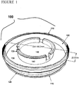
- a shipping chamber is illustrated in Figure 1 .
- the shipping chamber 100 is preferably constructed from a p150 tissue culture dish with a diameter of approximately 150 mm and height of approximately 20 mm.
- the shipping chamber preferably comprises a chamber top 110 and a chamber bottom 115.
- the chamber bottom 115 preferably comprises a chamber side wall 120 and a chamber bottom surface 125.
- a gel support 130 is formed on the chamber bottom surface.
- the shipping chamber comprises an insert 135 comprising a permeable membrane 140 and insert extensions 142.
- the skin equivalent 145 is formed on the permeable membrane 140.
- the permeable membrane 140 of the insert 135 is placed in contact with the gel support 130.
- the height of the gel support 130 within the shipping chamber 100 is such that when the insert 135 is placed in the shipping chamber 100 on gel support 130, the insert extensions 142 extend upward and contact the chamber top 110 when chamber top 110 is placed on chamber bottom 115 so that the insert 135 is secured on the gel support 130 by the downward force exerted by the chamber top 110 and the insert extensions 142.
- the temperature of the sterile package is reduced to about 2-8 degrees C.
- organotypically cultured skin equivalents can be shipped and stored at lowered temperatures and maintain their viability for use in skin grafting and wound closing procedures.
- the ability to ship and store at lowered temperatures greatly increases the flexibility of manufacturing, shipping and using organotypically cultured skin equivalents. This stands in direct contrast to the manufacturing, shipping, and use of other organotypically cultured skin equivalents such as APLIGRAF® which is usable for less than ten days and must be maintained between 20 and 23°C until used.
- organotypically cultured skin equivalents can be preferably used up to 15 days after the packaging date. The additional storage time greatly enhances the flexibility of use of the organotypically cultured skin equivalents.
- the sterile package is placed in an insulated container and packed with cold packs, preferably gel cold packs, to maintain the temperature of the sterile package at 2-8 degrees C. during shipping.
- cold packs preferably gel cold packs
- the sterile package is removed from the insulated container and placed in a refrigeration unit for storage at 2-8 C. until the time of use.
- the integrity (and sterility) of the sterile package is maintained until immediately prior to use by a physician or other care giver, for example, in an operating room.
- an intervening culture step or revival period is not required prior to use of the organotypically cultured skin equivalent in a skin grafting or wound closure procedure. This feature represents a substantial, unexpected improvement over prior methods where the tissue either must be stored at a higher temperature or revived at a higher temperature in a liquid media for use.
- preserved cells, organs, and tissues of the present description may be used therapeutically.
- the cells, organs, and tissues are utilized to treat chronic skin wounds.
- Successful treatment of chronic skin wounds e.g ., venous ulcers, diabetic ulcers, pressure ulcers
- the healing of such a wound often times takes well over a year of treatment.
- Treatment options currently include dressings and debridement (use of chemicals or surgery to clear away necrotic tissue), and/or antibiotics in the case of infection. These treatment options take extended periods of time and high amounts of patient compliance. As such, a therapy that can increase a practitioner's success in healing chronic wounds and accelerate the rate of wound healing would meet an unmet need in the field.
- the present description contemplates treatment of skin wounds with skin equivalents comprising the cells of the present description (e.g., NIKS ® cells).
- NIKS ® cells are topically applied to wound sites.
- skin equivalents comprising NIKS ® cells are used for engraftment on partial thickness wounds.
- skin equivalents comprising NIKS ® cells are used for engraftment on full thickness wounds.
- skin equivalents comprising NIKS ® cells are used to treat numerous types of internal wounds, including, but not limited to, internal wounds of the mucous membranes that line the gastrointestinal tract, ulcerative colitis, and inflammation of mucous membranes that may be caused by cancer therapies.
- skin equivalents comprising NIKS ® cells expressing are used as a temporary or permanent wound dressing.
- Skin equivalents comprising cells also find use in wound closure and burn treatment applications.
- the use of autografts and allografts for the treatment of burns and wound closure is described in Myers et al., A. J. Surg. 170(1):75-83 (1995 ) and U.S. Pat. Nos. 5,693,332 ; 5,658,331 ; and 6,039,760 .
- the skin equivalents may be used in conjunction with dermal replacements such as DERMAGRAFT or INTEGRA.
- the skin equivalents are produced using both a standard source of keratinocytes (e.g ., NIKS ® cells) and keratinocytes from the patient that will receive the graft.
- the skin equivalent contains keratinocytes from two different sources.
- the skin equivalent contains keratinocytes from a human tissue isolate. Accordingly, the present description provides methods for wound closure, including wounds caused by burns, comprising providing a skin equivalent and a patient suffering from a wound and treating the patient with the skin equivalent under conditions such that the wound is closed.
- the cells are engineered to provide additional therapeutic agents to a subject.
- the present description is not limited to the delivery of any particular therapeutic agent. Indeed, it is contemplated that a variety of therapeutic agents may be delivered to the subject, including, but not limited to, enzymes, peptides, peptide hormones, other proteins, ribosomal RNA, ribozymes, and antisense RNA. These therapeutic agents may be delivered for a variety of purposes, including but not limited to the purpose of correcting genetic defects. In some particular preferred examples, the therapeutic agent is delivered for the purpose of detoxifying a patient with an inherited inborn error of metabolism (e.g ., aminoacidopathesis) in which the graft serves as wild-type tissue.
- an inherited inborn error of metabolism e.g ., aminoacidopathesis
- the cells are transformed with a DNA construct encoding a therapeutic agent (e.g ., insulin, clotting factor IX, erythropoietin, etc) and the cells grafted onto the subject.
- a therapeutic agent e.g ., insulin, clotting factor IX, erythropoietin, etc
- the therapeutic agent is then delivered to the patient's bloodstream or other tissues from the graft.
- the nucleic acid encoding the therapeutic agent is operably linked to a suitable promoter.
- suitable promoter The present description is not limited to the use of any particular promoter. Indeed, the use of a variety of promoters is contemplated, including, but not limited to, inducible, constitutive, tissue specific, and keratinocyte specific promoters.
- the nucleic acid encoding the therapeutic agent is introduced directly into the keratinocytes (i.e., by calcium phosphate co-precipitation or via liposome transfection).
- the nucleic acid encoding the therapeutic agent is provided as a vector and the vector is introduced into the keratinocytes by methods known in the art.
- the vector is an episomal vector such as a plasmid.
- the vector integrates into the genome of the keratinocytes. Examples of integrating vectors include, but are not limited to, retroviral vectors, adeno-associated virus vectors, and transposon vectors.
- This example describes a method for the production of skin equivalents.
- TM 3T3 feeder cell medium
- FGM human fibroblast growth medium
- NM NIKS ® medium
- PM plating medium
- SMA stratification medium A
- SMB stratification medium B
- FGM is a commercially available fibroblast growth medium (Clonetics) that is used to propagate the normal human dermal fibroblast cells (NHDFs) for use in STRATAGRAFT ® skin equivalent and STATATEST skin equivalent dermal equivalent layers.
- NHDFs normal human dermal fibroblast cells
- NM is used to grow NIKS ® keratinocytes.
- NM is a 3:1 mixture of Ham's F-12 medium (GibcoBRL) and DME supplemented with 2.5% fetal clone II (Hyclone), 0.4 ⁇ g/ml hydrocortisone (Calbiochem), 8.4 ng/ml cholera toxin (ICN), 5 ⁇ g/ml insulin (Sigma), 24 ⁇ g/ml adenine (Sigma) and 10 ng/ml epidermal growth factor (EGF, R&D systems).
- PM is the medium used when NIKS ® cells are seeded onto a dermal equivalent.
- PM is the same NM with the exception that EGF is removed, the serum is reduced to 0.2%, and CaCl 2 (Sigma) is supplemented to a final calcium concentration of 1.88 nm.
- SMA is the same as PM with the addition of 1 mg/ml bovine serum albumin (BSA), 1 ⁇ M isoproterenol, 10 ⁇ M carnitine, 10 ⁇ M serine, 25 ⁇ M oleic acid, 15 ⁇ M linoleic acid, 7 ⁇ M arachidonic acid, 1 ⁇ M ⁇ -tocopherol, 0.05 mg/ml ascorbic acid (all from Sigma), and 1 ng/ml EGF.
- SMB is used during the epidermal stratification phase of STRATATEST skin equivalent and STRATAGRAFT ® skin equivalent growth. SMB is the same as SMA but without the presence of the fetal clone II serum supplement.
- 3T3 feeder cells Prior to starting STRATAGRAFT ® skin equivalent organotypic cultures, 3T3 feeder cells are prepared and then used either fresh or frozen for later use. 3T3 cells are grown to confluence and treated with mitomycin-C (4 ug/ml in TM, Roche) for four hours. The cells are then washed, resuspended, and plated at a density of 1.25 X 10 6 per 100 mm tissue culture dish to support NIKS ® growth. If frozen feeders are used, a single frozen ampoule containing 1 ml with 2 X 10 6 is thawed, diluted with fresh TM and plated onto two 100 mm tissue culture dishes. This is done for as many dishes as will be needed for NIKS ® cell growth one prior to plating the NIKS ® cells.
- Dermal equivalent preparation On day 0, frozen NHDF cells are thawed and plated. The cells are fed FGM-2 the next day (day 1) to residual cryoprotectant and again on day 3. On day 4, they are harvested for in the dermal equivalent.
- rat-tail collagen Type I, Becton-Dickinson
- a mixture of concentrated Ham's F12 medium (8.7X normal strength and buffered with HEPES at pH 7.5) is mixed with fetal clone II (supplemented bovine serum). These two solutions are 11.5 and 10% of the final solution volume. IN NaOH is added to the medium mixture (2.5% of final solution).
- the diluted collagen is then added (74%) to the mixture.
- a 2% volume of suspended fibroblasts (1.3 X 10 6 for STRATAGRAFT ® skin equivalent) is added to the mixture.
- 100 ⁇ l is aliquoted into tissue culture inserts (MILLICELL from Millipore Corp.) and placed in a 100 mm tissue culture dish. After 30 minutes for gel formation, the dish is flooded with 20 ml of FGM-2. One or two drops of the F-12-serum mix are placed on the surface of each dermal equivalent.
- STRATAGRAFT ® skin equivalent uses TRANSWELL inserts from Corning. A 13 ml dermal equivalent is poured into each insert.
- FGM-2 FGM-2 is placed around the TRANSWELL insert in a 150 mm tissue culture dish and 10 ml is placed on top of the dermal equivalent.
- the inserts are placed in 37°C, 5% CO 2 , 90% relative humidity incubator until used.
- the dermal equivalents are seeded with NIKS ® cells, they are lifted to the air interface by placing them onto a sterile stainless steel mesh to supply medium through the bottom of the tissue culture insert.
- NIKS ® Growth and Seeding On day 0, the feeders are plated in NM. On day 1, NIKS ® cells are plated onto the feeders at a density of approximately 3 X 10 5 cells per 100 mm dish. On day 2, the NIKS ® cells are fed fresh NM to remove residual cryoprotectant. The NIKS ® cells are fed again on days 4 and 6. (For STRATAGRAFT ® skin equivalent size cultures, the NIKS ® cultures are started a week earlier due to the increase in number of cells needed). On day 8, the NIKS ® cells are harvested, counted, and resuspended in PM.
- NIKS ® cells/cm 2 are seeded onto the surface of the MIILLICELL or TRANSWELL inserts.
- the dishes are fed 30 ml PM (100 ml for STRATAGRAFT ® skin equivalent) underneath the metal lifter and placed back into the incubator.
- PM 100 ml for STRATAGRAFT ® skin equivalent
- the cultures are fed SMA.
- days 12, 14, 16, 18, 20, and 22 the cultures are fed SMB.
- the cultures are transferred to a 75% humidity incubator where they remain for the rest of their growth.
- STRATAGRAFT ® skin tissue is a living skin substitute tissue that has a fully-stratified layer of viable epidermal keratinocytes on a collagen gel containing normal human dermal fibroblasts.
- the uppermost epidermal layers form a permeability barrier that prevents excessive moisture loss through the epidermis.
- Assays that measure these key structural and functional properties have been identified as stability-indicating assays for monitoring the quality of STRATAGRAFT ® skin tissue over time.
- STRATAGRAFT ® skin tissue lasts 31 days.
- STRATAGRAFT ® skin tissues are removed from organotypic culturing conditions and placed onto HEPES-buffered nutrient-agarose shipping chambers, which are designed to maintain the viability, barrier function, and histological architecture of STRATAGRAFT ® skin tissues prior to clinical use.
- This study was conducted to compare STRATAGRAFT ® skin tissue properties following storage on shipping chambers for 1 day at 2° - 8°C or 20° - 25°C.
- Two independent lots of STRATAGRAFT ® skin tissue were analyzed for viability, barrier function, and histology after a 1 day storage period at 2° - 8°C or 20° - 25°C.
- Barrier function data are presented below in Tables 3 and 4.
- the acceptance criteria for barrier function are all readings must have an initial DPM value ⁇ 294 and a DPM change over a 10 second interval ⁇ 658.
- the typical appearance of a paraffin embedded STRATAGRAFT ® tissue section stained with hematoxylin and eosin includes fibroblasts in the dermal layer, a basal layer of small, nucleated keratinocytes at the junction between the epidermal and dermal layers, multiple layers of differentiating keratinocytes above the basal layer, and a layer of flattened corneocytes. All tissues conformed to the specifications for histology.
- the STRATAGRAFT ® skin tissues stored at 2° - 8°C had tissue architecture comparable to tissues stored at 20° - 25°C and both sets of stored tissues were comparable to the Day 28 QC tissues.
- This example demonstrates that skin equivalents stored for 8 days at 2-8 C are comparable to, or superior to, tissues stored for only 1 day at 20-25 C. Reduction of the storage temperature has been shown to maintain tissue quality comparable to tissues stored at 20° - 25°C (See Example 2). The ability to store STRATAGRAFT ® tissue for eight days at 2 - 8 °C would increase the number of days STRATAGRAFT ® tissue is available for clinical use. This study tested the comparability of tissues stored for one day at 20 - 25 °C with tissues stored at 2 - 8 °C for eight days. Tissue viability was improved in the tissues that were stored at 2 - 8 °C, even though these tissues were stored for seven additional days.
- This study used one batch of tissues manufactured under cGMP at the WCBF and two batches produced at the Stratatech pilot production facility.
- One tissue from each batch was analyzed on process day 28.
- the remaining six tissues were fed on day 28 and 30, and placed onto shipping chambers on process day 31.
- Three tissues from each batch were stored at 20 - 25 °C for 1 day prior to analysis.
- the remaining three tissues from each batch were stored at 2 - 8 °C for 8 days. Prior to analysis, the tissues stored for 8 days at 2 - 8 °C were equilibrated at 20 - 25 °C for one hour.
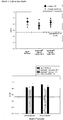
- Viability data is presented in Table 5 and Figure 3 . All viability samples met the acceptance criteria (A 550 ⁇ 0.533). However, in all three intra-lot comparisons ( Figure 3 left panel), the viability of tissues stored at 2 - 8 °C for 8 days was higher than tissues stored for one day at 20 - 25 °C. The mean A 550 value for the tissues stored at 20 - 25 °C for 1 day was 0.709, compared to 0.831 for tissues stored at 2 - 8 °C for eight days. This data indicates that storage of STRATAGRAFT ® tissues at 2 - 8 °C is better able to maintain tissue viability than storage at 20 - 25 °C. Table 5.
- Tissues were analyzed with a Nova impedance meter. Data are presented in Tables 6 and 7. All readings met the lot-release criteria for STRATAGRAFT ® tissue (Initial reading ⁇ 294, Change ⁇ 658). This data suggests that storage of STRATAGRAFT ® tissue for eight days at 2 - 8 °C does not adversely affect tissue barrier function, compared to tissues analyzed after storage at 20 - 25 °C for 1 day. Table 6.
- STRATAGRAFT ® tissue stored for eight days at 2 - 8 °C had viability, histology and barrier function properties comparable to or better than tissues stored for 1 day at 20 - 25 °C. This result demonstrates that up to eight days of storage at 2 - 8 °C does not adversely affect STRATAGRAFT ® tissue properties.
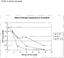
- STRATAGRAFT ® tissues produced at Stratatech's process development laboratory were packaged onto nutrient-agarose shipping chambers on Day 28 of the production process and stored at approximately 2 - 8 °C, 15 °C, or 22.5 °C for 1, 4, 8, 15, or 29 days. Tissues were analyzed for viability and histology after the indicated storage periods. Barrier function measurements were not performed on all tissues, and so this data is not presented.
- Tissues from three independent STRATAGRAFT ® skin tissue lots produced at the WCBF were used for this study.
- One randomly chosen tissue from each lot was tested on Day 28 of the STRATAGRAFT ® skin tissue production process.
- the six remaining tissues in each lot were fed SMB medium on process Day 28 and process Day 30.
- On process Day 31, the 6 tissues were placed onto shipping chambers and stored at 2 - 8 °C for either 1, 8, or 15 days. After the specified storage interval, the tissues were incubated at 20 - 25 °C for 1 hour and then analyzed for viability, barrier function, and histology.
- Viability data is presented in Tables 8 and 9 and in Figure 4 .
- the acceptance criterion is that all samples have an A 550 ⁇ 0.533.
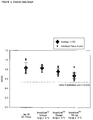
- Barrier function data is presented below in Tables 10 and 11 and in Figure 5 .
- the acceptance criteria for barrier function are all readings must have an initial DPM value ⁇ 294 and a DPM change over a 10 second interval ⁇ 658.
- tissues stored at 2 - 8 °C for 15 days exhibited typical histological architecture, consisting of a dermis containing fibroblasts and an epidermis containing all required tissue layers.
- This example describes how the shipping chambers and sterile packages for shipping are made.
- a solution of 3% agarose is prepared by mixing 45 g agarose in 1455 ml water. The mixture is stirred and then autoclaved (121 C for 60 min.) to dissolve the agarose.
- 2X media solution is prepared by mixing in 1455 ml water: 24 g F12 media powder, 10.0 g DMEM media powder and 7.2 g HEPES powder. The mixture is stirred until all powder is dissolved and the pH is adjusted to 7.3 to 7.5.
- the 2X media solution and 3% agarose solution are placed in 40 C water baths for 30-60 minutes. The 2X media solution is then sterile-filtered and aseptically added to the 3% agarose solution though a SterivexTM filter.
- 60 ml of the resulting solution is then aseptically dispensed into a sterile p150 culture dish (150 mm X 20mm circular tissue culture dish) and allowed to gel.
- shipping chambers are packed into a heat sealable sterile bag for storage until use.
- a skin equivalent in a Transwell insert (7.5 cm diameter (44 cm 2 ), pore size 0.4 micron) is aseptically placed on the agarose in the shipping chamber and the p150 plate top is placed on the shipping chamber and secured under aseptic conditions.
- the shipping chamber is then placed in a sterile heat sealable pouch and sealed to provide a shipping package.
- the shipping package is stored and shipped at 2-8 C, and is storable at the site of use at 2-8 C for 8-15 days from the time of packaging until immediately prior to the time of use.
- the integrity of the package may be maintained until the time of use and revival of the skin equivalent prior to use is not necessary.
- This example describes a simplified method for the production of skin equivalents.
- Media The organotypic culture process uses three different culture media, all based on the formulation of SMB medium described in US patent 7,407,805 , with the exception that cholera toxin is omitted from all media.
- FM01 is used to propagate the normal human dermal fibroblasts (NHDFs) for use in skin equivalent dermal equivalent layers.
- NHDFs normal human dermal fibroblasts
- FM01 has the same formulation as SMB except that it contains Fetal Clone II serum (2% final) and lacks cholera toxin.
- KM01 is used to grow NIKS ® keratinocytes and has the same composition as SMB except that it contains 2.5% fetal clone II, and additional epidermal growth factor (EGF) is added to a final concentration of 5 ng/ml.
- SM01 is used during the epidermal stratification phase of skin equivalent production and is identical to SMB except for the omission of cholera toxin.
- Dermal equivalent preparation On day 0, frozen NHDF cells are thawed and plated. The cells are fed FM01 the next day (day 1) to remove residual cryoprotectant and again on day 3. On day 4, they are harvested for use in the dermal equivalent.
- Type I rat-tail collagen is first diluted to 3 mg/ml in 0.03N acetic acid and chilled on ice. A mixture of concentrated Ham's F12 medium (8.7X normal strength and buffered with HEPES at pH 7.5) is mixed with fetal clone II. These two solutions are 11.3 and 9.6% of the final solution volume. IN NaOH is added to the medium mixture (2.4% of final solution). The diluted collagen is then added (74.7%) to the mixture.
- a 2% volume of suspended fibroblasts (2.78 X 10 6 /ml) is added to the mixture.
- 9 ml of the final dermal equivalent mixture is poured into each 75 mm TRANSWELL insert (Corning Costar). After a 50-70 minute gel formation period, the Transwell inserts are transferred to the surface of a stainless steel mesh in a 150 mm culture dish.
- 80 ml of FM01 is placed in the 150 mm dish outside the TRANSWELL insert and 10 ml is placed on top of the dermal equivalent.
- the dermal equivalents are placed in 37°C, 5% CO 2 , 90% relative humidity incubator for 4-5 days prior to use in the organotypic cultures.
- NIKS ® Growth and Seeding NIKS ® cells are thawed and plated at a density of approximately 5 x 10 5 cells per 100 mm dish. NIKS ® culture can be performed in the presence or absence of murine feeder cells. On day 1, the NIKS ® cells are fed fresh KM01 to remove residual cryoprotectant. The NIKS ® cells are fed again on day 3. On day 4, the NIKS ® cells are harvested from the initial p100 cultures and seeded into 225 cm 2 culture flasks at a density of 1.2 x 10 6 per flask. The NIKS ® cultures are fed fresh medium on Days 7 and 8.
- NIKS ® cells are harvested, counted, and resuspended in SM01. 2.27 X 10 4 NIKS ® cells/cm 2 are seeded onto the surface of the dermal equivalents. The dishes are cultures are fed and lifted to the air-medium interface. Cultures are transferred to a controlled humidity incubator set to 75% where they remain for the rest of their growth. Cultures are fed SM01 on days 14, 18, 22, 25, 28, and 30.
- STRATAGRAFT ® skin tissues produced using simplified procedures on shipping chambers for up to 15 days is highly desirable as it would enhance the availability of the tissue for clinical use.
- This example demonstrates that storage of skin equivalents produced using simplified procedures on nutrient-agarose shipping chambers for up to 15 days at 2 - 8 °C is acceptable.
- STRATAGRAFT ® tissues produced at Stratatech's process development laboratory were packaged onto nutrient-agarose shipping chambers on Day 31 of the production process and stored at approximately 2 - 8 °C for 8 or 15 days. Tissues were analyzed for viability, barrier function, and histology after the indicated storage periods.
- STRATAGRAFT ® skin tissues stored at 2 - 8 °C for 8 or 15 days exhibited all of the typical epidermal layers atop the collagen dermis containing fibroblasts.
- Several common storage-related effects were seen in these tissues including an increase in the number of condensed nuclei and a reduction in the eosin staining in the upper epidermal layers.
Landscapes
- Life Sciences & Earth Sciences (AREA)
- Engineering & Computer Science (AREA)
- Health & Medical Sciences (AREA)
- General Health & Medical Sciences (AREA)
- Dentistry (AREA)
- Wood Science & Technology (AREA)
- Zoology (AREA)
- Environmental Sciences (AREA)
- Hematology (AREA)
- Mechanical Engineering (AREA)
- Dispersion Chemistry (AREA)
- Chemical & Material Sciences (AREA)
- Micro-Organisms Or Cultivation Processes Thereof (AREA)
- Materials For Medical Uses (AREA)
Description
- This invention was made with government support under grant number NIST 70NANB3H3011 awarded by the Advanced Technology Program. The government has certain rights in the invention.
- The present invention is defined in the claims and relates generally to systems and methods for shipping and storing skin equivalents made by organotypic culture that are to be used for skin grafting to human patients.
- The emerging field of tissue engineering (TE) is poised to make enormous progress in the treatment of organ disease and dysfunction in the coming decade. In 2001, there were 23 cell-based therapeutics approved for market in the United States (U.S.) and Europe, of which nine were skin substitutes or grafts, and 100 more products were in development. (De Bree, Genomics-based Drug Data Report and Regenerative Therapy (1)2:77-96 (2001)). In 2007, nearly 100 companies were involved in developing engineered tissues, cell-based therapeutics, or related technologies (Applied Data Research, February 2007). Overall the industry had an annual growth rate of 16% from 1995-2001. The "structural" industry segment (e.g., skin, bone, cartilage) showed 85% growth from 1998-2001. In 2004, the U.S. market for tissue-engineered skin replacements/substitutes and active wound repair modulators was valued at approximately $195 million. Sales are expected to increase at a compound annual rate of 9.5%, reaching approximately $481 million in the year 2014 (MedTech Insight, Windhover Information, September 2005). The total U.S. market for advanced wound care technologies was worth more than $2.3 billion in 2005. By the end of 2006 the market will reach almost $2.6 billion, and over a five-year period will grow at an average annual growth rate of 12.3% to reach $4.6 billion in 2011 (BCC Research, PHM011E, January 2007). The global wound care market is estimated to be worth US$ 7.2 billion in 2006 and comprises two sectors, traditional and advanced (Espicom Business Intelligence, 2007). Traditional wound care products consist mainly of low technology gauze-based dressings such as woven and non-woven sponges, conforming bandages and non-adherent bandages. The advanced wound care segment (US$ 4.1 billion global) is the fastest growing area with double-digit growth of 10% per year (Espicom Business Intelligence, 2007).
- Although a multitude of revolutionary and economically important applications for engineered tissues and organs exist in the human health arena, the full economic potential of the industry is far from realized. At present, only one of the publicly-held tissue engineering companies worldwide has shown a profit despite global investment in these technologies exceeding $3.5 billion. (Lysaght and Reyes, Tissue Engineering 7(5):485-93 (2001)).
- A major impediment to the acceptance of engineered tissues by medical practitioners, healthcare providers, and second party payers is the lack of a means to effectively and efficiently preserve engineered tissues. The nature of living cells and tissue products makes them impractical for long-term storage. Current engineered tissues must often be stored and shipped under carefully controlled conditions to maintain viability and function. Typically, engineered tissue products take weeks or months to produce but must be used within hours or days after manufacture. As a result, TE companies must continually operate with their production facilities at top capacity and absorb the costs of inventory losses (i.e., unsold product which must be discarded). These inventory losses, on top of already costly manufacturing process, have forced prices to impractical levels. As one specific example, APLIGRAF requires about four weeks to manufacture, is usable for less than ten days and must be maintained between 20 and 23°C until used. As another example, EPICEL is transported by a nurse from Cambridge, MA to the point-of-use in a portable incubator and is used immediately upon arrival. Such constraints represent significant challenges to developing convenient and cost-effective products.
- Cryopreservation has been explored as a solution to the storage problem, but it is known to induce tissue damage through ice formation, chilling injury, and osmotic balance. Besides APLIGRAF, the only other approved living skin equivalent, ORCEL, is currently in clinical trials as a frozen product but has the drawback that it must be maintained at temperatures below -100°C prior to use. This means using liquid nitrogen storage, which is expensive, dangerous, and not universally available (e.g. rural clinics and field hospitals). Moreover, delivering a frozen product requires special training on the part of the end-user to successfully thaw the tissue prior to use.
- Accordingly, what is needed in the art are improved methods of preparing engineered tissues and cells for storage under conditions that are routinely available at the point of use. As all clinical facilities have refrigerated storage, development of a skin equivalent that can be stored for prolonged periods in a standard refrigerator would greatly improve the availability and clinical utility of these products.
- Transportation systems for a skin equivalent or cell-based products and methods of shipping and transporting said skin equivalents or cell-based products have been suggested in the prior art, see e.g.
WO 01/05942 A2 WO 93/08776 A1 WO 2007/033051 A1 . However, said prior art does not describe the temperature range under which the skin equivalent of the present invention, i.e. skin equivalent made by organotypic culture comprising dermal and epidermal layers, is shipped and stored as defined in the claims. - The invention is set out in the appended claims. The embodiments of the description which do not fall within the scope of said claims are provided for illustrative purposes only and do not form part of the present invention. In some examples, the present description provides methods of shipping an organotypically cultured skin equivalent to a user and using the skin equivalent in a skin grafting procedure on a human patient comprising: providing the organotypically cultured skin equivalent comprising dermal and epidermal layers and a sterile package comprising a gel support; packaging the skin equivalent in a sterile package under sterile conditions so that the skin equivalent contacts the gel support; lowering the temperature of the sterile package to 2-8 degrees Celsius; shipping the sterile package to a user at 2-8 degrees Celsius; storing the sterile package at the site of use at 2-8 degrees Celsius wherein the sterility and integrity of the sterile package are maintained; and removing the organotypically cultured skin equivalent from the package and applying to a patient without an intervening culture step. The present invention is not limited to skin equivalents comprising any particular types of keratinocytes. In some embodiments, the organotypically cultured skin equivalent comprises NIKS cells. The present invention is not limited to any particular type of gel support. In some embodiments, the gel support is an agarose gel support. The present invention is not limited to any particular type of sterile package. In some embodiments, the sterile package is heat sealable. In further embodiments, the skin equivalent contacts the gel support via a permeable membrane.
- In some examples, the present description provides methods of shipping and storing an organotypically cultured skin equivalent for use in a skin grafting procedure comprising: providing the organotypically cultured skin equivalent comprising dermal and epidermal layers and a sterile package comprising a gel support; packaging the skin equivalent in a sterile package under sterile conditions so that the skin equivalent contacts the gel support on a packaging date; lowering the temperature of the sterile package to 2-8 degrees Celsius; shipping the sterile package to a user at 2-8 degrees Celsius; storing the sterile package at the site of use at 2-8 degrees Celsius wherein the sterility and integrity of the sterile package are maintained for from 8 to 15 days from the packaging date. The present invention is not limited to skin equivalents comprising any particular types of keratinocytes. In some embodiments, the organotypically cultured skin equivalent comprises NIKS cells. The present invention is not limited to any particular type of gel support. In some embodiments, the gel support is an agarose gel support. The present invention is not limited to any particular type of sterile package. In some embodiments, the sterile package is heat sealable. In further embodiments, the skin equivalent contacts the gel support via a permeable membrane.
- In some examples, the present description provides methods of shipping an organotypically cultured skin equivalent to a user for use in a skin grafting procedure comprising: providing the organotypically cultured skin equivalent comprising dermal and epidermal layers and a sterile package comprising a gel support, wherein the gel support is formed with a minimal media; packaging the skin equivalent in a sterile package under sterile conditions so that the skin equivalent contacts the gel support; lowering the temperature of the sterile package to 2-8 degrees Celsius; shipping the sterile package to a user at 2-8 degrees Celsius; storing the sterile package at the site of use 2-8 degrees Celsius wherein the sterility and integrity of the sterile package are maintained. The present invention is not limited to skin equivalents comprising any particular types of keratinocytes. In some embodiments, the organotypically cultured skin equivalent comprises NIKS cells. The present invention is not limited to any particular type of gel support. In some embodiments, the gel support is an agarose gel support. The present invention is not limited to any particular type of sterile package. In some embodiments, the sterile package is heat sealable. In further embodiments, the skin equivalent contacts the gel support via a permeable membrane.
- In some examples, the present description provides kits comprising: a shipping chamber comprising a gel support comprising a minimal media; a skin equivalent supported on a permeable membrane in contact with the gel support; wherein the shipping chamber is contained with a sterile pouch. The present description is not limited to skin equivalents comprising any particular types of keratinocytes. In some embodiments, the organotypically cultured skin equivalent comprises NIKS cells. The present invention is not limited to any particular type of gel support. In some embodiments, the gel support is an agarose gel support. The present invention is not limited to any particular type of sterile package. In some embodiments, the sterile package is heat sealable. In further embodiments, the skin equivalent contacts the gel support via a permeable membrane.
- In some examples, the present description further provides articles of manufacture comprising a shipping chamber comprising a chamber top and a chamber bottom having a surface having thereon a gel support, said article further comprising a skin equivalent on a permeable membrane, said permeable membrane in contact with said gel support, said article further comprising extensions extending from said chamber top so that when said chamber top is placed on said chamber bottom said skin equivalent is secured against said gel support. In some examples, the gel support is formed with minimal media.
-
-
Figure 1 depicts a shipping chamber. -
Figure 2 is a viability data graft. -
Figure 3 is a viability data graft. -
Figure 4 is a viability data graft. -
Figure 5 is a viability data graft. -
Figure 6 is a viability data graft. -
Figure 7 is a table presenting viability data. -
Figure 8 is a table presenting barrier function summary data. - As used herein, the terms "human skin equivalent" and "human skin substitute" are used interchangeably to refer to an in vitro derived culture of keratinocytes that has stratified into squamous epithelia. Typically, the skin equivalents are produced by organotypic culture and include a dermal layer in addition to a keratinocyte layer.
- As used herein, the term "NIKS® cells" refers to cells having the characteristics of the cells deposited as cell line ATCC CRL-1219.
- The term "homology" refers to a degree of complementarity. There may be partial homology or complete homology (i.e., identity). A partially complementary sequence is one that at least partially inhibits a completely complementary sequence from hybridizing to a target nucleic acid and is referred to using the functional term "substantially homologous." The term "inhibition of binding," when used in reference to nucleic acid binding, refers to inhibition of binding caused by competition of homologous sequences for binding to a target sequence. The inhibition of hybridization of the completely complementary sequence to the target sequence may be examined using a hybridization assay (Southern or Northern blot, solution hybridization and the like) under conditions of low stringency. A substantially homologous sequence or probe will compete for and inhibit the binding (i.e., the hybridization) of a completely homologous to a target under conditions of low stringency. This is not to say that conditions of low stringency are such that non-specific binding is permitted; low stringency conditions require that the binding of two sequences to one another be a specific (i.e., selective) interaction. The absence of non-specific binding may be tested by the use of a second target that lacks even a partial degree of complementarity (e.g., less than about 30% identity); in the absence of non-specific binding the probe will not hybridize to the second non-complementary target.
- The term "gene" refers to a nucleic acid (e.g., DNA) sequence that comprises coding sequences necessary for the production of a polypeptide or precursor (e.g., KGF-2). The polypeptide can be encoded by a full length coding sequence or by any portion of the coding sequence so long as the desired activity or functional properties (e.g., enzymatic activity, ligand binding, signal transduction, etc.) of the full-length or fragment are retained. The term also encompasses the coding region of a structural gene and the including sequences located adjacent to the coding region on both the 5' and 3' ends for a distance of about 1 kb on either end such that the gene corresponds to the length of the full-length mRNA. The sequences that are located 5' of the coding region and which are present on the mRNA are referred to as 5' untranslated sequences. The sequences that are located 3' or downstream of the coding region and that are present on the mRNA are referred to as 3' untranslated sequences. The term "gene" encompasses both cDNA and genomic forms of a gene. A genomic form or clone of a gene contains the coding region interrupted with non-coding sequences termed "introns" or "intervening regions" or "intervening sequences." Introns are segments of a gene that are transcribed into nuclear RNA (hnRNA); introns may contain regulatory elements such as enhancers. Introns are removed or "spliced out" from the nuclear or primary transcript; introns therefore are absent in the messenger RNA (mRNA) transcript. The mRNA functions during translation to specify the sequence or order of amino acids in a nascent polypeptide.
- As used herein, the terms "nucleic acid molecule encoding," "DNA sequence encoding," and "DNA encoding" refer to the order or sequence of deoxyribonucleotides along a strand of deoxyribonucleic acid. The order of these deoxyribonucleotides determines the order of amino acids along the polypeptide (protein) chain. The DNA sequence thus codes for the amino acid sequence.
- As used herein, the term "recombinant DNA molecule" as used herein refers to a DNA molecule that is comprised of segments of DNA joined together by means of molecular biological techniques.
- As used herein, the term "purified" or "to purify" refers to the removal of contaminants from a sample.
- As used herein, the term "vector" is used in reference to nucleic acid molecules that transfer DNA segment(s) from one cell to another. The term "vehicle" is sometimes used interchangeably with "vector."
- The term "expression vector" as used herein refers to a recombinant DNA molecule containing a desired coding sequence and appropriate nucleic acid sequences necessary for the expression of the operably linked coding sequence in a particular host organism. Nucleic acid sequences necessary for expression in prokaryotes usually include a promoter, an operator (optional), and a ribosome binding site, often along with other sequences. Eukaryotic cells are known to utilize promoters, enhancers, and termination and polyadenylation signals.
- "Operably linked" refers to a juxtaposition wherein the components so described are in a relationship permitting them to function in their intended manner. A regulatory sequence is "operably linked" to a coding sequence when it is joined in such a way that expression of the coding sequence is achieved under conditions compatible with the regulatory sequence.
- The term "transfection" as used herein refers to the introduction of foreign DNA into eukaryotic cells. Transfection may be accomplished by a variety of means known to the art including calcium phosphate-DNA co-precipitation, DEAE-dextran-mediated transfection, polybrene-mediated transfection, electroporation, microinjection, liposome fusion, lipofection, protoplast fusion, retroviral infection, and biolistics.
- The term "stable transfection" or "stably transfected" refers to the introduction and integration of foreign DNA into the genome of the transfected cell. The term "stable transfectant" refers to a cell that has stably integrated foreign DNA into the genomic DNA.
- The term "transient transfection" or "transiently transfected" refers to the introduction of foreign DNA into a cell where the foreign DNA fails to integrate into the genome of the transfected cell. The foreign DNA persists in the nucleus of the transfected cell for several days. During this time the foreign DNA is subject to the regulatory controls that govern the expression of endogenous genes in the chromosomes. The term "transient transfectant" refers to cells that have taken up foreign DNA but have failed to integrate this DNA.
- The invention is set out in the appended claims. The embodiments of the description which do not fall within the scope of said claims are provided for illustrative purposes only and do not form part of the present invention. The present invention relates generally to systems and methods for shipping and storing skin equivalents made by organotypic culture that are to be used for skin grafting to human patients. In particular, the present invention relates to methods for production and packaging of a sterile skin equivalent using aseptic techniques and maintaining the sterility of
the skin equivalent during storage for up to 15 days until opened in a sterile surgical field for clinical use. For convenience, the description of the invention is presented in the following sections: - The present invention is not limited to the use of any particular source of cells that are capable of differentiating into squamous epithelia. Indeed, the present invention contemplates the use of a variety of cell lines and sources that can differentiate into squamous epithelia, including both primary and immortalized keratinocytes. Sources of cells include keratinocytes and dermal fibroblasts biopsied from humans and cavaderic donors (Auger et al., In Vitro Cell. Dev. Biol. - Animal 36:96-103;
U.S. Pat. Nos. 5,968,546 and5,693,332 ), neonatal foreskins (Asbill et al., Pharm. Research 17(9): 1092-97 (2000); Meana et al., Burns 24:621-30 (1998);U.S. Pat. Nos. 4,485,096 ;6,039,760 ; and5,536,656 ), and immortalized keratinocytes cell lines such as NM1 cells (Baden, In Vitro Cell. Dev. Biol. 23(3):205-213 (1987)), HaCaT cells (Boucamp et al., J. cell. Boil. 106:761-771 (1988)); and NIKS® cells (Cell line BC-1-Ep/SL;U.S. Pat. No. 5,989,837 ; ATCC CRL-12191). Each of these cell lines can be cultured or genetically modified as in order to produce a cell line capable of expressing or co-expressing the desired protein(s). In particularly preferred embodiments, NIKS® cells are utilized. The discovery of a novel human keratinocyte cell line (near-diploid immortalized keratinocytes or NIKS®) provides an opportunity to genetically engineer human keratinocytes. A unique advantage of the NIKS® cells is that they are a consistent source of genetically-uniform, pathogen-free human keratinocytes. For this reason, they are useful for the application of genetic engineering and genomic gene expression approaches to provide skin equivalent cultures with properties more similar to human skin. The NIKS® keratinocyte cell line, identified and characterized at the University of Wisconsin, is nontumorigenic, exhibits a stable karyotype, and exhibits normal differentiation both in monolayer and organotypic culture. NIKS® cells form fully stratified skin equivalents in culture. These cultures are indistinguishable by all criteria tested thus far from organotypic cultures formed from primary human keratinocytes. Unlike primary cells however, the immortalized NIKS® cells will continue to proliferate in monolayer culture indefinitely. This provides an opportunity to genetically manipulate the cells and isolate new clones of cells with new useful properties (Allen-Hoffmann et al., J. Invest. Dermatol., 114(3): 444-455 (2000)). Cultured skin substitutes resulting from genetic modifications of keratinocytes using NIKS cells are known fromUS 2002/168768 A1 . - The NIKS® cells arose from the BC-1-Ep strain of human neonatal foreskin keratinocytes isolated from an apparently normal male infant. In early passages, the BC-1-Ep cells exhibited no morphological or growth characteristics that were atypical for cultured normal human keratinocytes. Cultivated BC-1-Ep cells exhibited stratification as well as features of programmed cell death. To determine replicative lifespan, the BC-1-Ep cells were serially cultivated to senescence in standard keratinocyte growth medium at a density of 3 x 10⁵ cells per 100-mm dish and passaged at weekly intervals (approximately a 1:25 split). By
passage 15, most keratinocytes in the population appeared senescent as judged by the presence of numerous abortive colonies which exhibited large, flat cells. However, at passage 16, keratinocytes exhibiting a small cell size were evident. By passage 17, only the small-sized keratinocytes were present in the culture and no large, senescent keratinocytes were evident. The resulting population of small keratinocytes that survived this putative crisis period appeared morphologically uniform and produced colonies of keratinocytes exhibiting typical keratinocyte characteristics including cell-cell adhesion and apparent squame production. The keratinocytes that survived senescence were serially cultivated at a density of 3 x 10⁵ cells per 100-mm dish. Typically the cultures reached a cell density of approximately 8 x 10⁶ cells within 7 days. This stable rate of cell growth was maintained through at least 59 passages, demonstrating that the cells had achieved immortality. The keratinocytes that emerged from the original senescencing population were originally designated BC-1-Ep/Spontaneous Line and are now termed NIKS®. The NIKS® cell line has been screened for the presence of proviral DNA sequences for HIV-1, HIV-2, EBV, CMV, HTLV-1, HTLV-2, HBV, HCV, B-19 parvovirus, HPV-16 and HPV-31 using either PCR or Southern analysis. None of these viruses were detected. - Chromosomal analysis was performed on the parental BC-1-Ep cells at
passage 3 and NIKS cells atpassages 31 and 54. The parental BC-1-Ep cells have a normal chromosomal complement of 46, XY. At passage 31, all NIKS® cells contained 47 chromosomes with an extra isochromosome of the long arm ofchromosome 8. No other gross chromosomal abnormalities or marker chromosomes were detected. Atpassage 54, all cells contained theisochromosome 8. - The DNA fingerprints for the NIKS® cell line and the BC-1-Ep keratinocytes are identical at all twelve loci analyzed demonstrating that the NIKS® cells arose from the parental BC-1-Ep population. The odds of the NIKS® cell line having the parental BC-1-Ep DNA fingerprint by random chance is 4 x 10-16. The DNA fingerprints from three different sources of human keratinocytes, ED-1-Ep, SCC4 and SCC13y are different from the BC-1-Ep pattern. This data also shows that keratinocytes isolated from other humans, ED-1-Ep, SCC4, and SCC13y, are unrelated to the BC-1-Ep cells or each other. The NIKS® DNA fingerprint data provides an unequivocal way to identify the NIKS® cell line.
- Loss of p53 function is associated with an enhanced proliferative potential and increased frequency of immortality in cultured cells. The sequence of p53 in the NIKS® cells is identical to published p53 sequences (GenBank accession number: M14695). In humans, p53 exists in two predominant polymorphic forms distinguished by the amino acid at codon 72. Both alleles of p53 in the NIKS® cells are wild-type and have the sequence CGC at codon 72, which codes for an arginine. The other common form of p53 has a proline at this position. The entire sequence of p53 in the NIKS® cells is identical to the BC-1-Ep progenitor cells. Rb was also found to be wild-type in NIKS® cells.
- Anchorage-independent growth is highly correlated to tumorigenicity in vivo. For this reason, the anchorage-independent growth characteristics of NIKS® cells in agar or methylcellulose-containing medium was investigated. After 4 weeks in either agar- or methylcellulose-containing medium, NIKS® cells remained as single cells. The assays were continued for a total of 8 weeks to detect slow growing variants of the NIKS® cells. None were observed.
- To determine the tumorigenicity of the parental BC-1-Ep keratinocytes and the immortal NIKS® keratinocyte cell line, cells were injected into the flanks of athymic nude mice. The human squamous cell carcinoma cell line, SCC4, was used as a positive control for tumor production in these animals. The injection of samples was designed such that animals received SCC4 cells in one flank and either the parental BC-1-Ep keratinocytes or the NIKS® cells in the opposite flank. This injection strategy eliminated animal to animal variation in tumor production and confirmed that the mice would support vigorous growth of tumorigenic cells. Neither the parental BC-1-Ep keratinocytes (passage 6) nor the NIKS® keratinocytes (passage 35) produced tumors in athymic nude mice.
- NIKS® cells were analyzed for the ability to undergo differentiation in both surface culture and organotypic culture. Techniques for organotypic culture are described in detail in the examples. In particularly preferred embodiments, the organotypically cultured skin equivalents of the present invention comprise a dermal equivalent formed from collagen or a similar material and fibroblasts. The keratinocytes, for example NIKS® cells or a combination of NIKS® cells and cell from a patient are seeded onto the dermal equivalent and form an epidermal layer characterized by squamous differentiation following the organotypic culture process.
- For cells in surface culture, a marker of squamous differentiation, the formation cornified envelopes was monitored. In cultured human keratinocytes, early stages of cornified envelope assembly result in the formation of an immature structure composed of involucrin, cystatin-α and other proteins, which represent the innermost third of the mature cornified envelope. Less than 2% of the keratinocytes from the adherent BC-1-Ep cells or the NIKS® cell line produce cornified envelopes. This finding is consistent with previous studies demonstrating that actively growing, subconfluent keratinocytes produce less than 5% cornified envelopes. To determine whether the NIKS® cell line is capable of producing cornified envelopes when induced to differentiate, the cells were removed from surface culture and suspended for 24 hours in medium made semi-solid with methylcellulose. Many aspects of terminal differentiation, including differential expression of keratins and cornified envelope formation can be triggered in vitro by loss of keratinocyte cell-cell and cell-substratum adhesion. The NIKS® keratinocytes produced as many as and usually more cornified envelopes than the parental keratinocytes. These findings demonstrate that the NIKS® keratinocytes are not defective in their ability to initiate the formation of this cell type-specific differentiation structure.
- To confirm that the NIKS® keratinocytes can undergo squamous differentiation, the cells were cultivated in organotypic culture. Keratinocyte cultures grown on plastic substrata and submerged in medium replicate but exhibit limited differentiation. Specifically, human keratinocytes become confluent and undergo limited stratification producing a sheet consisting of 3 or more layers of keratinocytes. By light and electron microscopy there are striking differences between the architecture of the multilayered sheets formed in tissue culture and intact human skin. In contrast, organotypic culturing techniques allow for keratinocyte growth and differentiation under in vivo-like conditions. Specifically, the cells adhere to a physiological substratum consisting of dermal fibroblasts embedded within a fibrillar collagen base. The organotypic culture is maintained at the air-medium interface. In this way, cells in the upper sheets are air-exposed while the proliferating basal cells remain closest to the gradient of nutrients provided by diffusion through the collagen gel. Under these conditions, correct tissue architecture is formed. Several characteristics of a normal differentiating epidermis are evident. In both the parental cells and the NIKS® cell line a single layer of cuboidal basal cells rests at the junction of the epidermis and the dermal equivalent. The rounded morphology and high nuclear to cytoplasmic ratio is indicative of an actively dividing population of keratinocytes. In normal human epidermis, as the basal cells divide they give rise to daughter cells that migrate upwards into the differentiating layers of the tissue. The daughter cells increase in size and become flattened and squamous. Eventually these cells enucleate and form cornified, keratinized structures. This normal differentiation process is evident in the upper layers of both the parental cells and the NIKS® cells. The appearance of flattened squamous cells is evident in the upper layers of keratinocytes and demonstrates that stratification has occurred in the organotypic cultures. In the uppermost part of the organotypic cultures the enucleated squames peel off the top of the culture. To date, no histological differences in differentiation at the light microscope level between the parental keratinocytes and the NIKS® keratinocyte cell line grown in organotypic culture have been observed
- To observe more detailed characteristics of the parental (passage 5) and NIKS® (passage 38) organotypic cultures and to confirm the histological observations, samples were analyzed using electron microscopy. Parental cells and the immortalized human keratinocyte cell line, NIKS® were harvested after 15 days in organotypic culture and sectioned perpendicular to the basal layer to show the extent of stratification. Both the parental cells and the NIKS® cell line undergo extensive stratification in organotypic culture and form structures that are characteristic of normal human epidermis. Abundant desmosomes are formed in organotypic cultures of parental cells and the NIKS® cell line. The formation of a basal lamina and associated hemidesmosomes in the basal keratinocyte layers of both the parental cells and the cell line was also noted.
- Hemidesmosomes are specialized structures that increase adhesion of the keratinocytes to the basal lamina and help maintain the integrity and strength of the tissue. The presence of these structures was especially evident in areas where the parental cells or the NIKS® cells had attached directly to the porous support. These findings are consistent with earlier ultrastructural findings using human foreskin keratinocytes cultured on a fibroblast-containing porous support. Analysis at both the light and electron microscopic levels demonstrate that the NIKS® cell line in organotypic culture can stratify, differentiate, and form structures such as desmosomes, basal lamina, and hemidesmosomes found in normal human epidermis.
- In some preferred examples, the present description provides method, kits and devices for shipping and storing an organotypically cultured skin equivalent to a user for use in a skin grafting procedure. The present invention is not limited to any particular method of producing organotypically cultured human skin equivalents. Indeed, a variety of methods may be used. In preferred embodiments, the organotypically cultured skin equivalents of the present invention are produced by the methods described above and in the examples, or modifications thereof.
- Previous shipping and storage systems have relied on the use of complex media and the need to revive the skin equivalent under optimal culture conditions prior to use. For example, EpiDerm™ skin equivalents, which lack a dermal equivalent, are shipped at 2-8 C on a gelled media comprising EGF, insulin, hydrocortisone and other proprietary factors. Once the skin equivalents arrive at the site of use, it has been reported that further storage requires immersing the skin equivalents with an optimal liquid media such as HypoThermasolTM and culture at 37 C to revive the skin equivalents prior to use. See e.g., Cook et al., Tissue Engineering 1(4):361-77 (1995). Other studies demonstrate that storage at room temperature is optimal. Robb et al., J. Burn Care Rehab. 22(6):393-396 (2001). Such systems require unpackaging and culture of the skin equivalents or cadaveric grafts in liquid media prior to use, which is not practical for clinical use where the sterility of the packaged tissue must be maintained.
- In some examples of the present description, the organotypically cultured skin equivalents are aseptically packaged at the site of manufacture for shipment to a site of use. The date this occurs on is the "packaging date." In preferred examples, the organotypically cultured skin equivalents are sealed in a sterile package under sterile conditions. In preferred examples, the organotypically cultured skin equivalents are placed in contact with a gel support. The present invention is not limited to any particular gel support. In some preferred embodiments, the gel support is agarose. In preferred embodiments, the gel support is produced with or comprises a minimal media. Surprisingly, the present inventors have found that organotypically cultured skin equivalents can be supported for extended periods of time on gel supports supplemented with minimal media as opposed to complex media comprising active biological agents such as growth factors (e.g., epidermal growth factor, insulin and insulin-like growth factor 1) and steroids (e.g., hydrocortisone). Minimal media are media that are substantially free of biologically active growth factors and hormones. By substantially free it is meant, for example, the media comprises less than about 1mg/ml, 0.5 mg/ml, 100 ug/ml, 50 ug/ml, 10 ug/ml, 1 ug/ml, 500 ng/ml, 100 ng/ml, 10 ng/ml, 1 ng/ml or 0.1 ng/ml of a growth factor (e.g., EGF, IGF-1, or insulin) or steroid (e.g., hydrocortisone). In some preferred embodiments, the minimal media is a mixture of DMEM and F12 and is serum free.
- In some preferred examples, the gel support is formed or placed in a shipping chamber. A shipping chamber is illustrated in
Figure 1 . Referring toFigure 1 , theshipping chamber 100 is preferably constructed from a p150 tissue culture dish with a diameter of approximately 150 mm and height of approximately 20 mm. The shipping chamber preferably comprises achamber top 110 and achamber bottom 115. Thechamber bottom 115 preferably comprises achamber side wall 120 and achamber bottom surface 125. In preferred examples, agel support 130 is formed on the chamber bottom surface. In some preferred examples, the shipping chamber comprises aninsert 135 comprising apermeable membrane 140 and insertextensions 142. In preferred examples, the skin equivalent 145 is formed on thepermeable membrane 140. In some preferred examples, thepermeable membrane 140 of theinsert 135 is placed in contact with thegel support 130. In some preferred examples, the height of thegel support 130 within theshipping chamber 100 is such that when theinsert 135 is placed in theshipping chamber 100 ongel support 130, theinsert extensions 142 extend upward and contact thechamber top 110 whenchamber top 110 is placed onchamber bottom 115 so that theinsert 135 is secured on thegel support 130 by the downward force exerted by thechamber top 110 and theinsert extensions 142. - In some preferred examples, the temperature of the sterile package is reduced to about 2-8 degrees C. Surprisingly, the present inventors have found that organotypically cultured skin equivalents can be shipped and stored at lowered temperatures and maintain their viability for use in skin grafting and wound closing procedures. The ability to ship and store at lowered temperatures greatly increases the flexibility of manufacturing, shipping and using organotypically cultured skin equivalents. This stands in direct contrast to the manufacturing, shipping, and use of other organotypically cultured skin equivalents such as APLIGRAF® which is usable for less than ten days and must be maintained between 20 and 23°C until used. Using the methods and kit of the present invention as defined in the claims, organotypically cultured skin equivalents can be preferably used up to 15 days after the packaging date. The additional storage time greatly enhances the flexibility of use of the organotypically cultured skin equivalents.
- In preferred examples, the sterile package is placed in an insulated container and packed with cold packs, preferably gel cold packs, to maintain the temperature of the sterile package at 2-8 degrees C. during shipping. Upon arrival at the site of use, such as at a hospital, emergency care clinic, military medical unit or other health care clinic, the sterile package is removed from the insulated container and placed in a refrigeration unit for storage at 2-8 C. until the time of use. In some preferred examples, the integrity (and sterility) of the sterile package is maintained until immediately prior to use by a physician or other care giver, for example, in an operating room. In preferred examples, an intervening culture step or revival period is not required prior to use of the organotypically cultured skin equivalent in a skin grafting or wound closure procedure. This feature represents a substantial, unexpected improvement over prior methods where the tissue either must be stored at a higher temperature or revived at a higher temperature in a liquid media for use.
- It is contemplated that the preserved cells, organs, and tissues of the present description may be used therapeutically.
- In some examples, the cells, organs, and tissues are utilized to treat chronic skin wounds. Successful treatment of chronic skin wounds (e.g., venous ulcers, diabetic ulcers, pressure ulcers) is a serious problem. The healing of such a wound often times takes well over a year of treatment. Treatment options currently include dressings and debridement (use of chemicals or surgery to clear away necrotic tissue), and/or antibiotics in the case of infection. These treatment options take extended periods of time and high amounts of patient compliance. As such, a therapy that can increase a practitioner's success in healing chronic wounds and accelerate the rate of wound healing would meet an unmet need in the field. Accordingly, the present description contemplates treatment of skin wounds with skin equivalents comprising the cells of the present description (e.g., NIKS® cells). In some examples, NIKS® cells are topically applied to wound sites. In other examples, skin equivalents comprising NIKS® cells are used for engraftment on partial thickness wounds. In other examples, skin equivalents comprising NIKS® cells are used for engraftment on full thickness wounds. In other examples, skin equivalents comprising NIKS® cells are used to treat numerous types of internal wounds, including, but not limited to, internal wounds of the mucous membranes that line the gastrointestinal tract, ulcerative colitis, and inflammation of mucous membranes that may be caused by cancer therapies. In still other examples, skin equivalents comprising NIKS® cells expressing are used as a temporary or permanent wound dressing.
- Skin equivalents comprising cells also find use in wound closure and burn treatment applications. The use of autografts and allografts for the treatment of burns and wound closure is described in Myers et al., A. J. Surg. 170(1):75-83 (1995) and
U.S. Pat. Nos. 5,693,332 ;5,658,331 ; and6,039,760 . In some examples, the skin equivalents may be used in conjunction with dermal replacements such as DERMAGRAFT or INTEGRA. In other examples, the skin equivalents are produced using both a standard source of keratinocytes (e.g., NIKS® cells) and keratinocytes from the patient that will receive the graft. Therefore, the skin equivalent contains keratinocytes from two different sources. In still further examples, the skin equivalent contains keratinocytes from a human tissue isolate. Accordingly, the present description provides methods for wound closure, including wounds caused by burns, comprising providing a skin equivalent and a patient suffering from a wound and treating the patient with the skin equivalent under conditions such that the wound is closed. - In still further examples, the cells are engineered to provide additional therapeutic agents to a subject. The present description is not limited to the delivery of any particular therapeutic agent. Indeed, it is contemplated that a variety of therapeutic agents may be delivered to the subject, including, but not limited to, enzymes, peptides, peptide hormones, other proteins, ribosomal RNA, ribozymes, and antisense RNA. These therapeutic agents may be delivered for a variety of purposes, including but not limited to the purpose of correcting genetic defects. In some particular preferred examples, the therapeutic agent is delivered for the purpose of detoxifying a patient with an inherited inborn error of metabolism (e.g., aminoacidopathesis) in which the graft serves as wild-type tissue. It is contemplated that delivery of the therapeutic agent corrects the defect. In some examples, the cells are transformed with a DNA construct encoding a therapeutic agent (e.g., insulin, clotting factor IX, erythropoietin, etc) and the cells grafted onto the subject. The therapeutic agent is then delivered to the patient's bloodstream or other tissues from the graft. In preferred examples, the nucleic acid encoding the therapeutic agent is operably linked to a suitable promoter. The present description is not limited to the use of any particular promoter. Indeed, the use of a variety of promoters is contemplated, including, but not limited to, inducible, constitutive, tissue specific, and keratinocyte specific promoters. In some examples, the nucleic acid encoding the therapeutic agent is introduced directly into the keratinocytes (i.e., by calcium phosphate co-precipitation or via liposome transfection). In other preferred examples, the nucleic acid encoding the therapeutic agent is provided as a vector and the vector is introduced into the keratinocytes by methods known in the art. In some examples, the vector is an episomal vector such as a plasmid. In other examples, the vector integrates into the genome of the keratinocytes. Examples of integrating vectors include, but are not limited to, retroviral vectors, adeno-associated virus vectors, and transposon vectors.
- The following examples are provided in order to demonstrate and further illustrate certain preferred aspects of the present invention as defined in the claims.
- In the experimental disclosure which follows, the following abbreviations apply: eq (equivalents); M (Molar); mM (millimolar); µM (micromolar); N (Normal); mol (moles); mmol (millimoles); µmol (micromoles); nmol (nanomoles); g (grams); mg (milligrams); µg (micrograms); ng (nanograms); 1 or L (liters); ml (milliliters); µl (microliters); cm (centimeters); mm (millimeters); µm (micrometers); nm (nanometers); C (degrees Centigrade); U (units), mU (milliunits); min. (minutes); sec. (seconds); % (percent); kb (kilobase); bp (base pair); PCR (polymerase chain reaction); BSA (bovine serum albumin).
- This example describes a method for the production of skin equivalents.
- Media. The organotypic culture process uses six different culture media: 3T3 feeder cell medium (TM); human fibroblast growth medium (FGM); NIKS® medium (NM); plating medium (PM); stratification medium A (SMA); and stratification medium B (SMB). TM is used to propagate 3T3 cells that act as feeder cells for NIKS® cells in monolayer culture. TM is a mixture of Dulbecco's modified Eagle's medium (DME, GibcoBRL) supplemented with 10% calf serum (Hyclone). FGM is a commercially available fibroblast growth medium (Clonetics) that is used to propagate the normal human dermal fibroblast cells (NHDFs) for use in STRATAGRAFT® skin equivalent and STATATEST skin equivalent dermal equivalent layers. NM is used to grow NIKS® keratinocytes. NM is a 3:1 mixture of Ham's F-12 medium (GibcoBRL) and DME supplemented with 2.5% fetal clone II (Hyclone), 0.4 µg/ml hydrocortisone (Calbiochem), 8.4 ng/ml cholera toxin (ICN), 5 µg/ml insulin (Sigma), 24 µg/ml adenine (Sigma) and 10 ng/ml epidermal growth factor (EGF, R&D systems). PM is the medium used when NIKS® cells are seeded onto a dermal equivalent. PM is the same NM with the exception that EGF is removed, the serum is reduced to 0.2%, and CaCl2 (Sigma) is supplemented to a final calcium concentration of 1.88 nm. SMA is the same as PM with the addition of 1 mg/ml bovine serum albumin (BSA), 1 µM isoproterenol, 10 µM carnitine, 10 µM serine, 25 µM oleic acid, 15 µM linoleic acid, 7 µM arachidonic acid, 1 µM α-tocopherol, 0.05 mg/ml ascorbic acid (all from Sigma), and 1 ng/ml EGF. SMB is used during the epidermal stratification phase of STRATATEST skin equivalent and
STRATAGRAFT® skin equivalent growth. SMB is the same as SMA but without the presence of the fetal clone II serum supplement. - Feeder preparation. Prior to starting STRATAGRAFT® skin equivalent organotypic cultures, 3T3 feeder cells are prepared and then used either fresh or frozen for later use. 3T3 cells are grown to confluence and treated with mitomycin-C (4 ug/ml in TM, Roche) for four hours. The cells are then washed, resuspended, and plated at a density of 1.25 X 10⁶ per 100 mm tissue culture dish to support NIKS® growth. If frozen feeders are used, a single frozen ampoule containing 1 ml with 2 X 10⁶ is thawed, diluted with fresh TM and plated onto two 100 mm tissue culture dishes. This is done for as many dishes as will be needed for NIKS® cell growth one prior to plating the NIKS® cells.
- Dermal equivalent preparation. On
day 0, frozen NHDF cells are thawed and plated. The cells are fed FGM-2 the next day (day 1) to residual cryoprotectant and again onday 3. Onday 4, they are harvested for in the dermal equivalent. To prepare the dermal equivalent, rat-tail collagen (Type I, Becton-Dickinson) is first diluted to 3 mg/ml in 0.03N acetic acid and chilled on ice. A mixture of concentrated Ham's F12 medium (8.7X normal strength and buffered with HEPES at pH 7.5) is mixed with fetal clone II (supplemented bovine serum). These two solutions are 11.5 and 10% of the final solution volume. IN NaOH is added to the medium mixture (2.5% of final solution). The diluted collagen is then added (74%) to the mixture. A 2% volume of suspended fibroblasts (1.3 X 10⁶ for STRATAGRAFT® skin equivalent) is added to the mixture. For STRATATEST cultures, 100 µl is aliquoted into tissue culture inserts (MILLICELL from Millipore Corp.) and placed in a 100 mm tissue culture dish. After 30 minutes for gel formation, the dish is flooded with 20 ml of FGM-2. One or two drops of the F-12-serum mix are placed on the surface of each dermal equivalent. STRATAGRAFT® skin equivalent uses TRANSWELL inserts from Corning. A 13 ml dermal equivalent is poured into each insert. After the 30 minute gel formation period, 80 ml of FGM-2 is placed around the TRANSWELL insert in a 150 mm tissue culture dish and 10 ml is placed on top of the dermal equivalent. The inserts are placed in 37°C, 5% CO2, 90% relative humidity incubator until used. At the time the dermal equivalents are seeded with NIKS® cells, they are lifted to the air interface by placing them onto a sterile stainless steel mesh to supply medium through the bottom of the tissue culture insert. - NIKS® Growth and Seeding. On
day 0, the feeders are plated in NM. Onday 1, NIKS® cells are plated onto the feeders at a density of approximately 3X 10⁵ cells per 100 mm dish. Onday 2, the NIKS® cells are fed fresh NM to remove residual cryoprotectant. The NIKS® cells are fed again ondays 4 and 6. (For STRATAGRAFT® skin equivalent size cultures, the NIKS® cultures are started a week earlier due to the increase in number of cells needed). Onday 8, the NIKS® cells are harvested, counted, and resuspended in PM. 4.65 X 10⁵ NIKS® cells/cm2 are seeded onto the surface of the MIILLICELL or TRANSWELL inserts. The dishes are fed 30 ml PM (100 ml for STRATAGRAFT® skin equivalent) underneath the metal lifter and placed back into the incubator. Onday 10, the cultures are fed SMA. Ondays day 12, the cultures are transferred to a 75% humidity incubator where they remain for the rest of their growth. - This example demonstrates that storage of skin equivalents for 1 day at 2-8 C is superior to storage at 20-25 C.
- STRATAGRAFT® skin tissue is a living skin substitute tissue that has a fully-stratified layer of viable epidermal keratinocytes on a collagen gel containing normal human dermal fibroblasts. The uppermost epidermal layers form a permeability barrier that prevents excessive moisture loss through the epidermis. Assays that measure these key structural and functional properties (viability, histology, and barrier function) have been identified as stability-indicating assays for monitoring the quality of STRATAGRAFT® skin tissue over time.
- The production process for STRATAGRAFT® skin tissue lasts 31 days. At the end of the production process, STRATAGRAFT® skin tissues are removed from organotypic culturing conditions and placed onto HEPES-buffered nutrient-agarose shipping chambers, which are designed to maintain the viability, barrier function, and histological architecture of STRATAGRAFT® skin tissues prior to clinical use. This study was conducted to compare STRATAGRAFT® skin tissue properties following storage on shipping chambers for 1 day at 2° - 8°C or 20° - 25°C. Two independent lots of STRATAGRAFT® skin tissue were analyzed for viability, barrier function, and histology after a 1 day storage period at 2° - 8°C or 20° - 25°C. Tissues stored at both temperatures had comparable barrier function, and histology. However, storage at 2° - 8°C resulted in tissue with higher viability than tissues stored at 20° - 25°C. This study demonstrated that storage of STRATAGRAFT® skin tissue at 2° - 8°C for 1 day resulted in tissue properties that were similar to, or superior to, those of tissues stored at 20° - 25°C.
- Two independent STRATAGRAFT® lots produced under cGMP at the Waisman Clinical Biomanufacturing Facility (WCBF) were used for this study. One tissue from each lot was tested on
day 28 of the STRATAGRAFT® production process by Stratatech Quality Control according to the SOPs for viability, barrier function, and histology. The six remaining tissues in each lot were fed onprocess day 28 andprocess day 30. On process day 31, the 6 tissues were placed onto shipping chambers and stored in triplicate at 2° - 8°C or 20° - 25°C for 1 day. To allow for standardization of the tissue analysis conditions, the tissues that were stored at 2° - 8°C were warmed for 1 hour at 20° - 25°C prior to analysis. - Data are presented below in Tables 1 and 2 and in
Figure 2 . All samples from STRATAGRAFT® skin tissues stored for 1 day at 20° - 25°C and 2° - 8°C met the viability acceptance criteria (A550¬ ≥ 0.533). The viability of tissues stored at 2° - 8°C was comparable to theDay 28 QC tissues and was higher than that of tissues stored at 20° - 25°C.Table 1. Viability Summary Day 28 QC Tissue StrataGraft™ Skin Tissue Storage 1d @ 20 - 25°C StrataGraft™ Skin Tissue Storage 1d @ 2 - 8 ° C Sample size 8 24 24 Mean 0.880 0.746 0.855 Minimum 0.699 0.630 0.699 Maximum 0.986 0.944 0.986 Count < 0.533 0 0 0 SD 0.104 0.074 0.072 % Viability of Day 28100 85 97 Table 2. Viability Individual Values Day 28 QC Tissue Tissue Location A550 Lot 022 Tissue 6 Center 0.699 Edge 0.985 Edge 0.769 Edge 0.882 Lot 023 Tissue 7Center 0.976 Edge 0.849 Edge 0.894 Edge 0.986 StrataGraft™ Skin Tissue Storage 1d @ 20 - 25°C Tissue Location A550 Lot 022 Tissue 1Center 0.654 Edge 0.803 Edge 0.656 Edge 0.687 Lot 022Tissue 2Center 0.765 Edge 0.944 Edge 0.699 Edge 0.778 Lot 022Tissue 3Center 0.756 Edge 0.750 Edge 0.839 Edge 0.851 Lot 023Tissue 4Center 0.690 Edge 0.794 Edge 0.673 Edge 0.719 Lot 023Tissue 5Center 0.630 Edge 0.767 Edge 0.782 Edge 0.764 Lot 023 Tissue 6Center 0.695 Edge 0.782 Edge 0.657 Edge 0.780 StrataGraft™ Skin Tissue Storage 1d @ 2 - 8 °C Tissue Location A550 Lot 022 Tissue 4Center 0.699 Edge 0.985 Edge 0.769 Edge 0.882 Lot 022Tissue 5Center 0.976 Edge 0.849 Edge 0.894 Edge 0.986 Lot 022 Tissue 7Center 0.832 Edge 0.820 Edge 0.890 Edge 0.821 Lot 023Tissue 1Center 0.789 Edge 0.852 Edge 0.854 Edge 0.859 Lot 023Tissue 2Center 0.794 Edge 0.731 Edge 0.871 Edge 0.858 Lot 023Tissue 3Center 0.823 Edge 0.922 Edge 0.860 Edge 0.909 - Barrier function data are presented below in Tables 3 and 4. The acceptance criteria for barrier function are all readings must have an initial DPM value ≤ 294 and a DPM change over a 10 second interval ≤ 658.
- The
Day 28 QC tissues and the STRATAGRAFT® skin tissues stored at 20° - 25°C had acceptable barrier function. A single reading from one STRATAGRAFT® skin tissue stored at 2° - 8°C had an initial DPM value above the acceptance criteria (bolded in Table 4). This failing read was noted to have occurred on an area of the tissue where liquid had pooled. Therefore, the pooled liquid on the surface of the tissue is likely the cause of the high initial value. All other readings from STRATAGRAFT® skin tissues stored at 2° - 8°C passed the acceptance criteria and were comparable to barrier function readings from tissues stored at 20° - 25°C. The barrier function improved slightly following storage at either temperature compared to theDay 28 QC tissues. This data demonstrated that the barrier function of STRATAGRAFT® skin tissues stored for 1 day at 20° - 25°C or 2° - 8°C is comparable.Table 3. Barrier Function Summary Table Day 28 QC Tissue Lot Initial DPM DPM Change Lot 022 Tissue 6 98 122 Lot 023 Tissue 7101 87 Two lots (2 tissues) 99 104 StrataGraft™ Skin Tissue Storage 1d @ 20 - 25 °C Lot Initial DPM DPM Change Lot 022 (3 tissues) 94 31 Lot 023 (3 tissues) 96 29 Two lots (6 tissues) 95 30 StrataGraft™ Skin Tissue Storage 1d @ 2-8°C Lot Initial DPM DPM Change Lot 022 Tissue 6 134 79 Lot 023 Tissue 796 32 Two lots (2 tissues) 115 56 Table 4. Barrier Function Individual Values Day 28 QC Tissue DPM Tissue Location Initial Change Lot 022 Tissue 6 Center 90 172 Edge 106 106 Edge 104 104 Edge 90 106 Lot 023 Tissue 7 Center 102 210 Edge 102 70 Edge 108 38 Edge 92 28 StrataGraft™ Skin Tissue Storage 1d @ 20-25°C DPM Tissue Location Initial Change Lot 022 Tissue 1 Center 100 20 Edge 92 18 Edge 100 44 Edge 96 24 Lot 022 Tissue 2 Center 92 40 Edge 96 28 Edge 92 24 Edge 96 16 Lot 022 Tissue 3 Center 90 42 Edge 90 36 Edge 96 32 Edge 92 44 Lot 023 Tissue 4 Center 98 28 Edge 106 10 Edge 90 24 Edge 102 20 Lot 023 Tissue 5 Center 94 28 Edge 90 32 Edge 102 32 Edge 90 28 Lot 023 Tissue 6 Center 102 16 Edge 94 26 Edge 90 68 Edge 98 36 StrataGraft™ Skin Tissue Storage 1d @ 2 - 8°C DPM Tissue Location Initial Change Lot 022 Tissue 4 Center 108 80 Edge 496 272 Edge 90 76 Edge 94 64 Lot 022 Tissue 5 Center 98 70 Edge 110 40 Edge 96 76 Edge 90 40 Lot 022 Tissue 7 Center 114 30 Edge 108 38 Edge 110 72 Edge 94 92 Lot 023 Tissue 1 Center 102 32 Edge 90 40 Edge 94 40 Edge 102 34 Lot 023 Tissue 2 Center 92 28 Edge 90 32 Edge 92 54 Edge 98 24 Lot 023 Tissue 3 Center 94 32 Edge 96 30 Edge 98 24 Edge 102 14 - The typical appearance of a paraffin embedded STRATAGRAFT® tissue section stained with hematoxylin and eosin includes fibroblasts in the dermal layer, a basal layer of small, nucleated keratinocytes at the junction between the epidermal and dermal layers, multiple layers of differentiating keratinocytes above the basal layer, and a layer of flattened corneocytes. All tissues conformed to the specifications for histology. The STRATAGRAFT® skin tissues stored at 2° - 8°C had tissue architecture comparable to tissues stored at 20° - 25°C and both sets of stored tissues were comparable to the
Day 28 QC tissues. - Storage at 2° - 8°C for 1 day resulted in STRATAGRAFT® skin tissues with higher viability than storage at 20° - 25°C. The barrier function and histology of tissues stored for 1 day at 2° - 8°C and 20° - 25°C were comparable. These results demonstrate that 1 day of reduced temperature storage at 2° - 8°C does not adversely affect STRATAGRAFT® skin tissue properties compared to tissues stored at 20° - 25°C.
- This example demonstrates that skin equivalents stored for 8 days at 2-8 C are comparable to, or superior to, tissues stored for only 1 day at 20-25 C. Reduction of the storage temperature has been shown to maintain tissue quality comparable to tissues stored at 20° - 25°C (See Example 2). The ability to store STRATAGRAFT® tissue for eight days at 2 - 8 °C would increase the number of days STRATAGRAFT®tissue is available for clinical use. This study tested the comparability of tissues stored for one day at 20 - 25 °C with tissues stored at 2 - 8 °C for eight days. Tissue viability was improved in the tissues that were stored at 2 - 8 °C, even though these tissues were stored for seven additional days.
- This study used one batch of tissues manufactured under cGMP at the WCBF and two batches produced at the Stratatech pilot production facility. One tissue from each batch was analyzed on
process day 28. The remaining six tissues were fed onday - Viability data is presented in Table 5 and
Figure 3 . All viability samples met the acceptance criteria (A550 ≥ 0.533). However, in all three intra-lot comparisons (Figure 3 left panel), the viability of tissues stored at 2 - 8 °C for 8 days was higher than tissues stored for one day at 20 - 25 °C. The mean A550 value for the tissues stored at 20 - 25 °C for 1 day was 0.709, compared to 0.831 for tissues stored at 2 - 8 °C for eight days. This data indicates that storage of STRATAGRAFT®tissues at 2 - 8 °C is better able to maintain tissue viability than storage at 20 - 25 °C.Table 5. Tissue Viability Data Summary Day 28 Storage 1d @ Storage 8d @ QC Tissue 20 - 25 °C 2 - 8 ° C Sample size 12 36 36 Mean 0.822 0.709 0.831 Minimum 0.628 0.558 0.542 Maximum 0.976 0.807 0.970 SD 0.130 0.060 0.095 - Tissues were analyzed with a Nova impedance meter. Data are presented in Tables 6 and 7. All readings met the lot-release criteria for STRATAGRAFT®tissue (Initial reading ≤ 294, Change ≤ 658). This data suggests that storage of STRATAGRAFT®tissue for eight days at 2 - 8 °C does not adversely affect tissue barrier function, compared to tissues analyzed after storage at 20 - 25 °C for 1 day.
Table 6. Barrier Function Data Day 28 QC Tissue DPM Tissue Location Initial Change SG-082806-21 Day 28 Center 104 520 Edge 194 448 Edge 102 428 Edge 108 416 QC039-082806 Day 28 Center 120 480 Edge 150 438 Edge 90 488 Edge 102 360 STR-SG-SGM-021 Day 28 Center 96 78 Edge 96 112 Edge 96 76 Edge 96 50 Storage 1d @ 20 - 25 °C DPM Tissue Location Initial Change SG-082806-21 Day 32 Tissue 1 Center 106 302 Edge 104 298 Edge 106 342 Edge 128 268 SG-082806-21 Day 32 Tissue 2 Center 102 332 Edge 96 382 Edge 102 334 Edge 138 364 SG-082806-21 Day 32 Tissue 3 Center 114 380 Edge 114 360 Edge 114 362 Edge 104 374 QC039-082806 Day 32 Tissue 1 Center 112 232 Edge 130 206 Edge 94 298 Edge 114 198 QC039-082806 Day 32 Tissue 2 Center 108 198 Edge 116 230 Edge 90 254 Edge 90 254 QC039-082806 Day 32 Tissue 3 Center 116 182 Edge 130 276 Edge 94 210 Edge 126 160 STR-SG-SGM-021 Day 32 Tissue 1 Center 98 60 Edge 114 60 Edge 98 90 Edge 96 70 STR-SG-SGM-021 Day 32 Tissue 2 Center 92 40 Edge 98 32 Edge 98 70 Edge 114 38 STR-SG-SGM-021 Day 32 Tissue 3 Center 94 56 Edge 94 58 Edge 106 68 Edge 90 106 Storage 8d @ 2 - 8 °C DPM Tissue Location Initial Change SG-082806-21 Day 39 Tissue 1 Center 106 428 Edge 132 414 Edge 112 414 Edge 122 422 SG-082806-21 Day 39 Tissue 2 Center 114 332 Edge 106 442 Edge 96 446 Edge 112 274 SG-082806-21 Day 39 Tissue 3 Center 112 386 Edge 110 520 Edge 112 394 Edge 122 488 QC039-082806 Day 39 Tissue 1 Center 100 268 Edge 114 302 Edge 106 378 Edge 106 294 QC039-082806 Day 39 Tissue 2 Center 116 318 Edge 94 320 Edge 110 300 Edge 128 272 QC039-082806 Day 39 Tissue 3 Center 96 290 Edge 112 234 Edge 118 300 Edge 122 311 STR-SG-SGM-021 Day 39 Tissue 1 Center 102 112 Edge 90 122 Edge 90 48 Edge 90 40 STR-SG-SGM-021 Day 39 Tissue 2 Center 114 80 Edge 124 94 Edge 90 58 Edge 90 76 STR-SG-SGM-021 Day 39 Tissue 3 Tissue 3 Center 114 66 Edge 90 40 Edge 94 62 Edge 90 44 
- The histology of all three sets of tissue stored for 8 days at 2 - 8 °C met the acceptance criteria for STRATAGRAFT®tissue. In contrast, two of the three STRATAGRAFT®lots stored for one day at 20 - 25 °C had atypical histology with numerous intercellular gaps. This data suggests that storage of STRATAGRAFT®tissue for eight days at 2 - 8 °C does not adversely affect tissue histology, compared to tissues stored for 1 day at 20 - 25 °C.
- In this study, STRATAGRAFT®tissue stored for eight days at 2 - 8 °C had viability, histology and barrier function properties comparable to or better than tissues stored for 1 day at 20 - 25 °C. This result demonstrates that up to eight days of storage at 2 - 8 °C does not adversely affect STRATAGRAFT®tissue properties.
- Extending the duration that STRATAGRAFT® skin tissues can be stored on shipping chambers to 15 days is highly desirable as it would enhance the availability of STRATAGRAFT® tissue for clinical use. This example demonstrates that storage of skin equivalents on nutrient-agarose shipping chambers for more than 8 days is sub-optimal at temperatures above 2 - 8 °C.
- STRATAGRAFT® tissues produced at Stratatech's process development laboratory were packaged onto nutrient-agarose shipping chambers on
Day 28 of the production process and stored at approximately 2 - 8 °C, 15 °C, or 22.5 °C for 1, 4, 8, 15, or 29 days. Tissues were analyzed for viability and histology after the indicated storage periods. Barrier function measurements were not performed on all tissues, and so this data is not presented. - The viability data from this study is shown in
Figure 4 . All storage temperatures were equivalent in their ability to maintain tissue viability for up to 4 days of storage. After 8 days of storage, viability results from tissues stored at 2-8 °C or 15 °C were comparable to each other and were superior to those of tissues stored at 20-25 °C. After 15 or 29 days of storage, tissues stored at 2-8 °C exhibited higher viability compared to tissues stored at 15 °C or 20-25 °C. - This study demonstrates that storage of skin equivalents at 2-8 °C is more robust than storage at temperatures above 15 °C in the ability to support tissue viability beyond 8 days of storage.
- Extending the duration that STRATAGRAFT® skin tissues can be stored on shipping chambers to 15 days is highly desirable as it would enhance the availability of STRATAGRAFT® tissue for clinical use. This study was conducted to test the feasibility of storing STRATAGRAFT® skin tissue at 2 - 8 °C for 15 days on shipping chambers containing unsupplemented nutrient agarose.
- Tissues from three independent STRATAGRAFT® skin tissue lots produced at the WCBF were used for this study. One randomly chosen tissue from each lot was tested on
Day 28 of the STRATAGRAFT® skin tissue production process. The six remaining tissues in each lot were fed SMB medium onprocess Day 28 andprocess Day 30. On process Day 31, the 6 tissues were placed onto shipping chambers and stored at 2 - 8 °C for either 1, 8, or 15 days. After the specified storage interval, the tissues were incubated at 20 - 25 °C for 1 hour and then analyzed for viability, barrier function, and histology. - Viability data is presented in Tables 8 and 9 and in
Figure 4 . The acceptance criterion is that all samples have an A550 ≥ 0.533. AllDay 28 QC tissues and STRATAGRAFT® skin tissues stored at 2 - 8 °C for 1, 8, and 15 days in this study met the viability acceptance criteria.Table 8. Viability Summary Day 28 QC Tissue StrataGraft™ Skin Tissue Storage 1d @ 2 - 8°C StrataGraft™ Skin Tissue Storage 8d @ 2 - 8°C StrataGraft™ Skin Tissue Storage 15d @ 2 - 8°C Sample size 12 12 24 36 Mean (Lot 025) 0.757 0.829 NA 0.628 Mean (Lot 027) 0.963 NA 0.734 0.686 Mean (Lot 028) 0.779 NA 0.778 0.690 Minimum 0.716 0.742 0.659 0.563 Maximum 1.019 0.901 0.865 0.851 Count < 0.533 0 0 0 0 SD (per sample) 0.110 0.051 0.057 0.061 % of Respective Day 28100% 108 - 111% 74 - 103% 67 - 92% QC Tissue ( avg 110%)( avg 88%)(avg 81%) Table 9. Viability Individual Values Day 28 QC Tissue Tissue Location A550 Lot 025 Tissue 1 Center 0.730 Edge 0.788 Edge 0.739 Edge 0.769 Lot 027 Tissue 4 Center 0.832 Edge 1.000 Edge 1.019 Edge 1.000 Lot 028 Tissue 6 Center 0.716 Edge 0.785 Edge 0.818 Edge 0.796 StrataGraft™ Skin Tissue Storage 1d @ 2 - 8°C Tissue Location A550 Lot 025 Tissue 2 Center 0.749 Edge 0.880 Edge 0.843 Edge 0.901 Lot 025 Tissue 3 Center 0.834 Edge 0.789 Edge 0.835 Edge 0.824 Lot 025 Tissue 4 Center 0.813 Edge 0.836 Edge 0.901 Edge 0.742 StrataGraft™ Skin Tissue Storage 8d @ 2 - 8 °C Tissue Location A550 Lot 027 Tissue 1 Center 0.751 Edge 0.717 Edge 0.687 Edge 0.700 Lot 027 Tissue 2 Center 0.723 Edge 0.792 Edge 0.778 Edge 0.757 Lot 027 Tissue 3 Center 0.741 Edge 0.715 Edge 0.670 Edge 0.781 Lot 028 Tissue 1 Center 0.734 Edge 0.742 Edge 0.865 Edge 0.856 Lot 028 Tissue 2 Center 0.769 Edge 0.771 Edge 0.786 Edge 0.838 Lot 028 Tissue 3 Center 0.758 Edge 0.659 Edge 0.701 Edge 0.856 StrataGraft™ Skin Tissue Storage 15d @ 2 - 8 °C Tissue Location A550 Lot 025 Tissue 5 Center 0.624 Edge 0.563 Edge 0.623 Edge 0.585 Lot 025 Tissue 6 Center 0.592 Edge 0.685 Edge 0.648 Edge 0.610 Lot 025 Tissue 7 Center 0.607 Edge 0.636 Edge 0.635 Edge 0.729 Lot 027 Tissue 5 Center 0.622 Edge 0.668 Edge 0.729 Edge 0.712 Lot 027 Tissue 6 Center 0.602 Edge 0.712 Edge 0.734 Edge 0.851 Lot 027 Tissue 7 Center 0.621 Edge 0.712 Edge 0.653 Edge 0.613 Lot 028 Tissue 4 Center 0.660 Edge 0.707 Edge 0.686 Edge 0.702 Lot 028 Tissue 5 Center 0.692 Edge 0.665 Edge 0.571 Edge 0.737 Lot 028 Tissue 7 Center 0.699 Edge 0.738 Edge 0.733 Edge 0.685 - Barrier function data is presented below in Tables 10 and 11 and in
Figure 5 . The acceptance criteria for barrier function are all readings must have an initial DPM value < 294 and a DPM change over a 10 second interval ≤ 658. - All STRATAGRAFT® skin tissues stored at 2 - 8 °C for 1 day and 8 days met the barrier function acceptance criteria. There was a single reading (gray shade in Table 11) from a tissue stored at 2 - 8 °C for 15 days that exhibited an unacceptably high initial DPM value. All other readings from tissues stored for 15 days met the acceptance criteria. This high initial DPM value could have been caused by an accumulation of condensation on the surface of the tissue during storage, which is commonly seen in tissues stored at 2 - 8°C. With the exception of this single high reading, the barrier function of all stored tissues was comparable to the
Day 28 QC tissues. This data demonstrated that increasing the duration of storage at 2 - 8 °C does not adversely affect tissue barrier function.
Table 11. Barrier Function Individual Values Day 28 QC Tissue DPM Tissue Location Initial Change Lot 025 Tissue 1 Center 90 36 Edge 102 102 Edge 92 88 Edge 168 294 Lot 027 Tissue 4 Center 90 134 Edge 100 112 Edge 94 72 Edge 98 78 Lot 028 Tissue Center 112 92 Edge 92 92 Edge 104 78 Edge 100 188 StrataGraft™ Skin Tissue Storage 1d @ 2 - 8 °C DPM Tissue Location Initial Change Lot 025 Tissue 2 Center 94 68 Edge 98 60 Edge 124 156 Edge 102 44 Lot 025 Tissue 3 Center 98 52 Edge 114 110 Edge 122 70 Edge 108 92 Lot 025 Tissue 4 Center 100 46 Edge 106 196 Edge 106 136 Edge 122 124 StrataGraft™ Skin Tissue Storage 8d @ 2 - 8 °C DPM Tissue Location Initial Change Lot 027 Tissue 1 Center 94 64 Edge 90 44 Edge 90 74 Edge 94 64 Lot 027 Tissue 2 Center 98 68 Edge 108 46 Edge 114 80 Edge 90 36 Lot 027 Tissue 3 Center 90 50 Edge 106 178 Edge 98 84 Edge 98 52 Lot 028 Tissue 1 Center 94 194 Edge 150 280 Edge 140 328 Edge 96 272 Lot 028 Tissue 2 Center 102 76 Edge 132 234 Edge 94 130 Edge 94 196 Lot 028 Tissue 3 Center 94 90 Edge 100 182 Edge 90 178 Edge 108 434 StrataGraft™ Skin Tissue Storage 15d @ 2 - 8 °C DPM Tissue Location Initial Change Lot 025 Tissue 5 Center 94 44 Edge 112 38 Edge 92 66 Edge 98 56 Lot 025 Tissue 6 Center 102 76 Edge 104 60 Edge 112 172 Edge 96 198 Lot 025 Tissue 7 Center 90 64 Edge 96 104 Edge 94 82 Edge 112 88 Lot 027 Tissue 5 Center 94 138 Edge 100 218 Edge 110 126 Edge 124 144 Lot 027 Tissue 6 Center 98 114 Edge 280 308 Edge 94 150 Edge 92 140 Lot 027 Tissue 7 Center 96 184 Edge 642 118 Edge 106 246 Edge 142 280 Lot 028 Tissue 4 Center 110 62 Edge 104 112 Edge 106 70 Edge 92 48 Lot 028 Tissue 5 Center 90 68 Edge 98 68 Edge 226 146 Edge 98 70 Lot 028 Tissue 7 Center 90 58 Edge 94 38 Edge 102 44 Edge 90 74 - In general, tissues stored at 2 - 8 °C for 15 days exhibited typical histological architecture, consisting of a dermis containing fibroblasts and an epidermis containing all required tissue layers.
- The results of this study demonstrate that STRATAGRAFT® tissue stored at 2 - 8 °C for up to 15 days meets the lot-release criteria for STRATAGRAFT® tissue. In general, the barrier function was not adversely affected by increasing the storage duration to 15 days. The viability of tissues stored at 2 - 8 °C for 15 days met the acceptance criteria for STRATAGRAFT® tissue. Tissues stored for 15 days also met the acceptance criteria for histology.
- This example describes how the shipping chambers and sterile packages for shipping are made.
- A solution of 3% agarose is prepared by mixing 45 g agarose in 1455 ml water. The mixture is stirred and then autoclaved (121 C for 60 min.) to dissolve the agarose. 2X media solution is prepared by mixing in 1455 ml water: 24 g F12 media powder, 10.0 g DMEM media powder and 7.2 g HEPES powder. The mixture is stirred until all powder is dissolved and the pH is adjusted to 7.3 to 7.5. The 2X media solution and 3% agarose solution are placed in 40 C water baths for 30-60 minutes. The 2X media solution is then sterile-filtered and aseptically added to the 3% agarose solution though a Sterivex™ filter. 60 ml of the resulting solution is then aseptically dispensed into a sterile p150 culture dish (150 mm X 20mm circular tissue culture dish) and allowed to gel. If not immediately used, shipping chambers are packed into a heat sealable sterile bag for storage until use. For shipping, a skin equivalent in a Transwell insert (7.5 cm diameter (44 cm2), pore size 0.4 micron) is aseptically placed on the agarose in the shipping chamber and the p150 plate top is placed on the shipping chamber and secured under aseptic conditions. The shipping chamber is then placed in a sterile heat sealable pouch and sealed to provide a shipping package. The shipping package is stored and shipped at 2-8 C, and is storable at the site of use at 2-8 C for 8-15 days from the time of packaging until immediately prior to the time of use. The integrity of the package may be maintained until the time of use and revival of the skin equivalent prior to use is not necessary.
- This example describes a simplified method for the production of skin equivalents. Media. The organotypic culture process uses three different culture media, all based on the formulation of SMB medium described in
US patent 7,407,805 , with the exception that cholera toxin is omitted from all media. FM01 is used to propagate the normal human dermal fibroblasts (NHDFs) for use in skin equivalent dermal equivalent layers. FM01 has the same formulation as SMB except that it contains Fetal Clone II serum (2% final) and lacks cholera toxin. KM01 is used to grow NIKS® keratinocytes and has the same composition as SMB except that it contains 2.5% fetal clone II, and additional epidermal growth factor (EGF) is added to a final concentration of 5 ng/ml. SM01 is used during the epidermal stratification phase of skin equivalent production and is identical to SMB except for the omission of cholera toxin. - Dermal equivalent preparation. On
day 0, frozen NHDF cells are thawed and plated. The cells are fed FM01 the next day (day 1) to remove residual cryoprotectant and again onday 3. Onday 4, they are harvested for use in the dermal equivalent. To prepare the dermal equivalent, Type I rat-tail collagen is first diluted to 3 mg/ml in 0.03N acetic acid and chilled on ice. A mixture of concentrated Ham's F12 medium (8.7X normal strength and buffered with HEPES at pH 7.5) is mixed with fetal clone II. These two solutions are 11.3 and 9.6% of the final solution volume. IN NaOH is added to the medium mixture (2.4% of final solution). The diluted collagen is then added (74.7%) to the mixture. A 2% volume of suspended fibroblasts (2.78 X 10⁶/ml) is added to the mixture. 9 ml of the final dermal equivalent mixture is poured into each 75 mm TRANSWELL insert (Corning Costar). After a 50-70 minute gel formation period, the Transwell inserts are transferred to the surface of a stainless steel mesh in a 150 mm culture dish. 80 ml of FM01 is placed in the 150 mm dish outside the TRANSWELL insert and 10 ml is placed on top of the dermal equivalent. The dermal equivalents are placed in 37°C, 5% CO2, 90% relative humidity incubator for 4-5 days prior to use in the organotypic cultures. - NIKS® Growth and Seeding. NIKS® cells are thawed and plated at a density of approximately 5 x 10⁵ cells per 100 mm dish. NIKS® culture can be performed in the presence or absence of murine feeder cells. On
day 1, the NIKS® cells are fed fresh KM01 to remove residual cryoprotectant. The NIKS® cells are fed again onday 3. Onday 4, the NIKS® cells are harvested from the initial p100 cultures and seeded into 225 cm2 culture flasks at a density of 1.2 x 10⁶ per flask. The NIKS® cultures are fed fresh medium onDays 7 and 8. On day 9, the NIKS® cells are harvested, counted, and resuspended in SM01. 2.27 X 10⁴ NIKS® cells/cm2 are seeded onto the surface of the dermal equivalents. The dishes are cultures are fed and lifted to the air-medium interface. Cultures are transferred to a controlled humidity incubator set to 75% where they remain for the rest of their growth. Cultures are fed SM01 ondays - Storage of STRATAGRAFT® skin tissues produced using simplified procedures on shipping chambers for up to 15 days is highly desirable as it would enhance the availability of the tissue for clinical use. This example demonstrates that storage of skin equivalents produced using simplified procedures on nutrient-agarose shipping chambers for up to 15 days at 2 - 8 °C is acceptable.
- STRATAGRAFT® tissues produced at Stratatech's process development laboratory were packaged onto nutrient-agarose shipping chambers on Day 31 of the production process and stored at approximately 2 - 8 °C for 8 or 15 days. Tissues were analyzed for viability, barrier function, and histology after the indicated storage periods.
- The viability data from this study are shown in
Figure 7 . STRATAGRAFT® skin tissues stored for either 8 or 15 days met the acceptance criteria for viability. Tissue viability decreased as the storage period was increased. Nevertheless, the viability values were highly consistent, and all values easily surpassed the lower limit. - Barrier function data are presented below in
Figure 8 . All STRATAGRAFT® skin tissues stored at 2 - 8 °C for 8 day or 15 days met the barrier function acceptance criteria. Although the initial DPM values and DPM change values increased slightly as storage was increased, the barrier function was retained. - In general, STRATAGRAFT® skin tissues stored at 2 - 8 °C for 8 or 15 days exhibited all of the typical epidermal layers atop the collagen dermis containing fibroblasts. Several common storage-related effects were seen in these tissues including an increase in the number of condensed nuclei and a reduction in the eosin staining in the upper epidermal layers.
- This study demonstrates that STRATAGRAFT® skin tissues stored for 8 or 15 days at 2-8 °C have acceptable viability, barrier function, and histology.
from the scope and spirit of the invention. Although the invention has been described in connection with specific preferred embodiments, it should be understood that the invention as claimed should not be unduly limited to such specific embodiments. Indeed, various modifications of the described modes for carrying out the invention that are obvious to those skilled in molecular biology, biochemistry, or related fields are intended to be within the scope of the following claims.
Claims (14)
- A method of shipping and storing an organotypically cultured skin equivalent to a user comprising:a) providing said organotypically cultured skin equivalent comprising dermal and epidermal layers and a sterile package comprising a gel support;b) packaging said skin equivalent in a sterile package under sterile conditions so that said skin equivalent contacts said gel support;c) lowering the temperature of said sterile package to 2-8 degrees Celsius;d) shipping said sterile package to a user at 2-8 degrees Celsius;e) storing said sterile package at the site of use at 2-8 degrees Celsius wherein the sterility and integrity of the sterile package are maintained.
- The method of claim 1, wherein in step b) said skin equivalent contacts said gel support on a packaging date; and in step e) the sterility and integrity of the sterile package are maintained for from 8 to 15 days from said packaging date.
- The method of claim 1, wherein said gel support is formed with a media comprising less than 1 mg/ml of biologically active growth factors and hormones.
- The method of Claim 1, wherein said organotypically cultured skin equivalent comprises NIKS cells.
- The method of Claim 1, wherein said gel support is an agarose gel support.
- The method of Claim 1, wherein said sterile package is heat sealable.
- The method of Claim 1, wherein skin equivalent contacts said gel support via a permeable membrane.
- The method of Claim 3, wherein said media is a mixture of Dulbecco's modified Eagle's medium and Ham's F-12 medium and is serum free.
- A shipping and storing package kit comprising:a) a shipping chamber comprisingi) a gel support comprising a media, wherein said media comprises less than 1 mg/ml of biologically active growth factors and hormones;ii) an organotypically cultured skin equivalent comprising dermal and epidermal layers, wherein said skin equivalent is supported on a permeable membrane in contact with said gel support;wherein said shipping chamber is contained with a sterile pouch; andb) cold packs to maintain the temperature at 2-8 degrees Celsius during shipping and storing.
- The kit of Claim 9, wherein said organotypically cultured skin equivalent comprises NIKS cells.
- The kit of Claim 9, wherein said gel support is an agarose gel support.
- The kit of Claim 9, wherein said shipping chamber is packed into a heat sealable sterile bag.
- The kit of Claim 9, wherein skin equivalent contacts said gel support via a permeable membrane.
- The kit of Claim 9, wherein said media is a mixture of Dulbecco's modified Eagle's medium and Ham's F-12 medium and is serum free.
Applications Claiming Priority (2)
| Application Number | Priority Date | Filing Date | Title |
|---|---|---|---|
| US98791107P | 2007-11-14 | 2007-11-14 | |
| PCT/US2008/083585 WO2009065005A1 (en) | 2007-11-14 | 2008-11-14 | Cold storage of organotypically cultured skin equivalents for clinical applications |
Publications (3)
| Publication Number | Publication Date |
|---|---|
| EP2215212A1 EP2215212A1 (en) | 2010-08-11 |
| EP2215212A4 EP2215212A4 (en) | 2013-02-27 |
| EP2215212B1 true EP2215212B1 (en) | 2017-08-30 |
Family
ID=40639155
Family Applications (1)
| Application Number | Title | Priority Date | Filing Date |
|---|---|---|---|
| EP08850430.3A Active EP2215212B1 (en) | 2007-11-14 | 2008-11-14 | Cold storage of organotypically cultured skin equivalents for clinical applications |
Country Status (10)
| Country | Link |
|---|---|
| US (2) | US20090145087A1 (en) |
| EP (1) | EP2215212B1 (en) |
| JP (1) | JP5719172B2 (en) |
| AU (1) | AU2008322567B2 (en) |
| CA (1) | CA2705758C (en) |
| DK (1) | DK2215212T3 (en) |
| ES (1) | ES2649534T3 (en) |
| NO (1) | NO2215212T3 (en) |
| PT (1) | PT2215212T (en) |
| WO (1) | WO2009065005A1 (en) |
Families Citing this family (8)
| Publication number | Priority date | Publication date | Assignee | Title |
|---|---|---|---|---|
| JP5719172B2 (en) | 2007-11-14 | 2015-05-13 | ストラタテック コーポレーション | Refrigerated storage of organ-cultured skin equivalents for clinical use |
| AU2009313656B2 (en) | 2008-11-04 | 2014-02-20 | Stratatech Corporation | Dried and irradiated skin equivalents for ready use |
| US20100330046A1 (en) * | 2009-05-21 | 2010-12-30 | Stratatech Corporation | Human skin substitutes expressing il-12 |
| FR2990106B1 (en) * | 2012-05-03 | 2014-05-09 | Genoskin | SYSTEM FOR SURVIVAL MAINTENANCE AND TRANSPORT OF SKIN BIOSPSIES AND ITS APPLICATIONS |
| US10091983B2 (en) * | 2013-03-13 | 2018-10-09 | Stratatech Corporation | Cryopreservation of viable human skin substitutes |
| US9868931B2 (en) * | 2014-08-08 | 2018-01-16 | James David Pfau | Methods and an apparatus for transporting live cells |
| US11406102B2 (en) * | 2017-01-27 | 2022-08-09 | Stratatech Corporation | Tissue container systems |
| WO2022251676A1 (en) * | 2021-05-28 | 2022-12-01 | Stratatech Corporation | Allogeneic cultured keratinocyte products |
Citations (2)
| Publication number | Priority date | Publication date | Assignee | Title |
|---|---|---|---|---|
| WO2001005942A2 (en) * | 1999-07-20 | 2001-01-25 | Epitech S.A. | Improved keratinocyte culture and uses thereof |
| WO2007033051A2 (en) * | 2005-09-12 | 2007-03-22 | Genzyme Corporation | Thermally insulated transport container for cell-based products and related methods |
Family Cites Families (31)
| Publication number | Priority date | Publication date | Assignee | Title |
|---|---|---|---|---|
| US4485096A (en) | 1982-02-26 | 1984-11-27 | Massachusetts Institute Of Technology | Tissue-equivalent and method for preparation thereof |
| FR2628077B1 (en) * | 1988-03-07 | 1990-08-03 | Guilhem Jacques | CONTAINER FOR TRANSPORTING GRAFT |
| IL95429A (en) | 1989-09-15 | 1997-09-30 | Organogenesis | Living tissue equivalents comprising hydrated collagen lattice and a collagen gel and their production |
| US5110722A (en) * | 1989-11-09 | 1992-05-05 | Cryolife, Inc. | Cell, tissue or organ storage solution |
| JPH0747043B2 (en) | 1990-04-24 | 1995-05-24 | エイゼンバーグ,マーク | Synthetic living skin equivalent |
| IT1248934B (en) | 1990-06-01 | 1995-02-11 | Fidia Spa | BIOCOMPATIBLE PERFORATED MEMBRANES, PROCESSES FOR THEIR PREPARATION, THEIR USE AS A SUPPORT FOR THE IN VITRO GROWTH OF EPITHELIAL CELLS, ARTIFICIAL LEATHER THUS OBTAINED AND THEIR USE IN LEATHER TRANSPLANTS |
| WO1993008776A1 (en) * | 1991-11-01 | 1993-05-13 | Culture Technology, Inc. | Cultured skin graft carrier |
| US5405742A (en) * | 1993-07-16 | 1995-04-11 | Cyromedical Sciences, Inc. | Solutions for tissue preservation and bloodless surgery and methods using same |
| US5681740A (en) * | 1995-06-05 | 1997-10-28 | Cytotherapeutics, Inc. | Apparatus and method for storage and transporation of bioartificial organs |
| US5843766A (en) * | 1995-06-07 | 1998-12-01 | Advanced Tissue Sciences, Inc. | Apparatus for the growth and packaging of three dimensional tissue cultures |
| US5693332C1 (en) | 1995-08-11 | 2001-01-09 | Univ California | Human keratinocytes supported on a hydrophilic membrane and methods of using same to effect wound closure |
| US5968546A (en) | 1997-05-16 | 1999-10-19 | Baur; Marcus | Keratinocyte culture from precursor cells |
| AU1679999A (en) * | 1998-01-12 | 1999-07-26 | Safetherm Sa | Method for processing data concerning the transport of biological material and implementing device |
| US5989837A (en) | 1998-07-13 | 1999-11-23 | Wisconsin Alumni Research Foundation | Immortalized human keratinocyte cell line |
| US6146883A (en) * | 1998-09-14 | 2000-11-14 | Navicyte, Inc. | Packing device for transporting confluent cell monolayers |
| FR2798668B1 (en) * | 1999-09-17 | 2001-12-21 | Genevrier Lab | NEW PROCESS FOR THE TRANSPORT OF CELLS AND THE GRAFTS THUS CARRIED OUT |
| US6974697B2 (en) | 2001-03-02 | 2005-12-13 | Stratech Corporation | Skin substitutes with improved barrier function |
| NZ549528A (en) * | 2001-03-02 | 2008-04-30 | Stratatech Corp | Method of making skin equivalents having improved barrier function |
| JP2003070460A (en) * | 2001-09-06 | 2003-03-11 | Menicon Co Ltd | Backing material for membrane tissue and method for storing/transporting the membrane tissue using the same |
| DE60332256D1 (en) | 2002-04-30 | 2010-06-02 | Stratatech Corp | EXOGENOUS ANGIOGENIC GROWTH FACTORS EXPRESSIVE KERATINOCYTES |
| US7229820B2 (en) * | 2002-06-13 | 2007-06-12 | Wilson Wolf Manufacturing Corporation | Apparatus and method for culturing and preserving tissue constructs |
| US7462448B2 (en) | 2002-08-02 | 2008-12-09 | Stratatech Corporation | Species specific DNA detection |
| US20050186185A1 (en) | 2003-05-30 | 2005-08-25 | Paul Conrad | Room temperature storage of organs |
| EP3473102A1 (en) | 2003-08-01 | 2019-04-24 | Stratatech Corporation | Methods for providing human skin equivalents |
| US20060141627A1 (en) | 2004-11-18 | 2006-06-29 | Stratatech Corporation | Vectors for stable gene expression |
| EP1858333B1 (en) | 2005-03-01 | 2015-12-23 | Stratatech Corporation | Human skin equivalents expressing exogenous polypeptides |
| EP1865884B1 (en) | 2005-03-17 | 2017-01-18 | Stratatech Corporation | Skin substitutes with improved purity |
| JP5719172B2 (en) | 2007-11-14 | 2015-05-13 | ストラタテック コーポレーション | Refrigerated storage of organ-cultured skin equivalents for clinical use |
| KR101020486B1 (en) | 2008-09-23 | 2011-03-08 | 강원대학교산학협력단 | Novel 1,3-butadien-2-yl methyl amine derivatives and a process for their preparation using indium reagents |
| AU2009313656B2 (en) | 2008-11-04 | 2014-02-20 | Stratatech Corporation | Dried and irradiated skin equivalents for ready use |
| US20100330046A1 (en) | 2009-05-21 | 2010-12-30 | Stratatech Corporation | Human skin substitutes expressing il-12 |
-
2008
- 2008-11-14 JP JP2010534213A patent/JP5719172B2/en active Active
- 2008-11-14 US US12/271,455 patent/US20090145087A1/en not_active Abandoned
- 2008-11-14 PT PT88504303T patent/PT2215212T/en unknown
- 2008-11-14 EP EP08850430.3A patent/EP2215212B1/en active Active
- 2008-11-14 CA CA2705758A patent/CA2705758C/en active Active
- 2008-11-14 AU AU2008322567A patent/AU2008322567B2/en active Active
- 2008-11-14 ES ES08850430.3T patent/ES2649534T3/en active Active
- 2008-11-14 DK DK08850430.3T patent/DK2215212T3/en active
- 2008-11-14 NO NO08850430A patent/NO2215212T3/no unknown
- 2008-11-14 WO PCT/US2008/083585 patent/WO2009065005A1/en active Application Filing
-
2016
- 2016-12-12 US US15/375,755 patent/US10743533B2/en active Active
Patent Citations (2)
| Publication number | Priority date | Publication date | Assignee | Title |
|---|---|---|---|---|
| WO2001005942A2 (en) * | 1999-07-20 | 2001-01-25 | Epitech S.A. | Improved keratinocyte culture and uses thereof |
| WO2007033051A2 (en) * | 2005-09-12 | 2007-03-22 | Genzyme Corporation | Thermally insulated transport container for cell-based products and related methods |
Also Published As
| Publication number | Publication date |
|---|---|
| NO2215212T3 (en) | 2018-01-27 |
| DK2215212T3 (en) | 2017-11-27 |
| JP5719172B2 (en) | 2015-05-13 |
| EP2215212A1 (en) | 2010-08-11 |
| CA2705758C (en) | 2015-10-06 |
| US20090145087A1 (en) | 2009-06-11 |
| ES2649534T3 (en) | 2018-01-12 |
| WO2009065005A1 (en) | 2009-05-22 |
| PT2215212T (en) | 2017-12-01 |
| AU2008322567A1 (en) | 2009-05-22 |
| AU2008322567B2 (en) | 2013-01-17 |
| EP2215212A4 (en) | 2013-02-27 |
| CA2705758A1 (en) | 2009-05-22 |
| JP2011502538A (en) | 2011-01-27 |
| US20170339943A1 (en) | 2017-11-30 |
| US10743533B2 (en) | 2020-08-18 |
Similar Documents
| Publication | Publication Date | Title |
|---|---|---|
| US10743533B2 (en) | Cold storage of organotypically cultured skin equivalents for clinical applications | |
| AU2021261937B2 (en) | Cryopreservation of viable human skin substitutes | |
| US9867904B2 (en) | Dried and irradiated skin equivalents for ready use | |
| AU2018211945A1 (en) | Tissue container systems |
Legal Events
| Date | Code | Title | Description |
|---|---|---|---|
| PUAI | Public reference made under article 153(3) epc to a published international application that has entered the european phase |
Free format text: ORIGINAL CODE: 0009012 |
|
| 17P | Request for examination filed |
Effective date: 20100526 |
|
| AK | Designated contracting states |
Kind code of ref document: A1 Designated state(s): AT BE BG CH CY CZ DE DK EE ES FI FR GB GR HR HU IE IS IT LI LT LU LV MC MT NL NO PL PT RO SE SI SK TR |
|
| AX | Request for extension of the european patent |
Extension state: AL BA MK RS |
|
| A4 | Supplementary search report drawn up and despatched |
Effective date: 20130130 |
|
| RIC1 | Information provided on ipc code assigned before grant |
Ipc: C12M 3/00 20060101AFI20130123BHEP Ipc: A01N 1/02 20060101ALI20130123BHEP |
|
| 17Q | First examination report despatched |
Effective date: 20160408 |
|
| GRAP | Despatch of communication of intention to grant a patent |
Free format text: ORIGINAL CODE: EPIDOSNIGR1 |
|
| INTG | Intention to grant announced |
Effective date: 20170324 |
|
| GRAS | Grant fee paid |
Free format text: ORIGINAL CODE: EPIDOSNIGR3 |
|
| GRAA | (expected) grant |
Free format text: ORIGINAL CODE: 0009210 |
|
| AK | Designated contracting states |
Kind code of ref document: B1 Designated state(s): AT BE BG CH CY CZ DE DK EE ES FI FR GB GR HR HU IE IS IT LI LT LU LV MC MT NL NO PL PT RO SE SI SK TR |
|
| AX | Request for extension of the european patent |
Extension state: AL BA MK RS |
|
| REG | Reference to a national code |
Ref country code: GB Ref legal event code: FG4D |
|
| REG | Reference to a national code |
Ref country code: CH Ref legal event code: EP |
|
| REG | Reference to a national code |
Ref country code: AT Ref legal event code: REF Ref document number: 923580 Country of ref document: AT Kind code of ref document: T Effective date: 20170915 |
|
| REG | Reference to a national code |
Ref country code: IE Ref legal event code: FG4D |
|
| REG | Reference to a national code |
Ref country code: DE Ref legal event code: R096 Ref document number: 602008051939 Country of ref document: DE |
|
| REG | Reference to a national code |
Ref country code: CH Ref legal event code: NV Representative=s name: TROESCH SCHEIDEGGER WERNER AG, CH |
|
| REG | Reference to a national code |
Ref country code: DK Ref legal event code: T3 Effective date: 20171121 Ref country code: FR Ref legal event code: PLFP Year of fee payment: 10 |
|
| REG | Reference to a national code |
Ref country code: PT Ref legal event code: SC4A Ref document number: 2215212 Country of ref document: PT Date of ref document: 20171201 Kind code of ref document: T Free format text: AVAILABILITY OF NATIONAL TRANSLATION Effective date: 20171124 |
|
| REG | Reference to a national code |
Ref country code: SE Ref legal event code: TRGR |
|
| REG | Reference to a national code |
Ref country code: NL Ref legal event code: FP |
|
| REG | Reference to a national code |
Ref country code: LT Ref legal event code: MG4D |
|
| REG | Reference to a national code |
Ref country code: ES Ref legal event code: FG2A Ref document number: 2649534 Country of ref document: ES Kind code of ref document: T3 Effective date: 20180112 |
|
| PG25 | Lapsed in a contracting state [announced via postgrant information from national office to epo] |
Ref country code: LT Free format text: LAPSE BECAUSE OF FAILURE TO SUBMIT A TRANSLATION OF THE DESCRIPTION OR TO PAY THE FEE WITHIN THE PRESCRIBED TIME-LIMIT Effective date: 20170830 Ref country code: HR Free format text: LAPSE BECAUSE OF FAILURE TO SUBMIT A TRANSLATION OF THE DESCRIPTION OR TO PAY THE FEE WITHIN THE PRESCRIBED TIME-LIMIT Effective date: 20170830 |
|
| REG | Reference to a national code |
Ref country code: NO Ref legal event code: T2 Effective date: 20170830 |
|
| PG25 | Lapsed in a contracting state [announced via postgrant information from national office to epo] |
Ref country code: BG Free format text: LAPSE BECAUSE OF FAILURE TO SUBMIT A TRANSLATION OF THE DESCRIPTION OR TO PAY THE FEE WITHIN THE PRESCRIBED TIME-LIMIT Effective date: 20171130 Ref country code: LV Free format text: LAPSE BECAUSE OF FAILURE TO SUBMIT A TRANSLATION OF THE DESCRIPTION OR TO PAY THE FEE WITHIN THE PRESCRIBED TIME-LIMIT Effective date: 20170830 Ref country code: IS Free format text: LAPSE BECAUSE OF FAILURE TO SUBMIT A TRANSLATION OF THE DESCRIPTION OR TO PAY THE FEE WITHIN THE PRESCRIBED TIME-LIMIT Effective date: 20171230 |
|
| PG25 | Lapsed in a contracting state [announced via postgrant information from national office to epo] |
Ref country code: CZ Free format text: LAPSE BECAUSE OF FAILURE TO SUBMIT A TRANSLATION OF THE DESCRIPTION OR TO PAY THE FEE WITHIN THE PRESCRIBED TIME-LIMIT Effective date: 20170830 Ref country code: RO Free format text: LAPSE BECAUSE OF FAILURE TO SUBMIT A TRANSLATION OF THE DESCRIPTION OR TO PAY THE FEE WITHIN THE PRESCRIBED TIME-LIMIT Effective date: 20170830 Ref country code: PL Free format text: LAPSE BECAUSE OF FAILURE TO SUBMIT A TRANSLATION OF THE DESCRIPTION OR TO PAY THE FEE WITHIN THE PRESCRIBED TIME-LIMIT Effective date: 20170830 |
|
| REG | Reference to a national code |
Ref country code: GR Ref legal event code: EP Ref document number: 20170403171 Country of ref document: GR Effective date: 20180420 |
|
| PG25 | Lapsed in a contracting state [announced via postgrant information from national office to epo] |
Ref country code: EE Free format text: LAPSE BECAUSE OF FAILURE TO SUBMIT A TRANSLATION OF THE DESCRIPTION OR TO PAY THE FEE WITHIN THE PRESCRIBED TIME-LIMIT Effective date: 20170830 Ref country code: SK Free format text: LAPSE BECAUSE OF FAILURE TO SUBMIT A TRANSLATION OF THE DESCRIPTION OR TO PAY THE FEE WITHIN THE PRESCRIBED TIME-LIMIT Effective date: 20170830 |
|
| REG | Reference to a national code |
Ref country code: DE Ref legal event code: R097 Ref document number: 602008051939 Country of ref document: DE |
|
| PG25 | Lapsed in a contracting state [announced via postgrant information from national office to epo] |
Ref country code: MC Free format text: LAPSE BECAUSE OF FAILURE TO SUBMIT A TRANSLATION OF THE DESCRIPTION OR TO PAY THE FEE WITHIN THE PRESCRIBED TIME-LIMIT Effective date: 20170830 |
|
| PLBE | No opposition filed within time limit |
Free format text: ORIGINAL CODE: 0009261 |
|
| STAA | Information on the status of an ep patent application or granted ep patent |
Free format text: STATUS: NO OPPOSITION FILED WITHIN TIME LIMIT |
|
| 26N | No opposition filed |
Effective date: 20180531 |
|
| PG25 | Lapsed in a contracting state [announced via postgrant information from national office to epo] |
Ref country code: LU Free format text: LAPSE BECAUSE OF NON-PAYMENT OF DUE FEES Effective date: 20171114 Ref country code: SI Free format text: LAPSE BECAUSE OF FAILURE TO SUBMIT A TRANSLATION OF THE DESCRIPTION OR TO PAY THE FEE WITHIN THE PRESCRIBED TIME-LIMIT Effective date: 20170830 |
|
| PG25 | Lapsed in a contracting state [announced via postgrant information from national office to epo] |
Ref country code: MT Free format text: LAPSE BECAUSE OF NON-PAYMENT OF DUE FEES Effective date: 20171114 |
|
| REG | Reference to a national code |
Ref country code: AT Ref legal event code: UEP Ref document number: 923580 Country of ref document: AT Kind code of ref document: T Effective date: 20170830 |
|
| PG25 | Lapsed in a contracting state [announced via postgrant information from national office to epo] |
Ref country code: HU Free format text: LAPSE BECAUSE OF FAILURE TO SUBMIT A TRANSLATION OF THE DESCRIPTION OR TO PAY THE FEE WITHIN THE PRESCRIBED TIME-LIMIT; INVALID AB INITIO Effective date: 20081114 |
|
| PG25 | Lapsed in a contracting state [announced via postgrant information from national office to epo] |
Ref country code: CY Free format text: LAPSE BECAUSE OF NON-PAYMENT OF DUE FEES Effective date: 20170830 |
|
| PG25 | Lapsed in a contracting state [announced via postgrant information from national office to epo] |
Ref country code: TR Free format text: LAPSE BECAUSE OF FAILURE TO SUBMIT A TRANSLATION OF THE DESCRIPTION OR TO PAY THE FEE WITHIN THE PRESCRIBED TIME-LIMIT Effective date: 20170830 |
|
| P01 | Opt-out of the competence of the unified patent court (upc) registered |
Effective date: 20230601 |
|
| PGFP | Annual fee paid to national office [announced via postgrant information from national office to epo] |
Ref country code: NL Payment date: 20231126 Year of fee payment: 16 |
|
| PGFP | Annual fee paid to national office [announced via postgrant information from national office to epo] |
Ref country code: GB Payment date: 20231127 Year of fee payment: 16 Ref country code: GR Payment date: 20231129 Year of fee payment: 16 |
|
| PGFP | Annual fee paid to national office [announced via postgrant information from national office to epo] |
Ref country code: ES Payment date: 20231201 Year of fee payment: 16 |
|
| PGFP | Annual fee paid to national office [announced via postgrant information from national office to epo] |
Ref country code: SE Payment date: 20231127 Year of fee payment: 16 Ref country code: PT Payment date: 20231024 Year of fee payment: 16 Ref country code: NO Payment date: 20231129 Year of fee payment: 16 Ref country code: IT Payment date: 20231122 Year of fee payment: 16 Ref country code: IE Payment date: 20231127 Year of fee payment: 16 Ref country code: FR Payment date: 20231127 Year of fee payment: 16 Ref country code: FI Payment date: 20231127 Year of fee payment: 16 Ref country code: DK Payment date: 20231127 Year of fee payment: 16 Ref country code: DE Payment date: 20231129 Year of fee payment: 16 Ref country code: CH Payment date: 20231201 Year of fee payment: 16 Ref country code: AT Payment date: 20231019 Year of fee payment: 16 |
|
| PGFP | Annual fee paid to national office [announced via postgrant information from national office to epo] |
Ref country code: BE Payment date: 20231127 Year of fee payment: 16 |