EP2208736A2 - Endogenous retrovirus up-regulated in prostate cancer - Google Patents
Endogenous retrovirus up-regulated in prostate cancer Download PDFInfo
- Publication number
- EP2208736A2 EP2208736A2 EP10153737A EP10153737A EP2208736A2 EP 2208736 A2 EP2208736 A2 EP 2208736A2 EP 10153737 A EP10153737 A EP 10153737A EP 10153737 A EP10153737 A EP 10153737A EP 2208736 A2 EP2208736 A2 EP 2208736A2
- Authority
- EP
- European Patent Office
- Prior art keywords
- sequence
- seq
- complement
- pcav
- nucleic acid
- Prior art date
- Legal status (The legal status is an assumption and is not a legal conclusion. Google has not performed a legal analysis and makes no representation as to the accuracy of the status listed.)
- Withdrawn
Links
Images


Classifications
-
- C—CHEMISTRY; METALLURGY
- C07—ORGANIC CHEMISTRY
- C07K—PEPTIDES
- C07K14/00—Peptides having more than 20 amino acids; Gastrins; Somatostatins; Melanotropins; Derivatives thereof
- C07K14/435—Peptides having more than 20 amino acids; Gastrins; Somatostatins; Melanotropins; Derivatives thereof from animals; from humans
- C07K14/46—Peptides having more than 20 amino acids; Gastrins; Somatostatins; Melanotropins; Derivatives thereof from animals; from humans from vertebrates
- C07K14/47—Peptides having more than 20 amino acids; Gastrins; Somatostatins; Melanotropins; Derivatives thereof from animals; from humans from vertebrates from mammals
- C07K14/4701—Peptides having more than 20 amino acids; Gastrins; Somatostatins; Melanotropins; Derivatives thereof from animals; from humans from vertebrates from mammals not used
- C07K14/4748—Tumour specific antigens; Tumour rejection antigen precursors [TRAP], e.g. MAGE
-
- A—HUMAN NECESSITIES
- A61—MEDICAL OR VETERINARY SCIENCE; HYGIENE
- A61P—SPECIFIC THERAPEUTIC ACTIVITY OF CHEMICAL COMPOUNDS OR MEDICINAL PREPARATIONS
- A61P13/00—Drugs for disorders of the urinary system
- A61P13/08—Drugs for disorders of the urinary system of the prostate
-
- A—HUMAN NECESSITIES
- A61—MEDICAL OR VETERINARY SCIENCE; HYGIENE
- A61P—SPECIFIC THERAPEUTIC ACTIVITY OF CHEMICAL COMPOUNDS OR MEDICINAL PREPARATIONS
- A61P31/00—Antiinfectives, i.e. antibiotics, antiseptics, chemotherapeutics
- A61P31/12—Antivirals
- A61P31/14—Antivirals for RNA viruses
-
- A—HUMAN NECESSITIES
- A61—MEDICAL OR VETERINARY SCIENCE; HYGIENE
- A61P—SPECIFIC THERAPEUTIC ACTIVITY OF CHEMICAL COMPOUNDS OR MEDICINAL PREPARATIONS
- A61P35/00—Antineoplastic agents
-
- A—HUMAN NECESSITIES
- A61—MEDICAL OR VETERINARY SCIENCE; HYGIENE
- A61P—SPECIFIC THERAPEUTIC ACTIVITY OF CHEMICAL COMPOUNDS OR MEDICINAL PREPARATIONS
- A61P37/00—Drugs for immunological or allergic disorders
- A61P37/02—Immunomodulators
- A61P37/04—Immunostimulants
-
- A—HUMAN NECESSITIES
- A61—MEDICAL OR VETERINARY SCIENCE; HYGIENE
- A61P—SPECIFIC THERAPEUTIC ACTIVITY OF CHEMICAL COMPOUNDS OR MEDICINAL PREPARATIONS
- A61P43/00—Drugs for specific purposes, not provided for in groups A61P1/00-A61P41/00
-
- C—CHEMISTRY; METALLURGY
- C07—ORGANIC CHEMISTRY
- C07K—PEPTIDES
- C07K14/00—Peptides having more than 20 amino acids; Gastrins; Somatostatins; Melanotropins; Derivatives thereof
- C07K14/005—Peptides having more than 20 amino acids; Gastrins; Somatostatins; Melanotropins; Derivatives thereof from viruses
-
- C—CHEMISTRY; METALLURGY
- C12—BIOCHEMISTRY; BEER; SPIRITS; WINE; VINEGAR; MICROBIOLOGY; ENZYMOLOGY; MUTATION OR GENETIC ENGINEERING
- C12N—MICROORGANISMS OR ENZYMES; COMPOSITIONS THEREOF; PROPAGATING, PRESERVING, OR MAINTAINING MICROORGANISMS; MUTATION OR GENETIC ENGINEERING; CULTURE MEDIA
- C12N7/00—Viruses; Bacteriophages; Compositions thereof; Preparation or purification thereof
-
- C—CHEMISTRY; METALLURGY
- C12—BIOCHEMISTRY; BEER; SPIRITS; WINE; VINEGAR; MICROBIOLOGY; ENZYMOLOGY; MUTATION OR GENETIC ENGINEERING
- C12Q—MEASURING OR TESTING PROCESSES INVOLVING ENZYMES, NUCLEIC ACIDS OR MICROORGANISMS; COMPOSITIONS OR TEST PAPERS THEREFOR; PROCESSES OF PREPARING SUCH COMPOSITIONS; CONDITION-RESPONSIVE CONTROL IN MICROBIOLOGICAL OR ENZYMOLOGICAL PROCESSES
- C12Q1/00—Measuring or testing processes involving enzymes, nucleic acids or microorganisms; Compositions therefor; Processes of preparing such compositions
- C12Q1/68—Measuring or testing processes involving enzymes, nucleic acids or microorganisms; Compositions therefor; Processes of preparing such compositions involving nucleic acids
- C12Q1/6876—Nucleic acid products used in the analysis of nucleic acids, e.g. primers or probes
- C12Q1/6883—Nucleic acid products used in the analysis of nucleic acids, e.g. primers or probes for diseases caused by alterations of genetic material
- C12Q1/6886—Nucleic acid products used in the analysis of nucleic acids, e.g. primers or probes for diseases caused by alterations of genetic material for cancer
-
- C—CHEMISTRY; METALLURGY
- C12—BIOCHEMISTRY; BEER; SPIRITS; WINE; VINEGAR; MICROBIOLOGY; ENZYMOLOGY; MUTATION OR GENETIC ENGINEERING
- C12Q—MEASURING OR TESTING PROCESSES INVOLVING ENZYMES, NUCLEIC ACIDS OR MICROORGANISMS; COMPOSITIONS OR TEST PAPERS THEREFOR; PROCESSES OF PREPARING SUCH COMPOSITIONS; CONDITION-RESPONSIVE CONTROL IN MICROBIOLOGICAL OR ENZYMOLOGICAL PROCESSES
- C12Q1/00—Measuring or testing processes involving enzymes, nucleic acids or microorganisms; Compositions therefor; Processes of preparing such compositions
- C12Q1/70—Measuring or testing processes involving enzymes, nucleic acids or microorganisms; Compositions therefor; Processes of preparing such compositions involving virus or bacteriophage
- C12Q1/701—Specific hybridization probes
- C12Q1/702—Specific hybridization probes for retroviruses
-
- G—PHYSICS
- G01—MEASURING; TESTING
- G01N—INVESTIGATING OR ANALYSING MATERIALS BY DETERMINING THEIR CHEMICAL OR PHYSICAL PROPERTIES
- G01N33/00—Investigating or analysing materials by specific methods not covered by groups G01N1/00 - G01N31/00
- G01N33/48—Biological material, e.g. blood, urine; Haemocytometers
- G01N33/50—Chemical analysis of biological material, e.g. blood, urine; Testing involving biospecific ligand binding methods; Immunological testing
- G01N33/53—Immunoassay; Biospecific binding assay; Materials therefor
- G01N33/574—Immunoassay; Biospecific binding assay; Materials therefor for cancer
- G01N33/57407—Specifically defined cancers
- G01N33/57434—Specifically defined cancers of prostate
-
- C—CHEMISTRY; METALLURGY
- C12—BIOCHEMISTRY; BEER; SPIRITS; WINE; VINEGAR; MICROBIOLOGY; ENZYMOLOGY; MUTATION OR GENETIC ENGINEERING
- C12N—MICROORGANISMS OR ENZYMES; COMPOSITIONS THEREOF; PROPAGATING, PRESERVING, OR MAINTAINING MICROORGANISMS; MUTATION OR GENETIC ENGINEERING; CULTURE MEDIA
- C12N2740/00—Reverse transcribing RNA viruses
- C12N2740/00011—Details
- C12N2740/10011—Retroviridae
- C12N2740/10021—Viruses as such, e.g. new isolates, mutants or their genomic sequences
-
- C—CHEMISTRY; METALLURGY
- C12—BIOCHEMISTRY; BEER; SPIRITS; WINE; VINEGAR; MICROBIOLOGY; ENZYMOLOGY; MUTATION OR GENETIC ENGINEERING
- C12N—MICROORGANISMS OR ENZYMES; COMPOSITIONS THEREOF; PROPAGATING, PRESERVING, OR MAINTAINING MICROORGANISMS; MUTATION OR GENETIC ENGINEERING; CULTURE MEDIA
- C12N2740/00—Reverse transcribing RNA viruses
- C12N2740/00011—Details
- C12N2740/10011—Retroviridae
- C12N2740/10022—New viral proteins or individual genes, new structural or functional aspects of known viral proteins or genes
-
- C—CHEMISTRY; METALLURGY
- C12—BIOCHEMISTRY; BEER; SPIRITS; WINE; VINEGAR; MICROBIOLOGY; ENZYMOLOGY; MUTATION OR GENETIC ENGINEERING
- C12Q—MEASURING OR TESTING PROCESSES INVOLVING ENZYMES, NUCLEIC ACIDS OR MICROORGANISMS; COMPOSITIONS OR TEST PAPERS THEREFOR; PROCESSES OF PREPARING SUCH COMPOSITIONS; CONDITION-RESPONSIVE CONTROL IN MICROBIOLOGICAL OR ENZYMOLOGICAL PROCESSES
- C12Q2600/00—Oligonucleotides characterized by their use
- C12Q2600/136—Screening for pharmacological compounds
-
- G—PHYSICS
- G01—MEASURING; TESTING
- G01N—INVESTIGATING OR ANALYSING MATERIALS BY DETERMINING THEIR CHEMICAL OR PHYSICAL PROPERTIES
- G01N2333/00—Assays involving biological materials from specific organisms or of a specific nature
- G01N2333/005—Assays involving biological materials from specific organisms or of a specific nature from viruses
- G01N2333/08—RNA viruses
- G01N2333/15—Retroviridae, e.g. bovine leukaemia virus, feline leukaemia virus, feline leukaemia virus, human T-cell leukaemia-lymphoma virus
Definitions
- the present invention relates to the diagnosis of cancer, particularly prostate cancer.
- it relates to a human endogenous retrovirus (HERV) located on chromosome 22 which shows up-regulated expression in tumors, particularly prostate tumors.
- HERV human endogenous retrovirus
- Prostate cancer is the most common type of cancer in men in the USA.
- Benign prostatic hyperplasia (BPH) is the abnormal growth of benign prostate cells in which the prostate grows and pushes against the urethra and bladder, blocking the normal flow of urine. More than half of the men in the USA aged 60-70 and as many as 90% percent aged 70-90 have symptoms of BPH. Although BPH is seldom a threat to life, it may require treatment to relieve symptoms.
- Prostate cancer may remain in the prostate gland, or it may spread to nearby lymph nodes and may also spread to the bones, bladder, rectum, and other organs.
- Prostate cancer is currently diagnosed by measuring levels of prostate-specific antigen (PSA) and prostatic acid phosphatase (PAP) in the blood.
- PSA prostate-specific antigen
- PAP prostatic acid phosphatase
- the level of PSA in blood may rise in men who have prostate cancer, BPH, or an infection in the prostate.
- the level of PAP rises above normal in many prostate cancer patients, especially if the cancer has spread beyond the prostate.
- prostate cancer cannot be diagnosed using these tests alone because elevated PSA or PAP levels may also indicate other, non-cancerous problems.
- References 1 and 2 disclose that human endogenous retroviruses (HERVs) of the HML-2 subgroup of the HERV-K family show up-regulated expression in prostate tumors. This finding is disclosed as being useful in prostate cancer screening, diagnosis and therapy. In particular, higher levels of an HML-2 expression product relative to normal tissue are said to indicate that the patient from whom the sample was taken has cancer.
- HERVs human endogenous retroviruses
- HERV-K A specific member of the HERV-K family located in chromosome 22 at 20.428 megabases (22q11.2) has been found to be preferentially and significantly up-regulated in prostate tumors.
- This endogenous retrovirus (named 'PCAV' herein) has several features not found in other members of the HERV-K family and these features can be exploited in prostate cancer screening, diagnosis and therapy (e.g . adjuvant therapy).
- the invention provides a method for diagnosing cancer, especially prostate cancer, the method comprising the step of detecting in a patient sample the presence or absence of an expression product of a human endogenous retrovirus located at megabase 20.428 on chromosome 22. Higher levels of expression product relative to normal tissue indicate that the patient from whom the sample was taken has cancer.
- the expression product which is detected is preferably a mRNA transcript, but may alternatively be a polypeptide translated from such a transcript. These expression products may be detected directly or indirectly.
- a direct test uses an assay which detects PCAV RNA or polypeptide in a patient sample.
- An indirect test uses an assay which detects biomolecules which are not directly expressed in vivo from PCAV e.g . an assay to detect cDNA which has been reverse-transcribed from PCAV mRNA, or an assay to detect an antibody which has been raised in response to a PCAV polypeptide.
- HERVs HERV-K located at megabase 20.428 of chromosome 22, referred to herein as 'PCAV'. Expression of this HERV has been found to be up-regulated in cancer tissue. Furthermore, PCAV has five specific features not found in other HERVs.
- PCAV is a member of the HERV-K sub-family HML2.0.
- HML2 viruses appear to have inserted at least twice in human ancestry: 30 million years ago, before the ape lineage (including humans) split off from monkeys; and 20 million years ago, after the split.
- the viruses from the 30 million year insertion are sometimes referred to as "old type” viruses and the 20 million insertion as "new type” ⁇ 3 ⁇ .
- Old and new virus proteins are very highly related at the amino acid sequence level, but there are some distinguishing epitopes. DNA sequence identity is high at some regions of the genome but in others, particularly the LTRs, conservation is only about 70%.
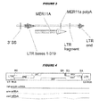
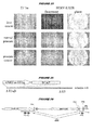
- old and new LTRs cluster as two separate groups in phylogenetic analyses ( figure 1 ). In keeping with their relative genetic ages, old viruses also contain more interruptions and deletions than new viruses.
- PCAV appears to have arisen from a rearrangement between a new and an old virus.
- the 5' region of the virus ( figure 2 ) starts with a new LTR allowed by 162 bp from a new virus.
- the rest of the new virus seems to be missing, as the 162 bp is followed by a 552 bp of non-viral sequence and then an almost-complete old virus,
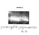
- the 3' LTR of the old virus ( figure 3 ) is fragmented and includes a MER11a insertion.
- SEQ ID 1 is the 12366bp sequence of PCAV, based on available human chromosome 22 sequence ⁇ 4 ⁇ , from the beginning of its first 5' LTR to the end of its fragmented 3' LTR. It is the sense strand of the double-stranded genomic DNA.
- SEQ ID 10 is the 11101bp sequence of PCAV from nucleotide 559 in SEQ ID 1 (a possible transcription start site) to its polyadenylation site (up to nucleotide 11735 in SEQ ID 1), although a more downstream transcription start site (e.g. nucleotide 635 ⁇ 5) is more likely.
- PCAV PCAV-derived protein
- the specific sequence of PCAV is manifested at both the mRNA and amino acid levels, and can be used to distinguish it from other HERVs within, the genome.
- This splice donor can join to splice acceptor sites (3'SS) at the start of the env open reading frame ( Figure 4 ).
- HERV-K genomes also include two splice acceptor sequences near the 3' end of the LTR, but these are not ordinarily used because they have no upstream viral splice donor partner.
- PCAV has two LTRs at its 5' end: the first is from a new HERV-K and the second is from an old HERV-K.
- the normally-unused splice acceptors in the old LTR can thus co-operate with the splice donor in the new LTR ( Figure 2 ), and transcripts resulting from these splice donor/acceptor pairings are specific to PCAV.
- Transcripts formed by using a splice acceptor site near the 3' end of the second 5' LTR comprise (i) a sequence transcribed from the transcription start site in the first 5' LTR, continuing to a splice donor site closely downstream of the first 5' LTR, joined to (ii) a sequence transcribed from one of the splice acceptor sites near the 3' end of the second 5' LTR. Detection of such transcripts indicates that PCAV is being transcribed.
- SEQ ID 1 the transcription start site in the first 5' LTR would be at nucleotide 559 by homology to other viruses, but seems to be further downstream ( e.g, at around 635 ⁇ 2) empirically; the conserved splice donor site downstream of the first 5' LTR is at nucleotides 1076-1081; the two splice acceptor sites near the 3' end of the second 5' LTR are at nucleotides 2593-2611 and 2680-2699.
- SEQ ID 2 is the sequence between the predicted transcription start site and the splice donor site.
- SEQ ID 3 is the first 10 nucleotides following the first splice acceptor site.
- SEQ ID 4 is the first 10 nucleotides following the second splice acceptor site.
- SEQ ID 5 is SEQ ID 2 fused to SEQ ID 3.
- SEQ ID 6 is SEQ ID 2 fused to SEQ ID 4.
- the 3' LTR of PCAV is fragmented, including insertion of a MER11a repetitive element ( figure 3 ).
- PCAV mRNAs terminate using a polyadenylation signal within the MER11a insertion, rather than using the signal within the viral LTR.
- Transcripts which terminate with a partial copy of a 3'HERV-K LTR followed by a MER11a sequence are specific to PCAV.
- transcripts from PCAV include copies of a partial LTR and a partial MER11a ( figure 3 ). Detection of such transcripts indicates that PCAV is being transcribed.
- the 3' LTR begins at nucleotide 10520 and continues until nucleotide 10838, where it is interrupted by a MER11a insertion; the MER11a insertion starts at nucleotide 10839 and continues to nucleotide 11834; after nucleotides 11835-11928, the 3' LTR continues from nucleotide 11929 to 12366.
- Within the MER11a insertion is its polyadenylation signal (located between nucleotides 11654 to 11659).
- SEQ ID 7 is the sequence of the first 319nt fragment of the 3' LTR.
- SEQ ID 8 is the sequence of the MER11a insertion up to its polyA site.
- SEQ ID 9 is SEQ ID 7 fused to SEQ ID 8.
- the env gene of PCAV is interrupted by an alu sequence. Detection of transcripts containing both env and alu sequence indicates that PCAV is being transcribed.
- the alu is at nucleotides 9938 to 10244 (SEQ ID 32).
- the 100 nucleotides immediately preceding the alu sequence (9838-9937) are SEQ ID 37, the last 10mer of which (9928-9937) is SEQ ID 33.
- the 100 nucleotides immediately following the alu sequence are SEQ ID 40, the first 10mer of which (10244-10253) is SEQ ID 34.
- the first 10 nucleotides of the alu sequence are SEQ ID 35 and the last 10 are SEQ ID 41.
- SEQ ID 36 is the 20mer bridging the alu/env boundary and SEQ ID 45 is the 20mer bridging the end of the alu sequence, SEQ ID 39 is the 8mer bridging the alu/env boundary, and SEQ ID 44 is the 8mer bridging the end of the alu sequence.
- SEQ ID 38 is SEQ ID 37 + SEQ ID 32, SEQ ID 42 is SEQ ID 41 + SEQ ID 40, and SEQ ID 43 is SEQ ID 32 + SEQ ID 40.
- the PCAV gag gene contains a 48 nucleotide sequence (SEQ ID 53) which is not found in other HERV-Ks.
- the 48mer encodes 16mer SEQ ID 110, which is not found in gag proteins from new or in other old HERV-Ks. Detection of transcripts containing SEQ ID 53, or of polypeptides containing SEQ ID 110, or antibodies which recognize epitope within or including SEQ ID 110 thus indicates that PCAV is being transcribed.
- the PCAV gag gene also contains a 69 nucleotide sequence (SEQ ID 111) which is not found in new HERV-Ks.
- the 69mer encodes 23mer SEQ ID 55. Detection of transcripts containing SEQ ID 111, or of polypeptides containing SEQ ID 55, or antibodies which recognize epitope within or including SEQ ID 55 thus indicates that an old HERV-K, typically PCAV, is being transcribed.
- the diagnostic method of the invention may be based on mRNA detection.
- PCAV mRNA may be detected directly or indirectly. It is preferred to detect a mRNA directly, thereby avoiding the need for separate preparation of mRNA-derived material (e.g. cDNA).
- transcripts for use according to the present invention are transcribed from PCAV.
- Three preferred types of transcript are: (1) transcripts spliced using a splice acceptor site near the 3' end of the second 5' LTR; (2) transcripts comprising both 3' LTR and MER11a sequences; (3) transcripts comprising the alu-interrupted env gene; and (4) transcripts comprising a PCAV-specific gag sequence.
- the invention provides a mRNA transcript transcribed from a human endogenous retrovirus located at megabase 20.428 on chromosome 22.
- the invention also provides a mRNA transcript comprising a nucleotide sequence with n% or more sequence identity to SEQ ID 23, or to a nucleotide sequence lacking up to 100 nucleotides ( e.g. 10, 20, 30, 40, 50, 60, 70, 71, 72, 73, 74, 75, 76, 77, 78, 79, 80, 81, 82, 83, 84, 85, 86, 87, 88, 89, 90 or 100) from the 5' end of SEQ ID 23 e.g. n% or more sequence identity to SEQ ID 1197 or 1198.
- the nucleotide sequence is preferably at the 5' end of the RNA, although upstream sequences may be present.
- the nucleotide sequence may be at the 3' end of the RNA, but there will typically be further downstream elements such as a poly-A tail.
- These mRNA transcripts include allelic variants, SNP variants, homologs, orthologs, paralogs, mutants, etc. of SEQ ID 23, SEQ ID 1197 and SEQ ID 1198.
- the invention provides a mRNA transcript formed by splicing involving a splice acceptor site near the 3' end of the second 5' LTR.
- a mRNA transcript comprising the sequence -N 1 -N 2 - ( e.g. SEQ ID 24, SEQ YD 25, SEQ ID 1199 or SEQ ID 1200), where: N 1 is a nucleotide sequence ( e.g.
- SEQ ID 26, SEQ ID 1201) from (i) the 5' end of a RNA transcribed from the first 5' LTR of a human endogenous retrovirus located at megabase 20.428 on chromosome 22, to (ii) a first splice donor site downstream of the U5 region of said mRNA transcribed from the first 5' LTR; and N 2 is a nucleotide sequence ( e.g. SEQ ID 27 or SEQ ID 28) immediately downstream of a splice acceptor site located (i) downstream of said first splice donor site and (ii) upstream of a second splice donor site, the second splice donor site being downstream of the second 5' LTR of said endogenous retrovirus.
- SEQ ID 27 or SEQ ID 28 is a nucleotide sequence immediately downstream of a splice acceptor site located (i) downstream of said first splice donor site and (ii) upstream of a second splice donor site, the second
- the first splice donor site is preferably the site conserved in the HML2 sub-family, located about 100 nucleotides downstream of the first 5' LTR (after nucleotide 1075 in SEQ ID 1).
- the second splice donor site is preferably the site conserved in the HML2 sub-family, located about 100 nucleotides downstream of the second 5' LTR (after SEQ ID 1 nucleotide 2778).
- the splice acceptor is preferably downstream of the second 5' LTR.
- the invention also provides a mRNA transcript comprising the sequence -N 1 -N 2 -, where: N 1 is a nucleotide sequence with a% or more sequence identity to SEQ ID 26 and/or SEQ ID 1201 and N 2 is a nucleotide sequence with b% or more sequence identity to SEQ ID 27 or SEQ ID 28.
- N 1 is a nucleotide sequence with a% or more sequence identity to SEQ ID 26 and/or SEQ ID 1201
- N 2 is a nucleotide sequence with b% or more sequence identity to SEQ ID 27 or SEQ ID 28.
- N 1 is preferably at the 5' end of the RNA, although upstream sequences may be present.
- N 2 may be at the 3' end of the RNA, but downstream sequences will usually be present.
- the invention also provides a mRNA transcript comprising a nucleotide sequence with c% or more sequence identity to SEQ ID 24, SEQ ID 25, SEQ ID 1199 or SEQ ID 1200.
- the invention provides a mRNA transcript comprising the sequence -N 3 -N 4 - (e.g. SEQ ID 29), where: N 3 is a nucleotide sequence (e.g. SEQ ID 30) from the 3' end of the 5' fragment of the 3' LTR of a human endogenous retrovirus located at megabase 20.428 on chromosome 22, and N 4 is a nucleotide sequence ( e.g. SEQ ID 31) from 5' end of the MER11a insertion in a human endogenous retrovirus located at megabase 20.428 on chromosome 22.
- N 3 is a nucleotide sequence (e.g. SEQ ID 30) from the 3' end of the 5' fragment of the 3' LTR of a human endogenous retrovirus located at megabase 20.428 on chromosome 22
- N 4 is a nucleotide sequence (e.g. SEQ ID 31) from 5' end of the MER11a insertion in a human endogen
- the invention also provides a mRNA transcript comprising the sequence -N 3 -N 4 -, where: N 3 is a nucleotide sequence with d% or more sequence identity to SEQ ID 30 and N 4 is a nucleotide sequence with e% or more sequence identity to SEQ ID 31.
- the RNA may comprise the sequence -N 3 -N 4 -N 5 -N 6 -, wherein: N 5 is a nucleotide sequence between the polyA signal and the polyA site of a MER11a sequence; and N 6 is a polyA tail.
- the transcript will generally include sequence upstream of N 3 .
- the transcript will generally include sequence downstream of N 4 , such as a polyA tail.
- the invention also provides a mRNA transcript comprising a nucleotide sequence with f% or more sequence identity to SEQ ID 29.
- the invention provides a mRNA transcript comprising the sequence -N 7 -N 8 - (e.g. SEQ ID 38), where: N 7 is a nucleotide sequence (e.g. SEQ ID 37) preceding the alu insertion within the env gene of a human endogenous retrovirus located at megabase 20.428 on chromosome 22, and N 8 is a nucleotide sequence ( e.g. SEQ ID 32) beginning at the 5' end of said alu insertion.
- N 7 is a nucleotide sequence (e.g. SEQ ID 37) preceding the alu insertion within the env gene of a human endogenous retrovirus located at megabase 20.428 on chromosome 22
- N 8 is a nucleotide sequence (e.g. SEQ ID 32) beginning at the 5' end of said alu insertion.
- the invention also provides a mRNA transcript comprising the sequence -N 7 -N 8 -, where: N 7 is a nudeotide sequence with mm% or more sequence identity to SEQ ID 37 and N 8 is a nucleotide sequence with nn% or more sequence identity to SEQ ID 32.
- the transcript will generally include sequence upstream of N 7 and downstream of N 8 .
- the invention also provides a mRNA transcript comprising a nucleotide sequence with pp% or more sequence identity to SEQ ID 38.
- the invention provides a mRNA transcript comprising the sequence "N 9 -N 10 - (e.g. SEQ ID 43), where: N 9 is a nucleotide sequence ( e.g. SEQ ID 32) at the end of the alu insertion within the env gene of a human endogenous retrovirus located at megabase 20.428 on chromosome 22, and N 10 is a nucleotide sequence ( e.g. SEQ ID 40) immediately downstream of said alu insertion.
- N 9 is a nucleotide sequence (e.g. SEQ ID 32) at the end of the alu insertion within the env gene of a human endogenous retrovirus located at megabase 20.428 on chromosome 22
- N 10 is a nucleotide sequence (e.g. SEQ ID 40) immediately downstream of said alu insertion.
- the invention also provides a mRNA transcript comprising the sequence -N 9 -N 10 -, where: N 9 is a nucleotide sequence with uu% or more sequence identity to SEQ ID 41 and N 10 is a nucleotide sequence with vv% or more sequence identity to SEQ ID 40.
- the transcript will generally include sequence upstream of N 9 and downstream of N 10 .
- the invention also provides a mRNA transcript comprising a nucleotide sequence with ww% or more sequence identity to SEQ ID 42.
- the invention provides a mRNA transcript comprising a nucleotide sequence with uu% or more sequence identity to SEQ ID 41.
- the transcript will generally include sequence upstream of N 9 and downstream of N 10 .
- the invention also provides a mRNA transcript comprising a nucleotide sequence with ii% or more sequence identity to SEQ ID 53.
- the invention also provides a mRNA transcript comprising a nucleotide sequence with ii% or more sequence identity to SEQ ID 111.
- the invention also provides a mRNA transcript comprising a nucleotide sequence with ii% or more sequence identity to SEQ ID 1191.
- the invention also provides a mRNA transcript which encodes a polypeptide having at least ii% sequence identity to SEQ ID 98.
- PCAV mRNA transcripts of the invention may be detected directly, for example by sequencing of the mRNA or by hybridization to mRNA transcripts ( e.g. by Northern blot).
- Various techniques are available for detecting the presence or absence of a particular RNA sequence in a sample ⁇ e.g . refs. 20 & 21 ⁇ .
- Indirect detection of mRNA transcripts is also possible and is performed on nucleic acid derived from a PCAV mRNA transcript e.g. detection of a cDNA copy of PCAV mRNA, detection of nucleic acids amplified from a PCAV mRNA template, etc.
- RNA from prostate cells is reported in, for example, references 14 to 19. It is preferred to use PCAV-specific probes in RT-PCR.
- the method of the invention involves detection of a single-stranded or double-stranded PCAV nucleic acid target, either (a) in the form of PCAV mRNA or (b) in the form of nucleic acid comprising a copy of at least a portion of a PCAV mRNA and/or a sequence complementary to at least a portion of a PCAV mRNA.
- the method of the invention does not involve the detection of PCAV genomic DNA, as this is present in all human cells and its presence is therefore not characteristic of tumors. If a sample contains PCAV DNA, it is preferred to use a RNA-specific detection technique or to focus on sequences present in PCAV mRNA transcripts but not in PCAV genomic DNA ( e.g. splice junctions, polyA tail etc. ) ,
- the method of the invention may therefore comprise an initial step of: (a) extracting mRNA from a patient sample; (b) removing DNA from a patient sample without removing mRNA; and/or (c) removing or disrupting PCAV DNA, but not PCAV mRNA, in a patient sample.
- a RNA-specific assay can be used which is not affected by the presence of homologous DNA. For RT-PCR, genomic DNA should be removed,
- RNA may be enriched e.g. using oligo-dT techniques.
- Methods for removing DNA from biological samples without removing mRNA are well known ⁇ e.g. appendix C of ref. 20 ⁇ and include DNase digestion. If DNase is used then it must be removed or inactivated ( e.g. by chelation with EDTA, by heating, or by proteinase K treatment followed by phcnol/chloroform extraction and NH 4 OAc/EtOH precipitation) prior to subsequent DNA synthesis or amplification, in order to avoid digestion of the newly-synthesized DNA.
- DNase e.g. appendix C of ref. 20 ⁇ and include DNase digestion. If DNase is used then it must be removed or inactivated (e.g. by chelation with EDTA, by heating, or by proteinase K treatment followed by phcnol/chloroform extraction and NH 4 OAc/EtOH precipitation) prior to subsequent DNA synthesis or amplification, in order to avoid digestion of the newly-synthesized DNA
- Methods for removing PCAV DNA, but not PCAV RNA will use a reagent which is specific to a sequence within a PCAV DNA e.g. a restriction enzyme which recognizes a DNA sequence within the PCAV genome, but which does not cleave the corresponding RNA sequence.
- a reagent which is specific to a sequence within a PCAV DNA e.g. a restriction enzyme which recognizes a DNA sequence within the PCAV genome, but which does not cleave the corresponding RNA sequence.
- Methods for specifically purifying PCAV mRNAs from a sample may also be used.
- One such method uses an affinity support which binds to PCAV mRNAs.
- the affinity support may include a polypeptide sequence which binds to the PCAV mRNA e.g. the cORF polypeptide, which binds to the LTR of HERV-K mRNAs in a sequence-specific manner, or HIV Rev protein, which has been shown to recognize the HERV-K LTR in RNA transcripts ⁇ 22 ⁇ .
- PCAV mRNA need not be maintained in a wild-type form for detection. It may, for example, be fragmented, provided that the fragmentation maintains PCAV-specific sequences within the mRNA.
- the invention provides nucleic acid comprising (a) the nucleotide sequence of a mRNA transcript transcribed from a human endogenous retrovirus located at megabase 20.428 on chromosome 22, and/or (b) the complement of (a).
- the invention also provides nucleic acid comprising a nucleotide sequence with qq% or more sequence identity to SEQ ID 10, SEQ ID 1197 and/or SEQ ID 1198.
- PCAV is approximately 87.5% identical to the HBRV-K found at megabase 47.1 on chromosome 6 and approximately 86% identical to the HERV-K found at megabase 103.75 on chromosome 3.
- the invention provides nucleic acid comprising (a) nucleotide sequence -N 1 -N 2 - as defined above, and/or (b) the complement of (a).
- the invention also provides nucleic acid comprising (a) a nucleotide sequence with c% or more sequence identity to SEQ ID 5, SEQ ID 6, SEQ ID 1199 or SEQ ID 1200, and/or (b) the complement of (a).
- the invention provides nucleic acid comprising (a) nucleotide sequence -N 3 -N 4 - as defined above, and/or (b) the complement of (a).
- the invention also provides nucleic acid comprising (a) a nucleotide sequence with f% or more sequence identity to SEQ ID 9, and/or (b) the complement of (a).
- the invention also provides nucleic acid comprising (a) nucleotide sequence -N 3 -N 4 -N 5 -N 6 - as defined above, and/or (b) the complement of (a).
- the invention provides nucleic acid comprising (a) nucleotide sequence -N 7 -N 8 - as defined above, and/or (b) the complement of (a).
- the invention also provides nucleic acid comprising (a) a nucleotide sequence with aa% or more sequence identity to SEQ ID 38, and/or (b) the complement of (a).
- the invention provides nucleic acid comprising (a) nucleotide sequence -N 9 -N 10 - as defined above, and/or (b) the complement of (a).
- the invention also provides nucleic acid comprising (a) a nucleotide sequence with hh% or more sequence identity to SEQ ID 42, and/or (b) the complement of (a).
- the invention provides nucleic acid comprising a nucleotide sequence with bbb% or more sequence identity to SEQ ID 53, and/or (b) the complement of (a).
- the invention provides nucleic acid comprising a nucleotide sequence with fff% or more sequence identity to SEQ ID 111, and/or (b) the complement of (a).
- nucleic acid targets include SEQ IDs 99 to 109, which are splice variant cDNA sequences assuming a transcription start site in SEQ ID 1 at 559 and including four A residues at the 3' end. Assuming a more downstream transcription start site (e.g. nucleotide 635 of SEQ ID 1), these nucleic targets would not include a stretch ofnucleotides at the 5' end of SEQ IDs 99 to 109 e.g.
- the invention provides nucleic acid which can hybridize to a PCAV nucleic acid target.
- Hybridization reactions can be performed under conditions of different "stringency”. Conditions that increase stringency of a hybridization reaction of widely known and published in the art ⁇ e.g. page 7.52 of reference 21 ⁇ . Examples of relevant conditions include (in order of increasing stringency): incubation temperatures of 25°C, 37°C, 50°C, 55°C and 68°C; buffer concentrations of 10 x SSC, 6 x SSC, 1 x SSC, 0.1 x SSC (where SSC is 0.15 M NaCl and 15 mM citrate buffer) and their equivalents using other buffer systems; formamide concentrations of 0%, 25%, 50%, and 75%; incubation times from 5 minutes to 24 hours; 1, 2, or more washing steps; wash incubation times of 1, 2, or 15 minutes; and wash solutions of 6 x SSC, 1 x SSC, 0.1 x SSC, or de-ionized water, Hybridization techniques and their optimization are well known in the art ⁇ e.g . see references 20, 21, 23, 24, 28 etc
- nucleic acid of the invention hybridizes to a target of the invention under low stringency conditions; in other embodiments it hybridizes under intermediate stringency conditions; in preferred embodiments, it hybridizes under high stringency conditions.
- An exemplary set of low stringency hybridization conditions is 50°C and 10 x SSC.
- An exemplary set of intermediate stringency hybridization conditions is 55°C and 1 x SSC.
- An exemplary set of high stringency hybridization conditions is 68°C and 0.1 x SSC.
- Preferred nucleic acids of the invention hybridize to PCAV nucleic acid targets but not to nucleic acid targets from other HERV-Ks.
- PCAV-specific hybridization is favored by exploiting features found within PCAV transcripts but not in other HERV-K transcripts e.g. specific nucleotide sequences, features arising from the tandem 5' LTRs, features arising from the MER11a insertion within the 3' LTR, or features arising from the alu interruption of env. Sequence alignments can be used to locate regions of PCAV which are most divergent from other HERV-K genomes and in which PCAV-specific hybridization can occur. Specificity for PCAV is desirable in order to detect its up-regulation above the low-level of natural background expression of other new HERV-Ks seen in most cells.
- One group of preferred nucleic acids of the invention can specifically detect PCAV products in which a splice acceptor site near the 3' end of the second 5' LTR has been used. As described above, such splicing brings together sequences N 1 and N 2 , which are not juxtaposed in PCAV genomic DNA.
- the invention provides a nucleic acid which hybridizes to sequence -N 1 -N 2 - (or the complement thereof) within a PCAV nucleic acid target, but which does not hybridize to sequences N 1 or N 2 alone (or to their complements alone).
- the nucleic acid comprises a first sequence which can hybridize to N 1 (or to its complement) and a second sequence which can hybridize to N 2 (or to its complement), such that it will hybridize to a target in which N 1 and N 2 are adjacent, but will not hybridize to targets in which splicing has not brought N 1 and N 2 together.
- Such nucleic acids can identify PCAV transcripts in the presence of PCAV genomic DNA because of the difference in relative locations of N 1 and N 2 .
- nucleic acids of the invention can specifically detect mRNAs containing 3' LTR and MER11a sequences.
- the invention provides a nucleic acid which hybridizes to sequence -N 3 -N 4 - (or the complement thereof) within a PCAV nucleic acid target, but which does not hybridize to sequences N 3 or N 4 alone (or to their complements alone).
- the nucleic acid comprises a first sequence which can hybridize to N 3 (or to its complement) and a second sequence which can hybridize to N 4 (or to its complement), such that it will hybridize to targets which include both (i) a 3' LTR sequence and (ii) a MER11a sequence, but not to targets which include only one of (i) and (ii).
- the nucleic acid may inherently be able to hybridize to gnomic DNA, although this property is not useful for detecting transcripts.
- nucleic acids of the invention can specifically detect mRNAs containing the alu-inlerrupted env gene.
- the invention provides a nucleic acid which hybridizes to sequence -N 7 -N 8 - (or the complement thereof) within a PCAV nucleic acid target, but which does not hybridize to sequences N 7 or N 8 alone (or to their complements alone).
- the nucleic acid comprises a first sequence which can hybridize to N 7 (or to its complement) and a second sequence which can hybridize to N 8 (or to its complement), such that it will hybridize to targets which include both (i) the env sequence immediately preceding the alu interruption and (ii) an alu interruption, but not to targets which include only one of (i) and (ii).
- the nucleic acid may inherently be able to hybridize to genomic DNA, although this property is not useful for detecting transcripts.
- the invention also provides a nucleic acid which hybridizes to sequence -N 9 -N 10 - (or the complement thereof) within a PCAV nucleic acid target, but which does not hybridize to sequences N 9 or N 10 alone (or to their complements alone).
- the nucleic acid comprises a first sequence which can hybridize to N 9 (or to its complement) and a second sequence which can hybridize to N 10 (or to its complement), such that it will hybridize to targets which include both (i) the 3' region of the alu interruption within env and (ii) the sequence immediately downstream of the alu interruption, but not to targets which include only one of (i) and (ii).
- the nucleic acid may inherently be able to hybridize to genomic DNA, although this property is not useful for detecting transcripts.
- the ability of a nucleic acid to hybridize to a PCAV nucleic acid target is related to its intrinsic features (e.g. the degree of sequence identity to the target) as well as extrinsic features (e.g. temperature, salt concentration etc. ).
- a group of preferred nucleic acids of the invention have a good intrinsic ability to hybridize to PCAV nucleic acid targets.
- the invention provides a nucleic acid comprising a nucleotide sequence with s % or more sequence identity to a fragment of a PCAV nucleic acid target or to the complement of a fragment of a PCAV nucleic acid target.
- the invention provides a nucleic acid comprising a nucleotide sequence with g % or more sequence identity to a fragment of SEQ ID 10 or to the complement of a fragment of SEQ ID 10.
- the invention also provides a nucleic acid comprising a nucleotide sequence with h % or more sequence identity to a fragment of SEQ ID 5 or to the complement of a fragment of SEQ ID 5.
- the invention also provides a nucleic acid comprising a nucleotide sequence with i % or more sequence identity to a fragment of SEQ ID 6 or to the complement of a fragment of SEQ ID 6.
- the invention also provides a nucleic acid comprising a nucleotide sequence with j % or more sequence identity to a fragment of SEQ ID 9 or to the complement of a fragment of SEQ ID 9.
- the invention also provides a nucleic acid comprising a nucleotide sequence with ccc % or more sequence identity to a fragment of SEQ ID 53 or to the complement of a fragment of SEQ ID 53.
- the invention also provides a nucleic acid comprising a nucleotide sequence with kkk % or more sequence identity to SEQ ID 1191. It also provides a nucleic acid comprising a nucleotide sequence which encodes a polypeptide having at least mmm % sequence identity to SEQ ID 98. The invention also provides a nucleic acid comprising a nucleotide sequence with nnn % or more sequence identity to SEQ ID 1198. It also provides a nucleic acid comprising a nucleotide sequence which encodes a polypeptide having at least qqq % sequence identity to SEQ ID 1199. It also provides a nucleic acid comprising a nucleotide sequence which encodes a polypeptide having at least rrr % sequence identity to SEQ ID 1200.
- the invention provides a nucleic acid comprising a fragment of at least k contiguous nucleotides of SEQ ID 10 or of the complement of SEQ ID 10.
- the fragment is preferably located within SEQ ID 1197 and/or 1198.
- the invention also provides a nucleic acid comprising a fragment of at least l contiguous nucleotides of SEQ ID 47 or of the complement of SEQ ID 47.
- the fragment preferably comprises nucleotide sequence B 1a -B 2a (or its complement), wherein B 1a comprises m or more nucleotides from the 3' end of SEQ ID 2 and B 2a comprises p or more nucleotides from the 5' end of SEQ ID 46.
- These nucleic acids thus span a splice junction which brings sequences N 1 and N 2 together and are thus able to identify PCAV transcripts in the presence of PCAV genomic DNA because of the difference in the relative locations of B 1a and B 2a .
- the invention also provides a nucleic acid comprising a fragment of at least q contiguous nucleotides of SEQ ID 49 or of the complement of SEQ ID 49.
- the fragment preferably comprises nucleotide sequence B 1b -B 2b (or its complement), wherein B 1b comprises r or more nucleotides from the 3' end of SEQ ID 2 and B 2b comprises t or more nucleotides from the 5' end of SEQ ID 48.
- the invention also provides a nucleic acid comprising a fragment of at least u contiguous nucleotides of SEQ ID 9 or of the complement of SEQ ID 9.
- the fragment preferably comprises nucleotide sequence B 3 -B 4 (or its complement), wherein B 3 comprises v or more nucleotides from the 3' end of SEQ ID 7 and B 4 comprises w or more nucleotides from the 5' end of SEQ ID 8.
- the invention also provides a nucleic acid comprising a fragment of at least rr contiguous nucleotides of SEQ ID 38 or of the complement of SEQ ID 38.
- the fragment preferably comprises nucleotide sequence B 7 -B 8 (or its complement), wherein B 7 comprises ss or more nucleotides from the 3' end of SEQ ID 37 and B 4 comprises tt or more nucleotides from the 5' end of SEQ ID 32.
- These nucleic acids thus include part of both of N 7 and N 8 .
- the invention also provides a nucleic acid comprising a fragment of at least jj contiguous nucleotides of SEQ ID 43 or of the complement of SEQ ID 43.
- the fragment preferably comprises nucleotide sequence B 9 -B 10 , or its complement, and wherein B 9 comprises kk or more nucleotides from the 3' end of SEQ ID 32 and B 10 comprises ll or more nucleotides from the 5' end of SEQ TD 40.
- These nucleic acids thus include part of both of N 9 and N 10 .
- the invention also provides a nucleic acid comprising a fragment of at least ddd contiguous nucleotides of SEQ ID 53 or of the complement of SEQ ID 53.
- the invention also provides a nucleic acid comprising a fragment of at least ggg contiguous nucleotides of SEQ ID 111 or of the complement of SEQ ID 111.
- the invention also provides a nucleic acid comprising a fragment of at least hhh contiguous nucleotides of SEQ ID 112 or of the complement of SEQ ID 112.
- the invention also provides a nucleic acid comprising a fragment of at least jjj contiguous nucleotides of SEQ ID 1191 or of the complement of SEQ ID 1191.
- the invention provides a nucleic acid of formula 5'-X-Y-Z-3', wherein: -X- is a nucleotide sequence consisting of x nucleotides; -Z- is a nucleotide sequence consisting of z nucleotides; -Y- is a nucleotide sequence consisting of either (a) a fragment of y nucleotides of any of SEQ IDs 1-13, 20-53, 57, 58, 63, 81, 86, 88-91, 99-109, 111, 112, 1191, 1197 or 1198, or (b) the complement of (a); and said nucleic acid 5'-X-Y-Z-3' is neither (i) a fragment of SEQ IDs 1-13, 20-53, 57, 58, 63, 81, 86, 88-91, 99-109, 111, 112, 1191, 1197 or 1198 or (ii) the complement of (i).
- the nucleotide sequence of -X- preferably shares less than bb % sequence identity to the x nucleotides which are 5' of sequence -Y- in SEQ IDs 1-13, 20-53, 57, 58, 63, 81, 86, 88-91, 99-109, 111, 112, 1191, 1197 or 1198 and/or the nucleotide sequence of -Z-preferably shares less than cc % sequence identity to the z nucleotides which are 3' of sequence - Z- in SEQ IDs 1-13, 20-53, 57, 58, 63, 81, 86, 88-91, 99-109, 111, 112, 1191, 1197 or 1198.
- the nucleotide sequence of -X- preferably shares less than bb % sequence identity to the complement of the x nucleotides which are 5' of the complement of sequence -Yin SEQ IDs 1-1,3, 20-53, 57, 58, 63, 81, 86, 88-91, 99-109, 111, 112, 1191, 1197 or 1198 and/or the nucleotide sequence of -Z- preferably shares less than cc % sequence identity to the complement of the z nucleotides which are 3' of the complement of sequence -Y- in SEQ IDs 1-13, 20-53, 57, 58, 63, 81, 86, 88-91, 99-109,111, 112, 1191, 1197 or 1198.
- the -X- and/or -Z- moieties may comprise a promoter sequence (or its complement).
- the invention provides nucleic acid comprising nucleotide sequence SEQ ID 53. This sequence is specific within the human genome to PCAV.
- the invention also provides nucleic acid comprising nucleotide sequence SEQ ID 111.
- the invention also provides nucleic acid comprising nucleotide sequence SEQ ID 1191.
- PCAV nucleic acids are provided by the invention.
- 25mer fragments of PCAV sequences are given as SEQ IDs 120 to 1184.
- the invention provides these sequences as 25mers, as well as fragments thereof ( e.g . the 2 x 24mers, the 3 x 23mers, the 4 x 22mers ... the 19 x 7mers in each) and as longer PCAV fragments comprising these 25mers.
- Preferred nucleic acids of the invention comprise one or more of SEQ IDs 53 and 842-1184,
- Nucleic acids of the invention are particularly useful as probes and/or as primers for use in hybridization and/or amplification reactions.
- More than one nucleic acid of the invention can hybridize to the same target (e.g. more than one can hybridize to a single mRNA or cDNA).
- Nucleic acid in a sample can conveniently and sensitively be detected by nucleic acid amplification techniques such as PCR, SDA, SSSR, LCR, TMA, NASBA, T7 amplification etc.
- the technique preferably gives exponential amplification.
- a preferred technique for use with RNA is RT-PCR (e.g. see chapter 15 of ref. 20).
- the technique may be quantitative and/or real-time.
- Amplification techniques generally involve the use of two primers. Where a target sequence is single-stranded, the techniques generally involve a preliminary step in which a complementary strand is made in order to give a double-stranded target. The two primers hybridize to different strands of the double-stranded target and are then extended. The extended products can serve as targets for further rounds of hybridization/extension. The net effect is to amplify a template sequence within the target, the 5' and 3' termini of the template being defined by the locations of the two primers in the target.
- the invention provides a kit comprising primers for amplifying a template sequence contained within a PCAV nucleic acid target, the kit comprising a first primer and a second primer, wherein the first primer comprises a sequence substantially complementary to a portion of said template sequence and the second primer comprises a sequence substantially complementary to a portion of the complement of said template sequence, wherein the sequences within said primers which have substantial complementarity define the termini of the template sequence to be amplified.
- Kits of the invention may further comprise a probe which is substantially complementary to the template sequence and/or to its complement and which can hybridize thereto. This probe can be used in a hybridization technique to detect amplified template.
- Kits of the invention may further comprise primers and/or probes for generating and detecting an internal standard, in order to aid quantitative measurements ⁇ e.g. 15, 25 ⁇ .
- Kits of the invention may comprise more than one pair of primers (e.g. for nested amplification), and one primer may be common to more than one primer pair.
- the kit may also comprise more than one probe.
- the template sequence is preferably located within a transcript of a HERV-K located at megabase 20.428 of chromosome 22, and is more preferably a fragment of SEQ ID 10 (or SEQ ID 23).
- the template sequence is preferably at least 50 nucleotides long ( e.g. 60, 70, 80, 90, 100, 125, 150, 175, 200, 250, 300, 400, 500, 600, 700, 800, 900, 1000, 1250, 1500, 2000, 3000 nucleotides or longer).
- the length of the template is inherently limited by the length of the target within which it is located, but the template sequence is preferably shorter than 500 nucleotides ( e.g. 450, 400, 350, 300, 250, 200, 175, 150, 125, 100, 90, 80, 70 or shorter).
- a preferred template comprises SEQ ID 53 and/or SEQ ID 111.
- Primers and probes used in kits of the invention are preferably nucleic acids as described in section B.4 above.
- Particularly preferred primers are those based on SEQ IDs 600-1184 (or their complements) e.g. comprising primers comprising SEQ IDs 600-1184, or primers comprising fragments of ppp or more nucleotides from one of SEQ IDs 600-1184.
- kits comprise (i) a first primer comprising a sequence which is substantially identical to a portion of SEQ ID 10 and (ii) a second primer comprising a sequence which is substantially complementary to a portion of SEQ ID 10, such that the primer pair (i) and (ii) defines a template sequence within SEQ ID 10.
- Other preferred kits comprise (i) a first primer comprising a sequence which is substantially identical to a portion of the complement of SEQ ID 10 and (ii) a second primer comprising a sequence which is substantially complementary to a portion of the complement of SEQ ID 10, such that the primer pair defines a template sequence within SEQ ID 10.
- the portion and template sequence preferably fall within SEQ ID 1197 or SEQ ID 1198.
- one or both of the primers is not substantially complementary to a portion of a HERV-K other than PCAV (or its complement) such that the primer pair is specific for PCAV.
- SEQ ID 10 may be divided into four exons: (1) nucleotides 1-517, containing sequences up to the conserved splice donor downstream of the first 5' LTR; (2) nucleotides 2142-2209, containing sequences between the splice acceptor near the 3' end of the second 5' LTR and the conserved splice donor; (3) nucleotides 7608-7686; and (4) nucleotides 9866-11181 (assuming transcription start at nucleotide 559 of SEQ ID 1).
- Exon (2) arises because of the unique PCAV feature of tandem 5' LTRs, but the other three exons exist in other HERV-Ks.
- the first and second primers are located in different exons. This arrangement means that the amplified template sequence is shorter than would be obtained from genomic DNA, because of the absence ofintrons.
- First primer in exon 1 1 1 2 2 3
- Second primer in exon 2 3 4 3 4 4
- the primers may comprise a fragment of SEQ ID 10 (or its complement) located between the following coordinates: First primer 1-517 1-517 1-517 2142-2219 2142-2219 7608-7686 Second primer 2142-2219 7608-7686 9866-11181 7608-7686 9866-11181 9866-11181
- the first exon may begin downstream of nucleotide 559 e.g. at around nucleotide 633, 635 or 637.
- Example primers within exon 1 are SEQ IDs 120 to 219.
- Example primers within exons 2 to 4 are SEQ IDs 220 to 336.
- first and second primers comprise a first sequence from a first exon and a second sequence from a second exon, such that the primer bridges an exon-exon boundary after splicing.
- a primer may comprise sequences from exons 1 & 2, exons 1 & 3, exons 1 & 4, exons 2 & 3, exons 2 & 4, or exons 3 & 4. These primers hybridize to transcripts where splicing has taken place.
- the primers may comprise a first sequence from the 3' end of the following coordinates and second sequence from the 5' end of the following coordinates (or complements thereof): First sequence 1-517 1-517 1.517 2142-2209 2142-2209 7608-7686 Second sequence 2142-2209 2142-2209 7608-7686 9866-11181 7608-7686 9866-11181 9866-11181
- the range '1-517' for selecting the first sequence should be replaced with around '77-517' e.g. 75-517 or 80-517.
- kits for detecting PCAV nucleic acid targets in which a splice acceptor site near the 3' end of the second 5' LTR has been used either (i) the first primer comprises a sequence which is substantially identical to a portion of N 1 and the second primer comprises a sequence which is substantially complementary to a portion of N 2 , or (ii) the first primer comprises a sequence which is substantially identical to a portion of the complement of N 1 and the second primer comprises a sequence which is substantially complementary to a portion of the complement of N 2 .
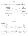
- This primer pair defines a template sequence which bridges the PCAV-specific splice junction. The amplified sequence will be shorter for targets where the splice junction has been used than for unspliced targets ( figure 5 ) or for genomic DNA.
- the amplified sequence will be shorter than for P CAV targets where transcription started in a more upstream 5' LTR.
- kits for detecting PCAV products in which a splice acceptor site near the 3' end of the second 5' LTR has been used either (i) the first primer comprises a sequence which is substantially identical to a portion of N 1 and the second primer comprises a sequence which is substantially complementary to a portion of PCAV sequence downstream of a splice donor which is itself downstream of the splice acceptors near the 3' end of the second PCAV 5' LTR, or (ii) the first primer comprises a sequence which is substantially identical to a portion of the complement of N 1 and the second primer comprises a sequence which is substantially complementary to a portion of the complement of a PCAV sequence downstream of a splice donor which is itself downstream of the splice acceptors near the 3' end of the second PCAV 5' LTR.
- the primers are located either side of exon 2 and thus define a template sequence which bridges exon 2.
- the amplified sequence will be longer in targets where the exon is present than in targets where the exon absent ( figure 6A vs . 6B) and only PCAV targets can give the longer amplification product. All splice products, whether or not including the exon, will give shorter amplification products than unspliced RNA or genomic DNA targets.
- kits for detecting PCAV products in which a splice acceptor site near the 3' end of the second 5' LTR has been used either (i) the first primer comprises a sequence which is substantially identical to the splice junction site in N 1 -N 2 and the second primer comprises a sequence which is substantially complementary to a portion of a PCAV sequence upstream or downstream of the splice junction site, or (ii) the first primer comprises a sequence which is substantially identical to the complement of the splice junction site in N 1 -N 2 and the second primer comprises a sequence which is substantially complementary to a portion of a PCAV upstream or sequence downstream of the splice junction site.
- the first primer comprises a first sequence which is substantially complementary to a portion of N 1 and a second sequence which is substantially complementary to a portion of N 2 and can hybridize to targets where the splice junction has been used but not to targets where the splice junction has not been used. Amplification from such primer pairs will only occur where the target sequence has been formed by use of the splice junction, and will not occur with unspliced targets or genomic DNA.
- kits for detecting the 3' region of PCAV products either (i) the first primer comprises a sequence which is substantially identical to a portion of N 3 and the second primer comprises a sequence which is substantially complementary to a portion of N 4 , or (ii) the first primer comprises a sequence which is substantially identical to a portion of the complement of N 3 and the second primer comprises a sequence which is substantially complementary to a portion of the complement of N 4 .
- the primer pair amplifies a template sequence which bridges the 3' LTR/MER11a junction and amplification will occur only where the target sequence contains both a 3' LTR sequence and a MER11a sequence ( figure 7 ).
- the first primer comprises a first sequence which is substantially identical to a portion of N 3 and a second sequence which is substantially identical to a portion of N 4
- the second primer comprises a sequence which is substantially complementary to a portion of an upstream or downstream PCAV sequence
- the first primer comprises a first sequence which is substantially identical to a portion of the complement of N 3 and a second sequence which is substantially identical to a portion of the complement of N 4
- the second primer comprises a sequence which is substantially complementary to a portion of the complement of an upstream or downstream PCAV sequence.
- the first primer hybridizes only to targets which contain both a 3' LTR sequence and a MER11a sequence, such that amplification occurs only where the target sequence contains both a 3' LTR sequence and a MER11a sequence ( figure 7 ).
- the second primer is preferably located in exon 3, so the amplification product is shorter than in the genome.
- kits for detecting the 3' region of PCAV products either (i) the first primer comprises a sequence which is substantially identical to a portion of N 3 and the second primer comprises a sequence which is substantially complementary to a portion of a polyA tail, or (ii) the first primer comprises a sequence which is substantially identical to a portion of the complement of N 3 and the second primer comprises a sequence which is substantially complementary to a portion of the complement of polyA tail.
- the template sequence defined by this primer pair is longer in targets where the 3' LTR contains a MER11a insertion than in targets ( e.g. other HERVs) where the 3' LTR is intact ( figure 8 ).
- PolyA-specificity means that genomic DNA is not amplified.
- kits for detecting PCAV products containing alu-interrupted env either (i) the first primer comprises a sequence which is substantially identical to a portion of N 7 and the second primer comprises a sequence which is substantially complementary to a portion of N 8 , or (ii) the first primer comprises a sequence which is substantially identical to a portion of the complement of N 7 and the second primer comprises a sequence which is substantially complementary to a portion of the complement of N 8 .
- the primer pair amplifies a template sequence which bridges the env/alu junction and amplification will occur only where the target sequence contains both an env sequence and an alu sequence.
- kits for detecting PCAV products containing alu-interrupted env either (i) the first primer comprises a first sequence which is substantially identical to a portion of N 7 and a second sequence which is substantially identical to a portion of N 8 , and the second primer comprises a sequence which is substantially complementary to a portion of an upstream or downstream PCAV sequence, or (ii) the first primer comprises a first sequence which is substantially identical to a portion of the complement of N 7 and a second sequence which is substantially identical to a portion of the complement of N 8 , and the second primer comprises a sequence which is substantially complementary to a portion of the complement of an upstream or downstream PCAV sequence.
- the first primer hybridizes only to targets which contain both an alu sequence and an env sequence, such that amplification occurs only where the target sequence contains both an alu sequence and an env sequence.
- kits for detecting PCAV products containing alu-interrupted env either (i) the first primer comprises a sequence which is substantially identical to a portion of N 9 and the second primer comprises a sequence which is substantially complementary to a portion of N 10 , or (ii) the first primer comprises a sequence which is substantially identical to a portion of the complement of N 9 and the second primer comprises a sequence which is substantially complementary to a portion of the complement of N 10.
- the primer pair amplifies a template sequence which bridges the end of the alu interruption.
- the first primer comprises a first sequence which is substantially identical to a portion of N 9 and a second sequence which is substantially identical to a portion of N 10 . and the second primer comprises a sequence which is substantially complementary to a portion of an upstream or downstream PCAV sequence, or (ii) the first primer comprises a first sequence which is substantially identical to a portion of the complement of N 9 and a second sequence which is substantially identical to a portion of the complement of N 10 , and the second primer comprises a sequence which is substantially complementary to the complement of an upstream or downstream PCAV sequence.
- the first primer hybridizes only to targets which contain the alu-interrupted env.
- Another preferred kit comprises either (i) a first primer comprising a sequence which is substantially identical to a first portion of SEQ ID 111, 112 or 53 and a second primer comprising a sequence which is substantially complementary to a second portion of SEQ ID 111, 112 or 53, or (ii) a first primer comprising a sequence which is substantially identical to a first portion of the complement of SEQ ID 111, 112 or 53 and a second primer comprising a sequence which is substantially complementary to a second portion of the complement of SEQ ID 111, 112 or 53, such that the primer pair defines a template sequence within, consisting of or comprising SEQ ID 111, 112 or 53.
- Nucleic acids and transcripts of the invention are preferably provided in isolated or substantially isolated form i.e . substantially free from other nucleic acids ( e.g . free from naturally-occurring nucleic acids), generally being at least about 50% pure (by weight), and usually at least about 90% pure.
- Nucleic acids of the invention can take various forms.
- Nucleic acids of the invention may be single-stranded or double-stranded. Unless otherwise specified or required, any embodiment of the invention that utilizes a nucleic acid may utilize both the double-stranded form and each of two complementary single-stranded forms which make up the double-stranded form. Primers and probes are generally single-stranded, as are antisense nucleic acids.
- Nucleic acids of the invention may be circular or branched, but will generally be linear.
- Nucleic acid of the invention may be attached to a solid support (e.g . a bead, plate, filter, film, slide, microarray support, resin, etc .)
- a solid support e.g . a bead, plate, filter, film, slide, microarray support, resin, etc .
- nucleic acids are preferably at least 7 nucleotides in length ( e.g . 8, 9, 10, 11, 12, 13, 14, 15, 16, 17, 18, 19, 20, 21, 22, 23, 24, 25, 26, 27, 28, 29, 30, 31, 32, 33, 34, 35, 36, 37, 38, 39, 40, 45, 50, 55, 60, 65, 70, 75, 80, 90, 100, 110, 120, 130, 140, 150, 160, 170, 180, 190, 200, 225, 250, 275, 300 nucleotides or longer).
- nucleic acids are preferably at most 500 nucleotides in length (e.g . 450, 400, 350, 300, 250, 200,150, 140, 130, 120, 110, 100, 90, 80, 75, 70, 65, 60, 55, 50, 45, 40, 39, 38, 37, 36, 35, 34, 33, 32, 31, 30, 29, 28, 27, 26, 25, 24, 23, 22, 21, 20, 19, 18, 17, 16, 15 nucleotides or shorter).
- Primers and probes of the invention, and other nucleic acids used for hybridization are preferably between 10 and 30 nucleotides in length ( e.g . 10, 11, 12, 13, 14, 15, 16, 17, 18, 19, 20, 21, 22, 23, 24, 25, 26, 27, 28, 29, or 30 nucleotides).
- Nucleic acids of the invention may be carry a detectable label e.g . a radioactive or fluorescent label, or a biotin label. This is particularly useful where the nucleic acid is to be used in nucleic acid detection techniques e.g . where the nucleic acid is a probe or a primer.
- Nucleic acids of the invention comprise PCAV sequences, but they may also comprise non-PCAV sequences (e.g . in nucleic acids of formula 5'-X-Y-Z-3', as defined above). This is particularly useful for primers, which may thus comprise a first sequence complementary to a PCAV nucleic acid target and a second sequence which is not complementary to the nucleic acid target. Any such non-complementary sequences in the primer are preferably 5' to the complementary sequences. Typical non-complementary sequences comprise restriction sites ⁇ 26 ⁇ or promoter sequences ⁇ 27 ⁇ .
- Nucleic acids of the invention can be prepared in many ways e.g. by chemical synthesis (at least in part), by digesting longer nucleic acids using nucleases (e.g . restriction enzymes), by joining shorter nucleic acids ( e.g . using ligases or polymerases), from genomic or cDNA libraries, etc .
- nucleases e.g . restriction enzymes
- ligases or polymerases e.g. using ligases or polymerases
- Nucleic acids of the invention may be part of a vector i.e . part of a nucleic acid construct designed for transduction/transfection of one or more cell types.
- Vectors may be, for example, "cloning vectors” which are designed for isolation, propagation and replication of inserted nucleotides, "expression vectors” which are designed for expression of a nucleotide sequence in a host cell, "viral vectors” which is designed to result in the production of a recombinant virus or virus-like particle, or “shuttle vectors", which comprise the attributes of more than one type of vector.
- a "host cell” includes an individual cell or cell culture which can be or has been a recipient of exogenous nucleic acid.
- Host cells include progeny of a single host cell, and the progeny may not necessarily be completely identical (in morphology or in total DNA complement) to the original parent cell due to natural, accidental, or deliberate mutation and/or change.
- Host cells include cells transfected or infected in vivo or in vitro with nucleic acid of the invention.
- nucleic acid includes in general means a polymeric form of nucleotides of any length, which contain deoxyribonucleotides, ribonucleotides, and/or their analogs. It includes DNA, RNA, DNA/RNA hybrids. It also includes DNA or RNA analogs, such as those containing modified backbones (e.g . peptide nucleic acids (PNAs) or phosphorothioates) or modified bases.
- PNAs peptide nucleic acids
- nucleic acid is not intended to be limiting as to the length or structure of a nucleic acid unless specifically indicated, and the following are non-limiting examples of nucleic acids: a gene or gene fragment, mRNA, tRNA, rRNA, ribozymes, cDNA, recombinant nucleic acids, branched nucleic acids, plasmids, vectors, DNA from any source, RNA from any source, probes, and primers. Where nucleic acid of the invention takes the form of RNA, it may have a 5' cap.
- nucleic acid is DNA
- U in a RNA sequence
- T in the DNA
- RNA RNA
- T in a DNA sequence
- complement or “complementary” when used in relation to nucleic acids refers to Watson-Crick base pairing.
- the complement of C is G
- the complement of G is C
- the complement of A is T (or U)
- the complement of T is A.
- bases such as I (the purine inosine) e.g . to complement pyrimidines (C or T).
- the terms also imply a direction - the complement of 5'-ACAGT-3' is 5'-ACTGT-3' rather than 5'-TGTCA-3'.
- Nucleic acids of the invention can be used, for example: to produce polypeptides; as hybridization probes for the detection of nucleic acid in biological samples; to generate additional copies of the nucleic acids; to generate ribozymes or antisense oligonucleotides; as single-stranded DNA primers or probes; or as triple-strand forming oligonucleotides.
- the nucleic acids are preferably uses to detect PCAV nucleic acid targets such as PCAV mRNAs.
- references to a percentage sequence identity between two nucleic acid sequences mean that, when aligned, that percentage of bases are the same in comparing the two sequences.
- This alignment and the percent homology or sequence identity can be determined using software programs known in the art, for example those described in section 7.7.18 of reference 28.
- the percentage values of a, aa, b, bbb, c, ccc, d, e, eee, f, fff, g, h, hh, i, ii, j, kkk, mm, mmm, n, nn, nnn, pp, qq, qqq, rrr, s, uu, vv and ww as used above may each independently be 50, 55, 60, 65, 70, 75, 80, 85, 90, 91, 92, 93, 94, 95, 96, 97, 98, 99, 99.5, 99.9 or 100.
- each of a , aa, b, bbb, c, ccc, d, e, eee, f, fff, g, h, hh, i, ii, j, mm, n, nn, pp, qq, s, uu, vv and ww may be the same or different as each other.
- Nucleic acid sequences which include 'silent' changes i.e. which do not affect the encoded amino acid for a codon are examples of these nucleic acids.
- the values of ddd, ggg, hhh, jj, jjj, k, kk, l, ll, m, p, ppp, q, r, rr, ss, t, tt, u, v, w and y as used above may each independently be 6, 7, 8, 9, 10, 11, 12, 13, 14, 15, 16, 17, 18, 19, 20, 21, 22, 23, 24, 25, 26, 27, 28, 29, 30, 31, 32, 33, 34, 35, 40, 45, 50, 60, 70, 80, 90, 100 or more.
- each of ddd, ggg, jj, k, kk, l, ll, m, p, q, r, rr, ss, t, tt, u, v, w and y may be the same or different as each other.
- the value of x + z is at least 1 ( e.g . at least 2, 3, 4, 5, 6, 7, 8, 9, 10, 11, 12, 13, 14, 15, 16, 17, 18, 19, 20, 21, 22, 23, 24, 25, 26, 27, 28, 29, 30, 35, 40, 45, 50, 60, 70, 80, 90, 100 etc .). It is preferred that the value of x + y + z is at least 8 ( e.g . at least 9, 10, 11, 12, 13, 14, 15, 16, 17, 18, 19, 20, 21, 22, 23, 24, 25, 30, 35, 40, 45, 50, 60, 70, 80, 90, 100 etc .). It is preferred that the value of x+y+z is at most 500 ( e.g .
- the percentage values of bb and cc as used above are independently each preferably less than 60 ( e.g. 50, 40, 30, 20, 10), or may even be 0.
- the values of bb and cc may be the same or different as each other.
- Preferred nucleic acids of the invention comprise nucleotide sequences which remain unmasked following application of a masking program for masking low complexity (e.g . XBLAST).
- nucleic acid is said to "encode" a polypeptide, it is not necessarily implied that the polynucleotide is translated, but it will include a series of codons which encode the amino acids of the polypeptide.
- nucleic acid comprising a nucleotide sequence disclosed in reference 1; (ii) nucleic acid comprising a nucleotide sequence within SEQ IDs 1 to 225 in reference 1; (iii) a known nucleic acid; (iv) nucleic acid comprising SEQ ID 505, 506, 507, 508 or 509 from reference 29; (v) nucleic acid comprising SEQ ID 407 from references 30, 31 or 32; (vi) nucleic acid comprising SEQ ID 591 from references 30, 31 or 32; (vii) nucleic acid comprising SEQ ID 2192 from reference 33; (viii) nucleic acid comprising diagnostic protein #19115 from reference 34; (ix) nucleic acid comprising SEQ ID 37169 from reference 35; (x) nucleic acid comprising probe nos.
- nucleic acid comprising probe nos. 9239 or 9663 from reference 37; nucleic acid comprising SEQ ID 12094 or 12516 from reference 38; nucleic acid comprising SEQ ID 12377 or 12795 from reference 39; nucleic acid comprising probe nos. 8509, 8960 or 17545 from reference 40; (xv) nucleic acid comprising probe nos.
- nucleic acid comprising nucleic acid 4609 from reference 42;
- nucleic acid comprising SEQ ID 3685, 12135 or 13658 from reference 43;
- a nucleic acid known as of 7th December 2001 e.g . a nucleic acid whose sequence is available in a public database such as GenBank or GeneSeq before 7th December 2001
- a nucleic acid known as of 10th June 2002 e.g . a nucleic acid whose sequence is available in a public database such as GenBank or GeneSeq before 10th June 2002.
- the method will involve detecting expression of a polypeptide encoded by a PCAV RNA transcript. This will typically involve detecting one or more of the following polypeptides: gag ( e.g . SEQ ID 57) or PCAP3/mORF ( e.g . SEQ ID 87). Although some PCAV mRNAs encode all of these polypeptides ( e.g , ERVK6 ⁇ 44 ⁇ ), PCAV is an old virus and its prt, pol and env genes are highly fragmented.
- the transcripts which encode HML-2 polypeptides are generated by alternative splicing of the full-length mRNA copy of the endogenous genome ⁇ e.g . Figure 4 of ref. 45, Figure 1 of ref. 54 ⁇ .
- PCAV_gag polypeptide is encoded by the first long ORF in the genome (nucleotides 2813-4683 of SEQ ID 1; SEQ ID 54).
- Full-length gag polypeptide is proteolytically cleaved.
- PCAV prt polypeptide is encoded by the second long ORF in the genome and is translated as a gag-prt fusion polypeptide which is proteolytically cleaved to give the protease.
- PCAV ppl polypeptide is encoded by the third long ORF in the genome and is translated as a gag-prt-pol fusion polypeptide which is proteolytically cleaved to give three pol products - reverse transcriptase, endonuclease and integrase ⁇ 46 ⁇ .
- PCAV env polypeptide is encoded by the fourth long ORF in the genome. The translated polypeptide is proteolytically cleaved.
- PCAY__cORF polypeptide is encoded by an ORF which shares the same 5' region and start codon as env, but in which a splicing event removes env-coding sequences and shifts to a reading frame +1 relative to that of env ⁇ 47, 48 ⁇ .
- PCAP3 polypeptide is encoded by an ORF which shares the same 5' region and start codon as env, but in which a splicing event removes env-coding sequences and shifts to a reading frame +2 relative to that of env (the third reading frame).
- Suitable techniques include standard immunohistological methods, ELISA, RIA, FIA, immunoprecipitation, immunofluorescence, etc.
- Polypeptides of the invention can also be detected by functional assays e.g. assays to detect binding activity or enzymatic activity.
- functional assays for cORF are disclosed in references 48 to 50
- a functional assay for the protease is disclosed in reference 51.
- PCAP3 has been found to cause apoptosis in primary prostate epithelial cells and, when apoptosis is suppressed, to enable cells to expand beyond their normal senescence point.
- polypeptides of the invention Another way of detecting polypeptides of the invention is to use standard proteomics techniques e.g. purify or separate polypeptides and then use peptide sequencing. For example, polypeptides can be separated using 2D-PAGE and polypeptide spots can be sequenced ( e.g. by mass spectroscopy) in order to identify if a sequence is present in a target polypeptide.
- proteomics techniques e.g. purify or separate polypeptides and then use peptide sequencing.
- polypeptides can be separated using 2D-PAGE and polypeptide spots can be sequenced ( e.g. by mass spectroscopy) in order to identify if a sequence is present in a target polypeptide.
- Cells may first be fixed onto a solid support, such as a microscope slide or microtiter well.
- the membranes of the cells can then be permeablized in order to permit entry of antibody (NB: fixing and permeabilization can be achieved together).
- NB fixing and permeabilization can be achieved together.
- the fixed cells can be exposed to fluorescently-labeled antibody which is specific for the polypeptide.
- the presence of this label identifies cells which express the target PCAV polypeptide.
- polypeptides may be preferred to detect molecules which are produced by the body in response to a polypeptide (i.e . indirect detection of a polypeptide). This will typically involve the detection of antibodies, so the patient sample will generally be a blood sample. Antibodies can be detected by conventional immunoassay techniques e.g. using PCAV polypeptides of the invention, which will typically be immobilized.
- Antibodies against HERV-K polypeptides have been detected in humans ⁇ e.g . 45, 53, 54 ⁇ e.g . in seminoma or teratocareinoma tissue.
- the invention provides polypeptides which can be used in detection methods of the invention, wherein the polypeptides are encoded by a human endogenous retrovirus located at megabase 20.428 on chromosome 22.
- the invention provides a polypeptide comprising an amino acid sequence selected from the group consisting of SEQ IDs 54, 55, 56, 59, 60, 61, 62, 64, 65, 66, 67, 68, 69, 70, 71, 72, 73, 74, 75, 76, 77, 78, 79, 80, 82, 83, 84, 85, 87, 92, 93, 94, 95, 96, 97, 98, 110, 1186 and 1188.
- SEQ IDs 54, 55, 56, 87, 98 and 110 are preferred members of this group.
- the invention also provides (a) a polypeptide comprising a fragment of at least dd amino acids of one or more of SEQ IDs 54, 55, 56, 59, 60, 61, 62, 64, 65, 66, 67, 68, 69, 70, 71, 72, 73, 74, 75, 76, 77, 78, 79, 80, 82, 83, 84, 85, 87, 92, 93, 94, 95, 96, 97, 98, 110, 1186 and 1188, and (b) a polypeptide comprising an amino acid sequence having at least ee % identity to one or more of SEQ IDs 54, 55, 56, 59, 60, 61, 62, 64, 65, 66, 67, 68, 69, 70, 71, 72, 73, 74, 75, 76, 77, 78, 79, 80, 82, 83, 84, 85, 87, 92, 93,
- the fragment of (a) may comprise a T-cell or, preferably, a B-cell epitope of SEQ IDs 54, 55, 56, 59, 60, 61, 62, 64, 65, 66, 67, 68, 69, 70, 71, 72, 73, 74, 75, 76, 77, 78, 79, 80, 82, 83, 84, 85, 87, 92, 93, 94, 95, 96, 97, 98, 110, 1186 and 1188.
- T- and B-cell epitopes can be identified empirically ( e.g . using PEPSCAN ⁇ 55, 56 ⁇ or similar methods), or they can be predicted ( e.g.
- Preferred fragments of (a) are SEQ IDs 55, 56 and 110, or are fragments of SEQ IDs 55, 56 or 110.
- SEQ IDs 55, 56 & 110 are found within the PCAV gag protein and are particularly useful for detecting PCAV expression above background expression of other HERV-Ks.
- the polypeptide may, compared to SEQ IDs 54, 55, 56, 59, 60, 61, 62, 64, 65, 66, 67, 68, 69, 70, 71, 72, 73, 74, 75, 76, 77, 78, 79, 80, 82, 83, 84, 85, 87, 92, 93, 94, 95, 96, 97, 98, 110, 1186 and 1188, comprise one or more conservative amino acid replacements i.e . replacements of one amino acid with another which has a related side chain. Genetically-encoded amino acids are generally divided into four families: (1) acidic i.e . aspartate, glutamate; (2) basic i.e.
- lysine, arginine, histidine (3) non-polar i.e. alanine, valine, leucine, isoleucine, proline, phenylalanine, methionine, tryptophan; and (4) uncharged polar i.e . glycine, asparagine, glutamine, cystine, serine, threonine, tyrosine. Phenylalanine, tryptophan, and tyrosine are sometimes classified jointly as aromatic amino acids. In general, substitution of single amino acids within these families does not have a major effect on the biological activity.
- the invention also provides a polypeptide having formula NH 2 -XX-YY-ZZ-COOH, wherein: XX is a polypeptide sequence consisting of xx amino acids; ZZ is a polypeptide sequence consisting of zz amino acids; YY is a polypeptide sequence consisting of a fragment of yy amino acids of an amino acid sequence selected from the group consisting of SEQ IDs 54, 55, 56, 59, 60, 61, 62, 64, 65, 66, 67, 68, 69, 70, 71, 72, 73, 74, 75, 76, 77, 78, 79, 80, 82, 83, 84, 85, 87, 92, 93, 94, 95, 96, 97, 98, 110, 1186 and 1188; and said polypeptide NH 2 -XX-NY-ZZ-COOH is not a fragment of a polypeptide sequence selected from SEQ IDs 54, 55, 56, 59
- sequence of -XX- preferably shares less than ff % sequence identity to the xx amino acids which are N-terminus to sequence -YY- in SEQ IDs 54, 55, 56, 59, 60, 61, 62, 64, 65, 66, 67, 68, 69, 70, 71, 72, 73, 74, 75, 76, 77, 78, 79, 80, 82, 83, 84, 85, 87, 92, 93, 94, 95, 96, 97, 98, 110, 1186 and 1188.
- sequence of -ZZ- preferably shares less than gg % sequence identity to the zz amino acids which are C-terminus to sequence -YY- in SEQ IDs 54, 55, 56, 59, 60, 61, 62, 64, 65, 66, 67, 68, 69, 70, 71, 72, 73, 74, 75, 76, 77, 78, 79, 80, 82, 83, 84, 85, 87, 92, 93, 94, 95, 96, 97, 98, 110, 1186 and 1188.
- Polypeptides of the invention can be prepared in various forms (e.g . native, fusions, glycosylated, non-glycosylated, myristoylated, non-myristoylated, lipdated, non-lipidated, monomeric, multimeric, particulate, denatured, etc .).
- Polypeptides of the invention maybe attached to a solid support.
- Polypeptides of the invention may comprise a detectable label (e.g. a radioactive or fluorescent label, or a biotin label).
- a detectable label e.g. a radioactive or fluorescent label, or a biotin label.
- Polypeptides of the invention can be prepared in many ways e.g . by chemical synthesis (at least in part), by digesting longer polypeptides using proteases, by translation from RNA, by purification from cell culture (e.g . from recombinant expression), from the organism itself ( e.g . isolation from prostate tissue), from a cell line source etc .
- polypeptides refers to amino acid polymers of any length.
- the polymer may be linear or branched, it may comprise modified amino acids, and it may be interrupted by non-ammo acids.
- the terms also encompass an amino acid polymer that has been modified naturally or by intervention; for example, disulfide bond formation, glycosylation, lipidation, acetylation, phosphorylation, or any other manipulation or modification, such as conjugation with a labeling component.
- polypeptides containing one or more analogs of an amino acid including, for example, unnatural amino acids, etc .
- Polypeptides can occur as single chains or associated chains.
- Polypeptides of the invention can be naturally or non-naturally glycosylated ( i.e. the polypeptide has a glycosylation pattern that differs from the glycosylation pattern found in the corresponding naturally occurring polypeptide).
- polypeptides of the invention are provided in a non-naturally occurring environment e.g . they are separated from their naturally-occurring environment.
- the polypeptide is present in a composition that is enriched for the polypeptide as compared to a control.
- Polypeptides of the invention are thus preferably provided in isolated or substantially isolated form i.e . the polypeptide is present in a composition that is substantially free of other expressed polypeptides, where by substantially free is meant that less than 75% (by weight), preferably less than 50%, and more preferably less than 10% ( e.g . 5%) of the composition is made up of other expressed polypeptides.
- Mutants can include amino acid substitutions, additions or deletions.
- the amino acid substitutions can be conservative amino acid substitutions or substitutions to eliminate non-essential amino acids, such as to alter a glycosylation site, a phosphorylation site or an acetylation site, or to minimize misfolding by substitution or deletion of one or more cysteine residues that are not necessary for function.
- Conservative amino acid substitutions are those that preserve the general charge, hydrophobicity/hydrophilicity, and/or steric bulk of the amino acid substituted.
- Variants can be designed so as to retain or have enhanced biological activity of a particular region of the polypeptide ( e.g .
- amino acid alterations for production of variants can be based upon the accessibility (interior vs , exterior) of the amino acid (e.g . ref. 68), the thermostability of the variant polypeptide (e.g . ref. 69), desired glycosylation sites (e.g . ref. 70), desired disulfide bridges ( e.g. refs. 71 & 72), desired metal binding sites ( e.g . refs. 73 & 74), and desired substitutions with in proline loops ( e.g. ref. 75). Cysteine-depleted muteins can be produced as disclosed in reference 76.
- the percentage value of ee as used above may be 50, 60, 65, 70, 75, 80, 85, 90, 91, 92, 93, 94, 95, 96, 97, 98, 99, 99.5, 99.9 or 100.
- the percentage values of ff and gg as used above are independently each preferably less than 60 ( e.g . 50, 40, 30, 20, 10), or may even be 0.
- the values of ff and gg may be the same or different as each other.
- the values of dd, xx , yy and zz as used above may each independently be 5, 6, 7, 8, 9, 10, 11, 12, 13, 14, 15, 16, 17, 18, 19, 20, 21, 22, 23, 24, 25, 26, 27, 28, 29, 30, 31, 32, 33, 34, 35, 40, 45, 50, 60, 70, 75, 80, 90, 100 or more.
- the values of each of dd, xx, yy and zz may be the same or different as each other.
- the value of dd may be less than 2000 ( e.g, less than 1000, 500, 100, or 50).
- xx + zz is at least 1 ( e.g . at least 2, 3, 4, 5, 6, 7, 8, 9,10, 11, 12, 13, 14, 15, 16, 17, 18, 19, 20, 21, 22, 23, 24, 25, 26, 27, 28, 29, 30, 35, 40, 45, 50, 60, 70, 80, 90, 100 etc .). It is preferred that the value of xx + yy + zz is at least 8 ( e.g . at least 9, 10, 11, 12, 13, 14, 15, 16, 17, 18, 19, 20, 21, 22, 23, 24, 25, 30, 35, 40, 45, 50, 60, 70, 80, 90, 100 etc .). It is preferred that the value of xx + yy + zz is at most 500 ( e.g.
- Polypeptides of the invention are generally at least 7 amino acids in length ( e.g . 8, 9, 10, 11, 12, 13, 14, 15, 16, 17, 18, 19, 20, 21, 22, 23, 24, 25, 26, 27, 28, 29, 30, 31, 32, 33, 34, 35, 36, 37, 38, 39, 40, 45, 50, 55, 60, 65, 70, 75, 80, 90, 100, 110, 120, 130, 140, 150, 160, 170, 180, 190, 200, 225, 250, 275, 300 amino acids or longer).
- polypeptides are preferably at most 500 amino acids in length (e.g. 450, 400, 350, 300, 250, 200, 150, 140, 130, 120, 110, 100, 90, 80, 75, 70, 65, 60, 55, 50, 45, 40, 39, 38, 37, 36, 35, 34, 33, 32, 31, 30, 29, 28, 27, 26, 25, 24, 23, 22, 21, 20, 19, 18, 17, 16, 15 ammo acids or shorter).
- references to a percentage sequence identity between two amino acid sequences means that, when aligned, that percentage of amino acids are the same in comparing the two sequences.
- This alignment and the percent homology or sequence identity can be determined using software programs known in the art, for example those described in section 7.7.18 of reference 28.
- a preferred alignment is determined by the Smith-Waterman homology search algorithm using an affine gap search with a gap open penalty of 12 and a gap extension penalty of 2, BLOSUM matrix of 62.
- the Smith-Waterman homology search algorithm is taught in reference 77.
- Preferred polypeptides of the invention comprise amino acid sequences which remain unmasked following application of a masking program for masking low complexity (e.g . XBLAST).
- the invention does not encompass: (i) polypeptides comprising an amino acid sequence disclosed in reference 1; (ii) polypeptides comprising an amino acid sequence within SEQ IDs 1 to 225 in reference 1; (iii) a polypeptide comprising SEQ ID 592 from references 30, 30 or 32; (iv) a known polypeptide; (v) a polypeptide known as of 7th December 2001 ( e.g . a polypeptide whose sequence is available in a public database such as GenBank or GeneSeq before 7th December 2001); or (vi) a polypeptide known as of 10th June 2002 ( e.g . a polypeptide whose sequence is available in a public database such as GenBank or GeneSeq before 10th June 2002),
- the invention provides antibody that binds to a polypeptide of the invention.
- the invention also provides antibody that binds to a polypeptide encoded by a nucleic acid of the invention.
- Preferred antibodies of the invention recognize epitopes within SEQ IDs 54, 55, 56, 59, 60, 61, 62, 64, 65, 66, 67, 68, 69, 70, 71, 72, 73, 74, 75, 76, 77, 78, 79, 80, 82, 83, 84, 85, 87, 92, 93, 94, 95, 96, 97, 98, 110, 1186 and 1188. More preferred antibodies of the invention recognize epitopes within SEQ IDs 54, 55, 56 or 110.
- HERV-K. gag protein recognizes a HERV-K. gag protein.
- the antibody may (a) recognize gag from PCAV and also from one or more further HERV-Ks, (b) recognize gag from PCAV but not from any other HERV-Ks, (c) recognize gag from PCAV and also from one or more old HERV-Ks, but not from new HERV-Ks, or (d) recognize gag from one or more HBRV-Ks but not from PCAV.
- a preferred antibody in group (a) is 5G2; a preferred antibody in group (c) is 5A5.
- Antibodies of the invention may be polyclonal or monoclonal.
- Antibodies of the invention may be produced by any suitable means e.g . by recombinant expression, or by administering (e.g . injecting) a polypeptide of the invention to an appropriate animal (e.g . a rabbit, hamster, mouse or other rodent).
- a suitable animal e.g . a rabbit, hamster, mouse or other rodent.
- Antibodies of the invention may include a label.
- the label may be detectable directly, such as a radioactive or fluorescent label.
- the label may be detectable indirectly, such as an enzyme whose products are detectable ( e.g . luciferase, ⁇ -galactosidase, peroxidase etc. ).
- Antibodies of the invention may be attached to a solid support.
- antibodies of the invention are provided in a non-naturally occurring environment e.g. they are separated from their naturally-occurring environment.
- the antibodies are present in a composition that is enriched for them as compared to a control.
- Antibodies of the invention are thus preferably provided in isolated or substantially isolated form i.e. the antibody is present in a composition that is substantially free of other antibodies, where by substantially free is meant that less than 75% (by weight), preferably less than 50%, and more preferably less than 10% ( e.g . 5%) of the composition is made up of other antibodies.
- antibody includes any suitable natural or artificial immunoglobulin or derivative thereof.
- the antibody will comprise a Fv region which possesses specific antigen-binding activity. This includes, but is not limited to: whole immunoglobulins, antigen-binding immunoglobulin fragments (e.g . Fv, Fab, F(ab') 2 etc. ), single-chain antibodies (e.g . scFv), oligobodies, chimeric antibodies, humanized antibodies, veneered antibodies, etc .
- the antibodies may be chimeric or humanized ⁇ e.g . refs. 78 & 79 ⁇ , or fully human antibodies may be used. Because humanized antibodies are far less immunogenic in humans than the original non-human monoclonal antibodies, they can be used for the treatment of humans with far less risk of anaphylaxis. Thus, these antibodies may be preferred in therapeutic applications that involve in vivo administration to a human such as, use as radiation sensitizers for the treatment of neoplastic disease or use in methods to reduce the side effects of cancer therapy.
- Humanized antibodies may be achieved by a variety of methods including, for example: (1) grafting non-human complementarity determining regions (CDRs) onto a human framework and constant region ("humanizing"), with the optional transfer of one or more framework residues from the non-human antibody; (2) transplanting entire non-human variable domains, but “cloaking” them with a human-like surface by replacement of surface residues ("veneering").
- CDRs non-human complementarity determining regions
- humanizing human framework and constant region
- humanized antibodies will include both “humanized” and “veneered” antibodies.
- CDRs are amino acid sequences which together define the binding affinity and specificity of a Fv region of a native immunoglobulin binding site ⁇ e.g . 87 & 88 ⁇ .
- constant region refers to the portion of the antibody molecule that confers effector functions.
- mouse constant regions are substituted by human constant regions.
- the constant regions of humanized antibodies are derived from human immunoglobulins.
- the heavy chain constant region can be selected from any of the 5 isotypes: alpha, delta, epsilon, gamma or mu, and thus antibody can be of any isotype ( e.g. IgG, IgA, IgM, IgD, IgE).
- IgG is preferred, which may be of any subclass ( e.g. IgG 1 , IgG 2 ).
- Humanized or fully-human antibodies can also be produced using transgenic animals that are engineered to contain human immunoglobulin loci.
- ref. 89 discloses transgenic animals having a human Ig locus wherein the animals do not produce functional endogenous immunoglobulins due to the inactivation of endogenous heavy and light chain loci.
- Ref. 90 also discloses transgenic non-primate mammalian hosts capable of mounting an immune response to an immunogen, wherein the antibodies have primate constant and/or variable regions, and wherein the endogenous immunoglobulin-encoding loci are substituted or inactivated.
- Ref. 91 discloses the use of the Cre/Lox system to modify the immunoglobulin locus in a mammal, such as to replace all or a portion of the constant or variable region to form a modified antibody molecule.
- Ref. 92 discloses non-human mammalian hosts having inactivated endogenous Ig loci and functional human Ig loci.
- Ref. 93 discloses methods of making transgenic mice in which the mice lack endogenous heavy chains, and express an exogenous immunoglobulin locus comprising one or more xenogeneic constant regions.
- an immune response can be produced to a PCAV polypeptide, and antibody-producing cells can be removed from the animal and used to produce hybridomas that secrete human monoclonal antibodies.
- Immunization protocols, adjuvants, and the like are known in the art, and are used in immunization of, for example, a transgenic mouse as described in ref. 94.
- the monoclonal antibodies can be tested for the ability to inhibit or neutralize the biological activity or physiological effect of the corresponding polypeptide.
- the invention does not encompass: (i) antibodies which recognize a polypeptide disclosed in reference 1; (ii) antibodies which recognize a polypeptide comprising an amino acid sequence within SEQ IDs 1 to 225 in reference 1; (iii) known antibodies; (iv) an antibody known as of 7th December 2001 ( e.g. a polypeptide whose sequence is available in a public database such as GenBank or GeneSeq before 7th December 2001); or (v) an antibody known as of 10th June 2002 ( e.g. a polypeptide whose sequence is available in a public database such as GenBank or GeneSeq before 10th June 2002).
- the patient sample will generally comprise cells (e.g. prostate cells, particularly those from the luminal epithelium). These may be present in a sample of tissue (e.g. prostate tissue), or may be cells which have escaped into circulation ( e.g. during metastasis). Instead of or as well as comprising prostate cells, the sample may comprise virions which contain PCAV mRNA.
- the patient sample may comprise cells, preferably, prostate cells and/or virions (as described above for mRNA), or may comprise antibodies which recognize PCAV polypeptides. Such antibodies will typically be present in circulation.
- the patient sample is tissue sample, preferably, a prostate sample (e.g. a biopsy) or a blood sample.
- tissue sample preferably, a prostate sample (e.g. a biopsy) or a blood sample.
- Other possible sources of patient samples include isolated cells, whole tissues, or bodily fluids (e.g. blood, plasma, serum, urine, pleural effusions, cerebro-spinal fluid, etc .).
- Another preferred patient sample is a semen sample.
- the patient is generally a human, preferably a human male, and more preferably an adult human male.
- Expression products may be detected in the patient sample itself, or may be detected in material derived from the sample (e.g. the supernatant of a cell lysate, a RNA extract, cDNA generated from a RNA extract, polypeptides translated from a RNA extract, cells derived from culturing cells extracted from a patient etc .). These are still considered to be "patient samples" within the meaning of the invention.
- material derived from the sample e.g. the supernatant of a cell lysate, a RNA extract, cDNA generated from a RNA extract, polypeptides translated from a RNA extract, cells derived from culturing cells extracted from a patient etc .
- Detection methods of the invention can be conducted in vitro or in vivo.
- PCAV transcripts are up-regulated in prostate tumors.
- a reference point is typically needed i.e. a control. Analysis of the control sample gives a standard level of mRNA and/or protein expression against which a patient sample can be compared.
- PCAV transcription is negligible in normal cells and highly up-regulated in tumor cells, however, a reference point may not always be necessary - significant expression indicates disease. Even so, the use of controls is preferable, particularly for standardization or for quantitative assays.
- a negative control gives a background or basal level of expression against which a patient sample can be compared. Higher levels of expression product relative to a negative control indicate that the patient from whom the sample was taken has a prostate tumor. Conversely, equivalent levels of expression product indicate that the patient does not have a PCAV-related cancer.
- a negative control will generally comprise material from cells which are not tumor cells.
- the negative control could be a sample from the same patient as the patient sample, but from a tissue in which PCAV expression is not up-regulated e.g. a non-tumor non-prostate cell.
- the negative control could be a prostate cell from the same patient as the patient sample, but taken at an earlier stage in the patient's life ( e.g. before the development of cancer, or from a BPH patient).
- the negative control could be a cell from a patient without a prostate tumor, and this cell may or may not be a prostate cell.
- the negative control could be a suitable cell line.
- the negative control will be the same tissue or cell type as the patient sample being tested (e.g. a prostate cell or a blood sample).
- a positive control gives a level of expression against which a patient sample can be compared. Equivalent or higher levels of expression product relative to a positive control indicate that the patient from whom the sample was taken has a prostate tumor. Conversely, lower levels of expression product indicate that the patient does not have a PCAV-related tumor.
- a positive control will generally comprise material from tumor cells or from a blood sample taken from a patient known to have a tumor.
- the positive control could be a prostate tumor cell from the same patient as the patient sample, but taken at an earlier stage in the patient's life ( e.g to monitor remission).
- the positive control could be a cell from another patient with a prostate tumor.
- the positive control could be a suitable prostate cell line.
- PCAV expression in the control can be assessed at the same time as expression in the patient sample.
- PCAV expression in the control can be assessed separately (earlier or later).
- the control may be an absolute value i.e. a level of expression which has been empirically determined from samples taken from prostate tumor patients ( e.g. under standard conditions). Examples of such negative controls for prostate tumors include lifetime baseline levels of expression or the expression level e.g. as observed in pooled normals.
- the up-regulation relative to the control (100%) will usually be at least 150% ( e.g 200%, 250%, 300%, 400%, 500%, 600% or more). A twenty- to forty-fold up-regulation is not uncommon.
- the invention provides a method for diagnosing prostate cancer, comprising the step of detecting in a patient sample the presence or absence of an expression product of a human endogenous retrovirus located at megabase 20.428 on chromosome 22.
- kits for use in diagnostic methods of the invention are described in section B.5 above.
- the invention provides nucleic acids, polypeptides and antibodies of the invention for use in diagnosis.
- the invention also provides the use of nucleic acids, polypeptides and antibodies of the invention in the manufacture of diagnostic assays.
- the invention provides a method for analyzing a patient sample, comprising the steps of: (a) contacting the patient sample with nucleic acid of the invention under hybridizing conditions; and (b) detecting the presence or absence of hybridization of nucleic acid of the invention to nucleic acid present in the patient sample.
- the presence of hybridization in step (b) indicates that the patient from whom the sample was taken has a prostate tumor.
- the invention also provides a method for analyzing a patient sample, comprising the steps of: (a) enriching mRNA in the sample relative to DNA to give a mRN-A-enriched sample; (b) contacting the mRNA-enriched sample with nucleic acid of the invention under hybridizing conditions; and (c) detecting the presence or absence of hybridization of nucleic acid of the invention to mRNA present in the mRNA-enriched sample.
- the presence of hybridization in step (c) indicates that the patient from whom the sample was taken has a prostate tumor.
- the enrichment in step (a) may take the form of extracting mRNA without extracting DNA, removing DNA without removing mRNA, or disrupting PCAV DNA without disrupting PCAV mRNA etc. (see section B.2 above).
- the invention also provides a method for analyzing a patient sample, comprising the steps of: (a) preparing DNA copies of RNA in the sample; (b) contacting the DNA copies with nucleic acid of the invention under hybridizing conditions; and (c) detecting the presence or absence of hybridization of nucleic acid of the invention to said DNA copies.
- the presence of hybridization in step (c) indicates that the patient from whom the sample was taken has a prostate tumor.
- Preparation of DNA in step (a) may be specific to PCAV ( e.g. by using RT-PCR with appropriate primers) or may be non-specific (e.g. preparation of cellular cDNA).
- the nucleic acid of the invention contacted with the sample may be a probe of the invention.
- it may comprise primers of the invention, in which case the relevant step of the method will generally involve two or more ( e.g. 3, 4, 5, 6, 7, 8, 9,10 or more) cycles of amplification.
- the method may involve the use of a probe for detecting hybridization to amplified DNA.
- the invention also provides a method for analyzing a patient sample, comprising the steps of: (a) amplifying any PCAV nucleic acid targets in the sample; and (b) detecting the presence or absence of amplified targets.
- the presence of amplified targets in step (b) indicates that the patient from whom the sample was taken has a prostate tumor.
- These methods of the invention may be qualitative, quantitative, or semi-quantitative.
- the invention provides an immunoassay method for diagnosing prostate cancer, comprising the step of contacting a patient sample with a polypeptide or antibody of the invention.
- the invention also provides a method for analyzing a patient blood sample, comprising the steps of: (a) contacting the blood sample with a polypeptide of the invention; and (b) detecting the presence or absence of interaction between said polypeptide and antibodies in said sample.
- the presence of an interaction in step (b) indicates that the patient from whom the blood sample was taken has raised anti-PCAV antibodies, and thus that they have a prostate tumor.
- Step (a) may be preceded by a step wherein antibodies in the blood sample are enriched.
- the invention also provides a method for analyzing a patient sample, comprising the steps of: (a) contacting the sample with antibody of the invention; and (b) detecting the presence or absence of interaction between said antibody and said sample.
- the presence of an interaction in step (b) indicates that the patient from whom the sample was taken is expressing PCAV polypeptides, and thus that they have a prostate tumor.
- Step (a) may be preceded by a step wherein cells in the sample are lysed or permeabilized and/or wherein polypeptides in the sample are enriched.
- the above methods may be adapted for use in vivo (e.g. to locate or identify sites where tumor cells are present).
- an antibody specific for a target PCAV polypeptide is administered to an individual ( e.g. by injection) and the antibody is located using standard imaging techniques ( e.g. magnetic resonance imaging, computerized tomography scanning, etc. ). Appropriate labels (e.g. spin labels etc. ) will be used. Using these techniques, cancer cells are differentially labeled.
- PCAV polypeptides functionally detect PCAV polypeptides functionally.
- a construct comprising a PCAV LTR operatively linked to a reporter gene (e.g. a fluorescent protein such as GFP) will be expressed in parallel to native PCAV polypeptides.
- a reporter gene e.g. a fluorescent protein such as GFP
- the invention provides a method for diagnosing prostate cancer. It will be appreciated that "diagnosis" according to the invention can range from a definite clinical diagnosis of disease to an indication that the patient should undergo further testing which may lead to a definite diagnosis.
- diagnosis can range from a definite clinical diagnosis of disease to an indication that the patient should undergo further testing which may lead to a definite diagnosis.
- the method of the invention can be used as part of a screening process, with positive samples being subjected to further analysis.
- diagnosis includes monitoring the progress of cancer in a patient already known to have the cancer.
- Cancer can also be staged by the methods of the invention.
- the cancer is prostate cancer.
- the efficacy of a treatment regimen (therametries) of a cancer associated can also monitored by the method of the invention e.g. to determine its efficacy.
- Susceptibility to a cancer can also be detected e.g. where up-regulation of expression has occurred, but before cancer has developed. Prognostic methods are also encompassed.
- the invention provides a pharmaceutical composition comprising nucleic acid, polypeptide, or antibody of the invention.
- the invention also provides their use as medicaments, and their use in the manufacture of medicaments for treating prostate cancer.
- the invention also provides a method for raising an immune response, comprising administering an immunogenic dose of nucleic acid or polypeptide of the invention to an animal ( e.g. to a patient).
- compositions encompassed by the present invention include as active agent, the nucleic acids, polypeptides, or antibodies of the invention disclosed herein in a therapeutically effective amount.
- An "effective amount” is an amount sufficient to effect beneficial or desired results, including clinical results.
- An effective amount can be administered in one or more administrations.
- an effective amount is an amount that is sufficient to palliate, ameliorate, stabilize, reverse, slow or delay the symptoms and/or progression of prostate cancer.
- compositions can be used to treat cancer as well as metastases of primary cancer.
- pharmaceutical compositions can be used in conjunction with conventional methods of cancer treatment, e.g. to sensitize tumors to radiation or conventional chemotherapy.
- treatment e.g. to sensitize tumors to radiation or conventional chemotherapy.
- treatment e.g. to sensitize tumors to radiation or conventional chemotherapy.
- treatment e.g. to sensitize tumors to radiation or conventional chemotherapy.
- treatment treating
- treating to treat and/or physiologic effect
- the effect may be prophylactic in terms of completely or partially preventing a disease or symptom thereof and/or may be therapeutic in terms of a partial or complete stabilization or cure for a disease and/or adverse effect attributable to the disease.
- Treatment covers any treatment of a disease in a mammal, particularly a human, and includes: (a) preventing the disease or symptom from occurring in a subject which may be predisposed to the disease or symptom but has not yet been diagnosed as having it; (b) inhibiting the disease symptom, i.e. arresting its development; or (c) relieving the disease symptom, i.e. causing regression of the disease or symptom.
- the pharmaceutical composition comprises an antibody that specifically binds to a gene product encoded by a differentially expressed nucleic acid
- the antibody can be coupled to a drug for delivery to a treatment site or coupled to a detectable label to facilitate imaging of a site comprising cancer cells, such as prostate cancer cells.
- Methods for coupling antibodies to drugs and detectable labels are well known in the art, as are methods for imaging using detectable labels.
- therapeutically effective amount refers to an amount of a therapeutic agent to treat, ameliorate, or prevent a desired disease or condition, or to exhibit a detectable therapeutic or preventative effect.
- the effect can be detected by, for example, chemical markers or antigen levels.
- Therapeutic effects also include reduction in physical symptoms.
- the precise effective amount for a subject will depend upon the subject's size and health, the nature and extent of the condition, and the therapeutics or combination of therapeutics selected for administration. The effective amount for a given situation is determined by routine experimentation and is within the judgment of the clinician.
- an effective dose will generally be from about 0.01mg/kg to about 5 mg/kg, or about 0.01 mg/kg to about 50 mg/kg or about 0.05 mg/kg to about 10 mg/kg of the compositions of the present invention in the individual to which it is administered.
- a pharmaceutical composition can also contain a pharmaceutically acceptable carrier.
- pharmaceutically acceptable carrier refers to a carrier for administration of a therapeutic agent, such as antibodies or a polypeptide, genes, and other therapeutic agents. The term refers to any pharmaceutical carrier that does not itself induce the production of antibodies harmful to the individual receiving the composition, and which can be administered without undue toxicity.
- Suitable carriers can be large, slowly metabolized macromolecules such as proteins, polysaccharides, polylactic acids, polyglycolic acids, polymeric amino acids, amino acid copolymers, and inactive virus particles. Such carriers are well known to those of ordinary skill in the art.
- Pharmaceutically acceptable carriers in therapeutic compositions can include liquids such as water, saline, glycerol and ethanol.
- the therapeutic compositions are prepared as injectables, either as liquid solutions or suspensions; solid forms suitable for solution in, or suspension in, liquid vehicles prior to injection can also be prepared.
- Liposomes are included within the definition of a pharmaceutically acceptable carrier.
- Pharmaceutically acceptable salts can also be present in the pharmaceutical composition, e.g. mineral acid salts such as hydrochlorides, hydrobromides, phosphates, sulfates, and the like; and the salts of organic acids such as acetates, propionates, malonates, benzoates, and the like.
- composition is preferably sterile and/or pyrogen-free. It will typically be buffered at about pH 7.
- compositions contemplated by the invention can be (1) administered directly to the subject (e.g. as nucleic acid, polypeptides, small molecule agonists or antagonists, and the like); or (2) delivered ex vivo, to cells derived from the subject ( e.g. as in ex vivo gene therapy).
- Direct delivery of the compositions will generally be accomplished by parenteral injection, e.g. subcutaneously, intraperitoneally, intravenously or intramuscularly, intratumoral or to the interstitial space of a tissue.
- Other modes of administration include oral and pulmonary administration, suppositories, and transdermal applications, needles, and gene guns or hyposprays.
- Dosage treatment can be a single dose schedule or a multiple dose schedule.
- nucleic acids for both ex vivo and in vitro applications can be accomplished by, for example, dextran-mediated transfection, calcium phosphate precipitation, polybrene mediated transfection, protoplast fusion, electroporation, encapsulation of the nucleic acid(s) in liposomes, and direct microinjection of the DNA into nuclei, all well known in the art.
- the tumor can be amenable to treatment by administration of a therapeutic agent based on the provided nucleic acid, corresponding polypeptide or other corresponding molecule (e.g. antisense, ribozyme, etc. ).
- the disorder can be amenable to treatment by administration of a small molecule drug that, for example, serves as an inhibitor (antagonist) of the function of the encoded gene product of a gene having increased expression in cancerous cells relative to normal cells or as an agonist for gene products that are decreased in expression in cancerous cells (e.g. to promote the activity of gene products that act as tumor suppressors).
- the dose and the means of administration of the inventive pharmaceutical compositions are determined based on the specific qualities of the therapeutic composition, the condition, age, and weight of the patient, the progression of the disease, and other relevant factors.
- administration of nucleic acid therapeutic compositions agents includes local or systemic administration, including injection, oral administration, particle gun or catheterized administration, and topical administration.
- the therapeutic nucleic acid composition contains an expression construct comprising a promoter operably linked to a nucleic acid of the invention.
- Various methods can be used to administer the therapeutic composition directly to a specific site in the body. For example, a small metastatic lesion is located and the therapeutic composition injected several times in several different locations within the body of tumor.
- arteries which serve a tumor are identified, and the therapeutic composition injected into such an artery, in order to deliver the composition directly into the tumor.
- a tumor that has a necrotic center is aspirated and the composition injected directly into the now empty center of the tumor.
- An antisense composition is directly administered to the surface of the tumor, for example, by topical application of the composition.
- X-ray imaging may be used to assist in certain of the above delivery methods.
- compositions containing an antisense nucleic acid, subgenomic nucleic acids, or antibodies to specific tissues can also be used.
- Receptor-mediated DNA delivery techniques are described in, for example, references 97 to 102.
- Therapeutic compositions containing a nucleic acid are administered in a range of about 100 ng to about 200 mg of DNA for local administration in a gene therapy protocol. Concentration ranges of about 500 ng to about 50 mg, about 1 ⁇ g to about 2 mg, about 5 ⁇ g to about 500 ⁇ g, and about 20 ⁇ g to about 100 ⁇ g of DNA can also be used during a gene therapy protocol.
- Factors such as method of action (e.g.
- the therapeutic nucleic acids and polypeptides of the present invention can be delivered using gene delivery vehicles.
- the gene delivery vehicle can be of viral or non-viral origin (see generally references 103, 104, 105 and 106). Expression of such coding sequences can be induced using endogenous mammalian or heterologous promoters. Expression of the coding sequence can be either constitutive or regulated.
- Viral-based vectors for delivery of a desired nucleic acid and expression in a desired cell are well known in the art.
- Exemplary viral-based vehicles include, but are not limited to, recombinant retroviruses (e.g. references 107 to 117), alphavirus-based vectors (e.g. Sindbis virus vectors, Semliki forest virus (ATCC VR-67; ATCC VR-1247), Ross River virus (ATCC VR-373; ATCC VR-1246) and Venezuelan equine encephalitis virus (ATCC VR-923; ATCC VR-1250; ATCC VR 1249; ATCC VR-532)), adenovirus vectors, and adeno-associated virus (AAV) vectors (e.g. see refs, 118 to 123).
- AAV adeno-associated virus
- Non-viral delivery vehicles and methods can also be employed, including, but not limited to, polycationic condensed DNA linked or unlinked to killed adenovirus alone ⁇ e.g. 124 ⁇ , ligand-linked DNA ⁇ 125 ⁇ , eukaryotic cell delivery vehicles cells ⁇ e.g. refs. 126 to 130 ⁇ and nucleic charge neutralization or fusion with cell membranes. Naked DNA can also be employed. Exemplary naked DNA introduction methods are described in refs. 131 and 132. Liposomes that can act as gene delivery vehicles are described in refs. 133 to 137. Additional approaches are described in refs. 138 & 139.
- non-viral delivery suitable for use includes mechanical delivery systems such as the approach described in ref. 139.
- the coding sequence and the product of expression of such can be delivered through deposition of photopolymerized hydrogel materials or use of ionizing radiation ⁇ e.g. refs. 140 & 141 ⁇ .
- Other conventional methods for gene delivery that can be used for delivery of the coding sequence include, for example, use of hand-held gene transfer particle gun ⁇ 142 ⁇ or use of ionizing radiation for activating transferred genes ⁇ 140 & 141 ⁇ .
- the pharmaceutical composition is preferably an immunogenic composition and is more preferably a vaccine composition.
- Such compositions can be used to raise antibodies in a mammal ( e.g. a human).
- composition may additionally comprise an adjuvant.
- the composition may comprise one or more of the following adjuvants: (1) oil-in-water emulsion formulations (with or without other specific immunostimulating agents such as muramyl peptides (see below) or bacterial cell wall components), such as for example (a) MF59TM ⁇ 143; Chapter 10 in ref.
- Span 85 (optionally containing MTP-PE) formulated into submicron particles using a microfluidizer, (b) SAF, containing 10% Squalane, 0.4% Tween 80, 5% pluronic-blocked polymer L121, and thr-MDP either microfluidized into a submicron emulsion or vortexed to generate a larger particle size emulsion, and (c) RibiTM adjuvant system (RAS), (Ribi Immunochem, Hamilton, MT) containing 2% Squalene, 0.2% Tween 80, and one or more bacterial cell wall components from the group consisting of monophosphorylipid A (MPL), trehalose dimycolate (TDM), and cell wall skeleton (CWS), preferably MPL + CAWS (DetoxTM): (2) saponin adjuvants, such as QS21 or StimulonTM (Cambridge Bioscience,
- interferons e.g. gamma interferon
- M-CSF macrophage colony stimulating factor
- TNF tumor necrosis factor
- MPL monophosphoryl lipid A
- 3dMPL 3-O-deacylated MPL
- oligonucleotides comprising CpG motifs i.e.
- a CpG oligonucleotide and a saponin ⁇ 154 ⁇ ; (11) an immunostimulant and a particle of metal salt ⁇ 155 ⁇ ; (12) a saponin and an oil-in-water emulsion ⁇ 156 ⁇ ; (13) a saponin ( e.g. QS21) + 3dMPL + IL-12 (optionally + a sterol) ⁇ 157 ⁇ ; (14) aluminium salts, preferably hydroxide or phosphate, but any other suitable salt may also be used ( e.g. hydroxyphosphate, oxyhydroxide, orthophosphate, sulphate etc. ⁇ chapters 8 & 9 of ref. 144 ⁇ ). Mixtures of different aluminium salts may also be used.
- the salt may take any suitable form (e.g. gel, crystalline, amorphous etc.); (15) chitosan; (16) cholera toxin or E.coli heat labile toxin, or detoxified mutants thereof ⁇ 158 ⁇ ; (17) microparticles of poly(a-hydroxy)acids, such as PLG; (18) other substances that act as immunostimulating agents to enhance the efficacy of the composition. Aluminium salts and/or MF59TM are preferred.
- Vaccines of the invention may be prophylactic (i.e. to prevent disease) or therapeutic (i.e. to reduce or eliminate the symptoms of a disease).
- Efficacy can be tested by monitoring expression of nucleic acids and/or polypeptides of the invention after administration of the composition of the invention.
- the invention provides methods of screening for compounds with activity against cancer, comprising: contacting a test compound with a tissue sample derived from a cell in which PCAV expression is up-regulated, or a cell line; and monitoring PCAV expression in the sample. A decrease in expression indicates potential anti-cancer efficacy of the test compound.
- the invention also provides methods of screening for compounds with activity against prostate cancer, comprising: contacting a test compound with a nucleic acid or polypeptide of the invention; and detecting a binding interaction between the test compound and the nucleic acid/polypeptide. A binding interaction indicates potential anti-cancer efficacy of the test compound.
- the invention also provides methods of screening for compounds with activity against prostate cancer, comprising: contacting a test compound with a polypeptide of the invention; and assaying the function of the polypeptide. Inhibition of the polypeptide's function (e . g . loss of protease activity, loss of RNA export, loss of reverse transcriptase activity, loss of endonuclease activity, loss of integrase activity etc .) indicates potential anti-cancer efficacy of the test compound.
- loss of protease activity e . g . loss of protease activity, loss of RNA export, loss of reverse transcriptase activity, loss of endonuclease activity, loss of integrase activity etc .
- test compounds include, but are not restricted to, peptides, peptoids, proteins, lipids, metals, nucleotides, nucleosides, small organic molecules, antibiotics, polyamines, and combinations and derivatives thereof.
- Small organic molecules have a molecular weight of more than 50 and less than about 2,500 daltons, and most preferably between about 300 and about 800 daltons.
- Complex mixtures of substances such as extracts containing natural products, or the products of mixed combinatorial syntheses, can also be tested and the component that binds to the target RNA can be purified from the mixture in a subsequent step.
- Test compounds may be derived from large libraries of synthetic or natural compounds. For instance, synthetic compound libraries are commercially available from Maybridge Chemical Co. (Trevillet, Cornwall, UK) or Aldrich (Milwaukee, WI). Alternatively, libraries of natural compounds in the form of bacterial, fungal, plant and animal extracts may be used. Additionally, test compounds may be synthetically produced using combinatorial chemistry either as individual compounds or as mixtures.
- Agonists or antagonists of the polypeptides of the invention can be screened using any available method known in the art, such as signal transduction, antibody binding, receptor binding, mitogenic assays, chemotaxis assays, etc .
- the assay conditions ideally should resemble the conditions under which the native activity is exhibited in vivo, that is, under physiologic pH, temperature, and ionic strength. Suitable agonists or antagonists will exhibit strong inhibition or enhancement of the native activity at concentrations that do not cause toxic side effects in the subject.
- Agonists or antagonists that compete for binding to the native polypeptide can require concentrations equal to or greater than the native concentration, while inhibitors capable of binding irreversibly to the polypeptide can be added in concentrations on the order of the native concentration.
- Such screening and experimentation can lead to identification of an agonist or antagonist of a PCAV polypeptide.
- Such agonists and antagonists can be used to modulate, enhance, or inhibit PCAV expression and/or function.
- the present invention relates to methods of using the polypeptides of the invention to screen compounds for their ability to bind or otherwise modulate, such as, inhibit, the activity of PCAV polypeptides, and thus to identify compounds that can serve, for example, as agonists or antagonists of the PCAV polypeptides.
- the PCAV polypeptide is incubated with cells susceptible to the growth stimulatory activity of PCAV, in the presence and absence of a test compound.
- the PCAV activity altering or binding potential of the test compound is measured. Growth of the cells is then determined. A reduction in cell growth in the test sample indicates that the test compound binds to and thereby inactivates the PCAV polypeptide, or otherwise inhibits the PCAV polypeptide activity.
- Transgenic animals e . g . rodents
- PCAV polypeptide-binding protein
- Transgenic animals that have been transformed to over-express PCAV genes can be used to screen compounds in vivo for the ability to inhibit development of tumors resulting from PCAV over-expression or to treat such tumors once developed.
- Transgenic animals that have prostate tumors of increased invasive or malignant potential can be used to screen compounds, including antibodies or peptides, for their ability to inhibit the effect of PCAV polypeptide.
- Such animals can be produced, for example, as described in the examples herein.
- nucleic acid sequences corresponding to PCAV including LTRs, may be used to assay for inhibitors of elevated gene expression.
- Antisense oligonucleotides complementary to PCAV mRNA can be used to selectively diminish or oblate the expression of the polypeptide. More specifically, antisense constructs or antisense oligonucleotides can be used to inhibit the production of PCAV polypeptide(s) in prostate tumor cells.
- Antisense mRNA can be produced by transfecting into target cancer cells an expression vector with a PCAV nucleic acid of the invention oriented in an antisense direction relative to the direction of PCAV-mRNA transcription. Appropriate vectors include viral vectors, including retroviral vectors, as well as non-viral vectors. Alternately, antisense oligonucleotides can be introduced directly into target cells to achieve the same goal. Oligonucleotides can be selected/designed to achieve the highest level of specificity and, for example, to bind to a PCAV-mRNA at the initiator ATG.
- Monoclonal antibodies to PCAV polypeptides can be used to block the action of the polypeptides and thereby control growth of cancer cells. This can be accomplished by infusion of antibodies that bind to PCAV polypeptides and block their action.
- the invention also provides high-throughput screening methods for identifying compounds that bind to a nucleic acid or polypeptide of the invention.
- all the biochemical steps for this assay are performed in a single solution in, for instance, a test tube or microtitre plate, and the test compounds are analyzed initially at a single compound concentration.
- the experimental conditions are adjusted to achieve a proportion of test compounds identified as "positive" compounds from amongst the total compounds screened.
- the assay is preferably set to identify compounds with an appreciable affinity towards the target e . g . when 0.1% to 1% of the total test compounds from a large compound library are shown to bind to a given target with a K i of 10 ⁇ M or less (e,g. 1 ⁇ M, 100nM, 10nM, or less).
- composition “comprising” means “including” as well as “consisting” e . g . a composition “comprising" X may consist exclusively of X or may include something additional e . g . X + Y.
- Neoplastic cells refer to cells which exhibit relatively autonomous growth, so that they exhibit an aberrant growth phenotype characterized by a significant loss of control of cell proliferation ( i . e . de-regulated cell division).
- Neoplastic cells can be malignant or benign and include prostate cancer derived tissue.
- Candidate nucleic acids that may represent genes differentially expressed in cancer were obtained from both publicly-available sources and from cDNA libraries generated from selected cell lines and patient tissues.
- a normalized cDNA library was prepared from one patient tumor tissue and cloned nucleic acids for spotting on microarrays were isolated from the library. Normal and tumor tissues from 100 patients were processed to generate T7 RNA polymerase transcribed nucleic acids, which were, in turn, assessed for expression in the microarrays.
- Normalization The objective of normalization is to generate a cDNA library in which all transcripts expressed in a particular cell type or tissue are equally represented ⁇ refs. 160 & 161 ⁇ , and therefore isolation of as few as 30,000 recombinant clones in an optimally normalized library may represent the entire gene expression repertoire of a cell, estimated to number 10,000 per cell.
- the source materials for generating the normalized prostate libraries were cryopreserved prostate tumor tissue from a patient with Gleason grade 3+3 adenocarcinoma and normal prostate biopsies from a pool of at-risk subjects under medical surveillance.
- Prostate epithelia were harvested directly from frozen sections of tissue by laser capture microdissection (LCM, Arcturus Engineering Inc., Mountain View, CA), carried out according to methods well known in the art (e . g . ref. 162), to provide substantially homogenous cell samples.
- Single-stranded cDNA (“normalized” cDNA) was purified by bydroxyapatite chromatography (#130-0520, BioRad, Hercules, CA) following the manufacturer's recommended procedures, amplified and converted to double-stranded cDNA by three cycles of PCR amplification, and cloned into plasmid vectors using standard procedures (ref. 21). All primers/adaptors used in the normalization and cloning process are provided by the manufacturer in the SMARTTM PCR cDNA synthesis kit (ClonTech, Palo Alto, CA). Supercompetent cells (XL-2 Blue Ultracompetent Cells, Stratagene, California) were transfected with the normalized cDNA libraries, plated on plated on solid media and grown overnight at 36°C.
- sequence identity analysis The number of times a sequence is represented in a library is determined by performing sequence identity analysis on cloned cDNA sequences and assigning transcript identity to each isolated clone.
- each sequence was checked to see if it was a mitochondrial, bacterial or ribosomal contaminant. Such sequences were excluded from the subsequent analysis.
- sequence artifacts e.g. vector and repetitive elements
- sequences were compared via BLAST ⁇ 166 ⁇ to GenBank and EST databases for gene identification and were compared with each other via FastA ⁇ 167 ⁇ to calculate the frequency of cDNA appearance in the normalized DNA library.
- the sequences were also searched against the GenBank and GeneScq nucleotide databases using the BLASTN program (BLASTN 1.3MP ⁇ 166 ⁇ ).
- the sequences were analyzed against a non-redundant protein (NRP) database with the BLASTX program (BLASTX 1.3MP ⁇ 166 ⁇ ).
- NRP non-redundant protein
- This protein database is a combination of the Swiss-Prot, PIR, and NCBI GenPept protein databases.
- the BLASTX program was run using the default BLOSUM-62 substitution matrix with the filter parameter: "xnu+seg". The score cutoff utilized was 75.
- cDNA sequences representing a variety of candidate genes to be screened for differential expression in prostate cancer were assayed by hybridization on nucleic acid arrays.
- the cDNA sequences included cDNA clones isolated from cell lines or tissues as described above.
- the cDNA sequences analyzed also included nucleic acids comprising sequence overlap with sequences in the Unigene database, and which encode a variety gene products of various origins, functionality, and levels of characterization.
- cDNAs were spotted onto reflective slides (Amersham) according to methods well known in the art at a density of 9,216 spots per slide representing 4608 sequences (including controls) spotted in duplicate, with approximately 0.8 ⁇ l of an approximately 200ng/ ⁇ l solution of cDNA.
- PCR products of selected cDNA clones corresponding to the gene products of interest were prepared in a 50% DMSO solution. These PCR products were spotted onto Amersham aluminum microarray slides at a density of 9216 clones per array using a Molecular Dynamics Generation III spotting robot. Clones were spotted in duplicate, giving 4608 different sequences per array.
- cDNA probes were prepared from total RNA obtained by laser capture microdissection (LCM, Arcturus Enginering Inc., Mountain View, CA) of tumor tissue samples and normal tissue samples isolated from the patients described above.
- RNA was first reverse transcribed into cDNA using a primer containing a T7 RNA polymerase promoter, followed by second strand DNA synthesis.
- cDNA was then transcribed in vitro to produce antisense RNA using the T7 promoter-mediated expression (e . g . ref. 168), and the antisense RNA was then converted into cDNA.
- the second set of cDNAs were again transcribed in vitro , using the T7 promoter, to provide antisense RNA.
- This antisense RNA was then fluorescently labeled, or the RNA was again converted into cDNA, allowing for third round of T7-mediated amplification to produce more antisense RNA.
- Probes were labeled by making fluorescently labeled cDNA from the RNA starting material. Fluorescently-labeled cDNAs prepared from the tumor RNA sample were compared to fluorescently labeled cDNAs prepared from normal cell RNA sample. For example, the cDNA probes from the normal cells were labeled with Cy3 fluorescent dye (green) and cDNA probes prepared from the tumor cells were labeled with Cy5 fluorescent dye (red).
- the differential expression assay was performed by mixing equal amounts of probes from tumor cells and normal cells of the same patient.
- the arrays were pre-hybridized by incubation for about 2 hrs at 60°C in 5X SSC/0.2% SDS/1 mM EDTA, and then washed three times in water and twice in isopropanol.
- the probe mixture was then hybridized to the array under conditions of high stringency (overnight at 42°C in 50% formamide, 5X SSC, and 0.2% SDS. After hybridization, the array was washed at 55°C three times as follows: 1) first wash in 1X SSC/0.2% SDS; 2) second wash in 0.1X SSC/0.2% SDS; and 3) third wash in 0.1X SSC.
- the arrays were then scanned for green and red fluorescence using a Molecular Dynamics Generation III dual color laser-scanner/detector.
- the images were processed using BioDiscovery Autogene software, and the data from each scan set normalized. The experiment was repeated, this time labeling the two probes with the opposite color in order to perform the assay in both "color directions.” Each experiment was sometimes repeated with two more slides (one in each color direction).
- the data from each scan was normalized, and the level fluorescence for each sequence on the array expressed as a ratio of the geometric mean of 8 replicate spots/genes from the four arrays or 4 replicate spots/gene from 2 arrays or some other permutation.
- Array features which were found to give elevated signals using prostate tumor tissue were sequenced and mapped to the human genome sequence.
- the elevated array spots features span about 90% of PCAV and the locations of 11 such sequences on the PCAV genome are shown in figure 9 , with five-digit numbers being the codes for individual array features.
- SEQ ID 14 is present at elevated levels in prostate tumors. It aligns to two separate regions of the genomic DNA sequence on chromosome 22 (nucleotides 977-1075 & 2700-2777 of SEQ ID 1):
- nucleotides 1076-1077 are GT and nucleotides 2698-2699 are AG, these being consensus splice donor and acceptor sequences, respectively.
- Hybridization to 27378 thus verifies splicing in which the first 5' LTR is joined to the splice acceptor site near the 3' end of the second 5' LTR (joins nucleotide 1075 of SEQ ID 1 to nucleotide 2700). Because the sequences in the two exons are from two different viruses (old and new), and these are significantly different from other family new and old family members, it is unlikely that the 27378 product was transcribed from a HERV-K other than PCAV.
- Spot 34058 (SEQ ID 15) is highly elevated in prostate tumor tissue. Its sequence spans an alternative splice site that occurs in some "old" genomes and that connects the envelope ATG to a splice acceptor site near the 3' LTR. The sequence matches PCAV more closely (single mismatch at 2443) than the related HERV-Ks found on chromosomes 3 and 6:
- sequence 26254 on the array was elevated in prostate tumor tissue compared to normal tissue.
- the four point mutations relative to the chromosome 22 sequence could represent sequencing errors (either for the chromosome or for 26254) or could, alternatively, be SNPs within the human genome.
- PCAV is most closely related to HERV-Ks found on chromosomes 3 and 6. Alignment of the chromosome 3, 6 and 22 viruses in the region of 26254 shows that it is unlikely that 26254 is derived from chromosome 3 or 6 and that it is most likely derived from a chromosome 22 PCAV transcript:
- HERVs on chromosomes 3, 6 and 22 are closely-related, therefore, they can be distinguished by hybridization.
- sequence 30453 on the array was elevated in prostate tumor tissue compared to normal tissue.
- the 30453 sequence (SEQ ID 113) aligns with chromosome 22:
- sequence 26503 on the array was elevated in prostate tumor tissue compared to normal tissue.
- the 26503 sequence (SEQ ID 116) aligns with chromosome 22:
- HERV-K IL-NIL2.0 cDNAs cloned from patient libraries align with PCAV. Clones from libraries derived from four patients align with >95% identity to PCAV.
- SEQ ID 19 is from a cDNA which is present at elevated levels in prostate tumors.
- the first 463 of its 470 nucleotides align to four separate regions of the genomic DNA sequence on chromosome 22 (nucleotides 956-1075,2700-2777, 8166-8244 & 10424-10609 of SEQ ID 1):
- SEQ ID 19 Exon SEQ ID 1 Preceding and following dinucleotide in SEQ ID 1 1-120 1 956-1075 - 1076-1077: GT 121-198 2 2700-2777 2698-2699: AG 2778-2779: GT 199-277 3 8166-8244 8164-8165: AG 8245-8246: GT 278-463 4 10424-10609 10422-10423:AG -
- SEQ ID 1 thus begin and end with consensus splice donor and acceptor sequences.
- the presence of SEQ ID 19 in a cDNA thus verifies splicing in which the first 5' LTR is joined to the splice acceptor site near the 3' end of the second 5' LTR (nucleotide 1075 of SEQ ID 1 joined to nucleotide 2700), as well as other splicing events. Because the sequences in exons 1 and 2 are from two different viruses (old and new), and these are significantly different from other family new and old family members, it is unlikely that the SEQ ID 19 product was transcribed from a HERV-K other than PCAV.
- SEQ ID 114 (035JN013.F03-FIS) aligns with available chromosome 22 sequence:
- SEQ ID 115 (035JN015.H02-FIS) aligns with available chromosome 22 sequence:
- SEQ ID 118 (035JN013.C11) aligns with available chromosome 22 sequence:
- SEQ ID 119 (035JN001.F06) aligns with available chromosome 22 sequence:
- RNA extracts from various tissues were analyzed by RT-PCR.
- the splicing event between exons 1 and 2 was investigated using primers as shown in figure 6 .
- Results are shown in figure 10 .
- All lanes show background levels of HERV-K HML2.0 (i.e. new virus) expression (thin lines) but prostate tissue (lane 6) shows a longer product (thick line), indicating expression of a HERV-K with a longer sequence between the 5'LTR and the start ofENV.
- the difference in length between the long lane 6 product and the background product seen in other tissues corresponds in length to the length of exon 2 illustrated in figure 6B .
- Extracts from cell lines were also tested ( figure 11 ). Again, background levels of "ubiquitous" HERV-K expression were evident in most cell lines. Prostate cell lines MDA PCA 2b (lane 7) and, to a lesser extent, 22RV1 (lane 6), clearly showed longer RT-PCR products,
- the 3' end of MDA PCA 2b RNA was mapped by RACE.
- the forward PCR primer was SEQ ID 21, which matches PCAV and new HERV-Ks.
- the reverse PCR primer was SEQ ID 22.
- the primer for reverse transcription was SEQ ID 20.
- mRNA targets from MDA PCA 2b gave a major band at 1.3 kb. The bands were cloned and sequenced (using either T7 or SP6 sequencing primers) and an alignment is shown below:
- PCAV is an "old" HERV-K. Low-level expression of "new” HERV-Ks can also be detected.
- the gag open reading frames from PCAV and the "new" HERV-Ks are homologous at the primary sequence level, but with significant divergence.
- Gag protein was expressed in yeast and purified for both PCAV and "new" HERV-K, and mouse monoclonal antibodies were raised.
- the "new" HERV-K gag sequence used for expression was isolated from the prostate cancer cell line LnCap and the PCAV gag sequence was isolated from the prostate cancer cell line MDA PCA 2b.
- These sequences were genetically engineered for expression in Saccharomyces cerevisiae AD3 strain, using the yeast expression vector pBS24.1
- This vector contains the 2 ⁇ sequence for autonomous replication in yeast and the yeast genes leu2d and URA3 as selectable markers.
- the ⁇ -lactamase gene and the ColEl origin of replication, required for plasmid replication in bacteria, are also present in this expression vector, as well as the a-factor terminator. Expression of the recombinant proteins is under the control of the hybrid ADH2/GAPDH promoter.
- HERV-K and PCAV gag were cloned as Hind III- Sal I fragments of 2012bp and 2168bp respectively. Each gag was subcloned in two parts:
- the “new” expression construct is SEQ ID 1185 and encodes SEQ ID 1186:
- the hybrid construct is SEQ TO 1187 and encodes SEQ ID 1188:
- S.cerevisiae AD3 strain (mata,leu2,trp1,ura3-52,prb-1122,pep4-3,prc1-407,cir°,trp+ : DM15[GAP/ADR]) was transformed and single transformants were checked for expression after depletion of glucose in the medium.
- the recombinant proteins were expressed at high level in yeast, as detected in total yeast extracts by Coomassie blue staining ( Figure 15A ). The expressed proteins were easily observed in a total yeast extract (arrows), with "new" gag in lanes 5 & 6 and the hybrid gag in lanes 3 & 4. Un-transformed control cells are shown in lane 2.
- Antibody 5G2 cross-reacts with both old and new gag antigens: mAb Antigen "New" HERV-K gag PCAV/ HERV-K hybrid gag 5G2/D11 "New” HERV-K gag POSITIVE POSITIVE 7B8/B12 "New” HERV-K gag POSITVE POSITIVE 8A6/D113 "New” HERV-K gag POSITIVE POSITIVE 7A9/D3 "New” HERV-K gag POSITIVE POSITIVE 1G10/D12 "New” HERV-K gag POSITTVE POSITIVE 1H3/F4 "New" HERV-K gag POSITIVE POSITIVE 5A5/D4 PCAV/HERV-K hybrid gag NEGATIVE POSITIVE 6F8/F1 PCAV/HERV-K hybrid gag POSITIVE POSITIVE POSITIVE
- mAb 6F8/F1 was used in a Western blot ( Figure 15B ) of a gel containing the yeast extracts in the same order and in Figure 15A .
- Figure 15B Western blot
- the samples containing the gag recombinant proteins were diluted 50-fold relative to the samples shown in Figure 15A using the yeast extract containing no recombinant protein.
- 5G2 antibody binds to MDA PCA 2b cells ( figure 12B ). The cells did not fluoresce in the absence of the antibody ( figure 12A ). Prostate cell line PC3 was also reactive ( figure 12C ), but less so than MDA PCA 2b. A transformed fibroblast cell line (NIH3T3) was not reactive with anti-HERV-K-gag antibody ( figure 12D ).
- gag mRNA structure found in MDA PCA 2b cells begins in the first 5' LTR and splices out the second 5' LTR. Such an arrangement is necessary in order for the RNA to be translationally competent because the second 5' LTR contains many stop codons which, in unspliced mRNA, would prevent gag translation.
- the genomic sequence of PCAV from chromosome 22 is given as SEQ ID 1. This sequence extends from the start of the first 5' LTR in the genome to the end of the final fragment of the 3' LTR. It is 12366 bp in total.
- the first 5' LTR (new) is nucleotides 1-968. This is followed by HERV-K sequence up to nucleotide 1126. Nucleotides 1127-1678 are non-viral, including TG repeats at 1464-1487.
- the second 5' LTR (old) is from nucleotides 1679-2668.
- the 3' LTR is fragmented as nucleotides 10520-10838 and 11929-12366.
- the MER11a insertion is at nucleotides 10839-11834, with its polyA signal located between 11654-11659.
- the polyA addition site is located between 11736 and 11739, but it is not possible to say precisely where, because these four nucleotides are already As.
- Basic coding regions within SEQ ID 1 are: Product Gag-pol frag PCAP6 Gag Prt Pol-Env frag Env frag Start (5') 2669 2680 2813 4762 8513 10244 End (3') 8227 2777 4960 5688 9946 10463
- Splice donor (5'SS) sites are located at nudeotides 999-1004, 1076-1081, 2778-2783, 8243-8249, 8372-8378, 8429-8436, 8634-8641, 8701-8708 and 8753-8760.
- Splice acceptor (3'SS) sites are located at nucleotides 2593-2611, 2680-2699, 8112-8131, 8143-8165 and 10408-10423.
- nucleotides 2700-2777,8166-8244 and 10424-11739 there are three main downstream exons located at nucleotides 2700-2777,8166-8244 and 10424-11739.
- the gag gene (nucleotides 2813-4960 of SEQ ID 1; SEQ ID 57) encodes a 715aa polypeptide (SEQ ID 54).
- protease gene (nucleotides 4762-5688 of SEQ ID 1; SEQ ID 58) is interrupted by three stop codons:
- the four amino acid sequences between stop codons are SEQ IDs 59 to 62.
- the pol gene (SEQ ID 86) is also interrupted. Alignment with known pol sequences reveals various fragments of amino acid sequences (SEQ IDs 92 to 97):
- the env gene (nucleotides 9165-9816 of SEQ ID 1; SEQ ID 63) is interrupted by stop codons.
- the longest uninterrupted sequence encodes amino acid sequence SEQ ID 64.
- the reading frame +1 to SEQ ID 63 contains several short amino acid sequences (SEQ IDs 65 to 80) between stop codons:
- Nucleotides 8916-9155 ofSEQ ID 1 are also interrupted to give several short amino acid sequences (SEQ IDs 82 to 85):
- a polypeptide product called 'morf' or 'PCAP3' (SEQ ID 87) is roughly equivalent to the 'cORF' product previously seen for HERV-Ks. Its coding sequence begins at nucleotide 8183 of SEQ ID 1, with splicing occurring after nucleotide 8244 and joining to nucleotide 10424. The splice junction forms a AGT serine codon within SEQ ID 88 ( Figure 23 ):
- PCAV gag contains a 48 nucleotide sequence (SEQ ID 53) which is not found in the closely-related HERV-Ks on chromosomes 3, 6 and 16.
- the 48mer encodes 16mer SEQ ID 110, which is not found in new or in other old HERV-Ks.
- the top 5 hits in BLAST analysis of a 99mer (3614 to 3712 from SEQ ID 1) comprising SEQ ID 53 shows:
- the first is from amino acid 203 to 225 in the alignment (SEQ ID 55; encoded by SEQ m 111). Although this region is present in two other HERV-Ks on chromosome 6, those two viruses are in the old HERV-K group. Background (“ubiquitous") expression of new HERV-Ks is seen in many tissues (e.g. Figure 10 ), but not of old HERV-Ks. Detection of SEQ ID 55 therefore distinguishes over background expression of new HERV-Ks and can be used to detect PCAV expression.
- the second region is found from amino acids 284-300 (SEQ ID 56; encoded by SEQ ID 112), as this sequence is unique to PCAV.
- SEQ ID 110 (SEQ ID 53) is a single amino acid truncation fragment of SEQ ID 56.
- SEQ ID 110 is therefore unique to PCAV.
- SEQ IDs 99 to 109 were constructed. SEQ ID 109 begins in the second 5' LTR.
- SEQ IDs 99 to 108 align to SEQ ID 10 as follows:
- PCAV-mRNA i.e. the transcription start site within the PCAV genome
- the 5' end of PCAV-mRNA should fall 30 bases downstream of the canonical TATA sequence, at nucleotide 559 in SEQ ID 1.
- Figure 33 shows the results of a RT-PCR scanning assay used to map the 5' end.
- cDNA of the 5' LTR was prepared by priming total Tera1 RNA with an antisense oligonucleotide spanning 997 to 972 in the proviral genome (SEQ ID 1202).
- This cDNA was then divided and run in PCR analyses with an antisense primer from 968 to 950 (SEQ ID 1203) combined with a sense primer from a set of primers designed to cover the likely 5'ends: 1) 571 ⁇ SEQ ID 1204>, 2) 600 ⁇ SEQ ID 1205>, 3) 626 ⁇ SEQ ID 1206>, 4) 660 ⁇ SEQ ID 1207>, 5) 712 ⁇ SEQ ID 1208>.
- Duplicate PCR reactions on 1 ⁇ g genomic HeLa DNA were used as a positive control, and these reactions showed all primer pairs were effective.
- the reactions primed with cDNA showed a marked difference between primers 600 and 626, suggesting that the 5' end lies near position 626 in the proviral genome.
- SEQ ID 87 is PCAP3, which shares the same 5' region and start codon as env, but in which a splicing event removes env-coding sequences and shifts to a reading frame +2 relative to that of env (SEQ IDs 88 & 1191):
- Figure 18 shows microarray analysis of prostate cancer employing 6000 random ESTs from a normalized prostate library. RNA levels prepared from laser-captured, micro-dissected tumor is compared to peri-tumor normal tissue RNA. The sequences tagged with asterisks in Figure 18 are up-regulated and are all from a single 12kb site in chromosome 22. These sequences span all portions of PCAV.
- Relative PCAV expression is very high in grade 3 tumors, with many of the patients having tumor/normal ratios in the 10 to 50 fold range. In Gleason grade 4 and above, however, the ratios return to 1 and in some cases the virus expression is suppressed. A similar pattern is seen with gag expression ( Figure 27 ), suggestion that PCAV expression is involved in the early stages of prostate cancer.
- PCAP3 is similar to the cORP protein, and the two ORFs share a start codon, but two small deletions in PCAV introduce both a frameshift and an 'old virus' 5' splice site (splice acceptor), thereby permitting the PCAP3-specific splice event. Inspection of various aligned HERV-K genomes gives further evidence that PCAP3 is a mutated form of an original protein. The protein is thus unlikely to be functioning in its original capacity, and oncogenic activity could arise through retention of a functional domain.
- the coding exon common to env, cORF and PCAP3 contains a RNA-binding domain that also functions as a nuclear localization signal (NLS).
- an adenovirus expressing PCAP3 with a C-terminal V5 tag (SEQ ID 1189) was used to infect primary prostate epithelial cells.
- the protein was relatively stable and was labeled in the nucleoplasm by anti-V5 ( Figure 19 ).
- the concentration of this small protein in this cellular location shows that it is specifically interacting with something within the nucleus.
- the first component of the assay is an adenovirus vector with a PCAV LTR (SEQ ID 1190) driving GFP expression ( Figure 24 ).
- PCAV LTR SEQ ID 1190
- GFP expression was driven by the EF-a promoter, which should be active in all eukaryotic cells.
- GFP expression was minimal in ovarian, colon and liver cancer cells. It was also minimal in 293 cells, an immortalized kidney cell line, and in primary prostate epithelium cells. GFP was easily detected in various prostate cancer cell lines (PC3, LNCaP, MDA2B PCA, DU145). Representative data are shown in figure 25 . The GFP expression pattern exactly matches genomics results from patient samples. These data indicate that expression driven from a PCAV-mRNA LTR is a marker for prostate cancer.
- PCAP3 was tested for its ability to activate expression in primary prostate cells.
- the coding sequence was inserted into an expression cassette and incorporated into an adenovirus vector ( Figure 26 ).
- the vector was co-infected with the GFP vector into primary prostate epithelial cells, and PCAP3 weakly activated GFP expression.
- Prostate cancer is believed to arise in the luminal epithelial layer, but normal luminal epithelial cells are capable of very few cell divisions. In contrast, NIH3T3 and RWPE1 cells (see Figures 11 & 12 ) are immortal. Because PCAV seems to be involved in early stages of cancer, the effects of PCAP3 on primary prostate epithelial cells (PrEC), which normally senesce rapidly, were tested.
- PrEC primary prostate epithelial cells
- Primary human epithelial cells have a very limited division potential. Alter a certain number of divisions the cells will enter senescence. Senescence is distinct from quiescence (immortal or pre-senescent cells enter quiescence when a positive growth signal is withdrawn, or when an inhibitory signal such as cell-cell contact is received, but can be induced to divide again by adding growth factors or by re-plating the cells at lower density) and is a permanent arrest in division, although senescent cells can live for many months without dividing if growth medium is regularly renewed.
- T-antigen stimulates cells to continue division up to a further expansion barrier termed 'replicative crisis'.
- 'replicative crisis' Two processes occur in crisis: cells continue to divide, but cells die in parallel at a very high rate from accumulated genetic damage. When cell death exceeds division then virtually all cells die in a short period. The rare cells which grow out after crisis have become immortal and yield cell lines. Cell lines typically have obvious genetic rearrangements: they are frequently close to tetraploid, there are frequent non-reciprocal chromosomal translocations, and many chromosomes have deletions and amplifications of multiple loci ⁇ 169,170,171 ⁇ .
- prostate cancers exhibit high genomic instability, which could be caused by post-senescence replication.
- Current theory holds that prostate cancer arises from lesions termed prostatic intraepithelial neoplasia (PIN) ⁇ 172 ⁇ .
- PIN prostatic intraepithelial neoplasia
- Genetic analyses of PIN show that many of the genetic rearrangements characteristic of prostate cancer have already occurred at this stage ⁇ 173 ⁇ .
- PIN cells were thus tested for PCAV expression to determine if the virus could play a role in the earliest stages of prostate cancer.
- PCAV gag was found to be abundantly expressed ( Figure 20 ), indicating that PCAV expression is high at the time when the genetic changes associated with prostate cancer occur.
- PCAP3 was seen to be expressed in prostate cancer, its role was investigated by seeing if it is capable of inducing cell division in PrEC after senescence.
- Plasmids encoding PCAP3 plus a neomycin marker were thus co-transfected with an expression plasmid encoding bcl-2 (anti-apoptosis) and lacZ (marker).
- cells were transfected with plasmids expressing neomycin and either lacZ, bcl-2, bcl-X L , or PCAP3.
- lacZ, bcl-2 and bcl-XL dishes all had numerous resistant cells that grew to fill in a faction of the dish.
- senescent cells nor cells approaching crisis expand in number.
- 30% of pre-senescent PrECs were labeled, as were 10% of PrEC transfected with PCAP3 + bcl-2, but none of the senescent lacZ or cORF + bcl-2 controls were labeled ( Fig. 22 ).
- Primer pairs were tested to determine those which produced the expected PCAV product on prostate samples (P) and little or no product on breast sample (B).
- the primers are shown on the map of the 5' LTRs of PCAV in Figure 28 .
- Forward primers were '914' (SEQ ID 1192) or '949' (SEQ ID 1193); reverse primers were '2736' (SEQ ID 1194) or 'cDNA' (SEQ ID 1195).
- the cDNA primer spans the splice junction.
- Each reaction was run for 30 cycles on dT-primed cDNA prepared from total RNA extracted from either MCF7 (B) or MDA PCA 2b (P) cells.
- Results are shown in Figure 29 .
- the primers clearly show preferential amplification in the prostate cells, and the primer bridging the splice junction ('cDNA') is highly specific.
- RNA from LCM-derived prostate tissue from 10 patients was reverse transcribed using the 2736 primer, followed by PCR amplification either with the '914' and 'cDNA' primer pairs (28 cycles), or with standard primers for human ⁇ -actin (25 cycles). Results are shown in Figure 30 . Matched samples of normal (N) or cancer (C) were amplified. The signal ratio in cancer tissue compared to normal tissue for each pair is shown above the PCAV PCR products.
- RT-PCR was also performed on prostate tissue from patients of various ages. Expression levels were compared to gusB ( ⁇ -glucuronidase). Results were as follows: Age PCAV RT-PCR GusB RT-PCR Normalized PCAV Normalized GusB 22 546 1105 1.60 340 47 430 729 1.06 406 67 848 689 1 848
- SEQ ID DESCRIPTION 1 PCAV from the beginning of its first 5' LTR to the end of its fragmented 3' LTR 2 Fragment of SEQ ID 1, from predicted transcription start site (559) to conserved splice donor site (1075) 3 Fragment of SEQ ID 1, following a splice acceptor site within second 5' LTR (2611-2620) 4 Fragment of SEQ ID 1, following a splice acceptor site downstream of second 5' LTR (2700-2709) 5 SEQ ID 2 + SEQ ID 3 6 SEQ ID 2 + SEQ ID 4 7 Fragment of SEQ ID 1: 5' end of 3' LTR (10520-10338) 8 Fragment of SEQ ID 1: MER11a insertion within 3' LTR, up to polyA site (10839-11736) 9 SEQ ID 7 + SEQ ID 8 10 Fragment of SEQ ID 1, from transcription start site to poly-A signal 11
Landscapes
- Health & Medical Sciences (AREA)
- Life Sciences & Earth Sciences (AREA)
- Chemical & Material Sciences (AREA)
- Organic Chemistry (AREA)
- Engineering & Computer Science (AREA)
- Immunology (AREA)
- General Health & Medical Sciences (AREA)
- Medicinal Chemistry (AREA)
- Bioinformatics & Cheminformatics (AREA)
- Genetics & Genomics (AREA)
- Molecular Biology (AREA)
- Proteomics, Peptides & Aminoacids (AREA)
- Zoology (AREA)
- Biochemistry (AREA)
- Wood Science & Technology (AREA)
- Virology (AREA)
- Pharmacology & Pharmacy (AREA)
- Chemical Kinetics & Catalysis (AREA)
- Veterinary Medicine (AREA)
- Public Health (AREA)
- Animal Behavior & Ethology (AREA)
- Nuclear Medicine, Radiotherapy & Molecular Imaging (AREA)
- General Chemical & Material Sciences (AREA)
- Biotechnology (AREA)
- Microbiology (AREA)
- Biophysics (AREA)
- Analytical Chemistry (AREA)
- Oncology (AREA)
- Physics & Mathematics (AREA)
- Biomedical Technology (AREA)
- Urology & Nephrology (AREA)
- General Engineering & Computer Science (AREA)
- Pathology (AREA)
- Gastroenterology & Hepatology (AREA)
- Hematology (AREA)
- Hospice & Palliative Care (AREA)
- Communicable Diseases (AREA)
- Food Science & Technology (AREA)
- Cell Biology (AREA)
- Toxicology (AREA)
Abstract
Description
- This application claims the benefit of: international patent application
PCT/US01/47824 (published in English on June 13, 2002, asWO02/46477 US patent application 10/016,604, filed December 7th 2001US provisional patent application 60/340,064, filed December 7, 2001US provisional patent application 60/388,046, filed June 12th 2002 - All publications and patent applications mentioned in this specification are incorporated herein by reference to the same extent as if each individual document were specifically and individually indicated to be incorporated by reference.
- The present invention relates to the diagnosis of cancer, particularly prostate cancer. In particular, it relates to a human endogenous retrovirus (HERV) located on chromosome 22 which shows up-regulated expression in tumors, particularly prostate tumors.
- Prostate cancer is the most common type of cancer in men in the USA. Benign prostatic hyperplasia (BPH) is the abnormal growth of benign prostate cells in which the prostate grows and pushes against the urethra and bladder, blocking the normal flow of urine. More than half of the men in the USA aged 60-70 and as many as 90% percent aged 70-90 have symptoms of BPH. Although BPH is seldom a threat to life, it may require treatment to relieve symptoms.
- Cancer that begins in the prostate is called primary prostate cancer (or prostatic cancer). Prostate cancer may remain in the prostate gland, or it may spread to nearby lymph nodes and may also spread to the bones, bladder, rectum, and other organs. Prostate cancer is currently diagnosed by measuring levels of prostate-specific antigen (PSA) and prostatic acid phosphatase (PAP) in the blood. The level of PSA in blood may rise in men who have prostate cancer, BPH, or an infection in the prostate. The level of PAP rises above normal in many prostate cancer patients, especially if the cancer has spread beyond the prostate. However, prostate cancer cannot be diagnosed using these tests alone because elevated PSA or PAP levels may also indicate other, non-cancerous problems.
- In order to help determine whether conditions of the prostate are benign or malignant further tests such as transrectal ultrasonography, intravenous pyelogram, and cystoscopy are usually performed. If these test results suggest that cancer may be present, the patient must undergo a biopsy as the only sure way of diagnosis. Consequently, it is desirable to provide a simple and direct test for the early detection and diagnosis of prostate cancer without having to undergo multiple rounds of cumbersome testing procedures. It is also desirable and necessary to provide compositions and methods for the prevention and/or treatment of prostate cancer.
-
References - It is an object of the invention to provide additional and improved materials and methods that can be used in the diagnosis, prevention and treatment of prostate cancer.
- A specific member of the HERV-K family located in chromosome 22 at 20.428 megabases (22q11.2) has been found to be preferentially and significantly up-regulated in prostate tumors. This endogenous retrovirus (named 'PCAV' herein) has several features not found in other members of the HERV-K family and these features can be exploited in prostate cancer screening, diagnosis and therapy (e.g. adjuvant therapy).
- The invention provides a method for diagnosing cancer, especially prostate cancer, the method comprising the step of detecting in a patient sample the presence or absence of an expression product of a human endogenous retrovirus located at megabase 20.428 on chromosome 22. Higher levels of expression product relative to normal tissue indicate that the patient from whom the sample was taken has cancer.
- The expression product which is detected is preferably a mRNA transcript, but may alternatively be a polypeptide translated from such a transcript. These expression products may be detected directly or indirectly. A direct test uses an assay which detects PCAV RNA or polypeptide in a patient sample. An indirect test uses an assay which detects biomolecules which are not directly expressed in vivo from PCAV e.g. an assay to detect cDNA which has been reverse-transcribed from PCAV mRNA, or an assay to detect an antibody which has been raised in response to a PCAV polypeptide.
- Many regions within the published human genome sequence are annotated as endogenous retroviruses and, even before its sequence was determined, it was known that the human genome contained multiple HERVs. One of the many HERVs is a HERV-K located at megabase 20.428 of chromosome 22, referred to herein as 'PCAV'. Expression of this HERV has been found to be up-regulated in cancer tissue. Furthermore, PCAV has five specific features not found in other HERVs. These five features are manifested in PCAV mRNA transcripts and can be exploited in screening, diagnosis and therapy: (1) it has a specific nucleotide sequence which distinguishes it from other HERVs within the genome, although the sequence shares significant identity with the other HERVs; (2) it has tandem 5' LTRs; (3) it has a fragmented 3' LTR; (4) its env gene is interrupted by an alu insertion; and (5) its gag contains a unique insertion.
- PCAV is a member of the HERV-K sub-family HML2.0. There are roughly 30 to 50 copies of HML2.0 viruses per haploid human genome. HML2 viruses appear to have inserted at least twice in human ancestry: 30 million years ago, before the ape lineage (including humans) split off from monkeys; and 20 million years ago, after the split. The viruses from the 30 million year insertion are sometimes referred to as "old type" viruses and the 20 million insertion as "new type" {3}. Old and new virus proteins are very highly related at the amino acid sequence level, but there are some distinguishing epitopes. DNA sequence identity is high at some regions of the genome but in others, particularly the LTRs, conservation is only about 70%. Most of the differences between old and new LTRs are clustered near the start of transcription, where old viruses have oen or two insertions relative to the new viruses. Old and new LTRs cluster as two separate groups in phylogenetic analyses (
figure 1 ). In keeping with their relative genetic ages, old viruses also contain more interruptions and deletions than new viruses. - PCAV appears to have arisen from a rearrangement between a new and an old virus. The 5' region of the virus (
figure 2 ) starts with a new LTR allowed by 162 bp from a new virus. The rest of the new virus seems to be missing, as the 162 bp is followed by a 552 bp of non-viral sequence and then an almost-complete old virus, The 3' LTR of the old virus (figure 3 ) is fragmented and includes a MER11a insertion. -
SEQ ID 1 is the 12366bp sequence of PCAV, based on available human chromosome 22 sequence {4}, from the beginning of its first 5' LTR to the end of its fragmented 3' LTR. It is the sense strand of the double-stranded genomic DNA.SEQ ID 10 is the 11101bp sequence of PCAV from nucleotide 559 in SEQ ID 1 (a possible transcription start site) to its polyadenylation site (up to nucleotide 11735 in SEQ ID 1), although a more downstream transcription start site (e.g. nucleotide 635±5) is more likely. - The specific sequence of PCAV is manifested at both the mRNA and amino acid levels, and can be used to distinguish it from other HERVs within, the genome.
- Downstream of the 5' LTR of a HERV-K, before the start of the gag open reading frame, there is a conserved splice donor site (5'SS). This splice donor can join to splice acceptor sites (3'SS) at the start of the env open reading frame (
Figure 4 ). - HERV-K genomes also include two splice acceptor sequences near the 3' end of the LTR, but these are not ordinarily used because they have no upstream viral splice donor partner. However, PCAV has two LTRs at its 5' end: the first is from a new HERV-K and the second is from an old HERV-K. The normally-unused splice acceptors in the old LTR can thus co-operate with the splice donor in the new LTR (
Figure 2 ), and transcripts resulting from these splice donor/acceptor pairings are specific to PCAV. - Transcripts formed by using a splice acceptor site near the 3' end of the second 5' LTR comprise (i) a sequence transcribed from the transcription start site in the first 5' LTR, continuing to a splice donor site closely downstream of the first 5' LTR, joined to (ii) a sequence transcribed from one of the splice acceptor sites near the 3' end of the second 5' LTR. Detection of such transcripts indicates that PCAV is being transcribed.
- In SEQ ID 1: the transcription start site in the first 5' LTR would be at nucleotide 559 by homology to other viruses, but seems to be further downstream (e.g, at around 635±2) empirically; the conserved splice donor site downstream of the first 5' LTR is at nucleotides 1076-1081; the two splice acceptor sites near the 3' end of the second 5' LTR are at nucleotides 2593-2611 and 2680-2699.
SEQ ID 2 is the sequence between the predicted transcription start site and the splice donor site.SEQ ID 3 is the first 10 nucleotides following the first splice acceptor site.SEQ ID 4 is the first 10 nucleotides following the second splice acceptor site.SEQ ID 5 isSEQ ID 2 fused toSEQ ID 3.SEQ ID 6 isSEQ ID 2 fused toSEQ ID 4. - The 3' LTR of PCAV is fragmented, including insertion of a MER11a repetitive element (
figure 3 ). PCAV mRNAs terminate using a polyadenylation signal within the MER11a insertion, rather than using the signal within the viral LTR. Transcripts which terminate with a partial copy of a 3'HERV-K LTR followed by a MER11a sequence are specific to PCAV. - The 3' ends of transcripts from PCAV include copies of a partial LTR and a partial MER11a (
figure 3 ). Detection of such transcripts indicates that PCAV is being transcribed. - In SEQ ID 1: the 3' LTR begins at nucleotide 10520 and continues until nucleotide 10838, where it is interrupted by a MER11a insertion; the MER11a insertion starts at nucleotide 10839 and continues to nucleotide 11834; after nucleotides 11835-11928, the 3' LTR continues from nucleotide 11929 to 12366. Within the MER11a insertion is its polyadenylation signal (located between nucleotides 11654 to 11659).
SEQ ID 7 is the sequence of the first 319nt fragment of the 3' LTR.SEQ ID 8 is the sequence of the MER11a insertion up to its polyA site.SEQ ID 9 isSEQ ID 7 fused toSEQ ID 8. - As well as being disrupted by mutations due to genetic age, the env gene of PCAV is interrupted by an alu sequence. Detection of transcripts containing both env and alu sequence indicates that PCAV is being transcribed.
- In
SEQ ID 1, the alu is at nucleotides 9938 to 10244 (SEQ ID 32). The 100 nucleotides immediately preceding the alu sequence (9838-9937) areSEQ ID 37, the last 10mer of which (9928-9937) is SEQ ID 33. The 100 nucleotides immediately following the alu sequence areSEQ ID 40, the first 10mer of which (10244-10253) isSEQ ID 34. The first 10 nucleotides of the alu sequence areSEQ ID 35 and the last 10 areSEQ ID 41.SEQ ID 36 is the 20mer bridging the alu/env boundary andSEQ ID 45 is the 20mer bridging the end of the alu sequence,SEQ ID 39 is the 8mer bridging the alu/env boundary, andSEQ ID 44 is the 8mer bridging the end of the alu sequence.SEQ ID 38 isSEQ ID 37 +SEQ ID 32,SEQ ID 42 isSEQ ID 41 +SEQ ID 40, andSEQ ID 43 isSEQ ID 32 +SEQ ID 40. - The PCAV gag gene contains a 48 nucleotide sequence (SEQ ID 53) which is not found in other HERV-Ks. The 48mer encodes 16mer SEQ ID 110, which is not found in gag proteins from new or in other old HERV-Ks. Detection of transcripts containing
SEQ ID 53, or of polypeptides containing SEQ ID 110, or antibodies which recognize epitope within or including SEQ ID 110 thus indicates that PCAV is being transcribed. - The PCAV gag gene also contains a 69 nucleotide sequence (SEQ ID 111) which is not found in new HERV-Ks. The 69mer encodes 23mer SEQ ID 55. Detection of transcripts containing
SEQ ID 111, or of polypeptides containing SEQ ID 55, or antibodies which recognize epitope within or including SEQ ID 55 thus indicates that an old HERV-K, typically PCAV, is being transcribed. - The diagnostic method of the invention may be based on mRNA detection. PCAV mRNA may be detected directly or indirectly. It is preferred to detect a mRNA directly, thereby avoiding the need for separate preparation of mRNA-derived material (e.g. cDNA).
- mRNA transcripts for use according to the present invention are transcribed from PCAV. Three preferred types of transcript are: (1) transcripts spliced using a splice acceptor site near the 3' end of the second 5' LTR; (2) transcripts comprising both 3' LTR and MER11a sequences; (3) transcripts comprising the alu-interrupted env gene; and (4) transcripts comprising a PCAV-specific gag sequence.
- The invention provides a mRNA transcript transcribed from a human endogenous retrovirus located at megabase 20.428 on chromosome 22.
- The invention also provides a mRNA transcript comprising a nucleotide sequence with n% or more sequence identity to SEQ ID 23, or to a nucleotide sequence lacking up to 100 nucleotides (e.g. 10, 20, 30, 40, 50, 60, 70, 71, 72, 73, 74, 75, 76, 77, 78, 79, 80, 81, 82, 83, 84, 85, 86, 87, 88, 89, 90 or 100) from the 5' end of SEQ ID 23 e.g. n% or more sequence identity to SEQ ID 1197 or 1198. The nucleotide sequence is preferably at the 5' end of the RNA, although upstream sequences may be present. The nucleotide sequence may be at the 3' end of the RNA, but there will typically be further downstream elements such as a poly-A tail. These mRNA transcripts include allelic variants, SNP variants, homologs, orthologs, paralogs, mutants, etc. of SEQ ID 23, SEQ ID 1197 and SEQ ID 1198.
- The invention provides a mRNA transcript formed by splicing involving a splice acceptor site near the 3' end of the second 5' LTR. Thus the invention provides a mRNA transcript comprising the sequence -N1-N2- (e.g. SEQ ID 24, SEQ YD 25, SEQ ID 1199 or SEQ ID 1200), where: N1 is a nucleotide sequence (e.g. SEQ ID 26, SEQ ID 1201) from (i) the 5' end of a RNA transcribed from the first 5' LTR of a human endogenous retrovirus located at megabase 20.428 on chromosome 22, to (ii) a first splice donor site downstream of the U5 region of said mRNA transcribed from the first 5' LTR; and N2 is a nucleotide sequence (e.g. SEQ ID 27 or SEQ ID 28) immediately downstream of a splice acceptor site located (i) downstream of said first splice donor site and (ii) upstream of a second splice donor site, the second splice donor site being downstream of the second 5' LTR of said endogenous retrovirus. The first splice donor site is preferably the site conserved in the HML2 sub-family, located about 100 nucleotides downstream of the first 5' LTR (after nucleotide 1075 in SEQ ID 1). The second splice donor site is preferably the site conserved in the HML2 sub-family, located about 100 nucleotides downstream of the second 5' LTR (after
SEQ ID 1 nucleotide 2778). The splice acceptor is preferably downstream of the second 5' LTR. - The invention also provides a mRNA transcript comprising the sequence -N1-N2-, where: N1 is a nucleotide sequence with a% or more sequence identity to SEQ ID 26 and/or SEQ ID 1201 and N2 is a nucleotide sequence with b% or more sequence identity to SEQ ID 27 or SEQ ID 28. These mRNA transcripts of the invention are illustrated in
figure 5 . Transcripts which use the second splice site (i.e. N2 is SEQ ID 28) are preferred. - In both cases, N1 is preferably at the 5' end of the RNA, although upstream sequences may be present. N2 may be at the 3' end of the RNA, but downstream sequences will usually be present.
- The invention also provides a mRNA transcript comprising a nucleotide sequence with c% or more sequence identity to SEQ ID 24, SEQ ID 25, SEQ ID 1199 or SEQ ID 1200.
- The invention provides a mRNA transcript comprising the sequence -N3-N4- (e.g. SEQ ID 29), where: N3 is a nucleotide sequence (e.g. SEQ ID 30) from the 3' end of the 5' fragment of the 3' LTR of a human endogenous retrovirus located at megabase 20.428 on chromosome 22, and N4 is a nucleotide sequence (e.g. SEQ ID 31) from 5' end of the MER11a insertion in a human endogenous retrovirus located at megabase 20.428 on chromosome 22.
- The invention also provides a mRNA transcript comprising the sequence -N3-N4-, where: N3 is a nucleotide sequence with d% or more sequence identity to SEQ ID 30 and N4 is a nucleotide sequence with e% or more sequence identity to SEQ ID 31. The RNA may comprise the sequence -N3-N4-N5-N6-, wherein: N5 is a nucleotide sequence between the polyA signal and the polyA site of a MER11a sequence; and N6 is a polyA tail.
- In both cases, the transcript will generally include sequence upstream of N3. The transcript will generally include sequence downstream of N4, such as a polyA tail.
- The invention also provides a mRNA transcript comprising a nucleotide sequence with f% or more sequence identity to SEQ ID 29.
- The invention provides a mRNA transcript comprising the sequence -N7-N8- (e.g. SEQ ID 38), where: N7 is a nucleotide sequence (e.g. SEQ ID 37) preceding the alu insertion within the env gene of a human endogenous retrovirus located at megabase 20.428 on chromosome 22, and N8 is a nucleotide sequence (e.g. SEQ ID 32) beginning at the 5' end of said alu insertion.
- The invention also provides a mRNA transcript comprising the sequence -N7-N8-, where: N7 is a nudeotide sequence with mm% or more sequence identity to
SEQ ID 37 and N8 is a nucleotide sequence with nn% or more sequence identity toSEQ ID 32. - The transcript will generally include sequence upstream of N7 and downstream of N8.
- The invention also provides a mRNA transcript comprising a nucleotide sequence with pp% or more sequence identity to
SEQ ID 38. - The invention provides a mRNA transcript comprising the sequence "N9-N10- (e.g. SEQ ID 43), where: N9 is a nucleotide sequence (e.g. SEQ ID 32) at the end of the alu insertion within the env gene of a human endogenous retrovirus located at megabase 20.428 on chromosome 22, and N10 is a nucleotide sequence (e.g. SEQ ID 40) immediately downstream of said alu insertion.
- The invention also provides a mRNA transcript comprising the sequence -N9-N10-, where: N9 is a nucleotide sequence with uu% or more sequence identity to
SEQ ID 41 and N10 is a nucleotide sequence with vv% or more sequence identity toSEQ ID 40. - The transcript will generally include sequence upstream of N9 and downstream of N10.
- The invention also provides a mRNA transcript comprising a nucleotide sequence with ww% or more sequence identity to
SEQ ID 42. - The invention provides a mRNA transcript comprising a nucleotide sequence with uu% or more sequence identity to
SEQ ID 41. - The transcript will generally include sequence upstream of N9 and downstream of N10.
- The invention also provides a mRNA transcript comprising a nucleotide sequence with ii% or more sequence identity to
SEQ ID 53. - The invention also provides a mRNA transcript comprising a nucleotide sequence with ii% or more sequence identity to
SEQ ID 111. - The invention also provides a mRNA transcript comprising a nucleotide sequence with ii% or more sequence identity to SEQ ID 1191. The invention also provides a mRNA transcript which encodes a polypeptide having at least ii% sequence identity to SEQ ID 98.
- PCAV mRNA transcripts of the invention may be detected directly, for example by sequencing of the mRNA or by hybridization to mRNA transcripts (e.g. by Northern blot). Various techniques are available for detecting the presence or absence of a particular RNA sequence in a sample {e.g. refs. 20 & 21}.
- Indirect detection of mRNA transcripts is also possible and is performed on nucleic acid derived from a PCAV mRNA transcript e.g. detection of a cDNA copy of PCAV mRNA, detection of nucleic acids amplified from a PCAV mRNA template, etc.
- A preferred method for detecting RNA is RT-PCR (reverse transcriptase polymerase chain reaction) {e.g. refs. 5 to 13}. RT-PCR of mRNA from prostate cells is reported in, for example, references 14 to 19. It is preferred to use PCAV-specific probes in RT-PCR.
- Whether direct or indirect detection is used, the method of the invention involves detection of a single-stranded or double-stranded PCAV nucleic acid target, either (a) in the form of PCAV mRNA or (b) in the form of nucleic acid comprising a copy of at least a portion of a PCAV mRNA and/or a sequence complementary to at least a portion of a PCAV mRNA.
- The method of the invention does not involve the detection of PCAV genomic DNA, as this is present in all human cells and its presence is therefore not characteristic of tumors. If a sample contains PCAV DNA, it is preferred to use a RNA-specific detection technique or to focus on sequences present in PCAV mRNA transcripts but not in PCAV genomic DNA (e.g. splice junctions, polyA tail etc.), The method of the invention may therefore comprise an initial step of: (a) extracting mRNA from a patient sample; (b) removing DNA from a patient sample without removing mRNA; and/or (c) removing or disrupting PCAV DNA, but not PCAV mRNA, in a patient sample. As an alternative, a RNA-specific assay can be used which is not affected by the presence of homologous DNA. For RT-PCR, genomic DNA should be removed,
- Methods for selectively extracting RNA from biological samples are well known {e.g. refs. 20 & 21} and include methods based on guanidinium buffers, lithium chloride, acid phenol:chloroform extraction, SDS/potassium acetate etc. After total cellular RNA has been extracted, mRNA may be enriched e.g. using oligo-dT techniques.
- Methods for removing DNA from biological samples without removing mRNA are well known {e.g. appendix C of ref. 20} and include DNase digestion. If DNase is used then it must be removed or inactivated (e.g. by chelation with EDTA, by heating, or by proteinase K treatment followed by phcnol/chloroform extraction and NH4OAc/EtOH precipitation) prior to subsequent DNA synthesis or amplification, in order to avoid digestion of the newly-synthesized DNA.
- Methods for removing PCAV DNA, but not PCAV RNA, will use a reagent which is specific to a sequence within a PCAV DNA e.g. a restriction enzyme which recognizes a DNA sequence within the PCAV genome, but which does not cleave the corresponding RNA sequence.
- Methods for specifically purifying PCAV mRNAs from a sample may also be used. One such method uses an affinity support which binds to PCAV mRNAs. The affinity support may include a polypeptide sequence which binds to the PCAV mRNA e.g. the cORF polypeptide, which binds to the LTR of HERV-K mRNAs in a sequence-specific manner, or HIV Rev protein, which has been shown to recognize the HERV-K LTR in RNA transcripts {22}.
- PCAV mRNA need not be maintained in a wild-type form for detection. It may, for example, be fragmented, provided that the fragmentation maintains PCAV-specific sequences within the mRNA.
- The invention provides nucleic acid comprising (a) the nucleotide sequence of a mRNA transcript transcribed from a human endogenous retrovirus located at megabase 20.428 on chromosome 22, and/or (b) the complement of (a). The invention also provides nucleic acid comprising a nucleotide sequence with qq% or more sequence identity to
SEQ ID 10, SEQ ID 1197 and/or SEQ ID 1198. PCAV is approximately 87.5% identical to the HBRV-K found at megabase 47.1 onchromosome 6 and approximately 86% identical to the HERV-K found at megabase 103.75 onchromosome 3. - The invention provides nucleic acid comprising (a) nucleotide sequence -N1-N2- as defined above, and/or (b) the complement of (a). The invention also provides nucleic acid comprising (a) a nucleotide sequence with c% or more sequence identity to
SEQ ID 5,SEQ ID 6, SEQ ID 1199 or SEQ ID 1200, and/or (b) the complement of (a). - The invention provides nucleic acid comprising (a) nucleotide sequence -N3-N4- as defined above, and/or (b) the complement of (a). The invention also provides nucleic acid comprising (a) a nucleotide sequence with f% or more sequence identity to
SEQ ID 9, and/or (b) the complement of (a). - The invention also provides nucleic acid comprising (a) nucleotide sequence -N3-N4-N5-N6- as defined above, and/or (b) the complement of (a).
- The invention provides nucleic acid comprising (a) nucleotide sequence -N7-N8- as defined above, and/or (b) the complement of (a). The invention also provides nucleic acid comprising (a) a nucleotide sequence with aa% or more sequence identity to
SEQ ID 38, and/or (b) the complement of (a). - The invention provides nucleic acid comprising (a) nucleotide sequence -N9-N10- as defined above, and/or (b) the complement of (a). The invention also provides nucleic acid comprising (a) a nucleotide sequence with hh% or more sequence identity to
SEQ ID 42, and/or (b) the complement of (a). - The invention provides nucleic acid comprising a nucleotide sequence with bbb% or more sequence identity to
SEQ ID 53, and/or (b) the complement of (a). - The invention provides nucleic acid comprising a nucleotide sequence with fff% or more sequence identity to
SEQ ID 111, and/or (b) the complement of (a). - Specific nucleic acid targets include SEQ IDs 99 to 109, which are splice variant cDNA sequences assuming a transcription start site in
SEQ ID 1 at 559 and including four A residues at the 3' end. Assuming a more downstream transcription start site (e.g. nucleotide 635 of SEQ ID 1), these nucleic targets would not include a stretch ofnucleotides at the 5' end of SEQ IDs 99 to 109 e.g. they would not include 10, 20, 30, 40, 50, 60, 70, 71, 72, 73, 74, 75, 76, 77, 78, 79, 80, 81, 82, 83, 84, 85, 86, 87, 88, 89, 90, 100 or more of the 5' nucleotides. 25mer sequences based on cDNA sequences are given as SEQ IDs 337 to 599. - The invention provides nucleic acid which can hybridize to a PCAV nucleic acid target.
- Hybridization reactions can be performed under conditions of different "stringency". Conditions that increase stringency of a hybridization reaction of widely known and published in the art {e.g. page 7.52 of reference 21}. Examples of relevant conditions include (in order of increasing stringency): incubation temperatures of 25°C, 37°C, 50°C, 55°C and 68°C; buffer concentrations of 10 x SSC, 6 x SSC, 1 x SSC, 0.1 x SSC (where SSC is 0.15 M NaCl and 15 mM citrate buffer) and their equivalents using other buffer systems; formamide concentrations of 0%, 25%, 50%, and 75%; incubation times from 5 minutes to 24 hours; 1, 2, or more washing steps; wash incubation times of 1, 2, or 15 minutes; and wash solutions of 6 x SSC, 1 x SSC, 0.1 x SSC, or de-ionized water, Hybridization techniques and their optimization are well known in the art {e.g. see
references 20, 21, 23, 24, 28 etc.}. - In some embodiments, nucleic acid of the invention hybridizes to a target of the invention under low stringency conditions; in other embodiments it hybridizes under intermediate stringency conditions; in preferred embodiments, it hybridizes under high stringency conditions. An exemplary set of low stringency hybridization conditions is 50°C and 10 x SSC. An exemplary set of intermediate stringency hybridization conditions is 55°C and 1 x SSC. An exemplary set of high stringency hybridization conditions is 68°C and 0.1 x SSC.
- Preferred nucleic acids of the invention hybridize to PCAV nucleic acid targets but not to nucleic acid targets from other HERV-Ks. PCAV-specific hybridization is favored by exploiting features found within PCAV transcripts but not in other HERV-K transcripts e.g. specific nucleotide sequences, features arising from the tandem 5' LTRs, features arising from the MER11a insertion within the 3' LTR, or features arising from the alu interruption of env. Sequence alignments can be used to locate regions of PCAV which are most divergent from other HERV-K genomes and in which PCAV-specific hybridization can occur. Specificity for PCAV is desirable in order to detect its up-regulation above the low-level of natural background expression of other new HERV-Ks seen in most cells.
- One group of preferred nucleic acids of the invention can specifically detect PCAV products in which a splice acceptor site near the 3' end of the second 5' LTR has been used. As described above, such splicing brings together sequences N1 and N2, which are not juxtaposed in PCAV genomic DNA. Thus the invention provides a nucleic acid which hybridizes to sequence -N1-N2- (or the complement thereof) within a PCAV nucleic acid target, but which does not hybridize to sequences N1 or N2 alone (or to their complements alone). The nucleic acid comprises a first sequence which can hybridize to N1 (or to its complement) and a second sequence which can hybridize to N2 (or to its complement), such that it will hybridize to a target in which N1 and N2 are adjacent, but will not hybridize to targets in which splicing has not brought N1 and N2 together. Such nucleic acids can identify PCAV transcripts in the presence of PCAV genomic DNA because of the difference in relative locations of N1 and N2.
- Another group of preferred nucleic acids of the invention can specifically detect mRNAs containing 3' LTR and MER11a sequences. Thus the invention provides a nucleic acid which hybridizes to sequence -N3-N4- (or the complement thereof) within a PCAV nucleic acid target, but which does not hybridize to sequences N3 or N4 alone (or to their complements alone). The nucleic acid comprises a first sequence which can hybridize to N3 (or to its complement) and a second sequence which can hybridize to N4 (or to its complement), such that it will hybridize to targets which include both (i) a 3' LTR sequence and (ii) a MER11a sequence, but not to targets which include only one of (i) and (ii). The nucleic acid may inherently be able to hybridize to gnomic DNA, although this property is not useful for detecting transcripts.
- Another group of preferred nucleic acids of the invention can specifically detect mRNAs containing the alu-inlerrupted env gene. Thus the invention provides a nucleic acid which hybridizes to sequence -N7-N8- (or the complement thereof) within a PCAV nucleic acid target, but which does not hybridize to sequences N7 or N8 alone (or to their complements alone). The nucleic acid comprises a first sequence which can hybridize to N7 (or to its complement) and a second sequence which can hybridize to N8 (or to its complement), such that it will hybridize to targets which include both (i) the env sequence immediately preceding the alu interruption and (ii) an alu interruption, but not to targets which include only one of (i) and (ii). The nucleic acid may inherently be able to hybridize to genomic DNA, although this property is not useful for detecting transcripts.
- The invention also provides a nucleic acid which hybridizes to sequence -N9-N10- (or the complement thereof) within a PCAV nucleic acid target, but which does not hybridize to sequences N9 or N10 alone (or to their complements alone). The nucleic acid comprises a first sequence which can hybridize to N9 (or to its complement) and a second sequence which can hybridize to N10 (or to its complement), such that it will hybridize to targets which include both (i) the 3' region of the alu interruption within env and (ii) the sequence immediately downstream of the alu interruption, but not to targets which include only one of (i) and (ii). The nucleic acid may inherently be able to hybridize to genomic DNA, although this property is not useful for detecting transcripts.
- The ability of a nucleic acid to hybridize to a PCAV nucleic acid target is related to its intrinsic features (e.g. the degree of sequence identity to the target) as well as extrinsic features (e.g. temperature, salt concentration etc.). A group of preferred nucleic acids of the invention have a good intrinsic ability to hybridize to PCAV nucleic acid targets.
- Thus the invention provides a nucleic acid comprising a nucleotide sequence with s% or more sequence identity to a fragment of a PCAV nucleic acid target or to the complement of a fragment of a PCAV nucleic acid target. The invention provides a nucleic acid comprising a nucleotide sequence with g% or more sequence identity to a fragment of
SEQ ID 10 or to the complement of a fragment ofSEQ ID 10. The invention also provides a nucleic acid comprising a nucleotide sequence with h% or more sequence identity to a fragment ofSEQ ID 5 or to the complement of a fragment ofSEQ ID 5. The invention also provides a nucleic acid comprising a nucleotide sequence with i% or more sequence identity to a fragment ofSEQ ID 6 or to the complement of a fragment ofSEQ ID 6. The invention also provides a nucleic acid comprising a nucleotide sequence with j% or more sequence identity to a fragment ofSEQ ID 9 or to the complement of a fragment ofSEQ ID 9. The invention also provides a nucleic acid comprising a nucleotide sequence with ccc% or more sequence identity to a fragment ofSEQ ID 53 or to the complement of a fragment ofSEQ ID 53. The invention also provides a nucleic acid comprising a nucleotide sequence with kkk% or more sequence identity to SEQ ID 1191. It also provides a nucleic acid comprising a nucleotide sequence which encodes a polypeptide having at least mmm% sequence identity to SEQ ID 98. The invention also provides a nucleic acid comprising a nucleotide sequence with nnn% or more sequence identity to SEQ ID 1198. It also provides a nucleic acid comprising a nucleotide sequence which encodes a polypeptide having at least qqq% sequence identity to SEQ ID 1199. It also provides a nucleic acid comprising a nucleotide sequence which encodes a polypeptide having at least rrr% sequence identity to SEQ ID 1200. - The invention provides a nucleic acid comprising a fragment of at least k contiguous nucleotides of
SEQ ID 10 or of the complement ofSEQ ID 10. The fragment is preferably located within SEQ ID 1197 and/or 1198. - The invention also provides a nucleic acid comprising a fragment of at least l contiguous nucleotides of
SEQ ID 47 or of the complement ofSEQ ID 47. The fragment preferably comprises nucleotide sequence B1a-B2a (or its complement), wherein B1a comprises m or more nucleotides from the 3' end ofSEQ ID 2 and B2a comprises p or more nucleotides from the 5' end ofSEQ ID 46. These nucleic acids thus span a splice junction which brings sequences N1 and N2 together and are thus able to identify PCAV transcripts in the presence of PCAV genomic DNA because of the difference in the relative locations of B1a and B2a. B1a-B2a preferably comprises SEQ ID 11 (or its complement), where m=p=4, and more preferably comprises SEQ ID 50 (or its complement), where m=p=10. - The invention also provides a nucleic acid comprising a fragment of at least q contiguous nucleotides of
SEQ ID 49 or of the complement ofSEQ ID 49. The fragment preferably comprises nucleotide sequence B1b-B2b (or its complement), wherein B1b comprises r or more nucleotides from the 3' end ofSEQ ID 2 and B2b comprises t or more nucleotides from the 5' end ofSEQ ID 48. These nucleic acids thus span the splice junction which brings sequences N1 and N2 together and are thus able to identify PCAV transcripts in the presence of PCAV genomic DNA because of the difference in the relative locations of B1b and B2b. B1b∼B2b preferably comprises SEQ ID 12 (or its complement), where r=t=4, and more preferably comprises SEQ ID 51 (or its complement), where r=t=10. - The invention also provides a nucleic acid comprising a fragment of at least u contiguous nucleotides of
SEQ ID 9 or of the complement ofSEQ ID 9. The fragment preferably comprises nucleotide sequence B3-B4 (or its complement), wherein B3 comprises v or more nucleotides from the 3' end ofSEQ ID 7 and B4 comprises w or more nucleotides from the 5' end ofSEQ ID 8. These nucleic acids thus include part of both of N3 and N4. B3∼B4 preferably comprises SEQ ID 13 (or its complement), where v=w=4, and more preferably comprises SEQ ID 52 (or its complement), where v=w=10. - The invention also provides a nucleic acid comprising a fragment of at least rr contiguous nucleotides of
SEQ ID 38 or of the complement ofSEQ ID 38. The fragment preferably comprises nucleotide sequence B7-B8 (or its complement), wherein B7 comprises ss or more nucleotides from the 3' end ofSEQ ID 37 and B4 comprises tt or more nucleotides from the 5' end ofSEQ ID 32. These nucleic acids thus include part of both of N7 and N8. B7-B8 preferably comprises SEQ ID 39 (or its complement), where ss=tt=4, and more preferably comprises SEQ ID 36 (or its complement), where ss=tt=10. - The invention also provides a nucleic acid comprising a fragment of at least jj contiguous nucleotides of
SEQ ID 43 or of the complement ofSEQ ID 43. The fragment preferably comprises nucleotide sequence B9-B10, or its complement, and wherein B9 comprises kk or more nucleotides from the 3' end ofSEQ ID 32 and B10 comprises ll or more nucleotides from the 5' end ofSEQ TD 40. These nucleic acids thus include part of both of N9 and N10. B9-B10 preferably comprises SEQ ID 44 (or its complement), where kk=ll=4, and more preferably comprises SEQ ID 45 (or its complement), where kk=ll=10. - The invention also provides a nucleic acid comprising a fragment of at least ddd contiguous nucleotides of
SEQ ID 53 or of the complement ofSEQ ID 53. The invention also provides a nucleic acid comprising a fragment of at least ggg contiguous nucleotides ofSEQ ID 111 or of the complement ofSEQ ID 111. The invention also provides a nucleic acid comprising a fragment of at least hhh contiguous nucleotides ofSEQ ID 112 or of the complement ofSEQ ID 112. The invention also provides a nucleic acid comprising a fragment of at least jjj contiguous nucleotides of SEQ ID 1191 or of the complement of SEQ ID 1191. - The invention provides a nucleic acid of formula 5'-X-Y-Z-3', wherein: -X- is a nucleotide sequence consisting of x nucleotides; -Z- is a nucleotide sequence consisting of z nucleotides; -Y- is a nucleotide sequence consisting of either (a) a fragment of y nucleotides of any of SEQ IDs 1-13, 20-53, 57, 58, 63, 81, 86, 88-91, 99-109, 111, 112, 1191, 1197 or 1198, or (b) the complement of (a); and said nucleic acid 5'-X-Y-Z-3' is neither (i) a fragment of SEQ IDs 1-13, 20-53, 57, 58, 63, 81, 86, 88-91, 99-109, 111, 112, 1191, 1197 or 1198 or (ii) the complement of (i).
- Where -Y- is (a), the nucleotide sequence of -X- preferably shares less than bb% sequence identity to the x nucleotides which are 5' of sequence -Y- in SEQ IDs 1-13, 20-53, 57, 58, 63, 81, 86, 88-91, 99-109, 111, 112, 1191, 1197 or 1198 and/or the nucleotide sequence of -Z-preferably shares less than cc% sequence identity to the z nucleotides which are 3' of sequence - Z- in SEQ IDs 1-13, 20-53, 57, 58, 63, 81, 86, 88-91, 99-109, 111, 112, 1191, 1197 or 1198.
- Where -Y- is (b), the nucleotide sequence of -X- preferably shares less than bb% sequence identity to the complement of the x nucleotides which are 5' of the complement of sequence -Yin SEQ IDs 1-1,3, 20-53, 57, 58, 63, 81, 86, 88-91, 99-109, 111, 112, 1191, 1197 or 1198 and/or the nucleotide sequence of -Z- preferably shares less than cc% sequence identity to the complement of the z nucleotides which are 3' of the complement of sequence -Y- in SEQ IDs 1-13, 20-53, 57, 58, 63, 81, 86, 88-91, 99-109,111, 112, 1191, 1197 or 1198.
- The -X- and/or -Z- moieties may comprise a promoter sequence (or its complement).
- The invention provides nucleic acid comprising nucleotide
sequence SEQ ID 53. This sequence is specific within the human genome to PCAV. The invention also provides nucleic acid comprising nucleotidesequence SEQ ID 111. - The invention also provides nucleic acid comprising nucleotide sequence SEQ ID 1191.
- Various PCAV nucleic acids are provided by the invention. 25mer fragments of PCAV sequences are given as SEQ IDs 120 to 1184. The invention provides these sequences as 25mers, as well as fragments thereof (e.g. the 2 x 24mers, the 3 x 23mers, the 4 x 22mers ... the 19 x 7mers in each) and as longer PCAV fragments comprising these 25mers.
- Preferred nucleic acids of the invention comprise one or more of
SEQ IDs 53 and 842-1184, - Nucleic acids of the invention are particularly useful as probes and/or as primers for use in hybridization and/or amplification reactions.
- More than one nucleic acid of the invention can hybridize to the same target (e.g. more than one can hybridize to a single mRNA or cDNA).
- Nucleic acid in a sample can conveniently and sensitively be detected by nucleic acid amplification techniques such as PCR, SDA, SSSR, LCR, TMA, NASBA, T7 amplification etc. The technique preferably gives exponential amplification. A preferred technique for use with RNA is RT-PCR (e.g. see
chapter 15 of ref. 20). The technique may be quantitative and/or real-time. - Amplification techniques generally involve the use of two primers. Where a target sequence is single-stranded, the techniques generally involve a preliminary step in which a complementary strand is made in order to give a double-stranded target. The two primers hybridize to different strands of the double-stranded target and are then extended. The extended products can serve as targets for further rounds of hybridization/extension. The net effect is to amplify a template sequence within the target, the 5' and 3' termini of the template being defined by the locations of the two primers in the target.
- The invention provides a kit comprising primers for amplifying a template sequence contained within a PCAV nucleic acid target, the kit comprising a first primer and a second primer, wherein the first primer comprises a sequence substantially complementary to a portion of said template sequence and the second primer comprises a sequence substantially complementary to a portion of the complement of said template sequence, wherein the sequences within said primers which have substantial complementarity define the termini of the template sequence to be amplified.
- Kits of the invention may further comprise a probe which is substantially complementary to the template sequence and/or to its complement and which can hybridize thereto. This probe can be used in a hybridization technique to detect amplified template.
- Kits of the invention may further comprise primers and/or probes for generating and detecting an internal standard, in order to aid quantitative measurements {e.g. 15, 25}.
- Kits of the invention may comprise more than one pair of primers (e.g. for nested amplification), and one primer may be common to more than one primer pair. The kit may also comprise more than one probe.
- The template sequence is preferably located within a transcript of a HERV-K located at megabase 20.428 of chromosome 22, and is more preferably a fragment of SEQ ID 10 (or SEQ ID 23). The template sequence is preferably at least 50 nucleotides long (e.g. 60, 70, 80, 90, 100, 125, 150, 175, 200, 250, 300, 400, 500, 600, 700, 800, 900, 1000, 1250, 1500, 2000, 3000 nucleotides or longer). The length of the template is inherently limited by the length of the target within which it is located, but the template sequence is preferably shorter than 500 nucleotides (e.g. 450, 400, 350, 300, 250, 200, 175, 150, 125, 100, 90, 80, 70 or shorter).
- A preferred template comprises
SEQ ID 53 and/orSEQ ID 111. - Primers and probes used in kits of the invention are preferably nucleic acids as described in section B.4 above. Particularly preferred primers are those based on SEQ IDs 600-1184 (or their complements) e.g. comprising primers comprising SEQ IDs 600-1184, or primers comprising fragments of ppp or more nucleotides from one of SEQ IDs 600-1184.
- Further features of primers and probes are described in section B.6 below.
- Preferred kits comprise (i) a first primer comprising a sequence which is substantially identical to a portion of
SEQ ID 10 and (ii) a second primer comprising a sequence which is substantially complementary to a portion ofSEQ ID 10, such that the primer pair (i) and (ii) defines a template sequence withinSEQ ID 10. Other preferred kits comprise (i) a first primer comprising a sequence which is substantially identical to a portion of the complement ofSEQ ID 10 and (ii) a second primer comprising a sequence which is substantially complementary to a portion of the complement ofSEQ ID 10, such that the primer pair defines a template sequence withinSEQ ID 10. The portion and template sequence preferably fall within SEQ ID 1197 or SEQ ID 1198. - It is preferred that one or both of the primers is not substantially complementary to a portion of a HERV-K other than PCAV (or its complement) such that the primer pair is specific for PCAV.
-
SEQ ID 10 may be divided into four exons: (1) nucleotides 1-517, containing sequences up to the conserved splice donor downstream of the first 5' LTR; (2) nucleotides 2142-2209, containing sequences between the splice acceptor near the 3' end of the second 5' LTR and the conserved splice donor; (3) nucleotides 7608-7686; and (4) nucleotides 9866-11181 (assuming transcription start at nucleotide 559 of SEQ ID 1). Exon (2) arises because of the unique PCAV feature of tandem 5' LTRs, but the other three exons exist in other HERV-Ks. - In preferred kits of the invention, the first and second primers are located in different exons. This arrangement means that the amplified template sequence is shorter than would be obtained from genomic DNA, because of the absence ofintrons. For example:
First primer in exon 1 1 1 2 2 3 Second primer in exon 2 3 4 3 4 4 - With reference to
SEQ ID 10, therefore, the primers may comprise a fragment of SEQ ID 10 (or its complement) located between the following coordinates:First primer 1-517 1-517 1-517 2142-2219 2142-2219 7608-7686 Second primer 2142-2219 7608-7686 9866-11181 7608-7686 9866-11181 9866-11181 - With reference to
SEQ ID 1, these coordinates are:First primer 559-1075 559-1075 559-1075 2700-2777 2700-2777 8166-8244 Second primer 2700-2777 8166-8244 10424-11739 8166-8244 10424-11739 10424-11739 - With a more-downstream transcription start site, however, the first exon may begin downstream of nucleotide 559 e.g. at around nucleotide 633, 635 or 637.
- Example primers within
exon 1 are SEQ IDs 120 to 219. Example primers withinexons 2 to 4 are SEQ IDs 220 to 336. - In other preferred kits, one or both of the first and second primers comprise a first sequence from a first exon and a second sequence from a second exon, such that the primer bridges an exon-exon boundary after splicing. For example, a primer may comprise sequences from
exons 1 & 2,exons 1 & 3,exons 1 & 4,exons 2 & 3,exons 2 & 4, orexons 3 & 4. These primers hybridize to transcripts where splicing has taken place. - With reference to
SEQ ID 10, therefore, the primers may comprise a first sequence from the 3' end of the following coordinates and second sequence from the 5' end of the following coordinates (or complements thereof):First sequence 1-517 1-517 1.517 2142-2209 2142-2209 7608-7686 Second sequence 2142-2209 2142-2209 7608-7686 9866-11181 7608-7686 9866-11181 9866-11181 - Taking a more-downstream transcription start site, however, the range '1-517' for selecting the first sequence should be replaced with around '77-517' e.g. 75-517 or 80-517.
- In preferred kits for detecting PCAV nucleic acid targets in which a splice acceptor site near the 3' end of the second 5' LTR has been used, either (i) the first primer comprises a sequence which is substantially identical to a portion of N1 and the second primer comprises a sequence which is substantially complementary to a portion of N2, or (ii) the first primer comprises a sequence which is substantially identical to a portion of the complement of N1 and the second primer comprises a sequence which is substantially complementary to a portion of the complement of N2. This primer pair defines a template sequence which bridges the PCAV-specific splice junction. The amplified sequence will be shorter for targets where the splice junction has been used than for unspliced targets (
figure 5 ) or for genomic DNA. For targets where transcription may start in the LTR immediately upstream of the splice acceptor sites (e.g. in the second 5' LTR of PCAV, or in the single 5' LTR of other HERVs), the amplified sequence will be shorter than for P CAV targets where transcription started in a more upstream 5' LTR. - In other preferred kits for detecting PCAV products in which a splice acceptor site near the 3' end of the second 5' LTR has been used, either (i) the first primer comprises a sequence which is substantially identical to a portion of N1 and the second primer comprises a sequence which is substantially complementary to a portion of PCAV sequence downstream of a splice donor which is itself downstream of the splice acceptors near the 3' end of the second PCAV 5' LTR, or (ii) the first primer comprises a sequence which is substantially identical to a portion of the complement of N1 and the second primer comprises a sequence which is substantially complementary to a portion of the complement of a PCAV sequence downstream of a splice donor which is itself downstream of the splice acceptors near the 3' end of the second PCAV 5' LTR. The primers are located either side of
exon 2 and thus define a template sequence which bridgesexon 2. The amplified sequence will be longer in targets where the exon is present than in targets where the exon absent (figure 6A vs. 6B) and only PCAV targets can give the longer amplification product. All splice products, whether or not including the exon, will give shorter amplification products than unspliced RNA or genomic DNA targets. - In other preferred kits for detecting PCAV products in which a splice acceptor site near the 3' end of the second 5' LTR has been used, either (i) the first primer comprises a sequence which is substantially identical to the splice junction site in N1-N2 and the second primer comprises a sequence which is substantially complementary to a portion of a PCAV sequence upstream or downstream of the splice junction site, or (ii) the first primer comprises a sequence which is substantially identical to the complement of the splice junction site in N1-N2 and the second primer comprises a sequence which is substantially complementary to a portion of a PCAV upstream or sequence downstream of the splice junction site. The first primer comprises a first sequence which is substantially complementary to a portion of N1 and a second sequence which is substantially complementary to a portion of N2 and can hybridize to targets where the splice junction has been used but not to targets where the splice junction has not been used. Amplification from such primer pairs will only occur where the target sequence has been formed by use of the splice junction, and will not occur with unspliced targets or genomic DNA.
- In preferred kits for detecting the 3' region of PCAV products, either (i) the first primer comprises a sequence which is substantially identical to a portion of N3 and the second primer comprises a sequence which is substantially complementary to a portion of N4, or (ii) the first primer comprises a sequence which is substantially identical to a portion of the complement of N3 and the second primer comprises a sequence which is substantially complementary to a portion of the complement of N4. The primer pair amplifies a template sequence which bridges the 3' LTR/MER11a junction and amplification will occur only where the target sequence contains both a 3' LTR sequence and a MER11a sequence (
figure 7 ). - In other preferred kits for detecting the 3' region of PCAV products, either (i) the first primer comprises a first sequence which is substantially identical to a portion of N3 and a second sequence which is substantially identical to a portion of N4, and the second primer comprises a sequence which is substantially complementary to a portion of an upstream or downstream PCAV sequence, or (ii) the first primer comprises a first sequence which is substantially identical to a portion of the complement of N3 and a second sequence which is substantially identical to a portion of the complement of N4, and the second primer comprises a sequence which is substantially complementary to a portion of the complement of an upstream or downstream PCAV sequence. The first primer hybridizes only to targets which contain both a 3' LTR sequence and a MER11a sequence, such that amplification occurs only where the target sequence contains both a 3' LTR sequence and a MER11a sequence (
figure 7 ). The second primer is preferably located inexon 3, so the amplification product is shorter than in the genome. - In other preferred kits for detecting the 3' region of PCAV products, either (i) the first primer comprises a sequence which is substantially identical to a portion of N3 and the second primer comprises a sequence which is substantially complementary to a portion of a polyA tail, or (ii) the first primer comprises a sequence which is substantially identical to a portion of the complement of N3 and the second primer comprises a sequence which is substantially complementary to a portion of the complement of polyA tail. The template sequence defined by this primer pair is longer in targets where the 3' LTR contains a MER11a insertion than in targets (e.g. other HERVs) where the 3' LTR is intact (
figure 8 ). PolyA-specificity means that genomic DNA is not amplified. - In preferred kits for detecting PCAV products containing alu-interrupted env, either (i) the first primer comprises a sequence which is substantially identical to a portion of N7 and the second primer comprises a sequence which is substantially complementary to a portion of N8, or (ii) the first primer comprises a sequence which is substantially identical to a portion of the complement of N7 and the second primer comprises a sequence which is substantially complementary to a portion of the complement of N8. The primer pair amplifies a template sequence which bridges the env/alu junction and amplification will occur only where the target sequence contains both an env sequence and an alu sequence.
- In other preferred kits for detecting PCAV products containing alu-interrupted env, either (i) the first primer comprises a first sequence which is substantially identical to a portion of N7 and a second sequence which is substantially identical to a portion of N8, and the second primer comprises a sequence which is substantially complementary to a portion of an upstream or downstream PCAV sequence, or (ii) the first primer comprises a first sequence which is substantially identical to a portion of the complement of N7 and a second sequence which is substantially identical to a portion of the complement of N8, and the second primer comprises a sequence which is substantially complementary to a portion of the complement of an upstream or downstream PCAV sequence. The first primer hybridizes only to targets which contain both an alu sequence and an env sequence, such that amplification occurs only where the target sequence contains both an alu sequence and an env sequence.
- In further preferred kits for detecting PCAV products containing alu-interrupted env, either (i) the first primer comprises a sequence which is substantially identical to a portion of N9 and the second primer comprises a sequence which is substantially complementary to a portion of N10, or (ii) the first primer comprises a sequence which is substantially identical to a portion of the complement of N9 and the second primer comprises a sequence which is substantially complementary to a portion of the complement of N10. The primer pair amplifies a template sequence which bridges the end of the alu interruption.
- In other preferred kits for detecting PCAV products containing alu-interrupted env, either (i) the first primer comprises a first sequence which is substantially identical to a portion of N9 and a second sequence which is substantially identical to a portion of N10. and the second primer comprises a sequence which is substantially complementary to a portion of an upstream or downstream PCAV sequence, or (ii) the first primer comprises a first sequence which is substantially identical to a portion of the complement of N9 and a second sequence which is substantially identical to a portion of the complement of N10, and the second primer comprises a sequence which is substantially complementary to the complement of an upstream or downstream PCAV sequence. The first primer hybridizes only to targets which contain the alu-interrupted env.
- Another preferred kit comprises either (i) a first primer comprising a sequence which is substantially identical to a first portion of
SEQ ID SEQ ID SEQ ID SEQ ID SEQ ID - Nucleic acids and transcripts of the invention are preferably provided in isolated or substantially isolated form i.e. substantially free from other nucleic acids (e.g. free from naturally-occurring nucleic acids), generally being at least about 50% pure (by weight), and usually at least about 90% pure.
- Nucleic acids of the invention can take various forms.
- Nucleic acids of the invention may be single-stranded or double-stranded. Unless otherwise specified or required, any embodiment of the invention that utilizes a nucleic acid may utilize both the double-stranded form and each of two complementary single-stranded forms which make up the double-stranded form. Primers and probes are generally single-stranded, as are antisense nucleic acids.
- Nucleic acids of the invention may be circular or branched, but will generally be linear.
- Nucleic acid of the invention may be attached to a solid support (e.g. a bead, plate, filter, film, slide, microarray support, resin, etc.)
- For certain embodiments of the invention, nucleic acids are preferably at least 7 nucleotides in length (e.g. 8, 9, 10, 11, 12, 13, 14, 15, 16, 17, 18, 19, 20, 21, 22, 23, 24, 25, 26, 27, 28, 29, 30, 31, 32, 33, 34, 35, 36, 37, 38, 39, 40, 45, 50, 55, 60, 65, 70, 75, 80, 90, 100, 110, 120, 130, 140, 150, 160, 170, 180, 190, 200, 225, 250, 275, 300 nucleotides or longer).
- For certain embodiments of the invention, nucleic acids are preferably at most 500 nucleotides in length (e.g. 450, 400, 350, 300, 250, 200,150, 140, 130, 120, 110, 100, 90, 80, 75, 70, 65, 60, 55, 50, 45, 40, 39, 38, 37, 36, 35, 34, 33, 32, 31, 30, 29, 28, 27, 26, 25, 24, 23, 22, 21, 20, 19, 18, 17, 16, 15 nucleotides or shorter).
- Primers and probes of the invention, and other nucleic acids used for hybridization, are preferably between 10 and 30 nucleotides in length (e.g. 10, 11, 12, 13, 14, 15, 16, 17, 18, 19, 20, 21, 22, 23, 24, 25, 26, 27, 28, 29, or 30 nucleotides).
- Nucleic acids of the invention may be carry a detectable label e.g. a radioactive or fluorescent label, or a biotin label. This is particularly useful where the nucleic acid is to be used in nucleic acid detection techniques e.g. where the nucleic acid is a probe or a primer.
- Nucleic acids of the invention comprise PCAV sequences, but they may also comprise non-PCAV sequences (e.g. in nucleic acids of formula 5'-X-Y-Z-3', as defined above). This is particularly useful for primers, which may thus comprise a first sequence complementary to a PCAV nucleic acid target and a second sequence which is not complementary to the nucleic acid target. Any such non-complementary sequences in the primer are preferably 5' to the complementary sequences. Typical non-complementary sequences comprise restriction sites {26} or promoter sequences {27}.
- Nucleic acids of the invention can be prepared in many ways e.g. by chemical synthesis (at least in part), by digesting longer nucleic acids using nucleases (e.g. restriction enzymes), by joining shorter nucleic acids (e.g. using ligases or polymerases), from genomic or cDNA libraries, etc.
- Nucleic acids of the invention may be part of a vector i.e. part of a nucleic acid construct designed for transduction/transfection of one or more cell types. Vectors may be, for example, "cloning vectors" which are designed for isolation, propagation and replication of inserted nucleotides, "expression vectors" which are designed for expression of a nucleotide sequence in a host cell, "viral vectors" which is designed to result in the production of a recombinant virus or virus-like particle, or "shuttle vectors", which comprise the attributes of more than one type of vector. A "host cell" includes an individual cell or cell culture which can be or has been a recipient of exogenous nucleic acid. Host cells include progeny of a single host cell, and the progeny may not necessarily be completely identical (in morphology or in total DNA complement) to the original parent cell due to natural, accidental, or deliberate mutation and/or change. Host cells include cells transfected or infected in vivo or in vitro with nucleic acid of the invention.
- The term "nucleic acid" includes in general means a polymeric form of nucleotides of any length, which contain deoxyribonucleotides, ribonucleotides, and/or their analogs. It includes DNA, RNA, DNA/RNA hybrids. It also includes DNA or RNA analogs, such as those containing modified backbones (e.g. peptide nucleic acids (PNAs) or phosphorothioates) or modified bases. The term "nucleic acid" is not intended to be limiting as to the length or structure of a nucleic acid unless specifically indicated, and the following are non-limiting examples of nucleic acids: a gene or gene fragment, mRNA, tRNA, rRNA, ribozymes, cDNA, recombinant nucleic acids, branched nucleic acids, plasmids, vectors, DNA from any source, RNA from any source, probes, and primers. Where nucleic acid of the invention takes the form of RNA, it may have a 5' cap.
- Where a nucleic acid is DNA, it will be appreciated that "U" in a RNA sequence will be replaced by "T" in the DNA. Similarly, where a nucleic acid is RNA, it will be appreciated that "T" in a DNA sequence will be replaced by "U" in the RNA.
- The term "complement" or "complementary" when used in relation to nucleic acids refers to Watson-Crick base pairing. Thus the complement of C is G, the complement of G is C, the complement of A is T (or U), and the complement of T (or U) is A. It is also possible to use bases such as I (the purine inosine) e.g. to complement pyrimidines (C or T). The terms also imply a direction - the complement of 5'-ACAGT-3' is 5'-ACTGT-3' rather than 5'-TGTCA-3'.
- Nucleic acids of the invention can be used, for example: to produce polypeptides; as hybridization probes for the detection of nucleic acid in biological samples; to generate additional copies of the nucleic acids; to generate ribozymes or antisense oligonucleotides; as single-stranded DNA primers or probes; or as triple-strand forming oligonucleotides. The nucleic acids are preferably uses to detect PCAV nucleic acid targets such as PCAV mRNAs.
- References to a percentage sequence identity between two nucleic acid sequences mean that, when aligned, that percentage of bases are the same in comparing the two sequences. This alignment and the percent homology or sequence identity can be determined using software programs known in the art, for example those described in section 7.7.18 of reference 28. A preferred alignment program is GCG Gap (Genetics Computer Group, Wisconsin, Suite Version 10.1), preferably using default parameters, which are as follows: open gap = 3; extend gap = 1.
- The percentage values of a, aa, b, bbb, c, ccc, d, e, eee, f, fff, g, h, hh, i, ii, j, kkk, mm, mmm, n, nn, nnn, pp, qq, qqq, rrr, s, uu, vv and ww as used above may each independently be 50, 55, 60, 65, 70, 75, 80, 85, 90, 91, 92, 93, 94, 95, 96, 97, 98, 99, 99.5, 99.9 or 100. The values of each of a, aa, b, bbb, c, ccc, d, e, eee, f, fff, g, h, hh, i, ii, j, mm, n, nn, pp, qq, s, uu, vv and ww may be the same or different as each other. Nucleic acid sequences which include 'silent' changes (i.e. which do not affect the encoded amino acid for a codon) are examples of these nucleic acids.
- The values of ddd, ggg, hhh, jj, jjj, k, kk, l, ll, m, p, ppp, q, r, rr, ss, t, tt, u, v, w and y as used above may each independently be 6, 7, 8, 9, 10, 11, 12, 13, 14, 15, 16, 17, 18, 19, 20, 21, 22, 23, 24, 25, 26, 27, 28, 29, 30, 31, 32, 33, 34, 35, 40, 45, 50, 60, 70, 80, 90, 100 or more. The values of each of ddd, ggg, jj, k, kk, l, ll, m, p, q, r, rr, ss, t, tt, u, v, w and y may be the same or different as each other.
- The value of x+z is at least 1 (e.g. at least 2, 3, 4, 5, 6, 7, 8, 9, 10, 11, 12, 13, 14, 15, 16, 17, 18, 19, 20, 21, 22, 23, 24, 25, 26, 27, 28, 29, 30, 35, 40, 45, 50, 60, 70, 80, 90, 100 etc.). It is preferred that the value of x+y+z is at least 8 (e.g. at least 9, 10, 11, 12, 13, 14, 15, 16, 17, 18, 19, 20, 21, 22, 23, 24, 25, 30, 35, 40, 45, 50, 60, 70, 80, 90, 100 etc.). It is preferred that the value of x+y+z is at most 500 (e.g. at most 450, 400, 350, 300, 250, 200, 190, 180, 170, 160, 150, 140, 130, 120, 110, 100, 90, 80, 70, 60, 50, 40, 30, 25, 20, 19, 18, 17, 16, 15, 14, 13, 12, 11, 10, 9, 8).
- The percentage values of bb and cc as used above are independently each preferably less than 60 (e.g. 50, 40, 30, 20, 10), or may even be 0. The values of bb and cc may be the same or different as each other.
- Preferred nucleic acids of the invention comprise nucleotide sequences which remain unmasked following application of a masking program for masking low complexity (e.g. XBLAST).
- Where a nucleic acid is said to "encode" a polypeptide, it is not necessarily implied that the polynucleotide is translated, but it will include a series of codons which encode the amino acids of the polypeptide.
- It is preferred that the invention does not encompass: (i) nucleic acid comprising a nucleotide sequence disclosed in reference 1; (ii) nucleic acid comprising a nucleotide sequence within SEQ IDs 1 to 225 in reference 1; (iii) a known nucleic acid; (iv) nucleic acid comprising SEQ ID 505, 506, 507, 508 or 509 from reference 29; (v) nucleic acid comprising SEQ ID 407 from references 30, 31 or 32; (vi) nucleic acid comprising SEQ ID 591 from references 30, 31 or 32; (vii) nucleic acid comprising SEQ ID 2192 from reference 33; (viii) nucleic acid comprising diagnostic protein #19115 from reference 34; (ix) nucleic acid comprising SEQ ID 37169 from reference 35; (x) nucleic acid comprising probe nos. 11882, 12335, 12181, 11701 or 24114 from reference 36; (xi) nucleic acid comprising probe nos. 9239 or 9663 from reference 37; (xii) nucleic acid comprising SEQ ID 12094 or 12516 from reference 38; (xiii) nucleic acid comprising SEQ ID 12377 or 12795 from reference 39; (xiv) nucleic acid comprising probe nos. 8509, 8960 or 17545 from reference 40; (xv) nucleic acid comprising probe nos. 12376, 12685, 12194, 25151 or 25457 from reference 41; (xvi) nucleic acid comprising nucleic acid 4609 from reference 42; (xvii) nucleic acid comprising SEQ ID 3685, 12135 or 13658 from reference 43; (xviii) a nucleic acid known as of 7th December 2001 (e.g. a nucleic acid whose sequence is available in a public database such as GenBank or GeneSeq before 7th December 2001); or (xix) a nucleic acid known as of 10th June 2002 (e.g. a nucleic acid whose sequence is available in a public database such as GenBank or GeneSeq before 10th June 2002).
- Where the method is based on polypeptide detection, it will involve detecting expression of a polypeptide encoded by a PCAV RNA transcript. This will typically involve detecting one or more of the following polypeptides: gag (e.g. SEQ ID 57) or PCAP3/mORF (e.g. SEQ ID 87). Although some PCAV mRNAs encode all of these polypeptides (e.g, ERVK6 {44}), PCAV is an old virus and its prt, pol and env genes are highly fragmented.
- The transcripts which encode HML-2 polypeptides are generated by alternative splicing of the full-length mRNA copy of the endogenous genome {e.g.
Figure 4 of ref. 45,Figure 1 of ref. 54}. PCAV_gag polypeptide is encoded by the first long ORF in the genome (nucleotides 2813-4683 ofSEQ ID 1; SEQ ID 54). Full-length gag polypeptide is proteolytically cleaved. PCAV prt polypeptide is encoded by the second long ORF in the genome and is translated as a gag-prt fusion polypeptide which is proteolytically cleaved to give the protease. PCAV ppl polypeptide is encoded by the third long ORF in the genome and is translated as a gag-prt-pol fusion polypeptide which is proteolytically cleaved to give three pol products - reverse transcriptase, endonuclease and integrase {46}. PCAV env polypeptide is encoded by the fourth long ORF in the genome. The translated polypeptide is proteolytically cleaved. PCAY__cORF polypeptide is encoded by an ORF which shares the same 5' region and start codon as env, but in which a splicing event removes env-coding sequences and shifts to a reading frame +1 relative to that of env {47, 48}. PCAP3 polypeptide is encoded by an ORF which shares the same 5' region and start codon as env, but in which a splicing event removes env-coding sequences and shifts to a reading frame +2 relative to that of env (the third reading frame). - Various techniques are available for detecting the presence or absence of a particular polypeptides in a sample. These are generally immunoassay techniques which are based on the specific interaction between an antibody and an antigenic amino acid sequence in the polypeptide. Suitable techniques include standard immunohistological methods, ELISA, RIA, FIA, immunoprecipitation, immunofluorescence, etc.
- Polypeptides of the invention can also be detected by functional assays e.g. assays to detect binding activity or enzymatic activity. For instance, functional assays for cORF are disclosed in
references 48 to 50, and a functional assay for the protease is disclosed inreference 51. PCAP3 has been found to cause apoptosis in primary prostate epithelial cells and, when apoptosis is suppressed, to enable cells to expand beyond their normal senescence point. - Another way of detecting polypeptides of the invention is to use standard proteomics techniques e.g. purify or separate polypeptides and then use peptide sequencing. For example, polypeptides can be separated using 2D-PAGE and polypeptide spots can be sequenced (e.g. by mass spectroscopy) in order to identify if a sequence is present in a target polypeptide.
- Techniques may require the enrichment of target polypeptides prior to detection. However, immunofluorescence assays can be easily performed on cells without the need for such enrichment. Cells may first be fixed onto a solid support, such as a microscope slide or microtiter well. The membranes of the cells can then be permeablized in order to permit entry of antibody (NB: fixing and permeabilization can be achieved together). Next, the fixed cells can be exposed to fluorescently-labeled antibody which is specific for the polypeptide. The presence of this label identifies cells which express the target PCAV polypeptide. To increase the sensitivity of the assay, it is possible to use a second antibody to bind to the anti-PCAV antibody, with the label being earned by the second antibody. {52}
- Rather than detect polypeptides directly, it may be preferred to detect molecules which are produced by the body in response to a polypeptide (i.e. indirect detection of a polypeptide). This will typically involve the detection of antibodies, so the patient sample will generally be a blood sample. Antibodies can be detected by conventional immunoassay techniques e.g. using PCAV polypeptides of the invention, which will typically be immobilized.
- Antibodies against HERV-K polypeptides have been detected in humans {e.g. 45, 53, 54} e.g. in seminoma or teratocareinoma tissue.
- The invention provides polypeptides which can be used in detection methods of the invention, wherein the polypeptides are encoded by a human endogenous retrovirus located at megabase 20.428 on chromosome 22.
- The invention provides a polypeptide comprising an amino acid sequence selected from the group consisting of
SEQ IDs SEQ IDs 54, 55, 56, 87, 98 and 110 are preferred members of this group. - The invention also provides (a) a polypeptide comprising a fragment of at least dd amino acids of one or more of
SEQ IDs SEQ IDs - The fragment of (a) may comprise a T-cell or, preferably, a B-cell epitope of
SEQ IDs - Preferred fragments of (a) are
SEQ IDs 55, 56 and 110, or are fragments ofSEQ IDs 55, 56 or 110.SEQ IDs 55, 56 & 110 are found within the PCAV gag protein and are particularly useful for detecting PCAV expression above background expression of other HERV-Ks. - Within (b), the polypeptide may, compared to
SEQ IDs - The invention also provides a polypeptide having formula NH2-XX-YY-ZZ-COOH, wherein: XX is a polypeptide sequence consisting of xx amino acids; ZZ is a polypeptide sequence consisting of zz amino acids; YY is a polypeptide sequence consisting of a fragment of yy amino acids of an amino acid sequence selected from the group consisting of
SEQ IDs SEQ IDs - The sequence of -XX- preferably shares less than ff% sequence identity to the xx amino acids which are N-terminus to sequence -YY- in
SEQ IDs SEQ IDs - Polypeptides of the invention can be prepared in various forms (e.g. native, fusions, glycosylated, non-glycosylated, myristoylated, non-myristoylated, lipdated, non-lipidated, monomeric, multimeric, particulate, denatured, etc.).
- Polypeptides of the invention maybe attached to a solid support.
- Polypeptides of the invention may comprise a detectable label (e.g. a radioactive or fluorescent label, or a biotin label).
- Polypeptides of the invention can be prepared in many ways e.g. by chemical synthesis (at least in part), by digesting longer polypeptides using proteases, by translation from RNA, by purification from cell culture (e.g. from recombinant expression), from the organism itself (e.g. isolation from prostate tissue), from a cell line source etc.
- The term "polypeptides" refers to amino acid polymers of any length. The polymer may be linear or branched, it may comprise modified amino acids, and it may be interrupted by non-ammo acids. The terms also encompass an amino acid polymer that has been modified naturally or by intervention; for example, disulfide bond formation, glycosylation, lipidation, acetylation, phosphorylation, or any other manipulation or modification, such as conjugation with a labeling component. Also included within the definition are, for example, polypeptides containing one or more analogs of an amino acid (including, for example, unnatural amino acids, etc.), as well as other modifications known in the art. Polypeptides can occur as single chains or associated chains. Polypeptides of the invention can be naturally or non-naturally glycosylated (i.e. the polypeptide has a glycosylation pattern that differs from the glycosylation pattern found in the corresponding naturally occurring polypeptide).
- In general, the polypeptides of the invention are provided in a non-naturally occurring environment e.g. they are separated from their naturally-occurring environment. In certain embodiments, the polypeptide is present in a composition that is enriched for the polypeptide as compared to a control. Polypeptides of the invention are thus preferably provided in isolated or substantially isolated form i.e. the polypeptide is present in a composition that is substantially free of other expressed polypeptides, where by substantially free is meant that less than 75% (by weight), preferably less than 50%, and more preferably less than 10% (e.g. 5%) of the composition is made up of other expressed polypeptides.
- Mutants can include amino acid substitutions, additions or deletions. The amino acid substitutions can be conservative amino acid substitutions or substitutions to eliminate non-essential amino acids, such as to alter a glycosylation site, a phosphorylation site or an acetylation site, or to minimize misfolding by substitution or deletion of one or more cysteine residues that are not necessary for function. Conservative amino acid substitutions are those that preserve the general charge, hydrophobicity/hydrophilicity, and/or steric bulk of the amino acid substituted. Variants can be designed so as to retain or have enhanced biological activity of a particular region of the polypeptide (e.g. a functional domain and/or, where the polypeptide is a member of a polypeptide family, a region associated with a consensus sequence). Selection of amino acid alterations for production of variants can be based upon the accessibility (interior vs, exterior) of the amino acid (e.g. ref. 68), the thermostability of the variant polypeptide (e.g. ref. 69), desired glycosylation sites (e.g. ref. 70), desired disulfide bridges (e.g. refs. 71 & 72), desired metal binding sites (e.g. refs. 73 & 74), and desired substitutions with in proline loops (e.g. ref. 75). Cysteine-depleted muteins can be produced as disclosed in reference 76.
- The percentage value of ee as used above may be 50, 60, 65, 70, 75, 80, 85, 90, 91, 92, 93, 94, 95, 96, 97, 98, 99, 99.5, 99.9 or 100.
- The percentage values of ff and gg as used above are independently each preferably less than 60 (e.g. 50, 40, 30, 20, 10), or may even be 0. The values of ff and gg may be the same or different as each other.
- The values of dd, xx, yy and zz as used above may each independently be 5, 6, 7, 8, 9, 10, 11, 12, 13, 14, 15, 16, 17, 18, 19, 20, 21, 22, 23, 24, 25, 26, 27, 28, 29, 30, 31, 32, 33, 34, 35, 40, 45, 50, 60, 70, 75, 80, 90, 100 or more. The values of each of dd, xx, yy and zz may be the same or different as each other. The value of dd may be less than 2000 (e.g, less than 1000, 500, 100, or 50).
- The value of xx+zz is at least 1 (e.g. at least 2, 3, 4, 5, 6, 7, 8, 9,10, 11, 12, 13, 14, 15, 16, 17, 18, 19, 20, 21, 22, 23, 24, 25, 26, 27, 28, 29, 30, 35, 40, 45, 50, 60, 70, 80, 90, 100 etc.). It is preferred that the value of xx+yy+zz is at least 8 (e.g. at least 9, 10, 11, 12, 13, 14, 15, 16, 17, 18, 19, 20, 21, 22, 23, 24, 25, 30, 35, 40, 45, 50, 60, 70, 80, 90, 100 etc.). It is preferred that the value of xx+yy+zz is at most 500 (e.g. at most 450, 400, 350, 300, 250, 200, 190, 180, 170, 160, 150, 140, 130, 120, 110, 100, 90, 80, 70, 60, 50, 40, 30, 25, 20, 19, 18, 17, 16, 15, 14, 13, 12, 11, 10, 9, 8).
- Polypeptides of the invention are generally at least 7 amino acids in length (e.g. 8, 9, 10, 11, 12, 13, 14, 15, 16, 17, 18, 19, 20, 21, 22, 23, 24, 25, 26, 27, 28, 29, 30, 31, 32, 33, 34, 35, 36, 37, 38, 39, 40, 45, 50, 55, 60, 65, 70, 75, 80, 90, 100, 110, 120, 130, 140, 150, 160, 170, 180, 190, 200, 225, 250, 275, 300 amino acids or longer).
- For certain embodiments of the invention, polypeptides are preferably at most 500 amino acids in length (e.g. 450, 400, 350, 300, 250, 200, 150, 140, 130, 120, 110, 100, 90, 80, 75, 70, 65, 60, 55, 50, 45, 40, 39, 38, 37, 36, 35, 34, 33, 32, 31, 30, 29, 28, 27, 26, 25, 24, 23, 22, 21, 20, 19, 18, 17, 16, 15 ammo acids or shorter).
- References to a percentage sequence identity between two amino acid sequences means that, when aligned, that percentage of amino acids are the same in comparing the two sequences. This alignment and the percent homology or sequence identity can be determined using software programs known in the art, for example those described in section 7.7.18 of reference 28. A preferred alignment is determined by the Smith-Waterman homology search algorithm using an affine gap search with a gap open penalty of 12 and a gap extension penalty of 2, BLOSUM matrix of 62. The Smith-Waterman homology search algorithm is taught in reference 77.
- Preferred polypeptides of the invention comprise amino acid sequences which remain unmasked following application of a masking program for masking low complexity (e.g. XBLAST).
- It is preferred that the invention does not encompass: (i) polypeptides comprising an amino acid sequence disclosed in
reference 1; (ii) polypeptides comprising an amino acid sequence withinSEQ IDs 1 to 225 inreference 1; (iii) a polypeptide comprising SEQ ID 592 fromreferences 30, 30 or 32; (iv) a known polypeptide; (v) a polypeptide known as of 7th December 2001 (e.g. a polypeptide whose sequence is available in a public database such as GenBank or GeneSeq before 7th December 2001); or (vi) a polypeptide known as of 10th June 2002 (e.g. a polypeptide whose sequence is available in a public database such as GenBank or GeneSeq before 10th June 2002), - The invention provides antibody that binds to a polypeptide of the invention. The invention also provides antibody that binds to a polypeptide encoded by a nucleic acid of the invention.
- Preferred antibodies of the invention recognize epitopes within
SEQ IDs SEQ IDs 54, 55, 56 or 110. - Other preferred antibodies of the invention recognize a HERV-K. gag protein. The antibody may (a) recognize gag from PCAV and also from one or more further HERV-Ks, (b) recognize gag from PCAV but not from any other HERV-Ks, (c) recognize gag from PCAV and also from one or more old HERV-Ks, but not from new HERV-Ks, or (d) recognize gag from one or more HBRV-Ks but not from PCAV. A preferred antibody in group (a) is 5G2; a preferred antibody in group (c) is 5A5.
- Antibodies of the invention may be polyclonal or monoclonal.
- Antibodies of the invention may be produced by any suitable means e.g. by recombinant expression, or by administering (e.g. injecting) a polypeptide of the invention to an appropriate animal (e.g. a rabbit, hamster, mouse or other rodent).
- Antibodies of the invention may include a label. The label may be detectable directly, such as a radioactive or fluorescent label. Alternatively, the label may be detectable indirectly, such as an enzyme whose products are detectable (e.g. luciferase, β-galactosidase, peroxidase etc.).
- Antibodies of the invention may be attached to a solid support.
- In general, antibodies of the invention are provided in a non-naturally occurring environment e.g. they are separated from their naturally-occurring environment. In certain embodiments, the antibodies are present in a composition that is enriched for them as compared to a control. Antibodies of the invention are thus preferably provided in isolated or substantially isolated form i.e. the antibody is present in a composition that is substantially free of other antibodies, where by substantially free is meant that less than 75% (by weight), preferably less than 50%, and more preferably less than 10% (e.g. 5%) of the composition is made up of other antibodies.
- The term "antibody" includes any suitable natural or artificial immunoglobulin or derivative thereof. In general, the antibody will comprise a Fv region which possesses specific antigen-binding activity. This includes, but is not limited to: whole immunoglobulins, antigen-binding immunoglobulin fragments (e.g. Fv, Fab, F(ab')2 etc.), single-chain antibodies (e.g. scFv), oligobodies, chimeric antibodies, humanized antibodies, veneered antibodies, etc.
- To increase compatibility with the human immune system, the antibodies may be chimeric or humanized {e.g. refs. 78 & 79}, or fully human antibodies may be used. Because humanized antibodies are far less immunogenic in humans than the original non-human monoclonal antibodies, they can be used for the treatment of humans with far less risk of anaphylaxis. Thus, these antibodies may be preferred in therapeutic applications that involve in vivo administration to a human such as, use as radiation sensitizers for the treatment of neoplastic disease or use in methods to reduce the side effects of cancer therapy.
- Humanized antibodies may be achieved by a variety of methods including, for example: (1) grafting non-human complementarity determining regions (CDRs) onto a human framework and constant region ("humanizing"), with the optional transfer of one or more framework residues from the non-human antibody; (2) transplanting entire non-human variable domains, but "cloaking" them with a human-like surface by replacement of surface residues ("veneering"). In the present invention, humanized antibodies will include both "humanized" and "veneered" antibodies. {refs. 80 to 86}. CDRs are amino acid sequences which together define the binding affinity and specificity of a Fv region of a native immunoglobulin binding site {e.g. 87 & 88}.
- The phrase "constant region" refers to the portion of the antibody molecule that confers effector functions. In chimeric antibodies, mouse constant regions are substituted by human constant regions. The constant regions of humanized antibodies are derived from human immunoglobulins. The heavy chain constant region can be selected from any of the 5 isotypes: alpha, delta, epsilon, gamma or mu, and thus antibody can be of any isotype (e.g. IgG, IgA, IgM, IgD, IgE). IgG is preferred, which may be of any subclass (e.g. IgG1, IgG2).
- Humanized or fully-human antibodies can also be produced using transgenic animals that are engineered to contain human immunoglobulin loci. For example, ref. 89 discloses transgenic animals having a human Ig locus wherein the animals do not produce functional endogenous immunoglobulins due to the inactivation of endogenous heavy and light chain loci. Ref. 90 also discloses transgenic non-primate mammalian hosts capable of mounting an immune response to an immunogen, wherein the antibodies have primate constant and/or variable regions, and wherein the endogenous immunoglobulin-encoding loci are substituted or inactivated. Ref. 91 discloses the use of the Cre/Lox system to modify the immunoglobulin locus in a mammal, such as to replace all or a portion of the constant or variable region to form a modified antibody molecule. Ref. 92 discloses non-human mammalian hosts having inactivated endogenous Ig loci and functional human Ig loci. Ref. 93 discloses methods of making transgenic mice in which the mice lack endogenous heavy chains, and express an exogenous immunoglobulin locus comprising one or more xenogeneic constant regions.
- Using a transgenic animal described above, an immune response can be produced to a PCAV polypeptide, and antibody-producing cells can be removed from the animal and used to produce hybridomas that secrete human monoclonal antibodies. Immunization protocols, adjuvants, and the like are known in the art, and are used in immunization of, for example, a transgenic mouse as described in ref. 94. The monoclonal antibodies can be tested for the ability to inhibit or neutralize the biological activity or physiological effect of the corresponding polypeptide.
- It is preferred that the invention does not encompass: (i) antibodies which recognize a polypeptide disclosed in
reference 1; (ii) antibodies which recognize a polypeptide comprising an amino acid sequence withinSEQ IDs 1 to 225 inreference 1; (iii) known antibodies; (iv) an antibody known as of 7th December 2001 (e.g. a polypeptide whose sequence is available in a public database such as GenBank or GeneSeq before 7th December 2001); or (v) an antibody known as of 10th June 2002 (e.g. a polypeptide whose sequence is available in a public database such as GenBank or GeneSeq before 10th June 2002). - Where the diagnostic method of the invention is based on detecting mRNA expression, the patient sample will generally comprise cells (e.g. prostate cells, particularly those from the luminal epithelium). These may be present in a sample of tissue (e.g. prostate tissue), or may be cells which have escaped into circulation (e.g. during metastasis). Instead of or as well as comprising prostate cells, the sample may comprise virions which contain PCAV mRNA.
- Where the diagnostic method of the invention is based on detecting polypeptide expression, the patient sample may comprise cells, preferably, prostate cells and/or virions (as described above for mRNA), or may comprise antibodies which recognize PCAV polypeptides. Such antibodies will typically be present in circulation.
- In general, therefore, the patient sample is tissue sample, preferably, a prostate sample (e.g. a biopsy) or a blood sample. Other possible sources of patient samples include isolated cells, whole tissues, or bodily fluids (e.g. blood, plasma, serum, urine, pleural effusions, cerebro-spinal fluid, etc.). Another preferred patient sample is a semen sample.
- The patient is generally a human, preferably a human male, and more preferably an adult human male.
- Expression products may be detected in the patient sample itself, or may be detected in material derived from the sample (e.g. the supernatant of a cell lysate, a RNA extract, cDNA generated from a RNA extract, polypeptides translated from a RNA extract, cells derived from culturing cells extracted from a patient etc.). These are still considered to be "patient samples" within the meaning of the invention.
- Detection methods of the invention can be conducted in vitro or in vivo.
- PCAV transcripts are up-regulated in prostate tumors. To detect such up-regulation, a reference point is typically needed i.e. a control. Analysis of the control sample gives a standard level of mRNA and/or protein expression against which a patient sample can be compared. As PCAV transcription is negligible in normal cells and highly up-regulated in tumor cells, however, a reference point may not always be necessary - significant expression indicates disease. Even so, the use of controls is preferable, particularly for standardization or for quantitative assays.
- A negative control gives a background or basal level of expression against which a patient sample can be compared. Higher levels of expression product relative to a negative control indicate that the patient from whom the sample was taken has a prostate tumor. Conversely, equivalent levels of expression product indicate that the patient does not have a PCAV-related cancer.
- A negative control will generally comprise material from cells which are not tumor cells. The negative control could be a sample from the same patient as the patient sample, but from a tissue in which PCAV expression is not up-regulated e.g. a non-tumor non-prostate cell. The negative control could be a prostate cell from the same patient as the patient sample, but taken at an earlier stage in the patient's life (e.g. before the development of cancer, or from a BPH patient).. The negative control could be a cell from a patient without a prostate tumor, and this cell may or may not be a prostate cell. The negative control could be a suitable cell line. Typically, the negative control will be the same tissue or cell type as the patient sample being tested (e.g. a prostate cell or a blood sample).
- A positive control gives a level of expression against which a patient sample can be compared. Equivalent or higher levels of expression product relative to a positive control indicate that the patient from whom the sample was taken has a prostate tumor. Conversely, lower levels of expression product indicate that the patient does not have a PCAV-related tumor.
- A positive control will generally comprise material from tumor cells or from a blood sample taken from a patient known to have a tumor. The positive control could be a prostate tumor cell from the same patient as the patient sample, but taken at an earlier stage in the patient's life (e.g to monitor remission). The positive control could be a cell from another patient with a prostate tumor. The positive control could be a suitable prostate cell line.
- Other suitable positive and negative controls will be apparent to the skilled person.
- PCAV expression in the control can be assessed at the same time as expression in the patient sample. Alternatively, PCAV expression in the control can be assessed separately (earlier or later). Rather than actually compare two samples, however, the control may be an absolute value i.e. a level of expression which has been empirically determined from samples taken from prostate tumor patients (e.g. under standard conditions). Examples of such negative controls for prostate tumors include lifetime baseline levels of expression or the expression level e.g. as observed in pooled normals.
- The up-regulation relative to the control (100%) will usually be at least 150% (e.g 200%, 250%, 300%, 400%, 500%, 600% or more). A twenty- to forty-fold up-regulation is not uncommon.
- The invention provides a method for diagnosing prostate cancer, comprising the step of detecting in a patient sample the presence or absence of an expression product of a human endogenous retrovirus located at megabase 20.428 on chromosome 22.
- Preferred expression products for detection in diagnostic methods of the invention are described in sections B.1, B.3 and C.3 above.
- Preferred reagents for use in diagnostic methods of the invention are described in sections B.4, C.3 and C.4 above.
- Preferred kits for use in diagnostic methods of the invention are described in section B.5 above.
- The invention provides nucleic acids, polypeptides and antibodies of the invention for use in diagnosis.
- The invention also provides the use of nucleic acids, polypeptides and antibodies of the invention in the manufacture of diagnostic assays.
- The invention provides a method for analyzing a patient sample, comprising the steps of: (a) contacting the patient sample with nucleic acid of the invention under hybridizing conditions; and (b) detecting the presence or absence of hybridization of nucleic acid of the invention to nucleic acid present in the patient sample. The presence of hybridization in step (b) indicates that the patient from whom the sample was taken has a prostate tumor.
- The invention also provides a method for analyzing a patient sample, comprising the steps of: (a) enriching mRNA in the sample relative to DNA to give a mRN-A-enriched sample; (b) contacting the mRNA-enriched sample with nucleic acid of the invention under hybridizing conditions; and (c) detecting the presence or absence of hybridization of nucleic acid of the invention to mRNA present in the mRNA-enriched sample. The presence of hybridization in step (c) indicates that the patient from whom the sample was taken has a prostate tumor. The enrichment in step (a) may take the form of extracting mRNA without extracting DNA, removing DNA without removing mRNA, or disrupting PCAV DNA without disrupting PCAV mRNA etc. (see section B.2 above).
- The invention also provides a method for analyzing a patient sample, comprising the steps of: (a) preparing DNA copies of RNA in the sample; (b) contacting the DNA copies with nucleic acid of the invention under hybridizing conditions; and (c) detecting the presence or absence of hybridization of nucleic acid of the invention to said DNA copies. The presence of hybridization in step (c) indicates that the patient from whom the sample was taken has a prostate tumor. Preparation of DNA in step (a) may be specific to PCAV (e.g. by using RT-PCR with appropriate primers) or may be non-specific (e.g. preparation of cellular cDNA).
- In the above methods for analyzing a patient sample, the nucleic acid of the invention contacted with the sample may be a probe of the invention. As an alternative, it may comprise primers of the invention, in which case the relevant step of the method will generally involve two or more (e.g. 3, 4, 5, 6, 7, 8, 9,10 or more) cycles of amplification. Where primers are used, the method may involve the use of a probe for detecting hybridization to amplified DNA.
- The invention also provides a method for analyzing a patient sample, comprising the steps of: (a) amplifying any PCAV nucleic acid targets in the sample; and (b) detecting the presence or absence of amplified targets. The presence of amplified targets in step (b) indicates that the patient from whom the sample was taken has a prostate tumor.
- These methods of the invention may be qualitative, quantitative, or semi-quantitative.
- The invention provides an immunoassay method for diagnosing prostate cancer, comprising the step of contacting a patient sample with a polypeptide or antibody of the invention.
- The invention also provides a method for analyzing a patient blood sample, comprising the steps of: (a) contacting the blood sample with a polypeptide of the invention; and (b) detecting the presence or absence of interaction between said polypeptide and antibodies in said sample. The presence of an interaction in step (b) indicates that the patient from whom the blood sample was taken has raised anti-PCAV antibodies, and thus that they have a prostate tumor. Step (a) may be preceded by a step wherein antibodies in the blood sample are enriched.
- The invention also provides a method for analyzing a patient sample, comprising the steps of: (a) contacting the sample with antibody of the invention; and (b) detecting the presence or absence of interaction between said antibody and said sample. The presence of an interaction in step (b) indicates that the patient from whom the sample was taken is expressing PCAV polypeptides, and thus that they have a prostate tumor. Step (a) may be preceded by a step wherein cells in the sample are lysed or permeabilized and/or wherein polypeptides in the sample are enriched.
- These methods of the invention maybe qualitative, quantitative, or semi-quantitative.
- The above methods may be adapted for use in vivo (e.g. to locate or identify sites where tumor cells are present). In these embodiments, an antibody specific for a target PCAV polypeptide is administered to an individual (e.g. by injection) and the antibody is located using standard imaging techniques (e.g. magnetic resonance imaging, computerized tomography scanning, etc.). Appropriate labels (e.g. spin labels etc.) will be used. Using these techniques, cancer cells are differentially labeled.
- Other in vivo methods may detect PCAV polypeptides functionally. For instance, a construct comprising a PCAV LTR operatively linked to a reporter gene (e.g. a fluorescent protein such as GFP) will be expressed in parallel to native PCAV polypeptides.
- To increase the sensitivity of immunoassays, it is possible to use a second antibody to bind to the anti-PCAV antibody, with a label being carried by the second antibody.
- The invention provides a method for diagnosing prostate cancer. It will be appreciated that "diagnosis" according to the invention can range from a definite clinical diagnosis of disease to an indication that the patient should undergo further testing which may lead to a definite diagnosis. For example, the method of the invention can be used as part of a screening process, with positive samples being subjected to further analysis.
- Furthermore, diagnosis includes monitoring the progress of cancer in a patient already known to have the cancer. Cancer can also be staged by the methods of the invention. Preferably, the cancer is prostate cancer.
- The efficacy of a treatment regimen (therametries) of a cancer associated can also monitored by the method of the invention e.g. to determine its efficacy.
- Susceptibility to a cancer can also be detected e.g. where up-regulation of expression has occurred, but before cancer has developed. Prognostic methods are also encompassed.
- All of these techniques fall within the general meaning of "diagnosis" in the present invention.
- The invention provides a pharmaceutical composition comprising nucleic acid, polypeptide, or antibody of the invention. The invention also provides their use as medicaments, and their use in the manufacture of medicaments for treating prostate cancer. The invention also provides a method for raising an immune response, comprising administering an immunogenic dose of nucleic acid or polypeptide of the invention to an animal (e.g. to a patient).
- Pharmaceutical compositions encompassed by the present invention include as active agent, the nucleic acids, polypeptides, or antibodies of the invention disclosed herein in a therapeutically effective amount. An "effective amount" is an amount sufficient to effect beneficial or desired results, including clinical results. An effective amount can be administered in one or more administrations. For purposes of this invention, an effective amount is an amount that is sufficient to palliate, ameliorate, stabilize, reverse, slow or delay the symptoms and/or progression of prostate cancer.
- The compositions can be used to treat cancer as well as metastases of primary cancer. In addition, the pharmaceutical compositions can be used in conjunction with conventional methods of cancer treatment, e.g. to sensitize tumors to radiation or conventional chemotherapy. The terms "treatment", "treating", "treat" and the like are used herein to generally refer to obtaining a desired pharmacologic and/or physiologic effect. The effect may be prophylactic in terms of completely or partially preventing a disease or symptom thereof and/or may be therapeutic in terms of a partial or complete stabilization or cure for a disease and/or adverse effect attributable to the disease. "Treatment" as used herein covers any treatment of a disease in a mammal, particularly a human, and includes: (a) preventing the disease or symptom from occurring in a subject which may be predisposed to the disease or symptom but has not yet been diagnosed as having it; (b) inhibiting the disease symptom, i.e. arresting its development; or (c) relieving the disease symptom, i.e. causing regression of the disease or symptom.
- Where the pharmaceutical composition comprises an antibody that specifically binds to a gene product encoded by a differentially expressed nucleic acid, the antibody can be coupled to a drug for delivery to a treatment site or coupled to a detectable label to facilitate imaging of a site comprising cancer cells, such as prostate cancer cells. Methods for coupling antibodies to drugs and detectable labels are well known in the art, as are methods for imaging using detectable labels.
- The term "therapeutically effective amount" as used herein refers to an amount of a therapeutic agent to treat, ameliorate, or prevent a desired disease or condition, or to exhibit a detectable therapeutic or preventative effect. The effect can be detected by, for example, chemical markers or antigen levels. Therapeutic effects also include reduction in physical symptoms. The precise effective amount for a subject will depend upon the subject's size and health, the nature and extent of the condition, and the therapeutics or combination of therapeutics selected for administration. The effective amount for a given situation is determined by routine experimentation and is within the judgment of the clinician. For purposes of the present invention, an effective dose will generally be from about 0.01mg/kg to about 5 mg/kg, or about 0.01 mg/kg to about 50 mg/kg or about 0.05 mg/kg to about 10 mg/kg of the compositions of the present invention in the individual to which it is administered.
- A pharmaceutical composition can also contain a pharmaceutically acceptable carrier. The term "pharmaceutically acceptable carrier" refers to a carrier for administration of a therapeutic agent, such as antibodies or a polypeptide, genes, and other therapeutic agents. The term refers to any pharmaceutical carrier that does not itself induce the production of antibodies harmful to the individual receiving the composition, and which can be administered without undue toxicity. Suitable carriers can be large, slowly metabolized macromolecules such as proteins, polysaccharides, polylactic acids, polyglycolic acids, polymeric amino acids, amino acid copolymers, and inactive virus particles. Such carriers are well known to those of ordinary skill in the art. Pharmaceutically acceptable carriers in therapeutic compositions can include liquids such as water, saline, glycerol and ethanol. Auxiliary substances, such as wetting or emulsifying agents, pH buffering substances, and the like, can also be present in such vehicles, Typically, the therapeutic compositions are prepared as injectables, either as liquid solutions or suspensions; solid forms suitable for solution in, or suspension in, liquid vehicles prior to injection can also be prepared. Liposomes are included within the definition of a pharmaceutically acceptable carrier. Pharmaceutically acceptable salts can also be present in the pharmaceutical composition, e.g. mineral acid salts such as hydrochlorides, hydrobromides, phosphates, sulfates, and the like; and the salts of organic acids such as acetates, propionates, malonates, benzoates, and the like. A thorough discussion of pharmaceutically acceptable excipients is available in reference 95.
- The composition is preferably sterile and/or pyrogen-free. It will typically be buffered at about
pH 7. - Once formulated, the compositions contemplated by the invention can be (1) administered directly to the subject (e.g. as nucleic acid, polypeptides, small molecule agonists or antagonists, and the like); or (2) delivered ex vivo, to cells derived from the subject (e.g. as in ex vivo gene therapy). Direct delivery of the compositions will generally be accomplished by parenteral injection, e.g. subcutaneously, intraperitoneally, intravenously or intramuscularly, intratumoral or to the interstitial space of a tissue. Other modes of administration include oral and pulmonary administration, suppositories, and transdermal applications, needles, and gene guns or hyposprays. Dosage treatment can be a single dose schedule or a multiple dose schedule.
- Methods for the ex vivo delivery and reimplantation of transformed cells into a subject are known in the art {e.g. ref. 96}. Examples of cells useful in ex vivo applications include, for example, stem cells, particularly hematopoetic, lymph cells, macrophages, dendritic cells, or tumor cells. Generally, delivery of nucleic acids for both ex vivo and in vitro applications can be accomplished by, for example, dextran-mediated transfection, calcium phosphate precipitation, polybrene mediated transfection, protoplast fusion, electroporation, encapsulation of the nucleic acid(s) in liposomes, and direct microinjection of the DNA into nuclei, all well known in the art.
- Differential expression of PCAV nucleic acids has been found to correlate with prostate tumors. The tumor can be amenable to treatment by administration of a therapeutic agent based on the provided nucleic acid, corresponding polypeptide or other corresponding molecule (e.g. antisense, ribozyme, etc.). In other embodiments, the disorder can be amenable to treatment by administration of a small molecule drug that, for example, serves as an inhibitor (antagonist) of the function of the encoded gene product of a gene having increased expression in cancerous cells relative to normal cells or as an agonist for gene products that are decreased in expression in cancerous cells (e.g. to promote the activity of gene products that act as tumor suppressors).
- The dose and the means of administration of the inventive pharmaceutical compositions are determined based on the specific qualities of the therapeutic composition, the condition, age, and weight of the patient, the progression of the disease, and other relevant factors. For example, administration of nucleic acid therapeutic compositions agents includes local or systemic administration, including injection, oral administration, particle gun or catheterized administration, and topical administration. Preferably, the therapeutic nucleic acid composition contains an expression construct comprising a promoter operably linked to a nucleic acid of the invention. Various methods can be used to administer the therapeutic composition directly to a specific site in the body. For example, a small metastatic lesion is located and the therapeutic composition injected several times in several different locations within the body of tumor. Alternatively, arteries which serve a tumor are identified, and the therapeutic composition injected into such an artery, in order to deliver the composition directly into the tumor. A tumor that has a necrotic center is aspirated and the composition injected directly into the now empty center of the tumor. An antisense composition is directly administered to the surface of the tumor, for example, by topical application of the composition. X-ray imaging may be used to assist in certain of the above delivery methods.
- Targeted delivery of therapeutic compositions containing an antisense nucleic acid, subgenomic nucleic acids, or antibodies to specific tissues can also be used. Receptor-mediated DNA delivery techniques are described in, for example, references 97 to 102. Therapeutic compositions containing a nucleic acid are administered in a range of about 100 ng to about 200 mg of DNA for local administration in a gene therapy protocol. Concentration ranges of about 500 ng to about 50 mg, about 1 µg to about 2 mg, about 5 µg to about 500 µg, and about 20 µg to about 100 µg of DNA can also be used during a gene therapy protocol. Factors such as method of action (e.g. for enhancing or inhibiting levels of the encoded gene product) and efficacy of transformation and expression are considerations which will affect the dosage required for ultimate efficacy of the antisense subgenomic nucleic acids. Where greater expression is desired over a larger area of tissue, larger amounts of antisense subgenomic nucleic acids or the same amounts re-administered in a successive protocol of administrations, or several administrations to different adjacent or close tissue portions of, for example, a tumor site, may be required to effect a positive therapeutic outcome. In all cases, routine experimentation in clinical trials will determine specific ranges for optimal therapeutic effect.
- The therapeutic nucleic acids and polypeptides of the present invention can be delivered using gene delivery vehicles. The gene delivery vehicle can be of viral or non-viral origin (see generally references 103, 104, 105 and 106). Expression of such coding sequences can be induced using endogenous mammalian or heterologous promoters. Expression of the coding sequence can be either constitutive or regulated.
- Viral-based vectors for delivery of a desired nucleic acid and expression in a desired cell are well known in the art. Exemplary viral-based vehicles include, but are not limited to, recombinant retroviruses (e.g. references 107 to 117), alphavirus-based vectors (e.g. Sindbis virus vectors, Semliki forest virus (ATCC VR-67; ATCC VR-1247), Ross River virus (ATCC VR-373; ATCC VR-1246) and Venezuelan equine encephalitis virus (ATCC VR-923; ATCC VR-1250; ATCC VR 1249; ATCC VR-532)), adenovirus vectors, and adeno-associated virus (AAV) vectors (e.g. see refs, 118 to 123). Administration of DNA linked to killed adenovirus {124} can also be employed.
- Non-viral delivery vehicles and methods can also be employed, including, but not limited to, polycationic condensed DNA linked or unlinked to killed adenovirus alone {e.g. 124}, ligand-linked DNA {125}, eukaryotic cell delivery vehicles cells {e.g. refs. 126 to 130} and nucleic charge neutralization or fusion with cell membranes. Naked DNA can also be employed. Exemplary naked DNA introduction methods are described in refs. 131 and 132. Liposomes that can act as gene delivery vehicles are described in refs. 133 to 137. Additional approaches are described in refs. 138 & 139.
- Further non-viral delivery suitable for use includes mechanical delivery systems such as the approach described in ref. 139. Moreover, the coding sequence and the product of expression of such can be delivered through deposition of photopolymerized hydrogel materials or use of ionizing radiation {e.g. refs. 140 & 141}. Other conventional methods for gene delivery that can be used for delivery of the coding sequence include, for example, use of hand-held gene transfer particle gun {142} or use of ionizing radiation for activating transferred genes {140 & 141}.
- The pharmaceutical composition is preferably an immunogenic composition and is more preferably a vaccine composition. Such compositions can be used to raise antibodies in a mammal (e.g. a human).
- The composition may additionally comprise an adjuvant. For example, the composition may comprise one or more of the following adjuvants: (1) oil-in-water emulsion formulations (with or without other specific immunostimulating agents such as muramyl peptides (see below) or bacterial cell wall components), such as for example (a) MF59™ {143; Chapter 10 in ref. 144}, containing 5% Squalene, 0.5% Tween 80, and 0.5% Span 85 (optionally containing MTP-PE) formulated into submicron particles using a microfluidizer, (b) SAF, containing 10% Squalane, 0.4% Tween 80, 5% pluronic-blocked polymer L121, and thr-MDP either microfluidized into a submicron emulsion or vortexed to generate a larger particle size emulsion, and (c) Ribi™ adjuvant system (RAS), (Ribi Immunochem, Hamilton, MT) containing 2% Squalene, 0.2% Tween 80, and one or more bacterial cell wall components from the group consisting of monophosphorylipid A (MPL), trehalose dimycolate (TDM), and cell wall skeleton (CWS), preferably MPL + CAWS (Detox™): (2) saponin adjuvants, such as QS21 or Stimulon™ (Cambridge Bioscience, Worcester, MA) may be used or particles generated therefrom such as ISCOMs (immunostimulating complexes), which ISCOMS may be devoid of additional detergent {145}; (3) Complete Freund's Adjuvant (CFA) and Incomplete Freund's Adjuvant (IFA); (4) cytokines, such as interleukins (e.g. IL-1, IL-2, IL-4, IL-5, IL-6, IL-7, IL-12 etc.), interferons (e.g. gamma interferon), macrophage colony stimulating factor (M-CSF), tumor necrosis factor (TNF), etc.; (5) monophosphoryl lipid A (MPL) or 3-O-deacylated MPL (3dMPL) {e.g. 146, 147}; (6) combinations of 3dMPL with, for example, QS21 and/or oil-in-water emulsions {e.g. 148, 149, 150}; (7) oligonucleotides comprising CpG motifs i.e. containing at least one CG dinucleotide, with 5-methylcytosine optionally being used in place of cytosine; (8) a polyoxyethylene ether or a polyoxyethylene ester {151}; (9) a polyoxyethylene sorbitan ester surfactant in combination with an octoxynol {152} or a polyoxyethylene alkyl ether or ester surfactant in combination with at least one additional non-ionic surfactant such as an octoxynol {153}; (10) an immunostimulatory oligonucleotide (e.g. a CpG oligonucleotide) and a saponin {154}; (11) an immunostimulant and a particle of metal salt {155}; (12) a saponin and an oil-in-water emulsion {156}; (13) a saponin (e.g. QS21) + 3dMPL + IL-12 (optionally + a sterol) {157}; (14) aluminium salts, preferably hydroxide or phosphate, but any other suitable salt may also be used (e.g. hydroxyphosphate, oxyhydroxide, orthophosphate, sulphate etc. {chapters 8 & 9 of ref. 144}). Mixtures of different aluminium salts may also be used. The salt may take any suitable form (e.g. gel, crystalline, amorphous etc.); (15) chitosan; (16) cholera toxin or E.coli heat labile toxin, or detoxified mutants thereof {158}; (17) microparticles of poly(a-hydroxy)acids, such as PLG; (18) other substances that act as immunostimulating agents to enhance the efficacy of the composition. Aluminium salts and/or MF59™ are preferred.
- Vaccines of the invention may be prophylactic (i.e. to prevent disease) or therapeutic (i.e. to reduce or eliminate the symptoms of a disease).
- Efficacy can be tested by monitoring expression of nucleic acids and/or polypeptides of the invention after administration of the composition of the invention.
- The invention provides methods of screening for compounds with activity against cancer, comprising: contacting a test compound with a tissue sample derived from a cell in which PCAV expression is up-regulated, or a cell line; and monitoring PCAV expression in the sample. A decrease in expression indicates potential anti-cancer efficacy of the test compound.
- The invention also provides methods of screening for compounds with activity against prostate cancer, comprising: contacting a test compound with a nucleic acid or polypeptide of the invention; and detecting a binding interaction between the test compound and the nucleic acid/polypeptide. A binding interaction indicates potential anti-cancer efficacy of the test compound.
- The invention also provides methods of screening for compounds with activity against prostate cancer, comprising: contacting a test compound with a polypeptide of the invention; and assaying the function of the polypeptide. Inhibition of the polypeptide's function (e.g. loss of protease activity, loss of RNA export, loss of reverse transcriptase activity, loss of endonuclease activity, loss of integrase activity etc.) indicates potential anti-cancer efficacy of the test compound.
- Typical test compounds include, but are not restricted to, peptides, peptoids, proteins, lipids, metals, nucleotides, nucleosides, small organic molecules, antibiotics, polyamines, and combinations and derivatives thereof. Small organic molecules have a molecular weight of more than 50 and less than about 2,500 daltons, and most preferably between about 300 and about 800 daltons. Complex mixtures of substances, such as extracts containing natural products, or the products of mixed combinatorial syntheses, can also be tested and the component that binds to the target RNA can be purified from the mixture in a subsequent step.
- Test compounds may be derived from large libraries of synthetic or natural compounds. For instance, synthetic compound libraries are commercially available from Maybridge Chemical Co. (Trevillet, Cornwall, UK) or Aldrich (Milwaukee, WI). Alternatively, libraries of natural compounds in the form of bacterial, fungal, plant and animal extracts may be used. Additionally, test compounds may be synthetically produced using combinatorial chemistry either as individual compounds or as mixtures.
- Agonists or antagonists of the polypeptides of the invention can be screened using any available method known in the art, such as signal transduction, antibody binding, receptor binding, mitogenic assays, chemotaxis assays, etc., The assay conditions ideally should resemble the conditions under which the native activity is exhibited in vivo, that is, under physiologic pH, temperature, and ionic strength. Suitable agonists or antagonists will exhibit strong inhibition or enhancement of the native activity at concentrations that do not cause toxic side effects in the subject. Agonists or antagonists that compete for binding to the native polypeptide can require concentrations equal to or greater than the native concentration, while inhibitors capable of binding irreversibly to the polypeptide can be added in concentrations on the order of the native concentration.
- Such screening and experimentation can lead to identification of an agonist or antagonist of a PCAV polypeptide. Such agonists and antagonists can be used to modulate, enhance, or inhibit PCAV expression and/or function. {159}
- The present invention relates to methods of using the polypeptides of the invention to screen compounds for their ability to bind or otherwise modulate, such as, inhibit, the activity of PCAV polypeptides, and thus to identify compounds that can serve, for example, as agonists or antagonists of the PCAV polypeptides. In one screening assay, the PCAV polypeptide is incubated with cells susceptible to the growth stimulatory activity of PCAV, in the presence and absence of a test compound. The PCAV activity altering or binding potential of the test compound is measured. Growth of the cells is then determined. A reduction in cell growth in the test sample indicates that the test compound binds to and thereby inactivates the PCAV polypeptide, or otherwise inhibits the PCAV polypeptide activity.
- Transgenic animals (e.g. rodents) that have been transformed to over-express PCAV genes can be used to screen compounds in vivo for the ability to inhibit development of tumors resulting from PCAV over-expression or to treat such tumors once developed. Transgenic animals that have prostate tumors of increased invasive or malignant potential can be used to screen compounds, including antibodies or peptides, for their ability to inhibit the effect of PCAV polypeptide. Such animals can be produced, for example, as described in the examples herein.
- Screening procedures such as those described above are useful for identifying agents for their potential use in pharmacological intervention strategies in prostate cancer treatment. Additionally, nucleic acid sequences corresponding to PCAV, including LTRs, may be used to assay for inhibitors of elevated gene expression.
- Antisense oligonucleotides complementary to PCAV mRNA can be used to selectively diminish or oblate the expression of the polypeptide. More specifically, antisense constructs or antisense oligonucleotides can be used to inhibit the production of PCAV polypeptide(s) in prostate tumor cells. Antisense mRNA can be produced by transfecting into target cancer cells an expression vector with a PCAV nucleic acid of the invention oriented in an antisense direction relative to the direction of PCAV-mRNA transcription. Appropriate vectors include viral vectors, including retroviral vectors, as well as non-viral vectors. Alternately, antisense oligonucleotides can be introduced directly into target cells to achieve the same goal. Oligonucleotides can be selected/designed to achieve the highest level of specificity and, for example, to bind to a PCAV-mRNA at the initiator ATG.
- Monoclonal antibodies to PCAV polypeptides can be used to block the action of the polypeptides and thereby control growth of cancer cells. This can be accomplished by infusion of antibodies that bind to PCAV polypeptides and block their action.
- The invention also provides high-throughput screening methods for identifying compounds that bind to a nucleic acid or polypeptide of the invention. Preferably, all the biochemical steps for this assay are performed in a single solution in, for instance, a test tube or microtitre plate, and the test compounds are analyzed initially at a single compound concentration. for the purposes of high throughput screening, the experimental conditions are adjusted to achieve a proportion of test compounds identified as "positive" compounds from amongst the total compounds screened. The assay is preferably set to identify compounds with an appreciable affinity towards the target e.g. when 0.1% to 1% of the total test compounds from a large compound library are shown to bind to a given target with a Ki of 10µM or less (e,g. 1µM, 100nM, 10nM, or less).
- The term "comprising" means "including" as well as "consisting" e.g. a composition "comprising" X may consist exclusively of X or may include something additional e.g. X + Y.
- The term "about" in relation to a numerical value x means, for example, x±10%.
- The terms "neoplastic cells", "neoplasia", "tumor", "tumor cells", "cancer" and "cancer cells" (used interchangeably) refer to cells which exhibit relatively autonomous growth, so that they exhibit an aberrant growth phenotype characterized by a significant loss of control of cell proliferation (i.e. de-regulated cell division). Neoplastic cells can be malignant or benign and include prostate cancer derived tissue.
- The word "substantially'' does not exclude "completely" e.g. a composition which is "substantially free" from Y may be completely free from Y. Where necessary, the word "substantially" may be omitted from the definition of the invention.
-
-
Figure 1 is a phylogenetic tree showing the relationship between various endogenous retroviral LTRs. "Old" and "new" HERV-K LTRs are highlighted. -
Figure 2 illustrates the arrangement the PCAV genome at its 5' end. -
Figure 3 illustrates the arrangement the PCAV genome at its 3' end. -
Figure 4 shows splicing events which take place in a prior art HERV-K ('HTDV' {45}) to produce env and cORF proteins, -
Figure 5 illustrates splicing events at the 5' LTRs of PCAV. -
Figure 6 illustrates how splicing events at the tandem 5' LTRs of PCAV (Figure 6B ) can be distinguished from those in other HERV-Ks (Figure 6A ). -
Figure 7 illustrates how primers can be used to specifically detect PCAV mRNA. -
Figure 8 illustrates how insertions at the 3' end of PCAV can be exploited to distinguish it from other HERV-Ks. -
Figure 9 maps the location of positive array features to the PCAV genome. -
Figure 10 shows the results ofRT-PCR analysis of the exon 1-2 splicing event in various tissues. Lanes are: (1) markers; (2) placenta; (3) & (4) brain; (5) testis; (6) prostate; (7) breast; (8) uterus; (9) thyroid; (10) cervix; and (11) lung. -
Figure 11 shows the results ofRT-PCR analysis of the exon 1-2 splicing event in cell lines. Lanes are: (1) and (12) markers; (2) Tera1; (3) colo360; (4) PC3; (5) DU145; (6) 22RV1; (7) PCA 2B; (8) LNCaP; (9) RWPE1; (10) RWPE2; and (11) PrEC. -
Figure 12 shows fluorescence results obtained using 5G2 monoclonal antibody against: (12B) MDA PCA 2b cells; (12C) PC3 cells; and (12D) NIH3T3 cells.Figure 12A shows MDA PCA 2b cells without 5G2 antibody. -
Figures 13 and14 show staining of prostate tumor samples with (A) hematoxylin & eosin stained, (B) mAb 5G2 plus fluorescein-anti-mouse, or (C) fluorescein-anti-mouse only. -
Figure 15 shows expression of HERV-K gag proteins in yeast, with 15A being a stained protein gel and 15B being a western blot. -
Figure 16 shows western blots of gag proteins using eight monoclonal antibodies. -
Figure 17 is a not-to-scale schematic of certain SEQ IDs mapped against the genome. -
Figure 18 shows microarray analysis of PCAV expression in patient samples. In the expanded portion on the right, the headings indicate Gleason grades of the samples. Red identifies sequences up-regulated in cancer, green identifies those depressed in cancer, and black denotes unchanged spots. Individual sequences are arrayed vertically and patients are presented horizontally. The panel on the left shows all 6000 sequences assayed with RNA from 103 patients, and the region showing almost uniform up-regulation is expanded on the right. -
Figure 19 shows the sub-cellular localization of PCAP3 using immuno-staining. -
Figure 20 shows PIN staining using anti-gag immunofluorescence. A fresh frozen section of PIN tissue was used, and the assessment of PIN was made by a certified pathologist in an hemotoxylin and eosin stained serial section. -
Figure 21 shows TUNEL for cells transfected with PCAP3-encoding adenovirus at moi 100 (top left), 50 (top right), 25 (bottom left), or an untransfected control (bottom right). -
Figure 22 shows results from a cell division assay using bromo-deoxyuridine labeling. -
Figure 23 shows splicing within the PCAV genome, particularly for env, cORF & PCAP3. -
Figure 24 shows the adenovirus vector used in an expression assay to test for LTR activity, andFigure 25 shows the results of GFP expression driven from this vector. -
Figure 26 shows the vector used to test the ability of PCAP3 to activate the PCAV LTR. -
Figure 27 shows immunofluorescence experiments using an anti-gag monoclonal antibody 5G2 to stain sections of tissue taken from a prostate cancer patient.Figure 27A shows a normal prostate gland, 27B shows atrophied tissue, 27C shows aGleason grade 3 cancer, and 27D shows aGleason grade 4 cancer. -
Figure 28 shows the position of PCAV-specific primers (cf. 5' region ofFigure 2 ), andFigure 29 shows the results of PCR using these primers. 'P' is prostate tissue and 'B' is breast tissue.Figure 30 shows RT-PCR results using the primers. Pairs of matched normal ('N') or cancer ('C') prostate tissue was used, and the signal ratio is given above each pair. -
Figure 31 shows quantitative PCR results for various tissues. The y-axis shows PCAV levels normalized to HPRT. The tissues are, from left to right: placenta, fetal brain, fetal heart, fetal liver, brain, heart, liver, pancreas, stomach, small intestine, colon, rectum, testicle, prostate (47 year old man), ovary, adrenal, thyroid, kidney, bladder, breast, uterus, cervix, skeletal muscle, lung, spleen, thymus, skin. -
Figure 32 shows the age-related increase in PCAV mRNA expression in prostate tissue. -
Figure 33 shows the results of a RT-PCR scanning assay used to map the 5' end of PCAV mRNAs. -
Figure 34 gives details of a RNase protection assay. Two antisense probes were used - a long probe (24B) and a short probe (24C). Both probes protected the region shown in 24A. In 24B, the position of the band expected based on the 'usual' 5' end based on the position of the TATA signal is shown, plus the actual band achieved. The three lanes in 24B are: (1) Teral; (2) no RNA; (3) probe, no RNase. The two lanes in 24C are: (1) Teral; (2) probe, no RNase. - Certain aspects of the present invention are described in greater detail in the non-limiting examples that follow. The examples are put forth so as to provide those of ordinary skill in the art with a disclosure and description of how to make and use the present invention, and are not intended to limit the scope of what the inventors regard as their invention nor are they intended to represent that the experiments below are all and only experiments performed. Efforts have been made to ensure accuracy with respect to numbers used (e.g. amounts, temperature, etc.) but some experimental errors and deviations should be accounted for. Unless indicated otherwise, parts are parts by weight, molecular weight is weight average molecular weight, temperature is in degrees Celsius, and pressure is at or near atmospheric.
- Candidate nucleic acids that may represent genes differentially expressed in cancer were obtained from both publicly-available sources and from cDNA libraries generated from selected cell lines and patient tissues. A normalized cDNA library was prepared from one patient tumor tissue and cloned nucleic acids for spotting on microarrays were isolated from the library. Normal and tumor tissues from 100 patients were processed to generate T7 RNA polymerase transcribed nucleic acids, which were, in turn, assessed for expression in the microarrays.
- Normalization: The objective of normalization is to generate a cDNA library in which all transcripts expressed in a particular cell type or tissue are equally represented {refs. 160 & 161}, and therefore isolation of as few as 30,000 recombinant clones in an optimally normalized library may represent the entire gene expression repertoire of a cell, estimated to number 10,000 per cell. The source materials for generating the normalized prostate libraries were cryopreserved prostate tumor tissue from a patient with
Gleason grade 3+3 adenocarcinoma and normal prostate biopsies from a pool of at-risk subjects under medical surveillance. Prostate epithelia were harvested directly from frozen sections of tissue by laser capture microdissection (LCM, Arcturus Engineering Inc., Mountain View, CA), carried out according to methods well known in the art (e.g. ref. 162), to provide substantially homogenous cell samples. - Total RNA was extracted from LCM-harvested cells using RNeasy™ Protect Kit (Qiagen, Valencia, CA), following manufacturer's recommended procedures. RNA was quantified using RiboGreen™ RNA quantification kit (Molecular Probes, Inc. Eugene, OR). One µg of total RNA was reverse transcribed and PCR amplified using SMART™ PCR cDNA synthesis kit (ClonTech, Palo Alto, CA). The cDNA products were size-selected by agarose gel electrophoresis using standard procedures (ref. 21). The cDNA was extracted using Bio 101Geneclean® II kit (Qbiogene, Carlsbad, CA). Normalization of the cDNA was carried out using kinetics of hybridization principles: 1.0 µg of cDNA was denatured by heat at 100° C for 10 minutes, then incubated at 42°C for 42 hours in the presence of 120 mM NaCl, 10 mM Tris.HCl (pH=8.0), 5 mM EDTA.Na+ and 50% formamide. Single-stranded cDNA ("normalized" cDNA) was purified by bydroxyapatite chromatography (#130-0520, BioRad, Hercules, CA) following the manufacturer's recommended procedures, amplified and converted to double-stranded cDNA by three cycles of PCR amplification, and cloned into plasmid vectors using standard procedures (ref. 21). All primers/adaptors used in the normalization and cloning process are provided by the manufacturer in the SMART™ PCR cDNA synthesis kit (ClonTech, Palo Alto, CA). Supercompetent cells (XL-2 Blue Ultracompetent Cells, Stratagene, California) were transfected with the normalized cDNA libraries, plated on plated on solid media and grown overnight at 36°C.
- Characterization of normalized libraries: The sequences of 10,000 recombinants per library were analyzed by capillary sequencing using the ABI PRISM 3700 DNA Analyzer (Applied Biosystems, California). To determine the representation of transcripts in a library, BLAST analysis was performed on the clone sequences to assign transcript identity to each isolated clone, i.e. the sequences of the isolated nucleic acids were first masked to eliminate low complexity sequences using the XBLAST masking program (refs. 163, 164 and 165). Generally, masking does not influence the final search results, except to eliminate sequences of relative little interest due to their low complexity, and to eliminate multiple "hits" based on similarity to repetitive regions common to multiple sequences e.g. Alu repeats. The remaining sequences were then used in a BLASTN vs. GenBank search. The sequences were also used as query sequence in a BLASTX vs. NRP (non-redundant proteins) database search.
- Automated sequencing reactions were performed using a Perkin-Elmer PRISM Dye Terminator Cycle Sequencing Ready Reaction Kit containing AmpliTaq DNA Polymerase, FS, according to the manufacturer's directions. The reactions were cycled on a GeneAmp PCR System. 9600 as per manufacturer's instructions, except that they were annealed at 20° C. or 30° C. for one minute. Sequencing reactions were ethanol precipitated, pellets were resuspended in 8 microliters of loading buffer, 1.5 microliters was loaded on a sequencing gel, and the data was collected by an ABI PRISM 3700 DNA Sequencer. (Applied Biosystems, Foster City, CA).
- The number of times a sequence is represented in a library is determined by performing sequence identity analysis on cloned cDNA sequences and assigning transcript identity to each isolated clone. First, each sequence was checked to see if it was a mitochondrial, bacterial or ribosomal contaminant. Such sequences were excluded from the subsequent analysis. Second, sequence artifacts (e.g. vector and repetitive elements) were masked and/or removed from each sequence.
- The remaining sequences were compared via BLAST {166} to GenBank and EST databases for gene identification and were compared with each other via FastA {167} to calculate the frequency of cDNA appearance in the normalized DNA library. The sequences were also searched against the GenBank and GeneScq nucleotide databases using the BLASTN program (BLASTN 1.3MP {166}). Fourth, the sequences were analyzed against a non-redundant protein (NRP) database with the BLASTX program (BLASTX 1.3MP {166}). This protein database is a combination of the Swiss-Prot, PIR, and NCBI GenPept protein databases. The BLASTX program was run using the default BLOSUM-62 substitution matrix with the filter parameter: "xnu+seg". The score cutoff utilized was 75.
- Assembly of overlapping clones into contigs was done using the program Sequencher (Gene Codes Corp.; Ann Arbor, Mich.). The assembled contigs were analyzed using the programs in the GCG package (Genetic Computer Group, University Research Park, 575 Science Drive, Madison, Wis. 53711) Suite Version 10.1.
- cDNA sequences representing a variety of candidate genes to be screened for differential expression in prostate cancer were assayed by hybridization on nucleic acid arrays. The cDNA sequences included cDNA clones isolated from cell lines or tissues as described above. The cDNA sequences analyzed also included nucleic acids comprising sequence overlap with sequences in the Unigene database, and which encode a variety gene products of various origins, functionality, and levels of characterization. cDNAs were spotted onto reflective slides (Amersham) according to methods well known in the art at a density of 9,216 spots per slide representing 4608 sequences (including controls) spotted in duplicate, with approximately 0.8 µl of an approximately 200ng/µl solution of cDNA.
- PCR products of selected cDNA clones corresponding to the gene products of interest were prepared in a 50% DMSO solution. These PCR products were spotted onto Amersham aluminum microarray slides at a density of 9216 clones per array using a Molecular Dynamics Generation III spotting robot. Clones were spotted in duplicate, giving 4608 different sequences per array.
- cDNA probes were prepared from total RNA obtained by laser capture microdissection (LCM, Arcturus Enginering Inc., Mountain View, CA) of tumor tissue samples and normal tissue samples isolated from the patients described above.
- Total RNA was first reverse transcribed into cDNA using a primer containing a T7 RNA polymerase promoter, followed by second strand DNA synthesis. cDNA was then transcribed in vitro to produce antisense RNA using the T7 promoter-mediated expression (e.g. ref. 168), and the antisense RNA was then converted into cDNA. The second set of cDNAs were again transcribed in vitro, using the T7 promoter, to provide antisense RNA. This antisense RNA was then fluorescently labeled, or the RNA was again converted into cDNA, allowing for third round of T7-mediated amplification to produce more antisense RNA. Thus the procedure provided for two or three rounds of in vitro transcription to produce the final RNA used for fluorescent labeling. Probes were labeled by making fluorescently labeled cDNA from the RNA starting material. Fluorescently-labeled cDNAs prepared from the tumor RNA sample were compared to fluorescently labeled cDNAs prepared from normal cell RNA sample. For example, the cDNA probes from the normal cells were labeled with Cy3 fluorescent dye (green) and cDNA probes prepared from the tumor cells were labeled with Cy5 fluorescent dye (red).
- The differential expression assay was performed by mixing equal amounts of probes from tumor cells and normal cells of the same patient. The arrays were pre-hybridized by incubation for about 2 hrs at 60°C in 5X SSC/0.2% SDS/1 mM EDTA, and then washed three times in water and twice in isopropanol. Following pre-hybridization of the array, the probe mixture was then hybridized to the array under conditions of high stringency (overnight at 42°C in 50% formamide, 5X SSC, and 0.2% SDS. After hybridization, the array was washed at 55°C three times as follows: 1) first wash in 1X SSC/0.2% SDS; 2) second wash in 0.1X SSC/0.2% SDS; and 3) third wash in 0.1X SSC.
- The arrays were then scanned for green and red fluorescence using a Molecular Dynamics Generation III dual color laser-scanner/detector. The images were processed using BioDiscovery Autogene software, and the data from each scan set normalized. The experiment was repeated, this time labeling the two probes with the opposite color in order to perform the assay in both "color directions." Each experiment was sometimes repeated with two more slides (one in each color direction). The data from each scan was normalized, and the level fluorescence for each sequence on the array expressed as a ratio of the geometric mean of 8 replicate spots/genes from the four arrays or 4 replicate spots/gene from 2 arrays or some other permutation.
- Array features which were found to give elevated signals using prostate tumor tissue were sequenced and mapped to the human genome sequence. The elevated array spots features span about 90% of PCAV and the locations of 11 such sequences on the PCAV genome are shown in
figure 9 , with five-digit numbers being the codes for individual array features. - Although some of the 11 elevated sequences come from regions in the genome which are highly conserved among the HERV-K HML2.0 family, and will thus not be specific for the virus at megabase 20.428 of chromosome 22, other spots are not.
- 27378 (SEQ ID 14) is present at elevated levels in prostate tumors. It aligns to two separate regions of the genomic DNA sequence on chromosome 22 (nucleotides 977-1075 & 2700-2777 of SEQ ID 1):


- Within
SEQ ID 1, nucleotides 1076-1077 are GT and nucleotides 2698-2699 are AG, these being consensus splice donor and acceptor sequences, respectively. Hybridization to 27378 thus verifies splicing in which the first 5' LTR is joined to the splice acceptor site near the 3' end of the second 5' LTR (joins nucleotide 1075 ofSEQ ID 1 to nucleotide 2700). Because the sequences in the two exons are from two different viruses (old and new), and these are significantly different from other family new and old family members, it is unlikely that the 27378 product was transcribed from a HERV-K other than PCAV. - Spot 34058 (SEQ ID 15) is highly elevated in prostate tumor tissue. Its sequence spans an alternative splice site that occurs in some "old" genomes and that connects the envelope ATG to a splice acceptor site near the 3' LTR. The sequence matches PCAV more closely (single mismatch at 2443) than the related HERV-Ks found on
chromosomes 3 and 6:

- Signal from sequence 26254 on the array was elevated in prostate tumor tissue compared to normal tissue. The 26254 sequence (SEQ ID 16) aligns almost perfectly to chromosome 22 contigs AP000345 (SEQ ID 17 = nucleotides 63683-64332 of AP000345) and AP000346 (SEQ ID 18 = nucleotides 26271-26920 of AP000346) (nucleotides 7065-7701 of SEQ ID 1):


- The four point mutations relative to the chromosome 22 sequence could represent sequencing errors (either for the chromosome or for 26254) or could, alternatively, be SNPs within the human genome.
- PCAV is most closely related to HERV-Ks found on
chromosomes chromosome chromosome 

- Although the HERVs on
chromosomes - Signal from
sequence 30453 on the array was elevated in prostate tumor tissue compared to normal tissue. The 30453 sequence (SEQ ID 113) aligns with chromosome 22:
- Signal from
sequence 26503 on the array was elevated in prostate tumor tissue compared to normal tissue. The 26503 sequence (SEQ ID 116) aligns with chromosome 22:

- HERV-K IL-NIL2.0 cDNAs cloned from patient libraries align with PCAV. Clones from libraries derived from four patients align with >95% identity to PCAV.
- SEQ ID 19 is from a cDNA which is present at elevated levels in prostate tumors. The first 463 of its 470 nucleotides align to four separate regions of the genomic DNA sequence on chromosome 22 (nucleotides 956-1075,2700-2777, 8166-8244 & 10424-10609 of SEQ ID 1):


- The dinucleotide sequences before and after the "gaps" in
SEQ ID 1 are as follows:SEQ ID 19 Exon SEQ ID 1 Preceding and following dinucleotide in SEQ ID 11-120 1 956-1075 - 1076-1077: GT 121-198 2 2700-2777 2698-2699: AG 2778-2779: GT 199-277 3 8166-8244 8164-8165: AG 8245-8246: GT 278-463 4 10424-10609 10422-10423:AG - - The "gaps" in
SEQ ID 1 thus begin and end with consensus splice donor and acceptor sequences. The presence of SEQ ID 19 in a cDNA thus verifies splicing in which the first 5' LTR is joined to the splice acceptor site near the 3' end of the second 5' LTR (nucleotide 1075 ofSEQ ID 1 joined to nucleotide 2700), as well as other splicing events. Because the sequences inexons - SEQ ID 114 (035JN013.F03-FIS) aligns with available chromosome 22 sequence:

- SEQ ID 115 (035JN015.H02-FIS) aligns with available chromosome 22 sequence:

- SEQ ID 117 (035JN003.E06-FIS) aligns with available chromosome 22 sequence:


- SEQ ID 118 (035JN013.C11) aligns with available chromosome 22 sequence:


- SEQ ID 119 (035JN001.F06) aligns with available chromosome 22 sequence:


- Fresh frozen prostate cancer tissue from two patients was cut in 10 micron sections, mounted on glass slides, and stained with murine monoclonal antibody 5G2. The staining was visualized with a second antibody (fluorescein-coupled goat anti-mouse). Staining was found to be specific for cancerous tissue. The samples were also analyzed by hybridization to 26254 and signal was 35-40 times stronger than in control samples from the same patient:
patient ID# Gleason grade 5G2 stainig spot 26254 ratio 101 3+3 + ( Figure 13 )35 153 3+3 +( Figure 14 )40 - RNA extracts from various tissues were analyzed by RT-PCR. In particular, the splicing event between
exons figure 6 . Results are shown infigure 10 . All lanes show background levels of HERV-K HML2.0 (i.e. new virus) expression (thin lines) but prostate tissue (lane 6) shows a longer product (thick line), indicating expression of a HERV-K with a longer sequence between the 5'LTR and the start ofENV. The difference in length between thelong lane 6 product and the background product seen in other tissues (∼80 bp) corresponds in length to the length ofexon 2 illustrated infigure 6B . - Extracts from cell lines were also tested (
figure 11 ). Again, background levels of "ubiquitous" HERV-K expression were evident in most cell lines. Prostate cell lines MDA PCA 2b (lane 7) and, to a lesser extent, 22RV1 (lane 6), clearly showed longer RT-PCR products, - RNA was extracted from MDA PCA 2b cell lines. Spliced mRNAs were cloned and sequenced which confirm that splice acceptor sites near the 3' end of the second 5' LTR are used. These mRNAs have four exons with sequences exactly matching PCAV. They have exons adjacent to
LTRs - The use of a splice acceptor site near the 3' end of the second 5' LTR was also seen in a cDNA present in a private prostate cancer library (Chiron clone ID 035JN024.B09).
- The 3' end of MDA PCA 2b RNA was mapped by RACE. The forward PCR primer was SEQ ID 21, which matches PCAV and new HERV-Ks. The reverse PCR primer was SEQ ID 22. The primer for reverse transcription was
SEQ ID 20. Using mRNA targets from MDA PCA 2b gave a major band at 1.3 kb. The bands were cloned and sequenced (using either T7 or SP6 sequencing primers) and an alignment is shown below:


- Sequencing of these amplification products shows that transcripts terminate using a polyA signal within a MER11a insertion (see row beginning with nucleotide 961). Again, this is a perfect match for PCAV.
- PCAV is an "old" HERV-K. Low-level expression of "new" HERV-Ks can also be detected. The gag open reading frames from PCAV and the "new" HERV-Ks are homologous at the primary sequence level, but with significant divergence. Gag protein was expressed in yeast and purified for both PCAV and "new" HERV-K, and mouse monoclonal antibodies were raised.
- The "new" HERV-K gag sequence used for expression was isolated from the prostate cancer cell line LnCap and the PCAV gag sequence was isolated from the prostate cancer cell line MDA PCA 2b. These sequences were genetically engineered for expression in Saccharomyces cerevisiae AD3 strain, using the yeast expression vector pBS24.1 This vector contains the 2µ sequence for autonomous replication in yeast and the yeast genes leu2d and URA3 as selectable markers. The β-lactamase gene and the ColEl origin of replication, required for plasmid replication in bacteria, are also present in this expression vector, as well as the a-factor terminator. Expression of the recombinant proteins is under the control of the hybrid ADH2/GAPDH promoter.
- The coding sequences for "new" HERV-K and PCAV gag were cloned as HindIII-SalI fragments of 2012bp and 2168bp respectively. Each gag was subcloned in two parts:
- 1. The "new" HERV-K gag was subcloned into pSP72. A 143bp synthetic oligonucleotide from the HindIII site adjoined the ADH/GAPDH promoter to a NcoI site within the gag coding sequence. The remaining 1869bp of "new" HERV-K gag sequence, from NcoI to SalI, was derived by PCR using a cDNA clone obtained from LnCaP cells named orf-99 as the template.
- 2. PCR. was used to create a 1715bp HindIII-Ava3 fragment PCAV gag, using a cDNA clone obtained from MDA PCa 2b cells named 2B11.12-44 as the template. The resulting PCR product was subcloned into pGEM7-Z. The AVa3-SalI fragment encoding the 3' end of this construct was isolated from the "new" HERV-K gag clone above, since the 3' end of the gag protein was missing in the 2B11.12-44 clone.
- After sequence confirmation the respective fragments were ligated with the ADH2/GAPDH promoter into the yeast expression vector to create pd.LnCap.gag (encoding the "new" HERV-K gag) and pd.MDA.gag (encoding the hybrid PCAV/ "new" HERV-K gag) yeast expression plasmids.
- The "new" expression construct is SEQ ID 1185 and encodes SEQ ID 1186:


- The hybrid construct is SEQ TO 1187 and encodes SEQ ID 1188:


- An alignment of the encoded proteins is below:
# 1: y.MDA.2b1112.44.aa 715 78.60% # 2: y.orf99.aa (LNCap) 663 84.77% - ALIGNMENT MAP
- showing sequences and aligned repeats (in brackets)
- in each given alphabet
- In alphabet in which alignment was found:
0 {MGQTESKYASYLSFIKILL} r {RGGVR} aste {NLI} t {LFQ} t (IEQFC 0 {MGQTESKYASYLSFIKILL} k {RGGVR} vstk {NLI} k {LFQ} i { IEQFC 42 PWFPEQGTLDLKDW} ekigk {ELKQA} nregk {IIPLTVWNDWAIIKA} t 42 PWFPEQGTLDLKDW} krige {ELKQA} grkgn {IIPLTVWNDWAIIKA} a 90 {LEPEQT}gedi {VSVSDAP} k {SCV} tdceeeagtesqqg {TES} shckyvaes 90 (LEPFQI) keds {VSVSDAP} g {SCV} idcnektgrksqke {TES} lhceyvtep 134 {VMAQSTQNVD} s {QLQ} ei {IYPE} ssklgeg (GPE) sl {GPSE} p 134 {VMAOSTQNVDY} n {QLQ} gv {IYPE} tlklegk {GPE} lv {GPSE} 174 {KPR} spstpppvvqm {PVTLQPQTQV} rqaqtprenqverdrvsipamptqiqypqyqp 174 {KPR} gpsplpagqv. {PVTLQPQTQV} k..... 228 v{ENKTQP} lvv {YQY} rlpt {ELQY} rppsevqyrpqavcpvpnstapyqqpt 196 . {ENKTQP} pva {YQY} wppa {ELQY} lpppesqygypgmppalqgrap..... 276 amasnspatqdaal {YPQPPTVRLNPTASRSGQGG} a {LHAVIDEARKQGDLEAWRF 238 .....{YPQPPTVRLNPTASRSGQGG} t {LHAVIDEARKQGDLEAWRF 330 LVILQLVQAGEETQVGAPARAETRCEPFTMKMLKDIKEGVKQYGSNSPYIRTLLDSIAHG 278 LVILQLVQAGEETQVGAPARAETRCEPFTMKMLKDIKEGVKQYGSNSPYIRTLLDSIAHG 390 NRLTPYDWE} i {LAKSSLSSSQYLQFKTWWIDGVQEQVRKNQATKPTVNIDADQLLGT 338 NRLTPYDWE} s {LAKSSLSSSQYLQFKTWWIDGVQEQVRKNQATKPTVNIDADQLLGT 446 CPNWSTINQQSVMQNEAIEQVRAICLRAWGKIQDPGTAFPINSIRQGSKEPYPDFVARLQ 394 CPNWSTINQQSVMQNEAIEQVRAICLRAWGKIQDPGTAFPINSIRQGSKEPYPDFVARLQ 506 DAAQKSITDDNARKVIVELMAYENANPECQSAIKPLKGKVPAGVDVITEYVKACDGIGGA 454 DAAQKSITDDNARKVIVELMAYENANPECQSAIKPLKGKVPAGVDVITEYVKACDGIGGA 566 MHKAMLMAQAMRGLTLGGQVRTFGKKCYNCGQIGHLKRSCPVLNKQNIINQAITAKNKKP 514 MHKAMLMAQAMRGLTLGGQVRTFGKKCYNCGQIGHLKRSCPVLNKQNIINQAITAKNKKP 626 SGLCPKCGKGKHWANQCHSKFDKDGQPLSGNRKRGQPQAPQQTGAFPVQLFVPQGFQGQQ 574 SGLCPKCGKGKHWANQCHSKFDKDGQPLSGNRKRGQPQAPQQTGAFPVQLFVPQGFQGQQ 686 PLQKIPPLQGVSQLQQSNSCPAPQQAAPQ} 634 PLQKIPPLQGVSQLQQSNSCPAPQQAAPQ} - S.cerevisiae AD3 strain (mata,leu2,trp1,ura3-52,prb-1122,pep4-3,prc1-407,cir°,trp+ : DM15[GAP/ADR]) was transformed and single transformants were checked for expression after depletion of glucose in the medium. The recombinant proteins were expressed at high level in yeast, as detected in total yeast extracts by Coomassie blue staining (
Figure 15A ). The expressed proteins were easily observed in a total yeast extract (arrows), with "new" gag inlanes 5 & 6 and the hybrid gag inlanes 3 & 4. Un-transformed control cells are shown inlane 2. - After a large-scale fermentation, proteins were purified and used for monoclonal antibody production. Eight mAbs were obtained in large quantities and they were tested for their ability to recognize both gag proteins in Western blots (
Figure 16 ). Of the 8 mAbs, 7 recognize both of the recombinant proteins and one (5A5/D4) recognizes only the PCAV/HERV-K hybrid gag protein. Antibody 5G2 cross-reacts with both old and new gag antigens:mAb Antigen "New" HERV-K gag PCAV/ HERV-K hybrid gag 5G2/D11 "New" HERV-K gag POSITIVE POSITIVE 7B8/B12 "New" HERV-K gag POSITVE POSITIVE 8A6/D113 "New" HERV-K gag POSITIVE POSITIVE 7A9/D3 "New" HERV-K gag POSITIVE POSITIVE 1G10/D12 "New" HERV-K gag POSITTVE POSITIVE 1H3/F4 "New" HERV-K gag POSITIVE POSITIVE 5A5/D4 PCAV/HERV-K hybrid gag NEGATIVE POSITIVE 6F8/F1 PCAV/HERV-K hybrid gag POSITIVE POSITIVE - mAb 6F8/F1 was used in a Western blot (
Figure 15B ) of a gel containing the yeast extracts in the same order and inFigure 15A . To reduce signal intensity, the samples containing the gag recombinant proteins were diluted 50-fold relative to the samples shown inFigure 15A using the yeast extract containing no recombinant protein. - 5G2 antibody binds to MDA PCA 2b cells (
figure 12B ). The cells did not fluoresce in the absence of the antibody (figure 12A ). Prostate cell line PC3 was also reactive (figure 12C ), but less so than MDA PCA 2b. A transformed fibroblast cell line (NIH3T3) was not reactive with anti-HERV-K-gag antibody (figure 12D ). - The gag mRNA structure found in MDA PCA 2b cells begins in the first 5' LTR and splices out the second 5' LTR. Such an arrangement is necessary in order for the RNA to be translationally competent because the second 5' LTR contains many stop codons which, in unspliced mRNA, would prevent gag translation.
- The genomic sequence of PCAV from chromosome 22 is given as
SEQ ID 1. This sequence extends from the start of the first 5' LTR in the genome to the end of the final fragment of the 3' LTR. It is 12366 bp in total. - Within
SEQ ID 1, the first 5' LTR (new) is nucleotides 1-968. This is followed by HERV-K sequence up to nucleotide 1126. Nucleotides 1127-1678 are non-viral, including TG repeats at 1464-1487. The second 5' LTR (old) is from nucleotides 1679-2668. The 3' LTR is fragmented as nucleotides 10520-10838 and 11929-12366. The MER11a insertion is at nucleotides 10839-11834, with its polyA signal located between 11654-11659. The polyA addition site is located between 11736 and 11739, but it is not possible to say precisely where, because these four nucleotides are already As. - Basic coding regions within
SEQ ID 1 are:Product Gag-pol frag PCAP6 Gag Prt Pol-Env frag Env frag Start (5') 2669 2680 2813 4762 8513 10244 End (3') 8227 2777 4960 5688 9946 10463 - Splice donor (5'SS) sites are located at nudeotides 999-1004, 1076-1081, 2778-2783, 8243-8249, 8372-8378, 8429-8436, 8634-8641, 8701-8708 and 8753-8760. Splice acceptor (3'SS) sites are located at nucleotides 2593-2611, 2680-2699, 8112-8131, 8143-8165 and 10408-10423.
- After the first transcribed region, there are three main downstream exons located at nucleotides 2700-2777,8166-8244 and 10424-11739.
- The gag gene (nucleotides 2813-4960 of
SEQ ID 1; SEQ ID 57) encodes a 715aa polypeptide (SEQ ID 54). - The protease gene (nucleotides 4762-5688 of
SEQ ID 1; SEQ ID 58) is interrupted by three stop codons:
- The four amino acid sequences between stop codons are SEQ IDs 59 to 62.
- The pol gene (SEQ ID 86) is also interrupted. Alignment with known pol sequences reveals various fragments of amino acid sequences (SEQ IDs 92 to 97):


- The env gene (nucleotides 9165-9816 of
SEQ ID 1; SEQ ID 63) is interrupted by stop codons. The longest uninterrupted sequence encodes amino acid sequence SEQ ID 64. The reading frame +1 to SEQ ID 63 contains several short amino acid sequences (SEQ IDs 65 to 80) between stop codons:
- Nucleotides 8916-9155 ofSEQ ID 1 (SEQ ID 81) are also interrupted to give several short amino acid sequences (SEQ IDs 82 to 85):

- A polypeptide product called 'morf' or 'PCAP3' (SEQ ID 87) is roughly equivalent to the 'cORF' product previously seen for HERV-Ks. Its coding sequence begins at nucleotide 8183 of
SEQ ID 1, with splicing occurring after nucleotide 8244 and joining to nucleotide 10424. The splice junction forms a AGT serine codon within SEQ ID 88 (Figure 23 ):

- Further details about PCAP3 are given below.
- PCAV gag contains a 48 nucleotide sequence (SEQ ID 53) which is not found in the closely-related HERV-Ks on
chromosomes SEQ ID 53 shows:

- An alignment of the N-termini of various HERV-Ks is shown below:


- Two regions are particularly useful for generating PCAV-specific detection reagents. The first is from amino acid 203 to 225 in the alignment (SEQ ID 55; encoded by SEQ m 111). Although this region is present in two other HERV-Ks on
chromosome 6, those two viruses are in the old HERV-K group. Background ("ubiquitous") expression of new HERV-Ks is seen in many tissues (e.g.Figure 10 ), but not of old HERV-Ks. Detection of SEQ ID 55 therefore distinguishes over background expression of new HERV-Ks and can be used to detect PCAV expression. - The second region is found from amino acids 284-300 (
SEQ ID 56; encoded by SEQ ID 112), as this sequence is unique to PCAV. SEQ ID 110 (SEQ ID 53) is a single amino acid truncation fragment ofSEQ ID 56. - TBLASTN analysis of SEQ ID 110 against the human genome sequence reveals 100% matches in clones KB208E9 and KB1572G7 at chromosome 22q11.2 but nowhere else. BLASTP analysis fails to identity any matches.
- BLASTN analysis of
SEQ ID 53 against the human genome sequence reveals a 100% match at nucleotides 3180761 to 3180808 of the Homo sapiens chromosome 22 working draft sequence, and no further hits. - The top five BLASTP hits using SEQ ID 110 against the non-redundant GenBank CDS database are shown below:


- SEQ ID 110 is therefore unique to PCAV.
- On the basis of splice donor and acceptor sites, SEQ IDs 99 to 109 were constructed. SEQ ID 109 begins in the second 5' LTR.
- SEQ IDs 99 to 108 align to
SEQ ID 10 as follows:


- By homology to other retroviruses, the 5' end of PCAV-mRNA (i.e. the transcription start site within the PCAV genome) should fall 30 bases downstream of the canonical TATA sequence, at nucleotide 559 in
SEQ ID 1. - However, empirical work suggests that the 5' end of PCAV-mRNA is further downstream.
Figure 33 shows the results of a RT-PCR scanning assay used to map the 5' end. cDNA of the 5' LTR was prepared by priming total Tera1 RNA with an antisense oligonucleotide spanning 997 to 972 in the proviral genome (SEQ ID 1202). This cDNA was then divided and run in PCR analyses with an antisense primer from 968 to 950 (SEQ ID 1203) combined with a sense primer from a set of primers designed to cover the likely 5'ends: 1) 571 <SEQ ID 1204>, 2) 600 <SEQ ID 1205>, 3) 626 <SEQ ID 1206>, 4) 660 <SEQ ID 1207>, 5) 712 <SEQ ID 1208>. Duplicate PCR reactions on 1 µg genomic HeLa DNA were used as a positive control, and these reactions showed all primer pairs were effective. The reactions primed with cDNA showed a marked difference betweenprimers position 626 in the proviral genome. - This result was confirmed using RNase protection assays (
Figure 34 ). Labeled antisense RNA probes covering bases (34B) 509-735 and (34C) 600-735 in the proviral genome were hybridized to total RNA from Tera1 cells and digested with RNase under standard conditions. After processing and detection by urea-containing PAGE, both probes gave 100 base products. These two results agree and show that 5' end of HFRV-K RNA is around base 635 in the proviral genome i.e. around 100bp downstream of the TATA signal, rather than the 30bp which is usual for TATA-dependeat genes. - Within the final exon in the env region of PCAV, reading
frames Figure 23 ). SEQ ID 87 is PCAP3, which shares the same 5' region and start codon as env, but in which a splicing event removes env-coding sequences and shifts to a reading frame +2 relative to that of env (SEQ IDs 88 & 1191):

- The majority of the coding sequence is thus located after the splice, within the exon which contains the 3' LTR. Although the +2 reading frame has no known function in HERV-K, cDNA prepared from prostate cancer cell line MDA Pca-2b included these transcripts, as did prostate cancer mRNA. For example, spot 34058 (see above) encodes PCAP3 and was up-regulated more than 2-fold in 79% of patient samples and more than 5-fold in 53%. These figures support the view that PCAP3 is involved in many prostate cancers. Furthermore, the figures do not reflect the whole relationship between cancer and PCAP3 expression - if patients are grouped according to Gleason grades,
grade 3 tumors show high up-regulation of PCAP3 whereas moredeveloped grade 4 tumors seem to show PCAP3 suppression.Figure 18 shows microarray analysis of prostate cancer employing 6000 random ESTs from a normalized prostate library. RNA levels prepared from laser-captured, micro-dissected tumor is compared to peri-tumor normal tissue RNA. The sequences tagged with asterisks inFigure 18 are up-regulated and are all from a single 12kb site in chromosome 22. These sequences span all portions of PCAV. Relative PCAV expression is very high ingrade 3 tumors, with many of the patients having tumor/normal ratios in the 10 to 50 fold range. InGleason grade 4 and above, however, the ratios return to 1 and in some cases the virus expression is suppressed. A similar pattern is seen with gag expression (Figure 27 ), suggestion that PCAV expression is involved in the early stages of prostate cancer. - PCAP3 is similar to the cORP protein, and the two ORFs share a start codon, but two small deletions in PCAV introduce both a frameshift and an 'old virus' 5' splice site (splice acceptor), thereby permitting the PCAP3-specific splice event. Inspection of various aligned HERV-K genomes gives further evidence that PCAP3 is a mutated form of an original protein. The protein is thus unlikely to be functioning in its original capacity, and oncogenic activity could arise through retention of a functional domain. The coding exon common to env, cORF and PCAP3 contains a RNA-binding domain that also functions as a nuclear localization signal (NLS).
- To study the subcellular localization of PCAP3, in order to better understand its role, an adenovirus expressing PCAP3 with a C-terminal V5 tag (SEQ ID 1189) was used to infect primary prostate epithelial cells. The protein was relatively stable and was labeled in the nucleoplasm by anti-V5 (
Figure 19 ). The concentration of this small protein in this cellular location shows that it is specifically interacting with something within the nucleus. - A functional expression assay was also designed. The first component of the assay is an adenovirus vector with a PCAV LTR (SEQ ID 1190) driving GFP expression (
Figure 24 ). A variety of human cell lines were infected with this virus and fluorescence was measured either by fluorescent microscopy or by FACS. As a positive control, a vector was used in which GFP expression was driven by the EF-a promoter, which should be active in all eukaryotic cells. - GFP expression was minimal in ovarian, colon and liver cancer cells. It was also minimal in 293 cells, an immortalized kidney cell line, and in primary prostate epithelium cells. GFP was easily detected in various prostate cancer cell lines (PC3, LNCaP, MDA2B PCA, DU145). Representative data are shown in
figure 25 . The GFP expression pattern exactly matches genomics results from patient samples. These data indicate that expression driven from a PCAV-mRNA LTR is a marker for prostate cancer. - As GFP expression from the LTR appeared to be silent in primary prostate cells, but active in prostate cancer tissue, PCAP3 was tested for its ability to activate expression in primary prostate cells. The coding sequence was inserted into an expression cassette and incorporated into an adenovirus vector (
Figure 26 ). The vector was co-infected with the GFP vector into primary prostate epithelial cells, and PCAP3 weakly activated GFP expression. - In a separate experiment, high passage PrECs (approaching senescence) were co-infected with an adenovirus vector expressing GFP from an old-type HERV-K LTR ('MDALTR': SEQ ID 1196), and a second vector expressing PCAP3 at moi of about 20. After 3 days, the fluorescent intensity was measured by FACs and activation by PCAP3 was seen. In a similar experiment with LTR60, however, there was no activation.
- Prostate cancer is believed to arise in the luminal epithelial layer, but normal luminal epithelial cells are capable of very few cell divisions. In contrast, NIH3T3 and RWPE1 cells (see
Figures 11 & 12 ) are immortal. Because PCAV seems to be involved in early stages of cancer, the effects of PCAP3 on primary prostate epithelial cells (PrEC), which normally senesce rapidly, were tested. - Primary human epithelial cells have a very limited division potential. Alter a certain number of divisions the cells will enter senescence. Senescence is distinct from quiescence (immortal or pre-senescent cells enter quiescence when a positive growth signal is withdrawn, or when an inhibitory signal such as cell-cell contact is received, but can be induced to divide again by adding growth factors or by re-plating the cells at lower density) and is a permanent arrest in division, although senescent cells can live for many months without dividing if growth medium is regularly renewed.
- Certain genes, particularly viral oncogenes (e.g. SV40 T-antigen) force cells to ignore senescence signals. T-antigen stimulates cells to continue division up to a further expansion barrier termed 'replicative crisis'. Two processes occur in crisis: cells continue to divide, but cells die in parallel at a very high rate from accumulated genetic damage. When cell death exceeds division then virtually all cells die in a short period. The rare cells which grow out after crisis have become immortal and yield cell lines. Cell lines typically have obvious genetic rearrangements: they are frequently close to tetraploid, there are frequent non-reciprocal chromosomal translocations, and many chromosomes have deletions and amplifications of multiple loci {169,170,171}.
- Gene products that lead to crisis are particularly interesting because prostate cancers exhibit high genomic instability, which could be caused by post-senescence replication. Current theory holds that prostate cancer arises from lesions termed prostatic intraepithelial neoplasia (PIN) {172}. Genetic analyses of PIN show that many of the genetic rearrangements characteristic of prostate cancer have already occurred at this stage {173}. PIN cells were thus tested for PCAV expression to determine if the virus could play a role in the earliest stages of prostate cancer. PCAV gag was found to be abundantly expressed (
Figure 20 ), indicating that PCAV expression is high at the time when the genetic changes associated with prostate cancer occur. As PCAP3 was seen to be expressed in prostate cancer, its role was investigated by seeing if it is capable of inducing cell division in PrEC after senescence. - Initial attempts to select drug-resistant PrECs after transfection with PCAP expression plasmids failed. Analysis of PrEC after infection with adenovirus vectors expressing either GFP or PCAP3 revealed abundant cell death on
day 4 post-infection in the PCAP3 cells. A dose-dependent increase in terminal deoxytransferase end labeling (TUNEL), to mark nuclei with nicked DNA, confirmed that the cells were undergoing apoptosis (Figure 21 ). This apoptosis may explain the failure to isolate drug-resistant PrECs, and is consistent with engagement of cell division machinery by PCAP3, as an unbalanced growth signal is an inducer of apoptosis. - These results suggested that apoptosis would have to be blocked before the effect of PCAP3 expression in PrECs could be assessed. Plasmids encoding PCAP3 plus a neomycin marker were thus co-transfected with an expression plasmid encoding bcl-2 (anti-apoptosis) and lacZ (marker). As controls, cells were transfected with plasmids expressing neomycin and either lacZ, bcl-2, bcl-XL, or PCAP3. After two weeks under selection, the lacZ, bcl-2 and bcl-XL dishes all had numerous resistant cells that grew to fill in a faction of the dish. When these cell were split they failed to divide further, but were viable and resembled senescent parental cells. In contrast, the cells which expressed PCAP3 and bcl-2 yielded some colonies made up of small cells which divided to fill the initial plate and continued to divide when split.
- In parallel to the above drug selections, the growth potential of cells was assessed. The parental PrECs went through seven population doublings before reaching senescence. In contrast, drug-resistant cells co-transfected with an anti-apoptotic gene plus PCAP3 expanded well beyond the senescence point before ceasing to grow, going through sixteen doublings. After rapid growth for around two weeks, expansion of the cells slowed and finally ceased. Concomitantly, the number of floating and dead cells increased and the appearance of the cells changed - they no longer had the regular "cobblestone" appearance of epithelial cells, but instead had several morphologies, and there were many multinucleate cells. Cells died two weeks later, while the cells transfected with lacZ or lacZ+bcl-2 were still alive one month later.
- Neither senescent cells nor cells approaching crisis expand in number. One difference between them, however, is that cells approaching crisis are dividing and dying at an appreciable rate, and so cell division can distinguish between the two states. After labeling with bromo-deoxyuridine, 30% of pre-senescent PrECs were labeled, as were 10% of PrEC transfected with PCAP3 + bcl-2, but none of the senescent lacZ or cORF + bcl-2 controls were labeled (
Fig. 22 ). - These results show that PCAP3 is capable of inducing growth in prostate epithelial cells, and this growth could be an underlying cause of prostate cancer.
- Primer pairs were tested to determine those which produced the expected PCAV product on prostate samples (P) and little or no product on breast sample (B). The primers are shown on the map of the 5' LTRs of PCAV in
Figure 28 . Forward primers were '914' (SEQ ID 1192) or '949' (SEQ ID 1193); reverse primers were '2736' (SEQ ID 1194) or 'cDNA' (SEQ ID 1195). The cDNA primer spans the splice junction. Each reaction was run for 30 cycles on dT-primed cDNA prepared from total RNA extracted from either MCF7 (B) or MDA PCA 2b (P) cells. - Results are shown in
Figure 29 . The primers clearly show preferential amplification in the prostate cells, and the primer bridging the splice junction ('cDNA') is highly specific. - Semi-quantitative RT-PCR experiments were also performed. Amplified RNA from LCM-derived prostate tissue from 10 patients was reverse transcribed using the 2736 primer, followed by PCR amplification either with the '914' and 'cDNA' primer pairs (28 cycles), or with standard primers for human β-actin (25 cycles). Results are shown in
Figure 30 . Matched samples of normal (N) or cancer (C) were amplified. The signal ratio in cancer tissue compared to normal tissue for each pair is shown above the PCAV PCR products. - Primers '914' and 'cDNA' were also tested in quantitative PCR against dT-primed cDNA from a variety of tissues. As shown, in
Figure 31 , only prostate tissue from a 47 year old patient gave a significant signal. - RT-PCR was also performed on prostate tissue from patients of various ages. Expression levels were compared to gusB (β-glucuronidase). Results were as follows:
Age PCAV RT-PCR GusB RT-PCR Normalized PCAV Normalized GusB 22 546 1105 1.60 340 47 430 729 1.06 406 67 848 689 1 848 - The normalized PCAV figures are also shown in
Figure 32 . - The above description of preferred embodiments of the invention has been presented by way of illustration and example for purposes of clarity and understanding. It is not intended to be exhaustive or to limit the invention to the precise forms disclosed. It will be readily apparent to those of ordinary skill in the art in light of the teachings of this invention that many changes and modifications may be made thereto without departing from the spirit of the invention. It is intended that the scope of the invention be defined by the appended claims and their equivalents.
- All patents, applications and references cited herein are incorporated by reference in their entirety.
-
SEQ ID DESCRIPTION 1 PCAV, from the beginning of its first 5' LTR to the end of its fragmented 3' LTR 2 Fragment of SEQ ID 1, from predicted transcription start site (559) to conserved splice donor site (1075) 3 Fragment of SEQ ID 1, following a splice acceptor site within second 5' LTR (2611-2620) 4 Fragment of SEQ ID 1, following a splice acceptor site downstream of second 5' LTR (2700-2709) 5 SEQ ID 2 + SEQ ID 3 6 SEQ ID 2 + SEQ ID 4 7 Fragment of SEQ ID 1: 5' end of 3' LTR (10520-10338) 8 Fragment of SEQ ID 1: MER11a insertion within 3' LTR, up to polyA site (10839-11736) 9 SEQ ID 7 + SEQ ID 8 10 Fragment of SEQ ID 1, from transcription start site to poly-A signal 11 Four 3' nucleotides of SEQ ID 2 + four 5' nucleotides of SEQ ID 3 12 Four 3' nucleotides of SEQ ID 2 + four 5' nucleotides of SEQ ID 4 13 Four 3' nucleotides of SEQ ID 7 + four 5' nucleotides of SEQ ID 8 14 27378 15 34058 16 26254 17 Contig AP000345 18 Contig AP000346 19 cDNA sequence SP MDA#6 x SP6 rev 20-22 RACE primers 23 mRNA form of SEQ ID 10 24 mRNA form of SEQ ID 5 25 mRNA form of SEQ ID 6 26 mRNA form of SEQ ID 2 27 mRNA form of SEQ ID 3 28 mRNA form of SEQ ID 4 29 mRNA form of SEQ ID 9 30 mRNA form of SEQ ID 7 31 mRNA form of SEQ ID 8 32 The alu interruption of env (9938-10244 of SEQ ID 1) 33 The 10 nucleotides upstream of SEQ ID 32 in SEQ ID 1 34 The 10 nucleotides downstream of SEQ ID 32 in SEQ ID 1 35 First 10 nucleotides of SEQ ID 32 36 SEQ ID 33 + SEQ ID 35 37 The 100 nucleotides upstream of SEQ ID 32 in SEQ ID 1 38 SEQ ID 37 + SEQ ID 32 39 Four 3' nucleotides of SEQ ID 37 + four 5' nucleotides of SEQ ID 32 40 The 100 nucleotides downstream of SEQ ID 32 in SEQ ID 1 41 Last 10 nucleotides of SEQ ID 32 42 SEQ ID 41 + SEQ ID 40 43 SEQ ID 32 + SEQ ID 40 44 Four 3' nucleotides of SEQ ID 32 + four 5' nucleotides of SEQ ID 40 45 Ten 3' nucleotides of SEQ ID 32 + ten 5' nucleotides of SEQ ID 40 46 Fragment of SEQ ID 1, following a splice acceptor site within second 5' LTR (2611-2710) 47 SEQ ID 2 + SEQ ID 46 48 Fragment of SEQ ID 1, following a splice acceptor site downstream of second 5' LTR (2700-2799) 49 SEQ ID 2 + SEQ ID 48 50 Ten 3' nucleotides of SEQ ID 2 + SEQ ID 3 51 Ten 3' nucleotides of SEQ ID 2 + SEQ ID 4 52 Ten 3' nucleotides of SEQ ID 7 + ten 5' nucleotides of SED ID 8 53 Gag nucleotide sequence unique to PCAV 54 PCAV gag 55 Cag fragment of SEQ ID 54 56 Gag fragment of SEQ ID 54 57 Gag (encodes SEQ ID 54) 58 Prt 59-62 Prt amino acid fragments 63 Env 64-80 Env amino acid fragments 81 Env 82-85 Env amino acid fragments 86 Pol 87 PCAP3 amino acid sequence 88 PCAP3 gene (spliced) 89 MDARU3#1xT7rev 90 MDARU3#2xSP6REV 91 MDARU3#4xSP6rev 92-97 Pol amino acid fragment 98 Variant of SEQ ID 87 99-109 Sequences of spliced cDNAs 110 Amino acids encoded by SEQ ID 53 111 Nucleotides encoding SEQ ID 55 112 Nucleotides encoding SEQ ID 56 113-119 Hybridizing sequences with homology to chromosome 22 120-599 25mer PCAV fragments 600-1184 25mer PCAV fragments with good predicted Tm values 1185 "New" gag construct 1186 "New" gag protein 1187 "Hybrid" gag construct 1188 "Hybrid" gag protein 1189 V5 tag 1190 HML-2 LTR 1191 cDNA sequence encoding PCAP3 1192-95 PCAV-specific primers 1196 MDALTR 1197 SEQ ID 23 excluding its 77 5' nucleotides 1198 SEQ ID 23 excluding its 100 5' nucleotides 1199 SEQ ID 24 excluding its 77 5' nucleotides 1200 SEQ ID 25 excluding its 77 5' nucleotides 1201 SEQ ID 26 excluding its 77 5' nucleotides 1202-08 Oligonucleotides used during RT-PCR mapping of transcription start site -
- {1} International patent application
WO02/46477 PCT/US01/47824. filed December 7,2001 ). - {2}
US patent application 10/016,604 (filed December 7, 2001 - {3} Reus et al. (2001) J. Virol. 75:8917-8926.
- {4} Dunham et al. (1999) Nature 402:489-495.
- {5} Prediger (2001) Methods Mol Biol160:49-63.
- {6} Bustin (2000) J. Mol. Endocrinol. 25:169-193.
- {7} Gene Cloning and Analysis by RT-PCR (eds. Siebert et al.) ISBN: 1881299147.
- {8} RT-PCR Protocols (ed. O'Connell) ISBN: 0896038750.
- {9} The PCR Technique: RT-PCR (ed. Siebert) ISBN: 1881299139.
- {10} Thaker (1999) Methods Mol Biol 115:379-402.
- {11} Seiden & Sklar (1996) Important Adv Oncol 191-204.
- {12} Hagen-Mann & Mann (1995) Exp Clin Endocrinol Diabetes 103:150-155.
- {13} Clementi et al. (1993) PCR Methods Appl 2:191-196.
- {14} Robbins et al. (1997) Clin Lab Sci 10(5):265-71.
- {15} de la Taille (1999) Prog Urol 9:1084-1089.
- {16} Ylikoski et al. (1999) Clin Chem 45(9):1397-1407.
- {17} Yao et al. (1996) Cancer Treat Res 88:77-91 .
- {18} Ylikoski et al. (2001) Biotechniques 30:832-840
- {19} Shirahata & Pegg (1986) J. Biol. Chem. 261(29):13833-7.
- {20} RNA Methodologies (Farrell, 1998) (Academic Press; ISBN 0-12-249695-7).
- {21} Sambrook et al. (1989) Molecular Cloning: A Laboratory Manual. NY, Cold Spring Harbor Laboratory
- {22} Yang et al. (1999) Proc Natl Acad Sci U S A 96(23):13404-8
- {23} Short protocols in molecular biology (4th edition, 1999) Ausubel et al. eds. ISBN 0-471-32938-X.
- {24}
US patent 5,707,829 - {25} Fille et al. (1997) Biotechniques 23:34-36.
- {26}
EP-B-0509612 - {27}
EP-B-0505012 - {28} Current Protocols in Molecular Biology (F.M. Ausubel et al. eds., 1987) Supplement 30.
- {29} International patent application
WO00/73801 - {30} International patent application
WO01/51633 - {31} International patent application
WO01/73032 - {32}
US patent application 20020022248 . - {33} International patent application
WO01/57270 - {34} International patent application
WO01/75067 - {35} International patent application
WO01/57182 - {36} International patent application
WO01/57277 - {37} International patent application
WO01/57274 - {38} International patent application
WO01/57275 - {39} International patent application
WO01/57276 - {40} International patent application
WO01/57278 - {41} International patent application
WO01/57272 - {42} International patent application
WO01/42467 - {43} European patent application
EP-A-1074617 . - {44} Mayer et al. (1999) Nat. Genet. 21 (3), 257-258
- {45} Löwer et al. (1996) Proc. Natl. Acad. Sci USA 93:5177
- {46} Berkhout et al. (1999) J. Virol. 73:2365-2375.
- {47} Löwer et al. (1995) J. Virol. 69:141-149.
- {48} Magin et al. (1999) J. Viral. 73:9496-9507.
- {49} Magin et al. (2000) Virology 274:11-16.
- {50} Boese et al. (2001) FEBS Lett 493(2-3):117-21.
- {51} Mueller- Lantzsch et al. AIDS Research and Human Retroviruses 9:343-350 (1993)
- {52} Hasbido et al. (1992) Biochem. Biophys. Res. Comm. 187:1241-1248.
- {53} Vogetseder et al. (1995) Exp Clin Immunogenet. 12:96-102.
- {54} Sauter et al. (1995) J. Virol. 69:414-421.
- {55} Geysen et al. (1984) PNAS USA 81:3998-4002.
- {56} Carter (1994) Methods Mol Biol 36:207-23.
- {57} Jameson, BA et al. 1988, CABIOS 4(1):181-186.
- {58} Raddrizzani & Hammer (2000) Brief Bioinform 1(2):179-89.
- {59} De Lalla et al. (1999) J. Immunol. 163:1725-29.
- {60} Brusic et al. (1998) Bioinformatics 14(2):121-30
- {61} Moister et al. (1995) Vaccine 13(6):581-91.
- {62} Roberts et al. (1996) AIDS Res Hum Retroviruses 12(7):593-610.
- {63} Maksyatov & Zagrebelnaya (1993) Comput Appl Biosci 9(3):291-7.
- {64} Feller & de la Cruz (1991) Nature 349(6311):720-1 .
- {65} Hopp (1993) Peptide Research 6:183-190.
- {66} Welling et al. (1985) FEBS Lett. 188:215-218.
- {67} Davenport et al. (1995) Immunogenetics 42:392-297.
- {68} Go et al. (1980) Int. J. Peptide Protein Res. 15:211
- {69} Querol et al. (1996) Prot. Eng. 9:265
- {70} Olsen & Thomsen (1991) J. Gen. Microbiol. 137:579
- {71} Clarke et al. (1993) Biochemistry 32:4322
- {72} Wakarchuk et al. (1994) Protein Eng. 7:1379
- {73} Toma et al. (1991) Biochemistry 30:97
- {74} Haezerbrouck et al. (1993) Protein Eng. 6:643
- {75} Masul et al. (1994) Appl. Env. Microbiol. (1994) 60:3579
- {76}
US patent 4,959,314 - {77} Smith & Waterman (1981) Adv. Appl. Math. 2: 482-489.
- {78} Breedveld (2000) Lancet 355(9205):735-740.
- {79} Gorman & Clark (1990) Semin. Immunol. 2:457-466
- {80} Jones et al. (1986) Nature 321:522-525.
- {81} Morrison et al. (1984) Proc. Natl. Acad. Sci, US.A., 81:6851-6855.
- {82} Morrison & Oi (1988) Adv. Immunol., 44:65-92.
- {83} Verhoeyer et al. (1988) Science 239:1534-1536.
- {84} Padlan (1991) Molec. Immun. 28:489-498.
- {85} Padlan (1994) Molec. Immunol. 31(3):169-217.
- {86} Kettleborough et al. (1991) Protein Eng. 4(7):773-83
- {87} Chothia et al.(1987) J. Mol. Biol. 196:901-917.
- {88} Kabat et al. U.S. Dept. of Health and Human Services NIH Publication No. 91-3242 (1991)
- {89}
WO 98/24893 - {90}
WO 91/10741 - {91}
WO 96/30498 - {92}
WO 94/02602 - {93}
US Patent 5,939,598 . - {94}
WO 96/33735 - {95} Gennaro (2000) Remington: The Science and Practice of Pharmacy. 20th edition, ISBN: 0683306472.
- {96}
WO 93/14778 - {97} Findeis et al. (1993) Trends Biotechnol. 11:202
- {98} Chiou et al. (1994) Gene Therapeutics: Methods And Applications Of Direct Gene Transfer. ed. Wolff
- {99} Wu et al. (1988), J. Biol. Chem. 263:621
- {100} Wu et al. (1994) J. Biol. Chem. 269:542
- {101} Zenke et al. (1998) Proc. Natl. Acad. Sci. (USA) 87:3655
- {102} Wu et al. (1991) J. Biol. Chem. 266:338.
- {103} Jolly (1994) Cancer Gene Therapy 1:51.
- {104} Kimura (1994) Human Gene Therapy 5:845
- {105} Connelly (1995) Human Gene Therapy 1:185
- {106} Kaplitt (1994) Nature Genetics 6:148
- {107}
WO 90/07936 - {108}
WO 94/03622 - {109}
WO 93/25698 - {110}
WO 93/25234 - {111}
US patent 5,219,740 - {112}
WO 93/11230 - {113}
WO 93/10218 - {114}
US patent 4,777,127 - {115}
GB Patent No. 2,200,651 - {116}
EP-A~ 0 345 242 - {117}
WO 91/02805 - {118}
WO 94/12649 - {119}
WO 93/03769 - {120}
WO 93/19191 - {121}
WO 94/28938 - {122}
WO 95/11984 - {123}
WO 95/00655 - {124} Curiel (1992) Hum. Gene Ther. 3:147
- {125} Wu, (1989) J. Biol. Chem. 264:16985
- {126}
US patent 5,814,482 - {127}
WO 95/07994 - {128}
WO 96/17072 - {129}
WO 95/30763 - {130}
WO 97/42338 - {131}
WO 90/11092 - {132}
US patent 5,580,859 - {133}
US patent 5,422,120 - {134}
WO 95/13796 - {135}
WO 94/23697 - {136}
WO 91/14445 - {137}
EP 0524968 - {138} Philip (1994) Mol. Cell Biol. 14:2411
- {139} Woffendin(1994) Proc. Natl. Acad. Sci. USA 91:11581
- {140}
US patent 5,206,152 - {141}
WO 92/11033 - {142}
US patent 5,149,655 - {143}
WO90/14837 - {144} Vaccine Design - the subunit and adjuvant approach (1995) eds. Powell & Newman. ASIN: 030644867X
- {145}
WO00/07621 - {146}
GB-2220221 - {147}
EP-A-0689454 - {148}
EP-A-0835318 - {149}
EP-A-0735898 - {150}
EP-A-0761231 - {151}
WO99/52549 - {152}
WO01/21207 - {153}
WO01/21152 - {154}
WO00/62800 - {155}
WO00/23105 - {156}
WO99/11241 - {157}
WO98/57659 - {158}
WO93/13202 - {159} McSharry (1999) Antiviral Res 43(1):1-21.
- {160} Weissman (1987) Mol Biol. Med. 4(3):133-143
- {161} Patanjali et al. (1991) Proc. Natl. Acad. Sci. USA 88: 1943-1947
- {162} Simone et al. (2000) Am J Pathol. 156(2):445-52.
- {163} Claverie (1996) Meth. Enzymol. 266:212-227.
- {164} Chapter 36 (page 267ff) of Automated DNA Sequencing and Analysis Techniques (eds. Adams et al.) ISBN: 0127170103.
- {165} Claverie et al. (1993) Comput, Chem. 17:191
- {166} Altschul et al. (1990), J. Mol. Biol. 215:403-410.
- {167} Pearson & Lipman (1988) PNAS USA, 85:2444.
- {168} Luo et al. (1999) Nature Med 5:117-122.
- {169} Sedivy (1998) Proc Natl Acad Sci U S A 95:9078-9081.
- {170} Hahn et al. (2002) Mol Cell Biol. 22(7):2111~2123.
- {171} Hahn et al. (1999) Nature 400(6743):464-468.
- {172} De Marzo et al. (1998) J Urol. 160:2381-2392.
- {173} Sakr & Partin (2001) Urology 57(4 Suppl 1):115∼120.


Claims (35)
- A polypeptide comprising an amino acid sequence encoded by a human endogenous retrovirus (PCAV) located at megabase 20.428 on chromosome 22.
- The polypeptide according to claim 1, wherein the polypeptide is selected from the group consisting of:(a) a polypeptide comprising an amino acid sequence selected from the group consisting of SEQ IDs 54, 55, 56, 110, 1186 and 1188.(b) a polypeptide comprising a fragment of at least 7 amino acids of one or more of SEQ IDs 54, 55, 56, 110, 1186 and 1188;(c) a polypeptide comprising an amino acid sequence having at least 70% identity to one or more of SEQ IDs 54, 55, 56, 110, 1186 and 1188;(d) a polypeptide comprising a T-cell or a B-cell epitope of SEQ ID 54, 55, 56, 110, 1186 and 1188.
- The polypeptide according to claim 1, wherein the polypeptide is selected from the group consisting of:(a) a polypeptide comprising an amino acid sequence selected from the group consisting of SEQ IDs 59, 60, 61, 62, 64, 65, 66, 67, 68, 69, 70, 71, 72, 73, 74, 75, 76, 77, 78, 79, 80, 82, 83, 84, 85, 87, 92, 93, 94, 95, 96, 97, and 98;(b) a polypeptide comprising a fragment of at least 7 amino acids of one or more of SEQ IDs 59, 60, 61, 62, 64, 65, 66, 67, 68, 69, 70, 71, 72, 73, 74, 75, 76, 77, 78, 79, 80, 82, 83, 84, 85, 87, 92, 93, 94, 95, 96, 97, and 98 ;(c) a polypeptide comprising an amino acid sequence having at least 70% identity to one or more of SEQ IDs 59, 60, 61, 62, 64, 65, 66, 67, 68, 69, 70, 71, 72, 73, 74, 75, 76, 77, 78, 79, 80, 82, 83, 84, 85, 87, 92, 93, 94, 95, 96, 97, and 98;(d) a polypeptide comprising a T-cell or a B-cell epitope of SEQ ID 59, 60, 61, 62, 64, 65, 66, 67, 68, 69, 70, 71, 72, 73, 74, 75, 76, 77, 78, 79, 80, 82, 83, 84, 85, 87, 92, 93, 94, 95, 96, 97, and 98.
- A polypeptide having formula NH2-XX-YY-ZZ-COOH, wherein:XX is a polypeptide sequence consisting of xx amino acids;ZZ is a polypeptide sequence consisting of zz amino acids;YY is a polypeptide sequence consisting of a fragment of yy amino acids of an amino acid sequence selected from the group consisting of SEQ IDs 54, 55, 56, 59, 60, 61, 62, 64, 65, 66, 67, 68, 69, 70, 71, 72, 73, 74, 75, 76, 77, 78, 79, 80, 82, 83, 84, 85, 87, 92, 93, 94, 95, 96, 97, 98, 110, 1186 and 1188;said polypeptide NH2-XX-YY-ZZ-COOH is not a fragment of a polypeptide sequence selected from SEQ IDs 54, 55, 56, 59, 60, 61, 62, 64, 65, 66, 67, 68, 69, 70, 71, 72, 73, 74, 75, 76, 77, 78, 79, 80, 82, 83, 84, 85, 87, 92, 93, 94, 95, 96, 97, 98, 110, 1186 and 1188;xx+zz is at least 1; andxx+yy+zz is at most 100.
- A pharmaceutical composition comprising a polypeptide according to any one of claims 1 to 4 and a pharmaceutically acceptable carrier.
- The pharmaceutical composition of claim 5, wherein the composition is immunogenic.
- The pharmaceutical composition of claim 6, wherein the composition is a vaccine composition.
- The pharmaceutical composition of claim 7, further comprising a vaccine adjuvant.
- A pharmaceutical composition according to any one of claims 5 to 8 for use in a method for raising an immune response in a patient, wherein the method comprises administering an immunogenic dose of the composition.
- The polypeptide of any one of claims 1 to 4, for use in diagnosis.
- A nucleic acid comprising a nucleotide sequence corresponding to or complementary to an mRNA transcript transcribed from a human endogenous retrovirus (PCAV) located at megabase 20.428 on chromosome 22 for use in diagnosis.
- The nucleic acid according to claim 11, wherein the nucleic acid is selected from the group consisting of:(a) nucleic acid comprising a nucleotide sequence with 90% or more sequence identity to SEQ ID 10, SEQ ID 1197 and/or SEQ ID 1198;(b) nucleic acid comprising a nucleotide sequence -N1-N2-, where: N1 is a nucleotide sequence from (1) the 5' end of a mRNA transcribed from the first 5' LTR of a human endogenous retrovirus located at megabase 20.428 on chromosome 22, to (2) a first splice donor site downstream of the U5 region of said mRNA transcribed from the first 5' LTR; and N2 is a nucleotide sequence immediately downstream of a splice acceptor site located (1) downstream of said first splice donor site and (2) upstream of a second splice donor site, the second splice donor site being downstream of the second 5' LTR of said endogenous retrovirus;;(c) nucleic acid comprising a nucleotide sequence with 70% or more sequence identity to SEQ ID 5, SEQ ID 6, SEQ ID 1199 or SEQ ID 1200;(d) nucleic acid comprising a nucleotide sequence -N3-N4-, where: N3 is a nucleotide sequence from the 3' end of the 5' fragment of the 3' LTR of a human endogenous retrovirus located at megabase 20.428 on chromosome 22, and N4 is a nucleotide sequence from 5' end of the MER11a insertion in a human endogenous retrovirus located at megabase 20.428 on chromosome 22;(e) nucleic acid comprising a nucleotide sequence with 70% or more sequence identity to SEQ ID 9;(f) nucleic acid comprising a nucleotide sequence -N7-N8-, where: N7 is a nucleotide sequence preceding the alu insertion within the env gene of a human endogenous retrovirus located at megabase 20.428 on chromosome 22, and N8 is a nucleotide sequence beginning at the 5' end of said alu insertion;(g) nucleic acid comprising a nucleotide sequence with 70% or more sequence identity to SEQ ID 38;(h) nucleic acid comprising a nucleotide sequence -N9-N10-, where: N9 is a nucleotide sequence at the end of the alu insertion within the env gene of a human endogenous retrovirus located at megabase 20.428 on chromosome 22, and N10 is a nucleotide sequence immediately downstream of said alu insertion;(i) nucleic acid comprising nucleotide sequence SEQ ID 42;(j) nucleic acid comprising a nucleotide sequence with 70% or more sequence identity to SEQ ID 42;(k) nucleic acid comprising nucleotide sequence SEQ ID 53;(l) nucleic acid comprising a nucleotide sequence with 70% or more sequence identity to SEQ ID 53;(m) nucleic acid comprising nucleotide sequence SEQ ID 111;(n) nucleic acid comprising a nucleotide sequence with 70% or more sequence identity to SEQ ID 111;(o) nucleic acid comprising nucleotide sequence SEQ ID 1191; or(p) nucleic acid comprising one or more of SEQ IDs 120 to 1184.
- A nucleic acid according to claim 11 or claim 12, wherein the nucleic acid comprises a sequence selected from the group consisting of one or more of SEQ IDs 5, 6, 9, 10, 38, 42, 53, 99, 100, 101, 102, 103, 104, 105, 106, 107, 108, 109, 111, 337-599, 600-1184, 1191, 1197 and 1198, 1199, 1200.
- A nucleic acid probe, wherein the nucleic acid probe is specific to a human endogenous retrovirus (PCAV) located at megabase 20.428 on chromosome 22.
- The nucleic acid probe according to claim 14, wherein the nucleic acid probe is selected from the group consisting of:(a) a probe which can hybridize to sequence -N1-N2- (or the complement thereof) within a PCAV nucleic acid target, but which does not hybridize to sequences N1 or N2 alone (or to their complements alone), where: N1 is a nucleotide sequence from (1) the 5' end of a mRNA transcribed from the first 5' LTR of a human endogenous retrovirus located at megabase 20.428 on chromosome 22, to (2) a first splice donor site downstream of the U5 region of said mRNA transcribed from the first 5' LTR; and N2 is a nucleotide sequence immediately downstream of a splice acceptor site located (1) downstream of said first splice donor site and (2) upstream of a second splice donor site, the second splice donor site being downstream of the second 5' LTR of said endogenous retrovirus;(b) a probe which can hybridize to sequence -N3-N4- (or the complement thereof) within a PCAV nucleic acid target, but which does not hybridize to sequences N3 or N4 alone (or to their complements alone), where: N3 is a nucleotide sequence from the 3' end of the 5' fragment of the 3' LTR of a human endogenous retrovirus located at megabase 20.428 on chromosome 22, and N4 is a nucleotide sequence from 5' end of the MER11a insertion in a human endogenous retrovirus located at megabase 20.428 on chromosome 22;(c) a probe which can hybridize to sequence -N7-N8- (or the complement thereof) within a PCAV nucleic acid target, but which does not hybridize to sequences N7 or N8 alone (or to their complements alone), where: N7 is a nucleotide sequence preceding the alu insertion within the env gene of a human endogenous retrovirus located at megabase 20.428 on chromosome 22, and N8 is a nucleotide sequence beginning at the 5' end of said alu insertion;(d) a probe which can hybridize to sequence -N9-N10- (or the complement thereof) within a PCAV nucleic acid target, but which does not hybridize to sequences N9 or N10 alone (or to their complements alone), where: N9 is a nucleotide sequence at the end of the alu insertion within the env gene of a human endogenous retrovirus located at megabase 20.428 on chromosome 22, and N10 is a nucleotide sequence immediately downstream of said alu insertion;(e) a probe comprising a nucleotide sequence with 70% or more sequence identity to a fragment of SEQ ID 10, SEQ ID 1197 or SEQ ID 1198, or to the complement of a fragment of SEQ ID 10, SEQ ID 1197 or SEQ ID 1198;(f) a probe comprising a nucleotide sequence with 70% or more sequence identity to a fragment of SEQ ID 5 and/or SEQ ID 1199 or to the complement of a fragment of SEQ ID 5 and/or SEQ ID 1199;(g) a probe comprising a nucleotide sequence with 70% or more sequence identity to a fragment of SEQ ID 6 and/or SEQ ID 1200 or to the complement of a fragment of SEQ ID 6 and/or SEQ ID 1200;(h) a probe comprising a nucleotide sequence with 70% or more sequence identity to a fragment of SEQ ID 9 or to the complement of a fragment of SEQ ID 9;(i) a probe comprising a nucleotide sequence with 70% or more sequence identity to a fragment of SEQ ID 53 or to the complement of a fragment of SEQ ID 53;(j) a probe comprising a nucleotide sequence with 70% or more sequence identity to a fragment of SEQ ID 1191 or to the complement of a fragment of SEQ ID 1191;(k) a probe comprising a fragment of at least 10 contiguous nucleotides of SEQ ID 10 and/or SEQ ID 1198 or of the complement of SEQ ID 10 and/or SEQ ID 1198;(l) a probe comprising a fragment of at least 10 contiguous nucleotides of SEQ ID 47 or of the complement of SEQ ID 47;(m)a probe comprising nucleotide sequence B1a-B2a (or its complement), wherein B1a comprises 6 or more nucleotides from the 3' end of SEQ ID 2 and B2a comprises 6 or more nucleotides from the 5' end of SEQ ID 46;(n) a probe comprising a fragment of at least 10 contiguous nucleotides of SEQ ID 49 or of the complement of SEQ ID 49;(o) a probe comprising nucleotide sequence Blb-B2b (or its complement), wherein B1b comprises 6 or more nucleotides from the 3' end of SEQ ID 2 and B2b comprises 6 or more nucleotides from the 5' end of SEQ ID 48;(p) a probe comprising a fragment of at least 10 contiguous nucleotides of SEQ ID 9 or of the complement of SEQ ID 9;(q) a probe comprising nucleotide sequence B3-B4 (or its complement), wherein B3 comprises 6 or more nucleotides from the 3' end of SEQ ID 7 and B4 comprises 6 or more nucleotides from the 5' end of SEQ ID 8;(r) a probe comprising a fragment of at least 10 contiguous nucleotides of SEQ ID 38 or of the complement of SEQ ID 38;(s) a probe comprising nucleotide sequence B7-B8 (or its complement), wherein B7 comprises 6 or more nucleotides from the 3' end of SEQ ID 37 and B4 comprises 6 or more nucleotides from the 5' end of SEQ ID 32;(t) a probe comprising a fragment of at least 10 contiguous nucleotides of SEQ ID 43 or of the complement of SEQ ID 43;(u) a probe comprising nucleotide sequence B9-B10 (or its complement) wherein B9 comprises 6 or more nucleotides from the 3' end of SEQ ID 32 and B10 comprises 6 or more nucleotides from the 5' end of SEQ ID 40;(v) a probe comprising a fragment of at least 10 contiguous nucleotides of SEQ ID 53 or of the complement of SEQ ID 53;(w)a probe comprising a fragment of at least 10 contiguous nucleotides of SEQ ID 111 or of the complement of SEQ ID 111;(x) a probe comprising a fragment of at least 10 contiguous nucleotides of SEQ ID 112 or of the complement of SEQ ID 112; and(y) a probe comprising a fragment of at least 10 contiguous nucleotides of SEQ ID 1191 or of the complement of SEQ ID 1191.
- The probe of claim 15, comprising one or more of SEQ IDs 11, 12, 13, 36, 39, 44, 45, 50, 51, 52, (or their complements).
- Nucleic acid of formula 5'-X-Y-Z-3', wherein:-X- is a nucleotide sequence consisting of x nucleotides;-Z- is a nucleotide sequence consisting of z nucleotides;-Y- is a nucleotide sequence consisting of either (a) a fragment of y nucleotides of any of SEQ IDs 1-13, 20-53, 57, 58, 63, 81, 86, 88-91, 99-109, 111, or 112 or 1191, or (b) the complement of (a);said nucleic acid 5'-X-Y-Z-3' is neither (i) a fragment of SEQ IDs 1-13, 20-53, 57, 58, 63, 81, 86, 88-91, 99-109, 111, or 112 or 1191 or (ii) the complement of (i);the value of x+z is at least 1; andthe value of x+y+z is at least 8.
- The nucleic acid of claim 17, wherein the -X- and/or -Z- moieties comprises a promoter sequence (or its complement).
- A kit comprising primers for amplifying a template sequence contained within the endogenous retrovirus located at megabase 20.428 on human chromosome 22 (PCAV), the kit comprising a first primer and a second primer, wherein the first primer comprises a sequence substantially complementary to a portion of said template sequence and the second primer comprises a sequence substantially complementary to a portion of the complement of said template sequence, wherein the sequences within said primers which have substantial complementarity define the termini of the template sequence to be amplified.
- The kit of claim 19, further comprising a probe which is substantially complementary to the template sequence and/or to its complement and which can hybridize thereto.
- The kit of claim 19 or claim 20, wherein the template sequence is located within a transcript of a HERV-K located at megabase 20.428 of chromosome 22 (PCAV).
- The kit of any one of claims 19 to 21, wherein the first and second primers are located in different exons of the template sequence.
- The kit of any one of claims 19 to 22, wherein one of the primers comprises one or more of nucleotide sequence SEQ IDs 120 to 336 or their complements.
- The kit of any one of claims 19 to 23, wherein:(a) the first primer comprises a sequence which is substantially identical to a portion of N1 and the second primer comprises a sequence which is substantially complementary to a portion of N2;(b) the first primer comprises a sequence which is substantially identical to a portion of the complement of N1 and the second primer comprises a sequence which is substantially complementary to a portion of the complement of N2;(c) the first primer comprises a sequence which is substantially identical to a portion of N1 and the second primer comprises a sequence which is substantially complementary to a portion of PCAV sequence downstream of a splice donor which is itself downstream of the splice acceptors near the 3' end of the second PCAV 5' LTR(d) the first primer comprises a sequence which is substantially identical to a portion of the complement of N1 and the second primer comprises a sequence which is substantially complementary to a portion of the complement of a PCAV sequence downstream of a splice donor which is itself downstream of the splice acceptors near the 3' end of the second PCAV 5' LTR;(e) the first primer comprises a sequence which is substantially identical to the splice junction site in N1-N2 and the second primer comprises a sequence which is substantially complementary to a portion of a PCAV sequence upstream or downstream of the splice junction site;(f) the first primer comprises a sequence which is substantially identical to the complement of the splice junction site in N1-N2 and the second primer comprises a sequence which is substantially complementary to a portion of a PCAV upstream or sequence downstream of the splice junction site;(g) the first primer comprises a sequence which is substantially identical to a portion of N3 and the second primer comprises a sequence which is substantially complementary to a portion of N4;(h) the first primer comprises a sequence which is substantially identical to a portion of the complement of N3 and the second primer comprises a sequence which is substantially complementary to a portion of the complement of N4;(i) the first primer comprises a first sequence which is substantially identical to a portion of N3 and a second sequence which is substantially identical to a portion of N4, and the second primer comprises a sequence which is substantially complementary to a ortion of an upstream or downstream PCAV sequence;(j) the first primer comprises a first sequence which is substantially identical to a portion of the complement of N3 and a second sequence which is substantially identical to a portion of the complement of N4, and the second primer comprises a sequence which is substantially complementary to a portion of the complement of an upstream or downstream PCAV sequence;(k) the first primer comprises a sequence which is substantially identical to a portion of N3 and the second primer comprises a sequence which is substantially complementary to a portion of a polyA tail;(l) the first primer comprises a sequence which is substantially identical to a portion of the complement of N3 and the second primer comprises a sequence which is substantially complementary to a portion of the complement of polyA tail;(m)the first primer comprises a sequence which is substantially identical to a portion of N7 and the second primer comprises a sequence which is substantially complementary to a portion of N8;(n) the first primer comprises a sequence which is substantially identical to a portion of the complement of N7 and the second primer comprises a sequence which is substantially complementary to a portion of the complement of N8;(o) the first primer comprises a first sequence which is substantially identical to a portion of N7 and a second sequence which is substantially identical to a portion of N8, and the second primer comprises a sequence which is substantially complementary to a portion of an upstream or downstream PCAV sequence;(p) the first primer comprises a first sequence which is substantially identical to a portion of the complement of N7 and a second sequence which is substantially identical to a portion of the complement of N8, and the second primer comprises a sequence which is substantially complementary to a portion of the complement of an upstream or downstream PCAV sequence;(q) the first primer comprises a sequence which is substantially identical to a portion of Ng and the second primer comprises a sequence which is substantially complementary to a portion of N10;(r) the first primer comprises a sequence which is substantially identical to a portion of the complement of N9 and the second primer comprises a sequence which is substantially complementary to a portion of the complement of N10;(s) the first primer comprises a first sequence which is substantially identical to a portion of N9 and a second sequence which is substantially identical to a portion of N10, and the second primer comprises a sequence which is substantially complementary to a portion of an upstream or downstream PCAV sequence;(t) the first primer comprises a first sequence which is substantially identical to a portion of the complement of N9 and a second sequence which is substantially identical to a portion of the complement of N10, and the second primer comprises a sequence which is substantially complementary to the complement of an upstream or downstream PCAV sequence;(u) the first primer comprises a sequence which is substantially identical to a first portion of SEQ ID 111, 112 or 53 and the second primer comprises a sequence which is substantially complementary to a second portion of SEQ ID 111, 112 or 53, such that the primer pair defines a template sequence within, consisting of or comprising SEQ ID 111, 112 or 53;(v) the first primer comprises a sequence which is substantially identical to a first portion of the complement of SEQ ID 111, 112 or 53 and the second primer comprises a sequence which is substantially complementary to a second portion of the complement of SEQ ID 111, 112 or 53, such that the primer pair defines a template sequence within, consisting of or comprising SEQ ID 111, 112 or 53,wherein N1 to N10 are as defined in claim 15, and wherein 'PCAV' is the endogenous retrovirus located at megabase 20.428 on human chromosome 22.
- A pharmaceutical composition comprising the nucleic acid of any one of claims 11 I to 18, and a pharmaceutically acceptable carrier.
- The pharmaceutical composition of claim 25, wherein the composition is immunogenic.
- The pharmaceutical composition of claim 26, wherein the composition is a vaccine composition.
- The pharmaceutical composition of claim 27, further comprising a vaccine adjuvant.
- The pharmaceutical composition of claim 28, further comprising a vaccine adjuvant.
- A pharmaceutical composition according to any one of claims 25 to 29 for use in a method for raising an immune response in a patient, wherein the method comprises administering an immunogenic dose of the composition.
- An antibody that binds to a polypeptide of claim 1.
- The antibody of claim 31, which recognizes a HERV-K gag protein.
- The antibody of claim 32, which recognizes gag from the human endogenous retrovirus located at megabase 20.428 on chromosome 22, but not the gag from other HERVs.
- A method of screening for compounds with activity against cancer, comprising: contacting a test compound with a tissue sample derived from a cell in which expression of the human endogenous retrovirus located at megabase 20.428 on chromosome 22 (PCAV) is up-regulated, or a cell line; and monitoring expression of the retrovirus in the sample, wherein a decrease in expression indicates anti-cancer efficacy of the test compound.
- A method of screening for compounds with activity against prostate cancer, comprising: contacting a test compound with a nucleic acid or polypeptide according to any of claims 1 to 4 or 11 to 18; and detecting a binding interaction between the test compound and the nucleic acid or polypeptide, wherein a binding interaction indicates potential anti-cancer efficacy of the test compound.
Applications Claiming Priority (5)
| Application Number | Priority Date | Filing Date | Title |
|---|---|---|---|
| US34064001P | 2001-12-07 | 2001-12-07 | |
| US10/016,604 US7776523B2 (en) | 2000-12-07 | 2001-12-07 | Endogenous retroviruses up-regulated in prostate cancer |
| PCT/US2001/047824 WO2002046477A2 (en) | 2000-12-07 | 2001-12-07 | Endogenous retroviruses up-regulated in prostate cancer |
| US38804602P | 2002-06-12 | 2002-06-12 | |
| EP02807929A EP1578919B1 (en) | 2001-12-07 | 2002-12-09 | Endogenous retrovirus up-regulated in prostate cancer |
Related Parent Applications (1)
| Application Number | Title | Priority Date | Filing Date |
|---|---|---|---|
| EP02807929.1 Division | 2002-12-09 |
Publications (2)
| Publication Number | Publication Date |
|---|---|
| EP2208736A2 true EP2208736A2 (en) | 2010-07-21 |
| EP2208736A3 EP2208736A3 (en) | 2010-10-27 |
Family
ID=32854155
Family Applications (2)
| Application Number | Title | Priority Date | Filing Date |
|---|---|---|---|
| EP10153737A Withdrawn EP2208736A3 (en) | 2001-12-07 | 2002-12-09 | Endogenous retrovirus up-regulated in prostate cancer |
| EP02807929A Expired - Lifetime EP1578919B1 (en) | 2001-12-07 | 2002-12-09 | Endogenous retrovirus up-regulated in prostate cancer |
Family Applications After (1)
| Application Number | Title | Priority Date | Filing Date |
|---|---|---|---|
| EP02807929A Expired - Lifetime EP1578919B1 (en) | 2001-12-07 | 2002-12-09 | Endogenous retrovirus up-regulated in prostate cancer |
Country Status (8)
| Country | Link |
|---|---|
| EP (2) | EP2208736A3 (en) |
| JP (2) | JP4646315B2 (en) |
| AT (1) | ATE458003T1 (en) |
| AU (1) | AU2002368276A1 (en) |
| CA (1) | CA2468903A1 (en) |
| DE (1) | DE60235413D1 (en) |
| ES (1) | ES2339433T3 (en) |
| WO (1) | WO2004037972A2 (en) |
Families Citing this family (31)
| Publication number | Priority date | Publication date | Assignee | Title |
|---|---|---|---|---|
| WO2003106634A2 (en) | 2002-06-13 | 2003-12-24 | Chiron Corporation | Vectors for expression of hml-2 polypeptides |
| JP2005535308A (en) * | 2002-06-13 | 2005-11-24 | カイロン コーポレイション | HML-2 polypeptide expression vector |
| AU2006235266B2 (en) | 2005-04-07 | 2010-12-23 | The Cleveland Clinic Foundation | Gammaretrovirus associated with cancer |
| US8546323B2 (en) * | 2005-06-24 | 2013-10-01 | Ramtin Agah | Thrombospondin-1 derived peptides and treatment methods |
| US8338109B2 (en) | 2006-11-02 | 2012-12-25 | Mayo Foundation For Medical Education And Research | Predicting cancer outcome |
| WO2009143603A1 (en) | 2008-05-28 | 2009-12-03 | Genomedx Biosciences, Inc. | Systems and methods for expression-based discrimination of distinct clinical disease states in prostate cancer |
| US10407731B2 (en) | 2008-05-30 | 2019-09-10 | Mayo Foundation For Medical Education And Research | Biomarker panels for predicting prostate cancer outcomes |
| US8680065B2 (en) | 2008-09-11 | 2014-03-25 | The United States Of America, As Represented By The Secretary, Department Of Health And Human Services | Oligonucleotides of human endogenous retrovirus 9 (ERV-9) long terminal repeat (LTR) and methods of use |
| CN102473202B (en) | 2009-07-08 | 2015-11-25 | 环球创新网络公司 | For predicting the method for medicine curative effect in patients |
| US8822663B2 (en) | 2010-08-06 | 2014-09-02 | Moderna Therapeutics, Inc. | Engineered nucleic acids and methods of use thereof |
| PL3590949T3 (en) | 2010-10-01 | 2022-08-29 | Modernatx, Inc. | Ribonucleic acids containing n1-methyl-pseudouracils and uses thereof |
| EP3336200A1 (en) * | 2010-11-19 | 2018-06-20 | The Regents Of The University Of Michigan | Prostate cancer ncrna and uses thereof |
| US20130267443A1 (en) | 2010-11-19 | 2013-10-10 | The Regents Of The University Of Michigan | ncRNA AND USES THEREOF |
| AU2012236099A1 (en) | 2011-03-31 | 2013-10-03 | Moderna Therapeutics, Inc. | Delivery and formulation of engineered nucleic acids |
| US9464124B2 (en) | 2011-09-12 | 2016-10-11 | Moderna Therapeutics, Inc. | Engineered nucleic acids and methods of use thereof |
| AU2012318752B2 (en) | 2011-10-03 | 2017-08-31 | Modernatx, Inc. | Modified nucleosides, nucleotides, and nucleic acids, and uses thereof |
| CA2858581A1 (en) | 2011-12-13 | 2013-06-20 | Genomedx Biosciences, Inc. | Cancer diagnostics using non-coding transcripts |
| LT2791160T (en) | 2011-12-16 | 2022-06-10 | Modernatx, Inc. | Modified mrna compositions |
| US9572897B2 (en) | 2012-04-02 | 2017-02-21 | Modernatx, Inc. | Modified polynucleotides for the production of cytoplasmic and cytoskeletal proteins |
| AU2013243951A1 (en) | 2012-04-02 | 2014-10-30 | Moderna Therapeutics, Inc. | Modified polynucleotides for the production of secreted proteins |
| US10501512B2 (en) | 2012-04-02 | 2019-12-10 | Modernatx, Inc. | Modified polynucleotides |
| US9283287B2 (en) | 2012-04-02 | 2016-03-15 | Moderna Therapeutics, Inc. | Modified polynucleotides for the production of nuclear proteins |
| EP3435084B1 (en) | 2012-08-16 | 2023-02-22 | Decipher Biosciences, Inc. | Prostate cancer prognostics using biomarkers |
| JP6144355B2 (en) | 2012-11-26 | 2017-06-07 | モデルナティエックス インコーポレイテッドModernaTX,Inc. | Chemically modified mRNA |
| US8980864B2 (en) | 2013-03-15 | 2015-03-17 | Moderna Therapeutics, Inc. | Compositions and methods of altering cholesterol levels |
| US10023626B2 (en) | 2013-09-30 | 2018-07-17 | Modernatx, Inc. | Polynucleotides encoding immune modulating polypeptides |
| EA201690675A1 (en) | 2013-10-03 | 2016-08-31 | Модерна Терапьютикс, Инк. | POLYNUCLEOTES ENCODING THE RECEPTOR OF LOW DENSITY LIPOPROTEINS |
| EP3504348B1 (en) | 2016-08-24 | 2022-12-14 | Decipher Biosciences, Inc. | Use of genomic signatures to predict responsiveness of patients with prostate cancer to post-operative radiation therapy |
| CA3050984A1 (en) | 2017-01-20 | 2018-07-26 | Decipher Biosciences, Inc. | Molecular subtyping, prognosis, and treatment of bladder cancer |
| CA3055925A1 (en) | 2017-03-09 | 2018-09-13 | Decipher Biosciences, Inc. | Subtyping prostate cancer to predict response to hormone therapy |
| US11078542B2 (en) | 2017-05-12 | 2021-08-03 | Decipher Biosciences, Inc. | Genetic signatures to predict prostate cancer metastasis and identify tumor aggressiveness |
Citations (1)
| Publication number | Priority date | Publication date | Assignee | Title |
|---|---|---|---|---|
| WO2003029460A1 (en) * | 2001-09-27 | 2003-04-10 | Greenhills Biotechnology Research Development Trade Gmbh | Polynucleotide fragments of an infectious human endogenous retrovirus |
Family Cites Families (5)
| Publication number | Priority date | Publication date | Assignee | Title |
|---|---|---|---|---|
| US5858723A (en) * | 1995-12-05 | 1999-01-12 | Behringwerke Aktiengesellschaft | Polypeptides and antibodies for diagnosing and treating seminoma |
| AU1575697A (en) * | 1996-01-10 | 1997-08-01 | Corixa Corporation | Compositions and methods for the treatment and diagnosis of cancer |
| JP2003518364A (en) * | 1999-05-28 | 2003-06-10 | ルードヴィッヒ インスティテュート フォー キャンサー リサーチ | Breast, gastric and prostate cancer-related antigens and uses thereof |
| WO2001060860A2 (en) | 2000-02-17 | 2001-08-23 | Millennium Predictive Medicine, Inc. | Genes differentially expressed in human prostate cancer and their use |
| US7776523B2 (en) * | 2000-12-07 | 2010-08-17 | Novartis Vaccines And Diagnostics, Inc. | Endogenous retroviruses up-regulated in prostate cancer |
-
2002
- 2002-12-09 EP EP10153737A patent/EP2208736A3/en not_active Withdrawn
- 2002-12-09 WO PCT/US2002/039136 patent/WO2004037972A2/en active Application Filing
- 2002-12-09 AT AT02807929T patent/ATE458003T1/en not_active IP Right Cessation
- 2002-12-09 ES ES02807929T patent/ES2339433T3/en not_active Expired - Lifetime
- 2002-12-09 AU AU2002368276A patent/AU2002368276A1/en not_active Abandoned
- 2002-12-09 EP EP02807929A patent/EP1578919B1/en not_active Expired - Lifetime
- 2002-12-09 CA CA002468903A patent/CA2468903A1/en not_active Abandoned
- 2002-12-09 DE DE60235413T patent/DE60235413D1/en not_active Expired - Lifetime
- 2002-12-09 JP JP2005501590A patent/JP4646315B2/en not_active Expired - Fee Related
-
2009
- 2009-03-17 JP JP2009065310A patent/JP2009131287A/en active Pending
Patent Citations (1)
| Publication number | Priority date | Publication date | Assignee | Title |
|---|---|---|---|---|
| WO2003029460A1 (en) * | 2001-09-27 | 2003-04-10 | Greenhills Biotechnology Research Development Trade Gmbh | Polynucleotide fragments of an infectious human endogenous retrovirus |
Non-Patent Citations (7)
| Title |
|---|
| DATABASE EMBL [online] 27 September 2001 (2001-09-27), "EBma03_SQ001_L14_R maternal, 8 DPA, no treatment, cv Optic, EBma03 Hordeum vulgare cDNA clone EBma03_SQ001_L14 5', mRNA sequence.", retrieved from EBI accession no. EMBL:BI781192 Database accession no. BI781192 * |
| DATABASE EMBL [online] 30 November 2000 (2000-11-30), "MR2-UM0060-170400-204-e11 UM0060 Homo sapiens cDNA, mRNA sequence.", retrieved from EBI accession no. EMBL:BF378661 Database accession no. BF378661 * |
| DATABASE EPO PROTEINS [online] 8 December 2008 (2008-12-08), "Sequence 968 from Patent EP1988097.", retrieved from EBI accession no. EPOP:GM868649 Database accession no. GM868649 * |
| DATABASE GENESEQ [online] 17 October 2001 (2001-10-17), "Peptide #5887 encoded by probe for measuring placental gene expression.", retrieved from EBI accession no. GSP:AAM31850 Database accession no. AAM31850 * |
| DATABASE GENESEQ [online] 23 March 2001 (2001-03-23), "Yeast NORF gene SAGE tag oligonucleotide SEQ ID NO:11891.", retrieved from EBI accession no. GSN:AAF43752 Database accession no. AAF43752 * |
| DATABASE UNIPROT [online] 1 October 1996 (1996-10-01), TORNERO ET AL, Database accession no. Q08697 * |
| TRISTEM M: "Identification and characterization of novel human endogenous retrovirus families by phylogenetic screening of the human genome mapping project database", JOURNAL OF VIROLOGY, THE AMERICAN SOCIETY FOR MICROBIOLOGY, US, vol. 74, no. 8, 1 April 2000 (2000-04-01), pages 3715 - 3730, XP002996133, ISSN: 0022-538X, DOI: 10.1128/JVI.74.8.3715-3730.2000 * |
Also Published As
| Publication number | Publication date |
|---|---|
| ES2339433T3 (en) | 2010-05-20 |
| EP2208736A3 (en) | 2010-10-27 |
| WO2004037972A2 (en) | 2004-05-06 |
| EP1578919B1 (en) | 2010-02-17 |
| JP2009131287A (en) | 2009-06-18 |
| JP4646315B2 (en) | 2011-03-09 |
| EP1578919A2 (en) | 2005-09-28 |
| EP1578919A4 (en) | 2007-07-25 |
| AU2002368276A8 (en) | 2004-05-13 |
| JP2006508690A (en) | 2006-03-16 |
| CA2468903A1 (en) | 2004-05-06 |
| ATE458003T1 (en) | 2010-03-15 |
| DE60235413D1 (en) | 2010-04-01 |
| WO2004037972A3 (en) | 2006-05-04 |
| AU2002368276A1 (en) | 2004-05-13 |
Similar Documents
| Publication | Publication Date | Title |
|---|---|---|
| EP2208736A2 (en) | Endogenous retrovirus up-regulated in prostate cancer | |
| US20060275747A1 (en) | Endogenous retrovirus up-regulated in prostate cancer | |
| US7776523B2 (en) | Endogenous retroviruses up-regulated in prostate cancer | |
| US20140135384A1 (en) | Endogenous retrovirus polypeptides linked to oncogenic transformation | |
| AU2003299646B2 (en) | Novel therapeutic targets in cancer | |
| MX2007012361A (en) | Gammaretrovirus associated with cancer. | |
| WO2005107396A2 (en) | Novel compositions and methods in cancer | |
| AU2009200751A1 (en) | Novel compositions and methods in cancer | |
| US20100047273A1 (en) | Coxsackie B virus and type 1 diabetes | |
| WO2006038955A2 (en) | Novel therapeutic targets in cancer | |
| US20020111467A1 (en) | Compositions and methods for the treatment and diagnosis of breast cancer | |
| AU2005262348A1 (en) | Novel compositions and methods in cancer |
Legal Events
| Date | Code | Title | Description |
|---|---|---|---|
| PUAI | Public reference made under article 153(3) epc to a published international application that has entered the european phase |
Free format text: ORIGINAL CODE: 0009012 |
|
| AC | Divisional application: reference to earlier application |
Ref document number: 1578919 Country of ref document: EP Kind code of ref document: P |
|
| AK | Designated contracting states |
Kind code of ref document: A2 Designated state(s): AT BE BG CH CY CZ DE DK EE ES FI FR GB GR IE IT LI LU MC NL PT SE SI SK TR |
|
| PUAL | Search report despatched |
Free format text: ORIGINAL CODE: 0009013 |
|
| AK | Designated contracting states |
Kind code of ref document: A3 Designated state(s): AT BE BG CH CY CZ DE DK EE ES FI FR GB GR IE IT LI LU MC NL PT SE SI SK TR |
|
| 17P | Request for examination filed |
Effective date: 20110427 |
|
| 17Q | First examination report despatched |
Effective date: 20110608 |
|
| STAA | Information on the status of an ep patent application or granted ep patent |
Free format text: STATUS: THE APPLICATION IS DEEMED TO BE WITHDRAWN |
|
| 18D | Application deemed to be withdrawn |
Effective date: 20140614 |