EP2201058B1 - A rapid and sensitive method for detection of biological targets - Google Patents
A rapid and sensitive method for detection of biological targets Download PDFInfo
- Publication number
- EP2201058B1 EP2201058B1 EP08801362.8A EP08801362A EP2201058B1 EP 2201058 B1 EP2201058 B1 EP 2201058B1 EP 08801362 A EP08801362 A EP 08801362A EP 2201058 B1 EP2201058 B1 EP 2201058B1
- Authority
- EP
- European Patent Office
- Prior art keywords
- reporter
- peroxidase
- molecule
- target site
- hrp
- Prior art date
- Legal status (The legal status is an assumption and is not a legal conclusion. Google has not performed a legal analysis and makes no representation as to the accuracy of the status listed.)
- Active
Links
- 238000000034 method Methods 0.000 title claims description 119
- 238000001514 detection method Methods 0.000 title claims description 45
- 239000000523 sample Substances 0.000 claims description 153
- 102000003992 Peroxidases Human genes 0.000 claims description 85
- 229920000642 polymer Polymers 0.000 claims description 77
- 239000000090 biomarker Substances 0.000 claims description 65
- 108040007629 peroxidase activity proteins Proteins 0.000 claims description 65
- 108010001336 Horseradish Peroxidase Proteins 0.000 claims description 63
- 239000004971 Cross linker Substances 0.000 claims description 60
- 239000000126 substance Substances 0.000 claims description 46
- -1 peroxide compound Chemical class 0.000 claims description 40
- 239000000758 substrate Substances 0.000 claims description 38
- 238000000151 deposition Methods 0.000 claims description 36
- 230000000694 effects Effects 0.000 claims description 35
- 102000004190 Enzymes Human genes 0.000 claims description 30
- 108090000790 Enzymes Proteins 0.000 claims description 30
- 230000008021 deposition Effects 0.000 claims description 28
- 239000012472 biological sample Substances 0.000 claims description 26
- 230000009870 specific binding Effects 0.000 claims description 26
- 229920002307 Dextran Polymers 0.000 claims description 19
- 238000007901 in situ hybridization Methods 0.000 claims description 15
- 150000001875 compounds Chemical class 0.000 claims description 11
- 239000002245 particle Substances 0.000 claims description 8
- 239000003446 ligand Substances 0.000 claims description 7
- 239000012528 membrane Substances 0.000 claims description 6
- 244000068988 Glycine max Species 0.000 claims description 5
- 235000010469 Glycine max Nutrition 0.000 claims description 5
- 239000012736 aqueous medium Substances 0.000 claims description 4
- 230000002759 chromosomal effect Effects 0.000 claims description 4
- 230000002255 enzymatic effect Effects 0.000 claims description 4
- 241000700605 Viruses Species 0.000 claims description 3
- 238000000338 in vitro Methods 0.000 claims description 3
- HSTOKWSFWGCZMH-UHFFFAOYSA-N 3,3'-diaminobenzidine Chemical compound C1=C(N)C(N)=CC=C1C1=CC=C(N)C(N)=C1 HSTOKWSFWGCZMH-UHFFFAOYSA-N 0.000 claims description 2
- 239000000243 solution Substances 0.000 description 42
- 150000007523 nucleic acids Chemical class 0.000 description 38
- 102000039446 nucleic acids Human genes 0.000 description 36
- 108020004707 nucleic acids Proteins 0.000 description 36
- 239000000872 buffer Substances 0.000 description 34
- 210000001519 tissue Anatomy 0.000 description 32
- 210000004027 cell Anatomy 0.000 description 29
- 229940088598 enzyme Drugs 0.000 description 29
- 108020004414 DNA Proteins 0.000 description 26
- 102000053602 DNA Human genes 0.000 description 26
- 230000027455 binding Effects 0.000 description 26
- FAPWRFPIFSIZLT-UHFFFAOYSA-M Sodium chloride Chemical compound [Na+].[Cl-] FAPWRFPIFSIZLT-UHFFFAOYSA-M 0.000 description 24
- 108090000623 proteins and genes Proteins 0.000 description 24
- RTZKZFJDLAIYFH-UHFFFAOYSA-N Diethyl ether Chemical compound CCOCC RTZKZFJDLAIYFH-UHFFFAOYSA-N 0.000 description 23
- 238000011534 incubation Methods 0.000 description 23
- 239000000427 antigen Substances 0.000 description 21
- 102000036639 antigens Human genes 0.000 description 21
- 108091007433 antigens Proteins 0.000 description 21
- 102000004169 proteins and genes Human genes 0.000 description 21
- 235000018102 proteins Nutrition 0.000 description 20
- 238000010186 staining Methods 0.000 description 20
- 108091093037 Peptide nucleic acid Proteins 0.000 description 19
- LFQSCWFLJHTTHZ-UHFFFAOYSA-N Ethanol Chemical compound CCO LFQSCWFLJHTTHZ-UHFFFAOYSA-N 0.000 description 18
- 238000003364 immunohistochemistry Methods 0.000 description 18
- OQEBIHBLFRADNM-UHFFFAOYSA-N D-iminoxylitol Natural products OCC1NCC(O)C1O OQEBIHBLFRADNM-UHFFFAOYSA-N 0.000 description 17
- 125000003088 (fluoren-9-ylmethoxy)carbonyl group Chemical group 0.000 description 16
- 229960002086 dextran Drugs 0.000 description 16
- 239000011347 resin Substances 0.000 description 16
- 229920005989 resin Polymers 0.000 description 16
- 150000003384 small molecules Chemical class 0.000 description 16
- 230000003321 amplification Effects 0.000 description 15
- 238000003199 nucleic acid amplification method Methods 0.000 description 15
- XLYOFNOQVPJJNP-UHFFFAOYSA-N water Substances O XLYOFNOQVPJJNP-UHFFFAOYSA-N 0.000 description 15
- 239000002202 Polyethylene glycol Substances 0.000 description 14
- 238000006243 chemical reaction Methods 0.000 description 14
- 238000002474 experimental method Methods 0.000 description 14
- 239000003550 marker Substances 0.000 description 14
- 229920001223 polyethylene glycol Polymers 0.000 description 14
- 229920002477 rna polymer Polymers 0.000 description 14
- 239000003153 chemical reaction reagent Substances 0.000 description 13
- 239000012634 fragment Substances 0.000 description 13
- 239000000047 product Substances 0.000 description 13
- 239000003623 enhancer Substances 0.000 description 12
- 238000009396 hybridization Methods 0.000 description 12
- WSFSSNUMVMOOMR-UHFFFAOYSA-N Formaldehyde Chemical compound O=C WSFSSNUMVMOOMR-UHFFFAOYSA-N 0.000 description 11
- 108020004711 Nucleic Acid Probes Proteins 0.000 description 11
- 229920001213 Polysorbate 20 Polymers 0.000 description 11
- 239000002853 nucleic acid probe Substances 0.000 description 11
- 239000000256 polyoxyethylene sorbitan monolaurate Substances 0.000 description 11
- 235000010486 polyoxyethylene sorbitan monolaurate Nutrition 0.000 description 11
- 108090000765 processed proteins & peptides Proteins 0.000 description 11
- 108091003079 Bovine Serum Albumin Proteins 0.000 description 10
- UIIMBOGNXHQVGW-UHFFFAOYSA-M Sodium bicarbonate Chemical compound [Na+].OC([O-])=O UIIMBOGNXHQVGW-UHFFFAOYSA-M 0.000 description 10
- 238000002820 assay format Methods 0.000 description 10
- 125000005647 linker group Chemical group 0.000 description 10
- 239000011780 sodium chloride Substances 0.000 description 10
- QTBSBXVTEAMEQO-UHFFFAOYSA-N Acetic acid Chemical compound CC(O)=O QTBSBXVTEAMEQO-UHFFFAOYSA-N 0.000 description 9
- 102000011782 Keratins Human genes 0.000 description 9
- 108010076876 Keratins Proteins 0.000 description 9
- 229940098773 bovine serum albumin Drugs 0.000 description 9
- 239000000178 monomer Substances 0.000 description 9
- 238000005406 washing Methods 0.000 description 9
- YBJHBAHKTGYVGT-ZKWXMUAHSA-N (+)-Biotin Chemical compound N1C(=O)N[C@@H]2[C@H](CCCCC(=O)O)SC[C@@H]21 YBJHBAHKTGYVGT-ZKWXMUAHSA-N 0.000 description 8
- ZHNUHDYFZUAESO-UHFFFAOYSA-N Formamide Chemical compound NC=O ZHNUHDYFZUAESO-UHFFFAOYSA-N 0.000 description 8
- JGFZNNIVVJXRND-UHFFFAOYSA-N N,N-Diisopropylethylamine (DIPEA) Chemical compound CCN(C(C)C)C(C)C JGFZNNIVVJXRND-UHFFFAOYSA-N 0.000 description 8
- 239000003607 modifier Substances 0.000 description 8
- JKMHFZQWWAIEOD-UHFFFAOYSA-N 2-[4-(2-hydroxyethyl)piperazin-1-yl]ethanesulfonic acid Chemical compound OCC[NH+]1CCN(CCS([O-])(=O)=O)CC1 JKMHFZQWWAIEOD-UHFFFAOYSA-N 0.000 description 7
- 108020004774 Alkaline Phosphatase Proteins 0.000 description 7
- 102000002260 Alkaline Phosphatase Human genes 0.000 description 7
- KCXVZYZYPLLWCC-UHFFFAOYSA-N EDTA Chemical compound OC(=O)CN(CC(O)=O)CCN(CC(O)=O)CC(O)=O KCXVZYZYPLLWCC-UHFFFAOYSA-N 0.000 description 7
- 239000007995 HEPES buffer Substances 0.000 description 7
- MHAJPDPJQMAIIY-UHFFFAOYSA-N Hydrogen peroxide Chemical compound OO MHAJPDPJQMAIIY-UHFFFAOYSA-N 0.000 description 7
- 230000008878 coupling Effects 0.000 description 7
- 238000010168 coupling process Methods 0.000 description 7
- 238000005859 coupling reaction Methods 0.000 description 7
- 238000004132 cross linking Methods 0.000 description 7
- 239000003599 detergent Substances 0.000 description 7
- 229960001484 edetic acid Drugs 0.000 description 7
- KSEBMYQBYZTDHS-HWKANZROSA-N ferulic acid Chemical compound COC1=CC(\C=C\C(O)=O)=CC=C1O KSEBMYQBYZTDHS-HWKANZROSA-N 0.000 description 7
- 229940114124 ferulic acid Drugs 0.000 description 7
- 238000004128 high performance liquid chromatography Methods 0.000 description 7
- 238000002372 labelling Methods 0.000 description 7
- 239000000203 mixture Substances 0.000 description 7
- 239000007787 solid Substances 0.000 description 7
- 239000011534 wash buffer Substances 0.000 description 7
- KSEBMYQBYZTDHS-HWKANZROSA-M (E)-Ferulic acid Natural products COC1=CC(\C=C\C([O-])=O)=CC=C1O KSEBMYQBYZTDHS-HWKANZROSA-M 0.000 description 6
- BZTDTCNHAFUJOG-UHFFFAOYSA-N 6-carboxyfluorescein Chemical compound C12=CC=C(O)C=C2OC2=CC(O)=CC=C2C11OC(=O)C2=CC=C(C(=O)O)C=C21 BZTDTCNHAFUJOG-UHFFFAOYSA-N 0.000 description 6
- 239000008000 CHES buffer Substances 0.000 description 6
- 238000002965 ELISA Methods 0.000 description 6
- OUYCCCASQSFEME-QMMMGPOBSA-N L-tyrosine Chemical compound OC(=O)[C@@H](N)CC1=CC=C(O)C=C1 OUYCCCASQSFEME-QMMMGPOBSA-N 0.000 description 6
- MKWKNSIESPFAQN-UHFFFAOYSA-N N-cyclohexyl-2-aminoethanesulfonic acid Chemical compound OS(=O)(=O)CCNC1CCCCC1 MKWKNSIESPFAQN-UHFFFAOYSA-N 0.000 description 6
- NQRYJNQNLNOLGT-UHFFFAOYSA-N Piperidine Chemical compound C1CCNCC1 NQRYJNQNLNOLGT-UHFFFAOYSA-N 0.000 description 6
- 239000011230 binding agent Substances 0.000 description 6
- 230000000295 complement effect Effects 0.000 description 6
- KSEBMYQBYZTDHS-UHFFFAOYSA-N ferulic acid Natural products COC1=CC(C=CC(O)=O)=CC=C1O KSEBMYQBYZTDHS-UHFFFAOYSA-N 0.000 description 6
- 235000001785 ferulic acid Nutrition 0.000 description 6
- GNBHRKFJIUUOQI-UHFFFAOYSA-N fluorescein Chemical compound O1C(=O)C2=CC=CC=C2C21C1=CC=C(O)C=C1OC1=CC(O)=CC=C21 GNBHRKFJIUUOQI-UHFFFAOYSA-N 0.000 description 6
- 150000004676 glycans Chemical class 0.000 description 6
- 239000002773 nucleotide Substances 0.000 description 6
- 125000003729 nucleotide group Chemical group 0.000 description 6
- 239000012188 paraffin wax Substances 0.000 description 6
- 229920001282 polysaccharide Polymers 0.000 description 6
- 239000005017 polysaccharide Substances 0.000 description 6
- 102000004196 processed proteins & peptides Human genes 0.000 description 6
- 230000002285 radioactive effect Effects 0.000 description 6
- 239000000725 suspension Substances 0.000 description 6
- QURCVMIEKCOAJU-UHFFFAOYSA-N trans-isoferulic acid Natural products COC1=CC=C(C=CC(O)=O)C=C1O QURCVMIEKCOAJU-UHFFFAOYSA-N 0.000 description 6
- DTQVDTLACAAQTR-UHFFFAOYSA-N trifluoroacetic acid Substances OC(=O)C(F)(F)F DTQVDTLACAAQTR-UHFFFAOYSA-N 0.000 description 6
- GEYOCULIXLDCMW-UHFFFAOYSA-N 1,2-phenylenediamine Chemical compound NC1=CC=CC=C1N GEYOCULIXLDCMW-UHFFFAOYSA-N 0.000 description 5
- 125000004042 4-aminobutyl group Chemical group [H]C([*])([H])C([H])([H])C([H])([H])C([H])([H])N([H])[H] 0.000 description 5
- 229920001917 Ficoll Polymers 0.000 description 5
- 108010043121 Green Fluorescent Proteins Proteins 0.000 description 5
- ZMXDDKWLCZADIW-UHFFFAOYSA-N N,N-Dimethylformamide Chemical compound CN(C)C=O ZMXDDKWLCZADIW-UHFFFAOYSA-N 0.000 description 5
- 108091028043 Nucleic acid sequence Proteins 0.000 description 5
- 238000003556 assay Methods 0.000 description 5
- 230000000903 blocking effect Effects 0.000 description 5
- 239000005018 casein Substances 0.000 description 5
- BECPQYXYKAMYBN-UHFFFAOYSA-N casein, tech. Chemical compound NCCCCC(C(O)=O)N=C(O)C(CC(O)=O)N=C(O)C(CCC(O)=N)N=C(O)C(CC(C)C)N=C(O)C(CCC(O)=O)N=C(O)C(CC(O)=O)N=C(O)C(CCC(O)=O)N=C(O)C(C(C)O)N=C(O)C(CCC(O)=N)N=C(O)C(CCC(O)=N)N=C(O)C(CCC(O)=N)N=C(O)C(CCC(O)=O)N=C(O)C(CCC(O)=O)N=C(O)C(COP(O)(O)=O)N=C(O)C(CCC(O)=N)N=C(O)C(N)CC1=CC=CC=C1 BECPQYXYKAMYBN-UHFFFAOYSA-N 0.000 description 5
- 235000021240 caseins Nutrition 0.000 description 5
- 238000010790 dilution Methods 0.000 description 5
- 239000012895 dilution Substances 0.000 description 5
- AFOSIXZFDONLBT-UHFFFAOYSA-N divinyl sulfone Chemical compound C=CS(=O)(=O)C=C AFOSIXZFDONLBT-UHFFFAOYSA-N 0.000 description 5
- MHMNJMPURVTYEJ-UHFFFAOYSA-N fluorescein-5-isothiocyanate Chemical compound O1C(=O)C2=CC(N=C=S)=CC=C2C21C1=CC=C(O)C=C1OC1=CC(O)=CC=C21 MHMNJMPURVTYEJ-UHFFFAOYSA-N 0.000 description 5
- 230000001965 increasing effect Effects 0.000 description 5
- 238000002360 preparation method Methods 0.000 description 5
- 229910000030 sodium bicarbonate Inorganic materials 0.000 description 5
- CXNVOWPRHWWCQR-UHFFFAOYSA-N 4-Chloro-ortho-toluidine Chemical compound CC1=CC(Cl)=CC=C1N CXNVOWPRHWWCQR-UHFFFAOYSA-N 0.000 description 4
- HGINCPLSRVDWNT-UHFFFAOYSA-N Acrolein Chemical compound C=CC=O HGINCPLSRVDWNT-UHFFFAOYSA-N 0.000 description 4
- XLCNWEOAFLHAKC-UHFFFAOYSA-N CC1(C)C2=CC(N)=CC=C2[N+](CCCCS(O)(=O)=O)=C1C=CC=C(C(C)(C)C1=C2)N(CCCCS([O-])(=O)=O)C1=CC=C2N Chemical compound CC1(C)C2=CC(N)=CC=C2[N+](CCCCS(O)(=O)=O)=C1C=CC=C(C(C)(C)C1=C2)N(CCCCS([O-])(=O)=O)C1=CC=C2N XLCNWEOAFLHAKC-UHFFFAOYSA-N 0.000 description 4
- PIICEJLVQHRZGT-UHFFFAOYSA-N Ethylenediamine Chemical compound NCCN PIICEJLVQHRZGT-UHFFFAOYSA-N 0.000 description 4
- 239000004366 Glucose oxidase Substances 0.000 description 4
- 108010015776 Glucose oxidase Proteins 0.000 description 4
- 102000004144 Green Fluorescent Proteins Human genes 0.000 description 4
- 239000007821 HATU Substances 0.000 description 4
- KDXKERNSBIXSRK-YFKPBYRVSA-N L-lysine Chemical compound NCCCC[C@H](N)C(O)=O KDXKERNSBIXSRK-YFKPBYRVSA-N 0.000 description 4
- KDXKERNSBIXSRK-UHFFFAOYSA-N Lysine Natural products NCCCCC(N)C(O)=O KDXKERNSBIXSRK-UHFFFAOYSA-N 0.000 description 4
- 241000124008 Mammalia Species 0.000 description 4
- JUJWROOIHBZHMG-UHFFFAOYSA-N Pyridine Chemical compound C1=CC=NC=C1 JUJWROOIHBZHMG-UHFFFAOYSA-N 0.000 description 4
- 229920002305 Schizophyllan Polymers 0.000 description 4
- 239000012505 Superdex™ Substances 0.000 description 4
- 150000001413 amino acids Chemical class 0.000 description 4
- 239000004599 antimicrobial Substances 0.000 description 4
- 125000004429 atom Chemical group 0.000 description 4
- 229960002685 biotin Drugs 0.000 description 4
- 235000020958 biotin Nutrition 0.000 description 4
- 239000011616 biotin Substances 0.000 description 4
- 210000000170 cell membrane Anatomy 0.000 description 4
- XUJNEKJLAYXESH-UHFFFAOYSA-N cysteine Natural products SCC(N)C(O)=O XUJNEKJLAYXESH-UHFFFAOYSA-N 0.000 description 4
- MTHSVFCYNBDYFN-UHFFFAOYSA-N diethylene glycol Chemical compound OCCOCCO MTHSVFCYNBDYFN-UHFFFAOYSA-N 0.000 description 4
- LOKCTEFSRHRXRJ-UHFFFAOYSA-I dipotassium trisodium dihydrogen phosphate hydrogen phosphate dichloride Chemical compound P(=O)(O)(O)[O-].[K+].P(=O)(O)([O-])[O-].[Na+].[Na+].[Cl-].[K+].[Cl-].[Na+] LOKCTEFSRHRXRJ-UHFFFAOYSA-I 0.000 description 4
- 201000010099 disease Diseases 0.000 description 4
- 208000037265 diseases, disorders, signs and symptoms Diseases 0.000 description 4
- 239000000834 fixative Substances 0.000 description 4
- 239000011521 glass Substances 0.000 description 4
- 229940116332 glucose oxidase Drugs 0.000 description 4
- 235000019420 glucose oxidase Nutrition 0.000 description 4
- 239000005090 green fluorescent protein Substances 0.000 description 4
- 239000013067 intermediate product Substances 0.000 description 4
- JPXMTWWFLBLUCD-UHFFFAOYSA-N nitro blue tetrazolium(2+) Chemical compound COC1=CC(C=2C=C(OC)C(=CC=2)[N+]=2N(N=C(N=2)C=2C=CC=CC=2)C=2C=CC(=CC=2)[N+]([O-])=O)=CC=C1[N+]1=NC(C=2C=CC=CC=2)=NN1C1=CC=C([N+]([O-])=O)C=C1 JPXMTWWFLBLUCD-UHFFFAOYSA-N 0.000 description 4
- 239000002953 phosphate buffered saline Substances 0.000 description 4
- 229920001184 polypeptide Polymers 0.000 description 4
- 150000003254 radicals Chemical group 0.000 description 4
- 239000011541 reaction mixture Substances 0.000 description 4
- 102000005962 receptors Human genes 0.000 description 4
- 108020003175 receptors Proteins 0.000 description 4
- 150000003839 salts Chemical class 0.000 description 4
- 239000007790 solid phase Substances 0.000 description 4
- 239000002904 solvent Substances 0.000 description 4
- 241000894007 species Species 0.000 description 4
- 125000000391 vinyl group Chemical group [H]C([*])=C([H])[H] 0.000 description 4
- 229920002554 vinyl polymer Polymers 0.000 description 4
- QRXMUCSWCMTJGU-UHFFFAOYSA-L (5-bromo-4-chloro-1h-indol-3-yl) phosphate Chemical compound C1=C(Br)C(Cl)=C2C(OP([O-])(=O)[O-])=CNC2=C1 QRXMUCSWCMTJGU-UHFFFAOYSA-L 0.000 description 3
- QKNYBSVHEMOAJP-UHFFFAOYSA-N 2-amino-2-(hydroxymethyl)propane-1,3-diol;hydron;chloride Chemical compound Cl.OCC(N)(CO)CO QKNYBSVHEMOAJP-UHFFFAOYSA-N 0.000 description 3
- ASJSAQIRZKANQN-CRCLSJGQSA-N 2-deoxy-D-ribose Chemical compound OC[C@@H](O)[C@@H](O)CC=O ASJSAQIRZKANQN-CRCLSJGQSA-N 0.000 description 3
- WFDIJRYMOXRFFG-UHFFFAOYSA-N Acetic anhydride Chemical compound CC(=O)OC(C)=O WFDIJRYMOXRFFG-UHFFFAOYSA-N 0.000 description 3
- CSCPPACGZOOCGX-UHFFFAOYSA-N Acetone Chemical compound CC(C)=O CSCPPACGZOOCGX-UHFFFAOYSA-N 0.000 description 3
- 229920000936 Agarose Polymers 0.000 description 3
- 241000972773 Aulopiformes Species 0.000 description 3
- 241000271566 Aves Species 0.000 description 3
- 229920002101 Chitin Polymers 0.000 description 3
- 229920001661 Chitosan Polymers 0.000 description 3
- HMFHBZSHGGEWLO-SOOFDHNKSA-N D-ribofuranose Chemical compound OC[C@H]1OC(O)[C@H](O)[C@@H]1O HMFHBZSHGGEWLO-SOOFDHNKSA-N 0.000 description 3
- SXRSQZLOMIGNAQ-UHFFFAOYSA-N Glutaraldehyde Chemical compound O=CCCCC=O SXRSQZLOMIGNAQ-UHFFFAOYSA-N 0.000 description 3
- 108060003951 Immunoglobulin Proteins 0.000 description 3
- KFZMGEQAYNKOFK-UHFFFAOYSA-N Isopropanol Chemical compound CC(C)O KFZMGEQAYNKOFK-UHFFFAOYSA-N 0.000 description 3
- OKKJLVBELUTLKV-UHFFFAOYSA-N Methanol Chemical compound OC OKKJLVBELUTLKV-UHFFFAOYSA-N 0.000 description 3
- 108091034117 Oligonucleotide Proteins 0.000 description 3
- 241000283973 Oryctolagus cuniculus Species 0.000 description 3
- 229910019142 PO4 Inorganic materials 0.000 description 3
- 239000004793 Polystyrene Substances 0.000 description 3
- PYMYPHUHKUWMLA-LMVFSUKVSA-N Ribose Natural products OC[C@@H](O)[C@@H](O)[C@@H](O)C=O PYMYPHUHKUWMLA-LMVFSUKVSA-N 0.000 description 3
- 229920002472 Starch Polymers 0.000 description 3
- 239000002253 acid Substances 0.000 description 3
- HMFHBZSHGGEWLO-UHFFFAOYSA-N alpha-D-Furanose-Ribose Natural products OCC1OC(O)C(O)C1O HMFHBZSHGGEWLO-UHFFFAOYSA-N 0.000 description 3
- 210000004369 blood Anatomy 0.000 description 3
- 239000008280 blood Substances 0.000 description 3
- 229910052799 carbon Inorganic materials 0.000 description 3
- 238000004113 cell culture Methods 0.000 description 3
- 239000013592 cell lysate Substances 0.000 description 3
- OEUUFNIKLCFNLN-LLVKDONJSA-N chembl432481 Chemical compound OC(=O)[C@@]1(C)CSC(C=2C(=CC(O)=CC=2)O)=N1 OEUUFNIKLCFNLN-LLVKDONJSA-N 0.000 description 3
- 238000003776 cleavage reaction Methods 0.000 description 3
- 239000000412 dendrimer Substances 0.000 description 3
- 229920000736 dendritic polymer Polymers 0.000 description 3
- 238000009792 diffusion process Methods 0.000 description 3
- 239000000975 dye Substances 0.000 description 3
- 238000007667 floating Methods 0.000 description 3
- 238000000684 flow cytometry Methods 0.000 description 3
- 229960000587 glutaral Drugs 0.000 description 3
- 229920001519 homopolymer Polymers 0.000 description 3
- RAXXELZNTBOGNW-UHFFFAOYSA-N imidazole Natural products C1=CNC=N1 RAXXELZNTBOGNW-UHFFFAOYSA-N 0.000 description 3
- 102000018358 immunoglobulin Human genes 0.000 description 3
- 238000004519 manufacturing process Methods 0.000 description 3
- 230000001404 mediated effect Effects 0.000 description 3
- 238000010369 molecular cloning Methods 0.000 description 3
- 230000009871 nonspecific binding Effects 0.000 description 3
- 210000003463 organelle Anatomy 0.000 description 3
- AICOOMRHRUFYCM-ZRRPKQBOSA-N oxazine, 1 Chemical compound C([C@@H]1[C@H](C(C[C@]2(C)[C@@H]([C@H](C)N(C)C)[C@H](O)C[C@]21C)=O)CC1=CC2)C[C@H]1[C@@]1(C)[C@H]2N=C(C(C)C)OC1 AICOOMRHRUFYCM-ZRRPKQBOSA-N 0.000 description 3
- 210000002741 palatine tonsil Anatomy 0.000 description 3
- 239000010452 phosphate Substances 0.000 description 3
- 229920002401 polyacrylamide Polymers 0.000 description 3
- 102000040430 polynucleotide Human genes 0.000 description 3
- 108091033319 polynucleotide Proteins 0.000 description 3
- 239000002157 polynucleotide Substances 0.000 description 3
- 229920002223 polystyrene Polymers 0.000 description 3
- 238000011533 pre-incubation Methods 0.000 description 3
- 238000000746 purification Methods 0.000 description 3
- 235000019515 salmon Nutrition 0.000 description 3
- 230000007017 scission Effects 0.000 description 3
- 238000012216 screening Methods 0.000 description 3
- 230000035945 sensitivity Effects 0.000 description 3
- 239000008107 starch Substances 0.000 description 3
- 235000019698 starch Nutrition 0.000 description 3
- 239000004094 surface-active agent Substances 0.000 description 3
- ABZLKHKQJHEPAX-UHFFFAOYSA-N tetramethylrhodamine Chemical compound C=12C=CC(N(C)C)=CC2=[O+]C2=CC(N(C)C)=CC=C2C=1C1=CC=CC=C1C([O-])=O ABZLKHKQJHEPAX-UHFFFAOYSA-N 0.000 description 3
- MPLHNVLQVRSVEE-UHFFFAOYSA-N texas red Chemical compound [O-]S(=O)(=O)C1=CC(S(Cl)(=O)=O)=CC=C1C(C1=CC=2CCCN3CCCC(C=23)=C1O1)=C2C1=C(CCC1)C3=[N+]1CCCC3=C2 MPLHNVLQVRSVEE-UHFFFAOYSA-N 0.000 description 3
- 125000003396 thiol group Chemical group [H]S* 0.000 description 3
- OUYCCCASQSFEME-UHFFFAOYSA-N tyrosine Natural products OC(=O)C(N)CC1=CC=C(O)C=C1 OUYCCCASQSFEME-UHFFFAOYSA-N 0.000 description 3
- 230000003612 virological effect Effects 0.000 description 3
- WDQLRUYAYXDIFW-RWKIJVEZSA-N (2r,3r,4s,5r,6r)-4-[(2s,3r,4s,5r,6r)-3,5-dihydroxy-4-[(2r,3r,4s,5s,6r)-3,4,5-trihydroxy-6-(hydroxymethyl)oxan-2-yl]oxy-6-[[(2r,3r,4s,5s,6r)-3,4,5-trihydroxy-6-(hydroxymethyl)oxan-2-yl]oxymethyl]oxan-2-yl]oxy-6-(hydroxymethyl)oxane-2,3,5-triol Chemical compound O[C@@H]1[C@@H](CO)O[C@@H](O)[C@H](O)[C@H]1O[C@H]1[C@H](O)[C@@H](O[C@H]2[C@@H]([C@@H](O)[C@H](O)[C@@H](CO)O2)O)[C@H](O)[C@@H](CO[C@H]2[C@@H]([C@@H](O)[C@H](O)[C@@H](CO)O2)O)O1 WDQLRUYAYXDIFW-RWKIJVEZSA-N 0.000 description 2
- PHOLIFLKGONSGY-CSKARUKUSA-N (e)-(3-methyl-1,3-benzothiazol-2-ylidene)hydrazine Chemical compound C1=CC=C2S\C(=N\N)N(C)C2=C1 PHOLIFLKGONSGY-CSKARUKUSA-N 0.000 description 2
- PKYCWFICOKSIHZ-UHFFFAOYSA-N 1-(3,7-dihydroxyphenoxazin-10-yl)ethanone Chemical compound OC1=CC=C2N(C(=O)C)C3=CC=C(O)C=C3OC2=C1 PKYCWFICOKSIHZ-UHFFFAOYSA-N 0.000 description 2
- FEBUJFMRSBAMES-UHFFFAOYSA-N 2-[(2-{[3,5-dihydroxy-2-(hydroxymethyl)-6-phosphanyloxan-4-yl]oxy}-3,5-dihydroxy-6-({[3,4,5-trihydroxy-6-(hydroxymethyl)oxan-2-yl]oxy}methyl)oxan-4-yl)oxy]-3,5-dihydroxy-6-(hydroxymethyl)oxan-4-yl phosphinite Chemical compound OC1C(O)C(O)C(CO)OC1OCC1C(O)C(OC2C(C(OP)C(O)C(CO)O2)O)C(O)C(OC2C(C(CO)OC(P)C2O)O)O1 FEBUJFMRSBAMES-UHFFFAOYSA-N 0.000 description 2
- VCESGVLABVSDRO-UHFFFAOYSA-L 2-[4-[4-[3,5-bis(4-nitrophenyl)tetrazol-2-ium-2-yl]-3-methoxyphenyl]-2-methoxyphenyl]-3,5-bis(4-nitrophenyl)tetrazol-2-ium;dichloride Chemical compound [Cl-].[Cl-].COC1=CC(C=2C=C(OC)C(=CC=2)[N+]=2N(N=C(N=2)C=2C=CC(=CC=2)[N+]([O-])=O)C=2C=CC(=CC=2)[N+]([O-])=O)=CC=C1[N+]1=NC(C=2C=CC(=CC=2)[N+]([O-])=O)=NN1C1=CC=C([N+]([O-])=O)C=C1 VCESGVLABVSDRO-UHFFFAOYSA-L 0.000 description 2
- JRBJSXQPQWSCCF-UHFFFAOYSA-N 3,3'-Dimethoxybenzidine Chemical compound C1=C(N)C(OC)=CC(C=2C=C(OC)C(N)=CC=2)=C1 JRBJSXQPQWSCCF-UHFFFAOYSA-N 0.000 description 2
- RUAXWVDEYJEWRY-UHFFFAOYSA-N 4-(4-aminophenyl)aniline;dihydrochloride Chemical compound Cl.Cl.C1=CC(N)=CC=C1C1=CC=C(N)C=C1 RUAXWVDEYJEWRY-UHFFFAOYSA-N 0.000 description 2
- HUDPLKWXRLNSPC-UHFFFAOYSA-N 4-aminophthalhydrazide Chemical compound O=C1NNC(=O)C=2C1=CC(N)=CC=2 HUDPLKWXRLNSPC-UHFFFAOYSA-N 0.000 description 2
- NGSWKAQJJWESNS-UHFFFAOYSA-N 4-coumaric acid Chemical compound OC(=O)C=CC1=CC=C(O)C=C1 NGSWKAQJJWESNS-UHFFFAOYSA-N 0.000 description 2
- BPVHBBXCESDRKW-UHFFFAOYSA-N 5(6)-carboxyfluorescein Chemical compound C12=CC=C(O)C=C2OC2=CC(O)=CC=C2C21OC(=O)C1=CC(C(=O)O)=CC=C21.C12=CC=C(O)C=C2OC2=CC(O)=CC=C2C11OC(=O)C2=CC=C(C(=O)O)C=C21 BPVHBBXCESDRKW-UHFFFAOYSA-N 0.000 description 2
- NJYVEMPWNAYQQN-UHFFFAOYSA-N 5-carboxyfluorescein Chemical compound C12=CC=C(O)C=C2OC2=CC(O)=CC=C2C21OC(=O)C1=CC(C(=O)O)=CC=C21 NJYVEMPWNAYQQN-UHFFFAOYSA-N 0.000 description 2
- 102100031126 6-phosphogluconolactonase Human genes 0.000 description 2
- 108010029731 6-phosphogluconolactonase Proteins 0.000 description 2
- KDCGOANMDULRCW-UHFFFAOYSA-N 7H-purine Chemical compound N1=CNC2=NC=NC2=C1 KDCGOANMDULRCW-UHFFFAOYSA-N 0.000 description 2
- OXEUETBFKVCRNP-UHFFFAOYSA-N 9-ethyl-3-carbazolamine Chemical compound NC1=CC=C2N(CC)C3=CC=CC=C3C2=C1 OXEUETBFKVCRNP-UHFFFAOYSA-N 0.000 description 2
- GJCOSYZMQJWQCA-UHFFFAOYSA-N 9H-xanthene Chemical compound C1=CC=C2CC3=CC=CC=C3OC2=C1 GJCOSYZMQJWQCA-UHFFFAOYSA-N 0.000 description 2
- ROWKJAVDOGWPAT-UHFFFAOYSA-N Acetoin Chemical compound CC(O)C(C)=O ROWKJAVDOGWPAT-UHFFFAOYSA-N 0.000 description 2
- 108010055851 Acetylglucosaminidase Proteins 0.000 description 2
- JLDSMZIBHYTPPR-UHFFFAOYSA-N Alexa Fluor 405 Chemical compound CC[NH+](CC)CC.CC[NH+](CC)CC.CC[NH+](CC)CC.C12=C3C=4C=CC2=C(S([O-])(=O)=O)C=C(S([O-])(=O)=O)C1=CC=C3C(S(=O)(=O)[O-])=CC=4OCC(=O)N(CC1)CCC1C(=O)ON1C(=O)CCC1=O JLDSMZIBHYTPPR-UHFFFAOYSA-N 0.000 description 2
- 241000272525 Anas platyrhynchos Species 0.000 description 2
- 102100026189 Beta-galactosidase Human genes 0.000 description 2
- 241000283707 Capra Species 0.000 description 2
- WWZKQHOCKIZLMA-UHFFFAOYSA-N Caprylic acid Natural products CCCCCCCC(O)=O WWZKQHOCKIZLMA-UHFFFAOYSA-N 0.000 description 2
- OKTJSMMVPCPJKN-UHFFFAOYSA-N Carbon Chemical compound [C] OKTJSMMVPCPJKN-UHFFFAOYSA-N 0.000 description 2
- 102000014914 Carrier Proteins Human genes 0.000 description 2
- 102100024458 Cyclin-dependent kinase inhibitor 2A Human genes 0.000 description 2
- IAZDPXIOMUYVGZ-UHFFFAOYSA-N Dimethylsulphoxide Chemical compound CS(C)=O IAZDPXIOMUYVGZ-UHFFFAOYSA-N 0.000 description 2
- BVTJGGGYKAMDBN-UHFFFAOYSA-N Dioxetane Chemical class C1COO1 BVTJGGGYKAMDBN-UHFFFAOYSA-N 0.000 description 2
- 241000283073 Equus caballus Species 0.000 description 2
- VGGSQFUCUMXWEO-UHFFFAOYSA-N Ethene Chemical compound C=C VGGSQFUCUMXWEO-UHFFFAOYSA-N 0.000 description 2
- 239000005977 Ethylene Substances 0.000 description 2
- 108090000331 Firefly luciferases Proteins 0.000 description 2
- 241000287828 Gallus gallus Species 0.000 description 2
- 229920002148 Gellan gum Polymers 0.000 description 2
- 206010056740 Genital discharge Diseases 0.000 description 2
- 108010018962 Glucosephosphate Dehydrogenase Proteins 0.000 description 2
- 108010060309 Glucuronidase Proteins 0.000 description 2
- 102000053187 Glucuronidase Human genes 0.000 description 2
- 229920002907 Guar gum Polymers 0.000 description 2
- OAKJQQAXSVQMHS-UHFFFAOYSA-N Hydrazine Chemical compound NN OAKJQQAXSVQMHS-UHFFFAOYSA-N 0.000 description 2
- VEXZGXHMUGYJMC-UHFFFAOYSA-N Hydrochloric acid Chemical compound Cl VEXZGXHMUGYJMC-UHFFFAOYSA-N 0.000 description 2
- TWRXJAOTZQYOKJ-UHFFFAOYSA-L Magnesium chloride Chemical compound [Mg+2].[Cl-].[Cl-] TWRXJAOTZQYOKJ-UHFFFAOYSA-L 0.000 description 2
- 241001465754 Metazoa Species 0.000 description 2
- 206010028980 Neoplasm Diseases 0.000 description 2
- PXHVJJICTQNCMI-UHFFFAOYSA-N Nickel Chemical compound [Ni] PXHVJJICTQNCMI-UHFFFAOYSA-N 0.000 description 2
- 239000004677 Nylon Substances 0.000 description 2
- 108090000284 Pepsin A Proteins 0.000 description 2
- 102000057297 Pepsin A Human genes 0.000 description 2
- 108091005804 Peptidases Proteins 0.000 description 2
- 102000035195 Peptidases Human genes 0.000 description 2
- KFSLWBXXFJQRDL-UHFFFAOYSA-N Peracetic acid Chemical compound CC(=O)OO KFSLWBXXFJQRDL-UHFFFAOYSA-N 0.000 description 2
- 108090000608 Phosphoric Monoester Hydrolases Proteins 0.000 description 2
- 102000004160 Phosphoric Monoester Hydrolases Human genes 0.000 description 2
- OAICVXFJPJFONN-UHFFFAOYSA-N Phosphorus Chemical compound [P] OAICVXFJPJFONN-UHFFFAOYSA-N 0.000 description 2
- 229920002266 Pluriol® Polymers 0.000 description 2
- 239000004952 Polyamide Substances 0.000 description 2
- WCUXLLCKKVVCTQ-UHFFFAOYSA-M Potassium chloride Chemical compound [Cl-].[K+] WCUXLLCKKVVCTQ-UHFFFAOYSA-M 0.000 description 2
- 102100030122 Protein O-GlcNAcase Human genes 0.000 description 2
- 229920001218 Pullulan Polymers 0.000 description 2
- CZPWVGJYEJSRLH-UHFFFAOYSA-N Pyrimidine Chemical compound C1=CN=CN=C1 CZPWVGJYEJSRLH-UHFFFAOYSA-N 0.000 description 2
- 102000007056 Recombinant Fusion Proteins Human genes 0.000 description 2
- 108010008281 Recombinant Fusion Proteins Proteins 0.000 description 2
- BUGBHKTXTAQXES-UHFFFAOYSA-N Selenium Chemical compound [Se] BUGBHKTXTAQXES-UHFFFAOYSA-N 0.000 description 2
- NINIDFKCEFEMDL-UHFFFAOYSA-N Sulfur Chemical compound [S] NINIDFKCEFEMDL-UHFFFAOYSA-N 0.000 description 2
- GYDJEQRTZSCIOI-UHFFFAOYSA-N Tranexamic acid Chemical compound NCC1CCC(C(O)=O)CC1 GYDJEQRTZSCIOI-UHFFFAOYSA-N 0.000 description 2
- YZCKVEUIGOORGS-NJFSPNSNSA-N Tritium Chemical compound [3H] YZCKVEUIGOORGS-NJFSPNSNSA-N 0.000 description 2
- ISAKRJDGNUQOIC-UHFFFAOYSA-N Uracil Chemical compound O=C1C=CNC(=O)N1 ISAKRJDGNUQOIC-UHFFFAOYSA-N 0.000 description 2
- XSQUKJJJFZCRTK-UHFFFAOYSA-N Urea Chemical compound NC(N)=O XSQUKJJJFZCRTK-UHFFFAOYSA-N 0.000 description 2
- 102100033220 Xanthine oxidase Human genes 0.000 description 2
- 108010093894 Xanthine oxidase Proteins 0.000 description 2
- IOMLBTHPCVDRHM-UHFFFAOYSA-N [3-[(2,4-dimethylphenyl)carbamoyl]naphthalen-2-yl] dihydrogen phosphate Chemical compound CC1=CC(C)=CC=C1NC(=O)C1=CC2=CC=CC=C2C=C1OP(O)(O)=O IOMLBTHPCVDRHM-UHFFFAOYSA-N 0.000 description 2
- 238000009825 accumulation Methods 0.000 description 2
- DZBUGLKDJFMEHC-UHFFFAOYSA-N acridine Chemical class C1=CC=CC2=CC3=CC=CC=C3N=C21 DZBUGLKDJFMEHC-UHFFFAOYSA-N 0.000 description 2
- 150000001299 aldehydes Chemical class 0.000 description 2
- 235000010443 alginic acid Nutrition 0.000 description 2
- 229920000615 alginic acid Polymers 0.000 description 2
- 108010051210 beta-Fructofuranosidase Proteins 0.000 description 2
- 108010005774 beta-Galactosidase Proteins 0.000 description 2
- 238000001574 biopsy Methods 0.000 description 2
- UHYPYGJEEGLRJD-UHFFFAOYSA-N cadmium(2+);selenium(2-) Chemical compound [Se-2].[Cd+2] UHYPYGJEEGLRJD-UHFFFAOYSA-N 0.000 description 2
- 235000010418 carrageenan Nutrition 0.000 description 2
- 229920001525 carrageenan Polymers 0.000 description 2
- 230000001413 cellular effect Effects 0.000 description 2
- 229920002678 cellulose Polymers 0.000 description 2
- 239000001913 cellulose Substances 0.000 description 2
- 239000002738 chelating agent Substances 0.000 description 2
- 239000003795 chemical substances by application Substances 0.000 description 2
- 229910017052 cobalt Inorganic materials 0.000 description 2
- 239000010941 cobalt Substances 0.000 description 2
- GUTLYIVDDKVIGB-UHFFFAOYSA-N cobalt atom Chemical compound [Co] GUTLYIVDDKVIGB-UHFFFAOYSA-N 0.000 description 2
- 238000005520 cutting process Methods 0.000 description 2
- 210000000805 cytoplasm Anatomy 0.000 description 2
- OPTASPLRGRRNAP-UHFFFAOYSA-N cytosine Chemical compound NC=1C=CNC(=O)N=1 OPTASPLRGRRNAP-UHFFFAOYSA-N 0.000 description 2
- LSXWFXONGKSEMY-UHFFFAOYSA-N di-tert-butyl peroxide Chemical compound CC(C)(C)OOC(C)(C)C LSXWFXONGKSEMY-UHFFFAOYSA-N 0.000 description 2
- 238000003745 diagnosis Methods 0.000 description 2
- YMWUJEATGCHHMB-UHFFFAOYSA-N dichloromethane Natural products ClCCl YMWUJEATGCHHMB-UHFFFAOYSA-N 0.000 description 2
- 229960001760 dimethyl sulfoxide Drugs 0.000 description 2
- BFMYDTVEBKDAKJ-UHFFFAOYSA-L disodium;(2',7'-dibromo-3',6'-dioxido-3-oxospiro[2-benzofuran-1,9'-xanthene]-4'-yl)mercury;hydrate Chemical compound O.[Na+].[Na+].O1C(=O)C2=CC=CC=C2C21C1=CC(Br)=C([O-])C([Hg])=C1OC1=C2C=C(Br)C([O-])=C1 BFMYDTVEBKDAKJ-UHFFFAOYSA-L 0.000 description 2
- 230000002708 enhancing effect Effects 0.000 description 2
- 239000000284 extract Substances 0.000 description 2
- LEQAOMBKQFMDFZ-UHFFFAOYSA-N glyoxal Chemical compound O=CC=O LEQAOMBKQFMDFZ-UHFFFAOYSA-N 0.000 description 2
- UYTPUPDQBNUYGX-UHFFFAOYSA-N guanine Chemical compound O=C1NC(N)=NC2=C1N=CN2 UYTPUPDQBNUYGX-UHFFFAOYSA-N 0.000 description 2
- 235000010417 guar gum Nutrition 0.000 description 2
- 238000010438 heat treatment Methods 0.000 description 2
- 229920000140 heteropolymer Polymers 0.000 description 2
- 150000002431 hydrogen Chemical class 0.000 description 2
- 229910052739 hydrogen Inorganic materials 0.000 description 2
- 239000001257 hydrogen Substances 0.000 description 2
- XMBWDFGMSWQBCA-UHFFFAOYSA-N hydrogen iodide Chemical compound I XMBWDFGMSWQBCA-UHFFFAOYSA-N 0.000 description 2
- 230000002163 immunogen Effects 0.000 description 2
- 229940072221 immunoglobulins Drugs 0.000 description 2
- 238000011065 in-situ storage Methods 0.000 description 2
- 229910017053 inorganic salt Inorganic materials 0.000 description 2
- 230000003993 interaction Effects 0.000 description 2
- 239000001573 invertase Substances 0.000 description 2
- 235000011073 invertase Nutrition 0.000 description 2
- 238000002955 isolation Methods 0.000 description 2
- 150000002605 large molecules Chemical class 0.000 description 2
- 238000011068 loading method Methods 0.000 description 2
- HWYHZTIRURJOHG-UHFFFAOYSA-N luminol Chemical compound O=C1NNC(=O)C2=C1C(N)=CC=C2 HWYHZTIRURJOHG-UHFFFAOYSA-N 0.000 description 2
- 229920002521 macromolecule Polymers 0.000 description 2
- 125000005439 maleimidyl group Chemical group C1(C=CC(N1*)=O)=O 0.000 description 2
- 239000002609 medium Substances 0.000 description 2
- 108020004999 messenger RNA Proteins 0.000 description 2
- FUZZWVXGSFPDMH-UHFFFAOYSA-N n-hexanoic acid Natural products CCCCCC(O)=O FUZZWVXGSFPDMH-UHFFFAOYSA-N 0.000 description 2
- 210000004940 nucleus Anatomy 0.000 description 2
- 229920001778 nylon Polymers 0.000 description 2
- 229910000489 osmium tetroxide Inorganic materials 0.000 description 2
- 230000001575 pathological effect Effects 0.000 description 2
- 229940111202 pepsin Drugs 0.000 description 2
- 150000002978 peroxides Chemical class 0.000 description 2
- NTGBUUXKGAZMSE-UHFFFAOYSA-N phenyl n-[4-[4-(4-methoxyphenyl)piperazin-1-yl]phenyl]carbamate Chemical compound C1=CC(OC)=CC=C1N1CCN(C=2C=CC(NC(=O)OC=3C=CC=CC=3)=CC=2)CC1 NTGBUUXKGAZMSE-UHFFFAOYSA-N 0.000 description 2
- 125000002467 phosphate group Chemical group [H]OP(=O)(O[H])O[*] 0.000 description 2
- 230000000704 physical effect Effects 0.000 description 2
- 229920001983 poloxamer Polymers 0.000 description 2
- 229920002647 polyamide Polymers 0.000 description 2
- 238000006116 polymerization reaction Methods 0.000 description 2
- 229920001451 polypropylene glycol Polymers 0.000 description 2
- 229920002451 polyvinyl alcohol Polymers 0.000 description 2
- KMUONIBRACKNSN-UHFFFAOYSA-N potassium dichromate Chemical compound [K+].[K+].[O-][Cr](=O)(=O)O[Cr]([O-])(=O)=O KMUONIBRACKNSN-UHFFFAOYSA-N 0.000 description 2
- 238000001556 precipitation Methods 0.000 description 2
- 238000002203 pretreatment Methods 0.000 description 2
- 238000012545 processing Methods 0.000 description 2
- ULWHHBHJGPPBCO-UHFFFAOYSA-N propane-1,1-diol Chemical compound CCC(O)O ULWHHBHJGPPBCO-UHFFFAOYSA-N 0.000 description 2
- 235000019423 pullulan Nutrition 0.000 description 2
- UMJSCPRVCHMLSP-UHFFFAOYSA-N pyridine Natural products COC1=CC=CN=C1 UMJSCPRVCHMLSP-UHFFFAOYSA-N 0.000 description 2
- OHZYAOYVLLHTGW-UHFFFAOYSA-N pyrido[3,2-c]pyridazine Chemical class C1=CN=NC2=CC=CN=C21 OHZYAOYVLLHTGW-UHFFFAOYSA-N 0.000 description 2
- 239000012429 reaction media Substances 0.000 description 2
- 230000001603 reducing effect Effects 0.000 description 2
- PYWVYCXTNDRMGF-UHFFFAOYSA-N rhodamine B Chemical compound [Cl-].C=12C=CC(=[N+](CC)CC)C=C2OC2=CC(N(CC)CC)=CC=C2C=1C1=CC=CC=C1C(O)=O PYWVYCXTNDRMGF-UHFFFAOYSA-N 0.000 description 2
- 229910052711 selenium Inorganic materials 0.000 description 2
- 239000011669 selenium Substances 0.000 description 2
- 238000011896 sensitive detection Methods 0.000 description 2
- 125000001424 substituent group Chemical group 0.000 description 2
- 229910052717 sulfur Inorganic materials 0.000 description 2
- 239000011593 sulfur Substances 0.000 description 2
- RWQNBRDOKXIBIV-UHFFFAOYSA-N thymine Chemical compound CC1=CNC(=O)NC1=O RWQNBRDOKXIBIV-UHFFFAOYSA-N 0.000 description 2
- ITMCEJHCFYSIIV-UHFFFAOYSA-N triflic acid Chemical compound OS(=O)(=O)C(F)(F)F ITMCEJHCFYSIIV-UHFFFAOYSA-N 0.000 description 2
- 239000003656 tris buffered saline Substances 0.000 description 2
- 229910052722 tritium Inorganic materials 0.000 description 2
- 108700026220 vif Genes Proteins 0.000 description 2
- 229920001285 xanthan gum Polymers 0.000 description 2
- GIANIJCPTPUNBA-QMMMGPOBSA-N (2s)-3-(4-hydroxyphenyl)-2-nitramidopropanoic acid Chemical compound [O-][N+](=O)N[C@H](C(=O)O)CC1=CC=C(O)C=C1 GIANIJCPTPUNBA-QMMMGPOBSA-N 0.000 description 1
- UHPQFNXOFFPHJW-UHFFFAOYSA-N (4-methylphenyl)-phenylmethanamine Chemical compound C1=CC(C)=CC=C1C(N)C1=CC=CC=C1 UHPQFNXOFFPHJW-UHFFFAOYSA-N 0.000 description 1
- BDNKZNFMNDZQMI-UHFFFAOYSA-N 1,3-diisopropylcarbodiimide Chemical compound CC(C)N=C=NC(C)C BDNKZNFMNDZQMI-UHFFFAOYSA-N 0.000 description 1
- VILFTWLXLYIEMV-UHFFFAOYSA-N 1,5-difluoro-2,4-dinitrobenzene Chemical compound [O-][N+](=O)C1=CC([N+]([O-])=O)=C(F)C=C1F VILFTWLXLYIEMV-UHFFFAOYSA-N 0.000 description 1
- JDIIGWSSTNUWGK-UHFFFAOYSA-N 1h-imidazol-3-ium;chloride Chemical compound [Cl-].[NH2+]1C=CN=C1 JDIIGWSSTNUWGK-UHFFFAOYSA-N 0.000 description 1
- PIINGYXNCHTJTF-UHFFFAOYSA-N 2-(2-azaniumylethylamino)acetate Chemical group NCCNCC(O)=O PIINGYXNCHTJTF-UHFFFAOYSA-N 0.000 description 1
- SMZOUWXMTYCWNB-UHFFFAOYSA-N 2-(2-methoxy-5-methylphenyl)ethanamine Chemical compound COC1=CC=C(C)C=C1CCN SMZOUWXMTYCWNB-UHFFFAOYSA-N 0.000 description 1
- NIXOWILDQLNWCW-UHFFFAOYSA-N 2-Propenoic acid Natural products OC(=O)C=C NIXOWILDQLNWCW-UHFFFAOYSA-N 0.000 description 1
- IVLXQGJVBGMLRR-UHFFFAOYSA-N 2-aminoacetic acid;hydron;chloride Chemical compound Cl.NCC(O)=O IVLXQGJVBGMLRR-UHFFFAOYSA-N 0.000 description 1
- UAIUNKRWKOVEES-UHFFFAOYSA-N 3,3',5,5'-tetramethylbenzidine Chemical compound CC1=C(N)C(C)=CC(C=2C=C(C)C(N)=C(C)C=2)=C1 UAIUNKRWKOVEES-UHFFFAOYSA-N 0.000 description 1
- JJMQRJKPLUACSO-UHFFFAOYSA-N 3-(4-iodophenyl)-2-(4-nitrophenyl)-5-phenyl-1,3-dihydrotetrazol-3-ium;chloride Chemical compound [Cl-].C1=CC([N+](=O)[O-])=CC=C1N1N(C=2C=CC(I)=CC=2)[NH2+]C(C=2C=CC=CC=2)=N1 JJMQRJKPLUACSO-UHFFFAOYSA-N 0.000 description 1
- NEGFNJRAUMCZMY-UHFFFAOYSA-N 3-(dimethylamino)benzoic acid Chemical compound CN(C)C1=CC=CC(C(O)=O)=C1 NEGFNJRAUMCZMY-UHFFFAOYSA-N 0.000 description 1
- HVCOBJNICQPDBP-UHFFFAOYSA-N 3-[3-[3,5-dihydroxy-6-methyl-4-(3,4,5-trihydroxy-6-methyloxan-2-yl)oxyoxan-2-yl]oxydecanoyloxy]decanoic acid;hydrate Chemical compound O.OC1C(OC(CC(=O)OC(CCCCCCC)CC(O)=O)CCCCCCC)OC(C)C(O)C1OC1C(O)C(O)C(O)C(C)O1 HVCOBJNICQPDBP-UHFFFAOYSA-N 0.000 description 1
- BXIYIGBKSGVJOS-UHFFFAOYSA-N 4-(3,4-diaminophenyl)benzene-1,2-diamine;dihydrate;tetrahydrochloride Chemical compound O.O.Cl.Cl.Cl.Cl.C1=C(N)C(N)=CC=C1C1=CC=C(N)C(N)=C1 BXIYIGBKSGVJOS-UHFFFAOYSA-N 0.000 description 1
- KXPKJWKBNZXQTM-UHFFFAOYSA-N 4-(3,4-diaminophenyl)benzene-1,2-diamine;dihydrochloride Chemical compound Cl.Cl.C1=C(N)C(N)=CC=C1C1=CC=C(N)C(N)=C1 KXPKJWKBNZXQTM-UHFFFAOYSA-N 0.000 description 1
- YRNWIFYIFSBPAU-UHFFFAOYSA-N 4-[4-(dimethylamino)phenyl]-n,n-dimethylaniline Chemical compound C1=CC(N(C)C)=CC=C1C1=CC=C(N(C)C)C=C1 YRNWIFYIFSBPAU-UHFFFAOYSA-N 0.000 description 1
- 125000004217 4-methoxybenzyl group Chemical group [H]C1=C([H])C(=C([H])C([H])=C1OC([H])([H])[H])C([H])([H])* 0.000 description 1
- FHVDTGUDJYJELY-UHFFFAOYSA-N 6-{[2-carboxy-4,5-dihydroxy-6-(phosphanyloxy)oxan-3-yl]oxy}-4,5-dihydroxy-3-phosphanyloxane-2-carboxylic acid Chemical compound O1C(C(O)=O)C(P)C(O)C(O)C1OC1C(C(O)=O)OC(OP)C(O)C1O FHVDTGUDJYJELY-UHFFFAOYSA-N 0.000 description 1
- WEVYAHXRMPXWCK-UHFFFAOYSA-N Acetonitrile Chemical compound CC#N WEVYAHXRMPXWCK-UHFFFAOYSA-N 0.000 description 1
- GFFGJBXGBJISGV-UHFFFAOYSA-N Adenine Chemical compound NC1=NC=NC2=C1N=CN2 GFFGJBXGBJISGV-UHFFFAOYSA-N 0.000 description 1
- 229930024421 Adenine Natural products 0.000 description 1
- 108010088751 Albumins Proteins 0.000 description 1
- 102000009027 Albumins Human genes 0.000 description 1
- 239000012103 Alexa Fluor 488 Substances 0.000 description 1
- USFZMSVCRYTOJT-UHFFFAOYSA-N Ammonium acetate Chemical compound N.CC(O)=O USFZMSVCRYTOJT-UHFFFAOYSA-N 0.000 description 1
- 239000005695 Ammonium acetate Substances 0.000 description 1
- 240000003291 Armoracia rusticana Species 0.000 description 1
- 235000011330 Armoracia rusticana Nutrition 0.000 description 1
- 108090001008 Avidin Proteins 0.000 description 1
- 241000894006 Bacteria Species 0.000 description 1
- 206010004385 Benign neoplasm of prostate Diseases 0.000 description 1
- 102100026044 Biotinidase Human genes 0.000 description 1
- 108010039206 Biotinidase Proteins 0.000 description 1
- 206010006187 Breast cancer Diseases 0.000 description 1
- 208000026310 Breast neoplasm Diseases 0.000 description 1
- 101100091490 Caenorhabditis elegans hrp-1 gene Proteins 0.000 description 1
- OYPRJOBELJOOCE-UHFFFAOYSA-N Calcium Chemical compound [Ca] OYPRJOBELJOOCE-UHFFFAOYSA-N 0.000 description 1
- UXVMQQNJUSDDNG-UHFFFAOYSA-L Calcium chloride Chemical compound [Cl-].[Cl-].[Ca+2] UXVMQQNJUSDDNG-UHFFFAOYSA-L 0.000 description 1
- 241000282836 Camelus dromedarius Species 0.000 description 1
- 229920002134 Carboxymethyl cellulose Polymers 0.000 description 1
- 108010078791 Carrier Proteins Proteins 0.000 description 1
- 102100035882 Catalase Human genes 0.000 description 1
- 108010053835 Catalase Proteins 0.000 description 1
- 241000700199 Cavia porcellus Species 0.000 description 1
- 206010008342 Cervix carcinoma Diseases 0.000 description 1
- KRKNYBCHXYNGOX-UHFFFAOYSA-K Citrate Chemical compound [O-]C(=O)CC(O)(CC([O-])=O)C([O-])=O KRKNYBCHXYNGOX-UHFFFAOYSA-K 0.000 description 1
- 229920000858 Cyclodextrin Polymers 0.000 description 1
- QNAYBMKLOCPYGJ-UHFFFAOYSA-N D-alpha-Ala Natural products CC([NH3+])C([O-])=O QNAYBMKLOCPYGJ-UHFFFAOYSA-N 0.000 description 1
- 230000004568 DNA-binding Effects 0.000 description 1
- SHIBSTMRCDJXLN-UHFFFAOYSA-N Digoxigenin Natural products C1CC(C2C(C3(C)CCC(O)CC3CC2)CC2O)(O)C2(C)C1C1=CC(=O)OC1 SHIBSTMRCDJXLN-UHFFFAOYSA-N 0.000 description 1
- 235000017274 Diospyros sandwicensis Nutrition 0.000 description 1
- 241000196324 Embryophyta Species 0.000 description 1
- 241000283074 Equus asinus Species 0.000 description 1
- 108090000371 Esterases Proteins 0.000 description 1
- LYCAIKOWRPUZTN-UHFFFAOYSA-N Ethylene glycol Chemical compound OCCO LYCAIKOWRPUZTN-UHFFFAOYSA-N 0.000 description 1
- 229930186217 Glycolipid Natural products 0.000 description 1
- 108090000288 Glycoproteins Proteins 0.000 description 1
- 102000003886 Glycoproteins Human genes 0.000 description 1
- 101150054472 HER2 gene Proteins 0.000 description 1
- 241000238631 Hexapoda Species 0.000 description 1
- 101001012157 Homo sapiens Receptor tyrosine-protein kinase erbB-2 Proteins 0.000 description 1
- 101000733249 Homo sapiens Tumor suppressor ARF Proteins 0.000 description 1
- 241000701806 Human papillomavirus Species 0.000 description 1
- 239000004354 Hydroxyethyl cellulose Substances 0.000 description 1
- 229920000663 Hydroxyethyl cellulose Polymers 0.000 description 1
- 229920001612 Hydroxyethyl starch Polymers 0.000 description 1
- 102000008394 Immunoglobulin Fragments Human genes 0.000 description 1
- 108010021625 Immunoglobulin Fragments Proteins 0.000 description 1
- 241000282838 Lama Species 0.000 description 1
- HLFSDGLLUJUHTE-SNVBAGLBSA-N Levamisole Chemical compound C1([C@H]2CN3CCSC3=N2)=CC=CC=C1 HLFSDGLLUJUHTE-SNVBAGLBSA-N 0.000 description 1
- 102000004895 Lipoproteins Human genes 0.000 description 1
- 108090001030 Lipoproteins Proteins 0.000 description 1
- 239000004472 Lysine Substances 0.000 description 1
- AFVFQIVMOAPDHO-UHFFFAOYSA-N Methanesulfonic acid Chemical compound CS(O)(=O)=O AFVFQIVMOAPDHO-UHFFFAOYSA-N 0.000 description 1
- KYIMHWNKQXQBDG-UHFFFAOYSA-N N=C=O.N=C=O.CCCCCC Chemical compound N=C=O.N=C=O.CCCCCC KYIMHWNKQXQBDG-UHFFFAOYSA-N 0.000 description 1
- 108010006232 Neuraminidase Proteins 0.000 description 1
- 102000005348 Neuraminidase Human genes 0.000 description 1
- 239000000020 Nitrocellulose Substances 0.000 description 1
- 238000000636 Northern blotting Methods 0.000 description 1
- 102000007399 Nuclear hormone receptor Human genes 0.000 description 1
- 108020005497 Nuclear hormone receptor Proteins 0.000 description 1
- 108091007494 Nucleic acid- binding domains Proteins 0.000 description 1
- CTQNGGLPUBDAKN-UHFFFAOYSA-N O-Xylene Chemical compound CC1=CC=CC=C1C CTQNGGLPUBDAKN-UHFFFAOYSA-N 0.000 description 1
- 108010058846 Ovalbumin Proteins 0.000 description 1
- 108090000526 Papain Proteins 0.000 description 1
- 229930040373 Paraformaldehyde Natural products 0.000 description 1
- 108700019535 Phosphoprotein Phosphatases Proteins 0.000 description 1
- 102000045595 Phosphoprotein Phosphatases Human genes 0.000 description 1
- 102000007982 Phosphoproteins Human genes 0.000 description 1
- 108010089430 Phosphoproteins Proteins 0.000 description 1
- 241000276498 Pollachius virens Species 0.000 description 1
- 229920001744 Polyaldehyde Polymers 0.000 description 1
- 229920002873 Polyethylenimine Polymers 0.000 description 1
- 108010020346 Polyglutamic Acid Proteins 0.000 description 1
- 239000004743 Polypropylene Substances 0.000 description 1
- 108010059712 Pronase Proteins 0.000 description 1
- 206010060862 Prostate cancer Diseases 0.000 description 1
- 208000000236 Prostatic Neoplasms Diseases 0.000 description 1
- 239000004365 Protease Substances 0.000 description 1
- 239000004373 Pullulan Substances 0.000 description 1
- 102100030086 Receptor tyrosine-protein kinase erbB-2 Human genes 0.000 description 1
- 108020004511 Recombinant DNA Proteins 0.000 description 1
- 240000004808 Saccharomyces cerevisiae Species 0.000 description 1
- 108010071390 Serum Albumin Proteins 0.000 description 1
- 102000007562 Serum Albumin Human genes 0.000 description 1
- VMHLLURERBWHNL-UHFFFAOYSA-M Sodium acetate Chemical compound [Na+].CC([O-])=O VMHLLURERBWHNL-UHFFFAOYSA-M 0.000 description 1
- PXIPVTKHYLBLMZ-UHFFFAOYSA-N Sodium azide Chemical compound [Na+].[N-]=[N+]=[N-] PXIPVTKHYLBLMZ-UHFFFAOYSA-N 0.000 description 1
- 229920002125 Sokalan® Polymers 0.000 description 1
- 238000002105 Southern blotting Methods 0.000 description 1
- 108010090804 Streptavidin Proteins 0.000 description 1
- DTQVDTLACAAQTR-UHFFFAOYSA-M Trifluoroacetate Chemical compound [O-]C(=O)C(F)(F)F DTQVDTLACAAQTR-UHFFFAOYSA-M 0.000 description 1
- 239000013504 Triton X-100 Substances 0.000 description 1
- 229920004890 Triton X-100 Polymers 0.000 description 1
- 108090000631 Trypsin Proteins 0.000 description 1
- 102000004142 Trypsin Human genes 0.000 description 1
- 208000006105 Uterine Cervical Neoplasms Diseases 0.000 description 1
- JLCPHMBAVCMARE-UHFFFAOYSA-N [3-[[3-[[3-[[3-[[3-[[3-[[3-[[3-[[3-[[3-[[3-[[5-(2-amino-6-oxo-1H-purin-9-yl)-3-[[3-[[3-[[3-[[3-[[3-[[5-(2-amino-6-oxo-1H-purin-9-yl)-3-[[5-(2-amino-6-oxo-1H-purin-9-yl)-3-hydroxyoxolan-2-yl]methoxy-hydroxyphosphoryl]oxyoxolan-2-yl]methoxy-hydroxyphosphoryl]oxy-5-(5-methyl-2,4-dioxopyrimidin-1-yl)oxolan-2-yl]methoxy-hydroxyphosphoryl]oxy-5-(6-aminopurin-9-yl)oxolan-2-yl]methoxy-hydroxyphosphoryl]oxy-5-(6-aminopurin-9-yl)oxolan-2-yl]methoxy-hydroxyphosphoryl]oxy-5-(6-aminopurin-9-yl)oxolan-2-yl]methoxy-hydroxyphosphoryl]oxy-5-(6-aminopurin-9-yl)oxolan-2-yl]methoxy-hydroxyphosphoryl]oxyoxolan-2-yl]methoxy-hydroxyphosphoryl]oxy-5-(5-methyl-2,4-dioxopyrimidin-1-yl)oxolan-2-yl]methoxy-hydroxyphosphoryl]oxy-5-(4-amino-2-oxopyrimidin-1-yl)oxolan-2-yl]methoxy-hydroxyphosphoryl]oxy-5-(5-methyl-2,4-dioxopyrimidin-1-yl)oxolan-2-yl]methoxy-hydroxyphosphoryl]oxy-5-(5-methyl-2,4-dioxopyrimidin-1-yl)oxolan-2-yl]methoxy-hydroxyphosphoryl]oxy-5-(6-aminopurin-9-yl)oxolan-2-yl]methoxy-hydroxyphosphoryl]oxy-5-(6-aminopurin-9-yl)oxolan-2-yl]methoxy-hydroxyphosphoryl]oxy-5-(4-amino-2-oxopyrimidin-1-yl)oxolan-2-yl]methoxy-hydroxyphosphoryl]oxy-5-(4-amino-2-oxopyrimidin-1-yl)oxolan-2-yl]methoxy-hydroxyphosphoryl]oxy-5-(4-amino-2-oxopyrimidin-1-yl)oxolan-2-yl]methoxy-hydroxyphosphoryl]oxy-5-(6-aminopurin-9-yl)oxolan-2-yl]methoxy-hydroxyphosphoryl]oxy-5-(4-amino-2-oxopyrimidin-1-yl)oxolan-2-yl]methyl [5-(6-aminopurin-9-yl)-2-(hydroxymethyl)oxolan-3-yl] hydrogen phosphate Polymers Cc1cn(C2CC(OP(O)(=O)OCC3OC(CC3OP(O)(=O)OCC3OC(CC3O)n3cnc4c3nc(N)[nH]c4=O)n3cnc4c3nc(N)[nH]c4=O)C(COP(O)(=O)OC3CC(OC3COP(O)(=O)OC3CC(OC3COP(O)(=O)OC3CC(OC3COP(O)(=O)OC3CC(OC3COP(O)(=O)OC3CC(OC3COP(O)(=O)OC3CC(OC3COP(O)(=O)OC3CC(OC3COP(O)(=O)OC3CC(OC3COP(O)(=O)OC3CC(OC3COP(O)(=O)OC3CC(OC3COP(O)(=O)OC3CC(OC3COP(O)(=O)OC3CC(OC3COP(O)(=O)OC3CC(OC3COP(O)(=O)OC3CC(OC3COP(O)(=O)OC3CC(OC3COP(O)(=O)OC3CC(OC3COP(O)(=O)OC3CC(OC3CO)n3cnc4c(N)ncnc34)n3ccc(N)nc3=O)n3cnc4c(N)ncnc34)n3ccc(N)nc3=O)n3ccc(N)nc3=O)n3ccc(N)nc3=O)n3cnc4c(N)ncnc34)n3cnc4c(N)ncnc34)n3cc(C)c(=O)[nH]c3=O)n3cc(C)c(=O)[nH]c3=O)n3ccc(N)nc3=O)n3cc(C)c(=O)[nH]c3=O)n3cnc4c3nc(N)[nH]c4=O)n3cnc4c(N)ncnc34)n3cnc4c(N)ncnc34)n3cnc4c(N)ncnc34)n3cnc4c(N)ncnc34)O2)c(=O)[nH]c1=O JLCPHMBAVCMARE-UHFFFAOYSA-N 0.000 description 1
- 230000004913 activation Effects 0.000 description 1
- 229960000643 adenine Drugs 0.000 description 1
- 150000001298 alcohols Chemical class 0.000 description 1
- 229940072056 alginate Drugs 0.000 description 1
- 150000001412 amines Chemical group 0.000 description 1
- 125000003277 amino group Chemical group 0.000 description 1
- 229940043376 ammonium acetate Drugs 0.000 description 1
- 235000019257 ammonium acetate Nutrition 0.000 description 1
- BFNBIHQBYMNNAN-UHFFFAOYSA-N ammonium sulfate Chemical compound N.N.OS(O)(=O)=O BFNBIHQBYMNNAN-UHFFFAOYSA-N 0.000 description 1
- 229910052921 ammonium sulfate Inorganic materials 0.000 description 1
- 235000011130 ammonium sulphate Nutrition 0.000 description 1
- 239000012491 analyte Substances 0.000 description 1
- 238000004458 analytical method Methods 0.000 description 1
- 150000008064 anhydrides Chemical class 0.000 description 1
- 238000010148 antibody-based staining method Methods 0.000 description 1
- 229940045713 antineoplastic alkylating drug ethylene imines Drugs 0.000 description 1
- 239000007864 aqueous solution Substances 0.000 description 1
- XTKDAFGWCDAMPY-UHFFFAOYSA-N azaperone Chemical compound C1=CC(F)=CC=C1C(=O)CCCN1CCN(C=2N=CC=CC=2)CC1 XTKDAFGWCDAMPY-UHFFFAOYSA-N 0.000 description 1
- 230000001580 bacterial effect Effects 0.000 description 1
- 239000011324 bead Substances 0.000 description 1
- UCMIRNVEIXFBKS-UHFFFAOYSA-N beta-alanine Chemical compound NCCC(O)=O UCMIRNVEIXFBKS-UHFFFAOYSA-N 0.000 description 1
- 108091008324 binding proteins Proteins 0.000 description 1
- 229920001400 block copolymer Polymers 0.000 description 1
- 239000002981 blocking agent Substances 0.000 description 1
- 238000009835 boiling Methods 0.000 description 1
- KGBXLFKZBHKPEV-UHFFFAOYSA-N boric acid Chemical compound OB(O)O KGBXLFKZBHKPEV-UHFFFAOYSA-N 0.000 description 1
- 239000004327 boric acid Substances 0.000 description 1
- XJMXIWNOKIEIMX-UHFFFAOYSA-N bromo chloro 1h-indol-2-yl phosphate Chemical compound C1=CC=C2NC(OP(=O)(OBr)OCl)=CC2=C1 XJMXIWNOKIEIMX-UHFFFAOYSA-N 0.000 description 1
- XVBRCOKDZVQYAY-UHFFFAOYSA-N bronidox Chemical compound [O-][N+](=O)C1(Br)COCOC1 XVBRCOKDZVQYAY-UHFFFAOYSA-N 0.000 description 1
- 239000008366 buffered solution Substances 0.000 description 1
- 210000004899 c-terminal region Anatomy 0.000 description 1
- 239000011575 calcium Substances 0.000 description 1
- 229910052791 calcium Inorganic materials 0.000 description 1
- 239000001110 calcium chloride Substances 0.000 description 1
- 229910001628 calcium chloride Inorganic materials 0.000 description 1
- 201000011510 cancer Diseases 0.000 description 1
- 239000004202 carbamide Substances 0.000 description 1
- 150000001718 carbodiimides Chemical class 0.000 description 1
- 239000001768 carboxy methyl cellulose Substances 0.000 description 1
- 235000010948 carboxy methyl cellulose Nutrition 0.000 description 1
- 229920003090 carboxymethyl hydroxyethyl cellulose Polymers 0.000 description 1
- 239000008112 carboxymethyl-cellulose Substances 0.000 description 1
- 239000000679 carrageenan Substances 0.000 description 1
- 229940113118 carrageenan Drugs 0.000 description 1
- 230000003197 catalytic effect Effects 0.000 description 1
- 239000006143 cell culture medium Substances 0.000 description 1
- 210000003855 cell nucleus Anatomy 0.000 description 1
- 230000004663 cell proliferation Effects 0.000 description 1
- 239000006285 cell suspension Substances 0.000 description 1
- 210000003850 cellular structure Anatomy 0.000 description 1
- 201000010881 cervical cancer Diseases 0.000 description 1
- 238000002512 chemotherapy Methods 0.000 description 1
- 210000000349 chromosome Anatomy 0.000 description 1
- 230000000052 comparative effect Effects 0.000 description 1
- 239000002299 complementary DNA Substances 0.000 description 1
- 230000009260 cross reactivity Effects 0.000 description 1
- MLUCVPSAIODCQM-NSCUHMNNSA-N crotonaldehyde Chemical compound C\C=C\C=O MLUCVPSAIODCQM-NSCUHMNNSA-N 0.000 description 1
- MLUCVPSAIODCQM-UHFFFAOYSA-N crotonaldehyde Natural products CC=CC=O MLUCVPSAIODCQM-UHFFFAOYSA-N 0.000 description 1
- 239000012043 crude product Substances 0.000 description 1
- 229940097362 cyclodextrins Drugs 0.000 description 1
- 230000002380 cytological effect Effects 0.000 description 1
- 229940104302 cytosine Drugs 0.000 description 1
- 125000001295 dansyl group Chemical group [H]C1=C([H])C(N(C([H])([H])[H])C([H])([H])[H])=C2C([H])=C([H])C([H])=C(C2=C1[H])S(*)(=O)=O 0.000 description 1
- DEZRYPDIMOWBDS-UHFFFAOYSA-N dcm dichloromethane Chemical compound ClCCl.ClCCl DEZRYPDIMOWBDS-UHFFFAOYSA-N 0.000 description 1
- 238000010908 decantation Methods 0.000 description 1
- 230000003247 decreasing effect Effects 0.000 description 1
- 230000018044 dehydration Effects 0.000 description 1
- 238000006297 dehydration reaction Methods 0.000 description 1
- 230000001419 dependent effect Effects 0.000 description 1
- 229960000633 dextran sulfate Drugs 0.000 description 1
- QONQRTHLHBTMGP-UHFFFAOYSA-N digitoxigenin Natural products CC12CCC(C3(CCC(O)CC3CC3)C)C3C11OC1CC2C1=CC(=O)OC1 QONQRTHLHBTMGP-UHFFFAOYSA-N 0.000 description 1
- SHIBSTMRCDJXLN-KCZCNTNESA-N digoxigenin Chemical compound C1([C@@H]2[C@@]3([C@@](CC2)(O)[C@H]2[C@@H]([C@@]4(C)CC[C@H](O)C[C@H]4CC2)C[C@H]3O)C)=CC(=O)OC1 SHIBSTMRCDJXLN-KCZCNTNESA-N 0.000 description 1
- KZNICNPSHKQLFF-UHFFFAOYSA-N dihydromaleimide Natural products O=C1CCC(=O)N1 KZNICNPSHKQLFF-UHFFFAOYSA-N 0.000 description 1
- BGRWYRAHAFMIBJ-UHFFFAOYSA-N diisopropylcarbodiimide Natural products CC(C)NC(=O)NC(C)C BGRWYRAHAFMIBJ-UHFFFAOYSA-N 0.000 description 1
- 239000003085 diluting agent Substances 0.000 description 1
- 239000000539 dimer Substances 0.000 description 1
- 238000010494 dissociation reaction Methods 0.000 description 1
- 230000005593 dissociations Effects 0.000 description 1
- 239000012153 distilled water Substances 0.000 description 1
- 239000012636 effector Substances 0.000 description 1
- 210000002472 endoplasmic reticulum Anatomy 0.000 description 1
- 238000005516 engineering process Methods 0.000 description 1
- 230000006862 enzymatic digestion Effects 0.000 description 1
- 108700020302 erbB-2 Genes Proteins 0.000 description 1
- 150000002148 esters Chemical class 0.000 description 1
- DEFVIWRASFVYLL-UHFFFAOYSA-N ethylene glycol bis(2-aminoethyl)tetraacetic acid Chemical compound OC(=O)CN(CC(O)=O)CCOCCOCCN(CC(O)=O)CC(O)=O DEFVIWRASFVYLL-UHFFFAOYSA-N 0.000 description 1
- 210000003527 eukaryotic cell Anatomy 0.000 description 1
- 238000000605 extraction Methods 0.000 description 1
- 239000012894 fetal calf serum Substances 0.000 description 1
- 239000012467 final product Substances 0.000 description 1
- 239000012530 fluid Substances 0.000 description 1
- 239000007850 fluorescent dye Substances 0.000 description 1
- 229940014144 folate Drugs 0.000 description 1
- 239000011724 folic acid Substances 0.000 description 1
- 238000007710 freezing Methods 0.000 description 1
- 230000008014 freezing Effects 0.000 description 1
- 125000000524 functional group Chemical group 0.000 description 1
- 102000037865 fusion proteins Human genes 0.000 description 1
- 108020001507 fusion proteins Proteins 0.000 description 1
- 230000002068 genetic effect Effects 0.000 description 1
- 229940015043 glyoxal Drugs 0.000 description 1
- PCHJSUWPFVWCPO-UHFFFAOYSA-N gold Chemical compound [Au] PCHJSUWPFVWCPO-UHFFFAOYSA-N 0.000 description 1
- 239000010931 gold Substances 0.000 description 1
- 229910052737 gold Inorganic materials 0.000 description 1
- 125000005179 haloacetyl group Chemical group 0.000 description 1
- 125000000623 heterocyclic group Chemical group 0.000 description 1
- GPRLSGONYQIRFK-UHFFFAOYSA-N hydron Chemical compound [H+] GPRLSGONYQIRFK-UHFFFAOYSA-N 0.000 description 1
- 239000001341 hydroxy propyl starch Substances 0.000 description 1
- 235000019447 hydroxyethyl cellulose Nutrition 0.000 description 1
- 229940050526 hydroxyethylstarch Drugs 0.000 description 1
- GFAZHVHNLUBROE-UHFFFAOYSA-N hydroxymethyl propionaldehyde Natural products CCC(=O)CO GFAZHVHNLUBROE-UHFFFAOYSA-N 0.000 description 1
- 235000013828 hydroxypropyl starch Nutrition 0.000 description 1
- 229920000587 hyperbranched polymer Polymers 0.000 description 1
- 150000002463 imidates Chemical class 0.000 description 1
- 150000002460 imidazoles Chemical class 0.000 description 1
- 238000003018 immunoassay Methods 0.000 description 1
- 238000002991 immunohistochemical analysis Methods 0.000 description 1
- 238000011532 immunohistochemical staining Methods 0.000 description 1
- 238000001727 in vivo Methods 0.000 description 1
- 230000008595 infiltration Effects 0.000 description 1
- 238000001764 infiltration Methods 0.000 description 1
- 238000007689 inspection Methods 0.000 description 1
- 230000003834 intracellular effect Effects 0.000 description 1
- 150000002500 ions Chemical class 0.000 description 1
- 150000002540 isothiocyanates Chemical group 0.000 description 1
- 229960001614 levamisole Drugs 0.000 description 1
- 150000002632 lipids Chemical class 0.000 description 1
- 239000007788 liquid Substances 0.000 description 1
- DLBFLQKQABVKGT-UHFFFAOYSA-L lucifer yellow dye Chemical compound [Li+].[Li+].[O-]S(=O)(=O)C1=CC(C(N(C(=O)NN)C2=O)=O)=C3C2=CC(S([O-])(=O)=O)=CC3=C1N DLBFLQKQABVKGT-UHFFFAOYSA-L 0.000 description 1
- 125000003588 lysine group Chemical group [H]N([H])C([H])([H])C([H])([H])C([H])([H])C([H])([H])C([H])(N([H])[H])C(*)=O 0.000 description 1
- 229910001629 magnesium chloride Inorganic materials 0.000 description 1
- 230000003211 malignant effect Effects 0.000 description 1
- 210000004962 mammalian cell Anatomy 0.000 description 1
- IWYDHOAUDWTVEP-UHFFFAOYSA-N mandelic acid Chemical compound OC(=O)C(O)C1=CC=CC=C1 IWYDHOAUDWTVEP-UHFFFAOYSA-N 0.000 description 1
- 239000000463 material Substances 0.000 description 1
- 210000002284 membrane microdomain Anatomy 0.000 description 1
- 229960002523 mercuric chloride Drugs 0.000 description 1
- LWJROJCJINYWOX-UHFFFAOYSA-L mercury dichloride Chemical compound Cl[Hg]Cl LWJROJCJINYWOX-UHFFFAOYSA-L 0.000 description 1
- 229910021645 metal ion Inorganic materials 0.000 description 1
- WSFSSNUMVMOOMR-NJFSPNSNSA-N methanone Chemical compound O=[14CH2] WSFSSNUMVMOOMR-NJFSPNSNSA-N 0.000 description 1
- 108091005592 methylated proteins Proteins 0.000 description 1
- 238000000386 microscopy Methods 0.000 description 1
- 230000004048 modification Effects 0.000 description 1
- 238000012986 modification Methods 0.000 description 1
- 239000003068 molecular probe Substances 0.000 description 1
- 230000000877 morphologic effect Effects 0.000 description 1
- 230000035772 mutation Effects 0.000 description 1
- WOOWBQQQJXZGIE-UHFFFAOYSA-N n-ethyl-n-propan-2-ylpropan-2-amine Chemical compound CCN(C(C)C)C(C)C.CCN(C(C)C)C(C)C WOOWBQQQJXZGIE-UHFFFAOYSA-N 0.000 description 1
- 230000007935 neutral effect Effects 0.000 description 1
- 229910052759 nickel Inorganic materials 0.000 description 1
- 229920001220 nitrocellulos Polymers 0.000 description 1
- 108020004017 nuclear receptors Proteins 0.000 description 1
- 238000007899 nucleic acid hybridization Methods 0.000 description 1
- 210000000056 organ Anatomy 0.000 description 1
- 150000001451 organic peroxides Chemical class 0.000 description 1
- 239000003960 organic solvent Substances 0.000 description 1
- 239000012285 osmium tetroxide Substances 0.000 description 1
- 229940092253 ovalbumin Drugs 0.000 description 1
- 229940055729 papain Drugs 0.000 description 1
- 235000019834 papain Nutrition 0.000 description 1
- 229920002866 paraformaldehyde Polymers 0.000 description 1
- 239000008188 pellet Substances 0.000 description 1
- 150000002972 pentoses Chemical class 0.000 description 1
- 238000002823 phage display Methods 0.000 description 1
- 150000002989 phenols Chemical class 0.000 description 1
- HXITXNWTGFUOAU-UHFFFAOYSA-N phenylboronic acid Chemical class OB(O)C1=CC=CC=C1 HXITXNWTGFUOAU-UHFFFAOYSA-N 0.000 description 1
- 238000000053 physical method Methods 0.000 description 1
- 230000004962 physiological condition Effects 0.000 description 1
- OXNIZHLAWKMVMX-UHFFFAOYSA-N picric acid Chemical compound OC1=C([N+]([O-])=O)C=C([N+]([O-])=O)C=C1[N+]([O-])=O OXNIZHLAWKMVMX-UHFFFAOYSA-N 0.000 description 1
- 229920001753 poly (acrylic esters) Polymers 0.000 description 1
- 229920001754 poly (ethyl-co- vinyl acetate) Polymers 0.000 description 1
- 229920001762 poly (vinyl alcohol-co-vinyl chloroacetate) Polymers 0.000 description 1
- 229920000962 poly(amidoamine) Polymers 0.000 description 1
- 229920001308 poly(aminoacid) Polymers 0.000 description 1
- 229920000141 poly(maleic anhydride) Polymers 0.000 description 1
- 229920003229 poly(methyl methacrylate) Polymers 0.000 description 1
- 229920002643 polyglutamic acid Polymers 0.000 description 1
- 229920002338 polyhydroxyethylmethacrylate Polymers 0.000 description 1
- 229920000656 polylysine Polymers 0.000 description 1
- 229920001444 polymaleic acid Polymers 0.000 description 1
- 239000004926 polymethyl methacrylate Substances 0.000 description 1
- 229920001155 polypropylene Polymers 0.000 description 1
- 229920000136 polysorbate Polymers 0.000 description 1
- 229920002635 polyurethane Polymers 0.000 description 1
- 239000004814 polyurethane Substances 0.000 description 1
- 229920000915 polyvinyl chloride Polymers 0.000 description 1
- 239000004800 polyvinyl chloride Substances 0.000 description 1
- 229920002981 polyvinylidene fluoride Polymers 0.000 description 1
- 230000008092 positive effect Effects 0.000 description 1
- 239000001103 potassium chloride Substances 0.000 description 1
- 235000011164 potassium chloride Nutrition 0.000 description 1
- 229940069002 potassium dichromate Drugs 0.000 description 1
- 239000012286 potassium permanganate Substances 0.000 description 1
- 229960001841 potassium permanganate Drugs 0.000 description 1
- 230000001376 precipitating effect Effects 0.000 description 1
- 230000008569 process Effects 0.000 description 1
- 210000001236 prokaryotic cell Anatomy 0.000 description 1
- 235000019833 protease Nutrition 0.000 description 1
- 108020001580 protein domains Proteins 0.000 description 1
- 150000003212 purines Chemical class 0.000 description 1
- INCIMLINXXICKS-UHFFFAOYSA-M pyronin Y Chemical compound [Cl-].C1=CC(=[N+](C)C)C=C2OC3=CC(N(C)C)=CC=C3C=C21 INCIMLINXXICKS-UHFFFAOYSA-M 0.000 description 1
- 239000001397 quillaja saponaria molina bark Substances 0.000 description 1
- 239000002516 radical scavenger Substances 0.000 description 1
- 229920005604 random copolymer Polymers 0.000 description 1
- 230000009257 reactivity Effects 0.000 description 1
- 238000005215 recombination Methods 0.000 description 1
- 230000006798 recombination Effects 0.000 description 1
- 230000009467 reduction Effects 0.000 description 1
- 230000002829 reductive effect Effects 0.000 description 1
- 238000006894 reductive elimination reaction Methods 0.000 description 1
- 238000009877 rendering Methods 0.000 description 1
- 238000011160 research Methods 0.000 description 1
- 238000004007 reversed phase HPLC Methods 0.000 description 1
- 238000012552 review Methods 0.000 description 1
- 150000003303 ruthenium Chemical class 0.000 description 1
- 229930182490 saponin Natural products 0.000 description 1
- 150000007949 saponins Chemical class 0.000 description 1
- 210000002966 serum Anatomy 0.000 description 1
- 239000001632 sodium acetate Substances 0.000 description 1
- 235000017281 sodium acetate Nutrition 0.000 description 1
- 229910001467 sodium calcium phosphate Inorganic materials 0.000 description 1
- 239000001509 sodium citrate Substances 0.000 description 1
- NLJMYIDDQXHKNR-UHFFFAOYSA-K sodium citrate Chemical compound O.O.[Na+].[Na+].[Na+].[O-]C(=O)CC(O)(CC([O-])=O)C([O-])=O NLJMYIDDQXHKNR-UHFFFAOYSA-K 0.000 description 1
- 239000001488 sodium phosphate Substances 0.000 description 1
- 238000011895 specific detection Methods 0.000 description 1
- 229940032147 starch Drugs 0.000 description 1
- 239000011550 stock solution Substances 0.000 description 1
- 229960002317 succinimide Drugs 0.000 description 1
- HXJUTPCZVOIRIF-UHFFFAOYSA-N sulfolane Chemical compound O=S1(=O)CCCC1 HXJUTPCZVOIRIF-UHFFFAOYSA-N 0.000 description 1
- 150000003461 sulfonyl halides Chemical group 0.000 description 1
- 229920001059 synthetic polymer Polymers 0.000 description 1
- 125000000999 tert-butyl group Chemical group [H]C([H])([H])C(*)(C([H])([H])[H])C([H])([H])[H] 0.000 description 1
- 125000003698 tetramethyl group Chemical group [H]C([H])([H])* 0.000 description 1
- WROMPOXWARCANT-UHFFFAOYSA-N tfa trifluoroacetic acid Chemical compound OC(=O)C(F)(F)F.OC(=O)C(F)(F)F WROMPOXWARCANT-UHFFFAOYSA-N 0.000 description 1
- 238000010257 thawing Methods 0.000 description 1
- 230000001225 therapeutic effect Effects 0.000 description 1
- HNKJADCVZUBCPG-UHFFFAOYSA-N thioanisole Chemical compound CSC1=CC=CC=C1 HNKJADCVZUBCPG-UHFFFAOYSA-N 0.000 description 1
- 229940113082 thymine Drugs 0.000 description 1
- DVKJHBMWWAPEIU-UHFFFAOYSA-N toluene 2,4-diisocyanate Chemical compound CC1=CC=C(N=C=O)C=C1N=C=O DVKJHBMWWAPEIU-UHFFFAOYSA-N 0.000 description 1
- 239000003053 toxin Substances 0.000 description 1
- 231100000765 toxin Toxicity 0.000 description 1
- YNJBWRMUSHSURL-UHFFFAOYSA-N trichloroacetic acid Chemical compound OC(=O)C(Cl)(Cl)Cl YNJBWRMUSHSURL-UHFFFAOYSA-N 0.000 description 1
- 238000009966 trimming Methods 0.000 description 1
- RYFMWSXOAZQYPI-UHFFFAOYSA-K trisodium phosphate Chemical compound [Na+].[Na+].[Na+].[O-]P([O-])([O-])=O RYFMWSXOAZQYPI-UHFFFAOYSA-K 0.000 description 1
- 239000012588 trypsin Substances 0.000 description 1
- 229960001322 trypsin Drugs 0.000 description 1
- CMPGARWFYBADJI-UHFFFAOYSA-L tungstic acid Chemical compound O[W](O)(=O)=O CMPGARWFYBADJI-UHFFFAOYSA-L 0.000 description 1
- 238000002604 ultrasonography Methods 0.000 description 1
- 229940035893 uracil Drugs 0.000 description 1
- AQLJVWUFPCUVLO-UHFFFAOYSA-N urea hydrogen peroxide Chemical compound OO.NC(N)=O AQLJVWUFPCUVLO-UHFFFAOYSA-N 0.000 description 1
- 125000005500 uronium group Chemical group 0.000 description 1
- 238000001262 western blot Methods 0.000 description 1
- 239000008096 xylene Substances 0.000 description 1
- UHVMMEOXYDMDKI-JKYCWFKZSA-L zinc;1-(5-cyanopyridin-2-yl)-3-[(1s,2s)-2-(6-fluoro-2-hydroxy-3-propanoylphenyl)cyclopropyl]urea;diacetate Chemical compound [Zn+2].CC([O-])=O.CC([O-])=O.CCC(=O)C1=CC=C(F)C([C@H]2[C@H](C2)NC(=O)NC=2N=CC(=CC=2)C#N)=C1O UHVMMEOXYDMDKI-JKYCWFKZSA-L 0.000 description 1
Images


Classifications
-
- G—PHYSICS
- G01—MEASURING; TESTING
- G01N—INVESTIGATING OR ANALYSING MATERIALS BY DETERMINING THEIR CHEMICAL OR PHYSICAL PROPERTIES
- G01N33/00—Investigating or analysing materials by specific methods not covered by groups G01N1/00 - G01N31/00
- G01N33/48—Biological material, e.g. blood, urine; Haemocytometers
- G01N33/50—Chemical analysis of biological material, e.g. blood, urine; Testing involving biospecific ligand binding methods; Immunological testing
- G01N33/58—Chemical analysis of biological material, e.g. blood, urine; Testing involving biospecific ligand binding methods; Immunological testing involving labelled substances
- G01N33/581—Chemical analysis of biological material, e.g. blood, urine; Testing involving biospecific ligand binding methods; Immunological testing involving labelled substances with enzyme label (including co-enzymes, co-factors, enzyme inhibitors or substrates)
-
- C—CHEMISTRY; METALLURGY
- C12—BIOCHEMISTRY; BEER; SPIRITS; WINE; VINEGAR; MICROBIOLOGY; ENZYMOLOGY; MUTATION OR GENETIC ENGINEERING
- C12Q—MEASURING OR TESTING PROCESSES INVOLVING ENZYMES, NUCLEIC ACIDS OR MICROORGANISMS; COMPOSITIONS OR TEST PAPERS THEREFOR; PROCESSES OF PREPARING SUCH COMPOSITIONS; CONDITION-RESPONSIVE CONTROL IN MICROBIOLOGICAL OR ENZYMOLOGICAL PROCESSES
- C12Q1/00—Measuring or testing processes involving enzymes, nucleic acids or microorganisms; Compositions therefor; Processes of preparing such compositions
- C12Q1/26—Measuring or testing processes involving enzymes, nucleic acids or microorganisms; Compositions therefor; Processes of preparing such compositions involving oxidoreductase
- C12Q1/28—Measuring or testing processes involving enzymes, nucleic acids or microorganisms; Compositions therefor; Processes of preparing such compositions involving oxidoreductase involving peroxidase
-
- C—CHEMISTRY; METALLURGY
- C12—BIOCHEMISTRY; BEER; SPIRITS; WINE; VINEGAR; MICROBIOLOGY; ENZYMOLOGY; MUTATION OR GENETIC ENGINEERING
- C12Q—MEASURING OR TESTING PROCESSES INVOLVING ENZYMES, NUCLEIC ACIDS OR MICROORGANISMS; COMPOSITIONS OR TEST PAPERS THEREFOR; PROCESSES OF PREPARING SUCH COMPOSITIONS; CONDITION-RESPONSIVE CONTROL IN MICROBIOLOGICAL OR ENZYMOLOGICAL PROCESSES
- C12Q1/00—Measuring or testing processes involving enzymes, nucleic acids or microorganisms; Compositions therefor; Processes of preparing such compositions
- C12Q1/68—Measuring or testing processes involving enzymes, nucleic acids or microorganisms; Compositions therefor; Processes of preparing such compositions involving nucleic acids
- C12Q1/6806—Preparing nucleic acids for analysis, e.g. for polymerase chain reaction [PCR] assay
-
- G—PHYSICS
- G01—MEASURING; TESTING
- G01N—INVESTIGATING OR ANALYSING MATERIALS BY DETERMINING THEIR CHEMICAL OR PHYSICAL PROPERTIES
- G01N2333/00—Assays involving biological materials from specific organisms or of a specific nature
- G01N2333/90—Enzymes; Proenzymes
- G01N2333/902—Oxidoreductases (1.)
- G01N2333/908—Oxidoreductases (1.) acting on hydrogen peroxide as acceptor (1.11)
Definitions
- the present invention relates to a method of biological labeling that occurs via a free radical chain reaction.
- the labeling reaction described herein may generally be used to detect targets in a host of experimental schemes for detecting and visualizing a biological or chemical target, including immunohistochemistry (IHC), in situ hybridization (ISH), antibody-based staining methods such as ELISA, Southern, Northern, and Western blotting, and others.
- IHC immunohistochemistry
- ISH in situ hybridization
- antibody-based staining methods such as ELISA, Southern, Northern, and Western blotting, and others.
- Detection of biological or chemical targets in a sample using a detectable label is a procedure at the heart of many biological diagnosis and detection methods.
- the target may be a particular polynucleotide sequence or gene, a mutation of a gene, a genetic expression pattern, detected at the DNA or RNA level, either in situ or after extraction or isolation.
- the target may be a peptide, protein, antigen, or other substance, again detected in situ or after isolation or laboratory manipulation.
- the target may also be a particle or debris of organic origin.
- One or many probe molecules may bind to each target, depending upon the specifics of the scheme used. In some cases, especially when the target is present in low concentration, it is necessary to amplify the signal from the target-probe binding by adding one or more amplification layers to the system. For example, if the probe is a primary antibody that recognizes the target, a secondary antibody that recognizes the primary antibody probe may be added such that many secondary antibodies bind to each primary antibody. If the secondary antibodies are attached to a detectable label such as a fluorophore or chromophore, then, via amplification, each target molecule in the sample may effectively be bound to multiple fluorophores or chromophores instead of only one or a few fluorophores or chromophores. Hence, the target will produce a stronger detection signal after amplification.
- a detectable label such as a fluorophore or chromophore
- the one or more probes and/or detectable labels bound to a target may slowly diffuse away from the target, or away from each other over time.
- buffer changes that affect the binding affinity of the target, probe, and amplification layers can also cause signal diffusion.
- Many detectable labels are bound to targets by non-covalent interactions such as protein-ligand binding or polynucleotide hybridization. Buffer changes after labeling may reduce the affinity between the target, probe, and detectable label, causing the various components to dissociate. Simple diffusion over a period of time, such as several days, may also cause dissociation between target, probe, and detectable label, rendering the signal diffuse.
- an enzyme comprised by the ADEAS reacts with a conjugate consisting of a detectably labeled substrate specific for the enzyme.
- a conjugate consisting of a detectably labeled substrate specific for the enzyme.
- an activated conjugate is formed which deposits covalently at a site where a specific receptor for the activated conjugate is immobilized.
- the conjugate comprises a label it plays a role of a reporter which indicates the presence of a target in the site.
- Enzymatically deposited labels may be detected directly or indirectly. The method results in signal amplification and improved detection limits.
- the CARD method may be used in assay formats, where the target to be detected is a receptor immobilized on a solid support, e.g. a membrane.
- Such assays formats include sandwich immunoassays and membrane based nucleic acid hybridization assays.
- the CARD method is also applicable to detection of biological targets e.g. by immunohystochemistry (IHC), as described in US 6,593,100 .
- IHC immunohystochemistry
- the method described in US 6,593,100 utilizes a reaction of horse radish peroxidase (HRP) with a labeled conjugate comprising a HRP substrate in the presence of an enhancer. Both HRP substrate and enhancer are derivatives of phenol. Upon reaction with HRP the HRP substrate becomes activated and binds to receptor sites of the sample, e.g. proteins.
- HRP horse radish peroxidase
- the method is limited to reporter molecules which are labeled HRP substrates selected either from tyramide or p-hydroxycinnamic acid or derivatives thereof.
- the present invention overcomes the limitations of the above described CARD method and provides a novel method for a rapid and sensitive detection of biological and chemical markers.
- the method comprises both valuable features of the CARD method and new features that make it applicable to a wider range of the assay formats and independent from a narrow selection of reporter molecules and allow a rapid, precise and sensitive detection of a variety of biological or chemical targets.
- the present invention is based on finding that a variety of molecules can be deposited from a solution that comprises a substance comprising at least two moieties of a peroxidase enzyme substrate (termed herein "cross-linker") in a site comprising a peroxidase activity, e.g. in a site comprising a moiety of a peroxidase enzyme, e.g. HRP.
- cross-linker a peroxidase enzyme substrate
- the depositing molecule may be a detectable molecule, e.g a molecule that itself may give off a color, a fluorescent signal, or radioactivity or that comprises a detectable label which may give off a color, a fluorescent signal, or radioactivity, accordingly, the site of deposition of this detectable molecule can be detected, and if the deposition site comprises a biological or chemical marker, the presence of this biological or chemical marker can be detected as well.
- the depositing molecule will thus "report" the presence of the biological marker in the site of its deposition. Accordingly, such detectable depositing molecules are termed herein "reporters”.
- reporter molecules are deposited from a media comprising the cross-linker in the presence of peroxidase activity.
- Free radicals of the cross-linker molecules formed in the course of this reaction may prime reporter molecules present in the same media; the primed reporter molecules may further react to each other and form large insoluble aggregates which are deposited in or around the sites comprising peroxidase activity (termed herein "target sites").
- target sites comprise a biological or chemical marker, e.g. a protein or nucleic acid, the marker may thus be detected by detecting the deposited reporter.
- cross-linker is used herein to designate a molecule which comprises at least two moieties of a molecule (or at least two moieties of two different molecules) which can serve as substrate of a peroxidase (the term “peroxidase” is interchangeably used herein with the term “peroxidase enzyme” or "peroxidase activity”), and which is capable of cross-linking activity when activated by the peroxidase.
- the crosslinkers of the present invention are capable of cross-linking of at least two reporter molecules.
- the reporter mediated by the reaction of a cross-linker and peroxidase is very rapid and site-directed, i.e. the reporter is deposited non-randomly but specifically in a target site which comprises peroxidase activity, e.g. a moiety of HRP.
- the deposited reporter is tightly attached to the target site and does not diffuse from the site over time. Accordingly, a signal associated with the deposited reporter is precisely localized and stays sharp over time.
- the present invention relates to a method of deposition of a reporter in a target site, said method comprising incubating a target site in a medium comprising a reporter and a cross-linker, wherein said target site comprises a peroxidase activity, wherein said reporter is a detectable molecule, and wherein said cross-linker is a molecule comprising at least two moieties of a peroxidase enzyme substrate (see claim 1).
- cross-linker is used herein to refer to a molecule capable of linking together at least two molecules, e.g. at least two reporter molecules.
- the cross-linker of the invention comprises at least two moieties of a peroxidase enzyme substrate, wherein said two moieties may be bound to each other by a chemical bond, or may be liked via a linking molecule or grouping.
- the cross-linker is horse radish peroxidase (HRP) substrate o-phenylenediamine (OPD) (i.e. R1 and R2 are the OPD moieties) which are linked to each other via one covalent bond.
- HRP horse radish peroxidase
- OPD o-phenylenediamine
- reporter is used herein to refer to any detectable molecule, wherein the detectable molecule is a molecule selected from a molecule which can give off a color, a fluorescent signal, or radioactivity, or it is a member of a specific binding pair, or it is a conjugate comprising a detectable moiety, such as chromogenic, fluorescent, chemiluminescent, radioactive label, enzyme moiety, enzyme substrate, detectable particle, etc., or it is a molecule, which can be deposited form the media comprising a cross-linker (embodiments of the cross-linker are described above) in the presence of peroxidase activity and labeled as deposited.
- a detectable moiety such as chromogenic, fluorescent, chemiluminescent, radioactive label, enzyme moiety, enzyme substrate, detectable particle, etc.
- a substance which has the cross-linking capability according to the invention may in one embodiment be used either as crosslink-linker or reporter; however, one and the same molecule may not be used in the same embodiment both as cross-linker and reporter.
- Non-limiting examples of molecules that may be used as a cross-linker in one embodiment and reporter in another embodiment are the molecules D17120 and D17140 described above (see also in Examples 1.1 to 1.8.)
- the reporter molecule according to the invention is a molecule which is soluble in the media comprising a cross-linker in the absence of peroxidase activity. Concentration of the reporter in the media may vary from about 10 -9 M to about 10 -4 M, for example from about 10 -9 M to about 10 -8 M, such as from about 10 -8 M to about 10 -7 M, from about 10 -7 M to about 10 -6 M, or from about 10 -6 M to about 10- 5 M, or from about 10 -6 M to about 10 -4 M.
- the reporter in one embodiment may be a detectable small molecule, e.g. selected from a fluorescent or chromogenic substance, hapten or enzyme substrate, or in another embodiment the reporter may be a large molecule, e.g. a conjugate comprising a backbone polymer and at least one detectable substance, wherein the detectable substance, i.e. detectable label, is attached to the backbone polymer via a chemical bond or via a linker molecule.
- the reporter may be a conjugate comprising two or more molecules, at least one of which is detectable, or the reporter may be a small molecule, such as a molecule which has the molecular mass that is not greater than 500-2000 Da, for example about 1000 Da.
- the reporter-conjugate may be a large molecule, typically a polymer molecule to which a detectable label is attached. The size of such reporter molecules may be very different and vary from 3x10 3 Da to 3x10 6 Da or more.
- the reporter according to the invention may also be a molecule which is not "detectable". Such molecule cannot generate a signal which can be detected, e.g. by means for detection of color, fluorescence or radioactivity. Such reporter molecule can be detected when deposited by application of secondary means allowing the detection, e.g. detectable non-reporter molecules which can specifically bind to the deposited reporter, Such detection may comprise several steps on which one or more detectable substances will be applied to bind to this deposited "non-detectable" reporter molecule and thus make it visually detectable.
- the reporter is a detectable molecule. It may be a small detectable molecule, or it may be a large detectable molecule.
- the small detectable molecule is typically a directly detectable molecule (some embodiments of small detectable molecules are described in this section and in the following sections below).
- the large detectable molecule is typically represented by a large, typically, not directly detectable molecule which is made detectable upon coupling this molecule to a small directly detectable molecule, or another detectable label.
- a large reporter molecule comprising a label is termed herein as "detectable conjugate".
- the detectable conjugate according to the invention may comprise two or more different molecules, at least one of which is a detectable label.
- Both small detectable molecule and label comprised by a big reporter molecule may be selected from is a fluorescent, luminescent, bioluminescent, radioactive or chromogenic substance.
- fluorescent, luminescent, bioluminescent, radioactive or chromogenic labels may be used. Many of them are commercially available, for example fluorescent stains Alexa Fluors (Molecular Probes) and DyLight Fluors (Thermo Fisher Scientific).
- labels and small reporter molecules may be the molecules of the group consisting of 5-(and 6)-carboxyfluorescein, 5- or 6-carboxyfluorescein, 6-(fluorescein)-5-(and 6)-carboxamido hexanoic acid, fluorescein isothiocyanate, rhodamine, tetramethylrhodamine, Cy2, Cy3, Cy5, AMCA, PerCP, R-phycoerythrin (RPE) allophycoerythrin (APC), Texas Red, Princeton Red, Green fluorescent protein (GFP) coated CdSe nanocrystallites, DNP, digoxiginin, ruthenium derivatives, luminol, isoluminol, acridinium esters, 1,2-dioxetanes and pyridopyridazines, radioactive isotopes of hydrogen, carbon, sulfur, iodide, cobalt, selenium, tritium, or
- a small detectable molecule or label may be a substance which is an enzyme substrate.
- An enzyme substrate may be selected form the group consisting of substrates of horse radish peroxidase (HRP), excluding DAB, alkaline phosphatase (AP), beta-galactosidase (GAL), glucose-6-phosphate dehydrogenase, beta-N-acetylglucosaminidase, ß-glucuronidase, invertase, xanthine oxidase, firefly luciferase, glucose oxidase (GO).
- HRP horse radish peroxidase
- AP alkaline phosphatase
- GAL beta-galactosidase
- glucose-6-phosphate dehydrogenase beta-N-acetylglucosaminidase
- ß-glucuronidase invertase
- xanthine oxidase firefly luciferase
- HRP 3-amino-9-ethylcarbazole
- BDHC Benzidine dihydrochloride
- HMR Hanker-Yates reagent
- IB Indophane blue
- TMB tetramethylbenzidine
- CN 4-chloro-1-naphtol
- ⁇ -NP ⁇ -naphtol pyronin
- OD o-dianisidine
- BCIP 5-bromo-4-chloro-3-indolylphosphate
- Nitro blue tetrazolium (NBT) 2-(p-iodophenyl)-3-p-nitrophenyl-5-phenyl tetrazolium chloride (INT), tetranitro blue tetrazolium (TNBT), 5-bromo-4-chloro-3-indoxyl-beta-D-galactoside/ferro-ferricyanide (BCIG/FF), 5-amino-2-[3-[5-amino-1
- the small molecule may be 5-amino-2-[3-[5-amino-1,3-dihydro-3,3-dimethyl-1-(4sulfobutyl)-2H-indol-2-ylidene]-1-propenyl]-3,3dimethyl-1-(4sulfobutyl)-3H-Indolium
- Examples of useful substrates of AP include Naphthol-AS-B1-phosphate/fast red TR (NABP/FR), Naphthol-AS-MX-phosphate/fast red TR (NAMP/FR), Naphthol-AS-B1-phosphate/fast red TR (NABP/FR), Naphthol-AS-MX-phosphate/fast red TR (NAMP/FR), Naphthol-AS-B1-phosphate/new fuschin (NABP/NF), bromochloroindolyl phosphate/nitroblue tetrazolium (BCIP/NBT), 5-Bromo-4-chloro-3-indolyl-b-d-galactopyranoside (BCIG).
- NABP/FR Naphthol-AS-B1-phosphate/fast red TR
- NAMP/FR Naphthol-AS-B1-phosphate/fast red TR
- NAMP/FR Naphthol-AS-B1-phosphate/new
- the label or a small reporter molecule may be an enzyme.
- enzyme labels may be alkaline phosphatase (AP), beta-galactosidase (GAL), glucose-6-phosphate dehydrogenase, beta-N-acetylglucosaminidase, ß-glucuronidase, invertase, xanthine oxidase, firefly luciferase, glucose oxidase (GO)
- AP alkaline phosphatase
- GAL beta-galactosidase
- glucose-6-phosphate dehydrogenase beta-N-acetylglucosaminidase
- ß-glucuronidase invertase
- xanthine oxidase firefly luciferase
- glucose oxidase GO
- the enzyme label is AP.
- the detectable label of a conjugate reporter molecule or small reporter molecule may be a member of a specific binding pair.
- Members of specific binding pairs suitable for use in practicing the invention may be of the immune or non-immune type. Immune specific binding pairs are exemplified by antigen/antibody systems-or hapten/anti-hapten systems.
- Haptens are small molecules thus permitting multiple copies to be attached to a single polymer molecule, in case the reporter comprises both a detectable label and polymer. Haptens provide convenient target molecules for assay formats where it is necessary or advantageous to amplify a signal. Thus, the bound multiple copies of a hapten provide for enhanced sensitivity, e.g. increased signal strength. Examples of suitable haptens include FITC, DNP, myc Digoxigenin, nitrotyrosine biotin, avidin, strepavidin and anti-dye antibodies to e.g. tetramethylrhodamine, Texas Red, dansyl, Alexa Fluor 488, BODIPY FL, lucifer yellow and Alexa Fluor 405/Cascade Blue fluorophores.
- the antibody member whether polyclonal, monoclonal or an immunoreactive fragment thereof, of the binding pair can be produced by customary methods familiar to those skilled in the art.
- the terms immunoreactive antibody fragment or immunoreactive fragment mean fragments which contain the binding region of the antibody. Such fragments may be Fab-type fragments which are defined as fragments devoid of the Fc portion, e.g. Fab, Fab' and F(ab') 2 fragments, or may be so-called "half-molecule" fragments obtained by reductive cleavage of the disulfide bonds connecting the heavy chain components of the intact antibody.
- the antigen member of the specific binding pair is not immunogenic, e.g. a hapten, it can be covalently coupled to a carrier protein to render it immunogenic.
- Non-immune specific binding pairs include systems wherein the two components share a natural affinity for each other but are not antibodies.
- Exemplary non-immune binding pairs are biotin-avidin or biotin-streptavidin, folic acid-folate binding protein, complementary nucleic acids, receptor-ligand, etc.
- the invention also includes non-immune binding pairs which form a covalent bond with each other.
- Exemplary covalent binding pairs include sulfhydryl reactive groups such as maleimides and haloacetyl derivatives and amine reactive groups such as isothiocyanates, succinimidyl esters, sulfonyl halides, and coupler dyes such as 3-methyl-2-benzothiazolinone hydrazone (MBTH) and 3-(dimethyl-amino)benzoic acid (DMAB), etc.
- sulfhydryl reactive groups such as maleimides and haloacetyl derivatives and amine reactive groups such as isothiocyanates, succinimidyl esters, sulfonyl halides, and coupler dyes such as 3-methyl-2-benzothiazolinone hydrazone (MBTH) and 3-(dimethyl-amino)benzoic acid (DMAB), etc.
- sulfhydryl reactive groups such as maleimides and haloacetyl derivatives and amine reactive groups such as isothio
- labels linked to a polymer molecule are different.
- a reporter that comprises two or more different labels. Any combination of different labels selected from any of the groups identified above may be made, e.g. a reporter may comprise a combination of a fluorescent label and enzyme label, a combination of a member of a specific binding pair, enzyme and/or enzyme substrate, etc.
- the reporter is represented by a conjugate of a polymer with one or more detectable molecules
- the conjugate may comprise at least one polymer and at least one label, wherein the at least one label is linked to the at least one polymer via a chemical bond or via a linker groping, e.g. L30.
- linker groping e.g. L30.
- Non-limited examples of such reporter are described in Examples, see for example Example 1.9.
- each of the polymers may be linked to one or more detectable labels.
- the labels may be same or different. Different labels may be selected from any groups of the described above and used in any desired combinations.
- a large reporter molecule may comprise a polymer.
- the polymer comprised by a large reporter molecule may be any polymeric molecule. It may be soluble or insoluble in water on its own, but when serves as part of a reporter conjugate, it is soluble or can be made soluble in a water media or at least in the media of the invention described below.
- the polymer is preferably selected from molecules which can be deposited from the media comprising a cross-linker in the presence of peroxidase activity.
- suitable polymers include polysaccharides such as dextrans, carboxy methyl dextran, dextran polyaldehyde, carboxymethyl dextran lactone, and cyclodextrins; pullulans, schizophyllan, scleroglucan, xanthan, gellan, O-ethylamino guaran, chitins and chitosans such as 6-O-carboxymethyl chitin and N-carboxymethyl chitosan; derivatized cellolosics such as carboxymethyl cellulose, carboxymethyl hydroxyethyl cellulose, hydroxyethyl cellulose, 6-amino-6-deoxy cellulose and O-ethylamine cellulose; hydroxylated starch, hydroxypropyl starch, hydroxyethyl starch, carrageenans, alginates, and agarose; synthetic polysaccharides such as ficoll and carboxymethylated ficoll; vinyl polymers including poly(acrylic acid
- polymer may depend on particular application where the method of the invention is used. Physical properties of the polymer may be selected depending on particular applications of the method to optimize the performance. Examples of these physical properties include the length and branching of the polymer. Furthermore, the polymer may carry various substituents. The substituents may be chemically protected and/or activated, allowing the polymer to be derivatized further.
- the polymer may be a nucleic acid, in another embodiment it may be a nucleic acid analog, in another embodiment it may be a polypeptide, in another embodiment it may be a polysaccharide, in other embodiments it may be any variant thereof.
- nucleic acid is meant a polymer composed of a chain(s) nucleotide monomers.
- the most common nucleic acids are deoxyribonucleic acid (DNA) and ribonucleic acid (RNA).
- a nucleotide is a compound that consists of a heterocyclic base (nucleobase), a sugar, and one or more phosphate groups.
- the base is a derivative of purine or pyrimidine
- the sugar is the pentose (five-carbon sugar) deoxyribose or ribose.
- nucleotides are the monomers of nucleic acids.
- the nucleobases are the parts of nucleotide that may be involved in pairing in RNA and DNA molecules.
- the nucleobases include cytosine, guanine, adenine, thymine (DNA), uracil (RNA).
- nucleic acid analog is meant a polymer, which is a homopolymer composed of nucleotide monomers, wherein nucleobases may be natural, modified and/or synthetic, or which is a heteropolymer composed of monomers of natural, modified and synthetic nucleobases, amino acids and other types of monomers.
- “Home-" and “hetero-” in respect of a polymer indicates that the polymer is composed of monomer of different chemical origin, e.g. a polymer composed of nucleobase monomers only is a homopolymer, a polymer composed of nucleobase and amino acid monomers is hereropolymer.
- An example of a homopolymer may be a DNA or RNA molecule
- an example of a heteropolymer may be a PNA molecule
- Peptide nucleic acid (PNA) is a chemical similar to DNA or RNA.
- PNA is artificially synthesized compopound.
- DNA and RNA have a deoxyribose and ribose sugar backbone, respectively, whereas PNA's backbone is composed of repeating N-(2-aminoethyl)-glycine units linked by peptide bonds.
- the various purine and pyrimidine bases are linked to the backbone by methylene carbonyl bonds.
- PNAs are depicted like peptides, with the N-terminus at the first (left) position and the C-terminus at the right. Since the backbone of PNA contains no charged phosphate groups, the binding between PNA/DNA strands is stronger than between DNA/DNA strands due to the lack of electrostatic repulsion. Mixed base PNA molecules are true mimics of DNA molecules in terms of base-pair recognition. PNA/PNA binding is stronger than PNA/DNA binding
- protein is used herein interchangeably with the term “polypeptide” and refers to at least one polymer composed of natural or artificial amino acids.
- the polymer comprised by a reporter molecule may also be selected from a polysaccharide, pullulan, schizophyllan, scleroglucan, xanthan, gellan, O-ethylamino guaran, chitin, chitosan, derivatized cellolosic, hydroxylated starch, carrageenan, alginate, agarose, synthetic polysaccharide, vinyl polymer, poly ethylene glycol (PEG) or polypropylene glycol or poly(ethylene oxide-co-propylene oxides) containing polymer backbones including linear, comb-shaped or branched dendrimer, poly amino acid, poly(ethylene imine), or pluriol.
- a polysaccharide pullulan, schizophyllan, scleroglucan, xanthan, gellan, O-ethylamino guaran, chitin, chitosan, derivatized cellolosic, hydroxylated star
- the polymer may be a mixed polymer comprised of two or more different polymers described above.
- the polymer may comprise dextran. In another preferred embodiments the polymer may comprise at least one nucleic acid. In another preferred embodiments the polymer may comprise at least one nucleic acid analog. In another preferred embodiment the polymer may consist of or comprise L30 molecule. In other preferred embodiments the polymer may comprise at least one polypeptide.
- the polymer may be conjugated with a detectable label.
- the detectable label may be conjugated to the polymer directly, ie covalently attached to the polymer, in other embodiments the detectable label may be conjugated to the polymer indirectly via a linker.
- linkers are known in the art. Non-limited examples include polyethylene glycol and poly amides.
- a linker molecule in one embodiment may comprise 5-15 atoms, in another embodiment it may comprise 15-30 atoms, in another embodiment it may comprise more than 35 atoms, for example 36-45, in some embodiments the linker may comprise more than 45 atoms.
- One preferred embodiment of such linker molecule is L30 described above.
- L30 in some embodiments may serve as a backbone polymer of the reporter conjugate molecule. Some of such embodiments are described below in the provided examples (Examples 1.1-1.8) and shown in Figure 2 . In some other embodiments L30 may serve as linker grouping for the linking detectable labels to the polymer.
- 1-500 detectable label molecules may be directly or indirectly linked to a polymer molecule.
- the detectable label is an enzyme and the number of enzyme molecules linked to each polymer molecule is 1-200, 2-50, 2-25.
- the detectable label is a gold particle, a dye, a low molecular weight fluorochrome and the number of detectable substances linked to each polymer molecule is 1-500, 1-200, 1-100, 10-100, 20-50, 50-100, 1-50, 2-30, 10-20.
- the detectable label is a protein fluorochrome, and the number of detectable molecules linked to a polymer molecule is 1-50, 2-20.
- the detectable label is a nucleic acid or nucleic acid analog, e.g an oligonucleotide or PNA molecule, and the number of detectable molecules linked to a polymer is 1-200, 2-50, 2-25.
- a detectable substance if desired, can be chemically linked, or conjugated, to a polymeric backbone.
- the polymer conjugate is formed by covalent coupling amino groups to conjugated double bonds.
- the polymer may be activated with vinylsulfon and mixed with a detectable substance to form the polymer conjugate.
- aldehydes are used to activate a polymeric backbone, e.g., dextrans which are then mixed with a detectable substance.
- Yet another method of preparing polymeric conjugates is by using so called chemo selective coupling schemes for coupling the components together, e.g., enzymes or other molecules can be derivatized with thiol reactive maleimide groups before being covalent coupled to an thiol modified polymeric carrier or backbone.
- chemo selective coupling schemes for coupling the components together, e.g., enzymes or other molecules can be derivatized with thiol reactive maleimide groups before being covalent coupled to an thiol modified polymeric carrier or backbone.
- Other embodiments described below permit the reagents themselves to form conjugates, e.g. the detectable substance.
- the polymer may serve on its own as a reporter molecule and not comprise any detectable label.
- Non-limited examples of such polymers may be nucleic acids, nucleic acid analogs and proteins.
- the reporter may be a small molecule.
- small molecule non-polymeric molecule of not more than 3000 Da, typically, from around 200 to around 1000 Da, for example around 500 Da.
- such small reporter molecule is soluble in the media of the invention which comprises a cross-linker.
- the invention relates to any kind of such small molecule which can be deposited from the media in the presence of a peroxidase.
- the small molecule may by a directly detectable substance.
- examples of such substances include but not limited to 5-(and 6)-carboxyfluorescein, 5- or 6-carboxyfluorescein, 6-(fluorescein)-5-(and 6)-carboxamido hexanoic acid, fluorescein isothiocyanate, rhodamine, tetramethylrhodamine, Cy2, Cy3, Cy5, AMCA, PerCP, R-phycoerythrin (RPE) allophycoerythrin (APC), Texas Red, Princeton Red, Green fluorescent protein (GFP) coated CdSe nanocrystallites, DNP, digoxiginin, , luminol, isoluminol, acridinium esters, 1,2-dioxetanes and pyridopyridazines, and radioactive isotopes of hydrogen, carbon, sulfur, iodide, cobalt, selenium, tritium, and
- the chromogenic substance may be selected form 3,3',5,5'-Tetramethylbenzidine (TMB), 10-acetyl-3,7-dihydroxyphenoxazine (ADHP), 3-amino-9-ethylcarbazole, 4-chloro-1-naphtol (AEC), o-phenilenediamine (OPD), 2,2'-azido-bis(3)-ethylbenzthiazoline-6-sulphonic acid , 5-amino-2-[3-[5-amino-1,3-dihydro-3,3-dimethyl-1-(4sulfobutyl)-2H-indol-2-ylidene]-1-propenyl]-3,3dimethyl-1-(4sulfobutyl)-3H-Indolium.
- TMB 3,3',5,5'-Tetramethylbenzidine
- ADHP 10-acetyl-3,7-dihydroxyphenoxazine
- the detectable small molecule may be a substrate for a peroxidase, preferably horse radish peorxidase (HRP).
- HRP horse radish peorxidase
- the detectable small molecule is 5-amino-2-[3-[5-amino-1,3-dihydro-3,3-dimethyl-1-(4sulfobutyl)-2H-indo(-2-ylidene]-1-propenyl]-3,3dimethyl-1-(4sulfobutyl)-3H-Indolium.
- the small molecule may be a molecule which cannot be detected directly, e.g. a substance which is not capable to become colored, fluorescent or chemiluminescent, e.g. biotin, or a substrate of a peroxidase enzyme such as ferulic acid or tyrosine.
- a detectable substance may be coupled to this kind of small molecule.
- the small molecule and detectable substance may be derivatized, e.g. , with vinyl groups. Polymerization occurs by addition of a radical, which results in polymerization of the vinyl groups to form a polymeric conjugate.
- the conjugate thus will contain a poly vinyl backbone or blocks of poly vinyl. Active esters of acrylic acid can be used to activate the molecules.
- Generating free radicals can polymerize the derivatized molecules.
- Small molecule linkers with more than one vinyl group can be further added to help form a polymeric conjugate of a small molecule and a detectable substance.
- the small molecule and detectable substance can be derivatized with a cross binder. Examples of this method include the use of homobifunctional cross binders such as glutaric dialdehyde, hexan di isocyanate, dimethylapimidate, 1,5-difluoro-2,4-dinitrobenzene, heterobifunctional cross binders like e.g.
- N-gamma-maleimidobytyroloxy succinimide ester N-gamma-maleimidobytyroloxy succinimide ester, and zero length cross binders such as 1-ethyl-3-(3-dimethylaminopropyl)cabodiimide.
- the cross binders can form bridges between various functional groups in, e.g. , the detectable substances and detectable agents to form a polymeric reporter molecule.
- Any enzyme which is capable of peroxidase activity is suitable for practicing the present invention.
- peroxidase activity is present in a target site.
- target site refers to a site where the reporter molecule is to be deposited, e.g. a site comprising a biological or chemical marker.
- the peroxidase activity is associated with at least one moiety of a peroxidase enzyme present in the target site.
- one moiety means that the peroxidase may be a natural or recombinant protein or a derivative thereof, e.g. a fragment thereof capable of peroxidase activity.
- the peroxidase may be selected from horse radish peroxidase (HRP) or soybean peroxidase (SP), fragments, recombinant or fusion proteins thereof.
- HRP horse radish peroxidase
- SP soybean peroxidase
- the target site may be a site of any solid support which comprises peroxidase activity.
- Suitable supports include synthetic polymer supports, such as polystyrene, polypropylene, substituted polystyrene, e.g., aminated or carboxylated polystyrene; polyacrylamides; polyamides; polyvinylchloride, etc.; glass beads; agarose; nitrocellulose; nylon; polyvinylidenedifluoride; surface-modified nylon, etc.
- a peroxidase molecule can be directly or indirectly immobilized on these supports
- the target site may be a site of a biological sample, e.g. a site of a cell membrane, cell organelle, in case the biological sample comprises a cell, it may also be a site of a cell free biological sample, e.g. plasma sample or cell lysate or extract which is immobilized on a solid support such as described above.
- a biological marker which may be a target molecule or structure.
- the peroxidase activity will typically be associated with the target molecule indirectly, e.g. as part of a specific probe bound to the target molecule.
- the media used in the method of the invention is a media from which a soluble reporter molecule may be deposited in the presence of peroxidase activity. It is a water buffered solution with pH from about 4 to about 9, essentially comprising
- Suitable soluble reporter molecules comprised in the media is in detail discussed above.
- Suitable compounds capable of cross-linking of at least two reporter molecules in the presence of peroxidase activity is according to the invention are also in detail discussed above.
- the cross-linking compound is DAB.
- the amount of the cross-linking compound in the media may vary from about 10 -5 to about 10 -2 M, such as from about 10 -5 to about 10 -3 M, or from about 10 -4 to about 10 -2 M, or from about 10 -5 to about 10 -4 M, or from about 10 -4 to about 10 -3 M, Concentration of the cross-linker may be optimized for different embodiments, e.g. when deposition of different reporters is concerned.
- the media according to the invention comprises a peroxide compound.
- the peroxide compound may be selected from organic peroxides such as tert -butyl peroxide, di tert -butyl peroxide, peracetic acid, or it may be an adduct of hydrogen peroxide, such as hydrogen peroxide urea adduct. In some embodiments hydrogen peroxide (H 2 O 2 ) may be preferred peroxide.
- the amount of the peroxide compound in the media is vary from about 10 -4 to about 10 -2 M in different embodiments.
- the media may further comprise an organic modifier and an organic modifier and organic or inorganic salt.
- the inorganic salt may be selected form e.g. sodium chloride, magnesium chloride, potassium chloride, calcium chloride, sodium phosphate, or ammonium sulfate.
- the media may comprise an organic salt, such as sodium acetate, ammonium acetate or imidazole salts, e.g. imidazole hydrochloride.
- organic salt such as sodium acetate, ammonium acetate or imidazole salts, e.g. imidazole hydrochloride.
- the concentration of salt in the media may range from approximately 10 -3 M to saturation, e.g. from approximately 20 mM to approximately 200 mM, or from approximately 50 mM to approximately 500 mM.
- the media may comprise salt in the amount from approximately 10 mM to 500 mM.
- the medium may be free of salt.
- the pH value of the media may vary from around 4 to around 9. Any buffer with a suitable buffer capacity may be used, e.g. phosphate buffered saline (PBS), imidazole buffer. Other suitable buffers may be found in Good, NE., et al (1966) Hydrogen ion buffers for biological research. Biochem. 5(2), 467-477 .
- the pH value of the media may be essential for depositing the reporter; it may be optimized depending on the nature of the reporter.
- organic modifier any non water solvent which enhances the solubility of the reporter. In such embodiments it is sufficient that the modifier is present in the media in the amount of around 1% (v/v or w/v), however, in some embodiments higher concentrations of the organic modifier may be required.
- the organic modifier may for example be polyethylene glycol (PEG).
- Other examples include but not limited to organic modifiers selected from the group essentially consisting of lower alcohols, N-Methyl pyroiidone (NMP), dimethylsulphoxide (DMSO), mono- and diethylene glycol, sulpholane, N,N-dimethylformamide (DMF).
- NMP N-Methyl pyroiidone
- DMSO dimethylsulphoxide
- mono- and diethylene glycol sulpholane
- N,N-dimethylformamide DMF
- PEG polyethylene glycol
- concentration of polyethylene glycol in the media in these cases may vary from about 0.1 % (v/v) to about
- enzyme enhancer any compound which enhances the catalytic activity of peroxidase.
- Such enzyme enhancer may be selected from the group essentially consisting of phenylboronic acid derivatives and divalent metal ions such as nickel or calcium. Concentration of the enzyme enhancer may vary from about 10 -7 to about 10 -3 M.
- the iron chelator may be ethylene diamine tetra acetic acid (EDTA) or ethylene diamine hydroxyl phenylacetic acid type chelator (EDHPA). Concentration of the iron chelator may vary from about 10 -6 to about 10 -2 M.
- EDTA ethylene diamine tetra acetic acid
- EHPA ethylene diamine hydroxyl phenylacetic acid type chelator
- the detergent may be selected from polyethylenglycol-p-isooctyphenyl ether (NP-40), a surfactant selected from the surfactants based on polyoxyethylene sorbitanmonolaurate (Tween), or a surfactant based on block copolymers (pluronic etc.). Concentration of the detergent may vary from about 0.001% to about 5%.
- the composition of the media is a stable solution.
- stable in the present context means that the capability of the media to serve as a reaction media for the peroxidase-mediated reporter deposition remains to be essentially unchanged during substantial periods of time; such as the media may tretain its reactive capability unaffected for at least 4 hours at room temperature.
- the media can also be preserved for longer periods of time. To prolong the shelf-life of the media it may be recommended to store the media at temperatures below 20°C, e.g. at 4-10°C, and/or to add to the media an anti-microbial compound.
- the anti-microbial compound may be any anti-microbial compound commonly used for such purpose, e.g. sodium azid, ProclinTM or Bronidox®.
- the media described above is a reaction media for depositing a reporter in a target site comprising peroxidase activity, e.g. a site comprising a biological marker.
- the method of deposition of a reporter In a target site described above may advantageously be used for detecting biological markers associated with this target site, e.g. biological molecules such as nucleic acids, proteins, etc. because the target site may be a site of a biological sample, e.g. a site of a cell membrane, cell organelle, in case the biological sample comprises a cell, or it may be a site of a cell free biological sample loaded on a solid support, e.g. plasma sample or cell lysate or cell extract which are immobilized on a solid support such as described above.
- biological marker means in the present context a molecule, molecular complex or structure that is specific for a biological species, cell type, cell compartment, physiological condition, etc.
- Non-limited examples of such biological marker include but not-limited to a particular genetic sequence, protein or another biological molecule, chromosomal or membrane structure, virus etc. which is associated with a particular disease.
- Biological markers are commonly used in medical diagnostic as markers of particular diseases and therapeutic targets.
- a further aspect of the invention relates to a method of detection of a biological marker in vitro as set forth in claims 14 and 15.
- Biological sample comprising a biological marker may be any biological sample comprising intact or damaged cells, e.g. a body tissue sample or cell lysate.
- Non-limiting examples of the biological sample of the invention include:
- the biological sample presumably comprising a biological marker is according to the method of the invention incubated with at least one probe comprising at least one moiety of HRP or moiety of another peroxidase enzyme.
- incubating means that the sample is maintained in a media comprising a probe (or a media comprising a cross-linker and a reporter (step (b)) during a certain period of time. This period of time may vary from 1-3 min to 1-2 hours or longer, e.g. overnight. Incubation may be performed at different temperatures depending on different embodiments, e.g. the type of biological marker molecule to be detected or type of probe and/or reporter used for the detection
- One probe or more probes which the sample being incubated specifically recognize and bind to a biological marker present in the sample.
- the probes which recognize the biological marker are capable of specific binding to this marker and only to this marker. Typically such probes are members of specific binding pairs.
- the members of a specific binding pair may be two antibody molecules.
- members of a specific binding pair may be two complementary nucleic acids.
- the members of a specific binding pair may two nucleic acid analog molecules.
- the member of a specific binding pair may be a members of particular receptor-ligand binding pairs.
- a probe which is capable of specifically binding to the biological marker and is the member of a specific binding pair, e.g. a primary antibody probe, is designated herein as a first probe.
- the first probe may be optionally labeled with a moiety of peroxidase enzyme.
- the first probe may comprise at least one moiety of HRP or another peroxidase enzyme, e.g. soybean peroxidase (SP).
- HRP peroxidase
- a non-limiting example of such probe may be a HRP-labeled primary antibody molecule or a derivative thereof, or HRP-labeled nucleic acid probe.
- SP soybean peroxidase
- Such first probes specifically bind to the corresponding biological markers and label these biological markers with peroxidase activity and form thereby target sites.
- the first probe may be not labeled, i.e. not comprise a moiety of a peroxidase.
- a moiety of peroxidase may be linked to a target site through a second probe.
- the second probe is a probe which is capable of specific binding to the first probe, e.g. is the other member of a specific binding pair.
- Non-limiting examples of such probes may be secondary antibodies molecules of derivatives thereof, nucleic acid probes or members of receptor-ligand binding pairs.
- Such second probe may comprise at least one moiety of HRP or another peroxidase enzyme e.g. soybean peroxidase (SP).
- SP soybean peroxidase
- both the first and second probe may comprise at least one moiety of peroxidase enzyme, e.g. HRP and/or SP.
- the step (a) may in some embodiments include several sub-steps where third and fourth probes are used.
- the sample comprising a biological marker may be sequentially incubated with multiple probes, whereof first probes are capable of binding to the biological marker, whereas second, third and the other probes are capable of binding to each other, i.e. second probes to first probes, third probes to second probes, etc., and thus one marker molecule will be associated with many different probes.
- Every probe may comprise one or more moieties of a peroxidase enzyme.
- Such multi-probe labeling of a single biological marker may be used when a high accumulation of peroxidase activity in a single target site is desirable. This may be useful for enhancement of reporter deposition in a single target site.
- step (a) may be repeated as many times as desirable in order to increase accumulation of peroxidase activity in the target site.
- the sample comprising a target site formed on step (a) is further incubated in a media comprising a cross-linker and a reporter according to the invention.
- composition of the media suitable for the incubation of step (b) are discussed above.
- the composition of the media may vary depending on the species of reporter and cross-linker molecules used and nature of a particular biological marker to be detected.
- the media may comprise DAB as a cross-linker when the reporter is a reporter comprising a polymer backbone to which several fluorescent labels are attached (e.g. reporter molecules described in Example 6 or 7).
- step (b) results in that the reporter molecules present in the incubation media are deposited in the target sites, i.e. the sites of a biological sample which comprise a biological marker labeled with peroxidase activity. Because of the reporter is deposited only in or around the site of the presence of peroxidase activity, the site of its deposition is the site where the biological marker is present.
- step (b) may comprise at least two incubations:
- Step (b) may optionally be repeated.
- the reporter molecule is a detectable molecule. Different embodiments of detectable reporter molecules are described above.
- Detection of the reporter may be "direct" - the one-step detection in case the deposited reporter may give off a chromogenic, radioactive or fluorescent signal which can be detected by a suitable means.
- Detection may be "indirect" - comprise several steps of detection, e.g. when the deposited reporter is a non-labeled member of a specific binding pair, e.g. antibody or nucleic acid probe. Such reporter may be detected by a procedure comprising several steps of detection on which steps a number of different detectable probes may be used. Every probe used on any step may comprise multiple detectable labels.
- the labels may also be enzyme labels, e.g. moieties HRP.
- Such indirect detection of the deposited reporter in some embodiments may be preferred, e.g. when a signal associated with the deposited reporter in a target site is desirable to amplify.
- a sample comprising the deposited reporter to which a reporter recognizing probe is bound may be incubated further in the media comprising a cross-linker and another reporter.
- this further incubation may be used for deposition of another reporter in the same target site; this may be used for amplification of the initial signal emanating from the target site and thus for enhancement of sensitivity of the detection, or it may be used for labeling the target site with a detectable label which is different from the label used in the initial deposition.
- Such further incubation may be repeated several times.
- the method of detection of biological targets described above may be used in a variety of assay formats. Some embodiments of these assay formats are described below and illustrated by non-limiting working examples of the invention.
- Target molecules comprised by cells of a cell suspension may be detected employing the method described above in any suitable assay format, for example in flow cytometry (FC), or ELISA, or immunohytochemistry (IHC), or in situ hybridization (ISH).
- FC flow cytometry
- IHC immunohytochemistry
- ISH in situ hybridization
- the biolodical sample may be a suspension of cells.
- Target molecules or structures of cells in suspension may be detected using FC, ELISA, IHC or ISH.
- FC, ELISA, IHC or ISH are used for the detection cells of the suspension are to be attached to a solid support, e.g. ELISA plate or ICH slide.
- the biological sample may be a slice of a body tissue.
- Target molecules or structures of cells of such samples will be typically detected using IHC or ISH.
- IHC and ISH assay formats usually require a series of treatment steps conducted on a tissue section mounted on a suitable solid support for microscopic inspection, or the production of photomicrographs, e.g., a glass slide or other planar support, to highlight by selective staining certain morphological indicators of disease states or detection of biological markers.
- a sample is taken from an individual, fixed and exposed to antibodies which specifically bind to the biological marker of interest.
- the sample processing steps may include, for example, antigen retrieval, exposure to a primary antibody, washing, exposure to a secondary antibody (optionally coupled to a HRP moiety), washing, and exposure to a tertiary antibody linked to one or more HRP moieties.
- Washing steps may be performed with any suitable buffer or solvent, e.g., phosphate-buffered saline (PBS), tris buffered saline (TBS), distilled water.
- PBS phosphate-buffered saline
- TBS tris buffered saline
- the wash buffer may optionally contain a detergent, e.g., Tween 20.
- histological samples there are in general two categories of histological samples: (1) preparations comprising fresh tissues and/or cells, which generally are not fixed with aldehyde-based fixatives, and (2) fixed and embedded tissue specimens, often archived material.
- a pre-detection procedure is to be performed. It may involve the steps of: cutting and trimming tissue, fixation, dehydration, paraffin infiltration, cutting in thin sections, mounting onto glass slides, baking, deparaffination, rehydration, antigen retrieval, blocking steps, applying primary antibody, washing, applying secondary antibody - enzyme conjugate and washing.
- a sample is taken from an individual, fixed and exposed to a nucleic acid probe which hybridizes by virtue of complementary base pairing to the nucleic acid of interest.
- the biological sample typically comprises a detectable nucleic acid, such as DNA and RNA, including messenger RNA. Detection of DNA/RNA levels may indicate the level of expression of a particular gene, and hence may be used to detect a condition (such as a disease condition) of a cell, tissue, organ or organism.
- the nucleic acid in the sample is typically denatured to expose binding sites.
- the probe is typically a double or single stranded nucleic acid, such as a DNA or RNA, or a nucleic acid analog, such as PNA.
- the amount of the relevant target protein or nucleic acid detected by such techniques is then assessed to determine whether it is above a certain pre-determined minimum threshold or compared to a known standard, and therefore, diagnostically relevant. Suitable treatment may then be planned for the individual if necessary.
- FFPE paraffin embedding
- Fixatives are needed to preserve cells and tissues in a reproducible and life-like manner. To achieve this, tissue blocks, sections, or smears are immersed in a fixative fluid, or in the case of smears, are dried. Fixatives stabilize cells and tissues thereby protecting them from the rigors of processing and staining techniques.
- Any suitable fixing agent may be used, for example, ethanol, acetic acid, picric acid, 2-propanol, 3,3'-diaminobenzidine tetrahydrochloride dihydrate, acetoin (mixture of monomer) and dimer, acrolein, crotonaldehyde (cis+trans), formaldehyde, glutaraldehyde, glyoxal, potassium dichromate, potassium permanganate, osmium tetroxide, paraformaldehyde, mercuric chloride, tolylene-2,4-diisocyanate, trichloroacetic acid, tungstic acid.
- Fresh biopsy specimens, cytological preparations (including touch preparations and blood smears), frozen sections and tissues for immunohistochemical analysis are commonly fixed in organic solvents, including ethanol, acetic acid, methanol and/or acetone.
- Antigen retrieval includes a variety of methods by which the availability of the target for interaction with a specific detection reagent is maximized.
- the most common techniques are enzymatic digestion with a proteolytic enzyme (for example proteinase, pronase, pepsin, papain, trypsin or neuraminidase) in an appropriate buffer or heat induced epitope retrieval (HIER) using microwave irradiation, heating in a water bath, a steamer, a regular oven, an autoclave or a pressure cooker in an appropriately pH stabilized buffer, usually containing EDTA, EGTA, Tris-HCl, citrate, urea, glycin-HCl or boric acid.
- Detergents may be added to the HIER buffer to increase the epitope retrieval or added to the dilution media and/or rinsing buffers to lower non-specific binding.
- the antigen retrieval buffer is most often aqueous, but may also contain other solvents, including solvents with a boiling point above that of water. This allows for treatment of the tissue at more than 100 °C at normal pressure.
- the signal-to-noise ratio may be increased by different physical methods, including application of vacuum and ultrasound, or freezing and thawing of the sections before or during incubation of the reagents.
- Endogenous biotin binding sites or endogenous enzyme activity may be removed as a step in the detection procedure, e.g., endogenous biotin and peroxidase activity may be removed by treatment with peroxides. Endogenous phosphatase activity may be removed by treatment with levamisole. Endogenous phosphatases and esterases may be destroyed by heating.
- Blocking of non-specific binding sites with inert proteins like, horse serum albumin (HSA), casein, bovine serum albumin (BSA), and ovalbumin, fetal calf serum or other sera, or detergents like Tween20, Triton X-100, Saponin, Brij or Pluronics may be used. Blocking non-specific binding sites in the tissue or cells with unlabeled and target non-specific versions of the specific reagents may also be used.
- Samples may also be prepared and and target molecules detected using the free floating technique.
- a tissue section is brought into contact with different reagents and wash buffers in suspension or freely floating in appropriate containers, for example micro centrifuge tubes.
- the tissue sections may be transferred from tube to tube with different reagents and buffers during the staining procedure using for example a "fishing hook like" device, a spatula or a glass ring.
- the different reagents and buffer can also be changed by gentle decantation or vacuum suction.
- containers with the tissue sections can be emptied into a special staining net, like the Coming "Netwells" (Corning,) and the tissue section washed before being transferred back into the tube for the next staining step.
- All the steps including for example fixation, antigen retrieval, washing, incubation with blocking reagents, immuno-specific reagents and the peroxidase-mediated reporter deposition, are done while the tissue section is floating freely or withheld on nets. After deposition of the reporter, the tissue section is mounted on slides, the reporter is detected and slide covered with a cover slip before being analyzed, e.g., by light or fluorescent microscopy.
- the tissue section may be mounted on slides following the critical incubation with the immuno-specific reagents following the procedure (a) of the method.. The rest of the process of detection is then conducted on the slide mounted tissue sections.
- the biological marker may be any molecule or structure present in a sample, preferably in a biological sample, e.g. a protein, glycoprotein, lipoprotein, phosphoprotein, methylated protein, or a protein fragment, e.g., a peptide or a nucleic acid, e.g. , DNA, RNA, it a lipid, a glycolipid, or a sugar, a polysaccharide, or a starch.
- the biological marker may be expressed on the surface of the biological sample, e.g. , membrane bound.
- the marker may be contained in the interior of the biological sample, i.e.
- the biological marker may be a cellular structure, such as a membrane microdomain, ion chanel, chromosomal structure, etc., or it may be a molecular complex, e.g. RNA-protein complex, etc.
- a biological marker is preferably a specific biological marker, for example it is a marker of a normal or pathological condition, or it is specific for a particular cell or tissue, or specific for a particular biological species.
- Detection of such biological marker may be useful in diagnosis and treatment of pathological conditions.
- the invention relates to detection at least one biological marker in a biological sample. Accordingly the invention also provides for detecting multiple biological markers. e.g., two, three, in a given sample and thus provides a method of obtaining data, e.g., diagnostic information, concerning expression of biological markers, panels of proteins, genes or combinations of one or more proteins and one or more genes.
- HER2 protein and the HER2 gene can be screened simultaneously in a cancer diagnostic assay, e.g., an assay for breast cancer.
- Another non-limiting example may include screening for three markers, e.g., to detect cervical cancer.
- the markers may include Ki67/mib-1, as well as the cellular proliferation marker, p16(INK4a), along with a marker, e.g., a protein or nucleic acid, for human papilloma virus.
- a marker e.g., a protein or nucleic acid, for human papilloma virus.
- Yet another non-limiting example includes screening for multiple markers associated with prostate cancer. These markers may include AMACR P504S, high molecular weight cytokeratin (HMW-CK), and p63. Screening this combination of markers provides a method to distinguish benign prostate tumors from malignant ones.
- the green fluorescent signal (emanating from PNA1'-FITC) is detected from the target site where R1 is deposited
- the red fluorescent signal (emanating from PNA2'-Texas Red) is detected from the target site of R2 deposition
- the yellow signal where both R1 and R2 are deposited, ie rom the site were both targets A1 and A2 are present.
- All steps of the method may be completed within 2 -20 min. Such rapid detection may be advantageously used for automated or semi-automated detection of target biological markers. Automated staining devices are known in the field and the method may be adapted for these devices.
- Automated staining devices may be used in various embodiments of the invention, for example for the detection of multiple biological markers. Detection of multiple markers frequently requires balancing of the signals emanating from the different detectable substances. Automated procedure may include multiple steps of amplification of the signals emanating from target biological markers. It is especially advantageous when multiple markers are to be detected.
- probe designates a substance that can specifically binds to a target, wherein the target may be a biological marker, another probe, reporter, or any molecule associated with said biological marker, another probe or reporter.
- the probe is an antibody probe.
- Antibody means an immunoglobulin or a part thereof, and encompasses any polypeptide comprising an antigen-binding site regardless of the source, method of production, and other characteristics.
- the term includes for example, polyclonal, monoclonal, monospecific, polyspecific, humanized, single-chain, chimeric, synthetic, recombinant, hybrid, mutated, and CDR-grafted antibodies.
- a part of an antibody can include any fragment which can still bind antigen, for example, an Fab, F(ab') 2 , Fv, scFv.
- the origin of the antibody is defined by the genomic sequence irrespective of the method of production.
- Primary antibody refers to an antibody that specifically binds to a biological marker of interest present within the biological sample.
- the primary antibody may be polymerized.
- Primary antibodies may be derived from any warm blooded species, e.g. mammals, birds.
- Secondary antibody refers to an antibody that has an antigen binding domain that specifically binds to a first probe, e.g., a primary antibody, or a hapten deposited in the target site, or hapten linked directly oe indirectly to a first probe.
- Tertiary antibody refers to an antibody that has an antigen binding domain that specifically binds to a second probe (e.g., a secondary antibody) or a hapten linked to a second probe or a hapten linked to polymer conjugated to a second probe, or hapten deposited in the target site.
- a second probe e.g., a secondary antibody
- a hapten linked to a second probe e.g., a hapten linked to polymer conjugated to a second probe, or hapten deposited in the target site.
- an antibody may function both as a secondary and a tertiary antibody.
- Antibodies used in the invention may be derived from any mammal species, e.g., a rat, a mouse, a goat, a guinea pig, a donkey, a rabbit, horse, lama, camel, or any avian species e.g., chicken, duck.
- Derived from any mammal or avian species, as used herein means that at least a part of the nucleic acid sequence encoding a particular antibody originated from the genomic sequence of a specific mammal, e.g., a rat, a mouse, a goat, or a rabbit or a specific bird e.g., chicken, duck.
- the antibody may be of any isotype, e.g. , IgG, IgM, IgA, IgD, IgE or any subclass, e.g. , IgG1, IgG2, IgG3, IgG4.
- the primary antibody contains an antigen binding region which can specifically bind to a biological marker expressed by cells comprising a biological sample.
- the marker may be expressed on the cell surface or within the cell membrane, i.e., on the interior of the cell, e.g. , within the cytoplasm, within the nucleus, within the endoplasmic reticulum.
- the biological marker is secreted from the cell and thus is present in solution, e.g., in cell culture media, in blood or plasma.
- the secondary antibody contains an antigen binding region which specifically binds to the primary antibody, e.g. , the constant region of the primary antibody.
- the secondary antibody is conjugated to a polymer.
- the polymer is conjugated with 2-20 secondary antibodies. In other embodiments, the polymer is conjugated with 2-10 secondary antibodies.
- the tertiary antibody contains an antigen binding region which specifically binds to the secondary antibody, e.g. , a constant region of the secondary antibody, or a hapten linked to the secondary antibody or a polymer conjugated to the secondary antibody.
- the tertiary antibody is conjugated to a polymer.
- the polymer is conjugated with 1-20 tertiary antibodies. In other embodiments, the polymer is conjugated with 1-5 tertiary antibodies.
- the antibodies that may be used in the methods and compositions of the invention include monoclonal and polyclonal antibodies, engineered antibodies including chimeric, CDR-grafted and artificially selected antibodies produced using phage display or alternative techniques.
- Antibodies may be produced recombinantly or synthetically. Nucleic acids encoding antibodies may be isolated from a cDNA library. Nucleic acids encoding antibodies may be isolated from a phage library ( see e.g. McCafferty et al. 1990, Nature 348:552 , Kang et al. 1991, Proc. Natl. Acad. Sci. USA 88:4363 ; EP 0 589 877 B1 ). Nucleic acids encoding antibodies can be obtained by gene shuffling of known sequences ( Mark et al. 1992, Bio/Technol. 10:779 ). Nucleic acids encoding antibodies can be isolated by in vivo recombination ( Waterhouse et al.
- the antibodies used in the methods and compositions of the invention include humanized immunoglobulins ( U.S. Patent No. 5,585,089 , Jones et al. 1986, Nature 332:323 ).
- the antibodies may be altered antibodies comprising an effector protein such as a toxin or a label, e.g. , a detectable substance.
- an effector protein such as a toxin or a label, e.g. , a detectable substance.
- Antibodies may be obtained from animal serum, or, in the case of monoclonal antibodies or fragments thereof, produced in cell culture. Recombinant DNA technology may be used to produce the antibodies according to established procedure, in bacterial, yeast, insect or mammalian cell culture. In certain embodiments, the selected cell culture system preferably secretes the antibody product.
- a first probe may be or comprise a nucleic acid or nucleic acid analog molecule, e.g. , a DNA molecule, an RNA molecule, a PNA molecule, for use in in situ hybridization.
- Nucleic acid probes may be synthesized chemically or produced recombinantly in cells (see e.g. Sambrook et al. (1989) Molecular Cloning: A Laboratory Manual, 2nd ed. Cold Spring Harbor Press ).
- the probe is comprised of a peptide nucleic acid (PNA).
- a peptide nucleic acid is a nucleic acid molecule in which the deoxyribose or ribose sugar backbone, usually present in DNA and RNA is replaced with a peptide backbone.
- Methods of making PNAs are known in the art (see e.g. Nielsen, 2001, Current Opinion in Biotechnology 12:16 ).
- the probe is comprised of locked nucleic acids (LNA) ( Sorenson et al. 2003, Chem. Commun. 7(17):2130 ).
- the nucleic acid probe specifically binds to the biological marker, e.g. , a nucleic acid molecule contained within the biological sample.
- the nucleic acid probe comprises at least a sequence that specifically hybridizes to a target sequence in the biological sample, e.g. a nucleic acid sequence such as a genomic DNA sequence or an mRNA sequence, under specific conditions of stringency.
- a target sequence in the biological sample e.g. a nucleic acid sequence such as a genomic DNA sequence or an mRNA sequence
- hybridization under stringent conditions is intended to describe conditions for hybridization and washes under which nucleotide sequences that are significantly complementary to each other remain bound to each other. The conditions are such that sequences at least 70%, at least 80%, at least 85-90% complementary remain bound to each other. The percent complementary is determined as described in Altschul et al. (1997) Nucleic Acids Res. 25:3389-3402 ).
- hybridization conditions are high stringency conditions.
- An example of high stringency hybridization conditions is hybridization in 4X sodium chloride/sodium citrate (SSC) at 65-70°C or hybridization in 4X SSC plus 50% formamide at 42-50°C, followed by one or more washes in 1X SSC, at 65-70°C.
- additional reagents may be added to hybridization and/or wash buffers, e.g., blocking agents (BSA or salmon sperm DNA), detergents (SDS), chelating agents (EDTA), Ficoll, PVP, etc.
- BSA blocking agents
- SDS detergents
- EDTA chelating agents
- Ficoll PVP, etc.
- the nucleic acid probes hybridize to a target sequence in a sample under moderately stringent conditions.
- Moderate stringency include conditions that can be readily determined by those having ordinary skill in the art based on, for example, the length of the DNA. Exemplified conditions are set forth by Sambrook et al. Molecular Cloning: A Laboratory Manual, 2d ed. Vol. 1, pp.
- the nucleic acid probes hybridize to a target sequence in a sample under low stringent conditions.
- Low stringency conditions may include, as used herein, conditions that can be readily determined by those having ordinary skill in the art based on, for example, the length of the DNA.
- Low stringency may include, for example, pretreating the DNA for 6 hours at 40°C in a solution containing 35% formamide, 5 x SSC, 50 mM Tris-HCl (pH 7.5), 5 mM EDTA, 0.1% PVP, 0.1% Ficoll, 1% BSA, and 500 ⁇ g/ml denatured salmon sperm DNA.
- Hybridizations are carried out in the same solution with the following modifications: 0.02% PVP, 0.02% Ficoll, 0.2% BSA, 100 ⁇ g/ml salmon sperm DNA, 10% (wt/vol) dextran sulfate, and 5-20x10 6 CPM probe is used. Samples are incubated in hybridization mixture for 18-20 hours at 40°C, and then washed for 1.5 h at 55°C in a solution containing 2 x SSC, 25 mM Tris-HCl (pH 7.4), 5 mM EDTA, and 0.1% SDS. The wash solution is replaced with fresh solution and incubated an additional 1.5 h at 60°C.
- probes include peptide sequences, e.g. peptide sequences derived from protein or nucleic acid binding domains of different proteins, ligands of different cellular and nuclear receptors and their derivatives, small molecules which can bind specifically to certain structural units of large biological molecules, however, this is just a list of non-limiting examples of substances that can be used as probes for the purposes of the present invention.
- peptide sequences e.g. peptide sequences derived from protein or nucleic acid binding domains of different proteins, ligands of different cellular and nuclear receptors and their derivatives, small molecules which can bind specifically to certain structural units of large biological molecules, however, this is just a list of non-limiting examples of substances that can be used as probes for the purposes of the present invention.
- MBHA resin was downloaded with Fmoc-Lys(ivDDE) to a loading of 150 micro mol/g. 200 mg resin was de-Fmoc'ed with 20% piperidine in NMP, the subjected to one coupling with Boc-L30-OH (1.5 mL 0.26 M in NMP, preactivated with 0.9 equi. HATU, 2 equivalents DIPEA for 2 min) for 20 min.
- the ivDDE group was removed with 5% hydrazine in NMP, and the lysine side chain was labelled with carboxy fluorescein (Flu) (1.5 mL 0.2 M in NMP, preactivated for 2 min with 0.9 equi.
- MBHA resin was downloaded with Boc-Lys(Fmoc) to a loading of 100 micro mol/g. 100 mg resin was subjected to 5 coupling cycles with Boc-L30-OH (a. Coupling with Boc-L30-OH as in 1. b. Capping with 2% acetic anhydride in NMP:Pyridine 1:1, 2 min. c. De-Bc with 5%mCresole in TFA 2x5Min.) . The lysine side chain was De-Fmoc'ed and labelled with carboxy fluoresceine, as in 1. The intermediate product H-L150-Lys(Flu)-NH 2 was cleaved of the resin, and labelled N-terminally with Ferulic Acid and purified as in 1.1.
- Boc-L90-Lys(Fmoc)-L90-Lys(Fmoc)-L90Lys(Fmoc)-L90Lys(Fmoc) was prepared on 0.5 g MBHA resin with standard solid phase chemistry (as in 1.1. and 1.2). Fmoc groups were removed from lysine side chains with 20% piperidine in NMP and the compound was subjected to repeated carboxy fluorescein labelling (3x30min). Following removal Boc groups with TFA, the N-terminal was labeled on solid phase with betaalanine-N,N-di acetic acid (betaala) tert - butyl ester. Following cleavage from resin and HPLC purification, betaala-L90-Lys(Flu)-L90Lys(Flu)-NH2 was isolated.
- Boc-L90-Lys(Fmoc)-L90-Lys(Fmoc)-L90Lys(Fmoc) resin was prepared and labeled with fluorescein using the procedure described in 1.3. Following removal Boc groups the N-terminal was labeled with N-Boc-S(4-Methoxybenzyl)-Cys-OH. The compound was cleaved from the column and purified by HPLC:
- betaala-L90-Lys(Flu)-L90Lys(Flu)-L90-Lys(Flu)-NH2 500 nmol was dissolved in 88 microL NMP and 2 microL pyridine, and converted to cyclic anhydride by reaction with 10 microL diisopropyl carbodiimide for 10 min.
- the anhydride was precipitated with diethyl ether, and the pellet was dissolved in 100 microL NMP comprising 250 nmol Fer-(Fer-L30) 5 -Lys(NH 2 )-NH 2 .
- After 20 min 5 microL ethylene diamine was added, and after 5min the product was precipitated with diethyl ether, acidified and HPLC purified.
- Boc-Lys(2CIZ)-L60-Lys(2CIZ)-Lys(2CIZ)-L60-Lys(2CIZ)-Lys(2CIZ)-L30-Lys(Fmoc) was prepared on MBHA resin. Following cleavage from the resin, the intermediate product H-Lys(NH 2 )-L60-Lys(NH 2 )-Lys(NH 2 )-L60-Lys(NH 2 )-Lys(NH 2 )-L30-Lys(Fmoc) was isolated by precipitation, and labeled with Ferulic acid as in 1.1. The final product was isolated by HPLC.
- Examples 1.5.-1.8. are the examples of cross-linker molecules of the formula (R1)n-(X)q-R2(m), wherein
- Linear polymer cross-Inkers bearing several residues of fluorescein, Ferulic acid (1.6) and/or tyrosine (1.7) are also embodiments of reporters that can be deposited by HRP in the presence of another cross-linker, e.g. DAB.
- Such linear relatively low molecular weight (MW ⁇ 15 kDa) cross-linkers/reporters may be particular useful when the HRP enzyme moieties are bound to target sites which are not easily accessible or exposed, e.g. in cell nuclei such as DNA.
- Larger reporter molecules for examples reporters comprising dextran molecules conjugated with tens and hundreds of labels (e.g. Flu and/or Fer and/or Tyr), such as for example molecule 1.9. (described below), may be used when peroxidase enzyme moieties are located in more easy accessible target sites.
- reaction mixture was quenched by addition of 50 microL 0.165M cystein for 30 min, solution was filtered, and the product was purified by FPLC on superdex 200 with 20% EtOH in aqueous solution containing 10 mM CHES, pH 9.0, and 0.1 M NaCl.
- the product eluted was a Dextran conjugate comprising around 56 Fluorescein and 113 Ferulic Acid residues.
- Molecules 1.10, 1.11 and 1.12 represent embodiments of antibody-Dextran-HRP conjugates which may be useful as probes for detection of deposited reporter molecules.
- Conjugate 1.10. may also be useful as a further probe for the detection of the probes bound to the deposited reporter (e.g. on step (b) of the method for detecting a marker in the target site described above).
- IHC was carried out on formalin fixed paraffin embedded tonsils. 3-5 micron sections were cut, baked and stored at 4 C, until used. Paraffin was then removed by xylene (2x 5min); 99% ethanol (2x 2min); 70% ethanol (2x2min) and finally water.
- the slides were put in the target retrieval solution, pH 9, (DAKO S2367) then heated in microwave oven (boiled for 10 min). Afterwards, the slides were allowed to cool and then were transferred to the wash buffer (DAKO S3006). The procedure was followed by a step of blocking of endogenous Peroxidase activity with 3% hydrogen peroxide for 5min., then again the slides were transferred into the wash buffer and then stained.
- Cytokeratin, Goat-anti-Mouse-HRP and Anti-FITC-HRP each were diluted in 2% BSA, 0.2% Casein, 2% PEG, 0.1% Tween20, 0.1 M NaCL, 10 mM HEPES, pH 7.2. (BCPT-buffer).
- Reporter D17128 and DAB were diluted in DAB substrate buffer (DAKO K5007 B). All incubations lasted 5 min, followed by 2 min wash in DAKO washing buffer S3006, except for after the incubation with D17128, where washes were performed in 10 mM CHES pH 9 + 0.1% Tween20.
- Slides 1, 4 and 7 were all specifically stained scoring from 2.5 to 3.
- a 250 times amplification of the signal was achieved due to deposition of reporter D17128 in the presence of DAB followed by recognition the reporter by Anti-FITC-HRP probe D17030.
- Remarkably the stains of both slides 7 and 9 remained crisp (i.e. non-diffused), with sharp borders between stained and unstained areas indicating that very little diffusion takes place in the deposition sites.
- Cytokeratin, GaM-HRP and Anti-FITC-HRP were all diluted in 2% BSA, 0.2% Casein, 2% PEG, 0.1% Tween20, 0.1 M NaCL, 10 mM HEPES, pH 7.2. (BCPT-buffer).
- D17128 and DAB were diluted in DAB substrate buffer (DAKO K5007 B). All incubations were 5 min, followed by 2 min wash in DAKO S3006, except following the incubation with reporter D17128, wherein the washes were performed in 10 mM CHES pH 9 + 0.1% Tween20.
- the experiment further illustrates the possibility of reducing the number of steps by pre-incubating (for 10 min) primary antibody with secondary antibody-HRP conjugate.
- Cytokeratin, GaM-HRP and Anti-FITC-HRP each were diluted in 2% BSA, 0.2% Casein, 2% PEG, 0.1% Tween20, 0.1 M NaCL, 10 mM HEPES, pH 7.2. (BCPT-buffer).
- D17128 and DAB (DAKO K5007 C) were diluted in DAB substrate buffer (DAKO K5007 B). All incubations were 5 min, followed by 2 min wash in DAKO S3006, except following the incubation with D17128 and D18044, wherein washes were performed in 10 mM CHES pH 9 + 0.1 % Tween20.
- the third step of incubation of the slides with DAB in substrate buffer was applied (slides 3 and 5), the incubation time was 1 min.
- Slide 2 was the most intensely stained slide (score 3), showing that the one minute of extra application of DAB of the third step on slide 3 (score 2,5) did not much improve, but rather slightly reduced the staining intensity.
- the lack of staining in absence of cross-linker on slide 4 (score 0) is in accordance with the results obtained in IHC experiment 2 (described above).
- slides 5 where DAB was applied prior to application of the reporter conjugate D17128, a staining was observed (score 2), albeit not quite as intense as on slide 3.
- Cytokeratin, GaM-HRP and Anti-FITC-HRP were all diluted in 2% BSA, 0.2% Casein, 2% PEG, 0.1% Tween20, 0.1 M NaCL, 10 mM HEPES, pH 7.2. (BCPT-buffer).
- D17128, D 18044 and DAB were diluted in DAB substrate buffer (DAKO K5007 B). All incubations were 5 min, followed by 2 min wash in DAKO S3006, except following incubations with D17128 and D18044, wherein washes were performed in 10 mM CHES pH 9 + 0.1% Tween20.
- the low molecular weight reporters (e.g. reporter 18044 described above) were better deposited at lower concentrations of DAB (slide 13; score 3); the staining intensity decreased with increasing DAB concentrations (slide 12, score 2.5; slide 11, score 2)
- This experiment also illustrates that larger reporter molecules comprising many peroxidase substrate moieties can be used at lower amounts (e.g. Fluorescein-Ferulic Acid Dextran conjugate D17128, 50 nM) compared to smaller reporter molecules comprising the same but fewer peroxidase substrate moieties (e.g. D 18044,t 5 microM) to achieve the same or even better staining results.
- larger reporter molecules comprising many peroxidase substrate moieties can be used at lower amounts (e.g. Fluorescein-Ferulic Acid Dextran conjugate D17128, 50 nM) compared to smaller reporter molecules comprising the same but fewer peroxidase substrate moieties (e.g. D 18044,t 5 microM) to achieve the same or even better staining results.
- Tissue Formalin-fixed Paraffin embedded Tonsil. Fluorescein labeled probe targeted against the centromer of Chromosome 11.
- Solution A and Solution B are mixed in the ratio 1 mL A: 9 mL B.
- Human Tonsil tissue slides were de-paraffinized, washed and antigen retrieval was performed in 10 min in a microwave oven. After another wash, pepsin treatment at 37 °C and washing, slides where dehydrated and incubated with the PNA probe. After 5 min denaturing of the sample at 85 °C, the probes where hybridized for 1 hour at 45 °C. The slides were stringent washed at 65 °C for 10 min. Peroxidase was blocked for 3 min, the slides were washed and incubated with an Rabbit-anti-FITC/HRP diluted 1:20. After 30 min the slides where washed and incubated with the signal enhancer solution.
Landscapes
- Chemical & Material Sciences (AREA)
- Life Sciences & Earth Sciences (AREA)
- Health & Medical Sciences (AREA)
- Engineering & Computer Science (AREA)
- Organic Chemistry (AREA)
- Immunology (AREA)
- Molecular Biology (AREA)
- Zoology (AREA)
- Wood Science & Technology (AREA)
- Proteomics, Peptides & Aminoacids (AREA)
- Analytical Chemistry (AREA)
- Physics & Mathematics (AREA)
- Biotechnology (AREA)
- General Health & Medical Sciences (AREA)
- Biochemistry (AREA)
- Microbiology (AREA)
- Biomedical Technology (AREA)
- Hematology (AREA)
- Urology & Nephrology (AREA)
- Genetics & Genomics (AREA)
- General Engineering & Computer Science (AREA)
- Bioinformatics & Cheminformatics (AREA)
- Biophysics (AREA)
- Food Science & Technology (AREA)
- Cell Biology (AREA)
- Medicinal Chemistry (AREA)
- Pathology (AREA)
- General Physics & Mathematics (AREA)
- Chemical Kinetics & Catalysis (AREA)
- Measuring Or Testing Involving Enzymes Or Micro-Organisms (AREA)
- Micro-Organisms Or Cultivation Processes Thereof (AREA)
Description
- The present invention relates to a method of biological labeling that occurs via a free radical chain reaction. The labeling reaction described herein may generally be used to detect targets in a host of experimental schemes for detecting and visualizing a biological or chemical target, including immunohistochemistry (IHC), in situ hybridization (ISH), antibody-based staining methods such as ELISA, Southern, Northern, and Western blotting, and others.
- Detection of biological or chemical targets in a sample using a detectable label is a procedure at the heart of many biological diagnosis and detection methods. In some cases the target may be a particular polynucleotide sequence or gene, a mutation of a gene, a genetic expression pattern, detected at the DNA or RNA level, either in situ or after extraction or isolation. In other cases, the target may be a peptide, protein, antigen, or other substance, again detected in situ or after isolation or laboratory manipulation. The target may also be a particle or debris of organic origin.
- Many standard detection methods, e.g. IHC, ISH, ELISA or blotting, employ labeling schemes to detect the desired targets (see e.g.
US 7,252,955 ; Piris J.,& Whitehead R. 1974 J. Clin. Path, 27:798-799;WO2007/015168 ; Caldwell JP et al., 2002 J Forensic Sci. 45:785-794;US 7,183,072 ;WO99/43846 - Some detection experiments, however, have a tendency to produce relatively diffuse-looking signals, especially if the sample is allowed to rest for a period of time before analysis. For example, the one or more probes and/or detectable labels bound to a target may slowly diffuse away from the target, or away from each other over time. In some cases buffer changes that affect the binding affinity of the target, probe, and amplification layers can also cause signal diffusion. Many detectable labels are bound to targets by non-covalent interactions such as protein-ligand binding or polynucleotide hybridization. Buffer changes after labeling may reduce the affinity between the target, probe, and detectable label, causing the various components to dissociate. Simple diffusion over a period of time, such as several days, may also cause dissociation between target, probe, and detectable label, rendering the signal diffuse.
- Prior art describes only a very few techniques which allow to overcome the above mentioned problems, but yet only partially. One example of such techniques is a method of catalyzed reporter deposition (CARD) described in
WO 03002733 US 5,863,748 ;5,688,966 ;5,767,287 ;5,731,158 ;5,583,001 ,5,198,306 ,6,372,937 or6,593,100 . This method utilizes so-called "analyte-dependent enzyme activation system" (ADEAS) to catalyze the deposition of a detectable label onto the solid phase of an assay platform. In the assay format, an enzyme comprised by the ADEAS reacts with a conjugate consisting of a detectably labeled substrate specific for the enzyme. When the enzyme and the conjugate react, an activated conjugate is formed which deposits covalently at a site where a specific receptor for the activated conjugate is immobilized. Thus, because of the conjugate comprises a label it plays a role of a reporter which indicates the presence of a target in the site. Enzymatically deposited labels may be detected directly or indirectly. The method results in signal amplification and improved detection limits. - The CARD method may be used in assay formats, where the target to be detected is a receptor immobilized on a solid support, e.g. a membrane. Such assays formats include sandwich immunoassays and membrane based nucleic acid hybridization assays. The CARD method is also applicable to detection of biological targets e.g. by immunohystochemistry (IHC), as described in
US 6,593,100 . The method described inUS 6,593,100 utilizes a reaction of horse radish peroxidase (HRP) with a labeled conjugate comprising a HRP substrate in the presence of an enhancer. Both HRP substrate and enhancer are derivatives of phenol. Upon reaction with HRP the HRP substrate becomes activated and binds to receptor sites of the sample, e.g. proteins. - Despite of having some advantageous features, e.g. an increased sensitivity of detection, the method is limited to reporter molecules which are labeled HRP substrates selected either from tyramide or p-hydroxycinnamic acid or derivatives thereof.
- The present invention overcomes the limitations of the above described CARD method and provides a novel method for a rapid and sensitive detection of biological and chemical markers. The method comprises both valuable features of the CARD method and new features that make it applicable to a wider range of the assay formats and independent from a narrow selection of reporter molecules and allow a rapid, precise and sensitive detection of a variety of biological or chemical targets.
- The present invention is based on finding that a variety of molecules can be deposited from a solution that comprises a substance comprising at least two moieties of a peroxidase enzyme substrate (termed herein "cross-linker") in a site comprising a peroxidase activity, e.g. in a site comprising a moiety of a peroxidase enzyme, e.g. HRP. The depositing molecule may be a detectable molecule, e.g a molecule that itself may give off a color, a fluorescent signal, or radioactivity or that comprises a detectable label which may give off a color, a fluorescent signal, or radioactivity, accordingly, the site of deposition of this detectable molecule can be detected, and if the deposition site comprises a biological or chemical marker, the presence of this biological or chemical marker can be detected as well. The depositing molecule will thus "report" the presence of the biological marker in the site of its deposition. Accordingly, such detectable depositing molecules are termed herein "reporters".
- One possible reason for the reporter molecule is deposited from a media comprising the cross-linker in the presence of peroxidase activity is that a free-radical chain reaction initiated by the reaction between the peroxidase and the cross-linker is taking place in the media. Free radicals of the cross-linker molecules formed in the course of this reaction may prime reporter molecules present in the same media; the primed reporter molecules may further react to each other and form large insoluble aggregates which are deposited in or around the sites comprising peroxidase activity (termed herein "target sites"). Because of the peroxidase activity is strictly localized to target sites, the result of this chain reaction is that the reporter molecules are deposited only in the target sites or in a very close proximity to these sites. If such target sites comprise a biological or chemical marker, e.g. a protein or nucleic acid, the marker may thus be detected by detecting the deposited reporter.
- It was surprisingly found that molecules comprising at least two moieties of a peroxidase enzyme substrate can play a role of cross-linker in the method of the invention. Accordingly, the term "cross-linker" is used herein to designate a molecule which comprises at least two moieties of a molecule (or at least two moieties of two different molecules) which can serve as substrate of a peroxidase (the term "peroxidase" is interchangeably used herein with the term "peroxidase enzyme" or "peroxidase activity"), and which is capable of cross-linking activity when activated by the peroxidase. The crosslinkers of the present invention are capable of cross-linking of at least two reporter molecules.
- It was found that deposition of the reporter mediated by the reaction of a cross-linker and peroxidase is very rapid and site-directed, i.e. the reporter is deposited non-randomly but specifically in a target site which comprises peroxidase activity, e.g. a moiety of HRP. The deposited reporter is tightly attached to the target site and does not diffuse from the site over time. Accordingly, a signal associated with the deposited reporter is precisely localized and stays sharp over time.
- Embodiments of the invention are:
- 1. A method of deposition of a reporter in a target site, wherein the target site comprises peroxidase enzymatic activity, and the method comprises a step of incubating said target site in an aqueous media comprising the reporter, a peroxidase compound and a cross-linker, which is 3,3'-diaminobenzidine (DAB),
wherein said method is characterized in that the reporter is a conjugate molecule comprising at least one backbone polymer and at least one detectable label, wherein said at least one detectable label is linked to the at least one backbone polymer via a chemical bond or via a linker molecule, wherein the at least one detectable label is not DAB. - 2. The method according to
embodiment 1, wherein the at least one detectable label is selected from a fluorescent or chromogenic substance, enzyme substrate or member of a specific binding pair. - 3. The method according to
embodiment 2, wherein the at least one detectable label is a fluorescent substance. - 4. The method according to
embodiment 2, wherein the at least one detectable label is a hapten. - 5. The method according to
embodiment 2, wherein the at least one detectable label that is a horse radish peroxidase substrate (HRP). - 6. The method according to any of
embodiments 1 to 5, wherein the reporter comprises a combination of two or more different detectable labels. - 7. The method according to
embodiment - 8. The method according to any of the preceding embodiments, wherein the peroxidase activity is associated with at least one moiety of a peroxidase enzyme present in the target site.
- 9. The method of
embodiment 8, wherein the peroxidase enzyme is selected from horse radish peroxidase (HRP) or soybean peroxidase (SP). - 10. The method according to any of the preceding embodiments, wherein the target site comprises a biological marker.
- 11. A method of detecting a biological marker in a target site of a biological sample in vitro, wherein the target site comprises the biological marker and peroxidase enzymatic activity, wherein the method is characterized in that it comprises the step of deposition of a reporter in the target site according to the method of any embodiments 1-10.
- 12. The method of embodiment 11, comprising steps of
- a) incubating the biological sample presumably comprising the biological marker with one or more probes, wherein (i) least one of the one or more probes comprises at least one moiety of horse radish peroxidase (HRP), and (ii) at least one of the one or more probes recognizes and specifically binds to the biological marker, thereby forming a complex of the biological marker with the one or more probes, wherein at least one probe comprises at least one moiety of HRP, thereby forming a target site;
- b) incubating the sample comprising the target site of (a) in an aqueous media comprising DAB, a reporter and a peroxide compound, thereby depositing the reporter in the target site;
- c) detecting the deposited reporter of (b) and thereby detecting the biological marker.
- 13. The method of embodiment 11 or 12, wherein the biological marker is a biological molecule, structure, such as a membrane or chromosomal structure, molecular complex or particle, such as a virus particle.
- 14. The method of embodiment 12 or 13, wherein the one or more probe(s) is(are) a member(s) of a specific binding pair(s).
- 15. The method of embodiment 14, wherein the members of specific binding pairs are selected from receptor-ligand, antibody-antibody, two-nucleic acids or two-nucleic acid analogs specific binding pairs.
- 16. The method of any of embodiments 11-15, wherein the method is used for immunocytochemical (IHC) or in situ hybridization (ISH) detection of a biological marker in a target molecule comprised by a cell.
- 17. The method of embodiment 16, wherein the method is automated or semi-automated.
-
-
Figure 1 shows examples of the cross-linker molecules of the invention -
Figure 2 shows examples of the reporter molecules of the invention. -
Figure 3 shows a schematic presentation of the method of detection of a biological marker of the invention applied for the detection of two different markers. - In one aspect the present invention relates to a method of deposition of a reporter in a target site, said method comprising incubating a target site in a medium comprising a reporter and a cross-linker, wherein said target site comprises a peroxidase activity, wherein said reporter is a detectable molecule, and wherein said cross-linker is a molecule comprising at least two moieties of a peroxidase enzyme substrate (see claim 1).
- The term "cross-linker" is used herein to refer to a molecule capable of linking together at least two molecules, e.g. at least two reporter molecules. The cross-linker of the invention comprises at least two moieties of a peroxidase enzyme substrate, wherein said two moieties may be bound to each other by a chemical bond, or may be liked via a linking molecule or grouping. In the claims, the cross-linker is horse radish peroxidase (HRP) substrate o-phenylenediamine (OPD) (i.e. R1 and R2 are the OPD moieties) which are linked to each other via one covalent bond.
- The term "reporter" is used herein to refer to any detectable molecule, wherein the detectable molecule is a molecule selected from a molecule which can give off a color, a fluorescent signal, or radioactivity, or it is a member of a specific binding pair, or it is a conjugate comprising a detectable moiety, such as chromogenic, fluorescent, chemiluminescent, radioactive label, enzyme moiety, enzyme substrate, detectable particle, etc., or it is a molecule, which can be deposited form the media comprising a cross-linker (embodiments of the cross-linker are described above) in the presence of peroxidase activity and labeled as deposited.
- A substance which has the cross-linking capability according to the invention may in one embodiment be used either as crosslink-linker or reporter; however, one and the same molecule may not be used in the same embodiment both as cross-linker and reporter.
- A cross-linker of the formula
(R1)n-(X)q-(R2)m,
- wherein R1 and R2 are moieties of o-phenylenediamine,
- X is a covalent bond, and
- m, n and q are 1,
- Non-limiting examples of molecules that may be used as a cross-linker in one embodiment and reporter in another embodiment are the molecules D17120 and D17140 described above (see also in Examples 1.1 to 1.8.)
- The reporter molecule according to the invention is a molecule which is soluble in the media comprising a cross-linker in the absence of peroxidase activity. Concentration of the reporter in the media may vary from about 10-9 M to about 10-4M, for example from about 10-9 M to about 10-8 M, such as from about 10-8 M to about 10-7 M, from about 10-7 M to about 10-6 M, or from about 10-6 M to about 10-5 M, or from about 10-6 M to about 10-4 M.
- The reporter in one embodiment may be a detectable small molecule, e.g. selected from a fluorescent or chromogenic substance, hapten or enzyme substrate, or in another embodiment the reporter may be a large molecule, e.g. a conjugate comprising a backbone polymer and at least one detectable substance, wherein the detectable substance, i.e. detectable label, is attached to the backbone polymer via a chemical bond or via a linker molecule. Thus, the reporter may be a conjugate comprising two or more molecules, at least one of which is detectable, or the reporter may be a small molecule, such as a molecule which has the molecular mass that is not greater than 500-2000 Da, for example about 1000 Da. The reporter-conjugate may be a large molecule, typically a polymer molecule to which a detectable label is attached. The size of such reporter molecules may be very different and vary from 3x10³ Da to 3x10⁶ Da or more.
- The reporter according to the invention may also be a molecule which is not "detectable". Such molecule cannot generate a signal which can be detected, e.g. by means for detection of color, fluorescence or radioactivity. Such reporter molecule can be detected when deposited by application of secondary means allowing the detection, e.g. detectable non-reporter molecules which can specifically bind to the deposited reporter, Such detection may comprise several steps on which one or more detectable substances will be applied to bind to this deposited "non-detectable" reporter molecule and thus make it visually detectable.
- The reporter is a detectable molecule. It may be a small detectable molecule, or it may be a large detectable molecule. The small detectable molecule is typically a directly detectable molecule (some embodiments of small detectable molecules are described in this section and in the following sections below). The large detectable molecule is typically represented by a large, typically, not directly detectable molecule which is made detectable upon coupling this molecule to a small directly detectable molecule, or another detectable label. A large reporter molecule comprising a label is termed herein as "detectable conjugate". The detectable conjugate according to the invention may comprise two or more different molecules, at least one of which is a detectable label.
- Both small detectable molecule and label comprised by a big reporter molecule may be selected from is a fluorescent, luminescent, bioluminescent, radioactive or chromogenic substance.
- A number of fluorescent, luminescent, bioluminescent, radioactive or chromogenic labels may be used. Many of them are commercially available, for example fluorescent stains Alexa Fluors (Molecular Probes) and DyLight Fluors (Thermo Fisher Scientific). Other non-limited examples of labels and small reporter molecules may be the molecules of the group consisting of 5-(and 6)-carboxyfluorescein, 5- or 6-carboxyfluorescein, 6-(fluorescein)-5-(and 6)-carboxamido hexanoic acid, fluorescein isothiocyanate, rhodamine, tetramethylrhodamine, Cy2, Cy3, Cy5, AMCA, PerCP, R-phycoerythrin (RPE) allophycoerythrin (APC), Texas Red, Princeton Red, Green fluorescent protein (GFP) coated CdSe nanocrystallites, DNP, digoxiginin, ruthenium derivatives, luminol, isoluminol, acridinium esters, 1,2-dioxetanes and pyridopyridazines, radioactive isotopes of hydrogen, carbon, sulfur, iodide, cobalt, selenium, tritium, or phosphor.
- In another embodiment a small detectable molecule or label may be a substance which is an enzyme substrate. An enzyme substrate may be selected form the group consisting of substrates of horse radish peroxidase (HRP), excluding DAB, alkaline phosphatase (AP), beta-galactosidase (GAL), glucose-6-phosphate dehydrogenase, beta-N-acetylglucosaminidase, ß-glucuronidase, invertase, xanthine oxidase, firefly luciferase, glucose oxidase (GO). In one preferred embodiment the enzyme substrate label may be a HRP substrate, in another preferred embodiment the enzyme label may be an AP substrate.
- Examples of useful substrates of HRP include 3-amino-9-ethylcarbazole (AEC), Benzidine dihydrochloride (BDHC), Hanker-Yates reagent (HYR), Indophane blue (IB), tetramethylbenzidine (TMB), 4-chloro-1-naphtol (CN), α-naphtol pyronin (α-NP), o-dianisidine (OD), 5-bromo-4-chloro-3-indolylphosphate (BCIP), Nitro blue tetrazolium (NBT), 2-(p-iodophenyl)-3-p-nitrophenyl-5-phenyl tetrazolium chloride (INT), tetranitro blue tetrazolium (TNBT), 5-bromo-4-chloro-3-indoxyl-beta-D-galactoside/ferro-ferricyanide (BCIG/FF), 5-amino-2-[3-[5-amino-1,3-dihydro-3,3-dimethyl-1-(4sulfobutyl)-2H indol-2-ylidene]-1-propenyl]-3,3dimethyl-1-(4sulfobutyl)-3H-Indolium. In one preferred embodiment the small molecule may be 5-amino-2-[3-[5-amino-1,3-dihydro-3,3-dimethyl-1-(4sulfobutyl)-2H-indol-2-ylidene]-1-propenyl]-3,3dimethyl-1-(4sulfobutyl)-3H-Indolium
- Examples of useful substrates of AP include Naphthol-AS-B1-phosphate/fast red TR (NABP/FR), Naphthol-AS-MX-phosphate/fast red TR (NAMP/FR), Naphthol-AS-B1-phosphate/fast red TR (NABP/FR), Naphthol-AS-MX-phosphate/fast red TR (NAMP/FR), Naphthol-AS-B1-phosphate/new fuschin (NABP/NF), bromochloroindolyl phosphate/nitroblue tetrazolium (BCIP/NBT), 5-Bromo-4-chloro-3-indolyl-b-d-galactopyranoside (BCIG).
- The label or a small reporter molecule may be an enzyme. Non-limiting examples of enzyme labels may be alkaline phosphatase (AP), beta-galactosidase (GAL), glucose-6-phosphate dehydrogenase, beta-N-acetylglucosaminidase, ß-glucuronidase, invertase, xanthine oxidase, firefly luciferase, glucose oxidase (GO) In one preferred embodiment the enzyme label is AP.
- Yet, the detectable label of a conjugate reporter molecule or small reporter molecule may be a member of a specific binding pair. Members of specific binding pairs suitable for use in practicing the invention may be of the immune or non-immune type. Immune specific binding pairs are exemplified by antigen/antibody systems-or hapten/anti-hapten systems.
- Haptens are small molecules thus permitting multiple copies to be attached to a single polymer molecule, in case the reporter comprises both a detectable label and polymer. Haptens provide convenient target molecules for assay formats where it is necessary or advantageous to amplify a signal. Thus, the bound multiple copies of a hapten provide for enhanced sensitivity, e.g. increased signal strength. Examples of suitable haptens include FITC, DNP, myc Digoxigenin, nitrotyrosine biotin, avidin, strepavidin and anti-dye antibodies to e.g. tetramethylrhodamine, Texas Red, dansyl, Alexa Fluor 488, BODIPY FL, lucifer yellow and Alexa Fluor 405/Cascade Blue fluorophores.
- The antibody member, whether polyclonal, monoclonal or an immunoreactive fragment thereof, of the binding pair can be produced by customary methods familiar to those skilled in the art. The terms immunoreactive antibody fragment or immunoreactive fragment mean fragments which contain the binding region of the antibody. Such fragments may be Fab-type fragments which are defined as fragments devoid of the Fc portion, e.g. Fab, Fab' and F(ab')2 fragments, or may be so-called "half-molecule" fragments obtained by reductive cleavage of the disulfide bonds connecting the heavy chain components of the intact antibody. If the antigen member of the specific binding pair is not immunogenic, e.g. a hapten, it can be covalently coupled to a carrier protein to render it immunogenic.
- Non-immune specific binding pairs include systems wherein the two components share a natural affinity for each other but are not antibodies. Exemplary non-immune binding pairs are biotin-avidin or biotin-streptavidin, folic acid-folate binding protein, complementary nucleic acids, receptor-ligand, etc. The invention also includes non-immune binding pairs which form a covalent bond with each other. Exemplary covalent binding pairs include sulfhydryl reactive groups such as maleimides and haloacetyl derivatives and amine reactive groups such as isothiocyanates, succinimidyl esters, sulfonyl halides, and coupler dyes such as 3-methyl-2-benzothiazolinone hydrazone (MBTH) and 3-(dimethyl-amino)benzoic acid (DMAB), etc.
- In some embodiments, it may be preferred that labels linked to a polymer molecule are different. In some embodiments it may be preferred to use a reporter that comprises two or more different labels. Any combination of different labels selected from any of the groups identified above may be made, e.g. a reporter may comprise a combination of a fluorescent label and enzyme label, a combination of a member of a specific binding pair, enzyme and/or enzyme substrate, etc.
- If the reporter is represented by a conjugate of a polymer with one or more detectable molecules, in one embodiment the conjugate may comprise at least one polymer and at least one label, wherein the at least one label is linked to the at least one polymer via a chemical bond or via a linker groping, e.g. L30. Non-limited examples of such reporter are described in Examples, see for example Example 1.9. If the conjugate comprises more than one polymer, each of the polymers may be linked to one or more detectable labels.
- The labels may be same or different. Different labels may be selected from any groups of the described above and used in any desired combinations.
- A large reporter molecule may comprise a polymer. The polymer comprised by a large reporter molecule may be any polymeric molecule. It may be soluble or insoluble in water on its own, but when serves as part of a reporter conjugate, it is soluble or can be made soluble in a water media or at least in the media of the invention described below. The polymer is preferably selected from molecules which can be deposited from the media comprising a cross-linker in the presence of peroxidase activity.
- Examples of suitable polymers include polysaccharides such as dextrans, carboxy methyl dextran, dextran polyaldehyde, carboxymethyl dextran lactone, and cyclodextrins; pullulans, schizophyllan, scleroglucan, xanthan, gellan, O-ethylamino guaran, chitins and chitosans such as 6-O-carboxymethyl chitin and N-carboxymethyl chitosan; derivatized cellolosics such as carboxymethyl cellulose, carboxymethyl hydroxyethyl cellulose, hydroxyethyl cellulose, 6-amino-6-deoxy cellulose and O-ethylamine cellulose; hydroxylated starch, hydroxypropyl starch, hydroxyethyl starch, carrageenans, alginates, and agarose; synthetic polysaccharides such as ficoll and carboxymethylated ficoll; vinyl polymers including poly(acrylic acid), poly(acryl amides), poly(acrylic esters), poly(2-hydroxy ethyl methacrylate), poly(methyl methacrylate), poly(maleic acid), poly(maleic anhydride), poly(acrylamide), poly(ethyl-co-vinyl acetate), poly(methacrylic acid), poly(vinylalcohol), poly(vinyl alcohol-co-vinyl chloroacetate), aminated poly(vinyl alcohol), and co block polymers thereof; poly ethylene glycol (PEG) or polypropylene glycol or poly(ethylene oxide-co-propylene oxides) containing polymer backbones including linear, comb-shaped or hyperbranched polymers and dendrimers, including branched PAMAM-dendrimers; poly amino acids including polylysines, polyglutamic acid, polyurethanes, poly(ethylene imines), pluriol; proteins including albumins, immunoglobulins, and virus-like proteins (VLP), and polynucleotides, DNA, PNA, LNA, oligonucleotides and oligonucleotide dendrimer constructs. Also contemplated is the use of mixed polymers, i.e., a polymer comprised of one or more of the above examples including any of the polymers, the co-block polymers and random co-polymers.
- The choice of polymer may depend on particular application where the method of the invention is used. Physical properties of the polymer may be selected depending on particular applications of the method to optimize the performance. Examples of these physical properties include the length and branching of the polymer. Furthermore, the polymer may carry various substituents. The substituents may be chemically protected and/or activated, allowing the polymer to be derivatized further. For example in one embodiment the polymer may be a nucleic acid, in another embodiment it may be a nucleic acid analog, in another embodiment it may be a polypeptide, in another embodiment it may be a polysaccharide, in other embodiments it may be any variant thereof.
- By the term "nucleic acid" is meant a polymer composed of a chain(s) nucleotide monomers. The most common nucleic acids are deoxyribonucleic acid (DNA) and ribonucleic acid (RNA). A nucleotide is a compound that consists of a heterocyclic base (nucleobase), a sugar, and one or more phosphate groups. In the most common nucleotides the base is a derivative of purine or pyrimidine, and the sugar is the pentose (five-carbon sugar) deoxyribose or ribose. As said, nucleotides are the monomers of nucleic acids. The nucleobases are the parts of nucleotide that may be involved in pairing in RNA and DNA molecules. The nucleobases include cytosine, guanine, adenine, thymine (DNA), uracil (RNA).
- By the term "nucleic acid analog" is meant a polymer, which is a homopolymer composed of nucleotide monomers, wherein nucleobases may be natural, modified and/or synthetic, or which is a heteropolymer composed of monomers of natural, modified and synthetic nucleobases, amino acids and other types of monomers. "Home-" and "hetero-" in respect of a polymer indicates that the polymer is composed of monomer of different chemical origin, e.g. a polymer composed of nucleobase monomers only is a homopolymer, a polymer composed of nucleobase and amino acid monomers is hereropolymer. An example of a homopolymer may be a DNA or RNA molecule, an example of a heteropolymer may be a PNA molecule. Peptide nucleic acid (PNA) is a chemical similar to DNA or RNA. PNA is artificially synthesized compopound. DNA and RNA have a deoxyribose and ribose sugar backbone, respectively, whereas PNA's backbone is composed of repeating N-(2-aminoethyl)-glycine units linked by peptide bonds. The various purine and pyrimidine bases are linked to the backbone by methylene carbonyl bonds. PNAs are depicted like peptides, with the N-terminus at the first (left) position and the C-terminus at the right. Since the backbone of PNA contains no charged phosphate groups, the binding between PNA/DNA strands is stronger than between DNA/DNA strands due to the lack of electrostatic repulsion. Mixed base PNA molecules are true mimics of DNA molecules in terms of base-pair recognition. PNA/PNA binding is stronger than PNA/DNA binding
- The term "protein" is used herein interchangeably with the term "polypeptide" and refers to at least one polymer composed of natural or artificial amino acids.
- The polymer comprised by a reporter molecule may also be selected from a polysaccharide, pullulan, schizophyllan, scleroglucan, xanthan, gellan, O-ethylamino guaran, chitin, chitosan, derivatized cellolosic, hydroxylated starch, carrageenan, alginate, agarose, synthetic polysaccharide, vinyl polymer, poly ethylene glycol (PEG) or polypropylene glycol or poly(ethylene oxide-co-propylene oxides) containing polymer backbones including linear, comb-shaped or branched dendrimer, poly amino acid, poly(ethylene imine), or pluriol.
- In some embodiments the polymer may be a mixed polymer comprised of two or more different polymers described above.
- In some preferred embodiments the polymer may comprise dextran. In another preferred embodiments the polymer may comprise at least one nucleic acid. In another preferred embodiments the polymer may comprise at least one nucleic acid analog. In another preferred embodiment the polymer may consist of or comprise L30 molecule. In other preferred embodiments the polymer may comprise at least one polypeptide.
- As already mentioned, the polymer may be conjugated with a detectable label. In some embodiments, the detectable label may be conjugated to the polymer directly, ie covalently attached to the polymer, in other embodiments the detectable label may be conjugated to the polymer indirectly via a linker. Many such linkers are known in the art. Non-limited examples include polyethylene glycol and poly amides. A linker molecule in one embodiment may comprise 5-15 atoms, in another embodiment it may comprise 15-30 atoms, in another embodiment it may comprise more than 35 atoms, for example 36-45, in some embodiments the linker may comprise more than 45 atoms. One preferred embodiment of such linker molecule is L30 described above.
- As mentioned above, L30 in some embodiments may serve as a backbone polymer of the reporter conjugate molecule. Some of such embodiments are described below in the provided examples (Examples 1.1-1.8) and shown in
Figure 2 . In some other embodiments L30 may serve as linker grouping for the linking detectable labels to the polymer. - In some embodiments, 1-500 detectable label molecules may be directly or indirectly linked to a polymer molecule. In some embodiments, the detectable label is an enzyme and the number of enzyme molecules linked to each polymer molecule is 1-200, 2-50, 2-25. In some embodiments, the detectable label is a gold particle, a dye, a low molecular weight fluorochrome and the number of detectable substances linked to each polymer molecule is 1-500, 1-200, 1-100, 10-100, 20-50, 50-100, 1-50, 2-30, 10-20. In some embodiments, the detectable label is a protein fluorochrome, and the number of detectable molecules linked to a polymer molecule is 1-50, 2-20. In some other embodiments the detectable label is a nucleic acid or nucleic acid analog, e.g an oligonucleotide or PNA molecule, and the number of detectable molecules linked to a polymer is 1-200, 2-50, 2-25.
- Many methods of forming polymeric conjugates are known in the art and can be used to make the polymeric conjugates of the invention. In some embodiments a detectable substance, if desired, can be chemically linked, or conjugated, to a polymeric backbone. In some embodiments, the polymer conjugate is formed by covalent coupling amino groups to conjugated double bonds. The polymer may be activated with vinylsulfon and mixed with a detectable substance to form the polymer conjugate. In other embodiments, aldehydes are used to activate a polymeric backbone, e.g., dextrans which are then mixed with a detectable substance. Yet another method of preparing polymeric conjugates is by using so called chemo selective coupling schemes for coupling the components together, e.g., enzymes or other molecules can be derivatized with thiol reactive maleimide groups before being covalent coupled to an thiol modified polymeric carrier or backbone. Other embodiments described below permit the reagents themselves to form conjugates, e.g. the detectable substance.
- As mentioned above, the polymer may serve on its own as a reporter molecule and not comprise any detectable label. Non-limited examples of such polymers may be nucleic acids, nucleic acid analogs and proteins.
- As already discussed above, the reporter may be a small molecule. By "small molecule is meant non-polymeric molecule of not more than 3000 Da, typically, from around 200 to around 1000 Da, for example around 500 Da. Typically such small reporter molecule is soluble in the media of the invention which comprises a cross-linker. The invention relates to any kind of such small molecule which can be deposited from the media in the presence of a peroxidase.
- The small molecule may by a directly detectable substance. Examples of such substances include but not limited to 5-(and 6)-carboxyfluorescein, 5- or 6-carboxyfluorescein, 6-(fluorescein)-5-(and 6)-carboxamido hexanoic acid, fluorescein isothiocyanate, rhodamine, tetramethylrhodamine, Cy2, Cy3, Cy5, AMCA, PerCP, R-phycoerythrin (RPE) allophycoerythrin (APC), Texas Red, Princeton Red, Green fluorescent protein (GFP) coated CdSe nanocrystallites, DNP, digoxiginin, , luminol, isoluminol, acridinium esters, 1,2-dioxetanes and pyridopyridazines, and radioactive isotopes of hydrogen, carbon, sulfur, iodide, cobalt, selenium, tritium, and phosphor. The chromogenic substance may be selected
form - In some embodiments the small molecule may be a molecule which cannot be detected directly, e.g. a substance which is not capable to become colored, fluorescent or chemiluminescent, e.g. biotin, or a substrate of a peroxidase enzyme such as ferulic acid or tyrosine. In such embodiments a detectable substance may be coupled to this kind of small molecule. For example the small molecule and detectable substance may be derivatized, e.g., with vinyl groups. Polymerization occurs by addition of a radical, which results in polymerization of the vinyl groups to form a polymeric conjugate. The conjugate thus will contain a poly vinyl backbone or blocks of poly vinyl. Active esters of acrylic acid can be used to activate the molecules. Generating free radicals can polymerize the derivatized molecules. Small molecule linkers with more than one vinyl group can be further added to help form a polymeric conjugate of a small molecule and a detectable substance. In some other embodiments, the small molecule and detectable substance can be derivatized with a cross binder. Examples of this method include the use of homobifunctional cross binders such as glutaric dialdehyde, hexan di isocyanate, dimethylapimidate, 1,5-difluoro-2,4-dinitrobenzene, heterobifunctional cross binders like e.g. N-gamma-maleimidobytyroloxy succinimide ester, and zero length cross binders such as 1-ethyl-3-(3-dimethylaminopropyl)cabodiimide. By choosing the correct reaction conditions, the cross binders can form bridges between various functional groups in, e.g., the detectable substances and detectable agents to form a polymeric reporter molecule.
- Any enzyme which is capable of peroxidase activity is suitable for practicing the present invention.
- According to the invention peroxidase activity is present in a target site. The term "target site" refers to a site where the reporter molecule is to be deposited, e.g. a site comprising a biological or chemical marker. According to the invention the peroxidase activity is associated with at least one moiety of a peroxidase enzyme present in the target site. The term "one moiety" means that the peroxidase may be a natural or recombinant protein or a derivative thereof, e.g. a fragment thereof capable of peroxidase activity. In particular, the peroxidase may be selected from horse radish peroxidase (HRP) or soybean peroxidase (SP), fragments, recombinant or fusion proteins thereof. In one preferred embodiment the peroxidase is HRP.
- In one embodiment the target site may be a site of any solid support which comprises peroxidase activity. Suitable supports include synthetic polymer supports, such as polystyrene, polypropylene, substituted polystyrene, e.g., aminated or carboxylated polystyrene; polyacrylamides; polyamides; polyvinylchloride, etc.; glass beads; agarose; nitrocellulose; nylon; polyvinylidenedifluoride; surface-modified nylon, etc. A peroxidase molecule can be directly or indirectly immobilized on these supports
- The target site may be a site of a biological sample, e.g. a site of a cell membrane, cell organelle, in case the biological sample comprises a cell, it may also be a site of a cell free biological sample, e.g. plasma sample or cell lysate or extract which is immobilized on a solid support such as described above. Such site will typically comprise a biological marker which may be a target molecule or structure. The peroxidase activity, will typically be associated with the target molecule indirectly, e.g. as part of a specific probe bound to the target molecule.
- The media used in the method of the invention is a media from which a soluble reporter molecule may be deposited in the presence of peroxidase activity. It is a water buffered solution with pH from about 4 to about 9, essentially comprising
- (i) a compound capable of cross-linking of at least two reporter molecules in the presence of peroxidase activity, wherein said compound is a molecule comprising at least two moieties of a peroxidase enzyme substrate, and wherein at least one of said two reporter molecules is a detectable molecule, and
- (ii) a peroxide compound.
- Suitable soluble reporter molecules comprised in the media is in detail discussed above.
- Suitable compounds capable of cross-linking of at least two reporter molecules in the presence of peroxidase activity is according to the invention are also in detail discussed above. In one preferred embodiment the cross-linking compound is DAB.
- The amount of the cross-linking compound in the media may vary from about 10-5 to about 10-2M, such as from about 10-5 to about 10-3M, or from about 10-4 to about 10-2M, or from about 10-5 to about 10-4M, or from about 10-4 to about 10-3M, Concentration of the cross-linker may be optimized for different embodiments, e.g. when deposition of different reporters is concerned.
- The media according to the invention comprises a peroxide compound. The peroxide compound may be selected from organic peroxides such as tert-butyl peroxide, ditert-butyl peroxide, peracetic acid, or it may be an adduct of hydrogen peroxide, such as hydrogen peroxide urea adduct. In some embodiments hydrogen peroxide (H2O2) may be preferred peroxide.The amount of the peroxide compound in the media is vary from about 10-4 to about 10-2M in different embodiments.
- The media may further comprise an organic modifier and an organic modifier and organic or inorganic salt.
- The inorganic salt may be selected form e.g. sodium chloride, magnesium chloride, potassium chloride, calcium chloride, sodium phosphate, or ammonium sulfate.
- In other embodiments the media may comprise an organic salt, such as sodium acetate, ammonium acetate or imidazole salts, e.g. imidazole hydrochloride.
- The concentration of salt in the media may range from approximately 10-3 M to saturation, e.g. from approximately 20 mM to approximately 200 mM, or from approximately 50 mM to approximately 500 mM. In one preferred embodiment, the media may comprise salt in the amount from approximately 10 mM to 500 mM. In another preferred embodiment the medium may be free of salt.
- Typically the pH value of the media may vary from around 4 to around 9. Any buffer with a suitable buffer capacity may be used, e.g. phosphate buffered saline (PBS), imidazole buffer. Other suitable buffers may be found in Good, NE., et al (1966) Hydrogen ion buffers for biological research. Biochem. 5(2), 467-477. The pH value of the media may be essential for depositing the reporter; it may be optimized depending on the nature of the reporter.
- The media may in different embodiments further comprise:
- (i) an organic modifier and/or
- (ii) an enzyme enhancer, and/or
- (iii) an iron chelator, and/or
- (iv) a detergent, and/or
- (v) an anti-microbial agent
- By the term "organic modifier" is meant any non water solvent which enhances the solubility of the reporter. In such embodiments it is sufficient that the modifier is present in the media in the amount of around 1% (v/v or w/v), however, in some embodiments higher concentrations of the organic modifier may be required. The organic modifier may for example be polyethylene glycol (PEG). Other examples include but not limited to organic modifiers selected from the group essentially consisting of lower alcohols, N-Methyl pyroiidone (NMP), dimethylsulphoxide (DMSO), mono- and diethylene glycol, sulpholane, N,N-dimethylformamide (DMF). In some embodiments it may be advantageous to use polyethylene glycol (PEG), e.g. PEG2000. The concentration of polyethylene glycol in the media in these cases may vary from about 0.1 % (v/v) to about 20% (v/v), for example from about 1%(v/v) to about 15%, such as 5-10% (v/v).
- By the term "enzyme enhancer" is meant any compound which enhances the catalytic activity of peroxidase. Such enzyme enhancer may be selected from the group essentially consisting of phenylboronic acid derivatives and divalent metal ions such as nickel or calcium. Concentration of the enzyme enhancer may vary from about 10-7 to about 10-3 M.
- The iron chelator may be ethylene diamine tetra acetic acid (EDTA) or ethylene diamine hydroxyl phenylacetic acid type chelator (EDHPA). Concentration of the iron chelator may vary from about 10-6 to about 10-2 M.
- The detergent may be selected from polyethylenglycol-p-isooctyphenyl ether (NP-40), a surfactant selected from the surfactants based on polyoxyethylene sorbitanmonolaurate (Tween), or a surfactant based on block copolymers (pluronic etc.). Concentration of the detergent may vary from about 0.001% to about 5%.
- According to the invention the composition of the media is a stable solution. The term "stable" in the present context means that the capability of the media to serve as a reaction media for the peroxidase-mediated reporter deposition remains to be essentially unchanged during substantial periods of time; such as the media may tretain its reactive capability unaffected for at least 4 hours at room temperature.
- The media can also be preserved for longer periods of time. To prolong the shelf-life of the media it may be recommended to store the media at temperatures below 20°C, e.g. at 4-10°C, and/or to add to the media an anti-microbial compound. The anti-microbial compound may be any anti-microbial compound commonly used for such purpose, e.g. sodium azid, Proclin™ or Bronidox®.
- The media described above is a reaction media for depositing a reporter in a target site comprising peroxidase activity, e.g. a site comprising a biological marker.
- The method of deposition of a reporter In a target site described above may advantageously be used for detecting biological markers associated with this target site, e.g. biological molecules such as nucleic acids, proteins, etc. because the target site may be a site of a biological sample, e.g. a site of a cell membrane, cell organelle, in case the biological sample comprises a cell, or it may be a site of a cell free biological sample loaded on a solid support, e.g. plasma sample or cell lysate or cell extract which are immobilized on a solid support such as described above. The term "biological marker" means in the present context a molecule, molecular complex or structure that is specific for a biological species, cell type, cell compartment, physiological condition, etc. Non-limited examples of such biological marker include but not-limited to a particular genetic sequence, protein or another biological molecule, chromosomal or membrane structure, virus etc. which is associated with a particular disease. Biological markers are commonly used in medical diagnostic as markers of particular diseases and therapeutic targets.
- Accordingly, a further aspect of the invention relates to a method of detection of a biological marker in vitro as set forth in claims 14 and 15.
- Biological sample comprising a biological marker may be any biological sample comprising intact or damaged cells, e.g. a body tissue sample or cell lysate.
- Non-limiting examples of the biological sample of the invention include:
- a sample of liquid media comprising suspended cells, e.g. blood sample, clonal cells suspension or suspension of dissociated cells of a body tissue.;
- a sample of a body tissue, e.g. a biopsy sample; The tissue sample may be a fresh tissue sample, or it may be a sample of preserved tissue, e.g. formalin fixed and paraffin embedded tissue sample.
- a sample of a tumor;
- a sample derived from any living organism, e.g., animal, plant, bacteria etc. It may comprise eukaryotic cells or prokaryotic cells or both. It may be a cell smear.
- a sample comprising viral particles, debris thereof, or viral products, e.g., viral nucleic acids, proteins, peptides, etc.
- The biological sample presumably comprising a biological marker is according to the method of the invention incubated with at least one probe comprising at least one moiety of HRP or moiety of another peroxidase enzyme.
- The term "incubating" means that the sample is maintained in a media comprising a probe (or a media comprising a cross-linker and a reporter (step (b)) during a certain period of time. This period of time may vary from 1-3 min to 1-2 hours or longer, e.g. overnight. Incubation may be performed at different temperatures depending on different embodiments, e.g. the type of biological marker molecule to be detected or type of probe and/or reporter used for the detection
- One probe or more probes which the sample being incubated specifically recognize and bind to a biological marker present in the sample.
- The probes which recognize the biological marker are capable of specific binding to this marker and only to this marker. Typically such probes are members of specific binding pairs.
- A number of different specific binding pairs are known in the art. In one embodiment the members of a specific binding pair may be two antibody molecules. In another embodiment, members of a specific binding pair may be two complementary nucleic acids. In another embodiment, the members of a specific binding pair may two nucleic acid analog molecules. In another embodiment the member of a specific binding pair may be a members of particular receptor-ligand binding pairs.
- A probe which is capable of specifically binding to the biological marker and is the member of a specific binding pair, e.g. a primary antibody probe, is designated herein as a first probe. The first probe may be optionally labeled with a moiety of peroxidase enzyme.
- In one embodiment the first probe may comprise at least one moiety of HRP or another peroxidase enzyme, e.g. soybean peroxidase (SP). A non-limiting example of such probe may be a HRP-labeled primary antibody molecule or a derivative thereof, or HRP-labeled nucleic acid probe. Such first probes specifically bind to the corresponding biological markers and label these biological markers with peroxidase activity and form thereby target sites.
- In another embodiment the first probe may be not labeled, i.e. not comprise a moiety of a peroxidase. In this embodiment, a moiety of peroxidase may be linked to a target site through a second probe. The second probe is a probe which is capable of specific binding to the first probe, e.g. is the other member of a specific binding pair. Non-limiting examples of such probes may be secondary antibodies molecules of derivatives thereof, nucleic acid probes or members of receptor-ligand binding pairs. Such second probe may comprise at least one moiety of HRP or another peroxidase enzyme e.g. soybean peroxidase (SP). By binding to the first probe the second probe comprising peroxidase activity will label the site where the biological marker is found with this peroxidase activity and form thereby a target site.
- In another embodiment, both the first and second probe may comprise at least one moiety of peroxidase enzyme, e.g. HRP and/or SP.
- The step (a) may in some embodiments include several sub-steps where third and fourth probes are used. For example, before proceeding to step (b) of the procedure, the sample comprising a biological marker may be sequentially incubated with multiple probes, whereof first probes are capable of binding to the biological marker, whereas second, third and the other probes are capable of binding to each other, i.e. second probes to first probes, third probes to second probes, etc., and thus one marker molecule will be associated with many different probes. Every probe may comprise one or more moieties of a peroxidase enzyme. Such multi-probe labeling of a single biological marker may be used when a high accumulation of peroxidase activity in a single target site is desirable. This may be useful for enhancement of reporter deposition in a single target site.
- Before proceeding to step (b) of the method, step (a) may be repeated as many times as desirable in order to increase accumulation of peroxidase activity in the target site.
- The sample comprising a target site formed on step (a) is further incubated in a media comprising a cross-linker and a reporter according to the invention.
- Details of the composition of the media suitable for the incubation of step (b) are discussed above. The composition of the media may vary depending on the species of reporter and cross-linker molecules used and nature of a particular biological marker to be detected. For example, the media may comprise DAB as a cross-linker when the reporter is a reporter comprising a polymer backbone to which several fluorescent labels are attached (e.g. reporter molecules described in Example 6 or 7).
- Incubation of step (b) according to the invention results in that the reporter molecules present in the incubation media are deposited in the target sites, i.e. the sites of a biological sample which comprise a biological marker labeled with peroxidase activity. Because of the reporter is deposited only in or around the site of the presence of peroxidase activity, the site of its deposition is the site where the biological marker is present.
- In one embodiment, step (b) may comprise at least two incubations:
- (i) incubation of the sample in the media comprising a cross-linker (i.e. without a reporter); following by
- (ii) incubation of the sample in the media comprising both cross-linker and reporter.
- Step (b) may optionally be repeated.
- As discussed above, the reporter molecule is a detectable molecule. Different embodiments of detectable reporter molecules are described above.
- Detection of the reporter may be "direct" - the one-step detection in case the deposited reporter may give off a chromogenic, radioactive or fluorescent signal which can be detected by a suitable means.
- Detection may be "indirect" - comprise several steps of detection, e.g. when the deposited reporter is a non-labeled member of a specific binding pair, e.g. antibody or nucleic acid probe. Such reporter may be detected by a procedure comprising several steps of detection on which steps a number of different detectable probes may be used. Every probe used on any step may comprise multiple detectable labels. The labels may also be enzyme labels, e.g. moieties HRP. Such indirect detection of the deposited reporter in some embodiments may be preferred, e.g. when a signal associated with the deposited reporter in a target site is desirable to amplify.
- A sample comprising the deposited reporter to which a reporter recognizing probe is bound may be incubated further in the media comprising a cross-linker and another reporter. In case the reporter bound probe comprises a moiety of a peroxidase, this further incubation may be used for deposition of another reporter in the same target site; this may be used for amplification of the initial signal emanating from the target site and thus for enhancement of sensitivity of the detection, or it may be used for labeling the target site with a detectable label which is different from the label used in the initial deposition. Such further incubation may be repeated several times.
- The method of detection of biological targets described above may be used in a variety of assay formats. Some embodiments of these assay formats are described below and illustrated by non-limiting working examples of the invention.
- Target molecules comprised by cells of a cell suspension may be detected employing the method described above in any suitable assay format, for example in flow cytometry (FC), or ELISA, or immunohytochemistry (IHC), or in situ hybridization (ISH).
- In one embodiment the biolodical sample may be a suspension of cells. Target molecules or structures of cells in suspension may be detected using FC, ELISA, IHC or ISH. When ELISA, IHC or ISH, are used for the detection cells of the suspension are to be attached to a solid support, e.g. ELISA plate or ICH slide.
- In another embodiment the biological sample may be a slice of a body tissue. Target molecules or structures of cells of such samples will be typically detected using IHC or ISH.
- IHC and ISH assay formats usually require a series of treatment steps conducted on a tissue section mounted on a suitable solid support for microscopic inspection, or the production of photomicrographs, e.g., a glass slide or other planar support, to highlight by selective staining certain morphological indicators of disease states or detection of biological markers. Thus, for example in IHC, a sample is taken from an individual, fixed and exposed to antibodies which specifically bind to the biological marker of interest. The sample processing steps may include, for example, antigen retrieval, exposure to a primary antibody, washing, exposure to a secondary antibody (optionally coupled to a HRP moiety), washing, and exposure to a tertiary antibody linked to one or more HRP moieties. Washing steps may be performed with any suitable buffer or solvent, e.g., phosphate-buffered saline (PBS), tris buffered saline (TBS), distilled water. The wash buffer may optionally contain a detergent, e.g., Tween 20.
- As mentioned above, there are in general two categories of histological samples: (1) preparations comprising fresh tissues and/or cells, which generally are not fixed with aldehyde-based fixatives, and (2) fixed and embedded tissue specimens, often archived material.
- Before performing detection of a target in the IHC assay format, a pre-detection procedure is to be performed. It may involve the steps of: cutting and trimming tissue, fixation, dehydration, paraffin infiltration, cutting in thin sections, mounting onto glass slides, baking, deparaffination, rehydration, antigen retrieval, blocking steps, applying primary antibody, washing, applying secondary antibody - enzyme conjugate and washing.
- In ISH, a sample is taken from an individual, fixed and exposed to a nucleic acid probe which hybridizes by virtue of complementary base pairing to the nucleic acid of interest. The biological sample typically comprises a detectable nucleic acid, such as DNA and RNA, including messenger RNA. Detection of DNA/RNA levels may indicate the level of expression of a particular gene, and hence may be used to detect a condition (such as a disease condition) of a cell, tissue, organ or organism. The nucleic acid in the sample is typically denatured to expose binding sites. The probe is typically a double or single stranded nucleic acid, such as a DNA or RNA, or a nucleic acid analog, such as PNA. The amount of the relevant target protein or nucleic acid detected by such techniques is then assessed to determine whether it is above a certain pre-determined minimum threshold or compared to a known standard, and therefore, diagnostically relevant. Suitable treatment may then be planned for the individual if necessary.
- Many methods of fixing and embedding tissue specimens are known, for example, alcohol fixation and formalin-fixation and subsequent paraffin embedding (FFPE).
- Fixatives are needed to preserve cells and tissues in a reproducible and life-like manner. To achieve this, tissue blocks, sections, or smears are immersed in a fixative fluid, or in the case of smears, are dried. Fixatives stabilize cells and tissues thereby protecting them from the rigors of processing and staining techniques.
- Any suitable fixing agent may be used, for example, ethanol, acetic acid, picric acid, 2-propanol, 3,3'-diaminobenzidine tetrahydrochloride dihydrate, acetoin (mixture of monomer) and dimer, acrolein, crotonaldehyde (cis+trans), formaldehyde, glutaraldehyde, glyoxal, potassium dichromate, potassium permanganate, osmium tetroxide, paraformaldehyde, mercuric chloride, tolylene-2,4-diisocyanate, trichloroacetic acid, tungstic acid. Other examples include formalin (aqueous formaldehyde) and neutral buffered formalin (NBF), glutaraldehyde, acrolein, carbodiimide, imidates, benzoequinone, osmic acid and osmium tetraoxide.
- Fresh biopsy specimens, cytological preparations (including touch preparations and blood smears), frozen sections and tissues for immunohistochemical analysis are commonly fixed in organic solvents, including ethanol, acetic acid, methanol and/or acetone.
- To facilitate the specific recognition in fixed tissue, it is often necessary to retrieve or unmask the targets, i.e., the biological markers of interest, through pre-treatment of the specimens to increase reactivity of the majority of targets. This procedure is referred to as "antigen retrieval", "target retrieval" or "epitope retrieval", "target unmasking" or "antigen unmasking." An extensive review of antigen retrieval (antigen unmasking) may be found in Shi et al. 1997, J Histochem Cytochem, 45(3):327.
- Antigen retrieval includes a variety of methods by which the availability of the target for interaction with a specific detection reagent is maximized. The most common techniques are enzymatic digestion with a proteolytic enzyme (for example proteinase, pronase, pepsin, papain, trypsin or neuraminidase) in an appropriate buffer or heat induced epitope retrieval (HIER) using microwave irradiation, heating in a water bath, a steamer, a regular oven, an autoclave or a pressure cooker in an appropriately pH stabilized buffer, usually containing EDTA, EGTA, Tris-HCl, citrate, urea, glycin-HCl or boric acid. Detergents may be added to the HIER buffer to increase the epitope retrieval or added to the dilution media and/or rinsing buffers to lower non-specific binding.
- The antigen retrieval buffer is most often aqueous, but may also contain other solvents, including solvents with a boiling point above that of water. This allows for treatment of the tissue at more than 100 °C at normal pressure.
- Additionally, the signal-to-noise ratio may be increased by different physical methods, including application of vacuum and ultrasound, or freezing and thawing of the sections before or during incubation of the reagents.
- Endogenous biotin binding sites or endogenous enzyme activity (for example phosphatase, catalase or peroxidase) may be removed as a step in the detection procedure, e.g., endogenous biotin and peroxidase activity may be removed by treatment with peroxides. Endogenous phosphatase activity may be removed by treatment with levamisole. Endogenous phosphatases and esterases may be destroyed by heating.
- Blocking of non-specific binding sites with inert proteins like, horse serum albumin (HSA), casein, bovine serum albumin (BSA), and ovalbumin, fetal calf serum or other sera, or detergents like Tween20, Triton X-100, Saponin, Brij or Pluronics may be used. Blocking non-specific binding sites in the tissue or cells with unlabeled and target non-specific versions of the specific reagents may also be used.
- Samples may also be prepared and and target molecules detected using the free floating technique. In this method a tissue section is brought into contact with different reagents and wash buffers in suspension or freely floating in appropriate containers, for example micro centrifuge tubes.
- The tissue sections may be transferred from tube to tube with different reagents and buffers during the staining procedure using for example a "fishing hook like" device, a spatula or a glass ring. The different reagents and buffer can also be changed by gentle decantation or vacuum suction. Alternatively, containers with the tissue sections can be emptied into a special staining net, like the Coming "Netwells" (Corning,) and the tissue section washed before being transferred back into the tube for the next staining step.
- All the steps, including for example fixation, antigen retrieval, washing, incubation with blocking reagents, immuno-specific reagents and the peroxidase-mediated reporter deposition, are done while the tissue section is floating freely or withheld on nets. After deposition of the reporter, the tissue section is mounted on slides, the reporter is detected and slide covered with a cover slip before being analyzed, e.g., by light or fluorescent microscopy.
- In some embodiments, the tissue section may be mounted on slides following the critical incubation with the immuno-specific reagents following the procedure (a) of the method.. The rest of the process of detection is then conducted on the slide mounted tissue sections.
- Detectable by the method, the biological marker may be any molecule or structure present in a sample, preferably in a biological sample, e.g. a protein, glycoprotein, lipoprotein, phosphoprotein, methylated protein, or a protein fragment, e.g., a peptide or a nucleic acid, e.g., DNA, RNA, it a lipid, a glycolipid, or a sugar, a polysaccharide, or a starch. The biological marker may be expressed on the surface of the biological sample, e.g., membrane bound. The marker may be contained in the interior of the biological sample, i.e., within the cell membrane, e.g., within the cytoplasm, within the nucleus, within an intracellular compartment or organelle. The biological marker may be a cellular structure, such as a membrane microdomain, ion chanel, chromosomal structure, etc., or it may be a molecular complex, e.g. RNA-protein complex, etc. A biological marker is preferably a specific biological marker, for example it is a marker of a normal or pathological condition, or it is specific for a particular cell or tissue, or specific for a particular biological species.
- Detection of such biological marker may be useful in diagnosis and treatment of pathological conditions.
- The invention relates to detection at least one biological marker in a biological sample. Accordingly the invention also provides for detecting multiple biological markers. e.g., two, three, in a given sample and thus provides a method of obtaining data, e.g., diagnostic information, concerning expression of biological markers, panels of proteins, genes or combinations of one or more proteins and one or more genes. As an example, but not as a limitation, HER2 protein and the HER2 gene can be screened simultaneously in a cancer diagnostic assay, e.g., an assay for breast cancer. Another non-limiting example may include screening for three markers, e.g., to detect cervical cancer. The markers may include Ki67/mib-1, as well as the cellular proliferation marker, p16(INK4a), along with a marker, e.g., a protein or nucleic acid, for human papilloma virus. Yet another non-limiting example includes screening for multiple markers associated with prostate cancer. These markers may include AMACR P504S, high molecular weight cytokeratin (HMW-CK), and p63. Screening this combination of markers provides a method to distinguish benign prostate tumors from malignant ones.
- It is desirable to minimize cross-reactivity between binding agents, e.g. where multiple markers are detected. This can be accomplished by using different probes and different reporter molecules in the detection procedures. An example of such a system is depicted in
Figure 3 where two different biological markers, e.g. two different cellular receptors, are detected using the following steps: - (1). incubating the sample with first probe 1 (1P1) (e.g. an HRP-conjugated antibody AB1)
- (2). incubating the sample (1) with reporter 1 (R1) (e.g. ferulic acid-PNA1 conjugate) in the presence of a cross-linker (e.g. DAB)
- (3). incubating the sample (2) with hydrogen peroxide (> 3%v/v)
- (4). incubating the sample (3) with first probe 2 (1 P2) (e.g. HRP-conjugated antibody AB2)
- (5). incubating the sample (4) with reporter 2 (R2) (e.g. ferulic acid-PNA2 conjugate (6) incubating the sample (5) with second probe 1 (2P1) (e.g. PNA1'-FITC) and with second probe 2 (2P2) (e.g. PNA2'-Texas Red)
- As the result, the green fluorescent signal (emanating from PNA1'-FITC) is detected from the target site where R1 is deposited, the red fluorescent signal (emanating from PNA2'-Texas Red) is detected from the target site of R2 deposition, and the yellow signal where both R1 and R2 are deposited, ie rom the site were both targets A1 and A2 are present.
- All steps of the method (from (a) to (c)) may be completed within 2 -20 min. Such rapid detection may be advantageously used for automated or semi-automated detection of target biological markers. Automated staining devices are known in the field and the method may be adapted for these devices.
- Automated staining devices may be used in various embodiments of the invention, for example for the detection of multiple biological markers. Detection of multiple markers frequently requires balancing of the signals emanating from the different detectable substances. Automated procedure may include multiple steps of amplification of the signals emanating from target biological markers. It is especially advantageous when multiple markers are to be detected.
- The term "probe" designates a substance that can specifically binds to a target, wherein the target may be a biological marker, another probe, reporter, or any molecule associated with said biological marker, another probe or reporter.
- In one embodiment the probe is an antibody probe.
- Antibody, as used herein, means an immunoglobulin or a part thereof, and encompasses any polypeptide comprising an antigen-binding site regardless of the source, method of production, and other characteristics. The term includes for example, polyclonal, monoclonal, monospecific, polyspecific, humanized, single-chain, chimeric, synthetic, recombinant, hybrid, mutated, and CDR-grafted antibodies. A part of an antibody can include any fragment which can still bind antigen, for example, an Fab, F(ab')2, Fv, scFv. The origin of the antibody is defined by the genomic sequence irrespective of the method of production.
- Primary antibody, as used herein, refers to an antibody that specifically binds to a biological marker of interest present within the biological sample. In certain embodiments the primary antibody may be polymerized. Primary antibodies may be derived from any warm blooded species, e.g. mammals, birds.
- Secondary antibody, as used herein, refers to an antibody that has an antigen binding domain that specifically binds to a first probe, e.g., a primary antibody, or a hapten deposited in the target site, or hapten linked directly oe indirectly to a first probe.
- Tertiary antibody, as used herein, refers to an antibody that has an antigen binding domain that specifically binds to a second probe (e.g., a secondary antibody) or a hapten linked to a second probe or a hapten linked to polymer conjugated to a second probe, or hapten deposited in the target site.
- Sometimes an antibody may function both as a secondary and a tertiary antibody.
- Antibodies used in the invention, including primary antibodies, secondary antibodies and tertiary antibodies, may be derived from any mammal species, e.g., a rat, a mouse, a goat, a guinea pig, a donkey, a rabbit, horse, lama, camel, or any avian species e.g., chicken, duck. Derived from any mammal or avian species, as used herein, means that at least a part of the nucleic acid sequence encoding a particular antibody originated from the genomic sequence of a specific mammal, e.g., a rat, a mouse, a goat, or a rabbit or a specific bird e.g., chicken, duck. The antibody may be of any isotype, e.g., IgG, IgM, IgA, IgD, IgE or any subclass, e.g., IgG1, IgG2, IgG3, IgG4.
- In certain embodiments the primary antibody contains an antigen binding region which can specifically bind to a biological marker expressed by cells comprising a biological sample. The marker may be expressed on the cell surface or within the cell membrane, i.e., on the interior of the cell, e.g., within the cytoplasm, within the nucleus, within the endoplasmic reticulum. In some embodiments the biological marker is secreted from the cell and thus is present in solution, e.g., in cell culture media, in blood or plasma.
- In certain embodiments, the secondary antibody contains an antigen binding region which specifically binds to the primary antibody, e.g., the constant region of the primary antibody. In certain embodiments, the secondary antibody is conjugated to a polymer. In some embodiments, the polymer is conjugated with 2-20 secondary antibodies. In other embodiments, the polymer is conjugated with 2-10 secondary antibodies.
- In certain embodiments, the tertiary antibody contains an antigen binding region which specifically binds to the secondary antibody, e.g., a constant region of the secondary antibody, or a hapten linked to the secondary antibody or a polymer conjugated to the secondary antibody. In certain embodiments, the tertiary antibody is conjugated to a polymer. In some embodiments, the polymer is conjugated with 1-20 tertiary antibodies. In other embodiments, the polymer is conjugated with 1-5 tertiary antibodies.
- The antibodies that may be used in the methods and compositions of the invention include monoclonal and polyclonal antibodies, engineered antibodies including chimeric, CDR-grafted and artificially selected antibodies produced using phage display or alternative techniques.
- Various techniques for producing antibodies have been described, see, e.g., Kohler and Milstein, (1975) Nature 256:495; Harlow and Lane, Antibodies: a Laboratory Manual, (1988) (Cold Spring Harbor Press, Cold Spring Harbor, NY). Techniques for the preparation of recombinant antibody molecules is described in the above references and also in, for example,
EP 0623679 ;EP 0368684 ; andEP 0436597 . - Antibodies may be produced recombinantly or synthetically. Nucleic acids encoding antibodies may be isolated from a cDNA library. Nucleic acids encoding antibodies may be isolated from a phage library (see e.g. McCafferty et al. 1990, Nature 348:552, Kang et al. 1991, Proc. Natl. Acad. Sci. USA 88:4363;
EP 0 589 877 B1 ). Nucleic acids encoding antibodies can be obtained by gene shuffling of known sequences (Mark et al. 1992, Bio/Technol. 10:779). Nucleic acids encoding antibodies can be isolated by in vivo recombination (Waterhouse et al. 1993, Nucl. Acid Res. 21:2265). The antibodies used in the methods and compositions of the invention include humanized immunoglobulins (U.S. Patent No. 5,585,089 , Jones et al. 1986, Nature 332:323). - The antibodies may be altered antibodies comprising an effector protein such as a toxin or a label, e.g., a detectable substance.
- Antibodies may be obtained from animal serum, or, in the case of monoclonal antibodies or fragments thereof, produced in cell culture. Recombinant DNA technology may be used to produce the antibodies according to established procedure, in bacterial, yeast, insect or mammalian cell culture. In certain embodiments, the selected cell culture system preferably secretes the antibody product.
- In another embodiment a first probe may be or comprise a nucleic acid or nucleic acid analog molecule, e.g., a DNA molecule, an RNA molecule, a PNA molecule, for use in in situ hybridization. Nucleic acid probes may be synthesized chemically or produced recombinantly in cells (see e.g. Sambrook et al. (1989) Molecular Cloning: A Laboratory Manual, 2nd ed. Cold Spring Harbor Press). In some embodiments, the probe is comprised of a peptide nucleic acid (PNA). A peptide nucleic acid is a nucleic acid molecule in which the deoxyribose or ribose sugar backbone, usually present in DNA and RNA is replaced with a peptide backbone. Methods of making PNAs are known in the art (see e.g. Nielsen, 2001, Current Opinion in Biotechnology 12:16). In other embodiments, the probe is comprised of locked nucleic acids (LNA) (Sorenson et al. 2003, Chem. Commun. 7(17):2130). In some embodiments, the nucleic acid probe specifically binds to the biological marker, e.g., a nucleic acid molecule contained within the biological sample.
- The nucleic acid probe, in particular embodiments, comprises at least a sequence that specifically hybridizes to a target sequence in the biological sample, e.g. a nucleic acid sequence such as a genomic DNA sequence or an mRNA sequence, under specific conditions of stringency. As used herein, the term "hybridization under stringent conditions," is intended to describe conditions for hybridization and washes under which nucleotide sequences that are significantly complementary to each other remain bound to each other. The conditions are such that sequences at least 70%, at least 80%, at least 85-90% complementary remain bound to each other. The percent complementary is determined as described in Altschul et al. (1997) Nucleic Acids Res. 25:3389-3402).
- Specified conditions of stringency are known in the art and can be found in Current Protocols in Molecular Biology, John Wiley & Sons, Inc. (Ausubel et al. 1995 eds.),
sections - In some embodiments, the nucleic acid probes hybridize to a target sequence in a sample under moderately stringent conditions. Moderate stringency, as used herein, include conditions that can be readily determined by those having ordinary skill in the art based on, for example, the length of the DNA. Exemplified conditions are set forth by Sambrook et al. Molecular Cloning: A Laboratory Manual, 2d ed. Vol. 1, pp. 1.101-104, Cold Spring Harbor Laboratory Press (1989), and include use of a prewashing solution of 5X SSC, 0.5% SDS, 1.0 mM EDTA (pH 8.0), hybridization conditions of 50% formamide, 6X SSC at 42°C (or other similar hybridization solution, such as Stark's solution, in 50% formamide at 42°C), and washing conditions of 60°C, 0.5X SSC, 0.1% SDS.
- In some embodiments, the nucleic acid probes hybridize to a target sequence in a sample under low stringent conditions. Low stringency conditions may include, as used herein, conditions that can be readily determined by those having ordinary skill in the art based on, for example, the length of the DNA. Low stringency may include, for example, pretreating the DNA for 6 hours at 40°C in a solution containing 35% formamide, 5 x SSC, 50 mM Tris-HCl (pH 7.5), 5 mM EDTA, 0.1% PVP, 0.1% Ficoll, 1% BSA, and 500 µg/ml denatured salmon sperm DNA. Hybridizations are carried out in the same solution with the following modifications: 0.02% PVP, 0.02% Ficoll, 0.2% BSA, 100 µg/ml salmon sperm DNA, 10% (wt/vol) dextran sulfate, and 5-20x106 CPM probe is used. Samples are incubated in hybridization mixture for 18-20 hours at 40°C, and then washed for 1.5 h at 55°C in a solution containing 2 x SSC, 25 mM Tris-HCl (pH 7.4), 5 mM EDTA, and 0.1% SDS. The wash solution is replaced with fresh solution and incubated an additional 1.5 h at 60°C.
- Other embodiments of the probes include peptide sequences, e.g. peptide sequences derived from protein or nucleic acid binding domains of different proteins, ligands of different cellular and nuclear receptors and their derivatives, small molecules which can bind specifically to certain structural units of large biological molecules, however, this is just a list of non-limiting examples of substances that can be used as probes for the purposes of the present invention.
-
- MBHA
- 4-Methylbenzhydrylamine.
- NMP
- N-Methyl Pyrolidon
- HATU
- 2-(1h-7-azabenzotriazole-1-yl)-1,1,3,3 tetramethyl uronium hexafluorophosphate; methenamminium
- DIPEA
- DiIsopropyl EthylAmine
- DCM
- Dichloro Methane
- TFA
- TriFluoroacetic Acid
- TFMSA
- TriFluor Methyl Sulphonic Acid
- Fer
- Ferrulic acid
- FLU
- Fluorescein
- Tyr
- Tyrosine
- Lys
- Lysine
- Dex
- Dextran
- HPLC
- High Performance Liquid Chromatography
- equi.
- equivalent
- MBHA resin was downloaded with Fmoc-Lys(ivDDE) to a loading of 150 micro mol/g. 200 mg resin was de-Fmoc'ed with 20% piperidine in NMP, the subjected to one coupling with Boc-L30-OH (1.5 mL 0.26 M in NMP, preactivated with 0.9 equi. HATU, 2 equivalents DIPEA for 2 min) for 20 min. The ivDDE group was removed with 5% hydrazine in NMP, and the lysine side chain was labelled with carboxy fluorescein (Flu) (1.5 mL 0.2 M in NMP, preactivated for 2 min with 0.9 equi. HATU, 2 equi DIPEA) for 2x20 min. The resin was treated with 20% piperidine in NMP, NMP, DCM then DCM. The intermediate product H-L30-Lys(Flu)-NH2 was cleaved of the resin with TFA:TFMSA:mCresol (7:2:1, 1.5 ml for 1 h), precipitated with diethyl ether, re-suspended in TFA, precipitated with diethyl ether, re-suspended in NMP and again precipitated with diethyl ether. It was made basic with 100 microL DIPEA and dissolved directly in 0.5 mL 0.3M Ferulic acid preactivated with 0.9 equi. HATU and 2 equi. DIPEA. After 25 min the crude product was precipitated with diethyl ether, dissolved in 450 microL NMP and 50 microL ethylenediamine. After 5 min the product was precipitated with diethyl ether, dissolved in 15% acetonitril in water (8mL) and acidified with 100 microL TFA and subjected to RP-HPLC purification.
- MBHA resin was downloaded with Boc-Lys(Fmoc) to a loading of 100 micro mol/g. 100 mg resin was subjected to 5 coupling cycles with Boc-L30-OH (a. Coupling with Boc-L30-OH as in 1. b. Capping with 2% acetic anhydride in NMP:Pyridine 1:1, 2 min. c. De-Bc with 5%mCresole in TFA 2x5Min.) . The lysine side chain was De-Fmoc'ed and labelled with carboxy fluoresceine, as in 1. The intermediate product H-L150-Lys(Flu)-NH2 was cleaved of the resin, and labelled N-terminally with Ferulic Acid and purified as in 1.1.
- Boc-L90-Lys(Fmoc)-L90-Lys(Fmoc)-L90Lys(Fmoc) was prepared on 0.5 g MBHA resin with standard solid phase chemistry (as in 1.1. and 1.2). Fmoc groups were removed from lysine side chains with 20% piperidine in NMP and the compound was subjected to repeated carboxy fluorescein labelling (3x30min). Following removal Boc groups with TFA, the N-terminal was labeled on solid phase with betaalanine-N,N-di acetic acid (betaala) tert - butyl ester. Following cleavage from resin and HPLC purification, betaala-L90-Lys(Flu)-L90Lys(Flu)-L90-Lys(Flu)-NH2 was isolated.
- Boc-L90-Lys(Fmoc)-L90-Lys(Fmoc)-L90Lys(Fmoc) resin was prepared and labeled with fluorescein using the procedure described in 1.3. Following removal Boc groups the N-terminal was labeled with N-Boc-S(4-Methoxybenzyl)-Cys-OH. The compound was cleaved from the column and purified by HPLC:
- Molecules 1.1, 1.2, 1.3. and 1.4. are non-limited embodiments of reporters comprising Fluorescein residues.
- To MBHA resin was sequentially coupled Boc-Lys(Fmoc) (2 cycles), Boc-L30-OH (5 cycles) and Boc-Lys(2CIZ)-OH. The intermediate product was cleaved from the resin in the presence of 10% thioanisol scavenger to remove 2CIZ-groups. The N-terminal and the 5 de-protected lysine side chains were labeled with Ferulic acid as in 1.1 (2x30Min). The Fmoc group on the N of the C-terminal Lysine residues was then removed with 10% ethylene diamine in NMP prior to purification.
- betaala-L90-Lys(Flu)-L90Lys(Flu)-L90-Lys(Flu)-NH2 (compound 1.4) 500 nmol was dissolved in 88 microL NMP and 2 microL pyridine, and converted to cyclic anhydride by reaction with 10 microL diisopropyl carbodiimide for 10 min. The anhydride was precipitated with diethyl ether, and the pellet was dissolved in 100 microL NMP comprising 250 nmol Fer-(Fer-L30)5-Lys(NH2)-NH2. After 20
min 5 microL ethylene diamine was added, and after 5min the product was precipitated with diethyl ether, acidified and HPLC purified. - Ac-(Tyr(2BrZ)-L30)6-L90-Lys(Fmoc)-L90-Lys(Fmoc)-Lys(Fmoc) was prepared on MBHA resin. On solid phase the Fmoc groups were removed, and the lysine side chains labeled with carboxy fluorescein. Following cleavage from the resin, the product was HPLC purified.
- Boc-Lys(2CIZ)-L60-Lys(2CIZ)-Lys(2CIZ)-L60-Lys(2CIZ)-Lys(2CIZ)-L30-Lys(Fmoc) was prepared on MBHA resin. Following cleavage from the resin, the intermediate product H-Lys(NH2)-L60-Lys(NH2)-Lys(NH2)-L60-Lys(NH2)-Lys(NH2)-L30-Lys(Fmoc) was isolated by precipitation, and labeled with Ferulic acid as in 1.1. The final product was isolated by HPLC.
- Examples 1.5.-1.8. are the examples of cross-linker molecules of the formula
(R1)n-(X)q-R2(m),
wherein - R1 and R2 are different moieties of HRP substrate(s) (e.g. Fer or Tyr)
- X is a linker molecule of the following formula
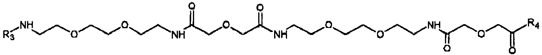
- wherein R3 and R4 are residues of Lys, and
- m, n and q are from 1 to 6.
- Linear polymer cross-Inkers (for example molecules 1.6. and 1.7. described above) bearing several residues of fluorescein, Ferulic acid (1.6) and/or tyrosine (1.7) are also embodiments of reporters that can be deposited by HRP in the presence of another cross-linker, e.g. DAB. Such linear relatively low molecular weight (MW<15 kDa) cross-linkers/reporters may be particular useful when the HRP enzyme moieties are bound to target sites which are not easily accessible or exposed, e.g. in cell nuclei such as DNA. Larger reporter molecules, for examples reporters comprising dextran molecules conjugated with tens and hundreds of labels (e.g. Flu and/or Fer and/or Tyr), such as for example molecule 1.9. (described below), may be used when peroxidase enzyme moieties are located in more easy accessible target sites.
- Dextran MW 70kDa, activated with divinyl sulphone, 10 nmol, was reacted with Fer-(Fer-L30)5-Lys(NH2)-NH2 (compound 1.5) 500 nmol, in a total volume of 300 microL 0.16M NaHCO3 pH 9.5 for 30 min at 40 C. After a slight precipitation was observed, further 100 microL water was added and the reaction was allowed to proceed for another 30 min. Further 200 microL 0.15 M NaHCO3 was added together with 500 nmol H-Cys-L90-Lys(Flu)-L90Lys(Flu)-L90-Lys(Flu)-NH2 (compound 1.4). After 1 h at 40 C, the reaction mixture was quenched by addition of 50 microL 0.165M cystein for 30 min, solution was filtered, and the product was purified by FPLC on superdex 200 with 20% EtOH in aqueous solution containing 10 mM CHES, pH 9.0, and 0.1 M NaCl. The product eluted was a Dextran conjugate comprising around 56 Fluorescein and 113 Ferulic Acid residues.
- 13.7 nmol divinylsulphone were activated 70 kDA MW dextran and reacted with 602 nmol HRP were in 600 microL buffer (100 mM NaCl, 25 mM NaHCO3, pH 9.5) for 3h at 30 C. Then 41.1 nmol Goat-anti-Mouse F(ab)2 antibody in 105 microL water was added, and the reaction was continued for additional 16 h. The reaction mixture was quenched by addition of 70 microL 0.165M cystein for 30 min and the product was purified on superdex 200 in 100mM NaCl, 10 mM HEPES pH 7.2. The eluded product was a Dextran conjugate comprising Goat-anti-Mouse (GaM) and HRP (Ratio dex:GaM:HRP=1:1:1).
- 10 nmol divinylsulphone activated 70 kDA MW dextran and 440 nmol HRP were reacted in 400 microL buffer (100 mM NaCl, 25 mM NaHCO3, pH 9.5) for 3h at 30 C. Then, 30 nmol Anti-Mouse F(ab)2 antibody in 80 microL water was added, and the reaction was continued for additional 90 min at 40 C. The reaction mixture was quenched by addition of 50 microL 0.165M cystein for 30 min and the product was purified on superdex 200 in 100mM NaCl, 10 mM HEPES pH 7.2. The eluded product was a conjugate of Dextran with anti-FITC and HRP (Dex/anti-FITC/HRP Ratio = 1/2/9).
- 10 nmol divinylsulphone activated 70 kDA MW dextran ;440 nmol HRP and 25 nmol F(ab)2 anti-FITC were reacted in 374 microL buffer (100 mM NaCl, 25 mM NaHCO3, pH 9.5) for 16 at 30 C. The reaction mixture was quenched by addition of 50 microL 0.165M cystein for 30 min and the product purified on superdex 200 in 100mM NaCl, 10 mM HEPES pH 7.2. The eluded product was a Dextran conjugate comprising Anti-FITC and HRP (ratio 1:1:1).
- Molecules 1.10, 1.11 and 1.12 represent embodiments of antibody-Dextran-HRP conjugates which may be useful as probes for detection of deposited reporter molecules. Conjugate 1.10. may also be useful as a further probe for the detection of the probes bound to the deposited reporter (e.g. on step (b) of the method for detecting a marker in the target site described above).
- IHC was carried out on formalin fixed paraffin embedded tonsils. 3-5 micron sections were cut, baked and stored at 4 C, until used. Paraffin was then removed by xylene (2x 5min); 99% ethanol (2x 2min); 70% ethanol (2x2min) and finally water. The slides were put in the target retrieval solution, pH 9, (DAKO S2367) then heated in microwave oven (boiled for 10 min). Afterwards, the slides were allowed to cool and then were transferred to the wash buffer (DAKO S3006). The procedure was followed by a step of blocking of endogenous Peroxidase activity with 3% hydrogen peroxide for 5min., then again the slides were transferred into the wash buffer and then stained. To minimize slide to slide variation each comparative experiment was carried out with consecutively cut sections during the same day. The specific as well as background signals were scored using a score scale from 0 to 4 wherein 0 is representing no stain at all, 1 - weakly stained, 2 - moderately stained, 3 - strongly stained, 4 - over stained.
- Cytokeratin, Goat-anti-Mouse-HRP and Anti-FITC-HRP each were diluted in 2% BSA, 0.2% Casein, 2% PEG, 0.1% Tween20, 0.1 M NaCL, 10 mM HEPES, pH 7.2. (BCPT-buffer). Reporter D17128 and DAB (DAKO K5007 C) were diluted in DAB substrate buffer (DAKO K5007 B). All incubations lasted 5 min, followed by 2 min wash in DAKO washing buffer S3006, except for after the incubation with D17128, where washes were performed in 10 mM CHES pH 9 + 0.1% Tween20.
- Table and text below summarize the embodiments of the experiment:
Primary antibody probe Secondary antibody probe Cross-linker and reporter Reporter detection probe Cross-linker and reporter Reporter detection probe Devel oping soluti on Slide 1Cytokeratin (DAKO M5315) 15 nM GaM- HRP, D18033, 125 nM, DAB (DAKO K5007 C) No reporter Slide 4 As slide 1GaM-HRP, D18033 5 nMD17128 100nM + DAB 1:150 D17030 50 nM DAB Slide 7 As slide 1GaM-HRP, D18033 0.5 nM As slide 4As slide 4D17128 100 nM + DAB 1:150 D 17030 50 nM DAB Slide 9 As slide 1GaM-HRP, D18033 0.5 nM D17128 20 nM + DAB 1:150 As slide 4As slide 7As slide 7DAB - Slide 1 - standard DAB-HRP staining (no amplification). Standard DAB DAKO reagent (K7005 C) contains 3mM DAB.
- Slide 4 - deposition of reporter D17128 in the presence of 1mM DAB - GaM-HRP conjugate D18033 diluted 25 times compared to slide 1 - one step amplification
-
Slide 7 and Slide 9 - further dilution of GaM-HRP conjugate D18033 compared to slide 1 and additional step of reporter deposition. -
Slides slides 7 and 9 remained crisp (i.e. non-diffused), with sharp borders between stained and unstained areas indicating that very little diffusion takes place in the deposition sites. - Cytokeratin, GaM-HRP and Anti-FITC-HRP were all diluted in 2% BSA, 0.2% Casein, 2% PEG, 0.1% Tween20, 0.1 M NaCL, 10 mM HEPES, pH 7.2. (BCPT-buffer). D17128 and DAB (DAKO K5007 C) were diluted in DAB substrate buffer (DAKO K5007 B). All incubations were 5 min, followed by 2 min wash in DAKO S3006, except following the incubation with reporter D17128, wherein the washes were performed in 10 mM CHES pH 9 + 0.1% Tween20.
- The table and text below summarize the embodiments of the experiment:
Primary & secondary antibody probes Cross-linker & reporter Reporter probe Developing solution Slide 9 Cytokeratin DAKO M5315 15 nM + GaM-HRP D18033 10 nM D17128 25nM no cross-linker D18058 Anti-FITC-HRP 25 nM DAB (DAKO K5007 C) Slide 12 As slide 9 D17128 25 nM + D17140 100 microM As slide 9 DAB (DAKO K5007 C) Slide 14 As slide 9 D17128 25 nM + DAB 1:150 As slide 9 DAB (DAKO K5007 C) - Slides 12 and 14 were strongly and specifically stained (both scoring 2), whereas slide 9 was not stained (score 0). The results show that the reporter is not precipitated by HRP in the media without a cross-linker (e.g. DAB (slide 14) or D17140 (slide 12)).
- The experiment further illustrates the possibility of reducing the number of steps by pre-incubating (for 10 min) primary antibody with secondary antibody-HRP conjugate.
- Cytokeratin, GaM-HRP and Anti-FITC-HRP each were diluted in 2% BSA, 0.2% Casein, 2% PEG, 0.1% Tween20, 0.1 M NaCL, 10 mM HEPES, pH 7.2. (BCPT-buffer). D17128 and DAB (DAKO K5007 C) were diluted in DAB substrate buffer (DAKO K5007 B). All incubations were 5 min, followed by 2 min wash in DAKO S3006, except following the incubation with D17128 and D18044, wherein washes were performed in 10 mM CHES pH 9 + 0.1 % Tween20. When the third step of incubation of the slides with DAB in substrate buffer was applied (
slides 3 and 5), the incubation time was 1 min. - The table and text below summarize the embodiments of the experiment:
Primary antibody probe Secondary antibody probes Cross-linker and reporter Cross-linker and reporter Reporter probe Developing solution Slide 2 Cytokeratin DAKO M5315 15 nM GaM-HRP, D18033 3 nMD17128 50 nM + DAB 1:150 D18058 Anti-FITC-HRP 50 nM DAB Slide 3 As slide 2As slide 2DAB No reporter D17128 50 nM + DAB 1:150 D18058 Anti-FITC-HRP 50 nM DAB Slide 4 As slide 2As slide 2D17128 50 nM No cross-linker D18058 Anti-FITC-HRP 50 nM DAB Slide 5 As slide 2As slide 2DAB No reporter D17128 50 nM No cross-linker D18058 Anti-FITC-HRP 50 nM DAB -
Slide 2 was the most intensely stained slide (score 3), showing that the one minute of extra application of DAB of the third step on slide 3 (score 2,5) did not much improve, but rather slightly reduced the staining intensity. The lack of staining in absence of cross-linker on slide 4 (score 0) is in accordance with the results obtained in IHC experiment 2 (described above). Onslide 5, where DAB was applied prior to application of the reporter conjugate D17128, a staining was observed (score 2), albeit not quite as intense as onslide 3. - This results show that a cross-linker can be first applied in a separate step before incubating the slide with both reporter and cross-linker (i.e. on pre-incubation step). Another positive effect of applying DAB prior to depositing the labeled reporter observed was a remarkable reduction of the background staining (scored 0.5-1 on
slide 2, and scored 0 on slide 3), while specific signals intensity did not decrease much. Particularly weak diffuse background staining associated with unspecific binding of Goat-anti-Mouse-HRP conjugates, was in many cases virtually eliminated by the 1 min extra DAB step prior to deposition. - Cytokeratin, GaM-HRP and Anti-FITC-HRP were all diluted in 2% BSA, 0.2% Casein, 2% PEG, 0.1% Tween20, 0.1 M NaCL, 10 mM HEPES, pH 7.2. (BCPT-buffer). D17128, D 18044 and DAB (DAKO K5007 C) were diluted in DAB substrate buffer (DAKO K5007 B). All incubations were 5 min, followed by 2 min wash in DAKO S3006, except following incubations with D17128 and D18044, wherein washes were performed in 10 mM CHES pH 9 + 0.1% Tween20.
- The table and text below summarize the embodiments of the experiment:
Primary &secondary antibody probes Preincubation with cross-linker (DAB) Cross-linker (DAB) & reporter Slide 8 Cytokeratin DAKO M5315 15 nM + GaM-HRP D18033 10 nM DAB 1:50 in substrate buffer (DAKO K5007 C), 1 min. D17128 50 nM + DAB 1:50 (DAKO K5007 C) 50 nM D18058 DAB Slide 9 As slide 8As slide 8D17128 50 nM + DAB 1:150 As slide 8As slide 8Slide 10 As slide 8As slide 8D17128 50 nM + DAB 1:500 As slide 8As slide 8Slide 11 As slide 8As slide 8D18044 5 microM + DAB 1:50As slide 8As slide 8Slide 12 As slide 8As slide 8D18044 5 microM + DAB 1:150As slide 8As slide 8Slide 13 As slide 8As slide 8D11804 5 microM + DAB 1:500As slide 8As slide 8 - This experiment illustrates the effect of cross linker concentration on deposition of two different types of reporters, Slide 9 was the most intensely stained slide (score 4). DAB as cross linker was applied on slide 9 at dilution 1:150,(1 mM). A higher (1:50 (3mM), slide 8) or lower (1:500 (0.3mM),slide 10) concentrations of DAB gave slightly less intense staining (score 3.5). The background reducing effect of the pre-incubation with DAB was also visible; none of the six slides (of the above table) had significant background staining.
- The low molecular weight reporters (e.g. reporter 18044 described above) were better deposited at lower concentrations of DAB (slide 13; score 3); the staining intensity decreased with increasing DAB concentrations (slide 12, score 2.5; slide 11, score 2)
- This experiment also illustrates that larger reporter molecules comprising many peroxidase substrate moieties can be used at lower amounts (e.g. Fluorescein-Ferulic Acid Dextran conjugate D17128, 50 nM) compared to smaller reporter molecules comprising the same but fewer peroxidase substrate moieties (e.g. D 18044,
t 5 microM) to achieve the same or even better staining results. - By running samples with and without the use of DAB in the Chromogen solution and the signal enhancer solution the precipitating effect of DAB can be illustrated. Further illustration of the inventions signal amplification can be obtained by further dilution of either the probe, the antibody or by omitting the second Peroxidase-blocking step.
- Tissue: Formalin-fixed Paraffin embedded Tonsil.
Fluorescein labeled probe targeted against the centromer of Chromosome 11. - The following solutions are used in the example:
Antigen retrieval/pre-treatment (Dako K 5599) Washbuffer1 (Dako K5331) Washbuffer2 (Dako S 3006) Pepsin RTU (Dako K 5331) Stringent washbuffer, (Dako K 5331) R-a-Fitc/HRP, F(ab) (Dako P 5100) Antibody diluent (Dako S 2022) Probe (Dako Y 5505) Peroxidase-Blocking solution (Dako S 2023) Nuclear Fast red (Dako S1968) Chromogen buffer (Dako K5007) - 102,5 mg 5-amino-2-[3-[5-amino-1,3-dihydro-3,3-dimethyl-1-(4sulfobutyl)-2H indol-2-ylidene]-1-propenyl]-3,3dimethyl-1-(4sulfobutyl)-3H-Indolium, ter trifluoroacetate is dissolved in 4,525 mL Propanediol/water 8:2
- 59,6
mg 3,3'-Di-aminobenzidine dihydrochloride is dissolved 5,96 mL Propanediol/water 8:2. 13,7 µL 12 M Hydrochloric acid is added. - Solution A and Solution B are mixed in the
ratio 1 mL A: 9 mL B. - For staining these stock solutions were diluted into the chromogen buffer of kits obtained from Dakocytomation code # K5007.
1 mL Solution C is mixed with 19 mL Chromogen buffer - 1 mL Solution B is diluted with 499 mL Chromogen buffer.
- This is a solution of D1734 in 20 % ethanol, 0,1 M NaCl, 10 mM CHES pH 9,0 diluted with Solution D to a concentration of 250 nM or 50 nM of Fer-L30-FITC. In the case of No DAB in the solution dilution was performed directly in Chromogen buffer
- Human Tonsil tissue slides were de-paraffinized, washed and antigen retrieval was performed in 10 min in a microwave oven. After another wash, pepsin treatment at 37 °C and washing, slides where dehydrated and incubated with the PNA probe. After 5 min denaturing of the sample at 85 °C, the probes where hybridized for 1 hour at 45 °C. The slides were stringent washed at 65 °C for 10 min. Peroxidase was blocked for 3 min, the slides were washed and incubated with an Rabbit-anti-FITC/HRP diluted 1:20. After 30 min the slides where washed and incubated with the signal enhancer solution. After 30 min the slides where washed, peroxidase was blocked for 3 min, washed again and the slides were incubated with an Rabbit -anti-FITC/HRP diluted 1/20. After 30 min the slides were washed and incubated with a Chromogen solution. Following washing with water the slides were counterstained with nuclear fast red, washed and mounted.
- The experimental set-up and results of the staining are illustrated in the following table:
Slide #. Probe 1 timeanti-FITC/HRP Antiboby 30 min Signal enhancer solution 30 min anti-FITC/HRP Antiboby 30 min Result Signal- intensity Comments 1 Buffer Anti FITC/HRP 1:50 Fer6-Flu3 50nM with DAB 1:20 0 2 Y5505 Diluted 10 x 1:100 Fer6-Flu3 50nM 1:20 0 3 Y5505 Fortyndet 50 x 1:100 Fer6-Flu3 50nM with DAB 1:20 1½ No DAB in the chromogen solution 4 Y5505 Diluted 10 x 1:100 Fer6-Flu3 50nM with DAB 1:20 2½ 5 Y5505 Diluted 10 x 1:100 Fer6-Flu3 250nM with DAB 1:20 4 Over stained/ to much signal 6 Y5505 Diluted 10 x 1:100 Fer6-Flu3 250nM 1:20 0-½ 7 Y5505 Fortyndet 10x 1:100 No amplification stained directly after first incubation with anti FITC/HRP 0 -
Slide 1 is a control showing probe is need to get a signal -
Slide 2 shows that without a cross-linker in the signal enhancing solution no specific signal is obtained -
Slide 3 shows that adding a cross-linker (DAB) to the signal enhancing solution results in high signal amplification. This is the only slide that did not have DAB in the chromogen solution. -
Slide 4 shows that adding a cross-linker to the chromogen solution further enhances the signal (compared to slide 3) -
Slide 6 shows that by increasing the concentration of the signal enhancer (D17134) a weak signal can be obtained without a crosslinker. However adding a cross-linker gives a strong amplification of this signal (as illustrated by slide 5)
Claims (17)
- A method of deposition of a reporter in a target site, wherein the target site comprises peroxidase enzymatic activity, and the method comprises a step of incubating said target site in an aqueous media comprising the reporter, a peroxidase compound and a cross-linker, which is 3,3'-diaminobenzidine (DAB),
wherein said method is characterized in that the reporter is a conjugate molecule comprising at least one backbone polymer and at least one detectable label, wherein said at least one detectable label is linked to the at least one backbone polymer via a chemical bond or via a linker molecule, wherein the at least one detectable label is not DAB. - The method according to claim 1, wherein the at least one detectable label is selected from a fluorescent or chromogenic substance, enzyme substrate or member of a specific binding pair.
- The method according to claim 2, wherein the at least one detectable label is a fluorescent substance.
- The method according to claim 2, wherein the at least one detectable label is a hapten.
- The method according to claim 2, wherein the at least one detectable label that is a horse radish peroxidase substrate (HRP).
- The method according to any of claims 1 to 5, wherein the reporter comprises a combination of two or more different detectable labels.
- The method according to claim 1 or 6, wherein the at least on backbone polymer of the reporter comprises dextran.
- The method according to any of the preceding claims, wherein the peroxidase activity is associated with at least one moiety of a peroxidase enzyme present in the target site.
- The method of claim 8, wherein the peroxidase enzyme is selected from horse radish peroxidase (HRP) or soybean peroxidase (SP).
- The method according to any of the preceding claims, wherein the target site comprises a biological marker.
- A method of detecting a biological marker in a target site of a biological sample in vitro, wherein the target site comprises the biological marker and peroxidase enzymatic activity; wherein the method is characterized in that it comprises the step of deposition of a reporter in the target site according to the method of any claims 1-10.
- The method of claim 11, comprising steps ofa) incubating the biological sample presumably comprising the biological marker with one or more probes, wherein (i) least one of the one or more probes comprises at least one moiety of horse radish peroxidase (HRP), and (ii) at least one of the one or more probes recognizes and specifically binds to the biological marker, thereby forming a complex of the biological marker with the one or more probes, wherein at least one probe comprises at least one moiety of HRP, thereby forming a target site;b) incubating the sample comprising the target site of (a) in an aqueous media comprising DAB, a reporter and a peroxide compound, thereby depositing the reporter in the target site;c) detecting the deposited reporter of (b) and thereby detecting the biological marker.
- The method of claim 11 or 12, wherein the biological marker is a biological molecule, structure, such as a membrane or chromosomal structure, molecular complex or particle, such as a virus particle.
- The method of claim 12 or 13, wherein the one or more probe(s) is(are) a member(s) of a specific binding pair(s).
- The method of claim 14, wherein the members of specific binding pairs are selected from receptor-ligand, antibody-antibody, two-nucleic acids or two-nucleic acid analogs specific binding pairs.
- The method of any of claims 11-15, wherein the method is used for immunocytochemical (IHC) or in situ hybridization (ISH) detection of a biological marker in a target molecule comprised by a cell.
- The method of claim 16, wherein the method is automated or semi-automated.
Applications Claiming Priority (2)
| Application Number | Priority Date | Filing Date | Title |
|---|---|---|---|
| US99420607P | 2007-09-18 | 2007-09-18 | |
| PCT/DK2008/000327 WO2009036760A2 (en) | 2007-09-18 | 2008-09-16 | A rapid and sensitive method for detection of biological targets |
Publications (2)
| Publication Number | Publication Date |
|---|---|
| EP2201058A2 EP2201058A2 (en) | 2010-06-30 |
| EP2201058B1 true EP2201058B1 (en) | 2017-04-12 |
Family
ID=40121988
Family Applications (1)
| Application Number | Title | Priority Date | Filing Date |
|---|---|---|---|
| EP08801362.8A Active EP2201058B1 (en) | 2007-09-18 | 2008-09-16 | A rapid and sensitive method for detection of biological targets |
Country Status (7)
| Country | Link |
|---|---|
| US (2) | US9377465B2 (en) |
| EP (1) | EP2201058B1 (en) |
| JP (1) | JP5639889B2 (en) |
| CN (1) | CN101836117B (en) |
| CA (1) | CA2698390C (en) |
| ES (1) | ES2624450T3 (en) |
| WO (1) | WO2009036760A2 (en) |
Families Citing this family (20)
| Publication number | Priority date | Publication date | Assignee | Title |
|---|---|---|---|---|
| JP5721638B2 (en) * | 2009-02-19 | 2015-05-20 | ダコ・デンマーク・エー/エス | Conjugate molecule |
| CA2777411C (en) * | 2009-10-20 | 2018-06-19 | Dako Denmark A/S | Immunochemical detection of single target entities |
| NZ704322A (en) | 2010-07-06 | 2016-07-29 | Interna Technologies Bv | Mirna and its diagnostic and therapeutic uses in diseases or conditions associated with melanoma, or in diseases or conditions associated with activated braf pathway |
| CA2816472A1 (en) | 2010-11-08 | 2012-05-18 | Dako Denmark A/S | Quantification of single target molecules in histological samples |
| CA2817882C (en) | 2010-11-17 | 2022-08-23 | Asuragen, Inc. | Mirnas as biomarkers for distinguishing benign from malignant thyroid neoplasms |
| CA2817161C (en) | 2010-12-06 | 2019-04-02 | Dako Denmark A/S | Combined histological stain |
| CN102565411B (en) * | 2010-12-08 | 2014-05-28 | 中国科学院生物物理研究所 | New method for tracing magnetic nano granules |
| US9366675B2 (en) | 2011-04-19 | 2016-06-14 | Dako Denmark A/S | Method for enzyme-mediated signal amplification |
| US8815532B2 (en) * | 2011-06-07 | 2014-08-26 | Diagnostic Innovations, Llc | Color-producing diagnostic systems, reagents and methods |
| ES2613477T3 (en) * | 2011-11-08 | 2017-05-24 | Dako Denmark A/S | New method for the evaluation of a target in a histological sample |
| AU2013240090B2 (en) | 2012-03-27 | 2017-01-05 | Ventana Medical Systems, Inc. | Signaling conjugates and methods of use |
| EP2970909A4 (en) | 2013-03-15 | 2017-02-15 | The University of Chicago | Methods and compositions related to t-cell activity |
| US9890417B2 (en) | 2014-11-03 | 2018-02-13 | Agilent Technologies, Inc. | Signal amplification of fluorescence in situ hybridization |
| US10839509B2 (en) | 2015-07-10 | 2020-11-17 | 3Scan Inc. | Spatial multiplexing of histological stains |
| US11360098B2 (en) * | 2015-09-28 | 2022-06-14 | Quest Diagnostics Investments Llc | Amyloid beta detection by mass spectrometry |
| WO2017103678A2 (en) | 2015-12-18 | 2017-06-22 | Dako Denmark A/S | Chromogenic peroxidase substrates |
| CN105987839B (en) * | 2016-06-03 | 2018-09-28 | 浙江世纪康大医疗科技股份有限公司 | A kind of EDTA- citric acid antigens reparation liquid |
| CN105987840B (en) * | 2016-06-03 | 2018-09-28 | 浙江世纪康大医疗科技股份有限公司 | A kind of citric acid antigen reparation liquid |
| CN109085338B (en) * | 2018-09-13 | 2021-07-13 | 武汉伊莱瑞特生物科技股份有限公司 | ECL substrate solution and preparation method and application thereof |
| US11237170B2 (en) | 2018-12-04 | 2022-02-01 | Aat Bioquest, Inc. | Styryl phenols, derivatives and their use in methods of analyte detection |
Family Cites Families (16)
| Publication number | Priority date | Publication date | Assignee | Title |
|---|---|---|---|---|
| ATE243754T1 (en) | 1987-05-21 | 2003-07-15 | Micromet Ag | MULTIFUNCTIONAL PROTEINS WITH PREDEFINED TARGET |
| DE768377T1 (en) | 1988-09-02 | 1998-01-02 | Dyax Corp | Production and selection of recombinant proteins with different binding sites |
| ATE102631T1 (en) | 1988-11-11 | 1994-03-15 | Medical Res Council | CLONING OF IMMUNOGLOBULIN SEQUENCES FROM THE VARIABLE DOMAINS. |
| US5530101A (en) | 1988-12-28 | 1996-06-25 | Protein Design Labs, Inc. | Humanized immunoglobulins |
| US5196306A (en) | 1989-03-29 | 1993-03-23 | E. I. Du Pont De Nemours And Company | Method for the detection or quantitation of an analyte using an analyte dependent enzyme activation system |
| GB9015198D0 (en) | 1990-07-10 | 1990-08-29 | Brien Caroline J O | Binding substance |
| US5688966A (en) | 1996-07-26 | 1997-11-18 | E. I. Du Pont De Nemours And Company | Compounds and method for synthesizing sulfoindocyanine dyes |
| US5863748A (en) | 1997-03-14 | 1999-01-26 | New Life Science Products, Inc. | p-Hydroxycinnamoyl-containing substrates for an analyte dependent enzyme activation system |
| IL123451A (en) | 1998-02-25 | 2003-07-06 | Ehud Keinan | Method and kit for the detection of peroxide-based explosives |
| US6372937B1 (en) * | 1998-11-09 | 2002-04-16 | Mark Norman Bobrow | Enhanced catalyzed reporter deposition |
| US6531319B1 (en) | 2000-05-10 | 2003-03-11 | Keshab D. Pant | Method of detecting colon cancer |
| US6670113B2 (en) | 2001-03-30 | 2003-12-30 | Nanoprobes | Enzymatic deposition and alteration of metals |
| US6617125B2 (en) | 2001-06-29 | 2003-09-09 | Perkinelmer Life Sciences, Inc. | Compositions for enhanced catalyzed reporter deposition |
| US9017992B2 (en) | 2005-07-01 | 2015-04-28 | Dako Denmark A/S | In situ hybridization detection method |
| JP5721638B2 (en) * | 2009-02-19 | 2015-05-20 | ダコ・デンマーク・エー/エス | Conjugate molecule |
| CA2777411C (en) * | 2009-10-20 | 2018-06-19 | Dako Denmark A/S | Immunochemical detection of single target entities |
-
2008
- 2008-09-16 EP EP08801362.8A patent/EP2201058B1/en active Active
- 2008-09-16 CN CN200880112656.5A patent/CN101836117B/en active Active
- 2008-09-16 US US12/678,937 patent/US9377465B2/en active Active
- 2008-09-16 JP JP2010524354A patent/JP5639889B2/en active Active
- 2008-09-16 WO PCT/DK2008/000327 patent/WO2009036760A2/en active Application Filing
- 2008-09-16 ES ES08801362.8T patent/ES2624450T3/en active Active
- 2008-09-16 CA CA2698390A patent/CA2698390C/en not_active Expired - Fee Related
-
2016
- 2016-05-26 US US15/165,823 patent/US10168335B2/en active Active
Non-Patent Citations (1)
| Title |
|---|
| None * |
Also Published As
| Publication number | Publication date |
|---|---|
| US20100285467A1 (en) | 2010-11-11 |
| US10168335B2 (en) | 2019-01-01 |
| CA2698390C (en) | 2018-05-22 |
| CN101836117A (en) | 2010-09-15 |
| JP2010539456A (en) | 2010-12-16 |
| US9377465B2 (en) | 2016-06-28 |
| WO2009036760A3 (en) | 2009-07-23 |
| CN101836117B (en) | 2014-07-23 |
| EP2201058A2 (en) | 2010-06-30 |
| JP5639889B2 (en) | 2014-12-10 |
| CA2698390A1 (en) | 2009-03-26 |
| US20160370375A1 (en) | 2016-12-22 |
| WO2009036760A2 (en) | 2009-03-26 |
| ES2624450T3 (en) | 2017-07-14 |
Similar Documents
| Publication | Publication Date | Title |
|---|---|---|
| US10168335B2 (en) | Rapid and sensitive method for detection of biological targets | |
| US8435735B2 (en) | Methods and compounds for detection of molecular targets | |
| US9091691B2 (en) | Immunochemical detection of single target entities | |
| US9958449B2 (en) | Method for enzyme-mediated signal amplification |
Legal Events
| Date | Code | Title | Description |
|---|---|---|---|
| PUAI | Public reference made under article 153(3) epc to a published international application that has entered the european phase |
Free format text: ORIGINAL CODE: 0009012 |
|
| 17P | Request for examination filed |
Effective date: 20100415 |
|
| AK | Designated contracting states |
Kind code of ref document: A2 Designated state(s): AT BE BG CH CY CZ DE DK EE ES FI FR GB GR HR HU IE IS IT LI LT LU LV MC MT NL NO PL PT RO SE SI SK TR |
|
| AX | Request for extension of the european patent |
Extension state: AL BA MK RS |
|
| RIN1 | Information on inventor provided before grant (corrected) |
Inventor name: PETERSEN, KENNETH HEESCHE Inventor name: LOHSE, JESPER |
|
| 17Q | First examination report despatched |
Effective date: 20120411 |
|
| DAX | Request for extension of the european patent (deleted) | ||
| REG | Reference to a national code |
Ref country code: DE Ref legal event code: R079 Ref document number: 602008049744 Country of ref document: DE Free format text: PREVIOUS MAIN CLASS: C08G0069020000 Ipc: C12Q0001280000 |
|
| RIC1 | Information provided on ipc code assigned before grant |
Ipc: G01N 33/58 20060101ALI20160705BHEP Ipc: C12Q 1/28 20060101AFI20160705BHEP |
|
| GRAP | Despatch of communication of intention to grant a patent |
Free format text: ORIGINAL CODE: EPIDOSNIGR1 |
|
| INTG | Intention to grant announced |
Effective date: 20161028 |
|
| GRAS | Grant fee paid |
Free format text: ORIGINAL CODE: EPIDOSNIGR3 |
|
| STAA | Information on the status of an ep patent application or granted ep patent |
Free format text: STATUS: GRANT OF PATENT IS INTENDED |
|
| GRAA | (expected) grant |
Free format text: ORIGINAL CODE: 0009210 |
|
| STAA | Information on the status of an ep patent application or granted ep patent |
Free format text: STATUS: THE PATENT HAS BEEN GRANTED |
|
| AK | Designated contracting states |
Kind code of ref document: B1 Designated state(s): AT BE BG CH CY CZ DE DK EE ES FI FR GB GR HR HU IE IS IT LI LT LU LV MC MT NL NO PL PT RO SE SI SK TR |
|
| REG | Reference to a national code |
Ref country code: GB Ref legal event code: FG4D |
|
| REG | Reference to a national code |
Ref country code: CH Ref legal event code: EP |
|
| REG | Reference to a national code |
Ref country code: IE Ref legal event code: FG4D |
|
| REG | Reference to a national code |
Ref country code: AT Ref legal event code: REF Ref document number: 883929 Country of ref document: AT Kind code of ref document: T Effective date: 20170515 |
|
| REG | Reference to a national code |
Ref country code: DE Ref legal event code: R096 Ref document number: 602008049744 Country of ref document: DE |
|
| REG | Reference to a national code |
Ref country code: NL Ref legal event code: FP |
|
| REG | Reference to a national code |
Ref country code: ES Ref legal event code: FG2A Ref document number: 2624450 Country of ref document: ES Kind code of ref document: T3 Effective date: 20170714 |
|
| REG | Reference to a national code |
Ref country code: FR Ref legal event code: PLFP Year of fee payment: 10 |
|
| REG | Reference to a national code |
Ref country code: LT Ref legal event code: MG4D |
|
| REG | Reference to a national code |
Ref country code: AT Ref legal event code: MK05 Ref document number: 883929 Country of ref document: AT Kind code of ref document: T Effective date: 20170412 |
|
| PG25 | Lapsed in a contracting state [announced via postgrant information from national office to epo] |
Ref country code: HR Free format text: LAPSE BECAUSE OF FAILURE TO SUBMIT A TRANSLATION OF THE DESCRIPTION OR TO PAY THE FEE WITHIN THE PRESCRIBED TIME-LIMIT Effective date: 20170412 Ref country code: AT Free format text: LAPSE BECAUSE OF FAILURE TO SUBMIT A TRANSLATION OF THE DESCRIPTION OR TO PAY THE FEE WITHIN THE PRESCRIBED TIME-LIMIT Effective date: 20170412 Ref country code: GR Free format text: LAPSE BECAUSE OF FAILURE TO SUBMIT A TRANSLATION OF THE DESCRIPTION OR TO PAY THE FEE WITHIN THE PRESCRIBED TIME-LIMIT Effective date: 20170713 Ref country code: FI Free format text: LAPSE BECAUSE OF FAILURE TO SUBMIT A TRANSLATION OF THE DESCRIPTION OR TO PAY THE FEE WITHIN THE PRESCRIBED TIME-LIMIT Effective date: 20170412 Ref country code: LT Free format text: LAPSE BECAUSE OF FAILURE TO SUBMIT A TRANSLATION OF THE DESCRIPTION OR TO PAY THE FEE WITHIN THE PRESCRIBED TIME-LIMIT Effective date: 20170412 Ref country code: NO Free format text: LAPSE BECAUSE OF FAILURE TO SUBMIT A TRANSLATION OF THE DESCRIPTION OR TO PAY THE FEE WITHIN THE PRESCRIBED TIME-LIMIT Effective date: 20170712 |
|
| PG25 | Lapsed in a contracting state [announced via postgrant information from national office to epo] |
Ref country code: PL Free format text: LAPSE BECAUSE OF FAILURE TO SUBMIT A TRANSLATION OF THE DESCRIPTION OR TO PAY THE FEE WITHIN THE PRESCRIBED TIME-LIMIT Effective date: 20170412 Ref country code: SE Free format text: LAPSE BECAUSE OF FAILURE TO SUBMIT A TRANSLATION OF THE DESCRIPTION OR TO PAY THE FEE WITHIN THE PRESCRIBED TIME-LIMIT Effective date: 20170412 Ref country code: IS Free format text: LAPSE BECAUSE OF FAILURE TO SUBMIT A TRANSLATION OF THE DESCRIPTION OR TO PAY THE FEE WITHIN THE PRESCRIBED TIME-LIMIT Effective date: 20170812 Ref country code: LV Free format text: LAPSE BECAUSE OF FAILURE TO SUBMIT A TRANSLATION OF THE DESCRIPTION OR TO PAY THE FEE WITHIN THE PRESCRIBED TIME-LIMIT Effective date: 20170412 Ref country code: BG Free format text: LAPSE BECAUSE OF FAILURE TO SUBMIT A TRANSLATION OF THE DESCRIPTION OR TO PAY THE FEE WITHIN THE PRESCRIBED TIME-LIMIT Effective date: 20170712 |
|
| REG | Reference to a national code |
Ref country code: DE Ref legal event code: R097 Ref document number: 602008049744 Country of ref document: DE |
|
| PG25 | Lapsed in a contracting state [announced via postgrant information from national office to epo] |
Ref country code: DK Free format text: LAPSE BECAUSE OF FAILURE TO SUBMIT A TRANSLATION OF THE DESCRIPTION OR TO PAY THE FEE WITHIN THE PRESCRIBED TIME-LIMIT Effective date: 20170412 Ref country code: RO Free format text: LAPSE BECAUSE OF FAILURE TO SUBMIT A TRANSLATION OF THE DESCRIPTION OR TO PAY THE FEE WITHIN THE PRESCRIBED TIME-LIMIT Effective date: 20170412 Ref country code: EE Free format text: LAPSE BECAUSE OF FAILURE TO SUBMIT A TRANSLATION OF THE DESCRIPTION OR TO PAY THE FEE WITHIN THE PRESCRIBED TIME-LIMIT Effective date: 20170412 Ref country code: CZ Free format text: LAPSE BECAUSE OF FAILURE TO SUBMIT A TRANSLATION OF THE DESCRIPTION OR TO PAY THE FEE WITHIN THE PRESCRIBED TIME-LIMIT Effective date: 20170412 Ref country code: SK Free format text: LAPSE BECAUSE OF FAILURE TO SUBMIT A TRANSLATION OF THE DESCRIPTION OR TO PAY THE FEE WITHIN THE PRESCRIBED TIME-LIMIT Effective date: 20170412 |
|
| PLBE | No opposition filed within time limit |
Free format text: ORIGINAL CODE: 0009261 |
|
| STAA | Information on the status of an ep patent application or granted ep patent |
Free format text: STATUS: NO OPPOSITION FILED WITHIN TIME LIMIT |
|
| 26N | No opposition filed |
Effective date: 20180115 |
|
| REG | Reference to a national code |
Ref country code: CH Ref legal event code: PL |
|
| PG25 | Lapsed in a contracting state [announced via postgrant information from national office to epo] |
Ref country code: SI Free format text: LAPSE BECAUSE OF FAILURE TO SUBMIT A TRANSLATION OF THE DESCRIPTION OR TO PAY THE FEE WITHIN THE PRESCRIBED TIME-LIMIT Effective date: 20170412 Ref country code: MC Free format text: LAPSE BECAUSE OF FAILURE TO SUBMIT A TRANSLATION OF THE DESCRIPTION OR TO PAY THE FEE WITHIN THE PRESCRIBED TIME-LIMIT Effective date: 20170412 |
|
| REG | Reference to a national code |
Ref country code: IE Ref legal event code: MM4A |
|
| REG | Reference to a national code |
Ref country code: BE Ref legal event code: MM Effective date: 20170930 |
|
| PG25 | Lapsed in a contracting state [announced via postgrant information from national office to epo] |
Ref country code: LU Free format text: LAPSE BECAUSE OF NON-PAYMENT OF DUE FEES Effective date: 20170916 |
|
| PG25 | Lapsed in a contracting state [announced via postgrant information from national office to epo] |
Ref country code: IE Free format text: LAPSE BECAUSE OF NON-PAYMENT OF DUE FEES Effective date: 20170916 Ref country code: LI Free format text: LAPSE BECAUSE OF NON-PAYMENT OF DUE FEES Effective date: 20170930 Ref country code: CH Free format text: LAPSE BECAUSE OF NON-PAYMENT OF DUE FEES Effective date: 20170930 |
|
| REG | Reference to a national code |
Ref country code: FR Ref legal event code: PLFP Year of fee payment: 11 |
|
| PG25 | Lapsed in a contracting state [announced via postgrant information from national office to epo] |
Ref country code: BE Free format text: LAPSE BECAUSE OF NON-PAYMENT OF DUE FEES Effective date: 20170930 |
|
| PG25 | Lapsed in a contracting state [announced via postgrant information from national office to epo] |
Ref country code: MT Free format text: LAPSE BECAUSE OF NON-PAYMENT OF DUE FEES Effective date: 20170916 |
|
| PGFP | Annual fee paid to national office [announced via postgrant information from national office to epo] |
Ref country code: ES Payment date: 20181001 Year of fee payment: 11 |
|
| PG25 | Lapsed in a contracting state [announced via postgrant information from national office to epo] |
Ref country code: HU Free format text: LAPSE BECAUSE OF FAILURE TO SUBMIT A TRANSLATION OF THE DESCRIPTION OR TO PAY THE FEE WITHIN THE PRESCRIBED TIME-LIMIT; INVALID AB INITIO Effective date: 20080916 |
|
| PG25 | Lapsed in a contracting state [announced via postgrant information from national office to epo] |
Ref country code: CY Free format text: LAPSE BECAUSE OF NON-PAYMENT OF DUE FEES Effective date: 20170412 |
|
| PG25 | Lapsed in a contracting state [announced via postgrant information from national office to epo] |
Ref country code: TR Free format text: LAPSE BECAUSE OF FAILURE TO SUBMIT A TRANSLATION OF THE DESCRIPTION OR TO PAY THE FEE WITHIN THE PRESCRIBED TIME-LIMIT Effective date: 20170412 |
|
| PG25 | Lapsed in a contracting state [announced via postgrant information from national office to epo] |
Ref country code: PT Free format text: LAPSE BECAUSE OF FAILURE TO SUBMIT A TRANSLATION OF THE DESCRIPTION OR TO PAY THE FEE WITHIN THE PRESCRIBED TIME-LIMIT Effective date: 20170412 |
|
| REG | Reference to a national code |
Ref country code: NL Ref legal event code: PD Owner name: AGILENT TECHNOLOGIES, INC.; US Free format text: DETAILS ASSIGNMENT: CHANGE OF OWNER(S), CHANGE OF LEGAL ENTITY; FORMER OWNER NAME: DAKO DENMARK APS Effective date: 20201030 |
|
| REG | Reference to a national code |
Ref country code: ES Ref legal event code: FD2A Effective date: 20210128 |
|
| REG | Reference to a national code |
Ref country code: DE Ref legal event code: R082 Ref document number: 602008049744 Country of ref document: DE Representative=s name: GRUENECKER PATENT- UND RECHTSANWAELTE PARTG MB, DE Ref country code: DE Ref legal event code: R081 Ref document number: 602008049744 Country of ref document: DE Owner name: AGILENT TECHNOLOGIES, INC. (N.D.GES.D. STAATES, US Free format text: FORMER OWNER: DAKO DENMARK A/S, GLOSTRUP, DK |
|
| PG25 | Lapsed in a contracting state [announced via postgrant information from national office to epo] |
Ref country code: ES Free format text: LAPSE BECAUSE OF NON-PAYMENT OF DUE FEES Effective date: 20190917 |
|
| P01 | Opt-out of the competence of the unified patent court (upc) registered |
Effective date: 20230527 |
|
| PGFP | Annual fee paid to national office [announced via postgrant information from national office to epo] |
Ref country code: NL Payment date: 20230915 Year of fee payment: 16 Ref country code: IT Payment date: 20230913 Year of fee payment: 16 Ref country code: GB Payment date: 20230831 Year of fee payment: 16 |
|
| PGFP | Annual fee paid to national office [announced via postgrant information from national office to epo] |
Ref country code: FR Payment date: 20230911 Year of fee payment: 16 Ref country code: DE Payment date: 20230906 Year of fee payment: 16 |