EP2129397B1 - Methods and compositions for treating tumor diseases - Google Patents
Methods and compositions for treating tumor diseases Download PDFInfo
- Publication number
- EP2129397B1 EP2129397B1 EP08726281.2A EP08726281A EP2129397B1 EP 2129397 B1 EP2129397 B1 EP 2129397B1 EP 08726281 A EP08726281 A EP 08726281A EP 2129397 B1 EP2129397 B1 EP 2129397B1
- Authority
- EP
- European Patent Office
- Prior art keywords
- igf
- antibody
- subject
- tumor
- cell
- Prior art date
- Legal status (The legal status is an assumption and is not a legal conclusion. Google has not performed a legal analysis and makes no representation as to the accuracy of the status listed.)
- Active
Links
- 206010028980 Neoplasm Diseases 0.000 title claims description 255
- 238000000034 method Methods 0.000 title claims description 195
- 239000000203 mixture Substances 0.000 title claims description 52
- 210000004027 cell Anatomy 0.000 claims description 299
- 241000282414 Homo sapiens Species 0.000 claims description 153
- 108090000623 proteins and genes Proteins 0.000 claims description 120
- 239000003112 inhibitor Substances 0.000 claims description 92
- 230000011664 signaling Effects 0.000 claims description 74
- 108010031794 IGF Type 1 Receptor Proteins 0.000 claims description 55
- 102000038455 IGF Type 1 Receptor Human genes 0.000 claims description 55
- AOJJSUZBOXZQNB-TZSSRYMLSA-N Doxorubicin Chemical compound O([C@H]1C[C@@](O)(CC=2C(O)=C3C(=O)C=4C=CC=C(C=4C(=O)C3=C(O)C=21)OC)C(=O)CO)[C@H]1C[C@H](N)[C@H](O)[C@H](C)O1 AOJJSUZBOXZQNB-TZSSRYMLSA-N 0.000 claims description 22
- 230000005855 radiation Effects 0.000 claims description 19
- 229960004528 vincristine Drugs 0.000 claims description 16
- OGWKCGZFUXNPDA-XQKSVPLYSA-N vincristine Chemical compound C([N@]1C[C@@H](C[C@]2(C(=O)OC)C=3C(=CC4=C([C@]56[C@H]([C@@]([C@H](OC(C)=O)[C@]7(CC)C=CCN([C@H]67)CC5)(O)C(=O)OC)N4C=O)C=3)OC)C[C@@](C1)(O)CC)CC1=C2NC2=CC=CC=C12 OGWKCGZFUXNPDA-XQKSVPLYSA-N 0.000 claims description 16
- OGWKCGZFUXNPDA-UHFFFAOYSA-N vincristine Natural products C1C(CC)(O)CC(CC2(C(=O)OC)C=3C(=CC4=C(C56C(C(C(OC(C)=O)C7(CC)C=CCN(C67)CC5)(O)C(=O)OC)N4C=O)C=3)OC)CN1CCC1=C2NC2=CC=CC=C12 OGWKCGZFUXNPDA-UHFFFAOYSA-N 0.000 claims description 16
- CMSMOCZEIVJLDB-UHFFFAOYSA-N Cyclophosphamide Chemical compound ClCCN(CCCl)P1(=O)NCCCO1 CMSMOCZEIVJLDB-UHFFFAOYSA-N 0.000 claims description 13
- RJURFGZVJUQBHK-UHFFFAOYSA-N actinomycin D Natural products CC1OC(=O)C(C(C)C)N(C)C(=O)CN(C)C(=O)C2CCCN2C(=O)C(C(C)C)NC(=O)C1NC(=O)C1=C(N)C(=O)C(C)=C2OC(C(C)=CC=C3C(=O)NC4C(=O)NC(C(N5CCCC5C(=O)N(C)CC(=O)N(C)C(C(C)C)C(=O)OC4C)=O)C(C)C)=C3N=C21 RJURFGZVJUQBHK-UHFFFAOYSA-N 0.000 claims description 12
- 210000004072 lung Anatomy 0.000 claims description 12
- 150000001875 compounds Chemical class 0.000 claims description 11
- SDUQYLNIPVEERB-QPPQHZFASA-N gemcitabine Chemical compound O=C1N=C(N)C=CN1[C@H]1C(F)(F)[C@H](O)[C@@H](CO)O1 SDUQYLNIPVEERB-QPPQHZFASA-N 0.000 claims description 11
- 229960005277 gemcitabine Drugs 0.000 claims description 11
- 208000006168 Ewing Sarcoma Diseases 0.000 claims description 10
- -1 cisplatinum Chemical compound 0.000 claims description 10
- HOMGKSMUEGBAAB-UHFFFAOYSA-N ifosfamide Chemical compound ClCCNP1(=O)OCCCN1CCCl HOMGKSMUEGBAAB-UHFFFAOYSA-N 0.000 claims description 9
- 229960001101 ifosfamide Drugs 0.000 claims description 9
- 208000034951 Genetic Translocation Diseases 0.000 claims description 8
- 229960004397 cyclophosphamide Drugs 0.000 claims description 8
- 239000002246 antineoplastic agent Substances 0.000 claims description 7
- 238000002512 chemotherapy Methods 0.000 claims description 7
- UCFGDBYHRUNTLO-QHCPKHFHSA-N topotecan Chemical compound C1=C(O)C(CN(C)C)=C2C=C(CN3C4=CC5=C(C3=O)COC(=O)[C@]5(O)CC)C4=NC2=C1 UCFGDBYHRUNTLO-QHCPKHFHSA-N 0.000 claims description 7
- 229960000303 topotecan Drugs 0.000 claims description 7
- 208000031404 Chromosome Aberrations Diseases 0.000 claims description 6
- 108010092160 Dactinomycin Proteins 0.000 claims description 6
- RJURFGZVJUQBHK-IIXSONLDSA-N actinomycin D Chemical compound C[C@H]1OC(=O)[C@H](C(C)C)N(C)C(=O)CN(C)C(=O)[C@@H]2CCCN2C(=O)[C@@H](C(C)C)NC(=O)[C@H]1NC(=O)C1=C(N)C(=O)C(C)=C2OC(C(C)=CC=C3C(=O)N[C@@H]4C(=O)N[C@@H](C(N5CCC[C@H]5C(=O)N(C)CC(=O)N(C)[C@@H](C(C)C)C(=O)O[C@@H]4C)=O)C(C)C)=C3N=C21 RJURFGZVJUQBHK-IIXSONLDSA-N 0.000 claims description 6
- 229940127089 cytotoxic agent Drugs 0.000 claims description 6
- 229960000640 dactinomycin Drugs 0.000 claims description 6
- 229960004679 doxorubicin Drugs 0.000 claims description 6
- 229960005420 etoposide Drugs 0.000 claims description 6
- VJJPUSNTGOMMGY-MRVIYFEKSA-N etoposide Chemical compound COC1=C(O)C(OC)=CC([C@@H]2C3=CC=4OCOC=4C=C3[C@@H](O[C@H]3[C@@H]([C@@H](O)[C@@H]4O[C@H](C)OC[C@H]4O3)O)[C@@H]3[C@@H]2C(OC3)=O)=C1 VJJPUSNTGOMMGY-MRVIYFEKSA-N 0.000 claims description 6
- 229940009456 adriamycin Drugs 0.000 claims description 5
- 230000008707 rearrangement Effects 0.000 claims description 5
- COVZYZSDYWQREU-UHFFFAOYSA-N Busulfan Chemical compound CS(=O)(=O)OCCCCOS(C)(=O)=O COVZYZSDYWQREU-UHFFFAOYSA-N 0.000 claims description 4
- ZDZOTLJHXYCWBA-VCVYQWHSSA-N N-debenzoyl-N-(tert-butoxycarbonyl)-10-deacetyltaxol Chemical compound O([C@H]1[C@H]2[C@@](C([C@H](O)C3=C(C)[C@@H](OC(=O)[C@H](O)[C@@H](NC(=O)OC(C)(C)C)C=4C=CC=CC=4)C[C@]1(O)C3(C)C)=O)(C)[C@@H](O)C[C@H]1OC[C@]12OC(=O)C)C(=O)C1=CC=CC=C1 ZDZOTLJHXYCWBA-VCVYQWHSSA-N 0.000 claims description 4
- 229960002092 busulfan Drugs 0.000 claims description 4
- SGDBTWWWUNNDEQ-LBPRGKRZSA-N melphalan Chemical compound OC(=O)[C@@H](N)CC1=CC=C(N(CCCl)CCCl)C=C1 SGDBTWWWUNNDEQ-LBPRGKRZSA-N 0.000 claims description 4
- 229960001924 melphalan Drugs 0.000 claims description 4
- 229940063683 taxotere Drugs 0.000 claims description 4
- 102000024905 CD99 Human genes 0.000 claims description 2
- 108060001253 CD99 Proteins 0.000 claims description 2
- 210000003958 hematopoietic stem cell Anatomy 0.000 claims description 2
- 102000025171 antigen binding proteins Human genes 0.000 description 162
- 108091000831 antigen binding proteins Proteins 0.000 description 162
- 238000011282 treatment Methods 0.000 description 144
- 108020004414 DNA Proteins 0.000 description 143
- 108090000765 processed proteins & peptides Proteins 0.000 description 131
- 238000009739 binding Methods 0.000 description 130
- 230000027455 binding Effects 0.000 description 125
- 108090001117 Insulin-Like Growth Factor II Proteins 0.000 description 109
- 102000004196 processed proteins & peptides Human genes 0.000 description 109
- 108090000723 Insulin-Like Growth Factor I Proteins 0.000 description 106
- 102000048143 Insulin-Like Growth Factor II Human genes 0.000 description 104
- 102000004218 Insulin-Like Growth Factor I Human genes 0.000 description 102
- 229920001184 polypeptide Polymers 0.000 description 98
- 102000004169 proteins and genes Human genes 0.000 description 77
- 235000018102 proteins Nutrition 0.000 description 73
- 150000007523 nucleic acids Chemical group 0.000 description 71
- 239000012634 fragment Substances 0.000 description 67
- 102000039446 nucleic acids Human genes 0.000 description 62
- 108020004707 nucleic acids Proteins 0.000 description 62
- 230000014509 gene expression Effects 0.000 description 55
- 230000000694 effects Effects 0.000 description 47
- 239000000427 antigen Substances 0.000 description 46
- 108091007433 antigens Proteins 0.000 description 46
- 102000036639 antigens Human genes 0.000 description 46
- 230000004044 response Effects 0.000 description 45
- 208000037265 diseases, disorders, signs and symptoms Diseases 0.000 description 44
- 201000011510 cancer Diseases 0.000 description 43
- 230000035772 mutation Effects 0.000 description 43
- 108010047041 Complementarity Determining Regions Proteins 0.000 description 36
- 239000002953 phosphate buffered saline Substances 0.000 description 36
- 235000001014 amino acid Nutrition 0.000 description 35
- 230000002401 inhibitory effect Effects 0.000 description 35
- 239000003814 drug Substances 0.000 description 34
- 239000013612 plasmid Substances 0.000 description 33
- 102000014160 PTEN Phosphohydrolase Human genes 0.000 description 32
- 108010011536 PTEN Phosphohydrolase Proteins 0.000 description 32
- 125000003275 alpha amino acid group Chemical group 0.000 description 31
- 108010021625 Immunoglobulin Fragments Proteins 0.000 description 30
- 102000008394 Immunoglobulin Fragments Human genes 0.000 description 30
- 229940024606 amino acid Drugs 0.000 description 30
- 150000001413 amino acids Chemical class 0.000 description 30
- 239000013604 expression vector Substances 0.000 description 30
- 102000040430 polynucleotide Human genes 0.000 description 30
- 108091033319 polynucleotide Proteins 0.000 description 30
- 239000002157 polynucleotide Substances 0.000 description 30
- 102000016914 ras Proteins Human genes 0.000 description 30
- DHMQDGOQFOQNFH-UHFFFAOYSA-N Glycine Chemical compound NCC(O)=O DHMQDGOQFOQNFH-UHFFFAOYSA-N 0.000 description 29
- 102000003746 Insulin Receptor Human genes 0.000 description 28
- 108010001127 Insulin Receptor Proteins 0.000 description 28
- 229940079593 drug Drugs 0.000 description 28
- 239000003795 chemical substances by application Substances 0.000 description 27
- 102000005962 receptors Human genes 0.000 description 27
- 108020003175 receptors Proteins 0.000 description 27
- 210000001519 tissue Anatomy 0.000 description 27
- 230000003213 activating effect Effects 0.000 description 26
- 239000011324 bead Substances 0.000 description 26
- 201000010099 disease Diseases 0.000 description 26
- 239000005557 antagonist Substances 0.000 description 25
- 210000004408 hybridoma Anatomy 0.000 description 25
- 230000001965 increasing effect Effects 0.000 description 25
- 230000009467 reduction Effects 0.000 description 24
- 239000000523 sample Substances 0.000 description 24
- 241000699666 Mus <mouse, genus> Species 0.000 description 21
- 208000037821 progressive disease Diseases 0.000 description 21
- 239000000126 substance Substances 0.000 description 21
- 206010006187 Breast cancer Diseases 0.000 description 20
- 208000026310 Breast neoplasm Diseases 0.000 description 20
- 238000003556 assay Methods 0.000 description 20
- 102000037865 fusion proteins Human genes 0.000 description 20
- 108020001507 fusion proteins Proteins 0.000 description 20
- NOESYZHRGYRDHS-UHFFFAOYSA-N insulin Chemical compound N1C(=O)C(NC(=O)C(CCC(N)=O)NC(=O)C(CCC(O)=O)NC(=O)C(C(C)C)NC(=O)C(NC(=O)CN)C(C)CC)CSSCC(C(NC(CO)C(=O)NC(CC(C)C)C(=O)NC(CC=2C=CC(O)=CC=2)C(=O)NC(CCC(N)=O)C(=O)NC(CC(C)C)C(=O)NC(CCC(O)=O)C(=O)NC(CC(N)=O)C(=O)NC(CC=2C=CC(O)=CC=2)C(=O)NC(CSSCC(NC(=O)C(C(C)C)NC(=O)C(CC(C)C)NC(=O)C(CC=2C=CC(O)=CC=2)NC(=O)C(CC(C)C)NC(=O)C(C)NC(=O)C(CCC(O)=O)NC(=O)C(C(C)C)NC(=O)C(CC(C)C)NC(=O)C(CC=2NC=NC=2)NC(=O)C(CO)NC(=O)CNC2=O)C(=O)NCC(=O)NC(CCC(O)=O)C(=O)NC(CCCNC(N)=N)C(=O)NCC(=O)NC(CC=3C=CC=CC=3)C(=O)NC(CC=3C=CC=CC=3)C(=O)NC(CC=3C=CC(O)=CC=3)C(=O)NC(C(C)O)C(=O)N3C(CCC3)C(=O)NC(CCCCN)C(=O)NC(C)C(O)=O)C(=O)NC(CC(N)=O)C(O)=O)=O)NC(=O)C(C(C)CC)NC(=O)C(CO)NC(=O)C(C(C)O)NC(=O)C1CSSCC2NC(=O)C(CC(C)C)NC(=O)C(NC(=O)C(CCC(N)=O)NC(=O)C(CC(N)=O)NC(=O)C(NC(=O)C(N)CC=1C=CC=CC=1)C(C)C)CC1=CN=CN1 NOESYZHRGYRDHS-UHFFFAOYSA-N 0.000 description 20
- 241000894007 species Species 0.000 description 20
- 239000013598 vector Substances 0.000 description 20
- 239000003636 conditioned culture medium Substances 0.000 description 19
- 125000003729 nucleotide group Chemical group 0.000 description 19
- 239000002773 nucleotide Substances 0.000 description 18
- 239000000047 product Substances 0.000 description 18
- DAEPDZWVDSPTHF-UHFFFAOYSA-M sodium pyruvate Chemical compound [Na+].CC(=O)C([O-])=O DAEPDZWVDSPTHF-UHFFFAOYSA-M 0.000 description 18
- 208000035475 disorder Diseases 0.000 description 17
- 239000008194 pharmaceutical composition Substances 0.000 description 17
- 239000013615 primer Substances 0.000 description 17
- 230000001105 regulatory effect Effects 0.000 description 17
- 239000006144 Dulbecco’s modified Eagle's medium Substances 0.000 description 16
- 241000700159 Rattus Species 0.000 description 16
- FAPWRFPIFSIZLT-UHFFFAOYSA-M Sodium chloride Chemical compound [Na+].[Cl-] FAPWRFPIFSIZLT-UHFFFAOYSA-M 0.000 description 16
- 230000001404 mediated effect Effects 0.000 description 16
- 238000003752 polymerase chain reaction Methods 0.000 description 16
- 102100030708 GTPase KRas Human genes 0.000 description 15
- 101000584612 Homo sapiens GTPase KRas Proteins 0.000 description 15
- 241001465754 Metazoa Species 0.000 description 15
- 241001529936 Murinae Species 0.000 description 15
- 238000009396 hybridization Methods 0.000 description 15
- 230000005764 inhibitory process Effects 0.000 description 15
- 229960001972 panitumumab Drugs 0.000 description 15
- 239000004471 Glycine Substances 0.000 description 14
- WCUXLLCKKVVCTQ-UHFFFAOYSA-M Potassium chloride Chemical compound [Cl-].[K+] WCUXLLCKKVVCTQ-UHFFFAOYSA-M 0.000 description 14
- 208000002458 carcinoid tumor Diseases 0.000 description 14
- 238000001514 detection method Methods 0.000 description 14
- 239000000499 gel Substances 0.000 description 14
- 230000012010 growth Effects 0.000 description 14
- 206010061289 metastatic neoplasm Diseases 0.000 description 14
- 238000011160 research Methods 0.000 description 14
- 108060003951 Immunoglobulin Proteins 0.000 description 13
- ROHFNLRQFUQHCH-YFKPBYRVSA-N L-leucine Chemical compound CC(C)C[C@H](N)C(O)=O ROHFNLRQFUQHCH-YFKPBYRVSA-N 0.000 description 13
- 108010022394 Threonine synthase Proteins 0.000 description 13
- 125000000539 amino acid group Chemical group 0.000 description 13
- 230000004071 biological effect Effects 0.000 description 13
- 238000005119 centrifugation Methods 0.000 description 13
- 210000001072 colon Anatomy 0.000 description 13
- 238000003745 diagnosis Methods 0.000 description 13
- 102000004419 dihydrofolate reductase Human genes 0.000 description 13
- 239000003102 growth factor Substances 0.000 description 13
- 102000018358 immunoglobulin Human genes 0.000 description 13
- 230000010076 replication Effects 0.000 description 13
- IAZDPXIOMUYVGZ-UHFFFAOYSA-N Dimethylsulphoxide Chemical compound CS(C)=O IAZDPXIOMUYVGZ-UHFFFAOYSA-N 0.000 description 12
- 102100025087 Insulin receptor substrate 1 Human genes 0.000 description 12
- 238000002648 combination therapy Methods 0.000 description 12
- 239000002609 medium Substances 0.000 description 12
- 230000026731 phosphorylation Effects 0.000 description 12
- 238000006366 phosphorylation reaction Methods 0.000 description 12
- 238000002360 preparation method Methods 0.000 description 12
- 230000002829 reductive effect Effects 0.000 description 12
- 208000024891 symptom Diseases 0.000 description 12
- 206010009944 Colon cancer Diseases 0.000 description 11
- 101000599951 Homo sapiens Insulin-like growth factor I Proteins 0.000 description 11
- 101710201824 Insulin receptor substrate 1 Proteins 0.000 description 11
- ROHFNLRQFUQHCH-UHFFFAOYSA-N Leucine Natural products CC(C)CC(N)C(O)=O ROHFNLRQFUQHCH-UHFFFAOYSA-N 0.000 description 11
- 241000699670 Mus sp. Species 0.000 description 11
- 206010035226 Plasma cell myeloma Diseases 0.000 description 11
- 230000008859 change Effects 0.000 description 11
- 239000002299 complementary DNA Substances 0.000 description 11
- 230000008488 polyadenylation Effects 0.000 description 11
- 238000001890 transfection Methods 0.000 description 11
- 108700028369 Alleles Proteins 0.000 description 10
- 108090001061 Insulin Proteins 0.000 description 10
- 102000004877 Insulin Human genes 0.000 description 10
- 206010069755 K-ras gene mutation Diseases 0.000 description 10
- 239000003797 essential amino acid Substances 0.000 description 10
- 230000006872 improvement Effects 0.000 description 10
- 238000001727 in vivo Methods 0.000 description 10
- 238000011534 incubation Methods 0.000 description 10
- 229940125396 insulin Drugs 0.000 description 10
- 239000008188 pellet Substances 0.000 description 10
- 210000002307 prostate Anatomy 0.000 description 10
- 238000000746 purification Methods 0.000 description 10
- 239000000243 solution Substances 0.000 description 10
- 230000005030 transcription termination Effects 0.000 description 10
- 241000283707 Capra Species 0.000 description 9
- 108020004705 Codon Proteins 0.000 description 9
- 208000001333 Colorectal Neoplasms Diseases 0.000 description 9
- 108091035707 Consensus sequence Proteins 0.000 description 9
- 238000002965 ELISA Methods 0.000 description 9
- 210000000481 breast Anatomy 0.000 description 9
- 229960005395 cetuximab Drugs 0.000 description 9
- 238000010367 cloning Methods 0.000 description 9
- 230000006870 function Effects 0.000 description 9
- 230000037361 pathway Effects 0.000 description 9
- 210000002966 serum Anatomy 0.000 description 9
- 239000011780 sodium chloride Substances 0.000 description 9
- 229940054269 sodium pyruvate Drugs 0.000 description 9
- 238000006467 substitution reaction Methods 0.000 description 9
- YBJHBAHKTGYVGT-ZKWXMUAHSA-N (+)-Biotin Chemical compound N1C(=O)N[C@@H]2[C@H](CCCCC(=O)O)SC[C@@H]21 YBJHBAHKTGYVGT-ZKWXMUAHSA-N 0.000 description 8
- 102100025064 Cellular tumor antigen p53 Human genes 0.000 description 8
- 241000588724 Escherichia coli Species 0.000 description 8
- 101000721661 Homo sapiens Cellular tumor antigen p53 Proteins 0.000 description 8
- 102100037852 Insulin-like growth factor I Human genes 0.000 description 8
- ZDXPYRJPNDTMRX-VKHMYHEASA-N L-glutamine Chemical compound OC(=O)[C@@H](N)CCC(N)=O ZDXPYRJPNDTMRX-VKHMYHEASA-N 0.000 description 8
- 108091028043 Nucleic acid sequence Proteins 0.000 description 8
- 206010061902 Pancreatic neoplasm Diseases 0.000 description 8
- IQFYYKKMVGJFEH-XLPZGREQSA-N Thymidine Chemical compound O=C1NC(=O)C(C)=CN1[C@@H]1O[C@H](CO)[C@@H](O)C1 IQFYYKKMVGJFEH-XLPZGREQSA-N 0.000 description 8
- 230000004913 activation Effects 0.000 description 8
- ZXKINMCYCKHYFR-UHFFFAOYSA-N aminooxidanide Chemical compound [O-]N ZXKINMCYCKHYFR-UHFFFAOYSA-N 0.000 description 8
- 238000004458 analytical method Methods 0.000 description 8
- 210000000988 bone and bone Anatomy 0.000 description 8
- 239000000872 buffer Substances 0.000 description 8
- 230000000295 complement effect Effects 0.000 description 8
- MHMNJMPURVTYEJ-UHFFFAOYSA-N fluorescein-5-isothiocyanate Chemical compound O1C(=O)C2=CC(N=C=S)=CC=C2C21C1=CC=C(O)C=C1OC1=CC(O)=CC=C21 MHMNJMPURVTYEJ-UHFFFAOYSA-N 0.000 description 8
- 230000004927 fusion Effects 0.000 description 8
- 230000002163 immunogen Effects 0.000 description 8
- 238000000338 in vitro Methods 0.000 description 8
- UWKQSNNFCGGAFS-XIFFEERXSA-N irinotecan Chemical compound C1=C2C(CC)=C3CN(C(C4=C([C@@](C(=O)OC4)(O)CC)C=4)=O)C=4C3=NC2=CC=C1OC(=O)N(CC1)CCC1N1CCCCC1 UWKQSNNFCGGAFS-XIFFEERXSA-N 0.000 description 8
- 239000003446 ligand Substances 0.000 description 8
- 238000004519 manufacturing process Methods 0.000 description 8
- 239000003550 marker Substances 0.000 description 8
- 239000011347 resin Substances 0.000 description 8
- 229920005989 resin Polymers 0.000 description 8
- QFJCIRLUMZQUOT-HPLJOQBZSA-N sirolimus Chemical compound C1C[C@@H](O)[C@H](OC)C[C@@H]1C[C@@H](C)[C@H]1OC(=O)[C@@H]2CCCCN2C(=O)C(=O)[C@](O)(O2)[C@H](C)CC[C@H]2C[C@H](OC)/C(C)=C/C=C/C=C/[C@@H](C)C[C@@H](C)C(=O)[C@H](OC)[C@H](O)/C(C)=C/[C@@H](C)C(=O)C1 QFJCIRLUMZQUOT-HPLJOQBZSA-N 0.000 description 8
- 238000012360 testing method Methods 0.000 description 8
- 239000012096 transfection reagent Substances 0.000 description 8
- 210000004881 tumor cell Anatomy 0.000 description 8
- 108090001008 Avidin Proteins 0.000 description 7
- 241000283690 Bos taurus Species 0.000 description 7
- 102000001301 EGF receptor Human genes 0.000 description 7
- 108060006698 EGF receptor Proteins 0.000 description 7
- 108010051696 Growth Hormone Proteins 0.000 description 7
- 102000004022 Protein-Tyrosine Kinases Human genes 0.000 description 7
- 108090000412 Protein-Tyrosine Kinases Proteins 0.000 description 7
- 108090000740 RNA-binding protein EWS Proteins 0.000 description 7
- 102100038803 Somatotropin Human genes 0.000 description 7
- 208000008383 Wilms tumor Diseases 0.000 description 7
- 208000029742 colonic neoplasm Diseases 0.000 description 7
- 238000010828 elution Methods 0.000 description 7
- 239000003623 enhancer Substances 0.000 description 7
- 239000012091 fetal bovine serum Substances 0.000 description 7
- ZDXPYRJPNDTMRX-UHFFFAOYSA-N glutamine Natural products OC(=O)C(N)CCC(N)=O ZDXPYRJPNDTMRX-UHFFFAOYSA-N 0.000 description 7
- 239000000122 growth hormone Substances 0.000 description 7
- 229940088597 hormone Drugs 0.000 description 7
- 239000005556 hormone Substances 0.000 description 7
- 210000004185 liver Anatomy 0.000 description 7
- 208000020816 lung neoplasm Diseases 0.000 description 7
- 230000002503 metabolic effect Effects 0.000 description 7
- 201000002528 pancreatic cancer Diseases 0.000 description 7
- 239000001103 potassium chloride Substances 0.000 description 7
- 235000011164 potassium chloride Nutrition 0.000 description 7
- 238000003259 recombinant expression Methods 0.000 description 7
- 230000001225 therapeutic effect Effects 0.000 description 7
- 238000002560 therapeutic procedure Methods 0.000 description 7
- 238000005406 washing Methods 0.000 description 7
- 108091032973 (ribonucleotides)n+m Proteins 0.000 description 6
- QKNYBSVHEMOAJP-UHFFFAOYSA-N 2-amino-2-(hydroxymethyl)propane-1,3-diol;hydron;chloride Chemical compound Cl.OCC(N)(CO)CO QKNYBSVHEMOAJP-UHFFFAOYSA-N 0.000 description 6
- 208000024893 Acute lymphoblastic leukemia Diseases 0.000 description 6
- 208000014697 Acute lymphocytic leukaemia Diseases 0.000 description 6
- 208000034578 Multiple myelomas Diseases 0.000 description 6
- 108700020796 Oncogene Proteins 0.000 description 6
- 229920001213 Polysorbate 20 Polymers 0.000 description 6
- 208000006664 Precursor Cell Lymphoblastic Leukemia-Lymphoma Diseases 0.000 description 6
- 208000000236 Prostatic Neoplasms Diseases 0.000 description 6
- 108091008611 Protein Kinase B Proteins 0.000 description 6
- 108010076504 Protein Sorting Signals Proteins 0.000 description 6
- 102000004229 RNA-binding protein EWS Human genes 0.000 description 6
- 210000001744 T-lymphocyte Anatomy 0.000 description 6
- 230000009471 action Effects 0.000 description 6
- 239000000556 agonist Substances 0.000 description 6
- 230000006907 apoptotic process Effects 0.000 description 6
- 230000035578 autophosphorylation Effects 0.000 description 6
- 239000000539 dimer Substances 0.000 description 6
- 231100000673 dose–response relationship Toxicity 0.000 description 6
- 235000020776 essential amino acid Nutrition 0.000 description 6
- 239000008103 glucose Substances 0.000 description 6
- 239000001963 growth medium Substances 0.000 description 6
- 230000036541 health Effects 0.000 description 6
- 230000003053 immunization Effects 0.000 description 6
- 208000015486 malignant pancreatic neoplasm Diseases 0.000 description 6
- 239000012528 membrane Substances 0.000 description 6
- 208000008443 pancreatic carcinoma Diseases 0.000 description 6
- 230000001817 pituitary effect Effects 0.000 description 6
- 239000000256 polyoxyethylene sorbitan monolaurate Substances 0.000 description 6
- 235000010486 polyoxyethylene sorbitan monolaurate Nutrition 0.000 description 6
- 238000012216 screening Methods 0.000 description 6
- 230000000638 stimulation Effects 0.000 description 6
- UCSJYZPVAKXKNQ-HZYVHMACSA-N streptomycin Chemical compound CN[C@H]1[C@H](O)[C@@H](O)[C@H](CO)O[C@H]1O[C@@H]1[C@](C=O)(O)[C@H](C)O[C@H]1O[C@@H]1[C@@H](NC(N)=N)[C@H](O)[C@@H](NC(N)=N)[C@H](O)[C@H]1O UCSJYZPVAKXKNQ-HZYVHMACSA-N 0.000 description 6
- 230000004083 survival effect Effects 0.000 description 6
- 238000011830 transgenic mouse model Methods 0.000 description 6
- 229960005486 vaccine Drugs 0.000 description 6
- QTBSBXVTEAMEQO-UHFFFAOYSA-M Acetate Chemical compound CC([O-])=O QTBSBXVTEAMEQO-UHFFFAOYSA-M 0.000 description 5
- 206010005949 Bone cancer Diseases 0.000 description 5
- 208000018084 Bone neoplasm Diseases 0.000 description 5
- 241000282693 Cercopithecidae Species 0.000 description 5
- 241000699802 Cricetulus griseus Species 0.000 description 5
- KCXVZYZYPLLWCC-UHFFFAOYSA-N EDTA Chemical compound OC(=O)CN(CC(O)=O)CCN(CC(O)=O)CC(O)=O KCXVZYZYPLLWCC-UHFFFAOYSA-N 0.000 description 5
- 102000004190 Enzymes Human genes 0.000 description 5
- 108090000790 Enzymes Proteins 0.000 description 5
- XZWYTXMRWQJBGX-VXBMVYAYSA-N FLAG peptide Chemical compound NCCCC[C@@H](C(O)=O)NC(=O)[C@H](CC(O)=O)NC(=O)[C@H](CC(O)=O)NC(=O)[C@H](CC(O)=O)NC(=O)[C@H](CC(O)=O)NC(=O)[C@H](CCCCN)NC(=O)[C@@H](NC(=O)[C@@H](N)CC(O)=O)CC1=CC=C(O)C=C1 XZWYTXMRWQJBGX-VXBMVYAYSA-N 0.000 description 5
- 108700028146 Genetic Enhancer Elements Proteins 0.000 description 5
- 206010018338 Glioma Diseases 0.000 description 5
- 102000003886 Glycoproteins Human genes 0.000 description 5
- 108090000288 Glycoproteins Proteins 0.000 description 5
- 101000852815 Homo sapiens Insulin receptor Proteins 0.000 description 5
- 241000714260 Human T-lymphotropic virus 1 Species 0.000 description 5
- 241000282567 Macaca fascicularis Species 0.000 description 5
- 108010058398 Macrophage Colony-Stimulating Factor Receptor Proteins 0.000 description 5
- 206010027476 Metastases Diseases 0.000 description 5
- 101100278853 Mus musculus Dhfr gene Proteins 0.000 description 5
- 101000966481 Mus musculus Dihydrofolate reductase Proteins 0.000 description 5
- 108091000080 Phosphotransferase Proteins 0.000 description 5
- 108010071690 Prealbumin Proteins 0.000 description 5
- 206010060862 Prostate cancer Diseases 0.000 description 5
- 108010008281 Recombinant Fusion Proteins Proteins 0.000 description 5
- 102000007056 Recombinant Fusion Proteins Human genes 0.000 description 5
- 240000004808 Saccharomyces cerevisiae Species 0.000 description 5
- 101710120037 Toxin CcdB Proteins 0.000 description 5
- 102000009190 Transthyretin Human genes 0.000 description 5
- 208000009956 adenocarcinoma Diseases 0.000 description 5
- 229960000723 ampicillin Drugs 0.000 description 5
- AVKUERGKIZMTKX-NJBDSQKTSA-N ampicillin Chemical compound C1([C@@H](N)C(=O)N[C@H]2[C@H]3SC([C@@H](N3C2=O)C(O)=O)(C)C)=CC=CC=C1 AVKUERGKIZMTKX-NJBDSQKTSA-N 0.000 description 5
- 230000001580 bacterial effect Effects 0.000 description 5
- 230000008901 benefit Effects 0.000 description 5
- 230000000903 blocking effect Effects 0.000 description 5
- 230000010261 cell growth Effects 0.000 description 5
- 229920002301 cellulose acetate Polymers 0.000 description 5
- 238000006243 chemical reaction Methods 0.000 description 5
- 239000003153 chemical reaction reagent Substances 0.000 description 5
- 238000007796 conventional method Methods 0.000 description 5
- 238000012217 deletion Methods 0.000 description 5
- 230000037430 deletion Effects 0.000 description 5
- 238000010494 dissociation reaction Methods 0.000 description 5
- 230000005593 dissociations Effects 0.000 description 5
- 102000047882 human INSR Human genes 0.000 description 5
- 230000002267 hypothalamic effect Effects 0.000 description 5
- 238000002347 injection Methods 0.000 description 5
- 239000007924 injection Substances 0.000 description 5
- 238000002955 isolation Methods 0.000 description 5
- 210000003292 kidney cell Anatomy 0.000 description 5
- 238000002372 labelling Methods 0.000 description 5
- 201000007270 liver cancer Diseases 0.000 description 5
- 238000005259 measurement Methods 0.000 description 5
- 239000003147 molecular marker Substances 0.000 description 5
- 201000000050 myeloid neoplasm Diseases 0.000 description 5
- 230000002611 ovarian Effects 0.000 description 5
- 230000002018 overexpression Effects 0.000 description 5
- 102000020233 phosphotransferase Human genes 0.000 description 5
- 229920001223 polyethylene glycol Polymers 0.000 description 5
- 230000035755 proliferation Effects 0.000 description 5
- 238000011002 quantification Methods 0.000 description 5
- 102200006538 rs121913530 Human genes 0.000 description 5
- 238000002821 scintillation proximity assay Methods 0.000 description 5
- 230000019491 signal transduction Effects 0.000 description 5
- 230000009870 specific binding Effects 0.000 description 5
- 210000004989 spleen cell Anatomy 0.000 description 5
- 238000010186 staining Methods 0.000 description 5
- 229960005322 streptomycin Drugs 0.000 description 5
- 239000006228 supernatant Substances 0.000 description 5
- 230000005945 translocation Effects 0.000 description 5
- 238000001262 western blot Methods 0.000 description 5
- AXAVXPMQTGXXJZ-UHFFFAOYSA-N 2-aminoacetic acid;2-amino-2-(hydroxymethyl)propane-1,3-diol Chemical compound NCC(O)=O.OCC(N)(CO)CO AXAVXPMQTGXXJZ-UHFFFAOYSA-N 0.000 description 4
- RZVAJINKPMORJF-UHFFFAOYSA-N Acetaminophen Chemical compound CC(=O)NC1=CC=C(O)C=C1 RZVAJINKPMORJF-UHFFFAOYSA-N 0.000 description 4
- 206010000599 Acromegaly Diseases 0.000 description 4
- 241000321096 Adenoides Species 0.000 description 4
- 206010003571 Astrocytoma Diseases 0.000 description 4
- 108091003079 Bovine Serum Albumin Proteins 0.000 description 4
- 206010007275 Carcinoid tumour Diseases 0.000 description 4
- 108091026890 Coding region Proteins 0.000 description 4
- 241000699800 Cricetinae Species 0.000 description 4
- 102000036364 Cullin Ring E3 Ligases Human genes 0.000 description 4
- WSFSSNUMVMOOMR-UHFFFAOYSA-N Formaldehyde Chemical compound O=C WSFSSNUMVMOOMR-UHFFFAOYSA-N 0.000 description 4
- ZHNUHDYFZUAESO-UHFFFAOYSA-N Formamide Chemical compound NC=O ZHNUHDYFZUAESO-UHFFFAOYSA-N 0.000 description 4
- WQZGKKKJIJFFOK-GASJEMHNSA-N Glucose Natural products OC[C@H]1OC(O)[C@H](O)[C@@H](O)[C@@H]1O WQZGKKKJIJFFOK-GASJEMHNSA-N 0.000 description 4
- 102000004457 Granulocyte-Macrophage Colony-Stimulating Factor Human genes 0.000 description 4
- 108010017213 Granulocyte-Macrophage Colony-Stimulating Factor Proteins 0.000 description 4
- 102000002812 Heat-Shock Proteins Human genes 0.000 description 4
- 108010004889 Heat-Shock Proteins Proteins 0.000 description 4
- 208000002250 Hematologic Neoplasms Diseases 0.000 description 4
- 206010025323 Lymphomas Diseases 0.000 description 4
- 208000000172 Medulloblastoma Diseases 0.000 description 4
- 241000699660 Mus musculus Species 0.000 description 4
- 241000283973 Oryctolagus cuniculus Species 0.000 description 4
- 206010061535 Ovarian neoplasm Diseases 0.000 description 4
- 241000282577 Pan troglodytes Species 0.000 description 4
- 239000002202 Polyethylene glycol Substances 0.000 description 4
- 241000288906 Primates Species 0.000 description 4
- 206010039491 Sarcoma Diseases 0.000 description 4
- 102000013275 Somatomedins Human genes 0.000 description 4
- 208000024770 Thyroid neoplasm Diseases 0.000 description 4
- 102100033177 Vascular endothelial growth factor receptor 2 Human genes 0.000 description 4
- 239000013543 active substance Substances 0.000 description 4
- 230000001154 acute effect Effects 0.000 description 4
- 210000002534 adenoid Anatomy 0.000 description 4
- 208000020990 adrenal cortex carcinoma Diseases 0.000 description 4
- 239000004037 angiogenesis inhibitor Substances 0.000 description 4
- 238000013459 approach Methods 0.000 description 4
- WQZGKKKJIJFFOK-VFUOTHLCSA-N beta-D-glucose Chemical compound OC[C@H]1O[C@@H](O)[C@H](O)[C@@H](O)[C@@H]1O WQZGKKKJIJFFOK-VFUOTHLCSA-N 0.000 description 4
- 239000011230 binding agent Substances 0.000 description 4
- 229960002685 biotin Drugs 0.000 description 4
- 235000020958 biotin Nutrition 0.000 description 4
- 239000011616 biotin Substances 0.000 description 4
- 210000004978 chinese hamster ovary cell Anatomy 0.000 description 4
- 238000003776 cleavage reaction Methods 0.000 description 4
- 238000010276 construction Methods 0.000 description 4
- 230000001419 dependent effect Effects 0.000 description 4
- 238000010790 dilution Methods 0.000 description 4
- 239000012895 dilution Substances 0.000 description 4
- LOKCTEFSRHRXRJ-UHFFFAOYSA-I dipotassium trisodium dihydrogen phosphate hydrogen phosphate dichloride Chemical compound P(=O)(O)(O)[O-].[K+].P(=O)(O)([O-])[O-].[Na+].[Na+].[Cl-].[K+].[Cl-].[Na+] LOKCTEFSRHRXRJ-UHFFFAOYSA-I 0.000 description 4
- 238000005516 engineering process Methods 0.000 description 4
- 229940011871 estrogen Drugs 0.000 description 4
- 239000000262 estrogen Substances 0.000 description 4
- 210000003527 eukaryotic cell Anatomy 0.000 description 4
- 210000002950 fibroblast Anatomy 0.000 description 4
- 238000001943 fluorescence-activated cell sorting Methods 0.000 description 4
- 210000000232 gallbladder Anatomy 0.000 description 4
- 210000001035 gastrointestinal tract Anatomy 0.000 description 4
- LNEPOXFFQSENCJ-UHFFFAOYSA-N haloperidol Chemical compound C1CC(O)(C=2C=CC(Cl)=CC=2)CCN1CCCC(=O)C1=CC=C(F)C=C1 LNEPOXFFQSENCJ-UHFFFAOYSA-N 0.000 description 4
- 238000003306 harvesting Methods 0.000 description 4
- 210000003128 head Anatomy 0.000 description 4
- 206010073071 hepatocellular carcinoma Diseases 0.000 description 4
- 230000028993 immune response Effects 0.000 description 4
- 230000003993 interaction Effects 0.000 description 4
- 210000003734 kidney Anatomy 0.000 description 4
- 208000012987 lip and oral cavity carcinoma Diseases 0.000 description 4
- 208000014018 liver neoplasm Diseases 0.000 description 4
- 230000036210 malignancy Effects 0.000 description 4
- 238000013507 mapping Methods 0.000 description 4
- 239000000463 material Substances 0.000 description 4
- 201000001441 melanoma Diseases 0.000 description 4
- 210000004379 membrane Anatomy 0.000 description 4
- 230000001394 metastastic effect Effects 0.000 description 4
- 201000005962 mycosis fungoides Diseases 0.000 description 4
- 201000008026 nephroblastoma Diseases 0.000 description 4
- 208000002154 non-small cell lung carcinoma Diseases 0.000 description 4
- 201000008968 osteosarcoma Diseases 0.000 description 4
- 238000002638 palliative care Methods 0.000 description 4
- 230000036961 partial effect Effects 0.000 description 4
- 239000000816 peptidomimetic Substances 0.000 description 4
- 239000003528 protein farnesyltransferase inhibitor Substances 0.000 description 4
- 238000001959 radiotherapy Methods 0.000 description 4
- 108010014186 ras Proteins Proteins 0.000 description 4
- 108091006082 receptor inhibitors Proteins 0.000 description 4
- 230000007017 scission Effects 0.000 description 4
- 230000028327 secretion Effects 0.000 description 4
- 238000012163 sequencing technique Methods 0.000 description 4
- 238000002741 site-directed mutagenesis Methods 0.000 description 4
- 239000000725 suspension Substances 0.000 description 4
- 230000009261 transgenic effect Effects 0.000 description 4
- MTCFGRXMJLQNBG-REOHCLBHSA-N (2S)-2-Amino-3-hydroxypropansäure Chemical compound OC[C@H](N)C(O)=O MTCFGRXMJLQNBG-REOHCLBHSA-N 0.000 description 3
- JKMHFZQWWAIEOD-UHFFFAOYSA-N 2-[4-(2-hydroxyethyl)piperazin-1-yl]ethanesulfonic acid Chemical compound OCC[NH+]1CCN(CCS([O-])(=O)=O)CC1 JKMHFZQWWAIEOD-UHFFFAOYSA-N 0.000 description 3
- 102100026802 72 kDa type IV collagenase Human genes 0.000 description 3
- 102000009027 Albumins Human genes 0.000 description 3
- 108010088751 Albumins Proteins 0.000 description 3
- 206010061424 Anal cancer Diseases 0.000 description 3
- 208000007860 Anus Neoplasms Diseases 0.000 description 3
- 208000032791 BCR-ABL1 positive chronic myelogenous leukemia Diseases 0.000 description 3
- 241000894006 Bacteria Species 0.000 description 3
- WVDDGKGOMKODPV-UHFFFAOYSA-N Benzyl alcohol Chemical compound OCC1=CC=CC=C1 WVDDGKGOMKODPV-UHFFFAOYSA-N 0.000 description 3
- 206010005003 Bladder cancer Diseases 0.000 description 3
- 101100025811 Caenorhabditis elegans hch-1 gene Proteins 0.000 description 3
- 241000700199 Cavia porcellus Species 0.000 description 3
- 206010008342 Cervix carcinoma Diseases 0.000 description 3
- FBPFZTCFMRRESA-FSIIMWSLSA-N D-Glucitol Natural products OC[C@H](O)[C@H](O)[C@@H](O)[C@H](O)CO FBPFZTCFMRRESA-FSIIMWSLSA-N 0.000 description 3
- BWGNESOTFCXPMA-UHFFFAOYSA-N Dihydrogen disulfide Chemical compound SS BWGNESOTFCXPMA-UHFFFAOYSA-N 0.000 description 3
- 101100342473 Drosophila melanogaster Raf gene Proteins 0.000 description 3
- 208000000461 Esophageal Neoplasms Diseases 0.000 description 3
- 108010020195 FLAG peptide Proteins 0.000 description 3
- 241000282326 Felis catus Species 0.000 description 3
- 241000287828 Gallus gallus Species 0.000 description 3
- 208000032612 Glial tumor Diseases 0.000 description 3
- WHUUTDBJXJRKMK-UHFFFAOYSA-N Glutamic acid Natural products OC(=O)C(N)CCC(O)=O WHUUTDBJXJRKMK-UHFFFAOYSA-N 0.000 description 3
- PEDCQBHIVMGVHV-UHFFFAOYSA-N Glycerine Chemical compound OCC(O)CO PEDCQBHIVMGVHV-UHFFFAOYSA-N 0.000 description 3
- 239000007995 HEPES buffer Substances 0.000 description 3
- 241000238631 Hexapoda Species 0.000 description 3
- 208000017604 Hodgkin disease Diseases 0.000 description 3
- 241000282412 Homo Species 0.000 description 3
- 101001034652 Homo sapiens Insulin-like growth factor 1 receptor Proteins 0.000 description 3
- 108010054477 Immunoglobulin Fab Fragments Proteins 0.000 description 3
- 102000001706 Immunoglobulin Fab Fragments Human genes 0.000 description 3
- 108700005091 Immunoglobulin Genes Proteins 0.000 description 3
- 102100039688 Insulin-like growth factor 1 receptor Human genes 0.000 description 3
- 102000004374 Insulin-like growth factor binding protein 3 Human genes 0.000 description 3
- 108090000965 Insulin-like growth factor binding protein 3 Proteins 0.000 description 3
- XUJNEKJLAYXESH-REOHCLBHSA-N L-Cysteine Chemical compound SC[C@H](N)C(O)=O XUJNEKJLAYXESH-REOHCLBHSA-N 0.000 description 3
- WHUUTDBJXJRKMK-VKHMYHEASA-N L-glutamic acid Chemical compound OC(=O)[C@@H](N)CCC(O)=O WHUUTDBJXJRKMK-VKHMYHEASA-N 0.000 description 3
- 206010058467 Lung neoplasm malignant Diseases 0.000 description 3
- 229940124761 MMP inhibitor Drugs 0.000 description 3
- 108010016165 Matrix Metalloproteinase 2 Proteins 0.000 description 3
- 102100030412 Matrix metalloproteinase-9 Human genes 0.000 description 3
- 108010015302 Matrix metalloproteinase-9 Proteins 0.000 description 3
- 208000003445 Mouth Neoplasms Diseases 0.000 description 3
- 108020004485 Nonsense Codon Proteins 0.000 description 3
- 206010030155 Oesophageal carcinoma Diseases 0.000 description 3
- 206010033128 Ovarian cancer Diseases 0.000 description 3
- 208000012868 Overgrowth Diseases 0.000 description 3
- 241001494479 Pecora Species 0.000 description 3
- 108091093037 Peptide nucleic acid Proteins 0.000 description 3
- 102000003993 Phosphatidylinositol 3-kinases Human genes 0.000 description 3
- 108090000430 Phosphatidylinositol 3-kinases Proteins 0.000 description 3
- 108010004729 Phycoerythrin Proteins 0.000 description 3
- 101100523543 Rattus norvegicus Raf1 gene Proteins 0.000 description 3
- 102100030086 Receptor tyrosine-protein kinase erbB-2 Human genes 0.000 description 3
- 108020004511 Recombinant DNA Proteins 0.000 description 3
- KJTLSVCANCCWHF-UHFFFAOYSA-N Ruthenium Chemical compound [Ru] KJTLSVCANCCWHF-UHFFFAOYSA-N 0.000 description 3
- 229920002684 Sepharose Polymers 0.000 description 3
- MTCFGRXMJLQNBG-UHFFFAOYSA-N Serine Natural products OCC(N)C(O)=O MTCFGRXMJLQNBG-UHFFFAOYSA-N 0.000 description 3
- 239000006180 TBST buffer Substances 0.000 description 3
- 108010065917 TOR Serine-Threonine Kinases Proteins 0.000 description 3
- 102000013530 TOR Serine-Threonine Kinases Human genes 0.000 description 3
- 208000007097 Urinary Bladder Neoplasms Diseases 0.000 description 3
- 208000006105 Uterine Cervical Neoplasms Diseases 0.000 description 3
- 208000027418 Wounds and injury Diseases 0.000 description 3
- 101100523549 Xenopus laevis raf1 gene Proteins 0.000 description 3
- 101150037250 Zhx2 gene Proteins 0.000 description 3
- 238000001042 affinity chromatography Methods 0.000 description 3
- 230000004075 alteration Effects 0.000 description 3
- 230000000202 analgesic effect Effects 0.000 description 3
- 230000003042 antagnostic effect Effects 0.000 description 3
- 230000000692 anti-sense effect Effects 0.000 description 3
- 230000000259 anti-tumor effect Effects 0.000 description 3
- 238000009166 antihormone therapy Methods 0.000 description 3
- 201000011165 anus cancer Diseases 0.000 description 3
- 239000000090 biomarker Substances 0.000 description 3
- FPPNZSSZRUTDAP-UWFZAAFLSA-N carbenicillin Chemical compound N([C@H]1[C@H]2SC([C@@H](N2C1=O)C(O)=O)(C)C)C(=O)C(C(O)=O)C1=CC=CC=C1 FPPNZSSZRUTDAP-UWFZAAFLSA-N 0.000 description 3
- 229960003669 carbenicillin Drugs 0.000 description 3
- 210000003169 central nervous system Anatomy 0.000 description 3
- 201000010881 cervical cancer Diseases 0.000 description 3
- 230000002759 chromosomal effect Effects 0.000 description 3
- 230000001684 chronic effect Effects 0.000 description 3
- 238000002591 computed tomography Methods 0.000 description 3
- 230000000875 corresponding effect Effects 0.000 description 3
- XUJNEKJLAYXESH-UHFFFAOYSA-N cysteine Natural products SCC(N)C(O)=O XUJNEKJLAYXESH-UHFFFAOYSA-N 0.000 description 3
- 235000018417 cysteine Nutrition 0.000 description 3
- 230000002950 deficient Effects 0.000 description 3
- 229960001251 denosumab Drugs 0.000 description 3
- 239000003085 diluting agent Substances 0.000 description 3
- 230000002255 enzymatic effect Effects 0.000 description 3
- 102000052116 epidermal growth factor receptor activity proteins Human genes 0.000 description 3
- 108700015053 epidermal growth factor receptor activity proteins Proteins 0.000 description 3
- 201000004101 esophageal cancer Diseases 0.000 description 3
- 238000002474 experimental method Methods 0.000 description 3
- 206010017758 gastric cancer Diseases 0.000 description 3
- 208000005017 glioblastoma Diseases 0.000 description 3
- 231100000844 hepatocellular carcinoma Toxicity 0.000 description 3
- 102000044162 human IGF1 Human genes 0.000 description 3
- 230000001900 immune effect Effects 0.000 description 3
- 230000036039 immunity Effects 0.000 description 3
- 238000002649 immunization Methods 0.000 description 3
- 238000011532 immunohistochemical staining Methods 0.000 description 3
- 238000010348 incorporation Methods 0.000 description 3
- 230000001939 inductive effect Effects 0.000 description 3
- 208000028867 ischemia Diseases 0.000 description 3
- 201000005202 lung cancer Diseases 0.000 description 3
- 210000002540 macrophage Anatomy 0.000 description 3
- 210000004962 mammalian cell Anatomy 0.000 description 3
- 241001515942 marmosets Species 0.000 description 3
- 230000008774 maternal effect Effects 0.000 description 3
- 230000007246 mechanism Effects 0.000 description 3
- YOHYSYJDKVYCJI-UHFFFAOYSA-N n-[3-[[6-[3-(trifluoromethyl)anilino]pyrimidin-4-yl]amino]phenyl]cyclopropanecarboxamide Chemical compound FC(F)(F)C1=CC=CC(NC=2N=CN=C(NC=3C=C(NC(=O)C4CC4)C=CC=3)C=2)=C1 YOHYSYJDKVYCJI-UHFFFAOYSA-N 0.000 description 3
- 239000013642 negative control Substances 0.000 description 3
- 230000037434 nonsense mutation Effects 0.000 description 3
- 238000006384 oligomerization reaction Methods 0.000 description 3
- 210000000496 pancreas Anatomy 0.000 description 3
- 239000002245 particle Substances 0.000 description 3
- 239000000546 pharmaceutical excipient Substances 0.000 description 3
- 210000002381 plasma Anatomy 0.000 description 3
- 229920002401 polyacrylamide Polymers 0.000 description 3
- 229940068977 polysorbate 20 Drugs 0.000 description 3
- 238000011533 pre-incubation Methods 0.000 description 3
- 238000003127 radioimmunoassay Methods 0.000 description 3
- 238000012552 review Methods 0.000 description 3
- 229910052707 ruthenium Inorganic materials 0.000 description 3
- 150000003839 salts Chemical class 0.000 description 3
- 229940091258 selenium supplement Drugs 0.000 description 3
- 230000007781 signaling event Effects 0.000 description 3
- 239000007787 solid Substances 0.000 description 3
- 239000000600 sorbitol Substances 0.000 description 3
- 206010041823 squamous cell carcinoma Diseases 0.000 description 3
- 239000000758 substrate Substances 0.000 description 3
- 238000001356 surgical procedure Methods 0.000 description 3
- 230000002459 sustained effect Effects 0.000 description 3
- 229940124597 therapeutic agent Drugs 0.000 description 3
- 238000011285 therapeutic regimen Methods 0.000 description 3
- 208000008732 thymoma Diseases 0.000 description 3
- 201000002510 thyroid cancer Diseases 0.000 description 3
- 201000005112 urinary bladder cancer Diseases 0.000 description 3
- 210000000239 visual pathway Anatomy 0.000 description 3
- 230000004400 visual pathway Effects 0.000 description 3
- DIWRORZWFLOCLC-HNNXBMFYSA-N (3s)-7-chloro-5-(2-chlorophenyl)-3-hydroxy-1,3-dihydro-1,4-benzodiazepin-2-one Chemical compound N([C@H](C(NC1=CC=C(Cl)C=C11)=O)O)=C1C1=CC=CC=C1Cl DIWRORZWFLOCLC-HNNXBMFYSA-N 0.000 description 2
- BQNSLJQRJAJITR-UHFFFAOYSA-N 1,1,2-trichloro-1,2-difluoroethane Chemical compound FC(Cl)C(F)(Cl)Cl BQNSLJQRJAJITR-UHFFFAOYSA-N 0.000 description 2
- LMDZBCPBFSXMTL-UHFFFAOYSA-N 1-ethyl-3-(3-dimethylaminopropyl)carbodiimide Chemical compound CCN=C=NCCCN(C)C LMDZBCPBFSXMTL-UHFFFAOYSA-N 0.000 description 2
- HZAXFHJVJLSVMW-UHFFFAOYSA-N 2-Aminoethan-1-ol Chemical compound NCCO HZAXFHJVJLSVMW-UHFFFAOYSA-N 0.000 description 2
- IVLXQGJVBGMLRR-UHFFFAOYSA-N 2-aminoacetic acid;hydron;chloride Chemical compound Cl.NCC(O)=O IVLXQGJVBGMLRR-UHFFFAOYSA-N 0.000 description 2
- MKBLHFILKIKSQM-UHFFFAOYSA-N 9-methyl-3-[(2-methyl-1h-imidazol-3-ium-3-yl)methyl]-2,3-dihydro-1h-carbazol-4-one;chloride Chemical compound Cl.CC1=NC=CN1CC1C(=O)C(C=2C(=CC=CC=2)N2C)=C2CC1 MKBLHFILKIKSQM-UHFFFAOYSA-N 0.000 description 2
- 208000030507 AIDS Diseases 0.000 description 2
- 208000031261 Acute myeloid leukaemia Diseases 0.000 description 2
- 206010001052 Acute respiratory distress syndrome Diseases 0.000 description 2
- 230000007730 Akt signaling Effects 0.000 description 2
- 229940123200 Angiopoietin inhibitor Drugs 0.000 description 2
- 108010032595 Antibody Binding Sites Proteins 0.000 description 2
- CIWBSHSKHKDKBQ-JLAZNSOCSA-N Ascorbic acid Chemical compound OC[C@H](O)[C@H]1OC(=O)C(O)=C1O CIWBSHSKHKDKBQ-JLAZNSOCSA-N 0.000 description 2
- DCXYFEDJOCDNAF-UHFFFAOYSA-N Asparagine Natural products OC(=O)C(N)CC(N)=O DCXYFEDJOCDNAF-UHFFFAOYSA-N 0.000 description 2
- 206010060971 Astrocytoma malignant Diseases 0.000 description 2
- 201000001320 Atherosclerosis Diseases 0.000 description 2
- 208000023275 Autoimmune disease Diseases 0.000 description 2
- 206010004446 Benign prostatic hyperplasia Diseases 0.000 description 2
- DWRXFEITVBNRMK-UHFFFAOYSA-N Beta-D-1-Arabinofuranosylthymine Natural products O=C1NC(=O)C(C)=CN1C1C(O)C(O)C(CO)O1 DWRXFEITVBNRMK-UHFFFAOYSA-N 0.000 description 2
- 206010004593 Bile duct cancer Diseases 0.000 description 2
- 208000003174 Brain Neoplasms Diseases 0.000 description 2
- 241000282836 Camelus dromedarius Species 0.000 description 2
- 206010007279 Carcinoid tumour of the gastrointestinal tract Diseases 0.000 description 2
- 102000014914 Carrier Proteins Human genes 0.000 description 2
- 241000282552 Chlorocebus aethiops Species 0.000 description 2
- 208000010833 Chronic myeloid leukaemia Diseases 0.000 description 2
- 201000003883 Cystic fibrosis Diseases 0.000 description 2
- 108010041986 DNA Vaccines Proteins 0.000 description 2
- 230000004543 DNA replication Effects 0.000 description 2
- 238000001712 DNA sequencing Methods 0.000 description 2
- 229940021995 DNA vaccine Drugs 0.000 description 2
- 206010012689 Diabetic retinopathy Diseases 0.000 description 2
- 206010012735 Diarrhoea Diseases 0.000 description 2
- 206010061818 Disease progression Diseases 0.000 description 2
- 102000012199 E3 ubiquitin-protein ligase Mdm2 Human genes 0.000 description 2
- 241000196324 Embryophyta Species 0.000 description 2
- 206010014967 Ependymoma Diseases 0.000 description 2
- 241000283073 Equus caballus Species 0.000 description 2
- LYCAIKOWRPUZTN-UHFFFAOYSA-N Ethylene glycol Chemical compound OCCO LYCAIKOWRPUZTN-UHFFFAOYSA-N 0.000 description 2
- 241000206602 Eukaryota Species 0.000 description 2
- HKVAMNSJSFKALM-GKUWKFKPSA-N Everolimus Chemical compound C1C[C@@H](OCCO)[C@H](OC)C[C@@H]1C[C@@H](C)[C@H]1OC(=O)[C@@H]2CCCCN2C(=O)C(=O)[C@](O)(O2)[C@H](C)CC[C@H]2C[C@H](OC)/C(C)=C/C=C/C=C/[C@@H](C)C[C@@H](C)C(=O)[C@H](OC)[C@H](O)/C(C)=C/[C@@H](C)C(=O)C1 HKVAMNSJSFKALM-GKUWKFKPSA-N 0.000 description 2
- 208000022072 Gallbladder Neoplasms Diseases 0.000 description 2
- 241000699694 Gerbillinae Species 0.000 description 2
- 206010018265 Gigantism Diseases 0.000 description 2
- 102000009465 Growth Factor Receptors Human genes 0.000 description 2
- 108010009202 Growth Factor Receptors Proteins 0.000 description 2
- 208000015752 Growth delay due to insulin-like growth factor type 1 deficiency Diseases 0.000 description 2
- NYHBQMYGNKIUIF-UUOKFMHZSA-N Guanosine Chemical compound C1=NC=2C(=O)NC(N)=NC=2N1[C@@H]1O[C@H](CO)[C@@H](O)[C@H]1O NYHBQMYGNKIUIF-UUOKFMHZSA-N 0.000 description 2
- 208000030836 Hashimoto thyroiditis Diseases 0.000 description 2
- 208000001258 Hemangiosarcoma Diseases 0.000 description 2
- 101001077604 Homo sapiens Insulin receptor substrate 1 Proteins 0.000 description 2
- 101000692455 Homo sapiens Platelet-derived growth factor receptor beta Proteins 0.000 description 2
- 101001012157 Homo sapiens Receptor tyrosine-protein kinase erbB-2 Proteins 0.000 description 2
- 108091006905 Human Serum Albumin Proteins 0.000 description 2
- 102000008100 Human Serum Albumin Human genes 0.000 description 2
- HEFNNWSXXWATRW-UHFFFAOYSA-N Ibuprofen Chemical compound CC(C)CC1=CC=C(C(C)C(O)=O)C=C1 HEFNNWSXXWATRW-UHFFFAOYSA-N 0.000 description 2
- 102000012745 Immunoglobulin Subunits Human genes 0.000 description 2
- 108010079585 Immunoglobulin Subunits Proteins 0.000 description 2
- 108700038030 Insulin-Like Growth Factor I Deficiency Proteins 0.000 description 2
- 206010061252 Intraocular melanoma Diseases 0.000 description 2
- ODKSFYDXXFIFQN-BYPYZUCNSA-P L-argininium(2+) Chemical compound NC(=[NH2+])NCCC[C@H]([NH3+])C(O)=O ODKSFYDXXFIFQN-BYPYZUCNSA-P 0.000 description 2
- DCXYFEDJOCDNAF-REOHCLBHSA-N L-asparagine Chemical compound OC(=O)[C@@H](N)CC(N)=O DCXYFEDJOCDNAF-REOHCLBHSA-N 0.000 description 2
- KDXKERNSBIXSRK-YFKPBYRVSA-N L-lysine Chemical compound NCCCC[C@H](N)C(O)=O KDXKERNSBIXSRK-YFKPBYRVSA-N 0.000 description 2
- 108010046938 Macrophage Colony-Stimulating Factor Proteins 0.000 description 2
- 102100028123 Macrophage colony-stimulating factor 1 Human genes 0.000 description 2
- TWRXJAOTZQYOKJ-UHFFFAOYSA-L Magnesium chloride Chemical compound [Mg+2].[Cl-].[Cl-] TWRXJAOTZQYOKJ-UHFFFAOYSA-L 0.000 description 2
- 206010025557 Malignant fibrous histiocytoma of bone Diseases 0.000 description 2
- 206010073059 Malignant neoplasm of unknown primary site Diseases 0.000 description 2
- 108090000744 Mitogen-Activated Protein Kinase Kinases Proteins 0.000 description 2
- 102000004232 Mitogen-Activated Protein Kinase Kinases Human genes 0.000 description 2
- 208000033761 Myelogenous Chronic BCR-ABL Positive Leukemia Diseases 0.000 description 2
- 208000033776 Myeloid Acute Leukemia Diseases 0.000 description 2
- 201000007224 Myeloproliferative neoplasm Diseases 0.000 description 2
- CMWTZPSULFXXJA-UHFFFAOYSA-N Naproxen Natural products C1=C(C(C)C(O)=O)C=CC2=CC(OC)=CC=C21 CMWTZPSULFXXJA-UHFFFAOYSA-N 0.000 description 2
- 208000034176 Neoplasms, Germ Cell and Embryonal Diseases 0.000 description 2
- 206010029260 Neuroblastoma Diseases 0.000 description 2
- 239000000020 Nitrocellulose Substances 0.000 description 2
- 238000000636 Northern blotting Methods 0.000 description 2
- 108700026244 Open Reading Frames Proteins 0.000 description 2
- 208000004179 Oral Leukoplakia Diseases 0.000 description 2
- 208000002193 Pain Diseases 0.000 description 2
- 229930182555 Penicillin Natural products 0.000 description 2
- JGSARLDLIJGVTE-MBNYWOFBSA-N Penicillin G Chemical compound N([C@H]1[C@H]2SC([C@@H](N2C1=O)C(O)=O)(C)C)C(=O)CC1=CC=CC=C1 JGSARLDLIJGVTE-MBNYWOFBSA-N 0.000 description 2
- 241000009328 Perro Species 0.000 description 2
- 102100026547 Platelet-derived growth factor receptor beta Human genes 0.000 description 2
- 239000004793 Polystyrene Substances 0.000 description 2
- 102100033237 Pro-epidermal growth factor Human genes 0.000 description 2
- 208000004403 Prostatic Hyperplasia Diseases 0.000 description 2
- 201000004681 Psoriasis Diseases 0.000 description 2
- 108010007127 Pulmonary Surfactant-Associated Protein D Proteins 0.000 description 2
- 102100027845 Pulmonary surfactant-associated protein D Human genes 0.000 description 2
- 108091008551 RET receptors Proteins 0.000 description 2
- 101000702488 Rattus norvegicus High affinity cationic amino acid transporter 1 Proteins 0.000 description 2
- 208000015634 Rectal Neoplasms Diseases 0.000 description 2
- 208000013616 Respiratory Distress Syndrome Diseases 0.000 description 2
- 201000000582 Retinoblastoma Diseases 0.000 description 2
- 206010038933 Retinopathy of prematurity Diseases 0.000 description 2
- 241000283984 Rodentia Species 0.000 description 2
- 238000012300 Sequence Analysis Methods 0.000 description 2
- 206010041067 Small cell lung cancer Diseases 0.000 description 2
- PXIPVTKHYLBLMZ-UHFFFAOYSA-N Sodium azide Chemical compound [Na+].[N-]=[N+]=[N-] PXIPVTKHYLBLMZ-UHFFFAOYSA-N 0.000 description 2
- 208000021712 Soft tissue sarcoma Diseases 0.000 description 2
- 108091081024 Start codon Proteins 0.000 description 2
- 208000005718 Stomach Neoplasms Diseases 0.000 description 2
- CZMRCDWAGMRECN-UGDNZRGBSA-N Sucrose Chemical compound O[C@H]1[C@H](O)[C@@H](CO)O[C@@]1(CO)O[C@@H]1[C@H](O)[C@@H](O)[C@H](O)[C@@H](CO)O1 CZMRCDWAGMRECN-UGDNZRGBSA-N 0.000 description 2
- 229930006000 Sucrose Natural products 0.000 description 2
- 208000031673 T-Cell Cutaneous Lymphoma Diseases 0.000 description 2
- 108010014401 TWEAK Receptor Proteins 0.000 description 2
- CBPNZQVSJQDFBE-FUXHJELOSA-N Temsirolimus Chemical compound C1C[C@@H](OC(=O)C(C)(CO)CO)[C@H](OC)C[C@@H]1C[C@@H](C)[C@H]1OC(=O)[C@@H]2CCCCN2C(=O)C(=O)[C@](O)(O2)[C@H](C)CC[C@H]2C[C@H](OC)/C(C)=C/C=C/C=C/[C@@H](C)C[C@@H](C)C(=O)[C@H](OC)[C@H](O)/C(C)=C/[C@@H](C)C(=O)C1 CBPNZQVSJQDFBE-FUXHJELOSA-N 0.000 description 2
- 208000025044 Thymus disease Diseases 0.000 description 2
- 108010078814 Tumor Suppressor Protein p53 Proteins 0.000 description 2
- 102000015098 Tumor Suppressor Protein p53 Human genes 0.000 description 2
- 102100028786 Tumor necrosis factor receptor superfamily member 12A Human genes 0.000 description 2
- 102100033019 Tyrosine-protein phosphatase non-receptor type 11 Human genes 0.000 description 2
- 201000005969 Uveal melanoma Diseases 0.000 description 2
- 208000009311 VIPoma Diseases 0.000 description 2
- 108010053099 Vascular Endothelial Growth Factor Receptor-2 Proteins 0.000 description 2
- 208000033559 Waldenström macroglobulinemia Diseases 0.000 description 2
- 239000002253 acid Substances 0.000 description 2
- 208000017733 acquired polycythemia vera Diseases 0.000 description 2
- 238000007792 addition Methods 0.000 description 2
- OIRDTQYFTABQOQ-KQYNXXCUSA-N adenosine Chemical compound C1=NC=2C(N)=NC=NC=2N1[C@@H]1O[C@H](CO)[C@@H](O)[C@H]1O OIRDTQYFTABQOQ-KQYNXXCUSA-N 0.000 description 2
- 239000002671 adjuvant Substances 0.000 description 2
- 201000005179 adrenal carcinoma Diseases 0.000 description 2
- 201000005188 adrenal gland cancer Diseases 0.000 description 2
- 201000000028 adult respiratory distress syndrome Diseases 0.000 description 2
- 238000012382 advanced drug delivery Methods 0.000 description 2
- 238000013019 agitation Methods 0.000 description 2
- 125000003295 alanine group Chemical group N[C@@H](C)C(=O)* 0.000 description 2
- 230000000735 allogeneic effect Effects 0.000 description 2
- 230000003474 anti-emetic effect Effects 0.000 description 2
- 230000001028 anti-proliverative effect Effects 0.000 description 2
- 239000002111 antiemetic agent Substances 0.000 description 2
- 230000000890 antigenic effect Effects 0.000 description 2
- ODKSFYDXXFIFQN-UHFFFAOYSA-N arginine Natural products OC(=O)C(N)CCCNC(N)=N ODKSFYDXXFIFQN-UHFFFAOYSA-N 0.000 description 2
- 235000009582 asparagine Nutrition 0.000 description 2
- 229960001230 asparagine Drugs 0.000 description 2
- 239000012131 assay buffer Substances 0.000 description 2
- WZSDNEJJUSYNSG-UHFFFAOYSA-N azocan-1-yl-(3,4,5-trimethoxyphenyl)methanone Chemical compound COC1=C(OC)C(OC)=CC(C(=O)N2CCCCCCC2)=C1 WZSDNEJJUSYNSG-UHFFFAOYSA-N 0.000 description 2
- 210000003719 b-lymphocyte Anatomy 0.000 description 2
- IQFYYKKMVGJFEH-UHFFFAOYSA-N beta-L-thymidine Natural products O=C1NC(=O)C(C)=CN1C1OC(CO)C(O)C1 IQFYYKKMVGJFEH-UHFFFAOYSA-N 0.000 description 2
- 229960000397 bevacizumab Drugs 0.000 description 2
- 108091008324 binding proteins Proteins 0.000 description 2
- 230000015572 biosynthetic process Effects 0.000 description 2
- 210000004369 blood Anatomy 0.000 description 2
- 239000008280 blood Substances 0.000 description 2
- 210000004204 blood vessel Anatomy 0.000 description 2
- 206010006475 bronchopulmonary dysplasia Diseases 0.000 description 2
- 238000009566 cancer vaccine Methods 0.000 description 2
- 229940022399 cancer vaccine Drugs 0.000 description 2
- 125000003178 carboxy group Chemical group [H]OC(*)=O 0.000 description 2
- 238000004113 cell culture Methods 0.000 description 2
- 239000006285 cell suspension Substances 0.000 description 2
- 229940030156 cell vaccine Drugs 0.000 description 2
- 230000001413 cellular effect Effects 0.000 description 2
- 201000007335 cerebellar astrocytoma Diseases 0.000 description 2
- 208000030239 cerebral astrocytoma Diseases 0.000 description 2
- 230000000973 chemotherapeutic effect Effects 0.000 description 2
- 238000009104 chemotherapy regimen Methods 0.000 description 2
- 201000008522 childhood cerebral astrocytoma Diseases 0.000 description 2
- NKLPQNGYXWVELD-UHFFFAOYSA-M coomassie brilliant blue Chemical compound [Na+].C1=CC(OCC)=CC=C1NC1=CC=C(C(=C2C=CC(C=C2)=[N+](CC)CC=2C=C(C=CC=2)S([O-])(=O)=O)C=2C=CC(=CC=2)N(CC)CC=2C=C(C=CC=2)S([O-])(=O)=O)C=C1 NKLPQNGYXWVELD-UHFFFAOYSA-M 0.000 description 2
- 239000003246 corticosteroid Substances 0.000 description 2
- 201000007241 cutaneous T cell lymphoma Diseases 0.000 description 2
- 229960003564 cyclizine Drugs 0.000 description 2
- UVKZSORBKUEBAZ-UHFFFAOYSA-N cyclizine Chemical compound C1CN(C)CCN1C(C=1C=CC=CC=1)C1=CC=CC=C1 UVKZSORBKUEBAZ-UHFFFAOYSA-N 0.000 description 2
- 231100000433 cytotoxic Toxicity 0.000 description 2
- 230000001472 cytotoxic effect Effects 0.000 description 2
- 230000003247 decreasing effect Effects 0.000 description 2
- 230000007547 defect Effects 0.000 description 2
- 230000007812 deficiency Effects 0.000 description 2
- 229940029030 dendritic cell vaccine Drugs 0.000 description 2
- 229960003957 dexamethasone Drugs 0.000 description 2
- UREBDLICKHMUKA-CXSFZGCWSA-N dexamethasone Chemical compound C1CC2=CC(=O)C=C[C@]2(C)[C@]2(F)[C@@H]1[C@@H]1C[C@@H](C)[C@@](C(=O)CO)(O)[C@@]1(C)C[C@@H]2O UREBDLICKHMUKA-CXSFZGCWSA-N 0.000 description 2
- 230000004069 differentiation Effects 0.000 description 2
- 230000005750 disease progression Effects 0.000 description 2
- FGXWKSZFVQUSTL-UHFFFAOYSA-N domperidone Chemical compound C12=CC=CC=C2NC(=O)N1CCCN(CC1)CCC1N1C2=CC=C(Cl)C=C2NC1=O FGXWKSZFVQUSTL-UHFFFAOYSA-N 0.000 description 2
- 229960001253 domperidone Drugs 0.000 description 2
- 231100000371 dose-limiting toxicity Toxicity 0.000 description 2
- 230000003828 downregulation Effects 0.000 description 2
- 239000012636 effector Substances 0.000 description 2
- 230000002124 endocrine Effects 0.000 description 2
- 210000004696 endometrium Anatomy 0.000 description 2
- 150000002148 esters Chemical class 0.000 description 2
- 229960005167 everolimus Drugs 0.000 description 2
- 201000008819 extrahepatic bile duct carcinoma Diseases 0.000 description 2
- 208000024519 eye neoplasm Diseases 0.000 description 2
- 239000012467 final product Substances 0.000 description 2
- 238000002509 fluorescent in situ hybridization Methods 0.000 description 2
- 201000010175 gallbladder cancer Diseases 0.000 description 2
- 238000010353 genetic engineering Methods 0.000 description 2
- 201000007116 gestational trophoblastic neoplasm Diseases 0.000 description 2
- 229960002743 glutamine Drugs 0.000 description 2
- RWSXRVCMGQZWBV-WDSKDSINSA-N glutathione Chemical compound OC(=O)[C@@H](N)CCC(=O)N[C@@H](CS)C(=O)NCC(O)=O RWSXRVCMGQZWBV-WDSKDSINSA-N 0.000 description 2
- 229960003607 granisetron hydrochloride Drugs 0.000 description 2
- 208000037824 growth disorder Diseases 0.000 description 2
- 229960003878 haloperidol Drugs 0.000 description 2
- 108010011705 herstatin Proteins 0.000 description 2
- 208000029824 high grade glioma Diseases 0.000 description 2
- QYZRTBKYBJRGJB-UHFFFAOYSA-N hydron;1-methyl-n-(9-methyl-9-azabicyclo[3.3.1]nonan-3-yl)indazole-3-carboxamide;chloride Chemical compound Cl.C1=CC=C2C(C(=O)NC3CC4CCCC(C3)N4C)=NN(C)C2=C1 QYZRTBKYBJRGJB-UHFFFAOYSA-N 0.000 description 2
- FDGQSTZJBFJUBT-UHFFFAOYSA-N hypoxanthine Chemical compound O=C1NC=NC2=C1NC=N2 FDGQSTZJBFJUBT-UHFFFAOYSA-N 0.000 description 2
- 229960001680 ibuprofen Drugs 0.000 description 2
- 238000003119 immunoblot Methods 0.000 description 2
- 230000005847 immunogenicity Effects 0.000 description 2
- 238000003364 immunohistochemistry Methods 0.000 description 2
- 238000007901 in situ hybridization Methods 0.000 description 2
- 208000015181 infectious disease Diseases 0.000 description 2
- 230000000977 initiatory effect Effects 0.000 description 2
- 206010022437 insomnia Diseases 0.000 description 2
- 230000002452 interceptive effect Effects 0.000 description 2
- 230000000968 intestinal effect Effects 0.000 description 2
- 230000003834 intracellular effect Effects 0.000 description 2
- 238000001990 intravenous administration Methods 0.000 description 2
- 229960004768 irinotecan Drugs 0.000 description 2
- 210000004153 islets of langerhan Anatomy 0.000 description 2
- 229930027917 kanamycin Natural products 0.000 description 2
- SBUJHOSQTJFQJX-NOAMYHISSA-N kanamycin Chemical compound O[C@@H]1[C@@H](O)[C@H](O)[C@@H](CN)O[C@@H]1O[C@H]1[C@H](O)[C@@H](O[C@@H]2[C@@H]([C@@H](N)[C@H](O)[C@@H](CO)O2)O)[C@H](N)C[C@@H]1N SBUJHOSQTJFQJX-NOAMYHISSA-N 0.000 description 2
- 229960000318 kanamycin Drugs 0.000 description 2
- 229930182823 kanamycin A Natural products 0.000 description 2
- 208000022013 kidney Wilms tumor Diseases 0.000 description 2
- 238000012933 kinetic analysis Methods 0.000 description 2
- 230000000670 limiting effect Effects 0.000 description 2
- 238000001638 lipofection Methods 0.000 description 2
- 239000007788 liquid Substances 0.000 description 2
- 238000011068 loading method Methods 0.000 description 2
- 229960004391 lorazepam Drugs 0.000 description 2
- 230000004777 loss-of-function mutation Effects 0.000 description 2
- 208000037841 lung tumor Diseases 0.000 description 2
- 230000005291 magnetic effect Effects 0.000 description 2
- 208000030883 malignant astrocytoma Diseases 0.000 description 2
- 201000011614 malignant glioma Diseases 0.000 description 2
- 239000011159 matrix material Substances 0.000 description 2
- 101150024228 mdm2 gene Proteins 0.000 description 2
- 230000009401 metastasis Effects 0.000 description 2
- 208000037819 metastatic cancer Diseases 0.000 description 2
- 208000011575 metastatic malignant neoplasm Diseases 0.000 description 2
- VRQVVMDWGGWHTJ-CQSZACIVSA-N methotrimeprazine Chemical compound C1=CC=C2N(C[C@H](C)CN(C)C)C3=CC(OC)=CC=C3SC2=C1 VRQVVMDWGGWHTJ-CQSZACIVSA-N 0.000 description 2
- 229940042053 methotrimeprazine Drugs 0.000 description 2
- 239000004005 microsphere Substances 0.000 description 2
- 238000002156 mixing Methods 0.000 description 2
- 238000010369 molecular cloning Methods 0.000 description 2
- 238000002703 mutagenesis Methods 0.000 description 2
- 231100000350 mutagenesis Toxicity 0.000 description 2
- ONDPWWDPQDCQNJ-UHFFFAOYSA-N n-(3,3-dimethyl-1,2-dihydroindol-6-yl)-2-(pyridin-4-ylmethylamino)pyridine-3-carboxamide;phosphoric acid Chemical compound OP(O)(O)=O.OP(O)(O)=O.C=1C=C2C(C)(C)CNC2=CC=1NC(=O)C1=CC=CN=C1NCC1=CC=NC=C1 ONDPWWDPQDCQNJ-UHFFFAOYSA-N 0.000 description 2
- 229960002009 naproxen Drugs 0.000 description 2
- CMWTZPSULFXXJA-VIFPVBQESA-N naproxen Chemical compound C1=C([C@H](C)C(O)=O)C=CC2=CC(OC)=CC=C21 CMWTZPSULFXXJA-VIFPVBQESA-N 0.000 description 2
- 208000018795 nasal cavity and paranasal sinus carcinoma Diseases 0.000 description 2
- 239000006199 nebulizer Substances 0.000 description 2
- 208000015122 neurodegenerative disease Diseases 0.000 description 2
- 230000003472 neutralizing effect Effects 0.000 description 2
- 208000004235 neutropenia Diseases 0.000 description 2
- 229920001220 nitrocellulos Polymers 0.000 description 2
- HCZKYJDFEPMADG-UHFFFAOYSA-N nordihydroguaiaretic acid Chemical compound C=1C=C(O)C(O)=CC=1CC(C)C(C)CC1=CC=C(O)C(O)=C1 HCZKYJDFEPMADG-UHFFFAOYSA-N 0.000 description 2
- 201000002575 ocular melanoma Diseases 0.000 description 2
- 229960000770 ondansetron hydrochloride Drugs 0.000 description 2
- 201000008557 oral mucosa leukoplakia Diseases 0.000 description 2
- 210000001672 ovary Anatomy 0.000 description 2
- 230000036542 oxidative stress Effects 0.000 description 2
- 229960003617 oxycodone hydrochloride Drugs 0.000 description 2
- 229940118537 p53 inhibitor Drugs 0.000 description 2
- 229960005489 paracetamol Drugs 0.000 description 2
- 239000012188 paraffin wax Substances 0.000 description 2
- 230000008775 paternal effect Effects 0.000 description 2
- 230000007170 pathology Effects 0.000 description 2
- 229960002995 pegvisomant Drugs 0.000 description 2
- 108700037519 pegvisomant Proteins 0.000 description 2
- 229940049954 penicillin Drugs 0.000 description 2
- 238000002823 phage display Methods 0.000 description 2
- 208000010626 plasma cell neoplasm Diseases 0.000 description 2
- 208000037244 polycythemia vera Diseases 0.000 description 2
- 229920002223 polystyrene Polymers 0.000 description 2
- 239000013641 positive control Substances 0.000 description 2
- 230000004481 post-translational protein modification Effects 0.000 description 2
- 208000025638 primary cutaneous T-cell non-Hodgkin lymphoma Diseases 0.000 description 2
- 230000008569 process Effects 0.000 description 2
- WIKYUJGCLQQFNW-UHFFFAOYSA-N prochlorperazine Chemical compound C1CN(C)CCN1CCCN1C2=CC(Cl)=CC=C2SC2=CC=CC=C21 WIKYUJGCLQQFNW-UHFFFAOYSA-N 0.000 description 2
- 229960003111 prochlorperazine Drugs 0.000 description 2
- 239000013587 production medium Substances 0.000 description 2
- 210000001236 prokaryotic cell Anatomy 0.000 description 2
- 230000000069 prophylactic effect Effects 0.000 description 2
- 208000021046 prostate intraepithelial neoplasia Diseases 0.000 description 2
- 208000005069 pulmonary fibrosis Diseases 0.000 description 2
- 230000002285 radioactive effect Effects 0.000 description 2
- 102000009929 raf Kinases Human genes 0.000 description 2
- 108010077182 raf Kinases Proteins 0.000 description 2
- 238000002708 random mutagenesis Methods 0.000 description 2
- 210000000664 rectum Anatomy 0.000 description 2
- 208000037803 restenosis Diseases 0.000 description 2
- 201000009410 rhabdomyosarcoma Diseases 0.000 description 2
- 108091008601 sVEGFR Proteins 0.000 description 2
- 208000011581 secondary neoplasm Diseases 0.000 description 2
- 239000006152 selective media Substances 0.000 description 2
- 239000012679 serum free medium Substances 0.000 description 2
- 230000005783 single-strand break Effects 0.000 description 2
- 201000008261 skin carcinoma Diseases 0.000 description 2
- 230000007958 sleep Effects 0.000 description 2
- 208000000587 small cell lung carcinoma Diseases 0.000 description 2
- 210000002460 smooth muscle Anatomy 0.000 description 2
- 238000009987 spinning Methods 0.000 description 2
- 230000002269 spontaneous effect Effects 0.000 description 2
- 230000007480 spreading Effects 0.000 description 2
- 238000003892 spreading Methods 0.000 description 2
- 208000017572 squamous cell neoplasm Diseases 0.000 description 2
- 201000011549 stomach cancer Diseases 0.000 description 2
- 238000007920 subcutaneous administration Methods 0.000 description 2
- 238000010254 subcutaneous injection Methods 0.000 description 2
- 239000007929 subcutaneous injection Substances 0.000 description 2
- 239000005720 sucrose Substances 0.000 description 2
- 208000011580 syndromic disease Diseases 0.000 description 2
- 206010042863 synovial sarcoma Diseases 0.000 description 2
- 238000003786 synthesis reaction Methods 0.000 description 2
- 201000000596 systemic lupus erythematosus Diseases 0.000 description 2
- 229960000235 temsirolimus Drugs 0.000 description 2
- 210000001550 testis Anatomy 0.000 description 2
- 206010043554 thrombocytopenia Diseases 0.000 description 2
- 229940104230 thymidine Drugs 0.000 description 2
- 206010043778 thyroiditis Diseases 0.000 description 2
- 230000035897 transcription Effects 0.000 description 2
- 238000013518 transcription Methods 0.000 description 2
- 230000002463 transducing effect Effects 0.000 description 2
- 230000014621 translational initiation Effects 0.000 description 2
- 238000012384 transportation and delivery Methods 0.000 description 2
- 108010075758 trebananib Proteins 0.000 description 2
- 239000013638 trimer Substances 0.000 description 2
- LWIHDJKSTIGBAC-UHFFFAOYSA-K tripotassium phosphate Chemical compound [K+].[K+].[K+].[O-]P([O-])([O-])=O LWIHDJKSTIGBAC-UHFFFAOYSA-K 0.000 description 2
- FQCQGOZEWWPOKI-UHFFFAOYSA-K trisalicylate-choline Chemical compound [Mg+2].C[N+](C)(C)CCO.OC1=CC=CC=C1C([O-])=O.OC1=CC=CC=C1C([O-])=O.OC1=CC=CC=C1C([O-])=O FQCQGOZEWWPOKI-UHFFFAOYSA-K 0.000 description 2
- ZNRGQMMCGHDTEI-ITGUQSILSA-N tropisetron Chemical compound C1=CC=C2C(C(=O)O[C@H]3C[C@H]4CC[C@@H](C3)N4C)=CNC2=C1 ZNRGQMMCGHDTEI-ITGUQSILSA-N 0.000 description 2
- 229960003688 tropisetron Drugs 0.000 description 2
- 208000029729 tumor suppressor gene on chromosome 11 Diseases 0.000 description 2
- 238000002604 ultrasonography Methods 0.000 description 2
- 208000018417 undifferentiated high grade pleomorphic sarcoma of bone Diseases 0.000 description 2
- 208000037965 uterine sarcoma Diseases 0.000 description 2
- 239000004066 vascular targeting agent Substances 0.000 description 2
- 229960004854 viral vaccine Drugs 0.000 description 2
- 239000013603 viral vector Substances 0.000 description 2
- XLYOFNOQVPJJNP-UHFFFAOYSA-N water Substances O XLYOFNOQVPJJNP-UHFFFAOYSA-N 0.000 description 2
- DYWSVUBJGFTOQC-UHFFFAOYSA-N xi-2-Ethylheptanoic acid Chemical compound CCCCCC(CC)C(O)=O DYWSVUBJGFTOQC-UHFFFAOYSA-N 0.000 description 2
- ZIXDDEATQSBHHL-BEFAXECRSA-N (2S)-2-[[[4-[[(2R)-2-amino-3-mercaptopropyl]amino]-2-phenylphenyl]-oxomethyl]amino]-4-(methylthio)butanoic acid Chemical compound CSCC[C@@H](C(O)=O)NC(=O)C1=CC=C(NC[C@@H](N)CS)C=C1C1=CC=CC=C1 ZIXDDEATQSBHHL-BEFAXECRSA-N 0.000 description 1
- LTVIJEFEZVFIST-AZUAARDMSA-N (2r,3r)-1-[4-[(2-chloro-4-fluorophenyl)methoxy]phenyl]sulfonyl-n,3-dihydroxy-3-methylpiperidine-2-carboxamide Chemical compound ONC(=O)[C@H]1[C@](C)(O)CCCN1S(=O)(=O)C(C=C1)=CC=C1OCC1=CC=C(F)C=C1Cl LTVIJEFEZVFIST-AZUAARDMSA-N 0.000 description 1
- RTQWWZBSTRGEAV-PKHIMPSTSA-N 2-[[(2s)-2-[bis(carboxymethyl)amino]-3-[4-(methylcarbamoylamino)phenyl]propyl]-[2-[bis(carboxymethyl)amino]propyl]amino]acetic acid Chemical compound CNC(=O)NC1=CC=C(C[C@@H](CN(CC(C)N(CC(O)=O)CC(O)=O)CC(O)=O)N(CC(O)=O)CC(O)=O)C=C1 RTQWWZBSTRGEAV-PKHIMPSTSA-N 0.000 description 1
- VKUYLANQOAKALN-UHFFFAOYSA-N 2-[benzyl-(4-methoxyphenyl)sulfonylamino]-n-hydroxy-4-methylpentanamide Chemical compound C1=CC(OC)=CC=C1S(=O)(=O)N(C(CC(C)C)C(=O)NO)CC1=CC=CC=C1 VKUYLANQOAKALN-UHFFFAOYSA-N 0.000 description 1
- QKCKCXFWENOGER-UHFFFAOYSA-N 2-phenyloxazol-5(4H)-one Chemical compound O1C(=O)CN=C1C1=CC=CC=C1 QKCKCXFWENOGER-UHFFFAOYSA-N 0.000 description 1
- WEVYNIUIFUYDGI-UHFFFAOYSA-N 3-[6-[4-(trifluoromethoxy)anilino]-4-pyrimidinyl]benzamide Chemical compound NC(=O)C1=CC=CC(C=2N=CN=C(NC=3C=CC(OC(F)(F)F)=CC=3)C=2)=C1 WEVYNIUIFUYDGI-UHFFFAOYSA-N 0.000 description 1
- NMDUJEUWNLCXSB-UHFFFAOYSA-N 3-[[4-(4-fluorophenoxy)phenyl]sulfonyl-[1-(hydroxyamino)-2-methyl-1-oxopropan-2-yl]amino]propanoic acid Chemical compound C1=CC(S(=O)(=O)N(CCC(O)=O)C(C)(C)C(=O)NO)=CC=C1OC1=CC=C(F)C=C1 NMDUJEUWNLCXSB-UHFFFAOYSA-N 0.000 description 1
- WARXYAHFCARUNH-UHFFFAOYSA-N 3-[[4-(4-fluorophenoxy)phenyl]sulfonyl-[4-(hydroxycarbamoyl)oxan-4-yl]amino]propanoic acid Chemical compound C=1C=C(OC=2C=CC(F)=CC=2)C=CC=1S(=O)(=O)N(CCC(O)=O)C1(C(=O)NO)CCOCC1 WARXYAHFCARUNH-UHFFFAOYSA-N 0.000 description 1
- 238000010600 3H thymidine incorporation assay Methods 0.000 description 1
- TVZGACDUOSZQKY-LBPRGKRZSA-N 4-aminofolic acid Chemical compound C1=NC2=NC(N)=NC(N)=C2N=C1CNC1=CC=C(C(=O)N[C@@H](CCC(O)=O)C(O)=O)C=C1 TVZGACDUOSZQKY-LBPRGKRZSA-N 0.000 description 1
- 208000002008 AIDS-Related Lymphoma Diseases 0.000 description 1
- BUROJSBIWGDYCN-GAUTUEMISA-N AP 23573 Chemical compound C1C[C@@H](OP(C)(C)=O)[C@H](OC)C[C@@H]1C[C@@H](C)[C@H]1OC(=O)[C@@H]2CCCCN2C(=O)C(=O)[C@](O)(O2)[C@H](C)CC[C@H]2C[C@H](OC)/C(C)=C/C=C/C=C/[C@@H](C)C[C@@H](C)C(=O)[C@H](OC)[C@H](O)/C(C)=C/[C@@H](C)C(=O)C1 BUROJSBIWGDYCN-GAUTUEMISA-N 0.000 description 1
- 208000036764 Adenocarcinoma of the esophagus Diseases 0.000 description 1
- 206010001367 Adrenal insufficiency Diseases 0.000 description 1
- 208000000819 Adrenocortical Hyperfunction Diseases 0.000 description 1
- 201000004384 Alopecia Diseases 0.000 description 1
- 208000024827 Alzheimer disease Diseases 0.000 description 1
- 108020000948 Antisense Oligonucleotides Proteins 0.000 description 1
- 101100404726 Arabidopsis thaliana NHX7 gene Proteins 0.000 description 1
- 239000004475 Arginine Substances 0.000 description 1
- 208000006820 Arthralgia Diseases 0.000 description 1
- 206010003445 Ascites Diseases 0.000 description 1
- 208000010839 B-cell chronic lymphocytic leukemia Diseases 0.000 description 1
- 102100022005 B-lymphocyte antigen CD20 Human genes 0.000 description 1
- MLDQJTXFUGDVEO-UHFFFAOYSA-N BAY-43-9006 Chemical compound C1=NC(C(=O)NC)=CC(OC=2C=CC(NC(=O)NC=3C=C(C(Cl)=CC=3)C(F)(F)F)=CC=2)=C1 MLDQJTXFUGDVEO-UHFFFAOYSA-N 0.000 description 1
- 238000000035 BCA protein assay Methods 0.000 description 1
- OLCWFLWEHWLBTO-HSZRJFAPSA-N BMS-214662 Chemical compound C=1C=CSC=1S(=O)(=O)N([C@@H](C1)CC=2C=CC=CC=2)CC2=CC(C#N)=CC=C2N1CC1=CN=CN1 OLCWFLWEHWLBTO-HSZRJFAPSA-N 0.000 description 1
- 206010004146 Basal cell carcinoma Diseases 0.000 description 1
- 238000009010 Bradford assay Methods 0.000 description 1
- 206010006143 Brain stem glioma Diseases 0.000 description 1
- 208000011691 Burkitt lymphomas Diseases 0.000 description 1
- 239000002126 C01EB10 - Adenosine Substances 0.000 description 1
- 206010006895 Cachexia Diseases 0.000 description 1
- UXVMQQNJUSDDNG-UHFFFAOYSA-L Calcium chloride Chemical compound [Cl-].[Cl-].[Ca+2] UXVMQQNJUSDDNG-UHFFFAOYSA-L 0.000 description 1
- WWZKQHOCKIZLMA-UHFFFAOYSA-N Caprylic acid Natural products CCCCCCCC(O)=O WWZKQHOCKIZLMA-UHFFFAOYSA-N 0.000 description 1
- 102000003952 Caspase 3 Human genes 0.000 description 1
- 108090000397 Caspase 3 Proteins 0.000 description 1
- 229940123587 Cell cycle inhibitor Drugs 0.000 description 1
- 206010007953 Central nervous system lymphoma Diseases 0.000 description 1
- 208000017667 Chronic Disease Diseases 0.000 description 1
- 102100027995 Collagenase 3 Human genes 0.000 description 1
- 108050005238 Collagenase 3 Proteins 0.000 description 1
- 206010048832 Colon adenoma Diseases 0.000 description 1
- 206010052360 Colorectal adenocarcinoma Diseases 0.000 description 1
- 108020004635 Complementary DNA Proteins 0.000 description 1
- 241000759568 Corixa Species 0.000 description 1
- 241000557626 Corvus corax Species 0.000 description 1
- VHHGUBHZBLPTKL-UHFFFAOYSA-N Cp-471358 Chemical compound C=1C=C(OC=2C=CC(F)=CC=2)C=CC=1S(=O)(=O)N(CCC(O)=O)C1(C(=O)NO)CCCC1 VHHGUBHZBLPTKL-UHFFFAOYSA-N 0.000 description 1
- MIKUYHXYGGJMLM-GIMIYPNGSA-N Crotonoside Natural products C1=NC2=C(N)NC(=O)N=C2N1[C@H]1O[C@@H](CO)[C@H](O)[C@@H]1O MIKUYHXYGGJMLM-GIMIYPNGSA-N 0.000 description 1
- 208000014311 Cushing syndrome Diseases 0.000 description 1
- 101710106279 Cyclin-dependent kinase 1 Proteins 0.000 description 1
- 102100032857 Cyclin-dependent kinase 1 Human genes 0.000 description 1
- 102000010907 Cyclooxygenase 2 Human genes 0.000 description 1
- 108010037462 Cyclooxygenase 2 Proteins 0.000 description 1
- 241000701022 Cytomegalovirus Species 0.000 description 1
- 150000008574 D-amino acids Chemical class 0.000 description 1
- NYHBQMYGNKIUIF-UHFFFAOYSA-N D-guanosine Natural products C1=2NC(N)=NC(=O)C=2N=CN1C1OC(CO)C(O)C1O NYHBQMYGNKIUIF-UHFFFAOYSA-N 0.000 description 1
- KDXKERNSBIXSRK-RXMQYKEDSA-N D-lysine Chemical compound NCCCC[C@@H](N)C(O)=O KDXKERNSBIXSRK-RXMQYKEDSA-N 0.000 description 1
- 102000053602 DNA Human genes 0.000 description 1
- 230000004544 DNA amplification Effects 0.000 description 1
- 230000005778 DNA damage Effects 0.000 description 1
- 231100000277 DNA damage Toxicity 0.000 description 1
- 239000003155 DNA primer Substances 0.000 description 1
- 102000052510 DNA-Binding Proteins Human genes 0.000 description 1
- 108700020911 DNA-Binding Proteins Proteins 0.000 description 1
- ZBNZXTGUTAYRHI-UHFFFAOYSA-N Dasatinib Chemical compound C=1C(N2CCN(CCO)CC2)=NC(C)=NC=1NC(S1)=NC=C1C(=O)NC1=C(C)C=CC=C1Cl ZBNZXTGUTAYRHI-UHFFFAOYSA-N 0.000 description 1
- 241000702421 Dependoparvovirus Species 0.000 description 1
- 208000008743 Desmoplastic Small Round Cell Tumor Diseases 0.000 description 1
- 206010064581 Desmoplastic small round cell tumour Diseases 0.000 description 1
- 229920002307 Dextran Polymers 0.000 description 1
- 239000004375 Dextrin Substances 0.000 description 1
- 229920001353 Dextrin Polymers 0.000 description 1
- 206010013975 Dyspnoeas Diseases 0.000 description 1
- 206010014733 Endometrial cancer Diseases 0.000 description 1
- 206010014759 Endometrial neoplasm Diseases 0.000 description 1
- 101800003838 Epidermal growth factor Proteins 0.000 description 1
- YQYJSBFKSSDGFO-UHFFFAOYSA-N Epihygromycin Natural products OC1C(O)C(C(=O)C)OC1OC(C(=C1)O)=CC=C1C=C(C)C(=O)NC1C(O)C(O)C2OCOC2C1O YQYJSBFKSSDGFO-UHFFFAOYSA-N 0.000 description 1
- 241000283074 Equus asinus Species 0.000 description 1
- 208000012468 Ewing sarcoma/peripheral primitive neuroectodermal tumor Diseases 0.000 description 1
- 208000017259 Extragonadal germ cell tumor Diseases 0.000 description 1
- 102000009109 Fc receptors Human genes 0.000 description 1
- 108010087819 Fc receptors Proteins 0.000 description 1
- 102000002090 Fibronectin type III Human genes 0.000 description 1
- 108050009401 Fibronectin type III Proteins 0.000 description 1
- 241000724791 Filamentous phage Species 0.000 description 1
- 241000233866 Fungi Species 0.000 description 1
- 102100035233 Furin Human genes 0.000 description 1
- 108090001126 Furin Proteins 0.000 description 1
- 230000025996 G0 to G1 transition Effects 0.000 description 1
- 102100029974 GTPase HRas Human genes 0.000 description 1
- 102100039788 GTPase NRas Human genes 0.000 description 1
- 230000010558 Gene Alterations Effects 0.000 description 1
- 208000021309 Germ cell tumor Diseases 0.000 description 1
- 206010018429 Glucose tolerance impaired Diseases 0.000 description 1
- 108010024636 Glutathione Proteins 0.000 description 1
- 208000003807 Graves Disease Diseases 0.000 description 1
- 208000015023 Graves' disease Diseases 0.000 description 1
- 102100033067 Growth factor receptor-bound protein 2 Human genes 0.000 description 1
- 206010053759 Growth retardation Diseases 0.000 description 1
- 206010066476 Haematological malignancy Diseases 0.000 description 1
- 208000032843 Hemorrhage Diseases 0.000 description 1
- 208000021519 Hodgkin lymphoma Diseases 0.000 description 1
- 208000010747 Hodgkins lymphoma Diseases 0.000 description 1
- 101000897405 Homo sapiens B-lymphocyte antigen CD20 Proteins 0.000 description 1
- 101000584633 Homo sapiens GTPase HRas Proteins 0.000 description 1
- 101000744505 Homo sapiens GTPase NRas Proteins 0.000 description 1
- 101000871017 Homo sapiens Growth factor receptor-bound protein 2 Proteins 0.000 description 1
- 101001024703 Homo sapiens Nck-associated protein 5 Proteins 0.000 description 1
- 101001051767 Homo sapiens Protein kinase C beta type Proteins 0.000 description 1
- 101000825399 Homo sapiens SHC-transforming protein 1 Proteins 0.000 description 1
- 101000851007 Homo sapiens Vascular endothelial growth factor receptor 2 Proteins 0.000 description 1
- 108010001336 Horseradish Peroxidase Proteins 0.000 description 1
- 206010020564 Hyperadrenocorticism Diseases 0.000 description 1
- 206010020850 Hyperthyroidism Diseases 0.000 description 1
- 206010021042 Hypopharyngeal cancer Diseases 0.000 description 1
- 206010056305 Hypopharyngeal neoplasm Diseases 0.000 description 1
- UGQMRVRMYYASKQ-UHFFFAOYSA-N Hypoxanthine nucleoside Natural products OC1C(O)C(CO)OC1N1C(NC=NC2=O)=C2N=C1 UGQMRVRMYYASKQ-UHFFFAOYSA-N 0.000 description 1
- 108010067060 Immunoglobulin Variable Region Proteins 0.000 description 1
- 102000017727 Immunoglobulin Variable Region Human genes 0.000 description 1
- 206010061216 Infarction Diseases 0.000 description 1
- 102100031419 Insulin receptor substrate 4 Human genes 0.000 description 1
- 101710201816 Insulin receptor substrate 4 Proteins 0.000 description 1
- 208000007766 Kaposi sarcoma Diseases 0.000 description 1
- 208000008839 Kidney Neoplasms Diseases 0.000 description 1
- 101150105104 Kras gene Proteins 0.000 description 1
- 235000019766 L-Lysine Nutrition 0.000 description 1
- ONIBWKKTOPOVIA-BYPYZUCNSA-N L-Proline Chemical compound OC(=O)[C@@H]1CCCN1 ONIBWKKTOPOVIA-BYPYZUCNSA-N 0.000 description 1
- QNAYBMKLOCPYGJ-REOHCLBHSA-N L-alanine Chemical compound C[C@H](N)C(O)=O QNAYBMKLOCPYGJ-REOHCLBHSA-N 0.000 description 1
- 229930182816 L-glutamine Natural products 0.000 description 1
- AGPKZVBTJJNPAG-WHFBIAKZSA-N L-isoleucine Chemical compound CC[C@H](C)[C@H](N)C(O)=O AGPKZVBTJJNPAG-WHFBIAKZSA-N 0.000 description 1
- FFEARJCKVFRZRR-BYPYZUCNSA-N L-methionine Chemical compound CSCC[C@H](N)C(O)=O FFEARJCKVFRZRR-BYPYZUCNSA-N 0.000 description 1
- FBOZXECLQNJBKD-ZDUSSCGKSA-N L-methotrexate Chemical compound C=1N=C2N=C(N)N=C(N)C2=NC=1CN(C)C1=CC=C(C(=O)N[C@@H](CCC(O)=O)C(O)=O)C=C1 FBOZXECLQNJBKD-ZDUSSCGKSA-N 0.000 description 1
- QIVBCDIJIAJPQS-VIFPVBQESA-N L-tryptophane Chemical compound C1=CC=C2C(C[C@H](N)C(O)=O)=CNC2=C1 QIVBCDIJIAJPQS-VIFPVBQESA-N 0.000 description 1
- OUYCCCASQSFEME-QMMMGPOBSA-N L-tyrosine Chemical compound OC(=O)[C@@H](N)CC1=CC=C(O)C=C1 OUYCCCASQSFEME-QMMMGPOBSA-N 0.000 description 1
- KZSNJWFQEVHDMF-BYPYZUCNSA-N L-valine Chemical compound CC(C)[C@H](N)C(O)=O KZSNJWFQEVHDMF-BYPYZUCNSA-N 0.000 description 1
- 239000005517 L01XE01 - Imatinib Substances 0.000 description 1
- 238000011050 LAL assay Methods 0.000 description 1
- 206010023825 Laryngeal cancer Diseases 0.000 description 1
- 208000031422 Lymphocytic Chronic B-Cell Leukemia Diseases 0.000 description 1
- 206010025312 Lymphoma AIDS related Diseases 0.000 description 1
- 239000004472 Lysine Substances 0.000 description 1
- 241000282560 Macaca mulatta Species 0.000 description 1
- 102100027998 Macrophage metalloelastase Human genes 0.000 description 1
- 101710187853 Macrophage metalloelastase Proteins 0.000 description 1
- 208000006644 Malignant Fibrous Histiocytoma Diseases 0.000 description 1
- 208000030070 Malignant epithelial tumor of ovary Diseases 0.000 description 1
- 208000032271 Malignant tumor of penis Diseases 0.000 description 1
- 241000124008 Mammalia Species 0.000 description 1
- 102100030417 Matrilysin Human genes 0.000 description 1
- 108090000855 Matrilysin Proteins 0.000 description 1
- 102000000380 Matrix Metalloproteinase 1 Human genes 0.000 description 1
- 108010016113 Matrix Metalloproteinase 1 Proteins 0.000 description 1
- 102000002274 Matrix Metalloproteinases Human genes 0.000 description 1
- 108010000684 Matrix Metalloproteinases Proteins 0.000 description 1
- 208000002030 Merkel cell carcinoma Diseases 0.000 description 1
- 206010027406 Mesothelioma Diseases 0.000 description 1
- 206010027452 Metastases to bone Diseases 0.000 description 1
- 206010059282 Metastases to central nervous system Diseases 0.000 description 1
- 206010027480 Metastatic malignant melanoma Diseases 0.000 description 1
- 108010085220 Multiprotein Complexes Proteins 0.000 description 1
- 102000007474 Multiprotein Complexes Human genes 0.000 description 1
- 201000003793 Myelodysplastic syndrome Diseases 0.000 description 1
- OUSFTKFNBAZUKL-UHFFFAOYSA-N N-(5-{[(5-tert-butyl-1,3-oxazol-2-yl)methyl]sulfanyl}-1,3-thiazol-2-yl)piperidine-4-carboxamide Chemical compound O1C(C(C)(C)C)=CN=C1CSC(S1)=CN=C1NC(=O)C1CCNCC1 OUSFTKFNBAZUKL-UHFFFAOYSA-N 0.000 description 1
- NQTADLQHYWFPDB-UHFFFAOYSA-N N-Hydroxysuccinimide Chemical compound ON1C(=O)CCC1=O NQTADLQHYWFPDB-UHFFFAOYSA-N 0.000 description 1
- 208000001894 Nasopharyngeal Neoplasms Diseases 0.000 description 1
- 206010061306 Nasopharyngeal cancer Diseases 0.000 description 1
- 206010028813 Nausea Diseases 0.000 description 1
- 102100036946 Nck-associated protein 5 Human genes 0.000 description 1
- 206010029266 Neuroendocrine carcinoma of the skin Diseases 0.000 description 1
- 102100030411 Neutrophil collagenase Human genes 0.000 description 1
- 101710118230 Neutrophil collagenase Proteins 0.000 description 1
- 244000061176 Nicotiana tabacum Species 0.000 description 1
- 235000002637 Nicotiana tabacum Nutrition 0.000 description 1
- 206010030137 Oesophageal adenocarcinoma Diseases 0.000 description 1
- 108091034117 Oligonucleotide Proteins 0.000 description 1
- 102000043276 Oncogene Human genes 0.000 description 1
- 206010031096 Oropharyngeal cancer Diseases 0.000 description 1
- 206010057444 Oropharyngeal neoplasm Diseases 0.000 description 1
- 208000007571 Ovarian Epithelial Carcinoma Diseases 0.000 description 1
- 206010061328 Ovarian epithelial cancer Diseases 0.000 description 1
- 206010033268 Ovarian low malignant potential tumour Diseases 0.000 description 1
- 108091007960 PI3Ks Proteins 0.000 description 1
- 102000038030 PI3Ks Human genes 0.000 description 1
- 229930012538 Paclitaxel Natural products 0.000 description 1
- 108010067035 Pancrelipase Proteins 0.000 description 1
- 208000000821 Parathyroid Neoplasms Diseases 0.000 description 1
- 208000002471 Penile Neoplasms Diseases 0.000 description 1
- 206010034299 Penile cancer Diseases 0.000 description 1
- 102000035195 Peptidases Human genes 0.000 description 1
- 108091005804 Peptidases Proteins 0.000 description 1
- 206010050487 Pinealoblastoma Diseases 0.000 description 1
- 208000007641 Pinealoma Diseases 0.000 description 1
- 208000007913 Pituitary Neoplasms Diseases 0.000 description 1
- 241000288935 Platyrrhini Species 0.000 description 1
- 201000008199 Pleuropulmonary blastoma Diseases 0.000 description 1
- 229920002594 Polyethylene Glycol 8000 Polymers 0.000 description 1
- ONIBWKKTOPOVIA-UHFFFAOYSA-N Proline Natural products OC(=O)C1CCCN1 ONIBWKKTOPOVIA-UHFFFAOYSA-N 0.000 description 1
- XBDQKXXYIPTUBI-UHFFFAOYSA-N Propionic acid Substances CCC(O)=O XBDQKXXYIPTUBI-UHFFFAOYSA-N 0.000 description 1
- 239000004365 Protease Substances 0.000 description 1
- 108010029485 Protein Isoforms Proteins 0.000 description 1
- 102000001708 Protein Isoforms Human genes 0.000 description 1
- 102100024923 Protein kinase C beta type Human genes 0.000 description 1
- 108010014608 Proto-Oncogene Proteins c-kit Proteins 0.000 description 1
- 102000016971 Proto-Oncogene Proteins c-kit Human genes 0.000 description 1
- 208000010378 Pulmonary Embolism Diseases 0.000 description 1
- 238000012181 QIAquick gel extraction kit Methods 0.000 description 1
- 102000014128 RANK Ligand Human genes 0.000 description 1
- 108010025832 RANK Ligand Proteins 0.000 description 1
- 238000012228 RNA interference-mediated gene silencing Methods 0.000 description 1
- 229940078123 Ras inhibitor Drugs 0.000 description 1
- 101710100968 Receptor tyrosine-protein kinase erbB-2 Proteins 0.000 description 1
- 208000006265 Renal cell carcinoma Diseases 0.000 description 1
- 239000006146 Roswell Park Memorial Institute medium Substances 0.000 description 1
- 241000714474 Rous sarcoma virus Species 0.000 description 1
- 102100030852 Run domain Beclin-1-interacting and cysteine-rich domain-containing protein Human genes 0.000 description 1
- 230000018199 S phase Effects 0.000 description 1
- 102100022340 SHC-transforming protein 1 Human genes 0.000 description 1
- 108700022176 SOS1 Proteins 0.000 description 1
- 101100197320 Saccharomyces cerevisiae (strain ATCC 204508 / S288c) RPL35A gene Proteins 0.000 description 1
- 208000004337 Salivary Gland Neoplasms Diseases 0.000 description 1
- 206010061934 Salivary gland cancer Diseases 0.000 description 1
- 108010071390 Serum Albumin Proteins 0.000 description 1
- 102000007562 Serum Albumin Human genes 0.000 description 1
- 208000009359 Sezary Syndrome Diseases 0.000 description 1
- 208000021388 Sezary disease Diseases 0.000 description 1
- 208000013738 Sleep Initiation and Maintenance disease Diseases 0.000 description 1
- 108020004459 Small interfering RNA Proteins 0.000 description 1
- 240000003768 Solanum lycopersicum Species 0.000 description 1
- 102000004584 Somatomedin Receptors Human genes 0.000 description 1
- 108010017622 Somatomedin Receptors Proteins 0.000 description 1
- 102100032929 Son of sevenless homolog 1 Human genes 0.000 description 1
- 101150100839 Sos1 gene Proteins 0.000 description 1
- 101800001707 Spacer peptide Proteins 0.000 description 1
- 108010090804 Streptavidin Proteins 0.000 description 1
- 102100030416 Stromelysin-1 Human genes 0.000 description 1
- 101710108790 Stromelysin-1 Proteins 0.000 description 1
- 102100028848 Stromelysin-2 Human genes 0.000 description 1
- 101710108792 Stromelysin-2 Proteins 0.000 description 1
- 102100028847 Stromelysin-3 Human genes 0.000 description 1
- 108050005271 Stromelysin-3 Proteins 0.000 description 1
- 208000024313 Testicular Neoplasms Diseases 0.000 description 1
- 206010057644 Testis cancer Diseases 0.000 description 1
- 201000009365 Thymic carcinoma Diseases 0.000 description 1
- 208000000728 Thymus Neoplasms Diseases 0.000 description 1
- 101710183280 Topoisomerase Proteins 0.000 description 1
- 108091023040 Transcription factor Proteins 0.000 description 1
- 102000040945 Transcription factor Human genes 0.000 description 1
- QIVBCDIJIAJPQS-UHFFFAOYSA-N Tryptophan Natural products C1=CC=C2C(CC(N)C(O)=O)=CNC2=C1 QIVBCDIJIAJPQS-UHFFFAOYSA-N 0.000 description 1
- 108060008683 Tumor Necrosis Factor Receptor Proteins 0.000 description 1
- 102000001742 Tumor Suppressor Proteins Human genes 0.000 description 1
- 108010040002 Tumor Suppressor Proteins Proteins 0.000 description 1
- 101710116241 Tyrosine-protein phosphatase non-receptor type 11 Proteins 0.000 description 1
- 208000015778 Undifferentiated pleomorphic sarcoma Diseases 0.000 description 1
- 208000031655 Uniparental Disomy Diseases 0.000 description 1
- 206010046431 Urethral cancer Diseases 0.000 description 1
- 206010046458 Urethral neoplasms Diseases 0.000 description 1
- 208000002495 Uterine Neoplasms Diseases 0.000 description 1
- 108091008605 VEGF receptors Proteins 0.000 description 1
- KZSNJWFQEVHDMF-UHFFFAOYSA-N Valine Natural products CC(C)C(N)C(O)=O KZSNJWFQEVHDMF-UHFFFAOYSA-N 0.000 description 1
- 206010047741 Vulval cancer Diseases 0.000 description 1
- 208000004354 Vulvar Neoplasms Diseases 0.000 description 1
- IXKSXJFAGXLQOQ-XISFHERQSA-N WHWLQLKPGQPMY Chemical compound C([C@@H](C(=O)N[C@@H](CC=1C2=CC=CC=C2NC=1)C(=O)N[C@@H](CC(C)C)C(=O)N[C@@H](CCC(N)=O)C(=O)N[C@@H](CC(C)C)C(=O)N1CCC[C@H]1C(=O)NCC(=O)N[C@@H](CCC(N)=O)C(=O)N[C@@H](CC(O)=O)C(=O)N1CCC[C@H]1C(=O)N[C@@H](CCSC)C(=O)N[C@@H](CC=1C=CC(O)=CC=1)C(O)=O)NC(=O)[C@@H](N)CC=1C2=CC=CC=C2NC=1)C1=CNC=N1 IXKSXJFAGXLQOQ-XISFHERQSA-N 0.000 description 1
- 108010084455 Zeocin Proteins 0.000 description 1
- 230000002159 abnormal effect Effects 0.000 description 1
- 230000005856 abnormality Effects 0.000 description 1
- 238000002835 absorbance Methods 0.000 description 1
- 150000007513 acids Chemical class 0.000 description 1
- 230000007059 acute toxicity Effects 0.000 description 1
- 231100000403 acute toxicity Toxicity 0.000 description 1
- 102000035181 adaptor proteins Human genes 0.000 description 1
- 108091005764 adaptor proteins Proteins 0.000 description 1
- 230000000996 additive effect Effects 0.000 description 1
- 229960005305 adenosine Drugs 0.000 description 1
- 230000001464 adherent effect Effects 0.000 description 1
- 208000026563 adrenal gland neuroblastoma Diseases 0.000 description 1
- 201000010420 adrenal neuroblastoma Diseases 0.000 description 1
- 208000007128 adrenocortical carcinoma Diseases 0.000 description 1
- 239000000443 aerosol Substances 0.000 description 1
- 239000011543 agarose gel Substances 0.000 description 1
- 235000004279 alanine Nutrition 0.000 description 1
- 229930013930 alkaloid Natural products 0.000 description 1
- 150000003797 alkaloid derivatives Chemical class 0.000 description 1
- 229940100198 alkylating agent Drugs 0.000 description 1
- 239000002168 alkylating agent Substances 0.000 description 1
- 231100000360 alopecia Toxicity 0.000 description 1
- 150000001412 amines Chemical class 0.000 description 1
- 229960003896 aminopterin Drugs 0.000 description 1
- 238000012870 ammonium sulfate precipitation Methods 0.000 description 1
- 230000003321 amplification Effects 0.000 description 1
- 238000000540 analysis of variance Methods 0.000 description 1
- YBBLVLTVTVSKRW-UHFFFAOYSA-N anastrozole Chemical compound N#CC(C)(C)C1=CC(C(C)(C#N)C)=CC(CN2N=CN=C2)=C1 YBBLVLTVTVSKRW-UHFFFAOYSA-N 0.000 description 1
- 230000033115 angiogenesis Effects 0.000 description 1
- 210000004102 animal cell Anatomy 0.000 description 1
- 239000003242 anti bacterial agent Substances 0.000 description 1
- 230000002280 anti-androgenic effect Effects 0.000 description 1
- 230000001772 anti-angiogenic effect Effects 0.000 description 1
- 230000003388 anti-hormonal effect Effects 0.000 description 1
- 230000000340 anti-metabolite Effects 0.000 description 1
- 239000000051 antiandrogen Substances 0.000 description 1
- 229940030495 antiandrogen sex hormone and modulator of the genital system Drugs 0.000 description 1
- 229940088710 antibiotic agent Drugs 0.000 description 1
- 238000011394 anticancer treatment Methods 0.000 description 1
- 229940100197 antimetabolite Drugs 0.000 description 1
- 239000002256 antimetabolite Substances 0.000 description 1
- 239000003963 antioxidant agent Substances 0.000 description 1
- 230000003078 antioxidant effect Effects 0.000 description 1
- 235000006708 antioxidants Nutrition 0.000 description 1
- 239000000074 antisense oligonucleotide Substances 0.000 description 1
- 238000012230 antisense oligonucleotides Methods 0.000 description 1
- 230000036528 appetite Effects 0.000 description 1
- 235000019789 appetite Nutrition 0.000 description 1
- 229940078010 arimidex Drugs 0.000 description 1
- 235000010323 ascorbic acid Nutrition 0.000 description 1
- 229960005070 ascorbic acid Drugs 0.000 description 1
- 239000011668 ascorbic acid Substances 0.000 description 1
- 238000011717 athymic nude mouse Methods 0.000 description 1
- 229940120638 avastin Drugs 0.000 description 1
- OHDRQQURAXLVGJ-HLVWOLMTSA-N azane;(2e)-3-ethyl-2-[(e)-(3-ethyl-6-sulfo-1,3-benzothiazol-2-ylidene)hydrazinylidene]-1,3-benzothiazole-6-sulfonic acid Chemical compound [NH4+].[NH4+].S/1C2=CC(S([O-])(=O)=O)=CC=C2N(CC)C\1=N/N=C1/SC2=CC(S([O-])(=O)=O)=CC=C2N1CC OHDRQQURAXLVGJ-HLVWOLMTSA-N 0.000 description 1
- 238000002819 bacterial display Methods 0.000 description 1
- 244000052616 bacterial pathogen Species 0.000 description 1
- 230000009286 beneficial effect Effects 0.000 description 1
- 235000019445 benzyl alcohol Nutrition 0.000 description 1
- GONOPSZTUGRENK-UHFFFAOYSA-N benzyl(trichloro)silane Chemical compound Cl[Si](Cl)(Cl)CC1=CC=CC=C1 GONOPSZTUGRENK-UHFFFAOYSA-N 0.000 description 1
- 230000001588 bifunctional effect Effects 0.000 description 1
- 229920000249 biocompatible polymer Polymers 0.000 description 1
- 230000008033 biological extinction Effects 0.000 description 1
- 230000008512 biological response Effects 0.000 description 1
- 238000001574 biopsy Methods 0.000 description 1
- 230000002051 biphasic effect Effects 0.000 description 1
- 230000000740 bleeding effect Effects 0.000 description 1
- 239000001045 blue dye Substances 0.000 description 1
- 210000001124 body fluid Anatomy 0.000 description 1
- 230000037396 body weight Effects 0.000 description 1
- 201000008873 bone osteosarcoma Diseases 0.000 description 1
- 230000008416 bone turnover Effects 0.000 description 1
- 208000012172 borderline epithelial tumor of ovary Diseases 0.000 description 1
- 229940098773 bovine serum albumin Drugs 0.000 description 1
- 210000004556 brain Anatomy 0.000 description 1
- 210000000133 brain stem Anatomy 0.000 description 1
- 201000008275 breast carcinoma Diseases 0.000 description 1
- 201000002143 bronchus adenoma Diseases 0.000 description 1
- 239000007975 buffered saline Substances 0.000 description 1
- 210000004899 c-terminal region Anatomy 0.000 description 1
- 239000011575 calcium Substances 0.000 description 1
- 239000001110 calcium chloride Substances 0.000 description 1
- 229910001628 calcium chloride Inorganic materials 0.000 description 1
- 244000309466 calf Species 0.000 description 1
- 229940088954 camptosar Drugs 0.000 description 1
- VSJKWCGYPAHWDS-FQEVSTJZSA-N camptothecin Chemical class C1=CC=C2C=C(CN3C4=CC5=C(C3=O)COC(=O)[C@]5(O)CC)C4=NC2=C1 VSJKWCGYPAHWDS-FQEVSTJZSA-N 0.000 description 1
- 230000005907 cancer growth Effects 0.000 description 1
- 239000012830 cancer therapeutic Substances 0.000 description 1
- 229940041514 candida albicans extract Drugs 0.000 description 1
- 150000001720 carbohydrates Chemical class 0.000 description 1
- 230000000747 cardiac effect Effects 0.000 description 1
- 230000015556 catabolic process Effects 0.000 description 1
- 230000003197 catalytic effect Effects 0.000 description 1
- 230000009743 cell cycle entry Effects 0.000 description 1
- 230000030833 cell death Effects 0.000 description 1
- 230000007910 cell fusion Effects 0.000 description 1
- 230000036755 cellular response Effects 0.000 description 1
- 238000012512 characterization method Methods 0.000 description 1
- 239000002738 chelating agent Substances 0.000 description 1
- ZWVZORIKUNOTCS-OAQYLSRUSA-N chembl401930 Chemical compound C1([C@H](O)CNC2=C(C(NC=C2)=O)C=2NC=3C=C(C=C(C=3N=2)C)N2CCOCC2)=CC=CC(Cl)=C1 ZWVZORIKUNOTCS-OAQYLSRUSA-N 0.000 description 1
- 238000012412 chemical coupling Methods 0.000 description 1
- 229940112822 chewing gum Drugs 0.000 description 1
- 235000015218 chewing gum Nutrition 0.000 description 1
- 201000004677 childhood cerebellar astrocytic neoplasm Diseases 0.000 description 1
- 208000011654 childhood malignant neoplasm Diseases 0.000 description 1
- 208000012191 childhood neoplasm Diseases 0.000 description 1
- 208000006990 cholangiocarcinoma Diseases 0.000 description 1
- 238000004587 chromatography analysis Methods 0.000 description 1
- 210000000349 chromosome Anatomy 0.000 description 1
- 208000032852 chronic lymphocytic leukemia Diseases 0.000 description 1
- 229950006647 cixutumumab Drugs 0.000 description 1
- 238000005352 clarification Methods 0.000 description 1
- 238000011260 co-administration Methods 0.000 description 1
- 238000012761 co-transfection Methods 0.000 description 1
- 230000000052 comparative effect Effects 0.000 description 1
- 238000004590 computer program Methods 0.000 description 1
- 230000021615 conjugation Effects 0.000 description 1
- 230000001276 controlling effect Effects 0.000 description 1
- 229920001577 copolymer Polymers 0.000 description 1
- 230000002596 correlated effect Effects 0.000 description 1
- 239000002537 cosmetic Substances 0.000 description 1
- 230000008878 coupling Effects 0.000 description 1
- 238000010168 coupling process Methods 0.000 description 1
- 238000005859 coupling reaction Methods 0.000 description 1
- 230000009260 cross reactivity Effects 0.000 description 1
- 239000013078 crystal Substances 0.000 description 1
- 239000012228 culture supernatant Substances 0.000 description 1
- 210000004748 cultured cell Anatomy 0.000 description 1
- 208000017763 cutaneous neuroendocrine carcinoma Diseases 0.000 description 1
- 125000000151 cysteine group Chemical group N[C@@H](CS)C(=O)* 0.000 description 1
- 230000002380 cytological effect Effects 0.000 description 1
- 230000001086 cytosolic effect Effects 0.000 description 1
- 239000002254 cytotoxic agent Substances 0.000 description 1
- 238000011393 cytotoxic chemotherapy Methods 0.000 description 1
- 230000006378 damage Effects 0.000 description 1
- 229960002448 dasatinib Drugs 0.000 description 1
- 238000006731 degradation reaction Methods 0.000 description 1
- 238000009795 derivation Methods 0.000 description 1
- 238000013461 design Methods 0.000 description 1
- 238000011161 development Methods 0.000 description 1
- 235000019425 dextrin Nutrition 0.000 description 1
- WQZGKKKJIJFFOK-UKLRSMCWSA-N dextrose-2-13c Chemical compound OC[C@H]1OC(O)[13C@H](O)[C@@H](O)[C@@H]1O WQZGKKKJIJFFOK-UKLRSMCWSA-N 0.000 description 1
- 238000000502 dialysis Methods 0.000 description 1
- 238000006471 dimerization reaction Methods 0.000 description 1
- 210000001840 diploid cell Anatomy 0.000 description 1
- 238000009826 distribution Methods 0.000 description 1
- 239000003534 dna topoisomerase inhibitor Substances 0.000 description 1
- 230000007783 downstream signaling Effects 0.000 description 1
- 230000035622 drinking Effects 0.000 description 1
- 230000007937 eating Effects 0.000 description 1
- 238000004520 electroporation Methods 0.000 description 1
- 230000008030 elimination Effects 0.000 description 1
- 238000003379 elimination reaction Methods 0.000 description 1
- 239000012149 elution buffer Substances 0.000 description 1
- 230000002357 endometrial effect Effects 0.000 description 1
- 208000023965 endometrium neoplasm Diseases 0.000 description 1
- 210000002472 endoplasmic reticulum Anatomy 0.000 description 1
- 239000002158 endotoxin Substances 0.000 description 1
- 230000007613 environmental effect Effects 0.000 description 1
- 238000006911 enzymatic reaction Methods 0.000 description 1
- 229940088598 enzyme Drugs 0.000 description 1
- 229940116977 epidermal growth factor Drugs 0.000 description 1
- 229940082789 erbitux Drugs 0.000 description 1
- 208000028653 esophageal adenocarcinoma Diseases 0.000 description 1
- HQPMKSGTIOYHJT-UHFFFAOYSA-N ethane-1,2-diol;propane-1,2-diol Chemical compound OCCO.CC(O)CO HQPMKSGTIOYHJT-UHFFFAOYSA-N 0.000 description 1
- 238000011156 evaluation Methods 0.000 description 1
- 238000002270 exclusion chromatography Methods 0.000 description 1
- 239000013613 expression plasmid Substances 0.000 description 1
- 239000000284 extract Substances 0.000 description 1
- 210000001508 eye Anatomy 0.000 description 1
- 239000003889 eye drop Substances 0.000 description 1
- 229940012356 eye drops Drugs 0.000 description 1
- 235000013861 fat-free Nutrition 0.000 description 1
- 206010016256 fatigue Diseases 0.000 description 1
- 230000002349 favourable effect Effects 0.000 description 1
- 239000000706 filtrate Substances 0.000 description 1
- GNBHRKFJIUUOQI-UHFFFAOYSA-N fluorescein Chemical compound O1C(=O)C2=CC=CC=C2C21C1=CC=C(O)C=C1OC1=CC(O)=CC=C21 GNBHRKFJIUUOQI-UHFFFAOYSA-N 0.000 description 1
- 239000007850 fluorescent dye Substances 0.000 description 1
- 238000009472 formulation Methods 0.000 description 1
- 238000013467 fragmentation Methods 0.000 description 1
- 238000006062 fragmentation reaction Methods 0.000 description 1
- 239000012737 fresh medium Substances 0.000 description 1
- 230000002538 fungal effect Effects 0.000 description 1
- 208000010749 gastric carcinoma Diseases 0.000 description 1
- 230000002496 gastric effect Effects 0.000 description 1
- XGALLCVXEZPNRQ-UHFFFAOYSA-N gefitinib Chemical compound C=12C=C(OCCCN3CCOCC3)C(OC)=CC2=NC=NC=1NC1=CC=C(F)C(Cl)=C1 XGALLCVXEZPNRQ-UHFFFAOYSA-N 0.000 description 1
- 229940020967 gemzar Drugs 0.000 description 1
- 230000009368 gene silencing by RNA Effects 0.000 description 1
- 230000002068 genetic effect Effects 0.000 description 1
- 229940080856 gleevec Drugs 0.000 description 1
- 229960003180 glutathione Drugs 0.000 description 1
- 230000013595 glycosylation Effects 0.000 description 1
- 238000006206 glycosylation reaction Methods 0.000 description 1
- PCHJSUWPFVWCPO-UHFFFAOYSA-N gold Chemical compound [Au] PCHJSUWPFVWCPO-UHFFFAOYSA-N 0.000 description 1
- 239000010931 gold Substances 0.000 description 1
- 229910052737 gold Inorganic materials 0.000 description 1
- 230000005283 ground state Effects 0.000 description 1
- 231100000001 growth retardation Toxicity 0.000 description 1
- 229940029575 guanosine Drugs 0.000 description 1
- 201000009277 hairy cell leukemia Diseases 0.000 description 1
- 201000010536 head and neck cancer Diseases 0.000 description 1
- 208000014829 head and neck neoplasm Diseases 0.000 description 1
- 230000009033 hematopoietic malignancy Effects 0.000 description 1
- 230000002440 hepatic effect Effects 0.000 description 1
- 208000006359 hepatoblastoma Diseases 0.000 description 1
- 210000003494 hepatocyte Anatomy 0.000 description 1
- 229940022353 herceptin Drugs 0.000 description 1
- 238000004128 high performance liquid chromatography Methods 0.000 description 1
- 230000013632 homeostatic process Effects 0.000 description 1
- 239000000710 homodimer Substances 0.000 description 1
- 229920001519 homopolymer Polymers 0.000 description 1
- 239000003652 hormone inhibitor Substances 0.000 description 1
- 238000001794 hormone therapy Methods 0.000 description 1
- 102000047742 human IRS1 Human genes 0.000 description 1
- 210000005260 human cell Anatomy 0.000 description 1
- 235000003642 hunger Nutrition 0.000 description 1
- WGCNASOHLSPBMP-UHFFFAOYSA-N hydroxyacetaldehyde Natural products OCC=O WGCNASOHLSPBMP-UHFFFAOYSA-N 0.000 description 1
- 201000001421 hyperglycemia Diseases 0.000 description 1
- 201000006866 hypopharynx cancer Diseases 0.000 description 1
- 208000003532 hypothyroidism Diseases 0.000 description 1
- 230000002989 hypothyroidism Effects 0.000 description 1
- 229960001001 ibritumomab tiuxetan Drugs 0.000 description 1
- KTUFNOKKBVMGRW-UHFFFAOYSA-N imatinib Chemical compound C1CN(C)CCN1CC1=CC=C(C(=O)NC=2C=C(NC=3N=C(C=CN=3)C=3C=NC=CC=3)C(C)=CC=2)C=C1 KTUFNOKKBVMGRW-UHFFFAOYSA-N 0.000 description 1
- 210000000987 immune system Anatomy 0.000 description 1
- 229940072221 immunoglobulins Drugs 0.000 description 1
- 238000013388 immunohistochemistry analysis Methods 0.000 description 1
- 239000000367 immunologic factor Substances 0.000 description 1
- 238000009169 immunotherapy Methods 0.000 description 1
- 239000007943 implant Substances 0.000 description 1
- 238000011065 in-situ storage Methods 0.000 description 1
- 230000006698 induction Effects 0.000 description 1
- 230000007574 infarction Effects 0.000 description 1
- 238000001802 infusion Methods 0.000 description 1
- 208000014674 injury Diseases 0.000 description 1
- 238000003780 insertion Methods 0.000 description 1
- 230000037431 insertion Effects 0.000 description 1
- 239000012444 intercalating antibiotic Substances 0.000 description 1
- 230000031146 intracellular signal transduction Effects 0.000 description 1
- 238000007918 intramuscular administration Methods 0.000 description 1
- 238000007912 intraperitoneal administration Methods 0.000 description 1
- 238000010253 intravenous injection Methods 0.000 description 1
- 238000011835 investigation Methods 0.000 description 1
- 229940084651 iressa Drugs 0.000 description 1
- 230000001788 irregular Effects 0.000 description 1
- AGPKZVBTJJNPAG-UHFFFAOYSA-N isoleucine Natural products CCC(C)C(N)C(O)=O AGPKZVBTJJNPAG-UHFFFAOYSA-N 0.000 description 1
- 229960000310 isoleucine Drugs 0.000 description 1
- 206010023841 laryngeal neoplasm Diseases 0.000 description 1
- 230000003902 lesion Effects 0.000 description 1
- 231100000518 lethal Toxicity 0.000 description 1
- 230000001665 lethal effect Effects 0.000 description 1
- 125000001909 leucine group Chemical class [H]N(*)C(C(*)=O)C([H])([H])C(C([H])([H])[H])C([H])([H])[H] 0.000 description 1
- 208000032839 leukemia Diseases 0.000 description 1
- 238000011694 lewis rat Methods 0.000 description 1
- PKCDDUHJAFVJJB-VLZXCDOPSA-N linsitinib Chemical compound C1[C@](C)(O)C[C@@H]1C1=NC(C=2C=C3N=C(C=CC3=CC=2)C=2C=CC=CC=2)=C2N1C=CN=C2N PKCDDUHJAFVJJB-VLZXCDOPSA-N 0.000 description 1
- 229950001762 linsitinib Drugs 0.000 description 1
- 239000012160 loading buffer Substances 0.000 description 1
- DHMTURDWPRKSOA-RUZDIDTESA-N lonafarnib Chemical compound C1CN(C(=O)N)CCC1CC(=O)N1CCC([C@@H]2C3=C(Br)C=C(Cl)C=C3CCC3=CC(Br)=CN=C32)CC1 DHMTURDWPRKSOA-RUZDIDTESA-N 0.000 description 1
- 229950001750 lonafarnib Drugs 0.000 description 1
- 238000011866 long-term treatment Methods 0.000 description 1
- 239000006210 lotion Substances 0.000 description 1
- 239000007937 lozenge Substances 0.000 description 1
- 201000006385 lung benign neoplasm Diseases 0.000 description 1
- 210000002751 lymph Anatomy 0.000 description 1
- 210000001165 lymph node Anatomy 0.000 description 1
- 210000004698 lymphocyte Anatomy 0.000 description 1
- 230000000527 lymphocytic effect Effects 0.000 description 1
- 239000011777 magnesium Substances 0.000 description 1
- 229910001629 magnesium chloride Inorganic materials 0.000 description 1
- 239000006148 magnetic separator Substances 0.000 description 1
- 238000012423 maintenance Methods 0.000 description 1
- 230000003211 malignant effect Effects 0.000 description 1
- 208000026045 malignant tumor of parathyroid gland Diseases 0.000 description 1
- HCZKYJDFEPMADG-TXEJJXNPSA-N masoprocol Chemical compound C([C@H](C)[C@H](C)CC=1C=C(O)C(O)=CC=1)C1=CC=C(O)C(O)=C1 HCZKYJDFEPMADG-TXEJJXNPSA-N 0.000 description 1
- 229940121386 matrix metalloproteinase inhibitor Drugs 0.000 description 1
- 239000003771 matrix metalloproteinase inhibitor Substances 0.000 description 1
- 230000035800 maturation Effects 0.000 description 1
- 210000003519 mature b lymphocyte Anatomy 0.000 description 1
- 230000006996 mental state Effects 0.000 description 1
- 210000000716 merkel cell Anatomy 0.000 description 1
- 108020004999 messenger RNA Proteins 0.000 description 1
- 239000002207 metabolite Substances 0.000 description 1
- 208000021039 metastatic melanoma Diseases 0.000 description 1
- 208000037970 metastatic squamous neck cancer Diseases 0.000 description 1
- 229930182817 methionine Natural products 0.000 description 1
- 125000001360 methionine group Chemical group N[C@@H](CCSC)C(=O)* 0.000 description 1
- 229960000485 methotrexate Drugs 0.000 description 1
- GJEFFRDWFVSCOJ-PXPMWPIZSA-N methyl (2s)-2-[[4-[[(2r)-2-amino-3-sulfanylpropyl]amino]-2-phenylbenzoyl]amino]-4-methylsulfanylbutanoate;2,2,2-trifluoroacetic acid Chemical compound OC(=O)C(F)(F)F.CSCC[C@@H](C(=O)OC)NC(=O)C1=CC=C(NC[C@@H](N)CS)C=C1C1=CC=CC=C1 GJEFFRDWFVSCOJ-PXPMWPIZSA-N 0.000 description 1
- 230000011987 methylation Effects 0.000 description 1
- 238000007069 methylation reaction Methods 0.000 description 1
- 239000011325 microbead Substances 0.000 description 1
- 235000013336 milk Nutrition 0.000 description 1
- 239000008267 milk Substances 0.000 description 1
- 210000004080 milk Anatomy 0.000 description 1
- 230000000394 mitotic effect Effects 0.000 description 1
- 230000037230 mobility Effects 0.000 description 1
- 230000004048 modification Effects 0.000 description 1
- 238000012986 modification Methods 0.000 description 1
- 102000035118 modified proteins Human genes 0.000 description 1
- 108091005573 modified proteins Proteins 0.000 description 1
- 238000003032 molecular docking Methods 0.000 description 1
- 239000003068 molecular probe Substances 0.000 description 1
- 238000012544 monitoring process Methods 0.000 description 1
- 239000000178 monomer Substances 0.000 description 1
- 206010051747 multiple endocrine neoplasia Diseases 0.000 description 1
- 201000006417 multiple sclerosis Diseases 0.000 description 1
- 206010028417 myasthenia gravis Diseases 0.000 description 1
- 208000017869 myelodysplastic/myeloproliferative disease Diseases 0.000 description 1
- 208000025113 myeloid leukemia Diseases 0.000 description 1
- LBWFXVZLPYTWQI-IPOVEDGCSA-N n-[2-(diethylamino)ethyl]-5-[(z)-(5-fluoro-2-oxo-1h-indol-3-ylidene)methyl]-2,4-dimethyl-1h-pyrrole-3-carboxamide;(2s)-2-hydroxybutanedioic acid Chemical compound OC(=O)[C@@H](O)CC(O)=O.CCN(CC)CCNC(=O)C1=C(C)NC(\C=C/2C3=CC(F)=CC=C3NC\2=O)=C1C LBWFXVZLPYTWQI-IPOVEDGCSA-N 0.000 description 1
- FUZZWVXGSFPDMH-UHFFFAOYSA-N n-hexanoic acid Natural products CCCCCC(O)=O FUZZWVXGSFPDMH-UHFFFAOYSA-N 0.000 description 1
- AEMBWNDIEFEPTH-UHFFFAOYSA-N n-tert-butyl-n-ethylnitrous amide Chemical compound CCN(N=O)C(C)(C)C AEMBWNDIEFEPTH-UHFFFAOYSA-N 0.000 description 1
- 230000008693 nausea Effects 0.000 description 1
- 210000003739 neck Anatomy 0.000 description 1
- 230000010807 negative regulation of binding Effects 0.000 description 1
- 230000009826 neoplastic cell growth Effects 0.000 description 1
- 210000005036 nerve Anatomy 0.000 description 1
- 230000007935 neutral effect Effects 0.000 description 1
- 231100000252 nontoxic Toxicity 0.000 description 1
- 230000003000 nontoxic effect Effects 0.000 description 1
- 238000003199 nucleic acid amplification method Methods 0.000 description 1
- 238000011580 nude mouse model Methods 0.000 description 1
- 201000008106 ocular cancer Diseases 0.000 description 1
- 239000002674 ointment Substances 0.000 description 1
- 231100000590 oncogenic Toxicity 0.000 description 1
- 230000002246 oncogenic effect Effects 0.000 description 1
- 208000022982 optic pathway glioma Diseases 0.000 description 1
- 210000000056 organ Anatomy 0.000 description 1
- 201000006958 oropharynx cancer Diseases 0.000 description 1
- 208000021284 ovarian germ cell tumor Diseases 0.000 description 1
- 230000003647 oxidation Effects 0.000 description 1
- 238000007254 oxidation reaction Methods 0.000 description 1
- 229960001592 paclitaxel Drugs 0.000 description 1
- 230000026792 palmitoylation Effects 0.000 description 1
- 201000002530 pancreatic endocrine carcinoma Diseases 0.000 description 1
- 230000005298 paramagnetic effect Effects 0.000 description 1
- 238000012259 partial gene deletion Methods 0.000 description 1
- 210000005105 peripheral blood lymphocyte Anatomy 0.000 description 1
- 210000003200 peritoneal cavity Anatomy 0.000 description 1
- 101150045036 petN gene Proteins 0.000 description 1
- 239000000825 pharmaceutical preparation Substances 0.000 description 1
- 230000003285 pharmacodynamic effect Effects 0.000 description 1
- 230000000144 pharmacologic effect Effects 0.000 description 1
- 208000028591 pheochromocytoma Diseases 0.000 description 1
- CWCMIVBLVUHDHK-ZSNHEYEWSA-N phleomycin D1 Chemical compound N([C@H](C(=O)N[C@H](C)[C@@H](O)[C@H](C)C(=O)N[C@@H]([C@H](O)C)C(=O)NCCC=1SC[C@@H](N=1)C=1SC=C(N=1)C(=O)NCCCCNC(N)=N)[C@@H](O[C@H]1[C@H]([C@@H](O)[C@H](O)[C@H](CO)O1)O[C@@H]1[C@H]([C@@H](OC(N)=O)[C@H](O)[C@@H](CO)O1)O)C=1N=CNC=1)C(=O)C1=NC([C@H](CC(N)=O)NC[C@H](N)C(N)=O)=NC(N)=C1C CWCMIVBLVUHDHK-ZSNHEYEWSA-N 0.000 description 1
- 230000004962 physiological condition Effects 0.000 description 1
- 230000008288 physiological mechanism Effects 0.000 description 1
- 239000006187 pill Substances 0.000 description 1
- 201000003113 pineoblastoma Diseases 0.000 description 1
- 208000010916 pituitary tumor Diseases 0.000 description 1
- 229920003023 plastic Polymers 0.000 description 1
- 239000004033 plastic Substances 0.000 description 1
- 229920001993 poloxamer 188 Polymers 0.000 description 1
- 229920001583 poly(oxyethylated polyols) Polymers 0.000 description 1
- 238000002264 polyacrylamide gel electrophoresis Methods 0.000 description 1
- 229920001451 polypropylene glycol Polymers 0.000 description 1
- 229920000136 polysorbate Polymers 0.000 description 1
- 229920002451 polyvinyl alcohol Polymers 0.000 description 1
- 235000019422 polyvinyl alcohol Nutrition 0.000 description 1
- 239000011148 porous material Substances 0.000 description 1
- 229910000160 potassium phosphate Inorganic materials 0.000 description 1
- 235000011009 potassium phosphates Nutrition 0.000 description 1
- 238000001556 precipitation Methods 0.000 description 1
- 239000002243 precursor Substances 0.000 description 1
- 239000003755 preservative agent Substances 0.000 description 1
- 238000002203 pretreatment Methods 0.000 description 1
- 230000002265 prevention Effects 0.000 description 1
- 125000002924 primary amino group Chemical group [H]N([H])* 0.000 description 1
- 208000016800 primary central nervous system lymphoma Diseases 0.000 description 1
- 239000002987 primer (paints) Substances 0.000 description 1
- 229950003608 prinomastat Drugs 0.000 description 1
- 238000012545 processing Methods 0.000 description 1
- 238000004393 prognosis Methods 0.000 description 1
- 230000002062 proliferating effect Effects 0.000 description 1
- 230000001737 promoting effect Effects 0.000 description 1
- 201000005825 prostate adenocarcinoma Diseases 0.000 description 1
- 238000002731 protein assay Methods 0.000 description 1
- 238000000159 protein binding assay Methods 0.000 description 1
- 230000020978 protein processing Effects 0.000 description 1
- 238000001742 protein purification Methods 0.000 description 1
- 230000017854 proteolysis Effects 0.000 description 1
- 230000006337 proteolytic cleavage Effects 0.000 description 1
- 230000002685 pulmonary effect Effects 0.000 description 1
- ZAHRKKWIAAJSAO-UHFFFAOYSA-N rapamycin Natural products COCC(O)C(=C/C(C)C(=O)CC(OC(=O)C1CCCCN1C(=O)C(=O)C2(O)OC(CC(OC)C(=CC=CC=CC(C)CC(C)C(=O)C)C)CCC2C)C(C)CC3CCC(O)C(C3)OC)C ZAHRKKWIAAJSAO-UHFFFAOYSA-N 0.000 description 1
- 230000009257 reactivity Effects 0.000 description 1
- 102000027426 receptor tyrosine kinases Human genes 0.000 description 1
- 108091008598 receptor tyrosine kinases Proteins 0.000 description 1
- 206010038038 rectal cancer Diseases 0.000 description 1
- 201000001275 rectum cancer Diseases 0.000 description 1
- 238000000611 regression analysis Methods 0.000 description 1
- 239000003488 releasing hormone Substances 0.000 description 1
- 208000030859 renal pelvis/ureter urothelial carcinoma Diseases 0.000 description 1
- 230000003362 replicative effect Effects 0.000 description 1
- 230000001850 reproductive effect Effects 0.000 description 1
- 230000029058 respiratory gaseous exchange Effects 0.000 description 1
- 230000004043 responsiveness Effects 0.000 description 1
- 108091008146 restriction endonucleases Proteins 0.000 description 1
- 238000010242 retro-orbital bleeding Methods 0.000 description 1
- 230000001177 retroviral effect Effects 0.000 description 1
- 238000003757 reverse transcription PCR Methods 0.000 description 1
- 230000002441 reversible effect Effects 0.000 description 1
- 206010039073 rheumatoid arthritis Diseases 0.000 description 1
- 229960001302 ridaforolimus Drugs 0.000 description 1
- 229960004641 rituximab Drugs 0.000 description 1
- RZJQGNCSTQAWON-UHFFFAOYSA-N rofecoxib Chemical compound C1=CC(S(=O)(=O)C)=CC=C1C1=C(C=2C=CC=CC=2)C(=O)OC1 RZJQGNCSTQAWON-UHFFFAOYSA-N 0.000 description 1
- 229960000371 rofecoxib Drugs 0.000 description 1
- 102200006616 rs104894230 Human genes 0.000 description 1
- 102200006655 rs104894230 Human genes 0.000 description 1
- 102220243160 rs121917743 Human genes 0.000 description 1
- 102200006618 rs727503094 Human genes 0.000 description 1
- 230000001835 salubrious effect Effects 0.000 description 1
- 238000013341 scale-up Methods 0.000 description 1
- 208000011571 secondary malignant neoplasm Diseases 0.000 description 1
- CYOHGALHFOKKQC-UHFFFAOYSA-N selumetinib Chemical compound OCCONC(=O)C=1C=C2N(C)C=NC2=C(F)C=1NC1=CC=C(Br)C=C1Cl CYOHGALHFOKKQC-UHFFFAOYSA-N 0.000 description 1
- 230000035945 sensitivity Effects 0.000 description 1
- 239000004017 serum-free culture medium Substances 0.000 description 1
- 108091006024 signal transducing proteins Proteins 0.000 description 1
- 102000034285 signal transducing proteins Human genes 0.000 description 1
- 229960002930 sirolimus Drugs 0.000 description 1
- 210000003491 skin Anatomy 0.000 description 1
- 201000000849 skin cancer Diseases 0.000 description 1
- 201000002314 small intestine cancer Diseases 0.000 description 1
- 150000003384 small molecules Chemical class 0.000 description 1
- 239000001509 sodium citrate Substances 0.000 description 1
- NLJMYIDDQXHKNR-UHFFFAOYSA-K sodium citrate Chemical compound O.O.[Na+].[Na+].[Na+].[O-]C(=O)CC(O)(CC([O-])=O)C([O-])=O NLJMYIDDQXHKNR-UHFFFAOYSA-K 0.000 description 1
- 239000001488 sodium phosphate Substances 0.000 description 1
- 229910000162 sodium phosphate Inorganic materials 0.000 description 1
- 239000012453 solvate Substances 0.000 description 1
- 230000009576 somatic growth Effects 0.000 description 1
- 229960003787 sorafenib Drugs 0.000 description 1
- 125000006850 spacer group Chemical group 0.000 description 1
- 230000003393 splenic effect Effects 0.000 description 1
- 210000004988 splenocyte Anatomy 0.000 description 1
- 239000007921 spray Substances 0.000 description 1
- 239000003381 stabilizer Substances 0.000 description 1
- 230000010473 stable expression Effects 0.000 description 1
- 238000003153 stable transfection Methods 0.000 description 1
- 238000010561 standard procedure Methods 0.000 description 1
- 238000011301 standard therapy Methods 0.000 description 1
- 230000037351 starvation Effects 0.000 description 1
- 238000007619 statistical method Methods 0.000 description 1
- 230000004936 stimulating effect Effects 0.000 description 1
- 238000003756 stirring Methods 0.000 description 1
- 210000002784 stomach Anatomy 0.000 description 1
- 201000000498 stomach carcinoma Diseases 0.000 description 1
- 238000013517 stratification Methods 0.000 description 1
- 230000003319 supportive effect Effects 0.000 description 1
- 201000008205 supratentorial primitive neuroectodermal tumor Diseases 0.000 description 1
- 239000004094 surface-active agent Substances 0.000 description 1
- 238000013268 sustained release Methods 0.000 description 1
- 239000012730 sustained-release form Substances 0.000 description 1
- 230000009747 swallowing Effects 0.000 description 1
- 239000006188 syrup Substances 0.000 description 1
- 235000020357 syrup Nutrition 0.000 description 1
- 230000009897 systematic effect Effects 0.000 description 1
- RCINICONZNJXQF-MZXODVADSA-N taxol Chemical compound O([C@@H]1[C@@]2(C[C@@H](C(C)=C(C2(C)C)[C@H](C([C@]2(C)[C@@H](O)C[C@H]3OC[C@]3([C@H]21)OC(C)=O)=O)OC(=O)C)OC(=O)[C@H](O)[C@@H](NC(=O)C=1C=CC=CC=1)C=1C=CC=CC=1)O)C(=O)C1=CC=CC=C1 RCINICONZNJXQF-MZXODVADSA-N 0.000 description 1
- 201000003120 testicular cancer Diseases 0.000 description 1
- 230000035922 thirst Effects 0.000 description 1
- 210000001541 thymus gland Anatomy 0.000 description 1
- 208000013076 thyroid tumor Diseases 0.000 description 1
- PLHJCIYEEKOWNM-HHHXNRCGSA-N tipifarnib Chemical compound CN1C=NC=C1[C@](N)(C=1C=C2C(C=3C=C(Cl)C=CC=3)=CC(=O)N(C)C2=CC=1)C1=CC=C(Cl)C=C1 PLHJCIYEEKOWNM-HHHXNRCGSA-N 0.000 description 1
- 238000003325 tomography Methods 0.000 description 1
- 230000000699 topical effect Effects 0.000 description 1
- 229940044693 topoisomerase inhibitor Drugs 0.000 description 1
- 229960005267 tositumomab Drugs 0.000 description 1
- 230000002103 transcriptional effect Effects 0.000 description 1
- 230000037317 transdermal delivery Effects 0.000 description 1
- 230000009466 transformation Effects 0.000 description 1
- 230000010474 transient expression Effects 0.000 description 1
- 238000003146 transient transfection Methods 0.000 description 1
- 238000013519 translation Methods 0.000 description 1
- 102000027257 transmembrane receptors Human genes 0.000 description 1
- 108091008578 transmembrane receptors Proteins 0.000 description 1
- 238000011269 treatment regimen Methods 0.000 description 1
- 150000004684 trihydrates Chemical class 0.000 description 1
- 238000005829 trimerization reaction Methods 0.000 description 1
- RYFMWSXOAZQYPI-UHFFFAOYSA-K trisodium phosphate Chemical compound [Na+].[Na+].[Na+].[O-]P([O-])([O-])=O RYFMWSXOAZQYPI-UHFFFAOYSA-K 0.000 description 1
- 239000012137 tryptone Substances 0.000 description 1
- 230000004614 tumor growth Effects 0.000 description 1
- 102000003298 tumor necrosis factor receptor Human genes 0.000 description 1
- 230000007306 turnover Effects 0.000 description 1
- 208000001072 type 2 diabetes mellitus Diseases 0.000 description 1
- OUYCCCASQSFEME-UHFFFAOYSA-N tyrosine Natural products OC(=O)C(N)CC1=CC=C(O)C=C1 OUYCCCASQSFEME-UHFFFAOYSA-N 0.000 description 1
- 125000001493 tyrosinyl group Chemical group [H]OC1=C([H])C([H])=C(C([H])=C1[H])C([H])([H])C([H])(N([H])[H])C(*)=O 0.000 description 1
- 241000701161 unidentified adenovirus Species 0.000 description 1
- 241001430294 unidentified retrovirus Species 0.000 description 1
- 238000011144 upstream manufacturing Methods 0.000 description 1
- VBEQCZHXXJYVRD-GACYYNSASA-N uroanthelone Chemical compound C([C@@H](C(=O)N[C@H](C(=O)N[C@@H](CS)C(=O)N[C@@H](CC(N)=O)C(=O)N[C@@H](CS)C(=O)N[C@H](C(=O)N[C@@H]([C@@H](C)CC)C(=O)NCC(=O)N[C@@H](CC=1C=CC(O)=CC=1)C(=O)N[C@@H](CO)C(=O)NCC(=O)N[C@@H](CC(O)=O)C(=O)N[C@@H](CCCNC(N)=N)C(=O)N[C@@H](CS)C(=O)N[C@@H](CCC(N)=O)C(=O)N[C@@H]([C@@H](C)O)C(=O)N[C@@H](CCCNC(N)=N)C(=O)N[C@@H](CC(O)=O)C(=O)N[C@@H](CC(C)C)C(=O)N[C@@H](CCCNC(N)=N)C(=O)N[C@@H](CC=1C2=CC=CC=C2NC=1)C(=O)N[C@@H](CC=1C2=CC=CC=C2NC=1)C(=O)N[C@@H](CCC(O)=O)C(=O)N[C@@H](CC(C)C)C(=O)N[C@@H](CCCNC(N)=N)C(O)=O)C(C)C)[C@@H](C)O)NC(=O)[C@H](CO)NC(=O)[C@H](CC(O)=O)NC(=O)[C@H](CC(C)C)NC(=O)[C@H](CO)NC(=O)[C@H](CCC(O)=O)NC(=O)[C@@H](NC(=O)[C@H](CC=1NC=NC=1)NC(=O)[C@H](CCSC)NC(=O)[C@H](CS)NC(=O)[C@@H](NC(=O)CNC(=O)CNC(=O)[C@H](CC(N)=O)NC(=O)[C@H](CC(C)C)NC(=O)[C@H](CS)NC(=O)[C@H](CC=1C=CC(O)=CC=1)NC(=O)CNC(=O)[C@H](CC(O)=O)NC(=O)[C@H](CC=1C=CC(O)=CC=1)NC(=O)[C@H](CO)NC(=O)[C@H](CO)NC(=O)[C@H]1N(CCC1)C(=O)[C@H](CS)NC(=O)CNC(=O)[C@H]1N(CCC1)C(=O)[C@H](CC=1C=CC(O)=CC=1)NC(=O)[C@H](CO)NC(=O)[C@@H](N)CC(N)=O)C(C)C)[C@@H](C)CC)C1=CC=C(O)C=C1 VBEQCZHXXJYVRD-GACYYNSASA-N 0.000 description 1
- 206010046766 uterine cancer Diseases 0.000 description 1
- 238000002255 vaccination Methods 0.000 description 1
- 206010046885 vaginal cancer Diseases 0.000 description 1
- 208000013139 vaginal neoplasm Diseases 0.000 description 1
- 229960002004 valdecoxib Drugs 0.000 description 1
- LNPDTQAFDNKSHK-UHFFFAOYSA-N valdecoxib Chemical compound CC=1ON=C(C=2C=CC=CC=2)C=1C1=CC=C(S(N)(=O)=O)C=C1 LNPDTQAFDNKSHK-UHFFFAOYSA-N 0.000 description 1
- 239000004474 valine Substances 0.000 description 1
- 229950000578 vatalanib Drugs 0.000 description 1
- YCOYDOIWSSHVCK-UHFFFAOYSA-N vatalanib Chemical compound C1=CC(Cl)=CC=C1NC(C1=CC=CC=C11)=NN=C1CC1=CC=NC=C1 YCOYDOIWSSHVCK-UHFFFAOYSA-N 0.000 description 1
- 229920002554 vinyl polymer Polymers 0.000 description 1
- 230000003612 virological effect Effects 0.000 description 1
- 201000005102 vulva cancer Diseases 0.000 description 1
- 239000011534 wash buffer Substances 0.000 description 1
- 230000003442 weekly effect Effects 0.000 description 1
- 230000036642 wellbeing Effects 0.000 description 1
- 238000012447 xenograft mouse model Methods 0.000 description 1
- 239000012138 yeast extract Substances 0.000 description 1
Images


Classifications
-
- C—CHEMISTRY; METALLURGY
- C07—ORGANIC CHEMISTRY
- C07K—PEPTIDES
- C07K16/00—Immunoglobulins [IGs], e.g. monoclonal or polyclonal antibodies
- C07K16/18—Immunoglobulins [IGs], e.g. monoclonal or polyclonal antibodies against material from animals or humans
- C07K16/28—Immunoglobulins [IGs], e.g. monoclonal or polyclonal antibodies against material from animals or humans against receptors, cell surface antigens or cell surface determinants
- C07K16/2863—Immunoglobulins [IGs], e.g. monoclonal or polyclonal antibodies against material from animals or humans against receptors, cell surface antigens or cell surface determinants against receptors for growth factors, growth regulators
-
- A—HUMAN NECESSITIES
- A61—MEDICAL OR VETERINARY SCIENCE; HYGIENE
- A61P—SPECIFIC THERAPEUTIC ACTIVITY OF CHEMICAL COMPOUNDS OR MEDICINAL PREPARATIONS
- A61P35/00—Antineoplastic agents
-
- A—HUMAN NECESSITIES
- A61—MEDICAL OR VETERINARY SCIENCE; HYGIENE
- A61P—SPECIFIC THERAPEUTIC ACTIVITY OF CHEMICAL COMPOUNDS OR MEDICINAL PREPARATIONS
- A61P35/00—Antineoplastic agents
- A61P35/04—Antineoplastic agents specific for metastasis
-
- A—HUMAN NECESSITIES
- A61—MEDICAL OR VETERINARY SCIENCE; HYGIENE
- A61K—PREPARATIONS FOR MEDICAL, DENTAL OR TOILETRY PURPOSES
- A61K39/00—Medicinal preparations containing antigens or antibodies
- A61K2039/505—Medicinal preparations containing antigens or antibodies comprising antibodies
-
- C—CHEMISTRY; METALLURGY
- C07—ORGANIC CHEMISTRY
- C07K—PEPTIDES
- C07K2317/00—Immunoglobulins specific features
- C07K2317/20—Immunoglobulins specific features characterized by taxonomic origin
- C07K2317/21—Immunoglobulins specific features characterized by taxonomic origin from primates, e.g. man
-
- C—CHEMISTRY; METALLURGY
- C07—ORGANIC CHEMISTRY
- C07K—PEPTIDES
- C07K2317/00—Immunoglobulins specific features
- C07K2317/50—Immunoglobulins specific features characterized by immunoglobulin fragments
- C07K2317/55—Fab or Fab'
-
- C—CHEMISTRY; METALLURGY
- C07—ORGANIC CHEMISTRY
- C07K—PEPTIDES
- C07K2317/00—Immunoglobulins specific features
- C07K2317/70—Immunoglobulins specific features characterized by effect upon binding to a cell or to an antigen
- C07K2317/76—Antagonist effect on antigen, e.g. neutralization or inhibition of binding
-
- C—CHEMISTRY; METALLURGY
- C07—ORGANIC CHEMISTRY
- C07K—PEPTIDES
- C07K2319/00—Fusion polypeptide
- C07K2319/30—Non-immunoglobulin-derived peptide or protein having an immunoglobulin constant or Fc region, or a fragment thereof, attached thereto
Definitions
- This application provides methods and compositions relating to the treatment of tumor diseases such as Ewing's sarcoma, other sarcomas, tumors comprising EWS-FLI genetic translocations, tumors comprising activating RAS mutations, carcinoid tumors, and other cancers and proliferative diseases.
- tumor diseases such as Ewing's sarcoma, other sarcomas, tumors comprising EWS-FLI genetic translocations, tumors comprising activating RAS mutations, carcinoid tumors, and other cancers and proliferative diseases.
- Ewing's sarcoma is the most common solid tumor in children and adolescents.
- the current standard of care comprises aggressive chemotherapy.
- the side effects of such treatment often include acute toxicity, and can include secondary malignancies, a serious limitation for a young patient population.
- metastatic Ewing's sarcoma is particularly resistant to conventional treatment. Twenty-five percent of Ewing's sarcoma patients have metastases when they are diagnosed; their five year survival rate can be as low as 20%.
- Activating RAS mutations are associated with many different types of cancers and are found in well over 50% of certain types of tumors. As many as 90% of pancreatic cancer tumors contain activating RAS mutations; such tumors are among the deadliest and most intractable tumors known. In spite of intense research efforts, no targeted therapeutic has been found that is effective against tumors containing activating RAS mutations.
- the present invention relates to an inhibitor of IGF-1R signaling for use in a method of treating a Ewing's sarcoma tumor in a human subject, comprising administering to said subject a composition comprising said inhibitor of IGF-1R signaling that is an antibody comprising the light chain variable domain of L16 as set forth in SEQ ID NO: 32 and the heavy chain variable domain of H16 as set forth in SEQ ID NO: 136, and wherein said antibody binds to the IGF-1 receptor and inhibits IGF-1 receptor signaling. Also disclosed is a method of treating a tumor in a human subject, comprising administering to said subject a therapeutically effective amount of an inhibitor of IGF-1R signalling, wherein said subject exhibits at least one of the following responses to said treatment: a.
- said tumor can be selected from the group consisting of: a. a sarcoma tumor, b. a Ewing's sarcoma tumor, c. an adenocarcinoma tumor, d. a pancreatic cancer tumor, e. a carcinoid tumor, f. a thymus tumor, g. an adenoid tumor, h.
- an adenoid R eye tumor i. a melanoma tumor, j. a colorectal tumor, k. an ovarian tumor, 1. a breast tumor, m. a tumor comprising a cell that has an activating RAS mutation, n. a tumor comprising a cell that has an activating KRAS mutation, o. a tumor comprising a cell that has an activating mutation in codon 12 ofKRAS, p. a tumor comprising a cell that has a KRAS G12C mutation, q. a tumor comprising a cell that does not have a missense or a nonsense mutation in the PTEN tumor suppressor, r.
- a tumor comprising a cell that does not have a reduction of expression of PTEN, relative to a non-tumor tissue sample, detectable by immunohistochemistry using an antibody specific for PTEN, s. a tumor that exhibits a complete loss of PTEN expression in 5% or fewer of tumor cells as assessed by immunohistochemical staining of archival formalin fixed paraffin embedded tumor sections, t. a tumor comprising a cell that has an EWS-FLI genetic translocation, u. a tumor that expresses an EWS-FLI hybrid gene, v. a tumor comprising a cell that has an EWS/ets gene rearrangement, w. a tumor that expresses an EWS/ets hybrid gene, and x.
- a tumor comprising a cell that has a t(11;22)(q24;q12) chromosomal abnormality.
- said subject exhibits said response within six months of said administration of said inhibitor of IGF-1R signaling.
- said subject exhibits said response within 90 days of said administration of said inhibitor of IGF-1R signaling.
- said subject exhibits said response within 60 days of said administration of said inhibitor of IGF-1R signaling.
- said subject exhibits said response within 30 days of said administration of said inhibitor of IGF-1R signaling.
- said subject exhibits said response within 14 days of said administration of said inhibitor of IGF-1R signaling.
- said subject exhibits said response within 8 days of said administration of said inhibitor of IGF-1R signaling.
- said symptom is irregular, labored, or difficult breathing.
- said symptom is pain.
- said symptom is difficulty sleeping.
- said symptom is difficulty eating, drinking, or swallowing.
- said inhibitor of IGF-1R signaling is administered to said subject in at least one dose.
- said inhibitor of IGF-1R signaling is administered to said subject in at least two doses.
- said inhibitor of IGF-1R signaling is administered to said subject in at least three doses.
- said inhibitor of IGF-1R signaling is administered to said subject in at least four doses.
- said inhibitor of IGF-1R signaling is administered to said subject in intermittent doses at least until said response is achieved.
- said response is a complete response according to RECIST criteria.
- Said inhibitor of IGF-1R signaling is an antibody or an antibody fragment that specifically binds to the IGF-1 receptor, comprising a combination of a light chain variable domain and a heavy chain variable domain of L16H 16, as set forth in SEQ ID NOs: 32 and 136, respectively. P1E2.3B12, In another embodiment, said antibody binds to the IGF-1 receptor L2 domain. In another embodiment, said antibody binds to the IGF-1 receptor FnIII 1 domain. In another embodiment, said antibody binds to the IGF-1 receptor FnIII 1 domain.
- said antibody binds to the IGF-1 receptor L1 and FnIII 1 domains.
- said human subject is a child. In another embodiment, said child is less than 18 years old. In another embodiment, said human subject is an adolescent.
- said tumor is a metastatic tumor. In another embodiment, said metastatic tumor is in a bone. In another embodiment, said metastatic tumor is in a lung.
- said inhibitor of IGF-1R signalling inhibits IGF-1 receptor signaling at least 10 times more than it inhibits insulin receptor signaling. In another embodiment, said inhibitor of IGF-1R signalling inhibits IGF-1 receptor signaling at least 100 times more than it inhibits insulin receptor signaling.
- said inhibitor of IGF-1R signalling inhibits IGF-1 receptor signaling at least 1000 times more than it inhibits insulin receptor signaling.
- said method comprises a combination therapy.
- said combination therapy comprises administering to said subject a chemotherapeutic agent.
- said combination therapy comprises administering to said subject an inhibitor of CD99.
- said combination therapy comprises administering to said subject at least one compound selected from the group consisting of adriamycin, cytoxan, ifosfamide, vincristine, topotecan, taxotere, cyclophosphamide, etoposide, actinomycin D, doxorubicin, busulfan, melphalan, cisplatinum, and gemcitabine.
- said combination therapy comprises administering to said subject at least one combination of compounds selected from the group of combinations consisting of: a. adriamycin and cytoxan, b.vincristine, actinomycin D, and cyclophosphamide, c.
- vincristine actinomycin D, cyclophosphamide, and doxorubicin
- d. vincristine, ifosfamide, doxorubicin, and etoposide e. vincristine, topotecan, and cyclophosphamide
- f. ifosfamide and etoposide g. busulfan and melphalan
- h. ifosfamide and vincristine and i. topotecan and vincristine.
- said combination therapy comprises administering to said subject at least one compound selected from the group consisting of a corticosteroid, an anti-emetic, ondansetron hydrochloride, granisetron hydrochloride, metroclopramide, domperidone, haloperidol, cyclizine, lorazepam, prochlorperazine, dexamethasone, levomepromazine, tropisetron, a cancer vaccine, a GM-CSF inhibiting agent, a GM-CSF DNA vaccine, a cell-based vaccine, a dendritic cell vaccine, a recombinant viral vaccine, a heat shock protein (HSP) vaccine, an allogeneic tumor vaccine, an autologous tumor vaccine, an analgesic, ibuprofen, naproxen, choline magnesium trisalicylate, an oxycodone hydrochloride, an anti-angiogenic agent, an anti-vascular agent, bevacizumab, an
- said method further comprises treating said subject with high-dose chemotherapy and autologous hematopoietic stem cell rescue.
- said method further comprises treating said subject with radiation.
- said method comprises whole lung irradiation.
- said subject receives at least 40 Gy of radiation.
- said subject receives between 40 and 60 Gy of radiation.
- said subject receives between 40 and 50 Gy of radiation.
- said subject receives between 55 and 60 Gy of radiation.
- said subject receives no more than 55.8 Gy of radiation.
- said subject receives between 45 and 55 Gy of radiation.
- said method further comprises surgically removing from said subject at least a portion of said tumor.
- said therapeutically effective amount of said inhibitor of IGF-1R signaling has an effect selected from the group consisting of: a. binds to at least 10% of subject's IGF-1 receptors within 24 hours of administration, b. binds to at least 25% of subject's IGF-1 receptors within 24 hours of administration, c. binds to at least 50% of subject's IGF-1 receptors within 24 hours of administration, d. binds to at least 75% of subject's IGF-1 receptors within 24 hours of administration, e. binds to at least 90% of subject's IGF-1 receptors within 24 hours of administration, f. binds to at least 99% of subject's IGF-1 receptors within 24 hours of administration, g.
- Also disclosed is a method of determining the relative likelihood that a tumor in a human subject will respond to a treatment comprising administering an inhibitor of IGF-1 receptor signaling to said subject, said method comprising determining whether cells from said tumor comprise a biomarker selected from the group consisting of: a. an activating RAS mutation, wherein presence of said activating RAS mutation indicates that said tumor is more likely to respond to said treatment, b. an activating mutation in codon 12 of a RAS, wherein presence of said activating mutation in codon 12 of said RAS indicates that said tumor is more likely to respond to said treatment, c. an activating KRAS mutation, wherein presence of said activating KRAS mutation indicates that said tumor is more likely to respond to said treatment, d.
- a biomarker selected from the group consisting of: a. an activating RAS mutation, wherein presence of said activating RAS mutation indicates that said tumor is more likely to respond to said treatment, b. an activating mutation in codon 12 of a RAS, wherein presence
- an activating mutation in codon 12 of KRAS wherein presence of said activating mutation in codon 12 of said KRAS indicates that said tumor is more likely to respond to said treatment
- a KRAS G12C mutation wherein presence of said KRAS G12C mutation indicates that said tumor is more likely to respond to said treatment
- a wild-type KRAS allele wherein said treatment further comprises treating said human subject with an inhibitor of EGF receptor, and presence of said wild-type KRAS allele indicates that said tumor is more likely to respond to said treatment, g.
- a wild-type KRAS allele wherein said treatment further comprises treating said human subject with panitumumab and/or cetuximab, and presence of said wild-type KRAS allele indicates that said tumor is more likely to respond to said treatment, h.
- a wild-type KRAS allele wherein said subject previously received panitumumab and/or cetuximab, said treatment further comprises treating said human subject with panitumumab and/or cetuximab, and presence of said wild-type KRAS allele indicates that said tumor is more likely to respond to said treatment, i.
- a wild-type KRAS allele wherein said tumor is a colorectal tumor, said subject previously received panitumumab and/or cetuximab, said treatment further comprises treating said human subject with panitumumab and/or cetuximab, and presence of said wild-type KRAS allele indicates that said tumor is more likely to respond to said treatment, j. a reduced expression of PTEN, wherein presence of said reduced expression of PTEN indicates that said tumor is less likely to respond to said treatment, k. a missense or nonsense mutation in PTEN, wherein presence of said missence or nonsense mutation in PTEN indicates that said tumor is less likely to respond to said treatment, 1.
- an EWS-FLI genetic translocation wherein presence of said EWS-FLI genetic translocation indicates that said tumor is more likely to respond to said treatment
- m. expression of an EWS-FLI hybrid gene wherein expression of said EWS-FLI hybrid gene indicates that said tumor is more likely to respond to said treatment
- n. an EWS/ets gene rearrangement wherein presence of said EWS/ets gene rearrangement indicates that said tumor is more likely to respond to said treatment
- o. expression of an EWS/ets hybrid gene wherein expression of said EWS/ets hybrid gene indicates that said tumor is more likely to respond to said treatment
- a t(11;22)(q24;q12) chromosomal abnormality wherein presence of said t(11;22)(q24;q12) chromosomal abnormality indicates that said tumor is more likely to respond to said treatment.
- said method further comprising the subsequent step of administering said treatment to said subject.
- the present disclosure provides a composition for treating a tumor disease in a human subject, comprising: between 10 and 150 mg/ml of an antibody, antibody fragment, or antibody derivative that specifically bind to IGF -1 receptor, between 1 and 100 mM acetate, pH between 4.0 and 9.0, between 0.5% and 20.0% w/v sorbitol, and between 0.001% and 0.010% w/v Polysorbate 20.
- the compositions comprises: 30 mg/ml of said antibody, antibody fragment, or antibody derivative, 10mM acetate, pH 5.2, 5% w/v sorbitol, and 0.004% w/v Polysorbate 20.
- the isolated antigen binding protein as claimed or disclosed herein further comprises: a. the kappa light chain constant sequence of Figure 13 , b. the IgG1 heavy chain constant sequence of Figure 13 , or c. the kappa light chain constant sequence of Figure 13 and the IgG1 heavy chain constant sequence of Figure 13 .
- the isolated antigen binding protein when bound to IGF-1R: a. inhibits IGF-1R; b. activates IGF-1R; c. cross-competes with a reference antibody for binding to IGF-1R; d. binds to the same epitope of IGF-1R as said reference antibody; e. binds to IGF-1R with substantially the same Kd as said reference antibody; or f.
- said reference antibody comprises a combination of light chain and heavy chain variable domain sequences selected from the group of combinations consisting of L1H1, L2H2, L3H3, L4H4, L5H5, L6H6, L7H7, L8H8, L9H9, L10H10, L11H11, L12H12, L13H13, L14H14, L15H15, L16H16, L17H17, L18H18, L19H19, L20, H20, L21H21, L22H22, L23H23, L24H24, L25H25, L26H26, L27H27, L28H28, L29H29, L30H30, L31H31, L32H32, L33H33, L34H34, L35H35, L36H36, L37H37, L38H38, L39H39, L40H40, L41H41, L42H42, L43H43
- the isolated antigen binding protein when bound to a human IGF-1R, inhibits binding of IGF-1 and/or IGF-2 to said human IGF-1R.
- the isolated antigen binding protein inhibits the growth of a cancer cell by greater than about 80% in the presence of a growth stimulant selected from the group consisting of serum, IGF-1, and IGF-2.
- said cancer cell is an MCF-7 human breast cancer cell.
- the isolated antigen binding protein binds to human IGF-1R with a selectivity that is at least fifty times greater than its selectivity for human insulin receptor.
- the isolated antigen binding protein inhibits tumor growth in vivo.
- the isolated antigen binding protein inhibits IGF-1R mediated tyrosine phosphorylation.
- the isolated antigen binding protein specifically binds to the IGF-1R of a non-human primate, a cynomologous monkey, a chimpanzee, a non-primate mammal, a rodent, a mouse, a rat, a hamster, a guinea pig, a cat, or a dog.
- the isolated antigen binding protein comprises: a. a human antibody; b. a humanized antibody; c. a chimeric antibody; d. a monoclonal antibody; e. a polyclonal antibody; f.
- a recombinant antibody g. an antigen-binding antibody fragment; h. a single chain antibody; i. a diabody; j. a triabody; k. a tetrabody; 1. a Fab fragment; m. a F(ab') 2 fragment; n. a domain antibody; o. an IgD antibody; p. an IgE antibody; q. an IgM antibody; r. an IgG1 antibody; s. an IgG2 antibody; t. an IgG3 antibody; u. an IgG4 antibody; or v. an IgG4 antibody having at least one mutation in a hinge region that alleviates a tendency to form intra-H chain disulfide bond.
- the present disclosure provides an isolated polynucleotide comprising a sequence that encodes the light chain, the heavy chain, or both of said antigen binding protein.
- said polynucleotide comprises a light chain variable domain nucleic acid sequence of Figure 1 and/or a heavy chain variable domain nucleic acid sequence of Figure 1 .
- a plasmid comprises said isolated polynucleotide.
- said plasmid is an expression vector.
- an isolated cell comprises said polynucleotide.
- a chromosome of said cell comprises said polynucleotide.
- said cell is a hybridoma.
- an expression vector comprises said polynucleotide.
- said cell is a CHO cell.
- the present disclosure provides a method of making an antigen binding protein that binds human IGF-1R, comprising incubating said isolated cell under conditions that allow it to express said antigen binding protein.
- a pharmaceutical composition comprising the antigen binding protein.
- the present disclosure provides a method of treating a condition in a subject comprising administering to said subject said pharmaceutical composition, wherein said condition is treatable by reducing the activity of IGF-1R in said subject.
- said subject is a human being.
- said condition is multiple myeloma, a liquid tumor, liver cancer, a thymus disorder, a T-cell mediated autoimmune disease, an endocronological disorder, ischemia, or a neurodegenerative disorder.
- said liquid tumor is selected from the group consisting of acute lymphocytic leukemia (ALL) and chronic myelogenous leukemia (CML); wherein said liver cancer is selected from the group consisting of hepatoma, hepatocellular carcinoma, cholangiocarcinoma, angiosarcomas, hemangiosarcomas, hepatoblastoma; wherein said thymus disorder is selected from the group consisting of thymoma and thyroiditis, wherein said T-cell mediated autoimmune disease is selected from the group consisting of Multiple Sclerosis, Rheumatoid Arthritis, Systemic Lupus Erythematosus (SLE), Grave's Disease, Hashimoto's Thyroiditis, Myasthenia Gravis, Auto-Immune Thyroiditis, Bechet's Disease, wherein said endocrinological disorder is selected from the group consisting of Type II Diabetes, hyperthyroidism, hypothyroid
- said condition is selected from the group consisting of acromegaly, bladder cancer, Wilm's tumor, ovarian cancer, pancreatic cancer, benign prostatic hyperplasia, breast cancer, prostate cancer, bone cancer, lung cancer, colorectal cancer, cervical cancer, synovial sarcoma, diarrhea associated with metastatic carcinoid, vasoactive intestinal peptide secreting tumors, gigantism, psoriasis, atherosclerosis, smooth muscle restenosis of blood vessels, inappropriate microvascular proliferation, glioblastoma, medulloblastoma, head and neck squamous cell cancer, oral cancer, oral leukoplakia, prostate intraepithelial neoplasia, anal cancer, esophageal cancer, gastric cancer, bone cancer, metastatic cancer, polycythemia rubra vera, a benign condition related to oxidative stress, retinopathy of prematurity, Acute Respiratory Distress Syndrome, an overdose of
- the method further comprising administering to said subject a second treatment.
- said second treatment is administered to said subject before and/or simultaneously with and/or after said pharmaceutical composition is administered to said subject.
- said second treatment comprises radiation treatment, surgery, or a second pharmaceutical composition.
- said second pharmaceutical composition comprises an agent selected from the group consisting of a corticosteroid, an anti-emetic, ondansetron hydrochloride, granisetron hydrochloride, metroclopramide, domperidone, haloperidol, cyclizine, lorazepam, prochlorperazine, dexamethasone, levomepromazine, tropisetron, a cancer vaccine, a GM-CSF inhibiting agent, a GM-CSF DNA vaccine, a cell-based vaccine, a dendritic cell vaccine, a recombinant viral vaccine, a heat shock protein (HSP) vaccine, an allogeneic tumor vaccine, an autologous tumor vaccine, an analgesic, ibuprofen, naproxen, choline magnesium trisalicylate, an oxycodone hydrochloride, an anti-angiogenic agent, an anti-vascular agent, bevacizumab, an anti-VEGF antibody,
- said method comprises administering to said subject a third treatment.
- said condition is a cancer
- said second treatment comprises administering panitumumab
- said third treatment comprises administering gemcitabine.
- said condition is selected from the group consisting of acromegaly, bladder cancer, Wilm's tumor, ovarian cancer, pancreatic cancer, benign prostatic hyperplasia, breast cancer, prostate cancer, bone cancer, lung cancer, colorectal cancer, cervical cancer, synovial sarcoma, diarrhea associated with metastatic carcinoid, vasoactive intestinal peptide secreting tumors, gigantism, psoriasis, atherosclerosis, smooth muscle restenosis of blood vessels, inappropriate microvascular proliferation, glioblastoma, medulloblastoma, head and neck squamous cell cancer, oral cancer, oral leukoplakia, prostate intraepithelial neoplasia, anal cancer, esophageal cancer, gastric cancer, bone
- the present disclosure provides a method of increasing the longevity of a subject comprising administering to said subject said pharmaceutical composition. In another aspect, the present disclosure provides a method of decreasing IGF-1R activity in a subject in need thereof comprising administering to said subject said pharmaceutical composition. In another aspect, the present disclosure provides a method of decreasing IGF-1R signaling in a subject in need thereof comprising administering to said subject said pharmaceutical composition. In another aspect, the present disclosure provides a method of inhibiting the binding of IGF-1 and/or IGF-2 to IGF-1R in a subject in need thereof comprising administering to said subject said pharmaceutical composition.
- compositions, kits, and methods relating to molecules that bind to the Insulin-Like Growth Factor Receptor (“IGF-1R”) including molecules that agonize or antagonize IGF-1R, such as anti-IGF-1R antibodies, antibody fragments, and antibody derivatives, e.g., antagonistic anti-IGF-1R antibodies, antibody fragments, or antibody derivatives.
- IGF-1R Insulin-Like Growth Factor Receptor
- nucleic acids, and derivatives and fragments thereof comprising a sequence of nucleotides that encodes all or a portion of a polypeptide that binds to IGF-1R, e.g., a nucleic acid encoding all or part of an anti-IGF-1R antibody, antibody fragment, or antibody derivative, plasmids and vectors comprising such nucleic acids, and cells or cell lines comprising such nucleic acids and/or vectors and plasmids.
- the provided methods include, for example, methods of making, identifying, or isolating molecules that bind to IGF-1R, such as anti-IGF-1R antibodies, methods of determining whether a molecule binds to IGF-1R, methods of determining whether a molecule agonizes or antagonizes IGF-1R, methods of making compositions, such as pharmaceutical compositions, comprising a molecule that binds to IGF-1R, and methods for administering a molecule that binds IGF-1R to a subject, for example, methods for treating a condition mediated by IGF-1R, and for agonizing or antagonizing a biological activity of IGF-1R, IGF-1, and/or IGF-2 in vivo or in vitro.
- polypeptide sequences are indicated using standard one- or three-letter abbreviations. Unless otherwise indicated, polypeptide sequences have their amino termini at the left and their carboxy termini at the right and single-stranded nucleic acid sequences, and the top strand of double-stranded nucleic acid sequences, have their 5' termini at the left and their 3' termini at the right. A particular polypeptide or polynucleotide sequence also can be described by explaining how it differs from a reference sequence.
- isolated molecule (where the molecule is, for example, a polypeptide, a polynucleotide, or an antibody) is a molecule that by virtue of its origin or source of derivation (1) is not associated with naturally associated components that accompany it in its native state, (2) is substantially free of other molecules from the same species (3) is expressed by a cell from a different species, or (4) does not occur in nature.
- a molecule that is chemically synthesized, or synthesized in a cellular system different from the cell from which it naturally originates will be "isolated" from its naturally associated components.
- a molecule also may be rendered substantially free of naturally associated components by isolation, using purification techniques well known in the art.
- Molecule purity or homogeneity may be assayed by a number of means well known in the art.
- the purity of a polypeptide sample may be assayed using polyacrylamide gel electrophoresis and staining of the gel to visualize the polypeptide using techniques well known in the art.
- higher resolution may be provided by using HPLC or other means well known in the art for purification.
- IGF-1R inhibitor and "IGF-1R antagonist” are used interchangeably. Each is a molecule that detectably inhibits at least one function of IGF-1R. Conversely, an "IGF-1R agonist” is a molecule that detectably increases at least one function of IGF-1R. The inhibition caused by an IGF-1R inhibitor need not be complete so long as it is detectable using an assay. Any assay of a function of IGF-1R can be used, examples of which are provided herein.
- IGF-1R examples include binding to IGF-1, IGF-12, and/or another IGF-1R-activating molecule, kinase activity, downstream signaling, and so on.
- IGF-1R inhibitors and IGF-1R agonists include, but are not limited to, IGF-1R binding polypeptides such as antigen binding proteins (e.g., IGF-1R inhibiting antiben binding proteins), antibodies, antibody fragments, and antibody derivatives.
- peptide each refers to a molecule comprising two or more amino acid residues joined to each other by peptide bonds. These terms encompass, e.g ., native and artificial proteins, protein fragments and polypeptide analogs (such as muteins, variants, and fusion proteins) of a protein sequence as well as post-translationally, or otherwise covalently or non-covalently, modified proteins.
- a peptide, polypeptide, or protein may be monomeric or polymeric.
- polypeptide fragment refers to a polypeptide that has an amino-terminal and/or carboxy-terminal deletion as compared to a corresponding full-length protein. Fragments can be, for example, at least 5, 6, 7, 8, 9, 10, 11, 12, 13, 14, 15, 20, 50, 70, 80, 90, 100, 150 or 200 amino acids in length. Fragments can also be, for example, at most 1,000, 750, 500, 250, 200, 175, 150, 125, 100, 90, 80, 70, 60, 50, 40, 30, 20, 15, 14, 13, 12, 11, or 10 amino acids in length.
- a fragment can further comprise, at either or both of its ends, one or more additional amino acids, for example, a sequence of amino acids from a different naturally-occurring protein (e.g., an Fc or leucine zipper domain) or an artificial amino acid sequence (e.g ., an artificial linker sequence).
- additional amino acids for example, a sequence of amino acids from a different naturally-occurring protein (e.g., an Fc or leucine zipper domain) or an artificial amino acid sequence (e.g ., an artificial linker sequence).
- Polypeptides include polypeptides that have been modified in any way and for any reason, for example, to: (1) reduce susceptibility to proteolysis, (2) reduce susceptibility to oxidation, (3) alter binding affinity for forming protein complexes, (4) alter binding affinities, and (4) confer or modify other physicochemical or functional properties.
- Analogs include muteins of a polypeptide. For example, single or multiple amino acid substitutions (e.g., conservative amino acid substitutions) may be made in the naturally occurring sequence ( e.g ., in the portion of the polypeptide outside the domain(s) forming intermolecular contacts.
- a “conservative amino acid substitution” is one that does not substantially change the structural characteristics of the parent sequence (e.g., a replacement amino acid should not tend to break a helix that occurs in the parent sequence, or disrupt other types of secondary structure that characterize the parent sequence or are necessary for its functionality).
- a replacement amino acid should not tend to break a helix that occurs in the parent sequence, or disrupt other types of secondary structure that characterize the parent sequence or are necessary for its functionality.
- Examples of art-recognized polypeptide secondary and tertiary structures are described in Proteins, Structures and Molecular Principles (Creighton, Ed., W. H. Freeman and Company, New York (1984 )); Introduction to Protein Structure (C. Branden and J. Tooze, eds., Garland Publishing, New York, N.Y. (1991 )); and Thornton et at. Nature 354:105 (1991 ).
- Non-peptide analogs of IGF-1R binding polypeptides are commonly used in the pharmaceutical industry as drugs with properties analogous to those of the template peptide. These types of non-peptide compound are termed "peptide mimetics" or "peptidomimetics”. Fauchere, J. Adv. Drug Res. 15:29 (1986 ); Veber and Freidinger TINS p.392 (1985 ); and Evans et al. J. Med. Chem. 30:1229 (1987 ). Peptide mimetics that are structurally similar to therapeutically useful peptides may be used to produce an equivalent therapeutic or prophylactic effect.
- a paradigm polypeptide i.e., a polypeptide that has a desired biochemical property or pharmacological activity
- Systematic substitution of one or more amino acids of a consensus sequence with a D-amino acid of the same type may also be used to generate more stable peptides.
- constrained peptides comprising a consensus sequence or a substantially identical consensus sequence variation may be generated by methods known in the art ( Rizo and Gierasch Ann. Rev. Biochem. 61:387 (1992 )), for example, by adding internal cysteine residues capable of forming intramolecular disulfide bridges which cyclize the peptide.
- a "variant" of a polypeptide comprises an amino acid sequence wherein one or more amino acid residues are inserted into, deleted from and/or substituted into the amino acid sequence relative to another polypeptide sequence.
- Variants include fusion proteins.
- a “derivative" of a polypeptide is a polypeptide (e.g., an antibody) that has been chemically modified, e.g ., via conjugation to another chemical moiety such as, for example, polyethylene glycol, albumin (e.g., human serum albumin), phosphorylation, and glycosylation.
- another chemical moiety such as, for example, polyethylene glycol, albumin (e.g., human serum albumin), phosphorylation, and glycosylation.
- the term “antibody” includes, in addition to antibodies comprising two full-length heavy chains and two full-length light chains, derivatives, variants, fragments, and muteins thereof, examples of which are described below.
- an "antigen binding protein” is a protein comprising a portion that binds to an antigen and, optionally, a scaffold or framework portion that allows the antigen binding portion to adopt a conformation that promotes binding of the antigen binding protein to the antigen.
- antigen binding proteins include antibodies, antibody fragments (e.g., an antigen binding portion of an antibody), antibody derivatives, and antibody analogs.
- the antigen binding protein can comprise, for example, an alternative protein scaffold or artificial scaffold with grafted CDRs or CDR derivatives.
- Such scaffolds include, but are not limited to, antibody-derived scaffolds comprising mutations introduced to, for example, stabilize the three-dimensional structure of the antigen binding protein as well as wholly synthetic scaffolds comprising, for example, a biocompatible polymer.
- PAMs peptide antibody mimetics
- An antigen binding protein can have, for example, the structure of a naturally occurring immunoglobulin.
- An "immunoglobulin” is a tetrameric molecule. In a naturally occurring immunoglobulin, each tetramer is composed of two identical pairs of polypeptide chains, each pair having one "light” (about 25 kDa) and one "heavy" chain (about 50-70 kDa).
- the amino-terminal portion of each chain includes a variable region of about 100 to 110 or more amino acids primarily responsible for antigen recognition.
- the carboxy-terminal portion of each chain defines a constant region primarily responsible for effector function. Human light chains are classified as kappa and lambda light chains.
- Heavy chains are classified as mu, delta, gamma, alpha, or epsilon, and define the antibody's isotype as IgM, IgD, IgG, IgA, and IgE, respectively.
- the variable and constant regions are joined by a "J" region of about 12 or more amino acids, with the heavy chain also including a "D” region of about 10 more amino acids. See generally, Fundamental Immunology Ch. 7 (Paul, W., ed., 2nd ed. Raven Press, N.Y. (1989 )).
- the variable regions of each light/heavy chain pair form the antibody binding site such that an intact immunoglobulin has two binding sites.
- Naturally occurring immunoglobulin chains exhibit the same general structure of relatively conserved framework regions (FR) joined by three hypervariable regions, also called complementarity determining regions or CDRs. From N-terminus to C-terminus, both light and heavy chains comprise the domains FR1, CDR1, FR2, CDR2, FR3, CDR3 and FR4. The assignment of amino acids to each domain is in accordance with the definitions of Kabat et al. in Sequences of Proteins of Immunological Interest, 5th Ed., US Dept. of Health and Human Services, PHS, NIH, NIH Publication no. 91-3242, 1991 .
- an “antibody” refers to an intact immunoglobulin or to an antigen binding portion thereof that competes with the intact antibody for specific binding, unless otherwise specified.
- Antigen binding portions may be produced by recombinant DNA techniques or by enzymatic or chemical cleavage of intact antibodies.
- Antigen binding portions include, inter alia, Fab, Fab', F(ab') 2 , Fv, domain antibodies (dAbs), and complementarity determining region (CDR) fragments, single-chain antibodies (scFv), chimeric antibodies, diabodies, triabodies, tetrabodies, and polypeptides that contain at least a portion of an immunoglobulin that is sufficient to confer specific antigen binding to the polypeptide.
- a Fab fragment is a monovalent fragment having the V L , V H , C L and C H 1 domains; a F(ab') 2 fragment is a bivalent fragment having two Fab fragments linked by a disulfide bridge at the hinge region; a Fd fragment has the V H and C H 1 domains; an Fv fragment has the V L and V H domains of a single arm of an antibody; and a dAb fragment has a V H domain, a V L domain, or an antigen-binding fragment of a V H or V L domain ( US Pat. No. 6,846,634 , 6,696,245 , US App. Pub. No. 05/0202512 , 04/0202995 , 04/0038291 , 04/0009507 , 03/0039958 , Ward et al., Nature 341:544-546, 1989 ).
- a single-chain antibody is an antibody in which a V L and a V H region are joined via a linker (e.g., a synthetic sequence of amino acid residues) to form a continuous protein chain wherein the linker is long enough to allow the protein chain to fold back on itself and form a monovalent antigen binding site (see, e.g., Bird et al., 1988, Science 242:423-26 and Huston et al., 1988, Proc. Natl. Acad. Sci. USA 85:5879-83 ).
- a linker e.g., a synthetic sequence of amino acid residues
- Diabodies are bivalent antibodies comprising two polypeptide chains, wherein each polypeptide chain comprises V H and V L domains joined by a linker that is too short to allow for pairing between two domains on the same chain, thus allowing each domain to pair with a complementary domain on another polypeptide chain (see, e.g., Holliger et al., 1993, Proc. Natl. Acad. Sci. USA 90:6444-48 , and Poljak et al., 1994, Structure 2:1121-23 ). If the two polypeptide chains of a diabody are identical, then a diabody resulting from their pairing will have two identical antigen binding sites.
- Polypeptide chains having different sequences can be used to make a diabody with two different antigen binding sites.
- tribodies and tetrabodies are antibodies comprising three and four polypeptide chains, respectively, and forming three and four antigen binding sites, respectively, which can be the same or different.
- Complementarity determining regions (CDRs) and framework regions (FR) of a given antibody may be identified using the system described by Kabat et al. in Sequences of Proteins of Immunological Interest, 5th Ed., US Dept. of Health and Human Services, PHS, NIH, NIH Publication no. 91-3242, 1991 .
- One or more CDRs may be incorporated into a molecule either covalently or noncovalently to make it an antigen binding protein.
- An antigen binding protein may incorporate the CDR(s) as part of a larger polypeptide chain, may covalently link the CDR(s) to another polypeptide chain, or may incorporate the CDR(s) noncovalently.
- the CDRs permit the antigen binding protein to specifically bind to a particular antigen of interest.
- An antigen binding protein may have one or more binding sites. If there is more than one binding site, the binding sites may be identical to one another or may be different. For example, a naturally occurring human immunoglobulin typically has two identical binding sites, while a "bispecific" or "bifunctional” antibody has two different binding sites.
- human antibody includes all antibodies that have one or more variable and constant regions derived from human immunoglobulin sequences. In one embodiment, all of the variable and constant domains are derived from human immunoglobulin sequences (a fully human antibody). These antibodies may be prepared in a variety of ways, examples of which are described below, including through the immunization with an antigen of interest of a mouse that is genetically modified to express antibodies derived from human heavy and/or light chain-encoding genes.
- a humanized antibody has a sequence that differs from the sequence of an antibody derived from a non-human species by one or more amino acid substitutions, deletions, and/or additions, such that the humanized antibody is less likely to induce an immune response, and/or induces a less severe immune response, as compared to the non-human species antibody, when it is administered to a human subject.
- certain amino acids in the framework and constant domains of the heavy and/or light chains of the non-human species antibody are mutated to produce the humanized antibody.
- the constant domain(s) from a human antibody are fused to the variable domain(s) of a non-human species.
- one or more amino acid residues in one or more CDR sequences of a non-human antibody are changed to reduce the likely immunogenicity of the non-human antibody when it is administered to a human subject, wherein the changed amino acid residues either are not critical for immunospecific binding of the antibody to its antigen, or the changes to the amino acid sequence that are made are conservative changes, such that the binding of the humanized antibody to the antigen is not significantly worse than the binding of the non-human antibody to the antigen. Examples of how to make humanized antibodies may be found in U.S. Pat. Nos. 6,054,297 , 5,886,152 and 5,877,293 .
- chimeric antibody refers to an antibody that contains one or more regions from one antibody and one or more regions from one or more other antibodies.
- one or more of the CDRs are derived from a human anti-IGF-1R antibody.
- all of the CDRs are derived from a human anti-IGF-1R antibody.
- the CDRs from more than one human anti-IGF-1R antibodies are mixed and matched in a chimeric antibody.
- a chimeric antibody may comprise a CDR1 from the light chain of a first human anti-IGF-1R antibody, a CDR2 and a CDR3 from the light chain of a second human anti-IGF-1R antibody, and the CDRs from the heavy chain from a third anti-IGF-1R antibody.
- the framework regions may be derived from one of the same anti-IGF-1R antibodies, from one or more different antibodies, such as a human antibody, or from a humanized antibody.
- a portion of the heavy and/or light chain is identical with, homologous to, or derived from an antibody from a particular species or belonging to a particular antibody class or subclass, while the remainder of the chain(s) is/are identical with, homologous to, or derived from an antibody (-ies) from another species or belonging to another antibody class or subclass.
- fragments of such antibodies that exhibit the desired biological activity (i.e., the ability to specifically bind IGF-IR). See, e.g., U.S. Patent No. 4,816,567 and Morrison, 1985, Science 229:1202-07 .
- a “neutralizing antibody” or “an inhibitory antibody” is an antibody that inhibits the binding of IGF-1R to IGF-I and/or IGF-2 when an excess of the anti-IGF-1R antibody reduces the amount of IGF-I and/or IGF-2 bound to IGF-1R by at least about 20% using the assay described in Example 9.
- the antibody reduces the amount of IGF-I and/or IGF-2 bound to IGF-1R by at least 30%, 40%, 50%, 60%, 70%, 75%, 80%, 85%, 90%, 95%, 97%, 99%, and 99.9%.
- an “activating antibody” is an antibody that activates IGF-1R by at least about 20% when added to a cell, tissue or organism expressing IGF-1R, where "100% activation” is the level of activation achieved under physiological conditions by the same molar amount of IGF-1 and/or IGF-2.
- the antibody activates IGF-1R activity by at least 30%, 40%, 50%, 60%, 70%, 80%, 90%, 100%, 125%, 150%, 175%, 200%, 250%, 300%, 350%, 400%, 450%, 500%, 750%, or 1000%.
- Fragments or analogs of antibodies can be readily prepared by those of ordinary skill in the art following the teachings of this specification and using techniques well-known in the art. Preferred amino- and carboxy-termini of fragments or analogs occur near boundaries of functional domains. Structural and functional domains can be identified by comparison of the nucleotide and/or amino acid sequence data to public or proprietary sequence databases. Computerized comparison methods can be used to identify sequence motifs or predicted protein conformation domains that occur in other proteins of known structure and/or function. Methods to identify protein sequences that fold into a known three-dimensional structure are known. See, e.g., Bowie et al., 1991, Science 253:164 .
- a “CDR grafted antibody” is an antibody comprising one or more CDRs derived from an antibody of a particular species or isotype and the framework of another antibody of the same or different species or isotype.
- a “multi-specific antibody” is an antibody that recognizes more than one epitope on one or more antigens.
- a subclass of this type of antibody is a "bi-specific antibody” which recognizes two distinct epitopes on the same or different antigens.
- An antigen binding protein "specifically binds" to an antigen (e.g., human IGF-1R) if it binds to the antigen with a dissociation constant of 1 nanomolar or less.
- an “antigen binding domain,” “antigen binding region,” or “antigen binding site” is a portion of an antigen binding protein that contains amino acid residues (or other moieties) that interact with an antigen and contribute to the antigen binding protein's specificity and affinity for the antigen. For an antibody that specifically binds to its antigen, this will include at least part of at least one of its CDR domains.
- An “epitope” is the portion of a molecule that is bound by an antigen binding protein (e.g., by an antibody).
- An epitope can comprise non-contiguous portions of the molecule (e.g., in a polypeptide, amino acid residues that are not contiguous in the polypeptide's primary sequence but that, in the context of the polypeptide's tertiary and quaternary structure, are near enough to each other to be bound by an antigen binding protein).
- the "percent identity" of two polynucleotide or two polypeptide sequences is determined by comparing the sequences using the GAP computer program (a part of the GCG Wisconsin Package, version 10.3 (Accelrys, San Diego, CA)) using its default parameters.
- polynucleotide oligonucleotide and “nucleic acid” are used interchangeably throughout and include DNA molecules (e.g., cDNA or genomic DNA), RNA molecules (e.g., mRNA), analogs of the DNA or RNA generated using nucleotide analogs (e.g., peptide nucleic acids and non-naturally occurring nucleotide analogs), and hybrids thereof.
- the nucleic acid molecule can be single-stranded or double-stranded.
- the nucleic acid molecules can comprise a contiguous open reading frame encoding an antibody, or a fragment, derivative, mutein, or variant thereof.
- Two single-stranded polynucleotides are "the complement" of each other if their sequences can be aligned in an anti-parallel orientiation such that every nucleotide in one polynucleotide is opposite its complementary nucleotide in the other polynucleotide, without the introduction of gaps, and without unpaired nucleotides at the 5' or the 3' end of either sequence.
- a polynucleotide is "complementary" to another polynucleotide if the two polynucleotides can hybridize to one another under moderately stringent conditions.
- a polynucleotide can be complementary to another polynucleotide without being its complement.
- a “vector” is a nucleic acid that can be used to introduce another nucleic acid linked to it into a cell.
- a vector is a "plasmid,” which refers to a linear or circular double stranded DNA molecule into which additional nucleic acid segments can be ligated.
- a viral vector e.g., replication defective retroviruses, adenoviruses and adeno-associated viruses
- certain vectors are capable of autonomous replication in a host cell into which they are introduced (e.g., bacterial vectors comprising a bacterial origin of replication and episomal mammalian vectors).
- vectors e.g., non-episomal mammalian vectors
- An "expression vector” is a type of vector that can direct the expression of a chosen polynucleotide.
- a nucleotide sequence is "operably linked" to a regulatory sequence if the regulatory sequence affects the expression (e.g., the level, timing, or location of expression) of the nucleotide sequence.
- a "regulatory sequence” is a nucleic acid that affects the expression (e.g., the level, timing, or location of expression) of a nucleic acid to which it is operably linked.
- the regulatory sequence can, for example, exert its effects directly on the regulated nucleic acid, or through the action of one or more other molecules (e.g., polypeptides that bind to the regulatory sequence and/or the nucleic acid).
- regulatory sequences include promoters, enhancers and other expression control elements (e.g., polyadenylation signals). Further examples of regulatory sequences are described in, for example, Goeddel, 1990, Gene Expression Technology: Methods in Enzymology 185, Academic Press, San Diego, CA and Baron et al., 1995, Nucleic Acids Res. 23:3605-06 .
- a "host cell” is a cell that can be used to express a nucleic acid, e.g., a nucleic acid.
- a host cell can be a prokaryote, for example, E. coli, or it can be a eukaryote, for example, a single-celled eukaryote ( e.g., a yeast or other fungus), a plant cell ( e.g., a tobacco or tomato plant cell), an animal cell (e.g., a human cell, a monkey cell, a hamster cell, a rat cell, a mouse cell, or an insect cell) or a hybridoma.
- host cells examples include the COS-7 line of monkey kidney cells (ATCC CRL 1651) (see Gluzman et al., 1981, Cell 23:175 ), L cells, C127 cells, 3T3 cells (ATCC CCL 163), Chinese hamster ovary (CHO) cells or their derivatives such as Veggie CHO and related cell lines which grow in serum-free media (see Rasmussen et al., 1998, Cytotechnology 28:31 ) or CHO strain DX-B11, which is deficient in DHFR (see Urlaub et al., 1980, Proc. Natl. Acad. Sci.
- COS-7 line of monkey kidney cells ATCC CRL 1651
- L cells C127 cells
- 3T3 cells ATCC CCL 163
- CHO Chinese hamster ovary
- HeLa cells HeLa cells, BHK (ATCC CRL 10) cell lines, the CV1/EBNA cell line derived from the African green monkey kidney cell line CV1 (ATCC CCL 70) (see McMahan et al., 1991, EMBO J. 10:2821 ), human embryonic kidney cells such as 293, 293 EBNA or MSR 293, human epidermal A431 cells, human Colo205 cells, other transformed primate cell lines, normal diploid cells, cell strains derived from in vitro culture of primary tissue, primary explants, HL-60, U937, HaK or Jurkat cells.
- a host cell is a cultured cell that can be transformed or transfected with a polypeptide-encoding nucleic acid, which can then be expressed in the host cell.
- the phrase "recombinant host cell” can be used to denote a host cell that has been transformed or transfected with a nucleic acid to be expressed.
- a host cell also can be a cell that comprises the nucleic acid but does not express it at a desired level unless a regulatory sequence is introduced into the host cell such that it becomes operably linked with the nucleic acid. It is understood that the term host cell refers not only to the particular subject cell but to the progeny or potential progeny of such a cell. Because certain modifications may occur in succeeding generations due to, e.g ., mutation or environmental influence, such progeny may not, in fact, be identical to the parent cell, but are still included within the scope of the term as used herein.
- IGF-1R is a transmembrane receptor tyrosine kinase ( Blume-Jensen et al., 2001, Nature 411:355-65 ).
- the human IGF-1R is synthesized as a 1367 amino acid precursor polypeptide that includes a 30 amino acid signal peptide removed during translocation into the endoplasmic reticulum (Swiss-Prot: P08069).
- the IGF-1R proreceptor is glycosylated and cleaved by a protease at positions 708-711 (counting from the first amino acid following the signal peptide sequence) during maturation in the ER-golgi resulting in the formation of an ⁇ -chain (1-707) and a ⁇ -chain (712-1337) that remain linked by disulfide bonds ( Bhaumick et al., 1981, Proc Natl Acad Sci USA 78:4279-83 , Chernausek et al., 1981, Biochemistry 20:7345-50 , Jacobs et al., 1983, Proc Natl Acad Sci USA 80:1228-31 , LeBon et al., 1986, J Biol Chem 261:7685-89 , Elleman, et al., 2000, Biochem J 347:771-79 ).
- the predominant form of the IGF-1R (and INSR) that exists on the cell-surface is a proteolytic
- the extracellular portion of the IGF-1R consists of the ⁇ -chain and 191 amino acids of the ⁇ -chain (712-905).
- the receptor contains a single transmembrane spanning sequence (906-929) and a 408-residue cytoplasmic domain that includes a functional tyrosine kinase ( Rubin et al., 1983, Nature 305:438-440 ).
- IGF-1R is composed of 11 distinct structural motifs (reviewed by Adams et al., 2000, Cell Mol Life Sci 57:1050-93 , Marino-Buslje et al., 1998, FEBS Ltrs 441:331-36 , Ward et al., 2001, BMC Bioinformatics 2:4 ).
- the N-terminal half of the extracellular domain contains two homologous domains referred to as L1 (1-151) and L2 (299-461) (Ward et al., 2001, supra ) separated by a cysteine-rich (CR) region (152-298) consisting of several structural modules with disulfide linkages that align with repeating units present in the TNF receptor and laminin ( Ward et al., 1995, Proteins 22:141-53 ).
- CR cysteine-rich
- the L2 domain is followed by three fibronectin type III domains (Marino-Buslje et al., 1998, supra, Mulhern et al., 1998, Trends Biochem Sci 23:465-66 , Ward et al., 1999, Growth Factors 16:315-22 ).
- the first FnIII domain (FnIII-1, 461-579) is 118 amino acids in length.
- the second FnIII domain (FnIII-2, 580-798) is disrupted by a major insert sequence (ID) of about 120 amino acids in length.
- ID domain includes a furin protease cleavage site that separates the ⁇ and ⁇ chains of the mature receptor.
- the third FnIII domain (FnIII-3) is located entirely in the ⁇ -chain (799-901) terminating several residues before the transmembrane sequence.
- the catalytic domain of the IGF-1R tyrosine kinase is located between amino acids positions 973-1229, and its structure has been solved ( Favelyukis et al., 2001, Nature Structural Biol 8:1058-63 , Pautsch et al., 2001, Structure 9:955-65 ).
- the kinase is flanked by two regulatory regions, the juxtamembrane region (930-972) and a 108 amino acid C-terminal tail (1220-1337) ( Surmacz et al., 1995, Experimental Cell Res 218:370-80 , Hongo et al., 1996, Oncogene 12:1231-38 ).
- the two regulatory regions contain tyrosine residues that serve as docking sites for signal transducing proteins when phosphorylated by the activated IGF-1R tyrosine kinase (reviewed by Baserga (ed.), 1998 The IGF-1 Receptor in Normal and Abnormal Growth, Hormones and Growth Factors in Development and Neoplasia, Wiley-Liss, Inc. , Adams et al., 2000, Cell Mol Life Sci 57:1050-93 ).
- the IGF-1R amino acid sequence is about 70% identical to the insulin receptor (INSR; Swiss-Prot: P06213). The highest homology between the receptors is located in the tyrosine kinase domain (84%); the lowest identity is in the CR region and the C-terminus. The IGF-1R is also highly related ( ⁇ 55% identical) to the insulin related receptor (IRR; Swiss-Prot: P14616).
- IGF-1R Human IGF-1R can be activated by the insulin-like growth factors, IGF-1 and IGF-2 and insulin (INS) ( Hill et al., 1985, Pediatric Research 19:879-86 ).
- IGF-1 and IGF-2 are encoded nonallelic genes ( Brissenden et al., 1984, Nature 310: 781-8 , Bell et al., 1985, Proceedings of the National Academy of Sciences of the United States of America 82: 6450-54 ), and both genes express alternative proteins related by differential RNA splicing and protein processing.
- IGF-1 and IGF-2 are respectively 70 and 67 amino acids in length ( Jansen et al., 1983, Nature 306:609-11 , Dull et al., 1984, Nature 310: 777-81 ). These proteins (and their isoforms) are identical at 11/21 positions to the insulin A-peptide, and identical at 12/30 positions with the insulin B-peptide.
- IGF-1R is expressed in all cells types in the normal adult animal except for liver hepatocytes and mature B-cells. Human blood plasma contains high concentrations of IGF-1 and IGF-2, and IGF-1 can be detected in most tissues.
- the receptor is an integral component of the physiological mechanism controlling organ size and homeostasis. Without being bound to a particular theory, the "Somatomedin Hypothesis” states that Growth Hormone (GH) mediated somatic growth that occurs during childhood and adolescence is dependent on the endocrine form of IGF-1 that is mainly produced and secreted by the liver ( Daughaday, 2000, Pediatric Nephrology 14: 537-40 ).
- GH Growth Hormone
- IGFBP3 IGF binding protein 3
- Primary IGF-1 deficiency arising form partial gene deletions, and secondary IGF-1 deficiency resulting from defects in GH production or signaling are not lethal ( Woods, 1999, IGF Deficiency in Contemporary Endocrinology: The IGF System, R. a. R. Rosenfeld, C. Jr. Totowa, ed.s, Humana Press, NJ: 651-74 ).
- the affected individuals exhibit growth retardation at birth, grow slowly and can face certain CNS abnormalities.
- IGF-1R signaling promotes cell growth and survival through the IRS adapter protein-dependent activation of the PI3Kinase/Akt pathway.
- IGF-1R transmits a signal to its major substrates, IRS-1 through IRS-4 and the Shc proteins ( Blakesley et al., 1999, IGF-1 receptor function: transducing the IGF-1 signal into intracellular events in The IGF System, R. G. a. R. Rosenfeld, Jr. C.T. Totowa, ed.s, Humana Press, NJ: 143-63 ).
- induction of Akt-mediated cell survival via IRS is the dominant pathway response upon IGF stimulation of most cells. See Figure 10 .
- the antigen binding protein for the use of the invention is an antibody as defined by the claims.
- the antigen binding proteins e.g., antibodies, antibody fragments, antibody derivatives, antibody muteins, and antibody variants
- IGF-1R e.g ., human IGF-1R.
- Antigen binding proteins as claimed or disclosed herein include antigen binding proteins that inhibit a biological activity of IGF-1R. Examples of such biological activities include binding a signaling molecule (e.g., IGF-1 and/or IGF-2), and transducing a signal in response to binding a signaling molecule.
- a signaling molecule e.g., IGF-1 and/or IGF-2
- Different antigen binding proteins may bind to different domains or epitopes of IGF-1R or act by different mechanisms of action. Examples include but are not limited to antigen binding proteins that interfere with binding of IGF-1 and/or IGF-2 to IGF-1R or that inhibit signal transduction.
- the site of action may be, for example, intracellular ( e.g., by interfering with an intracellular signaling cascade) or extracellular.
- An antigen binding protein need not completely inhibit an IGF-1 and/or IGF-2 induced activity to find use as claimed or disclosed herein; rather, antigen binding proteins that reduce a particular activity of IGF-1 and/or IGF-2 are contemplated for use as well. (Discussions herein of particular mechanisms of action for IGF-1R-binding antigen binding proteins in treating particular diseases are illustrative only, and the methods presented herein are not bound thereby.)
- IGF-1 and IGF-2 each exhibits biphasic binding to IGF-1R.
- High affinity binding has been reported to have a K D in the range of 0.2 nM; high affinity binding, about ten fold higher.
- an IGF-1R inhibitor is provided that inhibits both the high and low affinity binding of IGF-1 and/or IGF-2 to IGF-R. It has been suggested that the high affinity binding, rather than the low affinity binding, of IGF-1 and/or IGF-2 to IGF-1R is required for the conformation change that activates the tyrosine kinase activity of IGF-1R.
- the IGF-1R inhibitor preferentially inhibits the high affinity binding of IGF-1 and/or IGF-2 to IGF-1R as compared to the low affinity binding.
- Antigen binding proteins as claimed or disclosed herein comprise a light chain variable region selected from the group consisting of L1 through L52 and/or a heavy chain variable region selected from the group consisting of H1 through H52, and fragments, derivatives, muteins, and variants thereof (see Figures 2 and 3 ).
- Such an antigen binding protein can be denoted using the nomenclature "LxHy", wherein "x” corresponds to the number of the light chain variable region and "y” corresponds to the number of the heavy chain variable region as they are labeled in Figures 2 and 3 .
- L2H1 refers to an antigen binding protein with a light chain variable region comprising the amino acid sequence of L2 and a heavy chain variable region comprising the amino acid sequence of H1, as shown in Figures 2 and 3 .
- Figures 2 and 3 also indicate the location of the CDR and framework regions of each of these variable domain sequences.
- the CDR regions of each light and heavy chain also are grouped by type and by sequence similarity in Figures 4 through 9 .
- Antigen binding proteins as claimed or disclosed herein include, for example, antigen binding proteins having a combination of light chain and heavy chain variable domains selected from the group of combinations consisting of L1H1, L2H2, L3H3, L4H4, L5H5, L6H6, L7H7, L8H8, L9H9, L10H10, L11H11, L12H12, L13H13, L14H14, L15H15, L16H16, L17H17, L18H18, L19H19, L20H20, L21H21, L22H22, L23H23, L24H24, L25H25, L26H26, L27H27, L28H28, L29H29, L30H30, L31H31, L32H32, L33H33, L34H34, L35H35, L36H36, L37H37, L38H38, L39H39, L40H40, L41H41, L42H42, L43H43, L44H44,
- nucleotide sequences of Figure 1 can be altered, for example, by random mutagenesis or by site-directed mutagenesis (e.g., oligonucleotide-directed site-specific mutagenesis) to create an altered polynucleotide comprising one or more particular nucleotide substitutions, deletions, or insertions as compared to the non-mutated polynucleotide.
- site-directed mutagenesis e.g., oligonucleotide-directed site-specific mutagenesis
- anti- IGF-1R antibodies include covalent or aggregative conjugates of anti-IGF-1R antibodies, or fragments thereof, with other proteins or polypeptides, such as by expression of recombinant fusion proteins comprising heterologous polypeptides fused to the N-terminus or C-terminus of an anti- IGF-1R antibody polypeptide.
- the conjugated peptide may be a heterologous signal (or leader) polypeptide, e.g., the yeast alpha-factor leader, or a peptide such as an epitope tag.
- Antigen binding protein-containing fusion proteins can comprise peptides added to facilitate purification or identification of antigen binding protein (e.g., poly-His).
- An antigen binding protein also can be linked to the FLAG peptide Asp-Tyr-Lys-Asp-Asp-Asp-Lys (DYKDDDDK) (SEQ ID NO:255) as described in Hopp et al., Bio/Technology 6:1204, 1988 , and U.S. Patent 5,011,912 .
- the FLAG peptide is highly antigenic and provides an epitope reversibly bound by a specific monoclonal antibody (mAb), enabling rapid assay and facile purification of expressed recombinant protein.
- Reagents useful for preparing fusion proteins in which the FLAG peptide is fused to a given polypeptide are commercially available (Sigma, St. Louis, MO).
- Oligomers that contain one or more antigen binding proteins may be employed as IGF-1R antagonists. Oligomers may be in the form of covalently-linked or non-covalently-linked dimers, trimers, or higher oligomers. Oligomers comprising two or more antigen binding protein are contemplated for use, with one example being a homodimer. Other oligomers include heterodimers, homotrimers, heterotrimers, homotetramers, heterotetramers, etc.
- Oligomers can comprise multiple antigen binding proteins joined via covalent or non-covalent interactions between peptide moieties fused to the antigen binding proteins.
- Such peptides may be peptide linkers (spacers), or peptides that have the property of promoting oligomerization.
- Leucine zippers and certain polypeptides derived from antibodies are among the peptides that can promote oligomerization of antigen binding proteins attached thereto, as described in more detail below.
- the oligomers can comprise from two to four antigen binding proteins.
- the antigen binding proteins of the oligomer may be in any form, such as any of the forms described above, e.g ., variants or fragments.
- the oligomers comprise antigen binding proteins that have IGF-1R binding activity.
- An oligomer can be prepared using polypeptides derived from immunoglobulins. Preparation of fusion proteins comprising certain heterologous polypeptides fused to various portions of antibody-derived polypeptides (including the Fc domain) has been described, e.g., by Ashkenazi et al., 1991, PNAS USA 88:10535 ; Byrn et al., 1990, Nature 344:677 ; and Hollenbaugh et al., 1992 "Construction of Immunoglobulin Fusion Proteins", in Current Protocols in Immunology, Suppl. 4, pages 10.19.1 - 10.19.11 .
- a dimer comprising two fusion proteins can be created by fusing an IGF-1R binding fragment of an anti- IGF-1R antibody to the Fc region of an antibody.
- the dimer can be made by, for example, inserting a gene fusion encoding the fusion protein into an appropriate expression vector, expressing the gene fusion in host cells transformed with the recombinant expression vector, and allowing the expressed fusion protein to assemble much like antibody molecules, whereupon interchain disulfide bonds form between the Fc moieties to yield the dimer.
- Fc polypeptide as used herein includes native and mutein forms of polypeptides derived from the Fc region of an antibody. Truncated forms of such polypeptides containing the hinge region that promotes dimerization also are included. Fusion proteins comprising Fc moieties (and oligomers formed therefrom) offer the advantage of facile purification by affinity chromatography over Protein A or Protein G columns.
- Fc polypeptide is a single chain polypeptide extending from the N-terminal hinge region to the native C-terminus of the Fc region of a human IgG1 antibody.
- Another useful Fc polypeptide is the Fc mutein described in U.S. Patent 5,457,035 and in Baum et al., 1994, EMBO J. 13:3992-4001 .
- the amino acid sequence of this mutein is identical to that of the native Fc sequence presented in WO 93/10151 , except that amino acid 19 has been changed from Leu to Ala, amino acid 20 has been changed from Leu to Glu, and amino acid 22 has been changed from Gly to Ala.
- the mutein exhibits reduced affinity for Fc receptors.
- variable portion of the heavy and/or light chains of an anti- IGF-1R antibody may be substituted for the variable portion of an antibody heavy and/or light chain.
- the oligomer is a fusion protein comprising multiple antigen binding proteins, with or without peptide linkers (spacer peptides).
- suitable peptide linkers are those described in U.S. Patents 4,751,180 and 4,935,233 .
- Leucine zipper domains are peptides that promote oligomerization of the proteins in which they are found.
- Leucine zippers were originally identified in several DNA-binding proteins ( Landschulz et al., 1988, Science 240:1759 ), and have since been found in a variety of different proteins.
- leucine zippers are naturally occurring peptides and derivatives thereof that dimerize or trimerize.
- leucine zipper domains suitable for producing soluble oligomeric proteins are described in PCT application WO 94/10308 , and the leucine zipper derived from lung surfactant protein D (SPD) described in Hoppe et al., 1994, FEBS Letters 344:191 .
- SPD lung surfactant protein D
- the use of a modified leucine zipper that allows for stable trimerization of a heterologous protein fused thereto is described in Fanslow et al., 1994, Semin. Immunol. 6:267-78 .
- recombinant fusion proteins comprising an anti- IGF-1R antibody fragment or derivative fused to a leucine zipper peptide are expressed in suitable host cells, and the soluble oligomeric anti- IGF-1R antibody fragments or derivatives that form are recovered from the culture supernatant.
- antigen binding proteins as claimed or disclosed herein interfere with the binding of IGF-1 and/or IGF-2 to an IGF-1R.
- Such antigen binding proteins can be made against IGF-1R, or a fragment, variant or derivative thereof, and screened in conventional assays for the ability to interfere with binding of IGF-1 and/or IGF-2 to IGF-1R.
- suitable assays are assays that test the antigen binding proteins for the ability to inhibit binding of IGF-1 and/or IGF-2 to cells expressing IGF-1R, or that test antigen binding proteins for the ability to reduce a biological or cellular response that results from the binding of IGF-1 and/or IGF-2 to cell surface IGF-1R receptors.
- an antigen binding protein as claimed or disclosed herein blocks the binding of IGF-1 and/or IGF-2 to IGF-1R but does not significantly block the binding of insulin to insulin receptor (INS-R).
- the antigen binding protein does not bind to INS-R.
- the antigen binding protein binds to the INS-R with such a low affinity that it does not effectively block the binding of insulin to INS-R.
- the antigen binding protein binds to INS-R, but antigen binding protein-bound INS-R can still bind to insulin.
- the antigen binding protein's selectivity for IGF-1R is at least 50 times greater than its selectivity for insulin receptor. In another embodiment, the selectivity of the antigen binding protein is more than 100 times greater than its selectivity for insulin receptor.
- an antigen binding protein as claimed or disclosed herein demonstrates species selectivity.
- the antigen binding protein binds to one or more mammalian IGF-1R, for example, to human IGF-1R and one or more of mouse, rat, guinea pig, hamster, gerbil, cat, rabbit, dog, goat, sheep, cow, horse, camel, and non-human primate IGF-1R.
- the antigen binding protein binds to one or more primate IGF-1R, for example, to human IGF-1R and one or more of cynomologous, marmoset, rhesus, and chimpanzee IGF-1R.
- the antigen binding protein binds specifically to human, cynomologous, marmoset, rhesus, or chimpanzee IGF-1R. In another embodiment, the antigen binding protein does not bind to one or more of mouse, rat, guinea pig, hamster, gerbil, cat, rabbit, dog, goat, sheep, cow, horse, camel, and non-human primate IGF-1R. In another embodiment, the antigen binding protein does not bind to a New World monkey species such as a marmoset. In another embodiment, the antigen binding protein does not exhibit specific binding to any naturally occurring protein other than IGF-1R.
- the antigen binding protein does not exhibit specific binding to any naturally occurring protein other than mammalian IGF-1R. In another embodiment, the antigen binding protein does not exhibit specific binding to any naturally occurring protein other than primate IGF-1R. In another embodiment, the antigen binding protein does not exhibit specific binding to any naturally occurring protein other than human IGF-1R. In another embodiment, the antigen binding protein specifically binds to mouse, rat, cynomolgus monkey, and human IGF-1R. In another embodiment, the antigen binding protein specifically binds to mouse, rat, cynomolgus monkey, and human IGF-1R with a similar binding affinity.
- the antigen binding protein blocks binding of human IGF-1 and IGF-2 with mouse, rat, cynomolgus monkey, and human IGF-1R. In another embodiment, the antigen binding protein blocks binding of human IGF-1 and IGF-2 with mouse, rat, cynomolgus monkey, and human IGF-1R with similar K i . In another embodiment, the antigen binding protein blocks binding of human IGF-1 and IGF-2 with mouse, rat, cynomolgus monkey, and human IGF-1R with a K i of between about 0.57 and about 0.61 nM.
- an IGF-1R binding antigen as claimed or disclosed herein binding protein (for example, an anti-IGF-1R antibody), that has one or more of the following characteristics: binds to both human and murine IGF-1R, inhibits the binding of both IGF-1 and IGF-2 to human IGF-1R, inhibits the binding of both IGF-1 and IGF-2 to murine IGF-1R, preferentially inhibits the high affinity binding of IGF-1 and/or of IGF-2 to IGF-1R, binds to the L2 domain of IGF-1R, causes relatively little down-regulation of cell-surface expressed IGF-1R after 17 hours of exposure (as compared to MAB391 (R&D systems, Minneapolis, MN); e.g., amount of IGF-1R is reduced by less than 20%), causes a level of down-regulation of cell-surface expressed IGF-1R on Colo-205 or MiaPaCa-2 xenograft tumor cells in mice as MAB391 after four weeks of once weekly doses of 200 microgram
- Antigen-binding fragments as claimed or disclosed herein may be produced by conventional techniques. Examples of such fragments include, but are not limited to, Fab and F(ab') 2 fragments. Antibody fragments and derivatives produced by genetic engineering techniques also are contemplated.
- Additional embodiments include chimeric antibodies, e.g. , humanized versions of non-human (e.g., murine) monoclonal antibodies.
- humanized antibodies may be prepared by known techniques, and offer the advantage of reduced immunogenicity when the antibodies are administered to humans.
- a humanized monoclonal antibody comprises the variable domain of a murine antibody (or all or part of the antigen binding site thereof) and a constant domain derived from a human antibody.
- a humanized antibody fragment may comprise the antigen binding site of a murine monoclonal antibody and a variable domain fragment (lacking the antigen-binding site) derived from a human antibody.
- chimeric and further engineered monoclonal antibodies include those described in Riechmann et al., 1988, Nature 332:323 , Liu et al., 1987, Proc. Nat. Acad. Sci. USA 84:3439 , Larrick et al., 1989, Bio/Technology 7:934 , and Winter et al., 1993, TIPS 14:139 .
- the chimeric antibody is a CDR grafted antibody.
- Techniques for humanizing antibodies are discussed in, e.g., U.S. Pat. App. No. 10/194,975 (published February 27, 2003 ), U.S. Pat.
- mice in which one or more endogenous immunoglobulin genes have been inactivated by various means have been prepared.
- Human immunoglobulin genes have been introduced into the mice to replace the inactivated mouse genes.
- Antibodies produced in the animal incorporate human immunoglobulin polypeptide chains encoded by the human genetic material introduced into the animal.
- a non-human animal such as a transgenic mouse, is immunized with an IGF-1R polypeptide, such that antibodies directed against the IGF-1R polypeptide are generated in the animal.
- a suitable immunogen is a soluble human IGF-1R, such as a polypeptide comprising the extracellular domain of the protein of Figure 10 , or other immunogenic fragment of the protein of Figure 10 .
- soluble human IGF-1R such as a polypeptide comprising the extracellular domain of the protein of Figure 10 , or other immunogenic fragment of the protein of Figure 10 .
- Examples of techniques for production and use of transgenic animals for the production of human or partially human antibodies are described in U.S. Patents 5,814,318 , 5,569,825 , and 5,545,806 , Davis et al., 2003, Production of human antibodies from transgenic mice in Lo, ed. Antibody Engineering: Methods and Protocols, Humana Press, NJ:191-200 , Kellermann et al., 2002, Curr Opin Biotechnol. 13:593-97 , Russel et al., 2000, Infect Immun.
- monoclonal antibodies that bind to IGF-1R are provided.
- Monoclonal antibodies may be produced using any technique known in the art, e.g., by immortalizing spleen cells harvested from the transgenic animal after completion of the immunization schedule.
- the spleen cells can be immortalized using any technique known in the art, e.g., by fusing them with myeloma cells to produce hybridomas.
- Myeloma cells for use in hybridoma-producing fusion procedures preferably are non-antibody-producing, have high fusion efficiency, and enzyme deficiencies that render them incapable of growing in certain selective media which support the growth of only the desired fused cells (hybridomas).
- Examples of suitable cell lines for use in mouse fusions include Sp-20, P3-X63/Ag8, P3-X63-Ag8.653, NS1/1.Ag 41, Sp210-Ag14, FO, NSO/U, MPC-11, MPC11-X45-GTG 1.7 and S194/5XX0 Bul; examples of cell lines used in rat fusions include R210.RCY3, Y3-Ag 1.2.3, IR983F and 4B210.
- Other cell lines useful for cell fusions are U-266, GM1500-GRG2, LICR-LON-HMy2 and UC729-6.
- a hybridoma cell line is produced by immunizing an animal (e.g., a transgenic animal having human immunoglobulin sequences) with an IGF-1R immunogen; harvesting spleen cells from the immunized animal; fusing the harvested spleen cells to a myeloma cell line, thereby generating hybridoma cells; establishing hybridoma cell lines from the hybridoma cells, and identifying a hybridoma cell line that produces an antibody that binds an IGF-1R polypeptide.
- Such hybridoma cell lines, and anti-IGF-1R monoclonal antibodies produced by them are encompassed by the present disclosure.
- Monoclonal antibodies secreted by a hybridoma cell line can be purified using any technique known in the art.
- Hybridomas or mAbs may be further screened to identify mAbs with particular properties, such as the ability to block an IGF-1 and/or IGF-2 induced activity. Examples of such screens are provided in the examples below.
- CDRs complementarity determining regions
- Antigen binding proteins directed against an IGF-1R can be used, for example, in assays to detect the presence of IGF-1R polypeptides, either in vitro or in vivo.
- the antigen binding proteins also may be employed in purifying IGF-1R proteins by immunoaffinity chromatography. Those antigen binding proteins that additionally can block binding of IGF-1 and/or IGF-2 to IGF-1R may be used to inhibit a biological activity that results from such binding. Blocking antigen binding proteins can be used in the methods as claimed or disclosed herein.
- Such antigen binding proteins that function as IGF-1 and/or IGF-2 antagonists may be employed in treating any IGF-1 and/or IGF-2-induced condition, including but not limited to cancer.
- a human anti- IGF-1R monoclonal antibody generated by procedures involving immunization of transgenic mice is employed in treating such conditions.
- Antigen binding proteins may be employed in an in vitro procedure, or administered in vivo to inhibit an IGF-1 and/or IGF-2-induced biological activity. Disorders caused or exacerbated (directly or indirectly) by the interaction of IGF-1 and/or IGF-2 with cell surface IGF-1R, examples of which are provided above, thus may be treated.
- the present disclosure provides a therapeutic method comprising in vivo administration of an IGF-1 and/or IGF-2 blocking antigen binding protein to a mammal in need thereof in an amount effective for reducing an IGF-1 and/or IGF-2-induced biological activity.
- Antigen binding proteins as claimed or disclosed include partially human and fully human monoclonal antibodies that inhibit a biological activity of IGF-1 and also inhibit a biological activity of IGF-2.
- One embodiment is directed to a human monoclonal antibody that at least partially blocks binding of IGF-1 and of IGF-2 to a cell that expresses human IGF-1R.
- the antibodies are generated by immunizing a transgenic mouse with an IGF-1R immunogen.
- the immunogen is a human IGF-1R polypeptide (e.g., a soluble fragment comprising all or part of the IGF-1R extracellular domain).
- Hybridoma cell lines derived from such immunized mice, wherein the hybridoma secretes a monoclonal antibody that binds IGF-1R also are provided herein.
- non-human antibodies can be, for example, derived from any antibody-producing animal, such as mouse, rat, rabbit, goat, donkey, or non-human primate (such as monkey ( e.g ., cynomologous or rhesus monkey) or ape ( e.g., chimpanzee)).
- Non-human antibodies can be used, for example, in in vitro and cell-culture based applications, or any other application where an immune response to the antibody as claimed or disclosed herein does not occur, is insignificant, can be prevented, is not a concern, or is desired.
- a non-human antibody is administered to a non-human subject.
- the non-human antibody does not elicit an immune response in the non-human subject.
- the non-human antibody is from the same species as the non-human subject, e.g ., a mouse antibody is administered to a mouse.
- An antibody from a particular species can be made by, for example, immunizing an animal of that species with the desired immunogen (e.g., a soluble IGF-1R polypeptide) or using an artificial system for generating antibodies of that species (e.g., a bacterial or phage display-based system for generating antibodies of a particular species), or by converting an antibody from one species into an antibody from another species by replacing, e.g., the constant region of the antibody with a constant region from the other species, or by replacing one or more amino acid residues of the antibody so that it more closely resembles the sequence of an antibody from the other species.
- the antibody is a chimeric antibody comprising amino acid sequences derived from antibodies from two or more different species.
- Antigen binding proteins may be prepared by any of a number of conventional techniques. For example, they may be purified from cells that naturally express them (e.g., an antibody can be purified from a hybridoma that produces it), or produced in recombinant expression systems, using any technique known in the art. See, for example, Monoclonal Antibodies, Hybridomas: A New Dimension in Biological Analyses, Kennet et al. (eds.), Plenum Press, New York (1980 ); and Antibodies: A Laboratory Manual, Harlow and Land (eds.), Cold Spring Harbor Laboratory Press, Cold Spring Harbor, NY, (1988 ).
- any expression system known in the art can be used to make the recombinant polypeptides.
- host cells are transformed with a recombinant expression vector that comprises DNA encoding a desired polypeptide.
- the host cells that may be employed are prokaryotes, yeast or higher eukaryotic cells.
- Prokaryotes include gram negative or gram positive organisms, for example E. coli or bacilli.
- Higher eukaryotic cells include insect cells and established cell lines of mammalian origin.
- suitable mammalian host cell lines include the COS-7 line of monkey kidney cells (ATCC CRL 1651) ( Gluzman et al., 1981, Cell 23:175 ), L cells, 293 cells, C127 cells, 3T3 cells (ATCC CCL 163), Chinese hamster ovary (CHO) cells, HeLa cells, BHK (ATCC CRL 10) cell lines, and the CVI/EBNA cell line derived from the African green monkey kidney cell line CVI (ATCC CCL 70) as described by McMahan et al., 1991, EMBO J. 10: 2821 .
- the transformed cells can be cultured under conditions that promote expression of the polypeptide, and the polypeptide recovered by conventional protein purification procedures.
- One such purification procedure includes the use of affinity chromatography, e.g., over a matrix having all or a portion ( e.g., the extracellular domain) of IGF-1R bound thereto.
- Polypeptides contemplated for use herein include substantially homogeneous recombinant mammalian anti- IGF-1R antibody polypeptides substantially free of contaminating endogenous materials.
- Antigen binding proteins may be prepared, and screened for desired properties, by any of a number of known techniques. Certain of the techniques involve isolating a nucleic acid encoding a polypeptide chain (or portion thereof) of an antigen binding protein of interest (e.g., an anti-IGF-1R antibody), and manipulating the nucleic acid through recombinant DNA technology.
- the nucleic acid may be fused to another nucleic acid of interest, or altered ( e.g., by mutagenesis or other conventional techniques) to add, delete, or substitute one or more amino acid residues, for example.
- the antigen-binding fragments of an anti-IGF-1R antibody are provided. Such fragments can consist entirely of antibody-derived sequences or can comprise additional sequences. Examples of antigen-binding fragments include Fab, F(ab')2, single chain antibodies, diabodies, triabodies, tetrabodies, and domain antibodies. Other examples are provided in Lunde et al., 2002, Biochem. Soc. Trans. 30:500-06 .
- Single chain antibodies may be formed by linking heavy and light chain variable domain (Fv region) fragments via an amino acid bridge (short peptide linker), resulting in a single polypeptide chain.
- Fv region heavy and light chain variable domain
- short peptide linker short peptide linker
- Such single-chain Fvs have been prepared by fusing DNA encoding a peptide linker between DNAs encoding the two variable domain polypeptides (V L and V H ).
- the resulting polypeptides can fold back on themselves to form antigen-binding monomers, or they can form multimers (e.g., dimers, trimers, or tetramers), depending on the length of a flexible linker between the two variable domains ( Kortt et al., 1997, Prot. Eng.
- Single chain antibodies derived from antibodies provided herein include, but are not limited to, scFvs comprising the variable domain combinations L1H1, L2H2, L3H3, L4H4, L5H5, L6H6, L7H7, L8H8, L9H9, L10H10, L11H11, L12H12, L13H13, L14H14, L15H15, L16H16, L17H17, L18H18, L19H19, L20H20, L21H21, L22H22, L23H23, L24H24, L25H25, L26H26, L27H27, L28H28, L29H29, L30H30, L31H31, L32H32, L33H33, L34H34, L35H35, L36H36,
- Antigen binding proteins can comprise any constant region known in the art.
- the light chain constant region can be, for example, a kappa- or lambda-type light chain constant region, e.g., a human kappa- or lambda-type light chain constant region.
- the heavy chain constant region can be, for example, an alpha-, delta-, epsilon-, gamma-, or mu-type heavy chain constant regions, e.g., a human alpha-, delta-, epsilon-, gamma-, or mu-type heavy chain constant region.
- the light or heavy chain constant region is a fragment, derivative, variant, or mutein of a naturally occurring constant region.
- IgG antibodies may be derived from an IgM antibody, for example, and vice versa.
- Such techniques allow the preparation of new antibodies that possess the antigen-binding properties of a given antibody (the parent antibody), but also exhibit biological properties associated with an antibody isotype or subclass different from that of the parent antibody.
- Recombinant DNA techniques may be employed. Cloned DNA encoding particular antibody polypeptides may be employed in such procedures, e.g ., DNA encoding the constant domain of an antibody of the desired isotype. See also Lantto et al., 2002, Methods Mol. Bio1.178:303-16 .
- an antigen binding protein as claimed or disclosed herein comprises the IgG1 heavy chain domain of Figure 13 or a fragment of the IgG1 heavy chain domain of Figure 13 .
- an antigen binding protein as claimed or disclosed herein comprises the kappa light chain constant chain region of Figure 13 or a fragment of the kappa light chain constant region of Figure 13 .
- an antigen binding protein as claimed or disclosed herein comprises both the IgG1 heavy chain domain, or a fragment thereof, of Figure 13 and the kappa light chain domain, or a fragment thereof, of Figure 13 .
- the antigen binding proteins as claimed or disclosed herein include those comprising, for example, the variable domain combinations L1H1, L2H2, L3H3, L4H4, L5H5, L6H6, L7H7, L8H8, L9H9, L10H10, L11H11, L12H12, L13H13, L14H14, L15H15, L16H16, L17H17, L18H18, L19H19, L20H20, L21H21, L22H22, L23H23, L24H24, L25H25, L26H26, L27H27, L28H28, L29H29, L30H30, L31H31, L32H32, L33H33, L34H34, L35H35, L36H36, L37H37, L38H38, L39H39, L40H40, L41H41, L42H42, L43H43, L44H44, L45H45, L46H46, L47H47, L
- an IgG4 it may also be desired to introduce a point mutation (CPSCP -> CPPCP) in the hinge region as described in Bloom et al., 1997, Protein Science 6:407 ) to alleviate a tendency to form intra-H chain disulfide bonds that can lead to heterogeneity in the IgG4 antibodies.
- CPSCP -> CPPCP point mutation
- chain shuffling involves displaying immunoglobulin variable domain gene repertoires on the surface of filamentous bacteriophage, often referred to as phage display. Chain shuffling has been used to prepare high affinity antibodies to the hapten 2-phenyloxazol-5-one, as described by Marks et al., 1992, BioTechnology, 10:779 .
- antigen binding proteins claimed or disclosed herein have a binding affinity (K a ) for IGF-1R of at least 10 6 , measured as described in the Examples. In other embodiments, the antigen binding proteins exhibit a K a of at least 10 7 , at least 10 8 , at least 10 9 , or at least 10 10 .
- an antigen binding protein as claimed or disclosed herein that has a low dissociation rate from IGF-1R.
- the antigen binding protein has a K off of 1x10 4 s -1 or lower. In another embodiment, the K off is 5x10 -5 s -1 or lower.
- the K off is substantially the same as an antibody having a combination of light chain and heavy chain variable domain sequences selected from the group of combinations consisting of L1H1, L2H2, L3H3, L4H4, L5H5, L6H6, L7H7, L8H8, L9H9, L10H10, L11H11, L12H12, L13H13, L14H14, L15H15, L16H16, L17H17, L18H18, L19H19, L20H20, L21H21, L22H22, L23H23, L24H24, L25H25, L26H26, L27H27, L28H28, L29H29, L30H30, L31H31, L32H32, L33H33, L34H34, L35H35, L36H36, L37H37, L38H38, L39H39, L40H40, L41H41, L42H42, L43H43, L44H44, L45H45,
- the antigen binding protein binds to IGF-1R with substantially the same K off as an antibody that comprises one or more CDRs from an antibody having a combination of light chain and heavy chain variable domain sequences selected from the group of combinations consisting of L1H1, L2H2, L3H3, L4H4, L5H5, L6H6, L7H7, L8H8, L9H9, L10H10, L11H11, L12H12, L13H13, L14H14, L15H15, L16H16, L17H17, L18H18, L19H19, L20H20, L21H21, L22H22, L23H23, L24H24, L25H25, L26H26, L27H27, L28H28, L29H29, L30H30, L31H31, L32H32, L33H33, L34H34, L35H35, L36H36, L37H37, L38H38, L39H39, L40H40, L41
- the antigen binding protein binds to IGF-1R with substantially the same K off as an antibody that comprises one of the amino acid sequences illustrated in Figures 2 through 9 . In another embodiment, the antigen binding protein binds to IGF-1R with substantially the same K off as an antibody that comprises one or more CDRs from an antibody that comprises one of the amino acid sequences illustrated in Figures 2 through 9 .
- the antigen binding protein as claimed or disclosed herein binds to the L2 domain of human IGF-1R.
- Antigen binding proteins that bind to the L2 domain can be made using any technique known in the art.
- such antigen binding proteins can be isolated using the full-length IGF-1R polypeptide (e.g., in a membrane-bound preparation), a soluble extracellular domain fragment of IGF-1R (an example of which is provided in Example 1), or a smaller fragment of the IGF-1R extracellular domain comprising or consisting of the L2 domain (examples of which are provided in Example 10).
- Antigen binding proteins so isolated can be screened to determine their binding specificity using any method known in the art (an example of which is provided in Example 10).
- an antigen binding protein as claimed or disclosed herein binds to human IGF-1R expressed on the surface of a cell and, when so bound, inhibits IGF-1R signaling activity in the cell without causing a significant reduction in the amount of IGF-1R on the surface of the cell. Any method for determining or estimating the amount of IGF-1R on the surface and/or in the interior of the cell can be used.
- an antigen binding protein as claimed or disclosed herein binds to the L2 domain of a human IGF-1R expressed on the surface of a cell and, when so bound, inhibits IGF-1R signaling activity in the cell without significantly increasing the rate of internalization of the IGF-1R from the surface of the cell.
- binding of the antigen binding protein to the IGF-1R-expressing cell causes less than about 75%, 50%, 40%, 30%, 20%, 15%, 10%, 5%, 1%, or 0.1% of the cell-surface IGF-1R to be internalized.
- binding of the antigen binding protein to the IGF-1R-expressing cell causes a gradual reduction in the amount of IGF-1R on the cell surface such that within a few hours of contacting the cell with the antigen binding protein, little or no decrease in cell surface IGF-1R is detected, but, after several days or weeks of exposure of the cell to the antigen binding protein, a marked decrease in cell surface IGF-1R is detected.
- an antigen binding protein as claimed or disclosed herein has a half-life of at least one day in vitro or in vivo ( e.g., when administered to a human subject).
- the antigen binding protein has a half-life of at least three days.
- the antigen binding protein has a half-life of four days or longer.
- the antigen binding protein has a half-life of eight days or longer.
- the antigen binding protein is derivatized or modified such that it has a longer half-life as compared to the underivatized or unmodified antigen binding protein.
- the antigen binding protein contains one or more point mutations to increase serum half life, such as described in WO 00/09560, published Feb.24, 2000 .
- bispecific antigen binding protein e.g ., antigen binding protein that bind to two different epitopes of IGF-1R, or to an epitope of IGF-1R and an epitope of another molecule, via two different antigen binding sites or regions.
- bispecific antigen binding protein as disclosed herein can comprise an IGF-1R binding site from one of the herein-described antibodies and a second IGF-1R binding region from another of the herein-described antibodies.
- a bispecific antigen binding protein may comprise an antigen binding site from one of the herein described antibodies and a second antigen binding site from another IGF-1R antibody that is known in the art, or from an antibody that is prepared by known methods or the methods described herein.
- bispecific antibodies can be produced via recombinant means, for example by using leucine zipper moieties (i.e ., from the Fos and Jun proteins, which preferentially form heterodimers; Kostelny et al., 1992, J. Immnol. 148:1547 ) or other lock and key interactive domain structures as described in U.S. Patent 5,582,996 . Additional useful techniques include those described in Kortt et al., 1997, supra; U.S. Patent 5,959,083 ; and U.S. Patent 5,807,706 .
- the antigen binding protein of the present disclosure comprises a derivative of an antibody.
- the derivatized antibody can comprise any molecule or substance that imparts a desired property to the antibody, such as increased half-life in a particular use.
- the derivatized antibody can comprise, for example, a detectable (or labeling) moiety (e.g., a radioactive, colorimetric, antigenic or enzymatic molecule, a detecable bead (such as a magnetic or electrodense ( e.g., gold) bead), or a molecule that binds to another molecule ( e.g., biotin or streptavidin)), a therapeutic or diagnostic moiety (e.g., a radioactive, cytotoxic, or pharmaceutically active moiety), or a molecule that increases the suitability of the antibody for a particular use (e.g., administration to a subject, such as a human subject, or other in vivo or in vitro uses).
- an antibody examples include albumin (e.g., human serum albumin) and polyethylene glycol (PEG).
- Albumin-linked and PEGylated derivatives of antibodies can be prepared using techniques well known in the art.
- the antibody is conjugated or otherwise linked to transthyretin (TTR) or a TTR variant.
- TTR or TTR variant can be chemically modified with, for example, a chemical selected from the group consisting of dextran, poly(n-vinyl pyurrolidone), polyethylene glycols, propropylene glycol homopolymers, polypropylene oxide/ethylene oxide co-polymers, polyoxyethylated polyols and polyvinyl alcohols.
- the present disclosure provides methods of screening for a molecule that binds to IGF-1R using the antigen binding proteins. Any suitable screening technique can be used.
- an IGF-1R molecule, or a fragment thereof to which an antigen binding protein of the present disclosure binds is contacted with the antigen binding protein and with another molecule, wherein the other molecule binds to IGF-1R if it reduces the binding of the antigen binding protein to IGF-1R.
- Binding of the antigen binding protein can be detected using any suitable method, e.g., an ELISA. Detection of binding of the antigen binding protein to IGF-1R can be simplified by detectably labeling the antigen binding protein, as discussed above.
- the IGF-1R-binding molecule is further analyzed to determine whether it inhibits IGF-1R, IGF-1, and/or IGF-2-mediated signaling.
- the present disclosure provides isolated nucleic acid molecules.
- the nucleic acids comprise, for example, polynucleotides that encode all or part of an antigen binding protein , for example, one or both chains of an antibody-as claimed or disclosed herein, or a fragment, derivative, mutein, or variant thereof, polynucleotides sufficient for use as hybridization probes, PCR primers or sequencing primers for identifying, analyzing, mutating or amplifying a polynucleotide encoding a polypeptide, anti-sense nucleic acids for inhibiting expression of a polynucleotide, and complementary sequences of the foregoing.
- the nucleic acids can be any length.
- nucleic acids can be single-stranded or double-stranded and can comprise RNA and/or DNA nucleotides, and artificial variants thereof ( e.g., peptide nucleic acids).
- Nucleic acids encoding antibody polypeptides may be isolated from B-cells of mice that have been immunized with IGF-1R.
- the nucleic acid may be isolated by conventional procedures such as polymerase chain reaction (PCR).
- Figure 1 provides nucleic acid sequences encoding the variable regions of the heavy and light chain variable regions shown in Figures 2 and 3 .
- the skilled artisan will appreciate that, due to the degeneracy of the genetic code, each of the polypeptide sequences in Figures 2 through 9 also is encoded by a large number of other nucleic acid sequences.
- the present disclosure provides each degenerate nucleotide sequence encoding each antigen binding protein as claimed or disclosed herein.
- the disclosure further provides nucleic acids that hybridize to other nucleic acids (e.g., nucleic acids comprising a nucleotide sequence of Figure 1 ) under particular hybridization conditions.
- nucleic acids e.g., nucleic acids comprising a nucleotide sequence of Figure 1
- Methods for hybridizing nucleic acids are well-known in the art. See, e.g ., Current Protocols in Molecular Biology, John Wiley & Sons, N.Y. (1989), 6.3.1-6.3.6 .
- a moderately stringent hybridization condition uses a prewashing solution containing 5X sodium chloride/sodium citrate (SSC), 0.5% SDS, 1.0 mM EDTA (pH 8.0), hybridization buffer of about 50% formamide, 6X SSC, and a hybridization temperature of 55° C (or other similar hybridization solutions, such as one containing about 50% formamide, with a hybridization temperature of 42° C), and washing conditions of 60° C, in 0.5X SSC, 0.1% SDS.
- a stringent hybridization condition hybridizes in 6X SSC at 45° C, followed by one or more washes in 0.1X SSC, 0.2% SDS at 68° C.
- nucleic acids comprising nucleotide sequences that are at least 65, 70, 75, 80, 85, 90, 95, 98 or 99% identical to each other typically remain hybridized to each other.
- Changes can be introduced by mutation into a nucleic acid, thereby leading to changes in the amino acid sequence of a polypeptide (e.g., an antigen binding protein) that it encodes. Mutations can be introduced using any technique known in the art. In one embodiment, one or more particular amino acid residues are changed using, for example, a site-directed mutagenesis protocol. In another embodiment, one or more randomly selected residues is changed using, for example, a random mutagenesis protocol. However it is made, a mutant polypeptide can be expressed and screened for a desired property (e.g., binding to IGF-1R or blocking the binding of IGF-1 and/or IGF-2 to IGF-1R).
- a desired property e.g., binding to IGF-1R or blocking the binding of IGF-1 and/or IGF-2 to IGF-1R.
- Mutations can be introduced into a nucleic acid without significantly altering the biological activity of a polypeptide that it encodes. For example, one can make nucleotide substitutions leading to amino acid substitutions at non-essential amino acid residues.
- a nucleotide sequence provided in Figure 1 or a desired fragment, variant, or derivative thereof, is mutated such that it encodes an amino acid sequence comprising one or more deletions or substitutions of amino acid residues that are shown in Figures 2 through 9 to be residues where two or more sequences differ.
- the mutagenesis inserts an amino acid adjacent to one or more amino acid residues shown in Figures 2 through 9 to be residues where two or more sequences differ.
- one or more mutations can be introduced into a nucleic acid that selectively change the biological activity (e.g., binding of IGF-1R, inhibiting IGF-1 and/or IGF-2, etc .) of a polypeptide that it encodes.
- the mutation can quantitatively or qualitatively change the biological activity. Examples of quantitative changes include increasing, reducing or eliminating the activity. Examples of qualitative changes include changing the antigen specificity of an antigen binding protein.
- nucleic acid molecules that are suitable for use as primers or hybridization probes for the detection of nucleic acid sequences disclosed herein.
- a nucleic acid molecule can comprise only a portion of a nucleic acid sequence encoding a full-length polypeptide, for example, a fragment that can be used as a probe or primer or a fragment encoding an active portion (e.g., an IGF-1R binding portion) of a polypeptide.
- Probes based on the sequence of a nucleic acid can be used to detect the nucleic acid or similar nucleic acids, for example, transcripts encoding a polypeptide.
- the probe can comprise a label group, e.g., a radioisotope, a fluorescent compound, an enzyme, or an enzyme co-factor. Such probes can be used to identify a cell that expresses the polypeptide.
- the present disclosure provides vectors comprising a nucleic acid encoding a polypeptide or a portion thereof.
- vectors include, but are not limited to, plasmids, viral vectors, non-episomal mammalian vectors and expression vectors, for example, recombinant expression vectors.
- the recombinant expression vectors can comprise a nucleic acid in a form suitable for expression of the nucleic acid in a host cell.
- the recombinant expression vectors include one or more regulatory sequences, selected on the basis of the host cells to be used for expression, which is operably linked to the nucleic acid sequence to be expressed. Regulatory sequences include those that direct constitutive expression of a nucleotide sequence in many types of host cells (e.g., SV40 early gene enhancer, Rous sarcoma virus promoter and cytomegalovirus promoter), those that direct expression of the nucleotide sequence only in certain host cells (e.g., tissue-specific regulatory sequences, see Voss et al., 1986, Trends Biochem.
- the design of the expression vector can depend on such factors as the choice of the host cell to be transformed, the level of expression of protein desired, etc.
- the expression vectors can be introduced into host cells to thereby produce proteins or peptides, including fusion proteins or peptides, encoded by nucleic acids as described herein.
- the present disclosure provides host cells into which a recombinant expression vector has been introduced.
- a host cell can be any prokaryotic cell (for example, E. coli ) or eukaryotic cell (for example, yeast, insect, or mammalian cells ( e.g., CHO cells)).
- Vector DNA can be introduced into prokaryotic or eukaryotic cells via conventional transformation or transfection techniques. For stable transfection of mammalian cells, it is known that, depending upon the expression vector and transfection technique used, only a small fraction of cells may integrate the foreign DNA into their genome.
- a gene that encodes a selectable marker is generally introduced into the host cells along with the gene of interest.
- selectable markers include those which confer resistance to drugs, such as G418, hygromycin and methotrexate.
- Cells stably transfected with the introduced nucleic acid can be identified by drug selection (e.g., cells that have incorporated the selectable marker gene will survive, while the other cells die), among other methods.
- the present invention provides an antibody as defined by the claims for use in a method of treating a Ewing's sarcoma tumor in a human subject.
- the present disclosure provides further methods of treating a subject.
- the method can, for example, have a generally salubrious effect on the subject, e.g ., it can increase the subject's expected longevity.
- the method can, for example, treat, prevent, cure, relieve, or ameliorate ("treat") a disease, disorder, condition, or illness ("a condition").
- a condition a condition
- the conditions to be treated as claimed or disclosed herein are conditions characterized by inappropriate expression or activity of IGF-1, IGF-2, and/or IGF-1R. In some such conditions, the expression or activity level is too high, and the treatment comprises administering an IGF-1R antagonist as described herein. In other such conditions, the expression or activity level is too low, and the treatment comprises administering an IGF-1R agonist as described herein.
- a condition that involves cell growth for example, a cancerous condition.
- a cancerous condition can be any cancerous condition that can be treated using the compositions comprised herein, for example, IGF-1R antagonizing antigen binding proteins such as anti-IGF-1R antibodies, antibody fragments, or antibody derivatives.
- cancerous conditions include, for example, Acute Lymphoblastic Leukemia, Adrenocortical Carcinoma, AIDS-Related Cancers, AIDS-Related Lymphoma, Anal Cancer, Childhood Cerebellar Astrocytoma, Childhood Cerebral Astrocytoma, Basal Cell Carcinoma, Extrahepatic Bile Duct Cancer, Bladder Cancer, Osteosarcoma/Malignant Fibrous Histiocytoma Bone Cancer, Brain Tumors (e.g., Brain Stem Glioma, Cerebellar Astrocytoma, Cerebral Astrocytoma/Malignant Glioma, Ependymoma, Medulloblastoma, Supratentorial Primitive Neuroectodermal Tumors, Visual Pathway and Hypothalamic Glioma), Breast Cancer, Bronchial Adenomas/Carcinoids, Burkitt's Lymphoma, Carcinoid Tumor,
- the present disclosure provides a method of treating breast cancer in a subject in need of such treatment, comprising administering to the subject an effective amount of an IGF-1R antagonist as described herein.
- the method further comprises administering a hormone inhibitor, e.g., an estrogen inhibitor.
- the present disclosure provides a method of treating colon cancer in a subject in need of such treatment, comprising administering to the subject an effective amount of an IGF-1R antagonist as described herein.
- High plasma IGF-1 and reduced IGFbp3 in men 40-80 years old is associated with increased prostate cancer risk ( Chan et al., 1998, Science 279: 563-6 ).
- High IGF-1 is associated with a risk of other cancers including breast ( Hankinson et al., 1998, Lancet 351: 1393-96 ), colon ( Ma et al., 1999, Journal of the National Cancer Institute 91: 620-25 ) and lung ( Yu et al., 1999, Journal of the National Cancer Institute 91: 151-56 ).
- the present disclosure provides a method of treating a subject in need of such treatment, comprising administering to the subject an effective amount of an antagonist of IGF-1R as described herein, wherein the antagonist inhibits the activation of IGF-1R by IGF-1.
- the subject has cancer.
- the subject has a tumor.
- the cancer is prostate, breast, colon or lung cancer.
- an IGF-1R inhibitor is administered to a subject that has, or is at risk for developing, a tumor in a bone.
- the tumor can be, for example, a primary tumor or a metastatic tumor.
- the treatment optionally further comprises administering to the subject one or more additional therapeutic and/or palliative treatments, for example, an anti-tumor treatment (e.g ., chemotherapy, radiation therapy, or anti-hormone therapy) or a treatment that inhibits bone turnover (e.g. , denosumab (Amgen Inc., Thousand Oaks, CA)).
- IGF-2 is overexpressed in a variety of tumors and stromal tissues. IGF-2 levels appear especially high (as much as 40 fold) in primary liver cancers ( Cariani et al., 1988, Cancer Research 48: 6844-49 ) and adenocarcinoma of the colon ( Freier et al., 1999, Gut 44: 704-08 ). Many of the overgrowth disorders are associated with an increased incidence of childhood tumors.
- BWS Beckwith-Weidmann Syndrome
- hemihyperplasia develop tumors such as nephroblastoma, adrenal carcinoma, and neuroblastoma (reviewed by Morison et al., 1998, Molecular Medicine Today 4: 110-05 ).
- the tumor-predisposing factor in these children appears to be the mosaic loss of maternal IGF-2 gene imprinting, or duplication of the paternal chromosomal arm (11p) that carries IGF-2. Both alterations would increase the level of IGF-2 expression.
- IGF-2 overexpression as a result of mosaic uniparental disomy or loss of IGF-2 imprinting has also been detected in Wilms tumors.
- IGF-2 gene alterations also occur in some normal tissues, perhaps reflecting the tissue distribution of the affected cells. Imprinting of the maternal IGF-2 gene also occurs in mice, and the effects of IGF-2 overexpression are consistent with the human situation ( Cariani et al., 1991, Journal of Hepatology 13: 220-26 , Schirmacher et al., 1992, Cancer Research 52: 2549-56 ; Harris et al., 1998, Oncogene 16: 203-09 ).
- mice that transgenically express excess IGF-2 ( Christofori et al., 1994, Nature 369: 414-18 , Ward et al., 1994, Proceedings of the National Academy of Sciences of the United States of America 91: 10365-9 , Wolf et al., 1994, Endocrinology 135: 1877-86 , Bates et al., 1995, British Journal of Cancer 72: 1189-93 , Hassan et al., 2000, Cancer Research 60: 1070-76 ).
- Local IGF-2 overexpression increases the spontaneous appearance of prostate, mammary, intestinal, liver and epidermal tumors. Plasma specific expression using liver promoters elevate hepatocellular carcinomas and lymphoma.
- the present disclosure provides a method of treating a subject in need of such treatment, comprising administering to the subject an effective amount of an antagonist of IGF-1R as described herein, wherein the antagonist inhibits the activation of IGF-1R by IGF-2.
- the subject has cancer.
- the subject has a tumor.
- the subject has liver cancer, adenocarcinoma of the colon, Beckwith-Weidmann Syndrome, hemihyperplasia, nephroblastoma, adrenal carcinoma, neuroblastoma, mosaic loss of maternal IGF-2 gene imprinting, duplication of the paternal chromosomal arm (11p), increased IGF-2 expression, a tumor (e.g., a prostate, mammary, intestinal, liver, epidermal, or Wilms tumor), organomegaly, hepatocellular carcinoma, or lymphoma.
- a tumor e.g., a prostate, mammary, intestinal, liver, epidermal, or Wilms tumor
- organomegaly e.g., hepatocellular carcinoma, or lymphoma.
- the disclosure provides methods of preventing or inhibiting a cancer from spreading to another part of the body, or of treating a cancer that has spread to another part of the body.
- the cancer has spread to a regional lymph node.
- the cancer is metastatic.
- the primary tumor can be any kind of tumor, for example, an adenocarcinoma tumor (e.g. , a prostate adenocarcinoma tumor, a breast carcinoma tumor, or a renal cell carcinoma tumor), a non-small cell or small cell lung cancer tumor, a thyroid cancer tumor, etc.
- the site of the metastatic tumor can be anywhere in the body. It can be, for example, in bone, the lymph system, lung, brain, eye, skin, pancrease, or liver.
- a subject having a tumor disease is treated with an effective amount of an IGF-1R inhibiting composition as claimed or disclosed herein such that the primary tumor is prevented from metastasizing.
- a subject having a primary tumor is treated with an effective amount of an IGF-1R inhibiting composition as claimed or disclosed herein such that the primary tumor is inhibited from metastasizing.
- a subject having a metastatic tumor is treated with an effective amount of an IGF-1R inhibiting composition as claimed or disclosed herein such that growth or spreading of the secondary tumor is inhibited.
- a subject having a metastatic tumor is treated with an effective amount of an IGF-1R inhibiting composition as claimed or disclosed herein such that the secondary tumor is reduced in size.
- the primary tumor is an adenocarcinoma tumor, a non-small cell lung tumor, a small cell lung tumor, or a thyroid cancer.
- the metastatic tumor is in a bone.
- a metastatic tumor is prevented or inhibited from forming in a bone.
- the method comprises treating the subject with an IGF-1Rinhibiting composition as claimed or disclosed herein and one or more other treatments (e.g., a treatment that kills or inhibits the growth of cancer cells, such as radiation, hormonal therapy, or chemotherapy, or a treatment that inhibits the turnover of bone, such as denosumab), non-limiting examples of which are provided herein.
- the one or more other treatments can include, for example the standard of care for the subject's particular condition and/or palliative care.
- methods of treating a subject in need of such treatment can comprise administering to the subject an IGF-1R antagonist as claimed or disclosed herein and a chemotherapeutic, radiation, and/or an anti-hormone therapy.
- the chemotherapeutic agents is selected from the group consisting of mitotic inhibitors, alkylating agents, anti-metabolites, intercalating antibiotics, growth factor inhibitors, cell cycle inhibitors, enzymes, topoisomerase inhibitors, anti-survival agents, biological response modifiers, anti-hormones, e.g. antiandrogens, and anti-angiogenesis agents.
- CPT-11 is a semi synthetic, water soluble derivative of camptothecin, a plant alkaloid.
- CPT-11 and an associated metabolite called SN38 inhibit topoisomerase 1 (TOPO1).
- TOPO1 topoisomerase 1
- CPT-11 This DNA damage promotes cell death by apoptosis through the action of p53 and other systems that monitor genome integrity.
- the cytotoxic effect of CPT-11 is generally limited to cells that are replicating DNA (S-Phase). Quiescent cells are largely unaffected.
- the present disclosure provides treating a subject in need thereof with an effective amount of an IGF-1R antagonist as claimed or disclosed herein and with an effective amount of an apoptosis-inducing agent.
- an anti-angiogenesis agent such as an MMP-2 (matrix-metalloproteinase 2) inhibitor, an MMP-9 (matrix-metalloproteinase 9) inhibitor, and/or a COX-II (cyclooxygenase II) inhibitor
- MMP-2 matrix-metalloproteinase 2
- MMP-9 matrix-metalloproteinase 9
- COX-II cyclooxygenase II
- COX-II inhibitors include CELEBREXTM (alecoxib), BEXTRATM (valdecoxib), and VIOXXTM (rofecoxib).
- useful matrix metalloproteinase inhibitors are described in WO 96/33172 (published Oct. 24, 1996 ), WO 96/27583 (published Mar. 7, 1996 ), European Patent Application No.
- the MMP inhibitor is one that does not demonstrate arthralgia.
- the MMP inhibitor selectively inhibits MMP-2 and/or MMP-9 relative to other matrix-metalloproteinases (i.e., MMP-1, MMP-3, MMP-4, MMP-5, MMP-6, MMP-7, MMP-8, MMP-10, MMP-11, MMP-12, and MMP-13).
- MMP inhibitors are AG-3340, RO 32-3555, RS 13-0830, and the compounds recited in the following list: 3-[[4-(4-fluoro-phenoxy)-benzene-sulfonyl]-(1-hydroxycarbamoyl-cyclopentyl)-amino]-propionic acid; 3-exo-3-[4-(4-fluoro-phenoxy)-benzenesulfonylamino]-8-oxa-bicyclo[3.2.1]o-ctane-3-carboxylic acid hydroxyamide; (2R, 3R) 1-[4-(2-chloro-4-fluoro-ben- zyloxy)-benzenesulfonyl]-3-hydroxy-3-methyl-piperidine-2-carboxylic acid hydroxyamide; 4-[4-(4-fluoro-phenoxy)-benzenesulfonylamino]-tetrahydro-py- ran-4-
- Akt Akt promotes the phosphorylation of mdm2, which then binds the p53 tumor suppressor and promotes its degradation ( Mayo et al., 2002, TIBS 27:462-67 ), resulting in increased IGF-1R expression.
- p53 is inactivated by mutation.
- Saos-2 a human osteosarcoma cell line
- RD a rhabdomyosarcoma cell line
- the present disclosure provides a method of treating a cancerous condition in a subject in need of such treatment comprising administering to the subject an effective amount of an IGF-1R antagonist as described herein, wherein the cancerous condition is characterized by cells that have a reduced expression or activity of p53.
- the WT1 (Wilms kidney tumor suppressor 1 protein) also has been shown to bind and repress the IGF-1R promoter.
- the present disclosure provides a method of treating a cancerous condition in a subject in need of such treatment comprising administering to the subject an effective amount of an IGF-1R antagonist as described herein wherein the cancerous condition is characterized by a reduced expression or activity of WT1.
- the proliferation of normal fibroblasts has been shown to require, under defined culture conditions, the combined action of IGF and a stromal growth factor (e.g. PDGF, EGF) to ramp-up Ras/Raf/Map Kinase and promote cell cycle entry (the G0 to G1 transition).
- a stromal growth factor e.g. PDGF, EGF
- Fibroblasts derived from IGF-1R (-/-) mice do not respond to growth factor alone, or most oncogenes (e.g . oncogenic Ras) that activate the Ras/Raf/Map Kinase pathway.
- the present disclosure provides a method of treating a subject in need of such treatment comprising administering to the subject an IGF-1R antagonist as described herein and an agent that targets a growth factor and/or a growth factor receptor, such as a growth factor receptor tyrosine kinase, e.g., the EGFR, HER-2, bcr-abl, VEGFR, Kit, raf, mTOR, CDK1/2, VEGFR2, PKC ⁇ , Mek, and/or KDR.
- a growth factor receptor tyrosine kinase e.g., the EGFR, HER-2, bcr-abl, VEGFR, Kit, raf, mTOR, CDK1/2, VEGFR2, PKC ⁇
- Examples of molecules that target such growth factors and/or receptors include panitumumab (Abgenix, Fremont, CA/Amgen, Thousand Oaks, CA), HERCEPTINTM (Genentech, South San Francisco, CA), GLEEVECTM (Novartis, East Hanover, NJ), IRESSATM (AstraZeneca, Wilmington, DE), ERBITUXTM, (ImClone, New York, NY), AVASTINTM, (Genentech), PTK787 (Novartis), SU11248 (Pfizer, New York, NY), TARCEVATM (OSI Pharmaceuticals, Melville, NY), 43-9006 (Bayer, West Haven, CT), CCI-779 (Wyeth, Madison, NJ), RAD001 (Novartis), BMS-387032 (Bristol-Myers Squibb, New York, NY), IMC-1C11 (ImClone), LY333531 (Eli Lilly, Indianapolis, IN), PD 184352 (Pf
- IGF-1R The role of IGF-1R in hematological malignancies has been reviewed by ( Novak et al., 2003, Insulin-Like Growth Factors and Hematological Malignancies in Insulin-Like Growth Factors, LeRoith et al., ed.s, Austin Bioscience ).
- a functional role for the IGF-1R in hematopoietic malignancies is demonstrated by, for example, the ability of IGF-1R monoclonal antibodies to block transformed cell growth in culture. IGF-I has been found to enhance growth of freshly isolated human acute myelogenous leukemia and acute lymphoblastic leukemia blasts.
- the present disclosure provides methods of treating a hematological malignancy in a subject in need thereof comprising administering to the subject an antagonist of IGF-1R as described herein.
- the malignancy is an acute myelogenous leukemia, an acute lymphoblastic leukemia, or a T cell malignancy.
- the present disclosure provides methods of identifying subjects who are more likely to benefit from treatment using the compositions and/or methods of treatment of the present disclosure. Such methods can enable a caregiver to better tailor a therapeutic regimen to a particular subject's needs and reduce the likelihood of an ineffective or counterproductive course of treatment.
- a method of determining whether a subject is a candidate for treatment using a composition or method as described herein comprises determining whether a target cell type in the subject expresses IGF-1R, wherein if the target cell type expresses IGF-1R, then the subject is a candidate for treatment.
- the method comprises determining the approximate average number of IGF-1R molecules per target cell, wherein 10 2 , 10 3 , 10 4 , 10 5 , or 10 6 IGF-1R per cell indicates that the subject is a candidate for treatment.
- the approximate average number of IGF-1R molecules per target cell can be determined using any technique known in the art, for example, by staining a sample comprising cells of the target cell type with an IGF-1R binding molecule, and detecting the amount of IGF-1R binding molecule bound to the sample, where the amount of IGF-1R binding molecule detected is proportional to the average number of IGF-1R molecules in the sample.
- the method comprises comparing the approximate average number of IGF-1R molecules per target cell to a reference standard, wherein if the approximate average number of IGF-1R molecules per target cell is greater than the reference standard, then the subject is more likely to benefit from treatment using the compositions and/or methods of treatment of the present disclosure.
- the target cell type is a cancerous cell type.
- the target cell type is a colon cancer cell type, a breast cancer cell type, an NSCLC cell type, or a leukemic cell type.
- a subject who is a candidate for treatment is identified by detecting IGF-1 and/or IGF-2 in the target cell type, or in the stratum of the target cell type.
- the target cell type is a cancerous cell type.
- the target cell type is a colon cancer cell type, a breast cancer cell type, an NSCLC cell type, or a leukemic cell type.
- a subject who is a candidate for treatment is identified by detecting activity of IGF-1R-mediated signaling in the target cell type (e.g., a tumor or other cancerous tissue), wherein IGF-1R-mediated signaling in the target cell type indicates that the subject is a candidate for treatment.
- IGF-1R-mediated signaling in the target cell type indicates that the subject is a candidate for treatment.
- molecules that can be monitored for IGF-1R-dependent changes are shown in Figure 10 , such as molecules in the PI3/Akt pathway, e.g., IGF-1R, IRS adapters, Akt, etc.
- Such molecules can be monitored for, for example, a change in phosphorylation status, e.g., an increase in phosphorylation.
- Phosphospecific antibodies that recognize the activated forms of these protein markers are highly developed, and these reagents have proven to be reliable for immunoblot detection in experimental systems.
- a tissue in a subject for example, a tumor tissue or other cancerous tissue in the subject
- a molecular marker that identifies the subject as being more likely or less likely to respond favorably to treatment using the therapeutic methods and compositions of the present disclosure.
- the molecular marker is a chromosomal abnormality (for example, in tumor-derived tissue), such as a chromosomal abnormality involving the EWS gene and a transcription factor.
- the molecular marker is a EWS-FLI chromosomal translocation in a tumor or other cancerous tissue.
- translocations can be detected using any method known in the art (see, for example, Giovannini et al., 1994, J Clin Invest. 94:489-96 ; Delattre et al., 1994, NEJM 331:294-99 ; and Zoubek et al., 1994, Br J Cancer 70:908-13 ).
- detection methods include cytological analysis, fluorescent in situ hybridization (FISH), sequence analysis of a EWS-FLI hybrid gene, detection and/or quantification of a transcriptional product of a EWS-FLI hybrid gene (using, e.g., a PCR-based technique such as RT-PCR, or a hybridization based technique such as in situ hybridization or a northern blot), detection and/or quantification of a polypeptide product of a EWS-FLI hybrid gene (using, e.g., an antibody-based technique such as in situ staining or a western blot), detection and/or quantification of a molecule or an activity associated with a EWS-FLI hybrid gene product, detection and/or quantification of a molecule or an activity dependent upon an activity of a EWS-FLI hybrid gene product, or detection and/or quantification of a molecule or an activity affected by an activity of a EWS-FLI hybrid gene product.
- FISH fluorescent in situ hybridization
- a EWS-FLI hybrid gene product e.g., a product of transcription or of translation
- detection of a EWS-FLI hybrid gene product indicates that the tumor or cancerous tissue is more likely to respond to treatment using an anti-IGF-1 receptor inhibitor, or another inhibitor of signaling through the IGF-1 receptor signaling pathway, than a tumor or other cancerous tissue in which a EWS-FLI hybrid gene product is not detected.
- a sample derived from a tumor or other cancerous tissue containing a EWS-FLI chromosomal translocation is tested to determine whether it expresses a EWS-FLI hybrid gene product. Detection of the EWS-FLI hybrid gene product indicates that the tumor or cancerous tissue is more likely to respond to treatment using an anti-IGF-1 receptor treatment or another inhibitor of signaling through the IGF-1 receptor signaling pathway.
- the molecular marker is a mutation in a signaling molecule, for example, in a kinase.
- the mutation can, for example, increase the activity of the signaling molecule, decrease the activity of the signaling molecule, and/or alter the ligand specificity, substrate specificity, timing, or location of the activity of the signaling molecule.
- the signaling molecule is a RAS
- the mutation is an activating mutation.
- RAS mutations are found in about one third of all human tumors. Examples of activating RAS mutations include mutations to codons 12, 13, and 61. Other examples of activating RAS mutations include mutations in codons 10, 11, 15, 18, and 22.
- Other types of mutations or other changes can also cause an inappropriate increase in signaling through a RAS molecule.
- Examples of such other types of changes include gene amplification, overexpression, or upstream activation of a RAS pathway, e.g., approximately 40% of esophageal adenocarcinomas have an amplified KRAS gene, resulting in increased KRAS signaling; high levels of RAS activity are found in about half of all breast cancer tumors and are associated with expression of epidermal growth factor and HER-2, yet RAS mutations are rare in these tumors.
- the present disclosure provides methods for identifying subjects with elevated RAS activity as being more likelyt o respond favorably to treatment using an inhibitor of IGF-1 receptor signaling, and/or of treating such subjects with an inhibitor of IGF-1 receptor signaling.
- a subject has an activating KRAS mutation in at least some cells of at least one tumor, wherein the presence of the activating KRAS mutation indicates that the subject is more likely to respond to treatment of the tumor using an inhibitor of IGF-1 receptor signaling.
- the activating KRAS mutation can be any known in the art, for example, one affecting codon 10, 11, 12, 13, 15, 18, 22, 59, 61, and 63, such as G12C, G12D, G12E, and G12V. KRAS mutations are the most prevalent type of RAS mutations found in human tumors.
- tumor types are known to comprise activating KRAS mutations, including tumors of the pancreas (72-90% of which have an activating KRAS mutation), colon or rectum (32-57%), lung (15-50%), endometrium (5-50%), gallbladder (14-38%), and testes (9-12%), and multiple myeloma tumors (16-33%). Friday et al., 2005, Biochim Biophys Acta 1756:127-44 .
- methods and compositions are provided for detecting KRAS mutations in at least some cells of a tumor in a subject, and/or treating the subject with an inhibitor of IGF-1 receptor signaling.
- the subject has a tumor of the pancreas, colon, rectum, lung, endometrium, gallbladder, or testes, or a multiple myeloma tumors.
- a tumor that has a wild-type allele of KRAS is treated with an IGF-1 receptor inhibitor.
- the tumor is also treated with an EGF receptor inhibitor, such as panitumumab or cetuximab.
- the tumor was previously treated with an EGF receptor inhibitor, such as panitumumab or cetuximab, and is now treated with both an EGF receptor inhibitor (either the same EGF receptor inhibitor previously used, or another) and an IGF-1 receptor inhibitor.
- the treated tumor is a colorectal tumor.
- the reduction in PTEN activity can be detected using any suitable method.
- expression levels can be detected using a method that detects PTEN RNA levels (e.g., via a hybridization-based method such as Northern Blot or in situ hybridization), protein levels (e.g., using a detecable PTEN-binding agent, such as a detectably labled anti-PTEN antibody), or PTEN enzymatic activity (e.g., by measuring PTEN activity directly or indirectly through its effects on other molecules, or by detecting mutations that cause a reduction of PTEN activity, such as partial or complete loss-of-function mutations in PTEN, for example PTEN D331G).
- a method that detects PTEN RNA levels e.g., via a hybridization-based method such as Northern Blot or in situ hybridization
- protein levels e.g., using a detecable PTEN-binding agent, such as a detectably labled anti-PTEN antibody
- PTEN enzymatic activity e.g., by measuring PTEN activity directly or indirectly through its effects on other molecules, or by detecting
- compositions and/or methods also can be used, for example, in cosmetic treatments, in veterinary treatments, to increase longevity, to treat reproductive defects, and to treat a variety of growth related disorders.
- Certain methods provided herein comprise administering to a subject an inhibitor of IGF-1R-mediated signaling. Any treatment that results in a reduction of an activity or signal mediated by IGF-1R can be used. Examples of such treatments are provided in Sachdev et al., 2007, Mol Cancer Ther. 6:1-12 .
- the treatment comprises administering to the subject a substance that reduces an activity mediated by IGF-1R.
- Such substances include, but are not limited to, antibodies (including fragments and derivatives thereof), peptibodies, and AVIMERSTM (Amgen, Inc., Thousand Oaks, CA) that bind to IGF-1R, IGF-1, or IGF-2, soluble, IGF-1- and/or IGF-2-binding derivatives of IGF-1R, small molecules that bind to IGF-1R, IGF-1, IGF-2, IRS1, SHC, GRB2, SOS1, PI3K, SHP2, or any other molecule that acts in the IGF-1R signaling cascade, IGF-1 or IGF-2 binding proteins (and derivatives thereof), inhibitory nucleic acids (such as siRNA) and derivatives thereof (including peptide nucleic acids).
- antibodies including fragments and derivatives thereof
- peptibodies and AVIMERSTM (Amgen, Inc., Thousand Oaks, CA) that bind to IGF-1R, IGF-1, or IGF-2, soluble, IGF-1- and/or IGF-2-
- Non-limiting examples of such molecules can be found in, for example, US Pat. No. 7,329,7347 (published February 12, 2008 ), 173,005 (issued February 6, 2007 ), 7,071,300 (issued July 4, 2006 ), 7,020,563 (issued March 28, 2006 ), 6875741 (issued April 5, 2005 ); US Pat. App. Pub. No.
- any suitable anti-IGF-1R antibody, antibody fragment, or antibody derivative can be used in the methods as claimed or disclosed herein.
- the antibody, antibody fragment, or antibody derivative binds to the extracellular domain of IGF-1R.
- the antibody, antibody fragment, or antibody derivative competes for binding to IGF-R with IGF-1 and/or IGF-2.
- the antibody, antibody fragment, or antibody derivative when bound to IGF-1R, reduces the amount of IGF-1 and/or IGF-2 that binds to the IGF-1R.
- the antibody, antibody fragment, or antibody derivative binds to the LI subdomain of the IGF-1R extracellular domain.
- the antibody, antibody fragment, or antibody derivative binds to the CR subdomain of the IGF-1R extracellular domain. In another embodiment, the antibody, antibody fragment, or antibody derivative binds to the L2 subdomain of the IGF-1R extracellular domain. In another embodiment, the antibody, antibody fragment, or antibody derivative binds to the FnIII1 subdomain of the IGF-1R extracellular domain. In another embodiment, the antibody, antibody fragment, or antibody derivative binds to the FnIII2-ID subdomain of the IGF-1R extracellular domain. In another embodiment, the antibody, antibody fragment, or antibody derivative binds to the FnIII subdomain of the IGF-1R extracellular domain.
- the antibody, antibody fragment, or antibody derivative binds to more than one IGF-1R extracellular domain.
- anti-IGF-1R antibodies that can be used in the methods as claimed or disclosed herein include each of the antibodies identified herein as L1H1, L2H2, L3H3, L4H4, L5H5, L6H6, L7H7, L8H8, L9H9, L10H10, L11H11, L12H12, L13H13, L14H14, L15H15, L16H16, L17H17, L18H18, L19H19, L20, H20, L21H21, L22H22, L23H23, L24H24, L25H25, L26H26, L27H27, L28H28, L29H29, L30H30, L31H31, L32H32, L33H33, L34H34, L35H35, L36H36,
- 05/0084906 (published April 21, 2005 ), e.g., antibody 7C10, chimaeric antibody C7C10, antibody h7C10, antibody 7H2M, chimaeric antibody *7C10, antibody GM 607, humanized antibody 7C10 version 1, humanized antibody 7C10 version 2, humanized antibody 7C10 version 3, and antibody 7H2HM, as described therein; US Pat. App. Pub. No. 05/0249728 (published November 10, 2005 ), 05/0186203 (published August 25, 2005 ), 04/0265307 (published December 30, 2004 ), 03/0235582 (published December 25, 2003 ), Maloney et al., 2003, Cancer Res.
- 63:5073-83 e.g., antibody EM164, resurfaced EM164, humanized EM164, huEM164 v1.0, huEM164 v1.1, huEM164 v1.2, and huEM164 vl.3, as described therein; US Pat. No. 7,037,498 (issued May 2, 2006 ), US Pat. App. No. 05/0244408 (published November 30, 2005 ), 04/0086503 (published May 6, 2004 ), Cohen, et al., 2005, Clinical Cancer Res.
- 04/0202655 (published October 14, 2004 ), e.g., antibodies PINT-6A1, PINT-7A2, PINT-7A4, PINT-7A5, PINT-7A6, PINT-8A1, PINT-9A2, PINT-11A1, PINT-11A2, PINT-11A3, PINT-11A4, PINT-11A5, PINT-11A7, PINT-11A12, PINT-12A1, PINT-12A2, PINT-12A3, PINT-12A4, and PINT-12A5, as described therein; US Pat. App. No.
- 07/0243194 (published October 18, 2007 ), e.g., antibodies M13-C06, M14-G11, M14-C03, M14-B01, M12-E01, and M12-G04, and antibodies produced by hybridomas P2A7.3E11, 20C8.3B8, P1A2.2B11, 20D8.24B11, P1E2.3B12, and P1G10.2B8.
- antibodies, antibody fragments, or antibody derivatives that compete for binding to IGF-1 receptor with one of the aforementioned antibodies.
- the antibody, antibody fragment, or antibody derivative binds to the same epitope as one of the aforementioned antibodies, or to an epitope that overlaps with the epitope of one of the aforementioned antibodies.
- methods as claimed or disclosed herein involve contacting endogenous IGF-1R with an IGF-1R binding antigen binding protein, e.g., via administration to a subject or in an ex vivo procedure.
- treatment encompasses alleviation or prevention of at least one symptom or other aspect of a disorder, or reduction of disease severity, and the like.
- a treatment need not effect a complete cure, or eradicate every symptom or manifestation of a disease, to constitute a viable therapy.
- drugs or other treatments employed as therapeutic agents may reduce the severity of a given disease state, but need not abolish every manifestation of the disease to be regarded as therapeutically useful.
- a prophylactically administered treatment need not be completely effective in preventing the onset of a condition in order to constitute a viable prophylactic agent.
- Therapeutically useful treatments also include treatments that are effective in some patients, but not in others.
- One embodiment is directed to a method comprising administering to a patient an IGF-1R antagonist in an amount and for a time sufficient to induce a sustained improvement over baseline of an indicator that reflects the severity of the particular disorder.
- the progress of a course of treatment can be monitored or measured using any suitable technique.
- such techniques include detecting the size, or change in size, of the tumor.
- the size of the tumor can be measured by its length, circumference, volume, etc., as determined or estimated using any suitable technique, including direct observation, radiological techniques, and the like.
- progress of the treatment is monitored using the RECIST techniques and criteria ( Therasse et al. 2000, J Natl Cancer Inst. 92:205-16 ).
- Progress of the treatment can also be monitored in other ways, for example, by determining the relative health or vigor of the tumor tissue, e.g., by measuring the tumor's uptake of glucose using a PET scan, or by monitoring an aspect of the tumor that is correlated with the health or vigor of the tumor tissue, or with the effectiveness of the treatment.
- aspects of the tumor include expression levels of particular genes or proteins, phosphorylation states or other post-translational modifications of particular proteins, and the like.
- compositions comprising the molecules as claimed or disclosed herein are administered to a subject in a manner appropriate to the indication.
- Pharmaceutical compositions may be administered by any suitable technique, including but not limited to parenterally, topically, or by inhalation. If injected, the pharmaceutical composition can be administered, for example, via intra-articular, intravenous, intramuscular, intralesional, intraperitoneal or subcutaneous routes, by bolus injection, or continuous infusion. Localized administration, e.g. at a site of disease or injury is contemplated, as are transdermal delivery and sustained release from implants.
- Delivery by inhalation includes, for example, nasal or oral inhalation, use of a nebulizer, inhalation of the antagonist in aerosol form, and the like.
- Other alternatives include eyedrops; oral preparations including pills, syrups, lozenges or chewing gum; and topical preparations such as lotions, gels, sprays, and ointments.
- compositions in ex vivo procedures are contemplated.
- a patient's blood or other bodily fluid may be contacted with an inhibitor of IGF-1R signaling ex vivo.
- the inhibitor may be bound to a suitable insoluble matrix or solid support material.
- IGF-1R signaling inhibitors as claimed or disclosed herein can be administered in the form of a composition comprising one or more additional components such as a physiologically acceptable carrier, excipient or diluent.
- the composition additionally comprises one or more physiologically active agents, for example, a second IGF-1R signaling inhibitor, an anti-angiogenic substance, a chemotherapeutic substance, an analgesic substance, etc., non-exclusive examples of which are provided herein.
- the composition comprises one, two, three, four, five, or six physiologically active agents in addition to an IGF-1R binding antigen binding protein
- the pharmaceutical composition comprise an inhibitor of IGF-1R signaling together with one or more substances selected from the group consisting of a buffer, an antioxidant such as ascorbic acid, a low molecular weight polypeptide (such as those having fewer than 10 amino acids), a protein, an amino acid, a carbohydrate such as glucose, sucrose or dextrins, a chelating agent such as EDTA, glutathione, a stabilizer, and an excipient.
- a buffer an antioxidant such as ascorbic acid, a low molecular weight polypeptide (such as those having fewer than 10 amino acids), a protein, an amino acid, a carbohydrate such as glucose, sucrose or dextrins, a chelating agent such as EDTA, glutathione, a stabilizer, and an excipient.
- an antioxidant such as ascorbic acid
- a low molecular weight polypeptide such as those having fewer than 10 amino acids
- a protein an amino acid
- carbohydrate such as glucose,
- composition may be formulated as a lyophilizate using appropriate excipient solutions (e.g. , sucrose) as diluents.
- appropriate excipient solutions e.g. , sucrose
- suitable components are nontoxic to recipients at the dosages and concentrations employed. Further examples of components that may be employed in pharmaceutical formulations are presented in Remington's Pharmaceutical Sciences, 16th Ed. (1980 ) and 20th Ed. (2000), Mack Publishing Company, Easton, PA .
- Kits for use by medical practitioners include an IGF-1 receptor-inhibiting substance as claimed or disclosed herein and a label or other instructions for use in treating any of the conditions discussed herein.
- the kit includes a sterile preparation of one or more inhibitors of IGF-1R signaling, which may be in the form of a composition as disclosed above, and may be in one or more vials.
- Dosages and the frequency of administration may vary according to such factors as the route of administration, the particular antigen binding proteins employed, the nature and severity of the disease to be treated, whether the condition is acute or chronic, and the size and general condition of the subject.
- Appropriate dosages can be determined by procedures known in the pertinent art, e.g. in clinical trials that may involve dose escalation studies.
- Intermittent dosing refers to methods of administering to a subject a therapeutic compound (for example, an inhibitor of IGF-1R signaling) in multiple doses, wherein there is an interval of time between administration of a particular dose and any subsequent dose. Any schedule of dosing can be used so long as it is therapeutically effective or otherwise medically justified.
- the interval between consecutive doses can be very short, on the order of seconds or minutes, or longer, on the order of hours, days, weeks, months, or even years.
- the interval can be the same between every dose, for example, one dose per week or month, or it can vary from dose to dose.
- the amount of the therapeutically active compound e.g., an inhibitor of IGF-1R signaling or chemotherapeutic agent
- the period between consecutive doses and the amount of a therapeutically active substance in each dose are selected to keep a pharmacodynamic or pharmacokinetic parameter of interest (for example, serum concentration of said substance or percent reduction in IGF-1R signaling activity) within a desired range.
- the interval between doses and the amount of therapeutically active substance vary according to other criteria (for example, subject's objective or subjective response to the course of treatment).
- the IGF-1R signal inhibiting substance of the invention is administered over a period of at least a month or more, e.g., for one, two, or three months, six months, a year, for several years, or even indefinitely.
- a month or more e.g., for one, two, or three months, six months, a year, for several years, or even indefinitely.
- long-term treatment is generally most effective.
- administration for shorter periods, e.g. from one to six weeks may be sufficient.
- the IGF-1R signal inhibiting substance of the invention is administered until the patient manifests a medically relevant or desirable degree of improvement over baseline for the chosen indicator or indicators.
- Particular embodiments of the present invention involve administering an IGF-1R inhibiting substance at a dosage of from about 1 ng of antigen binding protein per kg of subject's mass per dose ("1ng/kg/dose") to about 50 mg/kg/dose, more preferably from about 1 mg/kg/dose to about 30 mg/kg/dose, and most preferably from about 10 mg/kg/dose to about 20 mg/kg/dose, to a subject.
- the IGF-1R inhibiting substance is administered to adults one time per month, once every two weeks, once per week, two times per week, or three or more times per week, to treat an IGF-1 and/or IGF-2 mediated disease, condition or disorder, e.g., a medical disorder disclosed herein.
- the effective amount of IGF-1R inhibiting substance per adult dose may range from 1-20 mg/m 2 , and preferably is about 5-12 mg/m 2 .
- a flat dose may be administered; the amount may range from 5-100 mg/dose.
- One range for a flat dose is about 20-30 mg per dose.
- a flat dose of 25 mg/dose is repeatedly administered by injection. If a route of administration other than injection is used, the dose is appropriately adjusted in accordance with standard medical practices.
- One example of a therapeutic regimen involves injecting a dose of about 20-30 mg of IGF-1R inhibiting substance from one to three times per week over a period of at least three weeks, though treatment for longer periods may be necessary to induce the desired degree of improvement.
- one exemplary suitable regimen involves the subcutaneous injection of 0.4 mg/kg, up to a maximum dose of 25 mg of IGF-1R inhibiting substance administered two or three times per week.
- Particular embodiments of the methods provided herein involve subcutaneous injection of from 0.5 mg to 500 mg, preferably from 50 to 300 mg, of an antigen binding protein, once or twice per week.
- Another embodiment is directed to pulmonary administration ( e.g ., by nebulizer) of 3 or more mg of IGF-1R inhibiting substance.
- therapeutic regimens comprise subcutaneous or intravenous administration of a dose of 1, 3, 5, 6, 7, 8, 9, 10, 11, 12, 15, 20, 25, 30, 35, 40, 45, 50, 75, 100, 125, 150, 175, 200, 250, 300, 400, or 500 milligrams of an IGF-1R inhibitor of the present invention per kilogram body mass of the subject (mg/kg).
- the dose can be administered once to the subject, or more than once at a certain interval, for example, once a day, three times a week, twice a week, once a week, three times a month, twice a month, once a month, once every two months, once every three months, once every six months, or once a year.
- the duration of the treatment, and any changes to the dose and/or frequency of treatment can be altered or varied during the course of treatment in order to meet the particular needs of the subject.
- an antigen binding protein is administered to the subject in an amount and for a time sufficient to induce an improvement, preferably a sustained improvement, in at least one indicator that reflects the severity of the disorder that is being treated.
- Various indicators that reflect the extent of the subject's illness, disease or condition may be assessed for determining whether the amount and time of the treatment is sufficient. Such indicators include, for example, clinically recognized indicators of disease severity, symptoms, or manifestations of the disorder in question.
- an improvement is considered to be sustained if the subject exhibits the improvement on at least two occasions separated by two to four weeks.
- the degree of improvement generally is determined by a physician, who may make this determination based on signs, symptoms, biopsies, or other test results, and who may also employ questionnaires that are administered to the subject, such as quality-of-life questionnaires developed for a given disease.
- An improvement in a subject's condition can be one that is, for example, detected, measured, or quantified by a physician or other health care provider using any appropriate technique. Such techniques include, but are not limited to, observing the subject, testing the subject or a sample taken from the subject, and collecting from the subject, directly or indirectly, the subject's impressions of the subject's condition. Such impressions can relate to any aspect of the subject's health or well-being, particularly those aspects that are affected directly or indirectly by subject's tumor disease. Examples of such aspects include, but are not limited to, pain, discomfort, sleep, appetite, thirst, mobility, strength, flexibility, and mental state.
- Elevated levels of IGF-1 and/or IGF-2 are associated with a number of disorders, including, for example, cancer (e.g., lung, prostate, breast and colon cancers), and acromegaly and other overgrowth disorders (e.g., constitutionally tall children).
- Subjects with a given disorder may be screened, to identify those individuals who have elevated IGF-1 and/or IGF-2 levels, thereby identifying the subjects who may benefit most from treatment with an IGF-1R signaling inhibitor.
- treatment methods provided herein optionally comprise a first step of measuring a subject's IGF-1 and/or IGF-2 levels.
- An antigen binding protein may be administered to a subject in whom IGF-1 and/or IGF-2 levels are elevated above a normal or a desirable level.
- a subject's levels of IGF-1 and/or IGF-2 may be monitored before, during and/or after treatment with an antigen binding protein, to detect changes, if any, in their levels. For some disorders, the incidence of elevated IGF-1 and/or IGF-2 levels may vary according to such factors as the stage of the disease or the particular form of the disease. Known techniques may be employed for measuring IGF-1 and/or IGF-2 levels, e.g., in a subject's serum. IGF-1 and/or IGF-2 levels in blood samples may be measured using any suitable technique, for example, ELISA.
- antigen binding protein and one or more additional IGF-1R antagonists, for example, two or more antigen binding proteins as claimed or disclosed herein, or an antigen binding protein as claimed or disclosed herein and one or more other IGF-1R antagonists.
- antigen binding protein are administered alone or in combination with other agents useful for treating the condition with which the patient is afflicted. Examples of such agents include both proteinaceous and non-proteinaceous drugs. When multiple therapeutics are co-administered, dosages may be adjusted accordingly, as is recognized in the pertinent art.
- “Co-administration" and combination therapy are not limited to simultaneous administration, but also include treatment regimens in which an antigen binding protein is administered at least once during a course of treatment that involves administering at least one other therapeutic agent to the patient.
- agents that may be co-administered with an antigen binding protein are other antigen binding proteins or therapeutic polypeptides that are chosen according to the particular condition to be treated.
- non-proteinaceous drugs that are useful in treating one of the particular conditions discussed above may be co-administered with an IGF-1R antagonist.
- a method of treating a subject with an IGF-1R inhibiting antigen binding protein and one or more other treatments is provided.
- such a combination therapy achieves synergy or an additive effect by, for example, attacking multiple sites or molecular targets in a tumor.
- Types of combination therapies that can be used in connection with the present invention or disclosure include inhibiting or activating (as appropriate) multiple nodes in a single disease-related pathway, multiple pathways in a target cell, and multiple cell types within a target tissue (e.g., within a tumor).
- an IGF-1R inhibitor as claimed or disclosed herein can be combined with a treatment that inhibits IGF-1, promotes apoptosis, inhibits angiogenesis, or inhibits macrophage.
- a targeted agent that, when used by itself, fails to elicit a therapeutically desired effect, could be used to, for example, sensitize cancer cells or augment treatment effect of other agents.
- an IGF-1R inhibitor as claimed or disclosed herein is used in combination with a cytotoxic drug or other targeted agent that induces apoptosis.
- an IGF-1R inhibitor is used in combination with one or more agents that inhibit different targets that are involved in cell survival (e.g ., PKB, mTOR), different receptor tyrosine kinases (e.g. , ErbB1, ErbB2, c-Met, c-kit), or different cell types (e.g., KDR inhibitors, c-fms).
- an IGF-1R inhibitor of as claimed or disclosed herein is added to the existing standard of care for a particular condition. Examples of therapeutic agents include, but are not limited to, gemcitabine, taxol, taxotere, and CPT-11.
- a combination therapy method comprises administering to the subject two, three, four, five, six, or more of the IGF-1R agonists or antagonists described herein. In another embodiment, the method comprises administering to the subject two or more treatments that together inhibit or activate (directly or indirectly) IGF-1R-mediated signal transduction.
- Examples of such methods include using combinations of two or more IGF-1R inhibiting antigen binding progeins, of an IGF-1R inhibiting antigen binding protein and one or more other IGF-1, IGF-2, and/or IGF-1R agonists or antagonists (e.g ., IGF-1 and/or IGF-2 binding polypeptides, IGF-1R binding polypeptides, IGF-1 and/or IGF-2 derivatives, anti-IGF-1 and/or IGF-2 antibodies, anti-sense nucleic acids against IGF-1, IGF-2, and/or IGF-1R, or other molecules that bind to IGF-1, IGF-2, and/or IGF-1R polypeptides or nucleic acids), or of an IGF-1R inhibiting antigen binding protein and one or more other treatments (e.g., surgery, ultrasound, radiotherapy, chemotherapy, or treatment with another anti-cancer agent), as described, for example, in US Pat.
- IGF-1R inhibiting antigen binding protein and one or more other treatments e.g.
- one or more anti-IGF-1R antibodies or antibody derivatives can be used in combination with one or more molecules or other treatments, wherein the other molecule(s) and/or treatment(s) do not directly bind to or affect IGF-1R, IGF-1, or IGF-2, but which combination is effective for treating or preventing a condition, such as cancer or an overgrowth disorder ( e.g., acromegaly).
- a condition such as cancer or an overgrowth disorder (e.g., acromegaly).
- one or more of the molecule(s) and/or treatment(s) treats or prevents a condition that is caused by one or more of the other molecule(s) or treatment(s) in the course of therapy, e.g., nausea, fatigue, alopecia, cachexia, insomnia, etc.
- the individual molecule(s) and/or treatment(s) can be administered in any order, over any length of time, which is effective, e.g., simultaneously, consecutively, or alternately.
- the method of treatment comprises completing a first course of treatment with one molecule or other treatment before beginning a second course of treatment.
- the length of time between the end of the first course of treatment and beginning of the second course of treatment can be any length of time that allows the total course of therapy to be effective, e.g., seconds, minutes, hours, days, weeks, months, or even years.
- the method comprises administering one or more of the IGF-1R antagonists described herein and one or more other treatments (e.g., a therapeutic or palliative treatment), for example, anti-cancer treatments (such as surgery, ultrasound, radiotherapy, chemotherapy, or treatment with another anti-cancer agent).
- a method comprises administering more than one treatment to a subject, it is to be understood that the order, timing, number, concentration, and volume of the administrations is limited only by the medical requirements and limitations of the treatment, i.e., two treatments can be administered to the subject, e.g., simultaneously, consecutively, alternately, or according to any other regimen.
- agents that can be administered in combination with the IGF-1R antagonists described herein include, but are not limited to, neutrophil-boosting agents, irinothecan, SN-38, gemcitabine, herstatin, or an IGF-1R-binding herstatin derivative (as described, for example, in US Pat. App. No.
- a combination therapy for treating a tumor disease comprising administering to a subject an inhibitor of IGF-1 receptor signaling before, during, or after treatment of the subject with an inhibitor of RAS signaling, e.g., an inhibitor of KRAS, NRAS, or HRAS. Any inhibitor of RAS activity can be used.
- RAS inhibitors examples include antisense oligonucleotides, RNA interference, inhibition of RAS post-translational modification or processing (e.g., farnesyltransferase inhibitors (FTIs), such as CAAX peptidomimetics like FTI-276 and FTI-277, and non-peptidomimetics like tipifarnib (R115777), lonafarnib (SCH663366), and BMS-214662)), geranylgeranyltransferase inhibitors (GGTIs), combination FTI/GGTIs, inhibitors of RAS proteolytic cleavage, methylation, or palmitoylation, immunological approaches (e.g., vaccination against an activated RAS mutant), mutant RAS peptide inhibitors, and inhibitors of downstream RAS effectors such as Raf kinase ( e.g., BAY 43-9006), MEK ( e.g., CI-1040, PD0325901, and AR
- IGF-1 receptor polypeptides may be employed as immunogens in generating monoclonal antibodies by conventional techniques. It is recognized that polypeptides in various forms may be employed as immunogens, e.g., full length proteins, fragments thereof, fusion proteins thereof such as Fc fusions, cells expressing the recombinant protein on the cell surface, etc.
- an IGF-1R immunogen emulsified in complete Freund's adjuvant is injected subcutaneously into Lewis rats, in amounts ranging from 10-100 ⁇ 1.
- the immunized animals are boosted with additional immunogen emulsified in incomplete Freund's adjuvant and boosted every three weeks thereafter.
- Serum samples are periodically taken by retro-orbital bleeding or tail-tip excision for testing by dot-blot assay, ELISA (enzyme-linked immunosorbent assay), or inhibition of binding of 125 I-IGF-1 or 125 I-IGF-2 to extracts of IGF-1R-expressing cells.
- Hybridoma clones thus generated are screened for reactivity with IGF-1R.
- Initial screening of hybridoma supernatants utilizes an antibody capture and binding of partially purified I25 I-IGF-1 receptor.
- Hybridomas that are positive in this screening method are tested by a modified antibody capture to detect hybridoma cells lines that are producing blocking antibody.
- Hybridomas that secrete a monoclonal antibody capable of inhibiting 125 I-IGF-1 binding to cells expressing IGF-1R are thus detected.
- Such hydridomas then are injected into the peritoneal cavities of nude mice to produce ascites containing high concentrations (>1 mg/ml) of anti-IGF-1R monoclonal antibody.
- the resulting monoclonal antibodies may be purified by ammonium sulfate precipitation followed by gel exclusion chromatography, and/or affinity chromatography based on binding of antibody to Protein G.
- EXAMPLE 2 Isolation of Human IGF-1R(ECD)-C3-muIgG1
- This example provides a method of making a soluble fragment of IGF-1R useful for raising antibodies.
- the clone expressing the highest level of IGF-1R(ECD)-C3-muIgG1Fc was expanded in DHFR selection medium and 2 x 10 7 cells were inoculated into 50 roller bottles each (Corning) in 250 ml of high-glucose DMEM (Invitrogen), 10% dialyzed FBS (Invitrogen), 1x glutamine (Invitrogen), 1x Non essential amino acids (Invitrogen), 1x sodium pyruvate (Invitrogen). Medium was gassed with 10% CO 2 /balance air for 5 seconds before capping the roller bottle. Roller bottles were kept at 37° C on roller racks spinning at 0.75 rpm.
- This sample was applied to a rProtein A-Sepharose column (Amersham Pharmacia Biotech, Uppsala, Sweden) which had been equilibrated in 1.5 M KCl, 0.5 M glycine, pH 9.0.
- the column was washed with 40 column volumes of the same buffer, then eluted with 20 column volumes of 0.1 M glycine-HCl, pH 2.8.
- This example presents a method of cloning and expressing a soluble fragment of the human insulin receptor.
- Cloning of pDSR ⁇ :huINSR(ECD)-muIgG1Fc Primers 2830-40: 5' AGC AAGCTT CCACCATGGGCACCGGGGGCCGG 3' SEQ ID NO:259 ( Hind III site underlined) and 2830-41: 5' ATTT GTCGAC TTTTGCAATATTTGACGGGACGTCTAA 3' SEQ ID NO:260 ( Sal I site underlined) were used to amplify the human INSR extracellular domain (1-929) from and INSR parental plamid encoding the B form of the INSR splice variant ( Ullrich et al., 1985, Nature 313:756-61 ; Ebina et al., 1985, Cell 40:747-58 ).
- the primers included a Kozak translation initiation sequence preceding the start codon and restriction sites for subsequent sub-cloning.
- PCR was performed on a PerkinElmer 2400 under the following conditions: 1 cycle at 95°C for 2 min, 32 cycles at 95°C for 30 sec, 58.5°C for 30 sec, and 72°C for 3 min, and 1 cycle at 72°C for 10 min.
- Final reaction conditions were 1X pfu TURBO® buffer, 200 ⁇ M dNTPs, 2 ⁇ M each primer, 5 U pfu TURBO® (Stratagene) and 10 ng template DNA.
- the PCR product was purified using a NUCLEOSPIN® Column (BD Biosciences Clontech, Palo Alto, CA) according to the manufacturer's instructions, digested with Hin d III and Sal I (Roche), and gel purified prior to ligation into Hin d III/ Sal I digested pDSR ⁇ -muIgG1. The integrity of the insert was confirmed by DNA sequencing.
- the protein sequence of the INSR-muFc is shown in Figure 11 .
- the final expression vector is described in Table 2.
- Genbank Accession Number J02400 7863 to 8119 A translational enhancer element from the HTLV-1 LTR domain ( Seiki et al, 1983, Proc. Natl. Acad. Sci. U. S. A. 80:3618-22 , Genbank Accession Number J02029) 8163 to 8259 An intron from the SV40 16S, 19S splice donor/acceptor signals ( Okayama and Berg, 1983. Mol. Cell Biol. 3:280-9 , Genbank Accession Number J02400)
- AM-1/D CHOd- cells were transfected with 15 ⁇ m of linearized expression vector pDSRa:huINSR(ECD) -muIgG1Fc using FUGENETM 6 lipofection reagent (Roche Diagnostics Corp., Indianapolis, IN), then cultured under conditions to allow expression and secretion of protein into the cell medium. Colonies were selected and analyzed as described above.
- This sample was applied to a rProtein A-Sepharose column (Pharmacia) which had been equilibrated in 1.5 M KCl, 0.5 M glycine, pH 9.0. The column was washed with 40 column volumes of the same buffer, then eluted with 20 column volumes of 0.1 M glycine-HCl, pH 2.8.
- This example provides a method of identifying anti-IGF-1R antibodies.
- Target Quest Q Fab library (“the TQ library”; Target Quest, Maastricht, the Netherlands), which was constructed using peripheral blood lymphocytes from four healthy donors and splenic lymphocytes from one patient with gastric carcinoma, was obtained.
- the library diversity was 3.7 x 10 10 clones, containing 3x10 9 heavy chains.
- the source, screening methods, and characterization of the library have been published ( de Haard et al, 1999, J Biol Chem 274:18218-30 ).
- 750 ⁇ l of the TQ library (4x10 12 pfu) was preblocked by mixing with 250 ⁇ l 8% MPBS at room temperature for 30 minutes to 1 hour.
- 500 ⁇ l of blocked beads were transferred into another microfuge tube and separated from the blocking solution on a magnetic separator.
- the preblocked phage mixture was added to the blocked beads and incubated for 90 minutes on a rotator at room temperature.
- Bead-bound phage were separated from the unbound phage, and then washed 6x with 1ml 2% MPBS/0.1% Tween 20, 6x with 1ml PBS/0.1% Tween 20, 2x with PBS with a change of tubes between different wash solutions.
- Bound phage was eluted with 1 ml of 0.1M TEA (pH11) for 10 minutes, then immediately separated from the beads and neutralized with 0.5 ml of 1 M Tris.HCl.
- the eluted phage pool was mixed with 4 ml 2x YT broth (10 g yeast extract, 16 g bacto-tryptone, 5 g NaCl per liter of water) and 5 ml of TG1 bacterial culture (O.D. 590 about 0.5) in a 50-ml conical tube.
- the infection mixture was incubate at 37° C in an incubator for 30 min., then centrifuged at 3500 rpm for 20 min.
- the cell pellet was resuspended in 1500 ⁇ l 2xYT-CG broth and 300 ⁇ l were spread on each of five 2xYT-CG (2x YT broth containing 100 ⁇ g/ml carbenicillin and 2% glucose) plates. After 20 hours of incubation at 30° C, 4 ml of 2x YT-AG were added to each plate and the cells were recovered with cell scraper from the plates. This step was repeated three times. A small portion of the recovered cells was used for phage rescue (see below). The remaining cell suspension was centrifuged at 3500 rpm for 20 min. The cell pellet was suspended into an amount of 50% glycerol roughly half the volume of the pellet size and stored at -80° C.
- the plated-amplified cell suspension was used to inoculate 40 ml of 2x YT-CG to an OD 590 of about 0.05.
- the culture was incubated at 37° C on a shaker to OD 590 0.5.
- the log phase culture was infected with M13KO7 helper phage (GIBCO BRL, Gaithersburg, MD, catalog # 18311-019, 1.1 x 10 11 pfu/ml) at M.O.I. 20 followed by incubation at 37° C for 30 min.
- the infected cells were centrifuged at 4000 rpm for 20 min.
- the cell pellet was re-suspended in 200 ml of 2xYT-CK (100 ⁇ g/ml carbenicillin and 40 ⁇ g/ml kanamycin) and transferred to two 250-ml flasks and incubated at 30° C with shaking at 270 rpm for 20 hours. The over-night culture was centrifuged at 4000 rpm for 20 min to removal cell debris. The centrifugation was repeated to ensure the removal of cell debris. About 1/5 volume of PEG solution (20% PEG 8000, 2.5 M NaCl) was added to the supernatant to precipitate the phage particles.
- 2xYT-CK 100 ⁇ g/ml carbenicillin and 40 ⁇ g/ml kanamycin
- the mixture was incubated on ice for at least 1 hour, followed by centrifugation at 4000 rpm for 20 min to collect the precipitated phage particles.
- the phage pellet was re-suspended into 1 ml of PBS and transferred to a microfuge tube.
- the phage suspension was left on ice for 1 hour to allow complete suspension of phage particles, and clarified by centrifugation at 14,000 rpm for 2 min to remove the residual cell debris. Phage precipitation step was repeated.
- the final phage pellet was suspended into PBS after clarification. The rescued phage suspension was used in the next round of selection.
- the second round of selection was identical to the first round of selection. Variations in input phage number and elution reagent were introduced in rounds three and four.
- For the round three selection 5x10 11 pfu of phages were selected and bound phages were eluted either with 1 ⁇ M IGF-1 (catalog # 13769, Sigma, St. Louis, MO) or with a 1 ⁇ M concentration of a chimeric ⁇ IR3-huFc antibody to yield two round-three pools, TQ4-3IS and TQ4-3CA. Round four selection was carried out on rescued phage pools from both round three pools.
- the 2.1 kb fragments were ligated to the 3.9 kb fragments from the DNA sample of TQ library in 3:1 ratio.
- the ligated DNA was precipitated and used to transform TG1 cells by electroporation.
- the library size of the resulted light chain shuffled secondary library was 8.8x10 8 . After sequencing 96 randomly picked clones, 76 unique light chain sequences were obtained, indicating that the attempt to shuffle light chains was successful.
- the binding, washing and elution condition for screening the light chain shuffle library were essentially the same as decribed for the intial screen. However, several variations were included to increase selection pressure for amplification of IGF-1R binders with higher affinities, especially those with significantly slower off-rates. These parameters were: higher number of input phage (2-2.7 x10 13 pfu), smaller bead volume (100 ⁇ l for round one, 50 ⁇ l for round two, and 25 ⁇ l for round three), and extended specific elution time up to 20 hours.
- Elution buffers were 0.1 M TEA for round one (RD1), 1 ⁇ M IGF-1 in 0.4% MPBS for RD2 and 1 ⁇ M IGF-1 or IGF-2 in 0.4% MPBS for RD3.
- RD1 round one
- RD2 binders that were eluted in 15 min or 2 hours were discarded. Elution was continued and eluted phages were collected after 8-10 hours and again after 20 hours.
- the cell pellet in each well was suspended into 480 ⁇ l of 2xYT-CK (2xYT broth containing 100 ⁇ g/ml carbenicillin and 40 ⁇ g/ml kanamycin), and incubated at 30° C overnight for about 20 hours.
- the cell debris was separated by centrifugation at 3600 rpm for 20 minutes.
- the rescued phage supernatant was used in the phage ELISA to check for IGF-1R-specific, INSR-cross reactive, or mouse Fc binding of individual clones.
- Three sets of Nunc MaxiSorb Immunoplates were coated with 100 ⁇ l/well of IGF-1R-C3-mFc at 5 ⁇ g/ml, INSR-mFc at 5 ⁇ g/ml, or mouse IgG1 (catalog # 010-0103, Rockland, Gilbertsville, PA) at 2 ⁇ g/ml in PBS, respectively, at 4° C overnight.
- the coated plates were washed 3x with 300 ⁇ l/well of PBS.
- the washed plates were blocked with 300 ⁇ l/well 2% MPBS at room temperature for one hour. Meanwhile, rescued phages of individual clones were pre-blocked by mixing 170 ⁇ l of rescued phage with 170 ⁇ l of 4% MPBS.
- the blocked plates were washed 5x with 300 ⁇ l/well TBST (TBS: 10 mM Tris-HCl, pH 7.5, 1 mM EDTA, 150 mM NaCl; Tween-20. 0.1%). 100 ⁇ l/well of pre-blocked phage dilutions were distributed to each set of coated plate, which were incubated at room temperature on a rocker for 90 minutes. The plates were washed 5x with 300 ⁇ l/well TBST. 100 ⁇ l/well of anti-M13-HRP in 2% MPBS (1:3000 dilution, catalog number 27-9421-01, Amersham Pharmacia Biotech) were distributed, and plates were incubated at room temperature on rocker for one hour.
- the plates were washed 5x with 300 ⁇ l/well TBST. 100 ⁇ l/well of the substrate 1-StepTM ABTS (Pierce Biotechnology, Rockford, IL, catalog number 37615) were added. Plates were incubated for one hour. OD 405 was measured for signal detection.
- the phage displayed antibodies exhibited essentially no crossreactivity with the insulin receptor and murine Fc domain.
- the signal observed in the IGF-1R ELISA is therefore specific for the IGF-1R extracellular domain. Results from similar assays for four of the phage-displayed antibodies are shown in Figure 14 .
- This example presents a method of subcloning the previously identified variable domain sequences into an IgG1 expression vector.
- the pDSR ⁇ 20:hIgG1C H expression vector ( WO 90/14363 ) was a derivative of pDSR19:hIgG1C H ( see U.S. Provisional Patent Application No. 60/370,407, filed April 5, 2002 , "Human Anti-OPGL Neutralizing Antibodies As Selective OPGL Pathway Inhibitors,").
- the pDSR ⁇ 19:hIgG1C H plasmid encoded a rat variable region/human constant region IgG1 (rVh/hCh1).
- the plasmid was constructed by the three-piece ligation of Xba I and Bsm B I terminated rat antibody variable region PCR product, the human IgG1 constant region (C H1 , hinge, C H2 and C H3 domains) derived by Sal I cleavage and gel isolation of the Bsm B I and Sal I fragment from the linear plasmid pDSR ⁇ 19:hIgG1 C H ( Hin d III and Bsm B I ends) and a linearized pDSR ⁇ 19 with Xba I and Sal I ends.
- pDSR ⁇ 20 was produced by changing nucleotide 2563 in pDSR ⁇ 19 from a guanosine to an adenosine by site directed mutagenesis.
- the heavy chain expression vector pDSR ⁇ 20:hIgG1C H rat variable region/human constant region IgG1 (rVh/hCh1), is 6163 base pairs and contains the 7 functional regions described in Table 3.
- Table 3 Plasmid Base Pair Number: 2 to 881 A transcription termination/polyadenylation signal from the ⁇ -subunit of the bovine pituitary glycoprotein hormone ( ⁇ -FSH) ( Goodwin et al., 1983, Nucleic Acids Res.
- ⁇ -FSH bovine pituitary glycoprotein hormone
- DHFR mouse dihydrofolate reductase
- the linear plasmid pDSR ⁇ 20:hIgG1C H was prepared by digesting the pDSR20: rat variable region/human constant region IgG1 plasmid with the restriction enzymes Xba I and BsmB I to remove the rat variable region and purified using a QIAquick Gel Extraction kit.
- the linear plasmid pDSR ⁇ 20:hIgG1C H containing the 1.0 kbp human IgG1 constant region domain was used to accept anti-IGF-1R variable heavy chain coding sequences.
- the sequence coding for the anti-IGF-1R variable region of the heavy chains was amplified from phagemid DNA with complementary oligonucleotide primers.
- Primers for polymerase chain reaction (PCR) were designed to incorporate a Hind III site, Xba I site, Kozak sequence (CCACC) and signal sequence (translated peptide is MDMRVPAQLLGLLLLWLRGARC; SEQ ID NO:263) onto the 5' end of the variable region, while a Bsm B I site was added onto the 3' end of the PCR product.
- the PCR products were digested with Xba I and Bsm B I, and then cloned into the Xba I- Bsm B I linear pDSR ⁇ 20:hIgG1C H expression vector containing the human IgG1 constant region ( Figure 13 ).
- the final expression vectors contained the seven functional regions described in Table 4.
- Table 4 Plasmid Base Pair Number: 2 to 881 A transcription termination/polyadenylation signal from the ⁇ -subunit of the bovine pituitary glycoprotein hormone ( ⁇ -FSH) ( Goodwin et al., 1983, Nucleic Acids Res.
- DHFR mouse dihydrofolate reductase
- the light chains encoded in anti-IGF-1R phage were either kappa or lambda class. They were cloned using one of two approaches. Complementary primers were designed to add a Hind III site, an Xba I site, Kozak sequence (CCACC) and signal sequence (translated peptide is MDMRVPAQLLGLLLLWLRGARC, SEQ ID NO:264) were added to the 5' end of the coding region. Those chains that had error-free coding regions were cloned as full-length products. The full-length light chains were cloned as Xba I and Sal I fragments into the expression vector pDSR ⁇ 20. The final expression vectors contained the seven functional regions described in Table 5.
- Table 5 Plasmid Base Pair Number: 2 to 881 A transcription termination/polyadenylation signal from the ⁇ -subunit of the bovine pituitary glycoprotein hormone ( ⁇ -FSH) ( Goodwin et al., 1983, Nucleic Acids Res. 11:6873-82 ; Genbank Accession Number X00004) 882 to 2027 A mouse dihydrofolate reductase (DHFR) minigene containing the endogenous mouse DHFR promoter, the cDNA coding sequences, and the DHFR transcription termination/polyadenylation signals ( Gasser et al, 1982, Proc. Natl. Acad. Sci. U. S. A.
- DHFR mouse dihydrofolate reductase
- Genbank Accession Number J02400 4299 to 4565
- a translational enhancer element from the HTLV-1 LTR domain ( Seiki et al., 1983, Proc. Natl. Acad. Sci. U. S. A. 80:3618-22 , Genbank Accession Number J02029) 4574 to 4730
- An intron from the SV40 16S, 19S splice donor/acceptor signals Okayama and Berg, 1983, Mol. Cell Biol. 3:280-9 , Genbank Accession Number J02400) 4755 to 5485
- kappa variable region was amplified with a primer that would introduce an Xba I site into the 5' end and a BsmB I site into the 3' end. This fragment was then ligated along with a human kappa constant region ( Figure 13 ) with a compatible BsmB I on the 5' end and a 3'Sal I ends into pDSR ⁇ 20 with Xba I and Sal I ends.
- This example provides a method of transiently expressing anti-IGF-1R antibodies.
- the antibodies were expressed transiently in serum-free suspension adapted 293T cells. All transfections were performed as 250 mL cultures. Briefly, 1.25 x 10 8 cells (5.0 x 10 5 cells/mL x 250 mL) were centrifuged at 2,500 RPM for 10 minutes at 4° C to remove the conditioned medium. The cells were resuspended in serum-free DMEM and centrifuged again at 2,500 RPM for 10 minutes at 4° C.
- the cells were resuspended in growth medium [DMEM/F12 (3:1) + 1x Insulin-Transferrin-Selenium Supplement + 1X Pen Strep Glut + 2mM L-Glutamine + 20 mM HEPES + 0.01% Pluronic F68] in a 500 mL spinner flask culture.
- the spinner flask culture was maintained on magnetic stir plate at 125 RPM which was placed in a humidified incubator maintained at 37° C and 5% CO 2 .
- the plasmid DNA was incubated with the transfection reagent in a 50 mL conical tube.
- the DNA-transfection reagent complex was prepared in 5% of the final culture volume in serum-free DMEM.
- plasmid DNA per milliliter of culture was first added to serum-free DMEM, followed by 1 ⁇ l X-TremeGene RO-1539/mL culture.
- the complexes were incubated at room temperature for approximately 30 minutes and then added to the cells in the spinner flask.
- the transfection/expression was performed for 7 days, after which the conditioned medium was harvested by centrifugation at 4,000 RPM for 60 minutes at 4° C.
- FUGENETM 6 transfection reagent was first added, followed by 112.5 ⁇ g plasmid DNA. The complex was incubated at room temperature for 30 minutes. The entire mixture was then added to a roller bottle. The roller bottle was infused with a 5% CO 2 gas mixture, capped tightly and placed in a 37° C incubator on a roller rack rotating at 0.35 RPM. The transfection was performed for 24 hours after which the medium was replaced with 100 mL DMEM + 1X Insulin-Transferrin-Selenium Supplement + 1X Pen Strep Glu + 1X Non-Essential Amino Acids + 1X Sodium Pyruvate. Typically, 2-3 harvests (100ml) were obtained from each roller bottle at a 48 hr interval. The harvested serum-free conditioned medium was pooled together and centrifuged at 4,000 RPM for 30 minutes at 4° C.
- This example provides a method of purifying anti-IGF-1R antibodies on a small scale.
- Conditioned medium was filtered through a 0.45 ⁇ m cellulose acetate filter and concentrated approximately 8-fold using a Vivaflow 200 50 K tangential flow membrane (Vivascience, Goettingen, Germany).
- rProtein A SEPHAROSETM Fast Flow resin (Amersham Biosciences, Piscataway, NJ) was washed with phosphate buffered saline (2.7 mM potassium chloride, 138 mM sodium chloride, 1.5 mM potassium phosphate, and 8.1 mM sodium phosphate, pH 7.4) (PBS) four times then directly applied to the concentrated media.
- the amount of resin used was based on antibody concentration determined by ELISA where 1 ⁇ l of resin was used per 5 ⁇ g antibody.
- the medium was incubated overnight at 4° C with gentle agitation.
- the resin was centrifuged at 500 g for 10 min. at 4° C.
- the supernatant was decanted as the unbound fraction.
- the resin was washed with PBS four times for one minute at room temperature with gentle agitation, each time collecting the resin by centrifugation at 500 g for 10 min. at 4° C.
- the antibody was eluted by incubating the resin with 1.5 volumes of 0.1 M glycine pH 3.0 for 10 min. at room temperature.
- the resin was centrifuged at 500 g for 10 min. at 4° C and the supernatant decanted as eluted antibody.
- the elution step described above was repeated for a total of three elutions; each time the eluted material was neutralized with 0.04 volumes of 1.0 M tris-HCl, pH 9.2.
- the sample was filtered through a 0.2 ⁇ m cellulose acetate filter. Protein concentration was determined by the Bradford method using the Bio-Rad Protein Assay (Bio-Rad Laboratories, Hercules, CA) as per the supplied instructions using Human IgG (Sigma-Aldrich, St. Louis, MO) as a standard.
- the sample was compared to a Human IgG1, K standard (Sigma-Aldrich, St. Louis, MO) using a 4-20% tris-glycine SDS polyacrylamide gel (SDS-PAGE) gel stained with Coomassie brilliant blue dye. No contaminating protein was visible in these preparations.
- This example provides a method for isolating stable CHO cell lines expressing anti-IGF-1R antibodies.
- TQ11C, TQ25, TQ 58 and TQ59 IgG1 was achieved by co-transfection of AM1-D CHO cells ( U.S. Pat. No. 6,210,924 ) with pDSR ⁇ 20 heavy and light chian IgG1 expression constructs.
- the plasmid transfections were performed using LF2000 (Invitrogen, Carlsbad, CA) according to the manufacturer's instructions.
- 106AM1-D CHO cells were plated 24 hours prior to transfection, in 100 mm diameter FALCONTM plastic petri dishes (BD Falcon, Franklin Lakes, NJ) in 10 ml of Dulbecco's Modified Eagles Medium (Invitrogen) supplemented with 5% fetal bovine serum, 1x penicillin-streptomycin and glutamine (Invitrogen), non-essential amino acids (Invitrogen), sodium pyruvate, and HT (0.1 mM sodiumhypoxanthine, 16 nM thymidine; Invitrogen).
- each pDSR ⁇ 21 - light chain and heavy chain plasmid DNA were linearized using Pvu I (New England Biolabs) and diluted in 2 ml of OPTI-MEM® (Invitrogen).
- the diluted plasmids were mixed with 75 ⁇ l of LIPOFECTAMINETM 2000 (LF2000; GIBCO/BRL) diluted in 2 ml of OPTI-MEM® and the mixture was incubated for 20 min at room temperature. The following day fresh growth medium was added.
- the cells were cultured in complete growth medium for 48 hours, then plated in HT- selection medium in 1:20 and 1:50 dilutions.
- the gel was transferred to a 0.45 ⁇ m pore size nitrocellulose membrane (Invitrogen), and western blot analysis was done using 1:1000 dilution of goat anti-human IgG Fc ImmunoPure antibody (Pierce Biotechnology, Inc., Rockford, IL) and ECL as detection agent.
- This example provides a method of expressing anti IGF-1R antibodies expressed by stable CHO cell lines.
- the CHO cell lines made according to Example 7 were expanded to T-175 tissue culture flasks (Falcon) for scale-up expression.
- a confluent T175 flask (approximately 2 -3 x 10⁷ cells) was used to seed 3 - 850 cm2 roller bottles (Corning Life Sciences, Acton, MA), and three confluent roller bottles (approximately 1-2 x 10⁸ cells per roller bottle) were used to seed 30 rollers in 250 ml of high-glucose DMEM (Invitrogen), 10% dialyzed FBS (Invitrogen), 1x glutamine (Invitrogen), 1x non-essential amino acids (Invitrogen), 1x sodium pyruvate (Invitrogen). Medium was infused with 10% CO 2 /balance air for 5 seconds before capping the roller bottle. Roller bottles were incubated at 37° C on roller racks spinning at 0.75 rpm.
- Conditioned medium was filtered through a 0.45 ⁇ m cellulose acetate filter and concentrated approximately 10-fold using a Sartorius Sartocon Slice Disposable 30 K tangential flow membrane (Sartorius AG, Goettingen, Germany).
- the concentrated material was applied to a 10 ml rProtein A Sepharose column at 4° C and the flowthrough was collected as the unbound fraction.
- the column was washed with four column volumes of PBS.
- the bound sample was eluted with approximately four column volumes of 0.1 M glycine pH 3.0.
- the eluate peak was collected and neutralized with 0.04 volumes of 1.0 M tris-HCl, pH 9.2.
- the eluate was dialyzed against 150 volumes of PBS overnight at 4° C.
- the sample was filtered through a 0.2 ⁇ m cellulose acetate filter and protein concentration was measured by determining the absorbance at 280nm using an extinction coefficient of 14,000 M-1.
- the sample was compared to a Human IgG1, K standard (Sigma-Aldrich, St. Louis, Missouri, USA) using a 4-20% tris-glycine SDS-PAGE gel stained with Coomassie brilliant blue stain. Endotoxin levels in each antibody prepration was determined using the Pyrotell Limulus Amebocyte Lysate Assay (Associates of Cape Cod, Inc., Falmouth, Ma) as per the supplied instructions.
- This example provides methods for testing the ability of an antibody to block ligand binding to IGF-1R.
- ORIGEN® binding assay was used to determine whether TQ11C, TQ25, TQ 58 and TQ59 IgG1 antibodies could block ligand binding to IGF-1R using procedures provided by the manufacturer (Igen, Inc., Gaithersburg, MD). To label IGF-1 and IGF-2 with ruthenium, lyophilized proteins were dissolved into PBS to give a 1.0 mg/ml solution. Label (ORI-TAG-NHS ester from Igen, Cat # 110034) was added to the protein at a molar ratio of 5:1 (label: protein) from a label stock of 5 mg/ml in DMSO.
- the mixture was incubated at room temperature (20-22° C) for 1 hr in the dark then treated with 20 ⁇ l 2M glycine for 10 min at room temperature.
- the labeled protein was separated from the free label by application to an Amersham Biosciences NAP-5 column (Amersham Biosciences, Piscataway, NJ) equilibrated in PBS and 0.33 ml fractions collected. The protein concentration of the fractions was determined by Micro BCA Protein Assay (Pierce Biotechnology, Inc., Rockford, IL). Fractions two and three contained significant protein and were combined. The amount of incorporated ruthenium label was assessed using the following formula: ruthenium tris-bipyridyl compound (Ru(bpy) 3 2+ ) labeling of IGF-1 and IGF-2.
- Dynal M450 paramagnetic beads coated with sheep anti-mouse IgG was used as the solid support phase for the IGF-1R(ECD)-C3-muFc.
- the M450 beads were prepared for receptor loading by washing three times with assay buffer containing 1x PBS, 0.05% TWEENTM 20 (ICI Americas, Inc., Wilmington DE) 0.1% BSA, 0.01% sodium azide.
- the IGF-1R(ECD)-C3-muFc was bound for 1 hr at a ratio of 50 ng receptor per 1 x 10 6 M450 beads in a volume of 25 ⁇ l assay buffer.
- the antibodies or unlabeled IGF-1 and IGF-2 factors were added at increasing concentrations (10 -11 M to 10 -6 M) simultaneously with 1 nM Ru-IGF-1 or 2 nM Ru-IGF-2.
- the final reaction volume was 100 ⁇ l.
- an M8 Analyzer (Igen) was used to remove free ruthenium labeled ligand and determine the amount of ligand bound to receptor. The data were expressed as the percent of total ligand bound minus background remaining after competition with excess unlabeled growth IGF1 or IGF-2. Competition curves were generated with GraphPad Prism software (GraphPad Software, San Diego, CA) using a single component equilibirium model.
- the positive control antibodies in the binding analysis were the murine anti-IGF-1R antibodies ⁇ IR3 (Calbiochem, San Diego, CA) or MAB391 (R&D systems, Minneapolis, MN), 24-57 (Biocarta, San Diego, CA) and 1H7 (Santa Cruz Biotechnology, Inc., Santa Cruz, CA).
- the negative control antibody was an anti-CD20 antibody.
- Ligand competition data are shown in Figure 15 .
- the Ki and maximum inhibition values observed for IGF-1 and IGF-2 binding reactions are listed in Table 6.
- This example presents a scintillation proximity assay (SPA) for assesessing the effect of antibodies on the interaction of insulin (INS) with the insulin receptor (INSR) and of IGF-1 and IGF-2 to IGF-1R.
- SPA scintillation proximity assay
- IGF-1R binding reactions for TQ11C, TQ25, TQ 58 and TQ59 IgG1 antibodies contained 1x PBS, 0,05% TWEEN® 20 (Mallinkrodt), 0.1% BSA (EM Science, Gibbstown, NJ), 50 ng IGF-1R(ECD)-C3-muFc, 500 ug SPA PVT anti-mouse IgG fluoromicrospheres (Amersham) and 125 I-labeled IGF-1 or IGF-2 obtained from Amersham at a final concentration of 0.64 nM. The total reaction volume was 100 ⁇ l.
- the INSR binding reactions were identical except they contained 50 ng INSR(ECD)-muFc and 0.64 nM 125 I-INS (Amersham). Receptor was loaded onto SPA PVT microspheres for 1h at room temperature prior to assembly of the binding reactions.
- antibodies or unlabeled growth factors were added at increasing concentrations (10 -11 M to 10 -6 M) simultaneously with 125 I-labeled growth factors. Essentially all binding was competed with excess unlabeled growth factors.
- This example provides a method of detecting the binding of an anti-IGF-1R antibody to IGF-1R.
- BIACORE® 2000 sensor chip CM5, surfactant P20, HBS-EP (10mM HEPES, 0.15M NaCl, 3.4mM EDTA, 0.005% P20, pH 7.4), amine coupling kit, 10mM acetate pH 4.5 and 10mM glycine pH 1.5 all were purchased from BIACore, Inc. (Piscataway, NJ).
- Phosphate-buffered saline PBS, 1X, no calcium chloride, no magnesium chloride
- BSA Bovine serum albumin
- Recombinant Protein G (“rProtein G”) was from Pierce Biotechnology.
- Immobilization of rProtein G and IGF-1R-C3-muFc to the sensor chip surface was performed according to manufacturer's instructions, using a continuous flow of 10mM HEPES, 0.15M NaCl, 3.4mM EDTA, 0.005% P20, pH 7.4 (HBS-EP buffer). Briefly, carboxyl groups on the sensor chips's surfaces were activated by injecting 60 ⁇ l of a mixture containing 0.2 M N-ethyl-N'-(dimethylaminopropyl)carbodiimide (EDC) and 0.05 M N-hydroxysuccinimide (NHS).
- EDC N-ethyl-N'-(dimethylaminopropyl)carbodiimide
- NHS N-hydroxysuccinimide
- Specific surfaces were obtained by injecting rProtein A (Pierce) or IGF-1R-C3-mFc diluted in 10mM acetate, pH 4.5 at concentrations between 20 and 50 ⁇ g/ml. Excess reactive groups on the surfaces were deactivated by injecting 60 ⁇ l of 1 M ethanolamine. Final immobilized levels were 5,000-6,000 resonance units (RU) for the Protein G surfaces, and -7,800 RU for the IGF-1R-mFc surfaces. A blank, mock-coupled reference surface was also prepared on the IGF-1R-mFc sensor chip.
- the kinetic analysis of the interaction between IGF-1R-mFc and antibodies was performed as follows. Antibodies as well as a positive control antibody (anti-IR3-CDR-human-mouse chimera) were diluted in PBS + 0.005% P20 + 0.1 mg/ml BSA and injected over the Protein G surfaces to capture the antibodies. IGF-1R-mFc was diluted in PBS + 0.005% P20 + 0.1 mg/ml BSA from 500nM to 3.9nM, and each concentration was injected over the captured antibody surfaces, as well as over a blank Protein G surface for background subtraction. After a 10 minute dissociation, each surface was regenerated by injecting 10mM glycine, pH 1.5. Kinetic analysis of the resulting sensorgrams was performed using BIAEvaluation, v. 3.2 (BIACore, Inc.).
- a solution affinity analysis was done by incubating two different concentrations (0.2nM and 1nM) of antibody with varying concentrations (0.01 nM to 50nM) of IGF-1R-mFc in PBS + 0.005% P-20 + 0.1 mg/ml BSA. Incubations were done at room temperature for at least five hours to allow samples to reach equilibrium. Samples were then injected over the immobilized IGF-1R-mFc surface. After the sample injection, the surfaces were regenerated by injecting 25 ⁇ l 8mM glycine, pH 1.5. The binding signal obtained is proportional to the free antibody in solution at equilibrium.
- K D The dissociation equilibrium constant (K D ) was obtained from nonlinear regression analysis of the competition curves using a dual-curve one-site homogeneous binding model (KinExA software v. 2.3, Sapidyne Instruments Inc., Boise ID).
- This example provides a method of determining the epitope of IGF-1R bound by an anti-IGF-1R antibody.
- the subdomains of IGF-1R bound by antibodies TQ11C, TQ25, TQ58, and TQ59 were determined using avidin-IGF-1R fusion proteins.
- To express each protein the coding DNA sequences of the complete IGF-1R(ECD) was cloned into the expression vector pCep4-avidin-C such that chicken avidin sequence is joined to the C-terminus of the expressed IGF-1R protein.
- the ECD coding sequence (1-932) was PCR amplified from a parental IGF-1R plasmid using PCR primers 2804-25:
- the primers include a 5' Hind III site and a 3' Not I site for cloning into pCep4avidin-C.
- the amino acid sequence of the avidin-human IGF-IR(ECD) fusion protein is shown in Figure 12 .
- the IGF-1R subdomains constructs used for epitope mapping included: L1 (1-151), CR (152-298), L2 (299-461), FnIII-1 (461-579), FnIII-2/ID (580-798), FnIII-3 (799-901), L1+CR+L2 (1-461), and L1+CR (1-298).
- the amino acid coordinates of the IGF-1R subdomain represented in each expression plasmid are given in parenthesis.
- the coding sequence of each domain was PCR amplified from a parental IGF1R cDNA clone using the following primer pairs:
- the primers included Hind III and Not I site for cloning as described for the IGF-1R (ECD).
- the IGF-1R subdomains were cloned into the expression vector pCep4avidin-N such that chicken avidin sequence (with endogenous signal sequence) is joined to the N-terminus of the expressed IGF-1R proteins.
- Expression of each avidin-fusion protein was achieved by transient transfection of human 293-EBNA cells (Invitrogen) in roller bottles cultures. The cells were grown and maintained in DMEM supplemented with 5% FBS + 1x Non-Essential Amino Acids + 1x Pen Strep Glut + 1x Sodium Pyruvate.
- the transfection was performed for 24 hours after which the medium was replaced with 100 mL DMEM + 1X Insulin-Transferrin-Selenium Supplement + 1X Pen Strep Glu + 1X Non-Essential Amino Acids + 1X Sodium Pyruvate.
- Harvest of the condition medium and replacement with fresh medium occurred 48 hr intervals (2-3 cycles).
- the harvested serum-free conditioned medium was pooled together and clarified by centrifugation at 10,000 x g for 30 minutes at 4° C.
- the concentration of avidin-fusion in each conditioned medium was determined using a quantitative FACS based method.
- the avidin fusion protein in 200 ⁇ l of conditioned medium was captured by incubation for 2 hr at room temperature with 5 ⁇ l ( ⁇ 3.5 x 10 5 ) of biotin coated polystyrene beads (Spherotech, Inc., Libertyville, IL).
- the conditioned medium was removed by three cycles of centrifugation and resuspension of the avidin-coated beads in PBS containing 0.5% BSA (BPBS).
- the avidin-beads were stained with 1 ⁇ g/ml of goat FITC-labeled anti-avidin antibody (Vector Lab Burlingame, CA) in 1ml BPBS.
- Method 1 One Color Assay: Biotin-coated polystyrene beads loaded with IGF-1R (ECD) and IGF-1R subdomain fusion proteins were mixed with 1 ⁇ g of anti-IGF-1R antibody in 1 ml of BPBS. After incubation for 1 hr at room temperature, 4 ml washing buffer was added and the antibody-beads complexes were collected by centrifugation for 5 min at 750g. The pellet was washed 3 times by resuspension in 4 ml of BPBS.
- the antibody bound to avidin-bead complexes was detected by treatment with 0.5 ⁇ g/ml Phycoerythrin-(PE) labeled goat anti-human F(ab')2 (Southern Biotech Associates, Inc., Birmingham, AL) in 1 ml BPBS. Tested antibodies were found to bind to the avidin-fusion protein containing the complete IGF-1R ECD and the L2 domain. Binding to L1, CR or FnIII-1 was not detected in this experiment. A relatively weak reaction was also observed with the L1 domain.
- Method 2 Two color assay: To simultaneously monitor the amounts of anti-IGF-1R monoclonal antibody and avidin-fusion bound to biotin-beads, FITC-labeled anti-avidin antibody was included (1 ⁇ g/ml) was included in the binding reaction in combination with 0.5 ⁇ g/ml PE-labeled goat anti-human IgG1. The beads were prepared for FACSCAN analysis as described for the one color assay.
- the FITC/ antibody ratios obtained ranged from 3 to 8.
- a binding reaction was assembled that contained a 50 fold excess (10-50 ⁇ g/ml) of unlabeled competitor antibody, 3.5 x 10 5 biotin beads coated with avidin fusion protein in BPBS.
- the FITC-labeled antibody (1 ⁇ g/ml) was added after a 30 min preincubation. The process followed the one color method from this point forward.
- Each of the four tested antibodies binds to the IGF-1R L2 domain, as shown in Table 8. However, the precise amino acid contacts of each antibody in the IGF-1R L2 domain may differ.
- Table 8 Antibody L1 1 CR 1 L2 1 FnIII-1 1 ECD 1,2 TQ11C No No Yes No Yes TQ25 No No Yes No Yes TQ58 Yes No Yes No Yes TQ59 No No Yes No Yes 1 Epitope mapping was performed with avidin-IGF-1R fusion proteins containing the indicated human IGF-1R regions. 2 The ECD fusion contains L1+CR+L2+FnIII-1+FnIII-2+ID+FnIII-3.
- This example provides a method for detecting the binding of an anti-IGF-1R antibody to cell-surface expressed IGF-1R.
- the ability of antibodies TQ11C, TQ25, TQ58, and TQ59 to bind to human IGF-1R displayed on the cell surface was evaluated using Balb/C 3T3 fibroblasts and MCF-7 human breast cancer cells engineered to overexpress the human IGF-1R receptor at a level of ⁇ 3-4 x 10 5 molecules per cell.
- a Balb/C 3T3 cell line that stably overexpresses the human IGF-1R ( ⁇ 3x10 5 receptors per cell) was derived using with a retroviral vector essentially as described by Pietrzkowski et al., 1992, Cell Growth Differentiation 3:199-205 .
- MCF-7 breast cancer cells that overproduce huIGF-1R were transfected with a pcDNA3.1 expression vector (Invitrogen Corp.).
- Zeocin resistant cells that express a high level of hu IGF-1R were expanded after selection by FACS using anti-IGF-1R monoclonal antibody ⁇ IR3 and an PE-labeled goat anti murine IgG antibody (Caltag Laboratories, Burlingame, CA). The process of selection and expansion was repeated four times.
- IGF-1R Receptor antibody staining and receptor expression was monitored by FACS as follows: the cells were released from T175 flasks (Corning) by washing 2 times with excess PBS (Ca/Mg free) followed by treatment with 5 ml of Cell Dissociation Buffer (Sigma) for 10 min at room temperature. The cells were collected by centrifugation and washed two times by resuspending them in PBS and centrifugation. For primary antibody staining, 1 ⁇ g of antibody was added to 10 6 cells resuspended in 100 ⁇ l PBS plus 0.5% BSA (BPBS) and the cells were incubated at 4° C for 1.5 hr.
- BPBS 0.5% BSA
- the cells were collected by centrifugation and washed twice with BPBS to remove unbound primary antibody.
- the cells were resuspended in 100 ⁇ l of BPBS and incubated with 1 ⁇ g of FITC-labeled goat anti-human F(ab')2 (Southern Biotechnology Associates, Inc., Birmingham, AL) at 4°C for 30 minutes. After washing to remove unbound FITC secondary antibody, the cells were resuspended in 1 ml of PBS+ 0.5% BSA and FITC cell fluorescence was detected with a FACSCAN (Beckton Dickson Bioscience, Franklin Lakes, NJ).
- the fluorescence levels were converted to absolute receptor levels using Quantum microbead (Bangs Laboratories, Inc., Fishers, IN) with predetermined IgG1 binding capacity to generate a standard curve. Data reduction was performed with QuickCal v2.1 software (Verity Software House, Topsham, ME) provided by the manufacturer.
- the peak fluorescent intensity of anti-IGF-1R antibody labeling of the IGF-1R overexpressors was increased 10-20 fold relative to parental Balb/C 3T3 and MCF-7 cells for each of the tested antibodies. This is the result predicted for an antibody that specifically binds IGF-1R. Background fluorescence of cells treated with no antibodies or FITC-labeled secondary alone were insignificant.
- This example presents methods of detecting inhibition of IGF-1R by anti-IGF-1R antibodies.
- Murine 32D cells that coexpress the human IGF-1R receptor (20K per cell) and human IRS-1 have proven to be a effective system to examine the molecular components IGF-1R signaling Valentinis et al., 1999, J Biol Chem 274:12423-30 .
- Normal 32D cells express relatively low levels of the murine orthologs of these two gene products.
- 32D cell normally required IL3 for growth and survival.
- IGF-1 or IGF-2 can replace IL3 in 32D huIGF-1R+IRS-1 cells as shown in Figure 16 , panel A.
- the EC 50 to the IGF-1 dose response curve was about 0.5 nM, whereas the IGF-2 EC 50 (2.8 nM) is about six fold higher reflecting weaker affinity of IGF-2 for IGF-1R.
- 96-well microtitre plates were seeded with 30,000 32D hu IGF-1R+IRS-1 cells per well in a volume of 200 ⁇ l of RPMI (Gibco/BRL) containing 5% fetal bovine serum (Gibco/BRL) and 1x penicillin, streptomycin, glutamine (Giboco/BRL) and increasing concentrations of antibody (10 -12 M to 10 -6 M) or no antibody.
- IGF-1 (2 nM), IGF-2 (8 nM) or nothing was added after 1 hr preincubation with antibody.
- 3 H-thymidine (1 ⁇ Ci per well) was added at 27 hr post-antibody addition. The cells were harvested 21 hr later, and incorporation of 3 H-thymidine into DNA was determined for each sample. The assays were performed in triplicate. An anti-CD20 antibody was used as a negative control.
- Each of antibodies TQ11C, TQ25, TQ58, and TQ59 was able to completely block the IGF-1 and IGF-2 mediated stimulation of the 32D cells.
- the reduction of background proliferation in the absence of added IGF-1 and IGF-2 is due to the inhibition of serum IGF-1 and IGF-2.
- the binding data were analyzed using GraphPad PRIZMTM software. The data are shown in Figure 16 .
- IGF-1 greatly stimulates the incorporation of 3 H-thymidine by serum-starved cultures of mouse embryonic fibroblasts (Balb/C 3T3 or NIH 3T3) that overexpress IGF-1R ( ⁇ 1 x 10 6 IGF1R per cell).
- Balb/C 3T3 or NIH 3T3 mouse embryonic fibroblasts
- IGF-1R ⁇ 1 x 10 6 IGF1R per cell.
- This phenomenon is recapitulated with both IGF-1 and IGF-2 in a Balb/C 3T3 cell line hu IGF-1R overexpressor. Both growth factors stimulated 3 H-thymidine incorporation by about 20-fold.
- the EC 50 of the IGF-1 dose response curve was about 0.7 nM, whereas the IGF-2 EC 50 (4.4 nM) is sevenfold higher, indicating a weaker affinity of IGF-2 for IGF-1R.
- 96-well microtitre plates were seeded with 10,000 cells per well in a volume of 200 ⁇ l of DMEM (Gibco/BRL) containing 10% calf serum (Gibco/BRL) and 1x penicillin, streptomycin, glutamine (Giboco/BRL).
- EXAMPLE 15 Treatment of cancer in humans with an anti-IGF-1R antibody
- This example demonstrates that inhibition of the IGF-1R pathway is effective for treating a variety of types of tumors in human subjects.
- each subject Prior to being selected for the study, each subject had failed available conventional treatments for his or her particular tumor disease, if such treatments were available, and was receiving only supportive care.
- Each subject was assigned to one of six dosing cohorts. Subjects in any given cohort each received the same dose of the Study Drug intravenously. Dosing between cohorts ranged from 1 to 20 milligrams of Study Drug per kilogram of subject's body mass (mg/kg), as shown in Table 9.
- the Study Drug was formulated at 30 mg/ml in 10mM acetate, pH 5.2, 5.0% w/v sorbitol, and 0.004% w/v Polysorbate 20.
- the subjects received the Study Drug as their only anti-tumor treatment.
- the subjects also received individualized palliative care, as appropriate, to reduce the severity of their symptoms.
- two of the subjects showed a reduction in tumor size of at least 30%.
- One of these subjects was classified as a partial responder (PR) according to RECIST.
- the other had a 100% reduction in tumor dimension and so was classified as a complete responder (CR) according to RECIST.
- Eight other subjects had as a best response either a reduction of tumor size of less than 30% or an increase of less than 20%, and so are classified as having stable disease (SD) using RECIST criteria (note that one of these subject's had as a best response an initial 2% reduction in tumor size, but that subsequently the tumor showed and overall increase in size of 25%).
- SD stable disease
- Each of these subjects eventually showed disease progression and was taken off of study.
- the remaining two subjects had RECIST tumor measurements that increased by more than 20%, indicating a best response of progressive disease (PD).
- the CR subject had classical Ewing's sarcoma (characterized by a EWS-FLI genetic translocation; see, e.g., Dagher et al., 2001, J Pediatr Hematol Oncol. 23:221-24 ; Morishita et al., 2001, Mol Biotechnol. 18:97-104 ) that had formed large metastatic tumors in the lungs, making breathing difficult, particularly while lying prone.
- the subject was resistant to multiple prior chemotherapy regimens, including 1) adriamycin and cytoxan, 2) ifosphamide and vincristine, 3) topotecan and vincristine, 4) taxotere, and 5) gemcitabine.
- the subject received a first dose of 12mg/kg of anti-IGF-1R antibody.
- the subject experienced significant symptomatic relief within two days of receiving the first dose of the Study Drug, allowing him to comfortably sleep in a prone position for the first time in several months.
- the subject subsequently received three doses of 12 mg/kg at 14 day intervals.
- Fifty days after the first injection a CT scan of the subject showed a decrease in tumor size from the baseline measurement of 9.8 cm to 2.2 cm, or 78%, using RECIST.
- the subject was also given a PET scan, which showed no detectable uptake of labeled glucose, indicating that most or all of the remaining tumor tissue was dead.
- the subject underwent a CT scan that showed a complete resolution of tumor from the pre-treatment diameter of 9.8 cm to 0 cm.
- the subject continued to receive 12 mg/kg of the Study Drug at 14 day intervals and at day 434 still had a CR according to RECIST.
- the PR subject had a mid gut carcinoid tumor and achieved a partial response after 33 weeks in the trial with a RECIST tumor dimension decrease from 13.1 to 6.8 cm, or 48%.
- the subject continued to receive 3mg/kg of the Study Drug at 14 day intervals and showed a maximum RECIST tumor dimension reduction of 63%.
- the subject was discovered to have new bone metastases and was taken off of the study.
- thrombocytopenia Some subjects exhibited grade 3 or 4 thrombocytopenia. In every case where thrombocytopenia was detected, it resolved spontaneously with cessation or interruption of dosing. There were no cases of spontaneous bleeding noted in these subjects.
- the tumor genotypes of the three best responders were not available. However, two of the subjects who showed a reduction in metabolic activity on day 8 (but whose best RECIST response was PD) were found to contain the EWS-FLI translocation. Two other subjects who showed a best RECIST response of PD, and who showed no change or a slight increase in tumor metabolic activity on day 8, were found to not have the translocation.
- Subject 1 showed a best response of PR (32% reduction in tumor size according to RECIST criteria).
- the remaining subjects showed best responses of SD, with between a 2% and 20% reduction in tumor size according to RECIST criteria.
- Subjects 2 and 3 remained on the study past day 378 and day 282, respectively.
- Subject 1 was removed from the study on day 288 after showing progressive disease.
- Subject 4 was removed from the study on day 112 for noncompliance.
- Subject 5 was removed from the study on day 191 after developing a pulmonary embolus.
- Subject 5 showed a best WHO response of PR.
- panitumumab and 6 mg/kg of Study Drug received 6 mg/kg panitumumab and 6 mg/kg of Study Drug and had their best responses evaluated according to WHO criteria. None of these subjects had previously been treated with an EGF receptor inhibitor.
- a first subject with a thyroid tumor showed a best response of progressive disease and was removed from the study on day 55. This subject was prediabetic prior to participation in the study, with a fasting glucose level of 113 mg/dL, and experienced a dose limiting toxicity of Grade 3 hyperglycemia.
- a second subject with a GE Junction tumor had a best response of stable disease and was removed from the study on day 114.
- a third subject with a pancreatic tumor had a best response of stable disease and was removed from the study on day 106.
- EXAMPLE 16 Correlation of Molecular Markers with Response to Inhibition of IGF-1 Receptor Signaling
- This example demonstrates that molecular markers can be used to determine whether a subject is more likely or less likely to respond to an anti-tumor treatment comprising an inhibitor of IGF-1 receptor signaling.
- the subject showing a complete loss of PTEN expression in 5% of tumor cells also was found to have a PTEN loss of function mutation (D331G).
- KRAS G12C An activating mutation of the gene encoding KRAS that changed the glycine normally found at codon twelve to a cysteine (i.e., KRAS G12C) was observed in the PR subject with the mid gut carcinoid tumor and in another subject with metastatic melanoma who had stable disease after eight weeks of treatment (RECIST 1% increased).
- mice were identified that display negative PTEN status. Their sensitivity to an anti-IGF-1R antibody was tested in vivo in a mouse xenograft model.
- the cell lines used were PC-3 and LnCap (prostate), U-87MG (Glioblastoma), Cal-51 (Breast), 786-0 (Kidney), and Colo-320 (Colon/carcinoid). Five million cells of each of these cell lines were injected subcutaneously in the left flank of 4-6 week old female athymic nude mice. When the average tumor size reached approximately 200-220 mm 3 , mice were randomly assigned into groups (10 mice/group).
- Antibody anti-IGF-IR antibody
- Control human IgG1 control
- Antibody orControl occurred twice per week, intraperitoneally. Tumor volume and body weight of each animal were measured twice per week using calipers and an analytical scale, respectively. Data were gathered as mean +/- standard error. Cell lines were considered responsive to Antibody if a statistically significant decrease in tumor volume was measured between any dose group and the Control group. For the statistical analysis, repeated measures ANOVA (RMANOVA), post-hoc Scheffe, was employed. Results are shown in Table 14.
- Xenograft data showed that none of the six PTEN null models studied was sensitive to Antibody. In contrast, all sensitive xenograft models displayed wild-type PTEN status. These data support the clinical observations and support the use of PTEN status as a negative stratification marker for treatment with IGF-1R inhibitors.
- Table 14 Cell Line PTEN Status p53 Status Tumor Type Antibody p ⁇ 0.05 TGI Colo 205 WT Mut Colon Yes DLD-1 WT Mut Colon Yes BT-474 WT Mut Breast Yes BxPC-3 WT Mut Pancreas Yes MiaPaCa WT Mut Pancreas Yes SJSA-1 WT mdm2 Osteosarcoma Yes U-87MG Null Wt GMB No Cal-51 Null Wt BBC No PC-3 Null Mut Prostate No LnCap Null Wt Prostate No Cal-51 Null Mut Breast No 786-O Null Mut Kidney No Colo-320 Null Mut Colon/Carcinoid No
Description
- This application provides methods and compositions relating to the treatment of tumor diseases such as Ewing's sarcoma, other sarcomas, tumors comprising EWS-FLI genetic translocations, tumors comprising activating RAS mutations, carcinoid tumors, and other cancers and proliferative diseases.
- Ewing's sarcoma is the most common solid tumor in children and adolescents. The current standard of care comprises aggressive chemotherapy. The side effects of such treatment often include acute toxicity, and can include secondary malignancies, a serious limitation for a young patient population. Moreover, metastatic Ewing's sarcoma is particularly resistant to conventional treatment. Twenty-five percent of Ewing's sarcoma patients have metastases when they are diagnosed; their five year survival rate can be as low as 20%.
- Activating RAS mutations are associated with many different types of cancers and are found in well over 50% of certain types of tumors. As many as 90% of pancreatic cancer tumors contain activating RAS mutations; such tumors are among the deadliest and most intractable tumors known. In spite of intense research efforts, no targeted therapeutic has been found that is effective against tumors containing activating RAS mutations.
-
-
Figure 1 provides nucleotide sequences encoding light chain variable domains L1 through L52 and heavy chain variable domains H1 through H52. -
Figure 2 provides amino acid sequences of light chain variable domains L1 through L52. CDR and FR regions are indicated. -
Figure 3 provides amino acid sequences of heavy chain variable domains H1 through H52. CDR and FR regions are indicated. -
Figure 4 provides amino acid sequences of the light chain CDR1 regions of light chain variable domains L1 through L52. Consensus sequences for groups of related CDR sequences are also provided. -
Figure 5 provides amino acid sequences of the light chain CDR2 regions of light chain variable domains L1 through L52. Consensus sequences for groups of related CDR sequences are also provided. -
Figure 6 provides amino acid sequences of the light chain CDR3 regions of light chain variable domains L1 through L52. Consensus sequences for groups of related CDR sequences are also provided. -
Figure 7 provides amino acid sequences of the heavy chain CDR1 regions of heavy chain variable domains H1 through H52. Consensus sequences for groups of related CDR sequences are also provided. -
Figure 8 provides amino acid sequences of the heavy chain CDR2 regions of heavy chain variable domains H1 through H52. Consensus sequences for groups of related CDR sequences are also provided. -
Figure 9 provides amino acid sequences of the heavy chain CDR3 regions of heavy chain variable domains H1 through H52. Consensus sequences for groups of related CDR sequences are also provided. -
Figure 10 provides the amino acid sequence of a human IGF-1R extracellular domain fused to a human IgG1 Fc region (underlined) with an intervening caspace-3 cleavage site (bold). -
Figure 11 provides the amino acid sequence of a human insulin receptor extracellular domain fused to a human IgG1 Fc region (underlined). -
Figure 12 provides the protein sequence of a human IGF-1R extracellular domain (including signal peptide) fused at the C-terminus with chicken avidin. The initiating met in the IGF-1R ECD is designatedposition 1 in this figure. -
Figure 13 provides the polypeptide sequence of a human kappa light chain antibody constant region and a human IgG1 heavy chain antibody constant region. -
Figure 14 provides a graph illustrating that four phage-displayed antibodies bind significantly better to an IGF-IR-Fc molecule than they bind to an insulin-receptor-Fc or a murine Fc. -
Figure 15 provides graphs illustrating the ability of certain antibodies to compete for binding to IGF-1R with IGF-1 and IGF-2. -
Figure 16 provides graphs illustrating the ability of certain antibodies to inhibit the growth of 32D hu IGF-1R+IRS-1 cells. -
Figure 17 provides graphs illustrating the ability of certain antibodies to inhibit the growth of Balb/C 3T3 hu IGF-1R cells. -
Figure 18 provides a graph illustrating the best tumor response achieved for each of twelve human subjects treated with an inhibitor of IGF-1 receptor signaling. - The present invention relates to an inhibitor of IGF-1R signaling for use in a method of treating a Ewing's sarcoma tumor in a human subject, comprising administering to said subject a composition comprising said inhibitor of IGF-1R signaling that is an antibody comprising the light chain variable domain of L16 as set forth in SEQ ID NO: 32 and the heavy chain variable domain of H16 as set forth in SEQ ID NO: 136, and wherein said antibody binds to the IGF-1 receptor and inhibits IGF-1 receptor signaling. Also disclosed is a method of treating a tumor in a human subject, comprising administering to said subject a therapeutically effective amount of an inhibitor of IGF-1R signalling, wherein said subject exhibits at least one of the following responses to said treatment: a. stable disease according to RECIST criteria, b. partial response according to RECIST criteria, c. complete response according to RECIST criteria, d. reduction in metabolic activity in said tumor as assayed by PET, e. elimination of metabolic activity in said tumor as assayed by PET, and f. improvement in a symptom associated with said tumor. As claimed or disclosed herein, said tumor can be selected from the group consisting of: a. a sarcoma tumor, b. a Ewing's sarcoma tumor, c. an adenocarcinoma tumor, d. a pancreatic cancer tumor, e. a carcinoid tumor, f. a thymus tumor, g. an adenoid tumor, h. an adenoid R eye tumor, i. a melanoma tumor, j. a colorectal tumor, k. an ovarian tumor, 1. a breast tumor, m. a tumor comprising a cell that has an activating RAS mutation, n. a tumor comprising a cell that has an activating KRAS mutation, o. a tumor comprising a cell that has an activating mutation in
codon 12 ofKRAS, p. a tumor comprising a cell that has a KRAS G12C mutation, q. a tumor comprising a cell that does not have a missense or a nonsense mutation in the PTEN tumor suppressor, r. a tumor comprising a cell that does not have a reduction of expression of PTEN, relative to a non-tumor tissue sample, detectable by immunohistochemistry using an antibody specific for PTEN, s. a tumor that exhibits a complete loss of PTEN expression in 5% or fewer of tumor cells as assessed by immunohistochemical staining of archival formalin fixed paraffin embedded tumor sections, t. a tumor comprising a cell that has an EWS-FLI genetic translocation, u. a tumor that expresses an EWS-FLI hybrid gene, v. a tumor comprising a cell that has an EWS/ets gene rearrangement, w. a tumor that expresses an EWS/ets hybrid gene, and x. a tumor comprising a cell that has a t(11;22)(q24;q12) chromosomal abnormality.. In another embodiment, said subject exhibits said response within six months of said administration of said inhibitor of IGF-1R signaling. In another embodiment, said subject exhibits said response within 90 days of said administration of said inhibitor of IGF-1R signaling. In another embodiment,said subject exhibits said response within 60 days of said administration of said inhibitor of IGF-1R signaling. In another embodiment, said subject exhibits said response within 30 days of said administration of said inhibitor of IGF-1R signaling. In another embodiment, said subject exhibits said response within 14 days of said administration of said inhibitor of IGF-1R signaling. In another embodiment, said subject exhibits said response within 8 days of said administration of said inhibitor of IGF-1R signaling. In another embodiment, said symptom is irregular, labored, or difficult breathing. In another embodiment, said symptom is pain. In another embodiment, said symptom is difficulty sleeping. In another embodiment, said symptom is difficulty eating, drinking, or swallowing. In another embodiment, said inhibitor of IGF-1R signaling is administered to said subject in at least one dose. In another embodiment, said inhibitor of IGF-1R signaling is administered to said subject in at least two doses. In another embodiment, said inhibitor of IGF-1R signaling is administered to said subject in at least three doses. In another embodiment, said inhibitor of IGF-1R signaling is administered to said subject in at least four doses. In another embodiment, said inhibitor of IGF-1R signaling is administered to said subject in intermittent doses at least until said response is achieved. In another embodiment, said response is a complete response according to RECIST criteria. Said inhibitor of IGF-1R signaling is an antibody or an antibody fragment that specifically binds to the IGF-1 receptor, comprising a combination of a light chain variable domain and a heavy chain variable domain ofL16H 16, as set forth in SEQ ID NOs: 32 and 136, respectively. P1E2.3B12, In another embodiment, said antibody binds to the IGF-1 receptor L2 domain. In another embodiment, said antibody binds to the IGF-1receptor FnIII 1 domain. In another embodiment, said antibody binds to the IGF-1receptor FnIII 1 domain. In another embodiment, said antibody binds to the IGF-1 receptor L1 andFnIII 1 domains. In another embodiment, said human subject is a child. In another embodiment, said child is less than 18 years old. In another embodiment, said human subject is an adolescent. In another embodiment, said tumor is a metastatic tumor. In another embodiment, said metastatic tumor is in a bone. In another embodiment, said metastatic tumor is in a lung. In another embodiment, said inhibitor of IGF-1R signalling inhibits IGF-1 receptor signaling at least 10 times more than it inhibits insulin receptor signaling. In another embodiment, said inhibitor of IGF-1R signalling inhibits IGF-1 receptor signaling at least 100 times more than it inhibits insulin receptor signaling. In another embodiment, said inhibitor of IGF-1R signalling inhibits IGF-1 receptor signaling at least 1000 times more than it inhibits insulin receptor signaling. In another embodiment, said method comprises a combination therapy. In another embodiment, said combination therapy comprises administering to said subject a chemotherapeutic agent. In another embodiment, said combination therapy comprises administering to said subject an inhibitor of CD99. In another embodiment, said combination therapy comprises administering to said subject at least one compound selected from the group consisting of adriamycin, cytoxan, ifosfamide, vincristine, topotecan, taxotere, cyclophosphamide, etoposide, actinomycin D, doxorubicin, busulfan, melphalan, cisplatinum, and gemcitabine. In another embodiment, said combination therapy comprises administering to said subject at least one combination of compounds selected from the group of combinations consisting of: a. adriamycin and cytoxan, b.vincristine, actinomycin D, and cyclophosphamide, c. vincristine, actinomycin D, cyclophosphamide, and doxorubicin, d. vincristine, ifosfamide, doxorubicin, and etoposide, e. vincristine, topotecan, and cyclophosphamide, f. ifosfamide and etoposide, g. busulfan and melphalan, h. ifosfamide and vincristine, and i. topotecan and vincristine. In another embodiment, said combination therapy comprises administering to said subject at least one compound selected from the group consisting of a corticosteroid, an anti-emetic, ondansetron hydrochloride, granisetron hydrochloride, metroclopramide, domperidone, haloperidol, cyclizine, lorazepam, prochlorperazine, dexamethasone, levomepromazine, tropisetron, a cancer vaccine, a GM-CSF inhibiting agent, a GM-CSF DNA vaccine, a cell-based vaccine, a dendritic cell vaccine, a recombinant viral vaccine, a heat shock protein (HSP) vaccine, an allogeneic tumor vaccine, an autologous tumor vaccine, an analgesic, ibuprofen, naproxen, choline magnesium trisalicylate, an oxycodone hydrochloride, an anti-angiogenic agent, an anti-vascular agent, bevacizumab, an anti-VEGF antibody, an anti-VEGF receptor antibody, a soluble VEGF receptor fragment, an anti-TWEAK antibody, an anti-TWEAK receptor antibody, a soluble TWEAK receptor fragment, AMG 706, AMG 386, an anti-proliferative agent, a farnesyl protein transferase inhibitor, an αvβ3 inhibitor, an αvβ5 inhibitor, a p53 inhibitor, a Kit receptor inhibitor, a ret receptor inhibitor, a PDGFR inhibitor, a growth hormone secretion inhibitor, an angiopoietin inhibitor, a tumor infiltrating macrophage-inhibiting agent, a c-fms inhibiting agent, an anti-c-fms antibody, an CSF-1 inhibiting agent, an anti-CSF-1 antibody, a soluble c-fms fragment, pegvisomant, gemcitabine, panitumumab, irinothecan, and SN-38. In another embodiment, said method further comprises treating said subject with high-dose chemotherapy and autologous hematopoietic stem cell rescue. In another embodiment, said method further comprises treating said subject with radiation. In another embodiment, said method comprises whole lung irradiation. In another embodiment, said subject receives at least 40 Gy of radiation. In another embodiment, said subject receives between 40 and 60 Gy of radiation. In another embodiment, said subject receives between 40 and 50 Gy of radiation. In another embodiment, said subject receives between 55 and 60 Gy of radiation. In another embodiment, said subject receives no more than 55.8 Gy of radiation. In another embodiment, said subject receives between 45 and 55 Gy of radiation. In another embodiment, said method further comprises surgically removing from said subject at least a portion of said tumor. In another embodiment, said therapeutically effective amount of said inhibitor of IGF-1R signaling has an effect selected from the group consisting of: a. binds to at least 10% of subject's IGF-1 receptors within 24 hours of administration, b. binds to at least 25% of subject's IGF-1 receptors within 24 hours of administration, c. binds to at least 50% of subject's IGF-1 receptors within 24 hours of administration, d. binds to at least 75% of subject's IGF-1 receptors within 24 hours of administration, e. binds to at least 90% of subject's IGF-1 receptors within 24 hours of administration, f. binds to at least 99% of subject's IGF-1 receptors within 24 hours of administration, g. reduces signaling through subject's IGF-1 receptors by at least 10% within 24 hours of administration, h. reduces signaling through subject's IGF-1 receptors by at least 25% within 24 hours of administration, i. reduces signaling through subject's IGF-1 receptors by at least 50% within 24 hours of administration, j. reduces signaling through subject's IGF-1 receptors by at least 75% within 24 hours of administration, k. reduces signaling through subject's IGF-1 receptors by at least 90% within 24 hours of administration, 1. reduces signaling through subject's IGF-1 receptors by at least 99% within 24 hours of administration, m. reduces autophosphorylation of IGF-1 receptor by at least 10% within 24 hours of administration, n. reduces autophosphorylation of IGF-1 receptor by at least 25% within 24 hours of administration, o. reduces autophosphorylation of IGF-1 receptor by at least 50% within 24 hours of administration, p. reduces autophosphorylation of IGF-1 receptor by at least 75% within 24 hours of administration, q. reduces autophosphorylation of IGF-1 receptor by at least 90% within 24 hours of administration, r. reduces autophosphorylation of IGF-1 receptor by at least 99% within 24 hours of administration, s. reduces phosphorylation of IRS-1 by at least 10% within 24 hours of administration, t. reduces phosphorylation of IRS-1 by at least 25% within 24 hours of administration, u. reduces phosphorylation of IRS-1 by at least 50% within 24 hours of administration, v. reduces phosphorylation of IRS-1 by at least 75% within 24 hours of administration, w. reduces phosphorylation of IRS-1 by at least 90% within 24 hours of administration, and x. reduces phosphorylation of IRS-1 by at least 99% within 24 hours of administration. - Also disclosed is a method of treating a tumor in a subject wherein said tumor is of a type selected from the group consisting of ovarian, lung, carcinoid, head and neck, colon, breast, prostate, and gallbladder, comprising administering to said subject a therapeutically effective amount of an inhibitor of IGF-1 receptor signaling and a therapeutically effective amount of gemcitabine.
- Also disclosed is a method of determining the relative likelihood that a tumor in a human subject will respond to a treatment comprising administering an inhibitor of IGF-1 receptor signaling to said subject, said method comprising determining whether cells from said tumor comprise a biomarker selected from the group consisting of: a. an activating RAS mutation, wherein presence of said activating RAS mutation indicates that said tumor is more likely to respond to said treatment, b. an activating mutation in
codon 12 of a RAS, wherein presence of said activating mutation incodon 12 of said RAS indicates that said tumor is more likely to respond to said treatment, c. an activating KRAS mutation, wherein presence of said activating KRAS mutation indicates that said tumor is more likely to respond to said treatment, d. an activating mutation incodon 12 of KRAS, wherein presence of said activating mutation incodon 12 of said KRAS indicates that said tumor is more likely to respond to said treatment, e. a KRAS G12C mutation, wherein presence of said KRAS G12C mutation indicates that said tumor is more likely to respond to said treatment, f. a wild-type KRAS allele, wherein said treatment further comprises treating said human subject with an inhibitor of EGF receptor, and presence of said wild-type KRAS allele indicates that said tumor is more likely to respond to said treatment, g. a wild-type KRAS allele, wherein said treatment further comprises treating said human subject with panitumumab and/or cetuximab, and presence of said wild-type KRAS allele indicates that said tumor is more likely to respond to said treatment, h. a wild-type KRAS allele, wherein said subject previously received panitumumab and/or cetuximab, said treatment further comprises treating said human subject with panitumumab and/or cetuximab, and presence of said wild-type KRAS allele indicates that said tumor is more likely to respond to said treatment, i. a wild-type KRAS allele, wherein said tumor is a colorectal tumor, said subject previously received panitumumab and/or cetuximab, said treatment further comprises treating said human subject with panitumumab and/or cetuximab, and presence of said wild-type KRAS allele indicates that said tumor is more likely to respond to said treatment, j. a reduced expression of PTEN, wherein presence of said reduced expression of PTEN indicates that said tumor is less likely to respond to said treatment, k. a missense or nonsense mutation in PTEN, wherein presence of said missence or nonsense mutation in PTEN indicates that said tumor is less likely to respond to said treatment, 1. an EWS-FLI genetic translocation, wherein presence of said EWS-FLI genetic translocation indicates that said tumor is more likely to respond to said treatment, m. expression of an EWS-FLI hybrid gene, wherein expression of said EWS-FLI hybrid gene indicates that said tumor is more likely to respond to said treatment, n. an EWS/ets gene rearrangement, wherein presence of said EWS/ets gene rearrangement indicates that said tumor is more likely to respond to said treatment, o. expression of an EWS/ets hybrid gene, wherein expression of said EWS/ets hybrid gene indicates that said tumor is more likely to respond to said treatment, and p. a t(11;22)(q24;q12) chromosomal abnormality, wherein presence of said t(11;22)(q24;q12) chromosomal abnormality indicates that said tumor is more likely to respond to said treatment. In one embodiment, wherein said tumor is determined to be more likely to respond to said treatment, said method further comprising the subsequent step of administering said treatment to said subject. - In another aspect, the present disclosure provides a composition for treating a tumor disease in a human subject, comprising: between 10 and 150 mg/ml of an antibody, antibody fragment, or antibody derivative that specifically bind to IGF -1 receptor, between 1 and 100 mM acetate, pH between 4.0 and 9.0, between 0.5% and 20.0% w/v sorbitol, and between 0.001% and 0.010% w/
v Polysorbate 20. In one embodiment, the compositions comprises: 30 mg/ml of said antibody, antibody fragment, or antibody derivative, 10mM acetate, pH 5.2, 5% w/v sorbitol, and 0.004% w/v Polysorbate 20. - In another embodiment, the isolated antigen binding protein as claimed or disclosed herein further comprises: a. the kappa light chain constant sequence of
Figure 13 , b. the IgG1 heavy chain constant sequence ofFigure 13 , or c. the kappa light chain constant sequence ofFigure 13 and the IgG1 heavy chain constant sequence ofFigure 13 . In another embodiment, the isolated antigen binding protein, when bound to IGF-1R: a. inhibits IGF-1R; b. activates IGF-1R; c. cross-competes with a reference antibody for binding to IGF-1R; d. binds to the same epitope of IGF-1R as said reference antibody; e. binds to IGF-1R with substantially the same Kd as said reference antibody; or f. binds to IGF-1R with substantially the same off rate as said reference antibody; wherein said reference antibody comprises a combination of light chain and heavy chain variable domain sequences selected from the group of combinations consisting of L1H1, L2H2, L3H3, L4H4, L5H5, L6H6, L7H7, L8H8, L9H9, L10H10, L11H11, L12H12, L13H13, L14H14, L15H15, L16H16, L17H17, L18H18, L19H19, L20, H20, L21H21, L22H22, L23H23, L24H24, L25H25, L26H26, L27H27, L28H28, L29H29, L30H30, L31H31, L32H32, L33H33, L34H34, L35H35, L36H36, L37H37, L38H38, L39H39, L40H40, L41H41, L42H42, L43H43, L44H44, L45H45, L46H46, L47H47, L48H48, L49H49, L50H50, L51H51, and L52H52. In another embodiment, the isolated antigen binding protein, when bound to a human IGF-1R, inhibits binding of IGF-1 and/or IGF-2 to said human IGF-1R. In another embodiment, the isolated antigen binding protein inhibits the growth of a cancer cell by greater than about 80% in the presence of a growth stimulant selected from the group consisting of serum, IGF-1, and IGF-2. In another embodiment, said cancer cell is an MCF-7 human breast cancer cell. In another embodiment, the isolated antigen binding protein binds to human IGF-1R with a selectivity that is at least fifty times greater than its selectivity for human insulin receptor. In another embodiment, the isolated antigen binding protein inhibits tumor growth in vivo. In another embodiment, the isolated antigen binding protein inhibits IGF-1R mediated tyrosine phosphorylation. In another embodiment, the isolated antigen binding protein specifically binds to the IGF-1R of a non-human primate, a cynomologous monkey, a chimpanzee, a non-primate mammal, a rodent, a mouse, a rat, a hamster, a guinea pig, a cat, or a dog. In another embodiment, the isolated antigen binding protein comprises: a. a human antibody; b. a humanized antibody; c. a chimeric antibody; d. a monoclonal antibody; e. a polyclonal antibody; f. a recombinant antibody; g. an antigen-binding antibody fragment; h. a single chain antibody; i. a diabody; j. a triabody; k. a tetrabody; 1. a Fab fragment; m. a F(ab')2 fragment; n. a domain antibody; o. an IgD antibody; p. an IgE antibody; q. an IgM antibody; r. an IgG1 antibody; s. an IgG2 antibody; t. an IgG3 antibody; u. an IgG4 antibody; or v. an IgG4 antibody having at least one mutation in a hinge region that alleviates a tendency to form intra-H chain disulfide bond. - In another aspect, the present disclosure provides an isolated polynucleotide comprising a sequence that encodes the light chain, the heavy chain, or both of said antigen binding protein. In one embodiment, said polynucleotide comprises a light chain variable domain nucleic acid sequence of
Figure 1 and/or a heavy chain variable domain nucleic acid sequence ofFigure 1 . In another embodiment, a plasmid comprises said isolated polynucleotide. In another embodiment, said plasmid is an expression vector. In another embodiment, an isolated cell comprises said polynucleotide. In another embodiment, a chromosome of said cell comprises said polynucleotide. In another embodiment, said cell is a hybridoma. In another embodiment, an expression vector comprises said polynucleotide. In another embodiment, said cell is a CHO cell. In another embodiment, the present disclosure provides a method of making an antigen binding protein that binds human IGF-1R, comprising incubating said isolated cell under conditions that allow it to express said antigen binding protein. - In another aspect, a pharmaceutical composition comprising the antigen binding protein is provided. In one embodiment, the present disclosure provides a method of treating a condition in a subject comprising administering to said subject said pharmaceutical composition, wherein said condition is treatable by reducing the activity of IGF-1R in said subject. In another embodiment, said subject is a human being. In another embodiment, said condition is multiple myeloma, a liquid tumor, liver cancer, a thymus disorder, a T-cell mediated autoimmune disease, an endocronological disorder, ischemia, or a neurodegenerative disorder. In another embodidment, said liquid tumor is selected from the group consisting of acute lymphocytic leukemia (ALL) and chronic myelogenous leukemia (CML); wherein said liver cancer is selected from the group consisting of hepatoma, hepatocellular carcinoma, cholangiocarcinoma, angiosarcomas, hemangiosarcomas, hepatoblastoma; wherein said thymus disorder is selected from the group consisting of thymoma and thyroiditis, wherein said T-cell mediated autoimmune disease is selected from the group consisting of Multiple Sclerosis, Rheumatoid Arthritis, Systemic Lupus Erythematosus (SLE), Grave's Disease, Hashimoto's Thyroiditis, Myasthenia Gravis, Auto-Immune Thyroiditis, Bechet's Disease, wherein said endocrinological disorder is selected from the group consisting of Type II Diabetes, hyperthyroidism, hypothyroidism, thyroiditis, hyperadrenocorticism, and hypoadrenocorticism; wherein said ischemia is post cardiac infarct ischemia, or wherein said neurodegenerative disorder is Alzheimer's Disease. In another embodiment, said condition is selected from the group consisting of acromegaly, bladder cancer, Wilm's tumor, ovarian cancer, pancreatic cancer, benign prostatic hyperplasia, breast cancer, prostate cancer, bone cancer, lung cancer, colorectal cancer, cervical cancer, synovial sarcoma, diarrhea associated with metastatic carcinoid, vasoactive intestinal peptide secreting tumors, gigantism, psoriasis, atherosclerosis, smooth muscle restenosis of blood vessels, inappropriate microvascular proliferation, glioblastoma, medulloblastoma, head and neck squamous cell cancer, oral cancer, oral leukoplakia, prostate intraepithelial neoplasia, anal cancer, esophageal cancer, gastric cancer, bone cancer, metastatic cancer, polycythemia rubra vera, a benign condition related to oxidative stress, retinopathy of prematurity, Acute Respiratory Distress Syndrome, an overdose of acetaminophen, bronchopulmonary dysplasia, cystic fibrosis, lung fibrosis, and diabetic retinopathy. In another embodiment, the method further comprising administering to said subject a second treatment. In another embodiment, said second treatment is administered to said subject before and/or simultaneously with and/or after said pharmaceutical composition is administered to said subject. In another embodiment, said second treatment comprises radiation treatment, surgery, or a second pharmaceutical composition. In another embodiment, said second pharmaceutical composition comprises an agent selected from the group consisting of a corticosteroid, an anti-emetic, ondansetron hydrochloride, granisetron hydrochloride, metroclopramide, domperidone, haloperidol, cyclizine, lorazepam, prochlorperazine, dexamethasone, levomepromazine, tropisetron, a cancer vaccine, a GM-CSF inhibiting agent, a GM-CSF DNA vaccine, a cell-based vaccine, a dendritic cell vaccine, a recombinant viral vaccine, a heat shock protein (HSP) vaccine, an allogeneic tumor vaccine, an autologous tumor vaccine, an analgesic, ibuprofen, naproxen, choline magnesium trisalicylate, an oxycodone hydrochloride, an anti-angiogenic agent, an anti-vascular agent, bevacizumab, an anti-VEGF antibody, an anti-VEGF receptor antibody, a soluble VEGF receptor fragment, an anti-TWEAK antibody, an anti-TWEAK receptor antibody, a soluble TWEAK receptor fragment, AMG 706, AMG 386, an anti-proliferative agent, a farnesyl protein transferase inhibitor, an αvβ3 inhibitor, an αvβ5 inhibitor, a p53 inhibitor, a Kit receptor inhibitor, a ret receptor inhibitor, a PDGFR inhibitor, a growth hormone secretion inhibitor, an angiopoietin inhibitor, a tumor infiltrating macrophage-inhibiting agent, a c-fms inhibiting agent, an anti-c-fms antibody, an CSF-1 inhibiting agent, an anti-CSF-1 antibody, a soluble c-fms fragment, pegvisomant, gemcitabine, panitumumab, irinothecan, and SN-38. In another embodiment, said method comprises administering to said subject a third treatment. In another embodiment, said condition is a cancer, said second treatment comprises administering panitumumab, and said third treatment comprises administering gemcitabine. In another embodiment, said condition is selected from the group consisting of acromegaly, bladder cancer, Wilm's tumor, ovarian cancer, pancreatic cancer, benign prostatic hyperplasia, breast cancer, prostate cancer, bone cancer, lung cancer, colorectal cancer, cervical cancer, synovial sarcoma, diarrhea associated with metastatic carcinoid, vasoactive intestinal peptide secreting tumors, gigantism, psoriasis, atherosclerosis, smooth muscle restenosis of blood vessels, inappropriate microvascular proliferation, glioblastoma, medulloblastoma, head and neck squamous cell cancer, oral cancer, oral leukoplakia, prostate intraepithelial neoplasia, anal cancer, esophageal cancer, gastric cancer, bone cancer, metastatic cancer, polycythemia rubra vera, a benign condition related to oxidative stress, retinopathy of prematurity, Acute Respiratory Distress Syndrome, an overdose of acetaminophen, bronchopulmonary dysplasia, cystic fibrosis, lung fibrosis, and diabetic retinopathy.
In another aspect, the present disclosure provides a method of increasing the longevity of a subject comprising administering to said subject said pharmaceutical composition.
In another aspect, the present disclosure provides a method of decreasing IGF-1R activity in a subject in need thereof comprising administering to said subject said pharmaceutical composition.
In another aspect, the present disclosure provides a method of decreasing IGF-1R signaling in a subject in need thereof comprising administering to said subject said pharmaceutical composition.
In another aspect, the present disclosure provides a method of inhibiting the binding of IGF-1 and/or IGF-2 to IGF-1R in a subject in need thereof comprising administering to said subject said pharmaceutical composition. - The present invention is defined by the claims. Also disclosed are compositions, kits, and methods relating to molecules that bind to the Insulin-Like Growth Factor Receptor ("IGF-1R"), including molecules that agonize or antagonize IGF-1R, such as anti-IGF-1R antibodies, antibody fragments, and antibody derivatives, e.g., antagonistic anti-IGF-1R antibodies, antibody fragments, or antibody derivatives. Also provided are nucleic acids, and derivatives and fragments thereof, comprising a sequence of nucleotides that encodes all or a portion of a polypeptide that binds to IGF-1R, e.g., a nucleic acid encoding all or part of an anti-IGF-1R antibody, antibody fragment, or antibody derivative, plasmids and vectors comprising such nucleic acids, and cells or cell lines comprising such nucleic acids and/or vectors and plasmids. The provided methods include, for example, methods of making, identifying, or isolating molecules that bind to IGF-1R, such as anti-IGF-1R antibodies, methods of determining whether a molecule binds to IGF-1R, methods of determining whether a molecule agonizes or antagonizes IGF-1R, methods of making compositions, such as pharmaceutical compositions, comprising a molecule that binds to IGF-1R, and methods for administering a molecule that binds IGF-1R to a subject, for example, methods for treating a condition mediated by IGF-1R, and for agonizing or antagonizing a biological activity of IGF-1R, IGF-1, and/or IGF-2 in vivo or in vitro.
- Polynucleotide and polypeptide sequences are indicated using standard one- or three-letter abbreviations. Unless otherwise indicated, polypeptide sequences have their amino termini at the left and their carboxy termini at the right and single-stranded nucleic acid sequences, and the top strand of double-stranded nucleic acid sequences, have their 5' termini at the left and their 3' termini at the right. A particular polypeptide or polynucleotide sequence also can be described by explaining how it differs from a reference sequence.
- Polynucleotide and polypeptide sequences of particular light and heavy chain variable domains are shown in
Figures 1 ,2 and3 " where they are labeled, for example, L1 ("lightchain variable domain 1"), H1 ("heavy chainvariable domain 1"), etc. Antibodies comprising a light chain and heavy chain fromFigures 2 and3 are indicated by combining the name of the light chain and the name of the heavy chain variable domains. For example, "L4H7," indicates an antibody comprising the light chain variable domain of L4 and the heavy chain variable domain of H7.
Unless otherwise defined herein, scientific and technical terms used in connection with the present invention shall have the meanings that are commonly understood by those of ordinary skill in the art. Further, unless otherwise required by context, singular terms shall include pluralities and plural terms shall include the singular. Generally, nomenclatures used in connection with, and techniques of, cell and tissue culture, molecular biology, immunology, microbiology, genetics and protein and nucleic acid chemistry and hybridization described herein are those well known and commonly used in the art. The methods and techniques of the present invention are generally performed according to conventional methods well known in the art and as described in various general and more specific references that are cited and discussed throughout the present specification unless otherwise indicated. See, e.g., Sambrook et al. Molecular Cloning: A Laboratory Manual, 2d ed., Cold Spring Harbor Laboratory Press, Cold Spring Harbor, N.Y. (1989) and Ausubel et al., Current Protocols in Molecular Biology, Greene Publishing Associates (1992), and Harlow and Lane Antibodies: A Laboratory Manual Cold Spring Harbor Laboratory Press, Cold Spring Harbor, N.Y. (1990). Enzymatic reactions and purification techniques are performed according to manufacturer's specifications, as commonly accomplished in the art or as described herein. The terminology used in connection with, and the laboratory procedures and techniques of, analytical chemistry, synthetic organic chemistry, and medicinal and pharmaceutical chemistry described herein are those well known and commonly used in the art. Standard techniques can be used for chemical syntheses, chemical analyses, pharmaceutical preparation, formulation, and delivery, and treatment of patients. - The following terms, unless otherwise indicated, shall be understood to have the following meanings:
The term "isolated molecule" (where the molecule is, for example, a polypeptide, a polynucleotide, or an antibody) is a molecule that by virtue of its origin or source of derivation (1) is not associated with naturally associated components that accompany it in its native state, (2) is substantially free of other molecules from the same species (3) is expressed by a cell from a different species, or (4) does not occur in nature. Thus, a molecule that is chemically synthesized, or synthesized in a cellular system different from the cell from which it naturally originates, will be "isolated" from its naturally associated components. A molecule also may be rendered substantially free of naturally associated components by isolation, using purification techniques well known in the art. Molecule purity or homogeneity may be assayed by a number of means well known in the art. For example, the purity of a polypeptide sample may be assayed using polyacrylamide gel electrophoresis and staining of the gel to visualize the polypeptide using techniques well known in the art. For certain purposes, higher resolution may be provided by using HPLC or other means well known in the art for purification. - The terms "IGF-1R inhibitor" and "IGF-1R antagonist" are used interchangeably. Each is a molecule that detectably inhibits at least one function of IGF-1R. Conversely, an "IGF-1R agonist" is a molecule that detectably increases at least one function of IGF-1R. The inhibition caused by an IGF-1R inhibitor need not be complete so long as it is detectable using an assay. Any assay of a function of IGF-1R can be used, examples of which are provided herein. Examples of functions of IGF-1R that can be inhibited by an IGF-1R inhibitor, or increased by an IGF-1R agonist, include binding to IGF-1, IGF-12, and/or another IGF-1R-activating molecule, kinase activity, downstream signaling, and so on. Examples of types of IGF-1R inhibitors and IGF-1R agonists include, but are not limited to, IGF-1R binding polypeptides such as antigen binding proteins (e.g., IGF-1R inhibiting antiben binding proteins), antibodies, antibody fragments, and antibody derivatives.
- The terms "peptide," "polypeptide" and "protein" each refers to a molecule comprising two or more amino acid residues joined to each other by peptide bonds. These terms encompass, e.g., native and artificial proteins, protein fragments and polypeptide analogs (such as muteins, variants, and fusion proteins) of a protein sequence as well as post-translationally, or otherwise covalently or non-covalently, modified proteins. A peptide, polypeptide, or protein may be monomeric or polymeric.
- The term "polypeptide fragment" as used herein refers to a polypeptide that has an amino-terminal and/or carboxy-terminal deletion as compared to a corresponding full-length protein. Fragments can be, for example, at least 5, 6, 7, 8, 9, 10, 11, 12, 13, 14, 15, 20, 50, 70, 80, 90, 100, 150 or 200 amino acids in length. Fragments can also be, for example, at most 1,000, 750, 500, 250, 200, 175, 150, 125, 100, 90, 80, 70, 60, 50, 40, 30, 20, 15, 14, 13, 12, 11, or 10 amino acids in length. A fragment can further comprise, at either or both of its ends, one or more additional amino acids, for example, a sequence of amino acids from a different naturally-occurring protein (e.g., an Fc or leucine zipper domain) or an artificial amino acid sequence (e.g., an artificial linker sequence).
- Polypeptides include polypeptides that have been modified in any way and for any reason, for example, to: (1) reduce susceptibility to proteolysis, (2) reduce susceptibility to oxidation, (3) alter binding affinity for forming protein complexes, (4) alter binding affinities, and (4) confer or modify other physicochemical or functional properties. Analogs include muteins of a polypeptide. For example, single or multiple amino acid substitutions (e.g., conservative amino acid substitutions) may be made in the naturally occurring sequence (e.g., in the portion of the polypeptide outside the domain(s) forming intermolecular contacts. A "conservative amino acid substitution" is one that does not substantially change the structural characteristics of the parent sequence (e.g., a replacement amino acid should not tend to break a helix that occurs in the parent sequence, or disrupt other types of secondary structure that characterize the parent sequence or are necessary for its functionality). Examples of art-recognized polypeptide secondary and tertiary structures are described in Proteins, Structures and Molecular Principles (Creighton, Ed., W. H. Freeman and Company, New York (1984)); Introduction to Protein Structure (C. Branden and J. Tooze, eds., Garland Publishing, New York, N.Y. (1991)); and Thornton et at. Nature 354:105 (1991).
- The present disclosure also provides non-peptide analogs of IGF-1R binding polypeptides. Non-peptide analogs are commonly used in the pharmaceutical industry as drugs with properties analogous to those of the template peptide. These types of non-peptide compound are termed "peptide mimetics" or "peptidomimetics". Fauchere, J. Adv. Drug Res. 15:29 (1986); Veber and Freidinger TINS p.392 (1985); and Evans et al. J. Med. Chem. 30:1229 (1987). Peptide mimetics that are structurally similar to therapeutically useful peptides may be used to produce an equivalent therapeutic or prophylactic effect. Generally, peptidomimetics are structurally similar to a paradigm polypeptide (i.e., a polypeptide that has a desired biochemical property or pharmacological activity), such as a human antibody, but have one or more peptide linkages optionally replaced by a linkage selected from the group consisting of: --CH2NH--, --CH2S--, --CH2--CH2--, --CH=CH-(cis and trans), --COCH2--, --CH(OH)CH2--, and --CH2SO--, by methods well known in the art. Systematic substitution of one or more amino acids of a consensus sequence with a D-amino acid of the same type (e.g., D-lysine in place of L-lysine) may also be used to generate more stable peptides. In addition, constrained peptides comprising a consensus sequence or a substantially identical consensus sequence variation may be generated by methods known in the art (Rizo and Gierasch Ann. Rev. Biochem. 61:387 (1992)), for example, by adding internal cysteine residues capable of forming intramolecular disulfide bridges which cyclize the peptide.
- A "variant" of a polypeptide (e.g., an antibody) comprises an amino acid sequence wherein one or more amino acid residues are inserted into, deleted from and/or substituted into the amino acid sequence relative to another polypeptide sequence. Variants include fusion proteins.
- A "derivative" of a polypeptide is a polypeptide (e.g., an antibody) that has been chemically modified, e.g., via conjugation to another chemical moiety such as, for example, polyethylene glycol, albumin (e.g., human serum albumin), phosphorylation, and glycosylation. Unless otherwise indicated, the term "antibody" includes, in addition to antibodies comprising two full-length heavy chains and two full-length light chains, derivatives, variants, fragments, and muteins thereof, examples of which are described below.
- An "antigen binding protein" is a protein comprising a portion that binds to an antigen and, optionally, a scaffold or framework portion that allows the antigen binding portion to adopt a conformation that promotes binding of the antigen binding protein to the antigen. Examples of antigen binding proteins include antibodies, antibody fragments (e.g., an antigen binding portion of an antibody), antibody derivatives, and antibody analogs. The antigen binding protein can comprise, for example, an alternative protein scaffold or artificial scaffold with grafted CDRs or CDR derivatives. Such scaffolds include, but are not limited to, antibody-derived scaffolds comprising mutations introduced to, for example, stabilize the three-dimensional structure of the antigen binding protein as well as wholly synthetic scaffolds comprising, for example, a biocompatible polymer. See, for example, Korndorfer et al., 2003, Proteins: Structure, Function, and Bioinformatics, ; Roque et al., 2004, Biotechnol. Prog. 20:639-654. In addition, peptide antibody mimetics ("PAMs") can be used, as well as scaffolds based on antibody mimetics utilizing fibronection components as a scaffold.
- An antigen binding protein can have, for example, the structure of a naturally occurring immunoglobulin. An "immunoglobulin" is a tetrameric molecule. In a naturally occurring immunoglobulin, each tetramer is composed of two identical pairs of polypeptide chains, each pair having one "light" (about 25 kDa) and one "heavy" chain (about 50-70 kDa). The amino-terminal portion of each chain includes a variable region of about 100 to 110 or more amino acids primarily responsible for antigen recognition. The carboxy-terminal portion of each chain defines a constant region primarily responsible for effector function. Human light chains are classified as kappa and lambda light chains. Heavy chains are classified as mu, delta, gamma, alpha, or epsilon, and define the antibody's isotype as IgM, IgD, IgG, IgA, and IgE, respectively. Within light and heavy chains, the variable and constant regions are joined by a "J" region of about 12 or more amino acids, with the heavy chain also including a "D" region of about 10 more amino acids. See generally, Fundamental Immunology Ch. 7 (Paul, W., ed., 2nd ed. Raven Press, N.Y. (1989)). The variable regions of each light/heavy chain pair form the antibody binding site such that an intact immunoglobulin has two binding sites.
- Naturally occurring immunoglobulin chains exhibit the same general structure of relatively conserved framework regions (FR) joined by three hypervariable regions, also called complementarity determining regions or CDRs. From N-terminus to C-terminus, both light and heavy chains comprise the domains FR1, CDR1, FR2, CDR2, FR3, CDR3 and FR4. The assignment of amino acids to each domain is in accordance with the definitions of Kabat et al. in Sequences of Proteins of Immunological Interest, 5th Ed., US Dept. of Health and Human Services, PHS, NIH, NIH Publication no. 91-3242, 1991.
- An "antibody" refers to an intact immunoglobulin or to an antigen binding portion thereof that competes with the intact antibody for specific binding, unless otherwise specified. Antigen binding portions may be produced by recombinant DNA techniques or by enzymatic or chemical cleavage of intact antibodies. Antigen binding portions include, inter alia, Fab, Fab', F(ab')2, Fv, domain antibodies (dAbs), and complementarity determining region (CDR) fragments, single-chain antibodies (scFv), chimeric antibodies, diabodies, triabodies, tetrabodies, and polypeptides that contain at least a portion of an immunoglobulin that is sufficient to confer specific antigen binding to the polypeptide.
- A Fab fragment is a monovalent fragment having the VL, VH, CL and
C H1 domains; a F(ab')2 fragment is a bivalent fragment having two Fab fragments linked by a disulfide bridge at the hinge region; a Fd fragment has the VH andC H1 domains; an Fv fragment has the VL and VH domains of a single arm of an antibody; and a dAb fragment has a VH domain, a VL domain, or an antigen-binding fragment of a VH or VL domain (US Pat. No. 6,846,634 ,6,696,245 ,US App. Pub. No. 05/0202512 04/0202995 04/0038291 04/0009507 03/0039958 - A single-chain antibody (scFv) is an antibody in which a VL and a VH region are joined via a linker (e.g., a synthetic sequence of amino acid residues) to form a continuous protein chain wherein the linker is long enough to allow the protein chain to fold back on itself and form a monovalent antigen binding site (see, e.g., Bird et al., 1988, Science 242:423-26 and Huston et al., 1988, Proc. Natl. Acad. Sci. USA 85:5879-83). Diabodies are bivalent antibodies comprising two polypeptide chains, wherein each polypeptide chain comprises VH and VL domains joined by a linker that is too short to allow for pairing between two domains on the same chain, thus allowing each domain to pair with a complementary domain on another polypeptide chain (see, e.g., Holliger et al., 1993, Proc. Natl. Acad. Sci. USA 90:6444-48, and Poljak et al., 1994, Structure 2:1121-23). If the two polypeptide chains of a diabody are identical, then a diabody resulting from their pairing will have two identical antigen binding sites. Polypeptide chains having different sequences can be used to make a diabody with two different antigen binding sites. Similarly, tribodies and tetrabodies are antibodies comprising three and four polypeptide chains, respectively, and forming three and four antigen binding sites, respectively, which can be the same or different.
- Complementarity determining regions (CDRs) and framework regions (FR) of a given antibody may be identified using the system described by Kabat et al. in Sequences of Proteins of Immunological Interest, 5th Ed., US Dept. of Health and Human Services, PHS, NIH, NIH Publication no. 91-3242, 1991. One or more CDRs may be incorporated into a molecule either covalently or noncovalently to make it an antigen binding protein. An antigen binding protein may incorporate the CDR(s) as part of a larger polypeptide chain, may covalently link the CDR(s) to another polypeptide chain, or may incorporate the CDR(s) noncovalently. The CDRs permit the antigen binding protein to specifically bind to a particular antigen of interest.
- An antigen binding protein may have one or more binding sites. If there is more than one binding site, the binding sites may be identical to one another or may be different. For example, a naturally occurring human immunoglobulin typically has two identical binding sites, while a "bispecific" or "bifunctional" antibody has two different binding sites.
- The term "human antibody" includes all antibodies that have one or more variable and constant regions derived from human immunoglobulin sequences. In one embodiment, all of the variable and constant domains are derived from human immunoglobulin sequences (a fully human antibody). These antibodies may be prepared in a variety of ways, examples of which are described below, including through the immunization with an antigen of interest of a mouse that is genetically modified to express antibodies derived from human heavy and/or light chain-encoding genes.
- A humanized antibody has a sequence that differs from the sequence of an antibody derived from a non-human species by one or more amino acid substitutions, deletions, and/or additions, such that the humanized antibody is less likely to induce an immune response, and/or induces a less severe immune response, as compared to the non-human species antibody, when it is administered to a human subject. In one embodiment, certain amino acids in the framework and constant domains of the heavy and/or light chains of the non-human species antibody are mutated to produce the humanized antibody. In another embodiment, the constant domain(s) from a human antibody are fused to the variable domain(s) of a non-human species. In another embodiment, one or more amino acid residues in one or more CDR sequences of a non-human antibody are changed to reduce the likely immunogenicity of the non-human antibody when it is administered to a human subject, wherein the changed amino acid residues either are not critical for immunospecific binding of the antibody to its antigen, or the changes to the amino acid sequence that are made are conservative changes, such that the binding of the humanized antibody to the antigen is not significantly worse than the binding of the non-human antibody to the antigen. Examples of how to make humanized antibodies may be found in
U.S. Pat. Nos. 6,054,297 ,5,886,152 and5,877,293 . - The term "chimeric antibody" refers to an antibody that contains one or more regions from one antibody and one or more regions from one or more other antibodies. In one embodiment, one or more of the CDRs are derived from a human anti-IGF-1R antibody. In another embodiment, all of the CDRs are derived from a human anti-IGF-1R antibody. In another embodiment, the CDRs from more than one human anti-IGF-1R antibodies are mixed and matched in a chimeric antibody. For instance, a chimeric antibody may comprise a CDR1 from the light chain of a first human anti-IGF-1R antibody, a CDR2 and a CDR3 from the light chain of a second human anti-IGF-1R antibody, and the CDRs from the heavy chain from a third anti-IGF-1R antibody. Further, the framework regions may be derived from one of the same anti-IGF-1R antibodies, from one or more different antibodies, such as a human antibody, or from a humanized antibody. In one example of a chimeric antibody, a portion of the heavy and/or light chain is identical with, homologous to, or derived from an antibody from a particular species or belonging to a particular antibody class or subclass, while the remainder of the chain(s) is/are identical with, homologous to, or derived from an antibody (-ies) from another species or belonging to another antibody class or subclass. Also included are fragments of such antibodies that exhibit the desired biological activity (i.e., the ability to specifically bind IGF-IR). See, e.g.,
U.S. Patent No. 4,816,567 and Morrison, 1985, Science 229:1202-07. - A "neutralizing antibody" or "an inhibitory antibody" is an antibody that inhibits the binding of IGF-1R to IGF-I and/or IGF-2 when an excess of the anti-IGF-1R antibody reduces the amount of IGF-I and/or IGF-2 bound to IGF-1R by at least about 20% using the assay described in Example 9. In various embodiments, the antibody reduces the amount of IGF-I and/or IGF-2 bound to IGF-1R by at least 30%, 40%, 50%, 60%, 70%, 75%, 80%, 85%, 90%, 95%, 97%, 99%, and 99.9%.
- An "activating antibody" is an antibody that activates IGF-1R by at least about 20% when added to a cell, tissue or organism expressing IGF-1R, where "100% activation" is the level of activation achieved under physiological conditions by the same molar amount of IGF-1 and/or IGF-2. In various embodiments, the antibody activates IGF-1R activity by at least 30%, 40%, 50%, 60%, 70%, 80%, 90%, 100%, 125%, 150%, 175%, 200%, 250%, 300%, 350%, 400%, 450%, 500%, 750%, or 1000%.
- Fragments or analogs of antibodies can be readily prepared by those of ordinary skill in the art following the teachings of this specification and using techniques well-known in the art. Preferred amino- and carboxy-termini of fragments or analogs occur near boundaries of functional domains. Structural and functional domains can be identified by comparison of the nucleotide and/or amino acid sequence data to public or proprietary sequence databases. Computerized comparison methods can be used to identify sequence motifs or predicted protein conformation domains that occur in other proteins of known structure and/or function. Methods to identify protein sequences that fold into a known three-dimensional structure are known. See, e.g., Bowie et al., 1991, Science 253:164.
- A "CDR grafted antibody" is an antibody comprising one or more CDRs derived from an antibody of a particular species or isotype and the framework of another antibody of the same or different species or isotype.
- A "multi-specific antibody" is an antibody that recognizes more than one epitope on one or more antigens. A subclass of this type of antibody is a "bi-specific antibody" which recognizes two distinct epitopes on the same or different antigens.
- An antigen binding protein "specifically binds" to an antigen (e.g., human IGF-1R) if it binds to the antigen with a dissociation constant of 1 nanomolar or less.
- An "antigen binding domain," "antigen binding region," or "antigen binding site" is a portion of an antigen binding protein that contains amino acid residues (or other moieties) that interact with an antigen and contribute to the antigen binding protein's specificity and affinity for the antigen. For an antibody that specifically binds to its antigen, this will include at least part of at least one of its CDR domains.
- An "epitope" is the portion of a molecule that is bound by an antigen binding protein (e.g., by an antibody). An epitope can comprise non-contiguous portions of the molecule (e.g., in a polypeptide, amino acid residues that are not contiguous in the polypeptide's primary sequence but that, in the context of the polypeptide's tertiary and quaternary structure, are near enough to each other to be bound by an antigen binding protein).
- The "percent identity" of two polynucleotide or two polypeptide sequences is determined by comparing the sequences using the GAP computer program (a part of the GCG Wisconsin Package, version 10.3 (Accelrys, San Diego, CA)) using its default parameters.
- The terms "polynucleotide," "oligonucleotide" and "nucleic acid" are used interchangeably throughout and include DNA molecules (e.g., cDNA or genomic DNA), RNA molecules (e.g., mRNA), analogs of the DNA or RNA generated using nucleotide analogs (e.g., peptide nucleic acids and non-naturally occurring nucleotide analogs), and hybrids thereof. The nucleic acid molecule can be single-stranded or double-stranded. The nucleic acid molecules can comprise a contiguous open reading frame encoding an antibody, or a fragment, derivative, mutein, or variant thereof.
- Two single-stranded polynucleotides are "the complement" of each other if their sequences can be aligned in an anti-parallel orientiation such that every nucleotide in one polynucleotide is opposite its complementary nucleotide in the other polynucleotide, without the introduction of gaps, and without unpaired nucleotides at the 5' or the 3' end of either sequence. A polynucleotide is "complementary" to another polynucleotide if the two polynucleotides can hybridize to one another under moderately stringent conditions. Thus, a polynucleotide can be complementary to another polynucleotide without being its complement.
- A "vector" is a nucleic acid that can be used to introduce another nucleic acid linked to it into a cell. One type of vector is a "plasmid," which refers to a linear or circular double stranded DNA molecule into which additional nucleic acid segments can be ligated. Another type of vector is a viral vector (e.g., replication defective retroviruses, adenoviruses and adeno-associated viruses), wherein additional DNA segments can be introduced into the viral genome. Certain vectors are capable of autonomous replication in a host cell into which they are introduced (e.g., bacterial vectors comprising a bacterial origin of replication and episomal mammalian vectors). Other vectors (e.g., non-episomal mammalian vectors) are integrated into the genome of a host cell upon introduction into the host cell, and thereby are replicated along with the host genome. An "expression vector" is a type of vector that can direct the expression of a chosen polynucleotide.
- A nucleotide sequence is "operably linked" to a regulatory sequence if the regulatory sequence affects the expression (e.g., the level, timing, or location of expression) of the nucleotide sequence. A "regulatory sequence" is a nucleic acid that affects the expression (e.g., the level, timing, or location of expression) of a nucleic acid to which it is operably linked. The regulatory sequence can, for example, exert its effects directly on the regulated nucleic acid, or through the action of one or more other molecules (e.g., polypeptides that bind to the regulatory sequence and/or the nucleic acid). Examples of regulatory sequences include promoters, enhancers and other expression control elements (e.g., polyadenylation signals). Further examples of regulatory sequences are described in, for example, Goeddel, 1990, Gene Expression Technology: Methods in and Baron et al., 1995, Nucleic Acids Res. 23:3605-06.
- A "host cell" is a cell that can be used to express a nucleic acid, e.g., a nucleic acid. A host cell can be a prokaryote, for example, E. coli, or it can be a eukaryote, for example, a single-celled eukaryote (e.g., a yeast or other fungus), a plant cell (e.g., a tobacco or tomato plant cell), an animal cell (e.g., a human cell, a monkey cell, a hamster cell, a rat cell, a mouse cell, or an insect cell) or a hybridoma. Examples of host cells include the COS-7 line of monkey kidney cells (ATCC CRL 1651) (see Gluzman et al., 1981, Cell 23:175), L cells, C127 cells, 3T3 cells (ATCC CCL 163), Chinese hamster ovary (CHO) cells or their derivatives such as Veggie CHO and related cell lines which grow in serum-free media (see Rasmussen et al., 1998, Cytotechnology 28:31) or CHO strain DX-B11, which is deficient in DHFR (see Urlaub et al., 1980, Proc. Natl. Acad. Sci. USA 77:4216-20), HeLa cells, BHK (ATCC CRL 10) cell lines, the CV1/EBNA cell line derived from the African green monkey kidney cell line CV1 (ATCC CCL 70) (see McMahan et al., 1991, EMBO J. 10:2821), human embryonic kidney cells such as 293, 293 EBNA or MSR 293, human epidermal A431 cells, human Colo205 cells, other transformed primate cell lines, normal diploid cells, cell strains derived from in vitro culture of primary tissue, primary explants, HL-60, U937, HaK or Jurkat cells. Typically, a host cell is a cultured cell that can be transformed or transfected with a polypeptide-encoding nucleic acid, which can then be expressed in the host cell. The phrase "recombinant host cell" can be used to denote a host cell that has been transformed or transfected with a nucleic acid to be expressed. A host cell also can be a cell that comprises the nucleic acid but does not express it at a desired level unless a regulatory sequence is introduced into the host cell such that it becomes operably linked with the nucleic acid. It is understood that the term host cell refers not only to the particular subject cell but to the progeny or potential progeny of such a cell. Because certain modifications may occur in succeeding generations due to, e.g., mutation or environmental influence, such progeny may not, in fact, be identical to the parent cell, but are still included within the scope of the term as used herein.
- IGF-1R is a transmembrane receptor tyrosine kinase (Blume-Jensen et al., 2001, Nature 411:355-65). The human IGF-1R is synthesized as a 1367 amino acid precursor polypeptide that includes a 30 amino acid signal peptide removed during translocation into the endoplasmic reticulum (Swiss-Prot: P08069). The IGF-1R proreceptor is glycosylated and cleaved by a protease at positions 708-711 (counting from the first amino acid following the signal peptide sequence) during maturation in the ER-golgi resulting in the formation of an α-chain (1-707) and a β-chain (712-1337) that remain linked by disulfide bonds (Bhaumick et al., 1981, Proc Natl Acad Sci USA 78:4279-83, Chernausek et al., 1981, Biochemistry 20:7345-50, Jacobs et al., 1983, Proc Natl Acad Sci USA 80:1228-31, LeBon et al., 1986, J Biol Chem 261:7685-89, Elleman, et al., 2000, Biochem J 347:771-79). The predominant form of the IGF-1R (and INSR) that exists on the cell-surface is a proteolytically processed and glycosylated (αβ)2 dimer joined covalently by one or more disulfide bonds.
- The extracellular portion of the IGF-1R consists of the α-chain and 191 amino acids of the β-chain (712-905). The receptor contains a single transmembrane spanning sequence (906-929) and a 408-residue cytoplasmic domain that includes a functional tyrosine kinase (Rubin et al., 1983, Nature 305:438-440). Comparative sequence analysis has revealed that the IGF-1R is composed of 11 distinct structural motifs (reviewed by Adams et al., 2000, Cell Mol Life Sci 57:1050-93, Marino-Buslje et al., 1998, FEBS Ltrs 441:331-36, Ward et al., 2001, BMC Bioinformatics 2:4). The N-terminal half of the extracellular domain contains two homologous domains referred to as L1 (1-151) and L2 (299-461) (Ward et al., 2001, supra) separated by a cysteine-rich (CR) region (152-298) consisting of several structural modules with disulfide linkages that align with repeating units present in the TNF receptor and laminin (Ward et al., 1995, Proteins 22:141-53). The crystal structure of the L1-CR-L2 domain has been solved (Garrett et al., 1998, Nature 394:395-99). The L2 domain is followed by three fibronectin type III domains (Marino-Buslje et al., 1998, supra, Mulhern et al., 1998, Trends Biochem Sci 23:465-66, Ward et al., 1999, Growth Factors 16:315-22). The first FnIII domain (FnIII-1, 461-579) is 118 amino acids in length. The second FnIII domain (FnIII-2, 580-798) is disrupted by a major insert sequence (ID) of about 120 amino acids in length. The ID domain includes a furin protease cleavage site that separates the α and β chains of the mature receptor. The third FnIII domain (FnIII-3) is located entirely in the β-chain (799-901) terminating several residues before the transmembrane sequence. The catalytic domain of the IGF-1R tyrosine kinase is located between amino acids positions 973-1229, and its structure has been solved (Favelyukis et al., 2001, Nature Structural Biol 8:1058-63, Pautsch et al., 2001, Structure 9:955-65). The kinase is flanked by two regulatory regions, the juxtamembrane region (930-972) and a 108 amino acid C-terminal tail (1220-1337) (Surmacz et al., 1995, Experimental Cell Res 218:370-80, Hongo et al., 1996, Oncogene 12:1231-38). The two regulatory regions contain tyrosine residues that serve as docking sites for signal transducing proteins when phosphorylated by the activated IGF-1R tyrosine kinase (reviewed by Baserga (ed.), 1998 The IGF-1 Receptor in Normal and Abnormal Growth, Hormones and Growth Factors in Development and Neoplasia, Wiley-Liss, Inc., Adams et al., 2000, Cell Mol Life Sci 57:1050-93).
- The IGF-1R amino acid sequence is about 70% identical to the insulin receptor (INSR; Swiss-Prot: P06213). The highest homology between the receptors is located in the tyrosine kinase domain (84%); the lowest identity is in the CR region and the C-terminus. The IGF-1R is also highly related (∼ 55% identical) to the insulin related receptor (IRR; Swiss-Prot: P14616).
- Human IGF-1R can be activated by the insulin-like growth factors, IGF-1 and IGF-2 and insulin (INS) (Hill et al., 1985, Pediatric Research 19:879-86). IGF-1 and IGF-2 are encoded nonallelic genes (Brissenden et al., 1984, Nature 310: 781-8, Bell et al., 1985, Proceedings of the National Academy of Sciences of the United States of America 82: 6450-54), and both genes express alternative proteins related by differential RNA splicing and protein processing. The most common and well-studied mature forms of IGF-1 and IGF-2 are respectively 70 and 67 amino acids in length (Jansen et al., 1983, Nature 306:609-11, Dull et al., 1984, Nature 310: 777-81). These proteins (and their isoforms) are identical at 11/21 positions to the insulin A-peptide, and identical at 12/30 positions with the insulin B-peptide.
- IGF-1R is expressed in all cells types in the normal adult animal except for liver hepatocytes and mature B-cells. Human blood plasma contains high concentrations of IGF-1 and IGF-2, and IGF-1 can be detected in most tissues. The receptor is an integral component of the physiological mechanism controlling organ size and homeostasis. Without being bound to a particular theory, the "Somatomedin Hypothesis" states that Growth Hormone (GH) mediated somatic growth that occurs during childhood and adolescence is dependent on the endocrine form of IGF-1 that is mainly produced and secreted by the liver (Daughaday, 2000, Pediatric Nephrology 14: 537-40). The synthesis of hepatic IGF-1 is stimulated by GH release in the pituitary in response to hypothalamic GHRH (GH releasing hormone). The serum concentration of IGF-1 increases over 100 fold between ages 5-15 in humans. The bioavailability of IGF-1 is regulated by IGF binding protein 3 (IGFBP3) with approximately 99% of the growth factor compartmentalized in the bound state. Primary IGF-1 deficiency arising form partial gene deletions, and secondary IGF-1 deficiency resulting from defects in GH production or signaling are not lethal (Woods, 1999, IGF Deficiency in Contemporary Endocrinology: The IGF System, R. a. R. Rosenfeld, C. Jr. Totowa, ed.s, Humana Press, NJ: 651-74). The affected individuals exhibit growth retardation at birth, grow slowly and can face certain CNS abnormalities.
- IGF-1R signaling promotes cell growth and survival through the IRS adapter protein-dependent activation of the PI3Kinase/Akt pathway. IGF-1R transmits a signal to its major substrates, IRS-1 through IRS-4 and the Shc proteins (Blakesley et al., 1999, IGF-1 receptor function: transducing the IGF-1 signal into intracellular events in The IGF System, R. G. a. R. Rosenfeld, Jr. C.T. Totowa, ed.s, Humana Press, NJ: 143-63). This results in activation of the Ras/Raf/MAP kinase and PI3 Kinase/Akt signaling pathways. However, induction of Akt-mediated cell survival via IRS is the dominant pathway response upon IGF stimulation of most cells. See
Figure 10 . - The antigen binding protein for the use of the invention is an antibody as defined by the claims. In one aspect, the antigen binding proteins (e.g., antibodies, antibody fragments, antibody derivatives, antibody muteins, and antibody variants), as claimed or disclosed herein bind to IGF-1R, e.g., human IGF-1R.
- Antigen binding proteins as claimed or disclosed herein include antigen binding proteins that inhibit a biological activity of IGF-1R. Examples of such biological activities include binding a signaling molecule (e.g., IGF-1 and/or IGF-2), and transducing a signal in response to binding a signaling molecule.
- Different antigen binding proteins may bind to different domains or epitopes of IGF-1R or act by different mechanisms of action. Examples include but are not limited to antigen binding proteins that interfere with binding of IGF-1 and/or IGF-2 to IGF-1R or that inhibit signal transduction. The site of action may be, for example, intracellular (e.g., by interfering with an intracellular signaling cascade) or extracellular. An antigen binding protein need not completely inhibit an IGF-1 and/or IGF-2 induced activity to find use as claimed or disclosed herein; rather, antigen binding proteins that reduce a particular activity of IGF-1 and/or IGF-2 are contemplated for use as well. (Discussions herein of particular mechanisms of action for IGF-1R-binding antigen binding proteins in treating particular diseases are illustrative only, and the methods presented herein are not bound thereby.)
- It has been observed that IGF-1 and IGF-2 each exhibits biphasic binding to IGF-1R. High affinity binding has been reported to have a KD in the range of 0.2 nM; high affinity binding, about ten fold higher. Thus, as claimed or disclosed herein an IGF-1R inhibitor is provided that inhibits both the high and low affinity binding of IGF-1 and/or IGF-2 to IGF-R. It has been suggested that the high affinity binding, rather than the low affinity binding, of IGF-1 and/or IGF-2 to IGF-1R is required for the conformation change that activates the tyrosine kinase activity of IGF-1R. Thus, in another embodiment, the IGF-1R inhibitor preferentially inhibits the high affinity binding of IGF-1 and/or IGF-2 to IGF-1R as compared to the low affinity binding.
- Antigen binding proteins as claimed or disclosed herein comprise a light chain variable region selected from the group consisting of L1 through L52 and/or a heavy chain variable region selected from the group consisting of H1 through H52, and fragments, derivatives, muteins, and variants thereof (see
Figures 2 and3 ). Such an antigen binding protein can be denoted using the nomenclature "LxHy", wherein "x" corresponds to the number of the light chain variable region and "y" corresponds to the number of the heavy chain variable region as they are labeled inFigures 2 and3 . For example, L2H1 refers to an antigen binding protein with a light chain variable region comprising the amino acid sequence of L2 and a heavy chain variable region comprising the amino acid sequence of H1, as shown inFigures 2 and3 .Figures 2 and3 also indicate the location of the CDR and framework regions of each of these variable domain sequences. The CDR regions of each light and heavy chain also are grouped by type and by sequence similarity inFigures 4 through 9 . Antigen binding proteins as claimed or disclosed herein include, for example, antigen binding proteins having a combination of light chain and heavy chain variable domains selected from the group of combinations consisting of L1H1, L2H2, L3H3, L4H4, L5H5, L6H6, L7H7, L8H8, L9H9, L10H10, L11H11, L12H12, L13H13, L14H14, L15H15, L16H16, L17H17, L18H18, L19H19, L20H20, L21H21, L22H22, L23H23, L24H24, L25H25, L26H26, L27H27, L28H28, L29H29, L30H30, L31H31, L32H32, L33H33, L34H34, L35H35, L36H36, L37H37, L38H38, L39H39, L40H40, L41H41, L42H42, L43H43, L44H44, L45H45, L46H46, L47H47, L48H48, L49H49, L50H50, L51H51, and L52H52. - The nucleotide sequences of
Figure 1 , or the amino acid sequences ofFigures 2 through 9 , can be altered, for example, by random mutagenesis or by site-directed mutagenesis (e.g., oligonucleotide-directed site-specific mutagenesis) to create an altered polynucleotide comprising one or more particular nucleotide substitutions, deletions, or insertions as compared to the non-mutated polynucleotide. Examples of techniques for making such alterations are described in Walder et al., 1986,Gene 42:133; Bauer et al. 1985, Gene 37:73; Craik, BioTechniques, January 1985, 12-19; Smith et al., 1981, Genetic Engineering: Principles and Methods, Plenum Press; andU.S. Patent Nos. 4,518,584 and4,737,462 . These and other methods can be used to make, for example, derivatives of anti-IGF-1R antibodies that have a desired property, for example, increased affinity, avidity, or specificity for IGF-1R, increased activity or stability in vivo or in vitro, or reduced in vivo side-effects as compared to the underivatized antibody. - Other derivatives of anti- IGF-1R antibodies include covalent or aggregative conjugates of anti-IGF-1R antibodies, or fragments thereof, with other proteins or polypeptides, such as by expression of recombinant fusion proteins comprising heterologous polypeptides fused to the N-terminus or C-terminus of an anti- IGF-1R antibody polypeptide. For example, the conjugated peptide may be a heterologous signal (or leader) polypeptide, e.g., the yeast alpha-factor leader, or a peptide such as an epitope tag. Antigen binding protein-containing fusion proteins can comprise peptides added to facilitate purification or identification of antigen binding protein (e.g., poly-His). An antigen binding protein also can be linked to the FLAG peptide Asp-Tyr-Lys-Asp-Asp-Asp-Asp-Lys (DYKDDDDK) (SEQ ID NO:255) as described in Hopp et al., Bio/Technology 6:1204, 1988, and
U.S. Patent 5,011,912 . The FLAG peptide is highly antigenic and provides an epitope reversibly bound by a specific monoclonal antibody (mAb), enabling rapid assay and facile purification of expressed recombinant protein. Reagents useful for preparing fusion proteins in which the FLAG peptide is fused to a given polypeptide are commercially available (Sigma, St. Louis, MO). - Oligomers that contain one or more antigen binding proteins may be employed as IGF-1R antagonists. Oligomers may be in the form of covalently-linked or non-covalently-linked dimers, trimers, or higher oligomers. Oligomers comprising two or more antigen binding protein are contemplated for use, with one example being a homodimer. Other oligomers include heterodimers, homotrimers, heterotrimers, homotetramers, heterotetramers, etc.
- Oligomers can comprise multiple antigen binding proteins joined via covalent or non-covalent interactions between peptide moieties fused to the antigen binding proteins. Such peptides may be peptide linkers (spacers), or peptides that have the property of promoting oligomerization. Leucine zippers and certain polypeptides derived from antibodies are among the peptides that can promote oligomerization of antigen binding proteins attached thereto, as described in more detail below.
- The oligomers can comprise from two to four antigen binding proteins. The antigen binding proteins of the oligomer may be in any form, such as any of the forms described above, e.g., variants or fragments. Preferably, the oligomers comprise antigen binding proteins that have IGF-1R binding activity.
- An oligomer can be prepared using polypeptides derived from immunoglobulins. Preparation of fusion proteins comprising certain heterologous polypeptides fused to various portions of antibody-derived polypeptides (including the Fc domain) has been described, e.g., by Ashkenazi et al., 1991, PNAS USA 88:10535; Byrn et al., 1990, Nature 344:677; and Hollenbaugh et al., 1992 "Construction of Immunoglobulin Fusion Proteins", in Current Protocols in Immunology, Suppl. 4, pages 10.19.1 - 10.19.11.
- A dimer comprising two fusion proteins can be created by fusing an IGF-1R binding fragment of an anti- IGF-1R antibody to the Fc region of an antibody. The dimer can be made by, for example, inserting a gene fusion encoding the fusion protein into an appropriate expression vector, expressing the gene fusion in host cells transformed with the recombinant expression vector, and allowing the expressed fusion protein to assemble much like antibody molecules, whereupon interchain disulfide bonds form between the Fc moieties to yield the dimer.
- The term "Fc polypeptide" as used herein includes native and mutein forms of polypeptides derived from the Fc region of an antibody. Truncated forms of such polypeptides containing the hinge region that promotes dimerization also are included. Fusion proteins comprising Fc moieties (and oligomers formed therefrom) offer the advantage of facile purification by affinity chromatography over Protein A or Protein G columns.
- One suitable Fc polypeptide, described in
PCT application WO 93/10151 U.S. Patent 5,457,035 and in Baum et al., 1994, EMBO J. 13:3992-4001. The amino acid sequence of this mutein is identical to that of the native Fc sequence presented inWO 93/10151 amino acid 19 has been changed from Leu to Ala,amino acid 20 has been changed from Leu to Glu, andamino acid 22 has been changed from Gly to Ala. The mutein exhibits reduced affinity for Fc receptors. - The variable portion of the heavy and/or light chains of an anti- IGF-1R antibody may be substituted for the variable portion of an antibody heavy and/or light chain.
- Alternatively, the oligomer is a fusion protein comprising multiple antigen binding proteins, with or without peptide linkers (spacer peptides). Among the suitable peptide linkers are those described in
U.S. Patents 4,751,180 and4,935,233 . - Another method for preparing oligomeric antigen binding proteins involves use of a leucine zipper. Leucine zipper domains are peptides that promote oligomerization of the proteins in which they are found. Leucine zippers were originally identified in several DNA-binding proteins (Landschulz et al., 1988, Science 240:1759), and have since been found in a variety of different proteins. Among the known leucine zippers are naturally occurring peptides and derivatives thereof that dimerize or trimerize. Examples of leucine zipper domains suitable for producing soluble oligomeric proteins are described in
PCT application WO 94/10308 - In one aspect, antigen binding proteins as claimed or disclosed herein interfere with the binding of IGF-1 and/or IGF-2 to an IGF-1R. Such antigen binding proteins can be made against IGF-1R, or a fragment, variant or derivative thereof, and screened in conventional assays for the ability to interfere with binding of IGF-1 and/or IGF-2 to IGF-1R. Examples of suitable assays are assays that test the antigen binding proteins for the ability to inhibit binding of IGF-1 and/or IGF-2 to cells expressing IGF-1R, or that test antigen binding proteins for the ability to reduce a biological or cellular response that results from the binding of IGF-1 and/or IGF-2 to cell surface IGF-1R receptors.
- In another aspect, the an antigen binding protein as claimed or disclosed herein blocks the binding of IGF-1 and/or IGF-2 to IGF-1R but does not significantly block the binding of insulin to insulin receptor (INS-R). In one embodiment, the antigen binding protein does not bind to INS-R. In another embodiment, the antigen binding protein binds to the INS-R with such a low affinity that it does not effectively block the binding of insulin to INS-R. In another embodiment, the antigen binding protein binds to INS-R, but antigen binding protein-bound INS-R can still bind to insulin. In another embodiment, the antigen binding protein's selectivity for IGF-1R is at least 50 times greater than its selectivity for insulin receptor. In another embodiment, the selectivity of the antigen binding protein is more than 100 times greater than its selectivity for insulin receptor.
- In another aspect, an antigen binding protein as claimed or disclosed herein demonstrates species selectivity. In one embodiment, the antigen binding protein binds to one or more mammalian IGF-1R, for example, to human IGF-1R and one or more of mouse, rat, guinea pig, hamster, gerbil, cat, rabbit, dog, goat, sheep, cow, horse, camel, and non-human primate IGF-1R. In another embodiment, the antigen binding protein binds to one or more primate IGF-1R, for example, to human IGF-1R and one or more of cynomologous, marmoset, rhesus, and chimpanzee IGF-1R. In another embodiment, the antigen binding protein binds specifically to human, cynomologous, marmoset, rhesus, or chimpanzee IGF-1R. In another embodiment, the antigen binding protein does not bind to one or more of mouse, rat, guinea pig, hamster, gerbil, cat, rabbit, dog, goat, sheep, cow, horse, camel, and non-human primate IGF-1R. In another embodiment, the antigen binding protein does not bind to a New World monkey species such as a marmoset. In another embodiment, the antigen binding protein does not exhibit specific binding to any naturally occurring protein other than IGF-1R. In another embodiment, the antigen binding protein does not exhibit specific binding to any naturally occurring protein other than mammalian IGF-1R. In another embodiment, the antigen binding protein does not exhibit specific binding to any naturally occurring protein other than primate IGF-1R. In another embodiment, the antigen binding protein does not exhibit specific binding to any naturally occurring protein other than human IGF-1R. In another embodiment, the antigen binding protein specifically binds to mouse, rat, cynomolgus monkey, and human IGF-1R. In another embodiment, the antigen binding protein specifically binds to mouse, rat, cynomolgus monkey, and human IGF-1R with a similar binding affinity. In another embodiment, the antigen binding protein blocks binding of human IGF-1 and IGF-2 with mouse, rat, cynomolgus monkey, and human IGF-1R. In another embodiment, the antigen binding protein blocks binding of human IGF-1 and IGF-2 with mouse, rat, cynomolgus monkey, and human IGF-1R with similar Ki. In another embodiment, the antigen binding protein blocks binding of human IGF-1 and IGF-2 with mouse, rat, cynomolgus monkey, and human IGF-1R with a Ki of between about 0.57 and about 0.61 nM.
- One may determine the selectivity of an antigen binding protein for an IGF-1R using methods well known in the art and following the teachings of the specification. For example, one may determine the selectivity using Western blot, FACS, ELISA or RIA.
- In another aspect, an IGF-1R binding antigen as claimed or disclosed herein binding protein (for example, an anti-IGF-1R antibody), that has one or more of the following characteristics: binds to both human and murine IGF-1R, inhibits the binding of both IGF-1 and IGF-2 to human IGF-1R, inhibits the binding of both IGF-1 and IGF-2 to murine IGF-1R, preferentially inhibits the high affinity binding of IGF-1 and/or of IGF-2 to IGF-1R, binds to the L2 domain of IGF-1R, causes relatively little down-regulation of cell-surface expressed IGF-1R after 17 hours of exposure (as compared to MAB391 (R&D systems, Minneapolis, MN); e.g., amount of IGF-1R is reduced by less than 20%), causes a level of down-regulation of cell-surface expressed IGF-1R on Colo-205 or MiaPaCa-2 xenograft tumor cells in mice as MAB391 after four weeks of once weekly doses of 200 micrograms.
- Antigen-binding fragments as claimed or disclosed herein may be produced by conventional techniques. Examples of such fragments include, but are not limited to, Fab and F(ab')2 fragments. Antibody fragments and derivatives produced by genetic engineering techniques also are contemplated.
- Additional embodiments include chimeric antibodies, e.g., humanized versions of non-human (e.g., murine) monoclonal antibodies. Such humanized antibodies may be prepared by known techniques, and offer the advantage of reduced immunogenicity when the antibodies are administered to humans. In one embodiment, a humanized monoclonal antibody comprises the variable domain of a murine antibody (or all or part of the antigen binding site thereof) and a constant domain derived from a human antibody.
Alternatively, a humanized antibody fragment may comprise the antigen binding site of a murine monoclonal antibody and a variable domain fragment (lacking the antigen-binding site) derived from a human antibody. Procedures for the production of chimeric and further engineered monoclonal antibodies include those described in Riechmann et al., 1988, Nature 332:323, Liu et al., 1987, Proc. Nat. Acad. Sci. USA 84:3439, Larrick et al., 1989, Bio/Technology 7:934, and Winter et al., 1993, TIPS 14:139. In one embodiment, the chimeric antibody is a CDR grafted antibody. Techniques for humanizing antibodies are discussed in, e.g.,U.S. Pat. App. No. 10/194,975 (published February 27, 2003 U.S. Pat. No.s 5,869,619 ,5,225,539 ,5,821,337 ,5,859,205 , Padlan et al., 1995, FASEB J. 9:133-39, and Tamura et al., 2000, J. Immunol. 164:1432-41. - Procedures have been developed for generating human or partially human antibodies in non-human animals. For example, mice in which one or more endogenous immunoglobulin genes have been inactivated by various means have been prepared. Human immunoglobulin genes have been introduced into the mice to replace the inactivated mouse genes. Antibodies produced in the animal incorporate human immunoglobulin polypeptide chains encoded by the human genetic material introduced into the animal. In one embodiment, a non-human animal, such as a transgenic mouse, is immunized with an IGF-1R polypeptide, such that antibodies directed against the IGF-1R polypeptide are generated in the animal. One example of a suitable immunogen is a soluble human IGF-1R, such as a polypeptide comprising the extracellular domain of the protein of
Figure 10 , or other immunogenic fragment of the protein ofFigure 10 . Examples of techniques for production and use of transgenic animals for the production of human or partially human antibodies are described inU.S. Patents 5,814,318 ,5,569,825 , and5,545,806 , Davis et al., 2003, Production of human antibodies from transgenic mice in Lo, ed. Antibody Engineering: Methods and Protocols, Humana Press, NJ:191-200, Kellermann et al., 2002, Curr Opin Biotechnol. 13:593-97, Russel et al., 2000, Infect Immun. 68:1820-26, Gallo et al., 2000, Eur J Immun. 30:534-40, Davis et al., 1999, Cancer Metastasis Rev. 18:421-25, Green, 1999, J Immunol Methods. 231:11-23, Jakobovits, 1998, Advanced Drug Delivery Reviews 31:33-42, Green et al., 1998, J Exp Med. 188:483-95, Jakobovits A, 1998, Exp. Opin. Invest. Drugs. 7:607-14, Tsuda et al., 1997, Genomics. 42:413-21, Mendez et al., 1997, Nat Genet. 15:146-56, Jakobovits, 1994, Curr Biol. 4:761-63, Arbones et al., 1994, Immunity. 1:247-60, Green et al., 1994, Nat Genet. 7:13-21, Jakobovits et al., 1993, Nature. 362:255-58, Jakobovits et al., 1993, Proc Natl Acad Sci U S A. 90:2551-55. Chen, J., M. Trounstine, F. W. Alt, F. Young, C. Kurahara, J. Loring, D. Huszar. "Immunoglobulin gene rearrangement in B cell deficient mice generated by targeted deletion of the JH locus." International Immunology 5 (1993): 647-656, Choi et al., 1993, Nature Genetics 4: 117-23, Fishwild et al., 1996, Nature Biotechnology 14: 845-51, Harding et al., 1995, Annals of the New York Academy of Sciences, Lonberg et al., 1994, Nature 368: 856-59, Lonberg, 1994, Transgenic Approaches to Human Monoclonal Antibodies in Handbook of Experimental Pharmacology 113: 49-101, Lonberg et al., 1995, Internal Review of Immunology 13: 65-93, Neuberger, 1996, Nature Biotechnology 14: 826, Taylor et al., 1992, Nucleic Acids Research 20: 6287-95, Taylor et al., 1994, International Immunology 6: 579-91, Tomizuka et al., 1997, Nature Genetics 16: 133-43, Tomizuka et al., 2000, Proceedings of the National Academy of Sciences USA 97: 722-27, Tuaillon et al., 1993, Proceedings of the National Academy of Sciences USA 90: 3720-24, and Tuaillon et al., 1994, Journal of Immunology 152: 2912-20. - In another aspect, as claimed or disclosed herein, monoclonal antibodies that bind to IGF-1R are provided. Monoclonal antibodies may be produced using any technique known in the art, e.g., by immortalizing spleen cells harvested from the transgenic animal after completion of the immunization schedule. The spleen cells can be immortalized using any technique known in the art, e.g., by fusing them with myeloma cells to produce hybridomas. Myeloma cells for use in hybridoma-producing fusion procedures preferably are non-antibody-producing, have high fusion efficiency, and enzyme deficiencies that render them incapable of growing in certain selective media which support the growth of only the desired fused cells (hybridomas). Examples of suitable cell lines for use in mouse fusions include Sp-20, P3-X63/Ag8, P3-X63-Ag8.653, NS1/1.Ag 41, Sp210-Ag14, FO, NSO/U, MPC-11, MPC11-X45-GTG 1.7 and S194/5XX0 Bul; examples of cell lines used in rat fusions include R210.RCY3, Y3-Ag 1.2.3, IR983F and 4B210. Other cell lines useful for cell fusions are U-266, GM1500-GRG2, LICR-LON-HMy2 and UC729-6.
- In one embodiment, a hybridoma cell line is produced by immunizing an animal (e.g., a transgenic animal having human immunoglobulin sequences) with an IGF-1R immunogen; harvesting spleen cells from the immunized animal; fusing the harvested spleen cells to a myeloma cell line, thereby generating hybridoma cells; establishing hybridoma cell lines from the hybridoma cells, and identifying a hybridoma cell line that produces an antibody that binds an IGF-1R polypeptide. Such hybridoma cell lines, and anti-IGF-1R monoclonal antibodies produced by them, are encompassed by the present disclosure.
- Monoclonal antibodies secreted by a hybridoma cell line can be purified using any technique known in the art. Hybridomas or mAbs may be further screened to identify mAbs with particular properties, such as the ability to block an IGF-1 and/or IGF-2 induced activity. Examples of such screens are provided in the examples below.
- Molecular evolution of the complementarity determining regions (CDRs) in the center of the antibody binding site also has been used to isolate antibodies with increased affinity, for example, antibodies having increased affinity for c-erbB-2, as described by Schier et al., 1996, J. Mol. Biol. 263:551. Accordingly, such techniques are useful in preparing antibodies to IGF-1R.
- Antigen binding proteins directed against an IGF-1R can be used, for example, in assays to detect the presence of IGF-1R polypeptides, either in vitro or in vivo. The antigen binding proteins also may be employed in purifying IGF-1R proteins by immunoaffinity chromatography. Those antigen binding proteins that additionally can block binding of IGF-1 and/or IGF-2 to IGF-1R may be used to inhibit a biological activity that results from such binding. Blocking antigen binding proteins can be used in the methods as claimed or disclosed herein. Such antigen binding proteins that function as IGF-1 and/or IGF-2 antagonists may be employed in treating any IGF-1 and/or IGF-2-induced condition, including but not limited to cancer. In one embodiment, a human anti- IGF-1R monoclonal antibody generated by procedures involving immunization of transgenic mice is employed in treating such conditions.
- Antigen binding proteins may be employed in an in vitro procedure, or administered in vivo to inhibit an IGF-1 and/or IGF-2-induced biological activity. Disorders caused or exacerbated (directly or indirectly) by the interaction of IGF-1 and/or IGF-2 with cell surface IGF-1R, examples of which are provided above, thus may be treated. In one embodiment, the present disclosure provides a therapeutic method comprising in vivo administration of an IGF-1 and/or IGF-2 blocking antigen binding protein to a mammal in need thereof in an amount effective for reducing an IGF-1 and/or IGF-2-induced biological activity.
- Antigen binding proteins as claimed or disclosed include partially human and fully human monoclonal antibodies that inhibit a biological activity of IGF-1 and also inhibit a biological activity of IGF-2. One embodiment is directed to a human monoclonal antibody that at least partially blocks binding of IGF-1 and of IGF-2 to a cell that expresses human IGF-1R. In one embodiment, the antibodies are generated by immunizing a transgenic mouse with an IGF-1R immunogen. In another embodiment, the immunogen is a human IGF-1R polypeptide (e.g., a soluble fragment comprising all or part of the IGF-1R extracellular domain). Hybridoma cell lines derived from such immunized mice, wherein the hybridoma secretes a monoclonal antibody that binds IGF-1R, also are provided herein.
- Although human, partially human, or humanized antibodies will be suitable for many applications, particularly those involving administration of the antibody to a human subject, other types of antigen binding proteins will be suitable for certain applications. The non-human antibodies can be, for example, derived from any antibody-producing animal, such as mouse, rat, rabbit, goat, donkey, or non-human primate (such as monkey (e.g., cynomologous or rhesus monkey) or ape (e.g., chimpanzee)). Non-human antibodies can be used, for example, in in vitro and cell-culture based applications, or any other application where an immune response to the antibody as claimed or disclosed herein does not occur, is insignificant, can be prevented, is not a concern, or is desired. In one embodiment, a non-human antibody is administered to a non-human subject. In another embodiment, the non-human antibody does not elicit an immune response in the non-human subject. In another embodiment, the non-human antibody is from the same species as the non-human subject, e.g., a mouse antibody is administered to a mouse. An antibody from a particular species can be made by, for example, immunizing an animal of that species with the desired immunogen (e.g., a soluble IGF-1R polypeptide) or using an artificial system for generating antibodies of that species (e.g., a bacterial or phage display-based system for generating antibodies of a particular species), or by converting an antibody from one species into an antibody from another species by replacing, e.g., the constant region of the antibody with a constant region from the other species, or by replacing one or more amino acid residues of the antibody so that it more closely resembles the sequence of an antibody from the other species. In one embodiment, the antibody is a chimeric antibody comprising amino acid sequences derived from antibodies from two or more different species.
- Antigen binding proteins may be prepared by any of a number of conventional techniques. For example, they may be purified from cells that naturally express them (e.g., an antibody can be purified from a hybridoma that produces it), or produced in recombinant expression systems, using any technique known in the art. See, for example, Monoclonal Antibodies, Hybridomas: A New Dimension in Biological Analyses, Kennet et al. (eds.), Plenum Press, New York (1980); and Antibodies: A Laboratory Manual, Harlow and Land (eds.), Cold Spring Harbor Laboratory Press, Cold Spring Harbor, NY, (1988).
- Any expression system known in the art can be used to make the recombinant polypeptides. In general, host cells are transformed with a recombinant expression vector that comprises DNA encoding a desired polypeptide. Among the host cells that may be employed are prokaryotes, yeast or higher eukaryotic cells. Prokaryotes include gram negative or gram positive organisms, for example E. coli or bacilli. Higher eukaryotic cells include insect cells and established cell lines of mammalian origin. Examples of suitable mammalian host cell lines include the COS-7 line of monkey kidney cells (ATCC CRL 1651) (Gluzman et al., 1981, Cell 23:175), L cells, 293 cells, C127 cells, 3T3 cells (ATCC CCL 163), Chinese hamster ovary (CHO) cells, HeLa cells, BHK (ATCC CRL 10) cell lines, and the CVI/EBNA cell line derived from the African green monkey kidney cell line CVI (ATCC CCL 70) as described by McMahan et al., 1991, EMBO J. 10: 2821. Appropriate cloning and expression vectors for use with bacterial, fungal, yeast, and mammalian cellular hosts are described by Pouwels et al. (Cloning Vectors: A Laboratory Manual, Elsevier, New York, 1985).
- The transformed cells can be cultured under conditions that promote expression of the polypeptide, and the polypeptide recovered by conventional protein purification procedures. One such purification procedure includes the use of affinity chromatography, e.g., over a matrix having all or a portion (e.g., the extracellular domain) of IGF-1R bound thereto. Polypeptides contemplated for use herein include substantially homogeneous recombinant mammalian anti- IGF-1R antibody polypeptides substantially free of contaminating endogenous materials.
- Antigen binding proteins may be prepared, and screened for desired properties, by any of a number of known techniques. Certain of the techniques involve isolating a nucleic acid encoding a polypeptide chain (or portion thereof) of an antigen binding protein of interest (e.g., an anti-IGF-1R antibody), and manipulating the nucleic acid through recombinant DNA technology. The nucleic acid may be fused to another nucleic acid of interest, or altered (e.g., by mutagenesis or other conventional techniques) to add, delete, or substitute one or more amino acid residues, for example.
- In one aspect, as claimed or disclosed herein, the antigen-binding fragments of an anti-IGF-1R antibody are provided. Such fragments can consist entirely of antibody-derived sequences or can comprise additional sequences. Examples of antigen-binding fragments include Fab, F(ab')2, single chain antibodies, diabodies, triabodies, tetrabodies, and domain antibodies. Other examples are provided in Lunde et al., 2002, Biochem. Soc. Trans. 30:500-06.
- Single chain antibodies may be formed by linking heavy and light chain variable domain (Fv region) fragments via an amino acid bridge (short peptide linker), resulting in a single polypeptide chain. Such single-chain Fvs (scFvs) have been prepared by fusing DNA encoding a peptide linker between DNAs encoding the two variable domain polypeptides (VL and VH). The resulting polypeptides can fold back on themselves to form antigen-binding monomers, or they can form multimers (e.g., dimers, trimers, or tetramers), depending on the length of a flexible linker between the two variable domains (Kortt et al., 1997, Prot. Eng. 10:423; Kortt et al., 2001, Biomol. Eng. 18:95-108). By combining different VL and VH-comprising polypeptides, one can form multimeric scFvs that bind to different epitopes (Kriangkum et al., 2001, Biomol. Eng. 18:31-40). Techniques developed for the production of single chain antibodies include those described in
U.S. Patent No. 4,946,778 ; Bird, 1988, Science 242:423; Huston et al., 1988, Proc. Natl. Acad. Sci. USA 85:5879; Ward et al., 1989, Nature 334:544, de Graaf et al., 2002, Methods Mol Biol. 178:379-87. Single chain antibodies derived from antibodies provided herein include, but are not limited to, scFvs comprising the variable domain combinations L1H1, L2H2, L3H3, L4H4, L5H5, L6H6, L7H7, L8H8, L9H9, L10H10, L11H11, L12H12, L13H13, L14H14, L15H15, L16H16, L17H17, L18H18, L19H19, L20H20, L21H21, L22H22, L23H23, L24H24, L25H25, L26H26, L27H27, L28H28, L29H29, L30H30, L31H31, L32H32, L33H33, L34H34, L35H35, L36H36, L37H37, L38H38, L39H39, L40H40, L41H41, L42H42, L43H43, L44H44, L45H45, L46H46, L47H47, L48H48, L49H49, L50H50, L51H51, and L52H52). - Antigen binding proteins (e.g., antibodies, antibody fragments, and antibody derivatives) as claimed or disclosed herein can comprise any constant region known in the art. The light chain constant region can be, for example, a kappa- or lambda-type light chain constant region, e.g., a human kappa- or lambda-type light chain constant region. The heavy chain constant region can be, for example, an alpha-, delta-, epsilon-, gamma-, or mu-type heavy chain constant regions, e.g., a human alpha-, delta-, epsilon-, gamma-, or mu-type heavy chain constant region. In one embodiment, the light or heavy chain constant region is a fragment, derivative, variant, or mutein of a naturally occurring constant region.
- Techniques are known for deriving an antibody of a different subclass or isotype from an antibody of interest, i.e., subclass switching. Thus, IgG antibodies may be derived from an IgM antibody, for example, and vice versa. Such techniques allow the preparation of new antibodies that possess the antigen-binding properties of a given antibody (the parent antibody), but also exhibit biological properties associated with an antibody isotype or subclass different from that of the parent antibody. Recombinant DNA techniques may be employed. Cloned DNA encoding particular antibody polypeptides may be employed in such procedures, e.g., DNA encoding the constant domain of an antibody of the desired isotype. See also Lantto et al., 2002, Methods Mol. Bio1.178:303-16.
- In one embodiment, an antigen binding protein as claimed or disclosed herein comprises the IgG1 heavy chain domain of
Figure 13 or a fragment of the IgG1 heavy chain domain ofFigure 13 . In another embodiment, an antigen binding protein as claimed or disclosed herein comprises the kappa light chain constant chain region ofFigure 13 or a fragment of the kappa light chain constant region ofFigure 13 . In another embodiment, an antigen binding protein as claimed or disclosed herein comprises both the IgG1 heavy chain domain, or a fragment thereof, ofFigure 13 and the kappa light chain domain, or a fragment thereof, ofFigure 13 . - Accordingly, the antigen binding proteins as claimed or disclosed herein include those comprising, for example, the variable domain combinations L1H1, L2H2, L3H3, L4H4, L5H5, L6H6, L7H7, L8H8, L9H9, L10H10, L11H11, L12H12, L13H13, L14H14, L15H15, L16H16, L17H17, L18H18, L19H19, L20H20, L21H21, L22H22, L23H23, L24H24, L25H25, L26H26, L27H27, L28H28, L29H29, L30H30, L31H31, L32H32, L33H33, L34H34, L35H35, L36H36, L37H37, L38H38, L39H39, L40H40, L41H41, L42H42, L43H43, L44H44, L45H45, L46H46, L47H47, L48H48, L49H49, L50H50, L51H51, and L52H52, having a desired isotype (for example, IgA, IgG1, IgG2, IgG3, IgG4, IgM, IgE, and IgD) as well as Fab or F(ab')2 fragments thereof. Moreover, if an IgG4 is desired, it may also be desired to introduce a point mutation (CPSCP -> CPPCP) in the hinge region as described in Bloom et al., 1997, Protein Science 6:407) to alleviate a tendency to form intra-H chain disulfide bonds that can lead to heterogeneity in the IgG4 antibodies.
- Moreover, techniques for deriving antigen binding proteins having different properties (i.e., varying affinities for the antigen to which they bind) are also known. One such technique, referred to as chain shuffling, involves displaying immunoglobulin variable domain gene repertoires on the surface of filamentous bacteriophage, often referred to as phage display. Chain shuffling has been used to prepare high affinity antibodies to the hapten 2-phenyloxazol-5-one, as described by Marks et al., 1992, BioTechnology, 10:779.
- In particular embodiments, antigen binding proteins claimed or disclosed herein have a binding affinity (Ka) for IGF-1R of at least 106, measured as described in the Examples. In other embodiments, the antigen binding proteins exhibit a Ka of at least 107, at least 108, at least 109, or at least 1010.
- An antigen binding protein as claimed or disclosed herein that has a low dissociation rate from IGF-1R. In one embodiment, the antigen binding protein has a Koff of 1x10⁴ s-1 or lower. In another embodiment, the Koff is 5x10⁻⁵ s-1 or lower. In another embodiment, the Koff is substantially the same as an antibody having a combination of light chain and heavy chain variable domain sequences selected from the group of combinations consisting of L1H1, L2H2, L3H3, L4H4, L5H5, L6H6, L7H7, L8H8, L9H9, L10H10, L11H11, L12H12, L13H13, L14H14, L15H15, L16H16, L17H17, L18H18, L19H19, L20H20, L21H21, L22H22, L23H23, L24H24, L25H25, L26H26, L27H27, L28H28, L29H29, L30H30, L31H31, L32H32, L33H33, L34H34, L35H35, L36H36, L37H37, L38H38, L39H39, L40H40, L41H41, L42H42, L43H43, L44H44, L45H45, L46H46, L47H47, L48H48, L49H49, L50H50, L51H51, and L52H52. In another embodiment, the antigen binding protein binds to IGF-1R with substantially the same Koff as an antibody that comprises one or more CDRs from an antibody having a combination of light chain and heavy chain variable domain sequences selected from the group of combinations consisting of L1H1, L2H2, L3H3, L4H4, L5H5, L6H6, L7H7, L8H8, L9H9, L10H10, L11H11, L12H12, L13H13, L14H14, L15H15, L16H16, L17H17, L18H18, L19H19, L20H20, L21H21, L22H22, L23H23, L24H24, L25H25, L26H26, L27H27, L28H28, L29H29, L30H30, L31H31, L32H32, L33H33, L34H34, L35H35, L36H36, L37H37, L38H38, L39H39, L40H40, L41H41, L42H42, L43H43, L44H44, L45H45, L46H46, L47H47, L48H48, L49H49, L50H50, L51H51, and L52H52. In another embodiment, the antigen binding protein binds to IGF-1R with substantially the same Koff as an antibody that comprises one of the amino acid sequences illustrated in
Figures 2 through 9 . In another embodiment, the antigen binding protein binds to IGF-1R with substantially the same Koff as an antibody that comprises one or more CDRs from an antibody that comprises one of the amino acid sequences illustrated inFigures 2 through 9 . - In another aspect, the antigen binding protein as claimed or disclosed herein binds to the L2 domain of human IGF-1R. Antigen binding proteins that bind to the L2 domain can be made using any technique known in the art. For example, such antigen binding proteins can be isolated using the full-length IGF-1R polypeptide (e.g., in a membrane-bound preparation), a soluble extracellular domain fragment of IGF-1R (an example of which is provided in Example 1), or a smaller fragment of the IGF-1R extracellular domain comprising or consisting of the L2 domain (examples of which are provided in Example 10). Antigen binding proteins so isolated can be screened to determine their binding specificity using any method known in the art (an example of which is provided in Example 10).
- In another aspect, an antigen binding protein as claimed or disclosed herein binds to human IGF-1R expressed on the surface of a cell and, when so bound, inhibits IGF-1R signaling activity in the cell without causing a significant reduction in the amount of IGF-1R on the surface of the cell. Any method for determining or estimating the amount of IGF-1R on the surface and/or in the interior of the cell can be used. In one embodiment, an antigen binding protein as claimed or disclosed herein binds to the L2 domain of a human IGF-1R expressed on the surface of a cell and, when so bound, inhibits IGF-1R signaling activity in the cell without significantly increasing the rate of internalization of the IGF-1R from the surface of the cell. In other embodiments, binding of the antigen binding protein to the IGF-1R-expressing cell causes less than about 75%, 50%, 40%, 30%, 20%, 15%, 10%, 5%, 1%, or 0.1% of the cell-surface IGF-1R to be internalized. In another aspect, binding of the antigen binding protein to the IGF-1R-expressing cell causes a gradual reduction in the amount of IGF-1R on the cell surface such that within a few hours of contacting the cell with the antigen binding protein, little or no decrease in cell surface IGF-1R is detected, but, after several days or weeks of exposure of the cell to the antigen binding protein, a marked decrease in cell surface IGF-1R is detected.
- In another aspect, an antigen binding protein as claimed or disclosed herein has a half-life of at least one day in vitro or in vivo (e.g., when administered to a human subject). In one embodiment, the antigen binding protein has a half-life of at least three days. In another embodiment, the antigen binding protein has a half-life of four days or longer. In another embodiment, the antigen binding protein has a half-life of eight days or longer. In another embodiment, the antigen binding protein is derivatized or modified such that it has a longer half-life as compared to the underivatized or unmodified antigen binding protein. In another embodiment, the antigen binding protein contains one or more point mutations to increase serum half life, such as described in
WO 00/09560, published Feb.24, 2000 - The present disclosure further provides multi-specific antigen binding proteins, for example, bispecific antigen binding protein, e.g., antigen binding protein that bind to two different epitopes of IGF-1R, or to an epitope of IGF-1R and an epitope of another molecule, via two different antigen binding sites or regions. Moreover, bispecific antigen binding protein as disclosed herein can comprise an IGF-1R binding site from one of the herein-described antibodies and a second IGF-1R binding region from another of the herein-described antibodies. Alternatively, a bispecific antigen binding protein may comprise an antigen binding site from one of the herein described antibodies and a second antigen binding site from another IGF-1R antibody that is known in the art, or from an antibody that is prepared by known methods or the methods described herein.
- Numerous methods of preparing bispecific antibodies are known in the art, and discussed in
US Patent Application 09/839,632, filed April 20, 2001 U.S. Patent 4,474,893 ,U.S. Patent 6,106,833 ), and chemical coupling of antibody fragments (Brennan et al.,1985, Science 229:81; Glennie et al.,1987, J. Immunol. 139:2367;U.S. Patent 6,010,902 ). Moreover, bispecific antibodies can be produced via recombinant means, for example by using leucine zipper moieties (i.e., from the Fos and Jun proteins, which preferentially form heterodimers; Kostelny et al., 1992, J. Immnol. 148:1547) or other lock and key interactive domain structures as described inU.S. Patent 5,582,996 . Additional useful techniques include those described in Kortt et al., 1997, supra;U.S. Patent 5,959,083 ; andU.S. Patent 5,807,706 . - In another aspect, the antigen binding protein of the present disclosure comprises a derivative of an antibody. The derivatized antibody can comprise any molecule or substance that imparts a desired property to the antibody, such as increased half-life in a particular use. The derivatized antibody can comprise, for example, a detectable (or labeling) moiety (e.g., a radioactive, colorimetric, antigenic or enzymatic molecule, a detecable bead (such as a magnetic or electrodense (e.g., gold) bead), or a molecule that binds to another molecule (e.g., biotin or streptavidin)), a therapeutic or diagnostic moiety (e.g., a radioactive, cytotoxic, or pharmaceutically active moiety), or a molecule that increases the suitability of the antibody for a particular use (e.g., administration to a subject, such as a human subject, or other in vivo or in vitro uses). Examples of molecules that can be used to derivatize an antibody include albumin (e.g., human serum albumin) and polyethylene glycol (PEG). Albumin-linked and PEGylated derivatives of antibodies can be prepared using techniques well known in the art. In one embodiment, the antibody is conjugated or otherwise linked to transthyretin (TTR) or a TTR variant. The TTR or TTR variant can be chemically modified with, for example, a chemical selected from the group consisting of dextran, poly(n-vinyl pyurrolidone), polyethylene glycols, propropylene glycol homopolymers, polypropylene oxide/ethylene oxide co-polymers, polyoxyethylated polyols and polyvinyl alcohols.
US Pat. App. No. 20030195154 . - In another aspect, the present disclosure provides methods of screening for a molecule that binds to IGF-1R using the antigen binding proteins. Any suitable screening technique can be used. In one embodiment, an IGF-1R molecule, or a fragment thereof to which an antigen binding protein of the present disclosure binds, is contacted with the antigen binding protein and with another molecule, wherein the other molecule binds to IGF-1R if it reduces the binding of the antigen binding protein to IGF-1R. Binding of the antigen binding protein can be detected using any suitable method, e.g., an ELISA. Detection of binding of the antigen binding protein to IGF-1R can be simplified by detectably labeling the antigen binding protein, as discussed above. In another embodiment, the IGF-1R-binding molecule is further analyzed to determine whether it inhibits IGF-1R, IGF-1, and/or IGF-2-mediated signaling.
- In one aspect, the present disclosure provides isolated nucleic acid molecules. The nucleic acids comprise, for example, polynucleotides that encode all or part of an antigen binding protein , for example, one or both chains of an antibody-as claimed or disclosed herein, or a fragment, derivative, mutein, or variant thereof, polynucleotides sufficient for use as hybridization probes, PCR primers or sequencing primers for identifying, analyzing, mutating or amplifying a polynucleotide encoding a polypeptide, anti-sense nucleic acids for inhibiting expression of a polynucleotide, and complementary sequences of the foregoing. The nucleic acids can be any length. They can be, for example, 5, 10, 15, 20, 25, 30, 35, 40, 45, 50, 75, 100, 125, 150, 175, 200, 250, 300, 350, 400, 450, 500, 750, 1,000, 1,500, 3,000, 5,000 or more nucleotides in length, and/or can comprise one or more additional sequences, for example, regulatory sequences, and/or be part of a larger nucleic acid, for example, a vector. The nucleic acids can be single-stranded or double-stranded and can comprise RNA and/or DNA nucleotides, and artificial variants thereof (e.g., peptide nucleic acids).
- Nucleic acids encoding antibody polypeptides (e.g., heavy or light chain, variable domain only, or full length) may be isolated from B-cells of mice that have been immunized with IGF-1R. The nucleic acid may be isolated by conventional procedures such as polymerase chain reaction (PCR).
-
Figure 1 provides nucleic acid sequences encoding the variable regions of the heavy and light chain variable regions shown inFigures 2 and3 . The skilled artisan will appreciate that, due to the degeneracy of the genetic code, each of the polypeptide sequences inFigures 2 through 9 also is encoded by a large number of other nucleic acid sequences. The present disclosure provides each degenerate nucleotide sequence encoding each antigen binding protein as claimed or disclosed herein. - The disclosure further provides nucleic acids that hybridize to other nucleic acids (e.g., nucleic acids comprising a nucleotide sequence of
Figure 1 ) under particular hybridization conditions. Methods for hybridizing nucleic acids are well-known in the art. See, e.g., Current Protocols in Molecular Biology, John Wiley & Sons, N.Y. (1989), 6.3.1-6.3.6. As defined herein, a moderately stringent hybridization condition uses a prewashing solution containing 5X sodium chloride/sodium citrate (SSC), 0.5% SDS, 1.0 mM EDTA (pH 8.0), hybridization buffer of about 50% formamide, 6X SSC, and a hybridization temperature of 55° C (or other similar hybridization solutions, such as one containing about 50% formamide, with a hybridization temperature of 42° C), and washing conditions of 60° C, in 0.5X SSC, 0.1% SDS. A stringent hybridization condition hybridizes in 6X SSC at 45° C, followed by one or more washes in 0.1X SSC, 0.2% SDS at 68° C. Furthermore, one of skill in the art can manipulate the hybridization and/or washing conditions to increase or decrease the stringency of hybridization such that nucleic acids comprising nucleotide sequences that are at least 65, 70, 75, 80, 85, 90, 95, 98 or 99% identical to each other typically remain hybridized to each other. The basic parameters affecting the choice of hybridization conditions and guidance for devising suitable conditions are set forth by, for example, Sambrook, Fritsch, and Maniatis (1989, Molecular Cloning: A Laboratory Manual, Cold Spring Harbor Laboratory Press, Cold Spring Harbor, N.Y., ; and Current Protocols in Molecular Biology, 1995, Ausubel et al., eds., John Wiley & Sons, Inc., sections 2.10 and 6.3-6.4), and can be readily determined by those having ordinary skill in the art based on, for example, the length and/or base composition of the DNA. - Changes can be introduced by mutation into a nucleic acid, thereby leading to changes in the amino acid sequence of a polypeptide (e.g., an antigen binding protein) that it encodes. Mutations can be introduced using any technique known in the art. In one embodiment, one or more particular amino acid residues are changed using, for example, a site-directed mutagenesis protocol. In another embodiment, one or more randomly selected residues is changed using, for example, a random mutagenesis protocol. However it is made, a mutant polypeptide can be expressed and screened for a desired property (e.g., binding to IGF-1R or blocking the binding of IGF-1 and/or IGF-2 to IGF-1R).
- Mutations can be introduced into a nucleic acid without significantly altering the biological activity of a polypeptide that it encodes. For example, one can make nucleotide substitutions leading to amino acid substitutions at non-essential amino acid residues. In one embodiment, a nucleotide sequence provided in
Figure 1 , or a desired fragment, variant, or derivative thereof, is mutated such that it encodes an amino acid sequence comprising one or more deletions or substitutions of amino acid residues that are shown inFigures 2 through 9 to be residues where two or more sequences differ. In another embodiment, the mutagenesis inserts an amino acid adjacent to one or more amino acid residues shown inFigures 2 through 9 to be residues where two or more sequences differ. Alternatively, one or more mutations can be introduced into a nucleic acid that selectively change the biological activity (e.g., binding of IGF-1R, inhibiting IGF-1 and/or IGF-2, etc.) of a polypeptide that it encodes. For example, the mutation can quantitatively or qualitatively change the biological activity. Examples of quantitative changes include increasing, reducing or eliminating the activity. Examples of qualitative changes include changing the antigen specificity of an antigen binding protein. - In another aspect, the present disclosure provides nucleic acid molecules that are suitable for use as primers or hybridization probes for the detection of nucleic acid sequences disclosed herein. A nucleic acid molecule can comprise only a portion of a nucleic acid sequence encoding a full-length polypeptide, for example, a fragment that can be used as a probe or primer or a fragment encoding an active portion (e.g., an IGF-1R binding portion) of a polypeptide.
- Probes based on the sequence of a nucleic acid can be used to detect the nucleic acid or similar nucleic acids, for example, transcripts encoding a polypeptide. The probe can comprise a label group, e.g., a radioisotope, a fluorescent compound, an enzyme, or an enzyme co-factor. Such probes can be used to identify a cell that expresses the polypeptide.
- In another aspect, the present disclosure provides vectors comprising a nucleic acid encoding a polypeptide or a portion thereof. Examples of vectors include, but are not limited to, plasmids, viral vectors, non-episomal mammalian vectors and expression vectors, for example, recombinant expression vectors.
- The recombinant expression vectors can comprise a nucleic acid in a form suitable for expression of the nucleic acid in a host cell. The recombinant expression vectors include one or more regulatory sequences, selected on the basis of the host cells to be used for expression, which is operably linked to the nucleic acid sequence to be expressed. Regulatory sequences include those that direct constitutive expression of a nucleotide sequence in many types of host cells (e.g., SV40 early gene enhancer, Rous sarcoma virus promoter and cytomegalovirus promoter), those that direct expression of the nucleotide sequence only in certain host cells (e.g., tissue-specific regulatory sequences, see Voss et al., 1986, Trends Biochem. Sci. 11:287, Maniatis et al., 1987, Science 236:1237), and those that direct inducible expression of a nucleotide sequence in response to particular treatment or condition (e.g., the metallothionin promoter in mammalian cells and the tet-responsive and/or streptomycin responsive promoter in both prokaryotic and eukaryotic systems (see id.). It will be appreciated by those skilled in the art that the design of the expression vector can depend on such factors as the choice of the host cell to be transformed, the level of expression of protein desired, etc. The expression vectors can be introduced into host cells to thereby produce proteins or peptides, including fusion proteins or peptides, encoded by nucleic acids as described herein.
- In another aspect, the present disclosure provides host cells into which a recombinant expression vector has been introduced. A host cell can be any prokaryotic cell (for example, E. coli) or eukaryotic cell (for example, yeast, insect, or mammalian cells (e.g., CHO cells)). Vector DNA can be introduced into prokaryotic or eukaryotic cells via conventional transformation or transfection techniques. For stable transfection of mammalian cells, it is known that, depending upon the expression vector and transfection technique used, only a small fraction of cells may integrate the foreign DNA into their genome. In order to identify and select these integrants, a gene that encodes a selectable marker (e.g., for resistance to antibiotics) is generally introduced into the host cells along with the gene of interest. Preferred selectable markers include those which confer resistance to drugs, such as G418, hygromycin and methotrexate. Cells stably transfected with the introduced nucleic acid can be identified by drug selection (e.g., cells that have incorporated the selectable marker gene will survive, while the other cells die), among other methods.
- The present invention provides an antibody as defined by the claims for use in a method of treating a Ewing's sarcoma tumor in a human subject. The present disclosure provides further methods of treating a subject. The method can, for example, have a generally salubrious effect on the subject, e.g., it can increase the subject's expected longevity. Alternatively, the method can, for example, treat, prevent, cure, relieve, or ameliorate ("treat") a disease, disorder, condition, or illness ("a condition"). Among the conditions to be treated as claimed or disclosed herein are conditions characterized by inappropriate expression or activity of IGF-1, IGF-2, and/or IGF-1R. In some such conditions, the expression or activity level is too high, and the treatment comprises administering an IGF-1R antagonist as described herein. In other such conditions, the expression or activity level is too low, and the treatment comprises administering an IGF-1R agonist as described herein.
- One example of a type of condition that can be treated using the methods and compositions as claimed or described herein is a condition that involves cell growth, for example, a cancerous condition. Thus, as claimed or described compositions and methods for treating a cancerous condition are provided. The cancerous condition can be any cancerous condition that can be treated using the compositions comprised herein, for example, IGF-1R antagonizing antigen binding proteins such as anti-IGF-1R antibodies, antibody fragments, or antibody derivatives. Examples of cancerous conditions include, for example, Acute Lymphoblastic Leukemia, Adrenocortical Carcinoma, AIDS-Related Cancers, AIDS-Related Lymphoma, Anal Cancer, Childhood Cerebellar Astrocytoma, Childhood Cerebral Astrocytoma, Basal Cell Carcinoma, Extrahepatic Bile Duct Cancer, Bladder Cancer, Osteosarcoma/Malignant Fibrous Histiocytoma Bone Cancer, Brain Tumors (e.g., Brain Stem Glioma, Cerebellar Astrocytoma, Cerebral Astrocytoma/Malignant Glioma, Ependymoma, Medulloblastoma, Supratentorial Primitive Neuroectodermal Tumors, Visual Pathway and Hypothalamic Glioma), Breast Cancer, Bronchial Adenomas/Carcinoids, Burkitt's Lymphoma, Carcinoid Tumor, Gastrointestinal Carcinoid Tumor, Carcinoma of Unknown Primary, Primary Central Nervous System, Cerebellar Astrocytoma, Cerebral Astrocytoma/Malignant Glioma, Cervical Cancer, Childhood Cancers, Chronic Lymphocytic Leukemia, Chronic Myelogenous Leukemia, Chronic Myeloproliferative Disorders, Colon Cancer, Colorectal Cancer, Cutaneous T-Cell Lymphoma, Endometrial Cancer, Ependymoma, Esophageal Cancer, Ewing's Family of Tumors, Extracranial Germ Cell Tumor, Extragonadal Germ Cell Tumor, Extrahepatic Bile Duct Cancer, Intraocular Melanoma Eye Cancer, Retinoblastoma Eye Cancer, Gallbladder Cancer, Gastric (Stomach) Cancer, Gastrointestinal Carcinoid Tumor, Germ Cell Tumors (e.g., Extracranial, Extragonadal, and Ovarian), Gestational Trophoblastic Tumor, Glioma (e.g., Adult, Childhood Brain Stem, Childhood Cerebral Astrocytoma, Childhood Visual Pathway and Hypothalamic), Hairy Cell Leukemia, Head and Neck Cancer, Hepatocellular (Liver) Cancer, Hodgkin's Lymphoma, Hypopharyngeal Cancer, Hypothalamic and Visual Pathway Glioma, Intraocular Melanoma, Islet Cell Carcinoma (Endocrine Pancreas), Kaposi's Sarcoma, Kidney (Renal Cell) Cancer, Laryngeal Cancer, Leukemia (e.g., Acute Lymphoblastic, Acute Myeloid, Chronic Lymphocytic, Chronic Myelogenous, and Hairy Cell), Lip and Oral Cavity Cancer, Liver Cancer, Non-Small Cell Lung Cancer, Small Cell Lung Cancer, Lymphoma (e.g., AIDS-Related, Burkitt's, Cutaneous T-Cell, Hodgkin's, Non-Hodgkin's, and Primary Central Nervous System), Waldenstrom's Macroglobulinemia, Malignant Fibrous Histiocytoma of Bone/Osteosarcoma, Medulloblastoma, Melanoma, Intraocular (Eye) Melanoma, Merkel Cell Carcinoma, Mesothelioma, Metastatic Squamous Neck Cancer with Occult Primary, Multiple Endocrine Neoplasia Syndrome, Multiple Myeloma/Plasma Cell Neoplasm, Mycosis Fungoides, Myelodysplastic Syndromes, Myelodysplastic/Myeloproliferative Diseases, Myelogenous Leukemia, Chronic Myeloid Leukemia, Multiple Myeloma, Chronic Myeloproliferative Disorders, Nasal Cavity and Paranasal Sinus Cancer, Nasopharyngeal Cancer, Neuroblastoma, Oral Cancer, Oropharyngeal Cancer, Osteosarcoma/Malignant Fibrous Histiocytoma of Bone, Ovarian Cancer, Ovarian Epithelial Cancer, Ovarian Germ Cell Tumor, Ovarian Low Malignant Potential Tumor, Pancreatic Cancer, Islet Cell Pancreatic Cancer, Paranasal Sinus and Nasal Cavity Cancer, Parathyroid Cancer, Penile Cancer, Pheochromocytoma, Pineoblastoma, Pituitary Tumor, Plasma Cell Neoplasm/Multiple Myeloma, Pleuropulmonary Blastoma, Primary Central Nervous System Lymphoma, Prostate Cancer, Rectal Cancer, Renal Cell (Kidney) Cancer, Renal Pelvis and Ureter Transitional Cell Cancer, Retinoblastoma, Rhabdomyosarcoma, Salivary Gland Cancer, Soft Tissue Sarcoma, Uterine Sarcoma, Sezary Syndrome, non-Melanoma Skin Cancer, Merkel Cell Skin Carcinoma, Small Intestine Cancer, Soft Tissue Sarcoma, Squamous Cell Carcinoma, Cutaneous T-Cell Lymphoma, Testicular Cancer, Thymoma, Thymic Carcinoma, Thyroid Cancer, Gestational Trophoblastic Tumor, Carcinoma of Unknown Primary Site, Cancer of Unknown Primary Site, Urethral Cancer, Endometrial Uterine Cancer, Uterine Sarcoma, Vaginal Cancer, Visual Pathway and Hypothalamic Glioma, Vulvar Cancer, Waldenström's Macroglobulinemia, and Wilms' Tumor.
- Four different groups have studied a total of 425 breast cancers, mostly ductal in origin, and 48 normal tissues or benign specimens by radioimmunoassay ("RIA") or immunohistochemistry ("IHC") (Papa et al., 1993, Cancer Research 53: 3736-40, Happerfield et al., 1997, Journal of Pathology 183: 412-17; Ellis et al., 1998, Breast Cancer Research & Treatment 52: 175-84, Lee et al., 1998, Breast Cancer Research & Treatment 47: 295-302, Schnarr et al., 2000, International Journal of Cancer 89: 506-13). These studies suggest that elevated IGF-1R expression, on the order of 5-10 fold, is associated with favorable prognosis and biomarkers (ER+ PR+), suggesting that estrogen and IGF cooperate in the maintenance or progression of well differentiated tumor. Similarly, estrogen has been shown to be essential for the growth and survival of the ER+ MCF-7 breast cancer cell line, and in this context IGF-1R is up-regulated by estrogen treatment (reviewed in Ellis et al., 1998, Breast Cancer Research & Treatment 52: 175-84). Thus, in one embodiment, the present disclosure provides a method of treating breast cancer in a subject in need of such treatment, comprising administering to the subject an effective amount of an IGF-1R antagonist as described herein. In another embodiment, the method further comprises administering a hormone inhibitor, e.g., an estrogen inhibitor.
- A retrospective IGF-1R IHC analysis has been reported for a collection of 12 colonic adenomas, 36 primary colorectal adenocarcinomas and 27 corresponding metastases, and 34 adjacent normal tissues (Hakam et al., 1999, Human Pathology. 30: 1128-33). The frequency of moderate to strong IHC staining appeared to dramatically increase with higher stage and tumor grade (0% normal vs. 93 % metastases). The results are consistent with RNA analysis by RNAse protection assay ("RPA") (Freier et al., 1999, Gut 44: 704-08). Thus, in one embodiment, the present disclosure provides a method of treating colon cancer in a subject in need of such treatment, comprising administering to the subject an effective amount of an IGF-1R antagonist as described herein.
- High plasma IGF-1 and reduced IGFbp3 in men 40-80 years old is associated with increased prostate cancer risk (Chan et al., 1998, Science 279: 563-6). High IGF-1 is associated with a risk of other cancers including breast (Hankinson et al., 1998, Lancet 351: 1393-96), colon (Ma et al., 1999, Journal of the National Cancer Institute 91: 620-25) and lung (Yu et al., 1999, Journal of the National Cancer Institute 91: 151-56). In transgenic mouse models, tumor incidence is increased by IGF-1 overexpression in diverse locations (Bol et al., 1997, Oncogene 14: 1725-34; DiGiovanni et al., 2000, Cancer Research 60: 1561-70; DiGiovanni et al., 2000, Proceedings of the National Academy of Sciences of the United States of America 97: 3455-60, Hadsell et al., 2000, Oncogene 19: 889-98). These mouse studies point to a role for both serum and stromal produced IGF-1. Thus, in one embodiment, the present disclosure provides a method of treating a subject in need of such treatment, comprising administering to the subject an effective amount of an antagonist of IGF-1R as described herein, wherein the antagonist inhibits the activation of IGF-1R by IGF-1. In another embodiment, the subject has cancer. In another embodiment, the subject has a tumor. In another embodiment, the cancer is prostate, breast, colon or lung cancer.
- It has been observed that bone is the major source of IGF-1 in the body. Thus, in one aspect, the present disclosure provides compositions and methods for inhibiting IGF-1R in a bone of a subject. In one embodiment, an IGF-1R inhibitor is administered to a subject that has, or is at risk for developing, a tumor in a bone. The tumor can be, for example, a primary tumor or a metastatic tumor. The treatment optionally further comprises administering to the subject one or more additional therapeutic and/or palliative treatments, for example, an anti-tumor treatment (e.g., chemotherapy, radiation therapy, or anti-hormone therapy) or a treatment that inhibits bone turnover (e.g., denosumab (Amgen Inc., Thousand Oaks, CA)).
- IGF-2 is overexpressed in a variety of tumors and stromal tissues. IGF-2 levels appear especially high (as much as 40 fold) in primary liver cancers (Cariani et al., 1988, Cancer Research 48: 6844-49) and adenocarcinoma of the colon (Freier et al., 1999, Gut 44: 704-08). Many of the overgrowth disorders are associated with an increased incidence of childhood tumors. Five to ten percent of individuals with either the prenatal growth disorder Beckwith-Weidmann Syndrome (BWS) or hemihyperplasia develop tumors such as nephroblastoma, adrenal carcinoma, and neuroblastoma (reviewed by Morison et al., 1998, Molecular Medicine Today 4: 110-05). The tumor-predisposing factor in these children appears to be the mosaic loss of maternal IGF-2 gene imprinting, or duplication of the paternal chromosomal arm (11p) that carries IGF-2. Both alterations would increase the level of IGF-2 expression. IGF-2 overexpression as a result of mosaic uniparental disomy or loss of IGF-2 imprinting has also been detected in Wilms tumors. Growth disorders are not observed in these children even though the IGF-2 gene alterations also occur in some normal tissues, perhaps reflecting the tissue distribution of the affected cells. Imprinting of the maternal IGF-2 gene also occurs in mice, and the effects of IGF-2 overexpression are consistent with the human situation (Cariani et al., 1991, Journal of Hepatology 13: 220-26, Schirmacher et al., 1992, Cancer Research 52: 2549-56; Harris et al., 1998, Oncogene 16: 203-09). The incidence of tumors and organomegaly increases in mice that transgenically express excess IGF-2 (Christofori et al., 1994, Nature 369: 414-18, Ward et al., 1994, Proceedings of the National Academy of Sciences of the United States of America 91: 10365-9, Wolf et al., 1994, Endocrinology 135: 1877-86, Bates et al., 1995, British Journal of Cancer 72: 1189-93, Hassan et al., 2000, Cancer Research 60: 1070-76). Local IGF-2 overexpression increases the spontaneous appearance of prostate, mammary, intestinal, liver and epidermal tumors. Plasma specific expression using liver promoters elevate hepatocellular carcinomas and lymphoma. Thus, in one embodiment, the present disclosure provides a method of treating a subject in need of such treatment, comprising administering to the subject an effective amount of an antagonist of IGF-1R as described herein, wherein the antagonist inhibits the activation of IGF-1R by IGF-2. In another embodiment, the subject has cancer. In another embodiment, the subject has a tumor. In another embodiment, the subject has liver cancer, adenocarcinoma of the colon, Beckwith-Weidmann Syndrome, hemihyperplasia, nephroblastoma, adrenal carcinoma, neuroblastoma, mosaic loss of maternal IGF-2 gene imprinting, duplication of the paternal chromosomal arm (11p), increased IGF-2 expression, a tumor (e.g., a prostate, mammary, intestinal, liver, epidermal, or Wilms tumor), organomegaly, hepatocellular carcinoma, or lymphoma.
- In another aspect, the disclosure provides methods of preventing or inhibiting a cancer from spreading to another part of the body, or of treating a cancer that has spread to another part of the body. In one embodiment, the cancer has spread to a regional lymph node. In another embodiment, the cancer is metastatic. The primary tumor can be any kind of tumor, for example, an adenocarcinoma tumor (e.g., a prostate adenocarcinoma tumor, a breast carcinoma tumor, or a renal cell carcinoma tumor), a non-small cell or small cell lung cancer tumor, a thyroid cancer tumor, etc. The site of the metastatic tumor can be anywhere in the body. It can be, for example, in bone, the lymph system, lung, brain, eye, skin, pancrease, or liver. In one particular embodiment, a subject having a tumor disease is treated with an effective amount of an IGF-1R inhibiting composition as claimed or disclosed herein such that the primary tumor is prevented from metastasizing. In another particular embodiment, a subject having a primary tumor is treated with an effective amount of an IGF-1R inhibiting composition as claimed or disclosed herein such that the primary tumor is inhibited from metastasizing. In another particular embodiment, a subject having a metastatic tumor is treated with an effective amount of an IGF-1R inhibiting composition as claimed or disclosed herein such that growth or spreading of the secondary tumor is inhibited. In another particular embodiment, a subject having a metastatic tumor is treated with an effective amount of an IGF-1R inhibiting composition as claimed or disclosed herein such that the secondary tumor is reduced in size. In a more particular embodiment, the primary tumor is an adenocarcinoma tumor, a non-small cell lung tumor, a small cell lung tumor, or a thyroid cancer. In another more particular embodiment, the metastatic tumor is in a bone. In another more particular embodiment, a metastatic tumor is prevented or inhibited from forming in a bone. In another more particularly defined embodiment, the method comprises treating the subject with an IGF-1Rinhibiting composition as claimed or disclosed herein and one or more other treatments (e.g., a treatment that kills or inhibits the growth of cancer cells, such as radiation, hormonal therapy, or chemotherapy, or a treatment that inhibits the turnover of bone, such as denosumab), non-limiting examples of which are provided herein. The one or more other treatments can include, for example the standard of care for the subject's particular condition and/or palliative care.
- Without being bound to any particular theory, tumor cells appear to depend on the PI3 Kinase/Akt signaling pathway to resist the apoptosis-inducing activity of chemotherapeutics, radiation, and anti-hormone therapy. Thus, in one embodiment, methods of treating a subject in need of such treatment can comprise administering to the subject an IGF-1R antagonist as claimed or disclosed herein and a chemotherapeutic, radiation, and/or an anti-hormone therapy. This concept has been validated experimentally in cell culture models and rodent tumor models by antisense and dominant negative mutations (reviewed by Baserga et al., 1997, Biochimica et Biophysica Acta 1332: F105-26, Baserga, 2000, Oncogene 19: 5574-81). In one embodiment, the chemotherapeutic agents is selected from the group consisting of mitotic inhibitors, alkylating agents, anti-metabolites, intercalating antibiotics, growth factor inhibitors, cell cycle inhibitors, enzymes, topoisomerase inhibitors, anti-survival agents, biological response modifiers, anti-hormones, e.g. antiandrogens, and anti-angiogenesis agents.
- One example of a chemotherapeutic agent that can be administered in combination with an IGF-1 receptor inhibitor as claimed or disclosed herein is CPT-11. CPT-11 (Irinotecan hydorchloride trihydrate) is a semi synthetic, water soluble derivative of camptothecin, a plant alkaloid. CPT-11 and an associated metabolite called SN38 inhibit topoisomerase 1 (TOPO1). This enzyme introduces reversible single-strand breaks in DNA that allow unwinding and permit DNA replication to proceed. Inhibition of TOPO1 prevents religation of single-strand breaks after DNA replication resulting in greatly increased chromosomal fragmentation. This DNA damage promotes cell death by apoptosis through the action of p53 and other systems that monitor genome integrity. The cytotoxic effect of CPT-11 is generally limited to cells that are replicating DNA (S-Phase). Quiescent cells are largely unaffected.
- In another embodiment, the present disclosure provides treating a subject in need thereof with an effective amount of an IGF-1R antagonist as claimed or disclosed herein and with an effective amount of an apoptosis-inducing agent.
- In another embodiment, an anti-angiogenesis agent, such as an MMP-2 (matrix-metalloproteinase 2) inhibitor, an MMP-9 (matrix-metalloproteinase 9) inhibitor, and/or a COX-II (cyclooxygenase II) inhibitor, is used in conjunction with a compound of the invention. Examples of useful COX-II inhibitors include CELEBREX™ (alecoxib), BEXTRA™ (valdecoxib), and VIOXX™ (rofecoxib). Examples of useful matrix metalloproteinase inhibitors are described in
WO 96/33172 (published Oct. 24, 1996 WO 96/27583 (published Mar. 7, 1996 97304971.1 (filed Jul. 8, 1997 99308617.2 (filed Oct. 29, 1999 WO 98/07697 (published Feb. 26, 1998 WO 98/03516 (published Jan. 29, 1998 WO 98/34918 (published Aug. 13, 1998 WO 98/34915 (published Aug. 13, 1998 WO 98/33768 (published Aug. 6, 1998 WO 98/30566 (published Jul. 16, 1998 606,046 (published Jul. 13, 1994 931,788 (published Jul. 28, 1999 WO 90/05719 (published May 31, 1990 WO 99/52910 (published Oct. 21, 1999 WO 99/52889 (published Oct. 21, 1999 WO 99/29667 (published Jun. 17, 1999 PCT/IB98/01113 (filed Jul. 21, 1998 99302232.1 (filed Mar. 25, 1999 9912961.1 (filed Jun. 3, 1999 U.S. Provisional Application No. 60/148,464 (filed Aug. 12, 1999 U.S. Pat. No. 5,863,949 (issued Jan. 26, 1999 ),U.S. Pat. No. 5,861,510 (issued Jan. 19, 1999 ), and European Patent Publication780,386 (published Jun. 25, 1997 - Sporadic mutations that inactivate the PETN gene product occur relatively frequently in most human cancers (Yamada et al., 2001, J Cell Sci 114:2375-82, Hill et al., 2002, Pharmacol Therapeut 93:243-51). Loss of PTEN causes the Akt phosphorylated state to persist through loss of the ability to down-regulate stimulatory signals originating from IGF-1R and other sources. The status of the p53 tumor suppressor also influences the activity of the IGF-1R signaling system. In the ground state, the basal or constitutive transcription of IGF-1R is repressed by p53 via an indirect mechanism. Activation of Akt promotes the phosphorylation of mdm2, which then binds the p53 tumor suppressor and promotes its degradation (Mayo et al., 2002, TIBS 27:462-67), resulting in increased IGF-1R expression. A similar outcome is observed when p53 is inactivated by mutation. When transiently expressed in Saos-2 (a human osteosarcoma cell line) and RD (a rhabdomyosarcoma cell line), wild-type p53 is able to suppress the activity of a cotransfected IGF-1R promoter construct, whereas tumor-derived, mutant versions of p53 have no effect. It has been proposed that the increased level of IGF-1R promotes the resistance to apoptosis associated with p53 loss in malignant cells (Werner et al., 2000, Cell Mol Life Sci 57:932-42). Thus, in one embodiment, the present disclosure provides a method of treating a cancerous condition in a subject in need of such treatment comprising administering to the subject an effective amount of an IGF-1R antagonist as described herein, wherein the cancerous condition is characterized by cells that have a reduced expression or activity of p53.
- The WT1 (Wilms
kidney tumor suppressor 1 protein) also has been shown to bind and repress the IGF-1R promoter. Thus, in one embodiment, the present disclosure provides a method of treating a cancerous condition in a subject in need of such treatment comprising administering to the subject an effective amount of an IGF-1R antagonist as described herein wherein the cancerous condition is characterized by a reduced expression or activity of WT1. The proliferation of normal fibroblasts has been shown to require, under defined culture conditions, the combined action of IGF and a stromal growth factor (e.g. PDGF, EGF) to ramp-up Ras/Raf/Map Kinase and promote cell cycle entry (the G0 to G1 transition). Fibroblasts derived from IGF-1R (-/-) mice do not respond to growth factor alone, or most oncogenes (e.g. oncogenic Ras) that activate the Ras/Raf/Map Kinase pathway. Thus, in one embodiment, the present disclosure provides a method of treating a subject in need of such treatment comprising administering to the subject an IGF-1R antagonist as described herein and an agent that targets a growth factor and/or a growth factor receptor, such as a growth factor receptor tyrosine kinase, e.g., the EGFR, HER-2, bcr-abl, VEGFR, Kit, raf, mTOR, CDK1/2, VEGFR2, PKCβ, Mek, and/or KDR. Examples of molecules that target such growth factors and/or receptors include panitumumab (Abgenix, Fremont, CA/Amgen, Thousand Oaks, CA), HERCEPTIN™ (Genentech, South San Francisco, CA), GLEEVEC™ (Novartis, East Hanover, NJ), IRESSA™ (AstraZeneca, Wilmington, DE), ERBITUX™, (ImClone, New York, NY), AVASTIN™, (Genentech), PTK787 (Novartis), SU11248 (Pfizer, New York, NY), TARCEVA™ (OSI Pharmaceuticals, Melville, NY), 43-9006 (Bayer, West Haven, CT), CCI-779 (Wyeth, Madison, NJ), RAD001 (Novartis), BMS-387032 (Bristol-Myers Squibb, New York, NY), IMC-1C11 (ImClone), LY333531 (Eli Lilly, Indianapolis, IN), PD 184352 (Pfizer), 2C4 (Genentech), and GW2016 (GlaxoSmithKline, Research Triangle Park, NC). - The role of IGF-1R in hematological malignancies has been reviewed by (Novak et al., 2003, Insulin-Like Growth Factors and Hematological Malignancies in Insulin-Like Growth Factors, LeRoith et al., ed.s, Landes Bioscience). A functional role for the IGF-1R in hematopoietic malignancies is demonstrated by, for example, the ability of IGF-1R monoclonal antibodies to block transformed cell growth in culture. IGF-I has been found to enhance growth of freshly isolated human acute myelogenous leukemia and acute lymphoblastic leukemia blasts. With respect to T cell malignancies, IGF-I has been shown to influence the growth of murine lymphoma cells bearing a pre-T cell phenotype and, immature and mature primary human T lineage acute lymphoblastic leukemia cells were found to express high numbers of IGF-1R. Thus, in one embodiment, the present disclosure provides methods of treating a hematological malignancy in a subject in need thereof comprising administering to the subject an antagonist of IGF-1R as described herein. In another embodiment, the malignancy is an acute myelogenous leukemia, an acute lymphoblastic leukemia, or a T cell malignancy.
In another aspect, the present disclosure provides methods of identifying subjects who are more likely to benefit from treatment using the compositions and/or methods of treatment of the present disclosure. Such methods can enable a caregiver to better tailor a therapeutic regimen to a particular subject's needs and reduce the likelihood of an ineffective or counterproductive course of treatment. In one embodiment, a method of determining whether a subject is a candidate for treatment using a composition or method as described herein comprises determining whether a target cell type in the subject expresses IGF-1R, wherein if the target cell type expresses IGF-1R, then the subject is a candidate for treatment. In another embodiment, the method comprises determining the approximate average number of IGF-1R molecules per target cell, wherein 102, 103, 104, 105, or 106 IGF-1R per cell indicates that the subject is a candidate for treatment. The approximate average number of IGF-1R molecules per target cell can be determined using any technique known in the art, for example, by staining a sample comprising cells of the target cell type with an IGF-1R binding molecule, and detecting the amount of IGF-1R binding molecule bound to the sample, where the amount of IGF-1R binding molecule detected is proportional to the average number of IGF-1R molecules in the sample. In another embodiment, the method comprises comparing the approximate average number of IGF-1R molecules per target cell to a reference standard, wherein if the approximate average number of IGF-1R molecules per target cell is greater than the reference standard, then the subject is more likely to benefit from treatment using the compositions and/or methods of treatment of the present disclosure. In another embodiment, the target cell type is a cancerous cell type. In another embodiment, the target cell type is a colon cancer cell type, a breast cancer cell type, an NSCLC cell type, or a leukemic cell type. - In another embodiment, a subject who is a candidate for treatment is identified by detecting IGF-1 and/or IGF-2 in the target cell type, or in the stratum of the target cell type. In another embodiment, the target cell type is a cancerous cell type. In another embodiment, the target cell type is a colon cancer cell type, a breast cancer cell type, an NSCLC cell type, or a leukemic cell type.
- In another embodiment, a subject who is a candidate for treatment is identified by detecting activity of IGF-1R-mediated signaling in the target cell type (e.g., a tumor or other cancerous tissue), wherein IGF-1R-mediated signaling in the target cell type indicates that the subject is a candidate for treatment. Examples of molecules that can be monitored for IGF-1R-dependent changes are shown in
Figure 10 , such as molecules in the PI3/Akt pathway, e.g., IGF-1R, IRS adapters, Akt, etc. Such molecules can be monitored for, for example, a change in phosphorylation status, e.g., an increase in phosphorylation. Phosphospecific antibodies that recognize the activated forms of these protein markers are highly developed, and these reagents have proven to be reliable for immunoblot detection in experimental systems. - In another embodiment, methods and compositions are provided for determining whether a tissue in a subject (for example, a tumor tissue or other cancerous tissue in the subject) has a molecular marker that identifies the subject as being more likely or less likely to respond favorably to treatment using the therapeutic methods and compositions of the present disclosure. Any such molecular marker can be used. In one embodiment, the molecular marker is a chromosomal abnormality (for example, in tumor-derived tissue), such as a chromosomal abnormality involving the EWS gene and a transcription factor. In one particular embodiment, the molecular marker is a EWS-FLI chromosomal translocation in a tumor or other cancerous tissue. Such translocations can be detected using any method known in the art (see, for example, Giovannini et al., 1994, J Clin Invest. 94:489-96; Delattre et al., 1994, NEJM 331:294-99; and Zoubek et al., 1994, Br J Cancer 70:908-13). Examples of such detection methods include cytological analysis, fluorescent in situ hybridization (FISH), sequence analysis of a EWS-FLI hybrid gene, detection and/or quantification of a transcriptional product of a EWS-FLI hybrid gene (using, e.g., a PCR-based technique such as RT-PCR, or a hybridization based technique such as in situ hybridization or a northern blot), detection and/or quantification of a polypeptide product of a EWS-FLI hybrid gene (using, e.g., an antibody-based technique such as in situ staining or a western blot), detection and/or quantification of a molecule or an activity associated with a EWS-FLI hybrid gene product, detection and/or quantification of a molecule or an activity dependent upon an activity of a EWS-FLI hybrid gene product, or detection and/or quantification of a molecule or an activity affected by an activity of a EWS-FLI hybrid gene product. In another particular embodiment, detection of a EWS-FLI hybrid gene product (e.g., a product of transcription or of translation) in a tumor or other cancerous tissue indicates that the tumor or cancerous tissue is more likely to respond to treatment using an anti-IGF-1 receptor inhibitor, or another inhibitor of signaling through the IGF-1 receptor signaling pathway, than a tumor or other cancerous tissue in which a EWS-FLI hybrid gene product is not detected. In another particular embodiment, a sample derived from a tumor or other cancerous tissue containing a EWS-FLI chromosomal translocation is tested to determine whether it expresses a EWS-FLI hybrid gene product. Detection of the EWS-FLI hybrid gene product indicates that the tumor or cancerous tissue is more likely to respond to treatment using an anti-IGF-1 receptor treatment or another inhibitor of signaling through the IGF-1 receptor signaling pathway.
- In another embodiment, the molecular marker is a mutation in a signaling molecule, for example, in a kinase. The mutation can, for example, increase the activity of the signaling molecule, decrease the activity of the signaling molecule, and/or alter the ligand specificity, substrate specificity, timing, or location of the activity of the signaling molecule. In some embodiments, the signaling molecule is a RAS, and the mutation is an activating mutation. RAS mutations are found in about one third of all human tumors. Examples of activating RAS mutations include mutations to
codons codons - In one particular embodiment, it is determined whether a subject has an activating KRAS mutation in at least some cells of at least one tumor, wherein the presence of the activating KRAS mutation indicates that the subject is more likely to respond to treatment of the tumor using an inhibitor of IGF-1 receptor signaling. The activating KRAS mutation can be any known in the art, for example, one affecting
codon
Thus, in various embodiments, methods and compositions are provided for detecting KRAS mutations in at least some cells of a tumor in a subject, and/or treating the subject with an inhibitor of IGF-1 receptor signaling. In particular embodiments, the subject has a tumor of the pancreas, colon, rectum, lung, endometrium, gallbladder, or testes, or a multiple myeloma tumors. - In another embodiment, a tumor that has a wild-type allele of KRAS is treated with an IGF-1 receptor inhibitor. In one particular embodiment, the tumor is also treated with an EGF receptor inhibitor, such as panitumumab or cetuximab. In another particular embodiment, the tumor was previously treated with an EGF receptor inhibitor, such as panitumumab or cetuximab, and is now treated with both an EGF receptor inhibitor (either the same EGF receptor inhibitor previously used, or another) and an IGF-1 receptor inhibitor. In another particular embodiment, the treated tumor is a colorectal tumor.
- In another embodiment, it is determined whether some fraction of cells taken from a tumor in a subject has reduced PTEN activity, wherein reduced PTEN activity indicates that the tumor is less likely to respond to inhibition of IGF-1 receptor signaling. The reduction in PTEN activity can be detected using any suitable method. For example, expression levels can be detected using a method that detects PTEN RNA levels (e.g., via a hybridization-based method such as Northern Blot or in situ hybridization), protein levels (e.g., using a detecable PTEN-binding agent, such as a detectably labled anti-PTEN antibody), or PTEN enzymatic activity (e.g., by measuring PTEN activity directly or indirectly through its effects on other molecules, or by detecting mutations that cause a reduction of PTEN activity, such as partial or complete loss-of-function mutations in PTEN, for example PTEN D331G). See, e.g., Teng et al., 1997, Cancer Res 57:5221-25; Bonneau et al., 2000, Human Mutation 16:109-22,
- The compositions and/or methods also can be used, for example, in cosmetic treatments, in veterinary treatments, to increase longevity, to treat reproductive defects, and to treat a variety of growth related disorders.
- Certain methods provided herein comprise administering to a subject an inhibitor of IGF-1R-mediated signaling. Any treatment that results in a reduction of an activity or signal mediated by IGF-1R can be used. Examples of such treatments are provided in Sachdev et al., 2007, Mol Cancer Ther. 6:1-12. In one embodiment, the treatment comprises administering to the subject a substance that reduces an activity mediated by IGF-1R. Examples of such substances include, but are not limited to, antibodies (including fragments and derivatives thereof), peptibodies, and AVIMERS™ (Amgen, Inc., Thousand Oaks, CA) that bind to IGF-1R, IGF-1, or IGF-2, soluble, IGF-1- and/or IGF-2-binding derivatives of IGF-1R, small molecules that bind to IGF-1R, IGF-1, IGF-2, IRS1, SHC, GRB2, SOS1, PI3K, SHP2, or any other molecule that acts in the IGF-1R signaling cascade, IGF-1 or IGF-2 binding proteins (and derivatives thereof), inhibitory nucleic acids (such as siRNA) and derivatives thereof (including peptide nucleic acids). Non-limiting examples of such molecules can be found in, for example,
US Pat. No. 7,329,7347 (published February 12, 2008 173,005 (issued February 6, 2007 ),7,071,300 (issued July 4, 2006 ),7,020,563 (issued March 28, 2006 ),6875741 (issued April 5, 2005 );US Pat. App. Pub. No. 07/0299010 (published December 27, 2007 07/0265189 (published November 15, 2007 07/0135340 (published June 14, 2007 07/0129399 (published June 7, 2007 07/0004634 A1 (published January 4, 2007 05/0282761 A1 (published December 22, 2005 05/0054638 A1 (published March 10, 2005 04/0023887 A1 (published February 5, 2004 03/0236190 A1 (published December 25, 2003 03/0195147 A1 (published October 16, 2003 PCT Pub. No. WO 07/099171 (published September 7, 2007 WO 07/099166 (published September 7, 2007 07/031745 (published March 22, 2007 WO 07/029106 (published March 15, 2007 WO 07/029107 (published March 15, 2007 WO 07/004060 (published January 11, 2007 WO 06/074057 A2 (published July 13, 2006 WO 06/069202 A2 (published June 29, 2006 WO 06/017443 A2 (published February 16, 2006 WO 06/012422 A1 (published February 2, 2006 WO 06/009962 A2 (published January 26, 2006 WO 06/009950 A2 (published January 26, 2006 WO 06/009947 A2 (published January 26, 2006 WO 06/009933 A2 (published January 26, 2006 WO 05/097800 A1 (October 20, 2005 WO 05/082415 A2 (published September 9, 2005 WO 05/037836 A2 (published April 28, 2005 WO 03/070911 A2 (published August 28, 2003 WO 99/28347 A2 (published June 10, 1999 EP 1 732 898 B1 (published January 23, 2008EP 0 737 248 B1 (published November 14, 2007EP 1 496 935 A2 (published January 19, 2005EP 1 432 433 A2 (published June 30, 2004US Pat. No. 6,608,108 ). - In one aspect, any suitable anti-IGF-1R antibody, antibody fragment, or antibody derivative can be used in the methods as claimed or disclosed herein. In one embodiment, the antibody, antibody fragment, or antibody derivative binds to the extracellular domain of IGF-1R. In another embodiment, the antibody, antibody fragment, or antibody derivative competes for binding to IGF-R with IGF-1 and/or IGF-2. In another embodiment, the antibody, antibody fragment, or antibody derivative, when bound to IGF-1R, reduces the amount of IGF-1 and/or IGF-2 that binds to the IGF-1R. In another embodiment, the antibody, antibody fragment, or antibody derivative binds to the LI subdomain of the IGF-1R extracellular domain. In another embodiment, the antibody, antibody fragment, or antibody derivative binds to the CR subdomain of the IGF-1R extracellular domain. In another embodiment, the antibody, antibody fragment, or antibody derivative binds to the L2 subdomain of the IGF-1R extracellular domain. In another embodiment, the antibody, antibody fragment, or antibody derivative binds to the FnIII1 subdomain of the IGF-1R extracellular domain. In another embodiment, the antibody, antibody fragment, or antibody derivative binds to the FnIII2-ID subdomain of the IGF-1R extracellular domain. In another embodiment, the antibody, antibody fragment, or antibody derivative binds to the FnIII subdomain of the IGF-1R extracellular domain. (The IGF-1R extracellular subdomains are defined in Example 12, below.) In another embodiment, the antibody, antibody fragment, or antibody derivative binds to more than one IGF-1R extracellular domain. Non-limiting examples of anti-IGF-1R antibodies that can be used in the methods as claimed or disclosed herein include each of the antibodies identified herein as L1H1, L2H2, L3H3, L4H4, L5H5, L6H6, L7H7, L8H8, L9H9, L10H10, L11H11, L12H12, L13H13, L14H14, L15H15, L16H16, L17H17, L18H18, L19H19, L20, H20, L21H21, L22H22, L23H23, L24H24, L25H25, L26H26, L27H27, L28H28, L29H29, L30H30, L31H31, L32H32, L33H33, L34H34, L35H35, L36H36, L37H37, L38H38, L39H39, L40H40, L41H41, L42H42, L43H43, L44H44, L45H45, L46H46, L47H47, L48H48, L49H49, L50H50, L51H51, and L52H52, and IGF-lR-binding fragments and derivatives thereof. Other non-limiting examples of anti-IGF-1R antibodies for use in the methods of the present disclosure include those described in
US Pat. App. Pub. No. 06/0040358 (published February 23, 2006 ),05/0008642 (published January 13, 2005 04/0228859 (published November 18, 2004 antibody 18 as described therein;PCT Pub. No. WO 06/138729 (published December 28, 2006 WO 05/016970 (published February 24, 2005 PCT Pub. No. WO 07/012614 (published February 1, 2007 WO 07/000328 (published January 4, 2007 WO 06/013472 (published February 9, 2006 05/058967 (published June 30, 2005 ),03/059951 (published July 24, 2003 ),US Pat. App. Pub. No. 05/0084906 (published April 21, 2005 ), e.g., antibody 7C10, chimaeric antibody C7C10, antibody h7C10, antibody 7H2M, chimaeric antibody *7C10, antibody GM 607, humanizedantibody 7C10 version 1, humanizedantibody 7C10 version 2, humanizedantibody 7C10 version 3, and antibody 7H2HM, as described therein;US Pat. App. Pub. No. 05/0249728 (published November 10, 2005 05/0186203 (published August 25, 2005 04/0265307 (published December 30, 2004 03/0235582 (published December 25, 2003 US Pat. No. 7,037,498 (issued May 2, 2006 ),US Pat. App. No. 05/0244408 (published November 30, 2005 04/0086503 (published May 6, 2004 US Pat. App. No. 05/0136063 (published June 23, 2005 04/0018191 (published January 29, 2004 US Pat. App. No. 04/0202655 (published October 14, 2004 US Pat. App. No. 07/0243194 (published October 18, 2007 - In particular embodiments, methods as claimed or disclosed herein involve contacting endogenous IGF-1R with an IGF-1R binding antigen binding protein, e.g., via administration to a subject or in an ex vivo procedure.
- The term "treatment" encompasses alleviation or prevention of at least one symptom or other aspect of a disorder, or reduction of disease severity, and the like. A treatment need not effect a complete cure, or eradicate every symptom or manifestation of a disease, to constitute a viable therapy. As is recognized in the pertinent field, drugs or other treatments employed as therapeutic agents may reduce the severity of a given disease state, but need not abolish every manifestation of the disease to be regarded as therapeutically useful. Similarly, a prophylactically administered treatment need not be completely effective in preventing the onset of a condition in order to constitute a viable prophylactic agent. Simply reducing the impact of a disease (for example, by reducing the number or severity of its symptoms, by delaying the onset of the condition, by accelerating the reduction of symptoms, by increasing the effectiveness of another treatment, or by producing another beneficial effect), or reducing the likelihood that the disease will occur or worsen in a subject, is sufficient. Therapeutically useful treatments also include treatments that are effective in some patients, but not in others. One embodiment is directed to a method comprising administering to a patient an IGF-1R antagonist in an amount and for a time sufficient to induce a sustained improvement over baseline of an indicator that reflects the severity of the particular disorder.
- The progress of a course of treatment can be monitored or measured using any suitable technique. For treating a tumor, such techniques include detecting the size, or change in size, of the tumor. The size of the tumor can be measured by its length, circumference, volume, etc., as determined or estimated using any suitable technique, including direct observation, radiological techniques, and the like. In certain embodiments, progress of the treatment is monitored using the RECIST techniques and criteria (Therasse et al. 2000, J Natl Cancer Inst. 92:205-16). Progress of the treatment can also be monitored in other ways, for example, by determining the relative health or vigor of the tumor tissue, e.g., by measuring the tumor's uptake of glucose using a PET scan, or by monitoring an aspect of the tumor that is correlated with the health or vigor of the tumor tissue, or with the effectiveness of the treatment. Examples of such aspects of the tumor include expression levels of particular genes or proteins, phosphorylation states or other post-translational modifications of particular proteins, and the like.
- As is understood in the pertinent field, pharmaceutical compositions comprising the molecules as claimed or disclosed herein are administered to a subject in a manner appropriate to the indication. Pharmaceutical compositions may be administered by any suitable technique, including but not limited to parenterally, topically, or by inhalation. If injected, the pharmaceutical composition can be administered, for example, via intra-articular, intravenous, intramuscular, intralesional, intraperitoneal or subcutaneous routes, by bolus injection, or continuous infusion. Localized administration, e.g. at a site of disease or injury is contemplated, as are transdermal delivery and sustained release from implants. Delivery by inhalation includes, for example, nasal or oral inhalation, use of a nebulizer, inhalation of the antagonist in aerosol form, and the like. Other alternatives include eyedrops; oral preparations including pills, syrups, lozenges or chewing gum; and topical preparations such as lotions, gels, sprays, and ointments.
- Use of pharmaceutical compositions in ex vivo procedures also is contemplated. For example, a patient's blood or other bodily fluid may be contacted with an inhibitor of IGF-1R signaling ex vivo. The inhibitor may be bound to a suitable insoluble matrix or solid support material.
- IGF-1R signaling inhibitors as claimed or disclosed herein can be administered in the form of a composition comprising one or more additional components such as a physiologically acceptable carrier, excipient or diluent. Optionally, the composition additionally comprises one or more physiologically active agents, for example, a second IGF-1R signaling inhibitor, an anti-angiogenic substance, a chemotherapeutic substance, an analgesic substance, etc., non-exclusive examples of which are provided herein. In various particular embodiments, the composition comprises one, two, three, four, five, or six physiologically active agents in addition to an IGF-1R binding antigen binding protein
- In one embodiment, the pharmaceutical composition comprise an inhibitor of IGF-1R signaling together with one or more substances selected from the group consisting of a buffer, an antioxidant such as ascorbic acid, a low molecular weight polypeptide (such as those having fewer than 10 amino acids), a protein, an amino acid, a carbohydrate such as glucose, sucrose or dextrins, a chelating agent such as EDTA, glutathione, a stabilizer, and an excipient. Neutral buffered saline or saline mixed with conspecific serum albumin are examples of appropriate diluents. In accordance with appropriate industry standards, preservatives such as benzyl alcohol may also be added. The composition may be formulated as a lyophilizate using appropriate excipient solutions (e.g., sucrose) as diluents. Suitable components are nontoxic to recipients at the dosages and concentrations employed. Further examples of components that may be employed in pharmaceutical formulations are presented in Remington's Pharmaceutical Sciences, 16th Ed. (1980) and 20th Ed. (2000), Mack Publishing Company, Easton, PA.
- Kits for use by medical practitioners include an IGF-1 receptor-inhibiting substance as claimed or disclosed herein and a label or other instructions for use in treating any of the conditions discussed herein. In one embodiment, the kit includes a sterile preparation of one or more inhibitors of IGF-1R signaling, which may be in the form of a composition as disclosed above, and may be in one or more vials.
- Dosages and the frequency of administration may vary according to such factors as the route of administration, the particular antigen binding proteins employed, the nature and severity of the disease to be treated, whether the condition is acute or chronic, and the size and general condition of the subject. Appropriate dosages can be determined by procedures known in the pertinent art, e.g. in clinical trials that may involve dose escalation studies. "Intermittent dosing" refers to methods of administering to a subject a therapeutic compound (for example, an inhibitor of IGF-1R signaling) in multiple doses, wherein there is an interval of time between administration of a particular dose and any subsequent dose. Any schedule of dosing can be used so long as it is therapeutically effective or otherwise medically justified. The interval between consecutive doses can be very short, on the order of seconds or minutes, or longer, on the order of hours, days, weeks, months, or even years. The interval can be the same between every dose, for example, one dose per week or month, or it can vary from dose to dose. Likewise, the amount of the therapeutically active compound (e.g., an inhibitor of IGF-1R signaling or chemotherapeutic agent) can vary from dose to dose. In one embodiment, the period between consecutive doses and the amount of a therapeutically active substance in each dose are selected to keep a pharmacodynamic or pharmacokinetic parameter of interest (for example, serum concentration of said substance or percent reduction in IGF-1R signaling activity) within a desired range. In another embodiment, the interval between doses and the amount of therapeutically active substance vary according to other criteria (for example, subject's objective or subjective response to the course of treatment).
- In other embodiments, the IGF-1R signal inhibiting substance of the invention is administered over a period of at least a month or more, e.g., for one, two, or three months, six months, a year, for several years, or even indefinitely. For treating chronic conditions, long-term treatment is generally most effective. However, for treating acute conditions, administration for shorter periods, e.g. from one to six weeks, may be sufficient. In general, the IGF-1R signal inhibiting substance of the invention is administered until the patient manifests a medically relevant or desirable degree of improvement over baseline for the chosen indicator or indicators.
- Particular embodiments of the present invention involve administering an IGF-1R inhibiting substance at a dosage of from about 1 ng of antigen binding protein per kg of subject's mass per dose ("1ng/kg/dose") to about 50 mg/kg/dose, more preferably from about 1 mg/kg/dose to about 30 mg/kg/dose, and most preferably from about 10 mg/kg/dose to about 20 mg/kg/dose, to a subject. In additional embodiments, the IGF-1R inhibiting substance is administered to adults one time per month, once every two weeks, once per week, two times per week, or three or more times per week, to treat an IGF-1 and/or IGF-2 mediated disease, condition or disorder, e.g., a medical disorder disclosed herein. If injected, the effective amount of IGF-1R inhibiting substance per adult dose may range from 1-20 mg/m2, and preferably is about 5-12 mg/m2. Alternatively, a flat dose may be administered; the amount may range from 5-100 mg/dose. One range for a flat dose is about 20-30 mg per dose. In one embodiment of the invention, a flat dose of 25 mg/dose is repeatedly administered by injection. If a route of administration other than injection is used, the dose is appropriately adjusted in accordance with standard medical practices. One example of a therapeutic regimen involves injecting a dose of about 20-30 mg of IGF-1R inhibiting substance from one to three times per week over a period of at least three weeks, though treatment for longer periods may be necessary to induce the desired degree of improvement. For pediatric subjects (age 4-17), one exemplary suitable regimen involves the subcutaneous injection of 0.4 mg/kg, up to a maximum dose of 25 mg of IGF-1R inhibiting substance administered two or three times per week.
- Particular embodiments of the methods provided herein involve subcutaneous injection of from 0.5 mg to 500 mg, preferably from 50 to 300 mg, of an antigen binding protein, once or twice per week. Another embodiment is directed to pulmonary administration (e.g., by nebulizer) of 3 or more mg of IGF-1R inhibiting substance.
- Other examples of therapeutic regimens provided herein comprise subcutaneous or intravenous administration of a dose of 1, 3, 5, 6, 7, 8, 9, 10, 11, 12, 15, 20, 25, 30, 35, 40, 45, 50, 75, 100, 125, 150, 175, 200, 250, 300, 400, or 500 milligrams of an IGF-1R inhibitor of the present invention per kilogram body mass of the subject (mg/kg). The dose can be administered once to the subject, or more than once at a certain interval, for example, once a day, three times a week, twice a week, once a week, three times a month, twice a month, once a month, once every two months, once every three months, once every six months, or once a year. The duration of the treatment, and any changes to the dose and/or frequency of treatment, can be altered or varied during the course of treatment in order to meet the particular needs of the subject.
- In another embodiment, an antigen binding protein is administered to the subject in an amount and for a time sufficient to induce an improvement, preferably a sustained improvement, in at least one indicator that reflects the severity of the disorder that is being treated. Various indicators that reflect the extent of the subject's illness, disease or condition may be assessed for determining whether the amount and time of the treatment is sufficient. Such indicators include, for example, clinically recognized indicators of disease severity, symptoms, or manifestations of the disorder in question. In one embodiment, an improvement is considered to be sustained if the subject exhibits the improvement on at least two occasions separated by two to four weeks. The degree of improvement generally is determined by a physician, who may make this determination based on signs, symptoms, biopsies, or other test results, and who may also employ questionnaires that are administered to the subject, such as quality-of-life questionnaires developed for a given disease. An improvement in a subject's condition can be one that is, for example, detected, measured, or quantified by a physician or other health care provider using any appropriate technique. Such techniques include, but are not limited to, observing the subject, testing the subject or a sample taken from the subject, and collecting from the subject, directly or indirectly, the subject's impressions of the subject's condition. Such impressions can relate to any aspect of the subject's health or well-being, particularly those aspects that are affected directly or indirectly by subject's tumor disease. Examples of such aspects include, but are not limited to, pain, discomfort, sleep, appetite, thirst, mobility, strength, flexibility, and mental state.
- Elevated levels of IGF-1 and/or IGF-2 are associated with a number of disorders, including, for example, cancer (e.g., lung, prostate, breast and colon cancers), and acromegaly and other overgrowth disorders (e.g., constitutionally tall children). Subjects with a given disorder may be screened, to identify those individuals who have elevated IGF-1 and/or IGF-2 levels, thereby identifying the subjects who may benefit most from treatment with an IGF-1R signaling inhibitor. Thus, treatment methods provided herein optionally comprise a first step of measuring a subject's IGF-1 and/or IGF-2 levels. An antigen binding protein may be administered to a subject in whom IGF-1 and/or IGF-2 levels are elevated above a normal or a desirable level.
- A subject's levels of IGF-1 and/or IGF-2 may be monitored before, during and/or after treatment with an antigen binding protein, to detect changes, if any, in their levels. For some disorders, the incidence of elevated IGF-1 and/or IGF-2 levels may vary according to such factors as the stage of the disease or the particular form of the disease. Known techniques may be employed for measuring IGF-1 and/or IGF-2 levels, e.g., in a subject's serum. IGF-1 and/or IGF-2 levels in blood samples may be measured using any suitable technique, for example, ELISA.
- Particular embodiments of methods and compositions as claimed or disclosed herein involve the use of an antigen binding protein and one or more additional IGF-1R antagonists, for example, two or more antigen binding proteins as claimed or disclosed herein, or an antigen binding protein as claimed or disclosed herein and one or more other IGF-1R antagonists. In further embodiments, antigen binding protein are administered alone or in combination with other agents useful for treating the condition with which the patient is afflicted. Examples of such agents include both proteinaceous and non-proteinaceous drugs. When multiple therapeutics are co-administered, dosages may be adjusted accordingly, as is recognized in the pertinent art. "Co-administration" and combination therapy are not limited to simultaneous administration, but also include treatment regimens in which an antigen binding protein is administered at least once during a course of treatment that involves administering at least one other therapeutic agent to the patient.
- Examples of other agents that may be co-administered with an antigen binding protein are other antigen binding proteins or therapeutic polypeptides that are chosen according to the particular condition to be treated. Alternatively, non-proteinaceous drugs that are useful in treating one of the particular conditions discussed above may be co-administered with an IGF-1R antagonist.
- In another aspect, as claimed or disclosed herein a method of treating a subject with an IGF-1R inhibiting antigen binding protein and one or more other treatments is provided. In one embodiment, such a combination therapy achieves synergy or an additive effect by, for example, attacking multiple sites or molecular targets in a tumor. Types of combination therapies that can be used in connection with the present invention or disclosure include inhibiting or activating (as appropriate) multiple nodes in a single disease-related pathway, multiple pathways in a target cell, and multiple cell types within a target tissue (e.g., within a tumor). For example, an IGF-1R inhibitor as claimed or disclosed herein can be combined with a treatment that inhibits IGF-1, promotes apoptosis, inhibits angiogenesis, or inhibits macrophage. In another embodiment, a targeted agent, that, when used by itself, fails to elicit a therapeutically desired effect, could be used to, for example, sensitize cancer cells or augment treatment effect of other agents. In another embodiment, an IGF-1R inhibitor as claimed or disclosed herein is used in combination with a cytotoxic drug or other targeted agent that induces apoptosis. In another embodiment, an IGF-1R inhibitor is used in combination with one or more agents that inhibit different targets that are involved in cell survival (e.g., PKB, mTOR), different receptor tyrosine kinases (e.g., ErbB1, ErbB2, c-Met, c-kit), or different cell types (e.g., KDR inhibitors, c-fms). In another embodiment, an IGF-1R inhibitor of as claimed or disclosed herein is added to the existing standard of care for a particular condition. Examples of therapeutic agents include, but are not limited to, gemcitabine, taxol, taxotere, and CPT-11.
- In another embodiment, a combination therapy method comprises administering to the subject two, three, four, five, six, or more of the IGF-1R agonists or antagonists described herein. In another embodiment, the method comprises administering to the subject two or more treatments that together inhibit or activate (directly or indirectly) IGF-1R-mediated signal transduction. Examples of such methods include using combinations of two or more IGF-1R inhibiting antigen binding progeins, of an IGF-1R inhibiting antigen binding protein and one or more other IGF-1, IGF-2, and/or IGF-1R agonists or antagonists (e.g., IGF-1 and/or IGF-2 binding polypeptides, IGF-1R binding polypeptides, IGF-1 and/or IGF-2 derivatives, anti-IGF-1 and/or IGF-2 antibodies, anti-sense nucleic acids against IGF-1, IGF-2, and/or IGF-1R, or other molecules that bind to IGF-1, IGF-2, and/or IGF-1R polypeptides or nucleic acids), or of an IGF-1R inhibiting antigen binding protein and one or more other treatments (e.g., surgery, ultrasound, radiotherapy, chemotherapy, or treatment with another anti-cancer agent), as described, for example, in
US Pat. No. 5,473,054 (issued Dec. 5, 1995 ),6,051,593 (issued April 18, 2000 ),6,084,085 (issued July 4, 2000 ),6,506,763 (issued Jan. 14, 2003 ),US Pat. App. Pub. No.s 03/0092631 (published May 15, 2003 03/0165502 (published Sept. 4, 2003 03/0235582 (published Dec. 25, 2003 04/0886503 (published May 6, 2004 05/0272637 (published Dec. 8, 2005 PCT Pub. Ser. No.s WO 99/60023 (published Nov. 25, 1999 WO 02/053596 (published July 11, 2002 WO 02/072780 (published Sept. 19, 2002 WO 03/027246 (published March 3, 2003 WO 03/020698 (published March 13, 2003 WO 03/059951 (published July 24, 2003 WO 03/100008 (published Dec. 4, 2003 WO 03/106621 (published Dec. 24, 2003 WO 04/071529 (published August 26, 2004 WO 04/083248 (published Sept. 30, 2004 WO 04/087756 (published Oct. 14, 2004 WO 05/112969 (published Dec. 1, 2005 - In another embodiment, the method comprises administering one or more of the IGF-1R antagonists described herein and one or more other treatments (e.g., a therapeutic or palliative treatment), for example, anti-cancer treatments (such as surgery, ultrasound, radiotherapy, chemotherapy, or treatment with another anti-cancer agent). Where a method comprises administering more than one treatment to a subject, it is to be understood that the order, timing, number, concentration, and volume of the administrations is limited only by the medical requirements and limitations of the treatment, i.e., two treatments can be administered to the subject, e.g., simultaneously, consecutively, alternately, or according to any other regimen. Examples of agents that can be administered in combination with the IGF-1R antagonists described herein include, but are not limited to, neutrophil-boosting agents, irinothecan, SN-38, gemcitabine, herstatin, or an IGF-1R-binding herstatin derivative (as described, for example, in
US Pat. App. No. 05/0272637 - In another embodiment, a combination therapy for treating a tumor disease is provided comprising administering to a subject an inhibitor of IGF-1 receptor signaling before, during, or after treatment of the subject with an inhibitor of RAS signaling, e.g., an inhibitor of KRAS, NRAS, or HRAS. Any inhibitor of RAS activity can be used. Examples of types of RAS inhibitors include antisense oligonucleotides, RNA interference, inhibition of RAS post-translational modification or processing (e.g., farnesyltransferase inhibitors (FTIs), such as CAAX peptidomimetics like FTI-276 and FTI-277, and non-peptidomimetics like tipifarnib (R115777), lonafarnib (SCH663366), and BMS-214662)), geranylgeranyltransferase inhibitors (GGTIs), combination FTI/GGTIs, inhibitors of RAS proteolytic cleavage, methylation, or palmitoylation, immunological approaches (e.g., vaccination against an activated RAS mutant), mutant RAS peptide inhibitors, and inhibitors of downstream RAS effectors such as Raf kinase (e.g., BAY 43-9006), MEK (e.g., CI-1040, PD0325901, and ARRY-142886), and mTOR (e.g., rapamycin, CCI-779, RAD001, and AP23573). See Friday et al., 2005, Biochim Biophys Acta 1756:127-44.
- The following examples, both actual and prophetic, are provided for the purpose of illustrating specific embodiments or features of the instant invention and do not limit its scope.
- This example demonstrates a method of preparing antibodies recognizing the IGF-1 receptor.
IGF-1 receptor polypeptides may be employed as immunogens in generating monoclonal antibodies by conventional techniques. It is recognized that polypeptides in various forms may be employed as immunogens, e.g., full length proteins, fragments thereof, fusion proteins thereof such as Fc fusions, cells expressing the recombinant protein on the cell surface, etc. - To summarize an example of such a procedure, an IGF-1R immunogen emulsified in complete Freund's adjuvant is injected subcutaneously into Lewis rats, in amounts ranging from 10-100 µ1. Three weeks later, the immunized animals are boosted with additional immunogen emulsified in incomplete Freund's adjuvant and boosted every three weeks thereafter. Serum samples are periodically taken by retro-orbital bleeding or tail-tip excision for testing by dot-blot assay, ELISA (enzyme-linked immunosorbent assay), or inhibition of binding of 125I-IGF-1 or 125I-IGF-2 to extracts of IGF-1R-expressing cells. Following detection of an appropriate antibody titer, positive animals are given a final intravenous injection of antigen in saline. Three to four days later, the animals are sacrificed, splenocytes harvested, and fused to the murine myeloma cell line AG8653. The resulting hybridoma cell lines are plated in multiple microtiter plates in a HAT selective medium (hypoxanthine, aminopterin, and thymidine) to inhibit proliferation of non-fused cells, myeloma hybrids, and spleen cell hybrids.
- Hybridoma clones thus generated are screened for reactivity with IGF-1R. Initial screening of hybridoma supernatants utilizes an antibody capture and binding of partially purified I25I-IGF-1 receptor. Hybridomas that are positive in this screening method are tested by a modified antibody capture to detect hybridoma cells lines that are producing blocking antibody. Hybridomas that secrete a monoclonal antibody capable of inhibiting 125I-IGF-1 binding to cells expressing IGF-1R are thus detected. Such hydridomas then are injected into the peritoneal cavities of nude mice to produce ascites containing high concentrations (>1 mg/ml) of anti-IGF-1R monoclonal antibody. The resulting monoclonal antibodies may be purified by ammonium sulfate precipitation followed by gel exclusion chromatography, and/or affinity chromatography based on binding of antibody to Protein G.
- Similar methods can be used to generate human antibodies in transgenic mice. See, e.g., Chen et al., 1993, lnternat. Immunol. 5: 647-56; Chen et al., 1993, EMBO J. 12: 821-30; Choi et al., 1993, Nature Genetics 4: 117-23; Fishwild et al., 1996, Nature Biotech. 14: 845-51; Harding et al., 1995, Annals New York Acad. Sci.; Lonberg et al., 1994, Nature 368: 856-59; Lonberg, 1994, Handbook Exper.1 Pharmacol. 113: 49-101; Lonberg et al., 1995, Internal Rev. Immunol. 13: 65-93; Morrison, 1994, Nature 368: 812-13; Neuberger, 1996, Nature Biotech. 14: 826; Taylor et al., 1992, Nuc. Acids Res. 20: 6287-95; Taylor et al., 1994, lnternat. Immunol. 6: 579-91; Tomizuka et al., 1997, Nature Genetics 16: 133-43; Tomizuka et al., 2000, Proc. Nat. Acad. Sci. USA 97: 722-27; Tuaillon et al., 1993, Proc. Nat. Acad. Sci. USA 90: 3720-24; Tuaillon et al., 1994, J. Immunol. 152: 2912-20; Russel et al., 2000, Infection and Immunity April 2000: 1820-26; Gallo et al., 2000, Eur. J. Immunol. 30: 534-40; Davis et al., 1999, Cancer Metastasis Rev. 18:421-25; Green, 1999, J. Immunol. Methods 231:11-23; Jakobovits, 1998, Advanced Drug Delivery Rev. 31:33-42; Green et al., 1998, J. Exp. Med. 188: 483-95; Jakobovits, 1998, Exp. Opin. Invest. Drugs 7: 607-14; Tsuda et al., 1997, Genomics 42: 413-21; Mendez et al., 1997, Nature Genetics 15: 146-56; Jakobovits, 1996, Weir's Handbook of Experimental Immunology, The Integrated Immune System Vol. IV, 194.1-194.7; Mendez et al., 1995, Genomics 26: 294-307; Jakobovits, 1994, Current Biol. 4: 761-63; Arbones, 1994, Immunity 1: 247-60; Green et al., 1994, Nature Genetics 7: 13-21; Jakobovits et al., 1993, Nature 362: 255-58; Jakobovits et al., 1993,. Proc. Nat. Acad. Sci. USA 90: 2551-55.
- This example provides a method of making a soluble fragment of IGF-1R useful for raising antibodies.
Cloning of pDSRα:huIGF-1R(ECD)-C3-muIgG1Fc
Primers 2830-36: - 5' AGCAAGCTTCCACCATGAAGTCTGGCTCCGGAGGAGG 3' SEQ ID NO:256) and 2830-38:
- 5' ATTTGTCGACTTCGTCCAGATGGATGAAGTTTTCAT 3', SEQ ID NO:257)
- Fifteen micrograms of linearized expression vector pDSRα:huIGF1R(ECD)-C3-muIgG1Fc was transfected into AM-1/D CHOd- cells using LT1 lipofection reagent (PanVera Corp., Madison, WI), and cells cultured under conditions to allow expression and secretion of protein into the cell media. Twenty-four colonies were selected after 10-14 days on DHFR selection medium (Dulbecco's Modified Eagles Medium (Invitrogen) supplemented with 10% dialyzed fetal bovine serum, 1x penicillin-streptomycin (Invitrogen)) and expression levels evaluated by western blot. To perform this assay, 0.5 ml of serum free medium was added to a single well confluent cells cultured in a 24 well plate (Falcon). The conditioned medium was recovered after 48hr. Samples for western blotting were run in 10% Tris-glycine gel (Novex), and blotted on 0.45 µm Nitrocellulose membrane (Invitrogen), using the Mini Trans-Blot cell (Biorad). The blotted membranes were incubated with rabbit anti-mouse IgG Fc antibody, conjugated with Horseradish Peroxidase (Pierce). The clone expressing the highest level of IGF-1R(ECD)-C3-muIgG1Fc was expanded in DHFR selection medium and 2 x 10⁷ cells were inoculated into 50 roller bottles each (Corning) in 250 ml of high-glucose DMEM (Invitrogen), 10% dialyzed FBS (Invitrogen), 1x glutamine (Invitrogen), 1x Non essential amino acids (Invitrogen), 1x sodium pyruvate (Invitrogen). Medium was gassed with 10% CO2/balance air for 5 seconds before capping the roller bottle. Roller bottles were kept at 37° C on roller racks spinning at 0.75 rpm.
- When cells reached approximately 85-90% confluency (after approximately 5-6 days in culture), growth medium was discarded, cells washed with 100 ml PBS and 200 ml production medium was added (50 % DMEM (Invitrogen)/ 50 % F12 (Invitrogen), 1x glutamine (Invitrogen), 1x non-essential amino acids (Invitrogen), 1x sodium pyruvate (Invitrogen), 1.5% DMSO (Sigma)). The conditioned medium was harvested and replaced at one week intervals. The resulting 30 liters of conditioned medium were filtered through a 0.45 µm cellulose acetate filter (Corning, Acton, MA).
- The resulting filtrate from the conditioned medium was concentrated 20-fold using a spiral-wound cartridge (molecular weight cut-off = 10 kDa), then diluted 1:1 with 3 M KCl, 1 M glycine, pH 9.0 to bring the final salt concentration to 1.5 M KCl, 0.5 M glycine, pH 9.0. This sample was applied to a rProtein A-Sepharose column (Amersham Pharmacia Biotech, Uppsala, Sweden) which had been equilibrated in 1.5 M KCl, 0.5 M glycine, pH 9.0. The column was washed with 40 column volumes of the same buffer, then eluted with 20 column volumes of 0.1 M glycine-HCl, pH 2.8. Five-mL fractions were collected and immediately neutralized with 1 mL of 1 M Tris-HCl, pH 7.5. Fractions containing huIGF1R(ECD)-C3-muIgGFc were identified by SDS-PAGE, pooled, and dialyzed against phosphate-buffered saline. The yield was 2.4 mg/L of conditioned medium. The major protein species detected were the mature α and β chains and murine Fc, each of which appeared to be properly glycosylated based on their elevated and heterogeneous molecular weights. Unprocessed IGF-IR(ECD), as well as glycosylated but not proteolytically cleaved IGF-1R(CED), was also present in the preparation. The shift in bands to higher molecular weights under non-reducing conditions indicates that disulfide linkages joined the α and β chains. Amino-terminal sequencing of the final product indicated that 60% of the protein was correctly processed between the α- and β-chains of IGF-1R(ECD), while 40% remained unprocessed.
- This example presents a method of cloning and expressing a soluble fragment of the human insulin receptor.
Cloning of pDSRα:huINSR(ECD)-muIgG1Fc
Primers 2830-40: 5' AGCAAGCTTCCACCATGGGCACCGGGGGCCGG 3' SEQ ID NO:259
(Hind III site underlined) and 2830-41: 5' ATTTGTCGACTTTTGCAATATTTGACGGGACGTCTAA 3' SEQ ID NO:260 (Sal I site underlined) were used to amplify the human INSR extracellular domain (1-929) from and INSR parental plamid encoding the B form of the INSR splice variant (Ullrich et al., 1985, Nature 313:756-61; Ebina et al., 1985, Cell 40:747-58). The primers included a Kozak translation initiation sequence preceding the start codon and restriction sites for subsequent sub-cloning. PCR was performed on a PerkinElmer 2400 under the following conditions: 1 cycle at 95°C for 2 min, 32 cycles at 95°C for 30 sec, 58.5°C for 30 sec, and 72°C for 3 min, and 1 cycle at 72°C for 10 min. Final reaction conditions were 1X pfu TURBO® buffer, 200 µM dNTPs, 2 µM each primer, 5 U pfu TURBO® (Stratagene) and 10 ng template DNA. The PCR product was purified using a NUCLEOSPIN® Column (BD Biosciences Clontech, Palo Alto, CA) according to the manufacturer's instructions, digested with Hind III and Sal I (Roche), and gel purified prior to ligation into Hind III/Sal I digested pDSRα-muIgG1. The integrity of the insert was confirmed by DNA sequencing. The protein sequence of the INSR-muFc is shown inFigure 11 . The final expression vector is described in Table 2.Table 2 Plasmid Base Pair Number: 11-3550 HuINSR-muIgG1Fc 

3557 to 4441 A transcription termination/polyadenylation signal from the α-subunit of the bovine pituitary glycoprotein hormone (α-FSH) (Goodwin et al., 1983, Nucleic Acids Res. 11:6873-82; Genbank Accession Number X00004) 4446 to 5586 A mouse dihydrofolate reductase (DHFR) minigene containing the endogenous mouse DHFR promoter, the cDNA coding sequences, and the DHFR transcription termination/polyadenylation signals (Gasser et al., 1982, Proc. Natl. Acad. Sci. U. S. A. 79:6522-6; Nunberg et al., 1980, Cell 19:355-64; Setzer et al., 1982, J. Biol. Chem. 257:5143-7; McGrogan et al., 1985, J. Biol. Chem. 260:2307-14) 5594 to 6241 pBR322 sequences containing the ampicillin resistance marker gene and the origin for replication of the plasmid in E. coli (Genbank Accession Number J01749) 7513 to 7856 An SV40 early promoter, enhancer and origin of replication (Takebe et al., 1988, Mol. Cell Biol. 8:466-72, Genbank Accession Number J02400) 7863 to 8119 A translational enhancer element from the HTLV-1 LTR domain (Seiki et al, 1983, Proc. Natl. Acad. Sci. U. S. A. 80:3618-22, Genbank Accession Number J02029) 8163 to 8259 An intron from the SV40 16S, 19S splice donor/acceptor signals (Okayama and Berg, 1983. Mol. Cell Biol. 3:280-9, Genbank Accession Number J02400) - AM-1/D CHOd- cells were transfected with 15 µm of linearized expression vector pDSRa:huINSR(ECD) -muIgG1Fc using
FUGENE™ 6 lipofection reagent (Roche Diagnostics Corp., Indianapolis, IN), then cultured under conditions to allow expression and secretion of protein into the cell medium. Colonies were selected and analyzed as described above. - The filtered conditioned medium containing huINSR(ECD)-muIgGFc was concentrated 17-fold using a spiral-wound cartridge (molecular weight cut-off = 10 kDa), then diluted 1:1 with 3 M KCl, 1 M glycine, pH 9.0 to bring the final salt concentration to 1.5 M KCl, 0.5 M glycine, pH 9.0. This sample was applied to a rProtein A-Sepharose column (Pharmacia) which had been equilibrated in 1.5 M KCl, 0.5 M glycine, pH 9.0. The column was washed with 40 column volumes of the same buffer, then eluted with 20 column volumes of 0.1 M glycine-HCl, pH 2.8. Five-mL fractions were collected and immediately neutralized with 1-mL of 1 M Tris-HCl, pH 7.5. Fractions containing huINSR(ECD)-muIgGFc were identified by SDS-PAGE, pooled, and dialyzed against phosphate-buffered saline. The yield was 0.9 mg/L of conditioned medium. The major protein species were the mature α and β chains and murine Fc. Each of these species appeared to be properly glycosylated based on its elevated and heterogeneous molecular weight. Unprocessed INSR (ECD) as well as glycosylated but not proteolytically cleaved INSR (CED) also was present in the preparation. The shift in bands to higher molecular weights under non-reducing conditions indicated that disulfide linkages joined the α and β chains. Amino-terminal sequencing of the final product indicated that 87% of the protein was correctly processed between the α- and β-chains of INSR(ECD), while 13% remained unprocessed.
- This example provides a method of identifying anti-IGF-1R antibodies.
- A Target Quest Q Fab library ("the TQ library"; Target Quest, Maastricht, the Netherlands), which was constructed using peripheral blood lymphocytes from four healthy donors and splenic lymphocytes from one patient with gastric carcinoma, was obtained. The library diversity was 3.7 x 10¹⁰ clones, containing 3x10⁹ heavy chains. The source, screening methods, and characterization of the library have been published (de Haard et al, 1999, J Biol Chem 274:18218-30). Dynabeads (200 µl) M-450 Uncoated (catalog # 140.02, Dynal, Lake Success, NY) were washed 3 times with PBS, resuspended in 200 µl of IGF1R(ECD)-C3-mFc to a concentration of 0.5 µM in PBS, and incubated at 4° C on a rotator overnight. The IGF-1R(ECD)-C3-mFc coated beads were washed 3x with 1 ml of 2% non-fat dry milk (M) in PBS (2% MPBS), and then blocked with 1 ml of 2% MPBS at room temperature for 1 hour. In parallel, 750 µl of the TQ library (4x10¹² pfu) was preblocked by mixing with 250
µl 8% MPBS at room temperature for 30 minutes to 1 hour. 500 µl of blocked beads were transferred into another microfuge tube and separated from the blocking solution on a magnetic separator. The preblocked phage mixture was added to the blocked beads and incubated for 90 minutes on a rotator at room temperature. Bead-bound phage were separated from the unbound phage, and then washed 6x with1ml 2% MPBS/0.1% Tween 20, 6x with 1ml PBS/0.1% Tween 20, 2x with PBS with a change of tubes between different wash solutions. Bound phage was eluted with 1 ml of 0.1M TEA (pH11) for 10 minutes, then immediately separated from the beads and neutralized with 0.5 ml of 1 M Tris.HCl. The eluted phage pool was mixed with 4 ml 2x YT broth (10 g yeast extract, 16 g bacto-tryptone, 5 g NaCl per liter of water) and 5 ml of TG1 bacterial culture (O.D. 590 about 0.5) in a 50-ml conical tube. The infection mixture was incubate at 37° C in an incubator for 30 min., then centrifuged at 3500 rpm for 20 min. The cell pellet was resuspended in 1500 µl 2xYT-CG broth and 300 µl were spread on each of five 2xYT-CG (2x YT broth containing 100 µg/ml carbenicillin and 2% glucose) plates. After 20 hours of incubation at 30° C, 4 ml of 2x YT-AG were added to each plate and the cells were recovered with cell scraper from the plates. This step was repeated three times. A small portion of the recovered cells was used for phage rescue (see below). The remaining cell suspension was centrifuged at 3500 rpm for 20 min. The cell pellet was suspended into an amount of 50% glycerol roughly half the volume of the pellet size and stored at -80° C. - In order to rescue phage, the plated-amplified cell suspension was used to inoculate 40 ml of 2x YT-CG to an OD590 of about 0.05. The culture was incubated at 37° C on a shaker to OD590 0.5. The log phase culture was infected with M13KO7 helper phage (GIBCO BRL, Gaithersburg, MD, catalog # 18311-019, 1.1 x 10¹¹ pfu/ml) at M.O.I. 20 followed by incubation at 37° C for 30 min. The infected cells were centrifuged at 4000 rpm for 20 min. The cell pellet was re-suspended in 200 ml of 2xYT-CK (100 µg/ml carbenicillin and 40 µg/ml kanamycin) and transferred to two 250-ml flasks and incubated at 30° C with shaking at 270 rpm for 20 hours. The over-night culture was centrifuged at 4000 rpm for 20 min to removal cell debris. The centrifugation was repeated to ensure the removal of cell debris. About 1/5 volume of PEG solution (20% PEG 8000, 2.5 M NaCl) was added to the supernatant to precipitate the phage particles. The mixture was incubated on ice for at least 1 hour, followed by centrifugation at 4000 rpm for 20 min to collect the precipitated phage particles. The phage pellet was re-suspended into 1 ml of PBS and transferred to a microfuge tube. The phage suspension was left on ice for 1 hour to allow complete suspension of phage particles, and clarified by centrifugation at 14,000 rpm for 2 min to remove the residual cell debris. Phage precipitation step was repeated. The final phage pellet was suspended into PBS after clarification. The rescued phage suspension was used in the next round of selection.
- Four rounds of selection were performed that included alterations of various standard binding parameters. The second round of selection was identical to the first round of selection. Variations in input phage number and elution reagent were introduced in rounds three and four. For the round three selection, 5x10¹¹ pfu of phages were selected and bound phages were eluted either with 1 µM IGF-1 (catalog # 13769, Sigma, St. Louis, MO) or with a 1 µM concentration of a chimeric αIR3-huFc antibody to yield two round-three pools, TQ4-3IS and TQ4-3CA. Round four selection was carried out on rescued phage pools from both round three pools. Two rounds of negative selection with mouse IgG Fc-coated DYNABEADS® (Dynal Biotech, Oslo, Norway) were included to remove mouse Fc binders prior to actual IGF-1R selection. The incubation time for negative selection was 30 minutes each. 3.78x10¹¹ pfu of TQ4-3IS pool and 3.75x10¹² pfu of TQ4-3CA pool were selected separately. Bound phage were eluted with 1 µM IGF-2 (catalog # 12526, Sigma, St. Louis, MO) to yield two round-4 pools, TQ4-4ISI2 and TQ4-4CAI2. The sequence of about 96-192 phage DNA inserts was determined at each elution step.
- In some cases, a secondary screen was done. Phagemid DNA mixtures of the total TQ library, and the selected phage amplified after several rounds of selection against IGF-1R, were prepared using a DNA Maxiprep kit according to the manufacturer's instructions (Qiagen, Valencia, CA). All four DNA preparations were digested with Asc I and EcoR I (New England Biolab, Beverly, MA). The resulting two Asc I/EcoR I fragments were separated on preparative 0.5% agarose gels. The 2.1 kb fragments containing heavy chains were gel purified from the IGF-1R selected phage. The 3.9 kb fragments containing the light chains and
pCES 1 vector portion were gel purified from the total TQ library DNA. The 2.1 kb fragments were ligated to the 3.9 kb fragments from the DNA sample of TQ library in 3:1 ratio. The ligated DNA was precipitated and used to transform TG1 cells by electroporation. The library size of the resulted light chain shuffled secondary library was 8.8x10⁸. After sequencing 96 randomly picked clones, 76 unique light chain sequences were obtained, indicating that the attempt to shuffle light chains was successful. - The binding, washing and elution condition for screening the light chain shuffle library were essentially the same as decribed for the intial screen. However, several variations were included to increase selection pressure for amplification of IGF-1R binders with higher affinities, especially those with significantly slower off-rates. These parameters were: higher number of input phage (2-2.7 x10¹³ pfu), smaller bead volume (100 µl for round one, 50 µl for round two, and 25 µl for round three), and extended specific elution time up to 20 hours. Elution buffers were 0.1 M TEA for round one (RD1), 1 µM IGF-1 in 0.4% MPBS for RD2 and 1 µM IGF-1 or IGF-2 in 0.4% MPBS for RD3. In RD2 and RD3, binders that were eluted in 15 min or 2 hours were discarded. Elution was continued and eluted phages were collected after 8-10 hours and again after 20 hours.
- In 96-well 2-ml deep-well blocks, 480 µl/well 2xYT-CG broth was inoculated with 20 µl of overnight cultures of the individual clones, then incubated at 37° C, 300 rpm for 3 hours. To each well, 50 µl of 1:3 diluted M13KO7 helper phage were added to infect the cells. The block was incubated at 37° C without shaking for 30 minutes, and then shaken gently for another 30 minutes at 150 rpm. The block was centrifuged at 3600 rpm for 20 minutes to pellet the infected cells. The cell pellet in each well was suspended into 480 µl of 2xYT-CK (2xYT broth containing 100 µg/ml carbenicillin and 40 µg/ml kanamycin), and incubated at 30° C overnight for about 20 hours. The cell debris was separated by centrifugation at 3600 rpm for 20 minutes. The rescued phage supernatant was used in the phage ELISA to check for IGF-1R-specific, INSR-cross reactive, or mouse Fc binding of individual clones.
- Three sets of Nunc MaxiSorb Immunoplates were coated with 100 µl/well of IGF-1R-C3-mFc at 5 µg/ml, INSR-mFc at 5 µg/ml, or mouse IgG1 (catalog # 010-0103, Rockland, Gilbertsville, PA) at 2 µg/ml in PBS, respectively, at 4° C overnight. The coated plates were washed 3x with 300 µl/well of PBS. The washed plates were blocked with 300 µl/well 2% MPBS at room temperature for one hour. Meanwhile, rescued phages of individual clones were pre-blocked by mixing 170 µl of rescued phage with 170 µl of 4% MPBS. The blocked plates were washed 5x with 300 µl/well TBST (TBS: 10 mM Tris-HCl, pH 7.5, 1 mM EDTA, 150 mM NaCl; Tween-20. 0.1%). 100 µl/well of pre-blocked phage dilutions were distributed to each set of coated plate, which were incubated at room temperature on a rocker for 90 minutes. The plates were washed 5x with 300 µl/well TBST. 100 µl/well of anti-M13-HRP in 2% MPBS (1:3000 dilution, catalog number 27-9421-01, Amersham Pharmacia Biotech) were distributed, and plates were incubated at room temperature on rocker for one hour. The plates were washed 5x with 300 µl/well TBST. 100 µl/well of the substrate 1-Step™ ABTS (Pierce Biotechnology, Rockford, IL, catalog number 37615) were added. Plates were incubated for one hour. OD405 was measured for signal detection.
- The phage displayed antibodies exhibited essentially no crossreactivity with the insulin receptor and murine Fc domain. The signal observed in the IGF-1R ELISA is therefore specific for the IGF-1R extracellular domain. Results from similar assays for four of the phage-displayed antibodies are shown in
Figure 14 . - The DNA inserts of IGF-1R positive, INSR and mu IgG1 negative, clones were sequenced. Fifty-two unique Fab sequences were identified, having the following combinations of light chain and heavy chain variable domain sequences: L1H1, L2H2, L3H3, L4H4, L5H5, L6H6, L7H7, L8H8, L9H9, L10H10, L11H11, L12H12, L13H13, L14H14, L15H15, L16H16, L17H17, L18H18, L19H19, L20, H20, L21H21, L22H22, L23H23, L24H24, L25H25, L26H26, L27H27, L28H28, L29H29, L30H30, L31H31, L32H32, L33H33, L34H34, L35H35, L36H36, L37H37, L38H38, L39H39, L40H40, L41H41, L42H42, L43H43, L44H44, L45H45, L46H46, L47H47, L48H48, L49H49, L50H50, L51H51, and L52H52, wherein "Lx" indicates light chain variable domain number "x" and "Hx" indicates heavy chain variable domain number "x."
Figure 1 presents the polynucleotide sequences of each of these light and heavy variable domains.Figures 2 and3 present the corresponding amino acid sequences. - This example presents a method of subcloning the previously identified variable domain sequences into an IgG1 expression vector.
- The pDSRα20:hIgG1CH expression vector (
WO 90/14363 U.S. Provisional Patent Application No. 60/370,407, filed April 5, 2002 Table 3 Plasmid Base Pair Number: 2 to 881 A transcription termination/polyadenylation signal from the α-subunit of the bovine pituitary glycoprotein hormone (α-FSH) (Goodwin et al., 1983, Nucleic Acids Res. 11:6873-82; Genbank Accession Number X00004) 882 to 2027 A mouse dihydrofolate reductase (DHFR) minigene containing the endogenous mouse DHFR promoter, the cDNA coding sequences, and the DHFR transcription termination/polyadenylation signals (Gasser et al., 1982, Proc. Natl. Acad. Sci. U. S. A. 79:6522-6; Nunberg et al., 1980, Cell 19:355-64; Setzer et al., 1982, J. Biol. Chem. 257:5143-7; McGrogan et al., 1985, J. Biol. Chem. 260:2307-14) 2031 to 3947 pBR322 sequences containing the ampicillin resistance marker gene and the origin for replication of the plasmid in E. coli (Genbank Accession Number J01749) 3949 to 4292 An SV40 early promoter, enhancer and origin of replication ( Takebe et al., 1988, Mol. Cell Biol. 8:466-72, Genbank Accession Number J02400) 4299 to 4565 A translational enhancer element from the HTLV-1 LTR domain (Seiki et al., 1983, Proc. Natl. Acad. Sci. U. S. A. 80:3618-22, Genbank Accession Number J02029) 4574 to 4730 An intron from the SV40 16S, 19S splice donor/acceptor signals (Okayama and Berg, 1983. Mol. Cell Biol. 3:280-9, Genbank Accession Number J02400) 4755 to 6158 The rVh/hCh1 heavy chain cDNA between the XbaI and SalI sites. This heavy chain fragment sequence is shown below (SEQ ID NO: 262) with the sequences of the restriction sites underlined: Xba I 
Bsm B1 

Sal I
AATGATAAGT CGAC - The linear plasmid pDSRα20:hIgG1CH was prepared by digesting the pDSR20: rat variable region/human constant region IgG1 plasmid with the restriction enzymes Xba I and BsmB I to remove the rat variable region and purified using a QIAquick Gel Extraction kit. The linear plasmid pDSRα20:hIgG1CH containing the 1.0 kbp human IgG1 constant region domain was used to accept anti-IGF-1R variable heavy chain coding sequences.
- The sequence coding for the anti-IGF-1R variable region of the heavy chains was amplified from phagemid DNA with complementary oligonucleotide primers. Primers for polymerase chain reaction (PCR) were designed to incorporate a Hind III site, Xba I site, Kozak sequence (CCACC) and signal sequence (translated peptide is MDMRVPAQLLGLLLLWLRGARC; SEQ ID NO:263) onto the 5' end of the variable region, while a BsmB I site was added onto the 3' end of the PCR product. The PCR products were digested with Xba I and BsmB I, and then cloned into the Xba I-BsmB I linear pDSRα20:hIgG1CH expression vector containing the human IgG1 constant region (
Figure 13 ). The final expression vectors contained the seven functional regions described in Table 4.Table 4 Plasmid Base Pair Number: 2 to 881 A transcription termination/polyadenylation signal from the α-subunit of the bovine pituitary glycoprotein hormone (α-FSH) (Goodwin et al., 1983, Nucleic Acids Res. 11:6873-82; Genbank Accession Number X00004) 882 to 2027 A mouse dihydrofolate reductase (DHFR) minigene containing the endogenous mouse DHFR promoter, the cDNA coding sequences, and the DHFR transcription termination/polyadenylation signals (Gasser et al., 1982, Proc. Natl. Acad. Sci. U. S. A. 79:6522-6; Nunberg et al., 1980, Cell 19:355-64; Setzer et al., 1982, J. Biol. Chem. 257:5143-7; McGrogan et al., 1985, J. Biol. Chem. 260:2307-14) 2031 to 3947 pBR322 sequences containing the ampicillin resistance marker gene and the origin for replication of the plasmid in E. coli (Genbank Accession Number J01749) 3949 to 4292 An SV40 early promoter, enhancer and origin of replication (Takebe et al., 1988, Mol. Cell Biol. 8:466-72, Genbank Accession Number J02400) 4299 to 4565 A translational enhancer element from the HTLV-1 LTR domain (Seiki et al., 1983, Proc. Natl. Acad. Sci. U. S. A. 80:3618-22, Genbank Accession Number J02029) 4574 to 4730 An intron from the SV40 16S, 19S splice donor/acceptor signals (Okayama and Berg, 1983. Mol. Cell Biol. 3:280-9, Genbank Accession Number J02400) 4755 to 6185 The heavy chain IgG1 cDNA between the XbaI and SalI sites - The light chains encoded in anti-IGF-1R phage were either kappa or lambda class. They were cloned using one of two approaches. Complementary primers were designed to add a Hind III site, an Xba I site, Kozak sequence (CCACC) and signal sequence (translated peptide is MDMRVPAQLLGLLLLWLRGARC, SEQ ID NO:264) were added to the 5' end of the coding region. Those chains that had error-free coding regions were cloned as full-length products. The full-length light chains were cloned as Xba I and Sal I fragments into the expression vector pDSRα20. The final expression vectors contained the seven functional regions described in Table 5.
Table 5 Plasmid Base Pair Number: 2 to 881 A transcription termination/polyadenylation signal from the α-subunit of the bovine pituitary glycoprotein hormone (α-FSH) (Goodwin et al., 1983, Nucleic Acids Res. 11:6873-82; Genbank Accession Number X00004) 882 to 2027 A mouse dihydrofolate reductase (DHFR) minigene containing the endogenous mouse DHFR promoter, the cDNA coding sequences, and the DHFR transcription termination/polyadenylation signals (Gasser et al, 1982, Proc. Natl. Acad. Sci. U. S. A. 79:6522-6; Nunberg et al., 1980, Cell 19:355-64; Setzer et al., 1982, J. Biol. Chem. 257:5143-7; McGrogan et al., 1985, J. Biol. Chem. 260:2307-14) 2031 to 3947 pBR322 sequences containing the ampicillin resistance marker gene and the origin for replication of the plasmid in E. coli (Genbank Accession Number J01749) 3949 to 4292 An SV40 early promoter, enhancer and origin of replication (Takebe et al., 1988, Mol. Cell Biol. 8:466-72, Genbank Accession Number J02400) 4299 to 4565 A translational enhancer element from the HTLV-1 LTR domain (Seiki et al., 1983, Proc. Natl. Acad. Sci. U. S. A. 80:3618-22, Genbank Accession Number J02029) 4574 to 4730 An intron from the SV40 16S, 19S splice donor/acceptor signals (Okayama and Berg, 1983, Mol. Cell Biol. 3:280-9, Genbank Accession Number J02400) 4755 to 5485 The kappa light chain cDNA between the XbaI and SalI sites - Some kappa clones had errors in their constant regions when compared to natural human constant region sequence. To eliminate these discrepancies, the kappa variable region was amplified with a primer that would introduce an Xba I site into the 5' end and a BsmB I site into the 3' end. This fragment was then ligated along with a human kappa constant region (
Figure 13 ) with a compatible BsmB I on the 5' end and a 3'Sal I ends into pDSRα20 with Xba I and Sal I ends. - This example provides a method of transiently expressing anti-IGF-1R antibodies.
- The antibodies were expressed transiently in serum-free suspension adapted 293T cells. All transfections were performed as 250 mL cultures. Briefly, 1.25 x 10⁸ cells (5.0 x 10⁵ cells/mL x 250 mL) were centrifuged at 2,500 RPM for 10 minutes at 4° C to remove the conditioned medium. The cells were resuspended in serum-free DMEM and centrifuged again at 2,500 RPM for 10 minutes at 4° C. After aspirating the wash solution, the cells were resuspended in growth medium [DMEM/F12 (3:1) + 1x Insulin-Transferrin-Selenium Supplement + 1X Pen Strep Glut + 2mM L-Glutamine + 20 mM HEPES + 0.01% Pluronic F68] in a 500 mL spinner flask culture. The spinner flask culture was maintained on magnetic stir plate at 125 RPM which was placed in a humidified incubator maintained at 37° C and 5% CO2. The plasmid DNA was incubated with the transfection reagent in a 50 mL conical tube. The DNA-transfection reagent complex was prepared in 5% of the final culture volume in serum-free DMEM. One microgram of plasmid DNA per milliliter of culture was first added to serum-free DMEM, followed by 1µl X-TremeGene RO-1539/mL culture. The complexes were incubated at room temperature for approximately 30 minutes and then added to the cells in the spinner flask. The transfection/expression was performed for 7 days, after which the conditioned medium was harvested by centrifugation at 4,000 RPM for 60 minutes at 4° C.
- If the initial transfection failed to yield the required 100 µg purified antibody, those clones were re-expressed in roller bottles. These transfections used 293T adherent cells grown and maintained in DMEM supplemented with 5% FBS + 1x Non-Essential Amino Acids + 1x Pen Strep Glut + 1x Sodium Pyruvate. Approximately, 4-5
x 107 293T cells were seeded in a 850 cm2 roller bottles overnight. The previously seeded cells were then transfected the following day usingFUGENE™ 6 transfection reagent. The DNA -transfection reagent mixture was prepared in approximately in 6.75 mL serum-free DMEM. 675µl FUGENE™ 6 transfection reagent was first added, followed by 112.5 µg plasmid DNA. The complex was incubated at room temperature for 30 minutes. The entire mixture was then added to a roller bottle. The roller bottle was infused with a 5% CO2 gas mixture, capped tightly and placed in a 37° C incubator on a roller rack rotating at 0.35 RPM. The transfection was performed for 24 hours after which the medium was replaced with 100 mL DMEM + 1X Insulin-Transferrin-Selenium Supplement + 1X Pen Strep Glu + 1X Non-Essential Amino Acids + 1X Sodium Pyruvate. Typically, 2-3 harvests (100ml) were obtained from each roller bottle at a 48 hr interval. The harvested serum-free conditioned medium was pooled together and centrifuged at 4,000 RPM for 30 minutes at 4° C. - This example provides a method of purifying anti-IGF-1R antibodies on a small scale.
- Conditioned medium was filtered through a 0.45 µm cellulose acetate filter and concentrated approximately 8-fold using a
Vivaflow 200 50 K tangential flow membrane (Vivascience, Goettingen, Germany). rProtein A SEPHAROSE™ Fast Flow resin (Amersham Biosciences, Piscataway, NJ) was washed with phosphate buffered saline (2.7 mM potassium chloride, 138 mM sodium chloride, 1.5 mM potassium phosphate, and 8.1 mM sodium phosphate, pH 7.4) (PBS) four times then directly applied to the concentrated media. The amount of resin used was based on antibody concentration determined by ELISA where 1 µl of resin was used per 5 µg antibody. The medium was incubated overnight at 4° C with gentle agitation. The resin was centrifuged at 500 g for 10 min. at 4° C. The supernatant was decanted as the unbound fraction. The resin was washed with PBS four times for one minute at room temperature with gentle agitation, each time collecting the resin by centrifugation at 500 g for 10 min. at 4° C. The antibody was eluted by incubating the resin with 1.5 volumes of 0.1 M glycine pH 3.0 for 10 min. at room temperature. The resin was centrifuged at 500 g for 10 min. at 4° C and the supernatant decanted as eluted antibody. The elution step described above was repeated for a total of three elutions; each time the eluted material was neutralized with 0.04 volumes of 1.0 M tris-HCl, pH 9.2. The sample was filtered through a 0.2 µm cellulose acetate filter. Protein concentration was determined by the Bradford method using the Bio-Rad Protein Assay (Bio-Rad Laboratories, Hercules, CA) as per the supplied instructions using Human IgG (Sigma-Aldrich, St. Louis, MO) as a standard. The sample was compared to a Human IgG1, K standard (Sigma-Aldrich, St. Louis, MO) using a 4-20% tris-glycine SDS polyacrylamide gel (SDS-PAGE) gel stained with Coomassie brilliant blue dye. No contaminating protein was visible in these preparations. - This example provides a method for isolating stable CHO cell lines expressing anti-IGF-1R antibodies.
- Stable expression of TQ11C, TQ25,
TQ 58 and TQ59 IgG1 was achieved by co-transfection of AM1-D CHO cells (U.S. Pat. No. 6,210,924 ) with pDSRα20 heavy and light chian IgG1 expression constructs. The plasmid transfections were performed using LF2000 (Invitrogen, Carlsbad, CA) according to the manufacturer's instructions. Briefly, 4 x 10⁶AM1-D CHO cells were plated 24 hours prior to transfection, in 100 mm diameter FALCON™ plastic petri dishes (BD Falcon, Franklin Lakes, NJ) in 10 ml of Dulbecco's Modified Eagles Medium (Invitrogen) supplemented with 5% fetal bovine serum, 1x penicillin-streptomycin and glutamine (Invitrogen), non-essential amino acids (Invitrogen), sodium pyruvate, and HT (0.1 mM sodiumhypoxanthine, 16 nM thymidine; Invitrogen). Approximately 15 mg of each pDSRα21 - light chain and heavy chain plasmid DNA were linearized using Pvu I (New England Biolabs) and diluted in 2 ml of OPTI-MEM® (Invitrogen). The diluted plasmids were mixed with 75 µl of LIPOFECTAMINE™ 2000 (LF2000; GIBCO/BRL) diluted in 2 ml of OPTI-MEM® and the mixture was incubated for 20 min at room temperature. The following day fresh growth medium was added. The cells were cultured in complete growth medium for 48 hours, then plated in HT- selection medium in 1:20 and 1:50 dilutions. Approximately 2 weeks after transfection, 12-24 visible colonies were picked into 24-well plates, using the sterile cloning discs (RPI). The clones expressing the highest level of TQ11C, TQ25, TQ58 and TQ59 IgG1 were identified by western immunoblot analysis. To perform this assay, 0.5 ml of serum free medium was added to a single-well confluent cells cultured in a 24 well plate (BD Falcon). The conditioned medium was recovered after 24 hr, and 10 µl of CM was mixed with an equal volume of loading buffer to run a 10% Tris-Glycine polyacrylamide protein gel (Invitrogen). The gel was transferred to a 0.45 µm pore size nitrocellulose membrane (Invitrogen), and western blot analysis was done using 1:1000 dilution of goat anti-human IgG Fc ImmunoPure antibody (Pierce Biotechnology, Inc., Rockford, IL) and ECL as detection agent. - This example provides a method of expressing anti IGF-1R antibodies expressed by stable CHO cell lines.
- The CHO cell lines made according to Example 7 were expanded to T-175 tissue culture flasks (Falcon) for scale-up expression. A confluent T175 flask (approximately 2 -3
x 107 cells) was used to seed 3 - 850 cm2 roller bottles (Corning Life Sciences, Acton, MA), and three confluent roller bottles (approximately 1-2x 10⁸ cells per roller bottle) were used to seed 30 rollers in 250 ml of high-glucose DMEM (Invitrogen), 10% dialyzed FBS (Invitrogen), 1x glutamine (Invitrogen), 1x non-essential amino acids (Invitrogen), 1x sodium pyruvate (Invitrogen). Medium was infused with 10% CO2/balance air for 5 seconds before capping the roller bottle. Roller bottles were incubated at 37° C on roller racks spinning at 0.75 rpm. - When cells reached approximately 85-90% confluency (approximately 5-6 days in culture), the growth medium was discarded, the cells were washed with 100 ml PBS, and 200 ml production medium was added (50% DMEM (Invitrogen)/ 50% F12 (Invitrogen), 1x glutamine (Invitrogen), 1x non-essential amino acids (Invitrogen), 1x sodium pyruvate (Invitrogen), 1.5% DMSO (Sigma). Conditioned medium was harvested every seven days for a total of four harvests.
- Conditioned medium was filtered through a 0.45 µm cellulose acetate filter and concentrated approximately 10-fold using a Sartorius Sartocon Slice Disposable 30 K tangential flow membrane (Sartorius AG, Goettingen, Germany). The concentrated material was applied to a 10 ml rProtein A Sepharose column at 4° C and the flowthrough was collected as the unbound fraction. The column was washed with four column volumes of PBS. The bound sample was eluted with approximately four column volumes of 0.1 M glycine pH 3.0. The eluate peak was collected and neutralized with 0.04 volumes of 1.0 M tris-HCl, pH 9.2. The eluate was dialyzed against 150 volumes of PBS overnight at 4° C. The sample was filtered through a 0.2 µm cellulose acetate filter and protein concentration was measured by determining the absorbance at 280nm using an extinction coefficient of 14,000 M-1. The sample was compared to a Human IgG1, K standard (Sigma-Aldrich, St. Louis, Missouri, USA) using a 4-20% tris-glycine SDS-PAGE gel stained with Coomassie brilliant blue stain. Endotoxin levels in each antibody prepration was determined using the Pyrotell Limulus Amebocyte Lysate Assay (Associates of Cape Cod, Inc., Falmouth, Ma) as per the supplied instructions.
- This example provides methods for testing the ability of an antibody to block ligand binding to IGF-1R.
- An ORIGEN® binding assay was used to determine whether TQ11C, TQ25,
TQ 58 and TQ59 IgG1 antibodies could block ligand binding to IGF-1R using procedures provided by the manufacturer (Igen, Inc., Gaithersburg, MD). To label IGF-1 and IGF-2 with ruthenium, lyophilized proteins were dissolved into PBS to give a 1.0 mg/ml solution. Label (ORI-TAG-NHS ester from Igen, Cat # 110034) was added to the protein at a molar ratio of 5:1 (label: protein) from a label stock of 5 mg/ml in DMSO. The mixture was incubated at room temperature (20-22° C) for 1 hr in the dark then treated with 20 µl 2M glycine for 10 min at room temperature. The labeled protein was separated from the free label by application to an Amersham Biosciences NAP-5 column (Amersham Biosciences, Piscataway, NJ) equilibrated in PBS and 0.33 ml fractions collected. The protein concentration of the fractions was determined by Micro BCA Protein Assay (Pierce Biotechnology, Inc., Rockford, IL). Fractions two and three contained significant protein and were combined. The amount of incorporated ruthenium label was assessed using the following formula: ruthenium tris-bipyridyl compound (Ru(bpy)3 2+) labeling of IGF-1 and IGF-2. - Dynal M450 paramagnetic beads coated with sheep anti-mouse IgG was used as the solid support phase for the IGF-1R(ECD)-C3-muFc. The M450 beads were prepared for receptor loading by washing three times with assay buffer containing 1x PBS, 0.05% TWEEN™ 20 (ICI Americas, Inc., Wilmington DE) 0.1% BSA, 0.01% sodium azide. The IGF-1R(ECD)-C3-muFc was bound for 1 hr at a ratio of 50 ng receptor per 1 x 10⁶ M450 beads in a volume of 25 µl assay buffer. To generate dose response data, the antibodies or unlabeled IGF-1 and IGF-2 factors were added at increasing concentrations (10-11M to 10-6M) simultaneously with 1 nM Ru-IGF-1 or 2 nM Ru-IGF-2. The final reaction volume was 100 µl. After incubation at room temperature in the dark for 2 hr, an M8 Analyzer (Igen) was used to remove free ruthenium labeled ligand and determine the amount of ligand bound to receptor. The data were expressed as the percent of total ligand bound minus background remaining after competition with excess unlabeled growth IGF1 or IGF-2. Competition curves were generated with GraphPad Prism software (GraphPad Software, San Diego, CA) using a single component equilibirium model. Essentially all (> 98%) binding was competed with excess unlabeled growth factors. The positive control antibodies in the binding analysis were the murine anti-IGF-1R antibodies αIR3 (Calbiochem, San Diego, CA) or MAB391 (R&D systems, Minneapolis, MN), 24-57 (Biocarta, San Diego, CA) and 1H7 (Santa Cruz Biotechnology, Inc., Santa Cruz, CA). The negative control antibody was an anti-CD20 antibody. Ligand competition data are shown in
Figure 15 . The Ki and maximum inhibition values observed for IGF-1 and IGF-2 binding reactions are listed in Table 6.Table 6 Antibody IGF-1 IGF-2 Ki (nM)1 Max (%)2 Ki (nM)1 Max (%)2 TQ11C 0.6 84 0.3 91 TQ25 0.8 88 0.8 94 TQ58 0.8 91 0.8 91 TQ59 1.5 79 1.4 91 1H7 16.0 89 13.1 99 αIR3 5.3 91 No Inhibition 1 Ki of inhibition.
2 Maximum level of inhibition at 1 µM antibody concentration. - This example presents a scintillation proximity assay (SPA) for assesessing the effect of antibodies on the interaction of insulin (INS) with the insulin receptor (INSR) and of IGF-1 and IGF-2 to IGF-1R.
- IGF-1R binding reactions for TQ11C, TQ25,
TQ 58 and TQ59 IgG1 antibodies contained 1x PBS, 0,05% TWEEN® 20 (Mallinkrodt), 0.1% BSA (EM Science, Gibbstown, NJ), 50 ng IGF-1R(ECD)-C3-muFc, 500 ug SPA PVT anti-mouse IgG fluoromicrospheres (Amersham) and 125I-labeled IGF-1 or IGF-2 obtained from Amersham at a final concentration of 0.64 nM. The total reaction volume was 100 µl. The INSR binding reactions were identical except they contained 50 ng INSR(ECD)-muFc and 0.64 nM 125I-INS (Amersham). Receptor was loaded onto SPA PVT microspheres for 1h at room temperature prior to assembly of the binding reactions. To generate dose response data, antibodies or unlabeled growth factors were added at increasing concentrations (10-11 M to 10-6 M) simultaneously with 125I-labeled growth factors. Essentially all binding was competed with excess unlabeled growth factors. The receptor-independent background, caused by random γ stimulation of the SPT PVT microspheres, was less than 0.5% of the input 125I cpm. The data were expressed as the percent of total ligand bound minus background remaining after competition with excess unlabeled growth IGF1 or IGF-2. Competition curves were generated with GraphPad Prism software using a single component equilibrium model. - This example provides a method of detecting the binding of an anti-IGF-1R antibody to IGF-1R.
- BIACORE® 2000, sensor chip CM5, surfactant P20, HBS-EP (10mM HEPES, 0.15M NaCl, 3.4mM EDTA, 0.005% P20, pH 7.4), amine coupling kit, 10mM acetate pH 4.5 and 10mM glycine pH 1.5 all were purchased from BIACore, Inc. (Piscataway, NJ). Phosphate-buffered saline (PBS, 1X, no calcium chloride, no magnesium chloride) was from Gibco. Bovine serum albumin (BSA, fraction V, IgG free) was from Sigma. Recombinant Protein G ("rProtein G") was from Pierce Biotechnology.
- Immobilization of rProtein G and IGF-1R-C3-muFc to the sensor chip surface was performed according to manufacturer's instructions, using a continuous flow of 10mM HEPES, 0.15M NaCl, 3.4mM EDTA, 0.005% P20, pH 7.4 (HBS-EP buffer). Briefly, carboxyl groups on the sensor chips's surfaces were activated by injecting 60 µl of a mixture containing 0.2 M N-ethyl-N'-(dimethylaminopropyl)carbodiimide (EDC) and 0.05 M N-hydroxysuccinimide (NHS). Specific surfaces were obtained by injecting rProtein A (Pierce) or IGF-1R-C3-mFc diluted in 10mM acetate, pH 4.5 at concentrations between 20 and 50 µg/ml. Excess reactive groups on the surfaces were deactivated by injecting 60 µl of 1 M ethanolamine. Final immobilized levels were 5,000-6,000 resonance units (RU) for the Protein G surfaces, and -7,800 RU for the IGF-1R-mFc surfaces. A blank, mock-coupled reference surface was also prepared on the IGF-1R-mFc sensor chip.
- The kinetic analysis of the interaction between IGF-1R-mFc and antibodies was performed as follows. Antibodies as well as a positive control antibody (anti-IR3-CDR-human-mouse chimera) were diluted in PBS + 0.005% P20 + 0.1 mg/ml BSA and injected over the Protein G surfaces to capture the antibodies. IGF-1R-mFc was diluted in PBS + 0.005% P20 + 0.1 mg/ml BSA from 500nM to 3.9nM, and each concentration was injected over the captured antibody surfaces, as well as over a blank Protein G surface for background subtraction. After a 10 minute dissociation, each surface was regenerated by injecting 10mM glycine, pH 1.5. Kinetic analysis of the resulting sensorgrams was performed using BIAEvaluation, v. 3.2 (BIACore, Inc.).
- A solution affinity analysis was done by incubating two different concentrations (0.2nM and 1nM) of antibody with varying concentrations (0.01 nM to 50nM) of IGF-1R-mFc in PBS + 0.005% P-20 + 0.1 mg/ml BSA. Incubations were done at room temperature for at least five hours to allow samples to reach equilibrium. Samples were then injected over the immobilized IGF-1R-mFc surface. After the sample injection, the surfaces were regenerated by injecting 25 µl 8mM glycine, pH 1.5. The binding signal obtained is proportional to the free antibody in solution at equilibrium. The dissociation equilibrium constant (KD) was obtained from nonlinear regression analysis of the competition curves using a dual-curve one-site homogeneous binding model (KinExA software v. 2.3, Sapidyne Instruments Inc., Boise ID). The data are shown in Table 7
Table 7 Antibody koa (1/Ms) Kd (1/s) Kd (ka/kd) Kinetic Method Kd Equilibrium Method TQ11C 6.0 x 10⁴ 6.7 x 10⁻⁵ 1.1 nM 0.3 nM TQ25 4.4 x 10⁴ <<5 x 10⁻⁵ 0.10 nM TQ58 1.1 x 10⁵ 2.8 x 10⁻⁵ 0.25 nM 0.25 nM TQ59 6.9 x 10⁴ 2.1 x 10⁻⁴ 3.0 nM 0.30 nM - This example provides a method of determining the epitope of IGF-1R bound by an anti-IGF-1R antibody.
- The subdomains of IGF-1R bound by antibodies TQ11C, TQ25, TQ58, and TQ59 were determined using avidin-IGF-1R fusion proteins. To express each protein the coding DNA sequences of the complete IGF-1R(ECD) was cloned into the expression vector pCep4-avidin-C such that chicken avidin sequence is joined to the C-terminus of the expressed IGF-1R protein. The ECD coding sequence (1-932) was PCR amplified from a parental IGF-1R plasmid using PCR primers 2804-25:
- 5' GCAAGCTTGGGAGAAATCTGCGGGCCAG 3' SEQ ID NO:265 and 2826-68:
- 5' ATTGCGGCCGCTTCATATCCTGTTTTGGCCTG 3' SEQ ID NO:266
- The primers include a 5' Hind III site and a 3' Not I site for cloning into pCep4avidin-C. The amino acid sequence of the avidin-human IGF-IR(ECD) fusion protein is shown in
Figure 12 . The IGF-1R subdomains constructs used for epitope mapping included: L1 (1-151), CR (152-298), L2 (299-461), FnIII-1 (461-579), FnIII-2/ID (580-798), FnIII-3 (799-901), L1+CR+L2 (1-461), and L1+CR (1-298). The amino acid coordinates of the IGF-1R subdomain represented in each expression plasmid are given in parenthesis. The coding sequence of each domain was PCR amplified from a parental IGF1R cDNA clone using the following primer pairs: - LI:
- 2804-25: (SEQ ID NO:265)
- 2804-19:
5' ATTGCGGCCGCCCCACATTCCTTTGGGGGC 3'SEQ ID NO:267
- CR:
- 2804-38:
5' AGCAAGCTTGGACCTGTGTCCAGGGACC 3' SEQ ID NO:268 - 2804-20:
5' ATTGCGGCCGCGCAAGGACCTTCACAAGGG 3' SEQ ID NO:269
- 2804-38:
- L2:
- 2804-39:
5' AGCAAGCTTGCCGAAGGTCTGTGAGGAAG 3' SEQ ID NO:270 - 2804-23:
5' ATTGCGGCCGCACTTTCACAGGAGGCTCTC 3' SEQ ID NO:271
- 2804-39:
- FnIII-1:
- 2808-08:
5' AGCAAGCTTGGACGTCCTGCATTTCACCTC 3' SEQ ID NO:272 - 2804-52:
5' ATTGCGGCCGCGGTGCGAATGTACAAGATCTC 3' SEQ ID NO:273
- 2808-08:
- FnIII-2+ID:
- 2804-41:
5' AGCAAGCTTGAATGCTTCAGTTCCTTCCATTC 3' SEQ ID NO:274 - 2804-51:
5' ATTGCGGCCGCAGTCCTTGCAAAGACGAAGTTG 3' SEQ ID NO:275
- 2804-41:
- FnIII-3:
- 2804-42:
5' AGCAAGCTTGATGCCCGCAGAAGGAGCAG 3' SEQ ID NO:276 - 2804-50:
5' ATTGCGGCCGCTTTAATGGCCACTCTGGTTTC 3' SEQ ID NO:277
- 2804-42:
- L1+CR+L2:
- 2804-25:
5' AGCAAGCTTGGGAGAAATCTGCGGGCCAG 3' SEQ ID NO:278 2804-23 (SEQ ID NO:272) - L1+CR:
2804-25: AGC AAG CTT GGG AGA AAT CTG CGG GCC AG (SEQ ID NO:279) 2804-20 (SEQ ID NO:270)
- 2804-25:
- The primers included Hind III and Not I site for cloning as described for the IGF-1R (ECD). The IGF-1R subdomains were cloned into the expression vector pCep4avidin-N such that chicken avidin sequence (with endogenous signal sequence) is joined to the N-terminus of the expressed IGF-1R proteins.
Expression of each avidin-fusion protein was achieved by transient transfection of human 293-EBNA cells (Invitrogen) in roller bottles cultures. The cells were grown and maintained in DMEM supplemented with 5% FBS + 1x Non-Essential Amino Acids + 1x Pen Strep Glut + 1x Sodium Pyruvate. Approximately 4-5x 10⁷ 293-EBNA cells were seeded in 850 cm2 roller bottles overnight. The previously seeded cells were then transfected with pCep4-avidin plasmid DNA the following day usingFUGENE™ 6 transfection reagent. The DNA -transfection reagent mixture was prepared in approximately in 6.75 mL serum-free DMEM. 675µl FUGENE™ 6 transfection reagent was first added, followed by 112.5 µg plasmid DNA. The complex was incubated at room temperature for 30 minutes. The entire mixture was then added to a roller bottle. The roller bottle was gassed with a 5% CO2 gas mixture, capped tightly and placed in a 37° C incubator on a roller rack rotating at 0.35 RPM. The transfection was performed for 24 hours after which the medium was replaced with 100 mL DMEM + 1X Insulin-Transferrin-Selenium Supplement + 1X Pen Strep Glu + 1X Non-Essential Amino Acids + 1X Sodium Pyruvate. Harvest of the condition medium and replacement with fresh medium occurred 48 hr intervals (2-3 cycles). The harvested serum-free conditioned medium was pooled together and clarified by centrifugation at 10,000 x g for 30 minutes at 4° C. - The concentration of avidin-fusion in each conditioned medium was determined using a quantitative FACS based method. The avidin fusion protein in 200 µl of conditioned medium was captured by incubation for 2 hr at room temperature with 5 µl (∼ 3.5 x 10⁵) of biotin coated polystyrene beads (Spherotech, Inc., Libertyville, IL). The conditioned medium was removed by three cycles of centrifugation and resuspension of the avidin-coated beads in PBS containing 0.5% BSA (BPBS). The avidin-beads were stained with 1 µg/ml of goat FITC-labeled anti-avidin antibody (Vector Lab Burlingame, CA) in 1ml BPBS. After 0.5 hr incubation antibody-beads complexes were collected by centrifugation at 1800 rpm for 5 min and the pellet was washed three times. The FITC fluorescence was detected with a FACSCAN (Beckton Dickson Bioscience, Franklin Lakes, NJ). The signal was converted to protein mass using a standard curve derived with recombinant avidin. For epitope mapping the biotin-beads were loaded with 50-100 ng avidin-fusion protein per -3.5 x 10⁵ beads of beads by incubation with the appropriate amount (1-20 ml) of conditioned medium. The loaded beads were washed extensively and resuspended in 1ml BPBS. For all experiment the biotin-beads were blocked with 10% BSA in PBS prior to loading fusion protein.
-
Method 1, One Color Assay: Biotin-coated polystyrene beads loaded with IGF-1R (ECD) and IGF-1R subdomain fusion proteins were mixed with 1 µg of anti-IGF-1R antibody in 1 ml of BPBS. After incubation for 1 hr at room temperature, 4 ml washing buffer was added and the antibody-beads complexes were collected by centrifugation for 5 min at 750g. The pellet was washed 3 times by resuspension in 4 ml of BPBS. The antibody bound to avidin-bead complexes was detected by treatment with 0.5 µg/ml Phycoerythrin-(PE) labeled goat anti-human F(ab')2 (Southern Biotech Associates, Inc., Birmingham, AL) in 1 ml BPBS. Tested antibodies were found to bind to the avidin-fusion protein containing the complete IGF-1R ECD and the L2 domain. Binding to L1, CR or FnIII-1 was not detected in this experiment. A relatively weak reaction was also observed with the L1 domain. -
Method 2, Two color assay: To simultaneously monitor the amounts of anti-IGF-1R monoclonal antibody and avidin-fusion bound to biotin-beads, FITC-labeled anti-avidin antibody was included (1 µg/ml) was included in the binding reaction in combination with 0.5 µg/ml PE-labeled goat anti-human IgG1. The beads were prepared for FACSCAN analysis as described for the one color assay. -
Method 3, Antibody Competition: To prepare for labeling with fluorescein the antibodies were dialyzed or resuspended at a concentration of 1 mg/ml in PBS (pH 8.5). Label ([6-fluorescein-5- (and-6)-carboxamido] hexanoic acid, succinimidyl ester 5(6)-SFX] mixed isomers from Molecular Probes (Eugene, OR, Cat. No. F2181) was added to the protein at a molar ratio 9.5:1 (label: protein) from a label stock of 5mg/ml in DMSO. The mixture was incubated at 4° C overnight in the dark. The labeled antibody was separated from the free label by dialysis in PBS. The FITC/ antibody ratios obtained ranged from 3 to 8. For each competition experiment, a binding reaction was assembled that contained a 50 fold excess (10-50 µg/ml) of unlabeled competitor antibody, 3.5 x 10⁵ biotin beads coated with avidin fusion protein in BPBS. The FITC-labeled antibody (1 µg/ml) was added after a 30 min preincubation. The process followed the one color method from this point forward. - Each of the four tested antibodies binds to the IGF-1R L2 domain, as shown in Table 8. However, the precise amino acid contacts of each antibody in the IGF-1R L2 domain may differ.
Table 8 Antibody L11 CR1 L21 FnIII-11 ECD1,2 TQ11C No No Yes No Yes TQ25 No No Yes No Yes TQ58 Yes No Yes No Yes TQ59 No No Yes No Yes 1 Epitope mapping was performed with avidin-IGF-1R fusion proteins containing the indicated human IGF-1R regions.
2 The ECD fusion contains L1+CR+L2+FnIII-1+FnIII-2+ID+FnIII-3. - This example provides a method for detecting the binding of an anti-IGF-1R antibody to cell-surface expressed IGF-1R.
- The ability of antibodies TQ11C, TQ25, TQ58, and TQ59 to bind to human IGF-1R displayed on the cell surface was evaluated using Balb/C 3T3 fibroblasts and MCF-7 human breast cancer cells engineered to overexpress the human IGF-1R receptor at a level of ∼3-4
x 105 molecules per cell. A Balb/C 3T3 cell line that stably overexpresses the human IGF-1R (∼ 3x10⁵ receptors per cell) was derived using with a retroviral vector essentially as described by Pietrzkowski et al., 1992, Cell Growth Differentiation 3:199-205. MCF-7 breast cancer cells that overproduce huIGF-1R were transfected with a pcDNA3.1 expression vector (Invitrogen Corp.). Zeocin resistant cells that express a high level of hu IGF-1R (∼4 x 10⁵ receptors per cell) were expanded after selection by FACS using anti-IGF-1R monoclonal antibody αIR3 and an PE-labeled goat anti murine IgG antibody (Caltag Laboratories, Burlingame, CA). The process of selection and expansion was repeated four times. - IGF-1R Receptor antibody staining and receptor expression was monitored by FACS as follows: the cells were released from T175 flasks (Corning) by washing 2 times with excess PBS (Ca/Mg free) followed by treatment with 5 ml of Cell Dissociation Buffer (Sigma) for 10 min at room temperature. The cells were collected by centrifugation and washed two times by resuspending them in PBS and centrifugation. For primary antibody staining, 1 µg of antibody was added to 106 cells resuspended in 100 µl PBS plus 0.5% BSA (BPBS) and the cells were incubated at 4° C for 1.5 hr. The cells were collected by centrifugation and washed twice with BPBS to remove unbound primary antibody. The cells were resuspended in 100 µl of BPBS and incubated with 1 µg of FITC-labeled goat anti-human F(ab')2 (Southern Biotechnology Associates, Inc., Birmingham, AL) at 4°C for 30 minutes. After washing to remove unbound FITC secondary antibody, the cells were resuspended in 1 ml of PBS+ 0.5% BSA and FITC cell fluorescence was detected with a FACSCAN (Beckton Dickson Bioscience, Franklin Lakes, NJ). The fluorescence levels were converted to absolute receptor levels using Quantum microbead (Bangs Laboratories, Inc., Fishers, IN) with predetermined IgG1 binding capacity to generate a standard curve. Data reduction was performed with QuickCal v2.1 software (Verity Software House, Topsham, ME) provided by the manufacturer.
- The peak fluorescent intensity of anti-IGF-1R antibody labeling of the IGF-1R overexpressors was increased 10-20 fold relative to parental Balb/C 3T3 and MCF-7 cells for each of the tested antibodies. This is the result predicted for an antibody that specifically binds IGF-1R. Background fluorescence of cells treated with no antibodies or FITC-labeled secondary alone were insignificant.
- This example presents methods of detecting inhibition of IGF-1R by anti-IGF-1R antibodies.
- Murine 32D cells that coexpress the human IGF-1R receptor (20K per cell) and human IRS-1 have proven to be a effective system to examine the molecular components IGF-1R signaling Valentinis et al., 1999, J Biol Chem 274:12423-30. Normal 32D cells express relatively low levels of the murine orthologs of these two gene products. 32D cell normally required IL3 for growth and survival. IGF-1 or IGF-2 can replace IL3 in 32D huIGF-1R+IRS-1 cells as shown in
Figure 16 , panel A. The EC50 to the IGF-1 dose response curve was about 0.5 nM, whereas the IGF-2 EC50 (2.8 nM) is about six fold higher reflecting weaker affinity of IGF-2 for IGF-1R. To assess the ability of the antibodies TQ11C, TQ25, TQ58, and TQ59 to block IGF-1 or IGF-2 stimulation, 96-well microtitre plates were seeded with 30,000 32D hu IGF-1R+IRS-1 cells per well in a volume of 200 µl of RPMI (Gibco/BRL) containing 5% fetal bovine serum (Gibco/BRL) and 1x penicillin, streptomycin, glutamine (Giboco/BRL) and increasing concentrations of antibody (10-12M to 10-6M) or no antibody. IGF-1 (2 nM), IGF-2 (8 nM) or nothing was added after 1 hr preincubation with antibody. 3H-thymidine (1 µCi per well) was added at 27 hr post-antibody addition. The cells were harvested 21 hr later, and incorporation of 3H-thymidine into DNA was determined for each sample. The assays were performed in triplicate. An anti-CD20 antibody was used as a negative control. Each of antibodies TQ11C, TQ25, TQ58, and TQ59 was able to completely block the IGF-1 and IGF-2 mediated stimulation of the 32D cells. The reduction of background proliferation in the absence of added IGF-1 and IGF-2 is due to the inhibition of serum IGF-1 and IGF-2. The binding data were analyzed using GraphPad PRIZM™ software. The data are shown inFigure 16 . - IGF-1 greatly stimulates the incorporation of 3H-thymidine by serum-starved cultures of mouse embryonic fibroblasts (Balb/C 3T3 or NIH 3T3) that overexpress IGF-1R (∼1 x 10⁶ IGF1R per cell). Kato et al., 1993, J Biol Chem 268:2655-61; Pietrzkowski et al., 1992, Cell Growth Differentiation 3:199-205. This phenomenon is recapitulated with both IGF-1 and IGF-2 in a Balb/C 3T3 cell line hu IGF-1R overexpressor. Both growth factors stimulated 3H-thymidine incorporation by about 20-fold. The EC50 of the IGF-1 dose response curve was about 0.7 nM, whereas the IGF-2 EC50 (4.4 nM) is sevenfold higher, indicating a weaker affinity of IGF-2 for IGF-1R. To assess the ability of a given antibody to block IGF-1 or IGF-2 stimulation, 96-well microtitre plates were seeded with 10,000 cells per well in a volume of 200 µl of DMEM (Gibco/BRL) containing 10% calf serum (Gibco/BRL) and 1x penicillin, streptomycin, glutamine (Giboco/BRL). After overnight incubation when the cells were about 80% confluent the growth medium was replaced with 100 µl DMEM containing 0.1% BSA after washing once with 200 µl PBS. Antibodies at increasing concentrations (10-12 M to 10-6 M), or no antibody, were added at 24 hr post-serum starvation. IGF-1 (2 nM), IGF-2 (8 nM) and 3H-thymindine (1 µCi per well) were added after a 1 hr preincubation with antibody. The cells were harvested 24 hr later, and incorporation of 3H-thymidine into DNA was determined for each sample. The assays were performed in triplicate. Each tested antibody was able to completely block the IGF-1 and IGF-2 mediated stimulation of Balb/C 3T3 cells, as shown in
Figure 17 . An anti-CD20 antibody was used as a negative control ("CD20" inFigure 17 ). - This example demonstrates that inhibition of the IGF-1R pathway is effective for treating a variety of types of tumors in human subjects.
- Human subjects were selected for treatment in a First in
Human Phase 1 clinical trial with a fully-human anti-human IGF-1 receptor IgG1 monoclonal antibody comprising the light chain variable domain identified herein as L16 and the heavy chain variable domain identified herein as H16 ("Study Drug"), as shown in Table 9.Table 9 Cohort #1 (1mg/Kg) Subject #5 Diagnosis Thymus Baseline TM (cm) 10 Antibody per Dose (mg) 92.5 Dosed at Days 1, 15, 29 Day 50 Tumor (cm) 10.4 (+4%) Subject #8 Diagnosis Unknown Baseline TM (cm) 18.5 Antibody per Dose (mg) 84.1 Dosed at Days 1, 15, 29, 57, 71, 85, 99, 113, 127, 141, 155 Day 50 Tumor (cm) 18.2 (-2%) Day 106 Tumor (cm) 18.9 (+2%) Day 162 Tumor (cm) 23.2 (+25%) Subject #7 Diagnosis Adenoid Baseline TM (cm) 31.1 Antibody per Dose (mg) 60 Dosed at Days 1, 15, 29, 57, 71, 85 Day 50 Tumor (cm) 30.9 (-1%) Cohort #2 (3mg/Kg) Subject #1 Diagnosis Nerve Sheath Baseline TM (cm) 1.1 Antibody per Dose (mg) 208 Dosed at Days 1, 15, 29 Day 50 Tumor (cm) 1.4 (+27%) Subject #11 Diagnosis Carcinoid Baseline TM (cm) 13.1 Antibody per Dose (mg) Week 1-35: 207 Week 39 and on: 828 Dosed at Days 1, 15, 29, 57, 85, 99, 120, 134, 148, 162, 176, 190, 204, 218, 232, 246, 260, 274, 296, 308, 331 Day 50 Tumor (cm) 14 (+7%) Day 106 Tumor (cm) 11 (-16%) Day 169 Tumor (cm) 10.6 (-19%) Day 225 Tumor (cm) 8.4 (-36%) Day 281 Tumor (cm) 8.2 (-37%) Day 338 Tumor (cm) 6.8 (-48%) Cohort #3 (10mg/Kg) Subject #2 Diagnosis Prostate Baseline TM (cm) 15.6 Antibody per Dose (mg) 790 Dosed at Days 1, 15, 29 Day 50 Tumor (cm) 18.8 (+21%) Subject #6 Diagnosis Melanoma Baseline TM (cm) 28.1 Antibody per Dose (mg) 854.5 Dosed at Days 1, 15, 29, 57, 71 Day 50 Tumor (cm) 28.4 (+1%) Subject #4 Diagnosis Colorectal Baseline TM (cm) 42.2 Antibody per Dose (mg) 895 Dosed at Days 1, 15, 29 Day 50 Tumor (cm) 45.3 (+7%) Cohort #4 (20mg/Kg) Subject #3 Diagnosis Ovarian Baseline TM (cm) 15.9 Antibody per Dose (mg) 2118 Dosed at Days 1, 15, 29 Day 50 Tumor (cm) 18.6 (+17%) Subject #9 Diagnosis Breast Baseline TM (cm) 4.8 Antibody per Dose (mg) 1570 Dosed at Days 1, 15,29,57, 71 Day 50 Tumor (cm) 4.7 (-2%) Cohort #5 (12mg/Kg) Subject #12 Diagnosis Ewing's Baseline TM (cm) 9.8 Antibody per Dose (mg) 1190 Dosed at Days 1, 15, 29, 57, 71 Day 50 Tumor (cm) 2.2 (-78%) Day 85 Tumor (cm) 0.0 (-100%) Cohort #6 (20mg/Kg) Subject #10 Diagnosis Adenoid R eye Baseline TM (cm) 38.7 Antibody per Dose (mg) 1763.6 Dosed at Days 1, 15,29,57, 71 Day 50 Tumor (cm) 35.2 (-9%) - Prior to being selected for the study, each subject had failed available conventional treatments for his or her particular tumor disease, if such treatments were available, and was receiving only supportive care.
- Each subject was assigned to one of six dosing cohorts. Subjects in any given cohort each received the same dose of the Study Drug intravenously. Dosing between cohorts ranged from 1 to 20 milligrams of Study Drug per kilogram of subject's body mass (mg/kg), as shown in Table 9. The Study Drug was formulated at 30 mg/ml in 10mM acetate, pH 5.2, 5.0% w/v sorbitol, and 0.004% w/
v Polysorbate 20. During the course of treatment, the subjects received the Study Drug as their only anti-tumor treatment. The subjects also received individualized palliative care, as appropriate, to reduce the severity of their symptoms. - Response to treatment was assessed using the Response Evaluation Criteria in Solid Tumors (RECIST) criteria as described in Therasse et al. 2000, J Natl Cancer Inst. 92:205-16. purposes. Briefly, prior to administration of the first dose, each subject was given a computerized tomography (CT) scan to determine the length of the largest measurable tumor along its longest diameter ("Baseline TM (cm)" in Table 9). CT scans were used to measure the same tumors along the same diameter at certain points after initiation of treatment ("Day X Tumor (cm)" in Table 9). Each such measurement was compared to the baseline tumor measurement for the same subject to calculate the percent increase or decrease in tumor size. As shown in
Figure 18 and in Table 9, two of the subjects showed a reduction in tumor size of at least 30%. One of these subjects was classified as a partial responder (PR) according to RECIST. The other had a 100% reduction in tumor dimension and so was classified as a complete responder (CR) according to RECIST. Eight other subjects had as a best response either a reduction of tumor size of less than 30% or an increase of less than 20%, and so are classified as having stable disease (SD) using RECIST criteria (note that one of these subject's had as a best response an initial 2% reduction in tumor size, but that subsequently the tumor showed and overall increase in size of 25%). Each of these subjects (except the CR subject, discussed below) eventually showed disease progression and was taken off of study. The remaining two subjects had RECIST tumor measurements that increased by more than 20%, indicating a best response of progressive disease (PD). - The CR subject had classical Ewing's sarcoma (characterized by a EWS-FLI genetic translocation; see, e.g., Dagher et al., 2001, J Pediatr Hematol Oncol. 23:221-24; Morishita et al., 2001, Mol Biotechnol. 18:97-104) that had formed large metastatic tumors in the lungs, making breathing difficult, particularly while lying prone. The subject was resistant to multiple prior chemotherapy regimens, including 1) adriamycin and cytoxan, 2) ifosphamide and vincristine, 3) topotecan and vincristine, 4) taxotere, and 5) gemcitabine. The subject received a first dose of 12mg/kg of anti-IGF-1R antibody. The subject experienced significant symptomatic relief within two days of receiving the first dose of the Study Drug, allowing him to comfortably sleep in a prone position for the first time in several months. The subject subsequently received three doses of 12 mg/kg at 14 day intervals. Fifty days after the first injection, a CT scan of the subject showed a decrease in tumor size from the baseline measurement of 9.8 cm to 2.2 cm, or 78%, using RECIST. At
day 50, the subject was also given a PET scan, which showed no detectable uptake of labeled glucose, indicating that most or all of the remaining tumor tissue was dead. Atday 85, the subject underwent a CT scan that showed a complete resolution of tumor from the pre-treatment diameter of 9.8 cm to 0 cm. The subject continued to receive 12 mg/kg of the Study Drug at 14 day intervals and at day 434 still had a CR according to RECIST. - The PR subject had a mid gut carcinoid tumor and achieved a partial response after 33 weeks in the trial with a RECIST tumor dimension decrease from 13.1 to 6.8 cm, or 48%. The subject continued to receive 3mg/kg of the Study Drug at 14 day intervals and showed a maximum RECIST tumor dimension reduction of 63%. At day 655, the subject was discovered to have new bone metastases and was taken off of the study.
- Some subjects exhibited
grade - Additional patients were treated on this study who also had diagnoses of either Ewing's sarcoma or Desmoplastic Small Round Cell Tumors. Each of these subjects had had multiple prior cytotoxic chemotherapy regimens and had subsequently shown progression. Twelve such subjects received either 12 mg/kg (n=6) or 20 mg/kg (n = 6) of the Study Drug at two week intervals. Table 10 shows the results for the study.
Table 10 Subject Number Study Drug Dose Translocation Study Status PET D8 Best Response 1 20 mg/kg N/A Off at day 127 -32 % SD 2 12 mg/kg N/A Off at day 114 -10 % SD 3 20 mg/kg N/A Off at day 79 -57% N/ A 4 20 mg/kg N/A Off at day 58 -60 % PD 5 12 mg/kg N/A Off at day 57+16 % PD 6 12 mg/kg N/A Off at day 48+10 % PD 7 20 mg/kg Negative Off at day 43+11 % PD 8 12 mg/kg N/A Off at day 39+25 % PD 9 20 mg/kg "EWS-FLI" Off at day 37 -11 % PD 10 12 mg/kg Negative Off at day 35+1 % PD 11 20 mg/kg N/A Off at day 34 -35 % PD 12 12 mg/kg "EWS-FLI" Off at day 23 -12% PD - Two subjects were classified as having a best response of SD using RECIST criteria. One of them showed a reduction in tumor metabolic activity of 32%, the other of 10%, on
day 8 according to a PET scan. A third subject achieved a PR according to RECIST and a 57% reduction in metabolic activity onday 8. The tumors in all three subjects subsequently progressed, and so the subjects were taken off of the study. The remaining subjects all showed progressive disease as a best response and were taken off of the study, although several of them showed reductions in metabolic activity onday 8 of between 11% and 35%. - The tumor genotypes of the three best responders were not available. However, two of the subjects who showed a reduction in metabolic activity on day 8 (but whose best RECIST response was PD) were found to contain the EWS-FLI translocation. Two other subjects who showed a best RECIST response of PD, and who showed no change or a slight increase in tumor metabolic activity on
day 8, were found to not have the translocation. - Another study was done in subjects with carcinoid tumors. Five subjects were given either 6 (n = 1) or 20 mg/kg (n = 4) of the Study Drug at two week intervals. The results are shown in Table 11.
Table 11 Subject Number Study Drug Dose (mg/kg) Study Status RECIST Best Response 1 20 Off at day 288 -32 % PR 2 20 Continued past day 378 -20 % SD 3 20 Continued past day 282 -2 % SD 4 6 Off at day 112 N/ A SD 5 20 Off at day 191 -5% SD - Each of the subjects was enrolled in the study after having tried and failed other treatments. Subject 1 showed a best response of PR (32% reduction in tumor size according to RECIST criteria). The remaining subjects showed best responses of SD, with between a 2% and 20% reduction in tumor size according to RECIST criteria.
-
Subjects Subject 1 was removed from the study on day 288 after showing progressive disease.Subject 4 was removed from the study onday 112 for noncompliance.Subject 5 was removed from the study onday 191 after developing a pulmonary embolus. - Another study was done in subjects with colorectal cancer (CRC). Seven subjects were each given 6 mg/kg of panitumumab (a human anti-EGF receptor antibody) and either 6 (n = 3) or 12 mg/kg (n = 4) of the Study Drug at two week intervals. The results are shown in Table 12.
Table 12 Subject Number Panitumumab Dose (mg/kg) Study Drug Dose (mg/kg) Study Status Prior EGFR Best WHO Response Wk 8 change (WHO) Wk 8 change (RECIST)1 6 6 Off at day 99Yes SD N/A N/ A 2 6 6 Off at day 113Yes SD -39% -27% 3 6 6 Off at day 58 - PD N/A N/ A 4 6 12 Off at day 168Yes SD -19% -5% 5 6 12 Continued Past Day 191Yes PR -54% -36% 6 6 12 Off at day 7Yes PD** 7 6 12 Off at day 57No PD*** -7% +1% * "Yes" indicates subject previously treated with EGF receptor inhibitor
** Newly discovered brain metastases atday 7
*** Progression of non-index lesions atday 57 - All of the subjects had advanced solid malignancies refractory to standard therapy. In table 12, "Yes" in the "Prior EGFR" column means that the subject had previously been treated with an anti-EGF receptor antibody (either panitumumab or cetuximab). "Best WHO Response" and "
Wk 8 CT change (WHO)" refer to tumor assessments done using WHO criteria (Miller et al., 1981, Cancer 47:207-14). - Subject 5 showed a best WHO response of PR. The tumors of
subject 5, who experienced a best WHO response of PR and who continued on the studypast day 191, were found to have a wild-type allele of KRAS. Before beginning the study, subject 5 had failed four prior chemotherapy regimens and five cycles of irinotecan and cetuximab. - Three subjects with non-CRC tumors also received 6 mg/kg panitumumab and 6 mg/kg of Study Drug and had their best responses evaluated according to WHO criteria. None of these subjects had previously been treated with an EGF receptor inhibitor. A first subject with a thyroid tumor showed a best response of progressive disease and was removed from the study on
day 55. This subject was prediabetic prior to participation in the study, with a fasting glucose level of 113 mg/dL, and experienced a dose limiting toxicity ofGrade 3 hyperglycemia. A second subject with a GE Junction tumor had a best response of stable disease and was removed from the study onday 114. A third subject with a pancreatic tumor had a best response of stable disease and was removed from the study onday 106. - Another study was done using Study Drug in combination with gemcitabine treatment in subjects with a variety of tumor types. Eleven subjects were each given three doses of gemcitabine at 1000 mg/kg every four weeks and were also given Study Drug at either 6 (n = 6) or 12 mg/kg (n = 5) every 2 weeks. The results are shown in Table 13.
Table 13 Subject Number Study Drug Dose (mg/kg) Diagnosis Study Status DLT Best WHO Response 1 6 Ovarian Off at day 157No SD 2 6 Ovarian Off at day 126No SD 3 6 Lung Off at day 53Yes* PD 4 6 Carcinoid Off at day 112No SD 5 6 Lung Off at day 56No SD 6 6 Head and Neck Off at day 123No SD 7 12 Colon Off at day 106No SD 8 12 Breast Off at day 184 No SD 9 12 Colon Continued past day 116No SD 10 12 Prostate Continued past day 114No SD 11 12 Gallbladder Continued past day 47No N/A * Grade 4 Neutropenia onday 8 - All but one evaluated subject had a best response according to WHO criteria of stable disease. Subject 3 had a best response of progressive disease, and also showed a dose limiting toxicity ("DLT" in Table 13) of
Grade 4 neutropenia onday 8. - This example demonstrates that molecular markers can be used to determine whether a subject is more likely or less likely to respond to an anti-tumor treatment comprising an inhibitor of IGF-1 receptor signaling.
- The presence or absence of certain biomarkers was found to correlate with the response of subjects to treatment with an inhibitor of IGF-1 receptor signaling. Of the subjects listed in Table 9, both of the subjects with disease progression (PD) after eight weeks of treatment exhibited a reduction of PTEN expression (complete loss of PTEN expression in 10% of the tumor cells observed in one subject, complete loss of PTEN in 5% of tumor cells in the other subject) as assessed by immunohistochemical staining of archival formalin fixed paraffin embedded tumor sections by a contract laboratory (Ventana Medical Systems, Tucson, AZ), as shown in
Figure 18 . PTEN expression was completely eliminated (absent in 100% of tumor cells) in one subject with stable disease (this subject exhibited a 4% increase in his tumor RECIST measurement). PTEN loss was not observed in either subject who had a PR or a CR to treatment with the anti-IGF-1R antibody. - The subject showing a complete loss of PTEN expression in 5% of tumor cells also was found to have a PTEN loss of function mutation (D331G).
- An activating mutation of the gene encoding KRAS that changed the glycine normally found at codon twelve to a cysteine (i.e., KRAS G12C) was observed in the PR subject with the mid gut carcinoid tumor and in another subject with metastatic melanoma who had stable disease after eight weeks of treatment (
RECIST 1% increased). - To further define the relationship between PTEN genotype and responsiveness to treatment with an anti-IGF-1 receptor inhibitor, six human tumor cell lines were identified that display negative PTEN status. Their sensitivity to an anti-IGF-1R antibody was tested in vivo in a mouse xenograft model. The cell lines used were PC-3 and LnCap (prostate), U-87MG (Glioblastoma), Cal-51 (Breast), 786-0 (Kidney), and Colo-320 (Colon/carcinoid). Five million cells of each of these cell lines were injected subcutaneously in the left flank of 4-6 week old female athymic nude mice. When the average tumor size reached approximately 200-220 mm3, mice were randomly assigned into groups (10 mice/group). Therapy with anti-IGF-IR antibody ("Antibody") at three doses (30, 100, or 300 µg/dose), or human IgG1 control ("Control"; 300 µg/dose) started on randomization day and continued until the end of each study. Administration of Antibody orControl occurred twice per week, intraperitoneally. Tumor volume and body weight of each animal were measured twice per week using calipers and an analytical scale, respectively. Data were gathered as mean +/- standard error. Cell lines were considered responsive to Antibody if a statistically significant decrease in tumor volume was measured between any dose group and the Control group. For the statistical analysis, repeated measures ANOVA (RMANOVA), post-hoc Scheffe, was employed. Results are shown in Table 14. Xenograft data showed that none of the six PTEN null models studied was sensitive to Antibody. In contrast, all sensitive xenograft models displayed wild-type PTEN status. These data support the clinical observations and support the use of PTEN status as a negative stratification marker for treatment with IGF-1R inhibitors.
Table 14 Cell Line PTEN Status p53 Status Tumor Type Antibody p<0.05 TGI Colo 205 WT Mut Colon Yes DLD-1 WT Mut Colon Yes BT-474 WT Mut Breast Yes BxPC-3 WT Mut Pancreas Yes MiaPaCa WT Mut Pancreas Yes SJSA-1 WT mdm2 Osteosarcoma Yes U-87MG Null Wt GMB No Cal-51 Null Wt BBC No PC-3 Null Mut Prostate No LnCap Null Wt Prostate No Cal-51 Null Mut Breast No 786-O Null Mut Kidney No Colo-320 Null Mut Colon/Carcinoid No -
- <110> Amgen Inc.
Beltran, Pedro J.
Calzone, Frank J.
Friberg, Gregory
Haqq, Christopher
Tolcher, Anthony W - <120> METHODS AND COMPOSITIONS FOR TREATING TUMOR DISEASES
- <130> A-1266-WO-PCT
- <140> --to be assigned--
<141> 2008-02-29 - <150>
US 60/904,538
<151> 2007-03-02 - <160> 279
- <170> PatentIn version 3.2
- <210> 1
<211> 336
<212> DNA
<213> Artificial Sequence - <220>
<223> light chain variable region - <220>
<221> CDS
<222> (1)..(336) - <400> 1


- <210> 2
<211> 112
<212> PRT
<213> Artificial Sequence - <220>
<223> light chain variable region - <400> 2

- <210> 3
<211> 327
<212> DNA
<213> Artificial Sequence - <220>
<223> light chain variable region - <220>
<221> CDS
<222> (1)..(327) - <400> 3


- <210> 4
<211> 109
<212> PRT
<213> Artificial Sequence - <220>
<223> light chain variable region - <400> 4

- <210> 5
<211> 336
<212> DNA
<213> Artificial Sequence - <220>
<223> light chain variable region - <220>
<221> CDS
<222> (1)..(336) - <400> 5

- <210> 6
<211> 112
<212> PRT
<213> Artificial Sequence - <220>
<223> light chain variable region - <400> 6
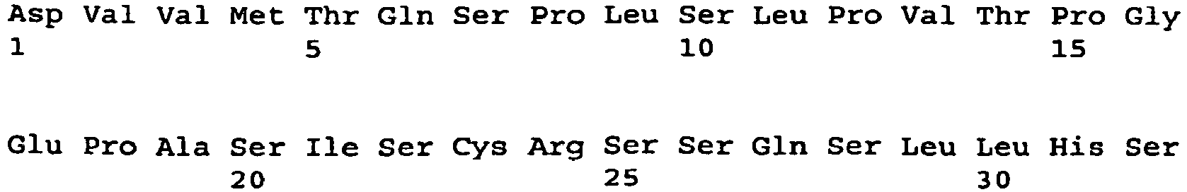

- <210> 7
<211> 336
<212> DNA
<213> Artificial Sequence - <220>
<223> light chain variable region - <220>
<221> CDS
<222> (1)..(336) - <400> 7


- <210> 8
<211> 112
<212> PRT
<213> Artificial Sequence - <220>
<223> light chain variable region - <400> 8

- <210> 9
<211> 336
<212> DNA
<213> Artificial Sequence - <220>
<223> light chain variable region - <220>
<221> CDS
<222> (1)..(336) - <400> 9


- <210> 10
<211> 112
<212> PRT
<213> Artificial Sequence - <220>
<223> light chain variable region - <400> 10

- <210> 11
<211> 336
<212> DNA
<213> Artificial Sequence - <220>
<223> light chain variable region - <220>
<221> CDS
<222> (1)..(336) - <400> 11

- <210> 12
<211> 112
<212> PRT
<213> Artificial Sequence - <220>
<223> light chain variable region - <400> 12


- <210> 13
<211> 336
<212> DNA
<213> Artificial Sequence - <220>
<223> light chain variable region - <220>
<221> CDS
<222> (1)..(336) - <400> 13


- <210> 14
<211> 112
<212> PRT
<213> Artificial Sequence - <220>
<223> light chain variable region - <400> 14

- <210> 15
<211> 336
<212> DNA
<213> Artificial Sequence - <220>
<223> light chain variable region - <220>
<221> CDS
<222> (1)..(336) . - <400> 15


- <210> 16
<211> 112
<212> PRT
<213> Artificial Sequence - <220>
<223> light chain variable region - <400> 16

- <210> 17
<211> 336
<212> DNA
<213> Artificial Sequence - <220>
<223> light chain variable region - <220>
<221> CDS
<222> (1)..(336) - <400> 17

- <210> 18
<211> 112
<212> PRT
<213> Artificial Sequence - <220>
<223> light chain variable region - <400> 18
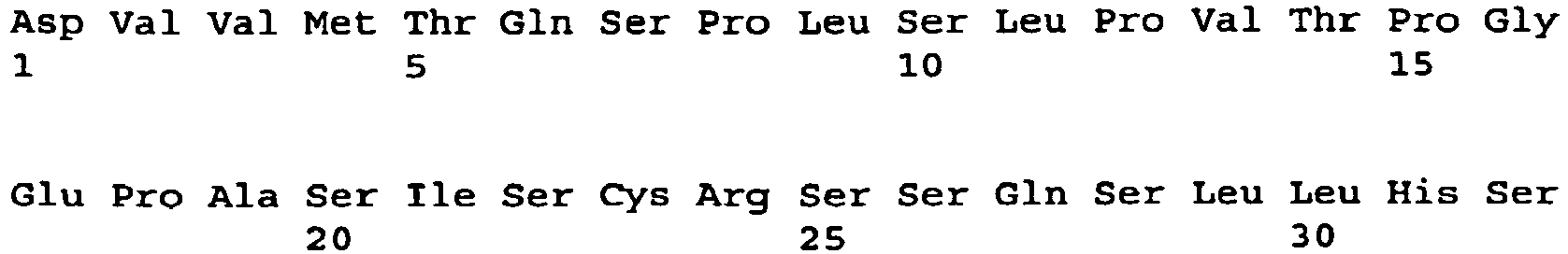

- <210> 19
<211> 336
<212> DNA
<213> Artificial Sequence - <220>
<223> light chain variable region - <220>
<221> CDS
<222> (1)..(336) - <400> 19

- <210> 20
<211> 112
<212> PRT
<213> Artificial Sequence - <220>
<223> light chain variable region - <400> 20
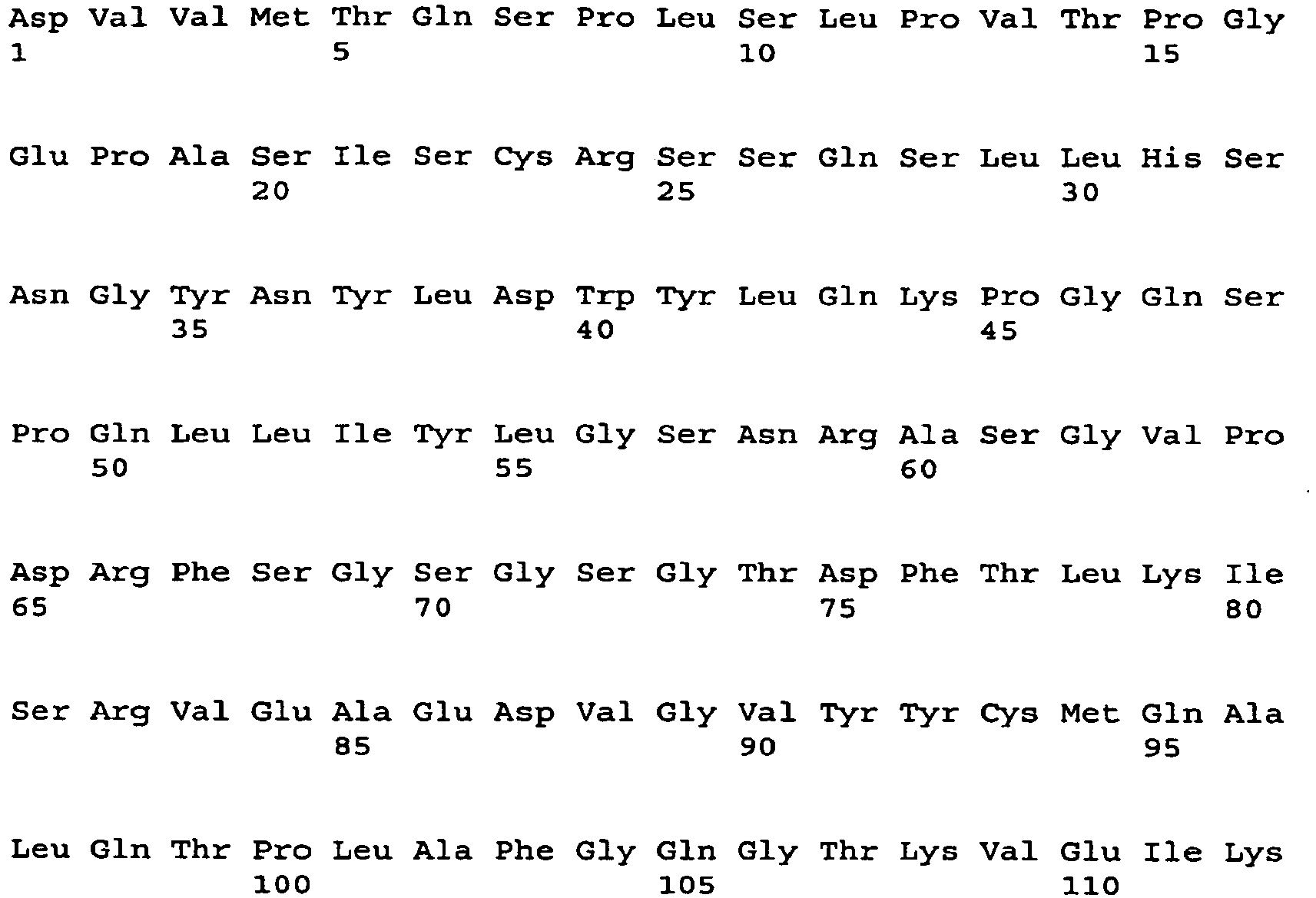
- <210> 21
<211> 336
<212> DNA
<213> Artificial Sequence - <220>
<223> light chain variable region - <220>
<221> CDS
<222> (1)..(336) - <400> 21


- <210> 22
<211> 112
<212> PRT
<213> Artificial Sequence - <220>
<223> light chain variable region - <400> 22

- <210> 23
<211> 333
<212> DNA
<213> Artificial Sequence - <220>
<223> light chain variable region - <220>
<221> CDS
<222> (1) .. (333) - <400> 23

- <210> 24
<211> 111
<212> PRT
<213> Artificial Sequence - <220>
<223> light chain variable region - <400> 24

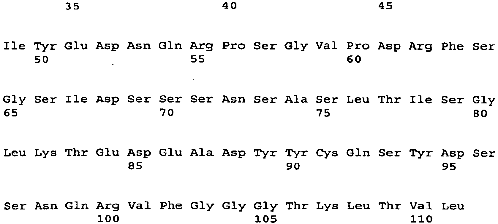
- <210> 25
<211> 336
<212> DNA
<213> Artificial Sequence - <220>
<223> light chain variable region - <220>
<221> CDS
<222> (1) .. (336) - <400> 25

- <210> 26
<211> 112
<212> PRT
<213> Artificial Sequence - <220>
<223> light chain variable region - <400> 26

- <210> 27
<211> 336
<212> DNA
<213> Artificial Sequence - <220>
<223> light chain variable region - <220>
<221> CDS
<222> (1)..(336) - <400> 27


- <210> 28
<211> 112
<212> PRT
<213> Artificial Sequence - <220>
<223> light chain variable region - <400> 28

- <210> 29
<211> 336
<212> DNA
<213> Artificial Sequence - <220>
<223> light chain variable region - <220>
<221> CDS
<222> (1) .. (336) - <400> 29

- <210> 30
<211> 112
<212> PRT
<213> Artificial Sequence - <220>
<223> light chain variable region - <400> 30


- <210> 31
<211> 336
<212> DNA
<213> Artificial Sequence - <220>
<223> light chain variable region - <220>
<221> CDS
<222> (1)..(336) - <400> 31

- <210> 32
<211> 112
<212> PRT
<213> Artificial Sequence - <220>
<223> light chain variable region - <400> 32

- <210> 33
<211> 335
<212> DNA
<213> Artificial Sequence - <220>
<223> light chain variable region - <220>
<221> CDS
<222> (1) .. (333) - <400> 33


- <210> 34
<211> 111
<212> PRT
<213> Artificial Sequence - <220>
<223> light chain variable region - <400> 34

- <210> 35
<211> 321
<212> DNA
<213> Artificial Sequence - <220>
<223> light chain variable region - <220>
<221> CDS
<222> (1)..(321) - <400> 35

- <210> 36
<211> 107
<212> PRT
<213> Artificial Sequence - <220>
<223> light chain variable region - <400> 36


- <210> 37
<211> 336
<212> DNA
<213> Artificial Sequence - <220>
<223> light chain variable region - <220>
<221> CDS
<222> (1)..(336) - <400> 37

- <210> 38
<211> 112
<212> PRT
<213> Artificial Sequence - <220>
<223> light chain variable region - <400> 38

- <210> 39
<211> 336
<212> DNA
<213> Artificial Sequence - <220>
<223> light chain variable region - <220>
<221> CDS
<222> (1)..(336) - <400> 39


- <210> 40
<211> 112
<212> PRT
<213> Artificial Sequence - <220>
<223> light chain variable region - <400> 40

- <210> 41
<211> 336
<212> DNA
<213> Artificial Sequence - <220>
<223> light chain variable region - <220>
<221> CDS
<222> (1)..(336) - <400> 41

- <210> 42
<211> 112
<212> PRT
<213> Artificial Sequence - <220>
<223> light chain variable region - <400> 42


- <210> 43
<211> 321
<212> DNA
<213> Artificial Sequence - <220>
<223> light chain variable region - <220>
<221> CDS
<222> (1).. (321) - <400> 43

- <210> 44
<211> 107
<212> PRT
<213> Artificial Sequence - <220>
<223> light chain variable region - <400> 44
- <210> 45
<211> 336
<212> DNA
<213> Artificial Sequence - <220>
<223> light chain variable region - <220>
<221> CDS
<222> (1)..(336) - <400> 45


- <210> 46
<211> 112
<212> PRT
<213> Artificial Sequence - <220>
<223> light chain variable region - <400> 46

- <210> 47
<211> 336
<212> DNA
<213> Artificial Sequence - <220>
<223> light chain variable region - <220>
<221> CDS
<222> (1)..(336) - <400> 47

- <210> 48
<211> 112
<212> PRT
<213> Artificial Sequence - <220>
<223> light chain variable region - <400> 48


- <210> 49
<211> 336
<212> DNA
<213> Artificial Sequence - <220>
<223> light chain variable region - <220>
<221> CDS
<222> (1)..(336) - <400> 49

- <210> 50
<211> 112
<212> PRT
<213> Artificial Sequence - <220>
<223> light chain variable region - <400> 50

- <210> 51
<211> 336
<212> DNA
<213> Artificial Sequence - <220>
<223> light chain variable region - <220>
<221> CDS
<222> (1)..(336) - <400> 51


- <210> 52
<211> 112
<212> PRT
<213> Artificial Sequence - <220>
<223> light chain variable region - <400> 52

- <210> 53
<211> 321
<212> DNA
<213> Artificial Sequence - <220>
<223> light chain variable region - <220>
<221> CDS
<222> (1) .. (321) - <400> 53

- <210> 54
<211> 107
<212> PRT
<213> Artificial Sequence - <220>
<223> light chain variable region - <400> 54


- <210> 55
<211> 315
<212> DNA
<213> Artificial Sequence - <220>
<223> light chain variable region - <220>
<221> CDS
<222> (1)..(315) - <400> 55

- <210> 56
<211> 105
<212> PRT
<213> Artificial Sequence - <220>
<223> light chain variable region - <400> 56

- <210> 57
<211> 336
<212> DNA
<213> Artificial Sequence - <220>
<223> light chain variable region - <220>
<221> CDS
<222> (1)..(336) - <400> 57


- <210> 58
<211> 112
<212> PRT
<213> Artificial Sequence - <220>
<223> light chain variable region - <400> 58
- <210> 59
<211> 336
<212> DNA
<213> Artificial Sequence - <220>
<223> light chain variable region - <220>
<221> CDS
<222> (1)..(336) - <400> 59

- <210> 60
<211> 112
<212> PRT
<213> Artificial Sequence - <220>
<223> light chain variable region - <400> 60

- <210> 61
<211> 321
<212> DNA
<213> Artificial Sequence - <220>
<223> light chain variable region - <220>
<221> CDS
<222> (1)..(321) - <400> 61

- <210> 62
<211> 107
<212> PRT
<213> Artificial Sequence - <220>
<223> light chain variable region - <400> 62
- <210> 63
<211> 336
<212> DNA
<213> Artificial Sequence - <220>
<223> light chain variable region - <220>
<221> CDS
<222> (1)..(336) - <400> 63
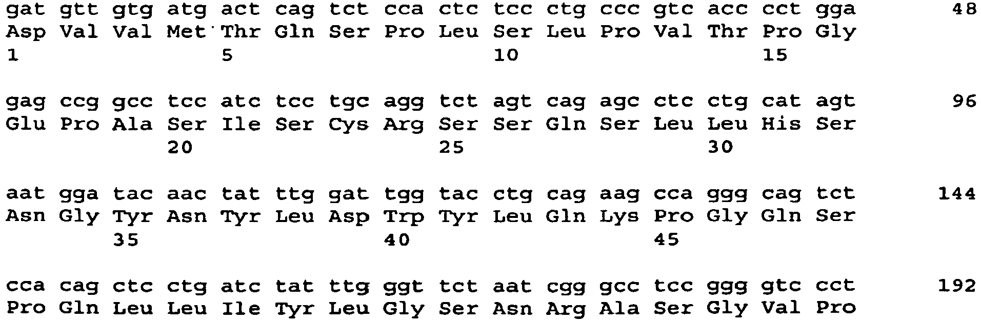

- <210> 64
<211> 112
<212> PRT
<213> Artificial Sequence - <220>
<223> light chain variable region - <400> 64

- <210> 65
<211> 336
<212> DNA
<213> Artificial Sequence - <220>
<223> light chain variable region - <220>
<221> CDS
<222> (1)..(336) - <400> 65
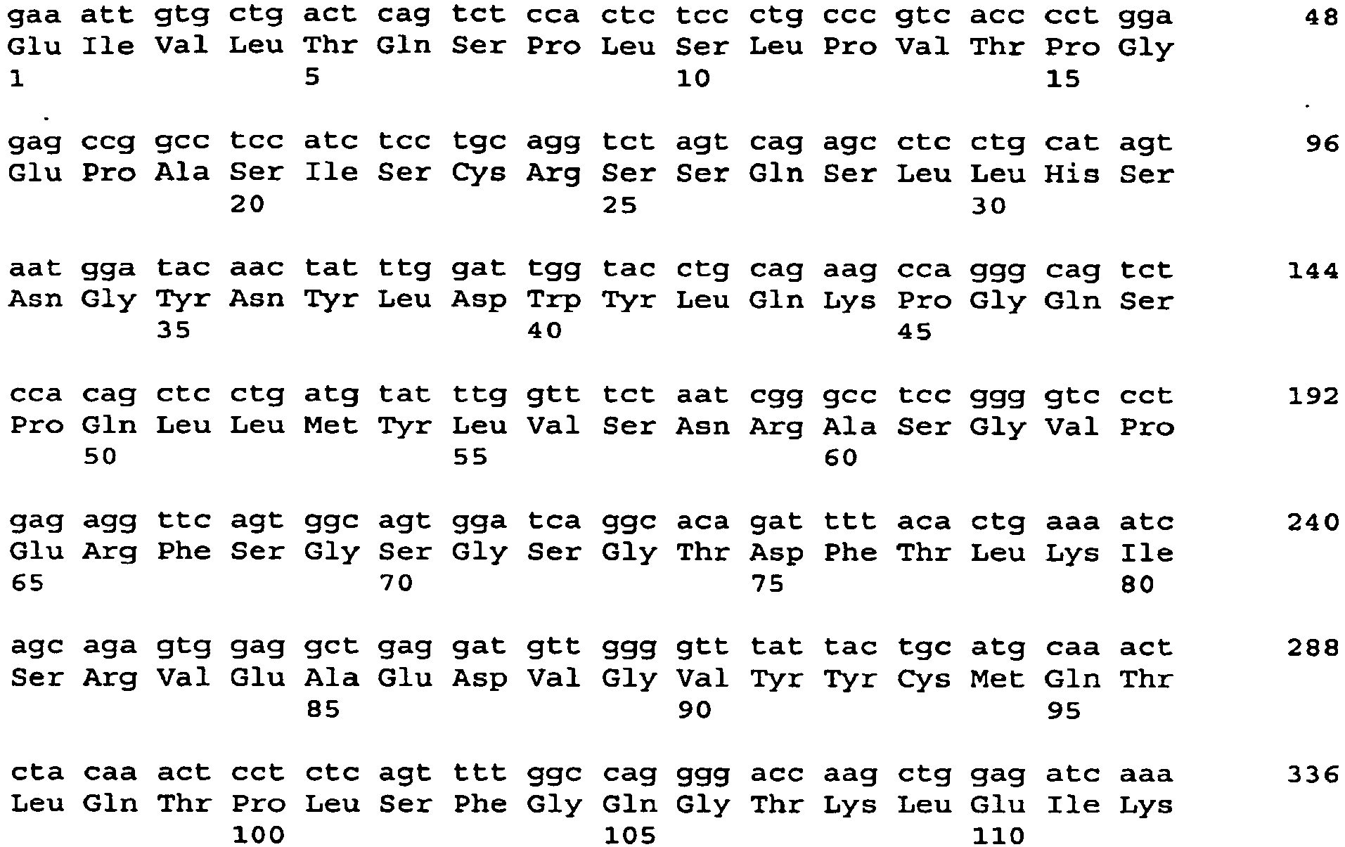
- <210> 66
<211> 112
<212> PRT
<213> Artificial Sequence - <220>
<223> light chain variable region - <400> 66

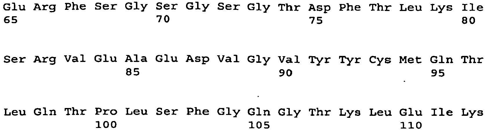
- <210> 67
<211> 336
<212> DNA
<213> Artificial Sequence - <220>
<223> light chain variable region - <220>
<221> CDS
<222> (1)..(336) - <400> 67

- <210> 68
<211> 112
<212> PRT
<213> Artificial Sequence - <220>
<223> light chain variable region - <400> 68

- <210> 69
<211> 330
<212> DNA
<213> Artificial Sequence - <220>
<223> light chain variable region - <220>
<221> CDS
<222> (1)..(330) - <400> 69


- <210> 70
<211> 110
<212> PRT
<213> Artificial Sequence - <220>
<223> light chain variable region - <400> 70

- <210> 71
<211> 330
<212> DNA
<213> Artificial Sequence - <220>
<223> light chain variable region - <220>
<221> CDS
<222> (1)..(330) - <400> 71

- <210> 72
<211> 110
<212> PRT
<213> Artificial Sequence - <220>
<223> light chain variable region - <400> 72


- <210> 73
<211> 336
<212> DNA
<213> Artificial Sequence - <220>
<223> light chain variable region - <220>
<221> CDS
<222> (1)..(336) - <400> 73

- <210> 74
<211> 112
<212> PRT
<213> Artificial Sequence - <220>
<223> light chain variable region - <400> 74

- <210> 75
<211> 336
<212> DNA
<213> Artificial Sequence - <220>
<223> light chain variable region - <220>
<221> CDS
<222> (1)..(336) - <400> 75


- <210> 76
<211> 112
<212> PRT
<213> Artificial Sequence - <220>
<223> light chain variable region - <400> 76

- <210> 77
<211> 336
<212> DNA
<213> Artificial Sequence - <220>
<223> light chain variable region - <220>
<221> CDS
<222> (1)..(336) - <400> 77

- <210> 78
<211> 112
<212> PRT
<213> Artificial Sequence - <220>
<223> light chain variable region - <400> 78


- <210> 79
<211> 321
<212> DNA
<213> Artificial Sequence - <220>
<223> light chain variable region - <220>
<221> CDS
<222> (1)..(321) - <400> 79

- <210> 80
<211> 107
<212> PRT
<213> Artificial Sequence - <220>
<223> light chain variable region - <400> 80

- <210> 81
<211> 321
<212> DNA
<213> Artificial Sequence - <220>
<223> light chain variable region - <220>
<221> CDS
<222> (1)..(321) - <400> 81


- <210> 82
<211> 107
<212> PRT
<213> Artificial Sequence - <220>
<223> light chain variable region - <400> 82

- <210> 83
<211> 336
<212> DNA
<213> Artificial Sequence - <220>
<223> light chain variable region - <220>
<221> CDS
<222> (1)..(336) - <400> 83

- <210> 84
<211> 112
<212> PRT
<213> Artificial Sequence - <220>
<223> light chain variable region - <400> 84


- <210> 85
<211> 321
<212> DNA
<213> Artificial Sequence - <220>
<223> light chain variable region - <220>
<221> CDS
<222> (1)..(321) - <400> 85

- <210> 86
<211> 107
<212> PRT
<213> Artificial Sequence - <220>
<223> light chain variable region - <400> 86

- <210> 87
<211> 336
<212> DNA
<213> Artificial Sequence - <220>
<223> light chain variable region - <220>
<221> CDS
<222> (1)..(336) - <400> 87
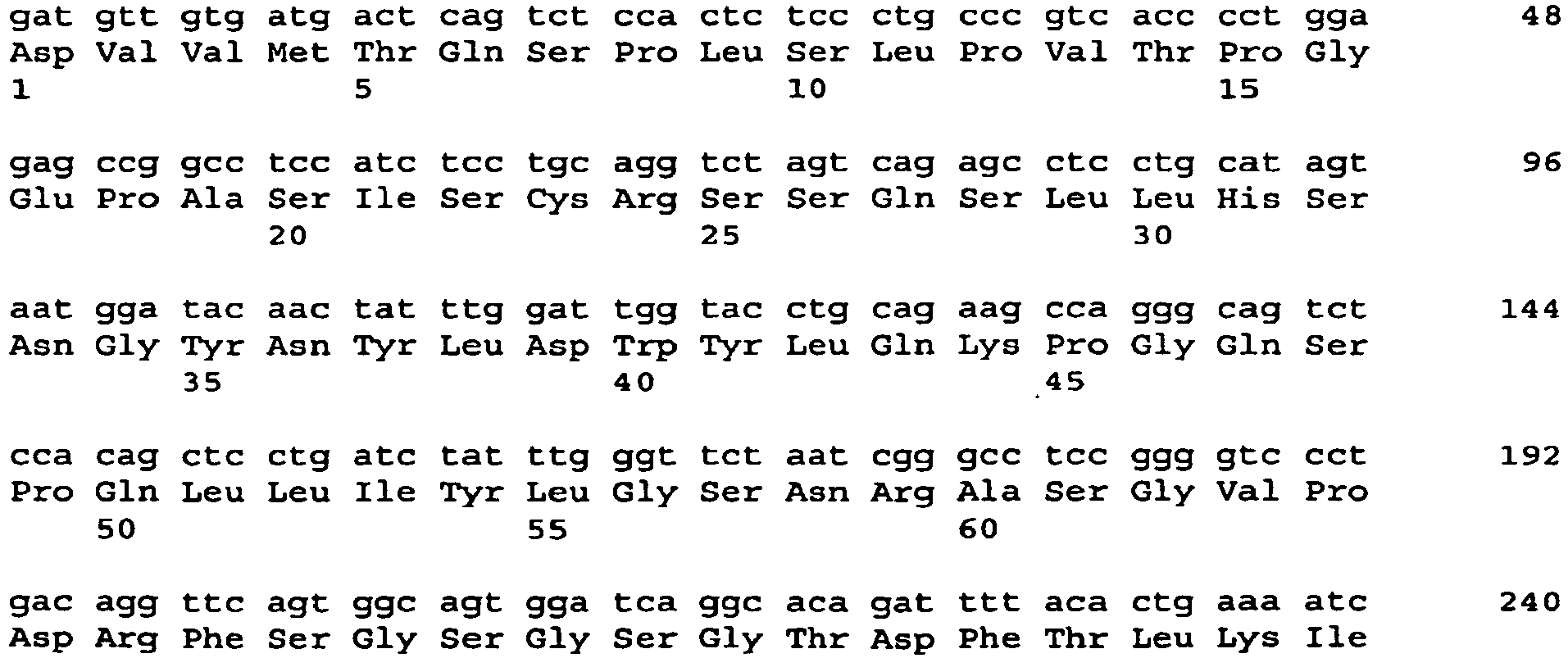

- <210> 88
<211> 112
<212> PRT
<213> Artificial Sequence - <220>
<223> light chain variable region - <400> 88
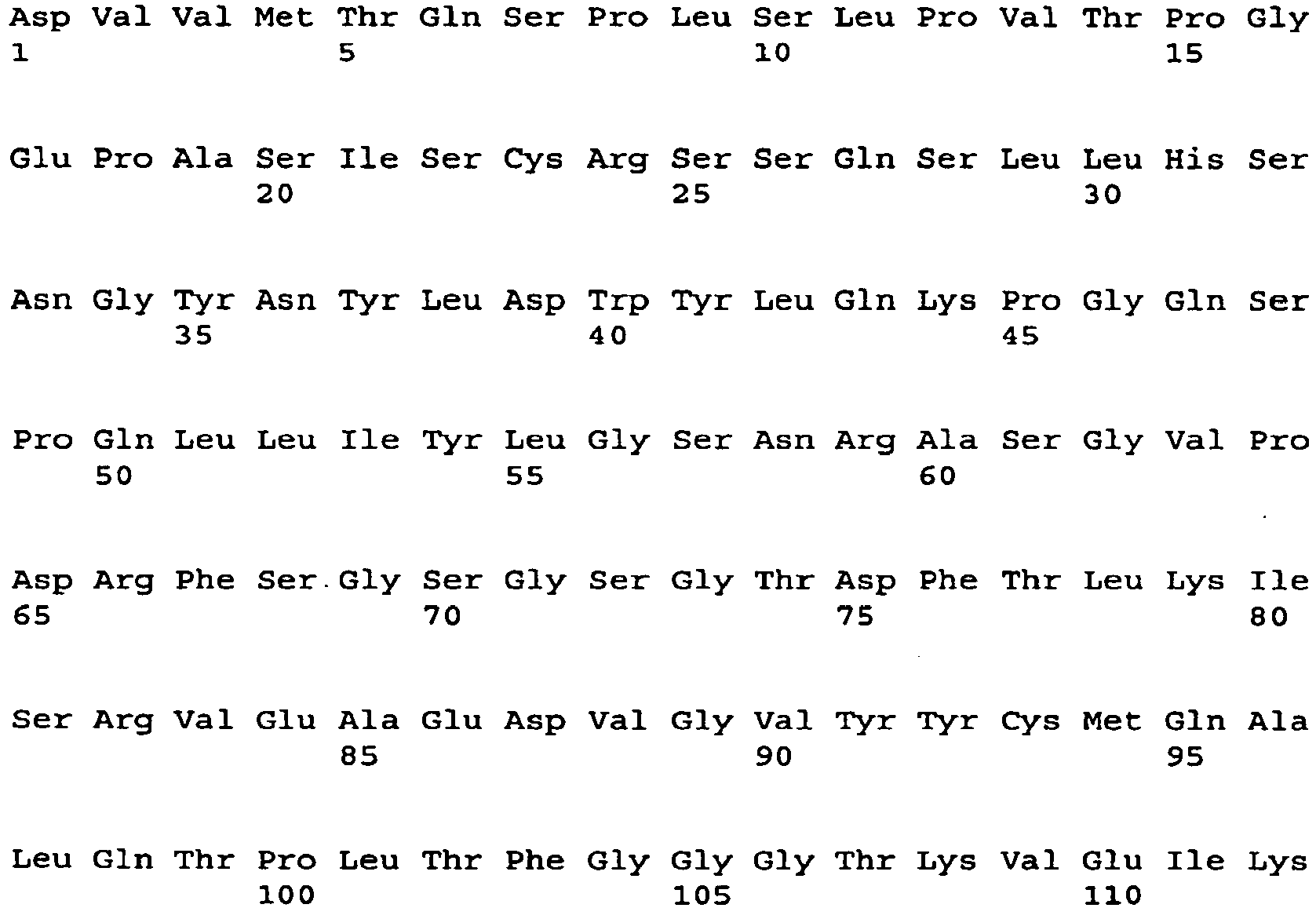
- <210> 89
<211> 336
<212> DNA
<213> Artificial Sequence - <220>
<223> light chain variable region - <220>
<221> CDS
<222> (1)..(336) - <400> 89

- <210> 90
<211> 112
<212> PRT
<213> Artificial Sequence - <220>
<223> light chain variable region - <400> 90
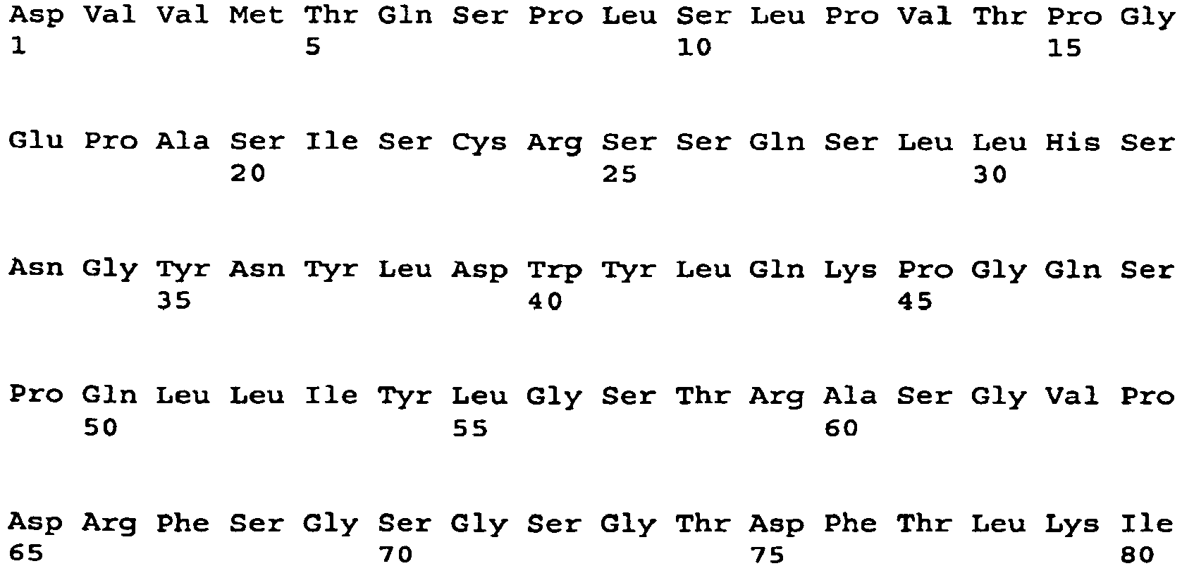

- <210> 91
<211> 336
<212> DNA
<213> Artificial Sequence - <220>
<223> light chain variable region - <220>
<221> CDS
<222> (1)..(336) - <400> 91

- <210> 92
<211> 112
<212> PRT
<213> Artificial Sequence - <220>
<223> light chain variable region - <400> 92

- <210> 93
<211> 336
<212> DNA
<213> Artificial Sequence - <220>
<223> light chain variable region - <220>
<221> CDS
<222> (1)..(336) - <400> 93

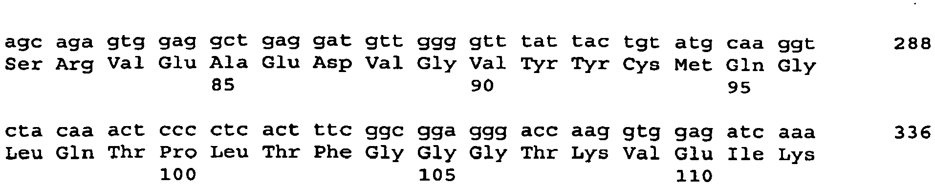
- <210> 94
<211> 112
<212> PRT
<213> Artificial Sequence - <220>
<223> light chain variable region - <400> 94

- <210> 95
<211> 336
<212> DNA
<213> Artificial Sequence - <220>
<223> light chain variable region - <220>
<221> CDS
<222> (1)..(336) - <400> 95

- <210> 96
<211> 112
<212> PRT
<213> Artificial Sequence - <220>
<223> light chain variable region - <400> 96


- <210> 97
<211> 330
<212> DNA
<213> Artificial Sequence - <220>
<223> light chain variable region - <220>
<221> CDS
<222> (1)..(330) - <400> 97

- <210> 98
<211> 110
<212> PRT
<213> Artificial Sequence - <220>
<223> light chain variable region - <400> 98


- <210> 99
<211> 324
<212> DNA
<213> Artificial Sequence - <220>
<223> light chain variable region - <220>
<221> CDS
<222> (1)..(324) - <400> 99


- <210> 100
<211> 108
<212> PRT
<213> Artificial Sequence - <220>
<223> light chain variable region - <400> 100

- <210> 101
<211> 330
<212> DNA
<213> Artificial Sequence - <220>
<223> light chain variable region - <220>
<221> CDS
<222> (1)..(330) - <400> 101

- <210> 102
<211> 110
<212> PRT
<213> Artificial Sequence - <220>
<223> light chain variable region - <400> 102


- <210> 103
<211> 336
<212> DNA
<213> Artificial Sequence - <220>
<223> light chain variable region - <220>
<221> CDS
<222> (1)..(336) - <400> 103
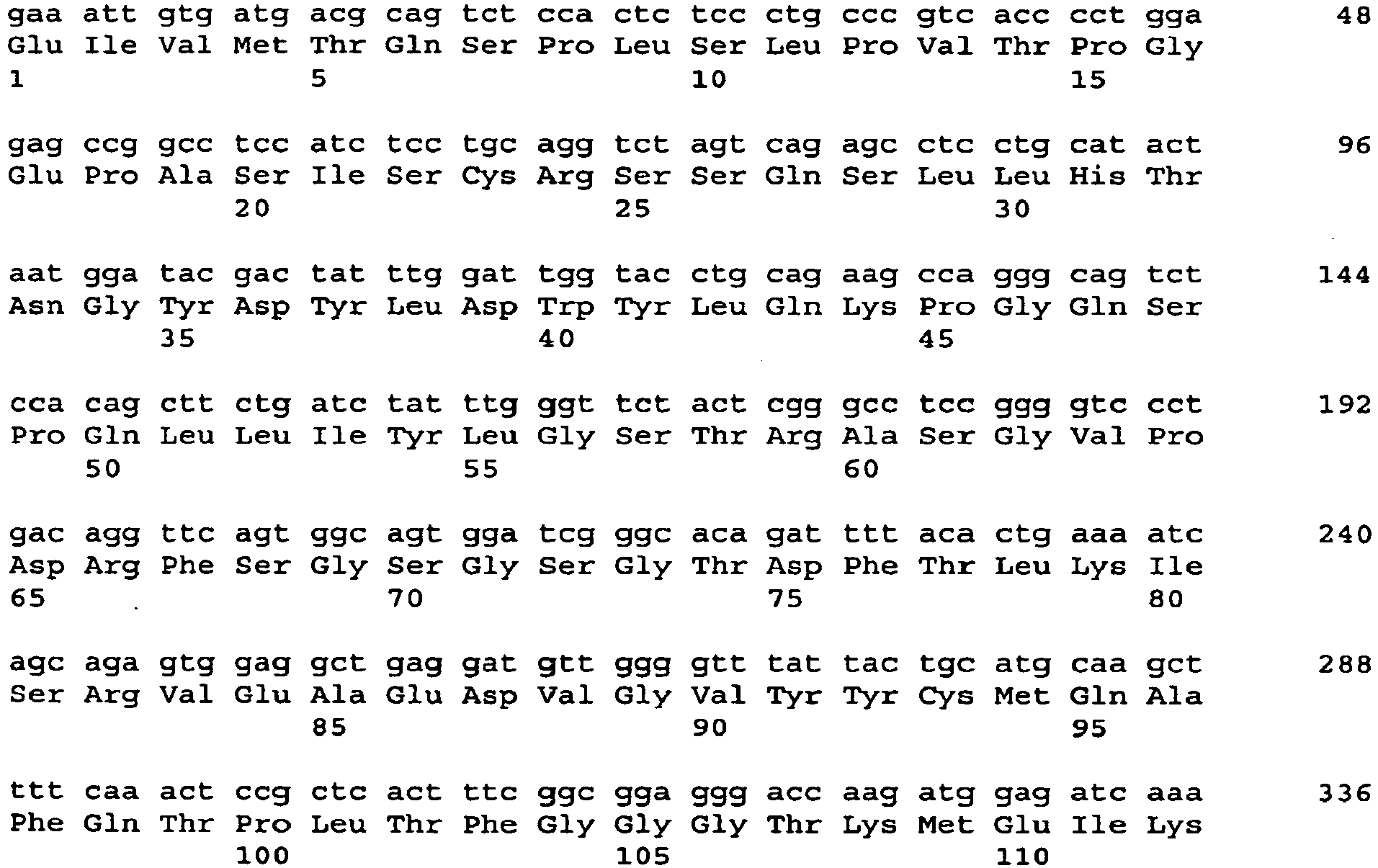
- <210> 104
<211> 112
<212> PRT
<213> Artificial Sequence - <220>
<223> light chain variable region - <400> 104


- <210> 105
<211> 351
<212> DNA
<213> Artificial Sequence - <220>
<223> heavy chain variable region - <220>
<221> CDS
<222> (1)..(351) - <400> 105

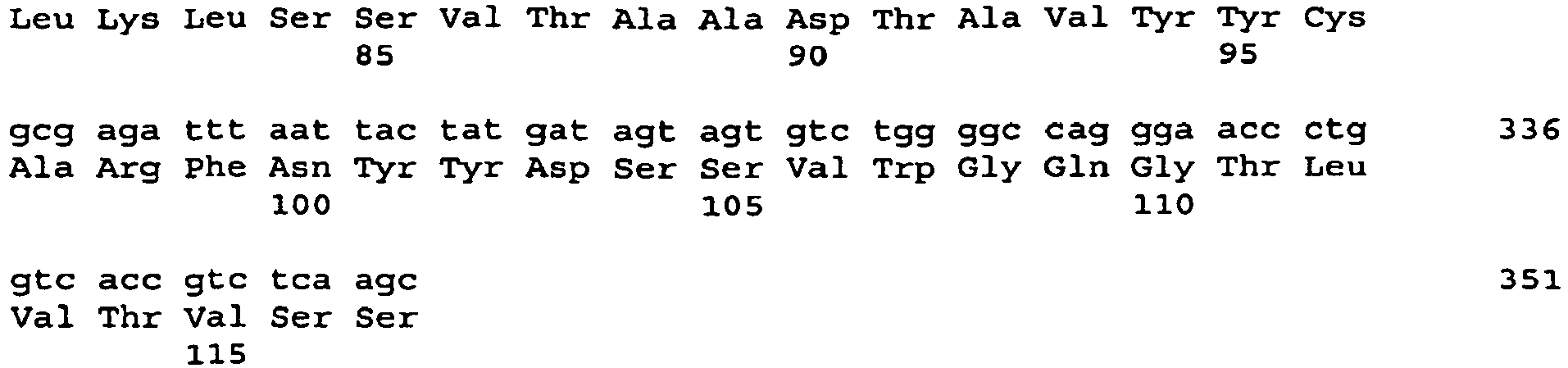
- <210> 106
<211> 117
<212> PRT
<213> Artificial Sequence - <220>
<223> heavy chain variable region - <400> 106

- <210> 107
<211> 348
<212> DNA
<213> Artificial Sequence - <220>
<223> heavy chain variable region - <220>
<221> CDS
<222> (1)..(348) - <400> 107

- <210> 108
<211> 116
<212> PRT
<213> Artificial Sequence - <220>
<223> heavy chain variable region - <400> 108


- <210> 109
<211> 354
<212> DNA
<213> Artificial Sequence - <220>
<223> heavy chain variable region - <220>
<221> CDS
<222> (1)..(354) - <400> 109


- <210> 110
<211> 118
<212> PRT
<213> Artificial Sequence - <220>
<223> heavy chain variable region - <400> 110

- <210> 111
<211> 351
<212> DNA
<213> Artificial Sequence - <220>
<223> heavy chain variable region - <220>
<221> CDS
<222> (1)..(351) - <400> 111

- <210> 112
<211> 117
<212> PRT
<213> Artificial Sequence - <220>
<223> heavy chain variable region - <400> 112


- <210> 113
<211> 360
<212> DNA
<213> Artificial Sequence - <220>
<223> heavy chain variable region - <220>
<221> CDS
<222> (1)..(360) - <400> 113

- <210> 114
<211> 120
<212> PRT
<213> Artificial Sequence - <220>
<223> heavy chain variable region - <400> 114
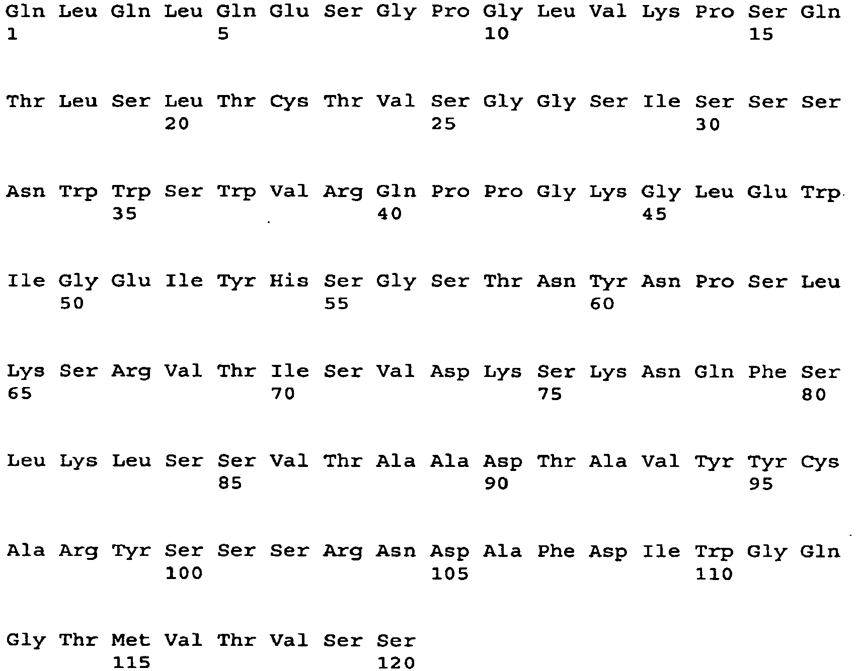
- <210> 115
<211> 354
<212> DNA
<213> Artificial Sequence - <220>
<223> heavy chain variable region - <220>
<221> CDS
<222> (1)..(354) - <400> 115


- <210> 116
<211> 118
<212> PRT
<213> Artificial Sequence - <220>
<223> heavy chain variable region - <400> 116
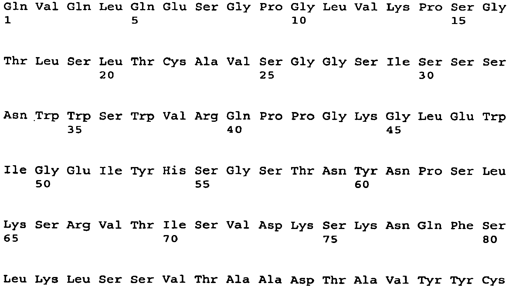
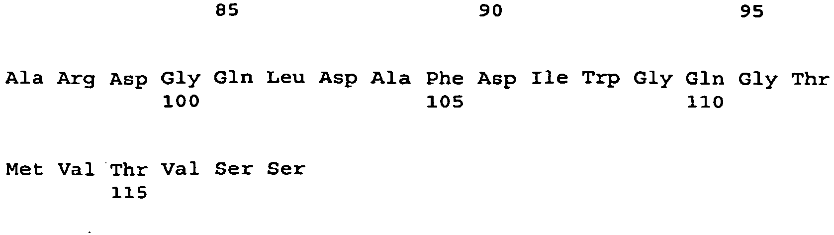
- <210> 117
<211> 354
<212> DNA
<213> Artificial Sequence - <220>
<223> heavy chain variable region - <220>
<221> CDS
<222> (1)..(354) - <400> 117
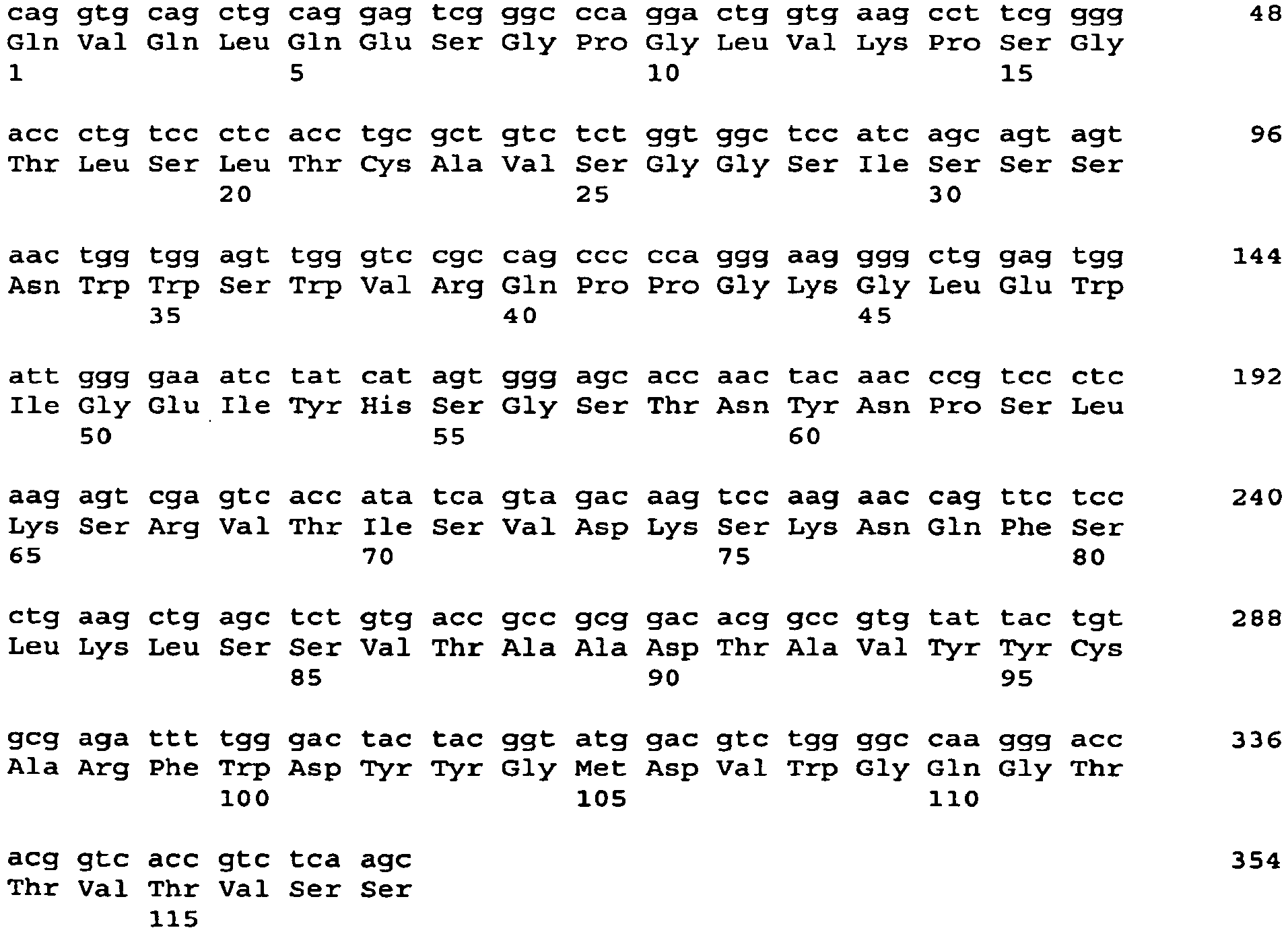
- <210> 118
<211> 118
<212> PRT
<213> Artificial Sequence - <220>
<223> heavy chain variable region - <400> 118

- <210> 119
<211> 351
<212> DNA
<213> Artificial Sequence - <220>
<223> heavy chain variable region - <220>
<221> CDS
<222> (1)..(351) - <400> 119


- <210> 120
<211> 117
<212> PRT
<213> Artificial Sequence - <220>
<223> heavy chain variable region - <400> 120


- <210> 121
<211> 354
<212> DNA
<213> Artificial Sequence - <220>
<223> heavy chain variable region - <220>
<221> CDS
<222> (1)..(354) - <400> 121

- <210> 122
<211> 118
<212> PRT
<213> Artificial Sequence - <220>
<223> heavy chain variable region - <400> 122

- <210> 123
<211> 354
<212> DNA
<213> Artificial Sequence - <220>
<223> heavy chain variable region - <220>
<221> CDS
<222> (1)..(354) - <400> 123
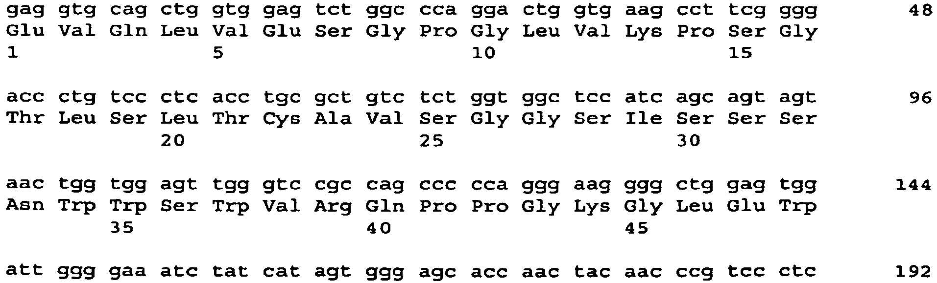
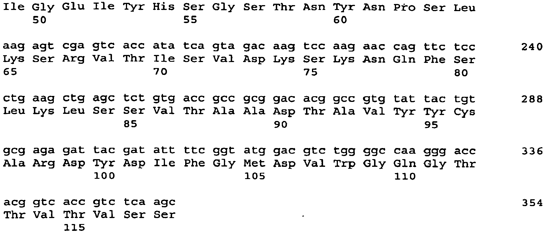
- <210> 124
<211> 118
<212> PRT
<213> Artificial Sequence - <220>
<223> heavy chain variable region - <400> 124
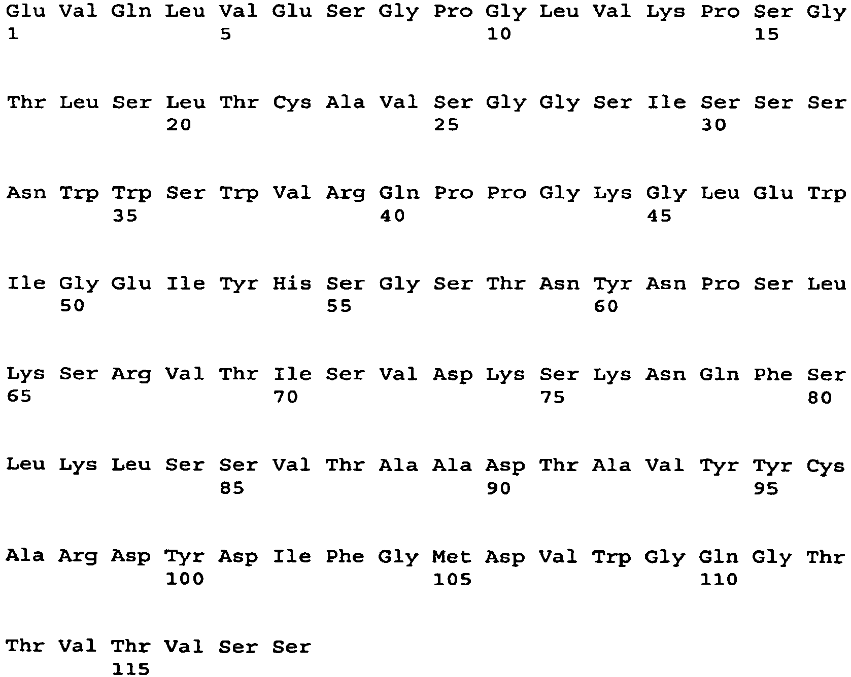
- <210> 125
<211> 354
<212> DNA
<213> Artificial Sequence - <220>
<223> heavy chain variable region - <220>
<221> CDS
<222> (1)..(354) - <400> 125

- <210> 126
<211> 118
<212> PRT
<213> Artificial Sequence - <220>
<223> heavy chain variable region - <400> 126


- <210> 127
<211> 357
<212> DNA
<213> Artificial Sequence - <220>
<223> heavy chain variable region - <220>
<221> CDS
<222> (1)..(357) - <400> 127


- <210> 128
<211> 119
<212> PRT
<213> Artificial Sequence - <220>
<223> heavy chain variable region - <400> 128

- <210> 129
<211> 363
<212> DNA
<213> Artificial Sequence - <220>
<223> heavy chain variable region - <220>
<221> CDS
<222> (1) .. (363) - <400> 129
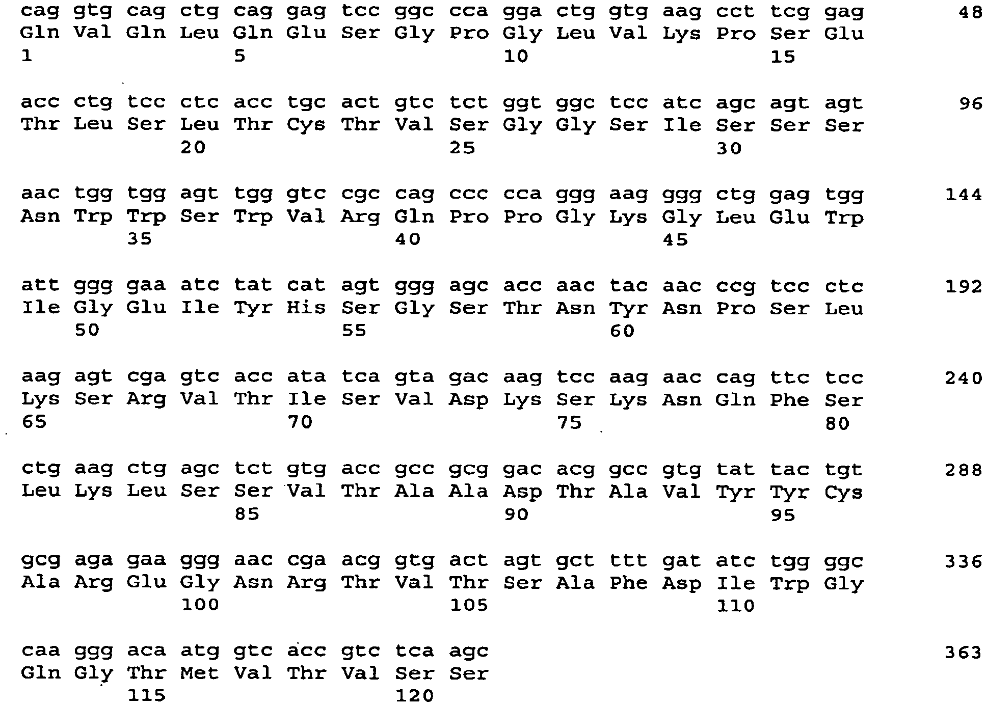
- <210> 130
<211> 121
<212> PRT
<213> Artificial Sequence - <220>
<223> heavy chain variable region - <400> 130


- <210> 131
<211> 357
<212> DNA
<213> Artificial Sequence - <220>
<223> heavy chain variable region - <220>
<221> CDS
<222> (1)..(357) - <400> 131


- <210> 132
<211> 119
<212> PRT
<213> Artificial Sequence - <220>
<223> heavy chain variable region - <400> 132

- <210> 133
<211> 357
<212> DNA
<213> Artificial Sequence - <220>
<223> heavy chain variable region - <220>
<221> CDS
<222> (1)..(357) - <400> 133

- <210> 134
<211> 119
<212> PRT
<213> Artificial Sequence - <220>
<223> heavy chain variable region - <400> 134


- <210> 135
<211> 357
<212> DNA
<213> Artificial Sequence - <220>
<223> heavy chain variable region - <220>
<221> CDS
<222> (1)..(357) - <400> 135
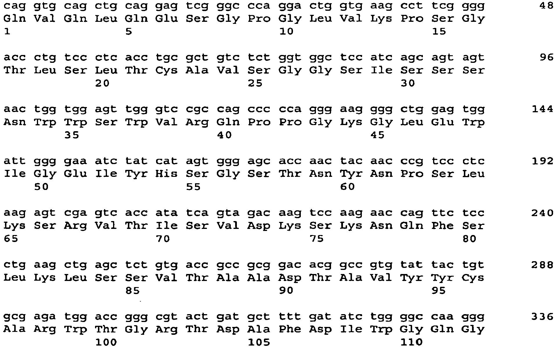

- <210> 136
<211> 119
<212> PRT
<213> Artificial Sequence - <220>
<223> heavy chain variable region - <400> 136

- <210> 137
<211> 354
<212> DNA
<213> Artificial Sequence - <220>
<223> heavy chain variable region - <220>
<221> CDS
<222> (1)..(354) - <400> 137

- <210> 138
<211> 118
<212> PRT
<213> Artificial Sequence - <220>
<223> heavy chain variable region - <400> 138
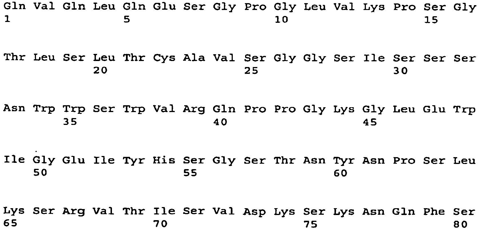
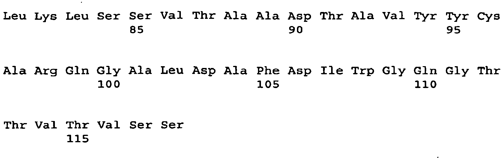
- <210> 139
<211> 366
<212> DNA
<213> Artificial Sequence - <220>
<223> heavy chain variable region - <220>
<221> CDS
<222> (1)..(366) - <400> 139

- <210> 140
<211> 122
<212> PRT
<213> Artificial Sequence - <220>
<223> heavy chain variable region - <400> 140

- <210> 141
<211> 357
<212> DNA
<213> Artificial Sequence - <220>
<223> heavy chain variable region - <220>
<221> CDS
<222> (1)..(357) - <400> 141


- <210> 142
<211> 119
<212> PRT
<213> Artificial Sequence - <220>
<223> heavy chain variable region - <400> 142


- <210> 143
<211> 360
<212> DNA
<213> Artificial Sequence - <220>
<223> heavy chain variable region - <220>
<221> CDS
<222> (1)..(360) - <400> 143

- <210> 144
<211> 120
<212> PRT
<213> Artificial Sequence - <220>
<223> heavy chain variable region - <400> 144

- <210> 145
<211> 351
<212> DNA
<213> Artificial Sequence - <220>
<223> heavy chain variable region - <220>
<221> CDS
<222> (1)..(351) - <400> 145


- <210> 146
<211> 117
<212> PRT
<213> Artificial Sequence - <220>
<223> heavy chain variable region - <400> 146


- <210> 147
<211> 366
<212> DNA
<213> Artificial Sequence - <220>
<223> heavy chain variable region - <220>
<221> CDS
<222> (1)..(366) - <400> 147

- <210> 148
<211> 122
<212> PRT
<213> Artificial Sequence - <220>
<223> heavy chain variable region - <400> 148
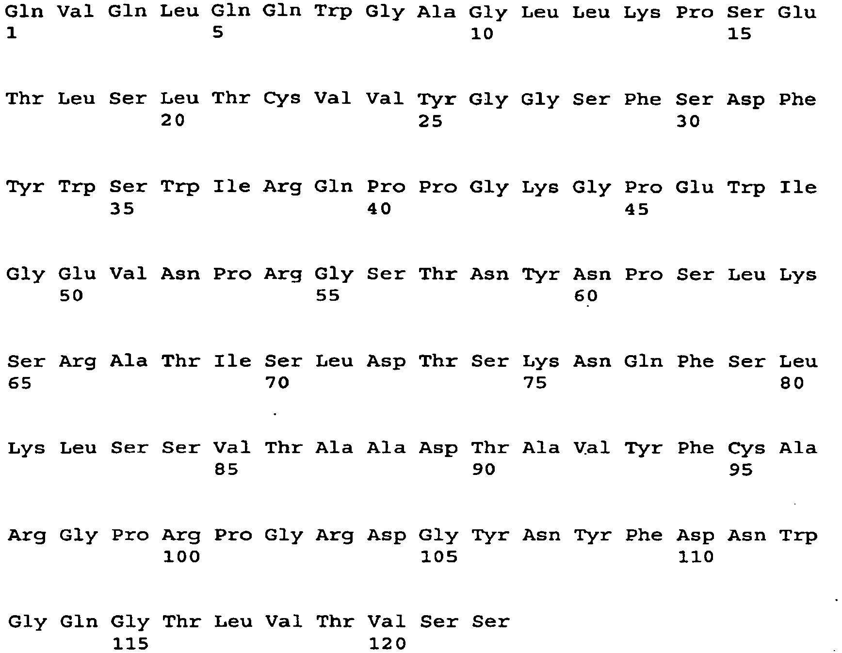
- <210> 149
<211> 357
<212> DNA
<213> Artificial Sequence - <220>
<223> heavy chain variable region - <220>
<221> CDS
<222> (1)..(357) - <400> 149


- <210> 150
<211> 119
<212> PRT
<213> Artificial Sequence - <220>
<223> heavy chain variable region - <400> 150

- <210> 151
<211> 363
<212> DNA
<213> Artificial Sequence - <220>
<223> heavy chain variable region - <220>
<221> CDS
<222> (1)..(363) - <400> 151

- <210> 152
<211> 121
<212> PRT
<213> Artificial Sequence - <220>
<223> heavy chain variable region - <400> 152


- <210> 153
<211> 351
<212> DNA
<213> Artificial Sequence - <220>
<223> heavy chain variable region - <220>
<221> CDS
<222> (1)..(351) - <400> 153


- <210> 154
<211> 117
<212> PRT
<213> Artificial Sequence - <220>
<223> heavy chain variable region - <400> 154

- <210> 155
<211> 357
<212> DNA
<213> Artificial Sequence - <220>
<223> heavy chain variable region - <220>
<221> CDS
<222> (1)..(357) - <400> 155
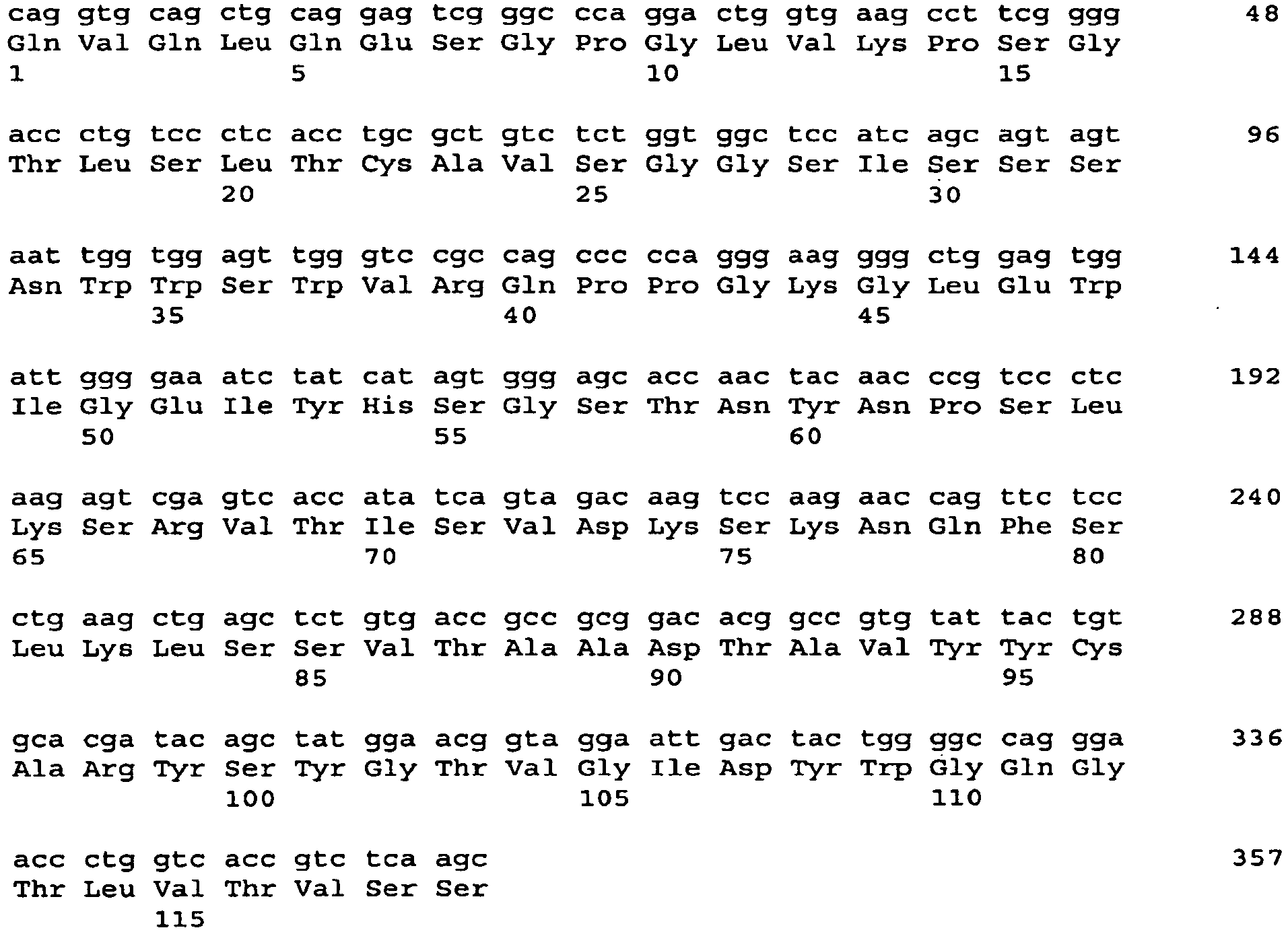
- <210> 156
<211> 119
<212> PRT
<213> Artificial Sequence - <220>
<223> heavy chain variable region - <400> 156


- <210> 157
<211> 351
<212> DNA
<213> Artificial Sequence - <220>
<223> heavy chain variable region - <220>
<221> CDS
<222> (1) .. (351)
<223> heavy chain variable region - <400> 157


- <210> 158
<211> 117
<212> PRT
<213> Artificial Sequence - <220>
<223> heavy chain variable region - <400> 158

- <210> 159
<211> 357
<212> DNA
<213> Artificial Sequence - <220>
<223> heavy chain variable region - <220>
<221> CDS
<222> (1)..(357) - <400> 159

- <210> 160
<211> 119
<212> PRT
<213> Artificial Sequence - <220>
<223> heavy chain variable region - <400> 160

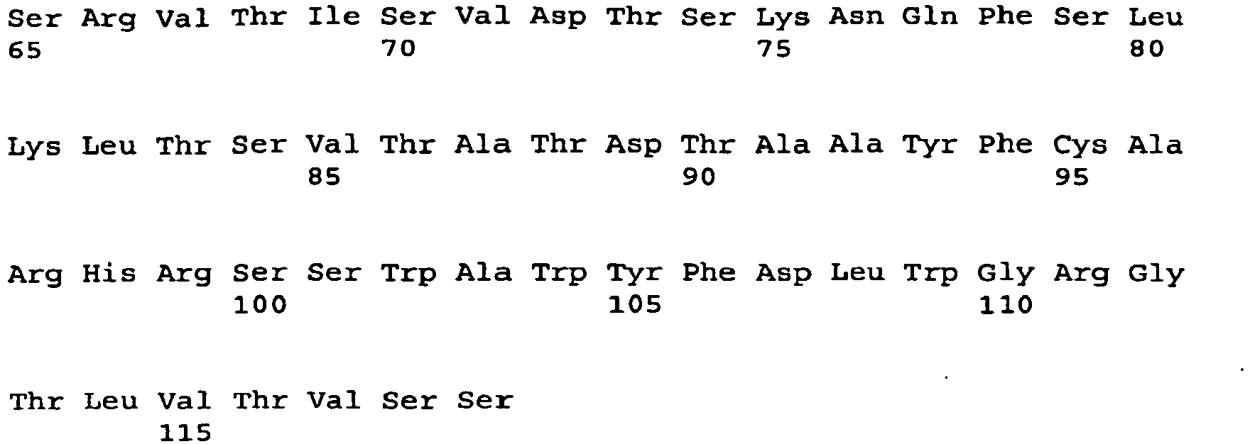
- <210> 161
<211> 354
<212> DNA
<213> Artificial Sequence - <220>
<223> heavy chain variable region - <220>
<221> CDS
<222> (1)..(354) - <400> 161


- <210> 162
<211> 118
<212> PRT
<213> Artificial Sequence - <220>
<223> heavy chain variable region - <400> 162
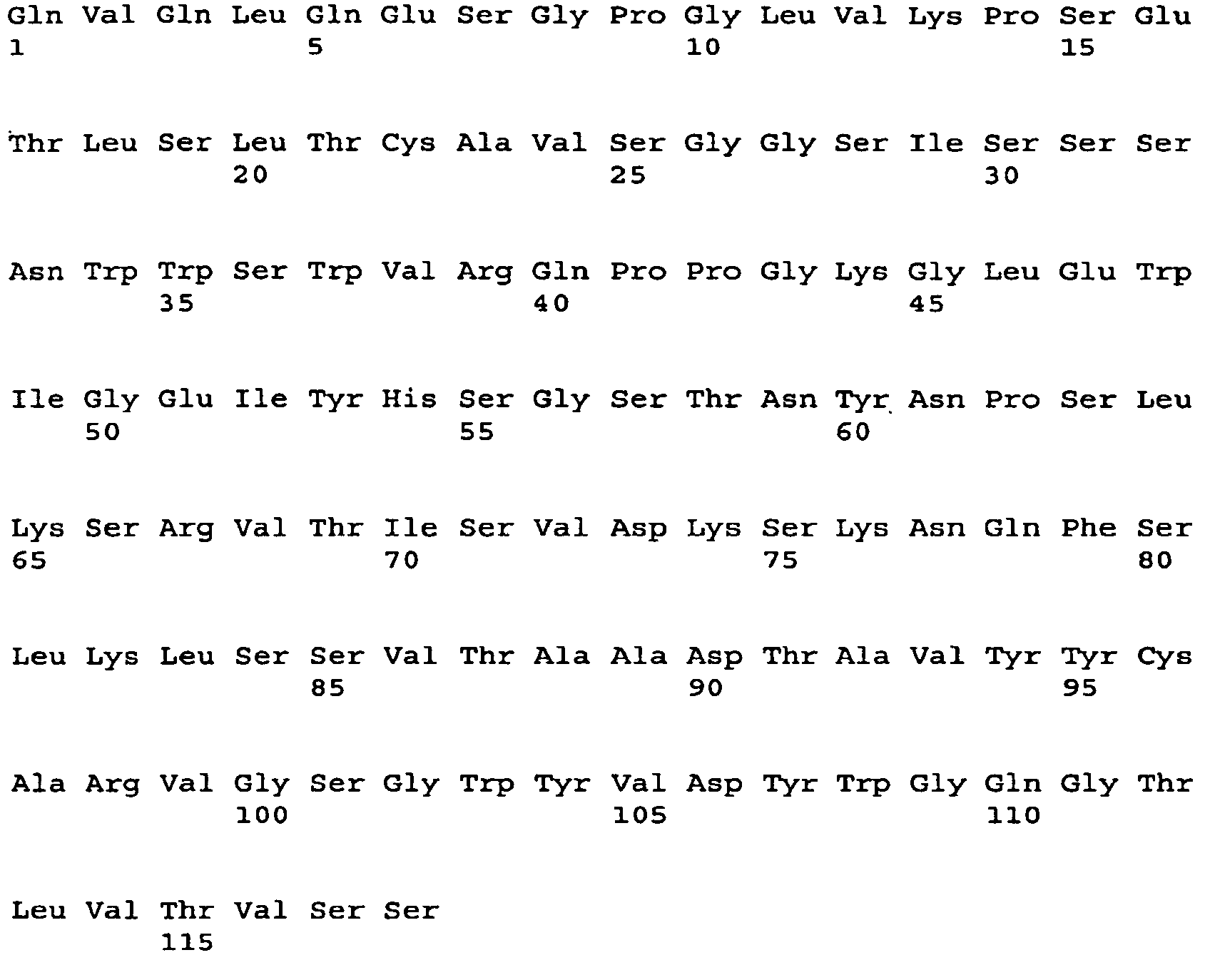
- <210> 163
<211> 360
<212> DNA
<213> Artificial Sequence - <220>
<223> heavy chain variable region - <220>
<221> CDS
<222> (1)..(360) - <400> 163


- <210> 164
<211> 120
<212> PRT
<213> Artificial Sequence - <220>
<223> heavy chain variable region - <400> 164


- <210> 165
<211> 369
<212> DNA
<213> Artificial Sequence - <220>
<223> heavy chain variable region - <220>
<221> CDS
<222> (1)..(369) - <400> 165
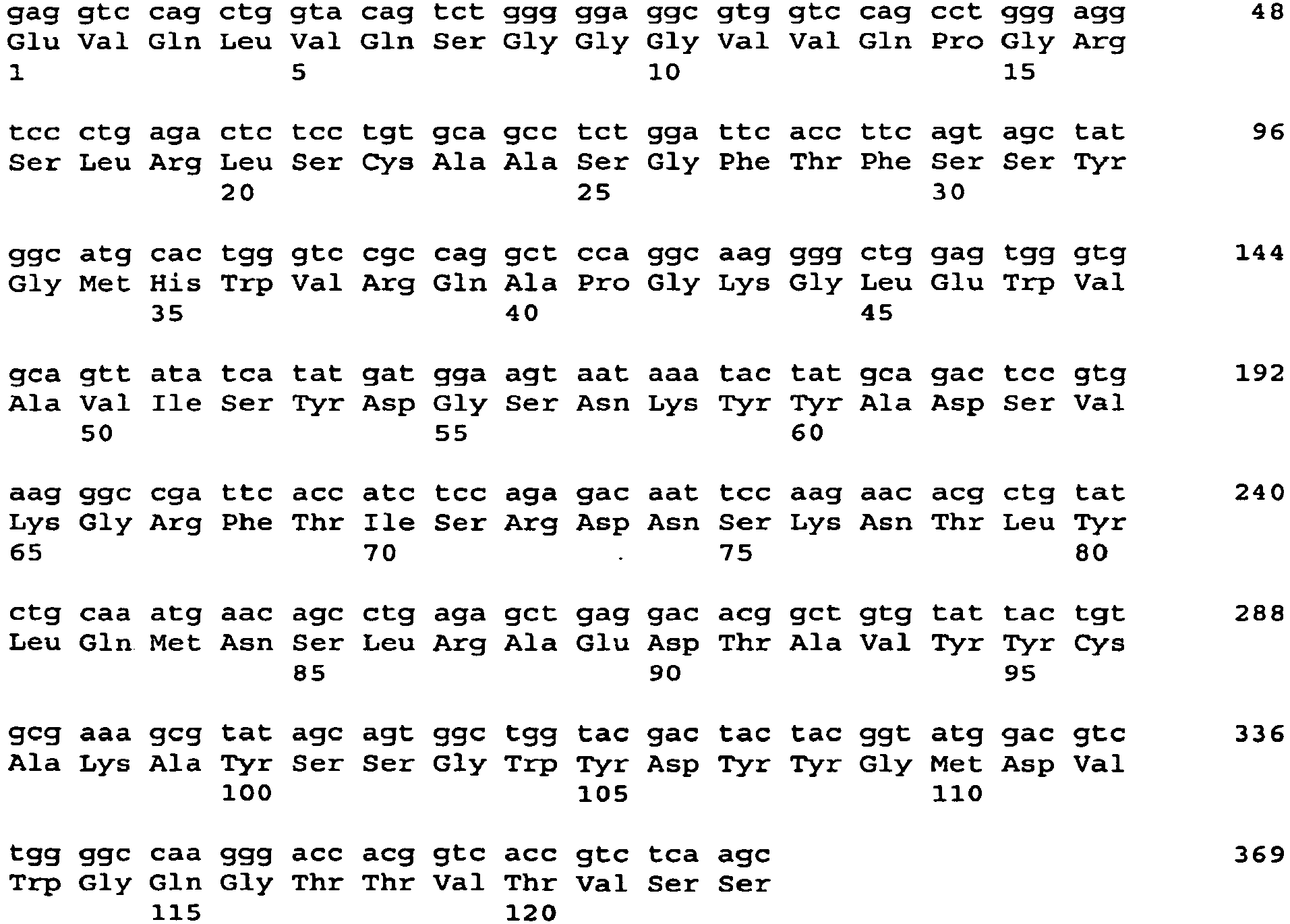
- <210> 166
<211> 123
<212> PRT
<213> Artificial Sequence - <220>
<223> heavy chain variable region - <400> 166

- <210> 167
<211> 351
<212> DNA
<213> Artificial Sequence - <220>
<223> heavy chain variable region - <220>
<221> CDS
<222> (1)..(351) - <400> 167


- <210> 168
<211> 117
<212> PRT
<213> Artificial Sequence - <220>
<223> heavy chain variable region - <400> 168


- <210> 169
<211> 357
<212> DNA
<213> Artificial Sequence - <220>
<223> heavy chain variable region - <220>
<221> CDS
<222> (1)..(357) - <400> 169
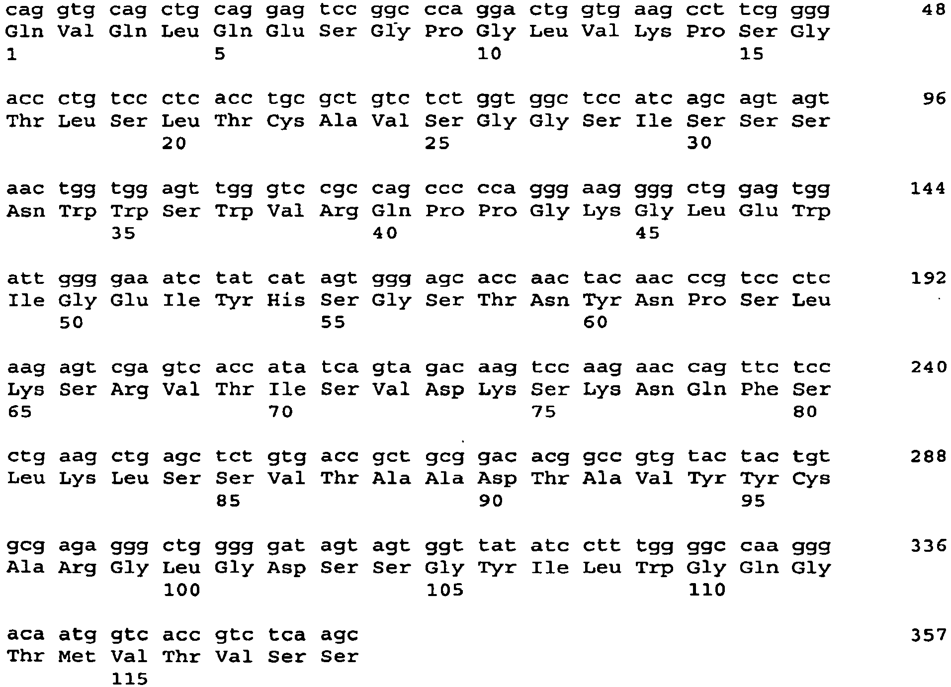
- <210> 170
<211> 119
<212> PRT
<213> Artificial Sequence - <220>
<223> heavy chain variable region - <400> 170

- <210> 171
<211> 348
<212> DNA
<213> Artificial Sequence - <220>
<223> heavy chain variable region - <220>
<221> CDS
<222> (1)..(348) - <400> 171


- <210> 172
<211> 116
<212> PRT
<213> Artificial Sequence - <220>
<223> heavy chain variable region - <400> 172


- <210> 173
<211> 360
<212> DNA
<213> Artificial Sequence - <220>
<223> heavy chain variable region - <220>
<221> CDS
<222> (1).. (360) - <400> 173
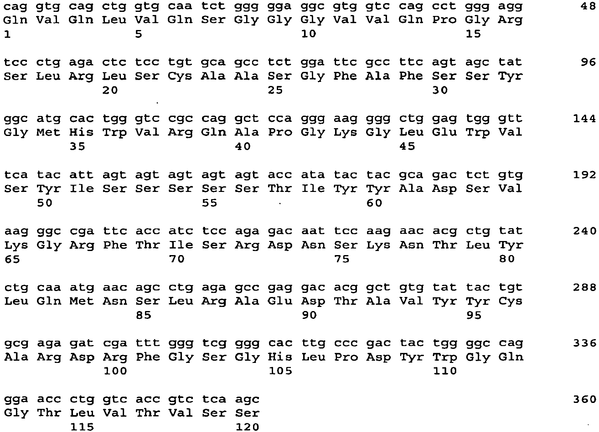
- <210> 174
<211> 120
<212> PRT
<213> Artificial Sequence - <220>
<223> heavy chain variable region - <400> 174


- <210> 175
<211> 357
<212> DNA
<213> Artificial Sequence - <220>
<223> heavy chain variable region - <220>
<221> CDS
<222> (1)..(357) - <400> 175


- <210> 176
<211> 119
<212> PRT
<213> Artificial Sequence - <220>
<223> heavy chain variable region - <400> 176

- <210> 177
<211> 360
<212> DNA
<213> Artificial Sequence - <220>
<223> heavy chain variable region - <220>
<221> CDS
<222> (1)..(360) - <400> 177
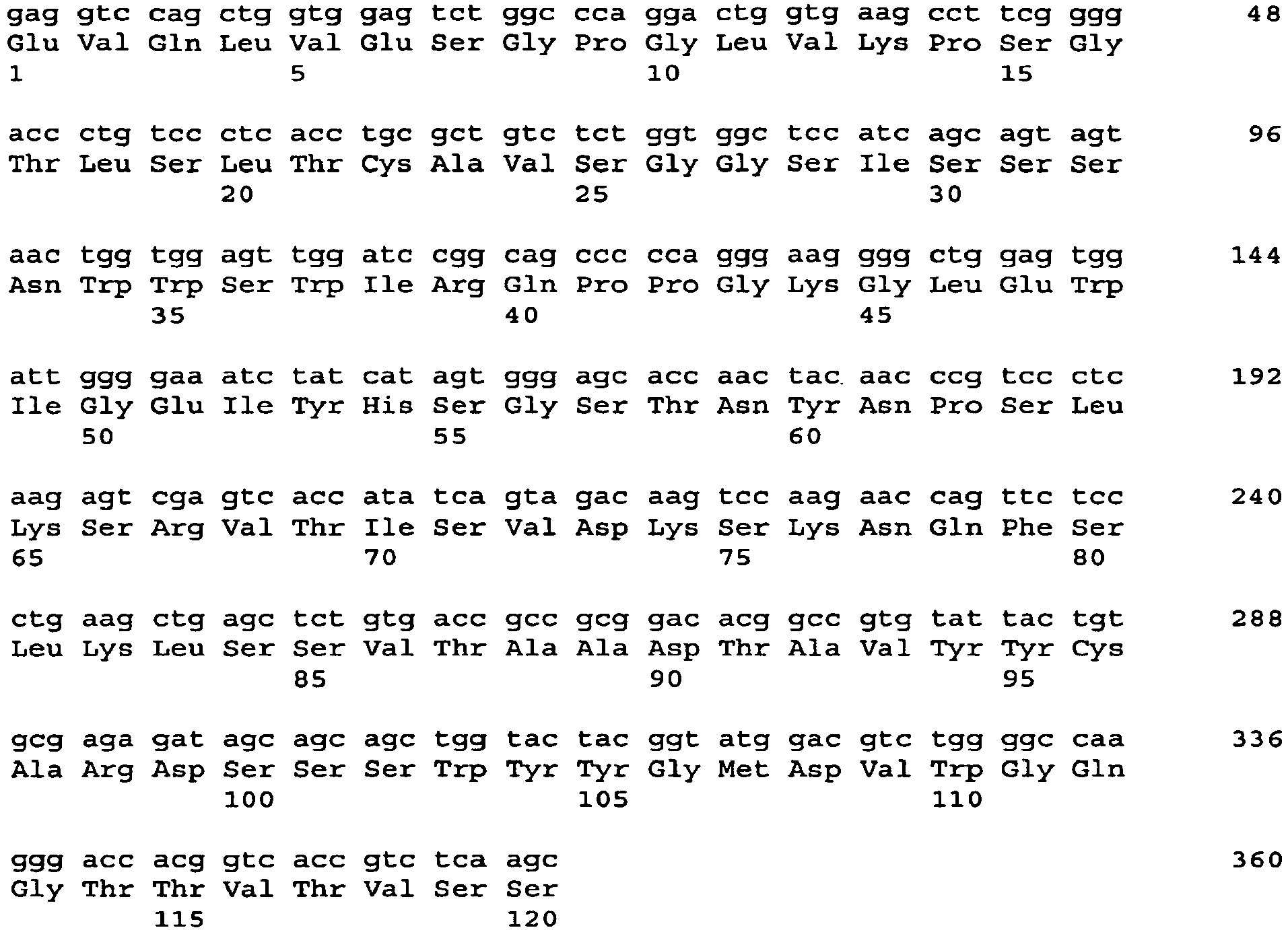
- <210> 178
<211> 120
<212> PRT
<213> Artificial Sequence - <220>
<223> heavy chain variable region - <400> 178


- <210> 179
<211> 348
<212> DNA
<213> Artificial Sequence - <220>
<223> heavy chain variable region - <220>
<221> CDS
<222> (1)..(348) - <400> 179
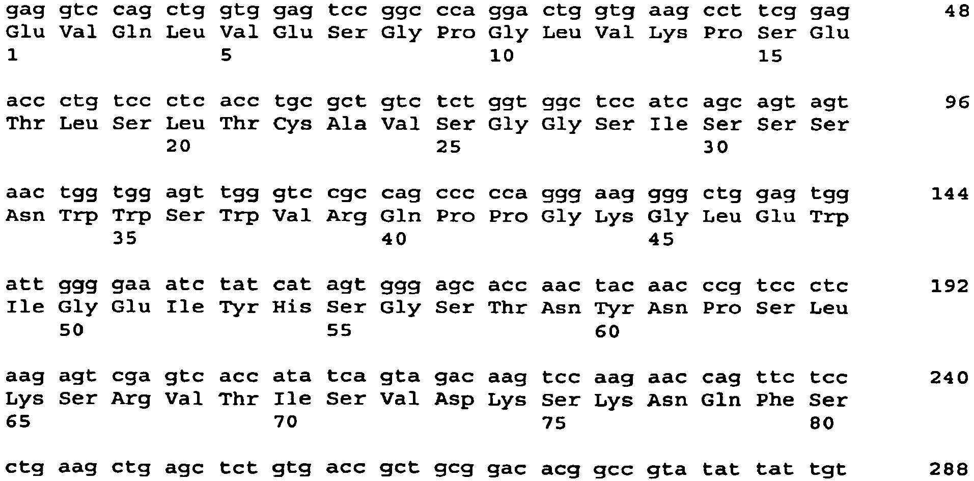

- <210> 180
<211> 116
<212> PRT
<213> Artificial Sequence - <220>
<223> heavy chain variable region - <400> 180
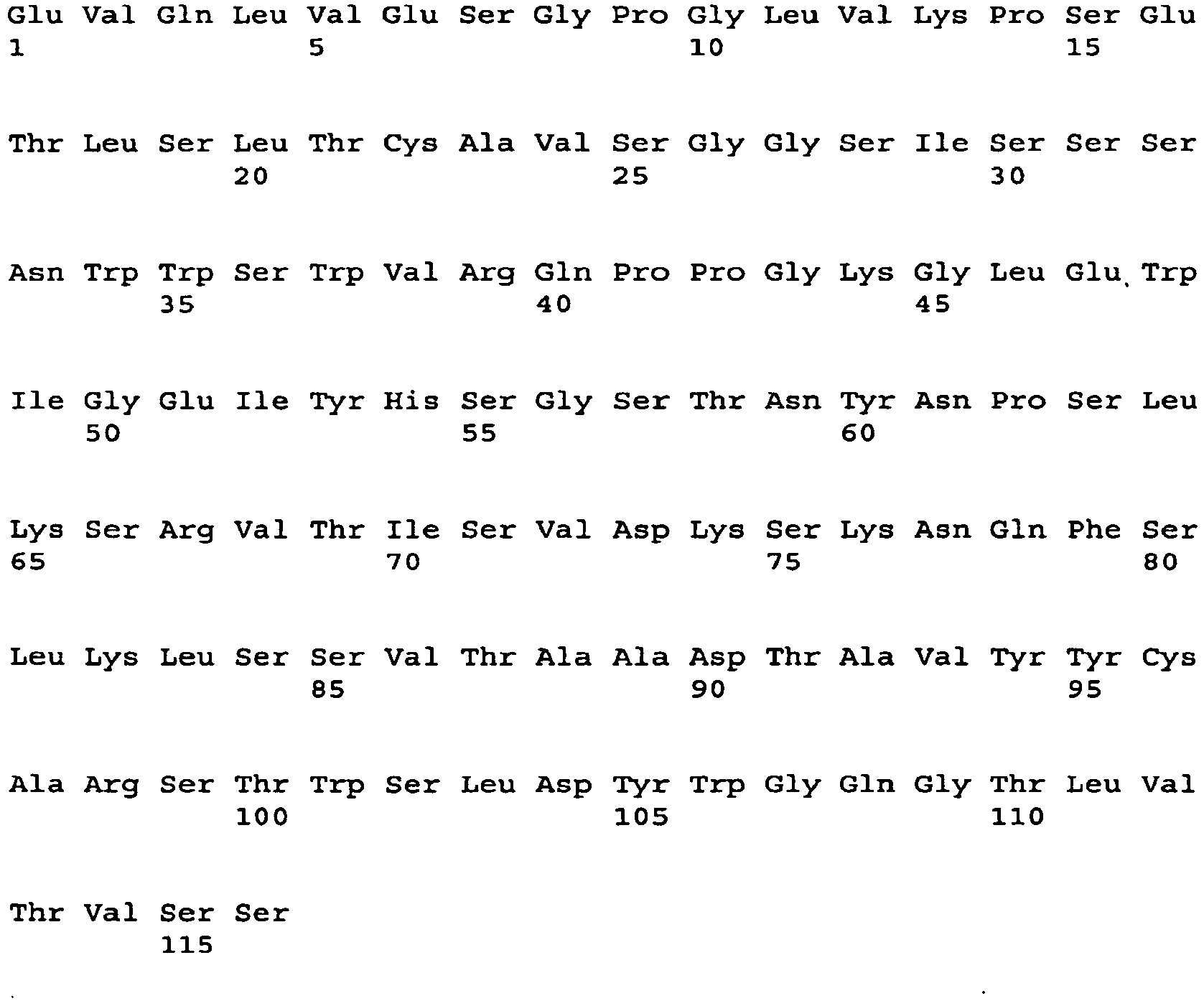
- <210> 181
<211> 354
<212> DNA
<213> Artificial Sequence - <220>
<223> heavy chain variable region - <220>
<221> CDS
<222> (1) .. (354) - <400> 181
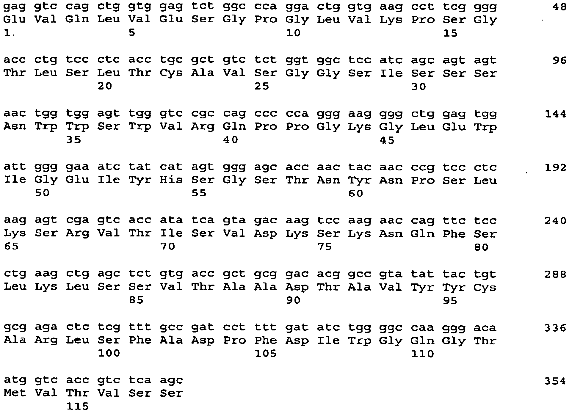
- <210> 182
<211> 118
<212> PRT
<213> Artificial Sequence - <220>
<223> heavy chain variable region - <400> 182


- <210> 183
<211> 366
<212> DNA
<213> Artificial Sequence - <220>
<223> heavy chain variable region - <220>
<221> CDS
<222> (1)..(366) - <400> 183


- <210> 184
<211> 122
<212> PRT
<213> Artificial Sequence - <220>
<223> heavy chain variable region - <400> 184

- <210> 185
<211> 357
<212> DNA
<213> Artificial Sequence - <220>
<223> heavy chain variable region - <220>
<221> CDS
<222> (1)..(357) - <400> 185

- <210> 186
<211> 119
<212> PRT
<213> Artificial Sequence - <220>
<223> heavy chain variable region - <400> 186


- <210> 187
<211> 345
<212> DNA
<213> Artificial Sequence - <220>
<223> heavy chain variable region - <220>
<221> CDS
<222> (1) .. (345) - <400> 187

- <210> 188
<211> 115
<212> PRT
<213> Artificial Sequence - <220>
<223> heavy chain variable region - <400> 188

- <210> 189
<211> 363
<212> DNA
<213> Artificial Sequence - <220>
<223> heavy chain variable region - <220>
<221> CDS
<222> (1)..(363) - <400> 189


- <210> 190
<211> 121
<212> PRT
<213> Artificial Sequence - <220>
<223> heavy chain variable region - <400> 190


- <210> 191
<211> 351
<212> DNA
<213> Artificial Sequence - <220>
<223> heavy chain variable region - <220>
<221> CDS
<222> (1)..(351) - <400> 191

- <210> 192
<211> 117
<212> PRT
<213> Artificial Sequence - <220>
<223> heavy chain variable region - <400> 192

- <210> 193
<211> 351
<212> DNA
<213> Artificial Sequence - <220>
<223> heavy chain variable region - <220>
<221> CDS
<222> (1)..(351) - <400> 193
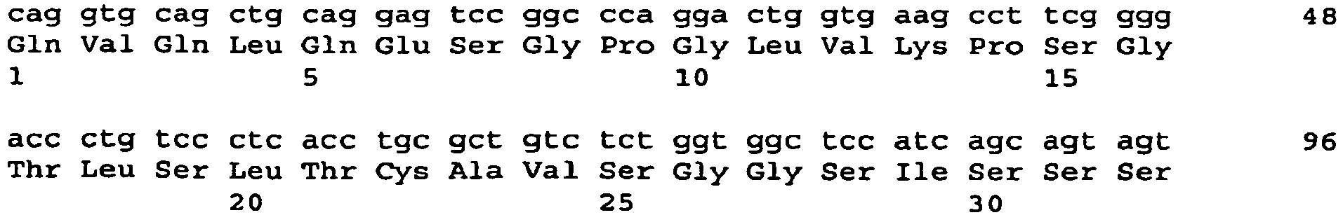

- <210> 194
<211> 117
<212> PRT
<213> Artificial Sequence - <220>
<223> heavy chain variable region - <400> 194


- <210> 195
<211> 354
<212> DNA
<213> Artificial Sequence - <220>
<223> heavy chain variable region - <220>
<221> CDS
<222> (1)..(354) - <400> 195

- <210> 196
<211> 118
<212> PRT
<213> Artificial Sequence - <220>
<223> heavy chain variable region - <400> 196

- <210> 197
<211> 357
<212> DNA
<213> Artificial Sequence - <220>
<223> heavy chain variable region - <220>
<221> CDS
<222> (1)..(357) - <400> 197


- <210> 198
<211> 119
<212> PRT
<213> Artificial Sequence - <220>
<223> heavy chain variable region - <400> 198
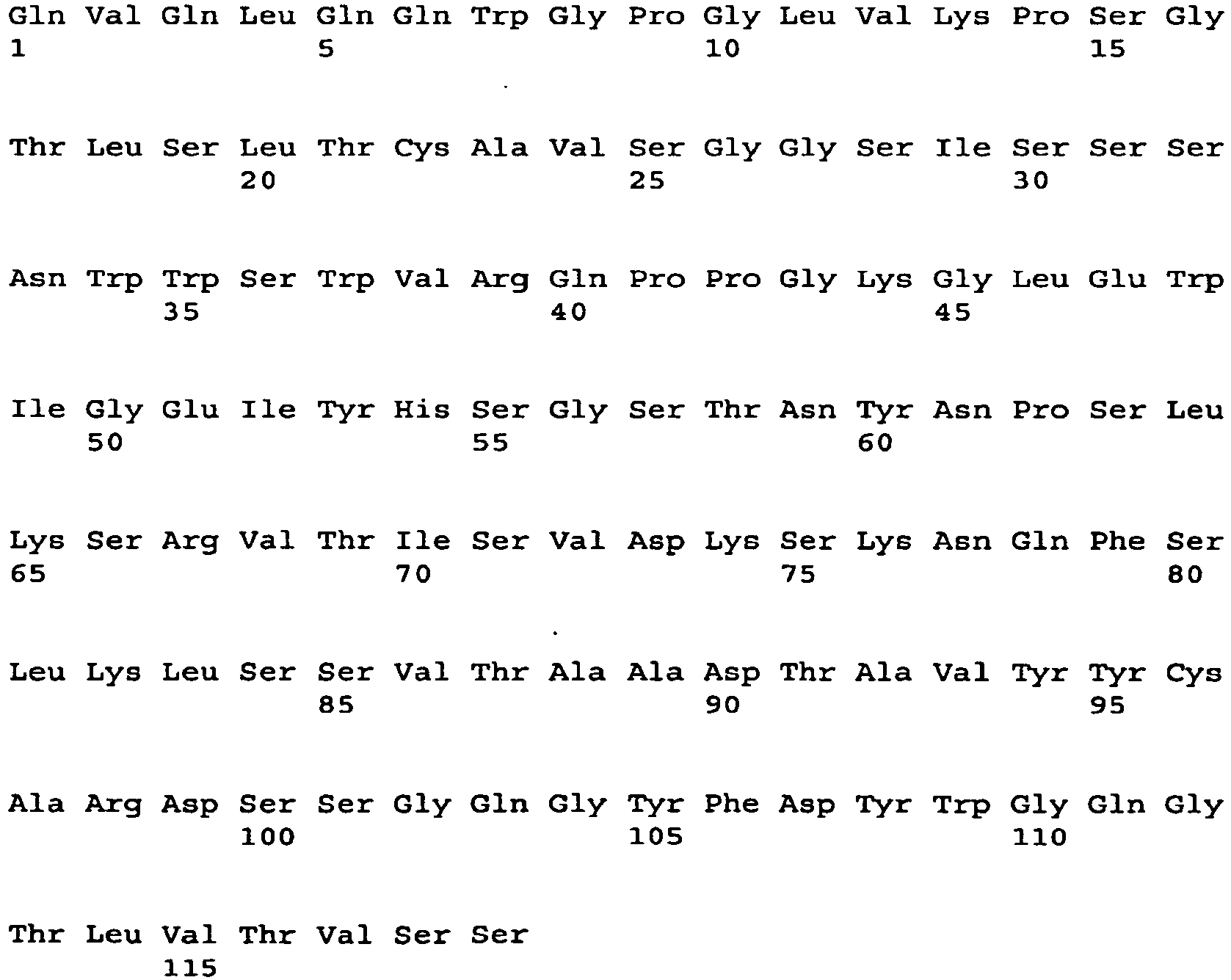
- <210> 199
<211> 354
<212> DNA
<213> Artificial Sequence - <220>
<223> heavy chain variable region - <220>
<221> CDS
<222> (1)..(354) - <400> 199

- <210> 200
<211> 118
<212> PRT
<213> Artificial Sequence - <220>
<223> heavy chain variable region - <400> 200


- <210> 201
<211> 360
<212> DNA
<213> Artificial Sequence - <220>
<223> heavy chain variable region - <220>
<221> CDS
<222> (1)..(360) - <400> 201
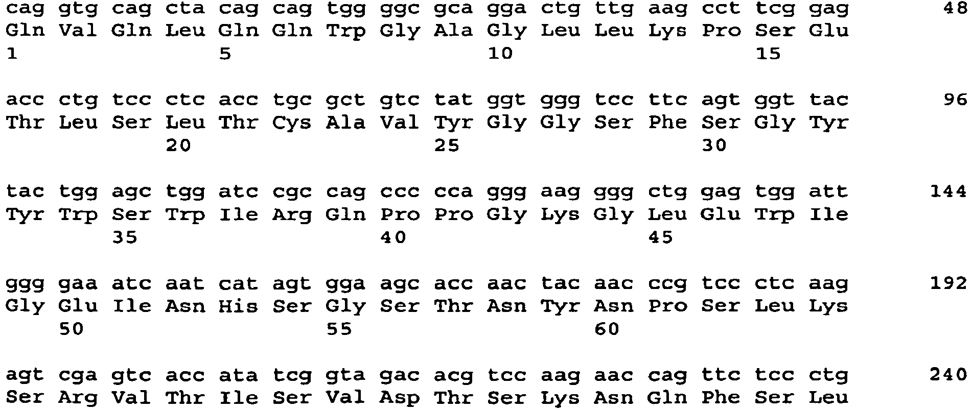

- <210> 202
<211> 120
<212> PRT
<213> Artificial Sequence - <220>
<223> heavy chain variable region - <400> 202

- <210> 203
<211> 354
<212> DNA
<213> Artificial Sequence - <220>
<223> heavy chain variable region - <220>
<221> CDS
<222> (1)..(354) - <400> 203

- <210> 204
<211> 118
<212> PRT
<213> Artificial Sequence - <220>
<223> heavy chain variable region - <400> 204


- <210> 205
<211> 357
<212> DNA
<213> Artificial Sequence - <220>
<223> heavy chain variable region - <220>
<221> CDS
<222> (1)..(357) - <400> 205


- <210> 206
<211> 119
<212> PRT
<213> Artificial Sequence - <220>
<223> heavy chain variable region - <400> 206

- <210> 207
<211> 348
<212> DNA
<213> Artificial Sequence - <220>
<223> heavy chain variable region - <220>
<221> CDS
<222> (1).. (348) - <400> 207

- <210> 208
<211> 116
<212> PRT
<213> Artificial Sequence - <220>
<223> heavy chain variable region - <400> 208
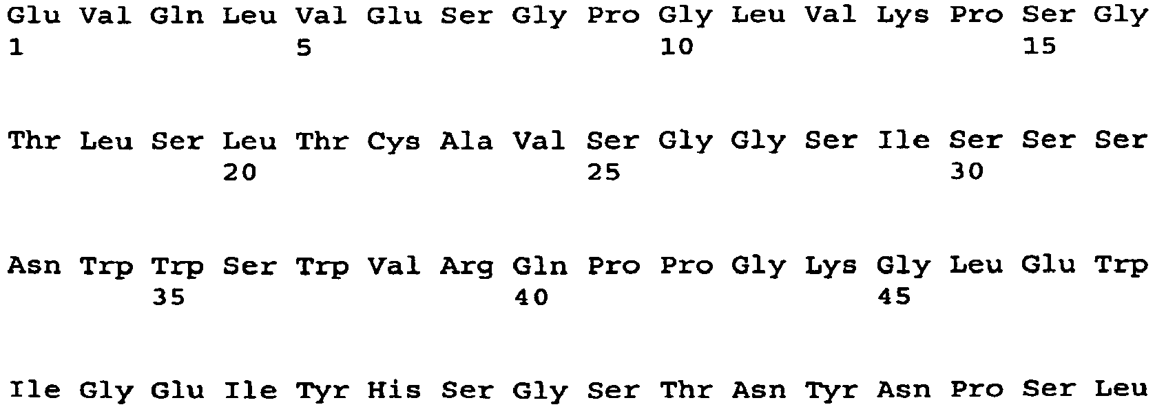

- <210> 209
<211> 321
<212> DNA
<213> Artificial Sequence - <220>
<223> light chain constant region - <220>
<221> CDS
<222> (1)..(321) - <400> 209

- <210> 210
<211> 107
<212> PRT
<213> Artificial Sequence - <220>
<223> light chain constant region - <400> 210

- <210> 211
<211> 990
<212> DNA
<213> Artificial Sequence - <220>
<223> heavy chain constant region - <220>
<221> CDS
<222> (1)..(990) - <400> 211
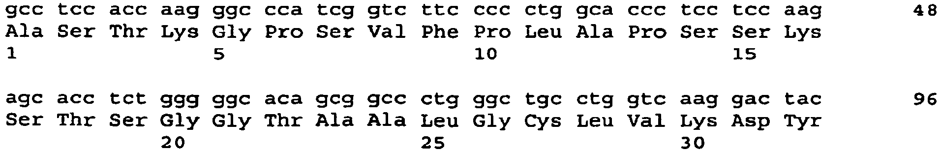

- <210> 212
<211> 330
<212> PRT
<213> Artificial Sequence - <220>
<223> heavy chain constant region - <400> 212


- <210> 213
<211> 9
<212> PRT
<213> Artificial Sequence - <220>
<223> Light chain CDR3 - <220>
<221> MISC_FEATURE
<222> (8)..(8)
<223> Xaa can be any naturally occuring amino acid - <400> 213

- <210> 214
<211> 9
<212> PRT
<213> Artificial Sequence - <220>
<223> Light chain CDR3 - <220>
<221> MISC_FEATURE
<222> (3)..(3)
<223> Xaa is arginine or serine - <220>
<221> MISC _FEATURE
<222> (4)..(4)
<223> Xaa is asparagine or serine - <220>
<221> MISC_FEATURE
<222> (5)..(5)
<223> Xaa is serine or asparagine - <220>
<221> MISC_FEATURE
<222> (6)..(6)
<223> Xaa is glycine, alanine, valine, leucine, isoleucine, proline, phenylalinine, methionine, tryptophan or cysteine - <400> 214

- <210> 215
<211> 10
<212> PRT
<213> Artificial Sequence - <220>
<223> Light chain CDR3 - <220>
<221> MISC _FEATURE
<222> (8)..(9)
<223> Xaa is arginine, valine, or isoleucine or no amino acid - <400> 215


- <210> 216
<211> 8
<212> PRT
<213> Artificial Sequence - <220>
<223> Heavy chain CDR3 - <400> 216

- <210> 217
<211> 10
<212> PRT
<213> Artificial Sequence - <220>
<223> Heavy chain CDR3 - <220>
<221> MISC_FEATURE
<222> (2).. (2)
<223> Xaa can be any naturally occurring amino acid - <400> 217

- <210> 218
<211> 11
<212> PRT
<213> Artificial Sequence - <220>
<223> Heavy chain CDR3 - <220>
<221> MISC _FEATURE
<222> (3) .. (3)
<223> Xaa can be any naturally occurring amino acid - <220>
<221> MISC _FEATURE
<222> (5) .. (5)
<223> Xaa can be any naturally occurring amino acid - <400> 218

- <210> 219
<211> 4
<212> PRT
<213> Artificial Sequence - <220>
<223> Heavy chain CDR3 - <220>
<221> MISC_FEATURE
<222> (1)..(4) - <400> 219

- <210> 220
<211> 16
<212> PRT
<213> Artificial Sequence - <220>
<223> Light chain CDR1 - <220>
<221> MISC_FEATURE
<222> (1)..(16) - <400> 220

- <210> 221
<211> 11
<212> PRT
<213> Artificial Sequence - <220>
<223> Light chain CDR1 - <220>
<221> MISC_FEATURE
<222> (5).. (5)
<223> Xaa is glycine or serine - <220>
<221> MISC_FEATURE
<222> (6)..(6)
<223> Xaa is isoleucine or valine - <220>
<221> MISC_FEATURE
<222> (7)..(7)
<223> Xaa is glycine or serine - <220>
<221> MISC_FEATURE
<222> (8)..(8)
<223> Xaa is any amino acid - <220>
<221> MISC_FEATURE
<222> (9)..(9)
<223> Xaa is tyrosine or phenyalanine - <220>
<221> MISC_FEATURE
<222> (11)..(11)
<223> Xaa is alanine or asparagine - <400> 221

- <210> 222
<211> 11
<212> PRT
<213> Artificial Sequence - <220>
<223> Light chain CDR1 - <220>
<221> MISC _FEATURE
<222> (6)..(6)
<223> Xaa is leucine or serine - <220>
<221> MISC _FEATURE
<222> (7)..(11)
<223> Xaa is independently any amino acid - <400> 222

- <210> 223
<211> 7
<212> PRT
<213> Artificial Sequence - <220>
<223> Light chain CDR2 - <400> 223

- <210> 224
<211> 7
<212> PRT
<213> Artificial Sequence - <220>
<223> Light chain CDR2 - <400> 224

- <210> 225
<211> 7
<212> PRT
<213> Artificial Sequence - <220>
<223> Light chain CDR2 - <220>
<221> MISC_FEATURE
<222> (4)..(4)
<223> Xaa can be any naturally occurring amino acid - <400> 225

- <210> 226
<211> 6
<212> PRT
<213> Artificial Sequence - <220>
<223> Heavy chain CDR1 - <400> 226

- <210> 227
<211> 5
<212> PRT
<213> Artificial Sequence - <220>
<223> Heavy chain CDR1 - <220>
<221> MISC_FEATURE
<222> (1)..(1)
<223> Xaa can be any naturally occurring amino acid - <400> 227

- <210> 228
<211> 5
<212> PRT
<213> Artificial Sequence - <220>
<223> Heavy chain CDR1 - <220>
<221> MISC_FEATURE
<222> (5)..(5)
<223> Xaa is serine or histidine - <400> 228

- <210> 229
<211> 16
<212> PRT
<213> Artificial Sequence - <220>
<223> Heavy chain CDR2 - <220>
<221> MISC _FEATURE
<222> (1)..(1)
<223> Xaa = glutamic acid or isoleucine - <220>
<221> MISC _FEATURE
<222> (2)..(2)
<223> Xaa = isoleucine or valine - <220>
<221> MISC_FEATURE
<222> (3).. (3)
<223> Xaa = tyrosine or asparagine - <220>
<221> MISC_FEATURE
<222> (4)..(4)
<223> Xaa = histidine or tyrosine - <220>
<221> MISC _FEATURE
<222> (9)..(9)
<223> Xaa = asparagine or tyrosine - <400> 229

- <210> 230
<211> 17
<212> PRT
<213> Artificial Sequence - <220>
<223> Heavy chain CDR2 - <220>
<221> MISC_FEATURE
<222> (1)..(1)
<223> Xaa = any amino acid - <220>
<221> MISC_FEATURE
<222> (4)..(4)
<223> Xaa = glycine or serine - <220>
<221> MISC_FEATURE
<222> (7)..(7)
<223> Xaa = glycine or serine - <400> 230

- <210> 231
<211> 1162
<212> PRT
<213> Artificial Sequence - <220>
<223> huIGF-1R:Fc - <220>
<221> MISC_FEATURE
<222> (1)..(1162) - <400> 231


- <210> 232
<211> 1180
<212> PRT
<213> Artificial Sequence - <220>
<223> hu INSR:fC - <220>
<221> MISC_FEATURE
<222> (1)..(1180) - <400> 232


- <210> 233
<211> 1062
<212> PRT
<213> Artificial Sequence - <220>
<223> hu IGF-1R:avidin - <400> 233


- <210> 234
<211> 107
<212> PRT
<213> Artificial Sequence - <220>
<223> human kappa light chain constant region - <400> 234


- <210> 235
<211> 330
<212> PRT
<213> Artificial Sequence - <220>
<223> heavy chain constant region - <400> 235


- <210> 236
<211> 16
<212> PRT
<213> Artificial Sequence - <220>
<223> light chain CDR1 - <220>
<221> MISC _FEATURE
<222> (9)..(9)
<223> Xaa is serine or threonine residue - <220>
<221> MISC _FEATURE
<222> (10)..(10)
<223> Xaa is asparagine, serine or histidine residue - <220>
<221> MISC_FEATURE
<222> (14)..(14)
<223> Xaa is tyrosine or phenylalanine residue - <220>
<221> MISC_FEATURE
<222> (16)..(16)
<223> Xaa is aspartate or asparagine residue - <400> 236

- <210> 237
<211> 13
<212> PRT
<213> Artificial Sequence - <220>
<223> light chain CDR1 - <220>
<221> MISC_FEATURE
<222> (6)..(6)
<223> Xaa is serine or aspartate residue - <220>
<221> MISC_FEATURE
<222> (8)..(8)
<223> Xaa is alanine or aspartate residue - <220>
<221> MISC_FEATURE
<222> (9)..(9)
<223> Xaa is serine or asparagine residue - <400> 237

- <210> 238
<211> 11
<212> PRT
<213> Artificial Sequence - <220>
<223> light chain CDR1 - <220>
<221> MISC_FEATURE
<222> (5)..(5)
<223> Xaa is glycine or serine residue - <220>
<221> MISC_FEATURE
<222> (6)..(6)
<223> Xaa is isoleucine, valine or proline residue - <220>
<221> MISC_FEATURE
<222> (7)..(7)
<223> Xaa is serine, glycine or tyrosine residue - <220>
<221> MISC_FEATURE
<222> (8)..(8)
<223> Xaa is any amino acid - <220>
<221> MISC_FEATURE
<222> (9)..(9)
<223> Xaa is phenylalanine, tyrosine, asparagine or tryptophan residue - <220>
<221> MISC_FEATURE
<222> (11)..(11)
<223> Xaa is alanine or asparagine residue - <400> 238

- <210> 239
<211> 7
<212> PRT
<213> Artificial Sequence - <220>
<223> light chain CDR2 - <220>
<221> MISC_FEATURE
<222> (2)..(2)
<223> Xaa is glycine or valine residue - <220>
<221> MISC_FEATURE
<222> (3)..(3)
<223> Xaa is serine or phenylalanine residue - <220>
<221> MISC_FEATURE
<222> (4)..(4)
<223> Xaa is asparagine, tyrosine or threonine residue - <220>
<221> MISC_FEATURE
<222> (6)..(6)
<223> Xaa is alanine or aspartate residue - <400> 239

- <210> 240
<211> 7
<212> PRT
<213> Artificial Sequence - <220>
<223> light chain CDR2 - <220>
<221> MISC_FEATURE
<222> (2)..(2)
<223> Xaa is alanine or threonine residue - <220>
<221> MISC_FEATURE
<222> (4)..(4)
<223> Xaa is threonine or glycine residue - <220>
<221> MISC_FEATURE
<222> (6)..(6)
<223> Xaa is glutamine or glutamate residue - <400> 240

- <210> 241
<211> 7
<212> PRT
<213> Artificial Sequence - <220>
<223> light chain CDR2 - <220>
<221> MISC_FEATURE
<222> (1)..(1)
<223> Xaa is glutamate, glutamine or glycine residue - <220>
<221> MISC_FEATURE
<222> (2)..(2)
<223> Xaa is aspartate or lysine residue - <220>
<221> MISC_FEATURE
<222> (4)..(4)
<223> Xaa is any amino acid residue - <400> 241

- <210> 242
<211> 9
<212> PRT
<213> Artificial Sequence - <220>
<223> light chain CDR3 - <220>
<221> MISC_FEATURE
<222> (2)..(2)
<223> Xaa is glutamine or glutamate residue - <220>
<221> MISC_FEATURE
<222> (3)..(3)
<223> Xaa is alanine, glycine, serine or threonine residue - <220>
<221> MISC_FEATURE
<222> (4)..(4)
<223> Xaa is leucine or threonine residue - <220>
<221> MISC_FEATURE
<222> (5)..(5)
<223> Xaa is glutamine, glutamate or histidine residue - <220>
<221> MISC_FEATURE
<222> (6)..(6)
<223> Xaa is threonine, tryptophan, methionine or valine residue - <220>
<221> MISC_FEATURE
<222> (8)..(8)
<223> Xaa is nonpolar side chain - <220>
<221> MISC_FEATURE
<222> (9)..(9)
<223> Xaa is threonine, serine or alanine residue - <400> 242

- <210> 243
<211> 9
<212> PRT
<213> Artificial Sequence - <220>
<223> light chain CDR3 - <220>
<221> MISC_FEATURE
<222> (3)..(3)
<223> Xaa is arginine, serine, leucine or alanine residue - <220>
<221> MISC_FEATURE
<222> (4)..(4)
<223> Xaa is asparagine, serine or histidine residue - <220>
<221> MISC_FEATURE
<222> (5)..(5)
<223> Xaa is serine or asparagine residue - <220>
<221> MISC_FEATURE
<222> (6)..(6)
<223> Xaa is nonpolar side chain - <220>
<221> MISC_FEATURE
<222> (8)..(8)
<223> Xaa is leucine, isoleucine, tyrosine or tryptophan residue - <400> 243

- <210> 244
<211> 10
<212> PRT
<213> Artificial Sequence - <220>
<223> light chain CDR3 - <220>
<221> MISC _FEATURE
<222> (4)..(4)
<223> Xaa is aspartate or glutamine residue - <220>
<221> MISC _FEATURE
<222> (6)..(6)
<223> Xaa is serine or aspartate residue - <220>
<221> MISC _FEATURE
<222> (8)..(8)
<223> Xaa is glutamine, valine or tryptophan residue - <220>
<221> MISC_FEATURE
<222> (9)..(9)
<223> Xaa is arginine residue or no residue - <400> 244

- <210> 245
<211> 6
<212> PRT
<213> Artificial Sequence - <220>
<223> heavy chain CDR1 - <220>
<221> MISC_FEATURE
<222> (1)..(1)
<223> Xaa is serine residue or no residue - <220>
<221> MISC_FEATURE
<222> (2)..(2)
<223> Xaa is serine or asparagine residue - <220>
<221> MISC_FEATURE
<222> (3)..(3)
<223> Xaa is asparagine and isoleucine residue - <400> 245

- <210> 246
<211> 5
<212> PRT
<213> Artificial Sequence - <220>
<223> heavy chain CDR1 - <220>
<221> MISC _FEATURE
<222> (1)..(1)
<223> Xaa is glycine, asparagine or aspartate residue - <220>
<221> MISC_FEATURE
<222> (2)..(2)
<223> Xaa is tyrosine or phenylalanine residue - <400> 246

- <210> 247
<211> 5
<212> PRT
<213> Artificial Sequence - <220>
<223> heavy chain CDR1 - <220>
<221> MISC_FEATURE
<222> (3)..(3)
<223> Xaa is alanine or glycine residue - <220>
<221> MISC_FEATURE
<222> (4)..(4)
<223> Xaa is methionine or isoleucine residue - <220>
<221> MISC_FEATURE
<222> (5)..(5)
<223> Xaa is serine or histidine residue - <400> 247

- <210> 248
<211> 16
<212> PRT
<213> Artificial Sequence - <220>
<223> heavy chain CDR2 - <220>
<221> MISC_FEATURE
<222> (1)..(1)
<223> Xaa is glutamate, tyrosine or serine residue - <220>
<221> MISC_FEATURE
<222> (2)..(2)
<223> Xaa is isoleucine or valine residue - <220>
<221> MISC_FEATURE
<222> (3)..(3)
<223> Xaa is tyrosine, asparagine or serine residue - <220>
<221> MISC _FEATURE
<222> (4)..(4)
<223> Xaa is histidine, tyrosine, aspartate,or proline residue - <220>
<221> MISC_FEATURE
<222> (5)..(5)
<223> Xaa is serine or arginine residue - <220>
<221> MISC_FEATURE
<222> (7)..(7)
<223> Xaa is serine or arginine residue - <220>
<221> MISC_FEATURE
<222> (9)..(9)
<223> Xaa is asparagine or tyrosine residue - <220>
<221> MISC_FEATURE
<222> (15)..(15)
<223> Xaa is lysine or glutamate residue - <400> 248

- <210> 249
<211> 17
<212> PRT
<213> Artificial Sequence - <220>
<223> heavy chain CDR2 - <220>
<221> MISC FEATURE
<222> (1)..(1)
<223> Xaa is threonine, alanine, valine or tyrosine residue - <220>
<221> MISC_FEATURE
<222> (4)..(4)
<223> Xaa is glycine, serine or tyrosine residue - <220>
<221> MISC_FEATURE
<222> (5)..(5)
<223> Xaa is serine, asparagine or aspartate residue - <220>
<221> MISC_FEATURE
<222> (6)..(6)
<223> Xaa is glycine or serine residue - <220>
<221> MISC_FEATURE
<222> (7)..(7)
<223> Xaa is glycine, serine or aspartate residue - <220>
<221> MISC_FEATURE
<222> (8)..(8)
<223> Xaa is serine, threonine or asparagine residue - <220>
<221> MISC_FEATURE
<222> (9)..(9)
<223> Xaa is threonine, lysine or isoleucine residue - <400> 249

- <210> 250
<211> 12
<212> PRT
<213> Artificial Sequence - <220>
<223> heavy chain CDR3 - <220>
<221> MISC_FEATURE
<222> (1)..(1)
<223> Xaa is glutamate or no residue - <220>
<221> MISC_FEATURE
<222> (2)..(2)
<223> Xaa is tyrosine, glycine or serine or no residue - <220>
<221> MISC_FEATURE
<222> (3)..(3)
<223> Xaa is serine, asparagine, tryptophan or glutamate or no residue - <220>
<221> MISC_FEATURE
<222> (4)..(4)
<223> Xaa is serine, aspartate, tryptophan, alanine, arginine, threonine, glutamine, leucine or glutamate or no residue - <220>
<221> MISC_FEATURE
<222> (5)..(5)
<223> Xaa is serine, glycine, asparagine, threonine, tryptophan, alanine, valine or isoleucine residue - <220>
<221> MISC_FEATURE
<222> (6)..(6)
<223> Xaa is arginine, glutamine, tyrosine, valine, alanine, glycine, serine, phenylalanine or tryptophan residue - <220>
<221> MISC_FEATURE
<222> (7)..(7)
<223> Xaa is leucine, asparagine, aspartate, threonine, tryptophan, tyrosine, valine, alanine, or histidine residue - <220>
<221> MISC_FEATURE
<222> (8)..(8)
<223> Xaa is aspartate, serine, asparagine or glutamine residue - <220>
<221> MISC_FEATURE
<222> (9)..(9)
<223> Xaa is alanine or proline residue - <400> 250

- <210> 251
<211> 14
<212> PRT
<213> Artificial Sequence - <220>
<223> heavy chain CDR3 - <220>
<221> MISC_FEATURE
<222> (1)..(1)
<223> Xaa is alanine or no residue - <220>
<221> MISC_FEATURE
<222> (2)..(2)
<223> Xaa is glutamate, tryosine or glycine or no residue - <220>
<221> MISC_FEATURE
<222> (3)..(3)
<223> Xaa is serine, asparagine, tryptophan, glutamate or no residue - <220>
<221> MISC_FEATURE
<222> (4)..(4)
<223> Xaa is aspartate, glycine, serine, or valine residue, or no residue - <220>
<221> MISC_FEATURE
<222> (5)..(5)
<223> Xaa is serine, glycine, or aspartate residue, or no residue - <220>
<221> MISC_FEATURE
<222> (6)..(6)
<223> Xaa is glycine, phenylalanine, aspartate, serine, tryptophan, or tyrosine residue, or no residue - <220>
<221> MISC_FEATURE
<222> (7)..(7)
<223> Xaa is a tyrosine, tryptophan, serine, or aspartate residue, or no residue - <220>
<221> MISC_FEATURE
<222> (8)..(8)
<223> Xaa is aspartate, arginine, serine, glycine, tyrosine, or tryptophan residue - <220>
<221> MISC_FEATURE
<222> (9)..(9)
<223> Xaa is tyrosine, isoleucine, leucine, phenylalanine, or lysine residue - <220>
<221> MISC_FEATURE
<222> (10)..(10)
<223> Xaa is tyrosine, phenylalanine, aspartate, or glycine residue - <220>
<221> MISC_FEATURE
<222> (11)..(11)
<223> Xaa is glycine, tyrosine, or asparagine residue - <400> 251

- <210> 252
<211> 11
<212> PRT
<213> Artificial Sequence - <220>
<223> heavy chain CDR3 - <220>
<221> MISC_FEATURE
<222> (1)..(1)
<223> Xaa is aspartate or valine residue, or no residue - <220>
<221> MISC_FEATURE
<222> (2)..(2)
<223> Xaa is glycine, tyrosine, arginine, or aspartate residue, or no residue, - <220>
<221> MISC_FEATURE
<222> (3)..(3)
<223> Xaa is asparagine, leucine, glycine, isoleucine, serine, valine, phenylalanine, or tyrosine residue, or no residue - <220>
<221> MISC_FEATURE
<222> (4)..(4)
<223> Xaa is leucine, serine, tryptophan, alanine, tyrosine, isoleucine, glycine, or aspartate residue, or no residue - <220>
<221> MISC_FEATURE
<222> (5)..(5)
<223> Xaa is glycine, alanine, tyrosine, serine, aspartate, or leucine residue - <220>
<221> MISC_FEATURE
<222> (6)..(6)
<223> Xaa is valine, alanine, glycine, threonine, proline, histidine, or glutamine residue - <220>
<221> MISC_FEATURE
<222> (7)..(7)
<223> Xaa is glutamate, glycine, serine, aspartate, glycine, valine, tryptophan, histidine, or arginine residue - <220>
<221> MISC_FEATURE
<222> (8)..(8)
<223> Xaa is glutamine, alanine, glycine, tyrosine, proline, leucine, aspartate, or serine residue - <220>
<221> MISC_FEATURE
<222> (9)..(9)
<223> Xaa is nonpolar side chain residue - <220>
<221> MISC_FEATURE
<222> (10)..(10)
<223> Xaa is aspartate or alanine residue - <400> 252

- <210> 253
<211> 14
<212> PRT
<213> Artificial Sequence - <220>
<223> Heavy chain CDR3 - <220>
<221> MISC_FEATURE
<222> (1)..(1)
<223> Xaa is glycine residue, or no residue - <220>
<221> MISC_FEATURE
<222> (2)..(2)
<223> Xaa is proline residue, or no residue - <220>
<221> MISC_FEATURE
<222> (3)..(3)
<223> Xaa is arginine or aspartate residue, or no residue - <220>
<221> MISC_FEATURE
<222> (4)..(4)
<223> Xaa is histidine or proline residue - <220>
<221> MISC_FEATURE
<222> (5)..(5)
<223> Xaa is arginine or glycine residue - <220>
<221> MISC_FEATURE
<222> (6)..(6)
<223> Xaa is arginine, serine, or phenylalanine residue - <220>
<221> MISC_FEATURE
<222> (7)..(7)
<223> Xaa is aspartate or serine residue - <220>
<221> MISC_FEATURE
<222> (8)..(8)
<223> Xaa is glycine, tryptophan, or tyrosine residue - <220>
<221> MISC_FEATURE
<222> (9)..(9)
<223> Xaa is tyrosine or alanine residue - <220>
<221> MISC_FEATURE
<222> (10)..(10)
<223> Xaa is asparagine or tryptophan residue - <220>
<221> MISC_FEATURE
<222> (14)..(14)
<223> Xaa is asparagine or leucine residue - <400> 253

- <210> 254
<211> 15
<212> PRT
<213> Artificial Sequence - <220>
<223> heavy chain CDR3 - <220>
<221> MISC_FEATURE
<222> (1)..(1)
<223> Xaa is phenylalanine residue, or no residue - <220>
<221> MISC_FEATURE
<222> (2)..(2)
<223> Xaa is asparagine or glycine residue, or no residue - <220>
<221> MISC_FEATURE
<222> (3)..(3)
<223> Xaa is tyrosine or a leucine residue, or no residue - <220>
<221> MISC_FEATURE
<222> (4)..(4)
<223> Xaa is a tyrosine or glycine residue, or no residue - <220>
<221> MISC_FEATURE
<222> (8)..(8)
<223> Xaa is a glycine, serine, or valine residue - <220>
<221> MISC_FEATURE
<222> (9)..(9)
<223> Xaa is tyrosine, phenylalanine, tryptophan, or glutamine residue, or no residue - <220>
<221> MISC_FEATURE
<222> (10)..(10)
<223> Xaa is tyrosine, glycine, or isoleucine residue, or no residue - <220>
<221> MISC_FEATURE
<222> (11)..(11)
<223> Xaa is tyrosine, leucine, or glycine residue, or no residue - <220>
<221> MISC_FEATURE
<222> (12)..(12)
<223> Xaa is methionine, glycine, or phenylalanine residue, or no residue - <220>
<221> MISC_FEATURE
<222> (13)..(13)
<223> Xaa is aspartate or methionine residue, or no residue - <220>
<221> MISC_FEATURE
<222> (14)..(14)
<223> Xaa is a valine, aspartate, or tyrosine residue, or no residue - <220>
<221> MISC_FEATURE
<222> (15)..(15)
<223> Xaa is a valine residue, or no residue - <400> 254

- <210> 255
<211> 8
<212> DNA
<213> Artificial Sequence - <220>
<223> polypeptide - <400> 255
dykddddk 8 - <210> 256
<211> 37
<212> DNA
<213> Artificial Sequence - <220>
<223> nucleic acid - <400> 256
agcaagcttc caccatgaag tctggctccg gaggagg 37 - <210> 257
<211> 36
<212> DNA
<213> Artificial Sequence - <220>
<223> nucleic acid - <400> 257
atttgtcgac ttcgtccaga tggatgaagt tttcat 36 - <210> 258
<211> 3486
<212> DNA
<213> Artificial Sequence - <220>
<223> nucleic acid - <400> 258


- <210> 259
<211> 32
<212> DNA
<213> Artificial Sequence - <220>
<223> nucleic acid - <400> 259
agcaagcttc caccatgggc accgggggcc gg 32 - <210> 260
<211> 37
<212> DNA
<213> Artificial Sequence - <220>
<223> nucleic acid - <400> 260
atttgtcgac ttttgcaata tttgacggga cgtctaa 37 - <210> 261
<211> 3540
<212> DNA
<213> Artificial Sequence - <220>
<223> nucleic acid - <400> 261

- <210> 262
<211> 1409
<212> DNA
<213> Artificial Sequence - <220>
<223> nucleic acid - <400> 262

- <210> 263
<211> 22
<212> PRT
<213> Artificial Sequence - <220>
<223> protein - <400> 263

- <210> 264
<211> 22
<212> PRT
<213> Artificial Sequence - <220>
<223> protein - <400> 264

- <210> 265
<211> 28
<212> DNA
<213> Artificial Sequence - <220>
<223> nucleic acid - <400> 265
gcaagcttgg gagaaatctg cgggccag 28 - <210> 266
<211> 32
<212> DNA
<213> Artificial Sequence - <220>
<223> nucleic acid - <400> 266
attgcggccg cttcatatcc tgttttggcc tg 32 - <210> 267
<211> 30
<212> DNA
<213> Artificial Sequence - <220>
<223> nucleic acid - <400> 267
attgcggccg ccccacattc ctttgggggc 30 - <210> 268
<211> 28
<212> DNA
<213> Artificial Sequence - <220>
<223> nucleic acid - <400> 268
agcaagcttg gacctgtgtc cagggacc 28 - <210> 269
<211> 30
<212> DNA
<213> Artificial Sequence - <220>
<223> nucleic acid - <400> 269
attgcggccg cgcaaggacc ttcacaaggg 30 - <210> 270
<211> 29
<212> DNA
<213> Artificial Sequence - <220>
<223> nucleic acid - <400> 270
agcaagcttg ccgaaggtct gtgaggaag 29 - <210> 271
<211> 30
<212> DNA
<213> Artificial Sequence - <220>
<223> nucleic acid - <400> 271
attgcggccg cactttcaca ggaggctctc 30 - <210> 272
<211> 30
<212> DNA
<213> Artificial Sequence - <220>
<223> nucleic acid - <400> 272
agcaagcttg gacgtcctgc atttcacctc 30 - <210> 273
<211> 32
<212> DNA
<213> Artificial Sequence - <220>
<223> nucleic acid - <400> 273
attgcggccg cggtgcgaat gtacaagatctc 32 - <210> 274
<211> 32
<212> DNA
<213> Artificial Sequence - <220>
<223> nucleic acid - <400> 274
agcaagcttg aatgcttcag ttccttccattc 32 - <210> 275
<211> 33
<212> DNA
<213> Artificial Sequence - <220>
<223> nucleic acid - <400> 275
attgcggccg cagtccttgc aaagacgaag ttg 33 - <210> 276
<211> 29
<212> DNA
<213> Artificial Sequence - <220>
<223> nucleic acid - <400> 276
agcaagcttg atgcccgcag aaggagcag 29 - <210> 277
<211> 32
<212> DNA
<213> Artificial Sequence - <220>
<223> nucleic acid - <400> 277
attgcggccg ctttaatggc cactctggtttc 32 - <210> 278
<211> 29
<212> DNA
<213> Artificial Sequence - <220>
<223> nucleic acid - <400> 278
agcaagcttg ggagaaatct gcgggccag 29 - <210> 279
<211> 29
<212> DNA
<213> Artificial Sequence - <220>
<223> nucleic acid - <400> 279
agcaagcttg ggagaaatct gcgggccag 29
| pDSRα:huIGF1R(ECD)-C3-muIgG1Fc | |
| Plasmid Base | |
| Pair Number: | |
| 11-3496 | HuIGF1R ( |
| | |
 | |
| | |
| 3507 to 4391 | A transcription termination/polyadenylation signal from the α-subunit of the bovine pituitary glycoprotein hormone (α-FSH) (Goodwin et al., 1983, Nucleic Acids Res. 11:6873-82; Genbank Accession Number X00004) |
| 4600 to 5163 | A mouse dihydrofolate reductase (DHFR) minigene containing the endogenous mouse DHFR promoter, the cDNA coding sequences, and the DHFR transcription termination/polyadenylation signals (Gasser et al., 1982, Proc. Natl. Acad. Sci. U. S. A. 79:6522-6; Nunberg et al., 1980, Cell 19:355-64; Setzer et al., 1982, J. Biol. Chem. 257:5143-7; McGrogan et al., 1985, J. Biol. Chem. 260:2307-14) |
| 6389 to 7246 | pBR322 sequences containing the ampicillin resistance marker gene and the origin for replication of the plasmid in E. coli (Genbank Accession Number J01749) |
| 7459 to 7802 | An SV40 early promoter, enhancer and origin of replication (Takebe et al, 1988, Mol. Cell Biol. 8:466-72, Genbank Accession Number J02400) |
| 7809 to 8065 | A translational enhancer element from the HTLV-1 LTR domain (Seiki et al, 1983, Proc. Natl. Acad. Sci. U. S. A. 80:3618-22, Genbank Accession Number J02029) |
| 8109 to 8205 | An intron from the SV40 16S, 19S splice donor/acceptor signals (Okayama and Berg, 1983. Mol. Cell Biol. 3:280-9, Genbank Accession Number J02400) |
Claims (8)
- An antibody for use in a method of treating a Ewing's sarcoma tumor in a human subject, wherein said method comprises administering to said subject a composition comprising said antibody,
wherein said antibody comprises the light chain variable domain of L16 as set forth in SEQ ID NO: 32 and the heavy chain variable domain of H16 as set forth in SEQ ID NO: 136, and wherein said antibody binds to the IGF-1 receptor and inhibits IGF-1 receptor signaling. - The antibody for use of claim 1, wherein said tumor in said subject is selected from the group consisting of:a. a tumor comprising a cell that has an EWS-FLI genetic translocation,b. a tumor that expresses an EWS-FLI hybrid gene,c. a tumor comprising a cell that has an EWS/ets gene rearrangement,d. a tumor that expresses an EWS/ets hybrid gene, ande. a tumor comprising a cell that has a t(11;22)(q24;q12) chromosomal abnormality.
- The antibody for the use of claim 1 or claim 2, wherein said method further comprises:a. treating said subject with high-dose chemotherapy and autologous hematopoietic stem cell rescue,b. treating said subject with radiation, e.g., radiationi. comprising whole lung irradiation,ii. wherein said subject receives at least 40 Gy of radiation,iii. wherein said subject receives between 40 and 60 Gy of radiation,iv. wherein said subject receives between 40 and 50 Gy of radiation,v. wherein said subject receives between 55 and 60 Gy of radiation,vi. wherein said subject receives no more than 55.8 Gy of radiation,vii. wherein said subject receives between 45 and 55 Gy of radiation, orc. surgically removing from said subject at least a portion of said tumor.
- The antibody for the use of claim 1, wherein said composition is administered in an amount that provides a dose of 1 to 20 milligrams of said antibody per kilogram of subject's body mass (mg/kg).
- The antibody for the use of claim 1, wherein said composition comprising said antibody is administered to said subject in combination with a chemotherapeutic agent.
- The antibody for the use of claim 1, wherein said composition comprising said antibody is administered to said subject in combination with an inhibitor of CD99.
- The antibody for the use of claim 1, wherein said composition comprising said antibody is administered to said subject in combination with at least one compound selected from the group consisting of adriamycin, cytoxan, ifosfamide, vincristine, topotecan, taxotere, cyclophosphamide, etoposide, actinomycin D, doxorubicin, busulfan, melphalan, cisplatinum, and gemcitabine.
- The antibody for the use of claim 1, wherein said composition comprising said antibody is administered to said subject in combination with at least one combination of compounds selected from the group of combinations consisting of:a. adriamycin and cytoxan,b. vincristine, actinomycin D, and cyclophosphamide,c. vincristine, actinomycin D, cyclophosphamide, and doxorubicin,d. vincristine, ifosfamide, doxorubicin, and etoposide,e. vincristine, topotecan, and cyclophosphamide,f. ifosfamide and etoposide,g. busulfan and melphalan,h. ifosfamide and vincristine, andi. topotecan and vincristine.
Applications Claiming Priority (2)
| Application Number | Priority Date | Filing Date | Title |
|---|---|---|---|
| US90453807P | 2007-03-02 | 2007-03-02 | |
| PCT/US2008/002710 WO2008108986A2 (en) | 2007-03-02 | 2008-02-29 | Methods and compositions for treating tumor diseases |
Publications (2)
| Publication Number | Publication Date |
|---|---|
| EP2129397A2 EP2129397A2 (en) | 2009-12-09 |
| EP2129397B1 true EP2129397B1 (en) | 2018-10-31 |
Family
ID=39683874
Family Applications (1)
| Application Number | Title | Priority Date | Filing Date |
|---|---|---|---|
| EP08726281.2A Active EP2129397B1 (en) | 2007-03-02 | 2008-02-29 | Methods and compositions for treating tumor diseases |
Country Status (8)
| Country | Link |
|---|---|
| US (2) | US20100166747A1 (en) |
| EP (1) | EP2129397B1 (en) |
| JP (1) | JP2010520204A (en) |
| AU (1) | AU2008223541B2 (en) |
| CA (1) | CA2679548C (en) |
| ES (1) | ES2707551T3 (en) |
| MX (1) | MX2009009379A (en) |
| WO (1) | WO2008108986A2 (en) |
Families Citing this family (42)
| Publication number | Priority date | Publication date | Assignee | Title |
|---|---|---|---|---|
| MY146381A (en) * | 2004-12-22 | 2012-08-15 | Amgen Inc | Compositions and methods relating relating to anti-igf-1 receptor antibodies |
| MX2008014608A (en) | 2006-05-18 | 2009-03-31 | Molecular Profiling Inst Inc | System and method for determining individualized medical intervention for a disease state. |
| US8768629B2 (en) | 2009-02-11 | 2014-07-01 | Caris Mpi, Inc. | Molecular profiling of tumors |
| EP2727936B1 (en) | 2006-11-22 | 2016-09-07 | Bristol-Myers Squibb Company | Targeted therapeutics based on engineered proteins for tyrosine kinases receptors, including IGF-IR |
| PE20090368A1 (en) | 2007-06-19 | 2009-04-28 | Boehringer Ingelheim Int | ANTI-IGF ANTIBODIES |
| KR20100128291A (en) | 2008-02-14 | 2010-12-07 | 브리스톨-마이어스 스큅 컴퍼니 | Targeted therapeutics based on engineered proteins that bind egfr |
| WO2009142773A2 (en) | 2008-05-22 | 2009-11-26 | Bristol-Myers Squibb Company | Multivalent fibronectin based scaffold domain proteins |
| WO2009148623A2 (en) | 2008-06-05 | 2009-12-10 | Stc.Unm | Methods and related compositions for the treatment of cancer |
| EP2318552B1 (en) | 2008-09-05 | 2016-11-23 | TOMA Biosciences, Inc. | Methods for stratifying and annotating cancer drug treatment options |
| CN107254538A (en) | 2008-11-12 | 2017-10-17 | 卡里斯生命科学瑞士控股有限责任公司 | The method and system of phenotype is determined using allochthon |
| TWI496582B (en) | 2008-11-24 | 2015-08-21 | 必治妥美雅史谷比公司 | Bispecific egfr/igfir binding molecules |
| TWI471139B (en) | 2008-12-12 | 2015-02-01 | Boehringer Ingelheim Int | Anti-igf antibodies |
| AU2011223789A1 (en) | 2010-03-01 | 2012-09-20 | Caris Life Sciences Switzerland Holdings Gmbh | Biomarkers for theranostics |
| CA2783665A1 (en) | 2010-03-03 | 2011-09-09 | OSI Pharmaceuticals, LLC | Biological markers predictive of anti-cancer response to insulin-like growth factor-1 receptor kinase inhibitors |
| WO2011109572A2 (en) | 2010-03-03 | 2011-09-09 | OSI Pharmaceuticals, LLC | Biological markers predictive of anti-cancer response to insulin-like growth factor-1 receptor kinase inhibitors |
| CA2795776A1 (en) | 2010-04-06 | 2011-10-13 | Caris Life Sciences Luxembourg Holdings, S.A.R.L. | Circulating biomarkers for disease |
| TW201138808A (en) | 2010-05-03 | 2011-11-16 | Bristol Myers Squibb Co | Serum albumin binding molecules |
| WO2011150133A2 (en) | 2010-05-26 | 2011-12-01 | Bristol-Myers Squibb Company | Fibronectin based scaffold proteins having improved stability |
| WO2011163512A2 (en) * | 2010-06-23 | 2011-12-29 | Brandeis University | Cancer therapy |
| EP2619329B1 (en) | 2010-09-24 | 2019-05-22 | The Board of Trustees of The Leland Stanford Junior University | Direct capture, amplification and sequencing of target dna using immobilized primers |
| US20120214830A1 (en) | 2011-02-22 | 2012-08-23 | OSI Pharmaceuticals, LLC | Biological markers predictive of anti-cancer response to insulin-like growth factor-1 receptor kinase inhibitors in hepatocellular carcinoma |
| KR20140138353A (en) | 2011-04-19 | 2014-12-03 | 메리맥 파마슈티컬즈, 인크. | Monospecific and bispecific anti-igf-1r and anti-erbb3 antibodies |
| US9896730B2 (en) | 2011-04-25 | 2018-02-20 | OSI Pharmaceuticals, LLC | Use of EMT gene signatures in cancer drug discovery, diagnostics, and treatment |
| EP2540828A1 (en) * | 2011-06-30 | 2013-01-02 | Gene Signal International SA | Composition comprising inhibitors of IRS-1 and of VEGF |
| AU2012277722B2 (en) | 2011-06-30 | 2017-03-16 | Gene Signal International Sa | Composition comprising inhibitors of IRS-1 and of VEGF |
| US9682144B2 (en) | 2011-06-30 | 2017-06-20 | Gene Signal International, Sa | Composition comprising inhibitors of IRS-1 and of VEGF |
| JP6268097B2 (en) | 2011-11-11 | 2018-01-24 | デューク・ユニヴァーシティ | Combination pharmacotherapy for the treatment of solid tumors |
| US20140341922A1 (en) * | 2011-11-25 | 2014-11-20 | SUN R & D Foundation | Method for Overcoming Tolerance to Targeted Anti-Cancer Agent |
| JP2015505843A (en) * | 2011-12-15 | 2015-02-26 | ザ・ロイヤル・インスティテューション・フォア・ザ・アドバンスメント・オブ・ラーニング/マクギル・ユニヴァーシティ | Soluble IGF receptor Fc fusion protein and use thereof |
| CN104487089B (en) * | 2012-03-20 | 2018-08-28 | 诺华股份有限公司 | The combination treatment of mek inhibitor and IGFIR inhibitor |
| US20140255413A1 (en) | 2013-03-07 | 2014-09-11 | Boehringer Ingelheim International Gmbh | Combination therapy for neoplasia treatment |
| JP6539274B2 (en) | 2013-08-12 | 2019-07-03 | ファーマシューティカル マニュファクチュアリング リサーチ サービシズ,インコーポレーテッド | Extruded immediate release abuse deterrent pills |
| US9492444B2 (en) | 2013-12-17 | 2016-11-15 | Pharmaceutical Manufacturing Research Services, Inc. | Extruded extended release abuse deterrent pill |
| US10172797B2 (en) | 2013-12-17 | 2019-01-08 | Pharmaceutical Manufacturing Research Services, Inc. | Extruded extended release abuse deterrent pill |
| KR20220162886A (en) | 2014-03-20 | 2022-12-08 | 브리스톨-마이어스 스큅 컴퍼니 | Serum albumin-binding fibronectin type iii domains |
| JP6371463B2 (en) | 2014-07-17 | 2018-08-08 | ファーマシューティカル マニュファクチュアリング リサーチ サービシズ,インコーポレーテッド | Immediate release abuse deterrent liquid filler form |
| AU2015336065A1 (en) | 2014-10-20 | 2017-05-04 | Pharmaceutical Manufacturing Research Services, Inc. | Extended release abuse deterrent liquid fill dosage form |
| CN106338423B (en) | 2015-07-10 | 2020-07-14 | 三斯坎公司 | Spatial multiplexing of histological staining |
| KR20180056701A (en) | 2015-09-23 | 2018-05-29 | 브리스톨-마이어스 스큅 컴퍼니 | Fast-off-rate serum albumin binding fibronectin type III domain |
| CN112236131A (en) | 2018-03-29 | 2021-01-15 | 技术研究及发展基金有限公司 | Vesicles comprising PTEN inhibitors and uses thereof |
| CN113512116B (en) | 2020-04-10 | 2022-09-20 | 苏州普乐康医药科技有限公司 | anti-IGF-1R antibody and application thereof |
| CA3230155A1 (en) * | 2021-08-25 | 2023-03-02 | Molecular Technologies Laboratories Llc | Growth hormone antagonist and anti-cancer composition combination therapy |
Citations (2)
| Publication number | Priority date | Publication date | Assignee | Title |
|---|---|---|---|---|
| WO2006069202A2 (en) * | 2004-12-22 | 2006-06-29 | Amgen Inc. | Compositions comprising anti-igf-i receptor antibodies and methods for obtaining said antibodies |
| WO2012106556A2 (en) * | 2011-02-02 | 2012-08-09 | Amgen Inc. | Methods and compositons relating to inhibition of igf-1r |
Family Cites Families (28)
| Publication number | Priority date | Publication date | Assignee | Title |
|---|---|---|---|---|
| US6300129B1 (en) * | 1990-08-29 | 2001-10-09 | Genpharm International | Transgenic non-human animals for producing heterologous antibodies |
| JP2000500654A (en) * | 1995-11-14 | 2000-01-25 | トーマス・ジェファーソン・ユニバーシティ | Induced resistance to tumor growth by soluble IGF-1 receptor |
| US7147851B1 (en) * | 1996-08-15 | 2006-12-12 | Millennium Pharmaceuticals, Inc. | Humanized immunoglobulin reactive with α4β7 integrin |
| ATE328903T1 (en) * | 1997-11-27 | 2006-06-15 | Commw Scient Ind Res Org | METHOD FOR CONSTRUCTING AGONISTS AND ANTAGONISTS OF THE IGF RECEPTOR (1-462) |
| US6258845B1 (en) * | 1998-03-28 | 2001-07-10 | The Regents Of The University Of California | DFMO and sulindac combination in cancer chemoprevention |
| EP1006184A1 (en) * | 1998-12-03 | 2000-06-07 | F. Hoffmann-La Roche Ag | IGF-1 receptor interacting proteins (IIPs) genes coding therefor and uses thereof |
| DK1141014T3 (en) * | 1999-01-06 | 2005-04-11 | Genentech Inc | Insulin-like growth factor (IGF) in mutant variant |
| US20030165502A1 (en) * | 2000-06-13 | 2003-09-04 | City Of Hope | Single-chain antibodies against human insulin-like growth factor I receptor: expression, purification, and effect on tumor growth |
| NZ569856A (en) * | 2001-01-05 | 2010-03-26 | Pfizer | Antibodies to insulin-like growth factor 1 receptor |
| CA2702192A1 (en) * | 2001-03-14 | 2002-09-19 | Genentech, Inc. | Igf antagonist peptides |
| CA2450793A1 (en) * | 2001-06-20 | 2002-12-27 | Prochon Biotech Ltd. | Antibodies that block receptor protein tyrosine kinase activation, methods of screening for and uses thereof |
| US7521053B2 (en) * | 2001-10-11 | 2009-04-21 | Amgen Inc. | Angiopoietin-2 specific binding agents |
| US7553485B2 (en) * | 2002-01-18 | 2009-06-30 | Pierre Fabre Medicament | Anti-IGF-IR and/or anti-insulin/IGF-I hybrid receptors antibodies and uses thereof |
| US7241444B2 (en) * | 2002-01-18 | 2007-07-10 | Pierre Fabre Medicament | Anti-IGF-IR antibodies and uses thereof |
| EP1506286B1 (en) * | 2002-05-24 | 2014-03-19 | Merck Sharp & Dohme Corp. | Neutralizing human anti-igfr antibody |
| US7538195B2 (en) * | 2002-06-14 | 2009-05-26 | Immunogen Inc. | Anti-IGF-I receptor antibody |
| US8034904B2 (en) * | 2002-06-14 | 2011-10-11 | Immunogen Inc. | Anti-IGF-I receptor antibody |
| JP2007528201A (en) * | 2003-03-14 | 2007-10-11 | ファルマシア・コーポレーション | Antibody to IGF-I receptor for cancer treatment |
| ATE549359T1 (en) * | 2003-04-02 | 2012-03-15 | Hoffmann La Roche | ANTIBODIES TO INSULIN-LIKE GROWTH FACTOR I RECEPTOR AND THEIR USES |
| US7579157B2 (en) * | 2003-07-10 | 2009-08-25 | Hoffmann-La Roche Inc. | Antibody selection method against IGF-IR |
| UA85058C2 (en) * | 2003-08-13 | 2008-12-25 | Пфайзер Продактс Инк. | Modified human monoclonal antibody which specifically binds to human insulin-like growth factor i receptor (igf-ir) |
| US20050059021A1 (en) * | 2003-09-15 | 2005-03-17 | Performance Genomics, Inc. | Insulin-like growth factor-1 receptor (IGF-1R) polymorphic alleles and use of the same to identify DNA markers for reproductive longevity |
| PE20050928A1 (en) * | 2003-11-21 | 2005-11-08 | Schering Corp | THERAPEUTIC COMBINATIONS OF ANTI-IGFR1 ANTIBODY |
| CA2564538A1 (en) * | 2004-04-22 | 2005-12-01 | Oregon Health And Science University | Compositions and methods for modulating signaling mediated by igf-1 receptor and erbb receptors |
| CN101014365B (en) * | 2004-07-16 | 2011-04-13 | 辉瑞产品公司 | Combination treatment for non-hematologic malignancies using an anti-igf-1r antibody |
| EP2460886B1 (en) * | 2005-05-04 | 2014-01-01 | University of South Florida | Predicting treatment response in cancer subjects |
| JP2008540566A (en) * | 2005-05-12 | 2008-11-20 | スレッシュオールド ファーマシューティカルズ, インコーポレイテッド | Treatment of cancer with 2-deoxyglucose |
| CN102123712B (en) * | 2006-12-13 | 2014-03-19 | 默沙东公司 | Methods of cancer treatment with IGF1R inhibitors |
-
2008
- 2008-02-29 MX MX2009009379A patent/MX2009009379A/en active IP Right Grant
- 2008-02-29 EP EP08726281.2A patent/EP2129397B1/en active Active
- 2008-02-29 AU AU2008223541A patent/AU2008223541B2/en active Active
- 2008-02-29 US US12/529,538 patent/US20100166747A1/en not_active Abandoned
- 2008-02-29 CA CA2679548A patent/CA2679548C/en active Active
- 2008-02-29 JP JP2009551740A patent/JP2010520204A/en active Pending
- 2008-02-29 WO PCT/US2008/002710 patent/WO2008108986A2/en active Application Filing
- 2008-02-29 ES ES08726281T patent/ES2707551T3/en active Active
-
2018
- 2018-04-17 US US15/955,304 patent/US11078288B2/en active Active
Patent Citations (2)
| Publication number | Priority date | Publication date | Assignee | Title |
|---|---|---|---|---|
| WO2006069202A2 (en) * | 2004-12-22 | 2006-06-29 | Amgen Inc. | Compositions comprising anti-igf-i receptor antibodies and methods for obtaining said antibodies |
| WO2012106556A2 (en) * | 2011-02-02 | 2012-08-09 | Amgen Inc. | Methods and compositons relating to inhibition of igf-1r |
Also Published As
| Publication number | Publication date |
|---|---|
| US11078288B2 (en) | 2021-08-03 |
| EP2129397A2 (en) | 2009-12-09 |
| ES2707551T3 (en) | 2019-04-04 |
| US20100166747A1 (en) | 2010-07-01 |
| AU2008223541B2 (en) | 2012-04-05 |
| JP2010520204A (en) | 2010-06-10 |
| WO2008108986A2 (en) | 2008-09-12 |
| WO2008108986A3 (en) | 2009-02-05 |
| CA2679548A1 (en) | 2008-09-12 |
| US20190040141A1 (en) | 2019-02-07 |
| MX2009009379A (en) | 2009-09-14 |
| AU2008223541A1 (en) | 2008-09-12 |
| CA2679548C (en) | 2015-01-27 |
Similar Documents
| Publication | Publication Date | Title |
|---|---|---|
| US11078288B2 (en) | Methods and compositions for treating tumor diseases | |
| EP2322550B1 (en) | Compositions comprising anti-IGF-1R Antibodies and Methods for obtaining said Antibodies | |
| EP2671082B1 (en) | Methods and compositons relating to inhibition of igf-1r | |
| AU2012212075A2 (en) | Methods and compositons relating to inhibition of IGF-1R | |
| AU2012203443B2 (en) | Methods and compositions for treating tumor diseases |
Legal Events
| Date | Code | Title | Description |
|---|---|---|---|
| PUAI | Public reference made under article 153(3) epc to a published international application that has entered the european phase |
Free format text: ORIGINAL CODE: 0009012 |
|
| 17P | Request for examination filed |
Effective date: 20091001 |
|
| AK | Designated contracting states |
Kind code of ref document: A2 Designated state(s): AT BE BG CH CY CZ DE DK EE ES FI FR GB GR HR HU IE IS IT LI LT LU LV MC MT NL NO PL PT RO SE SI SK TR |
|
| DAX | Request for extension of the european patent (deleted) | ||
| 17Q | First examination report despatched |
Effective date: 20130412 |
|
| STAA | Information on the status of an ep patent application or granted ep patent |
Free format text: STATUS: EXAMINATION IS IN PROGRESS |
|
| REG | Reference to a national code |
Ref country code: DE Ref legal event code: R079 Ref document number: 602008057666 Country of ref document: DE Free format text: PREVIOUS MAIN CLASS: A61K0039395000 Ipc: C07K0016280000 |
|
| RIC1 | Information provided on ipc code assigned before grant |
Ipc: C07K 16/28 20060101AFI20180216BHEP Ipc: A61P 35/00 20060101ALI20180216BHEP |
|
| GRAP | Despatch of communication of intention to grant a patent |
Free format text: ORIGINAL CODE: EPIDOSNIGR1 |
|
| STAA | Information on the status of an ep patent application or granted ep patent |
Free format text: STATUS: GRANT OF PATENT IS INTENDED |
|
| INTG | Intention to grant announced |
Effective date: 20180518 |
|
| GRAS | Grant fee paid |
Free format text: ORIGINAL CODE: EPIDOSNIGR3 |
|
| GRAA | (expected) grant |
Free format text: ORIGINAL CODE: 0009210 |
|
| STAA | Information on the status of an ep patent application or granted ep patent |
Free format text: STATUS: THE PATENT HAS BEEN GRANTED |
|
| AK | Designated contracting states |
Kind code of ref document: B1 Designated state(s): AT BE BG CH CY CZ DE DK EE ES FI FR GB GR HR HU IE IS IT LI LT LU LV MC MT NL NO PL PT RO SE SI SK TR |
|
| REG | Reference to a national code |
Ref country code: CH Ref legal event code: EP Ref country code: GB Ref legal event code: FG4D |
|
| REG | Reference to a national code |
Ref country code: AT Ref legal event code: REF Ref document number: 1059262 Country of ref document: AT Kind code of ref document: T Effective date: 20181115 |
|
| REG | Reference to a national code |
Ref country code: IE Ref legal event code: FG4D |
|
| REG | Reference to a national code |
Ref country code: DE Ref legal event code: R096 Ref document number: 602008057666 Country of ref document: DE |
|
| REG | Reference to a national code |
Ref country code: CH Ref legal event code: NV Representative=s name: VALIPAT S.A. GEVERS SA, CH |
|
| REG | Reference to a national code |
Ref country code: NL Ref legal event code: MP Effective date: 20181031 |
|
| REG | Reference to a national code |
Ref country code: LT Ref legal event code: MG4D |
|
| REG | Reference to a national code |
Ref country code: CH Ref legal event code: PCAR Free format text: NEW ADDRESS: RUE DES NOYERS 11, 2000 NEUCHATEL (CH) |
|
| REG | Reference to a national code |
Ref country code: ES Ref legal event code: FG2A Ref document number: 2707551 Country of ref document: ES Kind code of ref document: T3 Effective date: 20190404 |
|
| REG | Reference to a national code |
Ref country code: AT Ref legal event code: MK05 Ref document number: 1059262 Country of ref document: AT Kind code of ref document: T Effective date: 20181031 |
|
| PG25 | Lapsed in a contracting state [announced via postgrant information from national office to epo] |
Ref country code: LT Free format text: LAPSE BECAUSE OF FAILURE TO SUBMIT A TRANSLATION OF THE DESCRIPTION OR TO PAY THE FEE WITHIN THE PRESCRIBED TIME-LIMIT Effective date: 20181031 Ref country code: PL Free format text: LAPSE BECAUSE OF FAILURE TO SUBMIT A TRANSLATION OF THE DESCRIPTION OR TO PAY THE FEE WITHIN THE PRESCRIBED TIME-LIMIT Effective date: 20181031 Ref country code: BG Free format text: LAPSE BECAUSE OF FAILURE TO SUBMIT A TRANSLATION OF THE DESCRIPTION OR TO PAY THE FEE WITHIN THE PRESCRIBED TIME-LIMIT Effective date: 20190131 Ref country code: NO Free format text: LAPSE BECAUSE OF FAILURE TO SUBMIT A TRANSLATION OF THE DESCRIPTION OR TO PAY THE FEE WITHIN THE PRESCRIBED TIME-LIMIT Effective date: 20190131 Ref country code: FI Free format text: LAPSE BECAUSE OF FAILURE TO SUBMIT A TRANSLATION OF THE DESCRIPTION OR TO PAY THE FEE WITHIN THE PRESCRIBED TIME-LIMIT Effective date: 20181031 Ref country code: LV Free format text: LAPSE BECAUSE OF FAILURE TO SUBMIT A TRANSLATION OF THE DESCRIPTION OR TO PAY THE FEE WITHIN THE PRESCRIBED TIME-LIMIT Effective date: 20181031 Ref country code: HR Free format text: LAPSE BECAUSE OF FAILURE TO SUBMIT A TRANSLATION OF THE DESCRIPTION OR TO PAY THE FEE WITHIN THE PRESCRIBED TIME-LIMIT Effective date: 20181031 Ref country code: IS Free format text: LAPSE BECAUSE OF FAILURE TO SUBMIT A TRANSLATION OF THE DESCRIPTION OR TO PAY THE FEE WITHIN THE PRESCRIBED TIME-LIMIT Effective date: 20190228 Ref country code: AT Free format text: LAPSE BECAUSE OF FAILURE TO SUBMIT A TRANSLATION OF THE DESCRIPTION OR TO PAY THE FEE WITHIN THE PRESCRIBED TIME-LIMIT Effective date: 20181031 |
|
| PG25 | Lapsed in a contracting state [announced via postgrant information from national office to epo] |
Ref country code: PT Free format text: LAPSE BECAUSE OF FAILURE TO SUBMIT A TRANSLATION OF THE DESCRIPTION OR TO PAY THE FEE WITHIN THE PRESCRIBED TIME-LIMIT Effective date: 20190301 Ref country code: NL Free format text: LAPSE BECAUSE OF FAILURE TO SUBMIT A TRANSLATION OF THE DESCRIPTION OR TO PAY THE FEE WITHIN THE PRESCRIBED TIME-LIMIT Effective date: 20181031 Ref country code: GR Free format text: LAPSE BECAUSE OF FAILURE TO SUBMIT A TRANSLATION OF THE DESCRIPTION OR TO PAY THE FEE WITHIN THE PRESCRIBED TIME-LIMIT Effective date: 20190201 Ref country code: SE Free format text: LAPSE BECAUSE OF FAILURE TO SUBMIT A TRANSLATION OF THE DESCRIPTION OR TO PAY THE FEE WITHIN THE PRESCRIBED TIME-LIMIT Effective date: 20181031 |
|
| PG25 | Lapsed in a contracting state [announced via postgrant information from national office to epo] |
Ref country code: DK Free format text: LAPSE BECAUSE OF FAILURE TO SUBMIT A TRANSLATION OF THE DESCRIPTION OR TO PAY THE FEE WITHIN THE PRESCRIBED TIME-LIMIT Effective date: 20181031 Ref country code: CZ Free format text: LAPSE BECAUSE OF FAILURE TO SUBMIT A TRANSLATION OF THE DESCRIPTION OR TO PAY THE FEE WITHIN THE PRESCRIBED TIME-LIMIT Effective date: 20181031 |
|
| REG | Reference to a national code |
Ref country code: DE Ref legal event code: R097 Ref document number: 602008057666 Country of ref document: DE |
|
| PG25 | Lapsed in a contracting state [announced via postgrant information from national office to epo] |
Ref country code: SK Free format text: LAPSE BECAUSE OF FAILURE TO SUBMIT A TRANSLATION OF THE DESCRIPTION OR TO PAY THE FEE WITHIN THE PRESCRIBED TIME-LIMIT Effective date: 20181031 Ref country code: RO Free format text: LAPSE BECAUSE OF FAILURE TO SUBMIT A TRANSLATION OF THE DESCRIPTION OR TO PAY THE FEE WITHIN THE PRESCRIBED TIME-LIMIT Effective date: 20181031 Ref country code: EE Free format text: LAPSE BECAUSE OF FAILURE TO SUBMIT A TRANSLATION OF THE DESCRIPTION OR TO PAY THE FEE WITHIN THE PRESCRIBED TIME-LIMIT Effective date: 20181031 |
|
| PLBE | No opposition filed within time limit |
Free format text: ORIGINAL CODE: 0009261 |
|
| STAA | Information on the status of an ep patent application or granted ep patent |
Free format text: STATUS: NO OPPOSITION FILED WITHIN TIME LIMIT |
|
| 26N | No opposition filed |
Effective date: 20190801 |
|
| PG25 | Lapsed in a contracting state [announced via postgrant information from national office to epo] |
Ref country code: LU Free format text: LAPSE BECAUSE OF NON-PAYMENT OF DUE FEES Effective date: 20190228 Ref country code: MC Free format text: LAPSE BECAUSE OF FAILURE TO SUBMIT A TRANSLATION OF THE DESCRIPTION OR TO PAY THE FEE WITHIN THE PRESCRIBED TIME-LIMIT Effective date: 20181031 Ref country code: SI Free format text: LAPSE BECAUSE OF FAILURE TO SUBMIT A TRANSLATION OF THE DESCRIPTION OR TO PAY THE FEE WITHIN THE PRESCRIBED TIME-LIMIT Effective date: 20181031 |
|
| REG | Reference to a national code |
Ref country code: BE Ref legal event code: MM Effective date: 20190228 |
|
| PG25 | Lapsed in a contracting state [announced via postgrant information from national office to epo] |
Ref country code: BE Free format text: LAPSE BECAUSE OF NON-PAYMENT OF DUE FEES Effective date: 20190228 |
|
| PG25 | Lapsed in a contracting state [announced via postgrant information from national office to epo] |
Ref country code: TR Free format text: LAPSE BECAUSE OF FAILURE TO SUBMIT A TRANSLATION OF THE DESCRIPTION OR TO PAY THE FEE WITHIN THE PRESCRIBED TIME-LIMIT Effective date: 20181031 |
|
| PG25 | Lapsed in a contracting state [announced via postgrant information from national office to epo] |
Ref country code: MT Free format text: LAPSE BECAUSE OF NON-PAYMENT OF DUE FEES Effective date: 20190228 |
|
| PG25 | Lapsed in a contracting state [announced via postgrant information from national office to epo] |
Ref country code: CY Free format text: LAPSE BECAUSE OF FAILURE TO SUBMIT A TRANSLATION OF THE DESCRIPTION OR TO PAY THE FEE WITHIN THE PRESCRIBED TIME-LIMIT Effective date: 20181031 |
|
| PG25 | Lapsed in a contracting state [announced via postgrant information from national office to epo] |
Ref country code: HU Free format text: LAPSE BECAUSE OF FAILURE TO SUBMIT A TRANSLATION OF THE DESCRIPTION OR TO PAY THE FEE WITHIN THE PRESCRIBED TIME-LIMIT; INVALID AB INITIO Effective date: 20080229 |
|
| PGFP | Annual fee paid to national office [announced via postgrant information from national office to epo] |
Ref country code: IE Payment date: 20230217 Year of fee payment: 16 Ref country code: FR Payment date: 20230220 Year of fee payment: 16 Ref country code: CH Payment date: 20230307 Year of fee payment: 16 |
|
| PGFP | Annual fee paid to national office [announced via postgrant information from national office to epo] |
Ref country code: IT Payment date: 20230223 Year of fee payment: 16 |
|
| P01 | Opt-out of the competence of the unified patent court (upc) registered |
Effective date: 20230513 |
|
| PGFP | Annual fee paid to national office [announced via postgrant information from national office to epo] |
Ref country code: ES Payment date: 20230424 Year of fee payment: 16 |
|
| PGFP | Annual fee paid to national office [announced via postgrant information from national office to epo] |
Ref country code: IE Payment date: 20240219 Year of fee payment: 17 Ref country code: ES Payment date: 20240325 Year of fee payment: 17 |
|
| PGFP | Annual fee paid to national office [announced via postgrant information from national office to epo] |
Ref country code: DE Payment date: 20240219 Year of fee payment: 17 Ref country code: GB Payment date: 20240219 Year of fee payment: 17 Ref country code: CH Payment date: 20240301 Year of fee payment: 17 |