EP2123754B1 - CHIMERIC Fc GAMMA RECEPTOR AND METHOD FOR DETERMINATION OF ADCC ACTIVITY BY USING THE RECEPTOR - Google Patents
CHIMERIC Fc GAMMA RECEPTOR AND METHOD FOR DETERMINATION OF ADCC ACTIVITY BY USING THE RECEPTOR Download PDFInfo
- Publication number
- EP2123754B1 EP2123754B1 EP08704115A EP08704115A EP2123754B1 EP 2123754 B1 EP2123754 B1 EP 2123754B1 EP 08704115 A EP08704115 A EP 08704115A EP 08704115 A EP08704115 A EP 08704115A EP 2123754 B1 EP2123754 B1 EP 2123754B1
- Authority
- EP
- European Patent Office
- Prior art keywords
- mouse
- human
- antibody
- cells
- chimeric
- Prior art date
- Legal status (The legal status is an assumption and is not a legal conclusion. Google has not performed a legal analysis and makes no representation as to the accuracy of the status listed.)
- Active
Links
- 238000000034 method Methods 0.000 title claims abstract description 60
- 102000009490 IgG Receptors Human genes 0.000 title claims description 78
- 108010073807 IgG Receptors Proteins 0.000 title claims description 78
- 230000000694 effects Effects 0.000 title abstract description 83
- 230000010056 antibody-dependent cellular cytotoxicity Effects 0.000 title abstract description 76
- 102000005962 receptors Human genes 0.000 title abstract description 19
- 108020003175 receptors Proteins 0.000 title abstract description 19
- 241000282414 Homo sapiens Species 0.000 claims abstract description 174
- 230000003834 intracellular effect Effects 0.000 claims abstract description 32
- 238000012216 screening Methods 0.000 claims abstract description 18
- 210000004027 cell Anatomy 0.000 claims description 202
- 238000012360 testing method Methods 0.000 claims description 54
- 108090000623 proteins and genes Proteins 0.000 claims description 50
- 230000001472 cytotoxic effect Effects 0.000 claims description 39
- 239000000427 antigen Substances 0.000 claims description 33
- 102000036639 antigens Human genes 0.000 claims description 33
- 108091007433 antigens Proteins 0.000 claims description 33
- 102000037865 fusion proteins Human genes 0.000 claims description 26
- 108020001507 fusion proteins Proteins 0.000 claims description 26
- 210000000822 natural killer cell Anatomy 0.000 claims description 20
- 239000013598 vector Substances 0.000 claims description 18
- 108700010039 chimeric receptor Proteins 0.000 abstract description 68
- 241000699666 Mus <mouse, genus> Species 0.000 description 186
- 125000003275 alpha amino acid group Chemical group 0.000 description 71
- 108020004414 DNA Proteins 0.000 description 68
- 239000002773 nucleotide Substances 0.000 description 55
- 125000003729 nucleotide group Chemical group 0.000 description 55
- 239000012636 effector Substances 0.000 description 31
- 150000001413 amino acids Chemical class 0.000 description 29
- 108091008146 restriction endonucleases Proteins 0.000 description 29
- 239000013612 plasmid Substances 0.000 description 28
- 108090000765 processed proteins & peptides Proteins 0.000 description 25
- BRZYSWJRSDMWLG-CAXSIQPQSA-N geneticin Chemical compound O1C[C@@](O)(C)[C@H](NC)[C@@H](O)[C@H]1O[C@@H]1[C@@H](O)[C@H](O[C@@H]2[C@@H]([C@@H](O)[C@H](O)[C@@H](C(C)O)O2)N)[C@@H](N)C[C@H]1N BRZYSWJRSDMWLG-CAXSIQPQSA-N 0.000 description 22
- 241000699660 Mus musculus Species 0.000 description 18
- 238000005259 measurement Methods 0.000 description 18
- 239000013604 expression vector Substances 0.000 description 17
- 238000003752 polymerase chain reaction Methods 0.000 description 16
- 229920001184 polypeptide Polymers 0.000 description 16
- 102000004196 processed proteins & peptides Human genes 0.000 description 16
- 102000007577 Desmoglein 3 Human genes 0.000 description 14
- 108010032035 Desmoglein 3 Proteins 0.000 description 14
- 230000000692 anti-sense effect Effects 0.000 description 14
- 239000000047 product Substances 0.000 description 14
- 238000002360 preparation method Methods 0.000 description 11
- 239000000243 solution Substances 0.000 description 11
- 210000004989 spleen cell Anatomy 0.000 description 11
- 238000007792 addition Methods 0.000 description 10
- 239000012091 fetal bovine serum Substances 0.000 description 10
- 238000003780 insertion Methods 0.000 description 10
- 230000037431 insertion Effects 0.000 description 10
- XLYOFNOQVPJJNP-UHFFFAOYSA-N water Chemical compound O XLYOFNOQVPJJNP-UHFFFAOYSA-N 0.000 description 10
- 108091028043 Nucleic acid sequence Proteins 0.000 description 9
- 238000004520 electroporation Methods 0.000 description 9
- 239000012228 culture supernatant Substances 0.000 description 8
- 238000012217 deletion Methods 0.000 description 8
- 230000037430 deletion Effects 0.000 description 8
- 238000000684 flow cytometry Methods 0.000 description 8
- 238000009396 hybridization Methods 0.000 description 8
- 108020004707 nucleic acids Proteins 0.000 description 8
- 102000039446 nucleic acids Human genes 0.000 description 8
- 150000007523 nucleic acids Chemical class 0.000 description 8
- 239000000523 sample Substances 0.000 description 8
- 239000002904 solvent Substances 0.000 description 8
- UCSJYZPVAKXKNQ-HZYVHMACSA-N streptomycin Chemical compound CN[C@H]1[C@H](O)[C@@H](O)[C@H](CO)O[C@H]1O[C@@H]1[C@](C=O)(O)[C@H](C)O[C@H]1O[C@@H]1[C@@H](NC(N)=N)[C@H](O)[C@@H](NC(N)=N)[C@H](O)[C@H]1O UCSJYZPVAKXKNQ-HZYVHMACSA-N 0.000 description 8
- 238000006467 substitution reaction Methods 0.000 description 8
- 239000012980 RPMI-1640 medium Substances 0.000 description 7
- 230000003321 amplification Effects 0.000 description 7
- 201000010099 disease Diseases 0.000 description 7
- 208000037265 diseases, disorders, signs and symptoms Diseases 0.000 description 7
- 230000003053 immunization Effects 0.000 description 7
- 238000003199 nucleic acid amplification method Methods 0.000 description 7
- WVDDGKGOMKODPV-UHFFFAOYSA-N Benzyl alcohol Chemical compound OCC1=CC=CC=C1 WVDDGKGOMKODPV-UHFFFAOYSA-N 0.000 description 6
- CURLTUGMZLYLDI-UHFFFAOYSA-N Carbon dioxide Chemical compound O=C=O CURLTUGMZLYLDI-UHFFFAOYSA-N 0.000 description 6
- 238000010276 construction Methods 0.000 description 6
- 239000003814 drug Substances 0.000 description 6
- 238000002649 immunization Methods 0.000 description 6
- 239000002609 medium Substances 0.000 description 6
- 238000002731 protein assay Methods 0.000 description 6
- 102000004169 proteins and genes Human genes 0.000 description 6
- 238000012163 sequencing technique Methods 0.000 description 6
- 230000019491 signal transduction Effects 0.000 description 6
- 101000924311 Homo sapiens Desmoglein-3 Proteins 0.000 description 5
- 210000004978 chinese hamster ovary cell Anatomy 0.000 description 5
- 102000047598 human DSG3 Human genes 0.000 description 5
- 238000002347 injection Methods 0.000 description 5
- 239000007924 injection Substances 0.000 description 5
- 210000004962 mammalian cell Anatomy 0.000 description 5
- 238000002156 mixing Methods 0.000 description 5
- 239000013642 negative control Substances 0.000 description 5
- 238000005406 washing Methods 0.000 description 5
- 208000023275 Autoimmune disease Diseases 0.000 description 4
- LFQSCWFLJHTTHZ-UHFFFAOYSA-N Ethanol Chemical compound CCO LFQSCWFLJHTTHZ-UHFFFAOYSA-N 0.000 description 4
- ZHNUHDYFZUAESO-UHFFFAOYSA-N Formamide Chemical compound NC=O ZHNUHDYFZUAESO-UHFFFAOYSA-N 0.000 description 4
- 206010028980 Neoplasm Diseases 0.000 description 4
- 229930182555 Penicillin Natural products 0.000 description 4
- JGSARLDLIJGVTE-MBNYWOFBSA-N Penicillin G Chemical compound N([C@H]1[C@H]2SC([C@@H](N2C1=O)C(O)=O)(C)C)C(=O)CC1=CC=CC=C1 JGSARLDLIJGVTE-MBNYWOFBSA-N 0.000 description 4
- 101710120037 Toxin CcdB Proteins 0.000 description 4
- 238000010367 cloning Methods 0.000 description 4
- 230000000295 complement effect Effects 0.000 description 4
- 239000002299 complementary DNA Substances 0.000 description 4
- MHMNJMPURVTYEJ-UHFFFAOYSA-N fluorescein-5-isothiocyanate Chemical compound O1C(=O)C2=CC(N=C=S)=CC=C2C21C1=CC=C(O)C=C1OC1=CC(O)=CC=C21 MHMNJMPURVTYEJ-UHFFFAOYSA-N 0.000 description 4
- 210000004408 hybridoma Anatomy 0.000 description 4
- 230000006698 induction Effects 0.000 description 4
- 239000000203 mixture Substances 0.000 description 4
- 229940049954 penicillin Drugs 0.000 description 4
- 210000003819 peripheral blood mononuclear cell Anatomy 0.000 description 4
- 210000000952 spleen Anatomy 0.000 description 4
- 229960005322 streptomycin Drugs 0.000 description 4
- 239000013589 supplement Substances 0.000 description 4
- 108700026220 vif Genes Proteins 0.000 description 4
- VYZAMTAEIAYCRO-UHFFFAOYSA-N Chromium Chemical compound [Cr] VYZAMTAEIAYCRO-UHFFFAOYSA-N 0.000 description 3
- 108010047041 Complementarity Determining Regions Proteins 0.000 description 3
- 101001002657 Homo sapiens Interleukin-2 Proteins 0.000 description 3
- DNIAPMSPPWPWGF-UHFFFAOYSA-N Propylene glycol Chemical compound CC(O)CO DNIAPMSPPWPWGF-UHFFFAOYSA-N 0.000 description 3
- 239000012979 RPMI medium Substances 0.000 description 3
- 238000004458 analytical method Methods 0.000 description 3
- 239000002775 capsule Substances 0.000 description 3
- 239000001569 carbon dioxide Substances 0.000 description 3
- 229910002092 carbon dioxide Inorganic materials 0.000 description 3
- 239000003795 chemical substances by application Substances 0.000 description 3
- 229910052804 chromium Inorganic materials 0.000 description 3
- 239000011651 chromium Substances 0.000 description 3
- 239000013613 expression plasmid Substances 0.000 description 3
- 238000001943 fluorescence-activated cell sorting Methods 0.000 description 3
- 238000009472 formulation Methods 0.000 description 3
- 102000055277 human IL2 Human genes 0.000 description 3
- 229910052739 hydrogen Inorganic materials 0.000 description 3
- 230000000717 retained effect Effects 0.000 description 3
- 239000006228 supernatant Substances 0.000 description 3
- FWMNVWWHGCHHJJ-SKKKGAJSSA-N 4-amino-1-[(2r)-6-amino-2-[[(2r)-2-[[(2r)-2-[[(2r)-2-amino-3-phenylpropanoyl]amino]-3-phenylpropanoyl]amino]-4-methylpentanoyl]amino]hexanoyl]piperidine-4-carboxylic acid Chemical compound C([C@H](C(=O)N[C@H](CC(C)C)C(=O)N[C@H](CCCCN)C(=O)N1CCC(N)(CC1)C(O)=O)NC(=O)[C@H](N)CC=1C=CC=CC=1)C1=CC=CC=C1 FWMNVWWHGCHHJJ-SKKKGAJSSA-N 0.000 description 2
- 108010078791 Carrier Proteins Proteins 0.000 description 2
- 229920002261 Corn starch Polymers 0.000 description 2
- FBPFZTCFMRRESA-KVTDHHQDSA-N D-Mannitol Chemical compound OC[C@@H](O)[C@@H](O)[C@H](O)[C@H](O)CO FBPFZTCFMRRESA-KVTDHHQDSA-N 0.000 description 2
- 101150074155 DHFR gene Proteins 0.000 description 2
- 241000588724 Escherichia coli Species 0.000 description 2
- 108010010803 Gelatin Proteins 0.000 description 2
- 241000282412 Homo Species 0.000 description 2
- 108010052285 Membrane Proteins Proteins 0.000 description 2
- 241000699670 Mus sp. Species 0.000 description 2
- 229930193140 Neomycin Natural products 0.000 description 2
- ISWSIDIOOBJBQZ-UHFFFAOYSA-N Phenol Chemical compound OC1=CC=CC=C1 ISWSIDIOOBJBQZ-UHFFFAOYSA-N 0.000 description 2
- 229920002535 Polyethylene Glycol 1500 Polymers 0.000 description 2
- FAPWRFPIFSIZLT-UHFFFAOYSA-M Sodium chloride Chemical compound [Na+].[Cl-] FAPWRFPIFSIZLT-UHFFFAOYSA-M 0.000 description 2
- 239000002671 adjuvant Substances 0.000 description 2
- 125000000539 amino acid group Chemical group 0.000 description 2
- 235000019445 benzyl alcohol Nutrition 0.000 description 2
- SESFRYSPDFLNCH-UHFFFAOYSA-N benzyl benzoate Chemical compound C=1C=CC=CC=1C(=O)OCC1=CC=CC=C1 SESFRYSPDFLNCH-UHFFFAOYSA-N 0.000 description 2
- 239000011230 binding agent Substances 0.000 description 2
- 210000004369 blood Anatomy 0.000 description 2
- 239000008280 blood Substances 0.000 description 2
- 239000000872 buffer Substances 0.000 description 2
- 239000008120 corn starch Substances 0.000 description 2
- 229940099112 cornstarch Drugs 0.000 description 2
- 238000012258 culturing Methods 0.000 description 2
- 238000001514 detection method Methods 0.000 description 2
- 239000002552 dosage form Substances 0.000 description 2
- 238000010828 elution Methods 0.000 description 2
- 239000000796 flavoring agent Substances 0.000 description 2
- OVBPIULPVIDEAO-LBPRGKRZSA-N folic acid Chemical compound C=1N=C2NC(N)=NC(=O)C2=NC=1CNC1=CC=C(C(=O)N[C@@H](CCC(O)=O)C(O)=O)C=C1 OVBPIULPVIDEAO-LBPRGKRZSA-N 0.000 description 2
- 235000013355 food flavoring agent Nutrition 0.000 description 2
- 239000008273 gelatin Substances 0.000 description 2
- 229920000159 gelatin Polymers 0.000 description 2
- 229940014259 gelatin Drugs 0.000 description 2
- 235000019322 gelatine Nutrition 0.000 description 2
- 235000011852 gelatine desserts Nutrition 0.000 description 2
- 230000036039 immunity Effects 0.000 description 2
- 239000007788 liquid Substances 0.000 description 2
- HQKMJHAJHXVSDF-UHFFFAOYSA-L magnesium stearate Chemical compound [Mg+2].CCCCCCCCCCCCCCCCCC([O-])=O.CCCCCCCCCCCCCCCCCC([O-])=O HQKMJHAJHXVSDF-UHFFFAOYSA-L 0.000 description 2
- 238000004519 manufacturing process Methods 0.000 description 2
- 229960004927 neomycin Drugs 0.000 description 2
- 239000003921 oil Substances 0.000 description 2
- 235000019198 oils Nutrition 0.000 description 2
- 229910052698 phosphorus Inorganic materials 0.000 description 2
- 239000002504 physiological saline solution Substances 0.000 description 2
- 229910052700 potassium Inorganic materials 0.000 description 2
- 239000003381 stabilizer Substances 0.000 description 2
- 229910052717 sulfur Inorganic materials 0.000 description 2
- VUYXVWGKCKTUMF-UHFFFAOYSA-N tetratriacontaethylene glycol monomethyl ether Chemical compound COCCOCCOCCOCCOCCOCCOCCOCCOCCOCCOCCOCCOCCOCCOCCOCCOCCOCCOCCOCCOCCOCCOCCOCCOCCOCCOCCOCCOCCOCCOCCOCCOCCOCCO VUYXVWGKCKTUMF-UHFFFAOYSA-N 0.000 description 2
- 238000012546 transfer Methods 0.000 description 2
- 239000003981 vehicle Substances 0.000 description 2
- DGVVWUTYPXICAM-UHFFFAOYSA-N β‐Mercaptoethanol Chemical compound OCCS DGVVWUTYPXICAM-UHFFFAOYSA-N 0.000 description 2
- 108091032973 (ribonucleotides)n+m Proteins 0.000 description 1
- JKMHFZQWWAIEOD-UHFFFAOYSA-N 2-[4-(2-hydroxyethyl)piperazin-1-yl]ethanesulfonic acid Chemical compound OCC[NH+]1CCN(CCS([O-])(=O)=O)CC1 JKMHFZQWWAIEOD-UHFFFAOYSA-N 0.000 description 1
- QKNYBSVHEMOAJP-UHFFFAOYSA-N 2-amino-2-(hydroxymethyl)propane-1,3-diol;hydron;chloride Chemical compound Cl.OCC(N)(CO)CO QKNYBSVHEMOAJP-UHFFFAOYSA-N 0.000 description 1
- IVLXQGJVBGMLRR-UHFFFAOYSA-N 2-aminoacetic acid;hydron;chloride Chemical compound Cl.NCC(O)=O IVLXQGJVBGMLRR-UHFFFAOYSA-N 0.000 description 1
- 244000215068 Acacia senegal Species 0.000 description 1
- GUBGYTABKSRVRQ-XLOQQCSPSA-N Alpha-Lactose Chemical compound O[C@@H]1[C@@H](O)[C@@H](O)[C@@H](CO)O[C@H]1O[C@@H]1[C@@H](CO)O[C@H](O)[C@H](O)[C@H]1O GUBGYTABKSRVRQ-XLOQQCSPSA-N 0.000 description 1
- 241000972773 Aulopiformes Species 0.000 description 1
- 241000283690 Bos taurus Species 0.000 description 1
- 241000167854 Bourreria succulenta Species 0.000 description 1
- 108091003079 Bovine Serum Albumin Proteins 0.000 description 1
- 206010006187 Breast cancer Diseases 0.000 description 1
- 208000026310 Breast neoplasm Diseases 0.000 description 1
- 238000011735 C3H mouse Methods 0.000 description 1
- 102000014914 Carrier Proteins Human genes 0.000 description 1
- VYZAMTAEIAYCRO-BJUDXGSMSA-N Chromium-51 Chemical compound [51Cr] VYZAMTAEIAYCRO-BJUDXGSMSA-N 0.000 description 1
- 108020004705 Codon Proteins 0.000 description 1
- 108700010070 Codon Usage Proteins 0.000 description 1
- 241000701022 Cytomegalovirus Species 0.000 description 1
- FBPFZTCFMRRESA-FSIIMWSLSA-N D-Glucitol Natural products OC[C@H](O)[C@H](O)[C@@H](O)[C@H](O)CO FBPFZTCFMRRESA-FSIIMWSLSA-N 0.000 description 1
- FBPFZTCFMRRESA-JGWLITMVSA-N D-glucitol Chemical compound OC[C@H](O)[C@@H](O)[C@H](O)[C@H](O)CO FBPFZTCFMRRESA-JGWLITMVSA-N 0.000 description 1
- SHZGCJCMOBCMKK-UHFFFAOYSA-N D-mannomethylose Natural products CC1OC(O)C(O)C(O)C1O SHZGCJCMOBCMKK-UHFFFAOYSA-N 0.000 description 1
- WQZGKKKJIJFFOK-QTVWNMPRSA-N D-mannopyranose Chemical compound OC[C@H]1OC(O)[C@@H](O)[C@@H](O)[C@@H]1O WQZGKKKJIJFFOK-QTVWNMPRSA-N 0.000 description 1
- 230000004544 DNA amplification Effects 0.000 description 1
- 101150010971 DSG3 gene Proteins 0.000 description 1
- 238000002965 ELISA Methods 0.000 description 1
- PNNNRSAQSRJVSB-SLPGGIOYSA-N Fucose Natural products C[C@H](O)[C@@H](O)[C@H](O)[C@H](O)C=O PNNNRSAQSRJVSB-SLPGGIOYSA-N 0.000 description 1
- 244000059224 Gaultheria adenothrix Species 0.000 description 1
- 235000001721 Gaultheria adenothrix Nutrition 0.000 description 1
- WQZGKKKJIJFFOK-GASJEMHNSA-N Glucose Natural products OC[C@H]1OC(O)[C@H](O)[C@@H](O)[C@@H]1O WQZGKKKJIJFFOK-GASJEMHNSA-N 0.000 description 1
- 229920000084 Gum arabic Polymers 0.000 description 1
- SQUHHTBVTRBESD-UHFFFAOYSA-N Hexa-Ac-myo-Inositol Natural products CC(=O)OC1C(OC(C)=O)C(OC(C)=O)C(OC(C)=O)C(OC(C)=O)C1OC(C)=O SQUHHTBVTRBESD-UHFFFAOYSA-N 0.000 description 1
- 102000000588 Interleukin-2 Human genes 0.000 description 1
- 108010002350 Interleukin-2 Proteins 0.000 description 1
- 102000004310 Ion Channels Human genes 0.000 description 1
- 108090000862 Ion Channels Proteins 0.000 description 1
- SHZGCJCMOBCMKK-DHVFOXMCSA-N L-fucopyranose Chemical compound C[C@@H]1OC(O)[C@@H](O)[C@H](O)[C@@H]1O SHZGCJCMOBCMKK-DHVFOXMCSA-N 0.000 description 1
- ZDXPYRJPNDTMRX-VKHMYHEASA-N L-glutamine Chemical compound OC(=O)[C@@H](N)CCC(N)=O ZDXPYRJPNDTMRX-VKHMYHEASA-N 0.000 description 1
- 229930182816 L-glutamine Natural products 0.000 description 1
- ROHFNLRQFUQHCH-YFKPBYRVSA-N L-leucine Chemical compound CC(C)C[C@H](N)C(O)=O ROHFNLRQFUQHCH-YFKPBYRVSA-N 0.000 description 1
- GUBGYTABKSRVRQ-QKKXKWKRSA-N Lactose Natural products OC[C@H]1O[C@@H](O[C@H]2[C@H](O)[C@@H](O)C(O)O[C@@H]2CO)[C@H](O)[C@@H](O)[C@H]1O GUBGYTABKSRVRQ-QKKXKWKRSA-N 0.000 description 1
- ROHFNLRQFUQHCH-UHFFFAOYSA-N Leucine Natural products CC(C)CC(N)C(O)=O ROHFNLRQFUQHCH-UHFFFAOYSA-N 0.000 description 1
- 241000829100 Macaca mulatta polyomavirus 1 Species 0.000 description 1
- 241000124008 Mammalia Species 0.000 description 1
- 244000246386 Mentha pulegium Species 0.000 description 1
- 235000016257 Mentha pulegium Nutrition 0.000 description 1
- 235000004357 Mentha x piperita Nutrition 0.000 description 1
- 241001465754 Metazoa Species 0.000 description 1
- 101001033276 Mus musculus Interleukin-3 Proteins 0.000 description 1
- OVBPIULPVIDEAO-UHFFFAOYSA-N N-Pteroyl-L-glutaminsaeure Natural products C=1N=C2NC(N)=NC(=O)C2=NC=1CNC1=CC=C(C(=O)NC(CCC(O)=O)C(O)=O)C=C1 OVBPIULPVIDEAO-UHFFFAOYSA-N 0.000 description 1
- 108091034117 Oligonucleotide Proteins 0.000 description 1
- 206010035226 Plasma cell myeloma Diseases 0.000 description 1
- 239000002202 Polyethylene glycol Substances 0.000 description 1
- 241001505332 Polyomavirus sp. Species 0.000 description 1
- HCBIBCJNVBAKAB-UHFFFAOYSA-N Procaine hydrochloride Chemical compound Cl.CCN(CC)CCOC(=O)C1=CC=C(N)C=C1 HCBIBCJNVBAKAB-UHFFFAOYSA-N 0.000 description 1
- MTCFGRXMJLQNBG-UHFFFAOYSA-N Serine Natural products OCC(N)C(O)=O MTCFGRXMJLQNBG-UHFFFAOYSA-N 0.000 description 1
- 108091081024 Start codon Proteins 0.000 description 1
- 229930006000 Sucrose Natural products 0.000 description 1
- CZMRCDWAGMRECN-UGDNZRGBSA-N Sucrose Chemical compound O[C@H]1[C@H](O)[C@@H](CO)O[C@@]1(CO)O[C@@H]1[C@H](O)[C@@H](O)[C@H](O)[C@@H](CO)O1 CZMRCDWAGMRECN-UGDNZRGBSA-N 0.000 description 1
- 239000012505 Superdex™ Substances 0.000 description 1
- 229920001615 Tragacanth Polymers 0.000 description 1
- 241000700605 Viruses Species 0.000 description 1
- 239000000205 acacia gum Substances 0.000 description 1
- 235000010489 acacia gum Nutrition 0.000 description 1
- 239000004480 active ingredient Substances 0.000 description 1
- 239000000654 additive Substances 0.000 description 1
- 239000000783 alginic acid Substances 0.000 description 1
- 235000010443 alginic acid Nutrition 0.000 description 1
- 229920000615 alginic acid Polymers 0.000 description 1
- 229960001126 alginic acid Drugs 0.000 description 1
- 150000004781 alginic acids Chemical class 0.000 description 1
- 125000001931 aliphatic group Chemical group 0.000 description 1
- 150000001408 amides Chemical class 0.000 description 1
- 239000003708 ampul Substances 0.000 description 1
- 229940035676 analgesics Drugs 0.000 description 1
- 239000000730 antalgic agent Substances 0.000 description 1
- 239000003963 antioxidant agent Substances 0.000 description 1
- 235000006708 antioxidants Nutrition 0.000 description 1
- 239000007864 aqueous solution Substances 0.000 description 1
- 125000003118 aryl group Chemical group 0.000 description 1
- 238000003556 assay Methods 0.000 description 1
- 239000000305 astragalus gummifer gum Substances 0.000 description 1
- 210000003719 b-lymphocyte Anatomy 0.000 description 1
- 229960002903 benzyl benzoate Drugs 0.000 description 1
- 239000012148 binding buffer Substances 0.000 description 1
- 230000004071 biological effect Effects 0.000 description 1
- 239000001506 calcium phosphate Substances 0.000 description 1
- 229910000389 calcium phosphate Inorganic materials 0.000 description 1
- 235000011010 calcium phosphates Nutrition 0.000 description 1
- 201000011510 cancer Diseases 0.000 description 1
- 229910052799 carbon Inorganic materials 0.000 description 1
- 239000000969 carrier Substances 0.000 description 1
- 230000007910 cell fusion Effects 0.000 description 1
- 210000000170 cell membrane Anatomy 0.000 description 1
- 239000001913 cellulose Substances 0.000 description 1
- 229920002678 cellulose Polymers 0.000 description 1
- 238000006243 chemical reaction Methods 0.000 description 1
- 235000019693 cherries Nutrition 0.000 description 1
- OPTASPLRGRRNAP-UHFFFAOYSA-N cytosine Chemical group NC=1C=CNC(=O)N=1 OPTASPLRGRRNAP-UHFFFAOYSA-N 0.000 description 1
- 239000005549 deoxyribonucleoside Substances 0.000 description 1
- 239000005546 dideoxynucleotide Substances 0.000 description 1
- 230000029087 digestion Effects 0.000 description 1
- 238000003113 dilution method Methods 0.000 description 1
- 230000006806 disease prevention Effects 0.000 description 1
- PXLIDIMHPNPGMH-PJWPDVOUSA-N disodium;dioxido(dioxo)chromium-51 Chemical compound [Na+].[Na+].[O-][51Cr]([O-])(=O)=O PXLIDIMHPNPGMH-PJWPDVOUSA-N 0.000 description 1
- 239000012153 distilled water Substances 0.000 description 1
- 229940079593 drug Drugs 0.000 description 1
- 239000003937 drug carrier Substances 0.000 description 1
- 239000012149 elution buffer Substances 0.000 description 1
- 239000003995 emulsifying agent Substances 0.000 description 1
- 230000001804 emulsifying effect Effects 0.000 description 1
- 239000003623 enhancer Substances 0.000 description 1
- 210000003743 erythrocyte Anatomy 0.000 description 1
- 238000011156 evaluation Methods 0.000 description 1
- 238000002474 experimental method Methods 0.000 description 1
- 229960000304 folic acid Drugs 0.000 description 1
- 235000019152 folic acid Nutrition 0.000 description 1
- 239000011724 folic acid Substances 0.000 description 1
- 235000003599 food sweetener Nutrition 0.000 description 1
- 239000012634 fragment Substances 0.000 description 1
- 101150034785 gamma gene Proteins 0.000 description 1
- 238000002523 gelfiltration Methods 0.000 description 1
- 239000008103 glucose Substances 0.000 description 1
- 235000001050 hortel pimenta Nutrition 0.000 description 1
- 230000002209 hydrophobic effect Effects 0.000 description 1
- 125000002887 hydroxy group Chemical group [H]O* 0.000 description 1
- 230000001900 immune effect Effects 0.000 description 1
- 238000011534 incubation Methods 0.000 description 1
- CDAISMWEOUEBRE-GPIVLXJGSA-N inositol Chemical compound O[C@H]1[C@H](O)[C@@H](O)[C@H](O)[C@H](O)[C@@H]1O CDAISMWEOUEBRE-GPIVLXJGSA-N 0.000 description 1
- 229960000367 inositol Drugs 0.000 description 1
- 238000001361 intraarterial administration Methods 0.000 description 1
- 238000007918 intramuscular administration Methods 0.000 description 1
- 238000010253 intravenous injection Methods 0.000 description 1
- 238000002955 isolation Methods 0.000 description 1
- 239000000644 isotonic solution Substances 0.000 description 1
- 239000008101 lactose Substances 0.000 description 1
- 208000032839 leukemia Diseases 0.000 description 1
- 238000001638 lipofection Methods 0.000 description 1
- 239000000314 lubricant Substances 0.000 description 1
- 210000002540 macrophage Anatomy 0.000 description 1
- 235000019359 magnesium stearate Nutrition 0.000 description 1
- 235000010355 mannitol Nutrition 0.000 description 1
- 239000003550 marker Substances 0.000 description 1
- 239000000463 material Substances 0.000 description 1
- 239000012577 media supplement Substances 0.000 description 1
- 239000007758 minimum essential medium Substances 0.000 description 1
- 238000012986 modification Methods 0.000 description 1
- 230000004048 modification Effects 0.000 description 1
- -1 more specifically Chemical compound 0.000 description 1
- 230000035772 mutation Effects 0.000 description 1
- 201000000050 myeloid neoplasm Diseases 0.000 description 1
- 230000031942 natural killer cell mediated cytotoxicity Effects 0.000 description 1
- 238000006386 neutralization reaction Methods 0.000 description 1
- 210000000440 neutrophil Anatomy 0.000 description 1
- 229910052757 nitrogen Inorganic materials 0.000 description 1
- 239000002736 nonionic surfactant Substances 0.000 description 1
- 235000014593 oils and fats Nutrition 0.000 description 1
- 210000005259 peripheral blood Anatomy 0.000 description 1
- 239000011886 peripheral blood Substances 0.000 description 1
- 229940021222 peritoneal dialysis isotonic solution Drugs 0.000 description 1
- 239000000546 pharmaceutical excipient Substances 0.000 description 1
- 239000008363 phosphate buffer Substances 0.000 description 1
- 229920001223 polyethylene glycol Polymers 0.000 description 1
- 239000000244 polyoxyethylene sorbitan monooleate Substances 0.000 description 1
- 235000010482 polyoxyethylene sorbitan monooleate Nutrition 0.000 description 1
- 229920000053 polysorbate 80 Polymers 0.000 description 1
- 229940068968 polysorbate 80 Drugs 0.000 description 1
- 239000013641 positive control Substances 0.000 description 1
- 239000003755 preservative agent Substances 0.000 description 1
- 230000003449 preventive effect Effects 0.000 description 1
- 229960001309 procaine hydrochloride Drugs 0.000 description 1
- 238000000163 radioactive labelling Methods 0.000 description 1
- 230000035484 reaction time Effects 0.000 description 1
- 230000001105 regulatory effect Effects 0.000 description 1
- 238000011160 research Methods 0.000 description 1
- 239000002342 ribonucleoside Substances 0.000 description 1
- CVHZOJJKTDOEJC-UHFFFAOYSA-N saccharin Chemical compound C1=CC=C2C(=O)NS(=O)(=O)C2=C1 CVHZOJJKTDOEJC-UHFFFAOYSA-N 0.000 description 1
- 235000019515 salmon Nutrition 0.000 description 1
- CDAISMWEOUEBRE-UHFFFAOYSA-N scyllo-inosotol Natural products OC1C(O)C(O)C(O)C(O)C1O CDAISMWEOUEBRE-UHFFFAOYSA-N 0.000 description 1
- 210000002966 serum Anatomy 0.000 description 1
- 235000011803 sesame oil Nutrition 0.000 description 1
- 239000008159 sesame oil Substances 0.000 description 1
- 238000002741 site-directed mutagenesis Methods 0.000 description 1
- 239000007974 sodium acetate buffer Substances 0.000 description 1
- 239000011780 sodium chloride Substances 0.000 description 1
- 239000001488 sodium phosphate Substances 0.000 description 1
- 229910000162 sodium phosphate Inorganic materials 0.000 description 1
- 229960002920 sorbitol Drugs 0.000 description 1
- 235000012424 soybean oil Nutrition 0.000 description 1
- 239000003549 soybean oil Substances 0.000 description 1
- 239000008174 sterile solution Substances 0.000 description 1
- 238000010254 subcutaneous injection Methods 0.000 description 1
- 239000007929 subcutaneous injection Substances 0.000 description 1
- 239000005720 sucrose Substances 0.000 description 1
- 150000005846 sugar alcohols Polymers 0.000 description 1
- 125000004434 sulfur atom Chemical group 0.000 description 1
- 239000004094 surface-active agent Substances 0.000 description 1
- 239000000375 suspending agent Substances 0.000 description 1
- 239000000725 suspension Substances 0.000 description 1
- 239000003765 sweetening agent Substances 0.000 description 1
- 230000008961 swelling Effects 0.000 description 1
- 229940124597 therapeutic agent Drugs 0.000 description 1
- 230000002103 transcriptional effect Effects 0.000 description 1
- QORWJWZARLRLPR-UHFFFAOYSA-H tricalcium bis(phosphate) Chemical compound [Ca+2].[Ca+2].[Ca+2].[O-]P([O-])([O-])=O.[O-]P([O-])([O-])=O QORWJWZARLRLPR-UHFFFAOYSA-H 0.000 description 1
- RYFMWSXOAZQYPI-UHFFFAOYSA-K trisodium phosphate Chemical compound [Na+].[Na+].[Na+].[O-]P([O-])([O-])=O RYFMWSXOAZQYPI-UHFFFAOYSA-K 0.000 description 1
- 241000701161 unidentified adenovirus Species 0.000 description 1
- 241001430294 unidentified retrovirus Species 0.000 description 1
- 235000015112 vegetable and seed oil Nutrition 0.000 description 1
- 239000008158 vegetable oil Substances 0.000 description 1
- 210000003462 vein Anatomy 0.000 description 1
- 238000001262 western blot Methods 0.000 description 1
Images


Classifications
-
- G—PHYSICS
- G01—MEASURING; TESTING
- G01N—INVESTIGATING OR ANALYSING MATERIALS BY DETERMINING THEIR CHEMICAL OR PHYSICAL PROPERTIES
- G01N33/00—Investigating or analysing materials by specific methods not covered by groups G01N1/00 - G01N31/00
- G01N33/48—Biological material, e.g. blood, urine; Haemocytometers
- G01N33/50—Chemical analysis of biological material, e.g. blood, urine; Testing involving biospecific ligand binding methods; Immunological testing
- G01N33/53—Immunoassay; Biospecific binding assay; Materials therefor
- G01N33/566—Immunoassay; Biospecific binding assay; Materials therefor using specific carrier or receptor proteins as ligand binding reagents where possible specific carrier or receptor proteins are classified with their target compounds
-
- C—CHEMISTRY; METALLURGY
- C07—ORGANIC CHEMISTRY
- C07K—PEPTIDES
- C07K16/00—Immunoglobulins [IGs], e.g. monoclonal or polyclonal antibodies
- C07K16/18—Immunoglobulins [IGs], e.g. monoclonal or polyclonal antibodies against material from animals or humans
-
- C—CHEMISTRY; METALLURGY
- C07—ORGANIC CHEMISTRY
- C07K—PEPTIDES
- C07K14/00—Peptides having more than 20 amino acids; Gastrins; Somatostatins; Melanotropins; Derivatives thereof
- C07K14/435—Peptides having more than 20 amino acids; Gastrins; Somatostatins; Melanotropins; Derivatives thereof from animals; from humans
- C07K14/705—Receptors; Cell surface antigens; Cell surface determinants
- C07K14/70503—Immunoglobulin superfamily
- C07K14/70535—Fc-receptors, e.g. CD16, CD32, CD64 (CD2314/705F)
-
- G—PHYSICS
- G01—MEASURING; TESTING
- G01N—INVESTIGATING OR ANALYSING MATERIALS BY DETERMINING THEIR CHEMICAL OR PHYSICAL PROPERTIES
- G01N33/00—Investigating or analysing materials by specific methods not covered by groups G01N1/00 - G01N31/00
- G01N33/48—Biological material, e.g. blood, urine; Haemocytometers
- G01N33/50—Chemical analysis of biological material, e.g. blood, urine; Testing involving biospecific ligand binding methods; Immunological testing
- G01N33/5005—Chemical analysis of biological material, e.g. blood, urine; Testing involving biospecific ligand binding methods; Immunological testing involving human or animal cells
- G01N33/5008—Chemical analysis of biological material, e.g. blood, urine; Testing involving biospecific ligand binding methods; Immunological testing involving human or animal cells for testing or evaluating the effect of chemical or biological compounds, e.g. drugs, cosmetics
- G01N33/5014—Chemical analysis of biological material, e.g. blood, urine; Testing involving biospecific ligand binding methods; Immunological testing involving human or animal cells for testing or evaluating the effect of chemical or biological compounds, e.g. drugs, cosmetics for testing toxicity
-
- C—CHEMISTRY; METALLURGY
- C07—ORGANIC CHEMISTRY
- C07K—PEPTIDES
- C07K2317/00—Immunoglobulins specific features
- C07K2317/20—Immunoglobulins specific features characterized by taxonomic origin
- C07K2317/24—Immunoglobulins specific features characterized by taxonomic origin containing regions, domains or residues from different species, e.g. chimeric, humanized or veneered
-
- C—CHEMISTRY; METALLURGY
- C07—ORGANIC CHEMISTRY
- C07K—PEPTIDES
- C07K2317/00—Immunoglobulins specific features
- C07K2317/40—Immunoglobulins specific features characterized by post-translational modification
- C07K2317/41—Glycosylation, sialylation, or fucosylation
-
- C—CHEMISTRY; METALLURGY
- C07—ORGANIC CHEMISTRY
- C07K—PEPTIDES
- C07K2317/00—Immunoglobulins specific features
- C07K2317/50—Immunoglobulins specific features characterized by immunoglobulin fragments
- C07K2317/52—Constant or Fc region; Isotype
-
- C—CHEMISTRY; METALLURGY
- C07—ORGANIC CHEMISTRY
- C07K—PEPTIDES
- C07K2317/00—Immunoglobulins specific features
- C07K2317/70—Immunoglobulins specific features characterized by effect upon binding to a cell or to an antigen
- C07K2317/73—Inducing cell death, e.g. apoptosis, necrosis or inhibition of cell proliferation
- C07K2317/732—Antibody-dependent cellular cytotoxicity [ADCC]
-
- C—CHEMISTRY; METALLURGY
- C07—ORGANIC CHEMISTRY
- C07K—PEPTIDES
- C07K2319/00—Fusion polypeptide
-
- G—PHYSICS
- G01—MEASURING; TESTING
- G01N—INVESTIGATING OR ANALYSING MATERIALS BY DETERMINING THEIR CHEMICAL OR PHYSICAL PROPERTIES
- G01N2500/00—Screening for compounds of potential therapeutic value
- G01N2500/04—Screening involving studying the effect of compounds C directly on molecule A (e.g. C are potential ligands for a receptor A, or potential substrates for an enzyme A)
-
- G—PHYSICS
- G01—MEASURING; TESTING
- G01N—INVESTIGATING OR ANALYSING MATERIALS BY DETERMINING THEIR CHEMICAL OR PHYSICAL PROPERTIES
- G01N2500/00—Screening for compounds of potential therapeutic value
- G01N2500/10—Screening for compounds of potential therapeutic value involving cells
Definitions
- the present invention relates to chimeric Fc ⁇ receptors of a human Fc ⁇ receptor or the human ⁇ chain with a mouse Fc ⁇ receptor.
- ADCC activity is evaluated using cells expressing an antigen of interest (target cells) and effector cells that kill those target cells. Effector cells recognize the Fc region of antibodies bound to the target cells via the Fc ⁇ receptor (Fc ⁇ R). Signals transmitted from Fc ⁇ R causes the effector cells to kill the target cells. Fc ⁇ R binds to a molecule called the ⁇ chain through its transmembrane domain, and transmits ADCC signals via this ⁇ chain (Non-patent Documents 1 to 3).
- Mouse Fc ⁇ R3 and Fc ⁇ R4, and human Fc ⁇ R3 are known as Fc ⁇ Rs that induce ADCC.
- Amino acid sequence comparisons of the transmembrane domains of human and mouse Fc ⁇ Rs show that five out of the 21 amino acids are different between human Fc ⁇ R3 and mouse Fc ⁇ R3, and seven out of the 21 amino acids are different between human Fc ⁇ R3 and mouse Fc ⁇ R4.
- Human ⁇ chain and mouse ⁇ chain comparisons show that one out of the 21 amino acids is different between the sequences in the transmembrane domains (Non-patent Document 4).
- Human NK cells When measuring the ADCC activity of human antibodies, human NK cells are used as effector cells.
- Human NK cells can be purified from human peripheral blood mononuclear cells (PBMC) using the NK Cell Isolation Kit II (Miltenyi Biotec K.K.). Alternatively, PBMC can be used directly as effector cells.
- PBMC can be purchased (from Cambrex Corporation), or can be prepared from fresh peripheral blood collected from volunteers.
- the drawbacks include lot-to-lot differences and laborious preparation.
- Non-patent Document 5 The NK92 human NK cell line (ATCC) does not express human Fc ⁇ R, but expresses the human ⁇ chain (Non-patent Document 5). Therefore, ADCC activity can be induced by forcedly-expressing human Fc ⁇ R3 in the NK92 human NK cell line (Non-patent Documents 6 and 7). This greatly reduced preparation labor and enabled accurate measurements having small lot-to-lot differences. Furthermore, it has been reported that chimeric molecules produced by fusing the extracellular domain of human Fc ⁇ R3 and the transmembrane domain and intracellular domain of human ⁇ chain induce ADCC activity related to human antibodies (Non-patent Document 8).
- mouse spleen cells are used as effector cells (Non-patent Documents 9 and 10).
- To prepare mouse spleen cells it is necessary to remove the spleen from mice, hemolyze erythrocytes, and activate NK cells with interleukin 2.
- spleen cells prepared in this manner have high natural killer activity to kill target cells in an antibody-independent manner, the ADCC activity may not be measurable depending on the type of target cells.
- preparation of the effector cells requires effort.
- a further objective of the present invention is to provide methods of screening for mouse antibodies having ADCC activity using the chimeric receptors.
- the present inventors produced chimeric molecules by fusing the extracellular domain of mouse Fc ⁇ R3 or mouse Fc ⁇ R4 with the Tansmembrane domain and intracellular domain of human ⁇ chain or human Fc ⁇ R3, and expressed the chimeric molecules in human NK92 cells.
- the present inventors found that any one of the combinations of mouse Fc ⁇ R3 and human ⁇ chain, mouse Fc ⁇ R3 and human Fc ⁇ R3, mouse Fc ⁇ R4 and human ⁇ chain, and mouse Fc ⁇ R4 and human Fc ⁇ R3 can induce the ADCC activity.
- the present inventors discovered that the ADCC activity of mouse antibodies can be measured by using a chimeric receptor produced from a mouse Fc ⁇ receptor and a human Fc ⁇ receptor, or a chimeric receptor produced from a mouse Fc ⁇ receptor and the human ⁇ chain. Furthermore, the present inventors discovered that mouse antibodies having ADCC activity can be screened for by using a chimeric receptor produced from a mouse Fc ⁇ receptor and a human Fc ⁇ receptor, or produced from a mouse Fc ⁇ receptor and the human ⁇ chain.
- the present application provides the following:
- the present invention provides chimeric receptors comprising a mouse Fc ⁇ receptor extracellular domain and a human Fc ⁇ receptor transmembrane domain.
- the present invention also provides chimeric receptors comprising a mouse Fc ⁇ receptor extracellular domain and a human ⁇ chain transmembrane domain.
- the chimeric receptors of the present invention are preferably receptors that exhibit the activity to transmit signals into cells when the Fc region of a mouse antibody is bound to the extracellular domain of the receptors.
- Tne mouse Fc ⁇ receptors used in the present invention are not particularly limited, and any mouse Fc ⁇ receptor may be used.
- the receptors are preferably mouse Fc ⁇ receptor 3 (Fc ⁇ R3) and mouse Fc ⁇ receptor 4 (Fc ⁇ R4).
- mouse Fc ⁇ R3 is expressed mainly in NK cells, and mouse Fc ⁇ R4 is expressed in macrophages and neutrophils ( Immunity 2005, 23, 41 ).
- Mouse Fc ⁇ R3 binds to mouse IgG1, mouse IgG2a, and mouse IgG2b, while mouse Fc ⁇ R4 does not bind to mouse IgG1 ( Immunity 2005, 23, 41 ; Science 2005, 310, 1510 ). Therefore, when evaluating the ADCC activity of various types of mouse antibodies, mouse Fc ⁇ R3, which allows measurement using even mouse IgG1, is preferably used.
- nucleotide sequences of the DNAs encoding mouse Fc ⁇ receptors and known amino acid sequences thereof can be used.
- nucleotide sequences of DNAs encoding mouse Fc ⁇ receptor 3 and mouse Fc ⁇ receptor 4 and the amino acid sequences thereof, the sequences of SEQ ID NO: 1 (mouse Fc ⁇ receptor 3 nucleotide sequence), SEQ ID NO: 2 (mouse Fc ⁇ receptor 3 amino acid sequence), SEQ ID NO: 3 (mouse Fc ⁇ receptor 4 nucleotide sequence), and SEQ ID NO: 4 (mouse Fc ⁇ receptor 4 amino acid sequence) may be used.
- amino acid positions 31 to 212 correspond to the extracellular domain of mouse Fc ⁇ receptor 3.
- amino acid positions 19 to 201 correspond to the extracellular domain of mouse Fc ⁇ receptor 4.
- the extracellular domain of a receptor may be the entire extracellular domain, or may be a portion thereof.
- the entire extracellular domain is preferably used, since the receptor activity, such as the activity to bind to an antibody Fc region, can be appropriately retained.
- the portion preferably retains the activity to bind to an antibody Fc region.
- the mouse Fc ⁇ receptor extracellular domain used for the chimeric receptors of the present invention may include amino acid substitutions, deletions, insertions, and/or additions, as long as the domains have the ability to bind to an antibody Fc region.
- a method of introducing mutations into a protein is well-known to those skilled in the art as a method for preparing proteins that are functionally equivalent to a certain protein.
- those skilled in the art can prepare such proteins using site-directed mutagenesis ( Hashimoto-Gotoh, T. et al. (1995) Gene 152, 271-275 ; Zoller, MJ, and Smith, M. (1983) Methods Enzymol. 100, 468-500 ; Kramer, W. et al. (1984) Nucleic Acids Res. 12, 9441-9456 ; Kramer W, and Fritz HJ (1987) Methods. Enzymol. 154, 350-367 ; Kunkel, TA (1985) Proc. Natl. Acad. Sci. USA. 82, 488-492 ; Kunkel (1988) Methods Enzymol. 85, 2763-2766 ) and such.
- the number of mutated amino acids in such mutants is generally 50 amino acids or less, preferably 30 amino acids or less, more preferably 20 amino acids or less, still more preferably ten amino acids or less, and yet more preferably five amino acids or less.
- amino acids are categorized as follows depending on the side chain properties: hydrophobic amino acids (A, I, L, M, F, P, W, Y, and V); hydrophilic amino acids (R, D, N, C, E, Q, G, H, K, S, and T); amino acids having aliphatic side chains (G, A, V, L, I, and P); amino acids having hydroxyl-containing side chains (S, T, and Y); amino acids having sulfur atom-containing side chains (C and M); amino acids having carboxylic acid- and amide-containing side chains (D, N, E, and Q); amino acids having base-containing side chains (R, K, and H); and amino acids having aromatic ring-containing side chains (H, F, Y, and W) (amino acids are represented by one-letter codes in parentheses).
- Polypeptides having a modified amino acid sequence, in which one or more amino acid residues in a certain amino acid sequence is deleted, added, and/or substituted with other amino acids, are known to retain the biological activity of the original polypeptides ( Mark, D. F. et al., Proc. Natl. Acad. Sci. USA (1984) 81, 5662-5666 ; Zoller, M. J. & Smith, M. Nucleic Acids Research (1982) 10, 6487-6500 ; Wang, A. et al., Science 224, 1431-1433 ; Dalbadie-McFarland, G. et al., Proc. Natl. Acad. Sci. USA (1982) 79, 6409-6413 ).
- the mouse Fc ⁇ receptor extracellular domain used for the chimeric receptors of the present invention may be a polypeptide having high homology to a mouse Fc ⁇ receptor (for example, mouse Fc ⁇ receptor 3, mouse Fc ⁇ receptor 4, etc.), as long as the polypeptide has the ability to bind to an antibody Fc region.
- "high homology" of a polypeptide generally refers to a sequence identity of 70% or higher, preferably 80% or higher, more preferably 90% or higher, and still more preferably 95% or higher. Polypeptide homology can be determined by the algorithm described in literature ( Wilbur, W. J. and Lipman, D. J. Proc. Natl. Acad. Sci. USA (1983) 80, 726-730 ).
- hybridization reaction may be usually performed under stringent conditions.
- Stringent hybridization conditions can be selected appropriately by those skilled in the art. For example, hybridization may be performed by conducting overnight prehybridization at 42°C in a hybridization solution containing 25% formamide, or 50% formamide under more stringent conditions; 4x SSC; 50 mM Hepes pH 7.0; 10x Denhardt's solution; and 20 ⁇ g/mL denatured salmon sperm DNA, then adding a labeled probe, and then incubating the solution overnight at 42°C.
- the subsequent washing can be carried out using washing solution and temperature conditions of "1x SSC, 0.1% SDS, 37°C” or such, “0.5x SSC, 0.1% SDS, 42°C” or such for more stringent conditions, or “0.2x SSC, 0.1% SDS, 65°C” or such for even more stringent conditions.
- the above-mentioned combinations of SSC, SDS, and temperature conditions are examples, and those skilled in the art can suitably combine the above-mentioned factors and/or other factors (for example, probe concentration, probe length, hybridization reaction time, etc.) that determine hybridization stringency, to realize similar stringency.
- the homology of a DNA isolated is at least 50% or more, more preferably 70% or more, and even more preferably 90% or more (for example, 95%, 96%, 97%, 98%, 99%, or more), in terms of overall amino acid sequence identity.
- Programs such as BLASTN (nucleic acid level) and BLASTX (amino acid level) can be used to determine the sequence homology. These programs are based on the BLAST algorithm by Karlin and Altschul (Proc. Natl. Acad. Sci. USA, 87:2264-2268, 1990 ; Proc. Natl. Acad. Sci. USA, 90: 5873-5877, 1993 ).
- Gapped BLAST program the analysis can be performed as described by Altschul et al. (Nucleic Acids Res. 25: 3389-3402, 1997 ).
- the default parameters of each program are utilized. Specific procedures for these analytical methods are known.
- a DNA encoding a polypeptide highly homologous to a mouse Fc ⁇ receptor can be isolated by utilizing a gene amplification method such as polymerase chain reaction (PCR) using primers that are synthesized based on the sequence information of DNAs encoding mouse Fc ⁇ receptors (SEQ ID NOs: 1 and 3).
- PCR polymerase chain reaction
- a human Fc ⁇ receptor transmembrane domain or a human ⁇ chain transmembrane domain is used as the transmembrane domain.
- the human Fc ⁇ receptor used in the present invention is not particularly limited and may be any human Fc ⁇ receptor; however, human Fc ⁇ receptor 3 is preferred.
- Known nucleotide sequences of the DNAs encoding human Fc ⁇ receptors, and known amino acid sequences thereof can be used.
- the sequences of SEQ ID NO: 5 (human Fc ⁇ receptor 3 nucleotide sequence) and SEQ ID NO: 6 (human Fc ⁇ receptor 3 amino acid sequence) may be used.
- amino acid positions 207 to 229 correspond to the transmembrane domain.
- nucleotide sequence of the DNA encoding the human ⁇ chain and the amino acid sequence thereof are known.
- sequences of SEQ ID NO: 7 (human ⁇ chain nucleotide sequence) and SEQ ID NO: 8 (human ⁇ chain amino acid sequence) can be used.
- amino acid positions 24 to 44 correspond to the transmembrane domain.
- the transmembrane domain of a receptor may be the entire transmembrane domain or may be a portion thereof.
- the entire transmembrane domain is preferably used, since receptor activity such as signal transduction activity can be appropriately retained. When using a portion of the transmembrane domain, the portion preferably retains the signal transduction activity.
- the transmembrane domain used for the chimeric receptors of the present invention may include amino acid substitutions, deletions, insertions, and/or additions.
- the transmembrane domain used in the present invention may be a polypeptide highly homologous to such a transmembrane domain. Amino acid substitutions, deletions, insertions, and additions, and highly homologous polypeptides are as described above. Polypeptides with amino acid substitutions, deletions, insertions, and/or additions, and highly homologous polypeptides preferably retain the signal transduction activity of the transmembrane domain.
- chimeric receptors of the present invention which comprise a mouse Fc ⁇ receptor extracellular domain and a human Fc ⁇ receptor transmembrane domain, or comprise a mouse Fc ⁇ receptor extracellular domain and a human ⁇ chain transmembrane domain, further comprise an intracellular domain.
- An intracellular domain used for the chimeric receptors of the present invention is not particularly limited, and may be any type of intracellular domain.
- a human Fc ⁇ receptor transmembrane domain is used as the transmembrane domain
- a human Fc ⁇ receptor intracellular domain is preferably used as the intracellular domain.
- the human ⁇ chain is used as the transmembrane domain
- a human ⁇ chain intracellular domain is preferably used as the intracellular domain.
- human Fc ⁇ receptor 3 intracellular domain for example, the region of amino acid positions 230 to 254 in the amino acid sequence of SEQ ID NO: 6 can be used.
- human ⁇ chain intracellular domain for example, the region of amino acid positions 45 to 86 in the amino acid sequence of SEQ ID NO: 8 can be used.
- the intracellular domain of a receptor may be the entire intracellular domain, or may be a portion thereof.
- the entire intracellular domain is preferably used, since the receptor activity such as signal transduction activity can be appropriately retained.
- the portion preferably retains the signal transduction activity.
- the intracellular domain used for the chimeric receptors of the present invention may comprise amino acid substitutions, deletions, insertions, and additions.
- the intracellular domain used in the present invention may be a polypeptide highly homologous to such an intracellular domain. Amino acid substitutions, deletions, insertions, and additions, and highly homologous polypeptides are as described above. Polypeptides with amino acid substitutions, deletions, insertions, and/or additions, and highly homologous polypeptides preferably retain the signal transduction activity of the intracellular domain.
- the chimeric receptors of the present invention include the chimeric receptors of (a) to (f) below:
- “having 'activity equivalent to' that of the chimeric receptors of (a) to (d)” refers to having equivalent biological or biochemical activity.
- Examples of the biological or biochemical activity of the chimeric receptors of the present invention include the ability to bind to the Fc region of a mouse antibody or the ability to transmit ADCC signals.
- the activity of the chimeric receptors of the present invention to bind to a mouse antibody Fc region can be measured by methods known to those skilled in the art, such as the ELISA, BIACORE, and Western blotting methods.
- Whether or not the chimeric receptors of the present invention transmit ADCC signals can be determined by methods known to those skilled in the art. For example, it can be determined by using a chimeric receptor-expressing human NK cells (human NK92 cells or such) as effector cells, contacting the NK cells with a mouse antibody bound to an antigen expressed on target cells, and measuring the ADCC activity. More specifically, it can be determined by the methods described below, or by the methods described in the Examples of the present invention.
- DNAs encoding the chimeric receptors of the present invention and transcriptional RNA products of the DNAs are also included in the present invention.
- the DNAs encoding the chimeric receptors of the present invention can be prepared by methods known to those skilled in the art. For example, DNAs encoding the extracellular domain and the transmembrane domain can be prepared by making a cDNA library from cells expressing a receptor from which the extracellular domain or the transmembrane domain of the present invention is derived, and then performing hybridization using a portion of a known DNA sequence as the probe. DNAs encoding the chimeric receptors of the present invention can be prepared by linking the respective DNAs prepared.
- nucleotide sequences of the DNAs encoding the chimeric receptors of the present invention include the nucleotide sequences of SEQ ID NO: 9 (mouse Fc ⁇ R3/human Fc ⁇ R3), SEQ ID NO: 11 (mouse Fc ⁇ R3/human ⁇ chain), SEQ ID NO: 13 (mouse Fc ⁇ R4/human Fc ⁇ R3), and SEQ ID NO: 15 (mouse Fc ⁇ R4/human ⁇ chain).
- the prepared DNA encoding a chimeric receptor of the present invention is ligated to a vector DNA.
- a recombinant vector is thus produced, then introduced into Escherichia coli or such, a colony is selected, and a desired recombinant vector can be prepared.
- known vector DNAs for example, pUC19 and pBluescript
- E. coli strains for example, DH5 ⁇ and JM109
- the nucleotide sequence of a DNA of interest can be identified by a known method, such as dideoxynucleotide chain termination. Alternatively, an automatic sequencing apparatus can be used.
- DNAs of the present invention may be designed to have nucleotide sequences that are expressed more efficiently considering the codon usage frequency in the host used for expression ( Grantham R. et al. Nucleic Acids Res. (1981) 9, r43-74 ).
- DNAs of the present invention can be modified by commercially available kits or known methods. Examples of the modification include digestion with restriction enzymes, insertion of a synthetic oligonucleotide or an appropriate DNA fragment, addition of a linker, and insertion of the initiation codon and/or a stop codon.
- a chimeric receptor of the present invention can be expressed by producing an expression vector comprising a DNA encoding the chimeric receptor linked to an expression regulatory region such as a promoter. This expression vector is used to transform a host cell to express the chimeric receptor in the cell. Enhancers or such may be included in the vector.
- Promoters that are useful for expression in host cells include virus promoters such as cytomegalovirus, retrovirus, polyomavirus, adenovirus, and SV40 promoters, and promoters derived from mammalian cells.
- Selection marker genes can be included in the expression vectors for gene transfer into host cells.
- Gene transfer into host cells can be carried out by known methods such as the calcium phosphate method, the lipofection method, and the electroporation method.
- the present invention provides vectors comprising a DNA encoding a chimeric receptor of the present invention.
- Vectors used in the present invention are not particularly limited, and may be any vector.
- a vector can be appropriately selected by those skilled in the art.
- pCOS1 WO98/13388
- pME18S Med. Immunol. 20, 27-32 (1990 )
- pEF-BOS Nucleic Acids Res.
- the present invention relates to cells expressing a chimeric receptor of the present invention.
- Cells expressing a chimeric receptor of the present invention can be produced by methods known to those skilled in the art.
- the cells can be produced by introducing the above-mentioned vectors of the present invention into the cells.
- Cells used in the present invention are not particularly limited, and may be of any type.
- the cells are preferably effector cells, more preferably NK cells, and particularly preferably NK92 cells.
- Cells used in the present invention are preferably human-derived cells.
- human-derived NK cells are preferred.
- Known human NK cells may be used, or human NK cells may be produced and used.
- Cells used in the present invention may be cells expressing the human ⁇ chain, or cells that do not express the human ⁇ chain. However, cells expressing the human ⁇ chain are preferred. When using cells that do not express the human ⁇ chain or cells with low expression level of the human ⁇ chain, expression of the human ⁇ chain can be forced by introducing a gene encoding the human ⁇ chain.
- the present invention provides methods of measuring the cytotoxic activity of antibodies using a chimeric receptor of the present invention.
- the cytotoxic activity can be measured using a chimeric receptor of the present invention in a manner similar to conventional cytotoxic activity measurements.
- the measurements can be performed by methods comprising the steps of:
- measures include quantitative and qualitative measurements.
- qualitative measurements include, for example, measurement only for the presence or absence of cytotoxic activity in a test antibody, measurement to see whether or not a test antibody has cytotoxic activity above a certain level, and measurement that compares the cytotoxic activity of a test antibody with that of control antibodies (positive control, negative control, etc.).
- quantitative detection include measurement of the absolute or relative value of the cytotoxic activity of a test antibody, and evaluation of the usefulness of a test antibody as a pharmaceutical.
- the present invention also provides methods of screening for an antibody having cytotoxic activity, which use a chimeric receptor of the present invention.
- screening for an antibody having cytotoxic activity can be performed by methods comprising the steps of:
- the screening methods of the present invention may be any method including screening for antibodies having cytotoxic activity from test antibodies for which the presence or absence of cytotoxic activity is unknown; screening for antibodies having high cytotoxic activity from test antibodies having cytotoxic activity; and screening for antibodies that are useful as pharmaceuticals from test antibodies having cytotoxic activity, or from test antibodies for which the presence or absence of the cytotoxic activity is unknown.
- the cytotoxic activity measured is generally antibody-dependent cell-mediated cytotoxicity (ADCC activity).
- test antibodies used in the methods of the present invention are not particularly limited. While any type of antibody may be used, the test antibodies generally have a region that can bind to the extracellular domain of a chimeric receptor of the present invention. Preferred examples of the test antibodies include mouse antibodies, and antibodies having an Fc region derived from a mouse antibody. The amino acid sequence of an Fc region of the test antibodies can be modified, and so can the sugar chain(s).
- Antigens to which the test antibodies bind are not particularly limited, but are preferably membrane proteins.
- membrane proteins include receptors, transport proteins, ion channels, and cell membrane antigens.
- antigens to which the test antibodies bind include disease-related antigens.
- Disease-related antigens are antigens that have been demonstrated to be expressed in a specific disease, and are preferably antigens whose expression level increases under a specific disease state compared to normal conditions.
- diseases-related antigens include proteins highly expressed in cancers, and proteins highly expressed in autoimmune diseases.
- a cell expressing the antigen can be any cell, and may be a cell that inherently expresses the antigen to which a test antibody binds, or a cell in which expression of the antigen is forced by introducing a gene encoding the antigen.
- Preferred examples of antigens used in the methods of the present invention include cancer cells, or autoimmune disease-related cells such as B cells.
- a test antibody is contacted with a cell expressing an antigen to which the test antibody binds, and then the test antibody bound to the cell expressing the antigen is contacted with a cell expressing a chimeric receptor of the present invention.
- the order in which the test antibody, the cell expressing the antigen, and the cell expressing the chimeric receptor of the present invention are contacted is not limited to the above-described order.
- the test antibody, the cell expressing the antigen, and the cell expressing the chimeric receptor of the present invention may be contacted simultaneously. Alternatively, after contacting the test antibody with the cell expressing the chimeric receptor of the present invention, the cell expressing the antigen may be contacted with them.
- ADCC activity can be determined by known methods (for example, Current Protocols in Immunology, Chapter 7. Immunologic studies in humans, Editor, John E, Coligan et al., John Wiley & Sons, Inc., (1993 )).
- the determination can be carried out by the following method.
- effector cells and target cells are prepared.
- the effector cells can be prepared by adjusting the concentration of chimeric receptor-expressing cells of the present invention to 5 x 10 6 cells/mL.
- the target cells can be radioactively labeled by incubating cells expressing an antigen to which a test antibody binds with 0.2 mCi of sodium chromate- 51 Cr (manufactured by GE Healthcare Bio-Sciences) in RPMI 1640 medium containing 10% FBS for one hour at 37°C. After radioactive labeling, cells are washed three times in RPMI 1640 medium containing 10% FBS.
- the target cells can be prepared by adjusting the cell concentration to 2 x 10 5 cells/mL.
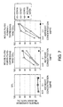
- the intensity of ADCC activity can be measured by the method described below. Fifty ⁇ L each of the target cells and the test antibody are added to a 96-well U-bottomed plate (manufactured by Becton Dickinson), and reacted for 15 minutes on ice. Thereafter, 100 ⁇ L of the cells expressing a chimeric receptor of the present invention are added as the effector cells, and incubated in a carbon dioxide gas incubator for four hours. The final concentration of the antibody is adjusted to 0 or 10 ⁇ g/mL. After incubation, 100 ⁇ L of the supernatant is collected, and the radioactivity is measured by a gamma counter (COBRAII AUTO-GAMMA, MODEL D5005, manufactured by Packard Instrument Company).
- the cytotoxic activity (%) can be calculated according to the equation: (A - C) / (B - C) x 100, wherein A represents the radioactivity (cpm) in each sample, B represents the radioactivity (cpm) in a sample to which 1% NP-40 (manufactured by Nacalai Tesque) has been added, and C represents the radioactivity (cpm) of a sample containing only the target cells.
- the antibodies having cytotoxic activity that are selected by the screening methods of the present invention can be used as pharmaceuticals for treating or preventing various types of diseases.
- the antibodies may be used as therapeutic agents or preventive agents against diseases such as cancers or autoimmune diseases.
- the present invention further provides methods of producing antibodies having cytotoxic activity, which use the chimeric receptors of the present invention. Specifically, the production can be carried out by methods comprising the steps of:
- the gene encoding the selected antibody may be a gene encoding an antibody having an amino acid sequence that is identical to the full amino acid sequence of the selected antibody, or a gene encoding an antibody having an amino acid sequence partially identical to that of the selected antibody.
- Preferred examples of an antibody having an amino acid sequence partially identical to that of the selected antibody include antibodies having variable regions identical to those of the selected antibody, and antibodies having complementarity determining regions (CDRs) identical to those of the selected antibody. Methods for substituting regions other than variable regions or CDRs with sequences derived from other antibodies are known (see, European Patent Publication No. EP 239,400 , and International Patent Publication No. WO 96/02576 ).
- these antibodies When using these antibodies as pharmaceuticals for humans or mammals, besides administering them directly as they are to patients, they can be administered as formulations produced by known preparation methods.
- the antibodies may be administered orally as tablets, capsules, or such; or parenterally in the form of injections of sterile solutions or suspensions prepared with water or other pharmaceutically acceptable liquids.
- the antibodies may be formulated by appropriately combining them with pharmaceutically acceptable carriers or media, more specifically, sterilized water or physiological saline solutions, vegetable oils, emulsifiers, suspending agents, surfactants, stabilizers, flavoring agents, vehicles, preservatives, binding agents, and such, and mixing them into a unit dosage form required for generally accepted pharmaceutical practice.
- Additives that can be mixed into tablets and capsules include, for example, binding agents such as gelatin, cornstarch, tragacanth gum, and gum arabic; excipients such as crystalline cellulose; swelling agents such as cornstarch, gelatin, alginic acid; lubricants such as magnesium stearate; sweeteners such as sucrose, lactose, and saccharine; and flavoring agents such as peppermint and Gaultheria adenothrix oils, and cherry.
- binding agents such as gelatin, cornstarch, tragacanth gum, and gum arabic
- excipients such as crystalline cellulose
- swelling agents such as cornstarch, gelatin, alginic acid
- lubricants such as magnesium stearate
- sweeteners such as sucrose, lactose, and saccharine
- flavoring agents such as peppermint and Gaultheria adenothrix oils, and cherry.
- Sterile compositions to be injected can be formulated using a
- Aqueous solutions for injections include, for example, physiological saline and isotonic solutions containing glucose or other adjunctive agents such as D-sorbitol, D-mannose, D-mannitol, and sodium chloride.
- the solutions may also be combined with appropriate solubilizing agents such as alcohol, more specifically, ethanol, polyalcohol such as propylene glycol or polyethylene glycol, or non-ionic surfactants such as Polysorbate 80 or HCO-50.
- Oil solutions include sesame oils and soybean oils, and can be combined with solubilizing agents such as benzyl benzoate or benzyl alcohol.
- Injection solutions may also be formulated with buffers such as phosphate buffers or sodium acetate buffers; analgesics such as procaine hydrochloride; stabilizers such as benzyl alcohol or phenol; or anti-oxidants.
- buffers such as phosphate buffers or sodium acetate buffers
- analgesics such as procaine hydrochloride
- stabilizers such as benzyl alcohol or phenol
- anti-oxidants such as sodium acetate buffers
- the solutions prepared are typically aliquoted into appropriate ampules.
- Administration to patients may be performed, for example, by intra-arterial injection, intravenous injection, or subcutaneous injection, alternatively, by intranasal, transbronchial, intramuscular, transdermal, or oral administration, using methods known to those skilled in the art.
- mouse Fc ⁇ R4 gene was amplified by PCR utilizing a sense primer containing an EcoRI restriction enzyme sequence (mFcR4-EcoRI-F, SEQ ID NO: 17) and an antisense primer containing a NotI restriction enzyme sequence (mFcR4-NotI-R, SEQ ID NO: 18). After treatment with the EcoRI and NotI restriction enzymes, the amplified product was cloned into the EcoRI-NotI site of the pMCDN plasmid for expression in mammalian cells to produce pMCDN/mFcR4.
- the pMCDN vector into which the neomycin resistance gene and the DHFR gene are inserted, enables induced expression under the control of the mouse CMV promoter (Accession No. U68299).
- the nucleotide sequence cloned was determined by sequencing using an ABI3730 DNA sequencer.
- the nucleotide sequence and the amino acid sequence of mouse Fc ⁇ R4 are shown in SEQ ID NOs: 3 and 4, respectively.
- the nucleotide at position 422 had been changed from C to T in the sequence obtained; therefore, the amino acid at position 141 had been changed from serine to leucine.
- mouse Fc ⁇ R3 gene was amplified by PCR utilizing a sense primer containing an EcoRI restriction enzyme sequence (mFcR3-EcoRI-F, SEQ ID NO: 19) and an antisense primer containing a Not I restriction enzyme sequence (mFcR3-NotI-R, SEQ ID NO: 20).
- EcoRI restriction enzyme sequence mFcR3-EcoRI-F, SEQ ID NO: 19
- an antisense primer containing a Not I restriction enzyme sequence mFcR3-NotI-R, SEQ ID NO: 20
- the amplified product was cloned into the EcoRI-NotI site of the pMCDN plasmid to produce pMCDN/mFcR3.
- the nucleotide sequence cloned was determined by sequencing using an ABI3730 DNA sequencer. The nucleotide sequence and the amino acid sequence of mouse Fc ⁇ R3 are shown in SEQ ID NOs: 1 and 2, respectively.
- mouse Fc ⁇ R4 Using the pMCDN/mFcR4 plasmid, into which the mouse Fc ⁇ R4 gene has been inserted, as a template, the extracellular domain of mouse Fc ⁇ R4 was amplified by PCR utilizing a sense primer (mFcR4-EcoRI-F) and an antisense primer (m4h3-mR, SEQ ID NO: 21).
- pMCDN/hFcR3 plasmid which was prepared by inserting the human Fc ⁇ R3 gene (nucleotide sequence: SEQ ID NO: 5; amino acid sequence: SEQ ID NO: 6) into pMCDN, as a template, the transmembrane domain and intracellular domain of human Fc ⁇ R3 were amplified by PCR utilizing a sense primer (m4h3-hF, SEQ ID NO: 22) and an antisense primer (vector primer: pMCM-R1, SEQ ID NO: 23).
- the products were further amplified using the mFcR4-EcoRI-F primer and pMCM-R1 primer, treated with the EcoRI and NotI restriction enzymes, and then inserted into the EcoRI-NotI site of the pMCDN plasmid to construct the mouse Fc ⁇ R4/human Fc ⁇ R3 chimeric (mouse Fc ⁇ R4/human Fc ⁇ R3) expression vector (pMCDN/mFcR4-hFcR3).
- the nucleotide sequence cloned was determined by sequencing using an ABI3730 DNA sequencer. The nucleotide sequence and the amino acid sequence of mouse Fc ⁇ R4/human Fc ⁇ R3 are shown in SEQ ID NOs: 13 and 14, respectively.
- mouse Fc ⁇ R4 Using the pMCDN/mFcR4 plasmid, into which the mouse Fc ⁇ R4 gene has been inserted, as a template, the extracellular domain of mouse Fc ⁇ R4 was amplified by PCR utilizing a sense primer (mFcR4-EcoRI-F) and an antisense primer (m4hG-mR, SEQ ID NO: 24).
- the products were further amplified using the mFcR4-EcoRI-F primer and m4hG-hR primer, treated with the EcoRI restriction enzyme, and then inserted into the EcoRI-EcoRV site of the pMCDN plasmid to construct the mouse Fc ⁇ R4/human ⁇ chain chimeric (mouse Fc ⁇ R4/human ⁇ ) expression vector (pMCDN/mFcR4-hG).
- the nucleotide sequence cloned was determined by sequencing using an ABI3730 DNA sequencer. The nucleotide sequence and the amino acid sequence of mouse Fc ⁇ R4/human ⁇ are shown in SEQ ID NOs: 15 and 16, respectively.
- mouse Fc ⁇ R3 Using the pMCDN/mFcR3 plasmid, into which the mouse Fc ⁇ R3 gene has been inserted, as a template, the extracellular domain of mouse Fc ⁇ R3 was amplified by PCR utilizing a sense primer (mFcR3-EcoRI-F) and an antisense primer (m3h3-mR, SEQ ID NO: 27).
- the transmembrane domain and intracellular domain of human Fc ⁇ R3 was amplified by PCR utilizing a sense primer (m3h3-hF, SEQ ID NO: 28) and an antisense primer (pMCM-R1).
- the products were further amplified using the mFcR3-EcoRI-F primer and pMCM-R1 primer, treated with EcoRI and NotI restriction enzymes, and then inserted into the EcoRI-NotI site of the pMCDN plasmid to construct the mouse Fc ⁇ R3/human Fc ⁇ R3 chimeric (mouse Fc ⁇ R3/human Fc ⁇ R3) expression vector (pMCDN/mFcR3-hFcR3).
- the nucleotide sequence cloned was determined by sequencing using an ABI3730 DNA sequencer. The nucleotide sequence and the amino acid sequence of mouse Fc ⁇ R3/human Fc ⁇ R3 are shown in SEQ ID NOs: 9 and 10, respectively.
- mouse Fc ⁇ R3 Using the pMCDN/mFcR3 plasmid, into which the mouse Fc ⁇ R3 gene has been inserted, as a template, the extracellular domain of mouse Fc ⁇ R3 was amplified by PCR utilizing a sense primer (mFcR3-EcoRI-F) and an antisense primer (m3hG-mR, SEQ ID NO: 29).
- the two amino acids of the extracellular domain, the transmembrane domain, and the intracellular domain of human ⁇ chain were amplified by PCR utilizing a sense primer (m3hG-hF, SEQ ID NO: 30) and an antisense primer (pMCM-R1).
- mice Fc ⁇ R3/human ⁇ chain chimeric (mouse Fc ⁇ R3/human ⁇ ) expression vector pMCDN/mFcR3-hG.
- the nucleotide sequence and the amino acid sequence of mouse FcR3/human ⁇ are shown in SEQ ID NOs: 11 and 12, respectively.
- NK92 cell lines that stably express mouse Fc ⁇ R4/human Fc ⁇ R3, mouse Fc ⁇ R4/human ⁇ , mouse Fc ⁇ R3/human Fc ⁇ R3, mouse Fc ⁇ R3/human ⁇ , and human Fc ⁇ R3 were established by digesting the pMCDN/mFcR4-hFcR3, pMCDN/mFcR4-hG, pMCDN/mFcR3-hFcR3, pMCDN/mFcR3-hG, and pMCDN/hFcR3 plasmids with the PvuI restriction enzyme, then introducing the digested plasmids into the NK92 cell line (purchased from ATCC) by electroporation, and selecting the cells with 500 ⁇ g/mL Geneticin (Invitrogen).
- NK92 cell lines were incubated in Alpha Minimum Essential Medium without ribonucleosides and deoxyribonucleosides with L-glutamine (Invitrogen) containing 500 ⁇ g/ml Geneticin, penicillin/streptomycin (Invitrogen), 100 U/ml recombinant human interleukin-2 (Peprotech), 10% fetal bovine serum (FBS, Invitrogen), 10% horse serum (Invitrogen), 0.11 mM 2-mercaptoethanol (Invitrogen), 0.2 mM inositol (Sigma), and 0.02 mM folic acid (Sigma).
- a CHO cell line stably expressing human desmoglein 3 (DSG3) (DSG3-DG44) was established by digesting the pMCN/DSG3 plasmid for expression in mammalian cells, into which the DSG3 gene (nucleotide sequence: SEQ ID NO: 31; amino acid sequence: SEQ ID NO: 32) has been inserted, with the Pvu I restriction enzyme, then introducing the digested plasmid into the CHO DG44 cell line (Invitrogen) by electroporation, and selecting the cells with 500 ⁇ g/mL Geneticin.
- pMCN enables induced expression under the control of the mouse CMV promoter (Accession No.
- DSG3-DG44 cells were incubated in CHO-S-SFM II medium (Invitrogen) containing 500 ⁇ g/mL Geneticin, HT supplement (Invitrogen), and penicillin/streptomycin.
- Soluble human desmoglein 3/mouse IgG2a-Fc fusion protein (DSG3-Fc) was prepared as an immunizing antigen for producing anti-DSG3 antibodies.
- a gene constructed by linking the DSG3 extracellular domain (Metl-Leu616) with the mouse IgG2a constant region at the CpoI restriction enzyme sequence of the hinge region of the mouse IgG2a constant region (DSG3-Fc; nucleotide sequence: SEQ ID NO: 33; amino acid sequence: SEQ ID NO: 34) was cloned into the pMCDN plasmid to produce pMCDN/DSG3-Fc.
- a CHO cell line stably expressing DSG3-Fc (DSG3-Fc-DG44) was established by introducing the pMCDN/DSG3-Fc plasmid into DG44 cells by electroporation, and selecting the cells with 500 ⁇ g/mL of Geneticin. Then, DSG3-Fc was purified from the culture supernatant of DSG3-Fc-DG44. The culture supernatant was applied to a Hi Trap Protein G HP column (Cat. No. 17-0404-01, GE Healthcare Bio-Sciences), and after washing with a binding buffer (20 mM sodium phosphate, pH 7.0), elution was carried out using an elution buffer (0.1 M glycine-HCl, pH 2.7).
- the eluate was immediately neutralized by elution into a tube containing a neutralization buffer (1 M Tris-HCl (pH 9.0)). This eluate was subjected to gel filtration using Superdex 200 HR 10/30 (GE Healthcare Bio-Sciences) to replace the solvent with PBS. Purified DSG3-Fc was quantified using a DC protein assay kit (BIO-RAD) and converting to a concentration using bovine IgG included in the kit as standard.
- MRL/MpJUmmCrj-lpr/lpr mice (7- to 8-weeks old, purchased from Charles River Japan) were used as the animals for immunization.
- 100 ⁇ g of DSG3-Fc was emulsified using Freund's complete adjuvant (Beckton Dickinson), and administered subcutaneously.
- boosting immunization was carried out by emulsifying 50 ⁇ g of DSG3-Fc using Freund's incomplete adjuvant (Beckton Dickinson), and administering it subcutaneously. Thereafter, boosting immunizations were performed at one-week intervals for three times.
- DSG3-Fc For the final immunization, 50 ⁇ g of DSG3-Fc was administered into the tail vein.
- spleen cells were extirpated and mixed with mouse myeloma cells P3-X63Ag8U1 (purchased from ATCC) at 2:1 ratio, and cell fusion was carried out by addition of PEG 1500 (Roche Diagnostics).
- RPMI 1640 medium Invitrogen was added, and then PEG 1500 was removed by centrifuging and removing the supernatant.
- the fused cells suspended in RPMI 1640 containing 10% FBS was seeded into a 96-well plate at 100 ⁇ L/well.
- RPMI 1640 medium containing 10% FBS, 1x HAT media supplement (Sigma), and 0.5x BM-Condimed H1 Hybridoma cloning supplement (Roche Diagnostics) (HAT medium) was added at 100 ⁇ L/well. Two days later and three days later, half of the culture solution was replaced with HAT medium, and the day-7 culture supernatant was used for screening. The screening was performed by flow cytometry that detects binding to DSG3-DG44 cells. Positive clones obtained by this screening were monocloned by the limiting dilution method to establish a hybridoma that produces DF366, which is a monoclonal antibody that specifically binds to DSG3.
- This hybridoma was cultured in a HAT medium supplemented with Ultra-Low IgG FBS (Invitrogen) instead of FBS, and the DF366 antibody was purified from this culture supernatant using a Hi Trap Protein G HP column. The solvent was replaced with PBS using a PD-10 column (GE Healthcare Bio-Sciences). The concentration of the purified DF366 antibody was quantified using a DC protein assay kit. The isotype of the DF366 antibody was determined to be mouse IgG1 ⁇ by analysis using Isostrip (Roche Diagnostics).
- Binding of the DF366 antibody to DSG3-DG44 cells was detected by flow cytometry. 5 x 10 4 cells of DSG3-DG44 was reacted with 3 ⁇ g/mL of DF366 antibody on ice for 30 minutes, and then washed. Then, this was reacted with an FITC-labeled anti-mouse IgG antibody (Beckman Coulter) as the secondary antibody on ice for 30 minutes, washed, and then subjected to flow cytometry. FACS Calibur (Becton Dickinson) was used as the flow cytometer. The DF366 antibody bound to DSG3-DG44 cells, but not to the parental DG44 cells. Therefore, the DF366 antibody was confirmed to bind specifically to DSG3.
- the H-chain variable region gene (nucleotide sequence: SEQ ID NO: 35; amino acid sequence: SEQ ID NO: 36) and the L-chain variable region gene (nucleotide sequence: SEQ ID NO: 37; amino acid sequence: SEQ ID NO: 38) of DF366 antibody were cloned from a hybridoma that produces the DF366 antibody. Then, these genes were ligated in frame with the nucleotide sequences of the H-chain constant region gene and L-chain ( ⁇ -chain) constant region gene of human IgG1.
- PCR was performed using a primer having the 5'-end nucleotide sequence of the H-chain variable region gene, a Kozak sequence, and an EcoRI restriction enzyme sequence, and an antisense primer having a sequence complementary to the 3'-end nucleotide sequence and a NheI restriction enzyme sequence.
- PCR was also performed using a primer having the 5'-end nucleotide sequence of the L-chain variable region gene, a Kozak sequence, and a BamHI restriction enzyme sequence, and an antisense primer having a sequence complementary to the 3'-end nucleotide sequence and the BsiWI restriction enzyme sequence.
- the amplification products obtained were treated with the EcoRI and NheI restriction enzymes, or BamHI and BsiWI restriction enzymes, and inserted into the EcoRI-NheI site or BamHI-BsiWI site of the human IgG1 chimeric antibody expression plasmid (pMCDN/G1k), to produce pMCDN/Glk-DF366.
- pMCDN/G1k was produced by cloning the H-chain constant region gene (nucleotide sequence: SEQ ID NO: 39; amino acid sequence: SEQ ID NO: 40) and the L-chain ( ⁇ -chain) constant region gene (nucleotide sequence: SEQ ID NO: 41; amino acid sequence: SEQ ID NO: 42) of human IgG1 cloned, into the pMCDN plasmid.
- the mouse H-chain variable region and the human H-chain constant region were linked by a NheI restriction enzyme sequence, and the mouse L-chain variable region and the human L-chain constant region were linked by the BsiWI restriction enzyme sequence.
- the pMCDN/G1k-DF366 plasmid was introduced into DG44 cells by electroporation.
- the CHO DF366c-DG44 cells which stably express the human IgG1 chimeric DF366 antibody (DF366c), were established by selection with 500 ⁇ g/mL of Geneticin.
- the DF366c antibody was purified from the culture supernatant of DF366c-DG44 using a Hi Trap rProtein A column (GE Healthcare Bio-Science). The solvent was replaced with PBS using a PD-10 column. The concentration of the DF366c antibody purified was quantified using a DC protein assay kit.
- nucleotide sequence of the full-length DF366c antibody H-chain gene and the corresponding amino acid sequence are shown in SEQ ID NOs: 43 and 44, respectively.
- nucleotide sequence of the full length DF366c antibody L-chain gene and the corresponding amino acid sequence are shown in SEQ ID NOs: 45 and 46, respectively.
- Binding of the DF366c antibody to DSG3-DG44 cells and DG44 cells was detected by flow cytometry. 3 x 10 4 cells were reacted with 10 ⁇ g/mL of DF366c antibody on ice for one hour, and then washed. Then, this was reacted with an FITC-labeled anti-human IgG antibody (Beckman Coulter) as the secondary antibody on ice for one hour, washed, and then subjected to flow cytometry. FACS Calibur (Becton Dickinson) was used as the flow cytometer. DF366c antibody bound to DSG3-DG44 cells, but not to the parental DG44 cells. Therefore, the DF366c antibody was confirmed to bind specifically to DSG3.
- the nucleotide sequence of the DF366 antibody H-chain variable region gene was ligated in frame with the nucleotide sequence of the mouse IgG2a H-chain constant region gene. PCR was performed using a primer having the 5'-end nucleotide sequence of the H-chain variable region gene, a Kozak sequence, and an EcoRI restriction enzyme sequence, and an antisense primer having a cytosine residue attached to a sequence complementary to the 3'-end nucleotide sequence.
- the amplification product obtained was treated with the EcoRI restriction enzyme, and inserted into the EcoRI-NruI site of the mouse IgG2a chimeric H-chain expression plasmid (pMCD/G2a) to construct the mouse IgG2a chimeric DF366 antibody H chain expression vector (pMCD/G2a-DF366).
- pMCD/G2a was produced by cloning the mouse IgG2a H-chain constant region gene (nucleotide sequence: SEQ ID NO: 47; amino acid sequence: SEQ ID NO: 48) into the pMCD plasmid for expression in mammalian cells.
- the H-chain constant region was ligated to the H-chain variable region via the Nrul restriction enzyme sequence.
- the pMCD vector, into which the DHFR gene is inserted enables induced expression under the control of the mouse CMV promoter (Accession No. U68299).
- the nucleotide sequence of the DF366 antibody L-chain variable region gene was ligated in frame with the nucleotide sequence of the L-chain ( ⁇ -chain) constant region gene of mouse IgG2a.
- PCR was performed using a primer having the 5'-end nucleotide sequence of the L-chain variable region gene, a Kozak sequence, and an EcoRI restriction enzyme sequence, and an antisense primer having "gcccg" residues attached to a sequence complementary to the 3'-end nucleotide sequence.
- the amplification product obtained was treated with the EcoRI restriction enzyme, and inserted into the EcoRI-NruI site of the mouse IgG2a chimeric L-chain ( ⁇ -chain) expression plasmid (pMCN/k) to construct the mouse IgG2a chimeric DF366 antibody L chain expression vector (pMCN/k-DF366).
- pMCN/k was produced by cloning the mouse IgG2a L-chain ( ⁇ -chain) constant region gene (nucleotide sequence: SEQ ID NO: 49; amino acid sequence: SEQ ID NO: 50) into the pMCN plasmid.
- the L-chain ( ⁇ -chain) constant region was ligated to the L-chain variable region via the Nrul restriction enzyme sequence.
- the pMCD/G2a-DF366 and pMCN/k-DF366 plasmids were introduced into DG44 cells by electroporation.
- the CHO DF366m-DG44 cells which stably express the mouse IgG2a chimeric DF366 antibody (DF366m) were established by selection in a nucleic acid (HT supplement)-free medium containing 500 ⁇ g/mL of Geneticin. Subsequently, the DF366m antibody was purified from the culture supernatant of DF366m-DG44 using a Hi Trap Protein G HP column. The solvent was substituted with PBS using a PD-10 column.
- the concentration of the DF366m antibody purified was quantified using a DC Protein Assay kit.
- the DF366m antibody was subjected to flow cytometric analysis to confirm that the antibody specifically binds to DSG3 in the same way as the DF366c antibody.
- An FITC-labeled anti-mouse IgG antibody (Beckman Coulter) was used as the secondary antibody.
- the nucleotide sequence of the full-length DF366m antibody H-chain gene and the corresponding amino acid sequence are shown in SEQ ID NO: 51 and SEQ ID NO: 52, respectively.
- the nucleotide sequence of the full-length DF366m antibody L-chain gene and the corresponding amino acid sequence are shown in SEQ ID NO: 53 and SEQ ID NO: 54, respectively.
- the pMCDN/G1k-DF366 plasmid was introduced into a fucose transporter knockout CHO cell line (FTPKO-DXB 11 cells, International Patent Publication Nos. WO 2006/067913 and WO 2006/067847 ) by electroporation.
- FTPKO-DXB 11 cells International Patent Publication Nos. WO 2006/067913 and WO 2006/067847
- Low-fucose DF366c-DXB11 CHO cells stably expressing the low-fucose human IgG1 chimeric DF366 antibody (low-fucose DF366c) was established by selection with 500 ⁇ g/mL Geneticin.
- the low-fucose DF366c antibody was purified from the culture supernatant of low-fucose DF366c-DXB11 using a Hi Trap rProtein A column.
- the solvent was substituted with PBS using a PD-10 column, and the antibody concentration was quantified using a DC Protein Assay kit.
- the pMCD/G2a-DF366 and pMCN/k-DF366 plasmids were introduced into FTPKO-DXB11 cells by electroporation.
- Low-fucose DF366m-DXB11 CHO cells stably expressing the low-fucose mouse IgG2a chimeric DF366 antibody (low-fucose DF366m)
- the low-fucose DF366m antibody was purified from the culture supernatant of low-fucose DF366m-DXB11 using a Hi Trap Protein G HP column.
- the solvent was substituted with PBS using a PD-10 column, and the antibody concentration was quantified using a DC Protein Assay kit.
- DSG3-Ba/F3 a Ba/F3 cell line stably expressing DSG3, was established by digesting, with the PvuI restriction enzyme, the pMCDN/DSG3 plasmid for expression in mammalian cells, into which the human desmoglein 3 (DSG3) gene (nucleotide sequence: SEQ ID NO: 31; amino acid sequence: SEQ ID NO: 32) has been inserted, then introducing the digested plasmid into Ba/F3 cells (purchased from RIKEN BioResource Center) by electroporation, and selecting the cells with 500 ⁇ Lg/mL Geneticin.
- DSG3 desmoglein 3
- DSG3-Ba/F3 cells were incubated using RPMI 1640 medium (Invitrogen) containing 500 ⁇ g/mL Geneticin, penicillin/streptomycin, recombinant mouse interleukin-3 (R&D Systems), and 10% FBS.
- DSG3-Ba/F3 cells were reacted with 10 ⁇ g/mL of an anti-DSG3 monoclonal antibody (R&D Systems) or a negative control antibody (mouse IgG2a, Becton Dickinson) on ice for one hour, and then washed. Subsequently, this was reacted with a secondary antibody (FITC-labeled anti-mouse Ig antibody, Becton Dickinson) on ice for one hour, washed, and then analyzed using a flow cytometer (FACS Calibur, Becton Dickinson).
- an anti-DSG3 monoclonal antibody R&D Systems
- a negative control antibody mouse IgG2a, Becton Dickinson
- RPMI 1640 medium containing penicillin/streptomycin and 10% FBS (RPMI medium) was used for the experiment.
- 1 x 10 6 cells of the DSG3-Ba/F3 cell line were suspended in approximately 200 ⁇ L of RPMI medium containing 3.7 MBq of Chromium-51 (GE Healthcare Bio-Sciences), and then incubated in a 5% carbon dioxide gas incubator at 37°C for one hour. After washing, the cell density was adjusted to 2 x 10 5 cells/mL, and then the cells were added to a 96-well U-bottomed plate at 50 ⁇ L/well. Then, 50 ⁇ L of an antibody solution was added to each well.
- the plate was left to stand at room temperature for 15 minutes, and then 100 ⁇ L of effector cells (described below) were added to each well. The plate was then left to stand in a 5% carbon dioxide gas incubator at 37°C for four to six hours. Thereafter, 100 ⁇ L of the supernatant was collected from each well, and used for the radioactivity measurement on a gamma counter (1480 WIZARD 3", Wallac).
- the assay was conducted in duplicates, and the mean value and standard deviation were calculated for the specific chromium release rate.
- SPL Human interleukin-2
- SPL-LAK recombinant human interleukin-2
- the ADCC activity was measured according to 3-2). The number of effector cells per well was 5 x 10 5 cells (SPL) or 2 x 10 5 cells (SPL-LAK), and the ADCC induction time was six hours.
- Mouse IgG2a Becton Dickinson
- human IgG1 Session Induction time was six hours.
- Fc ⁇ R-expressing NK92 cell lines (Example 1, mouse Fc ⁇ R4/human Fc ⁇ R3-, mouse Fc ⁇ R4/human ⁇ -, mouse Fc ⁇ R3/human Fc ⁇ R3-, mouse Fc ⁇ R3/human ⁇ -, and human Fc ⁇ R3-expressing NK92 cells) were used as the effector cells.
- the ADCC activity was measured according to 3-2). The number of effector cells per well was 5 x 10 4 cells, and the ADCC induction time was four hours.
- Mouse IgG2a Becton Dickinson
- human IgG1 Seseton Dickinson
- the ADCC activity was low in mouse Fc ⁇ R3/human Fc ⁇ R3-expressing NK92 cells. This was speculated to be caused by low expression level of Fc ⁇ R ( Fig. 5 ).
- chimeric Fc ⁇ R-expressing NK92 cells are advantageous for measuring the ADCC activity of mouse antibodies.
- the ADCC activity of anti-claudin 3 monoclonal antibody was measured ( Fig. 7 ).
- the MCF7 breast cancer cell line purchased from ATCC
- the antibodies used were CDN16 (mouse IgG2b, H-chain amino acid sequence: SEQ ID NO: 55; L-chain amino acid sequence: SEQ ID NO: 56), CDN27 (mouse IgG2a, H-chain amino acid sequence: SEQ ID NO: 57, L-chain amino acid sequence: SEQ ID NO: 58), and CDN35 (mouse IgG2a, H-chain amino acid sequence: SEQ ID NO: 59, L-chain amino acid sequence: SEQ ID NO: 60).
- chimeric proteins of the present invention in effector cells greatly reduces the labor for preparing effector cells for measuring the ADCC activity of mouse antibodies, and enables accurate measurements with small lot-to-lot differences.
- effector cells expressing the chimeric proteins of the present invention antibodies having cytotoxic activity, which are used for the treatment or prevention of diseases such as cancers or autoimmune diseases, can be efficiently screened.
Landscapes
- Health & Medical Sciences (AREA)
- Life Sciences & Earth Sciences (AREA)
- Chemical & Material Sciences (AREA)
- Immunology (AREA)
- Engineering & Computer Science (AREA)
- Molecular Biology (AREA)
- Biomedical Technology (AREA)
- Organic Chemistry (AREA)
- Medicinal Chemistry (AREA)
- General Health & Medical Sciences (AREA)
- Biochemistry (AREA)
- Urology & Nephrology (AREA)
- Hematology (AREA)
- Toxicology (AREA)
- Cell Biology (AREA)
- Proteomics, Peptides & Aminoacids (AREA)
- Genetics & Genomics (AREA)
- Biophysics (AREA)
- Analytical Chemistry (AREA)
- General Physics & Mathematics (AREA)
- Microbiology (AREA)
- Pathology (AREA)
- Food Science & Technology (AREA)
- Physics & Mathematics (AREA)
- Biotechnology (AREA)
- Bioinformatics & Cheminformatics (AREA)
- Tropical Medicine & Parasitology (AREA)
- Zoology (AREA)
- Gastroenterology & Hepatology (AREA)
- Peptides Or Proteins (AREA)
- Micro-Organisms Or Cultivation Processes Thereof (AREA)
- Preparation Of Compounds By Using Micro-Organisms (AREA)
- Investigating Or Analysing Biological Materials (AREA)
- Measuring Or Testing Involving Enzymes Or Micro-Organisms (AREA)
Abstract
Description
- The present invention relates to chimeric Fcγ receptors of a human Fcγ receptor or the human γ chain with a mouse Fcγ receptor.
- When developing antibody pharmaceuticals with a drug action mechanism based on antibody-dependent cell-mediated cytotoxicity (ADCC), it is important to select clones with high ADCC activity. ADCC activity is evaluated using cells expressing an antigen of interest (target cells) and effector cells that kill those target cells. Effector cells recognize the Fc region of antibodies bound to the target cells via the Fcγ receptor (FcγR). Signals transmitted from FcγR causes the effector cells to kill the target cells. FcγR binds to a molecule called the γ chain through its transmembrane domain, and transmits ADCC signals via this γ chain (Non-patent
Documents 1 to 3). Mouse FcγR3 and FcγR4, and human FcγR3 are known as FcγRs that induce ADCC. Amino acid sequence comparisons of the transmembrane domains of human and mouse FcγRs show that five out of the 21 amino acids are different between human FcγR3 and mouse FcγR3, and seven out of the 21 amino acids are different between human FcγR3 and mouse FcγR4. Human γ chain and mouse γ chain comparisons show that one out of the 21 amino acids is different between the sequences in the transmembrane domains (Non-patent Document 4). - When measuring the ADCC activity of human antibodies, human NK cells are used as effector cells. Human NK cells can be purified from human peripheral blood mononuclear cells (PBMC) using the NK Cell Isolation Kit II (Miltenyi Biotec K.K.). Alternatively, PBMC can be used directly as effector cells. PBMC can be purchased (from Cambrex Corporation), or can be prepared from fresh peripheral blood collected from volunteers. However, when such cells are used as effector cells, the drawbacks include lot-to-lot differences and laborious preparation.
- To avoid such drawbacks, systems that use human NK cell lines as effector cells have been developed for measuring the ADCC activity of human antibodies. The NK92 human NK cell line (ATCC) does not express human FcγR, but expresses the human γ chain (Non-patent Document 5). Therefore, ADCC activity can be induced by forcedly-expressing human FcγR3 in the NK92 human NK cell line (Non-patent Documents 6 and 7). This greatly reduced preparation labor and enabled accurate measurements having small lot-to-lot differences. Furthermore, it has been reported that chimeric molecules produced by fusing the extracellular domain of human FcγR3 and the transmembrane domain and intracellular domain of human γ chain induce ADCC activity related to human antibodies (Non-patent Document 8).
- On the other hand, when measuring the ADCC activity of mouse antibodies, mouse spleen cells are used as effector cells (Non-patent Documents 9 and 10). To prepare mouse spleen cells, it is necessary to remove the spleen from mice, hemolyze erythrocytes, and activate NK cells with interleukin 2. However, since spleen cells prepared in this manner have high natural killer activity to kill target cells in an antibody-independent manner, the ADCC activity may not be measurable depending on the type of target cells. Furthermore, preparation of the effector cells requires effort.
- Systems for measuring the ADCC activity of human antibodies using human NK cell lines have been developed. However, since the use of mouse NK cell lines is generally unknown, a system for conveniently measuring the ADCC activity of mouse antibodies using an NK cell line has not been established. Furthermore, since there are sequence differences between Fcγ receptors and γ chains between human and mouse, even if mouse FcγR is expressed as it is in human NK92 cells, mouse FcγR will not be able to bind to the human γ chain with similar strength as human FcγR.
- Therefore, to measure the ADCC activity of mouse antibodies, it was necessary to use a method that requires much effort such as the method of preparing mouse spleen cells as described above, or the method of preparing a chimeric antibody in which the antibody Fc regions have been replaced with those of a human antibody.
- Prior art literature information relating to the present invention is shown below.
- [Non-patent Document 1]
Blood 2003, 101, 4479 - [Non-patent Document 2] J. Immunol. 1991, 146, 1571
- [Non-patent Document 3] Immunol. Lett. 2004, 92, 199
- [Non-patent Document 4] J. Biol. Chem. 2006, 281, 17108
- [Non-patent Document 5] Leukemia Res. 2003, 27, 935
- [Non-patent Document 6] 97th AACR annual meeting 2006, abstract number 635
- [Non-patent Document 7] J. Biol. Chem. 2004, 279, 53 907
- [Non-patent Document 8] Blood 2006, 107, 4669
- [Non-patent Document 9] Oncol. Rep. 2006, 15, 361
- [Non-patent Document 10] Cell. Immunol. 1988, 115, 257
- An objective of the present invention is to provide chimeric receptors comprising a mouse Fcγ receptor extracellular domain and a human Fcγ receptor transmembrane domain, and chimeric receptors comprising a mouse Fcγ receptor extracellular domain and a human γ chain transmembrane domain. Another objective of the present invention is to provide methods for measuring the ADCC activity of mouse antibodies using the chimeric receptors. A further objective of the present invention is to provide methods of screening for mouse antibodies having ADCC activity using the chimeric receptors.
- To achieve the above-mentioned objectives, the present inventors produced chimeric molecules by fusing the extracellular domain of mouse FcγR3 or mouse FcγR4 with the Tansmembrane domain and intracellular domain of human γ chain or human FcγR3, and expressed the chimeric molecules in human NK92 cells. The present inventors found that any one of the combinations of mouse FcγR3 and human γ chain, mouse FcγR3 and human FcγR3, mouse FcγR4 and human γ chain, and mouse FcγR4 and human FcγR3 can induce the ADCC activity. Thus, the present inventors discovered that the ADCC activity of mouse antibodies can be measured by using a chimeric receptor produced from a mouse Fcγ receptor and a human Fcγ receptor, or a chimeric receptor produced from a mouse Fcγ receptor and the human γ chain. Furthermore, the present inventors discovered that mouse antibodies having ADCC activity can be screened for by using a chimeric receptor produced from a mouse Fcγ receptor and a human Fcγ receptor, or produced from a mouse Fcγ receptor and the human γ chain.
- Specifically, the present application provides the following:
- [1] a chimeric protein comprising a mouse Fcγ receptor extracellular domain and a human γ chain transmembrane domain, wherein the mouse Fcγ receptor is a mouse Fcγ receptor 3; or is a mouse Fcγ receptor 4;
- [2] the chimeric protein of [1]: wherein the chimeric protein further comprises a human γ chain intracellular domain;
- [3] a chimeric protein comprising a mouse Fcγ receptor extracellular domain and a human Fcγ receptor transmembrane domain, wherein the mouse Fcγ receptor is a mouse Fcγ receptor 3 or is a mouse Fcγ receptor 4;
- [4] the chimeric protein of [3], wherein the chimeric protein further comprises a human Fcγ receptor intracellular domain;
- [5] the chimeric protein of [3] or [4], wherein the human Fcγ receptor is a human Fcγ receptor 3;
- [6] a gene encoding the chimeric protein of any one of [1.] to [5];
- [7] a vector comprising the gene of [6];
- [8] a cell expressing the chimeric protein of any one of [1] to [5];
- [9] the cell of [8], wherein the cell is an NK cell or a human-derived cell;
- [10] the cell of [9], wherein the NK cell is a NK-92 cell;
- [11] a method for measuring the cytotoxic activity of an antibody, wherein the method comprises the steps of:
- (a) contacting a test antibody with a cell expressing an antigen to which the test antibody binds;
- (b) contacting the test antibody of (a) and the cell expressing an antigen of (a) with the cell of any one of [8] to [10]; and
- (c) measuring the cytotoxic activity of the test antibody;
- [12] a method of screening for an antibody having cytotoxic activity, wherein the method comprises the steps of:
- (a) contacting a test antibody with a cell expressing an antigen to which the test antibody binds;
- (b) contacting the test antibody of (a) and the cell expressing an antigen of (a) with the cell of any one of [8] to [10];
- (c) measuring the cytotoxic activity of the test antibody; and
- (d) selecting an antibody having cytotoxic activity;
- [13] the measuring method of [11] or the screening method of [12], wherein the test antibody is a mouse-derived antibody;
- [14] use of the chimeric protein of any one of [1] to [5] or the cell of any one of [8] to [10] for measuring cytotoxic activity;
- [15] use of the chimeric protein of any one of [1] to [5] or the cell of any one of [8] to [10] for screening for an antibody having cytotoxic activity;
- The present invention provides chimeric receptors comprising a mouse Fcγ receptor extracellular domain and a human Fcγ receptor transmembrane domain. The present invention also provides chimeric receptors comprising a mouse Fcγ receptor extracellular domain and a human γ chain transmembrane domain.
- The chimeric receptors of the present invention are preferably receptors that exhibit the activity to transmit signals into cells when the Fc region of a mouse antibody is bound to the extracellular domain of the receptors.
- Tne mouse Fcγ receptors used in the present invention are not particularly limited, and any mouse Fcγ receptor may be used. The receptors are preferably mouse Fcγ receptor 3 (FcγR3) and mouse Fcγ receptor 4 (FcγR4).
- Comparing the expression pattern of mouse FcγR3 and mouse FcγR4, mouse FcγR3 is expressed mainly in NK cells, and mouse FcγR4 is expressed in macrophages and neutrophils (Immunity 2005, 23, 41). Mouse FcγR3 binds to mouse IgG1, mouse IgG2a, and mouse IgG2b, while mouse FcγR4 does not bind to mouse IgG1 (Immunity 2005, 23, 41; Science 2005, 310, 1510). Therefore, when evaluating the ADCC activity of various types of mouse antibodies, mouse FcγR3, which allows measurement using even mouse IgG1, is preferably used.
- Known nucleotide sequences of the DNAs encoding mouse Fcγ receptors, and known amino acid sequences thereof can be used. For example, as the nucleotide sequences of DNAs encoding mouse Fcγ receptor 3 and mouse Fcγ receptor 4, and the amino acid sequences thereof, the sequences of SEQ ID NO: 1 (mouse Fcγ receptor 3 nucleotide sequence), SEQ ID NO: 2 (mouse Fcγ receptor 3 amino acid sequence), SEQ ID NO: 3 (mouse Fcγ receptor 4 nucleotide sequence), and SEQ ID NO: 4 (mouse Fcγ receptor 4 amino acid sequence) may be used. Within the amino acid sequence of SEQ ID NO: 2, amino acid positions 31 to 212 correspond to the extracellular domain of mouse Fcγ receptor 3. Within the amino acid sequence of SEQ ID NO: 4, amino acid positions 19 to 201 correspond to the extracellular domain of mouse Fcγ receptor 4.
- The extracellular domain of a receptor may be the entire extracellular domain, or may be a portion thereof. The entire extracellular domain is preferably used, since the receptor activity, such as the activity to bind to an antibody Fc region, can be appropriately retained. When using a portion of the extracellular domain of a receptor, the portion preferably retains the activity to bind to an antibody Fc region. The mouse Fcγ receptor extracellular domain used for the chimeric receptors of the present invention may include amino acid substitutions, deletions, insertions, and/or additions, as long as the domains have the ability to bind to an antibody Fc region. A method of introducing mutations into a protein is well-known to those skilled in the art as a method for preparing proteins that are functionally equivalent to a certain protein. For example, those skilled in the art can prepare such proteins using site-directed mutagenesis (Hashimoto-Gotoh, T. et al. (1995) Gene 152, 271-275; Zoller, MJ, and Smith, M. (1983) Methods Enzymol. 100, 468-500; Kramer, W. et al. (1984) Nucleic Acids Res. 12, 9441-9456; Kramer W, and Fritz HJ (1987) Methods. Enzymol. 154, 350-367; Kunkel, TA (1985) Proc. Natl. Acad. Sci. USA. 82, 488-492; Kunkel (1988) Methods Enzymol. 85, 2763-2766) and such. The number of mutated amino acids in such mutants is generally 50 amino acids or less, preferably 30 amino acids or less, more preferably 20 amino acids or less, still more preferably ten amino acids or less, and yet more preferably five amino acids or less.
- It is desirable that the amino acid residues are mutated into other amino acids in which the properties of the amino acid side chains are conserved. For example, amino acids are categorized as follows depending on the side chain properties: hydrophobic amino acids (A, I, L, M, F, P, W, Y, and V); hydrophilic amino acids (R, D, N, C, E, Q, G, H, K, S, and T); amino acids having aliphatic side chains (G, A, V, L, I, and P); amino acids having hydroxyl-containing side chains (S, T, and Y); amino acids having sulfur atom-containing side chains (C and M); amino acids having carboxylic acid- and amide-containing side chains (D, N, E, and Q); amino acids having base-containing side chains (R, K, and H); and amino acids having aromatic ring-containing side chains (H, F, Y, and W) (amino acids are represented by one-letter codes in parentheses).
- Polypeptides having a modified amino acid sequence, in which one or more amino acid residues in a certain amino acid sequence is deleted, added, and/or substituted with other amino acids, are known to retain the biological activity of the original polypeptides (Mark, D. F. et al., Proc. Natl. Acad. Sci. USA (1984) 81, 5662-5666; Zoller, M. J. & Smith, M. Nucleic Acids Research (1982) 10, 6487-6500; Wang, A. et al., Science 224, 1431-1433; Dalbadie-McFarland, G. et al., Proc. Natl. Acad. Sci. USA (1982) 79, 6409-6413).
- The mouse Fcγ receptor extracellular domain used for the chimeric receptors of the present invention may be a polypeptide having high homology to a mouse Fcγ receptor (for example, mouse Fcγ receptor 3, mouse Fcγ receptor 4, etc.), as long as the polypeptide has the ability to bind to an antibody Fc region. In the present invention, "high homology" of a polypeptide generally refers to a sequence identity of 70% or higher, preferably 80% or higher, more preferably 90% or higher, and still more preferably 95% or higher. Polypeptide homology can be determined by the algorithm described in literature (Wilbur, W. J. and Lipman, D. J. Proc. Natl. Acad. Sci. USA (1983) 80, 726-730).
- To isolate a DNA encoding a polypeptide having high homology to a mouse Fcγ receptor, hybridization reaction may be usually performed under stringent conditions. Stringent hybridization conditions can be selected appropriately by those skilled in the art. For example, hybridization may be performed by conducting overnight prehybridization at 42°C in a hybridization solution containing 25% formamide, or 50% formamide under more stringent conditions; 4x SSC; 50 mM Hepes pH 7.0; 10x Denhardt's solution; and 20 µg/mL denatured salmon sperm DNA, then adding a labeled probe, and then incubating the solution overnight at 42°C. The subsequent washing can be carried out using washing solution and temperature conditions of "1x SSC, 0.1% SDS, 37°C" or such, "0.5x SSC, 0.1% SDS, 42°C" or such for more stringent conditions, or "0.2x SSC, 0.1% SDS, 65°C" or such for even more stringent conditions. The more stringent the washing conditions for hybridization are, the higher the homology of an isolated DNA to the probe sequence is expected to be. However, the above-mentioned combinations of SSC, SDS, and temperature conditions are examples, and those skilled in the art can suitably combine the above-mentioned factors and/or other factors (for example, probe concentration, probe length, hybridization reaction time, etc.) that determine hybridization stringency, to realize similar stringency.
- The homology of a DNA isolated is at least 50% or more, more preferably 70% or more, and even more preferably 90% or more (for example, 95%, 96%, 97%, 98%, 99%, or more), in terms of overall amino acid sequence identity. Programs such as BLASTN (nucleic acid level) and BLASTX (amino acid level) (Altschul et al. J. Mol. Biol., 215:403-410, 1990) can be used to determine the sequence homology. These programs are based on the BLAST algorithm by Karlin and Altschul (Proc. Natl. Acad. Sci. USA, 87:2264-2268, 1990; Proc. Natl. Acad. Sci. USA, 90: 5873-5877, 1993). When nucleotide sequences are analyzed by BLASTN, parameters are set, for example, at score = 100 and wordlength = 12. When amino acid sequences are analyzed by BLASTX, parameters are set, for example, at score = 50 and wordlength = 3. When amino acid sequences are analyzed using the Gapped BLAST program, the analysis can be performed as described by Altschul et al. (Nucleic Acids Res. 25: 3389-3402, 1997). When using the BLAST and Gapped BLAST programs, the default parameters of each program are utilized. Specific procedures for these analytical methods are known.
- Alternatively, a DNA encoding a polypeptide highly homologous to a mouse Fcγ receptor can be isolated by utilizing a gene amplification method such as polymerase chain reaction (PCR) using primers that are synthesized based on the sequence information of DNAs encoding mouse Fcγ receptors (SEQ ID NOs: 1 and 3).
- In the chimeric receptors of the present invention, a human Fcγ receptor transmembrane domain or a human γ chain transmembrane domain is used as the transmembrane domain.
- The human Fcγ receptor used in the present invention is not particularly limited and may be any human Fcγ receptor; however, human Fcγ receptor 3 is preferred. Known nucleotide sequences of the DNAs encoding human Fcγ receptors, and known amino acid sequences thereof can be used. For example, as the nucleotide sequence of the DNA encoding human Fcγ receptor 3 and the amino acid sequence thereof, the sequences of SEQ ID NO: 5 (human Fcγ receptor 3 nucleotide sequence) and SEQ ID NO: 6 (human Fcγ receptor 3 amino acid sequence) may be used. Within the amino acid sequence of SEQ ID NO: 6, amino acid positions 207 to 229 correspond to the transmembrane domain.
- Furthermore, the nucleotide sequence of the DNA encoding the human γ chain and the amino acid sequence thereof are known. For example, the sequences of SEQ ID NO: 7 (human γ chain nucleotide sequence) and SEQ ID NO: 8 (human γ chain amino acid sequence) can be used. In the amino acid sequence of SEQ ID NO: 8, amino acid positions 24 to 44 correspond to the transmembrane domain.
- The transmembrane domain of a receptor may be the entire transmembrane domain or may be a portion thereof. The entire transmembrane domain is preferably used, since receptor activity such as signal transduction activity can be appropriately retained. When using a portion of the transmembrane domain, the portion preferably retains the signal transduction activity. The transmembrane domain used for the chimeric receptors of the present invention may include amino acid substitutions, deletions, insertions, and/or additions. The transmembrane domain used in the present invention may be a polypeptide highly homologous to such a transmembrane domain. Amino acid substitutions, deletions, insertions, and additions, and highly homologous polypeptides are as described above. Polypeptides with amino acid substitutions, deletions, insertions, and/or additions, and highly homologous polypeptides preferably retain the signal transduction activity of the transmembrane domain.
- Preferably, chimeric receptors of the present invention, which comprise a mouse Fcγ receptor extracellular domain and a human Fcγ receptor transmembrane domain, or comprise a mouse Fcγ receptor extracellular domain and a human γ chain transmembrane domain, further comprise an intracellular domain.
- An intracellular domain used for the chimeric receptors of the present invention is not particularly limited, and may be any type of intracellular domain. When a human Fcγ receptor transmembrane domain is used as the transmembrane domain, a human Fcγ receptor intracellular domain is preferably used as the intracellular domain. When the human γ chain is used as the transmembrane domain, a human γ chain intracellular domain is preferably used as the intracellular domain.
- As the human Fcγ receptor 3 intracellular domain, for example, the region of amino acid positions 230 to 254 in the amino acid sequence of SEQ ID NO: 6 can be used. As the human γ chain intracellular domain, for example, the region of amino acid positions 45 to 86 in the amino acid sequence of SEQ ID NO: 8 can be used.
- The intracellular domain of a receptor may be the entire intracellular domain, or may be a portion thereof. The entire intracellular domain is preferably used, since the receptor activity such as signal transduction activity can be appropriately retained. When using a portion of the intracellular domain, the portion preferably retains the signal transduction activity. The intracellular domain used for the chimeric receptors of the present invention may comprise amino acid substitutions, deletions, insertions, and additions. The intracellular domain used in the present invention may be a polypeptide highly homologous to such an intracellular domain. Amino acid substitutions, deletions, insertions, and additions, and highly homologous polypeptides are as described above. Polypeptides with amino acid substitutions, deletions, insertions, and/or additions, and highly homologous polypeptides preferably retain the signal transduction activity of the intracellular domain.
- In a preferred embodiment, the chimeric receptors of the present invention include the chimeric receptors of (a) to (f) below:
- (a) a chimeric receptor comprising the amino acid sequence of SEQ ID NO: 10 (a chimeric receptor comprising the mouse FcγR3 extracellular domain and the human FcγR3 transmembrane domain and intracellular domain; "mouse FcγR3/human FcγR3");
- (b) a chimeric receptor comprising the amino acid sequence of SEQ ID NO: 12 (a chimeric receptor comprising the mouse FcγR3 extracellular domain and the human γ chain transmembrane domain and intracellular domain; "mouse FcγR3/human γ chain");
- (c) a chimeric receptor comprising the amino acid sequence of SEQ ID NO: 14 (a chimeric receptor comprising the mouse FcγR4 extracellular domain and the human FcγR3 transmembrane domain and intracellular domain; "mouse FcγR4/human FcγR3");
- (d) a chimeric receptor comprising the amino acid sequence of SEQ ID NO: 16 (a chimeric receptor comprising the mouse FcγR4 extracellular domain and the human γ chain transmembrane domain and intracellular domain; "mouse FcγR4/human γ chain);
- (e) receptors in which one or more amino acids are substituted, deleted, added, and/or inserted in the chimeric receptors of (a) to (d) above, which are chimeric receptors having activity equivalent to that of the chimeric receptors of (a) to (d).
- (f) receptors having amino acid sequences that are highly homologous to the amino acid sequences of the chimeric receptors of (a) to (d) above, which are chimeric receptors having activity equivalent to that of the chimeric receptors of (a) to (d).
- In the present invention, "having 'activity equivalent to' that of the chimeric receptors of (a) to (d)" refers to having equivalent biological or biochemical activity. Examples of the biological or biochemical activity of the chimeric receptors of the present invention include the ability to bind to the Fc region of a mouse antibody or the ability to transmit ADCC signals.
- The activity of the chimeric receptors of the present invention to bind to a mouse antibody Fc region can be measured by methods known to those skilled in the art, such as the ELISA, BIACORE, and Western blotting methods.
- Whether or not the chimeric receptors of the present invention transmit ADCC signals can be determined by methods known to those skilled in the art. For example, it can be determined by using a chimeric receptor-expressing human NK cells (human NK92 cells or such) as effector cells, contacting the NK cells with a mouse antibody bound to an antigen expressed on target cells, and measuring the ADCC activity. More specifically, it can be determined by the methods described below, or by the methods described in the Examples of the present invention.
- DNAs encoding the chimeric receptors of the present invention, and transcriptional RNA products of the DNAs are also included in the present invention. The DNAs encoding the chimeric receptors of the present invention can be prepared by methods known to those skilled in the art. For example, DNAs encoding the extracellular domain and the transmembrane domain can be prepared by making a cDNA library from cells expressing a receptor from which the extracellular domain or the transmembrane domain of the present invention is derived, and then performing hybridization using a portion of a known DNA sequence as the probe. DNAs encoding the chimeric receptors of the present invention can be prepared by linking the respective DNAs prepared.
- Examples of the nucleotide sequences of the DNAs encoding the chimeric receptors of the present invention include the nucleotide sequences of SEQ ID NO: 9 (mouse FcγR3/human FcγR3), SEQ ID NO: 11 (mouse FcγR3/human γ chain), SEQ ID NO: 13 (mouse FcγR4/human FcγR3), and SEQ ID NO: 15 (mouse FcγR4/human γ chain).
- The prepared DNA encoding a chimeric receptor of the present invention is ligated to a vector DNA. A recombinant vector is thus produced, then introduced into Escherichia coli or such, a colony is selected, and a desired recombinant vector can be prepared. As the vector DNA for carrying a DNA, known vector DNAs (for example, pUC19 and pBluescript) can be used. Known E. coli strains (for example, DH5α and JM109) may be used. The nucleotide sequence of a DNA of interest can be identified by a known method, such as dideoxynucleotide chain termination. Alternatively, an automatic sequencing apparatus can be used.
- DNAs of the present invention may be designed to have nucleotide sequences that are expressed more efficiently considering the codon usage frequency in the host used for expression (Grantham R. et al. Nucleic Acids Res. (1981) 9, r43-74). DNAs of the present invention can be modified by commercially available kits or known methods. Examples of the modification include digestion with restriction enzymes, insertion of a synthetic oligonucleotide or an appropriate DNA fragment, addition of a linker, and insertion of the initiation codon and/or a stop codon.
- A chimeric receptor of the present invention can be expressed by producing an expression vector comprising a DNA encoding the chimeric receptor linked to an expression regulatory region such as a promoter. This expression vector is used to transform a host cell to express the chimeric receptor in the cell. Enhancers or such may be included in the vector.
- Promoters that are useful for expression in host cells include virus promoters such as cytomegalovirus, retrovirus, polyomavirus, adenovirus, and SV40 promoters, and promoters derived from mammalian cells.
- Selection marker genes can be included in the expression vectors for gene transfer into host cells.
- Gene transfer into host cells can be carried out by known methods such as the calcium phosphate method, the lipofection method, and the electroporation method.
- The present invention provides vectors comprising a DNA encoding a chimeric receptor of the present invention. Vectors used in the present invention are not particularly limited, and may be any vector. A vector can be appropriately selected by those skilled in the art. For example, pCOS1 (
WO98/13388 - The present invention relates to cells expressing a chimeric receptor of the present invention. Cells expressing a chimeric receptor of the present invention can be produced by methods known to those skilled in the art. For example, the cells can be produced by introducing the above-mentioned vectors of the present invention into the cells. Cells used in the present invention are not particularly limited, and may be of any type. However, the cells are preferably effector cells, more preferably NK cells, and particularly preferably NK92 cells. Cells used in the present invention are preferably human-derived cells. In particular, human-derived NK cells are preferred. Known human NK cells may be used, or human NK cells may be produced and used. Cells used in the present invention may be cells expressing the human γ chain, or cells that do not express the human γ chain. However, cells expressing the human γ chain are preferred. When using cells that do not express the human γ chain or cells with low expression level of the human γ chain, expression of the human γ chain can be forced by introducing a gene encoding the human γ chain.
- The present invention provides methods of measuring the cytotoxic activity of antibodies using a chimeric receptor of the present invention.
- The cytotoxic activity can be measured using a chimeric receptor of the present invention in a manner similar to conventional cytotoxic activity measurements.
- For example, the measurements can be performed by methods comprising the steps of:
- (a) contacting a test antibody with a cell expressing an antigen to which the test antibody binds;
- (b) contacting the test antibody of (a) with a cell expressing a chimeric receptor of the present invention; and
- (c) measuring the cytotoxic activity of the test antibody.
- In the present invention, "measurements" include quantitative and qualitative measurements. Examples of qualitative measurements include, for example, measurement only for the presence or absence of cytotoxic activity in a test antibody, measurement to see whether or not a test antibody has cytotoxic activity above a certain level, and measurement that compares the cytotoxic activity of a test antibody with that of control antibodies (positive control, negative control, etc.). On the other hand, examples of quantitative detection include measurement of the absolute or relative value of the cytotoxic activity of a test antibody, and evaluation of the usefulness of a test antibody as a pharmaceutical.
- The present invention also provides methods of screening for an antibody having cytotoxic activity, which use a chimeric receptor of the present invention.
- Specifically, screening for an antibody having cytotoxic activity can be performed by methods comprising the steps of:
- (a) contacting a test antibody with a cell expressing an antigen to which the test antibody binds;
- (b) contacting the test antibody of (a) with a cell expressing a chimeric receptor of the present invention;
- (c) measuring the cytotoxic activity of the test antibody; and
- (d) selecting an antibody having cytotoxic activity.
- The screening methods of the present invention may be any method including screening for antibodies having cytotoxic activity from test antibodies for which the presence or absence of cytotoxic activity is unknown; screening for antibodies having high cytotoxic activity from test antibodies having cytotoxic activity; and screening for antibodies that are useful as pharmaceuticals from test antibodies having cytotoxic activity, or from test antibodies for which the presence or absence of the cytotoxic activity is unknown.
- In the methods of the present invention, the cytotoxic activity measured is generally antibody-dependent cell-mediated cytotoxicity (ADCC activity).
- The test antibodies used in the methods of the present invention are not particularly limited. While any type of antibody may be used, the test antibodies generally have a region that can bind to the extracellular domain of a chimeric receptor of the present invention. Preferred examples of the test antibodies include mouse antibodies, and antibodies having an Fc region derived from a mouse antibody. The amino acid sequence of an Fc region of the test antibodies can be modified, and so can the sugar chain(s).
- Antigens to which the test antibodies bind are not particularly limited, but are preferably membrane proteins. Examples of membrane proteins include receptors, transport proteins, ion channels, and cell membrane antigens.
- In the present invention, preferred examples of antigens to which the test antibodies bind include disease-related antigens. Disease-related antigens are antigens that have been demonstrated to be expressed in a specific disease, and are preferably antigens whose expression level increases under a specific disease state compared to normal conditions. Examples of disease-related antigens include proteins highly expressed in cancers, and proteins highly expressed in autoimmune diseases.
- A cell expressing the antigen can be any cell, and may be a cell that inherently expresses the antigen to which a test antibody binds, or a cell in which expression of the antigen is forced by introducing a gene encoding the antigen. Preferred examples of antigens used in the methods of the present invention include cancer cells, or autoimmune disease-related cells such as B cells.
- Generally, in the methods of the present invention, a test antibody is contacted with a cell expressing an antigen to which the test antibody binds, and then the test antibody bound to the cell expressing the antigen is contacted with a cell expressing a chimeric receptor of the present invention. However, the order in which the test antibody, the cell expressing the antigen, and the cell expressing the chimeric receptor of the present invention are contacted is not limited to the above-described order. The test antibody, the cell expressing the antigen, and the cell expressing the chimeric receptor of the present invention may be contacted simultaneously. Alternatively, after contacting the test antibody with the cell expressing the chimeric receptor of the present invention, the cell expressing the antigen may be contacted with them.
- Whether or not an antibody has ADCC activity can be determined by known methods (for example, Current Protocols in Immunology, Chapter 7. Immunologic studies in humans, Editor, John E, Coligan et al., John Wiley & Sons, Inc., (1993)).
- Specifically, the determination can be carried out by the following method.
- First, effector cells and target cells are prepared.
- The effector cells can be prepared by adjusting the concentration of chimeric receptor-expressing cells of the present invention to 5 x 10⁶ cells/mL.
- The target cells can be radioactively labeled by incubating cells expressing an antigen to which a test antibody binds with 0.2 mCi of sodium chromate-51Cr (manufactured by GE Healthcare Bio-Sciences) in RPMI 1640 medium containing 10% FBS for one hour at 37°C. After radioactive labeling, cells are washed three times in RPMI 1640 medium containing 10% FBS. The target cells can be prepared by adjusting the cell concentration to 2 x 10⁵ cells/mL.
- The intensity of ADCC activity can be measured by the method described below. Fifty µL each of the target cells and the test antibody are added to a 96-well U-bottomed plate (manufactured by Becton Dickinson), and reacted for 15 minutes on ice. Thereafter, 100 µL of the cells expressing a chimeric receptor of the present invention are added as the effector cells, and incubated in a carbon dioxide gas incubator for four hours. The final concentration of the antibody is adjusted to 0 or 10 µg/mL. After incubation, 100 µL of the supernatant is collected, and the radioactivity is measured by a gamma counter (COBRAII AUTO-GAMMA, MODEL D5005, manufactured by Packard Instrument Company). Using the values obtained, the cytotoxic activity (%) can be calculated according to the equation: (A - C) / (B - C) x 100, wherein A represents the radioactivity (cpm) in each sample, B represents the radioactivity (cpm) in a sample to which 1% NP-40 (manufactured by Nacalai Tesque) has been added, and C represents the radioactivity (cpm) of a sample containing only the target cells.
- The antibodies having cytotoxic activity that are selected by the screening methods of the present invention can be used as pharmaceuticals for treating or preventing various types of diseases. For example, the antibodies may be used as therapeutic agents or preventive agents against diseases such as cancers or autoimmune diseases.
- The present invention further provides methods of producing antibodies having cytotoxic activity, which use the chimeric receptors of the present invention. Specifically, the production can be carried out by methods comprising the steps of:
- (a) contacting a test antibody with a cell expressing an antigen to which the test antibody binds;
- (b) contacting the test antibody of (a) with a cell expressing a chimeric receptor of the present invention;
- (c) measuring the cytotoxic activity of the test antibody;
- (d) selecting an antibody having cytotoxic activity;
- (e) producing an expression vector comprising a gene encoding the antibody selected;
- (f) transfecting the vector of (e) into host cells;
- (g) culturing the host cells of(f); and
- (h) collecting the antibody from the host cells cultured in (g).
- The gene encoding the selected antibody may be a gene encoding an antibody having an amino acid sequence that is identical to the full amino acid sequence of the selected antibody, or a gene encoding an antibody having an amino acid sequence partially identical to that of the selected antibody. Preferred examples of an antibody having an amino acid sequence partially identical to that of the selected antibody include antibodies having variable regions identical to those of the selected antibody, and antibodies having complementarity determining regions (CDRs) identical to those of the selected antibody. Methods for substituting regions other than variable regions or CDRs with sequences derived from other antibodies are known (see, European Patent Publication No.
EP 239,400 WO 96/02576 - When using these antibodies as pharmaceuticals for humans or mammals, besides administering them directly as they are to patients, they can be administered as formulations produced by known preparation methods. For example, as necessary, the antibodies may be administered orally as tablets, capsules, or such; or parenterally in the form of injections of sterile solutions or suspensions prepared with water or other pharmaceutically acceptable liquids. For example, the antibodies may be formulated by appropriately combining them with pharmaceutically acceptable carriers or media, more specifically, sterilized water or physiological saline solutions, vegetable oils, emulsifiers, suspending agents, surfactants, stabilizers, flavoring agents, vehicles, preservatives, binding agents, and such, and mixing them into a unit dosage form required for generally accepted pharmaceutical practice. The amount of active ingredient in the formulation is such that appropriate doses within indicated ranges are achieved. Additives that can be mixed into tablets and capsules include, for example, binding agents such as gelatin, cornstarch, tragacanth gum, and gum arabic; excipients such as crystalline cellulose; swelling agents such as cornstarch, gelatin, alginic acid; lubricants such as magnesium stearate; sweeteners such as sucrose, lactose, and saccharine; and flavoring agents such as peppermint and Gaultheria adenothrix oils, and cherry. When the unit dosage form is a capsule, liquid carriers such as oils and fats can be further included in the above-indicated materials. Sterile compositions to be injected can be formulated using a vehicle such as distilled water for injection, according to standard formulation practice.
- Aqueous solutions for injections include, for example, physiological saline and isotonic solutions containing glucose or other adjunctive agents such as D-sorbitol, D-mannose, D-mannitol, and sodium chloride. The solutions may also be combined with appropriate solubilizing agents such as alcohol, more specifically, ethanol, polyalcohol such as propylene glycol or polyethylene glycol, or non-ionic surfactants such as
Polysorbate 80 or HCO-50. - Oil solutions include sesame oils and soybean oils, and can be combined with solubilizing agents such as benzyl benzoate or benzyl alcohol. Injection solutions may also be formulated with buffers such as phosphate buffers or sodium acetate buffers; analgesics such as procaine hydrochloride; stabilizers such as benzyl alcohol or phenol; or anti-oxidants. The solutions prepared are typically aliquoted into appropriate ampules.
- Administration to patients may be performed, for example, by intra-arterial injection, intravenous injection, or subcutaneous injection, alternatively, by intranasal, transbronchial, intramuscular, transdermal, or oral administration, using methods known to those skilled in the art.
- All prior art references cited herein are incorporated by reference into this description.
-
-
Fig. 1 shows the results of measuring DSG3 expression in DSG3-Ba/F3 cells by flow cytometry. The results are indicated by a thick line for DSG3 monoclonal antibody (R&D Systems), a solid line for the negative control antibody (mouse IgG2a, Becton Dickinson), and a dashed line for no primary antibody. -
Fig. 2 shows the results of measuring the ADCC activity when mouse spleen cells were used as effector cells. -
Fig. 3 shows the results of measuring the ADCC activity when mouse FcγR4/human FcγR3-expressing and mouse FcγR4/human γ-expressing NK92 cells were used as effector cells. -
Fig. 4 shows the results of measuring the ADCC activity when mouse FcγR3/human γ-expressing NK92 cells were used as effector cells. -
Fig. 5 shows the results of measuring the ADCC activity when mouse FcγR3/human FcγR3-expressing NK92 cells were used as effector cells. -
Fig. 6 shows the results of measuring the ADCC activity when human FcγR3-expressing NK92 cells were used as effector cells. -
Fig. 7 shows the results of measuring the ADCC activity of anti-claudin3 monoclonal antibodies. - Herein below, the present invention will be specifically described with reference to the Examples, but it is not to be construed as being limited thereto.
- Using mouse spleen cDNA (Clontech) as a template, the mouse FcγR4 gene was amplified by PCR utilizing a sense primer containing an EcoRI restriction enzyme sequence (mFcR4-EcoRI-F, SEQ ID NO: 17) and an antisense primer containing a NotI restriction enzyme sequence (mFcR4-NotI-R, SEQ ID NO: 18). After treatment with the EcoRI and NotI restriction enzymes, the amplified product was cloned into the EcoRI-NotI site of the pMCDN plasmid for expression in mammalian cells to produce pMCDN/mFcR4. The pMCDN vector, into which the neomycin resistance gene and the DHFR gene are inserted, enables induced expression under the control of the mouse CMV promoter (Accession No. U68299). The nucleotide sequence cloned was determined by sequencing using an ABI3730 DNA sequencer. The nucleotide sequence and the amino acid sequence of mouse FcγR4 are shown in SEQ ID NOs: 3 and 4, respectively. Compared to the known sequence (NM_144559), the nucleotide at position 422 had been changed from C to T in the sequence obtained; therefore, the amino acid at position 141 had been changed from serine to leucine.
- Using mouse spleen cDNA (Clontech) as a template, the mouse FcγR3 gene was amplified by PCR utilizing a sense primer containing an EcoRI restriction enzyme sequence (mFcR3-EcoRI-F, SEQ ID NO: 19) and an antisense primer containing a Not I restriction enzyme sequence (mFcR3-NotI-R, SEQ ID NO: 20). After treatment with the EcoRI and NotI restriction enzymes, the amplified product was cloned into the EcoRI-NotI site of the pMCDN plasmid to produce pMCDN/mFcR3. The nucleotide sequence cloned was determined by sequencing using an ABI3730 DNA sequencer. The nucleotide sequence and the amino acid sequence of mouse FcγR3 are shown in SEQ ID NOs: 1 and 2, respectively.
- Using the pMCDN/mFcR4 plasmid, into which the mouse FcγR4 gene has been inserted, as a template, the extracellular domain of mouse FcγR4 was amplified by PCR utilizing a sense primer (mFcR4-EcoRI-F) and an antisense primer (m4h3-mR, SEQ ID NO: 21). Then, using the pMCDN/hFcR3 plasmid, which was prepared by inserting the human FcγR3 gene (nucleotide sequence: SEQ ID NO: 5; amino acid sequence: SEQ ID NO: 6) into pMCDN, as a template, the transmembrane domain and intracellular domain of human FcγR3 were amplified by PCR utilizing a sense primer (m4h3-hF, SEQ ID NO: 22) and an antisense primer (vector primer: pMCM-R1, SEQ ID NO: 23). After mixing these amplification products in an equal amount, the products were further amplified using the mFcR4-EcoRI-F primer and pMCM-R1 primer, treated with the EcoRI and NotI restriction enzymes, and then inserted into the EcoRI-NotI site of the pMCDN plasmid to construct the mouse FcγR4/human FcγR3 chimeric (mouse FcγR4/human FcγR3) expression vector (pMCDN/mFcR4-hFcR3). The nucleotide sequence cloned was determined by sequencing using an ABI3730 DNA sequencer. The nucleotide sequence and the amino acid sequence of mouse FcγR4/human FcγR3 are shown in SEQ ID NOs: 13 and 14, respectively.
- Using the pMCDN/mFcR4 plasmid, into which the mouse FcγR4 gene has been inserted, as a template, the extracellular domain of mouse FcγR4 was amplified by PCR utilizing a sense primer (mFcR4-EcoRI-F) and an antisense primer (m4hG-mR, SEQ ID NO: 24). Then, using human spleen cDNA (Clontech) as a template, the two amino acids of the extracellular domain, the transmembrane domain, and the intracellular domain of human γ chain (nucleotide sequence: SEQ ID NO: 7; amino acid sequence: SEQ ID NO: 8) were amplified by PCR utilizing a sense primer (m4hG-hF, SEQ ID NO: 25) and an antisense primer (m4hG-hR, SEQ ID NO: 26). After mixing these amplification products in an equal amount, the products were further amplified using the mFcR4-EcoRI-F primer and m4hG-hR primer, treated with the EcoRI restriction enzyme, and then inserted into the EcoRI-EcoRV site of the pMCDN plasmid to construct the mouse FcγR4/human γ chain chimeric (mouse FcγR4/human γ) expression vector (pMCDN/mFcR4-hG). The nucleotide sequence cloned was determined by sequencing using an ABI3730 DNA sequencer. The nucleotide sequence and the amino acid sequence of mouse FcγR4/human γ are shown in SEQ ID NOs: 15 and 16, respectively.
- Using the pMCDN/mFcR3 plasmid, into which the mouse FcγR3 gene has been inserted, as a template, the extracellular domain of mouse FcγR3 was amplified by PCR utilizing a sense primer (mFcR3-EcoRI-F) and an antisense primer (m3h3-mR, SEQ ID NO: 27). Then, using the pMCDN/mFcR4-hFcR3 plasmid, into which the mouse FcγR4/human FcγR3 gene has been inserted, as a template, the transmembrane domain and intracellular domain of human FcγR3 was amplified by PCR utilizing a sense primer (m3h3-hF, SEQ ID NO: 28) and an antisense primer (pMCM-R1). After mixing these amplification products in an equal amount, the products were further amplified using the mFcR3-EcoRI-F primer and pMCM-R1 primer, treated with EcoRI and NotI restriction enzymes, and then inserted into the EcoRI-NotI site of the pMCDN plasmid to construct the mouse FcγR3/human FcγR3 chimeric (mouse FcγR3/human FcγR3) expression vector (pMCDN/mFcR3-hFcR3). The nucleotide sequence cloned was determined by sequencing using an ABI3730 DNA sequencer. The nucleotide sequence and the amino acid sequence of mouse FcγR3/human FcγR3 are shown in SEQ ID NOs: 9 and 10, respectively.
- Using the pMCDN/mFcR3 plasmid, into which the mouse FcγR3 gene has been inserted, as a template, the extracellular domain of mouse FcγR3 was amplified by PCR utilizing a sense primer (mFcR3-EcoRI-F) and an antisense primer (m3hG-mR, SEQ ID NO: 29). Then, using the pMCDN/mFcR4-hG plasmid, into which the mouse FcγR4/human γ gene has been inserted, as a template, the two amino acids of the extracellular domain, the transmembrane domain, and the intracellular domain of human γ chain were amplified by PCR utilizing a sense primer (m3hG-hF, SEQ ID NO: 30) and an antisense primer (pMCM-R1). After mixing these amplification products in an equal amount, the products were further amplified using the mFcR3-EcoRI-F primer and pMCM-R1 primer, treated with the EcoRI and NotI restriction enzymes, and then inserted into the EcoRI-NotI site of the pMCDN plasmid to construct the mouse FcγR3/human γ chain chimeric (mouse FcγR3/human γ) expression vector (pMCDN/mFcR3-hG). The nucleotide sequence and the amino acid sequence of mouse FcR3/human γ are shown in SEQ ID NOs: 11 and 12, respectively.
- NK92 cell lines that stably express mouse FcγR4/human FcγR3, mouse FcγR4/human γ, mouse FcγR3/human FcγR3, mouse FcγR3/human γ, and human FcγR3 were established by digesting the pMCDN/mFcR4-hFcR3, pMCDN/mFcR4-hG, pMCDN/mFcR3-hFcR3, pMCDN/mFcR3-hG, and pMCDN/hFcR3 plasmids with the PvuI restriction enzyme, then introducing the digested plasmids into the NK92 cell line (purchased from ATCC) by electroporation, and selecting the cells with 500 µg/mL Geneticin (Invitrogen). These NK92 cell lines were incubated in Alpha Minimum Essential Medium without ribonucleosides and deoxyribonucleosides with L-glutamine (Invitrogen) containing 500 µg/ml Geneticin, penicillin/streptomycin (Invitrogen), 100 U/ml recombinant human interleukin-2 (Peprotech), 10% fetal bovine serum (FBS, Invitrogen), 10% horse serum (Invitrogen), 0.11 mM 2-mercaptoethanol (Invitrogen), 0.2 mM inositol (Sigma), and 0.02 mM folic acid (Sigma).
- A CHO cell line stably expressing human desmoglein 3 (DSG3) (DSG3-DG44) was established by digesting the pMCN/DSG3 plasmid for expression in mammalian cells, into which the DSG3 gene (nucleotide sequence: SEQ ID NO: 31; amino acid sequence: SEQ ID NO: 32) has been inserted, with the Pvu I restriction enzyme, then introducing the digested plasmid into the CHO DG44 cell line (Invitrogen) by electroporation, and selecting the cells with 500 µg/mL Geneticin. pMCN enables induced expression under the control of the mouse CMV promoter (Accession No. U68299), and is a vector into which the neomycin resistance gene has been inserted. DSG3-DG44 cells were incubated in CHO-S-SFM II medium (Invitrogen) containing 500 µg/mL Geneticin, HT supplement (Invitrogen), and penicillin/streptomycin.
- Soluble human desmoglein 3/mouse IgG2a-Fc fusion protein (DSG3-Fc) was prepared as an immunizing antigen for producing anti-DSG3 antibodies. A gene constructed by linking the DSG3 extracellular domain (Metl-Leu616) with the mouse IgG2a constant region at the CpoI restriction enzyme sequence of the hinge region of the mouse IgG2a constant region (DSG3-Fc; nucleotide sequence: SEQ ID NO: 33; amino acid sequence: SEQ ID NO: 34) was cloned into the pMCDN plasmid to produce pMCDN/DSG3-Fc. A CHO cell line stably expressing DSG3-Fc (DSG3-Fc-DG44) was established by introducing the pMCDN/DSG3-Fc plasmid into DG44 cells by electroporation, and selecting the cells with 500 µg/mL of Geneticin. Then, DSG3-Fc was purified from the culture supernatant of DSG3-Fc-DG44. The culture supernatant was applied to a Hi Trap Protein G HP column (Cat. No. 17-0404-01, GE Healthcare Bio-Sciences), and after washing with a binding buffer (20 mM sodium phosphate, pH 7.0), elution was carried out using an elution buffer (0.1 M glycine-HCl, pH 2.7). The eluate was immediately neutralized by elution into a tube containing a neutralization buffer (1 M Tris-HCl (pH 9.0)). This eluate was subjected to gel filtration using Superdex 200
HR 10/30 (GE Healthcare Bio-Sciences) to replace the solvent with PBS. Purified DSG3-Fc was quantified using a DC protein assay kit (BIO-RAD) and converting to a concentration using bovine IgG included in the kit as standard. - MRL/MpJUmmCrj-lpr/lpr mice (7- to 8-weeks old, purchased from Charles River Japan) were used as the animals for immunization. For the first immunization, 100 µg of DSG3-Fc was emulsified using Freund's complete adjuvant (Beckton Dickinson), and administered subcutaneously. Two weeks later, boosting immunization was carried out by emulsifying 50 µg of DSG3-Fc using Freund's incomplete adjuvant (Beckton Dickinson), and administering it subcutaneously. Thereafter, boosting immunizations were performed at one-week intervals for three times. For the final immunization, 50 µg of DSG3-Fc was administered into the tail vein. Four days after the final immunization, spleen cells were extirpated and mixed with mouse myeloma cells P3-X63Ag8U1 (purchased from ATCC) at 2:1 ratio, and cell fusion was carried out by addition of PEG 1500 (Roche Diagnostics). Then, RPMI 1640 medium (Invitrogen) was added, and then PEG 1500 was removed by centrifuging and removing the supernatant. The fused cells suspended in RPMI 1640 containing 10% FBS was seeded into a 96-well plate at 100 µL/well. On the following day, RPMI 1640 medium containing 10% FBS, 1x HAT media supplement (Sigma), and 0.5x BM-Condimed H1 Hybridoma cloning supplement (Roche Diagnostics) (HAT medium) was added at 100 µL/well. Two days later and three days later, half of the culture solution was replaced with HAT medium, and the day-7 culture supernatant was used for screening. The screening was performed by flow cytometry that detects binding to DSG3-DG44 cells. Positive clones obtained by this screening were monocloned by the limiting dilution method to establish a hybridoma that produces DF366, which is a monoclonal antibody that specifically binds to DSG3. This hybridoma was cultured in a HAT medium supplemented with Ultra-Low IgG FBS (Invitrogen) instead of FBS, and the DF366 antibody was purified from this culture supernatant using a Hi Trap Protein G HP column. The solvent was replaced with PBS using a PD-10 column (GE Healthcare Bio-Sciences). The concentration of the purified DF366 antibody was quantified using a DC protein assay kit. The isotype of the DF366 antibody was determined to be mouse IgG1κ by analysis using Isostrip (Roche Diagnostics).
- Binding of the DF366 antibody to DSG3-DG44 cells was detected by flow cytometry. 5 x 10⁴ cells of DSG3-DG44 was reacted with 3 µg/mL of DF366 antibody on ice for 30 minutes, and then washed. Then, this was reacted with an FITC-labeled anti-mouse IgG antibody (Beckman Coulter) as the secondary antibody on ice for 30 minutes, washed, and then subjected to flow cytometry. FACS Calibur (Becton Dickinson) was used as the flow cytometer. The DF366 antibody bound to DSG3-DG44 cells, but not to the parental DG44 cells. Therefore, the DF366 antibody was confirmed to bind specifically to DSG3.
- The H-chain variable region gene (nucleotide sequence: SEQ ID NO: 35; amino acid sequence: SEQ ID NO: 36) and the L-chain variable region gene (nucleotide sequence: SEQ ID NO: 37; amino acid sequence: SEQ ID NO: 38) of DF366 antibody were cloned from a hybridoma that produces the DF366 antibody. Then, these genes were ligated in frame with the nucleotide sequences of the H-chain constant region gene and L-chain (κ-chain) constant region gene of human IgG1. PCR was performed using a primer having the 5'-end nucleotide sequence of the H-chain variable region gene, a Kozak sequence, and an EcoRI restriction enzyme sequence, and an antisense primer having a sequence complementary to the 3'-end nucleotide sequence and a NheI restriction enzyme sequence. PCR was also performed using a primer having the 5'-end nucleotide sequence of the L-chain variable region gene, a Kozak sequence, and a BamHI restriction enzyme sequence, and an antisense primer having a sequence complementary to the 3'-end nucleotide sequence and the BsiWI restriction enzyme sequence. The amplification products obtained were treated with the EcoRI and NheI restriction enzymes, or BamHI and BsiWI restriction enzymes, and inserted into the EcoRI-NheI site or BamHI-BsiWI site of the human IgG1 chimeric antibody expression plasmid (pMCDN/G1k), to produce pMCDN/Glk-DF366. pMCDN/G1k was produced by cloning the H-chain constant region gene (nucleotide sequence: SEQ ID NO: 39; amino acid sequence: SEQ ID NO: 40) and the L-chain (κ-chain) constant region gene (nucleotide sequence: SEQ ID NO: 41; amino acid sequence: SEQ ID NO: 42) of human IgG1 cloned, into the pMCDN plasmid. The mouse H-chain variable region and the human H-chain constant region were linked by a NheI restriction enzyme sequence, and the mouse L-chain variable region and the human L-chain constant region were linked by the BsiWI restriction enzyme sequence.
- The pMCDN/G1k-DF366 plasmid was introduced into DG44 cells by electroporation. The CHO DF366c-DG44 cells, which stably express the human IgG1 chimeric DF366 antibody (DF366c), were established by selection with 500 µg/mL of Geneticin. The DF366c antibody was purified from the culture supernatant of DF366c-DG44 using a Hi Trap rProtein A column (GE Healthcare Bio-Science). The solvent was replaced with PBS using a PD-10 column. The concentration of the DF366c antibody purified was quantified using a DC protein assay kit. The nucleotide sequence of the full-length DF366c antibody H-chain gene and the corresponding amino acid sequence are shown in SEQ ID NOs: 43 and 44, respectively. The nucleotide sequence of the full length DF366c antibody L-chain gene and the corresponding amino acid sequence are shown in SEQ ID NOs: 45 and 46, respectively.
- Binding of the DF366c antibody to DSG3-DG44 cells and DG44 cells was detected by flow cytometry. 3 x 10⁴ cells were reacted with 10 µg/mL of DF366c antibody on ice for one hour, and then washed. Then, this was reacted with an FITC-labeled anti-human IgG antibody (Beckman Coulter) as the secondary antibody on ice for one hour, washed, and then subjected to flow cytometry. FACS Calibur (Becton Dickinson) was used as the flow cytometer. DF366c antibody bound to DSG3-DG44 cells, but not to the parental DG44 cells. Therefore, the DF366c antibody was confirmed to bind specifically to DSG3.
- The nucleotide sequence of the DF366 antibody H-chain variable region gene was ligated in frame with the nucleotide sequence of the mouse IgG2a H-chain constant region gene. PCR was performed using a primer having the 5'-end nucleotide sequence of the H-chain variable region gene, a Kozak sequence, and an EcoRI restriction enzyme sequence, and an antisense primer having a cytosine residue attached to a sequence complementary to the 3'-end nucleotide sequence. The amplification product obtained was treated with the EcoRI restriction enzyme, and inserted into the EcoRI-NruI site of the mouse IgG2a chimeric H-chain expression plasmid (pMCD/G2a) to construct the mouse IgG2a chimeric DF366 antibody H chain expression vector (pMCD/G2a-DF366). pMCD/G2a was produced by cloning the mouse IgG2a H-chain constant region gene (nucleotide sequence: SEQ ID NO: 47; amino acid sequence: SEQ ID NO: 48) into the pMCD plasmid for expression in mammalian cells. The H-chain constant region was ligated to the H-chain variable region via the Nrul restriction enzyme sequence. The pMCD vector, into which the DHFR gene is inserted, enables induced expression under the control of the mouse CMV promoter (Accession No. U68299).
- The nucleotide sequence of the DF366 antibody L-chain variable region gene was ligated in frame with the nucleotide sequence of the L-chain (κ-chain) constant region gene of mouse IgG2a. PCR was performed using a primer having the 5'-end nucleotide sequence of the L-chain variable region gene, a Kozak sequence, and an EcoRI restriction enzyme sequence, and an antisense primer having "gcccg" residues attached to a sequence complementary to the 3'-end nucleotide sequence. The amplification product obtained was treated with the EcoRI restriction enzyme, and inserted into the EcoRI-NruI site of the mouse IgG2a chimeric L-chain (κ-chain) expression plasmid (pMCN/k) to construct the mouse IgG2a chimeric DF366 antibody L chain expression vector (pMCN/k-DF366). pMCN/k was produced by cloning the mouse IgG2a L-chain (κ-chain) constant region gene (nucleotide sequence: SEQ ID NO: 49; amino acid sequence: SEQ ID NO: 50) into the pMCN plasmid. The L-chain (κ-chain) constant region was ligated to the L-chain variable region via the Nrul restriction enzyme sequence.
- The pMCD/G2a-DF366 and pMCN/k-DF366 plasmids were introduced into DG44 cells by electroporation. The CHO DF366m-DG44 cells, which stably express the mouse IgG2a chimeric DF366 antibody (DF366m) were established by selection in a nucleic acid (HT supplement)-free medium containing 500 µg/mL of Geneticin. Subsequently, the DF366m antibody was purified from the culture supernatant of DF366m-DG44 using a Hi Trap Protein G HP column. The solvent was substituted with PBS using a PD-10 column. The concentration of the DF366m antibody purified was quantified using a DC Protein Assay kit. The DF366m antibody was subjected to flow cytometric analysis to confirm that the antibody specifically binds to DSG3 in the same way as the DF366c antibody. An FITC-labeled anti-mouse IgG antibody (Beckman Coulter) was used as the secondary antibody. The nucleotide sequence of the full-length DF366m antibody H-chain gene and the corresponding amino acid sequence are shown in SEQ ID NO: 51 and SEQ ID NO: 52, respectively. The nucleotide sequence of the full-length DF366m antibody L-chain gene and the corresponding amino acid sequence are shown in SEQ ID NO: 53 and SEQ ID NO: 54, respectively.
- The pMCDN/G1k-DF366 plasmid was introduced into a fucose transporter knockout CHO cell line (FTPKO-DXB 11 cells, International Patent Publication Nos.
WO 2006/067913 andWO 2006/067847 ) by electroporation. Low-fucose DF366c-DXB11, CHO cells stably expressing the low-fucose human IgG1 chimeric DF366 antibody (low-fucose DF366c), was established by selection with 500 µg/mL Geneticin. Subsequently, the low-fucose DF366c antibody was purified from the culture supernatant of low-fucose DF366c-DXB11 using a Hi Trap rProtein A column. The solvent was substituted with PBS using a PD-10 column, and the antibody concentration was quantified using a DC Protein Assay kit. - The pMCD/G2a-DF366 and pMCN/k-DF366 plasmids were introduced into FTPKO-DXB11 cells by electroporation. Low-fucose DF366m-DXB11, CHO cells stably expressing the low-fucose mouse IgG2a chimeric DF366 antibody (low-fucose DF366m), was established by selection in a nucleic acid (HT supplement)-free medium containing 500 µg/mL of Geneticin. Subsequently, the low-fucose DF366m antibody was purified from the culture supernatant of low-fucose DF366m-DXB11 using a Hi Trap Protein G HP column. The solvent was substituted with PBS using a PD-10 column, and the antibody concentration was quantified using a DC Protein Assay kit.
- DSG3-Ba/F3, a Ba/F3 cell line stably expressing DSG3, was established by digesting, with the PvuI restriction enzyme, the pMCDN/DSG3 plasmid for expression in mammalian cells, into which the human desmoglein 3 (DSG3) gene (nucleotide sequence: SEQ ID NO: 31; amino acid sequence: SEQ ID NO: 32) has been inserted, then introducing the digested plasmid into Ba/F3 cells (purchased from RIKEN BioResource Center) by electroporation, and selecting the cells with 500 µLg/mL Geneticin. DSG3-Ba/F3 cells were incubated using RPMI 1640 medium (Invitrogen) containing 500 µg/mL Geneticin, penicillin/streptomycin, recombinant mouse interleukin-3 (R&D Systems), and 10% FBS.
- Expression of DSG3 was determined by flow cytometry (
Fig. 1 ). Specifically, DSG3-Ba/F3 cells were reacted with 10 µg/mL of an anti-DSG3 monoclonal antibody (R&D Systems) or a negative control antibody (mouse IgG2a, Becton Dickinson) on ice for one hour, and then washed. Subsequently, this was reacted with a secondary antibody (FITC-labeled anti-mouse Ig antibody, Becton Dickinson) on ice for one hour, washed, and then analyzed using a flow cytometer (FACS Calibur, Becton Dickinson). - RPMI 1640 medium containing penicillin/streptomycin and 10% FBS (RPMI medium) was used for the experiment. 1 x 10⁶ cells of the DSG3-Ba/F3 cell line were suspended in approximately 200 µL of RPMI medium containing 3.7 MBq of Chromium-51 (GE Healthcare Bio-Sciences), and then incubated in a 5% carbon dioxide gas incubator at 37°C for one hour. After washing, the cell density was adjusted to 2 x 10⁵ cells/mL, and then the cells were added to a 96-well U-bottomed plate at 50 µL/well. Then, 50 µL of an antibody solution was added to each well. The plate was left to stand at room temperature for 15 minutes, and then 100 µL of effector cells (described below) were added to each well. The plate was then left to stand in a 5% carbon dioxide gas incubator at 37°C for four to six hours. Thereafter, 100 µL of the supernatant was collected from each well, and used for the radioactivity measurement on a gamma counter (1480 WIZARD 3", Wallac). The specific chromium release rate was calculated according to the following equation:
Specific chromium release rate (%) = (A - C) x 100/(B - C)
where A represents the radioactivity (cpm) in each well, B represents the mean value of the radioactivity (cpm) in wells to which 50 µL of the cells and 150 µL of 2% Nonidet P-40 solution (Code No. 252-23, Nacalai Tesque) were added, and C represents the mean value of the radioactivity (cpm) in wells to which 50 µL of the cells and 150 µL of RPMI medium were added. The assay was conducted in duplicates, and the mean value and standard deviation were calculated for the specific chromium release rate. - Cells (SPL) prepared by adding 50 ng/mL of recombinant human interleukin-2 (Peprotech) to spleen cells prepared from C3H mice (Charles River Japan), or cells (SPL-LAK) prepared by culturing spleen cells in the presence of 50 ng/mL of recombinant human interleukin-2 for four days were used as the effector cells. The ADCC activity was measured according to 3-2). The number of effector cells per well was 5 x 10⁵ cells (SPL) or 2 x 10⁵ cells (SPL-LAK), and the ADCC induction time was six hours. Mouse IgG2a (Becton Dickinson) and human IgG1 (Serotec) were used as the negative control antibodies.
- Low ADCC activity was detected for the DF366m antibody and low-fucose DF366m antibody, while hardly any ADCC activity was detected for the DF366c antibody and low-fucose DF366c antibody (
Fig. 2 ). - FcγR-expressing NK92 cell lines (Example 1, mouse FcγR4/human FcγR3-, mouse FcγR4/human γ-, mouse FcγR3/human FcγR3-, mouse FcγR3/human γ-, and human FcγR3-expressing NK92 cells) were used as the effector cells. The ADCC activity was measured according to 3-2). The number of effector cells per well was 5 x 10⁴ cells, and the ADCC induction time was four hours. Mouse IgG2a (Becton Dickinson) and human IgG1 (Serotec) were used as the negative control antibodies.
- When mouse FcγR4/human FcyR3- and mouse FcγR4/human γ-expressing NK92 cells were used, the ADCC activity was detected for both the DF366m antibody and DF366c antibody, and the ADCC activity was more remarkably increased for the low-fucose DF366m antibody and low-fucose DF366c antibody (
Fig. 3 ). - When mouse FcγR3/human γ-expressing NK92 cells were used, high ADCC activity was detected for the DF366m antibody, and the ADCC activity was further increased for the low-fucose DF366m antibody. Low ADCC activity was detected for the DF366c antibody and low-fucose DF366c antibody (
Fig. 4 ). - The ADCC activity was low in mouse FcγR3/human FcγR3-expressing NK92 cells. This was speculated to be caused by low expression level of FcγR (
Fig. 5 ). - When using human FcγR3-expressing NK92 cells, high ADCC activity was detected for the DF366c antibody, and the ADCC activity was more remarkably increased for the low-fucose DF366c antibody. The DF366m antibody showed low ADCC activity, whereas high ADCC activity was detected for the low-fucose DF366m antibody (
Fig. 6 ). - The above-mentioned results show that high ADCC activity can be detected when using chimeric FcγR-expressing NK92 cells, even if only low ADCC activity can be detected in mouse spleen cells. In particular, in mouse FcγR4-expressing NK92 cells, enhancement of the ADCC activity by the low-fucose antibodies was clearly demonstrated. In human FcγR3-expressing NK92 cells, the ADCC activity of the mouse antibodies could not be sufficiently detected.
- Thus, it was shown that chimeric FcγR-expressing NK92 cells are advantageous for measuring the ADCC activity of mouse antibodies.
- The ADCC activity of anti-claudin 3 monoclonal antibody was measured (
Fig. 7 ). The MCF7 breast cancer cell line (purchased from ATCC) was used as target cells, and the measurements were performed as described in Example 3. The antibodies used were CDN16 (mouse IgG2b, H-chain amino acid sequence: SEQ ID NO: 55; L-chain amino acid sequence: SEQ ID NO: 56), CDN27 (mouse IgG2a, H-chain amino acid sequence: SEQ ID NO: 57, L-chain amino acid sequence: SEQ ID NO: 58), and CDN35 (mouse IgG2a, H-chain amino acid sequence: SEQ ID NO: 59, L-chain amino acid sequence: SEQ ID NO: 60). When SPL (4.5 x 10⁵ cells/well, and ADCC induction time of six hours) was used as the effector cells, hardly any ADCC activity could be detected. When mouse FcγR4/human FcyR3- and mouse FcγR4/human γ-expressing NK92 cells were used (5 x 10⁴ cells/well, and ADCC induction time of four hours), significant ADCC activity was detected. Among them, CDN35 showed high ADCC activity. - The above-mentioned results show that the use of chimeric FcγR-expressing NK92 cells enables detection of the ADCC activity, and allows accurate comparison of the ADCC activity of each antibody, even if the ADCC activity could not be detected sufficiently in mouse spleen cells.
- Expression of chimeric proteins of the present invention in effector cells greatly reduces the labor for preparing effector cells for measuring the ADCC activity of mouse antibodies, and enables accurate measurements with small lot-to-lot differences. By utilizing effector cells expressing the chimeric proteins of the present invention, antibodies having cytotoxic activity, which are used for the treatment or prevention of diseases such as cancers or autoimmune diseases, can be efficiently screened.
-
- <110>. FORERUNNER PHARMA RESEARCH CO., LTD.
- <120> Chimeric Fc-gamma receptor and method for determination of ADCC activity by using the receptor
- <130> R2273 EP S3
- <140>
EP 08 70 4115.8
<141> 2008-01-30 - <150>
JP 2007-20155
<151> 2007-01-30 - <160> 60
- <170> Patent In version 3.4
- <210> 1
<211> 786
<212> DNA
<213> Mus musculus - <400> 1

- <210> 2
<211> 261
<212> PRT
<213> Mus musculus - <400> 2


- <210> 3
<211> 750
<212> DNA
<213> Mus musculus - <400> 3

- <210> 4
<211> 249
<212> PRT
<213> Mus musculus - <400> 4


- <210> 5
<211> 765
<212> DNA
<213> Homo sapiens - <400> 5


- <210> 6
<211> 254
<212> PRT
<213> Homo sapiens - <400> 6


- <210> 7
<211> 261
<212> DNA
<213> Homo sapiens - <400> 7

- <210> 8
<211> 86
<212> PRT
<213> Homo sapiens - <400> 8


- <210> 9
<211> 783
<212> DNA
<213> Artificial sequence - <220>
<221> source
<223> /note= "Description of artificial sequence: An artificially synthesized nucleotide sequence" - <400> 9

- <210> 10
<211> 260
<212> PRT
<213> Artificial sequence - <220>
<221> source
<223> /note= "Description of artificial sequence: An artificially synthesized peptide sequence" - <400> 10


- <210> 11
<211> 834
<212> DNA
<213> Artificial sequence - <220>
<221> source
<223> /note= "Description of artificial sequence: An artificially synthesized nucleotide sequence" - <400> 11

- <210> 12
<211> 277
<212> PRT
<213> Artificial sequence - <220>
<221> source
<223> /note= "Description of artificial sequence: An artificially synthesized peptide sequence" - <400> 12


- <210> 13
<2111> 750
<212> DNA
<213> Artificial sequence - <220>
<221> source
<223> /note= "Description of artificial sequence: An artificially synthesized nucleotide sequence" - <400> 13

- <210> 14
<211> 249
<212> PRT
<213> Artificial sequence - <220>
<221> source
<223> /note= "Description of artificial sequence: An artificially synthesized peptide sequence" - <400> 14


- <210> 15
<211> 801
<212> DNA
<213> Artificial sequence - <220>
<221> source
<223> /note= "Description of artificial sequence: An artificially synthesized nucleotide sequence" - <400> 15

- <210> 16
<211> 266
<212> PRT
<213> Artificial sequence - <220>
<221> source
<223> /note= "Description of artificial sequence: An artificially synthesized peptide sequence" - <400> 16


- <210> 17
<211> 33
<212> DNA
<213> Artificial sequence - <220>
<221> source
<223> /note= "Description of artificial sequence: An artificially synthesized primer sequence" - <400> 17
taagaattcc accatgtggc agctactact acc 33 - <210> 18
<211> 30
<212> DNA
<213> Artificial sequence - <220>
<221> source
<223> /note= "Description of artificial sequence: An artificially synthesized primer sequence" - <400> 18
taagcggccg ctcacttgtc ctgaggttcc 30 - <210> 19
<211> 42
<212> DNA
<213> Artificial sequence - <220>
<221> source
<223> /note= "Description of artificial sequence: An artificially synthesized primer sequence" - <400> 19
taagaattcc caccatgttt cagaatgcac actctggaag cc 42 - <210> 20
<211> 37
<212> DNA
<213> Artificial sequence - <220>
<221> source
<223> /note= "Description of artificial sequence: An artificially synthesized primer sequence" - <400> 20
tttgcggccg ctcacttgtc ttgaggagcc tggtgct 37 - <210> 21
<211> 31
<212> DNA.
<213> Artificial sequence - <220>
<221> source
<223> /note= "Description of artificial sequence: An artificially synthesized primer sequence" - <400> 21
gagacttggt accacggtgg aaacatggat g 31 - <210> 22
<211> 34
<212> DNA
<213> Artificial sequence - <220>
<221> source
<223> /note= "Description of artificial sequence: An artificially synthesized primer sequence" - <400> 22
gtttccaccg tggtaccaag tctctttctg cttg 34 - <210> 23
<211> 18
<212> DNA
<213> Artificial sequence - <220>
<221> source
<223> /note= "Description of artificial sequence: An artificially synthesized primer sequence" - <400> 23
atgtctgctc gaagcggc 18 - <210> 24
<211> 31
<212> DNA
<213> Artificial sequence - <220>
<221> source
<223> /note= "Description of artificial sequence: An artificially synthesized primer sequence" - <400> 24
cagagctgag gccacggtgg aaacatggat g 31 - <210> 25
<211> 35
<212> DNA
<213> Artificial sequence - <220>
<221> source
<223> /note= "Description of artificial sequence: An artificially synthesized primer sequence" - <400> 25
gtttccaccg tggcctcagc tctgctatat cctgg 35 - <210> 26
<211> 23
<212> DNA
<213> Artificial sequence - <220>
<221> source
<223> /note= "Description of artificial sequence: An artificially synthesized primer sequence" - <400> 26
gcagccgcta ctgtggtggt ttc 23 - <210> 27
<211> 30
<212> DNA
<213> Artificial sequence - <220>
<221> source
<223> /note= "Description of artificial sequence: An artificially synthesized primer sequence" - <400> 27
gagacttggt accagactag agagatggag 30 - <210> 28
<211> 30
<212> DNA
<213> Artificial sequence - <220>
<221> source
<223> /note= "Description of artificial sequence: An artificially synthesized primer sequence" - <400> 28
ctagtctggt accaagtctc tttctgcttg 30 - <210> 29
<211> 30
<212> DNA
<213> Artificial sequence - <220>
<221> source
<223> /note= "Description of artificial sequence: An artificially synthesized primer sequence" - <400> 29
cagagctgag gccagactag agagatggag 30 - <210> 30
<211> 28
<212> DNA
<213> Artificial sequence - <220>
<221> source
<223> /note= "Description of artificial sequence: An artificially synthesized primer sequence" - <400> 30
ctagtctggc ctcagctctg ctatatcc 28 - <210> 31
<211> 3000
<212> DNA
<213> Homo sapiens - <400> 31


- <210> 32
<211> 999
<212> PRT
<213> Homo sapiens - <400> 32


- <210> 33
<211> 2550
<212> DNA
<213> Artificial sequence - <220>
<221> source
<223> /note= "Description of artificial sequence: An artificially synthesized nucleotide sequence" - <400> 33


- <210> 34
<211> 849
<212> PRT
<213> Artificial sequence - <220>
<221> source
<223> /note= "Description of artificial sequence: An artificially synthesized peptide sequence" - <400> 34


- <210> 35
<211> 426
<212> DNA
<213> Mus musculus - <400> 35

- <210> 36
<211> 142
<212> PRT
<213> Mus musculus - <400> 36


- <210> 37
<211> 381
<212> DNA
<213> Mus musculus - <400> 37

- <210> 38
<211> 127
<212> PRT
<213> Mus musculus - <400> 38


- <210> 39
<211> 1004
<212> DNA
<213> Homo sapiens - <400> 39

- <210> 40
<211> 330
<212> PRT
<213> Homo sapiens - <400> 40


- <210> 41
<211> 335
<212> DNA
<213> Homo sapiens - <400> 41

- <210> 42
<211> 107
<212> PRT
<213> Homo sapiens - <400> 42


- <210> 43
<211> 1416
<212> DNA
<213> Artificial sequence - <220>
<221> source
<223> /note= "Description of artificial sequence: An artificially synthesized nucleotide sequence" - <400> 43


- <210> 44
<211> 472
<212> PRT
<213> Artificial sequence - <220>
<221> source
<223> /note= "Description of artificial sequence: An artificially synthesized peptide sequence" - <400> 44


- <210> 45
<211> 702
<212> DNA
<213> Artificial sequence - <220>
<221> source
<223> /note= "Description of artificial sequence: An artificially synthesized nucleotide sequence" - <400> 45

- <210> 46
<211> 234
<212> PRT
<213> Artificial sequence - <220>
<221> source
<223> /note= "Description of artificial sequence: An artificially synthesized peptide sequence" - <400> 46


- <210> 47
<211> 1006
<212> DNA
<213> Mus musculus - <400> 47

- <210> 48
<211> 330
<212> PRT
<213> Mus musculus - <400> 48


- <210> 49
<211> 333
<212> DNA
<213> Mus musculus - <400> 49

- <210> 50
<211> 107
<212> PRT
<213> Mus musculus - <400> 50


- <210> 51
<211> 1430
<212> DNA
<213> Artificial sequence - <220>
<221> source
<223> /note= "Description of artificial sequence: An artificially synthesized nucleotide sequence" - <400> 51


- <210> 52
<211> 472
<212> PRT
<213> Artificial sequence - <220>
<221> source
<223> /note= "Description of artificial sequence: An artificially synthesized peptide sequence" - <400> 52


- <210> 53
<211> 716
<212> DNA
<213> Artificial sequence - <220>
<221> source
<223> /note= "Description of artificial sequence: An artificially synthesized nucleotide sequence" - <400> 53

- <210> 54
<211> 234
<212> PRT
<213> Artificial sequence - <220>
<221> source
<223> /note= "Description of artificial sequence: An artificially synthesized peptide sequence"

- <210> 55
<211> 120
<212> PRT
<213> Mus musculus - <400> 55

- <210> 56
<211> 112
<212> PRT
<213> Mus musculus - <400> 56


- <210> 57
<211> 117
<212> PRT
<213> Mus musculus - <400> 57

- <210> 58
<211> 112
<212> PRT
<213> Mus musculus - <400> 58

- <210> 59
<211> 120
<212> PRT
<213> Mus musculus - <400> 59


- <210> 60
<211> 112
<212> PRT
<213> Mus musculus - <400> 60

Claims (15)
- A chimeric protein comprising a mouse Fcγ receptor extracellular domain and a human γ chain transmembrane domain, wherein the mouse Fcγ receptor is a mouse Fcγ receptor 3 or is a mouse Fcγ receptor 4.
- The chimeric protein of claim 1, wherein the chimeric protein further comprises a human γ chain intracellular domain.
- A chimeric protein comprising a mouse Fcγ receptor extracellular domain and a human Fcγ receptor transmembrane domain, wherein the mouse Fcγ receptor is a mouse Fcγ receptor 3 or is a mouse Fcγ receptor 4.
- The chimeric protein of claim 3, wherein the chimeric protein further comprises a human Fcγ receptor intracellular domain.
- The chimeric protein of claim 3 or 4, wherein the human Fcγ receptor is a human Fcγ receptor 3.
- A gene encoding the chimeric protein of any one of claims 1 to 5.
- A vector comprising the gene of claim 6.
- A cell expressing the chimeric protein of any one of claims 1 to 5.
- The cell of claim 8, wherein the cell is a human-derived cell or a natural killer (NK) cell.
- The cell of claim 9, wherein the NK cell is a NK-92 cell.
- A method for measuring the cytotoxic activity of an antibody, wherein the method comprises the steps of:(a) contacting a test antibody with a cell expressing an antigen to which the test antibody binds;(b) contacting the test antibody of (a) and the cell expressing the antigen of (a) with the cell of any one of claims 8 to 10; and(c) measuring the cytotoxic activity of the test antibody.
- A method of screening for an antibody having cytotoxic activity, wherein the method comprises the steps of:(a) contacting a test antibody with a cell expressing an antigen to which the test antibody binds;(b) contacting the test antibody of (a) and the cell expressing the antigen of (a) with the cell of any one of claims 8 to 10;(c) measuring the cytotoxic activity of the test antibody; and(d) selecting an antibody having cytotoxic activity.
- The measuring method of claim 11 or the screening method of claim 12, wherein the test antibody is a mouse-derived antibody.
- Use of the chimeric protein of any one of claims 1 to 5 or the cell of any one of claims 8 to 10 for measuring cytotoxic activity.
- Use of the chimeric protein of any one of claims 1 to 5 or the cell of any one of claims 8 to 10 for screening for an antibody having cytotoxic activity.
Applications Claiming Priority (2)
| Application Number | Priority Date | Filing Date | Title |
|---|---|---|---|
| JP2007020155 | 2007-01-30 | ||
| PCT/JP2008/051333 WO2008093688A1 (en) | 2007-01-30 | 2008-01-30 | CHIMERIC Fcγ RECEPTOR AND METHOD FOR DETERMINATION OF ADCC ACTIVITY BY USING THE RECEPTOR |
Publications (3)
| Publication Number | Publication Date |
|---|---|
| EP2123754A1 EP2123754A1 (en) | 2009-11-25 |
| EP2123754A4 EP2123754A4 (en) | 2010-01-27 |
| EP2123754B1 true EP2123754B1 (en) | 2011-04-06 |
Family
ID=39673997
Family Applications (1)
| Application Number | Title | Priority Date | Filing Date |
|---|---|---|---|
| EP08704115A Active EP2123754B1 (en) | 2007-01-30 | 2008-01-30 | CHIMERIC Fc GAMMA RECEPTOR AND METHOD FOR DETERMINATION OF ADCC ACTIVITY BY USING THE RECEPTOR |
Country Status (6)
| Country | Link |
|---|---|
| US (2) | US8946385B2 (en) |
| EP (1) | EP2123754B1 (en) |
| JP (1) | JP5299902B2 (en) |
| AT (1) | ATE504647T1 (en) |
| DE (1) | DE602008006041D1 (en) |
| WO (1) | WO2008093688A1 (en) |
Families Citing this family (20)
| Publication number | Priority date | Publication date | Assignee | Title |
|---|---|---|---|---|
| CA2672581A1 (en) * | 2006-12-14 | 2008-06-19 | Forerunner Pharma Research Co., Ltd. | Anti-claudin 3 monoclonal antibody and treatment and diagnosis of cancer using the same |
| ATE504647T1 (en) | 2007-01-30 | 2011-04-15 | Forerunner Pharma Res Co Ltd | CHIMERIC FC GAMMA RECEPTOR AND METHOD FOR DETERMINING ADCC ACTIVITY USING THE RECEPTOR |
| WO2010074049A1 (en) | 2008-12-22 | 2010-07-01 | 株式会社 未来創薬研究所 | Anti-hs6st2 antibody and use thereof |
| EP2385114A4 (en) | 2008-12-25 | 2012-08-08 | Univ Tokyo | Diagnosis of treatment of cancer using anti-tm4sf20 antibody |
| JP5801557B2 (en) | 2008-12-26 | 2015-10-28 | 国立大学法人 東京大学 | Diagnosis and treatment of cancer using anti-LGR7 antibody |
| HRP20211444T1 (en) | 2010-01-29 | 2021-12-24 | Chugai Seiyaku Kabushiki Kaisha | Anti-dll3 antibody |
| JP6104794B2 (en) | 2011-04-18 | 2017-03-29 | 国立大学法人 東京大学 | Diagnosis and treatment of cancer using anti-ITM2A antibody |
| EP2832856A4 (en) | 2012-03-29 | 2016-01-27 | Chugai Pharmaceutical Co Ltd | Anti-lamp5 antibody and utilization thereof |
| EP3276000A3 (en) * | 2012-05-25 | 2018-02-21 | Cellectis | Methods for engineering allogeneic and immunosuppressive resistant t cell for immunotherapy |
| EP3092304A2 (en) * | 2014-01-08 | 2016-11-16 | 1Globe Biomedical Co., Ltd. | Novel synthetic biology-based adcc technology |
| PL3129400T3 (en) * | 2014-04-08 | 2020-09-07 | Regeneron Pharmaceuticals, Inc. | Non-human animals having humanized fc-gamma receptors |
| CA2987290C (en) | 2015-06-10 | 2023-10-03 | Nantkwest, Inc. | Modified nk-92 cells for treating cancer |
| AU2018219226A1 (en) | 2017-02-07 | 2019-08-15 | Seattle Children's Hospital (dba Seattle Children's Research Institute) | Phospholipid ether (PLE) CAR T cell tumor targeting (CTCT) agents |
| CN110582288A (en) | 2017-02-28 | 2019-12-17 | 恩多塞特公司 | Compositions and methods for CAR T cell therapy |
| US11779602B2 (en) | 2018-01-22 | 2023-10-10 | Endocyte, Inc. | Methods of use for CAR T cells |
| EP3768313A4 (en) * | 2018-03-20 | 2022-01-05 | Yuh, Chiou Hwa | Dual-function antibodies targeting vegfr2 and vegfr3 |
| MA52177A (en) | 2018-03-26 | 2021-02-17 | Regeneron Pharma | HUMANIZED RODENTS TO TEST THERAPEUTIC AGENTS |
| US11661459B2 (en) | 2020-12-03 | 2023-05-30 | Century Therapeutics, Inc. | Artificial cell death polypeptide for chimeric antigen receptor and uses thereof |
| EP4263600A1 (en) | 2020-12-18 | 2023-10-25 | Century Therapeutics, Inc. | Chimeric antigen receptor systems with adaptable receptor specificity |
| WO2023114847A2 (en) * | 2021-12-14 | 2023-06-22 | Dana-Farber Cancer Institute, Inc. | Compositions and methods for treating disease |
Family Cites Families (6)
| Publication number | Priority date | Publication date | Assignee | Title |
|---|---|---|---|---|
| GB8607679D0 (en) | 1986-03-27 | 1986-04-30 | Winter G P | Recombinant dna product |
| US5994524A (en) | 1994-07-13 | 1999-11-30 | Chugai Seiyaku Kabushiki Kaisha | Polynucleotides which encode reshaped IL-8-specific antibodies and methods to produce the same |
| AU744146B2 (en) | 1996-09-26 | 2002-02-14 | Chugai Seiyaku Kabushiki Kaisha | Antibody against human parathormone related peptides |
| JP4033390B2 (en) * | 2002-10-30 | 2008-01-16 | 独立行政法人科学技術振興機構 | Immortalized natural killer cell line |
| JPWO2006067847A1 (en) | 2004-12-22 | 2008-06-12 | 中外製薬株式会社 | Antibody production method using cells in which fucose transporter function is inhibited |
| ATE504647T1 (en) | 2007-01-30 | 2011-04-15 | Forerunner Pharma Res Co Ltd | CHIMERIC FC GAMMA RECEPTOR AND METHOD FOR DETERMINING ADCC ACTIVITY USING THE RECEPTOR |
-
2008
- 2008-01-30 AT AT08704115T patent/ATE504647T1/en not_active IP Right Cessation
- 2008-01-30 JP JP2008556119A patent/JP5299902B2/en active Active
- 2008-01-30 US US12/525,031 patent/US8946385B2/en active Active
- 2008-01-30 WO PCT/JP2008/051333 patent/WO2008093688A1/en active Application Filing
- 2008-01-30 DE DE602008006041T patent/DE602008006041D1/en active Active
- 2008-01-30 EP EP08704115A patent/EP2123754B1/en active Active
-
2014
- 2014-12-01 US US14/556,684 patent/US9733245B2/en active Active
Also Published As
| Publication number | Publication date |
|---|---|
| JP5299902B2 (en) | 2013-09-25 |
| ATE504647T1 (en) | 2011-04-15 |
| EP2123754A1 (en) | 2009-11-25 |
| US8946385B2 (en) | 2015-02-03 |
| EP2123754A4 (en) | 2010-01-27 |
| JPWO2008093688A1 (en) | 2010-05-20 |
| US9733245B2 (en) | 2017-08-15 |
| US20150079610A1 (en) | 2015-03-19 |
| DE602008006041D1 (en) | 2011-05-19 |
| WO2008093688A1 (en) | 2008-08-07 |
| US20100035280A1 (en) | 2010-02-11 |
Similar Documents
| Publication | Publication Date | Title |
|---|---|---|
| US9733245B2 (en) | Chimeric Fc-gamma receptor and method for determination of ADCC activity by using the receptor | |
| TWI734879B (en) | Anti-ox40 antibodies and their uses | |
| US10533055B2 (en) | Antibodies and chimeric antigen receptors specific for CD19 | |
| US20190270826A1 (en) | Anti-il1rap antibodies, bispecific antigen binding molecules that bind il1rap and cd3, and uses thereof | |
| EP2581113B1 (en) | Anti-tim-3 antibody | |
| JP6280031B2 (en) | Anti-LAMP5 antibody and use thereof | |
| EP2216344B1 (en) | Diagnosis and treatment of cancer using anti-gpr49 antibody | |
| JP6829210B2 (en) | Cell line overexpressing human CD303 antigen | |
| EP1910836B1 (en) | Potency assays for antibody drug substance binding to an fc receptor | |
| JPWO2009051201A1 (en) | Anti-BST2 antibody | |
| Pflugh et al. | Ly-6 superfamily members Ly-6A/E, Ly-6C, and Ly-6I recognize two potential ligands expressed by B lymphocytes | |
| US20240026017A1 (en) | Anti-human ccr1 monoclonal antibody | |
| EP4375299A1 (en) | B7-h3 antibody and use thereof | |
| KR20230051617A (en) | Materials and methods for binding siglec-3/cd33 | |
| KR20230022964A (en) | Cell-Based Assay for Determining In Vitro Tumor Killing Activity of Chimeric Antigen Expressing Immune Cells | |
| JP2019048770A (en) | Antihuman ccr1 monoclonal antibody | |
| WO2024035953A2 (en) | Affinity binding entities directed to psma and methods of use thereof | |
| EA040933B1 (en) | MONOCLONAL ANTIBODY AGAINST A UNIQUE SIALOGLYCOSYLATED TUMOR-ASSOCIATED CD43 EPITOPE | |
| Mickiewicz | Structural and functional studies of Ly49B, a key immune receptor in myeloid cells |
Legal Events
| Date | Code | Title | Description |
|---|---|---|---|
| PUAI | Public reference made under article 153(3) epc to a published international application that has entered the european phase |
Free format text: ORIGINAL CODE: 0009012 |
|
| 17P | Request for examination filed |
Effective date: 20090817 |
|
| AK | Designated contracting states |
Kind code of ref document: A1 Designated state(s): AT BE BG CH CY CZ DE DK EE ES FI FR GB GR HR HU IE IS IT LI LT LU LV MC MT NL NO PL PT RO SE SI SK TR |
|
| A4 | Supplementary search report drawn up and despatched |
Effective date: 20091230 |
|
| 17Q | First examination report despatched |
Effective date: 20100409 |
|
| DAX | Request for extension of the european patent (deleted) | ||
| GRAP | Despatch of communication of intention to grant a patent |
Free format text: ORIGINAL CODE: EPIDOSNIGR1 |
|
| GRAJ | Information related to disapproval of communication of intention to grant by the applicant or resumption of examination proceedings by the epo deleted |
Free format text: ORIGINAL CODE: EPIDOSDIGR1 |
|
| GRAP | Despatch of communication of intention to grant a patent |
Free format text: ORIGINAL CODE: EPIDOSNIGR1 |
|
| GRAC | Information related to communication of intention to grant a patent modified |
Free format text: ORIGINAL CODE: EPIDOSCIGR1 |
|
| GRAS | Grant fee paid |
Free format text: ORIGINAL CODE: EPIDOSNIGR3 |
|
| RIN1 | Information on inventor provided before grant (corrected) |
Inventor name: KAWAI, SHIGETO |
|
| GRAA | (expected) grant |
Free format text: ORIGINAL CODE: 0009210 |
|
| RTI1 | Title (correction) |
Free format text: CHIMERIC FC GAMMA RECEPTOR AND METHOD FOR DETERMINATION OF ADCC ACTIVITY BY USING THE RECEPTOR |
|
| AK | Designated contracting states |
Kind code of ref document: B1 Designated state(s): AT BE BG CH CY CZ DE DK EE ES FI FR GB GR HR HU IE IS IT LI LT LU LV MC MT NL NO PL PT RO SE SI SK TR |
|
| REG | Reference to a national code |
Ref country code: GB Ref legal event code: FG4D |
|
| REG | Reference to a national code |
Ref country code: CH Ref legal event code: EP |
|
| REG | Reference to a national code |
Ref country code: IE Ref legal event code: FG4D |
|
| REF | Corresponds to: |
Ref document number: 602008006041 Country of ref document: DE Date of ref document: 20110519 Kind code of ref document: P |
|
| REG | Reference to a national code |
Ref country code: DE Ref legal event code: R096 Ref document number: 602008006041 Country of ref document: DE Effective date: 20110519 |
|
| REG | Reference to a national code |
Ref country code: CH Ref legal event code: NV Representative=s name: VOSSIUS & PARTNER |
|
| REG | Reference to a national code |
Ref country code: NL Ref legal event code: VDEP Effective date: 20110406 |
|
| PG25 | Lapsed in a contracting state [announced via postgrant information from national office to epo] |
Ref country code: SI Free format text: LAPSE BECAUSE OF FAILURE TO SUBMIT A TRANSLATION OF THE DESCRIPTION OR TO PAY THE FEE WITHIN THE PRESCRIBED TIME-LIMIT Effective date: 20110406 |
|
| LTIE | Lt: invalidation of european patent or patent extension |
Effective date: 20110406 |
|
| PG25 | Lapsed in a contracting state [announced via postgrant information from national office to epo] |
Ref country code: HR Free format text: LAPSE BECAUSE OF FAILURE TO SUBMIT A TRANSLATION OF THE DESCRIPTION OR TO PAY THE FEE WITHIN THE PRESCRIBED TIME-LIMIT Effective date: 20110406 Ref country code: SE Free format text: LAPSE BECAUSE OF FAILURE TO SUBMIT A TRANSLATION OF THE DESCRIPTION OR TO PAY THE FEE WITHIN THE PRESCRIBED TIME-LIMIT Effective date: 20110406 Ref country code: PT Free format text: LAPSE BECAUSE OF FAILURE TO SUBMIT A TRANSLATION OF THE DESCRIPTION OR TO PAY THE FEE WITHIN THE PRESCRIBED TIME-LIMIT Effective date: 20110808 Ref country code: LT Free format text: LAPSE BECAUSE OF FAILURE TO SUBMIT A TRANSLATION OF THE DESCRIPTION OR TO PAY THE FEE WITHIN THE PRESCRIBED TIME-LIMIT Effective date: 20110406 Ref country code: NO Free format text: LAPSE BECAUSE OF FAILURE TO SUBMIT A TRANSLATION OF THE DESCRIPTION OR TO PAY THE FEE WITHIN THE PRESCRIBED TIME-LIMIT Effective date: 20110706 |
|
| PG25 | Lapsed in a contracting state [announced via postgrant information from national office to epo] |
Ref country code: CY Free format text: LAPSE BECAUSE OF FAILURE TO SUBMIT A TRANSLATION OF THE DESCRIPTION OR TO PAY THE FEE WITHIN THE PRESCRIBED TIME-LIMIT Effective date: 20110406 Ref country code: BE Free format text: LAPSE BECAUSE OF FAILURE TO SUBMIT A TRANSLATION OF THE DESCRIPTION OR TO PAY THE FEE WITHIN THE PRESCRIBED TIME-LIMIT Effective date: 20110406 Ref country code: ES Free format text: LAPSE BECAUSE OF FAILURE TO SUBMIT A TRANSLATION OF THE DESCRIPTION OR TO PAY THE FEE WITHIN THE PRESCRIBED TIME-LIMIT Effective date: 20110717 Ref country code: GR Free format text: LAPSE BECAUSE OF FAILURE TO SUBMIT A TRANSLATION OF THE DESCRIPTION OR TO PAY THE FEE WITHIN THE PRESCRIBED TIME-LIMIT Effective date: 20110707 Ref country code: FI Free format text: LAPSE BECAUSE OF FAILURE TO SUBMIT A TRANSLATION OF THE DESCRIPTION OR TO PAY THE FEE WITHIN THE PRESCRIBED TIME-LIMIT Effective date: 20110406 Ref country code: LV Free format text: LAPSE BECAUSE OF FAILURE TO SUBMIT A TRANSLATION OF THE DESCRIPTION OR TO PAY THE FEE WITHIN THE PRESCRIBED TIME-LIMIT Effective date: 20110406 Ref country code: AT Free format text: LAPSE BECAUSE OF FAILURE TO SUBMIT A TRANSLATION OF THE DESCRIPTION OR TO PAY THE FEE WITHIN THE PRESCRIBED TIME-LIMIT Effective date: 20110406 Ref country code: IS Free format text: LAPSE BECAUSE OF FAILURE TO SUBMIT A TRANSLATION OF THE DESCRIPTION OR TO PAY THE FEE WITHIN THE PRESCRIBED TIME-LIMIT Effective date: 20110806 |
|
| PG25 | Lapsed in a contracting state [announced via postgrant information from national office to epo] |
Ref country code: NL Free format text: LAPSE BECAUSE OF FAILURE TO SUBMIT A TRANSLATION OF THE DESCRIPTION OR TO PAY THE FEE WITHIN THE PRESCRIBED TIME-LIMIT Effective date: 20110406 |
|
| PG25 | Lapsed in a contracting state [announced via postgrant information from national office to epo] |
Ref country code: CZ Free format text: LAPSE BECAUSE OF FAILURE TO SUBMIT A TRANSLATION OF THE DESCRIPTION OR TO PAY THE FEE WITHIN THE PRESCRIBED TIME-LIMIT Effective date: 20110406 Ref country code: EE Free format text: LAPSE BECAUSE OF FAILURE TO SUBMIT A TRANSLATION OF THE DESCRIPTION OR TO PAY THE FEE WITHIN THE PRESCRIBED TIME-LIMIT Effective date: 20110406 |
|
| PLBE | No opposition filed within time limit |
Free format text: ORIGINAL CODE: 0009261 |
|
| STAA | Information on the status of an ep patent application or granted ep patent |
Free format text: STATUS: NO OPPOSITION FILED WITHIN TIME LIMIT |
|
| PG25 | Lapsed in a contracting state [announced via postgrant information from national office to epo] |
Ref country code: SK Free format text: LAPSE BECAUSE OF FAILURE TO SUBMIT A TRANSLATION OF THE DESCRIPTION OR TO PAY THE FEE WITHIN THE PRESCRIBED TIME-LIMIT Effective date: 20110406 Ref country code: RO Free format text: LAPSE BECAUSE OF FAILURE TO SUBMIT A TRANSLATION OF THE DESCRIPTION OR TO PAY THE FEE WITHIN THE PRESCRIBED TIME-LIMIT Effective date: 20110406 Ref country code: DK Free format text: LAPSE BECAUSE OF FAILURE TO SUBMIT A TRANSLATION OF THE DESCRIPTION OR TO PAY THE FEE WITHIN THE PRESCRIBED TIME-LIMIT Effective date: 20110406 Ref country code: PL Free format text: LAPSE BECAUSE OF FAILURE TO SUBMIT A TRANSLATION OF THE DESCRIPTION OR TO PAY THE FEE WITHIN THE PRESCRIBED TIME-LIMIT Effective date: 20110406 |
|
| 26N | No opposition filed |
Effective date: 20120110 |
|
| REG | Reference to a national code |
Ref country code: DE Ref legal event code: R097 Ref document number: 602008006041 Country of ref document: DE Effective date: 20120110 |
|
| PG25 | Lapsed in a contracting state [announced via postgrant information from national office to epo] |
Ref country code: MC Free format text: LAPSE BECAUSE OF NON-PAYMENT OF DUE FEES Effective date: 20120131 |
|
| REG | Reference to a national code |
Ref country code: IE Ref legal event code: MM4A |
|
| PG25 | Lapsed in a contracting state [announced via postgrant information from national office to epo] |
Ref country code: IE Free format text: LAPSE BECAUSE OF NON-PAYMENT OF DUE FEES Effective date: 20120130 |
|
| PG25 | Lapsed in a contracting state [announced via postgrant information from national office to epo] |
Ref country code: BG Free format text: LAPSE BECAUSE OF FAILURE TO SUBMIT A TRANSLATION OF THE DESCRIPTION OR TO PAY THE FEE WITHIN THE PRESCRIBED TIME-LIMIT Effective date: 20110706 |
|
| PG25 | Lapsed in a contracting state [announced via postgrant information from national office to epo] |
Ref country code: MT Free format text: LAPSE BECAUSE OF FAILURE TO SUBMIT A TRANSLATION OF THE DESCRIPTION OR TO PAY THE FEE WITHIN THE PRESCRIBED TIME-LIMIT Effective date: 20110406 |
|
| REG | Reference to a national code |
Ref country code: DE Ref legal event code: R082 Ref document number: 602008006041 Country of ref document: DE Representative=s name: VOSSIUS & PARTNER, DE |
|
| REG | Reference to a national code |
Ref country code: CH Ref legal event code: PUE Owner name: CHUGAI SEIYAKU KABUSHIKI KAISHA, JP Free format text: FORMER OWNER: FORERUNNER PHARMA RESEARCH CO., LTD., JP |
|
| REG | Reference to a national code |
Ref country code: DE Ref legal event code: R082 Ref document number: 602008006041 Country of ref document: DE Representative=s name: VOSSIUS & PARTNER PATENTANWAELTE RECHTSANWAELT, DE Effective date: 20130917 Ref country code: DE Ref legal event code: R082 Ref document number: 602008006041 Country of ref document: DE Representative=s name: VOSSIUS & PARTNER, DE Effective date: 20130917 Ref country code: DE Ref legal event code: R081 Ref document number: 602008006041 Country of ref document: DE Owner name: CHUGAI SEIYAKU KABUSHIKI KAISHA, JP Free format text: FORMER OWNER: FORERUNNER PHARMA RESEARCH CO., LTD., TOKYO, JP Effective date: 20130917 |
|
| REG | Reference to a national code |
Ref country code: GB Ref legal event code: 732E Free format text: REGISTERED BETWEEN 20131017 AND 20131023 |
|
| REG | Reference to a national code |
Ref country code: FR Ref legal event code: TP Owner name: CHUGAI SEIYAKU KABUSHIKI KAISHA, JP Effective date: 20140225 |
|
| PG25 | Lapsed in a contracting state [announced via postgrant information from national office to epo] |
Ref country code: TR Free format text: LAPSE BECAUSE OF FAILURE TO SUBMIT A TRANSLATION OF THE DESCRIPTION OR TO PAY THE FEE WITHIN THE PRESCRIBED TIME-LIMIT Effective date: 20110406 |
|
| PG25 | Lapsed in a contracting state [announced via postgrant information from national office to epo] |
Ref country code: LU Free format text: LAPSE BECAUSE OF NON-PAYMENT OF DUE FEES Effective date: 20120130 |
|
| PG25 | Lapsed in a contracting state [announced via postgrant information from national office to epo] |
Ref country code: HU Free format text: LAPSE BECAUSE OF FAILURE TO SUBMIT A TRANSLATION OF THE DESCRIPTION OR TO PAY THE FEE WITHIN THE PRESCRIBED TIME-LIMIT Effective date: 20080130 Ref country code: IT Free format text: LAPSE BECAUSE OF FAILURE TO SUBMIT A TRANSLATION OF THE DESCRIPTION OR TO PAY THE FEE WITHIN THE PRESCRIBED TIME-LIMIT Effective date: 20110406 |
|
| REG | Reference to a national code |
Ref country code: FR Ref legal event code: PLFP Year of fee payment: 9 |
|
| REG | Reference to a national code |
Ref country code: CH Ref legal event code: PFA Owner name: CHUGAI SEIYAKU KABUSHIKI KAISHA, JP Free format text: FORMER OWNER: CHUGAI SEIYAKU KABUSHIKI KAISHA, JP |
|
| REG | Reference to a national code |
Ref country code: FR Ref legal event code: PLFP Year of fee payment: 10 |
|
| REG | Reference to a national code |
Ref country code: FR Ref legal event code: PLFP Year of fee payment: 11 |
|
| P01 | Opt-out of the competence of the unified patent court (upc) registered |
Effective date: 20230515 |
|
| PGFP | Annual fee paid to national office [announced via postgrant information from national office to epo] |
Ref country code: GB Payment date: 20231207 Year of fee payment: 17 |
|
| PGFP | Annual fee paid to national office [announced via postgrant information from national office to epo] |
Ref country code: FR Payment date: 20231212 Year of fee payment: 17 |
|
| PGFP | Annual fee paid to national office [announced via postgrant information from national office to epo] |
Ref country code: DE Payment date: 20231205 Year of fee payment: 17 Ref country code: CH Payment date: 20240202 Year of fee payment: 17 |