EP2023826B1 - Middle turbinate medializer - Google Patents
Middle turbinate medializer Download PDFInfo
- Publication number
- EP2023826B1 EP2023826B1 EP07762117.5A EP07762117A EP2023826B1 EP 2023826 B1 EP2023826 B1 EP 2023826B1 EP 07762117 A EP07762117 A EP 07762117A EP 2023826 B1 EP2023826 B1 EP 2023826B1
- Authority
- EP
- European Patent Office
- Prior art keywords
- instrument
- approximately
- middle turbinate
- medical device
- nasal
- Prior art date
- Legal status (The legal status is an assumption and is not a legal conclusion. Google has not performed a legal analysis and makes no representation as to the accuracy of the status listed.)
- Active
Links
- 210000001944 turbinate Anatomy 0.000 title claims description 85
- 210000000492 nasalseptum Anatomy 0.000 claims description 47
- 229920001577 copolymer Polymers 0.000 claims description 27
- 239000000463 material Substances 0.000 claims description 20
- JJTUDXZGHPGLLC-IMJSIDKUSA-N 4511-42-6 Chemical compound C[C@@H]1OC(=O)[C@H](C)OC1=O JJTUDXZGHPGLLC-IMJSIDKUSA-N 0.000 claims description 14
- 239000012867 bioactive agent Substances 0.000 claims description 9
- 229920000728 polyester Polymers 0.000 claims description 6
- 239000008177 pharmaceutical agent Substances 0.000 claims description 5
- 229920002635 polyurethane Polymers 0.000 claims description 5
- 239000003242 anti bacterial agent Substances 0.000 claims description 4
- 239000004417 polycarbonate Substances 0.000 claims description 4
- 229920000515 polycarbonate Polymers 0.000 claims description 4
- 229920002732 Polyanhydride Polymers 0.000 claims description 3
- 239000004793 Polystyrene Substances 0.000 claims description 3
- 230000003115 biocidal effect Effects 0.000 claims description 3
- 239000000203 mixture Substances 0.000 claims description 3
- 229920002223 polystyrene Polymers 0.000 claims description 3
- 238000003825 pressing Methods 0.000 claims description 3
- 108090000765 processed proteins & peptides Proteins 0.000 claims description 3
- 108090000623 proteins and genes Proteins 0.000 claims description 3
- 102000004169 proteins and genes Human genes 0.000 claims description 3
- 150000003431 steroids Chemical class 0.000 claims description 3
- 239000004952 Polyamide Substances 0.000 claims description 2
- 239000004721 Polyphenylene oxide Substances 0.000 claims description 2
- 229920002396 Polyurea Polymers 0.000 claims description 2
- 230000000202 analgesic effect Effects 0.000 claims description 2
- 239000002260 anti-inflammatory agent Substances 0.000 claims description 2
- 229940121363 anti-inflammatory agent Drugs 0.000 claims description 2
- 239000003814 drug Substances 0.000 claims description 2
- 229940079593 drug Drugs 0.000 claims description 2
- 238000009472 formulation Methods 0.000 claims description 2
- 229920000058 polyacrylate Polymers 0.000 claims description 2
- 229920002647 polyamide Polymers 0.000 claims description 2
- 229920000768 polyamine Polymers 0.000 claims description 2
- 229920000570 polyether Polymers 0.000 claims description 2
- 229920000193 polymethacrylate Polymers 0.000 claims description 2
- 102000040430 polynucleotide Human genes 0.000 claims description 2
- 108091033319 polynucleotide Proteins 0.000 claims description 2
- 239000002157 polynucleotide Substances 0.000 claims description 2
- 230000029663 wound healing Effects 0.000 claims description 2
- 229920001606 poly(lactic acid-co-glycolic acid) Polymers 0.000 claims 1
- 235000012431 wafers Nutrition 0.000 description 55
- 238000000034 method Methods 0.000 description 24
- 238000001356 surgical procedure Methods 0.000 description 23
- 239000000853 adhesive Substances 0.000 description 19
- 230000001070 adhesive effect Effects 0.000 description 19
- -1 polyethylene Polymers 0.000 description 16
- 239000003106 tissue adhesive Substances 0.000 description 14
- 230000015572 biosynthetic process Effects 0.000 description 13
- 238000013461 design Methods 0.000 description 10
- 210000004877 mucosa Anatomy 0.000 description 9
- 210000002850 nasal mucosa Anatomy 0.000 description 9
- 210000001331 nose Anatomy 0.000 description 9
- 229920000642 polymer Polymers 0.000 description 9
- 210000002396 uvula Anatomy 0.000 description 8
- 229920001651 Cyanoacrylate Polymers 0.000 description 7
- 229920000747 poly(lactic acid) Polymers 0.000 description 7
- 108010080379 Fibrin Tissue Adhesive Proteins 0.000 description 6
- MWCLLHOVUTZFKS-UHFFFAOYSA-N Methyl cyanoacrylate Chemical compound COC(=O)C(=C)C#N MWCLLHOVUTZFKS-UHFFFAOYSA-N 0.000 description 6
- 206010052428 Wound Diseases 0.000 description 6
- 208000027418 Wounds and injury Diseases 0.000 description 6
- 210000001584 soft palate Anatomy 0.000 description 6
- 229920001397 Poly-beta-hydroxybutyrate Polymers 0.000 description 5
- 229920000954 Polyglycolide Polymers 0.000 description 5
- 229920000331 Polyhydroxybutyrate Polymers 0.000 description 5
- 239000004830 Super Glue Substances 0.000 description 5
- FGBJXOREULPLGL-UHFFFAOYSA-N ethyl cyanoacrylate Chemical compound CCOC(=O)C(=C)C#N FGBJXOREULPLGL-UHFFFAOYSA-N 0.000 description 5
- 208000015181 infectious disease Diseases 0.000 description 5
- 206010061218 Inflammation Diseases 0.000 description 4
- 239000003292 glue Substances 0.000 description 4
- 230000004054 inflammatory process Effects 0.000 description 4
- JJTUDXZGHPGLLC-UHFFFAOYSA-N lactide Chemical compound CC1OC(=O)C(C)OC1=O JJTUDXZGHPGLLC-UHFFFAOYSA-N 0.000 description 4
- RKDVKSZUMVYZHH-UHFFFAOYSA-N 1,4-dioxane-2,5-dione Chemical compound O=C1COC(=O)CO1 RKDVKSZUMVYZHH-UHFFFAOYSA-N 0.000 description 3
- MUBZPKHOEPUJKR-UHFFFAOYSA-N Oxalic acid Chemical compound OC(=O)C(O)=O MUBZPKHOEPUJKR-UHFFFAOYSA-N 0.000 description 3
- 229920003171 Poly (ethylene oxide) Polymers 0.000 description 3
- 229910045601 alloy Inorganic materials 0.000 description 3
- 239000000956 alloy Substances 0.000 description 3
- 229940088710 antibiotic agent Drugs 0.000 description 3
- 239000000560 biocompatible material Substances 0.000 description 3
- 238000000576 coating method Methods 0.000 description 3
- 229910052751 metal Inorganic materials 0.000 description 3
- 239000002184 metal Substances 0.000 description 3
- 229920001432 poly(L-lactide) Polymers 0.000 description 3
- 229920001610 polycaprolactone Polymers 0.000 description 3
- 239000004814 polyurethane Substances 0.000 description 3
- 239000000126 substance Substances 0.000 description 3
- 210000001519 tissue Anatomy 0.000 description 3
- AEMRFAOFKBGASW-UHFFFAOYSA-N Glycolic acid Polymers OCC(O)=O AEMRFAOFKBGASW-UHFFFAOYSA-N 0.000 description 2
- 206010020751 Hypersensitivity Diseases 0.000 description 2
- 239000004642 Polyimide Substances 0.000 description 2
- 229920001710 Polyorthoester Polymers 0.000 description 2
- 239000004372 Polyvinyl alcohol Substances 0.000 description 2
- 206010041235 Snoring Diseases 0.000 description 2
- 230000007815 allergy Effects 0.000 description 2
- QVGXLLKOCUKJST-UHFFFAOYSA-N atomic oxygen Chemical compound [O] QVGXLLKOCUKJST-UHFFFAOYSA-N 0.000 description 2
- 229920002988 biodegradable polymer Polymers 0.000 description 2
- 239000004621 biodegradable polymer Substances 0.000 description 2
- 230000000740 bleeding effect Effects 0.000 description 2
- 210000000988 bone and bone Anatomy 0.000 description 2
- 239000011248 coating agent Substances 0.000 description 2
- 210000002249 digestive system Anatomy 0.000 description 2
- 238000002674 endoscopic surgery Methods 0.000 description 2
- 150000004676 glycans Chemical class 0.000 description 2
- 210000000214 mouth Anatomy 0.000 description 2
- 108010004563 mussel adhesive protein Proteins 0.000 description 2
- 239000003988 mussel adhesive protein Substances 0.000 description 2
- 210000003928 nasal cavity Anatomy 0.000 description 2
- 229910052760 oxygen Inorganic materials 0.000 description 2
- 239000001301 oxygen Substances 0.000 description 2
- 210000003695 paranasal sinus Anatomy 0.000 description 2
- 229920000117 poly(dioxanone) Polymers 0.000 description 2
- 229920001721 polyimide Polymers 0.000 description 2
- 229920001282 polysaccharide Polymers 0.000 description 2
- 239000005017 polysaccharide Substances 0.000 description 2
- 229920000166 polytrimethylene carbonate Polymers 0.000 description 2
- 229920002451 polyvinyl alcohol Polymers 0.000 description 2
- 239000004800 polyvinyl chloride Substances 0.000 description 2
- 229920000915 polyvinyl chloride Polymers 0.000 description 2
- 102000004196 processed proteins & peptides Human genes 0.000 description 2
- 231100000241 scar Toxicity 0.000 description 2
- 201000009890 sinusitis Diseases 0.000 description 2
- 201000002859 sleep apnea Diseases 0.000 description 2
- WCDDVEOXEIYWFB-VXORFPGASA-N (2s,3s,4r,5r,6r)-3-[(2s,3r,5s,6r)-3-acetamido-5-hydroxy-6-(hydroxymethyl)oxan-2-yl]oxy-4,5,6-trihydroxyoxane-2-carboxylic acid Chemical compound CC(=O)N[C@@H]1C[C@H](O)[C@@H](CO)O[C@H]1O[C@@H]1[C@@H](C(O)=O)O[C@@H](O)[C@H](O)[C@H]1O WCDDVEOXEIYWFB-VXORFPGASA-N 0.000 description 1
- ALRHLSYJTWAHJZ-UHFFFAOYSA-M 3-hydroxypropionate Chemical compound OCCC([O-])=O ALRHLSYJTWAHJZ-UHFFFAOYSA-M 0.000 description 1
- 208000000884 Airway Obstruction Diseases 0.000 description 1
- 241000894006 Bacteria Species 0.000 description 1
- OUYCCCASQSFEME-QMMMGPOBSA-N L-tyrosine Chemical compound OC(=O)[C@@H](N)CC1=CC=C(O)C=C1 OUYCCCASQSFEME-QMMMGPOBSA-N 0.000 description 1
- FYYHWMGAXLPEAU-UHFFFAOYSA-N Magnesium Chemical compound [Mg] FYYHWMGAXLPEAU-UHFFFAOYSA-N 0.000 description 1
- 229910000861 Mg alloy Inorganic materials 0.000 description 1
- 208000026344 Nasal disease Diseases 0.000 description 1
- 208000030880 Nose disease Diseases 0.000 description 1
- 239000004677 Nylon Substances 0.000 description 1
- 239000004696 Poly ether ether ketone Substances 0.000 description 1
- 229920002614 Polyether block amide Polymers 0.000 description 1
- 239000004698 Polyethylene Substances 0.000 description 1
- 229920001273 Polyhydroxy acid Polymers 0.000 description 1
- 206010060932 Postoperative adhesion Diseases 0.000 description 1
- 208000028347 Sinus disease Diseases 0.000 description 1
- 229920002125 Sokalan® Polymers 0.000 description 1
- 239000004826 Synthetic adhesive Substances 0.000 description 1
- 239000004809 Teflon Substances 0.000 description 1
- 229920006362 Teflon® Polymers 0.000 description 1
- 229910001069 Ti alloy Inorganic materials 0.000 description 1
- RTAQQCXQSZGOHL-UHFFFAOYSA-N Titanium Chemical compound [Ti] RTAQQCXQSZGOHL-UHFFFAOYSA-N 0.000 description 1
- 208000036142 Viral infection Diseases 0.000 description 1
- 239000002253 acid Substances 0.000 description 1
- 150000007513 acids Chemical class 0.000 description 1
- 208000026935 allergic disease Diseases 0.000 description 1
- 210000003484 anatomy Anatomy 0.000 description 1
- 238000013459 approach Methods 0.000 description 1
- 230000001580 bacterial effect Effects 0.000 description 1
- JUPQTSLXMOCDHR-UHFFFAOYSA-N benzene-1,4-diol;bis(4-fluorophenyl)methanone Chemical compound OC1=CC=C(O)C=C1.C1=CC(F)=CC=C1C(=O)C1=CC=C(F)C=C1 JUPQTSLXMOCDHR-UHFFFAOYSA-N 0.000 description 1
- REKYPYSUBKSCAT-UHFFFAOYSA-N beta-hydroxyvaleric acid Natural products CCC(O)CC(O)=O REKYPYSUBKSCAT-UHFFFAOYSA-N 0.000 description 1
- 239000000227 bioadhesive Substances 0.000 description 1
- 230000015556 catabolic process Effects 0.000 description 1
- 210000004027 cell Anatomy 0.000 description 1
- 230000010261 cell growth Effects 0.000 description 1
- 210000004081 cilia Anatomy 0.000 description 1
- 229920006037 cross link polymer Polymers 0.000 description 1
- 229940127089 cytotoxic agent Drugs 0.000 description 1
- 239000002254 cytotoxic agent Substances 0.000 description 1
- 231100000599 cytotoxic agent Toxicity 0.000 description 1
- 238000006731 degradation reaction Methods 0.000 description 1
- OZJPLYNZGCXSJM-UHFFFAOYSA-N delta-Valerolactone Natural products O=C1CCCCO1 OZJPLYNZGCXSJM-UHFFFAOYSA-N 0.000 description 1
- 230000001419 dependent effect Effects 0.000 description 1
- 239000000645 desinfectant Substances 0.000 description 1
- 230000001066 destructive effect Effects 0.000 description 1
- 229920001971 elastomer Polymers 0.000 description 1
- 238000013129 endoscopic sinus surgery Methods 0.000 description 1
- 210000002409 epiglottis Anatomy 0.000 description 1
- 150000002148 esters Chemical class 0.000 description 1
- 238000001125 extrusion Methods 0.000 description 1
- 239000012530 fluid Substances 0.000 description 1
- 229920002313 fluoropolymer Polymers 0.000 description 1
- 239000004811 fluoropolymer Substances 0.000 description 1
- 210000005095 gastrointestinal system Anatomy 0.000 description 1
- 210000004195 gingiva Anatomy 0.000 description 1
- 230000012010 growth Effects 0.000 description 1
- 210000001983 hard palate Anatomy 0.000 description 1
- 201000000615 hard palate cancer Diseases 0.000 description 1
- 230000035876 healing Effects 0.000 description 1
- 229920001903 high density polyethylene Polymers 0.000 description 1
- 239000004700 high-density polyethylene Substances 0.000 description 1
- 229920001519 homopolymer Polymers 0.000 description 1
- 229940014041 hyaluronate Drugs 0.000 description 1
- 239000007943 implant Substances 0.000 description 1
- 238000001727 in vivo Methods 0.000 description 1
- 208000014674 injury Diseases 0.000 description 1
- 238000003780 insertion Methods 0.000 description 1
- 230000037431 insertion Effects 0.000 description 1
- 230000010354 integration Effects 0.000 description 1
- 239000004816 latex Substances 0.000 description 1
- 229920000126 latex Polymers 0.000 description 1
- 229910052749 magnesium Inorganic materials 0.000 description 1
- 239000011777 magnesium Substances 0.000 description 1
- 239000011976 maleic acid Substances 0.000 description 1
- 210000004086 maxillary sinus Anatomy 0.000 description 1
- 238000000465 moulding Methods 0.000 description 1
- 210000003097 mucus Anatomy 0.000 description 1
- 239000007922 nasal spray Substances 0.000 description 1
- 239000005445 natural material Substances 0.000 description 1
- 229910001000 nickel titanium Inorganic materials 0.000 description 1
- HLXZNVUGXRDIFK-UHFFFAOYSA-N nickel titanium Chemical compound [Ti].[Ti].[Ti].[Ti].[Ti].[Ti].[Ti].[Ti].[Ti].[Ti].[Ti].[Ni].[Ni].[Ni].[Ni].[Ni].[Ni].[Ni].[Ni].[Ni].[Ni].[Ni].[Ni].[Ni].[Ni] HLXZNVUGXRDIFK-UHFFFAOYSA-N 0.000 description 1
- 239000000041 non-steroidal anti-inflammatory agent Substances 0.000 description 1
- 229940021182 non-steroidal anti-inflammatory drug Drugs 0.000 description 1
- 229920001778 nylon Polymers 0.000 description 1
- 235000006408 oxalic acid Nutrition 0.000 description 1
- 238000012856 packing Methods 0.000 description 1
- 210000002741 palatine tonsil Anatomy 0.000 description 1
- 229940065514 poly(lactide) Drugs 0.000 description 1
- 229920002463 poly(p-dioxanone) polymer Polymers 0.000 description 1
- 229920002627 poly(phosphazenes) Polymers 0.000 description 1
- 239000004584 polyacrylic acid Substances 0.000 description 1
- 229920001281 polyalkylene Polymers 0.000 description 1
- 229920001230 polyarylate Polymers 0.000 description 1
- 239000004632 polycaprolactone Substances 0.000 description 1
- 239000000622 polydioxanone Substances 0.000 description 1
- 229920006149 polyester-amide block copolymer Polymers 0.000 description 1
- 229920002530 polyetherether ketone Polymers 0.000 description 1
- 229920000573 polyethylene Polymers 0.000 description 1
- 229920002643 polyglutamic acid Polymers 0.000 description 1
- 229920000098 polyolefin Polymers 0.000 description 1
- 229920001184 polypeptide Polymers 0.000 description 1
- 229920001296 polysiloxane Polymers 0.000 description 1
- 229920001343 polytetrafluoroethylene Polymers 0.000 description 1
- 239000004810 polytetrafluoroethylene Substances 0.000 description 1
- 230000037390 scarring Effects 0.000 description 1
- 239000000565 sealant Substances 0.000 description 1
- 238000007789 sealing Methods 0.000 description 1
- 150000003384 small molecules Chemical class 0.000 description 1
- 239000007787 solid Substances 0.000 description 1
- 239000010935 stainless steel Substances 0.000 description 1
- 229910001220 stainless steel Inorganic materials 0.000 description 1
- 230000008961 swelling Effects 0.000 description 1
- 239000010409 thin film Substances 0.000 description 1
- 230000009772 tissue formation Effects 0.000 description 1
- 239000010936 titanium Substances 0.000 description 1
- 229910052719 titanium Inorganic materials 0.000 description 1
- 210000002105 tongue Anatomy 0.000 description 1
- 230000008733 trauma Effects 0.000 description 1
- OUYCCCASQSFEME-UHFFFAOYSA-N tyrosine Natural products OC(=O)C(N)CC1=CC=C(O)C=C1 OUYCCCASQSFEME-UHFFFAOYSA-N 0.000 description 1
- 210000002229 urogenital system Anatomy 0.000 description 1
- 230000009385 viral infection Effects 0.000 description 1
- 238000012800 visualization Methods 0.000 description 1
Images


Classifications
-
- A—HUMAN NECESSITIES
- A61—MEDICAL OR VETERINARY SCIENCE; HYGIENE
- A61B—DIAGNOSIS; SURGERY; IDENTIFICATION
- A61B17/00—Surgical instruments, devices or methods, e.g. tourniquets
- A61B17/064—Surgical staples, i.e. penetrating the tissue
-
- A—HUMAN NECESSITIES
- A61—MEDICAL OR VETERINARY SCIENCE; HYGIENE
- A61B—DIAGNOSIS; SURGERY; IDENTIFICATION
- A61B17/00—Surgical instruments, devices or methods, e.g. tourniquets
- A61B17/24—Surgical instruments, devices or methods, e.g. tourniquets for use in the oral cavity, larynx, bronchial passages or nose; Tongue scrapers
-
- A—HUMAN NECESSITIES
- A61—MEDICAL OR VETERINARY SCIENCE; HYGIENE
- A61B—DIAGNOSIS; SURGERY; IDENTIFICATION
- A61B17/00—Surgical instruments, devices or methods, e.g. tourniquets
- A61B17/04—Surgical instruments, devices or methods, e.g. tourniquets for suturing wounds; Holders or packages for needles or suture materials
- A61B17/06—Needles ; Sutures; Needle-suture combinations; Holders or packages for needles or suture materials
- A61B17/06166—Sutures
- A61B2017/06176—Sutures with protrusions, e.g. barbs
-
- A—HUMAN NECESSITIES
- A61—MEDICAL OR VETERINARY SCIENCE; HYGIENE
- A61B—DIAGNOSIS; SURGERY; IDENTIFICATION
- A61B17/00—Surgical instruments, devices or methods, e.g. tourniquets
- A61B17/064—Surgical staples, i.e. penetrating the tissue
- A61B2017/0641—Surgical staples, i.e. penetrating the tissue having at least three legs as part of one single body
-
- A—HUMAN NECESSITIES
- A61—MEDICAL OR VETERINARY SCIENCE; HYGIENE
- A61B—DIAGNOSIS; SURGERY; IDENTIFICATION
- A61B17/00—Surgical instruments, devices or methods, e.g. tourniquets
- A61B17/064—Surgical staples, i.e. penetrating the tissue
- A61B2017/0647—Surgical staples, i.e. penetrating the tissue having one single leg, e.g. tacks
-
- A—HUMAN NECESSITIES
- A61—MEDICAL OR VETERINARY SCIENCE; HYGIENE
- A61B—DIAGNOSIS; SURGERY; IDENTIFICATION
- A61B17/00—Surgical instruments, devices or methods, e.g. tourniquets
- A61B17/08—Wound clamps or clips, i.e. not or only partly penetrating the tissue ; Devices for bringing together the edges of a wound
- A61B2017/081—Tissue approximator
-
- A—HUMAN NECESSITIES
- A61—MEDICAL OR VETERINARY SCIENCE; HYGIENE
- A61B—DIAGNOSIS; SURGERY; IDENTIFICATION
- A61B90/00—Instruments, implements or accessories specially adapted for surgery or diagnosis and not covered by any of the groups A61B1/00 - A61B50/00, e.g. for luxation treatment or for protecting wound edges
- A61B90/08—Accessories or related features not otherwise provided for
- A61B2090/0815—Implantable devices for insertion in between organs or other soft tissues
- A61B2090/0816—Implantable devices for insertion in between organs or other soft tissues for preventing adhesion
Definitions
- Sinusitis is a progression of inflammation, stasis, infection, and continued inflammation.
- the beginning of all sinus infections is either allergy or viral infection. Both of these conditions lead to swelling of the sinus and nasal mucosa that when severe enough, causes the small holes, called ostia, of the sinuses to close.
- ostia Once the ostia is closed, the environment inside the sinuses, specifically the maxillary sinus, becomes conducive to bacterial growth. The way this typically occurs is that once the ostia is shut, the oxygen content of the sinus drops and the fluid inside the sinus is unable to escape which leads to further inflammation.
- the reduced oxygen content and inflammation disrupts the ability of the cilia of the cells of the sinus to operate properly which leads to further stasis.
- US 2005/0113850 discloses a medical instrument for use in the nasal cavities and suitable for placing a medical device for attachment of the middle turbinate to the nasal septum.
- US 2002/0077661 discloses multi-barbed wound closure devices and methods for establishing and maintaining two sides of a wound in apposition.
- EP 1 297 788 discloses an absorbable bone anchor that can toggle in two planes for secure anchorage within a bone cavity.
- WO 03/008003 discloses an adhesive for sealing a wound, the adhesive comprising a cyanoacrylate.
- None of these applications disclose an instrument for the insertion of a medical device, wherein the medical device is configured to be placed between a middle turbinate and a nasal septum, and wherein the instrument comprises means for applying pressure to the middle turbinate and the nasal septum around the medical device in order to attach the middle turbinate to the nasal septum by means of the medical device.
- the desired solution preferably limits or eliminates the complications of the other proposals which have been used including infection, scar tissue formation, adhesions, bleeding, and patient discomfort.
- the present invention provides a system for reducing the adhesions formed in a patient's nasal cavity following a sinus or nasal procedure.
- the inventive system reduces the formation of adhesions between the lateral nasal wall and the middle turbinate by attaching the middle turbinate to the nasal septum.
- This system pulls the middle turbinate medially to avoid the formation of adhesions which may lead to further complications after sinus or nasal surgery.
- the attachment of the middle turbinate to the nasal septum may be temporary or permanent. This system may also be used prior to surgery to pull the middle turbinate away from the uncinate process to make surgeries in this area easier.
- a medical device for medializing the middle turbinate Suitable to be used with the instrument for inserting a medical device according to claim 1 is provided.
- the device is a wafer with a means for attaching the wafer to a surface (e.g. , a mucosal surface) on both sides of the wafer.
- the means for attaching may include a tissue glue (e.g. , cyanoacrylate, fibrin sealant), hooks, barbs, pins, staples, arrows, etc. The wafer thereby can bring two structures together.
- the device is particularly useful in attaching the middle turbinate to the nasal septum thereby preventing the formation of adhesions between the middle turbinate and the lateral nasal wall which can lead to complications after nasal and sinus surgeries.
- the wafer can be any shape including discs, rings, triangular-shaped wafers, polygonal-shaped wafers, zig-zag, etc .
- the wafer may include contours to fit comfortably inside the nose of the patient.
- the wafer may include a contour for the middle turbinate on one side and be flat on the side that abuts the nasal septum.
- the wafer is typically approximately 1 cm by approximately 1 cm so that it can rest comfortably inside the nose of the patient between the middle turbinate and nasal septum.
- the device is approximately 0.75 mm or less in thickness.
- the wafer may be made from any biocompatible material.
- the device comprises a sling-like portion to securely grasp the turbinate and barbs, adhesives, or other fixation means for attaching the device with the turbinate to the nasal wall.
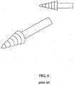
- the device is an arrow-like device or pin used to fix the middle turbinate to the nasal wall by pinning the turbinate. See , e.g. , Figures 5-9 .
- Such devices or pins may have protrusions, flanges, barbs, coatings, or bumps on their surfaces to prevent the device from falling out. See , e.g. , Figures 7-9 .
- the wafer or other device is made from a bioabsorbable material, for example, a PLGA co-polymer. Therefore, after the patient's nose has healed, the wafer or other device is absorbed by the body, thus avoiding the permanent attachment of the middle turbinate to the nasal septum.
- the wafer or other device is made of a non-bioresorbable material; thus, the device, if needed, can be removed later or left in place permanently.
- the invention is suitable for use in a method for medializing the middle turbinate.
- the wafer as described above is inserted into the nose of patient between the middle turbinate and the nasal septum, and pressure is applied to the middle turbinate and nasal septum to attach these two structures via the wafer.
- tissue adhesive e.g ., a cyanoacrylate adhesive
- the wafer may be used in conjunction with a tissue adhesive.
- the middle turbinate is pinned to the nasal septum.
- the sling-like device is used to draw the middle turbinate toward the nasal septum.
- the middle turbinate is adhered to the nasal septum thereby moving the middle turbinate medially.
- the method is typically performed during a nasal or sinus procedure or surgery (e.g. , endoscopic sinus surgery).
- the device may be implanted at the beginning of a procedure to pull the middle turbinate away from the uncinate process to make the procedure easier. This may move the middle turbinate out of the way for better visualization of the lateral wall and such structures as the ostia leading to the paranasal sinuses and the uncinate process.
- the device may then be left in place to prevent the formation of adhesions between the middle turbinate and the nasal septum.
- the wafer or other device may be implanted using medical devices for endoscopic surgery or may be implanted using specially designed tools for using the device.
- the device After the device is implanted or adhesive is applied, it typically stays in place long enough for the mucosa of the nasal passage to heal.
- the device or adhesive may stay in place for a time ranging from 1 week to 6 months. Once the mucosa has healed and there is no longer a risk of adhesions forming, the device may be removed or be absorbed by the patient's body. The device may also fall out of place, be swallowed by the patient along with mucus, and be safely degraded by the digestive system of the patient.
- An exemplary method not forming past of the present invention is a method for medializing the middle turbinate using a tissue glue (e.g ., cyanoacrylate, fibrin sealant) alone.
- Tissue glue is applied to the middle turbinate and/or the nasal septum, and pressure is applied to these two structures so that they come in contact for a sufficient time for the tissue glue to set.
- the adhesion of the middle turbinate to the nasal septum allows for the healing of the nasal mucosa without the risk of adhesions developing between the middle turbinate and the lateral nasal wall.
- the tissue glue breaks down, and the middle turbinate is subsequently released from the nasal septum.
- the glue may need to be reapplied by the treating physician every week or as needed until the mucosa heals and there is limited risk of adhesions forming.
- the invention is suitable for use in an exemplary method of using the medical device or tissue adhesive to attach the uvula to the nasopharyngeal side of the soft palate.
- a procedure is illustrated in Figures 3 A-D .
- the procedure is particularly useful in treating snoring or sleep apnea.
- the attachment may be permanent or temporary as needed.
- the invention provides an instrument for inserting the medical device into the nose of a patient.
- the instrument includes a comfortable grip and an elongated end with a means for holding and releasing the medical device in place.
- the invention provides an instrument for applying pressure to the middle turbinate and nasal septum around the medical device in order to attach the middle turbinate to the nasal septum by means of the medical device.
- An example of an instrument for inserting the wafer is shown in Figure 4 .
- the invention provides a kit including the instrument for inserting a medical device according to claim 1 and the medical device.
- the kit may also include tissue glue (e.g. cyanoacrylate, fibrin sealant), pharmaceutical agents (e.g. , steroids, non-steroidal anti-inflammatory agents, antibiotics), an instrument for removing the inventive device, instructions for inserting the inventive medical device, etc.
- tissue glue e.g. cyanoacrylate, fibrin sealant
- pharmaceutical agents e.g. , steroids, non-steroidal anti-inflammatory agents, antibiotics
- an instrument for removing the inventive device e.g. , instructions for inserting the inventive medical device, etc.
- these items are conveniently packaged for the use by a treating physician.
- the items are sterilely packaged.
- the present invention fills a need in nasal and sinus surgery for preventing adhesions after surgery by temporarily adhering the middle turbinate to the nasal septum. After the nasal mucosa has healed sufficiently the attachment naturally breaks down or is manually removed, thereby restoring the natural anatomy of the nasal passage.
- the inventive system reduces the complications following sinus and nasal surgery.
- the present invention provides a system for medializing the middle turbinate following and/or during nasal or sinus surgery.
- the invention stems from the recognition that attaching the middle turbinate to the nasal septum, thereby drawing the middle turbinate medially would prevent the formation of adhesions between the middle turbinate and lateral wall. These adhesions are known to cause further complications post surgery including paranasal sinus blockage.
- the inventive system prevents the formation of adhesions between the middle turbinate and the lateral nasal wall and therefore the subsequent complication. These adhesions frequently require post-revision surgery to remove the adhesions.
- the invention not only provides an instrument for inserting a medical device for use in medializing the middle turbinate but also provides kits, instruments for placing and removing the inventive devices, and the invention is suitable for procedures for medializing the middle turbinate.
- a patient suffering from nasal or sinus disease e.g. , allergies, infection
- nasal or sinus disease e.g. , allergies, infection
- the middle turbinate is attached at least temporarily to the nasal septum.
- the middle turbinate is attached to the nasal septum prior to starting the procedure or surgery in order to make the surgery easier. The attachment can then be left in place after the procedure or surgery is concluded.
- This attachment is accomplished using a medical device suitable to be used with the instrument for inserting a medical device according to claim 1 such as a wafer or pin with means for attaching middle turbinate to the nasal septum or in examples not forming past of the invention by using a tissue glue such as a cyanoacrylate adhesive, fibrin sealant, or other natural or synthetic adhesive.
- a tissue glue such as a cyanoacrylate adhesive, fibrin sealant, or other natural or synthetic adhesive.
- the attachment is temporary. Typically, the attachment is only in place for the length of time needed for the nasal mucosa to heal. Once the mucosa is healed, the chance of adhesions forming is greatly reduced.
- the attachment may be manually severed, or the means for attaching the middle turbinate and the nasal septum may degrade over time. For example, the device may be absorbed by the patient's body. The device may fall out of place and be harmlessly swallowed by the patient and degraded in the patient's digestive system. Or the adhesive may
- the attachment whether by medical device suitable to be used with the instrument for inserting a medical device according to claim 1 or exemplary adhesive alone may last from week to 24 months depending on the judgment of the treating physician.
- the attachment lasts from 2 weeks to 8 weeks, or 3 weeks to 6 weeks.
- the attachment lasts for approximately 1 month, 2 months, 3 months, 4 months, 5 months, or 6 months.
- the attachments last for approximately. 9 months, 12 months, 18 months, or 24 months. If longer attachment is necessary, the procedure may be repeated once, twice, three times, or more depending upon the patient and the judgment of the treating physician.
- the adhesive may need to be reapplied every few days, every week, every two weeks, or as needed until the nasal mucosa is healed. In certain examples where a cyanoacrylate adhesive is used, the adhesive is reapplied approximately every week.
- the device suitable to be used with the instrument for inserting a medical device according to claim 1 may also be used to attach the uvula to the nasopharyngeal side of the soft palate.
- Such an attachment is particularly useful in patients who snore or patients who suffer from sleep apnea.
- the attachment may also be used to move the uvula out of the way for a procedure involving the oronasopharynx.
- the attachment may be temporary or permanent.
- the wafer or other medical device as described herein is inserted into the oronasopharynx of the patient either through the nose or mouth. The device is then used to attach the soft palate to the uvula.
- tissue adhesive e.g. , a cyanoacrylate adhesive
- a device may be used in conjunction with a tissue adhesive. The method is typically performed during a procedure or surgery. The device may be implanted using medical devices for endoscopic surgery or may be implanted using specially designed tools for using the device.
- the inventive system may be used in attaching other structures in the body to each other (e.g. , in the oronasopharynx, gastrointestinal system, genitourinary system, etc .).
- the system is used in the oronasopharynx and attached to one or more of the following structures: turbinate, nasal septum, uvula, hard palate, soft palate, tonsils, tongue, gingiva, epiglottis, walls of the sinus, and sides of the oral cavity.
- the inventive system is particularly useful in attaching mucosal surfaces.
- inventive system is not used to approximate wound surfaces. In other examples, the inventive system is used to approximate wound surfaces.
- the medical device suitable to be used with the instrument for inserting a medical device according to claim 1 is a thin wafer with both sides of the wafer haying means for attaching the wafer to a surface. Therefore, the wafer can be used to bring two structures such as the middle turbinate and the nasal septum together.
- the wafer can be any shape or size capable of being placed into the space between the middle turbinate and nasal septum of a patient, preferably a human patient.
- the wafer is circular.
- the wafer is triangular shaper, rectangular shaped, or polygonal shaped.
- the wafer is a ring.
- the wafer is a zig-zag shape.
- the surface area of the sides of the wafer should provide a large enough surface area to adequately attach to the middle turbinate and nasal septum so that the middle turbinate can be pulled medially.
- the wafer is typically approximately 0.2 cm-2 cm in length by approximately 0.2 cm-2 cm in width.
- the length ranges from approximately 0.5 cm to approximately 1.5 cm.
- the length ranges from approximately I cm to approximately 2 cm.
- the length ranges from approximately 1.5 cm to approximately 2 cm.
- the length ranges from approximately 0.25 cm to approximately 0.75 cm.
- the length ranges from approximately 0.5 cm to approximately 1 cm.
- the width ranges from approximately 0.5 cm to approximately 1.5 cm.
- the width ranges from approximately 1 cm to approximately 2 cm. In certain embodiments, the width ranges from approximately 1.5 cm to approximately 2 cm. In certain embodiments, the width ranges from approximately 0.25 cm to approximately 0.75 cm. In certain embodiments, the width ranges from approximately 0.5 cm to approximately 1 cm. In certain embodiments, the wafer is approximately 1.5 cm by approximatey 1.5 cm. In certain embodiments, the wafer is approximately 1 cm by approximatey 1 cm. In certain embodiments, the wafer is approximately 0.75 cm by approximatey 0.75 cm. In certain embodiments, the wafer is approximately 0.5 cm by approximatey 0.5 cm. In certain embodiments, the wafer is approximately 0.25 cm by approximatey 0.25 cm.
- the wafer may be smaller, that is, less than 1 cm by 1 cm. Also, the wafer may be smaller where more than one wafer is being used to attach the middle turbinate to the nasal septum.
- the wafer is approximately 0.75 mm in thickness; however, the thickness of the wafer may vary from less than 0.2 mm to approximately 0.5 cm. In certain embodiments, the thickness of the wafer is in the range of approximately 0.5 mm to approximately 1.5 mm. In other embodiments, the wafer is a thin film of less than 0.2 mm in thickness.
- the means on the wafer or other device suitable to be used with the instrument for inserting a medical device according to claim 1 described herein for attaching the device to a surface such as the surface of the middle turbinate or the surface of the nasal septum include mechanical means of forming an attachment or, in examples not forming part of the claimed invention, may include any chemical adhesive.
- the means for attaching is preferably suitable for attaching the device to a mucosal surface.
- the adhesive is a cyanoacrylate adhesive.
- a similar synthetic glue is used as the adhesive.
- the adhesive is a fibrin sealant or other natural substance such as mussel adhesive protein, frog glue, etc..
- the adhesive may be applied to the device immediately before implanting the device in the patient.
- Mechanical means for forming an attachment include pins, staples, rivets, barbs, or hooks on the surface of the device which allow attachment to a surface.
- the surface of the wafer or other device may also be constructed to have a fibrous surface similar to Velcro ® for attaching the device to a tissue such as one with a mucosal surface.
- These attachment means typically extend less that approximately 1 cm from the surface of the wafer or other device, more preferably, less than 0.5 cm from the surface of the device. In certain embodiments, they extend less than 1 mm from the surface.
- an adhesive e.g., cyanoacrylate, fibrin sealant, mussel adhesive protein, frog glue
- a mechanical means for attachment e.g., cyanoacrylate, fibrin sealant, mussel adhesive protein, frog glue
- the device suitable to be used with the instrument for inserting a medical device according to claim 1 comprises a sling-like or pouch-like portion that is slipped around the middle turbinate and barbs or arrows for securing the device to the nasal septum. The device thereby draws the middle turbinate medially toward the nasal septum.
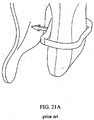
- the sling portion may be made of a thin suture-like material, or it may be made of a wider material, which is solid or mesh-like. An illustration of such a device is shown in Figures 21 A , B .
- the device suitable to be used with the instrument for inserting a medical device according to claim 1 is a pin for attaching the middle turbinate to the nasal septum.
- These devices are typically less than 2 cm in length.
- the devices are approximately 0.5 cm to 1.5 cm in length.
- the devices are approximately 0.25 cm, approximately 0.5 cm, approximately 0.75 cm, approximately 1 cm, approximately 1.25 cm, approximately 1.5 cm, approximately 1.75 cm, or approximately 2 cm in length.
- the surface of the pin may include protrusions to prevent the pin from coming out.
- the protrusions may be small barbs, bumps, ridges, etc .
- the pin may also be coated to prevent the pin for easily dislodging.
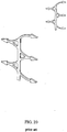
- the pin may also be coated to make it more biocompatible or allow for release of a bioactive agent. Exemplary designs for such pin devices are shown in Figures 5-9 . Other devices with two or more pins are also included within the invention as shown in Figures 18-20 . Such devices may be smaller than the wafer devices.
- the device is made of a biodegradable material.
- the material is a biodegradable polymer.
- the material may be synthetic (e.g. , polyesters, polyanhydrides) or natural (e.g ., proteins, rubber, polysaccharides).
- the device is made of a biodegradable material.
- the material is a biodegradable polymer.
- the material is a homopolymer.
- the material is a co-polymer.
- the material is a block polymer.
- the material is a branched polymer. In other embodiments, the material is a cross-linked polymer.
- the polymer is a polyester, polyurethane, polyvinyl chloride, polyalkylene ( e.g. , polyethylene), polyolefin, polyanhydride, polyamide, polycarbonate, polycarbamate, polyacrylate, polymethacrylate, polystyrene, polyurea, polyether, polyphosphazene, poly(ortho esters), polycarbonate, polyfumarate, polyarylate, polystyrene, or polyamine.
- the polymers is polylactide, polyglycolide, polycaprolactone, polydioxanone, polytrimethylene carbonate, and copolymers thereof.
- Polymers that have been used in producing biodegradable implants and are useful in preparing the inventive devices include alpha-polyhydroxy acids; polyglycolide (PGA); copolymers of polyglycolide such as glycolide/L-lactide copolymers (PGA/PLLA), glycolide/D,L-lactide copolymers (PGA/PDLLA), and glycolide/trimethylene carbonate copolymers (PGA/TMC); polylactides (PLA); stereocopolymers of PLA such as poly-L-lactide (PLLA), poly-D,L-lactide (PDLLA), L-lactide/D,L-lactide copolymers; copolymers of PLA such as lactide/tetramethylglycolide copolymers, lactide/trimethylene carbonate copoly
- the polymer is a polyester such as poly(glycolide-co-lactide) (PLGA), poly(lactide), poly(glycolide), poly(D,L-lactide-co-glycolide), poly(L-lactide-co-glycolide), poly-( ⁇ -hydroxybutyrate, and polyacrylic acid ester.
- the device is made of PLGA.
- the device is made of 85% D,L-lactide and 15% glycolide co-polymer.
- the device is made of 50% D,L-lactide and 50% glycolide co-polymer.
- the device is made of 65% D,L-lactide and 35% glycolide co-polymer. In certain embodiments, the device is made of 75% D,L-lactide and 25% glycolide co-polymer. In certain embodiments, the device is made of 85% L-lactide and 15% glycolide co-polymer. In certain embodiments, the device is made of 50% L-lactide and 50% glycolide co-polymer. In certain embodiments, the device is made of 65% L-lactide and 35% glycolide co-polymer. In certain embodiments, the device is made of 75% L-lactide and 25% glycolide co-polymer. In certain embodiments, the device is made of poly(caprolactone).
- the device is made of Pebax, Polyimide, Braided Polyimide, Nylon, PVC, Hytrel, HDPE, or PEEK.
- the device is made of a fluoropolymer such as PTFE, PFA, FEP, and EPTFE.
- the device is made of latex.
- the device is made of silicone.
- the polymer typically has a molecular weight sufficient to be shaped by molding or extrusion.
- the device is typically made of a material that is bioabsorbed after the device is not longer needed.
- the device may degrade after I week, 2 weeks, 3 weeks, 1 month, 2 months, 3 months, 4 months, 5 months, 6 months, 9 months, 1 year, 1.5 years, 2 years, 3 years, etc.
- the polymer used to make the device may be selected based on its degradation profile.
- the composition of the wafer may be varied to achieve the desired lifetime in vivo of the wafer.
- the device suitable to be used with the instrument for inserting a medical device according to claim 1 is made of a metal.
- the device is made of an alloy.
- the device is made of stainless steel.
- the device is made of a magnesium alloy (e.g ., magnesium based alloy AE21).
- the device is made of titanium.
- the device is made of a titanium alloy.
- the device is made of a superelastic alloy such as Nitinol.
- Metal devices may be optionally coated with a biocompatible coating. In the case where the device is made of a metal, the device may be inserted permanently or may be removed manually after the device is no longer needed.
- Suitable to be used with the instrument for may be coated with a biocompatible material.
- the device is made of or is coated with a timed-release formulation of a pharmaceutical agent.
- a pharmaceutical agent for example, a steroid, analgesic, anti-inflammatory agent, or antibiotic may be released by the wafer.
- the device is coated with a bioactive agent.
- Bioactive agents include small molecules, drugs, polynucleotide, proteins, peptides, etc.
- the bioactive agent may promote wound healing.
- the bioactive agent stimulates the formation of a desired tissue.
- the bioactive agent accelerates the integration of the turbinate with the nasal septum.
- the tube may be coated with a material to prevent cell growth such as a cytotoxic agent.
- the device may also be coated with a substance to prevent the formation of adhesions.
- the device may be coated with a polysaccharide such as hyaluronate.
- the device may also be coated with a polymeric coating such as Teflon.
- kits for convenience.
- the kits may also include all or some of the following items: an instrument for removing the device, adhesive, pharmaceutical agents, nasal sprays, gauze, bandages, disinfectant, and instructions for using the device.
- the kits are sterilely package for convenient use by a surgeon or other medical professional.
Landscapes
- Health & Medical Sciences (AREA)
- Life Sciences & Earth Sciences (AREA)
- Surgery (AREA)
- Molecular Biology (AREA)
- Engineering & Computer Science (AREA)
- Biomedical Technology (AREA)
- Heart & Thoracic Surgery (AREA)
- Medical Informatics (AREA)
- Nuclear Medicine, Radiotherapy & Molecular Imaging (AREA)
- Animal Behavior & Ethology (AREA)
- General Health & Medical Sciences (AREA)
- Public Health (AREA)
- Veterinary Medicine (AREA)
- Prostheses (AREA)
- Materials For Medical Uses (AREA)
- Radiation-Therapy Devices (AREA)
Description
- Sinusitis is a progression of inflammation, stasis, infection, and continued inflammation. Typically, the beginning of all sinus infections is either allergy or viral infection. Both of these conditions lead to swelling of the sinus and nasal mucosa that when severe enough, causes the small holes, called ostia, of the sinuses to close. Once the ostia is closed, the environment inside the sinuses, specifically the maxillary sinus, becomes conducive to bacterial growth. The way this typically occurs is that once the ostia is shut, the oxygen content of the sinus drops and the fluid inside the sinus is unable to escape which leads to further inflammation. The reduced oxygen content and inflammation disrupts the ability of the cilia of the cells of the sinus to operate properly which leads to further stasis.
- The typical patient that is seen by the otolaryngologist is started on antibiotics. Usually the antibiotic course can be as long as six weeks to eradicate the bacteria and bring the sinuses back to normal. For those patients in whom antibiotics do no relieve the problem, the only alternative is surgery. Although sinus and nasal surgeries are now common with 500,000 to 700,000 of such surgeries being performed annually in the U.S., these surgeries are typically both destructive and permanent. Around 10% of patients who undergo sinus surgery have scarring that leads to continued sinus problems which frequently require revision surgery.
- One frequent problem is postoperative adhesions. These adhesions occur between the middle turbinate and the adjacent nasal areas. One particular problem is the adhesion of the middle turbinate to the lateral nasal wall. Some surgeons have proposed removing the lower half of the middle turbinate to avoid this problem. This procedure, however, has its own problems (e.g., crust formation, nasal hygiene issues).
- Other solutions that have been suggested include placing a suture through the middle turbinate on one side of the nose, through the nasal septum, and then through the middle turbinate on the other side before the suture is tied off. Such a suture draws the middle turbinates medially and prevents the formation of adhesions between the middle turbinate and the lateral nasal wall. However, this suture is difficult and time-consuming to place and requires the puncturing of three separate structures in the nose. This can lead to discomfort for the patient, bleeding, infection, and other complications.
- Another solution surgeons have proposed is the use of various packing materials and splints. The use of these materials and devices however leads to the formation of scar tissue, which is undesirable and can lead to airway obstruction and infection. The adhesion of the middle turbinate to adjacent structures in the nose remains a problem in nasal and sinus surgery.
-
US 2005/0113850 discloses a medical instrument for use in the nasal cavities and suitable for placing a medical device for attachment of the middle turbinate to the nasal septum. -
US 2002/0077661 discloses multi-barbed wound closure devices and methods for establishing and maintaining two sides of a wound in apposition. -
EP 1 297 788 -
WO 03/008003 - None of these applications disclose an instrument for the insertion of a medical device, wherein the medical device is configured to be placed between a middle turbinate and a nasal septum, and wherein the instrument comprises means for applying pressure to the middle turbinate and the nasal septum around the medical device in order to attach the middle turbinate to the nasal septum by means of the medical device.
- Given this serious and common complication of sinus surgery, there remains a need in the art for preventing the formation of adhesions between the middle turbinate and adjacent nasal structures, particularly the lateral nasal wall. The desired solution preferably limits or eliminates the complications of the other proposals which have been used including infection, scar tissue formation, adhesions, bleeding, and patient discomfort.
- The invention is disclosed in
claim 1 with preferred embodiments disclosed in the dependent claims. - The present invention provides a system for reducing the adhesions formed in a patient's nasal cavity following a sinus or nasal procedure. In particular, the inventive system reduces the formation of adhesions between the lateral nasal wall and the middle turbinate by attaching the middle turbinate to the nasal septum. This system pulls the middle turbinate medially to avoid the formation of adhesions which may lead to further complications after sinus or nasal surgery. The attachment of the middle turbinate to the nasal septum may be temporary or permanent. This system may also be used prior to surgery to pull the middle turbinate away from the uncinate process to make surgeries in this area easier.
- In one aspect, a medical device for medializing the middle turbinate. Suitable to be used with the instrument for inserting a medical device according to
claim 1 is provided. As shown inFigures 1 and2 , in certain embodiments, the device is a wafer with a means for attaching the wafer to a surface (e.g., a mucosal surface) on both sides of the wafer. The means for attaching may include a tissue glue (e.g., cyanoacrylate, fibrin sealant), hooks, barbs, pins, staples, arrows, etc. The wafer thereby can bring two structures together. The device is particularly useful in attaching the middle turbinate to the nasal septum thereby preventing the formation of adhesions between the middle turbinate and the lateral nasal wall which can lead to complications after nasal and sinus surgeries. The wafer can be any shape including discs, rings, triangular-shaped wafers, polygonal-shaped wafers, zig-zag, etc. In certain instances, the wafer may include contours to fit comfortably inside the nose of the patient. For example, the wafer may include a contour for the middle turbinate on one side and be flat on the side that abuts the nasal septum. The wafer is typically approximately 1 cm by approximately 1 cm so that it can rest comfortably inside the nose of the patient between the middle turbinate and nasal septum. The device is approximately 0.75 mm or less in thickness. The wafer may be made from any biocompatible material. - In another embodiment, the device comprises a sling-like portion to securely grasp the turbinate and barbs, adhesives, or other fixation means for attaching the device with the turbinate to the nasal wall. In yet another embodiment, the device is an arrow-like device or pin used to fix the middle turbinate to the nasal wall by pinning the turbinate. See, e.g.,
Figures 5-9 . Such devices or pins may have protrusions, flanges, barbs, coatings, or bumps on their surfaces to prevent the device from falling out. See, e.g.,Figures 7-9 . - Preferably, the wafer or other device is made from a bioabsorbable material, for example, a PLGA co-polymer. Therefore, after the patient's nose has healed, the wafer or other device is absorbed by the body, thus avoiding the permanent attachment of the middle turbinate to the nasal septum. In certain embodiments, the wafer or other device is made of a non-bioresorbable material; thus, the device, if needed, can be removed later or left in place permanently.
- The invention is suitable for use in a method for medializing the middle turbinate. In certain embodiments, the wafer as described above is inserted into the nose of patient between the middle turbinate and the nasal septum, and pressure is applied to the middle turbinate and nasal septum to attach these two structures via the wafer. In another embodiment, tissue adhesive (e.g., a cyanoacrylate adhesive) rather than the inventive wafer is used to adhere the middle turbinate to the nasal septum. In still another embodiment, the wafer may be used in conjunction with a tissue adhesive. In still other embodiments, the middle turbinate is pinned to the nasal septum. In yet other embodiments, the sling-like device is used to draw the middle turbinate toward the nasal septum. By any of these approaches, the middle turbinate is adhered to the nasal septum thereby moving the middle turbinate medially. The method is typically performed during a nasal or sinus procedure or surgery (e.g., endoscopic sinus surgery). The device may be implanted at the beginning of a procedure to pull the middle turbinate away from the uncinate process to make the procedure easier. This may move the middle turbinate out of the way for better visualization of the lateral wall and such structures as the ostia leading to the paranasal sinuses and the uncinate process. The device may then be left in place to prevent the formation of adhesions between the middle turbinate and the nasal septum. The wafer or other device may be implanted using medical devices for endoscopic surgery or may be implanted using specially designed tools for using the device. After the device is implanted or adhesive is applied, it typically stays in place long enough for the mucosa of the nasal passage to heal. The device or adhesive may stay in place for a time ranging from 1 week to 6 months. Once the mucosa has healed and there is no longer a risk of adhesions forming, the device may be removed or be absorbed by the patient's body. The device may also fall out of place, be swallowed by the patient along with mucus, and be safely degraded by the digestive system of the patient.
- An exemplary method not forming past of the present invention is a method for medializing the middle turbinate using a tissue glue (e.g., cyanoacrylate, fibrin sealant) alone. Tissue glue is applied to the middle turbinate and/or the nasal septum, and pressure is applied to these two structures so that they come in contact for a sufficient time for the tissue glue to set. The adhesion of the middle turbinate to the nasal septum allows for the healing of the nasal mucosa without the risk of adhesions developing between the middle turbinate and the lateral nasal wall. Over time, the tissue glue breaks down, and the middle turbinate is subsequently released from the nasal septum. In the case of using a tissue glue such as cyanoacrylate alone, the glue may need to be reapplied by the treating physician every week or as needed until the mucosa heals and there is limited risk of adhesions forming.
- The invention is suitable for use in an exemplary method of using the medical device or tissue adhesive to attach the uvula to the nasopharyngeal side of the soft palate. Such a procedure is illustrated in
Figures 3A-D . The procedure is particularly useful in treating snoring or sleep apnea. The attachment may be permanent or temporary as needed. - The invention provides an instrument for inserting the medical device into the nose of a patient. The instrument includes a comfortable grip and an elongated end with a means for holding and releasing the medical device in place. The invention provides an instrument for applying pressure to the middle turbinate and nasal septum around the medical device in order to attach the middle turbinate to the nasal septum by means of the medical device. An example of an instrument for inserting the wafer is shown in
Figure 4 . - In another aspect, the invention provides a kit including the instrument for inserting a medical device according to
claim 1 and the medical device. The kit may also include tissue glue (e.g. cyanoacrylate, fibrin sealant), pharmaceutical agents (e.g., steroids, non-steroidal anti-inflammatory agents, antibiotics), an instrument for removing the inventive device, instructions for inserting the inventive medical device, etc. Typically, these items are conveniently packaged for the use by a treating physician. In certain embodiments, the items are sterilely packaged. - The present invention fills a need in nasal and sinus surgery for preventing adhesions after surgery by temporarily adhering the middle turbinate to the nasal septum. After the nasal mucosa has healed sufficiently the attachment naturally breaks down or is manually removed, thereby restoring the natural anatomy of the nasal passage. The inventive system reduces the complications following sinus and nasal surgery.
-
-
Figure 1 shows an example of the wafer-like medical device suitable to be used with the instrument for inserting a medical device according toclaim 1 with barbs for attaching to the nasal mucosa of the septum and the mucosa of the middle turbinate. -
Figure 2 shows the placement of the device suitable to be used with the instrument for inserting a medical device according toclaim 1 and the resulting medialization of the middle turbinate. - Figures 3A-D show the use of a medical device suitable to be used with the instrument for inserting a medical device according to
claim 1 with barbs to attach the uvula to the nasopharyngeal side of the soft palate. -
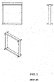
Figure 4 is an illustration of the inventive instrument for placing the wafer for attaching the middle turbinate to the nasal septum. -
Figure 5 shows exemplary pins suitable to be used with the instrument for inserting a medical device according toclaim 1 for attaching the nasal mucosa of the septum and the mucosa of the middle turbinate. -
Figure 6 shows another design of the pins suitable to be used with the instrument for inserting a medical device according toclaim 1 that have ridges on the pointed tip. -
Figure 7 shows another design of the pins suitable to be used with the instrument for inserting a medical device according toclaim 1 with protrusions for preventing the pin from dislodging. -
Figure 8 shows another design of the pins with bump-like protrusions. -
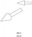
Figure 9 shows another design of the pins with barbs. -
Figure 10 shows a wafer suitable to be used with the instrument for inserting a medical device according toclaim 1 with barbs for attaching the nasal mucosa of the septum to the mucosa of the middle turbinate. -
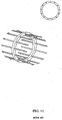
Figure 11 shows a circular design with barbs suitable to be used with the instrument for inserting a medical device according toclaim 1. for attaching the nasal mucosa of the septum to the mucosa of the middle turbinate. -
Figure 12 shows a zig-zag design of the medical device suitable to be used with the instrument for inserting a medical device according toclaim 1. -
Figure 13 shows a side view of an exemplary medical device suitable to be used with the instrument for inserting a medical device according toclaim 1. -
Figure 14 shows a side view of another exemplary medical device suitable to be used with the instrument for inserting a medical device according toclaim 1. with curved barbs. -
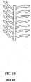
Figure 15 shows a side view of another exemplary medical device suitable to be used with the instrument for inserting a medical device according toclaim 1. with curved barbs. -
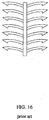
Figure 16 shows a side view of another exemplary medical device suitable to be used with the instrument for inserting a medical device according toclaim 1. with curved barbs. -
Figure 17 shows a side view of another exemplary medical device suitable to be used with the instrument for inserting a medical device according toclaim 1. with slanted barbs with respect to the surface of the wafer. -
Figure 18 shows another design for the medical device suitable to be used with the instrument for inserting a medical device according toclaim 1 with two barbs for attachment. -
Figure 19 shows a design with four barbs suitable to be used with the instrument for inserting a medical device according toclaim 1.. -
Figure 20 shows a planar design for the medical device suitable to be used with the instrument for inserting a medical device according toclaim 1. -
Figures 21A ,B show suitable to be used with the instrument for inserting a medical device according to claim 1 a sling-type device suitable to be used with the instrument for inserting a medical divice according toclaim 1 in which the sling portion is slipped around the turbinate and then the device, is secured to the nasal wall with piercing arrows or barbs. - The present invention provides a system for medializing the middle turbinate following and/or during nasal or sinus surgery. The invention stems from the recognition that attaching the middle turbinate to the nasal septum, thereby drawing the middle turbinate medially would prevent the formation of adhesions between the middle turbinate and lateral wall. These adhesions are known to cause further complications post surgery including paranasal sinus blockage. The inventive system prevents the formation of adhesions between the middle turbinate and the lateral nasal wall and therefore the subsequent complication. These adhesions frequently require post-revision surgery to remove the adhesions. The invention not only provides an instrument for inserting a medical device for use in medializing the middle turbinate but also provides kits, instruments for placing and removing the inventive devices, and the invention is suitable for procedures for medializing the middle turbinate.
- A patient suffering from nasal or sinus disease (e.g., allergies, infection) having undergone a sinus or nasal procedure is at a substantial risk of developing adhesions between various structures in the nasal passage due to trauma to the mucosal surfaces. In order to prevent the formation of adhesions, particularly between the lateral nasal wall and the middle turbinate, the middle turbinate is attached at least temporarily to the nasal septum. In certain embodiments, the middle turbinate is attached to the nasal septum prior to starting the procedure or surgery in order to make the surgery easier. The attachment can then be left in place after the procedure or surgery is concluded. This attachment is accomplished using a medical device suitable to be used with the instrument for inserting a medical device according to
claim 1 such as a wafer or pin with means for attaching middle turbinate to the nasal septum or in examples not forming past of the invention by using a tissue glue such as a cyanoacrylate adhesive, fibrin sealant, or other natural or synthetic adhesive. In most instances, the attachment is temporary. Typically, the attachment is only in place for the length of time needed for the nasal mucosa to heal. Once the mucosa is healed, the chance of adhesions forming is greatly reduced. The attachment may be manually severed, or the means for attaching the middle turbinate and the nasal septum may degrade over time. For example, the device may be absorbed by the patient's body. The device may fall out of place and be harmlessly swallowed by the patient and degraded in the patient's digestive system. Or the adhesive may break down releasing the middle turbinate from the nasal septum. - The attachment whether by medical device suitable to be used with the instrument for inserting a medical device according to
claim 1 or exemplary adhesive alone may last from week to 24 months depending on the judgment of the treating physician. In certain embodiments, the attachment lasts from 2 weeks to 8 weeks, or 3 weeks to 6 weeks. In other embodiments, the attachment lasts for approximately 1 month, 2 months, 3 months, 4 months, 5 months, or 6 months. In other embodiments, the attachments last for approximately. 9 months, 12 months, 18 months, or 24 months. If longer attachment is necessary, the procedure may be repeated once, twice, three times, or more depending upon the patient and the judgment of the treating physician. In certain examples where a tissue adhesive alone is used, the adhesive may need to be reapplied every few days, every week, every two weeks, or as needed until the nasal mucosa is healed. In certain examples where a cyanoacrylate adhesive is used, the adhesive is reapplied approximately every week. - As described above for drawing medially the middle turbinate, the device suitable to be used with the instrument for inserting a medical device according to
claim 1 may also be used to attach the uvula to the nasopharyngeal side of the soft palate. Such an attachment is particularly useful in patients who snore or patients who suffer from sleep apnea. The attachment may also be used to move the uvula out of the way for a procedure involving the oronasopharynx. The attachment may be temporary or permanent. The wafer or other medical device as described herein is inserted into the oronasopharynx of the patient either through the nose or mouth. The device is then used to attach the soft palate to the uvula. Pressure may be applied to the uvula and soft palate to attach these two structures via the device. In one particular example, tissue adhesive (e.g., a cyanoacrylate adhesive) rather than a device is used to adhere the uvula to the nasal septum. In still another embodiment, a device may be used in conjunction with a tissue adhesive. The method is typically performed during a procedure or surgery. The device may be implanted using medical devices for endoscopic surgery or may be implanted using specially designed tools for using the device. - As will be appreciated by those of skill in the art, the inventive system may be used in attaching other structures in the body to each other (e.g., in the oronasopharynx, gastrointestinal system, genitourinary system, etc.). In certain examples the system is used in the oronasopharynx and attached to one or more of the following structures: turbinate, nasal septum, uvula, hard palate, soft palate, tonsils, tongue, gingiva, epiglottis, walls of the sinus, and sides of the oral cavity. The inventive system is particularly useful in attaching mucosal surfaces. In certain examples, inventive system is not used to approximate wound surfaces. In other examples, the inventive system is used to approximate wound surfaces.
- In one embodiment, the medical device suitable to be used with the instrument for inserting a medical device according to
claim 1 is a thin wafer with both sides of the wafer haying means for attaching the wafer to a surface. Therefore, the wafer can be used to bring two structures such as the middle turbinate and the nasal septum together. The wafer can be any shape or size capable of being placed into the space between the middle turbinate and nasal septum of a patient, preferably a human patient. In certain embodiments, the wafer is circular. In other embodiments, the wafer is triangular shaper, rectangular shaped, or polygonal shaped. In yet other embodiments, the wafer is a ring. In certain embodiments, the wafer is a zig-zag shape. The surface area of the sides of the wafer should provide a large enough surface area to adequately attach to the middle turbinate and nasal septum so that the middle turbinate can be pulled medially. The wafer is typically approximately 0.2 cm-2 cm in length by approximately 0.2 cm-2 cm in width. In certain embodiments, the length ranges from approximately 0.5 cm to approximately 1.5 cm. In certain embodiments, the length ranges from approximately I cm to approximately 2 cm. In certain embodiments, the length ranges from approximately 1.5 cm to approximately 2 cm. In certain embodiments, the length ranges from approximately 0.25 cm to approximately 0.75 cm. In certain embodiments, the length ranges from approximately 0.5 cm to approximately 1 cm. In certain embodiments, the width ranges from approximately 0.5 cm to approximately 1.5 cm. In certain embodiments, the width ranges from approximately 1 cm to approximately 2 cm. In certain embodiments, the width ranges from approximately 1.5 cm to approximately 2 cm. In certain embodiments, the width ranges from approximately 0.25 cm to approximately 0.75 cm. In certain embodiments, the width ranges from approximately 0.5 cm to approximately 1 cm. In certain embodiments, the wafer is approximately 1.5 cm by approximatey 1.5 cm. In certain embodiments, the wafer is approximately 1 cm by approximatey 1 cm. In certain embodiments, the wafer is approximately 0.75 cm by approximatey 0.75 cm. In certain embodiments, the wafer is approximately 0.5 cm by approximatey 0.5 cm. In certain embodiments, the wafer is approximately 0.25 cm by approximatey 0.25 cm. For pediatric patients, the wafer may be smaller, that is, less than 1 cm by 1 cm. Also, the wafer may be smaller where more than one wafer is being used to attach the middle turbinate to the nasal septum. The wafer is approximately 0.75 mm in thickness; however, the thickness of the wafer may vary from less than 0.2 mm to approximately 0.5 cm. In certain embodiments, the thickness of the wafer is in the range of approximately 0.5 mm to approximately 1.5 mm. In other embodiments, the wafer is a thin film of less than 0.2 mm in thickness. - The means on the wafer or other device suitable to be used with the instrument for inserting a medical device according to
claim 1 described herein for attaching the device to a surface such as the surface of the middle turbinate or the surface of the nasal septum include mechanical means of forming an attachment or, in examples not forming part of the claimed invention, may include any chemical adhesive. The means for attaching is preferably suitable for attaching the device to a mucosal surface. In certain examples when a chemical adhesive is used, the adhesive is a cyanoacrylate adhesive. In other examples, a similar synthetic glue is used as the adhesive. In other examples when an adhesive is used, the adhesive is a fibrin sealant or other natural substance such as mussel adhesive protein, frog glue, etc.. These adhesives have been shown useful in closing wounds and are commercially available. The adhesive may be applied to the device immediately before implanting the device in the patient. Mechanical means for forming an attachment include pins, staples, rivets, barbs, or hooks on the surface of the device which allow attachment to a surface. The surface of the wafer or other device may also be constructed to have a fibrous surface similar to Velcro® for attaching the device to a tissue such as one with a mucosal surface. These attachment means typically extend less that approximately 1 cm from the surface of the wafer or other device, more preferably, less than 0.5 cm from the surface of the device. In certain embodiments, they extend less than 1 mm from the surface. Usually multiple pins, staples, rivets, barbs, or hooks are used to provide a secure attachment. These means typically do not puncture through the entire nasal structure. In certain embodiments, the mechanical means only penetrate the mucosa. In certain embodiments, an adhesive (e.g., cyanoacrylate, fibrin sealant, mussel adhesive protein, frog glue) is used in conjunction with a mechanical means for attachment. - In another example, the device suitable to be used with the instrument for inserting a medical device according to
claim 1 comprises a sling-like or pouch-like portion that is slipped around the middle turbinate and barbs or arrows for securing the device to the nasal septum. The device thereby draws the middle turbinate medially toward the nasal septum. The sling portion may be made of a thin suture-like material, or it may be made of a wider material, which is solid or mesh-like. An illustration of such a device is shown inFigures 21A ,B . - In yet another embodiment, the device suitable to be used with the instrument for inserting a medical device according to
claim 1 is a pin for attaching the middle turbinate to the nasal septum. These devices are typically less than 2 cm in length. In certain embodiments, the devices are approximately 0.5 cm to 1.5 cm in length. In certain embodiments, the devices are approximately 0.25 cm, approximately 0.5 cm, approximately 0.75 cm, approximately 1 cm, approximately 1.25 cm, approximately 1.5 cm, approximately 1.75 cm, or approximately 2 cm in length. The surface of the pin may include protrusions to prevent the pin from coming out. The protrusions may be small barbs, bumps, ridges, etc. The pin may also be coated to prevent the pin for easily dislodging. The pin may also be coated to make it more biocompatible or allow for release of a bioactive agent. Exemplary designs for such pin devices are shown inFigures 5-9 . Other devices with two or more pins are also included within the invention as shown inFigures 18-20 . Such devices may be smaller than the wafer devices. - Any of the devices suitable to be used with the instrument for inserting a medical device according to
claim 1 can be made of any biocompatible material. Preferably, the device is made of a biodegradable material. In certain embodiments, the material is a biodegradable polymer. The material may be synthetic (e.g., polyesters, polyanhydrides) or natural (e.g., proteins, rubber, polysaccharides). Preferably, the device is made of a biodegradable material. In certain embodiments, the material is a biodegradable polymer. In certain embodiments, the material is a homopolymer. In certain embodiments, the material is a co-polymer. In other embodiments, the material is a block polymer. In other embodiments, the material is a branched polymer. In other embodiments, the material is a cross-linked polymer. In certain embodiments, the polymer is a polyester, polyurethane, polyvinyl chloride, polyalkylene (e.g., polyethylene), polyolefin, polyanhydride, polyamide, polycarbonate, polycarbamate, polyacrylate, polymethacrylate, polystyrene, polyurea, polyether, polyphosphazene, poly(ortho esters), polycarbonate, polyfumarate, polyarylate, polystyrene, or polyamine. In certain embodiments, the polymers is polylactide, polyglycolide, polycaprolactone, polydioxanone, polytrimethylene carbonate, and copolymers thereof. Polymers that have been used in producing biodegradable implants and are useful in preparing the inventive devices include alpha-polyhydroxy acids; polyglycolide (PGA); copolymers of polyglycolide such as glycolide/L-lactide copolymers (PGA/PLLA), glycolide/D,L-lactide copolymers (PGA/PDLLA), and glycolide/trimethylene carbonate copolymers (PGA/TMC); polylactides (PLA); stereocopolymers of PLA such as poly-L-lactide (PLLA), poly-D,L-lactide (PDLLA), L-lactide/D,L-lactide copolymers; copolymers of PLA such as lactide/tetramethylglycolide copolymers, lactide/trimethylene carbonate copolymers, lactide/δ-valerolactone copolymers, lactide ε-caprolactone copolymers, polydepsipeptides, PLA/polyethylene oxide copolymers, unsymmetrically 3,6-substituted poly-1,4-dioxane-2,5-diones; polyhydroxyalkanate polymers including poly-beta-hydroxybutyrate (PHBA), PHBA/beta-hydroxyvalerate copolymers (PHBA/HVA), and poly-beta-hydroxypropionate (PHPA); poly-p-dioxanone (PDS); poly-δ-valerolatone; poly-ε-caprolactone; methylmethacrylate-N-vinyl pyrrolidone copolymers; polyesteramides; polyesters of oxalic acid; polydihydropyrans; polyalkyl-2-cyanoacrylates; polyurethanes (PU); polyvinyl alcohol (PVA); polypeptides; poly-beta-maleic acid (PMLA); poly(trimethylene carbonate); poly(ethylene oxide) (PEO); poly(β-hydroxyvalerate) (PHVA); poly(ortho esters); tyrosine-derived polycarbonates; and poly-beta-alkanoic acids. In certain embodiments, the polymer is a polyester such as poly(glycolide-co-lactide) (PLGA), poly(lactide), poly(glycolide), poly(D,L-lactide-co-glycolide), poly(L-lactide-co-glycolide), poly-(β-hydroxybutyrate, and polyacrylic acid ester. In certain embodiments, the device is made of PLGA. In certain embodiments, the device is made of 85% D,L-lactide and 15% glycolide co-polymer. In certain embodiments, the device is made of 50% D,L-lactide and 50% glycolide co-polymer. In certain embodiments, the device is made of 65% D,L-lactide and 35% glycolide co-polymer. In certain embodiments, the device is made of 75% D,L-lactide and 25% glycolide co-polymer. In certain embodiments, the device is made of 85% L-lactide and 15% glycolide co-polymer. In certain embodiments, the device is made of 50% L-lactide and 50% glycolide co-polymer. In certain embodiments, the device is made of 65% L-lactide and 35% glycolide co-polymer. In certain embodiments, the device is made of 75% L-lactide and 25% glycolide co-polymer. In certain embodiments, the device is made of poly(caprolactone). In certain embodiments, the device is made of Pebax, Polyimide, Braided Polyimide, Nylon, PVC, Hytrel, HDPE, or PEEK. In certain embodiments, the device is made of a fluoropolymer such as PTFE, PFA, FEP, and EPTFE. In certain embodiments, the device is made of latex. In other embodiments, the device is made of silicone. The polymer typically has a molecular weight sufficient to be shaped by molding or extrusion. The device is typically made of a material that is bioabsorbed after the device is not longer needed. For example, the device may degrade after I week, 2 weeks, 3 weeks, 1 month, 2 months, 3 months, 4 months, 5 months, 6 months, 9 months, 1 year, 1.5 years, 2 years, 3 years, etc. The polymer used to make the device may be selected based on its degradation profile. As would be appreciated by one of skill in this art, the composition of the wafer may be varied to achieve the desired lifetime in vivo of the wafer. - In other embodiments, the device suitable to be used with the instrument for inserting a medical device according to
claim 1 is made of a metal. In other embodiments, the device is made of an alloy. In certain embodiments, the device is made of stainless steel. In certain embodiments, the device is made of a magnesium alloy (e.g., magnesium based alloy AE21). In certain embodiments, the device is made of titanium. In certain embodiments, the device is made of a titanium alloy. In certain embodiments, the device is made of a superelastic alloy such as Nitinol. Metal devices may be optionally coated with a biocompatible coating. In the case where the device is made of a metal, the device may be inserted permanently or may be removed manually after the device is no longer needed. - The device suitable to be used with the instrument for inserting a medical device according to
claim 1. Suitable to be used with the instrument for may be coated with a biocompatible material. In certain embodiments, the device is made of or is coated with a timed-release formulation of a pharmaceutical agent. For example, a steroid, analgesic, anti-inflammatory agent, or antibiotic may be released by the wafer. In certain embodiments, the device is coated with a bioactive agent. Bioactive agents include small molecules, drugs, polynucleotide, proteins, peptides, etc. In certain embodiments, the bioactive agent may promote wound healing. In certain embodiments, the bioactive agent stimulates the formation of a desired tissue. In certain embodiments, the bioactive agent accelerates the integration of the turbinate with the nasal septum. In yet other embodiments, the tube may be coated with a material to prevent cell growth such as a cytotoxic agent. The device may also be coated with a substance to prevent the formation of adhesions. For example, the device may be coated with a polysaccharide such as hyaluronate. The device may also be coated with a polymeric coating such as Teflon. - The medical device and the inventive instrument for inserting a medical device according to
claim 1 may be packaged in kits for convenience. In certain embodiments, the kits may also include all or some of the following items: an instrument for removing the device, adhesive, pharmaceutical agents, nasal sprays, gauze, bandages, disinfectant, and instructions for using the device. In certain embodiments, the kits are sterilely package for convenient use by a surgeon or other medical professional.
Claims (13)
- An instrument for implanting a medical device, wherein:the medical device is configured to be placed between a middle turbinate and a nasal septum and configured to approximate a mucosal surface of the middle turbinate to a mucosal surface of the nasal septum, the device comprising a body comprising two opposed sides, and at least one hook, pin, arrow or barb on each side, wherein during use, the at least one hook, pin, arrow or barb on one side attaches the device to the mucosal surface of the middle turbinate and the at least one hook, pin, arrow or barb on the opposing side attaches the device to the mucosal surface of the nasal septum, thereby approximating the two mucosal surfaces; and wherein the instrument includes a comfortable grip and an elongated end with a means for holding and releasing the medical device in place and means for applying pressure to the middle turbinate and the nasal septum around the medical device in order to attach the middle turbinate to the nasal septum by means of the medical device.
- The instrument of claim 1, wherein the device is made of a bioabsorbable material.
- The instrument of claim 2, wherein the two mucosal surfaces are temporarily approximated and the device is bioabsorbed after the two mucosal surfaces are no longer approximated.
- The instrument of claim 2, wherein the bioabsorbable material comprises a polyester, a polyanhydride, a polyamide, a polycarbonate, a polycarbamate, a polyacrylate, a polymethacrylate, a polystyrene, a polyurea, a polyether, or a polyamine, in particular a polyester.
- The instrument of any of claims 1 to 4, wherein the device is made of PLGA, in particular wherein the device is made of a 85% D,L-lactide and 15% glycolide co-polymer; a 75% D,L-lactide and 25% glycolide co-polymer; a 65% D,L-lactide and 35% glycolide co-polymer; or a 50% D,L-lactide and 50% glycolide co-polymer.
- The instrument of any of claims 1 to 5, wherein the device is approximately 0.2-2 cm in length and approximately 0.2-2 cm in width, in particular wherein the device is approximately 1 cm by approximately 1 cm.
- The instrument of any of claims 1 to 6, wherein the body of the device is less than approximately 2 mm thick and preferably is approximately 0.75 mm thick.
- The instrument of any of claims 1 to 7, wherein the device is coated with a bioactive agent.
- The instrument of claim 8, wherein the bioactive agent comprises a drug, a polynucleotide, a protein or a peptide.
- The instrument of claim 8, wherein the bioactive agent promotes wound healing.
- The instrument of any of claims 1 to 7, wherein the device is made of or is coated with a timed-release formulation of a pharmaceutical agent.
- The instrument of claim 11, wherein the pharmaceutical agent comprises a steroid, analgesic, anti-inflammatory agent, or an antibiotic.
- A kit comprising the instrument for inserting the medical device and the medical device of any of claims 1 to 12 and optionally further comprising:instructions for using the device.
Applications Claiming Priority (2)
| Application Number | Priority Date | Filing Date | Title |
|---|---|---|---|
| US80017606P | 2006-05-12 | 2006-05-12 | |
| PCT/US2007/068743 WO2007134215A2 (en) | 2006-05-12 | 2007-05-11 | Middle turbinate medializer |
Publications (3)
| Publication Number | Publication Date |
|---|---|
| EP2023826A2 EP2023826A2 (en) | 2009-02-18 |
| EP2023826A4 EP2023826A4 (en) | 2009-12-23 |
| EP2023826B1 true EP2023826B1 (en) | 2017-06-21 |
Family
ID=38694724
Family Applications (1)
| Application Number | Title | Priority Date | Filing Date |
|---|---|---|---|
| EP07762117.5A Active EP2023826B1 (en) | 2006-05-12 | 2007-05-11 | Middle turbinate medializer |
Country Status (5)
| Country | Link |
|---|---|
| US (1) | US20070293946A1 (en) |
| EP (1) | EP2023826B1 (en) |
| AU (1) | AU2007249293C1 (en) |
| CA (1) | CA2651935C (en) |
| WO (1) | WO2007134215A2 (en) |
Families Citing this family (78)
| Publication number | Priority date | Publication date | Assignee | Title |
|---|---|---|---|---|
| DE10105592A1 (en) | 2001-02-06 | 2002-08-08 | Achim Goepferich | Placeholder for drug release in the frontal sinus |
| US8317816B2 (en) | 2002-09-30 | 2012-11-27 | Acclarent, Inc. | Balloon catheters and methods for treating paranasal sinuses |
| US7544192B2 (en) | 2003-03-14 | 2009-06-09 | Sinexus, Inc. | Sinus delivery of sustained release therapeutics |
| US20050113850A1 (en) * | 2003-10-08 | 2005-05-26 | Tagge Bryan C. | Apparatus, system, and method for middle turbinate medializer |
| US7410480B2 (en) | 2004-04-21 | 2008-08-12 | Acclarent, Inc. | Devices and methods for delivering therapeutic substances for the treatment of sinusitis and other disorders |
| US7419497B2 (en) | 2004-04-21 | 2008-09-02 | Acclarent, Inc. | Methods for treating ethmoid disease |
| US20060063973A1 (en) | 2004-04-21 | 2006-03-23 | Acclarent, Inc. | Methods and apparatus for treating disorders of the ear, nose and throat |
| US8146400B2 (en) | 2004-04-21 | 2012-04-03 | Acclarent, Inc. | Endoscopic methods and devices for transnasal procedures |
| US8747389B2 (en) | 2004-04-21 | 2014-06-10 | Acclarent, Inc. | Systems for treating disorders of the ear, nose and throat |
| US9351750B2 (en) | 2004-04-21 | 2016-05-31 | Acclarent, Inc. | Devices and methods for treating maxillary sinus disease |
| US8702626B1 (en) | 2004-04-21 | 2014-04-22 | Acclarent, Inc. | Guidewires for performing image guided procedures |
| US9101384B2 (en) | 2004-04-21 | 2015-08-11 | Acclarent, Inc. | Devices, systems and methods for diagnosing and treating sinusitis and other disorders of the ears, Nose and/or throat |
| US20070208252A1 (en) | 2004-04-21 | 2007-09-06 | Acclarent, Inc. | Systems and methods for performing image guided procedures within the ear, nose, throat and paranasal sinuses |
| US20060004323A1 (en) | 2004-04-21 | 2006-01-05 | Exploramed Nc1, Inc. | Apparatus and methods for dilating and modifying ostia of paranasal sinuses and other intranasal or paranasal structures |
| US10188413B1 (en) | 2004-04-21 | 2019-01-29 | Acclarent, Inc. | Deflectable guide catheters and related methods |
| US7803150B2 (en) | 2004-04-21 | 2010-09-28 | Acclarent, Inc. | Devices, systems and methods useable for treating sinusitis |
| US9554691B2 (en) | 2004-04-21 | 2017-01-31 | Acclarent, Inc. | Endoscopic methods and devices for transnasal procedures |
| US7654997B2 (en) | 2004-04-21 | 2010-02-02 | Acclarent, Inc. | Devices, systems and methods for diagnosing and treating sinusitus and other disorders of the ears, nose and/or throat |
| US7462175B2 (en) | 2004-04-21 | 2008-12-09 | Acclarent, Inc. | Devices, systems and methods for treating disorders of the ear, nose and throat |
| US20190314620A1 (en) | 2004-04-21 | 2019-10-17 | Acclarent, Inc. | Apparatus and methods for dilating and modifying ostia of paranasal sinuses and other intranasal or paranasal structures |
| US8932276B1 (en) | 2004-04-21 | 2015-01-13 | Acclarent, Inc. | Shapeable guide catheters and related methods |
| US7361168B2 (en) | 2004-04-21 | 2008-04-22 | Acclarent, Inc. | Implantable device and methods for delivering drugs and other substances to treat sinusitis and other disorders |
| US8764729B2 (en) | 2004-04-21 | 2014-07-01 | Acclarent, Inc. | Frontal sinus spacer |
| US9399121B2 (en) | 2004-04-21 | 2016-07-26 | Acclarent, Inc. | Systems and methods for transnasal dilation of passageways in the ear, nose or throat |
| US9089258B2 (en) | 2004-04-21 | 2015-07-28 | Acclarent, Inc. | Endoscopic methods and devices for transnasal procedures |
| US8894614B2 (en) | 2004-04-21 | 2014-11-25 | Acclarent, Inc. | Devices, systems and methods useable for treating frontal sinusitis |
| US20070167682A1 (en) | 2004-04-21 | 2007-07-19 | Acclarent, Inc. | Endoscopic methods and devices for transnasal procedures |
| US7559925B2 (en) | 2006-09-15 | 2009-07-14 | Acclarent Inc. | Methods and devices for facilitating visualization in a surgical environment |
| US8096303B2 (en) | 2005-02-08 | 2012-01-17 | Koninklijke Philips Electronics N.V | Airway implants and methods and devices for insertion and retrieval |
| DE602006013946D1 (en) | 2005-02-08 | 2010-06-10 | Koninkl Philips Electronics Nv | SYSTEM FOR PERCUTANEOUS GLOSSOPLASTICS |
| US8371307B2 (en) | 2005-02-08 | 2013-02-12 | Koninklijke Philips Electronics N.V. | Methods and devices for the treatment of airway obstruction, sleep apnea and snoring |
| US8025635B2 (en) | 2005-04-04 | 2011-09-27 | Intersect Ent, Inc. | Device and methods for treating paranasal sinus conditions |
| US8951225B2 (en) | 2005-06-10 | 2015-02-10 | Acclarent, Inc. | Catheters with non-removable guide members useable for treatment of sinusitis |
| US8114113B2 (en) | 2005-09-23 | 2012-02-14 | Acclarent, Inc. | Multi-conduit balloon catheter |
| US8190389B2 (en) | 2006-05-17 | 2012-05-29 | Acclarent, Inc. | Adapter for attaching electromagnetic image guidance components to a medical device |
| US8535707B2 (en) | 2006-07-10 | 2013-09-17 | Intersect Ent, Inc. | Devices and methods for delivering active agents to the osteomeatal complex |
| US9820688B2 (en) | 2006-09-15 | 2017-11-21 | Acclarent, Inc. | Sinus illumination lightwire device |
| US7780730B2 (en) | 2006-09-25 | 2010-08-24 | Iyad Saidi | Nasal implant introduced through a non-surgical injection technique |
| US8439687B1 (en) | 2006-12-29 | 2013-05-14 | Acclarent, Inc. | Apparatus and method for simulated insertion and positioning of guidewares and other interventional devices |
| WO2008124787A2 (en) | 2007-04-09 | 2008-10-16 | Acclarent, Inc. | Ethmoidotomy system and implantable spacer devices having therapeutic substance delivery capability for treatment of paranasal sinusitis |
| US8118757B2 (en) | 2007-04-30 | 2012-02-21 | Acclarent, Inc. | Methods and devices for ostium measurement |
| US8485199B2 (en) * | 2007-05-08 | 2013-07-16 | Acclarent, Inc. | Methods and devices for protecting nasal turbinate during surgery |
| DK2231065T3 (en) | 2007-12-18 | 2021-02-01 | Intersect Ent Inc | SELF-EXPANDING DEVICES |
| US10206821B2 (en) | 2007-12-20 | 2019-02-19 | Acclarent, Inc. | Eustachian tube dilation balloon with ventilation path |
| US8182432B2 (en) | 2008-03-10 | 2012-05-22 | Acclarent, Inc. | Corewire design and construction for medical devices |
| JP5676446B2 (en) | 2008-07-30 | 2015-02-25 | アクラレント インコーポレイテッド | Sinus mouth finder |
| EP2320832A4 (en) | 2008-08-01 | 2015-07-29 | Intersect Ent Inc | Methods and devices for crimping self-expanding devices |
| US8192450B2 (en) * | 2008-09-17 | 2012-06-05 | Entrigue Surgical, Inc. | Methods and systems for medializing a turbinate |
| AU2009293312B2 (en) | 2008-09-18 | 2015-07-09 | Acclarent, Inc. | Methods and apparatus for treating disorders of the ear nose and throat |
| WO2010059586A1 (en) | 2008-11-19 | 2010-05-27 | Entrigue Surgical, Inc. | Apparatus and methods for correcting nasal valve collapse |
| JP2010207289A (en) * | 2009-03-06 | 2010-09-24 | Kohei Kubota | Medical shooting staple |
| US20100241155A1 (en) | 2009-03-20 | 2010-09-23 | Acclarent, Inc. | Guide system with suction |
| US8435290B2 (en) | 2009-03-31 | 2013-05-07 | Acclarent, Inc. | System and method for treatment of non-ventilating middle ear by providing a gas pathway through the nasopharynx |
| US7978742B1 (en) | 2010-03-24 | 2011-07-12 | Corning Incorporated | Methods for operating diode lasers |
| EP2424448A4 (en) * | 2009-04-27 | 2015-03-04 | Intersect Ent Inc | Devices and methods for treating pain associated with tonsillectomies |
| CN102573981B (en) | 2009-05-15 | 2016-06-22 | 因特尔赛克特耳鼻喉公司 | Extendable device and using method thereof |
| US8435261B2 (en) | 2009-07-15 | 2013-05-07 | Regents Of The University Of Minnesota | Treatment and placement device for sinusitis applications |
| US9155492B2 (en) | 2010-09-24 | 2015-10-13 | Acclarent, Inc. | Sinus illumination lightwire device |
| US8480737B2 (en) * | 2010-09-27 | 2013-07-09 | Ethicon, Inc. | Columellar strut for nasal tip support |
| US9510940B2 (en) | 2011-02-17 | 2016-12-06 | Ethicon, Inc. | Bioabsorbable multilayer nasal valve spreader graft |
| JP5159918B2 (en) * | 2011-05-20 | 2013-03-13 | 浩平 窪田 | Medical implantable staples |
| US20130317540A1 (en) | 2012-05-22 | 2013-11-28 | Krasimira Hristov | Universal bioabsorbable nasal implant kit |
| US9480594B2 (en) | 2013-02-27 | 2016-11-01 | Spirox, Inc. | Nasal implants and systems and methods of use |
| AU2014236729B2 (en) | 2013-03-14 | 2018-11-22 | Intersect Ent, Inc. | Systems, devices, and method for treating a sinus condition |
| US9433437B2 (en) | 2013-03-15 | 2016-09-06 | Acclarent, Inc. | Apparatus and method for treatment of ethmoid sinusitis |
| US9629684B2 (en) | 2013-03-15 | 2017-04-25 | Acclarent, Inc. | Apparatus and method for treatment of ethmoid sinusitis |
| US10039563B2 (en) | 2013-03-15 | 2018-08-07 | Invictus Medical Innovations, Llc | Suturer |
| US9694163B2 (en) | 2013-12-17 | 2017-07-04 | Biovision Technologies, Llc | Surgical device for performing a sphenopalatine ganglion block procedure |
| US9516995B2 (en) | 2013-12-17 | 2016-12-13 | Biovision Technologies, Llc | Surgical device for performing a sphenopalatine ganglion block procedure |
| US10016580B2 (en) | 2013-12-17 | 2018-07-10 | Biovision Technologies, Llc | Methods for treating sinus diseases |
| US9510743B2 (en) | 2013-12-17 | 2016-12-06 | Biovision Technologies, Llc | Stabilized surgical device for performing a sphenopalatine ganglion block procedure |
| CA2958213A1 (en) | 2014-08-26 | 2016-03-03 | Spirox, Inc. | Nasal implants and systems and method of use |
| US10596330B2 (en) | 2015-08-26 | 2020-03-24 | Medtronic Xomed, Inc. | Resorbable, drug-eluting submucosal turbinate implant device and method |
| EP3352690B1 (en) | 2015-09-25 | 2023-09-13 | Spirox, Inc. | Nasal implants |
| JP6934889B2 (en) | 2016-05-02 | 2021-09-15 | エンテラス メディカル インコーポレイテッドEntellus Medical,Inc. | Nasal flap implant and its transplant method |
| JP2020504656A (en) * | 2016-12-30 | 2020-02-13 | スピロックス, インク.Spirox, Inc. | Nasal implants and methods of use |
| US10716558B2 (en) * | 2017-08-04 | 2020-07-21 | Ethicon, Inc. | Tissue anchors having bi-directional arrays of barbed pins for joining tissue layers |
| US10525240B1 (en) | 2018-06-28 | 2020-01-07 | Sandler Scientific LLC | Sino-nasal rinse delivery device with agitation, flow-control and integrated medication management system |
Family Cites Families (88)
| Publication number | Priority date | Publication date | Assignee | Title |
|---|---|---|---|---|
| US2525183A (en) * | 1947-03-20 | 1950-10-10 | Jehu M Robison | Antral pressure device |
| US4259959A (en) * | 1978-12-20 | 1981-04-07 | Walker Wesley W | Suturing element |
| US5125553A (en) * | 1987-03-02 | 1992-06-30 | Stryker Sales Corporation | Surgical suturing instrument and method |
| US4873976A (en) * | 1984-02-28 | 1989-10-17 | Schreiber Saul N | Surgical fasteners and method |
| US4895148A (en) * | 1986-05-20 | 1990-01-23 | Concept, Inc. | Method of joining torn parts of bodily tissue in vivo with a biodegradable tack member |
| US4924865A (en) * | 1986-05-20 | 1990-05-15 | Concept, Inc. | Repair tack for bodily tissue |
| US4917114A (en) * | 1986-10-17 | 1990-04-17 | United States Surgical Corporation | Surgical fastener and surgical stapling apparatus |
| US5366459A (en) * | 1987-05-14 | 1994-11-22 | Inbae Yoon | Surgical clip and clip application procedures |
| CA1291836C (en) * | 1987-06-17 | 1991-11-05 | Kenji Takato | Battery feed circuit |
| US5383881A (en) * | 1989-07-18 | 1995-01-24 | United States Surgical Corporation | Safety device for use with endoscopic instrumentation |
| US4991764A (en) * | 1989-01-23 | 1991-02-12 | Edward Weck Incorporated | Surgical stapling instrument |
| US4994073A (en) * | 1989-02-22 | 1991-02-19 | United States Surgical Corp. | Skin fastener |
| US5089009A (en) * | 1989-06-27 | 1992-02-18 | United States Surgical Corporation | Inwardly biased skin fastener |
| US5059206A (en) * | 1989-04-12 | 1991-10-22 | Winters Thomas F | Method and apparatus for repairing a tear in a knee meniscus |
| US6129740A (en) * | 1989-04-24 | 2000-10-10 | Michelson; Gary Karlin | Instrument handle design |
| US5040715B1 (en) * | 1989-05-26 | 1994-04-05 | United States Surgical Corp | Apparatus and method for placing staples in laparoscopic or endoscopic procedures |
| US5129906A (en) * | 1989-09-08 | 1992-07-14 | Linvatec Corporation | Bioabsorbable tack for joining bodily tissue and in vivo method and apparatus for deploying same |
| US5100042A (en) * | 1990-03-05 | 1992-03-31 | United States Surgical Corporation | Surgical fastener apparatus |
| US5116349A (en) * | 1990-05-23 | 1992-05-26 | United States Surgical Corporation | Surgical fastener apparatus |
| US5653373A (en) * | 1990-09-17 | 1997-08-05 | United States Surgical Corporation | Arcuate apparatus for applying two-part surgical fasteners |
| US5108422A (en) * | 1990-10-22 | 1992-04-28 | United States Surgical Corporation | Skin fastener |
| US5593423A (en) * | 1990-10-22 | 1997-01-14 | United States Surgical Corporation | Skin fastener |
| US5094233A (en) * | 1991-01-11 | 1992-03-10 | Brennan Louis G | Turbinate sheath device |
| US5312456A (en) * | 1991-01-31 | 1994-05-17 | Carnegie Mellon University | Micromechanical barb and method for making the same |
| CA2065205C (en) * | 1991-04-11 | 2002-12-03 | David T. Green | Reduced mass absorbable surgical fastener and retainer |
| US5246455A (en) * | 1991-05-17 | 1993-09-21 | Micromedics, Inc. | Middle meatal antrostomy ventilation tube |
| US5289963A (en) * | 1991-10-18 | 1994-03-01 | United States Surgical Corporation | Apparatus and method for applying surgical staples to attach an object to body tissue |
| US5366134A (en) * | 1991-10-18 | 1994-11-22 | United States Surgical Corporation | Surgical fastening apparatus |
| CA2079057A1 (en) * | 1991-10-18 | 1993-04-19 | Stanley H. Remiszewski | Surgical stapling apparatus |
| US5351871A (en) * | 1992-03-26 | 1994-10-04 | William Bauer | Intranasal septal stapling device |
| US5540240A (en) * | 1992-03-26 | 1996-07-30 | Bauer; William | Intranasal septal fastener driving method |
| US5370294A (en) * | 1992-03-26 | 1994-12-06 | Bauer; William | Intranasal septal stapling device and method |
| US5915615A (en) * | 1992-03-26 | 1999-06-29 | Bauer; William | Tissue fastening device |
| CA2100532C (en) * | 1992-09-21 | 2004-04-20 | David T. Green | Device for applying a meniscal staple |
| US5336163A (en) * | 1993-01-06 | 1994-08-09 | Smith & Nephew Richards, Inc. | Expandable nasal stent |
| US5380334A (en) * | 1993-02-17 | 1995-01-10 | Smith & Nephew Dyonics, Inc. | Soft tissue anchors and systems for implantation |
| US5350396A (en) * | 1993-04-15 | 1994-09-27 | Hood Laboratories | Nasal splint |
| US5342376A (en) * | 1993-05-03 | 1994-08-30 | Dermagraphics, Inc. | Inserting device for a barbed tissue connector |
| US6241747B1 (en) * | 1993-05-03 | 2001-06-05 | Quill Medical, Inc. | Barbed Bodily tissue connector |
| CA2124996C (en) * | 1993-06-21 | 2006-01-31 | Thomas W. Sander | Orthopedic fastener applicator |
| US5423858A (en) * | 1993-09-30 | 1995-06-13 | United States Surgical Corporation | Septoplasty fasteners and device for applying same |
| US5425747A (en) * | 1993-10-12 | 1995-06-20 | Brotz; Gregory R. | Suture |
| US5489058A (en) * | 1994-05-02 | 1996-02-06 | Minnesota Mining And Manufacturing Company | Surgical stapler with mechanisms for reducing the firing force |
| US5551622A (en) * | 1994-07-13 | 1996-09-03 | Yoon; Inbae | Surgical stapler |
| US5868760A (en) * | 1994-12-07 | 1999-02-09 | Mcguckin, Jr.; James F. | Method and apparatus for endolumenally resectioning tissue |
| US5599284A (en) * | 1995-02-08 | 1997-02-04 | Shea; John P. | Pre-operative nasal splint for endoscopic sinus surgery and method |
| US5827298A (en) * | 1995-11-17 | 1998-10-27 | Innovasive Devices, Inc. | Surgical fastening system and method for using the same |
| US5810240A (en) * | 1996-03-15 | 1998-09-22 | United States Surgical Corporation | Surgical fastener applying device |
| US5807302A (en) * | 1996-04-01 | 1998-09-15 | Wandel; Thaddeus | Treatment of glaucoma |
| US5720396A (en) * | 1996-04-08 | 1998-02-24 | Wegscheid; Ervin J. | Display rack for greeting cards and the like |
| US6565581B1 (en) * | 1996-09-16 | 2003-05-20 | Origin Medsystems, Inc. | Apparatus and method for performing an anastomosis |
| WO1998037825A1 (en) * | 1997-02-28 | 1998-09-03 | Synthes Ag Chur | Osteosynthesis implant |
| US5980524A (en) * | 1997-06-02 | 1999-11-09 | Innovasive Devices, Inc. | Device for repairing a meniscal tear in a knee and method |
| US6692499B2 (en) * | 1997-07-02 | 2004-02-17 | Linvatec Biomaterials Oy | Surgical fastener for tissue treatment |
| US6017346A (en) * | 1997-07-18 | 2000-01-25 | Ultraortho, Inc. | Wedge for fastening tissue to bone |
| EP1067873A1 (en) * | 1998-04-01 | 2001-01-17 | Bionx Implants Oy | Bioabsorbable surgical fastener for tissue treatment |
| ES2249024T3 (en) * | 1998-08-03 | 2006-03-16 | Anson Medical Limited | DEVICES FOR REPAIRING ARTERIES. |
| JP2946417B1 (en) * | 1998-08-07 | 1999-09-06 | 有限会社 福富ヘルスサイエンスアンドサービス | Manual pump and ambu bag using this manual pump |
| US6131790A (en) * | 1998-09-02 | 2000-10-17 | Piraka; Hadi A. | Surgical stapler and cartridge |
| US7226469B2 (en) * | 1999-02-02 | 2007-06-05 | Arthrex, Inc. | Insert molded suture anchor |
| US7211088B2 (en) * | 1999-02-02 | 2007-05-01 | Arthrex, Inc. | Bioabsorbable tissue tack with oval-shaped head and method of tissue fixation using the same |
| DE60037696T2 (en) * | 1999-02-02 | 2009-01-08 | Arthrex Inc., Naples | Bioresorbable tissue stapling with oval shaped head |
| US6981983B1 (en) * | 1999-03-31 | 2006-01-03 | Rosenblatt Peter L | System and methods for soft tissue reconstruction |
| US6991643B2 (en) * | 2000-12-20 | 2006-01-31 | Usgi Medical Inc. | Multi-barbed device for retaining tissue in apposition and methods of use |
| US6554852B1 (en) * | 1999-08-25 | 2003-04-29 | Michael A. Oberlander | Multi-anchor suture |
| US6270517B1 (en) * | 2000-02-04 | 2001-08-07 | Gregory R. Brotz | Suture assembly and method |
| GB2359024A (en) * | 2000-02-09 | 2001-08-15 | Anson Medical Ltd | Fixator for arteries |
| US7156862B2 (en) * | 2000-05-19 | 2007-01-02 | Coapt Systems, Inc. | Multi-point tension distribution system device and method of tissue approximation using that device to improve wound healing |
| US7172615B2 (en) * | 2000-05-19 | 2007-02-06 | Coapt Systems, Inc. | Remotely anchored tissue fixation device |
| US6485503B2 (en) * | 2000-05-19 | 2002-11-26 | Coapt Systems, Inc. | Multi-point tissue tension distribution device, a brow and face lift variation, and a method of tissue approximation using the device |
| US6645226B1 (en) * | 2000-05-19 | 2003-11-11 | Coapt Systems, Inc. | Multi-point tension distribution system device and method of tissue approximation using that device to improve wound healing |
| US20030044380A1 (en) * | 2001-07-19 | 2003-03-06 | Zhu Yong Hua | Adhesive including medicament |
| WO2003011148A1 (en) * | 2001-07-31 | 2003-02-13 | William Bauer | Stapling device for closure of deep tissue |
| US6629988B2 (en) * | 2001-08-28 | 2003-10-07 | Ethicon, Inc. | Composite staple for completing an anastomosis |
| US6773436B2 (en) | 2001-09-28 | 2004-08-10 | Depuy Mitek, Inc. | Absorbable bone anchor |
| WO2003057274A2 (en) * | 2001-12-28 | 2003-07-17 | Genzyme Corporation | Bioresorbable foam packing device and use thereof |
| EP1503675B1 (en) * | 2002-05-10 | 2010-12-15 | Tyco Healthcare Group LP | Surgical stapling apparatus having a wound closure material applicator assembly |
| US6726705B2 (en) * | 2002-06-25 | 2004-04-27 | Incisive Surgical, Inc. | Mechanical method and apparatus for bilateral tissue fastening |
| US6773440B2 (en) * | 2002-07-02 | 2004-08-10 | Satiety, Inc. | Method and device for use in tissue approximation and fixation |
| US7413570B2 (en) * | 2002-08-21 | 2008-08-19 | Kci Licensing, Inc. | Medical closure screen installation systems and methods |
| US7131977B2 (en) * | 2002-08-27 | 2006-11-07 | Pilling Weck Incorporated | Apparatus and method for removing a clip |
| US7033378B2 (en) * | 2002-09-20 | 2006-04-25 | Id, Llc | Surgical fastener, particularly for the endoluminal treatment of gastroesophageal reflux disease (GERD) |
| US7169163B2 (en) * | 2002-09-30 | 2007-01-30 | Bruce Becker | Transnasal method and catheter for lacrimal system |
| US20050113850A1 (en) * | 2003-10-08 | 2005-05-26 | Tagge Bryan C. | Apparatus, system, and method for middle turbinate medializer |
| US20050222610A1 (en) * | 2004-03-16 | 2005-10-06 | Melker Jeremy S | Method and apparatus for performing septal surgeries |
| US20060004323A1 (en) * | 2004-04-21 | 2006-01-05 | Exploramed Nc1, Inc. | Apparatus and methods for dilating and modifying ostia of paranasal sinuses and other intranasal or paranasal structures |
| US7410480B2 (en) * | 2004-04-21 | 2008-08-12 | Acclarent, Inc. | Devices and methods for delivering therapeutic substances for the treatment of sinusitis and other disorders |
| US7361168B2 (en) * | 2004-04-21 | 2008-04-22 | Acclarent, Inc. | Implantable device and methods for delivering drugs and other substances to treat sinusitis and other disorders |
-
2007
- 2007-05-11 EP EP07762117.5A patent/EP2023826B1/en active Active
- 2007-05-11 CA CA2651935A patent/CA2651935C/en not_active Expired - Fee Related
- 2007-05-11 AU AU2007249293A patent/AU2007249293C1/en not_active Ceased
- 2007-05-11 US US11/747,442 patent/US20070293946A1/en not_active Abandoned
- 2007-05-11 WO PCT/US2007/068743 patent/WO2007134215A2/en active Application Filing
Non-Patent Citations (1)
| Title |
|---|
| None * |
Also Published As
| Publication number | Publication date |
|---|---|
| WO2007134215B1 (en) | 2008-11-27 |
| WO2007134215A3 (en) | 2008-10-09 |
| EP2023826A2 (en) | 2009-02-18 |
| AU2007249293B2 (en) | 2012-11-15 |
| US20070293946A1 (en) | 2007-12-20 |
| AU2007249293C1 (en) | 2013-06-27 |
| CA2651935C (en) | 2016-03-15 |
| CA2651935A1 (en) | 2007-11-22 |
| EP2023826A4 (en) | 2009-12-23 |
| WO2007134215A2 (en) | 2007-11-22 |
| AU2007249293A1 (en) | 2007-11-22 |
Similar Documents
| Publication | Publication Date | Title |
|---|---|---|
| EP2023826B1 (en) | Middle turbinate medializer | |
| US20110152875A1 (en) | Sinus tube | |
| AU2008216217B2 (en) | Method and system for tissue fastening | |
| US9655610B2 (en) | Methods and systems for medializing a turbinate | |
| KR101577602B1 (en) | Self-retaining sutures with bi-directional retainers or uni-directional retainers | |
| US10993800B2 (en) | Nasal implants and systems and method of use | |
| US10299776B2 (en) | Biodegradable apparatus and method for closure of trocar defects | |
| JP2018516664A (en) | Fistula treatment device | |
| EP2473117B1 (en) | Methods and systems for tissue fastening | |
| AU2013200773B2 (en) | Middle turbinate medializer |
Legal Events
| Date | Code | Title | Description |
|---|---|---|---|
| PUAI | Public reference made under article 153(3) epc to a published international application that has entered the european phase |
Free format text: ORIGINAL CODE: 0009012 |
|
| 17P | Request for examination filed |
Effective date: 20081205 |
|
| AK | Designated contracting states |
Kind code of ref document: A2 Designated state(s): AT BE BG CH CY CZ DE DK EE ES FI FR GB GR HU IE IS IT LI LT LU LV MC MT NL PL PT RO SE SI SK TR |
|
| AX | Request for extension of the european patent |
Extension state: AL BA HR MK RS |
|
| RIN1 | Information on inventor provided before grant (corrected) |
Inventor name: WRANA, JEFFREY, S. Inventor name: NIEDERAUER, GABRIELE, G. Inventor name: DINGER, FRED, B, III Inventor name: LARSON, MICHAEL, CHARLES Inventor name: GONZALES, DONALD, ALBERT |
|
| RIN1 | Information on inventor provided before grant (corrected) |
Inventor name: WRANA, JEFFREY, S. Inventor name: NIEDERAUER, GABRIELE, G. Inventor name: DINGER, FRED, B, III Inventor name: LARSON, MICHAEL, CHARLES Inventor name: GONZALES, DONALD, ALBERT |
|
| RIN1 | Information on inventor provided before grant (corrected) |
Inventor name: LARSON, MICHAEL, CHARLES Inventor name: DINGER, FRED, B, III Inventor name: GONZALES, DONALD, ALBERT Inventor name: NIEDERAUER, GABRIELE, G. Inventor name: WRANA, JEFFREY, S. |
|
| DAX | Request for extension of the european patent (deleted) | ||
| A4 | Supplementary search report drawn up and despatched |
Effective date: 20091125 |
|
| 17Q | First examination report despatched |
Effective date: 20100304 |
|
| RAP1 | Party data changed (applicant data changed or rights of an application transferred) |
Owner name: ARTHROCARE CORPORATION |
|
| GRAP | Despatch of communication of intention to grant a patent |
Free format text: ORIGINAL CODE: EPIDOSNIGR1 |
|
| INTG | Intention to grant announced |
Effective date: 20160816 |
|
| GRAJ | Information related to disapproval of communication of intention to grant by the applicant or resumption of examination proceedings by the epo deleted |
Free format text: ORIGINAL CODE: EPIDOSDIGR1 |
|
| GRAJ | Information related to disapproval of communication of intention to grant by the applicant or resumption of examination proceedings by the epo deleted |
Free format text: ORIGINAL CODE: EPIDOSDIGR1 |
|
| INTG | Intention to grant announced |
Effective date: 20160816 |
|
| GRAP | Despatch of communication of intention to grant a patent |
Free format text: ORIGINAL CODE: EPIDOSNIGR1 |
|
| INTC | Intention to grant announced (deleted) | ||
| INTG | Intention to grant announced |
Effective date: 20170202 |
|
| GRAS | Grant fee paid |
Free format text: ORIGINAL CODE: EPIDOSNIGR3 |
|
| GRAJ | Information related to disapproval of communication of intention to grant by the applicant or resumption of examination proceedings by the epo deleted |
Free format text: ORIGINAL CODE: EPIDOSDIGR1 |
|
| GRAL | Information related to payment of fee for publishing/printing deleted |
Free format text: ORIGINAL CODE: EPIDOSDIGR3 |
|
| GRAJ | Information related to disapproval of communication of intention to grant by the applicant or resumption of examination proceedings by the epo deleted |
Free format text: ORIGINAL CODE: EPIDOSDIGR1 |
|
| GRAR | Information related to intention to grant a patent recorded |
Free format text: ORIGINAL CODE: EPIDOSNIGR71 |
|
| INTC | Intention to grant announced (deleted) | ||
| GRAA | (expected) grant |
Free format text: ORIGINAL CODE: 0009210 |
|
| INTG | Intention to grant announced |
Effective date: 20170425 |
|
| RIN1 | Information on inventor provided before grant (corrected) |
Inventor name: GONZALES, DONALD ALBERT Inventor name: WRANA, JEFFREY S. Inventor name: DINGER, FRED B, III Inventor name: LARSON, MICHAEL CHARLES Inventor name: NIEDERAUER, GABRIELE G. |
|
| AK | Designated contracting states |
Kind code of ref document: B1 Designated state(s): AT BE BG CH CY CZ DE DK EE ES FI FR GB GR HU IE IS IT LI LT LU LV MC MT NL PL PT RO SE SI SK TR |
|
| REG | Reference to a national code |
Ref country code: GB Ref legal event code: FG4D |
|
| REG | Reference to a national code |
Ref country code: CH Ref legal event code: EP |
|
| REG | Reference to a national code |
Ref country code: IE Ref legal event code: FG4D |
|
| REG | Reference to a national code |
Ref country code: AT Ref legal event code: REF Ref document number: 902225 Country of ref document: AT Kind code of ref document: T Effective date: 20170715 |
|
| REG | Reference to a national code |
Ref country code: DE Ref legal event code: R096 Ref document number: 602007051409 Country of ref document: DE |
|
| REG | Reference to a national code |
Ref country code: NL Ref legal event code: MP Effective date: 20170621 |
|
| PG25 | Lapsed in a contracting state [announced via postgrant information from national office to epo] |
Ref country code: FI Free format text: LAPSE BECAUSE OF FAILURE TO SUBMIT A TRANSLATION OF THE DESCRIPTION OR TO PAY THE FEE WITHIN THE PRESCRIBED TIME-LIMIT Effective date: 20170621 Ref country code: GR Free format text: LAPSE BECAUSE OF FAILURE TO SUBMIT A TRANSLATION OF THE DESCRIPTION OR TO PAY THE FEE WITHIN THE PRESCRIBED TIME-LIMIT Effective date: 20170922 Ref country code: LT Free format text: LAPSE BECAUSE OF FAILURE TO SUBMIT A TRANSLATION OF THE DESCRIPTION OR TO PAY THE FEE WITHIN THE PRESCRIBED TIME-LIMIT Effective date: 20170621 |
|
| REG | Reference to a national code |
Ref country code: LT Ref legal event code: MG4D |
|
| REG | Reference to a national code |
Ref country code: AT Ref legal event code: MK05 Ref document number: 902225 Country of ref document: AT Kind code of ref document: T Effective date: 20170621 |
|
| PG25 | Lapsed in a contracting state [announced via postgrant information from national office to epo] |
Ref country code: LV Free format text: LAPSE BECAUSE OF FAILURE TO SUBMIT A TRANSLATION OF THE DESCRIPTION OR TO PAY THE FEE WITHIN THE PRESCRIBED TIME-LIMIT Effective date: 20170621 Ref country code: SE Free format text: LAPSE BECAUSE OF FAILURE TO SUBMIT A TRANSLATION OF THE DESCRIPTION OR TO PAY THE FEE WITHIN THE PRESCRIBED TIME-LIMIT Effective date: 20170621 Ref country code: BG Free format text: LAPSE BECAUSE OF FAILURE TO SUBMIT A TRANSLATION OF THE DESCRIPTION OR TO PAY THE FEE WITHIN THE PRESCRIBED TIME-LIMIT Effective date: 20170921 Ref country code: NL Free format text: LAPSE BECAUSE OF FAILURE TO SUBMIT A TRANSLATION OF THE DESCRIPTION OR TO PAY THE FEE WITHIN THE PRESCRIBED TIME-LIMIT Effective date: 20170621 |
|
| PG25 | Lapsed in a contracting state [announced via postgrant information from national office to epo] |
Ref country code: SK Free format text: LAPSE BECAUSE OF FAILURE TO SUBMIT A TRANSLATION OF THE DESCRIPTION OR TO PAY THE FEE WITHIN THE PRESCRIBED TIME-LIMIT Effective date: 20170621 Ref country code: CZ Free format text: LAPSE BECAUSE OF FAILURE TO SUBMIT A TRANSLATION OF THE DESCRIPTION OR TO PAY THE FEE WITHIN THE PRESCRIBED TIME-LIMIT Effective date: 20170621 Ref country code: EE Free format text: LAPSE BECAUSE OF FAILURE TO SUBMIT A TRANSLATION OF THE DESCRIPTION OR TO PAY THE FEE WITHIN THE PRESCRIBED TIME-LIMIT Effective date: 20170621 Ref country code: RO Free format text: LAPSE BECAUSE OF FAILURE TO SUBMIT A TRANSLATION OF THE DESCRIPTION OR TO PAY THE FEE WITHIN THE PRESCRIBED TIME-LIMIT Effective date: 20170621 Ref country code: AT Free format text: LAPSE BECAUSE OF FAILURE TO SUBMIT A TRANSLATION OF THE DESCRIPTION OR TO PAY THE FEE WITHIN THE PRESCRIBED TIME-LIMIT Effective date: 20170621 |
|
| PG25 | Lapsed in a contracting state [announced via postgrant information from national office to epo] |
Ref country code: IT Free format text: LAPSE BECAUSE OF FAILURE TO SUBMIT A TRANSLATION OF THE DESCRIPTION OR TO PAY THE FEE WITHIN THE PRESCRIBED TIME-LIMIT Effective date: 20170621 Ref country code: PL Free format text: LAPSE BECAUSE OF FAILURE TO SUBMIT A TRANSLATION OF THE DESCRIPTION OR TO PAY THE FEE WITHIN THE PRESCRIBED TIME-LIMIT Effective date: 20170621 Ref country code: ES Free format text: LAPSE BECAUSE OF FAILURE TO SUBMIT A TRANSLATION OF THE DESCRIPTION OR TO PAY THE FEE WITHIN THE PRESCRIBED TIME-LIMIT Effective date: 20170621 Ref country code: IS Free format text: LAPSE BECAUSE OF FAILURE TO SUBMIT A TRANSLATION OF THE DESCRIPTION OR TO PAY THE FEE WITHIN THE PRESCRIBED TIME-LIMIT Effective date: 20171021 |
|
| REG | Reference to a national code |
Ref country code: DE Ref legal event code: R097 Ref document number: 602007051409 Country of ref document: DE |
|
| REG | Reference to a national code |
Ref country code: FR Ref legal event code: PLFP Year of fee payment: 12 |
|
| PLBE | No opposition filed within time limit |
Free format text: ORIGINAL CODE: 0009261 |
|
| STAA | Information on the status of an ep patent application or granted ep patent |
Free format text: STATUS: NO OPPOSITION FILED WITHIN TIME LIMIT |
|
| PG25 | Lapsed in a contracting state [announced via postgrant information from national office to epo] |
Ref country code: DK Free format text: LAPSE BECAUSE OF FAILURE TO SUBMIT A TRANSLATION OF THE DESCRIPTION OR TO PAY THE FEE WITHIN THE PRESCRIBED TIME-LIMIT Effective date: 20170621 |
|
| 26N | No opposition filed |
Effective date: 20180322 |
|
| PG25 | Lapsed in a contracting state [announced via postgrant information from national office to epo] |
Ref country code: SI Free format text: LAPSE BECAUSE OF FAILURE TO SUBMIT A TRANSLATION OF THE DESCRIPTION OR TO PAY THE FEE WITHIN THE PRESCRIBED TIME-LIMIT Effective date: 20170621 |
|
| REG | Reference to a national code |
Ref country code: CH Ref legal event code: PL |
|
| REG | Reference to a national code |
Ref country code: BE Ref legal event code: MM Effective date: 20180531 |
|
| PG25 | Lapsed in a contracting state [announced via postgrant information from national office to epo] |
Ref country code: MC Free format text: LAPSE BECAUSE OF FAILURE TO SUBMIT A TRANSLATION OF THE DESCRIPTION OR TO PAY THE FEE WITHIN THE PRESCRIBED TIME-LIMIT Effective date: 20170621 |
|
| REG | Reference to a national code |
Ref country code: IE Ref legal event code: MM4A |
|
| PG25 | Lapsed in a contracting state [announced via postgrant information from national office to epo] |
Ref country code: LI Free format text: LAPSE BECAUSE OF NON-PAYMENT OF DUE FEES Effective date: 20180531 Ref country code: CH Free format text: LAPSE BECAUSE OF NON-PAYMENT OF DUE FEES Effective date: 20180531 |
|
| PG25 | Lapsed in a contracting state [announced via postgrant information from national office to epo] |
Ref country code: LU Free format text: LAPSE BECAUSE OF NON-PAYMENT OF DUE FEES Effective date: 20180511 |
|
| PG25 | Lapsed in a contracting state [announced via postgrant information from national office to epo] |
Ref country code: IE Free format text: LAPSE BECAUSE OF NON-PAYMENT OF DUE FEES Effective date: 20180511 |
|
| PG25 | Lapsed in a contracting state [announced via postgrant information from national office to epo] |
Ref country code: BE Free format text: LAPSE BECAUSE OF NON-PAYMENT OF DUE FEES Effective date: 20180531 |
|
| PG25 | Lapsed in a contracting state [announced via postgrant information from national office to epo] |
Ref country code: MT Free format text: LAPSE BECAUSE OF NON-PAYMENT OF DUE FEES Effective date: 20180511 |
|
| PG25 | Lapsed in a contracting state [announced via postgrant information from national office to epo] |
Ref country code: TR Free format text: LAPSE BECAUSE OF FAILURE TO SUBMIT A TRANSLATION OF THE DESCRIPTION OR TO PAY THE FEE WITHIN THE PRESCRIBED TIME-LIMIT Effective date: 20170621 |
|
| PG25 | Lapsed in a contracting state [announced via postgrant information from national office to epo] |
Ref country code: PT Free format text: LAPSE BECAUSE OF FAILURE TO SUBMIT A TRANSLATION OF THE DESCRIPTION OR TO PAY THE FEE WITHIN THE PRESCRIBED TIME-LIMIT Effective date: 20170621 Ref country code: HU Free format text: LAPSE BECAUSE OF FAILURE TO SUBMIT A TRANSLATION OF THE DESCRIPTION OR TO PAY THE FEE WITHIN THE PRESCRIBED TIME-LIMIT; INVALID AB INITIO Effective date: 20070511 |
|
| PG25 | Lapsed in a contracting state [announced via postgrant information from national office to epo] |
Ref country code: CY Free format text: LAPSE BECAUSE OF FAILURE TO SUBMIT A TRANSLATION OF THE DESCRIPTION OR TO PAY THE FEE WITHIN THE PRESCRIBED TIME-LIMIT Effective date: 20170621 |
|
| REG | Reference to a national code |
Ref country code: DE Ref legal event code: R082 Ref document number: 602007051409 Country of ref document: DE Representative=s name: MATHYS & SQUIRE GBR, DE Ref country code: DE Ref legal event code: R082 Ref document number: 602007051409 Country of ref document: DE Representative=s name: MATHYS & SQUIRE EUROPE PATENTANWAELTE PARTNERS, DE |
|
| PGFP | Annual fee paid to national office [announced via postgrant information from national office to epo] |
Ref country code: GB Payment date: 20220323 Year of fee payment: 16 |
|
| PGFP | Annual fee paid to national office [announced via postgrant information from national office to epo] |
Ref country code: FR Payment date: 20220321 Year of fee payment: 16 |
|
| PGFP | Annual fee paid to national office [announced via postgrant information from national office to epo] |
Ref country code: DE Payment date: 20220322 Year of fee payment: 16 |
|
| P01 | Opt-out of the competence of the unified patent court (upc) registered |
Effective date: 20230510 |
|
| REG | Reference to a national code |
Ref country code: DE Ref legal event code: R119 Ref document number: 602007051409 Country of ref document: DE |
|
| GBPC | Gb: european patent ceased through non-payment of renewal fee |
Effective date: 20230511 |
|
| PG25 | Lapsed in a contracting state [announced via postgrant information from national office to epo] |
Ref country code: DE Free format text: LAPSE BECAUSE OF NON-PAYMENT OF DUE FEES Effective date: 20231201 Ref country code: GB Free format text: LAPSE BECAUSE OF NON-PAYMENT OF DUE FEES Effective date: 20230511 |