EP1974012B1 - Methods of improving stem cell homing and engraftment - Google Patents
Methods of improving stem cell homing and engraftment Download PDFInfo
- Publication number
- EP1974012B1 EP1974012B1 EP06821601A EP06821601A EP1974012B1 EP 1974012 B1 EP1974012 B1 EP 1974012B1 EP 06821601 A EP06821601 A EP 06821601A EP 06821601 A EP06821601 A EP 06821601A EP 1974012 B1 EP1974012 B1 EP 1974012B1
- Authority
- EP
- European Patent Office
- Prior art keywords
- cells
- cell
- nicotinamide
- stem
- population
- Prior art date
- Legal status (The legal status is an assumption and is not a legal conclusion. Google has not performed a legal analysis and makes no representation as to the accuracy of the status listed.)
- Active
Links
- 210000000130 stem cell Anatomy 0.000 title claims description 144
- 238000000034 method Methods 0.000 title claims description 71
- 210000004027 cell Anatomy 0.000 claims description 341
- DFPAKSUCGFBDDF-UHFFFAOYSA-N Nicotinamide Chemical compound NC(=O)C1=CC=CN=C1 DFPAKSUCGFBDDF-UHFFFAOYSA-N 0.000 claims description 240
- 235000005152 nicotinamide Nutrition 0.000 claims description 121
- 239000011570 nicotinamide Substances 0.000 claims description 120
- 229960003966 nicotinamide Drugs 0.000 claims description 120
- 238000002054 transplantation Methods 0.000 claims description 60
- 210000003958 hematopoietic stem cell Anatomy 0.000 claims description 45
- 102100025012 Dipeptidyl peptidase 4 Human genes 0.000 claims description 42
- 101000908391 Homo sapiens Dipeptidyl peptidase 4 Proteins 0.000 claims description 34
- 210000004700 fetal blood Anatomy 0.000 claims description 19
- 210000005087 mononuclear cell Anatomy 0.000 claims description 19
- 238000000338 in vitro Methods 0.000 claims description 16
- 230000010261 cell growth Effects 0.000 claims description 13
- DFPAKSUCGFBDDF-ZQBYOMGUSA-N [14c]-nicotinamide Chemical compound N[14C](=O)C1=CC=CN=C1 DFPAKSUCGFBDDF-ZQBYOMGUSA-N 0.000 claims description 11
- 230000002708 enhancing effect Effects 0.000 claims description 11
- 239000008194 pharmaceutical composition Substances 0.000 claims description 11
- 239000004480 active ingredient Substances 0.000 claims description 10
- 210000002798 bone marrow cell Anatomy 0.000 claims description 9
- 238000012413 Fluorescence activated cell sorting analysis Methods 0.000 claims description 6
- 239000003937 drug carrier Substances 0.000 claims description 6
- 210000004976 peripheral blood cell Anatomy 0.000 claims description 3
- 102100031573 Hematopoietic progenitor cell antigen CD34 Human genes 0.000 description 75
- 101000777663 Homo sapiens Hematopoietic progenitor cell antigen CD34 Proteins 0.000 description 75
- 102000004127 Cytokines Human genes 0.000 description 50
- 108090000695 Cytokines Proteins 0.000 description 50
- 241000699670 Mus sp. Species 0.000 description 30
- 210000001185 bone marrow Anatomy 0.000 description 29
- 210000000056 organ Anatomy 0.000 description 28
- 210000001519 tissue Anatomy 0.000 description 23
- 238000011282 treatment Methods 0.000 description 22
- 210000004369 blood Anatomy 0.000 description 21
- 239000008280 blood Substances 0.000 description 21
- 230000000694 effects Effects 0.000 description 20
- 210000004748 cultured cell Anatomy 0.000 description 18
- 238000011579 SCID mouse model Methods 0.000 description 14
- 230000003394 haemopoietic effect Effects 0.000 description 14
- 230000005012 migration Effects 0.000 description 14
- 238000013508 migration Methods 0.000 description 14
- 239000000203 mixture Substances 0.000 description 13
- 210000005259 peripheral blood Anatomy 0.000 description 13
- 239000011886 peripheral blood Substances 0.000 description 13
- 101000738771 Homo sapiens Receptor-type tyrosine-protein phosphatase C Proteins 0.000 description 11
- 238000002474 experimental method Methods 0.000 description 11
- 230000006870 function Effects 0.000 description 11
- 238000002360 preparation method Methods 0.000 description 11
- 102100037422 Receptor-type tyrosine-protein phosphatase C Human genes 0.000 description 10
- 102100021669 Stromal cell-derived factor 1 Human genes 0.000 description 10
- 238000000684 flow cytometry Methods 0.000 description 10
- 210000005260 human cell Anatomy 0.000 description 10
- 150000005480 nicotinamides Chemical class 0.000 description 10
- 108010017080 Granulocyte Colony-Stimulating Factor Proteins 0.000 description 9
- 102000004269 Granulocyte Colony-Stimulating Factor Human genes 0.000 description 9
- 108010008212 Integrin alpha4beta1 Proteins 0.000 description 9
- 230000004069 differentiation Effects 0.000 description 9
- 239000003814 drug Substances 0.000 description 9
- 239000007924 injection Substances 0.000 description 9
- 238000002347 injection Methods 0.000 description 9
- 101000930822 Giardia intestinalis Dipeptidyl-peptidase 4 Proteins 0.000 description 8
- 101000617130 Homo sapiens Stromal cell-derived factor 1 Proteins 0.000 description 8
- 208000037265 diseases, disorders, signs and symptoms Diseases 0.000 description 8
- 230000001965 increasing effect Effects 0.000 description 8
- 230000014759 maintenance of location Effects 0.000 description 8
- 102000019034 Chemokines Human genes 0.000 description 7
- 108010012236 Chemokines Proteins 0.000 description 7
- 108010000134 Vascular Cell Adhesion Molecule-1 Proteins 0.000 description 7
- 102100023543 Vascular cell adhesion protein 1 Human genes 0.000 description 7
- 230000012292 cell migration Effects 0.000 description 7
- 210000001671 embryonic stem cell Anatomy 0.000 description 7
- 239000003102 growth factor Substances 0.000 description 7
- 238000001727 in vivo Methods 0.000 description 7
- 230000007774 longterm Effects 0.000 description 7
- 239000000463 material Substances 0.000 description 7
- 239000002609 medium Substances 0.000 description 7
- 230000008569 process Effects 0.000 description 7
- 206010028980 Neoplasm Diseases 0.000 description 6
- 210000001744 T-lymphocyte Anatomy 0.000 description 6
- 238000002512 chemotherapy Methods 0.000 description 6
- 201000010099 disease Diseases 0.000 description 6
- 239000001963 growth medium Substances 0.000 description 6
- 210000004185 liver Anatomy 0.000 description 6
- 230000001404 mediated effect Effects 0.000 description 6
- 210000003205 muscle Anatomy 0.000 description 6
- 108090000623 proteins and genes Proteins 0.000 description 6
- 230000002629 repopulating effect Effects 0.000 description 6
- 238000000926 separation method Methods 0.000 description 6
- 238000012360 testing method Methods 0.000 description 6
- -1 50 ng/ml each Proteins 0.000 description 5
- 102100031585 ADP-ribosyl cyclase/cyclic ADP-ribose hydrolase 1 Human genes 0.000 description 5
- 108091003079 Bovine Serum Albumin Proteins 0.000 description 5
- 102000012000 CXCR4 Receptors Human genes 0.000 description 5
- 108010061299 CXCR4 Receptors Proteins 0.000 description 5
- 108010008951 Chemokine CXCL12 Proteins 0.000 description 5
- 102000006573 Chemokine CXCL12 Human genes 0.000 description 5
- 101000777636 Homo sapiens ADP-ribosyl cyclase/cyclic ADP-ribose hydrolase 1 Proteins 0.000 description 5
- 108010002386 Interleukin-3 Proteins 0.000 description 5
- 241000699666 Mus <mouse, genus> Species 0.000 description 5
- 230000004663 cell proliferation Effects 0.000 description 5
- 239000003795 chemical substances by application Substances 0.000 description 5
- 229940079593 drug Drugs 0.000 description 5
- 239000012894 fetal calf serum Substances 0.000 description 5
- 108700014844 flt3 ligand Proteins 0.000 description 5
- 210000003494 hepatocyte Anatomy 0.000 description 5
- 108010044426 integrins Proteins 0.000 description 5
- 102000006495 integrins Human genes 0.000 description 5
- 230000000670 limiting effect Effects 0.000 description 5
- 239000000546 pharmaceutical excipient Substances 0.000 description 5
- 239000002953 phosphate buffered saline Substances 0.000 description 5
- 239000000523 sample Substances 0.000 description 5
- 239000000758 substrate Substances 0.000 description 5
- IAZDPXIOMUYVGZ-UHFFFAOYSA-N Dimethylsulphoxide Chemical compound CS(C)=O IAZDPXIOMUYVGZ-UHFFFAOYSA-N 0.000 description 4
- PVNIIMVLHYAWGP-UHFFFAOYSA-N Niacin Chemical compound OC(=O)C1=CC=CN=C1 PVNIIMVLHYAWGP-UHFFFAOYSA-N 0.000 description 4
- 230000004913 activation Effects 0.000 description 4
- 108091007433 antigens Proteins 0.000 description 4
- 102000036639 antigens Human genes 0.000 description 4
- 230000008901 benefit Effects 0.000 description 4
- 210000000601 blood cell Anatomy 0.000 description 4
- 210000000988 bone and bone Anatomy 0.000 description 4
- 239000000872 buffer Substances 0.000 description 4
- 239000000969 carrier Substances 0.000 description 4
- 238000004113 cell culture Methods 0.000 description 4
- 230000001419 dependent effect Effects 0.000 description 4
- 230000003828 downregulation Effects 0.000 description 4
- MHMNJMPURVTYEJ-UHFFFAOYSA-N fluorescein-5-isothiocyanate Chemical compound O1C(=O)C2=CC(N=C=S)=CC=C2C21C1=CC=C(O)C=C1OC1=CC(O)=CC=C21 MHMNJMPURVTYEJ-UHFFFAOYSA-N 0.000 description 4
- 238000009472 formulation Methods 0.000 description 4
- 238000009169 immunotherapy Methods 0.000 description 4
- 238000011534 incubation Methods 0.000 description 4
- 239000003112 inhibitor Substances 0.000 description 4
- 210000004153 islets of langerhan Anatomy 0.000 description 4
- 239000010410 layer Substances 0.000 description 4
- 210000000265 leukocyte Anatomy 0.000 description 4
- 235000015097 nutrients Nutrition 0.000 description 4
- 230000035755 proliferation Effects 0.000 description 4
- 235000018102 proteins Nutrition 0.000 description 4
- 102000004169 proteins and genes Human genes 0.000 description 4
- 239000000243 solution Substances 0.000 description 4
- 102000004190 Enzymes Human genes 0.000 description 3
- 108090000790 Enzymes Proteins 0.000 description 3
- 108090000386 Fibroblast Growth Factor 1 Proteins 0.000 description 3
- 108090000379 Fibroblast growth factor 2 Proteins 0.000 description 3
- PEDCQBHIVMGVHV-UHFFFAOYSA-N Glycerine Chemical compound OCC(O)CO PEDCQBHIVMGVHV-UHFFFAOYSA-N 0.000 description 3
- 102000004889 Interleukin-6 Human genes 0.000 description 3
- 108090001005 Interleukin-6 Proteins 0.000 description 3
- 238000007476 Maximum Likelihood Methods 0.000 description 3
- 241001529936 Murinae Species 0.000 description 3
- ONIBWKKTOPOVIA-UHFFFAOYSA-N Proline Natural products OC(=O)C1CCCN1 ONIBWKKTOPOVIA-UHFFFAOYSA-N 0.000 description 3
- 102000036693 Thrombopoietin Human genes 0.000 description 3
- 108010041111 Thrombopoietin Proteins 0.000 description 3
- 230000000735 allogeneic effect Effects 0.000 description 3
- 238000004458 analytical method Methods 0.000 description 3
- 238000010171 animal model Methods 0.000 description 3
- 239000000427 antigen Substances 0.000 description 3
- 238000003556 assay Methods 0.000 description 3
- 210000003995 blood forming stem cell Anatomy 0.000 description 3
- 238000010322 bone marrow transplantation Methods 0.000 description 3
- 201000011510 cancer Diseases 0.000 description 3
- 210000001612 chondrocyte Anatomy 0.000 description 3
- 238000012258 culturing Methods 0.000 description 3
- 239000003085 diluting agent Substances 0.000 description 3
- 229940088598 enzyme Drugs 0.000 description 3
- 230000001605 fetal effect Effects 0.000 description 3
- 238000001415 gene therapy Methods 0.000 description 3
- 230000011132 hemopoiesis Effects 0.000 description 3
- 238000001802 infusion Methods 0.000 description 3
- 229940100601 interleukin-6 Drugs 0.000 description 3
- 238000002372 labelling Methods 0.000 description 3
- 230000036210 malignancy Effects 0.000 description 3
- 238000004519 manufacturing process Methods 0.000 description 3
- 239000012528 membrane Substances 0.000 description 3
- 230000001483 mobilizing effect Effects 0.000 description 3
- 238000010369 molecular cloning Methods 0.000 description 3
- 210000002569 neuron Anatomy 0.000 description 3
- 229960003512 nicotinic acid Drugs 0.000 description 3
- 210000004967 non-hematopoietic stem cell Anatomy 0.000 description 3
- 210000000496 pancreas Anatomy 0.000 description 3
- 239000000047 product Substances 0.000 description 3
- 230000009467 reduction Effects 0.000 description 3
- 230000002829 reductive effect Effects 0.000 description 3
- 230000001172 regenerating effect Effects 0.000 description 3
- 230000001850 reproductive effect Effects 0.000 description 3
- 238000004904 shortening Methods 0.000 description 3
- 238000011476 stem cell transplantation Methods 0.000 description 3
- 238000003860 storage Methods 0.000 description 3
- 210000001541 thymus gland Anatomy 0.000 description 3
- NMWKYTGJWUAZPZ-WWHBDHEGSA-N (4S)-4-[[(4R,7S,10S,16S,19S,25S,28S,31R)-31-[[(2S)-2-[[(1R,6R,9S,12S,18S,21S,24S,27S,30S,33S,36S,39S,42R,47R,53S,56S,59S,62S,65S,68S,71S,76S,79S,85S)-47-[[(2S)-2-[[(2S)-4-amino-2-[[(2S)-2-[[(2S)-2-[[(2S)-2-[[(2S)-2-[[(2S)-2-amino-3-methylbutanoyl]amino]-3-methylbutanoyl]amino]-3-hydroxypropanoyl]amino]-3-(1H-imidazol-4-yl)propanoyl]amino]-3-phenylpropanoyl]amino]-4-oxobutanoyl]amino]-3-carboxypropanoyl]amino]-18-(4-aminobutyl)-27,68-bis(3-amino-3-oxopropyl)-36,71,76-tribenzyl-39-(3-carbamimidamidopropyl)-24-(2-carboxyethyl)-21,56-bis(carboxymethyl)-65,85-bis[(1R)-1-hydroxyethyl]-59-(hydroxymethyl)-62,79-bis(1H-imidazol-4-ylmethyl)-9-methyl-33-(2-methylpropyl)-8,11,17,20,23,26,29,32,35,38,41,48,54,57,60,63,66,69,72,74,77,80,83,86-tetracosaoxo-30-propan-2-yl-3,4,44,45-tetrathia-7,10,16,19,22,25,28,31,34,37,40,49,55,58,61,64,67,70,73,75,78,81,84,87-tetracosazatetracyclo[40.31.14.012,16.049,53]heptaoctacontane-6-carbonyl]amino]-3-methylbutanoyl]amino]-7-(3-carbamimidamidopropyl)-25-(hydroxymethyl)-19-[(4-hydroxyphenyl)methyl]-28-(1H-imidazol-4-ylmethyl)-10-methyl-6,9,12,15,18,21,24,27,30-nonaoxo-16-propan-2-yl-1,2-dithia-5,8,11,14,17,20,23,26,29-nonazacyclodotriacontane-4-carbonyl]amino]-5-[[(2S)-1-[[(2S)-1-[[(2S)-3-carboxy-1-[[(2S)-1-[[(2S)-1-[[(1S)-1-carboxyethyl]amino]-4-methyl-1-oxopentan-2-yl]amino]-4-methyl-1-oxopentan-2-yl]amino]-1-oxopropan-2-yl]amino]-1-oxopropan-2-yl]amino]-3-(1H-imidazol-4-yl)-1-oxopropan-2-yl]amino]-5-oxopentanoic acid Chemical compound CC(C)C[C@H](NC(=O)[C@H](CC(C)C)NC(=O)[C@H](CC(O)=O)NC(=O)[C@H](C)NC(=O)[C@H](Cc1c[nH]cn1)NC(=O)[C@H](CCC(O)=O)NC(=O)[C@@H]1CSSC[C@H](NC(=O)[C@@H](NC(=O)[C@@H]2CSSC[C@@H]3NC(=O)[C@H](Cc4ccccc4)NC(=O)[C@H](CCC(N)=O)NC(=O)[C@@H](NC(=O)[C@H](Cc4c[nH]cn4)NC(=O)[C@H](CO)NC(=O)[C@H](CC(O)=O)NC(=O)[C@@H]4CCCN4C(=O)[C@H](CSSC[C@H](NC(=O)[C@@H](NC(=O)CNC(=O)[C@H](Cc4c[nH]cn4)NC(=O)[C@H](Cc4ccccc4)NC3=O)[C@@H](C)O)C(=O)N[C@@H](CCCNC(N)=N)C(=O)N[C@@H](Cc3ccccc3)C(=O)N[C@@H](CC(C)C)C(=O)N[C@@H](C(C)C)C(=O)N[C@@H](CCC(N)=O)C(=O)N[C@@H](CCC(O)=O)C(=O)N[C@@H](CC(O)=O)C(=O)N[C@@H](CCCCN)C(=O)N3CCC[C@H]3C(=O)N[C@@H](C)C(=O)N2)NC(=O)[C@H](CC(O)=O)NC(=O)[C@H](CC(N)=O)NC(=O)[C@H](Cc2ccccc2)NC(=O)[C@H](Cc2c[nH]cn2)NC(=O)[C@H](CO)NC(=O)[C@@H](NC(=O)[C@@H](N)C(C)C)C(C)C)[C@@H](C)O)C(C)C)C(=O)N[C@@H](Cc2c[nH]cn2)C(=O)N[C@@H](CO)C(=O)NCC(=O)N[C@@H](Cc2ccc(O)cc2)C(=O)N[C@@H](C(C)C)C(=O)NCC(=O)N[C@@H](C)C(=O)N[C@@H](CCCNC(N)=N)C(=O)N1)C(=O)N[C@@H](C)C(O)=O NMWKYTGJWUAZPZ-WWHBDHEGSA-N 0.000 description 2
- UZOVYGYOLBIAJR-UHFFFAOYSA-N 4-isocyanato-4'-methyldiphenylmethane Chemical compound C1=CC(C)=CC=C1CC1=CC=C(N=C=O)C=C1 UZOVYGYOLBIAJR-UHFFFAOYSA-N 0.000 description 2
- KXDAEFPNCMNJSK-UHFFFAOYSA-N Benzamide Chemical compound NC(=O)C1=CC=CC=C1 KXDAEFPNCMNJSK-UHFFFAOYSA-N 0.000 description 2
- 102100039398 C-X-C motif chemokine 2 Human genes 0.000 description 2
- 108700012434 CCL3 Proteins 0.000 description 2
- 102000017420 CD3 protein, epsilon/gamma/delta subunit Human genes 0.000 description 2
- 108050005493 CD3 protein, epsilon/gamma/delta subunit Proteins 0.000 description 2
- VTYYLEPIZMXCLO-UHFFFAOYSA-L Calcium carbonate Chemical compound [Ca+2].[O-]C([O-])=O VTYYLEPIZMXCLO-UHFFFAOYSA-L 0.000 description 2
- 102000000013 Chemokine CCL3 Human genes 0.000 description 2
- CMSMOCZEIVJLDB-UHFFFAOYSA-N Cyclophosphamide Chemical compound ClCCN(CCCl)P1(=O)NCCCO1 CMSMOCZEIVJLDB-UHFFFAOYSA-N 0.000 description 2
- 108010016626 Dipeptides Proteins 0.000 description 2
- 102100031334 Elongation factor 2 Human genes 0.000 description 2
- 102000003971 Fibroblast Growth Factor 1 Human genes 0.000 description 2
- 102000003974 Fibroblast growth factor 2 Human genes 0.000 description 2
- 108010017213 Granulocyte-Macrophage Colony-Stimulating Factor Proteins 0.000 description 2
- 102100039620 Granulocyte-macrophage colony-stimulating factor Human genes 0.000 description 2
- 101000889128 Homo sapiens C-X-C motif chemokine 2 Proteins 0.000 description 2
- 101100220044 Homo sapiens CD34 gene Proteins 0.000 description 2
- 101001015004 Homo sapiens Integrin beta-3 Proteins 0.000 description 2
- 101000934338 Homo sapiens Myeloid cell surface antigen CD33 Proteins 0.000 description 2
- 229920002153 Hydroxypropyl cellulose Polymers 0.000 description 2
- 102100025390 Integrin beta-2 Human genes 0.000 description 2
- 102100032999 Integrin beta-3 Human genes 0.000 description 2
- 108090001007 Interleukin-8 Proteins 0.000 description 2
- 102000004890 Interleukin-8 Human genes 0.000 description 2
- XNSAINXGIQZQOO-UHFFFAOYSA-N L-pyroglutamyl-L-histidyl-L-proline amide Natural products NC(=O)C1CCCN1C(=O)C(NC(=O)C1NC(=O)CC1)CC1=CN=CN1 XNSAINXGIQZQOO-UHFFFAOYSA-N 0.000 description 2
- 108010064548 Lymphocyte Function-Associated Antigen-1 Proteins 0.000 description 2
- 241001465754 Metazoa Species 0.000 description 2
- 102100025243 Myeloid cell surface antigen CD33 Human genes 0.000 description 2
- BAWFJGJZGIEFAR-NNYOXOHSSA-M NAD(1-) Chemical compound NC(=O)C1=CC=C[N+]([C@H]2[C@@H]([C@H](O)[C@@H](COP([O-])(=O)OP([O-])(=O)OC[C@@H]3[C@H]([C@@H](O)[C@@H](O3)N3C4=NC=NC(N)=C4N=C3)O)O2)O)=C1 BAWFJGJZGIEFAR-NNYOXOHSSA-M 0.000 description 2
- 108010077519 Peptide Elongation Factor 2 Proteins 0.000 description 2
- 102000010780 Platelet-Derived Growth Factor Human genes 0.000 description 2
- 108010038512 Platelet-Derived Growth Factor Proteins 0.000 description 2
- 239000004793 Polystyrene Substances 0.000 description 2
- 108020004511 Recombinant DNA Proteins 0.000 description 2
- 239000006146 Roswell Park Memorial Institute medium Substances 0.000 description 2
- UIIMBOGNXHQVGW-UHFFFAOYSA-M Sodium bicarbonate Chemical compound [Na+].OC([O-])=O UIIMBOGNXHQVGW-UHFFFAOYSA-M 0.000 description 2
- 101710088580 Stromal cell-derived factor 1 Proteins 0.000 description 2
- IQFYYKKMVGJFEH-XLPZGREQSA-N Thymidine Chemical compound O=C1NC(=O)C(C)=CN1[C@@H]1O[C@H](CO)[C@@H](O)C1 IQFYYKKMVGJFEH-XLPZGREQSA-N 0.000 description 2
- 239000000627 Thyrotropin-Releasing Hormone Substances 0.000 description 2
- 102400000336 Thyrotropin-releasing hormone Human genes 0.000 description 2
- 101800004623 Thyrotropin-releasing hormone Proteins 0.000 description 2
- 102400001320 Transforming growth factor alpha Human genes 0.000 description 2
- 101800004564 Transforming growth factor alpha Proteins 0.000 description 2
- 108700019146 Transgenes Proteins 0.000 description 2
- 210000004504 adult stem cell Anatomy 0.000 description 2
- 210000004102 animal cell Anatomy 0.000 description 2
- 239000003242 anti bacterial agent Substances 0.000 description 2
- 229940088710 antibiotic agent Drugs 0.000 description 2
- 230000004071 biological effect Effects 0.000 description 2
- 230000037396 body weight Effects 0.000 description 2
- 230000003197 catalytic effect Effects 0.000 description 2
- 238000002659 cell therapy Methods 0.000 description 2
- 230000008614 cellular interaction Effects 0.000 description 2
- 238000005119 centrifugation Methods 0.000 description 2
- 239000002975 chemoattractant Substances 0.000 description 2
- 150000001875 compounds Chemical class 0.000 description 2
- 230000001351 cycling effect Effects 0.000 description 2
- 229960004397 cyclophosphamide Drugs 0.000 description 2
- 230000007547 defect Effects 0.000 description 2
- 210000004443 dendritic cell Anatomy 0.000 description 2
- 206010012601 diabetes mellitus Diseases 0.000 description 2
- 208000035475 disorder Diseases 0.000 description 2
- 238000010494 dissociation reaction Methods 0.000 description 2
- 230000005593 dissociations Effects 0.000 description 2
- 238000009826 distribution Methods 0.000 description 2
- VYFYYTLLBUKUHU-UHFFFAOYSA-N dopamine Chemical compound NCCC1=CC=C(O)C(O)=C1 VYFYYTLLBUKUHU-UHFFFAOYSA-N 0.000 description 2
- 239000002552 dosage form Substances 0.000 description 2
- 230000002222 downregulating effect Effects 0.000 description 2
- 210000002308 embryonic cell Anatomy 0.000 description 2
- 238000005516 engineering process Methods 0.000 description 2
- 210000003979 eosinophil Anatomy 0.000 description 2
- 238000011156 evaluation Methods 0.000 description 2
- 238000011010 flushing procedure Methods 0.000 description 2
- 210000000232 gallbladder Anatomy 0.000 description 2
- 210000001035 gastrointestinal tract Anatomy 0.000 description 2
- 235000010977 hydroxypropyl cellulose Nutrition 0.000 description 2
- 230000001900 immune effect Effects 0.000 description 2
- 238000002513 implantation Methods 0.000 description 2
- 230000001976 improved effect Effects 0.000 description 2
- 230000001939 inductive effect Effects 0.000 description 2
- 230000005764 inhibitory process Effects 0.000 description 2
- NOESYZHRGYRDHS-UHFFFAOYSA-N insulin Chemical compound N1C(=O)C(NC(=O)C(CCC(N)=O)NC(=O)C(CCC(O)=O)NC(=O)C(C(C)C)NC(=O)C(NC(=O)CN)C(C)CC)CSSCC(C(NC(CO)C(=O)NC(CC(C)C)C(=O)NC(CC=2C=CC(O)=CC=2)C(=O)NC(CCC(N)=O)C(=O)NC(CC(C)C)C(=O)NC(CCC(O)=O)C(=O)NC(CC(N)=O)C(=O)NC(CC=2C=CC(O)=CC=2)C(=O)NC(CSSCC(NC(=O)C(C(C)C)NC(=O)C(CC(C)C)NC(=O)C(CC=2C=CC(O)=CC=2)NC(=O)C(CC(C)C)NC(=O)C(C)NC(=O)C(CCC(O)=O)NC(=O)C(C(C)C)NC(=O)C(CC(C)C)NC(=O)C(CC=2NC=NC=2)NC(=O)C(CO)NC(=O)CNC2=O)C(=O)NCC(=O)NC(CCC(O)=O)C(=O)NC(CCCNC(N)=N)C(=O)NCC(=O)NC(CC=3C=CC=CC=3)C(=O)NC(CC=3C=CC=CC=3)C(=O)NC(CC=3C=CC(O)=CC=3)C(=O)NC(C(C)O)C(=O)N3C(CCC3)C(=O)NC(CCCCN)C(=O)NC(C)C(O)=O)C(=O)NC(CC(N)=O)C(O)=O)=O)NC(=O)C(C(C)CC)NC(=O)C(CO)NC(=O)C(C(C)O)NC(=O)C1CSSCC2NC(=O)C(CC(C)C)NC(=O)C(NC(=O)C(CCC(N)=O)NC(=O)C(CC(N)=O)NC(=O)C(NC(=O)C(N)CC=1C=CC=CC=1)C(C)C)CC1=CN=CN1 NOESYZHRGYRDHS-UHFFFAOYSA-N 0.000 description 2
- 230000010354 integration Effects 0.000 description 2
- 230000000302 ischemic effect Effects 0.000 description 2
- 238000002955 isolation Methods 0.000 description 2
- 210000003734 kidney Anatomy 0.000 description 2
- 239000003446 ligand Substances 0.000 description 2
- 210000005228 liver tissue Anatomy 0.000 description 2
- 210000002751 lymph Anatomy 0.000 description 2
- 210000004698 lymphocyte Anatomy 0.000 description 2
- 210000003738 lymphoid progenitor cell Anatomy 0.000 description 2
- 210000001616 monocyte Anatomy 0.000 description 2
- 238000010172 mouse model Methods 0.000 description 2
- 210000000663 muscle cell Anatomy 0.000 description 2
- 210000003643 myeloid progenitor cell Anatomy 0.000 description 2
- 210000000653 nervous system Anatomy 0.000 description 2
- 210000003061 neural cell Anatomy 0.000 description 2
- 230000001537 neural effect Effects 0.000 description 2
- 210000004498 neuroglial cell Anatomy 0.000 description 2
- 210000000440 neutrophil Anatomy 0.000 description 2
- 229930027945 nicotinamide-adenine dinucleotide Natural products 0.000 description 2
- 235000001968 nicotinic acid Nutrition 0.000 description 2
- 239000011664 nicotinic acid Substances 0.000 description 2
- 230000037361 pathway Effects 0.000 description 2
- 230000010412 perfusion Effects 0.000 description 2
- 229920001184 polypeptide Polymers 0.000 description 2
- 229920002223 polystyrene Polymers 0.000 description 2
- 108090000765 processed proteins & peptides Proteins 0.000 description 2
- 102000004196 processed proteins & peptides Human genes 0.000 description 2
- XNSAINXGIQZQOO-SRVKXCTJSA-N protirelin Chemical compound NC(=O)[C@@H]1CCCN1C(=O)[C@@H](NC(=O)[C@H]1NC(=O)CC1)CC1=CN=CN1 XNSAINXGIQZQOO-SRVKXCTJSA-N 0.000 description 2
- 238000011084 recovery Methods 0.000 description 2
- 230000008929 regeneration Effects 0.000 description 2
- 238000011069 regeneration method Methods 0.000 description 2
- 230000001105 regulatory effect Effects 0.000 description 2
- 210000002345 respiratory system Anatomy 0.000 description 2
- 230000004043 responsiveness Effects 0.000 description 2
- 238000012552 review Methods 0.000 description 2
- 208000002491 severe combined immunodeficiency Diseases 0.000 description 2
- 210000003491 skin Anatomy 0.000 description 2
- 210000000952 spleen Anatomy 0.000 description 2
- 230000000638 stimulation Effects 0.000 description 2
- UCSJYZPVAKXKNQ-HZYVHMACSA-N streptomycin Chemical compound CN[C@H]1[C@H](O)[C@@H](O)[C@H](CO)O[C@H]1O[C@@H]1[C@](C=O)(O)[C@H](C)O[C@H]1O[C@@H]1[C@@H](NC(N)=N)[C@H](O)[C@@H](NC(N)=N)[C@H](O)[C@H]1O UCSJYZPVAKXKNQ-HZYVHMACSA-N 0.000 description 2
- 239000000126 substance Substances 0.000 description 2
- 230000008093 supporting effect Effects 0.000 description 2
- 238000002560 therapeutic procedure Methods 0.000 description 2
- 229940034199 thyrotropin-releasing hormone Drugs 0.000 description 2
- 210000002303 tibia Anatomy 0.000 description 2
- 230000017423 tissue regeneration Effects 0.000 description 2
- 210000004881 tumor cell Anatomy 0.000 description 2
- 210000000689 upper leg Anatomy 0.000 description 2
- 210000001635 urinary tract Anatomy 0.000 description 2
- 210000003462 vein Anatomy 0.000 description 2
- 230000003612 virological effect Effects 0.000 description 2
- 230000003442 weekly effect Effects 0.000 description 2
- 102000009062 ADP Ribose Transferases Human genes 0.000 description 1
- 108010049290 ADP Ribose Transferases Proteins 0.000 description 1
- 230000005730 ADP ribosylation Effects 0.000 description 1
- 102000007469 Actins Human genes 0.000 description 1
- 108010085238 Actins Proteins 0.000 description 1
- 108010088751 Albumins Proteins 0.000 description 1
- 102000009027 Albumins Human genes 0.000 description 1
- 102100035248 Alpha-(1,3)-fucosyltransferase 4 Human genes 0.000 description 1
- 108010033760 Amphiregulin Proteins 0.000 description 1
- 102100038778 Amphiregulin Human genes 0.000 description 1
- 208000032467 Aplastic anaemia Diseases 0.000 description 1
- 102100024222 B-lymphocyte antigen CD19 Human genes 0.000 description 1
- 241000894006 Bacteria Species 0.000 description 1
- DWRXFEITVBNRMK-UHFFFAOYSA-N Beta-D-1-Arabinofuranosylthymine Natural products O=C1NC(=O)C(C)=CN1C1C(O)C(O)C(CO)O1 DWRXFEITVBNRMK-UHFFFAOYSA-N 0.000 description 1
- 108010075254 C-Peptide Proteins 0.000 description 1
- 101710082498 C-X-C chemokine receptor type 2 Proteins 0.000 description 1
- 102100028989 C-X-C chemokine receptor type 2 Human genes 0.000 description 1
- 102000013925 CD34 antigen Human genes 0.000 description 1
- 108050003733 CD34 antigen Proteins 0.000 description 1
- 210000001239 CD8-positive, alpha-beta cytotoxic T lymphocyte Anatomy 0.000 description 1
- 241001092081 Carpenteria Species 0.000 description 1
- 102000009410 Chemokine receptor Human genes 0.000 description 1
- 108050000299 Chemokine receptor Proteins 0.000 description 1
- 102000029816 Collagenase Human genes 0.000 description 1
- 108060005980 Collagenase Proteins 0.000 description 1
- 108010062580 Concanavalin A Proteins 0.000 description 1
- UHDGCWIWMRVCDJ-CCXZUQQUSA-N Cytarabine Chemical compound O=C1N=C(N)C=CN1[C@H]1[C@@H](O)[C@H](O)[C@@H](CO)O1 UHDGCWIWMRVCDJ-CCXZUQQUSA-N 0.000 description 1
- 239000012623 DNA damaging agent Substances 0.000 description 1
- 229920002307 Dextran Polymers 0.000 description 1
- 239000006144 Dulbecco’s modified Eagle's medium Substances 0.000 description 1
- 102000009024 Epidermal Growth Factor Human genes 0.000 description 1
- 108010037362 Extracellular Matrix Proteins Proteins 0.000 description 1
- 102000010834 Extracellular Matrix Proteins Human genes 0.000 description 1
- 102100031706 Fibroblast growth factor 1 Human genes 0.000 description 1
- 102100024785 Fibroblast growth factor 2 Human genes 0.000 description 1
- 241000233866 Fungi Species 0.000 description 1
- 108010010803 Gelatin Proteins 0.000 description 1
- 229930182566 Gentamicin Natural products 0.000 description 1
- CEAZRRDELHUEMR-URQXQFDESA-N Gentamicin Chemical compound O1[C@H](C(C)NC)CC[C@@H](N)[C@H]1O[C@H]1[C@H](O)[C@@H](O[C@@H]2[C@@H]([C@@H](NC)[C@@](C)(O)CO2)O)[C@H](N)C[C@@H]1N CEAZRRDELHUEMR-URQXQFDESA-N 0.000 description 1
- 102400000321 Glucagon Human genes 0.000 description 1
- 108060003199 Glucagon Proteins 0.000 description 1
- 208000009329 Graft vs Host Disease Diseases 0.000 description 1
- 239000012981 Hank's balanced salt solution Substances 0.000 description 1
- 102000006947 Histones Human genes 0.000 description 1
- 108010033040 Histones Proteins 0.000 description 1
- 241000282412 Homo Species 0.000 description 1
- 101001022185 Homo sapiens Alpha-(1,3)-fucosyltransferase 4 Proteins 0.000 description 1
- 101000980825 Homo sapiens B-lymphocyte antigen CD19 Proteins 0.000 description 1
- 101001078143 Homo sapiens Integrin alpha-IIb Proteins 0.000 description 1
- 101000946889 Homo sapiens Monocyte differentiation antigen CD14 Proteins 0.000 description 1
- 101000932478 Homo sapiens Receptor-type tyrosine-protein kinase FLT3 Proteins 0.000 description 1
- 101000622304 Homo sapiens Vascular cell adhesion protein 1 Proteins 0.000 description 1
- 108091006905 Human Serum Albumin Proteins 0.000 description 1
- 102000008100 Human Serum Albumin Human genes 0.000 description 1
- 208000023105 Huntington disease Diseases 0.000 description 1
- 206010061598 Immunodeficiency Diseases 0.000 description 1
- 208000029462 Immunodeficiency disease Diseases 0.000 description 1
- 208000026350 Inborn Genetic disease Diseases 0.000 description 1
- 102100023915 Insulin Human genes 0.000 description 1
- 108090001061 Insulin Proteins 0.000 description 1
- 102100025306 Integrin alpha-IIb Human genes 0.000 description 1
- 108010041012 Integrin alpha4 Proteins 0.000 description 1
- 108010018951 Interleukin-8B Receptors Proteins 0.000 description 1
- 102000002791 Interleukin-8B Receptors Human genes 0.000 description 1
- ONIBWKKTOPOVIA-BYPYZUCNSA-N L-Proline Chemical compound OC(=O)[C@@H]1CCCN1 ONIBWKKTOPOVIA-BYPYZUCNSA-N 0.000 description 1
- QNAYBMKLOCPYGJ-REOHCLBHSA-N L-alanine Chemical compound C[C@H](N)C(O)=O QNAYBMKLOCPYGJ-REOHCLBHSA-N 0.000 description 1
- 102000012750 Membrane Glycoproteins Human genes 0.000 description 1
- 108010090054 Membrane Glycoproteins Proteins 0.000 description 1
- FQISKWAFAHGMGT-SGJOWKDISA-M Methylprednisolone sodium succinate Chemical compound [Na+].C([C@@]12C)=CC(=O)C=C1[C@@H](C)C[C@@H]1[C@@H]2[C@@H](O)C[C@]2(C)[C@@](O)(C(=O)COC(=O)CCC([O-])=O)CC[C@H]21 FQISKWAFAHGMGT-SGJOWKDISA-M 0.000 description 1
- 102100035877 Monocyte differentiation antigen CD14 Human genes 0.000 description 1
- 208000021642 Muscular disease Diseases 0.000 description 1
- 201000009623 Myopathy Diseases 0.000 description 1
- 208000001132 Osteoporosis Diseases 0.000 description 1
- 206010033661 Pancytopenia Diseases 0.000 description 1
- 208000018737 Parkinson disease Diseases 0.000 description 1
- 229930182555 Penicillin Natural products 0.000 description 1
- JGSARLDLIJGVTE-MBNYWOFBSA-N Penicillin G Chemical compound N([C@H]1[C@H]2SC([C@@H](N2C1=O)C(O)=O)(C)C)C(=O)CC1=CC=CC=C1 JGSARLDLIJGVTE-MBNYWOFBSA-N 0.000 description 1
- 102000035195 Peptidases Human genes 0.000 description 1
- 108091005804 Peptidases Proteins 0.000 description 1
- 239000004698 Polyethylene Substances 0.000 description 1
- 101710098940 Pro-epidermal growth factor Proteins 0.000 description 1
- 102100020718 Receptor-type tyrosine-protein kinase FLT3 Human genes 0.000 description 1
- 206010061481 Renal injury Diseases 0.000 description 1
- 208000007660 Residual Neoplasm Diseases 0.000 description 1
- 240000004808 Saccharomyces cerevisiae Species 0.000 description 1
- 102000018410 Small GTPase Rho Human genes 0.000 description 1
- 108050007506 Small GTPase Rho Proteins 0.000 description 1
- FAPWRFPIFSIZLT-UHFFFAOYSA-M Sodium chloride Chemical compound [Na+].[Cl-] FAPWRFPIFSIZLT-UHFFFAOYSA-M 0.000 description 1
- 229920002472 Starch Polymers 0.000 description 1
- 102000004338 Transferrin Human genes 0.000 description 1
- 108090000901 Transferrin Proteins 0.000 description 1
- 102000004142 Trypsin Human genes 0.000 description 1
- 108090000631 Trypsin Proteins 0.000 description 1
- 206010067584 Type 1 diabetes mellitus Diseases 0.000 description 1
- 229930003537 Vitamin B3 Natural products 0.000 description 1
- 208000027418 Wounds and injury Diseases 0.000 description 1
- XJLXINKUBYWONI-DQQFMEOOSA-N [[(2r,3r,4r,5r)-5-(6-aminopurin-9-yl)-3-hydroxy-4-phosphonooxyoxolan-2-yl]methoxy-hydroxyphosphoryl] [(2s,3r,4s,5s)-5-(3-carbamoylpyridin-1-ium-1-yl)-3,4-dihydroxyoxolan-2-yl]methyl phosphate Chemical compound NC(=O)C1=CC=C[N+]([C@@H]2[C@H]([C@@H](O)[C@H](COP([O-])(=O)OP(O)(=O)OC[C@@H]3[C@H]([C@@H](OP(O)(O)=O)[C@@H](O3)N3C4=NC=NC(N)=C4N=C3)O)O2)O)=C1 XJLXINKUBYWONI-DQQFMEOOSA-N 0.000 description 1
- 230000005856 abnormality Effects 0.000 description 1
- 239000000853 adhesive Substances 0.000 description 1
- 230000001070 adhesive effect Effects 0.000 description 1
- 239000002671 adjuvant Substances 0.000 description 1
- 210000001943 adrenal medulla Anatomy 0.000 description 1
- 235000004279 alanine Nutrition 0.000 description 1
- 125000003295 alanine group Chemical group N[C@@H](C)C(=O)* 0.000 description 1
- 230000004075 alteration Effects 0.000 description 1
- 150000001408 amides Chemical group 0.000 description 1
- 235000001014 amino acid Nutrition 0.000 description 1
- 150000001413 amino acids Chemical class 0.000 description 1
- 230000003321 amplification Effects 0.000 description 1
- 208000007502 anemia Diseases 0.000 description 1
- 230000033115 angiogenesis Effects 0.000 description 1
- 230000002634 anti-blastic effect Effects 0.000 description 1
- 239000002246 antineoplastic agent Substances 0.000 description 1
- 238000013459 approach Methods 0.000 description 1
- 239000012062 aqueous buffer Substances 0.000 description 1
- 239000007864 aqueous solution Substances 0.000 description 1
- 230000002917 arthritic effect Effects 0.000 description 1
- 239000012298 atmosphere Substances 0.000 description 1
- VSRXQHXAPYXROS-UHFFFAOYSA-N azanide;cyclobutane-1,1-dicarboxylic acid;platinum(2+) Chemical compound [NH2-].[NH2-].[Pt+2].OC(=O)C1(C(O)=O)CCC1 VSRXQHXAPYXROS-UHFFFAOYSA-N 0.000 description 1
- 210000003719 b-lymphocyte Anatomy 0.000 description 1
- 230000004888 barrier function Effects 0.000 description 1
- 210000003651 basophil Anatomy 0.000 description 1
- 210000000227 basophil cell of anterior lobe of hypophysis Anatomy 0.000 description 1
- 239000011324 bead Substances 0.000 description 1
- 230000009286 beneficial effect Effects 0.000 description 1
- 229940054066 benzamide antipsychotics Drugs 0.000 description 1
- 150000003936 benzamides Chemical class 0.000 description 1
- IQFYYKKMVGJFEH-UHFFFAOYSA-N beta-L-thymidine Natural products O=C1NC(=O)C(C)=CN1C1OC(CO)C(O)C1 IQFYYKKMVGJFEH-UHFFFAOYSA-N 0.000 description 1
- 238000002306 biochemical method Methods 0.000 description 1
- 230000031018 biological processes and functions Effects 0.000 description 1
- 230000015572 biosynthetic process Effects 0.000 description 1
- 229960002685 biotin Drugs 0.000 description 1
- 239000011616 biotin Substances 0.000 description 1
- 230000000740 bleeding effect Effects 0.000 description 1
- 210000001772 blood platelet Anatomy 0.000 description 1
- 230000008468 bone growth Effects 0.000 description 1
- 229910000019 calcium carbonate Inorganic materials 0.000 description 1
- 239000001506 calcium phosphate Substances 0.000 description 1
- 229910000389 calcium phosphate Inorganic materials 0.000 description 1
- 235000011010 calcium phosphates Nutrition 0.000 description 1
- 229960004562 carboplatin Drugs 0.000 description 1
- 230000000747 cardiac effect Effects 0.000 description 1
- 230000015556 catabolic process Effects 0.000 description 1
- 239000006143 cell culture medium Substances 0.000 description 1
- 230000022131 cell cycle Effects 0.000 description 1
- 230000030833 cell death Effects 0.000 description 1
- 230000032823 cell division Effects 0.000 description 1
- 230000003915 cell function Effects 0.000 description 1
- 239000013553 cell monolayer Substances 0.000 description 1
- 230000001413 cellular effect Effects 0.000 description 1
- 230000019522 cellular metabolic process Effects 0.000 description 1
- 230000005754 cellular signaling Effects 0.000 description 1
- 239000001913 cellulose Substances 0.000 description 1
- 229920002678 cellulose Polymers 0.000 description 1
- 238000006243 chemical reaction Methods 0.000 description 1
- 230000035605 chemotaxis Effects 0.000 description 1
- 238000009104 chemotherapy regimen Methods 0.000 description 1
- DQLATGHUWYMOKM-UHFFFAOYSA-L cisplatin Chemical compound N[Pt](N)(Cl)Cl DQLATGHUWYMOKM-UHFFFAOYSA-L 0.000 description 1
- 229960004316 cisplatin Drugs 0.000 description 1
- 239000011248 coating agent Substances 0.000 description 1
- 238000000576 coating method Methods 0.000 description 1
- 229960002424 collagenase Drugs 0.000 description 1
- 230000005757 colony formation Effects 0.000 description 1
- 230000001332 colony forming effect Effects 0.000 description 1
- 238000011109 contamination Methods 0.000 description 1
- 238000004163 cytometry Methods 0.000 description 1
- 229940127089 cytotoxic agent Drugs 0.000 description 1
- 230000006378 damage Effects 0.000 description 1
- 230000002950 deficient Effects 0.000 description 1
- 238000006731 degradation reaction Methods 0.000 description 1
- 238000011161 development Methods 0.000 description 1
- 230000018109 developmental process Effects 0.000 description 1
- 230000001079 digestive effect Effects 0.000 description 1
- 210000002249 digestive system Anatomy 0.000 description 1
- LOKCTEFSRHRXRJ-UHFFFAOYSA-I dipotassium trisodium dihydrogen phosphate hydrogen phosphate dichloride Chemical compound P(=O)(O)(O)[O-].[K+].P(=O)(O)([O-])[O-].[Na+].[Na+].[Cl-].[K+].[Cl-].[Na+] LOKCTEFSRHRXRJ-UHFFFAOYSA-I 0.000 description 1
- 229960003638 dopamine Drugs 0.000 description 1
- 230000009977 dual effect Effects 0.000 description 1
- 230000004064 dysfunction Effects 0.000 description 1
- 230000002500 effect on skin Effects 0.000 description 1
- 210000003890 endocrine cell Anatomy 0.000 description 1
- 230000003511 endothelial effect Effects 0.000 description 1
- 210000003743 erythrocyte Anatomy 0.000 description 1
- VJJPUSNTGOMMGY-MRVIYFEKSA-N etoposide Chemical compound COC1=C(O)C(OC)=CC([C@@H]2C3=CC=4OCOC=4C=C3[C@@H](O[C@H]3[C@@H]([C@@H](O)[C@@H]4O[C@H](C)OC[C@H]4O3)O)[C@@H]3[C@@H]2C(OC3)=O)=C1 VJJPUSNTGOMMGY-MRVIYFEKSA-N 0.000 description 1
- 229960005420 etoposide Drugs 0.000 description 1
- 210000002744 extracellular matrix Anatomy 0.000 description 1
- 210000003754 fetus Anatomy 0.000 description 1
- 210000002950 fibroblast Anatomy 0.000 description 1
- 238000005206 flow analysis Methods 0.000 description 1
- 239000011888 foil Substances 0.000 description 1
- 210000003953 foreskin Anatomy 0.000 description 1
- 239000012737 fresh medium Substances 0.000 description 1
- 239000008273 gelatin Substances 0.000 description 1
- 229920000159 gelatin Polymers 0.000 description 1
- 235000019322 gelatine Nutrition 0.000 description 1
- 235000011852 gelatine desserts Nutrition 0.000 description 1
- 238000012239 gene modification Methods 0.000 description 1
- 208000016361 genetic disease Diseases 0.000 description 1
- 230000005017 genetic modification Effects 0.000 description 1
- 235000013617 genetically modified food Nutrition 0.000 description 1
- 229960002518 gentamicin Drugs 0.000 description 1
- 230000000762 glandular Effects 0.000 description 1
- MASNOZXLGMXCHN-ZLPAWPGGSA-N glucagon Chemical compound C([C@@H](C(=O)N[C@H](C(=O)N[C@@H](CCC(N)=O)C(=O)N[C@@H](CC=1C2=CC=CC=C2NC=1)C(=O)N[C@@H](CC(C)C)C(=O)N[C@@H](CCSC)C(=O)N[C@@H](CC(N)=O)C(=O)N[C@@H]([C@@H](C)O)C(O)=O)C(C)C)NC(=O)[C@H](CC(O)=O)NC(=O)[C@H](CCC(N)=O)NC(=O)[C@H](C)NC(=O)[C@H](CCCNC(N)=N)NC(=O)[C@H](CCCNC(N)=N)NC(=O)[C@H](CO)NC(=O)[C@H](CC(O)=O)NC(=O)[C@H](CC(C)C)NC(=O)[C@H](CC=1C=CC(O)=CC=1)NC(=O)[C@H](CCCCN)NC(=O)[C@H](CO)NC(=O)[C@H](CC=1C=CC(O)=CC=1)NC(=O)[C@H](CC(O)=O)NC(=O)[C@H](CO)NC(=O)[C@@H](NC(=O)[C@H](CC=1C=CC=CC=1)NC(=O)[C@@H](NC(=O)CNC(=O)[C@H](CCC(N)=O)NC(=O)[C@H](CO)NC(=O)[C@@H](N)CC=1NC=NC=1)[C@@H](C)O)[C@@H](C)O)C1=CC=CC=C1 MASNOZXLGMXCHN-ZLPAWPGGSA-N 0.000 description 1
- 229960004666 glucagon Drugs 0.000 description 1
- ZDXPYRJPNDTMRX-UHFFFAOYSA-N glutamine Natural products OC(=O)C(N)CCC(N)=O ZDXPYRJPNDTMRX-UHFFFAOYSA-N 0.000 description 1
- 208000024908 graft versus host disease Diseases 0.000 description 1
- 210000003714 granulocyte Anatomy 0.000 description 1
- 238000003306 harvesting Methods 0.000 description 1
- 230000035876 healing Effects 0.000 description 1
- 210000005003 heart tissue Anatomy 0.000 description 1
- 208000014951 hematologic disease Diseases 0.000 description 1
- 238000011134 hematopoietic stem cell transplantation Methods 0.000 description 1
- 210000000777 hematopoietic system Anatomy 0.000 description 1
- 230000002440 hepatic effect Effects 0.000 description 1
- 102000034345 heterotrimeric G proteins Human genes 0.000 description 1
- 108091006093 heterotrimeric G proteins Proteins 0.000 description 1
- 210000003630 histaminocyte Anatomy 0.000 description 1
- 102000044916 human PTPRC Human genes 0.000 description 1
- 229960001101 ifosfamide Drugs 0.000 description 1
- HOMGKSMUEGBAAB-UHFFFAOYSA-N ifosfamide Chemical compound ClCCNP1(=O)OCCCN1CCCl HOMGKSMUEGBAAB-UHFFFAOYSA-N 0.000 description 1
- 210000001822 immobilized cell Anatomy 0.000 description 1
- 230000028993 immune response Effects 0.000 description 1
- 210000000987 immune system Anatomy 0.000 description 1
- 238000003018 immunoassay Methods 0.000 description 1
- 230000007813 immunodeficiency Effects 0.000 description 1
- 230000002055 immunohistochemical effect Effects 0.000 description 1
- 238000003364 immunohistochemistry Methods 0.000 description 1
- 238000013394 immunophenotyping Methods 0.000 description 1
- 230000001506 immunosuppresive effect Effects 0.000 description 1
- 230000006872 improvement Effects 0.000 description 1
- 230000002779 inactivation Effects 0.000 description 1
- 238000010348 incorporation Methods 0.000 description 1
- 208000015181 infectious disease Diseases 0.000 description 1
- 230000002401 inhibitory effect Effects 0.000 description 1
- 208000014674 injury Diseases 0.000 description 1
- 229910052500 inorganic mineral Inorganic materials 0.000 description 1
- 229940125396 insulin Drugs 0.000 description 1
- 206010022498 insulinoma Diseases 0.000 description 1
- 230000000968 intestinal effect Effects 0.000 description 1
- 238000007918 intramuscular administration Methods 0.000 description 1
- 238000007912 intraperitoneal administration Methods 0.000 description 1
- 239000007928 intraperitoneal injection Substances 0.000 description 1
- 238000007913 intrathecal administration Methods 0.000 description 1
- 238000001990 intravenous administration Methods 0.000 description 1
- 238000007914 intraventricular administration Methods 0.000 description 1
- 230000007794 irritation Effects 0.000 description 1
- 208000028867 ischemia Diseases 0.000 description 1
- 239000000644 isotonic solution Substances 0.000 description 1
- 210000002510 keratinocyte Anatomy 0.000 description 1
- 230000002045 lasting effect Effects 0.000 description 1
- 208000032839 leukemia Diseases 0.000 description 1
- 238000007798 limiting dilution analysis Methods 0.000 description 1
- 230000033001 locomotion Effects 0.000 description 1
- 238000012423 maintenance Methods 0.000 description 1
- 230000003211 malignant effect Effects 0.000 description 1
- 238000013160 medical therapy Methods 0.000 description 1
- 239000012913 medium supplement Substances 0.000 description 1
- 210000003593 megakaryocyte Anatomy 0.000 description 1
- 210000002752 melanocyte Anatomy 0.000 description 1
- 210000001259 mesencephalon Anatomy 0.000 description 1
- 239000002207 metabolite Substances 0.000 description 1
- 229910052751 metal Inorganic materials 0.000 description 1
- 239000002184 metal Substances 0.000 description 1
- 229960004584 methylprednisolone Drugs 0.000 description 1
- 230000002906 microbiologic effect Effects 0.000 description 1
- 238000010232 migration assay Methods 0.000 description 1
- 239000011707 mineral Substances 0.000 description 1
- 235000010755 mineral Nutrition 0.000 description 1
- 230000004048 modification Effects 0.000 description 1
- 238000012986 modification Methods 0.000 description 1
- 238000003032 molecular docking Methods 0.000 description 1
- 239000003068 molecular probe Substances 0.000 description 1
- 238000012544 monitoring process Methods 0.000 description 1
- 230000000877 morphologic effect Effects 0.000 description 1
- 230000009756 muscle regeneration Effects 0.000 description 1
- 230000003039 myelosuppressive effect Effects 0.000 description 1
- 210000003098 myoblast Anatomy 0.000 description 1
- 210000004165 myocardium Anatomy 0.000 description 1
- 210000000107 myocyte Anatomy 0.000 description 1
- HFZXBZIYMINYKS-UHFFFAOYSA-N n-sulfanylpyridine-3-carboxamide Chemical compound SNC(=O)C1=CC=CN=C1 HFZXBZIYMINYKS-UHFFFAOYSA-N 0.000 description 1
- 210000000822 natural killer cell Anatomy 0.000 description 1
- 210000005036 nerve Anatomy 0.000 description 1
- 210000001178 neural stem cell Anatomy 0.000 description 1
- 230000004770 neurodegeneration Effects 0.000 description 1
- 208000015122 neurodegenerative disease Diseases 0.000 description 1
- 230000003448 neutrophilic effect Effects 0.000 description 1
- BOPGDPNILDQYTO-NNYOXOHSSA-N nicotinamide-adenine dinucleotide Chemical compound C1=CCC(C(=O)N)=CN1[C@H]1[C@H](O)[C@H](O)[C@@H](COP(O)(=O)OP(O)(=O)OC[C@@H]2[C@H]([C@@H](O)[C@@H](O2)N2C3=NC=NC(N)=C3N=C2)O)O1 BOPGDPNILDQYTO-NNYOXOHSSA-N 0.000 description 1
- 238000003199 nucleic acid amplification method Methods 0.000 description 1
- 238000007899 nucleic acid hybridization Methods 0.000 description 1
- 238000001216 nucleic acid method Methods 0.000 description 1
- 108020004707 nucleic acids Proteins 0.000 description 1
- 102000039446 nucleic acids Human genes 0.000 description 1
- 150000007523 nucleic acids Chemical class 0.000 description 1
- 238000002515 oligonucleotide synthesis Methods 0.000 description 1
- 210000002997 osteoclast Anatomy 0.000 description 1
- 208000002865 osteopetrosis Diseases 0.000 description 1
- 210000004923 pancreatic tissue Anatomy 0.000 description 1
- 230000036961 partial effect Effects 0.000 description 1
- 229940049954 penicillin Drugs 0.000 description 1
- 230000000144 pharmacologic effect Effects 0.000 description 1
- 238000000053 physical method Methods 0.000 description 1
- 230000004962 physiological condition Effects 0.000 description 1
- 239000004033 plastic Substances 0.000 description 1
- 229920003023 plastic Polymers 0.000 description 1
- 210000001778 pluripotent stem cell Anatomy 0.000 description 1
- 230000005731 poly ADP ribosylation Effects 0.000 description 1
- 229920001223 polyethylene glycol Polymers 0.000 description 1
- 238000011176 pooling Methods 0.000 description 1
- 239000011148 porous material Substances 0.000 description 1
- 230000004481 post-translational protein modification Effects 0.000 description 1
- 230000003389 potentiating effect Effects 0.000 description 1
- 239000002243 precursor Substances 0.000 description 1
- 239000000955 prescription drug Substances 0.000 description 1
- 238000012545 processing Methods 0.000 description 1
- 230000000644 propagated effect Effects 0.000 description 1
- 235000019833 protease Nutrition 0.000 description 1
- 238000012514 protein characterization Methods 0.000 description 1
- 238000001742 protein purification Methods 0.000 description 1
- 230000006337 proteolytic cleavage Effects 0.000 description 1
- XQWBMZWDJAZPPX-UHFFFAOYSA-N pyridine-3-carbothioamide Chemical class NC(=S)C1=CC=CN=C1 XQWBMZWDJAZPPX-UHFFFAOYSA-N 0.000 description 1
- 238000011002 quantification Methods 0.000 description 1
- 230000002285 radioactive effect Effects 0.000 description 1
- 238000001959 radiotherapy Methods 0.000 description 1
- 102000005962 receptors Human genes 0.000 description 1
- 108020003175 receptors Proteins 0.000 description 1
- 238000002278 reconstructive surgery Methods 0.000 description 1
- 230000007115 recruitment Effects 0.000 description 1
- 238000009256 replacement therapy Methods 0.000 description 1
- 210000004994 reproductive system Anatomy 0.000 description 1
- 230000002207 retinal effect Effects 0.000 description 1
- 230000001177 retroviral effect Effects 0.000 description 1
- 150000003839 salts Chemical class 0.000 description 1
- 230000003248 secreting effect Effects 0.000 description 1
- 230000028327 secretion Effects 0.000 description 1
- 210000002966 serum Anatomy 0.000 description 1
- 239000012679 serum free medium Substances 0.000 description 1
- 210000002027 skeletal muscle Anatomy 0.000 description 1
- 230000022379 skeletal muscle tissue development Effects 0.000 description 1
- 230000036560 skin regeneration Effects 0.000 description 1
- 235000017557 sodium bicarbonate Nutrition 0.000 description 1
- 229910000030 sodium bicarbonate Inorganic materials 0.000 description 1
- 239000011780 sodium chloride Substances 0.000 description 1
- 241000894007 species Species 0.000 description 1
- 230000002269 spontaneous effect Effects 0.000 description 1
- 238000010186 staining Methods 0.000 description 1
- 239000008107 starch Substances 0.000 description 1
- 235000019698 starch Nutrition 0.000 description 1
- 229960005322 streptomycin Drugs 0.000 description 1
- 238000007920 subcutaneous administration Methods 0.000 description 1
- 235000000346 sugar Nutrition 0.000 description 1
- 150000008163 sugars Chemical class 0.000 description 1
- 239000013589 supplement Substances 0.000 description 1
- 230000003319 supportive effect Effects 0.000 description 1
- 238000001356 surgical procedure Methods 0.000 description 1
- 230000004083 survival effect Effects 0.000 description 1
- 239000000725 suspension Substances 0.000 description 1
- 208000024891 symptom Diseases 0.000 description 1
- 210000002437 synoviocyte Anatomy 0.000 description 1
- 238000003786 synthesis reaction Methods 0.000 description 1
- 230000009885 systemic effect Effects 0.000 description 1
- 230000001225 therapeutic effect Effects 0.000 description 1
- 150000003573 thiols Chemical class 0.000 description 1
- 229940104230 thymidine Drugs 0.000 description 1
- 230000001988 toxicity Effects 0.000 description 1
- 231100000419 toxicity Toxicity 0.000 description 1
- 238000013518 transcription Methods 0.000 description 1
- 230000035897 transcription Effects 0.000 description 1
- 238000012546 transfer Methods 0.000 description 1
- 239000012581 transferrin Substances 0.000 description 1
- 238000013519 translation Methods 0.000 description 1
- QORWJWZARLRLPR-UHFFFAOYSA-H tricalcium bis(phosphate) Chemical compound [Ca+2].[Ca+2].[Ca+2].[O-]P([O-])([O-])=O.[O-]P([O-])([O-])=O QORWJWZARLRLPR-UHFFFAOYSA-H 0.000 description 1
- 239000012588 trypsin Substances 0.000 description 1
- QIVBCDIJIAJPQS-UHFFFAOYSA-N tryptophan Chemical compound C1=CC=C2C(CC(N)C(O)=O)=CNC2=C1 QIVBCDIJIAJPQS-UHFFFAOYSA-N 0.000 description 1
- 210000003171 tumor-infiltrating lymphocyte Anatomy 0.000 description 1
- 208000001072 type 2 diabetes mellitus Diseases 0.000 description 1
- 239000013598 vector Substances 0.000 description 1
- 235000015112 vegetable and seed oil Nutrition 0.000 description 1
- 239000008158 vegetable oil Substances 0.000 description 1
- 238000012800 visualization Methods 0.000 description 1
- 229940088594 vitamin Drugs 0.000 description 1
- 229930003231 vitamin Natural products 0.000 description 1
- 235000013343 vitamin Nutrition 0.000 description 1
- 239000011782 vitamin Substances 0.000 description 1
- 235000019160 vitamin B3 Nutrition 0.000 description 1
- 239000011708 vitamin B3 Substances 0.000 description 1
Images


Classifications
-
- C—CHEMISTRY; METALLURGY
- C12—BIOCHEMISTRY; BEER; SPIRITS; WINE; VINEGAR; MICROBIOLOGY; ENZYMOLOGY; MUTATION OR GENETIC ENGINEERING
- C12N—MICROORGANISMS OR ENZYMES; COMPOSITIONS THEREOF; PROPAGATING, PRESERVING, OR MAINTAINING MICROORGANISMS; MUTATION OR GENETIC ENGINEERING; CULTURE MEDIA
- C12N5/00—Undifferentiated human, animal or plant cells, e.g. cell lines; Tissues; Cultivation or maintenance thereof; Culture media therefor
- C12N5/06—Animal cells or tissues; Human cells or tissues
- C12N5/0602—Vertebrate cells
- C12N5/0634—Cells from the blood or the immune system
- C12N5/0647—Haematopoietic stem cells; Uncommitted or multipotent progenitors
-
- A—HUMAN NECESSITIES
- A61—MEDICAL OR VETERINARY SCIENCE; HYGIENE
- A61P—SPECIFIC THERAPEUTIC ACTIVITY OF CHEMICAL COMPOUNDS OR MEDICINAL PREPARATIONS
- A61P43/00—Drugs for specific purposes, not provided for in groups A61P1/00-A61P41/00
-
- C—CHEMISTRY; METALLURGY
- C12—BIOCHEMISTRY; BEER; SPIRITS; WINE; VINEGAR; MICROBIOLOGY; ENZYMOLOGY; MUTATION OR GENETIC ENGINEERING
- C12N—MICROORGANISMS OR ENZYMES; COMPOSITIONS THEREOF; PROPAGATING, PRESERVING, OR MAINTAINING MICROORGANISMS; MUTATION OR GENETIC ENGINEERING; CULTURE MEDIA
- C12N5/00—Undifferentiated human, animal or plant cells, e.g. cell lines; Tissues; Cultivation or maintenance thereof; Culture media therefor
- C12N5/06—Animal cells or tissues; Human cells or tissues
- C12N5/0602—Vertebrate cells
-
- A—HUMAN NECESSITIES
- A61—MEDICAL OR VETERINARY SCIENCE; HYGIENE
- A61K—PREPARATIONS FOR MEDICAL, DENTAL OR TOILETRY PURPOSES
- A61K48/00—Medicinal preparations containing genetic material which is inserted into cells of the living body to treat genetic diseases; Gene therapy
Landscapes
- Health & Medical Sciences (AREA)
- Engineering & Computer Science (AREA)
- Life Sciences & Earth Sciences (AREA)
- Chemical & Material Sciences (AREA)
- Biomedical Technology (AREA)
- Biotechnology (AREA)
- Bioinformatics & Cheminformatics (AREA)
- Organic Chemistry (AREA)
- Genetics & Genomics (AREA)
- Wood Science & Technology (AREA)
- Zoology (AREA)
- General Health & Medical Sciences (AREA)
- General Engineering & Computer Science (AREA)
- Microbiology (AREA)
- Cell Biology (AREA)
- Biochemistry (AREA)
- Public Health (AREA)
- Veterinary Medicine (AREA)
- Hematology (AREA)
- Animal Behavior & Ethology (AREA)
- Pharmacology & Pharmacy (AREA)
- Medicinal Chemistry (AREA)
- Nuclear Medicine, Radiotherapy & Molecular Imaging (AREA)
- General Chemical & Material Sciences (AREA)
- Chemical Kinetics & Catalysis (AREA)
- Immunology (AREA)
- Developmental Biology & Embryology (AREA)
- Molecular Biology (AREA)
- Epidemiology (AREA)
- Medicines Containing Material From Animals Or Micro-Organisms (AREA)
- Micro-Organisms Or Cultivation Processes Thereof (AREA)
Description
- The present invention relates to methods for improving homing, retention and engraftment efficiency of transplanted cells.
- Bone marrow transplantation (BMT) is a clinical procedure in which pluripotent hematopoietic cells obtained from the bone marrow are transplanted into a patient. BMT is the treatment of choice in several hematological disorders, including malignancies, Severe Combined Immune Deficiencies (SCIDs), congenitally or genetically determined hematopoietic abnormalities, anemia, aplastic anemia, leukemia and osteopetrosis.
- Primitive or pluripotent hematopoietic stem cells usually reside in the bone marrow, although cord blood is another functional source of transplantable hematopoietic stem/progenitor cells (Gluckman, E., et al 1989 N. Engl. J. Med. 321:1174). All of these primitive hematopoietic cells may be identified by their surface CD34 antigen. Hematopoietic stem cells differentiate along one of two major pathways - either into lymphoid stem cells or myeloid stem cells. Both further differentiate into progenitor cells for each type of mature blood cell. These progenitor cells have lost the capacity for self-renewal and are committed to a given cell lineage. Thus, lymphoid stem cells differentiate into T or B progenitor and myeloid stem cells differentiate into progenitor cells for erythrocytes, neutrophils, eosinophils, basophils, monocytes, mast cells, and platelets.
- Under steady state conditions, the majority of hematopoietic stem and progenitor cells reside in the bone marrow and only a few of these cells are detectable in peripheral blood. However, stem cells may be mobilized into the peripheral blood by treatment with myelosuppressive agents and/or certain hematopoietic growth factors. Studies have demonstrated that peripheral blood stem cells (PBSC) infused in a host exhibit enhanced potential for engraftment as compared to bone marrow-derived stem and progenitor cells. Thus, PBSC mobilized by chemotherapy, hematopoietic growth factors or a combination of these modalities are currently used in both autologous and non-autologous transplantation settings [Anderlini, P. and Korbling, M. (1997) Stem. ]. In the case of non-autologous transplantation, the donors of stem cells are healthy individuals and the procedure for mobilization of stem cells into the blood stream has to be achieved with minimal discomfort. In this case, stem cells mobilization with hematopoietic growth factors is preferred to mobilization with antiblastic drugs (i.e. cyclophosphamide).
- In addition to stem and progenitor cells, more differentiated cells can be used for transplantation, for treatment of diseases or conditions of specific organs or tissues characterized by cell dysfunction or cell death. For many such diseases current medical therapies or surgical procedures are either inadequate or nonexistent. Cellular therapy can replace or augment existing tissue to provide restorative therapy for these conditions. Exemplary cell types suitable for transplantation include: neural tissue derived cells, hepatocytes, myocytes, retinal cells, endocrine cells, melanocytes, keratinocytes, and chondrocytes. It has been shown in both animal models and in human studies that engraftment of transplanted cells can successfully reestablish tissue function. Thus, neurons can be transplanted, for example, for Parkinson's Disease and other neurodegenerative disease. Muscle cells, such as myoblasts, can be transplanted for, for example, treatment of ischemic cardiac myopathy. Islet cells can be transplanted to treat diabetes and/or other insulin- and glucagon-related disease or conditions. Differentiated blood cells, such as lymphocytes and dendritic cells, can also be transplanted, for example, for adoptive immunotherapy with NK cells.
- However, studies have shown that the majority of transplanted cells, such as hepatocytes and neural cells, are cleared from the body following transplantation, and do not localize to target organs or tissues (De Roos et al Transplantation 1997;63:513-18; Gagandeep et al, Gene Therapy 1999;6:729-36). Efforts to improve homing, retention and engraftment of transplanted cells, such as treatment of hepatocytes with Con A before implantation (Ito et al, Muscle Nerve 1998;21:291-7) have been only marginally effective. Thus, efforts have been directed to methods for pooling and storage of the freshly prepared cells (see, for example,
US Patent Nos. 6,713,245 and6,821,779 to Koopmans et al ), in order to provide greater numbers of cells for transplantation. - Following transplantation, cells must migrate towards their target tissues. Chemoattractants, such as certain of the cytokines (CXCL1-CXCL16, and CCL1-CCL-27) aid in steering the cells towards their objective. Stromal cell-derived factor 1α (SDF-1α), also termed CXCL12, is a powerful chemoattractant of CD34 + cells, including hematopoietic stem cells and neural stem cells (Aiuti J. Exp. Med. 1997;185:111-120) and is widely expressed in many tissues during development (McGrath Dev. Biol. 1999;213:442-456) and adulthood (Imai Br. J. Haematol. 1999;106:905-911). It also chemoattracts non-stem cells such as T lymphocytes. CXC chemokine receptor 4 (CXCR4) is the cognate receptor for SDF-1α and is expressed on stem cells. Recent studies have implicated SDF-la/CXCR4 as a pathway that activates stem cells molecular programs and homing during injury (Jaime Imitola et al., Proc Natl Acad Sci U S A. 2004 December 28; 101 (52): 18117-18122).
- CD26/dipeptidylpeptidase IV (DPPIV) a membrane-bound extracellular peptidase that cleaves dipeptides such as SDF- 1a from the N terminus of polypeptide chains after a proline or an alanine, is a non-lineage-specific antigen whose expression in hematopoietic and other cells is regulated by differentiation and activation. Proteolytic cleavage of chemokines has implications with respect to the ability of cells to be attracted and/or activated by chemokines(Baggiolini, M.. 1998, Nature 392:565).
- Several functional studies allude to the role CD26/DPPIV plays in migration and mobilization of T-cells and hematopoietic cells [Shioda et al. (1998) Proc. Natl. Acad. Sci. USA 95:6331]. Inhibition of endogenous CD26/DPPIV activity on CD34+ cells was shown to enhance the chemotactic response of these cells to SDF-1α (Christopherson KW 2nd, et al., Science. 2004 Aug 13;305(5686):1000- 1003; Christopherson KW 2nd, J Immunol. 2002 Dec 15;169(12):7000-7008), while N-terminal-truncation of SDF-1α with DPPIV results in failure to induce the migration of CD34+ cord blood cells.
- Nicotinamide (NA), the amide form of niacin (vitamin B3), is a base-exchange substrate and a potent inhibitor of NAD(+)-dependent enzymes endowed with mono- and poly-ADP-ribosyltransferase activities. ADP-ribosylation is implicated in the modification of a diverse array of biological processes (Corda D, Di Girolamo M. 2003;22(9):1953-1958; Rankin PW, et al., J Biol Chem. 1989;264:4312-4317; Banasik M. et al., J Biol Chem. 1992;267:1569- 1575; Ueda K, Hayaishi O, Annu Rev Biochem. 1985;54:73-100; Smith S. Trends Biochem Sci. 2001;26:174-179; Virág L, Szabó C. Pharm. Reviews. 2002;54:375-429).
- The endogenous ADP-ribosyl transferases responsible for mono- or poly-ADP-ribosylation reactions modify molecules involved in cell signaling, such as core histones (de la Cruz X, Lois S, et al., Bioessays. 2005;27(2):164-75), the alpha-subunit of heterotrimeric GTP-binding (G) proteins, the small GTPase Rho, monomeric actin and elongation factor 2 (EF-2). These post-translational modifications lead to activation or inactivation of cell functions modulated by these proteins (Lupi R, et al., J Biol Chem. 2000;275:9418-9424; Lupi R, et al. Biochem J. 2002:367:1-7; Yau L, et al., Eur. J. Biochem. 2003;270:101-110).
-
U.S. Pat. Appl. 2004/0247574 teaches the use of CD26 inhibitors for improving engraftment efficiency of stem cell transplants by both improving stem cell homing to bone marrow and by increasing the number of mobilized donor stem cells. It does not teach down-regulation of CD26 surface expression but rather down regulation of CD26 catalytic activity. Specifically,U.S. Pat. Appl. 2004/0247574 does not teach the use of nicotinamide for down-regulating CD26 surface expression. -
PCT Application IL03/00064 PCT IL03/00064 - It is therefore the object of this invention to overcome the drawbacks described in the currently available treatments and provide compositions and methods for the enhancement of cell migration, retention and homing potential of transplanted cells.
- According to one aspect of the present disclosure there is provided a method of enhancing cell engraftment potential, the method comprising ex-vivo or in-vitro subjecting a population of cells to an amount of nicotinamide for a period of time sufficient to enhance cell homing and engraftment potential, wherein the method is further characterized by at least one of the following:
- (i) wherein said population of cells is a hematopoietic stem and/or progenitor cell population, and said period of time is selected insufficient for stem cell expansion, or under conditions insufficient for stem and/or progenitor cell expansion;
- (ii) wherein said amount of nicotinamide and said period of time are selected sufficient to down regulate CD26 expression by cells of said population of cells but not for stem and/or progenitor cell expansion;
- (iii) said population of cells does not include hematopoietic cells, hematopoietic stem cells, mononuclear cells, early liver progenitor cells, committed progenitor cells, non-hematopoietic stem and progenitor cells, or embryonic stem and progenitor cells;
- (iv) said subjecting is in the absence of nutrients;
- (v) said subjecting is in the absence of a cytokine;
- (vi) said subjecting is in the absence of FLT-3 ligand;
- (vii) said subjecting is in the absence of stem cell factor (SCF);
- (viii) said subjecting is in the absence of granulocyte colony stimulating factor (GCSF);
- (ix) said subjecting is in the absence of an early acting cytokine; and
- (x) said subjecting is in the absence of a late acting cytokine.
- According to another aspect of the present disclosure there is provided a method of transplanting cells in a subject, the method comprising:
- (a) ex-vivo subjecting a population of cells comprising the cells to an amount of nicotinamide for a period of time sufficient to enhance homing and engraftment in said cells; the method further characterized by at least one of the following:
- (i) wherein said population of cells is a hematopoietic stem and/or progenitor cell population, and said period of time is selected insufficient for stem cell expansion, or under conditions insufficient for stem and/or progenitor cell expansion;
- (ii) wherein said amount of nicotinamide and said period of time are selected sufficient to down regulate CD26 expression by cells of said population of cells but not for stem and/or progenitor cell expansion;
- (iii) said population of cells does not include hematopoietic cells, hematopoietic stem cells, mononuclear cells, early liver progenitor cells, committed progenitor cells, non-hematopoietic stem and progenitor cells, or embryonic stem and progenitor cells;
- (iv) said subjecting is in the absence of nutrients;
- (v) said subjecting is in the absence of a cytokine;
- (vi) said subjecting is in the absence of FLT-3 ligand;
- (vii) said subjecting is in the absence of stem cell factor (SCF);
- (viii) said subjecting is in the absence of granulocyte colony stimulating factor (GCSF);
- (ix) said subjecting is in the absence of an early acting cytokine;
- (x) said subjecting is in the absence of a late acting cytokine; and subsequently
- (b) transplanting the cells in a subject in need thereof.
- According to still further features in the described preferred embodiments the subject is a human subject.
- According to still further features in the described preferred embodiments the nicotinamide is selected from the group consisting of nicotinamide, a nicotinamide analog, a nicotinamide metabolite, a nicotinamide analog metabolite and derivatives thereof.
- According to still further features in the described preferred embodiments the population of cells is derived from an organ selected from the group consisting of a muscle, skin, a bone, a lymph organ, a pancreas, a liver, a gallbladder, a kidney, a digestive tract organ, a respiratory tract organ, a reproductive organ, a urinary tract organ, a blood-associated organ, a thymus, a spleen, a nervous system organ.
- According to still further features in the described preferred embodiments the population of cells does not include hematopoietic cells, mononuclear cells, early liver progenitor cells, committed progenitor cells, non-hematopoietic stem and progenitor cells, or embryonic stem and progenitor cells.
- According to still further features in the described preferred embodiments, the population of cells comprises stem cells.
- According to still further features in the described preferred embodiments the stem cells are derived from a source selected from the group consisting of hematopoietic cells, umbilical cord blood cells, mobilized peripheral blood cells, bone marrow cells and embryonic stem and/or progenitor cells.
- According to still further features in the described preferred embodiments the stem cells are derived from bone marrow or peripheral blood.
- According to still further features in the described preferred embodiments the stem cells are derived from neonatal umbilical cord blood.
- According to still further features in the described preferred embodiments the stem cells are derived from a mononuclear cell fraction.
- According to still further features in the described preferred embodiments the stem cells are enriched for hematopoietic stem cells.
- According to still further features in the described preferred embodiments the methods of enhancing stem cell engraftment potential and transplanting further comprising the step of selecting the population of cells enriched for hematopoietic stem cells prior to, concomitant with or following the step of ex-vivo subjecting.
- According to still further features in the described preferred embodiments the selecting is effected via CD34
- According to still further features in the described preferred embodiments the methods of enhancing stem cell engraftment potential and transplanting, further comprising the step of selecting the population of cells enriched for early hematopoietic stem cells prior to, concomitant with or following the step of ex-vivo subjecting.
- According to still further features in the described preferred embodiments the selecting is effected via CD133.
- According to still further features in the described preferred embodiments the selecting is effected via CD34/CD38.
- According to still further features in the described preferred embodiments the period of time is between 1 and 18 weeks.
- According to still further features in the described preferred embodiments the period of time is between 1 and 7 days.
- According to still further features in the described preferred embodiments the period of time is between 2 and 4 days.
- According to still further features in the described preferred embodiments the period of time is between 12-30 hours.
- According to still further features in the described preferred embodiments the period of time does not exceed 72 hours.
- According to still further features in the described preferred embodiments said population of cells is a hematopoietic stem and progenitor cell population, and said period of time is selected insufficient for stem cell expansion.
- According to still further features in the described preferred embodiments said population of cells is a hematopoietic stem and progenitor cell population, and said subjecting is performed under conditions insufficient for stem cell expansion.
- According to still further features in the described preferred embodiments said conditions insufficient for stem cell expansion are selected from the group consisting of absence of nutrients, absence of late acting cytokines and absence of early acting cytokines.
- According to still further features in the described preferred embodiments said period of time is sufficient to downregulate expression of CD26 on the cells, but insufficient for cell proliferation.
- According to still further features in the described preferred embodiments a concentration of the nicotinamide is 0.01-60 mg/ml.
- According to still further features in the described preferred embodiments the effective amount of nicotinamide is 1.0-40 mg/kg body weight.
- According to still further features in the described preferred embodiments the effective amount of nicotinamide is 10-20 mg/kg body weight.
- According to still another aspect of the present invention there is provided a cell population comprising the cells characterized by enhanced homing and/or engraftment according to the above methods.
- According to an additional aspect of the present invention there is provided a pharmaceutical composition comprising as an active ingredient the cell population and a pharmaceutically acceptable carrier.
- According to yet an additional aspect of the present invention there is provided use of nicotinamide for the manufacture of a medicament identified for improving stem cell engraftment and/or homing.
- The present disclosure successfully addresses the shortcomings of the presently known configurations by providing methods for enhancing stem cell mobilization and migration, both prior to and following stem cell transplantation.
- Unless otherwise defined, all technical and scientific terms used herein have the same meaning as commonly understood by one of ordinary skill in the art to which this invention belongs. Although methods and materials similar or equivalent to those described herein can be used in the practice or testing of the present invention, suitable methods and materials are described below. In case of conflict, the patent specification, including definitions, will control. In addition, the materials, methods, and examples are illustrative only and not intended to be limiting.
- Human embryonic stem cells and their use are excluded from the invention.
- The invention is herein described, by way of example only, with reference to the accompanying drawings. With specific reference now to the drawings in detail, it is stressed that the particulars shown are by way of example and for purposes of illustrative discussion of the preferred embodiments of the present invention only, and are presented in the cause of providing what is believed to be the most useful and readily understood description of the principles and conceptual aspects of the invention. In this regard, no attempt is made to show structural details of the invention in more detail than is necessary for a fundamental understanding of the invention, the description taken with the drawings making apparent to those skilled in the art how the several forms of the invention may be embodied in practice.
- In the drawings:
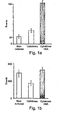
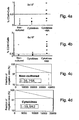
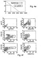
-
FIGs. 1a-1i are a graphic representation of flow cytometry analysis of the effect of nicotinamide on homing of hematopoietic stem cells into bone marrow of NOD/SCID mice. Non-cultured mononuclear cells or their entire progeny following 3-week expansion with cytokines and nicotinamide (cytokines+NA), or cytokines alone (cytokines) were labeled with CFSE and infused into sublethally irradiated NOD/SCID mice (10 x 106 cells/mouse for the non-cultured group containing 5x10⁴ CD34+ cells and the total progeny of 5x10⁴ CD34+ cells following 3-week expansion with or without nicotinamide; 20x106 cells/mouse containing 180x104 CD34+ cells).Figure 1a is a histogram of the results of flow cytometry of recipient's bone marrow cells showing homing of CFSE+/CD34+ cells after transplantation.Figure 1b is a histogram of the results of flow cytometry of recipient's bone marrow cells showing homing of total CFSE+ cells after transplantation. Homing of human cells is presented as number of positive events (cytometry) per 100,000 BM cells analyzed. Each bar represents the average ± SE of 3 independent experiments (6-7 mice/experimental group). Representative flow cytometry analysis of bone marrow cells from non-injected mice (Figure 1c ) and mice injected with non-cultured cells (Figures 1d and1g ), cells cultured with cytokines (Figures 1e and1h ) and cells cultured with cytokines and nicotinamide (Figures 1f and1i ) are shown. Total human cells that home to the BM are gated (see area R2) based on low side scatter (y axis) and log fluorescence distribution of CFSC expression (x axis) (Figures 1c-1f ). The bright fluorescence of CFSE was sufficient to separate labeled human cells from unlabeled murine cells by at least 1 log. Cells gated in R2 were then analyzed for CFSE (x axis) and CD34-APC (y axis) (Figures 1g-1i ). The upper and lower right quadrants represent total human cells while the upper right quadrant represents the human CD34+ cells that home to the BM; -
FIG. 2 is a histogram showing the effect of nicotinamide on in-vitro migration of hematopoietic cells. CXCL12 (100ng/ml)-induced transwell migration of the purified CD34+ cells either before (non-cultured) or after 3-week culture with cytokines and nicotinamide (cytokines+NA) or cytokines alone (cytokines) (n=3, *p<0.02, **p=0.05) was measured as described hereinbelow. Note the enhanced migration of cells cultured in the presence of nicotinamide; -
FIG. 3 is a graph showing the effect of nicotinamide on VLA4-mediated binding of cells to immobilized adhesion molecules under shear flow. CD34+ cells cultured as described inFigs. 1 and2 hereinabove were assayed for capture and arrest under shear stress, on immobilized VCAM-1 adsorbed as 10 µl dots on polystyrene. Cell settling events, and arrest were analyzed under perfusion (shear stress) by video photography. Assayed cell populations were cells before culture (non-cultured, open circles), cells cultured with cytokines, as described inFig. 2 (cultured, closed circles), and cells cultured with cytokines and nicotinamide (cultured+NA, closed triangles). Note the significant and consistent effect of nicotinamide on adhesion molecule-mediated binding; -
FIGs. 4a-4f is a graphic representation of the effect of nicotinamide on homing and engraftment of human hematopoietic cells transplanted into NOD/SCID mice.Figures 4a and 4b show the percentage of human (CD45+) cells in the cell populations before transplantation: non-cultured CD34+ cells (non-cultured, gray ovals), the entire progeny of cultures following 3-week exposure to cytokines alone (cytokines alone, closed ovals), or cytokines and nicotinamide (cytokines+NA, arrows). The percent of engraftment 4 weeks post transplantation was determined by flow cytometry of human CD45 cells in the NOD/SCID marrow (y-axis). The numbers of SCID repopulating cells (SRC) were calculated by plotting the engraftment frequencies at each dose. The resultant curves indicates the estimated frequency of SRCs within non-cultured CD34+ cells (Figure 4c ), culture with cytokines (Figure 4d ) or cytokines and nicotinamide (Figure 4e ). The number shown in each box indicates the calculated frequency of SRCs using the maximum likelihood estimator.Figure 4f shows the immunophenotype of engrafted human cells in representative mice transplanted with the progeny of 12x103 CD34+ cells cultured for 3-weeks with nicotinamide, as determined by FACS. Mouse bone marrow cells were dually stained with FITC-conjugated anti-CD45 (human) and antibodies to human differentiation markers as indicated. Percentages of dual positive cells are shown within each quadrant. Note the greater than 7 fold enhancement of engraftment in the nicotinamide-treated mice (Fig. 4e ), as compared with mice treated with cytokines alone (Fig. 4d ); -
FIGs. 5a-5c are a graphic representation of the effect of nicotinamide (NA) on the engraftment potential of cells cultured under differentiation-promoting conditions. Cultures were initiated with purified cord blood-derived CD34+ cells in medium supplemented with SCF, TPO, IL-6 and FLT3, 50 ng/ml each, and IL-3, 20 ng/ml, with (cytokines+NA, arrows) or without (cytokines, ovals) 10 mM nicotinamide. After 3 weeks, cells were harvested, and transplanted into SCID mice as indicated. Mice were transplanted with 1.25 - 5x 10⁴ CD34+ cells or with their progeny following expansion. Mice were sacrificed 4 weeks later and bone marrow cells were analyzed by FACS for the presence of CD34+ (human progenitor) and CD45+ (human) cells. Mice were scored as engrafted when the number of human (CD45+) cells constituted ≥0.5% of the marrow population.Figure 4a shows the numbers of positively engrafted mice per total number of transplanted mice, for each of the doses of transplanted cells (5.0X10⁴; 2.5X10⁴; and 1.25X10⁴ cells).Figures 4b and 4c show the percentage of total human (CD45+)(Fig. 4b ) cells and human progenitor (CD45+CD34+)(Fig. 4c ) cells in the bone marrow of mice transplanted with cells derived from culture initiated with 1.25 x 10⁴ CD34+ cells. Note the enhanced engraftment of both total human, and human progenitor cells resulting from nicotinamide treatment. - The present invention is of methods of improving homing and engraftment of transplantable cells.
- The principles and operation of the present invention may be better understood with reference to the accompanying descriptions.
- Before explaining at least one embodiment of the invention in detail, it is to be understood that the invention is not limited in its application to the details set forth in the following description or exemplified by the Examples. The invention is capable of other embodiments or of being practiced or carried out in various ways. Also, it is to be understood that the phraseology and terminology employed herein is for the purpose of description and should not be regarded as limiting.
- Successful blood and marrow transplantations, both autologous and allogenic, require the infusion of a sufficient number of hematopoietic stem cells capable of homing to the marrow cavity and regenerating a full array of hematopoietic cell lineages in a timely fashion. Recruitment of stem cells from the marrow into the blood is termed mobilization, or, more commonly stem cell mobilization. It is well established that enhancement of stem cell mobilization and/or homing will result in successful stem cell transplantation.
- SDF-α chemoattracts hematopoietic stem and progenitor cells (HSCs/HPCs) and is thought to play a crucial role in the mobilization of HSCs/HPCs from the bone marrow as well as in stem cell homing.
- CD26 is a widely distributed 110 kDa cell-surface glycoprotein with known dipeptidyl peptidase IV (DPPIV) activity in its extracellular domain capable of cleaving N-terminal dipeptides from polypeptides with either proline or alanine residues in the penultimate position. CD26 inhibits SDF activity by cleaving the latter at its position-2 proline, thereby inhibiting its stem cell mobilizing/homing functions.
-
U.S. Pat. Appl. 2004/0247574 teaches the use of CD26 inhibitors for improving engraftment efficiency of stem cell transplants by both improving stem cell homing to bone marrow and by increasing the number of mobilized donor stem cells. It does not teach down-regulation of CD26 surface expression but rather down regulation of CD26 catalytic activity. Specifically,U.S. Pat. Appl. 2004/0247574 does not teach the use of nicotinamide for down-regulating CD26 surface expression. - While reducing the present invention to practice, the present inventor uncovered that nicotinamide can be successfully used to down-regulate cell-surface expression of CD26, enhance the expression and function of adhesion and integrin molecules, increase induced transplantable cell migration, and significantly improve homing and engraftment of transplantable cells in-vivo.
- As is demonstrated hereinbelow and in the Examples section which follows, the present inventor showed that short-term incubation of cells with nicotinamide is sufficient to down-regulate CD26 expression. A significant reduction in CD26-expressing CD34+ and AC133+ cells was observed following as few as 20 hours of incubation with nicotinamide.
- Of greater significance, nicotinamide was shown effective in enhancing the functionality of molecules critical to the process of cell binding and arrest. Indeed, evaluation of cell migration potential in-vitro following nicotinamide exposure showed that nicotinamide both enhanced CXCL12-inducible migration and VLA-4-mediated binding and retention on VCAM-1 in transplantable cells cultured with nicotinamide(see Example 3 below). Since the processes of cell migration are mediated by "recognition" pairs such as VLA-4 and VCAM-1 in a wide variety of cells, this surprising finding indicates that nicotinamide, and nicotinamide derivatives and analogs, can be effective in enhancing binding and retention, critical to the ability of transplanted cells to successfully home, engraft and repopulate host tissues, for cells of diverse origins and stages of differentiation.
- Actual in-vivo transplantation experiments with nicotinamide-treated cells provided conclusive evidence for the effect of nicotinamide on cell engraftment and homing potential. Example 2 below shows that exposure of mononuclear cells to nicotinamide prior to transplantation into NOD/SCID mice increased the homing to bone marrow sixfold over identical cells cultured without nicotinamide. Yet further, Example 4 below shows that at clearly sub-optimal doses of cells, while control cultured cells failed to cause repopulation of bone marrow in NOD/SCID mice, transplantation of nicotinamide-treated cells resulted in a high degree of engraftment and successful repopulation.
- Taken together these results suggest a novel role for nicotinamide in cell homing and engraftment and as such in cell transplantation.
- Thus, according to one aspect of the present disclosure there is provided a method of enhancing cell homing and engraftment potential, the method comprising ex-vivo or in vitro subjecting a population of cells to an amount of nicotinamide for a period of time sufficient for enhancing cell homing and engraftment potential.
- As used herein the phrase "enhancing cell engraftment potential" refers to an improvement in efficiency, quality or rapidity of cell transplantation which may result from improved homing to the target tissue, improved adhesion, reduced rejection and the like. Methods for assessing cell engraftment potential include, for example, cell migration and other in vitro techniques, and histological, immunological and/or radiological assessment of tissues and organs from actual in-vivo transplantation, as described in detail hereinbelow. A self renewal potential of the stem cells can be determined in-vitro by long term colony formation (LTC-CFUc), or by in-vivo engraftment in the SCID-Hu mouse model. The SCID-Hu mouse model employs C.B-17 scid/scid (SCID) mice transplanted with human fetal thymus and liver tissue or fetal BM tissue and provides an appropriate model for the evaluation of putative human hematopoietic stem cells. Because of the reconstitution of the SCID mice with human fetal tissue, the model affords the proliferation of stem cells, in this case human hematopoietic stem cells to proliferate, and function in the hematopoietic microenvironment of human origin. Mice are typically irradiated, then delivered stem cells into the grafts, and reconstitution is measured by any number of methods, including FACS and immunohistochemistry of repopulated organs (Humeau L., et al Blood (1997) 90:3496; also see Materials and Experimental Methods below).
- As used herein the term "ex-vivo" refers to a process in which cells are removed from a living organism and are propagated outside the organism (e.g., in a test tube).
- As used herein, the term "in-vitro" refers to a process in which cells originating from a cell line or lines (such as NTera2 neural cells, embryonic cell lines, etc.) maintained in the laboratory, are manipulated outside of an organism. Such cell lines are often immortalized cells.
- As used herein the phrase "population of cells" refers to a homogeneous or heterogeneous isolated population of cells which comprise cell populations suitable for transplantation. In a preferred embodiment, at least a portion of the population of cells expresses CD26 or VLA-4 on the cell-surface.
- As used herein, the phrase "stem cells" refers both to the earliest renewable cell population responsible for generating cell mass in a tissue or body and the very early progenitor cells, which are somewhat more differentiated, yet are not committed and can readily revert to become a part of the earliest renewable cell population.
- As used herein, the phrases "non-stem", "non-progenitor" and "committed cells" refer to cells at various stages of differentiation, which generally no longer retain the ability to revert to become a part of a renewable cell population. Methods of ex-vivo culturing stem, progenitor, and non-stem, non-progenitor committed cells are well known in the art of cell culturing. To this effect, see for example, the text book "Culture of Animal Cells - A Manual of Basic Technique" by Freshney, Wiley-Liss, N. Y. (1994), Third Edition.
- The population of cells of the present invention may be from an autologous or non-autologous donor (allogeneic or xenogeneic).
- In a preferred embodiment, the cells for transplantation are stem and/or progenitor cells, and the source of the stem cell population is an unfractionated mononuclear cell preparation, not having been enriched for CD34+ or other hematopoietic stem cells. In another embodiment, the stem cells are identified by stem cell markers such as CD34+, CD34+/CD38-, CD133+, CD34+/Lin-, and other stem cell markers known in the art. In yet another embodiment, the source of the stem cell population are stem cells having been enriched for hematopoietic stem cells by selection according to stem cell markers.
- For example, stem cells of the present invention may be derived from a source selected from the group consisting of hematopoietic cells, umbilical cord blood cells, and mobilized peripheral blood cells.
- As used herein "nicotinamide" refers to nicotinamide as well as to products that are derived from nicotinamide, analogs thereof and metabolites of nicotinamide or nicotinamide analogs, such as, for example, NAD, NADH and NADPH.
- As used herein, the phrase "nicotinamide analog" refers to any molecule that is known to act similarly to nicotinamide. Representative examples of nicotinamide analogs include, without limitation, benzamide, nicotinethioamide (the thiol analog of nicotinanide), nicotinic acid and α-amino-3-indolepropionic acid.
- As used herein the term "subject" refers to a mammalian subject, preferably a human subject.
- The phrase "a nicotinamide or a nicotinamide analog derivative" refers to any structural derivative of nicotinamide itself or of an analog of nicotinamide. Examples of such derivatives include, without limitation, substituted benzamides, substituted nicotinamides and nicotinethioamides and N-substituted nicotinamides and nicotinthioamides.
- Additionally or alternatively, stem cell mobilization may be effected prior to harvest of cells for stem cell transplantation using mobilizing agents which are well known in the art. Generally, the mobilization process is initiated by stress-induced activation of neutrophils and osteoclasts by chemotherapy and repeated stimulation with cytokines such as granulocyte colony-stimulating factor (G-CSF), resulting in shedding and release of membrane-bound stem cell factor (SCF), proliferation of progenitor cells, as well as activation and/or degradation of adhesion molecules. Mobilizing agents which may be used in accordance with the present invention include, but are not limited to, DNA damaging agents, single chemotherapeutic agents (e.g., cyclophosphamide), combined chemotherapy regimens [e.g., iphosphamide, carboplatin and etoposide (ICE) and methylprednisolone, ara-c, and cisplatin (ESHAP)], cytokines such as G-CSF, GM-CSF, SCF, FLT-3 ligand, and chemokines such as IL-8, MIP-1α, Groβ, and SDF-1. The mode of administration, as well as the time frame needed to achieve mobilization and the types of cells mobilized depend on the molecules used. For example, G-CSF is usually administered daily as a dose of 5-10 µg/gk for 5-10 days, alone or after therapy. The adjustment of the mobilization regimen is done by the physician and reviewed in Cottker-Fox et al. (2003) Hematology 419-437.
- Methods of preparation of cells for transplantation are well known in the art. For preparation of non-stem cells, cells can be obtained from donor tissue by dissociation of individual cells from the collecting extracellular matrix of the tissue. Tissue from a particular region is removed using a sterile procedure, and the cells are dissociated-using any method known in the art including treatment with enzymes such as trypsin, collagenase, DNAse and the like, or by using physical methods of dissociation such as with a blunt instrument.
- Cells prepared for transplantation can be maintained in a physiological solution, or cultured in suspension or on a fixed substrate. Suitable culture media capable of supporting cells include HEM, DMEM, RPMI, F-12, and the like. If required, the medium can contain supplements required for cellular metabolism such as glutamine and other amino acids, vitamins, minerals and useful proteins such as transferrin, and the like. The medium may also contain antibiotics to prevent contamination with yeast, bacteria, and fungi, such as penicillin, streptomycin, gentamicin, and the like. If cells are to be cultured, conditions should be close to physiological conditions (preferably, a pH of about 6 to about 8, and a temperature of about 30° C. to about 40° C.). The culture medium can be optionally supplemented with at least one proliferation-inducing growth factor, such as EGF, amphiregulin, acidic fibroblast growth factor (aFGF or FGF-1), basic fibroblast growth factor (bFGF or FGF-2), transforming growth factor alpha (TGF-alpha), cytokines such as G-CSF, GM-CSF, SCF, FLT-3 ligand, and/or chemokines such as IL-8, MIP-1α, Groβ, and SDF-1, and combinations thereof. In addition to proliferation-inducing growth factors, other growth factors may be added to the culture medium, including NGF, platelet-derived growth factor (PDGF), thyrotropin releasing hormone (TRH), and the like.
- Selection and enrichment of specific cell types can be performed, such as separation of hepatocytes from liver tissue, separation of neurons from glial cells, or isolation of islet cells from pancreatic tissue, by morphological, physical, immunohistochemical (FACS) or other means. Fresh or cultured cell preparations can be cryopreserved until they are needed by any method known in the art. The cells can be suspended in an isotonic solution, preferably a cell culture medium, containing a particular cryopreservant. Such cryopreservants include dimethyl sulfoxide (DMSO), glycerol, and the like. Further methods for preparation and storage of cells for transplantation are known in the art, and disclosed in detail in, for example, the Handbook of Transplantation (Kipshidze and Serruys, eds. London, UK, 2004).
- Methods of preparation of stem cells are well known in the art, commonly selecting cells expressing one or more stem cell markers such as CD34, CD133, etc, or lacking markers of differentiated cells. Selection is usually by FACS, or immunomagnetic separation, but can also be by nucleic acid methods such as PCR (see Materials and Experimental Methods hereinbelow). Embryonic stem cells and methods of their retrieval are well known in the art and are described, for example, in Trounson AO (Reprod Fertil Dev (2001) 13: 523), Roach ML (Methods Mol Biol (2002) 185: 1), and Smith AG (Annu Rev Cell Dev Biol (2001) 17:435). Adult stem cells are stem cells, which are derived from tissues of adults and are also well known in the art. Methods of isolating or enriching for adult stem cells are described in, for example, Miraglia, S. et al. (1997) Blood 90: 5013, Uchida, N. et al. (2000) Proc. Natl. Acad. Sci. USA 97: 14720, Simmons, P.J. et al. (1991) Blood 78: 55, Prockop DJ (Cytotherapy (2001) 3: 393), Bohmer RM (Fetal Diagn Ther (2002) 17: 83) and Rowley SD et al. (Bone Marrow Transplant (1998) 21: 1253), Stem Cell Biology Daniel R. Marshak (Editor) Richard L. Gardner (Editor), Publisher: Cold Spring Harbor Laboratory Press, (2001) and Hematopoietic Stem Cell Transplantation. Anthony D. Ho (Editor) Richard Champlin (Editor), Publisher: Marcel Dekker (2000).
- It will be appreciated that nicotinamide can enhance engraftment and homing potential in a wide variety of cell types. For example, nicotinamide can down-regulate surface expression of CD26, and enhance functionality of VLA-4, CXCR-2 or other adhesion molecules from any cell-type expressing same and since these molecules are widely expressed in cell populations of diverse origin, the population of cells may comprise unselected cell populations, such as crude cell preparations from tissue, or mononuclear stem and/or progenitor cells, as well as more homogenous populations of selected cell types.
- As used herein, the phrase "hematopoietic mononuclear cells" refers to the entire repertoire of white blood cells present in a blood sample, usually hematopoietic mononuclear cells which comprise a major fraction of hematopoietic committed cells and a minor fraction of hematopoietic stem and progenitor cells. In a healthy human being, the white blood cells comprise a mixture of hematopoietic lineages committed and differentiated cells (typically over 99 % of the mononuclear cells are lineages committed cells) including, for example: Lineage committed progenitor cells CD34+CD33+ (myeloid committed cells), CD34+CD3+ (lymphoid committed cells) CD34+CD41+ (megakaryocytic committed cells) and differentiated cells - CD34-CD33+ (myeloids, such as granulocytes and monocytes), CD34-CD3+, CD34-CD19+ (T and B cells, respectively), CD34-CD41+ (megakaryocytes), and hematopoietic stem and early progenitor cells such as CD34+Lineage negative (Lin-), CD34-Lineage negative CD34+CD38- (typically less than 1 %).
- Hematopoietic mononuclear cells are typically obtained from a blood sample by applying the blood sample onto a Ficoll-Hypaque layer and collecting, following density-cushion centrifugation, the interface layer present between the Ficoll-Hypaque and the blood serum, which interface layer essentially entirely consists of the white blood cells present in the blood sample.
- Presently, hematopoietic stem cells can be obtained by further enrichment of the hematopoietic mononuclear cells obtained by differential density centrifugation as described above. This further enrichment process is typically performed by immuno-separation such as immunomagnetic-separation or FACS and results in a cell fraction that is enriched for hematopoietic stem cells (for detailed description of enrichment of hematopoietic stem cells, see Materials and Experimental Procedures in the Examples section hereinbelow).
- Regardless of the origin of cells employed and their composition, once the cells are obtained, they are subjected to (contacted with) an amount of nicotinamide for a period of time sufficient to enhance engraftment and homing of the cells following transplantation. Such a period of time may be brief, or lengthier, as required. In one preferred embodiment, the contacting is for a period of time sufficient to down-regulate CD26 surface expression. In another preferred embodiment, the cells are hematopoietic stem cells, and the contacting is for a period of time insufficient for stem cell proliferation (also referred to as expansion) while sufficient to down-regulate CD26 surface expression. In yet another embodiment, the contacting is for a period of time sufficient to increase functionality of VLA-4, CXCR2 and other adhesion and/or integrin molecules.
- Methods of determining protein cell-surface expression are well known in the art. Examples include immunological methods, such as, FACS analysis (see Examples section) as well as biochemical methods (cell-surface labeling, e.g., radioactive, fluorescence, avidin-biotin).
- Methods of assaying cell-proliferation are well known in the art (such as MTT, thymidine incorporation, FACS). It will be appreciated that cell doubling rate may also be derived from the literature.
- Depending on the cell type, and the intended use thereof, cells may be ex-vivo subjected to nicotinamide for long-term contact, i.e. periods of weeks or more, and cells may even be stored in contact with nicotinamide prior to use for transplantation. Further, according to certain embodiments, short-term exposure is desirable. Long-term contacting can be for between 1 and 18 weeks, preferably between 3 and 9 weeks, more preferably between 2 and 5 weeks, most preferably between 2 and 3 weeks. Short term contacting can be for 1 to 2 weeks, preferably one week or less, more preferably between 1-5 days.
- While reducing the present invention to practice, it was uncovered that 20 hours exposure of hematopoietic stem cells to nicotinamide was sufficient to effect a reduction in CD26 expression, crucial to cell homing and engraftment, although insufficient to allow for stem cell expansion or proliferation to take place. Thus, cells are ex-vivo subjected to nicotinamide for a period of time not exceeding a few days, preferably 30 hours, more preferably 1-30 hours, even more preferably 5-30 hours, even more preferably 10-30 hours. In another preferred embodiment, the cells are stem and/or progenitor cells, and the duration of exposure to nicotinamide is selected insufficient for stem cell expansion, or under conditions insufficient for stem cell expansion, such as absence of cytokines, absence of nutrients, sub-optimal temperature, etc.
- Nicotinamide is preferably provided in a final concentration of 0.01-60 mg/ml, preferably 1-40 mg/ml, more preferably 5-30 mg/ml, and most preferably 10-20 mg/ml. The selection of culture medium and medium supplements, depends on the cells and their intended use.
- In one preferred embodiment cells subjected to nicotinamide can be used for transplantation to a subject in need thereof following nicotinamide exposure for the predetermined period of time, without further ex-vivo expansion. It will be appreciated that exposure to nicotinamide can be performed on cells that have received additional ex-vivo treatment, such as expansion, selection, genetic modification, etc. well known in the art, including prior ex-vivo exposure to nicotinamide, and preferably closely preceding the use thereof for engraftment.
- Following exposure to nicotinamide, the cells may then be transplanted in (administered to) a subject in need thereof. The following summarizes some clinical applications which may be addressed according to the teachings of the present invention.
- Hematopoietic cell transplantation: Transplantation of hematopoietic cells has become the treatment of choice for a variety of inherited or malignant diseases. While early transplantation procedures utilized the entire bone marrow (BM) population, recently, more defined populations, enriched for stem cells (CD34+ cells) have been used [Van Epps Blood Cells 20:411, (1994)]. In addition to the marrow, such cells could be derived from other sources such as peripheral blood (PB) and neonatal umbilical cord blood (CB) [Emerson Blood 87:3082 (1996)]. Compared to BM, transplantation with PB cells shortens the period of pancytopenia and reduces the risks of infection and bleeding [Brugger N Engl J Med 333:283, 1995; Williams Blood 87:1687, (1996); Zimmerman J Heamatotherapy 5:247, (1996)].
- An additional advantage of using PB for transplantation is its accessibility. The limiting factor for PB transplantation is the low number of circulating pluripotent stem/progenitor cells.
- To obtain enough PB-derived stem cells for transplantation, these cells are "harvested" by repeated leukophoresis following their mobilization from the marrow into the circulation by treatment with chemotherapy and cytokines [Brugger N Engl J Med 333:283, 1995; Williams Blood 87:1687, (1996)]. Such treatment is obviously not suitable for normal donors.
- The use of ex-vivo expanded stem cells for transplantation has the following advantages [Koller Blood 82:378, (1993); Lebkowski Blood Cells 20:404, (1994)]:
- It reduces the volume of blood required for reconstitution of an adult hematopoietic system and may obviate the need for mobilization and leukophoresis [Brugger N Engl J Med 333:283, 1995].
- It enables storage of small number of PB or CB stem cells for potential future use.
- In the case of autologous transplantation of recipients with malignancies, contaminating tumor cells in autologous infusion often contribute to the recurrence of the disease [Brugger N Engl J Med 333:283, 1995]. Selecting and expanding CD34+ stem cells will reduce the load of tumor cells in the final transplant.
- The cultures provide a significant depletion of T lymphocytes, which may be useful in the allogeneic transplant setting for reducing graft-versus-host disease.
- Clinical studies indicate that transplantation of ex-vivo expanded cells derived from a small number of PB CD34+ cells can restore hematopoiesis in recipients treated with high doses of chemotherapy, although the results do not yet allow firm conclusions about long term in-vivo hematopoietic capabilities of these cultured cells [Brugger N Engl J Med 333:283, 1995; Williams Blood 87:1687, (1996)].
- For successful transplantation, shortening of the duration of the cytopenic phase, as well as long-term engraftment, is crucial. Inclusion of intermediate and late progenitor cells in the transplant could accelerate the production of donor-derived mature cells thereby shortening the cytopenic phase. It is important, therefore, that ex-vivo expanded cells include, in addition to stem and/or progenitor cells subjected to nicotinamide as described hereinabove, more differentiated cells in order to optimize short-term recovery and long-term restoration of hematopoiesis. Inclusion of expanded intermediate and late committed cells, especially those committed to the neutrophilic and megakaryocytic lineages, concomitant with the expanded stem and/or progenitor cells, should serve this purpose [Sandstrom Blood 86:958, (1995)].
- Such cultures may be useful in restoring hematopoiesis in recipients with completely ablated bone marrow, as well as in providing a supportive measure for shortening recipient bone marrow recovery following conventional radio- or chemotherapies.
- Tissue regeneration: Stem cell populations of the present invention, can be used for the promotion of tissue regeneration. Transplantation of stem cells, has great promise for benefits in regenerative medicine, reconstructive surgery, tissue engineering, regenerating new tissues and naturally healing diseased or injured organs (for review see Czyz et al, Biol Chem, 2003;384:1391-40, Sylvester et al Arch Surg 2004;139:93-99). Further, neurons and supporting glial cells have been used for transplantation in treatment of Huntington's disease (
US Patent No. 6,524, 865 to Freed et al ) and pancreatic islet cells are being used for transplantation for type I and type II diabetes (see, for example,US Patents 6,326,201, to Fung et al ; and7,045,349 to Benedict et al ). Muscle, and muscle derived cells, are being investigated for clinical use, and have shown promising results when transplanted into injured heart tissue, bone tissue and articular structures (seeUS Patent No. 6,866,842 to Chancellor, et al ). Thus, the cells for engraftment or transplantation can be derived from an organ selected from the group consisting of a muscle, skin, a bone, a lymph organ, a pancreas, a liver, a gallbladder, a kidney, a digestive tract organ, a respiratory tract organ, a reproductive organ, a urinary tract organ, a blood-associated organ, a thymus, a spleen and a nervous system organ. Examples of cells which can be prepared for implantation by the methods of the present invention include primary cultures as well as established cell lines. Examples of these include, but are not limited to pancreatic islet cells, human foreskin fibroblasts, beta cell insulomas, NT2 cells, embryonic cells, embryonic stem cells, hepatocytes, dopamine secreting ventral mesencephalon cells, neuroblastoid cells, adrenal medulla cells, T-cells combinations of these, and the like. As can be seen from this partial list, cells of all types, including dermal, neural, blood, organ, muscle, glandular, bone, digestive, reproductive and immune system cells, as well as cells of all species of origin, can be prepared successfully by this method. - Recent reports have demonstrated the capability of transplanted or transfused stem cells to enhance regeneration in non-homologous tissue, other than that which the stem cells were derived. For example, enhanced myogenesis and angiogenesis in infarcted myocardium have been observed following infusion of bone marrow stem cells (Tse et al, Lancet 2003, 361:47-79, Jackson et al J Clin Invest 2001;107:1395-402, Orlic et al Nature 2001;410:701-5; Lee et al, Cell Cycle 2005;4:861-64, Nagaya et al Am J Heart Circ Phys 2004;287:H2670-76). Other studies have shown bone marrow, endothelial and skeletal muscle stem cells to be beneficial in ischemic renal injury (Togel et al, AJP Renal Phys 2005;289:F31-F42 and Arriero, et al, AMJ Ren Phys 2004;287:F621-27).
- Gene therapy: For successful long-term gene therapy, a high frequency of genetically modified cells with transgenes stably integrated within their genome, is an obligatory requirement. In BM tissue, for example, while the majority of cells are cycling progenitors and precursors, stem cells constitute only a small fraction of the cell population and most of them are in a quiescent, non-cycling state. Viral-based (e.g., retroviral) vectors require active cell division for integration of the transgene into the host genome. Therefore, gene transfer into fresh stem cells is highly inefficient. The ability to store and process a selected population of cells ex-vivo, and enhance their homing and engraftment potential would provide for an increased probability of the successful use of genetically modified cell transplantation [Palmiter Proc Natl Acad Sci USA 91(4): 1219-1223, (1994)].
- Adoptive immunotherapy: Ex-vivo-expanded, defined lymphoid subpopulations have been studied and used for adoptive immunotherapy of various malignancies, immunodeficiencies, viral and genetic diseases [Freedman Nature Medicine 2: 46, (1996); Heslop Nature Medicine 2: 551, (1996); Protti Cancer Res 56: 1210, (1996)].
- The treatment enhances the required immune response or replaces deficient functions. This approach was pioneered clinically by Rosenberg et al. [Rosenberg J Natl Cancer Inst. 85: 622, 1993] using a large number of autologous and also allogeneic ex-vivo expanded non-specific killer T cells, and subsequently ex-vivo expanded specific tumor infiltrating lymphocytes.
- Functionally active, antigen-presenting, cells could be grown from a starting population of CD34+ PB cells in cytokine-supported cultures, as well. These cells can present soluble protein antigens to autologous T cells in-vitro and, thus, offer new prospects for the immunotherapy of minimal residual disease after high dose chemotherapy. Ex-vivo expansion of antigen-presenting dendritic cells has been studied as well, and is an additional promising application of the currently proposed technology [Bernhard Cancer Res 10: 99, (1995); Fisch Eur J Immunol 26: 595, (1996); Siena Expt Hematol 23:1463, (1996)].
- Additional examples for ex-vivo applications:
- Additional applications of non-stem, differentiated cells, as well as stem and progenitor cell treatment with nicotinamide include skin regeneration, hepatic regeneration, muscle regeneration, stimulation of bone growth for applications in osteoporosis and transplantation of chondrocytes/chondroblasts and/or synovial cells for treatment of articular and arthritic disorders.
- According to one aspect of the present invention, the ex-vivo contacting of cell populations with nicotinamide, according to the features described hereinabove, can be utilized for preparing a population of stem or non-stem cells ex-vivo or in-vitro for implanting the cells in an organ of a subject in need thereof.
- Cells of the present invention may be transplanted by means of direct injection into an organ, injection into the bloodstream, intraperitoneal injection, etc. Suitable methods of transplantation can be determined by monitoring the homing and engraftment of the implanted cells to the desired organ, the expression of desired organ-specific genes or markers, and the function of the derived organ of the subject. In the pancreas, for example, maintenance of euglycemia, secretion of insulin and/or C peptide can be a measure of the restoration of function to a diabetic host animal following cell replacement therapy as disclosed hereinbelow. In the liver, for example, albumin synthesis can be monitored.
- Cell populations of the present invention can be provided per se, along with the culture medium containing same, isolated from the culture medium, and combined with a pharmaceutically acceptable carrier as well as with additional agents which may promote cell engraftment and/or organ function (e.g., immunosuppressing agents, antibiotics, growth factor). Hence, cell populations of the invention can be administered in a pharmaceutically acceptable carrier or diluent, such as sterile saline and aqueous buffer solutions. The use of such carriers and diluents is well known in the art.
- Compositions of the present invention may, if desired, be presented in a pack or dispenser device, such as an FDA-approved kit, which may contain one or more unit dosage forms containing the active ingredient (e.g., cells). The pack may, for example, comprise metal or plastic foil, such as a blister pack. The pack or dispenser device may be accompanied by instructions for administration. The pack or dispenser device may also be accompanied by a notice in a form prescribed by a governmental agency regulating the manufacture, use, or sale of pharmaceuticals, which notice is reflective of approval by the agency of the form of the compositions for human or veterinary administration. Such notice, for example, may include labeling approved by the U.S. Food and Drug Administration for prescription drugs or of an approved product insert. Compositions comprising a preparation of the disclosure formulated in a pharmaceutically acceptable carrier may also be prepared, placed in an appropriate container, and labeled for treatment of an indicated condition, as further detailed above.
- The cells prepared according to the methods of the present invention can be administered to the subject per se, or in a pharmaceutical composition where it is mixed with suitable carriers or excipients.
- As used herein, a "pharmaceutical composition" refers to a preparation of one or more of the active ingredients described herein with other chemical components such as physiologically suitable carriers and excipients. The purpose of a pharmaceutical composition is to facilitate administration of a compound to an organism.
- Hereinafter, the phrases "physiologically acceptable carrier" and "pharmaceutically acceptable carrier," which may be used interchangeably, refer to a carrier or a diluent that does not cause significant irritation to an organism and does not abrogate the biological activity and properties of the administered compound. An adjuvant is included under these phrases.
- Herein, the term "excipient" refers to an inert substance added to a pharmaceutical composition to further facilitate administration of an active ingredient. Examples, without limitation, of excipients include calcium carbonate, calcium phosphate, various sugars and types of starch, cellulose derivatives, gelatin, vegetable oils, and polyethylene glycols.
- Techniques for formulation and administration of drugs may be found in the latest edition of "Remington's Pharmaceutical Sciences," Mack Publishing Co., Easton, PA.
- Suitable routes of administration may, for example, include oral, rectal, transmucosal, especially transnasal, intestinal, or parenteral delivery, including intramuscular, subcutaneous, and intramedullary injections, as well as intrathecal, direct intraventricular, intravenous, intraperitoneal, intranasal, or intraocular injections.
- Alternately, one may administer the pharmaceutical composition in a local rather than systemic manner, for example, via injection of the pharmaceutical composition directly into a tissue region of a patient.
- Pharmaceutical compositions for use in accordance with the present invention thus may be formulated in conventional manner using one or more physiologically acceptable carriers comprising excipients and auxiliaries, which facilitate processing of the active ingredients into preparations that can be used pharmaceutically. Proper formulation is dependent upon the route of administration chosen.
- For injection, the active ingredients of the pharmaceutical composition may be formulated in aqueous solutions, preferably in physiologically compatible buffers such as Hank's solution, Ringer's solution, or physiological salt buffer. For transmucosal administration, penetrants appropriate to the barrier to be permeated are used in the formulation. Such penetrants are generally known in the art.
- Pharmaceutical compositions suitable for use in the context of the present invention include compositions wherein the active ingredients are contained in an amount effective to achieve the intended purpose. More specifically, a "therapeutically effective amount" means an amount of active ingredients (e.g., a nucleic acid construct) effective to prevent, alleviate, or ameliorate symptoms of a disorder (e.g., ischemia) or prolong the survival of the subject being treated.
- Determination of a therapeutically effective amount is well within the capability of those skilled in the art, especially in light of the detailed disclosure provided herein.
- For any preparation used in the methods of the invention, the dosage or the therapeutically effective amount can be estimated initially from in vitro and cell culture assays. For example, a dose can be formulated in animal models to achieve a desired concentration or titer. Such information can be used to more accurately determine useful doses in humans.
- Toxicity and therapeutic efficacy of the active ingredients described herein can be determined by standard pharmaceutical procedures in vitro, in cell cultures or experimental animals. The data obtained from these in vitro and cell culture assays and animal studies can be used in formulating a range of dosage for use in human. The dosage may vary depending upon the dosage form employed and the route of administration utilized. The exact formulation, route of administration, and dosage can be chosen by the individual physician in view of the patient's condition. (See, e.g., Fingl, E. et al. (1975), "The Pharmacological Basis of Therapeutics," Ch. 1, p.1.)
- Depending on the severity and responsiveness of the condition to be treated, dosing can be of a single or a plurality of administrations, with course of treatment lasting from several days to several weeks, or until cure is effected or diminution of the disease state is achieved.
- The amount of a composition to be administered will, of course, be dependent on the subject being treated, the severity of the affliction, the manner of administration, the judgment of the prescribing physician, etc.
- Additional objects, advantages, and novel features of the present invention will become apparent to one ordinarily skilled in the art upon examination of the following examples, which are not intended to be limiting. Additionally, each of the various embodiments and aspects of the present invention as delineated hereinabove and as claimed in the claims section below finds experimental support in the following examples.
- Reference is now made to the following examples, which together with the above descriptions, illustrate the invention in a non limiting fashion.
- Generally, the nomenclature used herein and the laboratory procedures utilized in the present invention include molecular, biochemical, microbiological and recombinant DNA techniques. Such techniques are thoroughly explained in the literature. See, for example, "Molecular Cloning: A laboratory Manual" Sambrook et al., (1989); "Current Protocols in Molecular Biology" Volumes I-III Ausubel, R. M., ed. (1994); Ausubel et al., "Current Protocols in Molecular Biology", John Wiley and Sons, Baltimore, Maryland (1989); Perbal, "A Practical Guide to Molecular Cloning", John Wiley & Sons, New York (1988); Watson et al., "Recombinant DNA", Scientific American Books, New York; Birren et al. (eds) "Genome Analysis: A Laboratory Manual Series", Vols. 1-4, Cold Spring Harbor Laboratory Press, New York (1998); methodologies as set forth in
U.S. Pat. Nos. 4,666,828 ;4,683,202 ;4,801,531 ;5,192,659 and5,272,057 ; "Cell Biology: A Laboratory Handbook", Volumes I-III Cellis, J. E., ed. (1994); "Current Protocols in Immunology" Volumes I-III Coligan J. E., ed. (1994); Stites et al. (eds), "Basic and Clinical Immunology" (8th Edition), Appleton & Lange, Norwalk, CT (1994); Mishell and Shiigi (eds), "Selected Methods in Cellular Immunology", W. H. Freeman and Co., New York (1980); available immunoassays are extensively described in the patent and scientific literature, see, for example,U.S. Pat: Nos. 3,791,932 ;3,839,153 ;3,850,752 ;3,850,578 ;3,853,987 ;3,867,517 ;3,879,262 ;3,901,654 ;3,935,074 ;3,984,533 ;3,996,345 ;4,034,074 ;4,098,876 ;4,879,219 ;5,011,771 and5,281,521 ; "Oligonucleotide Synthesis" Gait, M. J., ed. (1984); "nucleic Acid Hybridization" Hames, B. D., and Higgins S. J., eds. (1985); "Transcription and Translation" Hames, B. D., and Higgins S. J., Eds. (1984); "Animal Cell Culture" Freshney, R. I., ed. (1986); "Immobilized Cells and Enzymes" IRL Press, (1986); "A Practical Guide to Molecular Cloning" Perbal, B., (1984) and "Methods in Enzymology" Vol. 1-317, Academic Press; "PCR Protocols: A Guide To Methods And Applications", Academic Press, San Diego, CA (1990); Marshak et al., "Strategies for Protein Purification and Characterization - A Laboratory Course Manual" CSHL Press (1996). Other general references are provided throughout this document. The procedures therein are believed to be well known in the art and are provided for the convenience of the reader. - Cord blood samplers - Cells were obtained from umbilical cord blood after normal full-term delivery (informed consent was given). Samples were collected and frozen according to Rubinstein et al. [Rubinstein et al. Proc Natl Acad Sci U S A. 1995; 92 (22):10119-10122.] within 24 h postpartum. Prior to use, the cells were thawed in Dextran buffer (Sigma, St. Louis, MO, USA) containing 2.5 % human serum albumin (HAS, Bayer Corp. Elkhart, IN, USA), layered on a Ficoll-Hypaque gradient (1.077 g/mL; Sigma), and centrifuged at 800 x g for 30 min. The mononuclear cells in the interface layer were collected and washed three times in phosphate-buffered saline (PBS, Biological Industries) containing 0.5 % HSA. To purify the CD34+ cells, the mononuclear cell fraction was subjected to two cycles of immunomagnetic bead separation, using a "MiniMACS CD34 progenitor cell isolation kit" (Miltenyi Biotec Bergisch, Gladbach, Germany), according to manufacturer's instructions. The purity of the CD34+ population thus obtained was 95-98 %, as evaluated by flow cytometry.
- FACS analysis of CD34+ cells - Percentages of progenitor cells positive for CD26 were determined by staining of CD34+ cells with anti-CD26 FITC purchased from Becton Dickinson.
- Ex vivo expansion- Purified CD34+ cells were cultured in culture bags (American Fluoroseal Co., Gaithersburg, MD) at 1x10⁴ cells/ml in MEMα medium, 10% fetal calf serum (FCS) and cytokines: Thrombopoietin (TPO), interleukin-6 (IL-6), FLT-3 ligand and stem cell factor (SCF), each at a final concentration of 50 ng/ml (Perpo Tech, Inc., Rocky Hill, NJ), with or without 5 mM nicotinamide (NA) (Sigma Aldrich, Milwaukee, WI) and incubated at 37°C in a humidified atmosphere of 5% CO2 in air. Preliminary studies conducted with various concentrations of NA (1-10 mM), indicated that in combination with the 4 cytokines, 5 mM was the optimal NA concentration (data not shown). Until week 3, the cultures were topped weekly with the same volume of fresh medium and then weekly demi-depopulated. Cell counting, colony forming unit (CFUc) assay and immunophenotype analysis were performed as described hereinbelow.
- Immunophenotyping of CD34+ cells- MiniMACS re-isolated CD34+ cells were washed with PBS solution containing 1% BSA and double stained (at 4°C for 30 min) with PE-conjugated anti-CD34 and FITC-conjugated antibodies to CXCR4, VLA-4 (Chemicon Intnl, Inc. Temecula, CA, USA), LFA-1 (IQ product), CD38, or with a mixture of FITC-conjugated antibodies against differentiation antigens (CD38, CD33, CD14, CD15, CD3, CD61, CD19) for determination of CD34+Lin-cells. (Antibodies to CD34, CD38 and CD61 were purchased from DAKO Glostrup, Corp, Carpenteria, CA, USA, while the others were from Becton Dickinson and Co, San Jose, CA, USA). The cells were then washed in the above buffer and analyzed, using a FACScalibur® flow cytometer (Becton Dickinson and Co., San Jose, CA). Emission of 104 cells was measured using logarithmic amplification, and analyzed using CellQuest software (Becton Dickinson and Co, San Jose, CA, USA). The FACS analysis results are presented as percentages of CD34+ cells. The absolute number of CD34+CD38- and CD34+Lin- cells was calculated from the total number of CD34+ cells in culture.
- CFSE labeling- Non-cultured or cultured cells were washed and resuspended at less than 107 cells/mL in serum-free medium. CFSE (Molecular Probes, Inc., Eugene, OR, USA) was added at a final concentration of 5 µg/ml, and the cells were incubated for 10 minutes at 37°C. Uptake of the dye was stopped by the addition of 10% FCS. After labeling, cells were washed three times in PBS supplemented with 10% FCS and analyzed by flow cytometry for fluorescence intensity, then injected intravenously into sublethally irradiated NOD/SCID mice (10-20 million cells per mouse).
- In Vitro Migration Assay- RPMI plus 10% FCS (0.6 ml) containing 100 ng/ml CXCL12 (R&D Systems) was put into the lower chamber of a Costar 24-well "transwell" culture plate (Corning, Inc, Corning, NY). Cells (2 x10⁵) in 100-µl medium were introduced into the upper chamber, over a porous membrane (pore size, 5 µm). After 4 hours, cells were collected from both chambers and counted by flow cytometry (FACSsort, Becton Dickinson and Co, San Jose, CA, USA). Spontaneous migration control migration was performed without CXCL12 in the lower chamber.
- In vivo analysis of homing- NOD/SCID mice (8-10 week old) (Harlan Ltd., Israel) were sub-lethally irradiated (at 375cGy at 67cGy/min) and 24 hours later inoculated via the tail vein with either CFSE-labeled cultured, or non-cultured CB cells. Mice were sacrificed at 24 hours post injection and bone marrow samples were obtained by flushing their femurs and tibias with IMDM at 4°C. Homing of human cells was detected by flow cytometry via visualization of CFSE-stained cells over a background of unlabeled murine cells. The bright fluorescence of CFSE was sufficient to separate labeled human cells from unlabeled murine cells by at least 1 log. To quantify homing of human progenitor cells, bone marrow cells were stained with APC-conjugated antihuman CD34 monoclonal antibodies and CFSE+CD34+ (human progenitor) cells were enumerated. For each sample 100,000 events were recorded and analyzed.
- Transplantation of human CD34+ cells into NOD/SCID mice- NOD/SCID mice were bred and maintained in sterile intra-ventilated cages (Techniplast, Bugugiatte, Italy). Eight-week-old mice were sub-lethally irradiated as described above. Mice were then inoculated via the tail vein with fresh purified CB-derived CD34+ cells or their entire progeny following 3-weeks in culture. To avoid donor variability, CB-derived CD34+ cells from several units were pooled and used for expansion cultures as well as group injection. Mice were sacrificed at week 4, and marrow samples were obtained by flushing their femurs and tibias with IMDM at 4°C. Flow cytometric analysis of NOD/SCID marrow cells was performed as described hereinabove, using monoclonal antibodies against human leukocyte differentiation antigens to identify human cell engraftment.
- Quantification of SCID repopulating cells (SRCs)- The frequencies of SRCs were quantified by a limiting dilution analysis and applying Poisson statistics to the single-hit model as described previously. Mice were scored as positively engrafted if 0.5% of their marrow cells expressed human CD45. The frequencies of SRCs and statistical comparison between individual populations were calculated by maximum likelihood estimator using L-Calc. Software (StemCell Technologies, Vancouver, BC).
- Shear Flow Experiments- Soluble purified seven-domain human VCAM-1, sVCAM-1 was mixed in coating medium (PBS buffered with 20 mM sodium bicarbonate pH, 8.5) with a fixed amount of carrier (2 µg/ml HSA) and adsorbed as 10-µl spots on polystyrene plates (Becton Dickinson and Co, San Jose, CA, USA) for 2 hours at 37°C, alone or with the indicated amounts of intact or heat-inactivated chemokines. Plates were washed and blocked with HSA (20 mg/ml). VCAM-1 site densities were assessed using 121I-labeled anti-VCAM-1 mAb, 4B9. Cell monolayers of non-cultured cells and cells following culturing with or without nicotinamide and VCAM-1/chemokine-coated substrates were assembled as the lower surface of the flow chamber (260-µm gap) and expensively washed with binding medium. The flow chamber was mounted on the stage of an inverted phase contrast microscope (Diaphot 300; Nikon Europe BV, Badhoevedorp, The Netherlands). All flow experiments were conducted at 37°C. Cells were perfused at 106 cells/ml through the chamber at desired flow rate generated with an automated syringe pump. The entire duration of cell perfusions were recorded on a videotape with a long integration LIS-700 CCD video camera (Applitech Rigicam, Israel) and a Time Lapse SVHS-Video recorder (AG-6730; Panasonic, Japan). All cellular interactions with the adhesive substrates were determined by manually tracking the motions of individual cells along 0.9-mm field paths for 1 min. Cellular interactions with VCAM-1-bearing surfaces were >95% α4 integrin dependent. In each experiment all events were normalized to a constant cell population flowing in immediate proximity with the substrate. Frequency of each category of tethers was expressed in percentage of units (event x cell-1 x 10²); 1% unit measured at 0.5, 1, and 1.5 dyn/cm2 corresponded to tethering rate of 1.5 x 10⁻³, 3 x 10⁻³, and 4.5 x 10⁻³ events x cell-1 mm-1 s-1, respectively, expressed as the mean ± range or SD.
- Statistics- The non-parametric Wilcoxon Rank Test was applied for testing differences between the study groups. All the tests applied were two-tailed, and a p value of ≤ 5% was considered statistically significant. The data were analyzed using SAS software (SAS Institute, Cary, NC).
- The effect of short-term incubation with nicotinamide on HSC CD26 membranal expression was addressed by FACS analysis.
- Freshly purified CD34+ cells were FACS analyzed for the expression of CD26 (T-0) then incubated +/- Nicotinamide 5mM for 20 hours (T-20 hours) and FACS analyzed again. As demonstrated in a duplicate experiment summarized in Table 1 below, following 20 hours incubation (T-20) in the presence of Nicotinamide, CD26 expression was significantly reduced by the presence of Nicotinamide (2-3 fold reduction) as compared to cells maintained for 20 hours in the absence of Nicotinamide as well as to freshly purified CD34+ cells (T-0).
Table 1 CD26+ cells (%) T-0 T-20 Control + Nicotinamide Exp. 1 8.5 5 2.7 Exp. 2 10 12 6 - Reduced engraftment efficacy of cultured cells has been attributed, at least in part, to a defect in their homing ability relative to non-cultured cells (Szilvassy, S.J., et al, Blood, 2000;95:2829-37). To evaluate the effect of nicotinamide on the homing of cultured cells, NOD/SCID mice were transplanted with either 10x106 non-cultured mononuclear cells (MNC), containing 5x10⁴ CD34+ cells (0.5% CD34+ cells), or with their total progeny following 3-weeks in culture with cytokines, with or without nicotinamide, each transplantation containing 180x104 CD34+ cells. Prior to transplantation, the cells were labeled with CFSE. Twenty-four hours post transplantation total CFSE-labeled cells and CFSE labeled CD34+ cells that homed to the mouse bone marrow of the recipient mice were quantified by FACS.
- Even though the same number of cells and CD34+ cells were transplanted from both cultured groups, the homing of nicotinamide-treated CD34+ cells was 6-fold higher, while the homing of CD34+ cells without exposure to nicotinamide was only 2-fold higher relative to the homing of non-cultured CD34+ cells (n=21, p<0.05) (
Fig 1a ). The homing of cultured cells (MNC) was 2-fold higher with nicotinamide-treated cells compared to nicotinamide-untreated cells, and similar to the homing of non-cultured MNC (n=21, p<0.05) (Fig 1b ).Figures 1c-1i show FACS analysis dot plots of representative mice transplanted with non-cultured or cultured cells. - Alterations in chemokine and adhesion molecules, either expression or functionality, have been suggested to cause a homing defect in cultured CD34+ cells, since the binding of cells to specific "docking" ligands is critical for the efficient passage of cells from circulation to target tissues (Foguenne, J., et al. Haematologica, 2005;90:445-51). This is specially significant in view of the broad distribution of integrins and adhesion molecules such as VLA-4 and LFA-1 across a variety of cell types (muscle cells, lymphocytes, eosinophils, etc). In order to determine the role of such adhesion and related molecules in nicotinamide-mediated enhancement of homing and engraftment of cells, the effect of nicotinamide on in-vitro migration and the functionality of the adhesion molecule Very Late Activation Antigen-4 (VLA-4) was tested.
- Using a trans-well migration assay, CXCL12-induced migration of non-cultured and cultured hematopoietic cells was tested, assessing the effects of nicotinamide on integrin and adhesion molecule function. CXCL12 powerfully stimulated the migration of both treated and untreated CD34+ cells (Figure 2d). However, CXCL12-induced migration was significantly higher in cells cultured with nicotinamide (cytokines + NA) compared to the cells cultured without nicotinamide (p>0.02) or non-cultured cells (p=0.05) (
Fig. 2 ). These results suggest that treatment of CD34+ cells with NA can potentially increase the responsiveness of CXCR4 to its ligand CXCL12, resulting in enhanced engraftment and homing potential of the nicotinamide-treated cells. - When the functional quality of cell binding to adhesion molecules was investigated using shear flow analysis, the strong effect of nicotinamide on VLA4-mediated binding and retention on VCAM was revealed.
Figure 3 shows the significantly enhanced percentage of initially settled cells resistant to removal by shear stress evident in the cells treated with nicotinamide. - Thus, the results in
Figures 2 and 3 reveal that nicotinamide treatment of cells before transplantation increases the function of adhesion and cytokine-related molecules in these cells, enhances cell migration, and therefore enhance cell's transplantation potential, as indicated by increased initial capture and binding to immobilized VCAM-1, and retention under increased flow, as compared to non-cultured or cytokines-alone cultured cells. - Nicotinamide treatment was tested for ability to enhance homing and engraftment of transplanted cells by repopulation of NOD/SCID mice. To evaluate repopulating capacity, NOD/SCID mice were transplanted with non-cultured CD34+ cells (n = 12) over a range of doses intended to achieve a sub-optimal transplantation, and subsequent non-engraftment in a fraction of mice or their progeny following 3-weeks expansion with cytokines (n = 12) or cytokines + NA (n = 13). Human cell engraftment was evaluated 4-weeks post transplantation. Mice were scored as positively engrafted if 0.5% of the recipient bone marrow cells expressed human CD45 antigen (CD45+). As shown in
Fig 4a , transplantation of 3 x 10³ CD34+ cells resulted in no engraftment in the non-cultured cells. Similarly, the progeny of 3 x 10³ CB CD34+ cells cultured with cytokines only also failed to engraft. The presence of nicotinamide in culture, however, resulted in 50% engraftment of 3 x 10³ CB CD34+ cells in the mice. At a dose range of 6 x 10³ cells (Fig. 4b ), fresh CB CD34+ cells engrafted in only 16.7% of the mice, whereas the progeny of 6 x 10³ CD34+ cells cultured with cytokines engrafted in 33.3% of the mice. In contrast, at the same dose range, the progeny of cytokines nicotinamide cultured cells engrafted in 100% of the mice (Fig 4b ). - The frequency of SCID repopulating cells (SRCs) was calculated using the maximum likelihood estimator as described hereinabove (
Figs. 4c-4e ). The frequency of SRCs within non-cultured CD34+ cells was 1 in 36,756 cells (95% confidence interval [CI], 1/113,366 - 1/11,917) (Fig. 4c .). The SRC frequency within cells cultured with cytokines alone.was 1 in 19,982 (CI, 1/47,972 - 1/8,323) (Fig. 4d ) and the SRC frequency within cells cultured in the presence of nicotinamide and cytokines was significantly higher, at 1 in 2,620 (CI, 1/5,127 - 1/1,339) (Fig. 4e ). Therefore, culture conditions including nicotinamide supported a 14-fold greater number of SRCs than non-cultured cells and 7.6-fold more SRCs than cytokine alone-cultured cells.Fig 4f demonstrates in vivo multilineage differentiation of NA-treated cultured cells engrafted in NOD/SCID mice. - Effect of nicotinamide on homing and engraftment in IL-3 treated cells: IL-3 has been reported to accelerate differentiation and attenuate SCID repopulating ability of transplanted cells. In order to test whether nicotinamide modulates the engraftment potential of cells exposed to the cytokine, nicotinamide's effect on CB-derived CD34+ cells cultured with IL-3 was assessed. Transplantation experiments indicated that treatment with nicotinamide indeed increased the SCID repopulating potential of IL-3 supplemented cultures.
Fig. 5a shows the proportion of engraftment following injection of 1.25 to 5X 10⁴ cells.Figs. 5b-c exhibits the engraftment of total human cells (Fig. 5b ) and progenitor cells (Fig. 5c ) following transplantation of the lowest cell dose evaluated in this experiment (1.25X10⁴ cells). The results show the presence of human (CD45+) cells in bone marrow of 5 out of 5 mice transplanted with nicotinamide treated cells, but in only 2 out of 5 mice transplanted with cytokine alone treated cells (Fig. 5a ). Engraftment of human progenitors (CD45+CD34+) 4-weeks after transplantation was observed only in mice transplanted with cells cultured with nicotinamide. - The results brought hereinabove clearly show that exposure of cells to nicotinamide enhances expression and function of adhesion and integrin molecules critical to cell engraftment and homing, can increase cell migration potential, and clearly provides superior engraftment and homing of transplanted cells. Thus, nicotinamide can be used to provide cell populations for transplantation, having enhanced homing and engraftment potential.
- It is appreciated that certain features of the invention, which are, for clarity, described in the context of separate embodiments, may also be provided in combination in a single embodiment. Conversely, various features of the invention, which are, for brevity, described in the context of a single embodiment, may also be provided separately or in any suitable subcombination.
-
- Aiuti J. Exp. Med. 1997;185:111-120
- Anderlini, P. and Korbling, M. (1997) Stem.
- Baggiolini, M.. 1998, Nature 392:565
- Banasik M. et al., J Biol Chem. 1992;267:1569-1575
- Bernhard Cancer Res 10: 99, (1995)
- Christopherson KW 2nd, et al., Science. 2004 Aug 13;305(5686):1000- 1003
- Christopherson KW 2nd, J Immunol. 2002
- Corda D, Di Girolamo M. 2003;22(9):1953-1958
- de la Crux, Lois S, et al., Bioessays. 2005;27(2):164-75
- De Roos et al Transplantation 1997;63:513-18
- Fisch Eur J Immunol 26: 595, (1996)
- Foguenne, J., et al. Haematologica, 2005;90:445-51
- Freedman Nature Medicine 2: 46, (1996)
- Gagandeep et al, Gene Therapy 1999;6:729-36
- Heslop Nature Medicine 2: 551, (1996)
- Humeau L., et al Blood (1997) 90:3496
- Ito et al, Muscle Nerve 1998;21:291-7
- Imai Br. J. Haematol. 1999;106:905-911
- Jaime Imitola et al., Proc Natl Acad Sci U S A. 2004 101 (52): 18117-18122
- Kipshidze and Serruys, eds. London, UK, 2004
- Lupi R, et al., J Biol Chem. 2000;275:9418-9424
- Lupi R, et al. Biochem J. 2002:367:1-7
- McGrath Dev. Biol. 1999;213:442-456
- Protti Cancer Res 56: 1210, (1996)
- Rankin PW, et al., J Biol Chem. 1989;264:4312-4317
- Roach ML. Methods Mol Biol (2002) 185: 1
- Siena Expt Hematol 23:1463, (1996)
- Shioda et al. (1998) Proc. Natl. Acad. Sci. USA 95:6331
- Smith AG. Annu Rev Cell Dev Biol (2001) 17:435
- Smith S. Trends Biochem Sci. 2001;26:174-179
- Trounson AO. Reprod Fertil Dev (2001) 13: 523
- Ueda K, Hayaishi O, Annu Rev Biochem. 1985;54:73-100
- Virág L, Szabó C. Pharm. Reviews. 2002;54:375-429
- Yau L, et al., Eur. J. Biochem. 2003;270:101-110
Claims (10)
- A method of enhancing cell homing and engraftment potential, the method comprising ex-vivo or in vitro subjecting a population of cells to 0.01 to 60 mg/ml nicotinamide for a period of time insufficient for stem cell expansion, said period of time being from 10-30 hours wherein said population of cells is a CD 34+ hematopoietic stem and/or progenitor cell population.
- The method of claim 1, wherein said population of cells is derived from a source selected from the group consisting of umbilical cord blood cells, mobilized peripheral blood cells, bone marrow cells.
- The method of claim 1 or claim 2, wherein said population of cells is derived from a mononuclear cell fraction.
- The method of any of claims 1 to 3, further comprising the step of selecting a population of cells enriched for hematopoietic stem cells prior to, concomitant with or following said step of ex-vivo subjecting.
- The method of any of claims 1 to 4, wherein said period of time is sufficient to downregulate expression of CD26 on the cells.
- The method of any one of claim 1 to 5, wherein said amount of nicotinamide and said period of time are selected sufficient to reduce CD26 expression by cells of said population of cells as compared to CD26 expression in hematopoietic stem and/or progenitor cells maintained for the same period of time in the absence of nicotinamide, wherein said CD26 expression is measured in-vitro by FACS analysis.
- The method of any of claims 1 to 5 or 6, wherein said period of time is 20 hours.
- A cell population comprising hematopoietic stem and/or progenitor cells characterized by enhanced engraftment and homing potential prepared according to the method of any one of claims 1 to 5 or 7.
- A pharmaceutical composition comprising as an active ingredient the cell population of claim 8 and a pharmaceutically acceptable carrier.
- The cell population of claim 8, isolated and prepared for transplantation into a subject in need thereof.
Priority Applications (1)
| Application Number | Priority Date | Filing Date | Title |
|---|---|---|---|
| EP11177996A EP2390312A1 (en) | 2005-11-29 | 2006-11-29 | Methods of improving stem cell homing and engraftment |
Applications Claiming Priority (2)
| Application Number | Priority Date | Filing Date | Title |
|---|---|---|---|
| US28900405A | 2005-11-29 | 2005-11-29 | |
| PCT/IL2006/001381 WO2007063545A2 (en) | 2005-11-29 | 2006-11-29 | Methods of improving stem cell homing and engraftment |
Related Child Applications (1)
| Application Number | Title | Priority Date | Filing Date |
|---|---|---|---|
| EP11177996.3 Division-Into | 2011-08-18 |
Publications (2)
| Publication Number | Publication Date |
|---|---|
| EP1974012A2 EP1974012A2 (en) | 2008-10-01 |
| EP1974012B1 true EP1974012B1 (en) | 2013-02-27 |
Family
ID=37895790
Family Applications (2)
| Application Number | Title | Priority Date | Filing Date |
|---|---|---|---|
| EP11177996A Withdrawn EP2390312A1 (en) | 2005-11-29 | 2006-11-29 | Methods of improving stem cell homing and engraftment |
| EP06821601A Active EP1974012B1 (en) | 2005-11-29 | 2006-11-29 | Methods of improving stem cell homing and engraftment |
Family Applications Before (1)
| Application Number | Title | Priority Date | Filing Date |
|---|---|---|---|
| EP11177996A Withdrawn EP2390312A1 (en) | 2005-11-29 | 2006-11-29 | Methods of improving stem cell homing and engraftment |
Country Status (9)
| Country | Link |
|---|---|
| EP (2) | EP2390312A1 (en) |
| JP (1) | JP5102773B2 (en) |
| KR (1) | KR20080072748A (en) |
| AU (1) | AU2006321172B2 (en) |
| BR (1) | BRPI0620523A2 (en) |
| CA (1) | CA2632288A1 (en) |
| ES (1) | ES2409128T3 (en) |
| WO (1) | WO2007063545A2 (en) |
| ZA (1) | ZA200805210B (en) |
Families Citing this family (10)
| Publication number | Priority date | Publication date | Assignee | Title |
|---|---|---|---|---|
| AU770896C (en) | 1998-09-29 | 2006-09-07 | Gamida Cell Ltd. | Methods of controlling proliferation and differentiation of stem and progenitor cells |
| US8846393B2 (en) | 2005-11-29 | 2014-09-30 | Gamida-Cell Ltd. | Methods of improving stem cell homing and engraftment |
| JP5763894B2 (en) * | 2009-07-01 | 2015-08-12 | タカラバイオ株式会社 | Cell separation method |
| EP3785712A1 (en) * | 2009-12-29 | 2021-03-03 | Gamida-Cell Ltd. | Methods for enhancing natural killer cell proliferation and activity |
| WO2012121451A1 (en) * | 2011-03-08 | 2012-09-13 | 연세대학교 산학협력단 | Method for inducing in vivo migration of stem cell |
| SG11201404608WA (en) | 2012-02-13 | 2014-09-26 | Gamida Cell Ltd | Mesenchymal stem cells conditioned medium and methods of generating and using the same |
| US9175266B2 (en) | 2012-07-23 | 2015-11-03 | Gamida Cell Ltd. | Enhancement of natural killer (NK) cell proliferation and activity |
| US9567569B2 (en) | 2012-07-23 | 2017-02-14 | Gamida Cell Ltd. | Methods of culturing and expanding mesenchymal stem cells |
| JP7295810B2 (en) | 2017-05-16 | 2023-06-21 | ガミダ セル リミテッド | Selection and use of umbilical cord cell fractions suitable for transplantation |
| AU2017414804A1 (en) * | 2017-05-16 | 2020-01-02 | Gamida-Cell Ltd. | Selection and use of umbilical cord cell fractions suitable for transplantation |
Family Cites Families (31)
| Publication number | Priority date | Publication date | Assignee | Title |
|---|---|---|---|---|
| NL154600B (en) | 1971-02-10 | 1977-09-15 | Organon Nv | METHOD FOR THE DETERMINATION AND DETERMINATION OF SPECIFIC BINDING PROTEINS AND THEIR CORRESPONDING BINDABLE SUBSTANCES. |
| NL154598B (en) | 1970-11-10 | 1977-09-15 | Organon Nv | PROCEDURE FOR DETERMINING AND DETERMINING LOW MOLECULAR COMPOUNDS AND PROTEINS THAT CAN SPECIFICALLY BIND THESE COMPOUNDS AND TEST PACKAGING. |
| NL154599B (en) | 1970-12-28 | 1977-09-15 | Organon Nv | PROCEDURE FOR DETERMINING AND DETERMINING SPECIFIC BINDING PROTEINS AND THEIR CORRESPONDING BINDABLE SUBSTANCES, AND TEST PACKAGING. |
| US3901654A (en) | 1971-06-21 | 1975-08-26 | Biological Developments | Receptor assays of biologically active compounds employing biologically specific receptors |
| US3853987A (en) | 1971-09-01 | 1974-12-10 | W Dreyer | Immunological reagent and radioimmuno assay |
| US3867517A (en) | 1971-12-21 | 1975-02-18 | Abbott Lab | Direct radioimmunoassay for antigens and their antibodies |
| NL171930C (en) | 1972-05-11 | 1983-06-01 | Akzo Nv | METHOD FOR DETERMINING AND DETERMINING BITES AND TEST PACKAGING. |
| US3850578A (en) | 1973-03-12 | 1974-11-26 | H Mcconnell | Process for assaying for biologically active molecules |
| US3935074A (en) | 1973-12-17 | 1976-01-27 | Syva Company | Antibody steric hindrance immunoassay with two antibodies |
| US3996345A (en) | 1974-08-12 | 1976-12-07 | Syva Company | Fluorescence quenching with immunological pairs in immunoassays |
| US4034074A (en) | 1974-09-19 | 1977-07-05 | The Board Of Trustees Of Leland Stanford Junior University | Universal reagent 2-site immunoradiometric assay using labelled anti (IgG) |
| US3984533A (en) | 1975-11-13 | 1976-10-05 | General Electric Company | Electrophoretic method of detecting antigen-antibody reaction |
| US4098876A (en) | 1976-10-26 | 1978-07-04 | Corning Glass Works | Reverse sandwich immunoassay |
| US4879219A (en) | 1980-09-19 | 1989-11-07 | General Hospital Corporation | Immunoassay utilizing monoclonal high affinity IgM antibodies |
| US5011771A (en) | 1984-04-12 | 1991-04-30 | The General Hospital Corporation | Multiepitopic immunometric assay |
| US4666828A (en) | 1984-08-15 | 1987-05-19 | The General Hospital Corporation | Test for Huntington's disease |
| US4801531A (en) | 1985-04-17 | 1989-01-31 | Biotechnology Research Partners, Ltd. | Apo AI/CIII genomic polymorphisms predictive of atherosclerosis |
| US5272057A (en) | 1988-10-14 | 1993-12-21 | Georgetown University | Method of detecting a predisposition to cancer by the use of restriction fragment length polymorphism of the gene for human poly (ADP-ribose) polymerase |
| US5192659A (en) | 1989-08-25 | 1993-03-09 | Genetype Ag | Intron sequence analysis method for detection of adjacent and remote locus alleles as haplotypes |
| US5281521A (en) | 1992-07-20 | 1994-01-25 | The Trustees Of The University Of Pennsylvania | Modified avidin-biotin technique |
| ATE358276T1 (en) | 1995-06-07 | 2007-04-15 | Bioveris Corp | ELECTROCHEMILUMINESCENCE ENZYMIMMUNOASSAY |
| US6866842B1 (en) | 1998-05-01 | 2005-03-15 | University Of Pittsburgh | Muscle-derived cells (MDCs) for treating muscle-or bone-related injury or dysfunction |
| US6713245B2 (en) | 1998-07-06 | 2004-03-30 | Diacrin, Inc. | Methods for storing neural cells such that they are suitable for transplantation |
| IL144654A0 (en) | 1999-02-10 | 2002-05-23 | Curis Inc | Pancreatic progenitor cells, methods and uses related thereto |
| US7045349B2 (en) | 2001-01-23 | 2006-05-16 | Benedict Daniel J | Method of islet isolation using process control |
| IL152904A0 (en) * | 2002-01-24 | 2003-06-24 | Gamida Cell Ltd | Utilization of retinoid and vitamin d receptor antagonists for expansion of renewable stem cell populations |
| US20050220774A1 (en) * | 2002-03-18 | 2005-10-06 | Tony Peled | Methods of inducing differentiation in ex vivo expanded stem cells |
| WO2004016731A2 (en) * | 2002-08-19 | 2004-02-26 | Gamida-Cell Ltd. | Ex-vivo expansion of hematopoietic stem cell populations in mononuclear cell cultures |
| EP1601759A4 (en) * | 2003-03-07 | 2007-02-14 | Gamida Cell Ltd | Expansion of renewable stem cell populations using modulators of pi 3-kinase |
| US20040247574A1 (en) | 2003-05-27 | 2004-12-09 | Christopherson Kent W. | Methods for enhancing stem cell engraftment during transplantation |
| IL161903A0 (en) * | 2003-07-17 | 2005-11-20 | Gamida Cell Ltd | Ex vivo progenitor and stem cell expansion for usein the treatment of disease of endodermally- deri ved organs |
-
2006
- 2006-11-29 CA CA002632288A patent/CA2632288A1/en not_active Abandoned
- 2006-11-29 EP EP11177996A patent/EP2390312A1/en not_active Withdrawn
- 2006-11-29 JP JP2008542940A patent/JP5102773B2/en active Active
- 2006-11-29 AU AU2006321172A patent/AU2006321172B2/en active Active
- 2006-11-29 BR BRPI0620523-2A patent/BRPI0620523A2/en not_active IP Right Cessation
- 2006-11-29 KR KR1020087015455A patent/KR20080072748A/en not_active Application Discontinuation
- 2006-11-29 WO PCT/IL2006/001381 patent/WO2007063545A2/en active Application Filing
- 2006-11-29 ES ES06821601T patent/ES2409128T3/en active Active
- 2006-11-29 EP EP06821601A patent/EP1974012B1/en active Active
-
2008
- 2008-06-17 ZA ZA200805210A patent/ZA200805210B/en unknown
Also Published As
| Publication number | Publication date |
|---|---|
| ES2409128T3 (en) | 2013-06-25 |
| KR20080072748A (en) | 2008-08-06 |
| ZA200805210B (en) | 2009-09-30 |
| JP2009517078A (en) | 2009-04-30 |
| EP1974012A2 (en) | 2008-10-01 |
| CA2632288A1 (en) | 2007-06-07 |
| BRPI0620523A2 (en) | 2011-11-16 |
| JP5102773B2 (en) | 2012-12-19 |
| AU2006321172A1 (en) | 2007-06-07 |
| WO2007063545A3 (en) | 2007-07-19 |
| WO2007063545A2 (en) | 2007-06-07 |
| EP2390312A1 (en) | 2011-11-30 |
| AU2006321172B2 (en) | 2012-02-23 |
Similar Documents
| Publication | Publication Date | Title |
|---|---|---|
| US8846393B2 (en) | Methods of improving stem cell homing and engraftment | |
| EP1974012B1 (en) | Methods of improving stem cell homing and engraftment | |
| Dexter et al. | Stromal cells in haemopoiesis | |
| Davis et al. | Porcine brain microvascular endothelial cells support the in vitro expansion of human primitive hematopoietic bone marrow progenitor cells with a high replating potential: requirement for cell-to-cell interactions and colony-stimulating factors | |
| DiGiusto et al. | Hematopoietic potential of cryopreserved and ex vivo manipulated umbilical cord blood progenitor cells evaluated in vitro and in vivo | |
| Brandt et al. | Ex vivo expansion of autologous bone marrow CD34+ cells with porcine microvascular endothelial cells results in a graft capable of rescuing lethally irradiated baboons | |
| US20040235160A1 (en) | Process for preparing hematopoietic stem cells | |
| JP2007536936A (en) | Stem cell populations and methods of use | |
| JP2002516085A (en) | Compositions and uses that affect hematopoietic stem cell populations in mammals | |
| AU778504B2 (en) | Human brain endothelial cells and growth medium and method for expansion of primitive CD34+CD38- bone marrow stem cells | |
| TW202003837A (en) | Cd3-negative cell population expressing chemokine receptor and cell adhesion molecule, use thereof, and method for producing same | |
| Chute et al. | A comparative study of the cell cycle status and primitive cell adhesion molecule profile of human CD34+ cells cultured in stroma-free versus porcine microvascular endothelial cell cultures | |
| Shih et al. | Ex vivo expansion of transplantable human hematopoietic stem cells: where do we stand in the year 2000? | |
| US8802434B2 (en) | Biological cell culture, cell culture media and therapeutic use of biological cells | |
| Tushinski et al. | Expansion and Clinical use of Hematopoietic Progenitor Cells in Human Marrow and Peripheral Blood | |
| Hoffman et al. | Ex vivo expansion of human hematopoietic stem cells: implications for the modern blood bank | |
| Kauser et al. | Bone marrow-derived progenitors | |
| Case | The cytokine-mediated expansion of human umbilical cord blood stem and progenitor cells | |
| Koury et al. | Wintrobe’s clinical hematology | |
| WO1999030723A9 (en) | Use of human umbilical cord blood for adoptive therapy |
Legal Events
| Date | Code | Title | Description |
|---|---|---|---|
| PUAI | Public reference made under article 153(3) epc to a published international application that has entered the european phase |
Free format text: ORIGINAL CODE: 0009012 |
|
| 17P | Request for examination filed |
Effective date: 20080627 |
|
| AK | Designated contracting states |
Kind code of ref document: A2 Designated state(s): AT BE BG CH CY CZ DE DK EE ES FI FR GB GR HU IE IS IT LI LT LU LV MC NL PL PT RO SE SI SK TR |
|
| 17Q | First examination report despatched |
Effective date: 20090820 |
|
| DAX | Request for extension of the european patent (deleted) | ||
| GRAP | Despatch of communication of intention to grant a patent |
Free format text: ORIGINAL CODE: EPIDOSNIGR1 |
|
| GRAS | Grant fee paid |
Free format text: ORIGINAL CODE: EPIDOSNIGR3 |
|
| GRAA | (expected) grant |
Free format text: ORIGINAL CODE: 0009210 |
|
| AK | Designated contracting states |
Kind code of ref document: B1 Designated state(s): AT BE BG CH CY CZ DE DK EE ES FI FR GB GR HU IE IS IT LI LT LU LV MC NL PL PT RO SE SI SK TR |
|
| REG | Reference to a national code |
Ref country code: GB Ref legal event code: FG4D |
|
| REG | Reference to a national code |
Ref country code: CH Ref legal event code: EP |
|
| REG | Reference to a national code |
Ref country code: AT Ref legal event code: REF Ref document number: 598563 Country of ref document: AT Kind code of ref document: T Effective date: 20130315 |
|
| REG | Reference to a national code |
Ref country code: IE Ref legal event code: FG4D |
|
| REG | Reference to a national code |
Ref country code: DE Ref legal event code: R096 Ref document number: 602006034820 Country of ref document: DE Effective date: 20130418 |
|
| REG | Reference to a national code |
Ref country code: ES Ref legal event code: FG2A Ref document number: 2409128 Country of ref document: ES Kind code of ref document: T3 Effective date: 20130625 |
|
| REG | Reference to a national code |
Ref country code: AT Ref legal event code: MK05 Ref document number: 598563 Country of ref document: AT Kind code of ref document: T Effective date: 20130227 |
|
| REG | Reference to a national code |
Ref country code: LT Ref legal event code: MG4D |
|
| PG25 | Lapsed in a contracting state [announced via postgrant information from national office to epo] |
Ref country code: IS Free format text: LAPSE BECAUSE OF FAILURE TO SUBMIT A TRANSLATION OF THE DESCRIPTION OR TO PAY THE FEE WITHIN THE PRESCRIBED TIME-LIMIT Effective date: 20130627 Ref country code: LT Free format text: LAPSE BECAUSE OF FAILURE TO SUBMIT A TRANSLATION OF THE DESCRIPTION OR TO PAY THE FEE WITHIN THE PRESCRIBED TIME-LIMIT Effective date: 20130227 Ref country code: BG Free format text: LAPSE BECAUSE OF FAILURE TO SUBMIT A TRANSLATION OF THE DESCRIPTION OR TO PAY THE FEE WITHIN THE PRESCRIBED TIME-LIMIT Effective date: 20130527 Ref country code: AT Free format text: LAPSE BECAUSE OF FAILURE TO SUBMIT A TRANSLATION OF THE DESCRIPTION OR TO PAY THE FEE WITHIN THE PRESCRIBED TIME-LIMIT Effective date: 20130227 Ref country code: SE Free format text: LAPSE BECAUSE OF FAILURE TO SUBMIT A TRANSLATION OF THE DESCRIPTION OR TO PAY THE FEE WITHIN THE PRESCRIBED TIME-LIMIT Effective date: 20130227 |
|
| REG | Reference to a national code |
Ref country code: NL Ref legal event code: VDEP Effective date: 20130227 |
|
| PG25 | Lapsed in a contracting state [announced via postgrant information from national office to epo] |
Ref country code: BE Free format text: LAPSE BECAUSE OF FAILURE TO SUBMIT A TRANSLATION OF THE DESCRIPTION OR TO PAY THE FEE WITHIN THE PRESCRIBED TIME-LIMIT Effective date: 20130227 Ref country code: PL Free format text: LAPSE BECAUSE OF FAILURE TO SUBMIT A TRANSLATION OF THE DESCRIPTION OR TO PAY THE FEE WITHIN THE PRESCRIBED TIME-LIMIT Effective date: 20130227 Ref country code: LV Free format text: LAPSE BECAUSE OF FAILURE TO SUBMIT A TRANSLATION OF THE DESCRIPTION OR TO PAY THE FEE WITHIN THE PRESCRIBED TIME-LIMIT Effective date: 20130227 Ref country code: PT Free format text: LAPSE BECAUSE OF FAILURE TO SUBMIT A TRANSLATION OF THE DESCRIPTION OR TO PAY THE FEE WITHIN THE PRESCRIBED TIME-LIMIT Effective date: 20130627 Ref country code: FI Free format text: LAPSE BECAUSE OF FAILURE TO SUBMIT A TRANSLATION OF THE DESCRIPTION OR TO PAY THE FEE WITHIN THE PRESCRIBED TIME-LIMIT Effective date: 20130227 Ref country code: SI Free format text: LAPSE BECAUSE OF FAILURE TO SUBMIT A TRANSLATION OF THE DESCRIPTION OR TO PAY THE FEE WITHIN THE PRESCRIBED TIME-LIMIT Effective date: 20130227 Ref country code: GR Free format text: LAPSE BECAUSE OF FAILURE TO SUBMIT A TRANSLATION OF THE DESCRIPTION OR TO PAY THE FEE WITHIN THE PRESCRIBED TIME-LIMIT Effective date: 20130528 |
|
| PG25 | Lapsed in a contracting state [announced via postgrant information from national office to epo] |
Ref country code: CZ Free format text: LAPSE BECAUSE OF FAILURE TO SUBMIT A TRANSLATION OF THE DESCRIPTION OR TO PAY THE FEE WITHIN THE PRESCRIBED TIME-LIMIT Effective date: 20130227 Ref country code: RO Free format text: LAPSE BECAUSE OF FAILURE TO SUBMIT A TRANSLATION OF THE DESCRIPTION OR TO PAY THE FEE WITHIN THE PRESCRIBED TIME-LIMIT Effective date: 20130227 Ref country code: DK Free format text: LAPSE BECAUSE OF FAILURE TO SUBMIT A TRANSLATION OF THE DESCRIPTION OR TO PAY THE FEE WITHIN THE PRESCRIBED TIME-LIMIT Effective date: 20130227 Ref country code: EE Free format text: LAPSE BECAUSE OF FAILURE TO SUBMIT A TRANSLATION OF THE DESCRIPTION OR TO PAY THE FEE WITHIN THE PRESCRIBED TIME-LIMIT Effective date: 20130227 Ref country code: NL Free format text: LAPSE BECAUSE OF FAILURE TO SUBMIT A TRANSLATION OF THE DESCRIPTION OR TO PAY THE FEE WITHIN THE PRESCRIBED TIME-LIMIT Effective date: 20130227 Ref country code: SK Free format text: LAPSE BECAUSE OF FAILURE TO SUBMIT A TRANSLATION OF THE DESCRIPTION OR TO PAY THE FEE WITHIN THE PRESCRIBED TIME-LIMIT Effective date: 20130227 |
|
| PG25 | Lapsed in a contracting state [announced via postgrant information from national office to epo] |
Ref country code: CY Free format text: LAPSE BECAUSE OF FAILURE TO SUBMIT A TRANSLATION OF THE DESCRIPTION OR TO PAY THE FEE WITHIN THE PRESCRIBED TIME-LIMIT Effective date: 20130227 |
|
| PLBE | No opposition filed within time limit |
Free format text: ORIGINAL CODE: 0009261 |
|
| STAA | Information on the status of an ep patent application or granted ep patent |
Free format text: STATUS: NO OPPOSITION FILED WITHIN TIME LIMIT |
|
| 26N | No opposition filed |
Effective date: 20131128 |
|
| REG | Reference to a national code |
Ref country code: DE Ref legal event code: R097 Ref document number: 602006034820 Country of ref document: DE Effective date: 20131128 |
|
| REG | Reference to a national code |
Ref country code: CH Ref legal event code: PL |
|
| PG25 | Lapsed in a contracting state [announced via postgrant information from national office to epo] |
Ref country code: CH Free format text: LAPSE BECAUSE OF NON-PAYMENT OF DUE FEES Effective date: 20131130 Ref country code: MC Free format text: LAPSE BECAUSE OF FAILURE TO SUBMIT A TRANSLATION OF THE DESCRIPTION OR TO PAY THE FEE WITHIN THE PRESCRIBED TIME-LIMIT Effective date: 20130227 Ref country code: LI Free format text: LAPSE BECAUSE OF NON-PAYMENT OF DUE FEES Effective date: 20131130 |
|
| REG | Reference to a national code |
Ref country code: IE Ref legal event code: MM4A |
|
| PG25 | Lapsed in a contracting state [announced via postgrant information from national office to epo] |
Ref country code: IE Free format text: LAPSE BECAUSE OF NON-PAYMENT OF DUE FEES Effective date: 20131129 |
|
| PG25 | Lapsed in a contracting state [announced via postgrant information from national office to epo] |
Ref country code: TR Free format text: LAPSE BECAUSE OF FAILURE TO SUBMIT A TRANSLATION OF THE DESCRIPTION OR TO PAY THE FEE WITHIN THE PRESCRIBED TIME-LIMIT Effective date: 20130227 |
|
| PG25 | Lapsed in a contracting state [announced via postgrant information from national office to epo] |
Ref country code: LU Free format text: LAPSE BECAUSE OF NON-PAYMENT OF DUE FEES Effective date: 20131129 Ref country code: HU Free format text: LAPSE BECAUSE OF FAILURE TO SUBMIT A TRANSLATION OF THE DESCRIPTION OR TO PAY THE FEE WITHIN THE PRESCRIBED TIME-LIMIT; INVALID AB INITIO Effective date: 20061129 |
|
| REG | Reference to a national code |
Ref country code: FR Ref legal event code: PLFP Year of fee payment: 10 |
|
| REG | Reference to a national code |
Ref country code: FR Ref legal event code: PLFP Year of fee payment: 11 |
|
| REG | Reference to a national code |
Ref country code: FR Ref legal event code: PLFP Year of fee payment: 12 |
|
| PGFP | Annual fee paid to national office [announced via postgrant information from national office to epo] |
Ref country code: ES Payment date: 20230125 Year of fee payment: 17 |
|
| PGFP | Annual fee paid to national office [announced via postgrant information from national office to epo] |
Ref country code: GB Payment date: 20231123 Year of fee payment: 18 |
|
| PGFP | Annual fee paid to national office [announced via postgrant information from national office to epo] |
Ref country code: IT Payment date: 20231124 Year of fee payment: 18 Ref country code: FR Payment date: 20231120 Year of fee payment: 18 Ref country code: DE Payment date: 20231121 Year of fee payment: 18 |
|
| PGFP | Annual fee paid to national office [announced via postgrant information from national office to epo] |
Ref country code: ES Payment date: 20240126 Year of fee payment: 18 |