EP1962961B1 - Demibodies: dimerisation-activated therapeutic agents - Google Patents
Demibodies: dimerisation-activated therapeutic agents Download PDFInfo
- Publication number
- EP1962961B1 EP1962961B1 EP06817559A EP06817559A EP1962961B1 EP 1962961 B1 EP1962961 B1 EP 1962961B1 EP 06817559 A EP06817559 A EP 06817559A EP 06817559 A EP06817559 A EP 06817559A EP 1962961 B1 EP1962961 B1 EP 1962961B1
- Authority
- EP
- European Patent Office
- Prior art keywords
- binding
- immunoglobulins
- functional
- cell
- portions
- Prior art date
- Legal status (The legal status is an assumption and is not a legal conclusion. Google has not performed a legal analysis and makes no representation as to the accuracy of the status listed.)
- Not-in-force
Links
- 239000003814 drug Substances 0.000 title claims description 16
- 229940124597 therapeutic agent Drugs 0.000 title description 8
- 210000004027 cell Anatomy 0.000 claims description 180
- 230000027455 binding Effects 0.000 claims description 122
- 239000000427 antigen Substances 0.000 claims description 99
- 108091007433 antigens Proteins 0.000 claims description 99
- 102000036639 antigens Human genes 0.000 claims description 99
- 230000000295 complement effect Effects 0.000 claims description 67
- 108090000623 proteins and genes Proteins 0.000 claims description 58
- 108060003951 Immunoglobulin Proteins 0.000 claims description 54
- 102000018358 immunoglobulin Human genes 0.000 claims description 54
- 102000004169 proteins and genes Human genes 0.000 claims description 54
- 229940072221 immunoglobulins Drugs 0.000 claims description 38
- 239000003795 chemical substances by application Substances 0.000 claims description 35
- 206010028980 Neoplasm Diseases 0.000 claims description 34
- 201000011510 cancer Diseases 0.000 claims description 32
- 238000000034 method Methods 0.000 claims description 29
- ROHFNLRQFUQHCH-YFKPBYRVSA-N L-leucine Chemical compound CC(C)C[C@H](N)C(O)=O ROHFNLRQFUQHCH-YFKPBYRVSA-N 0.000 claims description 26
- ROHFNLRQFUQHCH-UHFFFAOYSA-N Leucine Natural products CC(C)CC(N)C(O)=O ROHFNLRQFUQHCH-UHFFFAOYSA-N 0.000 claims description 26
- 239000012634 fragment Substances 0.000 claims description 21
- 230000010056 antibody-dependent cellular cytotoxicity Effects 0.000 claims description 14
- 241001465754 Metazoa Species 0.000 claims description 9
- 241000271566 Aves Species 0.000 claims description 8
- 239000003446 ligand Substances 0.000 claims description 8
- 102000007469 Actins Human genes 0.000 claims description 7
- 108010085238 Actins Proteins 0.000 claims description 7
- 108091008102 DNA aptamers Proteins 0.000 claims description 7
- 102000002151 Microfilament Proteins Human genes 0.000 claims description 7
- 108010040897 Microfilament Proteins Proteins 0.000 claims description 7
- 230000001939 inductive effect Effects 0.000 claims description 7
- 241000282414 Homo sapiens Species 0.000 claims description 6
- 108010003723 Single-Domain Antibodies Proteins 0.000 claims description 6
- 238000003745 diagnosis Methods 0.000 claims description 5
- 239000007850 fluorescent dye Substances 0.000 claims description 4
- 230000001404 mediated effect Effects 0.000 claims description 4
- 108091005981 phosphorylated proteins Proteins 0.000 claims description 4
- 210000000130 stem cell Anatomy 0.000 claims description 4
- 238000002560 therapeutic procedure Methods 0.000 claims description 4
- 102000004127 Cytokines Human genes 0.000 claims description 3
- 108090000695 Cytokines Proteins 0.000 claims description 3
- 241000288906 Primates Species 0.000 claims description 3
- 201000010099 disease Diseases 0.000 claims description 3
- 208000037265 diseases, disorders, signs and symptoms Diseases 0.000 claims description 3
- 208000015181 infectious disease Diseases 0.000 claims description 3
- 238000009533 lab test Methods 0.000 claims description 3
- 244000144972 livestock Species 0.000 claims description 3
- MMVYPOCJESWGTC-UHFFFAOYSA-N Molybdenum(2+) Chemical compound [Mo+2] MMVYPOCJESWGTC-UHFFFAOYSA-N 0.000 claims description 2
- 108010057085 cytokine receptors Proteins 0.000 claims description 2
- 102000003675 cytokine receptors Human genes 0.000 claims description 2
- 108091005608 glycosylated proteins Proteins 0.000 claims description 2
- 102000035122 glycosylated proteins Human genes 0.000 claims description 2
- 238000000338 in vitro Methods 0.000 claims description 2
- 108091005626 post-translationally modified proteins Proteins 0.000 claims description 2
- 102000035123 post-translationally modified proteins Human genes 0.000 claims description 2
- 230000007402 cytotoxic response Effects 0.000 claims 1
- 235000018102 proteins Nutrition 0.000 description 47
- 102100033051 40S ribosomal protein S19 Human genes 0.000 description 27
- 230000001225 therapeutic effect Effects 0.000 description 22
- 238000001327 Förster resonance energy transfer Methods 0.000 description 20
- 231100000433 cytotoxic Toxicity 0.000 description 20
- 230000001472 cytotoxic effect Effects 0.000 description 20
- 101100456536 Caenorhabditis elegans mec-2 gene Proteins 0.000 description 13
- 239000000975 dye Substances 0.000 description 12
- 239000000203 mixture Substances 0.000 description 12
- 230000005284 excitation Effects 0.000 description 11
- 239000003550 marker Substances 0.000 description 11
- -1 microproteins Proteins 0.000 description 11
- 230000008685 targeting Effects 0.000 description 11
- 102100024222 B-lymphocyte antigen CD19 Human genes 0.000 description 10
- 101000980825 Homo sapiens B-lymphocyte antigen CD19 Proteins 0.000 description 10
- FAPWRFPIFSIZLT-UHFFFAOYSA-M Sodium chloride Chemical compound [Na+].[Cl-] FAPWRFPIFSIZLT-UHFFFAOYSA-M 0.000 description 10
- 238000001514 detection method Methods 0.000 description 10
- 238000001943 fluorescence-activated cell sorting Methods 0.000 description 10
- 102100022005 B-lymphocyte antigen CD20 Human genes 0.000 description 9
- PEDCQBHIVMGVHV-UHFFFAOYSA-N Glycerine Chemical compound OCC(O)CO PEDCQBHIVMGVHV-UHFFFAOYSA-N 0.000 description 9
- 101000897405 Homo sapiens B-lymphocyte antigen CD20 Proteins 0.000 description 9
- 230000004540 complement-dependent cytotoxicity Effects 0.000 description 9
- 238000000684 flow cytometry Methods 0.000 description 9
- 210000003000 inclusion body Anatomy 0.000 description 9
- 108091003079 Bovine Serum Albumin Proteins 0.000 description 7
- 230000003013 cytotoxicity Effects 0.000 description 7
- 231100000135 cytotoxicity Toxicity 0.000 description 7
- 239000012894 fetal calf serum Substances 0.000 description 7
- 244000052769 pathogen Species 0.000 description 7
- 108090000765 processed proteins & peptides Proteins 0.000 description 7
- 238000011160 research Methods 0.000 description 7
- 238000002415 sodium dodecyl sulfate polyacrylamide gel electrophoresis Methods 0.000 description 7
- 108091023037 Aptamer Proteins 0.000 description 6
- 101000738771 Homo sapiens Receptor-type tyrosine-protein phosphatase C Proteins 0.000 description 6
- 108010021625 Immunoglobulin Fragments Proteins 0.000 description 6
- 102000008394 Immunoglobulin Fragments Human genes 0.000 description 6
- 102100037422 Receptor-type tyrosine-protein phosphatase C Human genes 0.000 description 6
- 150000001413 amino acids Chemical group 0.000 description 6
- 238000002189 fluorescence spectrum Methods 0.000 description 6
- 230000003993 interaction Effects 0.000 description 6
- 229910052594 sapphire Inorganic materials 0.000 description 6
- 239000010980 sapphire Substances 0.000 description 6
- 239000006228 supernatant Substances 0.000 description 6
- ABZLKHKQJHEPAX-UHFFFAOYSA-N tetramethylrhodamine Chemical compound C=12C=CC(N(C)C)=CC2=[O+]C2=CC(N(C)C)=CC=C2C=1C1=CC=CC=C1C([O-])=O ABZLKHKQJHEPAX-UHFFFAOYSA-N 0.000 description 6
- 241000588724 Escherichia coli Species 0.000 description 5
- 241001198387 Escherichia coli BL21(DE3) Species 0.000 description 5
- 102000016359 Fibronectins Human genes 0.000 description 5
- 108010067306 Fibronectins Proteins 0.000 description 5
- 238000005119 centrifugation Methods 0.000 description 5
- 229940079593 drug Drugs 0.000 description 5
- 108010021843 fluorescent protein 583 Proteins 0.000 description 5
- 238000005734 heterodimerization reaction Methods 0.000 description 5
- 210000002500 microbody Anatomy 0.000 description 5
- 230000001717 pathogenic effect Effects 0.000 description 5
- 102000004196 processed proteins & peptides Human genes 0.000 description 5
- 238000000746 purification Methods 0.000 description 5
- 239000011780 sodium chloride Substances 0.000 description 5
- MJKVTPMWOKAVMS-UHFFFAOYSA-N 3-hydroxy-1-benzopyran-2-one Chemical compound C1=CC=C2OC(=O)C(O)=CC2=C1 MJKVTPMWOKAVMS-UHFFFAOYSA-N 0.000 description 4
- QCPFFGGFHNZBEP-UHFFFAOYSA-N 4,5,6,7-tetrachloro-3',6'-dihydroxyspiro[2-benzofuran-3,9'-xanthene]-1-one Chemical compound O1C(=O)C(C(=C(Cl)C(Cl)=C2Cl)Cl)=C2C21C1=CC=C(O)C=C1OC1=CC(O)=CC=C21 QCPFFGGFHNZBEP-UHFFFAOYSA-N 0.000 description 4
- 102000004190 Enzymes Human genes 0.000 description 4
- 108090000790 Enzymes Proteins 0.000 description 4
- 108010087819 Fc receptors Proteins 0.000 description 4
- 102000009109 Fc receptors Human genes 0.000 description 4
- 239000006137 Luria-Bertani broth Substances 0.000 description 4
- 241000700605 Viruses Species 0.000 description 4
- 239000000872 buffer Substances 0.000 description 4
- 238000002659 cell therapy Methods 0.000 description 4
- 238000012377 drug delivery Methods 0.000 description 4
- 239000012636 effector Substances 0.000 description 4
- GNBHRKFJIUUOQI-UHFFFAOYSA-N fluorescein Chemical compound O1C(=O)C2=CC=CC=C2C21C1=CC=C(O)C=C1OC1=CC(O)=CC=C21 GNBHRKFJIUUOQI-UHFFFAOYSA-N 0.000 description 4
- 108091006047 fluorescent proteins Proteins 0.000 description 4
- 102000034287 fluorescent proteins Human genes 0.000 description 4
- 238000003018 immunoassay Methods 0.000 description 4
- BPHPUYQFMNQIOC-NXRLNHOXSA-N isopropyl beta-D-thiogalactopyranoside Chemical compound CC(C)S[C@@H]1O[C@H](CO)[C@H](O)[C@H](O)[C@H]1O BPHPUYQFMNQIOC-NXRLNHOXSA-N 0.000 description 4
- 238000002372 labelling Methods 0.000 description 4
- 238000000569 multi-angle light scattering Methods 0.000 description 4
- 229920001184 polypeptide Polymers 0.000 description 4
- 239000000047 product Substances 0.000 description 4
- PYWVYCXTNDRMGF-UHFFFAOYSA-N rhodamine B Chemical compound [Cl-].C=12C=CC(=[N+](CC)CC)C=C2OC2=CC(N(CC)CC)=CC=C2C=1C1=CC=CC=C1C(O)=O PYWVYCXTNDRMGF-UHFFFAOYSA-N 0.000 description 4
- 239000000243 solution Substances 0.000 description 4
- 230000009466 transformation Effects 0.000 description 4
- QKNYBSVHEMOAJP-UHFFFAOYSA-N 2-amino-2-(hydroxymethyl)propane-1,3-diol;hydron;chloride Chemical compound Cl.OCC(N)(CO)CO QKNYBSVHEMOAJP-UHFFFAOYSA-N 0.000 description 3
- 208000010839 B-cell chronic lymphocytic leukemia Diseases 0.000 description 3
- 101000934338 Homo sapiens Myeloid cell surface antigen CD33 Proteins 0.000 description 3
- 102000018071 Immunoglobulin Fc Fragments Human genes 0.000 description 3
- 108010091135 Immunoglobulin Fc Fragments Proteins 0.000 description 3
- 102100025243 Myeloid cell surface antigen CD33 Human genes 0.000 description 3
- 108010004729 Phycoerythrin Proteins 0.000 description 3
- 208000029052 T-cell acute lymphoblastic leukemia Diseases 0.000 description 3
- XSQUKJJJFZCRTK-UHFFFAOYSA-N Urea Chemical compound NC(N)=O XSQUKJJJFZCRTK-UHFFFAOYSA-N 0.000 description 3
- 238000001042 affinity chromatography Methods 0.000 description 3
- BFNBIHQBYMNNAN-UHFFFAOYSA-N ammonium sulfate Chemical compound N.N.OS(O)(=O)=O BFNBIHQBYMNNAN-UHFFFAOYSA-N 0.000 description 3
- 229910052921 ammonium sulfate Inorganic materials 0.000 description 3
- 235000011130 ammonium sulphate Nutrition 0.000 description 3
- 230000015572 biosynthetic process Effects 0.000 description 3
- 239000004202 carbamide Substances 0.000 description 3
- 239000003153 chemical reaction reagent Substances 0.000 description 3
- 238000010276 construction Methods 0.000 description 3
- 239000002254 cytotoxic agent Substances 0.000 description 3
- 229940127089 cytotoxic agent Drugs 0.000 description 3
- 231100000599 cytotoxic agent Toxicity 0.000 description 3
- 238000000695 excitation spectrum Methods 0.000 description 3
- 238000000799 fluorescence microscopy Methods 0.000 description 3
- 238000002866 fluorescence resonance energy transfer Methods 0.000 description 3
- 201000003444 follicular lymphoma Diseases 0.000 description 3
- 108020001507 fusion proteins Proteins 0.000 description 3
- 102000037865 fusion proteins Human genes 0.000 description 3
- 238000003384 imaging method Methods 0.000 description 3
- 230000004048 modification Effects 0.000 description 3
- 238000012986 modification Methods 0.000 description 3
- 102000035118 modified proteins Human genes 0.000 description 3
- 108091005573 modified proteins Proteins 0.000 description 3
- 230000004481 post-translational protein modification Effects 0.000 description 3
- 238000002360 preparation method Methods 0.000 description 3
- 238000001742 protein purification Methods 0.000 description 3
- VXPULQMBPNGJOD-SECBINFHSA-N (2r)-2-amino-3-(4-hydroxyphenyl)-2-phosphonopropanoic acid Chemical compound OC(=O)[C@@](P(O)(O)=O)(N)CC1=CC=C(O)C=C1 VXPULQMBPNGJOD-SECBINFHSA-N 0.000 description 2
- VGIRNWJSIRVFRT-UHFFFAOYSA-N 2',7'-difluorofluorescein Chemical compound OC(=O)C1=CC=CC=C1C1=C2C=C(F)C(=O)C=C2OC2=CC(O)=C(F)C=C21 VGIRNWJSIRVFRT-UHFFFAOYSA-N 0.000 description 2
- PRDFBSVERLRRMY-UHFFFAOYSA-N 2'-(4-ethoxyphenyl)-5-(4-methylpiperazin-1-yl)-2,5'-bibenzimidazole Chemical compound C1=CC(OCC)=CC=C1C1=NC2=CC=C(C=3NC4=CC(=CC=C4N=3)N3CCN(C)CC3)C=C2N1 PRDFBSVERLRRMY-UHFFFAOYSA-N 0.000 description 2
- IOOMXAQUNPWDLL-UHFFFAOYSA-N 2-[6-(diethylamino)-3-(diethyliminiumyl)-3h-xanthen-9-yl]-5-sulfobenzene-1-sulfonate Chemical compound C=12C=CC(=[N+](CC)CC)C=C2OC2=CC(N(CC)CC)=CC=C2C=1C1=CC=C(S(O)(=O)=O)C=C1S([O-])(=O)=O IOOMXAQUNPWDLL-UHFFFAOYSA-N 0.000 description 2
- QWZHDKGQKYEBKK-UHFFFAOYSA-N 3-aminochromen-2-one Chemical compound C1=CC=C2OC(=O)C(N)=CC2=C1 QWZHDKGQKYEBKK-UHFFFAOYSA-N 0.000 description 2
- IDLISIVVYLGCKO-UHFFFAOYSA-N 6-carboxy-4',5'-dichloro-2',7'-dimethoxyfluorescein Chemical compound O1C(=O)C2=CC=C(C(O)=O)C=C2C21C1=CC(OC)=C(O)C(Cl)=C1OC1=C2C=C(OC)C(O)=C1Cl IDLISIVVYLGCKO-UHFFFAOYSA-N 0.000 description 2
- BZTDTCNHAFUJOG-UHFFFAOYSA-N 6-carboxyfluorescein Chemical compound C12=CC=C(O)C=C2OC2=CC(O)=CC=C2C11OC(=O)C2=CC=C(C(=O)O)C=C21 BZTDTCNHAFUJOG-UHFFFAOYSA-N 0.000 description 2
- YXHLJMWYDTXDHS-IRFLANFNSA-N 7-aminoactinomycin D Chemical compound C[C@H]1OC(=O)[C@H](C(C)C)N(C)C(=O)CN(C)C(=O)[C@@H]2CCCN2C(=O)[C@@H](C(C)C)NC(=O)[C@H]1NC(=O)C1=C(N)C(=O)C(C)=C2OC(C(C)=C(N)C=C3C(=O)N[C@@H]4C(=O)N[C@@H](C(N5CCC[C@H]5C(=O)N(C)CC(=O)N(C)[C@@H](C(C)C)C(=O)O[C@@H]4C)=O)C(C)C)=C3N=C21 YXHLJMWYDTXDHS-IRFLANFNSA-N 0.000 description 2
- 108700012813 7-aminoactinomycin D Proteins 0.000 description 2
- 208000031261 Acute myeloid leukaemia Diseases 0.000 description 2
- 239000012099 Alexa Fluor family Substances 0.000 description 2
- 102100022749 Aminopeptidase N Human genes 0.000 description 2
- RURLVUZRUFHCJO-UHFFFAOYSA-N Chromomycin A3 Natural products COC(C1Cc2cc3cc(OC4CC(OC(=O)C)C(OC5CC(O)C(OC)C(C)O5)C(C)O4)c(C)c(O)c3c(O)c2C(=O)C1OC6CC(OC7CC(C)(O)C(OC(=O)C)C(C)O7)C(O)C(C)O6)C(=O)C(O)C(C)O RURLVUZRUFHCJO-UHFFFAOYSA-N 0.000 description 2
- 108020004414 DNA Proteins 0.000 description 2
- 238000001712 DNA sequencing Methods 0.000 description 2
- KCXVZYZYPLLWCC-UHFFFAOYSA-N EDTA Chemical compound OC(=O)CN(CC(O)=O)CCN(CC(O)=O)CC(O)=O KCXVZYZYPLLWCC-UHFFFAOYSA-N 0.000 description 2
- 241000672609 Escherichia coli BL21 Species 0.000 description 2
- CEAZRRDELHUEMR-URQXQFDESA-N Gentamicin Chemical compound O1[C@H](C(C)NC)CC[C@@H](N)[C@H]1O[C@H]1[C@H](O)[C@@H](O[C@@H]2[C@@H]([C@@H](NC)[C@@](C)(O)CO2)O)[C@H](N)C[C@@H]1N CEAZRRDELHUEMR-URQXQFDESA-N 0.000 description 2
- 229930182566 Gentamicin Natural products 0.000 description 2
- BCCRXDTUTZHDEU-VKHMYHEASA-N Gly-Ser Chemical compound NCC(=O)N[C@@H](CO)C(O)=O BCCRXDTUTZHDEU-VKHMYHEASA-N 0.000 description 2
- DHMQDGOQFOQNFH-UHFFFAOYSA-N Glycine Chemical compound NCC(O)=O DHMQDGOQFOQNFH-UHFFFAOYSA-N 0.000 description 2
- 239000012981 Hank's balanced salt solution Substances 0.000 description 2
- 102100031573 Hematopoietic progenitor cell antigen CD34 Human genes 0.000 description 2
- 101000757160 Homo sapiens Aminopeptidase N Proteins 0.000 description 2
- 101000777663 Homo sapiens Hematopoietic progenitor cell antigen CD34 Proteins 0.000 description 2
- 101000731000 Homo sapiens Membrane-associated progesterone receptor component 1 Proteins 0.000 description 2
- 101000946889 Homo sapiens Monocyte differentiation antigen CD14 Proteins 0.000 description 2
- 108010054477 Immunoglobulin Fab Fragments Proteins 0.000 description 2
- 102000001706 Immunoglobulin Fab Fragments Human genes 0.000 description 2
- ZDXPYRJPNDTMRX-VKHMYHEASA-N L-glutamine Chemical compound OC(=O)[C@@H](N)CCC(N)=O ZDXPYRJPNDTMRX-VKHMYHEASA-N 0.000 description 2
- 229930182816 L-glutamine Natural products 0.000 description 2
- FGBAVQUHSKYMTC-UHFFFAOYSA-M LDS 751 dye Chemical compound [O-]Cl(=O)(=O)=O.C1=CC2=CC(N(C)C)=CC=C2[N+](CC)=C1C=CC=CC1=CC=C(N(C)C)C=C1 FGBAVQUHSKYMTC-UHFFFAOYSA-M 0.000 description 2
- 102100032399 Membrane-associated progesterone receptor component 1 Human genes 0.000 description 2
- 102100035877 Monocyte differentiation antigen CD14 Human genes 0.000 description 2
- 108091028043 Nucleic acid sequence Proteins 0.000 description 2
- 241000577979 Peromyscus spicilegus Species 0.000 description 2
- 208000009052 Precursor T-Cell Lymphoblastic Leukemia-Lymphoma Diseases 0.000 description 2
- 239000012980 RPMI-1640 medium Substances 0.000 description 2
- 101150027674 S1 gene Proteins 0.000 description 2
- DPXHITFUCHFTKR-UHFFFAOYSA-L To-Pro-1 Chemical compound [I-].[I-].S1C2=CC=CC=C2[N+](C)=C1C=C1C2=CC=CC=C2N(CCC[N+](C)(C)C)C=C1 DPXHITFUCHFTKR-UHFFFAOYSA-L 0.000 description 2
- QHNORJFCVHUPNH-UHFFFAOYSA-L To-Pro-3 Chemical compound [I-].[I-].S1C2=CC=CC=C2[N+](C)=C1C=CC=C1C2=CC=CC=C2N(CCC[N+](C)(C)C)C=C1 QHNORJFCVHUPNH-UHFFFAOYSA-L 0.000 description 2
- MZZINWWGSYUHGU-UHFFFAOYSA-J ToTo-1 Chemical compound [I-].[I-].[I-].[I-].C12=CC=CC=C2C(C=C2N(C3=CC=CC=C3S2)C)=CC=[N+]1CCC[N+](C)(C)CCC[N+](C)(C)CCC[N+](C1=CC=CC=C11)=CC=C1C=C1N(C)C2=CC=CC=C2S1 MZZINWWGSYUHGU-UHFFFAOYSA-J 0.000 description 2
- 239000007983 Tris buffer Substances 0.000 description 2
- 229920004890 Triton X-100 Polymers 0.000 description 2
- 239000013504 Triton X-100 Substances 0.000 description 2
- GRRMZXFOOGQMFA-UHFFFAOYSA-J YoYo-1 Chemical compound [I-].[I-].[I-].[I-].C12=CC=CC=C2C(C=C2N(C3=CC=CC=C3O2)C)=CC=[N+]1CCC[N+](C)(C)CCC[N+](C)(C)CCC[N+](C1=CC=CC=C11)=CC=C1C=C1N(C)C2=CC=CC=C2O1 GRRMZXFOOGQMFA-UHFFFAOYSA-J 0.000 description 2
- DPKHZNPWBDQZCN-UHFFFAOYSA-N acridine orange free base Chemical compound C1=CC(N(C)C)=CC2=NC3=CC(N(C)C)=CC=C3C=C21 DPKHZNPWBDQZCN-UHFFFAOYSA-N 0.000 description 2
- 108010004469 allophycocyanin Proteins 0.000 description 2
- 230000001640 apoptogenic effect Effects 0.000 description 2
- 238000013459 approach Methods 0.000 description 2
- 210000003719 b-lymphocyte Anatomy 0.000 description 2
- DZBUGLKDJFMEHC-UHFFFAOYSA-N benzoquinolinylidene Natural products C1=CC=CC2=CC3=CC=CC=C3N=C21 DZBUGLKDJFMEHC-UHFFFAOYSA-N 0.000 description 2
- CZPLANDPABRVHX-UHFFFAOYSA-N cascade blue Chemical compound C=1C2=CC=CC=C2C(NCC)=CC=1C(C=1C=CC(=CC=1)N(CC)CC)=C1C=CC(=[N+](CC)CC)C=C1 CZPLANDPABRVHX-UHFFFAOYSA-N 0.000 description 2
- ZYVSOIYQKUDENJ-WKSBCEQHSA-N chromomycin A3 Chemical compound O([C@@H]1C[C@@H](O[C@H](C)[C@@H]1OC(C)=O)OC=1C=C2C=C3C[C@H]([C@@H](C(=O)C3=C(O)C2=C(O)C=1C)O[C@@H]1O[C@H](C)[C@@H](O)[C@H](O[C@@H]2O[C@H](C)[C@@H](O)[C@H](O[C@@H]3O[C@@H](C)[C@H](OC(C)=O)[C@@](C)(O)C3)C2)C1)[C@H](OC)C(=O)[C@@H](O)[C@@H](C)O)[C@@H]1C[C@@H](O)[C@@H](OC)[C@@H](C)O1 ZYVSOIYQKUDENJ-WKSBCEQHSA-N 0.000 description 2
- CFCUWKMKBJTWLW-UHFFFAOYSA-N deoliosyl-3C-alpha-L-digitoxosyl-MTM Natural products CC=1C(O)=C2C(O)=C3C(=O)C(OC4OC(C)C(O)C(OC5OC(C)C(O)C(OC6OC(C)C(O)C(C)(O)C6)C5)C4)C(C(OC)C(=O)C(O)C(C)O)CC3=CC2=CC=1OC(OC(C)C1O)CC1OC1CC(O)C(O)C(C)O1 CFCUWKMKBJTWLW-UHFFFAOYSA-N 0.000 description 2
- 238000013461 design Methods 0.000 description 2
- 238000011161 development Methods 0.000 description 2
- 238000010586 diagram Methods 0.000 description 2
- 230000004069 differentiation Effects 0.000 description 2
- HRMOLDWRTCFZRP-UHFFFAOYSA-L disodium 5-acetamido-3-[(4-acetamidophenyl)diazenyl]-4-hydroxynaphthalene-2,7-disulfonate Chemical compound [Na+].OC1=C(C(=CC2=CC(=CC(=C12)NC(C)=O)S(=O)(=O)[O-])S(=O)(=O)[O-])N=NC1=CC=C(C=C1)NC(C)=O.[Na+] HRMOLDWRTCFZRP-UHFFFAOYSA-L 0.000 description 2
- 230000000694 effects Effects 0.000 description 2
- 238000012921 fluorescence analysis Methods 0.000 description 2
- 238000005194 fractionation Methods 0.000 description 2
- 229960002518 gentamicin Drugs 0.000 description 2
- 239000011521 glass Substances 0.000 description 2
- 239000001046 green dye Substances 0.000 description 2
- 238000000227 grinding Methods 0.000 description 2
- 201000009277 hairy cell leukemia Diseases 0.000 description 2
- 230000036541 health Effects 0.000 description 2
- 210000000265 leukocyte Anatomy 0.000 description 2
- DLBFLQKQABVKGT-UHFFFAOYSA-L lucifer yellow dye Chemical compound [Li+].[Li+].[O-]S(=O)(=O)C1=CC(C(N(C(=O)NN)C2=O)=O)=C3C2=CC(S([O-])(=O)=O)=CC3=C1N DLBFLQKQABVKGT-UHFFFAOYSA-L 0.000 description 2
- 239000006166 lysate Substances 0.000 description 2
- 239000000463 material Substances 0.000 description 2
- 238000002493 microarray Methods 0.000 description 2
- CFCUWKMKBJTWLW-BKHRDMLASA-N mithramycin Chemical compound O([C@@H]1C[C@@H](O[C@H](C)[C@H]1O)OC=1C=C2C=C3C[C@H]([C@@H](C(=O)C3=C(O)C2=C(O)C=1C)O[C@@H]1O[C@H](C)[C@@H](O)[C@H](O[C@@H]2O[C@H](C)[C@H](O)[C@H](O[C@@H]3O[C@H](C)[C@@H](O)[C@@](C)(O)C3)C2)C1)[C@H](OC)C(=O)[C@@H](O)[C@@H](C)O)[C@H]1C[C@@H](O)[C@H](O)[C@@H](C)O1 CFCUWKMKBJTWLW-BKHRDMLASA-N 0.000 description 2
- 210000000822 natural killer cell Anatomy 0.000 description 2
- SJYNFBVQFBRSIB-UHFFFAOYSA-N norbornadiene Chemical compound C1=CC2C=CC1C2 SJYNFBVQFBRSIB-UHFFFAOYSA-N 0.000 description 2
- 238000011275 oncology therapy Methods 0.000 description 2
- 238000005457 optimization Methods 0.000 description 2
- 244000045947 parasite Species 0.000 description 2
- 239000008363 phosphate buffer Substances 0.000 description 2
- INAAIJLSXJJHOZ-UHFFFAOYSA-N pibenzimol Chemical compound C1CN(C)CCN1C1=CC=C(N=C(N2)C=3C=C4NC(=NC4=CC=3)C=3C=CC(O)=CC=3)C2=C1 INAAIJLSXJJHOZ-UHFFFAOYSA-N 0.000 description 2
- 239000013612 plasmid Substances 0.000 description 2
- 229960003171 plicamycin Drugs 0.000 description 2
- 239000002244 precipitate Substances 0.000 description 2
- 238000001556 precipitation Methods 0.000 description 2
- 238000012552 review Methods 0.000 description 2
- 239000001022 rhodamine dye Substances 0.000 description 2
- 238000001542 size-exclusion chromatography Methods 0.000 description 2
- 238000000527 sonication Methods 0.000 description 2
- JGVWCANSWKRBCS-UHFFFAOYSA-N tetramethylrhodamine thiocyanate Chemical compound [Cl-].C=12C=CC(N(C)C)=CC2=[O+]C2=CC(N(C)C)=CC=C2C=1C1=CC=C(SC#N)C=C1C(O)=O JGVWCANSWKRBCS-UHFFFAOYSA-N 0.000 description 2
- MPLHNVLQVRSVEE-UHFFFAOYSA-N texas red Chemical compound [O-]S(=O)(=O)C1=CC(S(Cl)(=O)=O)=CC=C1C(C1=CC=2CCCN3CCCC(C=23)=C1O1)=C2C1=C(CCC1)C3=[N+]1CCCC3=C2 MPLHNVLQVRSVEE-UHFFFAOYSA-N 0.000 description 2
- ACOJCCLIDPZYJC-UHFFFAOYSA-M thiazole orange Chemical compound CC1=CC=C(S([O-])(=O)=O)C=C1.C1=CC=C2C(C=C3N(C4=CC=CC=C4S3)C)=CC=[N+](C)C2=C1 ACOJCCLIDPZYJC-UHFFFAOYSA-M 0.000 description 2
- 230000001988 toxicity Effects 0.000 description 2
- 231100000419 toxicity Toxicity 0.000 description 2
- LENZDBCJOHFCAS-UHFFFAOYSA-N tris Chemical compound OCC(N)(CO)CO LENZDBCJOHFCAS-UHFFFAOYSA-N 0.000 description 2
- XLYOFNOQVPJJNP-UHFFFAOYSA-N water Substances O XLYOFNOQVPJJNP-UHFFFAOYSA-N 0.000 description 2
- RDEIXVOBVLKYNT-VQBXQJRRSA-N (2r,3r,4r,5r)-2-[(1s,2s,3r,4s,6r)-4,6-diamino-3-[(2r,3r,6s)-3-amino-6-(1-aminoethyl)oxan-2-yl]oxy-2-hydroxycyclohexyl]oxy-5-methyl-4-(methylamino)oxane-3,5-diol;(2r,3r,4r,5r)-2-[(1s,2s,3r,4s,6r)-4,6-diamino-3-[(2r,3r,6s)-3-amino-6-(aminomethyl)oxan-2-yl]o Chemical compound OS(O)(=O)=O.O1C[C@@](O)(C)[C@H](NC)[C@@H](O)[C@H]1O[C@@H]1[C@@H](O)[C@H](O[C@@H]2[C@@H](CC[C@@H](CN)O2)N)[C@@H](N)C[C@H]1N.O1C[C@@](O)(C)[C@H](NC)[C@@H](O)[C@H]1O[C@@H]1[C@@H](O)[C@H](O[C@@H]2[C@@H](CC[C@H](O2)C(C)N)N)[C@@H](N)C[C@H]1N.O1[C@H](C(C)NC)CC[C@@H](N)[C@H]1O[C@H]1[C@H](O)[C@@H](O[C@@H]2[C@@H]([C@@H](NC)[C@@](C)(O)CO2)O)[C@H](N)C[C@@H]1N RDEIXVOBVLKYNT-VQBXQJRRSA-N 0.000 description 1
- JKMHFZQWWAIEOD-UHFFFAOYSA-N 2-[4-(2-hydroxyethyl)piperazin-1-yl]ethanesulfonic acid Chemical compound OCC[NH+]1CCN(CCS([O-])(=O)=O)CC1 JKMHFZQWWAIEOD-UHFFFAOYSA-N 0.000 description 1
- 102100021569 Apoptosis regulator Bcl-2 Human genes 0.000 description 1
- 102100038080 B-cell receptor CD22 Human genes 0.000 description 1
- 241000283690 Bos taurus Species 0.000 description 1
- 208000011691 Burkitt lymphomas Diseases 0.000 description 1
- 102100035793 CD83 antigen Human genes 0.000 description 1
- 241000283707 Capra Species 0.000 description 1
- 241000700199 Cavia porcellus Species 0.000 description 1
- 108020004705 Codon Proteins 0.000 description 1
- 102100026846 Cytidine deaminase Human genes 0.000 description 1
- 241000283074 Equus asinus Species 0.000 description 1
- 241000283073 Equus caballus Species 0.000 description 1
- 102100021260 Galactosylgalactosylxylosylprotein 3-beta-glucuronosyltransferase 1 Human genes 0.000 description 1
- 239000004471 Glycine Substances 0.000 description 1
- 102100026122 High affinity immunoglobulin gamma Fc receptor I Human genes 0.000 description 1
- 241000282412 Homo Species 0.000 description 1
- 101000971171 Homo sapiens Apoptosis regulator Bcl-2 Proteins 0.000 description 1
- 101000884305 Homo sapiens B-cell receptor CD22 Proteins 0.000 description 1
- 101000946856 Homo sapiens CD83 antigen Proteins 0.000 description 1
- 101000894906 Homo sapiens Galactosylgalactosylxylosylprotein 3-beta-glucuronosyltransferase 1 Proteins 0.000 description 1
- 101000913074 Homo sapiens High affinity immunoglobulin gamma Fc receptor I Proteins 0.000 description 1
- 101000878602 Homo sapiens Immunoglobulin alpha Fc receptor Proteins 0.000 description 1
- 101001078143 Homo sapiens Integrin alpha-IIb Proteins 0.000 description 1
- 101001015004 Homo sapiens Integrin beta-3 Proteins 0.000 description 1
- 101001057504 Homo sapiens Interferon-stimulated gene 20 kDa protein Proteins 0.000 description 1
- 101001055144 Homo sapiens Interleukin-2 receptor subunit alpha Proteins 0.000 description 1
- 101000878605 Homo sapiens Low affinity immunoglobulin epsilon Fc receptor Proteins 0.000 description 1
- 101000917826 Homo sapiens Low affinity immunoglobulin gamma Fc region receptor II-a Proteins 0.000 description 1
- 101000917824 Homo sapiens Low affinity immunoglobulin gamma Fc region receptor II-b Proteins 0.000 description 1
- 101000917858 Homo sapiens Low affinity immunoglobulin gamma Fc region receptor III-A Proteins 0.000 description 1
- 101000917839 Homo sapiens Low affinity immunoglobulin gamma Fc region receptor III-B Proteins 0.000 description 1
- 101000904787 Homo sapiens Serine/threonine-protein kinase ATR Proteins 0.000 description 1
- 108010067060 Immunoglobulin Variable Region Proteins 0.000 description 1
- 102000017727 Immunoglobulin Variable Region Human genes 0.000 description 1
- 102100038005 Immunoglobulin alpha Fc receptor Human genes 0.000 description 1
- 102100025306 Integrin alpha-IIb Human genes 0.000 description 1
- 102100032999 Integrin beta-3 Human genes 0.000 description 1
- 102100027268 Interferon-stimulated gene 20 kDa protein Human genes 0.000 description 1
- 208000006404 Large Granular Lymphocytic Leukemia Diseases 0.000 description 1
- 239000000232 Lipid Bilayer Substances 0.000 description 1
- 102100038007 Low affinity immunoglobulin epsilon Fc receptor Human genes 0.000 description 1
- 102100029204 Low affinity immunoglobulin gamma Fc region receptor II-a Human genes 0.000 description 1
- 102100029185 Low affinity immunoglobulin gamma Fc region receptor III-B Human genes 0.000 description 1
- 208000031422 Lymphocytic Chronic B-Cell Leukemia Diseases 0.000 description 1
- 102100027754 Mast/stem cell growth factor receptor Kit Human genes 0.000 description 1
- 208000035490 Megakaryoblastic Acute Leukemia Diseases 0.000 description 1
- 208000033776 Myeloid Acute Leukemia Diseases 0.000 description 1
- 206010028811 Natural killer-cell leukaemia Diseases 0.000 description 1
- 102000003729 Neprilysin Human genes 0.000 description 1
- 108090000028 Neprilysin Proteins 0.000 description 1
- 208000015914 Non-Hodgkin lymphomas Diseases 0.000 description 1
- 241000283973 Oryctolagus cuniculus Species 0.000 description 1
- 241001494479 Pecora Species 0.000 description 1
- 108091005804 Peptidases Proteins 0.000 description 1
- 208000008691 Precursor B-Cell Lymphoblastic Leukemia-Lymphoma Diseases 0.000 description 1
- 239000004365 Protease Substances 0.000 description 1
- 108010029485 Protein Isoforms Proteins 0.000 description 1
- 102000001708 Protein Isoforms Human genes 0.000 description 1
- 108010014608 Proto-Oncogene Proteins c-kit Proteins 0.000 description 1
- 102000016971 Proto-Oncogene Proteins c-kit Human genes 0.000 description 1
- 241000700159 Rattus Species 0.000 description 1
- 108020004511 Recombinant DNA Proteins 0.000 description 1
- 102100037486 Reverse transcriptase/ribonuclease H Human genes 0.000 description 1
- 102100023921 Serine/threonine-protein kinase ATR Human genes 0.000 description 1
- 241000282898 Sus scrofa Species 0.000 description 1
- 210000001744 T-lymphocyte Anatomy 0.000 description 1
- 238000002835 absorbance Methods 0.000 description 1
- 239000013543 active substance Substances 0.000 description 1
- 210000004504 adult stem cell Anatomy 0.000 description 1
- 238000001261 affinity purification Methods 0.000 description 1
- 230000002776 aggregation Effects 0.000 description 1
- 238000004220 aggregation Methods 0.000 description 1
- 208000015230 aggressive NK-cell leukemia Diseases 0.000 description 1
- 238000004458 analytical method Methods 0.000 description 1
- 239000003242 anti bacterial agent Substances 0.000 description 1
- 229940088710 antibiotic agent Drugs 0.000 description 1
- 230000009830 antibody antigen interaction Effects 0.000 description 1
- 239000002246 antineoplastic agent Substances 0.000 description 1
- 229940041181 antineoplastic drug Drugs 0.000 description 1
- 239000012298 atmosphere Substances 0.000 description 1
- 230000001580 bacterial effect Effects 0.000 description 1
- SQVRNKJHWKZAKO-UHFFFAOYSA-N beta-N-Acetyl-D-neuraminic acid Natural products CC(=O)NC1C(O)CC(O)(C(O)=O)OC1C(O)C(O)CO SQVRNKJHWKZAKO-UHFFFAOYSA-N 0.000 description 1
- 230000008033 biological extinction Effects 0.000 description 1
- 210000004369 blood Anatomy 0.000 description 1
- 239000008280 blood Substances 0.000 description 1
- 238000004113 cell culture Methods 0.000 description 1
- 230000022131 cell cycle Effects 0.000 description 1
- 230000025084 cell cycle arrest Effects 0.000 description 1
- 230000030833 cell death Effects 0.000 description 1
- 230000022534 cell killing Effects 0.000 description 1
- 230000006037 cell lysis Effects 0.000 description 1
- 239000006285 cell suspension Substances 0.000 description 1
- 230000001413 cellular effect Effects 0.000 description 1
- 238000004587 chromatography analysis Methods 0.000 description 1
- 208000032852 chronic lymphocytic leukemia Diseases 0.000 description 1
- 238000011260 co-administration Methods 0.000 description 1
- 239000003086 colorant Substances 0.000 description 1
- 150000001875 compounds Chemical class 0.000 description 1
- 201000004440 congenital dyserythropoietic anemia Diseases 0.000 description 1
- 230000001054 cortical effect Effects 0.000 description 1
- 230000009089 cytolysis Effects 0.000 description 1
- 210000004443 dendritic cell Anatomy 0.000 description 1
- 239000000032 diagnostic agent Substances 0.000 description 1
- 230000029087 digestion Effects 0.000 description 1
- 238000006471 dimerization reaction Methods 0.000 description 1
- 238000010494 dissociation reaction Methods 0.000 description 1
- 230000005593 dissociations Effects 0.000 description 1
- 239000003937 drug carrier Substances 0.000 description 1
- 210000001671 embryonic stem cell Anatomy 0.000 description 1
- 238000000295 emission spectrum Methods 0.000 description 1
- 238000005516 engineering process Methods 0.000 description 1
- 230000007717 exclusion Effects 0.000 description 1
- 238000002474 experimental method Methods 0.000 description 1
- 238000001506 fluorescence spectroscopy Methods 0.000 description 1
- 230000004927 fusion Effects 0.000 description 1
- 239000000499 gel Substances 0.000 description 1
- 210000002443 helper t lymphocyte Anatomy 0.000 description 1
- 210000005260 human cell Anatomy 0.000 description 1
- 229910052739 hydrogen Inorganic materials 0.000 description 1
- 239000001257 hydrogen Substances 0.000 description 1
- 239000012216 imaging agent Substances 0.000 description 1
- 230000001024 immunotherapeutic effect Effects 0.000 description 1
- 238000011534 incubation Methods 0.000 description 1
- 239000002198 insoluble material Substances 0.000 description 1
- 208000037393 large granular lymphocyte leukemia Diseases 0.000 description 1
- 208000032839 leukemia Diseases 0.000 description 1
- 230000000670 limiting effect Effects 0.000 description 1
- 230000002934 lysing effect Effects 0.000 description 1
- 230000002101 lytic effect Effects 0.000 description 1
- 210000002540 macrophage Anatomy 0.000 description 1
- 201000004792 malaria Diseases 0.000 description 1
- 239000002184 metal Substances 0.000 description 1
- 229910052751 metal Inorganic materials 0.000 description 1
- 244000005700 microbiome Species 0.000 description 1
- 238000000386 microscopy Methods 0.000 description 1
- 238000012544 monitoring process Methods 0.000 description 1
- 210000001616 monocyte Anatomy 0.000 description 1
- 210000000440 neutrophil Anatomy 0.000 description 1
- 230000036963 noncompetitive effect Effects 0.000 description 1
- 230000009871 nonspecific binding Effects 0.000 description 1
- 230000001575 pathological effect Effects 0.000 description 1
- 239000008194 pharmaceutical composition Substances 0.000 description 1
- 239000008024 pharmaceutical diluent Substances 0.000 description 1
- DCWXELXMIBXGTH-UHFFFAOYSA-N phosphotyrosine Chemical compound OC(=O)C(N)CC1=CC=C(OP(O)(O)=O)C=C1 DCWXELXMIBXGTH-UHFFFAOYSA-N 0.000 description 1
- 238000002600 positron emission tomography Methods 0.000 description 1
- 244000144977 poultry Species 0.000 description 1
- 235000004252 protein component Nutrition 0.000 description 1
- 230000002285 radioactive effect Effects 0.000 description 1
- 102000005962 receptors Human genes 0.000 description 1
- 108020003175 receptors Proteins 0.000 description 1
- 108091008146 restriction endonucleases Proteins 0.000 description 1
- 125000003607 serino group Chemical group [H]N([H])[C@]([H])(C(=O)[*])C(O[H])([H])[H] 0.000 description 1
- SQVRNKJHWKZAKO-OQPLDHBCSA-N sialic acid Chemical compound CC(=O)N[C@@H]1[C@@H](O)C[C@@](O)(C(O)=O)OC1[C@H](O)[C@H](O)CO SQVRNKJHWKZAKO-OQPLDHBCSA-N 0.000 description 1
- 239000007787 solid Substances 0.000 description 1
- 239000002904 solvent Substances 0.000 description 1
- 238000010186 staining Methods 0.000 description 1
- 230000003068 static effect Effects 0.000 description 1
- 239000000725 suspension Substances 0.000 description 1
- 238000003786 synthesis reaction Methods 0.000 description 1
- 210000001519 tissue Anatomy 0.000 description 1
- 239000003053 toxin Substances 0.000 description 1
- 231100000765 toxin Toxicity 0.000 description 1
- 108700012359 toxins Proteins 0.000 description 1
- 238000012546 transfer Methods 0.000 description 1
- 230000003612 virological effect Effects 0.000 description 1
Images


Classifications
-
- C—CHEMISTRY; METALLURGY
- C07—ORGANIC CHEMISTRY
- C07K—PEPTIDES
- C07K16/00—Immunoglobulins [IGs], e.g. monoclonal or polyclonal antibodies
- C07K16/18—Immunoglobulins [IGs], e.g. monoclonal or polyclonal antibodies against material from animals or humans
- C07K16/28—Immunoglobulins [IGs], e.g. monoclonal or polyclonal antibodies against material from animals or humans against receptors, cell surface antigens or cell surface determinants
- C07K16/2887—Immunoglobulins [IGs], e.g. monoclonal or polyclonal antibodies against material from animals or humans against receptors, cell surface antigens or cell surface determinants against CD20
-
- A—HUMAN NECESSITIES
- A61—MEDICAL OR VETERINARY SCIENCE; HYGIENE
- A61P—SPECIFIC THERAPEUTIC ACTIVITY OF CHEMICAL COMPOUNDS OR MEDICINAL PREPARATIONS
- A61P31/00—Antiinfectives, i.e. antibiotics, antiseptics, chemotherapeutics
- A61P31/04—Antibacterial agents
-
- A—HUMAN NECESSITIES
- A61—MEDICAL OR VETERINARY SCIENCE; HYGIENE
- A61P—SPECIFIC THERAPEUTIC ACTIVITY OF CHEMICAL COMPOUNDS OR MEDICINAL PREPARATIONS
- A61P31/00—Antiinfectives, i.e. antibiotics, antiseptics, chemotherapeutics
- A61P31/12—Antivirals
-
- A—HUMAN NECESSITIES
- A61—MEDICAL OR VETERINARY SCIENCE; HYGIENE
- A61P—SPECIFIC THERAPEUTIC ACTIVITY OF CHEMICAL COMPOUNDS OR MEDICINAL PREPARATIONS
- A61P33/00—Antiparasitic agents
- A61P33/02—Antiprotozoals, e.g. for leishmaniasis, trichomoniasis, toxoplasmosis
- A61P33/06—Antimalarials
-
- A—HUMAN NECESSITIES
- A61—MEDICAL OR VETERINARY SCIENCE; HYGIENE
- A61P—SPECIFIC THERAPEUTIC ACTIVITY OF CHEMICAL COMPOUNDS OR MEDICINAL PREPARATIONS
- A61P35/00—Antineoplastic agents
-
- A—HUMAN NECESSITIES
- A61—MEDICAL OR VETERINARY SCIENCE; HYGIENE
- A61P—SPECIFIC THERAPEUTIC ACTIVITY OF CHEMICAL COMPOUNDS OR MEDICINAL PREPARATIONS
- A61P35/00—Antineoplastic agents
- A61P35/02—Antineoplastic agents specific for leukemia
-
- C—CHEMISTRY; METALLURGY
- C07—ORGANIC CHEMISTRY
- C07K—PEPTIDES
- C07K14/00—Peptides having more than 20 amino acids; Gastrins; Somatostatins; Melanotropins; Derivatives thereof
- C07K14/435—Peptides having more than 20 amino acids; Gastrins; Somatostatins; Melanotropins; Derivatives thereof from animals; from humans
- C07K14/43504—Peptides having more than 20 amino acids; Gastrins; Somatostatins; Melanotropins; Derivatives thereof from animals; from humans from invertebrates
- C07K14/43595—Peptides having more than 20 amino acids; Gastrins; Somatostatins; Melanotropins; Derivatives thereof from animals; from humans from invertebrates from coelenteratae, e.g. medusae
-
- C—CHEMISTRY; METALLURGY
- C07—ORGANIC CHEMISTRY
- C07K—PEPTIDES
- C07K16/00—Immunoglobulins [IGs], e.g. monoclonal or polyclonal antibodies
- C07K16/18—Immunoglobulins [IGs], e.g. monoclonal or polyclonal antibodies against material from animals or humans
- C07K16/28—Immunoglobulins [IGs], e.g. monoclonal or polyclonal antibodies against material from animals or humans against receptors, cell surface antigens or cell surface determinants
- C07K16/289—Immunoglobulins [IGs], e.g. monoclonal or polyclonal antibodies against material from animals or humans against receptors, cell surface antigens or cell surface determinants against CD45
-
- C—CHEMISTRY; METALLURGY
- C07—ORGANIC CHEMISTRY
- C07K—PEPTIDES
- C07K2317/00—Immunoglobulins specific features
- C07K2317/60—Immunoglobulins specific features characterized by non-natural combinations of immunoglobulin fragments
- C07K2317/62—Immunoglobulins specific features characterized by non-natural combinations of immunoglobulin fragments comprising only variable region components
- C07K2317/622—Single chain antibody (scFv)
-
- C—CHEMISTRY; METALLURGY
- C07—ORGANIC CHEMISTRY
- C07K—PEPTIDES
- C07K2317/00—Immunoglobulins specific features
- C07K2317/70—Immunoglobulins specific features characterized by effect upon binding to a cell or to an antigen
- C07K2317/73—Inducing cell death, e.g. apoptosis, necrosis or inhibition of cell proliferation
- C07K2317/732—Antibody-dependent cellular cytotoxicity [ADCC]
-
- C—CHEMISTRY; METALLURGY
- C07—ORGANIC CHEMISTRY
- C07K—PEPTIDES
- C07K2319/00—Fusion polypeptide
-
- C—CHEMISTRY; METALLURGY
- C07—ORGANIC CHEMISTRY
- C07K—PEPTIDES
- C07K2319/00—Fusion polypeptide
- C07K2319/30—Non-immunoglobulin-derived peptide or protein having an immunoglobulin constant or Fc region, or a fragment thereof, attached thereto
Definitions
- the present invention relates generally to a set of modified immunoglobulins comprising two complementary immunoglobulins, which are referred to herein as "demibodies" which are useful in targeting particular cells in a subject. More particularly, the present invention provides a set of demibodies wherein at least two molecules from within the set, each specific for a different antigen on a target cell, are required to interact together at the cell surface in order to form an active complex which directs demibody-mediated cellular or complement mediated cytotoxicity and/or reporter function and/or therapeutic activity.
- the demibodies of the present invention are useful in the targeting of particular cells such as cancer cells including leukemic cells, pathogens including malarial, bacterial and viral agents, and stem cells including embryonic and adult stem cells and pathogen cells.
- the present invention provides, therefore, methods of treatment, diagnosis and undertaking research and compositions comprising demibodies useful for same.
- a key feature in the search for and development of therapeutic agents is target discrimination or selective toxicity.
- target cells such as cancer cells or cells infected with pathogen cells amongst a population of normal cells in a subject
- cancer therapies where the target cancer cells have many physiological, anatomical and biochemical properties in common with surrounding normal cells.
- anti-cancer drugs do cause collateral damage to normal cells, their use may be indicated or at least justified for particularly aggressive, fast growing cancers.
- Therapeutic antibodies are the most rapidly growing area of pharmaceuticals, with more than 30 antibodies in late-phase clinical trials ( Hudson and Souriau, Nat Med 9:129-134, 2003 ).
- engineered antibodies e.g. mouse monoclonal, chimeric, humanized, human monoclonal, single chain variable antibody fragments (scFv), minibodies, aptamers.
- scFv single chain variable antibody fragments
- minibodies e.g. mouse monoclonal, chimeric, humanized, human monoclonal, single chain variable antibody fragments (scFv), minibodies, aptamers.
- Diabodies developed using recombinant DNA technology contain two or more single chain variable antibody fragments (scFv) with different binding specificities and appropriate spacing between these domains to enable both scFv to bind antigens concurrently (Hudson and Souriau, supra 2003).
- Bivalent and bispecific scFv antibodies have been formed using leucine zipper-based dimerization cassettes attached to different scFv ( de Kruif and Logtenberg, J Biol Chem 271:7630-7634, 1996 ).
- Bi-specific antibodies with different scFv domains connected by a polypeptide chain have been designed to cross-link T-cells with tumours. The two or more interactions that such chimeric antibodies have with different surface antigens on a cell would greatly increase the strength of binding since the dissociation constants for the individual interactions are multiplicative.
- cytotoxic agents are linked to an antibody specific for an antigen on a target cell.
- antibodies have a high degree of target specificity, they have not achieved wide pathological therapeutic use and are primarily used in clinical imaging applications. This may be due to their relatively long circulating half-lives and their associated effector functions.
- Modified antibodies of particular interest are single chain variable fragments (scFv) carrying the variable region sequences of the light and heavy chains linked together.
- scFv antibody fragments are derived from Fragment antigen binding (Fab) portions of an antibody comprising the V region of a heavy chain linked by a stretch of synthetic peptide to a V region of a light chain.
- Fab Fragment antigen binding
- scFv antibody fragments Whilst scFv antibody fragments have achieved a reasonable level of utility as targeting molecules, they lack the Fc domain and are unable to induce ADCC or CDC.
- the present invention enables modified forms of scFv antibody fragments to be used in targeted cell therapy and/or diagnosis and/or research.
- the present invention provides a set of modified immunoglobulins comprising two complementary immunoglobulins capable of forming a functional agent or combined agents with specific properties upon binding of the first and second complementary immunoglobulins to a target cell, wherein each immunoglobulin comprises a first, second and third portion wherein:
- the present invention combines the specificity of antibody-antigen interactions to generate immunointeractive molecules which are capable of targeting particular cells and inducing cytotoxicity and/or reporter function and/or facilitating cell therapy.
- each demibody comprises an antigen-binding portion, an agent or portion thereof and one or other member of a binding pair.
- Two demibodies are designed such that when in close proximity, each member constituting one of a complementary binding pair, interacts forming a binding pair. This in turn permits interaction of portions of the agent to generate a functional agent or interaction of two agents which agent or agents have properties resulting in for example, cell death, cell therapy or providing a reporter signal.
- the agent on the demibodies can act as or form a reporter molecule, therapeutic molecule or cytotoxic molecule.
- the demibodies comprise non-functional portions of the agent.
- the reporter or therapeutic or cytotoxic molecule portions come together and a functional molecule capable of providing a signal or inducing cytotherapy or cytotoxicity is reconstituted. This increases the specificity for imaging, diagnostic and therapeutic purposes.
- the demibodies are also useful research tools such as for FACS, Flow cytometry and affinity chromatography.
- the demibodies may also be used to detect products of cells such as proteins or different phosphorylated or glycosylated or other post-translational modifications thereof. In essence, the demibodies of the present invention enable enhanced immunophenotypic selection of cells, viruses or products thereof such as proteins.
- the antigen binding portion may be derived from an immunoglobulin such as a scFv (or F'ab fragment) or any affinity scaffold such as a microaffinity scaffold.
- immunoglobulin such as a scFv (or F'ab fragment) or any affinity scaffold such as a microaffinity scaffold.
- scFv or F'ab fragment
- affinity scaffold such as a microaffinity scaffold.
- examples include dAbs, nanobodies, microproteins, fibronectins, microbodies, anticalins, aptamers, darpins, avimers, afflins, and Kunitz domains.
- the demibodies have enhanced target specificity since each demibody in the set is specific for a particular antigen. Hence, by selecting cells or virus having an unusual pair of antigens reduces the risk of non-specific binding.
- each demibody carries an incomplete Fc domain but upon binding of the pair of demibodies, the two incomplete Fc domains now form a functional Fc domain or functional portion thereof.
- the bound demibodies together have a biologically functional Fc domain and can initiate associated activities such as antibody-dependent cellular cytotoxicity (ADCC) and/or complement-dependent cytotoxicity (CDC).
- ADCC antibody-dependent cellular cytotoxicity
- CDC complement-dependent cytotoxicity
- the two incomplete domains form, when reconstituted, an agent such as a reporter molecule, therapeutic agent or cytotoxic agent.
- the agents are dyes such as fluorochromes which when together provide a particular signal.
- the present invention encompasses a composition comprising a set of demibodies according to the invention which, together, are able to induce highly selective cytotoxicity.
- the selective toxicity follows the demibodies interacting with at least two different antigens on the surface of a target cell.
- the two different antigens being predominantly only expressed simultaneously on a target cell population. Even if one of these antigens (or analogous antigens) is represented on normal cells, unless both antigens are present on the one cell, the pair of demibodies will not come together on the surface of the cell via the complementary binding pairs to form a functional cytotoxic domain such as a functional Fc domain, therapeutic molecule, cytotoxic molecule or reporter molecule.
- the reconstitution of the demibody pairs results in a functional cytotoxic or therapeutic molecule.
- reconstituted functional molecules are apoptotic, cell cycle static, lytic or cytotoxic molecules, antibiotics, peptides and cytokines.
- each demibody carries a functional reporter molecule such as a dye or fluorescent marker and when the demibodies reconstitute together, the combined signal (or combined dye) or combined fluorescent signal is different from the individual signals (or dyes).
- fluorescent markers include hydroxycoumarin, aminocoumarin, methoxyciumarin, cascade blue, Lucifer yellow, NBD, Phycoerythrin (PE), PerCP, allophycocyanin, hoechst 33342, DAP1, SYTOX Blue, hoechst 33258, chromomycin A3, mithramycin, YOYO-1, SYTOX green, SYTOX orange, 7-AAD, acridine orange, TOTO-1, To-PRO-1, thiazole orange, TOTO-3, TO-PRO-3, LDS 751, Alexa Fluor dyes including Alexa Fluro-350, -430, -488, -532, -546, -555, -556, -594, - 6
- the demibodies of the present invention are useful in the treatment of a range of conditions including cancer, infection by pathogenic agents or the selective targeting of any cell type in a subject. They are also useful in targeting cells such as stem cells.
- Reference to a subject includes a human or other primate, livestock animal, laboratory test animal, companion animal or avian species.
- a subject may also be regarded as a patient.
- the present invention contemplates, therefore, a method of treating in a subject, such as a patient, comprising administering to the subject a set of demibodies according to the invention which, when bound together via binding pairs, forms a functional cytotoxic domain such as an Fc domain or functional portion thereof capable of inducing cell cytotoxicity such as ADCC or CDC.
- a functional cytotoxic domain such as an Fc domain or functional portion thereof capable of inducing cell cytotoxicity such as ADCC or CDC.
- the demibodies reconstitute a cytotoxic agent or therapeutic molecule.
- Diagnostic compositions and methods for diagnosing and/or imaging and/or therapy also form part of the present invention.
- the pair of immunointeractive molecules may be simultaneously or sequentially administered.
- the present invention further provides an in vitro method for selectively identifying a cell, said method comprising contacting said cell with a set of immunoglobulins as defined herein wherein said first portions are capable of interacting with different antigens on the cell, said second portions comprise distinct reporter molecules or complementary non-functional portions of a single reporter molecule and said third portions are complementary binding pairs wherein upon binding of the individual immunoglobulins to the different antigens, the binding pairs combine at the cell surface enabling the reporter molecules to provide a combined signal or to reconstitute a single reporter molecule.
- the invention also provides a method for detecting a target cell said method comprising contacting a sample putatively comprising said target cell with a set of immunglobulins as defined herein wherein said first portions are capable of interacting with different target antigens on said cell, said second portions comprise distinct reporter molecules or complementary non-functional portions of a single reporter molecule wherein upon binding of the individual immunoglobulins to the different antigens, the binding pairs combine enabling the reporter molecules to provide a combined signal or to reconstitute a single reporter molecule.
- the present invention relates to a set of modified immunoglobulin molecules which form an antibody-like immunointeractive molecule.
- immunointeractive in this context is used to highlight one of the principal features of an antibody, i.e. the ability for an antibody to interact specifically with an antigen. As each molecule is effectively half of an antibody-like molecule, it is referred to herein as a "demibody".
- the present invention encompasses, therefore, antibodies modified recombinantly and/or chemically to form a single chain variable fragment (e.g. scFv) having specificity to one antigen.
- Such demibodies may also be referred to herein as immunointeractive molecules or modified or chimeric immunoglobulins or modified or chimeric antibodies.
- the demibodies described herein comprises:
- At least two demibodies are selected having:
- the incomplete non-functional portion of an agent is an Fc domain or a functional portion thereof
- complementary, incomplete and non-functional Fc domains are used on each demibody.
- the incomplete non-functional portions are distinct reporter molecules or complementary non-functional portions of a single reporter molecule.
- the non-functional portions could be a cytotoxic molecule or a reporter molecule or a therapeutic molecule.
- the agent is functional but when both agents are together, a particular result occurs (such as a signal) absent from one agent above.
- the agent when reconstituted, may have cytotoxic, medicament, therapeutic or reporter signal properties.
- the demibodies have a range of applications including cytotoxic targeting of cancer cells or pathogens, selective detection of cells such as cancer cells, stem cells (embryonic or adult) or cells infected with pathogens. Cell detection may be facilitated with FACS, Flow cytometry and fluorescent microscopy.
- the demibodies also have research applications such as affinity chromatography.
- the demibodies may also be used to detect products of cells such as proteins or different phosphorylated or glycosylated or other post-translational modifications thereof The detection of different forms of proteins is particularly useful for diagnosing or prognosing disease conditions or levels of health.
- the formation of the binding pair results in the formation of a functional Fc domain or portion thereof.
- the Fc domain generally corresponds to paired C H 2 and C H 3 domains and is the part of an antibody which interacts with effector molecules and cells carrying Fc receptors.
- the coming together of the incomplete Fc domains to form a functional Fc domain means that a sufficient amount of Fc required to interact with effector molecules or Fc receptors on cells is formed. It may still represent an incomplete Fc domain.
- the present invention encompasses, therefore, the use of complete or incomplete Fc domains provided that the Fc domain is functional with respect to interacting with effector molecules or cells when two demibodies are bound together. Individual immunointeractive molecules have incomplete, non-functional Fc domains.
- the incomplete, non-functional Fc domains correspond to the ⁇ 2a and ⁇ 2b chains.
- Each demibody in a preferred embodiment, carries either a ⁇ 2a chain or a ⁇ 2b chain of the Fc domain.
- a set of at least two demibodies is required which are complementary with respect to Fc domains and binding members and which have antigen binding portions directed to different antigens, which antigens are co-expressed on a target cell.
- a demibody may comprise first, second and third portions wherein:
- the functional Fc domain is useful for mediating ADCC of target cells.
- a set of demibodies may comprise a first demibody with first, second and third portions wherein:
- a method for selectively inducing cytotoxicity of a cell may comprise contacting cells with first and second demibodies wherein said first demibody comprises first, second and third portions wherein:
- Fc domain as the functional portion (i.e the agent)
- other cytotoxic domains may be employed such as apoptotic domains or lysing domains from agents inducing cell cycle arrest, and domains of therapeutic molecules.
- incomplete domains for cytotoxic molecules, therapeutic molecules and reporter molecules may be employed.
- a demibody may comprise first, second and third portions wherein:
- the demibody is, in effect, a modular or chimeric molecule having first, second and third modules equivalent to the first, second and third portions defined above.
- a set of demibodies may also comprise a first demibody with first, second and third portions wherein:
- a method for selectively detecting or targeting a cell may comprise contacting cells with first and second demibodies wherein said first demibody comprises first, second and third portions wherein:
- each demibody carries a reporter molecule such as a dye which when the two demibodies reconstitute with each other, the combined reporter molecules (e.g. combined dyes) provide a distinctive signal.
- dyes include fluorescent dyes such as hydroxycoumarin, aminocoumarin, methoxyciumarin, cascade blue, Lucifer yellow, NBD, Phyccerythrin (PE), PerCP, allophycocyanin, hoechst 33342, DAP1, SYTOX Blue, hoechst 33258, chromomycin A3, mithramycin, YOYO-1, SYTOX green, SYTOX orange, 7-AAD, acridine orange, TOTO-1, To-PRO-1, thiazole orange, TOTO-3, TO-PRO-3, LDS 751, Alexa Fluor dyes including Alexa Fluro-350, -430, -488, - 532, -546, -555, -556, -594
- a method for selectively identifying a cell may comprise contacting said cell with a pair of demibodies wherein each demibody comprises first, second and third portions wherein said first portions are capable of interacting with one or two antigens on the cell, said second portions comprise distinct reporter molecules or complementary non-functional portions of a single reporter molecule and said third portions are complementary binding pairs wherein upon binding of the individual demibodies to the two antigens, the binding pairs combine enabling the reporter molecules to provide a combined signal or to reconstitute a single reporter molecule.
- the antigen binding portion may be derived from an immunoglobulin or may be any affinity scaffold such as but not limited to dAbs, nanobodies, microproteins, fibronectins, microbodies, anticalins, aptamers, darpins, avimers, afflins, and Kunitz domains.
- Administration may be sequential or simultaneous. Sequential administration includes separate administration of both demibodies within nanoseconds, seconds, minutes, hours or days. Simultaneous administration includes co-administration in a single composition or in two separate compositions.
- the subject may be a human or other primate, a livestock animal (e.g. sheep, cow, pig, horse, donkey, goat), laboratory test animal (e.g. mouse, rat, rabbit, guinea pig), captive wild animal or avian species (e.g. poultry birds, caged birds, aviary birds, game birds, captive wild birds).
- a subject may also be regarded as a patient.
- the antigen-binding portion is generally a recombinant Fab fragment. This fragment corresponds to the arms of an antibody molecule which contains the complete light chains paired with the V H and C H domains of the heavy chains.
- the antigen binding portion is an scFv portion of an antibody.
- scFv are generally monomeric although the extent of monomerism compared to dimerism or multivalentism may depend on the size of the linker between the V H and V L domains.
- the construction of scFv molecules is described in Hudson (1999) supra and Kortt et al, Biomolecular Engineering 18:95-108, 2001 .
- the present invention extends to other antigen binding portions of immunoglobulins such as but not limited to dAbs, nanobodies, microproteins, fibronectins, microbodies, anticalins, aptamers, darpins, avimers, afflins, and Kunitz domains or other affinity scaffolds such as but not limited to microscaffolds.
- immunoglobulins such as but not limited to dAbs, nanobodies, microproteins, fibronectins, microbodies, anticalins, aptamers, darpins, avimers, afflins, and Kunitz domains or other affinity scaffolds such as but not limited to microscaffolds.
- the antigen binding portion is specific for an antigen on the surface or a sub-surface co-continuous with the external environment on a target cell.
- the target cell may be a cancer cell, a cell infected by a pathogen or parasite or other unwanted cell.
- a pathogen includes a eukaryotic or prokaryotic microorganism including a malaria parasite. It also includes a virus.
- a virally infected cell may express virally-derived receptors on its surface. Specific targeting of such a cell may result in loss of virus-producing cells. This would be an example of an unwanted cell.
- the present invention is particularly useful in the generation of therapeutic agents to target cancer cells including leukocyte subsets.
- Cancer cells are generally defined by the expression of particular CD antigens.
- targeting a single CD antigen may cause collateral damage to normal cells which carry the same CD antigen.
- This problem is proposed to be solved by the present invention in that a single type of demibody alone would be incapable of inducing ADCC or CDC due to lack of a functional Fc domain and an inability to cross link.
- two demibodies having complementary binding members are used which are directed to two different CD antigens or other antigens present on a cancer cell, then only cells with this unusual pair of surface molecules are cytotoxically targeted after both demibodies have bound.
- the CD antigen will move freely in the two dimensions of the lipid bilayer and the demibodies will eventually be in close proximity to each other. At that point, the members of the binding pair interact resulting in a binding pair and the two incomplete Fc domains form a functional Fc domain. Selective cytotoxicity is now induced for that cell.
- the essence of this aspect of the present invention is the unusual combination of surface molecules which correspond to particular cancer or unwanted cells or other target cells.
- CD antigens are particularly preferred such as two or more CD antigens selected from those listed in Table 2.
- Cd designation Blood-related cancer CDS + CD19/CD20 chronic lymphocytic leukemia (CLL) CD19/CD20 + ⁇ (or ⁇ ) would target most clonal B-cell populations CD19/CD20 + CD 10 follicular non-Hodgkins lymphoma (NHL) CD19/CD20 + bcl-2 follicular lymphoma (FL) CLL CD103 + CD22 (or CD25) (or CD19) hairy cell leukemia (HCL) CD4 + CD8 T-cell acute lymphocytic leukemia (T-ALL) ("cortical thymocyte”) CD8 + CD57 NK cell/large granular lymphocyte leukemia CD10 + CD34 pre-B ALL CD34 + myeloid marker (i.e.
- CD13 or CD33 acute myeloid leukemia (AML) CD117 (c-kit) + myeloid marker AML CD13 or CD33 (My) + CD14 (Mo) leukemias CD41 or CD61 + CD33 megakaryocytic leukemias
- the binding partners may constitute any of a number of entities which are capable of interacting with each other to form an association or bond.
- the binding partners can be complementary portions of a leucine zipper, a receptor-ligand (e.g. cytokine and cytokine receptor), actin and an actin-binding protein or DNA aptamers.
- a receptor-ligand e.g. cytokine and cytokine receptor
- actin e.g. cytokine and cytokine receptor
- an actin-binding protein or DNA aptamers e.g. cytokine and cytokine receptor
- the binding pairs comprise complementary portions of a leucine zipper.
- Leucine zipper amino acid sequences are shown in Table 3. Heterodimerization occurs between for example SEQ ID NOs:1 and 2, SEQ ID NOs:3 and 4 and SEQ ID NOs:3 and 5.
- the length of the leucine zipper or other complementary binding portions include but is not limited to from 10 amino acids to 100 amino acid such as 10, 11, 12, 13, 14, 15, 16, 17, 18, 19, 20, 21, 22, 23, 24, 25, 26, 27, 28, 29, 30, 31, 32, 33, 34, 35, 36, 37, 38, 39, 40, 41, 42, 43, 44, 45, 46, 47, 48, 49, 50, 51, 52, 53, 54, 55, 56, 57, 58, 59, 60, 61, 62, 63, 64, 65, 66, 67, 68, 69, 70, 71, 72, 73, 74, 75, 76, 77, 78, 79, 80, 81, 82, 83, 84, 85, 86, 87, 88, 89, 90, 91, 92, 93, 94, 95, 96, 97, 98, 99 or 100 amino acids.
- the present invention contemplates a set of demibodies wherein each demibody comprises an Fab portion (scFv) or derivative thereof, an incomplete, non-functional Fc domain (C ⁇ 2a or C ⁇ 2b) which are members of a binding pair, and one member of a complementary binding pair as described above wherein said demibody is capable of forming a functional Fc domain when the immunointeractive molecule is bound to a second complementary demibody comprising an Fab portion, an amount of an Fc domain required to complement the incomplete Fc domain on the first demibody to thereby form an active Fc domain with the other member of the binding pair.
- scFv Fab portion
- C ⁇ 2a or C ⁇ 2b incomplete, non-functional Fc domain
- the binding pairs comprise a leucine zipper.
- the Fab portion is an scFv fragment.
- the Fab or scFv fragment has specificity for an antigen on a cancer cell such as a CD antigen.
- each set comprises at least two demibodies each having the structure: x - Fc I ⁇ i - scFv Ag 1 ; or y - Fc II ⁇ i - scFv Ag 2 ; wherein:
- (Fc I ) i is the ⁇ 2a chain of the Fc domain and the (Fc II ) i is the ⁇ 2b chain of the Fc domain.
- the Fc portions may be replaced with an agent such as a dye or portions of an agent such as a cytotoxic molecule, therapeutic molecule or reporter molecule.
- each set comprises at least two demibodies each having the structure: x - Mo I ⁇ i - scFv Ag 1 ; or y - Mo II ⁇ i - scFv Ag 2 ; wherein:
- the scFv may be replaced by another antigen binding portion of an immunoglobulin or any affinity scaffold such as but not limited to dAbs, nanobodies, microproteins, fibronectins, microbodies, anticalins, aptamers, darpins, avimers, afflins, and Kunitz domains.
- an immunoglobulin or any affinity scaffold such as but not limited to dAbs, nanobodies, microproteins, fibronectins, microbodies, anticalins, aptamers, darpins, avimers, afflins, and Kunitz domains.
- the demibodies may be in different compositions or in the same composition. As stated above, multi-part pharmaceutical packs are contemplated in which the demibodies are admixed prior to use.
- the demibodies of the present invention may need to be deimmunized prior to administration.
- deimmunized includes, in relation to humans, humanization. Any technique of deimmunization may be used including generating chimeric antibodies or grafting CDAs to an antibody.
- composition of this aspect may also include one or more pharmaceutical carriers and/or diluents.
- pharmaceutical carriers and/or diluents.
- the preparation of pharmaceutical compositions is well described in the art such as Remington's Pharmaceutical Sciences; 18th Ed. (Mack Publishing Company, Easton, PA, U.S.A., 1990 ).
- a particularly useful embodiment is directed to demibodies specific for CD antigens on cancer cells.
- the repertoires of the CD antigens expressed by cancer cells is determined by a CD antibody microarray.
- demibodies specific for two CD antigens which are expressed on the target cells for example cancer cells but not commonly on normal cells are selected and used in therapy, for example cancer therapy.
- a similar approach may also be adopted in order to target other cells such as virally infected cells.
- the Fc portion comprises parts of a reporter molecule.
- a functional reporter molecule capable of giving an identifiable signal is re-constituted. This enables the design of highly specific diagnostic and imaging agents.
- a demibody may comprise a portion of a drug or other therapeutic agent which reconstitutes when a pair of demibodies come together.
- the drug or therapeutic agent may also be internalized.
- a method for diagnosing cancer in a subject may comprise contacting putative cancer cells with a pair of demibodies wherein each demibody comprises first, second and third portions wherein said first portions are capable of interacting with one or two cancer specific antigens on the cell, said second portions comprise distinct reporter molecules or complementary non-functional portions of a single reporter molecule and said third portions are complementary binding pairs wherein upon binding of the individual demibodies to the two antigens, the binding pairs combine enabling the reporter molecules to provide a combined signal or to reconstitute a single reporter molecule.
- the first portion may be an antigen binding portion of an immunoglobulin or other affinity scaffold such as but not limited to dAbs, nanobodies, microproteins, fibronectins, microbodies, anticalins, aptamers, darpins, avimers, afflins, and Kunitz domains.
- an immunoglobulin or other affinity scaffold such as but not limited to dAbs, nanobodies, microproteins, fibronectins, microbodies, anticalins, aptamers, darpins, avimers, afflins, and Kunitz domains.
- a method for detecting a target molecule may comprise contacting a sample putatively comprising said target molecule with a pair of demibodies wherein each demibody comprises first, second and third portions wherein said first portions are capable of interacting with a target molecule specific epitope, said second portions comprise distinct reporter molecules or complementary non-functional portions of a single reporter molecule and said third portions are complementary binding pairs wherein upon binding of the individual demibodies to the two epitopes, the binding pairs combine enabling the reporter molecules to provide a combined signal or to reconstitute a single reporter molecule.
- the target molecule is a cell product such as a protein including a post-translationally modified protein. Examples include a phosphorylated or glycosylated protein. Such proteins or their level of post-translational modification may be indicative of a disease condition or level of health.
- RPMI 1640 medium Hepes modification
- Gentamicin 50 mg gentamicin sulfate/ml
- L-glutamine 200 mM
- FCS fetal calf serum
- Demibodies were designed as scFV-linker-FP-linker-LZ, in the following arrangements:
- the linkers were (GGGGS GGGGS GGGGS GGG glycine serine) [SEQ ID NO:8].
- a unique 6 bp restriction endonuclease site placed on each side of each linker at the gene level gave rise to pairs of additional residues.
- the gene sequences were designed based on these protein sequences using E. coli codon preferences.
- Protein concentrations were estimated at A 280 nm using theoretical extinction coefficients of Abs 0.1% of 1.26 and 1.21 for Trx-DBA and Trx-DBB, respectively.
- the aggregation states of the proteins were assessed by SEC/MALLS (Size exclusion chromatography monitored by multi angle laser light scattering). Samples were filtered using a 0.22 ⁇ M filter and subjected to SEC on an ⁇ kta Basic (GE Healthcare) using a Superose-6 column (GE Healthcare) using a flow rate of 0.4 mL.min -1 . In addition to monitoring absorbance at (215 nm, 280 nm and 400 or 550 nm), the refractive index of the sample and scattering data at three different angles were monitored by in-line Optilab DSP Interferometric Refractometer and MiniDawn (Wyatt) instruments. BSA was used as a calibration standard.
- Fluorescence experiments were measured on a Cary Eclipse Fluorescence Spectrophotometer (Varian Inc., Palo Alto, CA). Proteins (0.1-0.9 mg/mL) in Tris-HCl (100 mM) NaCl (50 mM), pH 8.5 containing 5 % v/v glycerol (0.1 % v/v). 5-mm were used holding a volume of ⁇ 250 ⁇ L. T-Sapphire fluorescence and FRET were measured by excitation at 400 nm, and the fluorescence emission spectra recorded from 450-600 nm, using slit widths of 10 nm.
- mOrange fluorescence was measured by excitation at 510 nm and the fluorescence emission spectra recorded from 550-650 nm, using slit widths of 10 nm. Scans were recorded at a rate of 60 nm.min -1 . Data were processed using Scan Software v.1.1.
- MEC1 cells were obtained from the Dipartimento di Scienze Biomediche e Oncologia Umana, Universita di Torino, Ospedale Mauriziano Umberto 1°, Italy.
- Raji cells B-cell Burkitt's lymphoma
- CCRF-CEM cells T-cell acute lymphocytic leukaemia
- Cell lines were grown in RPMI 1640 medium supplemented with 10% v/v FCS, 50 ⁇ g/mL gentamicin and 2 mM L-glutamine at 37°C in a non-humidified atmosphere.
- Samples of cell cultures (10 mL) were harvested at a density of 8 x 10 5 cells/mL by centrifugation (600 g, 10 min, 4°C), washed in an equal volume of Hanks solution and resuspended in Hanks solution for interaction with one or both Demibodies.
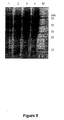
- the genes encoding DBA and DBB were synthesized (Schemes 1 and 2) and cloned into the pET32a vector to generate pET32a-30103s1, and pET32a-30103s2. After transformation into E. coli BL-21(DE3), several colonies from each transformation were selected, and tested for protein expression. The appearance of a band at ⁇ 78 kDa from an induced sample was taken to represent the successful expression of the proteins and highly expressing colonies (colony 5 for pET32a-30103s2, and colony 3 for pET32a-30103s2) were used in successive expression experiments ( Figures 2A, B ).
- the proteins were expressed as inclusion bodies so after large-scale expression (1L), the proteins were purified by metal affinity chromatography under denaturing conditions, and refolded to yield a final protein concentration of 0.1 and 0.9 mg/mL for Trx-DBA and Trx-DBB, respectively.
- the refolded proteins were ⁇ 80% pure as judged by SDS-PAGE ( Figure 2C ).
- the gene DBB was subcloned into the pGS21a vector resulting in the vector pGS21a-30103s2, which encodes His 6 -GST-DDB.
- This vector and the pGEX-30103s1 were transformed into the Origami (Novagen) E. coli strain, and highly expressing transformants were selected as described above for the BL21(DE3) strain. Both Trx-DBA and His 6 -GST-DBB were expressed in the soluble fraction with ⁇ 50% soluble protein ( Figure 2D ).
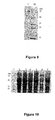
- Fluorescence excitation spectra were recorded for TRX-DBA and TRX-DBB. The emission wavelength was set at or as close to the theoretical maximum for each protein as was practical. TRX-DBA (orange) and TRX-DBB (blue) showed excitation maxima at ⁇ 344 nm and 400 nm as expected for their GFP-variant components, mOrange and T-Sapphire, respectively ( Figure 3 ). Fluorescence emission spectra were recorded for TRX-DBA and TRX-DBB. TRX-DBA (orange) and TRX-DBB (blue) showed emission maxima at ⁇ 510 nm and ⁇ 560 nm as expected for mOrange and T-Sapphire, respectively ( Figure 4 ).
- Size exclusion chromatography in combination with MALLS indicated that the refolded proteins contained a wide range of sized molecules, most likely arising from incorrectly folded protein.
- the fluorophores in GFP only form when the GFPs are correctly folded, and in general, GFPs are only fluorescent when the proteins they are fused to are also folded and soluble. Thus relative fluorescence was used to gauge the approximate concentration of active protein in each sample.
- the fluorescence properties of a 1- ⁇ M sample of a known eGFP-fusion protein were assessed. When an excitation wavelength of 430 nm (close to the fluorescence excitation maximum) was used the sample showed strong fluorescence emission at ⁇ 510 nm ( Figure 5 ).
- Residual ammonium sulfate in these concentrated Demibody solutions was removed by buffer exchange using Centricon centrifugal concentrators. Flow cytometric results obtained with these fractionated and concentrated Demibody solutions showed significant removal of inactive protein in this step.
- Demibodies To label cells with one or both Demibodies, 300 ⁇ L cell suspension (3 X 10 5 cells) was incubated with 30 ⁇ L Demibody (200 ⁇ g/mL, Demibody A (DBA; CD45-mOrange-E3), Demibody B (DBB; CD20-T-Sapphire-K3)) for 60 min in the dark at room temperature. Based upon the known immunophenotypes of these cell lines, the following binding patterns are expected. DBA CD45-mOrange-E3 bind to Raji, MEC-2, CCRF-CEM DBB CD20-T-Sapphire-K3 bind to Raji, MEC-2
- scFv fragments are derived from whole antibodies. They are constructed by linking the antigen-binding V H and V L domains of an antibody with a flexible polypeptide linker.
- the linker may comprise a combination of glycine and serine residues to provide flexibility and to enhance hydrophilicity of the peptide backbone to allow hydrogen bonding with solvent molecules as well as being resistant to protease digestion.
- the construction of scFv fragments is described in Kort et al. (2001) supra and in publications described therein.
- a schematic representation showing polypeptide segments in the demibody is shown in Figure 13 .
- a diagram of the structural components of the human IgGI Fc fragment is shown in Figure 14 .
- the Fc domain is preferably divided into the ⁇ 2a and ⁇ 2b chains. This is useful in generating the Fc portion of the demibody.
- Figure 15 shows heterodimerization of the leucine zipper.
- Antibodies specific for target antigens are used to develop scFv fragments.
- Antigens e.g. CD antigens
- target cells in isolated form or in recombinant or synthetic form, or whole cells expressing surface antigens, are used to develop monoclonal or polyclonal antibodies.
- the antibodies are isolated and scFv fragments generated as described in Examples 1 and 2.
- Bispecific antibodies are used for diagnostics as reagents which bind to a specific marker and provide a binding site for an agent which would enable detection of the antibody.
- the bispecific antibody provides a binding site for a radio-label.
- Demibodies enable enhanced diagnostic applications such as fluorescence-activated cell sorting (FACS), fluorescence microscopy and immunoassays.
- the unifying principle is that a signal is only obtained after zipping of the two scFv-fusion proteins, either by indirect labeling of the newly formed Fc domain with a fluorescence or enzyme-labeled anti-Fc-antibody, or by so called "protein fragment complementation" (PCA), where each scFv-fusion protein would contain the half of an enzyme or fluorescent protein and only after zipping such an enzyme or fluorescent protein becomes active.
- PCA protein fragment complementation
- demibodies enable staining of combinations of antigens/epitopes with only one color and thus "free" other colors for additional antigens (e.g. CD4+ helper T cell: CD3 and CD4 - actual red and green, with demibody only red or green).
- Demibodies forming different Fc-domains e.g. IgG isotypes allow differential labeling with secondary IgG subtype-specific antibodies.
- each scFv-fusion-protein could carry a part of a fluorescent protein, which after zipping would complement each other to a fluorescent protein (in case of FRET a complete fluorescence protein which in combination would allow FRET-transfer). See Figures 16A and B .
- demibodies enable detection of multimeric modified proteins and allow their differentiation, e.g. phosphorylated proteins (1 scFv against phosphotyrosine, 1 scFv against the protein, capture antibody against second subunit), or glycosylated isoforms (1 scFv against sialic acid, 1 scFv against the protein, capture antibody against second subunit) [ Figure 17 ].
- phosphorylated proteins (1 scFv against phosphotyrosine, 1 scFv against the protein, capture antibody against second subunit
- glycosylated isoforms (1 scFv against sialic acid, 1 scFv against the protein, capture antibody against second subunit
- Demibody reagents useful for diagnostics also could be used in basic research as tools for e.g. advanced cell labeling and sorting, or novel antibody-based analyses.
- Demibodies can be used for the purification of complexes, such a multimeric proteins, or modified proteins by affinity purification.
- An example would be the one-step purification of a phosphorylated protein (scFv anti-protein, scFv anti-phospho-tyrosine) [ Figure 18 ].
- Figure 19 is a diagrammatic representation showing the use of two demibodies specific for CD5 or CD19.
- the demibodies comprise an scFv portion capable of binding to CD5 or CD19, an incomplete, non-functional Fc portion and half of a leucine zipper.
- the amino acid sequence of portions of the leucine zipper are shown in Table 3 (see SEQ ID NOs:1-5). Appropriate pairs include SEQ ID NOs:1 and 2, SEQ ID NOs:3 and 4 and SEQ ID NOs:3 and 5.
- the two demibodies bind to a target cell expressing both CD5 and CD19.
- the leucine zipper portions interact and the two incomplete Fc portions form a functional Fc domain (see Figure 19 ).
- a cytotoxic cell e.g. neutrophil, macrophage, natural killer cell
- FcR Fc receptor
Landscapes
- Health & Medical Sciences (AREA)
- Chemical & Material Sciences (AREA)
- Organic Chemistry (AREA)
- Life Sciences & Earth Sciences (AREA)
- Medicinal Chemistry (AREA)
- General Health & Medical Sciences (AREA)
- Immunology (AREA)
- Biochemistry (AREA)
- Proteomics, Peptides & Aminoacids (AREA)
- Veterinary Medicine (AREA)
- Genetics & Genomics (AREA)
- Biophysics (AREA)
- Molecular Biology (AREA)
- Pharmacology & Pharmacy (AREA)
- Chemical Kinetics & Catalysis (AREA)
- General Chemical & Material Sciences (AREA)
- Nuclear Medicine, Radiotherapy & Molecular Imaging (AREA)
- Animal Behavior & Ethology (AREA)
- Public Health (AREA)
- Tropical Medicine & Parasitology (AREA)
- Oncology (AREA)
- Communicable Diseases (AREA)
- Toxicology (AREA)
- Gastroenterology & Hepatology (AREA)
- Zoology (AREA)
- Virology (AREA)
- Hematology (AREA)
- Micro-Organisms Or Cultivation Processes Thereof (AREA)
- Peptides Or Proteins (AREA)
- Medicines Containing Antibodies Or Antigens For Use As Internal Diagnostic Agents (AREA)
- Medicines That Contain Protein Lipid Enzymes And Other Medicines (AREA)
Description
- The present invention relates generally to a set of modified immunoglobulins comprising two complementary immunoglobulins, which are referred to herein as "demibodies" which are useful in targeting particular cells in a subject. More particularly, the present invention provides a set of demibodies wherein at least two molecules from within the set, each specific for a different antigen on a target cell, are required to interact together at the cell surface in order to form an active complex which directs demibody-mediated cellular or complement mediated cytotoxicity and/or reporter function and/or therapeutic activity. The demibodies of the present invention are useful in the targeting of particular cells such as cancer cells including leukemic cells, pathogens including malarial, bacterial and viral agents, and stem cells including embryonic and adult stem cells and pathogen cells. The present invention provides, therefore, methods of treatment, diagnosis and undertaking research and compositions comprising demibodies useful for same.
- Bibliographic details of the publications referred to by author in this specification are collected at the end of the description.
- Reference to any prior art in this specification is not, and should not be taken as, an acknowledgment or any form of suggestion that this prior art forms part of the common general knowledge in any country.
- A key feature in the search for and development of therapeutic agents is target discrimination or selective toxicity. In particular, the ability to distinguish target cells such as cancer cells or cells infected with pathogen cells amongst a population of normal cells in a subject is of paramount importance. This is particularly the case in cancer therapies where the target cancer cells have many physiological, anatomical and biochemical properties in common with surrounding normal cells. Whilst some anti-cancer drugs do cause collateral damage to normal cells, their use may be indicated or at least justified for particularly aggressive, fast growing cancers.
- Therapeutic antibodies are the most rapidly growing area of pharmaceuticals, with more than 30 antibodies in late-phase clinical trials (Hudson and Souriau, Nat Med 9:129-134, 2003). There are many variations of engineered antibodies (e.g. mouse monoclonal, chimeric, humanized, human monoclonal, single chain variable antibody fragments (scFv), minibodies, aptamers). Diabodies developed using recombinant DNA technology, contain two or more single chain variable antibody fragments (scFv) with different binding specificities and appropriate spacing between these domains to enable both scFv to bind antigens concurrently (Hudson and Souriau, supra 2003). Bivalent and bispecific scFv antibodies have been formed using leucine zipper-based dimerization cassettes attached to different scFv (de Kruif and Logtenberg, J Biol Chem 271:7630-7634, 1996). Bi-specific antibodies with different scFv domains connected by a polypeptide chain have been designed to cross-link T-cells with tumours. The two or more interactions that such chimeric antibodies have with different surface antigens on a cell would greatly increase the strength of binding since the dissociation constants for the individual interactions are multiplicative.
- Whilst therapeutic antibodies are important, multi-specific antibodies have not been as successful. There is a need, therefore, to develop antibody-based drugs and other therapeutic agents which are more highly selective for target cells.
- Whole antibodies have been proposed as highly specific targeting agents (Carter, Nature Reviews 1:118-128, 2001). In one proposal, cytotoxic agents are linked to an antibody specific for an antigen on a target cell. However, although antibodies have a high degree of target specificity, they have not achieved wide pathological therapeutic use and are primarily used in clinical imaging applications. This may be due to their relatively long circulating half-lives and their associated effector functions.
- Modified antibodies, however, have achieved some level of acceptance in immunotherapeutic applications (Carter (2001) supra; de Haard et al, Adv. Drug Delivery Rev. 31:5-31, 1998; Chames and Baty, FEMS Microbiol. Lett. 189:1-8, 2000; Funaro et al, Biotechnol. Adv. 18:385-401, 2000; Hudson, Exp. Opin. Invest Drugs 9:1231-1242, 2000).
- Examples involving therapeutic antibodies are reviewed in Table 1 of Carter (2001) supra.
- These antibodies all contain the Fc domain which is required for complement-dependent cytotoxicity (CDC) and antibody-dependent cellular cytotoxicity (ADCC).
- Ohiro et al. (Anal. Chem. 74:5786-5792, 2002) describe a homogeneous and noncompetitive immunoassay based on the enhanced fluorescence resonance energy transfer by leucine zipper interaction.
- Another useful development in the use of antibody fragments is their fusion to active agents such as radioactive isotypes (Wu et al, Immunotechnology 2:21-36, 1996; Wu et al, Proc. Natl. Acad. Sci. USA 97:8495-8500, 2000; Adams et al, Nuc. Med. Biol. 27:330-346, 2000), enzymes for producing therapy (Bagshawe and Begent, Adv. Drug Delivery Rev. 22:365-367, 1996) and toxins for targeted cell killing (Reiter and Pastan, Tibtech 16:51-520, 1998; Kreitman, Curr. Opin. Immunol. 11:570-578, 1999).
- Modified antibodies of particular interest are single chain variable fragments (scFv) carrying the variable region sequences of the light and heavy chains linked together. scFv antibody fragments are derived from Fragment antigen binding (Fab) portions of an antibody comprising the V region of a heavy chain linked by a stretch of synthetic peptide to a V region of a light chain.
- Whilst scFv antibody fragments have achieved a reasonable level of utility as targeting molecules, they lack the Fc domain and are unable to induce ADCC or CDC.
- The present invention enables modified forms of scFv antibody fragments to be used in targeted cell therapy and/or diagnosis and/or research.
- Throughout this specification, unless the context requires otherwise, the word "comprise", or variations such as "comprises" or "comprising", will be understood to imply the inclusion of a stated element or integer or group of elements or integers but not the exclusion of any other element or integer or group of elements or integers.
- The present invention provides a set of modified immunoglobulins comprising two complementary immunoglobulins capable of forming a functional agent or combined agents with specific properties upon binding of the first and second complementary immunoglobulins to a target cell,
wherein each immunoglobulin comprises a first, second and third portion wherein: - (a) each first portion is an scFv or other antigen binding fragment of an immunoglobulin and the first portion of the first complementary immunoglobulin is capable of interacting with a first antigen on the target cell and the first portion of the second complementary immunoglobulin is capable of interacting with a different antigen on said target cell;
- (b) each second portion is an agent or a non-functional, incomplete portion of an agent whereby the two second portions are
- (i) distinct reporter molecules which in combination provide a combined signal,
- (ii) complementary non-functional portions of a single reporter molecule, or
- (iii) incomplete, non-functional Fc domains capable of forming a functional Fc domain; and
- (c) each third portion is a different member of a complementary binding pair comprising a first and a second member wherein the binding pair comprises complementary portions of a leucine zipper, a receptor-ligand pair, actin and an actin-binding protein or DNA aptamers;
wherein upon binding of the complementary immunoglobulins to the two antigens the binding pairs combine- (i) enabling the reporter molecules to provide a combined signal,
- (ii) enabling the complementary non- functional portions of a single reporter molecule to reconstitute a single reporter molecule, or
- (iii) enabling the incomplete, non-functional Fc domains to form a functional Fc domain.
- The present invention combines the specificity of antibody-antigen interactions to generate immunointeractive molecules which are capable of targeting particular cells and inducing cytotoxicity and/or reporter function and/or facilitating cell therapy. Disclosed herein are sets of at least two demibodies wherein each demibody comprises an antigen-binding portion, an agent or portion thereof and one or other member of a binding pair. Two demibodies are designed such that when in close proximity, each member constituting one of a complementary binding pair, interacts forming a binding pair. This in turn permits interaction of portions of the agent to generate a functional agent or interaction of two agents which agent or agents have properties resulting in for example, cell death, cell therapy or providing a reporter signal. Hence, the agent on the demibodies can act as or form a reporter molecule, therapeutic molecule or cytotoxic molecule. Preferably, the demibodies comprise non-functional portions of the agent. When in close proximity, the reporter or therapeutic or cytotoxic molecule portions come together and a functional molecule capable of providing a signal or inducing cytotherapy or cytotoxicity is reconstituted. This increases the specificity for imaging, diagnostic and therapeutic purposes. The demibodies are also useful research tools such as for FACS, Flow cytometry and affinity chromatography. The demibodies may also be used to detect products of cells such as proteins or different phosphorylated or glycosylated or other post-translational modifications thereof. In essence, the demibodies of the present invention enable enhanced immunophenotypic selection of cells, viruses or products thereof such as proteins.
- The antigen binding portion may be derived from an immunoglobulin such as a scFv (or F'ab fragment) or any affinity scaffold such as a microaffinity scaffold. Examples include dAbs, nanobodies, microproteins, fibronectins, microbodies, anticalins, aptamers, darpins, avimers, afflins, and Kunitz domains.
- The demibodies have enhanced target specificity since each demibody in the set is specific for a particular antigen. Hence, by selecting cells or virus having an unusual pair of antigens reduces the risk of non-specific binding.
- In one embodiment, each demibody carries an incomplete Fc domain but upon binding of the pair of demibodies, the two incomplete Fc domains now form a functional Fc domain or functional portion thereof. The bound demibodies together have a biologically functional Fc domain and can initiate associated activities such as antibody-dependent cellular cytotoxicity (ADCC) and/or complement-dependent cytotoxicity (CDC). In another embodiment, the two incomplete domains form, when reconstituted, an agent such as a reporter molecule, therapeutic agent or cytotoxic agent. In a further embodiment, the agents are dyes such as fluorochromes which when together provide a particular signal.
- Hence, the present invention encompasses a composition comprising a set of demibodies according to the invention which, together, are able to induce highly selective cytotoxicity. The selective toxicity follows the demibodies interacting with at least two different antigens on the surface of a target cell. The two different antigens being predominantly only expressed simultaneously on a target cell population. Even if one of these antigens (or analogous antigens) is represented on normal cells, unless both antigens are present on the one cell, the pair of demibodies will not come together on the surface of the cell via the complementary binding pairs to form a functional cytotoxic domain such as a functional Fc domain, therapeutic molecule, cytotoxic molecule or reporter molecule.
- In a further embodiment, the reconstitution of the demibody pairs results in a functional cytotoxic or therapeutic molecule. Examples of such reconstituted functional molecules are apoptotic, cell cycle static, lytic or cytotoxic molecules, antibiotics, peptides and cytokines.
- In an alternative embodiment, each demibody carries a functional reporter molecule such as a dye or fluorescent marker and when the demibodies reconstitute together, the combined signal (or combined dye) or combined fluorescent signal is different from the individual signals (or dyes). Examples of fluorescent markers include hydroxycoumarin, aminocoumarin, methoxyciumarin, cascade blue, Lucifer yellow, NBD, Phycoerythrin (PE), PerCP, allophycocyanin, hoechst 33342, DAP1, SYTOX Blue, hoechst 33258, chromomycin A3, mithramycin, YOYO-1, SYTOX green, SYTOX orange, 7-AAD, acridine orange, TOTO-1, To-PRO-1, thiazole orange, TOTO-3, TO-PRO-3, LDS 751, Alexa Fluor dyes including Alexa Fluro-350, -430, -488, -532, -546, -555, -556, -594, - 633, -647, -660, -680, -700 and -750; BoDipy dyes, including BoDipy 630/650 and BoDipy 650/665; CY dyes, particularly Cy2, Cy3, Cy3.5, Cy5, Cy 5.5 and Cy7; 6-FAM (Fluorescein); PE-Cy5, PE-Cy7, Fluorescein dT; Hexachlorofluorescein (Hex); 6-carboxy-4', 5'-dichloro-2', 7'- dimethoxyfluorescein (JOE); Oregon green dyes, including 488-X and 514; Rhodamine dyes, including X-Rhodamine, Lissamine Rhodamine B, Rhodamine Green, Rhodamine Red and ROX; TRITC, Tetramethylrhodamine (TMR); Carboxytetramethylrhodamine (TAMRA); Tetrachlorofluorescein (TET); Red 6B, FluorX, BODIPY-FL and Texas Red.
- The demibodies of the present invention are useful in the treatment of a range of conditions including cancer, infection by pathogenic agents or the selective targeting of any cell type in a subject. They are also useful in targeting cells such as stem cells.
- Reference to a subject includes a human or other primate, livestock animal, laboratory test animal, companion animal or avian species. A subject may also be regarded as a patient.
- The present invention contemplates, therefore, a method of treating in a subject, such as a patient, comprising administering to the subject a set of demibodies according to the invention which, when bound together via binding pairs, forms a functional cytotoxic domain such as an Fc domain or functional portion thereof capable of inducing cell cytotoxicity such as ADCC or CDC. Alternatively, the demibodies reconstitute a cytotoxic agent or therapeutic molecule.
- Diagnostic compositions and methods for diagnosing and/or imaging and/or therapy also form part of the present invention.
- The pair of immunointeractive molecules may be simultaneously or sequentially administered.
- The present invention further provides an in vitro method for selectively identifying a cell, said method comprising contacting said cell with a set of immunoglobulins as defined herein wherein said first portions are capable of interacting with different antigens on the cell, said second portions comprise distinct reporter molecules or complementary non-functional portions of a single reporter molecule and said third portions are complementary binding pairs wherein upon binding of the individual immunoglobulins to the different antigens, the binding pairs combine at the cell surface enabling the reporter molecules to provide a combined signal or to reconstitute a single reporter molecule.
- The invention also provides a method for detecting a target cell said method comprising contacting a sample putatively comprising said target cell with a set of immunglobulins as defined herein wherein said first portions are capable of interacting with different target antigens on said cell, said second portions comprise distinct reporter molecules or complementary non-functional portions of a single reporter molecule wherein upon binding of the individual immunoglobulins to the different antigens, the binding pairs combine enabling the reporter molecules to provide a combined signal or to reconstitute a single reporter molecule.
- A list of abbreviations used herein is provided in Table 1.
TABLE 1 Abbreviations ABBREVIATION DESCRIPTION Ab antibody ADCC antibody-dependent cellular cytotoxicity Ag antigen C region constant region CD antigen cluster of differentiation antigen CDC complement-dependent cytotoxicity Fab fragment fragment antigen binding Fc fragment crystalline FcR Fc receptor Fv fragment variable H chain heavy chain Ig immunoglobulin L chain light chain sc single chain scFv single chain variable fragment, recombinant Fab comprising V region of heavy and light chains V region variable region -
-
Figures 1A and B are a representations of the amino acid sequences for the thioredoxin-Demibody conjugates. Trx-CD45-mOrange-E3 (Demibody A, DBA, A) and Trx-CD20-T-Sapphire-K3 (Demibody B, DBB, B). -
Figures 2A through 2D are photographic representations of protein gels. -
Figure 3 is a graphical representation showing fluorescence excitation spectra of the refolded TRX Demibodies. The emission wavelength was set at 580 nm for TRX-DBA and 510 nm for TRX-DBB. The slit widths were both 10 nm and concentrations of the proteins were 0.1 mg/mL and -0.9 mg/mL, respectively. Fluorescence excitation spectra were recorded from 400-560 nm for TRX-DBA (orange) and 350-460 nm for TRX-DBB (blue). -
Figure 4 is a graphical representation showing fluorescence emission spectra of the refolded TRX Demibodies. The excitation wavelength was set at 510 nm for TRX-DBA and 400 nm for TRX-DBB. The slit widths were both 10 nm and concentrations of the proteins were 0.1 mg/mL and ~0.9 mg/mL, respectively. Fluorescence emission spectra were recorded from 550-650 nm for TRX-DBA (orange) and 450-600 nm for TRX-DBB (blue). -
Figure 5 is a graphical representation showing relative fluorescence properties of Demibodies. An eGFP fusion protein (1 µM) was excited at 430 nM (dark cyan); Refolded Trx-DBA (~1.5 µM) was excited at 510 nm (orange); refolded Trx-DBB(~15 µM) was excited at 400 nm (blue). Excitation and emission slit widths were both 5 nm in all cases. -
Figure 6 is a graphical representation showing fluorescence properties of a mixture of TRX-DBA and TRX-DBB. The excitation wavelength was set at 400 nm for TRX-DBB; the slit widths were both 10 nm and concentrations of the proteins were 0.05 mg/mL and ~0.45 mg/mL, respectively for TRX-DBA and TRX-DBB. This corresponds to estimated active concentrations of ≤~ 130 nM and ≤~75 nM. A fluorescence emission spectrum was recorded from 450-600 nm for a mixture of TRX-DBA and TRX-DBB (ABMIX; Green) and is compared with that of TRX-DBB alone (blue). -
Figure 7 is a photographic representation of the identification of pET32a-30103 S1 in E. coli BL21. M = Marker; 1 = Uninduced colony; 2 =Colony 1; 3 =Colony 2; 4 =Colony 3. -
Figure 8 is a photographic representation showing the solubility detection of 30103 S1. = supernatant of 30103 S1 induced at 15°C overnight; 2 = supernatant of 30103 S2 induced at 25°C for 6 hours; 3 = precipitation of 30103 S1 induced at 15°C for overnight; 4 = precipitation of 30103 S1 induced at 25°C for 6 hours; M = low weight marker. -
Figure 9 is a diagrammatic representation showing SDS-PAGE of Refolded 30103 S1. M = marker; 1 = refolded 30103 S1. -
Figure 10 is a photographic representation of the identification of the identification of pET32a-30103s2 in E. coli BL21. 1 = marker; 2 =colony # 1 uninduced; 3-7 = colony #2-#6 induced. -
Figure 11 is a photographic representation showing the solubility detection of 30103s2. 1 = precipitate of 30103s2 induced at 15°C overnight; 2 = supernatant of 30103s2 induced at 15°C overnight; 3 = precipitate of 30103s2 induced at 25°C for 6 hours; 4 = supernatant of 30103s2 induced at 25°C for 6 hours. -
Figure 12 is a photographic representation of the SDS-PAGE of Refolded 30103S2. 1 = marker; 2 = refolded 30103 S2. -
Figure 13 is a schematic representation showing the polypeptide domains of proposed chimeric antibody (demibody). -
Figure 14 is a diagrammatic representation showing the structure of protein components of the h1gG1 Fc fragment-Fc RIII complex. h1gG1 Fc fragment is shown as ribbon diagram with chain A in light color and chain B in dark color. The N and C termini are indicated. sFc III is shown as a wire representation (coordinates taken from RCSB Protein Databank Accession code:1e4k; Sondermann et al, Nature 406:267-273, 2000). -
Figure 15 is a diagrammatic representation showing leucine zipper heterodimerization. Top:helical wheel representation (looking down long axis of helices) of heterodimerization. Bottom:schematic showing that complementary electrostatic charges mediate heterodimerization not homodimerization. A = CD19, B = α-CD19-zip-N'-GFP, C = green, D = CD5, E = α-CD5-zip-C'-GFP. -
Figure 16 is a diagrammatic representation of enhanced FACS using demibodies. -
Cell Type Demibody Specificity Reconstituted Fc-Domain Detection by CD4+ TC CD4 Ig1 anti-Ig1-green CD3 CD8+ TC CD8 Ig2a anti-Ig2a-red CD3 Monocyte CD14 IgM anti-IgM-yellow Class II Dendritic Cell CD83 IgD anti-IgD-blue ClassII -
Figure 17 is a diagrammatic representation showing enhanced immunoassays using demibodies where A = α-phospho-tyrosine, B = α-protein-epitope, and C = e.g. detection of multimeric modified proteins in one step. -
Figure 18 is a diagrammatic representation showing enhanced immunoaffinity chromatography using demibodies where A = α-phospho-tyrosine, B = α-Fc, e.g. protein A, and C = α-protein-epitope. -
Figure 19 is a diagrammatic representation showing antibody-dependent cellular cytotoxicity (ADCC) mediated against CD5+ CD19+ chronic lymphocytic leukemia (CLL) cells by a pair of demibodies. - The present invention relates to a set of modified immunoglobulin molecules which form an antibody-like immunointeractive molecule. The term "immunointeractive" in this context is used to highlight one of the principal features of an antibody, i.e. the ability for an antibody to interact specifically with an antigen. As each molecule is effectively half of an antibody-like molecule, it is referred to herein as a "demibody". The present invention encompasses, therefore, antibodies modified recombinantly and/or chemically to form a single chain variable fragment (e.g. scFv) having specificity to one antigen. Such demibodies may also be referred to herein as immunointeractive molecules or modified or chimeric immunoglobulins or modified or chimeric antibodies.
- The demibodies described herein comprises:
- (i) an scFv antigen binding portion;
- (ii) an agent or non-function portion thereof; and
- (iii) a member of a binding pair capable of interacting with a member on a second demibody, thereby forming a binding pair of heterodimers but not homodimers.
- In use, at least two demibodies are selected having:
- (i) scFv antigen binding portions directed to different antigens on a target cell;
- (ii) an agent or non-functional portion thereof such that if both demibodies were bound together, a functional agent is formed; and
- (iii) two binding members so that, in close proximity, both members can interact to form a binding pair between complementary partners.
- In one embodiment of the invention, the incomplete non-functional portion of an agent is an Fc domain or a functional portion thereof In this case, complementary, incomplete and non-functional Fc domains are used on each demibody. In a further embodiment the incomplete non-functional portions are distinct reporter molecules or complementary non-functional portions of a single reporter molecule. The non-functional portions could be a cytotoxic molecule or a reporter molecule or a therapeutic molecule. In still another embodiment, the agent is functional but when both agents are together, a particular result occurs (such as a signal) absent from one agent above.
- Hence, the agent, when reconstituted, may have cytotoxic, medicament, therapeutic or reporter signal properties.
- The demibodies have a range of applications including cytotoxic targeting of cancer cells or pathogens, selective detection of cells such as cancer cells, stem cells (embryonic or adult) or cells infected with pathogens. Cell detection may be facilitated with FACS, Flow cytometry and fluorescent microscopy. The demibodies also have research applications such as affinity chromatography. The demibodies may also be used to detect products of cells such as proteins or different phosphorylated or glycosylated or other post-translational modifications thereof The detection of different forms of proteins is particularly useful for diagnosing or prognosing disease conditions or levels of health.
- In relation to one embodiment, the formation of the binding pair results in the formation of a functional Fc domain or portion thereof. The Fc domain generally corresponds to paired
C H2 andC H3 domains and is the part of an antibody which interacts with effector molecules and cells carrying Fc receptors. The coming together of the incomplete Fc domains to form a functional Fc domain means that a sufficient amount of Fc required to interact with effector molecules or Fc receptors on cells is formed. It may still represent an incomplete Fc domain. The present invention encompasses, therefore, the use of complete or incomplete Fc domains provided that the Fc domain is functional with respect to interacting with effector molecules or cells when two demibodies are bound together. Individual immunointeractive molecules have incomplete, non-functional Fc domains. - In a preferred embodiment, the incomplete, non-functional Fc domains correspond to the γ2a and γ2b chains. Each demibody, in a preferred embodiment, carries either a γ2a chain or a γ2b chain of the Fc domain.
- It will be appreciated, therefore, that in operation, a set of at least two demibodies is required which are complementary with respect to Fc domains and binding members and which have antigen binding portions directed to different antigens, which antigens are co-expressed on a target cell.
- Accordingly, a demibody may comprise first, second and third portions wherein:
- said first portion is capable of interacting with a first antigen on a target cell;
- said second portion is a non-functional, incomplete Fc domain; and
- said third portion is one member of a complementary binding pair;
- wherein said demibody is capable of forming a functional Fc domain when the demibody is bound to a second complementary demibody which comprises a portion which interacts with another antigen on said target cell.
- The functional Fc domain is useful for mediating ADCC of target cells.
- A set of demibodies may comprise a first demibody with first, second and third portions wherein:
- said first portion is capable of interacting with a first antigen on a target cell;
- said second portion is a non-functional, incomplete a Fc domain; and
- said third portion is one member of a complementary binding pair; and
- a second demibody comprising a first portion which is capable of interacting with a different antigen on said target cell;
- a second portion which, in combination with the second portion of the first mentioned demibody forms a functional Fc domain; and
- a third portion which is the complementary binding member of said binding pair.
- A method for selectively inducing cytotoxicity of a cell may comprise contacting cells with first and second demibodies wherein said first demibody comprises first, second and third portions wherein:
- said first portion is capable of interacting with a first antigen on a target cell;
- said second portion is a non-functional, incomplete Fc domain; and
- said third portion is one member of a complementary binding pair; and
- said second demibody comprises a first portion which is capable of interacting with a different antigen on said target cell;
- a second portion which, in combination with the second portion of the first mentioned demibody forms a functional Fc domain; and
- a third portion which is the complementary binding member of said binding pair;
- Although these aspects relate to the Fc domain as the functional portion (i.e the agent), other cytotoxic domains may be employed such as apoptotic domains or lysing domains from agents inducing cell cycle arrest, and domains of therapeutic molecules.
- As an alternative to the incomplete Fc domains, therefore, incomplete domains for cytotoxic molecules, therapeutic molecules and reporter molecules may be employed.
- Accordingly, a demibody may comprise first, second and third portions wherein:
- said first portion is capable of interacting with a first antigen on a target cell;
- said second portion is a non-functional, incomplete domain of cytotoxic or therapeutic or reporter molecule; and
- said third portion is one member of a complementary binding pair;
- wherein said demibody is capable of forming a functional cytotoxic or therapeutic or reporter molecule when the demibody is bound to a second complementary demibody which comprises a first portion capable of interacting with another antigen on said target cell;
- a second portion which, in combination with the second portion of the first mentioned demibody forms a functional Fc domain; and
- a third portion which is the complementary binding member of said binding pair.
- The demibody is, in effect, a modular or chimeric molecule having first, second and third modules equivalent to the first, second and third portions defined above.
- A set of demibodies may also comprise a first demibody with first, second and third portions wherein:
- said first portion is capable of interacting with a first antigen on a target cell;
- said second portion is a non-functional, incomplete cytotoxic or therapeutic or reporter molecule; and
- said third portion is one member of a complementary binding pair; and
- a second demibody comprising a first portion which is capable of interacting with a different antigen on said target cell;
- a second portion which, in combination with the second portion of the first mentioned demibody forms a functional cytotoxic or therapeutic or reporter molecule; and
- a third portion which is the complementary binding member of said binding pair.
- A method for selectively detecting or targeting a cell may comprise contacting cells with first and second demibodies wherein said first demibody comprises first, second and third portions wherein:
- said first portion is capable of interacting with a first antigen on a target cell;
- said second portion is a non-functional, incomplete cytotoxic or therapeutic or reporter molecule; and
- said third portion is one member of a complementary binding pair; and
- said second demibody comprises a first portion which is capable of interacting with a different antigen on said target cell;
- a second portion which, in combination with the second portion of the first mentioned demibody forms a functional cytotoxic or therapeutic or reporter molecule; and
- a third portion which is the complementary binding member of said binding pair.
- In an embodiment of the invention, each demibody carries a reporter molecule such as a dye which when the two demibodies reconstitute with each other, the combined reporter molecules (e.g. combined dyes) provide a distinctive signal. Examples of dyes include fluorescent dyes such as hydroxycoumarin, aminocoumarin, methoxyciumarin, cascade blue, Lucifer yellow, NBD, Phyccerythrin (PE), PerCP, allophycocyanin, hoechst 33342, DAP1, SYTOX Blue, hoechst 33258, chromomycin A3, mithramycin, YOYO-1, SYTOX green, SYTOX orange, 7-AAD, acridine orange, TOTO-1, To-PRO-1, thiazole orange, TOTO-3, TO-PRO-3, LDS 751, Alexa Fluor dyes including Alexa Fluro-350, -430, -488, - 532, -546, -555, -556, -594, -633, -647, -660, -680, -700 and -750; BoDipy dyes, including BoDipy 630/650 and BoDipy 650/665; CY dyes, particularly Cy2, Cy3, Cy3.5, Cy5, Cy 5.5 and Cy7; 6-FAM (Fluorescein); PE-Cy5, PE-Cy7, Fluorescein dT; Hexachlorofluorescein (Hex); 6-carboxy-4', 5'-dichloro-2', 7'- dimethoxyfluorescein (JOE); Oregon green dyes, including 488-X and 514; Rhodamine dyes, including X-Rhodamine, Lissamine Rhodamine B, Rhodamine Green, Rhodamine Red and ROX; TRITC, Tetramethylrhodamine (TMR); Carboxytetramethylrhodamine (TAMRA); Tetrachlorofluorescein (TET); Red 6B, FluorX, BODIPY-FL and Texas Red.
- A method for selectively identifying a cell may comprise contacting said cell with a pair of demibodies wherein each demibody comprises first, second and third portions wherein said first portions are capable of interacting with one or two antigens on the cell, said second portions comprise distinct reporter molecules or complementary non-functional portions of a single reporter molecule and said third portions are complementary binding pairs wherein upon binding of the individual demibodies to the two antigens, the binding pairs combine enabling the reporter molecules to provide a combined signal or to reconstitute a single reporter molecule.
- In the above aspects, the antigen binding portion may be derived from an immunoglobulin or may be any affinity scaffold such as but not limited to dAbs, nanobodies, microproteins, fibronectins, microbodies, anticalins, aptamers, darpins, avimers, afflins, and Kunitz domains.
- Administration may be sequential or simultaneous. Sequential administration includes separate administration of both demibodies within nanoseconds, seconds, minutes, hours or days. Simultaneous administration includes co-administration in a single composition or in two separate compositions.
- The subject may be a human or other primate, a livestock animal (e.g. sheep, cow, pig, horse, donkey, goat), laboratory test animal (e.g. mouse, rat, rabbit, guinea pig), captive wild animal or avian species (e.g. poultry birds, caged birds, aviary birds, game birds, captive wild birds). A subject may also be regarded as a patient.
- The antigen-binding portion is generally a recombinant Fab fragment. This fragment corresponds to the arms of an antibody molecule which contains the complete light chains paired with the VH and CH domains of the heavy chains.
- Generally, the antigen binding portion is an scFv portion of an antibody. scFv are generally monomeric although the extent of monomerism compared to dimerism or multivalentism may depend on the size of the linker between the VH and VL domains. The construction of scFv molecules is described in Hudson (1999) supra and Kortt et al, Biomolecular Engineering 18:95-108, 2001.
- The present invention extends to other antigen binding portions of immunoglobulins such as but not limited to dAbs, nanobodies, microproteins, fibronectins, microbodies, anticalins, aptamers, darpins, avimers, afflins, and Kunitz domains or other affinity scaffolds such as but not limited to microscaffolds.
- The antigen binding portion is specific for an antigen on the surface or a sub-surface co-continuous with the external environment on a target cell. The target cell may be a cancer cell, a cell infected by a pathogen or parasite or other unwanted cell. A pathogen includes a eukaryotic or prokaryotic microorganism including a malaria parasite. It also includes a virus. Furthermore, a virally infected cell may express virally-derived receptors on its surface. Specific targeting of such a cell may result in loss of virus-producing cells. This would be an example of an unwanted cell.
- The present invention is particularly useful in the generation of therapeutic agents to target cancer cells including leukocyte subsets. Cancer cells are generally defined by the expression of particular CD antigens. However, targeting a single CD antigen may cause collateral damage to normal cells which carry the same CD antigen. This problem is proposed to be solved by the present invention in that a single type of demibody alone would be incapable of inducing ADCC or CDC due to lack of a functional Fc domain and an inability to cross link. However, when two demibodies having complementary binding members are used which are directed to two different CD antigens or other antigens present on a cancer cell, then only cells with this unusual pair of surface molecules are cytotoxically targeted after both demibodies have bound. Once bound, the CD antigen will move freely in the two dimensions of the lipid bilayer and the demibodies will eventually be in close proximity to each other. At that point, the members of the binding pair interact resulting in a binding pair and the two incomplete Fc domains form a functional Fc domain. Selective cytotoxicity is now induced for that cell. The essence of this aspect of the present invention is the unusual combination of surface molecules which correspond to particular cancer or unwanted cells or other target cells.
- With respect to cancer treatment, CD antigens are particularly preferred such as two or more CD antigens selected from those listed in Table 2.
TABLE 2 Summary of CD antigens Cd designation Blood-related cancer CDS + CD19/CD20 chronic lymphocytic leukemia (CLL) CD19/CD20 + κ (or λ) would target most clonal B-cell populations CD19/CD20 + CD 10 follicular non-Hodgkins lymphoma (NHL) CD19/CD20 + bcl-2 follicular lymphoma (FL) CLL CD103 + CD22 (or CD25) (or CD19) hairy cell leukemia (HCL) CD4 + CD8 T-cell acute lymphocytic leukemia (T-ALL) ("cortical thymocyte") CD8 + CD57 NK cell/large granular lymphocyte leukemia CD10 + CD34 pre-B ALL CD34 + myeloid marker (i.e. CD13 or CD33) acute myeloid leukemia (AML) CD117 (c-kit) + myeloid marker AML CD13 or CD33 (My) + CD14 (Mo) leukemias CD41 or CD61 + CD33 megakaryocytic leukemias - The binding partners may constitute any of a number of entities which are capable of interacting with each other to form an association or bond. In the present invention the binding partners can be complementary portions of a leucine zipper, a receptor-ligand (e.g. cytokine and cytokine receptor), actin and an actin-binding protein or DNA aptamers. The actual nature of the binding pairs is not critical to the present invention provided that upon coming together in two dimensions in close proximity, both members of the binding pair form an association or bond.
- In a preferred embodiment, the binding pairs comprise complementary portions of a leucine zipper. Leucine zipper amino acid sequences are shown in Table 3. Heterodimerization occurs between for example SEQ ID NOs:1 and 2, SEQ ID NOs:3 and 4 and SEQ ID NOs:3 and 5. The length of the leucine zipper or other complementary binding portions include but is not limited to from 10 amino acids to 100 amino acid such as 10, 11, 12, 13, 14, 15, 16, 17, 18, 19, 20, 21, 22, 23, 24, 25, 26, 27, 28, 29, 30, 31, 32, 33, 34, 35, 36, 37, 38, 39, 40, 41, 42, 43, 44, 45, 46, 47, 48, 49, 50, 51, 52, 53, 54, 55, 56, 57, 58, 59, 60, 61, 62, 63, 64, 65, 66, 67, 68, 69, 70, 71, 72, 73, 74, 75, 76, 77, 78, 79, 80, 81, 82, 83, 84, 85, 86, 87, 88, 89, 90, 91, 92, 93, 94, 95, 96, 97, 98, 99 or 100 amino acids.
TABLE 3 Leucine zipper sequences SEQUENCE ID NO: SEQUENCE 1 LEI EAAFLEQ ENTALET EVAELEQ EVQRLEN EVSQYET RYGPLGGGK 2 KGGGLEI RAAFLRR RNTALRT RVAELRQ RVQRARN RVSQYRT RYGPL 3 LEI RAAFLRQ RNTALRT EVAELEQ EVQRLEN EVSQYET RYGPLGGGK 4 KGGGLEI EAAFLER ENTALET RVAELRQ RVQRARN RVSQYRT RYGPL 5 LEI EAAFLER ENTALET RVAELRQ RVQRLRN RVSQYRT RYGPLGGGK 6 

7 
8 GGGGS GGGGS GGGGS GGG 9 
10 
- In one embodiment, the present invention contemplates a set of demibodies wherein each demibody comprises an Fab portion (scFv) or derivative thereof, an incomplete, non-functional Fc domain (Cγ2a or Cγ2b) which are members of a binding pair, and one member of a complementary binding pair as described above wherein said demibody is capable of forming a functional Fc domain when the immunointeractive molecule is bound to a second complementary demibody comprising an Fab portion, an amount of an Fc domain required to complement the incomplete Fc domain on the first demibody to thereby form an active Fc domain with the other member of the binding pair.
- Preferably, the binding pairs comprise a leucine zipper.
- Preferably, the Fab portion is an scFv fragment.
- Preferably, the Fab or scFv fragment has specificity for an antigen on a cancer cell such as a CD antigen.
- The present invention provides, therefore, a set of demibodies wherein each set comprises at least two demibodies each having the structure:


- x and y are binding partners capable of forming an association or bond together wherein the binding pairs comprise complementary portions of a leucine zipper, a receptor-ligand pair, actin and an actin-binding protein or DNA aptamers;
- (FcI)i and (FcII)i are each incomplete, non-functional Fc domains capable of forming a functional Fc domain upon binding of x and y; and
- scFv(Ag1) and scFv(Ag2) are single chain variable fragments having specificity for two different antigens (Ag), Ag1 or Ag2.
- Preferably, (FcI)i is the γ2a chain of the Fc domain and the (FcII)i is the γ2b chain of the Fc domain.
- As indicated above, the Fc portions may be replaced with an agent such as a dye or portions of an agent such as a cytotoxic molecule, therapeutic molecule or reporter molecule.
- Hence, another aspect of the present invention provides, a set of demibodies wherein each set comprises at least two demibodies each having the structure:


- x and y are binding partners capable of forming an association or bond together wherein the binding pairs comprise complementary portions of a leucine zipper, a receptor-ligand pair, actin and an actin-binding protein or DNA aptamers;
- (MoI)i and (MoII)i are each portions of a reporter molecule capable of forming a functional reporter upon binding ofx and y; and
- scFv(Ag1) and scFv(Ag2) are single chain variable fragments having specificity for two different antigens (Ag), Ag1 or Ag2.
- In relation to the above formulae the scFv may be replaced by another antigen binding portion of an immunoglobulin or any affinity scaffold such as but not limited to dAbs, nanobodies, microproteins, fibronectins, microbodies, anticalins, aptamers, darpins, avimers, afflins, and Kunitz domains.
- The demibodies may be in different compositions or in the same composition. As stated above, multi-part pharmaceutical packs are contemplated in which the demibodies are admixed prior to use.
- The demibodies of the present invention may need to be deimmunized prior to administration. The term "deimmunized" includes, in relation to humans, humanization. Any technique of deimmunization may be used including generating chimeric antibodies or grafting CDAs to an antibody.
- The composition of this aspect may also include one or more pharmaceutical carriers and/or diluents. The preparation of pharmaceutical compositions is well described in the art such as Remington's Pharmaceutical Sciences; 18th Ed. (Mack Publishing Company, Easton, PA, U.S.A., 1990). As stated above, a particularly useful embodiment is directed to demibodies specific for CD antigens on cancer cells. Conveniently, the repertoires of the CD antigens expressed by cancer cells is determined by a CD antibody microarray.
- A particularly useful microarray is disclosed in International Patent Application No.
PCT/AU99/01156 WO 00/39580 - Once the pattern of CD antigen expression is determined, demibodies specific for two CD antigens which are expressed on the target cells for example cancer cells but not commonly on normal cells are selected and used in therapy, for example cancer therapy.
- A similar approach may also be adopted in order to target other cells such as virally infected cells.
- In an alternative embodiment, the Fc portion comprises parts of a reporter molecule. When two demibodies are in close proximity, a functional reporter molecule capable of giving an identifiable signal is re-constituted. This enables the design of highly specific diagnostic and imaging agents.
- A demibody may comprise a portion of a drug or other therapeutic agent which reconstitutes when a pair of demibodies come together. The drug or therapeutic agent may also be internalized.
- A method for diagnosing cancer in a subject may comprise contacting putative cancer cells with a pair of demibodies wherein each demibody comprises first, second and third portions wherein said first portions are capable of interacting with one or two cancer specific antigens on the cell, said second portions comprise distinct reporter molecules or complementary non-functional portions of a single reporter molecule and said third portions are complementary binding pairs wherein upon binding of the individual demibodies to the two antigens, the binding pairs combine enabling the reporter molecules to provide a combined signal or to reconstitute a single reporter molecule. The first portion may be an antigen binding portion of an immunoglobulin or other affinity scaffold such as but not limited to dAbs, nanobodies, microproteins, fibronectins, microbodies, anticalins, aptamers, darpins, avimers, afflins, and Kunitz domains.
- A method for detecting a target molecule may comprise contacting a sample putatively comprising said target molecule with a pair of demibodies wherein each demibody comprises first, second and third portions wherein said first portions are capable of interacting with a target molecule specific epitope, said second portions comprise distinct reporter molecules or complementary non-functional portions of a single reporter molecule and said third portions are complementary binding pairs wherein upon binding of the individual demibodies to the two epitopes, the binding pairs combine enabling the reporter molecules to provide a combined signal or to reconstitute a single reporter molecule. Preferably the target molecule is a cell product such as a protein including a post-translationally modified protein. Examples include a phosphorylated or glycosylated protein. Such proteins or their level of post-translational modification may be indicative of a disease condition or level of health.
- The present invention is further described by the following non-limiting Examples.
- In the Examples, the following materials and methods were employed:
- RPMI 1640 medium (Hepes modification) was purchased from Sigma Aldrich (Castle Hill, NSW, Australia). Gentamicin (50 mg gentamicin sulfate/ml), L-glutamine (200 mM) and fetal calf serum (FCS) were from Invitrogen (Mulgrave, Victoria, Australia).
- The protein sequences (
Figures 1A andB ) were derived from: - ● the fluorescence proteins (FPs) dsRED variant mOrange (Shaner et al, Nature Biotechnology 22:1567-1572, 2004) and GFP variant T-Sapphire (Zappater-Hommer and Griesbeck, BMC Biotechnol 3:5, 2003);
- ● a scFV CD45 (Lin et al, Cancer Res 66:3884-92, 2006);
- ● a scFV CD20 (Shan et al, J. Immunol 162:6589-95, 1999); and
- ● artificial leucine zippers (LZs) ISAL E3 and ISAL K3 (Litowski and Hodges, J. Biol. Chem. 277:37272-37279,2002).
- The Demibodies were designed as scFV-linker-FP-linker-LZ, in the following arrangements:
- ● Demibody A (DBA; CD45-mOrange-E3); and
- ● Demibody B (DBB; CD20-Sapphire-K3).
- The linkers were (GGGGS GGGGS GGGGS GGG glycine serine) [SEQ ID NO:8]. A unique 6 bp restriction endonuclease site placed on each side of each linker at the gene level gave rise to pairs of additional residues.
- The gene sequences were designed based on these protein sequences using E. coli codon preferences.
- Protein concentrations were estimated at A280 nm using theoretical extinction coefficients of Abs 0.1% of 1.26 and 1.21 for Trx-DBA and Trx-DBB, respectively.
- The aggregation states of the proteins were assessed by SEC/MALLS (Size exclusion chromatography monitored by multi angle laser light scattering). Samples were filtered using a 0.22 µM filter and subjected to SEC on an Äkta Basic (GE Healthcare) using a Superose-6 column (GE Healthcare) using a flow rate of 0.4 mL.min-1. In addition to monitoring absorbance at (215 nm, 280 nm and 400 or 550 nm), the refractive index of the sample and scattering data at three different angles were monitored by in-line Optilab DSP Interferometric Refractometer and MiniDawn (Wyatt) instruments. BSA was used as a calibration standard.
- Fluorescence experiments were measured on a Cary Eclipse Fluorescence Spectrophotometer (Varian Inc., Palo Alto, CA). Proteins (0.1-0.9 mg/mL) in Tris-HCl (100 mM) NaCl (50 mM), pH 8.5 containing 5 % v/v glycerol (0.1 % v/v). 5-mm were used holding a volume of ~250 µL. T-Sapphire fluorescence and FRET were measured by excitation at 400 nm, and the fluorescence emission spectra recorded from 450-600 nm, using slit widths of 10 nm. mOrange fluorescence was measured by excitation at 510 nm and the fluorescence emission spectra recorded from 550-650 nm, using slit widths of 10 nm. Scans were recorded at a rate of 60 nm.min-1. Data were processed using Scan Software v.1.1.
- MEC1 cells were obtained from the Dipartimento di Scienze Biomediche e Oncologia Umana, Universita di Torino,
Ospedale Mauriziano Umberto 1°, Italy. Raji cells (B-cell Burkitt's lymphoma) and CCRF-CEM cells (T-cell acute lymphocytic leukaemia) were from the American Type Culture Collection (Manassas, VA, USA). Cell lines were grown in RPMI 1640 medium supplemented with 10% v/v FCS, 50 µg/mL gentamicin and 2 mM L-glutamine at 37°C in a non-humidified atmosphere. Samples of cell cultures (10 mL) were harvested at a density of 8 x 10⁵ cells/mL by centrifugation (600 g, 10 min, 4°C), washed in an equal volume of Hanks solution and resuspended in Hanks solution for interaction with one or both Demibodies. - Cells (2 X 10⁶) were washed twice in PBS and resuspended in 2.0 mL of FACS buffer (PBS with 5% v/v fetal calf serum, FCS). Aliquots (300 µL, 3 X 10⁵ cells) were mixed with the Demibody (~200 µg/mL, 20 µL) and incubated at room temperature, in the dark, for one hour. Samples were analysed on a LSR II flow cytometer (Becton Dickinson, Franklin Lakes, NJ, USA). Data were collected and analyzed using LSR software (Becton Dickenson). Data from a minimum of 10,000 events were collected.
- The genes encoding DBA and DBB were synthesized (
Schemes 1 and 2) and cloned into the pET32a vector to generate pET32a-30103s1, and pET32a-30103s2. After transformation into E. coli BL-21(DE3), several colonies from each transformation were selected, and tested for protein expression. The appearance of a band at ~ 78 kDa from an induced sample was taken to represent the successful expression of the proteins and highly expressing colonies (colony 5 for pET32a-30103s2, andcolony 3 for pET32a-30103s2) were used in successive expression experiments (Figures 2A, B ). In both cases the proteins were expressed as inclusion bodies so after large-scale expression (1L), the proteins were purified by metal affinity chromatography under denaturing conditions, and refolded to yield a final protein concentration of 0.1 and 0.9 mg/mL for Trx-DBA and Trx-DBB, respectively. The refolded proteins were ~80% pure as judged by SDS-PAGE (Figure 2C ). -


-

- The gene DBB was subcloned into the pGS21a vector resulting in the vector pGS21a-30103s2, which encodes His6-GST-DDB. This vector and the pGEX-30103s1 were transformed into the Origami (Novagen) E. coli strain, and highly expressing transformants were selected as described above for the BL21(DE3) strain. Both Trx-DBA and His6-GST-DBB were expressed in the soluble fraction with ~50% soluble protein (
Figure 2D ). - Fluorescence excitation spectra were recorded for TRX-DBA and TRX-DBB. The emission wavelength was set at or as close to the theoretical maximum for each protein as was practical. TRX-DBA (orange) and TRX-DBB (blue) showed excitation maxima at ~344 nm and 400 nm as expected for their GFP-variant components, mOrange and T-Sapphire, respectively (
Figure 3 ). Fluorescence emission spectra were recorded for TRX-DBA and TRX-DBB. TRX-DBA (orange) and TRX-DBB (blue) showed emission maxima at ~510 nm and ~560 nm as expected for mOrange and T-Sapphire, respectively (Figure 4 ). - Size exclusion chromatography, in combination with MALLS indicated that the refolded proteins contained a wide range of sized molecules, most likely arising from incorrectly folded protein. The fluorophores in GFP only form when the GFPs are correctly folded, and in general, GFPs are only fluorescent when the proteins they are fused to are also folded and soluble. Thus relative fluorescence was used to gauge the approximate concentration of active protein in each sample. The fluorescence properties of a 1-µM sample of a known eGFP-fusion protein were assessed. When an excitation wavelength of 430 nm (close to the fluorescence excitation maximum) was used the sample showed strong fluorescence emission at ~510 nm (
Figure 5 ). This was compared with the fluorescence properties of the TRX-DBA and TRX-DBB samples recorded under similar conditions (Figure 4 ). The fluorescence maximum signal in each case was <10% of the eGFP. As the concentrations of those proteins were ~1.5 µM and ~15 µM, respectively, it was likely that the refolded preparations of Trx-DBA and Trx-DBB contained ≤~8 % and ≤~0.5 % properly refolded protein, respectively. - Because most of the DBA and DBB protein was incorrectly folded, the total sample (~ 30 mL) of each Demibody was subjected to fractionation with 60% saturation ammonium sulfate. Solid AS was added stepwise to the dilute Demibody solution and dissolved by grinding with a glass rod. The solutions were then left for 30 min on ice. The precipitated protein was collected by centrifugation (10,000 g, 30 min, 4°C), the inside of the tube was carefully dried with a tissue, and the protein was redissolved in 0.5 mL of PBS, again with grinding using a glass rod. Insoluble material, probably denatured inactive Demibody, was removed by centrifugation (10,000 g, 30 min, 4°C). Residual ammonium sulfate in these concentrated Demibody solutions was removed by buffer exchange using Centricon centrifugal concentrators. Flow cytometric results obtained with these fractionated and concentrated Demibody solutions showed significant removal of inactive protein in this step.
- In the absence of cells to which both DBA and DBB bind, and at protein concentrations below 10-5 M-1, no FRET should be observed. The emission spectrum of a mixture of TRX-DBA and TRX-DBB was recorded (
Figure 6 ). It was essentially identical to that of TRX-DBB, showing no indication of FRET (as would be demonstrated by a decrease in the intensity of the emission peak at 510 nm and the appearance of an additional peak at ~560 nm). - Suspensions of the human cell lines Raji (CD20+, CD45+), MEC-2 (CD20+, CD45+), CCRF-CEM (CD20-, CD45+) were harvested (500 g, 10 min, room temperature), washed with PBS + 5% v/v FCS, centrifuged and resuspended in PBS + 5% v/v FCS to a final concentration of 1x10⁶ cells/mL. To label cells with one or both Demibodies, 300 µL cell suspension (3 X 10⁵ cells) was incubated with 30 µL Demibody (200 µg/mL, Demibody A (DBA; CD45-mOrange-E3), Demibody B (DBB; CD20-T-Sapphire-K3)) for 60 min in the dark at room temperature. Based upon the known immunophenotypes of these cell lines, the following binding patterns are expected.
DBA CD45-mOrange-E3 bind to Raji, MEC-2, CCRF-CEM DBB CD20-T-Sapphire-K3 bind to Raji, MEC-2 - Following incubation, the labeled cells were analyzed by flow cytometry using a Becton Dickinson LSR II. The wavelength for detection of emission is listed followed by the band-width of the filter.
DBA (mOrange) DBB (T-Sapphire) DBA+DBB FRET Excitation 488 nm 407 nm 407 nm Emission 575/26 nm 525/50 nm 585/42 nm - The results obtained using flow cytometry are summarized in Table 4. The more than 7-fold shift in mean fluorescence detected at 575 nm (red numbers) indicates the binding of DBA to all three cell lines as expected, since they express CD45 which binds to DBA. A minor shift in fluorescence was detected for the binding of DBB on Raji and MEC-2 cells. A minor FRET signal was detected for the Raji cell line. These data are also presented in Table 5 as a ratio with fluorescence values for cells alone. The presence of binding and FRET for combinations of cells and Demibodies is summarised in Table 6.
- The data (Tables 4-6) show that DBA (CD45-mOrange-E3) binds strongly to all 3 cell lines, since they all - express the pan leukocyte marker, CD45. The data also show significantly greater binding of DBB (CD20-T-Sapphire-K3) to Raji and MEC-2 cells that express the B-lymphoid marker, CD20. Significant FRET was observed for Raji + DBA + DBB, but not for MEC-2. This discrepancy would be due to lower expression of CD20 on MEC-2, and the low proportion of active DBB in the current preparation. Excitation of (Raji cells + DBA + DBB) with light of 407 nm yielded FRET with emitted light at the long wavelength of 585 nm. This FRET can only be explained by combination of DBA and DBB at the surface of Raji cells via the complementary leucine zippers included in the constructs (
Figures 1A andB ). No such FRET was observed for DBA + DBB with cells omitted (Figure 6 ).TABLE 4 Flow cytometric analysis of cells labeled with Demibodies. The mean fluorescence is provided for combinations of cells and Demibodies at the indicated wavelengths. The fluorescence of T-Sapphire on DBB (CD20-T-Sapphire-K3) is highlighted in blue, fluorescence ofmOrange on DBA (CD45-mOrange-E3) in orange, and the Forster resonance energy transfer (FRET) in red. Excitation/emission 407/525 nm 488/575 nm 407/585 nm Cell line/Demibody binding of DBB binding of DBA FRET Raji cells 222 218 998 Raji+DBA 293 1678 1399 Raji+DBB 380 306 1408 Raji+DBA+DBB 387 1301 1551 MEC-2 cells 210 247 929 MEC-2+DBA 242 1926 1139 MEC-2+ DBB 375 307 1231 MEC-2+DBA+DBB 335 1416 1239 CCRF-CEM cells 280 323 1261 CCRF-CEM+DBA 289 2411 1414 CCRF-CEM+DBB 379 384 1360 CCRF-CEM+DBA+DBB 389 1623 1454 TABLE 5 Flow cytometric analysis of cells labeled with Demibodies Values for fluorescence are expressed as ratios of (cells + Demibody)/(cells) as indicated using the data from Table 4. Cell line Binding of DBB Binding of DBA FRET Ratio of the mean fluorescence (cells + Demibody/cells) Ratio of the mean fluorescence (cells + Demibody/cells) Ratio of the mean fluorescence (cells + Demibody/cells) Raji 1.7 7.7 1.6 MEC-2 1.8 7.8 1.3 CCRF-CEM 1.4 7.5 1.2 TABLE 6 Flow cytometric analysis of cells labeled with Demibodies The observation of binding of a Demibody to a particular cell line is indicated by +/notation with FRET found only for Raji cells with DBA + DBB, using the data from Table 4. ++ strong binding of Demibody to cell, + binding of Demibody to cell, +/- weak binding of Demibody to cell (background). Cell line Binding of DBB Binding of DBA DBA + DBB FRET Raji + ++ Yes MEC-2 + ++ No CCRF-CEM +/- ++ No -
- (1) The 30103 S1 gene was synthesized and cloned to pET32 vector using Kpn I and Hind III.
- (2) The resulted clone pET- 30103 S1 was verified by DNA sequencing.
-
- (3) Five nanogram of the pET-30103 S1 plasmid was transformed into E. coli BL-21 (DE3).
- (4) Three colonies were selected. These clones were inoculated and grown in LB media and induced by 0.5 mM/L IPTG for 4 hr at 37 °C. The expression results were detected by SDS-PAGE (12% w/v) (
Figure 7 ).Colony 3 was selected for future expression. -
- (5) The fusion protein was expressed at 25 °C and 15 °C . The results are shown in
Figure 8 . -
- (6) The E. coli strain was cultured in 1L LB media for 4 hours and then induced by 0.5 mM/L IPTG for a further 4 hours at 37°C.
- (7) The cells were harvested by centrifugation. The cell paste was resuspended in 70 mL PBS and then sonicated on ice.
-
- (8) The lysate after sonication was centrifuged at high speed for 10 min, and the inclusion bodies collected.
- (9) The inclusion bodies were washed twice with 2 Murea, 2% v/v Triton X-100, 5 mM EDTA, 0.5% BME, 1 M NaCl, 100 mM Tris-HCl buffer, pH 8.0 and water, respectively.
- (10) The inclusion bodies were resolved by 8 M urea, 20 mM Phosphate buffer, pH8.0 and then centrifuged to keep the supernatant.
- (11) The inclusion bodies were refolded by dialyzing against 100 mM Tris Cl, 50 mM NaCl and 5% v/v glycerol, pH 8.5 with a final concentration of 0.1 mg/mL. The refolded protein was detected by SDS-PAGE as shown in
Figure 9 . -
- (1) The 30103 S1 gene was synthesized and cloned to pET32 vector using Kpn I and Hind III.
- (2) The resulant clone pET- 30103 S2 was verified by DNA sequencing.
-
- (3) Five nanogram of the pET-30103 S2 plasmid was transformed into E. coli BL-21 (DE3).
- (4) Seven colonies were selected. These clones were inoculated and grown in LB media and induced by 0.5 mM/L IPTG for 4 hr at 37°C. The expression results were detected by SDS-PAGE (12% w/v) (
Figure 10 ).Colony 5 was selected for future expression. -
- (5) The fusion protein was expressed at 25 °C and 15°C. The results are shown in
Figure 11 . -
- (6) The E. coli strain was cultured in 1L LB media for 4 hours and then induced by 0.5 mM/L IPTG for a further 4 hours at 37°C.
- (7) The cells were harvested by centrifugation. The cell paste was resuspended in 70 mL PBS and then sonicated on ice.
-
- (8) The lysate after sonication was centrifuged at high speed for 10 min, and the inclusion bodies were collected.
- (9) The inclusion bodies were washed twice with 2 M urea, 2% v/v Triton X-100, 5 mM EDTA, 0.5% BME, 1 M NaCl, 100 mM Tris-HCl buffer, pH 8.0 and water, respectively.
- (10) The inclusion bodies were resolved by 8 M urea, 20 mM Phosphate buffer, pH 8.0 and then centrifuged to keep the supernatant.
- (11) The inclusion bodies were refolded by dialyzing against 100 mM Tris Cl, 50 mM NaCl and 5% glycerol, pH 8.5 with a final concentration of 0.1 mg/mL. The refolded protein was detected by SDS-PAGE as shown in
Figure 12 . - scFv fragments are derived from whole antibodies. They are constructed by linking the antigen-binding VH and VL domains of an antibody with a flexible polypeptide linker. The linker may comprise a combination of glycine and serine residues to provide flexibility and to enhance hydrophilicity of the peptide backbone to allow hydrogen bonding with solvent molecules as well as being resistant to protease digestion. The construction of scFv fragments is described in Kort et al. (2001) supra and in publications described therein. A schematic representation showing polypeptide segments in the demibody is shown in
Figure 13 . A diagram of the structural components of the human IgGI Fc fragment is shown inFigure 14 . The Fc domain is preferably divided into the γ2a and γ2b chains. This is useful in generating the Fc portion of the demibody.Figure 15 shows heterodimerization of the leucine zipper. - Antibodies specific for target antigens are used to develop scFv fragments. Antigens (e.g. CD antigens) from target cells in isolated form or in recombinant or synthetic form, or whole cells expressing surface antigens, are used to develop monoclonal or polyclonal antibodies. The antibodies are isolated and scFv fragments generated as described in Examples 1 and 2.
- Bispecific antibodies are used for diagnostics as reagents which bind to a specific marker and provide a binding site for an agent which would enable detection of the antibody. For example, in Positron-Emission-Tomography (PET) applications, the bispecific antibody provides a binding site for a radio-label.
- Demibodies enable enhanced diagnostic applications such as fluorescence-activated cell sorting (FACS), fluorescence microscopy and immunoassays. The unifying principle is that a signal is only obtained after zipping of the two scFv-fusion proteins, either by indirect labeling of the newly formed Fc domain with a fluorescence or enzyme-labeled anti-Fc-antibody, or by so called "protein fragment complementation" (PCA), where each scFv-fusion protein would contain the half of an enzyme or fluorescent protein and only after zipping such an enzyme or fluorescent protein becomes active. In principle, this would be a combination of the demibody and PCA/bi-molecular fluorescent complementation (BiFC) approaches.
- For FACS, the same combinations of antigens/epitopes are stained. Specially designed demibodies enable staining of combinations of antigens/epitopes with only one color and thus "free" other colors for additional antigens (e.g. CD4+ helper T cell: CD3 and CD4 - actual red and green, with demibody only red or green). Demibodies forming different Fc-domains (e.g. IgG isotypes) allow differential labeling with secondary IgG subtype-specific antibodies. In combination with BiFC or even fluorescence resonance energy transfer (or Förster resonance energy transfer, FRET) instead of indirect labeling of the Fc domain with an labeled antibody, each scFv-fusion-protein could carry a part of a fluorescent protein, which after zipping would complement each other to a fluorescent protein (in case of FRET a complete fluorescence protein which in combination would allow FRET-transfer). See
Figures 16A and B . - For fluorescence microscopy, the same reagents as for the FACS applications are used, including histology for clinical diagnostic purposes.
- Specially designed demibodies enable detection of multimeric modified proteins and allow their differentiation, e.g. phosphorylated proteins (1 scFv against phosphotyrosine, 1 scFv against the protein, capture antibody against second subunit), or glycosylated isoforms (1 scFv against sialic acid, 1 scFv against the protein, capture antibody against second subunit) [
Figure 17 ]. - Demibody reagents useful for diagnostics also could be used in basic research as tools for e.g. advanced cell labeling and sorting, or novel antibody-based analyses.
- Demibodies can be used for the purification of complexes, such a multimeric proteins, or modified proteins by affinity purification. An example would be the one-step purification of a phosphorylated protein (scFv anti-protein, scFv anti-phospho-tyrosine) [
Figure 18 ]. -
Figure 19 is a diagrammatic representation showing the use of two demibodies specific for CD5 or CD19. The demibodies comprise an scFv portion capable of binding to CD5 or CD19, an incomplete, non-functional Fc portion and half of a leucine zipper. The amino acid sequence of portions of the leucine zipper are shown in Table 3 (see SEQ ID NOs:1-5). Appropriate pairs include SEQ ID NOs:1 and 2, SEQ ID NOs:3 and 4 and SEQ ID NOs:3 and 5. - The two demibodies bind to a target cell expressing both CD5 and CD19. When the two bound demibodies come in close proximity, the leucine zipper portions interact and the two incomplete Fc portions form a functional Fc domain (see
Figure 19 ). A cytotoxic cell (e.g. neutrophil, macrophage, natural killer cell) with an Fc receptor (FcR; e.g. CD16, CD23, CD32, CD64, CD89) then mediates cell lysis. - Those skilled in the art will appreciate that the invention described herein is susceptible to variations and modifications other than those specifically described. It is to be understood that the invention includes all such variations and modifications in so far as they fall within the scope of the claims. The invention also includes all of the steps, features, compositions and compounds referred to or indicated in this specification, individually or collectively, and any and all combinations of any two or more of said steps or features insofar as they fall within the scope of the claims.
-
- Adams et al, Nuc. Med. Biol. 27:330-346,2000
- Bagshawe and Begent, Adv. Drug Delivery Rev. 22:365-367, 1996
- Carter, Nature Reviews 1:118-128, 2001
- Chames and Baty, FEMS Microbiol. Lett. 189:1-8, 2000
- de Haard et al, Adv. Drug Delivery Rev. 31:5-31, 1998
- de Kruif and Logtenberg, J Biol Chem 271:7630-7634, 1996
- Funaro et al, Biotechnol. Adv. 18:385-401, 2000
- Hudson, Exp. Opin. Invest Drugs 9:1231-1242, 2000
- Hudson and Souriau, Nat Med 9:129-134, 2003
- Kreitman, Curr. Opin. Immunol. 11: 570-578, 1999
- Lin et al, Cancer Res 66:3884-92, 2006
- Litowski and Hodges, J. Biol. Chem.277:37272-37279, 2002
- Reiter and Pastan, Tibtech 16:51-520, 1998
- Shan et al, J. Immunol 162:6589-95, 1999
- Shaner et al, Nature Biotechnology 22:1567-1572, 2004
- Sondermann et al, Nature 406:267-273, 2000
- Wu et al,
- Wu et al, Proc. Natl. Acad. Sci. USA 97: 8495-8500, 2000
- Zappater-Hommer and Griesbeck, BMC Biotechnol 3:5, 2003
-
- <110> UNIVERSITY OF SYDNEY
CHRISTOPHERSON, Richard Ian (US ONLY)
MATTHEWS, Jacqueline Mary (US. ONLY)
MACKAY, Joel Peter (US ONLY) - <120> Therapeutic agents
- <130> 30139794/EJH
- <150>
US 60/741030
<151> 2005-11-29 - <150>
AU 2006901059
<151> 2006-03-02 - <160> 10
- <170> PatentIn version 3.2
- <210> 1
<211> 47
<212> PRT
<213> artificial sequence - <220>
<223> leucine zipper - <400> 1

- <210> 2
<211> 47
<212> PRT
<213> artificial sequence - <220>
<223> leucine zipper - <400> 2

- <210> 3
<211> 47
<212> PRT
<213> artificial sequence - <220>
<223> leucine zipper - <400> 3

- <210> 4
<211> 47
<212> PRT
<213> artificial sequence - <220>
<223> leucine zipper - <400> 4

- <210> 5
<211> 47
<212> PRT
<213> artificial sequence - <220>
<223> leucine zipper - <400> 5

- <210> 6
<211> 1722
<212> DNA
<213> synthetic 30103 S1 - <400> 6


- <210> 7
<211> 1719
<212> DNA
<213> synthetic 30103 S2 - <400> 7


- <210> 8
<211> 18
<212> PRT
<213> glycine serine - <400> 8

- <210> 9
<211> 721
<212> PRT
<213> artificial sequence - <220>
<223> Trx-CD45-mOrange-E3 - <400> 9


- <210> 10
<211> 720
<212> PRT
<213> artificial sequence - <220>
<223> Trx-CD20-T-Sapphire-K3 - <400> 10


Claims (25)
- A set of modified immunoglobulins comprising two complementary immunoglobulins capable of forming a functional agent or combined agents with specific properties upon binding of the first and second complementary immunoglobulins to a target cell,
wherein each immunoglobulin comprises a first, second and third portion wherein:(a) each first portion is an scFv or other antigen binding fragment of an immunoglobulin and the first portion of the first complementary immunoglobulin is capable of interacting with a first antigen on the target cell and the first portion of the second complementary immunoglobulin is capable of interacting with a different antigen on said target cell;(b) each second portion is an agent or a non-functional, incomplete portion of an agent whereby the two second portions are(i) distinct reporter molecules which in combination provide a combined signal,(ii) complementary non-functional portions of a single reporter molecule, or(iii) incomplete, non-functional Fc domains capable of forming a functional Fc domain; and(c) each third portion is a different member of a complementary binding pair comprising a first and a second member wherein the binding pair comprises complementary portions of a leucine zipper, a receptor-ligand pair, actin and an actin-binding protein or DNA aptamers;
wherein upon binding of the complementary immunoglobulins to the two antigens the binding pairs combine(i) enabling the reporter molecules to provide a combined signal,(ii) enabling the complementary non- functional portions of a single reporter molecule to reconstitute a single reporter molecule, or(iii) enabling the incomplete, non-functional Fc domains to form a functional Fc domain. - The set of immunoglobulins as defined in claim 1 wherein the first portion is an affinity scaffold selected from dAbs and nanobodies.
- The set of immunoglobulins as defined in claim 1 or 2 for use in therapy or for diagnosis.
- The set of immunoglobulins as defined in claim 1 or 2 wherein the second portion of each immunoglobulin is an incomplete, non-functional Fc domain which is capable of forming a functional Fc domain for use in treating cancer or an infection by inducing a cytotoxic response on a target cell in a subject, wherein said complementary immunoglobulins bind to said different antigens on the target cell and then bind together at the surface of the cell to form an active Fc domain through which ADCC or CDC is mediated.
- The set of immunoglobulins as defined in claim 1 or 2 for use in diagnosis of cancer wherein said first portions of said immunoglobulins are capable of interacting with different cancer specific antigens on the cell and said second portions comprise distinct reporter molecules or complementary non-functional portions of a single reporter molecule wherein upon binding of the individual immunoglobulins to the different antigens, the binding pairs combine enabling the reporter molecules to provide a combined signal or to reconstitute a single reporter molecule.
- The set of immunoglobulins for use in diagnosis of cancer according to claim 5 wherein the reporter molecule or molecules is/are fluorescent dyes.
- Use of the immunoglobulins as defined in claim 1 or 2 to prepare a medicament, diagnostic or affinity agent.
- The set of immunoglobulins for use in treating cancer or an infection according to claim 4 wherein the subject is a human, primate, livestock animal, laboratory test animal, captive wild animal or avian species.
- The set of immunoglobulins or use of any one of claims 1 to 8 wherein each immunoglobulin is specific for a CD antigen.
- A set of immunoglobulins as claimed in claim 1 wherein each set comprises at least two immunoglobulins each having the structure:

or
wherein:x and y are binding partners capable of forming an association or bond together wherein the binding pairs comprise complementary portions of a leucine zipper, a receptor-ligand pair, actin and an actin-binding protein or DNA aptamers;(FcI)i and (FcII)i are each incomplete, non-functional Fc domains capable of forming a functional Fc domain upon binding of x and y; andscFv(Ag1) and scFv(Ag2) are single chain variable fragments having specificity for two different antigens (Ag), Ag1 or Ag2 on the target cell. - A set of immunoglobulins as claimed in claim 1 wherein each set comprises at least two immunoglobulins each having the structure:

or
wherein:x and y are binding partners capable of forming an association or bond together wherein the binding pairs comprise complementary portions of a leucine zipper, a receptor-ligand pair, actin and an actin-binding protein or DNA aptamers;(MoI)i and (MoII)i are each portions of a reporter molecule capable of forming a functional reporter molecule upon binding of x and y; andscFv(Ag1) and scFv(Ag2) are single chain variable fragments having specificity for two different antigens (Ag), Ag1 or Ag2 on the target cell - An in vitro method for selectively identifying a cell, said method comprising contacting said cell with a set of immunoglobulins as defined in claim 1 or 2 wherein said first portions are capable of interacting with different antigens on the cell, said second portions comprise distinct reporter molecules or complementary non-functional portions of a single reporter molecule and said third portions are complementary binding pairs wherein upon binding of the individual immunoglobulins to the different antigens, the binding pairs combine at the cell surface enabling the reporter molecules to provide a combined signal or to reconstitute a single reporter molecule.
- The method of claim 12 wherein the reporter molecule or molecules is/are fluorescent dyes.
- The method of claim 12 or 13 wherein the cell is a cancer cell or stem cell.
- The method of claim 12 or 13 for identifying cancer cells in a subject said method comprising contacting putative cancer cells with a set of immunoglobulins as defined in claim 1 or 2 wherein said first portions of said immunoglobulins are capable of interacting with different cancer specific antigens on the cell.
- A method for detecting a target cell said method comprising contacting a sample putatively comprising said target cell with a set of immunglobulins as defined in claim 1 or 2 wherein said first portions are capable of interacting with different target antigens on said cell, said second portions comprise distinct reporter molecules or complementary non-functional portions of a single reporter molecule wherein upon binding of the individual immunoglobulins to the different antigens, the binding pairs combine enabling the reporter molecules to provide a combined signal or to reconstitute a single reporter molecule.
- The method of claim 16 wherein the reporter molecule or molecules is/are fluorescent dyes.
- The method of claim 16 or 17 wherein the target antigen is a protein.
- The method of claim 18 wherein said protein is a post-translational modified protein.
- The method of claim 19 wherein said protein is a phosphorylated or glycosylated protein.
- The method of any one of claims 16 to 20 wherein the sample has been removed from a subject.
- The method of any one of claims 16 to 21 wherein the subject is a human.
- The method of any one of claims 16 to 22 wherein the target cell is indicative of a disease or condition.
- The set of immunoglobulins as defined in any one of claims 1, 2, 9, 10 or 11 for use in treating cancer.
- The set of immunoglobulins or use as claimed in any one of claims 1 to 11 wherein the binding pair is a receptor-ligand pair which comprises a cytokine and cytokine receptor.
Applications Claiming Priority (3)
| Application Number | Priority Date | Filing Date | Title |
|---|---|---|---|
| US74103005P | 2005-11-29 | 2005-11-29 | |
| AU2006901059A AU2006901059A0 (en) | 2006-03-02 | Therapeutic Agents | |
| PCT/AU2006/001810 WO2007062466A1 (en) | 2005-11-29 | 2006-11-29 | Demibodies: dimerisation-activated therapeutic agents |
Publications (3)
| Publication Number | Publication Date |
|---|---|
| EP1962961A1 EP1962961A1 (en) | 2008-09-03 |
| EP1962961A4 EP1962961A4 (en) | 2010-12-01 |
| EP1962961B1 true EP1962961B1 (en) | 2013-01-09 |
Family
ID=38091796
Family Applications (1)
| Application Number | Title | Priority Date | Filing Date |
|---|---|---|---|
| EP06817559A Not-in-force EP1962961B1 (en) | 2005-11-29 | 2006-11-29 | Demibodies: dimerisation-activated therapeutic agents |
Country Status (4)
| Country | Link |
|---|---|
| US (1) | US8623356B2 (en) |
| EP (1) | EP1962961B1 (en) |
| JP (1) | JP5102772B2 (en) |
| WO (1) | WO2007062466A1 (en) |
Families Citing this family (132)
| Publication number | Priority date | Publication date | Assignee | Title |
|---|---|---|---|---|
| SG177995A1 (en) * | 2006-10-27 | 2012-02-28 | Univ Boston | Targeted split biomolecular conjugates for thetreatment of diseases, malignancies and disorders, and methods of their production |
| MX2010011807A (en) | 2008-04-30 | 2011-03-04 | Immunogen Inc | Cross-linkers and their uses. |
| EP3480202A1 (en) | 2009-06-03 | 2019-05-08 | ImmunoGen, Inc. | Conjugation methods |
| CN102596922A (en) | 2009-10-06 | 2012-07-18 | 免疫基因公司 | Potent conjugates and hydrophilic linkers |
| DE102009047243A1 (en) | 2009-11-27 | 2011-06-01 | Orgentec Diagnostika Gmbh | Monospecific Polypeptide Reagents |
| KR20140037208A (en) | 2011-06-21 | 2014-03-26 | 이뮤노젠 아이엔씨 | Novel maytansinoid derivatives with peptide linker and conjugates thereof |
| KR102100817B1 (en) * | 2012-01-13 | 2020-04-17 | 율리우스-막시밀리안스 우니버지태트 뷔르츠부르크 | Dual Antigen-Induced Bipartite Functional Complementation |
| WO2013126962A1 (en) * | 2012-02-28 | 2013-09-06 | The University Of Sydney | Therapeutic and diagnostic agents |
| JP6423804B2 (en) | 2013-02-28 | 2018-11-14 | イミュノジェン・インコーポレーテッド | Complex containing cell binding agent and cytotoxic agent |
| JP6494533B2 (en) | 2013-02-28 | 2019-04-03 | イミュノジェン・インコーポレーテッド | Complexes comprising maytansinoids as cell binding agents and cytotoxic agents |
| WO2014194030A2 (en) | 2013-05-31 | 2014-12-04 | Immunogen, Inc. | Conjugates comprising cell-binding agents and cytotoxic agents |
| JOP20200094A1 (en) | 2014-01-24 | 2017-06-16 | Dana Farber Cancer Inst Inc | Antibody molecules to pd-1 and uses thereof |
| JOP20200096A1 (en) | 2014-01-31 | 2017-06-16 | Children’S Medical Center Corp | Antibody molecules to tim-3 and uses thereof |
| KR20220126813A (en) | 2014-03-14 | 2022-09-16 | 노파르티스 아게 | Antibody molecules to lag-3 and uses thereof |
| EP3119423B1 (en) | 2014-03-15 | 2022-12-14 | Novartis AG | Treatment of cancer using chimeric antigen receptor |
| KR20160144455A (en) | 2014-04-15 | 2016-12-16 | 그리피스 유니버시티 | Group a streptococcus vaccine |
| EP3722316A1 (en) | 2014-07-21 | 2020-10-14 | Novartis AG | Treatment of cancer using a cd33 chimeric antigen receptor |
| US11542488B2 (en) | 2014-07-21 | 2023-01-03 | Novartis Ag | Sortase synthesized chimeric antigen receptors |
| MX2017001011A (en) | 2014-07-21 | 2018-05-28 | Novartis Ag | Treatment of cancer using humanized anti-bcma chimeric antigen receptor. |
| JP2017528433A (en) | 2014-07-21 | 2017-09-28 | ノバルティス アーゲー | Low immunoenhancing dose of mTOR inhibitor and CAR combination |
| ES2781175T3 (en) | 2014-07-31 | 2020-08-31 | Novartis Ag | Optimized subset of T cells containing a chimeric antigen receptor |
| EP3180359A1 (en) | 2014-08-14 | 2017-06-21 | Novartis AG | Treatment of cancer using gfr alpha-4 chimeric antigen receptor |
| HUE049218T2 (en) | 2014-08-19 | 2020-10-28 | Novartis Ag | Anti-cd123 chimeric antigen receptor (car) for use in cancer treatment |
| MX2017002758A (en) | 2014-09-03 | 2017-10-20 | Immunogen Inc | Cytotoxic benzodiazepine derivatives. |
| WO2016036804A1 (en) | 2014-09-03 | 2016-03-10 | Immunogen, Inc. | Cytotoxic benzodiazepine derivatives |
| DK3194443T3 (en) | 2014-09-17 | 2021-09-27 | Novartis Ag | TARGETING OF CYTOTOXIC CELLS WITH CHIMARY RECEPTORS IN CONNECTION WITH ADOPTIVE IMMUNTERAPHY |
| AU2015333687B2 (en) | 2014-10-14 | 2021-03-18 | Dana-Farber Cancer Institute, Inc. | Antibody molecules to PD-L1 and uses thereof |
| WO2016090034A2 (en) | 2014-12-03 | 2016-06-09 | Novartis Ag | Methods for b cell preconditioning in car therapy |
| BR112017017825A2 (en) | 2015-02-23 | 2018-04-10 | Seagull Therapeutics Sas | unnatural semaphorins 3 and their medical use |
| KR20170134642A (en) | 2015-04-08 | 2017-12-06 | 노파르티스 아게 | CD20 therapy, CD22 therapy, and combination therapy with CD19 chimeric antigen receptor (CAR) -expressing cells |
| US20180298068A1 (en) | 2015-04-23 | 2018-10-18 | Novartis Ag | Treatment of cancer using chimeric antigen receptor and protein kinase a blocker |
| JP6688551B2 (en) | 2015-05-21 | 2020-04-28 | ハープーン セラピューティクス,インク. | Trispecific binding proteins and methods of use |
| EP3878465A1 (en) | 2015-07-29 | 2021-09-15 | Novartis AG | Combination therapies comprising antibody molecules to tim-3 |
| DK3317301T3 (en) | 2015-07-29 | 2021-06-28 | Immutep Sas | COMBINATION THERAPIES INCLUDING ANTIBODY MOLECULES AGAINST LAYER-3 |
| EP3356821B1 (en) | 2015-10-02 | 2019-10-23 | H. Hoffnabb-La Roche Ag | Cellular based fret assay for the determination of simultaneous binding |
| WO2017055404A1 (en) | 2015-10-02 | 2017-04-06 | F. Hoffmann-La Roche Ag | Bispecific antibodies specific for pd1 and tim3 |
| WO2017087789A1 (en) * | 2015-11-19 | 2017-05-26 | Revitope Limited | Functional antibody fragment complementation for a two-components system for redirected killing of unwanted cells |
| AU2016369537B2 (en) | 2015-12-17 | 2024-03-14 | Novartis Ag | Antibody molecules to PD-1 and uses thereof |
| RU2018125603A (en) | 2015-12-17 | 2020-01-21 | Новартис Аг | COMBINATION OF C-MET INHIBITOR WITH ANTIBODY MOLECULE TO PD-1 AND ITS APPLICATIONS |
| EP3851457A1 (en) | 2016-01-21 | 2021-07-21 | Novartis AG | Multispecific molecules targeting cll-1 |
| SG11201807489PA (en) | 2016-03-04 | 2018-09-27 | Novartis Ag | Cells expressing multiple chimeric antigen receptor (car) molecules and uses therefore |
| EP3216458A1 (en) | 2016-03-07 | 2017-09-13 | Max-Planck-Gesellschaft zur Förderung der Wissenschaften e.V. | Modified vascular endothelial growth factor a (vegf-a) and its medical use |
| WO2017165683A1 (en) | 2016-03-23 | 2017-09-28 | Novartis Ag | Cell secreted minibodies and uses thereof |
| ES2944607T3 (en) | 2016-04-15 | 2023-06-22 | Novartis Ag | Compositions and methods for the selective expression of chimeric receptors for the antigen |
| US10634680B2 (en) | 2016-04-26 | 2020-04-28 | University Of Utah Research Foundation | Target-binding activated split reporter systems for analyte detection and related components and methods |
| IL263102B2 (en) | 2016-05-20 | 2023-11-01 | Harpoon Therapeutics Inc | Single domain serum albumin binding protein |
| CN116987189A (en) | 2016-05-20 | 2023-11-03 | 哈普恩治疗公司 | Single chain variable fragment CD3 binding proteins |
| US11623958B2 (en) | 2016-05-20 | 2023-04-11 | Harpoon Therapeutics, Inc. | Single chain variable fragment CD3 binding proteins |
| WO2017210617A2 (en) | 2016-06-02 | 2017-12-07 | Porter, David, L. | Therapeutic regimens for chimeric antigen receptor (car)- expressing cells |
| JP7219376B2 (en) | 2016-07-15 | 2023-02-08 | ノバルティス アーゲー | Treatment and prevention of cytokine release syndrome using chimeric antigen receptors in combination with kinase inhibitors |
| TW201806619A (en) | 2016-07-28 | 2018-03-01 | 瑞士商諾華公司 | Combination therapies of chimeric antigen receptors and PD-1 inhibitors |
| CA3032581A1 (en) | 2016-08-01 | 2018-02-08 | Novartis Ag | Treatment of cancer using a chimeric antigen receptor in combination with an inhibitor of a pro-m2 macrophage molecule |
| WO2018067992A1 (en) | 2016-10-07 | 2018-04-12 | Novartis Ag | Chimeric antigen receptors for the treatment of cancer |
| US11360100B2 (en) | 2016-11-08 | 2022-06-14 | Oregon Health & Science University | Methods and compositions useful in detecting proteins |
| BR112019010604A2 (en) | 2016-11-23 | 2019-12-17 | Harpoon Therapeutics Inc | prostate-specific membrane antigen binding protein |
| KR20190087539A (en) | 2016-11-23 | 2019-07-24 | 하푼 테라퓨틱스, 인크. | PSMA-targeted triple specific proteins and methods of use |
| WO2018140725A1 (en) | 2017-01-26 | 2018-08-02 | Novartis Ag | Cd28 compositions and methods for chimeric antigen receptor therapy |
| EP3589647A1 (en) | 2017-02-28 | 2020-01-08 | Novartis AG | Shp inhibitor compositions and uses for chimeric antigen receptor therapy |
| KR20190122766A (en) | 2017-02-28 | 2019-10-30 | 이뮤노젠 아이엔씨 | Maytansinoid Derivatives Having Self-Sacrificing Peptide Linkers and Their Conjugates |
| US11535668B2 (en) | 2017-02-28 | 2022-12-27 | Harpoon Therapeutics, Inc. | Inducible monovalent antigen binding protein |
| WO2018184964A1 (en) | 2017-04-03 | 2018-10-11 | F. Hoffmann-La Roche Ag | Immunoconjugates of an anti-pd-1 antibody with a mutant il-2 or with il-15 |
| US11285207B2 (en) | 2017-04-05 | 2022-03-29 | Hoffmann-La Roche Inc. | Bispecific antibodies specifically binding to PD1 and LAG3 |
| WO2018195243A1 (en) | 2017-04-20 | 2018-10-25 | Immunogen, Inc. | Cytotoxic benzodiazepine derivatives and conjugates thereof |
| EP3615068A1 (en) | 2017-04-28 | 2020-03-04 | Novartis AG | Bcma-targeting agent, and combination therapy with a gamma secretase inhibitor |
| US20200055948A1 (en) | 2017-04-28 | 2020-02-20 | Novartis Ag | Cells expressing a bcma-targeting chimeric antigen receptor, and combination therapy with a gamma secretase inhibitor |
| KR20200026810A (en) | 2017-05-12 | 2020-03-11 | 하푼 테라퓨틱스, 인크. | MSLN targeting trispecific proteins and methods of use |
| IL300964A (en) | 2017-05-12 | 2023-04-01 | Harpoon Therapeutics Inc | Mesothelin binding proteins |
| EA202090104A1 (en) | 2017-06-22 | 2020-04-09 | Новартис Аг | ANTIBODY MOLECULES TO CD73 AND WAYS OF THEIR APPLICATION |
| US20200223924A1 (en) | 2017-06-27 | 2020-07-16 | Novartis Ag | Dosage regimens for anti-tim-3 antibodies and uses thereof |
| KR20200035966A (en) | 2017-07-11 | 2020-04-06 | 콤파스 테라퓨틱스 엘엘씨 | Effector antibodies that bind to human CD137 and uses thereof |
| CN111163798A (en) | 2017-07-20 | 2020-05-15 | 诺华股份有限公司 | Dosing regimens for anti-LAG-3 antibodies and uses thereof |
| KR102429747B1 (en) | 2017-10-13 | 2022-08-05 | 하푼 테라퓨틱스, 인크. | B cell maturation antigen binding protein |
| EP3694529A4 (en) | 2017-10-13 | 2021-11-10 | Harpoon Therapeutics, Inc. | Trispecific proteins and methods of use |
| WO2019089798A1 (en) | 2017-10-31 | 2019-05-09 | Novartis Ag | Anti-car compositions and methods |
| WO2019089753A2 (en) | 2017-10-31 | 2019-05-09 | Compass Therapeutics Llc | Cd137 antibodies and pd-1 antagonists and uses thereof |
| WO2019099838A1 (en) | 2017-11-16 | 2019-05-23 | Novartis Ag | Combination therapies |
| US11851497B2 (en) | 2017-11-20 | 2023-12-26 | Compass Therapeutics Llc | CD137 antibodies and tumor antigen-targeting antibodies and uses thereof |
| EP3759135A1 (en) * | 2017-12-21 | 2021-01-06 | Gernot Stuhler | Specific dosage regimen for hemibody therapy |
| TWI827575B (en) | 2017-12-28 | 2024-01-01 | 美商伊繆諾金公司 | Benzodiazepine derivatives |
| AU2019215031A1 (en) | 2018-01-31 | 2020-08-20 | Novartis Ag | Combination therapy using a chimeric antigen receptor |
| US20210147547A1 (en) | 2018-04-13 | 2021-05-20 | Novartis Ag | Dosage Regimens For Anti-Pd-L1 Antibodies And Uses Thereof |
| WO2019207051A1 (en) | 2018-04-25 | 2019-10-31 | Università Degli Studi Di Torino | Medical use of combinations of non-natural semaphorins 3 and antimetabolites |
| WO2019210153A1 (en) | 2018-04-27 | 2019-10-31 | Novartis Ag | Car t cell therapies with enhanced efficacy |
| WO2019226617A1 (en) | 2018-05-21 | 2019-11-28 | Compass Therapeutics Llc | Compositions and methods for enhancing the killing of target cells by nk cells |
| WO2019226658A1 (en) | 2018-05-21 | 2019-11-28 | Compass Therapeutics Llc | Multispecific antigen-binding compositions and methods of use |
| EP3801769A1 (en) | 2018-05-25 | 2021-04-14 | Novartis AG | Combination therapy with chimeric antigen receptor (car) therapies |
| US20210214459A1 (en) | 2018-05-31 | 2021-07-15 | Novartis Ag | Antibody molecules to cd73 and uses thereof |
| CN112203725A (en) | 2018-06-13 | 2021-01-08 | 诺华股份有限公司 | BCMA chimeric antigen receptors and uses thereof |
| JP7472119B2 (en) | 2018-06-19 | 2024-04-22 | アターガ,エルエルシー | Antibody molecules directed against the fifth component of complement and uses thereof |
| CA3104185A1 (en) | 2018-07-02 | 2020-01-09 | The General Hospital Corporation | Antibody tumor-targeting assembly complexes |
| AR116109A1 (en) | 2018-07-10 | 2021-03-31 | Novartis Ag | DERIVATIVES OF 3- (5-AMINO-1-OXOISOINDOLIN-2-IL) PIPERIDINE-2,6-DIONA AND USES OF THE SAME |
| WO2020021465A1 (en) | 2018-07-25 | 2020-01-30 | Advanced Accelerator Applications (Italy) S.R.L. | Method of treatment of neuroendocrine tumors |
| CN112996819A (en) * | 2018-08-16 | 2021-06-18 | 纪念斯隆-凯特琳癌症中心 | Cell sorting system and method of use |
| US10815311B2 (en) | 2018-09-25 | 2020-10-27 | Harpoon Therapeutics, Inc. | DLL3 binding proteins and methods of use |
| SG11202104864QA (en) | 2018-11-13 | 2021-06-29 | Compass Therapeutics Llc | Multispecific binding constructs against checkpoint molecules and uses thereof |
| US11702482B2 (en) | 2018-12-17 | 2023-07-18 | Revitope Limited | Twin immune cell engager |
| CA3122727A1 (en) | 2018-12-20 | 2020-06-25 | Novartis Ag | Pharmaceutical combinations |
| JP2022514315A (en) | 2018-12-20 | 2022-02-10 | ノバルティス アーゲー | Dosage regimens and drug combinations containing 3- (1-oxoisoindoline-2-yl) piperidine-2,6-dione derivatives |
| US10871640B2 (en) | 2019-02-15 | 2020-12-22 | Perkinelmer Cellular Technologies Germany Gmbh | Methods and systems for automated imaging of three-dimensional objects |
| WO2020165833A1 (en) | 2019-02-15 | 2020-08-20 | Novartis Ag | 3-(1-oxo-5-(piperidin-4-yl)isoindolin-2-yl)piperidine-2,6-dione derivatives and uses thereof |
| JP7488826B2 (en) | 2019-02-15 | 2024-05-22 | ノバルティス アーゲー | Substituted 3-(1-oxoisoindolin-2-yl)piperidine-2,6-dione derivatives and uses thereof |
| EP3927371A1 (en) | 2019-02-22 | 2021-12-29 | Novartis AG | Combination therapies of egfrviii chimeric antigen receptors and pd-1 inhibitors |
| CA3133802A1 (en) | 2019-03-21 | 2020-09-24 | Immunogen, Inc. | Methods of preparing cell-binding agent-drug conjugates |
| CN113661172A (en) | 2019-03-29 | 2021-11-16 | 伊缪诺金公司 | Cytotoxic bis-benzodiazepine derivatives and conjugates thereof with cell-binding agents for use in inhibiting abnormal cell growth or treating proliferative diseases |
| WO2020205523A1 (en) | 2019-03-29 | 2020-10-08 | Atarga, Llc | Anti fgf23 antibody |
| SG11202110922QA (en) | 2019-04-26 | 2021-10-28 | Immunogen Inc | Camptothecin derivatives |
| CA3138854A1 (en) | 2019-05-02 | 2020-11-05 | The General Hospital Corporation | Teac and attac immunooncology compositions and methods |
| EP4048285A1 (en) | 2019-10-21 | 2022-08-31 | Novartis AG | Tim-3 inhibitors and uses thereof |
| CN114786679A (en) | 2019-10-21 | 2022-07-22 | 诺华股份有限公司 | Combination therapy with Vernetork and TIM-3 inhibitors |
| EP4065595A4 (en) | 2019-11-25 | 2023-12-20 | Griffith University | Immunogenic protein against gonococcal infection |
| EP4065158A2 (en) | 2019-11-26 | 2022-10-05 | Novartis AG | Chimeric antigen receptors binding bcma and cd19 and uses thereof |
| US20230056470A1 (en) | 2019-12-20 | 2023-02-23 | Novartis Ag | Uses of anti-tgf-beta antibodies and checkpoint inhibitors for the treatment of proliferative diseases |
| CN114980902A (en) | 2020-01-17 | 2022-08-30 | 诺华股份有限公司 | Combination comprising a TIM-3 inhibitor and a hypomethylated drug for the treatment of myelodysplastic syndrome or chronic myelomonocytic leukemia |
| US20210222244A1 (en) | 2020-01-17 | 2021-07-22 | Becton, Dickinson And Company | Methods and compositions for single cell secretomics |
| CN115768463A (en) | 2020-02-21 | 2023-03-07 | 哈普恩治疗公司 | FLT 3-binding proteins and methods of use |
| WO2021173995A2 (en) | 2020-02-27 | 2021-09-02 | Novartis Ag | Methods of making chimeric antigen receptor-expressing cells |
| CN115916199A (en) | 2020-06-23 | 2023-04-04 | 诺华股份有限公司 | Dosing regimens comprising 3- (1-oxoisoindolin-2-yl) piperidine-2, 6-dione derivatives |
| UY39324A (en) | 2020-07-16 | 2022-02-25 | Novartis Ag | ANTI-BETACELLULIN ANTIBODIES, THEIR FRAGMENTS, MULTISPECIFIC BINDING MOLECULES, EXPRESSION CASSETTES, COMPOSITIONS AND TREATMENT METHODS. |
| WO2022026592A2 (en) | 2020-07-28 | 2022-02-03 | Celltas Bio, Inc. | Antibody molecules to coronavirus and uses thereof |
| CN116134027A (en) | 2020-08-03 | 2023-05-16 | 诺华股份有限公司 | Heteroaryl-substituted 3- (1-oxo-isoindolin-2-yl) piperidine-2, 6-dione derivatives and uses thereof |
| EP4204020A1 (en) | 2020-08-31 | 2023-07-05 | Advanced Accelerator Applications International S.A. | Method of treating psma-expressing cancers |
| US20230321285A1 (en) | 2020-08-31 | 2023-10-12 | Advanced Accelerator Applications International Sa | Method of treating psma-expressing cancers |
| WO2022097065A2 (en) | 2020-11-06 | 2022-05-12 | Novartis Ag | ANTIBODY Fc VARIANTS |
| IL302700A (en) | 2020-11-13 | 2023-07-01 | Novartis Ag | Combination therapies with chimeric antigen receptor (car)-expressing cells |
| JP2024505049A (en) | 2021-01-29 | 2024-02-02 | ノバルティス アーゲー | Administration modes for anti-CD73 and anti-ENTPD2 antibodies and their uses |
| TW202304979A (en) | 2021-04-07 | 2023-02-01 | 瑞士商諾華公司 | USES OF ANTI-TGFβ ANTIBODIES AND OTHER THERAPEUTIC AGENTS FOR THE TREATMENT OF PROLIFERATIVE DISEASES |
| AR125874A1 (en) | 2021-05-18 | 2023-08-23 | Novartis Ag | COMBINATION THERAPIES |
| WO2023044483A2 (en) | 2021-09-20 | 2023-03-23 | Voyager Therapeutics, Inc. | Compositions and methods for the treatment of her2 positive cancer |
| WO2023092004A1 (en) | 2021-11-17 | 2023-05-25 | Voyager Therapeutics, Inc. | Compositions and methods for the treatment of tau-related disorders |
| TW202342548A (en) | 2022-02-07 | 2023-11-01 | 美商威特拉公司 | Anti-idiotype antibody molecules and uses thereof |
| WO2023220695A2 (en) | 2022-05-13 | 2023-11-16 | Voyager Therapeutics, Inc. | Compositions and methods for the treatment of her2 positive cancer |
| WO2024030976A2 (en) | 2022-08-03 | 2024-02-08 | Voyager Therapeutics, Inc. | Compositions and methods for crossing the blood brain barrier |
Family Cites Families (7)
| Publication number | Priority date | Publication date | Assignee | Title |
|---|---|---|---|---|
| US6129914A (en) * | 1992-03-27 | 2000-10-10 | Protein Design Labs, Inc. | Bispecific antibody effective to treat B-cell lymphoma and cell line |
| JP3720353B2 (en) | 1992-12-04 | 2005-11-24 | メディカル リサーチ カウンシル | Multivalent and multispecific binding proteins, their production and use |
| WO1995009917A1 (en) * | 1993-10-07 | 1995-04-13 | The Regents Of The University Of California | Genetically engineered bispecific tetravalent antibodies |
| ES2169299T3 (en) * | 1996-09-03 | 2002-07-01 | Gsf Forschungszentrum Umwelt | PROCEDURE FOR THE DESTRUCTION OF CONTAMINANT TUMOR CELLS IN MOTHER CELL TRANSPLANTS USING SPECIFIC ANTIBODIES. |
| ES2344180T3 (en) | 1998-12-23 | 2010-08-19 | Medsaic Pty Limited | TEST FOR THE DETECTION OF A UNION COUPLE. |
| CN1735808A (en) * | 2002-11-07 | 2006-02-15 | 鹿特丹伊拉兹马斯大学 | FRET probes and methods for detecting interacting molecules |
| JP2006523090A (en) | 2002-12-27 | 2006-10-12 | ドマンティス リミテッド | Bispecific single domain antibody specific for ligand and for ligand receptor |
-
2006
- 2006-11-29 US US12/095,468 patent/US8623356B2/en not_active Expired - Fee Related
- 2006-11-29 EP EP06817559A patent/EP1962961B1/en not_active Not-in-force
- 2006-11-29 WO PCT/AU2006/001810 patent/WO2007062466A1/en active Application Filing
- 2006-11-29 JP JP2008542559A patent/JP5102772B2/en not_active Expired - Fee Related
Also Published As
| Publication number | Publication date |
|---|---|
| EP1962961A1 (en) | 2008-09-03 |
| EP1962961A4 (en) | 2010-12-01 |
| WO2007062466A1 (en) | 2007-06-07 |
| US8623356B2 (en) | 2014-01-07 |
| US20090130106A1 (en) | 2009-05-21 |
| JP5102772B2 (en) | 2012-12-19 |
| JP2009517408A (en) | 2009-04-30 |
Similar Documents
| Publication | Publication Date | Title |
|---|---|---|
| EP1962961B1 (en) | Demibodies: dimerisation-activated therapeutic agents | |
| TWI830761B (en) | Antibody constructs for cldn18.2 and cd3 | |
| US20210040235A1 (en) | Multispecific Antibodies | |
| US20180222996A1 (en) | Bispecific scfv immunofusion (bif) | |
| JP3803790B2 (en) | Novel diabody-type bispecific antibody | |
| EP0610046A2 (en) | Expression vectors encoding bispecific fusion proteins and methods of producing biologically active bispesific fusion proteins in a mammalian cell | |
| CN109311979A (en) | PSMA and CD3 bispecific T cell bound antibody construct | |
| JP2018510623A (en) | Bivalent antibodies against NKG2D and tumor-associated antigens | |
| Kipriyanov et al. | Effect of domain order on the activity of bacterially produced bispecific single-chain Fv antibodies | |
| CN104271602A (en) | Bispecific antibody | |
| JP7341185B2 (en) | Antibodies against prostate-specific stem cell antigen and their uses | |
| CN108779182A (en) | Bispecific antibody and application thereof with constant region mutation | |
| TW202309083A (en) | Anti-cldn4/anti-cd137 bispecific antibody | |
| KR20170089003A (en) | Soluble universal adcc-enhancing synthetic fusion gene and peptide technology and its use thereof | |
| AU2006319746B2 (en) | Demibodies: dimerisation-activated therapeutic agents | |
| JP2023518225A (en) | Antibodies against mucin 17 and uses thereof | |
| AU2013201525B2 (en) | Therapeutic and diagnostic agents | |
| ES2413493T3 (en) | Semicibodies: therapeutic agents activated by dimerization | |
| US20240174745A1 (en) | Antibody Constructs for CLDN18.2 and CD3 | |
| TW202233678A (en) | Polypeptides with enhanced clipping profile | |
| KR20230098334A (en) | Polypeptide constructs that selectively bind to CLDN6 and CD3 | |
| JP2023547661A (en) | Polypeptide constructs that bind to CD3 | |
| AU2013202861A1 (en) | Therapeutic and diagnostic agents |
Legal Events
| Date | Code | Title | Description |
|---|---|---|---|
| PUAI | Public reference made under article 153(3) epc to a published international application that has entered the european phase |
Free format text: ORIGINAL CODE: 0009012 |
|
| 17P | Request for examination filed |
Effective date: 20080630 |
|
| AK | Designated contracting states |
Kind code of ref document: A1 Designated state(s): AT BE BG CH CY CZ DE DK EE ES FI FR GB GR HU IE IS IT LI LT LU LV MC NL PL PT RO SE SI SK TR |
|
| A4 | Supplementary search report drawn up and despatched |
Effective date: 20101104 |
|
| RIC1 | Information provided on ipc code assigned before grant |
Ipc: C07K 16/00 20060101ALI20101022BHEP Ipc: C07K 14/435 20060101ALI20101022BHEP Ipc: C12P 21/08 20060101ALI20101022BHEP Ipc: A61K 39/395 20060101ALI20101022BHEP Ipc: A61P 35/02 20060101AFI20070803BHEP Ipc: C07K 16/46 20060101ALI20101022BHEP Ipc: C07K 16/30 20060101ALI20101022BHEP |
|
| 17Q | First examination report despatched |
Effective date: 20111121 |
|
| GRAP | Despatch of communication of intention to grant a patent |
Free format text: ORIGINAL CODE: EPIDOSNIGR1 |
|
| DAX | Request for extension of the european patent (deleted) | ||
| GRAS | Grant fee paid |
Free format text: ORIGINAL CODE: EPIDOSNIGR3 |
|
| GRAA | (expected) grant |
Free format text: ORIGINAL CODE: 0009210 |
|
| AK | Designated contracting states |
Kind code of ref document: B1 Designated state(s): AT BE BG CH CY CZ DE DK EE ES FI FR GB GR HU IE IS IT LI LT LU LV MC NL PL PT RO SE SI SK TR |
|
| REG | Reference to a national code |
Ref country code: GB Ref legal event code: FG4D |
|
| REG | Reference to a national code |
Ref country code: CH Ref legal event code: EP Ref country code: AT Ref legal event code: REF Ref document number: 592387 Country of ref document: AT Kind code of ref document: T Effective date: 20130115 |
|
| REG | Reference to a national code |
Ref country code: IE Ref legal event code: FG4D |
|
| REG | Reference to a national code |
Ref country code: DE Ref legal event code: R096 Ref document number: 602006034163 Country of ref document: DE Effective date: 20130307 |
|
| PG25 | Lapsed in a contracting state [announced via postgrant information from national office to epo] |
Ref country code: SI Free format text: LAPSE BECAUSE OF FAILURE TO SUBMIT A TRANSLATION OF THE DESCRIPTION OR TO PAY THE FEE WITHIN THE PRESCRIBED TIME-LIMIT Effective date: 20130109 |
|
| REG | Reference to a national code |
Ref country code: NL Ref legal event code: VDEP Effective date: 20130109 |
|
| REG | Reference to a national code |
Ref country code: AT Ref legal event code: MK05 Ref document number: 592387 Country of ref document: AT Kind code of ref document: T Effective date: 20130109 |
|
| REG | Reference to a national code |
Ref country code: LT Ref legal event code: MG4D |
|
| REG | Reference to a national code |
Ref country code: ES Ref legal event code: FG2A Ref document number: 2413493 Country of ref document: ES Kind code of ref document: T3 Effective date: 20130716 |
|
| PG25 | Lapsed in a contracting state [announced via postgrant information from national office to epo] |
Ref country code: LT Free format text: LAPSE BECAUSE OF FAILURE TO SUBMIT A TRANSLATION OF THE DESCRIPTION OR TO PAY THE FEE WITHIN THE PRESCRIBED TIME-LIMIT Effective date: 20130109 Ref country code: IS Free format text: LAPSE BECAUSE OF FAILURE TO SUBMIT A TRANSLATION OF THE DESCRIPTION OR TO PAY THE FEE WITHIN THE PRESCRIBED TIME-LIMIT Effective date: 20130509 Ref country code: BG Free format text: LAPSE BECAUSE OF FAILURE TO SUBMIT A TRANSLATION OF THE DESCRIPTION OR TO PAY THE FEE WITHIN THE PRESCRIBED TIME-LIMIT Effective date: 20130409 Ref country code: SE Free format text: LAPSE BECAUSE OF FAILURE TO SUBMIT A TRANSLATION OF THE DESCRIPTION OR TO PAY THE FEE WITHIN THE PRESCRIBED TIME-LIMIT Effective date: 20130109 Ref country code: AT Free format text: LAPSE BECAUSE OF FAILURE TO SUBMIT A TRANSLATION OF THE DESCRIPTION OR TO PAY THE FEE WITHIN THE PRESCRIBED TIME-LIMIT Effective date: 20130109 |
|
| PG25 | Lapsed in a contracting state [announced via postgrant information from national office to epo] |
Ref country code: GR Free format text: LAPSE BECAUSE OF FAILURE TO SUBMIT A TRANSLATION OF THE DESCRIPTION OR TO PAY THE FEE WITHIN THE PRESCRIBED TIME-LIMIT Effective date: 20130410 Ref country code: NL Free format text: LAPSE BECAUSE OF FAILURE TO SUBMIT A TRANSLATION OF THE DESCRIPTION OR TO PAY THE FEE WITHIN THE PRESCRIBED TIME-LIMIT Effective date: 20130109 Ref country code: PT Free format text: LAPSE BECAUSE OF FAILURE TO SUBMIT A TRANSLATION OF THE DESCRIPTION OR TO PAY THE FEE WITHIN THE PRESCRIBED TIME-LIMIT Effective date: 20130509 Ref country code: LV Free format text: LAPSE BECAUSE OF FAILURE TO SUBMIT A TRANSLATION OF THE DESCRIPTION OR TO PAY THE FEE WITHIN THE PRESCRIBED TIME-LIMIT Effective date: 20130109 Ref country code: FI Free format text: LAPSE BECAUSE OF FAILURE TO SUBMIT A TRANSLATION OF THE DESCRIPTION OR TO PAY THE FEE WITHIN THE PRESCRIBED TIME-LIMIT Effective date: 20130109 Ref country code: PL Free format text: LAPSE BECAUSE OF FAILURE TO SUBMIT A TRANSLATION OF THE DESCRIPTION OR TO PAY THE FEE WITHIN THE PRESCRIBED TIME-LIMIT Effective date: 20130109 |
|
| PG25 | Lapsed in a contracting state [announced via postgrant information from national office to epo] |
Ref country code: CZ Free format text: LAPSE BECAUSE OF FAILURE TO SUBMIT A TRANSLATION OF THE DESCRIPTION OR TO PAY THE FEE WITHIN THE PRESCRIBED TIME-LIMIT Effective date: 20130109 Ref country code: DK Free format text: LAPSE BECAUSE OF FAILURE TO SUBMIT A TRANSLATION OF THE DESCRIPTION OR TO PAY THE FEE WITHIN THE PRESCRIBED TIME-LIMIT Effective date: 20130109 Ref country code: EE Free format text: LAPSE BECAUSE OF FAILURE TO SUBMIT A TRANSLATION OF THE DESCRIPTION OR TO PAY THE FEE WITHIN THE PRESCRIBED TIME-LIMIT Effective date: 20130109 Ref country code: SK Free format text: LAPSE BECAUSE OF FAILURE TO SUBMIT A TRANSLATION OF THE DESCRIPTION OR TO PAY THE FEE WITHIN THE PRESCRIBED TIME-LIMIT Effective date: 20130109 Ref country code: RO Free format text: LAPSE BECAUSE OF FAILURE TO SUBMIT A TRANSLATION OF THE DESCRIPTION OR TO PAY THE FEE WITHIN THE PRESCRIBED TIME-LIMIT Effective date: 20130109 |
|
| PLBE | No opposition filed within time limit |
Free format text: ORIGINAL CODE: 0009261 |
|
| STAA | Information on the status of an ep patent application or granted ep patent |
Free format text: STATUS: NO OPPOSITION FILED WITHIN TIME LIMIT |
|
| PG25 | Lapsed in a contracting state [announced via postgrant information from national office to epo] |
Ref country code: CY Free format text: LAPSE BECAUSE OF FAILURE TO SUBMIT A TRANSLATION OF THE DESCRIPTION OR TO PAY THE FEE WITHIN THE PRESCRIBED TIME-LIMIT Effective date: 20130109 |
|
| 26N | No opposition filed |
Effective date: 20131010 |
|
| REG | Reference to a national code |
Ref country code: DE Ref legal event code: R097 Ref document number: 602006034163 Country of ref document: DE Effective date: 20131010 |
|
| PGFP | Annual fee paid to national office [announced via postgrant information from national office to epo] |
Ref country code: IE Payment date: 20131128 Year of fee payment: 8 |
|
| PGFP | Annual fee paid to national office [announced via postgrant information from national office to epo] |
Ref country code: IT Payment date: 20131129 Year of fee payment: 8 Ref country code: ES Payment date: 20131226 Year of fee payment: 8 Ref country code: BE Payment date: 20131128 Year of fee payment: 8 |
|
| PG25 | Lapsed in a contracting state [announced via postgrant information from national office to epo] |
Ref country code: MC Free format text: LAPSE BECAUSE OF FAILURE TO SUBMIT A TRANSLATION OF THE DESCRIPTION OR TO PAY THE FEE WITHIN THE PRESCRIBED TIME-LIMIT Effective date: 20130109 |
|
| PGFP | Annual fee paid to national office [announced via postgrant information from national office to epo] |
Ref country code: GB Payment date: 20141128 Year of fee payment: 9 Ref country code: CH Payment date: 20141219 Year of fee payment: 9 Ref country code: DE Payment date: 20141128 Year of fee payment: 9 |
|
| PGFP | Annual fee paid to national office [announced via postgrant information from national office to epo] |
Ref country code: FR Payment date: 20141128 Year of fee payment: 9 |
|
| PG25 | Lapsed in a contracting state [announced via postgrant information from national office to epo] |
Ref country code: BE Free format text: LAPSE BECAUSE OF NON-PAYMENT OF DUE FEES Effective date: 20141130 Ref country code: TR Free format text: LAPSE BECAUSE OF FAILURE TO SUBMIT A TRANSLATION OF THE DESCRIPTION OR TO PAY THE FEE WITHIN THE PRESCRIBED TIME-LIMIT Effective date: 20130109 |
|
| PG25 | Lapsed in a contracting state [announced via postgrant information from national office to epo] |
Ref country code: HU Free format text: LAPSE BECAUSE OF FAILURE TO SUBMIT A TRANSLATION OF THE DESCRIPTION OR TO PAY THE FEE WITHIN THE PRESCRIBED TIME-LIMIT; INVALID AB INITIO Effective date: 20061129 Ref country code: LU Free format text: LAPSE BECAUSE OF NON-PAYMENT OF DUE FEES Effective date: 20131129 |
|
| REG | Reference to a national code |
Ref country code: IE Ref legal event code: MM4A |
|
| PG25 | Lapsed in a contracting state [announced via postgrant information from national office to epo] |
Ref country code: IE Free format text: LAPSE BECAUSE OF NON-PAYMENT OF DUE FEES Effective date: 20141129 |
|
| REG | Reference to a national code |
Ref country code: ES Ref legal event code: FD2A Effective date: 20151230 |
|
| PG25 | Lapsed in a contracting state [announced via postgrant information from national office to epo] |
Ref country code: IT Free format text: LAPSE BECAUSE OF NON-PAYMENT OF DUE FEES Effective date: 20141129 |
|
| PG25 | Lapsed in a contracting state [announced via postgrant information from national office to epo] |
Ref country code: ES Free format text: LAPSE BECAUSE OF NON-PAYMENT OF DUE FEES Effective date: 20141130 |
|
| REG | Reference to a national code |
Ref country code: DE Ref legal event code: R119 Ref document number: 602006034163 Country of ref document: DE |
|
| REG | Reference to a national code |
Ref country code: CH Ref legal event code: PL |
|
| GBPC | Gb: european patent ceased through non-payment of renewal fee |
Effective date: 20151129 |
|
| PG25 | Lapsed in a contracting state [announced via postgrant information from national office to epo] |
Ref country code: LI Free format text: LAPSE BECAUSE OF NON-PAYMENT OF DUE FEES Effective date: 20151130 Ref country code: CH Free format text: LAPSE BECAUSE OF NON-PAYMENT OF DUE FEES Effective date: 20151130 |
|
| REG | Reference to a national code |
Ref country code: FR Ref legal event code: ST Effective date: 20160729 |
|
| PG25 | Lapsed in a contracting state [announced via postgrant information from national office to epo] |
Ref country code: GB Free format text: LAPSE BECAUSE OF NON-PAYMENT OF DUE FEES Effective date: 20151129 Ref country code: DE Free format text: LAPSE BECAUSE OF NON-PAYMENT OF DUE FEES Effective date: 20160601 |
|
| PG25 | Lapsed in a contracting state [announced via postgrant information from national office to epo] |
Ref country code: FR Free format text: LAPSE BECAUSE OF NON-PAYMENT OF DUE FEES Effective date: 20151130 |