EP1726650A1 - Anticorps monoclonaux et fragments d'anticorps monocaténaires contre l'antigène spécifique de surface membranaire de la prostate - Google Patents
Anticorps monoclonaux et fragments d'anticorps monocaténaires contre l'antigène spécifique de surface membranaire de la prostate Download PDFInfo
- Publication number
- EP1726650A1 EP1726650A1 EP05011536A EP05011536A EP1726650A1 EP 1726650 A1 EP1726650 A1 EP 1726650A1 EP 05011536 A EP05011536 A EP 05011536A EP 05011536 A EP05011536 A EP 05011536A EP 1726650 A1 EP1726650 A1 EP 1726650A1
- Authority
- EP
- European Patent Office
- Prior art keywords
- monoclonal antibody
- binding portion
- antigen binding
- isolated monoclonal
- psma
- Prior art date
- Legal status (The legal status is an assumption and is not a legal conclusion. Google has not performed a legal analysis and makes no representation as to the accuracy of the status listed.)
- Withdrawn
Links
Images


Classifications
-
- C—CHEMISTRY; METALLURGY
- C07—ORGANIC CHEMISTRY
- C07K—PEPTIDES
- C07K16/00—Immunoglobulins [IGs], e.g. monoclonal or polyclonal antibodies
- C07K16/18—Immunoglobulins [IGs], e.g. monoclonal or polyclonal antibodies against material from animals or humans
- C07K16/28—Immunoglobulins [IGs], e.g. monoclonal or polyclonal antibodies against material from animals or humans against receptors, cell surface antigens or cell surface determinants
- C07K16/30—Immunoglobulins [IGs], e.g. monoclonal or polyclonal antibodies against material from animals or humans against receptors, cell surface antigens or cell surface determinants from tumour cells
- C07K16/3069—Reproductive system, e.g. ovaria, uterus, testes, prostate
-
- A—HUMAN NECESSITIES
- A61—MEDICAL OR VETERINARY SCIENCE; HYGIENE
- A61K—PREPARATIONS FOR MEDICAL, DENTAL OR TOILETRY PURPOSES
- A61K47/00—Medicinal preparations characterised by the non-active ingredients used, e.g. carriers or inert additives; Targeting or modifying agents chemically bound to the active ingredient
- A61K47/50—Medicinal preparations characterised by the non-active ingredients used, e.g. carriers or inert additives; Targeting or modifying agents chemically bound to the active ingredient the non-active ingredient being chemically bound to the active ingredient, e.g. polymer-drug conjugates
- A61K47/51—Medicinal preparations characterised by the non-active ingredients used, e.g. carriers or inert additives; Targeting or modifying agents chemically bound to the active ingredient the non-active ingredient being chemically bound to the active ingredient, e.g. polymer-drug conjugates the non-active ingredient being a modifying agent
- A61K47/68—Medicinal preparations characterised by the non-active ingredients used, e.g. carriers or inert additives; Targeting or modifying agents chemically bound to the active ingredient the non-active ingredient being chemically bound to the active ingredient, e.g. polymer-drug conjugates the non-active ingredient being a modifying agent the modifying agent being an antibody, an immunoglobulin or a fragment thereof, e.g. an Fc-fragment
- A61K47/6835—Medicinal preparations characterised by the non-active ingredients used, e.g. carriers or inert additives; Targeting or modifying agents chemically bound to the active ingredient the non-active ingredient being chemically bound to the active ingredient, e.g. polymer-drug conjugates the non-active ingredient being a modifying agent the modifying agent being an antibody, an immunoglobulin or a fragment thereof, e.g. an Fc-fragment the modifying agent being an antibody or an immunoglobulin bearing at least one antigen-binding site
- A61K47/6851—Medicinal preparations characterised by the non-active ingredients used, e.g. carriers or inert additives; Targeting or modifying agents chemically bound to the active ingredient the non-active ingredient being chemically bound to the active ingredient, e.g. polymer-drug conjugates the non-active ingredient being a modifying agent the modifying agent being an antibody, an immunoglobulin or a fragment thereof, e.g. an Fc-fragment the modifying agent being an antibody or an immunoglobulin bearing at least one antigen-binding site the antibody targeting a determinant of a tumour cell
- A61K47/6869—Medicinal preparations characterised by the non-active ingredients used, e.g. carriers or inert additives; Targeting or modifying agents chemically bound to the active ingredient the non-active ingredient being chemically bound to the active ingredient, e.g. polymer-drug conjugates the non-active ingredient being a modifying agent the modifying agent being an antibody, an immunoglobulin or a fragment thereof, e.g. an Fc-fragment the modifying agent being an antibody or an immunoglobulin bearing at least one antigen-binding site the antibody targeting a determinant of a tumour cell the tumour determinant being from a cell of the reproductive system: ovaria, uterus, testes, prostate
-
- A—HUMAN NECESSITIES
- A61—MEDICAL OR VETERINARY SCIENCE; HYGIENE
- A61K—PREPARATIONS FOR MEDICAL, DENTAL OR TOILETRY PURPOSES
- A61K51/00—Preparations containing radioactive substances for use in therapy or testing in vivo
- A61K51/02—Preparations containing radioactive substances for use in therapy or testing in vivo characterised by the carrier, i.e. characterised by the agent or material covalently linked or complexing the radioactive nucleus
- A61K51/04—Organic compounds
- A61K51/08—Peptides, e.g. proteins, carriers being peptides, polyamino acids, proteins
- A61K51/10—Antibodies or immunoglobulins; Fragments thereof, the carrier being an antibody, an immunoglobulin or a fragment thereof, e.g. a camelised human single domain antibody or the Fc fragment of an antibody
- A61K51/1045—Antibodies or immunoglobulins; Fragments thereof, the carrier being an antibody, an immunoglobulin or a fragment thereof, e.g. a camelised human single domain antibody or the Fc fragment of an antibody against animal or human tumor cells or tumor cell determinants
- A61K51/1072—Antibodies or immunoglobulins; Fragments thereof, the carrier being an antibody, an immunoglobulin or a fragment thereof, e.g. a camelised human single domain antibody or the Fc fragment of an antibody against animal or human tumor cells or tumor cell determinants the tumor cell being from the reproductive system, e.g. ovaria, uterus, testes or prostate
-
- A—HUMAN NECESSITIES
- A61—MEDICAL OR VETERINARY SCIENCE; HYGIENE
- A61P—SPECIFIC THERAPEUTIC ACTIVITY OF CHEMICAL COMPOUNDS OR MEDICINAL PREPARATIONS
- A61P13/00—Drugs for disorders of the urinary system
- A61P13/08—Drugs for disorders of the urinary system of the prostate
-
- A—HUMAN NECESSITIES
- A61—MEDICAL OR VETERINARY SCIENCE; HYGIENE
- A61P—SPECIFIC THERAPEUTIC ACTIVITY OF CHEMICAL COMPOUNDS OR MEDICINAL PREPARATIONS
- A61P31/00—Antiinfectives, i.e. antibiotics, antiseptics, chemotherapeutics
-
- A—HUMAN NECESSITIES
- A61—MEDICAL OR VETERINARY SCIENCE; HYGIENE
- A61P—SPECIFIC THERAPEUTIC ACTIVITY OF CHEMICAL COMPOUNDS OR MEDICINAL PREPARATIONS
- A61P35/00—Antineoplastic agents
-
- A—HUMAN NECESSITIES
- A61—MEDICAL OR VETERINARY SCIENCE; HYGIENE
- A61K—PREPARATIONS FOR MEDICAL, DENTAL OR TOILETRY PURPOSES
- A61K39/00—Medicinal preparations containing antigens or antibodies
- A61K2039/505—Medicinal preparations containing antigens or antibodies comprising antibodies
-
- C—CHEMISTRY; METALLURGY
- C07—ORGANIC CHEMISTRY
- C07K—PEPTIDES
- C07K2317/00—Immunoglobulins specific features
- C07K2317/50—Immunoglobulins specific features characterized by immunoglobulin fragments
- C07K2317/56—Immunoglobulins specific features characterized by immunoglobulin fragments variable (Fv) region, i.e. VH and/or VL
- C07K2317/565—Complementarity determining region [CDR]
-
- C—CHEMISTRY; METALLURGY
- C07—ORGANIC CHEMISTRY
- C07K—PEPTIDES
- C07K2317/00—Immunoglobulins specific features
- C07K2317/60—Immunoglobulins specific features characterized by non-natural combinations of immunoglobulin fragments
- C07K2317/62—Immunoglobulins specific features characterized by non-natural combinations of immunoglobulin fragments comprising only variable region components
- C07K2317/622—Single chain antibody (scFv)
-
- C—CHEMISTRY; METALLURGY
- C07—ORGANIC CHEMISTRY
- C07K—PEPTIDES
- C07K2317/00—Immunoglobulins specific features
- C07K2317/60—Immunoglobulins specific features characterized by non-natural combinations of immunoglobulin fragments
- C07K2317/62—Immunoglobulins specific features characterized by non-natural combinations of immunoglobulin fragments comprising only variable region components
- C07K2317/626—Diabody or triabody
-
- C—CHEMISTRY; METALLURGY
- C07—ORGANIC CHEMISTRY
- C07K—PEPTIDES
- C07K2317/00—Immunoglobulins specific features
- C07K2317/70—Immunoglobulins specific features characterized by effect upon binding to a cell or to an antigen
- C07K2317/77—Internalization into the cell
-
- C—CHEMISTRY; METALLURGY
- C07—ORGANIC CHEMISTRY
- C07K—PEPTIDES
- C07K2317/00—Immunoglobulins specific features
- C07K2317/80—Immunoglobulins specific features remaining in the (producing) cell, i.e. intracellular antibodies or intrabodies
-
- C—CHEMISTRY; METALLURGY
- C07—ORGANIC CHEMISTRY
- C07K—PEPTIDES
- C07K2317/00—Immunoglobulins specific features
- C07K2317/90—Immunoglobulins specific features characterized by (pharmaco)kinetic aspects or by stability of the immunoglobulin
- C07K2317/92—Affinity (KD), association rate (Ka), dissociation rate (Kd) or EC50 value
Definitions
- prostate cancer represents an excellent target for antibody therapy for a number of reasons, that include i) the prostate expresses tissue specific antigens, ii) the prostate is a non-essential organ and its destruction will not harm the host, iii) the sites of metastasis are lymph nodes and bone that receive high levels of circulating antibodies, and iv) the PSA serum levels provide a means to monitor therapeutic response.
- PSMA prostate specific membrane antigen
- This type II transmembrane glycoprotein of about 100 KD consists of a short intracellular segment (amino acids 1 - 18), a transmembrane domain (amino acids 19 - 43), and an extensive extracellular domain (amino acids 44 - 750).
- PSMA may serve as a useful target for immunotherapy because it meets the following criteria: i) expression is primarily restricted to the prostate, ii) PSMA is abundantly expressed as protein at all stages of disease, iii) it is presented at the cell surface but not shed into the circulation, iv) expression is associated with enzymatic or signaling activity. PSMA is also expressed in the neovasculature of most other solid tumors, and therefore may be a target for specific anti-angiogenetic drug delivery.
- mAbs monoclonal antibodies
- mAbs monoclonal antibodies
- the in vivo use of mAbs is associated with serious problems, because of their size and immunogenicity. Therefore, research has focused on the development of smaller therapeutic antibodies with fewer side effects, better tumor accessibility and faster clearance rates.
- Genetic engineering has made it possible to construct single chain antibody fragments (scFv) which are potentially powerful tools for cancer therapy.
- scFv single chain antibody fragments
- Recombinant murine scFv can be established according to standard methods using either expression libraries from hybridomas or spleen cells of specifically immunized mice [ Chowdhury et al., Mol. Immunol. 4 (1997), p. 9-20 ].
- the first published mAb (7E11-C5) against PSMA targets at the intracellular domain of the protein and was shown to be highly prostate specific [ Horoszewicz et al., Anticancer Res. 7 (1987), p. 927-935 ]. Also, monoclonal antibodies against the extracellular domain of PSMA have been raised after immunization with the antigen. However, there is still a discrepancy between binding to the antigen on fixed cells and histological sections on the one hand and binding to viable tumor cells on the other hand
- Prostate specific membrane antigen is a prostate marker that is highly expressed in normal prostate as well as in prostate cancer. Its expression is increased in prostate cancer and is found primarily in the prostate.
- PSMA Prostate specific membrane antigen
- PSMA can serve as target for delivery of therapeutic agents such as cytotoxins or radionuclides.
- PSMA has two unique enzymatic functions, folate hydrolase and NAALADase and it is found to be recycled like other membrane bound receptors through clathrin coated pits.
- a radio-immuno-conjugate form of the anti-PSMA monoclonal antibody (mAb) 7E11 is commercially available as "ProstaScint ® " which is currently being used to diagnose prostate cancer metastasis and recurrence.
- the PSMA epitope recognized by monoclonal antibody 7E11-C5.3 is located in the cytoplasmic domain of the prostate specific membrane antigen.
- WO 01/009192 describes the development of human monoclonal antibodies to prostate-specific membrane antigen.
- Human anti-PSMA monoclonal antibodies were generated by immunizing mice with purified PSMA. Such purified antigen is a denatured PSMA since it has been purified by immunoadsorption.
- Such constructs can be used to target more specifically tumor cells but not healthy cells.
- PSMA Prostate specific membrane antigen
- PSMA Prostate specific membrane antigen
- PSMA is an attractive target for immunotherapy of prostate cancer.
- PSMA is expressed with a specific tertiary and quaternary structure and antibodies elicited with isolated denatured PSMA do not efficiently recognize PSMA expressing tumor cells.
- Antibodies and scFv binding to denatured PSMA can be obtained after immunization with the isolated purified antigen.
- the present invention allows the generation of high affinity antibodies and scFv against native cellular PSMA by a different immunization method which gives only a poor yield of positive clones. Only the later antibodies elicited with native PSMA may react with cell-surface PSMA and can be used as diagnostic and therapeutic tools.
- Monoclonal antibodies (mAbs) and single chain antibody fragments (scFv) of the present invention were prepared according to conventional methods from mice spleen cells. However, the mice had been immunized with LNCaP cells and LNCaP cell lysate containing full-length native PSMA.
- the antigen namely the full length native PSMA has been obtained after treatment of the cells, preferably LNCaP cells with a special lysis buffer called M-PER, mammalian protein extraction reagent which is commercially available from Pierce, Roquefort, Illinois.
- M-PER mammalian protein extraction reagent which is commercially available from Pierce, Roquefort, Illinois.
- the M-PER buffer uses a proprietory detergent in 25 mM bicine buffer (pH 7.6).
- Hybridomas and scFv were screened and selected by flow cytometry on PSMA-positive LNCaP cells after absorption with PSMA-negative DU 145 prostate cells. Additionally, they were tested for reactivity with purified PSMA. Resulting monoclonal antibodies and scFv were characterized by flow cytometry on LNCaP and PSMA-transfected DU 145 and by western blot with purified glycosylated and deglycosylated PSMA. In addition, immunocytology with LNCaP cells and immunhistochemistry on paraffin sections of prostate cancer samples was prepared.
- mAbs (3/F11, 3/A12 and 3/E7) could be obtained, that were reactive with viable LNCaP cells and PSMA-transfected DU 145 cells but not with other cell lines not expressing PSMA. Binding to LNCaP cells was very strong. At saturation concentrations (100 nM) the mean PE fluorescence intensity (MFI) was between 1000 and 1600. Reactivity with purified PSMA was stronger with the native form (ELISA) than with the denatured and deglycosylated protein (western blot). Immunohistochemistry on paraffin sections was specifically positive for epithelial cells with mAb E7.
- scFv E8 and A5 were obtained by selection of recombinant phages on LNCaP cells and purified PSMA.
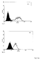
- the sequence of scFv E8 was identical to a scFv A4, which was obtained from the B-cell library of the same mouse. ScFv E8 was strongly reactive with LNCaP cells showing a MFI of about 100 at saturation concentrations, whereas the MFI of scFv A5 was only about 40 under the same conditions. No or minimal binding was seen with other cell lines lacking PSMA expression. Binding of both scFv to purified denatured glycosylated and deglycosylated PSMA was weak.
- mAbs which are different from those published by other authors with respect to high binding affinity and and high staining of PSMA expressing prostate cancer cells.
- the antibodies 3/F11, 3/A12 and 3/E7 do not only show a strong binding activity but also internalization into LNCaP cells as shown by immunofluorescence cytology and detection with confocal laser scanning microscopy.
- These mAbs were obtained after immunisation with full length native PSMA, which is in contrast to different published immunisation methods.
- anti-PSMA scFv were generated after immunisation with denatured and native PSMA.
- denatured PSMA we obtained scFv highly specific to the antigen, but not binding to LNCaP cells (data not shown in the present application).
- native PSMA we obtained scFv with a high cell binding activity, but a poor binding to the isolated denatured antigen.
- IC50 200 nM.
- the term IC50 is defined as the concentration in nM of the toxin which reduces cells proliferation to 50% of the cell proliferation without adding a toxin.
- the antibodies and scFv described in this application specifically bind to native cell-surface PSMA and therefore will have value in diagnostic and therapeutic applications focusing on PSMA as a target antigen for prostate cancer.
- PSMA is expressed on prostate cancer cells with a specific tertiary and quaternary structure
- only antibodies against this cellular conformation may recognize and strongly bind to viable prostate cancer cells and PSMA-expressing tissue. Therefore, the aim of the present study was to generate such mAbs and scFv that can be used for therapeutic and diagnostic targeting of prostate cancer.
- the present invention provides therefore an isolated monoclonal antibody or an antigen binding portion thereof which binds to prostate specific membrane antigen in its native form occurring on the surface of tumor cells which is linked to a label or a cytotoxic agent.
- isolated monoclonal antibody refers to a glycoprotein comprising at least two heavy (H) chains and two light (L) chains interconnected by disulfid bonds.
- Each heavy chain is comprised of a heavy chain variable region (abbreviated as V H ) and a heavy chain constant region.
- the heavy chain constant region is comprised of three domains, namely CH1, CH2 and CH3.
- Each light chain contains a light chain variable region (V L ) and a light chain constant region (C L ).
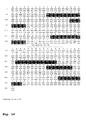
- V H and V L regions can be further subdivided into regions of hypervariability, which are also called complementarity determining regions (CDR) interspersed with regions that are more conserved. Those regions are also called framework regions (FR).
- Each V H and V L region is composed of three CDRs and four FRs arranged from amino terminus to carboxy terminus in the following order: FR1, CDR1, FR2, CDR2, FR3, CDR3, FR4.
- the variable regions of the heavy and light chains contain a binding domain that interacts with an antigen.
- the CDRs are marked by grey boxes. Those areas are important for the binding of the monoclonal antibody or the antigen binding portion thereof.
- the other areas are framework regions which can be replaced by other sequences.
- Monoclonal antibodies derived from mouse may cause unwanted immunological side-effects due to the fact that they contain a protein from another species which may elicit antibodies.
- the monoclonal antibodies or the antigen binding portions thereof may be humanized.
- the process of humanizing monoclonal antibodies is known to the person skilled in the art.
- the framework regions of a mouse mAb are replaced by the corresponding human framework regions. In order to maintain the preferred binding properties the sequences of the CDRs should be maintained as far as possible.
- antigen binding portion of the monoclonal antibody refers to one or more fragments of such an antibody which retained the ability to specifically binding to the prostate specific membrane antigen in its native form.
- antigen binding portions of the antibody include a Fab fragment, a monovalent fragment consisting of the V L , V H , C L and C H1 domains, an F(ab') 2 fragment, a bivalent fragment comprising two Fab fragments linked by a disulfid bridge at the hinge region, an Fd fragment consisting of the V H and C H1 domain, an F V fragment consisting of the V L and V H domains of a single arm of an antibody, a dAb fragment which consists of a V H domain and an isolated complementarity determining region (CDR).
- CDR complementarity determining region
- the isolated monoclonal antibody or antigen binding portion thereof according to the present invention can preferably be internalized by a tumor cell if it is used for therapeutic purposes. For diagnostic purposes an internalisation may not be required.
- the isolated monoclonal antibody or an antigen binding portion thereof according to the present invention binds strongly to LNCAP cells but not or only minimally to cells which lack expression of prostate specific membrane antigen.
- the binding of the isolated monoclonal antibody or antigen binding portion thereof is measured by PE fluorescence intensity (MFI) which is preferably higher than 40 for an scFv and preferably higher than 1000 for an mAb at saturating concentrations.
- MFI PE fluorescence intensity
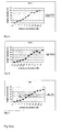
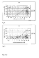
- the binding properties of the isolated monoclonal antibodies or an antigen binding portion thereof to the native PSMA were compared by treating LNCAP cells with increasing concentrations of the first step anti-PSMA Ab followed by incubation with the second step PE-labeled antibody. From the resulting saturation curves the antibody concentration reaching 50% saturation of PSMA sites can be read.
- the three mAb 3/F11, 3/A12 and 3/E7 showed a high binding activity reaching 50% saturation of PSMA sites at approximately 16 nM (3/F11), 2 nM (3/A12) and 30 nM (3/E7). With the scFv a 50% saturation of PSMA sites was found at 10 nM (E8) and 60 nM (A5).
- the PE phytoerythin fluorescence intensity
- the MFI values were plotted against the antibody (or binding fragments thereof) concentration whereby the plateau value of MFI corresponds to 100% saturation with antigen. After having determined the top value (plateau corresponding to 100% saturation of antigen) the value corresponding to 50% of saturation can be easily determined. By using the graph the corresponding concentration of the antibodies or binding fragments thereof in nM can be seen.
- the isolated monoclonal antibody or an antigen binding portion thereof comprises a label which may be a particle which emits radioactive radiation.
- This particle may be a radioactive element in a form which can be linked to the construct, preferably in the form of a complex.
- an mAb labeled with 111 Indium may be used as a radioimmunoscintigraphy agent in the detection of distant metastatic tumors in prostate cancer patients.
- the isolated monoclonal antibody or antigen binding portion thereof may comprise a cytotoxic agent which is a cell toxic substance selected from the group consisting of toxins, for example taxol, cytocalasin B, gramicidin D, ethidium bromid, emetine, mitomycin, etopside, tenopside, vincristine, vinblastine, colchicin, doxorubicin, daunorubicin, dihydroxy antracin dione, mitoxantrone, mithramycin, actinomycin D, 1-dehydrotestosteron, glycocorticoids, procain, tetracaine, lidokaine, propranolol and/or puromycin.
- a cytotoxic agent which is a cell toxic substance selected from the group consisting of toxins, for example taxol, cytocalasin B, gramicidin D, ethidium bromid, emetine, mitomycin, etopside, tenopside, vincris
- an isolated monoclonal antibody or an antigen binding portion thereof comprises a partial amino acid sequence of at least 10 consecutive amino acids of SEQ ID NO:1 (scFv E8) or SEQ ID NO:10 (scFv A5).
- the monoclonal antibody or antigen binding protein thereof comprises at least 25 and more preferred at least 50 consecutive amino acids of SEQ ID NO:1 or SEQ ID NO:10, respectively.
- the isolated monoclonal antibody or antigen binding portion thereof comprises at least one of the CDRs having SEQ ID NO:2 - SEQ ID NO:7 and/or SEQ ID NO:11 to 16. More preferably such construct comprises at least 3 and more preferably at least 5 of those CDR sequences.
- SEQ ID NO:8 and 9 relate to scFv E8 and SEQ ID NO:17 and 18 relate to scFv A5.
- the sequences report the coding strand and the complementary strand thereto.
- SEQ ID NOS:9 and 18 are shown in the 5' ⁇ 3' orientation.
- the polynucleotides of the present invention comprise a contiguous sequence of at least 20, preferably 50 and more preferably 100 nucleotides of the group consisting of SEQ ID NOS: 8, 9, 17 and 18.
- the sequence coding for the CDR are in particular preferred.
- compositions comprising an isolated monoclonal antibody or an antigen binding portion thereof as described in the present application.
- the pharmaceutical composition of the present invention comprises the monoclonal antibody or an antigen binding portion thereof together with pharmaceutically acceptable additives.
- a composition is prepared for intramuscular or intraveneous injection.
- the antibody may be provided in a depot formulation which allows the sustained release of the biologically active agent over a certain period of time which may range preferably from one to six months.
- Such a sustained release formulation may comprise a biodegradable polymer like a polylactide or polylactide/polyglycolide copolymer which is degraded over a prolonged period of time in the human body whereby the antibody or the antigen binding portion thereof preferably having a toxine is released in a controlled manner over a certain period of time.
- a biodegradable polymer like a polylactide or polylactide/polyglycolide copolymer which is degraded over a prolonged period of time in the human body whereby the antibody or the antigen binding portion thereof preferably having a toxine is released in a controlled manner over a certain period of time.
- the isolated monoclonal antibody or an antigen binding portion thereof may be used for the preparation of a medicament for the treatment of cancer, in particular prostate cancer.
- the invention provides a diagnostic kit for the detection of tumor cells comprising an isolated monoclonal antibody or an antigen binding portion thereof.
- the label allows the detection of the construct with suitable detection devices.
- the invention provides also a method for the in vitro identification of tumor cells by which the tumor cells to be identified are contacted with an isolated monoclonal antibody or an antigen binding portion thereof which carries a label which can be detected by suitable analytical devices.
- the label allows the diagnostic identification of tumor cells, for example in section of human tissues obtained after surgery or biopsy.
- LNCaP, DU 145, PC-3 and HeLa as well as the hybridoma 7E11-C5.3 (IgG1-k, PSMA) were purchased from the American Type Culture Collection (ATCC), Rockville, MD, USA.
- LNCaP, DU 145 and HeLa were cultured in RPMI 1640 medium, PC-3 in F12 Nutrimix medium, both supplemented with penicillin (100 000 U/I), streptomycin (100 mg/l) and 10 % FCS at 37 °C in a humidified atmosphere of 5 % CO 2 .
- LNCaP cells expressing unglycosylated PSMA on their surface 2 ⁇ g/ml tunicamycin (ICN Biomedicals) were added to the medium for 48 h.
- PSMA protein containing 1 % Triton X-100
- 10 8 LNCaP cells were washed with PBS and then lysed in PBS containing 1 % IGEPAL for 20 min at room temperature. After centrifugation at 10,000 g the supernatant was given on a 7E11-C5 affinity chromatography column (Amersham Biosciences, Uppsala, Sweden) and PSMA was eluted with 100 mM glycine buffer pH 2,5 containing 1 % Triton X-100. After neutralisation the protein was extensively dialyzed with PBS.
- LNCaP cell lysate containing full length native PSMA For preparation of a LNCaP cell lysate containing full length native PSMA, cells were lysed with M-PER reagent (Pierce) for 10 min and then centrifuged at 15,000 rpm for 30 min at 4°C. The supernatant containing native full length PSMA was collected (M-PER-lysate). The high molecular fraction (100 to 600 KD) of this lysate was separated by HPLC on a Biosil 250 size exclusion column.
- mice Four-month old female Balb/c mice were immunized intraperitoneally with 300 ⁇ g M-PER lysate from LNCaP cells or with the high molecular HPLC fraction of the lysate, or with 10 6 LNCaP cells, fixed with 2 % paraformaldehyde. These preparations were mixed 1:1 with complete Freund's adjuvant. Each mouse received 4 or 5 immunizations at 2-week intervals. Four days after the last immunization spleen cells were collected and either used for the preparation of hybridomas or a B-cell library.
- the mouse spleen was washed in phosphate buffered saline (PBS), minced to small pieces, washed again in PBS and then gently homogenized in a "loose-fitting" hand homogenizer.
- PBS phosphate buffered saline
- the resulting single cell suspension was overlayered onto Ficoll (Pharmacia, Freiburg, Germany) and centrifuged at 400 g for 20 min at room temperature.
- Interphase B cells were isolated with CD19 microbeads according to the manufacturer's instructions (Miltenyi, Bergisch Gladbach, Germany).
- the spleen was aseptically removed and a single cell suspension was prepared in RPMI-1640 medium without serum.
- the splenocytes were added to SP2/0 myeloma cells at a ratio of 10:1 and the fusion and selection was performed to established procedures [ Galfre et al., Nature (1979), p. 131-133 ].
- Hybridoma supernatants were tested by FACS on LNCaP and DU145 cells and by an ELISA with purified PSMA as solid phase. Monoclonal antibodies were purified using a protein G column (Pharmacia).
- Ig-isotypes of the anti-PSMA mAbs were determined by ELISA using either unlabelled (solid phase) or peroxidase-labeled (tracer) anti-isotype specific antibodies (Southern Biotechnology Associates, Birmingham, AL).
- mice which were immunized 5 times with the M-PER-lysate from LNCaP cells, spleen cells were fused with SP2/0 cells according to established methods. Positive hybridomas were selected by flow cytometry with LNCaP cells and ELISA on purified PSMA. By this way three positive clones were obtained.
- the corresponding mAbs with their isotypes were 3/F11 (IgG2a), 3/A12 (IgG1) and 3/E7 (IgG2b).
- the binding properties of the three antibodies were compared by treating LNCaP cells with increasing concentrations of the first step anti-PSMA mAb followed by incubation with a saturating amount of a second step PE-(phycoerythin)-labeled goat antibody followed by cytofluorometry analysis.
- PE-(phycoerythin)-labeled goat antibody followed by cytofluorometry analysis.
- the corrected mean PE (phycoerythin) fluorescence intensity was about 1000 for mab 3A12, and about 1400 for mAb 3F11 and about 1600 for mAB 3E7.
- the MFI was 5-fold lower on LNCaP cells expressing unglycosylated PSMA (grown with tunicamycine).
- RNA and mRNA was isolated with silicagel-based membranes (Rneasy, Qiagen, Hilden, Germany) according to the manufacturer's protocol.
- cDNA synthesis was performed at 42°C for 60 min in a final volume of 50 ⁇ l which contained 25 ⁇ l of denatured RNA, 10 ⁇ l 5x buffer (Promega, Heidelberg, Germany), 5 ⁇ l of 10 mM dNTP (dATP, dCTP, dGTP, dTTP, Promega), 1,5 ⁇ l RNAsin (40 U/ ⁇ l, Promega) 2,5 ⁇ l of 150 pM random hexamer primers, and 2,5 ⁇ l of AMV reverse transcriptase (10 U/ ⁇ l, Promega).
- the PCR products for the light chains were digested with Mlul and Notl, and ligated into the phagemid pSEX81 [ Dübel et al., Gene (1993), 97-101 ] using a molar ratio of 1:3 (2 ⁇ g vector, 400 ng insert).
- the products of one ligation were used for the electroporation of 50 ⁇ l electrocompetent E. coli XL1 blue cells (Stratagene) according to the supplier's protocol.
- the bacteria were plated on nine 80 mm diameter agarose plates containing 100 :g/ml ampicillin and 0,1 M glucose (SOB-AG) of and incubated overnight at 30 °C.
- Bacteria were isolated by adding 3 ml 2xYT medium on each plate, scraping them off with a sterile glass spreader and pelleted at 3,000 g for 15 min. From these bacteria plasmid DNA was isolated which revealed the VI sublibrary. Then the PCR products for the heavy chain and the VI sublibrary were digested with Ncol and HindIII. Ligation was prepared at a ratio of 3:1 (2 ⁇ g sublibrary and 400 ng insert). Transformation by electroporation, plating and collection of transformed bacteria was done as described for the VI sublibrary. From nine 80 mm diameter SOB-AG plates a total of 18 ml V H V L library was obtained.
- the cells were pelleted and resuspended in 15 ml 2xYT-medium containing 100 ⁇ g/ml ampicillin, 10 ⁇ g/ml tetracycline and 50 ⁇ g/ml kanamycin.
- the culture was incubated overnight at 37°C at 250 rpm, then chilled on ice and centrifuged to remove cells.
- the supernatant containing the phages was mixed with 1/5 volume of an aquous solution containing 20% PEG 8,000 and 14% NaCl and incubated 1 h at 4°C. Then a centrifugation step of 30 min at 4°C und 6,200 g was added.
- the pellet containing the phages was resuspended in 2 ml 10 mM Tris/HCl pH 7,5, 20 mM NaCl, 2 mM EDTA pH 7,5 and used for panning.
- Panning on purified PSMA was done in 96 well Maxi-Sorb microtiter plates (Nunc) which were coated with a solution of purified PSMA (100 ⁇ l/well, 12 ⁇ g/ml PSMA in PBS) and blocked with 4% non-fat milk/PBS.
- One ml of purified recombinant phages (circa 10 11 ) were incubated in 1 ml 4% non-fat milk/PBS supplemented with 15 ⁇ l 10% Triton X100 for 15 min and then allowed to bind to 8 wells coated with PSMA for 2 h at 37°C.
- phages were eluted with 0,1 M Glycin-Puffer pH 2,2.
- phages were previously absorbed on DU 145 cells.
- 1 ml (circa 10 11 ) recombinant phages were incubated in 2% non-fat milk/PBS for 15 min and then with 10 7 DU 145 cells for 1 h at room temperature on a shaker. Then the cells were centrifuged and the supernatant with non absorbed phages was incubated with 10 6 LNCaP cells for 1 h at room temperature on a shaker.
- Microtiter plates were coated with purified PSMA (1,5 ⁇ g PSMA/ml PBS) overnight and then blocked with 2% non-fat milk/PBS.
- PSMA purified PSMA
- V H V L library in the phagemid pSEX was constructed from the B cell library of a mouse immunized with M-PER-lysate of LNCaP cells. This library had a complexity of 10 7 .
- V H V L library was prepared from the monoclonal antibody 3/A12, which was obtained from the same mouse immunized with LNCaP lysate. This V H V L library had a complexity of 10 5 .
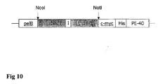
- the coding region of the scFv E8 and A5 were transferred from the phagemid pSEX into the expression vector pHOG, containing C-terminal c-myc and His-tags.
- the sequences with the corresponding CDRs are given in Fig. 13 and Fig. 14.
- the regions coding for the CDR's of the antigen binding portions are marked in Fig. 13 and 14. Those sequences should not be changed whereas the other parts of the sequence which are not marked can be changed. The appropriate three-dimensional structure must, however, be maintained.
- the scFv E8 strongly reacted with viable LNCaP cells as measured by flow cytometry with MFI values of about 100 at saturating concentrations, whereas binding of A5 was much weaker with MFI-values of about 40 at saturating concentrations (Fig 7).
- binding to purified PSMA as solid phase in an ELISA was weak for E8 and somewhat stronger for A5.
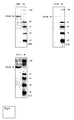
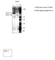
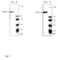
- a similar pattern was seen in western blots with denatured glycosylated and deglycosylated PSMA (Fig.8).
- Fig 9 By immunofluorescence cytology with LNCaP cells and detection by confocal laser microscopy a very good binding of the scFv E8 and internalization could be shown (Fig 9).
- ScFv fragments were expressed in E. coli XL1-Blue (Stratagene) using the secretion vector pHOG 21 which contains the sequences for the His-6 and c-myc-tag in a C-terminal position of the scFv [ Kipriganov et al., J.Immunol.Methods (1997), p. 69-77 ].
- E. coli bacteria transformed with pHOG plasmids were grown overnight in 2 x YT-AG-medium, then diluted 1:20 and grown as 600 ml cultures at 37°C.
- bacteria When cultures reached OD 0.8, bacteria were pelleted by centrifugation at 1,500 g for 10 min and resuspended in the same volume of fresh YT medium containing 50 :g/ml ampicillin, 0,4 M sucrose and 1 mM IPTG. Then growth was continued at room temperature for 18 to 20 h. Cells were harvested by centrifugation at 5,000 g for 10 min and 4°C. To isolate soluble periplasmic proteins, the pelleted bacteria were resuspended in 5% of the initial volume of ice-cold 50 mM Tris-HCl, 20% sucrose, 1 mM EDTA pH 8.0.
- periplasmic extract was concentrated using Amicon YM10 membranes with a 10 kDa cut-off (Amicon, Witten, Germany) followed by thorough dialysis against 50 mM Tris-HCl, 1 M NaCl, pH 7.0.
- Purification was achieved by immobilized metal affinity chromatography. This was performed using a 1 ml column of chelating Sepharose (Pharmacia) charged with Cu 2+ and equilibrated with a buffer containing 50 mM Tris-HCl and 1 M NaCl, pH 7.0. The periplasmatic extract was loaded, washed with twenty column volumes of equilibration buffer containing 30 mM imidazole and then eluted with the same buffer containing 250 mM imidazole. Eluted material was dialyzed against PBS.
- LNCaP, DU 145, and PC3 cells were freshly harvested from tissue culture flasks and a single cell suspension was prepared in PBS with 3% FCS and 0,1% NaN 3 . Approximately 10 5 cells were incubated with 50 ⁇ l of rescued phagemids, preincubated 1:1 with 2% non-fat milk/PBS, 1 h on ice. After 3 rounds of washing with PBS 25 ⁇ l/well anti-c-myc monoclonal antibody 9E10 (10 ⁇ g/ml; Becton Dickinson) or when phages were tested 25 ⁇ l/well anti-M13 antibody (10 ⁇ g/ml; Pharmacia) were added and incubated 40 min on ice.
- PBS 25 ⁇ l/well anti-c-myc monoclonal antibody 9E10 (10 ⁇ g/ml; Becton Dickinson) or when phages were tested 25 ⁇ l/well anti-M13 antibody (10 ⁇ g/ml; Pharmacia
- the cells were incubated with 100 ⁇ l of PE-labeled goat anti-mouse IgG (Becton Dickinson) for 40 min on ice. The cells were then washed again and resuspended in 100 ⁇ l of a solution containing 1 ⁇ g/ml propidium iodide (Sigma, Deisenhofen) in PBS with 3% FCS and 0, 1 % NaN 3 in order to exclude dead cells. The relative fluorescence of stained cells was measured using a FACScan flow cytometer and the CellQuest software (Becton Dickinson Mountain View, CA).
- LNCaP cells were grown on glass coverslips for 24 hours. For fixation, cells were treated with 2% paraformaldehyde in PBS for 30 min at RT, which does not permeabilize the cell membrane, washed with 1% BSA-PBS, quenched for 10 min in 50 mM NH 4 Cl in PBS, and rinsed with 1% BSA-PBS. Primary monoclonal antibody at 4 ⁇ g/ml in 1% BSA-PBS was added and incubated for 60 min at 4°C. FITC-labeled goat anti-mouse secondary antibody (2 ⁇ g/ml; Southern Biotechnology Associates Inc. Birmingham, USA) was incubated for 30 min and washed extensively with 1% BSA-PBS. Slides were mounted in Vectashield (Vector Laboratories, Inc. Burlingame, CA).
- the primary antibody was incubated for 30 min at 37°C before fixation of the cells with 2% paraformaldehyde and permeabilization with 0,5 % Triton X100 in PBS.
- Paraffin tissue sections were first deparaffinized and then treated with 0,3% Triton X100 in PBS for antigen retrieval. Kryostat sections were fixed in cold acetone. The the sections were treated 30 min at with 3% H 2 O 2 and 10 % methanol for quenching of endogenous peroxidase. After blocking with 1% BSA-PBS the primary antibody was added at a concentration of 2 ⁇ g/ml and incubated for 1 h at RT. For the scFv a secondary mouse-anti-c-myc antibody was added for 1 h at RT. Then a biotinylated goat-anti-mouse antibody was incubated for 30 min at RT and finally developed with ABC-reagent (Vectastain).
- Western blot analysis was performed following sodium dodecyl sulfate-polyacrylamide (SDS) gel electrophoresis of purified PSMA and cell lysate from LNCaP cells and transfered to polyvinylidene difluoride membranes.
- the blots were blocked overnight in PBS containing 5% non-fat milk and incubated with the purified mAbs or scFv at concentrations of 10 ⁇ g/ml for 1 h. Then the blots were washed 5 times with PBS-Tween (0,5%) and incubated with horseradish peroxidase conjugated goat anti-mouse IgG for 1 hour at RT. After 5 washes with PBS-Tween (0,5%) the blots were developed by using 3,3',5',5'-tetramethylbenzidine as substrate.
- SDS sodium dodecyl sulfate-polyacrylamide
- the toxin used in our approach was the truncated version of Pseudomonas exotoxin (PE40), lacking domain la and containing only domains Ib, II, and III [ Pastan et al., J.Biol.Chem. (1989), p. 15157-15160 ].
- the DNA with the coding region in the vector pSW200 was obtained from Prof. W. Wels, Frankfurt [ Wels et al., Biotechnology (1992), p. 1128-1132 ].
- the DNA fragment from bp position 253 to 613 encoding PE40 was amplified by PCR from plasmid pSW200.
- the amplified DNA was then ligated into the vector pHOG-His-scFv in a C-terminal position to the scFv using the restriction site Xbal. All cloning steps were performed according to standard methods in E. coli XL1 blue and the products were confirmed by sequencing.
- Protein induction of the immunotoxin and purification by IMAC was the same like the scFv.
- the products were tested and characterized by SDS-page, western blot and flow cytometry.
- Target cells LNCaP and DU 145 as control
- Various dilutions of the recombinant immunotoxins in aliquots of 50 ⁇ l/well were added and the plates were incubated for 48 hours at 37°C, 5% CO 2 . After this time the cultures were pulsed with 15 ⁇ l/well WST reagent and incubated for 90 min at 37°C, 5% CO 2 .
- Cytotoxicity assays (WST) with the immunotoxins E8-P40 and A5-P40 were prepared with PSMA expressing LNCaP cells and DU 145 control cells.
- a high cytotoxic effect could be shown with the immunotoxin E8-PE40 on LNCaP cells with a IC50 value of 0.05 nM.
- the cytotoxic effect of the immunotoxin A5-PE40 is shown with an IC50 of about 0.09 nM.
- the cytotoxic background on not PSMA expressing DU 145 cells was 5% for the E8 construct and only 0.01% for the A5 construct evidencing a very good therapeutic window.
Landscapes
- Health & Medical Sciences (AREA)
- Life Sciences & Earth Sciences (AREA)
- Chemical & Material Sciences (AREA)
- General Health & Medical Sciences (AREA)
- Medicinal Chemistry (AREA)
- Immunology (AREA)
- Cell Biology (AREA)
- Organic Chemistry (AREA)
- Pharmacology & Pharmacy (AREA)
- Animal Behavior & Ethology (AREA)
- Public Health (AREA)
- Veterinary Medicine (AREA)
- Proteomics, Peptides & Aminoacids (AREA)
- Reproductive Health (AREA)
- Oncology (AREA)
- Epidemiology (AREA)
- Bioinformatics & Cheminformatics (AREA)
- Nuclear Medicine, Radiotherapy & Molecular Imaging (AREA)
- Chemical Kinetics & Catalysis (AREA)
- General Chemical & Material Sciences (AREA)
- Engineering & Computer Science (AREA)
- Genetics & Genomics (AREA)
- Biochemistry (AREA)
- Biophysics (AREA)
- Molecular Biology (AREA)
- Gynecology & Obstetrics (AREA)
- Pregnancy & Childbirth (AREA)
- Physics & Mathematics (AREA)
- Optics & Photonics (AREA)
- Communicable Diseases (AREA)
- Urology & Nephrology (AREA)
- Peptides Or Proteins (AREA)
- Medicines Containing Antibodies Or Antigens For Use As Internal Diagnostic Agents (AREA)
- Preparation Of Compounds By Using Micro-Organisms (AREA)
- Medicines That Contain Protein Lipid Enzymes And Other Medicines (AREA)
Priority Applications (23)
| Application Number | Priority Date | Filing Date | Title |
|---|---|---|---|
| EP05011536A EP1726650A1 (fr) | 2005-05-27 | 2005-05-27 | Anticorps monoclonaux et fragments d'anticorps monocaténaires contre l'antigène spécifique de surface membranaire de la prostate |
| SI200630996T SI1883698T2 (sl) | 2005-05-27 | 2006-03-02 | Monoklonska protitelesa in enoverižni protitelesni fragmenti proti celičnemu površinskemu membranskemu antigenu, specifičnemu za prostato |
| ES06707388.2T ES2362795T5 (es) | 2005-05-27 | 2006-03-02 | Anticuerpos monoclonales y fragmentos de anticuerpo de cadena única frente a antígeno prostático específico de membrana de superficie celular |
| JP2008512701A JP5038297B2 (ja) | 2005-05-27 | 2006-03-02 | 細胞表面の前立腺特異的膜抗原に対するモノクローナル抗体および単鎖抗体フラグメント |
| RU2007147596/10A RU2458073C2 (ru) | 2005-05-27 | 2006-03-02 | Моноклональные антитела и одноцепочечные фрагменты антител против клеточно-поверхностного специфического для простаты мембранного антигена |
| EP11151622A EP2363486A1 (fr) | 2005-05-27 | 2006-03-02 | Anticorps monoclonaux et fragments d'anticorps monocaténaires contre l'antigène spécifique de surface membranaire de la prostate |
| US11/915,454 US8198416B2 (en) | 2005-05-27 | 2006-03-02 | Monoclonal antibodies and single chain antibody fragments against cell-surface prostate specific membrane antigen |
| AU2006251445A AU2006251445B2 (en) | 2005-05-27 | 2006-03-02 | Monoclonal antibodies and single chain antibody fragments against cell-surface prostate specific membrane antigen |
| PCT/EP2006/001917 WO2006125481A1 (fr) | 2005-05-27 | 2006-03-02 | Anticorps monoclonaux et fragments d'anticorps a chaine unique contre antigene de membrane specifique a la prostate ayant pour origine la surface cellulaire |
| AT06707388T ATE505544T1 (de) | 2005-05-27 | 2006-03-02 | Monoklonale antikörper und einzelkettenantikörper fragments gegen das zelloberflächen prostataspezifische membranantigen |
| DK06707388.2T DK1883698T4 (en) | 2005-05-27 | 2006-03-02 | Monoclonal antibodies and single-chain antibody fragments against cell surface prostate-specific membrane antigen |
| CN2006800275807A CN101233236B (zh) | 2005-05-27 | 2006-03-02 | 抗细胞表面前列腺特异性膜抗原的单克隆抗体和单链抗体片段 |
| DE602006021296T DE602006021296D1 (de) | 2005-05-27 | 2006-03-02 | Monoklonale antikörper und einzelkettenantikörper fragments gegen das zelloberflächen prostataspezifische membranantigen |
| CA2609682A CA2609682C (fr) | 2005-05-27 | 2006-03-02 | Anticorps monoclonaux et fragments d'anticorps a chaine unique contre antigene de membrane specifique a la prostate ayant pour origine la surface cellulaire |
| EP06707388.2A EP1883698B2 (fr) | 2005-05-27 | 2006-03-02 | Anticorps monoclonaux et fragments d'anticorps a chaine unique contre antigene de membrane specifique a la prostate ayant pour origine la surface cellulaire |
| PL06707388T PL1883698T5 (pl) | 2005-05-27 | 2006-03-02 | Przeciwciała monoklonalne i jednołańcuchowe fragmenty przeciwciał skierowane przeciw antygenom błonowym specyficznym dla powierzchni komórek prostaty |
| PT06707388T PT1883698E (pt) | 2005-05-27 | 2006-03-02 | Anticorpos monoclonais e fragmentos de anticorpo de cadeia única contra antigénio de membrana específico da próstata de superfície celular |
| CY20111100431T CY1111409T1 (el) | 2005-05-27 | 2011-05-04 | Μονοκλωνικα αντισωματα και θραυσματα αντισωματων μονης αλυσιδας εναντιον προστατικου μεμβρανικου αντιγονου κυτταρικης επιφανειας |
| JP2012091882A JP5524270B2 (ja) | 2005-05-27 | 2012-04-13 | 細胞表面の前立腺特異的膜抗原に対するモノクローナル抗体および単鎖抗体フラグメント |
| US13/466,286 US8632777B2 (en) | 2005-05-27 | 2012-05-08 | Monoclonal antibodies and single chain antibody fragments against cell-surface prostate specific membrane antigen as diagnostic and therapeutic tools for prostate cancer |
| US14/105,808 US9238694B2 (en) | 2005-05-27 | 2013-12-13 | Monoclonal antibodies and single chain antibody fragments against cell-surface prostate specific membrane antigen |
| JP2014017392A JP6110321B2 (ja) | 2005-05-27 | 2014-01-31 | 細胞表面の前立腺特異的膜抗原に対するモノクローナル抗体および単鎖抗体フラグメント |
| JP2016063219A JP2016190839A (ja) | 2005-05-27 | 2016-03-28 | 細胞表面の前立腺特異的膜抗原に対するモノクローナル抗体および単鎖抗体フラグメント |
Applications Claiming Priority (1)
| Application Number | Priority Date | Filing Date | Title |
|---|---|---|---|
| EP05011536A EP1726650A1 (fr) | 2005-05-27 | 2005-05-27 | Anticorps monoclonaux et fragments d'anticorps monocaténaires contre l'antigène spécifique de surface membranaire de la prostate |
Publications (1)
| Publication Number | Publication Date |
|---|---|
| EP1726650A1 true EP1726650A1 (fr) | 2006-11-29 |
Family
ID=34937022
Family Applications (3)
| Application Number | Title | Priority Date | Filing Date |
|---|---|---|---|
| EP05011536A Withdrawn EP1726650A1 (fr) | 2005-05-27 | 2005-05-27 | Anticorps monoclonaux et fragments d'anticorps monocaténaires contre l'antigène spécifique de surface membranaire de la prostate |
| EP06707388.2A Active EP1883698B2 (fr) | 2005-05-27 | 2006-03-02 | Anticorps monoclonaux et fragments d'anticorps a chaine unique contre antigene de membrane specifique a la prostate ayant pour origine la surface cellulaire |
| EP11151622A Ceased EP2363486A1 (fr) | 2005-05-27 | 2006-03-02 | Anticorps monoclonaux et fragments d'anticorps monocaténaires contre l'antigène spécifique de surface membranaire de la prostate |
Family Applications After (2)
| Application Number | Title | Priority Date | Filing Date |
|---|---|---|---|
| EP06707388.2A Active EP1883698B2 (fr) | 2005-05-27 | 2006-03-02 | Anticorps monoclonaux et fragments d'anticorps a chaine unique contre antigene de membrane specifique a la prostate ayant pour origine la surface cellulaire |
| EP11151622A Ceased EP2363486A1 (fr) | 2005-05-27 | 2006-03-02 | Anticorps monoclonaux et fragments d'anticorps monocaténaires contre l'antigène spécifique de surface membranaire de la prostate |
Country Status (16)
| Country | Link |
|---|---|
| US (3) | US8198416B2 (fr) |
| EP (3) | EP1726650A1 (fr) |
| JP (4) | JP5038297B2 (fr) |
| CN (1) | CN101233236B (fr) |
| AT (1) | ATE505544T1 (fr) |
| AU (1) | AU2006251445B2 (fr) |
| CA (1) | CA2609682C (fr) |
| CY (1) | CY1111409T1 (fr) |
| DE (1) | DE602006021296D1 (fr) |
| DK (1) | DK1883698T4 (fr) |
| ES (1) | ES2362795T5 (fr) |
| PL (1) | PL1883698T5 (fr) |
| PT (1) | PT1883698E (fr) |
| RU (1) | RU2458073C2 (fr) |
| SI (1) | SI1883698T2 (fr) |
| WO (1) | WO2006125481A1 (fr) |
Cited By (4)
| Publication number | Priority date | Publication date | Assignee | Title |
|---|---|---|---|---|
| WO2009019493A1 (fr) * | 2007-08-09 | 2009-02-12 | Imperial Innovations Limited | Anticorps se liant a des cellules epitheliales de la prostate humaine |
| WO2009130575A3 (fr) * | 2008-04-22 | 2010-01-14 | Universita' Degli Studi Di Verona | Anticorps monoclonal isolé ou fragment de celui-ci se liant à l'antigène membranaire spécifique de la prostate, ses conjugués et utilisations |
| US9782478B1 (en) | 2011-04-22 | 2017-10-10 | Aptevo Research And Development Llc | Prostate-specific membrane antigen binding proteins and related compositions and methods |
| US11352426B2 (en) | 2015-09-21 | 2022-06-07 | Aptevo Research And Development Llc | CD3 binding polypeptides |
Families Citing this family (79)
| Publication number | Priority date | Publication date | Assignee | Title |
|---|---|---|---|---|
| US7105159B1 (en) | 1992-11-05 | 2006-09-12 | Sloan-Kettering Institute For Cancer Research | Antibodies to prostate-specific membrane antigen |
| AU2002356844C1 (en) | 2001-10-23 | 2010-03-04 | Amgen Fremont Inc. | PSMA antibodies and protein multimers |
| US20050215472A1 (en) | 2001-10-23 | 2005-09-29 | Psma Development Company, Llc | PSMA formulations and uses thereof |
| EP1726650A1 (fr) * | 2005-05-27 | 2006-11-29 | Universitätsklinikum Freiburg | Anticorps monoclonaux et fragments d'anticorps monocaténaires contre l'antigène spécifique de surface membranaire de la prostate |
| JP5781765B2 (ja) | 2007-11-30 | 2015-09-24 | カロバイオス ファーマシューティカルズ インコーポレイティッド | シュードモナス・エルギノーサ(PseudomonasAeruginosa)のPcrV抗原に対する抗体 |
| US20110189093A1 (en) * | 2008-04-14 | 2011-08-04 | Proscan Rx Pharma | Prostate specific membrane antigen antibodies and antigen binding fragments |
| EP2352763B2 (fr) | 2008-10-01 | 2022-09-21 | Amgen Research (Munich) GmbH | Anticorps monocaténaires bispécifiques spécifiques d'antigènes cibles de masse moléculaire élevée |
| KR101820535B1 (ko) | 2008-10-01 | 2018-01-19 | 암젠 리서치 (뮌헨) 게엠베하 | 종간특이적 psmaxcd3 이중특이적 단일쇄 항체 |
| EP2398504B1 (fr) | 2009-02-17 | 2018-11-28 | Cornell Research Foundation, Inc. | Procédés et kits pour le diagnostic d'un cancer et la prédiction d'une valeur thérapeutique |
| TWI629357B (zh) * | 2009-10-02 | 2018-07-11 | 安進研究(慕尼黑)有限責任公司 | 跨物種特異性的PSMAxCD3雙特異性單鏈抗體 |
| WO2011069019A2 (fr) * | 2009-12-02 | 2011-06-09 | David Ho | Minobodies j591 et cys-diabodies pour le ciblage de l'antigène membranaire spécifique de la prostate humaine (psma), et procédés d'utilisation |
| TWI653333B (zh) * | 2010-04-01 | 2019-03-11 | 安進研究(慕尼黑)有限責任公司 | 跨物種專一性之PSMAxCD3雙專一性單鏈抗體 |
| WO2013155605A1 (fr) * | 2012-04-19 | 2013-10-24 | Innovascreen Inc. | Procédé de prévision d'un état pathophysiologique chez un animal |
| US20170335281A1 (en) | 2014-03-15 | 2017-11-23 | Novartis Ag | Treatment of cancer using chimeric antigen receptor |
| JP2017528433A (ja) | 2014-07-21 | 2017-09-28 | ノバルティス アーゲー | 低い免疫増強用量のmTOR阻害剤とCARの組み合わせ |
| US20180334490A1 (en) | 2014-12-03 | 2018-11-22 | Qilong H. Wu | Methods for b cell preconditioning in car therapy |
| US11161907B2 (en) | 2015-02-02 | 2021-11-02 | Novartis Ag | Car-expressing cells against multiple tumor antigens and uses thereof |
| EP3286211A1 (fr) | 2015-04-23 | 2018-02-28 | Novartis AG | Traitement du cancer à l'aide de protéine récepteur antigénique chimérique et un inhibiteur de protéine kinase |
| EP3932428A1 (fr) | 2015-05-21 | 2022-01-05 | Harpoon Therapeutics, Inc. | Protéines trispécifiques de liaison et méthodes d'utilisation |
| WO2017027392A1 (fr) | 2015-08-07 | 2017-02-16 | Novartis Ag | Traitement du cancer à l'aide des protéines de récepteur cd3 chimères |
| EP3380620A1 (fr) | 2015-11-23 | 2018-10-03 | Novartis AG | Vecteurs de transfert lentiviral optimisés et utilisations associées |
| EP3397756B1 (fr) | 2015-12-30 | 2023-03-08 | Novartis AG | Thérapies à base de cellules effectrices immunitaires dotées d'une efficacité accrue |
| CN105968203A (zh) * | 2016-02-03 | 2016-09-28 | 南昌大学 | 一种抗前列腺特异性膜抗原胞外区的单域重链抗体 |
| CN105504061A (zh) * | 2016-02-03 | 2016-04-20 | 南昌大学 | 针对前列腺特异性膜抗原的纳米抗体 |
| CN105968202A (zh) * | 2016-02-03 | 2016-09-28 | 南昌大学 | 针对前列腺特异性膜抗原胞外区的单域重链抗体 |
| CN105542009A (zh) * | 2016-02-03 | 2016-05-04 | 南昌大学 | 一种针对前列腺特异性膜抗原的单域重链抗体 |
| CN105524171A (zh) * | 2016-02-03 | 2016-04-27 | 中国人民解放军第三军医大学第三附属医院 | 一种针对前列腺特异性膜抗原的纳米抗体 |
| CN105968201A (zh) * | 2016-02-03 | 2016-09-28 | 南昌大学 | 针对前列腺特异性膜抗原的单域重链抗体 |
| US20200281973A1 (en) | 2016-03-04 | 2020-09-10 | Novartis Ag | Cells expressing multiple chimeric antigen receptor (car) molecules and uses therefore |
| US11549099B2 (en) | 2016-03-23 | 2023-01-10 | Novartis Ag | Cell secreted minibodies and uses thereof |
| SI3443096T1 (sl) | 2016-04-15 | 2023-07-31 | Novartis Ag | Sestavki in postopki za selektivno izražanje himerni antigenskih receptorjev |
| US11623958B2 (en) | 2016-05-20 | 2023-04-11 | Harpoon Therapeutics, Inc. | Single chain variable fragment CD3 binding proteins |
| US10100106B2 (en) | 2016-05-20 | 2018-10-16 | Harpoon Therapeutics, Inc. | Single domain serum albumin binding protein |
| KR102365977B1 (ko) | 2016-05-20 | 2022-02-22 | 하푼 테라퓨틱스, 인크. | 단일 쇄 가변 단편 cd3 결합 단백질 |
| US20190161542A1 (en) | 2016-08-01 | 2019-05-30 | Novartis Ag | Treatment of cancer using a chimeric antigen receptor in combination with an inhibitor of a pro-m2 macrophage molecule |
| EP3544997A4 (fr) * | 2016-11-23 | 2020-07-01 | Harpoon Therapeutics, Inc. | Protéine de liaison à l'antigène membranaire spécifique de la prostate |
| MX2019006045A (es) | 2016-11-23 | 2019-11-11 | Harpoon Therapeutics Inc | Proteinas triespecificas dirigidas a psma y metodos de uso. |
| WO2018111340A1 (fr) | 2016-12-16 | 2018-06-21 | Novartis Ag | Procédés de détermination de la puissance et de la fonction proliférative de lymphocytes t à récepteur antigénique chimérique (car) |
| ES2912408T3 (es) | 2017-01-26 | 2022-05-25 | Novartis Ag | Composiciones de CD28 y métodos para terapia con receptores quiméricos para antígenos |
| WO2018144535A1 (fr) | 2017-01-31 | 2018-08-09 | Novartis Ag | Traitement du cancer à l'aide de protéines chimères du récepteur de lymphocytes t ayant de multiples spécificités |
| EP3589647A1 (fr) | 2017-02-28 | 2020-01-08 | Novartis AG | Compositions d'inhibiteur shp et utilisations pour une thérapie de récepteur d'antigène chimère |
| EP3589662A4 (fr) | 2017-02-28 | 2020-12-30 | Harpoon Therapeutics, Inc. | Protéine monovalente inductible de fixation d' antigène |
| AU2018265856B2 (en) | 2017-05-12 | 2023-04-27 | Harpoon Therapeutics, Inc. | Mesothelin binding proteins |
| CN115028727A (zh) | 2017-05-12 | 2022-09-09 | 哈普恩治疗公司 | 靶向msln的三特异性蛋白质及使用方法 |
| WO2018229715A1 (fr) | 2017-06-16 | 2018-12-20 | Novartis Ag | Compositions comprenant des anticorps anti-cd32b et procédés d'utilisation correspondants |
| AU2018347582A1 (en) | 2017-10-13 | 2020-05-07 | Harpoon Therapeutics, Inc. | Trispecific proteins and methods of use |
| US10927180B2 (en) | 2017-10-13 | 2021-02-23 | Harpoon Therapeutics, Inc. | B cell maturation antigen binding proteins |
| WO2019079569A1 (fr) | 2017-10-18 | 2019-04-25 | Novartis Ag | Compositions et méthodes pour la dégradation sélective d'une protéine |
| MX2020004229A (es) | 2017-10-25 | 2020-07-22 | Novartis Ag | Metodos de produccion de celulas que expresan receptores antigenicos quimericos. |
| WO2019081983A1 (fr) | 2017-10-25 | 2019-05-02 | Novartis Ag | Anticorps ciblant cd32b et leurs procédés d'utilisation |
| US20210179709A1 (en) | 2017-10-31 | 2021-06-17 | Novartis Ag | Anti-car compositions and methods |
| SG11202005557TA (en) | 2017-12-12 | 2020-07-29 | Macrogenics Inc | Bispecific cd 16-binding molecules and their use in the treatment of disease |
| JP2021521202A (ja) | 2018-04-13 | 2021-08-26 | ハイデルベルク ファルマ リサーチ ゲゼルシャフト ミット ベシュレンクテル ハフツング | 固形腫瘍の治療のための標的化されたアマトキシン複合体 |
| US20210047405A1 (en) | 2018-04-27 | 2021-02-18 | Novartis Ag | Car t cell therapies with enhanced efficacy |
| EP3788369A1 (fr) | 2018-05-01 | 2021-03-10 | Novartis Ag | Biomarqueurs pour évaluer des cellules car-t pour prédire un résultat clinique |
| WO2019227003A1 (fr) | 2018-05-25 | 2019-11-28 | Novartis Ag | Polythérapie comprenant des thérapies par récepteur antigénique chimérique (car) |
| WO2019237035A1 (fr) | 2018-06-08 | 2019-12-12 | Intellia Therapeutics, Inc. | Compositions et procédés d'immuno-oncologie |
| EP3587454A1 (fr) * | 2018-06-27 | 2020-01-01 | Albert-Ludwigs-Universität Freiburg | Récepteurs d'antigène chimérique qui se lient à l'antigène membranaire spécifique de la prostate |
| AR116109A1 (es) | 2018-07-10 | 2021-03-31 | Novartis Ag | Derivados de 3-(5-amino-1-oxoisoindolin-2-il)piperidina-2,6-diona y usos de los mismos |
| HUE061078T2 (hu) * | 2018-07-31 | 2023-05-28 | Heidelberg Pharma Res Gmbh | PSMA elleni humanizált ellenanyagok |
| KR20210086623A (ko) | 2018-09-25 | 2021-07-08 | 하푼 테라퓨틱스, 인크. | Ddl3 결합 단백질 및 사용 방법 |
| CA3123511A1 (fr) | 2018-12-20 | 2020-06-25 | Novartis Ag | Schema posologique et combinaison pharmaceutique comprenant des derives de 3-(1-oxoisoindoline-2-yl) piperidine-2,6-dione |
| WO2020165833A1 (fr) | 2019-02-15 | 2020-08-20 | Novartis Ag | Dérivés de 3-(1-oxo-5-(pipéridin-4-yl)isoindolin-2-yl)pipéridine-2,6-dione et leurs utilisations |
| CA3123519A1 (fr) | 2019-02-15 | 2020-08-20 | Novartis Ag | Derives de 3-(1-oxoisoindoline-2-yl)piperidine-2,6-dione substitues et leurs utilisations |
| CN110407939B (zh) * | 2019-03-12 | 2023-09-29 | 广东医科大学附属第二医院 | 一种人源化抗psma单链抗体及其应用 |
| WO2020219742A1 (fr) | 2019-04-24 | 2020-10-29 | Novartis Ag | Compositions et procédés de dégradation sélective de protéines |
| CN115175937A (zh) | 2019-12-20 | 2022-10-11 | 诺华股份有限公司 | 用于治疗骨髓纤维化和骨髓增生异常综合征的抗TIM-3抗体MBG453和抗TGF-β抗体NIS793与或不与地西他滨或抗PD-1抗体斯巴达珠单抗的组合 |
| EP3842461A1 (fr) * | 2019-12-23 | 2021-06-30 | Albert-Ludwigs-Universität Freiburg | Récepteurs d'antigène chimérique qui se lient à l'antigène membranaire spécifique de la prostate |
| WO2021164692A1 (fr) * | 2020-02-18 | 2021-08-26 | 和铂医药(上海)有限责任公司 | Protéine isolée se liant à l'antigène psma et son utilisation |
| CN111303288B (zh) * | 2020-03-04 | 2020-12-25 | 和铂医药(苏州)有限公司 | 一种分离的结合抗原psma的蛋白及其用途 |
| WO2021168303A1 (fr) | 2020-02-21 | 2021-08-26 | Harpoon Therapeutics, Inc. | Protéines de liaison à flt3 et méthodes d'utilisation |
| JP7390214B2 (ja) | 2020-02-28 | 2023-12-01 | 株式会社東京精密 | シリコンウエハ表面状態診断方法及び表面改質方法 |
| JP2023529211A (ja) | 2020-06-11 | 2023-07-07 | ノバルティス アーゲー | Zbtb32阻害剤及びその使用 |
| JP2023531676A (ja) | 2020-06-23 | 2023-07-25 | ノバルティス アーゲー | 3-(1-オキソイソインドリン-2-イル)ピぺリジン-2,6-ジオン誘導体を含む投与レジメン |
| WO2022029573A1 (fr) | 2020-08-03 | 2022-02-10 | Novartis Ag | Dérivés de 3-(1-oxoisoindolin-2-yl)pipéridine-2,6-dione substitués par hétéroaryle et leurs utilisations |
| TW202304979A (zh) | 2021-04-07 | 2023-02-01 | 瑞士商諾華公司 | 抗TGFβ抗體及其他治療劑用於治療增殖性疾病之用途 |
| CN118056008A (zh) | 2021-04-27 | 2024-05-17 | 诺华股份有限公司 | 病毒载体生产系统 |
| WO2023214325A1 (fr) | 2022-05-05 | 2023-11-09 | Novartis Ag | Dérivés de pyrazolopyrimidine et leurs utilisations en tant qu'inhibiteurs de tet2 |
| WO2024089639A1 (fr) | 2022-10-26 | 2024-05-02 | Novartis Ag | Formulations lentivirales |
Citations (6)
| Publication number | Priority date | Publication date | Assignee | Title |
|---|---|---|---|---|
| WO1997035616A1 (fr) * | 1996-03-25 | 1997-10-02 | Pacific Northwest Cancer Foundation | Anticorps monoclonaux de l'antigene membranaire specifique de la prostate |
| WO1998003873A1 (fr) * | 1996-07-18 | 1998-01-29 | Cornell Research Foundation, Inc. | Traitement et diagnostic du cancer |
| WO2001009192A1 (fr) * | 1999-07-29 | 2001-02-08 | Medarex, Inc. | Anticorps monoclonaux humains de l'antigene d'enveloppe prostatique specifique |
| US6255458B1 (en) * | 1990-08-29 | 2001-07-03 | Genpharm International | High affinity human antibodies and human antibodies against digoxin |
| WO2003002144A1 (fr) * | 2001-06-26 | 2003-01-09 | Imclone Systems Incorporated | Anticorps bispecifiques se liant aux recepteurs vegf |
| US20040213791A1 (en) * | 2001-06-01 | 2004-10-28 | Neil Bander | Modified antibodies to prostate-specific membrane antigen and uses thereof |
Family Cites Families (18)
| Publication number | Priority date | Publication date | Assignee | Title |
|---|---|---|---|---|
| US5162504A (en) * | 1988-06-03 | 1992-11-10 | Cytogen Corporation | Monoclonal antibodies to a new antigenic marker in epithelial prostatic cells and serum of prostatic cancer patients |
| EP0682710B1 (fr) * | 1993-02-10 | 2003-10-29 | Unilever Plc | Procede d'isolation utilisant des proteines immobilisees a capacites de fixation specifique |
| US6150508A (en) * | 1996-03-25 | 2000-11-21 | Northwest Biotherapeutics, Inc. | Monoclonal antibodies specific for the extracellular domain of prostate-specific membrane antigen |
| PL335365A1 (en) * | 1997-02-26 | 2000-04-25 | Sankyo Co | Composition for treating or preventing prostatic carcinoma |
| US6258939B1 (en) * | 1997-03-10 | 2001-07-10 | The Regents Of The University Of California | PSCA antibodies and hybridomas producing them |
| FR2797743B1 (fr) * | 1999-08-23 | 2003-08-08 | Urogene | Lignee cellulaire prostatique et son utilisation pour l'obtention d'une tumeur prostatique etablie chez l'animal |
| AU2002305767B2 (en) * | 2001-09-20 | 2008-04-10 | Cornell Research Foundation, Inc. | Methods and compositions for treating and preventing skin disorders using binding agents specific for PSMA |
| AU2002356844C1 (en) * | 2001-10-23 | 2010-03-04 | Amgen Fremont Inc. | PSMA antibodies and protein multimers |
| KR20040077889A (ko) * | 2002-01-28 | 2004-09-07 | 메다렉스, 인코포레이티드 | 전립선 특이적 막 항원 (psma)에 대한 인간모노클로날 항체 |
| JP2004000045A (ja) * | 2002-05-31 | 2004-01-08 | Kyowa Hakko Kogyo Co Ltd | 前立腺特異的膜抗原に対する抗体 |
| US6641899B1 (en) * | 2002-11-05 | 2003-11-04 | International Business Machines Corporation | Nonlithographic method to produce masks by selective reaction, articles produced, and composition for same |
| US7811564B2 (en) * | 2003-01-28 | 2010-10-12 | Proscan Rx Pharma | Prostate cancer diagnosis and treatment |
| EP1841467A4 (fr) * | 2005-01-14 | 2009-01-28 | Cytogen Corp | Therapie du cancer de combinaison avec des anticorps diriges contre l'antigene membranaire specifique de la prostate |
| KR20070115967A (ko) * | 2005-02-18 | 2007-12-06 | 메다렉스, 인코포레이티드 | 전립선 특이 막 항원(psma)에 대한 인간 모노클로날항체 |
| EP1851251A2 (fr) * | 2005-02-18 | 2007-11-07 | Medarex, Inc. | Anticorps monoclonaux diriges contre l'antigene d'enveloppe specifique de la prostate (psma) depourvus de residus fucosyle |
| EP1871810A2 (fr) * | 2005-04-08 | 2008-01-02 | Cytogen Corporation | Anticorps anti-psma conjugués |
| EP1726650A1 (fr) | 2005-05-27 | 2006-11-29 | Universitätsklinikum Freiburg | Anticorps monoclonaux et fragments d'anticorps monocaténaires contre l'antigène spécifique de surface membranaire de la prostate |
| DK1912677T3 (da) * | 2005-06-20 | 2014-01-13 | Psma Dev Company L L C | PSMA-antistof-lægemiddel-konjugater |
-
2005
- 2005-05-27 EP EP05011536A patent/EP1726650A1/fr not_active Withdrawn
-
2006
- 2006-03-02 CA CA2609682A patent/CA2609682C/fr active Active
- 2006-03-02 SI SI200630996T patent/SI1883698T2/sl unknown
- 2006-03-02 DK DK06707388.2T patent/DK1883698T4/en active
- 2006-03-02 WO PCT/EP2006/001917 patent/WO2006125481A1/fr active Application Filing
- 2006-03-02 EP EP06707388.2A patent/EP1883698B2/fr active Active
- 2006-03-02 CN CN2006800275807A patent/CN101233236B/zh active Active
- 2006-03-02 US US11/915,454 patent/US8198416B2/en active Active
- 2006-03-02 JP JP2008512701A patent/JP5038297B2/ja active Active
- 2006-03-02 ES ES06707388.2T patent/ES2362795T5/es active Active
- 2006-03-02 AT AT06707388T patent/ATE505544T1/de active
- 2006-03-02 AU AU2006251445A patent/AU2006251445B2/en active Active
- 2006-03-02 PT PT06707388T patent/PT1883698E/pt unknown
- 2006-03-02 DE DE602006021296T patent/DE602006021296D1/de active Active
- 2006-03-02 RU RU2007147596/10A patent/RU2458073C2/ru active
- 2006-03-02 EP EP11151622A patent/EP2363486A1/fr not_active Ceased
- 2006-03-02 PL PL06707388T patent/PL1883698T5/pl unknown
-
2011
- 2011-05-04 CY CY20111100431T patent/CY1111409T1/el unknown
-
2012
- 2012-04-13 JP JP2012091882A patent/JP5524270B2/ja active Active
- 2012-05-08 US US13/466,286 patent/US8632777B2/en active Active
-
2013
- 2013-12-13 US US14/105,808 patent/US9238694B2/en active Active
-
2014
- 2014-01-31 JP JP2014017392A patent/JP6110321B2/ja active Active
-
2016
- 2016-03-28 JP JP2016063219A patent/JP2016190839A/ja active Pending
Patent Citations (6)
| Publication number | Priority date | Publication date | Assignee | Title |
|---|---|---|---|---|
| US6255458B1 (en) * | 1990-08-29 | 2001-07-03 | Genpharm International | High affinity human antibodies and human antibodies against digoxin |
| WO1997035616A1 (fr) * | 1996-03-25 | 1997-10-02 | Pacific Northwest Cancer Foundation | Anticorps monoclonaux de l'antigene membranaire specifique de la prostate |
| WO1998003873A1 (fr) * | 1996-07-18 | 1998-01-29 | Cornell Research Foundation, Inc. | Traitement et diagnostic du cancer |
| WO2001009192A1 (fr) * | 1999-07-29 | 2001-02-08 | Medarex, Inc. | Anticorps monoclonaux humains de l'antigene d'enveloppe prostatique specifique |
| US20040213791A1 (en) * | 2001-06-01 | 2004-10-28 | Neil Bander | Modified antibodies to prostate-specific membrane antigen and uses thereof |
| WO2003002144A1 (fr) * | 2001-06-26 | 2003-01-09 | Imclone Systems Incorporated | Anticorps bispecifiques se liant aux recepteurs vegf |
Non-Patent Citations (4)
| Title |
|---|
| BANDER N H ET AL: "TARGETED SYSTEMIC THERAPY OF PROSTATE CANCER WITH A MONOCLONAL ANTIBODY TO PROSTATE-SPECIFIC MEMBRANE ANTIGEN", SEMINARS IN ONCOLOGY, BETHESDA, MD, US, vol. 30, no. 5, October 2003 (2003-10-01), pages 667 - 677, XP008050060 * |
| DAVIES J ET AL: "Affinity improvement of single antibody VH domains: residues in all three hypervariable regions affect antigen binding", IMMUNOTECHNOLOGY, ELSEVIER SCIENCE PUBLISHERS BV, NL, vol. 2, no. 3, September 1996 (1996-09-01), pages 169 - 179, XP004070292, ISSN: 1380-2933 * |
| FRACASSO G ET AL: "Anti-tumor effects of toxins targeted to the prostate specific membrane antigen", PROSTATE, WILEY-LISS, NEW YORK, NY, US, vol. 53, no. 1, 15 September 2002 (2002-09-15), pages 9 - 23, XP002965355, ISSN: 0270-4137 * |
| HOLT L J ET AL: "Domain antibodies: proteins for therapy", TRENDS IN BIOTECHNOLOGY, ELSEVIER PUBLICATIONS, CAMBRIDGE, GB, vol. 21, no. 11, November 2003 (2003-11-01), pages 484 - 490, XP004467495, ISSN: 0167-7799 * |
Cited By (5)
| Publication number | Priority date | Publication date | Assignee | Title |
|---|---|---|---|---|
| WO2009019493A1 (fr) * | 2007-08-09 | 2009-02-12 | Imperial Innovations Limited | Anticorps se liant a des cellules epitheliales de la prostate humaine |
| WO2009130575A3 (fr) * | 2008-04-22 | 2010-01-14 | Universita' Degli Studi Di Verona | Anticorps monoclonal isolé ou fragment de celui-ci se liant à l'antigène membranaire spécifique de la prostate, ses conjugués et utilisations |
| US8703918B2 (en) | 2008-04-22 | 2014-04-22 | Universita' Degli Studi Di Verona | Isolated monoclonal antibody or fragment thereof binding prostate specific membrane antigen, conjugates and uses thereof |
| US9782478B1 (en) | 2011-04-22 | 2017-10-10 | Aptevo Research And Development Llc | Prostate-specific membrane antigen binding proteins and related compositions and methods |
| US11352426B2 (en) | 2015-09-21 | 2022-06-07 | Aptevo Research And Development Llc | CD3 binding polypeptides |
Also Published As
Similar Documents
| Publication | Publication Date | Title |
|---|---|---|
| EP1883698B1 (fr) | Anticorps monoclonaux et fragments d'anticorps a chaine unique contre antigene de membrane specifique a la prostate ayant pour origine la surface cellulaire | |
| Klimka et al. | Human anti-CD30 recombinant antibodies by guided phage antibody selection using cell panning | |
| ES2437515T3 (es) | Fragmento scFv anti-glicoproteína VI para el tratamiento de la trombosis | |
| JP6326137B2 (ja) | 抗her2抗体及びその結合体 | |
| Elsässer‐Beile et al. | A new generation of monoclonal and recombinant antibodies against cell‐adherent prostate specific membrane antigen for diagnostic and therapeutic targeting of prostate cancer | |
| WO2008105560A1 (fr) | Composition pharmaceutique comportant un anticorps anti-grp 78 en tant qu'ingrédient actif | |
| EP1268800A2 (fr) | Elements de liaison specifiques de la mucine-1 et techniques d'utilisation | |
| AU2001249760A1 (en) | Mucin-1 specific binding members and methods of use thereof | |
| EP1268550A2 (fr) | Anticorps humains specifiques de fap-alpha | |
| US20140010829A1 (en) | Antibodies for tumor gangliosides | |
| US20090220501A1 (en) | Anti-CD19 Antibody, Immunotoxin and Treatment Method | |
| MXPA04010695A (es) | Fragmentos de anticuerpo especificos para el antigeno carcinoembrionario humanos (cea). | |
| Niv et al. | Antibody Engineering for Targeted Therapy of Cancer Recombinant Fv-Immunotoxins | |
| Attestation | 2 Patentarnt Patent Office des brevets | |
| Cremonese | Prostate Stem Cell Antigen (PSCA): a putative target for immunotherapy and diagnosis in prostate, pancreatic and bladder carcinoma |
Legal Events
| Date | Code | Title | Description |
|---|---|---|---|
| PUAI | Public reference made under article 153(3) epc to a published international application that has entered the european phase |
Free format text: ORIGINAL CODE: 0009012 |
|
| AK | Designated contracting states |
Kind code of ref document: A1 Designated state(s): AT BE BG CH CY CZ DE DK EE ES FI FR GB GR HU IE IS IT LI LT LU MC NL PL PT RO SE SI SK TR |
|
| AX | Request for extension of the european patent |
Extension state: AL BA HR LV MK YU |
|
| AKX | Designation fees paid | ||
| STAA | Information on the status of an ep patent application or granted ep patent |
Free format text: STATUS: THE APPLICATION IS DEEMED TO BE WITHDRAWN |
|
| 18D | Application deemed to be withdrawn |
Effective date: 20070530 |
|
| REG | Reference to a national code |
Ref country code: DE Ref legal event code: 8566 |