EP1592800B1 - Mesecnhymal stem cells as a vehicle for ion channel transfer in syncytial structures - Google Patents
Mesecnhymal stem cells as a vehicle for ion channel transfer in syncytial structures Download PDFInfo
- Publication number
- EP1592800B1 EP1592800B1 EP04702196A EP04702196A EP1592800B1 EP 1592800 B1 EP1592800 B1 EP 1592800B1 EP 04702196 A EP04702196 A EP 04702196A EP 04702196 A EP04702196 A EP 04702196A EP 1592800 B1 EP1592800 B1 EP 1592800B1
- Authority
- EP
- European Patent Office
- Prior art keywords
- composition
- heart
- cell
- medicament
- cells
- Prior art date
- Legal status (The legal status is an assumption and is not a legal conclusion. Google has not performed a legal analysis and makes no representation as to the accuracy of the status listed.)
- Expired - Lifetime
Links
- 102000004310 Ion Channels Human genes 0.000 title claims abstract description 11
- 210000000130 stem cell Anatomy 0.000 title abstract description 82
- 238000012546 transfer Methods 0.000 title abstract description 16
- 239000000203 mixture Substances 0.000 claims abstract description 56
- 210000004027 cell Anatomy 0.000 claims description 123
- 230000000747 cardiac effect Effects 0.000 claims description 28
- 210000002901 mesenchymal stem cell Anatomy 0.000 claims description 21
- 108091006146 Channels Proteins 0.000 claims description 20
- 239000003814 drug Substances 0.000 claims description 18
- 239000012528 membrane Substances 0.000 claims description 15
- 238000002347 injection Methods 0.000 claims description 14
- 239000007924 injection Substances 0.000 claims description 14
- 230000033764 rhythmic process Effects 0.000 claims description 14
- 230000004913 activation Effects 0.000 claims description 11
- 210000003976 gap junction Anatomy 0.000 claims description 11
- 101001009079 Homo sapiens Potassium/sodium hyperpolarization-activated cyclic nucleotide-gated channel 2 Proteins 0.000 claims description 10
- 102100027391 Potassium/sodium hyperpolarization-activated cyclic nucleotide-gated channel 2 Human genes 0.000 claims description 10
- 101000974720 Homo sapiens Potassium voltage-gated channel subfamily E member 2 Proteins 0.000 claims description 9
- 230000001939 inductive effect Effects 0.000 claims description 8
- 108090000862 Ion Channels Proteins 0.000 claims description 7
- 208000037265 diseases, disorders, signs and symptoms Diseases 0.000 claims description 7
- 210000002064 heart cell Anatomy 0.000 claims description 7
- 241000124008 Mammalia Species 0.000 claims description 6
- 230000001965 increasing effect Effects 0.000 claims description 6
- 238000002360 preparation method Methods 0.000 claims description 6
- 230000008602 contraction Effects 0.000 claims description 5
- 108020004707 nucleic acids Proteins 0.000 claims description 5
- 102000039446 nucleic acids Human genes 0.000 claims description 5
- 150000007523 nucleic acids Chemical class 0.000 claims description 5
- 206010003671 Atrioventricular Block Diseases 0.000 claims description 4
- 230000007423 decrease Effects 0.000 claims description 4
- 238000004904 shortening Methods 0.000 claims description 4
- 208000020446 Cardiac disease Diseases 0.000 claims description 3
- 101001009074 Homo sapiens Potassium/sodium hyperpolarization-activated cyclic nucleotide-gated channel 1 Proteins 0.000 claims description 3
- 101001032038 Homo sapiens Potassium/sodium hyperpolarization-activated cyclic nucleotide-gated channel 4 Proteins 0.000 claims description 3
- 102100027376 Potassium/sodium hyperpolarization-activated cyclic nucleotide-gated channel 1 Human genes 0.000 claims description 3
- 102100038718 Potassium/sodium hyperpolarization-activated cyclic nucleotide-gated channel 4 Human genes 0.000 claims description 3
- 206010040639 Sick sinus syndrome Diseases 0.000 claims description 3
- 208000019622 heart disease Diseases 0.000 claims description 3
- 201000002931 third-degree atrioventricular block Diseases 0.000 claims description 3
- 238000007910 systemic administration Methods 0.000 claims description 2
- 230000000699 topical effect Effects 0.000 claims description 2
- 108090000623 proteins and genes Proteins 0.000 abstract description 37
- 238000000034 method Methods 0.000 abstract description 6
- 150000003384 small molecules Chemical class 0.000 abstract description 6
- 150000001875 compounds Chemical class 0.000 abstract description 3
- 108010048829 Hyperpolarization-Activated Cyclic Nucleotide-Gated Channels Proteins 0.000 description 26
- 230000014509 gene expression Effects 0.000 description 19
- 210000004413 cardiac myocyte Anatomy 0.000 description 14
- 230000006870 function Effects 0.000 description 14
- 102000009453 Hyperpolarization-Activated Cyclic Nucleotide-Gated Channels Human genes 0.000 description 13
- 210000004165 myocardium Anatomy 0.000 description 13
- 241000282465 Canis Species 0.000 description 12
- 230000001269 cardiogenic effect Effects 0.000 description 12
- 230000008878 coupling Effects 0.000 description 12
- 238000010168 coupling process Methods 0.000 description 12
- 238000005859 coupling reaction Methods 0.000 description 12
- 210000000107 myocyte Anatomy 0.000 description 12
- 230000002861 ventricular Effects 0.000 description 12
- 108050001175 Connexin Proteins 0.000 description 11
- 238000001727 in vivo Methods 0.000 description 11
- 241000282472 Canis lupus familiaris Species 0.000 description 10
- 102000010970 Connexin Human genes 0.000 description 10
- 230000000694 effects Effects 0.000 description 10
- 238000002513 implantation Methods 0.000 description 10
- 238000012360 testing method Methods 0.000 description 10
- 210000001519 tissue Anatomy 0.000 description 10
- 108010069241 Connexin 43 Proteins 0.000 description 9
- 102100021337 Gap junction alpha-1 protein Human genes 0.000 description 9
- 102100022752 Potassium voltage-gated channel subfamily E member 2 Human genes 0.000 description 8
- 230000004069 differentiation Effects 0.000 description 8
- DLBFLQKQABVKGT-UHFFFAOYSA-L lucifer yellow dye Chemical compound [Li+].[Li+].[O-]S(=O)(=O)C1=CC(C(N(C(=O)NN)C2=O)=O)=C3C2=CC(S([O-])(=O)=O)=CC3=C1N DLBFLQKQABVKGT-UHFFFAOYSA-L 0.000 description 8
- 230000008439 repair process Effects 0.000 description 8
- 241001465754 Metazoa Species 0.000 description 7
- 230000002567 autonomic effect Effects 0.000 description 7
- 239000000975 dye Substances 0.000 description 7
- 238000001000 micrograph Methods 0.000 description 7
- 230000004083 survival effect Effects 0.000 description 7
- 102000001708 Protein Isoforms Human genes 0.000 description 6
- 108010029485 Protein Isoforms Proteins 0.000 description 6
- 238000000338 in vitro Methods 0.000 description 6
- 102100030540 Gap junction alpha-5 protein Human genes 0.000 description 5
- 101710177922 Gap junction alpha-5 protein Proteins 0.000 description 5
- 238000002073 fluorescence micrograph Methods 0.000 description 5
- 210000003205 muscle Anatomy 0.000 description 5
- 230000000638 stimulation Effects 0.000 description 5
- 239000013598 vector Substances 0.000 description 5
- 208000010370 Adenoviridae Infections Diseases 0.000 description 4
- 206010060931 Adenovirus infection Diseases 0.000 description 4
- 102100039290 Gap junction gamma-1 protein Human genes 0.000 description 4
- 101710178004 Gap junction gamma-1 protein Proteins 0.000 description 4
- 208000011589 adenoviridae infectious disease Diseases 0.000 description 4
- 230000001746 atrial effect Effects 0.000 description 4
- 230000033228 biological regulation Effects 0.000 description 4
- 238000012512 characterization method Methods 0.000 description 4
- 238000011161 development Methods 0.000 description 4
- 230000018109 developmental process Effects 0.000 description 4
- 210000002837 heart atrium Anatomy 0.000 description 4
- 238000011065 in-situ storage Methods 0.000 description 4
- 239000002609 medium Substances 0.000 description 4
- 238000001890 transfection Methods 0.000 description 4
- 241000699670 Mus sp. Species 0.000 description 3
- 230000036982 action potential Effects 0.000 description 3
- 210000001992 atrioventricular node Anatomy 0.000 description 3
- 230000003115 biocidal effect Effects 0.000 description 3
- 210000004271 bone marrow stromal cell Anatomy 0.000 description 3
- 238000010322 bone marrow transplantation Methods 0.000 description 3
- 238000004113 cell culture Methods 0.000 description 3
- 238000009792 diffusion process Methods 0.000 description 3
- 229940079593 drug Drugs 0.000 description 3
- 238000002565 electrocardiography Methods 0.000 description 3
- 238000004520 electroporation Methods 0.000 description 3
- 210000002242 embryoid body Anatomy 0.000 description 3
- 108010048367 enhanced green fluorescent protein Proteins 0.000 description 3
- 238000002474 experimental method Methods 0.000 description 3
- 239000000411 inducer Substances 0.000 description 3
- 208000015181 infectious disease Diseases 0.000 description 3
- 230000000977 initiatory effect Effects 0.000 description 3
- 230000002107 myocardial effect Effects 0.000 description 3
- 239000000047 product Substances 0.000 description 3
- 230000002269 spontaneous effect Effects 0.000 description 3
- 238000002054 transplantation Methods 0.000 description 3
- 230000001515 vagal effect Effects 0.000 description 3
- NMUSYJAQQFHJEW-UHFFFAOYSA-N 5-Azacytidine Natural products O=C1N=C(N)N=CN1C1C(O)C(O)C(CO)O1 NMUSYJAQQFHJEW-UHFFFAOYSA-N 0.000 description 2
- NMUSYJAQQFHJEW-KVTDHHQDSA-N 5-azacytidine Chemical compound O=C1N=C(N)N=CN1[C@H]1[C@H](O)[C@H](O)[C@@H](CO)O1 NMUSYJAQQFHJEW-KVTDHHQDSA-N 0.000 description 2
- CIWBSHSKHKDKBQ-JLAZNSOCSA-N Ascorbic acid Chemical compound OC[C@H](O)[C@H]1OC(=O)C(O)=C1O CIWBSHSKHKDKBQ-JLAZNSOCSA-N 0.000 description 2
- WSFSSNUMVMOOMR-UHFFFAOYSA-N Formaldehyde Chemical compound O=C WSFSSNUMVMOOMR-UHFFFAOYSA-N 0.000 description 2
- 102100037260 Gap junction beta-1 protein Human genes 0.000 description 2
- 101710202596 Gap junction beta-1 protein Proteins 0.000 description 2
- 102100037156 Gap junction beta-2 protein Human genes 0.000 description 2
- 101710198067 Gap junction beta-2 protein Proteins 0.000 description 2
- 206010019280 Heart failures Diseases 0.000 description 2
- 241001529936 Murinae Species 0.000 description 2
- 241000700159 Rattus Species 0.000 description 2
- 210000002593 Y chromosome Anatomy 0.000 description 2
- 230000002293 adipogenic effect Effects 0.000 description 2
- 238000013459 approach Methods 0.000 description 2
- 206010003119 arrhythmia Diseases 0.000 description 2
- 210000001367 artery Anatomy 0.000 description 2
- 229960002756 azacitidine Drugs 0.000 description 2
- 238000010009 beating Methods 0.000 description 2
- 210000001185 bone marrow Anatomy 0.000 description 2
- 210000000170 cell membrane Anatomy 0.000 description 2
- 230000002648 chondrogenic effect Effects 0.000 description 2
- 208000035475 disorder Diseases 0.000 description 2
- 238000011833 dog model Methods 0.000 description 2
- 239000005090 green fluorescent protein Substances 0.000 description 2
- 230000004217 heart function Effects 0.000 description 2
- NOESYZHRGYRDHS-UHFFFAOYSA-N insulin Chemical compound N1C(=O)C(NC(=O)C(CCC(N)=O)NC(=O)C(CCC(O)=O)NC(=O)C(C(C)C)NC(=O)C(NC(=O)CN)C(C)CC)CSSCC(C(NC(CO)C(=O)NC(CC(C)C)C(=O)NC(CC=2C=CC(O)=CC=2)C(=O)NC(CCC(N)=O)C(=O)NC(CC(C)C)C(=O)NC(CCC(O)=O)C(=O)NC(CC(N)=O)C(=O)NC(CC=2C=CC(O)=CC=2)C(=O)NC(CSSCC(NC(=O)C(C(C)C)NC(=O)C(CC(C)C)NC(=O)C(CC=2C=CC(O)=CC=2)NC(=O)C(CC(C)C)NC(=O)C(C)NC(=O)C(CCC(O)=O)NC(=O)C(C(C)C)NC(=O)C(CC(C)C)NC(=O)C(CC=2NC=NC=2)NC(=O)C(CO)NC(=O)CNC2=O)C(=O)NCC(=O)NC(CCC(O)=O)C(=O)NC(CCCNC(N)=N)C(=O)NCC(=O)NC(CC=3C=CC=CC=3)C(=O)NC(CC=3C=CC=CC=3)C(=O)NC(CC=3C=CC(O)=CC=3)C(=O)NC(C(C)O)C(=O)N3C(CCC3)C(=O)NC(CCCCN)C(=O)NC(C)C(O)=O)C(=O)NC(CC(N)=O)C(O)=O)=O)NC(=O)C(C(C)CC)NC(=O)C(CO)NC(=O)C(C(C)O)NC(=O)C1CSSCC2NC(=O)C(CC(C)C)NC(=O)C(NC(=O)C(CCC(N)=O)NC(=O)C(CC(N)=O)NC(=O)C(NC(=O)C(N)CC=1C=CC=CC=1)C(C)C)CC1=CN=CN1 NOESYZHRGYRDHS-UHFFFAOYSA-N 0.000 description 2
- 230000010354 integration Effects 0.000 description 2
- 238000002955 isolation Methods 0.000 description 2
- 239000003550 marker Substances 0.000 description 2
- 230000002188 osteogenic effect Effects 0.000 description 2
- 230000002018 overexpression Effects 0.000 description 2
- 239000008188 pellet Substances 0.000 description 2
- 230000035699 permeability Effects 0.000 description 2
- 239000008055 phosphate buffer solution Substances 0.000 description 2
- 102000004169 proteins and genes Human genes 0.000 description 2
- 210000001567 regular cardiac muscle cell of ventricle Anatomy 0.000 description 2
- 210000001013 sinoatrial node Anatomy 0.000 description 2
- 238000003153 stable transfection Methods 0.000 description 2
- 238000002560 therapeutic procedure Methods 0.000 description 2
- JWZZKOKVBUJMES-UHFFFAOYSA-N (+-)-Isoprenaline Chemical compound CC(C)NCC(O)C1=CC=C(O)C(O)=C1 JWZZKOKVBUJMES-UHFFFAOYSA-N 0.000 description 1
- 108091032973 (ribonucleotides)n+m Proteins 0.000 description 1
- 206010007513 Cardiac aneurysm Diseases 0.000 description 1
- 206010053567 Coagulopathies Diseases 0.000 description 1
- 208000003322 Coinfection Diseases 0.000 description 1
- 102000004127 Cytokines Human genes 0.000 description 1
- 108090000695 Cytokines Proteins 0.000 description 1
- 239000006144 Dulbecco’s modified Eagle's medium Substances 0.000 description 1
- 108010043121 Green Fluorescent Proteins Proteins 0.000 description 1
- 102000004144 Green Fluorescent Proteins Human genes 0.000 description 1
- 101150038687 HCN2 gene Proteins 0.000 description 1
- HTTJABKRGRZYRN-UHFFFAOYSA-N Heparin Chemical group OC1C(NC(=O)C)C(O)OC(COS(O)(=O)=O)C1OC1C(OS(O)(=O)=O)C(O)C(OC2C(C(OS(O)(=O)=O)C(OC3C(C(O)C(O)C(O3)C(O)=O)OS(O)(=O)=O)C(CO)O2)NS(O)(=O)=O)C(C(O)=O)O1 HTTJABKRGRZYRN-UHFFFAOYSA-N 0.000 description 1
- 101001047090 Homo sapiens Potassium voltage-gated channel subfamily H member 2 Proteins 0.000 description 1
- 206010020772 Hypertension Diseases 0.000 description 1
- 102000004877 Insulin Human genes 0.000 description 1
- 108090001061 Insulin Proteins 0.000 description 1
- 241000283973 Oryctolagus cuniculus Species 0.000 description 1
- 241001494479 Pecora Species 0.000 description 1
- 102000004257 Potassium Channel Human genes 0.000 description 1
- 102100022807 Potassium voltage-gated channel subfamily H member 2 Human genes 0.000 description 1
- 238000002123 RNA extraction Methods 0.000 description 1
- 238000010240 RT-PCR analysis Methods 0.000 description 1
- 102000056172 Transforming growth factor beta-3 Human genes 0.000 description 1
- 108090000097 Transforming growth factor beta-3 Proteins 0.000 description 1
- 102000005789 Vascular Endothelial Growth Factors Human genes 0.000 description 1
- 108010019530 Vascular Endothelial Growth Factors Proteins 0.000 description 1
- 210000001789 adipocyte Anatomy 0.000 description 1
- UPEZCKBFRMILAV-UHFFFAOYSA-N alpha-Ecdysone Natural products C1C(O)C(O)CC2(C)C(CCC3(C(C(C(O)CCC(C)(C)O)C)CCC33O)C)C3=CC(=O)C21 UPEZCKBFRMILAV-UHFFFAOYSA-N 0.000 description 1
- 230000033115 angiogenesis Effects 0.000 description 1
- 239000000427 antigen Substances 0.000 description 1
- 102000036639 antigens Human genes 0.000 description 1
- 108091007433 antigens Proteins 0.000 description 1
- 230000006793 arrhythmia Effects 0.000 description 1
- 229940072107 ascorbate Drugs 0.000 description 1
- 235000010323 ascorbic acid Nutrition 0.000 description 1
- 239000011668 ascorbic acid Substances 0.000 description 1
- 230000008901 benefit Effects 0.000 description 1
- 102000015736 beta 2-Microglobulin Human genes 0.000 description 1
- 108010081355 beta 2-Microglobulin Proteins 0.000 description 1
- DHCLVCXQIBBOPH-UHFFFAOYSA-N beta-glycerol phosphate Natural products OCC(CO)OP(O)(O)=O DHCLVCXQIBBOPH-UHFFFAOYSA-N 0.000 description 1
- GHRQXJHBXKYCLZ-UHFFFAOYSA-L beta-glycerolphosphate Chemical compound [Na+].[Na+].CC(CO)OOP([O-])([O-])=O GHRQXJHBXKYCLZ-UHFFFAOYSA-L 0.000 description 1
- 230000005540 biological transmission Effects 0.000 description 1
- 230000015572 biosynthetic process Effects 0.000 description 1
- 210000004369 blood Anatomy 0.000 description 1
- 239000008280 blood Substances 0.000 description 1
- 210000002798 bone marrow cell Anatomy 0.000 description 1
- AIXAANGOTKPUOY-UHFFFAOYSA-N carbachol Chemical compound [Cl-].C[N+](C)(C)CCOC(N)=O AIXAANGOTKPUOY-UHFFFAOYSA-N 0.000 description 1
- 229960004484 carbachol Drugs 0.000 description 1
- 230000001413 cellular effect Effects 0.000 description 1
- 238000005119 centrifugation Methods 0.000 description 1
- 230000008859 change Effects 0.000 description 1
- 231100000481 chemical toxicant Toxicity 0.000 description 1
- 210000001612 chondrocyte Anatomy 0.000 description 1
- 230000035602 clotting Effects 0.000 description 1
- 238000003501 co-culture Methods 0.000 description 1
- 238000004891 communication Methods 0.000 description 1
- 238000004624 confocal microscopy Methods 0.000 description 1
- 230000002596 correlated effect Effects 0.000 description 1
- 230000000875 corresponding effect Effects 0.000 description 1
- 210000004748 cultured cell Anatomy 0.000 description 1
- 238000012258 culturing Methods 0.000 description 1
- 230000001086 cytosolic effect Effects 0.000 description 1
- UREBDLICKHMUKA-CXSFZGCWSA-N dexamethasone Chemical compound C1CC2=CC(=O)C=C[C@]2(C)[C@]2(F)[C@@H]1[C@@H]1C[C@@H](C)[C@@](C(=O)CO)(O)[C@@]1(C)C[C@@H]2O UREBDLICKHMUKA-CXSFZGCWSA-N 0.000 description 1
- 229960003957 dexamethasone Drugs 0.000 description 1
- 201000010099 disease Diseases 0.000 description 1
- 238000009826 distribution Methods 0.000 description 1
- 231100000673 dose–response relationship Toxicity 0.000 description 1
- 238000012137 double-staining Methods 0.000 description 1
- 210000001174 endocardium Anatomy 0.000 description 1
- 210000001900 endoderm Anatomy 0.000 description 1
- 150000002148 esters Chemical group 0.000 description 1
- 238000011156 evaluation Methods 0.000 description 1
- 239000013604 expression vector Substances 0.000 description 1
- 230000001605 fetal effect Effects 0.000 description 1
- 239000000835 fiber Substances 0.000 description 1
- 238000000684 flow cytometry Methods 0.000 description 1
- 238000000799 fluorescence microscopy Methods 0.000 description 1
- 239000007850 fluorescent dye Substances 0.000 description 1
- 102000037865 fusion proteins Human genes 0.000 description 1
- 108020001507 fusion proteins Proteins 0.000 description 1
- 238000001476 gene delivery Methods 0.000 description 1
- 238000001415 gene therapy Methods 0.000 description 1
- 230000012010 growth Effects 0.000 description 1
- 239000003102 growth factor Substances 0.000 description 1
- 230000036541 health Effects 0.000 description 1
- 229960002897 heparin Drugs 0.000 description 1
- 229920000669 heparin Polymers 0.000 description 1
- 230000001744 histochemical effect Effects 0.000 description 1
- 210000005260 human cell Anatomy 0.000 description 1
- 230000001631 hypertensive effect Effects 0.000 description 1
- 238000003384 imaging method Methods 0.000 description 1
- 230000001900 immune effect Effects 0.000 description 1
- 230000008073 immune recognition Effects 0.000 description 1
- 239000007943 implant Substances 0.000 description 1
- 238000007901 in situ hybridization Methods 0.000 description 1
- 230000006698 induction Effects 0.000 description 1
- 239000012678 infectious agent Substances 0.000 description 1
- 229910001410 inorganic ion Inorganic materials 0.000 description 1
- 238000003780 insertion Methods 0.000 description 1
- 230000037431 insertion Effects 0.000 description 1
- 229940125396 insulin Drugs 0.000 description 1
- 230000003993 interaction Effects 0.000 description 1
- 230000003834 intracellular effect Effects 0.000 description 1
- 150000002500 ions Chemical class 0.000 description 1
- 229940039009 isoproterenol Drugs 0.000 description 1
- 210000000265 leukocyte Anatomy 0.000 description 1
- 230000007774 longterm Effects 0.000 description 1
- 238000012423 maintenance Methods 0.000 description 1
- 238000005259 measurement Methods 0.000 description 1
- 230000001404 mediated effect Effects 0.000 description 1
- 239000002207 metabolite Substances 0.000 description 1
- 238000000520 microinjection Methods 0.000 description 1
- 238000012544 monitoring process Methods 0.000 description 1
- 230000003562 morphometric effect Effects 0.000 description 1
- 238000013425 morphometry Methods 0.000 description 1
- 230000003551 muscarinic effect Effects 0.000 description 1
- 230000009753 muscle formation Effects 0.000 description 1
- 230000035772 mutation Effects 0.000 description 1
- 208000010125 myocardial infarction Diseases 0.000 description 1
- 210000004457 myocytus nodalis Anatomy 0.000 description 1
- 238000010899 nucleation Methods 0.000 description 1
- 238000005457 optimization Methods 0.000 description 1
- 230000010412 perfusion Effects 0.000 description 1
- 239000013612 plasmid Substances 0.000 description 1
- 108020001213 potassium channel Proteins 0.000 description 1
- 230000035935 pregnancy Effects 0.000 description 1
- 230000008569 process Effects 0.000 description 1
- 210000000449 purkinje cell Anatomy 0.000 description 1
- 210000003742 purkinje fiber Anatomy 0.000 description 1
- 238000007674 radiofrequency ablation Methods 0.000 description 1
- 238000011160 research Methods 0.000 description 1
- 230000035945 sensitivity Effects 0.000 description 1
- 210000002966 serum Anatomy 0.000 description 1
- 210000002027 skeletal muscle Anatomy 0.000 description 1
- 210000000783 smooth endoplasmic reticulum Anatomy 0.000 description 1
- 210000002460 smooth muscle Anatomy 0.000 description 1
- 239000000243 solution Substances 0.000 description 1
- 241000894007 species Species 0.000 description 1
- 238000010186 staining Methods 0.000 description 1
- 238000003860 storage Methods 0.000 description 1
- 239000000758 substrate Substances 0.000 description 1
- 239000013589 supplement Substances 0.000 description 1
- 230000001225 therapeutic effect Effects 0.000 description 1
- 230000002992 thymic effect Effects 0.000 description 1
- 239000003440 toxic substance Substances 0.000 description 1
- 238000013518 transcription Methods 0.000 description 1
- 230000035897 transcription Effects 0.000 description 1
- 230000009466 transformation Effects 0.000 description 1
- 230000001228 trophic effect Effects 0.000 description 1
- 230000005740 tumor formation Effects 0.000 description 1
- 241000701161 unidentified adenovirus Species 0.000 description 1
- 206010047302 ventricular tachycardia Diseases 0.000 description 1
- 230000003612 virological effect Effects 0.000 description 1
Images


Classifications
-
- C—CHEMISTRY; METALLURGY
- C12—BIOCHEMISTRY; BEER; SPIRITS; WINE; VINEGAR; MICROBIOLOGY; ENZYMOLOGY; MUTATION OR GENETIC ENGINEERING
- C12N—MICROORGANISMS OR ENZYMES; COMPOSITIONS THEREOF; PROPAGATING, PRESERVING, OR MAINTAINING MICROORGANISMS; MUTATION OR GENETIC ENGINEERING; CULTURE MEDIA
- C12N5/00—Undifferentiated human, animal or plant cells, e.g. cell lines; Tissues; Cultivation or maintenance thereof; Culture media therefor
- C12N5/06—Animal cells or tissues; Human cells or tissues
- C12N5/0602—Vertebrate cells
- C12N5/0652—Cells of skeletal and connective tissues; Mesenchyme
- C12N5/0662—Stem cells
- C12N5/0663—Bone marrow mesenchymal stem cells (BM-MSC)
-
- A—HUMAN NECESSITIES
- A61—MEDICAL OR VETERINARY SCIENCE; HYGIENE
- A61P—SPECIFIC THERAPEUTIC ACTIVITY OF CHEMICAL COMPOUNDS OR MEDICINAL PREPARATIONS
- A61P1/00—Drugs for disorders of the alimentary tract or the digestive system
-
- A—HUMAN NECESSITIES
- A61—MEDICAL OR VETERINARY SCIENCE; HYGIENE
- A61P—SPECIFIC THERAPEUTIC ACTIVITY OF CHEMICAL COMPOUNDS OR MEDICINAL PREPARATIONS
- A61P1/00—Drugs for disorders of the alimentary tract or the digestive system
- A61P1/16—Drugs for disorders of the alimentary tract or the digestive system for liver or gallbladder disorders, e.g. hepatoprotective agents, cholagogues, litholytics
-
- A—HUMAN NECESSITIES
- A61—MEDICAL OR VETERINARY SCIENCE; HYGIENE
- A61P—SPECIFIC THERAPEUTIC ACTIVITY OF CHEMICAL COMPOUNDS OR MEDICINAL PREPARATIONS
- A61P13/00—Drugs for disorders of the urinary system
- A61P13/10—Drugs for disorders of the urinary system of the bladder
-
- A—HUMAN NECESSITIES
- A61—MEDICAL OR VETERINARY SCIENCE; HYGIENE
- A61P—SPECIFIC THERAPEUTIC ACTIVITY OF CHEMICAL COMPOUNDS OR MEDICINAL PREPARATIONS
- A61P35/00—Antineoplastic agents
-
- A—HUMAN NECESSITIES
- A61—MEDICAL OR VETERINARY SCIENCE; HYGIENE
- A61P—SPECIFIC THERAPEUTIC ACTIVITY OF CHEMICAL COMPOUNDS OR MEDICINAL PREPARATIONS
- A61P43/00—Drugs for specific purposes, not provided for in groups A61P1/00-A61P41/00
-
- A—HUMAN NECESSITIES
- A61—MEDICAL OR VETERINARY SCIENCE; HYGIENE
- A61P—SPECIFIC THERAPEUTIC ACTIVITY OF CHEMICAL COMPOUNDS OR MEDICINAL PREPARATIONS
- A61P9/00—Drugs for disorders of the cardiovascular system
-
- A—HUMAN NECESSITIES
- A61—MEDICAL OR VETERINARY SCIENCE; HYGIENE
- A61P—SPECIFIC THERAPEUTIC ACTIVITY OF CHEMICAL COMPOUNDS OR MEDICINAL PREPARATIONS
- A61P9/00—Drugs for disorders of the cardiovascular system
- A61P9/06—Antiarrhythmics
-
- C—CHEMISTRY; METALLURGY
- C07—ORGANIC CHEMISTRY
- C07K—PEPTIDES
- C07K14/00—Peptides having more than 20 amino acids; Gastrins; Somatostatins; Melanotropins; Derivatives thereof
- C07K14/435—Peptides having more than 20 amino acids; Gastrins; Somatostatins; Melanotropins; Derivatives thereof from animals; from humans
- C07K14/705—Receptors; Cell surface antigens; Cell surface determinants
-
- A—HUMAN NECESSITIES
- A61—MEDICAL OR VETERINARY SCIENCE; HYGIENE
- A61K—PREPARATIONS FOR MEDICAL, DENTAL OR TOILETRY PURPOSES
- A61K48/00—Medicinal preparations containing genetic material which is inserted into cells of the living body to treat genetic diseases; Gene therapy
Definitions
- the present invention relates to mesenchymal stem cells as a vehicle for ion channel transfer in syncytial structures.
- the pacemaker current, I f is present in both automatic (1) and non-automatic (2-6) regions of the heart. Further, the threshold voltage of activation varies widely among cardiac regions, being least negative in the sinus node (e. g. in rabbit sinus node it is-40 mV (7) ) and most negative in the ventricle (-108 mV or more negative, depending on species (5,8, 9) ). Interestingly, the current activates at less negative voltages in the newborn ventricle (approximately -70 mV in rat (8,10)) and the diseased adult ventricle (approximately -70 mV threshold in aged hypertensive rat (11), -55 mV in failing human ventricle (12)).
- the molecular and cellular bases for the regional variability of activation voltages in the normal adult heart and the regulation of ventricular activation voltage by development and disease remain to be determined, but such understanding is critical to any future therapeutic application of the expressed current in myocardium.
- the present invention involves utilizing mesenchymal stem cells as vehicles for gene delivery to syncytial structures.
- Mesenchymal stem cells incorporated with genes, are administered to syncytial structures, where the stem cells couple with the syncytia, allowing gene expression to occur within the syncytial structure.
- ion channel genes delivered via stem cells to the cardiac region can alter cardiac pacemaker activity.
- mesenchymal stem cells can also be similarly used to deliver small molecules to functional syncytia.
- the present invention provides a composition comprising a mesenchymal stem cell into which a nucleic acid which encodes an HCN channel has been introduced in an amount sufficient to create an ion channel in the cell.
- This invention also provides for use of the composition comprising a mesenchymal stem cell into which a nucleic acid which encodes an HCN channel has been introduced in an amount sufficient to create an ion channel in the cell in the preparation of a medicament for use in treatment of a cardiac disorder.
- the treatment may comprise contacting a cell of the heart of the subject with the composition, in an amount sufficient to increase the current expression of the cell, thereby treating the cardiac condition in the subject.
- the treatment may comprise inducing a current in the heart in a subject which comprise contacting a cell of the heart of a subject with the composition, in a sufficient amount to induce a current in the cell of the heart of the subject, thereby inducing a current in the cell of the heart of the subject.
- the treatment may comprise increasing the heart rate in a subject which comprises contacting a cell of the heart of a subject with the composition, in an amount sufficient to decrease the time constant of activation of the cell of the heart, thereby increasing heart rate in the subject.
- the treatment may comprise inducing a current in a cell which comprises contacting a cell with the composition, in a sufficient amount to induce a current in the cell, thereby inducing a current in the cell.
- the treatment may comprise causing a contraction of a cell which comprises contacting the cell with the composition, in an amount sufficient to induce a current required to cause a contraction of the cell, thereby causing a contraction of the cell.
- the treatment may comprise shortening the time required to activate a cell which comprises contacting a cell with the composition, in a sufficient amount to decrease the time constant of activation of the cell, thereby shortening the time required to activate the cell ,
- the treatment may comprise changing the membrane potential of a cell which comprises contacting a cell with the composition, in a sufficient amount to change the membrane potential of the cell, thereby changing the membrane potential of the cell.
- the present invention provides a composition according to claim 1.
- the HCN channel is HCN1.
- the HCN channel is HCN2.
- the HCN channel is HCN4.
- the HCN channel is a mutated HCN channel.
- the mutated HCN channel is E324A-HCN2.
- the mutated HCN channel is Y331A-HCN2.
- the mutated HCN channel is Y331A,E324A-HCN2.
- the introduced nucleic acid also encodes MiRP1.
- the present invention further provides for a use of a composition according to the invention in the preparation of a medicament for the treatment of a cardiac rhythm disorder in a mammal wherein the cardiac rhythm disorder is selected from a group consisting of at least one of conduction block, complete atrioventricular block, incomplete atrioventricular block and sinus node dysfunction, wherein the medicament is for introduction into the mammals heart in an amount sufficient to increase pacemaker current in the heart.
- the step of introduction is selected from the group consisting of systemic administration to the structure and injection.
- the administration introduction is selected from the group comprising topical application to the cells of the heart, microinjection and catheterization.
- the present invention further provides for a use or composition according to the invention wherein the medicament or composition is for one or more of the following: inducing a pacemaker current in the heart in; causing a contraction of a cell of the heart; shortening the time required to activate a cell of the heart; and changing the membrane potential of a cell of the heart.
- the term "syncytial structure” means a structure with gap junction-mediated communication between its cells.
- cell of a heart means a cell derived from a heart, either isolated or in culture.
- cardiac myocytes means myocytes derived from muscle or conductive tissue of a heart, either isolated or in culture, and capable of initiating a current.
- membrane potential of the cell means the transmembrane potential across the plasma membrane of the cell.
- inducing a current means causing a cell to produce an electric current.
- time required to activate a cell means the time period for activation of the cell.
- small molecules means molecules of up to 1200 Daltons and/or with minor diameters of up to 1.2 nanometers.
- Initial evaluation of expressed channel function may be done via transfection in neonatal ventricular myocytes in culture.
- Promising constructs may be transfected into stem cells (Phase 2) which will then be tested for ability to couple to and pace cardiac myocytes in culture (Phase 3).
- promising constructs may be prepared as an adenovirus expressing the gene of interest and its function characterized in vitro (in cultures of neonatal and adult ventricular myocytes) and in vivo (in different cardiac regions). The in vivo studies may be done under Phase 5.
- the core function incorporates the preparation of adenoviral construct with a fluorescent marker and HCN, MiRP and mutant genes.
- Phase 1 The specific goals or aims of Phase 1 are:
- the goal is to optimize a pacemaker gene (or genes) and a delivery system (stem cells) to create a permanent regulatable pacemaker in a chosen cardiac region:
- Phase 2 The specific goals or aims of Phase 2 are:
- the new cells are to be integrated into the cardiac syncytium.
- This process uses the formation of gap junctions and the ability to pass from cell to cell 1) ions to initiate and propagate action potentials and 2) relevant second messengers to sustain normal physiologic function.
- the cardiac gap junctions are composed of some combination of three subunit proteins: connexin43 (Cx43), and/or Cx40, and/or Cx45.
- Cx43 connexin43
- Cx40 connexin40
- Cx45 the major goals of this phase are to determine the types of connexins expressed and functioning in stem cells transfected with pacemaker genes for a biological pacemaker, and stem-cell derived cardiogenic cell lines which will be used for cardiac repair.
- Phase 3 The specific goals or aims of Phase 3 are:
- Phase 4 The goals of Phase 4 are (1) to grow stem cells for transformation into a cardiac cell line, (2) to select the cardiac cell lineage(s), (3) to further induce the cardiac-like cells to differentiate into ventricular or nodal cell types, and (4) to transfect each of the individual cell types with appropriate genes to optimize function and survival in particular cardiac regions,
- Phase 4 The specific goals or aims of Phase 4 are:
- Phase 5 The overall goals of Phase 5 are: (1) to determine the extent to which specific pacemaker constructs expressed in vivo via the approaches of gene therapy and of stem cell implantation can affect cardiac rate and rhythm. (2) to determine the extent to which engineered cell lines can effect myocardial repair and AV nodal replacement.
- the donor cell When the donor cell is provided with delivery gene or compound, it will be necessary to deliver the engineered donor cells to the biological target. If direct injection of the target is possible, this method will be applied. Alternatively, if the target is fed by a specific artery, then the engineered donor cells can be delivered to said artery and released into the blood for delivery to the biological target.
- the general hypotheses are (1) that pacemaker channels expressed or implanted in specific regions of the heart will develop regular, autonomic-responsive rhythms that counter the "clamping" effect of I K1 in specialized conducting fibers and atrium and possibly ventricle, and (2) that engineered cell lines can replace non-functional myocardium or the AV node.
- mesenchymal stem cells human or canine
- 10ml of marrow aspirate was collected into a syringe containing 6000 units of heparin to prevent clotting, washed twice in phosphate buffer solution (PBS), added to 20ml of control medium (DMEM containing 10% FBS), and then centrifuged to pellet the cells and remove the fat.
- the cell pellet was resuspended in control medium and fractionated at 1100g for 30 min on a density gradient generated by centrifugation of a 70% percoll solution at 13000g for 20 minutes.
- the mesenchymal stem cell-enriched, low density fraction was collected, rinsed with control medium and plated at a density of 10 7 nucleated cells per 60mm 2 dish.
- the mesenchymal stem cells were then cultured in control medium at 37°C in a humidified atmosphere containing 5%CO 2 .
- hMSC and cMSC can isolate from donors.
- the advantages to using mesenchymal stem cells are that they do not require an endoderm for differentiation, are easy to culture, do not require an expensive cytokine supplement and have minimal immunogenecity.
- hMSC were transplanted into fetal sheep early in gestation, before and after the expected development of immunologic competence.
- the hMSC engrafted and persisted in multiple tissues for as long as 13 months after transplantation.
- Transplanted human cells underwent site-specific differentiation into chondrocytes, adipocytes; myocytes and cardiomyocytes, bone marrow stromal cells and thymic stroma.
- chondrocytes adipocytes
- myocytes and cardiomyocytes myocytes and cardiomyocytes
- bone marrow stromal cells thymic stroma.
- hMSC express class I human leukocyte antigen but do not express class II, which may limit immune recognition.
- Cardiomyocytes have also been generated from murine marrow stromal cells.
- Murine bone marrow stromal cells were treated with 3 ⁇ M 5-azacytidine. After 1 week, some cells gradually increased in size to form a ball-like or stick-like appearance. After 2 weeks, the cells began spontaneously beating and the ball-like or stick-like cells connected with adjoining cells to form myotube-like structures. After 3 weeks, most of the synchronously beating cells connected and formed myotube-like structures and a cardiomyogencic cell line was formed.
- hMSC can be induced to differentiate in vitro exclusively to an osteogenic lineage with dexamethoasone, ⁇ -glycerol phosphate, ascorbate and 10% FBS.
- a chondrogenic lineage can be induced exclusively without serum, but with transforming growth factor ⁇ 3 (in a pelleted micromass) and hMSC.
- an adipogenic lineage can be induced exclusively with 1-methyl 1-3-isobutylxanthine, dexamethasone, insulin, iodomethacin and hMSC.
- BMT bone marrow transplantation
- the core may also support all histochemical and immuno-localization studies.
- Transplantation therapy is also possible through the use of hMSC.
- hMSC hMSC.
- pure cultures are selected by cell survival or cell sorting (e.g. muscle or cardiac specific promoter driving antibiotic resistance gene or GFP).
- cell survival or cell sorting e.g. muscle or cardiac specific promoter driving antibiotic resistance gene or GFP.
- cell survival or cell sorting e.g. muscle or cardiac specific promoter driving antibiotic resistance gene or GFP.
- one tests in vitro for electrical coupling of the differentiated myocytes to adult myocytes and also for biochemical, immunohistological and electrophysiological properties.
- a canine model to evaluate integration of the differentiated myocytes into host tissue and improved contractile performance.
- the canine model is also examined for the absence of tumor formation or transmission of infectious agents.
- methods of preventing rejection are tested. Note, that if the recipient and donor are the same, there is no danger of rejection. Finally, human trials are commenced.
- the delivery of specific solutes to the intracellular compartment of functional syncytia can be achieved by seeding target tissues (cells) with stem cells that have been preloaded with a specified solute.
- a gene producing a small solute can be introduced into the stem cells as previously described.
- the transfer of solute from stem cells to target cells is via diffusion through gap junctions.
- the system is capable of delivering hydrophilic second messengers, drugs and their metabolites, and inorganic ions.
- the selectivity ratio for Lucifer Yellow relative to K+ is 0.025 for Cx43 and 0.0028 for Cx40.
- the type of gap junctions and total number of channels determine the rate of transit of a specific solute between stem cell and target cell.
Landscapes
- Health & Medical Sciences (AREA)
- Life Sciences & Earth Sciences (AREA)
- Chemical & Material Sciences (AREA)
- Organic Chemistry (AREA)
- Engineering & Computer Science (AREA)
- General Health & Medical Sciences (AREA)
- Bioinformatics & Cheminformatics (AREA)
- Medicinal Chemistry (AREA)
- General Chemical & Material Sciences (AREA)
- Nuclear Medicine, Radiotherapy & Molecular Imaging (AREA)
- Chemical Kinetics & Catalysis (AREA)
- Pharmacology & Pharmacy (AREA)
- Animal Behavior & Ethology (AREA)
- Public Health (AREA)
- Veterinary Medicine (AREA)
- Genetics & Genomics (AREA)
- Biomedical Technology (AREA)
- Zoology (AREA)
- Immunology (AREA)
- Developmental Biology & Embryology (AREA)
- Wood Science & Technology (AREA)
- Biotechnology (AREA)
- Biochemistry (AREA)
- Cell Biology (AREA)
- Gastroenterology & Hepatology (AREA)
- Biophysics (AREA)
- Toxicology (AREA)
- Proteomics, Peptides & Aminoacids (AREA)
- Heart & Thoracic Surgery (AREA)
- Hematology (AREA)
- Rheumatology (AREA)
- Cardiology (AREA)
- Molecular Biology (AREA)
- Microbiology (AREA)
- General Engineering & Computer Science (AREA)
- Urology & Nephrology (AREA)
- Medicines Containing Material From Animals Or Micro-Organisms (AREA)
- Micro-Organisms Or Cultivation Processes Thereof (AREA)
- Medicines That Contain Protein Lipid Enzymes And Other Medicines (AREA)
- Immobilizing And Processing Of Enzymes And Microorganisms (AREA)
Abstract
Description
- This application claims priority of
U. S. Serial No. 10/342,506, filed January 15,2003 - The invention disclosed herein was made with Government support under NIH Grant Nos. HL-28958 and HL-20558 from the National Institutes of Health. Accordingly, the U. S. Government has certain rights in this invention.
- The present invention relates to mesenchymal stem cells as a vehicle for ion channel transfer in syncytial structures.
- Throughout this application, various publications are referenced to by numbers. Full citations may be found at the end of the specification immediately preceding the claims.
- The pacemaker current, If, is present in both automatic (1) and non-automatic (2-6) regions of the heart. Further, the threshold voltage of activation varies widely among cardiac regions, being least negative in the sinus node (e. g. in rabbit sinus node it is-40 mV (7) ) and most negative in the ventricle (-108 mV or more negative, depending on species (5,8, 9) ). Interestingly, the current activates at less negative voltages in the newborn ventricle (approximately -70 mV in rat (8,10)) and the diseased adult ventricle (approximately -70 mV threshold in aged hypertensive rat (11), -55 mV in failing human ventricle (12)). The molecular and cellular bases for the regional variability of activation voltages in the normal adult heart and the regulation of ventricular activation voltage by development and disease remain to be determined, but such understanding is critical to any future therapeutic application of the expressed current in myocardium.
- Currently, only electronic pacemakers and cardioactive drugs are used to repair cardiac function. There is a need for a biological pacemaker in the heart that utilizes components native to the heart itself, such as alpha and beta subunits of pacemaker channel genes. (57,58,59). The present invention is directed towards perfecting a delivery system for these genes that neither requires the implantation of electronic devices, as in electronic pacemakers, nor the administration of potentially toxic chemicals, as in cardioactive drugs.
- The present invention involves utilizing mesenchymal stem cells as vehicles for gene delivery to syncytial structures. Mesenchymal stem cells, incorporated with genes, are administered to syncytial structures, where the stem cells couple with the syncytia, allowing gene expression to occur within the syncytial structure. For example, ion channel genes delivered via stem cells to the cardiac region can alter cardiac pacemaker activity.
- Additionally, mesenchymal stem cells can also be similarly used to deliver small molecules to functional syncytia.
- The present invention provides a composition comprising a mesenchymal stem cell into which a nucleic acid which encodes an HCN channel has been introduced in an amount sufficient to create an ion channel in the cell.
- This invention also provides for use of the composition comprising a mesenchymal stem cell into which a nucleic acid which encodes an HCN channel has been introduced in an amount sufficient to create an ion channel in the cell in the preparation of a medicament for use in treatment of a cardiac disorder. The treatment may comprise contacting a cell of the heart of the subject with the composition, in an amount sufficient to increase the current expression of the cell, thereby treating the cardiac condition in the subject.
- The treatment may comprise inducing a current in the heart in a subject which comprise contacting a cell of the heart of a subject with the composition, in a sufficient amount to induce a current in the cell of the heart of the subject, thereby inducing a current in the cell of the heart of the subject.
- The treatment may comprise increasing the heart rate in a subject which comprises contacting a cell of the heart of a subject with the composition, in an amount sufficient to decrease the time constant of activation of the cell of the heart, thereby increasing heart rate in the subject.
- The treatment may comprise inducing a current in a cell which comprises contacting a cell with the composition, in a sufficient amount to induce a current in the cell, thereby inducing a current in the cell.
- The treatment may comprise causing a contraction of a cell which comprises contacting the cell with the composition, in an amount sufficient to induce a current required to cause a contraction of the cell, thereby causing a contraction of the cell.
- The treatment may comprise shortening the time required to activate a cell which comprises contacting a cell with the composition, in a sufficient amount to decrease the time constant of activation of the cell, thereby shortening the time required to activate the cell ,
- The treatment may comprise changing the membrane potential of a cell which comprises contacting a cell with the composition, in a sufficient amount to change the membrane potential of the cell, thereby changing the membrane potential of the cell.
-
-
Figure 1A -B: stem cells loaded with Lucifer Yellow dye via electroporation. A: light micrograph image of the stem cells.
B: fluorescence image of same stem cells. Note the load concentration was 2mM Lucifer Yellow in media. -
Figure 2A -B: transfer of Lucifer Yellow dye from a stem cell to a HeLa cell transfected with Cx43. A: light micrograph image of stem cell loaded with Lucifer Yellow via electrode, with subsequent transfer to HeLa Cx43 cell. B: fluorescence image of same stem cell. Note that transfer of the dye presumably occurs by diffusion through gap junctions. -
Figure 3A -C: coupling and ionic and dye transfer between stem cells and between a stem cell and a canine cardiomyocyte (ventricle). A: light micrograph and fluorescence images of dye transfer between stem cells. B: light micrograph and fluorescence images of dye transfer between a stem cell and a canine cardiomyocyte. C: graph representing ionic transfer between a stem cell and canine cardiomyocyte. -
Figure 4A -B: stem cell coupling to HeLa cell transfected with Cx43. A: light micrograph and fluorescence micrograph of dye transfer from stem cell to HeLa cell. B: graph of ionic transfer between stem cell and HeLa cell. -
Figure 5 : I-V relationship of a human mesenchymal stem cell isolated from an embryoid body. A: light micrograph image of the stem cell isolated from an embryoid body. B: graph showing
inward rectification suggesting the beginning of a cardiac-like differentiation for the I-V relationship. -
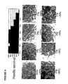
Figure 6 : needle survival rate of transfected stem cells in vitro. -
Figure 7 : human mesenchymal stem cells transiently transfected with mH2-EGFP. -
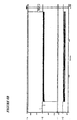
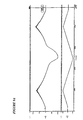
Figure 8A -B: HCN2 incorporated into stem cells can generate pacemaker current. A: ramp protocol of stem cells expressing HCN2. The ramp goes from +50 to -100 and back to 0mV. B: step protocol of stem cells expressing HCN2. The steps are from 0 to -100mV. Note that the experiment was done at room temperature, 20°C. Therefore, kinetics will be approximately 15x faster and amplitude will be 2x larger at physiologic temperature. -
Figure 9A -B: HCN2 incorporated in a stem cell failed to generate pacemaker current. A: ramp protocol of stem cell failing to express HCN2. The ramp goes from +50 to -100 and back to 0mV. B: Mutant HCN2 incorporated into a stem cell which failed to express and generate a current. The steps are from 0 to -100mV. Note that the experiment was done at room temperature, 20°C. Therefore, kinetics will be approximately 15x faster and amplitude will be 2x larger at physiologic temperature. -
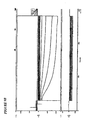
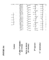
Figure 10A -E: Expression of pacemaker function in canine ventricle in situ as a result of implanting human mesenchymal stem cells having the HCN2 pacemaker gene. A dog was anesthetized and stem cells incorporating the HCN2 gene were
implanted via a 21-gauge needle into the anterior left ventricular wall of the dog. A: recordings of ECGs demonstrating sinus rhythm and a ventricular tachycardia of a specific configuration based on stimulation by the needle insertion. B: vagal stimulation of the dog resulted in cessation of sinus rhythm. C: continued vagal stimulation brought onset of a stable idioventricular rhythm after cessation of sinus rhythm. This rhythm had the same configuration on ECG lead II as the rhythm that arose on the initial day of implantation (Fig. 10A ) as a result of stimulation at the implantation site. The most likely cause of the idioventricular rhythm would be the initiation of spontaneous cardiac impulses by the pacemaker current in the stem cells. D: upon termination of vagal stimulation, the dog returned to sinus rhythm. E: dog's heart was removed and subjected to histological study. slide displaying node-like structure of mesenchymal stem cells along the needle track of the implantation site. - The present invention provides a composition according to
claim 1. - In a preferred embodiment of the preceding composition, the HCN channel is HCN1.
- In a preferred embodiment of the preceding composition, the HCN channel is HCN2.
- In a preferred embodiment of the preceding composition, the HCN channel is HCN4.
- In a preferred embodiment of the above-described composition, the HCN channel is a mutated HCN channel.
- In a preferred embodiment of the immediately preceding composition, the mutated HCN channel is E324A-HCN2.
- In a preferred embodiment of the preceding composition, the mutated HCN channel is Y331A-HCN2.
- In a preferred embodiment of the preceding composition, the mutated HCN channel is Y331A,E324A-HCN2.
- In a preferred embodiment of the above-described composition, the introduced nucleic acid also encodes MiRP1.
- The present invention further provides for a use of a composition according to the invention in the preparation of a medicament for the treatment of a cardiac rhythm disorder in a mammal wherein the cardiac rhythm disorder is selected from a group consisting of at least one of conduction block, complete atrioventricular block, incomplete atrioventricular block and sinus node dysfunction, wherein the medicament is for introduction into the mammals heart in an amount sufficient to increase pacemaker current in the heart.
- In a preferred embodiment of the above-described use the step of introduction is selected from the group consisting of systemic administration to the structure and injection.
- In a preferred embodiment of the immediately preceding use the administration introduction is selected from the group comprising topical application to the cells of the heart, microinjection and catheterization.
- The present invention further provides for a use or composition according to the invention wherein the medicament or composition is for one or more of the following: inducing a pacemaker current in the heart in; causing a contraction of a cell of the heart; shortening the time required to activate a cell of the heart; and changing the membrane potential of a cell of the heart.
- As used herein, the term "syncytial structure" means a structure with gap junction-mediated communication between its cells.
- As used herein, the term "cell of a heart" means a cell derived from a heart, either isolated or in culture.
- As used herein, the term "cardiac myocytes" means myocytes derived from muscle or conductive tissue of a heart, either isolated or in culture, and capable of initiating a current.
- As used herein, the term "membrane potential of the cell" means the transmembrane potential across the plasma membrane of the cell.
- As used herein, the term "inducing a current" means causing a cell to produce an electric current.
- As used herein, the term "time required to activate a cell" means the time period for activation of the cell.
- As used herein, the term "small molecules" means molecules of up to 1200 Daltons and/or with minor diameters of up to 1.2 nanometers.
- Methods explaining the above detailed description are set forth below.
- Initial evaluation of expressed channel function may be done via transfection in neonatal ventricular myocytes in culture. Promising constructs may be transfected into stem cells (Phase 2) which will then be tested for ability to couple to and pace cardiac myocytes in culture (Phase 3). In addition, promising constructs may be prepared as an adenovirus expressing the gene of interest and its function characterized in vitro (in cultures of neonatal and adult ventricular myocytes) and in vivo (in different cardiac regions). The in vivo studies may be done under Phase 5. The core function incorporates the preparation of adenoviral construct with a fluorescent marker and HCN, MiRP and mutant genes.
- The specific goals or aims of
Phase 1 are: - 1. Expression and characterization of native HCN isoforms (pacemaker channel alpha subunits). HCN1, HCN2 and HCN4 can be over-expressed in neonatal myocytes and characterized in terms of kinetics, voltage dependence and autonomic modulation by isoproterenol (beta-adrenergic) and carbachol (muscarinic) as well as directly by cAMP. These studies can be done by transfection or adenovirus infection.
- 2. Functional impact of native HCN isoform over-expression. The effect of over-expression on automaticity of neonatal cultures can be determined for each isoform, as well as the susceptibility of rate to autonomic modulation. These studies can be done by adenovirus infection since they require high expression efficiency. Effect of each isoform on rate can be correlated with level of infection (i.e. multiplicity of infection [m.o.i.] employed) and corresponding mean current density for this m.o.i. (as measured under goal or aim 1).
- 3. Beta subunit modulation of pacemaker channel function and rate. Experiments under goals or aims 1 and 2 can be repeated (using adenovirus infection) with a range of HCN m.o.i. with and without co-infection by the HCN beta subunit MiRP1 (KCNE2). This will determine if the same effect can be achieved with an overall lower viral load by using KCNE2 to increase efficiency of HCN expression at the cell membrane. In addition, it will be determined if KCNE2 infection alone sufficiently upregulates endogenous HCN protein to significantly increase pacemaker current and spontaneous rate. If so, autonomic modulation of rate will again be determined.
- 4. Optimization of pacemaker genes. Mutated HCN genes can be tested in the neonatal ventricular cultures to identify those with improved characteristics in terms of kinetics, voltage dependence and autonomic sensitivity, and the effect of these mutated genes on rate will then be determined. Mutations that shift threshold voltage positive and speed kinetics will likely be most suitable.
- 5. Functional impact of gene expression in adult myocytes. Those gene products from goals or aims 1-4 that appear most promising in terms of biophysical characteristics and effect on spontaneous rate can be infected in adult ventricular and/or Purkinje cells in culture and the biophysical characteristics (which are likely to differ with the cell type in which the gene is expressed) and ability to generate pacemaker activity determined.
- 6. Regulation of expression. Those gene products (from goals or aims 1-5) that appear most promising will be tested by
Phases 2 and 3 in stem cells and by Phase 5 in vivo. At the same time, they will also be transferred to new plasmids under the control of a regulatable (e.g. ecdysone- inducible) promoter. The regulatable constructs will be retested in vitro to assess the relation between expression level and induction of automaticity. Time course of regulation (i.e. time to up and down regulate channel expression) also will be determined (to be done in collaboration with Phase 2). - 7. Current characterization after in vivo expression. In collaboration with Phase 5, after adenovirus infection of gene products in dog heart and ECG characterization, animals will be sacrificed, cells isolated from infected region, and pacemaker current characteristics determined.
- 8. Stem cell myocytes interaction. In collaboration with Phase 2 examine the required density of transfected stem cells to induce a higher pacing rate in neonatal myocyte cultures.
- The goal is to optimize a pacemaker gene (or genes) and a delivery system (stem cells) to create a permanent regulatable pacemaker in a chosen cardiac region:
- The specific goals or aims of Phase 2 are:
- 1.To study the membrane properties of adult mesenchymal stem cells.
- 2.To optimize a pacemaker gene or genes for transfection.
- 3.To transfect the stem cells with the chosen pacemaker gene under a constitutively active promoter using a bicistronic expression vector, which permits a gene of interest and EGFP to be translated from a single RNA (nearly 100% of cells that exhibit fluorescence also express the gene of interest). The vectors to be used are pCMS-EGFP vector for the expression of HCN genes, pHygEGFP as a cotransfection marker vector, pEGFP-C1 vector for the expression of HCN genes as fusion proteins with EGFP and pEGFP-1 vector for monitoring of transcription of EGFP from a muscle-specific promoter.
- 4.To use antibiotic selection markers to select stably transfected clones.
- 5. To study the membrane properties, and the expressed pacemaker current of the transfected stem cells.
- 6. To study the needle survival of transfected stem cells in vitro, and to compare these results to studies of survival post-injection in dogs performed in Phase 5.
- 7. Provide transfected stem cells to Phase 3 for coupling studies.
- 8. To study the membrane properties of adult heart cells (atrial, Purkinje and ventricular) in the absence and presence of coupling to stem cells, to determine whether, coupling induces pacing.
- 9.In collaboration with
Phase 1, to examine the required density of transfected stem cells to induce a higher pacing rate in neonatal myocyte cultures. - 10.In collaboration with Phase 5, after a stem cell pacemaker is implanted in a dog heart, the animals may be sacrificed at various times post implantation. One can dissociate the cells in Phase 2 and study their membrane properties, and also use biochemical markers to investigate their level of differentiation into cardiac cell types.
- 11.To transfect stem cells using a regulatable promoter (e.g. the ecdyson system)
- 12.Select for stable transfections by antibiotic resistance.
- 13.Test the dose-response relationship between the inducer and the level of pacemaker current expressed. Also, determine the lag between exposure to the inducer and pacemaker gene expression, and determine the lag between termination of inducer exposure and the decline in pacemaker gene expression.
- 14.Provide the construct for use in
Phases 1 and 5 to perform similar studies as in goals or aims 9 and 10. - To create a biological pacemaker from stem cells or repair damaged myocardium with a stem cell derived cardiogenic cell line, the new cells are to be integrated into the cardiac syncytium. This process uses the formation of gap junctions and the ability to pass from cell to cell 1) ions to initiate and propagate action potentials and 2) relevant second messengers to sustain normal physiologic function. The cardiac gap junctions are composed of some combination of three subunit proteins: connexin43 (Cx43), and/or Cx40, and/or Cx45. The major goals of this phase are to determine the types of connexins expressed and functioning in stem cells transfected with pacemaker genes for a biological pacemaker, and stem-cell derived cardiogenic cell lines which will be used for cardiac repair. One may also determine the ability of these cell types to form gap junctions with normal adult cardiac myocytes from nodal, atrial, Purkinje, and ventricular myocardium. If necessary or desirable, one can investigate transfection of either preparation with relevant connexin genes. Because both ionic permeability (assayed by measuring gap junctional conductance) and permeability to physiologic second messengers (assayed by larger molecular weight fluorescent dye permeation) are important, both measurements will be made in our experimental protocols.
- The specific goals or aims of
Phase 3 are: - 1. Determine the extent of stem cell coupling to cells (HeLa) transfected with cardiac connexins (40, 43, and 45). (see
figure 4A-B ) - 2. Determine the ability of stem cells to couple to adult cardiac myocytes from nodal regions, atrium, Purkinje fibers and ventricular myocardium.
- 3. Use immuno-localization to determine the distribution and location of Cx43, Cx40, and Cx45 in confluent stem cell cultures and co-cultures with cardiomyocytes.
- 4. For transfected stem cells with pacemaker genes repeat goals or aims 1-3.
- 5. Following implantation of transfected stem cells into myocardium at fixed time periods after the initiation of a biological pacemaker the animals will be sacrificed. In these animals, one can determine 1) which connexins are expressed in the mesenchymal stem cells and 2) the functional coupling in isolated cell pairs of stem cell to stem cell, and stem cell to cardiac myocyte.
- 6. Determine the extent of coupling between the cardiogenic cell line(s) to cultured cells (HeLa) expressing with cardiac connexins (40,43,45).
- 7. Determine the ability of the cardiogenic cell line(s) to couple to adult cardiac myocytes from the same regions as used in goal or aim 2.
- 8. Following implantation of cells from the cardiogenic cell line(s) in dogs for cardiac repair, at fixed time periods the animals will be sacrificed. One can determine from these animals 1) the expression of connexins in the repair cell line and 2) the functional coupling of cardiogenic cell line pairs, or pairs between adult cardiac cells and cells from the cardiogenic cell line.
- 9. Express connexins in stem cells, stem cells transfected with pacemaker genes, or stem cell derived cardiogenic cell line(s) as necessary or desirable. One can determine if improved functional coupling results.
- The goals of Phase 4 are (1) to grow stem cells for transformation into a cardiac cell line, (2) to select the cardiac cell lineage(s), (3) to further induce the cardiac-like cells to differentiate into ventricular or nodal cell types, and (4) to transfect each of the individual cell types with appropriate genes to optimize function and survival in particular cardiac regions,
- The specific goals or aims of Phase 4 are:
- 1. Transfect stem cells with a muscle or cardiac muscle specific promoter and green fluorescent protein to aid in selection of cells which grow along a cardiac lineage.
- 2. Select for stable transfections.
- 3. Test available approaches to induce cardiac differentiation (including embryoid bodies, 5-azacytidine exposure).
- 4. Select cells which have differentiated into a cardiogenic cell line.
- 5. Patch clamp the cells looking for a cardiac-like action potential and cardiac I-V relationship, and cardiac specific membrane currents.
- 6. Study the biochemical markers of cardiac differentiation.
- 7. Investigate whether trophic factors or co-culturing can induce further differentiation specifically to nodal type, atrial muscle or ventricular endocardium or epicardium. Again, test by patch clamping to look for signature membrane action potential or membrane currents.
- 8. Provide cells to Phase 3 for tests of coupling to adult myocytes from the desired cardiac region.
- 9. Test needle survival of each cell type.
- 10. Provide cells to Phase 5 for tests of efficacy in a dog cardiac model of heart failure. One can get cells back after experimental time period in vivo to study their properties.
- 11. Add new genes to optimize cell survival including an increased inward rectifier or gene to induce additional angiogenesis (like VEGF or other growth factors).
- The overall goals of Phase 5 are: (1) to determine the extent to which specific pacemaker constructs expressed in vivo via the approaches of gene therapy and of stem cell implantation can affect cardiac rate and rhythm. (2) to determine the extent to which engineered cell lines can effect myocardial repair and AV nodal replacement.
- When the donor cell is provided with delivery gene or compound, it will be necessary to deliver the engineered donor cells to the biological target. If direct injection of the target is possible, this method will be applied. Alternatively, if the target is fed by a specific artery, then the engineered donor cells can be delivered to said artery and released into the blood for delivery to the biological target.
- The general hypotheses are (1) that pacemaker channels expressed or implanted in specific regions of the heart will develop regular, autonomic-responsive rhythms that counter the "clamping" effect of IK1 in specialized conducting fibers and atrium and possibly ventricle, and (2) that engineered cell lines can replace non-functional myocardium or the AV node.
- There are three goals or aims:
- 1. To investigate the function of specific HCN α and β subunit constructs as functioning pacemakers in the heart in situ and in isolated tissues, including testing of the following sub-hypotheses:
- a: Injection of adenoviral constructs carrying HCN2 or HCN4 into canine ventricular myocardium in vivo can elicit pacemaker current having characteristics of If. Although there is proof in concept that this will work, this intervention may not drive the ventricle because of the large IK1 and the highly negative membrane potentials at which expressed HCN2 is likely to activate.
- b: Injection of adenoviral constructs carrying HCN2 or HCN4 into canine bundle branches or atrium in vivo will elicit autonomic-responsive pacemaker current having characteristics of If, and - in light of the lesser IK1 present and more positive activation of expressed HCN - capable of driving the heart.
- c: Injection of an adenoviral construct carrying MiRP1 into the above tissues in the absence of additional HCN isoforms can significantly upregulate endogenous If and possibly speed activation kinetics as well, thus offering an alternative means for altering pacemaker function.
- d: Injection of an adenoviral construct of mutant genes (developed in
Phases 1 and 2) will provide alternative and perhaps superior functional pacemakers.
- 2. To investigate the function of specific HCN α and β subunit constructs inserted into human mesenchymal cell lines to provide functional pacemakers to the heart in situ and in isolated tissues. The subhypotheses are modified from those above, as follows:
- a: Injection of stem cells carrying HCN2 or HCN4 into canine ventricular myocardium in vivo can elicit pacemaker current having characteristics of If and capable of driving the heart. Stem cell implantation may make it possible to implant a node of sufficient dimension to overcome the effects of IK1.
- b: Injection of stem cells carrying HCN2 or HCN4 into canine bundle branches or atrium in vivo will elicit autonomic-responsive pacemaker current having characteristics of If, and - in light of the lesser IK1 present and more positive activation of expressed HCN - capable of driving the heart.
- c: Injection of stem cells carrying MiRP1 in the absence of additional HCN isoforms can significantly upregulate endogenous myocardial If and possibly speed activation kinetics as well, thus offering an alternative means for altering pacemaker function.
- d: Injection of stem cells carrying constructs of mutant genes (developed in
Phases 1 and 2) will provide alternative and perhaps superior functional pacemakers.
- 3. To investigate the utility of implantation of engineered cardiac cell lines in effecting myocardial repair. This includes replacement of myocardium and of AV node. The subhypotheses are:
- a: In canine hearts in which myocardial infarction has induced ventricular aneurysm alone and in hearts with congestive failure, cell lines engineered by Phase 4 will grow in the myocardium providing a substrate that is functional as studied hemodynamically and via imaging, while not generating arrhythmias. The result will be improved cardiac function and output.
- b: In canine hearts in which AV block has been induced via formalin injection or RF ablation, cell lines engineered by Phase 4 will provide bypass tracts that have the same function as the AV node in the heart in situ and in isolated tissues.
- One can isolate and grow mesenchymal stem cells (human or canine) from human/canine bone marrow aspirates.
- For example, 10ml of marrow aspirate was collected into a syringe containing 6000 units of heparin to prevent clotting, washed twice in phosphate buffer solution (PBS), added to 20ml of control medium (DMEM containing 10% FBS), and then centrifuged to pellet the cells and remove the fat. The cell pellet was resuspended in control medium and fractionated at 1100g for 30 min on a density gradient generated by centrifugation of a 70% percoll solution at 13000g for 20 minutes. The mesenchymal stem cell-enriched, low density fraction was collected, rinsed with control medium and plated at a density of 107 nucleated cells per 60mm2 dish. The mesenchymal stem cells were then cultured in control medium at 37°C in a humidified atmosphere containing 5%CO2.
- One can isolate hMSC and cMSC from donors. The advantages to using mesenchymal stem cells are that they do not require an endoderm for differentiation, are easy to culture, do not require an expensive cytokine supplement and have minimal immunogenecity. One can also test cell purity by flow cytometry and the ability of hMSC/cMSC to differentiate into osteogenic, chondrogenic, adipogenic and cardiogenic lineages.
- For example, hMSC were transplanted into fetal sheep early in gestation, before and after the expected development of immunologic competence. The hMSC engrafted and persisted in multiple tissues for as long as 13 months after transplantation. Transplanted human cells underwent site-specific differentiation into chondrocytes, adipocytes; myocytes and cardiomyocytes, bone marrow stromal cells and thymic stroma. Unexpectedly, there was long-term engraftment even when cells were transplanted after the expected development of immunocompetence. A possible reason may be because hMSC express class I human leukocyte antigen but do not express class II, which may limit immune recognition.
- In cardiac muscle, (human) β-2 microglobulin staining or in situ hybridization for human ALU sequences were combined with double staining with antibody against smooth endoplasmic reticulum ATPase-2 (SERCA-2), a cytoplasmic protein specific for smooth or skeletal muscle.
- Cardiomyocytes have also been generated from murine marrow stromal cells. Murine bone marrow stromal cells were treated with 3µM 5-azacytidine. After 1 week, some cells gradually increased in size to form a ball-like or stick-like appearance. After 2 weeks, the cells began spontaneously beating and the ball-like or stick-like cells connected with adjoining cells to form myotube-like structures. After 3 weeks, most of the synchronously beating cells connected and formed myotube-like structures and a cardiomyogencic cell line was formed.
- Additionally, hMSC can be induced to differentiate in vitro exclusively to an osteogenic lineage with dexamethoasone, β-glycerol phosphate, ascorbate and 10% FBS. A chondrogenic lineage can be induced exclusively without serum, but with transforming growth factor β3 (in a pelleted micromass) and hMSC. Finally, an adipogenic lineage can be induced exclusively with 1-methyl 1-3-isobutylxanthine, dexamethasone, insulin, iodomethacin and hMSC.
- Normal and dystrophic (mdx) female mice received bone marrow transplantation (BMT) from normal male donor. After 70 days, histological sections of atrial and ventricular regions from BMT mice were probed for donor-derived Y chromosomes. In BMT-mdx mice single cardiomyocytes were found to contain bone-marrow derived Y chromosomes.
- One can prepare media for hMSC/cMSC and other cell types to be utilized in the project along with cell storage, growth and maintenance of cells in culture.
- One can utilize light, fluorescent, and confocal microscopy to monitor cell types and cardiogenic cell lines. The core may also support all histochemical and immuno-localization studies.
- One can monitor the expression levels of all genes transfected into stem cells or cardiogenic cell lines.
- Transplantation therapy is also possible through the use of hMSC. After differentiation of the hMSC into cardiac myocytes, pure cultures are selected by cell survival or cell sorting (e.g. muscle or cardiac specific promoter driving antibiotic resistance gene or GFP). Then, one tests in vitro for electrical coupling of the differentiated myocytes to adult myocytes and also for biochemical, immunohistological and electrophysiological properties. After completion of these tests, one utilizes a canine model to evaluate integration of the differentiated myocytes into host tissue and improved contractile performance. The canine model is also examined for the absence of tumor formation or transmission of infectious agents. Then, methods of preventing rejection are tested. Note, that if the recipient and donor are the same, there is no danger of rejection. Finally, human trials are commenced.
- The delivery of specific solutes to the intracellular compartment of functional syncytia can be achieved by seeding target tissues (cells) with stem cells that have been preloaded with a specified solute. Alternatively, a gene producing a small solute can be introduced into the stem cells as previously described. The transfer of solute from stem cells to target cells is via diffusion through gap junctions. The system is capable of delivering hydrophilic second messengers, drugs and their metabolites, and inorganic ions.
- A) Loading of stems cells:
- The loading of specific solutes into stem cells can be accomplished by electroporation or by perfusion of stem cells with media containing membrane permeable ester forms.
Figure 1a is a light micrograph of stem cells whilefigure 1b is of the same cells with fluorescence microscopy showing the presence of Lucifer Yellow (LY). The dye was loaded via electroporation.
- The loading of specific solutes into stem cells can be accomplished by electroporation or by perfusion of stem cells with media containing membrane permeable ester forms.
- B) Transfer of loaded solute from stem cells to target cells: A demonstration using cells in culture.
Figure 2 shows transfer of dye from a stem cell to a HeLa cell. The LY has been delivered to the HeLa cell, presumably by diffusion through gap junctions. - Relevant parameters for the transfer:
- 1) Stem cells form gap junction channels with other cells b containing one or more of the following connexins: Cx43, Cx45, Cx40, Cx32 and Cx26.
- 2) Negatively charged solutes with minor diameters of ∼1.0 nm are all able to transit the aforementioned gap junction channels (homotypic Cx43, Cx40, Cx45, heterotypic Cx43-Cx40 and mixed or heteromeric Cx43-Cx40)(63,64) and Cx32 and Cx26 (65).
- The selectivity ratio for Lucifer Yellow relative to K+ is 0.025 for Cx43 and 0.0028 for Cx40. The type of gap junctions and total number of channels determine the rate of transit of a specific solute between stem cell and target cell.
- The scope of the invention is determined by way of the appended claims.
-
- 1. DiFrancesco D: The cardiac hyperpolarizing-activated current, If: Origins and developments. Prog. BiophysMol. Biol. Vol. 46, No. 3, 1985, pages 163-183.
- 2. Zhou Z and Lipsius SL: Effect of isoprenaline on If current in latent pacemaker cells isolated from cat right atrium: ruptured vs. perforated patch whole-cell recording methods. Pflugers Arch. Vol. 423, No.5 Pt. 6, June 1993, pages 442-447.
- 3. Thuringer D, et al.: A hyperpolarization-activated inward current in human myocardial cells. JmolCell. Cardiol. Vol. 24, No. 5, May 1992, pages 451-455.
- 4. Porciatti F, et al.: Block of the pacemaker current If in single human atrial myocytes and the effect of beta-adrenoceptor and Al-adenosine receptor stimulation. Br Jpharmacol. Vol. 122, No. 6, February 1991, pages 963-969.
- 5. Yu H, et al.: Pacemaker current exists in ventricular myocytes. Circ. Res. Vol. 72, No. 1, January 1993, pages 232-236.
- 6. Cerbai E, et al.: The properties of the pacemaker current If in Human Ventricular Myocytes are modulated by Cardiac Disease. Jmol. Cell Cardiol. Vol. 33, No. 3, March 2001, pages 441-448.
- 7. DiFrancesco D: Generation and control of cardiac pacing: the pacemaker current. Tends Cardiovasc. Med. Vol. 1, 1991, pages 250-255.
- 8. Robinson RB, et al.: Developmental change in the voltage dependence of the pacemaker current, If, in rat ventricle cells. Pflugers Arch. Vol. 433, 1991, pages 533-535.
- 9. Fares N, et al.: Characterization of a hyperpolarization-activated current in dedifferentiated adult rat ventricular cells in primary culture. J. Physiol. Vol. 506, No. 1, January 1, 1998, pages 73-82.
- 10. Cerbai E, et al.: Influence of postnatal-development on If occurrence and properties in neonatal rat ventricular myocytes. Cardiovasc. Res. Vol. 42, No. 2, May 1999, pages 416-423.
- 11. Cerbai E, et al.: Characterization of the hyperpolarization-activated current, If, in ventricular myocytes isolated from hypertensive rats. J. Physiol. Vol. 481, No. 3, .
- 12. Cerbai E, et al.: Characterization of the hyperpolarization-activated current, If, in ventricular myocytes from human failing heart. Circulation. Vol. 95, No. 3, February 4, 1997, pages 568-571.
- 13. Santoro B, et al.: Interactive cloning with the SH3 domain of N-src identifies a new brain specific ion channel protein, with homology to Eag and cyclic nucleotide-gated channels. Proc. Natl. Sci. USA. Vol. 94, No. 26, December 23, 1997, pages 14815-14820.
- 14. Ludwig A, et al.: A family of hyperpolarization-activated mammalian cation channels. Nature. Vol. 393, No. 6685, June 11, 1998, pages 587-591.
- 15. Santoro B, et al.: Identification of a gene encoding a hyperpolarization-activated pacemaker channel of brain. Cell. Vol. 93, No. 5, May 29, 1998, pages 717-729.
- 16. Shi W, et al.: Distribution and Prevalence of hyperpolarization-activated cation channel (HCN) mRNA Expression in Cardiac Tissues. Circ. Res. Vol. 85, No. 1, July 9, 1999, pages e1-e6.
- 17. Ishii TM, et al.: Molecular characterization of the hyperpolarization-activated cation channel in rabbit heart sinoatrial node. J. Biol. Chem. Vol. 264, No. 18, April 30, 1999, pages 12835-12839.
- 18. Ludwig A, et al.: Two pacemaker channels from human heart with profoundly different activation kinetics. EMBO J. Vol. 18, No. 9, May 4, 1999, pages 2323-2329.
- 19. Moosmang S, et al.: Cellular expression and functional characterization of four hyperpolarization-activated pacemaker channels in cardiac and neuronal tissues. Eur. J. Biochem. Vol. 268, No. 6, March 2001, pages 1646-1652.
- 20. Altomare C, et al.: Allosteric voltage-dependent gating of HCN channels. Biophys. J. Vol. 80, 2001, page 241a.
- 21. Protas L, et al.: Chronic neuropeptide Y exposure increases L-type Ca current in neonatal rat cardiomyocytes. Am. J. Physiol. Vol. 277, No. 3 Pt. 2, September 1999, pages H940-H946.
- 22. Kuznetsov V, et all.: β 2-adrenergic receptor actions in neonatal and adult rat ventricular myocytes. Circ. Res. Vol. 76, No. 1, January 1995, pages 40-52.
- 23. Ellingston O, et al.: Adult rat ventricular myocytes cultured in defined medium: phenotype and electromechanical function. Am. J. Physiol. Vol. 265, No. 2 Pt. 2, August 1993, pages H747-H754.
- 24. Ng P, et al.: An enhanced system for construction of adenoviral vectors by the two-plasmid rescue method. Hwn.Gene Ther. Vol. 11, No. 5, March 20, 2000, pages 693-699.
- 25. He TC, et al.: A simplified system for generating recombinant adenoviruses. Proc. Natl. Acad. Sci. USA. Vol. 95, No. 5, March 3, 1998, pages 2509-2514.
- 26. Santoro B, et al.: The HCN gene family: molecular basis of the hyperpolarization-activated pacemaker channels. Ann. NY Acad. Sci. Vol.868, April 30, 1999, pages 741-764.
- 27. Accili EA, et al.: Properties and modulation of If in newborn versus adult cardiac SA node. Am. J. Physiol. Vol. 272, 1991, pages H1549-H1552.
- 28. Qu J, et al.: Sympathetic innervation alters activation of pacemaker current (If) in rat ventricles. J. Physiol. Vol. 526, No. 3, August 1, 2000, pages 561-569.
- 29. Cui J, et al.: Gating of IsK expressed in Xenopus oocytes depends on the amount of mRNA injected. Gen. Physiol. Vol. 104, No. 1, July 1994, pages 87-105.
- 30. Guillemare E, et al.: Effects of the level of mRNA expression on biophysical properties, sensitivity to neurotoxins, and regulation of the brain delayed-rectifier K+ channels Kv1.2. Biochemistry. Vol. 31, No. 49, December 15, 1992, pages 12463-12468.
- 31. Honore E, et al.: Different types of K+ channel current are generated by different levels of a single mRNA. EMBO J. Vol. 11, No. 7, July 1992, pages 2465-2471.
- 32. Moran O, et al.: Level of expression controls modes of gating of a K+ channel. FEBS Lett. Vol. 302, No. 1, May 4, 1992, pages 21-25.
- 33. DiFrancesco D, et al.: Direct activation of cardiac pacemaker channels by intracellular cyclic AMP. Nature. Vol. 351, No. 6322, May 9, 1991, pages 145-147.
- 34. Kaupp UB, et al.: Molecular diversity of pacemaker ion channels. Annu. Rev. Physiol. Vol. 63, 2001, pages 235-257.
- 35. Chang F, et al.: Effects of protein kinase inhibitors on canine Purkinje fibre pacemaker depolarization and the pacemaker current If. J. Physiol. Vol. 440, 1991, pages 367-384.
- 36. Yu H, et al.: Phosphatase inhibition by calyculin A increases If in canine Purkinje fibers and myocytes. Pflugers Arch. Vol. 422, No. 6, March 1993, pages 614-616.
- 37. Accili EA, et al.: Differential control of the hyperpolarization-activated current (If) by intracellular cAMP and phosphatase inhibition. J. Physiol. Vol. 491, 1996, pages 115.
- 38. Ranjan R, et al.: Mechanism of anode break stimulation in the heart. Biophys. J. Vol. 74, No. 4, April 1998, pages 1850-1863.
- 39. Moroni A, et al.: Kinetic and ionic properties of the human HCN2 pacemaker channel. Pflugers Arch. Vol. 439, No. 5, March 2000, pages 618-626.
- 40. Santoro B, et al.: Molecular and functional heterogeneity of hyperpolarization-activated pacemaker channels in the mouse CNS. J Neurosci. Vol. 20, No. 14, July 15, 2000, pages 5264-5275.
- 41. Shi W, et al.: The distribution and prevalence of HCN isoform in the canine heart and their relation to the voltage dependence of If. Biophys. J. Vol. 78, 2000, page 353A.
- 42. Melman YF, et al.: Structural determinants of KvLQT1 control by the KCNE family of proteins. J Biol Chem. Vol. 276, No. 9, March 2, 2001, pages 6439-6444.
- 43. Tinel N, et al.: KCNE2 confers background current characteristics to the cardiac KCNQ1 potassium channel. EMBO J. Vol. 19, No. 23, December 1, 2000, pages 6326-6330.
- 44. Martens JR, et al.: Differential targeting of Shaker-like potassium channels to lipid rafts. Biol Chem. Vol. 275, No. 11, March 17, 2000, pages 7443-7446.
- 45. Chauhan VS, et al.: Abnormal cardiac Na(+) channel properties and QT heart rate adaptation in neonatal ankyrin(B) knockout mice. Circ. Res. Vol. 86, No. 4, March 3, 2000, pages 441-447.
- 46. Walsh KB, et al.: Distinct voltage-dependent regulation of heart-delayed IK by protein kinases A and C. Am. J. Physiol. Vol. 261, No. 6 Pt. 1, December 1991, pages C1081-C1090.
- 47. Gerhardstein BL, et al.: Proteolytic processing of the C terminus of the alpha (1C) subunit of L-type calcium channels and role of a proline-rich domain in membrane tethering of proteolytic fragments. J Biol. Chem. Vol. 275, No. 12, March 24, 2000, pages 8556-8563.
- 48. Barbuti A, et al.: Action of internal pronase on the f-channel kinetics in the rabbit SA node. J. Physiol. Vol. 520, No. 3, November 1, 1999, pages 737-744.
- 49. Wainger BJ, et al.: Domains involved in cyclic nucleotide modulation of hyperpolarization-activated HCN channels. Nature. In Press, 2001.
- 50. Wahler GM: Developmental increases in the inwardly rectifying potassium current of rat ventricular myocytes. Am. J. Physiol. Vol. 262, No. 5 Pt. 1, May 1992, pages C1266.
- 51. Abbott GW, et al.: MiRP1 forms Ikr potassium channels with HERG and is associated with cardiac arrhythmia. Cell. Vol. 97, No. 2, April 16, 1999, pages 175-187.
- 52. Sanguinetti MC, et al.: Coassembly of KvLGQT1 and minK (ISK) proteins to form cardiac ISK potassium channels. Nature. Vol. 384, No. 6604, November 7, 1996, pages 80-83.
- 53. Dixon JE and McKinnon D: Quantitative analysis of potassium channel expression in atrial and ventricular muscle of rats. Circ. Res. Vol. 75, No. 2, August 1994, pages 252-260.
- 54. Sclagger H and von Jagow G: Tricine-sodium dodecyl sulfate-polyacrylamide gel electrophoresis for separation of proteins in the range from 1 to 100 Kda. Analytical Biochem. Vol. 166, No. 2, November 1, 1987, pages 368-379.
- 55. Hansen JE, et al.: Prediction of O-glycosylation of mammalian proteins: Specificity patterns of UDP-GalNAc:polypeptide N-acetylgalactosaminyltransferase. Biochem. J. Vol. 308, No. 3, June 15, 1995, pages 801-813.
- 56. Vassalle M, et al.: Pacemaker channels and cardiac automaticity In "Cardiac Electrophysiology. From Cell to Bedside", Eds. Zipes D and Jalife WB Saunders Co. Philadelphia, PA, 2000, pages 94-103.
- 57. Rosen, M.R. and Robinson R.B.: Heart rate: a simple yet complex concept. Dialogues in Cardiovascular Medicine. Vol. 6, No. 1, 2001, pages 2 - 19.
- 58. Yu, H. et al.: MinK-Related Peptide 1: A β Subunit for the HCN Ion Channel Subunit Family Enhances Expression and Speeds Activation. Circ. Res. Vol. 88, 2001, pages 84-87.
- 59. Qu, J. et al.: Functional comparison of HCN isoforms expressed in ventricular and HEK 293 cells. Pfulgers Arch. - Eur. J. Physiol. Vol. 444, 2002, pages 597-601.
- 60. Qu, J. et al.: Sympathetic innervation alters activation of pacemaker current (If) in rat ventricle. J. of Physiol. Vol.526, No.3, 2000, pages 561-569.
- 61. Hamm, A. et al.: Efficient Transfection Method for Primary Cells. Tissue Engineering. Vol.8, No.2, 2002, pages 235-245.
- 62. Qu, J. et al.: HCN2 Overexpression in Newborn and Adult Ventricular Myocytes. Circ. Res. Vol.89, 2001, pages 8-14.
- 63. Valiunas, V. et al.: Cardiac gap junction channels show quantitative differences in selectivity. Circ. Res. Vol.91, No.2, 2002, pages 104-111.
- 64. Valuinas, V.: Biophysical properties of connexin-45 gap junction hemichannels studied in vertebrate cells. J. Gen. Physiol. Vol. 119, No.2, 2002, pages 147-164.
- 65. Cao, F. et al.: A quantitative analysis of connexin-specific permeability differences of gap junctions expressed in HeLa transfectants and Xenopus oocytes. J. Cell Sci. Vol. 111 (pt.1), 1998, pages 31-43.
Claims (23)
- A composition comprising a mesenchymal stem cell into which a nucleic acid which encodes a HCN channel has been introduced in an amount sufficient to create an ion channel in the mesenchymal stem cell.
- The composition according to claim 1 for delivering to a mammalian heart cell a cell expressing at least one pacemaker ion channel, wherein the at least one pacemaker ion channel is a HCN channel.
- The composition of claim 1 or 2, wherein the introduced nucleic acid also encodes a MiRP1 channel.
- The composition of any preceding claim, wherein the HCN channel is HCN1.
- The composition of any of claims 1 to 3, wherein the HCN channel is HCN2.
- The composition of any of claims 1 to 3, wherein the HCN channel is HCN4.
- The composition of any of claims 1 to 3, wherein the HCN channel is a mutated HCN channel.
- The composition of claim 7, wherein the mutated HCN channel is E324A-HCN2.
- The composition of claim 7, wherein the mutated HCN channel is Y331A-HCN2.
- The composition of claim 7, wherein the mutated HCN channel is Y331A, E324A-HCN2.
- A composition according to any preceding claim for use as a medicament.
- Use of a composition according to any preceding claim in the preparation of a medicament for use in treatment of a cardiac disorder.
- A composition according to any preceding claim for treating a cardiac disorder.
- The use of claim 12 or the composition of claim 13 wherein the medicament or composition is for introduction into a mammalian heart.
- The use or composition of claim 14 wherein upon administration to a mammalian heart the mesenchymal stem cells forms one or more gap junctions with one or more cells of the mammalian heart.
- The use or composition of claim 14 or 15 wherein the medicament or composition is for introduction into the heart in an amount sufficient to induce or increase a pacemaker current in the heart.
- The use or composition of any of claims 12 to 16 wherein the medicament or composition is for increasing the heart rate in a subject.
- The use or composition of claim 17 wherein the medicament or composition is for administration by contact with a cell of the heart of a subject in an amount sufficient to decrease the time constant of activation of the cell of the heart, thereby increasing heart rate in the subject.
- The use or composition of any of claims 12 to 15 wherein the medicament or composition is for one or more of the following: inducing a pacemaker current in a cell of the heart; causing a contraction of a cell of the heart; shortening the time required to activate a cell of the heart; and changing the membrane potential of a cell of the heart.
- Use of a composition according to any of claim 1 to 10 in the preparation of a medicament for the treatment of a cardiac rhythm disorder in a mammal, wherein the disorder is at least one of conduction block, complete atrioventricular block, incomplete atrioventricular block or sinus node dysfunction, wherein the medicament is for introduction into the mammal's heart in an amount sufficient to increase pacemaker current in the heart.
- A composition according to any of claim 1 to 10 for the treatment of a cardiac rhythm disorder in a mammal, wherein the disorder is at least one of conduction block, complete atrioventricular block, incomplete atrioventricular block or sinus node dysfunction, wherein the composition is for introduction into the mammal's heart in an amount sufficient to increase pacemaker current in the heart.
- The use of claim 20 or the composition of claim 21 wherein the medicament is for systemic administration or injection.
- The use of claim 20 or the composition of claim 21 wherein the medicament or composition is for introduction to the cells of the heart by topical application to the cells of the heart, injection, or catheterization.
Applications Claiming Priority (3)
| Application Number | Priority Date | Filing Date | Title |
|---|---|---|---|
| US342506 | 1994-11-21 | ||
| US10/342,506 US7794702B2 (en) | 2003-01-15 | 2003-01-15 | Mesenchymal stem cells as a vehicle for ion channel transfer in syncytial structures |
| PCT/US2004/000950 WO2004065580A2 (en) | 2003-01-15 | 2004-01-14 | Mesecnhymal stem cells as a vehicle for ion channel transfer in syncytial structures |
Publications (3)
| Publication Number | Publication Date |
|---|---|
| EP1592800A2 EP1592800A2 (en) | 2005-11-09 |
| EP1592800A4 EP1592800A4 (en) | 2006-03-29 |
| EP1592800B1 true EP1592800B1 (en) | 2009-08-26 |
Family
ID=32711727
Family Applications (1)
| Application Number | Title | Priority Date | Filing Date |
|---|---|---|---|
| EP04702196A Expired - Lifetime EP1592800B1 (en) | 2003-01-15 | 2004-01-14 | Mesecnhymal stem cells as a vehicle for ion channel transfer in syncytial structures |
Country Status (7)
| Country | Link |
|---|---|
| US (1) | US7794702B2 (en) |
| EP (1) | EP1592800B1 (en) |
| JP (1) | JP2006515759A (en) |
| AT (1) | ATE440961T1 (en) |
| DE (1) | DE602004022778D1 (en) |
| ES (1) | ES2329681T3 (en) |
| WO (1) | WO2004065580A2 (en) |
Families Citing this family (27)
| Publication number | Priority date | Publication date | Assignee | Title |
|---|---|---|---|---|
| EP0928135B1 (en) * | 1996-07-17 | 2006-06-14 | Medtronic, Inc. | System for enhancing cardiac signal sensing by cardiac pacemakers through genetic treatment |
| ITRM20030376A1 (en) * | 2003-07-31 | 2005-02-01 | Univ Roma | PROCEDURE FOR THE ISOLATION AND EXPANSION OF CARDIOC STAMIN CELLS FROM BIOPSIA. |
| EP1699523A4 (en) * | 2003-12-24 | 2008-05-21 | Univ Columbia | Creation of a biological atrioventricular bypass to compensate for atrioventricular block |
| JP2008506416A (en) * | 2004-07-19 | 2008-03-06 | ザ・トラスティーズ・オブ・コロンビア・ユニバーシティ・イン・ザ・シティ・オブ・ニューヨーク | Analytical system for monitoring the effect of genetically modified cells on the functioning of syncytia |
| US11660317B2 (en) | 2004-11-08 | 2023-05-30 | The Johns Hopkins University | Compositions comprising cardiosphere-derived cells for use in cell therapy |
| US8236296B2 (en) * | 2005-07-21 | 2012-08-07 | The Trustees Of Columbia University In The City Of New York | Biological bypass bridge with sodium channels, calcium channels and/or potassium channels to compensate for conduction block in the heart |
| WO2008011133A2 (en) * | 2006-07-21 | 2008-01-24 | The Trustees Of Columbia University In The City Of New York | Compositions of late passage mesenchymal stem cells (mscs) |
| KR101240487B1 (en) * | 2006-11-09 | 2013-03-08 | 더 존스 홉킨스 유니버시티 | Dedifferentiation of adult mammalian cardiomyocytes into cardiac stem cells |
| US8076305B2 (en) | 2006-11-30 | 2011-12-13 | Medtronic, Inc. | Biological pacemakers including mutated hyperpolarization-activated cyclic nucleotide-gated (HCN) channels |
| US20100150887A1 (en) * | 2007-01-25 | 2010-06-17 | Duff Henry J | Tissue Progenitor Cells That Overexpress ERG |
| US8224443B2 (en) * | 2007-05-08 | 2012-07-17 | Cardiac Pacemakers, Inc. | Method for controlling pacemaker therapy |
| US20080280341A1 (en) * | 2007-05-08 | 2008-11-13 | Kenknight Bruce | System And Method For Local Field Stimulation |
| US9150832B2 (en) * | 2007-05-08 | 2015-10-06 | Cardiac Pacemakers, Inc. | Cell training for local field stimulation |
| US8442631B2 (en) * | 2007-05-08 | 2013-05-14 | Cardiac Pacemakers, Inc. | System and method for determining the origin of a sensed beat |
| US10001470B2 (en) | 2009-12-04 | 2018-06-19 | Sanford-Burnham Medical Research Institute | Method, system and composition for optically inducing cardiomyocyte contraction |
| US9845457B2 (en) | 2010-04-30 | 2017-12-19 | Cedars-Sinai Medical Center | Maintenance of genomic stability in cultured stem cells |
| US9249392B2 (en) | 2010-04-30 | 2016-02-02 | Cedars-Sinai Medical Center | Methods and compositions for maintaining genomic stability in cultured stem cells |
| EP2861238A4 (en) | 2012-06-05 | 2016-03-16 | Capricor Inc | Optimized methods for generation of cardiac stem cells from cardiac tissue and their use in cardiac therapy |
| EP3563859B1 (en) | 2012-08-13 | 2021-10-13 | Cedars-Sinai Medical Center | Cardiosphere-derived exosomes for tissue regeneration |
| EP3200808A4 (en) | 2014-10-03 | 2018-05-16 | Cedars-Sinai Medical Center | Cardiosphere-derived cells and exosomes secreted by such cells in the treatment of muscular dystrophy |
| US9913865B2 (en) * | 2015-12-28 | 2018-03-13 | Ingeneron Inc. | Induced pacemaker and Purkinje cells from adult stem cells |
| WO2017123662A1 (en) | 2016-01-11 | 2017-07-20 | Cedars-Sinai Medical Center | Cardiosphere-derived cells and exosomes secreted by such cells in the treatment of heart failure with preserved ejection fraction |
| US11351200B2 (en) | 2016-06-03 | 2022-06-07 | Cedars-Sinai Medical Center | CDC-derived exosomes for treatment of ventricular tachyarrythmias |
| US11541078B2 (en) | 2016-09-20 | 2023-01-03 | Cedars-Sinai Medical Center | Cardiosphere-derived cells and their extracellular vesicles to retard or reverse aging and age-related disorders |
| US11759482B2 (en) | 2017-04-19 | 2023-09-19 | Cedars-Sinai Medical Center | Methods and compositions for treating skeletal muscular dystrophy |
| WO2018196782A1 (en) * | 2017-04-27 | 2018-11-01 | The University Of Hong Kong | Use of hcn inhibitors for treatment of cancer |
| US11660355B2 (en) | 2017-12-20 | 2023-05-30 | Cedars-Sinai Medical Center | Engineered extracellular vesicles for enhanced tissue delivery |
Family Cites Families (7)
| Publication number | Priority date | Publication date | Assignee | Title |
|---|---|---|---|---|
| US5591625A (en) * | 1993-11-24 | 1997-01-07 | Case Western Reserve University | Transduced mesenchymal stem cells |
| US6387369B1 (en) * | 1997-07-14 | 2002-05-14 | Osiris Therapeutics, Inc. | Cardiac muscle regeneration using mesenchymal stem cells |
| WO2000041731A1 (en) * | 1999-01-19 | 2000-07-20 | The Children's Hospital Of Philadelphia | Reverse gene therapy |
| DE10006309A1 (en) * | 2000-02-12 | 2001-08-23 | Aventis Pharma Gmbh | Method for the identification of substances that modulate the activity of hyperpolarization-activated cation channels |
| WO2002087419A2 (en) * | 2001-04-27 | 2002-11-07 | Johns Hopkins University | Biological pacemaker |
| US6849611B2 (en) | 2001-06-06 | 2005-02-01 | The Trustees Of Columbia University In The City Of New York | Implantation of biological pacemaker that is molecularly determined |
| WO2003039344A2 (en) * | 2001-11-08 | 2003-05-15 | The Regents Of The University Of California | Methods and compositions for correction of cardiac conduction disturbances |
-
2003
- 2003-01-15 US US10/342,506 patent/US7794702B2/en not_active Expired - Fee Related
-
2004
- 2004-01-14 JP JP2006500957A patent/JP2006515759A/en active Pending
- 2004-01-14 AT AT04702196T patent/ATE440961T1/en not_active IP Right Cessation
- 2004-01-14 WO PCT/US2004/000950 patent/WO2004065580A2/en active Application Filing
- 2004-01-14 ES ES04702196T patent/ES2329681T3/en not_active Expired - Lifetime
- 2004-01-14 EP EP04702196A patent/EP1592800B1/en not_active Expired - Lifetime
- 2004-01-14 DE DE602004022778T patent/DE602004022778D1/en not_active Expired - Lifetime
Also Published As
| Publication number | Publication date |
|---|---|
| EP1592800A4 (en) | 2006-03-29 |
| US7794702B2 (en) | 2010-09-14 |
| US20040137621A1 (en) | 2004-07-15 |
| WO2004065580A3 (en) | 2005-09-15 |
| ES2329681T3 (en) | 2009-11-30 |
| DE602004022778D1 (en) | 2009-10-08 |
| EP1592800A2 (en) | 2005-11-09 |
| WO2004065580A2 (en) | 2004-08-05 |
| ATE440961T1 (en) | 2009-09-15 |
| JP2006515759A (en) | 2006-06-08 |
Similar Documents
| Publication | Publication Date | Title |
|---|---|---|
| EP1592800B1 (en) | Mesecnhymal stem cells as a vehicle for ion channel transfer in syncytial structures | |
| US20050002914A1 (en) | Mesenchymal stem cells as a vehicle for ion channel transfer in syncytial structures | |
| JP4562816B2 (en) | Use and composition of mesenchymal stem cells for myocardial regeneration | |
| US8007779B2 (en) | Nucleic acid constructs and cells, and methods utilizing same for modifying the electrophysiological function of excitable tissues | |
| Takehara et al. | Controlled delivery of basic fibroblast growth factor promotes human cardiosphere-derived cell engraftment to enhance cardiac repair for chronic myocardial infarction | |
| US20070099268A1 (en) | Chimeric HCN channels | |
| US20090053180A1 (en) | Tandem cardiac pacemaker system | |
| US10905722B2 (en) | Induced pacemaker and Purkinje cells from adult stem cells | |
| Jun et al. | Canine bone marrow mesenchymal stromal cells with lentiviral mHCN4 gene transfer create cardiac pacemakers | |
| Zhou et al. | Mesenchymal stem cells transfected with HCN2 genes by LentiV can be modified to be cardiac pacemaker cells | |
| US8192929B2 (en) | Assay system for monitoring the effects of genetically engineered cells to alter function of a synctium | |
| US20100047216A1 (en) | Compositions of late passage mesenchymal stem cells (mscs) | |
| US20080112927A1 (en) | Cells and methods utilizing same for modifying the electrophysiological function of excitable tissues | |
| Jin et al. | Use of rats mesenchymal stem cells modified with mHCN2 gene to create biologic pacemakers | |
| US20080260705A1 (en) | Preventing Arrhythmias Associated with Cell Transplantation | |
| JP2009544285A (en) | Use of late passage mesenchymal stem cells (MSCs) to treat cardiac rhythm disorders | |
| US20140301989A1 (en) | Methods for increasing the potency and efficacy of stem cells | |
| Robinson | Bradyarrhythmia Therapies: The Creation of Biological Pacemakers and Restoring Atrioventricular Node Function |
Legal Events
| Date | Code | Title | Description |
|---|---|---|---|
| PUAI | Public reference made under article 153(3) epc to a published international application that has entered the european phase |
Free format text: ORIGINAL CODE: 0009012 |
|
| 17P | Request for examination filed |
Effective date: 20050803 |
|
| AK | Designated contracting states |
Kind code of ref document: A2 Designated state(s): AT BE BG CH CY CZ DE DK EE ES FI FR GB GR HU IE IT LI LU MC NL PT RO SE SI SK TR |
|
| AX | Request for extension of the european patent |
Extension state: AL LT LV MK |
|
| A4 | Supplementary search report drawn up and despatched |
Effective date: 20060210 |
|
| 17Q | First examination report despatched |
Effective date: 20060911 |
|
| GRAP | Despatch of communication of intention to grant a patent |
Free format text: ORIGINAL CODE: EPIDOSNIGR1 |
|
| GRAS | Grant fee paid |
Free format text: ORIGINAL CODE: EPIDOSNIGR3 |
|
| GRAA | (expected) grant |
Free format text: ORIGINAL CODE: 0009210 |
|
| RAP1 | Party data changed (applicant data changed or rights of an application transferred) |
Owner name: THE RESEARCH FOUNDATION OF STATE UNIVERSITY OF NEW Owner name: THE TRUSTEES OF COLUMBIA UNIVERSITY IN THE CITY OF |
|
| AK | Designated contracting states |
Kind code of ref document: B1 Designated state(s): AT BE BG CH CY CZ DE DK EE ES FI FR GB GR HU IE IT LI LU MC NL PT RO SE SI SK TR |
|
| AX | Request for extension of the european patent |
Extension state: AL LT LV MK |
|
| REG | Reference to a national code |
Ref country code: GB Ref legal event code: FG4D |
|
| REG | Reference to a national code |
Ref country code: CH Ref legal event code: EP |
|
| REG | Reference to a national code |
Ref country code: IE Ref legal event code: FG4D Ref country code: CH Ref legal event code: NV Representative=s name: NOVAGRAAF INTERNATIONAL SA |
|
| REF | Corresponds to: |
Ref document number: 602004022778 Country of ref document: DE Date of ref document: 20091008 Kind code of ref document: P |
|
| REG | Reference to a national code |
Ref country code: ES Ref legal event code: FG2A Ref document number: 2329681 Country of ref document: ES Kind code of ref document: T3 |
|
| REG | Reference to a national code |
Ref country code: SE Ref legal event code: TRGR |
|
| LTIE | Lt: invalidation of european patent or patent extension |
Effective date: 20090826 |
|
| PG25 | Lapsed in a contracting state [announced via postgrant information from national office to epo] |
Ref country code: FI Free format text: LAPSE BECAUSE OF FAILURE TO SUBMIT A TRANSLATION OF THE DESCRIPTION OR TO PAY THE FEE WITHIN THE PRESCRIBED TIME-LIMIT Effective date: 20090826 Ref country code: AT Free format text: LAPSE BECAUSE OF FAILURE TO SUBMIT A TRANSLATION OF THE DESCRIPTION OR TO PAY THE FEE WITHIN THE PRESCRIBED TIME-LIMIT Effective date: 20090826 |
|
| PGFP | Annual fee paid to national office [announced via postgrant information from national office to epo] |
Ref country code: SE Payment date: 20091218 Year of fee payment: 7 |
|
| PG25 | Lapsed in a contracting state [announced via postgrant information from national office to epo] |
Ref country code: SI Free format text: LAPSE BECAUSE OF FAILURE TO SUBMIT A TRANSLATION OF THE DESCRIPTION OR TO PAY THE FEE WITHIN THE PRESCRIBED TIME-LIMIT Effective date: 20090826 |
|
| PG25 | Lapsed in a contracting state [announced via postgrant information from national office to epo] |
Ref country code: BG Free format text: LAPSE BECAUSE OF FAILURE TO SUBMIT A TRANSLATION OF THE DESCRIPTION OR TO PAY THE FEE WITHIN THE PRESCRIBED TIME-LIMIT Effective date: 20091126 Ref country code: PT Free format text: LAPSE BECAUSE OF FAILURE TO SUBMIT A TRANSLATION OF THE DESCRIPTION OR TO PAY THE FEE WITHIN THE PRESCRIBED TIME-LIMIT Effective date: 20091228 Ref country code: CY Free format text: LAPSE BECAUSE OF FAILURE TO SUBMIT A TRANSLATION OF THE DESCRIPTION OR TO PAY THE FEE WITHIN THE PRESCRIBED TIME-LIMIT Effective date: 20090826 |
|
| PG25 | Lapsed in a contracting state [announced via postgrant information from national office to epo] |
Ref country code: EE Free format text: LAPSE BECAUSE OF FAILURE TO SUBMIT A TRANSLATION OF THE DESCRIPTION OR TO PAY THE FEE WITHIN THE PRESCRIBED TIME-LIMIT Effective date: 20090826 Ref country code: CZ Free format text: LAPSE BECAUSE OF FAILURE TO SUBMIT A TRANSLATION OF THE DESCRIPTION OR TO PAY THE FEE WITHIN THE PRESCRIBED TIME-LIMIT Effective date: 20090826 Ref country code: DK Free format text: LAPSE BECAUSE OF FAILURE TO SUBMIT A TRANSLATION OF THE DESCRIPTION OR TO PAY THE FEE WITHIN THE PRESCRIBED TIME-LIMIT Effective date: 20090826 Ref country code: RO Free format text: LAPSE BECAUSE OF FAILURE TO SUBMIT A TRANSLATION OF THE DESCRIPTION OR TO PAY THE FEE WITHIN THE PRESCRIBED TIME-LIMIT Effective date: 20090826 |
|
| PGFP | Annual fee paid to national office [announced via postgrant information from national office to epo] |
Ref country code: CH Payment date: 20100113 Year of fee payment: 7 Ref country code: ES Payment date: 20100128 Year of fee payment: 7 |
|
| PG25 | Lapsed in a contracting state [announced via postgrant information from national office to epo] |
Ref country code: SK Free format text: LAPSE BECAUSE OF FAILURE TO SUBMIT A TRANSLATION OF THE DESCRIPTION OR TO PAY THE FEE WITHIN THE PRESCRIBED TIME-LIMIT Effective date: 20090826 |
|
| PGFP | Annual fee paid to national office [announced via postgrant information from national office to epo] |
Ref country code: FR Payment date: 20100208 Year of fee payment: 7 Ref country code: IT Payment date: 20100116 Year of fee payment: 7 |
|
| PG25 | Lapsed in a contracting state [announced via postgrant information from national office to epo] |
Ref country code: BE Free format text: LAPSE BECAUSE OF FAILURE TO SUBMIT A TRANSLATION OF THE DESCRIPTION OR TO PAY THE FEE WITHIN THE PRESCRIBED TIME-LIMIT Effective date: 20090826 |
|
| PGFP | Annual fee paid to national office [announced via postgrant information from national office to epo] |
Ref country code: DE Payment date: 20100107 Year of fee payment: 7 |
|
| PLBE | No opposition filed within time limit |
Free format text: ORIGINAL CODE: 0009261 |
|
| STAA | Information on the status of an ep patent application or granted ep patent |
Free format text: STATUS: NO OPPOSITION FILED WITHIN TIME LIMIT |
|
| 26N | No opposition filed |
Effective date: 20100527 |
|
| PG25 | Lapsed in a contracting state [announced via postgrant information from national office to epo] |
Ref country code: MC Free format text: LAPSE BECAUSE OF NON-PAYMENT OF DUE FEES Effective date: 20100131 |
|
| PGFP | Annual fee paid to national office [announced via postgrant information from national office to epo] |
Ref country code: NL Payment date: 20100101 Year of fee payment: 7 |
|
| PG25 | Lapsed in a contracting state [announced via postgrant information from national office to epo] |
Ref country code: GR Free format text: LAPSE BECAUSE OF FAILURE TO SUBMIT A TRANSLATION OF THE DESCRIPTION OR TO PAY THE FEE WITHIN THE PRESCRIBED TIME-LIMIT Effective date: 20091127 |
|
| PG25 | Lapsed in a contracting state [announced via postgrant information from national office to epo] |
Ref country code: IE Free format text: LAPSE BECAUSE OF NON-PAYMENT OF DUE FEES Effective date: 20100114 |
|
| REG | Reference to a national code |
Ref country code: CH Ref legal event code: PFA Owner name: THE TRUSTEES OF COLUMBIA UNIVERSITY IN THE CITY O Free format text: THE TRUSTEES OF COLUMBIA UNIVERSITY IN THE CITY OF NEW YORK#535 WEST 116TH STREET#NEW YORK, NY 10027 (US) $ THE RESEARCH FOUNDATION OF STATE UNIVERSITY OF NEW YORK#35 STATE STREET#ALBANY, NY 12207 (US) -TRANSFER TO- THE TRUSTEES OF COLUMBIA UNIVERSITY IN THE CITY OF NEW YORK#535 WEST 116TH STREET#NEW YORK, NY 10027 (US) $ THE RESEARCH FOUNDATION OF STATE UNIVERSITY OF NEW YORK#35 STATE STREET#ALBANY, NY 12207 (US) |
|
| PGFP | Annual fee paid to national office [announced via postgrant information from national office to epo] |
Ref country code: GB Payment date: 20110330 Year of fee payment: 8 |
|
| REG | Reference to a national code |
Ref country code: NL Ref legal event code: V1 Effective date: 20110801 |
|
| REG | Reference to a national code |
Ref country code: CH Ref legal event code: PL |
|
| REG | Reference to a national code |
Ref country code: SE Ref legal event code: EUG |
|
| REG | Reference to a national code |
Ref country code: FR Ref legal event code: ST Effective date: 20110930 |
|
| PG25 | Lapsed in a contracting state [announced via postgrant information from national office to epo] |
Ref country code: FR Free format text: LAPSE BECAUSE OF NON-PAYMENT OF DUE FEES Effective date: 20110131 Ref country code: CH Free format text: LAPSE BECAUSE OF NON-PAYMENT OF DUE FEES Effective date: 20110131 Ref country code: LI Free format text: LAPSE BECAUSE OF NON-PAYMENT OF DUE FEES Effective date: 20110131 |
|
| REG | Reference to a national code |
Ref country code: DE Ref legal event code: R119 Ref document number: 602004022778 Country of ref document: DE Effective date: 20110802 |
|
| PG25 | Lapsed in a contracting state [announced via postgrant information from national office to epo] |
Ref country code: NL Free format text: LAPSE BECAUSE OF NON-PAYMENT OF DUE FEES Effective date: 20110801 Ref country code: IT Free format text: LAPSE BECAUSE OF NON-PAYMENT OF DUE FEES Effective date: 20110114 |
|
| REG | Reference to a national code |
Ref country code: ES Ref legal event code: FD2A Effective date: 20120220 |
|
| PG25 | Lapsed in a contracting state [announced via postgrant information from national office to epo] |
Ref country code: ES Free format text: LAPSE BECAUSE OF NON-PAYMENT OF DUE FEES Effective date: 20110115 |
|
| GBPC | Gb: european patent ceased through non-payment of renewal fee |
Effective date: 20120114 |
|
| PG25 | Lapsed in a contracting state [announced via postgrant information from national office to epo] |
Ref country code: HU Free format text: LAPSE BECAUSE OF FAILURE TO SUBMIT A TRANSLATION OF THE DESCRIPTION OR TO PAY THE FEE WITHIN THE PRESCRIBED TIME-LIMIT Effective date: 20100227 Ref country code: LU Free format text: LAPSE BECAUSE OF NON-PAYMENT OF DUE FEES Effective date: 20100114 |
|
| PG25 | Lapsed in a contracting state [announced via postgrant information from national office to epo] |
Ref country code: GB Free format text: LAPSE BECAUSE OF NON-PAYMENT OF DUE FEES Effective date: 20120114 Ref country code: TR Free format text: LAPSE BECAUSE OF FAILURE TO SUBMIT A TRANSLATION OF THE DESCRIPTION OR TO PAY THE FEE WITHIN THE PRESCRIBED TIME-LIMIT Effective date: 20090826 |
|
| PG25 | Lapsed in a contracting state [announced via postgrant information from national office to epo] |
Ref country code: SE Free format text: LAPSE BECAUSE OF NON-PAYMENT OF DUE FEES Effective date: 20110115 |
|
| PG25 | Lapsed in a contracting state [announced via postgrant information from national office to epo] |
Ref country code: DE Free format text: LAPSE BECAUSE OF NON-PAYMENT OF DUE FEES Effective date: 20110802 |